Lymphocyte And Monocyte Populations In Cancer Patients And Autologous Stem Cell Preparations, And Uses Thereof
Porrata; Luis F. ; et al.
U.S. patent application number 16/486863 was filed with the patent office on 2020-07-23 for lymphocyte and monocyte populations in cancer patients and autologous stem cell preparations, and uses thereof. This patent application is currently assigned to Mayo Foundation for Medical Education and Research. The applicant listed for this patent is Mayo Foundation for Medical Education and Research. Invention is credited to Svetomir N. Markovic, Luis F. Porrata.
| Application Number | 20200230209 16/486863 |
| Document ID | / |
| Family ID | 63254025 |
| Filed Date | 2020-07-23 |


| United States Patent Application | 20200230209 |
| Kind Code | A1 |
| Porrata; Luis F. ; et al. | July 23, 2020 |
LYMPHOCYTE AND MONOCYTE POPULATIONS IN CANCER PATIENTS AND AUTOLOGOUS STEM CELL PREPARATIONS, AND USES THEREOF
Abstract
Provided herein are materials and methods for assessing ratios of lymphocyte to monocytes in cancer patients and, based on the ratios, treating the cancer patients with an immunotherapeutic.
| Inventors: | Porrata; Luis F.; (Rochester, MN) ; Markovic; Svetomir N.; (Rochester, MN) | ||||||||||
| Applicant: |
|
||||||||||
|---|---|---|---|---|---|---|---|---|---|---|---|
| Assignee: | Mayo Foundation for Medical
Education and Research Rochester MN |
||||||||||
| Family ID: | 63254025 | ||||||||||
| Appl. No.: | 16/486863 | ||||||||||
| Filed: | February 20, 2018 | ||||||||||
| PCT Filed: | February 20, 2018 | ||||||||||
| PCT NO: | PCT/US2018/018744 | ||||||||||
| 371 Date: | August 19, 2019 |
Related U.S. Patent Documents
| Application Number | Filing Date | Patent Number | ||
|---|---|---|---|---|
| 62461663 | Feb 21, 2017 | |||
| Current U.S. Class: | 1/1 |
| Current CPC Class: | A61K 38/2086 20130101; A61K 31/675 20130101; A61K 45/06 20130101; A61K 38/20 20130101; A61K 31/675 20130101; A61K 2300/00 20130101; A61K 38/2086 20130101; A61K 2300/00 20130101; A61K 38/20 20130101; A61K 2300/00 20130101 |
| International Class: | A61K 38/20 20060101 A61K038/20; A61K 45/06 20060101 A61K045/06 |
Claims
1. A method for treating a cancer patient, said method comprising: (a) identifying the patient as having a biological sample with a lymphocyte:monocyte ratio (LMR) less than 1.0; and (b) administering to the patient a pharmaceutical composition comprising an immunotherapeutic agent.
2. The method of claim 1, wherein the immunotherapeutic agent is interleukin-15 (IL-15), interleukin-21 (IL-21), or a combination of IL-15 and IL-21.
3. The method of claim 1, wherein the LMR was determined at initial diagnosis of the cancer or prior to treatment.
4. (canceled)
5. The method of claim 1, wherein the patient has received an autologous peripheral hematopoietic stem cell transplantation (APBHSCT), and wherein the LMR was calculated using absolute lymphocyte count and absolute monocyte count in a blood sample obtained from the patient after the APBHSCT.
6. (canceled)
7. The method of claim 1, wherein the patient was treated with chemotherapy, and wherein the LMR was calculated using absolute lymphocyte count and absolute monocyte count within a blood sample obtained from the patient after the chemotherapy.
8-9. (canceled)
10. The method of claim 1, wherein the cancer is selected from the group consisting of diffuse large B-cell lymphoma, acute lymphoblastic leukemia, acute myeloid leukemia, classical Hodgkin's lymphoma, non-Hodgkin's lymphoma, breast cancer, lung cancer, ovarian cancer, liver cancer, kidney cancer, esophageal cancer, gastric cancer, and pancreatic cancer.
11. (canceled)
12. The method of claim 1, comprising identifying the patient as having a LMR less than 0.8, and administering a pharmaceutical composition comprising IL-15, IL-21, or a combination of IL-15 and IL-21.
13-15. (canceled)
16. A method for treating a cancer patient, said method comprising: (a) identifying the patient as having a biological sample with a natural killer (NK):CD14+HLA-DR.sup.DIM ratio less than 0.29; and (b) administering to the patient a pharmaceutical composition comprising an immunotherapeutic agent.
17. The method of claim 16, wherein the immunotherapeutic agent is IL-15, IL-21, or a combination of IL-15 and IL-21.
18. The method of claim 16, wherein the NK:CD14+HLA-DRD.sup.DIM ratio was determined at initial diagnosis of the cancer or prior to treatment.
19. (canceled)
20. The method of claim 16, wherein the patient has received an APBHSCT, and wherein the NK:CD14+HLA-DRD.sup.DIM ratio was calculated using absolute NK cell count and absolute CD14+HLA-DRD.sup.DIM count in a blood sample obtained from the patient after the APBHSCT.
21. (canceled)
22. The method of claim 16, wherein the patient was treated with chemotherapy, and wherein the NK:CD14+HLA-DRD.sup.DIM ratio was calculated using absolute NK cell count and absolute CD14+HLA-DRD.sup.DIM count within a blood sample obtained from the patient after the chemotherapy.
23-24. (canceled)
25. The method of claim 16, wherein the cancer is selected from the group consisting of diffuse large B-cell lymphoma, acute lymphoblastic leukemia, acute myeloid leukemia, classical Hodgkin's lymphoma, non-Hodgkin's lymphoma, breast cancer, lung cancer, ovarian cancer, liver cancer, kidney cancer, esophageal cancer, gastric cancer, and pancreatic cancer.
26. (canceled)
27. The method of claim 16, comprising identifying the patient as having a NK:CD14+HLA-DRD.sup.DIM ratio less than 0.25, and administering a pharmaceutical composition comprising IL-15, IL-21, or a combination of IL-15 and IL-21.
28-44. (canceled)
45. A method for treating a cancer patient, said method comprising: (a) identifying the patient as having a biological sample as containing an A-CD14.sup.+HLA-DR.sup.DIM cell count that is greater than or equal to 0.21.times.10.sup.9 cells/kg; and (b) administering to the patient a pharmaceutical composition comprising an immunotherapeutic agent.
46. The method of claim 45, wherein the immunotherapeutic agent is IL-15, IL-21, or a combination of IL-15 and IL-21.
47. The method of claim 45, wherein the number of A-CD14.sup.+HLA-DR.sup.DIM cells was determined at initial diagnosis of the cancer or prior to treatment.
48. (canceled)
49. The method of claim 45, wherein the patient has received an APBHSCT, and wherein the A-CD14.sup.+HLA-DR.sup.DIM count was calculated using absolute CD14+HLA-DRD.sup.DIM count in a blood sample obtained from the patient after the APBHSCT.
50. (canceled)
51. The method of claim 45, wherein the patient was treated with chemotherapy, and wherein the LM A-CD14.sup.+HLA-DR.sup.DIM count was calculated using absolute A-CD14.sup.+-HLA-DR.sup.DIM count within a blood sample obtained from the patient after the chemotherapy.
52-53. (canceled)
54. The method of claim 45, wherein the cancer is selected from the group consisting of diffuse large B-cell lymphoma, acute lymphoblastic leukemia, acute myeloid leukemia, classical Hodgkin's lymphoma, non-Hodgkin's lymphoma, breast cancer, lung cancer, ovarian cancer, liver cancer, kidney cancer, esophageal cancer, gastric cancer, and pancreatic cancer.
55-91. (canceled)
Description
CROSS-REFERENCE TO RELATED APPLICATIONS
[0001] This application claims benefit of priority from U.S. Provisional Application Ser. No. 62/461,663, filed Feb. 21, 2017.
TECHNICAL FIELD
[0002] This document relates to materials and methods for assessing the lymphocyte to monocyte ratio (LMR) in cancer patients, and for treating cancer patients an immunostimulatory agent when the LMR is at least 1.0.
BACKGROUND
[0003] Autologous peripheral blood hematopoeitic stem cell transplantation (APBHSCT) can improve survival in various types of cancer, including myelomas and leukemias [e.g., acute lymphoblastic leukemia (ALL) and acute myeloid leukemia (AML)], as well as Hodgkin and non-Hodgkin's lymphomas [e.g., diffuse large B-cell lymphoma (DLBCL)], whether the APBHSCT is carried out before, after, or without chemotherapy.
[0004] High relapse rates post-APBHSCT, however, have been attributed to the inability of high dose therapy (HDT) to eradicate minimal residual disease. In contrast, allogeneic stem cell transplantation following chemotherapy results in lower relapse rates, which have been correlated to early absolute lymphocyte count (ALC) recovery as a manifestation of early graft-versus-tumor effect in the recipient (Kersey et al. (1987) New Engl J Med 317:416; Marmont et al. (1991) Blood 78:2120). Allogeneic stem cell transplantation has also, however, been associated with a higher incidence of graft-versus-host disease (GVHD), thus leading to higher allogeneic transplant-related mortality in comparison to APBHSCT.
SUMMARY
[0005] This document is based, at least in part, on the discovery that the lymphocyte to monocyte ratio in infused autologous stem cell transplants (A-LMR), as well as the lymphocyte to monocyte ratio (LMR) achieved in patients treated by APBHSCT, can be used as an indicator of prognosis, and also as the impetus for treating a transplant recipient with an immunostimulatory agent [e.g., interleukin-15 (IL-15) or interleukin-21 (IL-21), or a combination thereof]. For example, a LMR that is less than 1.0 can be used as an indicator that a patient has a worse prognosis than a patient with a LMR of 1.0 or greater (e.g., such that the patient with a LMR less than 1.0 has a lower chance of progression-free survival or overall survival than a patient with a LMR of at least 1.0). The LMR also can be used as an indicator that a patient should be treated with an immunotherapeutic that can increase the absolute lymphocyte count (ALC) in the patient, reduce the absolute monocyte count (AMC) in the patient, or both.
[0006] In addition, this document is based, at least in part, on the discovery that the autograft absolute monocyte count (A-AMC) in infused autologous stem cell transplants (A-AMC), as well as the AMC recovery post-APBHSCT, can be used as an indicator of prognosis. For example, a patient receiving APBHSCT having an A-AMC that is more than 0.5.times.10.sup.9 cells/kg may have a worse prognosis than a patient receiving APBHSCT having an A-AMC of 0.5.times.10.sup.9 cells/kg or less. In some cases, an A-AMC greater than 0.5.times.10.sup.9 cells/kg can indicate that the stem cell preparation should be treated to deplete immunosuppressive monocytes before being administered to the patient. For example, a population of collected autologous cells can be passed through a CD14 microbead column (Mitenyi Biotec; Bergisch Gladbach, Germany) to reduce the number of monocytes in the population before the cells are returned to the patient.
[0007] Further, this document is based, at least in part, on the discovery that the ratio of natural killer (NK) cells to CD14+HLA-DR.sup.DIM myeloid-derived monocytic cells in a population of autologous stem cells to be infused (A-NK:A-CD14+HLA-DR.sup.DIM), as well as the CD14+HLA-DR.sup.DIM cell count in a population of autologous stem cells to be infused, can be used as indicators of prognosis. For example, a patient receiving APBHSCT with an A-NK:A-CD14+HLA-DR.sup.DIM less than 0.29, or a CD14+HLA-DR.sup.DIM count greater than or equal to 0.21.times.10.sup.9 cells/kg, may have a worse prognosis than a patient receiving APBHSCT with an A-NK:A-CD14+HLA-DR.sup.DIM of 0.29 or greater, or a CD14+HLA-DR.sup.DIM count less than 0.21.times.10.sup.9 cells/kg (e.g., such that the patient receiving the transplant with an A-NK:A-CD14+HLA-DR.sup.DIM less than 0.29 has a lower chance of progression-free survival or overall survival than a patient receiving APBHSCT with an A-NK:A-CD14+HLA-DR.sup.DIM of at least 0.29). In some cases, an A-NK:A-CD14+HLA-DR.sup.DIM of 0.29 or less can be an indicator that the stem cell preparation should be treated to deplete monocytes or increase NK numbers prior to transfer to the patient.
[0008] Using the methods and materials provided herein can allow clinicians to identify patients who may benefit from treatment with an immunotherapeutic such as IL-15, IL-21, and/or a monocyte depleting agent. Once identified as having a ratio of lymphocytes to monocytes, a CD14+HLA-DR.sup.DIM count, or a ratio of NK cells to CD14+HLA-DR.sup.DIM cells, that suggests a poor prognosis, a patient can be administered an agent (e.g., IL-15, IL-21, and/or a monocyte depleting agent) to and improve the patient's prognosis.
[0009] In one aspect, this document features a method for treating a cancer patient, where the method includes (a) identifying the patient as having a biological sample with a LMR less than 1.0; and (b) administering to the patient a pharmaceutical composition comprising an immunotherapeutic agent. The immunotherapeutic agent can be IL-15, IL-21, or a combination of IL-15 and IL-21. The LMR can have been determined at initial diagnosis of the cancer. The patient can be a subject who was or is to be treated with APBHSCT, and the LMR can have been determined using A-ALC and A-AMC in the APBHSCT sample prior to transfer. The patient can be a subject who has received an APBHSCT, and the LMR can have been calculated using ALC and AMC in a blood sample obtained from the patient after the APBHSCT (e.g., 15 days after the APBHSCT). The patient can be a subject treated with chemotherapy, and the LMR can have been calculated using ALC and AMC within a blood sample obtained from the patient after the chemotherapy (e.g., 15 days after the chemotherapy). The chemotherapy can include treatment with one or more of cyclophosphamide, doxorubicin, vincristine, prednisone, rituximab, a combination of rituximab, cyclophosphamide, doxorubicin, vincristine, and prednisolone (RCHOP), and a combination of doxorubicin, bleomycin, vinblastine, and dacarbazine (ABVD). The cancer can be selected from the group consisting of diffuse large B-cell lymphoma, acute lymphoblastic leukemia, acute myeloid leukemia, classical Hodgkin's lymphoma, and non-Hodgkin's lymphoma, or from the group consisting of breast cancer, lung cancer, ovarian cancer, liver cancer, kidney cancer, esophageal cancer, gastric cancer, and pancreatic cancer. The can include identifying the patient as having a LMR less than 0.9, and administering a pharmaceutical composition comprising IL-15, IL-21, or a combination of IL-15 and IL-21. The method can include administering IL-15 and IL-21 simultaneously, or administering IL-15 and IL-21 separately. The method can further include conducting a positron emission tomography (PET) scan on the patient.
[0010] In another aspect, this document features a method for treating a cancer patient, where the method includes (a) identifying the patient as having a biological sample with a natural killer (NK):CD14+HLA-DR.sup.DIM ratio less than 0.29; and (b) administering to the patient a pharmaceutical composition comprising an immunotherapeutic agent. The immunotherapeutic agent can be IL-15, IL-21, or a combination of IL-15 and IL-21. The NK:CD14+HLA-DRD.sup.DIM ratio can have been determined at initial diagnosis of the cancer.
[0011] The patient can be a subject who was or is to be treated with APBHSCT, and the NK:CD14+HLA-DRD.sup.DIM ratio can have been determined using absolute NK cell count and absolute CD14+HLA-DRD.sup.DIM count in the APBHSCT sample prior to transfer. The patient can be a subject who has received an APBHSCT, and the NK:CD14+HLA-DRD.sup.DIM ratio can have been calculated using absolute NK cell count and absolute CD14+HLA-DRD.sup.DIM count in a blood sample obtained from the patient after the APBHSCT (e.g., 15 days after the APBHSCT). The patient can be a subject who was treated with chemotherapy, and the NK cell count can have been calculated using absolute NK cell count and absolute NK:CD14+HLA-DRD.sup.DIM count within a blood sample obtained from the patient after the chemotherapy (e.g., 15 days after the chemotherapy). The chemotherapy can include treatment with one or more of cyclophosphamide, doxorubicin, vincristine, prednisone, rituximab, RCHOP, and ABVD. The cancer can be selected from the group consisting of diffuse large B-cell lymphoma, acute lymphoblastic leukemia, acute myeloid leukemia, classical Hodgkin's lymphoma, and non-Hodgkin's lymphoma, or from the group consisting of breast cancer, lung cancer, ovarian cancer, liver cancer, kidney cancer, esophageal cancer, gastric cancer, and pancreatic cancer. The method can include identifying the patient as having a NK:CD14+HLA-DRD.sup.DIM ratio less than 0.25, and administering a pharmaceutical composition comprising IL-15, IL-21, or a combination of IL-15 and IL-21. The method can include administering IL-15 and IL-21 simultaneously or separately. The method can further include conducting a PET scan on the patient.
[0012] In another aspect, this document features a method for identifying a cancer patient as having an increased likelihood of survival, where the method includes (a) determining that a biological sample containing cells from the patient has a LMR of at least 1.0 or a NK:CD14+HLA-DRD.sup.DIM of at least 0.29; and (b) classifying the patient as having an increased likelihood of survival, as compared to a patient having a LMR less than 1.0 or a NK:CD14+HLA-DRD.sup.DIM less than 0.29. The biological sample can contain autologous stem cells collected via an apheresis procedure. The biological sample can be a blood sample obtained from the patient before or after treatment with chemotherapy or APBHSCT. The cancer can be selected from the group consisting of diffuse large B-cell lymphoma, acute lymphoblastic leukemia, acute myeloid leukemia, classical Hodgkin's lymphoma, and non-Hodgkin's lymphoma, or from the group consisting of breast cancer, lung cancer, ovarian cancer, liver cancer, kidney cancer, esophageal cancer, gastric cancer, and pancreatic cancer. The patient can be a subject who has been treated with chemotherapy or APBHSCT, and the survival can be progression-free survival.
[0013] In still another aspect, this document features a method for identifying a cancer patient as having a decreased likelihood of survival, where the method includes (a) determining that a biological sample containing cells from the patient has a LMR less than 1.0 or a NK:CD14+HLA-DRD.sup.DIM less than 0.29; and (b) classifying the patient as having a decreased likelihood of survival, as compared to a patient having a LMR of at least 1.0 or a NK:CD14+HLA-DRD.sup.DIM of at least 0.29. The biological sample can contain autologous stem cells collected via an apheresis procedure. The biological sample can be a blood sample obtained from the patient before or after treatment with chemotherapy or APBHSCT. The cancer can be selected from the group consisting of diffuse large B-cell lymphoma, acute lymphoblastic leukemia, acute myeloid leukemia, classical Hodgkin's lymphoma, and non-Hodgkin's lymphoma, or from the group consisting of breast cancer, lung cancer, ovarian cancer, liver cancer, kidney cancer esophageal cancer, gastric cancer, and pancreatic cancer. The patient can be a subject who has been treated with chemotherapy or APBHSCT, and the survival can be progression-free survival.
[0014] This document also features a method for treating a population of autologous cells collected from a cancer patient, where the method includes (a) determining that the population of cells has a LMR less than 1.0 or a NK:CD14+HLA-DRD.sup.DIM less than 0.29; and (b) contacting the population of cells with an agent that depletes monocytes. The agent can include a support coupled to CD14.
[0015] In yet another aspect, this document features a method for treating a cancer patient, where the method includes (a) identifying the patient as having a biological sample as containing an A-CD14.sup.+HLA-DR.sup.DIM cell count that is greater than or equal to 0.21.times.10.sup.9 cells/kg; and (b) administering to the patient a pharmaceutical composition comprising an immunotherapeutic agent. The immunotherapeutic agent can be IL-15, IL-21, or a combination of IL-15 and IL-21. The number of A-CD14.sup.+HLA-DR.sup.DIM cells can have been determined at initial diagnosis of the cancer. The patient can be a subject who was or is to be treated by APBHSCT, where the A-CD14.sup.+HLA-DR.sup.DIM count was determined using absolute CD14+HLA-DRD.sup.DIM count in the APBHSCT sample prior to transfer. The patient can have received an APBHSCT, where the A-CD14.sup.+HLA-DR.sup.DIM count was calculated using absolute CD14+HLA-DRD.sup.DIM count in a blood sample obtained from the patient after the APBHSCT. The blood sample can have been obtained 15 days after the APBHSCT. The patient can be a subject who was treated with chemotherapy, where the LM A-CD14.sup.+HLA-DR.sup.DIM count was calculated using absolute A-CD14.sup.+HLA-DR.sup.DIM count within a blood sample obtained from the patient after the chemotherapy (e.g., 15 days after the chemotherapy). The chemotherapy can have included one or more of cyclophosphamide, doxorubicin, vincristine, prednisone, rituximab, RCHOP, and ABVD. The cancer can be selected from the group consisting of diffuse large B-cell lymphoma, acute lymphoblastic leukemia, acute myeloid leukemia, classical Hodgkin's lymphoma, and non-Hodgkin's lymphoma, or the group consisting of breast cancer, lung cancer, ovarian cancer, liver cancer, kidney cancer, esophageal cancer, gastric cancer, and pancreatic cancer. The method can further include conducting a PET scan on the patient.
[0016] In addition, this document features the use of a pharmaceutical composition containing an immunotherapeutic agent for treating a cancer patient, where the cancer patient is identified as having a biological sample with a LMR less than 1.0. The immunotherapeutic agent can be IL-15, IL-21, or a combination of IL-15 and IL-21. The LMR can have been determined at initial diagnosis of the cancer. The patient can be a subject who was or is to be treated by APBHSCT, where the LMR was determined using absolute lymphocyte count and absolute monocyte count in the APBHSCT sample prior to transfer. The patient can be a subject who has received an APBHSCT, where the LMR was calculated using absolute lymphocyte count and absolute monocyte count in a blood sample obtained from the patient after (e.g., 15 days after) the APBHSCT. The patient can be a subject who was treated with chemotherapy, where the LMR was calculated using absolute lymphocyte count and absolute monocyte count within a blood sample obtained from the patient after (e.g., 15 days after) the chemotherapy. The chemotherapy can include treating with one or more of cyclophosphamide, doxorubicin, vincristine, prednisone, rituximab, RCHOP, and ABVD. The cancer can be is selected from the group consisting of diffuse large B-cell lymphoma, acute lymphoblastic leukemia, acute myeloid leukemia, classical Hodgkin's lymphoma, and non-Hodgkin's lymphoma, or the group consisting of breast cancer, lung cancer, ovarian cancer, liver cancer, kidney cancer, esophageal cancer, gastric cancer, and pancreatic cancer. The patient can be identified as having a LMR less than 0.8.
[0017] In another aspect, this document features the use of a pharmaceutical composition containing an immunotherapeutic agent for treating a cancer patient identified as having a biological sample with a NK:CD14+HLA-DR.sup.DIM ratio less than 0.29. The immunotherapeutic agent can be IL-15, IL-21, or a combination of IL-15 and IL-21. The NK:CD14+HLA-DRD.sup.DIM ratio can have been determined at initial diagnosis of the cancer. The patient can be a subject who was or is to be treated by APBHSCT, where the NK:CD14+HLA-DRD.sup.DIM ratio was determined using absolute NK cell count and absolute CD14+HLA-DRD.sup.DIM count in the APBHSCT sample prior to transfer. The patient can be a subject who has received an APBHSCT, where the NK:CD14+HLA-DRD.sup.DIM ratio was calculated using absolute NK cell count and absolute CD14+HLA-DRD.sup.DIM count in a blood sample obtained from the patient after (e.g., 15 days after) the APBHSCT. The patient can be a subject who was treated with chemotherapy, where the NK:CD14+HLA-DRD.sup.DIM ratio was calculated using absolute NK cell count and absolute CD14+HLA-DRD.sup.DIM count within a blood sample obtained from the patient after (e.g., 15 days after) the chemotherapy. The chemotherapy can include treating with one or more of cyclophosphamide, doxorubicin, vincristine, prednisone, rituximab, RCHOP, and ABVD. The cancer can be selected from the group consisting of diffuse large B-cell lymphoma, acute lymphoblastic leukemia, acute myeloid leukemia, classical Hodgkin's lymphoma, and non-Hodgkin's lymphoma, or the group consisting of breast cancer, lung cancer, ovarian cancer, liver cancer, kidney cancer, esophageal cancer, gastric cancer, and pancreatic cancer. The cancer patient can be identified as having a NK:CD14+HLA-DRD.sup.DIM ratio less than 0.25.
[0018] In still another aspect, this document features the use of a pharmaceutical composition containing an immunotherapeutic agent for treating a cancer patient identified as having a biological sample as containing an A-CD14.sup.+HLA-DR.sup.DIM cell count greater than or equal to 0.21.times.10.sup.9 cells/kg. The immunotherapeutic agent can be IL-15, IL-21, or a combination of IL-15 and IL-21. The number of A-CD14.sup.+HLA-DR.sup.DIM cells can have been determined at initial diagnosis of the cancer. The patient can be a subject who was or is to be treated by APBHSCT, where the A-CD14.sup.+HLA-DR.sup.DIM count was determined using absolute CD14+HLA-DRD.sup.DIM count in the APBHSCT sample prior to transfer. The patient can be a subject who received an APBHSCT, where the A-CD14.sup.+HLA-DR.sup.DIM count was calculated using absolute CD14+HLA-DRD.sup.DIM count in a blood sample obtained from the patient after (e.g., 15 days after) the APBHSCT. The patient can be a subject who was treated with chemotherapy, where the LM A-CD14.sup.+HLA-DR.sup.DIM count was calculated using absolute A-CD14.sup.+HLA-DR.sup.DIM count within a blood sample obtained from the patient after (e.g., 15 days after) the chemotherapy. The chemotherapy can include treating with one or more of cyclophosphamide, doxorubicin, vincristine, prednisone, rituximab, RCHOP, and ABVD. The cancer can be selected from the group consisting of diffuse large B-cell lymphoma, acute lymphoblastic leukemia, acute myeloid leukemia, classical Hodgkin's lymphoma, and non-Hodgkin's lymphoma, or selected from the group consisting of breast cancer, lung cancer, ovarian cancer, liver cancer, kidney cancer, esophageal cancer, gastric cancer, and pancreatic cancer.
[0019] Unless otherwise defined, all technical and scientific terms used herein have the same meaning as commonly understood by one of ordinary skill in the art to which this invention pertains. Although methods and materials similar or equivalent to those described herein can be used to practice the invention, suitable methods and materials are described below. All publications, patent applications, patents, and other references mentioned herein are incorporated by reference in their entirety. In case of conflict, the present specification, including definitions, will control. In addition, the materials, methods, and examples are illustrative only and not intended to be limiting.
[0020] The details of one or more embodiments of the invention are set forth in the accompanying drawings and the description below. Other features, objects, and advantages of the invention will be apparent from the description and drawings, and from the claims.
DESCRIPTION OF DRAWINGS
[0021] FIGS. 1A and 1B are a series of Kaplan-Meier plots showing overall survival (top), progression-free survival (middle), and lymphoma-specific survival (bottom) for DLBCL patients treated with APBHSCT, using a population of autologous cells having an A-LMR of 1 or greater, or less than 1, as indicated.
[0022] FIGS. 2A-2E are a series of fluorescence-activated cell sorting (FACS) scatter plots. FIG. 2A, autograft lymphocytes were gated on a forward scatter (FSC)/side scatter (SSC) plot. FIG. 2B, autograft natural killer cells (CD16.sup.+, CD56.sup.+, CD3.sup.-) were enumerated from the lymphocyte-positive gate. FIG. 2C, autograft monocytes were gated on a FSC/SSC plot. FIG. 2D, autograft monocytes were further gated to determine CD14.sup.+ cells. FIG. 2E, autograft CD14.sup.+HLA-DR.sup.DIM were enumerated from the CD14.sup.+ gate.
[0023] FIGS. 3A and 3B are graphs plotting the Receiver Operating Characteristic (ROC) and area under the curve (AUC) for A-NK:A-CD14.sup.+HLA-DR.sup.DIM ratio. FIG. 3A, ROC and AUC for A-NK:A-CD14.sup.+HLA-DR.sup.DIM.gtoreq.0.29 with an AUC=0.75, 95% CI, 0.70-0.80, P <0.0003. FIG. 3B, K-fold cross-validation ROC and AUC for A-NK:A-CD14.sup.+HLA-DR.sup.DIM with an AUC=0.76, 95% CI, 0.70-0.82.
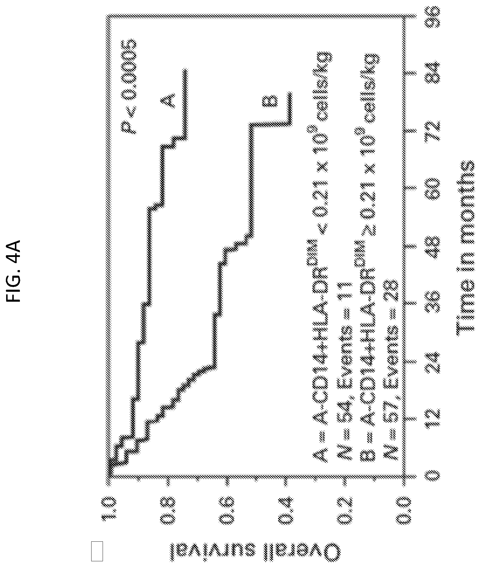
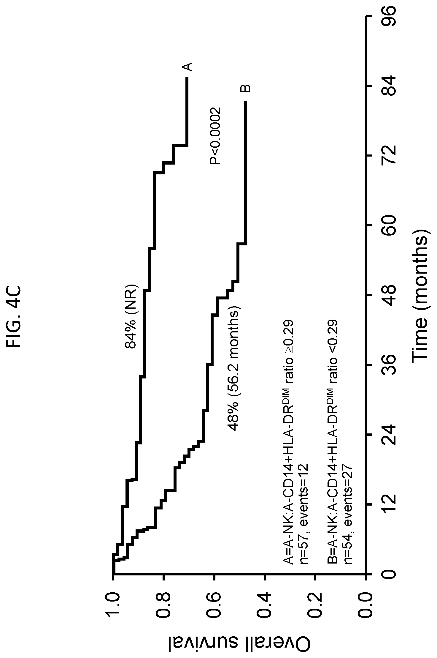
[0024] FIGS. 4A-4D are a series of Kaplan-Meier plots showing OS or PFS for non-Hodgkin lymphoma patients treated with APBHSCT using various populations of autologous cells. FIG. 4A, OS for A-CD14.sup.+HLA-DR.sup.DIM<0.21.times.10.sup.9 cells/kg vs. A-CD14.sup.+HLA-DR.sup.DIM.gtoreq.0.21.times.10.sup.9 cells/kg. FIG. 4B, PFS for A-CD14.sup.+HLA-DR.sup.DIM<0.21.times.10.sup.9 cells/kg vs. A-CD14.sup.+HLA-DR.sup.DIM.gtoreq.0.21.times.10.sup.9 cells/kg. FIG. 4C, OS for A-NK:A-CD14.sup.+HLA-DR.sup.DIM ratio .gtoreq.0.29 vs. A-NK:A-CD14.sup.+HLA-DR.sup.DIM ratio <0.29. FIG. 4D, PFS for A-NK:A-CD14.sup.+HLA-DR.sup.DIM ratio .gtoreq.0.29 vs. A-NK:A-CD14.sup.+HLA-DR.sup.DIM ratio <0.29.
DETAILED DESCRIPTION
[0025] This document provides materials and methods that for treating cancer patients, and is based, at least in part, on the discoveries that A-LMR and LMR, as well as A-AMC, AMC, and A-NK:A-CD14+HLA-DR.sup.DIM, can be powerful indicators of cancer patient prognosis. Thus, this document provides materials and methods for obtaining a population of stem cells and/or treating a patient to achieve an A-LMR or a LMR of at least 1.0, as well as materials and methods for obtaining a population of stem cells and/or treating a patient to achieve an A-NK:A-CD14+HLA-DR.sup.DIM or NK:CD14+HLA-DR.sup.DIM of at least 0.29.
[0026] In some embodiments, a subject treated according to the methods described herein can be a human cancer patient diagnosed with, for example, non-Hodgkin lymphoma, Hodgkin's disease, classical Hodgkin lymphoma, multiple myeloma, acute myeloid leukemia, diffuse large B-cell lymphoma, or acute lymphoblastic leukemia, or a solid tumor such as a breast tumor, lung tumor, ovarian tumor, liver tumor, or kidney tumor. The methods provided herein can include the step of returning a population of collected autologous cells to the patient. The cell population can be returned to the patient by, for example, intravenous infusion or any other suitable method known in the art. In some embodiments, the patient can be in remission from the cancer, either prior to collection of the cells or prior to returning the cells to the patient.
[0027] As used herein, "autologous" as it relates to transplantation refers to a graft in which the donor and recipient is the same individual. Thus, in an autologous transplant cells are harvested from a subject and then returned to the same subject. In contrast, an "allogeneic" transplant refers to a graft in which the donor and recipient are genetically non-identical individuals from the same species. A "xenogeneic" transplant refers to a graft in which the donor and recipient are of different species.
[0028] As used herein, an APBHSCT refers to a procedure in which a sample of a subject's own stem cells are removed and subsequently transplanted back into the same subject. Stem cells can be harvested from bone marrow (BM) or peripheral blood (PB), for example. Once obtained, stem cells can be frozen until needed. For example, stem cells can be obtained from a patient, cryopreserved at temperatures .ltoreq.-85.degree. C., and then thawed and returned (i.e., transplanted, typically by transfusion) to the patient. In some embodiments, stem cell aliquots can be thawed, loaded into one or more sterile syringes or infusion bags, and injected intravenously over a period of time ranging from about 30 minutes to about 45 minutes.
[0029] In some embodiments, stem cells capable of reconstituting a patient's immune system can be obtained from the patient's peripheral circulation following mobilization of such cells from BM into PB. Mobilization of stem cells can be accomplished by treatment of a patient with one or more factors that can (i) stimulate an increase in proliferation of stem cells and/or progenitor cells, and/or (ii) stimulate migration of stem cells and/or progenitor cells from the BM into the peripheral circulation. Such factors can be administered with adjuvants and/or other accessory substances, separately or in combination as desired. Examples of factors that can be used in this aspect include, without limitation, granulocyte colony-stimulating factor (G-CSF), granulocyte/macrophage colony-stimulating factor (GM-CSF), c-kit ligand (stem cell factor (SCF)), interleukin-2, -7, -8, and -12 (IL-2, IL-7, IL-8, and IL-12), and flt-3 ligand. See, e.g., Bungart et al. (1990) Br. J. Haematol. 76:174; Terella et al. (1993) Bone Marrow Transplant. 11:271; Molineux et al. (1991) Blood 85:275; Grzegorzewski et al. (1994) Blood 83:377; Laterveer et al. (1995) Blood 85:2269; Jackson et al. (1995) Blood 85:2371; and Lyman et al. (1994) Blood 83:2795. Factors to be administered can include, for example, G-CSF alone (e.g., 10 .mu.g/kg/day G-CSF), G-CSF+flt-3 ligand (e.g., 10 .mu.g/kg/day G-CSF+50 .mu.g/kg/day flt-3 ligand), or GM-CSF+flt-3 ligand (e.g., 5 .mu.g/kg/day GM-CSF+50 .mu.g/kg/day flt-3 ligand). See, e.g., Sudo et al. (1997) Blood 89:3186. Such factors can be administered prior to harvest or starting on the day of harvest, for example, and can be given on a daily basis for one to seven days (e.g., for one, two, three, four, five, six, or seven days), or until stem cell harvesting is complete. Factors that stimulate stem cell proliferation or mobilization can be administered using any suitable method. Typically, such factors can be administered parenterally (e.g., by subcutaneous, intrathecal, intraventricular, intramuscular, or intraperitoneal injection, or by intravenous drip). Mobilization of stem cells with, for example, GM-CSF and flt-3 ligand can be evaluated by determining the number of CD34.sup.+ cells present before, during, and/or after treatment with one or more mobilizing agents. In some embodiments, the number of CD34.sup.+ cells can be determined by FACS analysis using CD34-specific antibodies conjugated to fluorescent or other labeling moieties.
[0030] Following or during mobilization, peripheral blood stem cells (PBSC) can be collected using, for example, an apheresis procedure. The process of apheresis, which is well known in the art, involves removal of whole blood from a patient or donor. Within an instrument that is essentially designed as a centrifuge, the components of the whole blood are separated. One or more of the separated portions is then withdrawn, and the remaining components can be retransfused into the patient or donor. Thus, for example, all or most (e.g., 80%, 90%, 95%, 99%, or 100%) of the erythrocytes in a sample of whole blood can be returned to a patient during an apheresis procedure, while lymphocytes (e.g., NK cells) and stem cells can be collected. Apheresis can be performed as many as four times per week (e.g., one, two, three, or four times per week). In some embodiments, a commercially available blood cell collection device can be used, such as the CS3000.RTM. blood cell collection device or the Fenwal Amicus collection device, both of which are marketed by the Fenwal Division of Baxter Healthcare Corporation (Fenwal Laboratories, Deerfield, Ill.). Methods for performing apheresis with these machines are described in Williams et al. (1990) Bone Marrow Transplantation 5:129-133; Hillyer et al. (1993) Transfusion 33:316-321; and Porrata et al. (2016) Biol Blood Marrow Transplant 22:1017-1023, for example, all of which are incorporated herein by reference in their entirety.
[0031] In some embodiments, a total blood volume between 9.5 and 10 L per apheresis procedure can be processed at a flow rate of 50 to 90 mL/min. Following collection, a cell count can be performed on an aliquot of the total product to determine the number of stem cells. Cells can be collected until the total sample taken from the patient reaches a concentration of at least 2.times.10.sup.6 CD34.sup.+ stem cells/kg (e.g., 2.times.10.sup.6 CD34.sup.+ cell/kg to 3.times.10.sup.6 CD34.sup.+ cells/kg, 2.times.10.sup.6 CD34.sup.+ cell/kg to 2.5.times.10.sup.6 CD34.sup.+ cells/kg, or 2.5.times.10.sup.6 CD34.sup.+ cell/kg to 3.times.10.sup.6 CD34.sup.+ cells/kg).
[0032] Despite various methods of PBSC mobilization, adequate numbers of PBSC for APBHSCT may be not collected from some patients during a single apheresis procedure. In these patients, BM harvest or a second attempt at PBSC mobilization can be performed. Alternatively, these patients may be excluded from proceeding to APBHSCT.
[0033] Apheresis products can be centrifuged (e.g., at 400 g for 10 minutes), and the plasma can be removed to yield a total volume of, for example, about 100 mL. The resulting cell suspension can be mixed with a physiological freezing solution [e.g., 100 mL minimal essential medium such as MEM-S (Invitrogen Life Technologies, Carlsbad, Calif.) supplemented with 20% dimethylsulfoxide (DMSO)]. Cell/media suspensions can be transferred to freezing bags (such as those manufactured by Delmed, Canton, Mass.) or any other freezing receptacle known in the art, and frozen to -100.degree. C. using, for example, a computer-controlled cryopreservation device (e.g., the Cryoson-BV-6; Cryoson Deutschland GmbH, FRG). The cells then can be transferred into liquid nitrogen and stored at until transplantation.
[0034] Patients typically undergo a pre-transplant workup to evaluate, for example, heart, liver, kidney, and lung function, as well as current disease status. In some embodiments, patients deemed to be eligible (e.g., healthy enough) for APBHSCT are subjected to a tumor debulking procedure prior to APBHSCT. For example, a patient can be treated with high doses of chemotherapy, radiation therapy, and/or surgery (e.g., surgery with anesthesia) before the transplant. Stem cells for transplant typically are collected prior to tumor debulking regimens, since such potentially lethal procedures can be immunosuppressive and can severely damage or destroy the BM. APBHSCT following a debulking procedure can reconstitute the patient's immune cells with stem cells present in the transplant.
[0035] In some embodiments, a patient's stem cells can be collected by BM harvest using procedures known in the art, or by a stem cell apheresis procedure as described above, for example. Collected stem cells can be cryopreserved, and the patient can undergo a debulking procedure such as high-dose chemotherapy and/or radiation therapy. After the debulking procedure is completed, the patient's stem cells can be transplanted. APBHSCT can be done almost immediately after a debulking procedure (e.g., 24 to 48 hours after HDT). Alternatively, a longer period of time (e.g., a week to several months) can elapse between a debulking procedure and APBHSCT. Due to the likelihood of immunosuppression as a result of the debulking procedure, protective isolation precautions generally are taken after APBHSCT at least until the reinfused stem cells begin to engraft. "Engraftment" refers to a process whereby the transplanted stem cells begin to differentiate into mature blood cells. In addition, stem cells can be treated prior to transplantation with, for example, anticancer drugs or antibodies to reduce the number of cancerous cells that may be present in the sample. Such procedures are referred to as "purging."
[0036] In some embodiments of the methods provided herein, subjects (e.g., cancer patients) can be treated by administration of autologous cell populations that contain stem cells and other cell types, including, for example, red blood cells (RBC), lymphocytes, and monocytes. Lymphocytes are white blood cells (WBC) that are formed in lymphatic tissue throughout the human body (e.g., lymph nodes, spleen, thymus, tonsils, Peyer's Patches, and bone marrow). In normal adults, lymphocytes comprise approximately 22% to 28% of the total number of leukocytes in the circulating blood. As used herein, the term "lymphocyte" includes NK cells, B cells, and T cells (e.g., T helper cells, cytotoxic T cells, and T suppressor cells). NK cells are directly cytotoxic to foreign cells (e.g., foreign cancer cells), and do not require complement activity to effect their lysis. NK cells represent the body's first line of defense against malignancy. B cells produce immunoglobulins, and T cells are involved in modulation of immune responses and in regulation of erythropoiesis. Different types of lymphocytes can be distinguished from each other and from other cell types based on the cell type-specific expression of particular molecular markers, generally cell surface markers. For example, NK cells bear on their surface CD16 and/or CD56 markers. B cells bear at least one of the cell surface markers CD19 and CD20. T cells bear one or more of the cell surface markers CD3, CD4, and CD8. Typically, cytotoxic T cells express CD8, whereas helper T cells express CD4.
[0037] As used herein, the term "absolute lymphocyte count" (ALC) refers to the total number of lymphocytes per unit of whole blood or blood cells in a sample or in a subject (e.g., a human patient). A unit can be, for example, a liter (L), milliliter (mL), or microliter (.mu.L). Typically, but not always, ALC is measured as the number of mature lymphocytes per .mu.L of blood, and includes the cumulative numbers of B cells, T cells, and NK cells. Stem cells, lymphocyte precursor cells, and lymphocyte progenitor cells typically are not included in the ALC. Stem cells can be differentiated from lymphocytes in that stem cells express the cell surface marker CD34, whereas mature lymphocytes do not. Moreover, lymphocytes express specific cell surface markers as described above (NK cells: CD16 and/or CD56; B cells: CD20 and/or CD19; T cells: CD3, CD4, and/or CD8), whereas stem cells do not express these markers.
[0038] To determine an ALC, a sample of blood can be collected from a patient. For example, blood can be collected in a rubber-stopped tube containing EDTA or another medically acceptable anti-coagulant. Blood can be collected using any route of entry to the circulatory system known in the art. The blood sample then can be analyzed to determine the ALC. In some embodiments, an ALC can be obtained using an automated system for counting blood cells in a sample. Such cell counting systems typically are based on a principle by which unstained, unlabeled cells are sorted and counted based on morphological characteristics including, without limitation, cell size, cell shape, nuclear size, and nuclear shape. For example, the GEN-S.TM. Hematology Analyzer identifies and counts cell types based on three general criteria: volume, conductivity, and scatter (see U.S. Pat. No. 5,125,737). A blood sample can be treated before analysis with reagents and/or physical agitation to lyse the RBC, thereby leaving WBC for analysis. The Gen-S.TM. Analyzer uses a process of DC impedance by which the cells are collided with light to physically measure the volume displaced by the entire cell in an isotonic diluent. Cell size thus can be accurately determined regardless of the orientation of the cell in the light path. Cells can be further collided with an alternating current in the radio frequency range that can permeate cell membranes, such that information can be obtained with regard to internal structure including, for example, chemical composition and nuclear structure. A cell can be collided with a laser beam that, upon contacting the cell, scatters and spreads out in all directions, generating median angle light scatter signals. These signals can be collected to yield information regarding cellular granularity, nuclear lobularity, and cell surface structure. Thus, such a system can count and differentiate RBC from WBC based on the presence or absence of a nucleus, and can count and differentiate the different types of WBC based on the ratio of nuclear to cytoplasmic volume, lobularity of the nucleus, and granularity of the cytoplasm as described below, for example.
[0039] ALC also can be determined by placing a known volume of a blood sample onto a glass microscope slide, smearing the sample to create a thin film of blood on the slide, and staining the sample using standard histological stains such as, for example, hematoxylin and eosin (H & E). Briefly, a blood smear can be dried and subsequently fixed onto a slide using a fixative such as, without limitation, neutral buffered formalin, formaldehyde, paraformaldehyde, glutaraldehyde, Bouin's solution, mercuric chloride, or zinc formalin. The slides then can be immersed in a solution of Harris Hematoxylin, rinsed in water, immersed in a solution of Eosin, rinsed in water, dehydrated in ascending alcohol solutions, and cleared in xylenes. In blood smears that have been stained using H & E, nuclei and other basophilic structures stain blue, whereas cytoplasm and other acidophilic structures stain light to dark red (Sheehan et al. (1987) Theory and Practice of Histotechnology, 2nd Edition, Battelle Memorial Institute, Columbus, Ohio), which is incorporated herein by reference in its entirety. The number of lymphocytes present in a blood smear can be counted based on lymphocytic morphological criteria accepted in the art.
[0040] For example, when stained with H & E, the lymphocyte nucleus is deeply colored (purple-blue) and is composed of dense aggregates of chromatin within a sharply defined nuclear membrane. The nucleus generally is round, eccentrically located, and surrounded by a small amount of light blue staining cytoplasm. The volume of nucleus to cytoplasm in a lymphocyte typically is about 1:1.2. Lymphocytes can be differentiated from RBC in that RBC have no nuclei. Lymphocytes can be differentiated from neutrophils in that neutrophils have nuclei with 2 to 5 lobes, while lymphocyte nuclei are not lobed. Lymphocytes can be differentiated from basophils and eosinophils in that those cells have cytoplasmic granules, while lymphocytes do not have cytoplasmic granules. Lymphocytes can be differentiated from monocytes in that monocytes are 16 to 20 .mu.m in diameter, while lymphocytes are 7 to 10 .mu.m in diameter. In addition, one of skill in the art may refer to any of a number of hematology or histological texts or atlases (e.g., Wheater et al. (1987) Functional Histology 2nd Ed. Churchill Livingstone, incorporated herein by reference in its entirety) to determine the physical appearance of a lymphocyte.
[0041] ALC also can be determined by immunolabeling lymphocytes with antibodies specific for lymphocyte cell surface markers, and counting the immunolabeled cells using fluorescence flow cytometry (FFC). For example, NK cells can be labeled with one or more fluorescently labeled antibodies specific for CD16 and/or CD56. Similarly, B cells can be labeled with one or more fluorescently labeled antibodies specific for the adhesion molecules CD20 and/or CD19, and T cells can be labeled with one or more fluorescently labeled antibodies specific for CD3, CD4, and/or CD8, and. To determine ALC, cell surface marker-specific antibodies can be labeled with the same fluorophore (e.g., Cy-5, fluorescein, or Texas Red). In a FFC procedure, individual cells are held within a thin stream of fluid and passed through one or more laser beams, one cell at a time, causing light to scatter and the fluorescent dyes to emit light at various predetermined frequencies. Photomultiplier tubes convert the light to electrical signals, allowing for quantitation of the number of cells bearing the fluorophore. If all lymphocyte subtypes are labeled with the same fluorophore, quantification of the number of fluorophore-bearing cells will yield an ALC. FFC and quantitation is further described in, for example, U.S. Pat. No. 4,499,052. In addition, a FFC machine can be adapted for fluorescence activated cell sorting (FACS), i.e., the separation (and collection) of (a) fluorescent cells from non-fluorescent cells; (b) strongly fluorescent cells from weakly fluorescent cells; or (c) cells fluorescing at one wavelength from cells fluorescing at another wavelength.
[0042] Monocytes are large phagocytic WBC that have a simple nucleus and clear, grayish cytoplasm. Monocytes can differentiate into macrophages and myeloid lineage dendritic cells. Classical monocytes are characterized by high level expression of the CD14 cell surface receptor without expression of the CD16 cell surface receptor, but other populations of monocytes are characterized by low level expression of CD14 and high level expression of CD16, or high level expression of CD14 and low level expression of CD16.
[0043] The term "absolute monocyte count" (AMC) refers to the total number of mature monocytes per unit of whole blood or blood cells in a sample or in a subject (e.g., a human patient). Again, a unit can be, for example, a L, mL, or .mu.L. AMC can be determined by, for example, using an automated cell count machine or flow cytometry, as described above with regard to ALC.
[0044] Once ALC and AMC have been determined, whether the A-ALC and A-AMC in a population of collected stem cells from a patient, or the ALC and AMC in a blood sample obtained from a subject, the ratio of lymphocytes to monocytes can be determined. The ratio of A-ALC to A-AMC for a population of autologous cells to be used in APBHSCT is referred to herein as A-LMR, while the ratio of ALC to AMC in a biological sample (e.g., a blood sample) collected from a subject is referred to herein as LMR. An A-LMR for a population of collected autologous stem cells that is at least 1.0 (e.g., 1.1, 1.2, 1.3, 1.4, 1.5, 2.0, 2.5, or greater than 2.5) can indicate that the patient receiving the APBHSCT has a greater likelihood of progression-free and overall survival than he/she would if the A-LMR was less than 1.0 (e.g., 0.9, 0.8, 0.7, 0.6, 0.5, or less than 0.5). Further, a LMR of at least 1.0 determined for a biological sample (e.g., a blood sample) obtained from a cancer patient at diagnosis, before or after chemotherapy, or from an APBHSCT recipient after transplant (e.g., 15, 30, 60, or 90 days post-transplant) can indicate that the patient has a greater likelihood of progression-free and overall survival than he/she would if the LMR was less than 1.0.
[0045] This document also provides methods for assessing a patient's prognosis based on the ratio of NK cells to CD14+HLA-DR.sup.DIM monocytes in a biological (e.g., blood) sample from the patient, or in a population of collected autologous stem cells to be transferred back to the patient. NK cells can be collected using an apheresis procedure as described above. In some embodiments, the number of collected NK cells can be monitored. For example, the number of NK cells can be determined at one or more points during collection of the sample from the patient. The number of NK cells also can be determined after completion of a collection. Once the population of total collected cells includes at least about 0.5.times.10.sup.9 NK cells/kg, they can be returned to the patient. The number of collected CD14+HLA-DR.sup.DIM cells also can be determined, and the ratio as compared to NK cells can be determined. The number of collected NK cells and CD14+HLA-DR.sup.DIM cells can be monitored using methods such as those described above. In some embodiments, the number of collected NK cells can be determined using immunolabeling with one or more fluorescently labeled antibodies specific for CD16 and/or CD56, and counting with FACS, while the number of collected CD14+HLA-DR.sup.DIM cells can be determined using immunolabeling with one or more labeled antibodies specific for CD14 and/or HLA-DR.sup.DIM, followed by counting with FACS.
[0046] Once the numbers of NK cells and CD+HLA-DR.sup.DIM cells have been determined, whether the A-NK and A-CD14+HLA-DR.sup.DIM in a population of collected autologous cells to be used for APBHSCT, or the NKC and CD14+HLA-DR.sup.DIM in a blood sample obtained from a subject, the ratio of NK cells to CD14+HLA-DR.sup.DIM cells can be determined. A ratio of NK:CD14+HLA-DR.sup.DIM that is at least 0.29 (e.g., 0.30, 0.35, 0.40, 0.45, or more than 0.45) can indicate that the patient receiving the stem cells via APBHSCT has a greater likelihood of progression-free and overall survival than he/she would if the ratio was less than 0.29 (e.g., 0.28, 0.25, 0.23, 0.20, or less than 0.20). Further, a NK:CD14+HLA-DR.sup.DIM of at least 0.29 determined for a biological sample (e.g., a blood sample) obtained from a cancer patient at diagnosis, before or after chemotherapy, or from an APBHSCT recipient, post-transplant (e.g., 15, 30, 60, or 90 days post-transplant) can indicate that the patient has a greater likelihood of progression-free and overall survival than he/she would if the NK:CD14+HLA-DR.sup.DIM ratio was less than 0.29.
[0047] The methods provided herein also can include treatment of a patient or a cell population (e.g., in a biological sample such as an apheresis product) with one or more agents that stimulate proliferation, maturation, differentiation, function, and/or activity of immune cells (e.g., lymphocytes, including NK cells). For example, a patient or a population of collected cells can be contacted with an agent such as IL-2, IL-12, IL-15, IL-17, IL-21, interferon alpha (IFN-.alpha.), or interferon gamma (IFN-.gamma.). These agents can be native factors obtained from a natural source, factors produced by recombinant DNA methodology, chemically synthesized polypeptides or molecules, or any derivative having the functional activity of the native factor. Since agents such as these can enhance the number and/or activity of lymphocytes (e.g., NK cells), a patient may be subjected to shorter or fewer apheresis procedures in order to harvest a cell population containing, for example, an A-LMR or LMR of at least 1.0, and/or an A-NK:A-CD14+HLA-DR.sup.DIM of at least 0.29.
[0048] In some embodiments, a population of collected cells can be contacted with an agent or platform that depletes monocytes. In some cases, the population of cells may be identified as having an A-LMR or LMR less than 1.0, or an A-NK:A-CD14+HLA-DR.sup.DIM or NK:CD14+HLA-DR.sup.DIM less than 0.29. Suitable methods for depleting monocytes include, for example, contacting a population of cells with a support (e.g., a solid surface such as a bead) coupled to CD14 (e.g., CD14 microbeads; Mitenyi Biotec), and collecting the cells that do not bind to the support.
[0049] In some embodiments, a population of cells (e.g., a population of collected autologous lymphocytes containing NK cells and monocytes) can be contacted in vitro with one or more agents such as those listed above. For example, collected cells can be placed in a vessel (e.g., a bag, a tube, a vial, or any other suitable container) and contacted with one or more agents such as those described above. In some embodiments, cells can be contacted in vitro with IL-2 at a dose of, for example, about 1.5.times.10.sup.6 to about 2.0.times.10.sup.6 units. Lymphocyte (e.g., NK cell) enhancing agents can be added to cells within a container such as a bag (e.g., a blood bag), tube, or vial, or such a vessel can contain one or more such agents prior to placement of cells within the vessel. In some embodiments, one or more agents can be dispersed on an inner surface of the vessel. For example, an agent in liquid form can be dispersed (e.g., sprayed) onto an inner surface of the vessel and allowed to dry. Alternatively, an agent in solid (e.g., lyophilized or powdered) form can be dispersed on an inner surface of the vessel. In another alternative, an agent in liquid or solid form can simply be placed within the vessel.
[0050] Alternatively, one or more lymphocyte enhancing agents can be administered to a patient. A patient can be treated with such an agent prior to collection of a biological sample containing lymphocytes and monocytes, or a patient can be treated post-APBHSCT. For example, the number of NK cells in the PB of a patient can be monitored following APBHSCT, and a NK cell enhancing agent can be administered to the patient if the number of NK cells is below a particular threshold at a particular time point (e.g., at post-transplant day 15). A suitable threshold can be, for example, about 80 NK cells/.mu.L of blood (e.g., about 75 NK cells/.mu.L or about 85 NK cells/.mu.L). Similarly, a NK cell enhancing agent can be administered to a patient post-APBHSCT if the ALC-15 is less than 500 cells/.mu.L of blood. Agents such as those listed above can be administered to a patient via any pharmaceutically acceptable route known in the art, including, for example, intravenous injection, intra-arterial injection, subcutaneous injection, intramuscular injection, intraperitoneal injection, or oral administration in the form of a tablet, capsule, or syrup. In some embodiments, IL-2 can be administered to a patient prior to collection of autologous cells, or after APBHSCT. In another embodiment, a patient can be treated with IFN-.gamma. at a concentration of, for example, between about 1.times.10.sup.5 and about 1.times.10.sup.7 units/m.sup.2. When the treatment is post-APBHSCT, the agent(s) can be administered from the day of transplant up to about 21 days following the transplant.
[0051] In some embodiments, patients or collected cell populations (e.g., populations containing lymphocytes and monocytes) also can be treated with one or more agents that activate the T cell signal transduction pathway, leading to lymphocyte activation. A T cell activator can be, without limitation, one or more of the following: IL-1, IL-2, IL-4, IL-5, IL-6, IL-7, IL-12, IL-13, IFN.alpha., IFN.gamma., tumor necrosis factor (TNF.alpha.), an anti-CD3 antibody or antigen-binding fragments thereof (anti-CD3), an anti-CD28 antibody or antigen-binding fragments thereof (anti-CD28), phytohemagglutinin, concanavalin-A, and phorbol esters. As above, these agents can be native factors obtained from a natural source, factors produced by recombinant DNA methodology, chemically synthesized polypeptides or molecules, or any derivative having the functional activity of the native factor.
[0052] The invention will be further described in the following examples, which do not limit the scope of the invention described in the claims.
EXAMPLES
Example 1--A-LMR as a Predictor of Survival
[0053] Retrospective studies were conducted to determine whether the A-LMR for autologous cells infused into DLBCL patients during APBHSCT could be correlated to survival. In a training set including 253 patients, and also in a validation set containing 126 patients, an A-LMR of 1 or higher correlated with increased overall survival (FIGS. 1A and 1B, top panels), increased progression-free survival (FIGS. 1A and 1B, middle graphs), and increased lymphoma-specific survival (FIGS. 1A and 1B, bottom graphs), while an A-LMR less than 1 correlated with decreased overall survival (FIGS. 1A and 1B, top panels), decreased progression-free survival (FIGS. 1A and 1B, middle graphs), and decreased lymphoma-specific survival (FIGS. 1A and 1B, bottom graphs). Details of these studies are provided elsewhere (Porrata et al. (2014) Biol Blood Marrow Transplant 20:1804-1812).
Example 2--A-NK:A-CD14+HLA-DR.sup.DIM as a Predictor of Survival
[0054] A double-blind, phase III, randomized clinical trial was conducted to determine whether A-NK:A-CD14+HLA-DR.sup.DIM can be used as a predictor of survival in 111 non-Hodgkin's lymphoma (NHL) patients. The primary end point of was to assess the impact of A-NKC/ACD14.sup.+HLA-DR.sup.DIM ratio on overall survival (OS) and progression-free survival (PFS). A secondary end point was to determine whether the A-NKC/A-CD14.sup.+HLA-DR.sup.DIM ratio is an independent prognostic factor for OS and PFS.
[0055] The prognostic factors evaluated in this study included: international prognostic index (IPI; "The International Non-Hodgkin's Lymphoma Prognostic Factors Project. A predictive model for aggressive non-Hodgkin's lymphoma.," N Engl J Med 1993, 329:987-994) at diagnosis (.gtoreq.2), age at diagnosis (.ltoreq.60 vs. >60 years), lactate dehydrogenase (LDH) at diagnosis (> normal), ECOG (Eastern Cooperative Oncology Group) performance status at diagnosis (>1 vs. .ltoreq.1), extranodal sites at diagnosis (>1 versus .ltoreq.1), stage at diagnosis (III/IV vs. I/II), histologies at diagnosis (DLBCL vs. others), disease status before APBHSCT (complete response vs. partial response), infused CD34.sup.+ cell count, Plerixafor use, and A-NKC/A-CD14.sup.+HLA-DR.sup.DIM ratio. Basic characteristics of the study participants are shown in TABLE 1. Further details of these studies are described elsewhere (Porrata et al. 2016, supra; and Kansagra et al. 2018, Bone Marrow Transplantation 53:146-154).
[0056] The double-blind, randomized, phase III clinical trial was designed to assess whether the PFS was significantly increased among those patients whose apheresis was done using a modification to a standard Fenwal Amicus setting where the mononuclear cell offset=1.5 and RBC=6.0, compared with settings where mononuclear cells=1.5 and RBC=5.0.
[0057] For stem cell mobilization, patients received 10 .mu.g/kg of G-CSF subcutaneously (SC) daily for 5 to 7 consecutive days alone or in conjunction with 0.24 mg/kg of Plerixafor for up to 4 consecutive days by SC injection. Patients started with G-CSF and after 4 days of treatment, if the peripheral blood CD34.sup.+ count was .gtoreq.10 cells/.mu.L, stem cell collection began. If the peripheral blood CD34.sup.+ count was <10 cells/.mu.L, Plerixafor was added that evening and collections were initiated the next day. Apheresis collections were to be performed daily, with a goal of at least 2.times.10.sup.6CD34.sup.+ cells/kg. Additional collections were at the discretion of the transplantation team. Patients who failed mobilization or did not collect at least 2.times.10.sup.6 CD34.sup.+ cells/kg were allowed to choose to either undergo a second mobilization/apheresis or discontinue study participation.
[0058] For conditioning, all patients received BEAM chemotherapy: BCNU (carmustine, 300 mg/m.sup.2) on day -6, Etoposide (100 mg/m.sup.2) twice daily from days -5 to -2, ARA-C (Cytarabine, 100 mg/m.sup.2) twice daily from days -5 to -2, and Melphalan (140 mg/m.sup.2) on day -1.
[0059] OS was measured from the date of APBHSCT to the date of death or last follow-up, and PFS was defined as the time from APBHSCT to time of progression, relapse, death or last follow-up (Cheson et al. 2007, J Clin Oncol 25:579-586).
[0060] Previously frozen autograft mononuclear cells (0.5-1.0.times.10.sup.6 cells/mL) were thawed and aliquoted into 96-well round-bottomed plates (100 mL/well). The following monoclonal antibodies were used for autograft mononuclear cell immunophenotyping by flow cytometry: anti-human CD3 FITC, anti-human CD16 phycoerythrin (PE), and anti-human CD56 PE for NK cells, and anti-human CD14 FITC and anti-human HLA-DR PE-cyanin 5.5 (PE-Cy5.5) for HLA-DR dim monocytes (BD Pharmingen; San Jose, Calif.). The desired antibody or antibody pool was added at 5 .mu.L of each antibody/well. The cells and antibodies were incubated for 30 minutes at 4.degree. C. and washed twice with 1.times.PBS (Cellgro; Manassas, Va.), 0.1% BSA and 0.05% sodium azide (Sigma; St Louis, Mo.). Three-color flow cytometry was performed on a Guava 8-HT (Millipore; St Louis, Mo.) and Incyte software (Millipore) was utilized for data analysis. After gating on live lymphocytes, the percentage of NK cells was determined by the number of cells that were CD3 negative and CD16 and CD56 positive. For the HLA-DR.sup.DIM cells, gates were set on live cells, and CD14-positive cells and HLA-DR.sup.DIM cells were enumerated from the CD14-positive gate. The absolute numbers of autograft NK cells and autograft CD14.sup.+HLA-DR.sup.DIM cells were calculated by multiplying their percentage times the A-ALC for NK cells and autograft absolute monocyte count (A-AMC) for CD14.sup.+HLA-DR.sup.DIM cells for each apheresis unit collection. The A-ALC was calculated as follows: A-ALC=% collection lymphocytes.times.(absolute WBC/kg). In a similar manner, the A-AMC was calculated as follows: A-AMC=% collection monocytes.times.(absolute WBC/kg).
[0061] OS and PFS were analyzed using the approach of Kaplan-Meier (Kaplan and Meier 1958, J Am Stat Assoc 53:457-481). Differences between survival curves were tested for statistical significance using the two-tailed log-rank test. Univariate and multivariate analysis was performed using Cox proportional hazard model (Cox 1972, J R Stat Soc 34:187-202). The variables in the prognostic factor section were evaluated to assess their impact on OS and PFS times post APBHSCT. The choice of the cutoff value for the A-NKC/A-CD14.sup.+HLADR.sup.DIM ratio to assess survival was based on its utility as a marker for the clinically relevant binary outcome of death/survival using the receiver operating characteristics curves (ROC) and area under the curve (AUC). The binary clinical outcome (death/survival) was established at 5 years after APHSCT. Patients were classified as `alive/censored` when follow-up time was >5 years and `death/uncensored` for patients known to have died before this time point (Tzankov et al. 2010, Leuk Lymphoma 51:199-212). A K-fold cross-validation with K-values of 10 was performed to validate the results of A-NKC/A-CD14.sup.+HLA-DR.sup.DIM ratio cutoff obtained by the ROC and AUC curves. Randomly chosen subsets containing 90% of the cohort were used for training, and the remaining 10% were left for testing. The cross-validation process was then repeated 10 times. Based on this analysis, cross-validation AUC by the ROC was produced, representing the discriminating accuracy of A-NKC/A-CD14.sup.+HLA-DR.sup.DIM ratio for the binary clinical outcomes of death/survival.
[0062] The .chi.2 tests and Fisher's exact tests were used to determine relationships between categorical variables as appropriate. The Wilcoxon rank test was used to determine associations between continuous variables and categories and nonparametric tests were used to evaluate associations for continuous variables. All P-values represented were two sided and statistical significance was declared at P <0.05.
[0063] The median age at the time of APBHSCT was 57 years (range: 20-74). The distribution of additional baseline characteristics for this cohort is presented in TABLE 1. The median follow-up from APHSCT was 57.2 months (range: 2.1-84.6 months) and 62.6 months (range: 37.6-84.6 months) for patients who were alive (n=72). The day-100 transplantation-related mortality was 1.8% (2/111): one patient died because of septic shock and one patient died of acute respiratory distress syndrome. Other than the two patients who died in the first 100 days after APBHSCT, 35 patients died because of relapse/progression of lymphoma and two patients died of myocardial infarction.
[0064] Representative fluorescence-activated cell sorting (FACS) plots to calculate the percentage of A-NKC are shown in FIGS. 2A and 2B, and representative FACS plots to calculate the percentage of A-CD14.sup.+HLA-DR.sup.DIM FIGS. 2C-2E. ROC curves and AUC were used to determine the optimal cutoff points for A-NKC, A-CD14.sup.+HLA-DR.sup.DIM and A-NK:A-CD14.sup.+HLA-DR.sup.DIM ratio based on their utility as markers for the clinical binary outcome of death/survival. The A-NKC .gtoreq.0.09.times.10.sup.9 cells/kg had an AUC of 0.65 (95% CI, 0.59-0.71), with a sensitivity of 50% (95% CI, 45-55%) and a specificity of 81% (95% CI, 77-85%), P <0.04. The A-CD14.sup.+HLA-DR.sup.DIM.gtoreq.0.21.times.10.sup.9 cells/kg had an AUC of 0.74 (95% CI, 0.68-0.80), with a sensitivity of 49% (95% CI, 43-55%) and a specificity of 92% (95% CI, 85-99%), P <0.001. The A-NK:A-CD14.sup.+HLA-DR.sup.DIM ratio .gtoreq.0.29 had an AUC of 0.75 (95% CI, 0.70-0.80), with a sensitivity of 60% (95% CI, 55-65%) and a specificity of 89% (95% CI, 84-94%), P <0.0003 (FIG. 3A).
[0065] An internal validation of A-NKC, A-CD14.sup.+HLA-DR.sup.DIM and A-NK:A-CD14.sup.+HLA-DR.sup.DIM ratio performances as markers for the clinical binary outcomes of death/survival was performed using k-fold cross-validation with k=10. For A-NKC, the cross-validation ROC showed an AUC of 0.66 (95% CI, 0.66-0.72); for A-CD14.sup.+HLA-DR.sup.DIM, an AUC of 0.76 (95% CI, 0.71-0.81); and for A-NK:A-CD14.sup.+HLA-DR.sup.DIM ratio, an AUC of 0.76 (96% CI, 0.70-0.82) (FIG. 3B). The similar AUC from the empirical ROC and the cross-validation ROC curves support the use of A-NKC .gtoreq.0.09.times.10.sup.9 cells/kg, A-CD14.sup.+HLA-DR.sup.DIM.gtoreq.0.21.times.10.sup.9 cells/kg, and the A-NK:A-CD14.sup.+HLA-DR.sup.DIM ratio of .gtoreq.0.29 for cutoff values as markers of the binary clinical outcome of death/survival.
[0066] Based on the univariate Cox regression analysis, A-NK:A-CD14.sup.+HLA-DR.sup.DIM ratio .gtoreq.0.29 was a predictor for OS and PFS (see, TABLES 2A and 2B). A strong positive correlation was identified between A-NKC and A-NK:A-CD14.sup.+HLA-DR.sup.DIM ratio .gtoreq.0.29 (r=0.63, P <0.0001), and a strong negative correlation between A-CD14.sup.+HLA-DR.sup.DIM and A-NK:A-CD14.sup.+HLA-DR.sup.DIM ratio <0.29 (r=-0.70, P <0.0001). To avoid the problem of collinearity due to the strong correlation between A-NKC and A-NK:A-CD14.sup.+HLA-DR.sup.DIM ratio, and between A-CD14.sup.+HLA-DR.sup.DIM and A-NK:A-CD14.sup.+HLA-DR.sup.DIM ratio, two separated multivariate analyses were performed. The first multivariate analysis included A-NKC and A-CD14.sup.+HLA-DR.sup.DIM with the other prognostic factors in TABLES 2A and 2B, excluding the A-NK:A-CD14.sup.+HLA-DR.sup.DIM ratio. In this first multivariate analysis, both were independent predictors for OS (A-NK: hazard ratio (HR)=0.34, 95% CI, 0.15-0.76, P <0.006; and A-CD14.sup.+HLA-DR.sup.DIM: HR=2.53, 95% CI, 1.12-6.05, P <0.03) and for PFS (A-NK: HR=0.31, 95% CI, 0.16-0.58, P <0.0001; and A-CD14.sup.+HLA-DR.sup.DIM: HR=2.05, 95% CI, 1.14-3.76, P <0.03). In the second multivariate analysis, A-NKC and A-CD14.sup.+HLA-DR.sup.DIM were excluded, and the A-NK:A-CD14.sup.+HLA-DR.sup.DIM ratio was included in the analysis. TABLE 3 shows that the A-NK:A-CD14.sup.+HLA-DR.sup.DIM ratio was an independent prognostic factor for OS (HR=0.34, 95% CI, 0.16-0.68, P <0.002) and for PFS (HR=0.56, 95% CI, 0.32-0.96, P <0.03).
[0067] Patients were categorized into two groups (A-NK:A-CD14.sup.+HLA-DR.sup.DIM ratio .gtoreq.0.29 vs. A-NK:A-CD14.sup.+HLA-DR.sup.DIM ratio <0.29) to assess whether both groups were balanced with regard to the baseline patient characteristics. As shown in TABLE 4, both groups were balanced, except for LDH, the disease status before APBHSCT, and the use of Plerixafor.
[0068] To evaluate survival outcomes based on A-CD14.sup.+HLA-DR.sup.DIM and A-NK:A-CD14.sup.+HLA-DR.sup.DIM ratio, A-CD14.sup.+HLA-DR.sup.DIM.gtoreq.0.21.times.10.sup.9 cells/kg and A-NK:A-CD14.sup.+HLA-DR.sup.DIM ratio .gtoreq.0.29 were tested for OS and PFS. Patients infused with an A-CD14.sup.+HLA-DR.sup.DIM<0.21.times.10.sup.9 cells/kg vs. patients infused with an A-CD14.sup.+HLA-DR.sup.DIM.gtoreq.0.21.times.10.sup.9 cells/kg experienced superior OS (FIG. 4A) and PFS (FIG. 4B): median OS of not reached vs. 73.1 months, 5-year OS rates of 83% (95% CI, 70-95%) vs. 52% (95% CI, 39-56%), P <0.0005, respectively, and median PFS of not reached versus 31.1 months, 5-year PFS rates of 60% (95% CI, 47-72%) vs. 34% (95% CI, 22-47%), P <0.004, respectively. Further, patients infused with an A-NK:A-CD14.sup.+HLA-DR.sup.DIM ratio .gtoreq.0.29 vs. patients infused with an A-NK:A-CD14.sup.+HLA-DR.sup.DIM ratio <0.29 experienced superior OS (FIG. 4C) and PFS (FIG. 4D): median OS of not reached vs. 56.2 months, 5-year OS rates of 84% (95% CI, 72-91%) vs. 48% (95% CI, 34-62%), P <0.0002, respectively; and median PFS of not reached vs. 31.1 months, 5-year PFS rates of 59% (95% CI, 46-71%) vs. 34% (95% CI, 22-47%), P <0.002, respectively.
[0069] Patients with an IPI index of 0-2 experienced better OS and PFS with an A-NK:A-CD14.sup.+HLA-DR.sup.DIM ratio .gtoreq.0.29 compared with an A-NK:A-CD14.sup.+HLA-DR.sup.DIM ratio <0.29, but patients with an IPI index of 3-4 did not (FIGS. 4A and 4B). For the subset of patients with DLBCL, those who received an A-NK:A-CD14.sup.+HLA-DR.sup.DIM ratio .gtoreq.0.29 experienced better OS and PFS than with patients with an A-NK:A-CD14.sup.+HLA-DR.sup.DIM ratio <0.29 (FIGS. 4C and 4D). In the DLBCL group, the A-NK:A-CD14.sup.+HLA-DR.sup.DIM ratio .gtoreq.0.29 was an independent predictor for OS (HR=0.26, 95% CI, 0.16-0.43, P <0.0002) and PFS (HR=0.28, 95% CI, 0.18-0.55, P <0.0003).
TABLE-US-00001 TABLE 1 Baseline patient characteristics Variables N (%) Age, year, median (range) 55 (20-73) Age, years .ltoreq.60 71 (64%) >60 40 (36%) Gender Male 80 (72%) Female 31 (28%) Histology DLBCL 51 (46%) Follicular lymphoma 16 (14%) Mantle cell lymphoma 25 (23%) Other 19 (17%) LDH (U/L), median (range) 211 (106-3364) LDH Normal 58 (52%) Abnormal 53 (48%) Stage I 7 (6%) II 11 (10%) III 27 (24%) IV 66 (60%) Stage I/II 18 (16%) III/IV 93 (84%) Extra-nodal disease 0 51 (46%) 1 52 (47%) 2 8 (7%) Extra-nodal disease .ltoreq.1 103 (93%) >1 8 (7%) Performance status 0 28 (25%) 1 71 (64%) 2 12 (11%) Performance status .ltoreq.1 99 (89%) >1 12 (11%) IPI score 0 15 (13%) 1 34 (31%) 2 32 (29%) 3 25 (23%) 4 5 (4%) IPI Index .ltoreq.2 81 (73%) >2 30 (27%) At PBSC collection Age, years: median (range) 57 (20-74) Clinical status pre-transplant Complete response 63 (57%) Partial response 48 (43%) Number of chemotherapy regimens 1 35 (32%) 2 65 (58%) 3 7 (6%) 4 3 (3%) 5 1 (1%) Apheresis machine ModC 56 (50%) StdC 55 (50%) Number of apheresis collections 1 8 (7%) 2 22 (19%) 3 24 (22%) 4 25 (23%) 5 17 (15%) 6 5 (5%) 7 8 (7%) 8 2 (2%) Plerixafor Yes 44 (40%) No 67 (60%) Infused CD34.sup.+ cells .times. 10.sup.6/kg: median (range) 5.15 (2.02-11.37)
TABLE-US-00002 TABLE 2A Overall survival Progression-free survival Dichotomized Variables HR 95% CI P HR 95% CI P Age >60 years 1.55 0.81-2.93 0.2 1.18 0.69-1.97 0.5 Elevated LDH 2.40 1.26-4.74 <0.007 1.56 0.94-2.62 0.09 Stage III/IV 1.50 0.69-4.38 0.4 2.72 1.20-7.82 <0.01 Extra-nodal involvement >1 5.312 2.12-11.60 <0.001 3.35 1.38-6.97 <0.01 Performance status 1.48 0.51-3.50 0.4 1.29 0.53-2.65 0.5 IPI .gtoreq.2 2.27 1.18-4.28 <0.02 1.81 1.04-3.05 <0.04 Complete vs partial response prior to transplant 0.51 0.26-0.96 <0.03 0.41 0.24-0.68 <0.0001 DLBCL vs other 0.54 0.28-0.99 <0.05 0.95 0.57-1.59 0.9 Female vs male 2.03 1.04-0.55 <0.05 1.47 0.83-0.74 0.3 A-NKC .gtoreq.0.09 .times. 10.sup.9 cells/kg 0.34 0.14-0.70 <0.003 0.09 0.14-0.52 <0.0001 A-CD14 + HLA-DR.sup.DIM .gtoreq.0.21 .times. 10.sup.9 cells/kg 3.26 1.66-6.89 <0.0005 2.14 1.27-3.69 <0.004 A-NK:A-CD14.sup.+HLA-DR.sup.DIM ratio .gtoreq.0.29 0.28 0.14-0.55 <0.0002 0.44 0.26-0.74 <0.002 Infused CD34 0.39 0.05-2.51 0.3 0.42 0.08-1.86 0.3 Plerixafor 1.32 0.69-2.53 0.4 1.35 0.80-2.27 0.3
TABLE-US-00003 TABLE 2B Overall survival Progression-free survival Continuous Variables HR 95% CI P HR 95% CI P A-NKC 0.26 0.06-0.96 <0.04 0.18 0.05-0.56 <0.002 A-CD14.sup.+HLA-DR.sup.DIM 5.83 1.86-15.97 <0.004 2.92 1.07-7.07 <0.04 A-NKC/A-CD14.sup.+HLA-DR.sup.DIM ratio 0.07 0.01-0.30 <0.0001 0.15 0.05-0.37 <0.0001 Infused CD34.sup.+ cells 0.39 0.05-2.51 0.3 0.42 0.08-1.86 0.3
TABLE-US-00004 TABLE 3 Overall survival Progression-free survival Variables HR 95% CI P HR 95% CI P Female vs male 1.84 0.92-3.56 0.08 Complete vs 0.55 0.27-1.05 0.09 0.65 0.37-1.14 0.1 partial response prior to transplant DLBCL vs other 0.53 0.27-1.01 0.06 IPI .gtoreq.2 1.01 0.39-2.60 0.9 1.35 0.70-2.48 0.4 DIM 0.34 0.16-0.68 <0.002 0.56 0.32-0.96 <0.03 A-NK:A-CD14 + HLA-DR ratio .gtoreq.0.29 Extra-nodal 3.23 1.06-9.47 <0.04 2.07 0.77-5.13 0.1 disease >1 Elevated LDH 1.54 0.63-3.59 0.3 Stage III/IV 1.87 0.78-5.55 0.2
TABLE-US-00005 TABLE 4 Baseline characteristics based on A-NK:A-CD14.sup.+ HLA-DR.sup.DIM ratio A-NK:A-CD14 + A-NK:A-CD14 + HLA-DR.sup.DIM .gtoreq.0.29 HLA-DR.sup.DIM <0.29 Variables (n = 57) (n = 54) P Age, year, 58 (20-74) 56.5 (24-74) 0.6 median (range) Male 41 (72%) 39 (72%) 0.9 Female 16 (28%) 15 (28%) LDH (g/dL), 190 (120-913) 232 (106-365) <0.04 median (range) Extra-nodal 0.2 disease 0 29 (51%) 22 (41%) 1 26 (45%) 26 (48%) 2 2 (4%) 6 (11%) Performance 0.7 status 0 16 (28%) 12 (22%) 1 26 (63%) 35 (65%) 2 5 (9%) 7 (13%) Histologies 0.9 DLBCL 25 (44%) 26 (48%) Follicular 8 (14%) 8 (15%) Mantle cell 13 (23%) 12 (22%) Other 11 (19%) 8 (15%) Stage 0.3 I 5 (9%) 2 (4%) II 8 (14%) 3 (6%) III 12(21%) 15 (28%) IV 32 (56%) 34 (62%) IPI score 0.06 0 13 (22%) 2 (4%) 1 16 (28%) 18 (33%) 2 14 (25%) 18 (33%) 3 12 (21%) 13 (24%) 4 2 (4%) 3 (6%) IPI risk factors Age, years 0.8 >60 20 (35%) 20 (37%) .ltoreq.60 37 (65%) 34 (63%) Extra-nodal 0.2 disease .gtoreq.2 2 (4%) 6 (11%) <2 55 (96%) 48 (89%) LDH 0.3 Abnormal 24 (42%) 29 (54%) Normal 33 (58%) 25 (46%) Performance 0.6 status 1 5 (9%) 7 (13%) .ltoreq.1 52 (91%) 47 (87%) Stage 0.07 I/II 13 (23%) 5 (9%) III/IV 44 (77%) 49 (91%) IPI Index 0.7 2 14 (25%) 16 (30%) .ltoreq.2 43 (75%) 38 (70%) Clinical status <0.003 pre-transplant Complete 40 (70%) 23 (43%) response Partial 17 (30%) 31 (57%) response Plerixafor <0.004 Yes 15 (26%) 29 (54%) No 42 (74%) 25 (46%) Infused CD34 5.15 (2.0-11.37) 5.14 (2.27-8.15) 0.4 cells/kg, median (range) Apheresis 0.3 machine ModC 26 (46%) 30 (56%) StdC 31 (54%) 24 (44%) Number of 0.5 apheresis collections 1 5 (9%) 3 (6%) 2 9 (16%) 13 (24%) 3 14 (25%) 10 (18%) 4 11 (19%) 14 (26%) 5 11 (19%) 6 (11%) 6 3 (5%) 2 (4%) 7 4 (7%) 4 (7%) 8 0 (0%) 2 (4%) ModC = modified apheresis machine StdC = standard apheresis machine
OTHER EMBODIMENTS
[0070] It is to be understood that while the invention has been described in conjunction with the detailed description thereof, the foregoing description is intended to illustrate and not limit the scope of the invention, which is defined by the scope of the appended claims. Other aspects, advantages, and modifications are within the scope of the following claims.
* * * * *
D00001
D00002
D00003
D00004
D00005
D00006
D00007
D00008
D00009
D00010
XML
uspto.report is an independent third-party trademark research tool that is not affiliated, endorsed, or sponsored by the United States Patent and Trademark Office (USPTO) or any other governmental organization. The information provided by uspto.report is based on publicly available data at the time of writing and is intended for informational purposes only.
While we strive to provide accurate and up-to-date information, we do not guarantee the accuracy, completeness, reliability, or suitability of the information displayed on this site. The use of this site is at your own risk. Any reliance you place on such information is therefore strictly at your own risk.
All official trademark data, including owner information, should be verified by visiting the official USPTO website at www.uspto.gov. This site is not intended to replace professional legal advice and should not be used as a substitute for consulting with a legal professional who is knowledgeable about trademark law.