Methods, Systems, And Devices For Heart Valve Decalcification, Regeneration, And Repair
Leonhardt; Howard J. ; et al.
U.S. patent application number 16/842683 was filed with the patent office on 2020-07-23 for methods, systems, and devices for heart valve decalcification, regeneration, and repair. The applicant listed for this patent is Cal-X Stars Business Accelerator, Inc.. Invention is credited to Brett M. Burton, Howard J. Leonhardt, Kapil K. Sharma, Betty Vowles.
| Application Number | 20200229831 16/842683 |
| Document ID | / |
| Family ID | 71609466 |
| Filed Date | 2020-07-23 |


| United States Patent Application | 20200229831 |
| Kind Code | A1 |
| Leonhardt; Howard J. ; et al. | July 23, 2020 |
METHODS, SYSTEMS, AND DEVICES FOR HEART VALVE DECALCIFICATION, REGENERATION, AND REPAIR
Abstract
Methods of heart valve decalcification may include mechanically removing calcium deposits on a heart valve, and removing debris from the mechanical decalcification via suction. The mechanical removal of the calcium deposits may be accomplished with a burr and/or an ultrasonic device. In some embodiments, mechanical removal of the calcium deposits may be accomplished with a handheld mechanical decalcification device comprising a bur extending from a hand piece. A catheter system for removing plaque deposits from a heart valve may include at least one mechanical decalcification device configured for cleaning the edges of heart valve leaflets, and at least one active aspiration device. The at least one mechanical decalcification device may comprise a bur and/or an ultrasonic device. The system may additionally include a deployable net apparatus configured to encompass at least a portion of a heart valve.
| Inventors: | Leonhardt; Howard J.; (Corona Del Mar, CA) ; Burton; Brett M.; (Erda, UT) ; Sharma; Kapil K.; (Salt Lake City, UT) ; Vowles; Betty; (West Bountiful, UT) | ||||||||||
| Applicant: |
|
||||||||||
|---|---|---|---|---|---|---|---|---|---|---|---|
| Family ID: | 71609466 | ||||||||||
| Appl. No.: | 16/842683 | ||||||||||
| Filed: | April 7, 2020 |
Related U.S. Patent Documents
| Application Number | Filing Date | Patent Number | ||
|---|---|---|---|---|
| 15812760 | Nov 14, 2017 | |||
| 16842683 | ||||
| 15460129 | Mar 15, 2017 | 10646644 | ||
| 15812760 | ||||
| 62308702 | Mar 15, 2016 | |||
| 62363012 | Jul 15, 2016 | |||
| 62364472 | Jul 20, 2016 | |||
| 62375271 | Aug 15, 2016 | |||
| 62385124 | Sep 8, 2016 | |||
| 62454521 | Feb 3, 2017 | |||
| 62352930 | Jun 21, 2016 | |||
| 62831083 | Apr 8, 2019 | |||
| Current U.S. Class: | 1/1 |
| Current CPC Class: | A61B 2217/007 20130101; A61F 2/2409 20130101; A61B 2017/22098 20130101; A61F 2/2412 20130101; A61B 2017/22079 20130101; A61B 17/2202 20130101 |
| International Class: | A61B 17/22 20060101 A61B017/22; A61F 2/24 20060101 A61F002/24 |
Claims
1. A method of heart valve decalcification, the method comprising mechanically removing calcium deposits on a heart valve.
2. The method of claim 1, further comprising removing debris via suction.
3. The method of claim 1, further comprising removing calcium deposits on the heart valve by directing a stream of biocompatible solvent onto the heart valve.
4. The method of claim 3, further comprising providing bioelectric stimulation to the heart valve.
5. The method of claim 4, further comprising bathing the heart valve with a biochemical bath to encourage healing and regeneration of tissue.
6. The method of claim 5, further comprising reforming the shape of the valve by installing a nitinol ring.
7. The method of claim 6, further comprising placing autologous cell created heart valve leaflets.
8. The method of claim 1, wherein mechanically removing calcium deposits on the heart valve comprises mechanically removing the calcium deposits on the heart valve with a bur.
9. The method of claim 1, wherein mechanically removing calcium deposits on the heart valve comprises mechanically removing the calcium deposits on the heart valve with an ultrasonic device.
10. The method of claim 1, wherein mechanically removing calcium deposits on the heart valve comprises mechanically removing the calcium deposits on the heart valve with a handheld mechanical decalcification device comprising a bur extending from a hand piece.
11. A catheter system for removing plaque deposits from a heart valve, the system comprising at least one mechanical decalcification device configured for cleaning the edges of heart valve leaflets.
12. The system of claim 11, further comprising at least one active aspiration device.
13. The system of claim 11, wherein the at least one mechanical decalcification device comprises a bur.
14. The system of claim 13, further comprising a deployable net apparatus configured to encompass at least a portion of a heart valve.
15. The system of claim 14, wherein the deployable net apparatus is comprised of electrospun polymers forming a nanoscale fiber mesh.
16. The system of claim 11, wherein the at least one mechanical decalcification device comprises an ultrasonic device.
17. The system of claim 11, further comprising a biocompatible solvent delivery device configured for delivering a stream of biocompatible solvent to a heart valve.
18. The system of claim 17, further comprising a bioelectric signal array configured to deliver bioelectric stimulation to the heart valve.
19. The system of claim 18, further comprising a micro infusion pump configured to deliver a regeneration cocktail composition to the heart valve.
20. The system of claim 19, further comprising a nitinol ring placement catheter.
21. The system of claim 20, further comprising an optical viewing system.
22. The system of claim 21, further comprising at least one cerebral protection device.
Description
CROSS-REFERENCE TO RELATED APPLICATIONS
[0001] This application is a continuation-in-part of U.S. Ser. No. 15/812,760, filed on Nov. 14, 2017 (US 2018/0064935A1, Mar. 8, 2018), which is a continuation-in-part of U.S. Ser. No. 15/460,129, filed on Mar. 15, 2017 (US 2017/0266371A1, Sep. 21, 2017), which claims the benefit under 35 USC .sctn. 119 of:
[0002] U.S. Provisional Patent Application Ser. No. 62/308,702, filed Mar. 15, 2016;
[0003] U.S. Provisional Patent Application Ser. No. 62/363,012, filed Jul. 15, 2016;
[0004] U.S. Provisional Patent Application Ser. No. 62/364,472, filed Jul. 20, 2016;
[0005] U.S. Provisional Patent Application Ser. No. 62/375,271, filed Aug. 15, 2016;
[0006] U.S. Provisional Patent Application Ser. No. 62/385,124, filed Sep. 8, 2016;
[0007] U.S. Provisional Patent Application Ser. No. 62/454,521, filed Feb. 3, 2017; and
[0008] U.S. Provisional Patent Application Ser. No. 62/352,930, filed Jun. 21, 2016.
[0009] This application additionally claims the benefit under 35 U.S.C. .sctn. 119 of U.S. Provisional Patent Application Ser. No. 62/831,083, pending, filed Apr. 8, 2019, the disclosure of each of which is hereby incorporated herein in its entirety by this reference.
TECHNICAL FIELD
[0010] Embodiments of this disclosure relate generally to methods, systems, and devices for heart valve decalcification, regeneration, and repair. In particular, embodiments of this disclosure relate to methods, systems, and devices employing mechanical and/or chemical removal of calcification from a heart valve and optionally regenerating and/or repairing the heart valve after the removal of calcification therefrom.
BACKGROUND
[0011] Calcification deposits on the heart valves cause challenges for proper and competent heart valve function. Although the cardiac muscle may be strong and operative, improper valve function can lead to serious complications, including heart failure, blood clotting, stroke, heart attack, arrhythmia, etc. About one quarter of all Americans suffer hardening valves by the age of 65, and about half by the age of 85. The only treatment is surgical replacement.
[0012] Various methods have been proposed for improvement of heart function, which can be either surgical or percutaneous, as mentioned in U.S. Pat. No. 5,957,949 to Howard J. Leonhardt et al. issued on Sep. 28, 1999, and U.S. Patent Publication No. 20050171578 to Howard J. Leonhardt published on Aug. 4, 2005, the disclosure of each of which is hereby incorporated herein in its entirety by this reference.
[0013] Procedures and devices exist to facilitate the removal of calcified heart valves and the implantation of replacement valves. However, these procedures have many drawbacks, such as requiring patients to take blood thinners for the rest of their life.
BRIEF SUMMARY
[0014] Some embodiments of the present disclosure may include methods of heart valve decalcification. The methods may include mechanically removing calcium deposits on a heart valve, and removing debris from the mechanical decalcification via suction. The mechanical removal of the calcium deposits may be accomplished with a burr and/or an ultrasonic device. In some embodiments, mechanical removal of the calcium deposits may be accomplished with a handheld mechanical decalcification device comprising a bur extending from a hand piece.
[0015] In some embodiments, calcium deposits on the heart valve may be further removed by directing a stream of biocompatible solvent onto the heart valve. After cleaning, bioelectric stimulation may be provided to the heart valve. Additionally, the heart valve may be bathed with a biochemical bath to encourage healing and regeneration of tissue.
[0016] Optionally, the shape of the valve may be reformed by installing a nitinol ring and, if needed, autologous cell created heart valve leaflets can be placed.
[0017] Another embodiment of the present disclosure may include a catheter system for removing plaque deposits from a heart valve. The catheter system may include at least one mechanical decalcification device configured for cleaning the edges of heart valve leaflets, and at least one active aspiration device. The at least one mechanical decalcification device may comprises a bur and/or an ultrasonic device. The system may additionally include a deployable net apparatus configured to encompass at least a portion of a heart valve.
[0018] In some embodiments, the system may include a biocompatible solvent delivery device configured for delivering a stream of biocompatible solvent to a heart valve. The system may also include a bioelectric signal array configured to deliver bioelectric stimulation to the heart valve, and/or a micro infusion pump configured to deliver a regeneration cocktail composition to the heart valve. An optical viewing system may be included in the system, and the system may include at least one cerebral protection device.
BRIEF DESCRIPTION OF THE DRAWINGS
[0019] While this disclosure concludes with claims particularly pointing out and distinctly claiming specific embodiments, various features and advantages of embodiments within the scope of this disclosure may be more readily ascertained from the following description when read in conjunction with the accompanying drawings, in which:
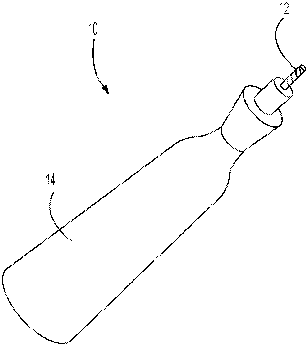
[0020] FIG. 1 illustrates a handheld mechanical decalcification device according to an embodiment of the present disclosure;
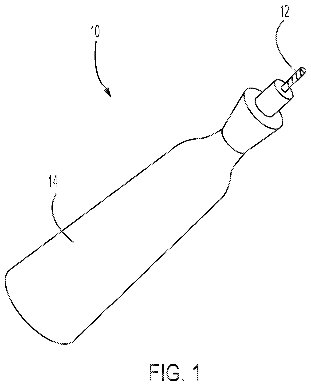
[0021] FIG. 2 illustrates a bur and deployable net apparatus at a distal end of a catheter system according to an embodiment of the present disclosure;
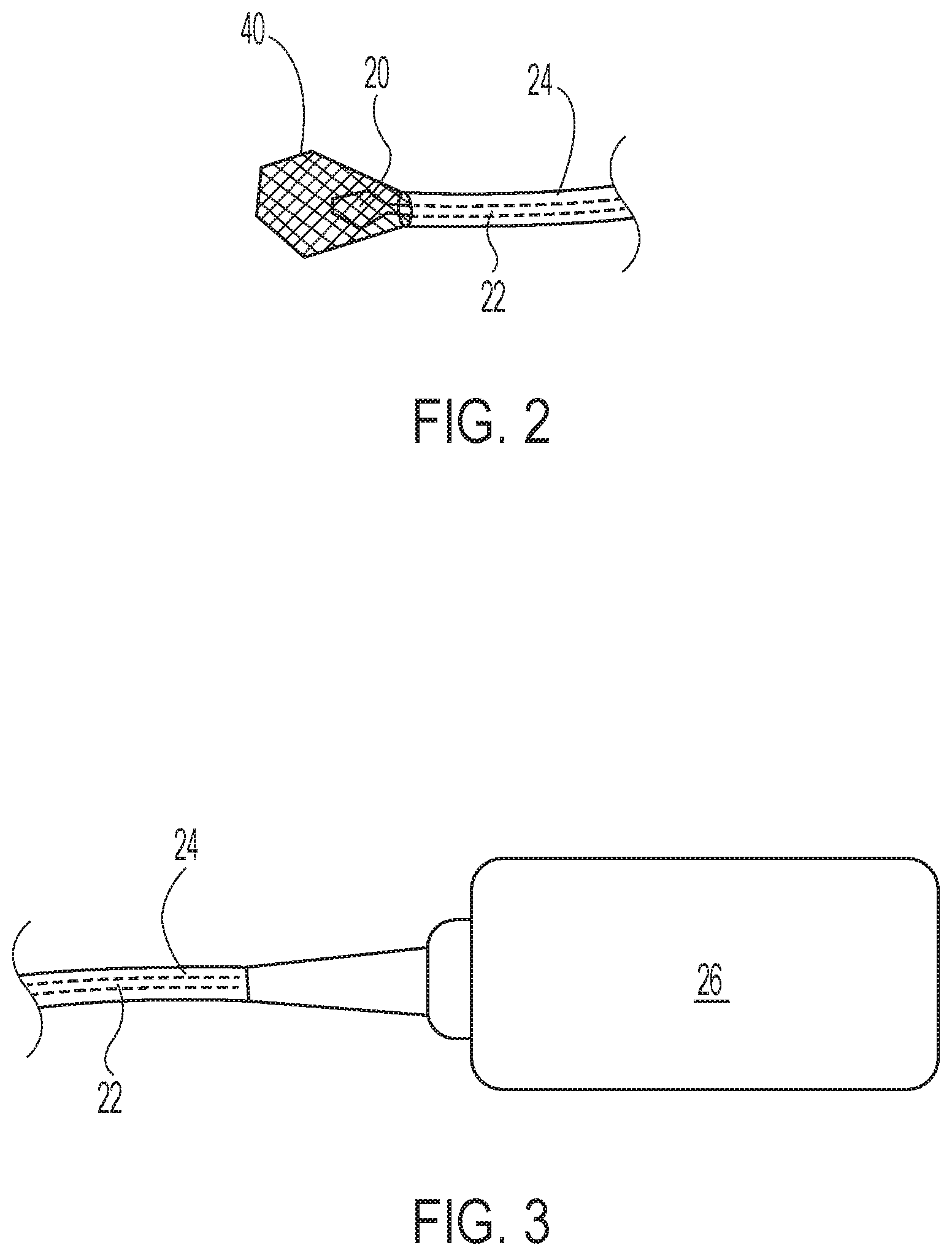
[0022] FIG. 3 illustrates a drive mechanism at a proximal end of the catheter system of FIG. 2;
[0023] FIG. 4 illustrates an ultrasonic tip at a distal end of the catheter system of FIG. 2;
[0024] FIG. 5 illustrates an opening for the delivery of a biocompatible solvent at a distal end of the catheter system of FIG. 2;
[0025] FIG. 6 illustrates an ultrasonic wave generator at a proximal end of the catheter system of FIG. 2;
[0026] FIG. 7 illustrates a biocompatible solvent delivery device at a proximal end of the catheter system of FIG. 2;
[0027] FIG. 8 illustrates a cerebral protection device at a distal end of the catheter system of FIG. 2;
[0028] FIG. 9 illustrates a an opening of an active aspiration system at a distal end of the catheter system of FIG. 2;
[0029] FIG. 10 illustrates an active aspiration device at a proximal end of the catheter system of FIG. 2;
[0030] FIG. 11 illustrates a bioelectric signal array at a distal end of the catheter system of FIG. 2;
[0031] FIG. 12 illustrates a bioelectric signal generator at a proximal end of the catheter system of FIG. 2;
[0032] FIG. 13 illustrates a nitinol ring placement catheter at a distal end of the catheter system of FIG. 2; and
[0033] FIG. 14 illustrates a micro infusion pump at a proximal end of the catheter system of FIG. 2.
DETAILED DESCRIPTION
[0034] In the following detailed description, reference is made to the accompanying drawings, which form a part hereof, and in which are shown, by way of illustration, specific examples of embodiments in which the present disclosure may be practiced. These embodiments are described in sufficient detail to enable a person of ordinary skill in the art to practice the present disclosure. However, other embodiments may be utilized, and structural, system, and process changes may be made without departing from the scope of the disclosure.
[0035] The following description may include examples to help enable one of ordinary skill in the art to practice the disclosed embodiments. The use of the terms "exemplary," "by example," and "for example," means that the related description is explanatory, and though the scope of the disclosure is intended to encompass the examples and legal equivalents, the use of such terms is not intended to limit the scope of an embodiment or this disclosure to the specified components, steps, features, functions, or the like.
[0036] Furthermore, specific implementations shown and described are only examples and should not be construed as the only way to implement the present disclosure unless specified otherwise herein. It will be readily apparent to one of ordinary skill in the art that the present disclosure may be practiced by numerous other partitioning solutions. For the most part, details concerning timing considerations and the like have been omitted where such details are not necessary to obtain a complete understanding of the present disclosure and are within the abilities of persons of ordinary skill in the relevant art.
[0037] As used in this specification, the terms "substantially," "about," and "approximately" in reference to a given parameter, property, or condition means and includes to a degree that one skilled in the art would understand that the given parameter, property, or condition is met with a small degree of variance, such as within acceptable manufacturing tolerances. For example, a parameter that is substantially met may be at least about 90% met, at least about 95% met, at least about 99% met, or even 100% met.
[0038] The phrase "at least one of" when used with a list of items means different combinations of one or more of the listed items may be used and only one of each item in the list may be needed. For example, "at least one of item A, item B, and item C" may include, without limitation, item A or item A and item B. This example may also include item A, item B, and item C, or item B and item C. In other examples, "at least one of" may be, without limitation, two of item A, one of item B, and 10 of item C; four of item B and seven of item C; and other suitable combinations.
[0039] As used in this disclosure, any relational term, such as "first," "second," "over," "top," "bottom," "side," etc., is used for clarity and convenience in understanding the disclosure and accompanying drawings and does not connote or depend on any specific preference, orientation, or order, except where the context clearly indicates otherwise.
[0040] As used in this disclosure, the term "and/or" means and includes any and all combinations of one or more of the associated listed items.
[0041] When one component is "associated" with another component, the association is a physical association in these examples. For example, a first component may be considered to be associated with a second component by being secured to the second component by welding, adhesives, fasteners or connected to the second component in some other suitable manner. The first component may also be connected to the second component using a third, intervening component by which the first component may also be considered to be associated with the second component.
[0042] The illustrations presented in this disclosure are not meant to be actual views of any particular system or device, but are merely idealized representations that are employed to describe the disclosed embodiments. Thus, the drawings are not necessarily to scale and relative dimensions may have been exaggerated for the sake of clarity. Additionally, elements common between figures may retain the same or similar numerical designation.
[0043] The following description provides specific details in order to provide a thorough description of embodiments of this disclosure. However, a person of ordinary skill in the art will understand that the embodiments of this disclosure may be practiced without employing these specific details.
[0044] Over time, heart valves may become dysfunctional from calcification build up, and clots may form, which may cause strokes. Additionally, heart valves lose shape, and thus function, and heart valve leaflets degenerate and do not function properly.
[0045] Embodiments of the present disclosure are designed to remove plaque (e.g., calcification) from a heart valve via processes involving one or more of the following steps: 1) a mechanical removal of calcified material, 2) a simultaneous removal of calcific material during decalcification, 3) an ultrasonic cleaning of the previously decalcified valve, 4) a chemical rinsing of the calcified region using a biocompatible solvent, such as a citric acid solution, and/or 5) an applied electric stimulation to up-regulate targeted gene expression via electroceutical therapy to encourage regeneration of the heart valve.
[0046] In some embodiments, such plaque removal and valve revitalization may be accomplished with one or more hand-held surgical device, such as with open-heart surgery techniques for surgical valve repair. In further embodiments, such plaque removal and valve revitalization may be accomplished with one or more devices using percutaneous delivery (e.g., transcatheter), such as for transthoracic valve repair.
[0047] FIG. 1 shows an isometric view of a handheld mechanical decalcification device 10 for use in open surgery, according to an embodiment of the present disclosure. For example, the handheld mechanical decalcification device 10 may be utilized within an open-heart surgery, or as an alternative to a surgical aortic valve replacement (SAVR). The handheld mechanical decalcification device 10 may include a bur 12 extending from a hand piece 14. As shown in FIG. 1, and as shown in a magnified view of the bur 12 in FIG. 2, the bur may include a plurality of blades and flutes in a spiral configuration extending along the circumference of the bur. Optionally, the bur may also include a rounded tip portion, which may prevent unintentional tissue damage during use. Other bur configurations may also be utilized. For example, an abrasive bur having a surface comprising abrasive material may be utilized, such as bur having a surface comprising diamond particles.
[0048] In use, a heart valve (not shown) may be exposed and calcified regions may be removed by mechanical decalcification. The decalcification may be administered through the handheld mechanical decalcification device 10 via the contact and movement of the surface of the bur 12 relative to the calcified regions. For example, the bur 12 may be rotated, vibrated, and/or reciprocated. Accordingly, the bur 12 may deliver sufficient mechanical stress to remove large regions of calcification.
[0049] Simultaneously, debris may be removed by way of a handheld, or largescale, suction device. Prior to and/or after the decalcification via the handheld mechanical decalcification device 10, ultrasonic cleaning may optionally be performed with an ultrasonic cleaning device. A brief ultrasonic pulse may be used post-decalcification to mechanically disperse any remaining calcium particles. Any released calcium micro-particles or other associated debris that escapes the surgeon's scrutiny and the calcium suction device may be small enough to be partially transported to regional lymph nodes. Ultrasound pulsation may be repeated until all particles are removed.
[0050] At the completion of mechanical decalcification, the heart valve may be further cleansed by applying a biocompatible solvent, such as a citric acid solution, to the regions where the calcification has been removed. For example, a syringe may filled with the biocompatible solvent and may be utilized to direct a stream of the biocompatible solvent onto the heart valve. The biocompatible solvent may be a non-harmful chemical that degrades calcified deposits, and may facilitate the removal of microparticles from the site and encourage healing and regeneration of tissue for complete recovery.
[0051] Once the heart valve has been cleaned of calcification deposits, electrodes to stimulate regeneration of the heart valve may apply electric stimulation of the heart valve. The electric stimulations of the heart valve, and optionally other tissue in the region of the heart valve, may provide one or more of various bioelectric signals to stimulate regeneration of the heart valve. For example, the electrodes may utilized to provide a bioelectric SDF-1 stem cell homing signal, a bioelectric IGF-1 DNA repair signal, a bioelectric HGF regeneration signal, a bioelectric EGF regeneration signal, a bioelectric Activin A+B regeneration signal, a bioelectric follistatin regeneration signal, a bioelectric Tropoelastin elasticity regeneration signal, a bioelectric eNOS blood flow signal, a bioelectric VEGF blood flow signal, a bioelectric stem cell proliferation signal, and/or a bioelectric stem cell differentiation control signal.
[0052] The heart valve may then be bathed with a biochemical bath to encourage healing and regeneration of tissue to facilitate complete recovery and full use of the heart valves. For example, the biochemical bath may comprise a cocktail of regenerative agents including any combination of the following: stem cells, endothelial progenitor cells, selected exosomes, selected alkaloids, selected anti-inflammatory agents, nutrient hydrogel, organ specific matrix, selected growth factors, amniotic fluid, placenta fluid, cord blood, and embryonic sourced growth factors and cells.
[0053] In certain embodiments, the device, system, and/or method is combined with bioelectric signaling for, e.g., the regeneration of heart tissue. Such bioelectric signaling generally modulates (e.g., upregulates) the expression of klotho, follistatin, EGF, HGF, tropoelastin and/or other regeneration promoting proteins. (See, e.g., the incorporated US 2018/0064935A1 to Leonhardt et al. and PCT International Application No. PCT/US2020/021556, filed Mar. 6, 2020, and also incorporated herein by this reference, for various appropriate bioelectric signals).
[0054] For instance, Klotho both prevents re-calcification and regenerates cardiac tissue. Klotho may be released into the blood stream simply by stimulating the subject's thigh muscle with the proper bioelectric signals for about 45 minutes, once or twice a month (e.g., at home) to prevent heart valve re-calcification. Such a treatment can be supplemented with, e.g., a once a year infusion of elastin nanoparticles (in a clinic). See, generally, Chen et al. "The Role and Mechanism of .alpha.-Klotho in the Calcification of Rat Aortic Vascular Smooth Muscle Cells" Biomed Res Int. 2015; 2015: 194362; Hu et al. "Klotho Deficiency Causes Vascular Calcification in Chronic Kidney Disease" J Am Soc Nephrol. 2011 January; 22(1): 124-136; Chen et al. "Deficiency in the anti-aging gene Klotho promotes aortic valve fibrosis through AMPK.alpha.-mediated activation of RUNX2" Aging Cell. 2016 October; 15(5): 853-860; and Chen et al. "Secreted Klotho Attenuates Inflammation-Associated Aortic Valve Fibrosis in Senescence-Accelerated Mice P1" Hypertension 2018; 71:877-885, the contents of the entirety of each of which are herein incorporated by this reference.
[0055] In further embodiments, such plaque removal and valve revitalization may be accomplished with one or more devices using percutaneous delivery (e.g., transcatheter), such as for transthoracic valve repair.
[0056] In some embodiments, methods of the present disclosure may be performed with a catheter system, such as shown in FIGS. 2-14. The catheter system may include components located at a distal end, the distal end configured for percutaneous insertion into a patient's body, and may include components located at a proximal end, the proximal end configured to remain outside of the patient's body and accessible to an operator of the catheter system, such as a surgeon.
[0057] The catheter system may be utilized for decalcifying and regenerating heart valves, so that a patient may keep their own heart valves instead of getting an implant. The catheter system may be utilized to perform three methods of decalcification. Additionally, the catheter system may be utilized to regenerate heart valve tissue.
[0058] In further embodiments, the catheter system may be utilized to implement shape reform via a nitinol ring, along with decalcification and regeneration.
[0059] For mechanical removal of calcium deposits, the catheter system may include multiple components, such as at least one mechanical decalcification device. As shown in FIG. 2, the catheter system may include a bur 20 located at the tip of the distal end, which may include an abrasive surface. The bur may have a generally frustoconical shaped tip and may be connected to a shaft 22 that is located within a lumen 24 of the catheter system. The shaft 22 may extend through the lumen 24 to a drive mechanism 26 located at the proximal end of the catheter system, as shown in FIG. 3.
[0060] The drive mechanism 26 may be configured to cause the bur 20 to move at the distal end of the catheter system to mechanically remove calcium deposits on the heart valve. In some embodiments, the drive mechanism 26 may comprise a motor configured to rotate the bur 20 via the shaft 22. In further embodiments, the drive mechanism 26 may comprise a vibration generator, such as an eccentric rotating mass vibration motor, configured to vibrate the bur 20 via the shaft 22. Accordingly, the drive mechanism 26 may be configured to cause the bur 20 to rotate, vibrate, reciprocate, and/or translate.
[0061] For additional decalcification and cleaning, the catheter system may include an ultrasonic tip 30 located at the distal end, as shown in FIG. 4, and an opening 32 at the distal end for the delivery of the biocompatible solvent, as shown in FIG. 5. The ultrasonic tip 30 may be coupled to an ultrasonic wave generator 34 located at the proximal end, as shown in FIG. 6, configured to deliver ultrasonic waves to the ultrasonic tip 30.
[0062] A lumen 36 of the catheter system may extend from the opening 32 to a biocompatible solvent delivery device 38 located at the proximal end of the catheter system, as shown in FIG. 7, for directing the biocompatible solvent through the lumen 36 and out of the opening 32 at the distal end. For example, the biocompatible solvent delivery device 38 may be a syringe filled with the biocompatible solvent. Accordingly, pressurized biocompatible solvent may be directed through the lumen 36 and out of the opening 32 as a relatively high-pressure stream of biocompatible solvent.
[0063] As shown in FIG. 2, the catheter system may also include a deployable net apparatus 40 located at the distal end proximal to the bur 20. The deployable net apparatus 40 may be deployed to encompass at least a portion of a heart valve, or optionally the entire valve, during operation of the bur to facilitate the removal of the calcification dislodged from the heart valve in the process. The deployable net apparatus 40 may be comprised of electrospun polymers forming a nanoscale fiber mesh. Optionally, other devices may be implemented instead of, or in combination with, the net apparatus to hold the heart valve during cleaning, such as a suction cup (not shown).
[0064] The catheter system may include a deflecting tip-guiding catheter and an optical viewing catheter to facilitate the guiding of the catheter system components at the distal end of the catheter system during use.
[0065] The catheter system may also include additional components for the capture and removal of debris. For example, the catheter system may include one or more cerebral protection devices 44 (i.e., an embolic protection device), as shown in FIG. 8, located at a distal portion of the catheter system for the capture of debris.
[0066] The catheter system may additionally include an active aspiration system. The active aspiration system may comprise at least one opening 50 at the distal end of a lumen 52, as shown in FIG. 9, and an active aspiration device 54 may be coupled to the lumen 52 of the catheter system at the proximal end, as shown in FIG. 10, to facilitate the removal of debris through the lumen 52 from the at least one opening 50 via suction.
[0067] As shown in FIG. 11, a bioelectric signal array 60 may be located at the distal end of the catheter system for the delivery of the bioelectric stimulation to the heart valve after the cleaning of calcification deposits. A bioelectric stimulator signal generator 62 may be positioned at the proximal end of the catheter system, as shown in FIG. 12, and may be coupled to the bioelectric signal array via wires 64 to provide bioelectric signals to the bioelectric signal array 60.
[0068] Optionally, as shown in FIG. 13, the catheter system may include a nitinol ring placement catheter 70 for the placement of a nitinol ring 72 at the heart valve to reform the valve shape.
[0069] In some embodiments, the catheter system may include various catheters to implement various components of the catheter system. In further embodiments, multiple components of the catheter system may be incorporated into a single catheter. The use of multiple catheters may make the design of the catheter system simpler. Incorporating multiple components on a single catheter, however, may reduce the number of times that catheters are inserted and removed from a patient during a procedure. Accordingly, embodiments of the present disclosure contemplate any variety of combination of the various components on one or more catheters to provide the catheter system.
[0070] In one embodiment, every component of the catheter system may be incorporated into a single catheter. In another embodiment, the catheter system may be comprised of a plurality of catheters, each catheter of the plurality incorporating only a single component of the catheter system at each of the proximal and distal ends thereof. In yet further embodiments, the catheter system may include a plurality of catheters with at least one catheter of the plurality of catheters incorporating multiple components of the catheter system at each of its proximal and distal ends.
[0071] The catheter system is designed to be implemented through transvascular manipulation as a supplement to, or a replacement for, transcatheter aortic valve replacement (TAVR). TAVR procedures may provide a critically needed alternative therapy for patients with severe aortic stenosis.
[0072] Heart valve surgery is very costly and disrupts a patient's life. This minimally invasive surgery is designed to prevent heart surgery and return the valves to full optimal capacity with little downtime. As the baby boom generation begins to experience heart valve complications, embodiments of this disclosure may assist physicians to enhance the quality of life for each patient.
[0073] For the cleaning operation, the bur 20, the ultrasonic tip 30, and biologically safe solvent may be utilized, such as in a sequence, to fully decalcify and clean the heart valve leaflets and orifice of the heart valve. Concurrently, optical viewing systems may provide visualization of areas being cleaned. For example, the cleaning operation may be guided via standard fluoroscopic approaches, but may also be accompanied by IVUS imaging probes attached to the distal end of the catheter system. The net apparatus 40, and/or a suction cup system may hold the heart valve leaflets during cleaning. Additionally, during decalcification or subsequent procedures, the catheter system may be designed to expand the net apparatus 40 to facilitate the removal of any calcification dislodged from the heart valve in the process. Additionally, debris may be captured by one or more cerebral protection devices 44 and/or removed via suction.
[0074] After the cleaning operation, bioelectric regeneration signals may be provided to the heart valve by the bioelectric signal array 60 powered by the bioelectric signal generator 62 to regenerate the native heart valve by recruiting stem cells and building new healthy tissues. For example, the bioelectric signals may include one or more of a bioelectric SDF-1 stem cell homing signal, a bioelectric IGF-1 DNA repair signal, a bioelectric HGF regeneration signal, a bioelectric EGF regeneration signal, a bioelectric Activin A+B regeneration signal, a bioelectric follistatin regeneration signal, a bioelectric Tropoelastin elasticity regeneration signal, a bioelectric eNOS blood flow signal, a bioelectric VEGF blood flow signal, a bioelectric stem cell proliferation signal, and/or a bioelectric stem cell differentiation control signal.
[0075] Optionally, the nitinol ring 72 is placed by the nitinol ring placement catheter 70, such as if the above decalcification and regeneration procedure has not restored full function. Furthermore, autologous cell created heart valve leaflets may be placed via a heart valve catheter-based delivery system if the previous steps have not restored full function.
[0076] Additionally, such as if all of the above has failed, a micro infusion pump 80, as shown in FIG. 14, may be connected to a lumen of the catheter system and a component regeneration cocktail composition may be infused until function is restored. For example, the micro infusion pump 80 may be utilized to deliver a biochemical bath comprising a cocktail of regenerative agents including any combination of the following: stem cells, endothelial progenitor cells, selected exosomes, selected alkaloids, selected anti-inflammatory agents, nutrient hydrogel, organ specific matrix, selected growth factors, amniotic fluid, placenta fluid, cord blood, and embryonic sourced growth factors and cells.
[0077] Optionally, a robot could control the full procedure of cleaning, regeneration, nitinol ring 72 placement and percutaneous autologous cell created valve placement, or portions thereof.
[0078] The catheter system may be designed to be fed through femoral artery, radial artery or other access points into the heart chamber and specifically is associated with a heart valve.
[0079] The systems, devices, and methods herein may be utilized to decalcify the heart valves, restores shape, and regenerates them restoring full normal function.
[0080] The disclosed systems, devices, and methods may reduce calcification in a heart valve, and may regenerate the heart valve with stem cell recruitment and differentiation supported by a full range of regeneration promotion proteins. The disclosed systems, devices, and methods may be combined with a non-surgical reforming option when required or thought desirable.
[0081] Alternative embodiments and variations in the detail design of the disclosed systems and devices are contemplated within the scope of the invention.
[0082] While certain illustrative embodiments have been described in connection with the figures, those of ordinary skill in the art will recognize and appreciate that the scope of this disclosure is not limited to those embodiments explicitly shown and described in this disclosure. Rather, many additions, deletions, and modifications to the embodiments described in this disclosure may be made to produce embodiments within the scope of this disclosure, such as those specifically claimed, including legal equivalents. In addition, features from one disclosed embodiment may be combined with features of another disclosed embodiment while still being within the scope of this disclosure, as contemplated by the inventor.
REFERENCES
[0083] (The contents of the entirety of each of which is incorporated herein by this reference.) [0084] Dahm M. et al. "Decalcification of the aortic valve does not prevent early recalcification" J Heart Valve Dis., 9(1):21-6. (January 2008). [0085] Chen et al. "The Role and Mechanism of .alpha.-Klotho in the Calcification of Rat Aortic Vascular Smooth Muscle Cells" Biomed Res Int. 2015; 2015: 194362. [0086] Hu et al. "Klotho Deficiency Causes Vascular Calcification in Chronic Kidney Disease" J Am Soc Nephrol. 2011 January; 22(1): 124-136. [0087] Chen et al. "Deficiency in the anti-aging gene Klotho promotes aortic valve fibrosis through AMPK.alpha.-mediated activation of RUNX2" Aging Cell. 2016 October; 15(5): 853-860. [0088] Chen et al. "Secreted Klotho Attenuates Inflammation-Associated Aortic Valve Fibrosis in Senescence-Accelerated Mice P1" Hypertension 2018; 71:877-885. [0089] Kose N. et al., "Citric acid as a decalcifying agent for the excised calcified human heart valves" Anadolu Kardiyol Derg., 2008 April; 8(2):94-8. [0090] "Focused Ultrasound Therapy" https://fusfoundation.org/diseases-and-conditions/cardiovascular/heart-va- lve-calcifications (28 Jan. 2020).
* * * * *
References
D00000
D00001
D00002
D00003
D00004
D00005
D00006
XML
uspto.report is an independent third-party trademark research tool that is not affiliated, endorsed, or sponsored by the United States Patent and Trademark Office (USPTO) or any other governmental organization. The information provided by uspto.report is based on publicly available data at the time of writing and is intended for informational purposes only.
While we strive to provide accurate and up-to-date information, we do not guarantee the accuracy, completeness, reliability, or suitability of the information displayed on this site. The use of this site is at your own risk. Any reliance you place on such information is therefore strictly at your own risk.
All official trademark data, including owner information, should be verified by visiting the official USPTO website at www.uspto.gov. This site is not intended to replace professional legal advice and should not be used as a substitute for consulting with a legal professional who is knowledgeable about trademark law.