Fc-binding Protein Having Improved Antibody Separation Ability, And Method For Separating Antibody Using Same
TERAO; Yosuke ; et al.
U.S. patent application number 16/486380 was filed with the patent office on 2020-07-16 for fc-binding protein having improved antibody separation ability, and method for separating antibody using same. This patent application is currently assigned to TOSOH CORPORATION. The applicant listed for this patent is TOSOH CORPORATION. Invention is credited to Yoshiharu ASAOKA, Satoshi ENDO, Seigo OE, Ryoko OTAKE, Yosuke TERAO, Yukie YAMAMOTO, Naoki YAMANAKA.
| Application Number | 20200223885 16/486380 |
| Document ID | / |
| Family ID | 63170310 |
| Filed Date | 2020-07-16 |

| United States Patent Application | 20200223885 |
| Kind Code | A1 |
| TERAO; Yosuke ; et al. | July 16, 2020 |
FC-BINDING PROTEIN HAVING IMPROVED ANTIBODY SEPARATION ABILITY, AND METHOD FOR SEPARATING ANTIBODY USING SAME
Abstract
The present application addresses the problem of providing an Fc-binding protein having an improved antibody separation ability. The present application also addresses the problem of providing a high-accuracy antibody separation method using an insoluble carrier having the protein immobilized thereon. The problems can be solved by: an Fc-binding protein in which at least an amino acid substitution at a specific position therein occurs and which has reduced affinity for an antibody; and an antibody separation method including allowing an equilibration buffer solution to pass through a column in which an insoluble carrier having the protein immobilized thereon is filled to equilibrate the column, adding a solution containing an antibody to cause the adsorption of the antibody onto the carrier, and eluting the antibody adsorbed on the carrier using an elution solution.
| Inventors: | TERAO; Yosuke; (Kanagawa, JP) ; ASAOKA; Yoshiharu; (Kanagawa, JP) ; OTAKE; Ryoko; (Kanagawa, JP) ; ENDO; Satoshi; (Kanagawa, JP) ; YAMANAKA; Naoki; (Kanagawa, JP) ; YAMAMOTO; Yukie; (Kanagawa, JP) ; OE; Seigo; (Kanagawa, JP) | ||||||||||
| Applicant: |
|
||||||||||
|---|---|---|---|---|---|---|---|---|---|---|---|
| Assignee: | TOSOH CORPORATION Yamaguchi JP |
||||||||||
| Family ID: | 63170310 | ||||||||||
| Appl. No.: | 16/486380 | ||||||||||
| Filed: | February 7, 2018 | ||||||||||
| PCT Filed: | February 7, 2018 | ||||||||||
| PCT NO: | PCT/JP2018/004202 | ||||||||||
| 371 Date: | August 15, 2019 |
| Current U.S. Class: | 1/1 |
| Current CPC Class: | C07K 1/22 20130101; C07K 14/00 20130101; C12N 15/09 20130101; C07K 16/00 20130101 |
| International Class: | C07K 1/22 20060101 C07K001/22; C07K 16/00 20060101 C07K016/00; C07K 14/00 20060101 C07K014/00 |
Foreign Application Data
| Date | Code | Application Number |
|---|---|---|
| Feb 20, 2017 | JP | 2017-028974 |
| Apr 4, 2017 | JP | 2017-074727 |
| Jun 2, 2017 | JP | 2017-109681 |
Claims
1. An Fc-binding protein comprising at least amino acid residues at positions 33 to 208 of the amino acid sequence set forth in SEQ ID NO: 5, wherein among the amino acid residues at positions 33 to 208, at least valine at position 192 is amino acid-substituted by phenylalanine.
2. The Fc-binding protein according to claim 1, which is selected from among the following (a), (b), and (c): (a) an Fc-binding protein in which among amino acid residues at positions 33 to 208 of the amino acid sequence set forth in SEQ ID NO: 5, valine at position 192 is amino acid-substituted by phenylalanine; (b) an Fc-binding protein in which among amino acid residues at positions 33 to 208 of the amino acid sequence set forth in SEQ ID NO: 5, valine at position 192 is amino acid-substituted by phenylalanine, and which further comprises an amino acid residue having a substitution, deletion, insertion, or addition of one or more amino acids, and which has antibody-binding activity; and (c) an Fc-binding protein which has 80% or more homology to an amino acid sequence comprising amino acid residues from glycine at position 33 to glutamine at position 208 of the amino acid sequence set forth in SEQ ID NO: 4, in which valine at position 192 is amino acid-substituted by phenylalanine, and which has antibody-binding activity.
3. A method for separating an antibody, comprising: adding an equilibration solution to a column which is filled with an insoluble carrier having the Fc-binding protein according to claim 1 immobilized thereon so as to equilibrate the column; adding a solution containing an antibody to cause the antibody to be adsorbed on the carrier; and eluting the antibody adsorbed on the carrier using an elution solution.
4. The separation method according to claim 3, further comprising isolating a fraction containing the eluted antibody.
5. The separation method according to claim 3, wherein the equilibration solution is a buffer containing an inorganic salt at pH 4.5 to pH 5.8.
6. A method for monitoring culture progress of an antibody-producing cell and/or a produced antibody, comprising: obtaining an antibody-producing cell by transfecting a host cell with an antibody expression vector; culturing the antibody-producing cell; obtaining an antibody from the culture medium and/or cultured cells; and separating the obtained antibody by the method according to claim 3.
7. A method for monitoring time-dependent changes in a sugar chain structure pattern of an antibody, comprising: obtaining an antibody-producing cell by transfecting a host cell with an antibody expression vector; a step of culturing the antibody-producing cell; obtaining an antibody from the culture medium and/or cultured cells; and separating the obtained antibody by the method according to claim 3.
8. A method for producing an antibody, comprising optimizing culture conditions for an antibody-producing cell based on the results of monitoring by the method according to claim 7 such that the cell produces an antibody having a desired sugar chain structure.
Description
FIELD
[0001] The present invention relates to an Fc-binding protein having improved antibody (immunoglobulin) separation ability, and a method for separating an antibody using the same. More specifically, the present invention relates to an Fc-binding protein which has decreased affinity to an antibody as compared to a conventionally known Fc-binding protein so as to have improved antibody (immunoglobulin) separation ability, and a method for separating an antibody using an insoluble carrier having the Fc-binding protein immobilized thereon.
BACKGROUND
[0002] The sugar chain structure of an antibody drug plays a major role in drug efficacy and stability. It is therefore extremely important to control the sugar chain structure when producing an antibody drug. Among Fc-binding proteins, Fc.gamma.RIIIa is known to recognize the sugar chain structure of an antibody (immunoglobulin). The antibody can be separated based on the sugar chain structure by using an adsorbent obtained by immobilizing Fc.gamma.RIIIa on an insoluble carrier (PTL 1). Accordingly, the adsorbent is useful for step analysis and isolation upon antibody drug production.
[0003] Meanwhile, it is known that Fc.gamma.RIIIa has lower antibody-binding ability as compared to another Fc-binding protein, namely, Fc.gamma.RI (NPL 1), and the affinity to an antibody is improved by introducing a mutation into a protein having antibody-binding ability (PTL 2). However, the degree of separation of an antibody has not been improved. Meanwhile, the site of interaction between the Fc-binding protein and the antibody has been clarified (NPL 2), and amino acid residues involved in binding between the Fc-binding protein and the antibody have been revealed. However, such findings are not enough to improve the degree of separation in relation to antibody analysis and adsorption. Therefore, in the industrial production of antibody drugs, it was difficult to apply an Fc.gamma.RIIIa-immobilized carrier to step analysis and product analysis.
CITATION LIST
Patent Literature
[PTL 1] Japanese Unexamined Patent Publication (Kokai) No. 2015-086216
[PTL 2] Japanese Unexamined Patent Publication (Kokai) No. 2015-019615
Non Patent Literature
[NPL 1] P. Bruhns et al., Blood, 16, 3716-3725, 2009
[NPL 2] P. Sonderman et al., Nature, 406, 267-2735, 2000
SUMMARY
Technical Problem
[0004] According to the present invention, an Fc-binding protein having improved antibody (immunoglobulin) separation ability as compared to a conventionally known Fc-binding protein is provided, and a method for separating an antibody (immunoglobulin) with high accuracy using an insoluble carrier having the protein immobilized thereon is provided.
Solution to Problem
[0005] In order to solve the above problems, the present inventors conducted exhaustive substitutions of a specific amino acid residue in Fc.gamma.RIIIa involved in antibody-binding ability. As a result, Fc-binding proteins having decreased affinity to an antibody was obtained by substituting the amino acid residue by a different amino acid residue. In addition, the use of a carrier having the protein immobilized thereon has made it possible to separate an antibody with high accuracy as compared to an insoluble carrier having a conventional Fc-binding protein immobilized thereon. Specifically, among amino acid residues involved in antibody-binding ability in an Fc-binding protein consisting of the amino acid sequence set forth in SEQ ID NO: 5 (human Fc.gamma.RIIIa amino acid substitution product), exhaustive substitutions of the amino acid corresponding to valine at position 192 of SEQ ID NO: 5 (corresponding to valine at position 176 in natural human Fc.gamma.RIIIa consisting of the amino acid sequence set forth in SEQ ID NO: 1) by different amino acids were conducted. Accordingly, an Fc-binding protein having decreased affinity to an antibody as compared to an Fc-binding protein consisting of the amino acid sequence set forth in SEQ ID NO: 5 was obtained, and the use of an insoluble carrier having the protein immobilized thereon has made it possible to separate the antibody with high accuracy as compared to an insoluble carrier having a conventional Fc-binding protein immobilized thereon.
[0006] In other words, the present invention encompasses embodiments described in the following (1) to (7).
[0007] (1) An Fc-binding protein comprising at least amino acid residues at positions 33 to 208 of the amino acid sequence set forth in SEQ ID NO: 5, wherein among the amino acid residues at positions 33 to 208, at least valine at position 192 is amino acid-substituted by phenylalanine.
[0008] (2) The Fc-binding protein according to (1), which is selected from among the following (a), (b), and (c): [0009] (a) an Fc-binding protein in which among amino acid residues at positions 33 to 208 of the amino acid sequence set forth in SEQ ID NO: 5, valine at position 192 is amino acid-substituted by phenylalanine; [0010] (b) an Fc-binding protein in which among amino acid residues at positions 33 to 208 of the amino acid sequence set forth in SEQ ID NO: 5, valine at position 192 is amino acid-substituted by phenylalanine, and which further comprises an amino acid residue having a substitution, deletion, insertion, or addition of one or more amino acids, and which has antibody-binding activity; and [0011] (c) an Fc-binding protein which has 80% or more homology to an amino acid sequence comprising amino acid residues from glycine at position 33 to glutamine at position 208 of the amino acid sequence set forth in SEQ ID NO: 4, in which valine at position 192 is amino acid-substituted by phenylalanine, and which has antibody-binding activity.
[0012] (3) A method for separating an antibody, comprising: a step of adding an equilibration solution to a column which is filled with an insoluble carrier having the Fc-binding protein according to (1) or (2) immobilized thereon so as to equilibrate the column; a step of adding a solution containing an antibody to cause the antibody to be adsorbed on the carrier; and a step of eluting the antibody adsorbed on the carrier using an elution solution.
[0013] (4) The separation method according to (3), further comprising a step of isolating a fraction containing the eluted antibody.
[0014] (5) The separation method according to (3) or (4), wherein the equilibration solution is a buffer containing an inorganic salt at pH 4.5 to pH 5.8.
[0015] (6) A method for monitoring culture progress of an antibody-producing cell and a produced antibody, comprising: a step of obtaining an antibody-producing cell by transfecting a host cell with an antibody expression vector; a step of culturing the antibody-producing cell; a step of obtaining an antibody from the culture solution; and a step of separating the obtained antibody by the method according to any one of (3) to (5).
[0016] (7) A method for monitoring time-dependent changes in a sugar chain structure pattern of an antibody, comprising: a step of obtaining an antibody-producing cell by transfecting a host cell with an antibody expression vector; a step of culturing the antibody-producing cell; a step of obtaining an antibody from the culture medium and/or cultured cells; and a step of separating the obtained antibody by the method according to any one of (3) to (5).
[0017] (8) A method for producing an antibody, comprising a step of optimizing culture conditions for an antibody-producing cell based on the results of monitoring by the method according to (7) such that the cell produces an antibody having a desired sugar chain structure.
[0018] Hereinafter, the present invention will be described in detail.
[0019] Specifically, the separation of an antibody may include not only separation of an antibody from contaminants but also separation between antibodies based on the structure, properties, activity, and the like.
[0020] The Fc-binding protein of the present invention is a protein having ability to bind to the Fc region of an antibody (immunoglobulin), which includes amino acid residues from glycine at position 33 to glutamine at position 208 of SEQ ID NO: 5 (corresponding to a region at least from glycine at position 17 to glutamine at position 192 in the extracellular domain (the region of EC in FIG. 1) of natural human Fc.gamma.RIIIa consisting of the amino acid sequence set forth in SEQ ID NO: 1), and which has at least an amino acid substitution of an amino acid residue at a certain position among the amino acid residues at positions 33 to 208. Therefore, an Fc-binding protein used in the present invention may include all or part of a signal peptide region (S in FIG. 1) present on the N-terminal side of the extracellular domain, or it may include all or part of a transmembrane domain (TM in FIG. 1) and an intracellular domain (C in FIG. 1) present on the C-terminal side of the extracellular domain. Specifically, an amino acid substitution at the certain position is an amino acid substitution denoted by Val192Phe (indicating a substitution of valine at position 192 of SEQ ID NO: 5 (corresponding to position 176 of SEQ ID NO: 1) by phenylalanine, and so forth).
[0021] There is a known variant of wild-type Fc.gamma.RIIIa, which has an amino acid substitution of at least one of Leu82His, Leu82Arg, Gly163Asp, and Tyr174His. The Fc-binding protein of the present invention may have such an amino acid substitution, in addition to an amino acid substitution (Val192Phe) at the certain position.
[0022] When the Fc-binding protein of the present invention is produced via amino acid substitution, an amino acid residue at a certain position may be substituted by an amino acid other than the above-described amino acid as long as the protein has antibody-binding activity. Specifically, the protein may have at least one of substitution, deletion, insertion, and addition of one or more amino acid residues. The expression "one or more" or "at least one" may refer to, for example, from 1 to 50, preferably form 1 to 40, more preferably from 1 to 30, still more preferably form 1 to 20, particularly preferably form 1 to 10, although it depends on positions and types of amino acid residues in the protein conformation. One example of a substitution, deletion, insertion, or addition of one or more amino acid residues is a conservative substitution between amino acids having similar physical and/or chemical properties. Those skilled in the art know that in the case of conservative substitution, the protein function is usually maintained between a protein having a substitution and a protein lacking the substitution, each of which is not limited to an Fc-binding protein. One example of a conservative substitution is a substitution that occurs between glycine and alanine, aspartic acid and glutamic acid, serine and proline, or glutamic acid and alanine (Protein Structure and Function, Medical Sciences International, Ltd., 9, 2005).
[0023] In addition, the Fc-binding protein of the present invention may be an Fc-binding protein having amino acid residues including Val192Phe described above which have 70% or more, preferably 80% or more, still more preferably 90% or more, particularly preferably 95% or more amino acid sequence homology to amino acid residues from glycine at position 33 to glutamine at position 208 of SEQ ID NO: 5 as long as the Fc-binding protein has antibody-binding activity.
[0024] A specific example of the Fc-binding protein of the present invention is an Fc-binding protein which includes at least amino acid residues at positions 33 to 208 of the amino acid sequence set forth in SEQ ID NO: 5, and has at least an amino acid substitution of Val192Phe in the amino acid residues at positions 33 to 208 (polypeptide at least including a region of amino acid residues at positions 33 to 208 of the amino acid sequence of SEQ ID NO: 9).
[0025] It is possible to further add a useful oligopeptide to the N-terminal side or the C-terminal side of the Fc-binding protein of the present invention when separating an antibody of interest from a solution with the presence of contaminants. Examples of the oligopeptide include polyhistidine, polylysine, polyarginine, polyglutamic acid, and polyaspartic acid. Further, a cysteine-containing oligopeptide, which is useful for immobilizing the Fc-binding protein of the present invention to a solid phase of a support for chromatography or the like, may be further added to the N-terminal side or the C-terminal side of the Fc-binding protein of the present invention. The length of an oligopeptide to be added to the N-terminal side or the C-terminal side of the Fc-binding protein is not particularly limited. When adding the oligopeptide to the Fc-binding protein of the present invention, it is possible to prepare a polynucleotide encoding the oligopeptide, and then, add the polynucleotide to the N-terminal side or the C-terminal side of the Fc-binding protein by a genetic engineering method known to those skilled in the art. Alternatively, it is also possible to chemically synthesize the oligopeptide and add the oligopeptide to the N-terminal side or the C-terminal side of the Fc-binding protein of the present invention via chemical binding. It is also possible to further add a signal peptide to the N-terminal side of the Fc-binding protein of the present invention for promoting efficient expression in a host. Examples of the signal peptide when a host is Escherichia coli include signal peptides that cause protein secretion in a periplasmic space, such as, PelB, DsbA, MalE (a region of the 1st to 26th amino acid residues of the amino acid sequence described in UniProt No. P0AEX9), and TorT (Japanese Unexamined Patent Publication (Kokai) No. 2011-097898).
[0026] When separating or analyzing an antibody using the Fc-binding protein of the present invention, it is necessary to prepare an antibody adsorbent in which the Fc-binding protein of the present invention is immobilized on an insoluble carrier as described above. An insoluble carrier to be immobilized to the Fc-binding protein is not particularly limited. Examples thereof include carriers made with polysaccharides such as agarose, alginate (alginic acid salt), carrageenan, chitin, cellulose, dextrin, dextran, and starch, carriers made with synthetic polymers such as polyvinyl alcohol, polymethacrylate, poly (2-hydroxyethyl methacrylate), and polyurethane, and carriers made with ceramics such as silica. Of these, carriers made with polysaccharides and carriers made with synthetic polymers are preferable as insoluble carriers. Examples of the preferable carrier include hydroxylated polymethacrylate gel such as TOYOPEARL (manufactured by Tosoh Corporation), agarose gel such as Sepharose (manufactured by GE Healthcare), and cellulose gel such as Cellufine (manufactured by JNC). The shape of insoluble carrier is not particularly limited, and it may be either a particulate or non-particulate shape or a porous or nonporous shape.
[0027] In order to immobilize the Fc-binding protein of the present invention on an insoluble carrier, it is possible to provide the insoluble carrier with an active group such as an N-hydroxysuccinimide (NHS)-activated ester group, an epoxy group, a carboxyl group, a maleimide group, a haloacetyl group, a tresyl group, a formyl group, or haloacetamide and allow the Fc-binding protein of the present invention to covalently bind to the insoluble carrier via the active group. It is possible to directly use a commercially available carrier as a carrier provided with an active group or to prepare a carrier by introducing an active group to the carrier surface under appropriate reaction conditions. Examples of a commercially available carrier provided with an active group include TOYOPEARL AF-Epoxy-650M, TOYOPEARL AF-Tresyl-650M (each manufactured by Tosoh Corporation), HiTrap NHS-activated HP Columns, NHS-activated Sepharose 4 Fast Flow, Epoxy-activated Sepharose 6B (each manufactured by GE Healthcare), and SulfoLink Coupling Resin (manufactured by Thermo Fisher Scientific).
[0028] Meanwhile, as a method for introducing an active group on the carrier surface, a method in which a compound having two or more active sites is allowed to react, at one of the active sites, with a hydroxyl group, an epoxy group, a carboxyl group, an amino group, or the like present on the carrier surface can be exemplified. Among examples of such a compound, examples of a compound for introducing an epoxy group to a hydroxyl group or an amino group on the carrier surface include epichlorohydrin, ethanediol diglycidyl ether, butanediol diglycidyl ether, and hexanediol diglycidyl ether. Examples of a compound for introducing a carboxyl group on the carrier surface after introduction of an epoxy group on the carrier surface by the compound include 2-mercaptoacetic acid, 3-mercaptopropionic acid, 4-mercaptobutyric acid, 6-mercaptobutyric acid, glycine, 3-aminopropionic acid, 4-aminobutyric acid, and 6-aminohexanoic acid.
[0029] Examples of a compound for introducing a maleimide group to a hydroxyl group, an epoxy group, a carboxyl group, or an amino group present on the carrier surface include N-(.epsilon.-maleimidocaproic acid)hydrazide, N-(.epsilon.-maleimidopropionic acid)hydrazide, 4-(4-N-maleimidophenyl)acetic acid hydrazide, 2-aminomaleimide, 3-aminomaleimide, 4-aminomaleimide, 6-aminomaleimide, 1-(4-aminophenyl)maleimide, 1-(3-aminophenyl)maleimide, 4-(maleimide)phenyl isocyanate, 2-maleimidoacetic acid, 3-maleimidopropionic acid, 4-maleimidobutyric acid, 6-maleimidohexanoic acid, N-(.alpha.-maleimidoacetoxy)succinimide ester, (m-maleimidobenzoyl)N-hydroxy succinimide ester, succinimidyl-4-(maleimidomethyl)cyclohexane-1-carbonyl-(6-aminohexanoic acid), succinimidyl-4-(maleimidomethyl)cyclohexane-1-carboxylic acid, (p-maleimidobenzoyl)N-hydroxy succinimide ester, and (m-maleimidobenzoyl)N-hydroxy succinimide ester.
[0030] Examples of a compound for introducing a haloacetyl group to a hydroxyl group or an amino group present on the carrier surface include chloroacetic acid, bromoacetic acid, iodoacetic acid, chloroacetic acid chloride, bromoacetic acid chloride, bromoacetic acid bromide, chloroacetic acid anhydride, bromoacetic acid anhydride, iodoacetic acid anhydride, 2-(iodoacetamido)acetic acid-N-hydroxysuccinimide ester, 3-(bromoacetamido)propionic acid-N-hydroxysuccinimide ester, and 4-(iodoacetyl)aminobenzoic acid-N-hydroxysuccinimide ester. A method in which .omega.-alkenyl alkane glycidyl ether is reacted with a hydroxyl group or an amino group present on the carrier surface, and then, .omega.-alkenyl is halogenated with a halogenating agent for activation can also be exemplified. Examples of .omega.-alkenyl alkane glycidyl ether may include allyl glycidyl ether, 3-butenyl glycidyl ether, and 4-pentenyl glycidyl ether. Examples of a halogenating agent may include N-hlorosuccinimide, N-bromosuccinimide, and N-iodosuccinimide.
[0031] Another example of a method for introducing an active group on the carrier surface is a method for introducing an active group to a carboxyl group present on the carrier surface using a condensing agent and an additive. Examples of a condensing agent include 1-ethyl-3-(3-dimethylaminopropyl)carbodiimide (EDC), dicyclohexylcarbodiamide, and carbonyldiimidazole. Examples of an additive include N-hydroxysuccinimide (NHS), 4-nitrophenol, and 1-hydroxybenzotriazole.
[0032] Examples of a buffer used for immobilizing the Fc-binding protein of the present invention on an insoluble carrier include acetate buffer, phosphate buffer, MES (2-morpholinoethanesulfonic acid) buffer, HEPES (4-(2-hydroxyethyl)-1-piperazineethanesulfonic acid) buffer, Tris buffer, and boric acid buffer. The reaction temperature for immobilization may be set as appropriate in a temperature range of from 5.degree. C. to 50.degree. C., preferably from 10.degree. C. to 35.degree. C., in consideration of reactivity of an active group and stability of an Fc-binding protein.
[0033] In order to carry out the separation method of the present invention using an antibody adsorbent obtained by the above-described method, an antibody may be eluted by adding a solution containing the antibody to a column filled with the antibody adsorbent using liquid transfer means such as a pump to allow the adsorbent to specifically adsorb the antibody, and then, adding an appropriate elution solution to the column. It is favorable to treat the solution containing an antibody by solvent substitution using an appropriate buffer before adding the solution to the column. In addition, it is preferable to equilibrate the column using an appropriate buffer before adding the solution containing an antibody to the column such that the antibody can be separated with high purity. Examples of a buffer used for equilibration (equilibration solution) include phosphate buffer, acetate buffer, and MES buffer. It is also possible to further add an inorganic salt such as 10 mM to 100 mM (preferably 40 mM to 60 mM) sodium chloride to the buffer. The pH of an equilibration solution is usually pH 3.0 to 5.8, preferably pH 4.5 to 5.8, more preferably pH 5.2 to pH 5.7 because the Examples described later indicate that when the pH is 6.0 or more, antibody separation ability of an antibody adsorbent to which the Fc-binding protein of the present invention is bound to largely declines. In order to elute the antibody adsorbed by the antibody adsorbent, it is favorable to reduce interaction between the antibody and the ligand (the Fc-binding protein of the present invention). Specifically, reducing pH using buffer, adding a counter peptide, increasing temperature, and changing salt concentration can be exemplified. Examples of an elution solution for eluting the antibody adsorbed by the antibody adsorbent, a buffer having a pH more acidic than that of the solution used for allowing the antibody adsorbent to adsorb the antibody can be exemplified. Examples of such a buffer include citrate buffer, glycine-HCl buffer, and acetate buffer, which have buffer capacity on the acidic side. The pH of buffer may be set in a range that does not impair the function (such as antigen-binding ability) possessed by the antibody, and it is preferably pH 2.5 to 6.0, more preferably pH 3.0 to 5.0, still more preferably pH 3.3 to 4.0.
[0034] Further, it is possible to isolate a fraction containing the eluted antibody, thereby collecting the antibody from the obtained fraction. Isolation may be carried out by an ordinary method. Specifically, for example, the fraction can be isolated by a method including replacing a collection container every fixed time or fixed capacity, a method including replacing a collection container according to the shape of an elution solution chromatogram, and a method using an automated fraction collector such as autosampler.
[0035] As means for obtaining an antibody of interest in the field of antibody drugs, culturing cells capable of producing an antibody of interest (hereinafter referred to as "antibody-producing cells") and collecting the antibody of interest from the culture solution (culture medium and/or antibody-producing cells) can be exemplified. A method for obtaining antibody-producing cells can be carried out by, for example, preparing an expression vector including DNA encoding all or part of the heavy chain and/or light chain of an antibody (hereinafter referred to as "antibody expression vector") and introducing the expression vector into a host cell. A host cell into which the antibody expression vector is introduced can be a cell capable of stably expressing a protein. Examples thereof include animal cells, insect cells, plant cells, eukaryotic cells, and prokaryotic cells. However, in view of efficiency of antibody production, animal cells are preferable. Further, COS cells (African green monkey kidney-derived cells), CHO cells (Chinese hamster ovary cells), Sp2/0 cells and NS0 cells (mouse myeloma cells) are more preferable.
Advantageous Effects of Invention
[0036] The Fc-binding protein of the present invention has decreased affinity to an antibody as compared with a conventional Fc-binding protein (specifically, an amino acid substitution product of human Fc.gamma.RIIIa consisting of the amino acid sequence set forth in SEQ ID NO: 5). Therefore, according to the present invention, accuracy in process analysis of an antibody and separation efficiency in isolating an antibody with the use of an Fc-binding protein-immobilized carrier can be improved.
[0037] Further, the Fc-binding protein of the present invention is an amino acid substitution product (variant) of Fc.gamma.RIIIa, and an adsorbent obtained by immobilizing Fc.gamma.RIIIa on an insoluble carrier is capable of separating an antibody based on the sugar chain structure (Japanese Unexamined Patent Publication (Kokai) No. 2015-086216). Moreover, separation using the adsorbent enables separation based on the degree of antibody dependent cellular cytotoxicity (Japanese Unexamined Patent Publication (Kokai) No. 2016-23152). Therefore, it can be said that the present invention is particularly useful for process analysis and isolation of antibody drugs.
[0038] For example, in the culture of antibody-producing cells, by separating an antibody contained in the culture solution during culture based on the sugar chain structure by the separation method of the present invention, process analysis for monitoring culture progress and the sugar chain structure pattern of a produced antibody can be readily carried out. Based on the results of monitoring the sugar chain structure pattern, culture conditions for producing an antibody drug having an optimal sugar chain structure can also be readily set. In addition, as it is possible to predict a sugar chain structure based on a purified antibody separation pattern obtained using the present invention, the present invention is also useful for quality control and quality analysis of antibody drugs.
BRIEF DESCRIPTION OF DRAWINGS
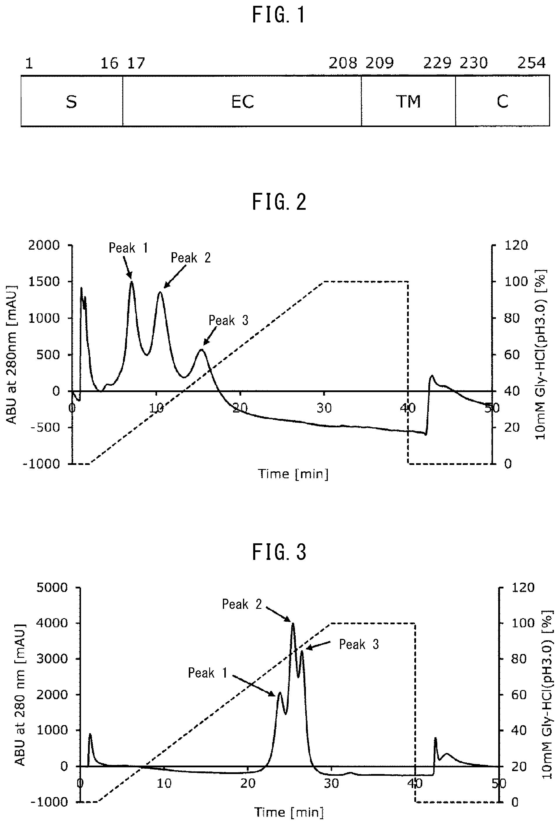
[0039] FIG. 1 schematically illustrates human Fc.gamma.RIIIa. The numerals in FIG. 1 correspond to positions in the amino acid sequence set forth in SEQ ID NO: 1. In FIG. 1, S represents a signal sequence, EC represents an extracellular domain, TM represents a transmembrane domain, and C represents an intracellular domain.
[0040] FIG. 2 depicts an elution pattern (chromatogram) when a monoclonal antibody was separated using a column filled with an insoluble carrier having the Fc-binding protein of the present invention (FcR9_F) immobilized thereon.
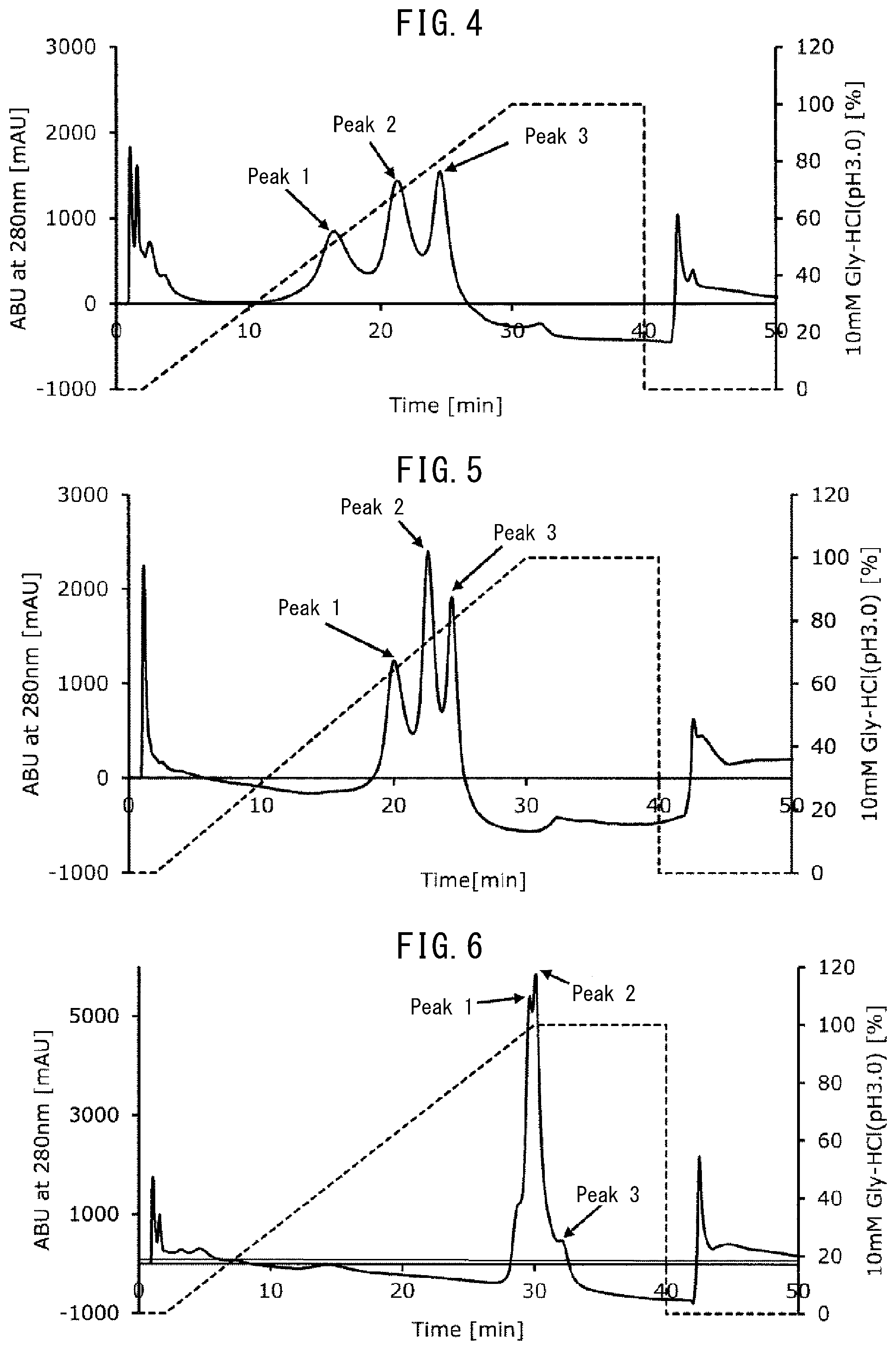
[0041] FIG. 3 depicts an elution pattern (chromatogram) when a monoclonal antibody was separated using a column filled with an insoluble carrier having a conventional Fc-binding protein (FcR9) immobilized thereon.
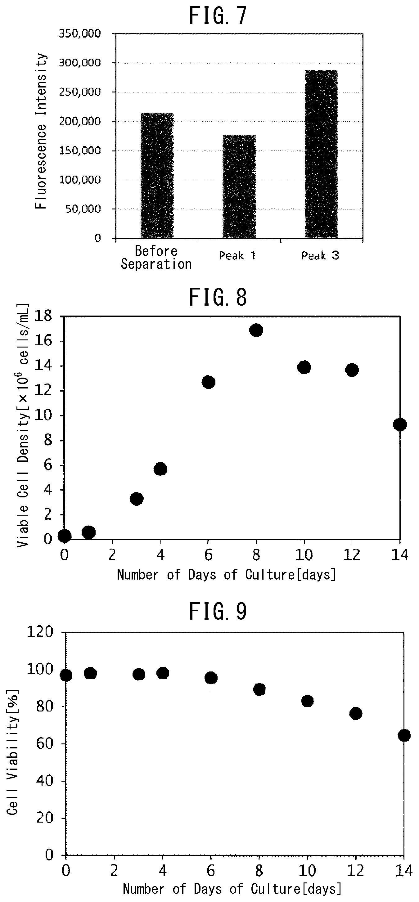
[0042] FIG. 4 depicts an elution pattern (chromatogram) when the pH of an equilibration buffer was set to 5.5 in Example 6.
[0043] FIG. 5 depicts an elution pattern (chromatogram) when the pH of an equilibration buffer was set to 5.6 in Example 6.
[0044] FIG. 6 depicts an elution pattern (chromatogram) when the pH of an equilibration buffer was set to 6.0 in Example 6.
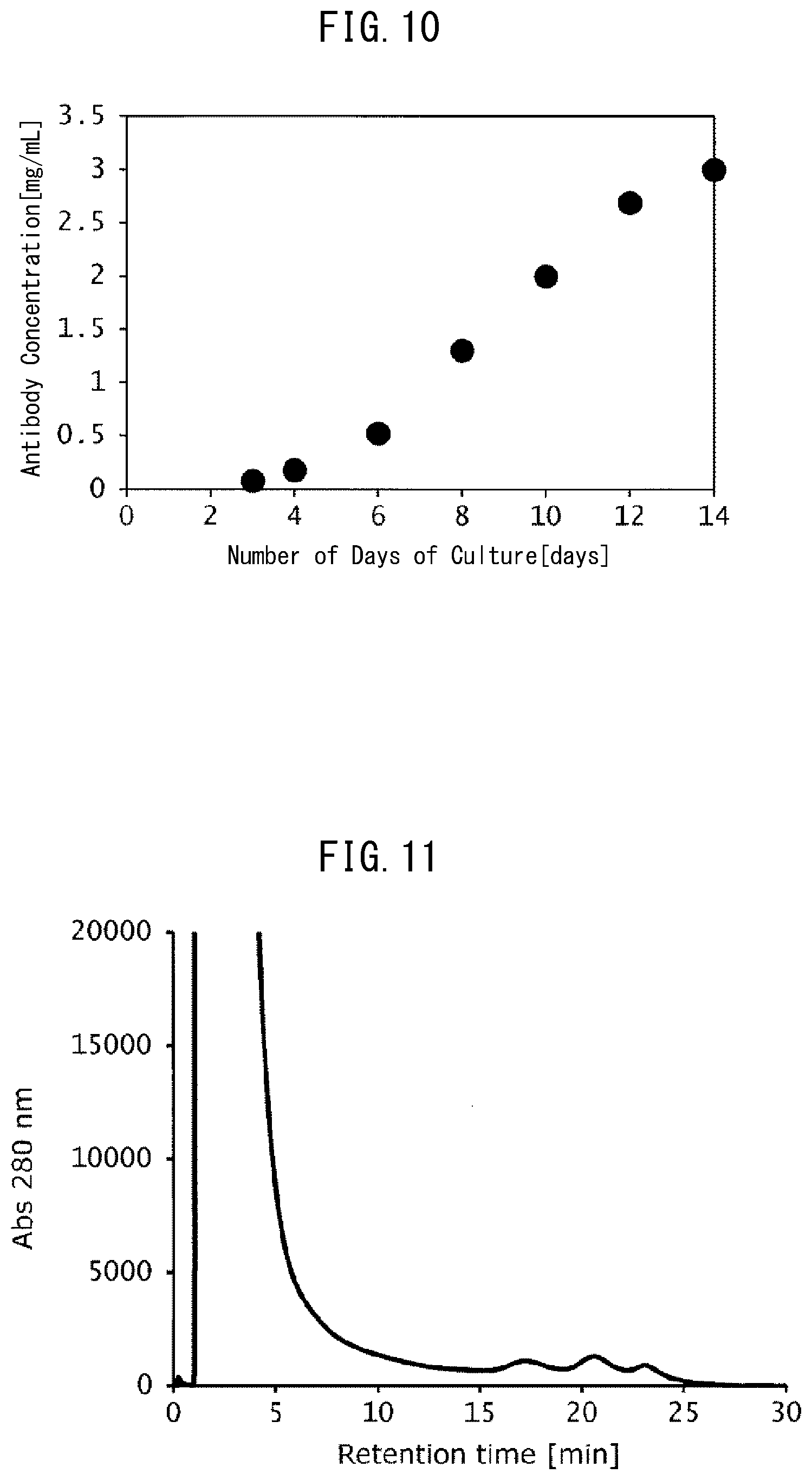
[0045] FIG. 7 is a graph indicating the results of measuring ADCC activity of a monoclonal antibody included in the isolated peaks in Example 7.
[0046] FIG. 8 is a graph indicating temporal changes in the viable cell density by the number of days of culture of antibody-producing cells in Example 8.
[0047] FIG. 9 is a graph indicating temporal changes in the cell viability by the number of days of culture of antibody-producing cells in Example 8.
[0048] FIG. 10 is a graph indicating temporal changes in the antibody concentration (antibody production) by the number of days of culture of antibody-producing cells in Example 8.
[0049] FIG. 11 is a chromatogram indicating the results of analysis of the culture solution containing an antibody using the FcR9_F column in Example 8.
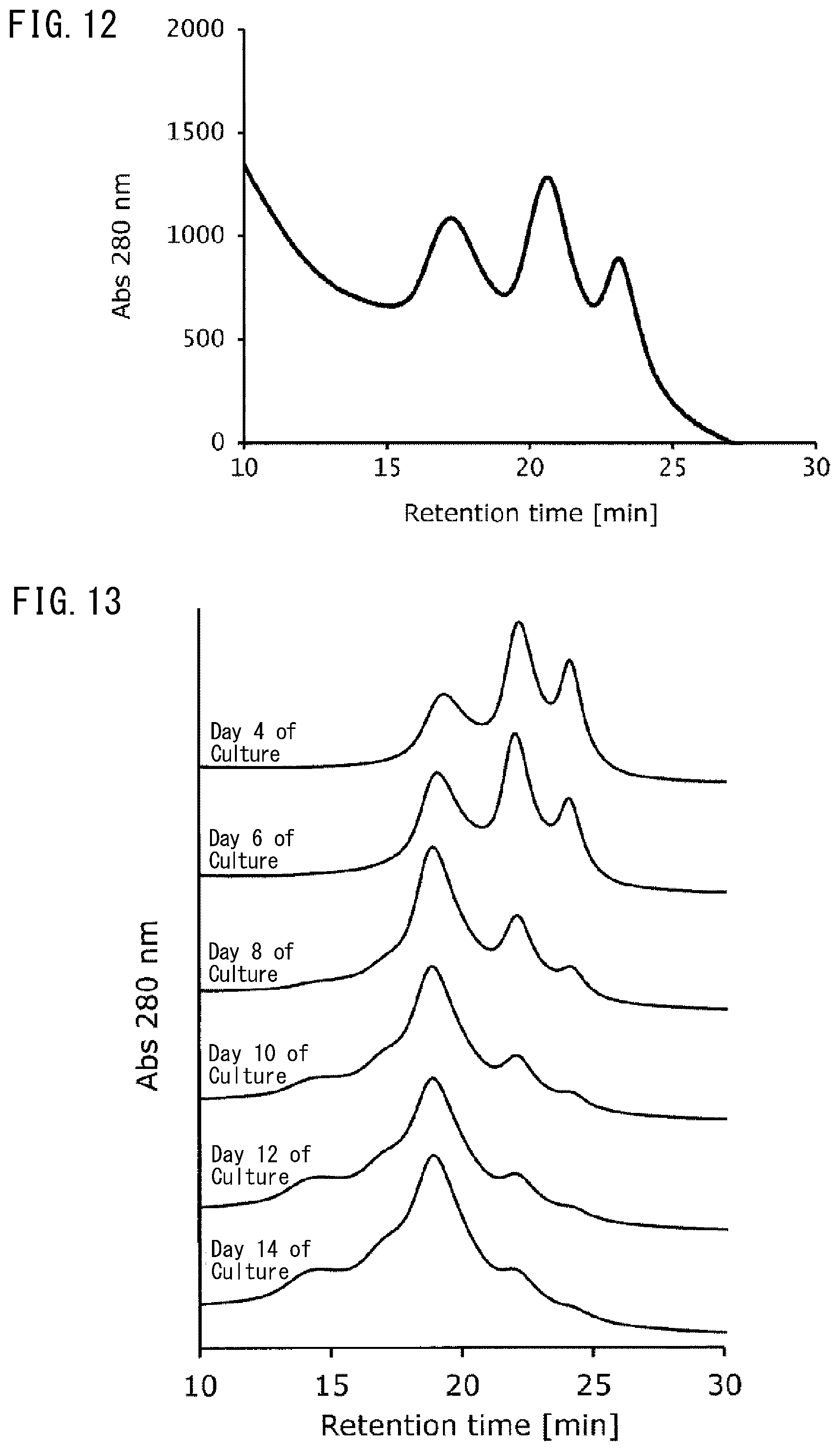
[0050] FIG. 12 is an enlarged view of the region between 10 minutes to 30 minutes of elution time (retention time) in the chromatogram in FIG. 11.
[0051] FIG. 13 depicts chromatograms (elution time (retention time): region between 10 minutes to 30 minutes) indicating the results of analysis of the culture solution containing an antibody on different days of culture using the FcR9_F column in Example 8, the chromatograms having different peak forms by the number of days of culture.
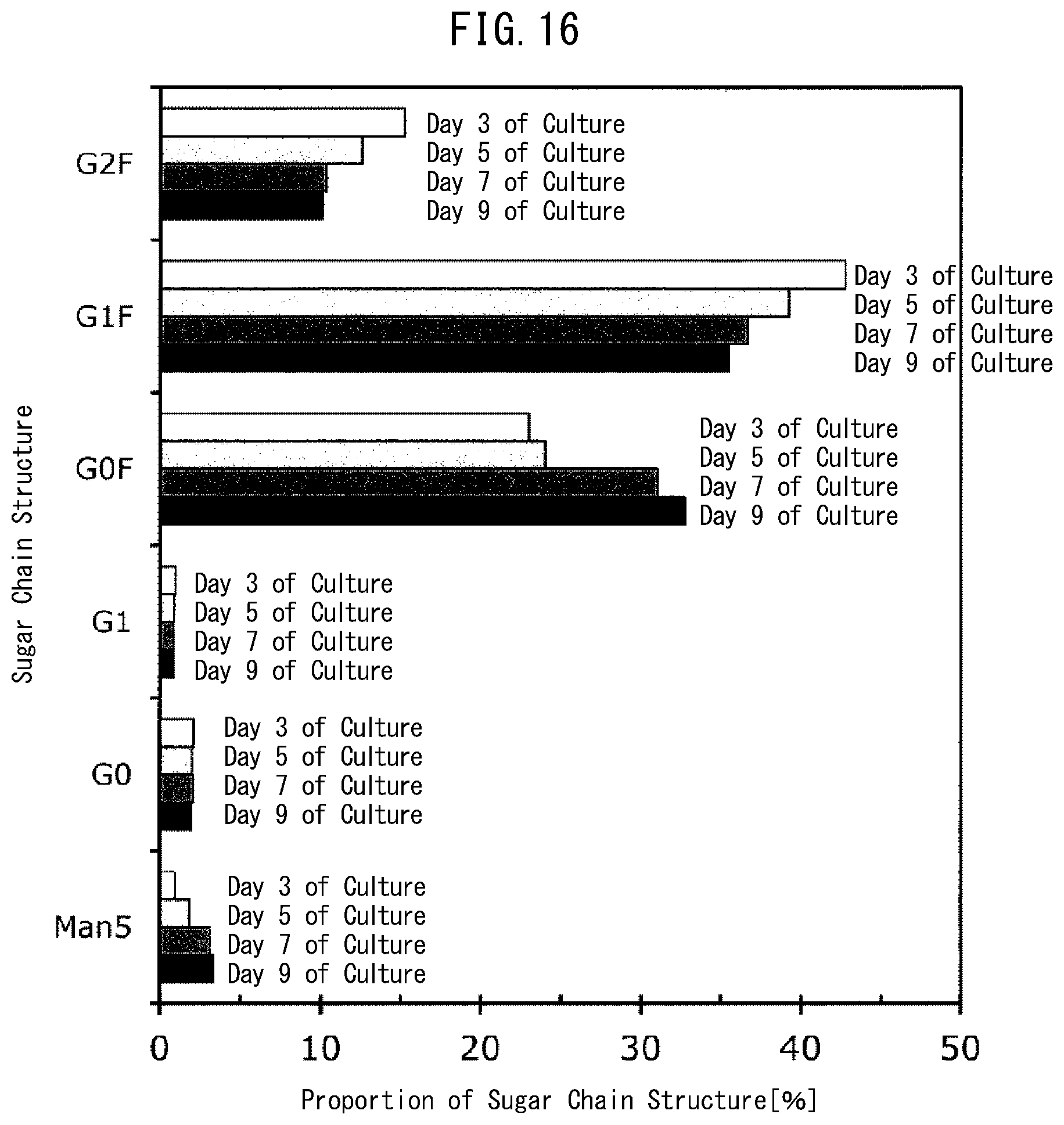
[0052] FIG. 14 depicts chromatograms (elution time (retention time): region between 5 minutes to 20 minutes) indicating the results of analysis of an antibody obtained from the culture solution on different days of culture using the FcR9_F column in Example 9, the chromatograms having different peak forms by the number of days of culture.
[0053] FIG. 15 is a graph indicating temporal changes in the antibody concentration (open bar) and viable cell concentration (filled diamond) by the number of days of culture of the antibody-producing cells cultured in Example 9.
[0054] FIG. 16 is a graph indicating proportions of constituents in the sugar chain structure of the antibody obtained by the number of days of culture in Example 9.
EXAMPLES
[0055] Hereinafter, the present invention will be described in more detail with reference to the Examples below, but the present invention is not limited to these Examples.
Example 1 Preparation of FcR9 Amino Acid Substitution Product
[0056] An amino acid substitution was introduced into an Fc-binding protein FcR9 (SEQ ID NO: 5) prepared in accordance with the method disclosed in WO2015/199154 as described below in order to confirm usefulness of a substitution of valine at position 192 (corresponding to valine at position 176 in wildtype human Fc.gamma.RIIIa consisting of the amino acid sequence set forth in SEQ ID NO: 1) by a different amino acid. Specifically, an amino acid substitution was introduced into plasmid pET-FcR9 (WO2015/199154) including a polynucleotide (SEQ ID NO: 6) encoding FcR9 by PCR, thereby producing an Fc-binding protein having a substitution of valine at position 192 in FcR9 (SEQ ID NO: 5) by a different amino acid. FcR9 (SEQ ID NO: 5) is an Fc-binding protein including the wild-type Fc.gamma.RIII extracellular domain set forth in SEQ ID NO: 4, which has an amino acid substitution of Val at position 43 by Glu (corresponding to position 27 of SEQ ID NO: 1), an amino acid substitution of Phe at position 45 by Ile (corresponding to position 29 of SEQ ID NO: 1), an amino acid substitution of Tyr at position 51 by Asn (corresponding to position 35 of SEQ ID NO: 1), an amino acid substitution of Gln at position 64 by Arg (corresponding to position 48 of SEQ ID NO: 1), an amino acid substitution of Phe at position 91 by Leu (corresponding to position 75 of SEQ ID NO: 1), an amino acid substitution of Asn at position 108 by Ser (corresponding to position 92 of SEQ ID NO: 1), an amino acid substitution of Val at position 133 by Glu (corresponding to position 117 of SEQ ID NO: 1), an amino acid substitution of Glu at position 137 by Gly (corresponding to position 121 of SEQ ID NO: 1), and an amino acid substitution of Phe at position 187 by Ser (corresponding to position 171 of SEQ ID NO: 1).
[0057] Hereinafter, a method for producing the following Fc-binding proteins will be described in detail.
[0058] (1) In order to confirm usefulness of a substitution of valine at position 192 (corresponding to position 176 in SEQ ID NO: 1) in the Fc-binding protein FcR9 (SEQ ID NO: 5) by a different amino acid, a reaction solution having the composition in Table 1 was prepared using plasmid pET-FcR9 (disclosed in WO2015/199154) including the polynucleotide (SEQ ID NO: 6) encoding FcR9 (SEQ ID NO: 5) produced by the method disclosed in WO2015/199154 as a template and oligoprimers consisting of the sequences set forth in SEQ ID NO: 2 (5'-TAATACGACTCACTATAGGG-3') and SEQ ID NO: 7 (5'-CATTTTTGCTGCCMNNCAGCCCACGGCAGG-3'). Subsequently, the reaction solution was heat-treated at 95.degree. C. for 2 minutes, and PCR was performed by a reaction of 30 cycles of a first step at 95.degree. C. for 30 seconds, a second step at 50.degree. C. for 30 seconds, and a third step at 72.degree. C. for 90 seconds, followed by heat treatment at 72.degree. C. for 7 minutes. The obtained PCR product was designated as V192p1.
TABLE-US-00001 TABLE 1 Composition Volume Template DNA 2 .mu.L 10 .mu.M Forward primer 1 .mu.L 10 .mu.M Reverse primer 1 .mu.L 5 .times. PrimeSTAR buffer 4 .mu.L (manufactured by Takara Bio) 2.5 mM dNTPs 2 .mu.L 2.5 U/.mu.L PrimeSTAR HS 0.5 .mu.L (manufactured by Takara Bio) H.sub.20 up to 20 .mu.L
[0059] (2) A reaction solution having the composition in Table 1 was prepared using plasmid pET-FcR9 (disclosed in WO2015/199154) including the polynucleotide (SEQ ID NO: 6) encoding FcR9 (SEQ ID NO: 5) produced by the method disclosed in WO2015/199154 as a template and oligoprimers consisting of the sequences set forth in SEQ ID NO: 3 (5'-TATGCTAGTTATTGCTCAG-3') and SEQ ID NO: 8 (5'-CCTGCCGTGGGCTGNNKGGCAGCAAAAATG-3'). Subsequently, the reaction solution was heat-treated at 95.degree. C. for 2 minutes, and PCR was performed by a reaction of 30 cycles of a first step at 95.degree. C. for 30 seconds, a second step at 50.degree. C. for 30 seconds, and a third step at 72.degree. C. for 90 seconds, followed by heat treatment at 72.degree. C. for 7 minutes. The obtained PCR product was designated as V192p2.
[0060] (3) The two types of PCR products (V192p1, V192p2) obtained in (1) and (2) were mixed, thereby preparing a reaction solution having the composition in Table 2. The reaction solution was heat-treated at 98.degree. C. for 5 minutes, and PCR was performed by a reaction of 5 cycles of a first step at 98.degree. C. for 10 seconds, a second step at 55.degree. C. for 5 seconds, and a third step at 72.degree. C. for 1 minute, thereby obtaining a PCR product V192p in which V192p1 and V192p2 were joined to each other.
TABLE-US-00002 TABLE 2 Composition Concentration/Volume PCR product 2 .mu.L each 2.5 U/.mu.L PrimeSTAR HS 0.5 .mu.L (manufactured by Takara Bio) 5 .times. PrimeSTAR buffer 4 .mu.L (manufactured by Takara Bio) 2.5 mM dNTPs 2 .mu.L H.sub.20 up to 20 .mu.L
[0061] (4) PCR was performed using the PCR product V192p obtained in (3) as a template and oligonucleotides consisting of the sequences set forth in SEQ ID NOs: 2 and 3 as PCR primers. A reaction solution having the composition in Table 3 was prepared. Subsequently, the reaction solution was heat-treated at 98.degree. C. for 5 minutes, and a reaction of 30 cycles of a first step at 98.degree. C. for 10 seconds, a second step at 55.degree. C. for 5 seconds, and a third step at 72.degree. C. for 1 minute was performed. Accordingly, a polynucleotide encoding an Fc-binding protein having a substitution of an amino acid at position 192 in the Fc-binding protein (FcR9) by an optional amino acid was obtained. The obtained polynucleotide was designated as V192p3.
TABLE-US-00003 TABLE 3 Composition Volume PCR product 2 .mu.L 10 .mu.M Forward primer 2 .mu.L 10 .mu.M Reverse primer 2 .mu.L 5 .times. PrimeSTAR buffer 10 .mu.L (manufactured by Takara Bio) 2.5 mM dNTPs 4 .mu.L 2.5 U/.mu.L PrimeSTAR HS 1 .mu.L (manufactured by Takara Bio) H.sub.20 up to 50 .mu.L
[0062] (5) The polynucleotide obtained in (4) was purified, digested with restriction enzymes NcoI and HindIII, and ligated to an expression vector pETMalE (Japanese Unexamined Patent Publication (Kokai) No. 2011-206046) which was preliminarily digested with restriction enzymes NcoI and HindIII, and the resulting product was used for transforming E. coli strain BL21 (DE3).
[0063] (6) The obtained transformant was cultured in an LB medium supplemented with 50 .mu.g/mL kanamycin. Plasmids were extracted from the collected bacterial cells (transformants).
[0064] (7) Regarding the polynucleotide encoding the Fc-binding protein and its surrounding region in each obtained plasmid, a cycle sequence reaction was performed using a BigDye Terminator v3.1 Cycle Sequencing Kit (manufactured by Life Technologies Corporation) based on the chain terminator method, and the nucleotide sequence was analyzed by a fully automated DNA sequencer (Applied Biosystems 3130 Genetic Analyzer (manufactured by Life Technologies Corporation)). An oligonucleotide consisting of the sequence set forth in SEQ ID NO: 2 or SEQ ID NO: 3 was used as a sequencing primer upon the analysis. As a result of sequence analysis, transformants expressing Fc-binding proteins each having a substitution of Val at position 192 (position 176 of SEQ ID NO: 1) in the Fc-binding protein FcR9 (SEQ ID NO: 5) by one of Ala, Arg, Asn, Asp, Cys, Gln, Glu, Gly, His, Ile, Leu, Lys, Met, Phe, Pro, Ser, Thr, Trp, and Tyr were obtained.
Example 2 Evaluation of IgG1-Binding Ability of Fc-Binding Protein
[0065] (1) The each transformant expressing Fc-binding protein produced in Example 1 was inoculated in a 20 mL of a 2YT liquid medium supplemented with 50 .mu.g/mL kanamycin and aerobically cultured with shaking overnight at 37.degree. C., thereby performing preculture, respectively.
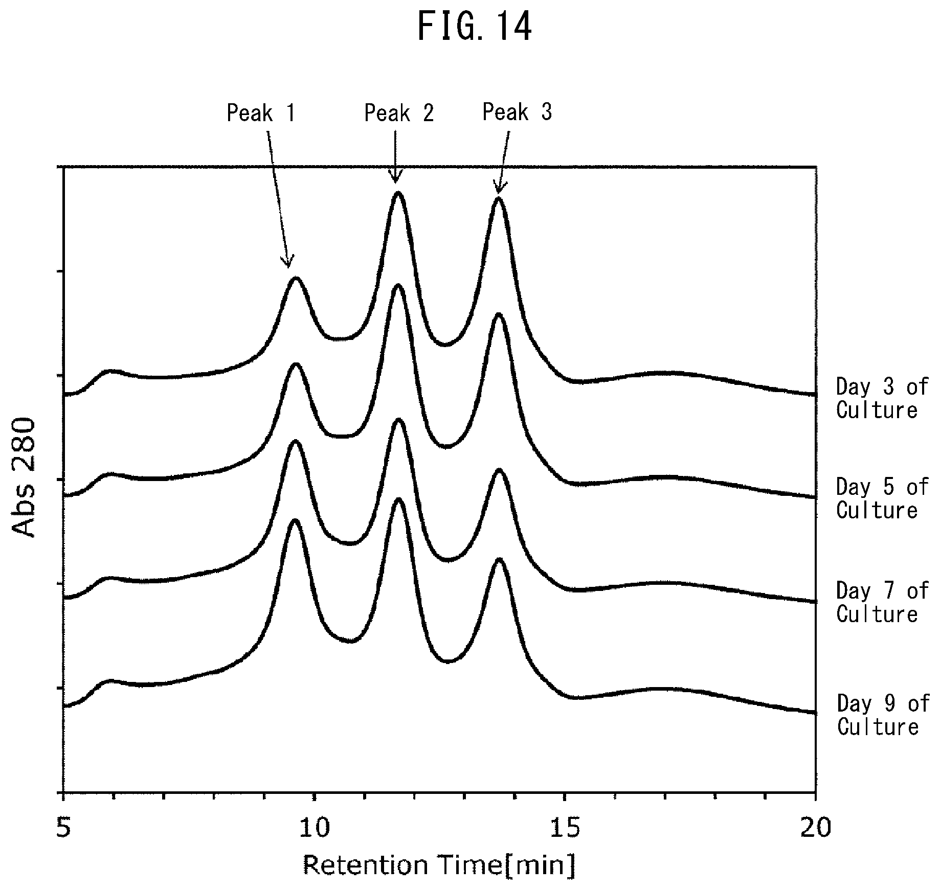
[0066] (2) Each preculture solution in a volume of 10 mL was inoculated in 1000 mL of a 2YT liquid medium (peptone at 16 g/L, yeast extract at 10 g/L, sodium chloride at 5 g/L) supplemented with 50 .mu.g/mL kanamycin and aerobically cultured with shaking at 37.degree. C.
[0067] (3) The culture temperature was changed to 20.degree. C. 1.5 hours after the start of culture, followed by shaking culture for 30 minutes. Thereafter, isopropyl-.beta.-thiogalactopyranoside (IPTG) was added to yield a final concentration of 0.01 mM, and shaking culture was continued aerobically overnight at 20.degree. C.
[0068] (4) After the end of culture, cells were collected by centrifugation, suspended in a buffer (20 mM Tris-HCL buffer containing 150 mM NaCl (pH 7.4)), and ultrasonically disrupted. Thereafter, the supernatant was collected by centrifugation.
[0069] (5) The collected supernatant was allowed to pass through a column filled with Ni Sepharose 6 Fast Flow (manufactured by GE Healthcare) and washed with a sufficient amount of a washing buffer (20 mM Tris-HCL buffer containing 150 mM NaCl (pH 7.4)), followed by elution with an elution buffer (20 mM Tris-HCl buffer containing 150 mM NaCl and 500 mM imidazole (pH 7.4)). Then, the elution fraction was collected.
[0070] (6) The elution fraction collected in (5) was allowed to pass through a column filled with IgG Sepharose 6 Fast Flow (manufactured by GE Healthcare) and washed with a sufficient amount of a washing buffer (20 mM Tris-HCl buffer containing 150 mM NaCl (pH 7.4)), followed by elution with an elution buffer (100 mM glycine buffer (pH 3.0)). Then, the elution fraction was collected.
[0071] (7) IgG1-binding ability of each Fc-binding protein collected as the elution fraction in (6) was evaluated using the surface plasmon resonance method. Upon measurement of the binding ability using the surface plasmon resonance method, Biacore T100 (manufactured by GE Healthcare) was used as a measurement system, Sensor Chip CM5 (manufactured by GE Healthcare) was used as a sensor chip, and Biacore T100 Evaluation Software (manufactured by GE Healthcare) was used as analysis software.
[0072] (8) A solution prepared by diluting IgG1 (manufactured by Sigma-Aldrich) with HBS-EP (manufactured by GE Healthcare) was supplied to a sensor chip on which each Fc-binding protein was separately immobilized using an Amine Coupling Kit (manufactured by GE Healthcare), thereby obtaining a sensorgram. Curve fitting was performed based on the sensorgram so as to calculate IgG1-binding ability.
[0073] Here, it is generally known that when affinity between an Fc-binding protein and an antibody decreases, the ability of the Fc-binding protein to retain the antibody is weakened, and the capacity for adsorption is reduced. Meanwhile, in a column filled with an insoluble carrier (resin) having an Fc-binding protein immobilized thereon, when binding ability between the Fc-binding protein and an antibody is reduced, ability to retain the antibody when adding the antibody for elution is weakened. This allows elution while the binding force is lowered, thereby facilitating separation. As a result of calculation of affinity to the IgG1 antibody by the above-described method, it was found that among the produced Fc-binding proteins, a protein having an amino acid substitution of Val at position 192 of SEQ ID NO: 5 (corresponding to position 176 of SEQ ID NO: 1) by Phe (hereinafter referred to as "FcR9_F") has lower antibody affinity than FcR9 (SEQ ID NO: 5) serving as a reference.
[0074] The amino acid sequence of FcR9_F to which a signal sequence and a polyhistidine tag were added is set forth in SEQ ID NO: 9, and the polynucleotide sequence encoding the FcR9_F is set forth in SEQ ID NO: 10. In SEQ ID NO: 9, a MalE signal peptide ranges from methionine (Met) at position 1 to alanine (Ala) at position 26, a linker sequence ranges from lysine (Lys) at position 27 to methionine (Met) at position 32, the amino acid sequence of FcR9_F (corresponding to a region from position 17 to position 192 of SEQ ID NO: 1) ranges from glycine at position 33 (Gly) to glutamine at position 208 (Gln), a linker sequence ranges from glycine (Gly) at position 209 to glycine (Gly) at position 210, and a tag sequence ranges from histidine (His) at position 211 to histidine (His) at position 216. In addition, phenylalanine of Val192Phe is located at position 192 of SEQ ID NO: 9.
[0075] Table 4 lists the results of calculating affinity to IgG1. In Table 4, the lower the KD value (dissociation constant) means the higher the affinity. It is understood that FcR9_F (SEQ ID NO: 9), which is an Fc-binding protein having an amino acid substitution of Val192Phe, has a higher dissociation constant, i.e., lower affinity to an antibody, compared to FcR9 (SEQ ID NO: 5) lacking the substitution. Accordingly, it is suggested that when an antibody is separated by a method including a step of adding a solution containing an antibody to a column filled with an insoluble carrier having the Fc-binding protein immobilized thereon so as to allow the carrier to adsorb the antibody and a step of eluting the antibody adsorbed by the carrier using an elution solution, the antibody can be separated with a high degree of separation and high efficiency using, as the Fc-binding protein, a protein having at least an amino acid substitution of Val192Phe in the Fc-binding protein FcR9 (SEQ ID NO: 5) disclosed in WO2015/199154 because affinity to the antibody is lowered as compared to when using FcR9.
TABLE-US-00004 TABLE 4 Fc-binding protein Association Dissociation Dissociation Substitution rate constant rate constant constant Name position ka [1/Ms] kd [1/s] K.sub.D [M] FcR9 (Reference) 4.05 .times. 10.sup.5 3.13 .times. 10.sup.-2 7.73 .times. 10.sup.-8 FcR9_F Val192Phe 9.02 .times. 10.sup.4 3.01 .times. 10.sup.-1 3.28 .times. 10.sup.-6
Example 3 Production of Fc-Binding Protein of Invention (FcR9_F_Cys) Having Cysteine Tag Added
[0076] (1) PCR was performed using an expression vector pET-FcR9_F including the polynucleotide set forth in SEQ ID NO: 10 encoding a protein consisting of the amino acid sequence set forth in SEQ ID NO: 9 produced in Example 2 as a template. Primers used in the PCR were oligonucleotides consisting of the sequences set forth in SEQ ID NO: 11 (5'-TAGCCATGGGCATGCGTACCGAAGATCTGCCGAAAGC-3') and SEQ ID NO: 12 (5'-CCCAAGCTTATCCGCAGGTATCGTTGCGGCACCCTTGGGTAATGGTAATATTCACGG TCTCGCTGC-3'). A reaction solution having the composition in Table 3 was prepared. Subsequently, the reaction solution was heat-treated at 98.degree. C. for 5 minutes, and PCR was performed by a reaction of 30 cycles of a first step at 98.degree. C. for 10 seconds, a second step at 55.degree. C. for 5 seconds, and a third step at 72.degree. C. for 1 minute.
[0077] (2) The polynucleotide obtained in (1) was purified and digested with restriction enzymes NcoI and HindIII, and ligated to an expression vector pTrc-PelBV3 produced by the method disclosed in WO2015/199154, which was preliminarily digested with restriction enzymes NcoI and HindIII. E. coli strain W3110 was transformed using the ligation product.
[0078] (3) After the obtained transformant was cultured in an LB medium containing 100 .mu.g/mL carbenicillin, an expression vector pTrc-FcR9_F_Cys was obtained using a QIAprep Spin Miniprep kit (manufactured by QIAGEN).
[0079] (4) The nucleotide sequence of pTrc-FcR9_F_Cys was analyzed in the same manner as in Example 1 (7) except that an oligonucleotide consisting of the sequence set forth in SEQ ID NO: 13 (5'-TGTGGTATGGCTGTGCAGG-3') or SEQ ID NO: 14 (5'-TCGGCATGGGGTCAGGTG-3') was used as a sequencing primer.
[0080] The amino acid sequence of a polypeptide expressed in the vector pTrc-FcR9_F_Cys is set forth in SEQ ID NO: 15, and the sequence of a polynucleotide encoding the polypeptide is set forth in SEQ ID NO: 16. In SEQ ID NO: 15, an improved PelB signal peptide ranges from methionine (Met) at position 1 to alanine (Ala) at position 22, the amino acid sequence of the Fc-binding protein FcR9_F (corresponding to a region from position 33 to position 208 of SEQ ID NO: 9) ranges from glycine (Gly) at position 24 to glutamine (Gln) at position 199, and a cysteine tag sequence ranges from glycine (Gly) at position 200 to glycine (Gly) at position 207.
Example 4 Preparation of FcR9_F_Cys
[0081] (1) The transformant expressing FcR9_F_Cys produced in Example 3 was inoculated in 400 mL of a 2YT liquid medium (peptone at 16 g/L, yeast extract at 10 g/L, sodium chloride at 5 g/L) containing 100 .mu.g/mL carbenicillin in a 2-L baffled flask and aerobically cultured with shaking overnight at 37.degree. C., thereby performing preculture.
[0082] (2) The culture solution of (1) in a volume of 180 mL was inoculated in 1.8 L of a liquid medium containing glucose at 10 g/L, yeast extract at 20 g/L, phosphate trisodium dodecahydrate at 3 g/L, hydrogen phosphate disodium dodecahydrate at 9 g/L, ammonium chloride at 1 g/L, and carbenicillin at 100 mg/L, and main culture was performed using a 3-L fermenter (manufactured by Biott Corporation). Main culture was initiated under conditions set as follows: 30.degree. C., pH 6.9 to 7.1, an airflow rate of 1 VVM, and a saturated dissolved oxygen concentration of 30%. For pH control, 50% phosphoric acid was used as an acid, and 14% ammonia water was used as an alkali, dissolved oxygen was controlled by changing the stirring rate, and the lower and upper limits of the number of rotations of stirring were set to 500 rpm and 1000 rpm, respectively. At a time point when the glucose concentration became unmeasurable after the start of culture, a feed medium (glucose at 248.9 g/L, yeast extract at 83.3 g/L, magnesium sulfate heptahydrate at 7.2 g/L) was added while controlling dissolved oxygen (DO).
[0083] (3) When the 600-nm optical density (OD 600 nm) serving as an indicator of the amount of bacterial cells reached about 150, the culture temperature was decreased to 25.degree. C., and it was confirmed that the temperature reached the preset temperature. Subsequently, IPTG was added to yield a final concentration of 0.5 mM, and culture was continued at 25.degree. C.
[0084] (4) Culture was terminated about 48 hours after the start of culture, and the culture solution was centrifuged at 4.degree. C. and 8000 rpm for 20 minutes, thereby collecting bacterial cells.
[0085] (5) The collected bacterial cells were suspended in a 20 mM Tris-HCl buffer (pH 7.0) to yield a concentration of 5 mL/1 g (of bacterial cells), and the bacterial cells were disrupted using an ultrasonicator (Insonator 201M (trade name), manufactured by KUBOTA Corporation co., ltd.) at 4.degree. C. and an output power of about 150 W for about 10 minutes. A solution of the disrupted bacterial cells was centrifuged twice at 4.degree. C. and 8000 rpm for 20 minutes, thereby collecting the supernatant.
[0086] (6) The supernatant obtained in (5) was applied to VL 32.times.250 Column (manufactured by MERCK MILLIPORE) filled with 140 mL of TOYOPEARL CM-650M (manufactured by Tosoh Corporation) which was preliminarily equilibrated with a 20 mM phosphate buffer (8 mM sodium dihydrogen phosphate, 12 mM disodium hydrogen phosphate) (pH 7.0) at a flow rate of 5 mL/min. After washing with the buffer used for equilibration, elution was performed using 20 mM phosphate buffer (pH 7.0) containing 0.5 M sodium chloride.
[0087] (7) The eluate obtained in (6) was applied to an XK 26/20 Column (manufactured by GE Healthcare) filled with 90 mL of IgG Sepharose (manufactured by GE Healthcare) which was preliminarily equilibrated with 20 mM Tris-HCl buffer (pH 7.4) containing 150 mM sodium chloride. After washing with the buffer used for equilibration, elution was performed using 0.1 M glycine-HCl buffer (pH 3.0). The pH of the eluate was adjusted to a neutral range with the addition of 1 M Tris-HCl buffer (pH 8.0) in an amount one-fourth the amount of the eluate.
[0088] As a result of the purification, about 20 mg of high-purity FcR9_F_Cys was obtained.
Example 5 Preparation of Fc-Binding Protein (FcR9_F)-Immobilized Gel and Antibody Separation
[0089] (1) After activation of hydroxyl groups on the surface of a hydrophilic vinyl polymer for a separating resin (manufactured by Tosoh Corporation: TOYOPEARL) in a volume of 2 mL with iodoacetyl groups, 4 mg of FcR9_F_Cys prepared in Example 4 was reacted therewith, thereby obtaining FcR9_F-immobilized gel.
[0090] (2) A stainless-steel column having a size of .phi.4.6 mm.times.75 mm was filled with 1.2 mL of the FcR9_F-immobilized gel prepared in (1), thereby preparing an FcR9_F column.
[0091] (3) The FcR9_F column prepared in (2) was connected to a high performance liquid chromatography system (manufactured by Tosoh Corporation) and equilibrated with 20 mM acetate buffer (pH 5.0) containing 50 mM sodium chloride.
[0092] (4) Five microliters of a monoclonal antibody (RITUXAN, manufactured by Zenyaku Kogyo Co., Ltd.), which was diluted with phosphate buffered saline (PBS) (pH 7.4) to yield a concentration of 1.0 mg/mL, was added at a flow rate of 0.6 mL/min.
[0093] (5) Washing was performed with the equilibration buffer for 2 minutes while the flow rate was maintained at 0.6 mL/min. Subsequently, a monoclonal antibody adsorbed was eluted with a pH gradient of 10 mM glycine-HCl buffer (pH 3.0) (i.e., a gradient in which 10 mM glycine-HCl buffer (pH 3.0) accounts for 100% in 28 minutes).
[0094] FIG. 2 depicts the results (elution pattern). The monoclonal antibody was separated into a plurality of peaks, but not a single peak as in the case of gel filtration chromatography, because it interacted with the Fc-binding protein.
[0095] (6) Each of Peaks 1 to 3 in the elution pattern depicted in FIG. 2 was separately collected into a container so as to obtain an isolation fraction of each peak.
Comparative Example 1 Preparation of Fc-Binding Protein (FcR9)-Immobilized Gel and Antibody Separation
[0096] Antibody separation was carried out by preparing Fc-binding protein (FcR9)-immobilized gel in the same manner as in Examples 3 to 5 except that the Fc-binding protein FcR9 (SEQ ID NO: 5) disclosed in WO2015/199154 was used as an Fc-binding protein to be immobilized on an insoluble carrier.
[0097] FIG. 3 depicts the results (elution pattern). It is understood that elution peaks are close to each other and the resolution is low, compared to the elution pattern in Example 5 (FIG. 2). In other words, it was found that antibody separation ability is improved with the use of gel having the Fc-binding protein of the present invention having an amino acid substitution of Val192Phe (FcR9_F) immobilized thereon, compared to when using gel having the Fc-binding protein lacking the amino acid substitution (FcR9) immobilized thereon.
Example 6 Examination of Equilibration Buffer
[0098] Example 6 was carried out in the same manner as in Example 5 except that the equilibration buffer used in Example 5 (3) was changed to any of the following (a) to (c). [0099] (a) 20 mM acetate buffer containing 50 mM sodium chloride (pH 5.5) [0100] (b) 20 mM acetate buffer containing 50 mM sodium chloride (pH 5.6) [0101] (c) 20 mM MES buffer containing 50 mM sodium chloride (pH 6.0)
[0102] The obtained elution patterns are depicted in FIG. 4 (equilibration buffer (a)), FIG. 5 (equilibration buffer (b)), and FIG. 6 (equilibration buffer (c)). In addition, among the elution patterns obtained in this Example, Example 5 (FIG. 2), and Comparative Example 1 (FIG. 3), peaks corresponding to the monoclonal antibody were designated as Peak 1, Peak 2, and Peak 3 in the ascending order of elution time, and the resolution between Peak 1 and Peak 2 and the resolution between Peak 2 and Peak 3 were calculated using the following formula. Table 5 lists the results.
Resolution (Rs value)=1.18.times.(Elution time of late elution time peak-Elution time of early elution time peak)/(Half width of early elution time peak+Half width of late elution time peak)
[0103] The higher the resolution (Rs value), the better the separation performance.
[0104] The inventors previously revealed that antibodies obtained by isolating elution fractions based on the Peak 1, Peak 2, and Peak 3 have different levels of antibody dependent cellular cytotoxicity (ADCC (Japanese Unexamined Patent Publication (Kokai) No. 2016-23152). Therefore, by isolating an antibody using the separation method with high resolution in Example 5, it is possible to separate, for example, antibodies having different levels of ADCC with good accuracy.
TABLE-US-00005 TABLE 5 Separation condition Fc-binding Equilibration Resolution Rs value protein buffer pH Peaks 1-2 Peaks 2-3 Example 5 FcR9_F 5.0 0.95 1.10 Comparative FcR9 5.0 0.85 0.74 Example 1 Example 6 (a) FcR9_F 5.5 1.22 1.11 Example 6 (b) FcR9_F 5.6 1.18 1.13 Example 6 (c) FcR9_F 6.0 0.05 0.09
[0105] As a result of comparison between the results of Comparative Example 1 (FcR9) and the results of Example 5 (FcR9_F) in Table 5, it is understood that the resolution Rs value and the antibody separation ability are higher in Example 5. In other words, it is also understood from the resolution values obtained by calculation that when using gel having the Fc-binding protein having an amino acid substitution of Val192Phe in FcR9 immobilized thereon, antibody separation ability is improved, compared to when using gel having FcR9 immobilized thereon.
[0106] In addition, it is understood from the results of Examples 5 and 6 that when the pH of equilibration buffer was 5.5 or 5.6 (Example 6 of (a) and (b)) rather than 5.0 (Example 5), the resolution (Rs value) increased, while the resolution remarkably decreased at pH 6.0 (Example 6 (c)) indicating that separation was substantially unsuccessful. It can be said from these results that the pH of equilibration buffer is favorably from 4.5 to 5.8, and when the pH is from 5.2 to 5.7, it is preferable in that antibody separation ability is further improved.
Example 7 Measurement of Antibody Dependent Cellular Cytotoxicity (ADCC) of Separated Antibody
[0107] (1) The regions of Peaks 1 and 3 in the chromatogram depicted in FIG. 2, which were separated in Example 5, were isolated. Isolation was repeated, the buffer was changed to PBS (10 mM disodium hydrogen phosphate, 1.76 mM potassium dihydrogen phosphate, 137 mM sodium chloride, 2.7 mM Potassium chloride) (pH 7.4) while pooled Peaks 1 and 3 were concentrated via an ultrafiltration membrane (manufactured by MERCK MILLIPORE). Thereafter, the concentrations of the monoclonal antibody in Peak 1 and the monoclonal antibody in Peak 3 after concentration and buffer exchange and the concentration of the monoclonal antibody before separation were measured at an absorbance at 280 nm.
[0108] (2) ADCC Activity Measurement
[0109] ADCC activity of the monoclonal antibody included in each peak and that of the monoclonal antibody before separation were measured in accordance with the manual of an ADCC Reporter Bioassay Kit (manufactured by Promega Corporation) by the following method.
[0110] (2-1) ADCC assay buffer was prepared by mixing 1.4 mL of Low IgG Serum and 33.6 mL of an RPMI 1640 medium. This ADCC assay buffer was used for creating an eight-step dilution series for each of the monoclonal antibody in Peak 1, the monoclonal antibody in Peak 3, and the monoclonal antibody before separation by three-fold dilution at each step starting from 3 .mu.g/mL.
[0111] (2-2) Raji cells were adjusted to have a concentration of about 5.times.10.sup.5 cells/mL with the ADCC assay buffer and added to a 96-well plate (3917: manufactured by Corning Incorporated) at 25 .mu.L/well. The dilution series of each of the monoclonal antibody in Peak 1, the monoclonal antibody in Peak 3, and the monoclonal antibody before separation created in (2-1), and the ADCC assay buffer alone as a blank were separately added to wells containing Raji cells at 25 .mu.L/well.
[0112] (2-3) Effector cells (manufactured by Promega Corporation) were adjusted to have a concentration of about 3.0.times.10.sup.5 cells/mL with the ADCC assay buffer and added to wells containing Raji cells and the antibody at 25 .mu.L/well. Thereafter, the 96-well plate was left to stand still in a CO.sub.2 incubator (5% CO.sub.2, 37.degree. C.) for 6 hours.
[0113] (2-4) The 96-well plate was left to stand still at room temperature for 5 to 30 minutes, and then, a Luciferase Assay Reagent (manufactured by Promega Corporation) was added at 75 .mu.L/well. Reaction was caused to proceed at room temperature for 30 minutes, followed by luminescence measurement by a GloMax Multi Detection System (manufactured by Promega Corporation).
[0114] (3) FIG. 7 depicts the results of comparing the luminescence intensities of the monoclonal antibody of Peak 1 and the monoclonal antibody of Peak 3, each peak being separated in Example 5, and the luminescence intensity of the monoclonal antibody before separation, which were calculated by subtracting the blank luminescence intensity from the measured luminescence intensity. The results indicate that the higher the luminescence intensity, the higher the ADCC activity. Although the ADCC activity of Peak 1 was lower than that before separation, the luminescence intensity of Peak 3 was about 1.5 times higher than before separation. It is understood from the results that the monoclonal antibody contained in Peak 3 has higher ADCC activity than that of the monoclonal antibody before separation and that of the monoclonal antibody contained in Peak 1. Accordingly, it was found that an antibody with higher ADCC activity is the one obtained by late elution from the FcR9_F column prepared in Example 5 (long retention time in the column), and it was confirmed that a monoclonal antibody can be separated based on ADCC activity using the present invention gel.
Example 8 Analysis of Antibody Contained in Culture Solution and Separation Pattern (1)
[0115] (1) Construction of Antibody-Producing Cells
[0116] (1-1) A polynucleotide obtained by adding a polynucleotide (heavy chain signal peptide nucleotide sequence: SEQ ID NO: 27; light chain signal peptide nucleotide sequence: SEQ ID NO: 28) encoding an antibody-derived signal peptide for secretion expression (heavy chain signal peptide amino acid sequence: SEQ ID NO: 25; light chain signal peptide amino acid sequence: SEQ ID NO: 26) to the 5' end of a polynucleotide (rituximab heavy chain nucleotide sequence: SEQ ID NO: 21; rituximab light chain nucleotide sequence: SEQ ID NO: 22; bevacizumab heavy chain nucleotide sequence: SEQ ID NO: 23; bevacizumab light chain nucleotide sequence: SEQ ID NO: 24) encoding rituximab (heavy chain amino acid sequence: SEQ ID NO: 17; light chain amino acid sequence: SEQ ID NO: 18) or bevacizumab (heavy chain amino acid sequence: SEQ ID NO: 19; light chain amino acid sequence: SEQ ID NO: 20) was introduced into a commercially available vector (pCAG-Neo, manufactured by Wako Pure Chemical Industries, Ltd.) by an ordinary method.
[0117] (1-2) CHO cells (strain DG44) were transfected using the constructed antibody expression vector by the Lipofectamine method, and antibody-producing cells were obtained by selection using antibiotics.
[0118] (1-3) A high-expression strain was selected by single cloning from the group of the obtained antibody-producing cells, thereby establishing high-expression antibody-producing cells.
[0119] (2) Culture of Antibody-Producing Cells
[0120] (2-1) Antibody-producing cells were seeded in an 125 mL Erlenmeyer flask (manufactured by Corning Incorporated) containing 20 mL of CD OptiCHO medium (manufactured by Thermo Fisher Scientific) to which a glutamine solution (manufactured by Thermo Fisher Scientific) and antibiotic G418 (manufactured by Thermo Fisher Scientific) were added to yield a concentration of 5.times.10.sup.5 cells/mL so as to perform shaking culture at 37.degree. C. and 125 rpm in the presence of 8% CO.sub.2.
[0121] (2-2) The viable cell density and cell viability were determined by a commercially available automated cell counter (manufactured by Thermo Fisher Scientific) once every other day.
[0122] (2-3) The culture scale was successively expanded by an ordinary method, and eventually, main culture in 1 L of the medium was initiated. The viable cell density and cell viability were determined in the same manner as in (2-2), and culture was continued by adding the medium described in (2-1) as appropriate.
[0123] (3) Sampling of Culture Solution and Analysis by FcR9_F Column
[0124] (3-1) The culture solution was sampled by the number of days of culture and the viable cell density and cell viability were determined. In addition, the sampled culture solution was used for determining the antibody production by the HPLC method using an affinity chromatography column for antibody quantification (TSKgel Protein A-5PW, manufactured by Tosoh Corporation).
[0125] (3-2) The culture solution sampled by the number of days of culture was analyzed using the FcR9_F column prepared in Example 5 by the same method as in Example 6 (b), thereby obtaining a pattern of separation of the antibody contained in the culture solution.
[0126] FIGS. 8 to 10 depict the results of the viable cell density, cell viability, and antibody concentration (antibody production) obtained by the number of days of culture, respectively. In addition, FIG. 11 depicts the results (chromatogram) obtained by analyzing the culture solution on Day 3 of culture using the FcR9_F column. Contaminants accounting for the majority of the culture solution could be observed as a pass-through fraction at a peak around 1 to 5 minutes of elution time (retention time). Meanwhile, when the region of 10 to 30 minutes of elution time was enlarged (FIG. 12), a plurality of peaks derived from the antibody contained in the culture solution were confirmed. It is understood from the above results that by analyzing an antibody contained in the culture solution during culture using the FcR9_F column of the present invention, it is possible to separate the antibody contained in the culture solution during culture based on its sugar chain structure.
[0127] Further, the culture solution was analyzed in the same manner using the FcR9_F column on Days 4, 6, 8, 10, 12, and 14 during culture. FIG. 13 is an enlarged view of the region of 10 to 30 minutes of elution time in the analysis results (chromatogram), based on which the peak derived from the antibody can be confirmed. It was possible to recognize differences in the shape and height of a separation peak derived from the sugar chain structure of the antibody with the passage of days during culture and to monitor temporal changes in the sugar chain structure pattern of the antibody from FIG. 13. The above results suggest that step analysis for monitoring culture progress can be readily performed by analyzing the culture solution during culture using the FcR9_F column of the present invention over time. In addition, it is suggested based on the analysis results that examination of culture conditions optimal for production of antibody drugs and prediction of the sugar chain structure of an antibody can be readily carried out.
Example 9 Analysis of Antibody Contained in Culture Solution and Analysis of Separation Pattern (2)
[0128] (1) Vectors for expressing the heavy chain of the anti-interleukin 6 receptor (hereinafter "IL-6R") antibody consisting of the amino acid sequence set forth in SEQ ID NO: 29 and the light chain of the anti-IL-6R antibody consisting of the amino acid sequence set forth in SEQ ID NO: 30 were constructed by the method described below.
[0129] (1-1) Total synthesis was carried out by adding the restriction enzyme SacII recognition sequence (CCGCGG) to both the 5' end and the 3' end of the gene encoding dihydrofolate reductase (dhfr) and SV40 PolyA set forth in SEQ ID NO: 31 (consigned to Integrated DNA Technologies, Inc.), and the synthesized product was cloned into a plasmid.
[0130] (1-2) E. coli strain JM 109 was transformed by the plasmid prepared in (1-1). The obtained transformant was cultured, and plasmids were extracted therefrom and digested with restriction enzyme SacII, thereby preparing a gene encoding dhfr-SV40 PolyA. The gene was named dhfr-SV40 PolyA-P1.
[0131] (1-3) PCR was performed using a pIRES vector (manufactured by Clontech Laboratories, Inc.) as a template and oligonucleotide primers consisting of the sequences set forth in SEQ ID NO: 32 (5'-TCCCCGCGGGCGGGACTCTGGGGTTCGAAATGACCG-3') and SEQ ID NO: 33 (5'-TCCCCGCGGGGTGGCTCTAGCCTTAAGTTCGAGACTG-3'). Specifically, a reaction solution having the composition in Table 6 was prepared. The reaction solution was heat-treated at 98.degree. C. for 30 seconds, and PCR was performed by a reaction of 25 cycles of a first step at 98.degree. C. for 10 seconds, a second step at 55.degree. C. for 5 seconds, and a third step at 72.degree. C. for 5 minutes. As a result of PCR, a region in the pIRES vector, from which the neomycin-resistant gene was removed, was amplified.
TABLE-US-00006 TABLE 6 Composition Volume 10 ng/.mu.L Template DNA 2 .mu.L 10 .mu.M Forward primer (SEQ ID NO: 32 or 34) 2 .mu.L 10 .mu.M Reverse primer (SEQ ID NO: 33 or 35) 2 .mu.L 5 .times. PrimeSTAR buffer 10 .mu.L (manufactured by Takara Bio) 2.5 mM dNTPs 4 .mu.L 2.5 U/.mu.L PrimeSTAR HS 0.5 .mu.L (manufactured by Takara Bio) H.sub.20 up to 50 .mu.L
[0132] (1-4) The PCR product produced in (1-3) was purified, digested with restriction enzyme SacII, and ligated to dhfr-SV40 PolyA-P1 prepared in (1-2). E. coli strain JM109 was transformed with the ligation product, and plasmids were extracted from the cultured transformant, thereby obtaining the expression vector pIRES-dhfr including the dhfr gene.
[0133] (2) PCR was performed using pIRES-dhfr prepared in (1) as a template and oligonucleotide primers consisting of the sequences set forth in SEQ ID NO: 34 (5'-ACGCGTCGACACTAGAAGCTTTATTGCGGTAGTTTATCAC-3') and SEQ ID NO: 35 (5'-ACGCGTCGACAGATCTGTCGAGCCATGTGAGCAAAAGGCC-3'). Specifically, a reaction solution having the composition in Table 6 was prepared. The reaction solution was heat-treated at 98.degree. C. for 5 minutes, and PCR was performed by a reaction of 30 cycles of a first step at 98.degree. C. for 10 seconds, a second step at 55.degree. C. for 5 seconds, and a third step at 72.degree. C. for 7 minutes. As a result of PCR, a region in the pIRES-dhfr vector, from which the CMV promoter gene was removed, was amplified, and designated as pIRES-dhfr-P2.
[0134] (3) PCR was performed using pIRES-dhfr-P2 prepared in (2) as a template and oligonucleotide primers consisting of the sequences set forth in SEQ ID NO: 36 (5'-TTTAAATCAGCGGCCGCGCAGCACCATGGCCTGAAATAACCTCTG-3') and SEQ ID NO: 37 (5'-ACGGGCACCGGAGCGATCGTTTACCACATTTGTAGAGGTTTTACTTGC-3'). Specifically, a reaction solution having the composition in Table 7 was prepared. The reaction solution was heat-treated at 98.degree. C. for 1 minute, and PCR was performed by a reaction of 30 cycles of a first step at 98.degree. C. for 10 seconds, a second step at 55.degree. C. for 5 seconds, and a third step at 72.degree. C. for 1 minute. The PCR product (i.e., a region including the SV40 promoter, dhfr, and SV40 PolyA) amplified as a result of PCR was designated as dhfr-P3 (SEQ ID NO: 38).
TABLE-US-00007 TABLE 7 Composition Volume 50 ng/.mu.LTemplate DNA 1 .mu.L 10 .mu.M Forward primer (SEQ ID NO: 36) 2 .mu.L 10 .mu.M Reverse primer (SEQ ID NO: 37) 2 .mu.L 5 .times. PrimeSTAR buffer 10 .mu.L (manufactured by Takara Bio) 2.5 mM dNTPs 4 .mu.L 2.5 U/.mu.L PrimeSTAR HS 0.5 .mu.L (manufactured by Takara Bio) H.sub.20 up to 50 .mu.L
[0135] (4) pFUSEss-CHIg-hG1 including a human antibody heavy chain constant region (manufactured by InvivoGen) and dhfr-P3 prepared in (3) were digested with restriction enzymes NotI and PvuI, purified, and ligated to each other. E. coli strain JM109 was transformed with the ligation product, and plasmids were extracted from the cultured transformant, thereby obtaining pFUSEss-CHIg-hG1 including the SV40 promoter, dhfr, and SV40 PolyA. The plasmid in which the SV40 promoter, dhfr, and SV40 PolyA were incorporated into pFUSEss-CHIg-hG1 was designated as pFU-CHIg-dhfr.
[0136] (5) Total synthesis of a polynucleotide encoding the anti-IL-6R antibody heavy chain variable region consisting of the amino acid sequence set forth in SEQ ID NO: 39 with a polynucleotide (SEQ ID NO: 40) to which restriction enzymes EcoRI (GAATTC) and NheI recognition sequence (GCTAGC) were added was conducted (consigned to FASMAC). The synthesized product was cloned into plasmids, and each plasmid was used for transforming E. coli strain JM109. Each obtained transformant was cultured, and plasm ids were extracted, digested with restriction enzymes EcoRI and NheI and purified, thereby obtaining a gene encoding the anti-IL-6R antibody heavy chain including a signal peptide. The gene was designated as aIL6RH-P4.
[0137] (6) pFU-CHIg-dhfr prepared in (4) was digested with restriction enzymes EcoRI and NheI and purified. The resulting product was ligated to aIL6RH-P4 prepared in (5), and E. coli JM109 was transformed with the ligation product. Plasmids were extracted from the culture solution of the transformant, thereby obtaining an expression vector pFU-aIL6RH for expressing the anti-IL-6R antibody heavy chain.
[0138] (7) pFUSE2ss-CLIg-hk including a human antibody light chain constant region (manufactured by InvivoGen) and dhfr-P3 prepared in (3) were separately digested with restriction enzymes NotI and PvuI, purified, and ligated to each other. E. coli strain JM109 was transformed with the ligation product, and plasmids were extracted from the cultured transformant, thereby obtaining pFUSE2ss-CLIg-hk including the SV40 promoter, dhfr, and SV40 PolyA. The plasmid in which the SV40 promoter, dhfr, and SV40 PolyA were incorporated into pFUSE2ss-CLIg-hk was designated as pFU-CLIg-dhfr.
[0139] (8) Total synthesis of a polynucleotide encoding the anti-IL-6R antibody light chain variable region consisting of the amino acid sequence set forth in SEQ ID NO: 41 with a polynucleotide (SEQ ID NO: 42) to which restriction enzyme EcoRI (GAATTC) and a BsiWI recognition sequence (CGTACG) were added was conducted (consigned to FASMAC). The synthesized product was cloned into plasmids, and the plasmids were used for transforming E. coli strain JM109. Each obtained transformant was cultured, plasmids were extracted and digested with restriction enzymes EcoRI and BsiWI and purified, thereby obtaining a gene encoding the anti-IL-6R antibody light chain including a signal peptide. The gene was designated as aIL6RL-P5.
[0140] (9) pFU-CLIg-dhfr prepared in (7) was digested with restriction enzymes EcoRI and BsiWI and purified. The resulting product was ligated to aIL6RL-P5 prepared in (8), and E. coli JM109 was transformed with the ligation product. Plasmids were extracted from the culture solution of the transformant, thereby obtaining an expression vector pFU-aIL6RL for expressing the anti-IL-6R antibody light chain.
[0141] (10) PFU-aIL6RH prepared in (6) and pFU-aIL6RL prepared in (9) were transfected into CHO cells (strain DG44) using Neon Transfection System (manufactured by Thermo Fisher Scientific). Thereafter, transformed cells were cultured on CD OptiCHO Medium (Thermo Fisher Scientific) and gene amplification was performed using 50 ng/mL methotrexate (MTX).
[0142] (11) Single cloning was performed by the limiting dilution method, and highly productive cells capable of stably producing an anti-IL-6R antibody were selected by ELISA (Enzyme-Linked Immunosorbent Assay) as described below.
[0143] (11-1) Soluble human IL-6R (manufactured by Wako Pure Chemical Industries, Ltd.) or anti-human Fab antibody (manufactured by Bethyl Laboratories) was immobilized at 1 .mu.g/well in wells of a 96-well microplate (overnight at 4.degree. C.). After the end of immobilization, blocking was performed with 20 mM Tris-HCl buffer (pH 7.4) containing 2% (w/v) of SKIM MILK (Becton, Dickinson and Company) and 150 mM sodium chloride.
[0144] (11-2) After washing with washing buffer (20 mM Tris-HCl buffer (pH 7.4) containing 0.05% [w/v] Tween 20 and 150 mM NaCl), the culture supernatant containing the antibody was added so that the antibody was allowed to react with the immobilized protein (1 hour at 30.degree. C.).
[0145] (11-3) After the end of the reaction, the reaction product was washed with the washing buffer, and a peroxidase-labeled anti-human Fc antibody (manufactured by Bethyl Laboratories) diluted to 100 ng/mL was added at 100 .mu.L/well.
[0146] (11-4) After the reaction at 30.degree. C. for 1 hour, the reaction product was washed with the washing buffer, and TMB Peroxidase Substrate (manufactured by KPL) was added at 50 .mu.L/well. Thereafter, color development was stopped by adding 1M phosphoric acid at 50 .mu.L/well. The absorbance at 450 nm was measured using a microplate reader (manufactured by Tecan Group Ltd.), and a highly productive cell line capable of producing an anti-IL-6R antibody was selected.
[0147] (12) A highly productive cell line capable of producing an anti-IL-6R antibody was obtained by repeatedly selecting clones by ELISA described in (11) by limiting dilution while increasing the MTX concentration in a stepwise manner until the MTX concentration reached 64 .mu.g/mL.
[0148] (13) The cell line obtained in (12) was scaled-up in a stepwise manner and cultured with shaking using 100 mL of CD OptiCHO Medium containing 50 .mu.g/mL kanamycin and two 500-mL Erlenmeyer flasks in a CO.sub.2 incubator (37.degree. C., 8% CO.sub.2). Thereafter, the cell line was inoculated in a jar fermenter (manufactured by Biott Corporation), and CD OptiCHO Medium containing 50 .mu.g/mL kanamycin was added to yield a final volume of 1 L. Culture was performed at 80 rpm, pH 7.0, 37.degree. C., and 5% CO.sub.2. The CO.sub.2 concentration was changed from 5% to 8% on Day 1 of culture, and the stirring speed was changed from 80 rpm to 100 rpm on Day 5 of culture. Culture was continued for 9 days. The culture solution was collected in a volume of 80 mL on Days 3, 5, 7, and 9 of culture.
[0149] (14) Cells and impurities were removed from each culture solution obtained in (13). The obtained supernatant was applied to 0.5 mL of TOYOPEARL rProtein A HC-650F (manufactured by Tosoh Corporation) filling an open column which was preliminarily equilibrated with 20 mM Tris-HCl (pH 7.4) containing 150 mM sodium chloride. The column was washed with the buffer used for the equilibration, followed by elution with 4 mL of 100 mM glycine-HCl buffer (pH 3.0). The pH of the elution solution was adjusted to a neutral range with the addition of 1 mL of 1M Tris-HCl (pH 8.0), and the buffer was exchanged to 50 mM citrate buffer (pH 6.5) via an ultrafiltration membrane, thereby obtaining a high-purity soluble anti-IL-6R antibody on Days 3, 5, 7, and 9 of culture.
[0150] (15) The purified anti-IL-6R antibodies obtained in (14) were analyzed by the method described below using the FcR9_F column prepared in Example 5 (2).
[0151] (15-1) The FcR9_F column prepared in Example 5 (2) was connected to a high performance liquid chromatography system (manufactured by Tosoh Corporation), and equilibration was performed with an equilibration buffer of 50 mM citrate buffer (pH 6.5) at a flow rate of 1.0 mL/min.
[0152] (15-2) The purified antibody obtained in (5) diluted with the 1.0 mg/mL equilibration buffer was added in a volume of 5 .mu.L at a flow rate of 1.0 m L/m in.
[0153] (15-3) Washing with the equilibration buffer was performed for 2 minutes while maintaining the flow rate at 1.0 mL/min, and then, the antibody adsorbed with a pH gradient of 50 mM citrate buffer (pH 4.5) (i.e., a gradient in which 50 mM citrate buffer (pH 4.5) accounts for 100% in 18 minutes) was eluted.
[0154] (16) Structural analysis of the sugar chain of the purified anti-IL-6R antibody obtained in (14) was conducted by a method as described in Japanese Unexamined Patent Publication (Kokai) No. 2016-169197.
[0155] Both FIG. 14 and Table 8 depict the summary results of the chromatogram (elution pattern) of each purified antibody obtained from the culture solution on a different day of culture and the area ratio of each peak calculated based on the chromatogram. In addition, FIG. 15 depicts the summary results of the viable cell concentration (viable cultured cell count) and the antibody concentration (antibody production) by the number of days of culture. Further, FIG. 16 and Table 9 depict the results of structural analysis of the sugar chain structure of the purified antibody. Others in Table 9 were specified as Man8, G0Fb-GN, G2, G1Fb-GN, G0Fa-GN, G1Fa-GN, G1F+SA, G2F+SA, G2F+SA2, and G1F+GN. Of these, G1F+SA was detected on Day 5 or later, and Man8 was detected on Day 7 or later.
[0156] From the results in FIG. 14 and Table 8, as in the case of the results obtained using the culture solution (Example 8 and FIG. 13), it was possible to recognize differences in the shape and height (peak area ratio) of a separation peak derived from the sugar chain structure of the antibody with the passage of days of culture. In addition, from the results in FIG. 16 and Table 9, it was confirmed that differences in the shape and height (peak area ratio) of a separation peak are based on differences in the sugar chain structure bound to the antibody. The above results indicate that by analyzing the culture solution during culture using the FcR9_F column of the present invention in a time-dependent manner, it is possible to conduct analysis in a step of culturing antibody-producing cells and monitor the sugar chain structure pattern of an antibody produced from the cells in a time dependent manner. Further, based on the results of monitoring the sugar chain structure pattern, it is possible to optimize culture conditions for obtaining an antibody having a desired sugar chain structure (capable of, for example, exhibiting performance as an antibody drug) and monitor culture steps.
TABLE-US-00008 TABLE 8 Elution time Number of days of culture Peak No. [min] Day 3 Day 5 Day 7 Day 9 Area ratio Peak 1 9.6 18.1 22.3 33.8 34.4 [%] Peak 2 11.7 36.7 38.1 35.6 35.4 Peak 3 13.7 45.2 39.6 30.6 30.2 Total 100 100 100 100
TABLE-US-00009 TABLE 9 Sugar chain Number of days of culture structure Day 3 Day 5 Day 7 Day 9 Area G2F 15.24 12.58 10.35 10.14 ratio G1F 42.76 39.25 36.67 35.48 [%] G0F 23.03 24.06 31.06 32.76 G1 0.96 0.89 0.80 0.82 G0 2.11 2.00 2.07 1.95 Man5 0.92 1.83 3.10 3.33 others 14.98 19.39 15.95 15.52 Total 100 100 100 100
[0157] The present invention is described above in detail with reference to specific embodiments. However, it will be obvious to those skilled in the art that various changes and modifications can be made without departing from the spirit and scope of the present invention.
[0158] The entire contents of the specifications, sequence listings, claims, drawings, and abstracts of Japanese Patent Application No. 2017-028974 filed on Feb. 20, 2017, Japanese Patent Application No. 2017-074727 filed on Apr. 4, 2017, and Japanese Patent Application No. 2017-109681 filed on Jun. 2, 2017 are incorporated herein by reference and incorporated as the disclosure of the specification of the present invention.
Sequence CWU 1
1
421254PRTHomo sapiens 1Met Trp Gln Leu Leu Leu Pro Thr Ala Leu Leu
Leu Leu Val Ser Ala1 5 10 15Gly Met Arg Thr Glu Asp Leu Pro Lys Ala
Val Val Phe Leu Glu Pro 20 25 30Gln Trp Tyr Arg Val Leu Glu Lys Asp
Ser Val Thr Leu Lys Cys Gln 35 40 45Gly Ala Tyr Ser Pro Glu Asp Asn
Ser Thr Gln Trp Phe His Asn Glu 50 55 60Ser Leu Ile Ser Ser Gln Ala
Ser Ser Tyr Phe Ile Asp Ala Ala Thr65 70 75 80Val Asp Asp Ser Gly
Glu Tyr Arg Cys Gln Thr Asn Leu Ser Thr Leu 85 90 95Ser Asp Pro Val
Gln Leu Glu Val His Ile Gly Trp Leu Leu Leu Gln 100 105 110Ala Pro
Arg Trp Val Phe Lys Glu Glu Asp Pro Ile His Leu Arg Cys 115 120
125His Ser Trp Lys Asn Thr Ala Leu His Lys Val Thr Tyr Leu Gln Asn
130 135 140Gly Lys Gly Arg Lys Tyr Phe His His Asn Ser Asp Phe Tyr
Ile Pro145 150 155 160Lys Ala Thr Leu Lys Asp Ser Gly Ser Tyr Phe
Cys Arg Gly Leu Val 165 170 175Gly Ser Lys Asn Val Ser Ser Glu Thr
Val Asn Ile Thr Ile Thr Gln 180 185 190Gly Leu Ala Val Ser Thr Ile
Ser Ser Phe Phe Pro Pro Gly Tyr Gln 195 200 205Val Ser Phe Cys Leu
Val Met Val Leu Leu Phe Ala Val Asp Thr Gly 210 215 220Leu Tyr Phe
Ser Val Lys Thr Asn Ile Arg Ser Ser Thr Arg Asp Trp225 230 235
240Lys Asp His Lys Phe Lys Trp Arg Lys Asp Pro Gln Asp Lys 245
250220DNAArtificial SequenceOligonucleotide 2taatacgact cactataggg
20319DNAArtificial SequenceOligonucleotide 3tatgctagtt attgctcag
194216PRTArtificial SequenceMalE-CD16a-6His 4Met Lys Ile Lys Thr
Gly Ala Arg Ile Leu Ala Leu Ser Ala Leu Thr1 5 10 15Thr Met Met Phe
Ser Ala Ser Ala Leu Ala Lys Ile Glu Glu Ala Met 20 25 30Gly Met Arg
Thr Glu Asp Leu Pro Lys Ala Val Val Phe Leu Glu Pro 35 40 45Gln Trp
Tyr Arg Val Leu Glu Lys Asp Ser Val Thr Leu Lys Cys Gln 50 55 60Gly
Ala Tyr Ser Pro Glu Asp Asn Ser Thr Gln Trp Phe His Asn Glu65 70 75
80Ser Leu Ile Ser Ser Gln Ala Ser Ser Tyr Phe Ile Asp Ala Ala Thr
85 90 95Val Asp Asp Ser Gly Glu Tyr Arg Cys Gln Thr Asn Leu Ser Thr
Leu 100 105 110Ser Asp Pro Val Gln Leu Glu Val His Ile Gly Trp Leu
Leu Leu Gln 115 120 125Ala Pro Arg Trp Val Phe Lys Glu Glu Asp Pro
Ile His Leu Arg Cys 130 135 140His Ser Trp Lys Asn Thr Ala Leu His
Lys Val Thr Tyr Leu Gln Asn145 150 155 160Gly Lys Gly Arg Lys Tyr
Phe His His Asn Ser Asp Phe Tyr Ile Pro 165 170 175Lys Ala Thr Leu
Lys Asp Ser Gly Ser Tyr Phe Cys Arg Gly Leu Val 180 185 190Gly Ser
Lys Asn Val Ser Ser Glu Thr Val Asn Ile Thr Ile Thr Gln 195 200
205Gly Gly His His His His His His 210 2155216PRTArtificial
SequenceFcR9 5Met Lys Ile Lys Thr Gly Ala Arg Ile Leu Ala Leu Ser
Ala Leu Thr1 5 10 15Thr Met Met Phe Ser Ala Ser Ala Leu Ala Lys Ile
Glu Glu Ala Met 20 25 30Gly Met Arg Thr Glu Asp Leu Pro Lys Ala Glu
Val Ile Leu Glu Pro 35 40 45Gln Trp Asn Arg Val Leu Glu Lys Asp Ser
Val Thr Leu Lys Cys Arg 50 55 60Gly Ala Tyr Ser Pro Glu Asp Asn Ser
Thr Gln Trp Phe His Asn Glu65 70 75 80Ser Leu Ile Ser Ser Gln Ala
Ser Ser Tyr Leu Ile Asp Ala Ala Thr 85 90 95Val Asp Asp Ser Gly Glu
Tyr Arg Cys Gln Thr Ser Leu Ser Thr Leu 100 105 110Ser Asp Pro Val
Gln Leu Glu Val His Ile Gly Trp Leu Leu Leu Gln 115 120 125Ala Pro
Arg Trp Glu Phe Lys Glu Gly Asp Pro Ile His Leu Arg Cys 130 135
140His Ser Trp Lys Asn Thr Ala Leu His Lys Val Thr Tyr Leu Gln
Asn145 150 155 160Gly Lys Gly Arg Lys Tyr Phe His His Asn Ser Asp
Phe Tyr Ile Pro 165 170 175Lys Ala Thr Leu Lys Asp Ser Gly Ser Tyr
Ser Cys Arg Gly Leu Val 180 185 190Gly Ser Lys Asn Val Ser Ser Glu
Thr Val Asn Ile Thr Ile Thr Gln 195 200 205Gly Gly His His His His
His His 210 2156648DNAArtificial SequencePolynucleotide encoding
FcR9 6atgaaaataa aaacaggtgc acgcatcctc gcattatccg cattaacgac
gatgatgttt 60tccgcctcgg ctctcgccaa aatcgaagaa gccatgggca tgcgtaccga
agatctgccg 120aaagcggagg tgattctgga accgcagtgg aatcgcgtgc
tggagaaaga ttctgtgacc 180cttaaatgcc ggggcgcgta tagcccggaa
gataacagca cccagtggtt ccacaatgaa 240agcctgattt ccagccaggc
gagcagctac cttattgatg cggcgacggt ggatgatagc 300ggcgaatatc
gttgccagac cagcctgagc accctgagcg atccggtgca gctggaggtg
360cacatcgggt ggcttctgtt acaggctcca cggtgggagt tcaaagaggg
ggatccgatt 420catctgcggt gtcactcctg gaagaatacc gccctgcata
aagtgaccta cctgcaaaac 480ggcaagggcc gcaagtattt ccaccacaac
tccgacttct atattcccaa agcgacgctg 540aaggacagcg gcagctattc
ctgccgtggg ctggtgggca gcaaaaatgt gagcagcgag 600accgtgaata
ttaccattac ccaagggggc catcatcatc atcatcat 648730DNAArtificial
SequenceOligonucleotidemisc_feature(15)..(16)n is a, c, g, or t
7catttttgct gccmnncagc ccacggcagg 30830DNAArtificial
SequenceOligonucleotidemisc_feature(15)..(16)n is a, c, g, or t
8cctgccgtgg gctgnnkggc agcaaaaatg 309216PRTArtificial
SequenceFcR9_F 9Met Lys Ile Lys Thr Gly Ala Arg Ile Leu Ala Leu Ser
Ala Leu Thr1 5 10 15Thr Met Met Phe Ser Ala Ser Ala Leu Ala Lys Ile
Glu Glu Ala Met 20 25 30Gly Met Arg Thr Glu Asp Leu Pro Lys Ala Glu
Val Ile Leu Glu Pro 35 40 45Gln Trp Asn Arg Val Leu Glu Lys Asp Ser
Val Thr Leu Lys Cys Arg 50 55 60Gly Ala Tyr Ser Pro Glu Asp Asn Ser
Thr Gln Trp Phe His Asn Glu65 70 75 80Ser Leu Ile Ser Ser Gln Ala
Ser Ser Tyr Leu Ile Asp Ala Ala Thr 85 90 95Val Asp Asp Ser Gly Glu
Tyr Arg Cys Gln Thr Ser Leu Ser Thr Leu 100 105 110Ser Asp Pro Val
Gln Leu Glu Val His Ile Gly Trp Leu Leu Leu Gln 115 120 125Ala Pro
Arg Trp Glu Phe Lys Glu Gly Asp Pro Ile His Leu Arg Cys 130 135
140His Ser Trp Lys Asn Thr Ala Leu His Lys Val Thr Tyr Leu Gln
Asn145 150 155 160Gly Lys Gly Arg Lys Tyr Phe His His Asn Ser Asp
Phe Tyr Ile Pro 165 170 175Lys Ala Thr Leu Lys Asp Ser Gly Ser Tyr
Ser Cys Arg Gly Leu Phe 180 185 190Gly Ser Lys Asn Val Ser Ser Glu
Thr Val Asn Ile Thr Ile Thr Gln 195 200 205Gly Gly His His His His
His His 210 21510648DNAArtificial SequencePolynucleotide encoding
FcR9_F 10atgaaaataa aaacaggtgc acgcatcctc gcattatccg cattaacgac
gatgatgttt 60tccgcctcgg ctctcgccaa aatcgaagaa gccatgggca tgcgtaccga
agatctgccg 120aaagcggagg tgattctgga accgcagtgg aatcgcgtgc
tggagaaaga ttctgtgacc 180cttaaatgcc ggggcgcgta tagcccggaa
gataacagca cccagtggtt ccacaatgaa 240agcctgattt ccagccaggc
gagcagctac cttattgatg cggcgacggt ggatgatagc 300ggcgaatatc
gttgccagac cagcctgagc accctgagcg atccggtgca gctggaggtg
360cacatcgggt ggcttctgtt acaggctcca cggtgggagt tcaaagaggg
ggatccgatt 420catctgcggt gtcactcctg gaagaatacc gccctgcata
aagtgaccta cctgcaaaac 480ggcaagggcc gcaagtattt ccaccacaac
tccgacttct atattcccaa agcgacgctg 540aaggacagcg gcagctattc
ctgccgtggg ctgttcggca gcaaaaatgt gagcagcgag 600accgtgaata
ttaccattac ccaagggggc catcatcatc atcatcat 6481137DNAArtificial
SequenceOligonucleotide 11tagccatggg catgcgtacc gaagatctgc cgaaagc
371266DNAArtificial SequenceOligonucleotide 12cccaagctta tccgcaggta
tcgttgcggc acccttgggt aatggtaata ttcacggtct 60cgctgc
661319DNAArtificial SequenceOligonucleotide 13tgtggtatgg ctgtgcagg
191418DNAArtificial SequenceOligonucleotide 14tcggcatggg gtcaggtg
1815207PRTArtificial SequenceFcR9_F_Cys 15Met Lys Tyr Leu Leu Ser
Thr Ala Ala Ala Gly Leu Leu Leu Leu Ala1 5 10 15Ala Gln Pro Ala Met
Ala Met Gly Met Arg Thr Glu Asp Leu Pro Lys 20 25 30Ala Glu Val Ile
Leu Glu Pro Gln Trp Asn Arg Val Leu Glu Lys Asp 35 40 45Ser Val Thr
Leu Lys Cys Arg Gly Ala Tyr Ser Pro Glu Asp Asn Ser 50 55 60Thr Gln
Trp Phe His Asn Glu Ser Leu Ile Ser Ser Gln Ala Ser Ser65 70 75
80Tyr Leu Ile Asp Ala Ala Thr Val Asp Asp Ser Gly Glu Tyr Arg Cys
85 90 95Gln Thr Ser Leu Ser Thr Leu Ser Asp Pro Val Gln Leu Glu Val
His 100 105 110Ile Gly Trp Leu Leu Leu Gln Ala Pro Arg Trp Glu Phe
Lys Glu Gly 115 120 125Asp Pro Ile His Leu Arg Cys His Ser Trp Lys
Asn Thr Ala Leu His 130 135 140Lys Val Thr Tyr Leu Gln Asn Gly Lys
Gly Arg Lys Tyr Phe His His145 150 155 160Asn Ser Asp Phe Tyr Ile
Pro Lys Ala Thr Leu Lys Asp Ser Gly Ser 165 170 175Tyr Ser Cys Arg
Gly Leu Phe Gly Ser Lys Asn Val Ser Ser Glu Thr 180 185 190Val Asn
Ile Thr Ile Thr Gln Gly Cys Arg Asn Asp Thr Cys Gly 195 200
20516621DNAArtificial SequencePolynucleotide encoding FcR9_F_Cys
16atgaaatacc tgctgtcgac cgctgctgct ggtctgctgc tcctcgctgc ccagccggcg
60atggccatgg gcatgcgtac cgaagatctg ccgaaagcgg aggtgattct ggaaccgcag
120tggaatcgcg tgctggagaa agattctgtg acccttaaat gccggggcgc
gtatagcccg 180gaagataaca gcacccagtg gttccacaat gaaagcctga
tttccagcca ggcgagcagc 240taccttattg atgcggcgac ggtggatgat
agcggcgaat atcgttgcca gaccagcctg 300agcaccctga gcgatccggt
gcagctggag gtgcacatcg ggtggcttct gttacaggct 360ccacggtggg
agttcaaaga gggggatccg attcatctgc ggtgtcactc ctggaagaat
420accgccctgc ataaagtgac ctacctgcaa aacggcaagg gccgcaagta
tttccaccac 480aactccgact tctatattcc caaagcgacg ctgaaggaca
gcggcagcta ttcctgccgt 540gggctgttcg gcagcaaaaa tgtgagcagc
gagaccgtga atattaccat tacccaaggg 600tgccgcaacg atacttgcgg a
62117451PRTArtificial SequenceRituximab Heavy chain 17Gln Val Gln
Leu Gln Gln Pro Gly Ala Glu Leu Val Lys Pro Gly Ala1 5 10 15Ser Val
Lys Met Ser Cys Lys Ala Ser Gly Tyr Thr Phe Thr Ser Tyr 20 25 30Asn
Met His Trp Val Lys Gln Thr Pro Gly Arg Gly Leu Glu Trp Ile 35 40
45Gly Ala Ile Tyr Pro Gly Asn Gly Asp Thr Ser Tyr Asn Gln Lys Phe
50 55 60Lys Gly Lys Ala Thr Leu Thr Ala Asp Lys Ser Ser Ser Thr Ala
Tyr65 70 75 80Met Gln Leu Ser Ser Leu Thr Ser Glu Asp Ser Ala Val
Tyr Tyr Cys 85 90 95Ala Arg Ser Thr Tyr Tyr Gly Gly Asp Trp Tyr Phe
Asn Val Trp Gly 100 105 110Ala Gly Thr Thr Val Thr Val Ser Ala Ala
Ser Thr Lys Gly Pro Ser 115 120 125Val Phe Pro Leu Ala Pro Ser Ser
Lys Ser Thr Ser Gly Gly Thr Ala 130 135 140Ala Leu Gly Cys Leu Val
Lys Asp Tyr Phe Pro Glu Pro Val Thr Val145 150 155 160Ser Trp Asn
Ser Gly Ala Leu Thr Ser Gly Val His Thr Phe Pro Ala 165 170 175Val
Leu Gln Ser Ser Gly Leu Tyr Ser Leu Ser Ser Val Val Thr Val 180 185
190Pro Ser Ser Ser Leu Gly Thr Gln Thr Tyr Ile Cys Asn Val Asn His
195 200 205Lys Pro Ser Asn Thr Lys Val Asp Lys Lys Val Glu Pro Lys
Ser Cys 210 215 220Asp Lys Thr His Thr Cys Pro Pro Cys Pro Ala Pro
Glu Leu Leu Gly225 230 235 240Gly Pro Ser Val Phe Leu Phe Pro Pro
Lys Pro Lys Asp Thr Leu Met 245 250 255Ile Ser Arg Thr Pro Glu Val
Thr Cys Val Val Val Asp Val Ser His 260 265 270Glu Asp Pro Glu Val
Lys Phe Asn Trp Tyr Val Asp Gly Val Glu Val 275 280 285His Asn Ala
Lys Thr Lys Pro Arg Glu Glu Gln Tyr Asn Ser Thr Tyr 290 295 300Arg
Val Val Ser Val Leu Thr Val Leu His Gln Asp Trp Leu Asn Gly305 310
315 320Lys Glu Tyr Lys Cys Lys Val Ser Asn Lys Ala Leu Pro Ala Pro
Ile 325 330 335Glu Lys Thr Ile Ser Lys Ala Lys Gly Gln Pro Arg Glu
Pro Gln Val 340 345 350Tyr Thr Leu Pro Pro Ser Arg Asp Glu Leu Thr
Lys Asn Gln Val Ser 355 360 365Leu Thr Cys Leu Val Lys Gly Phe Tyr
Pro Ser Asp Ile Ala Val Glu 370 375 380Trp Glu Ser Asn Gly Gln Pro
Glu Asn Asn Tyr Lys Thr Thr Pro Pro385 390 395 400Val Leu Asp Ser
Asp Gly Ser Phe Phe Leu Tyr Ser Lys Leu Thr Val 405 410 415Asp Lys
Ser Arg Trp Gln Gln Gly Asn Val Phe Ser Cys Ser Val Met 420 425
430His Glu Ala Leu His Asn His Tyr Thr Gln Lys Ser Leu Ser Leu Ser
435 440 445Pro Gly Lys 45018213PRTArtificial SequenceRituximab
Light chain 18Gln Ile Val Leu Ser Gln Ser Pro Ala Ile Leu Ser Ala
Ser Pro Gly1 5 10 15Glu Lys Val Thr Met Thr Cys Arg Ala Ser Ser Ser
Val Ser Tyr Ile 20 25 30His Trp Phe Gln Gln Lys Pro Gly Ser Ser Pro
Lys Pro Trp Ile Tyr 35 40 45Ala Thr Ser Asn Leu Ala Ser Gly Val Pro
Val Arg Phe Ser Gly Ser 50 55 60Gly Ser Gly Thr Ser Tyr Ser Leu Thr
Ile Ser Arg Val Glu Ala Glu65 70 75 80Asp Ala Ala Thr Tyr Tyr Cys
Gln Gln Trp Thr Ser Asn Pro Pro Thr 85 90 95Phe Gly Gly Gly Thr Lys
Leu Glu Ile Lys Arg Thr Val Ala Ala Pro 100 105 110Ser Val Phe Ile
Phe Pro Pro Ser Asp Glu Gln Leu Lys Ser Gly Thr 115 120 125Ala Ser
Val Val Cys Leu Leu Asn Asn Phe Tyr Pro Arg Glu Ala Lys 130 135
140Val Gln Trp Lys Val Asp Asn Ala Leu Gln Ser Gly Asn Ser Gln
Glu145 150 155 160Ser Val Thr Glu Gln Asp Ser Lys Asp Ser Thr Tyr
Ser Leu Ser Ser 165 170 175Thr Leu Thr Leu Ser Lys Ala Asp Tyr Glu
Lys His Lys Val Tyr Ala 180 185 190Cys Glu Val Thr His Gln Gly Leu
Ser Ser Pro Val Thr Lys Ser Phe 195 200 205Asn Arg Gly Glu Cys
21019453PRTArtificial SequenceBevacizumab Heavy chain 19Glu Val Gln
Leu Val Glu Ser Gly Gly Gly Leu Val Gln Pro Gly Gly1 5 10 15Ser Leu
Arg Leu Ser Cys Ala Ala Ser Gly Tyr Thr Phe Thr Asn Tyr 20 25 30Gly
Met Asn Trp Val Arg Gln Ala Pro Gly Lys Gly Leu Glu Trp Val 35 40
45Gly Trp Ile Asn Thr Tyr Thr Gly Glu Pro Thr Tyr Ala Ala Asp Phe
50 55 60Lys Arg Arg Phe Thr Phe Ser Leu Asp Thr Ser Lys Ser Thr Ala
Tyr65 70 75 80Leu Gln Met Asn Ser Leu Arg Ala Glu Asp Thr Ala Val
Tyr Tyr Cys 85 90 95Ala Lys Tyr Pro His Tyr Tyr Gly Ser Ser His Trp
Tyr Phe Asp Val 100 105 110Trp Gly Gln Gly Thr Leu Val Thr Val Ser
Ser Ala Ser Thr Lys Gly 115 120 125Pro Ser Val Phe Pro Leu Ala Pro
Ser Ser Lys Ser Thr Ser Gly Gly 130 135 140Thr Ala Ala Leu Gly Cys
Leu Val Lys Asp Tyr Phe Pro Glu Pro Val145 150 155
160Thr Val Ser Trp Asn Ser Gly Ala Leu Thr Ser Gly Val His Thr Phe
165 170 175Pro Ala Val Leu Gln Ser Ser Gly Leu Tyr Ser Leu Ser Ser
Val Val 180 185 190Thr Val Pro Ser Ser Ser Leu Gly Thr Gln Thr Tyr
Ile Cys Asn Val 195 200 205Asn His Lys Pro Ser Asn Thr Lys Val Asp
Lys Lys Val Glu Pro Lys 210 215 220Ser Cys Asp Lys Thr His Thr Cys
Pro Pro Cys Pro Ala Pro Glu Leu225 230 235 240Leu Gly Gly Pro Ser
Val Phe Leu Phe Pro Pro Lys Pro Lys Asp Thr 245 250 255Leu Met Ile
Ser Arg Thr Pro Glu Val Thr Cys Val Val Val Asp Val 260 265 270Ser
His Glu Asp Pro Glu Val Lys Phe Asn Trp Tyr Val Asp Gly Val 275 280
285Glu Val His Asn Ala Lys Thr Lys Pro Arg Glu Glu Gln Tyr Asn Ser
290 295 300Thr Tyr Arg Val Val Ser Val Leu Thr Val Leu His Gln Asp
Trp Leu305 310 315 320Asn Gly Lys Glu Tyr Lys Cys Lys Val Ser Asn
Lys Ala Leu Pro Ala 325 330 335Pro Ile Glu Lys Thr Ile Ser Lys Ala
Lys Gly Gln Pro Arg Glu Pro 340 345 350Gln Val Tyr Thr Leu Pro Pro
Ser Arg Glu Glu Met Thr Lys Asn Gln 355 360 365Val Ser Leu Thr Cys
Leu Val Lys Gly Phe Tyr Pro Ser Asp Ile Ala 370 375 380Val Glu Trp
Glu Ser Asn Gly Gln Pro Glu Asn Asn Tyr Lys Thr Thr385 390 395
400Pro Pro Val Leu Asp Ser Asp Gly Ser Phe Phe Leu Tyr Ser Lys Leu
405 410 415Thr Val Asp Lys Ser Arg Trp Gln Gln Gly Asn Val Phe Ser
Cys Ser 420 425 430Val Met His Glu Ala Leu His Asn His Tyr Thr Gln
Lys Ser Leu Ser 435 440 445Leu Ser Pro Gly Lys
45020214PRTArtificial SequenceBevacizumab Light chain 20Asp Ile Gln
Met Thr Gln Ser Pro Ser Ser Leu Ser Ala Ser Val Gly1 5 10 15Asp Arg
Val Thr Ile Thr Cys Ser Ala Ser Gln Asp Ile Ser Asn Tyr 20 25 30Leu
Asn Trp Tyr Gln Gln Lys Pro Gly Lys Ala Pro Lys Val Leu Ile 35 40
45Tyr Phe Thr Ser Ser Leu His Ser Gly Val Pro Ser Arg Phe Ser Gly
50 55 60Ser Gly Ser Gly Thr Asp Phe Thr Leu Thr Ile Ser Ser Leu Gln
Pro65 70 75 80Glu Asp Phe Ala Thr Tyr Tyr Cys Gln Gln Tyr Ser Thr
Val Pro Trp 85 90 95Thr Phe Gly Gln Gly Thr Lys Val Glu Ile Lys Arg
Thr Val Ala Ala 100 105 110Pro Ser Val Phe Ile Phe Pro Pro Ser Asp
Glu Gln Leu Lys Ser Gly 115 120 125Thr Ala Ser Val Val Cys Leu Leu
Asn Asn Phe Tyr Pro Arg Glu Ala 130 135 140Lys Val Gln Trp Lys Val
Asp Asn Ala Leu Gln Ser Gly Asn Ser Gln145 150 155 160Glu Ser Val
Thr Glu Glu Asp Ser Lys Asp Ser Thr Tyr Ser Leu Ser 165 170 175Ser
Thr Leu Thr Leu Ser Lys Ala Asp Tyr Glu Lys His Lys Val Tyr 180 185
190Ala Cys Glu Val Thr His Gln Gly Leu Ser Ser Pro Val Thr Lys Ser
195 200 205Phe Asn Arg Gly Glu Cys 210211293DNAArtificial
SequenceRituximab Heavy chain 21caggtgcagc tgcaacaacc aggagctgaa
ctcgtcaagc ctggcgccag cgtcaagatg 60agttgtaagg catccggata cacatttact
tcctacaaca tgcattgggt gaaacaaact 120cctggaaggg ggctggaatg
gatcggcgcc atctatcctg gaaacggcga cacctcttac 180aaccagaaat
tcaagggcaa ggccaccctc accgctgaca aatcttcttc tacagcttac
240atgcagctga gctccctgac ctccgaagat tccgccgtat attactgtgc
taggtccact 300tattacggag gcgactggta ttttaatgtc tggggagccg
gcactacagt aaccgtttct 360gccgcatcca caaaaggccc ctccgtgttt
cctctcgctc cttcatccaa aagtacttcc 420ggtggtactg cagctctggg
atgcctcgta aaagactatt ttccagaacc cgtcacagtc 480tcctggaata
gtggtgctct cacttccggc gtgcacacat tccctgccgt tctgcaaagc
540agcgggctct atagcctgtc ttctgttgtg accgtgcctt ctagctctct
tggtacccag 600acttatatct gtaatgtgaa tcataagcca tctaatacca
aggtggataa aaaagtcgag 660cctaaaagtt gtgataagac acatacctgt
cccccttgcc ctgctcctga gctcctggga 720gggcctagcg tgtttctgtt
tcccccaaag cccaaggaca ctctgatgat tagccggacc 780ccagaagtga
cctgcgtggt tgtggatgtc agccatgaag accccgaggt caaattcaac
840tggtacgttg atggcgttga ggtccacaac gctaagacta agccccgcga
agaacagtat 900aaagagtaca aatgcaaggt atctaataaa gccctgccag
cacccattga gaagaccata 960agtaaagcaa agggtcagcc acgcgagccc
caggtgtaca cattgccccc cagcagagat 1020gagttgacca agaaccaggt
gagcctgaca tgcctggtga agggtttcta tccaagtgat 1080attgcagtgg
agtgggagag caatgggcag ccagagaaca actataagac caccccccca
1140gtgcttgact cagatgggag tttcttcttg tacagtaagt tgacagtgga
caagtcaaga 1200tggcagcagg ggaacgtgtt cagttgctca gtgatgcacg
aggcacttca caaccactac 1260acccagaagt cactttcact ttcaccaggg aag
129322639DNAArtificial SequenceRituximab Light chain 22cagatcgttt
tgtcccaaag ccctgctatc ctgagcgcta gtcctggcga gaaagtgacc 60atgacttgcc
gcgcatcttc ttctgtgtcc tacatccatt ggtttcaaca aaaaccaggg
120tcttccccca agccatggat atacgccact agcaacctgg cttctggtgt
gcccgtgaga 180ttctctggct ccggctccgg gacctcctac tctctgacta
tctctagggt ggaagctgaa 240gatgctgcca cttactattg tcagcagtgg
acctcaaacc cccccacttt cggaggcggc 300accaagctgg agatcaagcg
gaccgtcgca gcaccaagcg tgtttatttt ccccccttca 360gacgaacagc
tgaaatcagg aacagccagc gtggtgtgtc tgcttaataa cttttatcca
420agggaggcca aagttcagtg gaaggtcgat aatgccctcc agagcgggaa
tagccaggag 480tcagttacag aacaggacag caaggacagc acatatagtt
tgtcatcaac cttgaccctc 540agtaaagccg actatgagaa gcacaaggtc
tacgcatgcg aggtaacaca ccagggactc 600agtagtcctg taacaaagag
tttcaaccgt ggtgagtgc 639231359DNAArtificial SequenceBevacizumab
Heavy chain 23gaagttcagc tggtggaatc gggaggcggc ttagtgcagc
caggtgggtc tctgcgctta 60tcgtgcgcag cctcgggcta taccttcacc aattatggca
tgaactgggt gcgtcaggca 120ccaggaaagg gtttagagtg ggtgggctgg
attaatacct ataccggcga accgacctat 180gcagcggatt ttaagcgtcg
ttttaccttt agcctggata ccagcaaaag cacagcgtat 240ctgcaaatga
atagtctgcg tgcggaagat acagccgtct attactgcgc gaaatacccg
300cactattacg gctccagcca ttggtacttt gatgtgtggg gacaaggcac
cctggtcacg 360gtaagcagcg cgtcaaccaa aggcccgtca gtatttccgt
tagcgcccag ctccaaatca 420acatccgggg gtactgccgc gctgggctgt
ctggtgaaag attatttccc tgaacctgtg 480actgtgagct ggaatagcgg
agcactgacc agcggggtgc atacttttcc ggccgtcttg 540caatctagcg
gcctgtattc cttgtctagc gtggtcactg tgccgagcag cagcctgggc
600acacagacct acatttgtaa tgtgaatcat aaaccgagca acaccaaggt
ggacaaaaaa 660gtcgaaccca aatcttgcga taaaacccat acgtgtccgc
cgtgccccgc tccagaactg 720ctgggtggcc cgagcgtgtt cctgtttccg
ccgaaaccga aggataccct gatgattagc 780cgtaccccgg aagtgacctg
cgttgtggtg gatgtgagcc atgaagatcc ggaggtgaaa 840ttcaactggt
atgtggatgg cgtggaagtg cataacgcga aaaccaaacc gcgcgaagaa
900cagtataact caacctaccg tgtggtgtca gtgttaaccg ttctgcacca
agattggctg 960aatggcaaag agtacaagtg caaagtgtcg aacaaagcgt
taccagcgcc gattgaaaaa 1020actatcagca aagcgaaagg gcagccgcgt
gaaccgcagg tgtatacttt accgccgtct 1080cgtgaagaaa tgacgaaaaa
tcaggtgtcg cttacgtgct tggtaaaagg cttttatccc 1140agcgatatag
ccgtcgaatg ggaatcgaac ggccagccag aaaataacta caagaccaca
1200cctccagtgc tggatagcga tggcagcttc tttctgtata gcaaactgac
cgtagacaaa 1260agccgttggc agcagggcaa cgtgtttagc tgtagcgtga
tgcacgaagc tcttcataat 1320cattataccc agaaatctct tagcttgtca
ccgggtaaa 135924642DNAArtificial SequenceBevacizumab Light chain
24gatattcaga tgacccagag cccgagcagc ctgagcgcgt ccgtgggcga tcgggtcacc
60attacgtgta gcgcttctca ggatattagc aattatctga actggtatca gcagaaaccg
120ggcaaagcgc ctaaagtgct gatctacttt accagctctc tgcatagcgg
cgtgccgagc 180cgttttagcg gcagcgggtc aggcaccgat ttcaccttga
ccattagcag tctgcaaccg 240gaagattttg cgacctatta ttgccagcag
tattctaccg tgccgtggac ctttggccag 300ggcacgaagg tagagattaa
acgtaccgtg gccgctccca gcgtctttat ctttcccccg 360agcgatgaac
agctgaaaag cggcacagcg agcgtagtgt gcctgctgaa caatttctat
420ccgcgtgaag cgaaagtgca atggaaggtg gataatgcgc tgcagagcgg
aaatagccag 480gaaagcgtga ccgaagaaga tagcaaggat agcacctata
gcctgtcaag caccctgact 540ctgagcaagg cggattacga aaaacataaa
gtgtatgcgt gcgaagtgac ccatcagggg 600ttaagctcgc cggtgaccaa
aagcttcaat cgtggcgaat gc 6422519PRTArtificial SequenceSignal
peptide for Heavy chain 25Met Gly Trp Ser Leu Ile Leu Leu Phe Leu
Val Ala Val Ala Thr Arg1 5 10 15Val Leu Ser2622PRTArtificial
SequenceSignal peptide for Light chain 26Met Asp Phe Gln Val Gln
Ile Ile Ser Phe Leu Leu Ile Ser Ala Ser1 5 10 15Val Ile Met Ser Arg
Gly 202757DNAArtificial SequenceSignal peptide for Heavy chain
27atggggtgga gccttatcct tttgttttta gtggcggtgg cgacccgtgt gctgagc
572866DNAArtificial SequenceSignal peptide for Light chain
28atggattttc aggtgcagat tattagcttt ctgttgatat cggcgtcggt gattatgagt
60cgtgga 6629449PRTArtificial Sequenceanti-IL-6 Receptor antibody
Heavy chain 29Gln Val Gln Leu Gln Glu Ser Gly Pro Gly Leu Val Arg
Pro Ser Gln1 5 10 15Thr Leu Ser Leu Thr Cys Thr Val Ser Gly Tyr Ser
Ile Thr Ser Asp 20 25 30His Ala Trp Ser Trp Val Arg Gln Pro Pro Gly
Arg Gly Leu Glu Trp 35 40 45Ile Gly Tyr Ile Ser Tyr Ser Gly Ile Thr
Thr Tyr Asn Pro Ser Leu 50 55 60Lys Ser Arg Val Thr Met Leu Arg Asp
Thr Ser Lys Asn Gln Phe Ser65 70 75 80Leu Arg Leu Ser Ser Val Thr
Ala Ala Asp Thr Ala Val Tyr Tyr Cys 85 90 95Ala Arg Ser Leu Ala Arg
Thr Thr Ala Met Asp Tyr Trp Gly Gln Gly 100 105 110Ser Leu Val Thr
Val Ser Ser Ala Ser Thr Lys Gly Pro Ser Val Phe 115 120 125Pro Leu
Ala Pro Ser Ser Lys Ser Thr Ser Gly Gly Thr Ala Ala Leu 130 135
140Gly Cys Leu Val Lys Asp Tyr Phe Pro Glu Pro Val Thr Val Ser
Trp145 150 155 160Asn Ser Gly Ala Leu Thr Ser Gly Val His Thr Phe
Pro Ala Val Leu 165 170 175Gln Ser Ser Gly Leu Tyr Ser Leu Ser Ser
Val Val Thr Val Pro Ser 180 185 190Ser Ser Leu Gly Thr Gln Thr Tyr
Ile Cys Asn Val Asn His Lys Pro 195 200 205Ser Asn Thr Lys Val Asp
Lys Lys Val Glu Pro Lys Ser Cys Asp Lys 210 215 220Thr His Thr Cys
Pro Pro Cys Pro Ala Pro Glu Leu Leu Gly Gly Pro225 230 235 240Ser
Val Phe Leu Phe Pro Pro Lys Pro Lys Asp Thr Leu Met Ile Ser 245 250
255Arg Thr Pro Glu Val Thr Cys Val Val Val Asp Val Ser His Glu Asp
260 265 270Pro Glu Val Lys Phe Asn Trp Tyr Val Asp Gly Val Glu Val
His Asn 275 280 285Ala Lys Thr Lys Pro Arg Glu Glu Gln Tyr Asn Ser
Thr Tyr Arg Val 290 295 300Val Ser Val Leu Thr Val Leu His Gln Asp
Trp Leu Asn Gly Lys Glu305 310 315 320Tyr Lys Cys Lys Val Ser Asn
Lys Ala Leu Pro Ala Pro Ile Glu Lys 325 330 335Thr Ile Ser Lys Ala
Lys Gly Gln Pro Arg Glu Pro Gln Val Tyr Thr 340 345 350Leu Pro Pro
Ser Arg Glu Glu Met Thr Lys Asn Gln Val Ser Leu Thr 355 360 365Cys
Leu Val Lys Gly Phe Tyr Pro Ser Asp Ile Ala Val Glu Trp Glu 370 375
380Ser Asn Gly Gln Pro Glu Asn Asn Tyr Lys Thr Thr Pro Pro Val
Leu385 390 395 400Asp Ser Asp Gly Ser Phe Phe Leu Tyr Ser Lys Leu
Thr Val Asp Lys 405 410 415Ser Arg Trp Gln Gln Gly Asn Val Phe Ser
Cys Ser Val Met His Glu 420 425 430Ala Leu His Asn His Tyr Thr Gln
Lys Ser Leu Ser Leu Ser Pro Gly 435 440 445Lys30214PRTArtificial
Sequenceanti-IL-6 Receptor antibody Light chain 30Asp Ile Gln Met
Thr Gln Ser Pro Ser Ser Leu Ser Ala Ser Val Gly1 5 10 15Asp Arg Val
Thr Ile Thr Cys Arg Ala Ser Gln Asp Ile Ser Ser Tyr 20 25 30Leu Asn
Trp Tyr Gln Gln Lys Pro Gly Lys Ala Pro Lys Leu Leu Ile 35 40 45Tyr
Tyr Thr Ser Arg Leu His Ser Gly Val Pro Ser Arg Phe Ser Gly 50 55
60Ser Gly Ser Gly Thr Asp Phe Thr Phe Thr Ile Ser Ser Leu Gln Pro65
70 75 80Glu Asp Ile Ala Thr Tyr Tyr Cys Gln Gln Gly Asn Thr Leu Pro
Tyr 85 90 95Thr Phe Gly Gln Gly Thr Lys Val Glu Ile Lys Arg Thr Val
Ala Ala 100 105 110Pro Ser Val Phe Ile Phe Pro Pro Ser Asp Glu Gln
Leu Lys Ser Gly 115 120 125Thr Ala Ser Val Val Cys Leu Leu Asn Asn
Phe Tyr Pro Arg Glu Ala 130 135 140Lys Val Gln Trp Lys Val Asp Asn
Ala Leu Gln Ser Gly Asn Ser Gln145 150 155 160Glu Ser Val Thr Glu
Gln Asp Ser Lys Asp Ser Thr Tyr Ser Leu Ser 165 170 175Ser Thr Leu
Thr Leu Ser Lys Ala Asp Tyr Glu Lys His Lys Val Tyr 180 185 190Ala
Cys Glu Val Thr His Gln Gly Leu Ser Ser Pro Val Thr Lys Ser 195 200
205Phe Asn Arg Gly Glu Cys 21031810DNAArtificial Sequencedhfr+SV40
31ccgcggatgg ttcgaccatt gaactgcatc gtcgccgtgt cccaaaatat ggggattggc
60aagaacggag acctaccctg gcctccgctc aggaacgagt tcaagtactt ccaaagaatg
120accacaacct cttcagtgga aggtaaacag aatctggtga ttatgggtag
gaaaacctgg 180ttctccattc ctgagaagaa tcgaccttta aaggacagaa
ttaatatagt tctcagtaga 240gaactcaaag aaccaccacg aggagctcat
tttcttgcca aaagtttgga tgatgcctta 300agacttattg aacaaccgga
attggcaagt aaagtagaca tggtttggat agtcggaggc 360agttctgttt
accaggaagc catgaatcaa ccaggccacc tcagactctt tgtgacaagg
420atcatgcagg aatttgaaag tgacacgttt ttcccagaaa ttgatttggg
gaaatataaa 480cttctcccag aatacccagg cgtcctctct gaggtccagg
aggaaaaagg catcaagtat 540aagtttgaag tctacgagaa gaaagactaa
taaggcggcg gcgacatgat aagatacatt 600gatgagtttg gacaaaccac
aactagaatg cagtgaaaaa aatgctttat ttgtgaaatt 660tgtgatgcta
ttgctttatt tgtaaccatt ataagctgca ataaacaagt taacaacaac
720aattgcattc attttatgtt tcaggttcag ggggagatgt gggaggtttt
ttaaagcaag 780taaaacctct acaaatgtgg taaaccgcgg 8103236DNAArtificial
SequenceNeo-Fsac 32tccccgcggg cgggactctg gggttcgaaa tgaccg
363337DNAArtificial SequenceNeo-Rsac 33tccccgcggg gtggctctag
ccttaagttc gagactg 373440DNAArtificial SequenceCMV-forward
34acgcgtcgac actagaagct ttattgcggt agtttatcac 403540DNAArtificial
SequenceCMV-reverse 35acgcgtcgac agatctgtcg agccatgtga gcaaaaggcc
403645DNAArtificial SequenceSV40pr-dhfr-SV40polyA-forward
36tttaaatcag cggccgcgca gcaccatggc ctgaaataac ctctg
453748DNAArtificial SequenceSV40pr-dhfr-SV40polyA-reverse
37acgggcaccg gagcgatcgt ttaccacatt tgtagaggtt ttacttgc
48381265DNAArtificial Sequencedhfr+SV40 38gcagcaccat ggcctgaaat
aacctctgaa agaggaactt ggttaggtac cttctgaggc 60ggaaagaacc agctgtggaa
tgtgtgtcag ttagggtgtg gaaagtcccc aggctcccca 120gcaggcagaa
gtatgcaaag catgcatctc aattagtcag caaccaggtg tggaaagtcc
180ccaggctccc cagcaggcag aagtatgcaa agcatgcatc tcaattagtc
agcaaccata 240gtcccgcccc taactccgcc catcccgccc ctaactccgc
ccagttccgc ccattctccg 300ccccatggct gactaatttt ttttatttat
gcagaggccg aggccgcctc ggcctctgag 360ctattccaga agtagtgagg
aggctttttt ggaggcctag gcttttgcaa aaagcttgat 420tcttctgaca
caacagtctc gaacttaagg ctagagccac cccgcggatg gttcgaccat
480tgaactgcat cgtcgccgtg tcccaaaata tggggattgg caagaacgga
gacctaccct 540ggcctccgct caggaacgag ttcaagtact tccaaagaat
gaccacaacc tcttcagtgg 600aaggtaaaca gaatctggtg attatgggta
ggaaaacctg gttctccatt cctgagaaga 660atcgaccttt aaaggacaga
attaatatag ttctcagtag agaactcaaa gaaccaccac 720gaggagctca
ttttcttgcc aaaagtttgg atgatgcctt aagacttatt gaacaaccgg
780aattggcaag taaagtagac atggtttgga tagtcggagg cagttctgtt
taccaggaag 840ccatgaatca accaggccac ctcagactct ttgtgacaag
gatcatgcag gaatttgaaa 900gtgacacgtt tttcccagaa attgatttgg
ggaaatataa acttctccca gaatacccag 960gcgtcctctc tgaggtccag
gaggaaaaag gcatcaagta taagtttgaa gtctacgaga 1020agaaagacta
ataaggcggc ggcgacatga taagatacat tgatgagttt ggacaaacca
1080caactagaat gcagtgaaaa aaatgcttta tttgtgaaat ttgtgatgct
attgctttat 1140ttgtaaccat tataagctgc aataaacaag ttaacaacaa
caattgcatt cattttatgt 1200ttcaggttca gggggagatg tgggaggttt
tttaaagcaa gtaaaacctc tacaaatgtg 1260gtaaa 126539119PRTArtificial
Sequenceanti-IL-6 Receptor
antibody Heavy chain VR 39Gln Val Gln Leu Gln Glu Ser Gly Pro Gly
Leu Val Arg Pro Ser Gln1 5 10 15Thr Leu Ser Leu Thr Cys Thr Val Ser
Gly Tyr Ser Ile Thr Ser Asp 20 25 30His Ala Trp Ser Trp Val Arg Gln
Pro Pro Gly Arg Gly Leu Glu Trp 35 40 45Ile Gly Tyr Ile Ser Tyr Ser
Gly Ile Thr Thr Tyr Asn Pro Ser Leu 50 55 60Lys Ser Arg Val Thr Met
Leu Arg Asp Thr Ser Lys Asn Gln Phe Ser65 70 75 80Leu Arg Leu Ser
Ser Val Thr Ala Ala Asp Thr Ala Val Tyr Tyr Cys 85 90 95Ala Arg Ser
Leu Ala Arg Thr Thr Ala Met Asp Tyr Trp Gly Gln Gly 100 105 110Ser
Leu Val Thr Val Ser Ser 11540370DNAArtificial Sequencesignal
peptide_anti-IL6 Receptor antibody heavy chain VR 40gaattcgcag
gtgcagttgc aggagtccgg ccctggtctg gttaggccct cccagaccct 60gtccctgacc
tgcaccgtgt ccggctactc catcacctcc gaccacgcct ggtcatgggt
120gaggcagccc ccaggcaggg gcctggagtg gatcggctac atctcctact
ccggcatcac 180cacctacaac ccctccctga agtccagggt gaccatgctg
cgcgacacct ccaagaacca 240gttctccctt cggctgtcct ccgtgaccgc
cgctgacacc gccgtgtact actgcgccag 300gtcccttgcc aggaccactg
ccatggacta ctggggccag ggctccctgg tgaccgtctc 360ctccgctagc
37041108PRTArtificial Sequenceanti-IL-6 Receptor antibody Light
chain VR 41Asp Ile Gln Met Thr Gln Ser Pro Ser Ser Leu Ser Ala Ser
Val Gly1 5 10 15Asp Arg Val Thr Ile Thr Cys Arg Ala Ser Gln Asp Ile
Ser Ser Tyr 20 25 30Leu Asn Trp Tyr Gln Gln Lys Pro Gly Lys Ala Pro
Lys Leu Leu Ile 35 40 45Tyr Tyr Thr Ser Arg Leu His Ser Gly Val Pro
Ser Arg Phe Ser Gly 50 55 60Ser Gly Ser Gly Thr Asp Phe Thr Phe Thr
Ile Ser Ser Leu Gln Pro65 70 75 80Glu Asp Ile Ala Thr Tyr Tyr Cys
Gln Gln Gly Asn Thr Leu Pro Tyr 85 90 95Thr Phe Gly Gln Gly Thr Lys
Val Glu Ile Lys Arg 100 10542334DNAArtificial Sequencesignal
peptide_anti-IL6 Receptor antibody light chain VR 42gaattcagat
attcaaatga cccagagccc gagctctctg agcgcgagcg tgggcgatcg 60tgtgaccatt
acttgccgtg cgagccagga tatttccagc tatctgaatt ggtatcagca
120aaaaccgggc aaagcgccga aactgctgat ttactatacc agccgtctgc
atagcggcgt 180gccgagccgt tttagcggga gcggcagcgg caccgatttt
acctttacca ttagcagcct 240gcagccggaa gatattgcga cctattattg
ccagcagggc aataccctgc cgtatacctt 300tggccagggc accaaagtgg
aaattaagcg tacg 334
D00001
D00002
D00003
D00004
D00005
D00006
D00007
D00008
S00001
XML
uspto.report is an independent third-party trademark research tool that is not affiliated, endorsed, or sponsored by the United States Patent and Trademark Office (USPTO) or any other governmental organization. The information provided by uspto.report is based on publicly available data at the time of writing and is intended for informational purposes only.
While we strive to provide accurate and up-to-date information, we do not guarantee the accuracy, completeness, reliability, or suitability of the information displayed on this site. The use of this site is at your own risk. Any reliance you place on such information is therefore strictly at your own risk.
All official trademark data, including owner information, should be verified by visiting the official USPTO website at www.uspto.gov. This site is not intended to replace professional legal advice and should not be used as a substitute for consulting with a legal professional who is knowledgeable about trademark law.