Integral Membrane Protien Display On Poxvirus Extracellular Enveloped Virions
Smith; Ernest S. ; et al.
U.S. patent application number 16/727151 was filed with the patent office on 2020-07-09 for integral membrane protien display on poxvirus extracellular enveloped virions. The applicant listed for this patent is Vaccinex, Inc.. Invention is credited to Angelica A. Cornelison, Renee A. Kirk, Mark Paris, Maria G.M. Scrivens, Ernest S. Smith.
| Application Number | 20200216562 16/727151 |
| Document ID | / |
| Family ID | 60116403 |
| Filed Date | 2020-07-09 |


View All Diagrams
| United States Patent Application | 20200216562 |
| Kind Code | A1 |
| Smith; Ernest S. ; et al. | July 9, 2020 |
INTEGRAL MEMBRANE PROTIEN DISPLAY ON POXVIRUS EXTRACELLULAR ENVELOPED VIRIONS
Abstract
This disclosure provides compositions and methods for expressing and displaying isolated integral membrane proteins (IMPs) or fragments thereof in a native conformation for use in the screening, selecting, and identifying of antibodies or antibody-like molecules that bind to a target IMP of interest.
| Inventors: | Smith; Ernest S.; (W. Henrietta, NY) ; Paris; Mark; (Mendon, NY) ; Scrivens; Maria G.M.; (Rochester, NY) ; Kirk; Renee A.; (Bloomfield, NY) ; Cornelison; Angelica A.; (Pittsford, NY) | ||||||||||
| Applicant: |
|
||||||||||
|---|---|---|---|---|---|---|---|---|---|---|---|
| Family ID: | 60116403 | ||||||||||
| Appl. No.: | 16/727151 | ||||||||||
| Filed: | December 26, 2019 |
Related U.S. Patent Documents
| Application Number | Filing Date | Patent Number | ||
|---|---|---|---|---|
| 16384087 | Apr 15, 2019 | 10550199 | ||
| 16727151 | ||||
| 16091077 | Oct 3, 2018 | 10577427 | ||
| PCT/US2017/028787 | Apr 21, 2017 | |||
| 16384087 | ||||
| 62326501 | Apr 22, 2016 | |||
| Current U.S. Class: | 1/1 |
| Current CPC Class: | C12N 2710/24134 20130101; C07K 16/2869 20130101; C07K 2319/02 20130101; C07K 2319/01 20130101; C12N 2710/24143 20130101; C07K 16/32 20130101; C12N 15/86 20130101; C07K 2319/21 20130101; C07K 14/71 20130101; G01N 33/6872 20130101; C07K 14/70596 20130101; C07K 14/7158 20130101; C07K 16/2866 20130101; G01N 2500/04 20130101; C07K 14/70503 20130101; C07K 14/723 20130101; C12N 2710/24142 20130101; C07K 14/705 20130101; C07K 16/081 20130101; C07K 16/2896 20130101; C12N 2710/24122 20130101; G01N 2333/07 20130101; C12N 2710/24131 20130101; C12N 2710/24121 20130101; G01N 33/6854 20130101; C07K 2319/00 20130101; C12N 2710/24123 20130101; C07K 14/005 20130101 |
| International Class: | C07K 16/32 20060101 C07K016/32; C07K 16/28 20060101 C07K016/28; C07K 14/705 20060101 C07K014/705; C12N 15/86 20060101 C12N015/86; C07K 16/08 20060101 C07K016/08; C07K 14/005 20060101 C07K014/005; C07K 14/71 20060101 C07K014/71; C07K 14/715 20060101 C07K014/715; C07K 14/72 20060101 C07K014/72 |
Claims
1. An isolated polynucleotide comprising: (a) a first nucleic acid fragment that encodes an integral membrane protein (IMP) or fragment thereof, wherein the IMP or fragment thereof comprises at least one extra-membrane region, at least one transmembrane domain and at least one intra-membrane region, and wherein a portion of the first nucleic acid fragment encoding at least one intra-membrane region is situated at the 5' or 3' end of the first nucleic acid fragment; and (b) a second nucleic acid fragment that encodes a poxivirus EEV-specific protein or functional fragment thereof, wherein the second nucleic acid fragment is fused in frame to a portion of the first nucleic acid fragment that encodes an intra-membrane region of the IMP; wherein a poxvirus infected cell comprising the polynucleotide can express an IMP poxvirus EEV-specific protein fusion protein as part of the outer envelope membrane of an extracellular enveloped virion (EEV).
2. The polynucleotide of claim 1, wherein the IMP is a multi-pass membrane protein comprising at least two transmembrane domains.
3. The polynucleotide of claim 2, wherein the IMP has an odd number of transmembrane domains, wherein the 5' end of the first nucleic acid fragment encodes an extra-membrane region, wherein the 3' end of the first nucleic acid fragment encodes an intra-membrane region, and wherein the 5' end of the second polynucleotide is fused to the 3' end of the first nucleic acid fragment.
4. The polynucleotide of claim 3, wherein the IMP comprises a G-protein coupled receptor (GPCR).
5. The polynucleotide of claim 4, wherein the IMP is the human frizzled-4 protein (FZD4), or a fragment thereof.
6. The polynucleotide of claim 4, wherein the IMP is the CXC chemokine receptor CXCR4, or a fragment thereof.
7. The polynucleotide of claim 2, wherein the IMP has an even number of transmembrane domains, and wherein both the 5' and 3' ends of the first nucleic acid fragment encode intra-membrane regions, and wherein the second nucleic acid fragment is fused to 3' end of the first nucleic acid fragment.
8. The polynucleotide of claim 7, wherein the IMP is human CD20 protein, or a fragment thereof.
9. The polynucleotide of claim 1, which is operably associated with a poxvirus promoter.
10. The IMP poxvirus EEV-specific protein fusion protein encoded by the polynucleotide of claim 1.
11. A poxvirus genome comprising the polynucleotide of claim 1.
12. (canceled)
13. (canceled)
14. (canceled)
15. (canceled)
16. (canceled)
17. (canceled)
18. (canceled)
19. (canceled)
20. (canceled)
21. (canceled)
22. (canceled)
23. (canceled)
24. A recombinant poxvirus EEV comprising a heterologous IMP or fragment thereof fused to a poxvirus EEV-specific protein or membrane-associated fragment thereof, wherein the fusion protein is situated in the EEV outer envelope membrane, wherein the IMP or fragment thereof displays on the surface of the EEV in its native conformation.
25. (canceled)
26. A method to select antibodies that bind to a multi-pass membrane protein comprising: (a) attaching the recombinant EEV of claim 24 to a solid support; (b) providing an antibody display library, wherein the library comprises display packages displaying a plurality of antigen binding domains; (c) contacting the display library with the EEV such that display packages displaying antigen binding domains that specifically binds to the IMP expressed on the EEV can bind thereto; (d) removing unbound display packages; and (e) recovering display packages that display an antigen binding domain specific for the IMP expressed on the EEV.
27. The method of claim 26, wherein the EEV are attached to the solid surface via reaction with tosyl groups attached to the surface.
28. The method of claim 26, wherein the EEV are biotinylated and attached to a streptavidin coated solid surface.
Description
CROSS-REFERENCE TO RELATED APPLICATIONS
[0001] This application is a continuation of U.S. patent application Ser. No. 16/384,087, filed Apr. 15, 2019, which is a continuation of U.S. patent application Ser. No. 16/091,077, filed Oct. 3, 2018, which is a U.S. National Stage Entry of PCT Application No. PCT/US2017/028787, filed Apr. 21, 2017, which claims priority benefit of the filing date of U.S. Provisional Patent Application Ser. No. 62/326,501 filed on Apr. 22, 2016, which are each hereby incorporated by reference in their entireties.
REFERENCE TO SEQUENCE LISTING SUBMITTED ELECTRONICALLY
[0002] The content of the electronically submitted sequence listing in ASCII text file (Name "Sequence Listing.txt; Size: 61,440 bytes; and Date of Creation: Oct. 3, 2018") filed with the application is incorporated herein by reference in its entirety.
BACKGROUND
[0003] Antibodies of defined specificity are being employed in an increasing number of diverse therapeutic applications. A number of methods have been used to obtain useful antibodies for human therapeutic use. These include chimeric and humanized antibodies, and fully human antibodies selected from libraries, e.g., phage display libraries, or from transgenic animals. Immunoglobulin libraries constructed in bacteriophage can derive from antibody producing cells of naive or specifically immunized individuals and could, in principle, include new and diverse pairings of human immunoglobulin heavy and light chains. Although this strategy does not suffer from an intrinsic repertoire limitation, it requires that complementarity determining regions (CDRs) of the expressed immunoglobulin fragment be synthesized and fold properly in bacterial cells. Many antigen binding regions, however, are difficult to assemble correctly as a fusion protein in bacterial cells. In addition, the protein will not undergo normal eukaryotic post-translational modifications. As a result, this method imposes a different selective filter on the antibody specificities that can be obtained. Alternatively, fully human antibodies can be isolated from libraries in eukaryotic systems, e.g., yeast display, retroviral display, or expression in DNA viruses such as poxviruses. See, e.g., U.S. Pat. No. 7,858,559, and U.S. Patent Appl. Publication No. 2013-028892, which are incorporated herein by reference in their entireties.
[0004] Many important targets for therapeutic antibodies are integral membrane proteins (IMPs), e.g., multi-pass membrane proteins (GPCRs, Ion Channels, etc.) that are difficult to express and purify in a conformationally-intact state. The absence of properly folded target proteins in an isolated state makes the identification and selection of antibodies to these targets challenging. While certain IMPs can be expressed on the surface of cells, e.g., mammalian cells, whole cells are problematic for use in antibody discovery because they are complex antigen mixtures, target expression can be low, and because certain display packages used to construct antibody libraries (e.g., vaccinia virus antibody libraries) can bind to whole cells non-specifically. There remains a need for new methods to express and display target IMPs of interest in their native conformation at a sufficient concentration and with minimal competition from other cell proteins to allow for identification and selection of therapeutic antibodies and antibody-like molecules from display libraries.
SUMMARY
[0005] This disclosure provides compositions and methods for expressing and displaying isolated integral membrane proteins (IMPs) or fragments thereof in a native conformation for use in the screening, selecting, and identifying of antibodies or antibody-like molecules that bind to a target IMP of interest.
[0006] In certain embodiments, the disclosure provides an isolated polynucleotide that includes: a first nucleic acid fragment that encodes an integral membrane protein (IMP) or fragment thereof, where the IMP or fragment thereof includes at least one extra-membrane region, at least one transmembrane domain and at least one intra-membrane region, and where a portion of the first nucleic acid fragment encoding at least one intra-membrane region is situated at the 5' or 3' end of the first nucleic acid fragment; and a second nucleic acid fragment that encodes a vaccinia virus F13L protein or functional fragment thereof, where the second nucleic acid fragment is fused in frame to a portion of the first nucleic acid fragment that encodes an intra-membrane region of the IMP. According to these embodiments, a poxvirus infected cell containing the polynucleotide can express an IMP-F13L fusion protein as part of the outer envelope membrane of an extracellular enveloped virion (EEV). In certain aspects, the F13L protein or functional fragment thereof can include the amino acid sequence SEQ ID NO: 1 or a functional fragment thereof. In certain aspects the IMP is a multi-pass membrane protein comprising at least two, at least three, at least four, at least five, at least six or at least seven transmembrane domains. In certain aspects the IMP is a multi-pass membrane protein listed in Table 1.
[0007] In certain aspects the multi-pass IMP can have an odd number of transmembrane domains, the 5' end of the first nucleic acid fragment can encode an extra-membrane region, and the 3' end of the first nucleic acid fragment can encode an intra-membrane region fused to the 5' end of the second nucleic acid fragment. In certain aspects the first nucleic acid fragment of this type can encode, e.g., a G-protein coupled receptor (GPCR). In certain aspects the GPCR can be the human frizzled-4 protein (FZD4), or a fragment thereof, and the polynucleotide can encode a polypeptide that includes amino acids 20 to 892 of SEQ ID NO: 2. In certain aspects the polypeptide can further include a signal peptide, e.g., amino acids 1 to 19 of SEQ ID NO: 2. In certain aspects the GPCR can be a CXC chemokine receptor, e.g., CXCR4, or a fragment thereof, and the polynucleotide can encode a polypeptide that includes the amino acid sequence SEQ ID NO: 3.
[0008] In certain aspects the multi-pass IMP can have an even number of transmembrane domains, and both the 5' and 3' ends of the first nucleic acid fragment can encode intra-membrane regions. In certain aspects, the second nucleic acid fragment can be fused to 3' end of the first nucleic acid fragment. In certain aspects the IMP can be, e.g., human CD20 protein, or a fragment thereof, and the polynucleotide can encode a polypeptide that includes the amino acid sequence SEQ ID NO: 4.
[0009] In certain aspects, the first and second nucleic acid fragments of a polynucleotide provided herein can be directly fused. In certain aspects the polynucleotide as provided herein can include a third nucleic acid fragment encoding a heterologous peptide, e.g., a linker sequence, an amino acid tag or label, or a peptide or polypeptide sequence that facilitates purification, such as a histidine tag. In certain aspects a polynucleotide as provided here can be operably associated with a poxvirus promoter, e.g., a p7.5, a T7, or H5 promoter.
[0010] The disclosure further provides an F13L fusion protein encoded by a polynucleotide as provided herein. The disclosure further provides a poxvirus genome, e.g., a vaccinia virus genome, that includes a polynucleotide as provided herein. The disclosure further provides a recombinant vaccinia virus EEV that includes a poxvirus genome as provided herein.
[0011] The disclosure further provides a method of producing a recombinant vaccinia virus EEV as provided herein where the method includes infecting a host cell permissive for vaccinia virus infectivity with a vaccinia virus comprising a poxvirus genome as provided herein, and recovering EEV released from the host cell.
[0012] The disclosure further provides a method to display an integral membrane protein (IMP) or fragment thereof in a native conformation where the method includes infecting host cells permissive for poxvirus infectivity with a recombinant poxvirus that expresses an IMP or fragment thereof as a fusion protein with poxvirus EEV-specific protein or membrane-associated fragment thereof, where EEV produced by the infected host cell comprise the IMP fusion protein as part of the EEV outer envelope membrane and recovering EEV released from the host cell. In certain aspects the IMP or fragment thereof displays on the surface of the EEV in a native conformation. In certain aspects the EEV-specific protein can be the vaccinia virus A33R protein, A34R protein, A56R protein, B5R protein, A36R protein, F13L protein, any membrane-associated fragment thereof, or any combination thereof.
[0013] In certain aspects the EEV-specific protein is F13L (SEQ ID NO: 1) or a functional fragment thereof. In certain aspects the IMP is a multi-pass membrane protein that includes at least two, at least three, at least four, at least five, at least six or at least seven transmembrane domains. In certain aspects the IMP can be a G-protein coupled receptor (GPCR), e.g., human FZD4 or CXCR4 as described above, that includes seven transmembrane domains, and the F13L protein can be fused to the C-terminus of the IMP. In certain aspects the IMP or fragment thereof can have an even number of transmembrane domains, e.g., human CD20 as described above, where both the N-terminus and the C-terminus of the IMP or fragment thereof are intra-membrane, and the F13L can be fused to the N-terminus or the C-terminus of the IMP.
[0014] In certain aspects the membrane-associated EEV specific protein fragment can include or consist of the stalk, transmembrane, and intra-membrane domains of the vaccinia virus A56R protein, e.g., amino acids 108 to 314 of SEQ ID NO: 5. In certain aspects IMP portion of the A56R fusion protein can include the extracellular domain of human FZD4, e.g., the fusion protein can include amino acids 20 to 370 of SEQ ID NO: 6, the extracellular domain of human ErbB2 (Her2), e.g., the fusion protein can include amino acids 20 to 855 of SEQ ID NO: 7, or the extracellular domain of human CD100 (Semaphorin 4D), e.g., the fusion protein can include amino acids 20 to 935 of SEQ ID NO: 8.
[0015] In certain aspects the membrane-associated EEV specific protein fragment can include or consist of the transmembrane and intra-membrane domains, or the stalk, transmembrane, and intra-membrane domains of the vaccinia virus B5R protein, e.g., amino acids 276 to 317 of SEQ ID NO: 9 or amino acids 238 to 317 of SEQ ID NO: 9, respectively. In certain aspects the IMP portion of the B5R fusion protein can include the extracellular domain of human FZD4, e.g., the fusion protein can include amino acids 20 to 243 of SEQ ID NO: 10 or amino acids 20 to 281 of SEQ ID NO: 11.
[0016] The disclosure further provides a fusion protein comprising: amino acids 20 to 892 of SEQ ID NO: 2; SEQ ID NO: 3; SEQ ID NO: 4; amino acids 20 to 370 of SEQ ID NO: 6; amino acids 20 to 855 of SEQ ID NO: 7; amino acids 20 to 935 of SEQ ID NO: 8; amino acids 20 to 243 of SEQ ID NO: 10; amino acids 20 to 281 of SEQ ID NO: 11, amino acids 20 to 506 of SEQ ID NO: 16, or amino acids 20 to 235 of SEQ ID NO: 17. A fusion protein as provided, when expressed by a recombinant poxvirus, e.g., a vaccinia virus, can appear on the surface of a poxvirus extracellular enveloped virion (EEV) in a native conformation. A recombinant poxvirus EEV comprising the fusion protein is also provided. The disclosure further provides a recombinant poxvirus EEV that includes a heterologous IMP or fragment thereof fused to a poxvirus EEV-specific protein or membrane-associated fragment thereof, where the fusion protein is situated in the EEV outer envelope membrane, and where the IMP or fragment thereof displays on the surface of the EEV in its native conformation. In certain aspects the recombinant poxvirus EEV is a vaccinia virus EEV.
[0017] The disclosure further provides a method to select antibodies that bind to a multi-pass membrane protein where the method includes attaching recombinant EEV as provided herein to a solid support; providing an antibody display library, where the library comprises display packages displaying a plurality of antigen binding domains; contacting the display library with the EEV such that display packages displaying antigen binding domains that specifically binds to the IMP expressed on the EEV can bind thereto; removing unbound display packages; and recovering display packages that display an antigen binding domain specific for the IMP expressed on the EEV. In certain aspects of this method the recombinant EEV are inactivated prior to attachment to the solid support, e.g., by incubation with Psoralen (Trioxsalen, 4'-aminomethyl-, hydrochloride) in the presence of UV irradiation. In certain aspects of this method the recombinant EEV are attached to the solid surface via reaction with tosyl groups attached to the surface. In certain aspects the solid surface can be tosyl-activated magnetic beads. In certain aspects of this method the recombinant EEV are biotinylated and attached to a streptavidin coated solid surface, e.g., streptavidin-coated magnetic beads.
BRIEF DESCRIPTION OF THE DRAWINGS/FIGURES
[0018] FIG. 1A-C: Diagrammatic depiction of integral membrane proteins (IMPs) or fragment thereof fused to vaccinia virus extracellular enveloped virion (EEV)-specific proteins or fragments thereof The parallel horizontal lines are a diagram of the EEV outer membrane. FIG. 1A diagrams the extracellular domain (ECD) of an IMP fused to a fragment of the vaccinia A56R protein that includes the transmembrane domain and the intra-membrane domain. FIG. 1B diagrams the topology of a typical G protein-coupled receptor fused to the vaccinia virus EEV-specific F13L protein. F13L is associated with the inner side of the EEV outer membrane via palmitoylation. FIG. 1C diagrams the topology of an IMP with an even number of transmembrane domains, e.g., CD20), fused to F13L.
[0019] FIG. 2: Demonstration of incorporation of CD20-F13L and CD20 ECD-A56R fusion proteins into vaccinia virus EEV particles.
[0020] FIG. 3A: Demonstration of preferential incorporation of CD20-F13L fusion protein over untagged CD20 into vaccinia virus EEV particles.
[0021] FIG. 3B: Demonstration of preferential incorporation of FZD4-F13L fusion protein over untagged (unfused) FZD4 into vaccinia virus EEV particles.
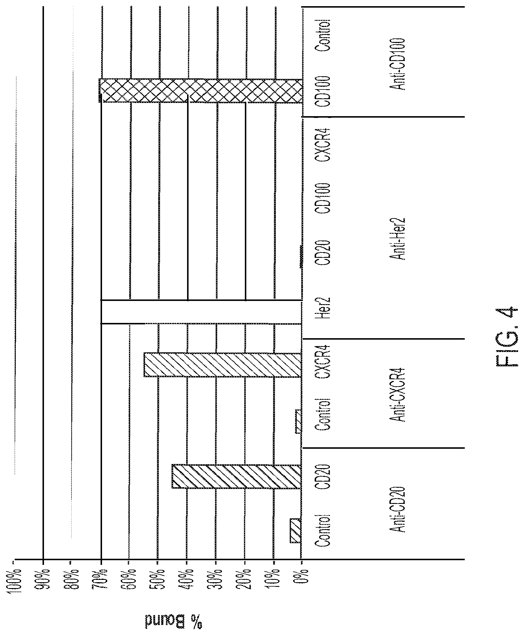
[0022] FIG. 4: Incorporation of additional IMP-EEV protein fusions into vaccinia virus EEV. "CD20" is a CD20-F13L fusion protein, "CXCR4" is a CXCR4-F13L fusion protein, "Her2" is a Her2 ECD-A56R fusion protein; and "CD100" is a CD100 ECD-A56R fusion protein.
[0023] FIG. 5: Outline of assay for screening an antibody display library for display packages that bind to an IMP of interest expressed on vaccinia virus EEV.
[0024] FIG. 6A: Binding of vaccinia virus EEV expressing an anti-HER-2 antibody to vaccinia virus EEV expressing the HER2 ECD as a fusion with the vaccinia virus A56R protein, bound by tosyl-groups to magnetic beads.
[0025] FIG. 6B: Binding of vaccinia virus EEV expressing an anti-FZD antibody to vaccinia virus EEV expressing FZD4 as a fusion with the vaccinia virus F13L protein, bound by tosyl-groups to magnetic beads.
[0026] FIG. 6C: Binding of vaccinia virus EEV expressing an anti-CXCR4 antibody to vaccinia virus EEV expressing the CXCR4 as a fusion with the vaccinia virus F13L protein, bound by tosyl-groups to magnetic beads.
[0027] FIG. 6D: Binding of vaccinia virus EEV expressing an anti-CD100 ("sema") antibody to vaccinia virus EEV expressing the CD100 ECD as a fusion with the vaccinia virus A56R protein, bound by tosyl-groups to magnetic beads.
[0028] FIG. 7: FACS scans showing enrichment for anti FZD4 antibodies following panning on inactivated FZD-ECD-A45R-expressing EEV bound by tosyl-groups to magnetic beads after 3 (Rd3), 4 (Rd4), and 5 (Rd5) rounds of panning. The top row shows antibody-expressing virus-infected cells stained with 10 .mu.g/ml FZD-His, followed by anti-His-Dyelight650 and anti-Fab-FITC. The bottom row shows antibody-expressing virus-infected cells stained with 10 .mu.g/ml CD100-His (negative control), followed by anti-His-Dyelight650 and anti-Fab-FITC.
[0029] FIG. 8: Incorporation two different protein fusions (HA-A56R fusion and FZD4-F13L fusion) into vaccinia virus EEV. EEV expressing the HA-A56R fusion alone, the FZD4-F13L fusion alone, or both fusion proteins, were tested for binding to either anti-FZD4-coated beads or anti-HA coated beads.
[0030] FIG. 9: Specific recovery of anti-CXCR4-expressing EEV by magnetic beads coated with EEV expressing both an HA-A56R fusion and CXCR4-F13L fusion. The antigen-EEV were coupled to anti-HA coated beads.
[0031] FIG. 10: Binding of biotinylated vaccinia virus EEV expressing the designated fusion proteins to streptavidin coated magnetic beads.
DETAILED DESCRIPTION
[0032] This disclosure provides methods and compositions for expressing and displaying integral membrane proteins (IMPs), e.g., multi-pass (IMPs), in a conformationally intact or native state on the surface of extracellular enveloped virion particles (EEV) of poxviruses, e.g., vaccinia virus, as a fusion with a polypeptide segment an EEV-specific membrane-associated protein, e.g., F13L.
Definitions
[0033] The term "a" or "an" entity refers to one or more of that entity; for example, "a binding molecule," is understood to represent one or more binding molecules. As such, the terms "a" (or "an"), "one or more," and "at least one" can be used interchangeably herein.
[0034] Furthermore, "and/or" where used herein is to be taken as specific disclosure of each of the two specified features or components with or without the other. Thus, the term and/or" as used in a phrase such as "A and/or B" herein is intended to include "A and B," "A or B," "A" (alone), and "B" (alone). Likewise, the term "and/or" as used in a phrase such as "A, B, and/or C" is intended to encompass each of the following embodiments: A, B, and C; A, B, or C; A or C; A or B; B or C; A and C; A and B; B and C; A (alone); B (alone); and C (alone).
[0035] Unless defined otherwise, technical and scientific terms used herein have the same meaning as commonly understood by one of ordinary skill in the art to which this disclosure is related. For example, the Concise Dictionary of Biomedicine and Molecular Biology, Juo, Pei-Show, 2nd ed., 2002, CRC Press; The Dictionary of Cell and Molecular Biology, 3rd ed., 1999, Academic Press; and the Oxford Dictionary Of Biochemistry And Molecular Biology, Revised, 2000, Oxford University Press, provide one of skill with a general dictionary of many of the terms used in this disclosure.
[0036] Units, prefixes, and symbols are denoted in their Systeme International de Unites (SI) accepted form. Numeric ranges are inclusive of the numbers defining the range. Unless otherwise indicated, amino acid sequences are written left to right in amino to carboxy orientation. The headings provided herein are not limitations of the various aspects or aspects of the disclosure, which can be had by reference to the specification as a whole. Accordingly, the terms defined immediately below are more fully defined by reference to the specification in its entirety.
[0037] As used herein, the term "non-naturally occurring" substance, composition, entity, and/or any combination of substances, compositions, or entities, or any grammatical variants thereof, is a conditional term that explicitly excludes, but only excludes, those forms of the substance, composition, entity, and/or any combination of substances, compositions, or entities that are well-understood by persons of ordinary skill in the art as being "naturally-occurring," or that are, or might be at any time, determined or interpreted by a judge or an administrative or judicial body to be, "naturally-occurring."
[0038] As used herein, the term "polypeptide" is intended to encompass a singular "polypeptide" as well as plural "polypeptides," and refers to a molecule composed of monomers (amino acids) linearly linked by amide bonds (also known as peptide bonds). The term "polypeptide" refers to any chain or chains of two or more amino acids, and does not refer to a specific length of the product. Thus, peptides, dipeptides, tripeptides, oligopeptides, "protein," "amino acid chain," or any other term used to refer to a chain or chains of two or more amino acids are included within the definition of "polypeptide," and the term "polypeptide" can be used instead of, or interchangeably with any of these terms. The term "polypeptide" is also intended to refer to the products of post-expression modifications of the polypeptide, including without limitation glycosylation, acetylation, phosphorylation, amidation, and derivatization by known protecting/blocking groups, proteolytic cleavage, or modification by non-naturally occurring amino acids. A polypeptide can be derived from a biological source or produced by recombinant technology, but is not necessarily translated from a designated nucleic acid sequence. It can be generated in any manner, including by chemical synthesis.
[0039] A polypeptide as disclosed herein can be of a size of about 3 or more, 5 or more, 10 or more, 20 or more, 25 or more, 50 or more, 75 or more, 100 or more, 200 or more, 500 or more, 1,000 or more, or 2,000 or more amino acids. Polypeptides can have a defined three-dimensional structure, although they do not necessarily have such structure. Polypeptides with a defined three-dimensional structure are referred to as folded, and polypeptides that do not possess a defined three-dimensional structure, but rather can adopt a large number of different conformations, and are referred to as unfolded. As used herein, the term glycoprotein refers to a protein coupled to at least one carbohydrate moiety that is attached to the protein via an oxygen-containing or a nitrogen-containing side chain of an amino acid, e.g., a serine or an asparagine.
[0040] By an "isolated" polypeptide or a fragment, variant, or derivative thereof is intended a polypeptide that is not in its natural milieu. No particular level of purification is required. For example, an isolated polypeptide can be removed from its native or natural environment. Recombinantly produced polypeptides and proteins expressed in host cells are considered isolated as disclosed herein, as are native or recombinant polypeptides that have been separated, fractionated, or partially or substantially purified by any suitable technique.
[0041] As used herein, the term "non-naturally occurring" polypeptide, or any grammatical variants thereof, is a conditional term that explicitly excludes, but only excludes, those forms of the polypeptide that are well-understood by persons of ordinary skill in the art as being "naturally-occurring," or that are, or might be at any time, determined or interpreted by a judge or an administrative or judicial body to be, "naturally-occurring."
[0042] Other polypeptides disclosed herein are fragments, derivatives, analogs, or variants of the foregoing polypeptides, and any combination thereof The terms "fragment," "variant," "derivative" and "analog" as disclosed herein include any polypeptides that retain at least some of the properties of the corresponding native antibody or polypeptide, for example, specifically binding to an antigen. Fragments of polypeptides include, for example, proteolytic fragments, as well as deletion fragments, in addition to specific antibody fragments discussed elsewhere herein. Variants of, e.g., a polypeptide include fragments as described above, and also polypeptides with altered amino acid sequences due to amino acid substitutions, deletions, or insertions. In certain aspects, variants can be non-naturally occurring. Non-naturally occurring variants can be produced using art-known mutagenesis techniques. Variant polypeptides can comprise conservative or non-conservative amino acid substitutions, deletions or additions. Derivatives are polypeptides that have been altered so as to exhibit additional features not found on the original polypeptide. Examples include fusion proteins. Variant polypeptides can also be referred to herein as "polypeptide analogs." As used herein a "derivative" of a polypeptide can also refer to a subject polypeptide having one or more amino acids chemically derivatized by reaction of a functional side group. Also included as "derivatives" are those peptides that contain one or more derivatives of the twenty standard amino acids. For example, 4-hydroxyproline can be substituted for proline; 5-hydroxylysine can be substituted for lysine; 3-methylhistidine can be substituted for histidine; homoserine can be substituted for serine; and ornithine can be substituted for lysine.
[0043] A "conservative amino acid substitution" is one in which one amino acid is replaced with another amino acid having a similar side chain. Families of amino acids having similar side chains have been defined in the art, including basic side chains (e.g., lysine, arginine, histidine), acidic side chains (e.g., aspartic acid, glutamic acid), uncharged polar side chains (e.g., asparagine, glutamine, serine, threonine, tyrosine, cysteine), nonpolar side chains (e.g., glycine, alanine, valine, leucine, isoleucine, proline, phenylalanine, methionine, tryptophan), beta-branched side chains (e.g., threonine, valine, isoleucine) and aromatic side chains (e.g., tyrosine, phenylalanine, tryptophan, histidine). For example, substitution of a phenylalanine for a tyrosine is a conservative substitution. In certain embodiments, conservative substitutions in the sequences of the polypeptides and antibodies of the present disclosure do not abrogate the binding of the polypeptide or antibody containing the amino acid sequence, to the antigen to which the binding molecule binds. Methods of identifying nucleotide and amino acid conservative substitutions that do not eliminate antigen binding are well-known in the art (see, e.g., Brummell et al., Biochem. 32:1180-1 187 (1993); Kobayashi et al., Protein Eng. 12 (10):879-884 (1999); and Burks et al., Proc. Natl. Acad. Sci. USA 94:412-417 (1997)).
[0044] As used herein the term "integral membrane protein" or "IMP" refers to a protein or polypeptide that is attached to a biological membrane. One example of an IMP is a transmembrane protein, which spans the lipid bilayer of the biological membrane one or more times. Single-pass membrane proteins cross the membrane only once, while multi-pass membrane proteins weave in and out, crossing several times. Type I single-pass proteins are positioned with their amino terminus on the outer side of the membrane or "extra-membrane" and their carboxyl-terminus on the interior side of the membrane, or "intra-membrane." Type II single-pass proteins have their amino-terminus on the intra-membrane side. Multi-pass transmembrane proteins pass through the membrane two or more times and can have a variety of different topologies. Those proteins with an even number of transmembrane domains will have both their amino terminus and carboxy terminus on the same side of the membrane. One example of such a protein is CD20, which is expressed on B cells. Those with an odd number of transmembrane domains will have their amino-and carboxy termini on opposite sides of the membrane. Examples include G-protein coupled receptors, which typically have 7 transmembrane domains, with the amino terminus on the extra-membrane side and the carboxy terminus on the intra-membrane side. Certain IMPs do not have transmembrane domains and are instead anchored to the membrane, e.g., via a lipid such as glycosylphosphatidylinositol or palmitoyl group. IMPs have myriad biological functions including, but not limited to transporters, linkers, channels, receptors, enzymes, energy transduction or cell adhesion.
[0045] The term "polynucleotide" is intended to encompass a singular nucleic acid as well as plural nucleic acids, and refers to an isolated nucleic acid molecule or construct, e.g., messenger RNA (mRNA), cDNA, or plasmid DNA (pDNA). A polynucleotide can comprise a conventional phosphodiester bond or a non-conventional bond (e.g., an amide bond, such as found in peptide nucleic acids (PNA)). The terms "nucleic acid" or "nucleic acid sequence" refer to any one or more nucleic acid segments, e.g., DNA or RNA fragments, present in a polynucleotide.
[0046] By an "isolated" nucleic acid or polynucleotide is intended any form of the nucleic acid or polynucleotide that is separated from its native environment. For example, gel-purified polynucleotide, or a recombinant polynucleotide encoding a polypeptide contained in a vector would be considered to be "isolated." Also, a polynucleotide segment, e.g., a PCR product, that has been engineered to have restriction sites for cloning is considered to be "isolated." Further examples of an isolated polynucleotide include recombinant polynucleotides maintained in heterologous host cells or purified (partially or substantially) polynucleotides in a non-native solution such as a buffer or saline. Isolated RNA molecules include in vivo or in vitro RNA transcripts of polynucleotides, where the transcript is not one that would be found in nature. Isolated polynucleotides or nucleic acids further include such molecules produced synthetically. In addition, polynucleotide or a nucleic acid can be or can include a regulatory element such as a promoter, ribosome binding site, or a transcription terminator.
[0047] As used herein, a "non-naturally occurring" polynucleotide, or any grammatical variants thereof, is a conditional definition that explicitly excludes, but only excludes, those forms of the polynucleotide that are well-understood by persons of ordinary skill in the art as being "naturally-occurring," or that are, or that might be at any time, determined or interpreted by a judge or an administrative or judicial body to be, "naturally-occurring."
[0048] As used herein, a "coding region" is a portion of nucleic acid that consists of codons translated into amino acids. Although a "stop codon" (TAG, TGA, or TAA) is not translated into an amino acid, it can be considered to be part of a coding region, but any flanking sequences, for example promoters, ribosome binding sites, transcriptional terminators, introns, and the like, are not part of a coding region. Two or more coding regions can be present in a single polynucleotide construct, e.g., on a single vector, or in separate polynucleotide constructs, e.g., on separate (different) vectors. Furthermore, any vector can contain a single coding region, or can comprise two or more coding regions, e.g., a single vector can separately encode an immunoglobulin heavy chain variable region and an immunoglobulin light chain variable region. In addition, a vector, polynucleotide, or nucleic acid can include heterologous coding regions, either fused or unfused to another coding region. Heterologous coding regions include without limitation, those encoding specialized elements or motifs, such as a secretory signal peptide or a heterologous functional domain.
[0049] In certain embodiments, the polynucleotide or nucleic acid is DNA. In the case of DNA, a polynucleotide comprising a nucleic acid that encodes a polypeptide normally can include a promoter and/or other transcription or translation control elements operably associated with one or more coding regions. An operable association is when a coding region for a gene product, e.g., a polypeptide, is associated with one or more regulatory sequences in such a way as to place expression of the gene product under the influence or control of the regulatory sequence(s). Two DNA fragments (such as a polypeptide coding region and a promoter associated therewith) are "operably associated" if induction of promoter function results in the transcription of mRNA encoding the desired gene product and if the nature of the linkage between the two DNA fragments does not interfere with the ability of the expression regulatory sequences to direct the expression of the gene product or interfere with the ability of the DNA template to be transcribed. Thus, a promoter region would be operably associated with a nucleic acid encoding a polypeptide if the promoter was capable of effecting transcription of that nucleic acid. The promoter can be a cell-specific promoter that directs substantial transcription of the DNA in predetermined cells. Other transcription control elements, besides a promoter, for example enhancers, operators, repressors, and transcription termination signals, can be operably associated with the polynucleotide to direct cell-specific transcription.
[0050] A variety of transcription control regions are known to those skilled in the art. These include, without limitation, transcription control regions that function in vertebrate cells, such as, but not limited to, promoter and enhancer segments from cytomegaloviruses (the immediate early promoter, in conjunction with intron-A), simian virus 40 (the early promoter), and retroviruses (such as Rous sarcoma virus). Other transcription control regions include those derived from vertebrate genes such as actin, heat shock protein, bovine growth hormone and rabbit .beta.-globin, as well as other sequences capable of controlling gene expression in eukaryotic cells. Additional suitable transcription control regions include tissue-specific promoters and enhancers as well as lymphokine-inducible promoters (e.g., promoters inducible by interferons or interleukins).
[0051] Poxvirus promoters (e.g. p7.5 or H5) or the bacteriophage T7 promoter can also be used as transcription control regions. When employing a T7 promoter, an inducible vaccinia expression system can be utilized. The vaccinia expression system can include, but is not limited, to a first recombinant vaccinia virus that encodes the entire bacteriophage T7 gene 1 coding region for T7 RNA polymerase, and a second recombinant vaccinia virus that encodes a gene of interest flanked by a T7 promoter and termination regulatory elements. Dual infection of eukaryotic cells with both recombinant vaccinia viruses results in synthesis of the T7 RNA polymerase and expression of the gene of interest controlled by the T7 promoter.
[0052] Similarly, a variety of translation control elements are known to those of ordinary skill in the art. These include, but are not limited to ribosome binding sites, translation initiation and termination codons, and elements derived from picornaviruses (particularly an internal ribosome entry site, or IRES, also referred to as a CITE sequence).
[0053] In other embodiments, a polynucleotide can be RNA, for example, in the form of messenger RNA (mRNA), transfer RNA, or ribosomal RNA.
[0054] Polynucleotide and nucleic acid coding regions can be associated with additional coding regions that encode secretory or signal peptides, which direct the secretion of a polypeptide encoded by a polynucleotide as disclosed herein. According to the signal hypothesis, proteins secreted by mammalian cells have a signal peptide or secretory leader sequence that is cleaved from the mature protein once export of the growing protein chain across the rough endoplasmic reticulum has been initiated. Those of ordinary skill in the art are aware that polypeptides secreted by vertebrate cells can have a signal peptide fused to the N-terminus of the polypeptide, which is cleaved from the complete or "full length" polypeptide to produce a secreted or "mature" form of the polypeptide. In certain embodiments, the native signal peptide, e.g., an immunoglobulin heavy chain or light chain signal peptide is used, or a functional derivative of that sequence that retains the ability to direct the secretion of the polypeptide that is operably associated with it. Alternatively, a heterologous mammalian signal peptide, or a functional derivative thereof, can be used. For example, the wild-type leader sequence can be substituted with the leader sequence of human tissue plasminogen activator (TPA) or mouse .beta.-glucuronidase.
[0055] As used herein, a "library" is a representative genus of polynucleotides, e.g., a group of polynucleotides related through, for example, their origin from a single animal species, tissue type, organ, or cell type, where the library collectively comprises at least two different species within a given genus of polynucleotides. A library of polynucleotides can include, e.g., at least two, at least 5, at least 10, 100, 10.sup.3, 10.sup.4, 10.sup.5, 10.sup.6, 10.sup.7, 10.sup.8, or 10.sup.9 different species within a given genus of polynucleotides. In certain aspects, a library of polynucleotides as provided herein can encode a plurality of polypeptides that contains a polypeptide of interest. In certain aspects, a library of polynucleotides as provided herein can encode a plurality of immunoglobulin subunit polypeptides, e.g., heavy chain subunit polypeptides or light chain subunit polypeptides. In this context, a "library" as provided herein comprises polynucleotides of a common genus, the genus being polynucleotides encoding immunoglobulin subunit polypeptides of a certain type and class e.g., a library might encode a human .mu., .gamma.-1, .gamma.-2, .gamma.-3, .gamma.-4, .alpha.-1, .alpha.-2, .epsilon., or .delta. heavy chain, or a human .kappa. or .lamda. light chain. Although each member of any one library constructed according to the methods provided herein can encode the same heavy or light chain constant region and/or a membrane anchoring domain, the library can collectively comprise at least two, at least 5, or at least 10, 100, 10.sup.3, 10.sup.4, 10.sup.5, 10.sup.6, 10.sup.7, 10.sup.8, or 10.sup.9 different variable region associated with the common constant region.
[0056] In other embodiments, the library can a plurality of immunoglobulin single-chain fragments that comprise a variable region, such as a light chain variable region or a heavy chain variable region, and/or both a light chain variable region and a heavy chain variable region, e.g., an ScFv fragment.
[0057] As used herein, a "display library" is a library of polynucleotides each carried in a "display package" that expresses the polypeptide encoded by the library polynucleotide on its surface. An antibody display library, for example, can include plurality of display packages, each displaying an antigen binding domain of an antibody on its surface. When the display library is permitted to interact with an antigen of interest, e.g., immobilized on a solid surface, those display packages that bind the antigen can be isolated from the rest of the library and recovered. The polynucleotide encoding the antigen binding domain displayed on the surface of the display package can then be isolated. Display libraries include, without limitation, phage display libraries in bacteria or libraries in eukaryotic systems, e.g., yeast display, retroviral display, or expression in DNA viruses such as poxviruses. See, e.g., U.S. Pat. No. 7,858,559, and U.S. Patent Appl. Publication No. 2013-028892, which are incorporated herein by reference in their entireties. In certain aspects, an antibody display library can be prepared in a poxvirus, e.g., vaccinia virus vector, as fusion proteins with an EEV-specific protein, such that the "display packages" are EEV particles. See U.S. Patent Appl. Publication No. 2013-028892.
[0058] Such display libraries can be screened against the IMP fusion proteins displayed on the surface of EEV as provided herein.
[0059] By "recipient cell" or "host cell" or "cell" is meant a cell or population of cells in which a recombinant protein can be expressed, a virus can be propagated, or polynucleotide libraries as provided herein can be constructed and/or propagated. A host cell as provided herein is typically a eukaryotic cell or cell line, e.g., a vertebrate, mammalian, rodent, mouse, primate, or human cell or cell line. By "a population of host cells" is meant a group of cultured cells which a "library" as provided herein can be constructed, propagated, and/or expressed. Any host cell which is permissive for vaccinia virus infectivity is suitable for the methods provided by this disclosure. Host cells for use in the methods provided herein can be adherent, e.g., host cells that grow attached to a solid substrate, or, alternatively, the host cells can be in suspension.
[0060] Host cells as provided herein can comprise a constitutive secretory pathway, where proteins, e.g., proteins of interest expressed by the cell or by a library, are secreted from the interior of the cell either to be expressed on a cell or viral membrane surface or to be fully secreted as soluble polypeptides. In certain aspects, proteins of interest expressed on or in a biological membrane, e.g., an IMP, are expressed on the surface of an enveloped virus produced by the host cell, e.g., an extracellular enveloped vaccinia virus, or EEV. IMPS can follow the same pathway as fully secreted forms or proteins, passing through to the ER lumen, except that they can be retained in the ER membrane by the presence of one or more stop-transfer signals, or "transmembrane domains." Transmembrane domains are hydrophobic stretches of about 20 amino acids that adopt an alpha-helical conformation as they transverse the membrane. Membrane embedded proteins are anchored in the phospholipid bilayer of the plasma membrane. Transmembrane forms of polypeptides of interest, e.g., membrane-anchored immunoglobulin heavy chain polypeptides typically utilize amino terminal signal peptides as do fully secreted forms.
[0061] Signal peptides, transmembrane domains, and cytosolic or "intra-membrane" domains are known for a wide variety of membrane bound and/or fully secreted proteins.
[0062] Suitable transmembrane domains can include, but are not limited to the TM domain of the vaccinia virus EEV-specific HA protein A56R, or the EEV-specific vaccinia virus transmembrane proteins A33R, A34R, A36R, or B5R. See, e.g., U.S. Patent Appl. Publ. No. 2013/0288927, published Oct. 31, 2013, and incorporated herein by reference in its entirety. In certain aspects the EEV specific protein can be anchored to the inner surface of the viral envelope via a palmitoyl group, e.g., the vaccinia virus protein F13L, discussed in more detail elsewhere herein.
[0063] As used herein, the term "binding molecule" refers in its broadest sense to a molecule that specifically binds to a receptor, e.g., an epitope or an antigenic determinant. As described further herein, a binding molecule can comprise one or more "antigen binding domains" described herein. A non-limiting example of a binding molecule is an antibody or fragment thereof that retains antigen-specific binding.
[0064] The terms "binding domain" and "antigen binding domain" are used interchangeably herein and refer to a region of a binding molecule that is necessary and sufficient to specifically bind to an epitope. For example, an "Fv," e.g., a variable heavy chain and variable light chain of an antibody, either as two separate polypeptide subunits or as a single chain, is considered to be a "binding domain."
[0065] Other antigen binding domains include, without limitation, the variable heavy chain (VHH) of an antibody derived from a camelid species, or six immunoglobulin complementarity determining regions (CDRs) expressed in a fibronectin scaffold.
[0066] The terms "antibody" and "immunoglobulin" can be used interchangeably herein. An antibody (or a fragment, variant, or derivative thereof as disclosed herein) includes at least the variable region of a heavy chain (e.g., for camelid species) or at least the variable regions of a heavy chain and a light chain. Basic immunoglobulin structures in vertebrate systems are relatively well understood. See, e.g., Harlow et al., Antibodies: A Laboratory Manual, (Cold Spring Harbor Laboratory Press, 2nd ed. 1988). Unless otherwise stated, the term "antibody" encompasses anything ranging from a small antigen binding fragment of an antibody to a full sized antibody, e.g., an IgG antibody that includes two complete heavy chains and two complete light chains.
[0067] The term "immunoglobulin" comprises various broad classes of polypeptides that can be distinguished biochemically. Those skilled in the art will appreciate that heavy chains are classified as gamma, mu, alpha, delta, or epsilon, (.gamma., .mu., .alpha., .delta., .epsilon.) with some subclasses among them (e.g., .gamma.1-.gamma.4 or .alpha.1-.alpha.2)). It is the nature of this chain that determines the "class" of the antibody as IgG, IgM, IgA IgG, or IgE, respectively. The immunoglobulin subclasses (isotypes) e.g., IgG.sub.1, IgG.sub.2, IgG.sub.3, IgG.sub.4, IgA.sub.1, IgA.sub.2, etc. are well characterized and are known to confer functional specialization.
[0068] Light chains are classified as either kappa or lambda (.kappa., .lamda.). Each heavy chain class can be bound with either a kappa or lambda light chain. In general, the light and heavy chains are covalently bonded to each other, and the "tail" portions of the two heavy chains are bonded to each other by covalent disulfide linkages or non-covalent linkages when the immunoglobulins are generated either by hybridomas, B cells or genetically engineered host cells. In the heavy chain, the amino acid sequences run from an N-terminus at the forked ends of the Y configuration to the C-terminus at the bottom of each chain. The basic structure of certain antibodies, e.g., IgG antibodies, includes two heavy chain subunits and two light chain subunits covalently connected via disulfide bonds to form a "Y" structure, also referred to herein as an "H2L2" structure.
[0069] The term "epitope" includes any molecular determinant capable of specific binding to an antibody. In certain aspects, an epitope can include chemically active surface groupings of molecules such as amino acids, sugar side chains, phosphoryl, or sulfonyl, and, in certain aspects, can have three dimensional structural characteristics, and or specific charge characteristics. An epitope is a region of a target that is bound by an antibody.
[0070] The term "target" is used in the broadest sense to include substances that can be bound by a binding molecule. A target can be, e.g., a polypeptide, a nucleic acid, a carbohydrate, a lipid, or other molecule. Moreover, a "target" can, for example, be a cell, an organ, or an organism that comprises an epitope bound that can be bound by a binding molecule.
[0071] Both the light and heavy chains are divided into regions of structural and functional homology. The terms "constant" and "variable" are used functionally. In this regard, it will be appreciated that the variable regions (which can be called "variable domains" interchangeably herein) of both the variable light (VL) and variable heavy (VH) chain portions determine antigen recognition and specificity. Conversely, the constant domains of the light chain (CL) and the heavy chain (e.g., CH1, CH2 or CH3) confer biological properties such as secretion, transplacental mobility, Fc receptor binding, complement binding, and the like. By convention the numbering of the constant region domains increases as they become more distal from the antigen binding site or amino-terminus of the antibody. The N-terminal portion is a variable region and at the C-terminal portion is a constant region; the CH3 (or CH4 in the case of IgM) and CL domains are at the carboxy-terminus of the heavy and light chain, respectively.
[0072] The six "complementarity determining regions" or "CDRs" present in an antibody antigen binding domain are short, non-contiguous sequences of amino acids that are specifically positioned to form the antigen binding domain as the antibody assumes its three dimensional configuration in an aqueous environment. The remainder of the amino acids in the antigen binding domain, referred to as "framework" regions, show less inter-molecular variability. The framework regions largely adopt a .beta.-sheet conformation and the CDRs form loops that connect, and in some cases form part of, the .beta.-sheet structure. Thus, framework regions act to form a scaffold that provides for positioning the CDRs in correct orientation by inter-chain, non-covalent interactions. The antigen binding domain formed by the positioned CDRs defines a surface complementary to the epitope on the immunoreactive antigen. This complementary surface promotes the non-covalent binding of the antibody to its cognate epitope. The amino acids that make up the CDRs and the framework regions, respectively, can be readily identified for any given heavy or light chain variable region by one of ordinary skill in the art, since they have been defined in various different ways (see, "Sequences of Proteins of Immunological Interest," Kabat, E., et al., U.S. Department of Health and Human Services, (1983); and Chothia and Lesk, J. Mol. Biol., 196:901-917 (1987), which are incorporated herein by reference in their entireties).
[0073] In the case where there are two or more definitions of a term that is used and/or accepted within the art, the definition of the term as used herein is intended to include all such meanings unless explicitly stated to the contrary. A specific example is the use of the term "complementarity determining region" ("CDR") to describe the non-contiguous antigen combining sites found within the variable region of both heavy and light chain polypeptides. These particular regions have been described, for example, by Kabat et al., U.S. Dept. of Health and Human Services, "Sequences of Proteins of Immunological Interest" (1983) and by Chothia et al., J. Mol. Biol. 196:901-917 (1987), which are incorporated herein by reference. Immunoglobulin variable domains can also be analyzed, e.g., using the IMGT information system (www://imgt.cines.fr/) (IMGT.RTM./V-Quest) to identify variable region segments, including CDRs. (See, e.g., Brochet et al., Nucl. Acids Res., 36:W503-508, 2008).
[0074] Kabat et al. also defined a numbering system for variable domain sequences that is applicable to any antibody. One of ordinary skill in the art can unambiguously assign this system of "Kabat numbering" to any variable domain sequence, without reliance on any experimental data beyond the sequence itself. As used herein, "Kabat numbering" refers to the numbering system set forth by Kabat et al., U.S. Dept. of Health and Human Services, "Sequence of Proteins of Immunological Interest" (1983). Unless use of the Kabat numbering system is explicitly noted, however, consecutive numbering is used for all amino acid sequences in this disclosure.
[0075] Binding molecules, e.g., antibodies or antigen binding fragments, variants, or derivatives thereof include, but are not limited to, polyclonal, monoclonal, human, humanized, or chimeric antibodies, single chain antibodies, epitope-binding fragments, e.g., Fab, Fab' and F(ab').sub.2, Fd, Fvs, single-chain Fvs (scFv), single-chain antibodies, disulfide-linked Fvs (sdFv), single domain antibodies such as camelid VHH antibodies, fragments comprising either a VL or VH domain, fragments produced by a Fab expression library. ScFv molecules are known in the art and are described, e.g., in U.S. Pat. No. 5,892,019. Immunoglobulin or antibody molecules encompassed by this disclosure can be of any type (e.g., IgG, IgE, IgM, IgD, IgA, and IgY), class (e.g., IgG1, IgG2, IgG3, IgG4, IgA1 and IgA2) or subclass of immunoglobulin molecule. Also contemplated are immunoglobulin new antigen receptor (IgNAR) isotypes that are bivalent and comprise a single chain that includes an IgNAR variable domain (VNAR). (See, Walsh et al., Virology 411:132-141, 2011).
[0076] By "specifically binds," it is generally meant that a binding molecule, e.g., an antibody or fragment, variant, or derivative thereof binds to an epitope via its antigen binding domain, and that the binding entails some complementarity between the antigen binding domain and the epitope. According to this definition, a binding molecule is said to "specifically bind" to an epitope when it binds to that epitope, via its antigen binding domain more readily than it would bind to a random, unrelated epitope. The term "specificity" is used herein to qualify the relative affinity by which a certain binding molecule binds to a certain epitope. For example, binding molecule "A" can be deemed to have a higher specificity for a given epitope than binding molecule "B," or binding molecule "A" can be said to bind to epitope "C" with a higher specificity than it has for related epitope "D."
[0077] As used herein, the term "affinity" refers to a measure of the strength of the binding of an individual epitope with one or more antigen binding domains, e.g., of an immunoglobulin molecule. See, e.g., Harlow et al., Antibodies: A Laboratory Manual, (Cold Spring Harbor Laboratory Press, 2nd ed. 1988) at pages 27-28. As used herein, the term "avidity" refers to the overall stability of the complex between a population of antigen binding domains and an antigen. See, e.g., Harlow at pages 29-34. Avidity is related to both the affinity of individual antigen binding domains in the population with specific epitopes, and also the valencies of the immunoglobulins and the antigen. For example, the interaction between a bivalent monoclonal antibody and an antigen with a highly repeating epitope structure, such as a polymer, would be one of high avidity. An interaction between a between a bivalent monoclonal antibody with a receptor present at a high density on a cell surface would also be of high avidity.
[0078] As used herein, the term "heavy chain subunit" or "heavy chain domain" includes amino acid sequences derived from an immunoglobulin heavy chain, a binding molecule, e.g., an antibody comprising a heavy chain subunit can include at least one of: a VH domain, a CH1 domain, a hinge (e.g., upper, middle, and/or lower hinge region) domain, a CH2 domain, a CH3 domain, a CH4 domain, or a variant or fragment thereof.
[0079] As used herein, the term "light chain subunit" or "light chain domain" includes amino acid sequences derived from an immunoglobulin light chain. The light chain subunit includes at least one of a VL or CL (e.g., C.kappa. or C.lamda.) domain.
[0080] Binding molecules, e.g., antibodies or antigen binding fragments, variants, or derivatives thereof can be described or specified in terms of the epitope(s) or portion(s) of an antigen that they recognize or specifically bind. The portion of a target antigen that specifically interacts with the antigen binding domain of an antibody is an "epitope," or an "antigenic determinant." A target antigen can comprise a single epitope or at least two epitopes, and can include any number of epitopes, depending on the size, conformation, and type of antigen.
[0081] As used herein, the terms "linked," "fused" or "fusion" or other grammatical equivalents can be used interchangeably. These terms refer to the joining together of two more elements or components, by whatever means including chemical conjugation or recombinant means. An "in-frame fusion" refers to the joining of two or more polynucleotide open reading frames (ORFs) to form a continuous longer ORF, in a manner that maintains the translational reading frame of the original ORFs. Thus, a recombinant fusion protein is a single protein containing two or more segments that correspond to polypeptides encoded by the original ORFs (which segments are not normally so joined in nature). Although the reading frame is thus made continuous throughout the fused segments, the segments can be physically or spatially separated by, for example, in-frame linker sequence. For example, polynucleotides encoding an IMP and a vaccinia virus EEV-specific protein can be fused, in-frame, but be separated by a polynucleotide encoding a linker or spacer, as long as the "fused" open reading frames are co-translated as part of a continuous polypeptide.
[0082] As used herein, the term "hemagglutinin tag" or "HA tag" is a protein derived from a human influenza hemagglutinin surface glycoprotein (HA) corresponding to amino acids 98-106. The HA tag is extensively used as a general epitope tag in expression vectors. Recombinant proteins can be engineered to express the HA tag, which does not appear to interfere with the bioactivity or the biodistribution of the recombinant protein. This tag facilitates the detection, isolation, and purification of the protein of interest.
[0083] In the context of polypeptides, a "linear sequence" or a "sequence" is an order of amino acids in a polypeptide from the amino or N-terminus to the carboxyl or C-terminus, in which amino acids that neighbor each other in the sequence are contiguous in the primary structure of the polypeptide.
[0084] A portion of a polypeptide that is "amino-terminal" or "N-terminal" to another portion of a polypeptide is that portion that comes earlier in the sequential polypeptide chain. Similarly, a portion of a polypeptide that is "carboxy-terminal" or "C-terminal" to another portion of a polypeptide is that portion that comes later in the sequential polypeptide chain.
[0085] The term "expression" as used herein refers to a process by which a gene produces a biochemical, for example, a polypeptide. The process includes any manifestation of the functional presence of the gene within the cell including, without limitation, gene knockdown as well as both transient expression and stable expression. It includes without limitation transcription of the gene into messenger RNA (mRNA), and the translation of such mRNA into polypeptide(s). If the final desired product is a biochemical, expression includes the creation of that biochemical and any precursors. Expression of a gene produces a "gene product." As used herein, a gene product can be either a nucleic acid, e.g., a messenger RNA produced by transcription of a gene, or a polypeptide that is translated from a transcript. Gene products described herein further include nucleic acids with post transcriptional modifications, e.g., polyadenylation, or polypeptides with post translational modifications, e.g., methylation, glycosylation, the addition of lipids, association with other protein subunits, proteolytic cleavage, and the like.
[0086] The term "eukaryote" or "eukaryotic organism" is intended to encompass all organisms in the animal, plant, and protist kingdoms, including protozoa, fungi, yeasts, green algae, single celled plants, multi celled plants, and all animals, both vertebrates and invertebrates. The term does not encompass bacteria or viruses. A "eukaryotic cell" is intended to encompass a singular "eukaryotic cell" as well as plural "eukaryotic cells," and comprises cells derived from a eukaryote.
[0087] The term "vertebrate" is intended to encompass a singular "vertebrate" as well as plural "vertebrates," and comprises mammals and birds, as well as fish, reptiles, and amphibians.
[0088] The term "mammal" is intended to encompass a singular "mammal" and plural "mammals," and includes, but is not limited to humans; primates such as apes, monkeys, orangutans, and chimpanzees; canids such as dogs and wolves; felids such as cats, lions, and tigers; equids such as horses, donkeys, and zebras, food animals such as cows, pigs, and sheep; ungulates such as deer and giraffes; rodents such as mice, rats, hamsters and guinea pigs; and bears. In certain aspects, the mammal is a human subject.
[0089] The terms "tissue culture" or "cell culture" or "culture" or "culturing" refer to the maintenance or growth of plant or animal tissue or cells in vitro under conditions that allow preservation of cell architecture, preservation of cell function, further differentiation, or all three. "Primary tissue cells" are those taken directly from tissue, i.e., a population of cells of the same kind performing the same function in an organism. Treating such tissue cells with the proteolytic enzyme trypsin, for example, dissociates them into individual primary tissue cells that grow or maintain cell architecture when seeded onto culture plates. Cell cultures arising from multiplication of primary cells in tissue culture are called "secondary cell cultures." Most secondary cells divide a finite number of times and then die. A few secondary cells, however, can pass through this "crisis period," after which they are able to multiply indefinitely to form a continuous "cell line." The liquid medium in which cells are cultured is referred to herein as "culture medium" or "culture media." Culture medium into which desired molecules, e.g., viruses or proteins, e.g., immunoglobulin molecules, have been secreted during culture of the cells therein can be referred to as "conditioned medium."
[0090] As used herein, the term "identify" refers to methods in which a desired molecule, e.g., a polynucleotide encoding a protein of interest with a desired characteristics or function, is differentiated from a plurality or library of such molecules. Identification methods include "selection" and "screening" or "panning." As used herein, "selection" methods are those in which the desired molecules can be directly separated from the library, e.g., via drug resistance. As used herein, "screening" or "panning" methods are those in which pools comprising the desired molecules are subjected to an assay in which the desired molecule can be detected. Aliquots of the pools in which the molecule is detected are then divided into successively smaller pools which are likewise assayed, until a pool which is highly enriched from the desired molecule is achieved.
Poxviruses, E.g., Vaccinia Virus EEV Vectors
[0091] IMP fusion proteins as provided herein are produced in poxvirus vectors, e.g., vaccinia virus vectors. The term "poxvirus" includes any member of the family Poxviridae. See, for example, B. Moss in: Virology, 2d Edition, B. N. Fields, D. M. Knipe et al., Eds., Raven Press, p. 2080 (1990). The genus of orthopoxvirus includes, e.g., vaccinia virus, variola virus (the virus that causes smallpox), and raccoon poxvirus. Vaccinia virus is the prototype orthopoxvirus and has been developed and is well-characterized as a vector for the expression of heterologous proteins.
[0092] In those embodiments where poxvirus vectors, in particular vaccinia virus vectors, are used to express IMP fusion proteins as provided herein, any suitable poxvirus vector can be used. In certain aspects, the location of a gene encoding an IMP fusion protein can be in a region of the vector that is non-essential for growth and replication of the virus so that infectious viruses are produced. Although a variety of non-essential regions of the vaccinia virus genome have been characterized, the most widely used locus for insertion of foreign genes is the thymidine kinase locus, located in the HindIII J fragment in the genome. In certain vaccinia virus vectors, the tk locus has been engineered to contain one or two unique restriction enzyme sites, allowing for convenient use of the trimolecular recombination method recombinant virus production, as described elsewhere herein.
[0093] Polynucleotides encoding IMP fusion proteins as provided herein can be inserted into poxvirus vectors, particularly vaccinia virus vectors, under operable association with a transcriptional control region which functions in the cytoplasm of a poxvirus-infected cell.
[0094] Poxvirus transcriptional control regions comprise a promoter and a transcription termination signal. Gene expression in poxviruses is temporally regulated, and promoters for early, intermediate, and late genes possess varying structures. Certain poxvirus genes are expressed constitutively, and promoters for these "early-late" genes bear hybrid structures. Synthetic early-late promoters have also been developed. Suitable poxvirus promoters for expressing IMP fusion proteins as provided herein include, but are not limited to late promoters such as the 7.5-kD promoter, the MIL promoter, the 37-kD promoter, the 11-kD promoter, the 11L promoter, the 12L promoter, the 13L promoter, the 15L promoter, the 17L promoter, the 28-kD promoter, the H1L promoter, the H3L promoter, the H5L promoter, the H6L promoter, the H8L promoter, the D11L promoter, the D12L promoter, the D13L promoter, the A1L promoter, the A2L promoter, the A3L promoter, and the P4b promoter. See, e.g., Moss, B., "Poxviridae and their Replication" IN Virology, 2d Edition, B. N. Fields, D. M. Knipe et al., Eds., Raven Press, p. 2090 (1990).
[0095] Suitable poxvirus vectors include wild-type vaccinia virus, e.g., strain Western Reserve or WR, or attenuated vaccinia virus, e.g., modified vaccinia Ankara (MVA) (Mayr, A. et al., Infection 3:6-14 (1975)).
[0096] During its replication cycle, a poxvirus, e.g., a vaccinia virus, produces four infectious forms which differ in their membrane structure: intracellular mature virion (IMV), the intracellular enveloped virion (IEV), the cell-associated enveloped virion (CEV) and the extracellular enveloped virion (EEV). The prevailing view is that the IMV have a single lipoprotein membrane, while the CEV and EEV are both surrounded by two membrane layers and the IEV has three envelopes. EEV is shed from the plasma membrane of the host cell and the EEV membrane is derived from the trans-Golgi.
[0097] After infection, the virus loses its membrane(s) and the DNA/protein core is transported along microtubules into the cell. The proteins encoded by early vaccinia mRNAs ("early" is defined as pre-DNA replication) lead to uncoating of the vaccinia core and subsequent DNA replication. This replication occurs in what are termed "viral factories" which are located essentially on top of the ER. Within the viral factory, immature virions (IV) assemble and are processed to form IMV (Intracellular Mature Virus). IMVs contain a membrane that is derived from the ER. The majority of IMVs are released from the cell by cell lysis. Some IMVs are transported on microtubules to sites of wrapping by membranes of the trans-Golgi network or early endosomes. The wrapping of the IMV particles by a double membrane creates a form of vaccinia called IEVs (Intracellular Enveloped Virus). The IEVs are then transported to the cell surface on microtubules. The outer IEV membrane fuses with the plasma membrane to expose a CEV (Cell Associated Enveloped Virus) at the cell surface. Actin polymerization from the host cell can drive the CEV to infect neighboring cells, or the virus can be released as an EEV. See, e.g., Kim L. Roberts and Geoffrey L. Smith. Trends in Microbiology 16 (10):472-479 (2008); Geoffrey L. Smith, et al., Journal of General Virology 83:2915-2931 (2002).
[0098] At least six virus-encoded proteins have been reported as components of the EEV envelope membrane. Of these, four proteins (A33R, A34R, A56R, and B5R) are glycoproteins, one (A36R) is a nonglycosylated transmembrane protein, and one (F13L) is a palmitoylated peripheral membrane protein. See, e.g., Lorenzo et al., Journal of Virology 74 (22):10535 (2000). During infection, these proteins localize to the Golgi complex, where they are incorporated into infectious virus that is then transported and released into the extracellular medium. As provided herein, IMP fusion proteins are directed to and expressed on the EEV membrane as a fusion protein with an EEV-specific protein, e.g., F13L or A56R.
[0099] The F13L protein is associated with the interior surface of the outermost EEV membrane through palmitoylation of cysteines 185 and 186. Smith Trends in Microbiol. 16:472-479 (2008). Vaccinia viruses in which the gene encoding F13L is deleted form tiny plaques and the number of EEV produced is reduced significantly.
[0100] The amino acid sequence of the F13L protein from vaccinia virus strain WR is presented as SEQ ID NO: 1. The two palmitoylated cysteine residues (amino acids 85 and 86 of SEQ ID NO: 1) are underlined. Since F13L does not cross the membrane, it does not have a transmembrane domain or signal peptide.
TABLE-US-00001 >F13L (SEQ ID NO: 1) MWPFASVPAGAKCRLVETLPENMDFRSDHLTTFECFNEIITLAKKYIYIAS FCCNPLSTTRGALIFDKLKEASEKGIKIIVLLDERGKRNLGELQSHCPDIN FITVNIDKKNNVGLLLGCFWVSDDERCYVGNASFTGGSIHTIKTLGVYSDY PPLATDLRRRFDTFKAFNSAKNSWLNLCSAACCLPVSTAYHIKNPIGGVFF TDSPEHLLGYSRDLDTDVVIDKLKSAKTSIDIEHLAIVPTTRVDGNSYYWP DIYNSIIEAAINRGVKIRLLVGNWDKNDVYSMATARSLDALCVQNDLSVKV FTIQNNTKLLIVDDEYVHITSANFDGTHYQNHGFVSFNSIDKQLVSEAKKI FERDWVSSHSKSLKI
[0101] The A56R protein is the vaccinia virus hemagglutinin and is a standard type I integral membrane protein comprising an amino-terminal extracellular ("extra-membrane") domain, a single transmembrane domain, and a cytoplasmic ("intra-membrane") domain. A56R comprises an N-terminal signal peptide of about 33 amino acids, an Ig-like domain extending from about amino acid 34 to about amino acid 103, a stalk region extending from about amino acid 121 to about amino acid 275, a transmembrane domain extending from about amino acid 276 to about amino acid 303, and an cytoplasmic ("inter-membrane") domain extending from about amino acid 304 to amino acid 314. See DeHaven et al., J. Gen Virol. 92:1971-1980 (2011). A56R is presented as SEQ ID NO: 5.
TABLE-US-00002 >A56R (SEQ ID NO: 5) MTRLPILLLLISLVYATPFPQTSKKIGDDATLSCNRNNTNDYVVMSAWYKE PNSIILLAAKSDVLYFDNYTKDKISYDSPYDDLVTTITIKSLTARDAGTYV CAFFMTSTTNDTDKVDYEEYSTELIVNTDSESTIDIILSGSTHSPETSSKK PDYIDNSNCSSVFEIATPEPITDNVEDHTDTVTYTSDSINTVSASSGESTT DETPEPITDKEDHTVTDTVSYTTVSTSSGIVTTKSTTDDADLYDTYNDNDT VPPTTVGGSTTSISNYKTKDFVEIFGITALIILSAVAIFCITYYIYNKRSR KYKTENKV
[0102] IMP fusion proteins as provided herein can be expressed in any suitable vaccinia virus. In certain embodiments, the DNA encoding an EEV fusion protein can be inserted into a region of the vaccinia virus genome which is non-essential for growth and replication of the vector so that infectious viruses are produced. Although a variety of non-essential regions of the vaccinia virus genome have been characterized, the most widely used locus for insertion of foreign genes is the thymidine kinase locus, located in the HindIII J fragment in the genome. IMP fusion proteins as provided herein can be inserted into vaccinia virus vectors under operable association with a transcriptional control region which functions in the cytoplasm of a poxvirus-infected cell.
[0103] Suitable promoters for use in the methods described herein include, without limitation, the early/late 7.5-kD promoter, or the early/late H5 promoter (or variants thereof).
The Tri-Molecular Recombination Method
[0104] Tri-molecular recombination, as disclosed in Zauderer, PCT Publication No. WO 00/028016 and in U.S. Pat. No. 7,858,559, is a high efficiency, high titer-producing method for expressing proteins of interest and or producing libraries in vaccinia virus. The tri-molecular recombination method allows the generation of recombinant viruses at efficiencies of at least 90%, and titers at least at least 2 orders of magnitude higher than those obtained by direct ligation.
[0105] In certain aspects, IMP fusion proteins for expression in vaccinia virus and display on EEV as described herein can be constructed in poxvirus vectors, e.g., vaccinia virus vectors, by tri-molecular recombination.
[0106] In certain embodiments, a transfer plasmid for IMP fusion proteins for expression in EEV is provided, which comprises a polynucleotide flanking regions in the vaccinia virus Tk gene, the vaccinia virus H5 promoter, and NcoI and BsiWI restriction sites for inserting coding regions for desired fusion proteins.
Integral Membrane Proteins
[0107] The disclosure provides a method for expressing integral membrane proteins (IMPs) in a conformationally intact state that approaches the native conformation of the protein as it would appear in a cell in which the protein is naturally expressed. According to the disclosure, IMPs are expressed as fusion proteins with poxvirus proteins that are expressed on poxvirus, e.g., vaccinia virus EEVs. IMP fusion proteins as provided herein, when expressed and displayed on the surface of EEVs, are useful as target antigens for screening libraries of binding molecules, e.g., antibody display libraries.
[0108] Any IMP can be constructed as a fusion protein according to the methods provided herein. In certain aspects the IMP is a target for immunotherapy. In certain aspects the IMP is a multi-pass IMP such as CD20 or a G-protein coupled receptor (GPCR). Suitable multi-pass human IMPs for use in the construction of IMP fusion proteins as provided herein include, without limitation, the proteins listed in Table 1.
TABLE-US-00003 TABLE 1 Exemplary Human Multi-Pass Integral Membrane Proteins ENTREZ gene # predicted TM Protein Name ENTREZ_gene_ID symbol domains Poliovirus receptor-related protein 3 25945 PVRL3 2 Prominin-1 8842 PROM1 5 FL cytokine receptor 2322 FLT3 2 Scavenger receptor cysteine-rich type 1 9332 CD163 2 protein M130 C-X-C chemokine receptor type 1 3577 CXCR1 6 C-X-C chemokine receptor type 3 2833 CXCR3 7 C-X-C chemokine receptor type 5 643 CXCR5 7 C-C chemokine receptor type 4 1233 CCR4 7 C-C chemokine receptor type 7 1236 CCR7 7 B-lymphocyte antigen CD20 931 MS4A1 4 Major prion protein 5621 PRNP 2 Plexin-C1 10154 PLXNC1 2 Multidrug resistance protein 1 5243 ABCB1 12 Putative G-protein coupled receptor 44 11251 GPR44 7 EGF-like module-containing mucin-like 30817 EMR2 7 hormone receptor-like 2 Frizzled-4 8322 FZD4 9 Leukocyte surface antigen CD47 961 CD47 5 CD63 antigen 967 CD63 4 Choline transporter-like protein 1 23446 SLC44A1 9 CD97 antigen 976 CD97 7 Multidrug resistance-associated protein 1 4363 ABCC1 16 CAS1 domain-containing protein 1 64921 CASD1 14 Solute carrier family 12 member 6 9990 SLC12A6 14 Sodium/hydrogen exchanger 1 6548 SLC9A1 13 Solute carrier family 12 member 9 56996 SLC12A9 13 Solute carrier family 2, facilitated glucose 6513 SLC2A1 12 transporter member 1 Sodium- and chloride-dependent taurine 6533 SLC6A6 12 transporter Solute carrier organic anion transporter 28231 SLCO4A1 12 family member 4A1 Solute carrier family 23 member 2 9962 SLC23A2 12 Solute carrier organic anion transporter 28232 SLCO3A1 12 family member 3A1 Prestin 375611 SLC26A5 11 Equilibrative nucleoside transporter 2 3177 SLC29A2 11 Equilibrative nucleoside transporter 1 2030 SLC29A1 11 Sodium-coupled neutral amino acid 81539 SLC38A1 11 transporter 1 Sodium bicarbonate cotransporter 3 9497 SLC4A7 11 Urea transporter 1 6563 SLC14A1 10 Transmembrane and coiled-coil domain- 55002 TMCO3 10 containing protein 3 Signal peptide peptidase-like 2A 84888 SPPL2A 9 Transmembrane 9 superfamily member 3 56889 TM9SF3 9 Anoctamin-9 338440 ANO9 8 Sodium/potassium-transporting ATPase 476 ATP1A1 8 subunit alpha-1 Sodium/potassium-transporting ATPase 478 ATP1A3 8 subunit alpha-3 Anoctamin-6 196527 ANO6 8 V-type proton ATPase 116 kDa subunit a 23545 ATP6V0A2 8 isoform 2 Putative P2Y purinoceptor 10 27334 P2RY10 7 G-protein coupled receptor 39 2863 GPR39 7 Sphingosine 1-phosphate receptor 2 9294 S1PR2 7 Latrophilin-2 23266 LPHN2 7 Beta-2 adrenergic receptor 154 ADRB2 7 Alpha-2C adrenergic receptor 152 ADRA2C 7 Thromboxane A2 receptor 6915 TBXA2R 7 Platelet-activating factor receptor 5724 PTAFR 7 Proteinase-activated receptor 1 2149 F2R 7 Neuropeptide Y receptor type 1 4886 NPY1R 7 Type-1 angiotensin II receptor 185 AGTR1 7 Neurotensin receptor type 1 4923 NTSR1 7 Cannabinoid receptor 2 1269 CNR2 7 Prostaglandin E2 receptor EP2 subtype 5732 PTGER2 7 Calcitonin gene-related peptide type 1 10203 CALCRL 7 receptor Protein GPR107 57720 GPR107 7 G-protein coupled receptor 126 57211 GPR126 7 P2Y purinoceptor 8 286530 P2RY8 7 Probable G-protein coupled receptor 125 166647 GPR125 7 Transmembrane protein 87A 25963 TMEM87A 7 Mas-related G-protein coupled receptor 116535 MRGPRF 7 member F Transmembrane protein 87B 84910 TMEM87B 7 Proteinase-activated receptor 4 9002 F2RL3 7 Smoothened homolog 6608 SMO 7 EGF-like module-containing mucin-like 84658 EMR3 7 hormone receptor-like 3 Nemomedin-U receptor 1 10316 NMUR1 7 EGF, latrophilin and seven transmembrane 64123 ELTD1 7 domain-containing protein 1 Transmembrane protein 8A 58986 TMEM8A 7 Cadherin EGF LAG seven-pass G-type 1952 CELSR2 7 receptor 2 Cadherin EGF LAG seven-pass G-type 9620 CELSR1 7 receptor 1 Cadherin EGF LAG seven-pass G-type 1951 CELSR3 7 receptor 3 Cysteinyl leukotriene receptor 1 10800 CYSLTR1 7 G-protein coupled receptor 56 9289 GPR56 7 Lipid phosphate phosphohydrolase 1 8611 PPAP2A 6 Potassium voltage-gated channel subfamily A 3738 KCNA3 6 member 3 Zinc transporter ZIP6 25800 SLC39A6 6 Zinc transporter ZIP14 23516 SLC39A14 6 P2Y purinoceptor 11 5032 P2RY11 6 Zinc transporter ZIP10 57181 SLC39A10 6 Cytochrome b-245 heavy chain 1536 CYBB 5 Prominin-2 150696 PROM2 5 Protein tweety homolog 2 94015 TTYH2 5 Protein tweety homolog 3 80727 TTYH3 5 Gamma-aminobutyric acid receptor subunit 2562 GABRB3 4 beta-3 Glutamate receptor, ionotropic kainate 3 2899 GRIK3 4 Neuronal membrane glycoprotein M6-b 2824 GPM6B 4 Metal transporter CNNM4 26504 CNNM4 4 Metal transporter CNNM3 26505 CNNM3 3 Discoidin, CUB and LCCL domain- 131566 DCBLD2 3 containing protein 2 Transmembrane protein 131-like 23240 KIAA0922 2 Leucine-rich repeat transmembrane protein 23768 FLRT2 2 FLRT2 Attractin 8455 ATRN 2 Receptor-type tyrosine-protein phosphatase 5793 PTPRG 2 gamma Interferon alpha/beta receptor 2 3455 IFNAR2 2 Ephrin type-A receptor 5 2044 EPHA5 2 Tyrosine-protein kinase transmembrane 4919 ROR1 2 receptor ROR1 Tomoregulin-1 8577 TMEFF1 2 P2X purinoceptor 7 5027 P2RX7 2 TM2 domain-containing protein 3 80213 TM2D3 2 TM2 domain-containing protein 1 83941 TM2D1 2 G-protein coupled receptor 64 10149 GPR64 8 Psychosine receptor 8477 GPR65 6 Large neutral amino acids transporter small 8140 SLC7A5 12 subunit 1 Sphingosine 1-phosphate receptor 3 1903 S1PR3 7 Solute carrier organic anion transporter 6578 SLCO2A1 12 family member 2A1 Type-2 angiotensin II receptor 186 AGTR2 7 UPF0513 transmembrane protein 79583 UNQ870/PRO1886 2 Lipid phosphate phosphohydrolase 3 8613 PPAP2B 5 Blood vessel epicardial substance 11149 BVES 3 Sodium/potassium/calcium exchanger 6 80024 SLC24A6 13 5-hydroxytryptamine receptor 2B 3357 HTR2B 7 Mucolipin-1 57192 MCOLN1 6 Cadherin-8 1006 CDH8 2 Adenosine receptor A1 134 ADORA1 7 Probable G-protein coupled receptor 110 266977 GPR110 7 Chemokine receptor-like 1 1240 CMKLR1 7 Proton-coupled folate transporter 113235 SLC46A1 11 Sphingosine 1-phosphate receptor 4 8698 S1PR4 7 Protein FAM171A2 284069 FAM171A2 2 Alpha-2A adrenergic receptor 150 ADRA2A 7 C-X-C chemokine receptor type 7 57007 CXCR7 7 Apelin receptor 187 APLNR 7 Probable G-protein coupled receptor 116 221395 GPR116 7 Metalloreductase STEAP4 79689 STEAP4 6 Solute carrier organic anion transporter 353189 SLCO4C1 12 family member 4C1 ATP-binding cassette sub-family A member 8 10351 ABCA8 14 Vasoactive intestinal polypeptide receptor 1 7433 VIPR1 7 SID1 transmembrane family member 2 51092 SIDT2 11 Equilibrative nucleoside transporter 4 222962 SLC29A4 10 Succinate receptor 1 56670 SUCNR1 7 Metal transporter CNNM2 54805 CNNM2 4 Probable palmitoyltransferase ZDHHC5 25921 ZDHHC5 4 Solute carrier family 22 member 16 85413 SLC22A16 12 Leukotriene B4 receptor 1 1241 LTB4R 7 Pannexin-1 24145 PANX1 4 Sodium-dependent glucose transporter 1 91749 NAGLT1 11 Sodium/calcium exchanger 1 6546 SLC8A1 10 Neuronal acetylcholine receptor subunit 1136 CHRNA3 4 alpha-3 Retinoic acid-induced protein 3 9052 GPRC5A 7 Lysophosphatidic acid receptor 5 57121 LPAR5 7 Probable G-protein coupled receptor 132 29933 GPR132 7 Sphingosine 1-phosphate receptor 5 53637 S1PR5 7 Endothelin-1 receptor 1909 EDNRA 7 Probable G-protein coupled receptor 124 25960 GPR124 7 Solute carrier family 12 member 7 10723 SLC12A7 12 Thyrotropin receptor 7253 TSHR 7 Transient receptor potential cation channel 51393 TRPV2 6 subfamily V member 2 Glutamate receptor delta-1 subunit 2894 GRID1 4 Gamma-aminobutyric acid receptor subunit 2555 GABRA2 4 alpha-2 Sphingosine 1-phosphate receptor 1 1901 S1PR1 7 Prostaglandin E2 receptor EP3 subtype 5733 PTGER3 7 Probable G-protein coupled receptor 174 84636 GPR174 7 Glutamate receptor 2 2891 GRIA2 3 Amiloride-sensitive sodium channel subunit 6339 SCNN1D 2 delta 5-hydroxytryptamine receptor 1D 3352 HTR1D 7 Goliath homolog 55819 RNF130 2 ATP-binding cassette sub-family A member 7 10347 ABCA7 11 Prostacyclin receptor 5739 PTGIR 7 Probable G-protein coupled receptor 176 11245 GPR176 7 Thyrotropin-releasing hormone receptor 7201 TRHR 7 Claudin-12 9069 CLDN12 4 Protein FAM38A 9780 FAM38A 29 Niemann-Pick C1 protein 4864 NPC1 13 Synaptic vesicle glycoprotein 2A 9900 SV2A 12 Signal peptide peptidase-like 2B 56928 SPPL2B 9 Rhomboid family member 2 79651 RHBDF2 7 Immunoglobulin superfamily member 1 3547 IGSF1 4 Dolichyl-diphosphooligosaccharide--protein 6185 RPN2 3 glycosyltransferase subunit 2 Transmembrane emp24 domain-containing 54732 TMED9 2 protein 9 Steryl-sulfatase 412 STS 2 Transmembrane 9 superfamily member 1 10548 TM9SF1 9 Melanoma inhibitory activity protein 3 375056 MIA3 2 Arylsulfatase F 416 ARSF 2 Solute carrier family 2, facilitated glucose 6517 SLC2A4 12 transporter member 4 Anoctamin-5 203859 ANO5 8 Nicalin 56926 NCLN 2
[0109] In certain aspects, the multi-pass IMP is a GPCR, e.g., FZD4 or CXCR4. In certain aspects the multi-pass IMP is CD20.
Polynucleotides Encoding IMP Fusion Proteins for Expression on Poxvirus EEV
[0110] This disclosure provides an isolated polynucleotide for expression of an integral membrane protein or fragment thereof in a conformationally-intact form in the context of a biological membrane, as a fusion with a protein or fragment thereof specific for vaccinia virus EEV. By "conformationally intact" is meant that the protein appears, or is displayed, in a native or close to native conformation in the context of a biological lipid bilayer membrane, much as the protein would appear in its native state.
[0111] In one aspect, the disclosure provides an isolated polynucleotide that includes a first nucleic acid fragment that encodes an integral membrane protein (IMP) or fragment thereof, e.g., a multi-pass IMP, where the IMP or fragment thereof comprises at least one extra-membrane region, at least one transmembrane domain and at least one intra-membrane region, and where a portion of the first nucleic acid fragment encoding at least one intra-membrane region is situated at the 5' or 3' end of the first nucleic acid fragment; and a second nucleic acid fragment that encodes a vaccinia virus F13L protein (SEQ ID NO: 1) or functional fragment thereof, where the second nucleic acid fragment is fused in frame to a portion of the first nucleic acid fragment that encodes an intra-membrane region of the IMP. The first nucleic acid fragment and the second nucleic acid fragment can, in some instances, we separated by a nucleic acid encoding a linker or other spacer. The polynucleotide can further include a poxvirus promoter operably associated with the first and second nucleic acid fragments, allowing expression of the polynucleotide in the cytoplasm of a poxvirus-infected cell. According to this aspect, a poxvirus-infected cell that contains the polynucleotide can express an IMP-F13L fusion protein as part of the outer envelope membrane of an extracellular enveloped virion (EEV). Schematic diagrams showing expression of an IMP as a fusion with F13L are shown in FIG. 1B and FIG. 1C.
[0112] In certain aspects, the IMP or fragment thereof can be a multi-pass membrane protein comprising at least two, at least three, at least four, at least five, at least six, at least seven, at least eight, at least nine, at least ten, at least eleven, at least twelve, or even more transmembrane (TM) domains, such as those listed in Table 1.
[0113] Where the IMP has an odd number of TM domains, one end of the IMP, either the N-terminus or the C-terminus, will be naturally situated on the extra-membrane side of the biological membrane and the other end of the IMP will be situated on the intra-membrane side of the IMP. Since the F13L protein is wholly-internal to the outer membrane of poxvirus EEVs, the end of the IMP, the N-terminus or the C-terminus that is situated internal to the membrane can be fused to F13L. Thus for an IMP such as a typical 7-TM domain GPCR in which the N-terminus of the protein is extra-membrane and the C-terminus is intra-membrane, the N-terminus of F13L can be fused to the C-terminus of the GPCR as shown in FIG. 1B. Accordingly, a polynucleotide as above is provided where the first nucleic acid fragment encodes an IMP with an odd number of transmembrane domains, where the 5' end of the first nucleic acid fragment encodes the extra-membrane region, and the 3' end of the first nucleic acid fragment encodes the intra-membrane region of the IMP, the latter being fused to the 5' end of the nucleic acid fragment encoding F13L or a fragment thereof.
[0114] In an exemplary polynucleotide of this type, the first polynucleotide can encode the human frizzled-4 protein (FZD4), or a fragment thereof, a target for immunotherapy of certain human cancers, fused to the N-terminus of F13L. Accordingly, a polynucleotide which encodes an FZD4-F13L fusion protein is provided. An exemplary polynucleotide according to this aspect encodes the mature fusion protein, amino acids 20 to 892 of SEQ ID NO: 2, as shown below. The polynucleotide can further encode a signal peptide, e.g., the signal peptide of FZD4, amino acids 1 to 19 of SEQ ID NO: 2.
TABLE-US-00004 FZD (FL)-F13L (SEQ ID NO: 2) MGWSCIILFLVATATGAHSFGDEEERRCDPIRISMCQNLGYNVTKMPNLVG HELQTDAELQLTTFTPLIQYGCSSQLQFFLCSVYVPMCTEKINIPIGPCGG MCLSVKRRCEPVLKEFGFAWPESLNCSKFPPQNDHNHMCMEGPGDEEVPLP HKTPIQPGEECHSVGTNSDQYIWVKRSLNCVLKCGYDAGLYSRSAKEFTDI WMAVWASLCFISTAFTVLTFLIDSSRFSYPERPIIFLSMCYNIYSIAYIVR LTVGRERISCDFEEAAEPVLIQEGLKNTGCAIIFLLMYFFGMASSIWWVIL TLTWFLAAGLKWGHEAIEMHSSYFHIAAWAIPAVKTIVILIMRLVDADELT GLCYVGNQNLDALTGFVVAPLFTYLVIGTLFIAAGLVALFKIRSNLQKDGT KTDKLERLMVKIGVFSVLYTVPATCVIACYFYEISNWALFRYSADDSNMAV EMLKIFMSLLVGITSGMWIWSAKTLHTWQKCSNRLVNSGKVKREKRGNGWV KPGKGSETVVMWPFASVPAGAKCRLVETLPENMDFRSDHLTTFECFNEIIT LAKKYIYIASFCCNPLSTTRGALIFDKLKEASEKGIKIIVLLDERGKRNLG ELQSHCPDINFITVNIDKKNNVGLLLGCFWVSDDERCYVGNASFTGGSIHT IKTLGVYSDYPPLATDLRRRFDTFKAFNSAKNSWLNLCSAACCLPVSTAYH IKNPIGGVFFTDSPEHLLGYSRDLDTDVVIDKLKSAKTSIDIEHLAIVPTT RVDGNSYYWPDIYNSIIEAAINRGVKIRLLVGNWDKNDVYSMATARSLDAL CVQNDLSVKVFTIQNNTKLLIVDDEYVHITSANFDGTHYQNHGFVSFNSID KQLVSEAKKIFERDWVSSHSKSLKI Single Underline - leader peptide (amino acids 1-19) Bold - human Fzd4 (amino acids 20-520) Italics - F13L (amino acids 521-892)
[0115] In another exemplary polynucleotide of this type, the first polynucleotide can encode A CXC chemokine receptor, or a fragment thereof, fused to the N-terminus of F13L. CXC chemokine receptors are likewise targets for immunotherapy of certain human cancers. An exemplary CXC chemokine receptor is CXCR4, or a fragment thereof. Accordingly, a polynucleotide which encodes a CXC chemokine receptor-F13L fusion protein, e.g., a CXCR4-F13L fusion protein is provided. An exemplary polynucleotide according to this aspect encodes SEQ ID NO: 3, as shown below.
TABLE-US-00005 CXCR4-F13L (SEQ ID NO: 3) MAIPLPLLQIYTSDNYTEEMGSGDYDSMKEPCFREENANFNKIFLPTIYSI IFLTGIVGNGLVILVMGYQKKLRSMTDKYRLHLSVADLLFVITLPFWAVDA VANWYFGNFLCKAVHVIYTVNLYSSVLILAFISLDRYLAIVHATNSQRPRK LLAEKVVYVGVWIPALLLTIPDFIFANVSEADDRYICDRFYPNDLWVVVFQ FQHIMVGLILPGIVILSCYCIIISKLSHSKGHQKRKALKTTVILILAFFAC WLPYYIGISIDSFILLEIIKQGCEFENTVHKWISITEALAFFHCCLNPILY AFLGAKFKTSAQHALTSVSRGSSLKILSKGKRGGHSSVSTESESSSFHSSM WPFASVPAGAKCRLVETLPENMDFRSDHLTTFECFNEIITLAKKYIYIASF CCNPLSTTRGALIFDKLKEASEKGIKIIVLLDERGKRNLGELQSHCPDINF ITVNIDKKNNVGLLLGCFWVSDDERCYVGNASFTGGSIHTIKTLGVYSDYP PLATDLRRRFDTFKAFNSAKNSWLNLCSAACCLPVSTAYHIKNPIGGVFFT DSPEHLLGYSRDLDTDVVIDKLKSAKTSIDIEHLAIVPTTRVDGNSYYWPD IYNSIIEAAINRGVKIRLLVGNWDKNDVYSMATARSLDALCVQNDLSVKVF TIQNNTKLLIVDDEYVHITSANFDGTHYQNHGFVSFNSIDKQLVSEAKKIF ERDWVSSHSKSLKI Bold - human CXCR4 (amino acids 1-356) Italics - F13L (amino acids 357-728)
[0116] As will be evident to a person of ordinary skill in the art, a multi-pass membrane protein having an even number of transmembrane domains will be inserted into a biological membrane such that its N-terminus and its C-terminus are on the same side of the membrane, either on the extra-membrane side of the membrane, or on the intra-membrane side of the membrane. Since the F13L protein is situated entirely on the intra-membrane side of poxvirus EEVs, formation of an IMP-F13L fusion protein properly embedded in the membrane would need at least one of the N-terminus or the C-terminus of the IMP or fragment thereof to be internal to the membrane. Where the IMP has an even number of TM domains and both are situated internally, the F13L protein can be fused either to the N-terminus of the IMP or to the C-terminus of the IMP. If the full-length IMP is situated such that both the N- and C-terminus are extra-membrane, a fragment of the IMP having an odd number of TM domains can be fused to F13L.
[0117] Accordingly, the disclosure provides a polynucleotide as described above that encodes an IMP with an even number of transmembrane domains, where both the 5' and 3' ends of the first nucleic acid fragment encode intra-membrane regions. In certain aspects the 3' end of the nucleic acid fragment encoding F13L can be fused to the 5' end of the nucleic acid fragment encoding the IMP, in certain aspects the 5' end of the nucleic acid fragment encoding F13L can be fused to the 3' end of the nucleic acid fragment encoding the IMP.
[0118] An exemplary IMP of this type is human CD20, a 4-TM domain IMP expressed on human B cells, which is a target for immunotherapy of B cell leukemias, lymphomas, and myelomas. A diagram of a CD20-F13L fusion protein in which the C-terminus of CD20 is fused to the N-terminus of F13L is shown in FIG. 1C. Accordingly, a polynucleotide which encodes a CD20-F13L fusion protein is provided. An exemplary polynucleotide according to this aspect encodes SEQ ID NO: 4, as shown below.
TABLE-US-00006 CD20-F13L (Seq ID NO: 4) MATPRNSVNGTFPAEPMKGPIAMQSGPKPLFRRMSSLVGPTQSFFMRESKT LGAVQIMNGLFHIALGGLLMIPAGIYAPICVTVWYPLWGGIMYIISGSLLA ATEKNSRKCLVKGKMIMNSLSLFAAISGMILSIMDILNIKISHFLKMESLN FIRAHTPYINIYNCEPANPSEKNSPSTQYCYSIQSLFLGILSVMLIFAFFQ ELVIAGIVENEWKRTCSRPKSNIVLLSAEEKKEQTIEIKEEVVGLTETSSQ PKNEEDIEIIPIQEEEEEETETNFPEPPQDQESSPIENDSSPMWPFASVPA GAKCRLVETLPENMDFRSDHLTTFECFNEIITLAKKYIYIASFCCNPLSTT RGALIFDKLKEASEKGIKIIVLLDERGKRNLGELQSHCPDINFITVNIDKK NNVGLLLGCFWVSDDERCYVGNASFTGGSIHTIKTLGVYSDYPPLATDLRR RFDTFKAFNSAKNSWLNLCSAACCLPVSTAYHIKNPIGGVFFTDSPEHLLG YSRDLDTDVVIDKLKSAKTSIDIEHLAIVPTTRVDGNSYYWPDIYNSIIEA AINRGVKIRLLVGNWDKNDVYSMATARSLDALCVQNDLSVKVFTIQNNTKL LIVDDEYVHITSANFDGTHYQNHGFVSFNSIDKQLVSEAKKIFERDWVSSH SKSLKI Bold - human CD20 (MS4A1) (amino acids 1-298) Italics - F13L (amino acids 299-669)
[0119] In polynucleotides as provided above, the first and second nucleic acid fragments can be directly fused, or alternatively they can be separated by a nucleic acid fragment encoding a linker or spacer or other polypeptide fragment. In certain aspects, a polynucleotide as provided above can further include a third nucleic acid fragment that encodes a heterologous peptide polypeptide, either between the first and second nucleic acid fragments, or on either side. The heterologous peptide can be, for example, a linker sequence, an amino acid tag or label, or a peptide or polypeptide sequence that facilitates purification. In certain aspects the heterologous peptide is a 6-histidine tag fused, e.g., to the C-terminus of the fusion protein.
[0120] In certain aspects, a polynucleotide as provided herein is operably associated with a poxvirus promoter. Suitable promoters are described elsewhere herein. In certain aspects the promoter is a poxvirus p7.5 promoter or a poxvirus H5 promoter.
[0121] A polynucleotide as provided herein can be or can be part of, a poxvirus genome, where the poxvirus genome, upon introduction into a suitable permissive host cell, can produce infectious EEV that display the IMP-F13L fusion protein on their surface. In certain aspects the poxvirus genome is a vaccinia virus genome, e.g., a vaccinia virus WR genome or an MVA genome. A poxvirus genome comprising a polynucleotide as described can be produced by standard molecular biological and virology techniques, for example by using tri-molecular recombination as described herein. A poxvirus genome as provided herein can be introduced into permissive cells as part of a recombinant poxvirus, or as naked DNA accompanied by suitable helper viruses, e.g., fowlpox virus. The disclosure further provides a recombinant poxvirus, e.g., a recombinant vaccinia virus comprising the provided poxvirus genome.
IMP-EEV Fusion Proteins, Recombinant Poxvirus EEVs, and Methods of Making
[0122] This disclosure further provides an IMP-F13L fusion protein such as those encoded by the polynucleotides described above. Moreover, the IMP-F13L fusion protein can be expressed on the surface of a recombinant poxvirus EEV, e.g., a recombinant vaccinia virus EEV. A recombinant poxvirus EEV, e.g., a recombinant vaccinia virus EEV comprising the provided fusion protein is provided by the disclosure. A recombinant poxvirus EEV can be produced by a method that includes infecting a host cell permissive for vaccinia virus infectivity with a vaccinia virus comprising a poxvirus genome as provided above and recovering EEV released from the infected host cell. Accordingly, an IMP-F13L fusion protein encoded by a polynucleotide as described above, is provided.
[0123] Moreover the disclosure provides fusion proteins comprising an IMP or fragment thereof, which can be a multi-pass IMP, and single pass IMP, or even just the extracellular domain (ECD) of the IMP, fused to a poxvirus protein, e.g., a vaccinia virus protein, specific for EEV, such as F13L, A56R, B5R, 33R, A34R, or A36R, an "IMP-EEV fusion protein." Exemplary ECD fusion proteins are described below. An IMP-EEV fusion protein as provided herein can display the IMP, e.g., a multi-pass IMP, single-pass IMP or ECD of an IMP, in a conformationally intact form on the surface of poxvirus EEV. For use in screening antibody display libraries for antigen binding domains that specifically bind to a target IMP, display of IMPs on the surface of poxvirus EEV offers many advantages over displaying IMPs on the surface of recombinant cells, e.g., CHO cells, as is typical. For example, the IMP can be expressed at higher density on EEV than on cells. Moreover, EEV express only about six different poxvirus proteins on their surface (e.g., F13L, A56R, B5R, 33R, A34R, and A36R) as opposed to hundreds that might be expressed on the surface of cells. Finally, inactivated EEV expressing IMP-F13L fusion proteins as provided herein can be attached to solid supports, offering convenience in library screening.
[0124] Accordingly, this disclosure provides a method to display an integral membrane protein (IMP) or fragment thereof in a native conformation for use, e.g., in screening antibody display libraries for antigen binding domains specific for the IMP. The method includes: infecting host cells permissive for poxvirus infectivity with a recombinant poxvirus that expresses the IMP or fragment thereof as a fusion protein with poxvirus EEV-specific protein or membrane-associated fragment thereof, where EEV produced by the infected host cell comprise the IMP as part of the EEV outer envelope membrane; and recovering EEV released from the host cell. IMP. In certain aspects, the EEV-specific protein or fragment thereof can be the vaccinia virus A33R protein, A34R protein, A56R protein, B5R protein, A36R protein, F13L protein, any membrane-associated fragment thereof, or any combination thereof.
[0125] In certain aspects, the EEV-specific protein is F13L (SEQ ID NO: 1) or a functional fragment thereof, and the fusion protein can be one expressed by a polynucleotide as provided above, e.g., where the IMP is a multi-pass membrane protein comprising at least two, at least three, at least four, at least five, at least six or at least seven transmembrane domains.
[0126] In certain aspects, the membrane-associated EEV specific protein fragment includes the stalk, transmembrane, and intra-membrane domains of the vaccinia virus A56R protein, a fragment comprising, consisting of, or consisting essentially of amino acids 108 to 314 of SEQ ID NO: 5. One of several exemplary fusion partners includes the ECD of human FZD4, shown in bold in SEQ ID NO: 6 below. According to this exemplary aspect the disclosure provides a method to display a conformationally intact fragment of human FZD4 on the surface of a poxvirus EEV comprising infecting host cells permissive for poxvirus infectivity with a recombinant poxvirus encoding a fusion protein comprising amino acids 20 to 370 of SEQ ID NO: 6. In certain aspects the fusion protein can further comprise a signal peptide, e.g., amino acids 1 to 19 of SEQ ID NO: 6.
TABLE-US-00007 FZD-ECD-A56R (Seq ID NO: 6) MGWSCIILFLVATATGAHSFGDEEERRCDPIRISMCQNLGYNVTKMPNLVG HELQTDAELQLTTFTPLIQYGCSSQLQFFLCSVYVPMCTEKINIPIGPCGG MCLSVKRRCEPVLKEFGFAWPESLNCSKFPPQNDHNHMCMEGPGDEEVPLP HKTPIQPGEETSTTNDTDKVDYEEYSTELIVNTDSESTIDIILSGSTHSPE TSSKKPDYIDNSNCSSVFEIATPEPITDNVEDHTDTVTYTSDSINTVSASS GESTTDETPEPITDKEDHTVTDTVSYTTVSTSSGIVTTKSTTDDADLYDTY NDNDTVPPTTVGGSTTSISNYKTKDFVEIFGITALIILSAVAIFCITYYTY NKRSRKYKTENKV. Single Underline - leader peptide (amino acids 1-19) Bold - human FZD4 extracellular domain (amino acids 20-163) Italics - A56R stalk, transmembrane, and intra- membrane (amino acids 164 to 370)
[0127] Another exemplary fusion partner includes the ECD of human ErbB2 (Her2), shown in bold in SEQ ID NO: 7 below. According to this exemplary aspect the disclosure provides a method to display a conformationally intact fragment of human Her2 on the surface of a poxvirus EEV comprising infecting host cells permissive for poxvirus infectivity with a recombinant poxvirus encoding a fusion protein comprising amino acids 20 to 855 of SEQ ID NO: 7. In certain aspects the fusion protein can further comprise a signal peptide, e.g., amino acids 1 to 19 of SEQ ID NO: 7.
TABLE-US-00008 Her2-A56R (SEQ ID NO: 7) MGWSCIILFLVATATGAHSSTQVCTGTDMKLRLPASPETHLDMLRHLYQGC QVVQGNLELTYLPTNASLSFLQDIQEVQGYVLIAHNQVRQVPLQRLRIVRG TQLFEDNYALAVLDNGDPLNNTTPVTGASPGGLRELQLRSLTEILKGGVLI QRNPQLCYQDTILWKDIFHKNNQLALTLIDTNRSRACHPCSPMCKGSRCWG ESSEDCQSLTRTVCAGGCARCKGPLPTDCCHEQCAAGCTGPKHSDCLACLH FNHSGICELHCPALVTYNTDTFESMPNPEGRYTFGASCVTACPYNYLSTDV GSCTLVCPLHNQEVTAEDGTQRCEKCSKPCARVCYGLGMEHLREVRAVTSA NIQEFAGCKKIFGSLAFLPESFDGDPASNTAPLQPEQLQVFETLEEITGYL YISAWPDSLPDLSVFQNLQVIRGRILHNGAYSLTLQGLGISWLGLRSLREL GSGLALIHHNTHLCFVHTVPWDQLFRNPHQALLHTANRPEDECVGEGLACH QLCARGHCWGPGPTQCVNCSQFLRGQECVEECRVLQGLPREYVNARHCLPC HPECQPQNGSVTCFGPEADQCVACAHYKDPPFCVARCPSGVKPDLSYMPIW KFPDEEGACQPCPINCTHSCVDLDDKGCPAEQRASPTSTTNDTDKVDYEEY STELIVNTDSESTIDIILSGSTHSPETSSKKPDYIDNSNCSSVFEIATPEP ITDNVEDHTDTVTYTSDSINTVSASSGESTTDETPEPITDKEDHTVTDTVS YTTVSTSSGIVTTKSTTDDADLYDTYNDNDTVPPTTVGGSTTSISNYKTKD FVEIFGITALIILSAVAIFCITYYIYNKRSRKYKTENKV. Single Underline - leader peptide (amino acids 1-19) Bold - human ERBB2 (HER2) extracellular domain (amino acids 20-648) Italics - A56R stalk, transmembrane, and intra- membrane (amino acids 649 to 855)
[0128] Another exemplary fusion partner includes the ECD of human CD100 (Semaphorin 4D), shown in bold in SEQ ID NO: 8 below. According to this exemplary aspect the disclosure provides a method to display a conformationally intact fragment of human CD100 on the surface of a poxvirus EEV comprising infecting host cells permissive for poxvirus infectivity with a recombinant poxvirus encoding a fusion protein comprising amino acids 20 to 935 of SEQ ID NO: 8. In certain aspects the fusion protein can further comprise a signal peptide, e.g., amino acids 1 to 19 of SEQ ID NO: 8.
TABLE-US-00009 CD100-A56R (SEQ ID NO: 8) MGWSCIILFLVATATGAHSFAPIPRITWEHREVHLVQFHEPDIYNYSALLL SEDKDTLYIGAREAVFAVNALNISEKQHEVYWKVSEDKKAKCAEKGKSKQT ECLNYIRVLQPLSATSLYVCGTNAFQPACDHLNLTSFKFLGKNEDGKGRCP FDPAHSYTSVMVDGELYSGTSYNFLGSEPIISRNSSHSPLRTEYAIPWLNE PSFVFADVIRKSPDSPDGEDDRVYFFFTEVSVEYEFVFRVLIPRIARVCKG DQGGLRTLQKKWTSFLKARLICSRPDSGLVFNVLRDVFVLRSPGLKVPVFY ALFTPQLNNVGLSAVCAYNLSTAEEVFSHGKYMQSTTVEQSHTKWVRYNGP VPKPRPGACIDSEARAANYTSSLNLPDKTLQFVKDHPLMDDSVTPIDNRPR LIKKDVNYTQIVVDRTQALDGTVYDVMFVSTDRGALHKAISLEHAVHIIEE TQLFQDFEPVQTLLLSSKKGNRFVYAGSNSGVVQAPLAFCGKHGTCEDCVL ARDPYCAWSPPTATCVALHQTESPSRGLIQEMSGDASVCPDKSKGSYRQHF FKHGGTAELKCSQKSNLARVFWKFQNGVLKAESPKYGLMGRKNLLIFNLSE GDSGVYQCLSEERVKNKTVFQVVAKHVLEVKVVPKPVVAPTLSVVQTEGSR IATKVLVASTQGSSPPTPAVQATSSGAITLPPKPAPTGTSCEPKIVINTVP QLHSEKTMYLKSSDTSTTNDTDKVDYEEYSTELIVNTDSESTIDIILSGST HSPETSSKKPDYIDNSNCSSVFEIATPEPITDNVEDHTDTVTYTSDSINTV SASSGESTTDETPEPITDKEDHTVTDTVSYTTVSTSSGIVTTKSTTDDADL YDTYNDNDTVPPTTVGGSTTSISNYKTKDFVEIFGITALIILSAVAIFCIT YYIYNKRSRKYKTENKV. Single Underline - leader peptide (amino acids 1- 19) Bold - human CD100 extracellular domain (amino acids 20-728) Italics - A56R stalk, transmembrane, and intra- membrane (amino acids 729 to 935)
[0129] In certain aspects, the membrane-associated EEV specific protein fragment includes the transmembrane and intra-membrane domains of the vaccinia virus B5R protein, a fragment comprising, consisting of, or consisting essentially of amino acids 276 to 317 of SEQ ID NO: 9. In certain aspects, the membrane-associated EEV specific protein fragment includes the stalk, transmembrane, and intra-membrane domains of the vaccinia virus B5R protein, a fragment comprising, consisting of, or consisting essentially of amino acids 238 to 317 of SEQ ID NO: 9.
TABLE-US-00010 SEQ ID NO: 9: WR B5R protein MKTISVVTLLCVLPAVVYSTCTVPTMNNAKLTSTETSFNDKQKVTFTCDQG YHSSDPNAVCETDKWKYENPCKKMCTVSDYISELYNKPLYEVNSTMTLSCN GETKYFRCEEKNGNTSWNDTVTCPNAECQPLQLEHGSCQPVKEKYSFGEYM TINCDVGYEVIGASYISCTANSWNVIPSCQQKCDMPSLSNGLISGSTFSIG GVIHLSCKSGFTLTGSPSSTCIDGKWNPVLPICVRTNEEFDPVDDGPDDET DLSKLSKDVVQYEQEIESLEATYHIIIVALTIMGVIFLISVIVLVCSCDKN NDQYKFHKLLP
[0130] In certain exemplary aspects the IMP fusion partner for the B5R fragment comprises the extracellular domain of human FZD4, shown in bold in SEQ ID NO: 10 and SEQ ID NO: 11 below. According to this exemplary aspect the disclosure provides a method to display a conformationally intact fragment of human FZD4 on the surface of a poxvirus EEV comprising infecting host cells permissive for poxvirus infectivity with a recombinant poxvirus encoding a fusion protein comprising amino acids 20 to 243 of SEQ ID NO: 10 or amino acids 20 to 281 of SEQ ID NO: 11. In certain aspects the fusion protein can further comprise a signal peptide, e.g., amino acids 1 to 19 of SEQ ID NO: 10.
TABLE-US-00011 FZD-B5R (short) (SEQ ID NO: 10) MGWSCIILFLVATATGAYAFGDEEERRCDPIRISMCQNLGYNVTKMPNLVG HELQTDAELQLTTFTPLIQYGCSSQLQFFLCSVYVPMCTEKINIPIGPCGG MCLSVKRRCEPVLKEFGFAWPESLNCSKFPPQNDHNHMCMEGPGDEEVPLP HKTPIQPGEECHSVGTNSDQYIWVKRSLNCVLKCGYDAGLYSRSAKECATY HIIIVALTIMGVIFLISVIVLVCSCDKNNDQYKFHKLLP. Single Underline - leader peptide (amino acids 1- 19) Bold - human FZD4 extracellular domain (amino acids 20-200) Italics - B5R TM and cytoplasmic tail (amino acids 201-243) FZD-B5R (long) (SEQ ID NO: 11) MGWSCIILFLVATATGAYAFGDEEERRCDPIRISMCQNLGYNVTKMPNLVG HELQTDAELQLTTFTPLIQYGCSSQLQFFLCSVYVPMCTEKINIPIGPCGG MCLSVKRRCEPVLKEFGFAWPESLNCSKFPPQNDHNHMCMEGPGDEEVPLP HKTPIQPGEECHSVGTNSDQYIWVKRSLNCVLKCGYDAGLYSRSAKEYVRT NEEFDPVDDGPDDETDLSKLSKDVVQYEQEIESLEATYHIIIVALTIMGVI FLISVIVLVCSCDKNNDQYKEHKLLP. Single Underline - leader peptide (amino acids 1- 19) Bold - human FZD4 extracellular domain (amino acids 20-200) Italics - B5R stalk, TM and cytoplasmic tail (amino acids 201-281)
[0131] The disclosure further provides a fusion protein comprising: amino acids 20 to 892 of SEQ ID NO: 2; SEQ ID NO: 3; SEQ ID NO: 4; amino acids 20 to 370 of SEQ ID NO: 6; amino acids 20 to 855 of SEQ ID NO: 7; amino acids 20 to 935 of SEQ ID NO: 8; amino acids 20 to 243 of SEQ ID NO: 10; or amino acids 20 to 281 of SEQ ID NO: 11, any combination thereof, any fragment thereof, or any variant thereof, where the fusion protein, when expressed by a recombinant poxvirus, appears on the surface of a poxvirus extracellular enveloped virion (EEV) in a native conformation.
[0132] A recombinant poxvirus EEV comprising any EEV fusion protein as provided herein is also provided.
Method of Selecting Antibodies
[0133] This disclosure further provides a method to select binding molecules, e.g., antibodies, antigen-binding antibody fragments, or antibody like binding molecules that bind to a multi-pass membrane protein interest. The method comprises attaching a recombinant EEV as provided herein to a solid support, where the recombinant EEV can display a multi-pass protein on its surface; providing a display library, e.g., an antibody display library, where the library comprises display packages displaying a plurality of antigen binding domains; contacting the display library with the EEV such that display packages displaying antigen binding domains that specifically binds to the IMP expressed on the EEV can bind thereto; removing unbound display packages; and recovering display packages that display an antigen binding domain specific for the IMP expressed on the EEV.
[0134] Any display library comprising a plurality of binding domains, e.g., antibodies, antibody like molecules or other binding molecules is suitable for this method. For example the display library can be a phage display library, a yeast display library or a library constructed in a vaccinia virus vector as described elsewhere herein.
[0135] In certain aspects, the recombinant EEV can be inactivated prior to attachment to the solid support. For example, the EEV can be inactivated by incubation with Psoralen (Trioxsalen, 4'-aminomethyl-, hydrochloride) in the presence of UV irradiation.
[0136] Any suitable solid support can be used. As used herein, a "solid support" is any support capable of binding an EEV, which can be in any of various forms, as is known in the art. Well-known supports include tissue culture plastic, glass, polystyrene, polypropylene, polyethylene, dextran, nylon, amylases, natural and modified celluloses, polyacrylamides, gabbros, and magnetite. The nature of the carrier can be either soluble to some extent or insoluble for the purposes of this disclosure. The support material can have virtually any structural configuration as long as the coupled EEV is capable of binding to a displayed binding molecule such as an antibody. Thus, the support configuration can be spherical, as in a bead, or cylindrical, as in the inside surface of a test tube, or the external surface of a rod. Alternatively, the surface can be flat such as a sheet, test strip, etc. Typical supports include beads, e.g., magnetic polystyrene beads such as DYNABEADS.RTM. that can be pulled out of suspension by a magnet. The support configuration can include a tube, bead, microbead, well, plate, tissue culture plate, petri plate, microplate, microtiter plate, flask, stick, strip, vial, paddle, etc., etc. A solid support can be magnetic or non-magnetic. Those skilled in the art will know many other suitable carriers for binding EEV as provided herein, or will be able to readily ascertain the same. In certain aspects, EEV as provided herein can be attached to the solid support via reaction with, e.g., tosyl groups, epoxy groups, carboxylic acid groups, or amino groups attached to the surface. For example, EEV can be attached to the surface of tosyl-activated magnetic beads, e.g., MYONE.TM. tosylactivated beads. Alternatively, the EEV can be biotinylated and attached to a streptavidin solid surface, e.g., streptavidin coated magnetic beads.
[0137] This disclosure employs, unless otherwise indicated, conventional techniques of cell biology, cell culture, molecular biology, transgenic biology, microbiology, recombinant DNA, and immunology, which are within the skill of the art. Such techniques are explained fully in the literature. (See, for example, Sambrook et al., ed. (1989) Molecular Cloning A Laboratory Manual (2nd ed.; Cold Spring Harbor Laboratory Press); Sambrook et al., ed. (1992) Molecular Cloning: A Laboratory Manual, (Cold Springs Harbor Laboratory, NY); D. N. Glover ed., (1985) DNA Cloning, Volumes I and II; Gait, ed. (1984) Oligonucleotide Synthesis; Mullis et al. U.S. Pat. No. 4,683,195; Hames and Higgins, eds. (1984) Nucleic Acid Hybridization; Hames and Higgins, eds. (1984) Transcription And Translation; Freshney (1987) Culture Of Animal Cells (Alan R. Liss, Inc.); Immobilized Cells And Enzymes (IRL Press) (1986); Perbal (1984) A Practical Guide To Molecular Cloning; the treatise, Methods In Enzymology (Academic Press, Inc., N.Y.); Miller and Calos eds. (1987) Gene Transfer Vectors For Mammalian Cells, (Cold Spring Harbor Laboratory); Wu et al., eds., Methods In Enzymology, Vols. 154 and 155; Mayer and Walker, eds. (1987) Immunochemical Methods In Cell And Molecular Biology (Academic Press, London); Weir and Blackwell, eds., (1986) Handbook Of Experimental Immunology, Volumes I-IV; Manipulating the Mouse Embryo, Cold Spring Harbor Laboratory Press, Cold Spring Harbor, N.Y., (1986); and in Ausubel et al. (1989) Current Protocols in Molecular Biology (John Wiley and Sons, Baltimore, Md.).
[0138] General principles of antibody engineering are set forth in Borrebaeck, ed. (1995) Antibody Engineering (2nd ed.; Oxford Univ. Press). General principles of protein engineering are set forth in Rickwood et al., eds. (1995) Protein Engineering, A Practical Approach (IRL Press at Oxford Univ. Press, Oxford, Eng.). General principles of antibodies and antibody-hapten binding are set forth in: Nisonoff (1984) Molecular Immunology (2nd ed.; Sinauer Associates, Sunderland, Mass.); and Steward (1984) Antibodies, Their Structure and Function (Chapman and Hall, New York, N.Y.). Additionally, standard methods in immunology known in the art and not specifically described can be followed as in Current Protocols in Immunology, John Wiley & Sons, New York; Stites et al., eds. (1994) Basic and Clinical Immunology (8th ed; Appleton & Lange, Norwalk, Conn.) and Mishell and Shiigi (eds) (1980) Selected Methods in Cellular Immunology (W.H. Freeman and Co., NY).
[0139] Standard reference works setting forth general principles of immunology include Current Protocols in Immunology, John Wiley & Sons, New York; Klein (1982) J., Immunology: The Science of Self-Nonself Discrimination (John Wiley & Sons, NY); Kennett et al., eds. (1980) Monoclonal Antibodies, Hybridoma: A New Dimension in Biological Analyses (Plenum Press, NY); Campbell (1984) "Monoclonal Antibody Technology" in Laboratory Techniques in Biochemistry and Molecular Biology, ed. Burden et al., (Elsevier, Amsterdam); Goldsby et al., eds. (2000) Kuby Immunology (4th ed.; H. Freeman & Co.); Roitt et al. (2001) Immunology (6th ed.; London: Mosby); Abbas et al. (2005) Cellular and Molecular Immunology (5th ed.; Elsevier Health Sciences Division); Kontermann and Dubel (2001) Antibody Engineering (Springer Verlag); Sambrook and Russell (2001) Molecular Cloning: A Laboratory Manual (Cold Spring Harbor Press); Lewin (2003) Genes VIII (Prentice Hall, 2003); Harlow and Lane (1988) Antibodies: A Laboratory Manual (Cold Spring Harbor Press); Dieffenbach and Dveksler (2003) PCR Primer (Cold Spring Harbor Press).
[0140] All of the references cited above, as well as all references cited herein, are incorporated herein by reference in their entireties. The following examples are offered by way of illustration and not by way of limitation.
EXAMPLES
Example 1: Fusion Protein Construction
[0141] IMPs were incorporated into vaccinia virus EEVs using the EEV-specific proteins F13L, A56R, and B5R, by the following methods. Generally, the extracellular domains of HER2, CD100 (semaphorin 4D), and FZD4 were incorporated as fusions with the single-pass EEV-specific membrane proteins A56R and B5R as diagrammed in FIG. 1A. The mature FZD4-ECD-A56R fusion protein comprises amino acids 20 to 370 of SEQ ID NO: 6, the mature HER2-ECD-A56R fusion protein comprises amino acids 20 to 855 of SEQ ID NO: 7, and the mature CD100-ECD-A56R fusion protein comprises amino acids 20 to 935 SEQ ID NO: 8. FIG. 1B and FIG. 1C show diagrammatically how the multi-pass proteins such as GPCRs and CD20 can be incorporated into EEVs as multi-pass membrane proteins as a fusion with the EEV membrane-associated protein F13L.
Preparation of F13L Fusion Proteins (FZD4-F13L, CD20-F13L, and CXCR4-F13L)
[0142] cDNAs encoding the IMPs were cloned in-frame to the vaccinia virus F13L gene encoding the palmitoylated EEV membrane glycoprotein (SEQ ID NO: 1) into the pJEM1 plasmid previously described for the purpose of introduction into vaccinia virus. pJEM1 is a derivative of p7.5/tk described in U.S. Patent Appl. Publ. No. 2013/0288927, and when digested with NcoI or BssHII and BsiWI, contains flanking regions capable of homologous recombination with the vaccinia virus TK gene and the vaccinia virus H5 promoter.
[0143] The open reading frame of human membrane protein MS4A1 gene (CD20)(NM_021950.3) was cloned in frame with the vaccinia virus F13L using SOE (Splicing by Overlap Extension) PCR as per standard protocols whereby restriction endonuclease sites NcoI and BsiWI were added to the PCR product by encoding them into the 5' and 3'-most flanking primers respectively. This strategy avoids the introduction of a leader peptide. The final PCR product and pJEM1 were digested with NcoI and BsiWI and the two species were joined via ligation according to standard protocols. The circularized plasmid was then introduced into competent E. coli via chemical transformation and colonies were selected on ampicillin-containing agar plates.
TABLE-US-00012 MS4A15 (SEQ ID NO: 12) tataCCATGgCAACACCCAGAAATTCAGTAAATG MS4A1AS (SEQ ID NO: 13) GGTACCGATGCAAATGGCCACATAGGAGAGCTGTCATTTTCTATTGG F135 (SEQ ID NO: 14) CCAATAGAAAATGACAGCTCTCCTATGTGGCCATTTGCATCGGTACC F13AS (SEQ ID NO: 15) tataCGTACGTTAATGGTGATGGTGATGATGAATTTTTAACGATTTACTGT G
[0144] The resulting CD20-F13L fusion protein encoded by the polynucleotide comprises the amino acid sequence SEQ ID NO: 4.
[0145] The open reading frame of human membrane protein FZD4 (NM_012193.3) was cloned in frame with the vaccinia virus F13L using SOE (Splicing by Overlap Extension) PCR as per standard protocols whereby restriction endonuclease sites BssHII and BsiWI were added to the PCR product by encoding them into the 5' and 3'-most flanking primers, respectively. This strategy provides for the use of the leader peptide contained within pJEM1. The final PCR product and pJEM1 were digested with BssHII and BsiWI and the two species were joined via ligation according to the standard protocols. The circularized plasmid was then introduced into competent E. coli via chemical transformation and colonies were selected on ampicillin-containing agar plates. PCR primers were specific for FZD4 and F13L and conform to the same general strategy as described for MS4A1. The resulting mature FZD4-F13L fusion protein encoded by the polynucleotide comprises amino acids 20-892 of SEQ ID NO: 2.
[0146] The open reading frame of human membrane protein CXCR4 (NM_001008540.1) was cloned in frame with the vaccinia virus F13L using SOE (Splicing by Overlap Extension) PCR as per standard protocols whereby restriction endonuclease sites NcoI and BsiWI were added to the PCR product by encoding them into the 5' and 3'-most flanking primers respectively. This strategy avoids the introduction of a leader peptide. The final PCR product and pJEM1 were digested with NcoI and BsiWI and the two species were joined via ligation according to the standard protocol. The circularized plasmid was then introduced into competent E. coli via chemical transformation and colonies were selected on ampicillin-containing agar plates. PCR primers were specific for CXCR4 and F13L and conform to the same general strategy as described for MS4A1. The resulting CXCR4-F13L fusion protein encoded by the polynucleotide comprises the amino acid sequence SEQ ID NO: 3.
[0147] The plasmids produced as described above, as well as similar plasmids encoding non-fused ("untagged") versions of CD20, FZD4, CXCR4, CD100, and HER2, were linearized and introduced into vaccinia virus via tri-molecular recombination.
Example 2: Expression of CD20-F13L Fusion Protein on EEV
[0148] BHK cells were infected with either IMV encoding the CD20-F13L fusion protein (SEQ ID NO: 4) or Control Western Reserve (WR) virus at a multiplicity of infection (MOI) of 1 virus per cell for two days after which the supernatant containing EEV was harvested and debris removed by low speed centrifugation. Protein G DYNABEADS.RTM. (110 .mu.L) were pulled down with a magnet and 1 mL of PBS+20 .mu.g of purified anti-CD20 antibody was added to the beads. The solution was incubated at room temperature with gentle rotation for 30-60 minutes to allow the antibody to couple to the Protein G beads. Ten .mu.g of purified mIgG1 isotype control was added to the solution to ensure complete blocking, and the solution was incubated at room temperature with gentle rotation for 10-30 additional minutes. Beads were pulled down with the magnet, washed once with 1 mL of PBS and resuspended in 110 .mu.L of PBS.
[0149] Fifty .mu.L of Anti-CD20-Pro G DYNABEADS.RTM. was added to 1 mL of CD20-F13L or WR EEV supernatant and was incubated at room temperature with gentle rotation for 1 hour. Beads were pelleted using the magnet and unbound supernatant removed. The beads were then washed five times with 1 mL of Dulbecco's Modified Eagle Medium (DMEM) media supplemented with 10% FBS and 1 mM HEPES (10% DMEM). All washes were pooled with the unbound supernatant ("Unbound"). The beads ("Bound") were then resuspended in 1 mL of 10% DMEM. "Unbound" and "Bound" were titered on BSC-1 cells and overlaid with growth medium containing methylcellulose. Plaques were allowed to form for two days and then the cells were fixed and stained with 0.1% Crystal Violet solution. Plaques were counted to determine the number of plaque forming units (pfu) in the "Unbound" and "Bound" from which the % of EEV bound to the beads could be calculated. Results are shown on Table 2.
TABLE-US-00013 TABLE 2 CD20-F13L EEV Binding EEV Supernatant % Bound Western Reserve 10.8% CD20-F13L 50.5%
[0150] The % EEV bound to the anti-CD20 coated beads was significantly higher for CD20-F13L EEV fusion protein than it is for the Western Reserve indicating that CD20 is being expressed on the EEV membrane surface.
Example 3: Fusion of CD20 to F13L is More Efficiently Expressed on the EEV Membrane
[0151] BHK cells were infected with either IMV encoding the CD20-F13L fusion protein (SEQ ID NO: 4), CD20-A56R fusion protein (SEQ ID NO: 16), CD20 untagged (unfused) or control HER2-A56R Extracellular Domain (ECD) (SEQ ID NO: 7) virus at a MOI=1 for two days after which the supernatant containing EEV was harvested and debris removed by low speed centrifugation. Streptavidin DYNABEADS.RTM. (200 .mu.L) were pulled down with a magnet and 0.2 mL of PBS+20 .mu.g of purified Biotinylated anti-CD20 antibody or Biotinylated anti-HER2 was added to the beads. The solution was incubated at room temperature with gentle rotation for 30 minutes to allow the antibody to couple to the Streptavidin beads. Beads were pulled down with the magnet, washed once with 1 mL of PBS and resuspended in 200 .mu.L of PBS.
TABLE-US-00014 CD20-A56R: (SEQ ID NO: 16) MGWSCHLFLVATATGAHTELIVNTDSESTIDIILSGSTHSPETSSKKPDYI DNSNCSSVFEIATPEPITDNVEDHTDTVTYTSDSINTVSASSGESTTDETP EPITDKEDHTVTDTVSYTTVSTSSGIVTTKSTTDDADLYDTYNDNDTVPPT TVGGSTTSISNYKTKDFVEIFGITALIILSAVAIFCITYYIYNKRSRKYKT ENKV TFPAEPMKGPIAMQSGPKPLFRRMSSLVGPTQSFFMR ESKTLGAVQIMNGLFHIALGGLLMIPAGIVAPICVTVWYPLWGGIMYIISG SLLAATEKNSRKCLVKGKMIMNSLSLFAAISGMILSIMDILNIKISHFLKM ESLNFIRAHTPYINIYNCEPANPSEKNSPSTQYCYSIQSLFLGILSVMLIF AFFQELVIAGIVENEWKRTCSRPKSNIVLLSAEEKKEQTIEIKEEVVGLTE TSSQPKNEEDIEIIPIQEEEEEETETNFPEPPQDQESSPIENDSSP Single Underline - Signal sequence (amino acids 1- 19) Italics - Truncated A56R (amino acids 20-190) Bold - CD20 Sequence (amino acids 191-506)
[0152] Fifty .mu.L of prepared streptavidin beads were added to 1 mL of each EEV supernatant and allowed to rotate at room temperature for 45 minutes. Beads were pelleted using the magnet and unbound supernatant removed. The beads were then washed five times with 1 mL of DMEM media supplemented with 10% FBS and 1 mM HEPES (10% DMEM). All washes were pooled with the unbound supernatant ("Unbound"). The beads ("Bound") were then resuspended in 1 mL of 10% DMEM. "Unbound" and "Bound" were titered on BSC-1 cells and overlaid with growth medium containing methylcellulose. Plaques were allowed to form for two days and then the cells were fixed and stained with 0.1% Crystal Violet solution. Plaques were counted to determine the number of plaque forming units (pfu) in the "Unbound" and "Bound" from which the % of EEV bound to the beads could be calculated. Results are shown in FIG. 2.
[0153] The % EEV bound to the anti-CD20 coated beads for CD20-F13L was greater than the % EEV bound for untagged (unfused) or A56R-fused CD20 indicating higher expression of CD20-F13L on the EEV membrane. The lack of binding to the anti-HER2 coated beads confirmed specificity of the assay.
[0154] The experiment above was repeated using CD20-F13L fusion protein (SEQ ID NO: 4), CD20 untagged (unfused), FZD-F13L fusion protein (SEQ ID NO: 2), and FZD untagged (unfused). Virus was pulled down using anti-CD20 or anti-FZD coated beads as described above. The data in FIG. 3A (anti-CD20-coated beads) and FIG. 3B (anti-FZD-coated beads) shows that F13L fusion proteins were specifically pulled down by their respective antibodies and were more efficiently incorporated into vaccinia virus than untagged (unfused) proteins.
Example 4: Vaccinia Virus can be engineered to Express Various Antigen-EEV Constructs
[0155] BHK cells were infected at a MOI=1 with virus expressing the following antigen constructs: CD20-F13L (SEQ ID NO: 4), CXCR4-F13L (SEQ ID NO: 3), HER2-ECD-A56R (SEQ ID NO: 7), and CD100-ECD-A56R (SEQ ID NO: 8). After two days, the supernatant containing EEV was harvested and debris removed by low speed centrifugation. Streptavidin DYNABEADS.RTM. were pulled down with a magnet and for each sample, 50 .mu.L of beads were resuspended in 0.1 mL of PBS+5 .mu.g of purified Biotinylated anti-CD20 antibody, Biotinylated anti-CXCR4 (12G5), Biotinylated anti-CD100 (2503), or Biotinylated anti-HER2. The solutions were incubated at room temperature with gentle rotation for 30 minutes to allow the antibody to couple to the Streptavidin beads. Beads were pulled down with the magnet, washed once with 1 mL of PBS and resuspended in 100 .mu.L of PBS per sample.
[0156] One hundred .mu.L of prepared streptavidin beads were added to 1 mL of each EEV supernatant and allowed to rotate at room temperature for 45 minutes. Beads were pelleted using the magnet and unbound supernatant removed. The beads were then washed five times with 1 mL of DMEM media supplemented with 10% FBS and 1 mM HEPES (10% DMEM). All washes were pooled with the unbound supernatant ("Unbound"). The beads ("Bound") were then resuspended in 1 mL of 10% DMEM. "Unbound" and "Bound" were titered on BSC-1 cells and overlaid with growth medium containing methylcellulose. Plaques were allowed to form for two days and then the cells were fixed and stained with 0.1% Crystal Violet solution. Plaques were counted to determine the number of plaque forming units (pfu) in the "Unbound" and "Bound" from which the % of EEV bound to the beads could be calculated. Results are shown in FIG. 4.
[0157] All of the antigen-EEV bound specifically to their corresponding antibody-coupled beads indicating efficient expression of the antigen on the EEV membrane.
Example 5: Antigen-EEV can be Directly Coupled to Magnetic Beads for Antibody Selection
[0158] BHK cells (2.times.10.sup.8 cells) were infected at a MOI=1 with virus expressing HER2-ECD-A56R (SEQ ID NO: 7), FZD-F13L (SEQ ID NO: 2), CXCR4-F13L (SEQ ID NO: 3) or CD100 (semaphorin 4D)-ECD-A56R (SEQ ID NO: 8) in one cellSTACK cell culture chamber each (Corning). After two days, the supernatant containing EEV was harvested and debris removed by low speed centrifugation. The clarified supernatant was then spun at 13,000 rpm (28,000.times.g) for 1 hour to pellet the antigen-EEV. The supernatant was aspirated and the pellet resuspended in 1.5 mL of 1.times. PBS. The various viruses were transferred to fresh tubes and Psoralen (Trioxsalen, 4'-aminomethyl-, hydrochloride; Sigma) was added to 20 .mu.g/ml final concentration. The EEV and Psoralen were incubated at room temperature for 10 minutes before being irradiated in the STRATALINKER.RTM. UV Crosslinker (Stratagene) for 99,999 microjoules. The Psoralen/UV procedure ensures that the antigen-EEV is inactivated and therefore unable to form plaques or multiply in any downstream testing.
[0159] Tosylactivated MyOne DYNABEADS.RTM. (100 .mu.L) were pulled down with a magnet and washed with 1 mL of PBS. The beads were pulled down with the magnet, the PBS removed and 1 mL of each Psoralen/UV inactivated antigen-EEV was added to a separate aliquot of beads. The beads and antigen-EEV were allowed to rotate at 37.degree. C. for 16-20 hours. The beads were pelleted and the supernatant was removed. The beads were blocked with 1 mL of 1.times. PBS, 10% FBS and 0.5% BSA at 37.degree. C. for 1 hour. The beads were pelleted and washed with 1 mL 1.times. PBS before being resuspended in 200 .mu.L of 1.times. PBS.
[0160] One hundred microliters of antigen-EEV-coupled beads was added to 1 mL of each respective antibody-EEV supernatant expressing anti-FZD4, anti-CXCR4, anti-CD100, or anti-HER2, as well as to control antibody-EEV. Antibody EEV were produced by infecting BHK cells at a MOI=1 each for 2 days with vaccinia virus encoding both the heavy and light chains of the respective antibodies, and harvesting the supernatants followed by a low speed spin to remove any cells. The virus coupled beads and antibody EEV were allowed to rotate at room temperature for 2 hours. Beads were pelleted using the magnet and unbound supernatants removed. The beads were then washed five times with 1 mL of DMEM media supplemented with 10% FBS and 1 mM HEPES (10% DMEM). All washes were pooled with the unbound supernatant ("Unbound"). The beads ("Bound") were then resuspended in 1 mL of 10% DMEM. "Unbound" and "Bound" were titered on BSC-1 cells and overlaid with growth medium containing methylcellulose. Plaques were allowed to form for two days and then the cells were fixed and stained with 0.1% Crystal Violet solution. Plaques were counted to determine the number of plaque forming units (pfu) in the "Unbound" and "Bound" from which the % of EEV bound to the beads could be calculated. A diagram of the method is shown in FIG. 5, and results are shown in FIG. 6A (HER2), FIG. 6B (FZD4), FIG. 6C (CXCR4), and FIG. 6D (CD100 ("Sema")).
[0161] Antibody-EEV expressing Anti-HER2 was specifically pulled down by beads coupled with HER2-ECD-A56R antigen EEV, Antibody-EEV expressing Anti-FZD was specifically pulled down by beads coupled with FZD-F13L antigen EEV, Antibody-EEV expressing Anti-CXCR4 was specifically pulled down by beads coupled with CXCR4-F13L antigen EEV, and Antibody-EEV expressing Anti-SEMA was specifically pulled down by beads coupled with Sema-ECD-A56R antigen EEV.
Example 6: Antibody Library Screening
[0162] BHK cells were infected at a MOI=1 each with an antibody library (H-IgG-A56R) and L48 (derivative of germline VK1-39) in four cellSTACK cell culture chamber (Corning) (2.times.10.sup.8 cells per stacker). The antibody library contained a diverse population of heavy chain variable domains in full length IgG format, fused in frame to A56R (see US Patent Appl. Publication No. 2013-0288927, which is incorporated herein by reference in its entirety). The diversity of this library was approximately 400 million independent clones. After two days, the supernatant containing EEV was harvested and debris removed by low speed centrifugation. The clarified supernatant was then spun at 13,000 rpm (28,000.times.g) for 1 hour to pellet the antibody-EEV. The antibody-EEV was resuspended in 1 ml EMEM with 10% FBS and stored at 4 degrees until ready for use. In order to make the antigen virus for panning, BHK cells (2.times.10.sup.8 cells) were infected at a MOI=1.5 with virus expressing FZD4-ECD-A56R (SEQ ID NO: 6) in two cellSTACK cell culture chamber (Corning). After two days, the supernatant containing EEV was harvested and debris removed by low speed centrifugation. The clarified supernatant was then spun at 13,000 rpm (28,000.times.g) for 1 hour to pellet the antigen-EEV. The supernatant was aspirated and the pellet resuspended in 1.0 mL of 1.times. PBS. The one mL of the FZD4-ECD-A56R EEV was transferred to a fresh tube and Psoralen (Trioxsalen, 4'-aminomethyl-, hydrochloride; Sigma) was added to 40 .mu.g/ml final concentration. The EEV and Psoralen were incubated at room temperature for 10 minutes before being irradiated in the STRATALINKER.RTM. UV Crosslinker (Stratagene) for 99,999 microjoules. The Psoralen/UV procedure ensures that the antigen-EEV is inactivated and therefore unable to form plaques or multiply in any downstream testing.
[0163] Tosylactivated MyOne DYNABEADS.RTM. (150 .mu.L) were pulled down with a magnet and washed with 1 mL of PBS, two times. The beads were pulled down with the magnet, the PBS removed and the 1 mL of Psoralen/UV inactivated FZD4-ECD-A56R was added to the beads. The beads and antigen-EEV were allowed to rotate at 37.degree. C. for 18-20 hours. The beads were pelleted and the supernatant was removed. The beads were blocked with 1 mL of 1.times. PBS, 10% FBS and 0.5% BSA at 37.degree. C. for 2 hours. The beads were pelleted and washed with 1 mL 1.times. PBS before being resuspended in 150 .mu.L of 1.times. PBS.
[0164] Fifty microliters of FZD4-ECD-A56R-coupled beads were added to 1 mL of the antibody-EEV library. The FZD4-ECD-A56R coupled beads and antibody EEV were allowed to rotate at room temperature for 2 hours. Beads were pelleted using the magnet and unbound supernatant removed. The beads were then washed five times with 1 mL of DMEM media supplemented with 10% FBS and 1 mM HEPES (10% DMEM). All washes were pooled with the unbound supernatant ("Unbound"). The beads ("Bound") were then resuspended in 1 mL of 10% DMEM. "Unbound" and "Bound" were titered on BSC-1 cells and overlaid with growth medium containing methylcellulose. Plaques were allowed to form for two days and then the cells were fixed and stained with 0.1% Crystal Violet solution. The remaining bound virus (990 .mu.l) was divided among 5 T175 flasks containing confluent BSC1 cells and allowed to amplify in DMEM 2.5% containing 1 mg/ml G418 for 3 days. The cells were then harvested, and the virus released by three cycles of freeze/thaw, and the virus titered.
[0165] For the second round of selection (Rd2), the amplified Heavy chains from round 1 were co-infected along with fresh L48 into one cellSTACK of BHK. Antibody EEV was harvested as described above. For each round of panning, fresh FZD-ECD-A56R antigen virus was produced, concentrated, inactivated and coupled to beads as described above. Fifty microliters of FZD4-ECD-A56R coupled beads was added to 1 mL antibody-EEV Rd2. The FZD4-ECD-A56R coupled beads and antibody EEV were allowed to rotate at room temperature for 2 hours. Beads were pelleted using the magnet and unbound supernatant removed. The beads were then washed five times with 1 mL of DMEM media supplemented with 10% FBS and 1 mM HEPES (10% DMEM). All washes were pooled with the unbound supernatant ("Unbound"). The beads ("Bound") were then resuspended in 1 mL of 10% DMEM. "Unbound" and "Bound" were titered on BSC-1 cells and overlaid with growth medium containing methylcellulose. Plaques were allowed to form for two days and then the cells were fixed and stained with 0.1% Crystal Violet solution. The remaining bound virus (990 ul) was divided among 5 T175 flasks containing confluent BSC1 cells and allowed to amplify in DMEM 2.5% containing 1 mg/ml G418 for 3 days. The cells were then harvested, and the virus released by three cycles of freeze/thaw, and the virus tittered.
[0166] Three additional cycles of panning (Rd3, Rd4, and Rd5) were performed as described above.
[0167] Rounds 3, 4 and 5 were tested for enrichment by infected A431 cells in 6 well plate at a MOI=1 with each amplified VH round and L48. After an overnight infection the cells were harvested and split in half. One half was stained with 10 .mu.g/ml FZD-His, followed by anti-His-Dyelight650 and anti-Fab-FITC. The other half was stained with 10 .mu.g/ml CD100-His (negative control), followed by anti-His-Dyelight650 and anti-Fab-FITC. The data shown in FIG. 7 shows increasing enrichment per round of selection. Antibodies from round 5 were sub-cloned into a mammalian expression vector to be expressed as full length soluble IgG and transfected (along with L48 in a mammalian expression vector). The resulting antibodies present in the supernatant were tested by flow cytometry for binding to FZD4 transfected CHO cells and the absence of binding to CXCR4 transfected CHO cells. A number of antibodies that bound specifically to FZD were identified.
Example 7: Dual Tag/Antigen EEV can be Coupled to Magnetic Beads
[0168] BHK cells were infected at two virions per cell where one virion was Hemagglutinin tag (HA)-A56R (SEQ ID NO: 17) and the second was FZD4-F13L (SEQ ID NO: 2) in order to yield EEV expressing both the HA tag and FZD4 antigen on its surface or infected at one virion per cell with each individual virus. After two days, the supernatant containing EEV was harvested and debris removed by low speed centrifugation. Protein G magnetic beads (150 .mu.L) were pulled down with a magnet and 1 mL of PBS+30 .mu.g of purified anti-FZD4 antibody (C6073) was added to the beads. The solution was incubated at room temperature with gentle rotation for 25 minutes to allow the antibody to couple to the Protein G beads. Beads were pulled down with the magnet, washed once with 1 mL of PBS and resuspended in 300 .mu.L of DMEM+10% FBS. Anti-HA-tag magnetic beads (ThermoFisher, 150 .mu.l) pulled down with a magnet and washed once with 1 mL of PBS before resuspending in 150 .mu.l of PBS.
TABLE-US-00015 HA-A56R (SEQ ID NO: 17) MGWSCIILFLVATATGAHS TSTTNDTDKVDYEEYSTELIVNT DSESTIDIILSGSTHSPETSSKKPDYIDNSNCSSVFEIATPEPITDNVEDH TDTVTYTSDSINTVSASSGESTTDETPEPITDKEDHTVTDTVSYTTVSTSS GIVTTKSTTDDADLYDTYNDNDTVPPTTVGGSTTSISNYKTKDFVEIFGIT ALIILSAVAIFCITYYIYNKRSRKYKTENKV Single Underline - Signal sequence (amino acids 1- 19) Bold - HA Tag (amino acids 20-29) Italics - Truncated A56R (amino acids 30-235)
[0169] Fifty .mu.L of prepared anti-HA-tag beads or 100 .mu.l of prepared anti-FZD4 Protein G were added to 1 mL of each EEV supernatant and allowed to rotate at room temperature for 60 minutes. Beads were pelleted using the magnet and unbound supernatant removed. The beads were then washed five times with 1 mL of DMEM media supplemented with 10% FBS and 1 mM HEPES (10% DMEM). All washes were pooled with the unbound supernatant ("Unbound"). The beads ("Bound") were then resuspended in 1 mL of 10% DMEM. "Unbound" and "Bound" were titered on BSC-1 cells and overlaid with growth medium containing methylcellulose. Plaques were allowed to form for two days and then the cells were fixed and stained with 0.1% Crystal Violet solution. Plaques were counted to determine the number of plaque forming units (pfu) in the "Unbound" and "Bound" from which the % of EEV bound to the beads could be calculated. Results are shown in FIG. 8. EEV expressing both fusion proteins were pulled down by either antibody.
Example 8: Dual Tag/Antigen EEV can be Coupled to Magnetic Beads and Used to Capture mAb EEV
[0170] BHK cells (2.times.10.sup.8 cells) were infected at two virions per cell where one virion was HA-A56R (SEQ ID NO: 17) and the second was CXCR4-F13L (SEQ ID NO: 3) in order to yield EEV expressing both the HA tag and CXCR4 antigen on its surface. After two days, the supernatant containing EEV was harvested and debris removed by low speed centrifugation. The clarified supernatant was then spun at 13,000 rpm (28,000.times.g) for 1 hour to pellet the tag/antigen-EEV. The supernatant was aspirated and the pellet resuspended in 1 mL of 1.times. PBS. Psoralen (Trioxsalen, 4'-aminomethyl-, hydrochloride; Sigma) was added to 40 ug/ml final concentration. The EEV and Psoralen were incubated at room temperature for 10 minutes before being irradiated in the Stratalinker UV Crosslinker (Stratagene) for 99,999 microjoules twice. The Psoralen/UV procedure ensures that the antigen-EEV is inactivated and therefore unable to form plaques or multiply in any downstream testing.
[0171] Anti-CXCR4 EEV and anti-HER2 mAb EEV were produced by infecting BHK cells at two virions per cell where one virion was specific heavy chain and the second was specific light chain. After two days, supernatants containing the anti-CXCR4 EEV and the anti-HER2 EEV were harvested and debris removed by low speed centrifugation.
[0172] Three hundred microliters of anti-HA magnetic beads were washed with 1 mL of PBS and then resuspended in one milliliter of the Psoralen/UV inactivated HA/CXCR4 EEV. The beads and EEV were incubated at room temperature with gentle rotation for 90 minutes to allow the EEV to couple with the anti-HA beads. Beads were pulled down with the magnet, washed once with 1 mL of PBS and resuspended in 300 .mu.L of PBS.
[0173] One hundred .mu.L of HA/CXCR4 EEV coupled to the anti-HA beads was added to 1 mL of each mAb EEV supernatant and incubated at room temperature with gentle rotation for 1-1.5 hours. Beads were pelleted using the magnet and unbound supernatant removed. The beads were then washed five times with 1 mL of DMEM media supplemented with 10% FBS and 1 mM HEPES (10% DMEM). All washes were pooled with the unbound supernatant ("Unbound"). The beads ("Bound") were then resuspended in 1 mL of 10% DMEM. "Unbound" and "Bound" were titered on BSC-1 cells and overlaid with growth medium containing methylcellulose. Plaques were allowed to form for two days and then the cells were fixed and stained with 0.1% Crystal Violet solution. Plaques were counted to determine the number of plaque forming units (pfu) in the "Unbound" and "Bound" from which the % of EEV bound to the beads could be calculated. Results are shown in FIG. 9. Anti-CXCR4 EEV were specifically captured by the beads coated with EEV co-expressing HA-A45R and CXCR4-F13L.
Example 9: Antigen-EEV can be Biotinylated for Coupling to Magnetic Beads
[0174] BHK cells were infected at a MOI=1 with virus expressing FZD4-F13L, FZD4-ECD-A56R or CD20-F13L. After two days, the supernatant containing EEV was harvested and debris removed by low speed centrifugation. The clarified supernatant was then spun at 13,000 rpm for 1 hour to pellet the antigen-EEV. The supernatant was aspirated and the pellet resuspended in 1-2 mL of 1.times. PBS. To biotinylate the EEV, 2.5 .mu.L of Biotin-XX SSE stock solution in 1.times. PBS (FLUOREPORTER.RTM. Cell Surface Biotinylation Kit, Molecular Probes) was added to the 1 mL of each EEV in PBS and incubated on ice for 30 minutes. Fifty .mu.L of 1M Tris, pH 8 was added to quench each reaction.
[0175] To couple the Biotin-EEV to beads, 150 .mu.L of Streptavidin DYNABEADS.RTM. were pelleted and washed once with 1 mL of 1.times. PBS. The beads were resuspended in 150 .mu.L and 50 .mu.L was added to 1 mL of Eagle's Minimum Essential Medium (EMEM) media containing 10% FBS and 1 mM HEPES (10% EMEM). Fifty .mu.L of Biotin-EEV was added to the beads and media and allowed to rotate at room temperature for 1 hour. Beads were pelleted using the magnet and unbound supernatant removed. The beads were then washed five times with 1 mL of DMEM media supplemented with 10% FBS and 1 mM HEPES (10% DMEM). All washes were pooled with the unbound supernatant ("Unbound"). The beads ("Bound") were then resuspended in 1 mL of 10% DMEM. "Unbound" and "Bound" were titered on BSC-1 cells and overlaid with growth medium containing methylcellulose. Plaques were allowed to form for two days and then the cells were fixed and stained with 0.1% Crystal Violet solution. Plaques were counted to determine the number of plaque forming units (pfu) in the "Unbound" and "Bound" from which the % of EEV bound to the beads could be calculated. Results are shown in FIG. 10.
[0176] Antigen-EEV was able to be biotinylated and coupled to magnetic Streptavidin beads.
Sequence CWU 1
1
171372PRTArtificial SequenceF13L 1Met Trp Pro Phe Ala Ser Val Pro
Ala Gly Ala Lys Cys Arg Leu Val1 5 10 15Glu Thr Leu Pro Glu Asn Met
Asp Phe Arg Ser Asp His Leu Thr Thr 20 25 30Phe Glu Cys Phe Asn Glu
Ile Ile Thr Leu Ala Lys Lys Tyr Ile Tyr 35 40 45Ile Ala Ser Phe Cys
Cys Asn Pro Leu Ser Thr Thr Arg Gly Ala Leu 50 55 60Ile Phe Asp Lys
Leu Lys Glu Ala Ser Glu Lys Gly Ile Lys Ile Ile65 70 75 80Val Leu
Leu Asp Glu Arg Gly Lys Arg Asn Leu Gly Glu Leu Gln Ser 85 90 95His
Cys Pro Asp Ile Asn Phe Ile Thr Val Asn Ile Asp Lys Lys Asn 100 105
110Asn Val Gly Leu Leu Leu Gly Cys Phe Trp Val Ser Asp Asp Glu Arg
115 120 125Cys Tyr Val Gly Asn Ala Ser Phe Thr Gly Gly Ser Ile His
Thr Ile 130 135 140Lys Thr Leu Gly Val Tyr Ser Asp Tyr Pro Pro Leu
Ala Thr Asp Leu145 150 155 160Arg Arg Arg Phe Asp Thr Phe Lys Ala
Phe Asn Ser Ala Lys Asn Ser 165 170 175Trp Leu Asn Leu Cys Ser Ala
Ala Cys Cys Leu Pro Val Ser Thr Ala 180 185 190Tyr His Ile Lys Asn
Pro Ile Gly Gly Val Phe Phe Thr Asp Ser Pro 195 200 205Glu His Leu
Leu Gly Tyr Ser Arg Asp Leu Asp Thr Asp Val Val Ile 210 215 220Asp
Lys Leu Lys Ser Ala Lys Thr Ser Ile Asp Ile Glu His Leu Ala225 230
235 240Ile Val Pro Thr Thr Arg Val Asp Gly Asn Ser Tyr Tyr Trp Pro
Asp 245 250 255Ile Tyr Asn Ser Ile Ile Glu Ala Ala Ile Asn Arg Gly
Val Lys Ile 260 265 270Arg Leu Leu Val Gly Asn Trp Asp Lys Asn Asp
Val Tyr Ser Met Ala 275 280 285Thr Ala Arg Ser Leu Asp Ala Leu Cys
Val Gln Asn Asp Leu Ser Val 290 295 300Lys Val Phe Thr Ile Gln Asn
Asn Thr Lys Leu Leu Ile Val Asp Asp305 310 315 320Glu Tyr Val His
Ile Thr Ser Ala Asn Phe Asp Gly Thr His Tyr Gln 325 330 335Asn His
Gly Phe Val Ser Phe Asn Ser Ile Asp Lys Gln Leu Val Ser 340 345
350Glu Ala Lys Lys Ile Phe Glu Arg Asp Trp Val Ser Ser His Ser Lys
355 360 365Ser Leu Lys Ile 3702892PRTArtificial SequenceFZD (FL) -
F13L Fusion Protein 2Met Gly Trp Ser Cys Ile Ile Leu Phe Leu Val
Ala Thr Ala Thr Gly1 5 10 15Ala His Ser Phe Gly Asp Glu Glu Glu Arg
Arg Cys Asp Pro Ile Arg 20 25 30Ile Ser Met Cys Gln Asn Leu Gly Tyr
Asn Val Thr Lys Met Pro Asn 35 40 45Leu Val Gly His Glu Leu Gln Thr
Asp Ala Glu Leu Gln Leu Thr Thr 50 55 60Phe Thr Pro Leu Ile Gln Tyr
Gly Cys Ser Ser Gln Leu Gln Phe Phe65 70 75 80Leu Cys Ser Val Tyr
Val Pro Met Cys Thr Glu Lys Ile Asn Ile Pro 85 90 95Ile Gly Pro Cys
Gly Gly Met Cys Leu Ser Val Lys Arg Arg Cys Glu 100 105 110Pro Val
Leu Lys Glu Phe Gly Phe Ala Trp Pro Glu Ser Leu Asn Cys 115 120
125Ser Lys Phe Pro Pro Gln Asn Asp His Asn His Met Cys Met Glu Gly
130 135 140Pro Gly Asp Glu Glu Val Pro Leu Pro His Lys Thr Pro Ile
Gln Pro145 150 155 160Gly Glu Glu Cys His Ser Val Gly Thr Asn Ser
Asp Gln Tyr Ile Trp 165 170 175Val Lys Arg Ser Leu Asn Cys Val Leu
Lys Cys Gly Tyr Asp Ala Gly 180 185 190Leu Tyr Ser Arg Ser Ala Lys
Glu Phe Thr Asp Ile Trp Met Ala Val 195 200 205Trp Ala Ser Leu Cys
Phe Ile Ser Thr Ala Phe Thr Val Leu Thr Phe 210 215 220Leu Ile Asp
Ser Ser Arg Phe Ser Tyr Pro Glu Arg Pro Ile Ile Phe225 230 235
240Leu Ser Met Cys Tyr Asn Ile Tyr Ser Ile Ala Tyr Ile Val Arg Leu
245 250 255Thr Val Gly Arg Glu Arg Ile Ser Cys Asp Phe Glu Glu Ala
Ala Glu 260 265 270Pro Val Leu Ile Gln Glu Gly Leu Lys Asn Thr Gly
Cys Ala Ile Ile 275 280 285Phe Leu Leu Met Tyr Phe Phe Gly Met Ala
Ser Ser Ile Trp Trp Val 290 295 300Ile Leu Thr Leu Thr Trp Phe Leu
Ala Ala Gly Leu Lys Trp Gly His305 310 315 320Glu Ala Ile Glu Met
His Ser Ser Tyr Phe His Ile Ala Ala Trp Ala 325 330 335Ile Pro Ala
Val Lys Thr Ile Val Ile Leu Ile Met Arg Leu Val Asp 340 345 350Ala
Asp Glu Leu Thr Gly Leu Cys Tyr Val Gly Asn Gln Asn Leu Asp 355 360
365Ala Leu Thr Gly Phe Val Val Ala Pro Leu Phe Thr Tyr Leu Val Ile
370 375 380Gly Thr Leu Phe Ile Ala Ala Gly Leu Val Ala Leu Phe Lys
Ile Arg385 390 395 400Ser Asn Leu Gln Lys Asp Gly Thr Lys Thr Asp
Lys Leu Glu Arg Leu 405 410 415Met Val Lys Ile Gly Val Phe Ser Val
Leu Tyr Thr Val Pro Ala Thr 420 425 430Cys Val Ile Ala Cys Tyr Phe
Tyr Glu Ile Ser Asn Trp Ala Leu Phe 435 440 445Arg Tyr Ser Ala Asp
Asp Ser Asn Met Ala Val Glu Met Leu Lys Ile 450 455 460Phe Met Ser
Leu Leu Val Gly Ile Thr Ser Gly Met Trp Ile Trp Ser465 470 475
480Ala Lys Thr Leu His Thr Trp Gln Lys Cys Ser Asn Arg Leu Val Asn
485 490 495Ser Gly Lys Val Lys Arg Glu Lys Arg Gly Asn Gly Trp Val
Lys Pro 500 505 510Gly Lys Gly Ser Glu Thr Val Val Met Trp Pro Phe
Ala Ser Val Pro 515 520 525Ala Gly Ala Lys Cys Arg Leu Val Glu Thr
Leu Pro Glu Asn Met Asp 530 535 540Phe Arg Ser Asp His Leu Thr Thr
Phe Glu Cys Phe Asn Glu Ile Ile545 550 555 560Thr Leu Ala Lys Lys
Tyr Ile Tyr Ile Ala Ser Phe Cys Cys Asn Pro 565 570 575Leu Ser Thr
Thr Arg Gly Ala Leu Ile Phe Asp Lys Leu Lys Glu Ala 580 585 590Ser
Glu Lys Gly Ile Lys Ile Ile Val Leu Leu Asp Glu Arg Gly Lys 595 600
605Arg Asn Leu Gly Glu Leu Gln Ser His Cys Pro Asp Ile Asn Phe Ile
610 615 620Thr Val Asn Ile Asp Lys Lys Asn Asn Val Gly Leu Leu Leu
Gly Cys625 630 635 640Phe Trp Val Ser Asp Asp Glu Arg Cys Tyr Val
Gly Asn Ala Ser Phe 645 650 655Thr Gly Gly Ser Ile His Thr Ile Lys
Thr Leu Gly Val Tyr Ser Asp 660 665 670Tyr Pro Pro Leu Ala Thr Asp
Leu Arg Arg Arg Phe Asp Thr Phe Lys 675 680 685Ala Phe Asn Ser Ala
Lys Asn Ser Trp Leu Asn Leu Cys Ser Ala Ala 690 695 700Cys Cys Leu
Pro Val Ser Thr Ala Tyr His Ile Lys Asn Pro Ile Gly705 710 715
720Gly Val Phe Phe Thr Asp Ser Pro Glu His Leu Leu Gly Tyr Ser Arg
725 730 735Asp Leu Asp Thr Asp Val Val Ile Asp Lys Leu Lys Ser Ala
Lys Thr 740 745 750Ser Ile Asp Ile Glu His Leu Ala Ile Val Pro Thr
Thr Arg Val Asp 755 760 765Gly Asn Ser Tyr Tyr Trp Pro Asp Ile Tyr
Asn Ser Ile Ile Glu Ala 770 775 780Ala Ile Asn Arg Gly Val Lys Ile
Arg Leu Leu Val Gly Asn Trp Asp785 790 795 800Lys Asn Asp Val Tyr
Ser Met Ala Thr Ala Arg Ser Leu Asp Ala Leu 805 810 815Cys Val Gln
Asn Asp Leu Ser Val Lys Val Phe Thr Ile Gln Asn Asn 820 825 830Thr
Lys Leu Leu Ile Val Asp Asp Glu Tyr Val His Ile Thr Ser Ala 835 840
845Asn Phe Asp Gly Thr His Tyr Gln Asn His Gly Phe Val Ser Phe Asn
850 855 860Ser Ile Asp Lys Gln Leu Val Ser Glu Ala Lys Lys Ile Phe
Glu Arg865 870 875 880Asp Trp Val Ser Ser His Ser Lys Ser Leu Lys
Ile 885 8903728PRTArtificial SequenceCXCR4-F13L Fusion Protein 3Met
Ala Ile Pro Leu Pro Leu Leu Gln Ile Tyr Thr Ser Asp Asn Tyr1 5 10
15Thr Glu Glu Met Gly Ser Gly Asp Tyr Asp Ser Met Lys Glu Pro Cys
20 25 30Phe Arg Glu Glu Asn Ala Asn Phe Asn Lys Ile Phe Leu Pro Thr
Ile 35 40 45Tyr Ser Ile Ile Phe Leu Thr Gly Ile Val Gly Asn Gly Leu
Val Ile 50 55 60Leu Val Met Gly Tyr Gln Lys Lys Leu Arg Ser Met Thr
Asp Lys Tyr65 70 75 80Arg Leu His Leu Ser Val Ala Asp Leu Leu Phe
Val Ile Thr Leu Pro 85 90 95Phe Trp Ala Val Asp Ala Val Ala Asn Trp
Tyr Phe Gly Asn Phe Leu 100 105 110Cys Lys Ala Val His Val Ile Tyr
Thr Val Asn Leu Tyr Ser Ser Val 115 120 125Leu Ile Leu Ala Phe Ile
Ser Leu Asp Arg Tyr Leu Ala Ile Val His 130 135 140Ala Thr Asn Ser
Gln Arg Pro Arg Lys Leu Leu Ala Glu Lys Val Val145 150 155 160Tyr
Val Gly Val Trp Ile Pro Ala Leu Leu Leu Thr Ile Pro Asp Phe 165 170
175Ile Phe Ala Asn Val Ser Glu Ala Asp Asp Arg Tyr Ile Cys Asp Arg
180 185 190Phe Tyr Pro Asn Asp Leu Trp Val Val Val Phe Gln Phe Gln
His Ile 195 200 205Met Val Gly Leu Ile Leu Pro Gly Ile Val Ile Leu
Ser Cys Tyr Cys 210 215 220Ile Ile Ile Ser Lys Leu Ser His Ser Lys
Gly His Gln Lys Arg Lys225 230 235 240Ala Leu Lys Thr Thr Val Ile
Leu Ile Leu Ala Phe Phe Ala Cys Trp 245 250 255Leu Pro Tyr Tyr Ile
Gly Ile Ser Ile Asp Ser Phe Ile Leu Leu Glu 260 265 270Ile Ile Lys
Gln Gly Cys Glu Phe Glu Asn Thr Val His Lys Trp Ile 275 280 285Ser
Ile Thr Glu Ala Leu Ala Phe Phe His Cys Cys Leu Asn Pro Ile 290 295
300Leu Tyr Ala Phe Leu Gly Ala Lys Phe Lys Thr Ser Ala Gln His
Ala305 310 315 320Leu Thr Ser Val Ser Arg Gly Ser Ser Leu Lys Ile
Leu Ser Lys Gly 325 330 335Lys Arg Gly Gly His Ser Ser Val Ser Thr
Glu Ser Glu Ser Ser Ser 340 345 350Phe His Ser Ser Met Trp Pro Phe
Ala Ser Val Pro Ala Gly Ala Lys 355 360 365Cys Arg Leu Val Glu Thr
Leu Pro Glu Asn Met Asp Phe Arg Ser Asp 370 375 380His Leu Thr Thr
Phe Glu Cys Phe Asn Glu Ile Ile Thr Leu Ala Lys385 390 395 400Lys
Tyr Ile Tyr Ile Ala Ser Phe Cys Cys Asn Pro Leu Ser Thr Thr 405 410
415Arg Gly Ala Leu Ile Phe Asp Lys Leu Lys Glu Ala Ser Glu Lys Gly
420 425 430Ile Lys Ile Ile Val Leu Leu Asp Glu Arg Gly Lys Arg Asn
Leu Gly 435 440 445Glu Leu Gln Ser His Cys Pro Asp Ile Asn Phe Ile
Thr Val Asn Ile 450 455 460Asp Lys Lys Asn Asn Val Gly Leu Leu Leu
Gly Cys Phe Trp Val Ser465 470 475 480Asp Asp Glu Arg Cys Tyr Val
Gly Asn Ala Ser Phe Thr Gly Gly Ser 485 490 495Ile His Thr Ile Lys
Thr Leu Gly Val Tyr Ser Asp Tyr Pro Pro Leu 500 505 510Ala Thr Asp
Leu Arg Arg Arg Phe Asp Thr Phe Lys Ala Phe Asn Ser 515 520 525Ala
Lys Asn Ser Trp Leu Asn Leu Cys Ser Ala Ala Cys Cys Leu Pro 530 535
540Val Ser Thr Ala Tyr His Ile Lys Asn Pro Ile Gly Gly Val Phe
Phe545 550 555 560Thr Asp Ser Pro Glu His Leu Leu Gly Tyr Ser Arg
Asp Leu Asp Thr 565 570 575Asp Val Val Ile Asp Lys Leu Lys Ser Ala
Lys Thr Ser Ile Asp Ile 580 585 590Glu His Leu Ala Ile Val Pro Thr
Thr Arg Val Asp Gly Asn Ser Tyr 595 600 605Tyr Trp Pro Asp Ile Tyr
Asn Ser Ile Ile Glu Ala Ala Ile Asn Arg 610 615 620Gly Val Lys Ile
Arg Leu Leu Val Gly Asn Trp Asp Lys Asn Asp Val625 630 635 640Tyr
Ser Met Ala Thr Ala Arg Ser Leu Asp Ala Leu Cys Val Gln Asn 645 650
655Asp Leu Ser Val Lys Val Phe Thr Ile Gln Asn Asn Thr Lys Leu Leu
660 665 670Ile Val Asp Asp Glu Tyr Val His Ile Thr Ser Ala Asn Phe
Asp Gly 675 680 685Thr His Tyr Gln Asn His Gly Phe Val Ser Phe Asn
Ser Ile Asp Lys 690 695 700Gln Leu Val Ser Glu Ala Lys Lys Ile Phe
Glu Arg Asp Trp Val Ser705 710 715 720Ser His Ser Lys Ser Leu Lys
Ile 7254669PRTArtificial SequenceCD20-F13L Fusion Protein 4Met Ala
Thr Pro Arg Asn Ser Val Asn Gly Thr Phe Pro Ala Glu Pro1 5 10 15Met
Lys Gly Pro Ile Ala Met Gln Ser Gly Pro Lys Pro Leu Phe Arg 20 25
30Arg Met Ser Ser Leu Val Gly Pro Thr Gln Ser Phe Phe Met Arg Glu
35 40 45Ser Lys Thr Leu Gly Ala Val Gln Ile Met Asn Gly Leu Phe His
Ile 50 55 60Ala Leu Gly Gly Leu Leu Met Ile Pro Ala Gly Ile Tyr Ala
Pro Ile65 70 75 80Cys Val Thr Val Trp Tyr Pro Leu Trp Gly Gly Ile
Met Tyr Ile Ile 85 90 95Ser Gly Ser Leu Leu Ala Ala Thr Glu Lys Asn
Ser Arg Lys Cys Leu 100 105 110Val Lys Gly Lys Met Ile Met Asn Ser
Leu Ser Leu Phe Ala Ala Ile 115 120 125Ser Gly Met Ile Leu Ser Ile
Met Asp Ile Leu Asn Ile Lys Ile Ser 130 135 140His Phe Leu Lys Met
Glu Ser Leu Asn Phe Ile Arg Ala His Thr Pro145 150 155 160Tyr Ile
Asn Ile Tyr Asn Cys Glu Pro Ala Asn Pro Ser Glu Lys Asn 165 170
175Ser Pro Ser Thr Gln Tyr Cys Tyr Ser Ile Gln Ser Leu Phe Leu Gly
180 185 190Ile Leu Ser Val Met Leu Ile Phe Ala Phe Phe Gln Glu Leu
Val Ile 195 200 205Ala Gly Ile Val Glu Asn Glu Trp Lys Arg Thr Cys
Ser Arg Pro Lys 210 215 220Ser Asn Ile Val Leu Leu Ser Ala Glu Glu
Lys Lys Glu Gln Thr Ile225 230 235 240Glu Ile Lys Glu Glu Val Val
Gly Leu Thr Glu Thr Ser Ser Gln Pro 245 250 255Lys Asn Glu Glu Asp
Ile Glu Ile Ile Pro Ile Gln Glu Glu Glu Glu 260 265 270Glu Glu Thr
Glu Thr Asn Phe Pro Glu Pro Pro Gln Asp Gln Glu Ser 275 280 285Ser
Pro Ile Glu Asn Asp Ser Ser Pro Met Trp Pro Phe Ala Ser Val 290 295
300Pro Ala Gly Ala Lys Cys Arg Leu Val Glu Thr Leu Pro Glu Asn
Met305 310 315 320Asp Phe Arg Ser Asp His Leu Thr Thr Phe Glu Cys
Phe Asn Glu Ile 325 330 335Ile Thr Leu Ala Lys Lys Tyr Ile Tyr Ile
Ala Ser Phe Cys Cys Asn 340 345 350Pro Leu Ser Thr Thr Arg Gly Ala
Leu Ile Phe Asp Lys Leu Lys Glu 355 360 365Ala Ser Glu Lys Gly Ile
Lys Ile Ile Val Leu Leu Asp Glu Arg Gly 370 375 380Lys Arg Asn Leu
Gly Glu Leu Gln Ser His Cys Pro Asp Ile Asn Phe385 390 395 400Ile
Thr Val Asn Ile Asp Lys Lys Asn Asn Val Gly Leu Leu Leu Gly 405 410
415Cys Phe Trp Val Ser Asp Asp Glu Arg Cys Tyr Val Gly Asn Ala Ser
420 425 430Phe Thr Gly Gly Ser Ile His Thr Ile Lys Thr Leu Gly Val
Tyr Ser 435 440 445Asp Tyr Pro Pro Leu Ala Thr Asp Leu Arg Arg Arg
Phe Asp Thr Phe 450
455 460Lys Ala Phe Asn Ser Ala Lys Asn Ser Trp Leu Asn Leu Cys Ser
Ala465 470 475 480Ala Cys Cys Leu Pro Val Ser Thr Ala Tyr His Ile
Lys Asn Pro Ile 485 490 495Gly Gly Val Phe Phe Thr Asp Ser Pro Glu
His Leu Leu Gly Tyr Ser 500 505 510Arg Asp Leu Asp Thr Asp Val Val
Ile Asp Lys Leu Lys Ser Ala Lys 515 520 525Thr Ser Ile Asp Ile Glu
His Leu Ala Ile Val Pro Thr Thr Arg Val 530 535 540Asp Gly Asn Ser
Tyr Tyr Trp Pro Asp Ile Tyr Asn Ser Ile Ile Glu545 550 555 560Ala
Ala Ile Asn Arg Gly Val Lys Ile Arg Leu Leu Val Gly Asn Trp 565 570
575Asp Lys Asn Asp Val Tyr Ser Met Ala Thr Ala Arg Ser Leu Asp Ala
580 585 590Leu Cys Val Gln Asn Asp Leu Ser Val Lys Val Phe Thr Ile
Gln Asn 595 600 605Asn Thr Lys Leu Leu Ile Val Asp Asp Glu Tyr Val
His Ile Thr Ser 610 615 620Ala Asn Phe Asp Gly Thr His Tyr Gln Asn
His Gly Phe Val Ser Phe625 630 635 640Asn Ser Ile Asp Lys Gln Leu
Val Ser Glu Ala Lys Lys Ile Phe Glu 645 650 655Arg Asp Trp Val Ser
Ser His Ser Lys Ser Leu Lys Ile 660 6655314PRTArtificial
SequenceA56R 5Met Thr Arg Leu Pro Ile Leu Leu Leu Leu Ile Ser Leu
Val Tyr Ala1 5 10 15Thr Pro Phe Pro Gln Thr Ser Lys Lys Ile Gly Asp
Asp Ala Thr Leu 20 25 30Ser Cys Asn Arg Asn Asn Thr Asn Asp Tyr Val
Val Met Ser Ala Trp 35 40 45Tyr Lys Glu Pro Asn Ser Ile Ile Leu Leu
Ala Ala Lys Ser Asp Val 50 55 60Leu Tyr Phe Asp Asn Tyr Thr Lys Asp
Lys Ile Ser Tyr Asp Ser Pro65 70 75 80Tyr Asp Asp Leu Val Thr Thr
Ile Thr Ile Lys Ser Leu Thr Ala Arg 85 90 95Asp Ala Gly Thr Tyr Val
Cys Ala Phe Phe Met Thr Ser Thr Thr Asn 100 105 110Asp Thr Asp Lys
Val Asp Tyr Glu Glu Tyr Ser Thr Glu Leu Ile Val 115 120 125Asn Thr
Asp Ser Glu Ser Thr Ile Asp Ile Ile Leu Ser Gly Ser Thr 130 135
140His Ser Pro Glu Thr Ser Ser Lys Lys Pro Asp Tyr Ile Asp Asn
Ser145 150 155 160Asn Cys Ser Ser Val Phe Glu Ile Ala Thr Pro Glu
Pro Ile Thr Asp 165 170 175Asn Val Glu Asp His Thr Asp Thr Val Thr
Tyr Thr Ser Asp Ser Ile 180 185 190Asn Thr Val Ser Ala Ser Ser Gly
Glu Ser Thr Thr Asp Glu Thr Pro 195 200 205Glu Pro Ile Thr Asp Lys
Glu Asp His Thr Val Thr Asp Thr Val Ser 210 215 220Tyr Thr Thr Val
Ser Thr Ser Ser Gly Ile Val Thr Thr Lys Ser Thr225 230 235 240Thr
Asp Asp Ala Asp Leu Tyr Asp Thr Tyr Asn Asp Asn Asp Thr Val 245 250
255Pro Pro Thr Thr Val Gly Gly Ser Thr Thr Ser Ile Ser Asn Tyr Lys
260 265 270Thr Lys Asp Phe Val Glu Ile Phe Gly Ile Thr Ala Leu Ile
Ile Leu 275 280 285Ser Ala Val Ala Ile Phe Cys Ile Thr Tyr Tyr Ile
Tyr Asn Lys Arg 290 295 300Ser Arg Lys Tyr Lys Thr Glu Asn Lys
Val305 3106370PRTArtificial SequenceFZD-ECD-A56R Fusion Protein
6Met Gly Trp Ser Cys Ile Ile Leu Phe Leu Val Ala Thr Ala Thr Gly1 5
10 15Ala His Ser Phe Gly Asp Glu Glu Glu Arg Arg Cys Asp Pro Ile
Arg 20 25 30Ile Ser Met Cys Gln Asn Leu Gly Tyr Asn Val Thr Lys Met
Pro Asn 35 40 45Leu Val Gly His Glu Leu Gln Thr Asp Ala Glu Leu Gln
Leu Thr Thr 50 55 60Phe Thr Pro Leu Ile Gln Tyr Gly Cys Ser Ser Gln
Leu Gln Phe Phe65 70 75 80Leu Cys Ser Val Tyr Val Pro Met Cys Thr
Glu Lys Ile Asn Ile Pro 85 90 95Ile Gly Pro Cys Gly Gly Met Cys Leu
Ser Val Lys Arg Arg Cys Glu 100 105 110Pro Val Leu Lys Glu Phe Gly
Phe Ala Trp Pro Glu Ser Leu Asn Cys 115 120 125Ser Lys Phe Pro Pro
Gln Asn Asp His Asn His Met Cys Met Glu Gly 130 135 140Pro Gly Asp
Glu Glu Val Pro Leu Pro His Lys Thr Pro Ile Gln Pro145 150 155
160Gly Glu Glu Thr Ser Thr Thr Asn Asp Thr Asp Lys Val Asp Tyr Glu
165 170 175Glu Tyr Ser Thr Glu Leu Ile Val Asn Thr Asp Ser Glu Ser
Thr Ile 180 185 190Asp Ile Ile Leu Ser Gly Ser Thr His Ser Pro Glu
Thr Ser Ser Lys 195 200 205Lys Pro Asp Tyr Ile Asp Asn Ser Asn Cys
Ser Ser Val Phe Glu Ile 210 215 220Ala Thr Pro Glu Pro Ile Thr Asp
Asn Val Glu Asp His Thr Asp Thr225 230 235 240Val Thr Tyr Thr Ser
Asp Ser Ile Asn Thr Val Ser Ala Ser Ser Gly 245 250 255Glu Ser Thr
Thr Asp Glu Thr Pro Glu Pro Ile Thr Asp Lys Glu Asp 260 265 270His
Thr Val Thr Asp Thr Val Ser Tyr Thr Thr Val Ser Thr Ser Ser 275 280
285Gly Ile Val Thr Thr Lys Ser Thr Thr Asp Asp Ala Asp Leu Tyr Asp
290 295 300Thr Tyr Asn Asp Asn Asp Thr Val Pro Pro Thr Thr Val Gly
Gly Ser305 310 315 320Thr Thr Ser Ile Ser Asn Tyr Lys Thr Lys Asp
Phe Val Glu Ile Phe 325 330 335Gly Ile Thr Ala Leu Ile Ile Leu Ser
Ala Val Ala Ile Phe Cys Ile 340 345 350Thr Tyr Tyr Ile Tyr Asn Lys
Arg Ser Arg Lys Tyr Lys Thr Glu Asn 355 360 365Lys Val
3707855PRTArtificial SequenceHer2-A56R Fusion Protein 7Met Gly Trp
Ser Cys Ile Ile Leu Phe Leu Val Ala Thr Ala Thr Gly1 5 10 15Ala His
Ser Ser Thr Gln Val Cys Thr Gly Thr Asp Met Lys Leu Arg 20 25 30Leu
Pro Ala Ser Pro Glu Thr His Leu Asp Met Leu Arg His Leu Tyr 35 40
45Gln Gly Cys Gln Val Val Gln Gly Asn Leu Glu Leu Thr Tyr Leu Pro
50 55 60Thr Asn Ala Ser Leu Ser Phe Leu Gln Asp Ile Gln Glu Val Gln
Gly65 70 75 80Tyr Val Leu Ile Ala His Asn Gln Val Arg Gln Val Pro
Leu Gln Arg 85 90 95Leu Arg Ile Val Arg Gly Thr Gln Leu Phe Glu Asp
Asn Tyr Ala Leu 100 105 110Ala Val Leu Asp Asn Gly Asp Pro Leu Asn
Asn Thr Thr Pro Val Thr 115 120 125Gly Ala Ser Pro Gly Gly Leu Arg
Glu Leu Gln Leu Arg Ser Leu Thr 130 135 140Glu Ile Leu Lys Gly Gly
Val Leu Ile Gln Arg Asn Pro Gln Leu Cys145 150 155 160Tyr Gln Asp
Thr Ile Leu Trp Lys Asp Ile Phe His Lys Asn Asn Gln 165 170 175Leu
Ala Leu Thr Leu Ile Asp Thr Asn Arg Ser Arg Ala Cys His Pro 180 185
190Cys Ser Pro Met Cys Lys Gly Ser Arg Cys Trp Gly Glu Ser Ser Glu
195 200 205Asp Cys Gln Ser Leu Thr Arg Thr Val Cys Ala Gly Gly Cys
Ala Arg 210 215 220Cys Lys Gly Pro Leu Pro Thr Asp Cys Cys His Glu
Gln Cys Ala Ala225 230 235 240Gly Cys Thr Gly Pro Lys His Ser Asp
Cys Leu Ala Cys Leu His Phe 245 250 255Asn His Ser Gly Ile Cys Glu
Leu His Cys Pro Ala Leu Val Thr Tyr 260 265 270Asn Thr Asp Thr Phe
Glu Ser Met Pro Asn Pro Glu Gly Arg Tyr Thr 275 280 285Phe Gly Ala
Ser Cys Val Thr Ala Cys Pro Tyr Asn Tyr Leu Ser Thr 290 295 300Asp
Val Gly Ser Cys Thr Leu Val Cys Pro Leu His Asn Gln Glu Val305 310
315 320Thr Ala Glu Asp Gly Thr Gln Arg Cys Glu Lys Cys Ser Lys Pro
Cys 325 330 335Ala Arg Val Cys Tyr Gly Leu Gly Met Glu His Leu Arg
Glu Val Arg 340 345 350Ala Val Thr Ser Ala Asn Ile Gln Glu Phe Ala
Gly Cys Lys Lys Ile 355 360 365Phe Gly Ser Leu Ala Phe Leu Pro Glu
Ser Phe Asp Gly Asp Pro Ala 370 375 380Ser Asn Thr Ala Pro Leu Gln
Pro Glu Gln Leu Gln Val Phe Glu Thr385 390 395 400Leu Glu Glu Ile
Thr Gly Tyr Leu Tyr Ile Ser Ala Trp Pro Asp Ser 405 410 415Leu Pro
Asp Leu Ser Val Phe Gln Asn Leu Gln Val Ile Arg Gly Arg 420 425
430Ile Leu His Asn Gly Ala Tyr Ser Leu Thr Leu Gln Gly Leu Gly Ile
435 440 445Ser Trp Leu Gly Leu Arg Ser Leu Arg Glu Leu Gly Ser Gly
Leu Ala 450 455 460Leu Ile His His Asn Thr His Leu Cys Phe Val His
Thr Val Pro Trp465 470 475 480Asp Gln Leu Phe Arg Asn Pro His Gln
Ala Leu Leu His Thr Ala Asn 485 490 495Arg Pro Glu Asp Glu Cys Val
Gly Glu Gly Leu Ala Cys His Gln Leu 500 505 510Cys Ala Arg Gly His
Cys Trp Gly Pro Gly Pro Thr Gln Cys Val Asn 515 520 525Cys Ser Gln
Phe Leu Arg Gly Gln Glu Cys Val Glu Glu Cys Arg Val 530 535 540Leu
Gln Gly Leu Pro Arg Glu Tyr Val Asn Ala Arg His Cys Leu Pro545 550
555 560Cys His Pro Glu Cys Gln Pro Gln Asn Gly Ser Val Thr Cys Phe
Gly 565 570 575Pro Glu Ala Asp Gln Cys Val Ala Cys Ala His Tyr Lys
Asp Pro Pro 580 585 590Phe Cys Val Ala Arg Cys Pro Ser Gly Val Lys
Pro Asp Leu Ser Tyr 595 600 605Met Pro Ile Trp Lys Phe Pro Asp Glu
Glu Gly Ala Cys Gln Pro Cys 610 615 620Pro Ile Asn Cys Thr His Ser
Cys Val Asp Leu Asp Asp Lys Gly Cys625 630 635 640Pro Ala Glu Gln
Arg Ala Ser Pro Thr Ser Thr Thr Asn Asp Thr Asp 645 650 655Lys Val
Asp Tyr Glu Glu Tyr Ser Thr Glu Leu Ile Val Asn Thr Asp 660 665
670Ser Glu Ser Thr Ile Asp Ile Ile Leu Ser Gly Ser Thr His Ser Pro
675 680 685Glu Thr Ser Ser Lys Lys Pro Asp Tyr Ile Asp Asn Ser Asn
Cys Ser 690 695 700Ser Val Phe Glu Ile Ala Thr Pro Glu Pro Ile Thr
Asp Asn Val Glu705 710 715 720Asp His Thr Asp Thr Val Thr Tyr Thr
Ser Asp Ser Ile Asn Thr Val 725 730 735Ser Ala Ser Ser Gly Glu Ser
Thr Thr Asp Glu Thr Pro Glu Pro Ile 740 745 750Thr Asp Lys Glu Asp
His Thr Val Thr Asp Thr Val Ser Tyr Thr Thr 755 760 765Val Ser Thr
Ser Ser Gly Ile Val Thr Thr Lys Ser Thr Thr Asp Asp 770 775 780Ala
Asp Leu Tyr Asp Thr Tyr Asn Asp Asn Asp Thr Val Pro Pro Thr785 790
795 800Thr Val Gly Gly Ser Thr Thr Ser Ile Ser Asn Tyr Lys Thr Lys
Asp 805 810 815Phe Val Glu Ile Phe Gly Ile Thr Ala Leu Ile Ile Leu
Ser Ala Val 820 825 830Ala Ile Phe Cys Ile Thr Tyr Tyr Ile Tyr Asn
Lys Arg Ser Arg Lys 835 840 845Tyr Lys Thr Glu Asn Lys Val 850
8558935PRTArtificial SequenceCD100-A56R Fusion Protein 8Met Gly Trp
Ser Cys Ile Ile Leu Phe Leu Val Ala Thr Ala Thr Gly1 5 10 15Ala His
Ser Phe Ala Pro Ile Pro Arg Ile Thr Trp Glu His Arg Glu 20 25 30Val
His Leu Val Gln Phe His Glu Pro Asp Ile Tyr Asn Tyr Ser Ala 35 40
45Leu Leu Leu Ser Glu Asp Lys Asp Thr Leu Tyr Ile Gly Ala Arg Glu
50 55 60Ala Val Phe Ala Val Asn Ala Leu Asn Ile Ser Glu Lys Gln His
Glu65 70 75 80Val Tyr Trp Lys Val Ser Glu Asp Lys Lys Ala Lys Cys
Ala Glu Lys 85 90 95Gly Lys Ser Lys Gln Thr Glu Cys Leu Asn Tyr Ile
Arg Val Leu Gln 100 105 110Pro Leu Ser Ala Thr Ser Leu Tyr Val Cys
Gly Thr Asn Ala Phe Gln 115 120 125Pro Ala Cys Asp His Leu Asn Leu
Thr Ser Phe Lys Phe Leu Gly Lys 130 135 140Asn Glu Asp Gly Lys Gly
Arg Cys Pro Phe Asp Pro Ala His Ser Tyr145 150 155 160Thr Ser Val
Met Val Asp Gly Glu Leu Tyr Ser Gly Thr Ser Tyr Asn 165 170 175Phe
Leu Gly Ser Glu Pro Ile Ile Ser Arg Asn Ser Ser His Ser Pro 180 185
190Leu Arg Thr Glu Tyr Ala Ile Pro Trp Leu Asn Glu Pro Ser Phe Val
195 200 205Phe Ala Asp Val Ile Arg Lys Ser Pro Asp Ser Pro Asp Gly
Glu Asp 210 215 220Asp Arg Val Tyr Phe Phe Phe Thr Glu Val Ser Val
Glu Tyr Glu Phe225 230 235 240Val Phe Arg Val Leu Ile Pro Arg Ile
Ala Arg Val Cys Lys Gly Asp 245 250 255Gln Gly Gly Leu Arg Thr Leu
Gln Lys Lys Trp Thr Ser Phe Leu Lys 260 265 270Ala Arg Leu Ile Cys
Ser Arg Pro Asp Ser Gly Leu Val Phe Asn Val 275 280 285Leu Arg Asp
Val Phe Val Leu Arg Ser Pro Gly Leu Lys Val Pro Val 290 295 300Phe
Tyr Ala Leu Phe Thr Pro Gln Leu Asn Asn Val Gly Leu Ser Ala305 310
315 320Val Cys Ala Tyr Asn Leu Ser Thr Ala Glu Glu Val Phe Ser His
Gly 325 330 335Lys Tyr Met Gln Ser Thr Thr Val Glu Gln Ser His Thr
Lys Trp Val 340 345 350Arg Tyr Asn Gly Pro Val Pro Lys Pro Arg Pro
Gly Ala Cys Ile Asp 355 360 365Ser Glu Ala Arg Ala Ala Asn Tyr Thr
Ser Ser Leu Asn Leu Pro Asp 370 375 380Lys Thr Leu Gln Phe Val Lys
Asp His Pro Leu Met Asp Asp Ser Val385 390 395 400Thr Pro Ile Asp
Asn Arg Pro Arg Leu Ile Lys Lys Asp Val Asn Tyr 405 410 415Thr Gln
Ile Val Val Asp Arg Thr Gln Ala Leu Asp Gly Thr Val Tyr 420 425
430Asp Val Met Phe Val Ser Thr Asp Arg Gly Ala Leu His Lys Ala Ile
435 440 445Ser Leu Glu His Ala Val His Ile Ile Glu Glu Thr Gln Leu
Phe Gln 450 455 460Asp Phe Glu Pro Val Gln Thr Leu Leu Leu Ser Ser
Lys Lys Gly Asn465 470 475 480Arg Phe Val Tyr Ala Gly Ser Asn Ser
Gly Val Val Gln Ala Pro Leu 485 490 495Ala Phe Cys Gly Lys His Gly
Thr Cys Glu Asp Cys Val Leu Ala Arg 500 505 510Asp Pro Tyr Cys Ala
Trp Ser Pro Pro Thr Ala Thr Cys Val Ala Leu 515 520 525His Gln Thr
Glu Ser Pro Ser Arg Gly Leu Ile Gln Glu Met Ser Gly 530 535 540Asp
Ala Ser Val Cys Pro Asp Lys Ser Lys Gly Ser Tyr Arg Gln His545 550
555 560Phe Phe Lys His Gly Gly Thr Ala Glu Leu Lys Cys Ser Gln Lys
Ser 565 570 575Asn Leu Ala Arg Val Phe Trp Lys Phe Gln Asn Gly Val
Leu Lys Ala 580 585 590Glu Ser Pro Lys Tyr Gly Leu Met Gly Arg Lys
Asn Leu Leu Ile Phe 595 600 605Asn Leu Ser Glu Gly Asp Ser Gly Val
Tyr Gln Cys Leu Ser Glu Glu 610 615 620Arg Val Lys Asn Lys Thr Val
Phe Gln Val Val Ala Lys His Val Leu625 630 635 640Glu Val Lys Val
Val Pro Lys Pro Val Val Ala Pro Thr Leu Ser Val 645 650 655Val Gln
Thr Glu Gly Ser Arg Ile Ala Thr Lys Val Leu Val Ala Ser 660 665
670Thr Gln Gly Ser Ser Pro Pro Thr Pro Ala Val Gln Ala Thr Ser Ser
675 680 685Gly Ala Ile Thr Leu Pro Pro Lys Pro Ala Pro Thr Gly Thr
Ser Cys 690
695 700Glu Pro Lys Ile Val Ile Asn Thr Val Pro Gln Leu His Ser Glu
Lys705 710 715 720Thr Met Tyr Leu Lys Ser Ser Asp Thr Ser Thr Thr
Asn Asp Thr Asp 725 730 735Lys Val Asp Tyr Glu Glu Tyr Ser Thr Glu
Leu Ile Val Asn Thr Asp 740 745 750Ser Glu Ser Thr Ile Asp Ile Ile
Leu Ser Gly Ser Thr His Ser Pro 755 760 765Glu Thr Ser Ser Lys Lys
Pro Asp Tyr Ile Asp Asn Ser Asn Cys Ser 770 775 780Ser Val Phe Glu
Ile Ala Thr Pro Glu Pro Ile Thr Asp Asn Val Glu785 790 795 800Asp
His Thr Asp Thr Val Thr Tyr Thr Ser Asp Ser Ile Asn Thr Val 805 810
815Ser Ala Ser Ser Gly Glu Ser Thr Thr Asp Glu Thr Pro Glu Pro Ile
820 825 830Thr Asp Lys Glu Asp His Thr Val Thr Asp Thr Val Ser Tyr
Thr Thr 835 840 845Val Ser Thr Ser Ser Gly Ile Val Thr Thr Lys Ser
Thr Thr Asp Asp 850 855 860Ala Asp Leu Tyr Asp Thr Tyr Asn Asp Asn
Asp Thr Val Pro Pro Thr865 870 875 880Thr Val Gly Gly Ser Thr Thr
Ser Ile Ser Asn Tyr Lys Thr Lys Asp 885 890 895Phe Val Glu Ile Phe
Gly Ile Thr Ala Leu Ile Ile Leu Ser Ala Val 900 905 910Ala Ile Phe
Cys Ile Thr Tyr Tyr Ile Tyr Asn Lys Arg Ser Arg Lys 915 920 925Tyr
Lys Thr Glu Asn Lys Val 930 9359317PRTArtificial SequenceWR B5R
Protein 9Met Lys Thr Ile Ser Val Val Thr Leu Leu Cys Val Leu Pro
Ala Val1 5 10 15Val Tyr Ser Thr Cys Thr Val Pro Thr Met Asn Asn Ala
Lys Leu Thr 20 25 30Ser Thr Glu Thr Ser Phe Asn Asp Lys Gln Lys Val
Thr Phe Thr Cys 35 40 45Asp Gln Gly Tyr His Ser Ser Asp Pro Asn Ala
Val Cys Glu Thr Asp 50 55 60Lys Trp Lys Tyr Glu Asn Pro Cys Lys Lys
Met Cys Thr Val Ser Asp65 70 75 80Tyr Ile Ser Glu Leu Tyr Asn Lys
Pro Leu Tyr Glu Val Asn Ser Thr 85 90 95Met Thr Leu Ser Cys Asn Gly
Glu Thr Lys Tyr Phe Arg Cys Glu Glu 100 105 110Lys Asn Gly Asn Thr
Ser Trp Asn Asp Thr Val Thr Cys Pro Asn Ala 115 120 125Glu Cys Gln
Pro Leu Gln Leu Glu His Gly Ser Cys Gln Pro Val Lys 130 135 140Glu
Lys Tyr Ser Phe Gly Glu Tyr Met Thr Ile Asn Cys Asp Val Gly145 150
155 160Tyr Glu Val Ile Gly Ala Ser Tyr Ile Ser Cys Thr Ala Asn Ser
Trp 165 170 175Asn Val Ile Pro Ser Cys Gln Gln Lys Cys Asp Met Pro
Ser Leu Ser 180 185 190Asn Gly Leu Ile Ser Gly Ser Thr Phe Ser Ile
Gly Gly Val Ile His 195 200 205Leu Ser Cys Lys Ser Gly Phe Thr Leu
Thr Gly Ser Pro Ser Ser Thr 210 215 220Cys Ile Asp Gly Lys Trp Asn
Pro Val Leu Pro Ile Cys Val Arg Thr225 230 235 240Asn Glu Glu Phe
Asp Pro Val Asp Asp Gly Pro Asp Asp Glu Thr Asp 245 250 255Leu Ser
Lys Leu Ser Lys Asp Val Val Gln Tyr Glu Gln Glu Ile Glu 260 265
270Ser Leu Glu Ala Thr Tyr His Ile Ile Ile Val Ala Leu Thr Ile Met
275 280 285Gly Val Ile Phe Leu Ile Ser Val Ile Val Leu Val Cys Ser
Cys Asp 290 295 300Lys Asn Asn Asp Gln Tyr Lys Phe His Lys Leu Leu
Pro305 310 31510243PRTArtificial SequenceFZD-B5R (short) 10Met Gly
Trp Ser Cys Ile Ile Leu Phe Leu Val Ala Thr Ala Thr Gly1 5 10 15Ala
Tyr Ala Phe Gly Asp Glu Glu Glu Arg Arg Cys Asp Pro Ile Arg 20 25
30Ile Ser Met Cys Gln Asn Leu Gly Tyr Asn Val Thr Lys Met Pro Asn
35 40 45Leu Val Gly His Glu Leu Gln Thr Asp Ala Glu Leu Gln Leu Thr
Thr 50 55 60Phe Thr Pro Leu Ile Gln Tyr Gly Cys Ser Ser Gln Leu Gln
Phe Phe65 70 75 80Leu Cys Ser Val Tyr Val Pro Met Cys Thr Glu Lys
Ile Asn Ile Pro 85 90 95Ile Gly Pro Cys Gly Gly Met Cys Leu Ser Val
Lys Arg Arg Cys Glu 100 105 110Pro Val Leu Lys Glu Phe Gly Phe Ala
Trp Pro Glu Ser Leu Asn Cys 115 120 125Ser Lys Phe Pro Pro Gln Asn
Asp His Asn His Met Cys Met Glu Gly 130 135 140Pro Gly Asp Glu Glu
Val Pro Leu Pro His Lys Thr Pro Ile Gln Pro145 150 155 160Gly Glu
Glu Cys His Ser Val Gly Thr Asn Ser Asp Gln Tyr Ile Trp 165 170
175Val Lys Arg Ser Leu Asn Cys Val Leu Lys Cys Gly Tyr Asp Ala Gly
180 185 190Leu Tyr Ser Arg Ser Ala Lys Glu Cys Ala Thr Tyr His Ile
Ile Ile 195 200 205Val Ala Leu Thr Ile Met Gly Val Ile Phe Leu Ile
Ser Val Ile Val 210 215 220Leu Val Cys Ser Cys Asp Lys Asn Asn Asp
Gln Tyr Lys Phe His Lys225 230 235 240Leu Leu Pro11281PRTArtificial
SequenceFZD-B5R (long) 11Met Gly Trp Ser Cys Ile Ile Leu Phe Leu
Val Ala Thr Ala Thr Gly1 5 10 15Ala Tyr Ala Phe Gly Asp Glu Glu Glu
Arg Arg Cys Asp Pro Ile Arg 20 25 30Ile Ser Met Cys Gln Asn Leu Gly
Tyr Asn Val Thr Lys Met Pro Asn 35 40 45Leu Val Gly His Glu Leu Gln
Thr Asp Ala Glu Leu Gln Leu Thr Thr 50 55 60Phe Thr Pro Leu Ile Gln
Tyr Gly Cys Ser Ser Gln Leu Gln Phe Phe65 70 75 80Leu Cys Ser Val
Tyr Val Pro Met Cys Thr Glu Lys Ile Asn Ile Pro 85 90 95Ile Gly Pro
Cys Gly Gly Met Cys Leu Ser Val Lys Arg Arg Cys Glu 100 105 110Pro
Val Leu Lys Glu Phe Gly Phe Ala Trp Pro Glu Ser Leu Asn Cys 115 120
125Ser Lys Phe Pro Pro Gln Asn Asp His Asn His Met Cys Met Glu Gly
130 135 140Pro Gly Asp Glu Glu Val Pro Leu Pro His Lys Thr Pro Ile
Gln Pro145 150 155 160Gly Glu Glu Cys His Ser Val Gly Thr Asn Ser
Asp Gln Tyr Ile Trp 165 170 175Val Lys Arg Ser Leu Asn Cys Val Leu
Lys Cys Gly Tyr Asp Ala Gly 180 185 190Leu Tyr Ser Arg Ser Ala Lys
Glu Tyr Val Arg Thr Asn Glu Glu Phe 195 200 205Asp Pro Val Asp Asp
Gly Pro Asp Asp Glu Thr Asp Leu Ser Lys Leu 210 215 220Ser Lys Asp
Val Val Gln Tyr Glu Gln Glu Ile Glu Ser Leu Glu Ala225 230 235
240Thr Tyr His Ile Ile Ile Val Ala Leu Thr Ile Met Gly Val Ile Phe
245 250 255Leu Ile Ser Val Ile Val Leu Val Cys Ser Cys Asp Lys Asn
Asn Asp 260 265 270Gln Tyr Lys Phe His Lys Leu Leu Pro 275
2801234DNAArtificial SequenceMS4A1S 12tataccatgg caacacccag
aaattcagta aatg 341347DNAArtificial SequenceMS4A1AS 13ggtaccgatg
caaatggcca cataggagag ctgtcatttt ctattgg 471447DNAArtificial
SequenceF13S 14ccaatagaaa atgacagctc tcctatgtgg ccatttgcat cggtacc
471552DNAArtificial SequenceF13AS 15tatacgtacg ttaatggtga
tggtgatgat gaatttttaa cgatttactg tg 5216506PRTArtificial
SequenceCD20-A56R Fusion Protein 16Met Gly Trp Ser Cys Ile Ile Leu
Phe Leu Val Ala Thr Ala Thr Gly1 5 10 15Ala His Thr Glu Leu Ile Val
Asn Thr Asp Ser Glu Ser Thr Ile Asp 20 25 30Ile Ile Leu Ser Gly Ser
Thr His Ser Pro Glu Thr Ser Ser Lys Lys 35 40 45Pro Asp Tyr Ile Asp
Asn Ser Asn Cys Ser Ser Val Phe Glu Ile Ala 50 55 60Thr Pro Glu Pro
Ile Thr Asp Asn Val Glu Asp His Thr Asp Thr Val65 70 75 80Thr Tyr
Thr Ser Asp Ser Ile Asn Thr Val Ser Ala Ser Ser Gly Glu 85 90 95Ser
Thr Thr Asp Glu Thr Pro Glu Pro Ile Thr Asp Lys Glu Asp His 100 105
110Thr Val Thr Asp Thr Val Ser Tyr Thr Thr Val Ser Thr Ser Ser Gly
115 120 125Ile Val Thr Thr Lys Ser Thr Thr Asp Asp Ala Asp Leu Tyr
Asp Thr 130 135 140Tyr Asn Asp Asn Asp Thr Val Pro Pro Thr Thr Val
Gly Gly Ser Thr145 150 155 160Thr Ser Ile Ser Asn Tyr Lys Thr Lys
Asp Phe Val Glu Ile Phe Gly 165 170 175Ile Thr Ala Leu Ile Ile Leu
Ser Ala Val Ala Ile Phe Cys Ile Thr 180 185 190Tyr Tyr Ile Tyr Asn
Lys Arg Ser Arg Lys Tyr Lys Thr Glu Asn Lys 195 200 205Val Met Thr
Thr Pro Arg Asn Ser Val Asn Gly Thr Phe Pro Ala Glu 210 215 220Pro
Met Lys Gly Pro Ile Ala Met Gln Ser Gly Pro Lys Pro Leu Phe225 230
235 240Arg Arg Met Ser Ser Leu Val Gly Pro Thr Gln Ser Phe Phe Met
Arg 245 250 255Glu Ser Lys Thr Leu Gly Ala Val Gln Ile Met Asn Gly
Leu Phe His 260 265 270Ile Ala Leu Gly Gly Leu Leu Met Ile Pro Ala
Gly Ile Tyr Ala Pro 275 280 285Ile Cys Val Thr Val Trp Tyr Pro Leu
Trp Gly Gly Ile Met Tyr Ile 290 295 300Ile Ser Gly Ser Leu Leu Ala
Ala Thr Glu Lys Asn Ser Arg Lys Cys305 310 315 320Leu Val Lys Gly
Lys Met Ile Met Asn Ser Leu Ser Leu Phe Ala Ala 325 330 335Ile Ser
Gly Met Ile Leu Ser Ile Met Asp Ile Leu Asn Ile Lys Ile 340 345
350Ser His Phe Leu Lys Met Glu Ser Leu Asn Phe Ile Arg Ala His Thr
355 360 365Pro Tyr Ile Asn Ile Tyr Asn Cys Glu Pro Ala Asn Pro Ser
Glu Lys 370 375 380Asn Ser Pro Ser Thr Gln Tyr Cys Tyr Ser Ile Gln
Ser Leu Phe Leu385 390 395 400Gly Ile Leu Ser Val Met Leu Ile Phe
Ala Phe Phe Gln Glu Leu Val 405 410 415Ile Ala Gly Ile Val Glu Asn
Glu Trp Lys Arg Thr Cys Ser Arg Pro 420 425 430Lys Ser Asn Ile Val
Leu Leu Ser Ala Glu Glu Lys Lys Glu Gln Thr 435 440 445Ile Glu Ile
Lys Glu Glu Val Val Gly Leu Thr Glu Thr Ser Ser Gln 450 455 460Pro
Lys Asn Glu Glu Asp Ile Glu Ile Ile Pro Ile Gln Glu Glu Glu465 470
475 480Glu Glu Glu Thr Glu Thr Asn Phe Pro Glu Pro Pro Gln Asp Gln
Glu 485 490 495Ser Ser Pro Ile Glu Asn Asp Ser Ser Pro 500
50517235PRTArtificial SequenceHA-A56R Fusion Protein 17Met Gly Trp
Ser Cys Ile Ile Leu Phe Leu Val Ala Thr Ala Thr Gly1 5 10 15Ala His
Ser Tyr Pro Tyr Asp Val Pro Asp Tyr Ala Thr Ser Thr Thr 20 25 30Asn
Asp Thr Asp Lys Val Asp Tyr Glu Glu Tyr Ser Thr Glu Leu Ile 35 40
45Val Asn Thr Asp Ser Glu Ser Thr Ile Asp Ile Ile Leu Ser Gly Ser
50 55 60Thr His Ser Pro Glu Thr Ser Ser Lys Lys Pro Asp Tyr Ile Asp
Asn65 70 75 80Ser Asn Cys Ser Ser Val Phe Glu Ile Ala Thr Pro Glu
Pro Ile Thr 85 90 95Asp Asn Val Glu Asp His Thr Asp Thr Val Thr Tyr
Thr Ser Asp Ser 100 105 110Ile Asn Thr Val Ser Ala Ser Ser Gly Glu
Ser Thr Thr Asp Glu Thr 115 120 125Pro Glu Pro Ile Thr Asp Lys Glu
Asp His Thr Val Thr Asp Thr Val 130 135 140Ser Tyr Thr Thr Val Ser
Thr Ser Ser Gly Ile Val Thr Thr Lys Ser145 150 155 160Thr Thr Asp
Asp Ala Asp Leu Tyr Asp Thr Tyr Asn Asp Asn Asp Thr 165 170 175Val
Pro Pro Thr Thr Val Gly Gly Ser Thr Thr Ser Ile Ser Asn Tyr 180 185
190Lys Thr Lys Asp Phe Val Glu Ile Phe Gly Ile Thr Ala Leu Ile Ile
195 200 205Leu Ser Ala Val Ala Ile Phe Cys Ile Thr Tyr Tyr Ile Tyr
Asn Lys 210 215 220Arg Ser Arg Lys Tyr Lys Thr Glu Asn Lys Val225
230 235
D00000
D00001
D00002
D00003
D00004
D00005
D00006
D00007
D00008
D00009
D00010
D00011
P00001

P00002

S00001
XML
uspto.report is an independent third-party trademark research tool that is not affiliated, endorsed, or sponsored by the United States Patent and Trademark Office (USPTO) or any other governmental organization. The information provided by uspto.report is based on publicly available data at the time of writing and is intended for informational purposes only.
While we strive to provide accurate and up-to-date information, we do not guarantee the accuracy, completeness, reliability, or suitability of the information displayed on this site. The use of this site is at your own risk. Any reliance you place on such information is therefore strictly at your own risk.
All official trademark data, including owner information, should be verified by visiting the official USPTO website at www.uspto.gov. This site is not intended to replace professional legal advice and should not be used as a substitute for consulting with a legal professional who is knowledgeable about trademark law.