Anti-pd1 Antibodies And Their Use As Therapeutics And Diagnostics
LI; Kang ; et al.
U.S. patent application number 16/684237 was filed with the patent office on 2020-07-09 for anti-pd1 antibodies and their use as therapeutics and diagnostics. The applicant listed for this patent is BEIGENE SWITZERLAND GMBH. Invention is credited to Kang LI, Qi LIU, Hao PENG, Jing SONG, Lanlan XU, Tong ZHANG.
| Application Number | 20200216535 16/684237 |
| Document ID | / |
| Family ID | 50736425 |
| Filed Date | 2020-07-09 |


| United States Patent Application | 20200216535 |
| Kind Code | A1 |
| LI; Kang ; et al. | July 9, 2020 |
ANTI-PD1 ANTIBODIES AND THEIR USE AS THERAPEUTICS AND DIAGNOSTICS
Abstract
Provided are antibodies that specifically bind to Programmed Death-1 (PD1, Pdcd-1, or CD279) and inhibit PD1-mediated cellular signaling and activities in immune cells, antibodies binding to a set of amino acid residues required for its ligand binding, and uses of these antibodies to treat or diagnose cancer, infectious diseases or other pathological disorders modulated by PD1-mediated functions.
| Inventors: | LI; Kang; (Beijing, CN) ; ZHANG; Tong; (Beijing, CN) ; SONG; Jing; (Beijing, CN) ; XU; Lanlan; (Beijing, CN) ; LIU; Qi; (Beijing, CN) ; PENG; Hao; (Beijing, CN) | ||||||||||
| Applicant: |
|
||||||||||
|---|---|---|---|---|---|---|---|---|---|---|---|
| Family ID: | 50736425 | ||||||||||
| Appl. No.: | 16/684237 | ||||||||||
| Filed: | November 14, 2019 |
Related U.S. Patent Documents
| Application Number | Filing Date | Patent Number | ||
|---|---|---|---|---|
| 15978695 | May 14, 2018 | 10519235 | ||
| 16684237 | ||||
| 15802093 | Nov 2, 2017 | 9988450 | ||
| 15978695 | ||||
| 14736966 | Jun 11, 2015 | 9834606 | ||
| 15802093 | ||||
| 14194797 | Mar 2, 2014 | 9217034 | ||
| 14736966 | ||||
| 14076214 | Nov 10, 2013 | 8735553 | ||
| 14194797 | ||||
| PCT/CN2013/083467 | Sep 13, 2013 | |||
| 14076214 | ||||
| Current U.S. Class: | 1/1 |
| Current CPC Class: | C07K 2317/565 20130101; C07K 16/2803 20130101; C07K 2317/70 20130101; A61K 2039/505 20130101; C07K 16/2818 20130101; C07K 2317/94 20130101; A61K 39/39591 20130101; A61P 35/00 20180101; C07K 2317/569 20130101; C07K 2317/567 20130101; A61P 31/12 20180101; C07K 2317/92 20130101; C07K 2317/24 20130101; C07K 2317/71 20130101; C07K 2317/34 20130101; A61P 25/00 20180101; C07K 2317/73 20130101; C07K 2317/53 20130101; C07K 2317/52 20130101; C07K 2317/33 20130101; C07K 2317/76 20130101; C07K 2317/55 20130101 |
| International Class: | C07K 16/28 20060101 C07K016/28; A61K 39/395 20060101 A61K039/395 |
Claims
1.-20. (canceled)
21. A polynucleotide encoding a heavy chain variable region of an antibody or antigen-binding fragment thereof that specifically binds to PD-1, wherein the antibody or antigen-binding fragment thereof comprises a heavy chain complementary determining region (CDR) 1 according to SEQ ID NO: 31, a heavy chain CDR2 according to SEQ ID NO: 32, and a heavy chain CDR3 according to SEQ ID NO: 33.
22. The polynucleotide of claim 21, further encoding a light chain variable region of the antibody or antigen-binding fragment thereof, wherein the antibody or antigen-binding fragment thereof comprises a light chain CDR1 according to SEQ ID NO: 34, a light chain CDR2 according to SEQ ID NO: 35, and a light chain CDR3 according to SEQ ID NO: 36.
23. The polynucleotide of claim 21, wherein the antibody or antigen-binding fragment thereof comprises a heavy chain variable region according to SEQ ID NO: 24 and a light chain variable region according to SEQ ID NO: 26.
24. The polynucleotide of claim 21, wherein the polynucleotide comprises a sequence according to SEQ ID NO: 23.
25. The polynucleotide of claim 22, wherein the polynucleotide comprises a sequence according to SEQ ID NO: 25.
26. The polynucleotide of claim 25, wherein the polynucleotide comprises SEQ ID NO: 23 and SEQ ID NO: 25.
27. The polynucleotide of claim 21, wherein the antibody is a monoclonal antibody.
28. The polynucleotide of claim 27, wherein the monoclonal antibody has an IgG4 heavy chain.
29. An expression vector comprising the polynucleotide of claim 21.
30. An expression vector comprising the polynucleotide of claim 22.
31. A host cell comprising the expression vector of claim 29.
32. A host cell comprising the expression vector of claim 30.
33. The host cell of claim 31, further comprising an expression vector comprising a polynucleotide encoding a light chain variable region of the antibody or antigen-binding fragment thereof, wherein the antibody or antigen-binding fragment thereof comprises a light chain CDR1 according to SEQ ID NO: 34, a light chain CDR2 according to SEQ ID NO: 35, and a light chain CDR3 according to SEQ ID NO: 36.
34. A method of producing an anti-PD-1 antibody or antigen-binding fragment thereof, the method comprising: culturing a cell comprising one or more nucleic acids encoding the antibody or antigen-binding fragment thereof, wherein the antibody or antigen-binding fragment thereof comprises a heavy chain complementary determining region (CDR) 1 region according to SEQ ID NO: 31, a heavy chain CDR2 according to SEQ ID NO: 32, a heavy chain CDR3 according to SEQ ID NO: 33, a light chain CDR1 according to SEQ ID NO: 34, a light chain CDR2 according to SEQ ID NO: 35, and a light chain CDR3 according to SEQ ID NO: 36, under conditions wherein the antibody or antigen-binding fragment thereof is produced; and recovering the antibody or antigen-binding fragment thereof.
35. The method of claim 34, wherein the heavy chain of the antibody or antigen-binding fragment thereof is encoded on a first expression vector in the cell and the light chain of the antibody or antigen-binding fragment thereof is encoded on a second expression vector in the cell.
36. The method of claim 34, wherein the heavy chain and light chain of the antibody or antigen-binding fragment thereof are encoded on the same expression vector.
37. The method of claim 34, further comprising purifying the antibody or fragment thereof following recovery of the antibody.
38. The method of claim 37, wherein the purifying comprises Protein A and/or size exclusion chromatography.
Description
CROSS-REFERENCE TO RELATED APPLICATION
[0001] This application is a continuation of U.S. patent application Ser. No. 15/978,695, filed on May 14, 2018, which is a continuation of U.S. patent application Ser. No. 15/802,093, filed Nov. 2, 2017, now U.S. Pat. No. 9,988,450 issued Jun. 5, 2018, which is a continuation of U.S. patent application Ser. No. 14/736,966, filed Jun. 11, 2015, now U.S. Pat. No. 9,834,606 issued Dec. 5, 2017, which is a divisional application of U.S. patent application Ser. No. 14/194,797, filed Mar. 2, 2014, now U.S. Pat. No. 9,217,034 issued Dec. 22, 2015, which is a divisional of U.S. patent application Ser. No. 14/076,214, filed Nov. 10, 2013, now U.S. Pat. No. 8,735,553 issued May 27, 2014, which is a continuation of International Patent Application No. PCT/CN2013/083467, filed Sep. 13, 2013, which are hereby incorporated by reference in their entireties.
DESCRIPTION OF THE TEXT FILE SUBMITTED ELECTRONICALLY
[0002] The contents of the text file submitted electronically herewith are incorporated herein by reference in their entirety: A computer readable format copy of the Sequence Listing (filename: BEIG_005_06US_SeqList.TXT, date recorded Nov. 14, 2019, file size 78 kilobytes).
INTRODUCTION
[0003] Programmed Death-1 (PD-1, also termed as CD279) is a 55 KD receptor protein related to CD28/CTLA4 co-stimulatory/inhibitory receptor family (Blank et al., 2005 Cancer Immunol Immunother 54:307-314). The genes and cDNAs coding for PD-1 were cloned and characterized in mouse and human (Ishida et al., 1992 EMBO J 11:3887-3395; Shinohara et al., 1994 Genomics 23:704-706). The full length PD-1 contains 288 amino acid residues (NCBI accession number: NP_005009). Its extracellular domain consists of amino acid residues 1-167, and the cytoplasmic C-terminal tail comprises residues 191-288, which has two hypothetical immune-regulatory motifs, an immunoreceptor tyrosine-based inhibitory motif (ITIM; Vivier et al., 1997 Immunol Today 18:286-291) and an immunoreceptor tyrosine switch motif (ITSM; Chemnitz et al., 2004 J Immunol 173:945-954).
[0004] To date, two sequence-related ligands, PD-L1 (B7-H1) and PD-L2 (B7-DC), have been identified to specifically interact with PD-1, inducing intracellular signal transduction that inhibits CD3 and CD28 mediated T-cell activation (Riley, 2009 Immunol Rev 229:114-125), which, in turn, attenuates T-cell activities, for example, reduction of cell proliferation, IL-2 and IFN-.gamma. secretion, as well as other growth factor and cytokine secretion.
[0005] Expression of PD-1 was frequently found in immune cells such as T-cells, B-cells, monocytes and natural killer (NK) cells. It was rarely expressed in other human tissues, such as muscle, epithelium, neuronal tissues, etc. Furthermore, high level of PD-1 expression is often associated with activation of immune cells. For example, when human T-cell line, Jurkat, was activated by phytohaemagglutinin (PHA) or phorbol ester (12-O-tetradecanoylphorbol-13-acetate, or TPA), the expression of PD-1 was up-regulated visible in Western Blot (Vibharka et al., 1997 Exp Cell Res 232:25-28). The same phenomenon was observed in stimulated murine T-and B-lymphocytes and in primary human CD4+ T-cells upon stimulation by anti-CD3 antibody (Agata et al., 1996 Int Immunol 8:765-772; Bennett et al., 2003 J Immunol 170:711-118). The increase of PD-1 expression following stimulation of T effector cells redirects the activated T-effector cells towards exhaustion and reduced immune activities. Therefore, PD-1 mediated inhibitory signal plays an important role in immune tolerance (Bour-Jordan et al., 2011 Immunol Rev 241:180-205).
[0006] Increase of PD-1 expression in tumor-infiltrating lymphocytes (TILs) and PD-1 ligand expression in tumor cells were reported in varieties of cancers involved in different types of tissues and organs such as lung (Konishi et al., 2004 Clin Cancer Res 10:5094-5100), liver (Shi et al., 2008 Int J Cancer 128:887-896; Gao et al., 2009 Clin Cancer Res 15:971-979), stomach (Wu et al., 2006 Acta Histochem 108:19-24), kidney (Thompson et al., 2004 Proc Natl Acad Sci 101:17174-17179; Thompson et al., 2007 Clin Cancer Res 13:1757-1761), breast (Ghebeh et al., 2006 Neoplasia 8:190-198), ovary (Hamanishi et al. 2007 Proc Natl Acad Sci 104:3360-3365), pancreas (Nomi et al., 2007 Clin Cancer Res 13:2151-2157), melanocytes (Hino et al., 2010 Cancer 116:1757-1766) and esophagus (Ohigashi et al., 2005 Clin Cancer Res 11:2947-2953). More frequently, the increased expression of PD-1 and PD-L1 in those cancers is associated with poor prognosis of patient survival outcome. Transgenic mice with PD-1 gene knockout inhibiting xenograft cancer cell growth further elucidated the significance of PD-1 signaling in the modulation of immune system for cancer eradication or tolerance (Zhang et al., 2009 Blood 114:1545-1552).
[0007] Not only does up-regulation of PD-1 signaling leads to immune tolerance to cancerous growth, but also to viral infection and expansion in human. The prevalent liver infection viruses, HBV and HCV, induce overexpression of PD-1 ligands in hepatocytes and activate PD-1 signaling in T-effector cells, resulting in T-cell exhaustion and tolerance to the viral infection (Boni et al., 2007 J Virol 81:4215-4225; Golden-Mason et al., 2008 J Immunol 180:3637-3641). Likewise, HIV infection frequently evades human immune system by similar mechanisms. Therapeutic modulation of PD-1 signaling by antagonist molecules may revert immune cells from tolerance, and reactivated to eradicate cancer and chronic viral infection (Blank et al., 2005 Cancer Immunol Immunother 54:307-314; Okazaki et al., 2007 Int Immunol 19:813-824).
SUMMARY OF THE INVENTION
[0008] The invention provides methods and compositions for immune-inhibition of PD-1. In one aspect, the invention provides an antibody antigen binding domain which specifically binds human PD-1, and comprises a complementarity determining region (CDR) having a sequence selected from SEQ ID NOS 11-22, 31-42 and 59-63.
[0009] The CDRs are amenable to recombination into heavy chain variable region (Vh) and light chain variable regions (Vk) which comprise (CDR-H1, CDR-H2 and CDR-H3) and (CDR-L1, CDR-L2 and CDR-L3) sequences, respectively and retain PD-1-specific binding and/or functionality.
[0010] In particular embodiments, the domain comprises a heavy chain variable region (Vh) or a light chain variable region (Vk) comprising:
TABLE-US-00001 a) CDR-H1 (SEQ ID NO: 11, 17, 31, or 37), b) CDR-H2 (SEQ ID NO: 12, 18, 32, or 38), c) CDR-H3 (SEQ ID NO: 13, 18, 33, or 39); d) CDR-L1 (SEQ ID NO: 14, 20, 34, or 40), e) CDR-L2 (SEQ ID NO: 15, 21, 35, or 41), or f) CDR-L3 (SEQ ID NO: 16, 22, 36, or 42).
[0011] In particular embodiments, the domain comprises a heavy chain variable region (Vh) and/or a light chain variable region (Vk) comprising:
TABLE-US-00002 a) CDR-H1, CDR-H2 and CDR-H3 (SEQ ID NOS: 11-13); mu317 CDR-L1, CDR-L2 and CDR-L3 (SEQ ID NOS: 14-16); b) CDR-H1, CDR-H2 and CDR-H3 (SEQ ID NOS: 17-19); mu326 CDR-L1, CDR-L2 and CDR-L3 (SEQ ID NOS: 20-22); c) CDR-H1, CDR-H2 and CDR-H3 (SEQ ID NOS: 31-33); 317-4B6 CDR-L1, CDR-L2 and CDR-L3 (SEQ ID NOS: 34-36); d) CDR-H1, CDR-H2 and CDR-H3 (SEQ ID NOS: 37-39); 326-4A3 CDR-L1, CDR-L2 and CDR-L3 (SEQ ID NOS: 40-42); e) 317-1 CDR-H1, CDR-H2 and CDR-H3 (SEQ ID NOS: 11, 59, 13); CDR-L1, CDR-L2 and CDR-L3 (SEQ ID NOS: 14-16); f) CDR-H1, CDR-H2 and CDR-H3 (SEQ ID NOS: 11, 60, 13); 317-4B2 CDR-L1, CDR-L2 and CDR-L3 (SEQ ID NOS: 61, 15, 16); g) CDR-H1, CDR-H2 and CDR-H3 (SEQ ID NOS: 11, 60, 13); 317-4B5 CDR-L1, CDR-L2 and CDR-L3 (SEQ ID NOS: 61, 15, 16); h) CDR-H1, CDR-H2 and CDR-H3 (SEQ ID NOS: 11, 32, 13); 317-4B6 CDR-L1, CDR-L2 and CDR-L3 (SEQ ID NOS: 61, 15, 16); i) 326-1 CDR-H1, CDR-H2 and CDR-H3 (SEQ ID NOS: 17, 62, 19); CDR-L1, CDR-L2 and CDR-L3 (SEQ ID NOS: 20-22); j) CDR-H1, CDR-H2 and CDR-H3 (SEQ ID NOS: 17, 62, 19); 326-3B1 CDR-L1, CDR-L2 and CDR-L3 (SEQ ID NOS: 20-22); k) CDR-H1, CDR-H2 and CDR-H3 (SEQ ID NOS: 326-3G1 17, 62, 19); or CDR-L1, CDR-L2 and CDR-L3 (SEQ ID NOS: 20-22).
[0012] In particular embodiments, the domain comprises a heavy chain variable region (Vh) and a light chain variable region (Vk) comprising:
[0013] (a) CDR-H1 (SEQ ID NO 31), CDR-H2 (SEQ ID NO 12, 32, 59 or 60) and CDR-H3 (SEQ ID NO 33),
[0014] CDR-L1 (SEQ ID NO 14, 34 or 61), CDR-L2 (SEQ ID NO 35) and CDR-L3 (SEQ ID NO 36); or
[0015] (b) CDR-H1 (SEQ ID NO 37), CDR-H2 (SEQ ID NO 18, 38 or 62) and CDR-H3 (SEQ ID NO 39),
[0016] CDR-L1 (SEQ ID NO 40), CDR-L2 (SEQ ID NO 41) and CDR-L3 (SEQ ID NO 42).
[0017] In particular embodiments, the domain comprises a heavy chain variable region (Vh) or a light chain variable region (Vk) comprising:
TABLE-US-00003 a) mu317 (SEQ ID NOS: 4 or 6); b) mu326 (SEQ ID NOS: 8 or 10); c) 317-4B6 (SEQ ID NOS: 24 or 26); d) 326-4A3 (SEQ ID NOS: 28 or 30); e) 317-4B2 (SEQ ID NOS: 43 or 44); f) 317-4B5 (SEQ ID NOS: 45 or 46); g) 317-1 (SEQ ID NOS: 48 or 50); h) 326-3B1 (SEQ ID NOS: 51 or 52); i) 326-3G1 (SEQ ID NOS: 53 or 54); j) 326-1 (SEQ ID NOS: 56 or 58); k) 317-3A1 (SEQ ID NOS: 64); l) 317-3C1 (SEQ ID NOS: 65); m) 317-3E1 (SEQ ID NOS: 66); n) 317-3F1 (SEQ ID NOS: 67); o) 317-3G1 (SEQ ID NOS: 68); p) 317-3H1 (SEQ ID NOS: 69); q) 317-311 (SEQ ID NOS: 70); r) 317-4B1 (SEQ ID NOS: 71); s) 317-4B3 (SEQ ID NOS: 72); t) 317-4B4 (SEQ ID NOS: 73); u) 317-4A2 (SEQ ID NOS: 74); v) 326-3A1 (SEQ ID NOS: 75); w) 326-3C1 (SEQ ID NOS: 76); x) 326-3D1 (SEQ ID NOS: 77); y) 326-3E1 (SEQ ID NOS: 78); z) 326-3F1 (SEQ ID NOS: 79); aa) 326-3B N55D (SEQ ID NOS: 80); ab) 326-4A1 (SEQ ID NOS: 81); or ac) 326-4A2 (SEQ ID NOS: 82).
[0018] In particular embodiments, the domain comprises a heavy chain variable region (Vh) and a light chain variable region (Vk) comprising:
TABLE-US-00004 a) mu317 (SEQ ID NOS: 4 and 6); b) mu326 (SEQ ID NOS: 8 and 10); c) 317-4B6 (SEQ ID NOS: 24 and 26); d) 326-4A3 (SEQ ID NOS: 28 and 30); e) 317-4B2 (SEQ ID NOS: 43 and 44); f) 317-4B5 (SEQ ID NOS: 45 and 46); g) 317-1 (SEQ ID NOS: 48 and 50); h) 326-3B1 (SEQ ID NOS: 51 and 52); i) 326-3G1 (SEQ ID NOS: 53 and 54); j) 326-1 (SEQ ID NOS: 56 and 58); k) 317-3A1 (SEQ ID NOS: 64 and 26); l) 317-3C1 (SEQ ID NOS: 65 and 26); m) 317-3E1 (SEQ ID NOS: 66 and 26); n) 317-3F1 (SEQ ID NOS: 67 and 26); o) 317-3G1 (SEQ ID NOS: 68 and 26); p) 317-3H1 (SEQ ID NOS: 69 and 26); q) 317-3I1 (SEQ ID NOS: 70 and 26); r) 317-4B1 (SEQ ID NOS: 71 and 26); s) 317-4B3 (SEQ ID NOS: 72 and 26); t) 317-4B4 (SEQ ID NOS: 73 and 26); u) 317-4A2 (SEQ ID NOS: 74 and 26); v) 326-3A1 (SEQ ID NOS: 75 and 30); w) 326-3C1 (SEQ ID NOS: 76 and 30); x) 326-3D1 (SEQ ID NOS: 77 and 30); y) 326-3E1 (SEQ ID NOS: 78 and 30); z) 326-3F1 (SEQ ID NOS: 79 and 30); aa) 326-3B N55D (SEQ ID NOS: 80 and 30); ab) 326-4A1 (SEQ ID NOS: 28 and 81); or ac) 326-4A2 (SEQ ID NOS: 28 and 82).
[0019] In particular embodiments, the domain specifically binds PD1 residues: (a) K45 and I93 (AA numbering based on 2008 PNAS, 105:10483; equivalent to K58 and 1106 in SEQ ID NO 2); or (b) I93, L95 and P97 (AA numbering based on 2008 PNAS, 105:10483; equivalent to I106, L108 and P110 in SEQ ID NO 2).
[0020] In particular embodiments, the domain induces IL-2 release in HuT78/PD-1 cells co-cultured with HEK293/OS8/PD-L1 cells or with EK293/OS8/PD-L2 cells, and/or inhibits IL-2 secretion in HuT78/P3Z cells co-cultured with HEK293/PD-L1 cells or with HEK293/PD-L2 cells.
[0021] The invention also provides an antibody IgG4 heavy chain effector or constant domain comprising any of SEQ ID NO: 83-88, particularly SEQ ID NO 87 or 88.
[0022] The invention also provides antibodies, F (ab) or F(ab)2 comprising a subject PD-1 binding domain.
[0023] The invention also provides antibodies comprising a subject PD-1 binding domain and a IgG4 heavy chain effector or constant domain comprising any of SEQ ID NO: 83-88, particularly SEQ ID NO 87 or 88.
[0024] The invention also provides a polynucleotide encoding a subject PD-1 binding domain, particularly cDNA sequences.
[0025] The invention provides methods of using the subject domains by administering the domain to a person determined to have cancer or a viral infection or to otherwise be in need of PD-1 antagonism.
[0026] The invention also provides fusion proteins comprising: (a) a single chain variable fragment (scFv) of an anti-human CD3 mAb OKT3 fused to the C-terminal domain (113-220) of mouse CD8.alpha. (SEQ ID NO:89); or (b) the extracellular and transmembrane domains of human PD-1 fused to the cytoplasmic domain of human CD3.zeta. chain (SEQ ID NO: 90).
[0027] The invention also provides methods of using the subject fusion proteins, comprising assaying, screening or selecting anti-PD-1 antibodies with a cell line expressing the fusion protein.
BRIEF DESCRIPTION OF THE DRAWINGS
[0028] FIG. 1. Schematic presentation of PD-1/Fc (top) and PD-1/His (bottom). ECD: extracellular domain. L: linker. H: His tag. Fc: .gamma.4Fc fragment from human IgG4. N: N-terminus. C: C-terminus.
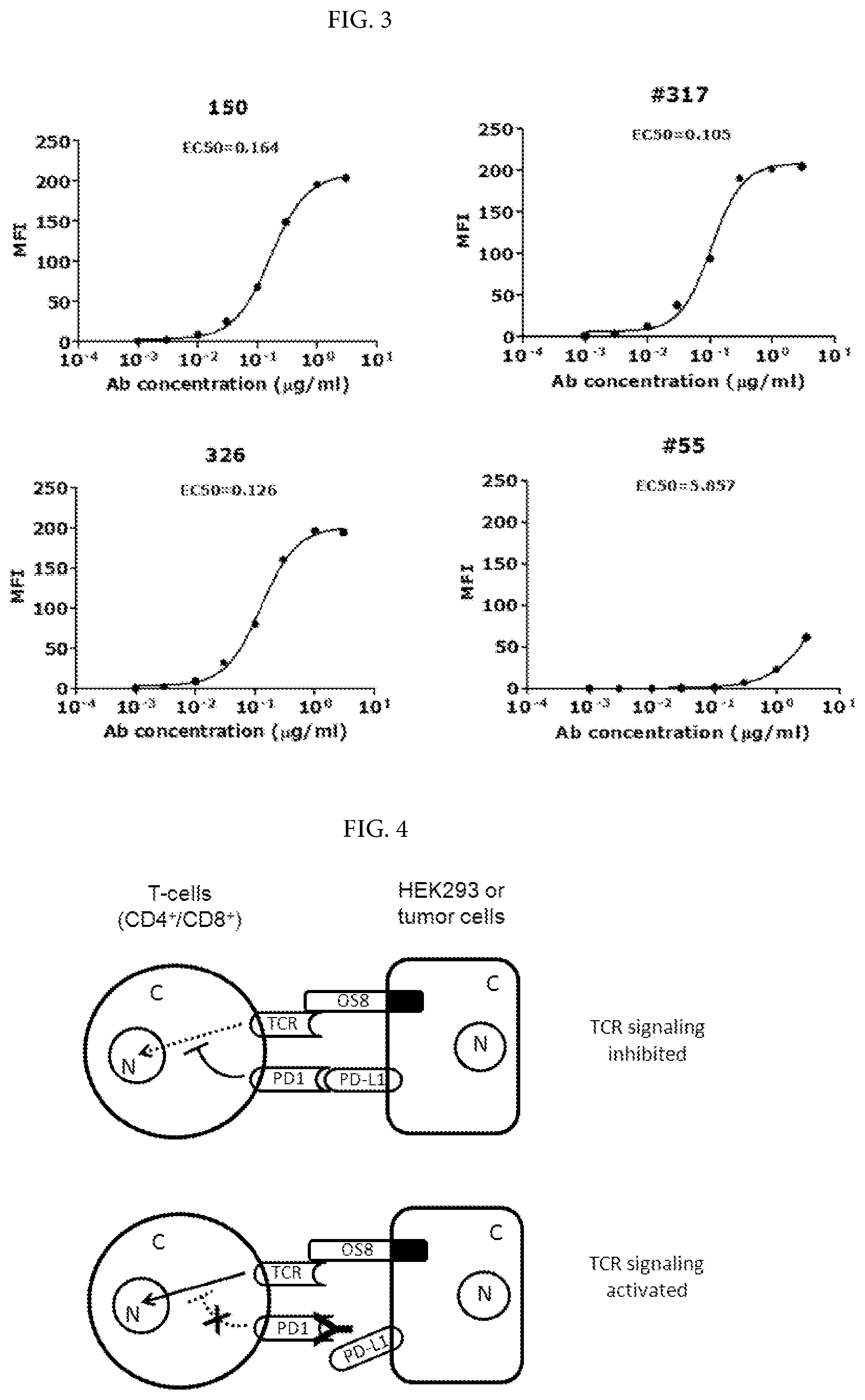
[0029] FIG. 2. Dose-dependent reaction curves of murine mAbs binding to human PD-1 in ELISA. The murine mAbs were indicated at top--left corner of each figure. MAb 317 and 517 share high degree of homology the variable region of heavy and light chains. The binding signal strength was indicated by direct OD.sub.450 readings. The antigen, PD-1/His, was coated at increasing concentrations up to 70 nanograms per well in a volume of 50 microliters. The method was described in Example 1.
[0030] FIG. 3. Dose-dependent reaction curve of murine mAbs binding to human PD-1 expressed on live cells by FACS analyses. Murine antibody codes and EC.sub.50 were indicated on each panel. MFI stands for mean fluorescence intensity. HuT78/PD-1 cells were suspended in 96-well plate at 5.times.10.sup.4 cells per well for FACS. PD-1 mAbs binding to the cell surface target and FACS detection were performed as described in Example 1.
[0031] FIG. 4. Schematic presentation of the cell co-culture systems used for assaying functional activities of anti-PD-1 mAbs. T-cells (either CD4.sup.+ or CD8.sup.+) represent HuT78/PD-1 or primary T-cells in PBMCs. TCR: T-cell receptor. N: nucleus. C: cytoplasm
[0032] FIG. 5. Dose-dependent reaction curve of murine mAb-induced IL-2 secretion in HuT78/PD-1 cells co-cultured with HEK293/OS8/PD-L1 cells. Baseline: Average IL-2 release induced by mIgGs at all tested concentrations. Top line: Highest IL-2 release based on regression calculation by Prizm Software.
[0033] FIG. 6A Histograms showing IFN-.gamma. secretion induced by anti-PD-1 mAbs in PBMCs (Donor-19) co-cultured with cell line HEK293/OS8/PD-L1. FIG. 6B Histograms showing IFN-.gamma. secretion induced by anti-PD-1 mAbs in PBMCs (Donor-20) co-cultured with cell line HEK293/OS8/PD-L1.
[0034] FIGS. 7A and 7B ADCC activities of anti-PD-1 mAbs by co-culture of effector cells (NK92MI/PD-1) and target cells (HuT78/PD-1). Means were calculated from two data points of the representative experiments. The mAbs were added to concentration of 10 .mu.g/ml. Experiment performed as described in Example 9.
[0035] FIG. 8. Mapping the binding epitopes of anti-PD-1 mAbs by ELISA (up-panel) and Western Blot (lower panel). Conditioned media containing WT or Mt PD-1 were used to assess binding activity by ELISA and Western Blot. ** indicates the AA residues to which the mAb binding activity reduced to 25-50% of WT PD-1. *** indicates the AA residues to which the mAb binding activity reduced below 25% of WT PD-1.
[0036] FIG. 9. IFN-.gamma. release induced by humanized anti-PD-1 mAbs in primary human PBMCs from different healthy donors, which were co-cultured with HEK293/OS8/PD-L1 cells.
[0037] FIG. 10. Cytotoxicity of NK92MI/PD-1 cells enhanced by humanized anti-PD-1 mAbs, hu317 (A) and hu326 (B). The target lung cancer cells, SK-MES-1/PD-L1, were co-cultured with the effector cells at the (T to E) ratio of 1 to 2, and assayed as described in Example 12.
[0038] FIG. 11. Individual tumor growth curves in three treatment groups, vehicle (PBS), human IgGs (huIgGs) and anti-PD-1 mAb (hu317-1/IgG4mt2). Each curve represents a tumor growth path, the tumor-bearing mice coded by numbers indicated on the right of each panel. Hep3B/OS8/PD-L1 cells (established from hepatocellular carcinoma line Hep3B) were seeded at Day 1, PBMCs were implanted at Day 15 and three doses of hu317-1/IgG4mt2 were injected at Day 18, 28 and 38, respectively. Methods described in Example 12.
DESCRIPTION OF PARTICULAR EMBODIMENTS OF THE INVENTION
[0039] PD-1 initiates inhibitory signaling in immune cells when engaged by its ligands, PD-L1 or PD-L2. In the cases of cancer outgrowth and viral infection, the activation of PD-1 signaling promotes immune tolerance, leading to the cancers or virus-infected cells escaping from immune surveillance and cancer metastasis or viral load increase. Inhibition of PD-1 mediated cellular signaling by therapeutic agents can activate immune cells including T-cells, B-cells and NK cells, and therefore enhance immune cell functions inhibiting cancer cell growth or viral infection, and restore immune surveillance and immune memory function to treat such human diseases.
[0040] The invention provides antibodies whose functions are antagonistic to the ligand-induced and PD-1-mediated cellular signaling in immune cells. Murine anti-PD-1 antibodies were humanized to a high degree of similarity to human antibodies in the framework regions. The full antibodies made in the modified human IgG4 variant format have a unique set of features in the aspects of effector functions and physicochemical properties. The disclosed anti-PD-1 antibodies are suitable for therapeutic uses in cancer treatment, controlling viral infections and other human diseases that are mechanistically involved in exacerbated immune tolerance.
Definitions
[0041] Unless the context indicates otherwise, the term "antibody" is used in the broadest sense and specifically covers antibodies (including full length monoclonal antibodies) and antibody fragments so long as they recognize PD-1. An antibody molecule is usually monospecific, but may also be described as idiospecific, heterospecific, or polyspecific. Antibody molecules bind by means of specific binding sites to specific antigenic determinants or epitopes on antigens. "Antibody fragments" comprise a portion of a full length antibody, generally the antigen binding or variable region thereof. Examples of antibody fragments include Fab, Fab', F(ab').sub.2, and Fv fragments; diabodies; linear antibodies; single-chain antibody molecules; and multispecific antibodies formed from antibody fragments.
[0042] Natural and engineered antibody structures are well known in the art, e.g. Strohl et al., Therapeutic antibody engineering: Current and future advances driving the strongest growth area in the pharmaceutical industry, Woodhead Publishing Series in Biomedicine No. 11, October 2012; Holliger et al. Nature Biotechnol 23, 1126 -1136 (2005); Chames et al. Br J Pharmacol. 2009 May; 157(2): 220-233.
[0043] Monoclonal antibodies (MAbs) may be obtained by methods known to those skilled in the art. See, for example Kohler et al (1975); U.S. Pat. No. 4,376,110; Ausubel et al (1987-1999); Harlow et al (1988); and Colligan et al (1993). The mAbs of the invention may be of any immunoglobulin class including IgG, IgM, IgE, IgA, and any subclass thereof. A hybridoma producing a mAb may be cultivated in vitro or in vivo. High titers of mAbs can be obtained in in vivo production where cells from the individual hybridomas are injected intraperitoneally into mice, such as pristine-primed Balb/c mice to produce ascites fluid containing high concentrations of the desired mAbs. MAbs of isotype IgM or IgG may be purified from such ascites fluids, or from culture supernatants, using column chromatography methods well known to those of skill in the art.
[0044] An "isolated polynucleotide" refers to a polynucleotide segment or fragment which has been separated from sequences which flank it in a naturally occurring state, e.g., a DNA fragment which has been removed from the sequences which are normally adjacent to the fragment, e.g., the sequences adjacent to the fragment in a genome in which it naturally occurs. The term therefore includes, for example, a recombinant DNA which is incorporated into a vector, into an autonomously replicating plasmid or virus, or into the genomic DNA of a prokaryote or eukaryote, or which exists as a separate molecule (e.g., as a cDNA or a genomic or cDNA fragment produced by PCR or restriction enzyme digestion) independent of other sequences. It also includes a recombinant DNA, which is part of a hybrid gene encoding additional polypeptide sequence.
[0045] A "construct" means any recombinant polynucleotide molecule such as a plasmid, cosmid, virus, autonomously replicating polynucleotide molecule, phage, or linear or circular single-stranded or double-stranded DNA or RNA polynucleotide molecule, derived from any source, capable of genomic integration or autonomous replication, comprising a polynucleotide molecule where one or more polynucleotide molecule has been linked in a functionally operative manner, i.e. operably linked. A recombinant construct will typically comprise the polynucleotides of the invention operably linked to transcriptional initiation regulatory sequences that will direct the transcription of the polynucleotide in the intended host cell. Both heterologous and non-heterologous (i.e., endogenous) promoters can be employed to direct expression of the nucleic acids of the invention.
[0046] A "vector" refers any recombinant polynucleotide construct that may be used for the purpose of transformation, i.e. the introduction of heterologous DNA into a host cell. One type of vector is a "plasmid", which refers to a circular double stranded DNA loop into which additional DNA segments can be ligated. Another type of vector is a viral vector, wherein additional DNA segments can be ligated into the viral genome. Certain vectors are capable of autonomous replication in a host cell into which they are introduced (e.g., bacterial vectors having a bacterial origin of replication and episomal mammalian vectors). Other vectors (e.g., non-episomal mammalian vectors) are integrated into the genome of a host cell upon introduction into the host cell, and thereby are replicated along with the host genome. Moreover, certain vectors are capable of directing the expression of genes to which they are operatively linked. Such vectors are referred to herein as "expression vectors".
[0047] An "expression vector" as used herein refers to a nucleic acid molecule capable of replication and expressing a gene of interest when transformed, transfected or transduced into a host cell. The expression vectors comprise one or more phenotypic selectable markers and an origin of replication to ensure maintenance of the vector and to, if desired, provide amplification within the host. The expression vector further comprises a promoter to drive the expression of the polypeptide within the cells. Suitable expression vectors may be plasmids derived, for example, from pBR322 or various pUC plasmids, which are commercially available. Other expression vectors may be derived from bacteriophage, phagemid, or cosmid expression vectors.
Additional Embodiments of the Invention
[0048] In specific embodiments the invention provides mouse monoclonal antibodies identified from screening murine hybridoma clones as disclosed herein.
[0049] In other embodiments the invention provides compositions of the following polynucleotide and protein sequences:
[0050] a) The cDNA sequence, SEQ ID NO 3, encoding the heavy chain variable region of murine mAb 317;
[0051] b) The protein sequence of the heavy chain variable region of murine mAb 317 or mu317_Vh (SEQ ID NO 4);
[0052] c) The cDNA sequence, SEQ ID NO 5, encoding the light chain variable region of murine mAb 317;
[0053] d) The protein sequence of the light chain variable region of murine mAb 317 or mu317_Vk (SEQ ID NO 6);
[0054] e) The cDNA sequence, SEQ ID NO 7, encoding the heavy chain variable region of murine mAb 326;
[0055] f) The protein sequence of the heavy chain variable region of murine mAb 326 or mu326_Vh (SEQ ID NO 8);
[0056] g) The cDNA sequence, SEQ ID NO 9, encoding the light chain variable region of murine mAb 326;
[0057] h) The protein sequence of the light chain variable region of murine mAb 326 or mu326_Vk (SEQ ID NO 10).
[0058] In one aspect, the invention provides compositions comprising complement determinant region (CDR) sequences, which mediate binding to the target antigens, PD-1, including the CDR sequences of mu317 and m326:
[0059] a) The CDR1 of mu317 heavy chain (mu317 H-CDR1) contains amino acid sequence of GFSLTSYGVH (SEQ ID NO 11);
[0060] b) The mu317 H-CDR2 contains amino acid sequence of VIWAGGSTNYNSALMS (SEQ ID NO 12);
[0061] c) The mu317 H-CDR3 contains amino acid sequence of ARAYGNYWYIDV (SEQ ID NO 13);
[0062] d) The CDR1 of mu317 light chain (mu317 L-CDR1) contains amino acid sequence of KASQSVSNDVA (SEQ ID NO 14);
[0063] e) The mu317 L-CDR2 contains amino acid sequence of YAFHRFT (SEQ ID NO 15);
[0064] f) The mu317 L-CDR3 contains amino acid sequence of HQAYSSPYT (SEQ NO 16);
[0065] g) The mu326 H-CDR1 contains amino acid sequence of GYTFTNYGMN (SEQ ID NO 17);
[0066] h) The mu326 H-CDR2 contains amino acid sequence of WINNNNGEPTYAEEFKG (SEQ ID NO 18);
[0067] i) The mu326 H-CDR3 contains amino acid sequence of ARDVMDY (SEQ ID NO 19);
[0068] j) The mu326 L-CDR1 contains amino acid sequence of RASESVDNYGYSFMH (SEQ ID NO 20);
[0069] k) The mu326 L-CDR2 contains amino acid sequence of RASNLES (SEQ ID NO 21);
[0070] l) The mu326 L-CDR3 contains amino acid sequence of QQSKEYPT (SEQ ID NO 22).
[0071] In another embodiment, the invention provides compositions comprising the sequences of the humanization monoclonal antibodies emanated from murine mAbs mu317 and mu326, incuding:
[0072] a) The humanization mAb hu317-4B6 comprises protein sequence of heavy chain variable region (Vh) as SEQ ID NO 24, which is encoded by
[0073] b) the cDNA of hu317-4B6_Vh (SEQ ID NO 23);
[0074] c) The humanization mAb hu317-4B6 also comprises protein sequence of light chain variable region (Vk) as SEQ ID NO 26, which is encoded by
[0075] d) the cDNA of hu317-4B6 (SEQ ID NO 25);
[0076] e) he humanization mAb hu326-4A3 comprises protein sequence of Vh as SEQ ID NO 28, which is encoded by
[0077] f) the cDNA of hu326-4A3-Vh (SEQ ID NO 27);
[0078] g) The humanization mAb hu326-4A3 also comprises protein sequence of Vk as SEQ ID NO 30, which is encoded by
[0079] h) the cDNA of hu326-4A3_Vk (SEQ ID NO 29);
[0080] i) The protein sequences of hu317-4B2_Vh (SEQ ID NO 43) and hu317-4B2_Vk (SEQ ID NO 44);
[0081] j) The protein sequences of hu317-4B5_Vh (SEQ ID NO 45) and hu317-4B5_Vk (SEQ ID NO 46);
[0082] k) The protein sequence of hu317-1_Vh (SEQ ID NO 48) and the cDNA encoding for hu317-1_Vh (SEQ ID NO 47);
[0083] l) The protein sequence of hu317-1_Vk (SEQ ID NO 50) and the cDNA encoding for hu317-1_Vk (SEQ ID NO 49);
[0084] m) The protein sequences of hu326-3B1_Vh (SEQ ID NO 51) and hu326-3B1_Vk (SEQ ID NO 52);
[0085] n) The protein sequences of hu326-3G1_Vh (SEQ ID NO 53) and hu326-3G1_Vk (SEQ ID NO 54);
[0086] o) The protein sequence of hu326-1_Vh (SEQ ID NO 56) and the cDNA encoding for hu326-1_Vh (SEQ ID NO 55);
[0087] p) The protein sequence of hu326-1_Vk (SEQ ID NO 58) and the cDNA encoding for hu326-1_Vk (SEQ ID NO 57);
[0088] q) The protein sequences of other humanization mAbs emanated from mu317 (SEQ ID NO 63-74);
[0089] r) The protein sequences of other humanization mAbs emanated from mu326 (SEQ ID NO 75-82);
[0090] In one aspect, the invention provides compositions comprising the CDR sequences of the humanization monoclonal antibodies. The CDRs may be shared among the same series of humanization mAbs, such as hu317 or hu326 (see Table 15-16). Non-redundant CDRs are listed below:
[0091] a) H-CDR1 sequence of GFSLTSYGVH (SEQ ID NO 31), shared throughout humanization mAbs hu317 and mu317 in the heavy chains;
[0092] b) H-CDR3 sequence of ARAYGNYWYIDV (SEQ ID NO 33), shared throughout humanization mAbs hu317 and mu317 in the heavy chains;
[0093] c) L-CDR1 sequence of KSSESVSNDVA (SEQ ID NO 34), shared throughout humanization mAbs hu317-4B2, hu317-4B5 and hu317-4B6 in the light chains;
[0094] d) L-CDR2 sequence of YAFHRFT (SEQ ID NO 35), shared throughout humanization mAbs hu317 and mu317 in the light chains;
[0095] e) L-CDR3 sequence of HQAYSSPYT (SEQ ID NO 36), shared throughout humanization mAbs hu317 and mu317 in the light chains;
[0096] f) H-CDR2 sequence of VIYADGSTNYNPSLKS (SEQ ID NO 32) in hu317-4B6_Vh;
[0097] g) H-CDR2 sequence of VIYAGGSTNYNPSLKS (SEQ ID NO 60) in hu317-4B2_Vh and hu317-4B5_Vh;
[0098] h) H-CDR2 sequence of VIWAGGSTNYNPSLKS (SEQ ID NO 59) in hu317-1_Vh;
[0099] i) L-CDR1 sequence of KASQSVSNDVA (SEQ ID NO 11) in hu317-1_Vk;
[0100] j) H-CDR1 sequence of GYTFTNYGMN (SEQ ID NO 37), shared throughout humanization mAbs hu326 and mu326 in the heavy chains;
[0101] k) H-CDR3 sequence of ARDVMDY (SEQ ID NO 39), shared throughout humanization mAbs hu326 and mu326 in the heavy chains;
[0102] l) L-CDR1 sequence of RASESVDNYGYSFMH (SEQ ID NO 40), shared throughout humanization mAbs hu326 and mu326 in the light chains;
[0103] m) L-CDR2 sequence of RASNLES (SEQ ID NO 41), shared throughout humanization mAbs hu326 and mu326 in the light chains;
[0104] n) L-CDR3 sequence of QQSKEYPT (SEQ ID NO 42), shared throughout humanization mAbs hu326 and mu326 in the light chains;
[0105] o) H-CDR2 sequence of WINNNNAEPTYAQDFRG (SEQ ID NO 38) in hu326_4A3_Vh;
[0106] p) H-CDR2 sequence of WINNNNGEPTYAQGFRG (SEQ ID NO 62) in the Vh of hu326_1 and other hu317 mAbs.
[0107] In another aspect, the invention provides particular binding epitopes of the humanized anti-PD-1 mAbs on the antigen, and functional use thereof. Six critical amino acid (AA) residues in PD-1 required for the ligand binding were mutated individually, and mutant and wild-type PD-1 proteins were used to assess the binding epitopes. The residue whose mutation significantly impaired the antibody binding is recognized as a key or significant binding epitope. Significant binding epitopes of mAbs hu317-4B5 and hu317-4B6 are K45 and I93 (AA numbering based on 2008 PNAS, 105:10483; equivalent to K58 and I106 in SEQ ID NO 2); and significant binding epitopes of mAbs hu326-3B1 and hu317-4A3 are I93, L95 and P97 (AA numbering based on 2008 PNAS, 105:10483; equivalent to I106, L108 and P110 in SEQ ID NO 2).
[0108] In a further aspect, the invention provides compositions comprising the constant region sequences of recombinant human IgG4 variants, which may be linked to the variable regions of the subject antibodies, including the humanized anti-PD-1 mAbs, which showed preferred effector functions and physicochemical properties. The sequences are as follows:
[0109] The constant region sequence of IgG4mt10 (SEQ ID NO 88);
[0110] a) A reference sequence of IgG4mt1 (SEQ ID NO 83);
[0111] b) A reference sequence of IgG4mt2 (SEQ ID NO 84);
[0112] c) A reference sequence of IgG4mt6 (SEQ ID NO 85);
[0113] d) A reference sequence of IgG4mt8 (SEQ ID NO 86);
[0114] e) A reference sequence of IgG4mt9 (SEQ ID NO 87).
[0115] In another embodiment, the invention provides methods for assaying anti-PD-1 antibody functions, using a plasmid expressing the recombinant fusion protein, OS8, to generate stable cell lines, HEK293/OS8/PD-L1 or HEK293/OS8/PD-L2, which co-expresses OS8 (a T cell-activating molecule) and a PD-1 ligand. The cell lines were used to engage T-cells and PBMCs by co-culture to assess the functionality of anti-PD-1 mAbs (see Example 3 and Example 4). Alternatively, another plasmid expressing the recombinant fusion protein, P3Z, was used to generate stable cell line, HuT78/P3Z, in which P3Z functions as molecular sensor and signal transduction mediator. When P3Z is engaged by PD-1 ligand, it will transmit intracellular signal to activate IL-2 release in the HuT78 cells. The systems may be used to assess inhibitory effect of anti-PD-1 mAbs (see Example 3).
[0116] In one aspect, the invention provides compositions comprising the amino acid sequences of the recombinant fusion proteins as follows:
[0117] a) Protein sequence of OS8 (SEQ ID NO 89);
[0118] b) Protein sequence of P3Z (SEQ ID NO 90).
[0119] In another aspect, the invention provides methods of generating the stable cell lines that express the recombinant fusion proteins described herein, and methods of using the system to quantitatively assay the functional activities of anti-PD-1 mAbs.
[0120] In another embodiment the invention provides polynucleotides encoding the subject proteins. The polynucleotides may be operably linked to a heterologous transcription regulating sequence for expression, and may be incorporated into vectors, cells, etc.
[0121] In another embodiment, the invention provides the murine anti-PD-1 antibodies and humanized version anti-PD-1 antibodies, including hu317-4B6, hu317-4B5, hu317-4B2, etc., and hu326-4A3, hu326-3B1, hu326-3G1, etc., having functions to suppress PD-1 mediated signal transduction, and to activate immune cells, which trigger a cascade of immune responses including cytokine secretion and cytotoxicity towards target cells such as cancer cells, and such functional use of the antibodies.
[0122] In one aspect, the invention provides humanized anti-PD-1 antibodies that activate several types of immune cells that express PD-1, including human T-cells, NK-cells and PBMCs, whose functions are to amplify the immune response signals, to mobilize immune system and to act as immune effector cells for clearance of cancer cells and viral infections, and such functional use of the antibodies.
[0123] In another aspect, the humanized anti-PD-1 mAbs are used as therapeutic agents to treat human diseases that are involved in suppression of immune cells by PD-1 mediated intracellular signaling, leading to disease progression, particularly cancers and viral infections.
[0124] The compositions of the invention are useful for the treatment of cancer, neurodegenerative and infectious, particularly viral, diseases and other conditions in which inappropriate or detrimental expression of the human PD-1 and/or is a component of the etiology or pathology of the condition. Hence, the invention provides methods for treating cancer or inhibiting tumor progression in a subject in need thereof with a subject anti-PD-1 protein. The invention further provides the use of subject polynucleotides for the manufacture of a medicament for treating cancer or inhibiting tumor progression in a subject.
[0125] The invention includes all combinations of the recited particular embodiments. Further embodiments and the full scope of applicability of the invention will become apparent from the detailed description given hereinafter. However, it should be understood that the detailed description and specific examples, while indicating preferred embodiments of the invention, are given by way of illustration only, since various changes and modifications within the spirit and scope of the invention will become apparent to those skilled in the art from this detailed description. All publications, patents, and patent applications cited herein, including citations therein, are hereby incorporated by reference in their entirety for all purposes.
EXAMPLES
Example 1
Generation of Anti-PD-1 Monoclonal Antibody
[0126] Anti-PD-1 monoclonal antibodies (mAbs) were generated based on conventional hybridoma fusion technology (Kohler and Milstein 1976 Eur J Immunol 6:511-519; de St Groth and Sheidegger 1980, J Immunol Methods 35:1-21; Mechetner 2007 Methods Mol Biol 378:1-13) with minor modifications. MAbs with high binding activities in enzyme-linked immunosorbent assay (ELISA) and fluorescence-activated cell sorting (FACS) assay were selected for further characterization
[0127] PD-1 Recombinant Protein for Immunization and Binding Assays
[0128] Expression plasmid containing full-length human PD-1 cDNA was obtained from Origene (Cat. No. SC117011, NCBI Accession No: NM_005018.1, Beijing, China). The extracellular domain consisting of amino acid (AA) 1-168 of PD-1 (SEQ NO.1, SEQ NO.2) was PCR-amplified, and subcloned in pcDNA3.1-based expression vector (Invitrogen, Carlsbad, Calif., USA) with C-terminus fused either to a His6 tag or to the .gamma.Fc domain of human IgG4 heavy chain, which resulted in two recombinant fusion protein expression plasmids, PD-1-EC/His and PD-1-EC/Fc (abbreviated as PD-1/His and PD-1/Fc). The schematic presentation of immunogen/antigen proteins were shown in FIG. 1. For the recombinant fusion protein production, PD-1/His and PD-1/Fc plasmids were transiently transfected into 293-F cells in 1-3 liters of medium (Invitrogen), and cultured for 5-7 days in a CO.sub.2 incubator equipped with rotating shaker. The supernatant containing the recombinant protein was collected and cleared by centrifugation at 15000 g for 30 minutes. PD-1/His was purified through immobilized metal affinity chromatography using Ni-Sepharose Fast Flow (Cat. No. 17531801, GE Lifesciences, Shanghai, China), followed by size exclusion chromatography using a HiLoad 16/60 Superdex 200 column (Cat. No. 17106901, GE Lifesciences, Shanghai, China). PD-1/Fc was purified using a Protein G Sepharose Fast Flow column (Cat. No. 17061805, GE Lifesciences). Both PD-1/His and PD-1/Fc proteins were dialyzed against phosphate buffered saline (PBS) and stored in -80.degree. C. freezer in small aliquots.
[0129] The cDNA coding for human PD-L1 was chemically synthesized by Genescript (Nanjing, China) based on the published sequence (NCBI Accession No. NM_014143). The PD-L2 expression plasmid was purchased from Origene (Cat. No. SC108873, NCBI Accession No. NM_025239.2, Beijing, China). Both cDNAs were cloned in pcDNA3.1/Hygromycin (Cat. No. V870-20, Invitrogen), and pcDNA3.1N5-His (Cat. No. V810-20, Invitrogen), respectively.
[0130] Stable Expression Cell Line
[0131] Stable cell lines expressing human PD-1, PD-L1 or PD-L2 were established by transfection of pcDNA3.1 plasmids containing PD-1, PD-L1 and PD-L2 to HUT78 (ATCC, Manassas, Va., USA) and HEK293 (ATCC), respectively, and followed by selection with medium containing 200 micrograms of hygromycin (Cat. No. 10687-010, Invitrogen) or 1 mg of G418 (Sigma) per milliliter. Single clones were isolated by conventional method, either limited dilution or picking up single colonies from culture-well surface. All clones were screened by Western blot and FACS analysis using anti-PD-1, PD-L1 and PD-L2 antibodies (Cat. No. 12-9969, 17-5983, 12-5888, eBioscience, San Diego, USA), respectively, and the top expression clones were selected for FACS binding assay to screen hybridoma monoclonal antibodies, or used in functional assays.
[0132] Immunization, Hybridoma Fusion and Cloning
[0133] Eight to twelve week-old Balb/c mice (from BEIJING HFK BIOCSIENCE CO., LTD, Beijing, China) were immunized subcutaneously with 100 ul of adjuvant (Cat. No. KX0210041, KangBiQuan, Beijing, China) containing 5 micrograms of PD-1/Fc. The immunization was conducted by two injections of the above immunogen with three weeks apart. Two weeks after the 2nd immunization, the mice sera were evaluated for PD-1 binding by FACS (following sections). The mice with high anti-PD-1 antibody titers in sera were selected and boosted intraperitoneally with 50 micrograms of PD-1/Fc in the absence of any adjuvant. Three days after boosting, the splenocytes were isolated and fused with the murine myeloma cell line, SP2/0 cells (ATCC), using standard techniques (Gefter, M. L. et al., 1977 Somat Cell Genet, 3:231-236).
[0134] Assess PD-1 Binding Activity of Antibodies by ELISA and FACS
[0135] The supernatants of hybridoma clones were initially screened by Enzyme-Linked Immuno-Sorbent Assay (ELISA) as described in "Flanagan, M. L. et al. 2007 Methods in Molecular Biology 378:33-52" with some modifications. Briefly, 50-200 nanograms of PD-1/His or PD-1/Fc protein in 50 microliters of phosphate buffered saline (PBS) were coated in 96-well plate (Shenzhen JinCanHua Industry Co., Ltd, Shenzhen, China) on per well base. The HRP-linked anti-mouse IgG antibody (Cat. No. 7076S, Cell Signaling Technology, USA and Shanghai, China) and chemiluminescent reagent (Cat. No. PA107-01, TIANGEN, China) were used to detect and develop the ELISA signal, which were read out by a plate reader (PHREAstar FS, BMG LABTECH, Germany) at wavelength of 450 nm. The ELISA-positive antibody producer clones were further verified by fluorescence-activated cell sorting (FACS) using a conventional method. PD-1 stable expression cell lines, HuT78/PD-1 (10.sup.5 cells/well), described above, was stained with supernatants from anti-PD-1 hybridomas in V-bottom 96-well plates (Cat. No. 3897, Corning, USA and Shanghai, China). To block human Fc receptors, cells were pre-incubated with human IgG (20 .mu.g/ml) (Cat. No. H11296, LifeHolder, USA and Shanghai, China). PD-1 antibodies were detected with Dylight.TM. 649-labelled goat anti-mouse IgG antibody (Cat. No. 405312, Biolegend, San Diego, USA) and cell fluorescence was monitored using a flow cytometer (Guava easyCyte 8HT, Merck-Millipore, USA and Shanghai, China).
[0136] The conditioned media of hybridoma cells that showed positive signal in both ELISA and FACS assay were subjected to functional assays to identify antibodies with good functional activity in human immune cell-based assays (herein). The antibodies with positive functional activity were further subcloned and characterized.
[0137] Subcloning and Adaptation to Serum Free or Low Serum Medium
[0138] The positive hybridoma clones from primary screening through ELISA, FACS and functional assays were subcloned by the conventional method of limited dilution. Each of the positive clones was plated out in a 96-well plate, cultured in RPMI1640 medium (Cat. No. SH30809.01B, Hyclone, Shanghai, China) with 10% fetal bovine serum (FBS, Cat. No. SH30084.03, Hyclone, Beijing, China) in CO.sub.2 incubator. Three subclones from each limited dilution plate were selected and characterized by FACS and functional assays. The subclones selected through functional assays were defined as monoclonal antibody. The top subclones were adapted for growth in the CDM4MAb medium (Cat. No. SH30801.02, Hyclone) with 1-3% FBS.
[0139] Expression and Purification of Monoclonal Antibodies
[0140] Either murine monoclonal antibody-producing hybridoma cells or recombinant antibody plasmids-transfected 293-F cells (Cat. No. R79007, Invitrogen) were cultured in CDM4MAb medium (Cat. No. SH30801.02, Hyclone) or Freestyle293 Expression medium (Cat. No. 12338018, Invitrogen), respectively, in a CO.sub.2 incubator at 37.degree. C. for 5 to 7 days. The conditioned medium was collected through centrifugation at 10,000 g for 30 minutes to remove all cells and cell debris, and filtrated through a 0.22 .mu.m membrane before purification. Murine or recombinant antibodies were applied and bound to a Protein A column (Cat. No. 17127901, GE Life Sciences) following the manufacturer's guidance, washed with PBS, eluted in the buffer containing 20 mM citrate, 150 mM NaCl, pH3.5. The eluted materials were neutralized with 1M Tris pH8.0, and usually contained antibodies of above 90% purity. The Protein A-affinity purified antibodies were either dialyzed against PBS or further purified using a HiLoad 16/60 Superdex200 column (Cat. No. 17531801, GE Life Sciences) to remove aggregates. Protein concentrations were determined by measuring absorbance at 280 nm or by Bradford assay (Cat. No. 1856210, Thermo Scientific, Rockford, Ill., USA) using bovine IgG of defined concentration (Cat. No. 23212, Thermo Scientific) as the standards. The purified antibodies were stored in aliquots in -80.degree. C. freezer.
Example 2
Comparison of Binding Activities Among Anti-PD-1 Antibodies
[0141] Through screening thousands of hybridomal clones we identified some top monoclonal antibodies (mAb), which bind to human PD-1 with high specificity and strength. As shown in ELISA assay (FIG. 2), three of the top antibodies elicited such binding strength and specificity. FACS analysis results demonstrated the selected monoclonal antibodies bind to the native PD-1 proteins expressed on cell surface. Murine mAb317 (mu317), mu326 and mu150 showed concentration-dependent binding activity, and their binding EC.sub.50 (Effective concentration at 50% activity) was significantly lower than that of the control mu55 (FIG. 3).
[0142] Assess mAb Binding Affinity by Surface Plasmon Resonance (SPR)
[0143] The mAbs with high binding activities in ELISA and FACS, as well as with potent functional activities in the cell-based assays (herein) were examined for their binding kinetic constant in real time binding reactions. Murine anti-PD-1 mAbs were purified from hybridoma supernatants using protein A Flow column (Cat. No. 17531801, GE Life Sciences) followed by exclusion chromatography using a HiLoad 16/60 Superdex200 column (Cat. No. 17106901, GE Life Sciences). The purified anti-PD-1 antibodies were concentrated to 0.5-1 mg/mL in PBS and stored in aliquots in -80.degree. C. freezer.
[0144] For determining binding affinities of PD-1 mAbs, SPR measurements were performed in HBS-N buffer (10 mM HEPES pH 7.4, 0.15 M NaCl, 3 mM EDTA, 0.005% v/v surfactant P20, GE Healthcare) using the BIAcore.TM. T-200 instrument (GE Life Sciences). Anti-mouse Fc CM5 biosensor chip (GE Healthcare) was generated using a standard primary amine coupling protocol. PD-1 mAbs at 0.3 .mu.g/ml were captured on anti-mouse Fc surface for 1 min at 10 .mu.l/min. PD-1/Fc in a serial dilutions from 3.3 nM to 120 nM was injected over antibody-bound surface for 3 min at 30 .mu.l/min followed by a 10 min dissociation phase. Association rates (K.sub.a or k.sub.on) and dissociation rates (K.sub.d or k.sub.off) were calculated using the one-to-one Langmuir binding model (BIA Evaluation Software, GE Life Sciences). The equilibrium dissociation constant (KD) was calculated as the ratio k.sub.off/k.sub.on.
[0145] As shown in Table 1, both mu326 and mu517, a cognate sequence family member related to mu317, have a sub-nanomolar K.sub.D equaling to 0.324 nM and 0.289 nM, respectively, which is significantly better than that of mu134. The K.sub.on rate was similar among the three mAbs listed in Table 1, yet the K.sub.off rate was significantly different, much faster dissociation rate was observed in mu134.
TABLE-US-00005 TABLE 1 Binding constant of certain top antibodies mAbs K.sub.on (M.sup.-1, s.sup.-1) K.sub.off (s) K.sub.D (M) mu326 2.4 .times. 10.sup.5 7.79 .times. 10.sup.-5 3.24 .times. 10.sup.-10 mu517 1.96 .times. 10.sup.5 5.66 .times. 10.sup.-5 2.89 .times. 10.sup.-10 mu134 1.1 .times. 10.sup.5 3.69 .times. 10.sup.-4 3.35 .times. 10.sup.-9
Affinity Determination of Anti-PD-1 Fabs by SPR
[0146] Anti-PD-1 mAbs were converted into Fab version by PCR to fuse the variable regions of heavy and light chains to the N-terminus of human IgG2-CH1 and constant region of kappa chain, respectively, and subcloned in pcDNA3.1 vector (Invitrogen). Both expression vectors were co-expressed in 293-F cells using a transient transfection protocol similar to the transient expression of whole antibodies. Briefly, the Fab kappa chain was PCR amplified and subcloned in pcDNA3.1-based expression vector (Invitrogen, Carlsbad, Calif., USA). In a separate plasmid, the heavy chain variable region (VH) together with the CH1 coding sequence from human IgG2 was fused with a C-terminal c-Myc-His8 tag by overlapping PCR, and then subcloned in the expression vector. The C232S and C233S (Kabat residue numbering, Kabat et al. Sequence of proteins of immunologic interest, 5.sup.th ed Bethesda, Md., NIH 1991) mutations were introduced in the IgG2 heavy chain to prevent disulfide bond exchange and stabilize human IgG2 in the IgG2-A conformation (Lightle et al. 2010 Protein Sci 19(4): 753-762). Both constructs contained a signal peptide upstream of the Fab mature sequences. Secreted expression of Fab was achieved by co-transfection of above 2 plasmids into 293-F cells and cell culture supernatants were harvested 6-7 days post transfection. His8-tagged Fabs were purified from cell culture supernatants using a Ni-sepharose Fast Flow column (Cat. No. 17531801, GE Life Sciences) followed by size exclusion chromatography using a HiLoad 16/60 Superdex200 column (Cat. No. 17106901, GE Life Sciences). The purified Fabs were concentrated to 0.5-5 mg/mL in PBS and stored in aliquots in -80.degree. C. freezer.
[0147] For affinity determinations of anti-PD-1 Fabs, SPR assays were used with the BIAcore.TM. T-200 instrument (GE Life Sciences). Briefly, human PD-1/His or cynomolgus monkey PD-1/His was coupled to activated CM5 biosensor chips (Cat. No. BR100530, GE Life Sciences) to achieve approximately 100-200 response units (RU), followed by blocking un-reacted groups with 1M ethanolamine. Fab samples of increasing concentration from 0.12 nM to 90 nM were injected in the SPR running buffer (10 mM HEPES, 150 mM NaCl, 0.05% Tween20, pH7.4) at 30 .mu.L/minute, and binding responses on human PD-1/His or monkey PD-1/His were calculated by substracting of RU from a blank flow-cell. Association rates (k.sub.on) and dissociation rates (k.sub.off) were calculated using the one-to-one Langmuir binding model (BIA Evaluation Software, GE Life Sciences). The equilibrium dissociation constant (K.sub.d) was calculated as the ratio k.sub.off/k.sub.on.
[0148] The SPR-determined binding affinities of anti-PD-1 Fabs were listed in Table 18. Each anti-PD-1 Fab bound with high affinity (K.sub.d=0.15-1 nM) to human PD-1. All Fabs, except 326-3G1, bound with slightly lower but comparable (within 5 fold in K.sub.d) affinities to cynomolgus monkey PD-1.
Example 3
Functional Activity of Anti-PD-1 Antibodies in Human T Cells
[0149] Generation of Stable Cell Lines
[0150] Retroviral packaging cell line PT67, human T cell lines HuT78 and HEK293 were obtained from the American Type Culture Collection (ATCC, Rockville, Md.). A HuT78 subline HuT78/PD-1 that expresses PD-1 was generated by retroviral transduction using pFB-neo vector (Strategene/Agilent Tech, Santa Clara, Calif.) containing the PD-1 gene, according to the protocol described previously (Zhang et al. 2005 Blood 106: 1544-1551). The T cell engager, a membrane-anchored chimeric Ab (OS8), was constructed by fusing the single chain variable fragment (scFv) of an anti-human CD3 mAb OKT3 (Kipriyanov et al. 1997, PEDS 10:445-453) to the C-terminal domain (113-220) of mouse CD8.alpha. (NCBI Accession No: NP_001074579.1) which includes hinge, transmembrane and cytoplasmic domains. By doing so, anti-CD3 scFv is anchored to cell surface as a T cell activator. Human PD-L1, PD-L2 and OS8 cDNAs were sub-cloned into pcDNA3.1 vector. Stable cell lines HEK293/OS8/PD-L1, Hep3B/OS8/PD-L1 and HEK293/OS8/PD-L2 that co-express both OS8 and PD-L1 or PD-L2 cDNAs were generated by co-transfection of HEK293 and Hep3B cells (ATCC) with the paired plasmids, followed by hygromycin or G418 selection for 10-14 days. Cell lines were then cloned by limiting dilution as described previously (Fuller S A, et al. Curr Protoc Mol Biol. Chapter 11:Unit 11.8, 2001). Chimeric PD-1 receptor, named P3Z, was constructed by fusing the extracellular and transmembrane domains) of human PD-1 to the cytoplasmic domain of human CD3.zeta. chain (NCBI Accession No. NP_932170.1). P3Z-coding cDNA sequence was cloned into pFB-neo and delivered into HuT78 cells via retroviral transduction to generate HuT78/P3Z cells.
[0151] Determination of PD-1 Antibody Functions by IL-2 Release in HuT78/PD-1 Cells
[0152] To determine whether anti-PD-1 antibodies can block the interaction of PD-L1-induced PD-1 signaling, HuT78/PD-1 cells (1.5.times.10.sup.4 cells per well in 96-well plate) were pre-incubated with hybridoma supernatants or PD-1 antibodies for 15 minutes prior to co-culture with HEK293/OS8/PD-L1 or HEK293/OS8/PD-L2 cells (4.times.10.sup.4 per well) in a flat bottom plate fed with 200 .mu.l of RPMI1640 growth medium per well at 37.degree. C. After 16-18 hours, supernatants of the co-culture were collected. IL-2 was assayed by ELISA using human IL-2 Ready-Set-Go! ELISA kits (Cat. No. 88-7025, eBiosciences, San Diego, Calif.). In this assay, blockade of PD-1 signaling with anti-PD-1 antibodies resulted in enhanced TCR signaling and IL-2 production (FIG. 4).
[0153] As shown in FIG. 5 and Table 2, murine anti-PD-1 mAb, mu317 and mu326, elicited significantly higher functional activity than mu30, inhibiting PD-L1-induced PD-1 signaling which leads to increased IL-2 secretion. Both had higher IL-2 secretion (top line, Table 2), 675 and 634 pg/ml, respectively, and both had lower EC.sub.50 (Effective concentration of mAb at 50% level of IL-2 secretion induction) than mu30 antibody.
TABLE-US-00006 TABLE 2 IL-2 release induced by anti-PD-1 mAbs in HuT78/PD-1 cells co-cultured with HEK293/OS8/PD-L1 cells Antibody Baseline (pg/ml) Top line (pg/ml) EC.sub.50 (.mu.g/ml) mu30 95 527 0.229 mu317 95 675 0.083 mu326 95 634 0.053 mIgGs 95 N/A N/A Baseline: Average IL-2 release induced by mlgGs at all tested concentrations, see FIG.4 Top line: Highest IL-2 release based on regression calculation by Prizm Software, FIG. 4. N/A: Not applicable
[0154] Not only did the engagement of HuT78/PD-1 cells by anti-PD-1 mAbs block PD-L1 induced T-cell activation, but also blocked PD-L2 induced IL-2 release. Table 3 presented the data showing mu317 and mu326 had much higher potency in activating the T-cells as indicated by the parameters (EC.sub.50) of IL-2 secretion than those of mu476.
TABLE-US-00007 TABLE 3 IL-2 release induced by anti-PD-1 mAbs in HuT78/PD-1 cells co-cultured with HEK293/OS8/PD-L2 cells Antibody Baseline (pg/ml) Top line (pg/ml) EC.sub.50 (.mu.g/ml) 476 180 599 0.183 317 192 563 0.032 326 218 635 0.038 Baseline: Average IL-2 release induced in the lower tail part of the sigmoid reaction curve. Top line: Average IL-2 release induced at the plateau part of the sigmoid reaction curve
[0155] Determination of PD-1 Antibody Functions by Reverse Signaling of IL-2 Release in HuT78/P3Z Cells
[0156] In chimeric receptor P3Z, PD-1 signaling domain was replaced with the cytoplasmic domain of CD3.zeta.. Therefore, P3Z mediates activation upon engagement with PD-L1, rather than inhibition as original PD-1 receptor. In this assay, HuT78/P3Z cells (3.times.10.sup.4/well) were pre-incubated with hybridoma supernatants or PD-1 antibodies for 15 minutes prior to co-culture with HEK293/PD-L1 or HEK293/PD-L2 cells (5.times.10.sup.4/well) in 96-well flat bottom plates (a total volume of 200 .mu.l/well) at 37.degree. C. After 16-18 hours, supernatants were collected and IL-2 production was assayed by ELISA as described above.
[0157] The functional activity of murine anti-PD-1 mAbs was further confirmed by direct read-out of T-cell activation in reverse signaling assay described above. Consistent to the result described above, mu317 and mu326 had best functional activity among the mAbs we screened. As shown in Table 4 and Table 5, mu317 and mu326 were much more potent than one of the low activity mAbs, mu37, both in terms of IC.sub.50 and maximum inhibition.
TABLE-US-00008 TABLE 4 Inhibition of IL-2 secretion by anti-PD-1 mAbs in HuT78/P3Z cells co-cultured with HEK293/PD-L1 cells Antibody IC.sub.50 (.mu.g/m10 Max inhibition, % 37 0.287 86.9 317 0.083 99.3 326 0.039 97.6 Maximum inhibition was calculated as percentage (%) of inhibition with anti-PD-1 mAbs added to the highest level of 10 .mu.g/ml in culture
TABLE-US-00009 TABLE 5 Inhibition of IL-2 secretion by anti-PD-1 mAbs in HuT78/P3Z cells co-cultured with HEK293/PD-L2 cells Antibody IC.sub.50 (.mu.g/m10 Max inhibition, % 37 0.127 43.3 317 0.020 94.3 326 0.018 93.4 Maximum inhibition was calculated as percentage (%) of inhibition with anti-PD-1 mAbs added to the highest level of 10 .mu.g/ml in culture
Example 4
Activation of IFN-.gamma. secretion by anti-PD-1 mAb in Primary Human PBMCs Co-Cultured with HEK293/OS8/PD-L1 Cells
[0158] To verify if the selected top mAbs against PD-1 also exert functional effect on primary human immune cells, we assayed the antibody function by using freshly isolated peripheral blood mononuclear cells (PBMCs), which are mainly consisted of T-cells (50-70%), B-cells and NK cells (15-30%), and monocytes (2-10%). Human PBMCs were isolated from healthy donors by density gradient centrifugation using ficoll lymphocyte separation medium (Histopaque-1077; Sigma-Aldrich, MO) according to the manufacturer's instructions. All the human blood collection followed the Internal Procedure of Beigene. PBMCs were then stimulated with anti-CD3 mAb (40 ng/mL) OKT3 (Cat. No. 16-0037, eBioscience, CA) for 3 days prior to assay. FACS analysis (Example 1) showed that PD-1 expression on the activated PBMCs (primarily T cells) was increased to variable degree dependent on individual donors (Table 6). To determine the response of pre-activated T cells to PD-1 ligand-positive tumor cells upon engagement TCR/CD3 complex, PBMCs (1.times.10.sup.4) were co-cultured with either HEK293/OS8/PD-L1 or HEK293/OS8/PD-L2 cells (3.times.10.sup.4) in 96-well flat-bottom plates for 15-18 hours. Cell-free supernatants were assayed for IFN-.gamma. level by ELISA using Ready-Set-Go! ELISA kits (Cat. No. 88-7316, eBiosciences), which is the most prominent indicator of T-cell activation, as well as of other immune cell activation (Thakur A. et al. 2012 Vaccine, 30:4907-4920).
TABLE-US-00010 Percent gated PD-1 staining positive cells versus total PMBCs stained PBMCs and treatment Donor-3 Donor-4 PBMCs, not stimulated/ 12.0% 3.2% stained by PD-1 Ab PBMCs, stimulated/ 40.0% 38.1% stained by PD-1 Ab PBMCs, not stimulated/ .ltoreq.0.5% .ltoreq.0.5% stained by control Ab PBMCs, stimulated/ .ltoreq.0.5% .ltoreq.0.5% stained by control Ab Stimulation: freshly isolated PBMCs were cultured for 3 days in presence of anti-CD3 antibody, OKT3, and IL-2. Without stimulation: fresh PBMCs subjected to antibody staining and FACS analysis.
[0159] FIG. 6 demonstrated that presence of mAbs mu317 and mu326 in the co-culture of pre-activated PBMCs and HEK293/OS8/PD-L1 cells resulted in increasing IFN-65 accumulation in a dose-dependent manner. Although the base level of IFN-.gamma. with control murine IgG treatment varies among different donors, the increase of IFN-.gamma. secretion in PBMCs treated by mu317 or mu326 is statistically significant in the range of 0.1 to 10 .mu.g/ml of antibody treatment. Comparing to the corresponding level of mIgG-treated PBMCs, IFN-.gamma. secretion induced by mu317 and mu326 between the 0.1 to 10 .mu.g/ml concentration levels increased 2.5 to 3.2 fold in PBMCs from Donor-19, and increased 1.4 to 2.3 fold in PBMCs of Donor-20, respectively.
Example 5
Activation of Human NK Cells by Anti-PD1 mAbs
[0160] Stable Cell Lines for Functional Assay in NK Cells
[0161] Primary human NK cells were reported previously to express PD-1 protein in response to IL-2 treatment and inhibiting PD-1-mediated signaling enhanced cytotoxicity of NK cells (2010 Blood, 116: 2286). For quantitative assay of functional effect exerted by anti-PD-1 mAbs in NK cells, human NK cell line NK92MI (ATCC) and lung cancer cell line SK-Mes-1 (ATCC) were engineered to stably express human PD-1 and PD-L1, respectively, by retroviral transduction according to the protocols described previously (Zhang et al. 2005, Blood 106: 1544-1551, Zhang et al. 2006, Cancer Res, 66: 5927). The two stable cell lines were named as NK92MI/PD-1 and SK-Mes-1/PD-L1
[0162] Anti-PD-1 Abs Promote IFN-.gamma. Production and Secretion in NK92MI/PD-1 Cells
[0163] Functional activity of the anti-PD-1 mAbs on NK cells was assayed by quantitative measurement of IFN-.gamma. production and secretion in NK92MI/PD-1 cells which were co-cultured with lung cancer cell line SK-MES-1/PD-L1 at ratio of 1 to 2 in 96-well flat-bottom plate with total of 6.times.10.sup.4 cells per well. The anti-PD-1 mAbs were added to NK92MI/PD-1 cells 15 minutes before the co-culture started, then the cells were co-cultured for overnight in CO.sub.2 incubator. Cell-free supernatants were assayed for IFN-.gamma. level by ELISA as described in Example 4.
[0164] All anti-PD-1 mAbs trigged significant increase of IFN-.gamma. production from the baseline with low concentration of antibody treatment to top line with high concentration of antibody treatment. The two top antibodies, mu317 and mu326, had lower EC.sub.50, than the comparison antibody 5C, indicating they have more potent activating effect to the NK cells (Table 7).
TABLE-US-00011 TABLE 7 IFN-.gamma. secreted in medium (pg/ml) by NK92MI/PD-1 cell in presence of anti-PD-1 mAb and Ski-MES-1/PD-L1 cells Antibody Baseline (pg/ml) Top line (pg/ml) EC.sub.50 (.mu.g/ml) 317 28 532 0.40 326 15 509 0.20 5C 20 535 1.17 Baseline: Average IFN-.gamma. release induced in the lower tail part of the sigmoid reaction curve. Top line: Average IFN-.gamma. release induced at the plateau part of the sigmoid reaction curve
[0165] Anti-PD-1 Antibody Enhances Cancer Cell Killing Mediated by NK92MI/PD-1 Cells
[0166] Cytotoxicity of NK92MI/PD-1 cells against SK-MES-1/PD-L1 cells was determined by lactate dehydrogenase (LDH) release assay using the CytoTox 96 Non-Radioactive Cytotoxicity Assay kit (Promega, Madison, Wis.). In brief, NK92MI/PD-1 cells (10.sup.5) were pre-incubated with anti-PD-1 mAbs at final concentrations within the range of 0.004-10 .mu.g/ml for 15 minutes, and SK-MES-1/PD-L1 cells (2.times.10.sup.4) were added to the immune cell culture in a 96-well V-bottom plate at an effector to tumor cell (E:T) ratio of 5:1, then co-cultured for 5 hours. The complete tumor cell lysis was set as maximum cell killing, the LDH-release assay readout of each sample was calculated as percentage of maximum cell killing. The cell killings (%) of all samples were normalized cross the plates using 10% of baseline as the common standard.
[0167] In the specific cytotoxicity assay set as above, the selected anti-PD-1 mAbs caused a net tumor cell killing (=top line-baseline) ranging from 19% to 20.2% at high concentration of mAb input. Mu317 and mu326 had lower EC.sub.50 than mu336, indicating better potency to trigger NK92MI/PD-1 cell-mediated tumor cell killing (Table 8).
TABLE-US-00012 TABLE 8 Cytotoxicity of NK92MI/PD-1 cells towards tumor cells induced by anti-PD-1 mAb Antibody Baseline (%) Top line (%) EC.sub.50 (.mu.g/ml) 317 10 29.06 0.50 326 10 30.19 0.37 336 10 29.72 1.52 Baseline: Percent of tumor cells killed not due to the effect of anti-PD-1 mAbs, normalized to 10% cross plates. Top line: Average percent of tumor killed in presence of highest concentrations of mAbs, i.e. 3 .mu.g/ml and 10 .mu.g/ml
Example 6
Cloning and Sequence Analyses of PD-1 mAbs
[0168] The murine hybridoma clones secreting a specific mAb were cultured to a density of 3 to 10.times.10.sup.6 cells in a 100 mm-tissue culture dish, and the cells were harvested through centrifugation at 1500 rpm in a swing bucket rotor. Total cellular RNA was isolated using Ultrapure RNA kit (Cat. No. CW0581, CWBIOTECH, Beijing, China) following the manufacturer's protocol. The RNA was resuspended in double-deionized water, concentration measured by NanoDrop (ThermoFisher, Shanghai, China).
[0169] PCR primers used for mAb cDNA cloning were synthesized by Invitrogen (Beijing, China) based on the sequences reported previously (Brocks et al. 2001 Mol Med 7:461-469). The 1.sup.st strand cDNA was synthesized using reverse transcriptase (Cat. No. AH301-02, Transgen Biotech, Beijing, China). PCR amplification of specific mAb cDNA was performed using PCR reagent kit (Cat. No. Ap221-12, TransGen Biotech, Beijing, China) and following manufacturer's protocol. The PCR product was either directly sequenced by service provider (GeneWiz, Beijing, China) or subcloned into a pCR vector (Invitrogen), subsequently sequenced (GeneWiz).
[0170] The protein sequences of murine mAbs were analyzed by sequence homology alignment. MAbs were grouped based on sequence homology and epitope-mapping results (Example 13). Complement determinant regions (CDRs) were identified based on Kabat (Wu, T. T. and Kabat, E. A., 1970 J. Exp. Med. 132: 211-250) and IMGT system (Lefranc M.-P. et al., 1999 Nucleic Acids Research, 27, 209-212) by sequence annotation and by internet-based sequence analysis (http ://www.imgt.org/IMGT_vquest/share/textes/index.html and http://www.ncbi.nlm.nih.gov/igblast/). As shown in Table 9, the CDRs of mu317 and mu326 are very different in sequence length and identity.
TABLE-US-00013 TABLE 9 CDRs of mu317 and mu326 SEQ SEQ SEQ ID ID ID MAbs CDR1 NO CDR2 NO CDR3 NO mu317, HC GFSLTSYGVH 11 VIWAGGSTNYNSALMS 12 ARAYGNYWYIDV 13 mu317, HC KASQSVSNDVA 14 YAFHRFT 15 HQAYSSPYT 16 mu326, HC GYTFTNYGMN 17 WINNNNGEPTYAEEFKG 18 ARDVMDY 19 mu326, HC RASESVDNYGYSFMH 20 RASNLES 21 QQSKEYPT 22 Note: CDRs in bold face are based on Kabat system; CDRs underlined are based IMGT system.
Example 7
Humanization of the Murine mAbs
[0171] Simulation of Antibody 3D Structure
[0172] The three dimensional structures were simulated for variable domains of mu317 and mu326 in order to identify framework residues that might be important for supporting CDR loop structures. Potentially important framework residues were kept as the original murine residues in the first round antibody humanization. The previously established structural modeling method for antibodies (Morea et al. Methods 2000 20:267-279) was adopted to simulate 3D structure of anti-PD-1 mAbs based on the known canonical structures of antibodies (Al-Lazikani et al. 1997 Journal of Molecular Biology 273:927-948). Briefly, the sequence of each variable domain (Vk and Vh) of murine antibody was blasted in the PDB database (Protein Data Bank, http://blast.ncbi.nlm.nih.gov/) to identify the most homologous antibody sequence with known high resolution structure (resolution less than 2.5 angstrom). Selected structure templates for modeling mu317 and mu326 (listed in Table 10) had the same classes of canonical loop structures in L-CDR1, L-CDR2, L-CDR3, H-CDR1, and H-CDR2 to the target antibodies to be modeled. If the templates for the Vk and the Vh came from different immunoglobulins, they were packed together by a least-squares fit of the main chain atoms to form a hybrid structure of Vk-Vh interface residues, which was used as the templates for structural homology modeling by Swiss-model program (Kiefer et al. 2009 Nucleic Acids Research 37, D387-D392). Certain side chain conformation was adjusted while the main chain conformations were retained. At the sites where the parent structure and the modeled structure had the same residue, the side chain conformation was retained. At sites where the residues were different, side chain conformations were modeled on the basis of template structure, rotamer libraries and packing considerations. After homology modeling, PLOP program (Jacobson et al. 2002 Journal of Physical Chemistry 106:11673-11680) was used to refine the homology models to minimize all-atom energy and optimize Vk and Vh interface. This step was performed to improve the stereochemistry, especially in those regions where segments of structures coming from different antibodies had been joined together.
TABLE-US-00014 TABLE 10 Structure templates used in antibody structure simulations Antibody PDB code of template structure Sequence Sequence chain (PDB template for H-CDR3) identity similarity mu317 Vk 3MXV 87% 92% mu317 Vh 3VFG 83% 91% mu326 Vk 1EJO 92% 94% mu326 Vh 1NCA 88% 90% 317-1 Vk 4HJJ 90% 95% 317-1 Vh 3VFG (1AY1) 75% 87% 326-1 Vk 1EJO 87% 92% 326-1 Vh 3T2N (3CXD) 84% 86%
[0173] The structures were also simulated for CDR-grafted 317-1 and 326-1 in order to guide further rounds of antibody engineering to enhance the extents of humanization and/or enhance antibody stabilities. The selected structure templates are also listed in Table 10. The structure simulations were done in a similar way to above procedure, except that the possible conformations of H-CDR3 were taken from PDB templates 1AY1 for 317-1 and 3CXD for 326-1, respectively, which contained H-CDR3s of similar size and torso region. Energy minimization for grafted H-CDR3 residues was done using PLOP.
[0174] Humanization
[0175] For humanization of the anti-PD-1 mAbs, we searched human germline IgG genes homologous to the cDNA sequences of mu317 and mu326 variable regions by blasting the human immunoglobulin gene database in IMGT (http://www.imgt.org/IMGT_vquest/share/textes/index. html) and NCBI (http://www.ncbi.nlm.nih.gov/igblast/) websites. The human IGVH and IGV.kappa. with high homology to the PD-1 mAbs were selected as the template for humanization.
[0176] Humanization was carried out in principle by CDR-grafting. In the 1.sup.st round of humanization, mutations from murine to human amino acid residues in framework sequences of variable regions was guided by the simulated 3D structures, and only the murine amino acid residues whose changes retain the overall antibody and CDR loop structure were mutated to human sequence as described above. The initial versions of humanized mAbs were hu317-1 (SEQ NO 47-50) and hu326-1 (SEQ NO 55-58), which comprise a heavy chain with humanized variable heavy chain (Vh) fused to human IgG2 constant region (NCBI accession No. P01859) and a light chain with humanized variable light chain kappa (V.kappa.) fused to human Ig kappa C-region (NCBI Accession No. P01834). Likewise, we generated chimeric antibodies from mu317 and mu326, which are consisted of a murine VH fused to human IgG2 constant region and a murine V.kappa. fused to human Ig kappa C-region. The full chimeric antibodies were named as ch317 and ch326, respectively. All recombinant mAbs were expressed and purified as described in Example 1.
[0177] FACS and functional assays demonstrated that mAb hu317-1 almost retained the same binding and functional activity as the mu317 and ch317. The EC.sub.50 difference in FACS analysis between mu317 versus ch317 and hu317-1 may be interpreted by the fact that two different detection antibodies, a goat anti-mouse IgG and a goat anti-human IgG, were used in FACS. In the two functional assays, all three versions of 317 were treated more equal, and the results also close to each other (Table 11).
[0178] As result of the initial round of humanization for mu326, mAb hu326-1 retained similar functional feature to the parental ch326 and mu326 although functional activity in FACS binding assay and in HuT78/PD-1 cell-based IL-2 release assay may be slightly weaker than ch326 (Table 12).
TABLE-US-00015 TABLE 11 Comparison of mu317, ch317 and hu317-1 by FACS and functional essays Assay/Parameter mu317 ch317 hu317-1 FACS EC.sub.50 (.mu.g/ml) 0.11 0.36 0.46 Top MFI* 205 217 203 Assay-1 EC.sub.50 (.mu.g/ml) 0.11 0.08 0.09 Top line (pg/ml) 346 294 386 Baseline (pg/ml) 98 82 91 Assay-2 IC.sub.50 (.mu.g/ml) 0.11 0.10 0.11 Max inhibition 99.5% 99.0% 99.8% *MFI: mean fluorescence intensity from FACS analysis Assay-1: IL-2 release induced by the mAbs in HuT78/PD-1 cells co-cultured with HEK293/OS8/PD-L1 cells Assay-2: IL-2 release induced by the mAbs in HuT78/P3Z cells co-cultured with HEK293/PD-L1 cells
TABLE-US-00016 TABLE 12 Comparison of mu317, ch317 and hu317-1 by FACS and functional assays Assay/Parameter mu326 ch326 hu326-1 FACS EC.sub.50 (.mu.g/ml) 0.126 0.72 0.117 Top MFI 195 163 129 Assay-1 EC.sub.50 (.mu.g/ml) 0.038 0.074 0.112 Top line (pg/ml) 1149 1057 1143 Baseline (pg/ml) 242 250 283 Assay-2 IC.sub.50 (.mu.g/ml) 0.14 0.12 0.10 Max inhibition 96.9% 81.0% 84.4% Assay-1: IL-2 release induced by the mAbs in HuT78/PD-1 cells co-cultured with HEK293/OS8/PD-L1 cells Assay-2: IL-2 release induced by the mAbs in HuT78/P3Z cells co-cultured with HEK293/PD-L1 cells
[0179] Based on the 1s.sup.t round of humanization, we further mutated the other murine amino acid (AA) residues in the framework (FR) of hu317-1_Vh and _V.kappa. individually to assess the impact on the antibody function. As shown in Table 13, the seven individual mutations in Vh and one mutation in V.kappa. of hu317-1 all have similar functional activities. Only minor changes were observed in some Vh mutation, such as hu317-2_K71V with slightly weaker inhibitory function among the mutations. However, when all the murine amino acid residues mutated together to human (hu317-3A), the function is clearly weaker than the rest mutations in FACS and IL-2 release assays.
[0180] In the initial trial described above, hu326-1 reached significant humanization level in the FR except for a few of murine AA residues left. Yet, it has weaker function than the mu326. Therefore, we made more individual mutations either back to murine residues or forward to human residues to explore the contribution of each individual AA to mAb326 function. Table 14 presented all single AA mutations made based on hu326-1_Vh template (SEQ NO 56, SEQ NO 57) and their functional assay results. Majority of the mutations showed better functional activity than those of hu326-1, matching the original mu326 mAb. A couple of mutations (E46K and F95Y) showed slightly less potency in the EC.sub.50 or IC.sub.50, indicating the role of those residues in the antibody structure and function.
TABLE-US-00017 TABLE 13 Comparison of functional activity of Fabs with humanization mutations in hu317-1 framework Fab and composition FACS, IL-2 release in HuT78/P3Z Vh V.kappa. EC.sub.50 Max inhibition, % EC.sub.50 hu317-1_Vh hu317-1_V.kappa. 0.19 98.78 0.30 hu317-2_L48I hu317-1_V.kappa. 0.147 98.51 0.37 hu317-2_L67V hu317-1_V.kappa. 0.15 98.57 0.30 hu317-2_K71V hu317-1_V.kappa. 0.18 98.55 0.48 hu317-2_N73T hu317-1_V.kappa. 0.15 98.29 0.31 hu317-2_S76N hu317-1_V.kappa. 0.13 98.56 0.28 hu317-2_V78F hu317-1_V.kappa. 0.18 98.03 0.38 hu317-2_M82L hu317-1_V.kappa. 0.13 98.47 0.27 hu317-1_Vh HU317-2_G100Q 0.21 98.86 0.27 hu317-3A hu317-1_V.kappa. 0.32 79.66 0.35 Note: Unit for EC.sub.50 is .mu.g/ml; mutated amino acid residue numbering is same as in the listed sequences for hu317-1; hu317-3A has all the framework sequence mutated to human.
TABLE-US-00018 TABLE 14 Comparison of functional activity of mAbs with mutations in hu326-1 framework IL-2 release in IL-2 release in HuT78/P3Z HuT78/PD-1 FACS, Max Top EC.sub.50 inhibition, IC.sub.50, line, EC.sub.50, Antibody .mu.g/ml % .mu.g/ml pg/ml .mu.g/ml ch326 0.118 93.05 0.074 993 0.135 hu326-1 0.317 92.38 0.087 987 0.213 hu326-2 S9P.sup.B 0.145 96.04 0.075 1022 0.136 hu326-2 A16E.sup.B 0.155 96.33 0.078 1048 0.126 hu326-2 E46K.sup.B 0.132 95.25 0.079 1244 0.259 hu326-2 G63D.sup.B 0.139 96.44 0.064 1069 0.120 hu326-2 A76V.sup.F 0.102 96.65 0.071 1002 0.112 hu326-2 S84N.sup.B 0.131 96.52 0.060 1015 0.126 hu326-2 S85N.sup.B 0.110 95.62 0.093 932 0.104 hu326-2 T88N.sup.B 0.098 95.85 0.102 hu326-2 F95Y.sup.F 0.097 95.62 0.166 1028 0.135 .sup.B: Back mutation to murine amino acid; .sup.F: Forward mutation to human amino acid. All of the mutations were made in hu326-1_Vh (SEQ NO 56), which were paired with hu326-1_Vk (SEQ NO 58).
[0181] To explore the best possible Vh and V.kappa. sequence composition for mAbs 317 and 326 that could be used as therapeutics in human, we made a variety of combination mutations (including some mutations in the CDR sequences) in considerations of the antibody features, such as humanization level in FR, functional activities, physicochemical properties, antibody-dependent cell-mediated cytotoxicy (ADCC) and complement-dependent cytotoxicity (CDC). Most of the mutations were deemed not passing the qualification standards. Through the engineering process, six of the humanized, recombinant mAbs were selected for their potential therapeutic utility: hu317-4B2 (SEQ ID NO 43-44), hu317-4B5 (SEQ ID NO 45-46), hu317-4B6 (SEQ ID NO 23-26), hu326-3B1 (SEQ ID NO 51-52), hu326-3G1 (SEQ ID NO 53-54) and hu326-4A3 (SEQ ID NO 27-30). The CDRs of the mAb were compared to those of original murine antibodies, shown in Table 15 and Table 16.
[0182] Among the six mAbs, hu317-4B2, hu317-4B5 and hu317-4B6 are closely related to each other in sequences and very similar in their functional activities and strength. On the other hand, hu326-3B1, hu326-3G1 and hu326-4A3 are quite close to each other in sequences and functionalities (Table 17-18). Within each of the two groups of mAbs, they also shared many other features in addition to sequences and function, such as physicochemical properties and binding epitopes (described in Examples 10 and 11) though some minor differences do exist.
TABLE-US-00019 TABLE 15 Comparison of CDRs among different versions of mAbs 317 SEQ SEQ ID ID SEQ mAbs CDR1 NO CDR2 NO CDR3 ID NO mu317, HC GFSLTSYGVH 11 VIWAGGSTNYNSALMS 12 ARAYGNYWYIDV 13 mu317-1, HC GFSLTSYGVH 11 VIWAGGSTNYNPSLKS 59 ARAYGNYWYIDV 13 mu317-4B2, HC GFSLTSYGVH 11 VIYAGGSTNYNPSLKS 60 ARAYGNYWYIDV 13 mu317-4B5, HC GFSLTSYGVH 11 VIYAGGSTNYNPSLKS 60 ARAYGNYWYIDV 13 mu317-4b6, HC GFSLTSYGVH 11 VIYADGSTNYNPSLKS 32 ARAYGNYWYIDV 13 mu317, LC KASQSVSNDVA 14 YAFHRFT 15 HQAYSSPYT 16 mu317-1, LC KASQSVSNDVA 14 YAFHRFT 15 HQAYSSPYT 16 mu317-4B2, LC KSSESVSNDVA 61 YAFHRFT 15 HQAYSSPYT 16 mu317-4B5, LC KSSESVSNDVA 61 YAFHRFT 15 HQAYSSPYT 16 mu317-4b6, LC KSSESVSNDVA 61 YAFHRFT 15 HQAYSSPYT 16 Note: AA residues underlined are changed from murine sequence to human antibody sequences or for improvement of physicochemical properties.
TABLE-US-00020 TABLE 16 Comparison of CDRs among different versions of mAbs 326 SEQ SEQ SEQ ID ID ID mAbs CDR1 NO CDR2 NO CDR3 NO mu326, HC GYTFTNYGMN 17 WINNNNGEPTYAEEFKG 18 ARDVMDY 19 mu326-1, HC GYTFTNYGMN 17 WINNNNGEPTYAQGFRG 62 ARDVMDY 19 mu326-3B2, HC GYTFTNYGMN 17 WINNNNGEPTYAQDFRG 63 ARDVMDY 19 mu326-3G1, HC GYTFTNYGMN 17 WINNNNGEPTYAQDFRG 63 ARDVMDY 19 mu326-4A3, HC GYTFTNYGMN 17 WINNNNAEPTYAQDFRG 38 ARDVMDY 19 mu326, LC RASESVDNYGYSFMH 20 RASNLES 21 QQSKEYPT 22 mu326-1, LC RASESVDNYGYSFMH 20 RASNLES 21 QQSKEYPT 22 mu326-3B1, LC RASESVDNYGYSFMH 20 RASNLES 21 QQSKEYPT 22 mu326-3G1, LC RASESVDNYGYSFMH 20 RASNLES 21 QQSKEYPT 22 mu326-4A3, LC RASESVDNYGYSFMH 20 RASNLES 21 QQSKEYPT 22 Note: AA residues underlined are changed from murine sequence to human antibody sequences or for improvement of physicochemical properties.
TABLE-US-00021 TABLE 17 Binding activities of humanized mAbs assayed by ELISA and FACS mAbs ELISA, EC.sub.50 .mu.g/ml FACS, EC.sub.50 .mu.g/ml hu317-4B2 0.066 0.129* hu317-4B5 0.057 0.115* hu317-4B6 0.061 0.092* hu326-3B1 0.092 0.165 hu326-3G1 0.088 0.190 hu326-4A3 0.091* 0.142* *FACS data by using Fab version of antibodies without normalization. **Data from bridging study and normalized.
TABLE-US-00022 TABLE 18 Binding affinity of Fabs assayed by SPR Fab Kon (M.sup.-1, s.sup.-1) K.sub.off (s) K.sub.D (M) hu317-4B5 3.89 .times. 10.sup.5 9.07 .times. 10.sup.-5 2.33 .times. 10.sup.-10 hu317-4B6 5.71 .times. 10.sup.5 8.37 .times. 10.sup.-5 1.47 .times. 10.sup.-10 hu326-3B1 2.18 .times. 10.sup.5 1.90 .times. 10.sup.-4 8.70 .times. 10.sup.-10 hu326-3G1 2.00 .times. 10.sup.5 2.01 .times. 10.sup.-4 1.00 .times. 10.sup.-9
[0183] Affinity Determination of Humanized Anti-PD-1 Fabs by SPR
[0184] Anti-PD-1 mAbs were converted into Fab version by PCR to fuse the variable regions of heavy and light chains to the N-terminus of human IgG2-CH1 and constant region of kappa chain, respectively, and subcloned in pcDNA3.1 vector (Invitrogen). Both expression vectors were co-expressed in 293-F cells using a transient transfection protocol similar to the transient expression of whole antibodies. Briefly, the Fab kappa chain was PCR amplified and subcloned in pcDNA3.1-based expression vector (Invitrogen, Carlsbad, Calif., USA). In a separate plasmid, the heavy chain variable region (VH) together with the CH1 coding sequence from human IgG2 was fused with a C-terminal c-Myc-His8 tag by overlapping PCR, and then subcloned in the expression vector. The C232S and C233S (Kabat residue numbering, Kabat et al. Sequence of proteins of immunologic interest, 5.sup.th ed Bethesda, Md., NIH 1991) mutations were introduced in the IgG2 heavy chain to prevent disulfide bond exchange and stabilize human IgG2 in the IgG2-A conformation (Lightle et al. 2010 Protein Sci 19(4): 753-762). Both constructs contained a signal peptide upstream of the Fab mature sequences. Secreted expression of Fab was achieved by co-transfection of above 2 plasmids into 293-F cells and cell culture supernatants were harvested 6-7 days post transfection. His8-tagged Fabs were purified from cell culture supernatants using a Ni-sepharose Fast Flow column (Cat. No. 17531801, GE Life Sciences) followed by size exclusion chromatography using a HiLoad 16/60 Superdex200 column (Cat. No. 17106901, GE Life Sciences). The purified Fabs were concentrated to 0.5-5 mg/mL in PBS and stored in aliquots in -80.degree. C. freezer.
[0185] For affinity determinations of anti-PD-1 Fabs, SPR assays were used with the BIAcore.TM. T-200 instrument (GE Life Sciences). Briefly, human PD-1/His or cynomolgus monkey PD-1/His was coupled to activated CM5 biosensor chips (Cat. No. BR100530, GE Life Sciences) to achieve approximately 100-200 response units (RU), followed by blocking un-reacted groups with 1M ethanolamine. Fab samples of increasing concentration from 0.12 nM to 90 nM were injected in the SPR running buffer (10 mM HEPES, 150 mM NaCl, 0.05% Tween20, pH7.4) at 30 .mu.L/minute, and binding responses on human PD-1/His or monkey PD-1/His were calculated by substracting of RU from a blank flow-cell. Association rates (k.sub.on) and dissociation rates (k.sub.off) were calculated using the one-to-one Langmuir binding model (BIA Evaluation Software, GE Life Sciences). The equilibrium dissociation constant (K.sub.d) was calculated as the ratio k.sub.off/k.sub.on.
[0186] The SPR-determined binding affinities of anti-PD-1 Fabs were listed in Table 18. Each anti-PD-1 Fab bound with high affinity (K.sub.d=0.15-1 nM) to human PD-1. All Fabs, except 326-3G1, bound with slightly lower but comparable (within 5 fold in Kd) affinities to cynomolgus monkey PD-1.
Example 8
Generation and Expression of Recombinant Anti-PD-1 mAbs with Modified Human IgG4 Constant Region
[0187] Since PD-1 is primarily expressed in activated T cells, PD-1 blocking antibodies linked to naturally occurring type of IgG-.quadrature.Fc moieties are expected to induce .quadrature.Fc-mediated effector functions, such as ADCC and CDC, to a variable degree depending on the IgG subclasses, which results in elimination of activated T cells (Natsume A, et al, 2009 Drug Des Devel Ther. 3: 7-16). Human antibody subclass IgG4 was shown in many previous reports that it has modest ADCC and almost no CDC effector function (Moore G L, et al. 2010 MAbs, 2:181-189). On the other hand, natural IgG4 was found less stable in stress conditions such as in acidic buffer or under increasing temperature (Angal, S. 1993 Mol Immunol, 30:105-108; Dall'Acqua, W. et al, 1998 Biochemistry, 37:9266-9273; Aalberse et al. 2002 Immunol, 105:9-19). In order to spare PD-1.sup.+ T cells from being killed and to improve physicochemical properties of the anti-PD-1 antibodies, the humanized mAbs were linked to IgG4 engineered by combinations of mutations to have reduced or null Fc.gamma.R binding or C1q binding activities, therefore, attenuating or eliminating ADCC and CDC effector functions. Considering physicochemical properties of antibody as a biological drug, one of the less desirable, intrinsic properties of IgG4 is dynamic separation of its two heavy chains in solution to form half antibody, which lead to bi-specific antibodies generated in vivo via a process called "Fab arm exchange" (Van der Neut Kolfschoten M, et al. 2007 Science, 317:1554-157). The mutation of serine to proline at position 228 (EU numbering system) appeared inhibitory to the IgG4 heavy chain separation (Angal, S. 1993 Mol Immunol, 30:105-108; Aalberse et al. 2002 Immunol, 105:9-19). Some of the amino acid residues in the hinge and .gamma.Fc region were reported to have impact on antibody interaction with Fc.gamma. receptors (Chappel S M, et al. 1991 Proc. Natl. Acad. Sci. USA, 88:9036-9040; Mukherjee, J. et al., 1995 FASEB J, 9:115-119; Armour, K. L. et al., 1999 Eur J Immunol, 29:2613-2624; Clynes, R.A. et al., 2000 Nature Medicine, 6:443-446; Arnold J.N., 2007 Annu Rev Immunol, 25:21-50). Furthermore, some rarely occurring IgG4 isoforms in human population may also elicit different physicochemical properties (Brusco, A. et al. 1998 Eur J Immunogenet, 25:349-55; Aalberse et al. 2002 Immunol, 105:9-19). However, lumping all the mutations and isoforms previously discovered into a specific antibody does not warrant for an ideal antibody molecule to share all the features for therapeutics such as described above, which may be resulted from contradictory effect of the combined mutations and from impact of variable region to the effector function and physicochemical properties of an antibody (Igawa T. et al., 2010 Prot Eng Design Select, 23:385-392; Perchiacca J. M. and Tessier P. M., 2012 Ann Rev Biomol Eng 3:263-286).
[0188] To generate anti-PD-1 mAbs with least ADCC, CDC and instability, we modified the hinge and .gamma.Fc region of human IgG4 by introduce a number of combinations of mutations, which created IgG4mt1 to IgG4mt12. Some of the modified IgG4 variants were clearly less desirable as indicated by our assay results, several relevant IgG4 variants and modified sequences were listed in Table 19. The assessment of these antibodies is described herein.
TABLE-US-00023 TABLE 19 Sequence modifications of IgG4 variants IgG4 and Amino acid residues* variants . . . 228 229 230 231 232 233 234 235 236 . . . 265 . . . 309 . . . 409 . . . IgG4 . . . S C P A P E F L G . . . D . . . L . . . R . . . IgG4mt1 . . . P C P A P E F L G . . . D . . . L . . . R . . . IgG4mt2 . . . P C P A P P V A G . . . D . . . L . . . R . . . IgG4mt6 . . . P C P A P P V A G . . . A . . . L . . . R . . . IgG4mt8 . . . P C P A P P V A G . . . T . . . L . . . R . . . IgG4mt9 . . . P C P A P P V A G . . . A . . . L . . . K . . . IgG4mt10 . . . P C P A P P V A G . . . A . . . V . . . K . . . *Amino acid numbering is based on EU system. Changes are highlighted by underline.
Example 9
IgG4mt10 has No Fc.gamma.R Binding, Lowest ADCC and CDC Effector Function
[0189] ADCC is initiated when an antibody binds to cell surface target protein followed by ligation to Fc.gamma. receptors (Fc.gamma.Rs) expressed on effector cells. It was well documented that human IgG1 has significantly higher binding affinity to Fc.gamma.Rs than IgG2 and IgG4, specially, binding to Fc.gamma.R-I and Fc.gamma.R-IIIA, which correlated to the strength of IgG1 to activate ADCC. Reminiscent of ADCC, CDC is activated when an antibody cross-links a cell surface target and C1q protein, which followed by a cascade reaction of complement complex formation and target cell lysis. As proxy of ADCC and CDC, assays for antibody binding to Fc.gamma.Rs and C1q may serve as the fundamental indicator of ADCC and CDC. We therefore systematically assessed the mAbs binding to all the major Fc.gamma.Rs.
[0190] Fc.gamma.R binding
[0191] Binding of various IgG4 mutants to Fc.gamma.Rs was determined by flow cytometry. In brief, a series of HEK293 transfectants expressing human Fc.gamma.Rs were established. These transfectants expressed Fc.gamma.RI, Fc.gamma.RIIA, Fc.gamma.RIIB or Fc.gamma.RIIIA Multi-subunit Fc.gamma.Rs (i.e., Fc.gamma.RI and Fc.gamma.RIIIA) were co-expressed with FcR.gamma.. Polymorphic variants (i.e., Fc.gamma.RIIA H131 and R131, Fc.gamma.RIIIA F158 and V158) were also included. A secondary antibody (goat anti-human IgG F(ab)'2-Alexa Fluor 488, Jackson ImmunoResearch, West Grove, Pa., USA) was used to detect the binding of anti-PD-1 mAbs with modified IgG4 variants (Table 19) to Fc.gamma.R+HEK293 cells. As expected, anti-PD-1 mAbs in IgG1 format (hu317-1/IgG1 and hu317-4B6/IgG1) bind strongly to all Fc.gamma.Rs including Fc.gamma.RI, Fc.gamma.RIIA (H131 and R131 alleles), Fc.gamma.RIIB, and Fc.gamma.RIIIA (V158 and F158 alleles) (Table 20). Interestingly, when the two different version of humanization mAbs, hu317-1 and hu317-4B6 (with differences in both Vh and V.kappa.), were generated in the same IgG4 variant format, such as either in IgG4mt1 or in IgG4mt6 format, their binding strength (MFI) vary by an range from a couple fold to close to 100 fold (e.g. 455.2/115.7=3.9 fold; 13.6/1.0=13.6 fold; 434.6/4.9=88.7 fold; and etc., see Table 20). It is consistent to the previous findings by other that the variable regions of antibodies do have significant impact on the binding to FcRs, therefore, exerting the impact on effector function such as ADCC (Igawa T. et al., 2010 Prot Eng Design Select, 23:385-392; Perchiacca J. M. and Tessier P. M., 2012 Ann Rev Biomol Eng 3:263-286).
[0192] As demonstrated in Table 20, when hu317-4B6 and hu326-4A3 were made in IgG4mt10 format, they have the lowest binding activity to Fc.gamma.Rs among the PD-1 mAbs and IgG variant formats listed in the table, as well as many other humanization mAbs and IgG formats we have tested in the study. The uniqueness of hu317-4B6 and hu326-4A3 in IgG4mt10 format in this regard may not be extended to the same family of humanization mAbs with somewhat distant sequence homology, such as hu317-1, as described above.
TABLE-US-00024 TABLE 20 Binding strength (MFI*) of anti-PD-1 mAbs to Fc Rs determined by FACS Fc.gamma.RIIA Fc.gamma.RIIA Fc.gamma.RIIIA Fc.gamma.RIIIA mAbs Fc.gamma.RI (H131) (R131) Fc.gamma.RIIB (F158) (V158) hu317-1/IgG1 2152.9 168.7 139.6 442.4 99.7 277.2 hu317-4B6/IgG1 2771.7 1.7 0.6 1.9 28.0 293.7 hu317-I/gG4mt1 455.2 21.3 21.9 434.6 0.6 20.7 hu317-4B6/IgG4mt1 115.7 0.2 0.0 4.9 0 6.1 hu317-1/IgG4mt6 13.6 1.0 0.8 1.8 0.9 1.1 hu317-4B6/IgG4mt6 1.0 0 0 0 0 0 hu317-4B6/IgG4mt10 0.4 0 0 0 0 0 hu326-4A3/IgG4mt10 0.5 0 0 0 0 0 *MFI: mean fluorescence intensity from FACS analysis
[0193] ADCC
[0194] Classical ADCC involves activation of NK cells by antibodies engaging to Fc.gamma.RIIIA or CD16. To verify whether humanized anti-PD-1 mAbs induce ADCC, NK92MI/CD16V cells, which were generated from NK92MI cells (ATCC) by co-transducing expression plasmids containing CD16 (V158 allele) and FcR.gamma. genes, were used as effector cells, and PD-1-expressing T cell line, HuT78/PD-1, was used as target cells. NK92MI/CD16V cells (4.times.10.sup.4) were co-cultured with equal number of HuT78/PD-1 cells in 96-well V-bottom plates for 5 h. Cytotoxicity was determined by LDH release assay described in previous section. The results confirmed that hu317-4B2/IgG4mt6, hu317-4B6/IgG4mt6, hu317-4B6/IgG4mt10 and hu326-4A3/IgG4mt10 all have base level of ADCC comparing to the positive controls (FIG. 7). The minor difference in ADCC between those 4 mAbs may be attributable to experimental error (see error bars in FIG. 7).
[0195] CDC
[0196] Human IgG4 antibodies, in general, do not induce any CDC via classical pathway. Whether anti-PD-1 mAbs in IgG4mt10 format will trigger CDC was evaluated using a PD-1-expressing T cell line, Hut78/PD-1, and fresh human serum from healthy donors. Cell lysis by CDC was determined by Celltiter glo assay kits (Promega, Beijing, China). In brief, HuT78/PD-1 cells (2.times.10.sup.4) were incubated in serum-free RPMI1640 (Invitrogen) with anti-PD-1 Abs (10 .mu.g/ml) at 37.degree. C. for 15 minutes before adding normal human serum (NHS) to the final concentration of 15% or 50% in 96-well flat-bottom plates in a total volume of 120 .mu.l. After overnight incubation at 37.degree. C., cells were lysed and assayed for ATP concentration. To test whether humanized anti-PD-1 mAbs in IgG4mt10 can kill PD-1.sup.+ primary T cells via CDC, PBMCs isolated from healthy donors were pre-activated with anti-CD3 Ab OKT3 (40 ng/ml) for 3 days before co-culture with anti-PD-1 Abs plus NHS. The amount of ATP is directly proportional to the number of cells present in culture. Fluorescence was read using a 96-well fluorometer (PHERA Star FS, BMG LABTECH). The results are expressed in relative fluoresence units (RFU) that are proportional to the number of viable cells. The percent CDC activity was calculated as follows: % CDC activity=[(RFU test-RFU background)/(RFU at total cell lysis-RFU background)].times.100. In general, we were not able to detect any ADCC mediated by anti-PD-1 mAbs in IgG4mt10 format that bind to activated PBMCs. In hypersensitive experimental conditions, such as using PD-1 highly-expressing cell line, high serum and antibody concentration, we detected very low level of CDC in some occasions, and there is not much differences between different versions and anti-PD-1 mAbs, indicating the anti-PD-1 mAbs in IgG4 variant formats retained the feature of low or no CDC activity as the common form of IgG4.
Example 10
Humanized Anti-PD-1 mAbs in IgG4mt10 Format Have Enhanced Stability Under Stress Conditions
[0197] Stability of anti-PD-1 Antibodies in High Temperature and Acidic Conditions
[0198] Anti-PD-1 antibodies used in stability studies were all purified from protein A column followed by size exclusion chromatography (SEC) as described in previous sections. Following purification, the aggregate contents of purified antibody samples were monitored in analytical size exclusion chromatography-high performance liquid chromatography (SEC-HPLC), which fell within the range of 0%-0.5%.
[0199] For SEC-HPLC analysis, the antibody samples were analyzed using a TSKgel G3000 SWXL column (7.8.times.300 mm, Cat. No. 08541, Tosoh Bioscience, Shanghai, China) under isocratic elution condition (elution buffer 0.2 M sodium phosphate, pH7.2), and subsequent detection at UV-215 nm. In each run, 10 microliters of antibody sample was loaded onto the column and eluted at a flow rate of 1 mL/minute. The dimer or larger aggregate species of antibody were separated from monomeric species and the percentages of dimers and aggregates were determined based on the integrated peak areas from UV traces.
[0200] For speed-enhanced shelf stability study, anti-PD-1 antibodies (10-40 mg/mL in PBS) were kept in incubators at 40-50.degree. C. for 4-7 days in order to test the stability of antibodies in high temperature condition. The antibody samples were then analyzed for heat-induced formation of dimer and aggregates in SEC-HPLC. For each of the anti-PD-1 antibodies analyzed, less than 2% became higher molecular weight species (dimers and aggregates), indicating the anti-PD-1 antibodies had good stability in high temperature conditions.
[0201] Antibody's stability in acidic condition has been a key challenge in the downstream manufacturing process (Liu et al. 2010 mAbs 2:480-499). Antibody elution from protein A and inactivation of virus usually require incubation of antibody in low pH (2.5-4) conditions. However, such acidic conditions could potentially cause antibody denaturation and aggregation. Human IgG4 has been known to be less stable than IgG1 and IgG2 (2002 Immunology 105:9). Therefore, we assayed the humanized mAbs made with various IgG4 mutant forms. Briefly, Antibody stabilities in low pH conditions were studied by 1:1 volume of each antibody sample (10 mg/mL in PBS) mixed with low pH buffers containing 50 mM sodium citrate, 100 mM NaCl at pH3.6, 3.3, 3.0 or 2.7, respectively. After 1 hour incubation at room temperature, the antibody samples in low pH conditions were neutralized by 1:5 dilution into SEC-HPLC elution buffer containing 0.2M sodium phosphate, pH7.2. SEC-HPLC analyses were done as described above and percentages of dimers and aggregates induced by low pH conditions were quantified. The anti-PD-1 mAb 317-4B6 in IgG1 format was most stable in bioprocessing-relevant acidic conditions even when pH value get as low as 2.7. Among the anti-PD-1 mAbs made in several IgG4 variants, hu317-4B6/IgG4mt10 and hu326-4A3/IgG4mt10 were the most stable under the acidic buffer condition (Table 21) as the acid-induced aggregates were significantly reduced to a level that was comparable to that of the IgG1 format of anti-PD-1 mAbs, 317-4B6 and 326-4A3, i.e. the soluble aggregate is less than 2% (Table 21).
TABLE-US-00025 TABLE 21 Dimer and soluble aggregates formed in acidic buffers and essayed by SEC-HPLC % of dimer and aggregates anti-PD-1 mAbs pH 7.2 pH 3.6 pH 3.3 pH 3.0 pH 2.7 317-4B6/IgG1 0.0% 0.0% 0.2% 0.1% 0.2% 317-4B6/IgG4mt1 0.0% 1.0% 11.0% 49.0% 48.0% 317-4B6/IgG4mt3 0.0% 13.0% 31.0% >50% >50% 317-4B6/IgG4mt6 0.0% 4.0% 41.0% >50% >50% 317-4B6/IgG4mt9 0.0% 0.5% 2.1% 3.3% 2.0% 317-4B6/IgG4mt10 0.0% 0.2% 0.6% 0.6% 1.4% 326-4A3/IgG4mt10 0.0% 0.0% 0.4% 0.5% 1.2%
Example 11
Mapping the Binding Epitopes of Anti-PD-1 mAbs
[0202] Previous reports about the crystal structures of PD-1/PD-L1 and PD-1/PD-L2 complexes had shed light to understanding critical amino acid (AA) residues on PD-1 which are required for the ligand-binding (Zhang et al. 2004 Immunity, 20:337-347; Lin D. Y. et al. 2008 PNAS 105:3011-3016; Lazar-Molnar E. et al. 2008 PNAS, 105:10483-10488). In fact, six of such AA residues were identified on the receptor through point mutation analysis required for PD-L1 binding. Five of the six AA residues were also required for PD-L2 binding (Lin D. Y. et al. 2008 PNAS 105:3011-3016). Based on the information from the structure-guided mutation analysis we hypothesized that most effective way for functional mAbs to block PD-1 mediated signaling is to compete with PD-1 ligands by binding to the six critical AA residues, therefore, occupying the binding epitopes required for the ligand binding. To explore the hypothesis and to understand the mechanism of action by functional PD-1 antibodies, we have made six mutants of PD-1 by replacing each of the six critical AAs to Ala, individually, i.e. K45A, I93A, L95A, P97A, I101A and E103A (AA residue numbering based on Lin D. Y. et al. 2008 PNAS 105:3011-3016). The mutant PD-1/Fc and PD-1/His (FIG. 1) were used as templates for PCR-guided mutagenesis or rolling-circle mutagenesis using Fast Mutagenesis System (Cat. No. FM111, Transgen Biotech, Beijing, China). All mutants were sub-cloned in our pcDNA-based expression vectors, and verified by sequencing. The mutated and wild-type PD-1 proteins were expressed by transient transfection (described in Example 1), and prepared after 4 to 6 days of culture. The conditioned media (CM) were analyzed by Western blot to verify the PD-1 protein expression in terms of quality and quantity. The supernatants (CM), after clearing cell debris, were directly used in ELISA analysis or Western blot for epitope-mapping.
[0203] To study the binding epitopes of humanized anti-PD-1 mAbs, ELISA assays using the wild-type (WT) and mutant (Mt) PD-1 were performed to assess the binding activities of hu317-4B5, hu317-4B6, hu326-3B1 and hu326-4A3. For comparison to check the uniqueness of the antibody binding signature, two reference antibody (Reference Ab-1 and Reference Ab-2 from U.S. Pat. Nos. 8,008,449B2 and 8,168,757B2, respectively) were included in the study. Equal volume of CM containing WT or Mt PD-1 was coated in 96-well plate for all mAbs in the same ELISA assay. All ELISA results were normalized using the mean ELISA readings of WT PD-1 binding signals as the standard. ELISA binding signals to a specific Mt PD-1 were further normalized against the highest antibody binding read-out (set as 100%) to the specific Mt PD-1. For convenience of data analysis, When a mAb's ELISA binding signal for a specific mutant dropped below 50% relative to WT PD-1, it is defined that the amino acid residue is a significant binding epitope because whose mutation significantly abrogated the antibody binding. Likewise, if a mAb's ELISA binding signal for a specific mutant dropped below 25%, it is defined as very significant. As shown in FIG. 8, two of the critical AA residues in PD-1, K45 and I93, are significant or very significant epitopes for mAbs hu317-4B5 and hu317-4B6 binding, and three AA residues, I93, L95 and P97, are either significant or very significant epitopes for hu326-3B1 and hu326-4A3. On the other hand, the two reference antibodies have distinctive binding epitopes, P97 is significant for Reference Ab-1, while L95 and P97 are significant for Reference Ab-2.
[0204] Interestingly, when the PD-1 protein is denatured in Western Blot, mAb hu317-4B5 and -4B6 were still capable of binding to WT PD-1 though the critical binding epitopes (K45 and I93) are not close to each other (non-linear). It indicated that the PD-1 protein became renatured to some degree after denaturation in SDS-PAGE of Western Blot process, which allows the anti-PD-1 mAbs to recognize and bind to it. Taking the advantage of this observation, we performed Western Blot analysis for all six antibodies used in above ELISA study. The overall results from Western Blot corroborated very well to the ELISA results, i.e. the significant or very significant epitopes, whose mutations resulted in low binding signals in ELISA, also gave weakest Western Blot band comparing to the binding to other mutant PD-1 (FIG. 8). Some minor differences between ELISA and Western Blot were also observed, e.g., the ELISA binding signals on I93A and E103A by reference Ab-2 were relatively stronger than those in Western Blot. It may be indicative of that those AA residues may also contribute to the binding because whose mutations impacted the binding though only under stress condition (i.e. denaturation or losing native conformation). As summarized in Table 22, the anti-PD-1 mAbs in this invention have identifiable binding epitopes differing from other anti-PD-1 antibody.
TABLE-US-00026 TABLE 22 Summary* of key epitopes by anti-PD-1 mAbs K45A I93A L95A P97A I101A E103A hu317-4B5 *** ** hu317-4B6 *** ** hu326-3B1 ** ** ** hu326-4A3 *** ** ** Ref. Ab-1 ** Ref. Ab-2 ** ** *based on FIG. 8
Example 12
Anti-PD-1 mAbs Activate Primary Human PBMCs and Inhibit Tumor Growth in Xenograft Mouse Models
[0205] Humanized Anti-PD-1 mAbs Activate Human PBMCs
[0206] Throughout the humanization processes, the humanized anti-PD-1 mAbs at various stages retained similar functional activities as assessed by ELISA, FACS and immune cell-based cytokine release assays. To confirm the function of final versions of humanized mAbs, we assayed the activating functions of hu317-4B5, hu317-4B6, hu326-3B1 and hu326-4A3 using primary human PBMCs. The results demonstrated that those mAbs throughout the humanization maintained the original murine mAb functions to activate primary PBMCs although the degree of activation differs among the four donors due to the variance of individual's genetic background (FIG. 9).
[0207] Humanized Anti-PD-1 mAbs Enhance NK Cell-Based Cytotoxicity Against Cancer Cells
[0208] Reminiscent of the original murine mAbs, the humanized anti-PD-1 mAbs, hu317-4B5, hu317-4B6, hu326-3B1 and hu326-3G1, enhance NK92MI/PD-1 cell-mediated cytotoxicity against the target lung cancer cells, SK-MES-1/PD-L1, in a dose-dependent manner (FIG. 10, Table 23). It appeared evident that in principle the humanized anti-PD-1 mAbs might function to break immune cell tolerance mediated by PD-1 signaling, enhancing the cancer killing activity by immune cells, e.g. NK cells and cytotoxic T-lymphocytes.
[0209] Humanized Anti-PD-1 mAb Activates Human PBMCs and Inhibits Tumor Growth in a Mouse Xenograft Cancer Model In Vivo
[0210] All above experimental evidences indicated that the anti-PD-1 mAbs might work in mouse cancer models utilizing immune-compromised mice xenografted with human cancer cells, subsequently implanting human PBMCs and applying the mAb treatment to inhibit cancer cell growth in vivo. The experiment was designed as follows. Seven-eight week old SCID-male mice (Vital River Laboratories, China) were inoculated subcutaneously at right flank with 3.times.10.sup.6 Hep3B/OS8-PD-L1 cells in 50% Matrigel (BD Biosciences, New Jesey, USA). Fifteen days after tumor inoculation, the mice bearing tumor size between 100-250 mm.sup.3 were randomized and divided into three treatment groups. One hundred microliters of pooled PBMCs (5.times.10.sup.5) from 2 healthy donors were injected intratumorally. Three days post PBMC-implanting, anti-PD-1 antibodies (Hu317-IgG4mt2) and human IgG were administered via s.c. at a dose of 10 mg/kg, respectively. Antibody treatment was repeated once every 10 days for a total of 3 times. PBS was injected in a parallel group as negative control. Tumors were measured twice a week using a caliper starting on day 7. Tumor volumes were calculated using the following formula: [D.times.(d.sup.2)]/2, in which D represents the large diameter of the tumor, and d represents the small diameter. All animal studies were performed following Beigene Animal Care and Use Procedure.
[0211] In the in vivo study, although 60% of tumors in the control groups were auto-regressed, the rest of in vivo experiment is still quite informative, which were presented in FIG. 11. In the control groups, either vehicle-treated or human IgG (huIgG)-treated group, each has 40% tumors (2 of 5 mice) outgrowing larger than the baseline at starting point. The two tumors in PBS-treated group grew much larger (above 2,000 mm.sup.3, one tumor-bearing mouse was terminated earlier due to passing tumor size limit by protocol). The two tumors in huIgG-treated group grew to the size of 800 and 1,370 mm.sup.3, significantly above the average baseline of 164 mm.sup.3 though smaller than the PBS-treated tumors. On the other hand, in the anti-PD-1 mAb (hu317-1/IgG4mt2)-treated group, tumors were either completely regressed or close to baseline size (one tumor=200 mm.sup.3, which grew back slowly after regressed to 50% of baseline at two weeks from PBMC implanting). The results indicated that the anti-PD-1 mAb described above can activate human immune cells inhibiting tumor cells growth in the mouse in vivo cancer model, which is consistent to the in vitro experimental results described above.
Sequence CWU 1
1
901444DNAHomo sapiens 1ccaggatggt tcttagactc cccagacagg ccctggaacc
cccccacctt ctccccagcc 60ctgctcgtgg tgaccgaagg ggacaacgcc accttcacct
gcagcttctc caacacatcg 120gagagcttcg tgctaaactg gtaccgcatg
agccccagca accagacgga caagctggcc 180gccttccccg aggaccgcag
ccagcccggc caggactgcc gcttccgtgt cacacaactg 240cccaacgggc
gtgacttcca catgagcgtg gtcagggccc ggcgcaatga cagcggcacc
300tacctctgtg gggccatctc cctggccccc aaggcgcaga tcaaagagag
cctgcgggca 360gagctcaggg tgacagagag aagggcagaa gtgcccacag
cccaccccag cccctcaccc 420aggccagccg gccagttcca aacc 4442148PRTHomo
sapiens 2Pro Gly Trp Phe Leu Asp Ser Pro Asp Arg Pro Trp Asn Pro
Pro Thr1 5 10 15Phe Ser Pro Ala Leu Leu Val Val Thr Glu Gly Asp Asn
Ala Thr Phe 20 25 30Thr Cys Ser Phe Ser Asn Thr Ser Glu Ser Phe Val
Leu Asn Trp Tyr 35 40 45Arg Met Ser Pro Ser Asn Gln Thr Asp Lys Leu
Ala Ala Phe Pro Glu 50 55 60Asp Arg Ser Gln Pro Gly Gln Asp Cys Arg
Phe Arg Val Thr Gln Leu65 70 75 80Pro Asn Gly Arg Asp Phe His Met
Ser Val Val Arg Ala Arg Arg Asn 85 90 95Asp Ser Gly Thr Tyr Leu Cys
Gly Ala Ile Ser Leu Ala Pro Lys Ala 100 105 110Gln Ile Lys Glu Ser
Leu Arg Ala Glu Leu Arg Val Thr Glu Arg Arg 115 120 125Ala Glu Val
Pro Thr Ala His Pro Ser Pro Ser Pro Arg Pro Ala Gly 130 135 140Gln
Phe Gln Thr1453354DNAMus musculus 3caggtgcagc tgaaggagtc aggacctggc
ctggtggcgc cctcaaagaa cctgtccatc 60acttgcactg tctctgggtt ttcattaacc
agctatggtg tacactggat tcgccagcct 120ccaggaaagg gactggaatg
gctgggagta atatgggccg gtggaagcac aaattataat 180tcggctctca
tgtccagact gagcatcagc aaagacaact ccaggagcca agttttctta
240agaatgaaca gtctgcaaac tgatgacaca gccatgtact actgtgccag
agcctatggt 300aactactggt acatcgatgt ctggggcgca gggaccacgg
tcaccgtctc ctca 3544118PRTMus musculus 4Gln Val Gln Leu Lys Glu Ser
Gly Pro Gly Leu Val Ala Pro Ser Lys1 5 10 15Asn Leu Ser Ile Thr Cys
Thr Val Ser Gly Phe Ser Leu Thr Ser Tyr 20 25 30Gly Val His Trp Ile
Arg Gln Pro Pro Gly Lys Gly Leu Glu Trp Leu 35 40 45Gly Val Ile Trp
Ala Gly Gly Ser Thr Asn Tyr Asn Ser Ala Leu Met 50 55 60Ser Arg Leu
Ser Ile Ser Lys Asp Asn Ser Arg Ser Gln Val Phe Leu65 70 75 80Arg
Met Asn Ser Leu Gln Thr Asp Asp Thr Ala Met Tyr Tyr Cys Ala 85 90
95Arg Ala Tyr Gly Asn Tyr Trp Tyr Ile Asp Val Trp Gly Ala Gly Thr
100 105 110Thr Val Thr Val Ser Ser 1155321DNAMus musculus
5gacattgtga tgacccagac tcccaaattc ctgcttgtat cagcaggaga cagggttacc
60ataacctgca aggccagtca gagtgtgagt aatgatgtag cttggtacca acagaagcca
120gggcagtctc ctaaactgct gataaactat gcatttcatc gcttcactgg
agtccctgat 180cgtttcactg gcagtggata tgggacggat ttcattttca
ccatcagcac tgtgcaggct 240gaagacctgg cagtttattt ctgtcaccag
gcttatagtt ctccgtacac gttcggaggg 300gggaccaagc tggaaatgaa a
3216107PRTMus musculus 6Asp Ile Val Met Thr Gln Thr Pro Lys Phe Leu
Leu Val Ser Ala Gly1 5 10 15Asp Arg Val Thr Ile Thr Cys Lys Ala Ser
Gln Ser Val Ser Asn Asp 20 25 30Val Ala Trp Tyr Gln Gln Lys Pro Gly
Gln Ser Pro Lys Leu Leu Ile 35 40 45Asn Tyr Ala Phe His Arg Phe Thr
Gly Val Pro Asp Arg Phe Thr Gly 50 55 60Ser Gly Tyr Gly Thr Asp Phe
Ile Phe Thr Ile Ser Thr Val Gln Ala65 70 75 80Glu Asp Leu Ala Val
Tyr Phe Cys His Gln Ala Tyr Ser Ser Pro Tyr 85 90 95Thr Phe Gly Gly
Gly Thr Lys Leu Glu Met Lys 100 1057342DNAMus musculus 7cagatccagt
tggtgcagtc tggacctgag ctgaagaagc ctggagagac agtcaagatc 60tcctgcaagg
cttctgggta taccttcaca aactatggaa tgaactgggt gaagcaggct
120ccaggaaagg gtttaaagtg gatgggctgg ataaacaata ataatggaga
gccaacatat 180gctgaagagt tcaagggacg gtttgccttc tctttggaaa
cctctgccag cactgcctat 240ttgcagatca acaacctcaa aaatgaggac
acggctacat atttctgtgc aagagatgtt 300atggactatt ggggtcaagg
aacctcagtc accgtctcct ca 3428114PRTMus musculus 8Gln Ile Gln Leu
Val Gln Ser Gly Pro Glu Leu Lys Lys Pro Gly Glu1 5 10 15Thr Val Lys
Ile Ser Cys Lys Ala Ser Gly Tyr Thr Phe Thr Asn Tyr 20 25 30Gly Met
Asn Trp Val Lys Gln Ala Pro Gly Lys Gly Leu Lys Trp Met 35 40 45Gly
Trp Ile Asn Asn Asn Asn Gly Glu Pro Thr Tyr Ala Glu Glu Phe 50 55
60Lys Gly Arg Phe Ala Phe Ser Leu Glu Thr Ser Ala Ser Thr Ala Tyr65
70 75 80Leu Gln Ile Asn Asn Leu Lys Asn Glu Asp Thr Ala Thr Tyr Phe
Cys 85 90 95Ala Arg Asp Val Met Asp Tyr Trp Gly Gln Gly Thr Ser Val
Thr Val 100 105 110Ser Ser9330DNAMus musculus 9gacattgtgc
tgacccaatc tccagcttct ttggctgtgt ctctagggca gagggccacc 60atatcctgca
gagccagtga aagtgttgat aattatggct atagttttat gcactggtac
120cagcagaaac caggacagcc accccaactc ctcatctatc gtgcatccaa
cctagaatct 180gggatccctg ccaggttcag tggcagtggg tctaggacag
gcttcaccct caccattaat 240cctgtggagg ctgatgatgt tgcaacctat
tactgtcagc aaagtaaaga atatccgacg 300ttcggtggag gcaccaagct
ggaagtcaaa 33010110PRTMus musculus 10Asp Ile Val Leu Thr Gln Ser
Pro Ala Ser Leu Ala Val Ser Leu Gly1 5 10 15Gln Arg Ala Thr Ile Ser
Cys Arg Ala Ser Glu Ser Val Asp Asn Tyr 20 25 30Gly Tyr Ser Phe Met
His Trp Tyr Gln Gln Lys Pro Gly Gln Pro Pro 35 40 45Gln Leu Leu Ile
Tyr Arg Ala Ser Asn Leu Glu Ser Gly Ile Pro Ala 50 55 60Arg Phe Ser
Gly Ser Gly Ser Arg Thr Gly Phe Thr Leu Thr Ile Asn65 70 75 80Pro
Val Glu Ala Asp Asp Val Ala Thr Tyr Tyr Cys Gln Gln Ser Lys 85 90
95Glu Tyr Pro Thr Phe Gly Gly Gly Thr Lys Leu Glu Val Lys 100 105
1101110PRTMus musculus 11Gly Phe Ser Leu Thr Ser Tyr Gly Val His1 5
101216PRTMus musculus 12Val Ile Trp Ala Gly Gly Ser Thr Asn Tyr Asn
Ser Ala Leu Met Ser1 5 10 151312PRTMus musculus 13Ala Arg Ala Tyr
Gly Asn Tyr Trp Tyr Ile Asp Val1 5 101411PRTMus musculus 14Lys Ala
Ser Gln Ser Val Ser Asn Asp Val Ala1 5 10157PRTMus musculus 15Tyr
Ala Phe His Arg Phe Thr1 5169PRTMus musculus 16His Gln Ala Tyr Ser
Ser Pro Tyr Thr1 51710PRTMus musculus 17Gly Tyr Thr Phe Thr Asn Tyr
Gly Met Asn1 5 101817PRTMus musculus 18Trp Ile Asn Asn Asn Asn Gly
Glu Pro Thr Tyr Ala Glu Glu Phe Lys1 5 10 15Gly197PRTMus musculus
19Ala Arg Asp Val Met Asp Tyr1 52015PRTMus musculus 20Arg Ala Ser
Glu Ser Val Asp Asn Tyr Gly Tyr Ser Phe Met His1 5 10 15217PRTMus
musculus 21Arg Ala Ser Asn Leu Glu Ser1 5228PRTMus musculus 22Gln
Gln Ser Lys Glu Tyr Pro Thr1 523354DNAArtificial Sequence317-4B6
cDNA-Vh 23caggtgcagc tgcaggagtc gggaccagga ctggtgaagc cttcggagac
cctgtccctc 60acctgcactg tctctgggtt ttcattaacc agctatggtg tacactggat
ccggcagccc 120ccagggaagg gactggagtg gatcggggtc atatacgccg
atggaagcac aaattataat 180ccctccctca agagtcgagt gaccatatca
aaagacacct ccaagaacca ggtttccctg 240aagctgagct ctgtgaccgc
tgcggacacg gccgtgtatt actgtgcgag agcctatggt 300aactactggt
acatcgatgt ctggggccaa gggaccacgg tcaccgtctc ctca
35424118PRTArtificial Sequence317-4B6 pro-Vh 24Gln Val Gln Leu Gln
Glu Ser Gly Pro Gly Leu Val Lys Pro Ser Glu1 5 10 15Thr Leu Ser Leu
Thr Cys Thr Val Ser Gly Phe Ser Leu Thr Ser Tyr 20 25 30Gly Val His
Trp Ile Arg Gln Pro Pro Gly Lys Gly Leu Glu Trp Ile 35 40 45Gly Val
Ile Tyr Ala Asp Gly Ser Thr Asn Tyr Asn Pro Ser Leu Lys 50 55 60Ser
Arg Val Thr Ile Ser Lys Asp Thr Ser Lys Asn Gln Val Ser Leu65 70 75
80Lys Leu Ser Ser Val Thr Ala Ala Asp Thr Ala Val Tyr Tyr Cys Ala
85 90 95Arg Ala Tyr Gly Asn Tyr Trp Tyr Ile Asp Val Trp Gly Gln Gly
Thr 100 105 110Thr Val Thr Val Ser Ser 11525321DNAArtificial
Sequence317-4B6 cDNA-Vk 25gacatcgtga tgacccagtc tccagactcc
ctggctgtgt ctctgggcga gagggccacc 60atcaactgca agtccagcga gagtgtgagt
aatgatgtag cttggtacca gcagaaacca 120ggacagcctc ctaagctgct
cattaactat gcatttcatc gcttcactgg ggtccctgac 180cgattcagtg
gcagcgggta tgggacagat ttcactctca ccatcagcag cctgcaggct
240gaagatgtgg cagtttatta ctgtcaccag gcttatagtt ctccgtacac
gtttggccag 300gggaccaagc tggagatcaa a 32126107PRTArtificial
Sequence317-4B6 pro-Vk 26Asp Ile Val Met Thr Gln Ser Pro Asp Ser
Leu Ala Val Ser Leu Gly1 5 10 15Glu Arg Ala Thr Ile Asn Cys Lys Ser
Ser Glu Ser Val Ser Asn Asp 20 25 30Val Ala Trp Tyr Gln Gln Lys Pro
Gly Gln Pro Pro Lys Leu Leu Ile 35 40 45Asn Tyr Ala Phe His Arg Phe
Thr Gly Val Pro Asp Arg Phe Ser Gly 50 55 60Ser Gly Tyr Gly Thr Asp
Phe Thr Leu Thr Ile Ser Ser Leu Gln Ala65 70 75 80Glu Asp Val Ala
Val Tyr Tyr Cys His Gln Ala Tyr Ser Ser Pro Tyr 85 90 95Thr Phe Gly
Gln Gly Thr Lys Leu Glu Ile Lys 100 10527342DNAArtificial
Sequence326-4A3 cDNA-Vh 27caggtgcagc tggtgcagag cggcagcgag
ctgaagaagc ccggcgccag cgtgaaggtg 60agctgcaagg ccagcggcta caccttcacc
aactacggca tgaactgggt gagacaggcc 120cccggccagg gcctgaagtg
gatgggctgg atcaacaaca acaacgccga gcccacctac 180gcccaggact
tcagaggcag attcgtgttc agcctggaca ccagcgccag caccgcctac
240ctgcagatca gcagcctgaa gaccgaggac accgccgtgt actactgcgc
cagagacgtg 300atggactact ggggccaggg caccctggtg accgtgagca gc
34228114PRTArtificial Sequence326-4A3 pro-Vh 28Gln Val Gln Leu Val
Gln Ser Gly Ser Glu Leu Lys Lys Pro Gly Ala1 5 10 15Ser Val Lys Val
Ser Cys Lys Ala Ser Gly Tyr Thr Phe Thr Asn Tyr 20 25 30Gly Met Asn
Trp Val Arg Gln Ala Pro Gly Gln Gly Leu Lys Trp Met 35 40 45Gly Trp
Ile Asn Asn Asn Asn Ala Glu Pro Thr Tyr Ala Gln Asp Phe 50 55 60Arg
Gly Arg Phe Val Phe Ser Leu Asp Thr Ser Ala Ser Thr Ala Tyr65 70 75
80Leu Gln Ile Ser Ser Leu Lys Thr Glu Asp Thr Ala Val Tyr Tyr Cys
85 90 95Ala Arg Asp Val Met Asp Tyr Trp Gly Gln Gly Thr Leu Val Thr
Val 100 105 110Ser Ser29330DNAArtificial Sequence326-4A3 cDNA-Vk
29gacattgtgc tgacccagtc tccagcctcc ttggccgtgt ctccaggaca gagggccacc
60atcacctgca gagccagtga aagtgttgat aattatggct atagttttat gcactggtat
120cagcagaaac caggacaacc tcctaaactc ctgatttacc gtgcatccaa
cctagaatct 180ggggtcccag ccaggttcag cggcagtggg tctgggaccg
atttcaccct cacaattaat 240cctgtggaag ctgaggatac tgcaaattat
tactgtcagc aaagtaaaga atatccgacg 300ttcggcggag ggaccaaggt
ggagatcaaa 33030110PRTArtificial Sequence326-4A3 pro-Vk 30Asp Ile
Val Leu Thr Gln Ser Pro Ala Ser Leu Ala Val Ser Pro Gly1 5 10 15Gln
Arg Ala Thr Ile Thr Cys Arg Ala Ser Glu Ser Val Asp Asn Tyr 20 25
30Gly Tyr Ser Phe Met His Trp Tyr Gln Gln Lys Pro Gly Gln Pro Pro
35 40 45Lys Leu Leu Ile Tyr Arg Ala Ser Asn Leu Glu Ser Gly Val Pro
Ala 50 55 60Arg Phe Ser Gly Ser Gly Ser Gly Thr Asp Phe Thr Leu Thr
Ile Asn65 70 75 80Pro Val Glu Ala Glu Asp Thr Ala Asn Tyr Tyr Cys
Gln Gln Ser Lys 85 90 95Glu Tyr Pro Thr Phe Gly Gly Gly Thr Lys Val
Glu Ile Lys 100 105 1103110PRTMus musculus 31Gly Phe Ser Leu Thr
Ser Tyr Gly Val His1 5 103216PRTArtificial Sequence317-4B6 H-CDR2
or CDR-H2 32Val Ile Tyr Ala Asp Gly Ser Thr Asn Tyr Asn Pro Ser Leu
Lys Ser1 5 10 153312PRTMus musculus 33Ala Arg Ala Tyr Gly Asn Tyr
Trp Tyr Ile Asp Val1 5 103411PRTArtificial Sequence317-4B6 L-CDR1
or CDR-L1 34Lys Ser Ser Glu Ser Val Ser Asn Asp Val Ala1 5
10357PRTMus musculus 35Tyr Ala Phe His Arg Phe Thr1 5369PRTMus
musculus 36His Gln Ala Tyr Ser Ser Pro Tyr Thr1 53710PRTMus
musculus 37Gly Tyr Thr Phe Thr Asn Tyr Gly Met Asn1 5
103817PRTArtificial Sequence326-4A3 H-CDR2 or CDR-H2 38Trp Ile Asn
Asn Asn Asn Ala Glu Pro Thr Tyr Ala Gln Asp Phe Arg1 5 10
15Gly397PRTMus musculus 39Ala Arg Asp Val Met Asp Tyr1 54015PRTMus
musculus 40Arg Ala Ser Glu Ser Val Asp Asn Tyr Gly Tyr Ser Phe Met
His1 5 10 15417PRTMus musculus 41Arg Ala Ser Asn Leu Glu Ser1
5428PRTMus musculus 42Gln Gln Ser Lys Glu Tyr Pro Thr1
543118PRTArtificial Sequence317-4B2 pro-Vh 43Gln Val Gln Leu Gln
Glu Ser Gly Pro Gly Leu Val Lys Pro Ser Glu1 5 10 15Thr Leu Ser Leu
Thr Cys Thr Val Ser Gly Phe Ser Leu Thr Ser Tyr 20 25 30Gly Val His
Trp Ile Arg Gln Pro Pro Gly Lys Gly Leu Glu Trp Ile 35 40 45Gly Val
Ile Tyr Ala Gly Gly Ser Thr Asn Tyr Asn Pro Ser Leu Lys 50 55 60Ser
Arg Val Thr Ile Ser Lys Asp Thr Ser Lys Asn Gln Phe Ser Leu65 70 75
80Lys Leu Ser Ser Val Thr Ala Ala Asp Thr Ala Val Tyr Tyr Cys Ala
85 90 95Arg Ala Tyr Gly Asn Tyr Trp Tyr Ile Asp Val Trp Gly Gln Gly
Thr 100 105 110Thr Val Thr Val Ser Ser 11544107PRTArtificial
Sequence317-4B2 pro-Vk 44Asp Ile Val Met Thr Gln Ser Pro Asp Ser
Leu Ala Val Ser Leu Gly1 5 10 15Glu Arg Ala Thr Ile Asn Cys Lys Ser
Ser Glu Ser Val Ser Asn Asp 20 25 30Val Ala Trp Tyr Gln Gln Lys Pro
Gly Gln Pro Pro Lys Leu Leu Ile 35 40 45Asn Tyr Ala Phe His Arg Phe
Thr Gly Val Pro Asp Arg Phe Ser Gly 50 55 60Ser Gly Tyr Gly Thr Asp
Phe Thr Leu Thr Ile Ser Ser Leu Gln Ala65 70 75 80Glu Asp Val Ala
Val Tyr Tyr Cys His Gln Ala Tyr Ser Ser Pro Tyr 85 90 95Thr Phe Gly
Gln Gly Thr Lys Leu Glu Ile Lys 100 10545118PRTArtificial
Sequence317-4B5 pro-Vh 45Gln Val Gln Leu Gln Glu Ser Gly Pro Gly
Leu Val Lys Pro Ser Glu1 5 10 15Thr Leu Ser Leu Thr Cys Thr Val Ser
Gly Phe Ser Leu Thr Ser Tyr 20 25 30Gly Val His Trp Ile Arg Gln Pro
Pro Gly Lys Gly Leu Glu Trp Ile 35 40 45Gly Val Ile Tyr Ala Gly Gly
Ser Thr Asn Tyr Asn Pro Ser Leu Lys 50 55 60Ser Arg Val Thr Ile Ser
Lys Asp Thr Ser Lys Asn Gln Val Ser Leu65 70 75 80Lys Leu Ser Ser
Val Thr Ala Ala Asp Thr Ala Val Tyr Tyr Cys Ala 85 90 95Arg Ala Tyr
Gly Asn Tyr Trp Tyr Ile Asp Val Trp Gly Gln Gly Thr 100 105 110Thr
Val Thr Val Ser Ser 11546107PRTArtificial Sequence317-4B5 pro-Vk
46Asp Ile Val Met Thr Gln Ser Pro Asp Ser Leu Ala Val Ser Leu Gly1
5 10 15Glu Arg Ala Thr Ile Asn Cys Lys Ser Ser Glu Ser Val Ser Asn
Asp 20 25 30Val Ala Trp Tyr Gln Gln Lys Pro Gly Gln Pro Pro Lys Leu
Leu Ile 35 40 45Asn Tyr Ala Phe His Arg Phe Thr Gly Val Pro Asp Arg
Phe Ser Gly 50 55 60Ser Gly Tyr Gly Thr Asp Phe Thr Leu Thr Ile Ser
Ser Leu Gln Ala65 70 75 80Glu Asp Val Ala Val Tyr Tyr Cys His Gln
Ala Tyr Ser Ser Pro Tyr 85 90 95Thr Phe Gly Gln Gly Thr Lys Leu Glu
Ile Lys 100
10547354DNAArtificial Sequence317-1 cDNA-Vh 47caggtgcagc tgcaggagtc
gggaccagga ctggtgaagc cttcggagac cctgtccctc 60acctgcactg tctctgggtt
ttcattaacc agctatggtg tacactggat ccggcagccc 120ccagggaagg
gactggagtg gctgggggtc atatgggccg gtggaagcac aaattataat
180ccctccctca agagtcgact gaccatatca aaagacaact ccaagagcca
ggtttccctg 240aagatgagct ctgtgaccgc tgcggacacg gccgtgtatt
actgtgcgag agcctatggt 300aactactggt acatcgatgt ctggggccaa
gggaccacgg tcaccgtctc ctca 35448118PRTArtificial Sequence317-1
pro-Vh 48Gln Val Gln Leu Gln Glu Ser Gly Pro Gly Leu Val Lys Pro
Ser Glu1 5 10 15Thr Leu Ser Leu Thr Cys Thr Val Ser Gly Phe Ser Leu
Thr Ser Tyr 20 25 30Gly Val His Trp Ile Arg Gln Pro Pro Gly Lys Gly
Leu Glu Trp Leu 35 40 45Gly Val Ile Trp Ala Gly Gly Ser Thr Asn Tyr
Asn Pro Ser Leu Lys 50 55 60Ser Arg Leu Thr Ile Ser Lys Asp Asn Ser
Lys Ser Gln Val Ser Leu65 70 75 80Lys Met Ser Ser Val Thr Ala Ala
Asp Thr Ala Val Tyr Tyr Cys Ala 85 90 95Arg Ala Tyr Gly Asn Tyr Trp
Tyr Ile Asp Val Trp Gly Gln Gly Thr 100 105 110Thr Val Thr Val Ser
Ser 11549321DNAArtificial Sequence317-1 cDNA-Vk 49gacatcgtga
tgacccagtc tccagactcc ctggctgtgt ctctgggcga gagggccacc 60atcaactgca
aggccagcca gagtgtgagt aatgatgtag cttggtacca gcagaaacca
120ggacagcctc ctaagctgct cattaactat gcatttcatc gcttcactgg
ggtccctgac 180cgattcagtg gcagcgggta tgggacagat ttcactctca
ccatcagcag cctgcaggct 240gaagatgtgg cagtttatta ctgtcaccag
gcttatagtt ctccgtacac gtttggcggg 300gggaccaagc tggagatcaa a
32150107PRTArtificial Sequence317-1 pro-Vk 50Asp Ile Val Met Thr
Gln Ser Pro Asp Ser Leu Ala Val Ser Leu Gly1 5 10 15Glu Arg Ala Thr
Ile Asn Cys Lys Ala Ser Gln Ser Val Ser Asn Asp 20 25 30Val Ala Trp
Tyr Gln Gln Lys Pro Gly Gln Pro Pro Lys Leu Leu Ile 35 40 45Asn Tyr
Ala Phe His Arg Phe Thr Gly Val Pro Asp Arg Phe Ser Gly 50 55 60Ser
Gly Tyr Gly Thr Asp Phe Thr Leu Thr Ile Ser Ser Leu Gln Ala65 70 75
80Glu Asp Val Ala Val Tyr Tyr Cys His Gln Ala Tyr Ser Ser Pro Tyr
85 90 95Thr Phe Gly Gly Gly Thr Lys Leu Glu Ile Lys 100
10551114PRTArtificial Sequence326-3B1 pro-Vh 51Gln Val Gln Leu Val
Gln Ser Gly Ser Glu Leu Lys Lys Pro Gly Ala1 5 10 15Ser Val Lys Val
Ser Cys Lys Ala Ser Gly Tyr Thr Phe Thr Asn Tyr 20 25 30Gly Met Asn
Trp Val Arg Gln Ala Pro Gly Gln Gly Leu Lys Trp Met 35 40 45Gly Trp
Ile Asn Asn Asn Asn Gly Glu Pro Thr Tyr Ala Gln Asp Phe 50 55 60Arg
Gly Arg Phe Val Phe Ser Leu Asp Thr Ser Ala Ser Thr Ala Tyr65 70 75
80Leu Gln Ile Ser Ser Leu Lys Thr Glu Asp Thr Ala Val Tyr Tyr Cys
85 90 95Ala Arg Asp Val Met Asp Tyr Trp Gly Gln Gly Thr Leu Val Thr
Val 100 105 110Ser Ser52110PRTArtificial Sequence326-3B1 pro-Vk
52Asp Ile Val Leu Thr Gln Ser Pro Ala Ser Leu Ala Val Ser Pro Gly1
5 10 15Gln Arg Ala Thr Ile Thr Cys Arg Ala Ser Glu Ser Val Asp Asn
Tyr 20 25 30Gly Tyr Ser Phe Met His Trp Tyr Gln Gln Lys Pro Gly Gln
Pro Pro 35 40 45Lys Leu Leu Ile Tyr Arg Ala Ser Asn Leu Glu Ser Gly
Val Pro Ala 50 55 60Arg Phe Ser Gly Ser Gly Ser Arg Thr Asp Phe Thr
Leu Thr Ile Asn65 70 75 80Pro Val Glu Ala Asn Asp Thr Ala Asn Tyr
Tyr Cys Gln Gln Ser Lys 85 90 95Glu Tyr Pro Thr Phe Gly Gly Gly Thr
Lys Val Glu Ile Lys 100 105 11053114PRTArtificial Sequence326-3G1
pro-Vh 53Gln Val Gln Leu Val Gln Ser Gly Pro Glu Leu Lys Lys Pro
Gly Ala1 5 10 15Ser Val Lys Val Ser Cys Lys Ala Ser Gly Tyr Thr Phe
Thr Asn Tyr 20 25 30Gly Met Asn Trp Val Arg Gln Ala Pro Gly Gln Gly
Leu Lys Trp Met 35 40 45Gly Trp Ile Asn Asn Asn Asn Gly Glu Pro Thr
Tyr Ala Gln Asp Phe 50 55 60Arg Gly Arg Phe Val Phe Ser Leu Asp Thr
Ser Ala Ser Thr Ala Tyr65 70 75 80Leu Gln Ile Ser Ser Leu Lys Thr
Glu Asp Thr Ala Val Tyr Tyr Cys 85 90 95Ala Arg Asp Val Met Asp Tyr
Trp Gly Gln Gly Thr Leu Val Thr Val 100 105 110Ser
Ser54110PRTArtificial Sequence326-3G1 pro-Vk 54Asp Ile Val Leu Thr
Gln Ser Pro Ala Ser Leu Ala Val Ser Pro Gly1 5 10 15Gln Arg Ala Thr
Ile Thr Cys Arg Ala Ser Glu Ser Val Asp Asn Tyr 20 25 30Gly Tyr Ser
Phe Met His Trp Tyr Gln Gln Lys Pro Gly Gln Pro Pro 35 40 45Lys Leu
Leu Ile Tyr Arg Ala Ser Asn Leu Glu Ser Gly Val Pro Ala 50 55 60Arg
Phe Ser Gly Ser Gly Ser Arg Thr Asp Phe Thr Leu Thr Ile Asn65 70 75
80Pro Val Glu Ala Asn Asp Thr Ala Asn Tyr Tyr Cys Gln Gln Ser Lys
85 90 95Glu Tyr Pro Thr Phe Gly Gly Gly Thr Lys Val Glu Ile Lys 100
105 11055342DNAArtificial Sequence326-1 cDNA-Vh 55caggtgcagc
tggtgcagag cggcagcgag ctgaagaagc ccggcgccag cgtgaaggtg 60agctgcaagg
ccagcggcta caccttcacc aactacggca tgaactgggt gagacaggcc
120cccggccagg gcctggagtg gatgggctgg atcaacaaca acaacggcga
gcccacctac 180gcccagggct tcagaggcag attcgtgttc agcctggaca
ccagcgccag caccgcctac 240ctgcagatca gcagcctgaa gaccgaggac
accgccgtgt acttctgcgc cagagacgtg 300atggactact ggggccaggg
caccaccgtg accgtgagca gc 34256114PRTArtificial Sequence326-1 pro-Vh
56Gln Val Gln Leu Val Gln Ser Gly Ser Glu Leu Lys Lys Pro Gly Ala1
5 10 15Ser Val Lys Val Ser Cys Lys Ala Ser Gly Tyr Thr Phe Thr Asn
Tyr 20 25 30Gly Met Asn Trp Val Arg Gln Ala Pro Gly Gln Gly Leu Glu
Trp Met 35 40 45Gly Trp Ile Asn Asn Asn Asn Gly Glu Pro Thr Tyr Ala
Gln Gly Phe 50 55 60Arg Gly Arg Phe Val Phe Ser Leu Asp Thr Ser Ala
Ser Thr Ala Tyr65 70 75 80Leu Gln Ile Ser Ser Leu Lys Thr Glu Asp
Thr Ala Val Tyr Phe Cys 85 90 95Ala Arg Asp Val Met Asp Tyr Trp Gly
Gln Gly Thr Thr Val Thr Val 100 105 110Ser Ser57330DNAArtificial
Sequence326-1 cDNA-Vk 57gacattgtgc tgacccagtc tccagcctcc ttggccgtgt
ctccaggaca gagggccacc 60atcacctgca gagccagtga aagtgttgat aattatggct
atagttttat gcactggtat 120cagcagaaac caggacaacc tcctaaactc
ctgatttacc gtgcatccaa cctagaatct 180ggggtcccag ccaggttcag
cggcagtggg tctaggaccg atttcaccct cacaattaat 240cctgtggaag
ctaatgatac tgcaaattat tactgtcagc aaagtaaaga atatccgacg
300ttcggcggag ggaccaaggt ggagatcaaa 33058110PRTArtificial
Sequence326-1 pro-Vk 58Asp Ile Val Leu Thr Gln Ser Pro Ala Ser Leu
Ala Val Ser Pro Gly1 5 10 15Gln Arg Ala Thr Ile Thr Cys Arg Ala Ser
Glu Ser Val Asp Asn Tyr 20 25 30Gly Tyr Ser Phe Met His Trp Tyr Gln
Gln Lys Pro Gly Gln Pro Pro 35 40 45Lys Leu Leu Ile Tyr Arg Ala Ser
Asn Leu Glu Ser Gly Val Pro Ala 50 55 60Arg Phe Ser Gly Ser Gly Ser
Arg Thr Asp Phe Thr Leu Thr Ile Asn65 70 75 80Pro Val Glu Ala Asn
Asp Thr Ala Asn Tyr Tyr Cys Gln Gln Ser Lys 85 90 95Glu Tyr Pro Thr
Phe Gly Gly Gly Thr Lys Val Glu Ile Lys 100 105
1105916PRTArtificial Sequence317-1 H-CDR2 or CDR-H2 59Val Ile Trp
Ala Gly Gly Ser Thr Asn Tyr Asn Pro Ser Leu Lys Ser1 5 10
156016PRTArtificial Sequence317-4B2 H-CDR2 or CDR-H2 60Val Ile Tyr
Ala Gly Gly Ser Thr Asn Tyr Asn Pro Ser Leu Lys Ser1 5 10
156111PRTArtificial Sequence317-4B2 L-CDR1 or CDR-L1 61Lys Ser Ser
Glu Ser Val Ser Asn Asp Val Ala1 5 106217PRTArtificial
Sequence326-1 H-CDR2 or CDR-H2 62Trp Ile Asn Asn Asn Asn Gly Glu
Pro Thr Tyr Ala Gln Gly Phe Arg1 5 10 15Gly6317PRTArtificial
Sequence326-3G1 H-CDR2 or CDR-H2 63Trp Ile Asn Asn Asn Asn Gly Glu
Pro Thr Tyr Ala Gln Asp Phe Arg1 5 10 15Gly64118PRTArtificial
Sequence317-3A1 pro-Vh 64Gln Val Gln Leu Gln Glu Ser Gly Pro Gly
Leu Val Lys Pro Ser Glu1 5 10 15Thr Leu Ser Leu Thr Cys Thr Val Ser
Gly Phe Ser Leu Thr Ser Tyr 20 25 30Gly Val His Trp Ile Arg Gln Pro
Pro Gly Lys Gly Leu Glu Trp Ile 35 40 45Gly Val Ile Trp Ala Gly Gly
Ser Thr Asn Tyr Asn Pro Ser Leu Lys 50 55 60Ser Arg Val Thr Ile Ser
Val Asp Thr Ser Lys Asn Gln Phe Ser Leu65 70 75 80Lys Leu Ser Ser
Val Thr Ala Ala Asp Thr Ala Val Tyr Tyr Cys Ala 85 90 95Arg Ala Tyr
Gly Asn Tyr Trp Tyr Ile Asp Val Trp Gly Gln Gly Thr 100 105 110Thr
Val Thr Val Ser Ser 11565118PRTArtificial Sequence317-3C1 pro-Vh
65Gln Val Gln Leu Gln Glu Ser Gly Pro Gly Leu Val Lys Pro Ser Glu1
5 10 15Thr Leu Ser Leu Thr Cys Thr Val Ser Gly Phe Ser Leu Thr Ser
Tyr 20 25 30Gly Val His Trp Ile Arg Gln Pro Pro Gly Lys Gly Leu Glu
Trp Leu 35 40 45Gly Val Ile Trp Ala Gly Gly Ser Thr Asn Tyr Asn Pro
Ser Leu Lys 50 55 60Ser Arg Val Thr Ile Ser Val Asp Thr Ser Lys Asn
Gln Phe Ser Leu65 70 75 80Lys Leu Ser Ser Val Thr Ala Ala Asp Thr
Ala Val Tyr Tyr Cys Ala 85 90 95Arg Ala Tyr Gly Asn Tyr Trp Tyr Ile
Asp Val Trp Gly Gln Gly Thr 100 105 110Thr Val Thr Val Ser Ser
11566118PRTArtificial Sequence317-3E1 pro-Vh 66Gln Val Gln Leu Gln
Glu Ser Gly Pro Gly Leu Val Lys Pro Ser Glu1 5 10 15Thr Leu Ser Leu
Thr Cys Thr Val Ser Gly Phe Ser Leu Thr Ser Tyr 20 25 30Gly Val His
Trp Ile Arg Gln Pro Pro Gly Lys Gly Leu Glu Trp Ile 35 40 45Gly Val
Ile Trp Ala Gly Gly Ser Thr Asn Tyr Asn Pro Ser Leu Lys 50 55 60Ser
Arg Val Thr Ile Ser Val Asp Thr Ser Lys Ser Gln Phe Ser Leu65 70 75
80Lys Leu Ser Ser Val Thr Ala Ala Asp Thr Ala Val Tyr Tyr Cys Ala
85 90 95Arg Ala Tyr Gly Asn Tyr Trp Tyr Ile Asp Val Trp Gly Gln Gly
Thr 100 105 110Thr Val Thr Val Ser Ser 11567118PRTArtificial
Sequence317-3F1 pro-Vh 67Gln Val Gln Leu Gln Glu Ser Gly Pro Gly
Leu Val Lys Pro Ser Glu1 5 10 15Thr Leu Ser Leu Thr Cys Thr Val Ser
Gly Phe Ser Leu Thr Ser Tyr 20 25 30Gly Val His Trp Ile Arg Gln Pro
Pro Gly Lys Gly Leu Glu Trp Ile 35 40 45Gly Val Ile Trp Ala Gly Gly
Ser Thr Asn Tyr Asn Pro Ser Leu Lys 50 55 60Ser Arg Val Thr Ile Ser
Val Asp Thr Ser Lys Asn Gln Val Ser Leu65 70 75 80Lys Leu Ser Ser
Val Thr Ala Ala Asp Thr Ala Val Tyr Tyr Cys Ala 85 90 95Arg Ala Tyr
Gly Asn Tyr Trp Tyr Ile Asp Val Trp Gly Gln Gly Thr 100 105 110Thr
Val Thr Val Ser Ser 11568118PRTArtificial Sequence317-3G1 pro-Vh
68Gln Val Gln Leu Gln Glu Ser Gly Pro Gly Leu Val Lys Pro Ser Glu1
5 10 15Thr Leu Ser Leu Thr Cys Thr Val Ser Gly Phe Ser Leu Thr Ser
Tyr 20 25 30Gly Val His Trp Ile Arg Gln Pro Pro Gly Lys Gly Leu Glu
Trp Ile 35 40 45Gly Val Ile Trp Ala Gly Gly Ser Thr Asn Tyr Asn Pro
Ser Leu Lys 50 55 60Ser Arg Val Thr Ile Ser Val Asp Thr Ser Lys Ser
Gln Val Ser Leu65 70 75 80Lys Leu Ser Ser Val Thr Ala Ala Asp Thr
Ala Val Tyr Tyr Cys Ala 85 90 95Arg Ala Tyr Gly Asn Tyr Trp Tyr Ile
Asp Val Trp Gly Gln Gly Thr 100 105 110Thr Val Thr Val Ser Ser
11569118PRTArtificial Sequence317-3H1 pro-Vh 69Gln Val Gln Leu Gln
Glu Ser Gly Pro Gly Leu Val Lys Pro Ser Glu1 5 10 15Thr Leu Ser Leu
Thr Cys Thr Val Ser Gly Phe Ser Leu Thr Ser Tyr 20 25 30Gly Val His
Trp Ile Arg Gln Pro Pro Gly Lys Gly Leu Glu Trp Ile 35 40 45Gly Val
Ile Trp Ala Gly Gly Ser Thr Asn Tyr Asn Pro Ser Leu Lys 50 55 60Ser
Arg Val Thr Ile Ser Lys Asp Thr Ser Lys Ser Gln Val Ser Leu65 70 75
80Lys Leu Ser Ser Val Thr Ala Ala Asp Thr Ala Val Tyr Tyr Cys Ala
85 90 95Arg Ala Tyr Gly Asn Tyr Trp Tyr Ile Asp Val Trp Gly Gln Gly
Thr 100 105 110Thr Val Thr Val Ser Ser 11570118PRTArtificial
Sequence317-3I1 pro-Vh 70Gln Val Gln Leu Gln Glu Ser Gly Pro Gly
Leu Val Lys Pro Ser Glu1 5 10 15Thr Leu Ser Leu Thr Cys Thr Val Ser
Gly Phe Ser Leu Thr Ser Tyr 20 25 30Gly Val His Trp Ile Arg Gln Pro
Pro Gly Lys Gly Leu Glu Trp Ile 35 40 45Gly Val Ile Trp Ala Gly Gly
Ser Thr Asn Tyr Asn Pro Ser Leu Lys 50 55 60Ser Arg Val Thr Ile Ser
Lys Asp Thr Ser Lys Asn Gln Val Ser Leu65 70 75 80Lys Leu Ser Ser
Val Thr Ala Ala Asp Thr Ala Val Tyr Tyr Cys Ala 85 90 95Arg Ala Tyr
Gly Asn Tyr Trp Tyr Ile Asp Val Trp Gly Gln Gly Thr 100 105 110Thr
Val Thr Val Ser Ser 11571118PRTArtificial Sequence317-4B1 pro-Vh
71Gln Val Gln Leu Gln Glu Ser Gly Pro Gly Leu Val Lys Pro Ser Glu1
5 10 15Thr Leu Ser Leu Thr Cys Thr Val Ser Gly Phe Ser Leu Thr Ser
Tyr 20 25 30Gly Val His Trp Ile Arg Gln Pro Pro Gly Lys Gly Leu Glu
Trp Ile 35 40 45Gly Val Ile Trp Ala Gly Gly Ser Thr Asn Tyr Asn Pro
Ser Leu Lys 50 55 60Ser Arg Val Thr Ile Ser Lys Asp Thr Ser Lys Asn
Gln Phe Ser Leu65 70 75 80Lys Leu Ser Ser Val Thr Ala Ala Asp Thr
Ala Val Tyr Tyr Cys Ala 85 90 95Arg Ala Tyr Gly Asn Tyr Trp Tyr Ile
Asp Val Trp Gly Gln Gly Thr 100 105 110Thr Val Thr Val Ser Ser
11572118PRTArtificial Sequence317-4B3 pro-Vh 72Gln Val Gln Leu Gln
Glu Ser Gly Pro Gly Leu Val Lys Pro Ser Glu1 5 10 15Thr Leu Ser Leu
Thr Cys Thr Val Ser Gly Phe Ser Leu Thr Ser Tyr 20 25 30Gly Val His
Trp Ile Arg Gln Pro Pro Gly Lys Gly Leu Glu Trp Ile 35 40 45Gly Val
Ile Asn Ala Gly Gly Ser Thr Asn Tyr Asn Pro Ser Leu Lys 50 55 60Ser
Arg Val Thr Ile Ser Lys Asp Thr Ser Lys Asn Gln Phe Ser Leu65 70 75
80Lys Leu Ser Ser Val Thr Ala Ala Asp Thr Ala Val Tyr Tyr Cys Ala
85 90 95Arg Ala Tyr Gly Asn Tyr Trp Tyr Ile Asp Val Trp Gly Gln Gly
Thr 100 105 110Thr Val Thr Val Ser Ser 11573118PRTArtificial
Sequence317-4B4 pro-Vh 73Gln Val Gln Leu Gln Glu Ser Gly Pro Gly
Leu Val Lys Pro Ser Glu1 5 10 15Thr Leu Ser Leu Thr Cys Thr Val Ser
Gly Phe Ser Leu Thr Ser Tyr 20 25 30Gly Val His Trp Ile Arg Gln Pro
Pro Gly Lys
Gly Leu Glu Trp Ile 35 40 45Gly Val Ile Trp Ala Gly Gly Ser Thr Asn
Tyr Asn Pro Ser Leu Lys 50 55 60Ser Arg Val Thr Ile Ser Lys Asp Thr
Ser Lys Asn Gln Phe Ser Leu65 70 75 80Lys Leu Ser Ser Val Thr Ala
Ala Asp Thr Ala Val Tyr Tyr Cys Ala 85 90 95Arg Ala Tyr Gly Asn Tyr
Pro Tyr Ile Asp Val Trp Gly Gln Gly Thr 100 105 110Thr Val Thr Val
Ser Ser 11574107PRTArtificial Sequence317-4A2 pro-Vk 74Asp Ile Val
Met Thr Gln Ser Pro Asp Ser Leu Ala Val Ser Leu Gly1 5 10 15Glu Arg
Ala Thr Ile Asn Cys Lys Ser Ser Gln Ser Val Ser Asn Asp 20 25 30Val
Ala Trp Tyr Gln Gln Lys Pro Gly Gln Pro Pro Lys Leu Leu Ile 35 40
45Asn Tyr Ala Phe His Arg Phe Thr Gly Val Pro Asp Arg Phe Ser Gly
50 55 60Ser Gly Tyr Gly Thr Asp Phe Thr Leu Thr Ile Ser Ser Leu Gln
Ala65 70 75 80Glu Asp Val Ala Val Tyr Tyr Cys His Gln Ala Tyr Ser
Ser Pro Tyr 85 90 95Thr Phe Gly Gln Gly Thr Lys Leu Glu Ile Lys 100
10575114PRTArtificial Sequence326-3A1 pro-Vh 75Gln Val Gln Leu Val
Gln Ser Gly Pro Glu Leu Lys Lys Pro Gly Ala1 5 10 15Ser Val Lys Val
Ser Cys Lys Ala Ser Gly Tyr Thr Phe Thr Asn Tyr 20 25 30Gly Met Asn
Trp Val Arg Gln Ala Pro Gly Gln Gly Leu Glu Trp Met 35 40 45Gly Trp
Ile Asn Asn Asn Asn Gly Glu Pro Thr Tyr Ala Gln Gly Phe 50 55 60Arg
Gly Arg Phe Val Phe Ser Leu Asp Thr Ser Ala Ser Thr Ala Tyr65 70 75
80Leu Gln Ile Ser Ser Leu Lys Thr Glu Asp Thr Ala Val Tyr Tyr Cys
85 90 95Ala Arg Asp Val Met Asp Tyr Trp Gly Gln Gly Thr Leu Val Thr
Val 100 105 110Ser Ser76114PRTArtificial Sequence326-3C1 pro-Vh
76Gln Val Gln Leu Val Gln Ser Gly Pro Glu Leu Lys Lys Pro Gly Ala1
5 10 15Ser Val Lys Val Ser Cys Lys Ala Ser Gly Tyr Thr Phe Thr Asn
Tyr 20 25 30Gly Met Asn Trp Val Arg Gln Ala Pro Gly Gln Gly Leu Lys
Trp Met 35 40 45Gly Trp Ile Asn Asn Asn Asn Gly Glu Pro Thr Tyr Ala
Gln Asp Phe 50 55 60Arg Gly Arg Phe Val Phe Ser Leu Asp Thr Ser Ala
Ser Thr Ala Tyr65 70 75 80Leu Gln Ile Ser Ser Leu Lys Thr Glu Asp
Thr Ala Val Tyr Tyr Cys 85 90 95Ala Arg Asp Val Met Asp Tyr Trp Gly
Gln Gly Thr Thr Val Thr Val 100 105 110Ser Ser77114PRTArtificial
Sequence326-3D1 pro-Vh 77Gln Val Gln Leu Val Gln Ser Gly Ser Glu
Leu Lys Lys Pro Gly Ala1 5 10 15Ser Val Lys Val Ser Cys Lys Ala Ser
Gly Tyr Thr Phe Thr Asn Tyr 20 25 30Gly Met Asn Trp Val Arg Gln Ala
Pro Gly Gln Gly Leu Glu Trp Met 35 40 45Gly Trp Ile Asn Asn Asn Asn
Gly Glu Pro Thr Tyr Ala Gln Gly Phe 50 55 60Arg Gly Arg Phe Val Phe
Ser Leu Asp Thr Ser Ala Ser Thr Ala Tyr65 70 75 80Leu Gln Ile Ser
Ser Leu Lys Thr Glu Asp Thr Ala Val Tyr Tyr Cys 85 90 95Ala Arg Asp
Val Met Asp Tyr Trp Gly Gln Gly Thr Leu Val Thr Val 100 105 110Ser
Ser78114PRTArtificial Sequence326-3E1 pro-Vh 78Gln Val Gln Leu Val
Gln Ser Gly Ser Glu Leu Lys Lys Pro Gly Ala1 5 10 15Ser Val Lys Val
Ser Cys Lys Ala Ser Gly Tyr Thr Phe Thr Asn Tyr 20 25 30Gly Met Asn
Trp Val Arg Gln Ala Pro Gly Gln Gly Leu Lys Trp Met 35 40 45Gly Trp
Ile Asn Asn Asn Asn Gly Glu Pro Thr Tyr Ala Gln Asp Phe 50 55 60Arg
Gly Arg Phe Val Phe Ser Leu Asp Thr Ser Ala Ser Thr Ala Tyr65 70 75
80Leu Gln Ile Ser Ser Leu Lys Thr Glu Asp Thr Ala Val Tyr Tyr Cys
85 90 95Ala Arg Asp Val Met Asp Tyr Trp Gly Gln Gly Thr Thr Val Thr
Val 100 105 110Ser Ser79114PRTArtificial Sequence326-3F1 pro-Vh
79Gln Val Gln Leu Val Gln Ser Gly Pro Glu Leu Lys Lys Pro Gly Ala1
5 10 15Ser Val Lys Val Ser Cys Lys Ala Ser Gly Tyr Thr Phe Thr Asn
Tyr 20 25 30Gly Met Asn Trp Val Arg Gln Ala Pro Gly Gln Gly Leu Glu
Trp Met 35 40 45Gly Trp Ile Asn Asn Asn Asn Gly Glu Pro Thr Tyr Ala
Gln Gly Phe 50 55 60Arg Gly Arg Phe Val Phe Ser Leu Asp Thr Ser Ala
Ser Thr Ala Tyr65 70 75 80Leu Gln Ile Ser Ser Leu Lys Thr Glu Asp
Thr Ala Val Tyr Tyr Cys 85 90 95Ala Arg Asp Val Met Asp Tyr Trp Gly
Gln Gly Thr Thr Val Thr Val 100 105 110Ser Ser80114PRTArtificial
Sequence326-3B N55D pro-Vh 80Gln Val Gln Leu Val Gln Ser Gly Ser
Glu Leu Lys Lys Pro Gly Ala1 5 10 15Ser Val Lys Val Ser Cys Lys Ala
Ser Gly Tyr Thr Phe Thr Asn Tyr 20 25 30Gly Met Asn Trp Val Arg Gln
Ala Pro Gly Gln Gly Leu Lys Trp Met 35 40 45Gly Trp Ile Asn Asn Asn
Asp Gly Glu Pro Thr Tyr Ala Gln Asp Phe 50 55 60Arg Gly Arg Phe Val
Phe Ser Leu Asp Thr Ser Ala Ser Thr Ala Tyr65 70 75 80Leu Gln Ile
Ser Ser Leu Lys Thr Glu Asp Thr Ala Val Tyr Tyr Cys 85 90 95Ala Arg
Asp Val Met Asp Tyr Trp Gly Gln Gly Thr Leu Val Thr Val 100 105
110Ser Ser81110PRTArtificial Sequence326-4A1 pro-Vk 81Asp Ile Val
Leu Thr Gln Ser Pro Ala Ser Leu Ala Val Ser Pro Gly1 5 10 15Gln Arg
Ala Thr Ile Thr Cys Arg Ala Ser Glu Ser Val Asp Asn Tyr 20 25 30Gly
Tyr Ser Phe Met His Trp Tyr Gln Gln Lys Pro Gly Gln Pro Pro 35 40
45Lys Leu Leu Ile Tyr Arg Ala Ser Asn Leu Glu Ser Gly Val Pro Ala
50 55 60Arg Phe Ser Gly Ser Gly Ser Arg Thr Asp Phe Thr Leu Thr Ile
Asn65 70 75 80Pro Val Glu Ala Glu Asp Thr Ala Asn Tyr Tyr Cys Gln
Gln Ser Lys 85 90 95Glu Tyr Pro Thr Phe Gly Gly Gly Thr Lys Val Glu
Ile Lys 100 105 11082110PRTArtificial Sequence326-4A2 pro-Vk 82Asp
Ile Val Leu Thr Gln Ser Pro Ala Ser Leu Ala Val Ser Pro Gly1 5 10
15Gln Arg Ala Thr Ile Thr Cys Arg Ala Ser Glu Ser Val Asp Asn Tyr
20 25 30Gly Tyr Ser Phe Met His Trp Tyr Gln Gln Lys Pro Gly Gln Pro
Pro 35 40 45Lys Leu Leu Ile Tyr Arg Ala Ser Asn Leu Glu Ser Gly Val
Pro Ala 50 55 60Arg Phe Ser Gly Ser Gly Ser Gly Thr Asp Phe Thr Leu
Thr Ile Asn65 70 75 80Pro Val Glu Ala Asn Asp Thr Ala Asn Tyr Tyr
Cys Gln Gln Ser Lys 85 90 95Glu Tyr Pro Thr Phe Gly Gly Gly Thr Lys
Val Glu Ile Lys 100 105 11083327PRTArtificial SequencehuIgG4mt1 pro
83Ala Ser Thr Lys Gly Pro Ser Val Phe Pro Leu Ala Pro Cys Ser Arg1
5 10 15Ser Thr Ser Glu Ser Thr Ala Ala Leu Gly Cys Leu Val Lys Asp
Tyr 20 25 30Phe Pro Glu Pro Val Thr Val Ser Trp Asn Ser Gly Ala Leu
Thr Ser 35 40 45Gly Val His Thr Phe Pro Ala Val Leu Gln Ser Ser Gly
Leu Tyr Ser 50 55 60Leu Ser Ser Val Val Thr Val Pro Ser Ser Ser Leu
Gly Thr Lys Thr65 70 75 80Tyr Thr Cys Asn Val Asp His Lys Pro Ser
Asn Thr Lys Val Asp Lys 85 90 95Arg Val Glu Ser Lys Tyr Gly Pro Pro
Cys Pro Pro Cys Pro Ala Pro 100 105 110Glu Phe Glu Gly Gly Pro Ser
Val Phe Leu Phe Pro Pro Lys Pro Lys 115 120 125Asp Thr Leu Met Ile
Ser Arg Thr Pro Glu Val Thr Cys Val Val Val 130 135 140Asp Val Ser
Gln Glu Asp Pro Glu Val Gln Phe Asn Trp Tyr Val Asp145 150 155
160Gly Val Glu Val His Asn Ala Lys Thr Lys Pro Arg Glu Glu Gln Phe
165 170 175Asn Ser Thr Tyr Arg Val Val Ser Val Leu Thr Val Leu His
Gln Asp 180 185 190Trp Leu Asn Gly Lys Glu Tyr Lys Cys Lys Val Ser
Asn Lys Gly Leu 195 200 205Pro Ser Ser Ile Glu Lys Thr Ile Ser Lys
Ala Lys Gly Gln Pro Arg 210 215 220Glu Pro Gln Val Tyr Thr Leu Pro
Pro Ser Gln Glu Glu Met Thr Lys225 230 235 240Asn Gln Val Ser Leu
Thr Cys Leu Val Lys Gly Phe Tyr Pro Ser Asp 245 250 255Ile Ala Val
Glu Trp Glu Ser Asn Gly Gln Pro Glu Asn Asn Tyr Lys 260 265 270Thr
Thr Pro Pro Val Leu Asp Ser Asp Gly Ser Phe Phe Leu Tyr Ser 275 280
285Arg Leu Thr Val Asp Lys Ser Arg Trp Gln Glu Gly Asn Val Phe Ser
290 295 300Cys Ser Val Met His Glu Ala Leu His Asn His Tyr Thr Gln
Lys Ser305 310 315 320Leu Ser Leu Ser Leu Gly Lys
32584327PRTArtificial SequencehuIgG4mt2 pro 84Ala Ser Thr Lys Gly
Pro Ser Val Phe Pro Leu Ala Pro Cys Ser Arg1 5 10 15Ser Thr Ser Glu
Ser Thr Ala Ala Leu Gly Cys Leu Val Lys Asp Tyr 20 25 30Phe Pro Glu
Pro Val Thr Val Ser Trp Asn Ser Gly Ala Leu Thr Ser 35 40 45Gly Val
His Thr Phe Pro Ala Val Leu Gln Ser Ser Gly Leu Tyr Ser 50 55 60Leu
Ser Ser Val Val Thr Val Pro Ser Ser Ser Leu Gly Thr Lys Thr65 70 75
80Tyr Thr Cys Asn Val Asp His Lys Pro Ser Asn Thr Lys Val Asp Lys
85 90 95Arg Val Glu Ser Lys Tyr Gly Pro Pro Cys Pro Pro Cys Pro Ala
Pro 100 105 110Pro Val Ala Gly Gly Pro Ser Val Phe Leu Phe Pro Pro
Lys Pro Lys 115 120 125Asp Thr Leu Met Ile Ser Arg Thr Pro Glu Val
Thr Cys Val Val Val 130 135 140Asp Val Ser Gln Glu Asp Pro Glu Val
Gln Phe Asn Trp Tyr Val Asp145 150 155 160Gly Val Glu Val His Asn
Ala Lys Thr Lys Pro Arg Glu Glu Gln Phe 165 170 175Asn Ser Thr Tyr
Arg Val Val Ser Val Leu Thr Val Leu His Gln Asp 180 185 190Trp Leu
Asn Gly Lys Glu Tyr Lys Cys Lys Val Ser Asn Lys Gly Leu 195 200
205Pro Ser Ser Ile Glu Lys Thr Ile Ser Lys Ala Lys Gly Gln Pro Arg
210 215 220Glu Pro Gln Val Tyr Thr Leu Pro Pro Ser Gln Glu Glu Met
Thr Lys225 230 235 240Asn Gln Val Ser Leu Thr Cys Leu Val Lys Gly
Phe Tyr Pro Ser Asp 245 250 255Ile Ala Val Glu Trp Glu Ser Asn Gly
Gln Pro Glu Asn Asn Tyr Lys 260 265 270Thr Thr Pro Pro Val Leu Asp
Ser Asp Gly Ser Phe Phe Leu Tyr Ser 275 280 285Arg Leu Thr Val Asp
Lys Ser Arg Trp Gln Glu Gly Asn Val Phe Ser 290 295 300Cys Ser Val
Met His Glu Ala Leu His Asn His Tyr Thr Gln Lys Ser305 310 315
320Leu Ser Leu Ser Leu Gly Lys 32585327PRTArtificial
SequencehuIgG4mt6 pro 85Ala Ser Thr Lys Gly Pro Ser Val Phe Pro Leu
Ala Pro Cys Ser Arg1 5 10 15Ser Thr Ser Glu Ser Thr Ala Ala Leu Gly
Cys Leu Val Lys Asp Tyr 20 25 30Phe Pro Glu Pro Val Thr Val Ser Trp
Asn Ser Gly Ala Leu Thr Ser 35 40 45Gly Val His Thr Phe Pro Ala Val
Leu Gln Ser Ser Gly Leu Tyr Ser 50 55 60Leu Ser Ser Val Val Thr Val
Pro Ser Ser Ser Leu Gly Thr Lys Thr65 70 75 80Tyr Thr Cys Asn Val
Asp His Lys Pro Ser Asn Thr Lys Val Asp Lys 85 90 95Arg Val Glu Ser
Lys Tyr Gly Pro Pro Cys Pro Pro Cys Pro Ala Pro 100 105 110Pro Val
Ala Gly Gly Pro Ser Val Phe Leu Phe Pro Pro Lys Pro Lys 115 120
125Asp Thr Leu Met Ile Ser Arg Thr Pro Glu Val Thr Cys Val Val Val
130 135 140Ala Val Ser Gln Glu Asp Pro Glu Val Gln Phe Asn Trp Tyr
Val Asp145 150 155 160Gly Val Glu Val His Asn Ala Lys Thr Lys Pro
Arg Glu Glu Gln Phe 165 170 175Asn Ser Thr Tyr Arg Val Val Ser Val
Leu Thr Val Leu His Gln Asp 180 185 190Trp Leu Asn Gly Lys Glu Tyr
Lys Cys Lys Val Ser Asn Lys Gly Leu 195 200 205Pro Ser Ser Ile Glu
Lys Thr Ile Ser Lys Ala Lys Gly Gln Pro Arg 210 215 220Glu Pro Gln
Val Tyr Thr Leu Pro Pro Ser Gln Glu Glu Met Thr Lys225 230 235
240Asn Gln Val Ser Leu Thr Cys Leu Val Lys Gly Phe Tyr Pro Ser Asp
245 250 255Ile Ala Val Glu Trp Glu Ser Asn Gly Gln Pro Glu Asn Asn
Tyr Lys 260 265 270Thr Thr Pro Pro Val Leu Asp Ser Asp Gly Ser Phe
Phe Leu Tyr Ser 275 280 285Arg Leu Thr Val Asp Lys Ser Arg Trp Gln
Glu Gly Asn Val Phe Ser 290 295 300Cys Ser Val Met His Glu Ala Leu
His Asn His Tyr Thr Gln Lys Ser305 310 315 320Leu Ser Leu Ser Leu
Gly Lys 32586327PRTArtificial SequencehuIgG4mt8 pro 86Ala Ser Thr
Lys Gly Pro Ser Val Phe Pro Leu Ala Pro Cys Ser Arg1 5 10 15Ser Thr
Ser Glu Ser Thr Ala Ala Leu Gly Cys Leu Val Lys Asp Tyr 20 25 30Phe
Pro Glu Pro Val Thr Val Ser Trp Asn Ser Gly Ala Leu Thr Ser 35 40
45Gly Val His Thr Phe Pro Ala Val Leu Gln Ser Ser Gly Leu Tyr Ser
50 55 60Leu Ser Ser Val Val Thr Val Pro Ser Ser Ser Leu Gly Thr Lys
Thr65 70 75 80Tyr Thr Cys Asn Val Asp His Lys Pro Ser Asn Thr Lys
Val Asp Lys 85 90 95Arg Val Glu Ser Lys Tyr Gly Pro Pro Cys Pro Pro
Cys Pro Ala Pro 100 105 110Pro Val Ala Gly Gly Pro Ser Val Phe Leu
Phe Pro Pro Lys Pro Lys 115 120 125Asp Thr Leu Met Ile Ser Arg Thr
Pro Glu Val Thr Cys Val Val Val 130 135 140Thr Val Ser Gln Glu Asp
Pro Glu Val Gln Phe Asn Trp Tyr Val Asp145 150 155 160Gly Val Glu
Val His Asn Ala Lys Thr Lys Pro Arg Glu Glu Gln Phe 165 170 175Asn
Ser Thr Tyr Arg Val Val Ser Val Leu Thr Val Leu His Gln Asp 180 185
190Trp Leu Asn Gly Lys Glu Tyr Lys Cys Lys Val Ser Asn Lys Gly Leu
195 200 205Pro Ser Ser Ile Glu Lys Thr Ile Ser Lys Ala Lys Gly Gln
Pro Arg 210 215 220Glu Pro Gln Val Tyr Thr Leu Pro Pro Ser Gln Glu
Glu Met Thr Lys225 230 235 240Asn Gln Val Ser Leu Thr Cys Leu Val
Lys Gly Phe Tyr Pro Ser Asp 245 250 255Ile Ala Val Glu Trp Glu Ser
Asn Gly Gln Pro Glu Asn Asn Tyr Lys 260 265 270Thr Thr Pro Pro Val
Leu Asp Ser Asp Gly Ser Phe Phe Leu Tyr Ser 275 280 285Arg Leu Thr
Val Asp Lys Ser Arg Trp Gln Glu Gly Asn Val Phe Ser 290 295 300Cys
Ser Val Met His Glu Ala Leu His Asn His Tyr Thr Gln Lys Ser305 310
315 320Leu Ser Leu Ser Leu Gly Lys 32587327PRTArtificial
SequencehuIgG4mt9 pro 87Ala Ser Thr Lys Gly Pro Ser Val Phe Pro Leu
Ala
Pro Cys Ser Arg1 5 10 15Ser Thr Ser Glu Ser Thr Ala Ala Leu Gly Cys
Leu Val Lys Asp Tyr 20 25 30Phe Pro Glu Pro Val Thr Val Ser Trp Asn
Ser Gly Ala Leu Thr Ser 35 40 45Gly Val His Thr Phe Pro Ala Val Leu
Gln Ser Ser Gly Leu Tyr Ser 50 55 60Leu Ser Ser Val Val Thr Val Pro
Ser Ser Ser Leu Gly Thr Lys Thr65 70 75 80Tyr Thr Cys Asn Val Asp
His Lys Pro Ser Asn Thr Lys Val Asp Lys 85 90 95Arg Val Glu Ser Lys
Tyr Gly Pro Pro Cys Pro Pro Cys Pro Ala Pro 100 105 110Pro Val Ala
Gly Gly Pro Ser Val Phe Leu Phe Pro Pro Lys Pro Lys 115 120 125Asp
Thr Leu Met Ile Ser Arg Thr Pro Glu Val Thr Cys Val Val Val 130 135
140Ala Val Ser Gln Glu Asp Pro Glu Val Gln Phe Asn Trp Tyr Val
Asp145 150 155 160Gly Val Glu Val His Asn Ala Lys Thr Lys Pro Arg
Glu Glu Gln Phe 165 170 175Asn Ser Thr Tyr Arg Val Val Ser Val Leu
Thr Val Leu His Gln Asp 180 185 190Trp Leu Asn Gly Lys Glu Tyr Lys
Cys Lys Val Ser Asn Lys Gly Leu 195 200 205Pro Ser Ser Ile Glu Lys
Thr Ile Ser Lys Ala Lys Gly Gln Pro Arg 210 215 220Glu Pro Gln Val
Tyr Thr Leu Pro Pro Ser Gln Glu Glu Met Thr Lys225 230 235 240Asn
Gln Val Ser Leu Thr Cys Leu Val Lys Gly Phe Tyr Pro Ser Asp 245 250
255Ile Ala Val Glu Trp Glu Ser Asn Gly Gln Pro Glu Asn Asn Tyr Lys
260 265 270Thr Thr Pro Pro Val Leu Asp Ser Asp Gly Ser Phe Phe Leu
Tyr Ser 275 280 285Lys Leu Thr Val Asp Lys Ser Arg Trp Gln Glu Gly
Asn Val Phe Ser 290 295 300Cys Ser Val Met His Glu Ala Leu His Asn
His Tyr Thr Gln Lys Ser305 310 315 320Leu Ser Leu Ser Leu Gly Lys
32588327PRTArtificial SequencehuIgG4mt10 pro 88Ala Ser Thr Lys Gly
Pro Ser Val Phe Pro Leu Ala Pro Cys Ser Arg1 5 10 15Ser Thr Ser Glu
Ser Thr Ala Ala Leu Gly Cys Leu Val Lys Asp Tyr 20 25 30Phe Pro Glu
Pro Val Thr Val Ser Trp Asn Ser Gly Ala Leu Thr Ser 35 40 45Gly Val
His Thr Phe Pro Ala Val Leu Gln Ser Ser Gly Leu Tyr Ser 50 55 60Leu
Ser Ser Val Val Thr Val Pro Ser Ser Ser Leu Gly Thr Lys Thr65 70 75
80Tyr Thr Cys Asn Val Asp His Lys Pro Ser Asn Thr Lys Val Asp Lys
85 90 95Arg Val Glu Ser Lys Tyr Gly Pro Pro Cys Pro Pro Cys Pro Ala
Pro 100 105 110Pro Val Ala Gly Gly Pro Ser Val Phe Leu Phe Pro Pro
Lys Pro Lys 115 120 125Asp Thr Leu Met Ile Ser Arg Thr Pro Glu Val
Thr Cys Val Val Val 130 135 140Ala Val Ser Gln Glu Asp Pro Glu Val
Gln Phe Asn Trp Tyr Val Asp145 150 155 160Gly Val Glu Val His Asn
Ala Lys Thr Lys Pro Arg Glu Glu Gln Phe 165 170 175Asn Ser Thr Tyr
Arg Val Val Ser Val Leu Thr Val Val His Gln Asp 180 185 190Trp Leu
Asn Gly Lys Glu Tyr Lys Cys Lys Val Ser Asn Lys Gly Leu 195 200
205Pro Ser Ser Ile Glu Lys Thr Ile Ser Lys Ala Lys Gly Gln Pro Arg
210 215 220Glu Pro Gln Val Tyr Thr Leu Pro Pro Ser Gln Glu Glu Met
Thr Lys225 230 235 240Asn Gln Val Ser Leu Thr Cys Leu Val Lys Gly
Phe Tyr Pro Ser Asp 245 250 255Ile Ala Val Glu Trp Glu Ser Asn Gly
Gln Pro Glu Asn Asn Tyr Lys 260 265 270Thr Thr Pro Pro Val Leu Asp
Ser Asp Gly Ser Phe Phe Leu Tyr Ser 275 280 285Lys Leu Thr Val Asp
Lys Ser Arg Trp Gln Glu Gly Asn Val Phe Ser 290 295 300Cys Ser Val
Met His Glu Ala Leu His Asn His Tyr Thr Gln Lys Ser305 310 315
320Leu Ser Leu Ser Leu Gly Lys 32589367PRTArtificial SequenceOS8
pro 89Met Glu Arg His Trp Ile Phe Leu Leu Leu Leu Ser Val Thr Ala
Gly1 5 10 15Val His Ser Gln Val Gln Leu Gln Gln Ser Gly Ala Glu Leu
Ala Arg 20 25 30Pro Gly Ala Ser Val Lys Met Ser Cys Lys Ala Ser Gly
Tyr Thr Phe 35 40 45Thr Arg Tyr Thr Met His Trp Val Lys Gln Arg Pro
Gly Gln Gly Leu 50 55 60Glu Trp Ile Gly Tyr Ile Asn Pro Ser Arg Gly
Tyr Thr Asn Tyr Asn65 70 75 80Gln Lys Phe Lys Asp Lys Ala Thr Leu
Thr Thr Asp Lys Ser Ser Ser 85 90 95Thr Ala Tyr Met Gln Leu Ser Ser
Leu Thr Ser Glu Asp Ser Ala Val 100 105 110Tyr Tyr Cys Ala Arg Tyr
Tyr Asp Asp His Tyr Cys Leu Asp Tyr Trp 115 120 125Gly Gln Gly Thr
Thr Leu Thr Val Ser Ser Gly Gly Gly Gly Ser Gly 130 135 140Gly Gly
Gly Ser Gly Gly Gly Gly Ser Gln Ile Val Leu Thr Gln Ser145 150 155
160Pro Ala Ile Met Ser Ala Ser Pro Gly Glu Lys Val Thr Met Thr Cys
165 170 175Ser Ala Ser Ser Ser Val Ser Tyr Met Asn Trp Tyr Gln Gln
Lys Ser 180 185 190Gly Thr Ser Pro Lys Arg Trp Ile Tyr Asp Thr Ser
Lys Leu Ala Ser 195 200 205Gly Val Pro Ala His Phe Arg Gly Ser Gly
Ser Gly Thr Ser Tyr Ser 210 215 220Leu Thr Ile Ser Gly Met Glu Ala
Glu Asp Ala Ala Thr Tyr Tyr Cys225 230 235 240Gln Gln Trp Ser Ser
Asn Pro Phe Thr Phe Gly Ser Gly Thr Lys Leu 245 250 255Glu Ile Asn
Ser Ser Val Val Pro Val Leu Gln Lys Val Asn Ser Thr 260 265 270Thr
Thr Lys Pro Val Leu Arg Thr Pro Ser Pro Val His Pro Thr Gly 275 280
285Thr Ser Gln Pro Gln Arg Pro Glu Asp Cys Arg Pro Arg Gly Ser Val
290 295 300Lys Gly Thr Gly Leu Asp Phe Ala Cys Asp Ile Tyr Ile Trp
Ala Pro305 310 315 320Leu Ala Gly Ile Cys Val Ala Leu Leu Leu Ser
Leu Ile Ile Thr Leu 325 330 335Ile Cys Tyr His Arg Ser Arg Lys Arg
Val Cys Lys Cys Pro Arg Pro 340 345 350Leu Val Arg Gln Glu Gly Lys
Pro Arg Pro Ser Glu Lys Ile Val 355 360 36590304PRTArtificial
SequenceP3Z pro 90Met Gln Ile Pro Gln Ala Pro Trp Pro Val Val Trp
Ala Val Leu Gln1 5 10 15Leu Gly Trp Arg Pro Gly Trp Phe Leu Asp Ser
Pro Asp Arg Pro Trp 20 25 30Asn Pro Pro Thr Phe Ser Pro Ala Leu Leu
Val Val Thr Glu Gly Asp 35 40 45Asn Ala Thr Phe Thr Cys Ser Phe Ser
Asn Thr Ser Glu Ser Phe Val 50 55 60Leu Asn Trp Tyr Arg Met Ser Pro
Ser Asn Gln Thr Asp Lys Leu Ala65 70 75 80Ala Phe Pro Glu Asp Arg
Ser Gln Pro Gly Gln Asp Cys Arg Phe Arg 85 90 95Val Thr Gln Leu Pro
Asn Gly Arg Asp Phe His Met Ser Val Val Arg 100 105 110Ala Arg Arg
Asn Asp Ser Gly Thr Tyr Leu Cys Gly Ala Ile Ser Leu 115 120 125Ala
Pro Lys Ala Gln Ile Lys Glu Ser Leu Arg Ala Glu Leu Arg Val 130
135
References
D00001
D00002
D00003
D00004
D00005
D00006
D00007
S00001
XML
uspto.report is an independent third-party trademark research tool that is not affiliated, endorsed, or sponsored by the United States Patent and Trademark Office (USPTO) or any other governmental organization. The information provided by uspto.report is based on publicly available data at the time of writing and is intended for informational purposes only.
While we strive to provide accurate and up-to-date information, we do not guarantee the accuracy, completeness, reliability, or suitability of the information displayed on this site. The use of this site is at your own risk. Any reliance you place on such information is therefore strictly at your own risk.
All official trademark data, including owner information, should be verified by visiting the official USPTO website at www.uspto.gov. This site is not intended to replace professional legal advice and should not be used as a substitute for consulting with a legal professional who is knowledgeable about trademark law.