Checkpoint Inhibitor And A Whole Cell Mycobacterium For Use In Cancer Therapy
AKLE; CHARLES ; et al.
U.S. patent application number 16/819927 was filed with the patent office on 2020-07-09 for checkpoint inhibitor and a whole cell mycobacterium for use in cancer therapy. The applicant listed for this patent is IMMODULON THERAPEUTICS LIMITED. Invention is credited to CHARLES AKLE, KEVIN BILYARD, JOHN GRANGE.
| Application Number | 20200215174 16/819927 |
| Document ID | / |
| Family ID | 50071260 |
| Filed Date | 2020-07-09 |
| United States Patent Application | 20200215174 |
| Kind Code | A1 |
| AKLE; CHARLES ; et al. | July 9, 2020 |
CHECKPOINT INHIBITOR AND A WHOLE CELL MYCOBACTERIUM FOR USE IN CANCER THERAPY
Abstract
An immunomodulator for use in the treatment, reduction, inhibition or control of a neoplastic disease in a patient intended to undergo checkpoint inhibition therapy, simultaneously, separately or sequentially with administration of the immunomodulator. The immunomodulator preferably comprises a whole cell Mycobacterium, for example, M. vaccae or M. obuense.
| Inventors: | AKLE; CHARLES; (UXBRIDGE, GB) ; GRANGE; JOHN; (LONDON, GB) ; BILYARD; KEVIN; (LONDON, GB) | ||||||||||
| Applicant: |
|
||||||||||
|---|---|---|---|---|---|---|---|---|---|---|---|
| Family ID: | 50071260 | ||||||||||
| Appl. No.: | 16/819927 | ||||||||||
| Filed: | March 16, 2020 |
Related U.S. Patent Documents
| Application Number | Filing Date | Patent Number | ||
|---|---|---|---|---|
| 16112430 | Aug 24, 2018 | 10610578 | ||
| 16819927 | ||||
| 15104890 | Jun 15, 2016 | 10610577 | ||
| PCT/GB2014/053717 | Dec 16, 2014 | |||
| 16112430 | ||||
| Current U.S. Class: | 1/1 |
| Current CPC Class: | A61K 2039/54 20130101; A61K 39/39558 20130101; A61K 35/74 20130101; A61K 2039/521 20130101; A61K 39/3955 20130101; C07K 2317/76 20130101; A61P 35/00 20180101; C07K 2317/73 20130101; A61K 39/04 20130101; A61K 2039/545 20130101; A61P 37/02 20180101; A61P 35/02 20180101; A61P 35/04 20180101; A61K 39/39 20130101; A61K 2039/585 20130101; A61P 43/00 20180101; C07K 16/2827 20130101; A61K 39/3955 20130101; A61K 2300/00 20130101 |
| International Class: | A61K 39/04 20060101 A61K039/04; A61K 39/395 20060101 A61K039/395; A61K 39/39 20060101 A61K039/39; C07K 16/28 20060101 C07K016/28; A61K 35/74 20060101 A61K035/74 |
Foreign Application Data
| Date | Code | Application Number |
|---|---|---|
| Dec 20, 2013 | GB | 1322725.1 |
Claims
1-18. (canceled)
19. In a method of treating, reducing, inhibiting or controlling a pancreatic neoplasia in a human patient by more than one intravenous administration of a therapeutically effective amount of a human or humanized anti-CTLA-4 and/or anti-PD-1 antibody to the human patient, the improvement comprising: further administering to the human patient two or more doses of whole cell, heat-killed Mycobacterium obuense, wherein 0.1 mg to 1 mg of the whole cell, heat-killed Mycobacterium obuense is administered to the human patient per dose, wherein the whole cell, heat-killed Mycobacterium obuense is administered simultaneously, separately or sequentially with respect to the human or humanized anti-CTLA-4 and/or anti-PD-1 antibody, with each of the whole cell, heat-killed Mycobacterium obuense and human or humanized anti-CTLA-4 and/or anti-PD-1 antibody being administered on multiple days, and wherein the method results in enhanced therapeutic efficacy relative to administration of the human or humanized anti-CTLA-4 and/or anti-PD-1 antibody alone.
20. The method of claim 19, wherein the amount of the whole cell, heat-killed Mycobacterium obuense administered is from 10.sup.7 to 10.sup.9 cells per dose.
21. The method of claim 19, wherein the two or more doses of the whole cell, heat-killed Mycobacterium obuense comprise three or more doses of the whole cell, heat-killed Mycobacterium obuense.
22. The method of claim 21, wherein the three or more doses of the whole cell, heat-killed Mycobacterium obuense are administered over multiple weeks.
23. The method of claim 19, wherein the improvement comprises intradermally administering the whole cell, heat-killed Mycobacterium obuense adjacent to the pancreatic neoplasia in the human patient.
24. The method of claim 19, wherein the whole cell, heat-killed Mycobacterium obuense is administered before administration of the human or humanized anti-CTLA-4 and/or anti-PD-1 antibody.
25. The method of claim 19, wherein the human or humanized anti-CTLA-4 and/or anti-PD-1 antibodies are administered in combination to the human patient.
26. The method of claim 19, further comprising the administration of a human or humanized anti-PD-L1 antibody.
27. The method of claim 25, further comprising the administration of a human or humanized anti-PD-L1 antibody.
28. The method of claim 19, wherein the whole cell, heat-killed Mycobacterium obuense is a rough variant.
29. The method of claim 19, wherein the pancreatic neoplasia is a metastatic pancreatic neoplasia.
30. The method of claim 19, wherein enhanced therapeutic efficacy is measured by increased overall survival time.
31. The method of claim 19, wherein enhanced therapeutic efficacy is measured by increased progression-free survival.
32. The method of claim 19, wherein enhanced therapeutic efficacy is measured by a decrease or stabilization of pancreatic neoplasia size.
33. The method of claim 19, wherein enhanced therapeutic efficacy is measured by increased quality of life.
34. The method of claim 19, wherein the pancreatic neoplasia is a primary pancreatic neoplasia.
35. The method of claim 19, wherein the M. obuense is administered via the parental, oral, sublingual, nasal or pulmonary route.
36. The method of claim 19, wherein the M. obuense is administered via a parental route selected from subcutaneous, intradermal, subdermal, intraperitoneal, or intravenous.
37. The method of claim 19, wherein the M. obuense is administered intradermally.
Description
CROSS-REFERENCE TO RELATED APPLICATIONS
[0001] This application is a continuation of U.S. application Ser. No. 16/112,430, filed Aug. 24, 2018, which is a continuation of U.S. application Ser. No. 15/104,890, filed Jun. 15, 2016, which is a National Stage Application of PCT/GB2014/053717, filed Dec. 16, 2014, which claims benefit of Application No. 1322725.1, filed Dec. 20, 2013 in United Kingdom and which applications are incorporated herein by reference. To the extent appropriate, a claim of priority is made to each of the above disclosed applications.
FIELD OF THE INVENTION
[0002] The present invention relates to the field of cancer therapy. In particular, the present invention relates to a method of preventing, treating or inhibiting the development of tumours and/or metastases in a subject.
BACKGROUND OF THE INVENTION
[0003] In humans with advance cancer, anti-tumour immunity is often ineffective due to the tightly regulated interplay of pro- and anti-inflammatory, immune-stimulatory and immunosuppressive signals. For example, loss of the anti-inflammatory signals leads to chronic inflammation and prolonged proliferative signalling. Interestingly, cytokines that both promote and suppress proliferation of the tumour cells are produced at the tumour site. It is the imbalance between the effects of these various processes that results in tumour promotion.
[0004] To date, a major barrier to attempts to develop effective immunotherapy for cancer has been an inability to break immunosuppression at the cancer site and restore normal networks of immune reactivity. The physiological approach of immunotherapy is to normalize the immune reactivity so that, for example, the endogenous tumour antigens would be recognized and effective cytolytic responses would be developed against tumour cells. Although it was once unclear if tumour immunosurveillance existed, it is now believed that the immune system constantly monitors and eliminates newly transformed cells. Accordingly, cancer cells may alter their phenotype in response to immune pressure in order to escape attack (immunoediting) and upregulate expression of inhibitory signals. Through immunoediting and other subversive processes, primary tumour and metastasis maintain their own survival.
[0005] One of the major mechanisms of anti-tumour immunity subversion is known as `T-cell exhaustion`, which results from chronic exposure to antigens and is characterized by the up-regulation of inhibitory receptors. These inhibitory receptors serve as immune checkpoints in order to prevent uncontrolled immune reactions.
[0006] PD-1 and co-inhibitory receptors such as cytotoxic T-lymphocyte antigen 4 (CTLA-4, B and T Lymphocyte Attenuator (BTLA; CD272), T cell Immunoglobulin and Mucin domain-3 (Tim-3), Lymphocyte Activation Gene-3 (Lag-3; CD223), and others are often referred to as a checkpoint regulators. They act as molecular "tollbooths," which allow extracellular information to dictate whether cell cycle progression and other intracellular signaling processes should proceed.
[0007] In addition to specific antigen recognition through the TCR, T-cell activation is regulated through a balance of positive and negative signals provided by co-stimulatory receptors. These surface proteins are typically members of either the TNF receptor or B7 superfamilies. Agonistic antibodies directed against activating co-stimulatory molecules and blocking antibodies against negative co-stimulatory molecules may enhance T-cell stimulation to promote tumour destruction.
[0008] Programmed Cell Death Protein 1, (PD-1 or CD279), a 55-kD type 1 transmembrane protein, is a member of the CD28 family of T cell co-stimulatory receptors that include immunoglobulin superfamily member CD28, CTLA-4, inducible co-stimulator (ICOS), and BTLA. PD-1 is highly expressed on activated T cells and B cells. PD-1 expression can also be detected on memory T-cell subsets with variable levels of expression. Two ligands specific for PD-1 have been identified: programmed death-ligand 1 (PD-L1, also known as B7-H1 or CD274) and PD-L2 (also known as B7-DC or CD273). PD-L1 and PD-L2 have been shown to down-regulate T cell activation upon binding to PD-1 in both mouse and human systems (Okazaki et al., Int Immunol., 2007; 19: 813-824). The interaction of PD-1 with its ligands, PD-L1 and PD-L2, which are expressed on antigen-presenting cells (APCs) and dendritic cells (DCs), transmits negative regulatory stimuli to down-modulate the activated T cell immune response. Blockade of PD-1 suppresses this negative signal and amplifies T cell responses.
[0009] Numerous studies indicate that the cancer microenvironment manipulates the PD-L1-/PD-1 signalling pathway and that induction of PD-L1 expression is associated with inhibition of immune responses against cancer, thus permitting cancer progression and metastasis. The PD-L1/PD-1 signalling pathway is a primary mechanism of cancer immune evasion for several reasons. First, and most importantly, this pathway is involved in negative regulation of immune responses of activated T effector cells, found in the periphery. Second, PD-L1 is up-regulated in cancer microenvironments, while PD-1 is also up-regulated on activated tumour infiltrating T cells, thus possibly potentiating a vicious cycle of inhibition. Third, this pathway is intricately involved in both innate and adaptive immune regulation through bi-directional signalling. These factors make the PD-1/PD-L1 complex a central point through which cancer can manipulate immune responses and promote its own progression.
[0010] The first immune-checkpoint inhibitor to be tested in a clinical trial was ipilimumab (Yervoy, Bristol-Myers Squibb), an CTLA-4 mAb. CTLA-4 belongs to the immunoglobulin superfamily of receptors, which also includes PD-1, BTLA, TIM-3, and V-domain immunoglobulin suppressor of T cell activation (VISTA).
[0011] Anti-CTLA-4 mAb is a powerful checkpoint inhibitor which removes "the break" from both naive and antigen-experienced cells. Therapy enhances the antitumor function of CD8+ T cells, increases the ratio of CD8+ T cells to Foxp3+ T regulatory cells, and inhibits the suppressive function of T regulatory cells. The major drawback to anti-CTLA-4 mAb therapy is the generation of autoimmune toxicities due to on-target effects of an over-exuberant immune system which has lost the ability to turn itself down. It has been reported that up to 25% of patients treated with ipilimumab developed serious grade 3-4 adverse events/autoimmune-type side effects including dermatitis, enterocolitis, hepatitis, endocrinopathies (including hypophysitis, thyroiditis, and adrenalitis), arthritis, uveitis, nephritis, and aseptic meningitis. In contrast to the anti-CTLA-4 experience, anti-PD-1 therapy appears to be better-tolerated and induces a relatively lower rate of autoimmune-type side effects.
[0012] TIM-3 has been identified as another important inhibitory receptor expressed by exhausted CD8+T cells. In mouse models of cancer, it has been shown that the most dysfunctional tumour-infiltrating CD8+T cells actually co-express PD-1 and TIM-3.
[0013] LAG-3 is another recently identified inhibitory receptor that acts to limit effector T-cell function and augment the suppressive activity of T regulatory cells. It has recently been revealed that PD-1 and LAG-3 are extensively co-expressed by tumour-infiltrating T cells in mice, and that combined blockade of PD-1 and LAG-3 provokes potent synergistic antitumor immune responses in mouse models of cancer.
[0014] PD-1 pathway blockade can be combined with vaccines or other immunomodulatory antibodies for improved therapeutic efficacy (Hirano, F. et al, Cancer Res., 65(3): 1089-1096 (2005); Li, B. et al, Clin. Cancer Res., 15: 1507-1509 (2009); and Curran, M. A. et al, Proc. Natl. Acad. Set, 107(9):4275-4280 (2010)).
[0015] Currently, antagonist mAbs against both PD-1 and their ligand PD-L1 are in various stages of development for the treatment of cancer, and recent human trials have shown promising results in cancer patients with advanced, treatment-refractory disease.
[0016] The first of the agents blocking the B7-H1/PD-1 pathway to enter phase I clinical trials was Nivolumab (MDX-1106/BMS-936558/ONO-4538), a fully human IgG4 anti-PD-1 mAb developed by Bristol-Myers Squibb. Another PD-1 mAb undergoing clinical evaluation is CT-011, a humanized IgG1 mAb specific for PD-1 developed by CureTech Ltd. Other agents include Lambrolizumab (MK-3475--Merck), a humanized monoclonal IgG4 PD-1 antibody; BMS-936559, a fully human IgG4 PD-L1 antibody and Roche's MPDL3280A, a human monoclonal antibody that targets the PD-L1 pathway.
[0017] Accordingly, an aim of the present invention is a combination therapy for treating cancer comprising an immunomodulator and blockade of checkpoint inhibitors with the potential to elicit potent and durable immune responses.
SUMMARY OF THE INVENTION
[0018] The present invention provides an effective method for treating and/or preventing cancer and/or the establishment of metastases by administering a checkpoint inhibitor which acts synergistically with a whole cell Mycobacterium.
[0019] In a first aspect of the invention, there is an immunomodulator for use in the treatment, reduction, inhibition or control of a neoplastic disease in a patient intended to undergo checkpoint inhibition therapy simultaneously, separately or sequentially with administration of the immunomodulator.
[0020] In a second aspect of the invention, there is a method of treating, reducing, inhibiting or controlling a neoplasia, tumour or cancer in a subject, wherein said method comprises simultaneously, separately or sequentially administering to the subject, (i) a checkpoint inhibitor, and (ii) an immunomodulator, wherein said method results in enhanced therapeutic efficacy relative to administration of the checkpoint inhibitor or immunomodulator alone.
[0021] In a third aspect of the invention, there is a method of treating, reducing, inhibiting or controlling a neoplasia, tumour or cancer in a subject, wherein said method comprises simultaneously, separately or sequentially administering to the subject, (i) a sub-therapeutic amount and/or duration of checkpoint inhibitor, and (ii) an immunomodulator, wherein said method results in enhanced therapeutic efficacy relative to administration of the checkpoint inhibitor or immunomodulator alone.
[0022] The present invention therefore provides a combination therapy of checkpoint inhibitor therapy together with a specific type of immunotherapy comprising administration of an immunomodulator. The inventors have found that the combination of both therapies is synergistic beyond simple additive effects of each therapy used individually.
DESCRIPTION OF THE DRAWINGS
[0023] The invention is described with reference to the following drawings, in which:
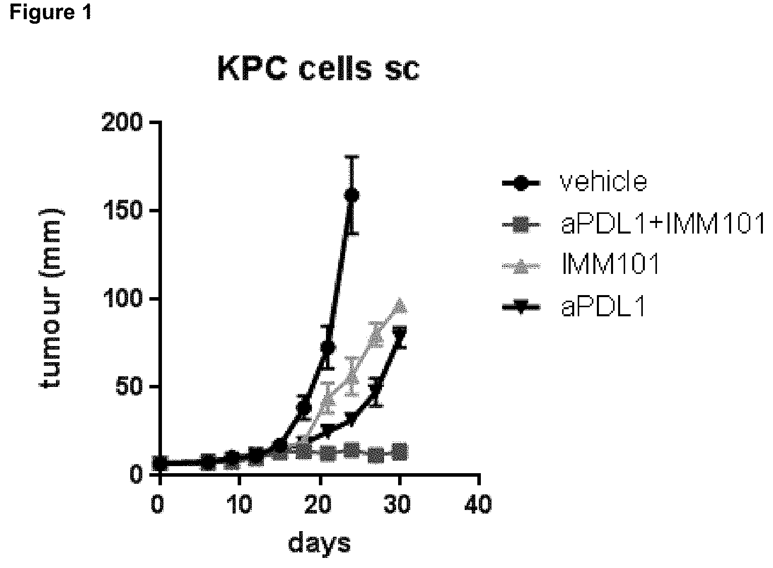
[0024] FIG. 1 shows the effect of a preparation of heat-killed Mycobacterium obuense (NCTC 13365) with or without co-administration of a checkpoint inhibitor (ant-PD-L1 mAb), in a xenograft model of pancreatic cancer (KPC cells injected subcutaneously).
DETAILED DESCRIPTION OF THE INVENTION
[0025] The invention provides a method for treating, reducing, inhibiting or controlling a neoplasia, tumour or cancer in a subject involving administering an immunomodulator and a checkpoint inhibitor. It is based upon the discovery that administration of an immunomodulator (whole cell heat-killed Mycobacterium) in combination with an anti-PD-L1 antibody (a checkpoint inhibitor) results in synergistic anti-tumour activity and/or antitumor activity that is more potent than administration of immunomodulator or anti-PD-L1 antibody alone.
[0026] In order that the present invention may be more readily understood, certain terms are first defined. Additional definitions are set forth throughout the detailed description.
[0027] A "checkpoint inhibitor" is an agent which acts on surface proteins which are members of either the TNF receptor or B7 superfamilies, including agents which bind to negative co-stimulatory molecules selected from CTLA-4, PD-1, TIM-3, BTLA, VISTA, LAG-3, and/or their respective ligands, including PD-L1. (Mellman et al., supra).
[0028] An immunomodulator, as defined according to the present invention, is a component which stimulates innate and type-1 immunity, including Th1 and macrophage activation and cytotoxic cell activity, as well as independently down-regulating inappropriate anti-Th2 responses via immunoregulatory mechanisms.
[0029] The terms "tumour," "cancer" and "neoplasia" are used interchangeably and refer to a cell or population of cells whose growth, proliferation or survival is greater than growth, proliferation or survival of a normal counterpart cell, e.g. a cell proliferative or differentiative disorder. Typically, the growth is uncontrolled. The term "malignancy" refers to invasion of nearby tissue. The term "metastasis" refers to spread or dissemination of a tumour, cancer or neoplasia to other sites, locations or regions within the subject, in which the sites, locations or regions are distinct from the primary tumour or cancer.
[0030] The terms "Programmed Death 1," "Programmed Cell Death 1," "Protein PD-1," "PD-1," and "PD1," are used interchangeably, and include variants, isoforms, species homologs of human PD-1, and analogs having at least one common epitope with PD-1. The complete PD-1 sequence can be found under GenBank Accession No. U64863.
[0031] The terms "cytotoxic T lymphocyte-associated antigen-4," "CTLA-4," "CTLA4," and "CTLA-4 antigen" (see, e.g., Murata, Am. J. Pathol. (1999) 155:453-460) are used interchangeably, and include variants, isoforms, species homologs of human CTLA-4, and analogs having at least one common epitope with CTLA-4 (see, e.g., Balzano (1992) Int. J. Cancer Suppl. 7:28-32). The complete CTLA-4 nucleic acid sequence can be found under GenBank Accession No. L15006.
[0032] As used herein, "sub-therapeutic dose" means a dose of a therapeutic compound (e.g., an antibody) or duration of ther
D00001
XML
uspto.report is an independent third-party trademark research tool that is not affiliated, endorsed, or sponsored by the United States Patent and Trademark Office (USPTO) or any other governmental organization. The information provided by uspto.report is based on publicly available data at the time of writing and is intended for informational purposes only.
While we strive to provide accurate and up-to-date information, we do not guarantee the accuracy, completeness, reliability, or suitability of the information displayed on this site. The use of this site is at your own risk. Any reliance you place on such information is therefore strictly at your own risk.
All official trademark data, including owner information, should be verified by visiting the official USPTO website at www.uspto.gov. This site is not intended to replace professional legal advice and should not be used as a substitute for consulting with a legal professional who is knowledgeable about trademark law.