Methods of Diagnosing Intervertebral Disc Disease And Chondrodystrophy In Canines
Bannasch; Danika ; et al.
U.S. patent application number 16/615659 was filed with the patent office on 2020-07-02 for methods of diagnosing intervertebral disc disease and chondrodystrophy in canines. The applicant listed for this patent is The Regents of the University of California. Invention is credited to Danika Bannasch, Emily Brown, Peter J. Dickinson.
| Application Number | 20200208218 16/615659 |
| Document ID | / |
| Family ID | 64454953 |
| Filed Date | 2020-07-02 |


| United States Patent Application | 20200208218 |
| Kind Code | A1 |
| Bannasch; Danika ; et al. | July 2, 2020 |
Methods of Diagnosing Intervertebral Disc Disease And Chondrodystrophy In Canines
Abstract
Provided are compositions and methods for identifying a canine suffering from or at risk of suffering from skeletal dysplasia (SD) and/or intervertebral disc disease (IVDD) by detecting a retrogene insertion encoding canine fibroblast growth factor 4 (FGF4) on canine chromosome 12.
| Inventors: | Bannasch; Danika; (Davis, CA) ; Brown; Emily; (Davis, CA) ; Dickinson; Peter J.; (Davis, CA) | ||||||||||
| Applicant: |
|
||||||||||
|---|---|---|---|---|---|---|---|---|---|---|---|
| Family ID: | 64454953 | ||||||||||
| Appl. No.: | 16/615659 | ||||||||||
| Filed: | May 23, 2018 | ||||||||||
| PCT Filed: | May 23, 2018 | ||||||||||
| PCT NO: | PCT/US18/34135 | ||||||||||
| 371 Date: | November 21, 2019 |
Related U.S. Patent Documents
| Application Number | Filing Date | Patent Number | ||
|---|---|---|---|---|
| 62512689 | May 30, 2017 | |||
| Current U.S. Class: | 1/1 |
| Current CPC Class: | C12Q 1/6883 20130101; C07K 14/50 20130101; C12Q 2600/156 20130101; C12Q 1/68 20130101 |
| International Class: | C12Q 1/6883 20060101 C12Q001/6883; C07K 14/50 20060101 C07K014/50 |
Goverment Interests
STATEMENT OF GOVERNMENTAL SUPPORT
[0002] This invention was made with government support under Grant No. NIH 5 T32 OD010931 2016_20F, awarded by the National Institutes of Health. The government has certain rights in the invention.
Claims
1. A reaction mixture comprising (i) a biological sample from a canine comprising a nucleic acid template, and (ii) one or more oligonucleotide pairs configured to detect the presence or absence of a retrogene insertion encoding canine fibroblast growth factor 4 (FGF4) on canine chromosome 12.
2. The reaction mixture of claim 1, wherein the retrogene comprises about 3.2 kilobases (kb).
3. The reaction mixture of any one of claims 1 to 2, wherein retrogene insertion is inserted at a target site duplication sequence located at chr12:33,710,168-33,710,178 (canFam3).
4. The reaction mixture of any one of claims 1 to 3, wherein the oligonucleotide pairs detect the 5'-end and/or the 3'-end of the retrogene insertion.
5. The reaction mixture of any one of claims 1 to 4, wherein the 5'-end of the retrogene insertion encoding canine fibroblast growth factor 4 (FGF4) comprises a nucleic acid sequence having at least 90% sequence identity to nucleic acid residues 1002-2001 SEQ ID NO:1.
6. The reaction mixture of any one of claims 1 to 5, wherein the one or more oligonucleotide pairs are configured to detect the 5'-end of the retrogene insertion located at nucleic acid residue 1002 of SEQ ID NO:1.
7. The reaction mixture of any one of claims 1 to 6, wherein an oligonucleotide in the one or more oligonucleotide pairs hybridizes to a sequence segment within nucleic acid residues 1-1001 of SEQ ID NO:1.
8. The reaction mixture of any one of claims 1 to 6, wherein the 3'-end of the retrogene insertion encoding canine fibroblast growth factor 4 (FGF4) comprises a nucleic acid sequence having at least 90% sequence identity to nucleic acid residues 1-1000 SEQ ID NO:2.
9. The reaction mixture of any one of claims 1 to 8, wherein the one or more oligonucleotide pairs are configured to detect the 3'-end of the retrogene insertion located at nucleic acid residue 1000 of SEQ ID NO:2.
10. The reaction mixture of any one of claims 1 to 9, wherein an oligonucleotide in the one or more oligonucleotide pairs hybridizes to a sequence segment within nucleic acid residues 1001-2000 of SEQ ID NO:2.
11. The reaction mixture of any one of claims 1 to 10, wherein the one or more oligonucleotide pairs comprise one or more forward primers selected from the group consisting of: ACAGCTGGCATGGTCAGTTA (SEQ ID NO:2), GTGTTTGCATGGAGGAAGGT (SEQ ID NO:3), CTGAGCAAGAACGGGAAGAC (SEQ ID NO:4), AGCCTGATGGCTGGACTGTA (SEQ ID NO:5) and GTCCGTGCGGTGAAATAAAA (SEQ ID NO:6) and one or more reverse primers selected from the group consisting of TGCTGTAGATTTTGAGGTGTCTT (SEQ ID NO:7), CCTGATTTTGAGACAGCCAAA (SEQ ID NO:8), TTGATGCCCAGGAGGTAGTC (SEQ ID NO:9) and TGAGTGGGTTAAGGGTTTCG (SEQ ID NO:10).
12. The reaction mixture of any one of claims 1 to 11, wherein the one or more oligonucleotides comprise one or more forward primers selected from the group consisting of: ACAGCTGGCATGGTCAGTTA (SEQ ID NO:2) and GTCCGTGCGGTGAAATAAAA (SEQ ID NO:6) and reverse primer TGCTGTAGATTTTGAGGTGTCTT (SEQ ID NO:7).
13. The reaction mixture of any one of claims 1 to 12, wherein the nucleic acid template comprises genomic DNA.
14. The reaction mixture of any one of claims 1 to 13, wherein the reaction mixture further comprises a polymerase and dNTPs.
15. A kit comprising one or more oligonucleotide pairs that specifically identify the presence or absence of a retrogene insertion encoding canine fibroblast growth factor 4 (FGF4) on canine chromosome 12.
16. The kit of claim 15, wherein the oligonucleotide pairs detect the 5'-end and/or the 3'-end of the retrogene insertion.
17. The kit of any one of claims 15 to 16, wherein the 5'-end of the retrogene insertion encoding canine fibroblast growth factor 4 (FGF4) comprises a nucleic acid sequence having at least 90% sequence identity to nucleic acid residues 1002-2001 SEQ ID NO:1.
18. The kit of any one of claims 15 to 17, wherein the one or more oligonucleotide pairs are configured to detect the 5'-end of the retrogene insertion located at nucleic acid residue 1002 of SEQ ID NO:1.
19. The kit of any one of claims 15 to 18, wherein an oligonucleotide in the one or more oligonucleotide pairs hybridizes to a sequence segment within nucleic acid residues 1-1001 of SEQ ID NO:1.
20. The kit of any one of claims 15 to 19, wherein the 3'-end of the retrogene insertion encoding canine fibroblast growth factor 4 (FGF4) comprises a nucleic acid sequence having at least 90% sequence identity to nucleic acid residues 1-1000 SEQ ID NO:2.
21. The kit of any one of claims 15 to 20, wherein the one or more oligonucleotide pairs are configured to detect the 3'-end of the retrogene insertion located at nucleic acid residue 1000 of SEQ ID NO:2.
22. The kit of any one of claims 15 to 21, wherein an oligonucleotide in the one or more oligonucleotide pairs hybridizes to a sequence segment within nucleic acid residues 1001-2000 of SEQ ID NO:2.
23. The kit of any one of claims 15 to 22, wherein the one or more oligonucleotide pairs comprise one or more forward primers selected from the group consisting of: ACAGCTGGCATGGTCAGTTA (SEQ ID NO:2), GTGTTTGCATGGAGGAAGGT (SEQ ID NO:3), CTGAGCAAGAACGGGAAGAC (SEQ ID NO:4), AGCCTGATGGCTGGACTGTA (SEQ ID NO:5) and GTCCGTGCGGTGAAATAAAA (SEQ ID NO:6) and one or more reverse primers selected from the group consisting of TGCTGTAGATTTTGAGGTGTCTT (SEQ ID NO:7), CCTGATTTTGAGACAGCCAAA (SEQ ID NO:8), TTGATGCCCAGGAGGTAGTC (SEQ ID NO:9) and TGAGTGGGTTAAGGGTTTCG (SEQ ID NO:10).
24. A solid support attached to one or more oligonucleotides that specifically identify the presence or absence of a retrogene insertion encoding canine fibroblast growth factor 4 (FGF4) on canine chromosome 12.
25. The solid support of claim 24, wherein the solid support is attached to an oligonucleotide that hybridizes to the 5'-end of the retrogene insertion located at nucleic acid residue 1002 of SEQ ID NO:1.
26. The solid support of any one of claims 24 to 25, wherein the solid support is attached to an oligonucleotide that hybridizes to the 3'-end of the retrogene insertion located at nucleic acid residue 1000 of SEQ ID NO:2.
27. The solid support of any one of claims 24 to 26, wherein the solid support is attached to an oligonucleotide having at least about 80% sequence identity to SEQ ID NO:11 and/or SEQ ID NO:12.
28. The solid support of any one of claims 24 to 27, wherein the solid support is a microarray.
29. The solid support of any one of claims 24 to 27, wherein the solid support is a mounted tissue sample.
30. A kit comprising the solid support of any one of claims 24 to 28.
31. A method for identifying a canine suffering from or at risk of suffering from skeletal dysplasia (SD) and/or intervertebral disc disease (IVDD), the method comprising: a) obtaining a biological sample comprising a nucleic acid template from the canine; b) determining the presence or absence of a retrogene insertion encoding canine fibroblast growth factor 4 (FGF4) on canine chromosome 12; and c) selecting a canine comprising the retrogene insertion identifies a canine suffering from or at risk of suffering from skeletal dysplasia (SD) and/or intervertebral disc disease (IVDD) relative to canine that does not have the retrogene insertion encoding canine fibroblast growth factor 4 (FGF4) on canine chromosome 12.
32. A method for identifying a canine with reduced risk of suffering from skeletal dysplasia (SD) and/or intervertebral disc disease (IVDD), the method comprising: a) obtaining a biological sample comprising a nucleic acid template from the canine; b) determining the presence or absence of a retrogene insertion encoding canine fibroblast growth factor 4 (FGF4) on canine chromosome 12; and c) selecting a canine that does not comprise the retrogene insertion identifies a canine with reduced risk of suffering from skeletal dysplasia (SD) and/or intervertebral disc disease (IVDD) relative to canine that has the retrogene insertion encoding canine fibroblast growth factor 4 (FGF4) on canine chromosome 12.
33. The method of any one of claims 31 to 32, wherein the retrogene comprises about 3.2 kilobases (kb).
34. The method of any one of claims 31 to 33, wherein retrogene insertion is inserted at a target site duplication sequence located at chr12:33,710,168-33,710,178 (canFam3).
35. The method of any one of claims 31 to 34, wherein the determining step employs one or more polynucleotides configured to detect the 5'-end and/or the 3'-end of the retrogene insertion.
36. The method of claim 35, wherein the 5'-end of the retrogene insertion encoding canine fibroblast growth factor 4 (FGF4) comprises a nucleic acid sequence having at least 90% sequence identity to nucleic acid residues 1002-2001 SEQ ID NO:1.
37. The method of any one of claims 35 to 36, wherein the one or more polynucleotides are configured to detect the 5'-end of the retrogene insertion located at nucleic acid residue 1002 of SEQ ID NO:1.
38. The method of any one of claims 35 to 37, wherein one polynucleotide hybridizes to a sequence segment within nucleic acid residues 1-1001 of SEQ ID NO:1.
39. The method of any one of claims 35 to 38, wherein the 3'-end of the retrogene insertion encoding canine fibroblast growth factor 4 (FGF4) comprises a nucleic acid sequence having at least 90% sequence identity to nucleic acid residues 1-1000 SEQ ID NO:2.
40. The method of any one of claims 35 to 39, wherein the one or more polynucleotides are configured to detect the 3'-end of the retrogene insertion located at nucleic acid residue 1000 of SEQ ID NO:2.
41. The method of any one of claims 35 to 40, wherein one polynucleotide hybridizes to a sequence segment within nucleic acid residues 1001-2000 of SEQ ID NO:2.
42. The method of any one of claims 35 to 41, wherein the SD/IVDD genotype is detected by an amplification reaction using polynucleotides that identify the presence or absence of the CFA 12 FGF4 retrogene insertion.
43. The method of claim 42, wherein the amplification reaction is selected from the group consisting of polymerase chain reaction (PCR), strand displacement amplification (SDA), nucleic acid sequence based amplification (NASBA), rolling circle amplification (RCA), T7 polymerase mediated amplification, T3 polymerase mediated amplification and SP6 polymerase mediated amplification.
44. The method of any one of claims 31 to 43, wherein the 5'-end and/or the 3'-end of the retrogene insertion are detected.
45. The method of any one of claims 31 to 44, wherein a portion of the retrogene insertion sequence is specifically amplified using one or more forward primers selected from the group consisting of: ACAGCTGGCATGGTCAGTTA (SEQ ID NO:2), GTGTTTGCATGGAGGAAGGT (SEQ ID NO:3), CTGAGCAAGAACGGGAAGAC (SEQ ID NO:4), AGCCTGATGGCTGGACTGTA (SEQ ID NO:5) and GTCCGTGCGGTGAAATAAAA (SEQ ID NO:6) and one or more reverse primers selected from the group consisting of TGCTGTAGATTTTGAGGTGTCTT (SEQ ID NO:7), CCTGATTTTGAGACAGCCAAA (SEQ ID NO:8), TTGATGCCCAGGAGGTAGTC (SEQ ID NO:9) and TGAGTGGGTTAAGGGTTTCG (SEQ ID NO:10).
46. The method of any one of claims 31 to 45, wherein a portion of the retrogene insertion sequence is specifically amplified using one or more forward primers selected from the group consisting of: ACAGCTGGCATGGTCAGTTA (SEQ ID NO:2) and GTCCGTGCGGTGAAATAAAA (SEQ ID NO:6) and reverse primer TGCTGTAGATTTTGAGGTGTCTT (SEQ ID NO:7).
47. The method of any one of claims 31 to 46, wherein the SD/IVDD genotype is detected by hybridization using polynucleotides which identify the presence or absence of the CFA 12 FGF4 retrogene insertion.
48. The method of any one of claims 31 to 47, wherein the SD/IVDD genotype is detected by sequencing.
49. The method of any one of claims 31 to 48, wherein the canine is a domesticated canine.
50. The method of any one of claims 31 to 49, wherein the canine is of a breed having a predisposition to chondrodystrophy.
51. The method of any one of claims 31 to 50, wherein the canine is a purebred or mix from a breed selected from the group consisting of American Cocker Spaniel, Basset Hound, Beagle, Cardigan Welsh Corgi, Chesapeake Bay Retriever, Chihuahua, Coton de Tulear, Dachshund, English Springer Spaniel, French Bulldog, Jack Russell Terrier, Miniature Schnauzer, Nova Scotia Duck Tolling Retriever, Pekingese, Pembroke Welsh Corgi, Poodle, Portuguese Water Dog, Scottish Terrier, Shih Tzu, and mixtures thereof.
52. The method of any one of claims 31 to 50, wherein the canine is a purebred or mix from a breed selected from the group consisting of American Cocker Spaniel, Basset Hound, Beagle, Corgi, Dachshund, French bulldog, Nova Scotia Duck Tolling Retriever, and Pekingese.
Description
CROSS-REFERENCE TO RELATED APPLICATIONS
[0001] This application claims the benefit of U.S. Provisional Application No. 62/512,689, filed on May 30, 2017, which is hereby incorporated herein by reference in its entirety.
BACKGROUND
[0003] Variation in domestic dog (Canis familiaris, CFA) morphology has long fascinated both scientists and pet owners. Domestication of the dog from the wolf and the subsequent variation in size and shape within purebred dog breeds is a remarkable feat of animal breeding and selection. One of the most extreme examples of dog breed differences is in limb length, as extremely short limbs define many breeds. This morphological feature is present in breeds from all over the world and from all American Kennel Club groups, indicating that the underlying genetic causes are likely very old.
[0004] Extensive examination of growth plates has been performed on many of these short-legged dog breeds (Dachshund, Pekingese, French Bulldog, Spaniels, Beagle), as these breeds are also prone to intervertebral disc disease (IVDD) (1-3). Histopathological analysis of the bones of puppies from these breeds demonstrated that their short stature is due to defects in endochondral ossification, the process whereby cartilage is replaced with bone in the developing limb. The long bone growth plates show disorganization of the proliferative zone and reduction in the depth of the maturation zone (1-4). In addition to the long bones, similar but more subtle changes exist in endochondral ossification of the vertebral bodies (1,2).
[0005] The intervertebral disc (IVD) is composed of an outer fibrous basket, called the annulus fibrosis, made of 70% collagen and an inner gel-like layer that is a remnant of the embryonic notochord, called the nucleus pulposus (5). Together, these structures and the cartilaginous endplates allow for flexibility of the vertebral column. In chondrodystrophic dogs, the nucleus pulposus is gradually replaced by chondrocyte-like cells in chondroid metaplasia (or metamorphosis) that occurs between birth and 1 year of age (1,2). Recent studies have shown that in advanced stages of degeneration in non-chondrodystrophoid dogs there is also replacement of notochordal cells by chondrocyte-like cells, similar to the changes observed in chondrodystrophoid dogs, although this happens at an older age (3,6-10). The replacement of the nucleus pulposus with chondrocyte like cells is seen in humans, and chondrodystrophoid breeds have been proposed as models for human degenerative disc disease (3, 7, 11, 12).
[0006] Hansen described the two different types of canine IVD prolapse as type I and type II. Type I occurs exclusively in chondrodystrophic breeds and is characterized by premature degeneration of all discs in young dogs. In contrast, Type II occurs in older dogs and is usually limited to a single disc with only partial protrusion. In Type I disc disease the calcified nucleus pulposus may undergo an explosive herniation through the annulus fibrous into the vertebral canal, resulting in inflammation and hemorrhage and causing severe pain and neurological dysfunction (1, 2). In veterinary hospital population studies, breeds with a significant increased risk of IVDD include the Beagle, Cocker Spaniel, Dachshund, French Bulldog, Lhasa Apso, Pekingese, Pembroke Welsh Corgi, and Shih Tzu (13-15). Pet insurance data suggests a conservative "lifetime prevalence" for IVDD in dogs of 3.5% in the overall population; however, in the chondrodystrophic breed with the highest risk, the Dachshund, the "lifetime prevalence" is between 20-62% with a mortality rate of 24% (9, 16-19). The effect of this disease on dogs and the financial burden to pet owners is enormous.
[0007] Skeletal dysplasia (SD), a general term to classify abnormalities of growth and development of cartilage and/or bone resulting in various forms of short stature, occurs in humans and dogs in many forms (20). With advances in molecular genetics, many of the diseases in humans are being reclassified based on the specific underlying causative mutations (21). To a lesser degree, progress has also been made in understanding the molecular nature of SD and the extreme interbreed limb length variation observed in dogs (22-25). While the mutations causing some subtypes of SD in dogs have been determined, there are still many unexplained types of SD observed within and across dog breeds.
[0008] In 2009 the genetic basis for extreme differences in limb length in dogs was investigated by Parker et al. using an across breed genome-wide association approach (26). They determined that a FGF4 retrogene insertion on CFA18 was responsible for the "chondrodysplasia" phenotype in a number of breeds, such as the Basset Hound, Pembroke Welsh Corgi, and Dachshund. However, the FGF4 retrogene insertion on CFA18 failed to explain breeds such as the American Cocker Spaniel, Beagle, and French Bulldog, that in addition to Dachshunds, were the breeds originally classified as chondrodystrophoid based on histopathologic and morphologic analysis by Hansen and Braund (1, 3). The FGF gene family has similarly been implicated in SD in humans, with mutations in FGFR3 found to be responsible for achondrodysplasia, the most common form of dwarfism, characterized by shortened limbs and abnormal vertebrae and IVDs (21, 27-31). FGF genes are involved in a number of embryological development processes, and specific levels of ligand and receptor are key for appropriate growth and development (32-34).
SUMMARY
[0009] In one aspect, provided are reaction mixtures. In some embodiments, the reaction mixtures comprise (i) a biological sample from a canine comprising a nucleic acid template, and (ii) one or more oligonucleotide pairs configured to detect the presence or absence of a retrogene insertion encoding canine fibroblast growth factor 4 (FGF4) on canine chromosome 12. In some embodiments, the retrogene comprises about 3.2 kilobases (kb). In some embodiments, retrogene insertion is inserted at a target site duplication sequence located at chr12:33,710,168-33,710,178 (canFam3). In some embodiments, the oligonucleotide pairs detect the 5'-end and/or the 3'-end of the retrogene insertion. In some embodiments, the 5'-end of the retrogene insertion encoding canine fibroblast growth factor 4 (FGF4) comprises a nucleic acid sequence having at least 90% sequence identity, e.g., at least 91%, 92%, 93%, 94%, 95%, 96%, 97%, 98%, 99%, or 100% sequence identity, to nucleic acid residues 1002-2001 SEQ ID NO:1. In some embodiments, the one or more oligonucleotide pairs are configured or designed or constructed to detect the 5'-end of the retrogene insertion located at nucleic acid residue 1002 of SEQ ID NO:1. In some embodiments, an oligonucleotide in the one or more oligonucleotide pairs hybridizes to a sequence segment within nucleic acid residues 1-1001 of SEQ ID NO:1. In some embodiments, the 3'-end of the retrogene insertion encoding canine fibroblast growth factor 4 (FGF4) comprises a nucleic acid sequence having at least 90% sequence identity, e.g., at least 91%, 92%, 93%, 94%, 95%, 96%, 97%, 98%, 99%, or 100% sequence identity, to nucleic acid residues 1-1000 SEQ ID NO:2. In some embodiments, the one or more oligonucleotide pairs are configured to detect the 3'-end of the retrogene insertion located at nucleic acid residue 1000 of SEQ ID NO:2. In some embodiments, an oligonucleotide in the one or more oligonucleotide pairs hybridizes to a sequence segment within nucleic acid residues 1001-2000 of SEQ ID NO:2. In some embodiments, the one or more oligonucleotide pairs comprise one or more forward primers selected from the group consisting of: ACAGCTGGCATGGTCAGTTA (SEQ ID NO:2), GTGTTTGCATGGAGGAAGGT (SEQ ID NO:3), CTGAGCAAGAACGGGAAGAC (SEQ ID NO:4), AGCCTGATGGCTGGACTGTA (SEQ ID NO:5) and GTCCGTGCGGTGAAATAAAA (SEQ ID NO:6) and one or more reverse primers selected from the group consisting of TGCTGTAGATTTTGAGGTGTCTT (SEQ ID NO:7), CCTGATTTTGAGACAGCCAAA (SEQ ID NO:8), TTGATGCCCAGGAGGTAGTC (SEQ ID NO:9) and TGAGTGGGTTAAGGGTTTCG (SEQ ID NO:10). In some embodiments, the one or more oligonucleotides comprise one or more forward primers selected from the group consisting of:
TABLE-US-00001 (SEQ ID NO: 2) ACAGCTGGCATGGTCAGTTA and (SEQ ID NO: 6) GTCCGTGCGGTGAAATAAAA and reverse primer (SEQ ID NO: 7) TGCTGTAGATTTTGAGGTGTCTT.
In some embodiments, the nucleic acid template comprises genomic DNA. In some embodiments, the reaction mixture further comprises a polymerase and dNTPs.
[0010] In another aspect, provided are kits. In some embodiments, the kits comprise one or more oligonucleotide pairs that specifically identify the presence or absence of a retrogene insertion encoding canine fibroblast growth factor 4 (FGF4) on canine chromosome 12. In some embodiments, the oligonucleotide pairs detect the 5'-end and/or the 3'-end of the retrogene insertion. In some embodiments, the 5'-end of the retrogene insertion encoding canine fibroblast growth factor 4 (FGF4) comprises a nucleic acid sequence having at least 90% sequence identity, e.g., at least 91%, 92%, 93%, 94%, 95%, 96%, 97%, 98%, 99%, or 100% sequence identity, to nucleic acid residues 1002-2001 SEQ ID NO:1. In some embodiments, the one or more oligonucleotide pairs are configured to detect the 5'-end of the retrogene insertion located at nucleic acid residue 1002 of SEQ ID NO:1. In some embodiments, an oligonucleotide in the one or more oligonucleotide pairs hybridizes to a sequence segment within nucleic acid residues 1-1001 of SEQ ID NO:1. In some embodiments, the 3'-end of the retrogene insertion encoding canine fibroblast growth factor 4 (FGF4) comprises a nucleic acid sequence having at least 90% sequence identity, e.g., at least 91%, 92%, 93%, 94%, 95%, 96%, 97%, 98%, 99%, or 100% sequence identity, to nucleic acid residues 1-1000 SEQ ID NO:2. In some embodiments, the one or more oligonucleotide pairs are configured to detect the 3'-end of the retrogene insertion located at nucleic acid residue 1000 of SEQ ID NO:2. In some embodiments, an oligonucleotide in the one or more oligonucleotide pairs hybridizes to a sequence segment within nucleic acid residues 1001-2000 of SEQ ID NO:2. In some embodiments, the one or more oligonucleotide pairs comprise one or more forward primers selected from the group consisting of: ACAGCTGGCATGGTCAGTTA (SEQ ID NO:2), GTGTTTGCATGGAGGAAGGT (SEQ ID NO:3), CTGAGCAAGAACGGGAAGAC (SEQ ID NO:4), AGCCTGATGGCTGGACTGTA (SEQ ID NO:5) and GTCCGTGCGGTGAAATAAAA (SEQ ID NO:6) and one or more reverse primers selected from the group consisting of TGCTGTAGATTTTGAGGTGTCTT (SEQ ID NO:7), CCTGATTTTGAGACAGCCAAA (SEQ ID NO:8), TTGATGCCCAGGAGGTAGTC (SEQ ID NO:9) and TGAGTGGGTTAAGGGTTTCG (SEQ ID NO:10).
[0011] In another aspect, provided are solid supports. In some embodiments, the solid supports are attached to one or more oligonucleotides that specifically identify the presence or absence of a retrogene insertion encoding canine fibroblast growth factor 4 (FGF4) on canine chromosome 12. In some embodiments, the solid support is attached to an oligonucleotide that hybridizes to the 5'-end of the retrogene insertion located at nucleic acid residue 1002 of SEQ ID NO:1. In some embodiments, the solid support is attached to an oligonucleotide that hybridizes to the 3'-end of the retrogene insertion located at nucleic acid residue 1000 of SEQ ID NO:2. In some embodiments, the solid support is attached to an oligonucleotide having at least about 80% sequence identity, e.g., at least 81%, 82%, 83%, 84%, 85%, 86%, 87%, 88%, 89%, 90%, 91%, 92%, 93%, 94%, 95%, 96%, 97%, 98%, 99%, or 100% sequence identity, to SEQ ID NO:11 and/or SEQ ID NO:12. In some embodiments, the solid support is a microarray. In some embodiments, the solid support is a mounted tissue sample. Further provided are kits comprising the solid support as described above and herein.
[0012] In a further aspect, provided are methods for identifying a canine suffering from or at risk of suffering from skeletal dysplasia (SD) and/or intervertebral disc disease (IVDD). In some embodiment, the methods comprise: a) obtaining a biological sample comprising a nucleic acid template from the canine; b) determining the presence or absence of a retrogene insertion encoding canine fibroblast growth factor 4 (FGF4) on canine chromosome 12; and c) selecting a canine comprising the retrogene insertion identifies a canine suffering from or at risk of suffering from skeletal dysplasia (SD) and/or intervertebral disc disease (IVDD) relative to canine that does not have the retrogene insertion encoding canine fibroblast growth factor 4 (FGF4) on canine chromosome 12. In a related aspect, provided are methods for identifying a canine with reduced risk of suffering from skeletal dysplasia (SD) and/or intervertebral disc disease (IVDD). In some embodiments, the methods comprise: a) obtaining a biological sample comprising a nucleic acid template from the canine; b) determining the presence or absence of a retrogene insertion encoding canine fibroblast growth factor 4 (FGF4) on canine chromosome 12; and c) selecting a canine that does not comprise the retrogene insertion identifies a canine with reduced risk of suffering from skeletal dysplasia (SD) and/or intervertebral disc disease (IVDD) relative to canine that has the retrogene insertion encoding canine fibroblast growth factor 4 (FGF4) on canine chromosome 12. In some embodiments, the retrogene comprises about 3.2 kilobases (kb). In some embodiments, the retrogene insertion is inserted at a target site duplication sequence located at chr12:33,710,168-33,710,178 (canFam3). In some embodiments, the determining step employs one or more polynucleotides configured to detect the 5'-end and/or the 3'-end of the retrogene insertion. In some embodiments, the 5'-end of the retrogene insertion encoding canine fibroblast growth factor 4 (FGF4) comprises a nucleic acid sequence having at least 90% sequence identity, e.g., at least 91%, 92%, 93%, 94%, 95%, 96%, 97%, 98%, 99%, or 100% sequence identity, to nucleic acid residues 1002-2001 SEQ ID NO:1. In some embodiments, the one or more polynucleotides are configured to detect the 5'-end of the retrogene insertion located at nucleic acid residue 1002 of SEQ ID NO:1. In some embodiments, one polynucleotide hybridizes to a sequence segment within nucleic acid residues 1-1001 of SEQ ID NO:1. In some embodiments, the 3'-end of the retrogene insertion encoding canine fibroblast growth factor 4 (FGF4) comprises a nucleic acid sequence having at least 90% sequence identity, e.g., at least 91%, 92%, 93%, 94%, 95%, 96%, 97%, 98%, 99%, or 100% sequence identity, to nucleic acid residues 1-1000 SEQ ID NO:2. In some embodiments, the one or more polynucleotides are configured to detect the 3'-end of the retrogene insertion located at nucleic acid residue 1000 of SEQ ID NO:2. In some embodiments, one polynucleotide hybridizes to a sequence segment within nucleic acid residues 1001-2000 of SEQ ID NO:2. In some embodiments, the SD/IVDD genotype is detected by an amplification reaction using polynucleotides that identify the presence or absence of the CFA 12 FGF4 retrogene insertion. In some embodiments, the amplification reaction is selected from the group consisting of polymerase chain reaction (PCR), strand displacement amplification (SDA), nucleic acid sequence based amplification (NASBA), rolling circle amplification (RCA), T7 polymerase mediated amplification, T3 polymerase mediated amplification and SP6 polymerase mediated amplification. In some embodiments, a portion of the retrogene insertion sequence is specifically amplified using one or more forward primers selected from the group consisting of: ACAGCTGGCATGGTCAGTTA (SEQ ID NO:2), GTGTTTGCATGGAGGAAGGT (SEQ ID NO:3), CTGAGCAAGAACGGGAAGAC (SEQ ID NO:4), AGCCTGATGGCTGGACTGTA (SEQ ID NO:5) and GTCCGTGCGGTGAAATAAAA (SEQ ID NO:6) and one or more reverse primers selected from the group consisting of TGCTGTAGATTTTGAGGTGTCTT (SEQ ID NO:7), CCTGATTTTGAGACAGCCAAA (SEQ ID NO:8), TTGATGCCCAGGAGGTAGTC (SEQ ID NO:9) and TGAGTGGGTTAAGGGTTTCG (SEQ ID NO:10). In some embodiments, a portion of the retrogene insertion sequence is specifically amplified using one or more forward primers selected from the group consisting of: ACAGCTGGCATGGTCAGTTA (SEQ ID NO:2) and GTCCGTGCGGTGAAATAAAA (SEQ ID NO:6) and reverse primer TGCTGTAGATTTTGAGGTGTCTT (SEQ ID NO:7). In some embodiments, the SD/IVDD genotype is detected by hybridization using polynucleotides which identify the presence or absence of the CFA 12 FGF4 retrogene insertion. In some embodiments, the SD/IVDD genotype is detected by sequencing. In some embodiments, the canine is a domesticated canine. In some embodiments, the canine is of a breed having a predisposition to chondrodystrophy. In some embodiments, the canine is a purebred or mix from a breed selected from the group consisting of American Cocker Spaniel, Basset Hound, Beagle, Cardigan Welsh Corgi, Chesapeake Bay Retriever, Chihuahua, Coton de Tulear, Dachshund, English Springer Spaniel, French Bulldog, Jack Russell Terrier, Miniature Schnauzer, Nova Scotia Duck Tolling Retriever, Pekingese, Pembroke Welsh Corgi, Poodle, Portuguese Water Dog, Scottish Terrier, Shih Tzu, and mixtures thereof. In varying, the canine is a purebred or mix from a breed selected from the group consisting of American Cocker Spaniel, Basset Hound, Beagle, Corgi, Dachshund, French bulldog, Nova Scotia Duck Tolling Retriever, and Pekingese.
Definitions
[0013] Unless defined otherwise, all technical and scientific terms used herein generally have the same meaning as commonly understood by one of ordinary skill in the art. Generally, the nomenclature used herein and the laboratory procedures in cell culture, molecular genetics, organic chemistry and nucleic acid chemistry and hybridization described below are those well-known and commonly employed in the art. Standard techniques are used for nucleic acid and peptide synthesis. Generally, enzymatic reactions and purification steps are performed according to the manufacturer's specifications. The techniques and procedures are generally performed according to conventional methods in the art and various general references (see generally, Green and Sambrook et al. MOLECULAR CLONING: A LABORATORY MANUAL, 4th ed. (2012) Cold Spring Harbor Laboratory Press, Cold Spring Harbor, N.Y. and Ausubel, ed., Current Protocols in Molecular Biology, 1990-2017, John Wiley Interscience), which are provided throughout this document. The nomenclature used herein and the laboratory procedures in analytical chemistry, and organic synthetic described below are those well-known and commonly employed in the art. Standard techniques, or modifications thereof, are used for chemical syntheses and chemical analyses.
[0014] "Chondrodystrophy" refers to defects in long bone and vertebral body endochondral ossification and abnormal intervertebral discs that prematurely degenerate and calcify, which ultimately can lead to disc herniation and paralysis referred to as Hansen's Type I intervertebral disc disease (IVDD). Reviewed in, e.g., Smith, et al., Vet Comp Orthop Traumatol. (2016) 29(3):220-6; Smolders, et al., Vet J. (2013) 195(3):292-9; Bergknut, et al., Vet J. (2013) 195(3):282-91; Bergknut, et al., Vet J. (2013) 195(2):156-63 and Beachley, et al., J Am Vet Med Assoc. (1973) 163(3):283-4.
[0015] "Chondrodysplasia" refers to disproportionate dwarfism.
[0016] As used herein, the terms "dog," "canine" and "Canis lupus familiaris" are used interchangeably.
[0017] The term "gene" means the segment of DNA involved in producing a polypeptide chain; it includes regions preceding and following the coding region (leader and trailer) as well as intervening sequences (introns) between individual coding segments (exons).
[0018] The terms "nucleic acid" and "polynucleotide" are used interchangeably herein to refer to deoxyribonucleotides or ribonucleotides and polymers thereof in either single- or double-stranded form. The term encompasses nucleic acids containing known nucleotide analogs or modified backbone residues or linkages, which are synthetic, naturally occurring, and non-naturally occurring, which have similar binding properties as the reference nucleic acid, and which are metabolized in a manner similar to the reference nucleotides. Examples of such analogs include, without limitation, phosphorothioates, phosphoramidates, methyl phosphonates, chiral-methyl phosphonates, 2-O-methyl ribonucleotides, peptide-nucleic acids (PNAs).
[0019] Unless otherwise indicated, a particular nucleic acid sequence also encompasses conservatively modified variants thereof (e.g., degenerate codon substitutions) and complementary sequences, as well as the sequence explicitly indicated. Specifically, degenerate codon substitutions may be achieved by generating sequences in which the third position of one or more selected (or all) codons is substituted with mixed-base and/or deoxyinosine residues (Batzer et al., Nucleic Acid Res. 19:5081 (1991); Ohtsuka et al., J. Biol. Chem. 260:2605-2608 (1985); Rossolini et al., Mol. Cell. Probes 8:91-98 (1994)). The term nucleic acid is used interchangeably with gene, cDNA, mRNA, oligonucleotide, and polynucleotide.
[0020] A "variant" is a difference in the nucleotide sequence among related polynucleotides. The difference may be the deletion of one or more nucleotides from the sequence of one polynucleotide compared to the sequence of a related polynucleotide, the addition of one or more nucleotides or the substitution of one nucleotide for another. The terms "mutation," "polymorphism" and "variant" are used interchangeably herein to describe such variants. As used herein, the term "variant" in the singular is to be construed to include multiple variances; i.e., two or more nucleotide additions, deletions and/or substitutions in the same polynucleotide. A "point mutation" refers to a single substitution of one nucleotide for another.
[0021] The phrase "stringent hybridization conditions" refers to conditions under which a probe will hybridize to its target subsequence, typically in a complex mixture of nucleic acid, but to no other sequences. Stringent conditions are sequence-dependent and will be different in different circumstances. Longer sequences hybridize specifically at higher temperatures. An extensive guide to the hybridization of nucleic acids is found in Tijssen, Techniques in Biochemistry and Molecular Biology-Hybridization with Nucleic Probes, "Overview of principles of hybridization and the strategy of nucleic acid assays" (1993). Generally, stringent conditions are selected to be about 5-10.degree. C. lower than the thermal melting point I for the specific sequence at a defined ionic strength pH. The Tm is the temperature (under defined ionic strength, pH, and nucleic concentration) at which 50% of the probes complementary to the target hybridize to the target sequence at equilibrium (as the target sequences are present in excess, at Tm, 50% of the probes are occupied at equilibrium). Stringent conditions will be those in which the salt concentration is less than about 1.0 M sodium ion, typically about 0.01 to 1.0 M sodium ion concentration (or other salts) at pH 7.0 to 8.3 and the temperature is at least about 30.degree. C. for short probes (e.g., 10 to 50 nucleotides) and at least about 60.degree. C. for long probes (e.g., greater than 50 nucleotides). Stringent conditions may also be achieved with the addition of destabilizing agents such as formamide. For selective or specific hybridization, a positive signal is at least two times background, optionally 10 times background hybridization. Exemplary stringent hybridization conditions can be as following: 50% formamide, 5.times.SSC, and 1% SDS, incubating at 42.degree. C., or, 5.times.SSC, 1% SDS, incubating at 65.degree. C., with wash in 0.2.times.SSC, and 0.1% SDS at 65.degree. C.
[0022] Nucleic acids that do not hybridize to each other under stringent conditions are still substantially identical if the polypeptides which they encode are substantially identical. This occurs, for example, when a copy of a nucleic acid is created using the maximum codon degeneracy permitted by the genetic code. In such cases, the nucleic acids typically hybridize under moderately stringent hybridization conditions. Exemplary "moderately stringent hybridization conditions" include a hybridization in a buffer of 40% formamide, 1 M NaCl, 1% SDS at 37.degree. C., and a wash in 1.times.SSC at 45.degree. C. A positive hybridization is at least twice background. Those of ordinary skill will readily recognize that alternative hybridization and wash conditions can be utilized to provide conditions of similar stringency.
[0023] The phrase "selectively (or specifically) hybridizes to" refers to the binding, duplexing, or hybridizing of a molecule only to a particular nucleotide sequence under stringent hybridization conditions when that sequence is present in a complex mixture (e.g., total cellular or library DNA or RNA).
[0024] The terms "isolated," "purified," or "biologically pure" refer to material that is substantially or essentially free from components that normally accompany it as found in its native state. Purity and homogeneity are typically determined using analytical chemistry techniques such as polyacrylamide gel electrophoresis or high performance liquid chromatography. A protein that is the predominant species present in a preparation is substantially purified. The term "purified" denotes that a nucleic acid or protein gives rise to essentially one band in an electrophoretic gel. Particularly, it means that the nucleic acid or protein is at least 85% pure, more preferably at least 95% pure, and most preferably at least 99% pure.
[0025] The terms "identical" or percent "identity," in the context of two or more nucleic acids or polypeptide sequences, refer to two or more sequences or subsequences that are the same or have a specified percentage of amino acid residues or nucleotides that are the same (i.e., share at least about 80% identity, for example, at least about 85%, 90%, 91%, 92%, 93%, 94%, 95%, 96%, 97%, 98%, 99% identity over a specified region to a reference sequence, e.g., SEQ ID NOs:1-11 and the other nucleic acid sequences provided herein), when compared and aligned for maximum correspondence over a comparison window, or designated region as measured using one of the following sequence comparison algorithms or by manual alignment and visual inspection. Such sequences are then said to be "substantially identical." This definition also refers to the compliment of a test sequence. Preferably, the identity exists over a region that is at least about 25 amino acids or nucleotides in length, for example, over a region that is 50-100 amino acids or nucleotides in length, or over the full-length of a reference sequence.
[0026] For sequence comparison, typically one sequence acts as a reference sequence, to which test sequences are compared. When using a sequence comparison algorithm, test and reference sequences are entered into a computer, subsequence coordinates are designated, if necessary, and sequence algorithm program parameters are designated. Default program parameters can be used, or alternative parameters can be designated. The sequence comparison algorithm then calculates the percent sequence identities for the test sequences relative to the reference sequence, based on the program parameters. For sequence comparison of nucleic acids and proteins to canine FGF4 nucleic acids and proteins, the BLAST and BLAST 2.0 algorithms and the default parameters discussed below are used.
[0027] An indication that two nucleic acid sequences or polypeptides are substantially identical is that the polypeptide encoded by the first nucleic acid is immunologically cross reactive with the antibodies raised against the polypeptide encoded by the second nucleic acid, as described below. Thus, a polypeptide is typically substantially identical to a second polypeptide, for example, where the two peptides differ only by conservative substitutions. Another indication that two nucleic acid sequences are substantially identical is that the two molecules or their complements hybridize to each other under stringent conditions, as described below. Yet another indication that two nucleic acid sequences are substantially identical is that the same primers can be used to amplify the sequence.
[0028] "Array" as used herein refers to a solid support comprising attached nucleic acid or peptide probes. Arrays typically comprise a plurality of different nucleic acid or peptide probes that are coupled to a surface of a substrate in different, known locations. These arrays, also described as "microarrays" or colloquially "chips" have been generally described in the art, for example, U.S. Pat. Nos. 5,143,854, 5,445,934, 5,744,305, 5,677,195, 6,040,193, 5,424,186 and Fodor et al., Science, 251:767-777 (1991). These arrays may generally be produced using mechanical synthesis methods or light directed synthesis methods which incorporate a combination of photolithographic methods and solid phase synthesis methods. Techniques for the synthesis of these arrays using mechanical synthesis methods are described in, e.g., U.S. Pat. No. 5,384,261. Arrays may comprise a planar surface or may be nucleic acids or peptides on beads, gels, polymeric surfaces, fibers such as fiber optics, glass or any other appropriate substrate as described in, e.g., U.S. Pat. Nos. 5,770,358, 5,789,162, 5,708,153, 6,040,193 and 5,800,992. Arrays may be packaged in such a manner as to allow for diagnostics or other manipulation of an all-inclusive device, as described in, e.g., U.S. Pat. Nos. 5,856,174 and 5,922,591.
BRIEF DESCRIPTION OF THE DRAWINGS
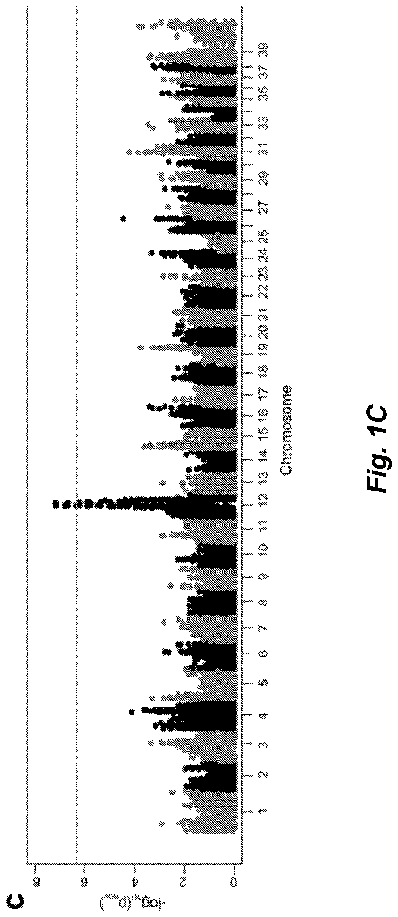
[0029] FIGS. 1A-C illustrate Skeletal Dysplasia (SD) in the Nova Scotia Duck Tolling Retriever (NSDTR): a) Picture and lateral thoracic limb radiograph of unaffected NSDTRs (ages 1 year old and 4 years old, respectively). b) Left panels depict picture and lateral thoracic limb radiograph of mildly SD affected NSDTRs (ages 4 years old and 2 years old, respectively); right panels depict picture and lateral thoracic limb radiograph of more severely SD affected NSDTRs (ages 3 years old and 6 months old, respectively). Relative to the unaffected dog, the mildly SD affected NSDTR has cranial bowing of the radius. Radiographic changes in the more severely SD NSDTR include moderate cranial bowing of the radius, physeal widening, and incongruity of the elbow joint with the shape of the semi-lunar notch of the ulna being elongated. Pictures and radiographs are representatives of each phenotype and not paired (i.e. the radiographs are not of the dogs pictured). c) SD in NSDTR GWAS: Manhattan plot showing log 10 of the raw p-values for each genotyped SNP by chromosome (x-axis). After SNP quality control, there were 106,303 SNPs for Chi square analysis. Genomic inflation was 1.01604. Line denotes genome-wide significance based on Bonferroni corrected p-values.
[0030] FIG. 2 illustrates quantile-quantile (QQ) plot shows -log 10 of the expected versus observed p-values plotted for each SNP, with the SNPs on CFA12 colored in grey.
[0031] FIGS. 3A-C illustrate across breed investigation of SD-IVDD locus: a) Minor allele frequency on the y-axis and base pair on CFA12 on the x-axis plotted by breed: SD affected NSDTR (n=13), American Cocker Spaniel (n=7), and Beagle (n=14). b) Manhattan plot for the SNPs in the across breed IVDD GWAS showing -log 10 of the raw p-values (y-axis) for each genotyped SNP by chromosome (x-axis). After SNP quality control, there were 126,020 SNPs for Chi square analysis. Genomic inflation was 1.6339. c) SNPs in 5 Mb region surrounding most highly associated SNP (chr12:36,909,311 (canFam2)) plotted by base pair on the x-axis and p-value on the y-axis. SNPs have been pruned from analysis using recommended criteria by Kierczak et al. (37). SNPs are color-coded by r.sup.2 value to show the extent of linkage disequilibrium.
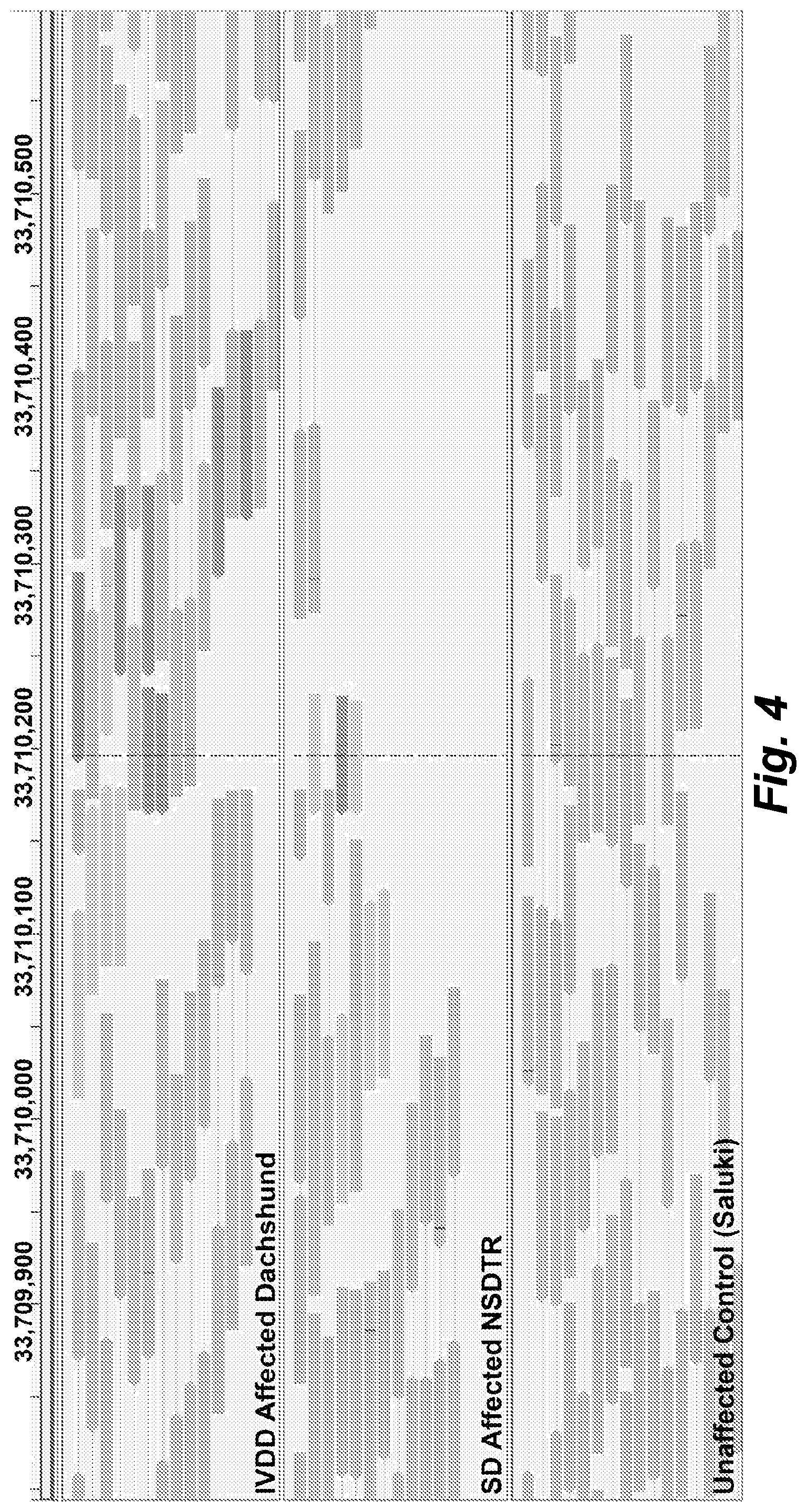
[0032] FIG. 4 illustrates large insertion identified on CFA12: Screenshot of the Integrative Genomics Viewer (IGV-Broad Institute) showing an insertion at approximately 12:33,710,200 (canFam3) in an IVDD affected Dachshund case and a SD affected NSDTR that is not present in the Saluki unaffected control. Read mates in green map to chr18:48.4 Mb (canFam3) and the read mates in blue map to chr7:68.3 Mb (canFam3).
[0033] FIG. 5 illustrates a schematic of endogenous FGF4 (CFA18) retrotransposition to CFA12:33,710,178 (canFam3): Predicted TATA box at chr12:33,709,940-947 (canFam3) and predicted RNA Pol II promoter at chr12:33,709,964-976 (canFam3), which are 239 bp and 215 bp upstream, respectively (38). Endogenous FGF4 untranslated regions (UTR) are unknown in the dog; however, they were approximated in the figure based on human and mouse TransMap data available at the UCSC genome browser (genome.ucsc.edu). The predicted 5'UTR spans from chr18:48,413,185-48,413,480 (canFam3); however, RT-PCR in IVD from a Beagle suggest that the TSS is located between chr18:48,413,315-48,413,402 (canFam3). The insert includes all predicted 3'UTR, followed by 42 adenine residues and the duplicated 11 bp TSD sequence (AAG TGC TTT GA) (chr12:33,710,168-33,710,178 (canFam3)). Endogenous FGF4 sequence that was retrotransposed also includes a large CpG island.
[0034] FIGS. 6A-C illustrate association of FGF4 insertion genotypes with height and IVDD: a) Genotyping results for CFA18 and CFA12 FGF4 insertions across breeds. Arrows indicate wild type (WT) band. Lane order: Ladder; 1-3) NSDTR; 4) Beagle; 5) American Cocker Spaniel; 6) Dachshund; 7) Basset Hound; 8) Pembroke Welsh Corgi; 9) Coton de Tulear; 10) Cairn Terrier; 11) West Highland White Terrier. b) Height at the withers in inches (in) was available for 7 SD NSDTR cases: all were homozygous mutant for the CFA12 FGF4 insertion, and their mean height was 18.22 in. Height was available for 13 NSDTR unaffected with SD: 5 were wild type and had a mean height of 20.2 in; 8 were heterozygous for the CFA12 FGF4 insertion and had a mean height of 18.94 in. * indicate relative levels of association of the insertion with height. ***: p=0.007, **: p=0.016, *: p=0.034. c) Association of various identified loci with IVDD, including Chi square, p-value, and Odds ratio (95% confidence intervals in parenthesis).
[0035] FIG. 7 illustrates CFA 12 FGF4 Genotypes Across Breeds: Genotypes for the CFA12 FGF4 insertion across dog breeds ordered by breed standard height from shortest to tallest (x-axis), plotted by dog weight in kilograms (kg) (y-axis). Only genotyped dogs with weights available (n=376) were included in the figure. Dogs are color-coded by genotype status.
[0036] FIG. 8 illustrates semi-quantitative RT-PCR for FGF4 across tissues in a case and control. Lane order: Ladder; 1) Control VB (Cane Corso); 2) Case VB (Beagle); 3) Control IVD (Cane Corso); 4) Case IVD (Beagle); 5) Control skeletal muscle (Labrador retriever); 6) Case skeletal muscle (Beagle); 7) Control testis (Labrador retriever); 8) Case testis (Beagle); 9) Negative control.
[0037] FIG. 9 illustrates FGF4 expression: Bar graph depicting fold change differences in FGF4 expression between controls and IVDD cases in neonatal IVD and VB. FGF4 expression was 19.47.times. higher (p=0.02857) in IVD and 2.16.times. higher (p=0.02857) in VB of cases compared to controls. Error bars representative of standard error of measurement for each group. Gels depict genotypes of 4 cases (Beagles) and 5 controls (1 Rottweiler and 4 Cane Corso) used in qRT-PCR analysis. The five controls were wild type, meaning they lacked the FGF4 insert at both the CFA12 and CFA18 locations; however, the cases, while wild type for the CFA18 FGF4 insert, were homozygous mutant at the CFA12 locus. Lanes: 1-4: Beagle cases; 5-9: Cane Corso controls; 10: heterozygous control; 11: negative control.
DETAILED DESCRIPTION
1. Introduction
[0038] Chondrodystrophy in dogs is defined by dysplastic, shortened long bones and premature degeneration and calcification of intervertebral discs. Independent genome-wide association analyses for skeletal dysplasia (short limbs) within a single breed (pBonferroni=0.0072) and intervertebral disc disease (IVDD) across breeds) (pBonferroni=4.02.times.10.sup.-10 both identified a significant association to the same region on CFA12. Whole genome sequencing identified a highly expressed FGF4 retrogene within this shared region. The FGF4 retrogene segregated with limb length and had an odds ratio of 51.23 (95% CI=46.69, 56.20) for IVDD. Long bone length in dogs is a unique example of multiple disease-causing retrocopies of the same parental gene in a mammalian species. FGF signaling abnormalities have been associated with skeletal dysplasia in humans, and our findings present opportunities for both selective elimination of a medically and financially devastating disease in dogs and further understanding of the ever-growing complexity of retrogene biology.
[0039] In this study, genome-wide association analysis in a cohort of Nova Scotia Duck Tolling Retrievers (NSDTRs) with and without severe SD identified a significant association on CFA12 due to a 12 Mb associated haplotype, of which 1.9 Mb was found to be shared in chondrodystrophoid breeds. Subsequent genome-wide association analysis of Hansen's type I IVDD across breeds localized the same 1.9 Mb region on CFA12, suggesting that the locus responsible for SD in the NSDTR is also responsible for type I IVDD and the chondrodystrophoid phenotype across dog breeds. A previous genetic investigation of IVDD in Dachshunds and limb length morphology in Portuguese Water Dogs both identified the same CFA12 locus; however, neither study reported a causative mutation (35,36). The present compositions and methods are based, in part, on the discovery of a second FGF4 retrogene insertion (chr12:33.7 Mb (canFam3)) in the canine genome and show that it is not only responsible for SD in the NSDTR, but also chondrodystrophy, including the predisposition to Hansen's type I IVDD, across all dog breeds.
2. Reaction Mixtures
[0040] Provided are reaction mixtures for identifying the presence or absence of a retrogene insertion encoding canine fibroblast growth factor 4 (FGF4) on canine chromosome 12, as correlated with canine skeletal dysplasia (SD) and/or intervertebral disc disease (IVDD). In some embodiments, the reaction mixtures comprise (i) a biological sample from an canine comprising a nucleic acid template, and (ii) one or more oligonucleotide pairs configured to detect the presence or absence of a retrogene insertion encoding canine fibroblast growth factor 4 (FGF4) on canine chromosome 12. In some embodiments, the retrogene comprises about 3.2 kilobases (kb). In some embodiments, retrogene insertion is inserted at a target site duplication sequence located at chr12:33,710,168-33,710,178 (canFam3). In some embodiments, the oligonucleotide pairs detect the 5'-end and/or the 3'-end of the retrogene insertion. In some embodiments, the 3'-end of the retrogene insertion encoding canine fibroblast growth factor 4 (FGF4) comprises a nucleic acid sequence having at least 90% sequence identity, e.g., at least 91%, 92%, 93%, 94%, 95%, 96%, 97%, 98%, 99%, or 100% sequence identity, to nucleic acid residues 1002-2001 SEQ ID NO:1. In some embodiments, the one or more oligonucleotide pairs are configured or designed or constructed to detect the 3'-end of the retrogene insertion located at nucleic acid residue 1002 of SEQ ID NO:1. In some embodiments, the 5'-end of the retrogene insertion encoding canine fibroblast growth factor 4 (FGF4) comprises a nucleic acid sequence having at least 90% sequence identity, e.g., at least 91%, 92%, 93%, 94%, 95%, 96%, 97%, 98%, 99%, or 100% sequence identity, to nucleic acid residues 1-1000 SEQ ID NO:2. In some embodiments, the one or more oligonucleotide pairs are configured to detect the 5'-end of the retrogene insertion located at nucleic acid residue 1000 of SEQ ID NO:2. In some embodiments, the one or more oligonucleotide pairs comprise one or more forward primers selected from the group consisting of: ACAGCTGGCATGGTCAGTTA (SEQ ID NO:2), GTGTTTGCATGGAGGAAGGT (SEQ ID NO:3), CTGAGCAAGAACGGGAAGAC (SEQ ID NO:4), AGCCTGATGGCTGGACTGTA (SEQ ID NO:5) and GTCCGTGCGGTGAAATAAAA (SEQ ID NO:6) and one or more reverse primers selected from the group consisting of TGCTGTAGATTTTGAGGTGTCTT (SEQ ID NO:7), CCTGATTTTGAGACAGCCAAA (SEQ ID NO:8), TTGATGCCCAGGAGGTAGTC (SEQ ID NO:9) and TGAGTGGGTTAAGGGTTTCG (SEQ ID NO:10). In some embodiments, the one or more oligonucleotides comprise one or more forward primers selected from the group consisting of: ACAGCTGGCATGGTCAGTTA (SEQ ID NO:2) and GTCCGTGCGGTGAAATAAAA (SEQ ID NO:6) and reverse primer TGCTGTAGATTTTGAGGTGTCTT (SEQ ID NO:7). In some embodiments, the nucleic acid template comprises genomic DNA.
[0041] The nucleic acid template in the biological sample can comprise genomic DNA. In some embodiments, the reaction mixtures further can comprise appropriate buffers, salts, polymerases, reverse-transcriptases, dNTPs, nuclease inhibitors, and other reagents to facilitate amplification and/or detection reactions (e.g., primers, labels) for amplifying the canine FGF4 retrogene from genomic DNA.
3. Solid Supports
[0042] Further provided are solid supports attached to one or more polynucleotides or oligonucleotides that specifically detect the presence or absence of a retrogene insertion encoding canine fibroblast growth factor 4 (FGF4) on canine chromosome 12, found to correlate with canine skeletal dysplasia (SD) and/or intervertebral disc disease (IVDD).
[0043] In some embodiments, the solid supports are attached to one or more oligonucleotides that specifically identify the presence or absence of a retrogene insertion encoding canine fibroblast growth factor 4 (FGF4) on canine chromosome 12. In some embodiments, the solid support is attached to an oligonucleotide that hybridizes to the 5'-end of the retrogene insertion located at nucleic acid residue 1002 of SEQ ID NO: 1. In some embodiments, the solid support is attached to an oligonucleotide that hybridizes to the 3'-end of the retrogene insertion located at nucleic acid residue 1000 of SEQ ID NO:2. In some embodiments, the solid support is attached to an oligonucleotide having at least about 80% sequence identity, e.g., at least 81%, 82%, 83%, 84%, 85%, 86%, 87%, 88%, 89%, 90%, 91%, 92%, 93%, 94%, 95%, 96%, 97%, 98%, 99%, or 100% sequence identity, to SEQ ID NO:11 and/or SEQ ID NO:12. In some embodiments, the solid support is a microarray. In some embodiments, the solid support is a mounted tissue sample.
[0044] In certain embodiments, the solid support is a microarray, e.g., a genotyping array. Microarrays suitable for genotyping are commercially available, e.g., Axiom.TM. Canine Genotyping Array from ThermoFisher (thermofisher.com); CanineHD Whole-Genome Genotyping BeadChip from Illumina (illumina.com). In some embodiments, the one or more polynucleotides or oligonucleotides that specifically identify the presence or absence of a retrogene insertion encoding canine fibroblast growth factor 4 (FGF4) on canine chromosome 12 can be further or additionally attached to a canine genotyping array, e.g., an Axiom.TM. Canine Genotyping Array from ThermoFisher (thermofisher.com) or a CanineHD Whole-Genome Genotyping BeadChip from Illumina. Construction and use of microarrays is known in the art and described, e.g., in Bowtell and Sambrook, "DNA Microarrays: A Molecular Cloning Manual," Cold Spring Harbor Laboratory Press; 1st edition (Sep. 15, 2002). In some embodiments, the solid support is a microbead.
4. Methods of Diagnosis
[0045] a. Obtaining a Biological Sample
[0046] The present diagnostic methods are useful for identifying whether a canine is genetically predisposed to suffer from skeletal dysplasia (SD) and/or intervertebral disc disease (IVDD) by determining the presence or absence of a retrogene insertion encoding canine fibroblast growth factor 4 (FGF4) on canine chromosome 12. The methods can involve obtaining a biological sample from a canine suspected of being genetically predisposed to suffer from skeletal dysplasia (SD) and/or intervertebral disc disease (IVDD).
[0047] The biological sample suitable for testing by the methods described herein comprises a template nucleic acid, e.g., genomic DNA. The biological sample can include body fluids including whole blood, serum, plasma, cerebrospinal fluid, urine, lymph fluids, semen, and various external secretions of the respiratory, intestinal and genitourinary tracts, tears, saliva, milk, white blood cells, myelomas, and the like; and biological fluids such as cell extracts, cell culture supernatants; fixed tissue specimens; and fixed cell specimens. Biological samples can also be from solid tissue, including hair bulb, skin, muscle, biopsy or autopsy samples or frozen sections taken for histologic purposes. These samples are well known in the art. A biological sample is obtained from any canine to be tested for retrogene insertion encoding canine fibroblast growth factor 4 (FGF4) on canine chromosome 12 as described herein. In some embodiments, the canine has lineage of a breed having a predisposition to chondrodystrophy (e.g., a chondrodystrophic (CD) breed, e.g., as reviewed in Smolders, et al., Vet J. 2013 March; 195(3):292-9). Illustrative chondrodystrophic (CD) dog breeds include without limitation, e.g., American Cocker Spaniel, Basset Hound, Beagle, Cardigan Welsh Corgi, Chesapeake Bay Retriever, Chihuahua, Coton de Tulear, Dachshund, English Springer Spaniel, French Bulldog, Jack Russell Terrier, Miniature Schnauzer, Nova Scotia Duck Tolling Retriever, Pekingese, Pembroke Welsh Corgi, Poodle, Portuguese Water Dog, Scottish Terrier, and Shih Tzu. A biological sample can be suspended or dissolved in liquid materials such as buffers, extractants, solvents and the like.
[0048] The biological sample may be obtained from a canine exhibiting symptoms of skeletal dysplasia (SD), chondrodystrophy and/or intervertebral disc disease (IVDD). In some embodiments, the canine is asymptomatic, but is suspected of being predisposed to developing skeletal dysplasia (SD), chondrodystrophy and/or intervertebral disc disease (IVDD), e.g., due to breed, parentage or lineage. In some embodiments, the biological sample is from a canine who has a parent, grandparent or sibling that is or has suffered from skeletal dysplasia (SD), chondrodystrophy and/or intervertebral disc disease (IVDD). In certain embodiments, a biological sample is also obtained from an canine is not suffering from or suspected of developing skeletal dysplasia (SD), chondrodystrophy and/or intervertebral disc disease (IVDD) as a negative control. In certain embodiments, a biological sample is also obtained from a canine known to be suffering from skeletal dysplasia (SD), chondrodystrophy and/or intervertebral disc disease (IVDD) as a positive control.
[0049] b. Detecting the Genotype
[0050] The retrogene insert encoding canine fibroblast growth factor 4 (FGF4) on canine chromosome 12 ("CAF12 FGF4 insert") can be detected using any methods known in art, including without limitation amplification, sequencing and hybridization techniques. Detection techniques for evaluating nucleic acids for the presence of a single base change involve procedures well known in the field of molecular genetics. Methods for amplifying nucleic acids find use in carrying out the present methods. Ample guidance for performing the methods is provided in the art. Exemplary references include manuals such as PCR Technology: PRINCIPLES AND APPLICATIONS FOR DNA AMPLIFICATION (ed. H. A. Erlich, Freeman Press, NY, N.Y., 1992); PCR PROTOCOLS: A GUIDE TO METHODS AND APPLICATIONS (eds. Innis, et al., Academic Press, San Diego, Calif., 1990); CURRENT PROTOCOLS IN MOLECULAR BIOLOGY, Ausubel, 1990-2017, including supplemental updates; Green and Sambrook, Molecular Cloning, A Laboratory Manual (4th Ed, 2012).
[0051] The nucleic acid template is isolated from the biological sample and a region of the CAF12 FGF4 insert (e.g., the 5'- or 3'-ends) is amplified using an oligonucleotide pair to form nucleic acid amplification products of all or part of the CAF12 FGF4 insert, generally also including flanking or abutting sequences of canine chromosome 12. Amplification can be by any of a number of methods known to those skilled in the art including PCR, and the methods are intended to encompass any suitable techniques of DNA amplification. A number of DNA amplification techniques are suitable for use with the present methods. Conveniently such amplification techniques include methods such as polymerase chain reaction (PCR), strand displacement amplification (SDA), nucleic acid sequence based amplification (NASBA), rolling circle amplification, T7 polymerase mediated amplification, T3 polymerase mediated amplification and SP6 polymerase mediated amplification. The precise method of DNA amplification is not intended to be limiting, and other methods not listed here will be apparent to those skilled in the art and their use is within the scope of the invention.
[0052] In some embodiments, the polymerase chain reaction (PCR) process is used (see, e.g., U.S. Pat. Nos. 4,683,195 and 4,683,202. PCR involves the use of a thermostable DNA polymerase, known sequences as primers, and heating cycles, which separate the replicating deoxyribonucleic acid (DNA), strands and exponentially amplify a gene of interest. Any type of PCR, including quantitative PCR, RT-PCR, hot start PCR, LA-PCR, multiplex PCR, touchdown PCR, finds use. In some embodiments, real-time PCR is used.
[0053] The amplification products are then analyzed in order to detect the presence or absence of the CAF12 FGF4 insert that is associated with canine skeletal dysplasia (SD) and/or intervertebral disc disease (IVDD), as discussed herein. By practicing the methods of the present methods and analyzing the amplification products it is possible to determine the genotype of individual canines with respect to the CAF12 FGF4 insert.
[0054] In some embodiments, analysis may be made by restriction fragment length polymorphism (RFLP) analysis of a PCR amplicon produced by amplification of genomic DNA with the oligonucleotide pair. In order to simplify detection of the amplification products and the restriction fragments, those of skill will appreciate that the amplified DNA will further comprise labeled moieties to permit detection of relatively small amounts of product. A variety of moieties are well known to those skilled in the art and include such labeling tags as fluorescent, bioluminescent, chemiluminescent, and radioactive or colorigenic moieties.
[0055] A variety of methods of detecting the presence and restriction digestion properties of CAF12 FGF4 insert amplification products are also suitable for use with the present methods. These can include methods such as gel electrophoresis, mass spectroscopy or the like. The present methods are also adapted to the use of single stranded DNA detection techniques such as fluorescence resonance energy transfer (FRET). For FRET analysis, hybridization anchor and detection probes may be used to hybridize to the amplification products. The probes sequences are selected such that in the presence of the SNP, for example, the resulting hybridization complex is more stable than if there is a G or C residue at a particular nucleotide position. By adjusting the hybridization conditions, it is therefore possible to distinguish between animals with the SNP and those without. A variety of parameters well known to those skilled in the art can be used to affect the ability of a hybridization complex to form. These include changes in temperature, ionic concentration, or the inclusion of chemical constituents like formamide that decrease complex stability. It is further possible to distinguish animals heterozygous for the SNP versus those that are homozygous for the same. The method of FRET analysis is well known to the art, and the conditions under which the presence or absence of the SNP would be detected by FRET are readily determinable.
[0056] Suitable sequence methods of detection also include e.g., dideoxy sequencing-based methods and Maxam and Gilbert sequence (see, e.g., Green and Sambrook, supra). Suitable HPLC-based analyses include, e.g., denaturing HPLC (dHPLC) as described in e.g., Premstaller and Oefner, LC-GC Europe 1-9 (July 2002); Bennet et al., BMC Genetics 2:17 (2001); Schrimi et al., Biotechniques 28(4):740 (2000); and Nairz et al., PNAS USA 99(16):10575-10580 (2002); and ion-pair reversed phase HPLC-electrospray ionization mass spectrometry (ICEMS) as described in e.g., Oberacher et al.; Hum. Mutat. 21(1):86 (2003). Other methods for characterizing retrogene inserts include, e.g., single base extensions (see, e.g., Kobayashi et al, Mol. Cell. Probes, 9:175-182, 1995); single-strand conformation polymorphism analysis, as described, e.g, in Orita et al., Proc. Nat. Acad. Sci. 86, 2766-2770 (1989), allele specific oligonucleotide hybridization (ASO) (e.g., Stoneking et al., Am. J. Hum. Genet. 48:70-382, 1991; Saiki et al., Nature 324, 163-166, 1986; EP 235,726; and WO 89/11548); and sequence-specific amplification or primer extension methods as described in, for example, WO 93/22456; U.S. Pat. Nos. 5,137,806; 5,595,890; 5,639,611; and 4,851,331; 5'-nuclease assays, as described in U.S. Pat. Nos. 5,210,015; 5,487,972; and 5,804,375; and Holland et al., 1988, Proc. Natl. Acad. Sci. USA 88:7276-7280.
[0057] Methods for detecting single base changes well known in the art often entail one of several general protocols: hybridization using sequence-specific oligonucleotides, primer extension, sequence-specific ligation, sequencing, or electrophoretic separation techniques, e.g., singled-stranded conformational polymorphism (SSCP) and heteroduplex analysis. Exemplary assays include 5' nuclease assays, template-directed dye-terminator incorporation, molecular beacon allele-specific oligonucleotide assays, single-base extension assays, and scoring by real-time pyrophosphate sequences. Analysis of amplified sequences can be performed using various technologies such as microchips, fluorescence polarization assays, and matrix-assisted laser desorption ionization (MALDI) mass spectrometry. In addition to these frequently used methodologies for analysis of nucleic acid samples to detect single base changes, any method known in the art can be used to detect the presence of the CAF12 FGF4 insert described herein.
[0058] For example FRET analysis can be used as a method of detection. Conveniently, hybridization probes comprising an anchor and detection probe, the design of which art is well known to those skilled in the art of FRET analysis, are labeled with a detectable moiety, and then under suitable conditions are hybridized a CAF12 FGF4 insert amplification product containing the site of interest in order to form a hybridization complex. A variety of parameters well known to those skilled in the art can be used to affect the ability of a hybridization complex to form. These include changes in temperature, ionic concentration, or the inclusion of chemical constituents like formamide that decrease complex stability. The presence or absence of the CAF12 FGF4 insert is then determined by the stability of the hybridization complex. The parameters affecting hybridization and FRET analysis are well known to those skilled in the art. The amplification products and hybridization probes described herein are suitable for use with FRET analysis.
[0059] In one embodiment, the CAF12 FGF4 insert is detecting using one or more oligonucleotide pairs configured or designed to detect the 5'-end and/or the 3'-end of the retrogene insertion. In some embodiments, the one or more oligonucleotide pairs are configured or designed or constructed to detect the 5'-end of the retrogene insertion located at nucleic acid residue 1002 of SEQ ID NO:1. In some embodiments, an oligonucleotide in the one or more oligonucleotide pairs hybridizes to a sequence segment within nucleic acid residues 1-1001 of SEQ ID NO:1. In some embodiments, the one or more oligonucleotide pairs are configured to detect the 3'-end of the retrogene insertion located at nucleic acid residue 1000 of SEQ ID NO:2. In some embodiments, an oligonucleotide in the one or more oligonucleotide pairs hybridizes to a sequence segment within nucleic acid residues 1001-2000 of SEQ ID NO:2. In some embodiments, the one or more oligonucleotide pairs comprise one or more forward primers selected from the group consisting of: ACAGCTGGCATGGTCAGTTA (SEQ ID NO:2), GTGTTTGCATGGAGGAAGGT (SEQ ID NO:3), CTGAGCAAGAACGGGAAGAC (SEQ ID NO:4), AGCCTGATGGCTGGACTGTA (SEQ ID NO:5) and GTCCGTGCGGTGAAATAAAA (SEQ ID NO:6) and one or more reverse primers selected from the group consisting of TGCTGTAGATTTTGAGGTGTCTT (SEQ ID NO:7), CCTGATTTTGAGACAGCCAAA (SEQ ID NO:8), TTGATGCCCAGGAGGTAGTC (SEQ ID NO:9) and TGAGTGGGTTAAGGGTTTCG (SEQ ID NO:10). In some embodiments, the one or more oligonucleotides comprise one or more forward primers selected from the group consisting of: ACAGCTGGCATGGTCAGTTA (SEQ ID NO:2) and GTCCGTGCGGTGAAATAAAA (SEQ ID NO:6) and reverse primer TGCTGTAGATTTTGAGGTGTCTT (SEQ ID NO:7).
[0060] c. Identifying or Selecting the Canine Based on Genotype
[0061] The methods identify individual canines based on the knowledge of the presence or absence of a retrogene insertion encoding canine fibroblast growth factor 4 (FGF4) on chromosome 12. Presence of the CAF12 FGF4 insert is statistically correlated with a predisposition to develop canine skeletal dysplasia (SD) and/or intervertebral disc disease (IVDD) in comparison to a canine that does not have the CAF12 FGF4 insert.
[0062] With the knowledge of the canine's genotype with respect to the CAF12 FGF4 insert, one can then identify and sort canines into groups of like phenotype(s), or otherwise use the knowledge of the genotype in order to predict which canines will have the desired phenotypes, for example, decreased susceptibility to develop SD and/or IVDD. Knowledge of the canine's genotype with respect to the CAF12 FGF4 insert allows a breeder to encourage breeding between canines with a desired CAF12 FGF4 genotype (e.g., where the CAF12 FGF4 insert is absent), and to discourage breeding between canines with an undesirable CAF12 FGF4 genotype (e.g., where the CAF12 FGF4 insert is present).
[0063] Selecting or sorting can be taken to mean placing canines in physical groupings such as pens, so that canines of like genotype are kept separate from canines of a different genotype. This would be a useful practice in the case of breeding programs where it would be desirable to produce canines of particular genotypes. For example, it may be desirable to breed canines that do not have the CAF12 FGF4 insert, such that breeding among these canines would only produce canines with a desired genotype with respect to the CAF12 FGF4 insert. On the other hand, it may also be desirable to decrease production of animals with an undesired CAF12 FGF4 insert genotype. Separating out canines with the desired CAF12 FGF4 insert genotype(s) would prevent canines with an undesired CAF12 FGF4 insert genotype from breeding with canines possessing a desired CAF12 FGF4 insert genotype, facilitating the reproduction of canines with an increased susceptibility to develop SD and/or IVDD, which is associated with presence of the CAF12 FGF4 insert. Furthermore, ensuring that at least one canine in a breeding pair possesses desired CAF12 FGF4 insert genotype allows for the frequency of the desired CAF12 FGF4 insert genotype to be increased in the next, and subsequent generations.
[0064] Sorting may also be of a "virtual" nature, such that a canine's genotype is recorded either in a notebook or computer database. In this case, canines could then be selected based on their known genotype without the need for physical separation. This would allow one to select for canines of desired phenotype where physical separation is not required. For example, many canine breed registries perform parentage verification using a set of alleles each time a canine is registered.
5. Kits
[0065] Further provided are kits. In some embodiments, the kits comprise one or more oligonucleotide pairs configured or designed to detect the presence or absence of a retrogene insertion encoding canine fibroblast growth factor 4 (FGF4) on chromosome 12. In some embodiments, the oligonucleotide pairs detect the 5'-end and/or the 3'-end of the retrogene insertion. In some embodiments, the 5'-end of the retrogene insertion encoding canine fibroblast growth factor 4 (FGF4) comprises a nucleic acid sequence having at least 90% sequence identity, e.g., at least 91%, 92%, 93%, 94%, 95%, 96%, 97%, 98%, 99%, or 100% sequence identity, to nucleic acid residues 1002-2001 SEQ ID NO:1. In some embodiments, the one or more oligonucleotide pairs are configured to detect the 5'-end of the retrogene insertion located at nucleic acid residue 1002 of SEQ ID NO:1. In some embodiments, an oligonucleotide in the one or more oligonucleotide pairs hybridizes to a sequence segment within nucleic acid residues 1-1001 of SEQ ID NO:1. In some embodiments, the 3'-end of the retrogene insertion encoding canine fibroblast growth factor 4 (FGF4) comprises a nucleic acid sequence having at least 90% sequence identity, e.g., at least 91%, 92%, 93%, 94%, 95%, 96%, 97%, 98%, 99%, or 100% sequence identity, to nucleic acid residues 1-1000 SEQ ID NO:2. In some embodiments, the one or more oligonucleotide pairs are configured to detect the 3'-end of the retrogene insertion located at nucleic acid residue 1000 of SEQ ID NO:2. In some embodiments, an oligonucleotide in the one or more oligonucleotide pairs hybridizes to a sequence segment within nucleic acid residues 1001-2000 of SEQ ID NO:2. In some embodiments, the one or more oligonucleotide pairs comprise one or more forward primers selected from the group consisting of: ACAGCTGGCATGGTCAGTTA (SEQ ID NO:2), GTGTTTGCATGGAGGAAGGT (SEQ ID NO:3), CTGAGCAAGAACGGGAAGAC (SEQ ID NO:4), AGCCTGATGGCTGGACTGTA (SEQ ID NO:5) and GTCCGTGCGGTGAAATAAAA (SEQ ID NO:6) and one or more reverse primers selected from the group consisting of TGCTGTAGATTTTGAGGTGTCTT (SEQ ID NO:7), CCTGATTTTGAGACAGCCAAA (SEQ ID NO:8), TTGATGCCCAGGAGGTAGTC (SEQ ID NO:9) and TGAGTGGGTTAAGGGTTTCG (SEQ ID NO:10). In addition, the kit can comprise appropriate buffers, salts and other reagents to facilitate amplification and/or detection reactions (e.g., primers, labels, secondary antibodies).
EXAMPLES
[0066] The following examples are offered to illustrate, but not to limit the claimed invention.
Example 1
[0067] FGF4 retrogene on CFA12 is responsible for chondrodystrophy and intervertebral disc disease in dogs
Materials and Methods
[0068] Phenotype and Sample Collection. Blood samples, height at the withers, thoracic limb radiographs, and pictures (when possible), were collected from privately owned NSDTRs affected with owner or veterinarian reported skeletal dysplasia (SD), as well as phenotypically "normal" dogs. Additionally, blood samples were collected from cases of type I intervertebral disc disease (IVDD) seen at the University of California, Davis School of Veterinary Medicine Teaching Hospital and privately owned NSDTRs. IVDD cases were defined by the presence of one or more mineralized thoracolumbar intervertebral discs (IVD), as confirmed by vertebral column radiographs and/or the presence of extruded calcified degenerative disc material at surgery or necropsy. DNA was extracted from EDTA whole blood samples using Gentra Puregene DNA purification extraction kit (Qiagen, Valencia, Calif.). Collection of canine samples was approved by the University of California, Davis Animal Care and Use Committee (protocol #18561).
[0069] Genome-wide Association Study (GWAS). Genome-wide single nucleotide polymorphism (SNP) genotyping was performed using the Illumina Canine HD 174,000 SNP array (Illumina, San Diego, Calif.) for 13 NSDTR SD cases and 15 NSDTR controls with no reported SD. SNPs were pruned from analysis if the minor allele frequency was <5% and the call rate <90%. Additionally, a separate GWAS was performed for 36 IVDD cases from 16 breeds and 31 controls with no reported IVDD from 14 breeds (number of dogs from each breed listed in Table 1). The SNPs were pruned from analysis if the minor allele frequency was <10% and the call rate <90%. Chi-square association analysis, Bonferroni adjustments, and genomic inflation calculations were performed in Plink (59).
TABLE-US-00002 TABLE 1 Number of dogs per breed used in across breed IVDD GWAS # of Breed Dogs Phenotype Basset Hound 1 Case Beagle 1 Case Boston Terrier 3 Control Brittany 3 Control Bulldog 1 Control Cardigan Welsh Corgi 1 Case Chihuahua 3 Case Collie 2 Control Coton de Tulear 12 6 Cases, 6 Controls Dachshund 4 Case Dalmatian 1 Control French Bulldog 3 Case lbizan Hound 2 Control Jack Russell Terrier 1 Control Lacy Dog 1 Control Maltese 1 Case Miniature Poodle 1 Case Mix 15 11 Cases, 4 Controls NSDTR 1 Case Pembroke Welsh Corgi 1 Case Poodle 1 Control Rottweiler 1 Case Shetland Sheepdog 2 Control Shih Tzu 1 Case Whippet 3 Control Yorkshire Terrier 1 Control
[0070] Whole Genome Sequencing (WGS). For library prep and sequencing, DNA was fragmented using the Covaris E220 sonicator (Covaris Inc.), and then followed by selection of 550 bp insert size fragments. Illumina paired-end 150 bp libraries were prepared using PCR-free library prep kits. Sequencing was done on the HiSeq2500 platform at the BGI sequencing facility. Reads were scanned for sequencing adaptors and low quality sequences using the Trimmomatic software package (V 0.36) 60. High quality reads were aligned to the dog reference genome canFam3 (61) using the BWA-MEM algorithm of the BWA software package (v0.7.7) (62). Duplicate reads were excluded using Picard v2.2.4 tool MarkDuplicates (63). Variant calling was performed using GATK HaplotypeCaller (v3.5) (64). SNPs and small insertions and deletions (indels) were investigated for segregation with the IVDD phenotype in a 4 Mb region (chr12:33.1-35.5 Mb (canFam3)), which included the critical interval identified using GWAS. Segregation of variants was performed using 2 cases (1 affected NSDTR and 1 Dachshund) compared to 83 controls of various normal legged breeds. To investigate the presence of large indels within the critical interval, BAM files covering the associated interval were scanned by eye using the Integrative Genomics Viewer (IGV, Broad Institute) in 2 cases (1 Dachshund and 1 SD affected NSDTR) and 2 controls (1 NSDTR and 1 Saluki) for segregation with the IVDD phenotype. The reads were viewed as color-coded by insert size and pair orientation to flag mate pairs that mapped to other chromosomes in order to easily identify large insertions and deletions.
[0071] Investigation of Large Indels. BAM files for additional control genomes (1 NSDTR, 1 Weimaraner, 1 Border Collie (ncbi.nlm.nih.gov/biosample/SAMN03801652)) were used to evaluate segregation of the 8 identified large indels. The remaining segregating large indels (Deletions 1-3 and Insert 5) were investigated in additional cases and controls using PCR (primers listed in Table 2).
TABLE-US-00003 TABLE 2 Primers used to large indels identified CFA12: 33.1-35.5 Mb (canFam3) Forward Primer (5'.fwdarw.3') Reverse Primer (5'.fwdarw.3') CFA7 Insert CTCTGTGGACCTCTTTCAACG TGACACCAGTGAGAATTGCAT Deletion 1 TGCTTGCTCCAGCTCTGTTA TTGGCCATAATTTTCCTTGG Deletion 2 AAATGGCATATGGGCTGAGT TCTGCAAAACAGCTTGCATT Deletion 3 CACTGTTGGCAGTCCTCAAA AAAGCCGGTTGTTGATGAAG Insert 5 ATGCTACACCACTCCCTGCT ATCCTTGCCAAAACTGATGG
[0072] Investigation of potential insert from CFA 7. The integrity of the region on CFA7 (approximately chr7:68,371,500-68,374,000) was tested using PCR and sequencing, as read mates at the FGF4 insertion site on CFA12 also mapped to this location. Primers (listed in Table 2) spanning the potentially inserted segment of CFA7 were used in PCR for 6 cases (1 NSDTR, 1 Beagle, 1 Basset Hound, 1 French Bulldog, and 1 Maltese) and 1 control (Boston Terrier) using the LongAmp Taq PCR Kit (New England Biolabs, Ipswich, Mass., USA). If the genome assembly is correct, PCR product size should be 2,077 bp, if not, the product would only be 842 bp. PCR products for 3 cases and 1 control were sequenced on an Applied Biosystems 3500 Genetic Analyzer using the Big Dye Terminator Sequencing Kit (Life Technologies, Burlington, ON, Canada) and the products aligned to the UCSC genome browser using BLAT (genome.ucsc.edu/).
[0073] Cloning. To obtain the full FGF4 retrogene insertion sequence on CFA12, as well as on CFA18 for comparison, the PCR products using CFA12 FGF4 Insertion and CFA18 FGF4 Insertion primer pairs (Table 3), respectively, were cloned using the TOPO TA Cloning kit with PCR2.1 TOPO (Thermo Fisher Scientific, Inc., Waltham, Mass., USA) and One Shot TOP10 competent cells. CFA12 and 18 FGF4 insertions were amplified from genomic DNA using the LongAmp Taq PCR Kit (New England Biolabs, Ipswich, Mass., USA) using primers flanking the inserts on CFA12 and 18, respectively and recommended cycling conditions. The CFA12 FGF4 insertion was cloned from a Beagle and the CFA18 FGF4 insertion was cloned from a Dachshund. Plasmid DNA was extracted using the QIAprep Spin Miniprep Kit (Qiagen, Valencia, Calif., USA). To confirm successful transformation, plasmid DNA was sequenced using vector primers M13.F and M13.R (Thermo Fisher Scientific, Inc., Waltham, Mass., USA) on an Applied Biosystems 3500 Genetic Analyzer using the Big Dye Terminator Sequencing Kit (Life Technologies, Burlington, ON, Canada) and analyzed using VectorNTI software (Thermo Fisher Scientific, Inc., Waltham, Mass., USA). Internal FGF4 primers were also used to create overlapping contigs and ensure the entire insert was sequenced (listed in Table 3). CFA12 and 18 FGF4 inserts were aligned to endogenous FGF4 sequence to confirm presence of the gene's entire coding sequence. Polymorphisms identified were not queried in additional dogs, so could be due to sequencing or cloning error or be dog/breed specific.
TABLE-US-00004 TABLE 3 Primers used to sequence and assay the FGF4 insert on CFA12 and 18 Forward Primer (5'.fwdarw.3') Reverse Primer (5'.fwdarw.3') CFA12 FGF4 Insertion ACAGCTGGCATGGTCAGTTA CCTGATTTTGAGACAGCCAAA CFA18 FGF4 Insertion TTGGGAATGTCAAACCACTG AGGGCCAAGTGTCCAATACA FGF4.F1 GTGTTTGCATGGAGGAAGGT FGF4.F2 CTGAGCAAGAACGGGAAGAC FGF4.F3 AGCCTGATGGCTGGACTGTA FGF4.F4 GTCCGTGCGGTGAAATAAAA FGF4.R1 TTGATGCCCAGGAGGTAGTC FGF4.R2 TGAGTGGGTTAAGGGTTTCG FGF4_TSS.F1 GCGCTCCGCACCGAGTCC FGF4_TSS.F2 CTCCATGCAGCCCGGGTA
[0074] Transcription start site (TSS) Investigation. To ensure the CFA12 FGF4 insertion included the transcription start site (TSS), PCR was performed using cDNA with primers at varying positions 5' to the FGF4 start codon (Table 3). cDNA was synthesized from RNA extracted from neonatal intervertebral disc (IVD) and vertebral body (VB), as described below, from a Beagle. Amplified products were visualized on a 2% agarose gel.
[0075] Genotyping Assay. The presence or absence of the FGF4 insertion on CFA12 and 18 was assayed using a PCR-based genotyping test. Three primer PCR was performed using a forward and reverse primer flanking the respective insert, as well an additional forward primer located within the FGF4 insert (listed in Table 4). For the CFA12 FGF4 insert assay, each reaction included 12 .mu.l water, 2 .mu.l 10.times. Buffer, 0.8 .mu.l 25 mM MgCl.sub.2, 2 .mu.l dNTP, 0.5 .mu.l of the external forward primer (20 .mu.M), 0.7 .mu.l of the internal forward primer (20 .mu.M), 0.8 .mu.l of the reverse primer (20 .mu.M), 0.2 .mu.l of HotStarTaq DNA Polymerase (Qiagen, Valenica, Calif., USA), and 1 .mu.l of DNA. For the CFA18 FGF4 insert assay, each reaction included 12 .mu.l water, 2 .mu.l 10.times. Buffer, 0.8 .mu.l 25 mM MgCl.sub.2, 2 .mu.l dNTP, 0.5 .mu.l of the external forward primer (20 .mu.M), 0.6 .mu.l of the internal forward primer (20 .mu.M), 0.9 .mu.l of the reverse primer (20 .mu.M), 0.2 .mu.l of HotStarTaq DNA Polymerase (Qiagen, Valenica, Calif., USA), and 1 .mu.l of DNA. Amplified products were visualized on a 2% agarose gel. For the CFA18 FGF4 insertion: wild type dogs had a single 388 bp band, homozygous mutant samples had a single 168 bp band, and both bands were present in heterozygous samples. For the CFA12 FGF4 insertion, a single 333 bp band was present in wild type samples, a single 654 bp band was present in the homozygous mutant samples, and both bands were present in heterozygous samples.
TABLE-US-00005 TABLE 4 External Internal External Forward Forward Reverse Primer Primer Primer (5'.fwdarw.3') (5'.fwdarw.3') (5'.fwdarw.3') CFA12 ACAGCTGGCAT GTCCGTGCGGTGA TGCTGTAGATTTTG FGF4 GGTCAGTTA AATAAAA AGGTGTCTT Insert Genotyping Assay CFA18 TTGGGAATGTC GTCCGTGCGGTGA GTTCCCTCCATTTC FGF4 AAACCACTG AATAAAA GGTTT Insert Genotyping Assay
[0076] Height at the withers was collected for 20 male NSDTRs to associate dog height with the CFA12 FGF4 insertion. Significance was determined using a one-tailed T-test and a threshold cutoff of p <0.05.
[0077] To compare the significance of association of the CFA12 FGF4 insertion to the most highly associated SNP and the CFA18 FGF4 insertion, 34 of 36 IVDD cases and 31 controls were genotyped for both the CFA12 and CFA18 FGF4 insertions. Chi square and odds ratio analysis was performed in Plink (59)
[0078] IVDD Incidence Across Breeds seen at UC Davis SVM VMTH. To assess frequency of IVDD in specific breeds, UC Davis School of Veterinary Medicine Teaching Hospital (VMTH) records were searched between 1980 and 2016 for dogs with a clinical diagnosis of "disc/k disease" or "IVDD." Significantly over or underrepresented breeds relative to the total VMTH hospital population were determined based on a Chi-squared test. Significance was set at p <0.05.
[0079] qRT-PCR. cDNA was prepared from RNA that was extracted from IVD or VB dissected from neonatal canine tail samples, skeletal muscle, and testis, using the QuantiTect Reverse Transcription Kit (Qiagen, Valencia, Calif., USA) and the RNeasy Fibrous Tissue Mini Kit (Qiagen, Valencia, Calif., USA), respectively. Additionally, cDNA was made from commercially available Beagle skeletal muscle and testis RNA (Zyagen, San Diego, Calif., USA).
[0080] Semi-quantitative RT-PCR was performed for genes within and near the critical interval, including COL9A1, SMAP1, B3GAT2, OGFRL1, LINC00472, RIMS1, KCNQ5, and COL12A1, as well as FGF4, for VB, IVD, skeletal muscle, and testis cDNA from a case and control. RPS5 was also included as a housekeeping gene control. All case samples were collected from a Beagle, while control VB and IVD were from a Cane Corso and skeletal muscle and testis from a Labrador Retriever. Primers spanning at least 1 intron were designed for all genes using Primer3, except for RPS5 in which the primers were as recommended by Brinkhof et al. (65,66). Each reaction included 13.9 .mu.l water, 2 .mu.l 10.times. Buffer with MgCl.sub.2, 1 .mu.l dNTP, 1 .mu.l of each forward and reverse primers (20 .mu.M) (listed in Table 5), 1 .mu.l of HotStarTaq DNA Polymerase (Qiagen, Valenica, Calif., USA), and 1 .mu.l of cDNA made from 1000 ng of RNA. Amplified products were visualized on a 2% agarose gel.
TABLE-US-00006 TABLE 5 Primers used for semi-qPCR and qRT-PCR experiments Forward Primer Reverse Primer (5'.fwdarw.3') (5'.fwdarw.3') COL9A1 TTCTGTCGGACCAAGAGGAC CCATCATGAAAGCCAATGGT SMAP1 AGGCTCAGAAGCTGAACGAG TCTGGTGTCCATTGGTCTAG G B3GAT2 CCTACAGCCTGGAGCTGTTC GGCTTTTGGATTGGACAAGA OGFRL1 AAGCAACTGCCAAACCAAAG GTTCTCTCAGGGGGAAAAGC LINC00472 GGGCTGTACTGGCTCATTGT AGAGCAGCACACCCAAGTCT RIMS1 TGGCCATCTCTGCTCCTACT ACCTCAGAACCAGCACCTGT KCNQ5 TTGTGGAAAAGGATGCCAAT GGCGGTGCTGTTCTTGTACT COL12A1 CTACAGGGGACGACAGAAGG CTGCTTCTGCTCTGGTGAGA FGF4 GACTACCTCCTGGGCATCAA GTCTTCCCGTTCTTGCTCAG RPS5 TCACTGGTGAGAACCCCCT CCTGATTCACACGGCGTAG
[0081] Quantitative RT-PCR was performed for FGF4 using cDNA synthesized from 500 ng of RNA extracted from IVD and VB dissected from the tails of 4 cases (4 Beagle) and 5 controls (1 Rottweiler and 4 Cane Corso). A 2-step cycle protocol was employed using the Rotor-Gene SYBR Green PCR Kit (Qiagen, Valencia, Calif., USA) on the Rotor Gene Q real-time PCR system: Initial denaturation at 95.degree. C. for 5 minutes; Annealing at 95.degree. C. for 5 seconds and extension at 60.degree. C. for 10 seconds for 35 cycles; Final melt curve. Samples were run in triplicate with 20 ng of template cDNA each for both the IVD and VB experiment. FGF4 transcript levels were normalized to RPS5 and analyzed for fold change differences in expression using .DELTA..DELTA.CT. A technical replicate was removed from analysis if the standard deviation of the 3 technical replicates was greater than 1, and a sample was removed from analysis if there were less than 2 technical replicates that met this criteria. Fold change in expression of FGF4 in IVD and VB was calculated by taking 2.sup.-(.DELTA..DELTA.CT) for each tissue, respectively. Statistical significance was assessed using a Mann-Whitney-Wilcoxin test.
Results
[0082] A form of skeletal dysplasia (SD) is common in the NSDTR and is characterized by variable decrease in limb length and associated abnormalities including long bone bowing, physeal widening, and joint incongruity. (FIG. 1a, 1b). On physical examination, in addition to shorter limbs, SD dogs may also have valgus limb deformities and larger ears (pinnae). While SD is a common phenotype in the breed, the degree of severity is highly variable.
[0083] To determine a region of the genome associated with SD in the NSDTR, genome-wide association analysis was performed using 13 NSDTR with severe SD and 15 NSDTR controls without severe SD. There were 41 SNPs that were genome-wide significant with a pBonferroni <0.05, all present between chr12:35,413,695-46,117,273 (top SNP-chr12:36,790,324 pBonferroni=0.007232) (canFam2) (FIG. 1c, FIG. 2).
[0084] Underlying this strong association for SD in NSDTRs was an approximately 12 Mb critical interval from chr12:36-48 Mb (canFam2). Since the NSDTR SD phenotype is not uncommon in different dog breeds, we investigated haplotype sharing across breeds and observed that a portion of this associated haplotype was shared with two breeds of dog considered classically chondrodystrophic: the American Cocker Spaniel and Beagle (1,3). By plotting the minor allele frequency (MAF) across this interval for 7 American Cocker Spaniels, 14 Beagles, and 13 SD affected NSDTR, the critical interval identified via GWAS for SD was shortened to a shared haplotype from chr12:36.4-38.3 Mb (canFam2) (FIG. 3a). Interestingly, the breeds that shared this smaller haplotype have been well characterized as chondrodystrophic and predisposed to Hansen's type I IVDD, suggesting that the associated SD locus in the NSDTR may also be causing chondordystrophy across dog breeds.
[0085] In order to test this hypothesis, a second genome-wide association study was performed using IVDD affected cases (n=36) and unaffected controls (n=31) across 26 dog breeds (Table 6). The most highly associated SNP was located on CFA12 (chr12:36,909,311 (canFam2)) with a p.sub.raw=3.191.times.10.sup.-15, pBonferroni=4.02.times.10.sup.-10, and odds ratio of 32.67 (FIG. 3b). Observing LD with the highest associated SNP using r.sup.2 values, the critical interval identified via GWAS for IVDD overlaps with that seen when mapping MAF across breeds (FIG. 3c).
TABLE-US-00007 TABLE 6 CFA12 Increase FGF4 Insert VMTH IVDD Chi or Allele Breed Pop. % % square Significance Decrease Frequency American Cocker Spaniel 2.3 2.99 9.15 0.003 Increase 0.95 Basset Hound 0.51 1.24 45.21 1.77 .times. 10.sup.-11 Increase 0.68 Beagle 0.86 2.42 120.69 4.47 .times. 10.sup.-28 Increase 0.97 Corgi 0.67 1.82 85.41 .sup. 2.43 .times. 10-.sup.20 Increase 0.82 Dachshund 2.68 25.95 8874.25 <0.000001 Increase 0.98 French Bulldog 0.39 1.89 248.09 6.77 .times. 10.sup.-56 Increase 0.94 Pekingese 0.35 1.6 189.72 3.66 .times. 10.sup.-43 Increase 0.44 Brittany 0.49 0.24 5.34 0.021 Decrease 0.00 Bulldog 0.97 0.07 35.83 2.16 .times. 10.sup.-9 Decrease 0.00 Cairn Terrier 0.23 0.07 4.46 0.035 Decrease 0.00 Scottish Terrier 0.31 0.1 6.18 0.013 Decrease 0.05 Shetland Sheepdog 0.93 0.19 25.29 4.95 .times. 10.sup.-7 Decrease 0.00 Springer Spaniel 0.96 0.62 5.11 0.024 Decrease 0.11 West Highland White Terrier 0.53 0.12 13.72 0.0002 Decrease 0.00 Yorkshire Terrier 1.56 0.96 10.13 0.002 Decrease 0.00 Investigation of IVDD in breeds seen at the UC Davis School of Veterinary Medicine Teaching Hospital: Canine cases seen at the UC Davis School of Veterinary Medicine Teaching Hospital between 1980 and 2016 were queried for a clinical diagnosis of "disc/k disease" or "IVDD." 203,958 cases were seen, of which 4,177 were diagnosed with "disc/k disease" or "IVDD." The breeds shown have a p-value associated with an increase or decrease in incidence of IVDD. Allele frequencies calculated from on dogs genotyped in Table 10, below.
[0086] To identify a causative variant for SD and IVDD, paired-end whole genome sequences of 2 cases, 1 SD affected NSDTR and 1 IVDD affected Dachshund, and 83 unaffected controls were investigated in the associated interval. There were 9,156 SNP variants and 7,877 insertion/deletion (indel) variants identified from chr12:33.1-35.5 Mb (canFam3) (chr12:36.1-38.5 Mb (canFam2)); however, none segregated with the IVDD phenotype. The same interval was also investigated by visual inspection of BAM files to flag mate pairs with unusual insert sizes in an effort to identify any large indels. Using the 2 cases and 2 controls, 8 large indels (>200 bp) were identified within the interval (Table 7). Four large indels did not segregate when investigated in additional control genomes, while the remaining 4 were eliminated after PCR showed lack of segregation between cases and controls.
TABLE-US-00008 TABLE 7 Indel Coordinates (canFam3) Indel Method of Elimination Chr12:33,927,660-33,928,003 Deletion 1 PCR Chr12:34,256,430-34,256,530 Deletion 2 PCR Chr12:34,467,000 Insertion 1 Additional Genomes Chr12:34,734,000 Insertion 2 Additional Genomes Chr12:34,758,000 Insertion 3 Additional Genomes Chr12:34,947,000 Insertion 4 Additional Genomes Chr12:35,228,600-35,228,800 Deletion 3 PCR Chr12:35:498,000 Insertion 5 PCR Large indels identified via BAM file investigation: Coordinates and method of elimination for each of the 8 segregating large indels identified in 2 cases (1 Dachshund and 1 SD NSDTR) and 2 controls (1 NSDTR and 1 Saluki). Insertions 1-4 were eliminated based on lack of segregation with investigation of additional control genome BAM files. Deletions 1-3 and Insert 5 were eliminated based on lack of segregation demonstrated via PCR of additional cases and controls.
[0087] Visual inspection of the BAM files for read-pairs mapping to a different chromosome location identified a region, located at approximately chr12:33,710,200 (canFam3), that segregated with the 2 cases and 2 controls (FIG. 4). At this location, read mates mapped to chr18:48.4 Mb (canFam3) and chr7:68.3 Mb (canFam3) in the NSDTR and Dachshund cases, but none of the controls. The reads that mapped to CFA18 aligned to endogenous FGF4, which was highly suggestive of a FGF4 retrogene insertion at this location. The reads that mapped to CFA7 were investigated by PCR and appear to mark a genome assembly error or a mutation within the dog used for the genome assembly (canFam3).
[0088] To investigate the potential FGF4 insert on CFA12, the region was PCR amplified using primers flanking the insertion site in an IVDD affected Beagle. Wild type dogs without the insert had a single 615 bp band, while dogs homozygous for the CFA12 FGF4 insertion had an approximately 4 kb product. Sanger sequencing showed the insertion on CFA12 is 3,209 bp long (GenBank Accession # MF040221) and includes endogenous FGF4 cDNA (i.e. FGF4 exons spliced without introns), as shown in the insert schematic comparing endogenous FGF4 to the CFA12 insert (FIG. 5). The insert also contains a majority of the predicted 5'UTR, which includes the transcription start site (TSS) as only PCR primers FGF4_TSS.F1 and FGF4.R1 yielded a product in RT-PCR using cDNA from neonatal Beagle IVD (Table 2).
[0089] In order to compare the CFA12 FGF4 retrogene to the previously identified CFA18 FGF4 retrogene, it was necessary to obtain the full length sequence of the CFA18 insertion (26). The cloned product was sequenced using the flanking and common internal primers (Table 2), yielding a 2,665 bp insert (GenBank Accession # MF040222). While it contained the same length 5'UTR and FGF4 cDNA as that seen in the CFA12 FGF4 insert, the 3'UTR was shortened in comparison. The 3'UTR of the CFA18 FGF4 insert was followed by a sequence containing 30 adenine and 1 guanine residues and a different target site duplication (TSD) sequence (AAG TCA GAC AGA G).
[0090] In order to assay the insertions in additional dogs, insertion and allele specific PCR based genotyping assays were developed for both the CFA12 FGF4 insertion and the previously identified CFA18 FGF4 insertion (FIG. 6a). Twelve SD NSDTR cases from the GWAS were genotyped and were homozygous for the CFA12 FGF4 insertion, while all controls were heterozygous or wild type. Additionally, IVDD cases (n=7) from the NSDTR breed were collected and were either homozygous mutant or heterozygous for the CFA12 FGF4 insertion (Table 8). All NSDTR tested for the CFA18 FGF4 insertion (n=31) were wild type, including SD and IVDD cases. NSDTRs with known height (n=20 males) at the withers were also genotyped for the CFA12 FGF4 insertion to investigate the association of height with genotype status. Height and genotype were significantly associated in a dose dependent manner when comparing wild type, heterozygous, and homozygous dogs (FIG. 6b).
TABLE-US-00009 TABLE 8 CFA12 FGF4 insert genotyping results for an additional 40 IVDD cases Breed Wild type Heterozygous Mutant Bichon Frise 0 1 2 Chihuahua 0 2 0 Dachshund 0 0 17 Dandie Dinmont 0 1 0 Terrier Mix 0 6 4 Nova Scotia Duck 0 5 2 Tolling Retriever
[0091] To assess the significance of association of the CFA12 FGF4 insertion with IVDD across breeds, dogs used in the IVDD GWAS were genotyped for both insertions. All dogs' genotypes were concordant with phenotype except for one case, a Rottweiler (Table 9). When associated with IVDD, the CFA12 FGF4 insertion was more highly associated than both the most highly associated SNP from the GWAS, as well as the CFA18 FGF4 insertion (FIG. 6c). To further investigate the association of the CFA12 FGF4 insertion with IVDD, 33 additional cases were genotyped for the CFA12 FGF4 insertion: 10 were heterozygous and 23 were homozygous for the CFA 12 FGF4 insertion (Table 8).
TABLE-US-00010 TABLE 9 CFA18 and CFA12 FGF4 insert genotype for 34 IVDD cases and 31 controls used in the across breed IVDD GWAS Breed of Dog Case or CFA18 FGF4 Insert Genotype CFA12 FGF4 Insert Genotype Genotyped Control WT Heterozygous Mutant WT Heterozygous Mutant Basset Hound Case (n = 1) 0 0 1 0 0 1 Beagle Case (n = 1) 1 0 0 0 0 1 Cardigan Welsh Case (n = 1) 0 1 0 0 1 0 Corgi Chihuahua Case (n = 3) 1 2 0 0 3 0 Coton de Tulear Case (n = 6) 0 2 4 0 1 5 Dachshund Case (n = 3) 0 0 3 0 0 3 French Bulldog Case (n = 3) 3 0 0 0 0 3 Maltese Case (n = 1) 0 0 1 0 0 1 Mix Case (n = 10) 1 4 5 0 6 4 NSDTR Case (n = 1) 1 0 0 0 1 0 Pembroke Welsh Case (n = 1) 0 0 1 0 0 1 Corgi Miniature Poodle Case (n = 1) 0 1 0 0 0 1 Rottweiler Case (n = 1) 1 0 0 1 0 0 Shih Tzu Case (n = 1) 0 0 1 0 1 0 Boston Terrier Control (n = 3) 3 0 0 3 0 0 Brittany Control (n = 3) 3 0 0 3 0 0 Bulldog Control (n = 1) 1 0 0 1 0 0 Collie Control (n = 2) 2 0 0 2 0 0 Coton de Tulear Control (n = 6) 0 0 6 2 4 0 Dalmatian Control (n = 1) 1 0 0 1 0 0 Ibizan Hound Control (n = 2) 2 0 0 2 0 0 Jack Russel Control (n = 1) 1 0 0 1 0 0 Terrier Lacy Dog Control (n = 1) 1 0 0 1 0 0 Poodle Control (n = 1) 1 0 0 1 0 0 Shetland Control (n = 2) 2 0 0 2 0 0 Sheepdog Whippet Control (n = 3) 3 0 0 3 0 0 Mix Control (n = 4) 4 0 0 4 0 0 Yorkshire Terrier Control (n = 1) 0 1 0 1 0 0
[0092] In order to investigate the breed distribution of the retrogene insertion, 568 dogs from 50 breeds were genotyped (Table 10). The CFA12 FGF4 insertion segregates in the majority of breeds where it occurs and is present in small and medium sized dog breeds with high frequency (FIG. 7). Interestingly, all of the dogs with the CFA12 FGF4 insertion also have large external ears (pinnae), which is consistent with the phenotype seen in the NSDTR.
TABLE-US-00011 TABLE 10 Genotyping results for 568 dogs from 50 different breeds for CFA12 FGF4 insertion. Breeds listed in bold showed segregation Breed Wild type Heterozygous Mutant American Cocker Spaniel 0 1 9 Australian Cattle Dog 10 0 0 Australian Shepherd 10 0 0 Basset Hound 1 5 5 Beagle 0 1 17 Bernese Mountain Dog 10 0 0 Boston Terrier 3 0 0 Brittany 14 0 0 Bulldog 11 0 0 Cairn Terrier 9 0 0 Cane Corso 5 0 0 Cardigan Welsh Corgi 1 2 5 Cavalier King Charles Spaniel 0 0 9 Chesapeake Bay Retriever 29 7 0 Chihuahua 5 6 2 Collie 2 0 0 Coton de Tulear 2 5 5 Dachshund 0 1 27 Dalmatian 1 0 0 Doberman Pinscher 15 0 0 English Springer Spaniel 7 2 0 Fox Terrier 10 0 0 French Bulldog 0 4 28 German Shepherd Dog 10 0 0 Golden Retriever 10 0 0 Great Dane 10 0 0 Ibizan Hound 3 0 0 Irish Setter 8 0 0 Jack Russell Terrier 1 1 9 Labrador Retriever 10 0 0 Lacy Dog 1 0 0 Maltese 0 0 1 Miniature Schnauzer 9 1 0 Mix 4 6 4 Newfoundland 14 0 0 Nova Scotia Duck Tolling Retriever 6 15 12 Pekingese 2 5 1 Pembroke Welsh Corgi 0 2 7 Poodle 4 7 4 Portuguese Water Dog 8 1 0 Rottweiler 11 0 0 Scottish Terrier 9 1 0 Shetland Sheepdog 12 0 0 Shih Tzu 3 8 1 Siberian Husky 10 0 0 Saint Bernard 10 0 0 Weimaraner 10 0 0 West Highland White Terrier 10 0 0 Whippet 3 0 0 Yorkshire Terrier 11 0 0
[0093] Based on occurrence of IVDD at the Pritchard Veterinary Medicine Teaching Hospital at UC Davis, the breeds with a statistically higher frequency of IVDD are also those with a higher frequency of the CFA12 FGF4 insert allele, while the breeds with a statistically lower frequency of IVDD are those with a lower frequency of the CFA12 FGF4 insert allele (Table 6).
[0094] To investigate the gene expression environment in which FGF4 inserted on CFA12, semi-quantitative RT-PCR was performed for genes across the IVDD associated interval. Using cDNA derived from neonatal vertebral body (VB) and IVD, skeletal muscle, and testis, expression levels of genes across the CFA12 associated interval were assayed in a Beagle case and Cane Corso or Labrador Retriever control, including: COL9A1, SMAP1, B3GAT2, OGFRL1, LINC00472, RIMS1, KCNQ5, and COL12A1. Expression differences between case and control were not apparent in these genes; however, we confirmed that all except RIMS1 are expressed in both neonatal VB and IVD, supporting that FGF4 inserted itself in a gene milieu conducive to expression in IVD. Semi-q PCR for total FGF4 (endogenous and retrogene products) in the same tissues showed increased expression across all tested tissue types in the case versus the control (FIG. 8).
[0095] In order to evaluate the effect of the CFA12 FGF4 retrogene insertion on overall FGF4 transcript levels, quantitative RT-PCR was performed. A comparison between samples homozygous for the CFA12 FGF4 insertion and samples with only the endogenous copy of FGF4 (i.e. wild type for both the CFA12 and CFA18 FGF4 insertions) showed a 19.47.times. higher (p=0.02857) and 2.16.times. higher (p=0.02857) expression of FGF4 in neonatal IVD and VB, respectively (FIG. 9).
Discussion
[0096] In this study, we report the identification of a FGF4 retrogene insertion in the dog genome responsible for chondrodystrophy across dog breeds, characterized by both short limbs and susceptibility to Hansen's type I intervertebral disc disease. A region was identified on CFA12 due to association with a segregating form of skeletal dysplasia observed in the NSDTR. While NSDTRs can be variably affected, the use of severely affected dogs enabled identification of the locus through GWAS. Haplotype sharing with chondrodystrophoid breeds and genome-wide association analysis for type I IVDD identified the same region on CFA12. Evaluation of mismapped mate pairs allowed the identification of a novel FGF4 retrogene, which leads to an about 20 fold increase in expression of FGF4 in neonatal intervertebral disc. Due to the embryonic expression pattern of FGF4, it is probable that these expression changes are also impacting endochondral ossification. This is the second FGF4 retrogene identified in dogs that affects limb length. While the FGF4 retrogene on CFA18 impacts limb length, the FGF4 retrogene on CFA12 explains the chondrodystrophoid phenotype, which includes limb length and IVDD (significant odds ratio >50).
[0097] Fibroblast Growth Factor 4 (FGF4) is a growth factor gene expressed in specific tissues and at specific times throughout embryonic development in the mouse (39). FGF4 is highly expressed in the apical ectodermal ridge of the developing limb bud, as well as somites and the notochord that will form the vertebral column and IVDs (39-41). FGF signaling is required for appropriate embryonic axial growth and segmentation, and FGF4/FGF8 murine hypomorphs are characterized by altered vertebral morphology and smaller limb buds (42, 43). Additionally, FGF8 hypomorphs are observed to have either hypoplastic or non-existent external ear structures (44). In mice, creation of a gain of function FGF4 copy to replace an inactive FGF8 gene was able rescue limb development; however, it also caused abnormal tissue deposition and postaxial polydactyly, highlighting that levels of FGF throughout embryonic development must be properly controlled for normal limb formation (32). While the specific embryonic expression pattern of FGF4 in dogs with 4-6 copies of the gene is unknown, we hypothesize that the insertion site milieu on CFA12 versus CFA18 is contributing to differences in expression between the retrogenes, leading to the differences in phenotype.
[0098] A survey of retrogenes in the canine reference genome reported about 70 functional retrogenes in the dog; however, only the previous CFA18 FGF4 retrogene insertion has been reported to be associated with a disease causing phenotype (26, 45). Similarly in humans, the formation of processed pseudogenes in general, as well as those that retain their intended function and cause disease, is rare (46-51).
[0099] Both copies of the canine FGF4 retrogenes have signatures of having arisen from RNA retrotransposed by LINE-1 integrase and reverse transcriptase, including flanking TSDs and polyA tracts (class 1 templated sequence insertion polymorphism) (52). The CFA18 FGF4 retrogene insertion was predicted to be expressed due to insertion near sequence with promoter properties (26). While the CFA12 FGF4 insertion is placed near a potential TATA box and RNA Pol II promoter, it is more likely that the CpG island included in the retrogene is driving expression (53-55). This hypothesis is supported by the finding that a majority of retrogene expression is actually due to genomic context and contribution of CpG islands, not through the use of nearby promoters (56). To our knowledge, this is the first documentation of a second retrogene insertion of the same parental gene resulting in a disease phenotype in a mammalian species. Due to the lack of resources available to identify these types of mutations, it is likely that there are other phenotype inducing retrocopies present in the canine genome that have yet to be discovered.
[0100] Chondrodystrophy associated mutation events occurred a very long time ago, as there are descriptions of short-legged dogs dating back over 4000 years (57). In addition, both mutations occur concurrently in very unrelated dog breeds from diverse breed groupings and geographical locations. The fact that FGF4 has been retrotransposed twice in dogs in the last 3-4 thousand years makes it likely that this has happened at other times. The large CpG island in the 5' end of the endogenous FGF4 gene may enable phenotypic consequences more readily than for other retrogenes. Once the FGF4 retrogene appeared and produced an obvious phenotype, strong selection was likely applied to retain it, aided by the semi-dominant nature of the mutation.
[0101] The NSDTR is the smallest of the retriever dog breeds, and based on the association of the CFA12 FGF4 insertion with height, we hypothesize that the heterozygous phenotype is aesthetically desirable and that selection is maintaining the insertion at a relatively high allele frequency. Investigation of the CFA12 FGF4 insertion in additional breeds also showed high allele frequency in multiple small and medium sized dog breeds. In breeds also containing the CFA18 FGF4 insertion, there is an even more dramatic decrease in height (e.g. Basset Hound, Cardigan Welsh Corgi, Dachshund, etc.), supporting that both FGF4 retrogenes affect long bone length.
[0102] In addition to segregating with height, the CFA12 FGF4 insertion also segregates with Hansen's type I IVDD susceptibility. Of the IVDD cases genotyped for the CFA12 FGF4 insertion, all were homozygous mutant or heterozygous, except for 1, suggesting that one additional copy of FGF4 on CFA12 is sufficient to cause type I IVDD. The single discordant case was a Rottweiler, a breed that does not fit the chondrodystrophic phenotype. It is possible that there is another cause of IVDD in non-chondrodystrophoid dog breeds occurring without endochondral ossification defects (9). IVDD-affected NSDTRs were also all either homozygous or heterozygous for the CFA12 FGF4 insertion. Given that the CFA18 FGF4 insertion is not found in the NSDTR and was inconsistently present in the IVDD cases tested, this further supports that the identified insertion on CFA12 is causing both short limbs and Hansen's type I IVDD in both the NSDTR and across dog breeds.
[0103] The breeds with a higher frequency of the CFA12 FGF4 insertion are the same breeds identified in the last 50 years as being predisposed to IVDD. Presence of the CFA18 FGF4 insertion is common in many breeds with IVDD, and it is possible that it may contribute to the disease; however, previous mapping within Dachshunds, which are reported "fixed" for the CFA18 FGF4 insertion, actually show segregation of the associated haplotype on chromosome 12 with IVDD, supporting that the CFA12 FGF4 insertion is the critical factor determining disease status (26, 35). Of particular interest is the lack of reports of IVDD cases in breeds such as the Cairn Terrier and West Highland White Terrier, both of which have the CFA18 FGF4 insertion, but not the CFA12 FGF4 insertion. Similarly, the high incidence of IVDD in breeds such as the American Cocker Spaniel, Beagle, and French Bulldog that do not have the CFA18 FGF4 insertion but a high frequency of the CFA12 FGF4 insertion supports that FGF4 specifically from CFA12 is contributing to the IVDD phenotype.
[0104] The segregation of the CFA12 FGF4 insertion within dog breeds presents an opportunity for improvement of animal health, as implementation of genetic testing over time could lead to the elimination of type I IVDD. Based on the ever-growing popularity of some breeds, the number of animals with this intervertebral disc disease mutation across the globe is in the millions. Myelopathy secondary to IVD herniation is the most commonly presenting neurological disorder of the spinal cord in dogs (58). The overall heath and financial consequences across the spectrum of presentations in companion dogs is immense. Prevention of disease through breeding and eradication has the potential for far-reaching benefits beyond those achievable through advances in surgical or medical therapy.
[0105] Additionally, the dog may serve as a valuable human-animal model for IVDD. Administration of a tyrosine kinase inhibitor in a mouse model with a gain of function mutation in FGFR3 has been shown to overcome growth defects associated with altered FGF signaling (33). Based on the phenotype and molecular etiology of chondrodystrophy and IVDD in dogs, it has the potential to serve as a bridge between mouse and human studies evaluating the efficacy of targeted pharmacological treatment of FGF based genetic disorders.
[0106] Given the high mortality rate of IVDD and the high cost of surgery, identification of this susceptibility locus could provide a valuable tool for owners, breeders, and veterinarians for mitigating risk of intervertebral disc herniation and resulting myelopathy (9). This could be especially useful in breeds that have both the CFA12 and CFA18 FGF4 retrogene, as they could breed away from the CFA12 FGF4 retrogene, while still maintaining the aesthetically desirable shortness in stature contributed by the CFA18 FGF4 retrogene. In breeds with only the CFA12 FGF4 retrogene, breeders will ultimately decide if prevention of Hansen's type I IVDD outweighs any potential loss of shortness (or gain in height).
REFERENCES
[0107] 1. Hansen, H.-J. A pathologic-anatomical study on disc degeneration in dog: with special reference to the so-called enchondrosis intervertebralis. Acta Orthop Scand 23, 1-130 (1952). [0108] 2. Hansen, H.-J. A pathologic-anatomical interpretation of disc degeneration in dogs. Acta Orthop Scand 20, 280-293 (1951). [0109] 3. Braund, K., Ghosh, P., Taylor, T. & Larsen, L. Morphological studies of the canine intervertebral disc. The assignment of the beagle to the achondroplastic classification. Res Vet Sc 19, 167-172 (1975). [0110] 4. Martinez, S., Fajardo, R., Valdes, J., Ulloa-Arvizu, R. & Alonso, R. Histopathologic study of long-bone growth plates confirms the basset hound as an osteochondrodysplastic breed. Can J Vet Res 71, 66 (2007). [0111] 5. Bray, J. P. & Burbidge, H. The canine intervertebral disk. Part Two: Degenerative changes-nonchondrodystrophoid versus chondrodystrophoid disks. J Am Anim Hosp Assoc 34, 135-144 (1998). [0112] 6. Hunter, C. J., Matyas, J. R. & Duncan, N. A. The three-dimensional architecture of the notochordal nucleus pulposus: novel observations on cell structures in the canine intervertebral disc. J Anat 202, 279-291 (2003). [0113] 7. Cappello, R., Bird, J. L., Pfeiffer, D., Bayliss, M. T. & Dudhia, J. Notochordal cell produce and assemble extracellular matrix in a distinct manner, which may be responsible for the maintenance of healthy nucleus pulposus. Spine 31, 873-882 (2006). [0114] 8. Kranenburg, H.-J. C. et al. Intervertebral disc disease in dogs Part 2: Comparison of clinical, magnetic resonance imaging, and histological findings in 74 surgically treated dogs. Vet J 195, 164-171 (2013). [0115] 9. Bergknut, N. et al. Incidence of intervertebral disk degeneration-related diseases and associated mortality rates in dogs. J Am Vet Med Assoc 240, 1300-1309 (2012). [0116] 10. Bergknut, N. et al. The dog as an animal model for intervertebral disc degeneration? Spine 37, 351-358 (2012). [0117] 11. Sakai, D., Nakai, T., Mochida, J., Alini, M. & Grad, S. Differential phenotype of intervertebral disc cells: microarray and immunohistochemical analysis of canine nucleus pulposus and anulus fibrosus. Spine 34, 1448-1456 (2009). [0118] 12. Oegema, T. The role of disc cell heterogeneity in determining disc biochemistry: a speculation. Biochem Soc Trans 30, 839-844 (2002). [0119] 13. Priester, W. A. Canine intervertebral disc disease Occurrence by age, breed, and sex among 8,117 cases. Theriogenology 6, 293-303 (1976). [0120] 14. Fluehmann, G., Doherr, M. & Jaggy, A. Canine neurological diseases in a referral hospital population between 1989 and 2000 in Switzerland. J Small Anim Pract 47, 582-587 (2006). [0121] 15. Brisson, B. A., Moffatt, S. L., Swayne, S. L. & Parent, J. M. Recurrence of thoracolumbar intervertebral disk extrusion in chondrodystrophic dogs after surgical decompression with or without prophylactic fenestration: 265 cases (1995-1999). J Am Vet Med Assoc 224, 1808-1814 (2004). [0122] 16. Ball, M., McGuire, J., Swaim, S. & Hoerlein, B. Patterns of occurrence of disk disease among registered dachshunds. J Am Vet Med Assoc 180, 519-522 (1982). [0123] 17. Bellumori, T. P., Famula, T. R., Bannasch, D. L., Belanger, J. M. & Oberbauer, A. M. Prevalence of inherited disorders among mixed-breed and purebred dogs: 27,254 cases (1995-2010). J Am Vet Med Assoc 242, 1549-1555 (2013). [0124] 18. Lappalainen, A. K., Vaittinen, E., Junnila, J. & Laitinen-Vapaavuori, O. Intervertebral disc disease in Dachshunds radiographically screened for intervertebral disc calcifications. Acta Vet Scand 56, 89 (2014). [0125] 19. Bergknut, N. et al. Incidence And Mortality Of Diseases Related To Intervertebral Disc Degeneration In A Population Of Over 600,000 Dogs. J Vet Intern Med 26, 847 (2012). [0126] 20. NOMENCLATURE FOR CONSTITUTIONAL (INTRINSIC) DISEASES OF BONES. Pediatrics 47, 431-434 (1971). [0127] 21. Warman, M. L. et al. Nosology and classification of genetic skeletal disorders: 2010 revision. Am J Med Genet A 155A (2011). [0128] 22. Frischknecht, M. et al. A COL11A2 Mutation in Labrador Retrievers with Mild Disproportionate Dwarfism. 8, e60149 (2013). [0129] 23. Goldstein, O. et al. COL9A2 and COL9A3 mutations in canine autosomal recessive oculoskeletal dysplasia. Mamm Genome 21, 398-408 (2010). [0130] 24. Neff, M. W. et al. Partial deletion of the sulfate transporter SLC13A1 is associated with an osteochondrodysplasia in the Miniature Poodle breed. PloS one 7, e51917 (2012). [0131] 25. Kyostila, K., Lappalainen, A. K. & Lohi, H. Canine chondrodysplasia caused by a truncating mutation in collagen-binding integrin alpha subunit 10. PloS one 8, e75621 (2013). [0132] 26. Parker, H. G. et al. An expressed fgf4 retrogene is associated with breed-defining chondrodysplasia in domestic dogs. Science 325, 995-998 (2009). [0133] 27. Shiang, R. et al. Mutations in the transmembrane domain of FGFR3 cause the most common genetic form of dwarfism, achondroplasia. Cell 78, 335-342 (1994). [0134] 28. Langer J R, L. O., Baumann, P. A. & Gorlin, R. J. Achondroplasia: clinical radiologic features with comment on genetic implications. Clin Pediatr (Phila) 7, 474-485 (1968). [0135] 29. Rousseau, F. et al. Mutations in the gene encoding fibroblast growth factor receptor-3 in achondroplasia. Nature 371, 252 (1994). [0136] 30. Naski, M. C., Wang, Q., Xu, J. & Ornitz, D. M. Graded activation of fibroblast growth factor receptor 3 by mutations causing achondroplasia and thanatophoric dysplasia. Nat Genet 13, 233-237 (1996). [0137] 31. Gibson, B. G. & Briggs, M. D. The aggrecanopathies; an evolving phenotypic spectrum of human genetic skeletal diseases. Orphanet J Rare Dis 11, 86 (2016). [0138] 32. Lu, P., Minowada, G. & Martin, G. R. Increasing Fgf4 expression in the mouse limb bud causes polysyndactyly and rescues the skeletal defects that result from loss of Fgf8 function. Development 133, 33-42 (2006). [0139] 33. Komla-Ebri, D. et al. Tyrosine kinase inhibitor NVP-BGJ398 functionally improves FGFR3-related dwarfism in mouse model. J Clin Invest 126, 1871 (2016). [0140] 34. Dailey, L., Ambrosetti, D., Mansukhani, A. & Basilico, C. Mechanisms underlying differential responses to FGF signaling. Cytokine Growth Factor Rev 16, 233-247 (2005). [0141] 35. Mogensen, M. S. et al. Genome-Wide Association Study in Dachshund: Identification of a Major Locus Affecting Intervertebral Disc Calcification. The Journal of heredity 102, S81-S86 (2011). [0142] 36. Quignon, P. et al. Fine Mapping a Locus Controlling Leg Morphology in the Domestic Dog. Cold Spring Harb Symp Quant Biol (2009). [0143] 37. Kierczak, M. et al. cgmisc: Enhanced Genome-wide Association Analyses and Visualisation. Bioinformatics, btv426 (2015). [0144] 38. Solovyev, V. V., Shahmuradov, I. A. & Salamov, A. A. Identification of promoter regions and regulatory sites. Methods in molecular biology (Clifton, N.J.) 674, 57-83 (2010). [0145] 39. Niswander, L. & Martin, G. R. Fgf-4 expression during gastrulation, myogenesis, limb and tooth development in the mouse. Development 114, 755-768 (1992). [0146] 40. Bagnall, K., Higgins, S. & Sanders, E. The contribution made by a single somite to the vertebral column: experimental evidence in support of resegmentation using the chick-quail chimaera model. Development 103, 69-85 (1988). [0147] 41. Shamim, H. et al. Sequential roles for Fgf4, En1 and Fgf8 in specification and regionalisation of the midbrain. Development 126, 945-959 (1999). [0148] 42. Boulet, A. M. & Capecchi, M. R. Signaling by FGF4 and FGF8 is required for axial elongation of the mouse embryo. Dev Biol 371, 235-245 (2012). [0149] 43. Sun, X., Mariani, F. V. & Martin, G. R. Functions of FGF signalling from the apical ectodermal ridge in limb development. 418, 501-508 (2002). [0150] 44. Abu-Issa, R., Smyth, G., Smoak, I., Yamamura, K.-i. & Meyers, E. N. Fgf8 is required for pharyngeal arch and cardiovascular development in the mouse. Development 129, 4613-4625 (2002). [0151] 45. Pan, D. & Zhang, L. Burst of young retrogenes and independent retrogene formation in mammals. PloS one 4, e5040 (2009). [0152] 46. Kubiak, M. R. & Makalowska, I. Protein-Coding Genes' Retrocopies and Their Functions. Viruses 9, 80 (2017). [0153] 47. Hancks, D. C. & Kazazian, H. H. Active human retrotransposons: variation and disease. 22(2012). [0154] 48. de Boer, M. et al. Primary Immunodeficiency Caused by an Exonized Retroposed Gene Copy Inserted in the CYBB Gene. Hum Mutat 35, 486-496 (2014). [0155] 49. Breyer, J. P. et al. An expressed retrogene of the master embryonic stem cell gene POU5F1 is associated with prostate cancer susceptibility. Am J Hum Genet 94, 395-404 (2014). [0156] 50. Demars, J. et al. Genome-wide Identification Of The Mutation Underlying Fleece Variation And Discriminating Ancestral Hairy Species From Modern Woolly Sheep. Mol Biol Evol (2017). [0157] 51. Geister, K. A. et al. LINE-1 Mediated Insertion into Poc1a (Protein of Centriole 1 A) Causes Growth Insufficiency and Male Infertility in Mice. PLoS Genet 11, e1005569 (2015). [0158] 52. Onozawa, M., Goldberg, L. & Aplan, P. D. Landscape of insertion polymorphisms in the human genome. Genome Biol Evol 7, 960-968 (2015). [0159] 53. Antequera, F. Structure, function and evolution of CpG island promoters. Cell Mol Life Sci 60, 1647-1658 (2003). [0160] 54. Hannenhalli, S. & Levy, S. Promoter prediction in the human genome. Bioinformatics 17, S90-S96 (2001). [0161] 55. Ioshikhes, I. P. & Zhang, M. Q. Large-scale human promoter mapping using CpG islands. Nat Genet 26, 61-63 (2000). [0162] 56. Carelli, F. N. et al. The life history of retrocopies illuminates the evolution of new mammalian genes. Genome Res 26, 301-314 (2016). [0163] 57. Morris, D. Dogs: a dictionary of dog breeds, (Trafalgar Square Pub., 2002). [0164] 58. Packer, R. M. A., Hendricks, A., Volk, H. A., Shihab, N. K. & Burn, C. C. How Long and Low Can You Go? Effect of Conformation on the Risk of Thoracolumbar Intervertebral Disc Extrusion in Domestic Dogs. PloS one 8, e69650 (2013). [0165] 59. Purcell, s. et al. PLINK, a tool set for whole-genome association and population-based linkage analyses. 81 (2007). [0166] 60. Bolger, A. M., Lohse, M. & Usadel, B. Trimmomatic: a flexible trimmer for Illumina sequence data. Bioinformatics 30, 2114-20 (2014). [0167] 61. Hoeppner, M. P. et al. An improved canine genome and a comprehensive catalogue of coding genes and non-coding transcripts. PloS one 9, e91172 (2014). [0168] 62. Li, H. & Durbin, R. Fast and accurate short read alignment with Burrows-Wheeler transform. Bioinformatics 25, 1754-60 (2009). [0169] 63. Picard Tools. [0170] 64. McKenna, A. et al. The Genome Analysis Toolkit: a MapReduce framework for analyzing next-generation DNA sequencing data. Genome Res 20, 1297-303 (2010). [0171] 65. Rozen, S. & Skaletsky, H. Primer3 on the WWW for general users and for biologist programmers. Methods in molecular biology (Clifton, N.J.) 132, 365-86 (2000). [0172] 66. Brinkhof, B., Spee, B., Rothuizen, J. & Penning, L. C. Development and evaluation of canine reference genes for accurate quantification of gene expression. Anal Biochem 356, 36-43 (2006).
[0173] It is understood that the examples and embodiments described herein are for illustrative purposes only and that various modifications or changes in light thereof will be suggested to persons skilled in the art and are to be included within the spirit and purview of this application and scope of the appended claims. All publications, patents, and patent applications cited herein are hereby incorporated by reference in their entirety for all purposes.
TABLE-US-00012 SEQUENCE LISTING Sequence ID No: 1 - 5' Breakpoint of FGF4 insertion on CFA12 at 33,710,178. Bolded and underlined text at nucleotide residues 1002-2001 shows insertion sequence. TTCCTCAGACTCAGTTTCCCATTTGAAAGCAAGAAACTTTAGTTGGGCACTAGCCATTTCA GTTGGATTCCTTACTATGTCTTACTTCTCCCAAGAAGTTATTCCTTCCTATCTATCCATTG TATTAAGAAAGGCAGATTAGTTTTCCCATAGCCATCTGTTTATTTGGCCTATTGTCTTATG ATTTCTCAAAACCAACACAGGTATTCCCAGGGTCATCTGTGACATCTGCCAAGCTGCCTGG GGAGCAGAGTCATATCTAGACATAACATGTGAAGGAGGTCATGTTTTGGTTGGCGGGGAGC TTTTTGGTTCTTTTTCTACCCAGAAGTCTCTTTTGTCTCCTTAGATGTCTCTTCTGTATTT ATAGCTAGTCTTGGAGCATTGCTTTTTCCTACCTGTAAACTAAAGCGACAGTTCATAAAAC TTCATATGTCTTTTTTTCTCTTACTCATTTTGTTTTGTTTTACTTTTCCCAATTATAAATT ACATGACATTTAGAGAAAATTTGGAAAATATAAAGTTAAAAAGTTATAGACAGTACAACTC CCACACATAATCATGGCTAACATGGTGTATTTTTTATTCCTTCTTGGCTTGTATTTAGGTG TAATTTAAGAAAACTCTTCAAAATCCAAAGGGATAAGTTTAAGCAATTAAACAAATGGAAC TGGTGAATAACAGCTGGCATGGTCAGTTATTTCTGTCTGAGGATCAGGGAGGGTTTGATGG AAGAGTTAATATTTCTGTTAGGTTTGAGAGTATAAATAGAATTTTGAAGAAGAGTGAATAA GGAAAAAAAATCCTATGATAAAAGAAAAACGTGGAAGCATTAAAGTATACATATATATGAA TAATTAATGGCTGACTGTGGTCCTGAAGACTCTATTAAAATTTTGTGCTTTAGTTTCCTTG TTTATGCATTGGGGAGAGTCATATTTGTCTTTTTATCTTTCTAGAAAATGGAATATGTTAA GATAATTCCTATTCAAGTGCTTTGAGGCGGAGGGAGGCGCGCACCGCTCCGGAGGGTCCCA GCCCGGCCGCGCGTCCCGCCCGCCGCCCGCCGCCCGCCGCTCCATGCAGCCCGGGTAGCCC CGGCGCCCGGGGGCCCCGCGCCTCGCCTCCCGCTCCGCCTGCGGCCGCGCGCTCCGCACCG AGTCCCGGCCGTGCGCTCCCGCGGGCCGCCACAGGCGCAGCTCGGCCCCGCGGCTTCCCGG GCGCACGGCCCGAGGGCGGGGATGGCGGGGCCCGGGGCGGCCGCGGCGGCGCTGCTCCCGG CGGTCCTGCTGGCGGTGCTGGCGCCCTGGGCCGGCCGCGGGGGCGCCGCCGCTCCCACCGC CCCCAACGGCACGCTGGGCGCCGAGCTGGAGCGCCGCTGGGAGAGCCTGGTGGTGCGCTCG CTGGCGCGCCTGCCGGTGGCCGCGCAGCCCAAGGAGGCGGCCGTCCAGAGCGGCGCCGGCG ACTACCTCCTGGGCATCAAGCGGCTGCGGCGGCTCTACTGCAACGTGGGCATCGGCTTCCA CCTCCAGGTGCTCCCCGACGGCCGCATCGGCGGCGTGCACGCGGACACGAGCGACAGCCTG CTGGAGCTCTCGCCGGTGGAGCGGGGCGTGGTGAGCATCTTCGGCGTGGCCAGCCGGTTCT TCGTGGCCATGAGCAGCAAGGGCAAGCTGTACGGCTCGCCCTTCTTCACCGAGGAGTGCAA GTTCAAAGAGATCCTGCTCCCCAACAACTACAATGCCTACGAGTGCTGCAGGTACCCGGGC ATGTTCATTGCCCTGAGCAAGAACGGGAAGACCAAGAAAGGGAGCCGAGTGTCCCCCACCA TGAAGGTCACCCACTTCCTCCCCAGGCTGTGACTCCAGGCATCCTGCCTCAGTTTCCCAAT GCTCCGGAGACTTTCTCCAGATGGACAATTTAATGCCAGAGTAGGTGTAAGATATTTAAAT TAATTATTTAAATGTGTATATATCGCCACCAAATTATTTATGGTTCTGT Sequence ID No: 2 - 3'Breakpoint of FGF4 insertion on CFA12 at 33,710,178. Bolded and underlined text at nucleotide residues 1-1000 shows insertion sequence. AGATGGAAGAGGCAGGGTCGGTGATGTTTAAAAAAAGTCCTGAGGTGATGGCAAACATTTA ATTTTAATGAATGACTTTTTAGAGTTTATACAAAATGACCTTAGCTCGCTACCAGAAATGC TCCGAATGTTTTGTCAAGACTTTAATGCTCTCCTAGGATGTTTCTGAACCACCTCCCAAAT TAACTTTATGGGAGTCTACAGACAGCAAGACTGGAAAAGGCTGATTGGAGTTTGTGTCTTT CGCATTCCTTTTTAAAACTCTTTGTTCGAATGCAAATCATCTACTTAAAATACTGTCCTTA AACCAAGGCCTTGGAGGAGGAAGGAAGCCGCTCGTGAAGCCTGATGGCTGGACTGTACATC TCAACCGGCCGTCCCCGTCCGTGCGGTGAAATAAAAAATGTTTTCAATTTTAAATTGCGTC CTAGGCTCCAGGAGTCTTGAGCAGAGGGGATGCTCCCAGGTCTCGGTGCTGATGGGGGGGA GGGGGCGGGGCTGGAATGTGTGGACATTCGGTATTTCAAATACTCGCCTCCTAAGTCTTAG CTGCCTTGGGATGATGGCACGATGTCTCATCTCAGAGCCCAATCCGATTGTCAGGAACGAA GATGTCTTAAGTGCAGAATGTGGTGATCCTTGGCCACTTGCTAGTCAGCGAGCCTTGTGGG AAGCGTATAGAGATGTCATTGGACCTCTGCAATATCGCTAAGTGTTTTCTACTGTCGTGAT GGGATCTAAGGTTTCTGTACTTTCCGCGGTTTGCAGGATCTGTCTGTAGTTTTATACAGGT GCTGAGCCCTATTGTGATGTATGTGCTGTGCACATTGACATATGCCGAATAAATGGAAACA TTTGTCATGTATGAAAAGAACCCCATTGGACTTGATGTAAAGAACCGGGGAAGGTATTGAA AACGATTAAAACCTGCCTGGAAATGCCATGCTAAAAAAAAAAAAAAAAAAAAAAAAAAAAA AAAAAAAAAAAAAAAGTGCTTTGAACTCTTCAAAGAAAAGACACCTCAAAATCTACAGCAT AATTGAGGTATCAGATTTTTGCCACTTTTTACTATTATGATTTTATTCACTAGCATTTATT TTGTCATCTAGAATTTTATTCACAAGCAAAAGCTTTTAGGAAATCATACTTATTTTAGGTT AAATGAGAAAAAACATTAGCTTAATTTTTTCTTAGTTTTTTTAATGTTTCTGACTATGATA TAATACCTCACAATAGCTTATAAAGTGAATTTTCAATTTCAGTCATTATTAATTTGGCTGT CTCAAAATCAGGCTATATGTTATTCAAGTACCACTCTTTAGTGTCGTGAAAAAAATCAGTA CAGTGGGTTCAAAAACAAACATCTTAATTGCAGACACTTGCAAATAGTTCAGAGTATGTGT TAATATGACATCTGTAACATTTCAGATATTTCTTGTTCTCAGTTTTTAAAAAAGCAATCTA AAAAGCAACGGAACAAAGGGAAAATATTGACAAGTAATCTGCTTAAAAACCCATGTGAATA CGTGGAAATGTCAGATCTTTACACACTGTAATCTTGAACTTCCCATGTTTTATATGCTAAA CATTCTAGCCTCTTGTTAGTTACCAGGTCTTTTAAAATTGTATGGGATTACATTCATATTT AAGATTGTATTCTGATTTTGCATGGACTAAATAAAAATCTACTTATTTAGACTGACAGTAA TTTCACTGTCATTCTGCAAGTTGGGTTTTGTAGTTTTAGTAATCAAAAGTAGGTTTACTTT TTTAAAAAACTGTAATTCAGGTGTGGTATAGAAAATAAACTCAAGTGGGTAAGAAACATCT CAGTTCTTATTGCCTTTAAATGAAAATTAAACTAACTGATTTCAACCTCATCAGACTTCCA GGAAACCCAATCTCAATCAGTGATGGAGTCCCTGATTCTCTTAGGATCATAGCATCTGGGA AGTTCACATGGTATTGGATAAGGAAAAGGGCCTCTGCTGGCTAGGCCA SEQ ID NO: 11 - oligo for detecting 5'-end of insert TCTTTCTAGAAAATGGAATATGTTAAGATAATTCCTATTCAAGTGCTTTGAGGCGGAGGGA GGCGCGCACCGCTCCGCAGGGTCCCAGCCCGGCCGCGCG Wherein 5'-end of insert is at nucleic acid residue 52, and the insertion sequence is in bold and underlined. The oligonucleotide of SEQ ID NO: 11 is at least 20 nucleic acids in length, comprising at least 3 nucleic acid bases flanking either 5' or 3' of the 5'-end of insert (nucleic acid residue number 52) SEQ ID NO: 12 - oligo for detecting 3'-end of insert AAAAAAGTGCTTTGAACTCTTCAAAGAAAAGACACCTCAAAATCTACAGCATAATTGAGGT ATCA Wherein 3'-end of insert is at nucleic acid residue 15, and the insertion sequence is in bold and underlined. The oligonucleotide of SEQ ID NO: 12 is at least 20 nucleic acids in length, comprising at least 3 nucleic acid bases flanking either 5' or 3' of the 3'-end of insert (nucleic acid residue number 15)
Sequence CWU 1
1
5012001DNAArtificial SequenceDescription of Artificial Sequence
Synthetic polynucleotide 1ttcctcagac tcagtttccc atttgaaagc
aagaaacttt agttgggcac tagccatttc 60agttggattc cttactatgt cttacttctc
ccaagaagtt attccttcct atctatccat 120tgtattaaga aaggcagatt
agttttccca tagccatctg tttatttggc ctattgtctt 180atgatttctc
aaaaccaaca caggtattcc cagggtcatc tgtgacatct gccaagctgc
240ctggggagca gagtcatatc tagacataac atgtgaagga ggtcatgttt
tggttggcgg 300ggagcttttt ggttcttttt ctacccagaa gtctcttttg
tctccttaga tgtctcttct 360gtatttatag ctagtcttgg agcattgctt
tttcctacct gtaaactaaa gcgacagttc 420ataaaacttc atatgtcttt
ttttctctta ctcattttgt tttgttttac ttttcccaat 480tataaattac
atgacattta gagaaaattt ggaaaatata aagttaaaaa gttatagaca
540gtacaactcc cacacataat catggctaac atggtgtatt ttttattcct
tcttggcttg 600tatttaggtg taatttaaga aaactcttca aaatccaaag
ggataagttt aagcaattaa 660acaaatggaa ctggtgaata acagctggca
tggtcagtta tttctgtctg aggatcaggg 720agggtttgat ggaagagtta
atatttctgt taggtttgag agtataaata gaattttgaa 780gaagagtgaa
taaggaaaaa aaatcctatg ataaaagaaa aacgtggaag cattaaagta
840tacatatata tgaataatta atggctgact gtggtcctga agactctatt
aaaattttgt 900gctttagttt ccttgtttat gcattgggga gagtcatatt
tgtcttttta tctttctaga 960aaatggaata tgttaagata attcctattc
aagtgctttg aggcgcaggg aggcgcgcac 1020cgctccgcag ggtcccagcc
cggccgcgcg tcccgcccgc cgcccgccgc ccgccgctcc 1080atgcagcccg
ggtagccccg gcgcccgggg gccccgcgcc tcgcctcccg ctccgcctgc
1140ggccgcgcgc tccgcaccga gtcccggccg tgcgctcccg cgggccgcca
caggcgcagc 1200tcggccccgc ggcttcccgg gcgcacggcc cgagggcggg
gatggcgggg cccggggcgg 1260ccgcggcggc gctgctcccg gcggtcctgc
tggcggtgct ggcgccctgg gccggccgcg 1320ggggcgccgc cgctcccacc
gcccccaacg gcacgctggg cgccgagctg gagcgccgct 1380gggagagcct
ggtggtgcgc tcgctggcgc gcctgccggt ggccgcgcag cccaaggagg
1440cggccgtcca gagcggcgcc ggcgactacc tcctgggcat caagcggctg
cggcggctct 1500actgcaacgt gggcatcggc ttccacctcc aggtgctccc
cgacggccgc atcggcggcg 1560tgcacgcgga cacgagcgac agcctgctgg
agctctcgcc ggtggagcgg ggcgtggtga 1620gcatcttcgg cgtggccagc
cggttcttcg tggccatgag cagcaagggc aagctgtacg 1680gctcgccctt
cttcaccgag gagtgcaagt tcaaagagat cctgctcccc aacaactaca
1740atgcctacga gtgctgcagg tacccgggca tgttcattgc cctgagcaag
aacgggaaga 1800ccaagaaagg gagccgagtg tcccccacca tgaaggtcac
ccacttcctc cccaggctgt 1860gactccaggc atcctgcctc agtttcccaa
tgctccggag actttctcca gatggacaat 1920ttaatgccag agtaggtgta
agatatttaa attaattatt taaatgtgta tatatcgcca 1980ccaaattatt
tatggttctg t 200122000DNAArtificial SequenceDescription of
Artificial Sequence Synthetic polynucleotide 2agatggaaga ggcagggtcg
gtgatgttta aaaaaagtcc tgaggtgatg gcaaacattt 60aattttaatg aatgactttt
tagagtttat acaaaatgac cttagctcgc taccagaaat 120gctccgaatg
ttttgtcaag actttaatgc tctcctagga tgtttctgaa ccacctccca
180aattaacttt atgggagtct acagacagca agactggaaa aggctgattg
gagtttgtgt 240ctttcgcatt cctttttaaa actctttgtt cgaatgcaaa
tcatctactt aaaatactgt 300ccttaaacca aggccttgga ggaggaagga
agccgctcgt gaagcctgat ggctggactg 360tacatctcaa ccggccgtcc
ccgtccgtgc ggtgaaataa aaaatgtttt caattttaaa 420ttgcgtccta
ggctccagga gtcttgagca gaggggatgc tcccaggtct cggtgctgat
480gggggggagg gggcggggct ggaatgtgtg gacattcggt atttcaaata
ctcgcctcct 540aagtcttagc tgccttggga tgatggcacg atgtctcatc
tcagagccca atccgattgt 600caggaacgaa gatgtcttaa gtgcagaatg
tggtgatcct tggccacttg ctagtcagcg 660agccttgtgg gaagcgtata
gagatgtcat tggacctctg caatatcgct aagtgttttc 720tactgtcgtg
atgggatcta aggtttctgt actttccgcg gtttgcagga tctgtctgta
780gttttataca ggtgctgagc cctattgtga tgtatgtgct gtgcacattg
acatatgccg 840aataaatgga aacatttgtc atgtatgaaa agaaccccat
tggacttgat gtaaagaacc 900ggggaaggta ttgaaaacga ttaaaacctg
cctggaaatg ccatgctaaa aaaaaaaaaa 960aaaaaaaaaa aaaaaaaaaa
aaaaaaaaaa agtgctttga actcttcaaa gaaaagacac 1020ctcaaaatct
acagcataat tgaggtatca gatttttgcc actttttact attatgattt
1080tattcactag catttatttt gtcatctaga attttattca caagcaaaag
cttttaggaa 1140atcatactta ttttaggtta aatgagaaaa aacattagct
taattttttc ttagtttttt 1200taatgtttct gactatgata taatacctca
caatagctta taaagtgaat tttcaatttc 1260agtcattatt aatttggctg
tctcaaaatc aggctatatg ttattcaagt accactcttt 1320agtgtcgtga
aaaaaatcag tacagtgggt tcaaaaacaa acatcttaat tgcagacact
1380tgcaaatagt tcagagtatg tgttaatatg acatctgtaa catttcagat
atttcttgtt 1440ctcagttttt aaaaaagcaa tctaaaaagc aacggaacaa
agggaaaata ttgacaagta 1500atctgcttaa aaacccatgt gaatacgtgg
aaatgtcaga tctttacaca ctgtaatctt 1560gaacttccca tgttttatat
gctaaacatt ctagcctctt gttagttacc aggtctttta 1620aaattgtatg
ggattacatt catatttaag attgtattct gattttgcat ggactaaata
1680aaaatctact tatttagact gacagtaatt tcactgtcat tctgcaagtt
gggttttgta 1740gttttagtaa tcaaaagtag gtttactttt ttaaaaaact
gtaattcagg tgtggtatag 1800aaaataaact caagtgggta agaaacatct
cagttcttat tgcctttaaa tgaaaattaa 1860actaactgat ttcaacctca
tcagacttcc aggaaaccca atctcaatca gtgatggagt 1920ccctgattct
cttaggatca tagcatctgg gaagttcaca tggtattgga taaggaaaag
1980ggcctctgct ggctaggcca 2000320DNAArtificial SequenceDescription
of Artificial Sequence Synthetic primer 3gtgtttgcat ggaggaaggt
20420DNAArtificial SequenceDescription of Artificial Sequence
Synthetic primer 4ctgagcaaga acgggaagac 20520DNAArtificial
SequenceDescription of Artificial Sequence Synthetic primer
5agcctgatgg ctggactgta 20620DNAArtificial SequenceDescription of
Artificial Sequence Synthetic primer 6gtccgtgcgg tgaaataaaa
20723DNAArtificial SequenceDescription of Artificial Sequence
Synthetic primer 7tgctgtagat tttgaggtgt ctt 23821DNAArtificial
SequenceDescription of Artificial Sequence Synthetic primer
8cctgattttg agacagccaa a 21920DNAArtificial SequenceDescription of
Artificial Sequence Synthetic primer 9ttgatgccca ggaggtagtc
201020DNAArtificial SequenceDescription of Artificial Sequence
Synthetic primer 10tgagtgggtt aagggtttcg 2011100DNAArtificial
SequenceDescription of Artificial Sequence Synthetic polynucleotide
11tctttctaga aaatggaata tgttaagata attcctattc aagtgctttg aggcgcaggg
60aggcgcgcac cgctccgcag ggtcccagcc cggccgcgcg 1001265DNAArtificial
SequenceDescription of Artificial Sequence Synthetic
oligonucleotide 12aaaaaagtgc tttgaactct tcaaagaaaa gacacctcaa
aatctacagc ataattgagg 60tatca 651320DNAArtificial
SequenceDescription of Artificial Sequence Synthetic primer
13acagctggca tggtcagtta 201411DNAUnknownDescription of Unknown TSD
sequence 14aagtgctttg a 111521DNAArtificial SequenceDescription of
Artificial Sequence Synthetic primer 15ctctgtggac ctctttcaac g
211620DNAArtificial SequenceDescription of Artificial Sequence
Synthetic primer 16tgcttgctcc agctctgtta 201720DNAArtificial
SequenceDescription of Artificial Sequence Synthetic primer
17aaatggcata tgggctgagt 201820DNAArtificial SequenceDescription of
Artificial Sequence Synthetic primer 18cactgttggc agtcctcaaa
201920DNAArtificial SequenceDescription of Artificial Sequence
Synthetic primer 19atgctacacc actccctgct 202021DNAArtificial
SequenceDescription of Artificial Sequence Synthetic primer
20tgacaccagt gagaattgca t 212120DNAArtificial SequenceDescription
of Artificial Sequence Synthetic primer 21ttggccataa ttttccttgg
202220DNAArtificial SequenceDescription of Artificial Sequence
Synthetic primer 22tctgcaaaac agcttgcatt 202320DNAArtificial
SequenceDescription of Artificial Sequence Synthetic primer
23aaagccggtt gttgatgaag 202420DNAArtificial SequenceDescription of
Artificial Sequence Synthetic primer 24atccttgcca aaactgatgg
202520DNAArtificial SequenceDescription of Artificial Sequence
Synthetic primer 25ttgggaatgt caaaccactg 202618DNAArtificial
SequenceDescription of Artificial Sequence Synthetic primer
26gcgctccgca ccgagtcc 182718DNAArtificial SequenceDescription of
Artificial Sequence Synthetic primer 27ctccatgcag cccgggta
182820DNAArtificial SequenceDescription of Artificial Sequence
Synthetic primer 28agggccaagt gtccaataca 202919DNAArtificial
SequenceDescription of Artificial Sequence Synthetic primer
29gttccctcca tttcggttt 193020DNAArtificial SequenceDescription of
Artificial Sequence Synthetic primer 30ttctgtcgga ccaagaggac
203120DNAArtificial SequenceDescription of Artificial Sequence
Synthetic primer 31aggctcagaa gctgaacgag 203220DNAArtificial
SequenceDescription of Artificial Sequence Synthetic primer
32cctacagcct ggagctgttc 203320DNAArtificial SequenceDescription of
Artificial Sequence Synthetic primer 33aagcaactgc caaaccaaag
203420DNAArtificial SequenceDescription of Artificial Sequence
Synthetic primer 34gggctgtact ggctcattgt 203520DNAArtificial
SequenceDescription of Artificial Sequence Synthetic primer
35tggccatctc tgctcctact 203620DNAArtificial SequenceDescription of
Artificial Sequence Synthetic primer 36ttgtggaaaa ggatgccaat
203720DNAArtificial SequenceDescription of Artificial Sequence
Synthetic primer 37ctacagggga cgacagaagg 203820DNAArtificial
SequenceDescription of Artificial Sequence Synthetic primer
38gactacctcc tgggcatcaa 203919DNAArtificial SequenceDescription of
Artificial Sequence Synthetic primer 39tcactggtga gaaccccct
194020DNAArtificial SequenceDescription of Artificial Sequence
Synthetic primer 40ccatcatgaa agccaatggt 204121DNAArtificial
SequenceDescription of Artificial Sequence Synthetic primer
41tctggtgtcc attggtctag g 214220DNAArtificial SequenceDescription
of Artificial Sequence Synthetic primer 42ggcttttgga ttggacaaga
204320DNAArtificial SequenceDescription of Artificial Sequence
Synthetic primer 43gttctctcag ggggaaaagc 204420DNAArtificial
SequenceDescription of Artificial Sequence Synthetic primer
44agagcagcac acccaagtct 204520DNAArtificial SequenceDescription of
Artificial Sequence Synthetic primer 45acctcagaac cagcacctgt
204620DNAArtificial SequenceDescription of Artificial Sequence
Synthetic primer 46ggcggtgctg ttcttgtact 204720DNAArtificial
SequenceDescription of Artificial Sequence Synthetic primer
47ctgcttctgc tctggtgaga 204820DNAArtificial SequenceDescription of
Artificial Sequence Synthetic primer 48gtcttcccgt tcttgctcag
204919DNAArtificial SequenceDescription of Artificial Sequence
Synthetic primer 49cctgattcac acggcgtag 195013DNAUnknownDescription
of Unknown TSD sequence 50aagtcagaca gag 13
D00001
D00002
D00003
D00004
D00005
D00006
D00007
D00008
D00009
D00010
S00001
XML
uspto.report is an independent third-party trademark research tool that is not affiliated, endorsed, or sponsored by the United States Patent and Trademark Office (USPTO) or any other governmental organization. The information provided by uspto.report is based on publicly available data at the time of writing and is intended for informational purposes only.
While we strive to provide accurate and up-to-date information, we do not guarantee the accuracy, completeness, reliability, or suitability of the information displayed on this site. The use of this site is at your own risk. Any reliance you place on such information is therefore strictly at your own risk.
All official trademark data, including owner information, should be verified by visiting the official USPTO website at www.uspto.gov. This site is not intended to replace professional legal advice and should not be used as a substitute for consulting with a legal professional who is knowledgeable about trademark law.