Use Of Markers In The Diagnosis And Treatment Of Lupus
Akmaev; Viatcheslav R. ; et al.
U.S. patent application number 16/660898 was filed with the patent office on 2020-06-18 for use of markers in the diagnosis and treatment of lupus. The applicant listed for this patent is Berg LLC. Invention is credited to Viatcheslav R. Akmaev, Eric Grund, Michael Andrew Kiebish.
| Application Number | 20200190589 16/660898 |
| Document ID | / |
| Family ID | 71072447 |
| Filed Date | 2020-06-18 |


View All Diagrams
| United States Patent Application | 20200190589 |
| Kind Code | A1 |
| Akmaev; Viatcheslav R. ; et al. | June 18, 2020 |
USE OF MARKERS IN THE DIAGNOSIS AND TREATMENT OF LUPUS
Abstract
Methods for diagnosing the presence of Lupus, renal disease, and scleroderma in a subject are provided, such methods including the detection of levels of markers diagnostic of Lupus, renal disease, and scleroderma, including proteins, nucleic acids, and lipids. The invention also provides methods of treating Lupus, renal disease, and scleroderma by modulating the level or activity of the marker proteins, nucleic acids and lipids. Compositions in the form of kits and panels of reagents for detecting the markers of the invention are also provided.
| Inventors: | Akmaev; Viatcheslav R.; (Sudbury, MA) ; Kiebish; Michael Andrew; (Millis, MA) ; Grund; Eric; (Hanover, MA) | ||||||||||
| Applicant: |
|
||||||||||
|---|---|---|---|---|---|---|---|---|---|---|---|
| Family ID: | 71072447 | ||||||||||
| Appl. No.: | 16/660898 | ||||||||||
| Filed: | October 23, 2019 |
Related U.S. Patent Documents
| Application Number | Filing Date | Patent Number | ||
|---|---|---|---|---|
| 62750041 | Oct 24, 2018 | |||
| Current U.S. Class: | 1/1 |
| Current CPC Class: | C12Q 1/6883 20130101; C12Q 2600/158 20130101; G01N 2800/104 20130101 |
| International Class: | C12Q 1/6883 20060101 C12Q001/6883 |
Claims
1. A method for diagnosing Lupus or an increased risk for developing Lupus in a subject, comprising: (a) detecting the level of one or more markers selected from Tables 1 and 7-12 in a biological sample from the subject; and (b) comparing the level of the one or more markers in the biological sample with a predetermined threshold value; wherein an increased or decreased level of the one or more markers as compared to the predetermined threshold value indicates a diagnosis of Lupus or an increased risk for developing Lupus in the subject.
2. (canceled)
3. (canceled)
4. The method of claim 1, wherein the biological sample is selected from the group consisting of blood, serum, plasma, urine, organ tissue, biopsy tissue, and seminal fluid.
5. (canceled)
6. (canceled)
7. The method of claim 1, wherein the one or more markers comprise at least two or more markers selected from Tables 1 and 7-12.
8. The method of claim 7, wherein the one or more markers comprise AMP and S-adenosyl-L-homocysteine.
9. (canceled)
10. (canceled)
11. The method of claim 1, wherein the one or more markers comprise one or more markers with an increased level when compared to the predetermined threshold value in the subject, and/or one or more markers with a decreased level when compared to the predetermined threshold value in the subject.
12. (canceled)
13. (canceled)
14. (canceled)
15. (canceled)
16. (canceled)
17. (canceled)
18. (canceled)
19. (canceled)
20. (canceled)
21. (canceled)
22. (canceled)
23. (canceled)
24. (canceled)
25. (canceled)
26. (canceled)
27. The method of claim 1, further comprising administering a treatment for Lupus where the diagnosis indicates the presence of Lupus in the subject.
28. (canceled)
29. (canceled)
30. A method for classifying the stage or disease progression of Lupus in a subject, comprising: (a) detecting the level of one or more markers selected from Tables 1 and 7-12 in a biological sample from the subject; and (b) comparing the level of the one or more markers in the biological sample with a predetermined threshold value; wherein an increased or decreased level of the one or more markers as compared to the predetermined threshold value classifies the stage or disease progression of Lupus in the subject.
31. The method of claim 30, wherein the subject is stratified based on a Systemic Lupus International Collaborating Clinics (SLICC) damage index, and the subject has an SLICC damage index of less than 2 or an SLICC damage index of 2 or more.
32. (canceled)
33. The method of claim 30, wherein the subject is stratified based on systemic Lupus erythematosus disease activity index (SLEDAI) score, and the subject has an SLEDAI score of less than 6 or an SLICC damage index of 6 or more.
34. (canceled)
35. (canceled)
36. (canceled)
37. (canceled)
38. The method of claim 30, wherein the one or more markers comprise at least two or more markers selected from Tables 7-12.
39. (canceled)
40. The method of claim 31, wherein the one or more markers comprise AMP, threonine, cystatin-C and PE-34:2, or coumaric acid and afamin.
41. (canceled)
42. The method of claim 33, wherein the one or more markers comprise AMP and SH3 domain-binding glutamic acid-rich-like protein 3, or coumaric acid and valerylcarnitine.
43. (canceled)
44. (canceled)
45. (canceled)
46. The method of claim 30, wherein the one or more markers comprise one or more markers with an increased level when compared to the predetermined threshold value in the subject, and/or one or more markers with a decreased level when compared to the predetermined threshold value in the subject.
47. The method of claim 30, further comprising administering a treatment for Lupus to the subject.
48. (canceled)
49. (canceled)
50. (canceled)
51. (canceled)
52. A method for monitoring Lupus in a subject, the method comprising: (1) determining a level of at least one of the markers in Tables 1 and 7-12 in a first biological sample obtained at a first time from a subject having Lupus; (2) determining the level of the at least one marker in a second biological sample obtained from the subject at a second time, wherein the second time is later than the first time; and (3) comparing the level of the at least one marker in the second sample with the level of the at least one marker in the first sample, wherein a change in the level of the at least one marker is indicative of a change in the status or stage of Lupus in the subject.
53. The method of claim 52, wherein the subject is actively treated for Lupus prior to obtaining the second sample.
54. (canceled)
55. The method of claim 52, wherein a change in the level of the at least one marker and/or the one or more additional markers in the second biological sample as compared to the first biological sample is indicative of progression of Lupus in the subject.
56. The method of claim 52, further comprising comparing the level of the at least one marker in the first biological sample or the second biological sample with the level of the at least one marker in a control sample selected from the group consisting of a normal control sample and a sample from a subject with Lupus.
57. (canceled)
58. A method of treating Lupus in a subject, comprising: (a) obtaining diagnostic information as to the level of at least one of the markers in Tables 1 and 7-12 in a biological sample, and (b) administering a therapeutically effective amount of a Lupus therapy if the level of the at least one marker is above or below a threshold level.
59. (canceled)
60. (canceled)
61. (canceled)
62. (canceled)
63. (canceled)
64. (canceled)
65. (canceled)
66. The method of claim 1 further comprising obtaining diagnostic information as to the level of one or more additional markers of Lupus.
67. (canceled)
68. A kit for detecting one or more markers in a biological sample from a subject having, suspected of having, or at risk for having Lupus, comprising one or more reagents for measuring the level of the one or more markers in the biological sample from the subject, wherein the one or more markers comprise one or more markers selected from Tables 1 and 7-12, and a set of instructions for measuring the level of the marker.
69. (canceled)
70. (canceled)
71. (canceled)
72. (canceled)
73. (canceled)
74. (canceled)
75. (canceled)
76. (canceled)
77. (canceled)
78. (canceled)
79. (canceled)
80. (canceled)
81. (canceled)
82. (canceled)
83. (canceled)
84. (canceled)
85. (canceled)
86. (canceled)
87. (canceled)
88. (canceled)
89. (canceled)
90. (canceled)
91. (canceled)
92. (canceled)
93. (canceled)
94. (canceled)
95. (canceled)
96. The panel of claim 68, wherein the marker comprises at least two or more markers selected from Tables 1 and 7-12, wherein the marker comprises at least two or more of AMP, S-adenosyl-L-homocysteine, threonine, cystatin-C, PE-34:2, coumaric acid, afamin, SH3 domain-binding glutamic acid-rich-like protein 3, and valerylcarnitine.
97. (canceled)
98. (canceled)
99. (canceled)
100. (canceled)
101. (canceled)
102. (canceled)
103. (canceled)
104. (canceled)
105. (canceled)
106. (canceled)
107. (canceled)
108. (canceled)
109. (canceled)
110. (canceled)
111. (canceled)
112. (canceled)
113. (canceled)
114. (canceled)
115. (canceled)
116. (canceled)
117. (canceled)
118. (canceled)
119. (canceled)
120. (canceled)
Description
CROSS REFERENCE TO RELATED APPLICATIONS
[0001] This application claims priority to U.S. Provisional Application Ser. No. 62/750,041, filed on Oct. 24, 2018, the contents of which are incorporated herein by reference in their entirety.
INCORPORATION BY REFERENCE
[0002] All documents cited or referenced herein and all documents cited or referenced in the herein cited documents, together with any manufacturer's instructions, descriptions, product specifications, and product sheets for any products mentioned herein or in any document incorporated by reference herein, are hereby incorporated by reference, and may be employed in the practice of the invention.
BACKGROUND
[0003] Systemic Lupus erythematosus (SLE or Lupus) is characterized by the pathological formation of pathogenic autoantibodies against nuclear, cytoplasmic, and/or cell surface molecules, resulting from B and T cell immune dysregulation. Local formation and/or deposition of circulating antigen antibody immune complexes trigger inflammatory responses that are responsible for a wide spectrum of systemic and organ-specific clinical presentations, characterized by remissions and exacerbations, leading to multi-organ system damage and, potentially, end-organ failure. Lupus is a multifaceted autoimmune disease characterized by disabling symptoms and progressive organ damage (Lam and Petri, 2005).
[0004] Given the heterogeneous nature of Lupus, recognition and early treatment to prevent tissue and organ damage is clinically challenging. The Systemic Lupus Erythematosus Disease Activity Index (SLEDAI) (Petri et al., 2005) is one measure of clinical disease activity (Lam and Petri, 2005). However, the traditional biomarkers incorporated in the SLEDAI are not necessarily the earliest or sufficient biologic signals of worsening disease. Despite clinical instruments of disease activity and improved treatment regimens to temper chronic inflammation, Lupus patients may experience an average of 1.8 disease flares annually (Petri et al., 2009). Treatment typically relies on rapidly acting, side effect-pervaded agents such as steroids.
[0005] Accordingly, biomarkers for use in developing treatments and diagnostics for Lupus and related diseases and disorders are a large, unmet need. The wide differential in patient progression and outcome for Lupus calls for the identification of informative biomarkers for use in patient stratification and determining course of treatment.
SUMMARY OF THE INVENTION
[0006] Where applicable or not specifically disclaimed, any one of the embodiments described herein are contemplated to be able to combine with any other one or more embodiments, even though the embodiments are described under different aspects of the invention.
[0007] The instant application provides several biomarkers associated with Lupus, renal disease, scleroderma, and/or positive antinuclear antibody (ANA) test, as well as markers for the classification of Lupus patients based on Systemic Lupus International Collaborating Clinics (SLICC) damage index and SLEDAI disease activity scores. The markers disclosed herein are useful in methods for diagnosing and treating Lupus, renal disease, and scleroderma, and methods for the classification of Lupus patients, methods for distinguishing Lupus from scleroderma in a patient, and/or in methods for monitoring the progression of Lupus, renal disease, and/or scleroderma.
[0008] In one aspect, the invention provides a method for diagnosing Lupus or an increased risk for developing Lupus in a subject, comprising detecting the level of one or more markers selected from Tables 1 and 7-12 in a biological sample from the subject; and comparing the level of the one or more markers in the biological sample with a predetermined threshold value; wherein an increased or decreased level of the one or more markers as compared to the predetermined threshold value indicates a diagnosis of Lupus or an increased risk for developing Lupus in the subject.
[0009] In another aspect, the invention provides a method for diagnosing renal disease or an increased risk for developing renal disease in a subject, comprising detecting the level of one or more markers selected from Tables 3 and 4 in a biological sample from the subject; and comparing the level of the one or more markers in the biological sample with a predetermined threshold value; wherein an increased or decreased level of the one or more markers as compared to the predetermined threshold value indicates a diagnosis of renal disease or an increased risk for developing renal disease in the subject.
[0010] In another aspect, the invention provides a method for diagnosing scleroderma or an increased risk for developing scleroderma in a subject, comprising detecting the level of one or more markers selected from Tables 5 and 6 in a biological sample from the subject; and comparing the level of the one or more markers in the biological sample with a predetermined threshold value; wherein an increased or decreased level of the one or more markers as compared to the predetermined threshold value indicates a diagnosis of scleroderma or an increased risk for developing scleroderma in the subject.
[0011] In one embodiment, the one or more markers comprise at least two or more markers selected from Tables 1 and 7-12. In another embodiment of any of the foregoing aspects, the one or more markers comprise AMP and S-adenosyl-L-homocysteine.
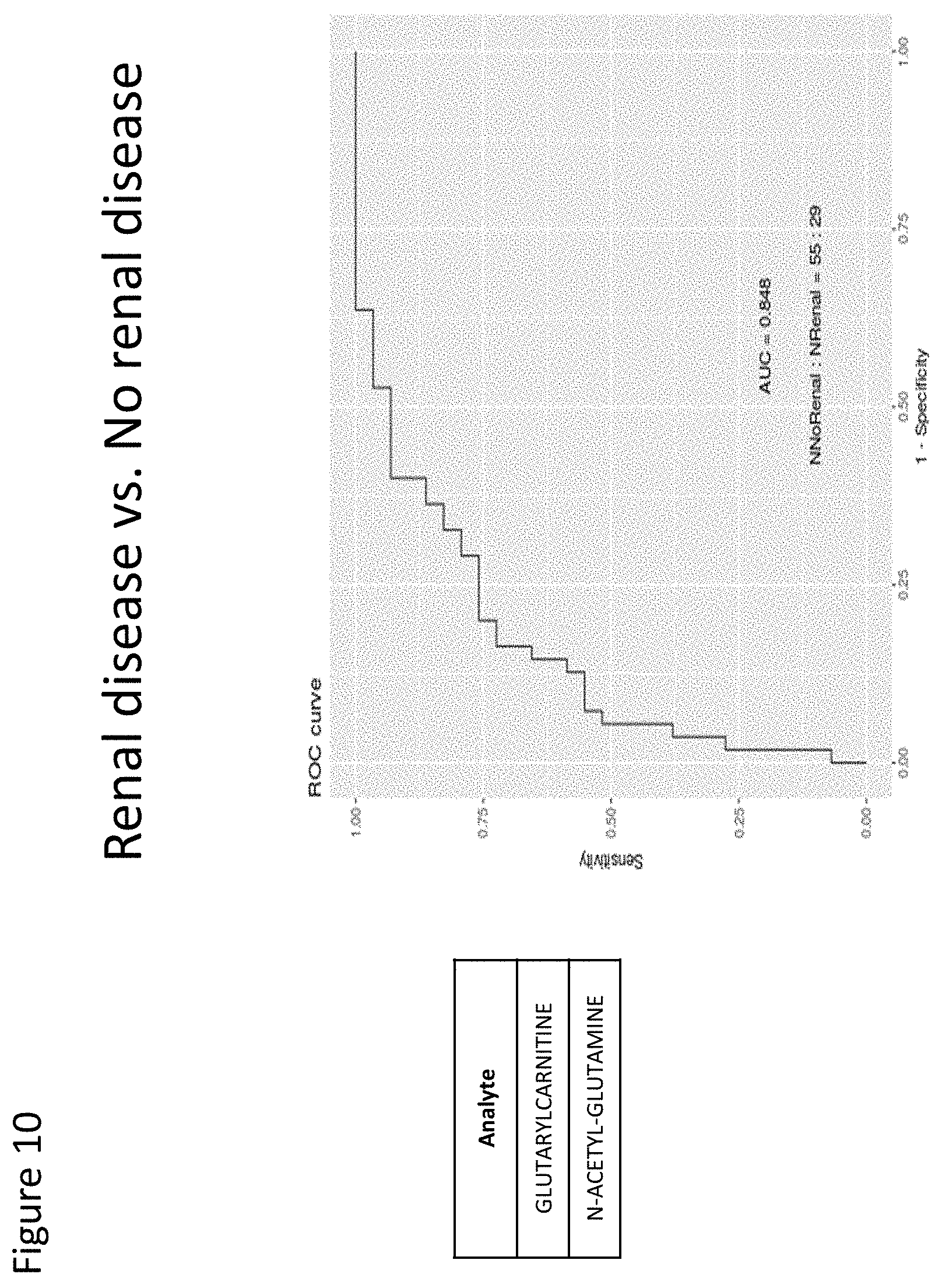
[0012] In one embodiment, the one or more markers comprise at least two or more markers selected from Tables 3 and 4. In another embodiment, the one or more markers comprise glutarylcarnitine and N-acetyl-glutamine. In another embodiment, the one or more markers comprise pentacosanoylglycine and ciliary neurotrophic factor receptor subunit alpha.
[0013] In one embodiment, the one or more markers comprise at least two or more markers selected from Tables 5 and 6. In another embodiment, the one or more markers comprise at least three or more markers selected from Tables 5 and 6. In another embodiment, the one or more markers comprise 1,2-diacetyl-sn-glycero-3-phosphate, coumaric acid and phe-pro.
[0014] In another embodiment, the one or more markers comprise at least four or more markers selected from Tables 5 and 6. In another embodiment, the one or more markers comprise at least five or more markers selected from Tables 5 and 6. In another embodiment, the one or more markers comprise 2-furoylglycine, 3-methylphenylacetic acid, AMP, complement factor D, and ficolin-2.
[0015] In one embodiment, the method further comprises administering a treatment for Lupus where the diagnosis indicates the presence of Lupus in the subject.
[0016] In another embodiment, the method further comprises administering a treatment for renal disease where the diagnosis indicates the presence of renal disease in the subject.
[0017] In another embodiment, the method further comprises administering a treatment for scleroderma where the diagnosis indicates the presence of scleroderma in the subject.
[0018] In one embodiment of any of the foregoing aspects, the level of the one or more of the markers is increased when compared to the predetermined threshold value in the subject. In another embodiment, the level of the one or more of the markers is decreased when compared to the predetermined threshold value in the subject. In another embodiment, the one or more markers comprise one or more markers with an increased level when compared to the predetermined threshold value in the subject, and/or one or more markers with a decreased level when compared to the predetermined threshold value in the subject.
[0019] In one embodiment of any of the foregoing aspects, the biological sample is selected from the group consisting of blood, serum, plasma, urine, organ tissue, biopsy tissue, and seminal fluid. In another embodiment of any of the foregoing aspects, the biological sample is serum. In another embodiment of any of the foregoing aspects, the biological sample is urine.
[0020] In one aspect, the present invention provides a method for classifying the stage or disease progression of Lupus in a subject, comprising detecting the level of one or more markers selected from Tables 1 and 7-12 in a biological sample from the subject; and comparing the level of the one or more markers in the biological sample with a predetermined threshold value; wherein an increased or decreased level of the one or more markers as compared to the predetermined threshold value classifies the stage or disease progression of Lupus in the subject.
[0021] In one embodiment, the subject is stratified based on a Systemic Lupus International Collaborating Clinics (SLICC) damage index. In another embodiment, the subject has an SLICC damage index of less than 2 or an SLICC damage index of 2 or more.
[0022] In another embodiment, the subject is stratified based on systemic Lupus erythematosus disease activity index (SLEDAI) score. In another embodiment, the subject has an SLEDAI score of less than 6 or an SLICC damage index of 6 or more.
[0023] In one embodiment of any of the foregoing aspects, the biological sample is selected from the group consisting of blood, serum, plasma, urine, organ tissue, biopsy tissue, and seminal fluid. In another embodiment of any of the foregoing aspects, the biological sample is serum. In another embodiment of any of the foregoing aspects, the biological sample is urine.
[0024] In one embodiment, the one or more markers comprise at least two or more markers selected from Tables 7-12. In another embodiment, the one or more markers comprise at least three or more markers selected from Tables 7-12.
[0025] In one embodiment, the one or more markers comprise AMP, threonine, cystatin-C and PE-34:2. In another embodiment, the one or more markers comprise coumaric acid and afamin. In another embodiment, the one or more markers comprise AMP and SH3 domain-binding glutamic acid-rich-like protein 3. In another embodiment, the one or more markers comprise coumaric acid and valerylcarnitine.
[0026] In one embodiment of any of the foregoing aspects, the level of the one or more of the markers is increased when compared to the predetermined threshold value in the subject. In another embodiment, the level of the one or more of the markers is decreased when compared to the predetermined threshold value in the subject. In another embodiment, the one or more markers comprise one or more markers with an increased level when compared to the predetermined threshold value in the subject, and/or one or more markers with a decreased level when compared to the predetermined threshold value in the subject.
[0027] In one embodiment, the methods of the invention further comprise administering a treatment for Lupus to the subject. In another embodiment, the methods of the invention further comprise selecting a subject suspected of having or being at risk of having Lupus.
[0028] In another embodiment, the methods of the invention further comprise obtaining a biological sample from a subject suspected of having or being at risk of having Lupus.
[0029] In another embodiment, the level of the marker is detected by HPLC/UV-Vis spectroscopy, enzymatic analysis, mass spectrometry, NMR, immunoassay, ELISA, or any combination thereof. In another embodiment, the level of the marker is detected by determining the level of its corresponding mRNA in the biological sample.
[0030] In one aspect, the present invention is directed to a method for monitoring Lupus in a subject, the method comprising determining a level of at least one of the markers in Tables 1 and 7-12 in a first biological sample obtained at a first time from a subject having Lupus; determining the level of the at least one marker in a second biological sample obtained from the subject at a second time, wherein the second time is later than the first time; and comparing the level of the at least one marker in the second sample with the level of the at least one marker in the first sample, wherein a change in the level of the at least one marker is indicative of a change in the status or stage of Lupus in the subject.
[0031] In one embodiment, the subject is actively treated for Lupus prior to obtaining the second sample. In another embodiment, the subject is not actively treated for Lupus prior to obtaining the second sample. In another embodiment, a change in the level of the at least one marker and/or the one or more additional markers in the second biological sample as compared to the first biological sample is indicative of progression of Lupus in the subject.
[0032] In one embodiment, the method further comprises comparing the level of the at least one marker in the first biological sample or the second biological sample with the level of the at least one marker in a control sample selected from the group consisting of a normal control sample and a sample from a subject with Lupus.
[0033] In one aspect, the present invention provides a method of treating Lupus in a subject, comprising: (a) obtaining a biological sample from a subject suspected of having Lupus, (b) submitting the biological sample to obtain diagnostic information as to the level of at least one of the markers in Tables 1 and 7-12, (c) administering a therapeutically effective amount of a Lupus therapy if the level of the at least one marker is above or below a threshold level.
[0034] In another aspect, the present invention provides a method of treating Lupus in a subject, comprising: (a) obtaining diagnostic information as to the level of at least one of the markers in Tables 1 and 7-12 in a biological sample, and (b) administering a therapeutically effective amount of a Lupus therapy if the level of the at least one marker is above or below a threshold level.
[0035] In another aspect, the present invention provides a method of treating Lupus in a subject, comprising: (a) obtaining a biological sample from a subject suspected of having Lupus for use in identifying diagnostic information as to the level of at least one of the markers in Tables 1 and 7-12, (b) measuring the level of the at least one marker in the biological sample, (c) recommending to a healthcare provider to administer a Lupus therapy if the level of the at least one marker is above or below a threshold level.
[0036] In another aspect, the present invention provides a method of treating renal disease in a subject, comprising: (a) obtaining a biological sample from a subject suspected of having Lupus or renal disease, (b) submitting the biological sample to obtain diagnostic information as to the level of at least one of the markers in Tables 3 and 4, (c) administering a therapeutically effective amount of a Lupus or renal disease therapy if the level of the at least one marker is above or below a threshold level.
[0037] In another aspect, the present invention provides a method of treating renal disease in a subject, comprising: (a) obtaining diagnostic information as to the level of at least one of the markers in Tables 3 and 4 in a biological sample, and (b) administering a therapeutically effective amount of a Lupus or renal disease therapy if the level of the at least one marker is above or below a threshold level.
[0038] In another aspect, the present invention provides a method of treating renal disease in a subject, comprising: (a) obtaining a biological sample from a subject suspected of having Lupus or renal disease for use in identifying diagnostic information as to the level of at least one of the markers in Tables 3 and 4, (b) measuring the level of the at least one marker in the biological sample, (c) recommending to a healthcare provider to administer a Lupus or renal disease therapy if the level of the at least one marker is above or below a threshold level.
[0039] In another aspect, the present invention provides a method of treating scleroderma in a subject, comprising: (a) obtaining a biological sample from a subject suspected of having Lupus or scleroderma, (b) submitting the biological sample to obtain diagnostic information as to the level of at least one of the markers in Tables 5 and 6, (c) administering a therapeutically effective amount of a Lupus or scleroderma therapy if the level of the at least one marker is above or below a threshold level.
[0040] In another aspect, the present invention provides a method of treating scleroderma in a subject, comprising: (a) obtaining diagnostic information as to the level of at least one of the markers in Tables 5 and 6 in a biological sample, and (b) administering a therapeutically effective amount of a Lupus or scleroderma therapy if the level of the at least one marker is above or below a threshold level.
[0041] In another aspect, the present invention provides a method of treating scleroderma in a subject, comprising: (a) obtaining a biological sample from a subject suspected of having Lupus or scleroderma for use in identifying diagnostic information as to the level of at least one of the markers in Tables 5 and 6, (b) measuring the level of the at least one marker in the biological sample, (c) recommending to a healthcare provider to administer a Lupus or scleroderma therapy if the level of the at least one marker is above or below a threshold level.
[0042] In one embodiment of any of the preceeding aspects, the method further comprises obtaining diagnostic information as to the level of one or more additional markers of Lupus, renal disease or scleroderma.
[0043] In one embodiment of any of the preceeding aspects, the method further comprises measuring the level of the one or more additional markers of Lupus, renal disease or scleroderma.
[0044] In one aspect, the present invention provides a kit for detecting one or more markers in a biological sample from a subject having, suspected of having, or at risk for having Lupus, comprising one or more reagents for measuring the level of the one or more markers in the biological sample from the subject, wherein the one or more markers comprise one or more markers selected from Tables 1 and 7-12, and a set of instructions for measuring the level of the marker.
[0045] In one embodiment, the reagent is an antibody. In another embodiment, the method further comprises a means to detect the antibody.
[0046] In one embodiment, the reagent is an oligonucleotide that is complementary to the corresponding mRNA of the one or more markers.
[0047] In one embodiment, the instructions set forth an immunoassay, ELISA, or mass spectrometry assay for detecting the level of the one or more markers in the biological sample. In another embodiment, the instructions set forth an amplification reaction for assaying the level of the mRNA in the biological sample corresponding to the one or more markers.
[0048] In another embodiment, the instructions set forth a hybridization assay for detecting the level of the mRNA in the biological sample corresponding to the one or more markers.
[0049] In another embodiment, the instructions further set forth comparing the level of the one or more markers in the biological sample from the subject to a predetermined threshold value of the marker.
[0050] In another embodiment, the instructions further set forth making a diagnosis of Lupus based on the level of the one or more markers in the biological sample from the subject as compared to a predetermined threshold value of the one or more markers.
[0051] In another aspect, the invention provides a kit for detecting one or more markers in a biological sample from a subject having, suspected of having, or at risk for having renal disease, comprising one or more reagents for measuring the level of the one or more markers in the biological sample from the subject, wherein the one or more markers comprise one or more markers selected from Tables 3 and 4, and a set of instructions for measuring the level of the renal disease marker.
[0052] In one embodiment, the reagent is an antibody. In another embodiment, the method further comprises a means to detect the antibody.
[0053] In one embodiment, the reagent is an oligonucleotide that is complementary to the corresponding mRNA of the one or more markers.
[0054] In one embodiment, the instructions set forth an immunoassay, ELISA, or mass spectrometry assay for detecting the level of the one or more markers in the biological sample. In another embodiment, the instructions set forth an amplification reaction for assaying the level of the mRNA in the biological sample corresponding to the one or more markers.
[0055] In another embodiment, the instructions set forth a hybridization assay for detecting the level of the mRNA in the biological sample corresponding to the one or more markers.
[0056] In another embodiment, the instructions further set forth comparing the level of the one or more markers in the biological sample from the subject to a predetermined threshold value of the marker.
[0057] In another embodiment, the instructions further set forth making a diagnosis of renal disease based on the level of the one or more markers in the biological sample from the subject as compared to a predetermined threshold value of the one or more markers.
[0058] A kit for detecting one or more markers in a biological sample from a subject having, suspected of having, or at risk for having scleroderma, comprising one or more reagents for measuring the level of the one or more markers in the biological sample from the subject, wherein the one or more markers comprises one or more markers selected from Tables 5 and 6 and a set of instructions for measuring the level of the one or more markers.
[0059] In one embodiment, the reagent is an antibody. In another embodiment, the method further comprises a means to detect the antibody.
[0060] In one embodiment, the reagent is an oligonucleotide that is complementary to the corresponding mRNA of the one or more markers.
[0061] In one embodiment, the instructions set forth an immunoassay, ELISA, or mass spectrometry assay for detecting the level of the one or more markers in the biological sample. In another embodiment, the instructions set forth an amplification reaction for assaying the level of the mRNA in the biological sample corresponding to the one or more markers.
[0062] In another embodiment, the instructions set forth a hybridization assay for detecting the level of the mRNA in the biological sample corresponding to the one or more markers.
[0063] In another embodiment, the instructions further set forth comparing the level of the one or more markers in the biological sample from the subject to a predetermined threshold value of the marker.
[0064] In another embodiment, the instructions further set forth making a diagnosis of scleroderma based on the level of the one or more markers in the biological sample from the subject as compared to a predetermined threshold value of the one or more markers.
[0065] In one aspect, the present invention provides a panel for use in a method of diagnosing Lupus, the panel comprising one or more detection reagents, wherein each detection reagent is specific for the detection of one or more markers selected from Tables 1 and 7-12.
[0066] In one embodiment, the marker comprises at least two or more markers selected from Tables 1 and 7-12. In another embodiment, the marker comprises AMP and S-adenosyl-L-homocysteine.
[0067] In one embodiment, the invention provides a kit comprising a panel of the invention and a set of instructions for obtaining diagnostic information based on a level of the one or more markers.
[0068] In another embodiment, the level of the one or more markers is increased when compared to a predetermined threshold value. In another embodiment, the level of the one or more markers is decreased when compared to a predetermined threshold value. In another embodiment, the one or more markers comprise one or more markers with an increased level when compared to a predetermined threshold value, and/or one or more markers with a decreased level when compared to a predetermined threshold value.
[0069] A panel for use in a method of diagnosing renal disease, the panel comprising one or more detection reagents, wherein each detection reagent is specific for one or more markers selected from Tables 3 and 4.
[0070] In one embodiment, the marker comprises at least two or more markers selected from Tables 3 and 4. In another embodiment, the marker comprises glutarylcarnitine and N-acetyl-glutamine. In another embodiment, the marker comprises pentacosanoylglycine and ciliary neurotrophic factor receptor subunit alpha.
[0071] In one embodiment, the invention provides a kit comprising a panel of the invention and a set of instructions for obtaining diagnostic information based on a level of the one or more markers.
[0072] In another embodiment, the level of the one or more markers is increased when compared to a predetermined threshold value. In another embodiment, the level of the one or more markers is decreased when compared to a predetermined threshold value. In another embodiment, the one or more markers comprise one or more markers with an increased level when compared to a predetermined threshold value, and/or one or more markers with a decreased level when compared to a predetermined threshold value.
[0073] A panel for use in a method of diagnosing scleroderma or differentiating between scleroderman and lupus, the panel comprising one or more detection reagents, wherein each detection reagent is specific for the detection of one or more markers selected from Tables 5 and 6.
[0074] In one embodiment, the marker comprises at least two or more markers selected from Tables 5 and 6. In another embodiment, the marker comprises at least three or more markers selected from Tables 5 and 6. In another embodiment, the marker comprises 1,2-diacetyl-sn-glycero-3-phosphate, coumaric acid and phe-pro. In another embodiment, the marker comprises at least four or more markers selected from Tables 5 and 6. In another embodiment, the marker comprises at least five or more markers selected from Tables 5 and 6. In another embodiment, the marker comprises 2-furoylglycine, 3-methylphenylacetic acid, AMP, complement factor D, and ficolin-2.
[0075] In one embodiment, the invention provides a kit comprising a panel of the invention and a set of instructions for obtaining diagnostic information based on a level of the one or more markers.
[0076] In another embodiment, the level of the one or more markers is increased when compared to a predetermined threshold value. In another embodiment, the level of the one or more markers is decreased when compared to a predetermined threshold value. In another embodiment, the one or more markers comprise one or more markers with an increased level when compared to a predetermined threshold value, and/or one or more markers with a decreased level when compared to a predetermined threshold value.
[0077] Where applicable or not specifically disclaimed, any one of the embodiments described herein are contemplated to be able to combine with any other one or more embodiments, even though the embodiments are described under different aspects of the invention.
[0078] These and other embodiments are disclosed or are obvious from and encompassed by, the following Detailed Description.
BRIEF DESCRIPTION OF THE DRAWINGS
[0079] The following detailed description, given by way of example, but not intended to limit the invention solely to the specific embodiments described, may best be understood in conjunction with the accompanying drawings.
[0080] FIG. 1 depicts the study aims and workflow to identify biomarkers for Lupus using the Interrogative Biology.RTM. platform.
[0081] FIG. 2 depicts the Berg Interrogative Biology.RTM. Discovery workflow.
[0082] FIG. 3 depicts the Berg Interrogative Biology.RTM. Artificial Intelligence Clinical Information System, which utilizes Bayesian AI-based software technology bAIcis.RTM..
[0083] FIG. 4 depicts exemplary use of mass spectrometry to identify protein, lipid and metabolite markers.
[0084] FIG. 5 is a schematic depicting the bAIcis.RTM. and statisitical analysis pipline used to identify the markers of the invention.
[0085] FIG. 6 is a schematic depicting the deconstructed Lupus network.
[0086] FIG. 7 is a box plot depicting a direct comparison of normalized expression levels of marker AMP between Lupus patients and negative controls.
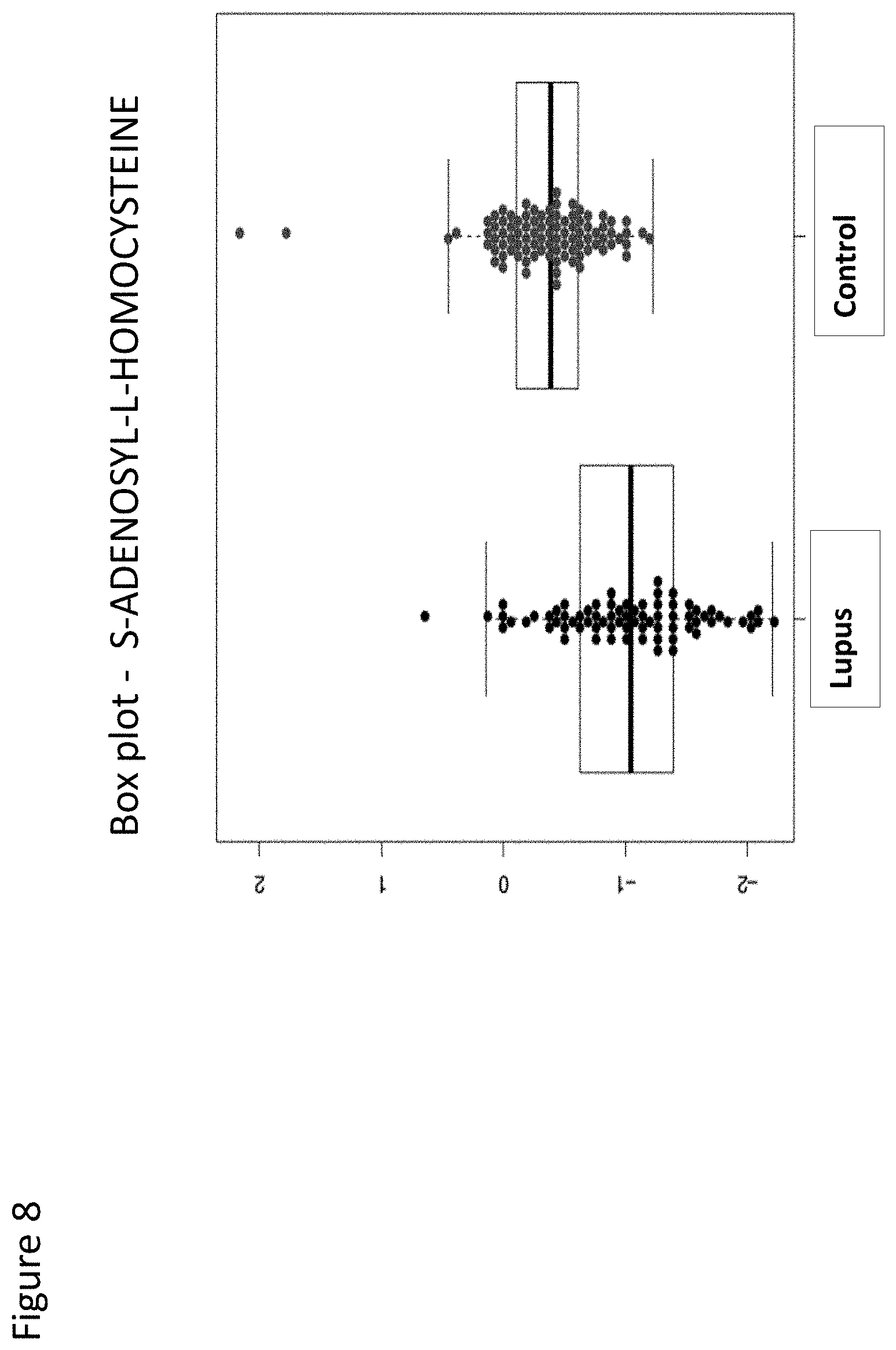
[0087] FIG. 8 is a box plot depicting a direct comparison of normalized expression levels of marker S-adenosyl-L-homocysteine between Lupus patients and negative controls.
[0088] FIG. 9 depicts a ROC curve with a predictive diagnostic value of 0.836 for two serum markers (AMP and S-adenosyl-L-homocysteine) for patients with Lupus.
[0089] FIG. 10 depicts a ROC curve with a predictive diagnostic value of 0.848 for two serum markers (glutarylcarnitine and N-acetyl-glutamine) for patients with renal disease.
[0090] FIG. 11 depicts a ROC curve with a predictive diagnostic value of 0.844 for two urine markers (pentacosanoylglycine and ciliary neurotrophic factor receptor subunit alpha) for patients with renal disease.
[0091] FIG. 12 depicts a ROC curve with a predictive diagnostic value of 0.831 for five serum markers (2-furoylglycine, 3-methylphenylacetic acid, AMP, complement factor D, and ficolin-2) for patients with scleroderma and patients with Lupus.
[0092] FIG. 13 depicts a ROC curve with a predictive diagnostic value of 0.771 for three urine markers (1,2-diacetyl-sn-glycero-3-phosphate, coumaric acid, phe-pro) for patients with scleroderma and patients with Lupus.
[0093] FIG. 14 depicts a ROC curve with a predictive classification value of 0.829 for four serum markers (AMP, threonine, cystatin-C and PE-34:2) for patients with a Systemic Lupus International Collaborating Clinics (SLICC) damage index of less than 2 and patients with a SLICC damage index of 2 or more.
[0094] FIG. 15 depicts a ROC curve with a predictive classification value of 0.77 for two urine markers (coumaric acid and afamin) for patients with a SLICC damage index of less than 2 and patients with a SLICC damage index of 2 or more.
[0095] FIG. 16 depicts a ROC curve with a predictive classification value of 0.809 for two serum markers (AMP and SH3 domain-binding glutamic acid-rich-like protein 3) for patients with a systemic Lupus erythematosus disease activity index (SLEDAI) score less than 6 and patients with a SLEDAI score of 6 or more.
[0096] FIG. 17 depicts a ROC curve with a predictive classification value of 0.641 for two urine markers (coumaric acid and valerylcarnitine) for patients with a SLEDAI score less than 6 and patients with a SLEDAI score of 6 or more.
[0097] FIG. 18 depicts a ROC curve with a predictive diagnostic value of 0.604 for one serum marker (AMP) for use in an antinuclear antibody (ANA) test.
[0098] FIG. 19 depicts a ROC curve with a predictive diagnostic value of 0.73 for two urine markers (coumaric acid and valerylcarnitine) for use in an antinuclear antibody (ANA) test.
DETAILED DESCRIPTION OF THE INVENTION
A. Overview
[0099] As presently described herein, the invention at hand is based, at least in part, on the discovery that the levels of the markers listed in Tables 1-12 are modulated in subjects having Lupus, and across various levels of disease activity, and thus serve as useful markers of Lupus and markers of stages of Lupus.
[0100] The present invention is based, also in part, on the discovery that the levels of the markers listed in Table 2 are modulated in subjects having Lupus versus subjects that do not have Lupus.
[0101] The present invention is based, also in part, on the discovery that the levels of the markers listed in Tables 3 and 4 are modulated in subjects having renal disease versus subjects that do not have renal disease.
[0102] The present invention is based, also in part, on the discovery that the levels of the markers listed in Tables 5 and 6 are modulated in subjects having scleroderma versus subjects having Lupus.
[0103] The present invention is based, also in part, on the discovery that the levels of the markers listed in Tables 7 and 8 are associated with subjects having an SLICC score of less than 2 versus subjects having an SLICC score of greater than or equal to 2.
[0104] The present invention is based, also in part, on the discovery that the levels of the markers listed in Tables 9 and 10 are associated with subjects having an SLEDAI score of less than 6 versus subjects having an SLEDAI score of greater than or equal to 6.
[0105] The present invention is based, also in part, on the discovery that the levels of the markers listed in Tables 11 and 12 are modulated in subjects having a positive ANA result versus subjects having a normal ANA result.
[0106] As described in the Examples, the inventors used retrospectively collected and clinically annotated serum and urine samples from 166 patients (90 African American and 71 Caucasian patients). Additional medical data included a range of clinical and omic data sets, including demographic data, ACR classification criteria, Systemic Lupus International Collaborating Clinic (SLICC) damage index, SLE disease activity index (DAI) scores, lab data, and medication information.
[0107] The inventors then used BERG's Interrogative Biology.RTM. platform to process and integrate samples into a harmonized dataset, then conducted analysis using BERG's AI technology, bAIcis.RTM., to identify panels of Lupus candidate biomarkers, each with a target area under the AUROC (Area Under the Receiver Operating Characteristics) curve of 0.8 with the minimal combination of up to six biomarkers. Biomarker panels were analyzed separately for each biomatrix and revealed new targets for further clinical analysis based on several patient types and disease characteristics: [0108] Patients with Lupus vs those without: two biomarkers in serum with AUC 0.836 (Table 2) and five in urine with AUC 0.805. [0109] Patients with renal disease vs those without: two biomarkers in serum with AUC 0.848 (Table 3) and two in urine with AUC 0.844 (Table 4). [0110] Patients with scleroderma vs. those without: two biomarkers in serum with AUC 0.826 and two in urine with AUC 0.705. [0111] Patients with scleroderma vs. those with Lupus: five biomarkers in serum with AUC 0.831 (Table 5) and three in urine with AUC 0.771 (Table 6). [0112] SLICC by disease stage (<2 vs >=2): four biomarkers in serum with AUC 0.829 (Table 7) and two in urine with AUC 0.77 (Table 8). [0113] SLEDAI score (<6 vs >=6): two biomarkers in serum with AUC 0.809 (Table 9) and two in urine with AUC 0.641 (Table 10). [0114] ANA: one biomarker in serum with AUC 0.604 (Table 11) and two in urine with AUC 0.73 (Table 12). [0115] Drug efficacy for Mycophenolate: two biomarkers in serum with AUC 0.847 and one in urine with AUC 0.933.
[0116] Accordingly, in one embodiment, one or more markers in Tables 1, 2 and 7-12 can serve as useful diagnostic markers to predict and/or detect the presence of Lupus in a subject, or the stage of progression of the disease. In another embodiment, one or more markers in Tables 1, 2 and 7-12 can serve as a useful prognostic markers, serving to inform on the likely progression of Lupus in a subject with or without treatment. In still another embodiment, one or more markers in Tables 1, 2 and 7-12 can serve as a useful predictive markers for helping to assess the likely response of Lupus to a particular treatment.
[0117] In one embodiment, one or more markers in Tables 3 and 4 can serve as useful diagnostic markers to predict and/or detect the presence of renal disease in a subject, e.g., a subject having Lupus, or the stage of progression of the disease. In another embodiment, one or more markers in Tables 3 and 4 can serve as a useful prognostic markers, serving to inform on the likely progression of renal disease in a subject, e.g., a subject having Lupus, with or without treatment. In still another embodiment, one or more markers in Tables 3 and 4 can serve as a useful predictive markers for helping to assess the likely response of renal disease to a particular treatment.
[0118] In another embodiment, one or more markers in Tables 5 and 6 can serve as useful diagnostic markers to predict and/or detect the presence of scleroderma in a subject, or the stage of progression of the disease. In another embodiment, one or more markers in Tables 5 and 6 can serve as useful diagnostic markers to distinguish the presence of scleroderma from Lupus in a subject. In another embodiment, one or more markers in Tables 5 and 6 can serve as a useful prognostic markers, serving to inform on the likely progression of scleroderma in a subject, with or without treatment. In still another embodiment, one or more markers in Tables 5 and 6 can serve as a useful predictive markers for helping to assess the likely response of scleroderma to a particular treatment.
[0119] In another embodiment, one or more markers in Tables 7-10 can serve as useful diagnostic markers to classify the stage or disease progression of Lupus in a subject, for example as defined by SLEDAI or SLICC scores.
[0120] In another embodiment, one or more markers in Tables 11 and 12 can serve as useful diagnostic markers to predict and/or detect the presence of Lupus in a subject, or the stage of progression of the disease. In another embodiment, one or more markers in Tables 11 and 12 can serve as a useful prognostic markers, serving to inform on the likely progression of Lupus in a subject, with or without treatment. In still another embodiment, one or more markers in Tables 11 and 12 can serve as a useful predictive markers for helping to assess the likely response of Lupus to a particular treatment.
[0121] Accordingly, the invention provides methods that use one or more markers, e.g., one or more markers in Tables 1-12, in the diagnosis of Lupus (e.g., prediction of the presence of Lupus in a subject), in the diagnosis of the stage of Lupus (e.g., diagnosis of the stage of Lupus in a subject), in the prognosis of Lupus (e.g., prediction of the course or outcome of Lupus with or without treatment), and in the assessment of therapies intended to treat Lupus (i.e., the one or more markers in Tables 1-12 as a theragnostic or predictive marker). The invention further provides compositions of matter, including panels comprising binding or detection reagents specific for the one or more markers in Tables 1-12 and optionally other markers for use in the methods of the invention, as well as kits for practicing the methods of the invention.
[0122] In one embodiment, the invention provides methods for diagnosing Lupus in a subject following ANA testing using one or more markers in Tables 11 and 12.
[0123] The invention also provides methods that use one or more markers, e.g., one or more markers in Tables 3 and 4, in the diagnosis of renal disease (e.g., prediction of the presence of renal disease in a subject, e.g, a subject having Lupus), in the diagnosis of the stage of renal disease (e.g., diagnosis of the stage of renal disease in a subject), in the prognosis of renal disease (e.g., prediction of the course or outcome of renal disease with or without treatment), and in the assessment of therapies intended to treat renal disease.
[0124] The invention also provides methods that use one or more markers, e.g., one or more markers in Tables 5 and 6, in the diagnosis of scleroderma (e.g., prediction of the presence of renal disease in a subject, e.g, a subject having Lupus), or to distinguish between Lupus and scleroderma, in the diagnosis of the stage of scleroderma (e.g., diagnosis of the stage of scleroderma in a subject), in the prognosis of scleroderma (e.g., prediction of the course or outcome of scleroderma with or without treatment), and in the assessment of therapies intended to treat scleroderma.
[0125] The invention also provides methods that use one or more markers, e.g., one or more markers in Tables 7-10, in the diagnosis of the stage or disease progression of Lupus (e.g., diagnosis of the stage of Lupus in a subject).
[0126] The following is a detailed description of the invention provided to aid those skilled in the art in practicing the present invention. Those of ordinary skill in the art may make modifications and variations in the embodiments described herein without departing from the spirit or scope of the present invention. Unless otherwise defined, all technical and scientific terms used herein have the same meaning as commonly understood by one of ordinary skill in the art to which this invention belongs. The terminology used in the description of the invention herein is for describing particular embodiments only and is not intended to be limiting of the invention. All publications, patent applications, patents, figures and other references mentioned herein are expressly incorporated by reference in their entirety.
[0127] Although any methods and materials similar or equivalent to those described herein can also be used in the practice or testing of the present invention, the preferred methods and materials are now described. All publications mentioned herein are incorporated herein by reference to disclose and described the methods and/or materials in connection with which the publications are cited.
B. Definitions
[0128] Unless defined otherwise, all technical and scientific terms used herein have the meaning commonly understood by a person skilled in the art to which this invention belongs. The following references, the entire disclosures of which are incorporated herein by reference, provide one of skill with a general definition of many of the terms (unless defined otherwise herein) used in this invention: Singleton et al., Dictionary of Microbiology and Molecular Biology (2.sup.nd ed. 1994); The Cambridge Dictionary of Science and Technology (Walker ed., 1988); The Glossary of Genetics, 5.sup.th Ed., R. Rieger et al. (eds.), Springer Verlag (1991); and Hale & Marham, the Harper Collins Dictionary of Biology (1991). Generally, the procedures of molecular biology methods described or inherent herein and the like are common methods used in the art. Such standard techniques can be found in reference manuals such as for example Sambrook et al., (2000, Molecular Cloning--A Laboratory Manual, Third Edition, Cold Spring Harbor Laboratories); and Ausubel et al., (1994, Current Protocols in Molecular Biology, John Wiley & Sons, New-York).
[0129] The following terms may have meanings ascribed to them below, unless specified otherwise. However, it should be understood that other meanings that are known or understood by those having ordinary skill in the art are also possible, and within the scope of the present invention. All publications, patent applications, patents, and other references mentioned herein are incorporated by reference in their entirety. In the case of conflict, the present specification, including definitions, will control. In addition, the materials, methods, and examples are illustrative only and not intended to be limiting.
[0130] As used herein, the singular forms "a", "and", and "the" include plural references unless the context clearly dictates otherwise. All technical and scientific terms used herein have the same meaning.
[0131] Unless specifically stated or obvious from context, as used herein, the term "about" is understood as within a range of normal tolerance in the art, for example within 2 standard deviations of the mean. About can be understood as within 10%, 9%, 8%, 7%, 6%, 5%, 4%, 3%, 2%, 1%, 0.5%, 0.1%, 0.05%, or 0.01% of the stated value. Unless otherwise clear from context, all numerical values provided herein can be modified by the term about.
[0132] As used herein, the term "amplification" refers to any known in vitro procedure for obtaining multiple copies ("amplicons") of a target nucleic acid sequence or its complement or fragments thereof. In vitro amplification refers to production of an amplified nucleic acid that may contain less than the complete target region sequence or its complement. Known in vitro amplification methods include, e.g., transcription-mediated amplification, replicase-mediated amplification, polymerase chain reaction (PCR) amplification, ligase chain reaction (LCR) amplification and strand-displacement amplification (SDA including multiple strand-displacement amplification method (MSDA)). Replicase-mediated amplification uses self-replicating RNA molecules, and a replicase such as Q-.beta.-replicase (e.g., Kramer et al., U.S. Pat. No. 4,786,600). PCR amplification is well known and uses DNA polymerase, primers and thermal cycling to synthesize multiple copies of the two complementary strands of DNA or cDNA (e.g., Mullis et al., U.S. Pat. Nos. 4,683,195, 4,683,202, and 4,800,159). LCR amplification uses at least four separate oligonucleotides to amplify a target and its complementary strand by using multiple cycles of hybridization, ligation, and denaturation (e.g., EP Pat. App. Pub. No. 0 320 308). SDA is a method in which a primer contains a recognition site for a restriction endonuclease that permits the endonuclease to nick one strand of a hemimodified DNA duplex that includes the target sequence, followed by amplification in a series of primer extension and strand displacement steps (e.g., Walker et al., U.S. Pat. No. 5,422,252). Two other known strand-displacement amplification methods do not require endonuclease nicking (Dattagupta et al., U.S. Pat. Nos. 6,087,133 and 6,124,120 (MSDA)). Those skilled in the art will understand that the oligonucleotide primer sequences of the present invention may be readily used in any in vitro amplification method based on primer extension by a polymerase. (see generally Kwoh et al., 1990, Am. Biotechnol. Lab. 8:14-25 and (Kwoh et al., 1989, Proc. Natl. Acad. Sci. USA 86, 1173-1177; Lizardi et al., 1988, BioTechnology 6:1197-1202; Malek et al., 1994, Methods Mol. Biol., 28:253-260; and Sambrook et al., 2000, Molecular Cloning--A Laboratory Manual, Third Edition, CSH Laboratories). As commonly known in the art, the oligos are designed to bind to a complementary sequence under selected conditions.
[0133] As used herein, the term "antigen" refers to a molecule, e.g., a peptide, polypeptide, protein, fragment, or other biological moiety, which elicits an antibody response in a subject, or is recognized and bound by an antibody.
[0134] As used herein, the term "area under the curve" or "AUC" refers to the area under the curve in a plot of sensitivity versus specificity. In one embodiment, the AUC for a biomarker, or combination of biomarkers, of the invention is 0.5. In another embodiment, the AUC for a biomarker, or combination of biomarkers, of the invention is 0.6. In another embodiment, the AUC for a biomarker, or combination of biomarkers, of the invention is 0.7. In another embodiment, the AUC for a biomarker, or combination of biomarkers, of the invention is 0.8. In another embodiment, the AUC for a biomarker, or combination of biomarkers, of the invention is 0.9. In another embodiment, the AUC for a biomarker, or combination of biomarkers, of the invention is 1.0. In specific embodiments, the AUC for a biomarker, or combination of biomarkers, of the invention is 0.5, 0.51, 0.52, 0.53, 0.54, 0.55, 0.56, 0.57, 0.58, 0.59, 0.6, 0.61, 0.62, 0.63, 0.64, 3.65, 0.66, 0.67, 0.68, 0.69, 0.7, 0.71, 0.72, 0.73, 0.74, 0.75, 0.76, 0.77, 0.78, 0.79, 0.8, 0.81, 0.82, 0.83, 0.84, 0.85, 0.86, 0.87, 0.88, 0.89, 0.9, 0.91, 0.92, 0.93, 0.94, 0.95, 0.96, 0.97, 0.98, 0.99 or 1.0. In one embodiment, the AUC for a biomarker, or combination of biomarkers, of the invention is at least 0.5. In another embodiment, the AUC for a biomarker, or combination of biomarkers, of the invention is at least 0.6. In another embodiment, the AUC for a biomarker, or combination of biomarkers, of the invention is at least 0.7. In another embodiment, the AUC for a biomarker, or combination of biomarkers, of the invention is at least 0.8. In another embodiment, the AUC for a biomarker, or combination of biomarkers, of the invention is at least 0.9. In another embodiment, the AUC for a biomarker, or combination of biomarkers, of the invention is at least 1.0. In specific embodiments, the AUC for a biomarker, or combination of biomarkers, of the invention is at least 0.5, 0.51, 0.52, 0.53, 0.54, 0.55, 0.56, 0.57, 0.58, 0.59, 0.6, 0.61, 0.62, 0.63, 0.64, 3.65, 0.66, 0.67, 0.68, 0.69, 0.7, 0.71, 0.72, 0.73, 0.74, 0.75, 0.76, 0.77, 0.78, 0.79, 0.8, 0.81, 0.82, 0.83, 0.84, 0.85, 0.86, 0.87, 0.88, 0.89, 0.9, 0.91, 0.92, 0.93, 0.94, 0.95, 0.96, 0.97, 0.98, 0.99 or 1.0
[0135] As used herein, the term "biomarker" or "marker" is understood to mean a measurable characteristic that reflects in a quantitative or qualitative manner the physiological state of an organism. The physiological state of an organism is inclusive of any disease or non-disease state, e.g., a subject having Lupus or a subject who is otherwise healthy. Said another way, markers are characteristics that can be objectively measured and evaluated as indicators of normal processes, pathogenic processes, or pharmacologic responses to a therapeutic intervention. Markers can be clinical parameters (e.g., age, performance status), laboratory measures (e.g., molecular markers), imaging-based measures, or genetic or other molecular determinants, such as phosphorylation or acetylation state of a protein marker, methylation state of nucleic acid, or any other detectable molecular modification to a biological molecule. Examples of markers include, for example, polypeptides, peptides, polypeptide fragments, proteins, antibodies, hormones, polynucleotides, RNA or RNA fragments, microRNA (miRNAs), lipids (e.g., structural lipids or signaling lipids), polysaccharides, and other bodily metabolites. In one embodiment, a biomarker of the invention is one or more of the biomarkers included in Tables 1-12. In another embodiment, a biomarker of the invention is one that is metabolically stable over time (e.g., over the course of 1, 2, 3, 4, 5, 6, 7, or more days), and is metabolically stable regardless of the diet of the subject. In still another embodiment, a biomarker of the invention is one that has a consistent biomarker profile regardless of whether or not the patient had been previously or is currently taking medications for Lupus, renal disease, scleroderma, or a related disease or disorder.
[0136] Preferably, a marker of the present invention is modulated (e.g., increased or decreased level) in a biological sample from a subject or a group of subjects having a first phenotype (e.g., having a disease or a certain stage or disease progression) as compared to a biological sample from a subject or group of subjects having a second phenotype (e.g., not having the disease, e.g., a control). A marker may be differentially present at any level, but is generally present at a level that is increased relative to normal or control levels by at least 5%, by at least 10%, by at least 15%, by at least 20%, by at least 25%, by at least 30%, by at least 35%, by at least 40%, by at least 45%, by at least 50%, by at least 55%, by at least 60%, by at least 65%, by at least 70%, by at least 75%, by at least 80%, by at least 85%, by at least 90%, by at least 95%, by at least 100%, by at least 110%, by at least 120%, by at least 130%, by at least 140%, by at least 150%, or more; or is generally present at a level that is decreased relative to normal or control levels by at least 5%, by at least 10%, by at least 15%, by at least 20%, by at least 25%, by at least 30%, by at least 35%, by at least 40%, by at least 45%, by at least 50%, by at least 55%, by at least 60%, by at least 65%, by at least 70%, by at least 75%, by at least 80%, by at least 85%, by at least 90%, by at least 95%, or by 100% (i.e., absent). A marker is preferably differentially present at a level that is statistically significant (e.g., a p-value less than 0.05 and/or a q-value of less than 0.10 as determined using either Welch's T-test or Wilcoxon's rank-sum Test).
[0137] As used herein, the term "complementary" refers to the broad concept of sequence complementarity between regions of two nucleic acid strands or between two regions of the same nucleic acid strand. It is known that an adenine residue of a first nucleic acid region is capable of forming specific hydrogen bonds ("base pairing") with a residue of a second nucleic acid region which is antiparallel to the first region if the residue is thymine or uracil. Similarly, it is known that a cytosine residue of a first nucleic acid strand is capable of base pairing with a residue of a second nucleic acid strand which is antiparallel to the first strand if the residue is guanine. A first region of a nucleic acid is complementary to a second region of the same or a different nucleic acid if, when the two regions are arranged in an antiparallel fashion, at least one nucleotide residue of the first region is capable of base pairing with a residue of the second region. Preferably, the first region comprises a first portion and the second region comprises a second portion, whereby, when the first and second portions are arranged in an antiparallel fashion, at least about 50%, and preferably at least about 75%, at least about 90%, or at least about 95% of the nucleotide residues of the first portion are capable of base pairing with nucleotide residues in the second portion. More preferably, all nucleotide residues of the first portion are capable of base pairing with nucleotide residues in the second portion.
[0138] The term "control sample," as used herein, refers to any clinically relevant comparative sample, including, for example, a sample from a healthy subject not afflicted with Lupus, or a sample from a subject from an earlier time point, e.g., prior to treatment, an earlier assessment time point, at an earlier stage of treatment, or at an earlier stage of disease progression. A control sample can be a purified sample, metabolite, lipid, protein, and/or nucleic acid provided with a kit. Such control samples can be diluted, for example, in a dilution series to allow for quantitative measurement of levels of analytes, e.g., markers, in test samples. A control sample may include a sample derived from one or more subjects. A control sample may also be a sample made at an earlier time point from the subject to be assessed. For example, the control sample could be a sample taken from the subject to be assessed before the onset of a disorder, e.g., Lupus, at an earlier stage of disease, or before the administration of treatment or of a portion of treatment. The control sample may also be a sample from an animal model, or from a tissue or cell line derived from the animal model of a disorder, e.g., Lupus. The level of activity or expression of one or more markers (e.g., 1, 2, 3, 4, 5, 6, 7, 8, 9, 10, 11, 12, 13, 14, 15, 16, 17, 18, 19, 20, 21, 22, 23, 24, 25, 26, 27, 28, 29, 30 or more markers) in a control sample consists of a group of measurements that may be determined, e.g., based on any appropriate statistical measurement, such as, for example, measures of central tendency including average, median, or modal values. Different from a control is preferably statistically significantly different from a control.
[0139] As used herein, "changed as compared to a control" sample or subject is understood as having a level of the analyte or diagnostic or therapeutic indicator (e.g., marker) to be detected at a level that is statistically different than a sample from a normal, untreated, or abnormal state control sample. Changed as compared to control can also include a difference in the rate of change of the level of one or more markers obtained in a series of at least two subject samples obtained over time. Determination of statistical significance is within the ability of those skilled in the art and can include any acceptable means for determining and/or measuring statistical significance, such as, for example, the number of standard deviations from the mean that constitute a positive or negative result, an increase in the detected level of a biomarker in a sample (e.g., Lupus sample) versus a control or healthy sample, wherein the increase is above some threshold value, or a decrease in the detected level of a biomarker in a sample (e.g., Lupus sample) versus a control or healthy sample, wherein the decrease is below some threshold value. The threshold value can be determine by any suitable means by measuring the biomarker levels in a plurality of tissues or samples known to have a disease, e.g., Lupus, and comparing those levels to a normal sample and calculating a statistically significant threshold value.
[0140] The term "control level" refers to an accepted or pre-determined level of a marker in a subject sample. A control level can be a range of values. Marker levels can be compared to a single control value, to a range of control values, to the upper level of normal, or to the lower level of normal as appropriate for the assay. In one embodiment, the control is a standardized control, such as, for example, a control which is predetermined using an average of the levels of expression of one or more markers from a population of subjects having no Lupus.
[0141] In one embodiment, the control is a standardized control, such as, for example, a control which is predetermined using an average of the levels of expression of one or more markers from a population of subjects not having Lupus. A control can also be a sample from a subject at an earlier time point, e.g., a baseline level prior to suspected presence of disease, before the diagnosis of a disease, before the treatment with a specific agent or intervention. In certain embodiments, a change in the level of the marker in a subject can be more significant than the absolute level of a marker, e.g., as compared to control.
[0142] As used herein, "detecting", "detection", "determining", and the like are understood to refer to an assay performed for identification of one or more specific markers in a sample, e.g., one or more (e.g., 1, 2, 3, 4, 5, 6, 7, 8, 9, 10, 11, 12, 13, 14, 15, 16, 17, 18, 19, 20, 21, 22, 23, 24, 25, 26, 27 or more) markers selected from the group consisting of the markers in Tables 1-12. The amount of the marker detected in the sample can be none or below the level of detection of the assay or method.
[0143] As used herein, the term "DNA" or "RNA" molecule or sequence (as well as sometimes the term "oligonucleotide") refers to a molecule comprised generally of the deoxyribonucleotides adenine (A), guanine (G), thymine (T) and/or cytosine (C). In "RNA", T is replaced by uracil (U).
[0144] The terms "disorders", "diseases", and "abnormal state" are used inclusively and refer to any deviation from the normal structure or function of any part, organ, or system of the body (or any combination thereof). A specific disease is manifested by characteristic symptoms and signs, including biological, chemical, and physical changes, and is often associated with a variety of other factors including, but not limited to, demographic, environmental, employment, genetic, and medically historical factors. Certain characteristic signs, symptoms, and related factors can be quantitated through a variety of methods to yield important diagnostic information. As used herein the disorder, disease, or abnormal state is Lupus, renal disease or scleroderma.
[0145] As used herein, a sample obtained at an "earlier time point" is a sample that was obtained at a sufficient time in the past such that clinically relevant information could be obtained in the sample from the earlier time point as compared to the later time point. In certain embodiments, an earlier time point is at least four weeks earlier. In certain embodiments, an earlier time point is at least six weeks earlier. In certain embodiments, an earlier time point is at least two months earlier. In certain embodiments, an earlier time point is at least three months earlier. In certain embodiments, an earlier time point is at least six months earlier. In certain embodiments, an earlier time point is at least nine months earlier. In certain embodiments, an earlier time point is at least one year earlier. Multiple subject samples (e.g., 3, 4, 5, 6, 7, or more) can be obtained at regular or irregular intervals over time and analyzed for trends in changes in marker levels. Appropriate intervals for testing for a particular subject can be determined by one of skill in the art based on ordinary considerations.
[0146] The term "expression" is used herein to mean the process by which a polypeptide is produced from DNA. The process involves the transcription of the gene into mRNA and the translation of this mRNA into a polypeptide. Depending on the context in which used, "expression" may refer to the production of RNA, or protein, or both.
[0147] As used herein, "greater predictive value" is understood as an assay that has significantly greater sensitivity and/or specificity, preferably greater sensitivity and specificity, than the test to which it is compared. The predictive value of a test can be determined using an ROC analysis. In an ROC analysis a test that provides perfect discrimination or accuracy between normal and disease states would have an area under the curve (AUC)=1, whereas a very poor test that provides no better discrimination than random chance would have AUC=0.5. As used herein, a test with a greater predictive value will have a statistically improved AUC as compared to another assay. The assays are performed in an appropriate subject population.
[0148] A "higher level of expression", "higher level", and the like of a marker refers to an expression level in a test sample that is greater than the standard error of the assay employed to assess expression, and is preferably at least 25% more, at least 50% more, at least 75% more, at least two, at least three, at least four, at least five, at least six, at least seven, at least eight, at least nine, or at least ten times the expression level of the marker in a control sample (e.g., sample from a healthy subject not having the marker associated disease, i.e., Lupus) and preferably, the average expression level of the marker or markers in several control samples.
[0149] As used herein, the term "hybridization," as in "nucleic acid hybridization," refers generally to the hybridization of two single-stranded nucleic acid molecules having complementary base sequences, which under appropriate conditions will form a thermodynamically favored double-stranded structure. Examples of hybridization conditions can be found in the two laboratory manuals referred above (Sambrook et al., 2000, supra and Ausubel et al., 1994, supra, or further in Higgins and Hames (Eds.) "Nucleic acid hybridization, a practical approach" IRL Press Oxford, Washington D.C., (1985)) and are commonly known in the art. In the case of a hybridization to a nitrocellulose filter (or other such support like nylon), as for example in the well-known Southern blotting procedure, a nitrocellulose filter can be incubated overnight at a temperature representative of the desired stringency condition (60-65.degree. C. for high stringency, 50-60.degree. C. for moderate stringency and 40-45.degree. C. for low stringency conditions) with a labeled probe in a solution containing high salt (6.times.SSC or 5.times.SSPE), 5.times. Denhardt's solution, 0.5% SDS, and 100 .mu.g/ml denatured carrier DNA (e.g., salmon sperm DNA). The non-specifically binding probe can then be washed off the filter by several washes in 0.2.times.SSC/0.1% SDS at a temperature which is selected in view of the desired stringency: room temperature (low stringency), 42.degree. C. (moderate stringency) or 65.degree. C. (high stringency). The salt and SDS concentration of the washing solutions may also be adjusted to accommodate for the desired stringency. The selected temperature and salt concentration is based on the melting temperature (Tm) of the DNA hybrid. Of course, RNA-DNA hybrids can also be formed and detected. In such cases, the conditions of hybridization and washing can be adapted according to well-known methods by the person of ordinary skill Stringent conditions will be preferably used (Sambrook et al., 2000, supra). Other protocols or commercially available hybridization kits (e.g., ExpressHyb.RTM. from BD Biosciences Clonetech) using different annealing and washing solutions can also be used as well known in the art. As is well known, the length of the probe and the composition of the nucleic acid to be determined constitute further parameters of the hybridization conditions. Note that variations in the above conditions may be accomplished through the inclusion and/or substitution of alternate blocking reagents used to suppress background in hybridization experiments. Typical blocking reagents include Denhardt's reagent, BLOTTO, heparin, denatured salmon sperm DNA, and commercially available proprietary formulations. The inclusion of specific blocking reagents may require modification of the hybridization conditions described above, due to problems with compatibility. Hybridizing nucleic acid molecules also comprise fragments of the above described molecules. Furthermore, nucleic acid molecules which hybridize with any of the aforementioned nucleic acid molecules also include complementary fragments, derivatives and allelic variants of these molecules. Additionally, a hybridization complex refers to a complex between two nucleic acid sequences by virtue of the formation of hydrogen bonds between complementary G and C bases and between complementary A and T bases; these hydrogen bonds may be further stabilized by base stacking interactions. The two complementary nucleic acid sequences hydrogen bond in an antiparallel configuration. A hybridization complex may be formed in solution (e.g., Cot or Rot analysis) or between one nucleic acid sequence present in solution and another nucleic acid sequence immobilized on a solid support (e.g., membranes, filters, chips, pins or glass slides to which, e.g., cells have been fixed).
[0150] As used herein, the term "identical" or "percent identity" in the context of two or more nucleic acid or amino acid sequences, refers to two or more sequences or subsequences that are the same, or that have a specified percentage of amino acid residues or nucleotides that are the same (e.g., 60% or 65% identity, preferably, 70-95% identity, more preferably at least 95% identity), when compared and aligned for maximum correspondence over a window of comparison, or over a designated region as measured using a sequence comparison algorithm as known in the art, or by manual alignment and visual inspection. Sequences having, for example, 60% to 95% or greater sequence identity are considered to be substantially identical. Such a definition also applies to the complement of a test sequence. Preferably the described identity exists over a region that is at least about 15 to 25 amino acids or nucleotides in length, more preferably, over a region that is about 50 to 100 amino acids or nucleotides in length. Those having skill in the art will know how to determine percent identity between/among sequences using, for example, algorithms such as those based on CLUSTALW computer program (Thompson Nucl. Acids Res. 2 (1994), 4673-4680) or FASTDB (Brutlag Comp. App. Biosci. 6 (1990), 237-245), as known in the art. Although the FASTDB algorithm typically does not consider internal non-matching deletions or additions in sequences, i.e., gaps, in its calculation, this can be corrected manually to avoid an overestimation of the % identity. CLUSTALW, however, does take sequence gaps into account in its identity calculations. Also available to those having skill in this art are the BLAST and BLAST 2.0 algorithms (Altschul Nucl. Acids Res. 25 (1977), 3389-3402). The BLASTN program for nucleic acid sequences uses as defaults a word length (W) of 11, an expectation (E) of 10, M=5, N=4, and a comparison of both strands. For amino acid sequences, the BLASTP program uses as defaults a wordlength (W) of 3, and an expectation (E) of 10. The BLOSUM62 scoring matrix (Henikoff Proc. Natl. Acad. Sci., USA, 89, (1989), 10915) uses alignments (B) of 50, expectation (E) of 10, M=5, N=4, and a comparison of both strands. Moreover, the present invention also relates to nucleic acid molecules the sequence of which is degenerate in comparison with the sequence of an above-described hybridizing molecule. When used in accordance with the present invention the term "being degenerate as a result of the genetic code" means that due to the redundancy of the genetic code different nucleotide sequences code for the same amino acid. The present invention also relates to nucleic acid molecules which comprise one or more mutations or deletions, and to nucleic acid molecules which hybridize to one of the herein described nucleic acid molecules, which show (a) mutation(s) or (a) deletion(s).
[0151] The term "including" is used herein to mean, and is used interchangeably with, the phrase "including but not limited to."
[0152] A subject at "increased risk for developing Lupus" may or may not develop Lupus. Identification of a subject at increased risk for developing Lupus should be monitored for additional signs or symptoms of Lupus. The methods provided herein for identifying a subject with increased risk for developing Lupus can be used in combination with assessment of other known risk factors or signs of Lupus.
[0153] As used herein, the term "in vitro" refers to an artificial environment and to processes or reactions that occur within an artificial environment. In vitro environments can consist of, but are not limited to, test tubes and cell culture. The term "in vivo" refers to the natural environment (e.g., an animal or a cell) and to processes or reaction that occur within a natural environment.
[0154] As used herein, a "label" refers to a molecular moiety or compound that can be detected or can lead to a detectable signal. A label is joined, directly or indirectly, to a molecule, such as an antibody, a nucleic acid probe or the protein/antigen or nucleic acid to be detected (e.g., an amplified sequence). Direct labeling can occur through bonds or interactions that link the label to the nucleic acid (e.g., covalent bonds or non-covalent interactions), whereas indirect labeling can occur through the use of a "linker" or bridging moiety, such as oligonucleotide(s) or small molecule carbon chains, which is either directly or indirectly labeled. Bridging moieties may amplify a detectable signal. Labels can include any detectable moiety (e.g., a radionuclide, ligand such as biotin or avidin, enzyme or enzyme substrate, reactive group, chromophore such as a dye or colored particle, luminescent compound including a bioluminescent, phosphorescent or chemiluminescent compound, and fluorescent compound). Preferably, the label on a labeled probe is detectable in a homogeneous assay system, i.e., in a mixture, the bound label exhibits a detectable change compared to an unbound label.
[0155] The terms "level of expression of a gene", "gene expression level", "level of a marker", and the like refer to the level of mRNA, as well as pre-mRNA nascent transcript(s), transcript processing intermediates, mature mRNA(s) and degradation products, or the level of protein, encoded by the gene in the cell. The "level" of one of more biomarkers means the absolute or relative amount or concentration of the biomarker in the sample.
[0156] A "lower level of expression" or "lower level" of a marker refers to an expression level in a test sample that is less than 90%, 85%, 80%, 75%, 70%, 65%, 60%, 55%, 50%, 45%, 40%, 35%, 30%, 25%, 20%, 15%, or 10% of the expression level of the marker in a control sample (e.g., sample from a healthy subject not having the marker associated disease, i.e., Lupus) and preferably, the average expression level of the marker in several control samples.
[0157] The term "modulation" refers to upregulation (i.e., activation or stimulation), down-regulation (i.e., inhibition or suppression) of a response (e.g., level of expression of a marker), or the two in combination or apart. A "modulator" is a compound or molecule that modulates, and may be, e.g., an agonist, antagonist, activator, stimulator, suppressor, or inhibitor.
[0158] As used herein, "negative fold change" refers to "down-regulation" or "decrease (of expression)" of a gene that is listed herein.
[0159] As used herein, "nucleic acid molecule" or "polynucleotides", refers to a polymer of nucleotides. Non-limiting examples thereof include DNA (e.g., genomic DNA, cDNA), RNA molecules (e.g., mRNA) and chimeras thereof. The nucleic acid molecule can be obtained by cloning techniques or synthesized. DNA can be double-stranded or single-stranded (coding strand or non-coding strand [antisense]). Conventional ribonucleic acid (RNA) and deoxyribonucleic acid (DNA) are included in the term "nucleic acid" and polynucleotides as are analogs thereof. A nucleic acid backbone may comprise a variety of linkages known in the art, including one or more of sugar-phosphodiester linkages, peptide-nucleic acid bonds (referred to as "peptide nucleic acids" (PNA); Hydig-Hielsen et al., PCT Intl Pub. No. WO 95/32305), phosphorothioate linkages, methylphosphonate linkages or combinations thereof. Sugar moieties of the nucleic acid may be ribose or deoxyribose, or similar compounds having known substitutions, e.g., 2' methoxy substitutions (containing a 2'-O-methylribofuranosyl moiety; see PCT No. WO 98/02582) and/or 2' halide substitutions. Nitrogenous bases may be conventional bases (A, G, C, T, U), known analogs thereof (e.g., inosine or others; see The Biochemistry of the Nucleic Acids 5-36, Adams et al., ed., 11th ed., 1992), or known derivatives of purine or pyrimidine bases (see, Cook, PCT Int'l Pub. No. WO 93/13121) or "abasic" residues in which the backbone includes no nitrogenous base for one or more residues (Arnold et al., U.S. Pat. No. 5,585,481). A nucleic acid may comprise only conventional sugars, bases and linkages, as found in RNA and DNA, or may include both conventional components and substitutions (e.g., conventional bases linked via a methoxy backbone, or a nucleic acid including conventional bases and one or more base analogs). An "isolated nucleic acid molecule", as is generally understood and used herein, refers to a polymer of nucleotides, and includes, but should not limited to DNA and RNA. The "isolated" nucleic acid molecule is purified from its natural in vivo state, obtained by cloning or chemically synthesized.
[0160] As used herein, the term "obtaining" is understood herein as manufacturing, purchasing, or otherwise coming into possession of.
[0161] As used herein, "oligonucleotides" or "oligos" define a molecule having two or more nucleotides (ribo or deoxyribonucleotides). The size of the oligo will be dictated by the particular situation and ultimately on the particular use thereof and adapted accordingly by the person of ordinary skill An oligonucleotide can be synthesized chemically or derived by cloning according to well-known methods. While they are usually in a single-stranded form, they can be in a double-stranded form and even contain a "regulatory region". They can contain natural rare or synthetic nucleotides. They can be designed to enhance a chosen criteria like stability for example. Chimeras of deoxyribonucleotides and ribonucleotides may also be within the scope of the present invention.
[0162] As used herein, "one or more" is understood as each value 1, 2, 3, 4, 5, 6, 7, 8, 9, 10, and any value greater than 10.
[0163] The term "or" is used inclusively herein to mean, and is used interchangeably with, the term "and/or," unless context clearly indicates otherwise. For example, as used herein, AMP or s-adenosyl-1-homocysteine is understood to include AMP alone, s-adenosyl-1-homocysteine alone, and the combination of AMP and s-adenosyl-1-homocysteine.
[0164] As used herein, "patient" or "subject" can mean either a human or non-human animal, preferably a mammal. By "subject" is meant any animal, including horses, dogs, cats, pigs, goats, rabbits, hamsters, monkeys, guinea pigs, rats, mice, lizards, snakes, sheep, cattle, fish, and birds. A human subject may be referred to as a patient. It should be noted that clinical observations described herein were made with human subjects and, in at least some embodiments, the subjects are human
[0165] As used herein, "positive fold change" refers to "up-regulation" or "increase (of expression)" of a gene that is listed herein.
[0166] As used herein, "preventing" or "prevention" refers to a reduction in risk of acquiring a disease or disorder (i.e., causing at least one of the clinical symptoms of the disease not to develop in a patient that may be exposed to or predisposed to the disease but does not yet experience or display symptoms of the disease). Prevention does not require that the disease or condition never occurs in the subject. Prevention includes delaying the onset or severity of the disease or condition.
[0167] As used herein, a "predetermined threshold value" or "threshold value" of a biomarker refers to the level of the biomarker (e.g., the expression level or quantity (e.g., ng/ml) in a biological sample) in a corresponding control/normal sample or group of control/normal samples obtained from normal or healthy subjects, e.g., those subjects that do not have Lupus. The predetermined threshold value may be determined prior to or concurrently with measurement of marker levels in a biological sample. The control sample may be from the same subject at a previous time or from different subjects.
[0168] As used herein, a "probe" is meant to include a nucleic acid oligomer or oligonucleotide that hybridizes specifically to a target sequence in a nucleic acid or its complement, under conditions that promote hybridization, thereby allowing detection of the target sequence or its amplified nucleic acid. Detection may either be direct (i.e., resulting from a probe hybridizing directly to the target or amplified sequence) or indirect (i.e., resulting from a probe hybridizing to an intermediate molecular structure that links the probe to the target or amplified sequence). A probe's "target" generally refers to a sequence within an amplified nucleic acid sequence (i.e., a subset of the amplified sequence) that hybridizes specifically to at least a portion of the probe sequence by standard hydrogen bonding or "base pairing." Sequences that are "sufficiently complementary" allow stable hybridization of a probe sequence to a target sequence, even if the two sequences are not completely complementary. A probe may be labeled or unlabeled. A probe can be produced by molecular cloning of a specific DNA sequence or it can also be synthesized. Numerous primers and probes which can be designed and used in the context of the present invention can be readily determined by a person of ordinary skill in the art to which the present invention pertains.
[0169] As used herein, the terminology "prognosis", "staging" and "determination of aggressiveness" are defined herein as the prediction of the degree of severity of the Lupus and of its evolution as well as the prospect of increasing severity of symptoms as anticipated from usual course of the disease.
[0170] As used herein, "prophylactic" or "therapeutic" treatment refers to administration to the subject of one or more agents or interventions to provide the desired clinical effect. If it is administered prior to clinical manifestation of the unwanted condition (e.g., disease or other unwanted state of the host animal) then the treatment is prophylactic, i.e., it protects the host against developing at least one sign or symptom of the unwanted condition, whereas if administered after manifestation of the unwanted condition, the treatment is therapeutic (i.e., it is intended to diminish, ameliorate, or maintain at least one sign or symptom of the existing unwanted condition or side effects therefrom).
[0171] Systemic Lupus erythematosus (Lupus or SLE) is a systemic autoimmune disease (or autoimmune connective tissue disease) that can affect any part of the body. The disease occurs nine times more often in women than in men, especially in women in child-bearing years ages 15 to 35, and is also more common in those of non-European descent.
[0172] As occurs in other autoimmune diseases, the immune system attacks the body's cells and tissue, resulting in inflammation and tissue damage. Lupus can induce abnormalities in the adaptive and innate immune system, as well as mount Type III hypersensitivity reactions in which antibody-immune complexes precipitate and cause a further immune response. Lupus most often damages the joints, skin, lungs, heart, blood components, blood vessels, kidneys, liver and nervous system. The course of the disease is unpredictable, often with periods of increased disease activity (called "flares") alternating with suppressed or decreased disease activity. A flare has been defined as a measurable increase in disease activity in one or more organ systems involving new or worse clinical signs and symptoms and/or laboratory measurements. It must be considered clinically significant by the assessor and usually there would be at least consideration of a change or an increase in treatment (Ruperto et al., 2010).
[0173] Lupus has no cure, and leads to increased morbidity and early mortality in many patients. The most common causes of death in Lupus patients include accelerated cardiovascular disease (likely associated with increased inflammation and perhaps additionally increased by select Lupus therapies), complications from renal involvement and infections. Survival for people with Lupus in the United States, Canada, and Europe has risen to approximately 95% at five years, 90% at 10 years, and 78% at 20 years in patients of European descent; however, similar improvements in mortality rates in non-Caucasian patients are not as evident. Childhood systemic Lupus erythematosus generally presents between the ages of 3 and 15, with girls outnumbering boys 4:1, and typical skin manifestations being butterfly eruption on the face and photosensitivity.
[0174] As used herein, the term "clinical parameter" or "clinical feature", used interchangeably herein, includes any clinical measure of a disease state of a patient. Clinical parameters for Lupus can include, but are not limited to, the signs, symptoms and disorders described below. One or more clinical features can be assessed in combination with one or more of the markers set forth in Tables 1-12 for use in the methods of the invention.
[0175] Lupus is one of several diseases known as "the great imitators" because it often mimics or is mistaken for other illnesses. Lupus is a classical item in differential diagnosis, because Lupus symptoms vary widely and come and go unpredictably. Diagnosis can thus be elusive, with some people suffering unexplained symptoms of untreated Lupus for years. Common initial and chronic clinical features include fever, malaise, joint pains, myalgias, fatigue, and temporary loss of cognitive abilities. Because they are so often seen with other diseases, these signs and symptoms are not part of the American College of Rheumatology Lupus classification criteria. When occurring in conjunction with other signs and symptoms, however, they are suggestive.
[0176] The most common clinical feature which brings a patient for medical attention is joint pain, with the small joints of the hand and wrist usually affected, although nearly all joints are at risk. Between 80 and 90% of those affected will experience joint and/or muscle pain at some time during the course of their illness. Unlike rheumatoid arthritis, many Lupus arthritis patients will have joint swelling and pain, but no X-ray changes and minimal loss of function. Fewer than 10% of people with Lupus arthritis will develop deformities of the hands and feet. Lupus patients are at particular risk of developing articular tuberculosis. An association between osteoporosis and Lupus has been found, and Lupus may be associated with an increased risk of bone fractures in relatively young women.
[0177] Over half (65%) of Lupus sufferers have some dermatological manifestations at some point in their disease, with approximately 30% to 50% suffering from the classic malar rash (or butterfly rash) associated with the name of the disorder. Some may exhibit chronic thick, annual scaly patches on the skin (referred to as discoid Lupus). Alopecia, mouth ulcers, nasal ulcers, and photosensitive lesions on the skin are also possible manifestations. Anemia may develop in up to 50% of Lupus cases. Low platelet and white blood cell counts may be due to the disease or as a side effect of pharmacological treatment. People with Lupus may have an association with antiphospholipid antibody syndrome (a thrombotic disorder), wherein autoantibodies to phospholipids are present in their serum. Abnormalities associated with antiphospholipid antibody syndrome include a paradoxical prolonged partial thromboplastin time (which usually occurs in hemorrhagic disorders) and a positive test for antiphospholipid antibodies; the combination of such findings has earned the term "Lupus anticoagulant-positive." Lupus patients with anti-phospholipid autoantibodies have more ACR classification criteria of the disease and may suffer from a more severe Lupus phenotype.
[0178] A person with Lupus may have inflammation of various parts of the heart, such as pericarditis, myocarditis, and endocarditis. The endocarditis of Lupus is characteristically noninfective (Libman-Sacks endocarditis), and involves either the mitral valve or the tricuspid valve. Atherosclerosis also tends to occur more often and advances more rapidly than in the general population. Lung and pleura inflammation can cause pleuritis, pleural effusion, Lupus pneumonitis, chronic diffuse interstitial lung disease, pulmonary hypertension, pulmonary emboli, pulmonary hemorrhage, and shrinking lung syndrome.
[0179] Neuropsychiatric syndromes can result when Lupus affects the central or peripheral nervous systems. The American College of Rheumatology defines 19 neuropsychiatric syndromes in systemic Lupus erythematosus. The diagnosis of neuropsychiatric syndromes concurrent with Lupus is one of the most difficult challenges in medicine, because it can involve so many different patterns of symptoms, some of which may be mistaken for signs of infectious disease or stroke. The most common neuropsychiatric disorder people with Lupus have is headache, although the existence of a specific Lupus headache and the optimal approach to headache in Lupus cases remains controversial. Other common neuropsychiatric manifestations of Lupus include cognitive dysfunction, mood disorder (including depression), cerebrovascular disease, seizures, polyneuropathy, anxiety disorder, cerebritis, and psychosis. CNS Lupus can rarely present with intracranial hypertension syndrome, characterized by an elevated intracranial pressure, papilledema, and headache with occasional abducens nerve paresis, absence of a space-occupying lesion or ventricular enlargement, and normal cerebrospinal fluid chemical and hematological constituents. More rare manifestations are acute confusional state, Guillain-Barre syndrome, aseptic meningitis, autonomic disorder, demyelinating syndrome, mononeuropathy (which might manifest as mononeuritis multiplex), movement disorder (more specifically, chorea), myasthenia gravis, myelopathy, cranial neuropathy and plexopathy. Neural symptoms contribute to a significant percentage of morbidity and mortality in patients with Lupus. As a result, the neural side of Lupus is being studied in hopes of reducing morbidity and mortality rates. The neural manifestation of Lupus is known as neuropsychiatric systemic Lupus erythematosus (NPLupus). One aspect of this disease is severe damage to the epithelial cells of the blood-brain barrier.
[0180] Lupus causes an increased rate of fetal death in utero and spontaneous abortion (miscarriage). The overall live-birth rate in Lupus patients has been estimated to be 72%. Pregnancy outcome appears to be worse in Lupus patients whose disease flares up during pregnancy. Neonatal Lupus is the occurrence of Lupus symptoms in an infant born from a mother with Lupus, most commonly presenting with a rash resembling discoid Lupus erythematosus, and sometimes with systemic abnormalities such as heart block or hepatosplenomegaly. Neonatal Lupus is usually benign and self-limited.
[0181] Fatigue in Lupus is probably multifactorial and has been related to not only disease activity or complications such as anemia or hypothyroidism, but also to pain, depression, poor sleep quality, poor physical fitness and lack of social support.
[0182] Renal disease is a common clinical feature common in Lupus. Painless hematuria or proteinuria may often be the only presenting renal symptom. Acute or chronic renal impairment may develop with Lupus nephritis, leading to acute or end-stage renal failure. Because of early recognition and management of Lupus, end-stage renal failure occurs in less than 5% of cases. A histological hallmark of Lupus is membranous glomerulonephritis with "wire loop" abnormalities. This finding is due to immune complex deposition along the glomerular basement membrane, leading to a typical granular appearance in immunofluorescence testing.
[0183] SLEDAI or Systemic Lupus Erythematosus Disease Activity Index is a common measurement for disease activity or flare in Lupus (see, Mikdashi et al., Arthritis Res Ther. 2015; 17(1): 183). SLEDAI measures a list of 24 items, 16 of which are clinical items such as seizure, psychosis, organic brain syndrome, visual disturbance, other neurological problems, hair loss, new rash, muscle weakness, arthritis, blood vessel inflammation, mouth sores, chest pain worse with deep breathing and manifestations of pleurisy and/or pericarditis and fever. Eight of the 24 items are laboratory results such as urinalysis testing, blood complement levels, increased anti-DNA antibody levels, low platelets, and low white blood cell count. These items are scored based on whether these manifestations are present or absent in the previous 10 days. Organ involvement is weighted; for example, joint pain and kidney disease are each multiplied by four, but central nervous system neurological involvement is multiplied by eight. The weighted organ manifestations are then summed into a final score, which can range from zero to 105. Scores greater than 20 are rare. A SLEDAI of 6 or more has been shown to be consistent with active disease requiring therapy. A clinically meaningful difference has been reported to be an improvement of 6 points or worsening of 8 points.
[0184] SLICC or Systemic Lupus International Collaborating Clinics Damage Index is a method for measuring damage and damage progression in Lupus patients (see Gladman, et al., Arthritis and Rheumatism, 39:3, 363, 1996; Gladman, et al., J Rheumatol. 2000;27(2):373; Gladman et al., Arthritis Rheum. 1997 May;40(5):809-13; Sulton et al., Semin Arthritis Rheum. 2013 December; 43(3):352-61). SLICC measures accumulated damage that has occurred since the onset of Lupus.
[0185] Antinuclear antibody (ANA) testing, along with anti-dsDNA and anti-extractable nuclear antigen (anti-ENA) responses, are Lupus serologic testing methods. Several techniques are used to detect ANAs (Lu et al., 2012; Bruner et al., 2012). Clinically the most widely used method is indirect immunofluorescence. The pattern of fluorescence suggests the type of antibody present in the patient's serum. Direct immunofluorescence can detect deposits of immunoglobulins and complement proteins in the patient's skin. When skin not exposed to the sun is tested, a positive direct IF (the so-called Lupus band test) is an evidence of systemic Lupus erythematosus.
[0186] ANA screening yields positive results in many connective tissue disorders and other autoimmune diseases, and may occur in healthy individuals. Subtypes of antinuclear antibodies include anti-Smith and anti-double stranded DNA (dsDNA) antibodies (which are linked to Lupus) and anti-histone antibodies (which are linked to drug-induced Lupus). Anti-dsDNA antibodies are relatively specific for Lupus; they are present in up to 50% of cases depending on ethnicity, whereas they appear in less than 2% of people without Lupus. The anti-dsDNA antibody titers also tend to reflect disease activity, although not in all cases. Other ANA that may occur in Lupus sufferers are anti-U1 RNP (which also appears in systemic sclerosis), anti-Ro (or anti-SSA) and anti-La (or anti-SSB; both of which are more common in Sjogren's syndrome). Anti-Ro and anti-La, when present in the maternal circulation, confer an increased risk for heart conduction block in neonatal Lupus. Other tests routinely performed in suspected Lupus are complement system levels (low levels suggest consumption by the immune system), electrolytes and renal function (disturbed if the kidneys are involved), liver enzymes, urine tests (proteinuria, hematuria, pyuria, and casts), and complete blood count.
[0187] One or more of the clinical parameters, features, or symptoms mentioned above, can be assessed in combination with one or more of the markers set forth in Tables 1-12 (for example AMP and/or s-adenyl-1-homocystein) in order to diagnose Lupus, renal disease, or scleroderma, in a subject, or to determine clinical stage or disease progression, in a subject.
[0188] As used herein, a "reference level" of a marker means a level of the marker that is indicative of a particular disease state, phenotype, or lack thereof, as well as combinations of disease states, phenotypes, or lack thereof. A "positive" reference level of a marker means a level that is indicative of a particular disease state or phenotype. A "negative" reference level of a marker means a level that is indicative of a lack of a particular disease state or phenotype. For example, a "Lupus-positive reference level" of a marker means a level of a marker that is indicative of a positive diagnosis of Lupus in a subject, and a "Lupus-negative reference level" of a marker means a level of a marker that is indicative of a negative diagnosis of Lupus in a subject. A "reference level" of a marker may be an absolute or relative amount or concentration of the marker, a presence or absence of the marker, a range of amount or concentration of the marker, a minimum and/or maximum amount or concentration of the marker, a mean amount or concentration of the marker, and/or a median amount or concentration of the marker; and, in addition, "reference levels" of combinations of markers may also be ratios of absolute or relative amounts or concentrations of two or more markers with respect to each other. Appropriate positive and negative reference levels of markers for a particular disease state, phenotype, or lack thereof may be determined by measuring levels of desired markers in one or more appropriate subjects, and such reference levels may be tailored to specific populations of subjects (e.g., a reference level may be age-matched so that comparisons may be made between marker levels in samples from subjects of a certain age and reference levels for a particular disease state, phenotype, or lack thereof in a certain age group). Such reference levels may also be tailored to specific techniques that are used to measure levels of markers in biological samples (e.g., LC-MS, GC-MS, etc.), where the levels of markers may differ based on the specific technique that is used.
[0189] As used herein, "sample" or "biological sample" includes a specimen or culture obtained from any source. Biological samples can be obtained from blood (including any blood product, such as whole blood, plasma, serum, or specific types of cells of the blood), urine, saliva, and the like. Biological samples also include tissue samples, such as pathological tissues that have previously been fixed (e.g., formaline snap frozen, cytological processing, etc.). In one embodiment, the biological sample is from blood. In one embodiment, the biological sample is from serum. In one embodiment, the biological sample is from urine.
[0190] As use herein, the phrase "specific binding" or "specifically binding" when used in reference to the interaction of an antibody and a protein or peptide means that the interaction is dependent upon the presence of a particular structure (i.e., the antigenic determinant or epitope) on the protein; in other words the antibody is recognizing and binding to a specific protein structure rather than to proteins in general. For example, if an antibody is specific for epitope "A," the presence of a protein containing epitope A (or free, unlabeled A) in a reaction containing labeled "A" and the antibody will reduce the amount of labeled A bound to the antibody.
[0191] The phrase "specific identification" is understood as detection of a marker of interest with sufficiently low background of the assay and cross-reactivity of the reagents used such that the detection method is diagnostically useful. In certain embodiments, reagents for specific identification of a marker bind to only one isoform of the marker. In certain embodiments, reagents for specific identification of a marker bind to more than one isoform of the marker. In certain embodiments, reagents for specific identification of a marker bind to all known isoforms of the marker.
[0192] As used herein, the phrase "subject suspected of having Lupus" refers to a subject that presents one or more symptoms indicative of Lupus. A subject suspected of having Lupus may also have one or more risk factors. A subject suspected of having Lupus has generally not been tested for Lupus. However, a "subject suspected of having Lupus" encompasses an individual who has received an initial diagnosis, but for whom the stage of Lupus is not known.
[0193] The term "such as" is used herein to mean, and is used interchangeably, with the phrase "such as but not limited to."
[0194] The term "therapeutic effect" refers to a local or systemic effect in animals, particularly mammals, and more particularly humans caused by a pharmacologically active substance. The term thus means any substance intended for use in the diagnosis, cure, mitigation, treatment, or prevention of disease, or in the enhancement of desirable physical or mental development and conditions in an animal or human A therapeutic effect can be understood as a decrease in the symptoms of Lupus such as rest tremor, bradykinesia, rigidity or loss of postural stability.
[0195] As used herein, "therapeutically effective amount" means the amount of a compound that, when administered to a patient for treating a disease, is sufficient to effect such treatment for the disease, e.g., the amount of such a substance that produces some desired local or systemic effect at a reasonable benefit/risk ratio applicable to any treatment, e.g., is sufficient to ameliorate at least one sign or symptom of the disease, e.g., to prevent progression of the disease or condition. When administered for preventing a disease, the amount is sufficient to avoid or delay onset of the disease. The "therapeutically effective amount" will vary depending on the compound, its therapeutic index, solubility, the disease and its severity and the age, weight, etc., of the patient to be treated, and the like. For example, certain compounds discovered by the methods of the present invention may be administered in a sufficient amount to produce a reasonable benefit/risk ratio applicable to such treatment. Administration of a therapeutically effective amount of a compound may require the administration of more than one dose of the compound.
[0196] As used herein, "treatment," particularly "active treatment," refers to performing an intervention to treat Lupus or renal disease or scleroderma in a subject, e.g., reduce at least one of the clinical parameters of the disease. There is no cure for Lupus, but medications can provide relief from the symptoms. NSAIDs, antimalarial drugs, corticosteroids, immunosuppressants such as azathioprine (Imuran.RTM., Azasan), mycophenolate mofetil (CellCept.RTM.) and methotrexate (Trexall.RTM.), and biologics such as belimumab (Benlysta.RTM.) and Rituximab (Rituxan.RTM.) are examples of drugs used in the treatment of Lupus.
[0197] A "transcribed polynucleotide" or "nucleotide transcript" is a polynucleotide (e.g. an mRNA, hnRNA, a cDNA, or an analog of such RNA or cDNA) which is complementary to or having a high percentage of identity (e.g., at least 80% identity) with all or a portion of a mature mRNA made by transcription of a marker of the invention and normal post-transcriptional processing (e.g. splicing), if any, of the RNA transcript, and reverse transcription of the RNA transcript.
[0198] The recitation of a listing of chemical group(s) in any definition of a variable herein includes definitions of that variable as any single group or combination of listed groups. The recitation of an embodiment for a variable or aspect herein includes that embodiment as any single embodiment or in combination with any other embodiments or portions thereof.
[0199] Any compositions or methods provided herein can be combined with one or more of any of the other compositions and methods provided herein.
[0200] Ranges provided herein are understood to be shorthand for all of the values within the range. For example, a range of 1 to 50 is understood to include any number, combination of numbers, or sub-range from the group consisting 1, 2, 3, 4, 5, 6, 7, 8, 9, 10, 11, 12, 13, 14, 15, 16, 17, 18, 19, 20, 21, 22, 23, 24, 25, 26, 27, 28, 29, 30, 31, 32, 33, 34, 35, 36, 37, 38, 39, 40, 41, 42, 43, 44, 45, 46, 47, 48, 49, or 50.
[0201] Reference will now be made in detail to exemplary embodiments of the invention. While the invention will be described in conjunction with the exemplary embodiments, it will be understood that it is not intended to limit the invention to those embodiments. To the contrary, it is intended to cover alternatives, modifications, and equivalents as may be included within the spirit and scope of the invention as defined by the appended claims.
[0202] Exemplary compositions and methods of the present invention are described in more detail in the following sections: (C) Biomarkers of the invention; (D) Biological samples; (E) Detection and/or measurement of the biomarkers of the invention; (F) Isolated biomarkers; (G) Applications of biomarkers of the invention; and (H) Kits/panels.
C. Biomarkers of the Invention
[0203] The present invention is based, at least in part, on the discovery that the levels of biomarkers in Tables 1-12 are modulated in Lupus. In some embodiments, one or more of the markers in Tables 1-12 are increased in samples from subjects suffering from Lupus as compared to a control. In other embodiments, one or more of the markers in Tables 1-12 are decreased in samples from subjects suffering from Lupus as compared to a control. Accordingly, the invention provides methods for diagnosing and/or monitoring (e.g., monitoring of disease progression or treatment) and/or prognosing Lupus, in a mammal
[0204] The present invention is based, at least in part, on the discovery that the levels of biomarkers in Tables 3 and 4 are modulated in renal disease. In some embodiments, one or more of the markers in Tables 3 and 4 are increased in samples from subjects suffering from renal disease as compared to a control. In other embodiments, one or more of the markers in Tables 3 and 4 are decreased in samples from subjects suffering from renal disease as compared to a control. Accordingly, the invention provides methods for diagnosing and/or monitoring (e.g., monitoring of disease progression or treatment) and/or prognosing renal disease, in a mammal.
[0205] The present invention is based, at least in part, on the discovery that the levels of biomarkers in Tables 5 and 6 are modulated in scleroderma versus Lupus. In some embodiments, one or more of the markers in Tables 5 and 6 are increased in samples from subjects suffering from scleroderma as compared to subjects suffering from Lupus. In other embodiments, one or more of the markers in Tables 5 and 6 are decreased in samples from subjects suffering from scleroderma as compared to subjects suffering from Lupus. Accordingly, the invention provides methods for diagnosing and/or monitoring (e.g., monitoring of disease progression or treatment) and/or prognosing scleroderma, in a mammal.
[0206] Moreover, the present invention is based, at least in part, on the discovery that the levels of biomarkers in Tables 7-10 are modulated in various stages of Lupus. For example, stages of Lupus can be based on the SLEDAI or SLICC indices. In some embodiments, one or more of the markers in Tables 7-10 are increased as stages of the disease or damage from the disease progresses in subjects suffering from Lupus. In other embodiments, one or more of the markers in Tables 7-10 are decreased as stages of the disease or damage from the disease progresses in subjects suffering from Lupus. Accordingly, the invention provides methods for diagnosing the stage of Lupus in a subject and/or monitoring (e.g., monitoring of disease progression or treatment) and/or prognosing Lupus, in a mammal, based on the stage of the disease or disease progression
[0207] The invention also provides methods for treating or for adjusting treatment regimens based on diagnostic information relating to the levels of the markers in Tables 1-12 in a sample, e.g., a urine, plasma, serum, or cerebrospinal fluid, of a subject with Lupus. The invention further provides panels and kits for practicing the methods of the invention.
[0208] The present invention provides new markers and combinations of markers for use in diagnosing and/or prognosing Lupus. The present invention also provides new markers and combinations of markers for use in diagnosing and/or prognosing renal disease. The present invention provides new markers and combinations of markers for use in diagnosing and/or prognosing scleroderma, and in particular, markers for use in distinguishing between scleroderma and Lupus. The present invention also provides new markers and combinations of markers for use in determining the stage or level of disease in a Lupus patient. The markers of the invention are meant to encompass any measurable characteristic that reflects in a quantitative or qualitative manner the physiological state of an organism, e.g., whether the organism has Lupus and/or what stage of Lupus the organism has. The physiological state of an organism is inclusive of any disease or non-disease state, e.g., a subject having Lupus or a subject who is otherwise healthy. Said another way, the markers of the invention include characteristics that can be objectively measured and evaluated as indicators of normal processes, pathogenic processes, or pharmacologic responses to a therapeutic intervention, including, in particular, Lupus. Markers can be clinical parameters (e.g., age, performance status), laboratory measures (e.g., molecular markers), imaging-based measures, or genetic or other molecular determinants, as well as combinations thereof. Examples of markers include, for example, polypeptides, peptides, polypeptide fragments, proteins, antibodies, hormones, polynucleotides, RNA or RNA fragments, microRNA (miRNAs), lipids (e.g. structural lipids or signaling lipids), polysaccharides, and other bodily metabolites that are diagnostic and/or indicative and/or predictive of a disease, e.g., Lupus. Examples of markers also include polypeptides, peptides, polypeptide fragments, proteins, antibodies, hormones, polynucleotides, RNA or RNA fragments, microRNA (miRNAs), lipids (e.g. structural lipids or signaling lipids), polysaccharides, and other bodily metabolites which are diagnostic and/or indicative and/or predictive of any stage or clinical phase of a disease, such as, Lupus. Clinical stage or phase can be represented by any means known in the art, for example, based on the SLEDAI index or the SLICC index.
[0209] In one aspect, the present invention relates to using, measuring, detecting, and the like of the markers in Tables 1-12 alone, or together with one or more additional markers of Lupus.
[0210] In one embodiment, these markers may be detected and used in the methods of the invention separately from each other using methods known in the art. In another embodiment, two, three, or four of these markers may be detected in combination.
[0211] Other markers that may be used in combination with the markers in Tables 1-12 include any measurable characteristic described herein that reflects in a quantitative or qualitative manner the physiological state of an organism, e.g., whether the organism has Lupus and/or what stage of Lupus the organism has. The physiological state of an organism is inclusive of any disease or non-disease state, e.g., a subject having Lupus or a subject who is otherwise healthy. The markers of the invention that may be used in combination with the markers in Tables 1-12 include characteristics that can be objectively measured and evaluated as indicators of normal processes, pathogenic processes, or pharmacologic responses to a therapeutic intervention, including, in particular, Lupus. Such combination markers can be clinical parameters (e.g., age, performance status), laboratory measures (e.g., molecular markers), imaging-based measures, or genetic or other molecular determinants Examples of markers for use in combination with the markers in Tables 1-12 include, for example, polypeptides, peptides, polypeptide fragments, proteins, antibodies, hormones, polynucleotides, RNA or RNA fragments, microRNA (miRNAs), lipids, polysaccharides, and other bodily metabolites that are diagnostic and/or indicative and/or predictive of Lupus, or any particular stage or phase of Lupus. In other embodiments, the present invention also involves the analysis and consideration of any clinical and/or patient-related health data, for example, data obtained from an Electronic Medical Record (e.g., collection of electronic health information about individual patients or populations relating to various types of data, such as, demographics, medical history, medication and allergies, immunization status, laboratory test results, radiology images, vital signs, personal statistics like age and weight, and billing information).
[0212] The present invention also contemplates the use of particular combinations of the markers listed in Tables 1-12.
[0213] In one embodiment, the invention contemplates marker sets with at least two (2) members, which may include any two of the markers in Tables 1-12. In another embodiment, the invention contemplates marker sets with at least three (3) members, which may include any three of the markers in Tables 1-12. In another embodiment, the invention contemplates marker sets with at least four (4) members, which may include any four of the markers in Tables 1-12. In another embodiment, the invention contemplates marker sets with at least five (5) members, which may include any five of the markers in Tables 1-12. In another embodiment, the invention contemplates marker sets with at least six (6) members, which may include any six of the markers in Tables 1-12. In another embodiment, the invention contemplates marker sets with at least seven (7) members, which may include any seven of the markers in Tables 1-12. In another embodiment, the invention contemplates marker sets with at least eight (8) members, which may include any eight of the markers in Tables 1-12. In another embodiment, the invention contemplates marker sets with at least nine (9) members, which may include any nine of the markers in Tables 1-12. In another embodiment, the invention contemplates marker sets with at least nine (9) members, which may include any nine of the markers in Tables 1-12. In another embodiment, the invention contemplates marker sets with at least ten (10) members, which may include any ten of the markers in Tables 1-12. In another embodiment, the invention contemplates marker sets with at least eleven (11) members, which may include any eleven of the markers in Tables 1-12. In another embodiment, the invention contemplates marker sets with at least twelve (12) members, which may include any twelve of the markers in Tables 1-12. In another embodiment, the invention contemplates marker sets with at least thirteen (13) members, which may include any thirteen of the markers in Tables 1-12. In another embodiment, the invention contemplates marker sets with at least fourteen (14) members, which may include any fourteen of the markers in Tables 1-12. In another embodiment, the invention contemplates marker sets with at least fifteen (15) members, which may include any fifteen of the markers in Tables 1-12. In another embodiment, the invention contemplates marker sets with at least sixteen (16) members, which may include any sixteen of the markers in Tables 1-12. In another embodiment, the invention contemplates marker sets with at least seventeen (17) members, which may include any seventeen of the markers in Tables 1-12. In another embodiment, the invention contemplates marker sets with at least eighteen (18) members, which may include any eighteen of the markers in Tables 1-12. In another embodiment, the invention contemplates marker sets with at least nineteen (19) members, which may include any nineteen of the markers in Tables 1-12. In another embodiment, the invention contemplates marker sets with at least twenty (20) members, which may include any twenty of the markers in Tables 1-12. In another embodiment, the invention contemplates marker sets with at least twenty-one (21) members, which may include any twenty-one of the markers in Tables 1-12. In another embodiment, the invention contemplates marker sets with at least twenty-two (22) members, which may include any twenty-two of the markers in Tables 1-12. In another embodiment, the invention contemplates marker sets with at least twenty-three (23) members, which may include any twenty-two of the markers in Tables 1-12. In another embodiment, the invention contemplates marker sets with at least twenty-four (24) members, which may include any twenty-two of the markers in Tables 1-12. In another embodiment, the invention contemplates marker sets with at least twenty-five (25) members, which may include any twenty-two of the markers in Tables 1-12. In another embodiment, the invention contemplates marker sets with at least twenty-six (26) members, which may include any twenty-two of the markers in Tables 1-12. In another embodiment, the invention contemplates marker sets with at least twenty-seven (27) members, which may include any twenty-two of the markers in Tables 1-12. In other embodiments, the invention contemplates a marker set comprising at least 1, 2, 3, 4, 5, 6, 7, 8, 9, 10, 11, 12, 13, 14, 15, 16, 17, 18, 19, 20, 21, 22, 23, 24, 25, 26 or 27 of the markers listed in Tables 1-12.
[0214] In certain embodiments, the markers in Tables 1-12 may be used in combination with at least one other marker, or more preferably, with at least two other markers, or still more preferably, with at least three other markers, or even more preferably with at least four other markers. Still further, the markers in Tables 1-12 in certain embodiments, may be used in combination with at least five other markers, or at least six other markers, or at least seven other markers, or at least eight other markers, or at least nine other markers, or at least ten other markers, or at least eleven other markers, or at least twelve other markers, or at least thirteen other markers, or at least fourteen other markers, or at least fifteen other markers, or at least sixteen other markers, or at least seventeen other markers, or at least eighteen other markers, or at least nineteen other markers, at least twenty other markers, or at least twenty-one other markers. Further, the markers in Tables 1-12 may be used in combination with a multitude of other markers, including, for example, with between about 20-50 other markers, or between 50-100, or between 100-500, or between 500-1000, or between 1000-10,000 markers or more.
[0215] In other embodiments, the present invention contemplates the detection and/or analysis of each of the markers in Tables 1-12, for use in the methods of the invention. In other embodiments, the present invention contemplates the detection and/or analysis of each of the markers in Table 2, for use in the methods of the invention. In other embodiments, the present invention contemplates the detection and/or analysis of each of the markers in Table 3, for use in the methods of the invention. In other embodiments, the present invention contemplates the detection and/or analysis of each of the markers in Table 4, for use in the methods of the invention. In other embodiments, the present invention contemplates the detection and/or analysis of each of the markers in Table 5, for use in the methods of the invention. In other embodiments, the present invention contemplates the detection and/or analysis of each of the markers in Table 6, for use in the methods of the invention. In other embodiments, the present invention contemplates the detection and/or analysis of each of the markers in Table 7, for use in the methods of the invention. In other embodiments, the present invention contemplates the detection and/or analysis of each of the markers in Table 8, for use in the methods of the invention. In other embodiments, the present invention contemplates the detection and/or analysis of each of the markers in Table 9, for use in the methods of the invention. In other embodiments, the present invention contemplates the detection and/or analysis of each of the markers in Table 10, for use in the methods of the invention. In other embodiments, the present invention contemplates the detection and/or analysis of each of the markers in Table 11, for use in the methods of the invention. In other embodiments, the present invention contemplates the detection and/or analysis of each of the markers in Table 12, for use in the methods of the invention.
[0216] In another embodiment, a biomarker of the invention is one that is metabolically stable over time (e.g., over the course of 1, 2, 3, 4, 5, 6, 7, or more days), and is metabolically stable regardless of the diet or circadian rhythm of the subject. In still another embodiment, a biomarker of the invention is one that has a consistent biomarker profile regardless of whether or not the patient had been previously or is currently taking medications for Lupus or a related disease or disorder.
[0217] The markers may also be combined in a marker set comprising at least 1, 2, 3, 4, 5, 6, 7, 8, 9, 10, 11, 12, 13, 14, 15, 16, 17, 18, 19, 20, 21, 22, 23, 24, 25, 26, or 27 of the markers listed in Tables 1-12.
[0218] In another aspect, the present invention provides for the identification of a "diagnostic signature" or "disease profile" based on the levels of the markers of the invention in a biological sample, including in a diseased tissue or directly from the urine, serum or blood, that correlates with the stage, presence and/or risk and/or prognosis of Lupus. The "levels of the markers" can refer to the level of a marker lipid, protein, or metabolite in a biological sample, e.g., urine, serum, or plasma. The "levels of the markers" can also refer to the expression level of the genes corresponding to the proteins, e.g., by measuring the expression levels of the corresponding marker mRNAs. The collection or totality of levels of markers provide a diagnostic signature that correlates with the presence and/or stage and/or diagnosis and/or progression of Lupus. The methods for obtaining a diagnostic signature or disease profile of the invention are meant to encompass any measurable characteristic that reflects in a quantitative or qualitative manner the physiological state of an organism, e.g., whether the organism has Lupus and/or what stage of Lupus the organism has. The physiological state of an organism is inclusive of any disease or non-disease state, e.g., a subject having Lupus or a subject who is otherwise healthy. Said another way, the methods used for identifying a diagnostic signature or disease profile of the invention include determining characteristics that can be objectively measured and evaluated as indicators of normal processes, pathogenic processes, or pharmacologic responses to a therapeutic intervention, including, in particular, Lupus. These characteristics can be clinical parameters (e.g., age, performance status), laboratory measures (e.g., molecular markers, such as proteins, lipids, or metabolites), imaging-based measures, or genetic or other molecular determinants Examples of markers include, for example, polypeptides, peptides, polypeptide fragments, proteins, antibodies, hormones, polynucleotides, RNA or RNA fragments, microRNA (miRNAs), lipids, polysaccharides, and other metabolites that are diagnostic and/or indicative and/or predictive of Lupus. Examples of markers also include polypeptides, peptides, polypeptide fragments, proteins, antibodies, hormones, polynucleotides, RNA or RNA fragments, microRNA (miRNAs), lipids, polysaccharides, and other metabolites which are diagnostic and/or indicative and/or predictive of any stage or clinical phase of Lupus.
[0219] In a particular embodiment, a Lupus profile or diagnostic signature is determined on the basis of the combination of one or more of the markers in Tables 1-12, together with one or more additional markers of Lupus. Other markers that may be used in combination with one or more of the markers in Tables 1-12 include any measurable characteristic that reflects in a quantitative or qualitative manner the physiological state of an organism, e.g., whether the organism has Lupus and/or what stage of Lupus the organism has. The physiological state of an organism is inclusive of any disease or non-disease state, e.g., a subject having Lupus or a subject who is otherwise healthy. Said another way, the markers of the invention that may be used in combination with one or more of the markers in Tables 1-12 include characteristics that can be objectively measured and evaluated as indicators of normal processes, pathogenic processes, or pharmacologic responses to a therapeutic intervention, including, in particular, Lupus. Such combination markers can be clinical parameters (e.g., age, performance status), laboratory measures (e.g., molecular markers), imaging-based measures, or genetic or other molecular determinants Example of markers for use in combination with the markers in Tables 1-12 include, for example, polypeptides, peptides, polypeptide fragments, proteins, antibodies, hormones, polynucleotides, RNA or RNA fragments, microRNA (miRNAs), lipids, polysaccharides, and other metabolites that are diagnostic and/or indicative and/or predictive of Lupus, or any particular stage or phase of Lupus. In certain embodiments, markers for use in combination with the markers in Tables 1-12 include polypeptides, peptides, polypeptide fragments, proteins, antibodies, hormones, polynucleotides, RNA or RNA fragments, microRNA (miRNAs), lipids, polysaccharides, and other bodily metabolites which are diagnostic and/or indicative and/or predictive of Lupus, or any stage or clinical phase thereof. In other embodiments, the present invention also involves the analysis and consideration of any clinical parameters and/or patient-related health data, for example, data obtained from an Electronic Medical Record (e.g., collection of electronic health information about individual patients or populations relating to various types of data, such as, demographics, medical history, medication and allergies, immunization status, laboratory test results, radiology images, vital signs, personal statistics like age and weight, and billing information).
[0220] In certain embodiments, the diagnostic signature is obtained by (1) detecting the level of at least one of the markers in Tables 1-12 in a biological sample, (2) comparing the level of the at least one marker in Tables 1-12 to the levels of the same marker(s) from a control sample, and (3) determining if the at least one marker in Tables 1-12 is above or below a certain threshold level. If the at least one marker in Tables 1-12 is above or below the threshold level, then the diagnostic signature is indicative of Lupus in the biological sample and/or a particular stage of Lupus. In certain embodiments, the diagnostic signature can be determined based on an algorithm or computer program that predicts whether the biological sample is from a subject with Lupus and/or the stage of Lupus based on the level of the at least one marker in Tables 1-12.
[0221] In certain other embodiments, the diagnostic signature is obtained by (1) detecting the level of at least two markers in Tables 1-12 in a biological sample, (2) comparing the levels of the at least two markers in Tables 1-12 to the levels of the same markers from a control sample, and (3) determining if the at least two markers in Tables 1-12 detected in the biological sample are above or below a certain threshold level. If the at least two markers in Tables 1-12 are above or below the threshold level, then the diagnostic signature is indicative of Lupus in the biological sample and/or a particular stage of Lupus. In certain embodiments, the diagnostic signature can be determined based on an algorithm or computer program that predicts whether the biological sample is from a subject with Lupus and/or the stage of Lupus based on the levels of the at least two markers in Tables 1-12.
[0222] In certain other embodiments, the diagnostic signature is obtained by (1) detecting the level of at least three markers in Tables 1-12 in a biological sample, (2) comparing the levels of the at least three markers in Tables 1-12 to the levels of the same markers from a control sample, and (3) determining if the at least three markers in Tables 1-12 detected in the biological sample are above or below a certain threshold level. If the at least three markers in Tables 1-12 are above the threshold level, then the diagnostic signature is indicative of Lupus in the biological sample and/or a particular stage of Lupus. In certain embodiments, the diagnostic signature can be determined based on an algorithm or computer program that predicts whether the biological sample is from a subject with Lupus and/or the stage of Lupus based on the levels of the at least three markers in Tables 1-12.
[0223] In certain other embodiments, the diagnostic signature is obtained by (1) detecting the level of at least four markers in Tables 1-12 in a biological sample, (2) comparing the levels of the at least four markers in Tables 1-12 to the levels of the same markers from a control sample, and (3) determining if the at least four markers in Tables 1-12 detected in the biological sample are above or below a certain threshold level. If the at least four markers in Tables 1-12 are above the threshold level, then the diagnostic signature is indicative of Lupus in the biological sample and/or a particular stage of Lupus. In certain embodiments, the diagnostic signature can be determined based on an algorithm or computer program that predicts whether the biological sample is from a subject with Lupus and/or the stage of Lupus based on the levels of the at least four markers in Tables 1-12.
[0224] In certain other embodiments, the diagnostic signature is obtained by (1) detecting the level of at least five markers in Tables 1-12 in a biological sample, (2) comparing the levels of the at least five markers in Tables 1-12 to the levels of the same markers from a control sample, and (3) determining if the at least five markers in Tables 1-12 detected in the biological sample are above or below a certain threshold level. If the at least five markers in Tables 1-12 are above the threshold level, then the diagnostic signature is indicative of Lupus in the biological sample and/or a particular stage of Lupus. In certain embodiments, the diagnostic signature can be determined based on an algorithm or computer program that predicts whether the biological sample is from a subject with Lupus and/or the stage of Lupus based on the levels of the at least five markers in Tables 1-12.
[0225] In certain other embodiments, the diagnostic signature is obtained by (1) detecting the level of at least six markers in Tables 1-12 in a biological sample, (2) comparing the levels of the at least six markers in Tables 1-12 to the levels of the same markers from a control sample, and (3) determining if the at least six markers in Tables 1-12 detected in the biological sample are above or below a certain threshold level. If the at least six markers in Tables 1-12 are above the threshold level, then the diagnostic signature is indicative of Lupus in the biological sample and/or a particular stage of Lupus. In certain embodiments, the diagnostic signature can be determined based on an algorithm or computer program that predicts whether the biological sample is from a subject with Lupus and/or the stage of Lupus based on the levels of the at least six markers in Tables 1-12.
[0226] In certain other embodiments, the diagnostic signature is obtained by (1) detecting the level of at least seven markers in Tables 1-12 in a biological sample, (2) comparing the levels of the at least seven markers in Tables 1-12 to the levels of the same markers from a control sample, and (3) determining if the at least seven markers in Tables 1-12 detected in the biological sample are above or below a certain threshold level. If the at least seven markers in Tables 1-12 are above the threshold level, then the diagnostic signature is indicative of Lupus in the biological sample and/or a particular stage of Lupus. In certain embodiments, the diagnostic signature can be determined based on an algorithm or computer program that predicts whether the biological sample is from a subject with Lupus and/or the stage of Lupus based on the levels of the at least seven markers in Tables 1-12.
[0227] In certain other embodiments, the diagnostic signature is obtained by (1) detecting the level of at least eight markers in Tables 1-12 in a biological sample, (2) comparing the levels of the at least eight markers in Tables 1-12 to the levels of the same markers from a control sample, and (3) determining if the at least eight markers in Tables 1-12 detected in the biological sample are above or below a certain threshold level. If the at least eight markers in Tables 1-12 are above the threshold level, then the diagnostic signature is indicative of Lupus in the biological sample and/or a particular stage of Lupus. In certain embodiments, the diagnostic signature can be determined based on an algorithm or computer program that predicts whether the biological sample is from a subject with Lupus and/or the stage of Lupus based on the levels of the at least eight markers in Tables 1-12.
[0228] In certain other embodiments, the diagnostic signature is obtained by (1) detecting the level of at least nine markers in Tables 1-12 in a biological sample, (2) comparing the levels of the at least nine markers in Tables 1-12 to the levels of the same markers from a control sample, and (3) determining if the at least nine markers in Tables 1-12 detected in the biological sample are above or below a certain threshold level. If the at least nine markers in Tables 1-12 are above the threshold level, then the diagnostic signature is indicative of Lupus in the biological sample and/or a particular stage of Lupus. In certain embodiments, the diagnostic signature can be determined based on an algorithm or computer program that predicts whether the biological sample is from a subject with Lupus and/or the stage of Lupus based on the levels of the at least nine markers in Tables 1-12.
[0229] In certain other embodiments, the diagnostic signature is obtained by (1) detecting the level of at least ten, 11, 12, 13, 14, 15, 16, 17, 18, 19, 20, 21, 22, 23, 24, 25, 26 or 27 markers in Tables 1-12 in a biological sample, (2) comparing the levels of the at least ten, 11, 12, 13, 14, 15, 16, 17, 18, 19, 20, 21, 22, 23, 24, 25, 26 or 27 markers in Tables 1-12 to the levels of the same markers from a control sample, and (3) determining if the at least ten, 11, 12, 13, 14, 15, 16, 17, 18, 19, 20, 21, 22, 23, 24, 25, 26 or 27 markers in Tables 1-12 detected in the biological sample are above or below a certain threshold level. If the at least ten, 11, 12, 13, 14, 15, 16, 17, 18, 19, 20, 21, 22, 23, 24, 25, 26 or 27 markers in Tables 1-12 are above the threshold level, then the diagnostic signature is indicative of Lupus in the biological sample and/or a particular stage of Lupus. In certain embodiments, the diagnostic signature can be determined based on an algorithm or computer program that predicts whether the biological sample is from a subject with Lupus and/or the stage of Lupus based on the levels of the at least ten, 11, 12, 13, 14, 15, 16, 17, 18, 19, 20, 21, 22, 23, 24, 25, 26 or 27 markers in Tables 1-12.
[0230] In accordance with various embodiments, algorithms may be employed to predict whether or not a biological sample is likely to be diseased, e.g., have Lupus. The skilled artisan will appreciate that an algorithm can be any computation, formula, statistical survey, nomogram, look-up table, decision tree method, or computer program which processes a set of input variables (e.g., number of markers (n) which have been detected at a level exceeding some threshold level, or number of markers (n) which have been detected at a level below some threshold level) through a number of well-defined successive steps to eventually produce a score or "output," e.g., a diagnosis of Lupus. Any suitable algorithm--whether computer-based or manual-based (e.g., look-up table)--is contemplated herein.
[0231] In certain embodiments, an algorithm of the invention is used to predict whether a biological sample is from a subject that has Lupus by producing a score on the basis of the detected level of at least 1, 2, 3, 4, 5, 6, 7, 8, 9, 10, 11, 12, 13, 14, 15, 16, 17, 18, 19, 20, 21, 22, 23, 24, 25, 26 or 27 of the markers in Tables 1-12 in the sample, wherein if the score is above or below a certain threshold score, then the biological sample is from a subject that has Lupus.
[0232] In other embodiments, an algorithm of the invention is used to predict whether a biological sample is from a subject that his suffering from a certain stage of Lupus by producing a score on the basis of the detected level of at least 1, 2, 3, 4, 5, 6, 7, 8, 9, 10, 11, 12, 13, 14, 15, 16, 17, 18, 19, 20, 21, 22, 23, 24, 25, 26 or 27 of the markers in Tables 1-12 in the sample, wherein if the score is above or below a certain threshold score, then the biological sample is from a subject that is suffering from a certain stage of Lupus.
[0233] Moreover, a Lupus profile or signature may be obtained by detecting at least one of the markers in Tables 1-12 in combination with at least one other marker, or more preferably, with at least two other markers, or still more preferably, with at least three other markers, or even more preferably with at least four other markers. Still further, the markers in Tables 1-12 in certain embodiments, may be used in combination with at least five other markers, or at least six other markers, or at least seven other markers, or at least eight other markers, or at least nine other markers, or at least ten other markers, or at least eleven other markers, or at least twelve other markers, or at least thirteen other markers, or at least fourteen other markers, or at least fifteen other markers, or at least sixteen other markers, or at least seventeen other markers, or at least eighteen other markers, or at least nineteen other markers, or at least twenty other markers. Further still, the markers in Tables 1-12 may be used in combination with a multitude of other markers, including, for example, with between about 20-50 other markers, or between 50-100, or between 100-500, or between 500-1000, or between 1000-10,000 or markers or more.
[0234] In certain embodiments, the markers of the invention can include variant sequences. More particularly, the binding agents/reagents used for detecting the markers of the invention can bind and/or identify variants of the markers of the invention. As used herein, the term "variant" encompasses nucleotide or amino acid sequences different from the specifically identified sequences, wherein one or more nucleotides or amino acid residues is deleted, substituted, or added. Variants may be naturally occurring allelic variants, or non-naturally occurring variants. Variant sequences (polynucleotide or polypeptide) preferably exhibit at least 80%, 85%, 90%, 95%, 96%, 97%, 98% or 99% identity to a sequence disclosed herein. The percentage identity is determined by aligning the two sequences to be compared as described below, determining the number of identical residues in the aligned portion, dividing that number by the total number of residues in the inventive (queried) sequence, and multiplying the result by 100.
[0235] In addition to exhibiting the recited level of sequence identity, variants of the disclosed polypeptide markers are preferably themselves expressed in subjects with Lupus at levels that are higher or lower than the levels of expression in normal, healthy individuals.
[0236] Variant sequences generally differ from the specifically identified sequence only by conservative substitutions, deletions or modifications. As used herein, a "conservative substitution" is one in which an amino acid is substituted for another amino acid that has similar properties, such that one skilled in the art of peptide chemistry would expect the secondary structure and hydropathic nature of the polypeptide to be substantially unchanged. In general, the following groups of amino acids represent conservative changes: (1) ala, pro, gly, glu, asp, gln, asn, ser, thr; (2) cys, ser, tyr, thr; (3) val, ile, leu, met, ala, phe; (4) lys, arg, his; and (5) phe, tyr, trp, his. Variants may also, or alternatively, contain other modifications, including the deletion or addition of amino acids that have minimal influence on the antigenic properties, secondary structure and hydropathic nature of the polypeptide. For example, a polypeptide may be conjugated to a signal (or leader) sequence at the N-terminal end of the protein which co-translationally or post-translationally directs transfer of the protein. The polypeptide may also be conjugated to a linker or other sequence for ease of synthesis, purification or identification of the polypeptide (e.g., poly-His), or to enhance binding of the polypeptide to a solid support. For example, a polypeptide may be conjugated to an immunoglobulin Fc region.
[0237] Polypeptide and polynucleotide sequences may be aligned, and percentages of identical amino acids or nucleotides in a specified region may be determined against another polypeptide or polynucleotide sequence, using computer algorithms that are publicly available. The percentage identity of a polynucleotide or polypeptide sequence is determined by aligning polynucleotide and polypeptide sequences using appropriate algorithms, such as BLASTN or BLASTP, respectively, set to default parameters; identifying the number of identical nucleic or amino acids over the aligned portions; dividing the number of identical nucleic or amino acids by the total number of nucleic or amino acids of the polynucleotide or polypeptide of the present invention; and then multiplying by 100 to determine the percentage identity.
[0238] Two exemplary algorithms for aligning and identifying the identity of polynucleotide sequences are the BLASTN and FASTA algorithms. The alignment and identity of polypeptide sequences may be examined using the BLASTP algorithm. BLASTX and FASTX algorithms compare nucleotide query sequences translated in all reading frames against polypeptide sequences. The FASTA and FASTX algorithms are described in Pearson and Lipman, Proc. Natl. Acad. Sci. USA 85:2444-2448, 1988; and in Pearson, Methods in Enzymol. 183:63-98, 1990. The FASTA software package is available from the University of Virginia, Charlottesville, Va. 22906-9025. The FASTA algorithm, set to the default parameters described in the documentation and distributed with the algorithm, may be used in the determination of polynucleotide variants. The readme files for FASTA and FASTX Version 2.0x that are distributed with the algorithms describe the use of the algorithms and describe the default parameters.
[0239] The BLASTN software is available on the NCBI anonymous FTP server and is available from the National Center for Biotechnology Information (NCBI), National Library of Medicine, Building 38A, Room 8N805, Bethesda, Md. 20894. The BLASTN algorithm Version 2.0.6 [Sep. 10, 1998] and Version 2.0.11 [Jan. 20, 2000] set to the default parameters described in the documentation and distributed with the algorithm, is preferred for use in the determination of variants according to the present invention. The use of the BLAST family of algorithms, including BLASTN, is described at NCBI's website and in the publication of Altschul, et al., "Gapped BLAST and PSI-BLAST: a new generation of protein database search programs," Nucleic Acids Res. 25:3389-3402, 1997.
[0240] In an alternative embodiment, variant polypeptides are encoded by polynucleotide sequences that hybridize to a disclosed polynucleotide under stringent conditions. Stringent hybridization conditions for determining complementarity include salt conditions of less than about 1 M, more usually less than about 500 mM, and preferably less than about 200 mM. Hybridization temperatures can be as low as 5.degree. C., but are generally greater than about 22.degree. C., more preferably greater than about 30.degree. C., and most preferably greater than about 37.degree. C. Longer DNA fragments may require higher hybridization temperatures for specific hybridization. Since the stringency of hybridization may be affected by other factors such as probe composition, presence of organic solvents and extent of base mismatching, the combination of parameters is more important than the absolute measure of any one alone. An example of "stringent conditions" is prewashing in a solution of 6.times.SSC, 0.2% SDS; hybridizing at 65.degree. C., 6.times.SSC, 0.2% SDS overnight; followed by two washes of 30 minutes each in 1.times.SSC, 0.1% SDS at 65.degree. C. and two washes of 30 minutes each in 0.2.times.SSC, 0.1% SDS at 65.degree. C.
D. Biological Samples
[0241] The present invention may be practiced with any suitable biological sample that potentially contains, expresses, or includes a detectable disease marker, e.g., a polypeptide marker, or a nucleic acid marker. For example, the biological sample may be obtained from sources that include urine, whole blood, serum, plasma or diseased or healthy tissue. The methods of the invention may especially be applied to plasma. In another embodiment, the present invention may be practiced with any suitable plasma samples which are freshly isolated or which have been frozen or stored after having been collected from a subject, or archival plasma samples, for example, with known diagnosis, treatment and/or outcome history. The methods of the invention may also be applied to urine or cerebrospinal fluid.
[0242] The inventive methods may be performed at the single cell level (e.g., isolation and testing of a blood cell). However, preferably, the inventive methods are performed using a sample comprising many cells, where the assay is "averaging" the level of the marker over the entire sample, for example over the collection of cells or tissue present in the sample. Preferably, there is enough of the biological sample to accurately and reliably determine the levels of the marker. In certain embodiments, multiple samples may be taken from the same subject in order to obtain a representative sampling of the subject. In addition, sufficient biological material can be obtained in order to perform duplicate, triplicate or further rounds of testing.
[0243] Any commercial device or system for isolating and/or obtaining blood or other biological products, and/or for processing said materials prior to conducting a detection reaction is contemplated.
[0244] In certain embodiments, the present invention relates to detecting marker nucleic acid molecules (e.g., mRNA encoding the protein markers in Tables 1-12). In such embodiments, RNA can be extracted from a biological sample, e.g., a blood, serum or urine sample, before analysis. Methods of RNA extraction are well known in the art (see, for example, J. Sambrook et al., "Molecular Cloning: A Laboratory Manual", 1989, 2.sup.nd Ed., Cold Spring Harbour Laboratory Press: New York). Most methods of RNA isolation from bodily fluids or tissues are based on the disruption of the tissue in the presence of protein denaturants to quickly and effectively inactivate RNases. Generally, RNA isolation reagents comprise, among other components, guanidinium thiocyanate and/or beta-mercaptoethanol, which are known to act as RNase inhibitors. Isolated total RNA is then further purified from the protein contaminants and concentrated by selective ethanol precipitations, phenol/chloroform extractions followed by isopropanol precipitation (see, for example, P. Chomczynski and N. Sacchi, Anal. Biochem., 1987, 162: 156-159) or cesium chloride, lithium chloride or cesium trifluoroacetate gradient centrifugations.
[0245] Numerous different and versatile kits can be used to extract RNA (i.e., total RNA or mRNA) from bodily fluids or tissues (e.g., blood) and are commercially available from, for example, Ambion, Inc. (Austin, Tex.), Amersham Biosciences (Piscataway, N.J.), BD Biosciences Clontech (Palo Alto, Calif.), BioRad Laboratories (Hercules, Calif.), GIBCO BRL (Gaithersburg, Md.), and Qiagen, Inc. (Valencia, Calif.). User Guides that describe in great detail the protocol to be followed are usually included in all these kits. Sensitivity, processing time and cost may be different from one kit to another. One of ordinary skill in the art can easily select the kit(s) most appropriate for a particular situation.
[0246] In certain embodiments, after extraction, mRNA is amplified, and transcribed into cDNA, which can then serve as template for multiple rounds of transcription by the appropriate RNA polymerase. Amplification methods are well known in the art (see, for example, A. R. Kimmel and S. L. Berger, Methods Enzymol. 1987, 152: 307-316; J. Sambrook et al., "Molecular Cloning: A Laboratory Manual", 1989, 2.sup.nd Ed., Cold Spring Harbour Laboratory Press: New York; "Short Protocols in Molecular Biology", F. M. Ausubel (Ed.), 2002, 5.sup.th Ed., John Wiley & Sons; U.S. Pat. Nos. 4,683,195; 4,683,202 and 4,800,159). Reverse transcription reactions may be carried out using non-specific primers, such as an anchored oligo-dT primer, or random sequence primers, or using a target-specific primer complementary to the RNA for each genetic probe being monitored, or using thermostable DNA polymerases (such as avian myeloblastosis virus reverse transcriptase or Moloney murine leukemia virus reverse transcriptase).
[0247] In certain embodiments, the RNA isolated from the biological sample (for example, after amplification and/or conversion to cDNA or cRNA) is labeled with a detectable agent before being analyzed. The role of a detectable agent is to facilitate detection of RNA or to allow visualization of hybridized nucleic acid fragments (e.g., nucleic acid fragments hybridized to genetic probes in an array-based assay). Preferably, the detectable agent is selected such that it generates a signal which can be measured and whose intensity is related to the amount of labeled nucleic acids present in the sample being analyzed. In array-based analysis methods, the detectable agent is also preferably selected such that it generates a localized signal, thereby allowing spatial resolution of the signal from each spot on the array.
[0248] Methods for labeling nucleic acid molecules are well-known in the art. For a review of labeling protocols, label detection techniques and recent developments in the field, see, for example, L. J. Kricka, Ann. Clin. Biochem. 2002, 39: 114-129; R. P. van Gijlswijk et al., Expert Rev. Mol. Diagn. 2001, 1: 81-91; and S. Joos et al., J. Biotechnol. 1994, 35: 135-153. Standard nucleic acid labeling methods include: incorporation of radioactive agents, direct attachment of fluorescent dyes (see, for example, L. M. Smith et al., Nucl. Acids Res. 1985, 13: 2399-2412) or of enzymes (see, for example, B. A. Connoly and P. Rider, Nucl. Acids. Res. 1985, 13: 4485-4502); chemical modifications of nucleic acid fragments making them detectable immunochemically or by other affinity reactions (see, for example, T. R. Broker et al., Nucl. Acids Res. 1978, 5: 363-384; E. A. Bayer et al., Methods of Biochem. Analysis, 1980, 26: 1-45; R. Langer et al., Proc. Natl. Acad. Sci. USA, 1981, 78: 6633-6637; R. W. Richardson et al., Nucl. Acids Res. 1983, 11: 6167-6184; D. J. Brigati et al., Virol. 1983, 126: 32-50; P. Tchen et al., Proc. Natl Acad. Sci. USA, 1984, 81: 3466-3470; J. E. Landegent et al., Exp. Cell Res. 1984, 15: 61-72; and A. H. Hopman et al., Exp. Cell Res. 1987, 169: 357-368); and enzyme-mediated labeling methods, such as random priming, nick translation, PCR and tailing with terminal transferase (for a review on enzymatic labeling, see, for example, J. Temsamani and S. Agrawal, Mol. Biotechnol. 1996, 5: 223-232).
[0249] Any of a wide variety of detectable agents can be used in the practice of the present invention. Suitable detectable agents include, but are not limited to: various ligands, radionuclides, fluorescent dyes, chemiluminescent agents, microparticles (such as, for example, quantum dots, nanocrystals, phosphors and the like), enzymes (such as, for example, those used in an ELISA, i.e., horseradish peroxidase, beta-galactosidase, luciferase, alkaline phosphatase), colorimetric labels, magnetic labels, and biotin, dioxigenin or other haptens and proteins for which antisera or monoclonal antibodies are available.
[0250] However, in some embodiments, the expression levels are determined by detecting the expression of a gene product (e.g., protein) thereby eliminating the need to obtain a genetic sample (e.g., RNA) from the biological sample.
[0251] In still other embodiments, the present invention relates to preparing a prediction model for the likelihood of progression of Lupus, renal disease or scleroderma by preparing a model for Lupus, renal disease or scleroderma based on measuring the markers in Tables 1-12 of the invention in known control samples.
[0252] The invention further relates to the preparation of a model for Lupus, renal disease or scleroderma by evaluating the markers of the invention in known samples of Lupus, renal disease or scleroderma. More particularly, the present invention relates to a Lupus model for diagnosing and/or monitoring and/or prognosing Lupus, renal disease or scleroderma using the markers of the invention, which can include the markers in Tables 1-12.
[0253] In the methods of the invention aimed at preparing a model for Lupus, it is understood that the particular clinical outcome associated with each sample contributing to the model preferably should be known. Consequently, the model can be established using archived biological samples. In the methods of the invention aimed at preparing a model for Lupus, total RNA can be generally extracted from the source material of interest, generally an archived tissue such as a formalin-fixed, paraffin-embedded tissue, and subsequently purified. Methods for obtaining robust and reproducible gene expression patterns from archived tissues, including formalin-fixed, paraffin-embedded (FFPE) tissues are taught in U.S. Publ. No. 2004/0259105, which is incorporated herein by reference in its entirety. Commercial kits and protocols for RNA extraction from FFPE tissues are available including, for example, ROCHE High Pure RNA Paraffin Kit (Roche) MasterPure.TM. Complete DNA and RNA Purification Kit (EPICENTRE.RTM.Madison, Wis.); Paraffin Block RNA Isolation Kit (Ambion, Inc.) and RNeasy.TM. Mini kit (Qiagen, Chatsworth, Calif.).
[0254] The use of FFPE tissues as a source of RNA for RT-PCR has been described previously (Stanta et al., Biotechniques 11:304-308 (1991); Stanta et al., Methods Mol. Biol. 86:23-26 (1998); Jackson et al., Lancet 1:1391 (1989); Jackson et al., J. Clin. Pathol. 43:499-504 (1999); Finke et al., Biotechniques 14:448-453 (1993); Goldsworthy et al., Mol. Carcinog. 25:86-91 (1999); Stanta and Bonin, Biotechniques 24:271-276 (1998); Godfrey et al., J. Mol. Diagnostics 2:84 (2000); Specht et al., J. Mol. Med. 78:B27 (2000); Specht et al., Am. J. Pathol. 158:419-429 (2001)). For quick analysis of the RNA quality, RT-PCR can be performed utilizing a pair of primers targeting a short fragment in a highly expressed gene, for example, actin, ubiquitin, gapdh or other well-described commonly used housekeeping gene. If the cDNA synthesized from the RNA sample can be amplified using this pair of primers, then the sample is suitable for the a quantitative measurements of RNA target sequences by any method preferred, for example, the DASL assay, which requires only a short cDNA fragment for the annealing of query oligonucleotides.
[0255] There are numerous tissue banks and collections including exhaustive samples from all stages of a wide variety of disease states, and in particular, Lupus. The ability to perform genotyping and/or gene expression analysis, including both qualitative and quantitative analysis on these samples enables the application of this methodology to the methods of the invention. In particular, the ability to establish a correlation of gene expression and a known predictor of disease extent and/or outcome by probing the genetic state of tissue samples for which clinical outcome is already known, allows for the establishment of a correlation between a particular molecular signature and the known predictor to derive a score that allows for a more sensitive prognosis than that based on the known predictor alone. The skilled person will appreciate that by building databases of molecular signatures from biological samples of known outcomes, many such correlations can be established, thus allowing both diagnosis and prognosis of any condition. Thus, such approaches may be used to correlate the levels of the markers of the invention, e.g., the markers in Tables 1-12 to a particular stage of Lupus.
[0256] Tissue samples useful for preparing a model for Lupus prediction include, for example, paraffin and polymer embedded samples, ethanol embedded samples and/or formalin and formaldehyde embedded tissues, although any suitable sample may be used. In general, nucleic acids isolated from archived samples can be highly degraded and the quality of nucleic preparation can depend on several factors, including the sample shelf life, fixation technique and isolation method. However, using the methodologies taught in U.S. Publ. No. 2004/0259105, which have the significant advantage that short or degraded targets can be used for analysis as long as the sequence is long enough to hybridize with the oligonucleotide probes, highly reproducible results can be obtained that closely mimic results found in fresh samples.
[0257] Archived tissue samples, which can be used for all methods of the invention, typically have been obtained from a source and preserved. Preferred methods of preservation include, but are not limited to paraffin embedding, ethanol fixation and formalin, including formaldehyde and other derivatives, fixation as are known in the art. A tissue sample may be temporally "old", e.g. months or years old, or recently fixed. For example, post-surgical procedures generally include a fixation step on excised tissue for histological analysis. In a preferred embodiment, the tissue sample is a diseased tissue sample, particularly a Lupus tissue.
[0258] Thus, an archived sample can be heterogeneous and encompass more than one cell or tissue type. In embodiments directed to methods of establishing a model for Lupus progression prediction, the tissue sample is one for which patient history and outcome is known. Generally, the invention methods can be practiced with the signature gene sequence contained in an archived sample or can be practiced with signature gene sequences that have been physically separated from the sample prior to performing a method of the invention.
E. Detection and/or Measurement of Biomarkers
[0259] The present invention contemplates any suitable means, techniques, and/or procedures for detecting and/or measuring the markers (e.g., the metabolite, lipid, protein, or nucleic acid markers) of the invention. The skilled artisan will appreciate that the methodologies employed to measure the markers of the invention will depend at least on the type of marker being detected or measured (e.g., lipid, marker, metabolite marker, mRNA marker or polypeptide marker) and the source of the biological sample (e.g., whole blood versus plasma or serum, or urine or other sample). Certain biological samples may also require certain specialized treatments prior to measuring the markers of the invention, e.g., the preparation of mRNA from a biological sample in the case where mRNA markers are being measured.
[0260] 1. Detection of Lipid Markers and Metabolite Markers
[0261] A lipid sample may be extracted from a biological sample using any method known in the art such as chloroform-methanol based methods, isopropanol-hexane methods, the Bligh & Dyer lipid extraction method or a modified version thereof, or any combination thereof. Suitable modifications to the Bligh & Dyer method include treatment of crude lipid extracts with lithium methoxide followed by subsequent liquid-liquid extraction to remove generated free fatty acids, fatty acid methyl esters, cholesterol, and water-soluble components that may hinder the shotgun analysis of sphingolipidomes. Since sphingolipids are inert to the described base-treatment, the global analysis and accurate quantitation to assess low and even very low abundant sphingolipids is possible by using a modified Bligh & Dyer method. Following lipid extraction, it may be beneficial to separate the lipids prior to mass spectrometric analysis. Methods for separating lipids are known in the art. Suitable methods include, but are not limited to, chromatography methods such as solid-phase extraction, high performance liquid chromatography (HPLC), normal-phase HPLC, or reverse-phase HPLC. The resultant lipid extracts are then analyzed by mass spectrometric techniques commonly known in the art.
[0262] Detection and measurement of metabolites may be carried out using techniques commonly known in the art. For example, metabolomics analysis is described in Tolstikov V, Nikolayev A, Dong S, Zhao G, Kuo M S. Metabolomics Analysis of Metabolic Effects of Nicotinamide Phosphoribosyltransferase (NAMPT) Inhibition on Human Cancer Cells. PLoS One. 2014; 9:e114019, the contents of which is hereby incorporated herein by reference. Exemplary separation protocols which can be used in metabolite analysis include GC-MS, LC-MS, GC-TOF-MS, HILIC-LC-MS/MS, and RP-LC-HRMS analyses.
[0263] Web based databases having high resolution MS data, for example METLIN (http://metlin.scripps.edu/index.php), The Human Metabolome Database (HMDB) (http://www.hmdb.ca/), MASSBANK (http://www.massbank.jp/), NIST-MS (http://chemdata.nist.gov/), IDEOME (http://mzmatch.sourceforge.net/ideom.php), mzCloud (https://mzcloud.org/) and other libraries can be used for the elemental composition assignment, spectral data comparisons, and detailed manual interpretation.
[0264] 2. Detection of Nucleic Acid Biomarkers
[0265] In certain embodiments, the invention involves the detection of nucleic acid markers, e.g., mRNA encoding the protein markers in Tables 1-12. In some embodiments, the diagnostic/prognostic methods of the present invention generally involve the determination of expression levels of one or more genes in a biological sample. Determination of gene expression levels in the practice of the inventive methods may be performed by any suitable method. For example, determination of gene expression levels may be performed by detecting the expression of mRNA expressed from the genes of interest and/or by detecting the expression of a polypeptide encoded by the genes.
[0266] For detecting nucleic acids encoding markers of the invention, any suitable method can be used, including, but not limited to, Southern blot analysis, Northern blot analysis, polymerase chain reaction (PCR) (see, for example, U.S. Pat. Nos. 4,683,195; 4,683,202, and 6,040,166; "PCR Protocols: A Guide to Methods and Applications", Innis et al. (Eds), 1990, Academic Press: New York), reverse transcriptase PCR (RT-PCT), anchored PCR, competitive PCR (see, for example, U.S. Pat. No. 5,747,251), rapid amplification of cDNA ends (RACE) (see, for example, "Gene Cloning and Analysis: Current Innovations, 1997, pp. 99-115); ligase chain reaction (LCR) (see, for example, EP 01 320 308), one-sided PCR (Ohara et al., Proc. Natl. Acad. Sci., 1989, 86: 5673-5677), in situ hybridization, Taqman-based assays (Holland et al., Proc. Natl. Acad. Sci., 1991, 88: 7276-7280), differential display (see, for example, Liang et al., Nucl. Acid. Res., 1993, 21: 3269-3275) and other RNA fingerprinting techniques, nucleic acid sequence based amplification (NASBA) and other transcription based amplification systems (see, for example, U.S. Pat. Nos. 5,409,818 and 5,554,527), Qbeta Replicase, Strand Displacement Amplification (SDA), Repair Chain Reaction (RCR), nuclease protection assays, subtraction-based methods, Rapid-Scan.RTM., etc.
[0267] In other embodiments, gene expression levels of markers of interest may be determined by amplifying complementary DNA (cDNA) or complementary RNA (cRNA) produced from mRNA and analyzing it using a microarray. A number of different array configurations and methods of their production are known to those skilled in the art (see, for example, U.S. Pat. Nos. 5,445,934; 5,532,128; 5,556,752; 5,242,974; 5,384,261; 5,405,783; 5,412,087; 5,424,186; 5,429,807; 5,436,327; 5,472,672; 5,527,681; 5,529,756; 5,545,531; 5,554,501; 5,561,071; 5,571,639; 5,593,839; 5,599,695; 5,624,711; 5,658,734; and 5,700,637). Microarray technology allows for the measurement of the steady-state mRNA level of a large number of genes simultaneously. Microarrays currently in wide use include cDNA arrays and oligonucleotide arrays. Analyses using microarrays are generally based on measurements of the intensity of the signal received from a labeled probe used to detect a cDNA sequence from the sample that hybridizes to a nucleic acid probe immobilized at a known location on the microarray (see, for example, U.S. Pat. Nos. 6,004,755; 6,218,114; 6,218,122; and 6,271,002). Array-based gene expression methods are known in the art and have been described in numerous scientific publications as well as in patents (see, for example, M. Schena et al., Science, 1995, 270: 467-470; M. Schena et al., Proc. Natl. Acad. Sci. USA 1996, 93: 10614-10619; J. J. Chen et al., Genomics, 1998, 51: 313-324; U.S. Pat. Nos. 5,143,854; 5,445,934; 5,807,522; 5,837,832; 6,040,138; 6,045,996; 6,284,460; and 6,607,885).
[0268] In one particular embodiment, the invention comprises a method for identification of Lupus in a biological sample by amplifying and detecting nucleic acids corresponding to one or more of the novel Lupus markers in Tables 1-12. The biological sample may be a bodily fluid, for example, blood, serum, plasma, lymph fluid, ascites, serous fluid, pleural effusion, sputum, cerebrospinal fluid, lacrimal fluid, stool, prostatic fluid or urine.
[0269] A nucleic acid used as a template for amplification can be isolated from cells contained in the biological sample, according to standard methodologies. (Sambrook et al., 1989) The nucleic acid may be genomic DNA or fractionated or whole cell RNA. Where RNA is used, it may be desired to convert the RNA to a complementary cDNA. In one embodiment, the RNA is whole cell RNA and is used directly as the template for amplification.
[0270] Pairs of primers that selectively hybridize to nucleic acids corresponding to any of the Lupus marker nucleotide sequences identified herein are contacted with the isolated nucleic acid under conditions that permit selective hybridization. Once hybridized, the nucleic acid:primer complex is contacted with one or more enzymes that facilitate template-dependent nucleic acid synthesis. Multiple rounds of amplification, also referred to as "cycles," are conducted until a sufficient amount of amplification product is produced. Next, the amplification product is detected. In certain applications, the detection may be performed by visual means. Alternatively, the detection may involve indirect identification of the product via chemiluminescence, radioactive scintigraphy of incorporated radiolabel or fluorescent label or even via a system using electrical or thermal impulse signals (Affymax technology; Bellus, 1994). Following detection, one may compare the results seen in a given patient with a statistically significant reference group of normal patients and Lupus patients. In this way, it is possible to correlate the amount of nucleic acid detected with various clinical states.
[0271] The term primer, as defined herein, is meant to encompass any nucleic acid that is capable of priming the synthesis of a nascent nucleic acid in a template-dependent process. Typically, primers are oligonucleotides from ten to twenty base pairs in length, but longer sequences may be employed. Primers may be provided in double-stranded or single-stranded form, although the single-stranded form is preferred.
[0272] A number of template dependent processes are available to amplify the nucleic acid sequences present in a given template sample. One of the best known amplification methods is the polymerase chain reaction (referred to as PCR) which is described in detail in U.S. Pat. Nos. 4,683,195, 4,683,202 and 4,800,159, and in Innis et al., 1990, each of which is incorporated herein by reference in its entirety.
[0273] In PCR, two primer sequences are prepared which are complementary to regions on opposite complementary strands of the target nucleic acid sequence. An excess of deoxynucleoside triphosphates are added to a reaction mixture along with a DNA polymerase, e.g., Taq polymerase. If the target nucleic acid sequence is present in a sample, the primers will bind to the target nucleic acid and the polymerase will cause the primers to be extended along the target nucleic acid sequence by adding on nucleotides. By raising and lowering the temperature of the reaction mixture, the extended primers will dissociate from the target nucleic acid to form reaction products, excess primers will bind to the target nucleic acid and to the reaction products and the process is repeated.
[0274] A reverse transcriptase PCR amplification procedure may be performed in order to quantify the amount of mRNA amplified. Methods of reverse transcribing RNA into cDNA are well known and described in Sambrook et al., 1989. Alternative methods for reverse transcription utilize thermostable DNA polymerases. These methods are described in WO 90/07641 filed Dec. 21, 1990. Polymerase chain reaction methodologies are well known in the art.
[0275] Another method for amplification is the ligase chain reaction ("LCR"), disclosed in European Application No. 320 308, incorporated herein by reference in its entirely. In LCR, two complementary probe pairs are prepared, and in the presence of the target sequence, each pair will bind to opposite complementary strands of the target such that they abut. In the presence of a ligase, the two probe pairs will link to form a single unit. By temperature cycling, as in PCR, bound ligated units dissociate from the target and then serve as "target sequences" for ligation of excess probe pairs. U.S. Pat. No. 4,883,750 describes a method similar to LCR for binding probe pairs to a target sequence.
[0276] Qbeta Replicase, described in PCT Application No. PCT/US87/00880, also may be used as still another amplification method in the present invention. In this method, a replicative sequence of RNA which has a region complementary to that of a target is added to a sample in the presence of an RNA polymerase. The polymerase will copy the replicative sequence which may then be detected.
[0277] An isothermal amplification method, in which restriction endonucleases and ligases are used to achieve the amplification of target molecules that contain nucleotide 5'-[.alpha.-thio]-triphosphates in one strand of a restriction site also may be useful in the amplification of nucleic acids in the present invention. Walker et al. (1992), incorporated herein by reference in its entirety.
[0278] Strand Displacement Amplification (SDA) is another method of carrying out isothermal amplification of nucleic acids which involves multiple rounds of strand displacement and synthesis, i.e., nick translation. A similar method, called Repair Chain Reaction (RCR), involves annealing several probes throughout a region targeted for amplification, followed by a repair reaction in which only two of the four bases are present. The other two bases may be added as biotinylated derivatives for easy detection. A similar approach is used in SDA. Target specific sequences also may be detected using a cyclic probe reaction (CPR). In CPR, a probe having 3' and 5' sequences of non-specific DNA and a middle sequence of specific RNA is hybridized to DNA which is present in a sample. Upon hybridization, the reaction is treated with RNase H, and the products of the probe identified as distinctive products which are released after digestion. The original template is annealed to another cycling probe and the reaction is repeated.
[0279] Still other amplification methods described in GB Application No. 2 202 328, and in PCT Application No. PCT/US89/01025, each of which is incorporated herein by reference in its entirety, may be used in accordance with the present invention. In the former application, "modified" primers are used in a PCR like, template and enzyme dependent synthesis. The primers may be modified by labeling with a capture moiety (e.g., biotin) and/or a detector moiety (e.g., enzyme). In the latter application, an excess of labeled probes are added to a sample. In the presence of the target sequence, the probe binds and is cleaved catalytically. After cleavage, the target sequence is released intact to be bound by excess probe. Cleavage of the labeled probe signals the presence of the target sequence.
[0280] Other contemplated nucleic acid amplification procedures include transcription-based amplification systems (TAS), including nucleic acid sequence based amplification (NASBA) and 3SR. Kwoh et al. (1989); Gingeras et al., PCT Application WO 88/10315, incorporated herein by reference in their entirety. In NASBA, the nucleic acids may be prepared for amplification by standard phenol/chloroform extraction, heat denaturation of a clinical sample, treatment with lysis buffer and minispin columns for isolation of DNA and RNA or guanidinium chloride extraction of RNA. These amplification techniques involve annealing a primer which has target specific sequences. Following polymerization, DNA/RNA hybrids are digested with RNase H while double stranded DNA molecules are heat denatured again. In either case the single stranded DNA is made fully double stranded by addition of second target specific primer, followed by polymerization. The double-stranded DNA molecules are then multiply transcribed by a polymerase such as T7 or SP6. In an isothermal cyclic reaction, the RNA's are reverse transcribed into double stranded DNA, and transcribed once against with a polymerase such as T7 or SP6. The resulting products, whether truncated or complete, indicate target specific sequences.
[0281] Davey et al., European Application No. 329 822 (incorporated herein by reference in its entirely) disclose a nucleic acid amplification process involving cyclically synthesizing single-stranded RNA ("ssRNA"), ssDNA, and double-stranded DNA (dsDNA), which may be used in accordance with the present invention. The ssRNA is a first template for a first primer oligonucleotide, which is elongated by reverse transcriptase (RNA-dependent DNA polymerase). The RNA is then removed from the resulting DNA:RNA duplex by the action of ribonuclease H(RNase H, an RNase specific for RNA in duplex with either DNA or RNA). The resultant ssDNA is a second template for a second primer, which also includes the sequences of an RNA polymerase promoter (exemplified by T7 RNA polymerase) 5' to its homology to the template. This primer is then extended by DNA polymerase (exemplified by the large "Klenow" fragment of E. coli DNA polymerase 1), resulting in a double-stranded DNA ("dsDNA") molecule, having a sequence identical to that of the original RNA between the primers and having additionally, at one end, a promoter sequence. This promoter sequence may be used by the appropriate RNA polymerase to make many RNA copies of the DNA. These copies may then re-enter the cycle leading to very swift amplification. With proper choice of enzymes, this amplification may be done isothermally without addition of enzymes at each cycle. Because of the cyclical nature of this process, the starting sequence may be chosen to be in the form of either DNA or RNA.
[0282] Miller et al., PCT Application WO 89/06700 (incorporated herein by reference in its entirety) disclose a nucleic acid sequence amplification scheme based on the hybridization of a promoter/primer sequence to a target single-stranded DNA ("ssDNA") followed by transcription of many RNA copies of the sequence. This scheme is not cyclic, i.e., new templates are not produced from the resultant RNA transcripts. Other amplification methods include "race" and "one-sided PCR..TM.." Frohman (1990) and Ohara et al. (1989), each herein incorporated by reference in their entirety.
[0283] Methods based on ligation of two (or more) oligonucleotides in the presence of nucleic acid having the sequence of the resulting "di-oligonucleotide", thereby amplifying the di-oligonucleotide, also may be used in the amplification step of the present invention. Wu et al. (1989), incorporated herein by reference in its entirety.
[0284] Oligonucleotide probes or primers of the present invention may be of any suitable length, depending on the particular assay format and the particular needs and targeted sequences employed. In a preferred embodiment, the oligonucleotide probes or primers are at least 10 nucleotides in length (preferably, 10, 11, 12, 13, 14, 15, 16, 17, 18, 19, 20, 21, 22, 23, 24, 25, 26, 27, 28, 29, 30, 31, 32 . . . ) and they may be adapted to be especially suited for a chosen nucleic acid amplification system and/or hybridization system used. Longer probes and primers are also within the scope of the present invention as well known in the art. Primers having more than 30, more than 40, more than 50 nucleotides and probes having more than 100, more than 200, more than 300, more than 500 more than 800 and more than 1000 nucleotides in length are also covered by the present invention. Of course, longer primers have the disadvantage of being more expensive and thus, primers having between 12 and 30 nucleotides in length are usually designed and used in the art. As well known in the art, probes ranging from 10 to more than 2000 nucleotides in length can be used in the methods of the present invention. As for the % of identity described above, non-specifically described sizes of probes and primers (e.g., 16, 17, 31, 24, 39, 350, 450, 550, 900, 1240 nucleotides, . . . ) are also within the scope of the present invention. In one embodiment, the oligonucleotide probes or primers of the present invention specifically hybridize with a nucleic acid encoding a protein marker in Tables 1-12, or its complementary sequence. Preferably, the primers and probes of the invention will be chosen to detect a marker in Tables 1-12 which is associated with Lupus.
[0285] In other embodiments, the detection means can utilize a hybridization technique, e.g., where a specific primer or probe is selected to anneal to a target marker of interest, e.g., a nucleic acid encoding a protein marker in Tables 1-12, and thereafter detection of selective hybridization is made. As commonly known in the art, the oligonucleotide probes and primers can be designed by taking into consideration the melting point of hybridization thereof with its targeted sequence (see below and in Sambrook et al., 1989, Molecular Cloning--A Laboratory Manual, 2nd Edition, CSH Laboratories; Ausubel et al., 1994, in Current Protocols in Molecular Biology, John Wiley & Sons Inc., N.Y.).
[0286] To enable hybridization to occur under the assay conditions of the present invention, oligonucleotide primers and probes should comprise an oligonucleotide sequence that has at least 70% (at least 71%, 72%, 73%, 74%), preferably at least 75% (75%, 76%, 77%, 78%, 79%, 80%, 81%, 82%, 83%, 84%, 85%, 86%, 87%, 88%, 89%) and more preferably at least 90% (90%, 91%, 92%, 93%, 94%, 95%, 96%, 97%, 98%, 99%, 100%) identity to a portion of a nucleic acid encoding a marker in Tables 1-12, or a polynucleotide encoding another marker of the invention. Probes and primers of the present invention are those that hybridize under stringent hybridization conditions and those that hybridize to marker homologs of the invention under at least moderately stringent conditions. In certain embodiments probes and primers of the present invention have complete sequence identity (i.e. 100% sequence identity) to the markers of the invention (for example, a nucleic acid encoding a marker in Tables 1-12, such as a cDNA or mRNA). It should be understood that other probes and primers could be easily designed and used in the present invention based on the markers of the invention disclosed herein by using methods of computer alignment and sequence analysis known in the art (cf. Molecular Cloning: A Laboratory Manual, Third Edition, edited by Cold Spring Harbor Laboratory, 2000).
[0287] 3. Detection of Polypeptide Markers
[0288] The present invention contemplates any suitable method for detecting polypeptide markers of the invention. In certain embodiments, the detection method is an immunodetection method involving an antibody that specifically binds to one or more of the markers of the invention, e.g., the markers in Tables 1-12. The steps of various useful immunodetection methods have been described in the scientific literature, such as, e.g., Nakamura et al. (1987), which is incorporated herein by reference.
[0289] In general, the immunobinding methods include obtaining a sample suspected of containing a marker protein, peptide or antibody, and contacting the sample with an antibody or protein or peptide in accordance with the present invention, as the case may be, under conditions effective to allow the formation of immunocomplexes.
[0290] The immunobinding methods include methods for detecting or quantifying the amount of a reactive component in a sample, which methods require the detection or quantitation of any immune complexes formed during the binding process. Here, one would obtain a sample suspected of containing a specific protein, peptide or a corresponding antibody, and contact the sample with an antibody or encoded protein or peptide, as the case may be, and then detect or quantify the amount of immune complexes formed under the specific conditions.
[0291] In terms of marker detection, the biological sample analyzed may be any sample that is suspected of containing a Lupus-specific marker, such as, the markers in Tables 1-12. The biological sample may be, for example, a homogenized tissue extract, an isolated cell, a cell membrane preparation, separated or purified forms of any of the above protein-containing compositions, or biological fluids including blood or lymphatic fluid.
[0292] The chosen biological sample may be contacted with the protein, peptide, or antibody (e.g., as a detection reagent that binds the protein markers in Tables 1-12 in a biological sample) under conditions effective and for a period of time sufficient to allow the formation of immune complexes (primary immune complexes). Generally, complex formation is a matter of simply adding the composition to the biological sample and incubating the mixture for a period of time long enough for the antibodies to form immune complexes with, i.e., to bind to, any antigens present. After this time, the sample-antibody composition, such as a tissue section, ELISA plate, dot blot or Western blot, will generally be washed to remove any non-specifically bound antibody species, allowing only those antibodies specifically bound within the primary immune complexes to be detected.
[0293] In general, the detection of immunocomplex formation is well known in the art and may be achieved through the application of numerous approaches. These methods are generally based upon the detection of a label or marker, such as any radioactive, fluorescent, biological or enzymatic tags or labels of standard use in the art. U.S. patents concerning the use of such labels include U.S. Pat. Nos. 3,817,837; 3,850,752; 3,939,350; 3,996,345; 4,277,437; 4,275,149 and 4,366,241, each incorporated herein by reference. Of course, one may find additional advantages through the use of a secondary binding ligand such as a second antibody or a biotin/avidin ligand binding arrangement, as is known in the art.
[0294] The encoded protein, peptide, or corresponding antibody (e.g. that selectively binds to a protein marker in Tables 1-12) employed in the detection may itself be linked to a detectable label, wherein one would then simply detect this label, thereby allowing the amount of the primary immune complexes in the composition to be determined.
[0295] Alternatively, the first added component that becomes bound within the primary immune complexes may be detected by means of a second binding ligand that has binding affinity for the encoded protein, peptide or corresponding antibody. In these cases, the second binding ligand may be linked to a detectable label. The second binding ligand is itself often an antibody, which may thus be termed a "secondary" antibody. The primary immune complexes are contacted with the labeled, secondary binding ligand, or antibody, under conditions effective and for a period of time sufficient to allow the formation of secondary immune complexes. The secondary immune complexes are then generally washed to remove any non-specifically bound labeled secondary antibodies or ligands, and the remaining label in the secondary immune complexes is then detected.
[0296] Further methods include the detection of primary immune complexes by a two step approach. A second binding ligand, such as an antibody, that has binding affinity for the encoded protein, peptide or corresponding antibody is used to form secondary immune complexes, as described above. After washing, the secondary immune complexes are contacted with a third binding ligand or antibody that has binding affinity for the second antibody, again under conditions effective and for a period of time sufficient to allow the formation of immune complexes (tertiary immune complexes). The third ligand or antibody is linked to a detectable label, allowing detection of the tertiary immune complexes thus formed. This system may provide for signal amplification if this is desired.
[0297] The immunodetection methods of the present invention have evident utility in the diagnosis of conditions such as Lupus. Here, a biological or clinical sample suspected of containing either the encoded protein or peptide or corresponding antibody is used.
[0298] The present invention, in particular, contemplates the use of ELISAs as a type of immunodetection assay. It is contemplated that the marker proteins or peptides of the invention will find utility as immunogens in ELISA assays in diagnosis and prognostic monitoring of Lupus. Immunoassays, in their most simple and direct sense, are binding assays. Certain preferred immunoassays are the various types of enzyme linked immunosorbent assays (ELISAs) and radioimmunoassays (RIA) known in the art. Immunohistochemical detection using tissue sections is also particularly useful. However, it will be readily appreciated that detection is not limited to such techniques, and Western blotting, dot blotting, FACS analyses, and the like also may be used.
[0299] In one exemplary ELISA, antibodies binding to the markers of the invention are immobilized onto a selected surface exhibiting protein affinity, such as a well in a polystyrene microtiter plate. Then, a test composition suspected of containing the Lupus marker antigen, such as a clinical sample, is added to the wells. After binding and washing to remove non-specifically bound immunecomplexes, the bound antigen may be detected. Detection is generally achieved by the addition of a second antibody specific for the target protein, that is linked to a detectable label. This type of ELISA is a simple "sandwich ELISA." Detection also may be achieved by the addition of a second antibody, followed by the addition of a third antibody that has binding affinity for the second antibody, with the third antibody being linked to a detectable label.
[0300] In another exemplary ELISA, the samples suspected of containing the Lupus marker antigen are immobilized onto the well surface and then contacted with the anti-marker antibodies of the invention. After binding and washing to remove non-specifically bound immunecomplexes, the bound antigen is detected. Where the initial antibodies are linked to a detectable label, the immunecomplexes may be detected directly. Again, the immunecomplexes may be detected using a second antibody that has binding affinity for the first antibody, with the second antibody being linked to a detectable label.
[0301] Irrespective of the format employed, ELISAs have certain features in common, such as coating, incubating or binding, washing to remove non-specifically bound species, and detecting the bound immunecomplexes. These are described as follows.
[0302] In coating a plate with either antigen or antibody, one will generally incubate the wells of the plate with a solution of the antigen or antibody, either overnight or for a specified period of hours. The wells of the plate will then be washed to remove incompletely adsorbed material. Any remaining available surfaces of the wells are then "coated" with a nonspecific protein that is antigenically neutral with regard to the test antisera. These include bovine serum albumin (BSA), casein and solutions of milk powder. The coating allows for blocking of nonspecific adsorption sites on the immobilizing surface and thus reduces the background caused by nonspecific binding of antisera onto the surface.
[0303] In ELISAs, it is probably more customary to use a secondary or tertiary detection means rather than a direct procedure. Thus, after binding of a protein or antibody to the well, coating with a non-reactive material to reduce background, and washing to remove unbound material, the immobilizing surface is contacted with the control sample and/or clinical or biological sample to be tested under conditions effective to allow immunecomplex (antigen/antibody) formation. Detection of the immunecomplex then requires a labeled secondary binding ligand or antibody, or a secondary binding ligand or antibody in conjunction with a labeled tertiary antibody or third binding ligand.
[0304] The phrase "under conditions effective to allow immunecomplex (antigen/antibody) formation" means that the conditions preferably include diluting the antigens and antibodies with solutions such as BSA, bovine gamma globulin (BGG) and phosphate buffered saline (PBS)/Tween. These added agents also tend to assist in the reduction of nonspecific background.
[0305] The "suitable" conditions also mean that the incubation is at a temperature and for a period of time sufficient to allow effective binding. Incubation steps are typically from about 1 to 2 to 4 h, at temperatures preferably on the order of 25 to 27.degree. C., or may be overnight at about 4.degree. C. or so.
[0306] Following all incubation steps in an ELISA, the contacted surface is washed so as to remove non-complexed material. A preferred washing procedure includes washing with a solution such as PBS/Tween, or borate buffer. Following the formation of specific immunecomplexes between the test sample and the originally bound material, and subsequent washing, the occurrence of even minute amounts of immunecomplexes may be determined.
[0307] To provide a detecting means, the second or third antibody will have an associated label to allow detection. Preferably, this will be an enzyme that will generate color development upon incubating with an appropriate chromogenic substrate. Thus, for example, one will desire to contact and incubate the first or second immunecomplex with a urease, glucose oxidase, alkaline phosphatase or hydrogen peroxidase-conjugated antibody for a period of time and under conditions that favor the development of further immunecomplex formation (e.g., incubation for 2 h at room temperature in a PBS-containing solution such as PBS-Tween).
[0308] After incubation with the labeled antibody, and subsequent to washing to remove unbound material, the amount of label is quantified, e.g., by incubation with a chromogenic substrate such as urea and bromocresol purple. Quantitation is then achieved by measuring the degree of color generation, e.g., using a visible spectra spectrophotometer.
[0309] The markers of the invention can also be measured, quantitated, detected, and otherwise analyzed using mass spectrometry methods and instrumentation. Protein mass spectrometry refers to the application of mass spectrometry to the study of proteins. Although not intending to be limiting, two approaches are typically used for characterizing proteins using mass spectrometry. In the first, intact proteins are ionized and then introduced to a mass analyzer. This approach is referred to as "top-down" strategy of protein analysis. The two primary methods for ionization of whole proteins are electrospray ionization (ESI) and matrix-assisted laser desorption/ionization (MALDI). In the second approach, proteins are enzymatically digested into smaller peptides using a protease such as trypsin. Subsequently these peptides are introduced into the mass spectrometer and identified by peptide mass fingerprinting or tandem mass spectrometry. Hence, this latter approach (also called "bottom-up" proteomics) uses identification at the peptide level to infer the existence of proteins.
[0310] Whole protein mass analysis of the markers of the invention can be conducted using time-of-flight (TOF) MS, or Fourier transform ion cyclotron resonance (FT-ICR). These two types of instruments are useful because of their wide mass range, and in the case of FT-ICR, its high mass accuracy. The most widely used instruments for peptide mass analysis are the MALDI time-of-flight instruments as they permit the acquisition of peptide mass fingerprints (PMFs) at high pace (1 PMF can be analyzed in approx. 10 sec). Multiple stage quadrupole-time-of-flight and the quadrupole ion trap also find use in this application.
[0311] The markers of the invention can also be measured in complex mixtures of proteins and molecules that co-exist in a biological medium or sample, however, fractionation of the sample may be required and is contemplated herein. It will be appreciated that ionization of complex mixtures of proteins can result in situation where the more abundant proteins have a tendency to "drown" or suppress signals from less abundant proteins in the same sample. In addition, the mass spectrum from a complex mixture can be difficult to interpret because of the overwhelming number of mixture components. Fractionation can be used to first separate any complex mixture of proteins prior to mass spectrometry analysis. Two methods are widely used to fractionate proteins, or their peptide products from an enzymatic digestion. The first method fractionates whole proteins and is called two-dimensional gel electrophoresis. The second method, high performance liquid chromatography (LC or HPLC) is used to fractionate peptides after enzymatic digestion. In some situations, it may be desirable to combine both of these techniques. Any other suitable methods known in the art for fractionating protein mixtures are also contemplated herein.
[0312] Gel spots identified on a 2D Gel are usually attributable to one protein. If the identity of the protein is desired, usually the method of in-gel digestion is applied, where the protein spot of interest is excised, and digested proteolytically. The peptide masses resulting from the digestion can be determined by mass spectrometry using peptide mass fingerprinting. If this information does not allow unequivocal identification of the protein, its peptides can be subject to tandem mass spectrometry for de novo sequencing.
[0313] Characterization of protein mixtures using HPLC/MS may also be referred to in the art as "shotgun proteomics" and MuDPIT (Multi-Dimensional Protein Identification Technology). A peptide mixture that results from digestion of a protein mixture is fractionated by one or two steps of liquid chromatography (LC). The eluent from the chromatography stage can be either directly introduced to the mass spectrometer through electrospray ionization, or laid down on a series of small spots for later mass analysis using MALDI.
[0314] The polypeptide markers of the present invention (e.g., the markers in Tables 3) can be identified using MS using a variety of techniques, all of which are contemplated herein. Peptide mass fingerprinting uses the masses of proteolytic peptides as input to a search of a database of predicted masses that would arise from digestion of a list of known proteins. If a protein sequence in the reference list gives rise to a significant number of predicted masses that match the experimental values, there is some evidence that this protein was present in the original sample. It will be further appreciated that the development of methods and instrumentation for automated, data-dependent electrospray ionization (ESI) tandem mass spectrometry (MS/MS) in conjunction with microcapillary liquid chromatography (LC) and database searching has significantly increased the sensitivity and speed of the identification of gel-separated proteins. Microcapillary LC-MS/MS has been used successfully for the large-scale identification of individual proteins directly from mixtures without gel electrophoretic separation (Link et al., 1999; Opitek et al., 1997).
[0315] Several recent methods allow for the quantitation of proteins by mass spectrometry. For example, stable (e.g., non-radioactive) heavier isotopes of carbon (.sup.13C) or nitrogen (.sup.15N) can be incorporated into one sample while the other one can be labeled with corresponding light isotopes (e.g. .sup.12C and .sup.14N). The two samples are mixed before the analysis. Peptides derived from the different samples can be distinguished due to their mass difference. The ratio of their peak intensities corresponds to the relative abundance ratio of the peptides (and proteins). The most popular methods for isotope labeling are SILAC (stable isotope labeling by amino acids in cell culture), trypsin-catalyzed .sup.18O labeling, ICAT (isotope coded affinity tagging), iTRAQ (isobaric tags for relative and absolute quantitation). "Semi-quantitative" mass spectrometry can be performed without labeling of samples. Typically, this is done with MALDI analysis (in linear mode). The peak intensity, or the peak area, from individual molecules (typically proteins) is here correlated to the amount of protein in the sample. However, the individual signal depends on the primary structure of the protein, on the complexity of the sample, and on the settings of the instrument. Other types of "label-free" quantitative mass spectrometry, uses the spectral counts (or peptide counts) of digested proteins as a means for determining relative protein amounts.
[0316] In one embodiment, any one or more of the polypeptide markers of the invention (e.g., the markers in Tables 1-12) can be identified and quantified from a complex biological sample using mass spectroscopy in accordance with the following exemplary method, which is not intended to limit the invention or the use of other mass spectrometry-based methods.
[0317] In the first step of this embodiment, (A) a biological sample, e.g., a biological sample suspected of having Lupus, which comprises a complex mixture of protein (including at least one marker of interest) is fragmented and labeled with a stable isotope X. (B) Next, a known amount of an internal standard is added to the biological sample, wherein the internal standard is prepared by fragmenting a standard protein that is identical to the at least one target marker of interest, and labeled with a stable isotope Y. (C) This sample obtained is then introduced in an LC-MS/MS device, and multiple reaction monitoring (MRM) analysis is performed using MRM transitions selected for the internal standard to obtain an MRM chromatogram. (D) The MRM chromatogram is then viewed to identify a target peptide marker derived from the biological sample that shows the same retention time as a peptide derived from the internal standard (an internal standard peptide), and quantifying the target protein marker in the test sample by comparing the peak area of the internal standard peptide with the peak area of the target peptide marker.
[0318] Any suitable biological sample may be used as a starting point for LC-MS/MS/MRM analysis, including biological samples derived blood, urine, saliva, hair, cells, cell tissues, and treated products thereof; and protein-containing samples prepared by gene recombination techniques.
[0319] Each of the above steps (A) to (D) is described further below.
[0320] Step (A) (Fragmentation and Labeling). In step (A), the target protein marker is fragmented to a collection of peptides, which is subsequently labeled with a stable isotope X. To fragment the target protein, for example, methods of digesting the target protein with a proteolytic enzyme (protease) such as trypsin, and chemical cleavage methods, such as a method using cyanogen bromide, can be used. Digestion by protease is preferable. It is known that a given mole quantity of protein produces the same mole quantity for each tryptic peptide cleavage product if the proteolytic digest is allowed to proceed to completion. Thus, determining the mole quantity of tryptic peptide to a given protein allows determination of the mole quantity of the original protein in the sample. Absolute quantification of the target protein can be accomplished by determining the absolute amount of the target protein-derived peptides contained in the protease digestion (collection of peptides). Accordingly, in order to allow the proteolytic digest to proceed to completion, reduction and alkylation treatments are preferably performed before protease digestion with trypsin to reduce and alkylate the disulfide bonds contained in the target protein.
[0321] Subsequently, the obtained digest (collection of peptides, comprising peptides of the target marker in the biological sample) is subjected to labeling with a stable isotope X. Examples of stable isotopes X include .sup.1H and .sup.2H for hydrogen atoms, .sup.12C and .sup.13C for carbon atoms, and .sup.14N and .sup.15N for nitrogen atoms. Any isotope can be suitably selected therefrom. Labeling by a stable isotope X can be performed by reacting the digest (collection of peptides) with a reagent containing the stable isotope. Preferable examples of such reagents that are commercially available include mTRAQ (registered trademark) (produced by Applied Biosystems), which is an amine-specific stable isotope reagent kit. mTRAQ is composed of 2 or 3 types of reagents (mTRAQ-light and mTRAQ-heavy; or mTRAQ-D0, mTRAQ-D4, and mTRAQ-D8) that have a constant mass difference therebetween as a result of isotope-labeling, and that are bound to the N-terminus of a peptide or the primary amine of a lysine residue.
[0322] Step (B) (Addition of the Internal Standard). In step (B), a known amount of an internal standard is added to the sample obtained in step (A). The internal standard used herein is a digest (collection of peptides) obtained by fragmenting a protein (standard protein) consisting of the same amino acid sequence as the target protein (target marker) to be measured, and labeling the obtained digest (collection of peptides) with a stable isotope Y. The fragmentation treatment can be performed in the same manner as above for the target protein. Labeling with a stable isotope Y can also be performed in the same manner as above for the target protein. However, the stable isotope Y used herein must be an isotope that has a mass different from that of the stable isotope X used for labeling the target protein digest. For example, in the case of using the aforementioned mTRAQ (registered trademark) (produced by Applied Biosystems), when mTRAQ-light is used to label a target protein digest, mTRAQ-heavy should be used to label a standard protein digest.
[0323] Step (C) (LC-MS/MS and MRM Analysis). In step (C), the sample obtained in step (B) is first placed in an LC-MS/MS device, and then multiple reaction monitoring (MRM) analysis is performed using MRM transitions selected for the internal standard. By LC (liquid chromatography) using the LC-MS/MS device, the sample (collection of peptides labeled with a stable isotope) obtained in step (B) is separated first by one-dimensional or multi-dimensional high-performance liquid chromatography. Specific examples of such liquid chromatography include cation exchange chromatography, in which separation is conducted by utilizing electric charge difference between peptides; and reversed-phase chromatography, in which separation is conducted by utilizing hydrophobicity difference between peptides. Both of these methods may be used in combination.
[0324] Subsequently, each of the separated peptides is subjected to tandem mass spectrometry by using a tandem mass spectrometer (MS/MS spectrometer) comprising two mass spectrometers connected in series. The use of such a mass spectrometer enables the detection of several fmol levels of a target protein. Furthermore, MS/MS analysis enables the analysis of internal sequence information on peptides, thus enabling identification without false positives. Other types of MS analyzers may also be used, including magnetic sector mass spectrometers (Sector MS), quadrupole mass spectrometers (QMS), time-of-flight mass spectrometers (TOFMS), and Fourier transform ion cyclotron resonance mass spectrometers (FT-ICRMS), and combinations of these analyzers.
[0325] Subsequently, the obtained data are put through a search engine to perform a spectral assignment and to list the peptides experimentally detected for each protein. The detected peptides are preferably grouped for each protein, and preferably at least three fragments having an m/z value larger than that of the precursor ion and at least three fragments with an m/z value of, preferably, 500 or more are selected from each MS/MS spectrum in descending order of signal strength on the spectrum. From these, two or more fragments are selected in descending order of strength, and the average of the strength is defined as the expected sensitivity of the MRR transitions. When a plurality of peptides is detected from one protein, at least two peptides with the highest sensitivity are selected as standard peptides using the expected sensitivity as an index.
[0326] Step (D) (Quantification of the Target Protein in the Test Sample). Step (D) comprises identifying, in the MRM chromatogram detected in step (C), a peptide derived from the target protein (a target marker of interest) that shows the same retention time as a peptide derived from the internal standard (an internal standard peptide), and quantifying the target protein in the test sample by comparing the peak area of the internal standard peptide with the peak area of the target peptide. The target protein can be quantified by utilizing a calibration curve of the standard protein prepared beforehand.
[0327] The calibration curve can be prepared by the following method. First, a recombinant protein consisting of an amino acid sequence that is identical to that of the target marker protein is digested with a protease such as trypsin, as described above. Subsequently, precursor-fragment transition selection standards (PFTS) of a known concentration are individually labeled with two different types of stable isotopes (i.e., one is labeled with a stable isomer used to label an internal standard peptide (labeled with IS), whereas the other is labeled with a stable isomer used to label a target peptide (labeled with T). A plurality of samples are produced by blending a certain amount of the IS-labeled PTFS with various concentrations of the T-labeled PTFS. These samples are placed in the aforementioned LC-MS/MS device to perform MRM analysis. The area ratio of the T-labeled PTFS to the IS-labeled PTFS (T-labeled PTFS/IS-labeled PTFS) on the obtained MRM chromatogram is plotted against the amount of the T-labeled PTFS to prepare a calibration curve. The absolute amount of the target protein contained in the test sample can be calculated by reference to the calibration curve.
[0328] 4. Antibodies and Labels (e.g., Fluorescent Moieties, Dyes)
[0329] In some embodiments, the invention provides methods and compositions that include labels for the highly sensitive detection and quantitation of the biomolecules of the invention, e.g., the markers in Tables 1-12. One skilled in the art will recognize that many strategies can be used for labeling target molecules to enable their detection or discrimination in a mixture of particles (e.g., labeled antibodies to the markers in Tables 1-12, or labeled secondary antibody, or labeled oligonucleotide probe that specifically hybridizes to mRNA encoding the polypeptide markers in Tables 1-12). The labels may be attached by any known means, including methods that utilize non-specific or specific interactions of label and target. Labels may provide a detectable signal or affect the mobility of the particle in an electric field. In addition, labeling can be accomplished directly or through binding partners.
[0330] In some embodiments, the label comprises a binding partner that binds to the marker of interest, where the binding partner is attached to a fluorescent moiety. The compositions and methods of the invention may utilize highly fluorescent moieties, e.g., a moiety capable of emitting at least about 200 photons when simulated by a laser emitting light at the excitation wavelength of the moiety, wherein the laser is focused on a spot not less than about 5 microns in diameter that contains the moiety, and wherein the total energy directed at the spot by the laser is no more than about 3 microJoules. Moieties suitable for the compositions and methods of the invention are described in more detail below.
[0331] In some embodiments, the invention provides a label for detecting a biological molecule comprising a binding partner for the biological molecule that is attached to a fluorescent moiety, wherein the fluorescent moiety is capable of emitting at least about 200 photons when simulated by a laser emitting light at the excitation wavelength of the moiety, wherein the laser is focused on a spot not less than about 5 microns in diameter that contains the moiety, and wherein the total energy directed at the spot by the laser is no more than about 3 microJoules. In some embodiments, the moiety comprises a plurality of fluorescent entities, e.g., about 2 to 4, 2 to 5, 2 to 6, 2 to 7, 2 to 8, 2 to 9, 2 to 10, or about 3 to 5, 3 to 6, 3 to 7, 3 to 8, 3 to 9, or 3 to 10 fluorescent entities. In some embodiments, the moiety comprises about 2 to 4 fluorescent entities. In some embodiments, the biological molecule is a protein or a small molecule. In some embodiments, the biological molecule is a protein. The fluorescent entities can be fluorescent dye molecules. In some embodiments, the fluorescent dye molecules comprise at least one substituted indolium ring system in which the substituent on the 3-carbon of the indolium ring contains a chemically reactive group or a conjugated substance. In some embodiments, the dye molecules are Alexa Fluor molecules selected from the group consisting of Alexa Fluor 488, Alexa Fluor 532, Alexa Fluor 647, Alexa Fluor 680 or Alexa Fluor 700. In some embodiments, the dye molecules are Alexa Fluor molecules selected from the group consisting of Alexa Fluor 488, Alexa Fluor 532, Alexa Fluor 680 or Alexa Fluor 700. In some embodiments, the dye molecules are Alexa Fluor 647 dye molecules. In some embodiments, the dye molecules comprise a first type and a second type of dye molecules, e.g., two different Alexa Fluor molecules, e.g., where the first type and second type of dye molecules have different emission spectra. The ratio of the number of first type to second type of dye molecule can be, e.g., 4 to 1, 3 to 1, 2 to 1, 1 to 1, 1 to 2, 1 to 3 or 1 to 4. The binding partner can be, e.g., an antibody.
[0332] In some embodiments, the invention provides a label for the detection of a biological marker of the invention, wherein the label comprises a binding partner for the marker and a fluorescent moiety, wherein the fluorescent moiety is capable of emitting at least about 200 photons when simulated by a laser emitting light at the excitation wavelength of the moiety, wherein the laser is focused on a spot not less than about 5 microns in diameter that contains the moiety, and wherein the total energy directed at the spot by the laser is no more than about 3 microJoules. In some embodiments, the fluorescent moiety comprises a fluorescent molecule. In some embodiments, the fluorescent moiety comprises a plurality of fluorescent molecules, e.g., about 2 to 10, 2 to 8, 2 to 6, 2 to 4, 3 to 10, 3 to 8, or 3 to 6 fluorescent molecules. In some embodiments, the label comprises about 2 to 4 fluorescent molecules. In some embodiments, the fluorescent dye molecules comprise at least one substituted indolium ring system in which the substituent on the 3-carbon of the indolium ring contains a chemically reactive group or a conjugated substance. In some embodiments, the fluorescent molecules are selected from the group consisting of Alexa Fluor 488, Alexa Fluor 532, Alexa Fluor 647, Alexa Fluor 680 or Alexa Fluor 700. In some embodiments, the fluorescent molecules are selected from the group consisting of Alexa Fluor 488, Alexa Fluor 532, Alexa Fluor 680 or Alexa Fluor 700. In some embodiments, the fluorescent molecules are Alexa Fluor 647 molecules. In some embodiments, the binding partner comprises an antibody. In some embodiments, the antibody is a monoclonal antibody. In other embodiments, the antibody is a polyclonal antibody.
[0333] In various embodiments, the binding partner for detecting a marker of interest, e.g., the markers in Tables 1-12, is an antibody or antigen-binding fragment thereof. The term "antibody," as used herein, is a broad term and is used in its ordinary sense, including, without limitation, to refer to naturally occurring antibodies as well as non-naturally occurring antibodies, including, for example, single chain antibodies, chimeric, bifunctional and humanized antibodies, as well as antigen-binding fragments thereof. An "antigen-binding fragment" of an antibody refers to the part of the antibody that participates in antigen binding. The antigen binding site is formed by amino acid residues of the N-terminal variable ("V") regions of the heavy ("H") and light ("L") chains. It will be appreciated that the choice of epitope or region of the molecule to which the antibody is raised will determine its specificity, e.g., for various forms of the molecule, if present, or for total (e.g., all, or substantially all of the molecule).
[0334] Methods for producing antibodies are well-established. One skilled in the art will recognize that many procedures are available for the production of antibodies, for example, as described in Antibodies, A Laboratory Manual, Ed Harlow and David Lane, Cold Spring Harbor Laboratory (1988), Cold Spring Harbor, N.Y. One skilled in the art will also appreciate that binding fragments or Fab fragments which mimic antibodies can also be prepared from genetic information by various procedures (Antibody Engineering: A Practical Approach (Borrebaeck, C., ed.), 1995, Oxford University Press, Oxford; J. Immunol. 149, 3914-3920 (1992)). Monoclonal and polyclonal antibodies to molecules, e.g., proteins, and markers also commercially available (R and D Systems, Minneapolis, Minn.; HyTest, HyTest Ltd., Turku Finland; Abcam Inc., Cambridge, Mass., USA, Life Diagnostics, Inc., West Chester, Pa., USA; Fitzgerald Industries International, Inc., Concord, Mass. 01742-3049 USA; BiosPacific, Emeryville, Calif.).
[0335] In some embodiments, the antibody is a polyclonal antibody. In other embodiments, the antibody is a monoclonal antibody.
[0336] Antibodies may be prepared by any of a variety of techniques known to those of ordinary skill in the art (see, for example, Harlow and Lane, Antibodies: A Laboratory Manual, Cold Spring Harbor Laboratory, 1988). In general, antibodies can be produced by cell culture techniques, including the generation of monoclonal antibodies as described herein, or via transfection of antibody genes into suitable bacterial or mammalian cell hosts, in order to allow for the production of recombinant antibodies.
[0337] Monoclonal antibodies may be prepared using hybridoma methods, such as the technique of Kohler and Milstein (Eur. J. Immunol. 6:511-519, 1976), and improvements thereto. These methods involve the preparation of immortal cell lines capable of producing antibodies having the desired specificity. Monoclonal antibodies may also be made by recombinant DNA methods, such as those described in U.S. Pat. No. 4,816,567. DNA encoding antibodies employed in the disclosed methods may be isolated and sequenced using conventional procedures. Recombinant antibodies, antibody fragments, and/or fusions thereof, can be expressed in vitro or in prokaryotic cells (e.g. bacteria) or eukaryotic cells (e.g. yeast, insect or mammalian cells) and further purified as necessary using well known methods.
[0338] More particularly, monoclonal antibodies (MAbs) may be readily prepared through use of well-known techniques, such as those exemplified in U.S. Pat. No. 4,196,265, incorporated herein by reference. Typically, this technique involves immunizing a suitable animal with a selected immunogen composition, e.g., a purified or partially purified expressed protein, polypeptide or peptide. The immunizing composition is administered in a manner effective to stimulate antibody producing cells. The methods for generating monoclonal antibodies (MAbs) generally begin along the same lines as those for preparing polyclonal antibodies. Rodents such as mice and rats are preferred animals, however, the use of rabbit, sheep or frog cells is also possible. The use of rats may provide certain advantages (Goding, 1986, pp. 60-61), but mice are preferred, with the BALB/c mouse being most preferred as this is most routinely used and generally gives a higher percentage of stable fusions.
[0339] The animals are injected with antigen as described above. The antigen may be coupled to carrier molecules such as keyhole limpet hemocyanin if necessary. The antigen would typically be mixed with adjuvant, such as Freund's complete or incomplete adjuvant. Booster injections with the same antigen would occur at approximately two-week intervals. Following immunization, somatic cells with the potential for producing antibodies, specifically B lymphocytes (B cells), are selected for use in the MAb generating protocol. These cells may be obtained from biopsied spleens, tonsils or lymph nodes, or from a peripheral blood sample. Spleen cells and peripheral blood cells are preferred, the former because they are a rich source of antibody-producing cells that are in the dividing plasmablast stage, and the latter because peripheral blood is easily accessible. Often, a panel of animals will have been immunized and the spleen of the animal with the highest antibody titer will be removed and the spleen lymphocytes obtained by homogenizing the spleen with a syringe.
[0340] The antibody-producing B lymphocytes from the immunized animal are then fused with cells of an immortal myeloma cell, generally one of the same species as the animal that was immunized. Myeloma cell lines suited for use in hybridoma-producing fusion procedures preferably are non-antibody-producing, have high fusion efficiency, and enzyme deficiencies that render then incapable of growing in certain selective media which support the growth of only the desired fused cells (hybridomas).
[0341] The selected hybridomas would then be serially diluted and cloned into individual antibody-producing cell lines, which clones may then be propagated indefinitely to provide MAbs. The cell lines may be exploited for MAb production in two basic ways. A sample of the hybridoma may be injected (often into the peritoneal cavity) into a histocompatible animal of the type that was used to provide the somatic and myeloma cells for the original fusion. The injected animal develops tumors secreting the specific monoclonal antibody produced by the fused cell hybrid. The body fluids of the animal, such as serum or ascites fluid, may then be tapped to provide MAbs in high concentration. The individual cell lines also may be cultured in vitro, where the MAbs are naturally secreted into the culture medium from which they may be readily obtained in high concentrations. MAbs produced by either means may be further purified, if desired, using filtration, centrifugation and various chromatographic methods such as HPLC or affinity chromatography.
[0342] Large amounts of the monoclonal antibodies of the present invention also may be obtained by multiplying hybridoma cells in vivo. Cell clones are injected into mammals which are histocompatible with the parent cells, e.g., syngeneic mice, to cause growth of antibody-producing tumors. Optionally, the animals are primed with a hydrocarbon, especially oils such as pristane (tetramethylpentadecane) prior to injection.
[0343] In accordance with the present invention, fragments of the monoclonal antibody of the invention may be obtained from the monoclonal antibody produced as described above, by methods which include digestion with enzymes such as pepsin or papain and/or cleavage of disulfide bonds by chemical reduction. Alternatively, monoclonal antibody fragments encompassed by the present invention may be synthesized using an automated peptide synthesizer.
[0344] Antibodies may also be derived from a recombinant antibody library that is based on amino acid sequences that have been designed in silico and encoded by polynucleotides that are synthetically generated. Methods for designing and obtaining in silico-created sequences are known in the art (Knappik et al., J. Mol. Biol. 296:254:57-86, 2000; Krebs et al., J. Immunol. Methods 254:67-84, 2001; U.S. Pat. No. 6,300,064).
[0345] Digestion of antibodies to produce antigen-binding fragments thereof can be performed using techniques well known in the art. For example, the proteolytic enzyme papain preferentially cleaves IgG molecules to yield several fragments, two of which (the "F(ab)" fragments) each comprise a covalent heterodimer that includes an intact antigen-binding site. The enzyme pepsin is able to cleave IgG molecules to provide several fragments, including the "F(ab').sub.2" fragment, which comprises both antigen-binding sites. "Fv" fragments can be produced by preferential proteolytic cleavage of an IgM, IgG or IgA immunoglobulin molecule, but are more commonly derived using recombinant techniques known in the art. The Fv fragment includes a non-covalent V.sub.H::V.sub.L heterodimer including an antigen-binding site which retains much of the antigen recognition and binding capabilities of the native antibody molecule (Inbar et al., Proc. Natl. Acad. Sci. USA 69:2659-2662 (1972); Hochman et al., Biochem. 15:2706-2710 (1976); and Ehrlich et al., Biochem. 19:4091-4096 (1980)).
[0346] Antibody fragments that specifically bind to the polypeptide markers disclosed herein can also be isolated from a library of scFvs using known techniques, such as those described in U.S. Pat. No. 5,885,793.
[0347] A wide variety of expression systems are available in the art for the production of antibody fragments, including Fab fragments, scFv, VL and VHs. For example, expression systems of both prokaryotic and eukaryotic origin may be used for the large-scale production of antibody fragments. Particularly advantageous are expression systems that permit the secretion of large amounts of antibody fragments into the culture medium. Eukaryotic expression systems for large-scale production of antibody fragments and antibody fusion proteins have been described that are based on mammalian cells, insect cells, plants, transgenic animals, and lower eukaryotes. For example, the cost-effective, large-scale production of antibody fragments can be achieved in yeast fermentation systems. Large-scale fermentation of these organisms is well known in the art and is currently used for bulk production of several recombinant proteins.
[0348] Antibodies that bind to the polypeptide markers employed in the present methods are well known to those of skill in the art and in some cases are available commercially or can be obtained without undue experimentation.
[0349] In still other embodiments, particularly where oligonucleotides are used as binding partners to detect and hybridize to mRNA markers or other nucleic acid based markers, the binding partners (e.g., oligonucleotides) can comprise a label, e.g., a fluorescent moiety or dye. In addition, any binding partner of the invention, e.g., an antibody, can also be labeled with a fluorescent moiety. The fluorescence of the moiety will be sufficient to allow detection in a single molecule detector, such as the single molecule detectors described herein. A "fluorescent moiety," as that term is used herein, includes one or more fluorescent entities whose total fluorescence is such that the moiety may be detected in the single molecule detectors described herein. Thus, a fluorescent moiety may comprise a single entity (e.g., a Quantum Dot or fluorescent molecule) or a plurality of entities (e.g., a plurality of fluorescent molecules). It will be appreciated that when "moiety," as that term is used herein, refers to a group of fluorescent entities, e.g., a plurality of fluorescent dye molecules, each individual entity may be attached to the binding partner separately or the entities may be attached together, as long as the entities as a group provide sufficient fluorescence to be detected.
[0350] Typically, the fluorescence of the moiety involves a combination of quantum efficiency and lack of photobleaching sufficient that the moiety is detectable above background levels in a single molecule detector, with the consistency necessary for the desired limit of detection, accuracy, and precision of the assay. For example, in some embodiments, the fluorescence of the fluorescent moiety is such that it allows detection and/or quantitation of a molecule, e.g., a marker, at a limit of detection of less than about 10, 5, 4, 3, 2, 1, 0.1, 0.01, 0.001, 0.00001, or 0.000001 pg/ml and with a coefficient of variation of less than about 20, 15, 14, 13, 12, 11, 10, 9, 8, 7, 6, 5, 4, 3, 2, 1% or less, e.g., about 10% or less, in the instruments described herein. In some embodiments, the fluorescence of the fluorescent moiety is such that it allows detection and/or quantitation of a molecule, e.g., a marker, at a limit of detection of less than about 5, 1, 0.5, 0.1, 0.05, 0.01, 0.005, 0.001 pg/ml and with a coefficient of variation of less than about 10%, in the instruments described herein. "Limit of detection," or LoD, as those terms are used herein, includes the lowest concentration at which one can identify a sample as containing a molecule of the substance of interest, e.g., the first non-zero value. It can be defined by the variability of zeros and the slope of the standard curve. For example, the limit of detection of an assay may be determined by running a standard curve, determining the standard curve zero value, and adding 2 standard deviations to that value. A concentration of the substance of interest that produces a signal equal to this value is the "lower limit of detection" concentration.
[0351] Furthermore, the moiety has properties that are consistent with its use in the assay of choice. In some embodiments, the assay is an immunoassay, where the fluorescent moiety is attached to an antibody; the moiety must have properties such that it does not aggregate with other antibodies or proteins, or experiences no more aggregation than is consistent with the required accuracy and precision of the assay. In some embodiments, fluorescent moieties that are preferred are fluorescent moieties, e.g., dye molecules that have a combination of 1) high absorption coefficient; 2) high quantum yield; 3) high photostability (low photobleaching); and 4) compatibility with labeling the molecule of interest (e.g., protein) so that it may be analyzed using the analyzers and systems of the invention (e.g., does not cause precipitation of the protein of interest, or precipitation of a protein to which the moiety has been attached).
[0352] Any suitable fluorescent moiety may be used. Examples include, but are not limited to, Alexa Fluor dyes (Molecular Probes, Eugene, Oreg.). The Alexa Fluor dyes are disclosed in U.S. Pat. Nos. 6,977,305; 6,974,874; 6,130,101; and 6,974,305 which are herein incorporated by reference in their entirety. Some embodiments of the invention utilize a dye chosen from the group consisting of Alexa Fluor 647, Alexa Fluor 488, Alexa Fluor 532, Alexa Fluor 555, Alexa Fluor 610, Alexa Fluor 680, Alexa Fluor 700, and Alexa Fluor 750. Some embodiments of the invention utilize a dye chosen from the group consisting of Alexa Fluor 488, Alexa Fluor 532, Alexa Fluor 647, Alexa Fluor 700 and Alexa Fluor 750. Some embodiments of the invention utilize a dye chosen from the group consisting of Alexa Fluor 488, Alexa Fluor 532, Alexa Fluor 555, Alexa Fluor 610, Alexa Fluor 680, Alexa Fluor 700, and Alexa Fluor 750. Some embodiments of the invention utilize the Alexa Fluor 647 molecule, which has an absorption maximum between about 650 and 660 nm and an emission maximum between about 660 and 670 nm. The Alexa Fluor 647 dye is used alone or in combination with other Alexa Fluor dyes.
[0353] In some embodiments, the fluorescent label moiety that is used to detect a marker in a sample using the analyzer systems of the invention is a quantum dot. Quantum dots (QDs), also known as semiconductor nanocrystals or artificial atoms, are semiconductor crystals that contain anywhere between 100 to 1,000 electrons and range from 2-10 nm. Some QDs can be between 10-20 nm in diameter. QDs have high quantum yields, which makes them particularly useful for optical applications. QDs are fluorophores that fluoresce by forming excitons, which are similar to the excited state of traditional fluorophores, but have much longer lifetimes of up to 200 nanoseconds. This property provides QDs with low photobleaching. The energy level of QDs can be controlled by changing the size and shape of the QD, and the depth of the QDs' potential. One optical feature of small excitonic QDs is coloration, which is determined by the size of the dot. The larger the dot, the redder, or more towards the red end of the spectrum the fluorescence. The smaller the dot, the bluer or more towards the blue end it is. The bandgap energy that determines the energy and hence the color of the fluoresced light is inversely proportional to the square of the size of the QD. Larger QDs have more energy levels which are more closely spaced, thus allowing the QD to absorb photons containing less energy, i.e., those closer to the red end of the spectrum. Because the emission frequency of a dot is dependent on the bandgap, it is possible to control the output wavelength of a dot with extreme precision. In some embodiments the protein that is detected with the single molecule analyzer system is labeled with a QD. In some embodiments, the single molecule analyzer is used to detect a protein labeled with one QD and using a filter to allow for the detection of different proteins at different wavelengths.
F. Isolated Biomarkers
[0354] 1. Isolated Polypeptide Biomarkers
[0355] One aspect of the invention pertains to isolated marker proteins and biologically active portions thereof, as well as polypeptide fragments suitable for use as immunogens to raise antibodies directed against a marker protein or a fragment thereof. In one embodiment, the native marker protein can be isolated from cells or tissue sources by an appropriate purification scheme using standard protein purification techniques. In another embodiment, a protein or peptide comprising the whole or a segment of the marker protein is produced by recombinant DNA techniques. Alternative to recombinant expression, such protein or peptide can be synthesized chemically using standard peptide synthesis techniques.
[0356] An "isolated" or "purified" protein or biologically active portion thereof is substantially free of cellular material or other contaminating proteins from the cell or tissue source from which the protein is derived, or substantially free of chemical precursors or other chemicals when chemically synthesized. The language "substantially free of cellular material" includes preparations of protein in which the protein is separated from cellular components of the cells from which it is isolated or recombinantly produced. Thus, protein that is substantially free of cellular material includes preparations of protein having less than about 30%, 20%, 10%, or 5% (by dry weight) of heterologous protein (also referred to herein as a "contaminating protein"). When the protein or biologically active portion thereof is recombinantly produced, it is also preferably substantially free of culture medium, i.e., culture medium represents less than about 20%, 10%, or 5% of the volume of the protein preparation. When the protein is produced by chemical synthesis, it is preferably substantially free of chemical precursors or other chemicals, i.e., it is separated from chemical precursors or other chemicals which are involved in the synthesis of the protein. Accordingly such preparations of the protein have less than about 30%, 20%, 10%, 5% (by dry weight) of chemical precursors or compounds other than the polypeptide of interest.
[0357] Biologically active portions of a marker protein include polypeptides comprising amino acid sequences sufficiently identical to or derived from the amino acid sequence of the marker protein, which include fewer amino acids than the full length protein, and exhibit at least one activity of the corresponding full-length protein. Typically, biologically active portions comprise a domain or motif with at least one activity of the corresponding full-length protein. A biologically active portion of a marker protein of the invention can be a polypeptide which is, for example, 10, 25, 50, 100 or more amino acids in length. Moreover, other biologically active portions, in which other regions of the marker protein are deleted, can be prepared by recombinant techniques and evaluated for one or more of the functional activities of the native form of the marker protein.
[0358] Preferred marker proteins are encoded by nucleotide sequences provided in the sequence listing. Other useful proteins are substantially identical (e.g., at least about 40%, preferably 50%, 60%, 70%, 80%, 90%, 91%, 92%, 93%, 94%, 95%, 96%, 97%, 98% or 99%) to one of these sequences and retain the functional activity of the corresponding naturally-occurring marker protein yet differ in amino acid sequence due to natural allelic variation or mutagenesis.
[0359] To determine the percent identity of two amino acid sequences or of two nucleic acids, the sequences are aligned for optimal comparison purposes (e.g., gaps can be introduced in the sequence of a first amino acid or nucleic acid sequence for optimal alignment with a second amino or nucleic acid sequence). The amino acid residues or nucleotides at corresponding amino acid positions or nucleotide positions are then compared. When a position in the first sequence is occupied by the same amino acid residue or nucleotide as the corresponding position in the second sequence, then the molecules are identical at that position. Preferably, the percent identity between the two sequences is calculated using a global alignment. Alternatively, the percent identity between the two sequences is calculated using a local alignment. The percent identity between the two sequences is a function of the number of identical positions shared by the sequences (i.e., % identity=# of identical positions/total # of positions (e.g., overlapping positions).times.100). In one embodiment the two sequences are the same length. In another embodiment, the two sequences are not the same length.
[0360] The determination of percent identity between two sequences can be accomplished using a mathematical algorithm. A preferred, non-limiting example of a mathematical algorithm utilized for the comparison of two sequences is the algorithm of Karlin and Altschul (1990) Proc. Natl. Acad. Sci. USA 87:2264-2268, modified as in Karlin and Altschul (1993) Proc. Natl. Acad. Sci. USA 90:5873-5877. Such an algorithm is incorporated into the BLASTN and BLASTX programs of Altschul, et al. (1990) J. Mol. Biol. 215:403-410. BLAST nucleotide searches can be performed with the BLASTN program, score=100, wordlength=12 to obtain nucleotide sequences homologous to a nucleic acid molecules of the invention. BLAST protein searches can be performed with the BLASTP program, score=50, wordlength=3 to obtain amino acid sequences homologous to a protein molecules of the invention. To obtain gapped alignments for comparison purposes, a newer version of the BLAST algorithm called Gapped BLAST can be utilized as described in Altschul et al. (1997) Nucleic Acids Res. 25:3389-3402, which is able to perform gapped local alignments for the programs BLASTN, BLASTP and BLASTX. Alternatively, PSI-Blast can be used to perform an iterated search which detects distant relationships between molecules. When utilizing BLAST, Gapped BLAST, and PSI-Blast programs, the default parameters of the respective programs (e.g., BLASTX and BLASTN) can be used. See the NCBI website. Another preferred, non-limiting example of a mathematical algorithm utilized for the comparison of sequences is the algorithm of Myers and Miller, (1988) CABIOS 4:11-17. Such an algorithm is incorporated into the ALIGN program (version 2.0) which is part of the GCG sequence alignment software package. When utilizing the ALIGN program for comparing amino acid sequences, a PAM120 weight residue table, a gap length penalty of 12, and a gap penalty of 4 can be used. Yet another useful algorithm for identifying regions of local sequence similarity and alignment is the FASTA algorithm as described in Pearson and Lipman (1988) Proc. Natl. Acad. Sci. USA 85:2444-2448. When using the FASTA algorithm for comparing nucleotide or amino acid sequences, a PAM120 weight residue table can, for example, be used with a k-tuple value of 2.
[0361] The percent identity between two sequences can be determined using techniques similar to those described above, with or without allowing gaps. In calculating percent identity, only exact matches are counted.
[0362] Another aspect of the invention pertains to antibodies directed against a protein of the invention. In preferred embodiments, the antibodies specifically bind a marker protein or a fragment thereof. The terms "antibody" and "antibodies" as used interchangeably herein refer to immunoglobulin molecules as well as fragments and derivatives thereof that comprise an immunologically active portion of an immunoglobulin molecule, (i.e., such a portion contains an antigen binding site which specifically binds an antigen, such as a marker protein, e.g., an epitope of a marker protein). An antibody which specifically binds to a protein of the invention is an antibody which binds the protein, but does not substantially bind other molecules in a sample, e.g., a biological sample, which naturally contains the protein. Examples of an immunologically active portion of an immunoglobulin molecule include, but are not limited to, single-chain antibodies (scAb), F(ab) and F(ab').sub.2 fragments.
[0363] An isolated protein of the invention or a fragment thereof can be used as an immunogen to generate antibodies. The full-length protein can be used or, alternatively, the invention provides antigenic peptide fragments for use as immunogens. The antigenic peptide of a protein of the invention comprises at least 8 (preferably 10, 15, 20, or 30 or more) amino acid residues of the amino acid sequence of one of the proteins of the invention, and encompasses at least one epitope of the protein such that an antibody raised against the peptide forms a specific immune complex with the protein. Preferred epitopes encompassed by the antigenic peptide are regions that are located on the surface of the protein, e.g., hydrophilic regions. Hydrophobicity sequence analysis, hydrophilicity sequence analysis, or similar analyses can be used to identify hydrophilic regions. In preferred embodiments, an isolated marker protein or fragment thereof is used as an immunogen.
[0364] The invention provides polyclonal and monoclonal antibodies. The term "monoclonal antibody" or "monoclonal antibody composition", as used herein, refers to a population of antibody molecules that contain only one species of an antigen binding site capable of immunoreacting with a particular epitope. Preferred polyclonal and monoclonal antibody compositions are ones that have been selected for antibodies directed against a protein of the invention. Particularly preferred polyclonal and monoclonal antibody preparations are ones that contain only antibodies directed against a marker protein or fragment thereof. Methods of making polyclonal, monoclonal, and recombinant antibody and antibody fragments are well known in the art.
[0365] 2. Isolated Nucleic Acid Biomarkers
[0366] One aspect of the invention pertains to isolated nucleic acid molecules, including nucleic acids which encode a marker protein or a portion thereof. Isolated nucleic acids of the invention also include nucleic acid molecules sufficient for use as hybridization probes to identify marker nucleic acid molecules, and fragments of marker nucleic acid molecules, e.g., those suitable for use as PCR primers for the amplification of a specific product or mutation of marker nucleic acid molecules. As used herein, the term "nucleic acid molecule" is intended to include DNA molecules (e.g., cDNA or genomic DNA) and RNA molecules (e.g., mRNA) and analogs of the DNA or RNA generated using nucleotide analogs. The nucleic acid molecule can be single-stranded or double-stranded, but preferably is double-stranded DNA.
[0367] An "isolated" nucleic acid molecule is one which is separated from other nucleic acid molecules which are present in the natural source of the nucleic acid molecule. In one embodiment, an "isolated" nucleic acid molecule (preferably a protein-encoding sequences) is free of sequences which naturally flank the nucleic acid (i.e., sequences located at the 5' and 3' ends of the nucleic acid) in the genomic DNA of the organism from which the nucleic acid is derived. For example, in various embodiments, the isolated nucleic acid molecule can contain less than about 5 kb, 4 kb, 3 kb, 2 kb, 1 kb, 0.5 kb or 0.1 kb of nucleotide sequences which naturally flank the nucleic acid molecule in genomic DNA of the cell from which the nucleic acid is derived. In another embodiment, an "isolated" nucleic acid molecule, such as a cDNA molecule, can be substantially free of other cellular material, or culture medium when produced by recombinant techniques, or substantially free of chemical precursors or other chemicals when chemically synthesized. A nucleic acid molecule that is substantially free of cellular material includes preparations having less than about 30%, 20%, 10%, or 5% of heterologous nucleic acid (also referred to herein as a "contaminating nucleic acid").
[0368] A nucleic acid molecule of the present invention can be isolated using standard molecular biology techniques and the sequence information in the database records described herein. Using all or a portion of such nucleic acid sequences, nucleic acid molecules of the invention can be isolated using standard hybridization and cloning techniques (e.g., as described in Sambrook et al., ed., Molecular Cloning: A Laboratory Manual, 2nd ed., Cold Spring Harbor Laboratory Press, Cold Spring Harbor, N.Y., 1989).
[0369] A nucleic acid molecule of the invention can be amplified using cDNA, mRNA, or genomic DNA as a template and appropriate oligonucleotide primers according to standard PCR amplification techniques. The nucleic acid so amplified can be cloned into an appropriate vector and characterized by DNA sequence analysis. Furthermore, nucleotides corresponding to all or a portion of a nucleic acid molecule of the invention can be prepared by standard synthetic techniques, e.g., using an automated DNA synthesizer.
[0370] In another preferred embodiment, an isolated nucleic acid molecule of the invention comprises a nucleic acid molecule which has a nucleotide sequence complementary to the nucleotide sequence of a marker nucleic acid or to the nucleotide sequence of a nucleic acid encoding a marker protein. A nucleic acid molecule which is complementary to a given nucleotide sequence is one which is sufficiently complementary to the given nucleotide sequence that it can hybridize to the given nucleotide sequence thereby forming a stable duplex.
[0371] Moreover, a nucleic acid molecule of the invention can comprise only a portion of a nucleic acid sequence, wherein the full length nucleic acid sequence comprises a marker nucleic acid or which encodes a marker protein. Such nucleic acids can be used, for example, as a probe or primer. The probe/primer typically is used as one or more substantially purified oligonucleotides. The oligonucleotide typically comprises a region of nucleotide sequence that hybridizes under stringent conditions to at least about 15, more preferably at least about 25, 50, 75, 100, 125, 150, 175, 200, 250, 300, 350, or 400 or more consecutive nucleotides of a nucleic acid of the invention.
[0372] Probes based on the sequence of a nucleic acid molecule of the invention can be used to detect transcripts or genomic sequences corresponding to one or more markers of the invention. In certain embodiments, the probes hybridize to nucleic acid sequences that traverse splice junctions. The probe comprises a label group attached thereto, e.g., a radioisotope, a fluorescent compound, an enzyme, or an enzyme co-factor. Such probes can be used as part of a diagnostic test kit or panel for identifying cells or tissues which express or mis-express the protein, such as by measuring levels of a nucleic acid molecule encoding the protein in a sample of cells from a subject, e.g., detecting mRNA levels or determining whether a gene encoding the protein or its translational control sequences have been mutated or deleted.
[0373] The invention further encompasses nucleic acid molecules that differ, due to degeneracy of the genetic code, from the nucleotide sequence of nucleic acids encoding a marker protein (e.g., protein having the sequence provided in the sequence listing), and thus encode the same protein.
[0374] It will be appreciated by those skilled in the art that DNA sequence polymorphisms that lead to changes in the amino acid sequence can exist within a population (e.g., the human population). Such genetic polymorphisms can exist among individuals within a population due to natural allelic variation and changes known to occur in cancer. An allele is one of a group of genes which occur alternatively at a given genetic locus. In addition, it will be appreciated that DNA polymorphisms that affect RNA expression levels can also exist that may affect the overall expression level of that gene (e.g., by affecting regulation or degradation).
[0375] As used herein, the phrase "allelic variant" refers to a nucleotide sequence which occurs at a given locus or to a polypeptide encoded by the nucleotide sequence.
[0376] As used herein, the terms "gene" and "recombinant gene" refer to nucleic acid molecules comprising an open reading frame encoding a polypeptide corresponding to a marker of the invention. Such natural allelic variations can typically result in 1-5% variance in the nucleotide sequence of a given gene. Alternative alleles can be identified by sequencing the gene of interest in a number of different individuals. This can be readily carried out by using hybridization probes to identify the same genetic locus in a variety of individuals. Any and all such nucleotide variations and resulting amino acid polymorphisms or variations that are the result of natural allelic variation and that do not alter the functional activity are intended to be within the scope of the invention.
[0377] In another embodiment, an isolated nucleic acid molecule of the invention is at least 15, 20, 25, 30, 40, 60, 80, 100, 150, 200, 250, 300, 350, 400, 450, 550, 650, 700, 800, 900, 1000, 1200, 1400, 1600, 1800, 2000, 2200, 2400, 2600, 2800, 3000, 3500, 4000, 4500, or more nucleotides in length and hybridizes under stringent conditions to a marker nucleic acid or to a nucleic acid encoding a marker protein. As used herein, the term "hybridizes under stringent conditions" is intended to describe conditions for hybridization and washing under which nucleotide sequences at least 60% (65%, 70%, preferably 75%) identical to each other typically remain hybridized to each other. Such stringent conditions are known to those skilled in the art and can be found in sections 6.3.1-6.3.6 of Current Protocols in Molecular Biology, John Wiley & Sons, N.Y. (1989). A preferred, non-limiting example of stringent hybridization conditions are hybridization in 6.times. sodium chloride/sodium citrate (SSC) at about 45.degree. C., followed by one or more washes in 0.2.times.SSC, 0.1% SDS at 50-65.degree. C.
G. Biomarker Applications
[0378] The invention provides methods for diagnosing a disease, e.g., Lupus, renal disease or scleroderma in a subject, or distinguishing Lupus from scleroderma. The invention further provides methods for prognosing or monitoring progression of Lupus or monitoring response to a therapeutic for Lupus. In one aspect, the present invention constitutes an application of diagnostic information obtainable by the methods of the invention in connection with analyzing, detecting, and/or measuring the markers of the present invention, for example, the markers in Tables 1-12, which goes well beyond the discovered correlation between Lupus, renal disease, and scleroderma and the markers of the invention.
[0379] For example, when executing the methods of the invention for detecting and/or measuring a polypeptide marker of the present invention, as described herein, one contacts a biological sample with a detection reagent, e.g, a monoclonal antibody, which selectively binds to the marker of interest, forming a protein-protein complex, which is then further detected either directly (if the antibody comprises a label) or indirectly (if a secondary detection reagent is used, e.g., a secondary antibody, which in turn is labeled). Thus, the method of the invention transforms the polypeptide markers of the invention to a protein-protein complex that comprises either a detectable primary antibody or a primary and further secondary antibody. Forming such protein-protein complexes is required in order to identify the presence of the polypeptide marker of interest and necessarily changes the physical characteristics and properties of the marker of interest as a result of conducting the methods of the invention.
[0380] The same principal applies when conducting the methods of the invention for detecting nucleic acid markers of the invention. In particular, when amplification methods are used to detect a marker of the invention (e.g., an mRNA encoding a polypeptide maker in Tables 1-12), the amplification process, in fact, results in the formation of a new population of amplicons--i.e., molecules that are newly synthesized and which were not present in the original biological sample, thereby physically transforming the biological sample. Similarly, when hybridization probes are used to detect a target marker, a physical new species of molecules is in effect created by the hybridization of the probes (optionally comprising a label) to the target marker mRNA (or other nucleic acid), which is then detected. Such polynucleotide products are effectively newly created or formed as a consequence of carrying out the method of the invention.
[0381] The invention provides, in one embodiment, methods for diagnosing a disease, e.g., Lupus, renal disease or scleroderma in a subject, or distinguishing Lupus from scleroderma. The methods of the present invention can be practiced in conjunction with any other method used by the skilled practitioner to prognose the occurrence of Lupus, renal disease or scleroderma in a subject, or distinguish Lupus from scleroderma and/or the survival of a subject being treated for Lupus, renal disease or scleroderma. The diagnostic and prognostic methods provided herein can be used to determine if additional and/ or more invasive tests or monitoring should be performed on a subject. It is understood that a disease as complex as Lupus is rarely diagnosed using a single test. Therefore, it is understood that the diagnostic, prognostic, and monitoring methods provided herein are typically used in conjunction with other methods known in the art. For example, the methods of the invention may be performed in conjunction with imaging analysis, and/or physical exam. Cytological methods would include immunohistochemical or immunofluorescence detection (and quantitation if appropriate) of any other molecular marker either by itself, in conjunction with other markers. Other methods would include detection of other markers by in situ PCR, or by extracting tissue and quantitating other markers by real time PCR. PCR is defined as polymerase chain reaction.
[0382] Methods for assessing disease progression during a treatment regimen are also provided. In these methods the amount of marker in a pair of samples (a first sample obtained from the subject at an earlier time point or prior to the treatment regimen and a second sample obtained from the subject at a later time point, e.g., at a later time point when the subject has undergone at least a portion of the treatment regimen) is assessed. It is understood that the methods of the invention include obtaining and analyzing more than two samples (e.g., 3, 4, 5, 6, 7, 8, 9, or more samples) at regular or irregular intervals for assessment of marker levels. Pairwise comparisons can be made between consecutive or non-consecutive subject samples. Trends of marker levels and rates of change of marker levels can be analyzed for any two or more consecutive or non-consecutive subject samples.
[0383] The invention also provides a method for determining the rate of progression of Lupus, renal disease or scleroderma. The method comprises determining the amount of a marker present in a sample and comparing the amount to a control amount of the marker present in one or more control samples, thereby determining the rate of progression of Lupus, renal disease or scleroderma. Marker levels can be compared to marker levels in samples obtained at different times from the same subject or marker levels from normal or abnormal Lupus, renal disease or scleroderma subjects. A rapid increase in the level of marker may be indicative of rapid progression of Lupus, renal disease or scleroderma compared to a slow increase or no increase or change in the marker level.
[0384] The methods of the invention may also be used to select a compound that is capable of modulating, i.e., decreasing, the progression of Lupus, renal disease or scleroderma. In this method, a Lupus, renal disease or scleroderma cell is contacted with a test compound, and the ability of the test compound to modulate the expression and/or activity of a marker in the invention in the Lupus, renal disease or scleroderma cell is determined, thereby selecting a compound that is capable of modulating aggressiveness of Lupus, renal disease or scleroderma.
[0385] Using the methods described herein, a variety of molecules, may be screened in order to identify molecules which modulate, e.g., increase or decrease the expression and/or activity of a marker of the invention, e.g., the markers in Tables 1-12. Compounds so identified can be provided to a subject in order to slow the progression of Lupus, renal disease or scleroderma in the subject, or to treat Lupus, renal disease or scleroderma in the subject.
[0386] The present invention pertains to the field of predictive medicine in which diagnostic assays, prognostic assays, pharmacogenomics, and monitoring clinical trials are used for prognostic (predictive) purposes to thereby treat an individual prophylactically. Accordingly, one aspect of the present invention relates to diagnostic assays for determining the level of expression of one or more marker proteins or nucleic acids, in order to determine whether an individual is at risk of developing a disease or disorder, such as, for example, Lupus, renal disease or scleroderma. Such assays can be used for prognostic or predictive purposes to thereby prophylactically treat an individual prior to the onset of the disorder.
[0387] Yet another aspect of the invention pertains to monitoring the influence of agents (e.g., drugs or other therapeutic compounds) on the level of a marker of the invention in clinical trials. These and other applications are described in further detail in the following sections.
[0388] 1. Diagnostic Assays
[0389] An exemplary method for detecting the presence or absence or change of expression level of a marker protein or nucleic acid in a biological sample involves obtaining a biological sample (e.g. a Lupus associated body fluid) from a test subject and contacting the biological sample with a compound or an agent capable of detecting the polypeptide or nucleic acid (e.g., mRNA, genomic DNA, or cDNA). The detection methods of the invention can thus be used to detect mRNA, protein, cDNA, or genomic DNA, for example, in a biological sample in vitro as well as in vivo.
[0390] Methods provided herein for detecting the presence, absence, change of expression level of a marker protein or nucleic acid in a biological sample include obtaining a biological sample from a subject that may or may not contain the marker protein or nucleic acid to be detected, contacting the sample with a marker-specific binding agent (i.e., one or more marker-specific binding agents) that is capable of forming a complex with the marker protein or nucleic acid to be detected, and contacting the sample with a detection reagent for detection of the marker--marker-specific binding agent complex, if formed. It is understood that the methods provided herein for detecting an expression level of a marker in a biological sample includes the steps to perform the assay. In certain embodiments of the detection methods, the level of the marker protein or nucleic acid in the sample is none or below the threshold for detection.
[0391] The methods include formation of either a transient or stable complex between the marker and the marker-specific binding agent. The methods require that the complex, if formed, be formed for sufficient time to allow a detection reagent to bind the complex and produce a detectable signal (e.g., fluorescent signal, a signal from a product of an enzymatic reaction, e.g., a peroxidase reaction, a phosphatase reaction, a beta-galactosidase reaction, or a polymerase reaction).
[0392] In certain embodiments, all markers are detected using the same method. In certain embodiments, all markers are detected using the same biological sample (e.g., same body fluid or tissue). In certain embodiments, different markers are detected using various methods. In certain embodiments, markers are detected in different biological samples.
2. Protein Detection
[0393] In certain embodiments of the invention, the marker to be detected is a protein. Proteins are detected using a number of assays in which a complex between the marker protein to be detected and the marker specific binding agent would not occur naturally, for example, because one of the components is not a naturally occurring compound or the marker for detection and the marker specific binding agent are not from the same organism (e.g., human marker proteins detected using marker-specific binding antibodies from mouse, rat, or goat). In a preferred embodiment of the invention, the marker protein for detection is a human marker protein. In certain detection assays, the human markers for detection are bound by marker-specific, non-human antibodies, thus, the complex would not be formed in nature. The complex of the marker protein can be detected directly, e.g., by use of a labeled marker-specific antibody that binds directly to the marker, or by binding a further component to the marker--marker-specific antibody complex. In certain embodiments, the further component is a second marker-specific antibody capable of binding the marker at the same time as the first marker-specific antibody. In certain embodiments, the further component is a secondary antibody that binds to a marker-specific antibody, wherein the secondary antibody preferably linked to a detectable label (e.g., fluorescent label, enzymatic label, biotin). When the secondary antibody is linked to an enzymatic detectable label (e.g., a peroxidase, a phosphatase, a beta-galactosidase), the secondary antibody is detected by contacting the enzymatic detectable label with an appropriate substrate to produce a colorimetric, fluorescent, or other detectable, preferably quantitatively detectable, product. Antibodies for use in the methods of the invention can be polyclonal, however, in a preferred embodiment monoclonal antibodies are used. An intact antibody, or a fragment or derivative thereof (e.g., Fab or F(ab').sub.2) can be used in the methods of the invention. Such strategies of marker protein detection are used, for example, in ELISA, RIA, western blot, and immunofluorescence assay methods.
[0394] In certain detection assays, the marker present in the biological sample for detection is an enzyme and the detection reagent is an enzyme substrate. For example, the enzyme can be a protease and the substrate can be any protein that includes an appropriate protease cleavage site. Alternatively, the enzyme can be a kinase and the substrate can be any substrate for the kinase. In preferred embodiments, the substrate which forms a complex with the marker enzyme to be detected is not the substrate for the enzyme in a human subject.
[0395] In certain embodiments, the marker--marker-specific binding agent complex is attached to a solid support for detection of the marker. The complex can be formed on the substrate or formed prior to capture on the substrate. For example, in an ELISA, RIA, immunoprecipitation assay, western blot, immunofluorescence assay, in gel enzymatic assay the marker for detection is attached to a solid support, either directly or indirectly. In an ELISA, RIA, or immunofluorescence assay, the marker is typically attached indirectly to a solid support through an antibody or binding protein. In a western blot or immunofluorescence assay, the marker is typically attached directly to the solid support. For in-gel enzyme assays, the marker is resolved in a gel, typically an acrylamide gel, in which a substrate for the enzyme is integrated.
[0396] 3. Nucleic Acid Detection
[0397] In certain embodiments of the invention, the marker is a nucleic acid. Nucleic acids are detected using a number of assays in which a complex between the marker nucleic acid to be detected and a marker-specific probe would not occur naturally, for example, because one of the components is not a naturally occurring compound. In certain embodiments, the analyte comprises a nucleic acid and the probe comprises one or more synthetic single stranded nucleic acid molecules, e.g., a DNA molecule, a DNA-RNA hybrid, a PNA, or a modified nucleic acid molecule containing one or more artificial bases, sugars, or backbone moieties. In certain embodiments, the synthetic nucleic acid is a single stranded is a DNA molecule that includes a fluorescent label. In certain embodiments, the synthetic nucleic acid is a single stranded oligonucleotide molecule of about 12 to about 50 nucleotides in length. In certain embodiments, the nucleic acid to be detected is an mRNA and the complex formed is an mRNA hybridized to a single stranded DNA molecule that is complementary to the mRNA. In certain embodiments, an RNA is detected by generation of a DNA molecule (i.e., a cDNA molecule) first from the RNA template using the single stranded DNA that hybridizes to the RNA as a primer, e.g., a general poly-T primer to transcribe poly-A RNA. The cDNA can then be used as a template for an amplification reaction, e.g., PCR, primer extension assay, using a marker-specific probe. In certain embodiments, a labeled single stranded DNA can be hybridized to the RNA present in the sample for detection of the RNA by fluorescence in situ hybridization (FISH) or for detection of the RNA by northern blot.
[0398] For example, in vitro techniques for detection of mRNA include northern hybridizations, in situ hybridizations, and rtPCR. In vitro techniques for detection of genomic DNA include Southern hybridizations. Techniques for detection of mRNA include PCR, northern hybridizations and in situ hybridizations. Methods include both qualitative and quantitative methods.
[0399] A general principle of such diagnostic, prognostic, and monitoring assays involves preparing a sample or reaction mixture that may contain a marker, and a probe, under appropriate conditions and for a time sufficient to allow the marker and probe to interact and bind, thus forming a complex that can be removed and/or detected in the reaction mixture. These assays can be conducted in a variety of ways known in the art, e.g., ELISA assay, PCR, FISH.
[0400] 4. Detection of Marker Levels
[0401] Marker levels can be detected based on the absolute level or a normalized or relative expression level. Detection of absolute marker levels may be preferable when monitoring the treatment of a subject or in determining if there is a change in the Lupus status of a subject. For example, the level of one or more markers can be monitored in a subject undergoing treatment for Lupus, e.g., at regular intervals, such a monthly intervals. A modulation in the level of one or more markers can be monitored over time to observe trends in changes in marker levels. Levels of the markers of the invention, e.g., the markers in Tables 1-12 in the subject may be higher than the level of those markers in a control or normal sample, but may be lower than the prior level, thus indicating a benefit of the treatment regimen for the subject. Similarly, rates of change of marker levels can be important in a subject who is not subject to active treatment for Lupus, renal disease or scleroderma. Changes, or not, in marker levels may be more relevant to treatment decisions for the subject than marker levels present in the population. Rapid changes in marker levels in a subject may be indicative of a rapid progression in Lupus, renal disease or scleroderma even if the markers are within normal ranges for the population.
[0402] As an alternative to making determinations based on the absolute level of the marker, determinations may be based on the normalized expression level of the marker. Marker levels are normalized by correcting the absolute level of a marker by comparing its level to the level of a compound that is not a marker, e.g., by comparing the expression of a protein marker to the expression of a housekeeping gene that is constitutively expressed. Suitable genes for normalization include housekeeping genes such as the actin gene, or epithelial cell-specific genes. This normalization allows the comparison of the expression level in one sample, e.g., a patient sample, to another sample, e.g., a non-Lupus sample, or between samples from different sources.
[0403] Alternatively, the marker level can be provided as a relative marker level as compared to an appropriate control, e.g., population control, adjacent normal tissue control, earlier time point control, etc. Preferably, the samples used in the baseline determination will be from subjects that do not have Lupus. The choice of the cell source is dependent on the use of the relative marker level. Using marker levels found in normal tissues as a mean marker level score aids in validating whether the marker assayed is Lupus specific (versus non-diseased samples). In addition, as more data is accumulated, the mean marker level value can be revised, providing improved relative marker level values based on accumulated data. Marker level data from Lupus samples provides a means for grading the severity of the Lupus state.
[0404] 5. Diagnostic, Prognostic, and Treatment Methods
[0405] The invention provides methods for detecting Lupus in a subject by
[0406] (1) contacting a biological sample from a subject with a panel of one or more detection reagents wherein each detection reagent is specific for one marker of Lupus; wherein the marker of Lupus is selected from the markers in Tables 1 and 7-12;
[0407] (2) measuring the amount of each Lupus related marker detected in the biological sample by each detection reagent; and
[0408] (3) comparing the level of one or more markers of Lupus in the biological sample obtained from the subject with a level of the one or more markers of Lupus in a control sample, thereby detecting Lupus.
[0409] The invention also provides methods for monitoring the treatment of Lupus in a subject by
[0410] (1) contacting a first biological sample obtained from the subject prior to administering at least a portion of a treatment regimen to the subject with a panel of one or more detection reagents wherein each detection reagent is specific for one marker of Lupus; wherein the marker of Lupus is selected from the group consisting of the markers in Tables 1 and 7-12;
[0411] (2) contacting a second biological sample obtained from the subject after administering at least a portion of a treatment regimen to the subject with a panel of one or more detection reagents wherein each detection reagent is specific for one marker of Lupus; wherein the marker of Lupus is selected from the group consisting of the markers in Tables 1 and 7-12;
[0412] (3) measuring the amount of the marker of Lupus in the first biological sample and the second biological sample by each detection reagent; and
[0413] (4) comparing the level of the marker of Lupus in the first sample with the level of one or more of markers of Lupus in the second sample, thereby monitoring the treatment of Lupus in the subject.
[0414] The invention provides methods of selecting for administration of active treatment or against administration of active treatment of Lupus in a subject by
[0415] (1) contacting a first biological sample obtained from the subject prior to administering a treatment regimen to the subject with a panel of one or more detection reagents wherein each detection reagent is specific for one marker of Lupus; wherein the marker of Lupus is selected from the group consisting of the markers in Tables 1 and 7-12;
[0416] (2) contacting a second biological sample obtained from the subject after administering a treatment regimen to the subject with a panel of one or more detection reagents wherein each detection reagent is specific for one marker of Lupus; wherein the marker of Lupus is selected from the group consisting of the markers in Tables 1 and 7-12;
[0417] (3) measuring the level of each marker of Lupus detected in the first biological sample and the second biological sample by each detection reagent; and
[0418] (4) comparing the level of one or more markers of Lupus in the first sample with the level of one or more markers of Lupus in the second sample, wherein selecting for administration of active treatment or against administration of active treatment of Lupus is based on the presence or absence of changes in the level of one or more markers between the first sample and the second sample.
[0419] The invention also provides methods for detecting renal disease in a subject by
[0420] (1) contacting a biological sample from a subject with a panel of one or more detection reagents wherein each detection reagent is specific for one marker of renal disease; wherein the marker of renal disease is selected from the markers in Tables 3 and 4;
[0421] (2) measuring the amount of each renal disease related marker detected in the biological sample by each detection reagent; and
[0422] (3) comparing the level of one or more markers of renal disease in the biological sample obtained from the subject with a level of the one or more markers of renal disease in a control sample, thereby detecting renal disease.
[0423] The invention also provides methods for monitoring the treatment of renal disease in a subject by
[0424] (1) contacting a first biological sample obtained from the subject prior to administering at least a portion of a treatment regimen to the subject with a panel of one or more detection reagents wherein each detection reagent is specific for one marker of renal disease; wherein the marker of renal disease is selected from the group consisting of the markers in Tables 3 and 4;
[0425] (2) contacting a second biological sample obtained from the subject after administering at least a portion of a treatment regimen to the subject with a panel of one or more detection reagents wherein each detection reagent is specific for one marker of renal disease; wherein the marker of renal disease is selected from the group consisting of the markers in Tables 3 and 4;
[0426] (3) measuring the amount of the marker of renal disease in the first biological sample and the second biological sample by each detection reagent; and
[0427] (4) comparing the level of the marker of renal disease in the first sample with the level of one or more of markers of renal disease in the second sample, thereby monitoring the treatment of renal disease in the subject.
[0428] The invention provides methods of selecting for administration of active treatment or against administration of active treatment of renal disease in a subject by
[0429] (1) contacting a first biological sample obtained from the subject prior to administering a treatment regimen to the subject with a panel of one or more detection reagents wherein each detection reagent is specific for one marker of renal disease; wherein the marker of renal disease is selected from the group consisting of the markers in Tables 3 and 4;
[0430] (2) contacting a second biological sample obtained from the subject after administering a treatment regimen to the subject with a panel of one or more detection reagents wherein each detection reagent is specific for one marker of renal disease; wherein the marker of renal disease is selected from the group consisting of the markers in Tables 3 and 4;
[0431] (3) measuring the level of each marker of renal disease detected in the first biological sample and the second biological sample by each detection reagent; and
[0432] (4) comparing the level of one or more markers of renal disease in the first sample with the level of one or more markers of renal disease in the second sample, wherein selecting for administration of active treatment or against administration of active treatment of renal disease is based on the presence or absence of changes in the level of one or more markers between the first sample and the second sample.
[0433] The invention provides methods for detecting scleroderma or distinguishing scleroderma from Lupus in a subject by
[0434] (1) contacting a biological sample from a subject with a panel of one or more detection reagents wherein each detection reagent is specific for one marker of scleroderma; wherein the marker of scleroderma is selected from the markers in Tables 5 and 6;
[0435] (2) measuring the amount of each scleroderma related marker detected in the biological sample by each detection reagent; and
[0436] (3) comparing the level of one or more markers of scleroderma in the biological sample obtained from the subject with a level of the one or more markers of scleroderma in a control sample, thereby detecting scleroderma.
[0437] The invention also provides methods for monitoring the treatment of scleroderma in a subject by
[0438] (1) contacting a first biological sample obtained from the subject prior to administering at least a portion of a treatment regimen to the subject with a panel of one or more detection reagents wherein each detection reagent is specific for one marker of scleroderma; wherein the marker of scleroderma is selected from the group consisting of the markers in Tables 5 and 6;
[0439] (2) contacting a second biological sample obtained from the subject after administering at least a portion of a treatment regimen to the subject with a panel of one or more detection reagents wherein each detection reagent is specific for one marker of scleroderma; wherein the marker of scleroderma is selected from the group consisting of the markers in Tables 5 and 6;
[0440] (3) measuring the amount of the marker of scleroderma in the first biological sample and the second biological sample by each detection reagent; and
[0441] (4) comparing the level of the marker of scleroderma in the first sample with the level of one or more of markers of scleroderma in the second sample, thereby monitoring the treatment of scleroderma in the subject.
[0442] The invention provides methods of selecting for administration of active treatment or against administration of active treatment of scleroderma in a subject by
[0443] (1) contacting a first biological sample obtained from the subject prior to administering a treatment regimen to the subject with a panel of one or more detection reagents wherein each detection reagent is specific for one marker of scleroderma; wherein the marker of scleroderma is selected from the group consisting of the markers in Tables 5 and 6;
[0444] (2) contacting a second biological sample obtained from the subject after administering a treatment regimen to the subject with a panel of one or more detection reagents wherein each detection reagent is specific for one marker of scleroderma; wherein the marker of scleroderma is selected from the group consisting of the markers in Tables 5 and 6;
[0445] (3) measuring the level of each marker of scleroderma detected in the first biological sample and the second biological sample by each detection reagent; and
[0446] (4) comparing the level of one or more markers of scleroderma in the first sample with the level of one or more markers of scleroderma in the second sample, wherein selecting for administration of active treatment or against administration of active treatment of scleroderma is based on the presence or absence of changes in the level of one or more markers between the first sample and the second sample.
[0447] In certain embodiments of the diagnostic and monitoring methods provided herein, the one or more markers are two or more markers. In certain embodiments of the diagnostic and monitoring methods provided herein, the one or more markers are three or more markers. In certain embodiments of the diagnostic and monitoring methods provided herein, the one or more markers are four or more markers. In certain embodiments of the diagnostic and monitoring methods provided herein, the one or more markers are five or more markers. In certain embodiments of the diagnostic and monitoring methods provided herein, the one or more markers are six or more markers. In certain embodiments of the diagnostic and monitoring methods provided herein, the one or more markers are seven or more markers. In certain embodiments of the diagnostic and monitoring methods provided herein, the one or more markers are eight or more markers. In certain embodiments of the diagnostic and monitoring methods provided herein, the one or more markers are nine or more markers.
[0448] In certain embodiments of the diagnostic methods provided herein, a difference in the level of one or more markers selected from the group consisting of the markers in Tables 1-12 in the biological sample as compared to the level of the one or more markers in a normal control sample is an indication that the subject is afflicted with Lupus, renal disease or scleroderma. In certain embodiments of the diagnostic methods provided herein, no difference in the detected level of the markers in Tables 1-12 in the biological sample as compared to the level in a normal control sample is an indication that the subject is not afflicted with Lupus, renal disease or scleroderma or not predisposed to developing Lupus, renal disease or scleroderma. In particular embodiments of the diagnostic methods provided herein, the difference in the level of one or more markers is an increase in the level of the one or more markers. In other embodiments of the diagnostic methods provided herein, the difference in the level of one or more markers is a decrease in the level of the one or more markers.
[0449] In certain embodiments of the diagnostic methods provided herein, a difference in the level of one or more markers selected from the group consisting of the markers in Tables 1-12 in the biological sample as compared to the level of expression of the one or more markers in a normal control sample is an indication that the subject is predisposed to developing Lupus, renal disease or scleroderma. In particular embodiments of the diagnostic methods provided herein, the difference in the level of one or more markers is an increase in the level of the one or more markers. In other embodiments of the diagnostic methods provided herein, the difference in the level of one or more markers is a decrease in the level of the one or more markers.
[0450] In certain embodiments of the monitoring methods provided herein, no change in the detected level of any of the one or more markers selected from the group consisting of the markers in Tables 1-12 in the second sample as compared to the level of the one or more markers in the first sample is an indication that the therapy is efficacious for treating Lupus, renal disease or scleroderma in the subject. In certain embodiments of the monitoring methods provided herein, the methods further comprise comparing the level of one or more markers selected from the group consisting of the markers in Tables 1-12 in the first sample or the level of one or more markers selected from the group consisting of the markers in Tables 1-12 in the second sample with the level of the one or more markers in a control sample.
[0451] In certain embodiments of the monitoring methods provided herein, a difference in the level of the one or more markers selected from the group consisting of the markers in Tables 1-12 in the second sample as compared to the level of the one or more markers in the first sample is an indication for selection of active treatment of Lupus, renal disease or scleroderma in the subject. In certain embodiments of the monitoring methods provided herein, no difference in the detected level of any of the one or more markers selected from the group consisting of the markers in Tables 1-12 in the second sample as compared to the level of the one or more markers in the first sample is an indication against selection of active treatment of Lupus, renal disease or scleroderma in the subject. In certain embodiments of the monitoring methods provided herein, a difference in the level of the markers in Tables 1-12 in the second sample as compared to the level in the first sample is an indication that the therapy is not efficacious in the treatment of Lupus, renal disease or scleroderma. In particular embodiments of the monitoring methods provided herein, the difference in the level of one or more markers is an increase in the level of the one or more markers. In other embodiments of the monitoring methods provided herein, the difference in the level of one or more markers is a decrease in the level of the one or more markers.
[0452] In certain embodiments of the diagnostic and monitoring methods provided herein, the one or more markers is selected from the group consisting of the protein markers of Tables 1-12. In certain embodiments of the diagnostic and monitoring methods provided herein, the one or more markers is selected from the group consisting of a nucleic acid encoding the protein markers of Tables 1-12.
[0453] In certain embodiments of the monitoring methods provided herein, modulation of the level of the one or more markers selected from the group consisting of the markers in Tables 1-12 in the second sample as compared to the level of the corresponding marker(s) in the first sample is indicative of a change in Lupus, renal disease or scleroderma status in response to treatment of the Lupus, renal disease or scleroderma in the subject.
[0454] In any of the aforementioned embodiments, the methods may also include a step of determining whether a subject having Lupus, renal disease or scleroderma or who is being treated for Lupus, renal disease or scleroderma is responsive to a particular treatment. Such a step can include, for example, measuring the level of one or more markers selected from the group consisting of the markers in Tables 1-12 prior to administering an anti-Lupus, renal disease or scleroderma treatment, and measuring the level of expression of one or more markers selected from the group consisting of the markers in Tables 1-12 after administering the anti-Lupus, renal disease or scleroderma treatment, and comparing the level of the markers before and after treatment. Determining that the Lupus, renal disease or scleroderma is responsive to the treatment if the level of the one or more markers is different before treatment as compared to after treatment. The method may further include the step of adjusting the treatment to a higher dose in order to increase the responsiveness to the treatment, or adjusting the treatment to a lower dose in order to descrease the responsiveness to the treatment.
[0455] In any of the aforementioned embodiments, the methods may also include a step of determining whether a subject having Lupus, renal disease or scleroderma or who is being treated for Lupus is responsive to a particular treatment. Such a step can include, for example, measuring the level of one or more markers selected from the group consisting of the markers in Tables 1-12 prior to administering an anti-Lupus treatment, and measuring the level of expression of one or more markers selected from the group consisting of the markers in Tables 1-12 after administering the anti-Lupus, renal disease or scleroderma treatment, and comparing the expression level before and after treatment. The method may also comprise determining that the Lupus, renal disease or scleroderma is responsive to the treatment if the level of the one or more markers is different than before treatment as compared to after treatment. The method may further include the step of adjusting the treatment to a higher dose in order to increase the responsiveness to the treatment, or adjusting the treatment to a lower dose in order to descrease the responsiveness to the treatment.
[0456] In any of the aforementioned embodiments, the methods may also include a step of determining whether a subject having Lupus, renal disease or scleroderma or who is being treated for Lupus, renal disease or scleroderma is not responsive to a particular treatment. Such a step can include, for example, measuring the level of one or more markers selected from the group consisting of the markers in Tables 1-12 prior to administering an anti-Lupus, renal disease or scleroderma treatment, and measuring the level of one or more markers selected from the group consisting of the markers in Tables 1-12 after administering the anti-Lupus, renal disease or scleroderma treatment, and comparing the level of the marker before and after treatment. Determining that the Lupus, renal disease or scleroderma is not responsive to the treatment if the level of the one or more markers is different after treatment as compared to before treatment. The method may further include the step of adjusting the treatment to a higher dose in order to increase the responsiveness to the treatment.
[0457] In certain embodiments the diagnostic and monitoring methods provided herein further comprise comparing the detected level of the one or more markers in the biological samples with one or more control samples wherein the control sample is one or more of a sample from the same subject at an earlier time point than the biological sample.
[0458] Certain other embodiments of the diagnostic and monitoring methods further comprise determining the particular stage or grade of Lupus. In other embodiments, the present invention also involves the analysis and consideration of any clinical and/or patient-related health data, for example, data obtained from an Electronic Medical Record (e.g., collection of electronic health information about individual patients or populations relating to various types of data, such as, demographics, medical history, medication and allergies, immunization status, laboratory test results, radiology images, vital signs, personal statistics like age and weight, and billing information).
[0459] In certain embodiments the diagnostic and monitoring methods provided herein further comprising obtaining a subject sample.
[0460] In certain embodiments the diagnostic and monitoring methods provided herein further comprising selecting a treatment regimen for the subject based on the level of one or more of the markers selected from the group consisting of the markers in Tables 1-12.
[0461] In certain embodiments the diagnostic and monitoring methods provided herein further comprise selecting a subject for having or being suspected of having Lupus.
[0462] In certain embodiments the diagnostic and monitoring methods provided herein further comprising treating the subject with a regimen including one or more treatments as described herein.
[0463] In certain embodiments the diagnostic and monitoring methods provided herein further comprise selecting the one or more specific treatment regimens for the subject based on the results of the diagnostic and monitoring methods provided herein. In one embodiment, a treatment regimen known to be effective against Lupus having the marker signature detected in the subject/sample is selected for the subject. In certain embodiments, the treatment method is started, change, revised, or maintained based on the results from the diagnostic or prognostic methods of the invention, e.g., when it is determined that the subject is responding to the treatment regimen, or when it is determined that the subject is not responding to the treatment regimen, or when it is determined that the subject is insufficiently responding to the treatment regimen. In certain embodiments, the treatment method is changed based on the results from the diagnostic or prognostic methods.
[0464] In certain other embodiments the diagnostic and monitoring methods provided herein further comprise introducing one or more specific treatment regimens for the subject based on the results of the diagnostic and monitoring methods provided herein. In one embodiment, a treatment regimen known to be effective against Lupus is selected for the subject. In certain embodiments, the treatment method is started, change, revised, or maintained based on the results from the diagnostic or prognostic methods of the invention, e.g., when it is determined that the subject is responding to the treatment regimen, or when it is determined that the subject is not responding to the treatment regimen, or when it is determined that the subject is insufficiently responding to the treatment regimen. In certain embodiments, the treatment method is changed based on the results from the diagnostic or prognostic methods.
[0465] In yet other embodiments the diagnostic and monitoring methods provided herein further comprise the step of administering a therapeutically effective amount of an anti-Lupus therapy based on the results of the diagnostic and monitoring methods provided herein. In one embodiment, a treatment regimen known to be effective against Lupus is selected for the subject. In certain embodiments, the treatment method is administered based on the results from the diagnostic or prognostic methods of the invention, e.g., when it is determined that the subject expresses one or more markers of the invention (e.g., the markers in Tables 1-12) above some threshold level that is indicative of Lupus.
[0466] In yet other embodiments the diagnostic and monitoring methods provided herein further comprise the step of administering a therapeutically effective amount of an anti-Lupus therapy based on the results of the diagnostic and monitoring methods provided herein. In one embodiment, a treatment regimen known to be effective against Lupus is selected for the subject. In certain embodiments, the treatment method is administered based on the results from the diagnostic or prognostic methods of the invention, e.g., when it is determined that the subject expresses one or more markers of the invention (e.g., the markers in Tables 1-12) below some threshold level that is indicative of Lupus.
[0467] In yet other embodiments the diagnostic and monitoring methods provided herein further comprise the step of increasing, decreasing, or changing the dose of an anti-Lupus therapy based on the results of the diagnostic and monitoring methods provided herein. In one embodiment, a treatment regimen known to be effective against Lupus is selected for the subject. In certain embodiments, the treatment method is administered based on the results from the diagnostic or prognostic methods of the invention, e.g., when it is determined that the subject expresses one or more markers of the invention (e.g., the markers in Tables 1-12) above some threshold level that is indicative of Lupus.
[0468] In yet other embodiments the diagnostic and monitoring methods provided herein further comprise the step of increasing, decreasing, or changing the dose of an anti-Lupus therapy based on the results of the diagnostic and monitoring methods provided herein. In one embodiment, a treatment regimen known to be effective against Lupus is selected for the subject. In certain embodiments, the treatment method is administered based on the results from the diagnostic or prognostic methods of the invention, e.g., when it is determined that the subject expresses one or more markers of the invention (e.g., the markers in Tables 1-12) below some threshold level that is indicative of Lupus.
[0469] In certain embodiments of the diagnostic and monitoring methods provided herein, the method further comprises isolating a component of the biological sample, for example a protein.
[0470] In certain embodiments of the diagnostic and monitoring methods provided herein, the method further comprises labeling a component of the biological sample, for example a protein.
[0471] In certain embodiments of the diagnostic and monitoring methods provided herein, the method further comprises amplifying a component of a biological sample, for example a nucleic acid.
[0472] In certain embodiments of the diagnostic and monitoring methods provided herein, the method comprises forming a complex with a probe and a component of a biological sample. In certain embodiments, forming a complex with a probe comprises forming a complex with at least one non-naturally occurring reagent. In certain embodiments of the diagnostic and monitoring methods provided herein, the method comprises processing the biological sample. In certain embodiments of the diagnostic and monitoring methods provided herein, the method of detecting a level of at least two markers comprises a panel of markers. In certain embodiments of the diagnostic and monitoring methods provided herein, the method of detecting a level comprises attaching the marker to be detected to a solid surface.
[0473] The invention provides methods of selecting for administration of active treatment or against administration of active treatment of Lupus in a subject comprising:
[0474] (1) detecting a level of one or more markers selected from the group consisting of the markers in Tables 1-12 in a first sample obtained from the subject having Lupus at a first time wherein the subject has not been actively treated for Lupus;
[0475] (2) detecting a level of one or more markers selected from the group consisting of the markers in Tables 1-12 in a second sample obtained from the subject at a second time, e.g., wherein the subject has not been actively treated;
[0476] (3) comparing the level of one or more markers selected from the group consisting of the markers in Tables 1-12 in the first sample with the level of the one or more markers selected from the group consisting of the markers in Tables 1-12 in the second sample;
[0477] wherein selecting for administration of active treatment or against administration of active treatment of Lupus is based on the presence or absence of changes in the level of the one or more markers between the first sample and the second sample.
[0478] In certain embodiments, the method further comprising obtaining a third sample obtained from the subject at a third time (e.g., wherein the subject has not been actively treated), detecting a level of one or more markers selected from the group consisting of the markers in Tables 1-12 in the third sample, and comparing the level of one or more markers selected from the group consisting of the markers in Tables 1-12 in the third sample with the level of the one or more markers in the first sample and/or the one or more markers in the second sample.
[0479] In certain embodiments, a change in the level of the markers in Tables 1-12 in the second sample as compared to the level of the markers in the first sample is an indication that the therapy is not efficacious in the treatment of Lupus. In particular embodiments, the change in the level of the markers is an increase in the level of the markers. In other embodiments, the change in the level of the markers is a decrease in the level of the markers.
[0480] In certain embodiments, a change in the level of the markers in Tables 1-12 in the second sample as compared to the level of the markers in the first sample is an indication for selecting active treatment for Lupus. In particular embodiments, the change in the level of the markers is an increase in the level of the markers. In other embodiments, the change in the level of the markers is a decrease in the level of the markers.
[0481] In certain embodiments, no change in the level of expression of one or more markers selected from the group consisting of the markers in Tables 1-12 between the first sample and the second sample is an indication for selecting against active treatment for Lupus.
[0482] In certain embodiments, a change in the level of at least 2, 3, 4, 5, 6, 7, 8, 9, 10, 20, 30, 40, 50, 60, 70, 90, 100, 200, 300, 400, 500, 600, 700, 800, 900 or 1000 of the markers in Tables 1-12 in the second sample as compared to the level of at least 2, 3, 4, 5, 6, 7, 8, 9, 10, 20, 30, 40, 50, 60, 70, 90, 100, 200, 300, 400, 500, 600, 700, 800, 900 or 1000 of the markers in Tables 1-12 in the first sample has greater predictive value for selecting against active treatment for Lupus than analysis of a single marker alone.
[0483] 6. Monitoring Clinical Trials
[0484] Monitoring the influence of agents (e.g., drug compounds) on the level of a marker of the invention can be applied not only in basic drug screening or monitoring the treatment of a single subject, but also in clinical trials. For example, the effectiveness of an agent to affect marker levels can be monitored in clinical trials of subjects receiving treatment for Lupus. In a preferred embodiment, the present invention provides a method for monitoring the effectiveness of treatment of a subject with an agent (e.g., an agonist, antagonist, peptidomimetic, protein, peptide, nucleic acid, small molecule, or other drug candidate) comprising the steps of (i) obtaining a pre-administration sample from a subject prior to administration of the agent; (ii) detecting the level of expression of one or more selected markers of the invention (e.g., a marker in Tables 1-12) in the pre-administration sample; (iii) obtaining one or more post-administration samples from the subject; (iv) detecting the level of the marker(s) in the post-administration samples; (v) comparing the level of the marker(s) in the pre-administration sample with the level of the marker(s) in the post-administration sample or samples; and (vi) altering the administration of the agent to the subject accordingly. For example, an increase in the level of the marker during the course of treatment may indicate ineffective dosage and the desirability of increasing the dosage. In other embodiments, a decrease in the level of the marker during the course of treatment may indicate ineffective dosage and the desirability of increasing the dosage. Conversely, in some embodiments, a decrease in the level of the marker may indicate efficacious treatment and no need to change dosage. In other embodiments, an increase in the level of the marker may indicate efficacious treatment and no need to change dosage.
H. Kits/Panels
[0485] The invention also provides compositions and kits for diagnosing, prognosing, or monitoring a disease or disorder, recurrence of a disorder, or survival of a subject being treated for a disorder (e.g., Lupus). These kits include one or more of the following: a detectable antibody that specifically binds to a marker of the invention, reagents for obtaining and/or preparing subject tissue samples for staining, and instructions for use.
[0486] The invention also encompasses kits for detecting the presence of a marker in a biological sample. Such kits can be used to determine if a subject is suffering from or is at increased risk of developing Lupus, renal disease or scleroderma. For example, the kit can comprise a labeled compound or agent capable of detecting a marker in a biological sample and means for determining the amount of the protein or mRNA in the sample (e.g., an antibody which binds the protein or a fragment thereof, or an oligonucleotide probe which binds to DNA or mRNA encoding the protein). Kits can also include instructions for use of the kit for practicing any of the methods provided herein or interpreting the results obtained using the kit based on the teachings provided herein. The kits can also include reagents for detection of a control protein in the sample not related to Lupus, renal disease or scleroderma, e.g., actin for tissue samples, albumin in blood or blood derived samples for normalization of the amount of the marker present in the sample. The kit can also include the purified marker for detection for use as a control or for quantitation of the assay performed with the kit.
[0487] Kits include a panel of reagents for use in a method to diagnose Lupus, renal disease or scleroderma in a subject (or to identify a subject predisposed to developing Lupus, renal disease or scleroderma, etc.), the panel comprising at least two detection reagents, wherein each detection reagent is specific for one Lupus, renal disease or scleroderma-specific marker, wherein said Lupus, renal disease or scleroderma-specific markers are selected from the Lupus, renal disease or scleroderma-specific marker sets provided herein.
[0488] In one aspect, the present invention includes a kit for detecting one or more markers in a biological sample from a subject having, suspected of having, or at risk for having Lupus, comprising one or more reagents for measuring the level of the one or more markers in the biological sample from the subject, wherein the one or more markers comprise one or more markers selected from Tables 1 and 7-12, and a set of instructions for measuring the level of the marker.
[0489] In one aspect, the present invention includes kit for detecting one or more markers in a biological sample from a subject having, suspected of having, or at risk for having renal disease, comprising one or more reagents for measuring the level of the one or more markers in the biological sample from the subject, wherein the one or more markers comprise one or more markers selected from Tables 3 and 4, and a set of instructions for measuring the level of the renal disease marker.
[0490] In one aspect, the present invention includes kit for detecting one or more markers in a biological sample from a subject having, suspected of having, or at risk for having scleroderma, comprising one or more reagents for measuring the level of the one or more markers in the biological sample from the subject, wherein the one or more markers comprises one or more markers selected from Tables 5 and 6 and a set of instructions for measuring the level of the one or more markers.
[0491] For antibody-based kits, the kit can comprise, for example: (1) a first antibody (e.g., attached to a solid support) which binds to a first marker; and, optionally, (2) a second, different antibody which binds to either the first marker or the first antibody and is conjugated to a detectable label. In certain embodiments, the kit includes (1) a second antibody (e.g., attached to a solid support) which binds to a second marker; and, optionally, (2) a second, different antibody which binds to either the second marker or the second antibody and is conjugated to a detectable label. The first and second markers are different. In an embodiment, the first and second markers are markers of the invention, e.g., the markers in Tables 1-12. In certain embodiments, the kit comprises a third antibody which binds to a third marker which is different from the first and second marker, and a second different antibody that binds to either the third marker or the antibody that binds the third marker wherein the third marker is different from the first and second marker.
[0492] For oligonucleotide-based kits, the kit can comprise, for example: (1) an oligonucleotide, e.g., a detectably labeled oligonucleotide, which hybridizes to a nucleic acid sequence encoding a marker protein or (2) a pair of primers useful for amplifying a marker nucleic acid molecule. In certain embodiments, the kit can further include, for example: (1) an oligonucleotide, e.g., a second detectably labeled oligonucleotide, which hybridizes to a nucleic acid sequence encoding a second marker protein or (2) a pair of primers useful for amplifying the second marker nucleic acid molecule. The first and second markers are different. In an embodiment, the first and second markers are markers of the invention, e.g., the markers in Tables 1-12. In certain embodiments, the kit can further include, for example: (1) an oligonucleotide, e.g., a third detectably labeled oligonucleotide, which hybridizes to a nucleic acid sequence encoding a third marker protein or (2) a pair of primers useful for amplifying the third marker nucleic acid molecule wherein the third marker is different from the first and second markers. In certain embodiments, the kit includes a third primer specific for each nucleic acid marker to allow for detection using quantitative PCR methods.
[0493] For chromatography methods, the kit can include markers, including labeled markers, to permit detection and identification of one or more markers of the invention, e.g., the markers in Tables 1-12, by chromatography. In certain embodiments, kits for chromatography methods include compounds for derivatization of one or more markers of the invention. In certain embodiments, kits for chromatography methods include columns for resolving the markers of the method.
[0494] Reagents specific for detection of a marker of the invention, e.g., the markers in Tables 1-12, allow for detection and quantitation of the marker in a complex mixture, e.g., plasma, serum, urine, or tissue sample. In certain embodiments, the reagents are species specific. In certain embodiments, the reagents are not species specific. In certain embodiments, the reagents are isoform specific. In certain embodiments, the reagents are not isoform specific.
[0495] In certain embodiments, the kits for the diagnosis, monitoring, or characterization of Lupus, renal disease or scleroderma comprise at least one reagent specific for the detection of the level of at least one marker selected from the group consisting of the markers in Tables 1-12. In certain embodiments, the kits further comprise instructions for the diagnosis, monitoring, or characterization of Lupus, renal disease, or scleroderma based on the level of at least one marker selected from the group consisting of the markers in Tables 1-12.
[0496] In certain embodiments, the kits can also comprise, e.g., a buffering agent, a preservative, a protein stabilizing agent, or a reaction buffer. The kit can further comprise components necessary for detecting the detectable label (e.g., an enzyme or a substrate). The kit can also contain a control sample or a series of control samples which can be assayed and compared to the test sample. The controls can be control serum samples or control samples of purified proteins or nucleic acids, as appropriate, with known levels of target markers. Each component of the kit can be enclosed within an individual container and all of the various containers can be within a single package, along with instructions for interpreting the results of the assays performed using the kit.
[0497] The kits of the invention may optionally comprise additional components useful for performing the methods of the invention.
[0498] The invention further provides panels of reagents for detection of one or more markers in a subject sample and at least one control reagent. In certain embodiments, the control reagent is to detect the marker for detection in the biological sample wherein the panel is provided with a control sample containing the marker for use as a positive control and optionally to quantitate the amount of marker present in the biological sample. In certain embodiments, the panel includes a detection reagent for a maker not related to Lupus, renal disease or scleroderma that is known to be present or absent in the biological sample to provide a positive or negative control, respectively. The panel can be provided with reagents for detection of a control marker in the sample not related to Lupus, renal disease or scleroderma, e.g., actin for tissue samples, albumin in blood or blood derived samples for normalization of the amount of the marker present in the sample. The panel can be provided with a purified marker for detection for use as a control or for quantitation of the assay performed with the panel.
[0499] In one aspect, the present invention includes a panel for use in a method of diagnosing Lupus, the panel comprising one or more detection reagents, wherein each detection reagent is specific for the detection of one or more markers selected from Tables 1 and 7-12.
[0500] In one aspect, the present invention includes a panel for use in a method of diagnosing renal disease, the panel comprising one or more detection reagents, wherein each detection reagent is specific for one or more markers selected from Tables 3 and 4.
[0501] In one aspect, the present invention includes a panel for use in a method of diagnosing scleroderma or distinguishing between scleroderma and Lupus, the panel comprising one or more detection reagents, wherein each detection reagent is specific for the detection of one or more markers selected from Tables 5 and 6.
[0502] In a preferred embodiment, the panel includes reagents for detection of two or more markers of the invention (e.g., 2, 3, 4, 5, 6, 7, 8, 9, 10, 11, 12, 13, 14, 15, 16, 17, 18, 19, 20, 21, 22, 23, 24, 25, 26, or 27), preferably in conjunction with a control reagent. In the panel, each marker is detected by a reagent specific for that marker. In certain embodiments, the panel includes replicate wells, spots, or portions to allow for analysis of various dilutions (e.g., serial dilutions) of biological samples and control samples. In a preferred embodiment, the panel allows for quantitative detection of one or more markers of the invention.
[0503] In certain embodiments, the panel is a protein chip for detection of one or more markers. In certain embodiments, the panel is an ELISA plate for detection of one or more markers. In certain embodiments, the panel is a plate for quantitative PCR for detection of one or more markers.
[0504] In certain embodiments, the panel of detection reagents is provided on a single device including a detection reagent for one or more markers of the invention and at least one control sample. In certain embodiments, the panel of detection reagents is provided on a single device including a detection reagent for two or more markers of the invention and at least one control sample. In certain embodiments, multiple panels for the detection of different markers of the invention are provided with at least one uniform control sample to facilitate comparison of results between panels.
EXAMPLES
[0505] This invention is further illustrated by the following examples which should not be construed as limiting. The contents of all references, GenBank Accession and Gene numbers, and published patents and patent applications cited throughout the application are hereby incorporated by reference. Those skilled in the art will recognize that the invention may be practiced with variations on the disclosed structures, materials, compositions and methods, and such variations are regarded as within the ambit of the invention.
Identification of Biomarkers for Lupus Using Multi-Omics Analysis and Artificial Intelligence
[0506] These Examples describe an analysis of both serum and urine samples to identify biomarkers for the diagnosis of Lupus, renal disease, and scleroderma, as well as markers for the classification of Lupus patients based on SLICC damage indexes and SLEDAI scores. Markers for use in an antinuclear antibody test and markers for use in determining the drug efficacy for Mycophenolate were identified as well.
[0507] Multi-omic analysis (e.g., serum lipodomic, proteomic and metabolomics) were performed in combination with artificial intelligence to discover serum and urine-based candidate biomarkers. (See International Patent Application Publication No. WO 2012/119129 and International Patent Application Publication No.: WO 2013/151577, the entire contents of the foregoing reference are incorporated herein by reference). Briefly, serum and urine samples were retrospectively collected and clinically annotated from 166 patients (90 African American and 71 Caucasian). Additional medical data were also obtained including a range of clinical and omic data sets, including demographic data, ACR classification criteria, Systemic Lupus International Collaborating Clinic (SLICC) damage index, Lupus disease activity index (DAI) scores, lab data, and medication information.
[0508] BERG's Interrogative Biology.RTM. platform was used to process and integrate samples into a harmonized dataset, then analysis was conducted using BERG's AI technology, bAIcis.RTM., to identify panels of Lupus candidate biomarkers, each with a target area under the AUROC (Area Under the Receiver Operating Characteristics) curve of 0.8 with the minimal combination of up to six biomarkers. Biomarker panels were analyzed separately for each biomatrix. bAIcis.RTM. provided a summary table with individual AUC, panel AUC, panel power, and number of samples participated; a panel ROC curve; and a diagnostic table with statistics: sensitivity, specificity, positive predicted value (PPV), negative predicted value (NPV), and odds ratio.
[0509] This analysis revealed new targets for further clinical analysis based on several patient types and disease characteristics. Biomarker panels with AUC>0.8 and power>0.8 are pursued in a further prospective clinical study with a larger subject number. The urine and serum biomarker panels for Lupus vs no Lupus, renal disease vs no renal disease, scleroderma vs no scleroderma, scleroderma vs Lupus, SLICC stage, SLEDAI scores, and drug efficacy for Mycophenolate are fit for further validation.
Example 1
Identification of Lupus Markers
[0510] Markers for Lupus were identified by methods described above. Table 1 provides a list of biomarkers identified in both serum and urine samples for Lupus. Expression levels of individual markers identified in Table 1 were analyzed in patients with Lupus and negative controls. FIGS. 7 and 8 are box plots depicting a direct comparison of normalized expression levels of individual markers identified between patients with Lupus and negative controls, i.e., patients without Lupus.
[0511] As shown in FIGS. 7 and 8, expression levels of AMP and S-adenosyl-L-homocysteine were increased in patients with Lupus when compared to negative controls. ROC curves were generated for these markers as well. As shown in FIG. 9 and Table 2, the combination of the two serum markers AMP and S-adenosyl-L-homocysteine has a predictive diagnostic value of 0.836 for patients with Lupus.
[0512] These data indicate that the markers identified in Tables 1 and 2 can be used as biomarkers for the diagnosis and prognosis of Lupus, and to improve the accuracy of Lupus detection.
TABLE-US-00001 TABLE 1 Serum and Urine Biomarkers for Lupus vs. Normal Fold Analyte Sample Type Change AUC AMP serum -0.071 0.53 S-ADENOSYL-L-HOMOCYSTEINE serum 0.688 0.826 TESTOSTERONE SULFATE serum -1.782 0.749 DHEA SULFATE serum -1.813 0.746 VALINE serum -0.24 0.737 GUANOSINE serum -1.496 0.733 Thymosin beta-4 serum 0.695 0.719 XANTHOSINE serum 0.584 0.714 Tropomyosin alpha-4 chain serum 0.576 0.71 ANDROSTERONE SULFATE serum -1.362 0.705 Zyxin serum 0.468 0.705 Ig kappa chain C region urine 0.414 0.682 COUMARIC ACID urine -0.33 0.641 VALERYLCARNITINE urine 0.357 0.512 Proactivator polypeptide urine 0.214 0.726 Ig kappa chain V-I region Ni urine 0.317 0.715 Beta-galactosidase urine 0.182 0.714 Cathepsin D urine 0.327 0.712 Ganglioside GM2 activator urine 0.427 0.702
TABLE-US-00002 TABLE 2 Serum Biomarkers for Lupus vs. Normal Ind. Combined. Combined. Analyte AUC AUC power N AMP 0.53 0.836 1 166 S-ADENOSYL-L- 0.826 HOMOCYSTEINE
Example 2
Identification of Renal Disease Markers
[0513] Markers for renal disease were identified by methods described above. Tables 3 and 4 provide a list of biomarkers identified in serum and urine samples, respectively, from patients with renal disease.
[0514] Expression levels of individual markers identified in Tables 3 and 4 were analyzed in patients with renal disease and negative controls. ROC curves were generated for these markers as well. As shown in FIG. 10, a combination of two serum markers (glutarylcarnitine and N-acetyl-glutamine) has a predictive diagnositic value of 0.848 for patients with renal disease Similarly, a combination two urine markers (pentacosanoylglycine and ciliary neurotrophic factor receptor subunit alpha) has a predictive diagnostic value of 0.844 for patients with renal disease (FIG. 11).
[0515] These data indicate that the markers identified in Tables 3 and 4 can be used as biomarkers for the diagnosis and prognosis of renal disease, and to improve the accuracy of renal disease detection.
TABLE-US-00003 TABLE 3 Serum Biomarkers for Renal Disease vs. No Renal Disease Ind. Combined. Combined. Analyte AUC AUC power N GLUTARYLCARNITINE 0.776 0.848 1 84 N-ACETYL-GLUTAMINE 0.745
TABLE-US-00004 TABLE 4 Urine Biomarkers for Renal Disease vs. No Renal Disease Ind. Combined. Combined. Analyte AUC AUC power N PENTACOSANOYLGLYCINE 0.774 0.844 1 81 Ciliary neurotrophic factor 0.772 receptor subunit alpha
Example 3
Identification of Scleroderma Markers
[0516] Markers distinguishing scleroderma and Lupus were identified by methods described above. Tables 5 and 6 provide a list of biomarkers identified in serum and urine samples, respectively, from patients with scleroderma and patients with Lupus.
[0517] Expression levels of individual markers identified in Tables 5 and 6 were analyzed in patients with scleroderma and patients with Lupus. ROC curves were also generated for these markers. As shown in FIG. 12, a combination of five serum markers (2-furoylglycine, 3-methylphenylacetic acid, AMP, complement factor D, and ficolin-2) has a predictive diagnositic value of 0.831 for patients with scleroderma versus patients with Lupus. A combination of three urine markers (1,2-diacetyl-sn-glycero-3-phosphate, coumaric acid, phe-pro) has a predictive diagnostic value of 0.771 for patients with scleroderma verus patients with Lupus (FIG. 13).
[0518] In addition, two additional serum markers were identified to show a predictive diagnostic value of 0.826 for patients with scleroderma when compared to negative controls, and two additional urine markers were identified to show a predictive diagnostic value of 0.705 for patients with scleroderma when compared to negative controls.
[0519] These data indicate that the markers identified in Tables 5 and 6 can be used as biomarkers for the diagnosis and prognosis of scleroderma versus Lupus, and to improve the accuracy of scleroderma and Lupus detection.
TABLE-US-00005 TABLE 5 Serum Biomarkers for Scleroderma vs. Lupus Ind. Combined. Combined. Analyte AUC AUC power N 2-FUROYLGLYCINE 0.59 0.831 1 104 3-METHYLPHENYL- 0.505 ACETIC ACID AMP 0.652 Complement factor D 0.767 Ficolin-2 0.565
TABLE-US-00006 TABLE 6 Urine Biomarkers for Scleroderma vs. Lupus Ind. Combined. Combined. Analyte AUC AUC power N 1,2-DIACETYL-SN- 0.582 0.771 1 101 GLYCERO-3- PHOSPHATE COUMARIC ACID 0.669 PHE-PRO 0.654
Example 4
Identification of Markers for Classification of Lupus Patients Based on SLICC Disease Index
[0520] Markers used to classify patients with Lupus based on SLICC disease index were identified by methods described above. Tables 7 and 8 provide a list of biomarkers identified in serum and urine samples, respectively, from patients with various stages of Lupus.
[0521] Expression levels of individual markers identified in Tables 7 and 8 were analyzed in patients with different stages of Lupus. ROC curves were also generated for these markers. As shown in FIG. 14, a combination of four serum markers (AMP, threonine, cystatin-C and PE-34:2) has a predictive value of 0.829 for patients with a SLICC disease index less than 2 when compared to patients with a SLICC disease index equal or greater than 2. A combination of two urine markers (coumaric acid and afamin) has a predictive value of 0.77 for patients with a SLICC damage index of less than 2 and patients with a SLICC damage index equal or greater than 2 (FIG. 15).
[0522] These data indicate that the markers identified in Tables 7 and 8 can be used as biomarkers for the classification of patients with Lupus and, therefore, can be used to determine the appropriate course of treatment for each patient and to improve the overall efficiency of treatment.
TABLE-US-00007 TABLE 7 Serum Biomarkers for Classification of Lupus Based on SLICC Disease Index (SLICC < 2 vs. SLICC >= 2) Ind. Combined. Combined. Analyte AUC AUC power N AMP 0.535 0.829 0.999 90 THREONINE 0.72 Cystatin-C 0.69 PE-34:2 0.701
TABLE-US-00008 TABLE 8 Urine Biomarkers for Classification of Lupus Based on SLICC Disease Index (SLICC < 2 vs. SLICC >= 2) Ind. Combined. Combined. Analyte AUC AUC power N COUMARIC ACID 0.604 0.77 0.977 88 Afamin 0.749
Example 5
Identification of Markers for Classification of Lupus Patients Based on SLEDAI Score
[0523] Markers used to classify patients with Lupus based on SLEDAI scores were identified by methods described above. Tables 9 and 10 provide a list of biomarkers identified in serum and urine samples, respectively, from patients with various stages of Lupus.
[0524] Expression levels of individual markers identified in Tables 9 and 10 were analyzed in patients with different stages of Lupus. ROC curves were also generated for these markers. As shown in FIG. 16, a combination of two serum markers (AMP and SH3 domain-binding glutamic acid-rich-like protein 3) has a predictive value of 0.809 for patients with a SLEDAI score less than 6 when compared to patients with a SLEDAI score of 6 or more. A combination of two urine markers (coumaric acid and valerylcarnitine) has a predictive value of 0.641 for patients with a SLEDAI score less than 6 when compared to patients with a SLEDAI score of 6 or more (FIG. 17).
[0525] These data indicate that the markers identified in Tables 9 and 10 can be used as biomarkers for the classification of patients with Lupus and, therefore, can be used to determine the appropriate course of treatment for each patient and to improve the overall efficiency of treatment.
TABLE-US-00009 TABLE 9 Serum Biomarkers for Classification of Lupus Based on SLEDAI Score (SLEDAI < 6 vs. SLEDAI >= 6) Ind. Combined. Combined. Analyte AUC AUC power N AMP 0.554 0.809 0.967 90 SH3 domain-binding 0.805 glutamic acid-rich-like protein 3
TABLE-US-00010 TABLE 10 Urine Biomarkers for Classification of Lupus Based on SLEDAI Score (SLEDAI < 6 vs. SLEDAI >= 6) Ind. Combined. Combined. Analyte AUC AUC power N COUMARIC ACID 0.514 0.641 0.32 88 VALERYLCARNITINE 0.644
Example 6
Identification of Markers for Associated with Antinuclear Antibody Test
[0526] Antinuclear antibody test has been a standard test for the diagnosis of Lupus. Markers associated with a positive antinuclear antibody (ANA) test were identified by methods described above. Tables 11 and 12 provide a list of biomarkers in serum and urine samples, respectively, that are associated with ANA test results.
[0527] Expression levels of individual markers identified in Tables 11 and 12 were analyzed and ROC curves were generated. As shown in FIG. 18, a single serum marker, AMP, has a predictive diagnostic value of 0.604. A combination of two urine markers (coumaric acid and valerylcarnitine) has a predictive value of 0.73 (FIG. 19).
[0528] In addition, two additional serum biomarkers were identified to show a predictive value of 0.847 for drug efficacy for Mycophenolate, and a single urine marker had a predictive values of 0.933 for drug efficacy for Mycophenolate.
[0529] These data indicate that the markers identified in Tables 11 and 12 may are associated with a positive ANA test and are therefore associated with Lupus.
TABLE-US-00011 TABLE 11 Serum Biomarkers Associated with Antinuclear Antibody Test (ANA vs. Negative) Ind. Combined. Combined. Analyte AUC AUC power N AMP 0.604 0.604 0.126 84
TABLE-US-00012 TABLE 12 Urine Biomarkers Associated with Antinuclear Antibody Test (ANA vs. Negative) Ind. Combined. Combined. Analyte AUC AUC power N COUMARIC ACID 0.554 0.73 0.2774 80 VALERYLCARNITINE 0.682
Equivalents
[0530] Those skilled in the art will recognize, or be able to ascertain using no more than routine experimentation, many equivalents to the specific embodiments and methods described herein. Such equivalents are intended to be encompassed by the scope of the following claims.
[0531] It is understood that the detailed examples and embodiments described herein are given by way of example for illustrative purposes only, and are in no way considered to be limiting to the invention. Various modifications or changes in light thereof will be suggested to persons skilled in the art and are included within the spirit and purview of this application and are considered within the scope of the appended claims. For example, the relative quantities of the ingredients may be varied to optimize the desired effects, additional ingredients may be added, and/or similar ingredients may be substituted for one or more of the ingredients described. Additional advantageous features and functionalities associated with the systems, methods, and processes of the present invention will be apparent from the appended claims. Moreover, those skilled in the art will recognize, or be able to ascertain using no more than routine experimentation, many equivalents to the specific embodiments of the invention described herein. Such equivalents are intended to be encompassed by the following claims.
* * * * *
References
D00000
D00001
D00002
D00003
D00004
D00005
D00006
D00007
D00008
D00009
D00010
D00011
D00012

D00013

D00014

D00015

D00016

D00017

D00018

D00019

XML
uspto.report is an independent third-party trademark research tool that is not affiliated, endorsed, or sponsored by the United States Patent and Trademark Office (USPTO) or any other governmental organization. The information provided by uspto.report is based on publicly available data at the time of writing and is intended for informational purposes only.
While we strive to provide accurate and up-to-date information, we do not guarantee the accuracy, completeness, reliability, or suitability of the information displayed on this site. The use of this site is at your own risk. Any reliance you place on such information is therefore strictly at your own risk.
All official trademark data, including owner information, should be verified by visiting the official USPTO website at www.uspto.gov. This site is not intended to replace professional legal advice and should not be used as a substitute for consulting with a legal professional who is knowledgeable about trademark law.