Methods And Compositions For Reducing Corneal Endothelial Cell Loss
Dana; Reza ; et al.
U.S. patent application number 16/609184 was filed with the patent office on 2020-06-18 for methods and compositions for reducing corneal endothelial cell loss. The applicant listed for this patent is The Schepens Eye Research Institute, Inc.. Invention is credited to Reza Dana, Ula V. Jurkunas, Ahmad Kheirkhah.
| Application Number | 20200188441 16/609184 |
| Document ID | / |
| Family ID | 63920448 |
| Filed Date | 2020-06-18 |


View All Diagrams
| United States Patent Application | 20200188441 |
| Kind Code | A1 |
| Dana; Reza ; et al. | June 18, 2020 |
METHODS AND COMPOSITIONS FOR REDUCING CORNEAL ENDOTHELIAL CELL LOSS
Abstract
Provided herein are, inter alia, methods and compositions for reducing and/or preventing ocular endothelial cell loss, and for treatment and/or prevention of ocular diseases.
| Inventors: | Dana; Reza; (Newton, MA) ; Kheirkhah; Ahmad; (San Antonio, TX) ; Jurkunas; Ula V.; (Winchester, MA) | ||||||||||
| Applicant: |
|
||||||||||
|---|---|---|---|---|---|---|---|---|---|---|---|
| Family ID: | 63920448 | ||||||||||
| Appl. No.: | 16/609184 | ||||||||||
| Filed: | April 27, 2018 | ||||||||||
| PCT Filed: | April 27, 2018 | ||||||||||
| PCT NO: | PCT/US18/29875 | ||||||||||
| 371 Date: | October 28, 2019 |
Related U.S. Patent Documents
| Application Number | Filing Date | Patent Number | ||
|---|---|---|---|---|
| 62491694 | Apr 28, 2017 | |||
| 62491671 | Apr 28, 2017 | |||
| Current U.S. Class: | 1/1 |
| Current CPC Class: | A61K 9/0048 20130101; A61K 9/0019 20130101; A61K 38/2285 20130101; A61K 38/2278 20130101; A61P 27/02 20180101; C12N 5/0621 20130101; A61K 35/30 20130101; A61K 45/06 20130101; A61K 38/34 20130101; A61K 38/185 20130101; C12N 2503/02 20130101; A61K 38/185 20130101; A61K 38/34 20130101; A61K 38/2285 20130101; A61K 38/34 20130101 |
| International Class: | A61K 35/30 20060101 A61K035/30; A61K 9/00 20060101 A61K009/00; A61K 38/34 20060101 A61K038/34; A61K 38/18 20060101 A61K038/18; A61K 38/22 20060101 A61K038/22; A61P 27/02 20060101 A61P027/02 |
Goverment Interests
STATEMENT AS TO FEDERALLY SPONSORED RESEARCH
[0002] This invention was made with government support under R01-EY012963 awarded by the National Institutes of Health. The government has certain rights in the invention.
Claims
1. A method for treating or preventing corneal endothelial cell (CEC) loss in a subject, comprising: locally administering to an eye of the subject a composition comprising an effective amount of an .alpha.-melanocyte stimulating hormone (.alpha.-MSH) or a melanocortin receptor binding derivative of said .alpha.-MSH.
2. The method of claim 1, wherein the subject comprises a corneal injury, a corneal dystrophy, corneal edema, an anterior corneal dystrophy, a stromal corneal dystrophy, a posterior corneal dystrophy, corneal endothelial dystrophy, Fuchs endothelial dystrophy, congenital hereditary endothelial dystrophy, posterior polymorphous corneal dystrophy, Schnyder crystalline corneal dystrophy, bullous keratopathy, an iridocorneal endothelial syndrome, keratitis, photokeratitis, neurotrophic keratophy, pseudoexfoliation syndrome, ocular hypertension, glaucoma, an ocular infection, a cataract, corneal endothelial cell loss due to contact lens wear, corneal endothelial cell loss due to aging, uveitis, intraocular inflammation, inflammatory disciform keratitis, diabetes, or dry eye disease.
3. The method of claim 1, wherein the subject comprises a non-inflammatory ocular disorder.
4. The method of claim 3, wherein the non-inflammatory ocular disorder is a non-autoimmune ocular disorder or wherein the subject does not comprise an autoimmune disorder.
5. The method of claim 4, wherein the non-autoimmune ocular disorder comprises a corneal injury, a corneal dystrophy, an anterior corneal dystrophy, a stromal corneal dystrophy, a posterior corneal dystrophy, corneal endothelial dystrophy, Fuchs endothelial dystrophy, congenital hereditary endothelial dystrophy, posterior polymorphous corneal dystrophy, Schnyder crystalline corneal dystrophy, bullous keratopathy, an iridocorneal endothelial syndrome, keratitis, neurotrophic keratopathy, ocular hypertension, glaucoma, diabetes, a cataract, an ocular infection, corneal endothelial cell loss due to contact lens wear, or corneal endothelial cell loss due to aging.
6.-15. (canceled)
16. The method of claim 1, wherein donor CECs have been administered to the subject.
17.-20. (canceled)
21. The method of claim 2, wherein the ocular infection comprises an infection by a virus, bacterium, fungus, or protozoan.
22.-26. (canceled)
27. The method of claim 1, wherein the effective amount is effective to reduce apoptosis of CECs, to increase the number of CECs in the cornea, to increase migration of CECs, to increase proliferation of CECs, to increase the number of CECs or to slow a decrease in the number of CECs in the subject.
28.-31. (canceled)
32. The method of claim 1, wherein the composition is in the form of an aqueous solution, a solid, an ointment, a gel, a liquid, a hydrogel, an aerosol, a mist, a polymer, a contact lens, a film, an emulsion, or a suspension.
33. The method of claim 1, wherein said composition is administered topically and does not comprise systemic administration or substantial dissemination to non-ocular tissue of the subject.
34.-35. (canceled)
36. The method of claim 1, wherein the effective amount is effective to prevent the density of CECs in the cornea of the subject from decreasing by more than about 50, 100, 150, 200, 250, 300, 350, 400, 450, or 500 cells/mm.sup.2 within the first 6 months after ocular surgery.
37. The method of claim 1, further comprising administering nerve growth factor (NGF) or vasoactive intestinal polypeptide (VIP).
38. The method of claim 1, wherein the composition is administered to the eye of the subject (a) less than 1, 2, 3, 4, 5, or 6 times per day; (b) about 1, 2, 3, 4, 5, 6, or 7 times per week; or (c) once daily.
39.-40. (canceled)
41. The method of claim 1, further comprising detecting CECs of the subject before or after administration of .alpha.-MSH.
42.-46. (canceled)
47. The method of claim 1, wherein the subject has been diagnosed as in need of Descemet stripping or a transplant of corneal tissues or CECs.
48.-50. (canceled)
51. The method of claim 1, further comprising administering a rho-kinase (ROCK) inhibitor, nerve growth factor (NGF) or vasoactive intestinal polypeptide (VIP) to the subject.
52. (canceled)
53.-65. (canceled)
66. A composition comprising (a) a rho-kinase (ROCK) inhibitor, nerve growth factor (NGF), or vasoactive intestinal polypeptide (VIP); and (b) .alpha.-MSH a melanocortin receptor agonist, or (a) (i) a ROCK inhibitor, NGF, substance P, CGRP, VIP, neurotrophin-3, neurotrophin-4, neurotrophin-6, .alpha.-MSH, or a melanocortin receptor binding derivative of .alpha.-MSH; and (ii) BDNF; or (b) (i) a ROCK inhibitor, NGF, substance P, BDNF, VIP, neurotrophin-3, neurotrophin-4, neurotrophin-6, .alpha.-MSH, or a melanocortin receptor binding derivative of .alpha.-MSH; and (ii) CGRP, or (a) a rho-kinase (ROCK) inhibitor, nerve growth factor (NGF), substance P, calcitonin gene-related peptide (CGRP), vasoactive intestinal polypeptide (VIP), neurotrophin-3, neurotrophin-4, neurotrophin-6, or brain-derived neurotrophic factor (BDNF); and (b) a melanocortin receptor agonist, A. or (a) a rho-kinase (ROCK) inhibitor, nerve growth factor (NGF), or vasoactive intestinal polypeptide (VIP); and (b) substance P, CGRP, neurotrophin-3, neurotrophin-4, neurotrophin-6, BDNF, .alpha.-MSH, or a melanocortin receptor binding derivative of .alpha.-MSH, in an ophthalmically acceptable vehicle.
67.-115. (canceled)
116. A contact lens comprising substance P, CGRP, neurotrophin-3, neurotrophin-4, neurotrophin-6, nerve growth factor (NGF), vasoactive intestinal polypeptide (VIP), BDNF, .alpha.-MSH, or a melanocortin receptor binding derivative of .alpha.-MSH incorporated into or coated onto said lens.
117. An ocular cell or tissue preservation solution comprising substance P, CGRP, neurotrophin-3, neurotrophin-4, neurotrophin-6, BDNF, .alpha.-MSH, or a melanocortin receptor binding derivative of .alpha.-MSH in an amount that inhibits CEC death.
118. A method for reducing corneal endothelial cell (CEC) loss or corneal edema in a subject, comprising locally administering to an eye of the subject a composition comprising an effective amount of calcitonin gene-related peptide (CGRP) or brain-derived neurotrophic factor (BDNF).
Description
RELATED APPLICATIONS
[0001] This application claims the benefit of priority to U.S. Provisional Application No. 62/491,694, filed Apr. 28, 2017 and U.S. Provisional Application No. 62/491,671, filed Apr. 28, 2017, the entire contents of each of which are incorporated herein by reference.
FIELD OF THE INVENTION
[0003] The present invention relates generally to the field of ophthalmology.
SEQUENCE LISTING
[0004] The entire contents of the text file named "036770-560001WO_SEQUENCE_LISTING.txt", which was created on Apr. 26, 2018, and is 24,655 bytes in size, are hereby incorporated by reference.
BACKGROUND
[0005] The cornea has the highest nerve density in the body. These nerves have been shown to play an important role in maintenance of corneal structure and function. However, how these nerves promote endothelial cell function has not been known.
[0006] Improved methods and compositions for treating corneal disorders are needed.
BRIEF SUMMARY OF THE INVENTION
[0007] Provided herein are, inter alia, methods and compositions for preventing and treating corneal endothelial cell (CEC) loss and disorders that include CEC loss. Also provided are methods, compositions and reagents that promote CEC survival, proliferation, and/or migration.
[0008] Compositions comprising .alpha.-Melanocyte Stimulating Hormone (.alpha.-MSH) or derivatives thereof and isolated cells and/or tissues are included. Also provided are compositions comprising vasoactive intestinal polypeptide (VIP). For example, CEC loss is inhibited or reduced by contacting CECs with compositions comprising a melanocortin receptor agonist such as .alpha.-MSH or a fragment of .alpha.-MSH that binds to a melanocortin receptor. Alternatively or in addition, CEC loss is inhibited or reduced by contacting CECs with compositions comprising VIP.
[0009] In an aspect, provided herein are compositions comprising one or more neuropeptides such as calcitonin gene-related peptide (CGRP) or brain-derived neurotrophic factor (BDNF) or derivatives thereof and isolated cells and/or tissues are also included. In certain embodiments, EC loss is inhibited or reduced by contacting CECs with compositions comprising such neuropeptides. In some embodiments, the composition also includes another neuropeptide such as .alpha.-MSH or one or more others disclosed herein.
[0010] In an aspect, included herein is a method for treating or preventing CEC loss in a subject, comprising locally administering to an eye of the subject a composition comprising an effective amount of a melanocortin receptor agonist. In various embodiments, the melanocortin receptor agonist comprises .alpha.-MSH or a melanocortin receptor binding derivative of .alpha.-MSH. In some embodiments, the melanocortin receptor agonist comprises an .alpha.-MSH agonist.
[0011] In an aspect, included herein are methods for reducing CEC loss in a subject, comprising locally administering to an eye of the subject a composition comprising an effective amount of a melanocortin receptor agonist such as .alpha.-MSH or a melanocortin receptor binding derivative of .alpha.-MSH. In some embodiments, reduced CEC loss is less CEC loss than an untreated control or subject. In various embodiments, reduced CEC loss is, e.g., at least 10%, 20%, 30%, 40%, 50%, 60%, 70%, 80%, 90%, 1-fold, 2-fold, 3-fold, 4-fold, 5-fold, or 10-fold less CEC loss than an untreated eye (e.g., of an untreated control eye or subject).
[0012] In certain embodiments, a method is provided for treating or preventing CEC loss in a subject, comprising locally administering to an eye of the subject a composition comprising an effective amount of .alpha.-MSH or a melanocortin receptor binding derivative of .alpha.-MSH.
[0013] In an aspect, included herein is a method for treating or preventing CEC loss in a subject, comprising locally administering to an eye of the subject a composition comprising an effective amount of VIP.
[0014] In an aspect, included herein are methods for reducing CEC loss in a subject, comprising locally administering to an eye of the subject a composition comprising an effective amount of VIP. In some embodiments, reduced CEC loss is less CEC loss than an untreated control or subject. In various embodiments, reduced CEC loss is, e.g., at least 10%, 20%, 30%, 40%, 50%, 60%, 70%, 80%, 90%, 1-fold, 2-fold, 3-fold, 4-fold, 5-fold, or 10-fold less CEC loss than an untreated eye (e.g., of an untreated control eye or subject).
[0015] In certain embodiments, a method is provided for treating or preventing CEC loss in a subject, comprising locally administering to an eye of the subject a composition comprising an effective amount of VIP.
[0016] In an aspect, included herein is a method for treating or preventing CEC loss in a subject, comprising locally administering to an eye of the subject a composition comprising an effective amount of at least one neuropeptide. In various embodiments, the at least one neuropeptide comprises CGRP, BDNF, or both CGRP and BDNF. In various embodiments, the composition also includes .alpha.-MSH, nerve growth factor (NGF), substance P, VIP, neurotrophin-3, neurotrophin-4, or neurotrophin-6, or any combination thereof. In some embodiments, the neuropeptide comprises a melanocortin receptor agonist such as .alpha.-MSH. In various embodiments, the neuropeptide does not comprise NGF or VIP. In certain embodiments, the neuropeptide comprises CGRP and/or BDNF. In some embodiments, the neuropeptide comprises BDNF. In certain embodiments, the neuropeptide comprises CGRP. In some embodiments, the neuropeptide comprises CGRP and BDNF. In various embodiments, the neuropeptide comprises .alpha.-MSH.
[0017] In an aspect, included herein are methods for reducing CEC loss in a subject, comprising locally administering to an eye of the subject a composition comprising an effective amount of a neuropeptide (such as CGRP and/or BDNF). In some embodiments, reduced CEC loss is less CEC loss than an untreated control or subject. In various embodiments, reduced CEC loss is, e.g., at least 10%, 20%, 30%, 40%, 50%, 60%, 70%, 80%, 90%, 1-fold, 2-fold, 3-fold, 4-fold, 5-fold, or 10-fold less CEC loss than an untreated eye (e.g., of an untreated control eye or subject).
[0018] In various embodiments, the subject comprises a corneal injury, a corneal dystrophy, an anterior corneal dystrophy, a stromal corneal dystrophy, a posterior corneal dystrophy, corneal endothelial dystrophy, Fuchs endothelial dystrophy, congenital hereditary endothelial dystrophy, posterior polymorphous corneal dystrophy, Schnyder crystalline corneal dystrophy, bullous keratopathy, iridocorneal endothelial syndrome, keratitis, photokeratitis, neurotrophic keratophy, pseudoexfoliation syndrome, ocular hypertension, glaucoma, an ocular infection, a cataract, corneal endothelial cell loss due to contact lens wear, corneal endothelial cell loss due to aging, uveitis, intraocular inflammation, inflammatory disciform keratitis, diabetes, or dry eye disease.
[0019] A subject who "comprises" an injury, disorder, or disease has the injury, disorder, or disease. For example, the subject has been diagnosed with the injury, disorder, or disease.
[0020] In some embodiments, the subject comprises a non-inflammatory ocular disorder. In certain embodiments, the non-inflammatory ocular disorder is a non-autoimmune ocular disorder or wherein the subject does not comprise an autoimmune disorder. In various embodiments, the non-autoimmune ocular disorder comprises a corneal injury, a corneal dystrophy, an anterior corneal dystrophy, a stromal corneal dystrophy, a posterior corneal dystrophy, corneal endothelial dystrophy, Fuchs endothelial dystrophy, congenital hereditary endothelial dystrophy, posterior polymorphous corneal dystrophy, Schnyder crystalline corneal dystrophy, bullous keratopathy, iridocorneal endothelial syndrome, keratitis, neurotrophic keratopathy, ocular hypertension, glaucoma, diabetes, a cataract, an ocular infection, corneal endothelial cell loss due to contact lens wear, or conical endothelial cell loss due to aging.
[0021] In some embodiments, the subject has been diagnosed as in need of ocular surgery or has received ocular surgery. In certain embodiments, the subject has been scheduled to receive ocular surgery. In various embodiments, the surgery comprises intraocular surgery, cataract surgery, glaucoma surgery, cornea transplantation, intraocular lens implantation, injection of CECs into the eye, Descemet stripping, Descemet stripping automated endothelial keratoplasty, anterior keratoplasty, anterior lamellar keratoplasty, endothelial keratoplasty, Descemet membrane endothelial keratoplasty, Descemet stripping endothelial keratoplasty, Descemet membrane endothelial transfer, phototherapeutic keratectomy, penetrating keratoplasty, or laser eye surgery. In some embodiments, the surgery comprises vision corrective surgery. In certain embodiments, the surgery comprises laser vision corrective surgery. In various embodiments, the subject is receiving or has had ocular surgery. In some embodiments, the surgery comprises intraocular surgery, cataract surgery, glaucoma surgery, cornea transplantation, intraocular lens implantation, injection of CECs into the eye, Descemet stripping, Descemet stripping automated endothelial keratoplasty, anterior keratoplasty, anterior lamellar keratoplasty, endothelial keratoplasty, Descemet membrane endothelial keratoplasty, Descemet stripping endothelial keratoplasty, Descemet membrane endothelial transfer, phototherapeutic keratectomy, penetrating keratoplasty, or laser eye surgery. In certain embodiments, the surgery comprises vision corrective surgery. In various embodiments, the surgery comprises laser vision corrective surgery.
[0022] In some embodiments, the subject does not comprise an ocular inflammatory disease.
[0023] In certain embodiments, donor CECs have been administered to the subject. In various embodiments, the endothelial cells have been injected into an eye of the subject.
[0024] Included herein are methods for treating or preventing CEC loss associated with aging.
[0025] In various embodiments, the subject is at least about 0.5, 1, 5, 10, 15, 20, 25, 30, 35, 40, 45, 50, 55, 60, 65, 70, 75, 80, 85, or 90 years old.
[0026] In some embodiments, the subject comprises an ocular infection. In certain embodiments, the ocular infection comprises an infection by a virus, bacterium, fungus, or protozoan. In various embodiments, the protozoan comprises an acanthamoeba. In some embodiments, the subject comprises conjunctivitis. In certain embodiments, the conjunctivitis comprises viral, allergic, bacterial, or chemical conjunctivitis. In various embodiments, the subject comprises herpes simplex keratitis.
[0027] In some embodiments, the subject has worn contact lenses at least once, twice, three times, or four times per month for at least about 5 years (e.g., about 5, 6, 7, 8, 9, 10, 15, 20, 25, or 30 years). In certain embodiments, the subject has worn contact lenses for at least 1, 2, 3, 4, 5, 6, 7, 8, 9, 10, 11, or 12 hours per day for at least about 10%, 20%, 30%, 40%, 50%, 60%, 70%, 80%, or 90% of the days within a span of at least about 5, 6, 7, 8, 9, 10, 15, 20, 25, or 30 years.
[0028] In various embodiments, the effective amount is effective to increase the number of CECs in the subject compared to a corresponding subject who has not been treated (e.g., compared to a corresponding subject who has not been administered a neuropeptide or melanocortin receptor agonist). In some embodiments, the effective amount is effective to slow a decrease in the number of CECs in the subject compared to a corresponding subject who has not been treated (e.g., compared to a corresponding subject who has not been administered a neuropeptide or melanocortin receptor agonist). In certain embodiments, the effective amount is effective to reduce apoptosis of CECs in the subject compared to a corresponding subject who has not been treated (e.g., compared to a corresponding subject who has not been administered a neuropeptide or melanocortin receptor agonist). In various embodiments, the effective amount is effective to increase proliferation of CECs in the subject compared to a corresponding subject who has not been treated (e.g., compared to a corresponding subject who has not been administered a neuropeptide or melanocortin receptor agonist). In some embodiments, the effective amount is effective to increase migration of CECs in the subject compared to a corresponding subject who has not been treated (e.g., compared to a corresponding subject who has not been administered a neuropeptide or melanocortin receptor agonist).
[0029] In certain embodiments, the subject comprises a corneal graft and the effective amount is effective to prevent, decrease, or reduce an increase of corneal graft opacity compared to a corresponding subject who has not been treated (e.g., compared to a corresponding subject who has not been administered a neuropeptide or melanocortin receptor agonist). In some embodiments, the corneal graft is a syngenic corneal graft. In various embodiments, the corneal graft is an allogenic corneal graft. In certain embodiments, the subject comprises a graft and the effective amount is effective to increase endothelial wound healing compared to a corresponding subject who has not been treated (e.g., compared to a corresponding subject who has not been administered a neuropeptide or melanocortin receptor agonist).
[0030] Compositions comprising a melanocortin receptor agonist such as .alpha.-MSH are also within the invention, e.g., a composition formulated for administration to ocular tissues such as corneal endothelial tissue or cells. In certain embodiments, the composition is in the form of an aqueous solution, a solid, an ointment, a gel, a liquid, a hydrogel, an aerosol, a mist, a polymer, a contact lens, a film, an emulsion, or a suspension. In various embodiments, the composition is administered topically.
[0031] In some embodiments, the method does not comprise systemic administration or substantial dissemination (e.g., less than about 25, 20, 15, 10, or 5% of the melanocortin receptor agonist such as .alpha.-MSH or an .alpha.-MSH agonist is disseminated) to non-ocular tissue of the composition. In various embodiments, the method does not comprise systemic administration or substantial dissemination (e.g., less than about 25, 20, 15, 10, or 5% of the VIP is disseminated) to non-ocular tissue of the composition. In some embodiments, the method does not comprise systemic administration or substantial dissemination (e.g., less than about 25, 20, 15, 10, or 5% of a neuropeptide such as CGRP and/or BDNF is disseminated) to non-ocular tissue of the composition.
[0032] In certain embodiments, the effective amount is effective to increase the number of CECs in the cornea of the subject. In various embodiments, the effective amount is effective to prevent the density of CECs in the cornea of the subject from decreasing by more than about 50, 100, 150, 200, 250, 300, 350, 400, 450, or 500 cells/mm.sup.2 within the first 6 months after ocular surgery.
[0033] In some embodiments, a melanocortin receptor agonist is present in an administered composition at a concentration of about, at least about, or less than about 0.0000001 .mu.M, 0.000001 .mu.M, 0.00001 .mu.M, 0.0001 .mu.M, 0.001 .mu.M, 0.01 .mu.M, 0.1 .mu.M, 1 .mu.M, 2 .mu.M, 3 .mu.M, 4 .mu.M, 5 .mu.M, 6 .mu.M, 7 .mu.M, 8 .mu.M, 9 .mu.M, 10 .mu.M, 11 .mu.M, 12 .mu.M, 13 .mu.M, 14 .mu.M, 15 .mu.M, 16 .mu.M, 17 .mu.M, 18 .mu.M, 19 .mu.M, 20 .mu.M, 21 .mu.M, 22 .mu.M, 23 .mu.M, 24 .mu.M, 25 .mu.M, 30 .mu.M, 35 .mu.M, 40 .mu.M, 45 .mu.M, 50 .mu.M, 55 .mu.M, 60 .mu.M, 65 .mu.M, 70 .mu.M, 75 .mu.M, 80 .mu.M, 85 .mu.M, 90 .mu.M, 95 .mu.M, 100 .mu.M, 150 .mu.M, 200 .mu.M, 250 .mu.M, 300 .mu.M, 350 .mu.M, 400 .mu.M, 500 .mu.M, 600 .mu.M, 700 .mu.M, 800 .mu.M, 900 .mu.M, or 1000 .mu.M or 0.0000001-100 .mu.M, 0.000001-100 .mu.M, 0.0000001-10 .mu.M, 0.000001-10 .mu.M, 0.00001-0.001 .mu.M, 0.0001-0.01 .mu.M, 0.001-0.01 .mu.M, 0.001-0.1 .mu.M, 0.001-1 .mu.M, 1-10 .mu.M, 1-50 .mu.M, 1-100 .mu.M, 10-25 .mu.M, 10-50 .mu.M, 10-100 .mu.M, 25-50 .mu.M, 25-100 .mu.M, 25-500 .mu.M, 50-100 .mu.M, 50-250 .mu.M, 50-500 .mu.M, 100-250 .mu.M, 100-500 .mu.M, 250-500 .mu.M, 250-750 .mu.M, or 500-1000 .mu.M. In some embodiments, a melanocortin receptor agonist is present at a concentration of at least about 0.0000001 .mu.M, 0.000001 .mu.M, 0.00001 .mu.M, 0.0001 .mu.M, 0.001 .mu.M, 0.01 .mu.M, 0.1 .mu.M, or 1 .mu.M and less than about 10 .mu.M, 15 .mu.M, 20 .mu.M, 25 .mu.M, 30 .mu.M, 35 .mu.M, 40 .mu.M, 45 .mu.M, 50 .mu.M, 55 .mu.M, 60 .mu.M, 65 .mu.M, 70 .mu.M, 75 .mu.M, 80 .mu.M, 85 .mu.M, 90 .mu.M, 95 .mu.M, 100 .mu.M, 150 .mu.M, 200 .mu.M, 250 .mu.M, 300 .mu.M, 350 .mu.M, 400 .mu.M, 500 .mu.M, 600 .mu.M, 700 .mu.M, 800 .mu.M, 900 .mu.M, or 1000 .mu.M. In certain embodiments, a melanocortin receptor agonist is present at a concentration of at least about 0.000000001%, 0.00000001%, 0.0000001%, 0.000001%, 0.00001%, 0.0001%, 0.001%, 0.01%, 0.1%, 1%, 5%, 10%, 20%, 30%, 40%, 50% or about 0.000000001-0.000001%, 0.000000001-0.0001%, 0.00000001-0.001%, 0.00000001-0.01%, 0.00000001-0.1%, 0.00000001-1%, 0.000001-0.00001%, 0.000001-0.0001%, 0.000001-0.001%, 0.000001-0.01%, 0.000001-0.1%, 0.000001-1%, 1-5%, 1-50%, 5-10%, 5-10%, 10-25%, 10-50%, 25-50%, or 0.000000001-50% (weight/volume). For example, a melanocortin receptor agonist may be present at concentrations of 0.000000001% (weight/volume), 0.0000001% (weight/volume), 0.00001% (weight/volume), 0.01% (weight/volume), 0.1% (weight/volume), 1% (weight/volume), 10% (weight/volume), 20% (weight/volume), 25% (weight/volume), 30% (weight/volume), 40% (weight/volume), 50% (weight/volume), or any percentage point in between.
[0034] In some embodiments, a melanocortin receptor agonist is present in a composition or administered at a dose of about, at least about, or less than about 0.5 microgram (.mu.g), 1 .mu.g, 2 .mu.g, 3 .mu.g, 4 .mu.g, 5 .mu.g, 6 .mu.g, 7 .mu.g, 8 .mu.g, 9 .mu.g, 10 .mu.g, 11 .mu.g, 12 .mu.g, 13 .mu.g, 14 .mu.g, 15 .mu.g, 16 .mu.g, 17 .mu.g, 18 .mu.g, 19 .mu.g, 20 .mu.g, 21 .mu.g, 22 .mu.g, 23 .mu.g, 24 .mu.g, 25 .mu.g, 30 .mu.g, 35 .mu.g, 40 .mu.g, 45 .mu.g, 50 .mu.g, 55 .mu.g, 60 .mu.g, 65 .mu.g, 70 .mu.g, 75 .mu.g, 80 .mu.g, 85 .mu.g, 90 .mu.g, 95 .mu.g, 100 .mu.g, 150 .mu.g, 200 .mu.g, 250 .mu.g, 300 .mu.g, 350 .mu.g, 400 .mu.g, 500 .mu.g, 600 .mu.g, 700 .mu.g, 800 .mu.g, 900 .mu.g, 1000 .mu.g or 0.5-100 .mu.g, 1-10 .mu.g, 100-1000 .mu.g, 1-50 .mu.g, 1-100 .mu.g, 10-25 .mu.g, 10-50 .mu.g, 10-100 .mu.g, 25-50 .mu.g, 25-100 .mu.g, 25-500 .mu.g, 50-100 .mu.g, 50-250 .mu.g, 50-500 .mu.g, 100-250 .mu.g, 100-500 .mu.g, 250-500 .mu.g, 250-750 .mu.g, or 500-1000 .mu.g. In some embodiments, a melanocortin receptor agonist is present at a concentration of at least about 0.5 .mu.g, 1 .mu.g, 2 .mu.g, 3 .mu.g, 4 .mu.g, 5 .mu.g, 6 .mu.g, 7 .mu.g, 8 .mu.g, 9 .mu.g, 10 .mu.g and less than about 25 .mu.g, 30 .mu.g, 35 .mu.g, 40 .mu.g, 45 .mu.g, 50 .mu.g, 55 .mu.g, 60 .mu.g, 65 .mu.g, 70 .mu.g, 75 .mu.g, 80 .mu.g, 85 .mu.g, 90 .mu.g, 95 .mu.g, 100 .mu.g, 150 .mu.g, 200 .mu.g, 250 .mu.g, 300 .mu.g, 350 .mu.g, 400 .mu.g, 500 .mu.g, 600 .mu.g, 700 .mu.g, 800 .mu.g, 900 .mu.g, or 1000 .mu.g.
[0035] In various embodiments, a volume of about, at least about, or less than about 1 .mu.l, 10 .mu.l, 50 .mu.l, 100 .mu.l, 500 .mu.l, 1000 .mu.l, 2500 .mu.l, or 5000 .mu.l of a composition comprising a melanocortin receptor agonist is administered to a subject. In some embodiments, the volume is about 1-10 .mu.l, 10-50 .mu.l, 10-100 .mu.l, 50-100 .mu.l, 50-500 .mu.l, 100-500 .mu.l, 1-5000 .mu.l, 100-5000 .mu.l, or 500-5000 .mu.l.
[0036] In some embodiments, VIP is present in an administered composition at a concentration of about, at least about, or less than about 0.0000001 .mu.m, 0.000001 .mu.m, 0.00001 .mu.m, 0.0001 .mu.m, 0.001 .mu.m, 0.01 .mu.m, 0.1 .mu.m, 1 .mu.m, 2 .mu.m, 3 .mu.m, 4 .mu.m, 5 .mu.m, 6 .mu.m, 7 .mu.m, 8 .mu.m, 9 .mu.m, 10 .mu.m, 11 .mu.m, 12 .mu.m, 13 .mu.m, 14 .mu.m, 15 .mu.m, 16 .mu.m, 17 .mu.m, 18 .mu.m, 19 .mu.m, 20 .mu.m, 21 .mu.m, 22 .mu.m, 23 .mu.m, 24 .mu.m, 25 .mu.m, 30 .mu.m, 35 .mu.m, 40 .mu.m, 45 .mu.m, 50 .mu.m, 55 .mu.m, 60 .mu.m, 65 .mu.m, 70 .mu.m, 75 .mu.m, 80 .mu.m, 85 .mu.m, 90 .mu.m, 95 .mu.m, 100 .mu.m, 150 .mu.m, 200 .mu.m, 250 .mu.m, 300 .mu.m, 350 .mu.m, 400 .mu.m, 500 .mu.m, 600 .mu.m, 700 .mu.m, 800 .mu.m, 900 .mu.m, or 1000 .mu.m or 0.0000001-100 .mu.m, 0.000001-100 .mu.m, 0.0000001-10 .mu.m, 0.000001-10 .mu.m, 0.00001-0.001 .mu.m, 0.0001-0.01 .mu.m, 0.001-0.01 .mu.m, 0.001-0.1 .mu.m, 0.001-1 .mu.m, 1-10 .mu.m, 1-50 .mu.m, 1-100 .mu.m, 10-25 .mu.m, 10-50 .mu.m, 10-100 .mu.m, 25-50 .mu.m, 25-100 .mu.m, 25-500 .mu.m, 50-100 .mu.m, 50-250 .mu.m, 50-500 .mu.m, 100-250 .mu.m, 100-500 .mu.m, 250-500 .mu.m, 250-750 .mu.m, or 500-1000 .mu.M. In some embodiments, VIP is present at a concentration of at least about 0.0000001 .mu.m, 0.000001 .mu.m, 0.00001 .mu.m, 0.0001 .mu.m, 0.001 .mu.m, 0.01 .mu.m, 0.1 .mu.m, or 1 .mu.m and less than about 10 .mu.m, 15 .mu.m, 20 .mu.m, 25 .mu.m, 30 .mu.m, 35 .mu.m, 40 .mu.m, 45 .mu.m, 50 .mu.m, 55 .mu.m, 60 .mu.m, 65 .mu.m, 70 .mu.m, 75 .mu.m, 80 .mu.m, 85 .mu.m, 90 .mu.m, 95 .mu.m, 100 .mu.m, 150 .mu.m, 200 .mu.m, 250 .mu.m, 300 .mu.m, 350 .mu.m, 400 .mu.m, 500 .mu.m, 600 .mu.m, 700 .mu.m, 800 .mu.m, 900 .mu.m, or 1000 .mu.M. In certain embodiments, VIP is present at a concentration of at least about 0.000000001%, 0.00000001%, 0.0000001%, 0.000001%, 0.00001%, 0.0001%, 0.001%, 0.01%, 0.1%, 1%, 5%, 10%, 20%, 30%, 40%, 50% or about 0.000000001-0.000001%, 0.000000001-0.0001%, 0.00000001-0.001%, 0.00000001-0.01%, 0.00000001-0.1%, 0.00000001-1%, 0.000001-0.00001%, 0.000001-0.0001%, 0.000001-0.001%, 0.000001-0.01%, 0.000001-0.1%, 0.000001-1%, 1-5%, 1-50%, 5-10%, 5-10%, 10-25%, 10-50%, 25-50%, or 0.000000001-50% (weight/volume). For example, VIP may be present at concentrations of 0.000000001% (weight/volume), 0.0000001% (weight/volume), 0.00001% (weight/volume), 0.01% (weight/volume), 0.1% (weight/volume), 1% (weight/volume), 10% (weight/volume), 20% (weight/volume), 25% (weight/volume), 30% (weight/volume), 40% (weight/volume), 50% (weight/volume), or any percentage point in between. In some embodiments, VIP is present in a composition or administered at a dose of about, at least about, or less than about 0.5 microgram (.mu.g), 1 .mu.g, 2 .mu.g, 3 .mu.g, 4 .mu.g, 5 .mu.g, 6 .mu.g, 7 .mu.g, 8 .mu.g, 9 .mu.g, 10 .mu.g, 11 .mu.g, 12 .mu.g, 13 .mu.g, 14 .mu.g, 15 .mu.g, 16 .mu.g, 17 .mu.g, 18 .mu.g, 19 .mu.g, 20 .mu.g, 21 .mu.g, 22 .mu.g, 23 .mu.g, 24 .mu.g, 25 .mu.g, 30 .mu.g, 35 .mu.g, 40 .mu.g, 45 .mu.g, 50 .mu.g, 55 .mu.g, 60 .mu.g, 65 .mu.g, 70 .mu.g, 75 .mu.g, 80 .mu.g, 85 .mu.g, 90 .mu.g, 95 .mu.g, 100 .mu.g, 150 .mu.g, 200 .mu.g, 250 .mu.g, 300 .mu.g, 350 .mu.g, 400 .mu.g, 500 .mu.g, 600 .mu.g, 700 .mu.g, 800 .mu.g, 900 .mu.g, 1000 .mu.g, or 0.5-100 .mu.g, 1-10 .mu.g, 100-1000 .mu.g, 1-50 .mu.g, 1-100 .mu.g, 10-25 .mu.g, 10-50 .mu.g, 10-100 .mu.g, 25-50 .mu.g, 25-100 .mu.g, 25-500 .mu.g, 50-100 .mu.g, 50-250 .mu.g, 50-500 .mu.g, 100-250 .mu.g, 100-500 .mu.g, 250-500 .mu.g, 250-750 .mu.g, or 500-1000 .mu.g. In some embodiments, VIP is present at a concentration of at least about 0.5 .mu.g, 1 .mu.g, 2 .mu.g, 3 .mu.g, 4 .mu.g, 5 .mu.g, 6 .mu.g, 7 .mu.g, 8 .mu.g, 9 .mu.g, 10 .mu.g, and less than about 25 .mu.g, 30 .mu.g, 35 .mu.g, 40 .mu.g, 45 .mu.g, 50 .mu.g, 55 .mu.g, 60 .mu.g, 65 .mu.g, 70 .mu.g, 75 .mu.g, 80 .mu.g, 85 .mu.g, 90 .mu.g, 95 .mu.g, 100 .mu.g, 150 .mu.g, 200 .mu.g, 250 .mu.g, 300 .mu.g, 350 .mu.g, 400 .mu.g, 500 .mu.g, 600 .mu.g, 700 .mu.g, 800 .mu.g, 900 .mu.g, or 1000 .mu.g.
[0037] In various embodiments, a volume of about, at least about, or less than about 1 .mu.l, 10 .mu.l, 50 .mu.l, 100 .mu.g, 500 .mu.g, 1000 .mu.g, 2500 .mu.g, or 5000 .mu.l of a composition comprising VIP agonist is administered to a subject. In some embodiments, the volume is about 1-10 .mu.l, 10-50 .mu.l, 10-100 .mu.g, 50-100 .mu.g, 50-500 .mu.g, 100-500 .mu.g, 1-5000 .mu.g, 100-5000 .mu.g, or 500-5000 .mu.l. In certain embodiments, a method or composition comprising a melanocortin receptor agonist further comprises administering nerve growth factor (NGF) or vasoactive intestinal polypeptide (VIP).
[0038] In some embodiments, a neuropeptide (such as CGRP and/or BDNF) is present in an administered composition at a concentration of about, at least about, or less than about 0.0000001 .mu.M, 0.000001 .mu.M, 0.00001 .mu.M, 0.0001 .mu.M, 0.001 .mu.M, 0.01 .mu.M, 0.1 .mu.M, 1 .mu.M, 2 .mu.M, 3 .mu.M, 4 .mu.M, 5 .mu.M, 6 .mu.M, 7 .mu.M, 8 .mu.M, 9 .mu.M, 10 .mu.M, 11 .mu.M, 12 .mu.M, 13 .mu.M, 14 .mu.M, 15 .mu.g, 16 .mu.g, 17 .mu.g, 18 .mu.g, 19 .mu.g, 20 .mu.g, 21 .mu.g, 22 .mu.g, 23 .mu.g, 24 .mu.g, 25 .mu.g, 30 .mu.M, 35 .mu.g, 40 .mu.g, 45 .mu.g, 50 .mu.g, 55 .mu.g, 60 .mu.g, 65 .mu.g, 70 .mu.g, 75 .mu.g, 80 .mu.g, 85 .mu.g, 90 .mu.g, 95 .mu.g, 100 .mu.g, 150 .mu.g, 200 .mu.g, 250 .mu.g, 300 .mu.g, 350 .mu.g, 400 .mu.g, 500 .mu.g, 600 .mu.g, 700 .mu.g, 800 .mu.g, 900 .mu.g, or 1000 .mu.M or 0.0000001-100 .mu.g, 0.000001-100 .mu.g, 0.0000001-10 .mu.g, 0.000001-10 .mu.g, 0.00001-0.001 .mu.M, 0.0001-0.01 .mu.M, 0.001-0.01 .mu.M, 0.001-0.1 .mu.M, 0.001-1 .mu.M, 1-10 .mu.g, 1-50 .mu.g, 1-100 .mu.g, 10-25 .mu.g, 10-50 .mu.g, 10-100 .mu.M, 25-50 .mu.g, 25-100 .mu.g, 25-500 .mu.g, 50-100 .mu.g, 50-250 .mu.g, 50-500 .mu.g, 100-250 .mu.g, 100-500 .mu.g, 250-500 .mu.g, 250-750 .mu.g, or 500-1000 .mu.M. In some embodiments, a neuropeptide (such as CGRP and/or BDNF) is present at a concentration of at least about 0.0000001 .mu.M, 0.000001 .mu.M, 0.00001 .mu.M, 0.0001 .mu.M, 0.001 .mu.M, 0.01 .mu.M, 0.1 .mu.M, or 1 .mu.M and less than about 10 .mu.M, 15 .mu.M, 20 .mu.M, 25 .mu.M, 30 .mu.M, 35 .mu.M, 40 .mu.M, 45 .mu.M, 50 .mu.M, 55 .mu.M, 60 .mu.M, 65 .mu.M, 70 .mu.M, 75 .mu.M, 80 .mu.M, 85 .mu.M, 90 .mu.M, 95 .mu.M, 100 .mu.M, 150 .mu.M, 200 .mu.M, 250 .mu.M, 300 .mu.M, 350 .mu.M, 400 .mu.M, 500 .mu.M, 600 .mu.M, 700 .mu.M, 800 .mu.M, 900 .mu.M, or 1000 .mu.M. In certain embodiments, a neuropeptide (such as CGRP and/or BDNF) is present at a concentration of at least about 0.000000001%, 0.00000001%, 0.0000001%, 0.000001%, 0.00001%, 0.0001%, 0.001%, 0.01%, 0.1%, 1%, 5%, 10%, 20%, 30%, 40%, 50% or about 0.000000001-0.000001%, 0.000000001-0.0001%, 0.00000001-0.001%, 0.00000001-0.01%, 0.00000001-0.1%, 0.00000001-1%, 0.000001-0.00001%, 0.000001-0.0001%, 0.000001-0.001%, 0.000001-0.01%, 0.000001-0.1%, 0.000001-1%, 1-5%, 1-50%, 5-10%, 5-10%, 10-25%, 10-50%, 25-50%, or 0.000000001-50% (weight/volume). In certain embodiments, a neuropeptide (such as CGRP and/or BDNF) is present at concentrations of 0.000000001% (weight/volume), 0.0000001% (weight/volume), 0.00001% (weight/volume), 0.01% (weight/volume), 0.1% (weight/volume), 1% (weight/volume), 10% (weight/volume), 20% (weight/volume), 25% (weight/volume), 30% (weight/volume), 40% (weight/volume), 50% (weight/volume), or any percentage point in between.
[0039] In some embodiments, a neuropeptide (such as CGRP and/or BDNF) is present in a composition or administered at a dose of about, at least about, or less than about 0.5 microgram (.mu.g), 1 .mu.g, 2 .mu.g, 3 .mu.g, 4 .mu.g, 5 .mu.g, 6 .mu.g, 7 .mu.g, 8 .mu.g, 9 .mu.g, 10 .mu.g, 11 .mu.g, 12 .mu.g, 13 .mu.g, 14 .mu.g, 15 .mu.g, 16 .mu.g, 17 .mu.g, 18 .mu.g, 19 .mu.g, 20 .mu.g, 21 .mu.g, 22 .mu.g, 23 .mu.g, 24 .mu.g, 25 .mu.g, 30 .mu.g, 35 .mu.g, 40 .mu.g, 45 .mu.g, 50 .mu.g, 55 .mu.g, 60 .mu.g, 65 .mu.g, 70 .mu.g, 75 .mu.g, 80 .mu.g, 85 .mu.g, 90 .mu.g, 95 .mu.g, 100 .mu.g, 150 .mu.g, 200 .mu.g, 250 .mu.g, 300 .mu.g, 350 .mu.g, 400 .mu.g, 500 .mu.g, 600 .mu.g, 700 .mu.g, 800 .mu.g, 900 .mu.g, 1000 .mu.g or 0.5-100 .mu.g, 1-10 .mu.g, 100-1000 .mu.g, 1-50 .mu.g, 1-100 .mu.g, 10-25 .mu.g, 10-50 .mu.g, 10-100 .mu.g, 25-50 .mu.g, 25-100 .mu.g, 25-500 .mu.g, 50-100 .mu.g, 50-250 .mu.g, 50-500 .mu.g, 100-250 .mu.g, 100-500 .mu.g, 250-500 .mu.g, 250-750 .mu.g, or 500-1000 .mu.g. In some embodiments, a neuropeptide (such as CGRP and/or BDNF) is present at a concentration of at least about 0.5 .mu.g, 1 .mu.g, 2 .mu.g, 3 .mu.g, 4 .mu.g, 5 .mu.g, 6 .mu.g, 7 .mu.g, 8 .mu.g, 9 .mu.g, 10 .mu.g and less than about 25 .mu.g, 30 .mu.g, 35 .mu.g, 40 .mu.g, 45 .mu.g, 50 .mu.g, 55 .mu.g, 60 .mu.g, 65 .mu.g, 70 .mu.g, 75 .mu.g, 80 .mu.g, 85 .mu.g, 90 .mu.g, 95 .mu.g, 100 .mu.g, 150 .mu.g, 200 .mu.g, 250 .mu.g, 300 .mu.g, 350 .mu.g, 400 .mu.g, 500 .mu.g, 600 .mu.g, 700 .mu.g, 800 .mu.g, 900 .mu.g, or 1000 .mu.g.
[0040] In various embodiments, a volume of about, at least about, or less than about 1 .mu.l, 10 al, 50 .mu.l, 100 .mu.l, 500 .mu.l, 1000 .mu.l, 2500 .mu.l, or 5000 .mu.l of a composition comprising a neuropeptide (such as CGRP and/or BDNF) is administered to a subject. In some embodiments, the volume is about 1-10 .mu.l, 10-50 .mu.l, 10-100 .mu.l, 50-100 .mu.l, 50-500 .mu.l, 100-500 .mu.l, 1-5000 .mu.l, 100-5000 .mu.l, or 500-5000 .mu.l.
[0041] In certain embodiments, a method or composition comprising one neuropeptide further comprises administering an additional neuropeotide such as .alpha.-MSH, NGF, substance P, CGRP, VIP, neurotrophin-3, neurotrophin-4, neurotrophin-6, or BDNF, or a melanocortin receptor binding derivative of .alpha.-MSH. In some embodiments, a method or composition comprising CGRP further comprises administering a .alpha.-MSH, or a melanocortin receptor binding derivative of .alpha.-MSH, NGF, substance P, VIP, neurotrophin-3, neurotrophin-4, neurotrophin-6, or BDNF. In some embodiments, a method or composition comprising BDNF further comprises administering .alpha.-MSH, or a melanocortin receptor binding derivative of .alpha.-MSH, NGF, substance P, VIP, neurotrophin-3, neurotrophin-4, neurotrophin-6, or CGRP.
[0042] In various embodiments, the composition is administered to the eye of the subject [0043] (a) less than 1, 2, 3, 4, 5, or 6 times per day; [0044] (b) about 1, 2, 3, 4, 5, 6, or 7 times per week; or [0045] (c) once daily.
[0046] In some embodiments, the composition is topically administered to or injected into the eye of the subject.
[0047] In certain embodiments, the composition is administered by the subject.
[0048] In various embodiments, methods provided herein further comprise detecting CECs of the subject before or after administration of a melanocortin receptor agonist such as .alpha.-MSH. In some embodiments, methods provided herein further comprise detecting CECs of the subject before or after administration of VIP. In certain embodiments, methods provided herein further comprise detecting CECs of the subject before or after administration of neuropeptide (such as CGRP and/or BDNF). In some embodiments, methods provided herein further comprise detecting CEC function in the subject, wherein detecting CEC function comprises measuring corneal thickness with optical coherence tomography (OCT). In certain embodiments, detecting CECs of the subject comprises detecting the morphology, density, or number of CECs in the cornea of the subject.
[0049] In various embodiments, less than about 60%, 55%, 50%, 45%, 40%, 35%, 30%, 25%, 20%, 15%, 10%, 5%, 4%, 3%, 2%, 1%, 0.5%, 0.1%, 0.01%, 0.001%, or 0.0001% of the CECs in the subject's cornea are in the shape of a hexagon, or wherein none of the CECs in the subject's cornea are in the shape of a hexagon. A CEC is in the shape of a hexagon if its outline (i.e., the borders of the CEC where the CEC meets other cells) when viewed from an angle that is perpendicular to the cornea has 6 sides. However, the sides need not be straight or equal in length, and the angles where each pair of two sides meet need not be the same.
[0050] In an aspect, provided herein is a method for treating or preventing corneal edema in a subject, comprising locally administering to an eye of the subject a composition comprising an effective amount of a neuropeptide.
[0051] In an aspect, provided herein is a method for treating or preventing corneal edema in a subject, comprising locally administering to an eye of the subject a composition comprising an effective amount of a melanocortin receptor agonist. In various embodiments, the melanocortin receptor agonist comprises .alpha.-MSH or a melanocortin receptor binding derivative of the .alpha.-MSH. In some embodiments, the melanocortin receptor agonist comprises an .alpha.-MSH agonist.
[0052] In an aspect, provided herein is a method for treating or preventing corneal edema in a subject, comprising locally administering to an eye of the subject a composition comprising an effective amount of a neuropeptide (such as CGRP and/or BDNF).
[0053] In an aspect, provided herein is a method for treating or preventing corneal edema in a subject, comprising locally administering to an eye of the subject a composition comprising an effective amount of VIP.
[0054] In various embodiments, the subject comprises a disease, wherein at least about 5%, 10%, 15%, 20%, 25%, 50%, or 75% of a population of subjects with the disease develops corneal edema within about 0.5, 1, 2, 3, 4, or 5 years of having the disease.
[0055] In some embodiments, the subject has been diagnosed as in need of Descemet stripping or a transplant of corneal tissues or CECs.
[0056] In certain embodiments, CECs have been administered to the subject. In various embodiments, the endothelial cells have been injected into an eye of the subject.
[0057] In various embodiments, Descemet stripping or a transplant of conical tissues or CECs is being or has been administered to the subject.
[0058] In some embodiments, a method or composition provided herein further comprises a rho-kinase (ROCK) inhibitor to the subject.
[0059] In certain embodiments, a method or composition provided herein further comprises NGF or VIP to the subject.
[0060] In certain embodiments, a method or composition comprising one neuropeptide further comprises administering an additional neuropeptide such as .alpha.-MSH, NGF, substance P, CGRP, VIP, neurotrophin-3, neurotrophin-4, neurotrophin-6, or BDNF. In some embodiments, a method or composition comprising CGRP further comprises administering .alpha.-MSH, or a melanocortin receptor binding derivative of .alpha.-MSH, NGF, substance P, VIP, neurotrophin-3, neurotrophin-4, neurotrophin-6, or BDNF. In some embodiments, a method or composition comprising BDNF further comprises administering .alpha.-MSH, or a melanocortin receptor binding derivative of .alpha.-MSH, NGF, substance P, VIP, neurotrophin-3, neurotrophin-4, neurotrophin-6, or CGRP.
[0061] In an aspect, provided herein is a cell or tissue culture medium comprising an endothelial cell and a neuropeptide. In certain embodiments, the neuropeptide comprises CGRP and/or BDNF. Also provided herein is a cell or tissue culture medium comprising an endothelial cell and a melanocortin receptor agonist (such as .alpha.-MSH or an .alpha.-MSH agonist). A cell or tissue culture medium comprising an endothelial cell and VIP is also included.
[0062] In various embodiments, the endothelial cell comprises a CEC.
[0063] The present subject matter also includes a composition comprising an isolated cornea and a neuropeptide. In certain embodiments, the neuropeptide comprises CGRP and/or BDNF. The present subject matter also includes a composition comprising an isolated cornea and a melanocortin receptor agonist (such as .alpha.-MSH or an .alpha.-MSH agonist). Also provided is a composition comprising an isolated cornea and VIP. In some embodiments, the composition further comprises an ophthalmically acceptable vehicle.
[0064] In an aspect, provided herein is a composition comprising a neuropeptide and isolated corneal tissue comprising CECs. In certain embodiments, the neuropeptide comprises CGRP and/or BDNF. In an aspect, provided herein is a composition comprising a melanocortin receptor agonist (such as .alpha.-MSH or an .alpha.-MSH agonist) and isolated corneal tissue comprising CECs. In an aspect, included herein is a composition comprising VIP and isolated corneal tissue comprising CECs. In certain embodiments, the composition further comprises an ophthalmically acceptable vehicle.
[0065] In an aspect, provided herein is a composition comprising an isolated endothelial cell and a neuropeptide. In certain embodiments, the neuropeptide comprises CGRP and/or BDNF. Also provided is a composition comprising an isolated endothelial cell and a melanocortin receptor agonist (such as .alpha.-MSH or an .alpha.-MSH agonist). A composition comprising an isolated endothelial cell and VIP is also included. In various embodiments, the composition further comprises an ophthalmically acceptable vehicle.
[0066] Additionally, the present subject matter includes a syringe comprising a composition disclosed herein.
[0067] In an aspect, included herein is a composition comprising (a) a ROCK inhibitor, .alpha.-MSH, or a melanocortin receptor binding derivative of .alpha.-MSH, NGF, substance P, VIP, neurotrophin-3, neurotrophin-4, neurotrophin-6, or BDNF; and (b) CGRP, in an ophthalmically acceptable vehicle.
[0068] In an aspect, included herein is a composition comprising (a) a ROCK inhibitor, .alpha.-MSH, or a melanocortin receptor binding derivative of .alpha.-MSH, NGF, substance P, VIP, neurotrophin-3, neurotrophin-4, neurotrophin-6, or CGRP; and (b) BDNF, in an ophthalmically acceptable vehicle.
[0069] In an aspect, included herein is a composition comprising (a) a ROCK inhibitor, NGF or VIP; and (b) a melanocortin receptor agonist (such as .alpha.-MSH or an .alpha.-MSH agonist), in an ophthalmically acceptable vehicle.
[0070] In an aspect, included herein is a contact lens comprising a neuropeptide, wherein the neuropeptide is incorporated into or coated onto the lens. In certain embodiments, the neuropeptide comprises CGRP and/or BDNF. Also provided is a contact lens comprising a melanocortin receptor agonist (such as .alpha.-MSH or an .alpha.-MSH agonist), wherein a melanocortin receptor agonist (such as .alpha.-MSH or an .alpha.-MSH agonist) is incorporated into or coated onto the lens. A contact lens comprising VIP, wherein the VIP is incorporated into or coated onto the lens is also included.
[0071] In an aspect, included herein is an ocular cell or tissue preservation solution comprising a neuropeptide in an amount that inhibits CEC death. In certain embodiments, the neuropeptide comprises CGRP and/or BDNF. The present subject matter also includes an ocular cell or tissue preservation solution comprising a melanocortin receptor agonist (such as .alpha.-MSH or an .alpha.-MSH agonist) in an amount that inhibits CEC death. In an aspect, provided herein is an ocular cell or tissue preservation solution comprising VIP in an amount that inhibits CEC death.
[0072] Each embodiment disclosed herein is contemplated as being applicable to each of the other disclosed embodiments. Thus, all combinations of the various elements described herein are within the scope of the invention.
[0073] Other features and advantages of the invention will be apparent from the following description of the preferred embodiments thereof, and from the claims. Unless otherwise defined, all technical and scientific terms used herein have the same meaning as commonly understood by one of ordinary skill in the art to which this invention belongs. Although methods and materials similar or equivalent to those described herein can be used in the practice or testing of the present invention, suitable methods and materials are described below.
DESCRIPTION OF THE DRAWINGS
[0074] FIGS. 1A-1D are images showing immunohistochemistry (IHC) staining of murine and human CECs with an antibody against the .alpha.-MSH receptor (melanocortin 1 receptor or MC-1R). (A) Human corneal endothelial cell line (red: propidium iodide; green: MC-1R). (B) Human donor corneal endothelium (blue: DAPI; green: MC-1R). (C) Murine corneal endothelial cell line (red: propidium iodide; green: MC-1R). (D) Murine corneal endothelium (blue: DAPI; green: MC-1R).
[0075] FIG. 2 is a graph showing the effects of various concentrations of .alpha.-MSH on recovery of scratch in a scratch test.
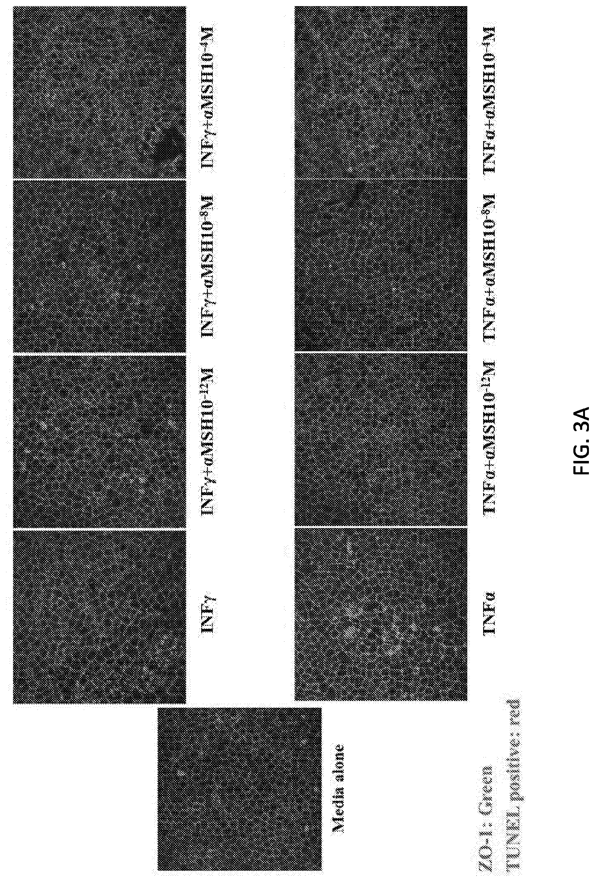
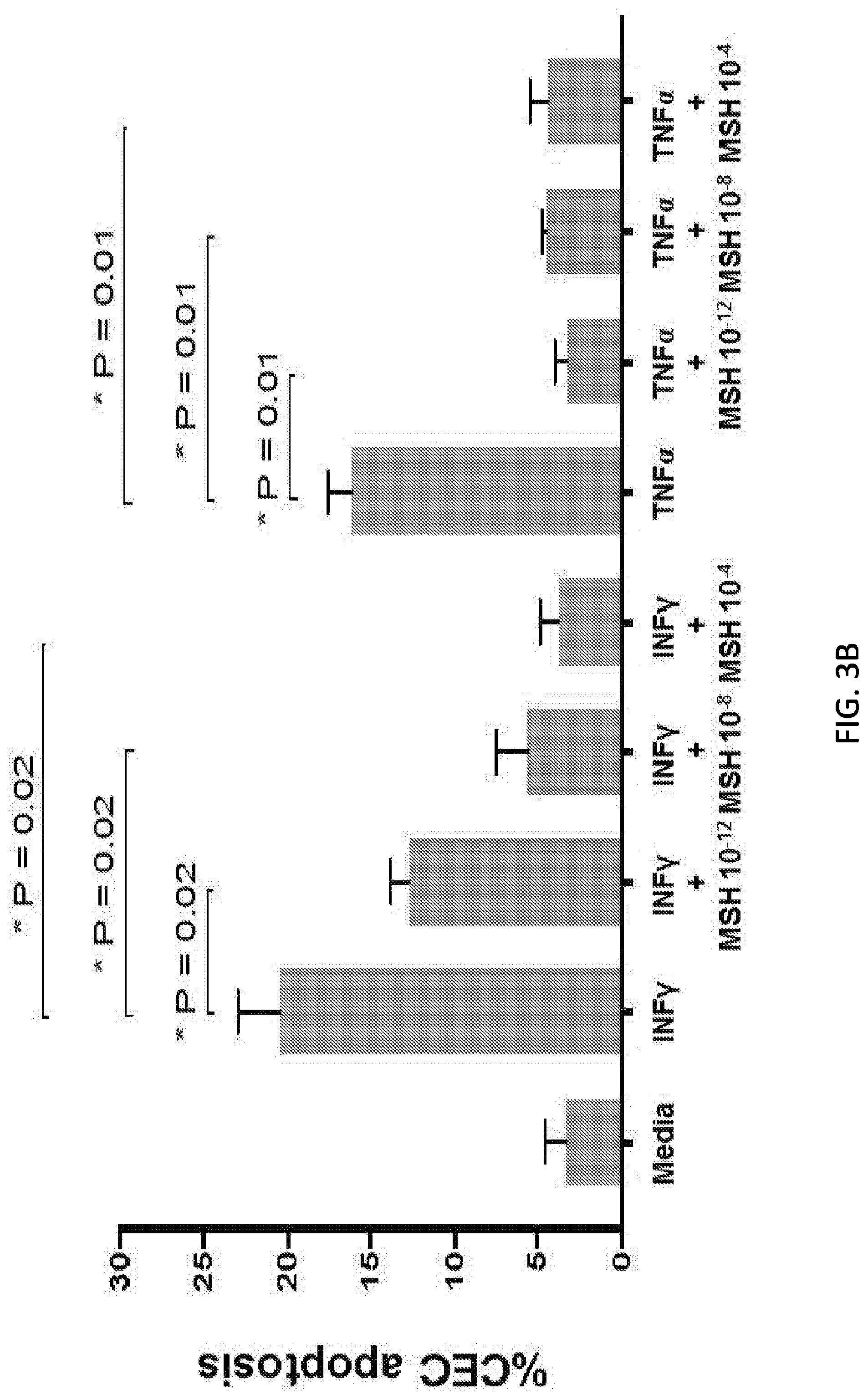
[0076] FIG. 3A is a series of images, and FIG. 3B is a graph showing effects of .alpha.-MSH on reducing CEC apoptosis induced by inflammatory cytokines. There is a significant reduction in percentage of apoptotic cells with addition of various concentrations of .alpha.-MSH.
[0077] FIG. 4 is a graph showing the effect of .alpha.-MSH on survival of allogeneic high-risk transplant. A subject is considered rejected, so that it is excluded from the calculations of survival, when the opacity score remains at least 3 for a continuous two week after week 2 of corneal transplant (n=7 in each group).
[0078] FIG. 5 is a graph showing the effect of .alpha.-MSH on central corneal thickness in murine model after allogeneic high-risk transplant. The corneal pachymetry is evaluated via OCT, *p<0.05 (n=7 in each group).
[0079] FIG. 6 is a graph showing the effect of .alpha.-MSH on opacity scores in murine model after allogeneic high-risk transplant, *p<0.05 (n=7 in each group).
[0080] FIG. 7 is a set of confocal images showing CEC density and morphology, stained with ZO-1, 8-week after allogeneic high-risk transplant in murine model. PBS group and .alpha.-MSH (10.sup.-4 M), twice per week subconj injection.
[0081] FIG. 8A is a graph showing the suppression effect of .alpha.-MSH (10.sup.-4 M) on CEC apoptosis in human corneal endothelium sheet under the stress of IFN-.gamma. (60 ng/ml). FIG. 8B is a graph showing the suppression effect of MSH (10.sup.-4 M) on CEC apoptosis in human corneal endothelium sheet under the stress of H2O2 (1.4 mM for 2 hr).
[0082] FIG. 9 is a graph showing that VIP treatment of human corneal endothelial cells accelerates corneal endothelial wound healing in vitro in a dose-dependent manner Percentage of healed endothelial area compared to baseline in VIP-treated groups and in control. Human corneal endothelial cells treated with VIP 10.sup.-9, 10.sup.-7 and 10.sup.-6 M demonstrated significantly enhanced wound healing compared to the control group 12 and 24 hours after incubation compared with the control (P<0.05, Mann-Whitney test). A dose-dependent trend was observed in the effect of VIP. Error bars indicate mean.+-.SEM. Each group consists of n=9, and data from one out of two independent experiments is shown.
[0083] FIG. 10A is a set of micrographs and FIG. 10B is a graph showing that VIP suppresses IFN.gamma.- and TNF.alpha.-mediated corneal endothelial cell apoptosis. FIG. 10A: Representative confocal micrographs showing nave C57BL/6 corneal cups incubated with either IFN.gamma. or TNF.alpha. with or without VIP (10.sup.-12, 10.sup.-9, 10.sup.-6 M). After 18 hours of incubation, corneas were stained for zonula occluden-1 (ZO-1, green) and terminal deoxynucleotidyl transferase-mediated dUTP nick-end labeling assay (TUNEL, red) to visualize endothelial cell-to-cell junctions and apoptotic cells, respectively. The scale bars are equal to 100 .mu.m (magnification .times.40). FIG. 10B: Bar diagram showing the percentages of apoptotic (TUNEL-positive) corneal endothelial cells incubated ex vivo with either IFN.gamma. or TNF.alpha. with different doses of VIP. VIP 10.sup.-6 M significantly suppresses IFN.gamma.- and TNF.alpha.-mediated corneal endothelial cell apoptosis (p=0.02 and 0.008, respectively; Mann-Whiney test). Data are presented as mean.+-.SEM. Each group consists of n=5 corneas and data from one out of two independent experiments is shown.
[0084] FIGS. 11A-D are images and graphs showing that VIP treatment decreases graft opacity and enhances corneal endothelial wound healing in a murine model of syngeneic corneal transplantation with endothelial injury. FIG. 11A: Representative slit-lamp images showing corneas at week 2-6 post-transplantation (magnification .times.25). FIG. 11B: Graft opacity scores were significantly decreased in VIP-treated group from week 2 to 6 post-transplantation compared with the control (P<0.05, n=15/group, Mann-Whitney test). FIG. 11C: Representative confocal micrographs of central area of transplanted corneas isolated from VIP-treated mice and the control groups at week 2, 4 and 6 after transplantation. To visualize corneal endothelial cell-to-cell junctions, corneas were stained with zonula occluden-1 (ZO-1, green). The scale bars are equal to 100 .mu.m (magnification .times.40). FIG. 11D: Bar diagram showing central endothelial cell density in VIP-treated corneas and controls at 2, 4 and 6 weeks post-transplantation. Data are presented as mean.+-.SEM. Each group consists of n=6 corneas, and data from one out of two independent experiments is shown.
[0085] FIGS. 12A-D are graphs and images showing the effect of VIP treatment on high-risk corneal transplant survival. Animals underwent high-risk allogeneic corneal transplantation and received treatment with VIP at 1, 3, 5, 7 and 9 days after transplantation. FIG. 12A: VIP treatment significantly decreased graft opacity scores at 4 to 8 weeks post-transplantation (P<0.05, Mann-Whitney test). FIG. 12B: Weekly examination of grafts for 8 weeks demonstrated a significant increase in graft survival in VIP-treated mice compared with the controls (85% vs. 0%; hazard ratio 0.10, 95% CI 0.04-0.26, P<0.0001, Log rank test). Each group consists of n=14. FIG. 12C: Representative confocal micrographs of central area of transplanted corneas in VIP-treated mice and in controls at 1, 2 and 8 weeks post-transplantation. Corneal endothelial cell-to-cell junction were stained and visualized with zonula occluden-1 (ZO-1, green). The scale bars are equal to 100 .mu.m (magnification .times.40). FIG. 12D: Bar diagram of central CEnC densities show significantly higher CEnC density in VIP-treated group compared with the control at 8 weeks post-transplantation (p=0.02, Man-Whitney test). Horizontal line represents the CEnC density of nave age-matched C57BL/6 corneas. Each group consists of n=5 corneas. All data are presented as mean.+-.SEM, and data from one out of two independent experiments is shown.
[0086] FIG. 13 is a graph showing the effect of .alpha.-MSH on corneal thickness in murine model after syngeneic corneal transplantation, *p<0.05.
DETAILED DESCRIPTION
[0087] The front part of the eye comprises the cornea, which is a transparent tissue. Corneal transparency is critical for normal vision. One of the important structures responsible for keeping the cornea transparent is CECs. These cells form a monolayer on the back surface of the cornea. Many different conditions are associated with damage to these cells which can result in corneal swelling (e.g., edema) and reduced vision. Treatments to prevent or reduce CEC loss in various ocular and systemic conditions are needed. Corneal transplantation is often performed for those with significant reduction in CECs. Included herein are strategies, methods, and compositions that improve the survival, function, proliferation, and/or migration of these cells.
[0088] CECs are critical for normal vision. Without being bound by any scientific theory, CECs control the fluid and solute transport across the posterior surface of the cornea and actively maintain the cornea in the slightly dehydrated state which is required for optical transparency (Hassell et al. 2010 Exp Eye Res 91:326-35; Bourne 2003 Eye (Lond) 17:912-8). Therefore, any damage to these cells may lead to reduced vision. Despite their importance, the mechanisms controlling the structure and function of CECs are not well understood.
[0089] Aspects of the present subject matter relate to the identification of an entirely novel function for a neuropeptide, .alpha.-MSH, in promoting survival and function of corneal endothelial cells. Therapeutic uses for this function are included herein.
[0090] Although nerves in the cornea have been shown to be involved in maintenance of corneal structure and function, e.g., providing trophic support for corneal epithelium, their role/function with respect to corneal endothelium, a structurally and functionally different tissue compared to corneal epithelium, was unknown prior to the invention. In addition, the role of each specific nerve-derived molecule (neuropeptide) on corneal endothelium is unknown. More specifically, the effects of .alpha.-MSH on these cells were unknown before the discovery disclosed herein.
[0091] Surprisingly, some neuropeptides can improve CEC survival, and these neuropeptides can be used to treat a variety conditions to reduce CEC loss. Non-limiting uses of methods and compositions provided herein include (but are not limited to) increasing CEC survival, proliferation, and/or migration in corneal tissue (e.g., in a subject or during the storage of donor tissue), following ocular surgery such as corneal transplantation, and in subjects with a corneal injury, a corneal dystrophy (such as an anterior corneal dystrophy, a stromal corneal dystrophy, a posterior corneal dystrophy, corneal endothelial dystrophy, Fuchs endothelial dystrophy, congenital hereditary endothelial dystrophy, posterior polymorphous corneal dystrophy, or Schnyder crystalline corneal dystrophy), bullous keratopathy, an iridocorneal endothelial (ICE) syndrome, photokeratitis (e.g., caused by sunlight reflection from sand, water, ice, or snow), neurotrophic keratophy, pseudoexfoliation syndrome, ocular hypertension, glaucoma, an ocular infection, a cataract, corneal endothelial cell loss due to contact lens wear, corneal endothelial cell loss due to aging, uveitis, intraocular inflammation, inflammatory disciform keratitis, diabetes, or dry eye disease. The compositions and methods described herein are useful to improve or restore vision in a large number of people suffering from complications of CEC loss.
[0092] Aspects of the present subject matter relate to the discovery that .alpha.-MSH promotes CEC migration and proliferation, and inhibits CEC apoptosis. In various embodiments, .alpha.-MSH reduces cell loss in any condition associated with CEC loss. Fuchs dystrophy (also known as Fuchs' dystrophy) affects about 1% of the general population and there are no known treatments. About 100,000 corneal transplants are performed every year in the United States. The present subject matter includes methods of treating disorders such as (but not limited to) Fuchs endothelial dystrophy, corneal endothelial cell loss due to contact lens wear and aging, neurotrophic keratopathy, and pseudoexfoliation syndrome. Also included are methods and compositions, e.g., storage or preservation solutions for donor cornea storage/maintenance (such as for eye banking), isolated endothelial cell (such as CEC) storage and culturing, and corneal tissues comprising endothelial cells (such as CECs) for use in endothelial keratoplasty procedures.
[0093] Human CECs are post-mitotic and exhibit poor regenerative capacity in vivo (Armitage et al. 2003 Investig Opthalmology Vis Sci. 2003 Aug. 1; 44(8):3326). CEC density decreases with increasing age, and the residual cells spread and enlarge (Yee et al. 1985 Curr Eye Res. 4(6):671-8). After corneal transplantation, the loss of CECs is accelerated (Bourne 2001 Cornea 20(6):560-9). Decreased endothelial cell density has been shown to be predictive of late endothelial failure, a key cause of graft failure in keratoplasty (Bourne 2001 Cornea 20(6):560-9).
[0094] In developed countries, CEC dysfunction is the principal indication for corneal transplantation (Peh et al. 2011 Transplantation 91(8):811-9). Furthermore, endothelial dysfunction is the most common cause of graft failure (Guilbert et al., 2013 Am J Ophthalmol 155(3):560-569.e2). Provided herein are strategies of using melanocortin receptor agonists to support CECs in both eye banking and corneal transplantation to improve CEC survival and reduce graft failure rates.
[0095] In eye banking, loss of CECs stored in Optisol is well established (Means et al. 1995 Arch Ophthalmol June 1; 113(6):805). Studies have demonstrated that grafts with a lower CEC density, either preoperatively or postoperatively, have increased rates of late corneal endothelial failure (Bourne 2001 Cornea 20(6):560-9; Nishimura et al. 1999 Ophthalmology 106(10):1962-5). Improving CEC health prior to transplantation is particularly important in the current era of increased endothelial keratoplasty procedures, since these techniques entail more donor tissue manipulation than penetrating keratoplasty (Price et al. 2008 Ophthalmology 115(5): 857-65).
[0096] While grafts performed in non-vascularized host beds or low-risk grafts enjoy a success rate of approximately 90%, in high-risk corneal transplantation (characterized by a vascularized and inflamed host bed) corneal graft rejection can exceed 50% (Dana et al. 2000 Cornea 19(5):625-43). In various embodiments, melanocortin receptor agonists demonstrate a protective effect on endothelium in an inflammatory microenvironment, e.g., in subjects with both low and high risk of tissue rejection upon corneal transplantation.
[0097] In non-limiting examples, a neuropeptide (such as .alpha.-MSH, VIP, CGRP, and/or BDNF) is used to reduce the CEC loss in a condition as detailed below:
[0098] 1. Conical Transplantation
[0099] Conical transplantation is well known to be associated with postoperative CEC loss. Here, a neuropeptide (such as .alpha.-MSH, VIP, CGRP, and/or BDNF) can be used to reduce this CEC loss after any form of corneal transplantation such as penetrating keratoplasty or endothelial keratoplasty. In addition, a neuropeptide (such as .alpha.-MSH, VIP, CGRP, and/or BDNF) can also be used to reduce the CEC loss associated with an episode of graft rejection.
[0100] 2. Donor Cornea Storage (Eye Banking)
[0101] Storage of the donor cornea is associated with CEC loss. Here, a neuropeptide (such as .alpha.-MSH, VIP, CGRP, and/or BDNF) can be used to reduce this natural CEC loss during storage.
[0102] 3. Cataract Surgery or Other Intraocular Surgeries
[0103] All intraocular surgeries, including cataract surgery, glaucoma surgery, and intraocular lens implantation, are associated with CEC loss. When this loss is severe, it can lead to bullous keratopathy and other conditions associated with conical edema. Here, a neuropeptide (such as .alpha.-MSH, VIP, CGRP, and/or BDNF) can be used to reduce CEC loss after any intraocular surgery.
[0104] 4. Fuchs Endothelial Dystrophy
[0105] This dystrophy is the most common conical endothelial dystrophy, which can be associated with significant CEC loss. Here, a neuropeptide (such as .alpha.-MSH, VIP, CGRP, and/or BDNF) can be used to reduce CEC loss over time or after any intraocular surgery in patients with Fuchs dystrophy, who are always at heightened risk of CEC loss.
[0106] 5. Other Conical Endothelial Dystrophies
[0107] A neuropeptide (such as .alpha.-MSH, VIP, CGRP, and/or BDNF) can be used to reduce the CEC loss in patients with different corneal endothelial dystrophies, including congenital hereditary endothelial dystrophy.
[0108] 6. Conical Injuries
[0109] A neuropeptide (such as .alpha.-MSH, VIP, CGRP, and/or BDNF) is useful to reduce CEC loss after any form mechanical, chemical, or physical injury. In addition, in patients with exposure of CEC to chemical materials, such as toxic substances, a neuropeptide (such as .alpha.-MSH, VIP, CGRP, and/or BDNF) can be used to reduce the CEC loss. Furthermore, a neuropeptide (such as .alpha.-MSH, VIP, CGRP, and/or BDNF) can also be used to reduce CEC loss in cases with surgical trauma to the corneal endothelium.
[0110] 7. Intraocular Inflammation
[0111] Any form of intraocular inflammation, such as uveitis (autoimmune and infectious), can be associated with CEC loss. A neuropeptide (such as .alpha.-MSH, VIP, CGRP, and/or BDNF) can be used to reduce the cell loss in subjects with uveitis.
[0112] 8. Increased Intraocular Pressure
[0113] Increased intraocular pressure is known to cause CEC loss. A neuropeptide (such as .alpha.-MSH, VIP, CGRP, and/or BDNF) can be used to reduce the cell loss due to increased intraocular pressure.
[0114] 9. Corneal Infections
[0115] Conical infections due to a variety of organisms, such as viral, bacterial, fungal, and acanthamoeba organisms, can be associated with CEC loss. A neuropeptide (such as .alpha.-MSH, VIP, CGRP, and/or BDNF) can be used to reduce cell loss in subjects with corneal infections. In addition, in noninfectious complications of these infections, such as inflammatory disciform keratitis, a neuropeptide (such as .alpha.-MSH, VIP, CGRP, and/or BDNF) can be used to reduce the CEC loss.
[0116] 10. Contact Lens Wear
[0117] Contact lens wear has been known to be associated with CEC loss. A neuropeptide (such as .alpha.-MSH, VIP, CGRP, and/or BDNF) can be used to reduce the cell loss.
[0118] 11. Diabetes Mellitus
[0119] Diabetes may be associated with CEC loss. A neuropeptide (such as .alpha.-MSH, VIP, CGRP, and/or BDNF) can be used to reduce cell loss in subjects with diabetes (e.g., Type I or Type II diabetes).
[0120] 12. Neurotrophic Keratopathy
[0121] It has been shown that neurotrophic keratopathy due to various reasons, including neurosurgically induced causes, can be associated with CEC loss. A neuropeptide (such as .alpha.-MSH, VIP, CGRP, and/or BDNF) can be used to reduce the cell loss in subjects with neurotrophic keratopathy.
[0122] 13. Dry Eye Disease
[0123] Dry eye disease can be associated with a significant CEC loss. A neuropeptide (such as .alpha.-MSH, VIP, CGRP, and/or BDNF) can be used to reduce the CEC loss.
[0124] 14. Pseudoexfoliation Syndrome
[0125] Pseudoexfoliation syndrome can be associated with CEC loss. A neuropeptide (such as .alpha.-MSH, VIP, CGRP, and/or BDNF) can be used to reduce cell loss associated in subjects who have pseudoexfoliation syndrome.
[0126] 15. Aging
[0127] Aging is associated with progressive CEC loss over time. A neuropeptide (such as .alpha.-MSH, VIP, CGRP, and/or BDNF) can be used to reduce cell loss that is associated with aging.
[0128] In certain embodiments, compositions provided herein may further comprise, or may comprise, consist essentially of, or consist of one or more neuropeptides (such as .alpha.-MSH, VIP, CGRP, and/or BDNF).
[0129] Various compositions provided herein may comprise, consist essentially of, or consist of a melanocortin receptor agonist such as .alpha.-MSH or an .alpha.-MSH agonist. As used herein a "melanocortin receptor agonist" is a compound that binds to at least one melanocortin receptor resulting in signaling that occurs when melanocortin binds its receptor. An ".alpha.-MSH agonist" is a melanocortin receptor agonist other than .alpha.-MSH. In some embodiments, the amount of signaling that results from .alpha.-MSH agonist binding to a receptor is greater than when .alpha.-MSH binds the receptor, i.e. the melanocortin receptor agonist activates the receptor more than .alpha.-MSH. In some embodiments, the receptor for .alpha.-MSH is one or more melanocortin receptors. In certain embodiments, the one or more melanocortin receptors is/are MC.sub.1, MC.sub.3, MC.sub.4, or MC.sub.5, or any combination thereof. In various embodiments, the melanocortin receptor(s) does not comprise MC.sub.2. In some embodiments, an .alpha.-MSH agonist is an .alpha.-MSH derivative. In certain embodiments, the .alpha.-MSH derivative is a synthetic or natural compound that binds to one or more melanocortin receptors resulting in signaling that typically occurs when .alpha.-MSH binds the one or more melanocortin receptors.
[0130] In some embodiments, the melanocortin receptor agonist (e.g., .alpha.-MSH or a derivative thereof) is native human .alpha.-MSH (e.g., has a wild-type amino acid sequence and structure), recombinant .alpha.-MSH, a variant of .alpha.-MSH comprising at least about 80%, 85%, or 90% sequence identity to native human .alpha.-MSH, a fragment of .alpha.-MSH (e.g., comprising about 3, 4, 5, 6, 7, 8, 9, 10, 11, or 12 amino acids) that binds to a receptor for .alpha.-MSH resulting in signaling that typically occurs when .alpha.-MSH binds its receptor, or a molecule derived from .alpha.-MSH (e.g., that has .alpha.-MSH activity, such as binding to a receptor for .alpha.-MSH resulting in signaling that occurs when .alpha.-MSH binds its receptor). In certain embodiments, the melanocortin receptor antagonist comprises a small molecule .alpha.-MSH agonist. In some embodiments, .alpha.-MSH is used (e.g., is administered or is present in a composition) alone (e.g., as a monotherapy). Alternatively, .alpha.-MSH is used (e.g., is administered or is present in a composition) in combination with other active agents.
[0131] Included herein are methods and compositions comprising an .alpha.-MSH agonist (e.g., in a corresponding embodiment of any method or composition disclosed herein that comprises .alpha.-MSH), e.g., a small molecule .alpha.-MSH agonist. In various embodiments, the .alpha.-MSH agonist binds at least one .alpha.-MSH receptor (such as a melanocortin receptor).
[0132] Non-limiting examples of .alpha.-MSH agonists are described in U.S. Pat. No. 8,703,702 issued Apr. 22, 2014 and U.S. Pat. No. 7,169,603 issued Jan. 30, 2007, the entire contents of each of which are incorporated herein by reference.
[0133] Receptor activation upon .alpha.-MSH agonist binding can be confirmed using, e.g., tests for binding to radioactive [35S]GTP.gamma.S or tests for fluorescence resonance energy transfer (FRET) nanosensor.
Exemplary Conditions and Diseases
[0134] Provided herein are methods and compositions for the treatment of any condition or disease (e.g., ocular condition or disease) that is associated with a loss of CECs, abnormal CEC morphology, and/or CEC dysfunction. In various embodiments, the condition comprises a disease, aging, an injury (e.g., trauma), prolonged contact lens use (e.g., use of contact lenses for at least about 6 to 12 hours per day for each of at least about 1, 2, 3, 4, 5, 6 or 7 days per week for at least about 0.5, 1, 2, 3, 4, 5, 6, 7, 8, 9, 10, 15, or 20 years), or surgical intervention (e.g., a transplant, laser eye surgery, cataract surgery, glaucoma surgery, etc.). In some embodiments, the disease is not an inflammatory disease (i.e., the disease is a non-inflammatory disease). In various embodiments, the disease is not an autoimmune disease (i.e., the disease is a non-autoimmune disease). In some embodiments, the subject does not have an inflammatory disease. In certain embodiments, the subject does not have an autoimmune disease. In various embodiments, the disease comprises inflammation. In some embodiments, the disease is an inflammatory disease. In certain embodiments, the disease is an autoimmune disease.
[0135] An ocular inflammatory disease is a disease that includes aberrant inflammation in an eye. In various embodiments, the aberrant inflammation occurs where no trauma (e.g., injury) has occurred. In some embodiments, the aberrant inflammation persists (e.g., for more than 1 week) after damage from trauma has healed or would normally be expected to heal in a corresponding subject who does not have the inflammatory disease. In certain embodiments, the aberrant inflammation occurs where infection has occurred. In various embodiments, the aberrant inflammation persists (e.g., for more than 1 week) after a pathogen that has caused an infection is cleared from the affected tissue (e.g., due to clearance by the immune system and/or therapeutic intervention). In certain embodiments, the inflammation is chronic inflammation. In various embodiments, the aberrant inflammation is an increased response to an injury or antigen (e.g., from a pathogen) compared to a corresponding subject who does not have the inflammatory disease. In some embodiments, the aberrant inflammation is an allergic reaction. In some embodiments, the inflammation is acute inflammation. An autoimmune disease is a disease in which a subject's immune system (e.g., immune cells) attacks a component of the subject's body, such as one or more types of the subject's cells (e.g., one or more types of ocular cells). In certain embodiments, ocular inflammation is observable and measurable visually. In some embodiments, ocular inflammation is clinically invisible, i.e. is only reflected by high expression levels of pro-inflammatory molecules (e.g., pro-inflammatory cytokines such as interleukin-1 (IL-1), IL-12, and IL-18, tumor necrosis factor alpha (TNF-.alpha.), interferon gamma (IFN-.gamma.), and granulocyte-macrophage colony stimulating factor). In various embodiments, the pro-inflammatory molecules are sufficient to cause or indicate a "subclinical" (meaning clinically non-evident, e.g., by visual inspection) disease.
[0136] As used herein, a "symptom" associated with a condition includes any clinical or laboratory manifestation associated with the condition, and is not limited to what the subject can feel or observe. In certain embodiments, the method described herein may include identifying a subject having one or more of these symptoms. Non-limiting examples of symptoms include CEC death, abnormal CEC morphology, and CEC dysfunction (e.g., as evidenced by corneal edema).
[0137] "Treating" (or treatment of) a condition includes ameliorating at least one symptom of the particular condition, even if the underlying pathophysiology is not affected. In some embodiments, the condition is an injury (e.g., trauma or a post-operative state). In certain embodiments, the condition is a disease. The efficacy of the treatment can be evaluated, e.g., as compared to a standard, e.g., improvement in the value or quality of a parameter (e.g., self-reported pain level, vision quality, CEC number, CEC morphology, cornea structure or clarity, and/or the existence of level of cornea edema) as compared to the value or quality of the parameter prior to treatment. As another example, the efficacy of treatment can be evaluated, e.g., as compared to a standard, e.g., slowing progression of the condition as compared to a usual time course for the condition in a cohort that has not been treated or compared to historical data on progression. Treating a condition also includes slowing its progress; and/or relieving the condition, e.g., causing regression of the condition. In some embodiments, the progressive worsening (e.g., the increasing intensity) of a symptom is slowed, reduced, or halted.
[0138] "Preventing" (or prevention of) a condition in a subject includes stopping a condition from occurring in the subject, who may be at risk of (e.g., predisposed to) the condition but has not yet been diagnosed as having it. Preventing a condition also includes delaying the onset of the condition. The efficacy of the prevention can be evaluated, e.g., as compared to a standard, e.g., delaying onset of the condition as compared to a usual time of onset for the condition in a cohort that has not been treated or compared to historical data on condition onset. In various embodiments, the condition is a disease. In some embodiments, the condition comprises CEC loss (i.e. due to cell death), dysfunction, and/or abnormal morphology.
[0139] As used herein and depending on the context in which it is used, "therapeutically effective amount" refers to an amount which is effective in reducing, eliminating, treating, preventing or controlling a symptom of a disorder or condition. In various embodiments, the symptom comprises CEC loss (i.e. due to cell death), dysfunction, and/or abnormal morphology. The term "controlling" is intended to refer to all processes wherein there may be a slowing, interrupting, arresting, or stopping of the progression of a condition described herein, but does not necessarily indicate a total elimination of all symptoms, and is intended to include prophylactic treatment.
[0140] Corneal Dystrophy
[0141] Aspects of the present subject matter provide methods and compositions for treating a corneal dystrophy. Corneal dystrophies are eye disorders (often genetic and progressive) in which abnormal material often accumulates in the cornea. See, e.g., The Corneal Dystrophy Foundation, 2016 What is Corneal Dystrophy? available at www.cornealdystrophyfoundation.org/what-is-corneal-dystrophy, the entire contents of which are incorporated herein by reference. In some embodiments, a subject with a corneal dystrophy does not have a symptom such as vision impairment, corneal edema, or eye discomfort (i.e., the corneal dystrophy is "asymptomatic"). In certain embodiments, a subject with a corneal dystrophy has vision impairment, corneal edema, and/or eye discomfort. In various embodiments, the age of onset and/or specific symptoms vary among the different forms of corneal dystrophy. In some embodiments, the corneal dystrophy affects both eyes (i.e., is bilateral). Alternatively, the corneal dystrophy affects one eye. In certain embodiments, the corneal dystrophy does not comprise symptoms in other areas of the body. In various embodiments, at least 1, 2, 3, or 4, cousins, aunts, uncles, grandparents, parents, and/or siblings of the subject has a corneal dystrophy. In some embodiments, the conical dystrophy is inherited as an autosomal dominant trait. In certain embodiments, the corneal dystrophy is inherited as an autosomal recessive trait.
[0142] The cornea comprises five layers: (1) the Epithelium, which is the outermost, protective layer of the cornea; (2) the Bowman's membrane, which is tough and difficult to penetrate, further protecting the eye; (3) the Stroma, which is the thickest layer of the cornea, comprising water, collagen fibers and other connective tissue components that help give the cornea strength, elasticity and clarity; (4) the Descemet membrane, which is a thin, strong inner layer that also acts as a protective layer; and (5) the Endothelium, which the innermost layer comprising CECs. Corneal dystrophies typically include the accumulation of abnormal material in one or more of layers (1), (2), (3), (4), or (5), and/or between layers of the cornea. In some embodiments, the material causes the cornea to lose its transparency. In certain embodiments, the material causes loss of vision or blurred vision.
[0143] In certain embodiments, the presence of a conical dystrophy is found incidentally during a routine eye examination. In various embodiments, a diagnosis is confirmed by a clinical evaluation, a detailed patient history, and/or one or more of a variety of tests, such as a slit lamp examination [in which a special microscope (slit lamp) allows a physician to view the eye through high magnification]. Some specific corneal dystrophies can be diagnosed with molecular genetic tests even before symptoms such as vision impairment, conical edema, or eye discomfort develop.
[0144] In various embodiments, a treatment regimen or composition provided herein is administered in a subject who has asymptomatic corneal dystrophy (e.g., the subject does not have a symptom such as vision impairment, corneal edema, or eye discomfort). In some embodiments, a treatment or composition is administered as a monotherapy or in conjunction with another treatment. Non-limiting examples of treatments for corneal dystrophies include eye drops, ointments, lasers, and conical transplant. In various embodiments, ocular surgery is performed. In various embodiments, a corneal transplant (e.g., a keratoplasty) may be performed, and a neuropeptide is administered before, during, and/or after the surgery. In some embodiments, the neuropeptide comprises CGRP, BDNF, VIP, and/or .alpha.-MSH). In certain embodiments, a corneal transplant (e.g., a keratoplasty) may be performed, and a melanocortin receptor agonist such as .alpha.-MSH is administered before, during, and/or after the surgery. Alternatively or in addition, a corneal transplant (e.g., a keratoplasty) may be performed, and VIP is administered before, during, and/or after the surgery.
[0145] In certain embodiments, a neuropeptide (such as CGRP, BDNF, and/or VIP) and/or a melanocortin receptor agonist (which may also be a neuropeptide such as .alpha.-MSH) is used to treat a corneal dystrophy that is associated with abnormal CEC morphology or density. In various embodiments, a melanocortin receptor agonist such as .alpha.-MSH is used to treat a corneal dystrophy that is associated with abnormal CEC morphology or density. In some embodiments, VIP is used to treat a corneal dystrophy that is associated with abnormal CEC morphology or density. Non-limiting examples of corneal dystrophies include posterior corneal dystrophies (e.g., congenital hereditary endothelial corneal dystrophy, Fuchs endothelial corneal dystrophy, posterior polymorphous corneal dystrophy, and Schnyder crystalline corneal dystrophy).
[0146] Various embodiments relate to the treatment of posterior corneal dystrophies, especially dystrophies that comprise a CEC abnormality. Posterior corneal dystrophies affect the innermost layers of the cornea such as the Descemet membrane and/or the endothelium. In some embodiments, a posterior corneal dystrophy progresses or has progressed to affect another layer (e.g., one or more other layers) or all layers of the cornea. Non-limiting examples include congenital hereditary endothelial corneal dystrophy, Fuchs endothelial corneal dystrophy, posterior polymorphous corneal dystrophy, and Schnyder crystalline corneal dystrophy.
[0147] Types of congenital hereditary corneal dystrophy or congenital-hereditary-endothelial-dystrophy (CHED) include:
[0148] 1) an autosomal dominant variant that presents after 1 year of age and is progressive (CHED1); and
[0149] 2) an autosomal recessive variant that presents at birth and is stationary (CHED2). See, e.g., Trief et al. 2016 Review of Ophthalmology, (available from www.reviewofophthalmology.com/article/congenital-hereditary-endothelial-d- ystrophy).
[0150] In some embodiments, a subject with CHED1 was born with a clear cornea and has developed clouding over the first 1, 2, or 3 years of life. In certain embodiments, a subject with CHED1 complains of epiphora and/or photophobia. In various embodiments, a subject with CHED1 does not have nystagmus. In certain embodiments, a subject with CHED1 has small flakes, spots, and/or irregular white areas throughout the stroma of 1 or 2 eyes. In various embodiments, a subject with CHED1 comprises multiple abnormal endothelial layers and/or microvilli.
[0151] In some embodiments, a subject with CHED2 has corneal clouding from birth with accompanying nystagmus. In certain embodiments, a subject with CHED2 does not complain of epiphora or photophobia. In various embodiments, the corneal clouding in a subject with CHED2 is more advanced than is typical for a subject with CHED1, and results in a worse visual acuity than is typical for a subject with CHED1.
[0152] Recently, the international classification of corneal dystrophies (IC3D) has been revised to eliminate CHED1 from classification. See, e.g., Weiss et al. IC3D classification of corneal dystrophies--edition 2. Cornea 2015; 34:117-59, the entire contents of which are incorporated herein by reference. Additionally, the autosomal recessive CHED (CHED2) was renamed CHED. However, CHED1 and CHED2 may be referred to as discussed herein to distinguish different classes of subjects with congenital hereditary corneal dystrophy.
[0153] In some embodiments, a subject with congenital hereditary corneal dystrophy has corneal edema. In certain embodiments, a subject with congenital hereditary corneal dystrophy has corneal clouding, and the clouding is diffuse (limbus to limbus) or the clouding is relatively uniform. In some embodiments, the cornea is cloudy (even milky) but the iris is visible. In certain embodiments, aside from corneal abnormalities, the structure of a subject's affected eye (or eyes) is normal.
[0154] In various embodiments, the subject does not have congenital glaucoma. In some embodiments, a subject has high intraocular pressure secondary to thick pachymetry (e.g., the cornea is more than 625 .mu.m thick). In various embodiments, congenital hereditary corneal dystrophy occurs concomitantly with glaucoma. In certain embodiments, a subject with congenital hereditary corneal dystrophy comprises a mutation at chromosome 20 loci 20p13. In various embodiments, the subject comprises a mutation in a gene encoding solute carrier family 4, sodium borate transporter member 11 (SLC4A11). This gene encodes a sodium borate transporter. In some embodiments, the mutation comprises a coding region mutation in the SLC4A11 gene.
[0155] In various embodiments, the subject comprises Fuchs endothelial corneal dystrophy (also known as, e.g., Fuchs endothelial dystrophy and Fuchs dystrophy). In some embodiments, Fuchs dystrophy develops in a subject during middle age (e.g., when the subject is about 25 to 45 or 30 to 40 years old). In certain embodiments, the subject does not have vision impairment, corneal edema, or eye discomfort initially. Fuchs dystrophy is characterized by CEC dysfunction. In Fuchs dystrophy CECs deteriorate (and may die). In various embodiments, the cornea fills with water and swells (i.e., corneal edema occurs). In some embodiments, the swelling worsens and blurred vision occurs that is worse in the morning, but gradually improves throughout the day. In certain embodiments, tiny blisters form on the cornea. In various embodiments, tiny blisters on the cornea rupture and causing pain. In some embodiments, a subject has a gritty or sandy feeling within the eye (foreign body sensation), is abnormally sensitive to light, and/or see a glare or halo when looking at lights. In certain embodiments, as the disease progresses, vision no longer improves during the day and significant vision loss may occur. In various embodiments, the subject receives a corneal transplant.
[0156] In some embodiments, an early-onset variant of Fuchs endothelial dystrophy is associated with a mutation in the COL8A2 gene, which encodes a protein that is part of type VIII collagen. Type VIII collagen is a major component of the Descemet membrane. In certain embodiments, a subject with a COL8A2 gene mutation and Fuchs endothelial dystrophy has an abnormal Descemet membrane. In various embodiments, CECs die in such subjects, leading and/or contributing to vision problems.
[0157] In various embodiments, a subject has posterior polymorphous dystrophy. In some embodiments, posterior polymorphous dystrophy presents at birth (with clouding of the cornea). In certain embodiments, posterior polymorphous dystrophy presents later during life and is characterized by lesions affecting the endothelium. In various embodiments, the subject does not have a symptom such as vision impairment, corneal edema, or eye discomfort. In some embodiments, effects on the cornea are slowly progressive. In certain embodiments, both eyes are affected, but one eye is more severely affected than the other (i.e., the posterior polymorphous dystrophy is asymmetric). In various embodiments, a subject with posterior polymorphous dystrophy has swelling (edema) of the stroma, an abnormal sensitivity to light (photophobia), decreased vision, and/or the sensation of foreign material in the eye. In some embodiments, the subject has increased intraocular pressure.
[0158] Methods and compositions provided herein are useful for the prevention and/or treatment, e.g., reduction in a clinical manifestation, of any symptom or type of corneal dystrophy (including CEC loss).
[0159] Bullous Keratopathy
[0160] In certain embodiments, a subject has bullous keratopathy. Bullous keratopathy is a pathological condition in which small vesicles, or bullae, are formed in the cornea due to endothelial dysfunction. In a healthy cornea, CECs keep the tissue from excess fluid absorption. In various embodiments, then affected by some reason, such as Fuchs dystrophy or a trauma during ocular surgery (such as a transplant or cataract removal), CECs may suffer mortality or damage. In some embodiments, when endothelial cell counts drop too low, excess fluid moves anterior into the stroma and epithelium, resulting in swelling of the cornea. In certain embodiments, as fluid accumulates between the basal epithelium cells, blister like formations form (bullae) and they undergo painful ruptures releasing their fluid content. In various embodiments, these malformations disrupt vision and create pain sensations. Methods and compositions provided herein are useful for the prevention and/or treatment of bullous keratopathy.
[0161] Iridocorneal Endothelial (ICE) Syndrome
[0162] In some embodiments, a subject has an ICE syndrome. ICE syndromes are a spectrum of diseases characterized by slowly progressive abnormalities of the corneal endothelium and features including corneal edema, iris distortion, and secondary angle-closure glaucoma. Methods and compositions provided herein are useful for the prevention and/or treatment of any type or symptom of ICE syndrome (including CEC loss).
[0163] Neurotrophic Keratopathy
[0164] In certain embodiments, a subject has neurotrophic keratopathy. Neurotrophic keratopathy is a disease characterized by decreased corneal sensitivity and poor corneal healing. See, e.g., Graham (2016) Neurotrophic Keratopathy, Medscape, available at emedicine.medscape.com/article/1194889-overview, the entire contents of which are incorporated herein by reference. In various embodiments, this disorder leaves the cornea susceptible to injury and decreases reflex tearing. In some embodiments, a subject comprises epithelial breakdown. In certain embodiments, a subject has a corneal ulceration, infection, melting, and/or perforation secondary to poor healing. Prognostic indicators in neurotrophic keratopathy include the degree of sensory loss, the duration of the condition, and the presence of other ocular surface disease. Neurotrophic keratopathy can occur in any age group. In some embodiments, the subject is at least about 0.5, 1, 5, 10, 15, 20, 25, 30, 35, 40, 45, 50, 55, 60, 65, 70, 75, 80, 85, 90, or 95 years old. In certain embodiments, the subject has corneal hypesthesia. In various embodiments, the neurotrophic keratopathy results during or following a herpetic infection of the cornea, a surgery for trigeminal neuralgia, or a surgery for acoustic neuroma. In some embodiments, the neurotrophic keratopathy has been caused by a herpes simplex or herpes zoster infection, or leprosy. In certain embodiments, a subject shows rose bengal staining of the inferior palpebral conjunctiva, has decreased tear breakup time, has increased mucous viscosity, and has punctate epithelial fluorescein staining. In various embodiments, the subject has an epithelial defect (such as an oval defect in the superior cornea), a defect surrounded by a rim of loose epithelium (optionally, the edges are smooth and rolled), stromal swelling with folds in the Descemet membrane, and/or inflammation in the anterior chamber.
[0165] Methods and compositions provided herein are useful for the prevention and/or treatment of any type or symptom of neurotrophic keratopathy (including CEC loss).
[0166] Pseudoexfoliation Syndrome
[0167] In some embodiments, a subject has pseudoexfoliation syndrome. Pseudoexfoliation syndrome is an agingrelated systemic disease manifesting itself primarily in the eyes which is characterized by the accumulation of microscopic granular amyloid-like protein fibers. In certain embodiments, the subject is at least about 65, 70, 75, 80, 85, 90, or 95 years old. In various embodiments, the subject self-identifies as being of Scandinavian descent. In some embodiments, the buildup of protein clumps blocks normal drainage of the eye fluid called the aqueous humor. In certain embodiments, this block of normal drainage causes an increase in ocular pressure leading to glaucoma and loss of vision.
[0168] In various embodiments, a subject has no specific symptoms. In some embodiments, a subject complains of lessened visual acuity or a change in the perceived visual field. In certain embodiments, such changes are secondary to or different from symptoms normally associated with cataracts or glaucoma.
[0169] In various embodiments, the pseudoexfoliation syndrome comprises tiny microscopic white or grey granular flakes that are clumps of proteins within the eye. In some embodiments, the flakes are visible during an examination of the lens of an eye by an ophthalmologist or optometrist. In certain embodiments, the white fluffy material is seen in many tissues both ocular and extraocular: in the anterior chamber structures, trabecular meshwork, central disc, zonular fibres, anterior hyaloid membrane, pupillary and anterior iris, trabecula, and occasionally the cornea. In various embodiments, the flakes are widespread.
[0170] In some embodiments, pseudoexfoliation syndrome includes the flakes becoming enmeshed in a "spongy area" known as the trabecular meshwork and block its normal functioning. In certain embodiments, the flakes interact with degenerative changes in the Schlemm's canal and the juxtacanalicular area. In various embodiments, the blockage leads to greater-than-normal elevated intraocular pressure. In some embodiments, a subject's optic nerve is damaged. In certain embodiments, pseudoexfoliation syndrome is associated with a weakening of structures within the eye which help hold the eye's lens in place, called lens zonules.
[0171] Methods and compositions provided herein are useful for the prevention and/or treatment of any type or symptom of pseudoexfoliation syndrome (including CEC loss).
[0172] Increased Intraocular Pressure
[0173] In various embodiments, a subject has been diagnosed with or is characterized by comprising increased intraocular pressure, such as any type of glaucoma or ocular hypertension.
[0174] In some embodiments, a subject has ocular hypertension. As used herein, the term "ocular hypertension" refers to any situation in which the pressure inside the eye, called intraocular pressure, is higher than normal. Eye pressure may be measured in, e.g., millimeters of mercury (mm Hg). Normal eye pressure ranges from 10-21 mmHg Ocular hypertension comprises an eye pressure of greater than 21 mmHg See, e.g., WebMD, Ocular Hypertension, available at www.webmd.com/eye-health/occular-hypertension #3-4. In certain embodiments, a subject with ocular hypertension may have an eye pressure of at least about 21.5 mmHg, 22 mmHg, 22.5 mmHg, 23 mmHg, 23.5 mmHg, 24 mmHg, or 24.5 mmHg.
[0175] In various embodiments, ocular hypertension is commonly defined as a condition comprising (i) intraocular pressure of greater than 21 mm Hg is measured in one or both eyes at two or more time points (e.g., on different dates, e.g., at least about 1, 2, 3, 4, 5, 6, or 12 months apart); (ii) a normal appearance of the optic nerve; (iii) no sign of glaucoma evident on visual field testing; and (iv) no sign of another ocular disease. In some embodiments, the pressure inside the eye is measured using an instrument such as a tonometer. In certain embodiments, a subject with ocular hypertension is observed more closely than the general population for the onset of glaucoma.
[0176] In various embodiments, increased intraocular pressure results from another eye condition. In some embodiments, ocular hypertension inside the eye is caused by an imbalance in the production and drainage of fluid in the eye (aqueous humor). For example, the channels that normally drain the fluid from inside the eye do not function properly.
[0177] Methods and compositions provided herein are useful for the prevention and/or treatment of any symptom or type of ocular hypertension (including CEC loss).
[0178] In some embodiments, the subject has glaucoma. Glaucoma results in damage to the optic nerve and vision loss, especially if left untreated. In certain embodiments, the glaucoma is open-angle glaucoma. In various embodiments, the glaucoma is closed-angle glaucoma. In some embodiments, the glaucoma is normal-tension glaucoma. Typically, open-angle glaucoma develops slowly over time and there is no pain. In some embodiments, side vision begins to decrease followed by central vision resulting in blindness if not treated. In certain embodiments, closed-angle glaucoma can present gradually or suddenly. In various embodiments, the sudden presentation involves severe eye pain, blurred vision, a mid-dilated pupil, redness of the eye, and nausea.
[0179] Non-limiting examples of risk factors for glaucoma include increased pressure in the eye, a family history of the condition, migraines, high blood pressure, and obesity. In some embodiments, for eye pressure a value of greater than 21 mmHg or 2.8 kPa is often used with higher pressures leading to a greater risk. However, a subject may have high eye pressure for years and never develop damage. Conversely, optic nerve damage may occur with normal pressure, known as normal-tension glaucoma.
[0180] Methods and compositions provided herein are useful for the prevention and/or treatment of any symptom or type of glaucoma (including CEC loss).
[0181] Keratitis
[0182] In various embodiments, a subject has keratitis. As used herein, keratitis is a condition in which a cornea becomes inflamed Non-limiting examples of symptoms associated with keratitis include pain, impaired eyesight, photophobia, red eye, and a `gritty` sensation. Examples of keratitis include acute epithelial keratitis, nummular keratitis, interstitial keratitis, disciform keratitis, neurotrophic keratitis, mucous plaque keratitis, herpes simplex keratitis, herpes zoster keratitis, bacterial keratitis, fungal keratitis, acanthamoebic keratitis, onchocercal keratitis, superficial punctate keratitis, ulcerative keratitis, exposure keratitis, photokeratitis, and contact lens acute red eye.
[0183] In some embodiments, the keratitis comprises inflammatory disciform keratitis. For example, the inflammatory disciform keratitis may result from a viral infection such as a herpes simplex virus (HSV) infection. In certain embodiments, an HSV infection induces inflammation of the corneal endothelium in a condition known as HSV endotheliitis. In various embodiments, HSV endotheliitis comprises secondary inflammation caused by the virus and/or direct infection of CECs. In various embodiments, HSV endotheliitis comprises endothelial dysfunction and stromal edema and/or opacity. In some embodiments, a subject receives corneal transplantation in the form of either full-thickness penetrating keratoplasty or anterior lamellar keratoplasty to restore corneal clarity.
[0184] Methods and compositions provided herein are useful for the prevention and/or treatment of any symptom or type of keratitis, as well as CEC loss associated with any treatment for keratitis.
[0185] Diabetes Mellitus
[0186] In certain embodiments, the subject has diabetes mellitus (DM). DM, commonly referred to as diabetes, is a group of metabolic diseases in which there are high blood sugar levels over a prolonged period. In various embodiments, the subject has frequent urination, increased thirst, and increased hunger. In some embodiments, the subject has heart disease, chronic kidney failure, foot ulcers, has had a stroke, and/or has damage to the eyes. Exemplary long-term complications relate to the damage to blood vessels, such as small blood vessels in the eyes, kidneys, and nerves. In certain embodiments, the subject has damage to the eyes such as diabetic retinopathy. In various embodiments, the subject has had gradual vision loss and/or blindness. In various embodiments, a subject has type 1 diabetes. In certain embodiments, a subject has type 2 diabetes.
[0187] Methods and compositions provided herein are useful for the prevention and/or treatment of vision and/or CEC loss associated with any type of diabetes.
[0188] Intraocular Inflammation and Uveitis
[0189] In various embodiments, a subject has intraocular inflammation, such as uveitis. Non-limiting examples of causes of intraocular inflammation include infection (e.g., a viral, fungal, or parasitic infection), rheumatoid arthritis, Gout, and injuries to the eye. In some embodiments, the cause is unknown. Exemplary symptoms of intraocular inflammation include eye redness, blurred vision, perceiving floaters (small dark or blurry dots or shapes that may appear to move), decreased vision, and light sensitivity.
[0190] Uveitis is swelling and irritation of the uvea, the middle layer of the eye. In various embodiments, the uveitis comprises anterior uveitis (which involves inflammation in the front part of the eye). In some embodiments, the uveitis comprises iritis. In certain embodiments, the uveitis affect one or two of the subject's eyes. In various embodiments, the uveitis comprises posterior uveitis. In some embodiments, the uveitis affects the choroid, which is a layer of blood vessels and connective tissue in the middle part of the eye. In certain embodiments, the uveitis comprises choroiditis. In various embodiments, the uveitis comprises chorioretinitis. In various embodiments, the uveitis comprises pars planitis. In some embodiments, the subject is male. In certain embodiments, the subject is female. Uveitis can occur at any age. In various embodiments, the subject is under the age of 95, 85, 80, 75, 70, 65, 60, 55, 50, 45, 40, 35, 30, 25, 20, 15, 10, 5, 1, or 0.5 years old.
[0191] In various embodiments, the uveitis is autoimmune uveitis. As used herein "autoimmune uveitis" refers to uveitis that is caused or associated with an autoimmune disorder. In certain embodiments, the uveitis is associated with or caused by an autoimmune disorder such as rheumatoid arthritis or ankylosing spondylitis. In some embodiments, the subject has Crohn's disease and/or multiple sclerosis. Non-limiting examples of autoimmune uveitis symptoms include redness of the eye; blurred vision; photophobia; sensitivity to light; irregular pupil; eye pain; floaters, which are dark spots that appear to float in the visual field; headaches; dilated ciliary vessels; presence of cells and flare in the anterior chamber; keratic precipitates ("KP") on the posterior surface of the cornea; a hypopyon; pigment deposits on the lens; a festooned pupil on dilation of pupil; busacca nodules (inflammatory nodules located on the surface of the iris); synechia; and photopsia or seeing flashing lights.
[0192] Methods and compositions provided herein are useful for the prevention and/or treatment of any symptom or type of intraocular inflammation (including CEC loss), including any symptom or type of uveitis (e.g., such as autoimmune uveitis or uveitis from an infection), including CEC loss.
[0193] Dry Eye Disease and Related Disorders
[0194] In some embodiments, the subject has Dry Eye Disease (DED). DED is a multifactorial disorder of the tears and ocular surface that results in symptoms of discomfort, visual disturbance, and tear film instability, with potential damage to the ocular surface. In some embodiments, it is accompanied by increased osmolarity of the tear film and inflammation of the ocular surface (Lemp M A. Report of the National Eye Institute/Industry Workshop on clinical trials in dry eyes. CLAO J 1995; 21:221-2). For a more detailed definition, see The definition and classification of dry eye disease: report of the Definition and Classification Subcommittee of the International Dry Eye WorkShop. Ocular Surface. 2007 April; 5(2):75-92, the entire contents of which are incorporated herein by reference. DED and related diseases may be caused or exacerbated by, e.g., autoimmune and environmental conditions as well as any activity that decreases the rate of blinking. DED and related diseases may also be caused by decreased tear production or a change in tear composition that results in inadequate lubrication of the eye. Contact lens use, eye surgery, and eye injury can induce DED. In certain embodiments, DED occurs as a consequence of aging and hormonal changes. DED can occur at any age. In various embodiments, the subject is at least about 0.5, 1, 5, 10, 15, 20, 25, 30, 35, 40, 45, 50, 55, 60, 65, 70, 75, 80, 85, 90, or 95 years old.
[0195] Non-limiting examples and related disorders of DED include, but are not limited to, keratoconjunctivitis sicca (KCS), Sjogren syndrome (SS), Sjogren syndrome associated keratoconjunctivitis sicca, non-Sjogren syndrome associated keratoconjunctivitis sicca, keratitis sicca, sicca syndrome, xerophthalmia, tear film disorder, decreased tear production, aqueous tear deficiency (ATD), meibomian gland dysfunction (MGD), and evaporative loss. In some embodiments, a subject is identified as suffering from DED or a related disorder by detecting a sign or symptom selected from the group consisting of dry, scratchy, stingy, itchy, burning or pressured sensations, irritation, pain, redness, inflammation, discharge, and excessive eye watering. In certain embodiments, a subject is identified as suffering from DED or a related disorder if their tear composition is insufficient for proper eye tissue lubrication. In various embodiments, a method of therapy for DED inhibits or reduces the severity of at least one of sign or symptom of DED.
[0196] In some embodiments, a subject is at risk of developing DED. Subjects at risk of developing DED include subjects who are taking antihistamines, nasal decongestants, tranquilizers, certain blood pressure medicines, Parkinson's medications, birth control pills and/or anti-depressants; subjects with a skin disease on or around the eyelids can result in dry eye; subjects suffering from a disease of the glands in the eyelids (such as meibomian gland dysfunction); subjects who are pregnant; female subjects who are on hormone replacement therapy (such as estrogen and/or progesterone); subjects who have had the refractive surgery known as laser-assisted in situ keratomileusis (LASIK); subjects who have suffered from a chemical or thermal burn on the membrane lining the eyelids and covering the eye; subjects afflicted with allergies; subjects afflicted with an immune system disorder (such as Sjogren's syndrome, lupus, or rheumatoid arthritis); subjects who have had an eye infection; subjects who have had ocular exposure to an irritant such as a chemical fume or tobacco smoke; and subjects with exposure keratitis.
[0197] Non-limiting examples of DED symptoms include pain (such as stinging or burning of the eye); ulcers or scars on the cornea; decrease tolerance for dry environments; reduced vision; a sandy or gritty feeling as if something is in the eye; episodes of excess tears following very dry eye periods; a stringy discharge from the eye; redness of the eye; episodes of blurred vision; heavy eyelids; inability to cry when emotionally stressed; uncomfortable contact lenses; decreased tolerance of reading, working on the computer, or any activity that requires sustained visual attention; and eye fatigue.
[0198] Methods and compositions provided herein are useful for the treatment of any type or symptom of DED or disorder that is related to DED as indicated above (including CEC loss).
[0199] Corneal Injury
[0200] As used herein, a "corneal injury" comprises a wound to the cornea. In some embodiments the wound comprises an injury to the outer surface of the cornea. In certain embodiments, the wound comprises an injury (e.g., a deep injury) such as a full-thickness or partial thickness (e.g., penetrating at least 5, 10, 15, 20, 25, 30, 35, 40, 45, 50, 55, 60, 65, 70, 75, 80, 85, 90, or 95% of the way through a part of the cornea) lacerations (e.g., cuts or tears) or punctures. Non-limiting examples of injuries to the outer surface of the cornea include injuries from abrasions (such as scratches or scrapes on the surface of the cornea, e.g., from trauma or a foreign body such as sand or dust), lacerations, punctures, chemical injuries (e.g., caused by a fluid or gas comprising a toxic agent that contacts the eye), contact lens problems (e.g., overuse, poor fit, or a sensitivity to a contact lens care solution), ultraviolet light (e.g., from sunlight, a sun lamp, snow or water reflections, or arc-welding), or an infection.
[0201] Methods and compositions provided herein are useful for the treatment of any type or symptom of corneal injury (including CEC loss).
[0202] CEC Loss Associated with Surgery
[0203] Methods and compositions provided herein are useful of increasing CEC survival, proliferation, and/or migration following ocular surgery, such as intraocular surgery. There are many types of intraocular surgeries. Any type of intraocular surgery can result in CEC loss. In various embodiments, a neuropeptide (such as .alpha.-MSH, VIP, CGRP, and/or BDNF) is used to prevent and/or treat CEC loss associated with any intraocular surgery. In certain embodiments, a melanocortin receptor agonist such as .alpha.-MSH is used to prevent and/or treat CEC loss associated with any intraocular surgery. In some embodiments, VIP is used to prevent and/or treat CEC loss associated with any intraocular surgery.
[0204] In various embodiments, the ocular surgery comprises Descemet stripping (sometimes referred to as "descemetorhexis"). Descemet stripping may comprise (i) the removal of at least a portion (e.g., at least about 10%, 20%, 30%, 40%, 50%, 60%, 70%, 80%, 90%, or all) of the Descemet membrane (sometimes referred to as "Descemet's membrane"), as well as CECs that are attached to the removed membrane; or (ii) the removal of CECs that are attached to the Descemet membrane, e.g., without substantial removal of the Descemet membrane. In certain embodiments, Descemet stripping consists of or consists essentially of (i) the removal of at least a portion (e.g., at least about 10%, 20%, 30%, 40%, 50%, 60%, 70%, 80%, 90%, or all) of the Descemet membrane, as well as CECs that are attached to the removed membrane; or (ii) the removal of CECs that are attached to the Descemet membrane, e.g., without substantial removal of the Descemet membrane. The Descemet membrane is the basement membrane that lies between the corneal proper substance (also called the stroma), and the endothelial layer of the cornea. In some embodiments, Descemet stripping is not followed by endothelial keratoplasty, and a subject's CECs (e.g., peripheral CECs, which are along the outer edge of the cornea's endothelial layer, or CECs from a region of the Descement membrane that has not been stripped) repopulate the cornea to form a new layer of CECs where the Descemet membrane and/or preexisting CECs were removed. Methods and compositions provided herein are useful for increasing the growth, survival, and/or migration of a subject's CECs after Descemet stripping.
[0205] In some embodiments, the ocular surgery comprises a cornea transplant such as penetrating keratoplasty (PK) or endothelial keratoplasty (EK). In certain embodiments, PK comprises the removal of a full-thickness section of tissue a diseased or injured cornea, and replacement of the tissue with donor cornea tissue (e.g., a matching section of donor tissue). In various embodiments, the tissue is removed using either a surgical cutting instrument (such as a trephine) or laser (such as a femtosecond laser). Typically, the donor tissue is sutured into place.
[0206] In various embodiments, the ocular surgery comprises EK. For example, the CECs may be the subject's CECs and/or donor CECs. In various embodiments, EK selectively replaces the diseased (i.e. endothelial) layer of the cornea with donor tissue, leaving healthy areas or layers intact [for example the innermost layer of the cornea (endothelium) is replaced and the overlying healthy corneal tissue is left intact other than, optionally, an incision]. In some embodiments, EK comprises Descemet Stripping Endothelial Keratoplasty (DSEK), Descemet Membrane Endothelial Keratosplaty (DMEK), Descemet Membrane Endothelial Transfer (DMET), or Descemet Stripping Automated Endothelial Keratoplasty (DSAEK).
[0207] DSEK is a CEC replacement procedure comprising Descemet stripping (which includes removal of the corneal endothelium comprising CECs) followed by replacement of the corneal endothelium (comprising CECs) with a graft comprising a donor's endothelium, Descemet membrane, and some part of the stroma. In some embodiments, the graft comprises the back 20-30% of the donor cornea.
[0208] DMEK comprises Descemet stripping followed by replacement of the corneal endothelium with donor graft comprising a thin layer (e.g., about 5% or less of corneal thickness) of donor tissue. For example, the donor tissue in DMEK comprises, consists essentially of, or consists of donor CECs attached the donor's Descemet membrane or a portion thereof. In certain embodiments, the graft has been peeled off the back of the donor cornea.
[0209] In various embodiments, the ocular surgery comprises DMET. DMET comprises Descemet stripping followed by administration of a graft comprising a thin layer (e.g., about 5% or less of corneal thickness) of donor tissue that is mostly free floating in the anterior chamber of the cornea. In some embodiments, the graft is attached only to the corneal incision site. In certain embodiments, the donor tissue in DMET comprises, consists essentially of, or consists of donor CECs attached the donor's Descemet membrane or a portion thereof.
[0210] In various embodiments, the ocular surgery comprises DSAEK. DSAEK is a partial thickness cornea transplant procedure that involves selective removal of the patient's Descemet membrane and endothelium, followed by transplantation of donor corneal endothelium in addition to donor corneal stroma. The transplanted tissue is approximately 100-200 microns thick. If the endothelium of the graft makes contact with any surgical instruments, it will be damaged and the graft may fail; therefore, the surgical procedure is designed to avoid contacting the donor endothelium. A tunneled corneoscleral incision is created, the recipient endothelium and Descemet membrane is removed, the graft is folded and inserted (e.g., with non-coapting forceps, which are forceps that do not meet at the tips), and an air bubble is placed in the anterior chamber to support graft adherence.
[0211] In various embodiments, the ocular surgery comprises the injection of donor CECs into the cornea of a subject. For example, the CECs may be injected to replace CECs that have died in a subject's corneal endothelium, and/to replace CECs that were removed by Descemet stripping.
[0212] In some embodiments, the ocular surgery comprises phototherapeutic keratectomy (PTK). PTK is a type of eye surgery that uses a laser to treat various ocular disorders by removing tissue (such as CECs) from the cornea. Common indications for PTK are corneal dystrophies, scars, opacities, and bullous keratopathy.
[0213] Aspects of the present subject matter relate to reducing CEC loss in subjects who have glaucoma, as well as CEC loss resulting from surgery to great glaucoma. Alternatively or in addition, methods and compositions herein are useful for increasing the number of CECs in subjects who have glaucoma or who have received surgery for glaucoma. In some embodiments, the surgery comprises laser surgery, such as trabeculoplasty or iridotomy. In various embodiments, the surgery comprises invasive surgery such as trabeculectomy or the implantation of a glaucoma drainage device. In certain embodiments, the surgery comprises canaloplasty. Methods and compositions provided herein are useful for preventing or treating CEC loss associated with any surgical treatment for glaucoma.
[0214] In some embodiments, the surgery comprises cataract surgery. In various embodiments, cataract surgery is an operation that removes the cloudy lens and replaces it with a clear artificial lens (i.e., an intraocular lens (IOL)). See, e.g., Boyd (2016) Cataract Surgery American Academy of Ophthalmology available at www.aao.org/eye-health/diseases/what-is-cataract-surgery, the entire contents of which are incorporated herein by reference. Methods and compositions provided herein are useful for preventing or treating CEC loss associated with any surgical treatment for cataracts.
[0215] Any type of intraocular surgery can result in CEC loss. A melanocortin receptor agonist such as .alpha.-MSH can be used and/or treat CEC loss in all these conditions. In various embodiments, a neuropeptide (such as .alpha.-MSH, VIP, CGRP, and/or BDNF) is used and/or treat CEC loss in all these conditions. In certain embodiments, the ocular surgery comprises laser eye surgery. Non-limiting examples of laser eye surgery include laser-assisted in situ keratomileusis, as well as procedures to change eye color (e.g., from brown to blue).
Exemplary Neuropeptides
[0216] .alpha.-MSH is generated from a precursor hormone called pro-opiomelanocortin (POMC) (Eipper and Mains (1980) Endocr Rev 1 (1): 1-27). This molecule serves as the source for several peptide hormones such as adrenocorticotrophin (ACTH), .alpha.-MSH, .beta.-MSH and .gamma.-MSH, and the endogenous opioids including 0-endorphin. .alpha.-MSH is a tridecapeptide which, upon proteolytic cleavage, is generated from its precursor ACTH. Non-limiting descriptions relating to .alpha.-MSH are provided in Luger and Brzoska (2007) Ann Rheum Dis 66 Suppl 3:iii52-5, the entire contents of which are incorporated herein by reference.
[0217] An exemplary (non-limiting) amino acid sequence of the human POMC preprotein is available in public databases, e.g., under National Center for Biotechnology Information (NCBI) Accession No. NP_001306134.1 and UniProt Accession No. P01189, and is as follows:
TABLE-US-00001 MPRSCCSRSGALLLALLLQASMEVRGWCLESSQCQDLTTESNLLECIRAC KPDLSAETPMFPGNGDEQPLTENPRKYVMGHFRWDRFGRRNSSSSGSSGA GQKREDVSAGEDCGPLPEGGPEPRSDGAKPGPREGKRSYSMEHFRWGKPV GKKRRPVKVYPNGAEDESAEAFPLEFKRELTGQRLREGDGPDGPADDGAG AQADLEHSLLVAAEKKDEGPYRMEHFRWGSPPKDKRYGGFMTSEKSQTPL VTLFKNAIIKNAYKKGE
[0218] In the amino acid sequence above, positions 1 to 26 correspond to the signal peptide; positions 27 to 102 correspond to the N-terminal peptide of pro-piomelanocortin (NPP); positions 77 to 87 correspond to .gamma.-MSH; positions 105 to 134 correspond to a potential peptide; positions 138 to 176 correspond to corticotropin; positions 138 to 150 correspond to .alpha.-MSH; positions 156 to 176 correspond to corticotropin-like intermediary peptide; positions 179 to 267 correspond to lipotropin beta; positions 179 to 234 correspond to lipotropin gamma; positions 217-234 correspond to 0-MSH; positions 237 to 267 correspond to .beta.-endorphin; and positions 237 to 241 correspond to met-enkephalin.
[0219] The amino acid sequence of human .alpha.-MSH is as follows: SYSMEHFRWGKPV (SEQ ID NO: 1). The International Union of Pure and Applied Chemistry (IUPAC) name for .alpha.-MSH is N-acetyl-L-seryl-L-tyrosyl-L-seryl-L-methionyl-L-.alpha.-glutamyl-L-histi- dyl-L-phenylalanyl-L-arginyl-L-tryptophylglycyl-L-lysyl-L-prolyl-L-valinam- ide. .alpha.-MSH is also known as:
[0220] .alpha.-melanocortin,
[0221] .alpha.-melanotropin,
[0222] .alpha.-intermedin,
[0223] Ac-Ser-Tyr-Ser-Met-Glu-His-Phe-Arg-Trp-Gly-Lys-Pro-Val-NH.sub.2,
[0224] Ac-DL-Ser-DL-Tyr-DL-Ser-DL-Met-DL-Glu-DL-His-DL-Phe-DL-Arg-DL-Trp-G- ly-DL-Lys-DL-Pro-DL-Val-NH.sub.2, and
[0225] [acetyl]-SYSMEHFRWGKPV-[NH2].
[0226] The PubChem CID for human .alpha.-MSH is 16132636.
[0227] An exemplary (non-limiting) nucleotide sequence that encodes the human POMC preprotein is available from public databases under NCBI Accession No. NM_001319205.1 and is as follows (the start and stop codons are bolded and underlined):
TABLE-US-00002 (SEQ ID NO: 2) GTTCTAAGCGGAGACCCAACGCCATCCATAATTAAGTTCTTCCTGAGGGC GAGCGGCCAGGTGCGCCTTCGGCAGGACAGTGCTAATTCCAGCCCCTTTC CAGCGCGTCTCCCCGCGCTCGTCCCCCGTCTGGAAGCCCCCCTCCCACGC CCCGCGGCCCCCCTTCCCCTGGCCCGGGGAGCTGCTCCTTGTGCTGCCGG GAAGGTCAAAGTCCCGCGCCCACCAGGAGAGCTCGGCAAGTATATAAGGA CAGAGGAGCGCGGGACCAAGCGGCGGCGAAGGAGGGGAAGAAGAGCCGCG ACCGAGAGAGGCCGCCGAGCGTCCCCGCCCTCAGAGAGCAGCCTCCCGAG ACAGGGGTCCCACCAATCTTGTTTGCTTCTGCAGAGCCTCAGCCTGCCTG GAAGATGCCGAGATCGTGCTGCAGCCGCTCGGGGGCCCTGTTGCTGGCCT TGCTGCTTCAGGCCTCCATGGAAGTGCGTGGCTGGTGCCTGGAGAGCAGC CAGTGTCAGGACCTCACCACGGAAAGCAACCTGCTGGAGTGCATCCGGGC CTGCAAGCCCGACCTCTCGGCCGAGACTCCCATGTTCCCGGGAAATGGCG ACGAGCAGCCTCTGACCGAGAACCCCCGGAAGTACGTCATGGGCCACTTC CGCTGGGACCGATTCGGCCGCCGCAACAGCAGCAGCAGCGGCAGCAGCGG CGCAGGGCAGAAGCGCGAGGACGTCTCAGCGGGCGAAGACTGCGGCCCGC TGCCTGAGGGCGGCCCCGAGCCCCGCAGCGATGGTGCCAAGCCGGGCCCG CGCGAGGGCAAGCGCTCCTACTCCATGGAGCACTTCCGCTGGGGCAAGCC GGTGGGCAAGAAGCGGCGCCCAGTGAAGGTGTACCCTAACGGCGCCGAGG ACGAGTCGGCCGAGGCCTTCCCCCTGGAGTTCAAGAGGGAGCTGACTGGC CAGCGACTCCGGGAGGGAGATGGCCCCGACGGCCCTGCCGATGACGGCGC AGGGGCCCAGGCCGACCTGGAGCACAGCCTGCTGGTGGCGGCCGAGAAGA AGGACGAGGGCCCCTACAGGATGGAGCACTTCCGCTGGGGCAGCCCGCCC AAGGACAAGCGCTACGGCGGTTTCATGACCTCCGAGAAGAGCCAGACGCC CCTGGTGACGCTGTTCAAAAACGCCATCATCAAGAACGCCTACAAGAAGG GCGAGTGAGGGCACAGCGGGGCCCCAGGGCTACCCTCCCCCAGGAGGTCG ACCCCAAAGCCCCTTGCTCTCCCCTGCCCTGCTGCCGCCTCCCAGCCTGG GGGGTCGTGGCAGATAATCAGCCTCTTAAAGCTGCCTGTAGTTAGGAAAT AAAACCTTTCAAATTTCACATCCACCTCTGACTTTGAATGTAAACTGTGT GAATAAAGTAAAAATACGTAGCCGTCAAATAACAGC
[0228] NGF is also known as beta-NGF. An exemplary (non-limiting) amino acid sequence of human nerve growth factor (NGF) is available in public databases, e.g., under UniProt Accession No. P01138, and is as follows:
TABLE-US-00003 (SEQ ID NO: 3) MSMLFYTLITAFLIGIQAEPHSESNVPAGHTIPQAHWTKLQHSLDTALRR ARSAPAAAIAARVAGQTRNITVDPRLFKKRRLRSPRVLFSTQPPREAADT QDLDFEVGGAAPFNRTHRSKRSSSHPIFHRGEFSVCDSVSVWVGDKTTAT DIKGKEVMVLGEVNINNSVFKQYFFETKCRDPNPVDSGCRGIDSKHWNSY CTTTHTFVKALTMDGKQAAWRFIRIDTACVCVLSRKAVRRA
[0229] An exemplary (non-limiting) nucleotide sequence that encodes human NGF is available from public databases under GenBank Accession No. CR541855.1 and is as follows (the start and stop codons are bolded and underlined):
TABLE-US-00004 (SEQ ID NO: 4) ATGTCCATGTTGTTCTACACTCTGATCACAGCTTTTCTGATCGGCATACA GGCGGAACCACACTCAGAGAGCAATGTCCCTGCAGGACACACCATCCCCC AAGCCCACTGGACTAAACTTCAGCATTCCCTTGACACTGCCCTTCGCAGA GCCCGCAGCGCCCCGGCAGCGGCGATAGCTGCACGCGTGGCGGGGCAGAC CCGCAACATTACTGTGGACCCCAGGCTGTTTAAAAAGCGGCGACTCCGTT CACCCCGTGTGCTGTTTAGCACCCAGCCTCCCCGTGAAGCTGCAGACACT CAGGATCTGGACTTCGAGGTCGGTGGTGCTGCCCCCTTCAACAGGACTCA CAGGAGCAAGCGGTCATCATCCCATCCCATCTTCCACAGGGGCGAATTCT CGGTGTGTGACAGTGTCAGCGTGTGGGTTGGGGATAAGACCACCGCCACA GACATCAAGGGCAAGGAGGTGATGGTGTTGGGAGAGGTGAACATTAACAA CAGTGTATTCAAACAGTACTTTTTTGAGACCAAGTGCCGGGACCCAAATC CCGTTGACAGCGGGTGCCGGGGCATTGACTCAAAGCACTGGAACTCATAT TGTACCACGACTCACACCTTTGTCAAGGCGCTGACCATGGATGGCAAGCA GGCTGCCTGGCGGTTTATCCGGATAGATACGGCCTGTGTGTGTGTGCTCA GCAGGAAGGCTGTGAGAAGAGCCTGA
[0230] An exemplary (non-limiting) amino acid sequence of human VIP is available in public databases, e.g., under UniProt Accession No. P01282, and is as follows:
TABLE-US-00005 (SEQ ID NO: 5) MDTRNKAQLLVLLTLLSVLFSQTSAWPLYRAPSALRLGDRIPFEGANEPD QVSLKEDIDMLQNALAENDTPYYDVSRNARHADGVFTSDFSKLLGQLSAK KYLESLMGKRVSSNISEDPVPVKRHSDAVFTDNYTRLRKQMAVKKYLNSI LNGKRSSEGESPDFPEELEK
[0231] An exemplary (non-limiting) nucleotide sequence that encodes human VIP is available from public databases under GenBank Accession No. M36634.1 and is as follows (the start and stop codons are bolded and underlined):
TABLE-US-00006 (SEQ ID NO: 6) GGTCAGCTCCAAAACAATCCGGAACGGCCAGCTCCGGGGGAGCACGACTG GGCGAGAGGCACAGAAATGGACACCAGAAATAAGGCCCAGCTCCTTGTGC TCCTGACTCTTCTCAGTGTGCTCTTCTCACAGACTTCGGCATGGCCTCTT TACAGGGCACCTTCTGCTCTCAGGTTGGGTGACAGAATACCCTTTGAGGG AGCAAATGAACCTGATCAAGTTTCATTAAAAGAAGACATTGACATGTTGC AAAATGCATTAGCTGAAAATGACACACCCTATTATGATGTATCCAGAAAT GCCAGGCATGCTGATGGAGTTTTCACCAGTGACTTCAGTAAACTCTTGGG TCAACTTTCTGCCAAAAAGTACCTTGAGTCTCTTATGGGAAAACGTGTTA GCAGTAACATCTCAGAAGACCCTGTACCAGTCAAACGTCACTCAGATGCA GTCTTCACTGACAACTATACCCGCCTTAGAAAACAAATGGCTGTAAAGAA ATATTTGAACTCAATTCTGAATGGAAAGAGGAGCAGTGAGGGAGAATCTC CCGACTTTCCAGAAGAGTTAGAAAAATGATGAAAAAGACCTTTGGAGCAA AGCTGATGACAACTTCCCAGTGAATTCTTGAAGGAAAATGATACGCAACA TAATTAAATTTTAGATTCTACATAAGTAATTCAAGAAAACAACTTCAATA TCCAAACCAAATAAAAATATTGTGTTGTGAATGTTGTGATGTATTCTAGC TAATGTAATAACTGTGAAGTTTACATTGTAAATAGTATTTGAGAGTTCTA AATTTTGTCTTTAACTCATAAAAAGCCTGCAATTTCATATGCTGTATATC CTTTCTAACAAAAAAATATATTTTAATGATAAGTAATGCTAGGTTAATCC AATTATATGAGACGTTTTTGGAAGAGTAGTAATAGAGCAAAATTGATGTG TTTATTTATAGAGTGTACTTAACTATTCAGGAGAGTAGAACAGATAATCA GTGTGTCTAAATTTGAATGTTAAGCAGATGGAATGCTGTGTTAAATAAAC CTCAAAATGTCTAAGATAGTAACAATGAAGATAAAAAGACATTCTTCCAA AAAGATTTTCAGAAAATATTATGTGTTTCCATATTTTATAGGCAACCTTT ATTTTTAATGGTGTTTTAAAAAATCTCAAATTTGGATTGCTAATCACCAA AGGCTCTCTCCTGATAGTCTTTCAGTTAAGGAGAACGACCCCTGCTTCTG ACACTGAAACTTCCCTTTCTGCTTGTGTTAAGTATGTGTAAAATGTGAAG TGAATGAAACACTCAGTTGTTCAATAATAAATATTTTTGCCATAATGACT CAGAATATTGCTTTGGTCATATGAGCTTCCTTCTGTGAAATACATTTTGG AGACACAACTATTTTTCCAAAATAATTTTAAGAAATCAAAGAGAGAAAAT AAAGACCTTGCTTATGATTGCAGATAAAAAAAAAAAAAAAAAAAAAAAAA AAAAAAAAAAA
[0232] CGRP is a member of the calcitonin family of peptides, which in humans exists in two forms, .alpha.-CGRP and .beta.-CGRP. .alpha.-CGRP (also known as Calcitonin gene-related peptide 1 or CALCA) is a 37-amino acid peptide and is formed from the alternative splicing of the calcitonin/CGRP gene. .beta.-CGRP differs in three amino acids (in humans) and is encoded in a separate gene.
[0233] An exemplary (non-limiting) amino acid sequence for human CALCA is available in public databases, e.g., under UniProt Accession No. P06881, and is as follows:
TABLE-US-00007 (SEQ ID NO: 7) MGFQKFSPFLALSILVLLQAGSLHAAPFRSALESSPADPATLSEDEARL LLAALVQNYVQMKASELEQEQEREGSRIIAQKRACDTATCVTHRLAGLL SRSGGVVKNNFVPTNVGSKAFGRRRRDLQA
[0234] An exemplary (non-limiting) nucleotide sequence that encodes human CGRP is available from public databases under NCBI Accession No. NM_001033953.2 and is as follows (the start and stop codons are bolded and underlined):
TABLE-US-00008 (SEQ ID NO: 8) CCGCCGCTGCCACCGCCTCTGATCCAAGCCACCTCCCGCCAGGTGAGCC CCGAGATCCTGGCTCAGAGAGGTGTCATGGGCTTCCAAAAGTTCTCCCC CTTCCTGGCTCTCAGCATCTTGGTCCTGTTGCAGGCAGGCAGCCTCCAT GCAGCACCATTCAGGTCTGCCCTGGAGAGCAGCCCAGCAGACCCGGCCA CGCTCAGTGAGGACGAAGCGCGCCTCCTGCTGGCTGCACTGGTGCAGGA CTATGTGCAGATGAAGGCCAGTGAGCTGGAGCAGGAGCAAGAGAGAGAG GGCTCCAGAATCATTGCCCAGAAGAGAGCCTGTGACACTGCCACCTGTG TGACTCATCGGCTGGCAGGCTTGCTGAGCAGATCAGGGGGTGTGGTGAA GAACAACTTTGTGCCCACCAATGTGGGTTCCAAAGCCTTTGGCAGGCGC CGCAGGGACCTTCAAGCCTGAGCAGCTGAATGACTCAAGAAGGTCACAA TAAAGCTGAACTCCTTTTAATGTGTAATGAAAGCAATTTGTAGGAAAGG CTCCATGGAAGACATACATATAGGCATCCTTCTTGATACTGAAAACTAT CTTCTTTGTTTGAAAGGAACTATTGCTAAATGCAGAACAAGCTCATTGC AGTTACCTATTGTGCATCTTTTTAAATACTTGATTATGTAACCATAAAT CTGACAGCATGTCTCATTGGCTTATCTGGTAGCAAATCTAGGCCCCGTC AGCCACCCTATTGACATTGGTGGCTCTGCTAAACCTCAGGGGGACATGA AATCACTGCCTCTTGGGCATCTGGGGACACATGGTAATGCTGTGCCTTG ACAGAAGTATTTGTTTAAAGAAATGTCAATGCTGTCATTTGTGAACTCT ATCAAAATTAAAAATGTATTTTCTATACCCTTCA
[0235] An exemplary (non-limiting) amino acid sequence of human BDNF is available in public databases, e.g., under UniProt Accession No. P23560, and is as follows:
TABLE-US-00009 (SEQ ID NO: 9) MTILFLTMVISYFGCMKAAPMKEANIRGQGGLAYPGVRTHGTLESVNG PKAGSRGLTSLADTFEHVIEELLDEDQKVRPNEENNKDADLYTSRVML SSQVPLEPPLLFLLEEYKNYLDAANMSMRVRRHSDPARRGELSVCDSI SEWVTAADKKTAVDMSGGTVTVLEKVPVSKGQLKQYFYETKCNPMGYT KEGCRGIDKRHWNSQCRTTQSYVRALTMDSKKRIGWRFIRIDTSCVCT LTIKRGR
[0236] An exemplary (non-limiting) nucleotide sequence that encodes human BDNF is available from public databases under GenBank Accession No. X60201.1 and is as follows (the start and stop codons are bolded and underlined):
TABLE-US-00010 (SEQ ID NO: 10) GAATTCGGGGCTGCCGCCGCCGCGCCCGGGCGCACCCGCCCGCTCGCT GTCCCGCGCACCCCGTAGCGCCTCGGGCTCCCGGGCCGGACAGAGGAG CCAGCCCGGTGCGCCCCTCCACCTCCTGCTCGGGGGGCTTTAATGAGA CACCCACCGCTGCTGTGGGGCCGGCGGGGAGCAGCACCGCGACGGGGA CCGGGGCTGGGCGCTGGAGCCAGAATCGGAACCACGATGTGACTCCGC CGCCGGGGACCCGTGAGGTTTGTGTGGACCCCGAGTTCCACCAGGTGA GAAGAGTGATGACCATCCTTTTCCTTACTATGGTTATTTCATACTTTG GTTGCATGAAGGCTGCCCCCATGAAAGAAGCAAACATCCGAGGACAAG GTGGCTTGGCCTACCCAGGTGTGCGGACCCATGGGACTCTGGAGAGCG TGAATGGGCCCAAGGCAGGTTCAAGAGGCTTGACATCATTGGCTGACA CTTTCGAACACGTGATAGAAGAGCTGTTGGATGAGGACCAGAAAGTTC GGCCCAATGAAGAAAACAATAAGGACGCAGACTTGTACACGTCCAGGG TGATGCTCAGTAGTCAAGTGCCTTTGGAGCCTCCTCTTCTCTTTCTGC TGGAGGAATACAAAAATTACCTAGATGCTGCAAACATGTCCATGAGGG TCCGGCGCCACTCTGACCCTGCCCGCCGAGGGGAGCTGAGCGTGTGTG ACAGTATTAGTGAGTGGGTAACGGCGGCAGACAAAAAGACTGCAGTGG ACATGTCGGGCGGGACGGTCACAGTCCTTGAAAAGGTCCCTGTATCAA AAGGCCAACTGAAGCAATACTTCTACGAGACCAAGTGCAATCCCATGG GTTACACAAAAGAAGGCTGCAGGGGCATAGACAAAAGGCATTGGAACT CCCAGTGCCGAACTACCCAGTCGTACGTGCGGGCCCTTACCATGGATA GCAAAAAGAGAATTGGCTGGCGATTCATAAGGATAGACACTTCTTGTG TATGTACATTGACCATTAAAAGGGGAAGATAGTGGATTTATGTTGTAT AGATTAGATTATATTGAGACAAAAATTATCTATTTGTATATATACATA ACAGGGTAAATTATTCAGTTAAGAAAAAAATAATTTTATGAACTGCAT GTATAAATGAAGTTTATACAGTACAGTGGTTCTACAATCTATTTATTG GACATGTCCATGACCAGAAGGGAAACAGTCATTTGCGCACAACTTAAA AAGTCTGCATTACATTCCTTGATAATGTTGTGGTTTGTTGCCGTTGCC AAGAACTGAAAACATAAAAAGTTAAAAAAAATAATAAATTGCATGCTG CCCGAATTC
[0237] In some embodiments, .alpha.-MSH is administered to a subject. Alternatively or in addition, VIP is administered to a subject. Alternatively or in addition, CGRP and/or BDNF is administered to a subject. In various embodiments, another or an additional neuropeptide is administered to the subject, such as NGF, Substance P (SP), neurotrophin-3, Neurotrophin-4 (NTF-4), or neurotrophin-6. In various embodiments, an additional neuropeptide is administered to the subject, such as NGF.
[0238] An exemplary (non-limiting) amino acid sequence of human neurotrophin-3 is available in public databases, e.g., under UniProt Accession No. P20783, and is as follows:
TABLE-US-00011 (SEQ ID NO: 11) MSILFYVIFLAYLRGIQGNNMDQRSLPEDSLNSLIIKLIQADILKNKL SKQMVDVKENYQSTLPKAEAPREPERGGPAKSAFQPVIAMDTELLRQQ RRYNSPRVLLSDSTPLEPPPLYLMEDYVGSPVVANRTSRRKRYAEHKS HRGEYSVCDSESLWVTDKSSAIDIRGHQVTVLGEIKTGNSPVKQYFYE TRCKEARPVKNGCRGIDDKHWNSQCKTSQTYVRALTSENNKLVGWRWI RIDTSCVCALSRKIGRT
[0239] An exemplary (non-limiting) nucleotide sequence that encodes human neurotrophin-3 is available from public databases under GenBank Accession No. BC107075.1 and is as follows (the start and stop codons are bolded and underlined):
TABLE-US-00012 (SEQ ID NO: 12) CACACTCAGCTGCCAGAGCCTGCTCTTAACACCTGTGTTTCCTTTTCAG ATCTTACAGGTGAACAAGGTGATGTCCATCTTGTTTTATGTGATATTTC TCGCTTATCTCCGTGGCATCCAAGGTAACAACATGGATCAAAGGAGTTT GCCAGAAGACTCGCTCAATTCCCTCATTATTAAGCTGATCCAGGCAGAT ATTTTGAAAAACAAGCTCTCCAAGCAGATGGTGGACGTTAAGGAAAATT ACCAGAGCACCCTGCCCAAAGCTGAGGCTCCCCGAGAGCCGGAGCGGGG AGGGCCCGCCAAGTCAGCATTCCAGCCAGTGATTGCAATGGACACCGAA CTGCTGCGACAACAGAGACGCTACAACTCACCGCGGGTCCTGCTGAGCG ACAGCACCCCCTTGGAGCCCCCGCCCTTGTATCTCATGGAGGATTACGT GGGCAGCCCCGTGGTGGCGAACAGAACATCACGGCGGAAACGGTACGCG GAGCATAAGAGTCACCGAGGGGAGTACTCGGTATGTGACAGTGAGAGTC TGTGGGTGACCGACAAGTCATCGGCCATCGACATTCGGGGACACCAGGT CACGGTGCTGGGGGAGATCAAAACGGGCAACTCTCCTGTCAAACAATAT TTTTATGAAACGCGATGTAAGGAAGCCAGGCCGGTCAAAAACGGTTGCA GGGGTATTGATGATAAACACTGGAACTCTCAGTGCAAAACATCCCAAAC CTACGTCCGAGCACTGACTTCAGAGAACAATAAACTCGTGGGCTGGCGG TGGATACGGATAGACACGTCCTGTGTGTGTGCCTTGTCGAGAAAAATCG GAAGAACATGAATTGGCATCTCTCCCCATATATAAATTATTACTTTAAA TTATATGATATGCATGTAGCATATAAATGTTTATATTGTTTTTATATAT TATAAGTTGACCTTTATTTATTAAACTTCAGCAACCCTACAGTATATAA GCTTTTTTCTCAATAAAATCAGTGTGCTTGCCTTCCCTCAGGCCTCTCC CATCT
[0240] NTF-4 is also known as neurotrophin-5 (NTF5). An exemplary (non-limiting) amino acid sequence of human NTF-4 is available in public databases, e.g., under UniProt Accession No. P34130, and is as follows:
TABLE-US-00013 (SEQ ID NO: 13) MLPLPSCSLPILLLFLLPSVPIESQPPPSTLPPFLAPEWDLLSPRVVLS RGAPAGPPLLFLLEAGAFRESAGAPANRSRRGVSETAPASRRGELAVCD AVSGWVTDRRTAVDLRGREVEVLGEVPAAGGSPLRQYFFETRCKADNAE EGGPGAGGGGCRGVDRRHWVSECKAKQSYVRALTADAQGRVGWRWIRID TACVCTLLSRTGRA
[0241] An exemplary (non-limiting) nucleotide sequence that encodes human NTF-4 is available from public databases under GenBank Accession No. BT019368.1 and is as follows (the start and stop codons are bolded and underlined):
TABLE-US-00014 (SEQ ID NO: 14) ATGCTTCCTCTCCCCTCATGCTCCCTCCCCATCCTCCTCCTTTTCCTCC TCCCCAGTGTGCCAATTGAGTCCCAACCCCCACCCTCAACATTGCCCCC TTTTCTGGCCCCTGAGTGGGACCTTCTCTCCCCCCGAGTAGTCCTGTCT AGGGGTGCCCCTGCTGGGCCCCCTCTGCTCTTCCTGCTGGAGGCTGGGG CCTTTCGGGAGTCAGCAGGTGCCCCGGCCAACCGCAGCCGGCGTGGGGT GAGCGAAACTGCACCAGCGAGTCGTCGGGGTGAGCTGGCTGTGTGCGAT GCAGTCAGTGGCTGGGTGACAGACCGCCGGACCGCTGTGGACTTGCGTG GGCGCGAGGTGGAGGTGTTGGGCGAGGTGCCTGCAGCTGGCGGCAGTCC CCTCCGCCAGTACTTCTTTGAAACCCGCTGCAAGGCTGATAACGCTGAG GAAGGTGGCCCGGGGGCAGGTGGAGGGGGCTGCCGGGGAGTGGACAGGA GGCACTGGGTATCTGAGTGCAAGGCCAAGCAGTCCTATGTGCGGGCATT GACCGCTGATGCCCAGGGCCGTGTGGGCTGGCGATGGATTCGAATTGAC ACTGCCTGCGTCTGCACACTCCTCAGCCGGACTGGCCGAGCCTAG
[0242] SP is an undecapeptide (a peptide composed of a chain of 11 amino acid residues) member of the tachykinin neuropeptide family Substance P and its closely related neurokinin A (NKA) are produced from a polyprotein precursor after differential splicing of the preprotachykinin A gene. An exemplary (non-limiting) deduced amino acid sequence of substance P is as follows:
[0243] Arg Pro Lys Pro Gln Gln Phe Phe Gly Leu Met (RPKPQQFFGLM) (SEQ ID NO: 15) with an amidation at the C-terminus. See, e.g., Wong and Jeng (1994) Journal of Neuroscience Research 37 (1): 97-102, the entire contents of which are incorporated herein by reference.
[0244] An exemplary (non-limiting) nucleotide sequence that encodes a precursor of human substance P [Homo sapiens tachykinin precursor 1 (TACO] is available from public databases under NCBI Accession No. NM_013996.2 and is as follows (the start and stop codons are bolded and underlined):
TABLE-US-00015 (SEQ ID NO: 16) CACGCAAGCGAAAGGAGAGGAGGCGGCTAATTAAATATTGAGCAGAAAG TCGCGTGGGGAGAATGTCACGTGGGTCTGGAGGCTCAAGGAGGCTGGGA TAAATACCGCAAGGCACTGAGCAGGCGAAAGAGCGCGCTCGGACCTCCT TCCCGGCGGCAGCTACCGAGAGTGCGGAGCGACCAGCGTGCGCTCGGAG GAACCAGAGAAACTCAGCACCCCGCGGGACTGTCCGTCGCAAAATCCAA CATGAAAATCCTCGTGGCCTTGGCAGTCTTTTTTCTTGTCTCCACTCAG CTGTTTGCAGAAGAAATAGGAGCCAATGATGATCTGAATTACTGGTCCG ACTGGTACGACAGCGACCAGATCAAGGAGGAACTGCCGGAGCCCTTTGA GCATCTTCTGCAGAGAATCGCCCGGAGACCCAAGCCTCAGCAGTTCTTT GGATTAATGGGCAAACGGGATGCTGATTCCTCAATTGAAAAACAAGTGG CCCTGTTAAAGGCTCTTTATGGACATGGCCAGATCTCTCACAAAATGGC TTATGAAAGGAGTGCAATGCAGAATTATGAAAGAAGACGTTAATAAACT ACCTAACATTATTTATTCAGCTTCATTTGTGTCAATGGGCAATGACAGG TAAATTAAGACATGCACTATGAGGAATAATTATTTATTTAATAACAATT GTTTGGGGTTGAAAATTCAAAAAGTGTTTATTTTTCATATTGTGCCAAT ATGTATTGTAAACATGTGTTTTAATTCCAATATGATGACTCCCTTAAAA TAGAAATAAGTGGTTATTTCTCAACAAAGCACAGTGTTAAATGAAATTG TAAAACCTGTCAATGATACAGTCCCTAAAGAAAAAAAATCATTGCTTTG AAGCAGTTGTGTCAGCTACTGCGGAAAAGGAAGGAAACTCCTGACAGTC TTGTGCTTTTCCTATTTGTTTTCATGGTGAAAATGTACTGAGATTTTGG TATTACACTGTATTTGTATCTCTGAAGCATGTTTCATGTTTTGTGACTA TATAGAGATGTTTTTAAAAGTTTCAATGTGATTCTAATGTCTTCATTTC ATTGTATGATGTGTTGTGATAGCTAACATTTTAAATAAAAGAAAAAATA TCTTGAA
[0245] Each of the amino acid and nucleotide sequences provided is exemplary and not limiting unless explicitly stated otherwise. Functional fragments, isoforms, and other variants of compounds with the exemplary amino acid sequences mentioned above are also included herein.
Pharmaceutical Formulations, Delivery to the Eye, and Cornea Storage
[0246] Dosages, formulations, dosage volumes, regimens, and methods for reducing or preventing CEC loss, increasing CEC proliferation, and/or increasing CEC migration can vary. Thus, minimum and maximum effective dosages vary depending on the method of administration.
[0247] In various embodiments of the invention, a composition comprising a neuropeptide (such as VIP, .alpha.-MSH, CGRP, and/or BDNF) may be administered only once or multiple times. For example, a neuropeptide (such as VIP, .alpha.-MSH, CGRP, and/or BDNF) may be administered using a method disclosed herein at least about once, twice, three times, four times, five times, six times, or seven times per day week, month, or year. In some embodiments, a composition comprising a neuropeptide (such as VIP, .alpha.-MSH, CGRP, and/or BDNF) is administered once per month. In certain embodiments, the composition is administered once per month via intravitreal or subconjunctival injection. In certain embodiments, the composition is administered via intravitreal injection. In certain embodiments, the composition is administered via subconjunctival injection. In various embodiments, such as embodiments involving eye drops, a composition is self-administered.
[0248] Preferred formulations are in the form of a solid, a paste, an ointment, a gel, a liquid, an aerosol, a mist, a polymer, a contact lens, a film, a solution, an emulsion, or a suspension. In some embodiments, the formulations are administered topically, e.g., the composition is delivered to and directly contacts the eye. In certain embodiments, a neuropeptide (such as VIP, .alpha.-MSH, CGRP, and/or BDNF) can be administered at any dose, and the dose thereof and/or frequency of administration may be adjusted (e.g., increased or decreased) to arrive at an effective dose. In various embodiments, a neuropeptide (such as VIP, .alpha.-MSH, CGRP, and/or BDNF) is present at a concentration of about, at least about, or less than about 0.000001 .mu.M, 0.00001 .mu.M, 0.0001 .mu.M, 0.001 .mu.M, 0.01 .mu.M, 0.1 .mu.M, 1 .mu.M, 2 .mu.M, 3 .mu.M, 4 .mu.M, 5 .mu.M, 6 .mu.M, 7 .mu.M, 8 .mu.M, 9 .mu.M, 10 .mu.M, 11 .mu.M, 12 .mu.M, 13 .mu.M, 14 .mu.M, 15 .mu.M, 16 .mu.M, 17 .mu.M, 18 .mu.M, 19 .mu.M, 20 .mu.M, 21 .mu.M, 22 .mu.M, 23 .mu.M, 24 .mu.M, 25 .mu.M, 30 .mu.M, 35 .mu.M, 40 .mu.M, 45 .mu.M, 50 .mu.M, 55 .mu.M, 60 .mu.M, 65 .mu.M, 70 .mu.M, 75 .mu.M, 80 .mu.M, 85 .mu.M, 90 .mu.M, 95 .mu.M, 100 .mu.M, 150 .mu.M, 200 .mu.M, 250 .mu.M, 300 .mu.M, 350 .mu.M, 400 .mu.M, 500 .mu.M, 600 .mu.M, 700 .mu.M, 800 .mu.M, 900 .mu.M, or 1000 .mu.M or 0.0000001-100 .mu.M, 0.000001-100 .mu.M, 0.0000001-10 .mu.M, 0.000001-10 .mu.M, 0.00001-0.001 .mu.M, 0.0001-0.01 .mu.M, 0.001-0.01 .mu.M, 0.001-0.1 .mu.M, 0.001-1 .mu.M, 1-10 .mu.M, 1-50 .mu.M, 1-100 .mu.M, 10-25 .mu.M, 10-50 .mu.M, 10-100 .mu.M, 25-50 .mu.M, 25-100 .mu.M, 25-500 .mu.M, 50-100 .mu.M, 50-250 .mu.M, 50-500 .mu.M, 100-250 .mu.M, 100-500 .mu.M, 250-500 .mu.M, 250-750 .mu.M, or 500-1000 .mu.M. In some embodiments, a neuropeptide (such as VIP, .alpha.-MSH, CGRP, and/or BDNF) is present at a concentration of at least about 0.0000001 .mu.M, 0.000001 .mu.M, 0.00001 .mu.M, 0.0001 .mu.M, 0.001 .mu.M, 0.01 .mu.M, 0.1 .mu.M, or 1 .mu.M and less than about 10 .mu.M, 15 .mu.M, 20 .mu.M, 25 .mu.M, 30 .mu.M, 35 .mu.M, 40 .mu.M, 45 .mu.M, 50 .mu.M, 55 .mu.M, 60 .mu.M, 65 .mu.M, 70 .mu.M, 75 .mu.M, 80 .mu.M, 85 .mu.M, 90 .mu.M, 95 .mu.M, 100 .mu.M, 150 .mu.M, 200 .mu.M, 250 .mu.M, 300 .mu.M, 350 .mu.M, 400 .mu.M, 500 .mu.M, 600 .mu.M, 700 .mu.M, 800 .mu.M, 900 .mu.M, or 1000 .mu.M. In certain embodiments, a neuropeptide (such as VIP, .alpha.-MSH, CGRP, and/or BDNF) is present at a concentration of at least about 0.000000001%, 0.00000001%, 0.0000001%, 0.000001%, 0.00001%, 0.0001%, 0.001%, 0.01%, 0.1%, 1%, 5%, 10%, 20%, 30%, 40%, 50% or about 0.000000001-0.000001%, 0.000000001-0.0001%, 0.00000001-0.001%, 0.00000001-0.01%, 0.00000001-0.1%, 0.00000001-1%, 0.000001-0.00001%, 0.000001-0.0001%, 0.000001-0.001%, 0.000001-0.01%, 0.000001-0.1%, 0.000001-1%, 1-5%, 1-50%, 5-10%, 5-10%, 10-25%, 10-50%, 25-50%, or 0.000000001-50% (weight/volume). In certain embodiments, a neuropeptide (such as VIP, .alpha.-MSH, CGRP, and/or BDNF) is present at concentrations of 0.000000001% (weight/volume), 0.0000001% (weight/volume), 0.00001% (weight/volume), 0.01% (weight/volume), 0.1% (weight/volume), 1% (weight/volume), 10% (weight/volume), 20% (weight/volume), 25% (weight/volume), 30% (weight/volume), 40% (weight/volume), 50% (weight/volume), or any percentage point in between. In various embodiments, the method does not involve systemic administration or planned substantial dissemination of the composition to non-ocular tissue.
[0249] In some embodiments, a neuropeptide (such as VIP, .alpha.-MSH, CGRP, and/or BDNF) is present in a composition or administered at a dose of about, at least about, or less than about 0.5 microgram (.mu.g), 1 .mu.g, 2 .mu.g, 3 .mu.g, 4 .mu.g, 5 .mu.g, 6 .mu.g, 7 .mu.g, 8 .mu.g, 9 .mu.g, 10 .mu.g, 11 .mu.g, 12 .mu.g, 13 .mu.g, 14 .mu.g, 15 .mu.g, 16 .mu.g, 17 .mu.g, 18 .mu.g, 19 .mu.g, 20 .mu.g, 21 .mu.g, 22 .mu.g, 23 .mu.g, 24 .mu.g, 25 .mu.g, 30 .mu.g, 35 .mu.g, 40 .mu.g, 45 .mu.g, 50 .mu.g, 55 .mu.g, 60 .mu.g, 65 .mu.g, 70 .mu.g, 75 .mu.g, 80 .mu.g, 85 .mu.g, 90 .mu.g, 95 .mu.g, 100 .mu.g, 150 .mu.g, 200 .mu.g, 250 .mu.g, 300 .mu.g, 350 .mu.g, 400 .mu.g, 500 .mu.g, 600 .mu.g, 700 .mu.g, 800 .mu.g, 900 .mu.g, 1000 .mu.g or 0.5-100 .mu.g, 1-10 .mu.g, 100-1000 .mu.g, 1-50 .mu.g, 1-100 .mu.g, 10-25 .mu.g, 10-50 .mu.g, 10-100 .mu.g, 25-50 .mu.g, 25-100 .mu.g, 25-500 .mu.g, 50-100 .mu.g, 50-250 .mu.g, 50-500 .mu.g, 100-250 .mu.g, 100-500 .mu.g, 250-500 .mu.g, 250-750 .mu.g, or 500-1000 .mu.g. In some embodiments, a neuropeptide (such as VIP, .alpha.-MSH, CGRP, and/or BDNF) is present at a concentration of at least about 0.5 .mu.g, 1 .mu.g, 2 .mu.g, 3 .mu.g, 4 .mu.g, 5 .mu.g, 6 .mu.g, 7 .mu.g, 8 .mu.g, 9 .mu.g, 10 .mu.g and less than about 25 .mu.g, 30 .mu.g, 35 .mu.g, 40 .mu.g, 45 .mu.g, 50 .mu.g, 55 .mu.g, 60 .mu.g, 65 .mu.g, 70 .mu.g, 75 .mu.g, 80 .mu.g, 85 .mu.g, 90 .mu.g, 95 .mu.g, 100 .mu.g, 150 .mu.g, 200 .mu.g, 250 .mu.g, 300 .mu.g, 350 .mu.g, 400 .mu.g, 500 .mu.g, 600 .mu.g, 700 .mu.g, 800 .mu.g, 900 .mu.g, or 1000 .mu.g.
[0250] In various embodiments of the invention, a composition comprising a melanocortin receptor agonist such as .alpha.-MSH may be administered only once or multiple times. For example, a melanocortin receptor agonist such as .alpha.-MSH may be administered using a method disclosed herein at least about once, twice, three times, four times, five times, six times, or seven times per day week, month, or year. In some embodiments, a composition comprising a melanocortin receptor agonist such as .alpha.-MSH is administered once per month. In certain embodiments, the composition is administered once per month via intravitreal or subconjunctival injection. In certain embodiments, the composition is administered via intravitreal injection. In certain embodiments, the composition is administered via subconjunctival injection. In various embodiments, such as embodiments involving eye drops, a composition is self-administered.
[0251] Preferred formulations are in the form of a solid, a paste, an ointment, a gel, a liquid, an aerosol, a mist, a polymer, a contact lens, a film, a solution, an emulsion, or a suspension. In some embodiments, the formulations are administered topically, e.g., the composition is delivered to and directly contacts the eye. In certain embodiments, a melanocortin receptor agonist such as .alpha.-MSH can be administered at any dose, and the dose thereof and/or frequency of administration may be adjusted (e.g., increased or decreased) to arrive at an effective dose. In various embodiments, a melanocortin receptor agonist such as .alpha.-MSH is present at a concentration of about, at least about, or less than about 0.0000001 .mu.M, 0.000001 .mu.M, 0.00001 .mu.M, 0.0001 .mu.M, 0.001 .mu.M, 0.01 .mu.M, 0.1 .mu.M, 1 .mu.M, 2 .mu.M, 3 .mu.M, 4 .mu.M, 5 .mu.M, 6 .mu.M, 7 .mu.M, 8 .mu.M, 9 .mu.M, 10 .mu.M, 11 .mu.M, 12 .mu.M, 13 .mu.M, 14 .mu.M, 15 .mu.M, 16 .mu.M, 17 .mu.M, 18 .mu.M, 19 .mu.M, 20 .mu.M, 21 .mu.M, 22 .mu.M, 23 .mu.M, 24 .mu.M, 25 .mu.M, 30 .mu.M, 35 .mu.M, 40 .mu.M, 45 .mu.M, 50 .mu.M, 55 .mu.M, 60 .mu.M, 65 .mu.M, 70 .mu.M, 75 .mu.M, 80 .mu.M, 85 .mu.M, 90 .mu.M, 95 .mu.M, 100 .mu.M, 150 .mu.M, 200 .mu.M, 250 .mu.M, 300 .mu.M, 350 .mu.M, 400 .mu.M, 500 .mu.M, 600 .mu.M, 700 .mu.M, 800 .mu.M, 900 .mu.M, 1000 .mu.M, or 0.0000001-100 .mu.M, 0.000001-100 .mu.M, 0.0000001-10 .mu.M, 0.000001-10 .mu.M, 0.00001-0.001 .mu.M, 0.0001-0.01 .mu.M, 0.001-0.01 .mu.M, 0.001-0.1 .mu.M, 0.001-1 .mu.M, 1-10 .mu.M, 1-50 .mu.M, 1-100 .mu.M, 10-25 .mu.M, 10-50 .mu.M, 10-100 .mu.M, 25-50 .mu.M, 25-100 .mu.M, 25-500 .mu.M, 50-100 .mu.M, 50-250 .mu.M, 50-500 .mu.M, 100-250 .mu.M, 100-500 .mu.M, 250-500 .mu.M, 250-750 .mu.M, or 500-1000 .mu.M. In some embodiments, a melanocortin receptor agonist such as .alpha.-MSH is present at a concentration of at least about 0.0000001 .mu.M, 0.000001 .mu.M, 0.00001 .mu.M, 0.0001 .mu.M, 0.001 .mu.M, 0.01 .mu.M, 0.1 .mu.M, or 1 .mu.M and less than about 10 .mu.M, 15 .mu.M, 20 .mu.M, 25 .mu.M, 30 .mu.M, 35 .mu.M, 40 .mu.M, 45 .mu.M, 50 .mu.M, 55 .mu.M, 60 .mu.M, 65 .mu.M, 70 .mu.M, 75 .mu.M, 80 .mu.M, 85 .mu.M, 90 .mu.M, 95 .mu.M, 100 .mu.M, 150 .mu.M, 200 .mu.M, 250 .mu.M, 300 .mu.M, 350 .mu.M, 400 .mu.M, 500 .mu.M, 600 .mu.M, 700 .mu.M, 800 .mu.M, 900 .mu.M, 1000 .mu.M. In certain embodiments, a melanocortin receptor agonist such as .alpha.-MSH is present at a concentration of at least about 0.000000001%, 0.00000001%, 0.0000001%, 0.000001%, 0.00001%, 0.0001%, 0.001%, 0.01%, 0.1%, 1%, 5%, 10%, 20%, 30%, 40%, 50% or about 0.000000001-0.000001%, 0.000000001-0.0001%, 0.00000001-0.001%, 0.00000001-0.01%, 0.00000001-0.1%, 0.00000001-1%, 0.000001-0.00001%, 0.000001-0.0001%, 0.000001-0.001%, 0.000001-0.01%, 0.000001-0.1%, 0.000001-1%, 1-5%, 1-50%, 5-10%, 5-10%, 10-25%, 10-50%, 25-50%, or 0.000000001-50% (weight/volume). In certain embodiments, a melanocortin receptor agonist such as .alpha.-MSH is present at concentrations of 0.000000001% (weight/volume), 0.0000001% (weight/volume), 0.00001% (weight/volume), 0.01% (weight/volume), 0.1% (weight/volume), 1% (weight/volume), 10% (weight/volume), 20% (weight/volume), 25% (weight/volume), 30% (weight/volume), 40% (weight/volume), 50% (weight/volume), or any percentage point in between. In various embodiments, the method does not involve systemic administration or planned substantial dissemination of the composition to non-ocular tissue.
[0252] In some embodiments, a melanocortin receptor agonist such as .alpha.-MSH is present in a composition or administered at a dose of about, at least about, or less than about 0.5 microgram (.mu.g), 1 .mu.g, 2 .mu.g, 3 .mu.g, 4 .mu.g, 5 .mu.g, 6 .mu.g, 7 .mu.g, 8 .mu.g, 9 .mu.g, 10 .mu.g, 11 .mu.g, 12 .mu.g, 13 .mu.g, 14 .mu.g, 15 .mu.g, 16 .mu.g, 17 .mu.g, 18 .mu.g, 19 .mu.g, 20 .mu.g, 21 .mu.g, 22 .mu.g, 23 .mu.g, 24 .mu.g, 25 .mu.g, 30 .mu.g, 35 .mu.g, 40 .mu.g, 45 .mu.g, 50 .mu.g, 55 .mu.g, 60 .mu.g, 65 .mu.g, 70 .mu.g, 75 .mu.g, 80 .mu.g, 85 .mu.g, 90 .mu.g, 95 .mu.g, 100 .mu.g, 150 .mu.g, 200 .mu.g, 250 .mu.g, 300 .mu.g, 350 .mu.g, 400 .mu.g, 500 .mu.g, 600 .mu.g, 700 .mu.g, 800 .mu.g, 900 .mu.g, 1000 .mu.g or 0.5-100 .mu.g, 1-10 .mu.g, 100-1000 .mu.g, 1-50 .mu.g, 1-100 .mu.g, 10-25 .mu.g, 10-50 .mu.g, 10-100 .mu.g, 25-50 .mu.g, 25-100 .mu.g, 25-500 .mu.g, 50-100 .mu.g, 50-250 .mu.g, 50-500 .mu.g, 100-250 .mu.g, 100-500 .mu.g, 250-500 .mu.g, 250-750 .mu.g, or 500-1000 .mu.g. In some embodiments, a melanocortin receptor agonist is present at a concentration of at least about 0.5 .mu.g, 1 .mu.g, 2 .mu.g, 3 .mu.g, 4 .mu.g, 5 .mu.g, 6 .mu.g, 7 .mu.g, 8 .mu.g, 9 .mu.g, 10 .mu.g and less than about 25 .mu.g, 30 .mu.g, 35 .mu.g, 40 .mu.g, 45 .mu.g, 50 .mu.g, 55 .mu.g, 60 .mu.g, 65 .mu.g, 70 .mu.g, 75 .mu.g, 80 .mu.g, 85 .mu.g, 90 .mu.g, 95 .mu.g, 100 .mu.g, 150 .mu.g, 200 .mu.g, 250 .mu.g, 300 .mu.g, 350 .mu.g, 400 .mu.g, 500 .mu.g, 600 .mu.g, 700 .mu.g, 800 .mu.g, 900 .mu.g, or 1000 .mu.g.
[0253] In various embodiments, a volume of about, at least about, or less than about 1 al, 10 .mu.l, 50 .mu.l, 100 .mu.l, 500 .mu.l, 1000 .mu.l, 2500 .mu.l, or 5000 .mu.l of a composition comprising a melanocortin receptor agonist is administered to a subject. In some embodiments, the volume is about 1-10 .mu.l, 10-50 .mu.l, 10-100 .mu.l, 50-100 .mu.l, 50-500 .mu.l, 100-500 .mu.l, 1-5000 .mu.l, 100-5000 .mu.l, or 500-5000 .mu.l. Optionally, the composition further contains a pharmaceutically-acceptable carrier. Exemplary pharmaceutical carriers include, but are not limited to, compounds selected from the group consisting of a physiological acceptable salt, poloxamer analogs with carbopol, carbopol/hydroxypropyl methyl cellulose (HPMC), carbopol-methyl cellulose, a mucolytic agent, carboxymethylcellulose (CMC), hyaluronic acid, cyclodextrin, and petroleum. In certain embodiments, the mucolytic agent is N-acetyl cysteine.
[0254] In some embodiments relating to the treatment or prevention of CEC loss, a neuropeptide (such as VIP, .alpha.-MSH, CGRP, and/or BDNF) (e.g., a pharmaceutical composition comprising a neuropeptide) may be administered locally, e.g., as a topical eye drop or by injection, such as intracameral injection (into the anterior chamber), by intravitreal injection, by subconjunctival injection, by an intraocular injection, by an intraocular implant, by subtenon injection, by retrobulbar injection, with a peri-ocular device (e.g., which can actively or passively deliver drug), by iontophoresis, by intracorneal injection, or by intraretinal injection.
[0255] In some embodiments relating to the treatment or prevention of CEC loss, a melanocortin receptor agonist such as .alpha.-MSH (e.g., a pharmaceutical composition comprising .alpha.-MSH) may be administered locally, e.g., as a topical eye drop or by injection, such as intracameral injection (into the anterior chamber), by intravitreal injection, by subconjunctival injection, by an intraocular injection, by an intraocular implant, by subtenon injection, by retrobulbar injection, with a peri-ocular device (e.g., which can actively or passively deliver drug), by iontophoresis, by intracorneal injection, or by intraretinal injection.
[0256] Pharmaceutical formulations adapted for topical administration may be formulated as, e.g., aqueous solutions, ointments, creams, suspensions, lotions, powders, solutions, pastes, gels, sprays, aerosols, liposomes, microcapsules, microspheres, or oils.
[0257] In various embodiments, pharmaceutical formulations adapted for topical administrations to the eye include eye drops wherein a neuropeptide (such as VIP, .alpha.-MSH, CGRP, and/or BDNF) is dissolved or suspended in a suitable carrier, especially an aqueous solvent.
[0258] In certain embodiments, pharmaceutical formulations adapted for topical administrations to the eye include eye drops wherein a melanocortin receptor agonist such as .alpha.-MSH is dissolved or suspended in a suitable carrier, especially an aqueous solvent.
[0259] Preferably, formulations to be administered to the eye will have ophthalmically compatible pH and osmolality. The term "ophthalmically acceptable vehicle" means a pharmaceutical composition having physical properties (e.g., pH and/or osmolality) that are physiologically compatible with ophthalmic tissues.
[0260] In some embodiments, an ophthalmic composition of the present invention is formulated as sterile aqueous solutions having an osmolality of from about 200 to about 400 milliosmoles/kilogram water ("mOsm/kg") and a physiologically compatible pH. The osmolality of the solutions may be adjusted by means of conventional agents, such as inorganic salts (e.g., NaCl), organic salts (e.g., sodium citrate), polyhydric alcohols (e.g., propylene glycol or sorbitol) or combinations thereof.
[0261] In various embodiments, the ophthalmic formulations of the present invention may be in the form of liquid, solid or semisolid dosage form. In certain embodiments, the ophthalmic formulations of the present invention may comprise, depending on the final dosage form, suitable ophthalmically acceptable excipients. In some embodiments, the ophthalmic formulations are formulated to maintain a physiologically tolerable pH range. In certain embodiments, the pH range of the ophthalmic formulation is in the range of from about 5 to about 9. In some embodiments, pH range of the ophthalmic formulation is in the range of from about 6 to about 8, or is about 6.5, about 7, or about 7.5.
[0262] In some embodiments, the composition is in the form of an aqueous solution, such as one that can be presented in the form of eye drops. By means of an exemplary suitable dispenser, a desired dosage of the active agent can be metered by administration of a known number of drops into the eye, such as by one, two, three, four, or five drops.
[0263] In certain embodiments, one or more ophthalmically acceptable pH adjusting agents and/or buffering agents can be included in a composition provided herein, including acids such as acetic, boric, citric, lactic, phosphoric, and hydrochloric acids; bases such as sodium hydroxide, sodium phosphate, sodium borate, sodium citrate, sodium acetate, and sodium lactate; and buffers such as citrate/dextrose, sodium bicarbonate, and ammonium chloride. Such acids, bases, and buffers can be included in an amount required to maintain pH of the composition in an ophthalmically acceptable range. Optionally, one or more ophthalmically acceptable salts can be included in the composition in an amount sufficient to bring osmolality of the composition into an ophthalmically acceptable range. Such salts include those having sodium, potassium, or ammonium cations and chloride, citrate, ascorbate, borate, phosphate, bicarbonate, sulfate, thiosulfate, or bisulfite anions.
[0264] Pharmaceutical compositions for ocular delivery also include in situ gellable aqueous composition. Such a composition comprises a gelling agent in a concentration effective to promote gelling upon contact with the eye or with lacrimal fluid. Suitable gelling agents include but are not limited to thermosetting polymers. The term "in situ gellable" as used herein includes not only liquids of low viscosity that form gels upon contact with the eye or with lacrimal fluid, but also includes more viscous liquids such as semi-fluid and thixotropic gels that exhibit substantially increased viscosity or gel stiffness upon administration to the eye. See, for example, Ludwig, Adv. Drug Deliv. Rev. 3; 57:1595-639 (2005), the entire contents of which are incorporated herein by reference.
[0265] Also included herein are compositions comprising a neuropeptide (such as VIP, .alpha.-MSH, CGRP, and/or BDNF) for the storage of a cornea, a CEC, or a membrane or sheet of cells comprising CECs (such as a Descemet membrane). In some embodiments, the composition comprises a corneal storage medium, conical tissue preservation solution, or CEC preservation solution. For example, the composition may be corneal storage medium or solution that comprises a neuropeptide (such as VIP, .alpha.-MSH, CGRP, and/or BDNF). In certain embodiments, the storage medium is a culture medium that comprises a neuropeptide (such as VIP, .alpha.-MSH, CGRP, and/or BDNF). In various embodiments, a neuropeptide (such as VIP, .alpha.-MSH, CGRP, and/or BDNF) is present at a concentration of about, at least about, or less than about 0.0000001 .mu.M, 0.000001 .mu.M, 0.00001 .mu.M, 0.0001 .mu.M, 0.001 .mu.M, 0.01 .mu.M, 0.1 .mu.M, 1 .mu.M, 2 .mu.M, 3 .mu.M, 4 .mu.M, 5 .mu.M, 6 .mu.M, 7 .mu.M, 8 .mu.M, 9 .mu.M, 10 .mu.M, 11 .mu.M, 12 .mu.M, 13 .mu.M, 14 .mu.M, 15 .mu.M, 16 .mu.M, 17 .mu.M, 18 .mu.M, 19 .mu.M, 20 .mu.M, 21 .mu.M, 22 .mu.M, 23 .mu.M, 24 .mu.M, 25 .mu.M, 30 .mu.M, 35 .mu.M, 40 .mu.M, 45 .mu.M, 50 .mu.M, 55 .mu.M, 60 .mu.M, 65 .mu.M, 70 .mu.M, 75 .mu.M, 80 .mu.M, 85 .mu.M, 90 .mu.M, 95 .mu.M, 100 .mu.M, 150 .mu.M, 200 .mu.M, 250 .mu.M, 300 .mu.M, 350 .mu.M, 400 .mu.M, 500 .mu.M, 600 .mu.M, 700 .mu.M, 800 .mu.M, 900 .mu.M, 1000 .mu.M, or 0.0000001-100 .mu.M, 0.000001-100 .mu.M, 0.0000001-10 .mu.M, 0.000001-10 .mu.M, 0.00001-0.001 .mu.M, 0.0001-0.01 .mu.M, 0.001-0.01 .mu.M, 0.001-0.1 .mu.M, 0.001-1 .mu.M, 1-10 .mu.M, 1-50 .mu.M, 1-100 .mu.M, 10-25 .mu.M, 10-50 .mu.M, 10-100 .mu.M, 25-50 .mu.M, 25-100 .mu.M, 25-500 .mu.M, 50-100 .mu.M, 50-250 .mu.M, 50-500 .mu.M, 100-250 .mu.M, 100-500 .mu.M, 250-500 .mu.M, 250-750 .mu.M, 500-1000 .mu.M. In certain embodiments, a neuropeptide (such as VIP, .alpha.-MSH, CGRP, and/or BDNF) is present at a concentration of at least about 0.0000001 .mu.M, 0.000001 .mu.M, 0.00001 .mu.M, 0.0001 .mu.M, 0.001 .mu.M, 0.01 .mu.M, 0.1 .mu.M, or 1 .mu.M and less than about 10 .mu.M, 15 .mu.M, 20 .mu.M, 25 .mu.M, 30 .mu.M, 35 .mu.M, 40 .mu.M, 45 .mu.M, 50 .mu.M, 55 .mu.M, 60 .mu.M, 65 .mu.M, 70 .mu.M, 75 .mu.M, 80 .mu.M, 85 .mu.M, 90 .mu.M, 95 .mu.M, 100 .mu.M, 150 .mu.M, 200 .mu.M, 250 .mu.M, 300 .mu.M, 350 .mu.M, 400 .mu.M, 500 .mu.M, 600 .mu.M, 700 .mu.M, 800 .mu.M, 900 .mu.M, or 1000 .mu.M.
[0266] Also provided herein are compositions comprising a melanocortin receptor agonist such as .alpha.-MSH for the storage of a cornea, a CEC, or a membrane or sheet of cells comprising CECs (such as a Descemet membrane). In some embodiments, the composition comprises a corneal storage medium, corneal tissue preservation solution, or CEC preservation solution. For example, the composition may be corneal storage medium or solution that comprises a melanocortin receptor agonist such as .alpha.-MSH. In certain embodiments, the storage medium is a culture medium that comprises a melanocortin receptor agonist such as .alpha.-MSH. In various embodiments, a melanocortin receptor agonist such as .alpha.-MSH is present at a concentration of about, at least about, or less than about 0.0000001 .mu.M, 0.000001 .mu.M, 0.00001 .mu.M, 0.0001 .mu.M, 0.001 .mu.M, 0.01 .mu.M, 0.1 .mu.M, 1 .mu.M, 2 .mu.M, 3 .mu.M, 4 .mu.M, 5 .mu.M, 6 .mu.M, 7 .mu.M, 8 .mu.M, 9 .mu.M, 10 .mu.M, 11 .mu.M, 12 .mu.M, 13 .mu.M, 14 .mu.M, 15 .mu.M, 16 .mu.M, 17 .mu.M, 18 .mu.M, 19 .mu.M, 20 .mu.M, 21 .mu.M, 22 .mu.M, 23 .mu.M, 24 .mu.M, 25 .mu.M, 30 .mu.M, 35 .mu.M, 40 .mu.M, 45 .mu.M, 50 .mu.M, 55 .mu.M, 60 .mu.M, 65 .mu.M, 70 .mu.M, 75 .mu.M, 80 .mu.M, 85 .mu.M, 90 .mu.M, 95 .mu.M, 100 .mu.M, 150 .mu.M, 200 .mu.M, 250 .mu.M, 300 .mu.M, 350 .mu.M, 400 .mu.M, 500 .mu.M, 600 .mu.M, 700 .mu.M, 800 .mu.M, 900 .mu.M, 1000 .mu.M, or 0.0000001-100 .mu.M, 0.000001-100 .mu.M, 0.0000001-10 .mu.M, 0.000001-10 .mu.M, 0.00001-0.001 .mu.M, 0.0001-0.01 .mu.M, 0.001-0.01 .mu.M, 0.001-0.1 .mu.M, 0.001-1 .mu.M, 1-10 .mu.M, 1-50 .mu.M, 1-100 .mu.M, 10-25 .mu.M, 10-50 .mu.M, 10-100 .mu.M, 25-50 .mu.M, 25-100 .mu.M, 25-500 .mu.M, 50-100 .mu.M, 50-250 .mu.M, 50-500 .mu.M, 100-250 .mu.M, 100-500 .mu.M, 250-500 .mu.M, 250-750 .mu.M, 500-1000 .mu.M. In certain embodiments, a melanocortin receptor agonist such as .alpha.-MSH is present at a concentration of at least about 0.0000001 .mu.M, 0.000001 .mu.M, 0.00001 .mu.M, 0.0001 .mu.M, 0.001 .mu.M, 0.01 .mu.M, 0.1 .mu.M, or 1 .mu.M and less than about 10 .mu.M, 15 .mu.M, 20 .mu.M, 25 .mu.M, 30 .mu.M, 35 .mu.M, 40 .mu.M, 45 .mu.M, 50 .mu.M, 55 .mu.M, 60 .mu.M, 65 .mu.M, 70 .mu.M, 75 .mu.M, 80 .mu.M, 85 .mu.M, 90 .mu.M, 95 .mu.M, 100 .mu.M, 150 .mu.M, 200 .mu.M, 250 .mu.M, 300 .mu.M, 350 .mu.M, 400 .mu.M, 500 .mu.M, 600 .mu.M, 700 .mu.M, 800 .mu.M, 900 .mu.M, or 1000 .mu.M.
[0267] In some embodiments, the composition comprises chondroitin sulphate. Non-limiting examples of conical storage media include Optisol GS (also referred to herein as Optisol) and Dexsol. These media differ mainly in the concentration of chondroitin sulphate, which is 2.5% in Optisol and 1.35% in Dexsol, and the addition of multiple components (vitamins, hydroxyproline, and ATP precursors) to the Optisol solution. Non-limiting features of exemplary compositions that are useful for storing cornea tissue are described in Greenbaum (2004) Optisol vs Dexsol as storage media for preservation of human corneal epithelium. Eye 18, 519-524; Lindstrom et al. (1992) Optisol conical storage medium. Am J Ophthalmol 114(3):345-56; Armitage (2011) Preservation of Human Cornea, Transfus Med Hemother 38(2): 143-147, the entire contents of each of which are incorporated herein by reference. Constituents of Optisol and Dexsol are listed in Table 1 below.
TABLE-US-00016 TABLE 1 Constituents of Two Examples of Corneal Storage Media Constituent Optisol Dexsol Base Medium Hybrid of TC-199 and MEM MEM Chondroitin sulphate, (%) 2.5 1.35 Dextran, (%) 1 1 HEPES buffer Yes Yes Gentamycin sulphate Yes Yes Streptomycin Yes No Nonessential amino acids 0.1 0.1 (mmol/l) Sodium pyruvate (mmol/l) 1 1 Additional antioxidants Yes Yes Sodium bicarbonate Yes Yes MEM = minimum essential medium; TC-199 = tissue culture medium 199; HEPES = N-2-hydroxyethylpiperazine-N'-2-ethane sulfonic acid.
[0268] In certain embodiments, a cornea, cornea tissue (e.g., a portion of a cornea), a CEC, or a membrane or sheet of cells comprising CECs (such as a Descemet membrane to which CECs are attached) remains suitable for transplantation into a subject when stored in a composition comprising a neuropeptide (such as VIP, .alpha.-MSH, CGRP, and/or BDNF) (e.g., at a temperature of about 4.degree. C.) for at least about 1, 2, 3, 4, 5, 6, 12, 24, 48, 72, or 120 hours longer than is typical for such a cornea, cornea tissue, CEC, or membrane or sheet when stored in a corresponding composition without the neuropeptide. In some embodiments, a cornea or cornea tissue may be stored in a composition provided herein for at least about 7, 8, 9, 10, 11, 12, 13, 14, 15, 16, 17, 18, 19, 20, or 21 days. In certain embodiments, a cornea or cornea tissue may be stored for at least about 10-15, 10-20, 15-20, or more days. In various embodiments, at least about 70%, 75%, 80%, 85%, 90%, 95%, 96%, 97%, 98%, 99%, or more of the CECs in a cornea, tissue, membrane, or sheet of cells comprising CECs remains viable when stored in a composition comprising a neuropeptide (such as VIP, .alpha.-MSH, CGRP, and/or BDNF) for at least about 7, 8, 9, 10, 11, 12, 13, 14, 15, 16, 17, 18, 19, 20, or 21 days. The present subject matter also includes corneas, cornea tissue, CECs, and membranes or sheets of cells comprising CECs that have been stored (e g, immersed) in a composition comprising a neuropeptide (such as VIP, .alpha.-MSH, CGRP, and/or BDNF) for at least about 7, 8, 9, 10, 11, 12, 13, 14, 15, 16, 17, 18, 19, 20, or 21 days.
[0269] In various embodiments, a cornea, cornea tissue (e.g., a portion of a cornea), a CEC, or a membrane or sheet of cells comprising CECs (such as a Descemet membrane to which CECs are attached) remains suitable for transplantation into a subject when stored in a composition comprising a melanocortin receptor agonist such as .alpha.-MSH (e.g., at a temperature of about 4.degree. C.) for at least about 1, 2, 3, 4, 5, 6, 12, 24, 48, 72, or 120 hours longer than is typical for such a cornea, cornea tissue, CEC, or membrane or sheet when stored in a corresponding composition without the a melanocortin receptor agonist. In some embodiments, a cornea or cornea tissue may be stored in a composition provided herein for at least about 7, 8, 9, 10, 11, 12, 13, 14, 15, 16, 17, 18, 19, 20, or 21 days. In certain embodiments, a cornea or cornea tissue may be stored for at least about 10-15, 10-20, 15-20, or more days. In various embodiments, at least about 70%, 75%, 80%, 85%, 90%, 95%, 96%, 97%, 98%, 99%, or more of the CECs in a cornea, tissue, membrane, or sheet of cells comprising CECs remains viable when stored in a composition comprising a melanocortin receptor agonist such as .alpha.-MSH for at least about 7, 8, 9, 10, 11, 12, 13, 14, 15, 16, 17, 18, 19, 20, or 21 days. The present subject matter also includes corneas, cornea tissue, CECs, and membranes or sheets of cells comprising CECs that have been stored (e.g., immersed) in a composition comprising a melanocortin receptor agonist such as .alpha.-MSH for at least about 7, 8, 9, 10, 11, 12, 13, 14, 15, 16, 17, 18, 19, 20, or 21 days.
Drug Delivery by Contact Lens
[0270] Included herein is a contact lens and a composition that inhibits CEC loss. For example, the composition may be incorporated into or coated onto the lens. In various embodiments, the composition is chemically bound or physically entrapped by the contact lens polymer. In some embodiments, a color additive is chemically bound or physically entrapped by the polymer composition that is released at the same rate as the therapeutic drug composition, such that changes in the intensity of the color additive indicate changes in the amount or dose of therapeutic drug composition remaining bound or entrapped within the polymer. In certain embodiments, an ultraviolet (UV) absorber is chemically bound or physically entrapped within the contact lens polymer. In various embodiments, the contact lens is either hydrophobic or hydrophilic.
[0271] Exemplary materials used to fabricate a hydrophobic lens with means to deliver the compositions of the invention include, but are not limited to, amefocon A, amsilfocon A, aquilafocon A, arfocon A, cabufocon A, cabufocon B, carbosilfocon A, crilfocon A, crilfocon B, dimefocon A, enflufocon A, enflofocon B, erifocon A, flurofocon A, flusilfocon A, flusilfocon B, flusilfocon C, flusilfocon D, flusilfocon E, hexafocon A, hofocon A, hybufocon A, itabisfluorofocon A, itafluorofocon A, itafocon A, itafocon B, kolfocon A, kolfocon B, kolfocon C, kolfocon D, lotifocon A, lotifocon B, lotifocon C, melafocon A, migafocon A, nefocon A, nefocon B, nefocon C, onsifocon A, oprifocon A, oxyfluflocon A, paflufocon B, paflufocon C, paflufocon D, paflufocon E, paflufocon F, pasifocon A, pasifocon B, pasifocon C, pasifocon D, pasifocon E, pemufocon A, porofocon A, porofocon B, roflufocon A, roflufocon B, roflufocon C, roflufocon D, roflufocon E, rosilfocon A, satafocon A, siflufocon A, silafocon A, sterafocon A, sulfocon A, sulfocon B, telafocon A, tisilfocon A, tolofocon A, trifocon A, unifocon A, vinafocon A, and wilofocon A.
[0272] Exemplary materials used to fabricate a hydrophilic lens with means to deliver the compositions of the invention include, but are not limited to, abafilcon A, acofilcon A, acofilcon B, acquafilcon A, alofilcon A, alphafilcon A, amfilcon A, astifilcon A, atlafilcon A, balafilcon A, bisfilcon A, bufilcon A, comfilcon A, crofilcon A, cyclofilcon A, darfilcon A, deltafilcon A, deltafilcon B, dimefilcon A, droxfilcon A, elastofilcon A, epsilfilcon A, esterifilcon A, etafilcon A, focofilcon A, galyfilcon A, genfilcon A, govafilcon A, hefilcon A, hefilcon B, hefilcon C, hilafilcon A, hilafilcon B, hioxifilcon A, hioxifilcon B, hioxifilcon C, hydrofilcon A, lenefilcon A, licryfilcon A, licryfilcon B, lidofilcon A, lidofilcon B, lotrafilcon A, lotrafilcon B, mafilcon A, mesafilcon A, methafilcon B, mipafilcon A, nelfilcon A, netrafilcon A, ocufilcon A, ocufilcon B, C, ocufilcon D, ocufilcon E, ofilcon A, omafilcon A, oxyfilcon A, pentafilcon A, perfilcon A, pevafilcon A, phemfilcon A, polymacon, senofilcon A, silafilcon A, siloxyfilcon A, surfilcon A, tefilcon A, tetrafilcon A, trilfilcon A, vifilcon A, vifilcon B, and xylofilcon A.
CEC Density, Morphology, and Identification of Pathology
[0273] Methods of detecting the morphology (e.g., shape), density, or number of CECs are well known in the art. These aspects of CECs may be determined non-invasively using various techniques. See, for example, McCarey et al. (2008) Review of Corneal Endothelial Specular Microscopy for FDA Clinical Trials of Refractive Procedures, Surgical Devices, and New Intraocular Drugs and Solutions. Cornea 27(1):1-16 (herein after "McCarey et al. 2008"); Patel et al. (2013) Quantitative analysis of in vivo confocal microscopy images: A review. Survey of Opthamology 58:466-475; Cornea: Fundamentals, Diagnosis and Management, Part ii, Section 3, Chapter 14, Specular Microscopy, pages 160-179, by Sayegh et al. Elsevier (2017); and Cornea: Fundamentals, Diagnosis and Management, Part ii, Section 3, Chapter 15, Confocal Microscopy, pages 180-191, by Petroll et al. Elsevier (2017), the entire contents of each of which are incorporated herein by reference. Non-limiting methods for evaluating CECs include in vivo confocal microscopy (using a confocal microscope) and non-invasive specular microscopy (using a specular microscope). These methods are not invasive.
[0274] According to McCarey et al. 2008, when the human corneal endothelium is damaged, the healing is a process of cellular enlargement and spreading to create a contiguous layer of cells on the inner surface of the cornea. In various embodiments, the degree of endothelial cell loss from, for example, disease, injury, or chemical toxicity can be detected with specular microscopy. In some embodiments, the degree of endothelial cell loss is detected as an increase in individual cell surface area and a decrease in the endothelial cell density for the cornea. In certain embodiments, corneal endothelial cell wound repair is also reflected as an increase in the variation of individual cell areas, i.e., polymegethism or coefficient of variation (CV). According to McCarey et al. 2008, six-sided cells are an indication of an even distribution of membrane surface tension and of normal cells (the polygon that has the greatest surface area relative to its perimeter is the hexagon). Thus, the most efficient cell shape to cover a given area is the hexagon (i.e., a perfect cornea should have 100% hexagons). In various embodiments, a healthy cornea has 60% of the endothelial cells as hexagons. In certain embodiments, stress to the endothelial cells results or has resulted in a decrease from the normal 60% distribution of 6-sided cells. In some embodiments, endothelial cell morphology analysis includes the following: cell area .+-.SD (square micrometers), cell density (cells/square millimeter), polymegethism (CV), and pleomorphism (percentage of 6-sided cells).
[0275] In certain embodiments, cell density is determined from the average cell area with the following relationship in Equation 1:
cell density = 10 6 average cell area ( Equation 1 ) ##EQU00001##
with cell density (cells/square millimeter), average cell area (square micrometers), and the value 10.sup.6 is used to convert units of measure.
[0276] In various embodiments, a subject's corneal endothelium comprises cells of various surface areas. In some embodiments, a polymegethism value is detected or calculated. The polymegethism value is a coefficient describing the variation in cell area. According to McCarey et al. 2008, as the standard deviation (SD) of the average cell area increases, the accuracy of the estimated true cell density decreases. Therefore, in some embodiments, increases in polymegethism result in a decrease in the accuracy of the average cell area. In some embodiments, polymegethism is defined by the CV value determined with Equation 2.
CV = SD cell area mean cell area , .mu.m 2 ( Equation 2 ) ##EQU00002##
with CV as coefficient of variation and SD as standard deviation of the mean cell area.
[0277] In various embodiments, a subject who has worn contact lenses for, e.g., at least about 10, 15, 20, or 25 years or a subject with diabetes has CEC polymegethism while still retaining healthy cell density for the subject's age. In some embodiments, contact lens wear is stressing or has stressed the endothelium to alter the lateral endothelial cell borders, resulting in cells expressing a large anterior-surface area with a small posterior surface area or vice versa. Thus, in certain embodiments, polymegethism may not alter the cell volume while altering the appearance of the cell surface interfacing with the aqueous humor in the anterior chamber.
[0278] In some embodiments, the corneal endothelial surface area of a human subject is about 100, 110, 120, 130, 140, or 150 mm.sup.2.
[0279] In various embodiments, the normal cell density of a 3, 4, 5, 6, or 3-6 year-old child is 3500-4000 cells/mm.sup.2 (e.g., there are 390,000-520,000 cells per cornea). Typically, this value decreases as the juvenile gets older and the corneal surface area increases. Graphic plots of this relationship are available in the literature. See, e.g., McCarey et al. 1979 Ophthalmology 86:1848-1860; Hoffer 1979 Am J Ophthalmol 87:252-253; Yee et al. 1985 Curr Eye Res 4:671-678, the entire contents of each of which are incorporated herein by reference.
[0280] According to McCarey et al. 2008, published studies show that 3 year-old children can have 4000 cells/mm.sup.2 (Laing et al. 1975 Arch Ophthalmol 93:143-145; McCarey et al. 1979 Ophthalmology 86:1848-1860), middle-aged adults (30 years) can have a range between 2700 and 2900 cells/mm.sup.2 (Laing et al. 1975 Arch Ophthalmol 93:143-145; McCarey et al. 1979 Ophthalmology 86:1848-1860; Hoffer 1979 Am J Ophthalmol 87:252-253; Yee et al. 1985 Curr Eye Res 4:671-678), and adults older than 75 years can have a range of endothelial cell densities between 2400 and 2600 cells/mm.sup.2 (Laing et al. 1975 Arch Ophthalmol 93:143-145; McCarey et al. 1979 Ophthalmology 86:1848-1860; Hoffer 1979 Am J Ophthalmol 87:252-253; Yee et al. 1985 Curr Eye Res 4:671-678). McCarey et al. 2008 indicates that these values represent the Caucasian race. According to McCarey et al. 2008, the Asian race has greater cell densities per given age group (Matsuda 1985 Arch Ophthalmol 103:68-70). In various embodiments, the range for these mean values can be significant.
[0281] In some embodiments, less than about 60%, 55%, 50%, 45%, 40%, 35%, 30%, or 25% of the CECs in a subject's cornea are in the shape of a hexagon (i.e., are 6-sided cells when viewed perpendicular to the cornea, e.g., from outside/above the curve of the cornea). A CEC is in the shape of a hexagon if its outline when viewed from an angle that is perpendicular to the cornea has 6 sides. However, the sides need not be straight or equal in length, and the angle where each pair of two sides meets need not be the same.
[0282] In certain embodiments, the subject is administered a composition or treatment provided herein if the subject has a CEC density of less than about 3500, 3400, 3300, 3200, 3100, 3000, 2900, 2800, 2700, 2600, 2400, 2300, 2200, 2100, or 2000 cells/mm.sup.2.
[0283] In various implementations, a subject is Caucasian or white (i.e., the subject self-identifies as Caucasian or white). In some embodiments, the subject is administered a composition or treatment provided herein if the subject (a) is 3, 4, 5, or 6 years old and has a CEC density of less than about 3500, 3400, 3300, 3200, 3100, 3000, 2900, 2800, 2700, 2600, or 2500 cells/mm.sup.2; (b) is about 30, 31, 32, 33, 34, 35, 36, 37, 38, 39, 40, 45, 50, 55, 60, 65, or 70 years old and has a CEC density of less than about 2700, 2600, 2500, 2400, 2300, 2200, 2100, or 2000 cells/mm.sup.2; or (c) is at least about 75, 76, 77, 78, 79, 80, 81, 82, 83, 84, 85 years old has a CEC density of less than about 2400, 2300, 2200, 2100, 2000, 1900, 1800, 1700, 1600, or 1500 cells/mm.sup.2.
[0284] In certain implementations, a subject is Asian (i.e., the subject self-identifies as Asian). In various embodiments, the subject is administered a composition or treatment provided herein if the subject (a) is 3, 4, 5, or 6 years old and has a CEC density of less than about 4500, 4400, 4300, 4200, 4100, 4000, 3900, 3800, 3700, 3600, or 3500 cells/mm.sup.2; (b) is about 30, 31, 32, 33, 34, 35, 36, 37, 38, 39, 40, 45, 50, 55, 60, 65, or 70 years old and has a CEC density of less than about 3700, 3600, 3500, 3400, 3300, 3200, 3100, or 3000 cells/mm.sup.2; or (c) is at least about 75, 76, 77, 78, 79, 80, 81, 82, 83, 84, 85 years old has a CEC density of less than about 3400, 3300, 3200, 3100, 3000, 2900, 2800, 2700, 2600, or 2500 cells/mm.sup.2.
[0285] In some embodiments, the subject is administered a composition or treatment provided herein if the subject is losing or has lost CECs at a rate of at least about 0.2%, 0.21%, 0.22%, 0.23%, 0.24%, 0.25%, 0.26%, 0.27%, 0.28%, 0.29%, 0.3%, 0.31%, 0.32%, 0.33%, 0.34%, 0.35% cell loss per year over a period of at least about 0.5, 1, 2, 3, 4, or 5 years. In certain embodiments, the subject is between 17 and 85 years old, e.g., about 20, 22.5, 25, 27.5, 30, 35, 40, 45, 50, 55, 60, 65, 70, 75, 80, or 85 years old.
[0286] In certain embodiments, the subject is administered a composition or treatment provided herein if the subject has a polymegethism (CV) of at least about 0.3, 0.31, 0.32, 0.33, 0.34, 0.35, 0.36, 0.37, 0.38, 0.39, 0.40, 0.45, or 0.5.
[0287] In various embodiments, the subject is administered a composition or treatment provided herein if the cornea of the subject comprises less than about 400 thousand (k), 375k, 350k, 325k, 300k, 275k, or 250k CECs.
[0288] In some embodiments, treating a subject comprises preventing the CEC density of the subject from decreasing by more than about 50, 100, 150, 200, 250, 300, 350, 400, 450, or 500 cells/mm.sup.2 within the first 1, 2, 3, 4, 5, 6, 12, 24, 26, 48, or 60 months after ocular surgery (e.g., intraocular surgery, cataract surgery, cornea transplantation, injection of CECs into the eye (e.g., cornea), glaucoma surgery, intraocular lens implantation, Descemet stripping, stripping automated endothelial keratoplasty, anterior keratoplasty, anterior lamellar keratoplasty, endothelial keratoplasty, Descemet membrane endothelial keratoplasty, Descemet stripping endothelial keratoplasty, Descemet membrane endothelial transfer, intraocular lens implantation, phototherapeutic keratectomy, penetrating keratoplasty, or laser eye surgery).
General Definitions
[0289] Unless specifically defined otherwise, all technical and scientific terms used herein shall be taken to have the same meaning as commonly understood by one of ordinary skill in the art (e.g., in cell culture, molecular genetics, and biochemistry).
[0290] As used herein, the term "about" in the context of a numerical value or range means.+-.10% of the numerical value or range recited or claimed, unless the context requires a more limited range.
[0291] In the descriptions above and in the claims, phrases such as "at least one of" or "one or more of" may occur followed by a conjunctive list of elements or features. The term "and/or" may also occur in a list of two or more elements or features. Unless otherwise implicitly or explicitly contradicted by the context in which it is used, such a phrase is intended to mean any of the listed elements or features individually or any of the recited elements or features in combination with any of the other recited elements or features. For example, the phrases "at least one of A and B;" "one or more of A and B;" and "A and/or B" are each intended to mean "A alone, B alone, or A and B together." A similar interpretation is also intended for lists including three or more items. For example, the phrases "at least one of A, B, and C;" "one or more of A, B, and C;" and "A, B, and/or C" are each intended to mean "A alone, B alone, C alone, A and B together, A and C together, B and C together, or A and B and C together." In addition, use of the term "based on," above and in the claims is intended to mean, "based at least in part on," such that an unrecited feature or element is also permissible.
[0292] It is understood that where a parameter range is provided, all integers within that range, and tenths thereof, are also provided by the invention. For example, "0.2-5 mg" is a disclosure of 0.2 mg, 0.3 mg, 0.4 mg, 0.5 mg, 0.6 mg etc. up to and including 5.0 mg.
[0293] As used herein, an "isolated" or "purified" nucleic acid molecule, polynucleotide, polypeptide, or protein, is substantially free of other cellular material, or culture medium when produced by recombinant techniques, or chemical precursors or other chemicals when chemically synthesized. Purified compounds are at least 60% by weight (dry weight) the compound of interest. Preferably, the preparation is at least 75%, more preferably at least 90%, and most preferably at least 99%, by weight the compound of interest. For example, a purified compound is one that is at least 90%, 91%, 92%, 93%, 94%, 95%, 98%, 99%, or 100% (w/w) of the desired compound by weight. Purity is measured by any appropriate standard method, for example, by column chromatography, thin layer chromatography, or high-performance liquid chromatography (HPLC) analysis. A purified or isolated polynucleotide (ribonucleic acid (RNA) or deoxyribonucleic acid (DNA)) is free of the genes or sequences that flank it in its naturally-occurring state. Similarly, a purified or isolated protein, protein fragment, or polypeptide is free of residues or amino acid sequences that flank the identified protein, fragment, or polypeptide in its naturally-occurring state. Purified also defines a degree of sterility that is safe for administration to a human subject, e.g., lacking infectious or toxic agents.
[0294] Similarly, by "substantially pure" is meant a nucleotide or polypeptide that has been separated from the components that naturally accompany it. Typically, the nucleotides and polypeptides are substantially pure when they are at least 60%, 70%, 80%, 90%, 95%, or even 99%, by weight, free from the proteins and naturally-occurring organic molecules with they are naturally associated.
[0295] The transitional term "comprising," which is synonymous with "including," "containing," or "characterized by," is inclusive or open-ended and does not exclude additional, unrecited elements or method steps. By contrast, the transitional phrase "consisting of" excludes any element, step, or ingredient not specified in the claim. The transitional phrase "consisting essentially of" limits the scope of a claim to the specified materials or steps "and those that do not materially affect the basic and novel characteristic(s)" of the claimed invention.
[0296] As used herein, the singular forms "a," "an," and "the" include the plural reference unless the context clearly dictates otherwise. Thus, for example, a reference to "a disease," "a disease state", or "a nucleic acid" is a reference to one or more such embodiments, and includes equivalents thereof known to those skilled in the art and so forth.
[0297] As used herein, "monotherapy" means a therapy that is administered to treat a condition comprising CEC loss without any other therapy that is used to treat the condition.
[0298] In certain embodiments, the monotherapy is a therapy that is administered to increase CEC survival, proliferation and/or migration without any other therapy that is used to increase CEC survival, proliferation, and/or migration. A monotherapy may optionally be combined with another treatment that is used to ameliorate a symptom of a condition while not being directed against the condition, but may not be combined with any other therapy directed against the condition (e.g., directed against an underlying mechanism of cause of the condition). In some embodiments, agents that are not directed against the disorder, for example pain killers, may be administered concurrently or simultaneously with the monotherapy. The invention also encompasses combination therapy to treat a condition characterized by CEC loss or dysfunction.
[0299] A small molecule is a compound that is less than 2000 daltons in mass. The molecular mass of the small molecule is preferably less than 1000 daltons, more preferably less than 600 daltons, e.g., the compound is less than 500 daltons, 400 daltons, 300 daltons, 200 daltons, or 100 daltons.
EMBODIMENTS
[0300] Embodiments and examples are provided below to facilitate a more complete understanding of the invention. The following embodiments and examples illustrate the exemplary modes of making and practicing the invention. However, the scope of the invention is not limited to specific embodiments disclosed in these embodiments and examples, which are for purposes of illustration only, since alternative methods can be utilized to obtain similar results.
[0301] Embodiments include Embodiments P1 to P73 following.
Embodiment P1
[0302] A method for treating or preventing corneal endothelial cell (CEC) loss in a subject, comprising locally administering to an eye of the subject a composition comprising an effective amount of an .alpha.-melanocyte stimulating hormone (.alpha.-MSH) or a melanocortin receptor binding derivative of said .alpha.-MSH.
Embodiment P2
[0303] The method of Embodiment P1, wherein the subject comprises a corneal injury, a corneal dystrophy, an anterior corneal dystrophy, a stromal corneal dystrophy, a posterior corneal dystrophy, corneal endothelial dystrophy, Fuchs endothelial dystrophy, congenital hereditary endothelial dystrophy, posterior polymorphous corneal dystrophy, Schnyder crystalline corneal dystrophy, bullous keratopathy, an iridocorneal endothelial syndrome, keratitis, photokeratitis, neurotrophic keratophy, pseudoexfoliation syndrome, ocular hypertension, glaucoma, an ocular infection, a cataract, corneal endothelial cell loss due to contact lens wear, corneal endothelial cell loss due to aging, uveitis, intraocular inflammation, inflammatory disciform keratitis, diabetes, or dry eye disease.
Embodiment P3
[0304] The method of Embodiment P1 or P2, wherein the subject comprises a non-inflammatory ocular disorder.
Embodiment P4
[0305] The method of Embodiment P3, wherein the non-inflammatory ocular disorder is a non-autoimmune ocular disorder or wherein the subject does not comprise an autoimmune disorder.
Embodiment P5
[0306] The method of Embodiment P4, wherein the non-autoimmune ocular disorder comprises a corneal injury, a corneal dystrophy, an anterior corneal dystrophy, a stromal corneal dystrophy, a posterior corneal dystrophy, corneal endothelial dystrophy, Fuchs endothelial dystrophy, congenital hereditary endothelial dystrophy, posterior polymorphous corneal dystrophy, Schnyder crystalline corneal dystrophy, bullous keratopathy, an iridocorneal endothelial syndrome, keratitis, neurotrophic keratopathy, ocular hypertension, glaucoma, diabetes, a cataract, an ocular infection, corneal endothelial cell loss due to contact lens wear, or corneal endothelial cell loss due to aging.
Embodiment P6
[0307] The method of any one of Embodiments P1-P5, wherein the subject has been diagnosed as in need of ocular surgery or has received ocular surgery.
Embodiment P7
[0308] The method of any one of Embodiments P1-P6, wherein the subject has been scheduled to receive ocular surgery.
Embodiment P8
[0309] The method of Embodiment P6 or P7, wherein the surgery comprises intraocular surgery, cataract surgery, glaucoma surgery, cornea transplantation, intraocular lens implantation, injection of CECs into the eye, Descemet stripping, anterior lamellar keratoplasty, endothelial keratoplasty, Descemet membrane endothelial keratoplasty, Descemet stripping endothelial keratoplasty, Descemet membrane endothelial transfer, or penetrating keratoplasty.
Embodiment P9
[0310] The method of Embodiment P6 or P7, wherein the surgery comprises vision corrective surgery.
Embodiment P10
[0311] The method of Embodiment P6 or P7, wherein the surgery comprises laser vision corrective surgery.
Embodiment P11
[0312] The method of any one of Embodiments P1-P10, wherein the subject is receiving or has had ocular surgery.
Embodiment P12
[0313] The method of Embodiment P11, wherein the surgery comprises intraocular surgery, cataract surgery, glaucoma surgery, cornea transplantation, intraocular lens implantation, injection of CECs into the eye, Descemet stripping, endothelial keratoplasty, Descemet membrane endothelial keratoplasty, Descemet stripping endothelial keratoplasty, Descemet membrane endothelial transfer, phototherapeutic keratectomy, penetrating keratoplasty, or laser eye surgery.
Embodiment P13
[0314] The method of Embodiment P11, wherein the surgery comprises vision corrective surgery.
Embodiment P14
[0315] The method of Embodiment P11, wherein the surgery comprises laser vision corrective surgery.
Embodiment P15
[0316] The method of any one of Embodiments P1-P14, wherein the subject does not comprise an ocular inflammatory disease.
Embodiment P16
[0317] The method of any one of Embodiments P1-P15, wherein donor CECs have been administered to the subject.
Embodiment P17
[0318] The method of Embodiment P16, wherein the endothelial cells have been injected into an eye of the subject.
Embodiment P18
[0319] The method of any one of Embodiments P1-P17, for treating or preventing CEC loss associated with aging.
Embodiment P19
[0320] The method of any one of Embodiments P1-P18, wherein the subject is at least about 0.5, 1, 5, 10, 15, 20, 25, 30, 35, 40, 45, 50, 55, 60, 65, 70, 75, 80, 85, or 90 years old.
Embodiment P20
[0321] The method of any one of Embodiments P1-P20, wherein the subject comprises an ocular infection.
Embodiment P21
[0322] The method of Embodiment P20, wherein the ocular infection comprises an infection by a virus, bacterium, fungus, or protozoan.
Embodiment P22
[0323] The method of Embodiment P21, wherein the protozoan comprises an acanthamoeba.
Embodiment P23
[0324] The method of any one of Embodiments P20-P22, wherein the subject comprises conjunctivitis.
Embodiment P24
[0325] The method of Embodiment P22, wherein the conjunctivitis comprises viral, allergic, bacterial, or chemical conjunctivitis.
Embodiment P25
[0326] The method of any one of Embodiments P20-P24, wherein the subject comprises herpes simplex keratitis.
Embodiment P26
[0327] The method of any one of Embodiments P1-P25, wherein the subject has worn contact lenses at least once per month for at least about 5 years.
Embodiment P27
[0328] The method of any one of Embodiments P1-P26, wherein the effective amount is effective to increase the number of CECs in the subject.
Embodiment P28
[0329] The method of any one of Embodiments P1-P26, wherein the effective amount is effective to slow a decrease in the number of CECs in the subject.
Embodiment P29
[0330] The method of any one of Embodiments P1-P28, wherein the effective amount is effective to reduce apoptosis of CECs in the subject.
Embodiment P30
[0331] The method of any one of Embodiments P1-P29, wherein the effective amount is effective to increase proliferation of CECs in the subject.
Embodiment P31
[0332] The method of any one of Embodiments P1-P30, wherein the effective amount is effective to increase migration of CECs in the subject.
Embodiment P32
[0333] The method of any one of Embodiments P1-P31, wherein the composition is in the form of an aqueous solution, a solid, an ointment, a gel, a liquid, a hydrogel, an aerosol, a mist, a polymer, a contact lens, a film, an emulsion, or a suspension.
Embodiment P33
[0334] The method of any one of Embodiments P1-P32, wherein said composition is administered topically.
Embodiment P34
[0335] The method of any one of Embodiments P1-P33, wherein said method does not comprise systemic administration or substantial dissemination to non-ocular tissue of the composition.
Embodiment P35
[0336] The method of any one of Embodiments P1-P34, wherein the effective amount is effective to increase the number of CECs in the cornea of the subject.
Embodiment P36
[0337] The method of any one of Embodiments P1-P35, wherein the effective amount is effective to prevent the density of CECs in the cornea of the subject from decreasing by more than about 50, 100, 150, 200, 250, 300, 350, 400, 450, or 500 cells/mm.sup.2 within the first 6 months after ocular surgery.
Embodiment P37
[0338] The method of any one of Embodiments P1-P36, further comprising administering nerve growth factor (NGF) or vasoactive intestinal polypeptide (VIP).
Embodiment P38
[0339] The method of any one of Embodiments P1-P37, wherein the composition is administered to the eye of the subject [0340] (a) less than 1, 2, 3, 4, 5, or 6 times per day; [0341] (b) about 1, 2, 3, 4, 5, 6, or 7 times per week; or [0342] (c) once daily.
Embodiment P39
[0343] The method of any one of Embodiments P1-P38, wherein the composition is topically administered to the eye of the subject.
Embodiment P40
[0344] The method of any one of Embodiments P1-P39, wherein the composition is administered by the subject.
Embodiment P41
[0345] The method of any one of Embodiments P1-P40, further comprising detecting CECs of the subject before or after administration of .alpha.-MSH.
Embodiment P42
[0346] The method of any one of Embodiments P1-P41, further comprising detecting CEC function in the subject, wherein detecting CEC function comprises measuring corneal thickness with optical coherence tomography (OCT).
Embodiment P43
[0347] The method of any one of Embodiments P1-P42, wherein detecting CECs of the subject comprises detecting the morphology, density, or number of CECs in the cornea of the subject.
Embodiment P44
[0348] The method of any one of Embodiments P1-P43, wherein less than about 60%, 55%, 50%, 45%, 40%, 35%, 30%, 25%, 20%, 15%, 10%, 5%, 4%, 3%, 2%, 1%, 0.5%, 0.1%, 0.01%, 0.001%, or 0.0001% of the CECs in the subject's cornea are in the shape of a hexagon, or wherein none of the CECs in the subject's cornea are in the shape of a hexagon.
Embodiment P45
[0349] A method for treating or preventing corneal edema in a subject, comprising locally administering to an eye of the subject a composition comprising an effective amount of .alpha.-MSH or a melanocortin receptor binding derivative of said .alpha.-MSH.
Embodiment P46
[0350] The method of Embodiment P45, wherein the subject comprises a disease, wherein at least about 5%, 10%, 15%, 20%, 25%, 50%, or 75% of a population of subjects with the disease develops corneal edema within about 0.5, 1, 2, 3, 4, or 5 years of having the disease.
Embodiment P47
[0351] The method of Embodiment P45 or P46, wherein the subject has been diagnosed as in need of Descemet stripping or a transplant of corneal tissues or CECs.
Embodiment P48
[0352] The method of any one of Embodiments P45-P47, wherein CECs have been administered to the subject.
Embodiment P49
[0353] The method of any one of Embodiments P45-P48, wherein the endothelial cells have been injected into an eye of the subject.
Embodiment P50
[0354] The method of any one of Embodiments P45-P49, wherein Descemet stripping or a transplant of corneal tissues or CECs is being or has been administered to the subject.
Embodiment P51
[0355] The method of any one of Embodiments P45-P50, further comprising administering a rho-kinase (ROCK) inhibitor to the subject.
Embodiment P52
[0356] The method of any one of Embodiments P45-P51, further comprising administering nerve growth factor (NGF) or vasoactive intestinal polypeptide (VIP) to the subject.
Embodiment P53
[0357] A cell or tissue culture medium comprising an endothelial cell and .alpha.-MSH.
Embodiment P54
[0358] The medium of Embodiment P53, wherein the endothelial cell comprises a CEC.
Embodiment P55
[0359] A composition comprising an isolated cornea and .alpha.-MSH.
Embodiment P56
[0360] The composition of Embodiment P55, further comprising an ophthalmically acceptable vehicle.
Embodiment P57
[0361] A composition comprising .alpha.-MSH and isolated corneal tissue comprising CECs.
Embodiment P58
[0362] The composition of Embodiment P57, further comprising an ophthalmically acceptable vehicle.
Embodiment P59
[0363] A composition comprising an isolated endothelial cell and .alpha.-MSH.
Embodiment P60
[0364] The composition of Embodiment P59, further comprising an ophthalmically acceptable vehicle.
Embodiment P61
[0365] A syringe comprising the composition of Embodiment P59 or P60.
Embodiment P62
[0366] A composition comprising [0367] (a) a rho-kinase (ROCK) inhibitor, nerve growth factor (NGF), or vasoactive intestinal polypeptide (VIP); and [0368] (b) .alpha.-MSH, in an ophthalmically acceptable vehicle.
Embodiment P63
[0369] A contact lens comprising .alpha.-MSH, wherein said .alpha.-MSH is incorporated into or coated onto said lens.
Embodiment P64
[0370] An ocular cell or tissue preservation solution comprising .alpha.-MSH in an amount that inhibits CEC death.
Embodiment P65
[0371] A method for treating or preventing corneal endothelial cell (CEC) loss in a subject, comprising locally administering to an eye of the subject a composition comprising an effective amount of a melanocortin receptor agonist.
Embodiment P66
[0372] A method for treating or preventing corneal edema in a subject, comprising locally administering to an eye of the subject a composition comprising an effective amount of a melanocortin receptor agonist.
Embodiment P67
[0373] A cell or tissue culture medium comprising an endothelial cell and a melanocortin receptor agonist.
Embodiment P68
[0374] A composition comprising an isolated cornea and a melanocortin receptor agonist.
Embodiment P69
[0375] A composition comprising a melanocortin receptor agonist and isolated corneal tissue comprising CECs.
Embodiment P70
[0376] A composition comprising an isolated endothelial cell and a melanocortin receptor agonist.
Embodiment P71
[0377] A composition comprising [0378] (a) a rho-kinase (ROCK) inhibitor, nerve growth factor (NGF), or vasoactive intestinal polypeptide (VIP); and [0379] (b) a melanocortin receptor agonist, in an ophthalmically acceptable vehicle.
Embodiment P72
[0380] A contact lens comprising a melanocortin receptor agonist, wherein said melanocortin receptor agonist is incorporated into or coated onto said lens.
Embodiment P73
[0381] An ocular cell or tissue preservation solution comprising a melanocortin receptor agonist in an amount that inhibits CEC death.
Embodiment P74
[0382] A method for reducing corneal endothelial cell (CEC) loss in a subject, comprising locally administering to an eye of the subject a composition comprising an effective amount of an .alpha.-melanocyte stimulating hormone (.alpha.-MSH) or a melanocortin receptor binding derivative of said .alpha.-MSH.
[0383] Embodiments include Embodiments P1 to P83 following.
Embodiment P1
[0384] A method for treating or preventing corneal endothelial cell (CEC) loss in a subject, comprising locally administering to an eye of the subject a composition comprising an effective amount of calcitonin gene-related peptide (CGRP) or brain-derived neurotrophic factor (BDNF).
Embodiment P2
[0385] The method of Embodiment P1, wherein the subject comprises a corneal injury, a corneal dystrophy, an anterior corneal dystrophy, a stromal corneal dystrophy, a posterior corneal dystrophy, corneal endothelial dystrophy, Fuchs endothelial dystrophy, congenital hereditary endothelial dystrophy, posterior polymorphous corneal dystrophy, Schnyder crystalline corneal dystrophy, bullous keratopathy, an iridocorneal endothelial syndrome, keratitis, photokeratitis, neurotrophic keratophy, pseudoexfoliation syndrome, ocular hypertension, glaucoma, an ocular infection, a cataract, corneal endothelial cell loss due to contact lens wear, corneal endothelial cell loss due to aging, uveitis, intraocular inflammation, inflammatory disciform keratitis, diabetes, or dry eye disease.
Embodiment P3
[0386] The method of Embodiment P1 or P2, wherein the subject comprises a non-inflammatory ocular disorder.
Embodiment P4
[0387] The method of Embodiment P3, wherein the non-inflammatory ocular disorder is a non-autoimmune ocular disorder or wherein the subject does not comprise an autoimmune disorder.
Embodiment P5
[0388] The method of Embodiment P4, wherein the non-autoimmune ocular disorder comprises a corneal injury, a corneal dystrophy, an anterior corneal dystrophy, a stromal corneal dystrophy, a posterior corneal dystrophy, corneal endothelial dystrophy, Fuchs endothelial dystrophy, congenital hereditary endothelial dystrophy, posterior polymorphous corneal dystrophy, Schnyder crystalline corneal dystrophy, bullous keratopathy, an iridocorneal endothelial syndrome, keratitis, neurotrophic keratopathy, ocular hypertension, glaucoma, diabetes, a cataract, an ocular infection, corneal endothelial cell loss due to contact lens wear, or corneal endothelial cell loss due to aging.
Embodiment P6
[0389] The method of any one of Embodiments P1-P5, wherein the subject has been diagnosed as in need of ocular surgery or has received ocular surgery.
Embodiment P7
[0390] The method of any one of Embodiments P1-P5, wherein the subject has been scheduled to receive ocular surgery.
Embodiment P8
[0391] The method of Embodiment P6 or P7, wherein the surgery comprises intraocular surgery, cataract surgery, glaucoma surgery, cornea transplantation, intraocular lens implantation, injection of CECs into the eye, Descemet stripping, anterior lamellar keratoplasty, endothelial keratoplasty, Descemet membrane endothelial keratoplasty, Descemet stripping endothelial keratoplasty, Descemet membrane endothelial transfer, or penetrating keratoplasty.
Embodiment P9
[0392] The method of Embodiment P6 or P7, wherein the surgery comprises vision corrective surgery.
Embodiment P10
[0393] The method of Embodiment P9, wherein the surgery comprises laser vision corrective surgery.
Embodiment P11
[0394] The method of any one of Embodiments P1-P10, wherein the subject is receiving or has had ocular surgery.
Embodiment P12
[0395] The method of Embodiment P11, wherein the surgery comprises intraocular surgery, cataract surgery, glaucoma surgery, cornea transplantation, intraocular lens implantation, injection of CECs into the eye, Descemet stripping, endothelial keratoplasty, Descemet membrane endothelial keratoplasty, Descemet stripping endothelial keratoplasty, Descemet membrane endothelial transfer, phototherapeutic keratectomy, penetrating keratoplasty, or laser eye surgery.
Embodiment P13
[0396] The method of Embodiment P11, wherein the surgery comprises vision corrective surgery.
Embodiment P14
[0397] The method of Embodiment P13, wherein the surgery comprises laser vision corrective surgery.
Embodiment P15
[0398] The method of any one of Embodiments P1-P14, wherein the subject does not comprise an ocular inflammatory disease.
Embodiment P16
[0399] The method of any one of Embodiments P1-P15, wherein donor CECs have been administered to the subject.
Embodiment P17
[0400] The method of Embodiment P16, wherein the endothelial cells have been injected into an eye of the subject.
Embodiment P18
[0401] The method of any one of Embodiments P1-P17, for treating or preventing CEC loss associated with aging.
Embodiment P19
[0402] The method of any one of Embodiments P1-P18, wherein the subject is at least about 0.5, 1, 5, 10, 15, 20, 25, 30, 35, 40, 45, 50, 55, 60, 65, 70, 75, 80, 85, or 90 years old.
Embodiment P20
[0403] The method of any one of Embodiments P1-P19, wherein the subject comprises an ocular infection.
Embodiment P21
[0404] The method of Embodiment P20, wherein the ocular infection comprises an infection by a virus, bacterium, fungus, or protozoan.
Embodiment P22
[0405] The method of Embodiment P21, wherein the protozoan comprises an acanthamoeba.
Embodiment P23
[0406] The method of Embodiment P20, wherein the subject comprises conjunctivitis.
Embodiment P24
[0407] The method of Embodiment P23, wherein the conjunctivitis comprises viral, allergic, bacterial, or chemical conjunctivitis.
Embodiment P25
[0408] The method of any one of Embodiments P1-P24, wherein the subject comprises herpes simplex keratitis.
Embodiment P26
[0409] The method of any one of Embodiments P1-P25, wherein the subject has worn contact lenses at least once per month for at least about 5 years.
Embodiment P27
[0410] The method of any one of Embodiments P1-P26, wherein the effective amount is effective to increase the number of CECs in the subject.
Embodiment P28
[0411] The method of any one of Embodiments P1-P26, wherein the effective amount is effective to slow a decrease in the number of CECs in the subject.
Embodiment P29
[0412] The method of any one of Embodiments P1-P28, wherein the effective amount is effective to reduce apoptosis of CECs in the subject.
Embodiment P30
[0413] The method of any one of Embodiments P1-P29, wherein the effective amount is effective to increase proliferation of CECs in the subject.
Embodiment P31
[0414] The method of any one of Embodiments P1-P30, wherein the effective amount is effective to increase migration of CECs in the subject.
Embodiment P32
[0415] The method of any one of Embodiments P1-P31, wherein the composition is in the form of an aqueous solution, a solid, an ointment, a gel, a liquid, a hydrogel, an aerosol, a mist, a polymer, a contact lens, a film, an emulsion, or a suspension.
Embodiment P33
[0416] The method of any one of Embodiments P1-P32, wherein said composition is administered topically.
Embodiment P34
[0417] The method of any one of Embodiments P1-P33, wherein said method does not comprise systemic administration or substantial dissemination to non-ocular tissue of the composition.
Embodiment P35
[0418] The method of any one of Embodiments P1-P34, wherein the effective amount is effective to increase the number of CECs in the cornea of the subject.
Embodiment P36
[0419] The method of any one of Embodiments P1-P35, wherein the effective amount is effective to prevent the density of CECs in the cornea of the subject from decreasing by more than about 50, 100, 150, 200, 250, 300, 350, 400, 450, or 500 cells/mm.sup.2 within the first 6 months after ocular surgery.
Embodiment P37
[0420] The method of any one of Embodiments P1-P36, wherein [0421] (a) CGRP is administered, and the method further comprises administering BDNF, nerve growth factor (NGF), substance P, vasoactive intestinal polypeptide (VIP), neurotrophin-3, neurotrophin-4, neurotrophin-6, an .alpha.-melanocyte stimulating hormone (.alpha.-MSH), or a melanocortin receptor binding derivative of said .alpha.-MSH; or [0422] (b) BDNF is administered, and the method further comprises administering CGRP, nerve growth factor (NGF), substance P, vasoactive intestinal polypeptide (VIP), neurotrophin-3, neurotrophin-4, neurotrophin-6, an .alpha.-melanocyte stimulating hormone (.alpha.-MSH), or a melanocortin receptor binding derivative of said .alpha.-MSH.
Embodiment P38
[0423] The method of any one of Embodiments P1-P37, wherein the composition is administered to the eye of the subject [0424] (a) less than 1, 2, 3, 4, 5, or 6 times per day; [0425] (b) about 1, 2, 3, 4, 5, 6, or 7 times per week; or [0426] (c) once daily.
Embodiment P39
[0427] The method of any one of Embodiments P1-P38, wherein the composition is topically administered to the eye of the subject.
Embodiment P40
[0428] The method of any one of Embodiments P1-P39, wherein the composition is administered by the subject.
Embodiment P41
[0429] The method of any one of Embodiments P1-P40, further comprising detecting CECs of the subject before or after administration of the BDNF or CGRP.
Embodiment P42
[0430] The method of any one of Embodiments P1-P41, further comprising detecting CEC function in the subject, wherein detecting CEC function comprises measuring corneal thickness with optical coherence tomography (OCT).
Embodiment P43
[0431] The method of any one of Embodiments P1-P42, wherein detecting CECs of the subject comprises detecting the morphology, density, or number of CECs in the cornea of the subject.
Embodiment P44
[0432] The method of any one of Embodiments P1-P43, wherein less than about 60%, 55%, 50%, 45%, 40%, 35%, 30%, 25%, 20%, 15%, 10%, 5%, 4%, 3%, 2%, 1%, 0.5%, 0.1%, 0.01%, 0.001%, or 0.0001% of the CECs in the subject's cornea are in the shape of a hexagon, or wherein none of the CECs in the subject's cornea are in the shape of a hexagon.
Embodiment P45
[0433] A method for treating or preventing corneal edema in a subject, comprising locally administering to an eye of the subject a composition comprising an effective amount of BDNF or CGRP.
Embodiment P46
[0434] The method of Embodiment P45, wherein the subject comprises a disease, wherein at least about 5%, 10%, 15%, 20%, 25%, 50%, or 75% of a population of subjects with the disease develops corneal edema within about 0.5, 1, 2, 3, 4, or 5 years of having the disease.
Embodiment P47
[0435] The method of Embodiment P45 or P46, wherein the subject has been diagnosed as in need of Descemet stripping or a transplant of corneal tissues or CECs.
Embodiment P48
[0436] The method of any one of Embodiments P45-P47, wherein CECs have been administered to the subject.
Embodiment P49
[0437] The method of Embodiment P48, wherein the endothelial cells have been injected into an eye of the subject.
Embodiment P50
[0438] The method of any one of Embodiments P45-P49, wherein Descemet stripping or a transplant of corneal tissues or CECs is being or has been administered to the subject.
Embodiment P51
[0439] The method of any one of Embodiments P45-P50, further comprising administering a rho-kinase (ROCK) inhibitor to the subject.
Embodiment P52
[0440] The method of any one of Embodiments P45-P51, [0441] (a) CGRP is administered, and the method further comprises administering BDNF, nerve growth factor (NGF), substance P, vasoactive intestinal polypeptide (VIP), neurotrophin-3, neurotrophin-4, neurotrophin-6, an .alpha.-melanocyte stimulating hormone (.alpha.-MSH), or a melanocortin receptor binding derivative of said .alpha.-MSH; or [0442] (b) BDNF is administered, and the method further comprises administering CGRP, nerve growth factor (NGF), substance P, vasoactive intestinal polypeptide (VIP), neurotrophin-3, neurotrophin-4, neurotrophin-6, .alpha.-MSH, or a melanocortin receptor binding derivative of .alpha.-MSH.
Embodiment P53
[0443] A cell or tissue culture medium comprising an endothelial cell and BDNF or CGRP.
Embodiment P54
[0444] The medium of Embodiment P53, wherein the endothelial cell comprises a CEC.
Embodiment P55
[0445] A composition comprising an isolated cornea and BDNF or CGRP.
Embodiment P56
[0446] The composition of Embodiment P55, further comprising an ophthalmically acceptable vehicle.
Embodiment P57
[0447] A composition comprising BDNF or CGRP and isolated corneal tissue comprising CECs.
Embodiment P58
[0448] The composition of Embodiment P57, further comprising an ophthalmically acceptable vehicle.
Embodiment P59
[0449] A composition comprising an isolated endothelial cell and BDNF or CGRP.
Embodiment P60
[0450] The composition of Embodiment P59, further comprising an ophthalmically acceptable vehicle.
Embodiment P61
[0451] A syringe comprising the composition of Embodiment P59 or P60.
Embodiment P62
[0452] A composition comprising [0453] (a) (i) a ROCK inhibitor, NGF, substance P, CGRP, VIP, neurotrophin-3, neurotrophin-4, neurotrophin-6, .alpha.-MSH, or a melanocortin receptor binding derivative of .alpha.-MSH; and (ii) BDNF; or [0454] (b) (i) a ROCK inhibitor, NGF, substance P, BDNF, VIP, neurotrophin-3, neurotrophin-4, neurotrophin-6, .alpha.-MSH, or a melanocortin receptor binding derivative of .alpha.-MSH; and (ii) CGRP, in an ophthalmically acceptable vehicle.
Embodiment P63
[0455] A contact lens comprising BDNF or CGRP, wherein said BDNF or CGRP is incorporated into or coated onto said lens.
Embodiment P64
[0456] An ocular cell or tissue preservation solution comprising BDNF or CGRP in an amount that inhibits CEC death.
Embodiment P65
[0457] A method for treating or preventing corneal endothelial cell (CEC) loss in a subject, comprising locally administering to an eye of the subject a composition comprising an effective amount of a melanocortin receptor agonist.
Embodiment P66
[0458] A method for treating or preventing corneal edema in a subject, comprising locally administering to an eye of the subject a composition comprising an effective amount of a melanocortin receptor agonist.
Embodiment P67
[0459] A cell or tissue culture medium comprising an endothelial cell and a melanocortin receptor agonist.
Embodiment P68
[0460] A composition comprising an isolated cornea and a melanocortin receptor agonist.
Embodiment P69
[0461] A composition comprising a melanocortin receptor agonist and isolated corneal tissue comprising CECs.
Embodiment P70
[0462] A composition comprising an isolated endothelial cell and a melanocortin receptor agonist.
Embodiment P71
[0463] A composition comprising [0464] (a) a rho-kinase (ROCK) inhibitor, nerve growth factor (NGF), substance P, calcitonin gene-related peptide (CGRP), vasoactive intestinal polypeptide (VIP), neurotrophin-3, neurotrophin-4, neurotrophin-6, or brain-derived neurotrophic factor (BDNF); and [0465] (b) a melanocortin receptor agonist, in an ophthalmically acceptable vehicle.
Embodiment P72
[0466] A contact lens comprising a melanocortin receptor agonist, wherein said melanocortin receptor agonist is incorporated into or coated onto said lens.
Embodiment P73
[0467] An ocular cell or tissue preservation solution comprising a melanocortin receptor agonist in an amount that inhibits CEC death.
Embodiment P74
[0468] A method for treating or preventing corneal endothelial cell (CEC) loss in a subject, comprising locally administering to an eye of the subject a composition comprising an effective amount of substance P, calcitonin gene-related peptide (CGRP), neurotrophin-3, neurotrophin-4, neurotrophin-6, brain-derived neurotrophic factor (BDNF), .alpha.-MSH, or a melanocortin receptor binding derivative of .alpha.-MSH.
Embodiment P75
[0469] A method for treating or preventing corneal edema in a subject, comprising locally administering to an eye of the subject a composition comprising an effective amount of substance P, CGRP, neurotrophin-3, neurotrophin-4, neurotrophin-6, BDNF, .alpha.-MSH, or a melanocortin receptor binding derivative of .alpha.-MSH.
Embodiment P76
[0470] A cell or tissue culture medium comprising an endothelial cell and substance P, CGRP, neurotrophin-3, neurotrophin-4, neurotrophin-6, BDNF, .alpha.-MSH, or a melanocortin receptor binding derivative of .alpha.-MSH.
Embodiment P77
[0471] A composition comprising an isolated cornea and substance P, CGRP, neurotrophin-3, neurotrophin-4, neurotrophin-6, BDNF, .alpha.-MSH, or a melanocortin receptor binding derivative of .alpha.-MSH.
Embodiment P78
[0472] A composition comprising substance P, CGRP, neurotrophin-3, neurotrophin-4, neurotrophin-6, BDNF, .alpha.-MSH, or a melanocortin receptor binding derivative of .alpha.-MSH and isolated corneal tissue comprising CECs.
Embodiment P79
[0473] A composition comprising an isolated endothelial cell and substance P, CGRP, neurotrophin-3, neurotrophin-4, neurotrophin-6, BDNF, .alpha.-MSH, or a melanocortin receptor binding derivative of .alpha.-MSH.
Embodiment P80
[0474] A composition comprising [0475] (a) a rho-kinase (ROCK) inhibitor, nerve growth factor (NGF), or vasoactive intestinal polypeptide (VIP); and [0476] (b) substance P, CGRP, neurotrophin-3, neurotrophin-4, neurotrophin-6, BDNF, .alpha.-MSH, or a melanocortin receptor binding derivative of .alpha.-MSH, in an ophthalmically acceptable vehicle.
Embodiment P81
[0477] A contact lens comprising substance P, CGRP, neurotrophin-3, neurotrophin-4, neurotrophin-6, BDNF, .alpha.-MSH, or a melanocortin receptor binding derivative of .alpha.-MSH incorporated into or coated onto said lens.
Embodiment P82
[0478] An ocular cell or tissue preservation solution comprising substance P, CGRP, neurotrophin-3, neurotrophin-4, neurotrophin-6, BDNF, .alpha.-MSH, or a melanocortin receptor binding derivative of .alpha.-MSH in an amount that inhibits CEC death.
Embodiment P83
[0479] A method for reducing corneal endothelial cell (CEC) loss in a subject, comprising locally administering to an eye of the subject a composition comprising an effective amount of calcitonin gene-related peptide (CGRP) or brain-derived neurotrophic factor (BDNF).
[0480] Additional embodiments include Embodiments 1 to 118 following:
Embodiment 1
[0481] A method for treating or preventing corneal endothelial cell (CEC) loss in a subject, comprising locally administering to an eye of the subject a composition comprising an effective amount of an .alpha.-melanocyte stimulating hormone (.alpha.-MSH) or a melanocortin receptor binding derivative of said .alpha.-MSH.
Embodiment 2
[0482] The method of Embodiment 1, wherein the subject comprises a corneal injury, a corneal dystrophy, an anterior corneal dystrophy, a stromal corneal dystrophy, a posterior corneal dystrophy, corneal endothelial dystrophy, Fuchs endothelial dystrophy, congenital hereditary endothelial dystrophy, posterior polymorphous corneal dystrophy, Schnyder crystalline corneal dystrophy, bullous keratopathy, an iridocorneal endothelial syndrome, keratitis, photokeratitis, neurotrophic keratophy, pseudoexfoliation syndrome, ocular hypertension, glaucoma, an ocular infection, a cataract, corneal endothelial cell loss due to contact lens wear, corneal endothelial cell loss due to aging, uveitis, intraocular inflammation, inflammatory disciform keratitis, diabetes, or dry eye disease.
Embodiment 3
[0483] The method of Embodiment 1 or 2, wherein the subject comprises a non-inflammatory ocular disorder.
Embodiment 4
[0484] The method of Embodiment 3, wherein the non-inflammatory ocular disorder is a non-autoimmune ocular disorder or wherein the subject does not comprise an autoimmune disorder.
Embodiment 5
[0485] The method of Embodiment 4, wherein the non-autoimmune ocular disorder comprises a corneal injury, a corneal dystrophy, an anterior corneal dystrophy, a stromal corneal dystrophy, a posterior corneal dystrophy, corneal endothelial dystrophy, Fuchs endothelial dystrophy, congenital hereditary endothelial dystrophy, posterior polymorphous corneal dystrophy, Schnyder crystalline corneal dystrophy, bullous keratopathy, an iridocorneal endothelial syndrome, keratitis, neurotrophic keratopathy, ocular hypertension, glaucoma, diabetes, a cataract, an ocular infection, corneal endothelial cell loss due to contact lens wear, or corneal endothelial cell loss due to aging.
Embodiment 6
[0486] The method of any one of Embodiments 1-5, wherein the subject has been diagnosed as in need of ocular surgery or has received ocular surgery.
Embodiment 7
[0487] The method of any one of Embodiments 1-6, wherein the subject has been scheduled to receive ocular surgery.
Embodiment 8
[0488] The method of Embodiment 6 or 7, wherein the surgery comprises intraocular surgery, cataract surgery, glaucoma surgery, cornea transplantation, intraocular lens implantation, injection of CECs into the eye, Descemet stripping, Descemet stripping automated endothelial keratoplasty, anterior keratoplasty, anterior lamellar keratoplasty, endothelial keratoplasty, Descemet membrane endothelial keratoplasty, Descemet stripping endothelial keratoplasty, Descemet membrane endothelial transfer, phototherapeutic keratectomy, penetrating keratoplasty, or laser eye surgery.
Embodiment 9
[0489] The method of Embodiment 6 or 7, wherein the surgery comprises vision corrective surgery.
Embodiment 10
[0490] The method of Embodiment 6 or 7, wherein the surgery comprises laser vision corrective surgery.
Embodiment 11
[0491] The method of any one of Embodiments 1-10, wherein the subject is receiving or has had ocular surgery.
Embodiment 12
[0492] The method of Embodiment 11, wherein the surgery comprises intraocular surgery, cataract surgery, glaucoma surgery, cornea transplantation, intraocular lens implantation, injection of CECs into the eye, Descemet stripping, Descemet stripping automated endothelial keratoplasty, anterior keratoplasty, anterior lamellar keratoplasty, endothelial keratoplasty, Descemet membrane endothelial keratoplasty, Descemet stripping endothelial keratoplasty, Descemet membrane endothelial transfer, phototherapeutic keratectomy, penetrating keratoplasty, or laser eye surgery.
Embodiment 13
[0493] The method of Embodiment 11, wherein the surgery comprises vision corrective surgery.
Embodiment 14
[0494] The method of Embodiment 13, wherein the surgery comprises laser vision corrective surgery.
Embodiment 15
[0495] The method of any one of Embodiments 1-14, wherein the subject does not comprise an ocular inflammatory disease.
Embodiment 16
[0496] The method of any one of Embodiments 1-15, wherein donor CECs have been administered to the subject.
Embodiment 17
[0497] The method of Embodiment 16, wherein the CECs have been injected into an eye of the subject.
Embodiment 18
[0498] The method of any one of Embodiments 1-17, for treating or preventing CEC loss associated with aging.
Embodiment 19
[0499] The method of Embodiment 18, wherein the subject is at least about 0.5, 1, 5, 10, 15, 20, 25, 30, 35, 40, 45, 50, 55, 60, 65, 70, 75, 80, 85, or 90 years old.
Embodiment 20
[0500] The method of any one of Embodiments 1-19, wherein the subject comprises an ocular infection.
Embodiment 21
[0501] The method of Embodiment 20, wherein the ocular infection comprises an infection by a virus, bacterium, fungus, or protozoan.
Embodiment 22
[0502] The method of Embodiment 21, wherein the protozoan comprises an acanthamoeba.
Embodiment 23
[0503] The method of any one of Embodiments 1-22, wherein the subject comprises conjunctivitis.
Embodiment 24
[0504] The method of Embodiment 23, wherein the conjunctivitis comprises viral, allergic, bacterial, or chemical conjunctivitis.
Embodiment 25
[0505] The method of any one of Embodiments 1-24, wherein the subject comprises herpes simplex keratitis.
Embodiment 26
[0506] The method of any one of Embodiments 1-25, wherein the subject has worn contact lenses at least once per month for at least about 5 years.
Embodiment 27
[0507] The method of any one of Embodiments 1-26, wherein the effective amount is effective to increase the number of CECs in the subject.
Embodiment 28
[0508] The method of any one of Embodiments 1-26, wherein the effective amount is effective to slow a decrease in the number of CECs in the subject.
Embodiment 29
[0509] The method of any one of Embodiments 1-28, wherein the effective amount is effective to reduce apoptosis of CECs in the subject.
Embodiment 30
[0510] The method of any one of Embodiments 1-29, wherein the effective amount is effective to increase proliferation of CECs in the subject.
Embodiment 31
[0511] The method of any one of Embodiments 1-30, wherein the effective amount is effective to increase migration of CECs in the subject.
Embodiment 32
[0512] The method of any one of Embodiments 1-31, wherein the composition is in the form of an aqueous solution, a solid, an ointment, a gel, a liquid, a hydrogel, an aerosol, a mist, a polymer, a contact lens, a film, an emulsion, or a suspension.
Embodiment 33
[0513] The method of any one of Embodiments 1-32, wherein said composition is administered topically.
Embodiment 34
[0514] The method of any one of Embodiments 1-33, wherein said method does not comprise systemic administration or substantial dissemination to non-ocular tissue of the subject.
Embodiment 35
[0515] The method of any one of Embodiments 1-34, wherein the effective amount is effective to increase the number of CECs in the cornea of the subject.
Embodiment 36
[0516] The method of any one of Embodiments 1-35, wherein the effective amount is effective to prevent the density of CECs in the cornea of the subject from decreasing by more than about 50, 100, 150, 200, 250, 300, 350, 400, 450, or 500 cells/mm.sup.2 within the first 6 months after ocular surgery.
Embodiment 37
[0517] The method of any one of Embodiments 1-36, further comprising administering nerve growth factor (NGF) or vasoactive intestinal polypeptide (VIP).
Embodiment 38
[0518] The method of any one of Embodiments 1-37, wherein the composition is administered to the eye of the subject [0519] (a) less than 1, 2, 3, 4, 5, or 6 times per day; [0520] (b) about 1, 2, 3, 4, 5, 6, or 7 times per week; or [0521] (c) once daily.
Embodiment 39
[0522] The method of any one of Embodiments 1-38, wherein the composition is topically administered to the eye of the subject.
Embodiment 40
[0523] The method of any one of Embodiments 1-39, wherein the composition is administered by the subject.
Embodiment 41
[0524] The method of any one of Embodiments 1-40, further comprising detecting CECs of the subject before or after administration of .alpha.-MSH.
Embodiment 42
[0525] The method of any one of Embodiments 1-41, further comprising detecting CEC function in the subject, wherein detecting CEC function comprises measuring corneal thickness with optical coherence tomography (OCT).
Embodiment 43
[0526] The method of any one of Embodiments 1-42, wherein detecting CECs of the subject comprises detecting the morphology, density, or number of CECs in the cornea of the subject.
Embodiment 44
[0527] The method of any one of Embodiments 1-43, wherein less than about 60%, 55%, 50%, 45%, 40%, 35%, 30%, 25%, 20%, 15%, 10%, 5%, 4%, 3%, 2%, 1%, 0.5%, 0.1%, 0.01%, 0.001%, or 0.0001% of the CECs in the subject's cornea are in the shape of a hexagon, or wherein none of the CECs in the subject's cornea are in the shape of a hexagon.
Embodiment 45
[0528] A method for treating or preventing corneal edema in a subject, comprising locally administering to an eye of the subject a composition comprising an effective amount of .alpha.-MSH or a melanocortin receptor-binding derivative of .alpha.-MSH.
Embodiment 46
[0529] The method of Embodiment 45, wherein the subject comprises a disease, wherein at least about 5%, 10%, 15%, 20%, 25%, 50%, or 75% of a population of subjects with the disease develops corneal edema within about 0.5, 1, 2, 3, 4, or 5 years of having the disease.
Embodiment 47
[0530] The method of Embodiment 45 or 46, wherein the subject has been diagnosed as in need of Descemet stripping or a transplant of corneal tissues or CECs.
Embodiment 48
[0531] The method of any one of Embodiments 45-47, wherein CECs have been administered to the subject.
Embodiment 49
[0532] The method of any one of Embodiments 45-48, wherein the CECs have been injected into an eye of the subject.
Embodiment 50
[0533] The method of any one of Embodiments 45-49, wherein Descemet stripping or a transplant of corneal tissues or CECs is being or has been administered to the subject.
Embodiment 51
[0534] The method of any one of Embodiments 45-50, further comprising administering a rho-kinase (ROCK) inhibitor to the subject.
Embodiment 52
[0535] The method of any one of Embodiments 45-51, further comprising administering nerve growth factor (NGF) or vasoactive intestinal polypeptide (VIP) to the subject.
Embodiment 53
[0536] A cell or tissue culture medium comprising an endothelial cell and .alpha.-MSH.
Embodiment 54
[0537] The medium of Embodiment 53, wherein the endothelial cell comprises a CEC.
Embodiment 55
[0538] The medium of Embodiment 53 or 54, further comprising nerve growth factor (NGF) or vasoactive intestinal polypeptide (VIP).
Embodiment 56
[0539] A composition comprising an isolated cornea and .alpha.-MSH.
Embodiment 57
[0540] The composition of Embodiment 56, further comprising an ophthalmically acceptable vehicle.
Embodiment 58
[0541] The composition of Embodiment 56 or 57, further comprising nerve growth factor (NGF) or vasoactive intestinal polypeptide (VIP).
Embodiment 59
[0542] A composition comprising .alpha.-MSH and isolated corneal tissue comprising CECs.
Embodiment 60
[0543] The composition of Embodiment 59, further comprising an ophthalmically acceptable vehicle.
Embodiment 61
[0544] The composition of Embodiment 59 or 60, further comprising nerve growth factor (NGF) or vasoactive intestinal polypeptide (VIP).
Embodiment 62
[0545] A composition comprising an isolated endothelial cell and .alpha.-MSH.
Embodiment 63
[0546] The composition of Embodiment 62, further comprising an ophthalmically acceptable vehicle.
Embodiment 64
[0547] The composition of Embodiment 62 or 63, further comprising nerve growth factor (NGF) or vasoactive intestinal polypeptide (VIP).
Embodiment 65
[0548] A syringe comprising the composition of any one of Embodiments 62-64.
Embodiment 66
[0549] A composition comprising [0550] (a) a rho-kinase (ROCK) inhibitor, nerve growth factor (NGF), or vasoactive intestinal polypeptide (VIP); and [0551] (b) .alpha.-MSH, in an ophthalmically acceptable vehicle.
Embodiment 67
[0552] A contact lens comprising .alpha.-MSH, wherein said .alpha.-MSH is incorporated into or coated onto said lens.
Embodiment 68
[0553] The contact lens of Embodiment 67, further comprising nerve growth factor (NGF) or vasoactive intestinal polypeptide (VIP), wherein said NGF or VIP is incorporated into or coated onto said lens.
Embodiment 69
[0554] An ocular cell or tissue preservation solution comprising .alpha.-MSH in an amount that inhibits CEC death.
Embodiment 70
[0555] An ocular cell or tissue preservation solution comprising .alpha.-MSH and nerve growth factor (NGF) or vasoactive intestinal polypeptide (VIP) in an amount that inhibits CEC death.
Embodiment 71
[0556] A method for treating or preventing corneal endothelial cell (CEC) loss in a subject, comprising locally administering to an eye of the subject a composition comprising an effective amount of a melanocortin receptor agonist.
Embodiment 72
[0557] The method of Embodiment 71, further comprising administering nerve growth factor (NGF) or vasoactive intestinal polypeptide (VIP) to the subject.
Embodiment 73
[0558] A method for treating or preventing corneal edema in a subject, comprising locally administering to an eye of the subject a composition comprising an effective amount of a melanocortin receptor agonist.
Embodiment 74
[0559] The method of Embodiment 73, further comprising administering nerve growth factor (NGF) or vasoactive intestinal polypeptide (VIP) to the subject.
Embodiment 75
[0560] A cell or tissue culture medium comprising an endothelial cell and a melanocortin receptor agonist.
Embodiment 76
[0561] The medium of Embodiment 75, further comprising nerve growth factor (NGF) or vasoactive intestinal polypeptide (VIP).
Embodiment 77
[0562] A composition comprising an isolated cornea and a melanocortin receptor agonist.
Embodiment 78
[0563] The composition of Embodiment 77, further comprising nerve growth factor (NGF) or vasoactive intestinal polypeptide (VIP).
Embodiment 79
[0564] A composition comprising a melanocortin receptor agonist and isolated corneal tissue comprising CECs.
Embodiment 80
[0565] The composition of Embodiment 79, further comprising nerve growth factor (NGF) or vasoactive intestinal polypeptide (VIP).
Embodiment 81
[0566] A composition comprising an isolated endothelial cell and a melanocortin receptor agonist.
Embodiment 82
[0567] The composition of Embodiment 81, further comprising nerve growth factor (NGF) or vasoactive intestinal polypeptide (VIP).
Embodiment 83
[0568] A composition comprising [0569] (a) a rho-kinase (ROCK) inhibitor, nerve growth factor (NGF), or vasoactive intestinal polypeptide (VIP); and [0570] (b) a melanocortin receptor agonist, in an ophthalmically acceptable vehicle.
Embodiment 84
[0571] A contact lens comprising a melanocortin receptor agonist, wherein said melanocortin receptor agonist is incorporated into or coated onto said lens.
Embodiment 85
[0572] The contact lens of Embodiment 84, further comprising nerve growth factor (NGF) or vasoactive intestinal polypeptide (VIP).
Embodiment 86
[0573] An ocular cell or tissue preservation solution comprising a melanocortin receptor agonist in an amount that inhibits CEC death.
Embodiment 87
[0574] The solution of Embodiment 86, further comprising nerve growth factor (NGF) or vasoactive intestinal polypeptide (VIP).
Embodiment 88
[0575] A method for reducing corneal endothelial cell (CEC) loss in a subject, comprising locally administering to an eye of the subject a composition comprising an effective amount of an .alpha.-melanocyte stimulating hormone (.alpha.-MSH) or a melanocortin receptor binding derivative of said .alpha.-MSH.
Embodiment 89
[0576] The method of Embodiment 88, further comprising administering nerve growth factor (NGF) or vasoactive intestinal polypeptide (VIP) to the subject.
Embodiment 90
[0577] A method for treating or preventing corneal endothelial cell (CEC) loss in a subject, comprising locally administering to an eye of the subject a composition comprising an effective amount of vasoactive intestinal polypeptide (VIP).
Embodiment 91
[0578] A method for treating or preventing corneal edema in a subject, comprising locally administering to an eye of the subject a composition comprising an effective amount of vasoactive intestinal polypeptide (VIP).
Embodiment 92
[0579] A cell or tissue culture medium comprising an endothelial cell and vasoactive intestinal polypeptide (VIP).
Embodiment 93
[0580] A composition comprising an isolated cornea and vasoactive intestinal polypeptide (VIP).
Embodiment 94
[0581] A composition comprising vasoactive intestinal polypeptide (VIP) and isolated corneal tissue comprising CECs.
Embodiment 95
[0582] A composition comprising an isolated endothelial cell and vasoactive intestinal polypeptide (VIP).
Embodiment 96
[0583] A syringe comprising the composition of Embodiment 95.
Embodiment 97
[0584] An ocular cell or tissue preservation solution comprising vasoactive intestinal polypeptide (VIP) in an amount that inhibits CEC death.
Embodiment 98
[0585] A method for treating or preventing corneal endothelial cell (CEC) loss in a subject, comprising locally administering to an eye of the subject a composition comprising an effective amount of calcitonin gene-related peptide (CGRP) or brain-derived neurotrophic factor (BDNF).
Embodiment 99
[0586] A method for treating or preventing corneal edema in a subject, comprising locally administering to an eye of the subject a composition comprising an effective amount of BDNF or CGRP.
Embodiment 100
[0587] A cell or tissue culture medium comprising an endothelial cell and BDNF or CGRP.
Embodiment 101
[0588] A composition comprising an isolated cornea and BDNF or CGRP.
Embodiment 102
[0589] A composition comprising BDNF or CGRP and isolated corneal tissue comprising CECs.
Embodiment 103
[0590] A composition comprising an isolated endothelial cell and BDNF or CGRP.
Embodiment 104
[0591] A syringe comprising the composition of Embodiment 103.
Embodiment 105
[0592] A composition comprising [0593] (a) (i) a ROCK inhibitor, NGF, substance P, CGRP, VIP, neurotrophin-3, neurotrophin-4, neurotrophin-6, .alpha.-MSH, or a melanocortin receptor binding derivative of .alpha.-MSH; and (ii) BDNF; or [0594] (b) (i) a ROCK inhibitor, NGF, substance P, BDNF, VIP, neurotrophin-3, neurotrophin-4, neurotrophin-6, .alpha.-MSH, or a melanocortin receptor binding derivative of .alpha.-MSH; and (ii) CGRP, in an ophthalmically acceptable vehicle.
Embodiment 106
[0595] A contact lens comprising BDNF or CGRP, wherein said BDNF or CGRP is incorporated into or coated onto said lens.
Embodiment 107
[0596] An ocular cell or tissue preservation solution comprising BDNF or CGRP in an amount that inhibits CEC death.
Embodiment 108
[0597] A composition comprising [0598] (a) a rho-kinase (ROCK) inhibitor, nerve growth factor (NGF), substance P, calcitonin gene-related peptide (CGRP), vasoactive intestinal polypeptide (VIP), neurotrophin-3, neurotrophin-4, neurotrophin-6, or brain-derived neurotrophic factor (BDNF); and [0599] (b) a melanocortin receptor agonist, in an ophthalmically acceptable vehicle.
Embodiment 109
[0600] A method for treating or preventing corneal endothelial cell (CEC) loss in a subject, comprising locally administering to an eye of the subject a composition comprising an effective amount of substance P, calcitonin gene-related peptide (CGRP), neurotrophin-3, neurotrophin-4, neurotrophin-6, brain-derived neurotrophic factor (BDNF), .alpha.-MSH, or a melanocortin receptor binding derivative of .alpha.-MSH.
Embodiment 110
[0601] A method for treating or preventing corneal edema in a subject, comprising locally administering to an eye of the subject a composition comprising an effective amount of substance P, CGRP, neurotrophin-3, neurotrophin-4, neurotrophin-6, BDNF, .alpha.-MSH, or a melanocortin receptor binding derivative of .alpha.-MSH.
Embodiment 111
[0602] A cell or tissue culture medium comprising an endothelial cell and substance P, CGRP, neurotrophin-3, neurotrophin-4, neurotrophin-6, BDNF, .alpha.-MSH, or a melanocortin receptor binding derivative of .alpha.-MSH.
Embodiment 112
[0603] A composition comprising an isolated cornea and substance P, CGRP, neurotrophin-3, neurotrophin-4, neurotrophin-6, BDNF, .alpha.-MSH, or a melanocortin receptor binding derivative of .alpha.-MSH.
Embodiment 113
[0604] A composition comprising substance P, CGRP, neurotrophin-3, neurotrophin-4, neurotrophin-6, BDNF, .alpha.-MSH, or a melanocortin receptor binding derivative of .alpha.-MSH and isolated corneal tissue comprising CECs.
Embodiment 114
[0605] A composition comprising an isolated endothelial cell and substance P, CGRP, neurotrophin-3, neurotrophin-4, neurotrophin-6, BDNF, .alpha.-MSH, or a melanocortin receptor binding derivative of .alpha.-MSH.
Embodiment 115
[0606] A composition comprising [0607] (a) a rho-kinase (ROCK) inhibitor, nerve growth factor (NGF), or vasoactive intestinal polypeptide (VIP); and [0608] (b) substance P, CGRP, neurotrophin-3, neurotrophin-4, neurotrophin-6, BDNF, .alpha.-MSH, or a melanocortin receptor binding derivative of .alpha.-MSH, in an ophthalmically acceptable vehicle.
Embodiment 116
[0609] A contact lens comprising substance P, CGRP, neurotrophin-3, neurotrophin-4, neurotrophin-6, BDNF, .alpha.-MSH, or a melanocortin receptor binding derivative of .alpha.-MSH incorporated into or coated onto said lens.
Embodiment 117
[0610] An ocular cell or tissue preservation solution comprising substance P, CGRP, neurotrophin-3, neurotrophin-4, neurotrophin-6, BDNF, .alpha.-MSH, or a melanocortin receptor binding derivative of .alpha.-MSH in an amount that inhibits CEC death.
Embodiment 118
[0611] A method for reducing corneal endothelial cell (CEC) loss or corneal edema in a subject, comprising locally administering to an eye of the subject a composition comprising an effective amount of calcitonin gene-related peptide (CGRP) or brain-derived neurotrophic factor (BDNF).
EXAMPLES
[0612] Examples are provided below to facilitate a more complete understanding of the invention. The following examples illustrate the exemplary modes of making and practicing the invention. However, the scope of the invention is not limited to specific embodiments disclosed in these Examples, which are for purposes of illustration only, since alternative methods can be utilized to obtain similar results.
Example 1: Alpha-Melanocyte Stimulating Hormone for Reduction of Corneal Endothelial Cell Loss
[0613] Nerve-derived molecules such as alpha-melanocyte stimulating hormone (.alpha.-MSH) play a critical role in maintenance of CEC and can be used to prevent or reduce CEC in a variety of conditions.
[0614] In vitro and in vivo studies have been performed Immunohistochemical (IHC) staining of CEC in mice and humans showed that these cells express receptor for .alpha.-MSH
[0615] (FIG. 1).
[0616] To evaluate the effect of .alpha.-MSH on migration/proliferation of CEC, a scratch test was performed. In this test, a human CEC line was cultured on culture plates. After reaching confluence, a scratch was created on the cell sheet using a pipette tip. The culture medium contained different concentrations of .alpha.-MSH. In the control group, no .alpha.-MSH was used in the culture medium. Then, the culture plates were images at various time points. The percentage of reduction in the size of initial scratch was measured for each concentration of .alpha.-MSH. As it is seen in FIG. 2, although low concentrations of .alpha.-MSH had reduced the recovery rate, the high concentration resulted in a significant increase in the recovery rate showing the beneficial effects of this concentration on migration/proliferation of CEC.
[0617] To evaluate the effects of .alpha.-MSH on reducing the induced apoptosis of CEC, the following in vitro test was performed. Corneas were harvested from mice. To induce apoptosis, these corneal were exposed to either interferon-.gamma. (IFN-.gamma.) 60 ng/ml or tumor necrosis factor-.alpha. (TNF-.alpha.) 50 ng/ml for 18 hours. During this time, various concentrations of .alpha.-MSH were used in the storage media. In the control group, no .alpha.-MSH was used. Percentage of apoptotic cells was then evaluated in each group using TUNEL assay. As FIGS. 3A and B reveal, there was a significant reduction of CEC apoptosis with using .alpha.-MSH.
Example 2: The Use of Neuropeptides to Prevent Conical Endothelial Cell Loss in Storage and Transplantation
[0618] Experiments are performed using neuropeptides to maintain CEC structure and function, in both preserved human donor corneas and in allogeneic transplantation. The following three effects are evaluated: [0619] 1) The effect of neuropeptides on CEC preservation prior to transplantation is determined. [0620] 2) The effect of neuropeptides on graft survival in low-risk allogeneic corneal transplantation is evaluated. [0621] 3) The effect of neuropeptides on graft survival in high-risk allogeneic corneal transplantation is evaluated.
[0622] Intact innervation of the cornea is required for maintenance of corneal structure and function (Muller et al. 2003 Exp Eye Res 76(5):521-42). Conical nerves release neuropeptides, which are small protein molecules that have a multitude of effects. Neuropeptides have been shown to promote corneal epithelial migration and proliferation (Sabatino et al. 2017 Ocul Surf 15(1):2-14).
[0623] The monolayer of CECs is vital for the maintenance of corneal transparency.
[0624] Experiments are completed examining the role of neuropeptides using a murine model of syngeneic corneal transplantation. The results demonstrate that some neuropeptides both (i) improve the survival of CECs and (ii) reduce the cytotoxic effects of inflammatory cytokines on CECs. Since allogeneic corneal transplantation is the standard practice in humans, these analyses are extended to an allogeneic model of conical transplantation. Furthermore, ex vivo experiments are performed to examine the capacity of neuropeptides to promote CEC survival in human donor corneas during storage preoperatively.
Determining the Effect of Neuropeptides on Corneal Preservation Prior to Transplantation:
[0625] Human corneas are divided into multiple experimental groups. The control group includes corneas stored in Optisol at 4.degree. C. In each of the remaining experimental groups, different concentrations of a particular neuropeptide are added to the storage medium. Corneas are evaluated using standard eye bank methodologies, e.g., specular microscopy image quality assessment according to the SMAS study (see, e.g., Benetz B A, Gal R L, Ruedy K J, Rice C, Beck R W, Kalajian A D, Lass J H; Cornea Donor Study Group. Specular microscopy ancillary study methods for donor endothelial cell density determination of Cornea Donor Study images. Curr Eye Res. 2006 April; 31(4):319-27). The CEC density is determined using immunohistochemical analysis at days 7 and 14 to determine the percentage of CEC loss in each group.
[0626] The neuropeptide .alpha.-MSH decreases CEC loss during storage.
Evaluating the Effect of Neuropeptides on Graft Survival in Low-Risk Allogeneic Corneal Transplantation:
[0627] Allogeneic corneal transplantation is performed in BALB/c mice in accordance with a standard protocol for murine orthotopic corneal transplantation described previously (Chauhan et al. 2009 J Immunol 182(1):148-53). Following surgery, mice are divided into multiple treatment groups. Intracameral injection of different neuropeptides is performed in the experimental groups, whilst the control group receives intracameral injection of saline solution only. Mice are monitored for 8 weeks to determine graft survival. Optical Coherence Tomography (OCT) and ZO-1 staining is performed every 7 days to assess corneal thickness and CEC density, respectively.
[0628] The neuropeptide .alpha.-MSH increases graft survival in low-risk allogeneic corneal transplantation.
Evaluating the Effect of Neuropeptides on Graft Survival in High-Risk Allogeneic Corneal Transplantation:
[0629] A well-established model of high-risk corneal transplantation described previously is used (Jin et al. 2010 Investig Opthalmology Vis Sci 51(2):816). Briefly, three interrupted 8-shaped sutures are placed in the corneas of BALB/c mice 14 days prior to orthotopic corneal transplantation to induce neovascularization and inflammation in the host beds. Then, allogeneic corneal transplantation is performed in these BALB/c mice and intracameral injection of different neuropeptides or saline is given to mice after the transplantation. Mice are followed for 8 weeks and corneal thickness and CEC density are evaluated.
[0630] The neuropeptide .alpha.-MSH increases graft survival in high-risk allogeneic corneal transplantation.
Example 3: Neuropeptides for Improving Graft Survival in Corneal Transplantation
[0631] The effects of nerve-derived molecules on the survival of CECs are studied. The following experiments are performed:
1. Determining the presence of receptors for neuropeptides on CECs:
[0632] Without wishing to be bound by any scientific theory, for neuropeptides to have any effect on CECs, a receptor needs to be on the cells. In this experiment, presence of receptors for various nerve-derived molecules (such as .alpha.-MSH) on CECs is determined using different techniques.
2. Identifying the neuropeptides that enhance CEC proliferation/migration:
[0633] The ability of certain neuropeptides such as .alpha.-MSH to enhance proliferation/migration of CECs is confirmed. In these experiments, both human and mouse cultured CECs undergo injury, and then the proliferation/migration of CECs is compared with and without addition of neuropeptides (such as .alpha.-MSH).
3. Determining the effect of addition of neuropeptides on CECs after corneal transplantation in mouse:
[0634] Corneal transplantation is a procedure in which the central part of the cornea is replaced by the cornea from another person. Significant reduction of CECs is a major reason for failure of this procedure. Therefore, strategies to enhance CEC survival, such as those disclosed herein, improve the outcome of corneal transplantation. In this experiment, before transplanting the cornea from one mouse to another, CECs of the donor cornea are destroyed mechanically. Then, the survival of the remaining CECs after the transplantation is compared with and without addition of neuropeptides such as .alpha.-MSH. Furthermore, to simulate the condition commonly seen in human corneal transplantation, corneal vessels are induced in one subset of mice before transplantation. Then, the survival of corneal transplants is compared with and without addition of neuropeptides such as .alpha.-MSH.
[0635] Neuropeptides that improve CEC survival can be used in a variety applications and conditions to reduce CEC loss. Non-limiting examples include the treatment of patients with different eye and systemic conditions, as well as following many forms of eye surgeries such as corneal transplantation. The methods and compositions provided herein are useful for treating people suffering from complications of CEC loss, and can reduce or prevent corneal blindness in many patients.
[0636] The best example of nerve injury in the cornea includes full-thickness corneal transplantation (penetrating keratoplasty) in which all corneal nerves are severed in the mid-peripheral cornea. It is well established that there is a continuous CEC loss after penetrating keratoplasty even years after the surgery. Such CEC loss is very important as it is the major cause of corneal graft failure.
[0637] Without wishing to be bound by any scientific theory, one mechanism involved in prolonged CEC loss after conical transplantation is corneal nerve injury which takes years, if ever, to regenerate completely. In various embodiments, exogenous replacement of nerve-derived molecules prevents CEC loss and thus improve the survival of corneal graft. These experiments investigate the role of nerve-derived molecules on the survival of corneal grafts. .alpha.-MSH is determined to be a key neuropeptide involved in the maintenance of CECs provides important therapeutic strategies to prevent the CEC loss in those with corneal transplantation and thus improving the graft survival.
[0638] In vitro and in vivo models are used in the following experiments.
[0639] 1. Determining the expression of receptors for neuropeptides on CECs:
[0640] To be modulated by neuropeptides, CECs need to express the receptor for these molecules. The expression of receptors for several neuropeptides on CECs is evaluated using immunohistochemical staining and quantitative Real-Time Polymerase Chain Reaction (PCR) techniques. For immunohistochemical study, double staining is performed for the CEC and neuropeptide markers in freshly prepared sections from normal human donor cornea as well as nave mouse cornea. For CEC immunostaining, an antibody against Zonula Occludens-1 (ZO-1) is used. For neuropeptides, the expression of receptors for nerve growth factor (NGF), substance P, calcitonin gene-related peptide (CGRP), vasoactive intestinal polypeptide (VIP), neurotrophin 3, 4/5, and 6, brain-derived neurotrophic factor (BDNF), and .alpha.-MSH is evaluated.
[0641] In addition, the expression of these neuropeptides is also investigated in cultured human and mouse CECs. Primary cells or established CEC lines are used. After reaching 70% of confluence, the quantity of the above-mentioned neuropeptides is determined using immunohistochemical staining as well as quantitative Real-Time PCR.
[0642] CECs express receptors for some neuropeptides (FIG. 1).
[0643] 2. Identifying the neuropeptides that enhance CEC proliferation/migration in culture:
[0644] To identify the neuropeptides which have a supportive role for CECs, human and mouse CEC cultures are used. After reaching confluence in culture plates, the effects of adding the above-mentioned neuropeptides at different concentrations on CEC proliferation and migration is investigated using scratch test. Concentrations of neuropeptides increase the migration of CECs and thus the speed of recovery from the scratch test.
[0645] In addition, the effects of addition of the neuropeptides on an in vitro model of CEC apoptosis is studied in human and mouse CEC cultures. For this, after the cells reach confluence in culture plates, CEC apoptosis is induced by a validated model using hydrogen peroxide (Hull 1981 Acta Ophthalmol (Copenh) 59:409-21). The effects of adding the above-mentioned neuropeptides on the CEC proliferation and migration are investigated using BrdU labeling and the scratch test.
[0646] 3. Determining the Effect of In Vivo Addition of Neuropeptides after Syngeneic Murine Corneal Transplantation:
[0647] An art-recognized murine model of corneal transplantation is used. To avoid any immunologic reaction after transplantation, syngeneic corneal graft is employed in BALBc mice. After removing the cornea from the donor mice, its endothelium is removed mechanically. Then, the cornea is transplanted to the recipient mouse using the standard technique of transplantation. After surgery, the mice are divided into two groups. To show the effects of neuropeptides on the CEC survival, in one group neuropeptides is injected intracamerally and the other group serves as the control group. The following is evaluated during 8 weeks of follow-up: corneal opacity score (by slit lamp examination), density of CEC (by immunohistochemical staining for ZO-1), and corneal thickness (by Optical Coherence Tomography [OCT]). Treatment with some neuropeptides results in reduced corneal thickness and opacity after syngeneic corneal transplantation compared with the control group.
[0648] 4. To determine the effect of in vivo addition of neuropeptides after high-risk allogeneic murine corneal transplantation:
[0649] An art-recognized murine model of high-risk allogeneic corneal transplantation is used, as described before (Qian et al. 2002 Cornea 21:592-7; Qian et al. 2002 J Interferon Cytokine Res 22:1217-25). Here, the donor is a C57BL/6 mouse and the recipient is a BALBc mouse. To produce a high-risk environment, corneal neovascularization is induced in the recipient eye by using corneal suturing prior to transplantation. Corneal transplantation is then performed as described above. After surgery, the mice are divided into two groups, one group receives intracameral neuropeptide and the other group serves as the control group. The following is evaluated during 8 weeks of follow-up: corneal opacity score (by slit lamp examination), density of CEC (by immunohistochemical staining for ZO-1), and corneal thickness (by Optical Coherence Tomography [OCT]). Treatment with some neuropeptides results in increased graft survival and decreased corneal opacity after this high-risk allogeneic corneal transplantation compared with the control group.
OTHER EMBODIMENTS
[0650] While the invention has been described in conjunction with the detailed description thereof, the foregoing description is intended to illustrate and not limit the scope of the invention, which is defined by the scope of the appended claims. Other aspects, advantages, and modifications are within the scope of the following claims.
[0651] The patent and scientific literature referred to herein establishes the knowledge that is available to those with skill in the art. All United States patents and published or unpublished United States patent applications cited herein are incorporated by reference. All published foreign patents and patent applications cited herein are hereby incorporated by reference. Protein, polypeptide, or nucleic acid sequences, e.g., Genbank and NCBI submissions, indicated by accession number(s) cited herein are hereby incorporated by reference. All other published references, documents, manuscripts and scientific literature cited herein are hereby incorporated by reference.
[0652] While this invention has been particularly shown and described with references to preferred embodiments thereof, it will be understood by those skilled in the art that various changes in form and details may be made therein without departing from the scope of the invention encompassed by the appended claims.
Sequence CWU 1
1
16113PRTHomo sapiens 1Ser Tyr Ser Met Glu His Phe Arg Trp Gly Lys
Pro Val1 5 1021436DNAArtificial SequenceNucleotide sequence that
encodes the human POMC preprotein. 2gttctaagcg gagacccaac
gccatccata attaagttct tcctgagggc gagcggccag 60gtgcgccttc ggcaggacag
tgctaattcc agcccctttc cagcgcgtct ccccgcgctc 120gtcccccgtc
tggaagcccc cctcccacgc cccgcggccc cccttcccct ggcccgggga
180gctgctcctt gtgctgccgg gaaggtcaaa gtcccgcgcc caccaggaga
gctcggcaag 240tatataagga cagaggagcg cgggaccaag cggcggcgaa
ggaggggaag aagagccgcg 300accgagagag gccgccgagc gtccccgccc
tcagagagca gcctcccgag acaggggtcc 360caccaatctt gtttgcttct
gcagagcctc agcctgcctg gaagatgccg agatcgtgct 420gcagccgctc
gggggccctg ttgctggcct tgctgcttca ggcctccatg gaagtgcgtg
480gctggtgcct ggagagcagc cagtgtcagg acctcaccac ggaaagcaac
ctgctggagt 540gcatccgggc ctgcaagccc gacctctcgg ccgagactcc
catgttcccg ggaaatggcg 600acgagcagcc tctgaccgag aacccccgga
agtacgtcat gggccacttc cgctgggacc 660gattcggccg ccgcaacagc
agcagcagcg gcagcagcgg cgcagggcag aagcgcgagg 720acgtctcagc
gggcgaagac tgcggcccgc tgcctgaggg cggccccgag ccccgcagcg
780atggtgccaa gccgggcccg cgcgagggca agcgctccta ctccatggag
cacttccgct 840ggggcaagcc ggtgggcaag aagcggcgcc cagtgaaggt
gtaccctaac ggcgccgagg 900acgagtcggc cgaggccttc cccctggagt
tcaagaggga gctgactggc cagcgactcc 960gggagggaga tggccccgac
ggccctgccg atgacggcgc aggggcccag gccgacctgg 1020agcacagcct
gctggtggcg gccgagaaga aggacgaggg cccctacagg atggagcact
1080tccgctgggg cagcccgccc aaggacaagc gctacggcgg tttcatgacc
tccgagaaga 1140gccagacgcc cctggtgacg ctgttcaaaa acgccatcat
caagaacgcc tacaagaagg 1200gcgagtgagg gcacagcggg gccccagggc
taccctcccc caggaggtcg accccaaagc 1260cccttgctct cccctgccct
gctgccgcct cccagcctgg ggggtcgtgg cagataatca 1320gcctcttaaa
gctgcctgta gttaggaaat aaaacctttc aaatttcaca tccacctctg
1380actttgaatg taaactgtgt gaataaagta aaaatacgta gccgtcaaat aacagc
14363241PRTHomo sapiens 3Met Ser Met Leu Phe Tyr Thr Leu Ile Thr
Ala Phe Leu Ile Gly Ile1 5 10 15Gln Ala Glu Pro His Ser Glu Ser Asn
Val Pro Ala Gly His Thr Ile 20 25 30Pro Gln Ala His Trp Thr Lys Leu
Gln His Ser Leu Asp Thr Ala Leu 35 40 45Arg Arg Ala Arg Ser Ala Pro
Ala Ala Ala Ile Ala Ala Arg Val Ala 50 55 60Gly Gln Thr Arg Asn Ile
Thr Val Asp Pro Arg Leu Phe Lys Lys Arg65 70 75 80Arg Leu Arg Ser
Pro Arg Val Leu Phe Ser Thr Gln Pro Pro Arg Glu 85 90 95Ala Ala Asp
Thr Gln Asp Leu Asp Phe Glu Val Gly Gly Ala Ala Pro 100 105 110Phe
Asn Arg Thr His Arg Ser Lys Arg Ser Ser Ser His Pro Ile Phe 115 120
125His Arg Gly Glu Phe Ser Val Cys Asp Ser Val Ser Val Trp Val Gly
130 135 140Asp Lys Thr Thr Ala Thr Asp Ile Lys Gly Lys Glu Val Met
Val Leu145 150 155 160Gly Glu Val Asn Ile Asn Asn Ser Val Phe Lys
Gln Tyr Phe Phe Glu 165 170 175Thr Lys Cys Arg Asp Pro Asn Pro Val
Asp Ser Gly Cys Arg Gly Ile 180 185 190Asp Ser Lys His Trp Asn Ser
Tyr Cys Thr Thr Thr His Thr Phe Val 195 200 205Lys Ala Leu Thr Met
Asp Gly Lys Gln Ala Ala Trp Arg Phe Ile Arg 210 215 220Ile Asp Thr
Ala Cys Val Cys Val Leu Ser Arg Lys Ala Val Arg Arg225 230 235
240Ala4726DNAArtificial SequenceNucleotide sequence that encodes
human NGF. 4atgtccatgt tgttctacac tctgatcaca gcttttctga tcggcataca
ggcggaacca 60cactcagaga gcaatgtccc tgcaggacac accatccccc aagcccactg
gactaaactt 120cagcattccc ttgacactgc ccttcgcaga gcccgcagcg
ccccggcagc ggcgatagct 180gcacgcgtgg cggggcagac ccgcaacatt
actgtggacc ccaggctgtt taaaaagcgg 240cgactccgtt caccccgtgt
gctgtttagc acccagcctc cccgtgaagc tgcagacact 300caggatctgg
acttcgaggt cggtggtgct gcccccttca acaggactca caggagcaag
360cggtcatcat cccatcccat cttccacagg ggcgaattct cggtgtgtga
cagtgtcagc 420gtgtgggttg gggataagac caccgccaca gacatcaagg
gcaaggaggt gatggtgttg 480ggagaggtga acattaacaa cagtgtattc
aaacagtact tttttgagac caagtgccgg 540gacccaaatc ccgttgacag
cgggtgccgg ggcattgact caaagcactg gaactcatat 600tgtaccacga
ctcacacctt tgtcaaggcg ctgaccatgg atggcaagca ggctgcctgg
660cggtttatcc ggatagatac ggcctgtgtg tgtgtgctca gcaggaaggc
tgtgagaaga 720gcctga 7265170PRTHomo sapiens 5Met Asp Thr Arg Asn
Lys Ala Gln Leu Leu Val Leu Leu Thr Leu Leu1 5 10 15Ser Val Leu Phe
Ser Gln Thr Ser Ala Trp Pro Leu Tyr Arg Ala Pro 20 25 30Ser Ala Leu
Arg Leu Gly Asp Arg Ile Pro Phe Glu Gly Ala Asn Glu 35 40 45Pro Asp
Gln Val Ser Leu Lys Glu Asp Ile Asp Met Leu Gln Asn Ala 50 55 60Leu
Ala Glu Asn Asp Thr Pro Tyr Tyr Asp Val Ser Arg Asn Ala Arg65 70 75
80His Ala Asp Gly Val Phe Thr Ser Asp Phe Ser Lys Leu Leu Gly Gln
85 90 95Leu Ser Ala Lys Lys Tyr Leu Glu Ser Leu Met Gly Lys Arg Val
Ser 100 105 110Ser Asn Ile Ser Glu Asp Pro Val Pro Val Lys Arg His
Ser Asp Ala 115 120 125Val Phe Thr Asp Asn Tyr Thr Arg Leu Arg Lys
Gln Met Ala Val Lys 130 135 140Lys Tyr Leu Asn Ser Ile Leu Asn Gly
Lys Arg Ser Ser Glu Gly Glu145 150 155 160Ser Pro Asp Phe Pro Glu
Glu Leu Glu Lys 165 17061511DNAArtificial SequenceNucleotide
sequence that encodes human VIP. 6ggtcagctcc aaaacaatcc ggaacggcca
gctccggggg agcacgactg ggcgagaggc 60acagaaatgg acaccagaaa taaggcccag
ctccttgtgc tcctgactct tctcagtgtg 120ctcttctcac agacttcggc
atggcctctt tacagggcac cttctgctct caggttgggt 180gacagaatac
cctttgaggg agcaaatgaa cctgatcaag tttcattaaa agaagacatt
240gacatgttgc aaaatgcatt agctgaaaat gacacaccct attatgatgt
atccagaaat 300gccaggcatg ctgatggagt tttcaccagt gacttcagta
aactcttggg tcaactttct 360gccaaaaagt accttgagtc tcttatggga
aaacgtgtta gcagtaacat ctcagaagac 420cctgtaccag tcaaacgtca
ctcagatgca gtcttcactg acaactatac ccgccttaga 480aaacaaatgg
ctgtaaagaa atatttgaac tcaattctga atggaaagag gagcagtgag
540ggagaatctc ccgactttcc agaagagtta gaaaaatgat gaaaaagacc
tttggagcaa 600agctgatgac aacttcccag tgaattcttg aaggaaaatg
atacgcaaca taattaaatt 660ttagattcta cataagtaat tcaagaaaac
aacttcaata tccaaaccaa ataaaaatat 720tgtgttgtga atgttgtgat
gtattctagc taatgtaata actgtgaagt ttacattgta 780aatagtattt
gagagttcta aattttgtct ttaactcata aaaagcctgc aatttcatat
840gctgtatatc ctttctaaca aaaaaatata ttttaatgat aagtaatgct
aggttaatcc 900aattatatga gacgtttttg gaagagtagt aatagagcaa
aattgatgtg tttatttata 960gagtgtactt aactattcag gagagtagaa
cagataatca gtgtgtctaa atttgaatgt 1020taagcagatg gaatgctgtg
ttaaataaac ctcaaaatgt ctaagatagt aacaatgaag 1080ataaaaagac
attcttccaa aaagattttc agaaaatatt atgtgtttcc atattttata
1140ggcaaccttt atttttaatg gtgttttaaa aaatctcaaa tttggattgc
taatcaccaa 1200aggctctctc ctgatagtct ttcagttaag gagaacgacc
cctgcttctg acactgaaac 1260ttccctttct gcttgtgtta agtatgtgta
aaatgtgaag tgaatgaaac actcagttgt 1320tcaataataa atatttttgc
cataatgact cagaatattg ctttggtcat atgagcttcc 1380ttctgtgaaa
tacattttgg agacacaact atttttccaa aataatttta agaaatcaaa
1440gagagaaaat aaagaccttg cttatgattg cagataaaaa aaaaaaaaaa
aaaaaaaaaa 1500aaaaaaaaaa a 15117128PRTHomo sapiens 7Met Gly Phe
Gln Lys Phe Ser Pro Phe Leu Ala Leu Ser Ile Leu Val1 5 10 15Leu Leu
Gln Ala Gly Ser Leu His Ala Ala Pro Phe Arg Ser Ala Leu 20 25 30Glu
Ser Ser Pro Ala Asp Pro Ala Thr Leu Ser Glu Asp Glu Ala Arg 35 40
45Leu Leu Leu Ala Ala Leu Val Gln Asn Tyr Val Gln Met Lys Ala Ser
50 55 60Glu Leu Glu Gln Glu Gln Glu Arg Glu Gly Ser Arg Ile Ile Ala
Gln65 70 75 80Lys Arg Ala Cys Asp Thr Ala Thr Cys Val Thr His Arg
Leu Ala Gly 85 90 95Leu Leu Ser Arg Ser Gly Gly Val Val Lys Asn Asn
Phe Val Pro Thr 100 105 110Asn Val Gly Ser Lys Ala Phe Gly Arg Arg
Arg Arg Asp Leu Gln Ala 115 120 1258916DNAArtificial
SequenceNucleotide sequence that encodes human CGRP. 8ccgccgctgc
caccgcctct gatccaagcc acctcccgcc aggtgagccc cgagatcctg 60gctcagagag
gtgtcatggg cttccaaaag ttctccccct tcctggctct cagcatcttg
120gtcctgttgc aggcaggcag cctccatgca gcaccattca ggtctgccct
ggagagcagc 180ccagcagacc cggccacgct cagtgaggac gaagcgcgcc
tcctgctggc tgcactggtg 240caggactatg tgcagatgaa ggccagtgag
ctggagcagg agcaagagag agagggctcc 300agaatcattg cccagaagag
agcctgtgac actgccacct gtgtgactca tcggctggca 360ggcttgctga
gcagatcagg gggtgtggtg aagaacaact ttgtgcccac caatgtgggt
420tccaaagcct ttggcaggcg ccgcagggac cttcaagcct gagcagctga
atgactcaag 480aaggtcacaa taaagctgaa ctccttttaa tgtgtaatga
aagcaatttg taggaaaggc 540tccatggaag acatacatat aggcatcctt
cttgatactg aaaactatct tctttgtttg 600aaaggaacta ttgctaaatg
cagaacaagc tcattgcagt tacctattgt gcatcttttt 660aaatacttga
ttatgtaacc ataaatctga cagcatgtct cattggctta tctggtagca
720aatctaggcc ccgtcagcca ccctattgac attggtggct ctgctaaacc
tcagggggac 780atgaaatcac tgcctcttgg gcatctgggg acacatggta
atgctgtgcc ttgacagaag 840tatttgttta aagaaatgtc aatgctgtca
tttgtgaact ctatcaaaat taaaaatgta 900ttttctatac ccttca
9169247PRTHomo sapiens 9Met Thr Ile Leu Phe Leu Thr Met Val Ile Ser
Tyr Phe Gly Cys Met1 5 10 15Lys Ala Ala Pro Met Lys Glu Ala Asn Ile
Arg Gly Gln Gly Gly Leu 20 25 30Ala Tyr Pro Gly Val Arg Thr His Gly
Thr Leu Glu Ser Val Asn Gly 35 40 45Pro Lys Ala Gly Ser Arg Gly Leu
Thr Ser Leu Ala Asp Thr Phe Glu 50 55 60His Val Ile Glu Glu Leu Leu
Asp Glu Asp Gln Lys Val Arg Pro Asn65 70 75 80Glu Glu Asn Asn Lys
Asp Ala Asp Leu Tyr Thr Ser Arg Val Met Leu 85 90 95Ser Ser Gln Val
Pro Leu Glu Pro Pro Leu Leu Phe Leu Leu Glu Glu 100 105 110Tyr Lys
Asn Tyr Leu Asp Ala Ala Asn Met Ser Met Arg Val Arg Arg 115 120
125His Ser Asp Pro Ala Arg Arg Gly Glu Leu Ser Val Cys Asp Ser Ile
130 135 140Ser Glu Trp Val Thr Ala Ala Asp Lys Lys Thr Ala Val Asp
Met Ser145 150 155 160Gly Gly Thr Val Thr Val Leu Glu Lys Val Pro
Val Ser Lys Gly Gln 165 170 175Leu Lys Gln Tyr Phe Tyr Glu Thr Lys
Cys Asn Pro Met Gly Tyr Thr 180 185 190Lys Glu Gly Cys Arg Gly Ile
Asp Lys Arg His Trp Asn Ser Gln Cys 195 200 205Arg Thr Thr Gln Ser
Tyr Val Arg Ala Leu Thr Met Asp Ser Lys Lys 210 215 220Arg Ile Gly
Trp Arg Phe Ile Arg Ile Asp Thr Ser Cys Val Cys Thr225 230 235
240Leu Thr Ile Lys Arg Gly Arg 245101353DNAArtificial
SequenceNucleotide sequence that encodes human BDNF. 10gaattcgggg
ctgccgccgc cgcgcccggg cgcacccgcc cgctcgctgt cccgcgcacc 60ccgtagcgcc
tcgggctccc gggccggaca gaggagccag cccggtgcgc ccctccacct
120cctgctcggg gggctttaat gagacaccca ccgctgctgt ggggccggcg
gggagcagca 180ccgcgacggg gaccggggct gggcgctgga gccagaatcg
gaaccacgat gtgactccgc 240cgccggggac ccgtgaggtt tgtgtggacc
ccgagttcca ccaggtgaga agagtgatga 300ccatcctttt ccttactatg
gttatttcat actttggttg catgaaggct gcccccatga 360aagaagcaaa
catccgagga caaggtggct tggcctaccc aggtgtgcgg acccatggga
420ctctggagag cgtgaatggg cccaaggcag gttcaagagg cttgacatca
ttggctgaca 480ctttcgaaca cgtgatagaa gagctgttgg atgaggacca
gaaagttcgg cccaatgaag 540aaaacaataa ggacgcagac ttgtacacgt
ccagggtgat gctcagtagt caagtgcctt 600tggagcctcc tcttctcttt
ctgctggagg aatacaaaaa ttacctagat gctgcaaaca 660tgtccatgag
ggtccggcgc cactctgacc ctgcccgccg aggggagctg agcgtgtgtg
720acagtattag tgagtgggta acggcggcag acaaaaagac tgcagtggac
atgtcgggcg 780ggacggtcac agtccttgaa aaggtccctg tatcaaaagg
ccaactgaag caatacttct 840acgagaccaa gtgcaatccc atgggttaca
caaaagaagg ctgcaggggc atagacaaaa 900ggcattggaa ctcccagtgc
cgaactaccc agtcgtacgt gcgggccctt accatggata 960gcaaaaagag
aattggctgg cgattcataa ggatagacac ttcttgtgta tgtacattga
1020ccattaaaag gggaagatag tggatttatg ttgtatagat tagattatat
tgagacaaaa 1080attatctatt tgtatatata cataacaggg taaattattc
agttaagaaa aaaataattt 1140tatgaactgc atgtataaat gaagtttata
cagtacagtg gttctacaat ctatttattg 1200gacatgtcca tgaccagaag
ggaaacagtc atttgcgcac aacttaaaaa gtctgcatta 1260cattccttga
taatgttgtg gtttgttgcc gttgccaaga actgaaaaca taaaaagtta
1320aaaaaaataa taaattgcat gctgcccgaa ttc 135311257PRTHomo sapiens
11Met Ser Ile Leu Phe Tyr Val Ile Phe Leu Ala Tyr Leu Arg Gly Ile1
5 10 15Gln Gly Asn Asn Met Asp Gln Arg Ser Leu Pro Glu Asp Ser Leu
Asn 20 25 30Ser Leu Ile Ile Lys Leu Ile Gln Ala Asp Ile Leu Lys Asn
Lys Leu 35 40 45Ser Lys Gln Met Val Asp Val Lys Glu Asn Tyr Gln Ser
Thr Leu Pro 50 55 60Lys Ala Glu Ala Pro Arg Glu Pro Glu Arg Gly Gly
Pro Ala Lys Ser65 70 75 80Ala Phe Gln Pro Val Ile Ala Met Asp Thr
Glu Leu Leu Arg Gln Gln 85 90 95Arg Arg Tyr Asn Ser Pro Arg Val Leu
Leu Ser Asp Ser Thr Pro Leu 100 105 110Glu Pro Pro Pro Leu Tyr Leu
Met Glu Asp Tyr Val Gly Ser Pro Val 115 120 125Val Ala Asn Arg Thr
Ser Arg Arg Lys Arg Tyr Ala Glu His Lys Ser 130 135 140His Arg Gly
Glu Tyr Ser Val Cys Asp Ser Glu Ser Leu Trp Val Thr145 150 155
160Asp Lys Ser Ser Ala Ile Asp Ile Arg Gly His Gln Val Thr Val Leu
165 170 175Gly Glu Ile Lys Thr Gly Asn Ser Pro Val Lys Gln Tyr Phe
Tyr Glu 180 185 190Thr Arg Cys Lys Glu Ala Arg Pro Val Lys Asn Gly
Cys Arg Gly Ile 195 200 205Asp Asp Lys His Trp Asn Ser Gln Cys Lys
Thr Ser Gln Thr Tyr Val 210 215 220Arg Ala Leu Thr Ser Glu Asn Asn
Lys Leu Val Gly Trp Arg Trp Ile225 230 235 240Arg Ile Asp Thr Ser
Cys Val Cys Ala Leu Ser Arg Lys Ile Gly Arg 245 250
255Thr121034DNAArtificial SequenceNucleotide sequence that encodes
human neurotrophin-3. 12cacactcagc tgccagagcc tgctcttaac acctgtgttt
ccttttcaga tcttacaggt 60gaacaaggtg atgtccatct tgttttatgt gatatttctc
gcttatctcc gtggcatcca 120aggtaacaac atggatcaaa ggagtttgcc
agaagactcg ctcaattccc tcattattaa 180gctgatccag gcagatattt
tgaaaaacaa gctctccaag cagatggtgg acgttaagga 240aaattaccag
agcaccctgc ccaaagctga ggctccccga gagccggagc ggggagggcc
300cgccaagtca gcattccagc cagtgattgc aatggacacc gaactgctgc
gacaacagag 360acgctacaac tcaccgcggg tcctgctgag cgacagcacc
cccttggagc ccccgccctt 420gtatctcatg gaggattacg tgggcagccc
cgtggtggcg aacagaacat cacggcggaa 480acggtacgcg gagcataaga
gtcaccgagg ggagtactcg gtatgtgaca gtgagagtct 540gtgggtgacc
gacaagtcat cggccatcga cattcgggga caccaggtca cggtgctggg
600ggagatcaaa acgggcaact ctcctgtcaa acaatatttt tatgaaacgc
gatgtaagga 660agccaggccg gtcaaaaacg gttgcagggg tattgatgat
aaacactgga actctcagtg 720caaaacatcc caaacctacg tccgagcact
gacttcagag aacaataaac tcgtgggctg 780gcggtggata cggatagaca
cgtcctgtgt gtgtgccttg tcgagaaaaa tcggaagaac 840atgaattggc
atctctcccc atatataaat tattacttta aattatatga tatgcatgta
900gcatataaat gtttatattg tttttatata ttataagttg acctttattt
attaaacttc 960agcaacccta cagtatataa gcttttttct caataaaatc
agtgtgcttg ccttccctca 1020ggcctctccc atct 103413210PRTHomo sapiens
13Met Leu Pro Leu Pro Ser Cys Ser Leu Pro Ile Leu Leu Leu Phe Leu1
5 10 15Leu Pro Ser Val Pro Ile Glu Ser Gln Pro Pro Pro Ser Thr Leu
Pro 20 25 30Pro Phe Leu Ala Pro Glu Trp Asp Leu Leu Ser Pro Arg Val
Val Leu 35 40 45Ser Arg Gly Ala Pro Ala Gly Pro Pro Leu Leu Phe Leu
Leu Glu Ala 50 55 60Gly Ala Phe Arg Glu Ser Ala Gly Ala Pro Ala Asn
Arg Ser Arg Arg65 70 75 80Gly Val Ser Glu Thr Ala Pro Ala Ser Arg
Arg Gly Glu Leu Ala Val 85 90 95Cys Asp Ala Val Ser Gly Trp Val Thr
Asp Arg Arg Thr Ala Val Asp 100 105 110Leu Arg Gly Arg Glu Val Glu
Val Leu Gly Glu Val Pro Ala Ala Gly 115 120 125Gly Ser Pro Leu Arg
Gln Tyr Phe Phe Glu Thr Arg Cys Lys Ala Asp 130 135 140Asn Ala Glu
Glu Gly Gly Pro Gly Ala Gly Gly Gly Gly Cys Arg
Gly145 150 155 160Val Asp Arg Arg His Trp Val Ser Glu Cys Lys Ala
Lys Gln Ser Tyr 165 170 175Val Arg Ala Leu Thr Ala Asp Ala Gln Gly
Arg Val Gly Trp Arg Trp 180 185 190Ile Arg Ile Asp Thr Ala Cys Val
Cys Thr Leu Leu Ser Arg Thr Gly 195 200 205Arg Ala
21014633DNAArtificial SequenceNucleotide sequence that encodes
human NTF-4. 14atgcttcctc tcccctcatg ctccctcccc atcctcctcc
ttttcctcct ccccagtgtg 60ccaattgagt cccaaccccc accctcaaca ttgccccctt
ttctggcccc tgagtgggac 120cttctctccc cccgagtagt cctgtctagg
ggtgcccctg ctgggccccc tctgctcttc 180ctgctggagg ctggggcctt
tcgggagtca gcaggtgccc cggccaaccg cagccggcgt 240ggggtgagcg
aaactgcacc agcgagtcgt cggggtgagc tggctgtgtg cgatgcagtc
300agtggctggg tgacagaccg ccggaccgct gtggacttgc gtgggcgcga
ggtggaggtg 360ttgggcgagg tgcctgcagc tggcggcagt cccctccgcc
agtacttctt tgaaacccgc 420tgcaaggctg ataacgctga ggaaggtggc
ccgggggcag gtggaggggg ctgccgggga 480gtggacagga ggcactgggt
atctgagtgc aaggccaagc agtcctatgt gcgggcattg 540accgctgatg
cccagggccg tgtgggctgg cgatggattc gaattgacac tgcctgcgtc
600tgcacactcc tcagccggac tggccgagcc tag 6331511PRTHomo sapiens
15Arg Pro Lys Pro Gln Gln Phe Phe Gly Leu Met1 5
10161134DNAArtificial SequenceNucleotide sequence that encodes a
precursor of human substance P. 16cacgcaagcg aaaggagagg aggcggctaa
ttaaatattg agcagaaagt cgcgtgggga 60gaatgtcacg tgggtctgga ggctcaagga
ggctgggata aataccgcaa ggcactgagc 120aggcgaaaga gcgcgctcgg
acctccttcc cggcggcagc taccgagagt gcggagcgac 180cagcgtgcgc
tcggaggaac cagagaaact cagcaccccg cgggactgtc cgtcgcaaaa
240tccaacatga aaatcctcgt ggccttggca gtcttttttc ttgtctccac
tcagctgttt 300gcagaagaaa taggagccaa tgatgatctg aattactggt
ccgactggta cgacagcgac 360cagatcaagg aggaactgcc ggagcccttt
gagcatcttc tgcagagaat cgcccggaga 420cccaagcctc agcagttctt
tggattaatg ggcaaacggg atgctgattc ctcaattgaa 480aaacaagtgg
ccctgttaaa ggctctttat ggacatggcc agatctctca caaaatggct
540tatgaaagga gtgcaatgca gaattatgaa agaagacgtt aataaactac
ctaacattat 600ttattcagct tcatttgtgt caatgggcaa tgacaggtaa
attaagacat gcactatgag 660gaataattat ttatttaata acaattgttt
ggggttgaaa attcaaaaag tgtttatttt 720tcatattgtg ccaatatgta
ttgtaaacat gtgttttaat tccaatatga tgactccctt 780aaaatagaaa
taagtggtta tttctcaaca aagcacagtg ttaaatgaaa ttgtaaaacc
840tgtcaatgat acagtcccta aagaaaaaaa atcattgctt tgaagcagtt
gtgtcagcta 900ctgcggaaaa ggaaggaaac tcctgacagt cttgtgcttt
tcctatttgt tttcatggtg 960aaaatgtact gagattttgg tattacactg
tatttgtatc tctgaagcat gtttcatgtt 1020ttgtgactat atagagatgt
ttttaaaagt ttcaatgtga ttctaatgtc ttcatttcat 1080tgtatgatgt
gttgtgatag ctaacatttt aaataaaaga aaaaatatct tgaa 1134
References
D00001
D00002
D00003
D00004
D00005
D00006
D00007
D00008
D00009
D00010
D00011
D00012
D00013

D00014
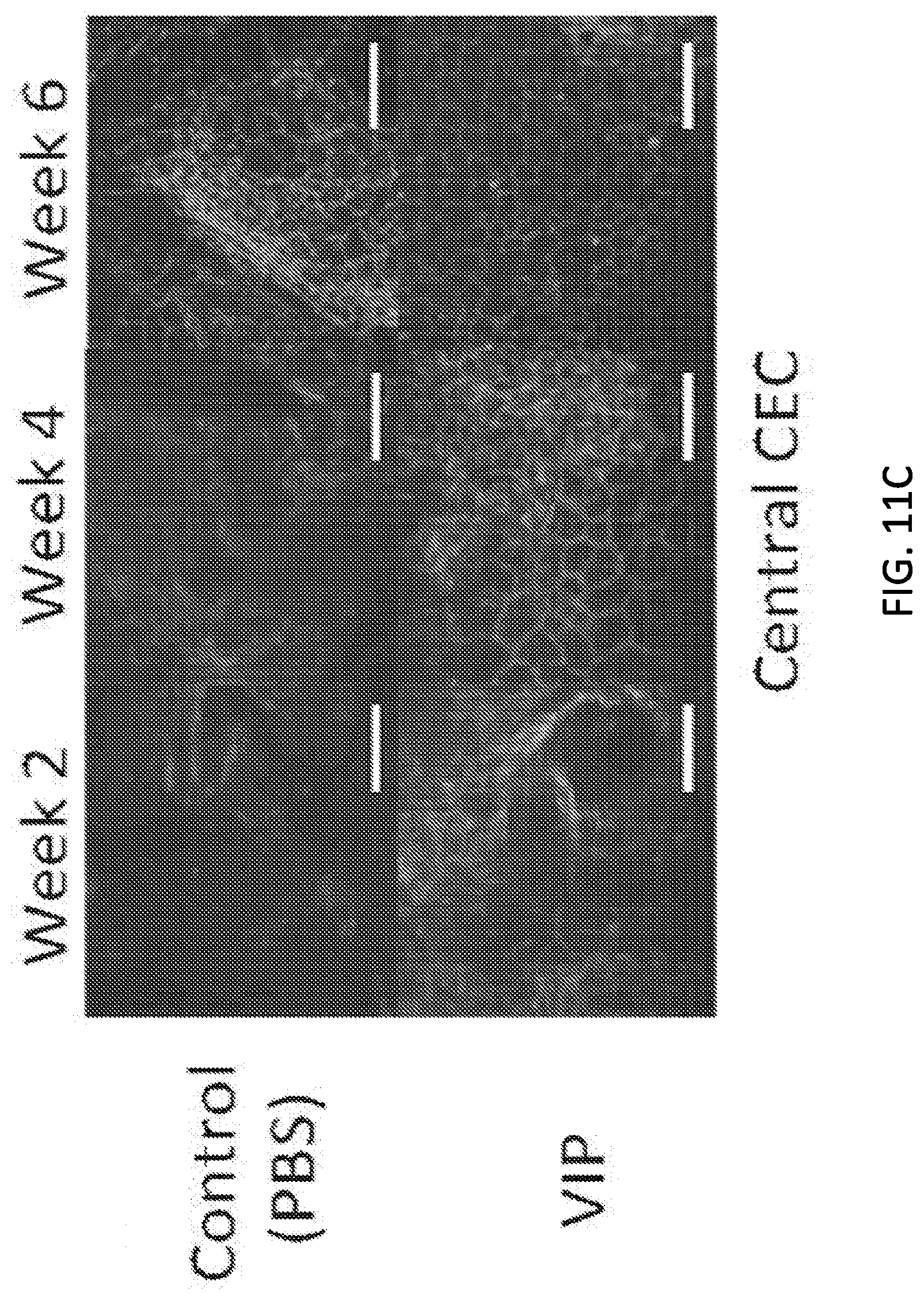
D00015

D00016

D00017

D00018

D00019

D00020


S00001
XML
uspto.report is an independent third-party trademark research tool that is not affiliated, endorsed, or sponsored by the United States Patent and Trademark Office (USPTO) or any other governmental organization. The information provided by uspto.report is based on publicly available data at the time of writing and is intended for informational purposes only.
While we strive to provide accurate and up-to-date information, we do not guarantee the accuracy, completeness, reliability, or suitability of the information displayed on this site. The use of this site is at your own risk. Any reliance you place on such information is therefore strictly at your own risk.
All official trademark data, including owner information, should be verified by visiting the official USPTO website at www.uspto.gov. This site is not intended to replace professional legal advice and should not be used as a substitute for consulting with a legal professional who is knowledgeable about trademark law.