Self-Contained Apparatus and System for Detecting Microorganisms
Erickson; Stephen E. ; et al.
U.S. patent application number 16/709567 was filed with the patent office on 2020-06-11 for self-contained apparatus and system for detecting microorganisms. The applicant listed for this patent is Laboratory Corporation of America Holdings. Invention is credited to Dwight L. Anderson, Stephen E. Erickson, Jose S. Gil, Wendy Hahn, Minh Mindy Bao Nguyen, John Paulson, Jessica Stach.
| Application Number | 20200182869 16/709567 |
| Document ID | / |
| Family ID | 69106196 |
| Filed Date | 2020-06-11 |


View All Diagrams
| United States Patent Application | 20200182869 |
| Kind Code | A1 |
| Erickson; Stephen E. ; et al. | June 11, 2020 |
Self-Contained Apparatus and System for Detecting Microorganisms
Abstract
Disclosed herein are devices, methods, and systems for rapid detection of microorganisms using a recombinant bacteriophage. The specificity of recombinant bacteriophages for binding microorganisms allows targeted and highly specific detection of a microorganism of interest.
| Inventors: | Erickson; Stephen E.; (White Bear Township, MN) ; Paulson; John; (Burlington, NC) ; Nguyen; Minh Mindy Bao; (Shoreview, MN) ; Stach; Jessica; (Burlington, NC) ; Gil; Jose S.; (Winnetka, CA) ; Anderson; Dwight L.; (Minneapolis, MN) ; Hahn; Wendy; (Hugo, MN) | ||||||||||
| Applicant: |
|
||||||||||
|---|---|---|---|---|---|---|---|---|---|---|---|
| Family ID: | 69106196 | ||||||||||
| Appl. No.: | 16/709567 | ||||||||||
| Filed: | December 10, 2019 |
Related U.S. Patent Documents
| Application Number | Filing Date | Patent Number | ||
|---|---|---|---|---|
| 62777473 | Dec 10, 2018 | |||
| 62798980 | Jan 30, 2019 | |||
| Current U.S. Class: | 1/1 |
| Current CPC Class: | B01L 3/5029 20130101; B01L 2200/0647 20130101; B01L 3/502769 20130101; C12Y 113/12 20130101; B01L 2200/10 20130101; G01N 33/535 20130101; C12N 2795/00031 20130101; G01N 33/56911 20130101; C12N 7/00 20130101; B01L 3/502753 20130101 |
| International Class: | G01N 33/569 20060101 G01N033/569; B01L 3/00 20060101 B01L003/00; C12N 7/00 20060101 C12N007/00; G01N 33/535 20060101 G01N033/535 |
Claims
1. A device comprising: a first compartment comprising a recombinant bacteriophage having a genetic construct inserted into a bacteriophage genome, wherein the construct comprises a promoter and an indicator gene; and a second compartment comprising a signal detecting component, wherein the signal detecting component facilitates detection of the indicator gene product produced as a result of infecting the sample with the recombinant bacteriophage.
2. The device of claim 1, wherein the signal detecting component is a substrate and the indicator gene encodes an enzyme.
3. The device of the claim 2, wherein the enzyme is a luciferase.
4. A method to detect one or more microorganism of interest in a sample comprising the steps of: contacting the sample with an infectious reagent in a device, wherein the one or more microorganisms of interest in the sample, if present, are infected by the infectious agent, wherein the device comprises: a first compartment comprising recombinant bacteriophage having a genetic construct inserted into a bacteriophage genome, wherein the construct comprises a promoter and an indicator gene; contacting the recombinant bacteriophage from the first compartment with the sample such that the recombinant bacteriophage infect the one or more microorganisms in the sample, thereby producing indicator gene product, and detecting the indicator gene product in a second compartment.
5. The method of claim 4, wherein the second compartment comprises a substrate, and wherein detecting the indicator gene product is performed after contacting the indicator gene product with a substrate.
6. The method of claim 4, wherein the method further comprises binding microorganisms in the sample to a solid support.
7. The method of claim 6, wherein the solid support is a bead.
8. The method of claim 6, wherein the solid support comprises polyethylene (PE), polypropylene (PP), polystyrene (PS), polylactic acid (PLA) and polyvinyl chloride (PVC).
9. The method of claim 4, wherein the apparatus further comprises a second compartment containing a substrate, and wherein the method further comprises: adding the substrate from the second compartment to the sample, concurrently with or after adding the recombinant bacteriophage.
10. The method of claim 4, wherein the first compartment comprises a seal, and wherein contacting the recombinant bacteriophage with the sample is by breaking the seal, wherein the breakage of the seal causes the recombinant bacteriophage from the first compartment to be in contact with the sample and infect the one or more microorganisms in the sample, thereby producing indicator gene product.
11. The method of claim 4, wherein the bacteriophage is lyophilized.
12. The method of claim 4, wherein the apparatus comprises a third compartment containing growth media.
13. The method of claim 4, wherein the method comprises incubating the solid support that has captured the one or more microorganisms of interest in the growth media for a time period before adding the recombinant bacteriophage.
14. The method of claim 4, wherein the apparatus comprises a stop-lock for phased mixing of the recombinant bacteriophage and the substrate with the sample.
15. The method of claim 4, wherein the solid support is dry prior to contacting the sample.
16. The method of claim 4, wherein the solid support is soaked in media prior to contacting the sample.
17. The method of claim 4, wherein the solid support that has captured the one or more organisms is incubated with the growth media in a third compartment before contacting with the recombinant bacteriophage.
18. The method of claim 4, wherein the incubation is 0-2 hours.
19. The method of any of claims 4, wherein the bacteriophage has been in contact with the sample for 0.2-3 hours before detecting the indicator gene product.
20. The method of claim 4, wherein the indicator gene product comprises at least one of a fluorophore, a fluorescent protein, a particle, and an enzyme.
21. The method of claim 20, wherein the enzyme comprises at least one of a luciferase, a phosphatase, a peroxidase, and a glycosidase.
22. The method of claim 21, wherein the luciferase is a genetically engineered luciferase.
23. The method of claim 4, wherein the sample is a food, environmental, water, commercial, or clinical sample.
24. The method of claim 4, wherein the method detects as few as 10, 9, 8, 7, 6, 5, 4, 3, 2, or a single bacterium in a sample of a standard size for the food safety industry.
25. The method of claim 4, wherein the sample comprises meat or vegetables.
26. The method of claim 4, wherein the sample is a food, water, dairy, environmental, commercial, or clinical sample.
27. The method of claim 4, wherein the sample is first incubated in conditions favoring growth for an enrichment period of 9 hours or less, 8 hours or less, 7 hours or less, 6 hours or less, 5 hours or less, 4 hours or less, 3 hours or less, or 2 hours or less.
28. A system for detecting microorganism of interest in a sample comprising: an apparatus comprising: a first compartment comprising recombinant bacteriophage having a genetic construct inserted into a bacteriophage genome, wherein the construct comprises a promoter and an indicator gene; and a signal detecting component, wherein the signal detecting component can detect the indicator gene product produced from infecting the sample with the recombinant bacteriophage.
29. The system of claim 28, wherein the signal detecting component is a handheld luminometer.
Description
CROSS REFERENCE TO RELATED APPLICATION
[0001] The present application claims priority to U.S. provisional Application Nos. 62/777,473, filed on Dec. 10, 2018, and 62/798,980, filed on Jan. 30, 2019. The disclosures of U.S. application Ser. Nos. 13/773,339, 14/625,481, 15/263,619, 15/409,258, 16/298,695 and U.S. provisional Application Nos. 62/616,956, 62/628,616, 62/661,739, 62/640,793, and 62/798,980 are hereby incorporated by reference in their entirety herein.
FIELD OF THE INVENTION
[0002] The invention relates to methods, apparatuses, devices, and systems for detection of microorganism of interest using recombinant bacteriophages.
BACKGROUND
[0003] There is a strong interest in improving speed and sensitivity for detection of bacteria, viruses, and other microorganisms in biological, food, water, and clinical samples. Microbial pathogens can cause substantial morbidity among humans and domestic animals, as well as immense economic loss. Detection of microorganisms is a high priority for the Food and Drug Administration (FDA) and Centers for Disease Control (CDC) given outbreaks of life-threatening or fatal illness caused by ingestion of food contaminated with certain microorganisms, e.g., Staphylococcus spp., Escherichia coli or Salmonella spp.
[0004] Traditional microbiological tests for the detection of bacteria rely on non-selective and selective enrichment cultures followed by plating on selective media and further testing to confirm suspect colonies. Such procedures can require several days. A variety of rapid methods have been investigated and introduced into practice to reduce the time requirement. However, to-date, methods reducing the time requirement have drawbacks. For example, techniques involving direct immunoassays or gene probes generally require an overnight enrichment step in order to obtain adequate sensitivity, and therefore lack the ability to deliver same-day results. Polymerase chain reaction (PCR) tests also include an amplification step and therefore are capable of both very high sensitivity and selectivity; however, the sample size that can be economically subjected to PCR testing is limited. Dilute bacterial suspensions capable of being subjected to PCR will be free of cells and therefore purification and/or lengthy enrichment steps are still required.
[0005] The time required for traditional biological enrichment is dictated by the growth rate of the target bacterial population of the sample, by the effect of the sample matrix, and by the required sensitivity. In practice, most high sensitivity methods employ an overnight incubation and take about 24 hours overall. Due to the time required for cultivation, these methods can take up to three days, depending upon the organism to be identified and the source of the sample. This lag time is generally unsuitable as such delays allow contaminated food or water or other products to make its way into livestock or humans. In addition, increases in antibiotic-resistant bacteria and biodefense considerations make rapid identification of bacterial pathogens in water, food, and clinical samples critical priorities worldwide.
[0006] Therefore, there is a need for more rapid, simple and sensitive detection and identification of microorganisms, such as bacteria and other potentially pathogenic microorganisms.
SUMMARY
[0007] Embodiments of the invention comprise devices, compositions, methods, apparatuses, systems, and kits for the detection of microorganisms. The invention may be embodied in a variety of ways.
[0008] Aspects of the invention comprise devices for facilitating ease and simplicity of microorganism detection. Described herein is an apparatus or device for performing an assay to detect a microorganism in a test sample using recombinant bacteriophage.
[0009] In one aspect, the present invention utilizes the high specificity of bacteriophage that can bind microorganisms to detect low levels of a microorganism. In some embodiments, the method detects as few as 10, 9, 8, 7, 6, 5, 4, 3, 2, or a single bacterium in a sample of a standard size for the food safety industry. In other embodiments, the sample is first incubated in conditions favoring growth for an enrichment period of 9 hours or less, 8 hours or less, 7 hours or less, 6 hours or less, 5 hours or less, 4 hours or less, 3 hours or less, or 2 hours or less. In some embodiments, the sample is not enriched prior to incubation with recombinant bacteriophage.
[0010] In some embodiments, the recombinant bacteriophage is genetically modified to include a reporter gene as previously described. In additional embodiments the recombinant bacteriophage is derived from bacteriophage specific to the microorganism to be detected. For example, the recombinant bacteriophage may be derived from any of the following bacteriophages: Salmonella phage SPN1S, Salmonella phage 10, Salmonella phage epsilon15, Salmonella phage SEA1, Salmonella phage Spn1s, Salmonella phage P22, Listeria phage LipZ5, Listeria phage P40, Listeria phage vB_LmoM_AG20, Listeria phage P70, Listeria phage A511, Staphylococcus phage P4W, Staphylococcus phage K, Staphylococcus phage Twort, Staphylococcus phage SA97, or Escherichia coli O157:H7 phage CBA120.
[0011] In some embodiments, the recombinant bacteriophage may be stabilized or preserved. For example, the recombinant bacteriophage may be lyophilized.
[0012] In certain embodiments, the reporter generates an indicator moiety. In certain embodiments the indicator moiety can generate an intrinsic signal. In other embodiments the indicator moiety comprises an enzyme that generates signal upon reaction with substrate. In yet other embodiments, the indicator moiety comprises a cofactor that generates signal upon reaction with one or more additional signal producing components. For example, the indicator moiety comprises at least one of a fluorophore, a fluorescent protein, a particle, and an enzyme. The enzyme may comprise at least one of a luciferase, a phosphatase, a peroxidase, and a glycosidase. The luciferase gene can be a naturally occurring gene, such as Oplophorus luciferase, Firefly luciferase, Lucia luciferase, or Renilla luciferase, or it can be a genetically engineered gene.
[0013] Some embodiments of the invention comprise a device or apparatus for performing the detection method. In some embodiments a device may include separate compartments. In some embodiments separate compartments of a device may be configured to allow a user to combine the contents from different compartments at various stages of the detection method. For example, the sample to be tested may be combined with a bacteriophage in one compartment and allowed to incubate for some period of time before being added to the contents of another compartment of the device, such as a substrate. In such embodiments the substrate may react with any reporter (or indicator moiety) produced as a result of infection by the recombinant bacteriophage (e.g., if the target microorganism is present in the sample).
[0014] Some embodiments include a device comprising a first compartment comprising a recombinant bacteriophage having a genetic construct inserted into a bacteriophage genome, wherein the construct comprises a promoter and an indicator gene; and a second compartment comprising a signal detecting component, wherein the signal detecting component facilitates detection of the indicator gene product produced as a result of infecting the sample with the recombinant bacteriophage. In some embodiments, the signal detecting component is a substrate and the indicator gene encodes an enzyme. In some embodiments, the enzyme is a luciferase.
[0015] In some embodiments, the apparatus comprises a first compartment comprising recombinant bacteriophage, an inlet/portal for adding a portion of a test sample to the recombinant bacteriophage, and a second compartment comprising a substrate or other paired reagent and wherein the method further comprises adding the substrate from the second compartment to the sample, concurrently with or after adding the recombinant bacteriophage. In some embodiments, wherein the apparatus comprises a third compartment. The third compartment can, for example, comprise growth media for enriching a sample. In other embodiments, the apparatus is free of media.
[0016] In some embodiments, the apparatus further comprises a second compartment comprising a substrate, and wherein detecting the indicator gene product is by contacting the indicator gene product with a substrate. In some embodiments, the solid support is a bead. In some embodiments, the solid support comprises polyethylene (PE), polypropylene (PP), polystyrene (PS), polylactic acid (PLA) and polyvinyl chloride (PVC). In some embodiments, the solid support is dry prior to contacting the sample. In some embodiments, the solid support is soaked in media prior to contacting the sample. In some embodiments, the solid support that has captured the one or more organisms is incubated with the growth media in the third compartment before contacting with the recombinant bacteriophage.
[0017] In some embodiments, the solid support is coated with a cell binding component that binds with high affinity to the microorganism of interest in the sample. This allows the more bacteria binding to the solid support and increase assay sensitivity and specificity. In other embodiments, the solid support is coated with an antibody capable of binding the microorganism of interest.
[0018] In some embodiments, the first compartment comprises a seal, and wherein contacting the recombinant bacteriophage with the sample is by breaking the seal, wherein the breakage of the seal causes the recombinant bacteriophage from the first compartment to be in contact with the sample and infect the one or more microorganisms in the sample, thereby producing indicator gene product (indicator protein). In further embodiments, the apparatus comprises a stop-lock for phased mixing of the media, the recombinant bacteriophage, and the substrate with the sample.
[0019] In some embodiments, the indicator gene product comprises at least one of a fluorophore, a fluorescent protein, a particle, and an enzyme. In some embodiments, the enzyme comprises at least one of a luciferase, a phosphatase, a peroxidase, and a glycosidase. In some embodiments, the luciferase is a genetically engineered luciferase. In some embodiments, the sample is a food, environmental, water, commercial, or clinical sample.
[0020] In another aspect, the method to detect one or more microorganism of interest in a sample comprising the steps of: contacting. the sample with a solid support of an apparatus, wherein the solid support captures the one or more microorganisms in the sample, if present, wherein the apparatus comprises: a first compartment comprising recombinant bacteriophage having a genetic construct inserted into a bacteriophage genome, wherein the construct comprises a promoter and an indicator gene; contacting the recombinant bacteriophage from the first compartment with the sample such that the recombinant bacteriophage infect the one or more microorganisms in the sample, thereby producing indicator gene product, and detecting the indicator gene product.
[0021] In some embodiments, the method comprising incubating the solid support that has captured the one or more microorganisms of interest in the growth media for a time period before adding the recombinant bacteriophage. In some embodiments, the incubation is 0-2 hours. In some embodiments, wherein the bacteriophage has been in contact with the sample for 0.5-3 hours before detecting the indicator gene product.
[0022] In some embodiments, the method detects as few as 10, 9, 8, 7, 6, 5, 4, 3, 2, or a single bacterium in a sample of a standard size for the food safety industry. In some embodiments, the sample comprises meat or vegetables. In some embodiments, the sample is a food, water, dairy, environmental, commercial, or clinical sample.
[0023] In some embodiments, the sample is first incubated in conditions favoring growth for an enrichment period of 9 hours or less, 8 hours or less, 7 hours or less, 6 hours or less, 5 hours or less, 4 hours or less, 3 hours or less, or 2 hours or less.
[0024] Additional embodiments include systems and kits for detecting microorganisms such as Listeria, Salmonella, Staphylococcus, or E. coli O157:H7, comprising a recombinant bacteriophage. Some embodiments further include a substrate for reacting with an indicator moiety of the recombinant bacteriophage. These systems or kits can include features described for the bacteriophage, compositions, and methods of the invention. In still other embodiments, the invention comprises non-transient computer readable media for use with methods or systems according to the invention.
[0025] In another aspect, this disclosure provides a system for detecting microorganism of interest in a sample comprising: an apparatus, which comprises: a first compartment comprising recombinant bacteriophage having a genetic construct inserted into a bacteriophage genome, wherein the construct comprises a promoter and an indicator gene; and a signal detecting component, wherein the signal detecting component can detect the indicator gene product produced from infecting the sample with the recombinant bacteriophage. In some embodiments, the signal detecting component is a handheld luminometer.
BRIEF DESCRIPTION OF THE FIGURES
[0026] The present invention may be better understood by referring to the following non-limiting figures.
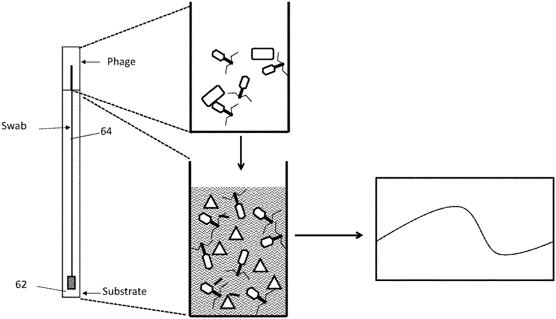
[0027] FIG. 1 shows one embodiment of a method for detecting a bacterium of interest using a recombinant bacteriophage.
[0028] FIG. 2 shows the results of detecting L. monocytogenes culture using one embodiment of a self-contained apparatus with a swab as a solid support. The signals corresponding to the presence of the bacteria was detected by Hygiena, GloMax, and GloMax 20/20 luminometers. Table 1 shows results from log phase culture and Table 2 shows results from overnight culture.
[0029] FIGS. 3A and 3B are plots generated from the data shown in Table 1. FIG. 3A shows measurements of signals detected using Hygiena. Swabs were inoculated with log phase cells at the indicated CFU level. Sample was immediately infected with Listeria phage cocktail for 4 hours. Substrate was added and samples were read on the Hygiena Luminometer. A signal of >10 RLU is considered positive. With this method, approximately 25,000 CFU is required to generate a positive result.
[0030] FIG. 3B shows the measurements of signals detected using GloMax 20/20 and GloMax (a.k.a., GloMax 96) luminometers. Swabs were inoculated with log phase cells at the indicated CFU level. Sample was immediately infected with Listeria phage cocktail for 4 hours. Substrate was added and samples were read on either the GloMax 20/20 (1 mL of sample) or GloMax (150 .mu.l of sample) Luminometers. A signal/background ratio of >3.0 is considered positive. With this method, approximately 5,000 CFU is required to generate a positive result.
[0031] FIG. 4 shows the results of detecting Salmonella in ground turkey that has been inoculated with Salmonella. Table 3 shows uninoculated control sample, and Table 4 shows inoculated turkey sample. The tests were repeated with varying incubation and infection time.
[0032] FIG. 5A and FIG. 5B are plots generated from the data in FIG. 4. FIG. 6A shows that Salmonella-inoculated turkey samples were detected as positive with every incubation and infection time tested. The turkey sample was grown for 24 hours at 41.degree. C. after inoculation before testing with the methods disclosed in the application. For relative signal: 0 HR incubation, 2 HR infection >1 HR incubation, 0.5 HR infection >0 HR incubation, 0.5 HR infection. In addition, comparison of RLU signal shows that the GloMax luminometers have a much higher signal that that of the Hygiena luminometer.
[0033] FIG. 5B shows that detecting using GloMax 20/20 and GloMax luminometers produced similar signal/background ratios for the same samples. Although the GloMax 20/20 had a greater signal (FIG. 5A), the background was significantly higher than that with the GloMax. Thus when determining the signal/background, the two luminometers perform similarly.
[0034] FIG. 6 shows data for detecting Salmonella in three turkey samples (samples 21, 24, and 26) that had been inoculated with Salmonella before the assays using the self-contained apparatus. The samples were infected for different duration of time as indicated before detection of the signal.
[0035] FIGS. 7A-7C are plots generated from the data shown in FIG. 6. FIGS. 7A-7C show results of the experiments in which three inoculated ground turkey samples were enriched for 24 hours and swab samples were taken and assayed. Sample 24 (FIG. 7B) and 26 (FIG. 7C) did not show signal on Hygiena handheld luminometer for samples that had 30 min phage infection, but did for sample that had 2 hour infection. The GloMax 20/20 and GloMax luminometer generated relatively low signals.
[0036] FIGS. 8A-8C are plots generated from the data shown in FIG. 6. The plots show both GloMax 20/20 and GloMax were able to detect Sample 21 (FIG. 8A) and 26 (FIG. 8B) as positive with 30 minute infection (signal/background ratio of >3.0 is positive), however Sample 24 (FIG. 8C) required a 2 hour infection to show a positive result. The GloMax 20/20 and GloMax luminometer results were similar.
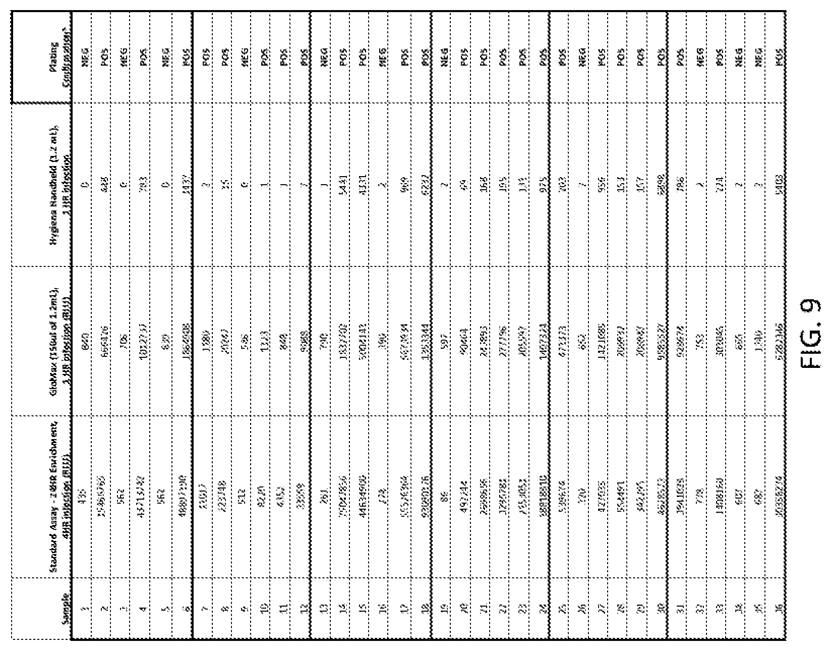
[0037] FIG. 9 shows data for detecting L. monocytogenes environmental sponge samples from inoculated surfaces and enriched for 24 hours.
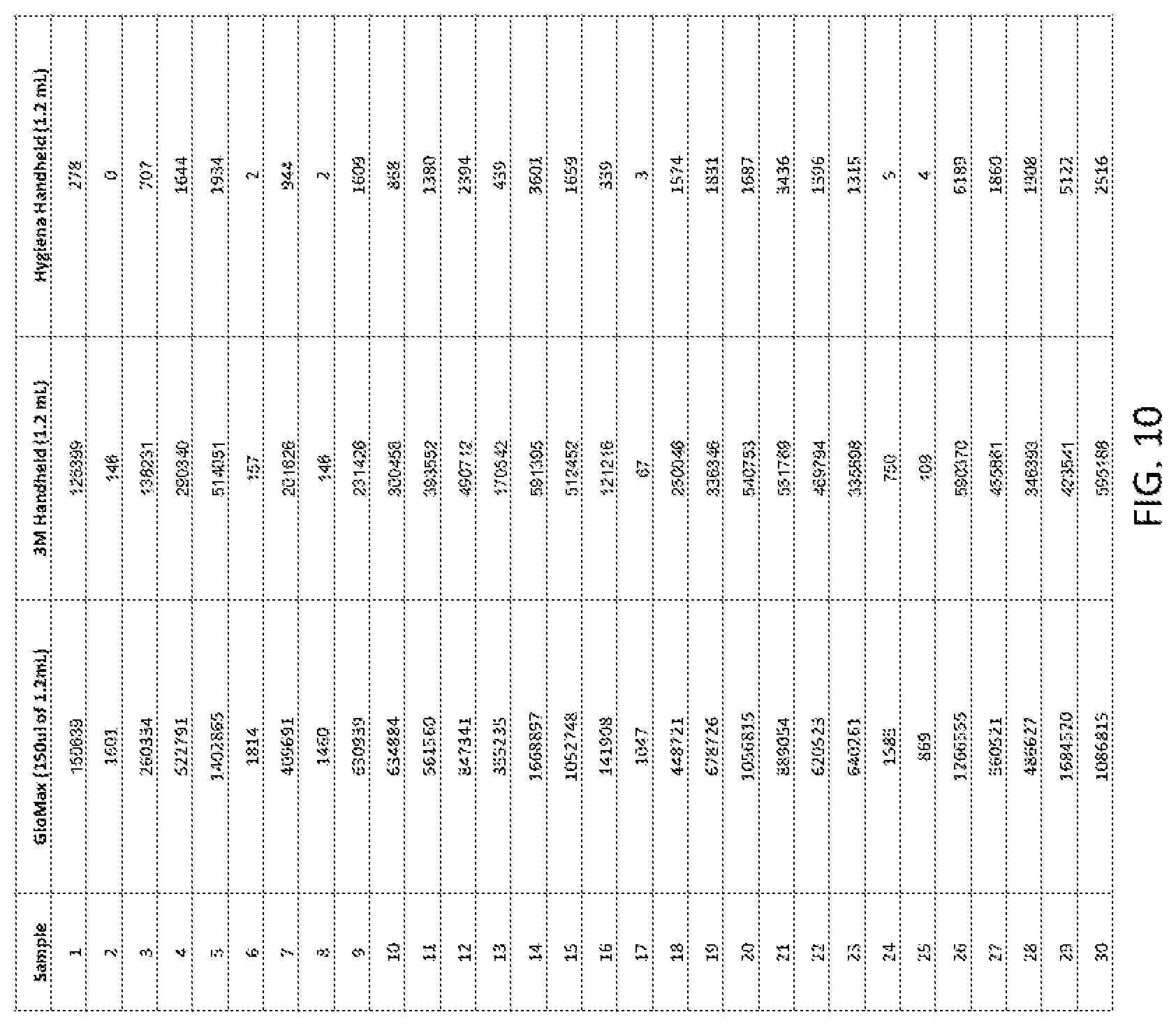
[0038] FIG. 10 shows detecting microorganisms in Salmonella-inoculated turkey samples using the apparatus. The signals were measured using three different luminometers: GloMax, 3M, and Hygiena.
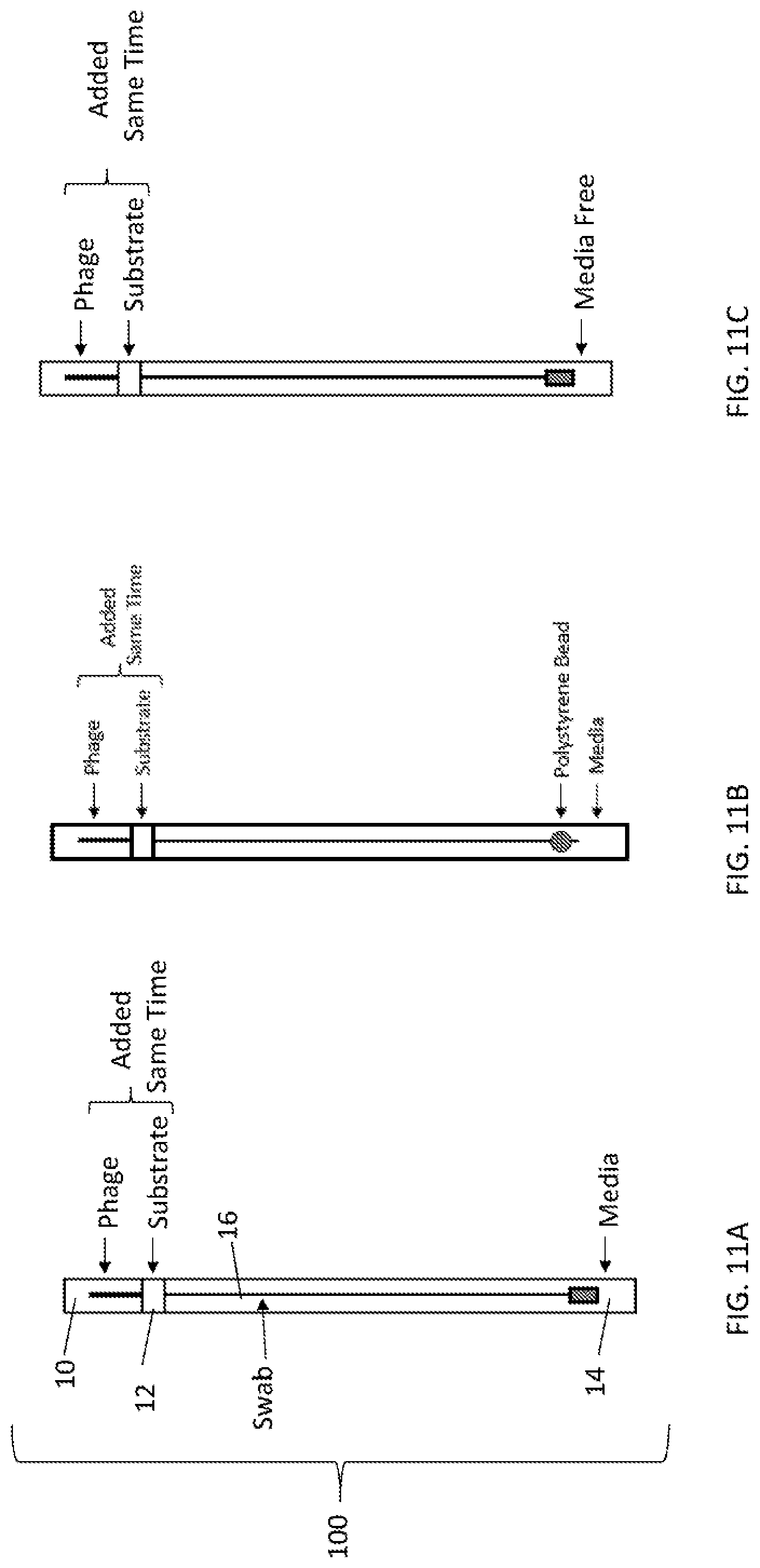
[0039] FIGS. 11A, 11B, and 11C depict a view of one embodiment of a self-contained apparatus system for detecting microorganisms, having a swab (FIG. 11A) or a bead (FIG. 11B) inserted into a container comprising three compartments. Each compartment is separated by a snap action seal. The first compartment contains phage, the second compartment contains substrate, and the third compartment contains media. In some embodiments the solid support is a swab. In some embodiments, the device is free of media (FIG. 11C).
[0040] FIGS. 12A, 12B and 12C depict a view of one embodiment of a self-contained apparatus system for detecting microorganisms, having a swab (FIG. 12A) or a bead (FIG. 12B) inserted into a container comprising three compartments. Each compartment is separated by a snap action seal. The first compartment contains phage, the second compartment contains media, and the third compartment contains substrate. After incubation with the phage and media, the seal separating the second and third compartment may be broken. In some embodiments the solid support is a swab. In some embodiments, the device is free of media (FIG. 12C).
[0041] FIGS. 13A, 13B, and 13C depict a view of one embodiment of a self-contained apparatus system for detecting microorganisms, having a swab (FIG. 13A) or a bead (FIG. 13B) inserted into a container comprising three compartments. Each compartment is separated by a snap action seal. The first compartment contains media, the second compartment contains phage, and the third compartment contains substrate. In some embodiments the solid support is a swab. In some embodiments, the device is free of media (FIG. 13C).
[0042] FIGS. 14A, 14B, and 14C depict a view of one embodiment of a self-contained apparatus system for detecting microorganisms, having a swab (FIG. 14A) or a bead (FIG. 14B) inserted into a container comprising three compartments. Each compartment is separated by a snap action seal. The first compartment contains media, the second compartment contains phage, and the third compartment contains substrate. The apparatus has a stop-lock mechanism for phased mixing of reagents. In some embodiments the solid support is a swab. In some embodiments, the device is free of media (FIG. 14C).
[0043] FIGS. 15A, 15B, and 15C depict a view of one embodiment of a self-contained apparatus system for detecting microorganisms, having a swab (FIG. 15A) or a bead (FIG. 15B) inserted into a container comprising three compartments. Each compartment is separated by a snap action seal. The first compartment contains media, the second compartment contains phage, and the third compartment contains substrate. The apparatus has a stop-lock mechanism for phased mixing of reagents. In some embodiments the solid support is a swab. In some embodiments, the device is free of media (FIG. 15C).
[0044] FIG. 16 depicts a view of one embodiment of a self-contained apparatus system for system for detecting microorganisms, having a swab inserted into a container comprising two compartments. Each compartment is separated by a snap action seal. The first compartment contains phage and the second compartment contains substrate. In some embodiments the solid support is a swab.
[0045] FIG. 17 depicts a flow diagram of an embodiment utilizing a self-contained apparatus system for detecting microorganisms.
DETAILED DESCRIPTION OF THE INVENTION
Definitions
[0046] Unless otherwise defined herein, scientific and technical terms used in connection with the present invention shall have the meanings that are commonly understood by those of ordinary skill in the art. Further, unless otherwise required by context, singular terms shall include pluralities and plural terms shall include the singular. Generally, nomenclatures used in connection with, and techniques of, cell and tissue culture, molecular biology, immunology, microbiology, genetics and protein and nucleic acid chemistry and hybridization described herein are those well-known and commonly used in the art. Known methods and techniques are generally performed according to conventional methods well-known in the art and as described in various general and more specific references that are discussed throughout the present specification unless otherwise indicated. Enzymatic reactions and purification techniques are performed according to manufacturer's specifications, as commonly accomplished in the art or as described herein. The nomenclatures used in connection with the laboratory procedures and techniques described herein are those well-known and commonly used in the art.
[0047] The following terms, unless otherwise indicated, shall be understood to have the following meanings:
[0048] As used herein, the terms "a", "an", and "the" can refer to one or more unless specifically noted otherwise.
[0049] The use of the term "or" is used to mean "and/or" unless explicitly indicated to refer to alternatives only or the alternatives are mutually exclusive, although the disclosure supports a definition that refers to only alternatives and "and/or." As used herein "another" can mean at least a second or more.
[0050] Throughout this application, the term "about" is used to indicate that a value includes the inherent variation of error for the device, the method being employed to determine the value, or the variation that exists among samples.
[0051] The term "solid support" or "support" means a structure that provides a substrate and/or surface onto which biomolecules may be bound. For example, a solid support may be an assay well (i.e., such as a microtiter plate or multi-well plate), or the solid support may be a location on a filter, an array, or a mobile support, such as a bead or a membrane (e.g., a filter plate or lateral flow strip).
[0052] The term "indicator" or "indicator moiety" or "detectable moiety" or "detectable biomolecule" or "reporter" or "label" refers to a molecule that provides a signal that can be measured in a qualitative or quantitative assay. For example, an indicator moiety may comprise an enzyme that may be used to convert a substrate to a product that can be measured. An indicator moiety may be an enzyme that catalyzes a reaction that generates bioluminescent emissions (e.g., luciferase, HRP, or AP). Or, an indicator moiety may be a radioisotope that can be quantified. Or, an indicator moiety may be a fluorophore. Or, other detectable molecules may be used.
[0053] As used herein, "bacteriophage" or "phage" includes one or more of a plurality of bacterial viruses. In this disclosure, the terms "bacteriophage" and "phage" include viruses such as mycobacteriophage (such as for TB and paraTB), mycophage (such as for fungi), mycoplasma phage, and any other term that refers to a virus that can invade living bacteria, fungi, mycoplasma, protozoa, yeasts, and other microscopic living organisms and uses them to replicate itself. Here, "microscopic" means that the largest dimension is one millimeter or less. Bacteriophages are viruses that have evolved in nature to use bacteria as a means of replicating themselves.
[0054] As used herein, "culture enrichment", "culturing for enrichment", "cultured for enrichment", or "culture for enrichment", refers to traditional culturing, such as incubation in media favorable to propagation of microorganisms, and should not be confused with other possible uses of the word "enrichment," such as enrichment by removing the liquid component of a sample to concentrate the microorganism contained therein, or other forms of enrichment that do not include traditional facilitation of microorganism propagation. Culturing for enrichment for short periods of time may be employed in some embodiments of methods described herein, but is not necessary and is for a much shorter period of time than traditional culturing for enrichment, if it is used at all.
[0055] As used herein "recombinant" refers to genetic (i.e., nucleic acid) modifications as usually performed in a laboratory to bring together genetic material that would not otherwise be found. This term is used interchangeably with the term "modified" herein.
[0056] As used herein "RLU" refers to relative light units as measured by a luminometer (e.g., GLOMAX.RTM. 96) or similar instrument that detects light. For example, the detection of the reaction between luciferase and appropriate substrate (e.g., NANOLUC.RTM. with NANO-GLO.RTM.) is often reported in RLU detected.
Overview
[0057] The present invention utilizes the high specificity of recombinant bacteriophage that can bind to a particular microorganism with high affinity to detect the presence of and/or quantify the specific microorganism in the sample.
[0058] Disclosed herein are compositions, methods, kits, and systems that demonstrate surprising sensitivity and speed for detection of a microorganism of interest in test samples (e.g., food, water, dairy, environmental, commercial, clinical, or other biological samples) using assays performed without culturing for enrichment, or in some embodiments with minimal incubation times during which microorganism could potentially multiply. These compositions, methods, kits, and systems allow detection of microorganisms to be achieved in a shorter timeframe than was previously thought possible.
[0059] Embodiments of the compositions, methods, kits, and system of the invention can be applied to detection of a variety of microorganisms (e.g., bacteria, fungi, yeast) in a variety of circumstances, including but not limited to, detection of pathogens from food, water, dairy, environmental, commercial, clinical, or other biological samples. The recombinant bacteriophage based detection embodiments disclosed herein may be adapted to any bacteria or other microorganism of interest (e.g., pathogenic microorganisms) for which a specific bacteriophage is available that does not recognize microorganisms for which detection is not of interest. The methods of the present invention provide high detection sensitivity and specificity rapidly and without the need for traditional biological enrichment (e.g., culturing). Thus, a variety of microorganisms may be detected using the methods of the invention.
[0060] Embodiments of the methods and systems of the invention can be applied to detection and quantification of a variety of microorganisms (e.g., bacteria, fungi, yeast) in a variety of circumstances, including but not limited to detection of pathogens from food, water, dairy, environmental, commercial, clinical, or other biological samples. The methods of the present invention can rapidly provide high detection sensitivity and specificity without the need for traditional biological enrichment (e.g., culturing), which is a surprising aspect as all available methods with the desired sensitivity and specificity require culturing.
[0061] Also disclosed herein are systems and methods that uses an apparatus to detect microorganisms in test samples (e.g., food, water, dairy, environmental, commercial, clinical, or other biological samples). The method uses a self-contained apparatus that comprise a solid support, that can be used to collect sample. The apparatus further comprises a first compartment comprising bacteriophage having a genetic construct inserted into the bacteriophage genome, wherein the construct comprises a promoter and an indicator gene. The method comprises contacting the recombinant bacteriophage from the first compartment with the sample such that the recombinant bacteriophage infect one or more microorganisms in the sample thereby producing an indicator gene product ("indicator"), and detecting the indicator. In some aspects, the apparatus further comprises a second compartment, which contains a substrate specific for detecting the indicator. In some embodiments, the method further comprises contacting the sample that has been infected by the bacteriophage with the substrate, whereby detecting the indicator. In these embodiments, each compartment is separated from the immediately adjacent compartment by a snap action seal, which upon breakage, allows the content of the compartments to exit the compartment and mix with contents from the sample or contents from other compartments. For example, a user can break the snap action seal such that the recombinant bacteriophage from the first compartment contacts the sample on the solid support, thereby infecting microorganisms that bind thereon. Upon infection of the microorganisms, the indicator gene is expressed to produce an indicator protein, which can be detected by various detection devices. The presence of the signals indicates the presence of the microorganisms in the sample.
[0062] Embodiments of the apparatus, compositions, methods, kits, and system of the invention can be applied to detection of a variety of microorganisms (e.g., bacteria, fungi, yeast) in a variety of circumstances, including but not limited to, detection of pathogens from food, water, dairy, environmental, commercial, clinical, or other biological samples. The detection embodiments disclosed herein may be adapted to any bacteria or other microorganism of interest (e.g., pathogenic microorganisms) for which a specific recombinant bacteriophage is available that does not recognize other microorganisms that are not of interest. The methods of the present invention provide high detection sensitivity and specificity rapidly and without the need for traditional biological enrichment (e.g., culturing). This is a surprising aspect as all available methods with the desired sensitivity and specificity require culturing. The detection of microorganisms in a sample using the self-contained apparatus, which houses reagents required for detecting the microorganism in separate compartments until the time of detection, is convenient and efficient. The apparatus is easy to use and does not require extensive training.
Samples
[0063] Each of the embodiments of the compositions, methods, kits, and systems of the invention allows for the rapid detection and/or quantification of microbes in a sample. For example, methods according to the present invention can be performed in a shortened time period with superior results.
[0064] In certain embodiments, a recombinant bacteriophage is used to detect microorganisms of interest. Microorganisms that can be detected by the compositions, methods, kits and systems of the present invention include pathogens that are of commercial, medical, or veterinary concern. Any microorganism for which a recombinant bacteriophage specific for the particular microbe has been identified can be detected by the methods of the present invention. Those skilled in the art will appreciate that there is no limit to the application of the present methods other than the availability of the necessary recombinant bacteriophage/microbe pairs.
[0065] Bacterial cells detectable by the present invention include, but are not limited to, bacterial cells that are food- or water-borne pathogens. Bacterial cells detectable by the present invention include, but are not limited to, all species of Salmonella, all strains of Escherichia coli, including, but not limited to E. coli O157:H7 (and other Shiga toxin--and enterotoxin-producing strains of E. coli), all species of Listeria, including, but not limited to L. monocytogenes, and all species of Campylobacter. Bacterial cells detectable by the present invention include, but are not limited to, bacterial cells that are pathogens of medical or veterinary significance. Such pathogens include, but are not limited to, Bacillus spp., Bordetella pertussis, Brucella spp., Campylobacter jejuni, Chlamydia pneumoniae, Clostridium perfringens, Clostridium botulinum, Enterobacter spp., Klebsiella pneumoniae, Mycoplasma pneumoniae, Salmonella typhi, Salmonella typhimurium, Salmonella enteritidis, Shigella sonnei, Yersinia spp., Vibrio spp. Staphylococcus aureus, and Streptococcus spp.
[0066] The sample may be an environmental or food or water sample. Some embodiments may include medical or veterinary samples. Samples may be liquid, solid, or semi-solid. Samples may be swabs of solid surfaces. Samples may include environmental materials, such as water samples, or the filters from air samples, or aerosol samples from cyclone collectors. Samples may be of beef, poultry, processed foods, milk, cheese, or other dairy products. Medical or veterinary samples include, but are not limited to, blood, sputum, cerebrospinal fluid, and fecal samples. In some embodiments, samples may be different types of swabs.
[0067] In some embodiments, samples may be used directly in the detection methods of the present invention, without preparation, concentration, or dilution. For example, liquid samples, including but not limited to, milk and juices, may be assayed directly. In other embodiments, samples may be diluted or suspended in solution, which may include, but is not limited to, a buffered solution or a bacterial culture medium. A sample that is a solid or semi-solid may be suspended in a liquid by mincing, mixing or macerating the solid in the liquid. In some embodiments, a sample should be maintained within a pH range that promotes recombinant bacteriophage attachment to the host bacterial cell. In some embodiments, the preferred pH range may be one suitable for bacteriophage attached to a bacterial cell. A sample should also contain the appropriate concentrations of divalent and monovalent cations, including but not limited to Na.sup.+, Mg.sup.2+, and K.sup.+.
[0068] In some embodiments, the sample is maintained at a temperature that maintains the viability of any pathogen cell present in the sample. During steps in which bacteriophage, are attaching to bacterial cells, the sample may be maintained at a temperature that facilitates bacteriophage activity. Such temperatures are at least about 25.degree. C. and no greater than about 45.degree. C. In some embodiments the sample is maintained at about 37.degree. C. In some embodiments the samples are subjected to gentle mixing or shaking during recombinant bacteriophage binding or infection.
Methods of Using Recombinant Bacteriophage for Detecting Microorganism
[0069] Methods for using recombinant bacteriophage to detect microorganisms of interest have previously been described. Assays may include various appropriate control samples. For example, control samples containing no recombinant bacteriophages and/or control samples containing recombinant bacteriophages without bacteria may be assayed as controls for background signal levels.
[0070] As noted herein, in certain embodiments, the invention may comprise methods of using recombinant bacteriophage for detecting microorganisms. The methods of the invention may be embodied in a variety of ways.
[0071] In some aspects, the invention comprises a method for detecting a microorganism of interest. The method may use a recombinant bacteriophage for detection of the microorganism of interest. For example, in certain embodiments, the microorganism of interest is a bacterium and the recombinant bacteriophage is derived from a bacteriophage that specifically recognizes the bacterium of interest. In certain embodiments, the method may comprise detection of a bacterium of interest in a sample by incubating the sample with a plurality of recombinant bacteriophage that can bind to the bacterium of interest. A plurality of recombinant bacteriophage bound to a single microorganism is any number greater than 1, but is preferably at least 5.times.10.sup.4, or at least 1.times.10.sup.5, or at least 1.times.10.sup.6, or at least 1.times.10.sup.8, or at least 1.times.10.sup.9, or at least 1.times.10.sup.10 recombinant bacteriophage.
[0072] In certain embodiments, the recombinant bacteriophage comprises an indicator moiety. The methods may comprise detecting the indicator moiety of the recombinant bacteriophage, wherein positive detection of the indicator moiety indicates that the bacterium of interest is present in the sample.
[0073] In some embodiments, the invention may comprise a method to detect as few as a single microorganism of interest in a sample comprising the steps of: incubating the sample with a plurality of recombinant bacteriophage that bind the microorganism of interest, wherein the recombinant bacteriophage comprises an indicator moiety and is contacted with the sample to be tested under conditions such that the plurality of recombinant bacteriophage bind the bacterium of interest; separating unbound recombinant bacteriophage from cell-bound recombinant bacteriophage; and detecting the indicator moiety resulting from infection of the bacterium, wherein positive detection of the indicator moiety indicates that the microorganism of interest is present in the sample. Embodiments may include incubating the sample with at least 5.times.10.sup.8, or at least 5.times.10.sup.9, or at least 5.times.10.sup.10, or at least 5.times.10.sup.11, or at least 5.times.10.sup.12, or at least 5.times.10.sup.13 recombinant bacteriophage.
[0074] In some embodiments, the detecting step will require addition of a substrate for the indicator enzyme to act on. The selection of a particular indicator is not critical to the present invention, but the indicator will be capable of generating a detectable signal either by itself, or be instrumentally detectable, or be detectable in conjunction with one or more additional signal producing components, such as an enzyme/substrate signal producing system.
[0075] In certain embodiments, the assay may be performed to utilize a recombinant bacteriophage to identify the presence of a specific microorganism. The assay can be modified to accommodate different sample types or sizes and assay formats. Embodiments employing recombinant bacteriophage of the invention may allow rapid detection of specific bacterial strains, with total assay times under 1.5, 2.0, 2.5, 3.0, 3.5, 4.0, 4.5, 5.0, 5.5, 6.0, 6.5, 7.0, 7.5, 8.0, 8.5, 9.0, 9.5, 10.0, 10.5, 11.0, 11.5, or 12 hours, depending on the sample type, sample size, and assay format. For example, the amount of time required may be somewhat shorter or longer depending on affinity of the recombinant bacteriophage and/or and types of bacteria to be detected in the assay, type and size of the sample to be tested, complexity of the physical/chemical environment, and the concentration of endogenous non-target bacterial contaminants.
[0076] FIG. 1 illustrates an embodiment of an assay for detecting a bacterium of interest using a recombinant bacteriophage according to an embodiment of the invention. A wide variety of configurations and steps for combining reagents are possible. In the embodiment illustrated here, aliquots of indicator phage (or lyophilized indicator phage) are contained in a first compartment of a device. A swab containing bacteria is introduced into the first compartment and incubated for a period of time (e.g., 45 minutes at 37.degree. C.) sufficient for phage to replicate and generate soluble indicator (e.g., luciferase). The seal between the first compartment containing soluble indicator and phage and a second compartment may then be broken to allow the soluble indicator protein to come into contact with a substrate contained within the second compartment. A luminometer may be used to detect the reaction of indicator (e.g., luciferase) with a substrate. Experiments utilizing this method are described herein. In some embodiments, following infection, the contents of the device may be pulled back into the first compartment in order to mix the infected bacteria with the substrate. In further embodiments, the contents may then pushed back into the second compartment such that the indicator signal can be read in the luminometer.
[0077] In some embodiments, the sample may be enriched prior to testing by incubation in conditions that encourage growth. In such embodiments, the enrichment period can be 1, 2, 3, 4, 5, 6, 7, or up to 8 hours or longer, depending on the sample type and size.
[0078] Thus, in some embodiments, the indicator bacteriophage comprises a detectable indicator moiety, and infection of a single pathogenic cell (e.g., bacterium) can be detected by an amplified signal generated via the indicator moiety. Thus the method may comprise detecting an indicator moiety produced during phage replication, wherein detection of the indicator indicates that the bacterium of interest is present in the sample.
[0079] In in some embodiments, the indicator bacteriophage comprises a detectable indicator moiety, and infection of a single pathogenic cell (e.g., bacterium) can be detected by an amplified signal generated via the indicator moiety. Thus the method may comprise detecting an indicator moiety produced during phage replication, wherein detection of the indicator indicates that the bacterium of interest is present in the sample.
[0080] As described in more detail herein, the methods and systems of the invention may utilize a range of concentrations of parental indicator bacteriophage to infect bacteria present in the sample. In some embodiments the indicator bacteriophage are added to the sample at a concentration sufficient to rapidly find, bind, and infect target bacteria that are present in very low numbers in the sample, such as a single cell. In some embodiments, the phage concentration can be sufficient to find, bind, and infect the target bacteria in less than one hour. In other embodiments, these events can occur in less than two hours, or less than three hours, following addition of indicator phage to the sample. For example, in certain embodiments, the bacteriophage concentration for the incubating step is greater than 1.times.10.sup.5 PFU/mL, greater than 1.times.10.sup.6 PFU/mL, or greater than 1.times.10.sup.7 PFU/mL.
[0081] Methods of the invention may comprise various other steps to increase sensitivity. For example, as discussed in more detail herein, the method may comprise a step for capturing and washing the captured and bound bacterium, to remove excess recombinant bacteriophage and increase the signal to noise ratio. In some embodiments, positive detection of the indicator moiety requires that the ratio of signal to background generated by detecting the indicator moiety is at least 2.0 or at least 2.5.
[0082] FIGS. 2-10 demonstrate data and plots of those data from exemplary embodiments of recombinant bacteriophage assays.
[0083] FIGS. 2, 3A, and 3B relate to detection of L. monocytogenes. FIG. 2 shows data from detection of L. monocytogenes culture using one embodiment of a self-contained apparatus with a swab as a solid support. The signals corresponding to the presence of the bacteria was detected by Hygiena, GloMax, and GloMax 20/20 luminometers. Table 1 shows results from log phase culture and Table 2 shows results from overnight culture.
[0084] FIGS. 3A and 3B are plots generated from the data shown in Table 1. FIG. 3A shows measurements of signals detected using Hygiena. Swabs were inoculated with log phase cells at the indicated CFU level. Sample was immediately infected with Listeria phage cocktail for 4 hours. Substrate was added and samples were read on the Hygiena Luminometer. A signal of >10 RLU is considered positive. With this method, approximately 25,000 CFU is required to generate a positive result.
[0085] FIG. 3B shows the measurements of signals detected using GloMax 20/20 and GloMax (a.k.a., GloMax 96) luminometers. Swabs were inoculated with log phase cells at the indicated CFU level. Sample was immediately infected with Listeria phage cocktail for 4 hours. Substrate was added and samples were read on either the GloMax 20/20 (1 mL of sample) or GloMax (150 .mu.l of sample) Luminometers. A signal/background ratio of >3.0 is considered positive. With this method, approximately 5,000 CFU is required to generate a positive result.
[0086] FIGS. 4-8 relate to embodiments demonstrating detection of Salmonella. FIG. 4 shows the results of detecting Salmonella in ground turkey that has been inoculated with Salmonella. Table 3 shows uninoculated control sample, and Table 4 shows inoculated turkey sample. The tests were repeated with varying incubation and infection time.
[0087] FIG. 5A and FIG. 5B are plots generated from the data in FIG. 4. FIG. 6A shows that Salmonella-inoculated turkey samples were detected as positive with every incubation and infection time tested. The turkey sample was grown for 24 hours at 41.degree. C. after inoculation before testing with the methods disclosed in the application. For relative signal: 0 HR incubation, 2 HR infection >1 HR incubation, 0.5 HR infection >0 HR incubation, 0.5 HR infection. In addition, comparison of RLU signal shows that the GloMax luminometers have a much higher signal that that of the Hygiena luminometer.
[0088] FIG. 5B shows that detecting using GloMax 20/20 and GloMax luminometers produced similar signal/background ratios for the same samples. Although the GloMax 20/20 had a greater signal (FIG. 5A), the background was significantly higher than that with the GloMax. Thus when determining the signal/background, the two luminometers perform similarly.
[0089] FIG. 6 shows data for detecting Salmonella in three turkey samples (samples 21, 24, and 26) that had been inoculated with Salmonella before the assays using the self-contained apparatus. The samples were infected for different duration of time as indicated before detection of the signal.
[0090] FIGS. 7A-7C are plots generated from the data shown in FIG. 6. FIGS. 7A-7C show results of the experiments in which three inoculated ground turkey samples were enriched for 24 hours and swab samples were taken and assayed. Sample 24 (FIG. 7B) and 26 (FIG. 7C) did not show signal on Hygiena handheld luminometer for samples that had 30 min phage infection, but did for sample that had 2 hour infection. The GloMax 20/20 and GloMax luminometer generated relatively low signals.
[0091] FIGS. 8A-8C are plots generated from the data shown in FIG. 6. The plots show both GloMax 20/20 and GloMax were able to detect Sample 21 (FIG. 8A) and 26 (FIG. 8B) as positive with 30 minute infection (signal/background ratio of >3.0 is positive), however Sample 24 (FIG. 8C) required a 2 hour infection to show a positive result. The GloMax 20/20 and GloMax luminometer results were similar.
[0092] FIG. 9 shows data for detecting L. monocytogenes environmental sponge samples from inoculated surfaces and enriched for 24 hours.
[0093] FIG. 10 shows detecting microorganisms in Salmonella-inoculated turkey samples using the apparatus. The signals were measured using three different luminometers: GloMax, 3M, and Hygiena.
[0094] Recombinant bacteriophage assays retain specificity over time. In numerous embodiments over multiple years of development and utilization of recombinant bacteriophage assays for, e.g., E. coli, Cronobacter, Salmonella, Listeria and S. aureus, changes in host specificity were not observed. These embodiments include more than 25 recombinant luciferase reporter or indicator bacteriophages. Further, loss or inactivation of the reporter or indicator (e.g., luciferase) gene has not been detected in any recombinant phage as described herein. Recombinant bacteriophages are stored and preserved in multiple aliquots to protect against mishandling. Although bacteriophages are intrinsically stable, titers may decrease to a variable extent over extended storage, e.g., for months or years at 4.degree. C. This is generally believed to be due to loss of DNA from particles; however, active bacteriophage are recovered by plating for plaques to obtain higher titers. Specificity verification is performed with each recombinant bacteriophage preparation by infecting different bacterial strains that are known to be positive and negative.
[0095] In some embodiments of methods for testing samples, the use of a large excess of recombinant bacteriophage necessitates separation of any unbound bacteria or other larger components of the sample from the excess of unbound recombinant bacteriophage. This may be accomplished in many different ways generally known by one of ordinary skill in the art. Microorganism cells can be separated through centrifugation, filtration by size, or selective immobilization. In some embodiments, filtration by size is accomplished through filter wells. In other embodiments, magnetic separation can be used for selective immobilization. For example, the sample may be filtered through a 0.45 .mu.m or 0.22 .mu.m membrane, either before or after incubating with the recombinant bacteriophage, to capture the target microorganism (e.g., bacterium) on a solid support. The captured microorganism may then be washed one or more times on the solid support to ensure that only specifically bound recombinant bacteriophage remains. Or a mechanism for specific or non-specific binding can be employed to capture the microorganism on micro-beads or another solid surface. Other formats for separating components of the sample are possible.
[0096] A variety of solid supports may be used. In certain embodiments, the solid support may comprise a multi-well plate, a filter, a bead, a lateral flow strip, a filter strip, filter disc, filter paper, or thin films designed for culturing cells (e.g., PetriFilm by 3M). Other solid supports may also be appropriate. For example, in some embodiments the test sample microorganism may be captured by binding to a swab, the surface of a plate, or by filtering the sample through a bacteriological filter (e.g., 0.45 .mu.m pore size spin filter or plate filter). In one embodiment, the microorganism captured on the filter or plate surface is incubated with recombinant bacteriophage and subsequently washed one or more times to remove excess unbound recombinant bacteriophage.
[0097] Alternatively, in some embodiments the capturing step may be based on other features of the microorganism of interest, such as size. In embodiments utilizing size-based capture, the solid support may be a spin column filter. In some embodiments, the solid support comprises a 96-well filter plate. Or, the solid support for capture may be a location on an array, or a mobile support, such as a bead.
[0098] In some embodiments, the solid support is coated with a cell binding component that binds with high affinity to the microorganism of interest in the sample. This allows the more bacteria binding to the solid support and increase assay sensitivity and specificity.
[0099] In an embodiment, where the microorganism of interest is a bacterium, the recombinant bacteriophage may bind to the bacterium via a cell binding domain from the bacteriophage. For example, well-studied phages of E. coli include T1, T2, T3, T4, T5, T7, and lambda; other E. coli phages available in the ATCC collection, for example, include phiX174, S13, Ox6, MS2, phiV1, fd, PR772, and ZIK1. Salmonella phages include SPN1S, 10, epsilon15, SEA1, and P22. Listeria phages include LipZ5, P40, vB_LmoM_AG20, P70, and A511. Staphylococcus phages include P4W, virus K, Twort, phi11, 187, P68, and phiWMY.
[0100] In some embodiments, the sample may be enriched prior to testing by incubation in conditions that encourage growth. In such embodiments, the enrichment period can be 1, 2, 3, 4, 5, 6, 7, or up to 8 hours or longer, depending on the sample type and size.
[0101] In other embodiments, the sample may be enriched following capture of the bacterium on a solid support. In such embodiments, the enrichment period can be 1, 2, 3, 4, 5, 6, 7, or up to 8 hours or longer, depending on the sample type and size.
[0102] Thus, in some embodiments, the recombinant bacteriophage comprises a detectable indicator moiety, and binding to a single pathogenic cell (e.g., bacterium) can be detected by an amplified signal generated via the indicator moiety. Thus the method may comprise detecting an indicator moiety of the recombinant bacteriophage, wherein detection of the indicator indicates that the bacterium of interest is present in the sample.
[0103] In some embodiments of the methods of the invention, the microorganism may be detected without any isolation or purification of the microorganisms from a sample. For example, in certain embodiments, a sample containing one or more microorganisms of interest may be applied directly to an assay container such as a spin column, a microtiter well, or a filter and the assay is conducted in that assay container. That is, microorganisms are captured on a membrane having pore size too small to allow the microorganisms to pass through. Various embodiments of such assays are disclosed herein.
[0104] Aliquots of a test sample may be distributed directly into wells of a multi-well plate, recombinant bacteriophage may be added, and after a period of time sufficient for binding, the cells may be captured on a solid surface such as a plate, bead, or a filter substrate, such that excess unbound recombinant bacteriophage can be removed in one or more subsequent washing steps. Then a substrate for the indicator moiety (e.g., luciferase substrate for a luciferase indicator) is added and assayed for detection of the indicator signal. Some embodiments of the method can be performed on filter plates. Some embodiments of the method can be performed with or without concentration of the sample before binding with recombinant bacteriophage.
[0105] For example, in many embodiments, multi-well plates are used to conduct the assays. The choice of plates (or any other container in which detecting may be performed) may affect the detecting step. For example, some plates may include a colored or white background, which may affect the detection of light emissions. Generally, white plates have higher sensitivity but also yield a higher background signal. Other colors of plates may generate lower background signal but also have a slightly lower sensitivity. Additionally, background signal can result from the leakage of light from one well to another, adjacent well. Some plates have white wells while the rest of the plate is black, thus, allowing for a high signal inside the well while preventing well-to-well light leakage. This combination of white wells with black plates may decrease background signal. Thus the choice of plate or other assay vessel may influence the sensitivity and background signal for the assay. In some embodiments, detection of the microorganism of interest may be completed without the need for culturing the sample. For example, in certain embodiments the total time required for detection is less than 12.0 hours, 11.0 hours, 10.0 hours, 9.0 hours, 8.0 hours, 7.0 hours, 6.0 hours, 5.0 hours, 4.0 hours, 3.0 hours, 2.5 hours, 2.0 hours, 1.5 hours, 1.0 hour, 45 minutes, or less than 30 minutes. Minimizing time to result is critical in food and environmental testing for pathogens.
[0106] In contrast to assays known in the art, the method of the invention can detect individual microorganisms. Thus, in certain embodiments, the method may detect .ltoreq.10 cells of the microorganism (i.e., 1, 2, 3, 4, 5, 6, 7, 8, 9, 10 microorganisms) or .ltoreq.20, or .ltoreq.30, or .ltoreq.40, or .ltoreq.50, or .ltoreq.60, or .ltoreq.70, or .ltoreq.80, or .ltoreq.90, or .ltoreq.100, or .ltoreq.200, or .ltoreq.500, or .ltoreq.1000 cells of the microorganism present in a sample. For example, in certain embodiments, the recombinant bacteriophage is highly specific for S. Aureus, Listeria, Salmonella, or E. coli. In an embodiment, the recombinant bacteriophage can distinguish S. Aureus, Listeria, Salmonella, or E. coli in the presence of more than 100 other types of bacteria. In an embodiment, the recombinant bacteriophage can distinguish a specific serotype within a species of bacteria (e.g., E. coli O157:H7) in the presence of more than 100 other types of bacteria. In certain embodiments, the recombinant bacteriophage can be used to detect a single bacterium of the specific type in the sample. In certain embodiments, the recombinant bacteriophage detects as few as 2, 3, 4, 5, 6, 7, 8, 9, 10, 15, 20, 30, 40, 50, 60, 70, 80, 90, or 100 of the specific bacteria in the sample.
[0107] Thus, aspects of the present invention provide methods for detection of microorganisms in a test sample via a reporter or an indicator moiety. In some embodiments, where the microorganism of interest is a bacterium, the indicator moiety may be associated with a recombinant bacteriophage. The indicator moiety may react with a substrate to emit a detectable signal or may emit an intrinsic signal (e.g., fluorescent protein). Fluorescent proteins naturally fluoresce (intrinsic fluorescence or autofluorescence) by emitting energy as a photon when the fluorescent moiety containing electrons absorb a photon. Fluorescent proteins (e.g., GFP) can be expressed as a fusion protein. In some embodiments, the detection sensitivity can reveal the presence of as few as 100, 50, 20, 10, 9, 8, 7, 6, 5, 4, 3, or 2 cells of the microorganism of interest in a test sample. In some embodiments, even a single cell of the microorganism of interest may yield a detectable signal.
[0108] The selection of a particular indicator moiety is not critical to the present invention, but the indicator moiety will be capable of generating a detectable signal either by itself, or be instrumentally detectable, or be detectable in conjunction with one or more additional signal producing components, such as an enzyme/substrate signal producing system. A number of recombinant bacteriophages can be formed by varying either the indicator moiety or reporter of the recombinant bacteriophage; it will be appreciated by one skilled in the art that the choice involves consideration of the microorganism to be detected and the desired means of detection.
[0109] For example, one or more signal producing components can be reacted with the indicatory moiety to generate a detectable signal. In some embodiments, the indicator can be a bioluminescent compound. If the indicator moiety is an enzyme, then amplification of the detectable signal is obtained by reacting the enzyme with one or more substrates or additional enzymes and substrates to produce a detectable reaction product. In an alternative signal producing system, the indicator can be a fluorescent compound where no enzymatic manipulation of the indicator is required to produce the detectable signal. Fluorescent molecules including, for example, fluorescein and rhodamine and their derivatives and analogs are suitable for use as indicators in such a system. In yet another alternative embodiment, the indicator moiety can be a cofactor, then amplification of the detectable signal is obtained by reacting the cofactor with the enzyme and one or more substrates or additional enzymes and substrates to produces a detectable reaction product. In some embodiments, the detectable signal is colorimetric.
[0110] The detectable indicator moiety is a key feature of the recombinant bacteriophage, which can be detected directly or indirectly. The indicator moiety provides a detectable signal by which the binding reaction is monitored providing a qualitative and/or quantitative measure. The relative quantity and location of signal generated by the decorated or signalized microorganisms can serve to indicate the presence and/or quantity of the microorganism. The indicator moiety can also be used to select and isolate decorated or signalized microorganisms, such as by flow sorting or using magnetic separation media.
[0111] In some embodiments, the indicator moiety of the recombinant bacteriophage may be detectable directly or after incubation with a substrate. Many different types of detectable biomolecules suitable for use as indicator moieties are known in the art, and many are commercially available. In some embodiments the recombinant bacteriophage comprises an enzyme, which serves as the indicator moiety. In some embodiments, the indicator or reporter of the recombinant bacteriophage encodes a detectable enzyme. The indicator moiety may emit light and/or may be detectable by a color change. Various appropriate enzymes are commercially available, such as alkaline phosphatase (AP), horseradish peroxidase (HRP), green fluorescent protein (GFP), or luciferase (Luc). In some embodiments, these enzymes may serve as the indicator moiety. In some embodiments, Firefly luciferase is the indicator moiety. In some embodiments, Oplophorus luciferase is the indicator moiety. In some embodiments, NANOLUC.RTM. is the indicator moiety. Other engineered luciferases or other enzymes that generate detectable signals may also be appropriate indicator moieties.
[0112] Thus, in some embodiments, the recombinant bacteriophage of the methods, systems or kits is a wild-type bacteriophage genetically modified with the sequence of an indicator protein, such as a fluorescent protein or a luciferase protein.
[0113] Bacteriophages are able to infect and lyse specific bacteria.
[0114] Detecting the indicator may include detecting emissions of light. In some embodiments, a luminometer may be used to detect the reaction of indicator (e.g., luciferase) with a substrate. The detection of RLU can be achieved with a luminometer, or other machines or devices may also be used. For example, a spectrophotometer, CCD camera, or CMOS camera may detect color changes and other light emissions. Absolute RLU are important for detection, but the signal to background ratio also needs to be high (e.g., >2.0, >2.5, or >3.0) in order for single cells or low numbers of cells to be detected reliably.
[0115] In some embodiments, the reaction of indicator moiety (e.g., luciferase) with substrate may continue for 30 minutes or more, and detection at various time points may be desirable for optimizing sensitivity. For example luminometer readings may be taken initially and at 3-, or 5-, or 10-, or 15-minute intervals until the reaction is completed.
[0116] Thus in some embodiments utilizing recombinant bacteriophage, the invention comprises a method for detecting a microorganism of interest comprising the steps of capturing at least one sample bacterium; incubating the at least one bacterium with a plurality of recombinant bacteriophage; allowing time for binding to target microorganism in the sample; and detecting the indicator moiety, wherein detection of the indicator moiety demonstrates that the bacterium is present in the sample.
[0117] For example, in some embodiments the test sample bacterium may be captured by binding to the surface of a plate, or by filtering the sample through a bacteriological filter (e.g., 0.45 .mu.m pore size spin filter or plate filter). In an embodiment, the recombinant bacteriophage is added in a minimal or modest volume to the captured sample directly on the filter. In an embodiment, the microorganism captured on the filter or plate surface is subsequently washed one or more times to remove excess unbound recombinant bacteriophage.
[0118] In some embodiments, aliquots of a test sample comprising bacteria may be applied to a spin column and after incubation with a recombinant bacteriophage and washing to remove any excess bacteriophage, the amount of indicator detected will be proportional to the amount of target bacteria present in the sample.
[0119] The indicator (e.g., luciferase) bound to the bacteria may then be measured and quantified. In an embodiment, the solution is spun through the filter, and the filtrate collected for assay in a new receptacle (e.g., in a luminometer) following addition of a substrate for the indicator enzyme (e.g., luciferase substrate). Alternatively, the indicator signal may be measured directly on the filter.
[0120] In an embodiment, the microorganism is a bacterium and the recombinant bacteriophage includes an indicator moiety derived from a bacteriophage. In an embodiment, the indicator moiety is luciferase. Thus, in an alternate embodiment, the indicator substrate (e.g., luciferase substrate) may be incubated with the portion of the sample that remains on a filter or bound to a plate surface. Accordingly, in some embodiments the solid support is a 96-well filter plate (or regular 96-well plate), and the substrate reaction may be detected by placing the plate directly in the luminometer.
[0121] For example, in an embodiment, the invention may comprise a method for detecting a pathogenic bacterium of interest comprising the steps of: binding cells captured on a 96-well filter plate with a plurality of recombinant bacteriophage; washing excess recombinant bacteriophage away; and detecting the indicator (e.g., luciferase) by adding substrate and measuring enzyme activity directly in the 96-well plate, wherein detection of enzyme activity indicates that the bacterium of interest is present in the sample.
[0122] In another embodiment, the invention may comprise a method for detecting a microorganism of interest, such as S. Aureus, comprising the steps of: binding cells in liquid solution or suspension in a 96-well plate with a plurality of recombinant bacteriophage; washing unbound recombinant bacteriophage away from cells having bound recombinant bacteriophage; and detecting the indicator (e.g., luciferase) by adding substrate and measuring enzyme activity directly in the 96-well plate, wherein detection of enzyme activity indicates that the microorganism of interest, such as S. Aureus, is present in the sample. In some embodiments, the microorganism of interest may be captured on a solid support such as on beads or a filter. This capturing can occur either before or after incubation with the recombinant bacteriophage. In some embodiments no capturing step is necessary.
[0123] In some embodiments, the liquid solution or suspension may be a consumable test sample, such as a vegetable wash. In some embodiments, the liquid solution or suspension may be vegetable wash fortified with concentrated LB Broth, Tryptic/Tryptone Soy Broth, Peptone Water, or Nutrient Broth. In some embodiments, the liquid solution or suspension may be bacteria diluted in LB Broth.
[0124] In some embodiments, target microorganism cells need to be intact for proper detection. That is, the cells need not be viable, but the cell wall must be structurally intact. Thus it is desirable to minimize lysis of the bacterium before the detection step.
[0125] In some embodiments, an initial concentration step for the sample is useful. That is, any microorganisms or other relatively large substances in the sample are concentrated to remove excess liquid. However it is possible to perform the assay without an initial concentration step. Some embodiments do include an initial concentration step, and in some embodiments this concentration step allows a shorter enrichment incubation time. In other embodiments, no enrichment period is necessary.
[0126] Some embodiments of testing methods may further include confirmatory assays. A variety of assays are known in the art for confirming an initial result, usually at a later point in time. For example, the samples can be cultured (e.g., CHROMAGAR.RTM./DYNABEADS.RTM. assay), PCR can be utilized to confirm the presence of the microbial DNA, or other confirmatory assays can be used to confirm the initial result.
[0127] Embodiments of food safety assays include sample preparation steps. Some embodiments can include enrichment time. For example, enrichment for 1, 2, 3, 4, 5, 6, 7, or 8 hours may be needed, depending on sample type and size. Following these sample preparation steps, binding with a high concentration of recombinant bacteriophage that comprises a reporter or indicator can be performed in a variety of assay formats, such as that shown in FIG. 1.
[0128] Embodiments of food assays can detect a single pathogenic bacterium in sample sizes corresponding to industry standards, with a reduction in time-to-results of at least 20%, or at least 30%, or at least 40% or at least 50% or at least 60% depending on the sample type and size.
[0129] Thus, some embodiments of the present invention solve a need by using recombinant bacteriophage methods for amplifying a detectable signal indicating the presence of bacteria. In certain embodiments as little as a single bacterium is detected. The principles applied herein can be applied to the detection of a variety of microorganisms. In this way, embodiments of the present invention can achieve tremendous signal amplification from even a single cell of the microorganism of interest.
[0130] Aspects of the present invention utilize the high specificity of binding components that can bind to particular microorganisms, such as the recognition and binding component of infectious agents, as a means to detect and/or quantify the specific microorganism in a sample. In some embodiments, the present invention takes advantage of the specificity of bacteriophages.
[0131] Some embodiments of the invention disclosed and described herein utilize the discovery that a single microorganism is capable of generating a large amount of indicator following infection with recombinant bacteriophage. This principle allows amplification of indicator signal from one or a few cells based on specific recognition of the microorganism. For example, by exposing even a single cell of a bacterium to a plurality of recombinant bacteriophage, the indicator signal is amplified such that a single bacterium is detectable.
[0132] The unprecedented speed and sensitivity of detecting a microorganism with recombinant bacteriophages are unexpected results. In some embodiments, the methods of the invention require less than 12, 11, 10, 9, 8, 7, 6, 5, 4, 3, or 2 hours for detection of a microorganism of interest. These are shorter timeframes than were previously thought possible. In some embodiments, the methods can detect as few as 100, 50, 20, 10, 9, 8, 7, 6, 5, 4, 3, or 2 cells of the bacterium of interest. In some embodiments, even a single cell of the bacterium is detectable. In additional embodiments, the invention comprises systems (e.g., computer systems, automated systems or kits) comprising components for performing the methods disclosed herein, and/or using the recombinant bacteriophages described herein.
[0133] Existing protocols for detection of pathogenic bacteria in foods are complicated, expensive, slow, labor-intensive and prone for false positives. Moreover, phage-based detection methods include the added complication and regulatory implications of infectious reagents. Detection with a recombinant bacteriophage specific for a given pathogen offers an effective, fast and simple testing alternative.
[0134] Infectious agents can be highly specific to a particular type of organism. For example, a bacteriophage may be specific to a particular genus of a bacterium, such as Listeria. For example, the A511 bacteriophage is specific for the genus Listeria. Or a bacteriophage may be specific to a particular species of bacterium, such as E. coli. For some types of bacteria, bacteriophages may even recognize particular subtypes of that organism with high specificity. For example, the CBA120 bacteriophage is highly specific for E. coli O157:H7 and the .phi.YeO3-12 bacteriophage is highly specific for Y. enterocolitica serotype O:3.
[0135] In some embodiments of the recombinant bacteriophage described herein, the recombinant bacteriophage can be derived from T7, T4, T4-like, ViI, ViI-like, AR1, A511, A118, A006, A500, PSA, P35, P40, B025, B054,A97, phiSM101, phi3626, CBA120, SPN1S, 10, epsilon15, P22, LipZ5, P40, vB_LmoM_AG20, P70, A511, P4W, K, Twort, or SA97. A recombinant bacteriophage also includes an indicator moiety, such as a fluorescent moiety, a fluorescent protein, a bioluminescent protein, or an enzyme, that allows the recombinant bacteriophage to generate a signal. In a recombinant bacteriophage, various types of reporters can be inserted into the bacteriophage genome, to serve as an indicator moiety. In some embodiments, the recombinant bacteriophage comprises an enzyme, such as a luciferase, which is only detectable upon addition of an appropriate substrate. For example, luciferase, alkaline phosphatase, and other reporter enzymes react with an appropriate substrate to provide a detectable signal. Some embodiments of a recombinant bacteriophage comprise a wild-type luciferase or an engineered luciferase, such as NANOLUC.RTM.. Other embodiments include a fluorescent protein or another reporter protein.
[0136] In alternate embodiments, bacteriophages, phages, mycobacteriophages (such as for TB and paraTB), mycophages (such as for fungi), mycoplasma phages, and any other virus that can invade living bacteria, fungi, mycoplasma, protozoa, yeasts, and other microscopic living organisms can be studied or copied to target a microorganism of interest. For example, well-studied phages of E. coli include T1, T2, T3, T4, T5, T7, and lambda; other E. coli phages available in the ATCC collection, for example, include phiX174, S13, Ox6, MS2, phiV1, fd, PR772, and ZIK1. Salmonella phages include SPN1S, 10, epsilon15, SEA1, and P22. Listeria phages include LipZ5, P40, vB_LmoM_AG20, P70, and A511. Staphylococcus phages include P4W, virus K, Twort, phi 11, 187, P68, and phiWMY.
[0137] Some embodiments of the invention utilize the specificity of binding of a recombinant bacteriophage for rapid and sensitive targeting to bind and facilitate detection of a bacterium of interest.
[0138] In some embodiments, a recombinant bacteriophage is derived from a bacteriophage specific for a target bacterium of interest, such as from T7, T4 or another similar phage. A recombinant bacteriophage may also be derived from T4-like, T7-like, ViI, ViI-like, AR1, A511, A118, A006, A500, PSA, P35, P40, B025, B054, A97, phiSM101, phi3626, CBA120, SPN1S, 10, epsilon15, P22, LipZ5, P40, vB_LmoM_AG20, P70, A511, P4W, K, Twort, or SA97.
[0139] In some embodiments, a small indicator gene product may be desirable. OpLuc and NANOLUC.RTM. proteins are only about 20 kDa (approximately 500-600 bp to encode), while FLuc is about 62 kDa and requires approximately 1,700 bp to encode. For comparison, the genome of T7 is around 40 kbp, while the T4 genome is about 170 kbp.
[0140] Moreover, the indicator should generate a high signal to background ratio and should be readily detectable in a timely manner. Promega's NANOLUC.RTM. is a modified Oplophorus gracilirostris (deep sea shrimp) luciferase. In some embodiments, NANOLUC.RTM. combined with Promega's NANO-GLO.RTM., an imidazopyrazinone substrate (furimazine), can provide a robust signal with low background.
[0141] In some recombinant bacteriophage embodiments, an indicator moiety can be any of a variety of biomolecules. The indicator can be a detectable product or an enzyme that produces a detectable product or a cofactor for an enzyme that produces a detectable product. In some embodiments, the indicator moiety of a recombinant bacteriophage is a reporter, such as a detectable enzyme. The indicator gene product may generate light and/or may be detectable by a color change. Various appropriate enzymes are commercially available, such as alkaline phosphatase (AP), horseradish peroxidase (HRP), or luciferase (Luc). For example, in one embodiment the indicator is a luciferase enzyme. Various types of luciferase may be used. In alternate embodiments, and as described in more detail herein, the luciferase is one of Oplophorus luciferase, Firefly luciferase, Lucia luciferase, Renilla luciferase, or an engineered luciferase. In some embodiments, the luciferase is derived from Oplophorus. In some embodiments, the indicator is a genetically modified luciferase, such as NANOLUC.RTM.. Other engineered luciferases or other enzymes that generate detectable signals may also be appropriate indicator moieties. In some embodiments, these enzymes may serve as the indicator moiety.
[0142] Compositions of the invention may comprise one or more recombinant bacteriophages derived from one or more wild-type infectious agents (e.g., bacteriophages) and one or more indicator moieties. In some embodiments, compositions can include cocktails of different recombinant bacteriophages that may generate the same or different indicator signals. That is, a composition for detecting a microorganism can include all the same or different recombinant bacteriophages.
[0143] Assays previously described utilize recombinant bacteriophage which recognize and bind to specific bacteria can be employed. The recombinant bacteriophage assays can be performed in a traditional laboratory setting or in a device, such as for example an apparatus as described further herein.
Recombinant Bacteriophage
[0144] As described in more detail herein, the apparatus, methods, systems and kits of the invention comprise recombinant bacteriophage for use in detection of microorganisms of interest. The recombinant bacteriophage are comprised in the first compartment of the apparatus disclosed above. In some embodiments, the invention may include a composition comprising a recombinant bacteriophage having an indicator gene incorporated into the genome of the bacteriophage. In some embodiments, the indicator gene is operably linked to a promoter that is not a native promoter of the bacteriophage. In some embodiments, the indicator gene is operably linked to a native promoter of the bacteriophage. The recombinant bacteriophage comprising the indicator or reporter gene are also referred to as indicator bacteriophage in this disclosure.
[0145] A recombinant bacteriophage can include a reporter or indicator gene. In certain embodiments of the bacteriophage, the indicator gene does not encode a fusion protein. For example, in certain embodiments, expression of the indicator gene during bacteriophage replication following infection of a host bacterium results in a soluble indicator protein product. In certain embodiments, the indicator gene may be inserted into a late gene region of the bacteriophage. Late genes are generally expressed at higher levels than other phage genes, as they code for structural proteins. The late gene region may be a class III gene region and may include a gene for a major capsid protein.
[0146] Some embodiments include designing (and optionally preparing) a sequence for homologous recombination downstream of the major capsid protein gene. Other embodiments include designing (and optionally preparing) a sequence for homologous recombination upstream of the major capsid protein gene. In some embodiments, the sequence comprises a codon-optimized reporter gene preceded by an untranslated region. The untranslated region may include a phage late gene promoter and ribosomal entry site.
[0147] In some embodiments, an indicator bacteriophage is derived from A511, P100, Listeria phage LMTA-94, LMA4, LMA8, T7, T4 or another similar phage. An indicator bacteriophage may also be derived from P100virus, T4-like, T7-like, ViI, ViI-like, Cronobacter-, Salmonella-, Listeria- or Staphylococcus-specific bacteriophage, or another bacteriophage having a genome with at least 70, 71, 72, 73, 74, 75, 76, 77, 78, 79, 80, 81, 82, 83, 84, 85, 86, 87, 88, 89, 90, 91, 92, 93, 94, 95, 96, 97, 98, or 99% homology to T7, T7-like, T4, T4-like, P100virus, Cronobacter-, Salmonella-, Listeria- or Staphylococcus-specific bacteriophage, ViI, or ViI-like (or Vil virus-like, per GenBank/NCBI) bacteriophages. In some embodiments, the indicator phage is derived from a bacteriophage that is highly specific for a particular pathogenic microorganism. The genetic modifications may avoid deletions of wild-type genes and thus the modified phage may remain more similar to the wild-type bacteriophage than many commercially available phage. Environmentally derived bacteriophage may be more specific for bacteria that are found in the environment and as such, genetically distinct from phage available commercially.
[0148] Moreover, phage genes thought to be nonessential may have unrecognized function. For example, an apparently nonessential gene may have an important function in elevating burst size such as subtle cutting, fitting, or trimming functions in assembly. Therefore, deleting genes to insert an indicator may be detrimental. Most phages can package a DNA that is a few percent larger than their natural genome. With this consideration, a smaller indicator gene may be a more appropriate choice for modifying a bacteriophage, especially one with a smaller genome. OpLuc and NANOLUC.RTM. proteins are only about 20 kDa (approximately 500-600 bp to encode), while FLuc is about 62 kDa (approximately 1,700 bp to encode). For comparison, the genome of T7 is around 40 kbp, while the T4 genome is about 170 kbp, and the genome of Cronobacter-, Salmonella-, or Staphylococcus-specific bacteriophage is about 157 kbp. Moreover, the reporter gene should not be expressed endogenously by the bacteria (i.e., is not part of the bacterial genome), should generate a high signal to background ratio, and should be readily detectable in a timely manner. Promega's NANOLUC.RTM. is a modified Oplophorus gracilirostris (deep sea shrimp) luciferase. In some embodiments, NANOLUC.RTM. combined with Promega's NANO-GLO.RTM., an imidazopyrazinone substrate (furimazine), can provide a robust signal with low background.
[0149] In some indicator phage embodiments, the indicator gene can be inserted into an untranslated region to avoid disruption of functional genes, leaving wild-type phage genes intact, which may lead to greater fitness when infecting non-laboratory strains of bacteria. Additionally, including stop codons in all three reading frames may help to increase expression by reducing read-through, also known as leaky expression. This strategy may also eliminate the possibility of a fusion protein being made at low levels, which would manifest as background signal (e.g., luciferase) that cannot be separated from the phage.
[0150] An indicator gene may express a variety of biomolecules. The indicator gene is a gene that expresses a detectable product or an enzyme that produces a detectable product. For example, in one embodiment the indicator gene encodes a luciferase enzyme. Various types of luciferase may be used. In alternate embodiments, and as described in more detail herein, the luciferase is one of Oplophorus luciferase, Firefly luciferase, Lucia luciferase, Renilla luciferase, or an engineered luciferase. In some embodiments, the luciferase gene is derived from Oplophorus. In some embodiments, the indicator gene is a genetically modified luciferase gene, such as NANOLUC.RTM..
[0151] Thus, in some embodiments, the present invention comprises a genetically modified bacteriophage comprising a non-bacteriophage indicator gene in the late (class III) gene region. In some embodiments, the non-native indicator gene is under the control of a late promoter. Using a viral late gene promoter insures the reporter gene (e.g., luciferase) is not only expressed at high levels, like viral capsid proteins, but also does not shut down like endogenous bacterial genes or even early viral genes.
[0152] In some embodiments, the late promoter is a P100virus, T4-, T7-, or ViI-like promoter, or another phage promoter similar to that found in the selected wild-type phage, i.e., without genetic modification. The late gene region may be a class III gene region, and the bacteriophage may be derived from T7, T4, T4-like, ViI, ViI-like, Cronobacter-, Salmonella-, Staphylococcus-, Listeria- or S. aureus-specific bacteriophage, or another natural bacteriophage having a genome with at least 70, 75, 80, 85, 90 or 95% homology to T7, T4, T4-like, ViI, ViI-like, or Cronobacter-, Salmonella-, Staphylococcus-, Listeria- or S. aureus-specific bacteriophage.
[0153] Genetic modifications to bacteriophages may include insertions, deletions, or substitutions of a small fragment of nucleic acid, a substantial part of a gene, or an entire gene. In some embodiments, inserted or substituted nucleic acids comprise non-native sequences. A non-native indicator gene may be inserted into a bacteriophage genome such that it is under the control of a bacteriophage promoter. In some embodiments, the non-native indicator gene is not part of a fusion protein. That is, in some embodiments, a genetic modification may be configured such that the indicator protein product does not comprise polypeptides of the wild-type bacteriophage. In some embodiments, the indicator protein product is soluble. In some embodiments, the invention comprises a method for detecting a bacterium of interest comprising the step of incubating a test sample with such a recombinant bacteriophage.
[0154] In some embodiments, expression of the indicator gene in progeny bacteriophage following infection of host bacteria results in a free, soluble protein product. In some embodiments, the non-native indicator gene is not contiguous with a gene encoding a structural phage protein and therefore does not yield a fusion protein. Unlike systems that employ a fusion of a detection moiety to the capsid protein (i.e., a fusion protein), some embodiments of the present invention express a soluble indicator or reporter (e.g., soluble luciferase). In some embodiments, the indicator or reporter is ideally free of the bacteriophage structure. That is, the indicator or reporter is not attached to the phage structure. As such, the gene for the indicator or reporter is not fused with other genes in the recombinant phage genome. This may greatly increase the sensitivity of the assay (down to a single bacterium), and simplifies the assay, allowing the assay to be completed in less than an hour for some embodiments, as opposed to several hours due to additional purification steps required with constructs that produce detectable fusion proteins. Further, fusion proteins may be less active than soluble proteins due, e.g., to protein folding constraints that may alter the conformation of the enzyme active site or access to the substrate.
[0155] Moreover, fusion proteins by definition limit the number of the moieties attached to subunits of a protein in the bacteriophage. For example, using a commercially available system designed to serve as a platform for a fusion protein would result in about 415 copies of the fusion moiety, corresponding to the about 415 copies of the gene 10B capsid protein in each T7 bacteriophage particle. Without this constraint, infected bacteria can be expected to express many more copies of the detection moiety (e.g., luciferase) than can fit on the bacteriophage. Additionally, large fusion proteins, such as a capsid-luciferase fusion, may inhibit assembly of the bacteriophage particle, thus yielding fewer bacteriophage progeny. Thus a soluble, non-fusion indicator gene product may be preferable.
[0156] In some embodiments, the indicator phage encodes a reporter, such as a detectable enzyme. The indicator gene product may generate light and/or may be detectable by a color change. Various appropriate enzymes are commercially available, such as alkaline phosphatase (AP), horseradish peroxidase (HRP), or luciferase (Luc). In some embodiments, these enzymes may serve as the indicator moiety. In some embodiments, Firefly luciferase is the indicator moiety. In some embodiments, Oplophorus luciferase is the indicator moiety. In some embodiments, NANOLUC.RTM. is the indicator moiety. Other engineered luciferases or other enzymes that generate detectable signals may also be appropriate indicator moieties.
[0157] In some embodiments, the use of a soluble detection moiety eliminates the need to remove contaminating parental phage from the lysate of the infected sample cells. With a fusion protein system, any bacteriophage used to infect sample cells would have the detection moiety attached, and would be indistinguishable from the daughter bacteriophage also containing the detection moiety. As detection of sample bacteria relies on the detection of a newly created (de novo synthesized) detection moiety, using fusion constructs requires additional steps to separate old (parental) moieties from newly created (daughter bacteriophage) moieties. This may be accomplished by washing the infected cells multiple times, prior to the completion of the bacteriophage life cycle, inactivating excess parental phage after infection by physical or chemical means, and/or chemically modifying the parental bacteriophage with a binding moiety (such as biotin), which can then be bound and separated (such as by streptavidin-coated sepharose beads). However, even with all these attempts at removal, parental phage can remain when a high concentration of parental phage is used to assure infection of a low number of sample cells, creating background signal that may obscure detection of signal from infected cell progeny phage.
[0158] By contrast, with the soluble detection moiety expressed in some embodiments of the present invention, purification of the parental phage from the final lysate is unnecessary, as the parental phage do not have any detection moiety attached. Thus any detection moiety present after infection must have been created de novo, indicating the presence of an infected bacterium or bacteria. To take advantage of this benefit, the production and preparation of parental phage may include purification of the phage from any free detection moiety produced during the production of parental bacteriophage in bacterial culture. Standard bacteriophage purification techniques may be employed to purify some embodiments of phage according to the present invention, such as sucrose density gradient centrifugation, cesium chloride isopycnic density gradient centrifugation, HPLC, size exclusion chromatography, and dialysis or derived technologies (such as Amicon brand concentrators--Millipore, Inc.). Cesium chloride isopycnic ultracentrifugation can be employed as part of the preparation of recombinant phage of the invention, to separate parental phage particles from contaminating luciferase protein produced upon propagation of the phage in the bacterial host. In this way, the parental recombinant bacteriophage of the invention is substantially free of any luciferase generated during production in the bacteria. Removal of residual luciferase present in the phage stock can substantially reduce background signal observed when the recombinant bacteriophage are incubated with a test sample.
[0159] In some embodiments of modified bacteriophage, the late promoter (class III promoter, e.g., from Listeria-specific phage, T7, T4, or ViI) has high affinity for RNA polymerase of the same bacteriophage that transcribes genes for structural proteins assembled into the bacteriophage particle. These proteins are the most abundant proteins made by the phage, as each bacteriophage particle comprises dozens or hundreds of copies of these molecules. The use of a viral late promoter can ensure optimally high level of expression of the luciferase detection moiety. The use of a late viral promoter derived from, specific to, or active under the original wild-type bacteriophage the indicator phage is derived from (e.g., a Listeria-specific phage, T4, T7, or ViI late promoter with a T4-, T7-, or ViI-based system) can further ensure optimal expression of the detection moiety. The use of a standard bacterial (non-viral/non-bacteriophage) promoter may in some cases be detrimental to expression, as these promoters are often down-regulated during bacteriophage infection (in order for the bacteriophage to prioritize the bacterial resources for phage protein production). Thus, in some embodiments, the phage is preferably engineered to encode and express at high level a soluble (free) indicator moiety, using a placement in the genome that does not limit expression to the number of subunits of a phage structural component.
[0160] Compositions of the invention may comprise one or more wild-type or genetically modified bacteriophages (e.g., bacteriophages) and one or more indicator genes. In some embodiments, compositions can include cocktails of different indicator phages that may encode and express the same or different indicator proteins. In some embodiments, the cocktail of bacteriophage comprises at least two different types of recombinant bacteriophages.
Devices or Apparatus for Detecting Microorganisms
[0161] Embodiments of the invention are directed to methods of detecting microorganisms of interest using a self-contained apparatus. In some embodiments, the apparatus comprises a solid support, which can be used for collecting a sample comprising the microorganisms of interest. In some embodiments, an apparatus according to the invention comprises a tube with separate compartments, either arranged sequentially or in branching configuration (e.g., "ears" on a tube). The apparatus may comprise a number of compartments which can be configured for varied mixing of reagents and timing of method steps. In some embodiments, the apparatus comprises at least two compartments. In some embodiments, the uppermost or superior compartment of the tube contains recombinant bacteriophage, and the substrate compartment is below the bacteriophage compartment. In some embodiments, the tube contains growth media.
[0162] In some embodiments, the apparatus comprises at least two compartments, including a first compartment comprising recombinant bacteriophage having a genetic construct inserted into a bacteriophage genome, wherein the construct comprises a promoter and a reporter (indicator) gene; an inlet port into the first compartment for adding at least a portion of a test sample to the recombinant bacteriophage; and a second compartment comprising a substrate or developer, wherein the substrate or developer allows detection of the reporter (indicator) gene. In some embodiments the
[0163] In one embodiment, a first compartment contains recombinant bacteriophage, and a second compartment contains substrate or developer reagent. In some such embodiments both reagents are mixed with a sample at the same time. In other embodiments, a sample is initially added to the first compartment to initiate infection. After a period of time, a conduit to the second compartment is opened to allow addition and mixing of the substrate or developer reagent into the infected sample.
[0164] In an alternative embodiment, the first compartment contains lyophilized infectious agent (e.g., lyophilized recombinant bacteriophage) and a second compartment contains substrate or developer reagent. In some such embodiments both reagents are mixed with a sample (optionally wherein microorganisms from the sample are captured on a solid support) at the same time. In other embodiments, a sample is initially added to the first compartment. After a period of time, a conduit to the second compartment is opened to allow addition and mixing of the substrate or developer reagent into the infected sample.
[0165] In some embodiments, a seal separates adjacent compartments, the user breaks the seal(s), both substrate and phage encounter the swab at the same time. As luciferase is produced and reacts with the substrate to produce a detectable signal.
[0166] In some embodiments, the substrate compartment may be positioned between a media compartment and a recombinant bacteriophage compartment. In other embodiments, the device does not comprise a media compartment and the device is fee of media. When a user breaks the seal to apply a reagent to a sample that is captured on the solid support, both reagents can be applied at the same time.
[0167] In alternative embodiments, the substrate compartment may be positioned between a media compartment and a recombinant bacteriophage compartment. In other embodiments, the device does not comprise a media compartment and the device is free of media. When a user breaks the seal to apply a reagent to a sample that is captured on the solid support, both reagents can be applied at the same time.
[0168] In some embodiments, a solid support is added to a tube where the substrate or developer is at the bottom of the tube; the solid support is pushed into the bottom to begin the reaction between the enzyme and substrate or other reporter and paired reagent. The solid support may be attached to a shaft for ease of handling.
[0169] In some embodiments, the solid support comprises a bead coated with a molecule that specifically captures the microorganism of interest (e.g., an antibody or a cell-binding protein). In other embodiments, the solid support comprises a swab or another non-specific capture mechanism.
[0170] Alternatively, in any of the embodiments described herein, the device is free of media.
[0171] The following figures provide illustrative examples of embodiments of the invention.
[0172] In one embodiment, the present disclosure provides a self-contained microorganism detection apparatus system. FIGS. 11A, 11B, and 11C show an embodiment of an apparatus 100. The apparatus comprises a solid support 16 and a container comprising three compartments. Each of the compartments is separated by a snap action seal. The first compartment 10 contains phage, the second compartment 12 contains substrate, and the third compartment 14 contains media. This apparatus allows the phage and substrate to be incubated with the sample at the same time. In some embodiments, the device is free of media (FIG. 11C).
[0173] FIGS. 12A, 12B, and 12C show a second embodiment of an apparatus 200. The apparatus comprises a solid support 26 and a container comprising three compartments. Each of the compartments is separated by a snap action seal. The first compartment 20 contains phage, the second compartment 22 contains media, and the third compartment 24 contains substrate. In embodiments, using the apparatus depicted in FIG. 12A, the sample is first incubated with the phage, prior to incubation with the substrate. In further embodiments using the apparatus depicted in FIG. 12B, the solid support is soaked with media prior to collection of the sample. In some embodiments, the device is free of media (FIG. 12C).
[0174] FIGS. 13A, 13B, and 13C depict a third embodiment of an apparatus 300. The apparatus comprises a solid support 36 and a container comprising three compartments. Each of the compartments is separated by a snap action seal. The first compartment 30 contains media, the second compartment 32 contains phage, and the third compartment 34 contains substrate. In embodiments, using the apparatus depicted in FIG. 13A, the sample is first incubated with the phage, prior to incubation with the substrate. In further embodiments using the apparatus depicted in FIG. 13B, the solid support is dry prior to collection of the sample. In some embodiments, the device is free of media (FIG. 13C).
[0175] FIGS. 14A, 14B, 14C show a fourth embodiment of an apparatus 400. The apparatus comprises a solid support 46 and a container comprising three compartments. Each of the compartments is separated by a snap action seal. The first compartment 40 contains media, the second compartment 42 contains phage, and the third compartment 46 contains substrate. The apparatus has a stop-lock mechanism for phased mixing of reagents. In embodiments, using the apparatus depicted in FIG. 14A, the sample is first incubated with the phage, prior to incubation with the substrate. In further embodiments using the apparatus depicted in FIG. 14B, the solid support is soaked with media prior to collection of the sample. In some embodiments, the device is free of media (FIG. 14C).
[0176] FIGS. 15A, 15B, and 15C depict a fifth embodiment of an apparatus 500. The apparatus comprises a solid support 56 and a container comprising three compartments. Each of the compartments is separated by a snap action seal. The first compartment 50 contains media, the second compartment 52 contains phage, and the third compartment 54 contains substrate. The apparatus has a stop-lock mechanism for phased mixing of reagents. In embodiments, using the apparatus depicted in FIG. 15A, the sample is first incubated with the phage, prior to incubation with the substrate. In further embodiments using the apparatus depicted in FIG. 15B, the solid support is dry prior to collection of the sample. In some embodiments, the device is free of media (FIG. 15C).
[0177] FIG. 16 depicts a sixth embodiment of an apparatus 600. The apparatus comprises a solid support 64 and a container comprising two compartments. Each of the compartments is separated by a snap action seal. The first compartment 60 contains phage and the second compartment 62 contains substrate. In embodiments, using the apparatus depicted in FIG. 16, the sample is first incubated with the phage, prior to incubation with the substrate.
[0178] FIG. 17 depicts an embodiment of a method for detecting microorganisms comprising (i) enriching a sample overnight, (ii) using a solid support from a self-contained apparatus to collect the sample that has been enriched overnight, (iii) infecting the sample with phage contained within a compartment of the self-contained apparatus, and (iv) detecting the presence of microorganisms by reading/detecting the signal produced by the infection step.
[0179] In some embodiments, compartments contained in projections or branches from the central tube allow mixing of reagents from more than 2 directions, e.g., in the form of "ears." For example, two squeeze bulbs could be used to add media and then phage sequentially or simultaneously to the main compartment, or to add other reagents. Various arrangements of other compartments with respect to a central compartment allows for addition and mixing of different reagents into the larger adjacent compartment.
Methods of Using the Apparatus for Detecting Microorganisms
[0180] As noted herein, in certain embodiments, the invention may include methods of detecting the microorganism, the method comprising contacting the sample with a solid support such that the microorganisms are captured on the solid support, contacting the recombinant bacteriophage from the first compartment with the microorganisms captured on the solid support by e.g., breaking a snap action seal of the first compartment. During and/or after the infection, the bacteriophage express the indicator gene to produce an indicator, which can be detected by various detection devices. In some embodiments, the detection of the indicator may require adding a substrate, which reacts with the indicator to produce a detectable signal. The presence of the signals indicate the presence of the microorganisms in the sample.
[0181] In an alternative embodiments, the invention may include methods of detecting the microorganism, the method comprising contacting the sample with a solid support such that the microorganisms are captured on the solid support, contacting the recombinant bacteriophage from the first compartment with the microorganisms captured on the solid support by e.g., breaking a snap action seal of the first compartment. The indicator moiety of the recombinant bacteriophage can be detected by various detection devices. In some embodiments, the detection of the indicator may require adding a substrate, which reacts with the indicator to produce a detectable signal. The presence of the signals indicate the presence of the microorganisms in the sample.
Sampling
[0182] In some embodiments, samples may be used directly in the detection methods of the present invention, without preparation, concentration, or dilution. For example, liquid samples, including but not limited to, milk and juices, may be assayed directly. In other embodiments, samples may be diluted or suspended in solution, which may include, but is not limited to, a buffered solution or a bacterial culture medium. A sample that is a solid or semi-solid may be suspended in a liquid by mincing, mixing or macerating the solid in the liquid. In some embodiments, a sample should be maintained within a pH range that promotes phage attachment to the host bacterial cell. In some embodiments, the preferred pH range may be one suitable for bacteriophage attached to a bacterial cell. A sample should also contain the appropriate concentrations of divalent and monovalent cations, including but not limited to Na+, Mg2+, and K+.
[0183] Preferably throughout detection assays, the sample is maintained at a temperature that maintains the viability of any pathogen cell present in the sample. During steps in which bacteriophage, are attaching to bacterial cells, it is preferable to maintain the sample at a temperature that facilitates bacteriophage activity. Such temperatures are at least about 25.degree. C. and no greater than about 45.degree. C. In some embodiments, the samples are maintained at about 37.degree. C. In some embodiments the samples are subjected to gentle mixing or shaking during bacteriophage binding or attachment.
[0184] Assays may include various appropriate control samples. For example, control samples, e.g., food samples, without bacteria may be assayed as controls for background signal levels.
[0185] Sampling can be performed using a variety of ways. In some embodiments, the samples, e.g., food samples are first liquefied and the solid support, e.g., the solid support or bead, is dipped into the liquid sample. In some embodiments, the solid support is first soaked in the culture media in the tube before sampling. In some embodiments, the solid support is dry before sampling. In some embodiments, the liquid sample is first cultured for a period of time ("culture enrichment"), for example, less than 24 hours, less than 12 hours, less than an enrichment period of 9 hours or less, 8 hours or less, 7 hours or less, 6 hours or less, 5 hours or less, 4 hours or less, 3 hours or less, or 2 hours or less.
[0186] In other embodiments, the sample may be enriched following capture of the microorganisms on the solid support. In some embodiments, the solid support with microorganisms can be incubated in growth media in the third compartment of the apparatus to allow the microorganism to expand in number. This step is referred to as incubation enrichment. In such embodiments, the enrichment period can be 1, 2, 3, 4, 5, 6, 7, or up to 8 hours or longer, depending on the sample type and size.
[0187] In some embodiments of the methods of the invention, the microorganisms may be detected without any isolation or purification of the microorganisms from a sample. For example, in certain embodiments, a sample containing one or more microorganisms of interest may be applied directly to the solid support and the assay is conducted in the apparatus.
Infection
[0188] The methods disclosed herein comprises operating the apparatus to cause the recombinant bacteriophage to contact the microorganisms of interest. Upon contacting the microorganisms, the bacteriophage replicate and express the indicator gene or reporter gene. See the section entitled "Recombinant Bacteriophage". The infection time, i.e., a time period between the time point when the sample is first contacted with bacteriophage and the time point when the substrate is added to the mixture, may vary, depending on the type of bacteriophage and concentration of the microorganisms in the sample. Using the apparatus in which the bacteria are captured on solid support can significantly reduce the time required for infection, for example, the infection time can be one hour or less, while in a standard assay, where no solid support is used to capture the bacteria, the infection is typically at least 4 hours, In certain embodiments, the time of infection for the methods disclosed herein is less than 6.0 hours, 5.0 hours, 4.0 hours, 3.0 hours, 2.5 hours, 2.0 hours, 1.5 hours, 1.0 hour, 45 minutes, or less than 30 minutes. In some embodiments, the time of infection is about 1 hour, about 2 hours, or about 3 hours.
Developing Signal
[0189] The indicator, produced by expression of the indicator gene, can be detected using methods well known to one or ordinary skill in the art. For example, one or more signal producing components can be reacted with the indicator to generate a detectable signal. In some embodiments, the indicator can be a bioluminescent compound. If the indicator is an enzyme, then amplification of the detectable signal is obtained by reacting the enzyme with one or more substrates or additional enzymes and substrates to produce a detectable reaction product. In an alternative signal producing system, the indicator can be a fluorescent compound where no enzymatic manipulation of the indicator is required to produce the detectable signal. Fluorescent molecules including, for example, fluorescein and rhodamine and their derivatives and analogs are suitable for use as indicators in such a system. In yet another alternative embodiment, the indicator moiety can be a cofactor, then amplification of the detectable signal is obtained by reacting the cofactor with the enzyme and one or more substrates or additional enzymes and substrates to produces a detectable reaction product. In some embodiments, the detectable signal is colorimetric. It is noted that the selection of a particular indicator is not critical to the present invention, but the indicator will be capable of generating a detectable signal either by itself, or be instrumentally detectable, or be detectable in conjunction with one or more additional signal producing components, such as an enzyme/substrate signal producing system.
[0190] In some embodiments, the detecting step will require addition of a substrate for the indicator enzyme to act on. Substrate can be added in a variety of ways. In some embodiments, the substrate is comprised in the second compartment of the apparatus and breaking the snap action seal causes the phage (in the first compartment in the apparatus) and substrate to contact the microorganisms (captured on the solid support) concurrently. See FIG. 12. In some embodiments, the snap action seals are broken sequentially, causing the microorganisms to contact the bacteriophage before contact the substrate. See FIGS. 13A and 13B. In some embodiments, the method comprises operating the stop-lock to enable phased mixing such that the microorganisms contact bacteriophage before contacting the substrate.
[0191] In some embodiments, the reaction of indicator (e.g., luciferase) with substrate may continue for 30 minutes or more, and detection at various time points may be desirable for optimizing sensitivity. In some embodiments, luminometer readings may be taken initially and at 3-, or 5-, or 10-, or 15-minute intervals until the reaction is completed.
Detecting Signal
[0192] Detecting the signal produced by the indicator may include detecting emission of light. In some embodiments the compartment of the apparatus in which the substrate is mixed with the test sample is transparent, such that any signal resulting from the infection and subsequent incubation with substrate is visible. In this case, the signal can be detected through the wall of the compartment. In some embodiments, the apparatus containing the reacted sample is inserted into an instrument for detecting the signal that results. In other embodiments, a detecting instrument is used to scan the apparatus containing the reacted sample.
[0193] In some embodiments, a luminometer may be used to detect the indicator (e.g., luciferase), e.g., GloMax.RTM. 20/20 and GloMax.RTM. from Promega (Madison, Wis.). In some embodiments, a spectrophotometer, CCD camera, or CMOS camera may be used to detect color changes and other light emissions. Absolute RLU are important for detection, but the signal to background ratio also needs to be high (e.g., >2.0, >2.5, or >3.0) in order for single cells or low numbers of cells to be detected reliably. The background signal can be obtained by measuring control sample that does not contain microorganism using the same procedure as described above. In some embodiments, detection of signal from the reporter or indicator gene may include, for example, use of an instrument that employs photodiode or PMT (photomultiplier tube) technology. In some embodiments, a handheld luminometer may be employed for detection of signal. Suitable PMT handheld luminometers are available from 3M (Maplewood, Minn.), BioControl (Seattle, Wash.), and Charm Science (Lawrence, Mass.). Suitable photodiode handheld luminometers are available from Hygiena (Camarillo, Calif.) and Neogen (Lansing, Mich.). These handheld luminometers typically produce much lower readings as compared to traditional luminometers (GloMax or GloMax 20/20) for the same sample. As shown in the Examples, multiple experiments show that the signals produced by the reactions were sufficient to be detected by these handheld luminometers. The assays were repeated multiple times with different types of microorganisms, including L. monocytogenes and Salmonella, and similar results were obtained each time. This indicates that the detection method using the apparatus is sufficiently sensitive and robust. Being able to use these handheld devices to detect the microorganism also offers convenience and flexibility that is often lacking with detection methods using traditional, non-handheld detection devices.
Systems and Kits of the Invention
[0194] In some embodiments, the invention comprises systems (e.g., automated systems) or kits comprising components for performing the methods disclosed herein. In some embodiments, the apparatus is comprised in systems or kits according to the invention. Methods described herein may also utilize such systems or kits. Some embodiments described herein are particularly suitable for automation and/or kits, given the minimal amount of reagents and materials required to perform the methods. In certain embodiments, each of the components of a kit may comprise a self-contained unit that is deliverable from a first site to a second site.
[0195] In some embodiments, the invention comprises systems or kits for rapid detection of a microorganism of interest in a sample. The systems or kits may in certain embodiments comprise: an apparatus as described above, and a signal detecting component, wherein the signal detecting component can detect the indicator gene product produced from infecting the sample with the recombinant bacteriophage. In some embodiments, the signal detecting component is a handheld device. In some embodiments, the signal detecting component is a handheld luminometer.
[0196] Thus in certain embodiments, the invention may comprise a system or kit for rapid detection of a microorganism of interest in a sample, comprising: apparatus comprises: a first compartment comprising recombinant bacteriophage having a genetic construct inserted into a bacteriophage genome, wherein the construct comprises a promoter and an indicator gene. The system or kit may further comprise a second compartment that contain substrate, and/or a third compartment that contain media. One or more of these compartments are sealed and separate from the other portion of the apparatus by a snap-action seal, and the breaking the snap-action seal causes the contents from the compartment to leave the compartment and mix with the sample.
[0197] In some embodiments, the system may comprise a component for isolating the microorganism of interest from the other components in the sample.
[0198] In some systems and/or kits, the same component may be used for multiple steps. In some systems and/or kits, the steps are automated or controlled by the user via computer
D00000
D00001
D00002
D00003
D00004
D00005
D00006
D00007
D00008
D00009
D00010
D00011
D00012

D00013

D00014

D00015

D00016
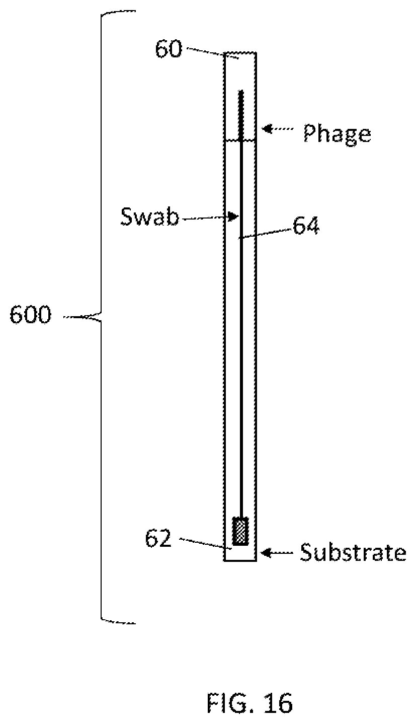
D00017

XML
uspto.report is an independent third-party trademark research tool that is not affiliated, endorsed, or sponsored by the United States Patent and Trademark Office (USPTO) or any other governmental organization. The information provided by uspto.report is based on publicly available data at the time of writing and is intended for informational purposes only.
While we strive to provide accurate and up-to-date information, we do not guarantee the accuracy, completeness, reliability, or suitability of the information displayed on this site. The use of this site is at your own risk. Any reliance you place on such information is therefore strictly at your own risk.
All official trademark data, including owner information, should be verified by visiting the official USPTO website at www.uspto.gov. This site is not intended to replace professional legal advice and should not be used as a substitute for consulting with a legal professional who is knowledgeable about trademark law.