Novel Antibody Conjugates Suitable For Use In Discrete Fluorescence Quenching Displacement Immunoassays
BUETTNER; Karin ; et al.
U.S. patent application number 16/637910 was filed with the patent office on 2020-06-11 for novel antibody conjugates suitable for use in discrete fluorescence quenching displacement immunoassays. This patent application is currently assigned to LIFE SCIENCE INKUBATOR SACHSEN GMBH & CO. KG. The applicant listed for this patent is LIFE SCIENCE INKUBATOR SACHSEN GMBH & CO. KG. Invention is credited to Karin BUETTNER, Tom STUECKEMANN.
| Application Number | 20200182863 16/637910 |
| Document ID | / |
| Family ID | 59631597 |
| Filed Date | 2020-06-11 |


| United States Patent Application | 20200182863 |
| Kind Code | A1 |
| BUETTNER; Karin ; et al. | June 11, 2020 |
NOVEL ANTIBODY CONJUGATES SUITABLE FOR USE IN DISCRETE FLUORESCENCE QUENCHING DISPLACEMENT IMMUNOASSAYS
Abstract
The present invention generally relates to the field of Fluorescence Quenching Immunoassays. Specifically, the invention provides novel antibody conjugates suitable for use in Discrete Fluorescence Quenching Displacement Immunoassays and methods for producing these antibody conjugates. The invention further relates to the use the novel antibody conjugates, and a kit comprising the same.
| Inventors: | BUETTNER; Karin; (Dresden, DE) ; STUECKEMANN; Tom; (Dresden, DE) | ||||||||||
| Applicant: |
|
||||||||||
|---|---|---|---|---|---|---|---|---|---|---|---|
| Assignee: | LIFE SCIENCE INKUBATOR SACHSEN GMBH
& CO. KG Dresden DE |
||||||||||
| Family ID: | 59631597 | ||||||||||
| Appl. No.: | 16/637910 | ||||||||||
| Filed: | August 8, 2018 | ||||||||||
| PCT Filed: | August 8, 2018 | ||||||||||
| PCT NO: | PCT/EP2018/071514 | ||||||||||
| 371 Date: | February 10, 2020 |
| Current U.S. Class: | 1/1 |
| Current CPC Class: | A61K 47/6889 20170801; G01N 33/533 20130101; G01N 33/542 20130101 |
| International Class: | G01N 33/542 20060101 G01N033/542; G01N 33/533 20060101 G01N033/533 |
Foreign Application Data
| Date | Code | Application Number |
|---|---|---|
| Aug 11, 2017 | EP | 17185959.8 |
Claims
1. A conjugate for fluorescence quenching immunoassays comprising an antibody bonded to a linker, characterized in that at its proximal end, the linker is bonded to the carbohydrate chain of the N-glycosylation site asparagine at position 297 or a position in close proximity to position 297 of the Fc chain of the antibody the length of the linker is in the range 5 to 85 .ANG. such that the distal terminus of the linker is capable of interacting with a molecule captured at the antibody binding site; said linker is a molecule according to formula (I): A-L1-B-C-L2-D-E (I), wherein A is hydrazide, aminooxy or thiosemicarbazide; L1 and L2 are selected from a bond, C.sub.1-10 alkyl or polyethylene glycol (PEG); B is alkyne, azide, thiol, tetrazine, DBCO, TCO, vinyl or methylcyclopropene; C is azide, alkyne, maleimide, TCO, vinyl, methylcyclopropene, azide or tetrazine; D is a group selected from amino and thiol; and E is a dark quencher selected from selected from Atto612Q, DABCYL, methyl red, QSY-7 diarylrhodamine dyes and 6-(dimethylamino)-2-[4-[4 (dimethylamino)phenyl]- -1,3-butadienyl]-1-ethyl quinolinium perchlorate, a rhodamine dye or Cy5, further comprising a NHS-ester group or a maleimide group; and said molecule captured at the antibody binding site of the antibody is an antigen or fragment thereof or a peptide, which is capable of binding to the antibody binding site of the antibody.
2. The conjugate of claim 1, wherein said molecule captured at the antibody binding site is labeled, such as with a fluorophore or a signal generating molecule.
3. The conjugate according to claim 1, wherein: when B is alkyne, then C is azide; when B is azide, then C is alkyne; when B is thiol, then C is maleimide; when B is tetrazine, then C is TCO, vinyl or methylcyclopropene; when B is DBCO, then C is azide; and when B is TCO, vinyl or methylcyclopropene, then C is tetracine.
4. The conjugate of claim 1, wherein L1 and L2 are both PEG; or L1 is C.sub.1-10 alkyl and L2 is PEG; or L1 is PEG and L2 is C.sub.1-10 alkyl.
5. The conjugate of claim 1, wherein chemical groups A and B form together a hydrazide alkyne group, wherein said hydrazine alkyne group is a compound of formula (II): H.sub.2N--NH--(C.dbd.O)-L1-C.ident.CH (II), wherein L1 is a bond or C.sub.1-10 alkyl.
6. The conjugate according to claim 1, comprising, essentially consisting of or consisting of an antibody or fragment thereof, a linker of formula (I) as claimed in claim 1; wherein said linker of formula (I) is bonded to the antibody at the proximal end via a compound selected from i. the group consisting of hydrazide-alkyne-azide, hydrazide-azide-alkyne, hydrazide-thiol-maleimide, hydrazide-DBCO-azide, hydrazide-TCO-tetrazine, hydrazide-methylcyclopropene-tetrazine and hydrazine-vinyl-tetrazine, wherein the hydrazide group of the linker is covalently bonded to an Fc region of the antibody; ii. the group consisting of aminooxy-alkyne-azide, aminooxy-azide-alkyne, aminooxy-thiol-maleimide, aminooxy-DBCO-azide, aminooxy-TCO-tetrazine, aminooxy-methylcyclopropene-tetrazine and aminooxy-vinyl-tetrazine, wherein the aminooxy group of the linker is covalently bonded to an Fc region of the antibody; and iii. the group consisting of thiosemicarbazide-alkyne-azide, thiosemicarbazide-azide-alkyne, thiosemicarbazide-thiol-maleimide, thiosemicarbazide-DBCO-azide, thiosemicarbazide-TCO-tetrazine, thiosemicarbazide-methylcyclopropene-tetrazine and thiosemicarbazide-vinyl-tetrazine, wherein the thiosemicarbazide group of the linker is covalently bonded to an Fc region of the antibody; wherein said compounds of groups i. to iii. contain group L1 according to formula (I) as defined in claim 1; and wherein said linker comprises at the distal end a dark quencher selected from Atto612Q, DABCYL, methyl red, QSY-7 diarylrhodamine dyes and 6-(dimethylamino)-2-[4-[4 (dimethylamino)phenyl]- -1,3-butadienyl]-1-ethyl quinolinium perchlorate, a rhodamine dye or Cy5; a molecule captured at the antibody binding site, which comprises a fluorophore.
7. The conjugate according to claim 7, comprising, essentially consisting of or consisting of a monoclonal antibody, a linker of formula (I); wherein said linker is at the proximal end bonded to the Fc region of the antibody via a compound selected from: hydrazide-alkyne-azide; hydrazide-tetrazine-TCO; and aminooxy-azide-alkyne; wherein L1 is PEG or C.sub.1-10 alkyl and L2 is PEG and wherein said linker comprises at the distal end the quencher Atto612Q; a molecule captured at the antibody binding site, which comprises the fluorophore EuLH.
8. The conjugate according to claim 1, comprising, essentially consisting of or consisting of a monoclonal antibody, a linker of formula (I); wherein said linker is at the proximal end bonded to the Fc region of the antibody via 4-pentynoic acid hydrazide; L2 is a linear PEG comprising 5 to 7, preferably 6 ethylene oxide monomers; and wherein said linker comprises at the distal end the dark quencher Atto612Q; a molecule captured at the antibody binding site, which comprises the fluorophore EuLH.
9. (canceled)
10. The conjugate of claim 2, wherein the quenching or the competition of the peptide-conjugated linker with an analyte, antigen or biomarker is performed intramolecular.
11. A method for preparing the conjugate according to claim 2, comprising the steps of: i. generating site specifically reactive aldehyde or ketone groups at the asparagine residue at position 297 or a position in close proximity to position 297 of the Fc region of the antibody by oxidizing the antibody; ii. reacting the oxidized antibody with a compound A-L1-B, preferably selected from hydrazide-L1-alkyne, hydrazide-L1-azide, hydrazide-L1-thiol, hydrazide-L1-tetrazine, hydrazide-L1-DBCO, hydrazide-L1-TCO, hydrazide-L1-vinyl, hydrazide-L1-methylcyclopropene, aminooxy-L1-alkyne, aminooxy-L1-azide, aminooxy-L1-thiol, aminooxy-L1-tetrazine, aminooxy-L1-DBCO, aminooxy-L1-TCO, aminooxy-L1-vinyl, aminooxy-L1-methylcyclopropene, thiosemicarbazide-L1-alkyne, thiosemicarbazide-L1-azide, thiosemicarbazide-L1-thiol, thiosemicarbazide-L1-tetrazine, thiosemicarbazide-L1-DBCO, thiosemicarbazide-L1-TCO, thiosemicarbazide-L1-vinyl, thiosemicarbazide-L1-methylcyclopropene; iii. preparing a compound C-L2-D-E; iv. reacting chemical group B of the antibody from step ii., which comprises the compound A-L1-B, with the C-L2-D-E compound of step iii. by click chemistry reactions; and v. capturing a molecule which is capable of binding to the antibody binding site of the antibody and which comprises a signal generating molecule, such as a fluorophore.
12. An in vitro method for the diagnosis of a disease or pathological condition comprising the steps of: i. contacting a conjugate according to claim 2 with a sample from a subject suspected to be afflicted with a disease or condition to be diagnosed, ii. detecting the amount of an analyte, antigen, biomarker or the like, or an isoform thereof, in said sample obtained from said subject; iii. comparing the detected amount of said analyte, antigen or biomarker in said sample with an amount of the analyte, antigen or biomarker characteristic of a normal control; whereby a changed amount, preferably an elevated amount of said analyte, antigen or biomarker in said sample relative to the normal control is a positive indicator of the disease or condition to be diagnosed.
13. A diagnostic kit, comprising a compound A-L1-B, preferably selected from hydrazide-L1-alkyne, hydrazide-L1-azide, hydrazide-L1-thiol, hydrazide-L1-tetrazine, hydrazide-L1-DBCO, hydrazide-L1-TCO, hydrazide-L1-vinyl, hydrazide-L1-methylcyclopropene, aminooxy-L1-alkyne, aminooxy-L1-azide, aminooxy-L1-thiol, aminooxy-L1-tetrazine, aminooxy-L1-DBCO, aminooxy-L1-TCO, aminooxy-L1-vinyl, aminooxy-L1-methylcyclopropene, thiosemicarbazide-L1-alkyne, thiosemicarbazide-L1-azide, thiosemicarbazide-L1-thiol, thiosemicarbazide-L1-tetrazine, thiosemicarbazide-L1-DBCO, thiosemicarbazide-L1-TCO, thiosemicarbazide-L1-vinyl, thiosemicarbazide-L1-methylcyclopropene; wherein A, B and L1 are as defined in claim 2; a compound C-L2-D-E; wherein C, L2, D and E are as defined in claim 1; an oxidant for oxidizing an antibody, a fluorophore, and instructions for using said kit ingredients for preparing and using the conjugate according to claim 2.
14. A diagnostic kit comprising an already assembled conjugate as claimed in claim 2, a well plate, such as a 96- or 384-well plate, or a microfluidic chip, wherein each well of the well plate or the microfluidic chip is loaded with the conjugate of the invention, and wherein said kit comprises negative and positive control samples.
15. The conjugate according to claim 2 for use in the diagnosing and/or monitoring the state of diseases or conditions like heart disorders, inflammation, Parathyroidectomy, blood coagulation, metabolic syndrome and kidney diseases.
16. (canceled)
Description
FIELD OF THE INVENTION
[0001] The present invention generally relates to the field of Fluorescence Quenching Immunoassays. Specifically, the invention provides novel antibody conjugates suitable for use in Discrete Fluorescence Quenching Displacement Immunoassays and methods for producing these antibody conjugates. The invention further relates to the use of the novel antibody conjugates and a kit comprising the same.
BACKGROUND ART
[0002] Antibodies are widely used molecular tools for clinical diagnostics, therapy and research (Chames, Van Regenmortel, Weiss, & Baty, 2009; Ecker, Jones, & Levine, 2015; Morgan & Levinsky, 1985; Scolnik, 2009; Siddiqui, 2010; Waldmann, 2003). In the field of immunodiagnostics, antibodies are used to quantify clinical biomarkers in complex biological samples like blood (Wild, 2013). These so called immunoassays can be discriminated between homogeneous and heterogeneous techniques (Wild, 2013). The heterogeneous immunoassays are today's gold standard for clinical diagnostics. Heterogeneous immunoassays use an antibody pair to capture and to detect the analyte (Wild, 2013). These assays reach good sensitivities, but require washing steps, which makes them complicated and slow.
[0003] In homogeneous immunoassays, the antibody reacts with the analyte in solution, which allows much faster assay times. Additionally, homogeneous immunoassays only require one analyte specific antibody, which makes them suitable for detecting smaller biomarkers. Because of their speed and simplicity homogeneous immunoassays are highly relevant for clinical diagnostics (Hemmil & Mukkala, 2001; Leuvering, Thal, Waart, & Schuurs, 1980; Nargessi, Landon, & Smith, 1979a; Ullman, Schwarzberg, & Rubenstein, 1976; Wild, 2013). However in contrast to heterogeneous immunoassays, homogeneous immunoassays have problems in signal generation, which so far results in less sensitive assays.
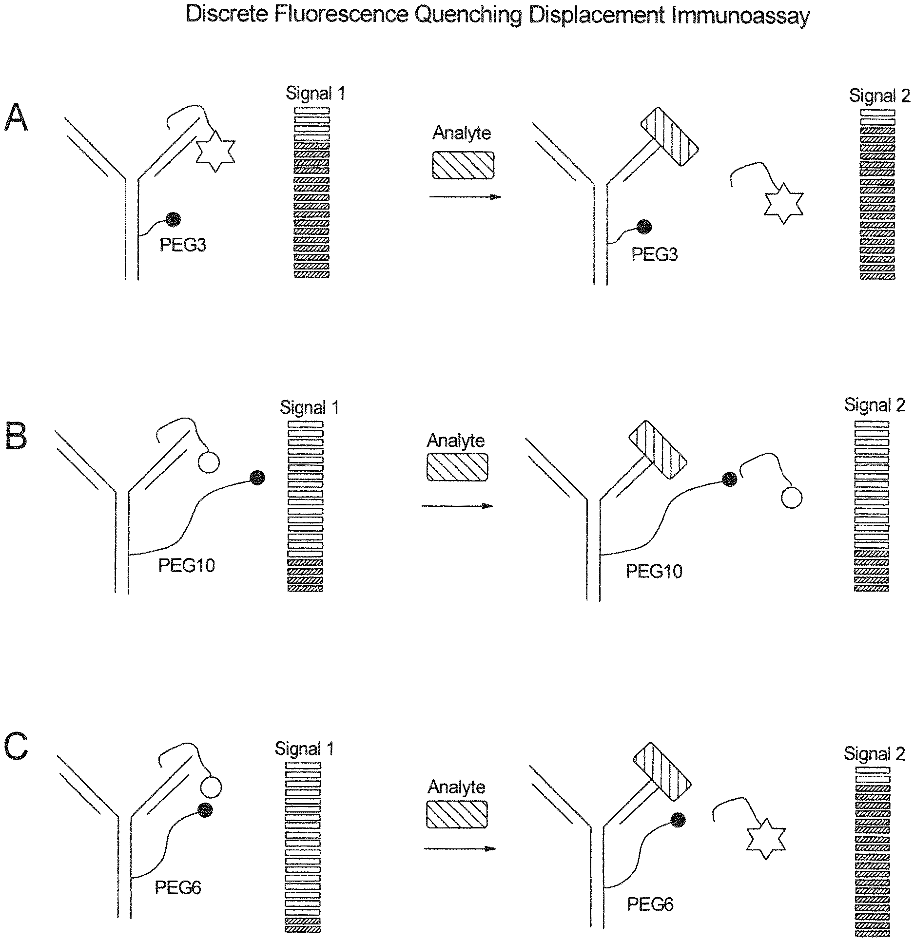
[0004] In most homogeneous immunoassays, the signal generating step is based on competition between the analyte and a known amount of fluorescently labeled analyte. The so-called Fluorescence Quenching Immunoassay uses two antibodies: one analyte specific antibody and one fluorophore specific antibody, whereby binding of the fluorophore specific antibody to the fluorophore reduces the fluorescence intensity. Both antibodies compete in binding the fluorescently labeled analyte. At steady state a given fluorescent signal is generated. By adding additional unlabeled analyte, via applying a sample, the steady state shifts and more fluorescence signal can be quenched by the fluorophore specific antibody. FIG. 1 shows a scheme of a typical Fluorescence Quenching Immunoassay. The analyte concentration is, however, disproportionally to the fluorescence signal (Nargessi, Landon, & Smith, 1979b; Zuk, Rowley, & Ullman, 1979).
[0005] A modified version of the Fluorescence Quenching Immunoassay has been proposed by Kreisig et. al (Kreisig, Hoffmann, & Zuchner, 2011). This FRET-based homogeneous immunoassay consists of a dark-quencher labeled antibody and a fluorescently labeled peptide. Both the analyte and the peptide are mixed together and compete in binding the subsequently added antibody. When the peptide is bound by the antibody, the dark quencher and the fluorophore are in spatial proximity, resulting into a reduced fluorescence signal. In contrast, when the analyte is bound, the peptide is displaced and the attached fluorophore is no longer quenched and emits light. At steady state of this reaction, the emitted light is proportional to the analyte concentration, as shown in FIG. 2. Because the steady state of this reaction can be reached already after 90 seconds, this assay is highly attractive for clinical point of care testing (POCT). However the FRET-based homogeneous Immunoassay from Kreisig et. al does not allow the quantification of biomarkers in the clinical relevant concentrations.
[0006] Additional publications from Kreisig et. al further describe the principle of a Fluorescence Quenching Displacement Immunoassay. In contrast to the Fluorescence Quenching Immunoassay in the Fluorescence Quenching Displacement Immunoassay, the dark quencher labeled antibody is premixed with the fluorophore labeled peptide. This pre-incubation step results into a lower starting signal, aiming to generate higher signal to noise ratios, which should increase the assay sensitivity. In order to generate a high analyte specific signal the added analyte consequently has to actively displace the bound peptide. However, Kreisig et. al could not show the functionality of the Fluorescence Quenching Displacement Immunoassay and it is therefore hypothesized that the signal generating step is problematic by using this method (Kreisig et al., 2013, 2015).
[0007] After analyzing the assay components, we further hypothesized that the dark quencher labeled antibody impedes the signal-generating step. Kreisig et. al generated the dark quencher labeled antibody by NHS-ester reactions. As a consequence of this bio-conjugation approach, dark quenchers are randomly distributed at the antibody surface, potentially resulting in undirected quenching of free diffusing peptides at steady state of the reaction (FIG. 3). Such undirected quenching of free diffusing peptides could explain both, the limited sensitivity of the Fluorescence Quenching Immunoassay, as well as the lack of functionality of the Fluorescence Quenching Displacement Immunoassay.
[0008] Several bio-conjugation techniques have been postulated, but no approach has been described to specifically generate an intramolecular interaction with the antigen binding site, without interfering with the antibodies affinity (Agarwal & Bertozzi, 2015; Kim, Ko, Park, & Lee, 2016; Kumar et al., 2015; Liberatore et al., 1990; Packard, Edidin, & Komoriya, 1986; Schumacher et al., 2015; Schumacher, Hackenberger, Leonhardt, & Helma, 2016; Sochaj, widerska, & Otlewski, 2015; Zimmerman et al., 2014).
DESCRIPTION OF THE INVENTION
[0009] Accordingly, the problem to be solved by the invention is to provide a bio-conjugation technique, which overcomes the problems of the prior art, in particular which overcomes the limited sensitivity and the lack of functionality of known Fluorescence Quenching Immunoassays.
[0010] This problem is solved by the provision of a conjugate according to claim 1.
[0011] In a preferred embodiment, the invention provides a conjugate comprising an antibody bonded to a linker, characterized in that the length of the linker is adaptable such that the free terminus of the linker is capable of interacting with a molecule captured at the antibody binding site.
[0012] The term "antibody" is used in the broadest sense and specifically covers intact monoclonal antibodies, polyclonal antibodies, multispecific antibodies (e.g. bispecific antibodies) formed from at least two intact antibodies, and antibody fragments so long as they exhibit the desired biological activity. The antibody may be an IgM, IgG (e.g. IgG1, IgG2, IgG3 or IgG4), IgD, IgA or IgE, for example.
[0013] "Antibody fragments" comprise a portion of an intact antibody, generally the antigen binding or variable region of the intact antibody. Examples of antibody fragments include Fab, Fab', F(ab')2, and Fv fragments: diabodies; single-chain antibody molecules; and multispecific antibodies formed from antibody fragments.
[0014] The term "monoclonal antibody" as used herein refers to an antibody obtained from a population of substantially homogeneous antibodies, i.e. the individual antibodies comprising the population are identical except for possible naturally occurring mutations that may be present in minor amounts. Monoclonal antibodies are highly specific, being directed against a single antigenic site. Furthermore, in contrast to "polyclonal antibody" preparations which typically include different antibodies directed against different determinants (epitopes), each monoclonal antibody is directed against a single determinant on the antigen. In addition to their specificity, the monoclonal antibodies can frequently be advantageous in that they are synthesized by the hybridoma culture, uncontaminated by other immunoglobulins. The "monoclonal" indicates the character of the antibody as being obtained from a substantially homogeneous population of antibodies, and is not to be construed as requiring production of the antibody by any particular method. For example, the monoclonal antibodies to be used in accordance with the present invention may be made by the hybridoma method first described by KOhler et al., Nature, 256:495 (1975), or may be made by generally well known recombinant DNA methods. The "monoclonal antibodies" may also be isolated from phage antibody libraries using the techniques described in Clackson et al., Nature, 352:624-628 (1991) and Marks et al., J. Mol. Biol., 222:581-597 (1991), for example.
[0015] The monoclonal antibodies herein specifically include chimeric antibodies (immunoglobulins) in which a portion of the heavy and/or light chain is identical with or homologous to corresponding sequences in antibodies derived from a particular species or belonging to a particular antibody class or subclass, while the remainder of the chain(s) is identical with or homologous to corresponding sequences in antibodies derived from another species or belonging to another antibody class or subclass, as well as fragments of such antibodies, so long as they exhibit the desired biological activity.
[0016] "Humanized" forms of non-human (e.g., murine) antibodies are chimeric immunoglobulins, immunoglobulin chains or fragments thereof (such as Fv, Fab, Fab', F(ab')2 or other antigen-binding subsequences of antibodies) which contain a minimal sequence derived from a non-human immunoglobulin. For the most part, humanized antibodies are human immunoglobulins (recipient antibody) in which residues from a complementarity-determining region (CDR) of the recipient are replaced by residues from a CDR of a non-human species (donor antibody) such as mouse, rat or rabbit having the desired specificity, affinity, and capacity. In some instances, Fv framework region (FR) residues of the human immunoglobulin are replaced by corresponding non-human residues. Furthermore, humanized antibodies may comprise residues which are found neither in the recipient antibody nor in the imported CDR or framework sequences.
[0017] These modifications are made to further refine and optimize antibody performance. In general, the humanized antibody will comprise substantially all or at least one, and typically two, variable domains, in which all or substantially all of the CDR regions correspond to those of a non-human immunoglobulin and all or substantially all of the FR regions are those of a human immunoglobulin sequence. The humanized antibody optimally also will comprise at least a portion of an immunoglobulin constant region (Fc), typically that of a human immunoglobulin. For further details, see Jones et al., Nature, 321:522-525 (1986), Reichmann et al, Nature. 332:323-329 (1988): and Presta, Curr. Op. Struct. Biel., 2:593-596 (1992). The humanized antibody includes a Primatized.TM. antibody wherein the antigen-binding region of the antibody is derived from an antibody produced by immunizing macaque monkeys with the antigen of interest.
[0018] "Single-chain Fv" or "sFv" antibody fragments comprise the VH and VL domains of an antibody, wherein these domains are present in a single polypeptide chain. Generally, the Fv polypeptide further comprises a polypeptide linker between the VH and VL domains which enables the sFv to form the desired structure for antigen binding. For a review of sFv see Pluckthun in The Pharmacology of Monoclonal Antibodies, vol. 113, Rosenburg and Moore eds., Springer-Verlag, New York, pp. 269-315 (1994).
[0019] The term "diabodies" refers to small antibody fragments with two antigen-binding sites, which fragments comprise a heavy-chain variable domain (VH) connected to a light-chain variable domain (VD) in the same polypeptide chain (VH-VD). By using a linker that is too short to allow pairing between the two domains on the same chain, the domains are forced to pair with the complementary domains of another chain and create two antigen-binding sites. Diabodies are described more fully in Hollinger et al., Proc. Natl. Acad. Sol. USA, 90:6444-6448 (1993).
[0020] An "isolated" antibody is one which has been identified and separated and/or recovered from a component of its natural environment. Contaminant components of its natural environment are materials which would interfere with diagnostic or therapeutic uses for the antibody, and may include enzymes, hormones, and other proteinaceous or non-proteinaceous solutes. In preferred embodiments, the antibody will be purified (1) to greater than 95% by weight of antibody as determined by the Lowry method, and most preferably more than 99% by weight, (2) to a degree sufficient to obtain at least 15 residues of N-terminal or internal amino acid sequence by use of a spinning cup sequenator, or (3) to homogeneity by SDS-PAGE under reducing or nonreducing conditions using Coomassie blue or, preferably, silver stain. Isolated antibody includes the antibody in situ within recombinant cells since at least one component of the antibody's natural environment will not be present. Ordinarily, however, isolated antibody will be prepared by at least one purification step.
[0021] As used herein, the expressions "cell", "cell line," and "cell culture" are used interchangeably and all such designations include progeny. Thus, the words "transformants" and "transformed cells" include the primary subject cell and culture derived therefrom without regard for the number of transfers. It is also understood that all progeny may not be precisely identical in DNA content, due to deliberate or inadvertent mutations. Mutant progeny that have the same function or biological activity as screened for in the originally transformed cell are included. Where distinct designations are intended, this will be clear from the context.
[0022] The terms "polypeptide", "peptide", and "protein", as used herein, are interchangeable and are defined to mean a biomolecule composed of amino acids linked by a peptide bond.
[0023] The terms "a", "an" and "the" as used herein are defined to mean "one or more" and include the plural unless the context is inappropriate.
[0024] In a preferred embodiment, any antibody that is already known in the art, may be used in the conjugate and/or methods of the invention.
[0025] In a more embodiment, the conjugate according to invention comprises a monoclonal or polyclonal antibody, preferably a monoclonal antibody. In a further preferred embodiment, the monoclonal antibody is a chimeric antibody.
[0026] In yet a further preferred embodiment, the antibody is an isolated antibody.
[0027] The "target polypeptide" can be any polypeptide, peptide, substrate, antigen or analyte, which may be the subject of investigation in a Fluorescence Quenching Immunoassay. The invention is not limited to a specific target polypeptide. Accordingly, the invention is also not limited to a specifically defined antibody. Antibodies have to be produced, e.g. with any technique known in the art, for the desired purpose to be comprised in a conjugate for use in a Fluorescence Quenching Immunoassay as described above. Alternatively, any antibody, preferably monoclonal antibody known in the art may be used.
[0028] An antibody, which may be comprised in the conjugate of the invention, may for example be selected from: [0029] suitable antibodies capable for detecting cardiac Troponin-I, (clinical marker to validate heart disorders), for example mouse monoclonal hybridoma clones selected from 8E10, C5, 4C2, 19C7, 16A11, 3C7, P4-14G5, M18, M155, MF4, 16A12, P4-9F6, 228, 625, 458, 267, 596, 84, 415, 581, 10F4, 247, 560, 17F3, p45-10, 916, 909, 820, 810, 801. These mouse monoclonal hybridoma clones are described in and can be ordered from HyTest Ltd., Catalog ID 4T21. [0030] suitable antibodies capable for detecting cardiac Troponin-T, (clinical marker to validate heart disorders), for example mouse monoclonal hybridoma clones 9G6, 7F4, 7G7, 2F3, 1A11, 1C11, 1F11, 7E7. These mouse monoclonal hybridoma clones are described in and can be ordered from HyTest Ltd., Catalog ID 4T19. [0031] suitable antibodies capable for detecting NT-ProBNP, (clinical marker to validate heart disorders), for example mouse monoclonal hybridoma clones. 5F11cc, 13G12cc, 15C4cc, 24E11cc, 29D12cc, 18H5cc. These mouse monoclonal hybridoma clones are described in and can be ordered from HyTest Ltd., Catalog ID 4NT1cc. [0032] suitable antibodies capable for detecting cardiac myoglobin, (clinical marker to validate heart disorders), for example mouse monoclonal hybridoma clones 4E2, 7C3, IB4. These mouse monoclonal hybridoma clones are described in and can be ordered from HyTest Ltd., Catalog ID 4M23. [0033] suitable antibodies capable for detecting D-dimer, (blood coagulation marker), for example mouse monoclonal hybridoma clones DD1, DD2, DD3, DD4, DD5, DD6, DD22, DD41, DD44, DD46, DD93, DD189, DD255. These mouse monoclonal hybridoma clones are described in and can be ordered from HyTest Ltd., Catalog ID 4D30. [0034] suitable antibodies capable for detecting Insulin, (clinical marker to validate metabolic syndrome), for example mouse monoclonal hybridoma clones C7C9, D4B8, 7F8, 3A6, 7F5. These mouse monoclonal hybridoma clones are described in and can be ordered from HyTest Ltd., Catalog ID 2I1. [0035] suitable antibodies capable for detecting C-Peptide, (clinical marker to validate metabolic syndrome), for example mouse monoclonal hybridoma clones 1H8, 2B7, 4H8, 5B8, 7E10, 2A11. These mouse monoclonal hybridoma clones are described in and can be ordered from HyTest Ltd., Catalog ID 2I2. [0036] suitable antibodies capable for detecting Cystatin-C, (clinical marker to validate kidney diseases), for example mouse monoclonal hybridoma clones Cyst10, Cyst13, Cyst16, Cyst18, Cyst19, Cyst23, Cyst24, Cyst28, Cyst11, Cyst20, Cyst29. These mouse monoclonal hybridoma clones are described in and can be ordered from HyTest Ltd., Catalog ID 4CC1. [0037] suitable antibodies capable for detecting C-reactive protein, (clinical marker to validate status of inflammation), for example mouse monoclonal hybridoma clones C1, C2, C3, C4, C5, C6, C7, CRP11, CRP30, CRP36, CRP103, CRP135, CRP169. These mouse monoclonal hybridoma clones are described in and can be ordered from HyTest Ltd., Catalog ID 4C28. [0038] suitable antibodies capable for detecting interleukin-6, (clinical marker to validate status of inflammation), for example mouse monoclonal hybridoma clones B10, G5. These mouse monoclonal hybridoma clones are described in and can be ordered from HyTest Ltd., Catalog ID 4IL6.
[0039] The "molecule captured at the antibody binding site" may be identical with the afore described "target polypeptide", i.e. is selected from any polypeptide, peptide, substrate, antigen or analyte, against which the antibody comprised in the conjugate of the invention has been created, and which is capable of binding to the binding region of the antibody. More preferably, the molecule captured at the antibody binding site is an antigen or fragment thereof, which is capable of binding to the antibody binding region.
[0040] The molecule captured at the antibody binding site is suitably a derivate (e.g. small peptide, fragment etc.) derived from the intact, wildtype or full length analyte or antigen present in a sample. The molecule captured at the antibody binding site may further be modified in such a way (e.g. by point mutations) that the affinity for binding to the antibody is altered, in particular reduced, compared to the intact analyte. Thereby the capability of the analyte to displace the molecule captured at the antibody binding site is improved. Altering, i.e. lowering of the affinity of the molecule captured at the antibody binding site may also be achieved by any other method known in art, such as chemical modification by, e.g. biotinylation, glycosylation etc.
[0041] Exemplary antigens, which may be detected using the conjugate and methods of the invention, are for example clinical relevant biomarkers like: [0042] Troponin-I, a clinical marker to validate heart disorders; [0043] Troponin-T, aclinical marker to validate heart disorders; [0044] CRP, a clinical marker to validate status of inflammation; [0045] Procalcitonin, a clinical marker to validate status of inflammation; [0046] Interleukin-6, a clinical marker to validate status of inflammation; [0047] Interleukin-8, a clinical marker to validate status of inflammation [0048] Interleukin-11, a clinical marker to validate status of inflammation; [0049] Parathyroid Hormone, a clinical marker to monitor and validate Parathyroidectomy; [0050] D-dimer, a clinical marker to validate blood coagulation; [0051] NT-proBNP, a clinical marker to validate heart disorders; [0052] BNP, a clinical marker to validate heart disorders; [0053] Insulin, (clinical marker to validate metabolic syndrome; [0054] C-Peptide, a (clinical marker to validate metabolic syndrome; and [0055] Cystatin C, a clinical marker to validate kidney diseases.
[0056] The invention is, however, not limited to the aforementioned antigens. The conjugate and methods of the invention can be adapted to any known or so far unknown antigen, analyte or biomarker.
[0057] In another preferred embodiment, the molecule captured at the antibody binding site is a fragment of an antigen, which may be known in the art, or which is a synthetic peptide. The amino acid sequence of a synthetic peptide is suitably adapted such that the binding to the binding site of the antibody comprised in the conjugate of the invention is facilitated. In a more preferred embodiment, such antigen fragment or synthetic peptide has a chain length of 4 to 22 amino acids, more preferably of 5 to 15 amino acids, most preferably of 6 to 12 amino acids. This has the advantage that a specific intramolecular quenching only at the antigen binding site can be ensured.
[0058] The invention provides a novel conjugate for use in Fluorescence Quenching Immunoassays. Accordingly, in a further preferred embodiment of the invention, the molecule captured at the antibody binding site is labeled with a signal generating molecule.
[0059] The signal-generating molecule is suitably selected from the group consisting of: fluorescent labels, enzyme labels, radioisotopes, chemiluminescent labels, electrochemiluminescent labels, bioluminescent labels, polymers, polymer particles, quantum dots, metal particles, haptens, and dyes. In other particular embodiments, the signal generating moiety comprises an enzymatic label.
[0060] Preferred according to invention is a molecule captured at the antibody binding site, which is labeled with a fluorophore or chemiluminophore. Suitable fluorophores or chemiluminophores are in general commercially available from various sources. Preferred according to the invention are TRF dyes and quantum dots.
[0061] A "fluorophore" according to the invention is preferably selected from the general class known as cyanine dyes, with emission wavelengths between 550 nm and 900 nm. These dyes may contain methine groups and their number influences the spectral properties of the dye. The monomethine dyes that are pyridines typically have blue to blue-green fluorescence emission, while quinolines have green to yellow-green fluorescence emission. The trimethine dye analogs are substantially shifted toward red wavelengths, and the pentamethine dyes are shifted even further, often exhibiting infrared fluorescence emission. Fluorophores or chemiluminophores are in general commercially available from various sources. Preferred according to the invention are TRF dyes and quantum dots.
[0062] TRF (time-resolved fluorometry) dyes involve fluorophores that are based on lanthanide ion complexes. Lanthanide metals are particularly useful. Certain life science applications take advantage of the unique fluorescence properties of lanthanide ion complexes (Ln(III) chelates or cryptates). These are well-suited due to their large Stokes shifts and extremely long emission lifetimes (from microseconds to milliseconds) compared to more traditional fluorophores (e.g. fluorescein, allophycoyanin, phycoerythrin, and rhodamine).
[0063] The two most commonly used lanthanides in life science assays with their corresponding acceptor dye are Europium.sup.3+/Allophycocyanin and Terbium.sup.3+/Phycoerythrin.
[0064] The advantage of using TRF dyes is because biological fluids or serum commonly used in these assays contain many compounds and proteins which are autofluorescent. Therefore, the use of conventional, steady-state fluorescence measurement presents serious limitations in assay sensitivity. Long-lived fluorophores, such as lanthanides, combined with time-resolved detection (a delay between excitation and emission detection) minimizes prompt fluorescence interference.
[0065] TRF dyes are especially useful in FRET assays.
[0066] Related dyes can be used in accordance with the invention and further selected from cyclobutenedione derivatives, substituted cephalosporin compounds, fluorinated squaraine compositions, symmetrical and unsymmetrical squaraines, alkylalkoxy squaraines, or squarylium compounds. Some of these dyes can fluoresce at near infrared as well as at infrared wavelengths that would effectively expand the range of emission spectra up to about 1,000 nm. In addition to squaraines, i.e., derived from squaric acid, hydrophobic dyes such as phthalocyanines and naphthalocyanines can also be selected to operate at longer wavelengths. Other classes of fluorophores include 3-Hydroxypyrene 5,8,10-Tri Sulfonic acid, 5-Hydroxy Tryptamine (5-HT), Acid Fuhsin, Acridine Orange, Acridine Red, Acridine Yellow, Acriflavin, AFA (Acriflavin Feulgen SITSA), Alizarin Complexon, Alizarin Red, Allophycocyanin, ACMA, Aminoactinomycin D, Aminocoumarin, Anthroyl Stearate, Aryl- or Heteroaryl-substituted Polyolefin, Astrazon Brilliant Red 4 G, Astrazon Orange R, Astrazon Red 6B, Astrazon Yellow 7 GLL, Atabrine, Auramine, Aurophosphine, Aurophosphine G, BAO 9 (Bisaminophenyloxadiazole), BCECF, Berberine Sulphate, Bisbenzamide, BOBO 1, Blancophor FFG Solution, Blancophor SV, Bodipy F1, BOPRO 1, Brilliant Sulphoflavin FF, Calcien Blue, Calcium Green, Calcofluor RW Solution, Calcofluor White, Calcophor White ABT Solution, Calcophor White Standard Solution, Carbocyanine, Carbostyryl, Cascade Blue, Cascade Yellow, Catecholamine, Chinacrine, Coriphosphine O, Coumarin, Coumarin-Phalloidin, CY3.1 8, CY5.1 8, CY7, Dans (1-Dimethyl Amino Naphaline 5 Sulphonic Acid), Dansa (Diamino Naphtyl Sulphonic Acid), Dansyl NH--CH3, DAPI, Diamino Phenyl Oxydiazole (DAO), Dimethylamino-5-Sulphonic acid, Dipyrrometheneboron Difluoride, Diphenyl Brilliant Flavine 7GFF, Dopamine, Eosin, Erythrosin ITC, Ethidium Bromide, Euchrysin, FIF (Formaldehyde Induced Fluorescence), Flazo Orange, Fluo 3, Fluorescamine, Fura-2, Genacryl Brilliant Red B, Genacryl Brilliant Yellow 10GF, Genacryl Pink 3G, Genacryl Yellow 5GF, Gloxalic Acid, Granular Blue, Haematoporphyrin, Hoechst 33258, Indo-1, Intrawhite Cf Liquid, Leucophor PAF, Leucophor SF, Leucophor WS, Lissamine Rhodamine B200 (RD200), Lucifer Yellow CH, Lucifer Yellow VS, Magdala Red, Marina Blue, Maxilon Brilliant Flavin 10 GFF, Maxilon Brilliant Flavin 8 GFF, MPS (Methyl Green Pyronine Stilbene), Mithramycin, NBD Amine, Nile Red, Nitrobenzoxadidole, Noradrenaline, Nuclear Fast Red, Nuclear Yellow, Nylosan Brilliant Flavin E8G, Oregon Green, Oxazine, Oxazole, Oxadiazole, Pacific Blue, Pararosaniline (Feulgen), Phorwite AR Solution, Phorwite BKL, Phorwite Rev, Phorwite RPA, Phosphine 3R, Phthalocyanine, Phycoerythrin R, Polyazaindacene Pontochrome Blue Black, Porphyrin, Primuline, Procion Yellow, Propidium Iodide, Pyronine, Pyronine B, Pyrozal Brilliant Flavin 7GF, Quinacrine Mustard, Rhodamine 123, Rhodamine 5 GLD, Rhodamine 6G, Rhodamine B, Rhodamine B 200, Rhodamine B Extra, Rhodamine BB, Rhodamine BG, Rhodamine WT, Rose Bengal, Serotonin, Sevron Brilliant Red 2B, Sevron Brilliant Red 4G, Sevron Brilliant Red B, Sevron Orange, Sevron Yellow L, SITS (Primuline), SITS (Stilbene Isothiosulphonic acid), Stilbene, Snarf 1, sulpho Rhodamine B Can C, Sulpho Rhodamine G Extra, Tetracycline, Texas Red, Thiazine Red R, Thioflavin S, Thioflavin TCN, Thioflavin 5, Thiolyte, Thiozol Orange, Tinopol CBS, TOTO 1, TOTO 3, True Blue, Ultralite, Uranine B, Uvitex SFC, Xylene Orange, XRITC, YO PRO 1, or combinations thereof.
[0067] One skilled in the art would know which one to select among such fluorescence dyes as long as the desired emission and absorption properties as well as their chemical, such as hydrophobic properties are appropriate. The spectral properties of the fluorescent dyes should, according to a preferred embodiment of the invention, be sufficiently similar in excitation wavelengths and intensity to fluorescein or rhodamine derivatives as to permit the use of the same.
[0068] Attaching of the fluorophore to the molecule, which is captured or which is to be captured at the binding site of the antibody which is comprised in the conjugate of the invention, may be achieved by any of the techniques familiar to those skilled in the art. For example, the fluorophore may be covalently attached to the biomolecular probe by methods disclosed in U.S. Pat. Nos. 5,194,300 and 4,774,189.
[0069] In a further preferred embodiment of the invention, the fluorophore facilitates quenching, in particular dark quenching in combination with a quencher molecule or dark quencher molecule, which is attached to the linker.
[0070] Accordingly, the invention further provides a conjugate, wherein the linker comprises a quencher or functions as quencher molecule. More preferably, the linker comprises a dark quencher.
[0071] Fluorescence Resonance Energy Transfer (FRET) is a process whereby a first fluorescent dye (the "donor" dye) is excited, typically by illumination, and transfers its absorbed energy to a second dye (the "acceptor" dye) that has a longer wavelength and therefore lower energy emission. Where the second dye is fluorescent, energy transfer results in fluorescence emission at the wavelength of the second dye. However, where the second dye is nonfluorescent, the absorbed energy does not result in fluorescence emission, and the fluorescence of the initial donor dye is said to be "quenched". Energy transfer can also be utilized to quench the emission of luminescent donors, including phosphorescent and chemiluminescent donors. When a luminescent emission is restored by preventing energy transfer, the luminescence is said to be "dequenched" or "unquenched". The use of a variety of dyes to quench fluorescence is known in the art. The application of this phenomenon to analyze biological systems is also well-detailed. FRET has been utilized to study DNA hybridization and amplification, the dynamics of protein folding, proteolytic degradation, and interactions between other biomolecules.
[0072] A quencher may itself be a fluorescent molecule which emits fluorescence at a characteristic wavelength. Thus, a fluorophore may act as a quencher when appropriately coupled to another dye and vice versa. In this case, increase in fluorescence from the acceptor molecule, which is of a different wavelength to that of the donor label, will also indicate binding of the antibody binding site. Alternatively, the acceptor does not fluoresce ("dark acceptor" or "dark quencher"): Such acceptors include DABCYL, methyl red, QSY-7 diarylrhodamine dyes and 6-(dimethylamino)-2-[4-[4 (dimethylamino)phenyl]-1,3-butadienyl]-1-ethyl quinolinium perchlorate (CAS number 181885-68-7). Typical fluorophore/quencher compounds include certain rhodamine dyes or Cy5.
[0073] DABCYL (4'dimethylaminophenylazo) benzoic acid is a common dark quencher used widely in many assays, such as "molecular beacons" for DNA detection (U.S. Pat. No. 5,989,823). Diazo dyes of the BHQ series, which are referred to as "Black Hole Quenchers" (WO 01/86001), provide a broad range of absorption which overlaps well with the emission of many fluorophores. The QSY series dyes from Molecular Probes are another series of dark quenchers used extensively as quenching reagents in many bioassays (U.S. Pat. No. 6,399,392). The structure of QSY 7 is illustrated in US 2005/0014160. QSY-7 is a nonfluorescent diarylrhodamine derivative. QSY21 is a nonfluorescent diarylrhodamine chromophore with strong absorption in the visible spectrum, and is an effective fluorescence quencher.
[0074] By far the most common donor-acceptor dye pair utilized in biomedical applications is DABCYL (the quenching dye) and EDANS (the fluorophore).
[0075] A preferred dark quencher-fluorophore pair according to the invention is selected from Atto612Q (Kokko, T. et al., 2007) and DyQ2 (Dyomics GmbH, Germany; MW: 842.88 g/mol; Molecular formula: C40H39N2013S2*Na) (dark quencher) and EuLH (as the fluorophore). EuLH is (6,9-dicarboxymethyl-3-{4-([1,10]-phenanthrol-2-ylethinylphenyl-carbamoyl- )-methyl}-3,6,9-triaza-)-undeca-1,11-dicarboxylic acid (LH4) in complex with Eu.sup.3+ (Zuchner et al., 2009).
[0076] The quencher, as referred to herein, is coupled to a linker. The linker is selected from a variety of linking groups, including lower alkyl, cycloalkyl and heterocycloalkyl linking groups that extend from the active quencher to the core structure, i.e. the Fc region of the antibody comprised in the conjugate of the invention, where the linker-quencher molecule is attached. Preferably, the quencher, more preferably the dark quencher, is attached to the distal end of the linker. Attached in this regard means that the dark quencher is bonded to the distal end of the linker via an amino-NHS ester group or a thiol-maleimide group.
[0077] In a preferred embodiment of the invention, the dark quencher is bonded to the distal end of the linker via an amino-NHS ester group.
[0078] In another embodiment of the invention, the dark quencher is bonded to the distal end of the linker via a thiol-maleimide group.
[0079] At its proximal end, the linker is bonded via a novel carbohydrate linker coupling (CLC) approach using an N-glycosylated asparagine residue in the Fc chain of the antibody comprised in the conjugate of the invention as chemical reactive group. Preferably, the N-glycosylated asparagine 297 on the Fc antibody fragment of the antibody comprised in the conjugate of the invention is used as chemical reactive group. Asparagine is naturally occurring in almost all antibodies, particularly in monoclonal antibodies at position 297 of the Fc chain. However, the scope of the invention also comprises engineered antibodies, in which, for example by point mutations, amino acid substitutions, deletions or insertions the position of the asparagine is moved to a position in close proximity to position 297. Close proximity means a position in the range of 294 to 300 of the Fc chain, preferably in the range of 295 to 299, more preferably in the range of 296 to 298. Since glycosylation in antibodies is specific to asparagine 297, it was surprisingly found that this specific site can be used as intramolecular landmark. This means that the distance between the landmark and the antigen binding site stays constant, even among different monoclonal antibodies (Mizuochi, Taniguchi, Shimizu, & Kobata, 1982; Weitzhandler et al., 1994; Wright et al., 1997). However to facilitate direct molecular interactions, distances in the low nm range (e.g. for FRET interactions: 2-9 nm) are required (Mizuochi et al., 1982; Weitzhandler et al., 1994; Wright et al., 1997). In order to facilitate those interactions, the linker between the terminal carbohydrate group and the dark quencher shows the following characteristics:
[0080] Generally, preferred linkers have from 5 to 100 bonds from end to end, preferably 20 to 40 bonds, and may be branched or straight chain or contain rings. The bonds may be carbon-carbon or carbon-heteroatom or heteroatom-heteroatom bonds. The linkage can be designed to be hydrophobic or hydrophilic. The linker can contain single and/or double bonds, a number of 0-10 heteroatoms (O, S preferred), and saturated or aromatic rings. The linker may contain groupings such as ester, ether, sulfide, disulfide and the like.
[0081] In a most preferred embodiment, the length of the linker is adaptable such that the quencher molecule of the linker is interacting intramolecularly with the fluorophore of the molecule captured at the antibody binding site, thereby the fluorescence of the fluorophore being inhibited (quenched) by this binding.
[0082] In order to ensure a specific intramolecular quenching only at the antigen binding site in the conjugate of the invention, the linker has typically a length in the range of 5 to 85 .ANG., 10 to 65 .ANG., preferably in the range of 15 to 45 .ANG., more preferably in the range of 15 to 40 or 15 to 35 .ANG., most preferably in the range of 15 to 30, 20 to 30 or 20 to 25 .ANG.. Best quenching results could be achieved with a linker length between 20 and 30 .ANG., particularly with a linker length of 25 .ANG..
[0083] In a preferred embodiment, the linker of the invention is a molecule according to formula (I):
A-L1-B-C-L2-D-E (I),
wherein A is hydrazide, aminooxy or thiosemicarbazide; L1 and L2 are selected from a bond, alkyl, polyethylene glycol (PEG), polyamide, peptide, carbohydrate, oligonucleotide or polynucleotide; B is alkyne, azide, thiol, tetrazine, DBCO, TCO, vinyl or methylcyclopropene; C is azide, alkyne, maleimide, TCO, vinyl, methylcyclopropene, azide or tetrazine; D is a group selected from amino and thiol; and E is a quencher, preferably a dark quencher as described herein, further comprising a NHS-ester group or a maleimide group, as described herein,
[0084] In a more preferred embodiment of the invention, the linker is a molecule according to formula (I) with the prerequisites that:
when B is alkyne, then C is azide; when B is azide, then C is alkyne; when B is thiol, then C is maleimide; when B is tetrazine, then C is TCO, vinyl or methylcyclopropene; when B is DBCO, then C is azide; and when B is TCO, vinyl or methylcyclopropene, then C is tetrazine.
[0085] Chemical group A is bonded to the proximal end of the linker and connects the linker to the carbohydrate chain of the N-glycosylation site asparagine at position 297 or a position in close proximity to position 297 of the Fc chain of the antibody comprised in the conjugate of the invention.
[0086] Chemical groups B and C are reacted and bonded via click chemistry.
[0087] Chemical group D is bonded at the distal end of the linker and connects the distal end of the linker to the quencher molecule E.
[0088] The compounds of chemical group A may be combined and used with any compound of chemical group B.
[0089] Chemical groups A and B may form together a hydrazide alkyne group. Preferably, the hydrazine alkyne group formed by chemical groups A and B is a compound of formula (II):
H.sub.2N--NH--(C.dbd.O)-L1-C.ident.CH (II),
wherein L1 is a bond or C.sub.1-10alkyl.
[0090] Preferably, L1 is C.sub.2-7alkyl.
[0091] More preferably, L1 is ethyl, propyl or butyl.
[0092] Most preferably, L1 is ethyl.
[0093] The compounds of formula (II) of the invention are suitably known in the art. Preferred compounds of formula (II) have an assigned CAS registry and/or ReaxysFile number and are selected from the compounds shown in Table 1.
TABLE-US-00001 TABLE 1 Hydrazide-alkyne compounds of formula (II) CAS- and Name ReaxysFile-number Structure 4-Pentynoic acid, hydrazide 114578-39-1 4953267 ##STR00001## 5-Hexynoic acid, hydrazide 4230-19-7 4305355 ##STR00002## 6-Heptynoic acid, hydrazide 130905-57-6 4305493 ##STR00003## 9-Decynoic acid, hydrazide 4230-18-6 ##STR00004##
[0094] L1 and/or L2 may comprise, essentially consist of or consist of C.sub.1-10alkyl, polyethylene glycol (PEG), a polyamide, a peptide, a carbohydrate, an oligonucleotide or a polynucleotide.
[0095] In a preferred embodiment according to the invention, the proximal end of the linker comprises, essentially consists of or consists of a compound of formula (II) and L2 is PEG.
[0096] Further preferably, L1 and L2 may both be PEG.
[0097] Further preferably, L1 is C.sub.1-10alkyl and L2 is PEG.
[0098] Further preferably, L1 is PEG and L2 is C.sub.1-10alkyl.
[0099] PEG is an oligomer or polymer composed of ethylene oxide monomers. Because different applications require different polymer chain lengths, PEGs are prepared by polymerization of ethylene oxide and are commercially available over a wide range of molecular weights from 300 g/mol to 10,000,000 g/mol. While PEGs with different molecular weights find use in different applications, and have different physical properties (e.g. viscosity) due to chain length effects, their chemical properties are nearly identical. Different forms of PEG are also available, depending on the initiator used for the polymerization process--the most common initiator is a monofunctional methyl ether PEG, or methoxypoly(ethylene glycol), abbreviated mPEG. Lower-molecular-weight PEGs are also available as purer oligomers, referred to as monodisperse, uniform, or discrete.
[0100] PEGs are also available with different geometries: [0101] Linear PEGs, where the ethylene oxide monomers are bound to each other in an unbranched polymer chain; [0102] Branched PEGs, which have three to ten PEG chains emanating from a central core group; [0103] Star PEGs, which have 10 to 100 PEG chains emanating from a central core group; and [0104] Comb PEGs, which have multiple PEG chains normally grafted onto a polymer backbone.
[0105] The numbers that are often included in the names of PEGs indicate their average molecular weights (e.g. a PEG with n=9 would have an average molecular weight of approximately 400 daltons, and would be labeled PEG 400). Most PEGs include molecules with a distribution of molecular weights (i.e. they are polydisperse). The size distribution can be characterized statistically by its weight average molecular weight (Mw) and its number average molecular weight (Mn), the ratio of which is called the polydispersity index (Mw/Mn). Mw and Mn can be measured by mass spectrometry.
[0106] PEG is soluble in water, methanol, ethanol, acetonitrile, benzene, and dichloromethane, and is insoluble in diethyl ether and hexane.
[0107] In a preferred embodiment, the linker of the invention comprises a linear PEG. Using linear PEGs has the advantage that linear PEGs are cheap and possess a narrower molecular weight distribution.
[0108] When linear PEG is used to form the linker of the conjugate of the invention, it has suitably a molecular weight in the range of 40 Da to 1000 Da, preferably in the range of 100 Da to 800 Da and 150 Da to 600 Da, more preferably in the range of 200 Da to 400 Da, most preferably in the range of 200 Da to 300 Da or 220 Da to 310 Da. Even most preferably, the linear PEG comprised in the linker according to the invention has a molecular weight selected from 176 Da, 220 Da, 264 Da, 308 Da and 352 Da.
[0109] In another preferred embodiment, the linear PEG used to form the linker conjugate of the invention suitably consists of 1 to 20, preferably 2 to 15 or 3 to 10, more preferably 4 to 9 or 4 to 8, most preferably 5 to 7 ethylene oxide monomers. Further preferably, the linear PEG comprised in the linker according to the invention consists of 3, 4, 5, 6, 7, 8, 9, or 10, more preferably 4, 5, 6, 7, or 8, most preferably 5, 6 or 7 ethylene oxide monomers.
[0110] It should be noted that the entire linker (formula (I) as described herein) has a length in the range of 5 to 85 .ANG., 10 to 65 .ANG., preferably in the range of 15 to 45 .ANG., more preferably in the range of 15 to 40 or 15 to 35 .ANG., most preferably in the range of 15 to 30, 20 to 30 or 20 to 25 .ANG.. Best quenching results could be achieved with a linker length between 20 and 30 .ANG., particularly with a linker length of 25 .ANG.. Accordingly, the numbers of ethylene monomers for L1 and L2 together is to be in the above discussed range. This ensures the specific intramolecular quenching according to the invention.
[0111] If one of L1 or L2 is for example a C.sub.1-10alkyl, the number of ethylene oxide monomers in the other of L1 and L2 is to be reduced accordingly in order to assure that the specific intramolecular quenching can occur.
[0112] At the proximal end, the linker is covalently bonded to the glycosylated Fc region of the antibody comprised in the conjugate of the invention, preferably at the N-glycosylated asparagine at position 297 or a position in close proximity to position 297. The bonding of the proximal end of the linker to the Fc region of the antibody is suitably performed via carbohydrate linker coupling, which is known in the art as "click chemistry".
[0113] Suitably, bonding of the proximal end of the linker to the Fc region of the antibody is performed with a compound selected from [0114] i. the group consisting of hydrazide-alkyne-azide, hydrazide-azide-alkyne, hydrazide-thiol-maleimide, hydrazide-DBCO-azide, hydrazide-TCO-tetrazine, hydrazide-methylcyclopropene-tetrazine and hydrazine-vinyl-tetrazine, wherein the hydrazide group of the linker is covalently bonded to an Fc region of the antibody; [0115] ii. the group consisting of aminooxy-alkyne-azide, aminooxy-azide-alkyne, aminooxy-thiol-maleimide, aminooxy-DBCO-azide, aminooxy-TCO-tetrazine, aminooxy-methylcyclopropene-tetrazine and aminooxy-vinyl-tetrazine, wherein the aminooxy group of the linker is covalently bonded to an Fc region of the antibody; and [0116] iii. the group consisting of thiosemicarbazide-alkyne-azide, thiosemicarbazide-azide-alkyne, thiosemicarbazide-thiol-maleimide, thiosemicarbazide-DBCO-azide, thiosemicarbazide-TCO-tetrazine, thiosemicarbazide-methylcyclopropene-tetrazine and thiosemicarbazide-vinyl-tetrazine, wherein the thiosemicarbazide group of the linker is covalently bonded to an Fc region of the antibody; wherein said compounds of groups i. to iii. contain group L1 as shown in formula (I) herein and in FIG. 8.
[0117] DBCO refers to dibenzocyclooctyne and TCO refers to trans-cyclooctene.
[0118] Preferred according to the invention is the bonding of the proximal end of the linker to the Fc region of the antibody with a compound selected from group i. consisting of hydrazide-alkyne-azide, hydrazide-azide-alkyne, hydrazide-thiol-maleimide, hydrazide-DBCO-azide, hydrazide-TCO-tetrazine, hydrazide-methylcyclopropene-tetrazine and hydrazide-vinyl-tetrazine, wherein said compounds of group i. contain group L1 as shown in formula (I) herein and in FIG. 8 and wherein the hydrazide group of the linker is covalently bonded to an Fc region of the antibody.
[0119] Most preferred according to the invention is the bonding of the proximal end of the linker to the Fc region of the antibody via a hydrazide-alkyne-azide compound, wherein the hydrazide-alkyne compound is a compound of formula (II).
[0120] Further preferred according to the invention is the bonding of the proximal end of the linker to the Fc region of the antibody via a hydrazide-tetrazine-TCO compound.
[0121] Still further preferred according to the invention is the bonding of the proximal end of the linker to the Fc region of the antibody via an aminooxy-azide-alkyne compound.
[0122] In a preferred embodiment, the conjugate of the invention comprises, essentially consists of or consists of [0123] an antibody or fragment thereof, [0124] a linker of formula (I); [0125] wherein said linker of formula (I) is bonded to the antibody at the proximal end via a compound selected from [0126] i. the group consisting of hydrazide-alkyne-azide, hydrazide-azide-alkyne, hydrazide-thiol-maleimide, hydrazide-DBCO-azide, hydrazide-TCO-tetrazine, hydrazide-methylcyclopropene-tetrazine and hydrazine-vinyl-tetrazine, wherein the hydrazide group of the linker is covalently bonded to an Fc region of the antibody; [0127] ii. the group consisting of aminooxy-alkyne-azide, aminooxy-azide-alkyne, aminooxy-thiol-maleimide, aminooxy-DBCO-azide, aminooxy-TCO-tetrazine, aminooxy-methylcyclopropene-tetrazine and aminooxy-vinyl-tetrazine, wherein the aminooxy group of the linker is covalently bonded to an Fc region of the antibody; and [0128] iii. the group consisting of thiosemicarbazide-alkyne-azide, thiosemicarbazide-azide-alkyne, thiosemicarbazide-thiol-maleimide, thiosemicarbazide-DBCO-azide, thiosemicarbazide-TCO-tetrazine, thiosemicarbazide-methylcyclopropene-tetrazine and thiosemicarbazide-vinyl-tetrazine, wherein the thiosemicarbazide group of the linker is covalently bonded to an Fc region of the antibody; [0129] wherein said compounds of groups i. to iii. contain group L1 according to formula (I); and [0130] wherein said linker comprises at the distal end a dark quencher selected from Atto612Q, DABCYL, methyl red, QSY-7 diarylrhodamine dyes and 6-(dimethylamino)-2-[4-[4 (dimethylamino)phenyl]- -1,3-butadienyl]-1-ethyl quinolinium perchlorate, a rhodamine dye or Cy5; [0131] a molecule captured at the antibody binding site, which comprises a fluorophore.
[0132] In a more preferred embodiment, the conjugate of the invention comprises, essentially consists of or consists of [0133] a monoclonal antibody, [0134] a linker of formula (I); [0135] wherein said linker is at the proximal end bonded to the Fc region of the antibody via a compound selected from: [0136] hydrazide-alkyne-azide; [0137] hydrazide-tetrazine-TCO; and [0138] aminooxy-azide-alkyne; [0139] wherein L1 is PEG or C.sub.1-10alkyl and L2 is PEG [0140] and wherein said linker comprises at the distal end the quencher Atto612Q; [0141] a molecule captured at the antibody binding site, which comprises the fluorophore EuLH.
[0142] In a most preferred embodiment, the conjugate of the invention comprises, essentially consists of or consists of [0143] a monoclonal antibody, [0144] a linker of formula (I); [0145] wherein said linker is at the proximal end bonded to the Fc region of the antibody via 4-pentynoic acid hydrazide; L2 is a liner PEG comprising 5 to 7, preferably 6 ethylene oxide monomers; and [0146] wherein said linker comprises at the distal end the dark quencher Atto612Q; [0147] a molecule captured at the antibody binding site, which comprises the fluorophore EuLH.
[0148] It should be noted that the compound 4-pentynoic acid hydrazide includes L1, which is ethyl. Further preferably, the PEG containing linker has a length in the range of 15 to 45 .ANG., more preferably in the range of 15 to 40 or 15 to 35 .ANG., most preferably in the range of 15 to 30, 20 to 30 or 20 to 25 .ANG.. Best quenching results could be achieved with a linker length between 20 and 30 .ANG., particularly with a linker length of 25 .ANG..
[0149] "Alkyne" as used herein means an unsaturated hydrocarbon containing at least one carbon-carbon triple bond. Preferred according to the invention are acyclic alkynes according to the general formula (III):
C.sub.nH.sub.2n-2 (III)
wherein n is an integer between 3 to 12, preferably 4 to 9, more preferably 4 to 6, most preferably 4.
[0150] The quantity of linker molecules, preferably of linker molecules, such as linker molecules according to formula (I) comprised in the conjugate of the invention depends on the type of glycosylation. There are two heavy chains present in antibodies, i.e. the N-glycosylation site asparagine at position 297 or a position in close proximity to position 297 is present two times in the antibody. N-glycosylation of said asparagine may provide one or two carbohydrate chain ends, which are capable of binding linker molecules, such as linker molecules according to formula (I). Accordingly, in a preferred embodiment, the conjugate of the invention comprises between 2 to 4 linker molecules, most preferably 2 to 4 linker molecules of formula (I).
[0151] The invention further provides methods for preparing the conjugate of the invention.
[0152] The methods of the present invention use carbohydrates present on the Fc fragment of antibodies, particularly at the asparagine residue at position 297 or a position in close proximity to position 297, to site specifically introducing a uniform labeling chemistry (click chemistry).
[0153] After an oxidation reaction these carbohydrates are either present as aldehyde or ketone groups. By using a molecule such as alkyne hydrazide, it is possible without any enzymatic reactions to introduce for example an alkyne group on these carbohydrates. Alkyne groups can subsequently be used to specifically react with azide groups (click chemistry), which further allow efficient and specific labeling of antibodies with proteins or chemical components.
[0154] The reaction conditions to perform click chemistry reactions are well known in the art, as for example described in Presolski, Hong, & Finn, 2011.
[0155] A preferred method comprises the steps of [0156] i. generating site specifically reactive aldehyde or ketone groups at the asparagine residue at position 297 or a position in close proximity to position 297 of the Fc region of the antibody by oxidizing the antibody; [0157] ii. reacting the oxidized antibody with a compound A-L1-B, preferably selected from hydrazide-L1-alkyne, hydrazide-L1-azide, hydrazide-L1-thiol, hydrazide-L1-tetrazine, hydrazide-L1-DBCO, hydrazide-L1-TCO, hydrazide-L1-vinyl, hydrazide-L1-methylcyclopropene, aminooxy-L1-alkyne, aminooxy-L1-azide, aminooxy-L1-thiol, aminooxy-L1-tetrazine, aminooxy-L1-DBCO, aminooxy-L1-TCO, aminooxy-L1-vinyl, aminooxy-L1-methylcyclopropene, thiosemicarbazide-L1-alkyne, thiosemicarbazide-L1-azide, thiosemicarbazide-L1-thiol, thiosemicarbazide-L1-tetrazine, thiosemicarbazide-L1-DBCO, thiosemicarbazide-L1-TCO, thiosemicarbazide-L1-vinyl, thiosemicarbazide-L1-methylcyclopropene; wherein A, B and L1 are defined as described herein for the conjugate of the invention; [0158] iii. preparing a compound C-L2-D-E; wherein C, L2, D and E are defined as herein described for the conjugate of the invention; [0159] iv. reacting chemical group B of the antibody from step ii., which comprises the compound A-L1-B, with the C-L2-D-E compound of step iii. by click chemistry reactions; and [0160] v. capturing a molecule which is capable of binding to the antibody binding site of the antibody and which comprises a signal generating molecule, such as a fluorophore.
[0161] It should be recognized that the advantages and advantageous embodiments described above for the conjugate according to the invention equally apply to the methods for preparing said conjugate such that it shall be referred to the above.
[0162] As for step i., the most preferred oxidant is p-periodate. Most studies used m-periodate which is the most water soluble of the periodate salts but difficult to solve at pH of 5.0 or higher (i.e., the pH range usually used in antibody oxidation). To overcome this problem p-periodate can be used. The preferred pH range for antibody oxidations using periodate is between 5.0 and 6.0. Preferably, periodate is used in a concentration between 10 mM-50 mM. The reaction time should not exceed 1 hour.
[0163] As for step ii., aldehyde-activated (oxidized) sugars most preferably react with hydrazine or alkoxyamine at pH 5.0-7.0. Aldehydes also react with primary amines to form Schiff bases which can be further reduced to form a covalent bond (reductive amination). Preferably, this reaction is catalysed by adding aniline in a 50-500 molar excess. The reaction time should not exceed 1 hour.
[0164] As for step iii., most preferably, the coupling of D-E is performed by reacting NHS-ester of chemical group E to primary amino groups of chemical group D. The reaction is performed between pH 8.0-9.2 using carbonate-bicarbonate, borate or phosphate buffer. Most preferably, the reaction is performed at 4 dC.
[0165] As for step iv., click reaction is most preferably performed by use of a Cu-THPTA catalyst at pH ranges between 7.0 and 7.6. Preferably, the concentration of the reaction components should be at least 10 .mu.M. The reaction should react at room temperature not longer than 1 hour.
[0166] In a preferred embodiment, the oxidized antibody is reacted in step ii. of the method of the invention with a compound selected from hydrazide-L1-alkyne, hydrazide-L1-tetrazine or aminooxy-L1-azide.
[0167] In a further preferred embodiment, in step iv. of the method of the invention, a compound C-L2-D-E, wherein C is selected from azide, alkyne or TCO, is reacted with the oxidized antibody comprising the compound A-La1-B, wherein B is selected from alkyne, tetrazine or azide.
[0168] The invention further provides a conjugate obtainable by the afore described preparation method.
[0169] The conjugate of the invention is especially useful in diagnostic assays and methods. Thus, in a further embodiment, the invention provides the use of the conjugate of the invention in diagnostic assays.
[0170] The conjugate of the invention is also useful in the therapy of diseases and pathological conditions. Accordingly, the invention provides the conjugate for use as a medicament. When used as a medicament, the conjugate of invention will not contain a fluorophore and quencher. Instead, in this embodiment, the linker is conjugated to a peptide which is capable of binding to the antigen binding site; characterized in that the length of the linker is adaptable such that the peptide-conjugated linker competes with an antigen or biomarker for binding the antigen binding site. Such a conjugate has several advantages. In particular, because the antibody binding site is blocked by a peptide conjugated to the linker, any side reactions are significantly, if not entirely blocked. Such a conjugate can be used for site-specific therapies within the body of a mammal, preferably a human. After administration, the conjugate will be transported by the blood through the circulation and will bind a pathogenic antigen or biomarker only at such locations, where said antigen or biomarker is present.
[0171] The following table shows therapeutic antibodies, which are known in the art, and which can be used in the conjugate of the invention in therapeutic applications. Diseases and pathogenic conditions, which may be treated using the conjugate of the invention are also described in the following table:
TABLE-US-00002 Name Trade name Type Source Target Use Abciximab ReoPro Fab chimeric CD41 (integrin alpha- platelet aggregation inhibitor IIb) Adalimumab Humira mab human TNF-.alpha. Rheumatoid arthritis, Crohn's Disease, Plaque Psoriasis, Psoriatic Arthritis, Ankylosing Spondylitis, Juvenile Idiopathic Arthritis, Hemolytic disease of the newborn Alemtuzumab Lemtrada, mab humanized CD52 Multiple sclerosis Campath Altumomab pentetate Hybri-ceaker mab mouse CEA colorectal cancer (diagnosis) Arcitumomab CEA-Scan Fab' mouse CEA gastrointestinal cancers (diagnosis) Atlizumab Actemra, mab humanized IL-6 receptor rheumatoid arthritis (=tocilizumab) RoActemra Basiliximab Simulect mab chimeric CD25 (.alpha. chain of IL-2 prevention of organ transplant rejections receptor) Bectumomab LymphoScan Fab' mouse CD22 non-Hodgkin's lymphoma (detection) Belimumab Benlysta, mab human BAFF non-Hodgkin lymphoma etc. LymphoStat-B Besilesomab Scintimun mab mouse CEA-related antigen inflammatory lesions and metastases (detection) Bevacizumab Avastin mab humanized VEGF-A metastatic cancer, retinopathy of prematurity Biciromab FibriScint Fab' mouse fibrin II, beta chain thromboembolism (diagnosis) Blontuvetmab Biontress mab veterinary CD20 ? Canakinumab Ilaris mab human IL-1? rheumatoid arthritis Capromab pendetide Prostascint mab mouse prostatic carcinoma prostate cancer (detection) cells Catumaxomab Removab 3funct rat/mouse EpCAM, CD3 ovarian cancer, malignant ascites, gastric cancer hybrid Certolizumab pegol Cimzia Fab' humanized TNF-.alpha. Crohn's disease Rheumatoid arthritis axial spondyloarthritis psoriasis arthritis Cetuximab Erbitux mab chimeric EGFR metastatic colorectal cancer and head and neck cancer Clivatuzumab hPAM4-Cide mab humanized MUC1 pancreatic cancer tetraxetan Daclizumab Zenapax mab humanized CD25 (.alpha. chain of IL-2 prevention of organ transplant rejections receptor) Denosumab Prolia mab human RANKL osteoporosis, bone metastases etc. Eculizumab Soliris mab humanized C5 paroxysmal nocturnal hemoglobinuria, atypical HUS Edrecolomab Panorex mab mouse EpCAM colorectal carcinoma Efalizumab Raptiva mab humanized LFA-1 (CD11a) psoriasis (blocks T-cell migration) Efungumab Mycograb scFv human Hsp90 invasive Candida infection Ertumaxomab Rexomun 3funct rat/mouse HER2/neu, CD3 breast cancer etc. hybrid Etaracizumab Abegrin mab humanized integrin .alpha..sub.v.beta..sub.3 melanoma, prostate cancer, ovarian cancer etc. Fanolesomab NeutroSpec mab mouse CD15 appendicitis (diagnosis) FBTA05 Lymphomun 3funct rat/mouse CD20 chronic lymphocytic leukaemia hybrid Fontolizumab HuZAF mab humanized IFN-.gamma. Crohn's disease etc. Gemtuzumab Mylotarg mab humanized CD33 acute myelogenous leukemia ozogamicin Girentuximab Rencarex mab chimeric carbonic anhydrase 9 clear cell renal cell carcinoma.sup.[84] (CA-IX) Golimumab Simponi mab human TNF-.alpha. rheumatoid arthritis, psoriatic arthritis, ankylosing spondylitis Ibritumomab tiuxetan Zevalin mab mouse CD20 non-Hodgkin's lymphoma Igovomab Indimacis-125 F(ab').sub.2 mouse CA-125 ovarian cancer (diagnosis) Imciromab Myoscint mab mouse cardiac myosin cardiac imaging Infliximab Remicade mab chimeric TNF-.alpha. rheumatoid arthritis, ankylosing spondylitis, psoriatic arthritis, psoriasis, Crohn's disease, ulcerative colitis Ipilimumab Yervoy mab human CD152 melanoma Labetuzumab CEA-Cide mab humanized CEA colorectal cancer MABp1 Xilonix mab human IL1A colorectal cancer Mepolizumab Bosatria mab humanized IL-5 asthma and white blood cell diseases Motavizumab Numax mab humanized respiratory syncytial respiratory syncytial virus (prevention) virus Muromonab-CD3 Orthoclone OKT3 mab mouse CD3 prevention of organ transplant rejections Natalizumab Tysabri mab humanized integrin .alpha.4 multiple sclerosis, Crohn's disease Nimotuzumab Theracim, mab humanized EGFR squamous cell carcinoma, head and neck Theraloc cancer, nasopharyngeal cancer, glioma Nivolumab Opdivo mab human PD-1 cancer Nofetumomab Verluma Fab mouse ? cancer (diagnosis) merpentan Obinutuzumab Gazyva mab humanized CD20 Chronic lymphatic leukemia Ofatumumab Arzerra mab human CD20 chronic lymphocytic leukemia etc. Omalizumab Xolair mab humanized IgE Fc region allergic asthma Oregovomab OvaRex mab mouse CA-125 ovarian cancer Pemtumomab Theragyn ? mouse MUC1 cancer Pertuzumab Omnitarg mab humanized HER2/neu cancer Ramucirumab Cyramza mab human VEGFR2 solid tumors Ranibizumab Lucentis Fab humanized VEGF-A macular degeneration (wet form) Rituximab MabThera, mab chimeric CD20 lymphomas, leukemias, some autoimmune Rituxan disorders Rovelizumab LeukArrest mab humanized CD11, CD18 haemorrhagic shock etc. Ruplizumab Antova mab humanized CD154 (CD40L) rheumatic diseases Sulesomab LeukoScan Fab' mouse NCA-90 (granulocyte osteomyelitis (imaging) antigen) Tacatuzumab AFP-Cide mab humanized alpha-fetoprotein cancer tetraxetan Tamtuvetmab Tactress mab veterinary CD52 ? Tefibazumab Aurexis mab humanized clumping factor A Staphylococcus aureus infection Tocilizumab Actemra, mab humanized IL-6 receptor rheumatoid arthritis (=atlizumab) RoActemra Tositumomab Bexxar ? mouse CD20 follicular lymphoma Trastuzumab Herceptin mab humanized HER2/neu breast cancer Trastuzumab Kadcyla mab humanized HER2/neu breast cancer emtansine TRBS07 Ektomab 3funct ? GD2 ganglioside melanoma Ustekinumab Stelara mab human IL-12, IL-23 multiple sclerosis, psoriasis, psoriatic arthritis Vedolizumab Entyvio mab humanized integrin .alpha.4.beta.7 Crohn's disease, ulcerative colitis Visilizumab Nuvion mab humanized CD3 Crohn's disease, ulcerative colitis Votumumab HumaSPECT mab human tumor antigen colorectal tumors CTAA16.88 Zalutumumab HuMax-EGFr mab human EGFR squamous cell carcinoma of the head and neck Zanolimumab HuMax-CD4 mab human CD4 rheumatoid arthritis, psoriasis, T-cell lymphoma
[0172] The conjugate of the invention may further be comprised in a pharmaceutical composition. The composition of suitable pharmaceutical compositions for administration of therapeutic antibodies and antibody conjugates are generally known to the person skilled in the art.
[0173] The invention further relates to diagnostic assays and methods comprising the conjugate of the invention. Especially preferred are in vitro diagnostic methods.
[0174] Typically, an in vitro diagnostic method for the diagnosis of a disease or pathological condition comprises the following steps: [0175] i. contacting a conjugate according to the invention with a sample from a subject suspected to be afflicted with a disease or condition to be diagnosed, [0176] ii. detecting the amount of an analyte, antigen, biomarker or the like, or an isoform thereof, in said sample obtained from said subject; [0177] iii. comparing the detected amount of said analyte, antigen or biomarker in said sample with an amount of the analyte, antigen or biomarker characteristic of a normal control; whereby a changed amount of said analyte, antigen or biomarker in said sample relative to the normal control is a positive indicator of the disease or condition to be diagnosed.
[0178] "Changed amount" of the analyte, antigen or biomarker means either an elevated or a decreased amount of the analyte, antigen or biomarker. Preferred according to the invention is the detection of an elevated amount of the analyte, antigen or biomarker. However, there are conditions, in which the decrease of the amount of an analyte, antigen or biomarker leads to the development of a disease or pathological condition. The diagnostic method according to the invention is not limited to measure only elevated amounts of the analyte, antigen or biomarker. Accordingly, in an alternative embodiment, it is also preferred to measure a decreased amount of the analyte, antigen or biomarker.
[0179] The detection of the amount of an analyte, antigen, biomarker or the like, or an isoform thereof, in a sample is based according to invention by the replacement of the molecule captured at the antibody binding site of the antibody comprised in the conjugate of the invention by the analyte, antigen, biomarker or the like, or an isoform thereof, from the sample tested. Thereby, the captured molecule is released from the antibody binding site and the bond between the quencher molecule E of the linker of formula (I) and the fluorophore of the captured molecule is interrupted and "dequenched". Accordingly, dependent on the amount of the analyte, antigen or biomarker in the sample, a signal, preferably a fluorescence signal is generated and detected.
[0180] Moreover, the invention provides a simple method for detecting an analyte in a (biological) sample using the conjugate according to the invention, wherein the presence of the analyte in the sample induces or promotes the replacement of a captured molecule from the antigen binding site by the analyte, whereby a detectable signal is generated or changed.
[0181] The diagnostic method of the invention is also useful in monitoring the state of a disease or pathological condition, or to monitor the therapeutic effect of a medicament, based on measuring the amount of analyte, antigen or biomarker in a sample of a subject.
[0182] Said analyte, antigen or biomarker may be any analyte, antigen or biomarker which is capable of binding to the antibody binding site of the conjugate according to the invention. For example, the analyte, antigen or biomarker may be selected from: [0183] Troponin-I, a clinical marker to validate heart disorders; [0184] Troponin-T, a clinical marker to validate heart disorders; [0185] CRP, a clinical marker to validate status of inflammation; [0186] Procalcitonin, a clinical marker to validate status of inflammation; [0187] Interleukin-6, a clinical marker to validate status of inflammation; [0188] Interleukin-8, a clinical marker to validate status of inflammation [0189] Interleukin-11, a clinical marker to validate status of inflammation; [0190] Parathyroid Hormone, a clinical marker to monitor and validate Parathyroidectomy; [0191] D-dimer, a clinical marker to validate blood coagulation; [0192] NT-proBNP, a clinical marker to validate heart disorders; [0193] BNP, a clinical marker to validate heart disorders; [0194] Insulin, clinical marker to validate metabolic syndrome; [0195] C-Peptide, a clinical marker to validate metabolic syndrome; and [0196] Cystatin C, a clinical marker to validate kidney diseases.
[0197] Accordingly, the invention provides methods of diagnosing or monitoring the disease state of, e.g. diseases or conditions like heart disorders, inflammation, Parathyroidectomy, blood coagulation, metabolic syndrome and kidney diseases.
[0198] Heart disorders, which can be diagnosed using the conjugate of the invention, are for example cerebrovascular disease (e.g. stoke), ischemic heart disease (e.g. heart attack), hypertensive heart disease, rheumatic heart disease, inflammatory heart disease, congenital heart disease, cardiac arrhythmias and heart failure.
[0199] Inflammatory diseases, which can be diagnosed using the conjugate of the invention, are for example sepsis, Alzheimer's disease, ankylosing spondylitis, arthritis (osteoarthritis, rheumatoid arthritis (RA), psoriatic arthritis), asthma, atherosclerosis, Crohn's disease, colitis, dermatitis, diverticulitis, fibromyalgia, hepatitis, irritable bowel syndrome (IBS), systemic lupus erythematous (SLE), nephritis, Parkinson's disease and ulcerative colitis.
[0200] Blood coagulation conditions, which can be diagnosed using the conjugate of the invention, are for example disseminated intravascular coagulation and development of circulating anticoagulants.
[0201] Kidney diseases, which can be diagnosed using the conjugate of the invention, are for example Abderhalden-Kaufmann-Lignac syndrome (Nephropathic Cystinosis), Abdominal Compartment Syndrome, Acetaminophen-induced Nephrotoxicity, Acute Kidney Failure/Acute Kidney Injury, Acute Lobar Nephronia, Acute Phosphate Nephropathy, Acute Tubular Necrosis, Adenine Phosphoribosyltransferase Deficiency, Adenovirus Nephritis, Alagille Syndrome, Alport Syndrome, Amyloidosis, ANCA Vasculitis Related to Endocarditis and Other Infections, Angiomyolipoma, Analgesic Nephropathy, Anorexia Nervosa and Kidney Disease, Angiotensin Antibodies and Focal Segmental Glomerulosclerosis, Antiphospholipid Syndrome, Anti-TNF-.alpha. Therapy-related Glomerulonephritis, APOL1 Mutations, Apparent Mineralocorticoid Excess Syndrome, Aristolochic Acid Nephropathy, Chinese Herbal Nephropathy, Balkan Endemic Nephropathy, Bartter Syndrome, Beer Potomania, Beeturia, .beta.-Thalassemia Renal Disease, Bile Cast Nephropathy, BK Polyoma Virus Nephropathy in the Native Kidney, Bladder Rupture, Bladder Sphincter Dyssynergia, Bladder Tamponade, Border-Crossers' Nephropathy, Bourbon Virus and Acute Kidney Injury, Burnt Sugarcane Harvesting and Acute Renal Dysfunction, Byetta and Renal Failure, C1q Nephropathy, Calcineurin Inhibitor Nephrotoxicity, Cannabinoid Hyperemesis Acute Renal Failure, Cardiorenal syndrome, Carfilzomib-lnduced Renal Injury, CFHR5 nephropathy, Charcot-Marie-Tooth Disease with Glomerulopathy, Cherry Concentrate and Acute Kidney Injury, Cholesterol Emboli, Churg-Strauss syndrome, Chyluria, Cold Diuresis, Colistin Nephrotoxicity, Collagenofibrotic Glomerulopathy, Collapsing Glomerulopathy, Collapsing Glomerulopathy Related to CMV, Combination Antiretroviral (cART) Related-Nephropathy, Congenital Anomalies of the Kidney and Urinary Tract (CAKUT), Congenital Nephrotic Syndrome, Conorenal syndrome (Mainzer-Saldino Syndrome or Saldino-Mainzer Disease), Contrast Nephropathy, Copper Sulfate Intoxication, Cortical Necrosis, Crizotinib-related Acute Kidney Injury, Cryoglobuinemia, Crystalglobulin-Induced Nephropathy, Crystal-Induced Acute Kidney injury, Cystic Kidney Disease, Acquired, Cystinuria, Dasatinib-Induced Nephrotic-Range Proteinuria, Dense Deposit Disease (MPGN Type 2), Dent Disease (X-linked Recessive Nephrolithiasis), DHA Crystalline Nephropathy, Dialysis Disequilibrium Syndrome, Diabetes and Diabetic Kidney Disease, Diabetes Insipidus, Dietary Supplements and Renal Failure, Diffuse Mesangial Sclerosis, Diuresis, Down Syndrome and Kidney Disease, Drugs of Abuse and Kidney Disease, Duplicated Ureter, EAST syndrome, Ebola and the Kidney, Ectopic Kidney, Ectopic Ureter, Edema, Swelling, Erdheim-Chester Disease, Fabry's Disease, Familial Hypocalciuric Hypercalcemia, Fanconi Syndrome, Fraser syndrome, Fibronectin Glomerulopathy, Fibrillary Glomerulonephritis and Immunotactoid Glomerulopathy, Fraley syndrome, Focal Segmental Glomerulosclerosis, Focal Sclerosis, Focal Glomerulosclerosis, Galloway Mowat syndrome, Giant Cell (Temporal) Arteritis with Kidney Involvement, Gestational Hypertension, Gitelman Syndrome, Glomerular Diseases, Glomerular Tubular Reflux, Glycosuria, Goodpasture Syndrome, Hair Dye Ingestion and Acute Kidney Injury, Hantavirus Infection Podocytopathy, Heat Stress Nephropathy, Hematuria (Blood in Urine), Hemolytic Uremic Syndrome (HUS), Atypical Hemolytic Uremic Syndrome (aHUS), Hemophagocytic Syndrome, Hemorrhagic Cystitis, Hemorrhagic Fever with Renal Syndrome (HFRS, Hantavirus Renal Disease, Korean Hemorrhagic Fever, Epidemic Hemorrhagic Fever, Nephropathis Epidemica), Hemosiderinuria, Hemosiderosis related to Paroxysmal Nocturnal Hemoglobinuria and Hemolytic Anemia, Hepatic Glomerulopathy, Hepatic Veno-Occlusive Disease, Sinusoidal Obstruction Syndrome, Hepatitis C-Associated Renal Disease, Hepatorenal Syndrome, Herbal Supplements and Kidney Disease, High Blood Pressure and Kidney Disease, HIV-Associated Immune Complex Kidney Disease (HIVICK), HIV-Associated Nephropathy (HIVAN), HNF1B-related Autosomal Dominant Tubulointerstitial Kidney Disease, Horseshoe Kidney (Renal Fusion), Hunner's Ulcer, Hyperaldosteronism, Hypercalcemia, Hyperkalemia, Hypermagnesemia, Hypernatremia, Hyperoxaluria, Hyperphosphatemia, Hypocalcemia, Hypocomplementemic Urticarial Vasculitic Syndrome, Hypokalemia, Hypokalemia-induced renal dysfunction, Hypokalemic Periodic Paralysis, Hypomagnesemia, Hyponatremia, Hypophosphatemia, Hypophosphatemia in Users of Cannabis, Ifosfamide Nephrotoxicity, IgA Nephropathy, IgG4 Nephropathy, Immersion Diuresis, Immune-Checkpoint Therapy-Related Interstitial Nephritis, Interstitial Cystitis, Painful Bladder Syndrome (Questionnaire), Interstitial Nephritis, Ivemark's syndrome, Ketamine-Associated Bladder Dysfunction, Kidney Stones, Nephrolithiasis, Kombucha Tea Toxicity, Lead Nephropathy and Lead-Related Nephrotoxicity, Lecithin Cholesterol Acyltransferase Deficiency (LCAT Deficiency), Leptospirosis Renal Disease, Light Chain Deposition Disease, Monoclonal Immunoglobulin Deposition Disease, Liddle Syndrome, Lightwood-Albright Syndrome, Lipoprotein Glomerulopathy, Lithium Nephrotoxicity, LMX1B Mutations Cause Hereditary FSGS, Loin Pain Hematuria, Lupus, Systemic Lupus Erythematosis, Lupus Kidney Disease, Lupus Nephritis, Lupus Nephritis with Antineutrophil Cytoplasmic Antibody Seropositivity, Lupus Podocytopathy, Lyme Disease-Associated Glomerulonephritis, Lysinuric Protein Intolerance, Lysozyme Nephropathy, Malarial Nephropathy, Malignancy-Associated Renal Disease, Malignant Hypertension, Malakoplakia, MDMA (Molly; Ecstacy; 3,4-Methylenedioxymethamphetamine) and Kidney Failure, Meatal Stenosis, Medullary Cystic Kidney Disease, Urolodulin-Associated Nephropathy, Juvenile Hyperuricemic Nephropathy Type 1, Medullary Sponge Kidney, Megaureter, Melamine Toxicity and the Kidney, Membranoproliferative Glomerulonephritis, Membranous Nephropathy, Membranous-like Glomerulopathy with Masked IgG Kappa Deposits, MesoAmerican Nephropathy, Metabolic Acidosis, Metabolic Alkalosis, Methotrexate-related Renal Failure, Microscopic Polyangiitis, Milk-alkalai syndrome, Minimal Change Disease, Mouthwash Toxicity, MUC1 Nephropathy, Multicystic dysplastic kidney, Multiple Myeloma, Myeloproliferative Neoplasms and Glomerulopathy, Nail-patella Syndrome, Nephrocalcinosis, Nephrogenic Systemic Fibrosis, Nephroptosis (Floating Kidney, Renal Ptosis), Nephrotic Syndrome, Neurogenic Bladder, Nodular Glomerulosclerosis, Non-Gonococcal Urethritis, Nutcracker syndrome, Oligomeganephronia, Orofaciodigital Syndrome, Orotic Aciduria, Orthostatic Hypotension, Orthostatic Proteinuria, Osmotic Diuresis, Osmotic Nephrosis, Ovarian Hyperstimulation Syndrome, Oxalate Nephropathy, Page Kidney, Papillary Necrosis, Papillorenal Syndrome (Renal-Coloboma Syndrome, Isolated Renal Hypoplasia), Parvovirus B19 and the Kidney, the Peritoneal-Renal Syndrome, Posterior Urethral Valve, Post-infectious Glomerulonephritis, Post-streptococcal Glomerulonephritis, Post-Infectious Glomerulonephritis (IgA-Dominant), Mimicking IgA Nephropathy, Polyarteritis Nodosa, Polycystic Kidney Disease, Posterior Urethral Valves, Post-Obstructive Diuresis, Preeclampsia, Propofol infusion syndrome, Proliferative Glomerulonephritis with Monoclonal IgG Deposits (Nasr Disease), Propolis (Honeybee Resin) Related Renal Failure, Proteinuria (Protein in Urine), Pseudohyperaldosteronism, Pseudohypobicarbonatemia, Pseudohypoparathyroidism, Pulmonary-Renal Syndrome, Pyelonephritis (Kidney Infection), Pyonephrosis, Radiation Nephropathy, Ranolazine and the Kidney, Refeeding syndrome, Reflux Nephropathy, Rapidly Progressive Glomerulonephritis, Renal Abscess, Peripnephric Abscess, Renal Agenesis, Renal Arcuate Vein Microthrombi-Associated Acute Kidney Injury, Renal Artery Aneurysm, Renal Artery Stenosis, Renal Cell Cancer, Renal Cyst, Renal Hypouricemia with Exercise-induced Acute Renal Failure, Renal Infarction, Renal Osteodystrophy, Renal Tubular Acidosis, Renin Mutations and Autosomal Dominant Tubulointerstitial Kidney Disease, Renin Secreting Tumors (Juxtaglomerular Cell Tumor), Reset Osmostat, Retrocaval Ureter, Retroperitoneal Fibrosis, Rhabdomyolysis, Rhabdomyolysis related to Bariatric Surgery, Rheumatoid Arthritis-Associated Renal Disease, Sarcoidosis Renal Disease, Salt Wasting, Renal and Cerebral, Schistosomiasis and Glomerular Disease, Schimke immuno-osseous dysplasia, Scleroderma Renal Crisis, Serpentine Fibula-Polycystic Kidney Syndrome, Exner Syndrome, Sickle Cell Nephropathy, Silica Exposure and Chronic Kidney Disease, Sri Lankan Farmers' Kidney Disease, Sjogren's Syndrome and Renal Disease, Synthetic Cannabinoid Use and Acute Kidney Injury, Kidney Disease Following Hematopoietic Cell Transplantation, Kidney Disease Related to Stem Cell Transplantation, Tea and Toast Hyponatremia, Thin Basement Membrane Disease, Benign Familial Hematuria, Trigonitis, Tuberculosis, Genitourinary, Tuberous Sclerosis, Tubular Dysgenesis, Immune Complex Tubulointerstitial Nephritis Due to Autoantibodies to the Proximal Tubule Brush Border, Tumor Lysis Syndrome, Uremia, Uremic Optic Neuropathy, Ureteritis Cystica, Ureterocele, Urethral Caruncle, Urethral Stricture, Urinary Incontinence, Urinary Tract Infection, Urinary Tract Obstruction, Urogenital Fistula, Uromodulin-Associated Kidney Disease, Vancomycin-Associated Cast Nephropathy, Vasomotor Nephropathy, Vesicointestinal Fistula, Vesicoureteral Reflux, Volatile Anesthetics and Acute Kidney Injury, Von Hippel-Lindau Disease, Waldenstrom's Macroglobulinemic Glomerulonephritis, Warfarin-Related Nephropathy, Wasp Stings and Acute Kidney Injury, Wegener's Granulomatosis, Granulomatosis with Polyangiitis, West Nile Virus and Chronic Kidney Disease, Wunderlich syndrome, Zellweger Syndrome, and Cerebrohepatorenal Syndrome.
[0202] The term "subject" refers to an animal, preferably a mammal, which is afflicted with, or suspected to be afflicted with a disease or pathological condition. Preferably, "subject" refers to a human. Accordingly, the conjugate of the invention is particularly suitable for use in the diagnosis of human diseases. Likewise, the conjugate of the invention is also suitable for use in veterinary medicine. The subject may for example be a pig, cattle, horse, sheep or poultry.
[0203] The term "sample" refers to any source of biological material, including, but not limited to, peripheral blood, plasma, lymphocytes, cerebrospinal fluid, urine, saliva, epithelia, fibroblasts, or any other sample comprising the analyte, antigen or biomarker.
[0204] In a preferred embodiment, the amount of the analyte, antigen or biomarker is detected in a body fluid sample obtained from a mammal, most preferably a human. The term "body fluid" refers to all fluids that are present in the human body including but not limited to blood, lymph, urine and cerebrospinal fluid (CSF). The blood sample may include a plasma sample or a serum sample, or fractions derived from these samples. The sample can be treated prior to use, such as preparing plasma from blood, diluting viscous fluids, and the like. Preferably, the plasma sample is treated with an anti-coagulant, such as EDTA.
[0205] According to a preferred embodiment of the present invention, the amount of the analyte, antigen or biomarker is detected in a blood sample taken from the subject, more preferably a plasma sample. Thus, the present invention preferably relates to a method as described above, comprising the steps of: obtaining a plasma sample from said subject; detecting the amount of the analyte, antigen or biomarker in the plasma sample; comparing the detected amount of the analyte, antigen or biomarker in the plasma sample with the amount of the analyte, antigen or biomarker in a plasma sample from a normal control, whereby an elevated amount of the analyte, antigen or biomarker relative to the normal control is a positive indication of the disease or condition to be diagnosed. Elevated amounts of analytes, antigens or biomarkers have been shown to correlate with and are useful in aiding the diagnosis of diseases or conditions.
[0206] An "elevated amount" of an analyte, antigen or biomarker (or an isoform thereof) means that the amount of the analyte, antigen or biomarker detected in the samples of the subjects is greater than the mean amount of analyte, antigen or biomarker characteristic of a normal control subject beyond the range of experimental error, as known in the art. Preferably, the amount of analyte, antigen or biomarker detected in the samples of the subjects is 10% greater than said mean amount of the analyte, antigen or biomarker characteristic of a normal control person. More preferably, the amount of analyte, antigen or biomarker (or an isoform thereof) detected in the samples of the subjects is 25% greater, or, even more preferred 50% or 75% greater than said mean amount of analyte, antigen or biomarker characteristic of a normal control person. Most preferably, the amount of analyte, antigen or biomarker (or an isoform thereof) detected in the samples of the subjects is several times greater than said mean amount of analyte, antigen or biomarker characteristic of a normal control person, e.g. 2, 3, 4, 5, 6, 7, 8, 9, 10 or more times greater.
[0207] A "normal control" is a sample of the same type obtained from a subject, for example that is obtained from at least one normal control person or from the patient at another time, wherein said control person or subject is not afflicted with the disease or condition to be diagnosed. In an embodiment, the normal control is taken from the patient at an earlier time. One of skilled in the art will appreciate that the sample from the subject to be diagnosed is assessed against a normal control and that a significant elevation in the amount of the analyte, antigen or biomarker in the subject's sample is determined based on comparison to the controls used in the given assay.
[0208] The diagnostic assay of the invention can for example be performed in HTS format as a high throughput screen. For example, the diagnostic assay may be carried out in a well plate, such as a 96- or 384-well plate, wherein each well will be loaded with a different sample and the conjugate of the invention. Each well plate may also contain samples from a normal control subject and positive controls. The diagnostic assay may also be performed on a microfluidic chip.
[0209] The invention further provides a diagnostic kit, comprising [0210] a compound A-L1-B, preferably selected from hydrazide-L1-alkyne, hydrazide-L1-azide, hydrazide-L1-thiol, hydrazide-L1-tetrazine, hydrazide-L1-DBCO, hydrazide-L1-TCO, hydrazide-L1-vinyl, hydrazide-L1-methylcyclopropene, aminooxy-L1-alkyne, aminooxy-L1-azide, aminooxy-L1-thiol, aminooxy-L1-tetrazine, aminooxy-L1-DBCO, aminooxy-L1-TCO, aminooxy-L1-vinyl, aminooxy-L1-methylcyclopropene, thiosemicarbazide-L1-alkyne, thiosemicarbazide-L1-azide, thiosemicarbazide-L1-thiol, thiosemicarbazide-L1-tetrazine, thiosemicarbazide-L1-DBCO, thiosemicarbazide-L1-TCO, thiosemicarbazide-L1-vinyl, thiosemicarbazide-L1-methylcyclopropene; wherein A, B and L1 are defined as described herein for the conjugate of the invention; [0211] a compound C-L2-D-E; wherein C, L2, D and E are defined as herein described for the conjugate of the invention; [0212] an oxidant for oxidizing an antibody, [0213] a fluorophore, and [0214] instructions for using said kit ingredients.
[0215] A suitable oxidant is for example sodium periodate.
[0216] Such a kit would enable a person skilled in the art to prepare a conjugate according to the invention using any known or unknown antibody.
[0217] In a more preferred embodiment, the kit of invention may comprise an already assembled conjugate of the invention, which has been designed against a particular analyte or biomarker and instructions for using the same.
[0218] In a most preferred embodiment, the kit of the invention enables a high throughput screen and comprises an already assembled conjugate of the invention, a well plate, such as a 96- or 384-well plate, wherein each well of the well plate is loaded the conjugate of the invention, and wherein said kit comprises negative and positive control samples.
[0219] Alternatively, the kit of the invention can comprise a microfluidic chip, which is used in the diagnostic method of invention using the conjugate of the invention.
[0220] The kit may further comprise additives such as stabilizers, positive and negative controls, and buffers (e.g. a block buffer) and the like. The relative amounts of the various reagents may be varied widely to provide for concentrations in solution of the reagents which substantially optimize the sensitivity of the assay. Particularly, the reagents may be provided as dry powders, usually lyophilized, including excipients which on dissolution will provide a reagent solution having the appropriate concentration.
[0221] The diagnostic kit of the invention is especially useful for diagnosing and/or monitoring the disease state of, e.g. diseases or conditions like heart disorders, inflammation, Parathyroidectomy, blood coagulation, metabolic syndrome and kidney diseases as detailed above.
[0222] The conjugate, methods, uses and the kit described herein have several advantages, especially in regard to current state of the art labeling strategies. Homogeneous antibody conjugates comprising between 2-4 linker molecules of formula (I) per antibody (depending on glycosylation degree) can be produced, rather than inhomogeneous antibody conjugates comprising 0-8 labels per antibody. The affinity of the coupled antibodies is not influenced, but maintained. There occurs no hydrolysis and unwanted attachment of the linker molecules of formula (I) to other proteins. No enzymatic coupling strategies are necessary. The coupling reacting of the linker, which comprises the quencher molecule, to the antibody is a simple and fast chemical reaction rather than a complex two-step enzymatic reaction. Moreover, there is no need of antibody engineering. It is for example not necessary to express the antibody in an optimized cell cultures system, which ultimately leads to low antibody yields. There is further no need of glycoengineering. The labeling degree is independent of terminal carbohydrate species. Accordingly, the invention described herein provides a solution to generate discrete intramolecular antibody interactions. The conjugate, methods, uses and the kit described herein allow the quantification of biomarkers in the clinical relevant concentrations and contribute thereby to the prior art.
[0223] The invention is further described by 7 figures and a working example in more detail.
[0224] FIG. 1 shows schematically a Fluorescence Quenching Immunoassay known in the prior art. Competitive binding of a fluorophore labeled analyte. Applying the sample leads to a signal reduction, mediated by a shift of the steady state.
[0225] FIG. 2 shows Fluorescence Quenching Immunoassay by Kreisig et. al. The sample is mixed with a labeled peptide. Subsequently an antibody, conjugated with dark quenchers, is added, resulting into three reactive states: State 1--the peptide is bound by the antibody--the fluorophore is in close distance to the dark quenchers and gets quenched. State 2--the analyte binds the antibody, the peptide is in large distance (more than 9 nm) from the antibody and emits light. State 3--the analyte binds the antibody, the peptide is in close distance (less than 9 nm) to the antibody and gets unspecified quenched by the dark quenchers. The overall steady state between the three states leads only to a minor signal change in equimolar concentrations.
[0226] FIG. 3 shows schematically a Fluorescence Quenching Displacement Immunoassay developed by Kreisig et. al. (Kreisig, Hoffmann, & Zuchner, 2013; Kreisig, Prasse, Zscharnack, Volke, & Zuchner, 2015) compared to the (B)
[0227] FIG. 4 shows schematically the Discrete Fluorescence quenching displacement Immunoassay according to the present invention.
[0228] FIG. 5 shows the schematic representation of our carbohydrate linker coupling (clc) strategy according to the invention.
[0229] FIG. 6 shows the Discrete Fluorescence Quenching Displacement Immunoassay (DFQDI) according to the invention. The discrete labeled antibody and the peptide-fluorophore conjugate are incubated together. Consequently the fluorophore is in close distance to the dark quenchers and gets quenched. Subsequently the sample with unknown analyte concentration is applied to the peptide-antibody complex. Because the antibody-analyte complex has a greater affinity, compared to the antibody-peptide complex, the analyte is able to displace the peptide from the antigen binding site. As a result the peptide-fluorophore conjugate is in large distance (more than 9 nm) from the dark quencher and emits light. Thereby the delta signal (signal 2-signal 1) directly correlates with the analyte concentration in the applied sample. In order to reach a high assay sensitivity and a great dynamic range the delta signal should be maximal. Therefore the length of the conjugated linker is critical, both for signal quenching and signal generation after displacement. (A) The chosen linker length is to short, resulting into a low quenching efficiency. (B) The chosen linker length is too long, mainly resulting into unspecific fluorophore quenching. (C) The chosen linker length is optimal for both, quenching and signal generation.
[0230] FIG. 7 shows the influence of linker length (PEG3, PEG6 and PEG10) on Discrete Fluorescence Quenching Displacement Immunoassay. Measured fluorescence intensities (A) after incubating the dark quencher labeled antibody with the fluorophore labeled peptide, (B) after adding the analyte. (C) The shown delta signal is calculated by subtracting signal A from signal B.
[0231] FIG. 8 summarizes the strategies for the site-directed carbohydrate linker coupling of the invention.
EXAMPLE: 1 SYNTHESIS OF THE ANTIBODY-PEG10-DARK QUENCHER CONJUGATE
1.) Attachment of Alkyne Group on the Antibody:
[0232] 2 mg antibody where transferred into coupling buffer (0.1 M sodium phosphate, 0.15 M NaCl, pH 6.0). Sodium periodate was added until a final concentration of 50 mM was reached. The reaction was mix using a vortex mixer until the sodium periodate was completely dissolved and rotated for 40 min at room temperature with periodic mixing (20 rotations per minute). The reaction was stopped by adding 1% ethylene glycol and dialyzed against coupling buffer.
[0233] To conjugate an alkyne group alkyne hydrazine was added in a 50 fold molar access supported by a 1000 fold molar access of anilin. The reaction was incubated for 120 minutes at room temperature with constant mixing. The reaction was stopped by dialyzing against 100 mM potassium phosphate pH 6.8. The final antibody concentration has been determined using the Implen NP80-Touch Nano Photometer.
2.) Attachment of Dark Quencher on PEG-Linker
[0234] 10 mg of Azid-PEG-Amin linker where dissolved in 1 ml labeling buffer (20 Parts 1.times.PBS mixed with 1 Part 0.2 M sodium bicarbonate pH 9.0) and mixed with a 10 fold molar excess of dark quencher-NHS ester. The reaction was stopped after 1 h and stored at -20.degree. C.
3.) Click Reaction
[0235] The click reaction was generally performed as described (Presolski, Hong, & Finn, 2011).
[0236] In more detail, for a 2 ml reaction 25 .mu.M alkyne from step 1 are mixed with 50 .mu.M azide-PEG-quencher. Subsequently 30 .mu.l of a 33 mM premixed THPTA-Cu complex (15 .mu.l of a 20 mM CuSO4 solution mixed with 30 .mu.l of a 50 mM THPTA solution) are added. Further 5 mM aminoguanidine (Stock 100 mM) and 5 mM sodium ascorbate (Stock 100 mM) are added. The reaction is rotated (20 rotations per minute) for 1 hour at room temperature. It is important to prevent the reaction from oxygen. Finally the reaction is dialyzed against a gradient of DMSO in PBS (5% DMSO in PBS, 2% DMSO in PBS, 1% DMSO in PBS and PBS).
4.) Adding the Capture Molecule
[0237] The discrete labeled antibody and the peptide-fluorophore conjugate are incubated together. Consequently, the fluorophore is in close distance to the dark quenchers and gets quenched.
5.) Discrete Fluorescence Quenching Displacement Immunoassay
[0238] Subsequently the sample with unknown analyte concentration is applied to the peptide-antibody complex. Because the antibody-analyte complex has a greater affinity, compared to the antibody-peptide complex, the analyte is able to displace the peptide from the antigen binding site. As a result the peptide-fluorophore conjugate is in large distance (more than 9 nm) from the dark quencher and emits light. Thereby the delta signal (signal 2-signal 1) directly correlates with the analyte concentration in the applied sample.
[0239] In order to reach a high assay sensitivity and a great dynamic range the delta signal should be maximal. Therefore the length of the conjugated linker is critical, both for signal quenching and signal generation after displacement. (A) The chosen linker length is to short, resulting into a low quenching efficiency. (B) The chosen linker length is too long, mainly resulting into unspecific fluorophore quenching. (C) The chosen linker length is optimal for both, quenching and signal generation (FIG. 6).
[0240] It could be shown that a linker, with a discreet length, is essential to be introduced between the dark quencher and the carbohydrate groups, mainly because the interaction between the dark quencher and the fluorophore should be restricted to intramolecular interactions. Two aspects with equal significance have to be considered: [0241] First, the distance between the dark quencher and the fluorophore should be optimal for quenching. Meaning the linker should be long enough to bring the dark quencher in close spatial proximity. This parameter can be assessed by measuring the fluorescence signal emitted after the antibody and the peptide are premixed together. [0242] Secondly the linker should be as short as possible in order to reduce unspecific interactions between the dark quencher and free diffusing peptides. Meaning the linker should only allow intramolecular interactions but no intermolecular interactions. This parameter can be assessed by measuring the fluorescence signal emitted after the analyte is added.
[0243] The conjugate of the invention comprises an antibody with a linker molecule as described herein, which is usually covalently bonded to the antibody. The conjugate further comprises the peptide which is captured at the antibody binding site and optionally comprises the linker molecule. As such, the conjugate comprising these components is, in accordance with the invention, defined as one molecule. In this regard, the term "intramolecular interaction" means that the linker molecule intramolecular interacts with the peptide captured at the antibody binding site, either directly or via the fluorophore/quencher pairs as described herein.
[0244] Finally the optimal linker length can be identified by generating the delta signal (signal2-signal1). A high delta signal implies great signal to noise ratios and therefore an increased assay sensitivity (FIG. 7).
[0245] The performance of three polyethylenglycol (PEG) linkers with a molecular length between 12.5 and 42 .ANG. were tested and compared. All linkers where conjugated using a carbohydrate click chemistry approach and compared in equimolar 10 nM reactions. As dark quencher we used Atto612Q and EuLH (Zuchner et al., 2009) as fluorophore.
[0246] Using PEG3 linkers within the quenching experiment (premixed peptide and antibody), a high fluorescence signal (more than 6500 a.u.) was detected, whereas using PEG6 and PEG10 linkers less than 400 fluorescence units were detected (FIG. 6a). This suggests that PEG3 linkers are not able to facilitate the, for the quenching process required, spatial proximity between quencher and fluorophore. In contrast the PEG6 and PEG10 linkers can generate the required spatial proximity.
[0247] To test whether the used linkers allow signal generation the analyte was added and measured again the fluorescence intensity. Conjugates with PEG3 and PEG6 linkers showed a high fluorescence signal (more than 6500 a.u.), whereas the conjugate with PEG10 linkers showed less than 200 fluorescence units (FIG. 6b). This means that even in presence of an analyte the dark quenchers conjugated with PEG10 linkers are able to quench fluorescence signals from the peptides. Therefore it is likely that PEG10 linked dark quenchers are able to quench fluorescence intermolecular. Consequently the PEG10 linker is too long to only allow intramolecular interactions.
[0248] After calculating the delta signal (signal2-signal1) PEG6 linkers are the only tested linkers, which show a high delta fluorescence signal. This means that PEG6 linkers (with a length of 25 .ANG.) are ideal to facilitate intramolecular interaction rather than intermolecular interactions and are thereby optimal for Discrete Fluorescence Quenching Displacement Immunoassays.
[0249] PEG3, PEG6 and PEG10 refer to linear PEGs with 3, 6 and 10 ethylene oxide monomers, respectively.
REFERENCES
[0250] Agarwal, P., & Bertozzi, C. R. (2015). Site-Specific Antibody-Drug Conjugates: The Nexus of Bioorthogonal Chemistry, Protein Engineering, and Drug Development. Bioconjugate Chemistry, 26(2), 176-192. https://doi.org/10.1021/bc5004982 [0251] Chames, P., Van Regenmortel, M., Weiss, E., & Baty, D. (2009). Therapeutic antibodies: successes, limitations and hopes for the future. British Journal of Pharmacology, 157(2), 220-33. https://doi.org/10.1111/j.1476-5381.2009.00190.x [0252] Clackson et al., Nature, 352:624-628 (1991) [0253] Ecker, D. M., Jones, S. D., & Levine, H. L. (2015). The therapeutic monoclonal antibody market. mAbs, 7(1), 9-14. https://doi.org/10.4161/19420862.2015.989042 [0254] Hemmil, I., & Mukkala, V.-M. (2001). Time-Resolution in Fluorometry Technologies, Labels, and Applications in Bioanalytical Assays. Critical Reviews in Clinical Laboratory Sciences, 38(6), 441-519. https://doi.org/10.1080/20014091084254 [0255] Hollinger et al., Proc. Natl. Acad. Sol. USA, 90:6444-6448 (1993) [0256] Jones et al., Nature, 321:522-525 (1986) [0257] Kim, S., Ko, W., Park, H., & Lee, H. S. (2016). Efficient and Site-specific Antibody Labeling by Strain-promoted Azide-alkyne Cycloaddition. Journal of Visualized Experiments: JoVE, (118). https://doi.org/10.3791/54922 [0258] Kohler et al., Nature, 256:495 (1975) [0259] Kreisig, T., Hoffmann, R., & Zuchner, T. (2011). Homogeneous fluorescence-based immunoassay detects antigens within 90 seconds. Analytical Chemistry, 83(11), 4281-4287. https://doi.org/10.1021/ac200777h [0260] Kreisig, T., Hoffmann, R., & Zuchner, T. (2013). Highly Efficient F??rster Resonance Energy Transfer in a Fast, Serum-Compatible Immunoassay. ChemBioChem, 14(6), 699-702. https://doi.org/10.1002/cbic.201300073 [0261] Kreisig, T., Prasse, A. A., Zscharnack, K., Volke, D., & Zuchner, T. (2015). His-tag protein monitoring by a fast mix-and-measure immunoassay. Scientific Reports, 4(1), 5613. https://doi.org/10.1038/srep05613 [0262] Kokko, T., Kokko, L., Soukka, T., & Lovgren, L. Homogeneous non-competitive bioaffinity assay based on fluorescence resonance energy transfer, Anal. Chim. Acta 585, 120 (2007). [0263] Kumar, A., Hao, G., Liu, L., Ramezani, S., Hsieh, J.-T., Oz, O. K., & Sun, X. (2015). Click-Chemistry Strategy for Labeling Antibodies with Copper-64 via a Cross-Bridged Tetraazamacrocyclic Chelator Scaffold. Bioconjugate Chemistry, 26(4), 782-789. https://doi.org/10.1021/acs.bioconjchem.5b00102 [0264] Leuvering, J. H. W., Thal, P. J. H. M., Waart, M. van der, & Schuurs, A. H. W. M. (1980). Sol Particle Immunoassay (SPIA). Journal of Immunoassay, 1(1), 77-91. https://doi.org/10.1080/01971528008055777 [0265] Liberatore, F. A., Comeau, R. D., McKearin, J. M., Pearson, D. A., Belonga, B. Q., Brocchini, S. J., . . . Lawton, R. G. (1990). Site-directed chemical modification and crosslinking of a monoclonal antibody using equilibrium transfer alkylating crosslink reagents. Bioconjugate Chemistry, 1(1), 36-50. https://doi.org/10.1021/bc00001a005 [0266] Marks et al., J. Mol. Biol., 222:581-597 (1991) [0267] Mizuochi, T., Taniguchi, T., Shimizu, A., & Kobata, A. (1982). Structural and numerical variations of the carbohydrate moiety of immunoglobulin G. The Journal of Immunology, 129(5). Retrieved from http://www.jimmunol.org/content/129/5/2016.long [0268] Morgan, G., & Levinsky, R. (1985). Monoclonal antibodies in diagnosis and treatment. Archives of Disease in Childhood, 60, 96-98. Retrieved from http://adc.bmj.com/content/archdischild/60/2/96.full.pdf [0269] Nargessi, R. D., Landon, J., & Smith, D. S. (1979a). Use of antibodies against the label in non-separation non-isotopic immunoassay: "Indirect quenching" fluoroimmunoassay of proteins. Journal of Immunological Methods, 26(4), 307-313. https://doi.org/10.1016/0022-1759(79)90176-5 [0270] Nargessi, R. D., Landon, J., & Smith, D. S. (1979b). Use of antibodies against the label in non-separation non-isotopic immunoassay: "Indirect quenching" fluoroimmunoassay of proteins. Journal of Immunological Methods, 26(4), 307-313. https://doi.org/10.1016/0022-1759(79)90176-5 [0271] Packard, B., Edidin, M., & Komoriya, A. (1986). Site-directed labeling of a monoclonal antibody: targeting to a disulfide bond. Biochemistry, 25(12), 3548-52. Retrieved from http://www.ncbi.nlm.nih.gov/pubmed/3718943 [0272] Pluckthun in The Pharmacology of Monoclonal Antibodies, vol. 113, Rosenburg and Moore eds., Springer-Verlag, New York, pp. 269-315 (1994) [0273] Presolski, S. I., Hong, V. P., & Finn, M. G. (2011). Copper-Catalyzed Azide-Alkyne Click Chemistry for Bioconjugation. In Current Protocols in Chemical Biology. https://doi.org/10.1002/9780470559277.chl 110148 [0274] Presta, Curr. Op. Struct. Biel., 2:593-596 (1992) [0275] Reichmann et al, Nature. 332:323-329 (1988) [0276] Schumacher, D., Hackenberger, C. P. R., Leonhardt, H., & Helma, J. (2016). Current Status: Site-Specific Antibody Drug Conjugates. Journal of Clinical Immunology, 36 Suppl 1, 100-7. https://doi.org/10.1007/s10875-016-0265-6 [0277] Schumacher, D., Helma, J., Mann, F. A., Pichler, G., Natale, F., Krause, E., . . . Leonhardt, H. (2015). Versatile and Efficient Site-Specific Protein Functionalization by Tubulin Tyrosine Ligase. Angewandte Chemie International Edition, 54(46), 13787-13791. https://doi.org/10.1002/anie.201505456 [0278] Scolnik, P. A. (2009). mAbs: a business perspective. mAbs, 1(2), 179-84. Retrieved from http://www.ncbi.nlm.nih.gov/pubmed/20061824 [0279] Siddiqui, M. Z. (2010). Monoclonal antibodies as diagnostics; an appraisal. Indian Journal of Pharmaceutical Sciences, 72(1), 12-7. https://doi.org/10.4103/0250-474X.62229 [0280] Sochaj, A. M., widerska, K. W., & Otlewski, J. (2015). Current methods for the synthesis of homogeneous antibody-drug conjugates. Biotechnology Advances, 33(6), 775-784. https://doi.org/10.1016/j.biotechadv.2015.05.001 [0281] Ullman, E. F., Schwarzberg, M., & Rubenstein, K. E. (1976). Fluorescent excitation transfer immunoassay. A general method for determination of antigens. The Journal of Biological Chemistry, 251(14), 4172-8. Retrieved from http://www.ncbi.nlm.nih.gov/pubmed/945272 [0282] Waldmann, T. A. (2003). Immunotherapy: past, present and future. Nature Medicine, 9(3), 269-277. https://doi.org/10.1038/nm0303-269 [0283] Weitzhandler, M., Hardy, M., Co, M. S., Avdalovic, N., Voragen, A. G. J., Bruggink, C., . . . Arato, Y. (1994). Analysis of Carbohydrates on IgG Preparations?? Journal of Pharmaceutical Sciences, 83(12), 1670-1675. https://doi.org/10.1002/jps.2600831206 [0284] Wild, D. (2013). The immunoassay handbook: theory and applications of ligand binding, ELISA and related techniques. [0285] Wright, A., Morrison, S. L., Shimizu, A., Kobata, A., Wilson, I. B. H., & Sim, R. B. (1997). Effect of glycosylation on antibody function: implications for genetic engineering. Trends in Biotechnology, 15(1), 26-32. https://doi.org/10.1016/S0167-7799(96)10062-7 [0286] Zimmerman, E. S., Heibeck, T. H., Gill, A., Li, X., Murray, C. J., Madlansacay, M. R., . . . Sato, A. K. (2014). Production of Site-Specific Antibody-Drug Conjugates Using Optimized Non-Natural Amino Acids in a Cell-Free Expression System. Bioconjugate Chemistry, 25(2), 351-361. https://doi.org/10.1021/bc400490z [0287] Zuchner, T., Schumer, F., Berger-Hoffmann, R., Miller, K., Lukas, M., Zeckert, K., . . . Hoffmann, R. (2009). Highly sensitive protein detection based on lanthanide chelates with antenna ligands providing a linear range of five orders of magnitude. Analytical Chemistry, 81(22), 9449-9453. https://doi.org/10.1021/ac902175g [0288] Zuk, R. F., Rowley, G. L., & Ullman, E. F. (1979). Fluorescence protection immunoassay: a new homogeneous assay technique. Clinical Chemistry, 25(9). Retrieved from http://clinchem.aaccjnls.org/content/25/9/1554.long
* * * * *
References
-
doi.org/10.1021/bc5004982
-
-
-
-
-
-
-
-
-
-
-
jimmunol.org/content/129/5/2016.long
-
adc.bmj.com/content/archdischild/60/2/96.full.pdf
-
-
ncbi.nlm.nih.gov/pubmed/3718943
-
-
-
-
-
-
-
-
-
-
-
-
-
clinchem.aaccjnls.org/content/25/9/1554.long
D00000
D00001
D00002
D00003
D00004
D00005
XML
uspto.report is an independent third-party trademark research tool that is not affiliated, endorsed, or sponsored by the United States Patent and Trademark Office (USPTO) or any other governmental organization. The information provided by uspto.report is based on publicly available data at the time of writing and is intended for informational purposes only.
While we strive to provide accurate and up-to-date information, we do not guarantee the accuracy, completeness, reliability, or suitability of the information displayed on this site. The use of this site is at your own risk. Any reliance you place on such information is therefore strictly at your own risk.
All official trademark data, including owner information, should be verified by visiting the official USPTO website at www.uspto.gov. This site is not intended to replace professional legal advice and should not be used as a substitute for consulting with a legal professional who is knowledgeable about trademark law.