CAR T-CELLS RECOGNIZING CANCER-SPECIFIC IL 13Ra2
BALYASNIKOVA; Irina V. ; et al.
U.S. patent application number 16/675861 was filed with the patent office on 2020-06-11 for car t-cells recognizing cancer-specific il 13ra2. The applicant listed for this patent is The University of Chicago Baylor College of Medicine. Invention is credited to Irina V. BALYASNIKOVA, Stephen M. G. GOTTSCHALK, Maciej S. LESNIAK.
| Application Number | 20200181227 16/675861 |
| Document ID | / |
| Family ID | 56544251 |
| Filed Date | 2020-06-11 |


View All Diagrams
| United States Patent Application | 20200181227 |
| Kind Code | A1 |
| BALYASNIKOVA; Irina V. ; et al. | June 11, 2020 |
CAR T-CELLS RECOGNIZING CANCER-SPECIFIC IL 13Ra2
Abstract
Provided are specific binding molecules, or fragments thereof, that bind to an epitope of IL13R.alpha.2, a receptor polypeptide preferentially found on the surface of cancer cells rather than healthy cells. Exemplary specific binding molecules are bispecific binding molecules that comprise a fragment of an IL13R.alpha.2 binding molecule and a peptide providing a second function providing a signaling function of the signaling domain of a T cell signaling protein, a peptide modulator of T cell activation, or an enzymatic component of a labeling system. Also provided are polynucleotides encoding such a specific binding molecule (e.g., bispecific binding molecule), vectors, host cells, pharmaceutical compositions and methods of preventing, treating or ameliorating a symptom associated with a cancer disease such as a solid tumor disease (e.g., glioblastoma multiforme).
| Inventors: | BALYASNIKOVA; Irina V.; (Chicago, IL) ; LESNIAK; Maciej S.; (Chicago, IL) ; GOTTSCHALK; Stephen M. G.; (Houston, TX) | ||||||||||
| Applicant: |
|
||||||||||
|---|---|---|---|---|---|---|---|---|---|---|---|
| Family ID: | 56544251 | ||||||||||
| Appl. No.: | 16/675861 | ||||||||||
| Filed: | November 6, 2019 |
Related U.S. Patent Documents
| Application Number | Filing Date | Patent Number | ||
|---|---|---|---|---|
| 15545950 | Jul 24, 2017 | |||
| PCT/US16/14985 | Jan 26, 2016 | |||
| 16675861 | ||||
| 62107980 | Jan 26, 2015 | |||
| 62245771 | Oct 23, 2015 | |||
| Current U.S. Class: | 1/1 |
| Current CPC Class: | C12N 15/79 20130101; C07K 2317/76 20130101; C07K 16/2866 20130101; C07K 14/7051 20130101; A61K 2039/505 20130101; C07K 2317/41 20130101; C07K 14/7155 20130101; C07K 2317/92 20130101; A61P 35/00 20180101; C07K 14/70578 20130101; C07K 14/70521 20130101; C07K 2319/03 20130101; C07K 2319/00 20130101; C07K 2317/53 20130101 |
| International Class: | C07K 14/725 20060101 C07K014/725; C07K 14/715 20060101 C07K014/715; C07K 16/28 20060101 C07K016/28; C07K 14/705 20060101 C07K014/705; A61P 35/00 20060101 A61P035/00; C12N 15/79 20060101 C12N015/79 |
Claims
1-31. (canceled)
32. An IL13R.alpha.2-specific chimeric antigen receptor (CAR) comprising: (A) an ectodomain comprising each of the amino acid sequences of: TABLE-US-00003 (i) (SEQ ID NO: 1) NYLMN; (ii) (SEQ ID NO: 2) RIDPYDGDIDYNQNFKD; (III) (SEQ ID NO: 3) GYGTAYGVDY; (iv) (SEQ ID NO: 4) RASESVDNYGISFMN; (v) (SEQ ID NO: 5) AASRQGSG; and (vi) (SEQ ID NO: 6) QQSKEVPWT;
(B) a spacer region; (C) a transmembrane domain; and (D) an endodomain selected from the group consisting of CD34, CD28..zeta., CD28.OX40..zeta., CD28.41BB..zeta. and 41BB..zeta..
33-35. (canceled)
36. The IL13R.alpha.2-specific CAR of claim 32 wherein the spacer region comprises SEQ ID NO:103, SEQ ID NO:35, or SEQ ID NO:37.
37. The IL13R.alpha.2-specific CAR of claim 32 wherein the transmembrane domain is the transmembrane domain of CD28 or CD8.alpha..
38. The IL13R.alpha.2-specific CAR of claim 37, wherein the transmembrane domain of CD28 comprises the amino acid sequence of SEQ ID NO: 39.
39. The IL13R.alpha.2-specific CAR of claim 32, wherein the endodomain comprises a signaling domain of one or more of: CD137, CD134, CD27, CD40, ICOS, or Myd88.
40-41. (canceled)
42. The IL13R.alpha.2-specific CAR of claim 32, comprising the amino acid sequence of SEQ ID NO: 47.
43-47. (canceled)
48. The IL13R.alpha.2-specific CAR of claim 32, comprising the amino acid sequence of SEQ ID NO: 53 or SEQ ID NO: 55.
49. A nucleic acid encoding the IL13R.alpha.2-specific CAR of claim 32.
50. (canceled)
51. A vector comprising the nucleic acid of claim 49.
52. (canceled)
53. A host cell comprising the vector of claim 51.
54. The host cell of claim 53, which is a human host cell.
55. The host cell of claim 53, which is a T-lymphocyte or a natural killer cell.
56-59. (canceled)
60. A pharmaceutical composition comprising the IL13R.alpha.2-specific CAR of claim 32.
61. A method of treating a cancer in a subject, comprising administering to the subject a population of cells comprising the IL13R.alpha.2-specific CAR of claim 32, in an amount effective to treat the cancer in the subject.
62-63. (canceled)
64. The method of claim 61, wherein the population of cells are T-lymphocytes or natural killer cells.
65. (canceled)
66. An IL13R.alpha.2 binding agent comprising each of the amino acid sequences of: (a) complementarity determining region 1 (NYLMN (SEQ ID NO: 1)); (b) complementarity determining region 2 (RIDPYDGDIDYNQNFKD (SEQ ID NO: 2)); (c) complementarity determining region 3 (GYGTAYGVDY (SEQ ID NO: 3)); (d) complementarity determining region 4 (RASESVDNYGISFMN (SEQ ID NO: 4)); (e) complementarity determining region 5 (AASRQGSG (SEQ ID NO: 5)); and (f) complementarity determining region 6 (QQSKEVPWT (SEQ ID NO: 6)).
67. The IL13R.alpha.2 binding agent of claim 66, comprising one or both of the amino acid sequences of SEQ ID NO: 7 and/or SEQ ID NO: 8.
68. The IL13R.alpha.2 binding agent of claim 66, wherein the agent comprises a polypeptide of SEQ ID NO: 13.
69. A nucleic acid encoding the IL13R.alpha.2 binding agent of claim 66.
70. A method of treating glioblastoma multiforme or colon cancer in a subject, comprising administering to the subject a binding agent of claim 66, in an amount effective to treat the cancer in the subject.
Description
CROSS-REFERENCE TO RELATED APPLICATIONS
[0001] This application is a continuation of U.S. patent application Ser. No. 15/545,950 filed Jul. 24, 2017, which is a national phase application under 35 U.S.C. .sctn. 371 of International Application No. PCT/US2016/014985 filed Jan. 26, 2016, which claims the priority benefit under 35 U.S.C. .sctn. 119(e) of Provisional U.S. Patent Application No. 62/107,980, filed Jan. 26, 2015 and Provisional U.S. Patent Application No. 62/245,771, filed Oct. 23, 2015, the disclosures of which are incorporated herein by reference in their entireties.
INCORPORATION BY REFERENCE OF MATERIAL SUBMITTED ELECTRONICALLY
[0002] This application contains, as a separate part of the disclosure, a Sequence Listing in computer-readable form which is incorporated by reference in its entirety.
FIELD OF THE DISCLOSURE
[0003] The disclosure relates generally to the fields of cancer biology and to molecular antibody-receptor technology.
BACKGROUND
[0004] Cancer is a major threat to human and non-human animal health, leading to reduced quality of life and, in too many cases, death. The burden placed on national, regional and local healthcare organizations to treat and prevent the various forms of cancer is significant in terms of the resources and manpower required. One of the main weapons vertebrates, including humans, have to combat disease is a functioning immune system. A brief consideration of immunotherapies to treat or prevent cancer might lead one to conclude that the effort held out little hope of success because immune systems guard against foreign, or non-self, materials and cancer cells arise from within, i.e., they are self materials. Continued progress in our understanding of cancer and immunology is modifying that view, however.
[0005] Mutant antigens are powerful targets for tumor destruction, e.g., in mice, and tumor-infiltrating lymphocytes targeting these mutations cause durable tumor regression in patients. Nevertheless, non-mutant antigens have been presumed by many scientists to be cancer-specific or "relatively cancer-specific" and safe antigens for vaccine approaches. However, adoptively transferred T cells can be orders of magnitude more effective and destructive than vaccinations. As a result, targeting MAGE-A3, HER-2 or CEA with T cells has caused death or serious toxicity in clinical trials now halted (8-11). As was shown in 2002, cancer cells with extremely high or very low expression levels of a target antigen differ only in the induction of immune responses, but not at the effector phase (15).
[0006] The high affinity interleukin-13 receptor .alpha.2 (IL13R.alpha.2) is selectively expressed at a high frequency by glioblastoma multiforme (GBM) as well as several other tumor types. One approach for targeting this tumor-specific receptor utilizes the cognate ligand, IL-13, conjugated to cytotoxic molecules. This approach, however, lacks specificity because the lower affinity receptor for IL-13, IL13R.alpha.1, is widely expressed by normal tissues.
[0007] Most human cancers lack specific antigens that are predictably present and serve as effective targets for eradication by T cells. Every cancer cell type harbors a unique set of mutations causing different tumor-specific antigens. Identifying an effective unique antigen and isolating an appropriate TCR for transduction of autologous T cells for adoptive immunotherapy is still difficult despite the enormous technological progress being made. Adoptive immunotherapy using antibodies or T cells is clinically as well as experimentally the most effective immunotherapy, at least when clinically relevant cancers are considered (22). The remarkable success of adoptive immunotherapy with chimeric antibody receptors (CARs) and bispecific T cell engaging proteins (BiTEs) is, however, largely restricted to those specific for CD19/CD20-eradicating B cell malignancies and normal B cells in patients, i.e., hematopoietic cancers. Thus, there is a need to identify shared, yet tumor-specific, antigens on a wide range of solid tumors, and a concomitant need to develop prophylactics and therapeutics that can diagnose, prevent, treat or ameliorate a symptom of these cancers, along with methods for diagnosing, preventing and treating various cancers.
SUMMARY
[0008] Disclosed herein are T cells expressing a chimeric antigen receptor (i.e., CAR) that specifically recognizes and binds to the .alpha.2 Interleukin 13 Receptor (i.e., IL13R.alpha.2). The IL13R.alpha.2-specific CARs, generally referred to herein as 47-CARs, when expressed in T cells effectively target and kill IL13R.alpha.2-positive target cells. Also disclosed is evidence establishing that 47-CARs with a short spacer region, or SSR (i.e., 47-CAR.SSR), exhibit greater capacity to induce IL2-production in an antigen-dependent manner. Further disclosed herein is experimental evidence that 47-CAR.SSR T cells have potent anti-tumor activity in vivo.
[0009] The disclosure provides (i) the sequences of heavy (SEQ ID NO:7) and light (SEQ ID NO:8) chain variable regions of a monoclonal antibody (i.e., the clone 47 antibody) specifically targeting human tumor-associated antigen, IL13R.alpha.2, and (ii) data demonstrating the functionality of the protein encoded by the heavy and light chain cDNAs in the format of an scFv antibody or fusion to other functional moieties. The sequences of the heavy and light chain constant regions were also determined and were found to be identical to the corresponding sequences in Genbank Acc. No. DQ381544.1. In particular, the CH1 sequence of the clone 47 antibody is set forth in SEQ ID NO:104, CH2 in SEQ ID NO:105 and CH3 in SEQ ID NO:106; the light chain constant region sequence of the clone 47 antibody is set forth in SEQ ID NO:107; and the hinge region of the clone 47 antibody in SEQ ID NO:108. The heavy and light chain can be arranged in different formats, such as single-chain antibody, diabodies, bi- and tri-specific antibodies, fusions with therapeutic proteins and other moieties, human or humanized whole antibodies as well as human or humanized Fab fragments and other functional derivatives. The single-chain antibody or other arrangements of the protein encoded by the heavy and light chains, e.g., a bispecific binding molecule, may be expressed and conjugated to therapeutic carriers (e.g., viruses, cells, nanomaterials) for specific delivery of therapeutic to IL13R.alpha.2-overexpressing tumors or for imaging tumor burden.
[0010] Proteins expressed by tumor cells but not by normal cells are attractive molecules for the selective delivery of cytotoxic molecules. Accordingly, interleukin-13 receptor .alpha.2 (IL13R.alpha.2), the high affinity receptor for interleukin-13 (IL-13), is a promising candidate. IL13R.alpha.2 is expressed at a high frequency in the aggressive and incurable form of primary brain tumor known as glioblastoma multiforme (GBM) (1-3), as well as by other solid tumors (4). In contrast, normal tissues express little to no IL13R.alpha.2, with the exception of the testes (6). Notably, IL13R.alpha.1, a different receptor with low affinity for IL-13, is expressed ubiquitously by many tissues (7-9), making it a poor candidate for selective targeting of tumor-specific immunotherapeutic applications.
[0011] Several studies have investigated the therapeutic properties of an IL-13 fusion protein conjugated to a recombinant cytotoxin derived from Pseudomonas exotoxin A (IL-13PE) that induces apoptosis in IL13R.alpha.2-expressing glioma cells in vitro, in preclinical animal models, and in patients tested in clinical trials (17-22). Such agents, however, lack a high specificity of interaction with IL13R.alpha.2 because they alternatively bind to the ubiquitously expressed IL13R.alpha.1. Therefore, developing highly selective antibody fragments that can be combined with effectors (e.g., T-cells, toxins) for specificity to IL13R.alpha.2-expressing cells is expected to yield therapeutically beneficial results.
[0012] The disclosure captures the tumor specificity of IL13R.alpha.2 by providing protein binding partners specific for IL13R.alpha.2, rather than mimicking IL13 itself, which would result in a molecule exhibiting a capacity to bind to both IL13R.alpha.1 and IL13R.alpha.2. In addition, the disclosure provides a polynucleotide encoding one of these cancer-specific IL13R.alpha.2 binding partners, including polynucleotides comprising codon-optimized coding regions for binding partners specific for an epitope of one of these IL13R.alpha.2 binding partners. Expressly contemplated are fusion proteins or chimeras that comprise an IL13R.alpha.2 binding partner as defined above in operable linkage to a peptide providing a second function, such as a signaling function of the signaling domain of a T cell signaling protein, a peptide modulator of T cell activation or an enzymatic component of a labeling system. Exemplary T cell signaling proteins include 4-1BB (CD137), CD3, and fusion proteins, e.g., CD28-CD3 and 4-1BB-CD3. 4-1BB (CD137) and CD28 are co-stimulatory molecules of T cells; CD3 is a signal-transduction component of the T-cell antigen receptor. In certain embodiments, the IL13R.alpha.2-specific CAR may be expressed in two fragments that are inactive without the addition of an exogenous substance. By way of non-limiting example, the CAR would consist of two molecules: 1) the first molecule would contain the IL13R.alpha.2-specific scFc, a hinge, a transmembrane domain, a costimulatory domain, and a heterodimerizer domain (Exto-TM-HD), and 2) the first molecule would contain a transmembrane domain, a costimulatory domain, a heterodimerizer domain, a CD3.zeta. activating domain (Cyto-HD) (Wu et al; Science. 2015 Oct. 16; 350(6258):aab407). Expression of Exto-TM-HD and Cyto-HD in cells would result in an inactive IL13R.alpha.2-CAR unless a small molecule, for example but not limited to, a rapalog A/C Heterodimerizer is added that links Exto-TM-HD and Cyto-HD, allowing for pharmacological control of IL13R.alpha.2-CAR activity. The peptide or protein providing a second function may provide a modulator of T cell activation, such as IL15, IL15R.alpha., of an IL15/IL15R.alpha. fusion, or it may encode a label or an enzymatic component of a labeling system useful in monitoring the extent and/or location of binding, in vivo or in vitro. Agent encoding these prophylactically and therapeutically active biomolecules placed in the context of T cells, such as autologous T cells, provide a powerful platform for recruiting adoptively transferred T cells to prevent or treat a variety of cancers in some embodiments of the disclosure. Codon optimization of the coding regions for binding partners specific for epitopes found on cancer cells provides an efficient approach to delivery of the diagnostic, prophylactic, and/or therapeutic proteins disclosed herein.
[0013] In one aspect, the disclosure provides an Interleukin 13 Receptor .alpha.2 (IL13R.alpha.2) binding partner comprising the antibody heavy chain variable fragment (V.sub.H) complementarity determining region 1 (CDR1) of SEQ ID NO:1, the V.sub.H CDR2 of SEQ ID NO: 2, the V.sub.H CDR3 of SEQ ID NO: 3, the light chain (V.sub.L) complementarity determining region 1 (CDR1) of SEQ ID NO: 4, the V.sub.L CDR2 of SEQ ID NO: 5, and the V.sub.L CDR3 of SEQ ID NO: 6, wherein the IL13R.alpha.2 binding partner specifically binds to an epitope of IL13R.alpha.2. In some embodiments, the V.sub.H sequence is set forth as SEQ ID NO: 7 and in some of the same and some different embodiments, the V.sub.L sequence is set forth as SEQ ID NO: 8.
[0014] A related aspect of the disclosure provides a bispecific binding molecule comprising a fragment of the IL13R.alpha.2 binding partner described herein that binds to the IL13R.alpha.2 epitope covalently linked to a peptide providing a second function to form a bispecific binding molecule. In some embodiments, the second function of the peptide is selected from the group consisting of a signaling function of the signaling domain of a T cell signaling protein, a peptide modulator of T cell activation, and an enzymatic component of a labeling system. In some embodiments, the fragment is a single-chain variable fragment (scFv), which may be contained within a bi-specific T-cell engager (BiTE) or a chimeric antigen receptor (CAR). Some embodiments are provided wherein the bispecific binding molecule as described herein is conjugated to a therapeutic carrier.
[0015] Another aspect of the disclosure is drawn to a pharmaceutical composition comprising the IL13R.alpha.2 binding partner as described herein and a pharmaceutically acceptable carrier, adjuvant or diluent.
[0016] A related aspect provides a kit comprising the pharmaceutical composition described herein and a protocol for administration of the composition. Also related is an aspect providing a polynucleotide encoding the IL13R.alpha.2 binding partner as described herein and a vector comprising the polynucleotide as described herein. Yet another aspect is directed to a host cell comprising the polynucleotide described herein or the vector described herein.
[0017] Yet another aspect of the disclosure provides a method of preventing, treating or ameliorating a symptom of a cancer disease comprising administering a therapeutically effective amount of the pharmaceutical composition as described herein. In some embodiments, the cancer is a solid tumor, such as a glioblastoma multiforme (GBM). In some embodiments, the cancer is treated by inhibiting the growth rate of the solid tumor. In some embodiments, the symptom ameliorated is pain.
[0018] More particularly, one aspect of the disclosure is drawn to an IL13R.alpha.2-specific chimeric antigen receptor (CAR) comprising: (A) each of the amino acid sequences of: NYLMN (SEQ ID NO: 1); RIDPYDGDIDYNQNFKD (SEQ ID NO: 2); GYGTAYGVDY (SEQ ID NO: 3); RASESVDNYGISFMN (SEQ ID NO: 4); AASRQGSG (SEQ ID NO: 5); and QQSKEVPWT (SEQ ID NO: 6), (B) a hinge region, (C) a transmembrane domain, and (D) an endodomain comprising a signaling domain a CD3 zeta chain and a signaling domain of CD28. In some embodiments, the endodomain further comprises a signaling domain of one or more of: CD137, CD134, CD27, CD40, ICOS, and Myd88, optionally, wherein the endodomain comprises one or more of the amino acid sequences of SEQ ID NOs: 68, 70, 72, 74, 76, and 78. In some embodiments, the hinge region comprises the amino acid sequence of SEQ ID NO: 35 or SEQ ID NO: 37. In some embodiments, the IL13R.alpha.2-specific CAR comprises a transmembrane domain of CD28. In some embodiments, the IL13R.alpha.2-specific CAR comprises the amino acid sequence of SEQ ID NO: 39. In some embodiments, the CD3 zeta chain signaling domain comprises the amino acid sequence of SEQ ID NO: 41. In some embodiments, the IL13R.alpha.2-specific CAR comprises the amino acid sequence of SEQ ID NO: 47. In some embodiments, the IL13R.alpha.2-specific CAR comprises the amino acid sequence of SEQ ID NO 49 or 51. In some embodiments, the IL13R.alpha.2-specific CAR comprises one or both of the amino acid sequences of SEQ ID NO: 7 and/or SEQ ID NO: 8. In some embodiments, the IL13R.alpha.2-specific CAR of claim 9, wherein the amino acid sequence of SEQ ID NO: 7 is fused to the amino acid sequence of SEQ ID NO: 8 through a linker. In some embodiments, the linker comprises the amino acid sequence of EEGEFSEAR (SEQ ID NO 10). In some embodiments, the IL13R.alpha.2-specific CAR comprises the amino acid sequence of SEQ ID NO: 13. In some embodiments, the IL13R.alpha.2-specific CAR comprises the amino acid sequence of SEQ ID NO: 53 or SEQ ID NO: 55.
[0019] In a related aspect, the disclosure provides a nucleic acid encoding any of the IL13R.alpha.2-specific CARs disclosed or described herein. In some embodiments, the nucleic acid comprises the sequence of SEQ ID NO: 34, 36, 38, 40, 42, 44, 46, 48, 50, 52, 54, 56, or 65.
[0020] In yet another aspect, the disclosure provides a vector comprising a nucleic acid disclosed or described herein. In some embodiments, the vector is a retroviral vector.
[0021] In another aspect, the disclosure provides a host cell comprising a vector disclosed or described herein. In some embodiments, the host cell is a human host cell. In some embodiments, the host cell is a T-lymphocyte. In some embodiments, the host cell is a natural killer cell.
[0022] In a related aspect, the disclosure provides a cell population comprising a host cell disclosed or described herein. In some embodiments, the cell population comprises at least 10.sup.7 host cells.
[0023] Another aspect is drawn to a pharmaceutical composition comprising an IL13R.alpha.2-specific CAR as disclosed or described herein, a nucleic acid as disclosed or described herein, a vector as disclosed or described herein, a host cell as disclosed or described herein, or a cell population as disclosed or described herein, and a pharmaceutically acceptable carrier.
[0024] Another aspect of the disclosure provides a method of treating a cancer in a subject, comprising administering to the subject a cell population as disclosed or described herein, in an amount effective to treat the cancer in the subject. In some embodiments, the cancer is colon cancer. In some embodiments, the host cells of the cell population are cells obtained from the subject. In some embodiments, the cells obtained from the subject are T-lymphocytes. In some embodiments, the cells obtained from the subject are natural killer cells.
[0025] Another aspect of the disclosure provides an IL13R.alpha.2-specific chimeric antigen receptor (CAR) comprising: (A) an ectodomain comprising each of the amino acid sequences of: (i) NYLMN (SEQ ID NO: 1); (ii) RIDPYDGDIDYNQNFKD (SEQ ID NO: 2); (III) GYGTAYGVDY (SEQ ID NO: 3); (iv) RASESVDNYGISFMN (SEQ ID NO: 4); (v) AASRQGSG (SEQ ID NO: 5); and (vi) QQSKEVPWT (SEQ ID NO: 6); (B) a spacer region; (C) a transmembrane domain; and (D) an endodomain selected from the group consisting of CD3..zeta., CD28..zeta., CD28.OX40..zeta., CD28.41BB..zeta. and 41BB..zeta.. In some embodiments, the spacer region comprises no more than 100 amino acids, or no more than 50 amino acids, or no more than 25 amino acids, or the spacer region comprises SEQ ID NO:103 (PKSCDKTHTCPPCPAPEL) from the IgG1 hinge region. In some embodiments, the transmembrane domain comprises the transmembrane domain of CD28, such as a transmembrane domain comprising the amino acid sequence of SEQ ID NO:39, or CD8.alpha.. In some embodiments, the endodomain further comprises a signaling domain of one or more of: CD137, CD134, CD27, CD40, ICOS, and Myd88. In some embodiments, the endodomain comprises one or more of the amino acid sequences of SEQ ID NOs: 68, 70, 72, 74, 76, and 78. In some embodiments comprising the CD3 zeta chain signaling domain, the IL13R.alpha.2-specific CAR comprises the amino acid sequence of SEQ ID NO: 41. In some embodiments, the IL13R.alpha.2-specific CAR comprises the amino acid sequence of SEQ ID NO: 47. In some embodiments, the IL13R.alpha.2-specific CAR comprises the amino acid sequence of SEQ ID NO 49 or 51. In some embodiments, the IL13R.alpha.2-specific CAR comprises one or both of the amino acid sequences of SEQ ID NO: 7 and/or SEQ ID NO: 8.
[0026] The disclosure also contemplates embodiments wherein the amino acid sequence of SEQ ID NO: 7 is fused to the amino acid sequence of SEQ ID NO: 8 through a linker. In some embodiments, the linker comprises the amino acid sequence of EEGEFSEAR (SEQ ID NO 10). In some of these embodiments, the IL13R.alpha.2-specific CAR comprises the amino acid sequence of SEQ ID NO: 13. In some embodiments, the IL13R.alpha.2-specific CAR comprises the amino acid sequence of SEQ ID NO: 53 or SEQ ID NO: 55.
[0027] Another aspect of the disclosure is drawn to a nucleic acid encoding the IL13R.alpha.2-specific CAR disclosed herein. In some embodiments, the nucleic acid comprises the sequence of SEQ ID NO: 34, 36, 38, 40, 42, 44, 46, 48, 50, 52, 54, 56, and 65.
[0028] Still another aspect of the disclosure is drawn to a vector comprising the nucleic acid disclosed herein. In some embodiments, the vector is a retroviral vector.
[0029] Yet another aspect of the disclosure is a host cell comprising the vector disclosed herein. In some embodiments, the host cell is a human host cell. In some embodiments, the host cell is a T-lymphocyte or a natural killer cell. In some embodiments, the cells obtained from the subject are T cells, and/or other lymphocytes including, but not limited to, NKT cells, .gamma..delta. T cells, mucosa associated invariant T cells or MAIT cells, and innate lymphoid cells. In addition, stem and/or progenitor cells may be obtained from the subject that are subsequently differentiated into the aforementioned immune cells.
[0030] Another aspect of the disclosure is a cell population comprising the host cell disclosed herein. In some embodiments, the cell population comprises at least 10.sup.7 host cells.
[0031] In another aspect, the disclosure provides a pharmaceutical composition comprising an IL13R.alpha.2-specific CAR as disclosed herein, a nucleic acid as disclosed herein, a vector as disclosed herein, a host cell as disclosed herein, or a cell population as disclosed herein, and a pharmaceutically acceptable carrier.
[0032] Yet another aspect of the disclosure is a method of treating a cancer in a subject, comprising administering to the subject a cell population as disclosed herein, in an amount effective to treat the cancer in the subject. In some embodiments, the cancer is colon cancer. In some embodiments, the host cells of the cell population are cells obtained from the subject. In some embodiments, the cells obtained from the subject are T cells, and/or other lymphocytes including, but not limited to, NKT cells, .gamma..delta. T cells, mucosa associated invariant T cells or MAIT cells, and innate lymphoid cells. In addition, stem and/or progenitor cells may be obtained from the subject that are subsequently differentiated into the aforementioned immune cells.
[0033] In some embodiments, the immune or stem and/or progenitor cells that are genetically modified to be IL13R.alpha.2-specific by expressing a CAR or BITE molecule may be further genetically modified to enhance their anti-tumor activity. Non-limiting examples of additional genetic modification include, but are not limited to: i) CARs or BITEs that are specific for other antigens expressed on tumor cells or within the tumor environment, ii) cytokines (e.g., various interleukins such as IL7, IL12, IL15, IL21), iii) chimeric cytokine receptors (e.g., IL7R, IL15R), iv) chemokine receptors (e.g., CCR2b, CXCR2), iv) chimeric activating receptors (e.g., IL4/IL7R, IL4/IL2R, TGF.beta./TLR4R), v) silencing negative regulators (e.g., PD1, SHP1), vi) silencing endogenous TCR expression, and vii) inducible suicide genes (e.g., CD20, truncated EGFR, inducible caspase 9).
[0034] Other features and advantages of the disclosure will become apparent from the following detailed description, including the drawing. It should be understood, however, that the detailed description and the specific examples, while indicating preferred embodiments, are provided for illustration only, because various changes and modifications within the spirit and scope of the invention will become apparent to those skilled in the art from the detailed description.
BRIEF DESCRIPTION OF THE DRAWING
[0035] The patent or application file contains at least one drawing executed in color. Copies of this patent or patent application publication with color drawing(s) will be provided by the Office upon request and payment of the necessary fee.
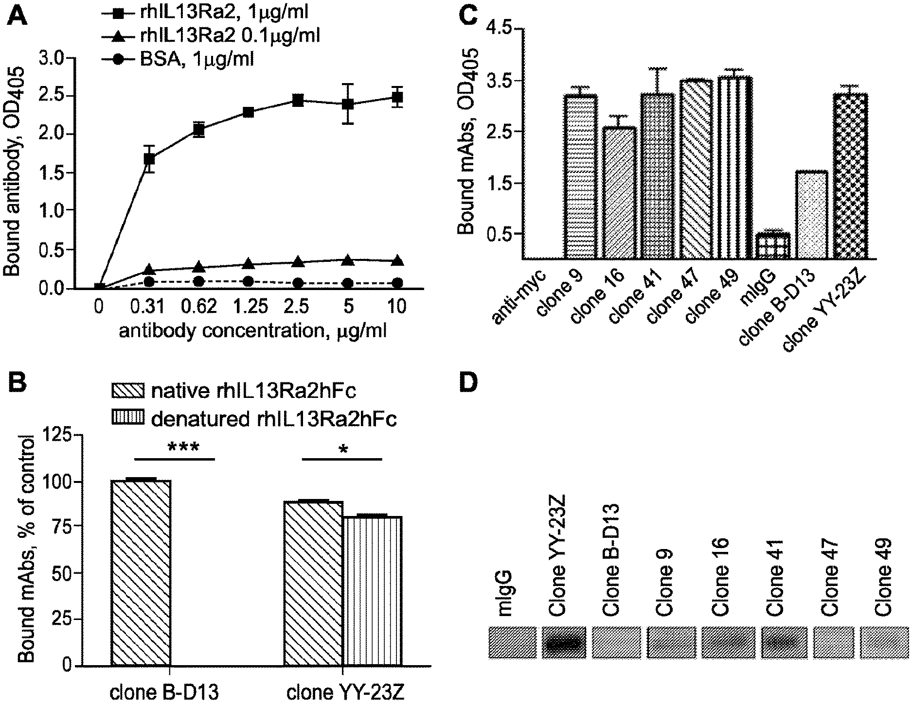
[0036] FIG. 1A-D. Characterization of antigen recognition and screening of hybridoma clones. A, binding of B-D13 mAb to ELISA plates coated with rhIL13R.alpha.2hFc at 0.1 and 1 .mu.g/ml. B, binding of IL13R.alpha.2 mAb to native and denatured (at 95.degree. C. in the presence of .beta.-mercaptoethanol) rhIL13R.alpha.2hFc in a plate-bound ELISA. A paired t test was used to evaluate the difference between control groups (n=4). *, p<0.1; ***, p<0.001. Error bars represent S.D. These data are representative of two independent experiments. C, screening of selected hybridoma populations against rhIL13R.alpha.2hFc in a plate-bound ELISA. D, screening of selected hybridoma populations against rhIL13R.alpha.2hFC using a Western blot.
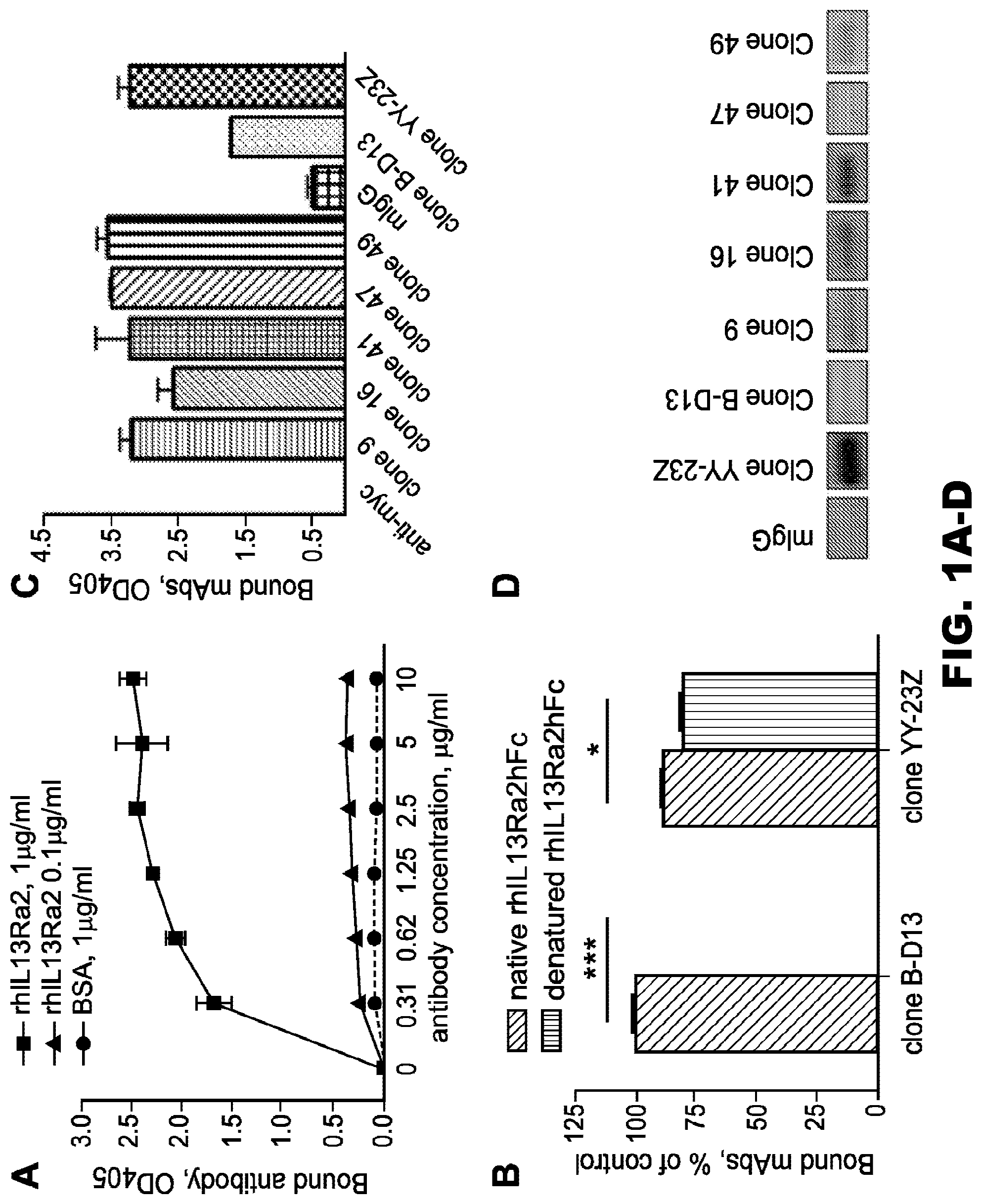
[0037] FIG. 2A-D. The IL13R.alpha.2 (clone 47) mAb specifically binds to rhIL13R.alpha.2 and IL13R.alpha.2 expressed on the cell surface of CHO cells. A, binding of IL13R.alpha.2 (clone 47, 83807, and B-D13) mAbs to rhIL13R.alpha.2 in a plate-bound ELISA. B, binding of the IL13R.alpha.2 (clone 47) mAb to human IL13R.alpha.2 expressed on the surface of CHO cells. C, cross-reactivity of the IL13R.alpha.2 (clone 47) mAb with hrIL13R.alpha.1. D, cross-reactivity of IL13R.alpha.2 (clones 47, 83807, and B-D13) mAbs with mouse rIL13R.alpha.2. Error bars represent S.D.
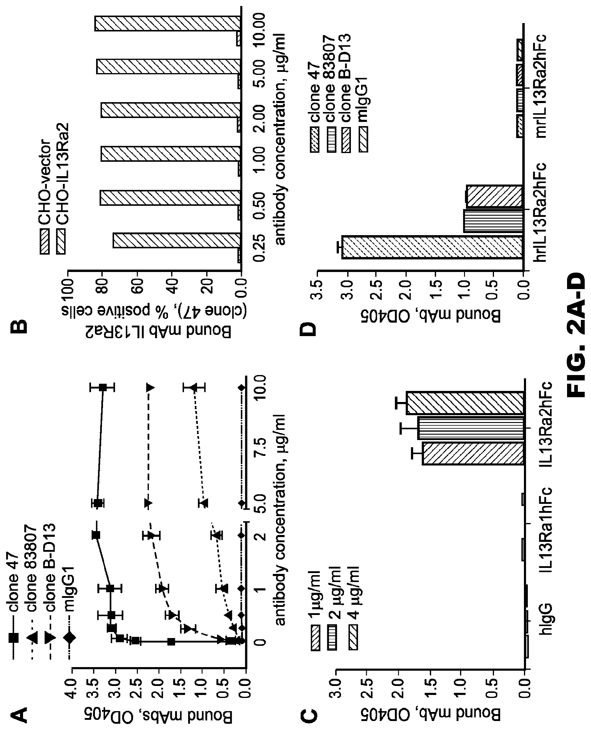
[0038] FIG. 3A-D. Binding of IL13R.alpha.2 mAb to glioma cells. A, flow charts of IL13R.alpha.2 (clones 47, 83807, and B-D13) mAbs binding to the surface of glioma cells, normal human primary astrocytes, and HEK cells transfected with IL13R.alpha.2. B, data of the median fluorescence intensity of binding between the IL13R.alpha.2 (clones 47, 83807, and B-D13) mAbs to various cell lines analyzed by flow cytometry. Numbers above the bars represent the difference in the binding of clone 47 when compared with clone B-D13 for each cell line. The color key is the same for A and B. C, mRNA expression for IL13R.alpha.2 in glioma cells as well as normal human primary astrocytes. D, panels a-c, flow cytometry demonstrating the specific binding of the IL13R.alpha.2 (clone 47) mAb to GFP-tagged U251 glioma cells from an intracranial xenograft (xeno). The curve with a clear area under the curve in sub-panel b depicts the binding of mAb IL13R.alpha.2 (clone 47) to GFP negative cells; the curve with a clear area under the curve in sub-panel c depicts the binding of mAb IL13R.alpha.2 (clone 47) to GFP positive cells. Curves in sub-panels b and c with gray areas under the curves show the results when exposing control IgG to GFP-negative (sub-panel b) or GFP-positive (sub-panel c) cells. neg, negative. A, area; SSC-A, side scatter area; APC-A, allophycocyanin area.
[0039] FIG. 4A-C. The affinity between the IL13R.alpha.2 (clone 47) mAb and rhIL13R.alpha.2. The kinetics of interaction of IL13R.alpha.2 (clone 47) mAb (A) and the commercially available mAb clones 83807 (B) and B-D13 (C) with rhIL13R.alpha.2 as visualized by SPR in a Biacore 3000 are shown. The rhIL13R.alpha.2 was injected at concentrations ranging from 1 to 100 nM (1 nM, 2.5 nM, 5 nM, 7.5 nM, 10 nM, 15 nM, 20 nM, 25 nM concentrations shown, lower to upper curves) at a constant flow rate of 20 .mu.l/minute over immobilized antibodies and over a control dextran surface (these values were subtracted from the signal). The association and dissociation phases were monitored for 300 s by following the change in SPR signal (colored curves) given in RU. Black curves represent the fit of the data to a one-site binding model. For derived kinetic parameters, see Table 1. Lower panels show residuals from a one-site binding model, indicating an excellent fit.
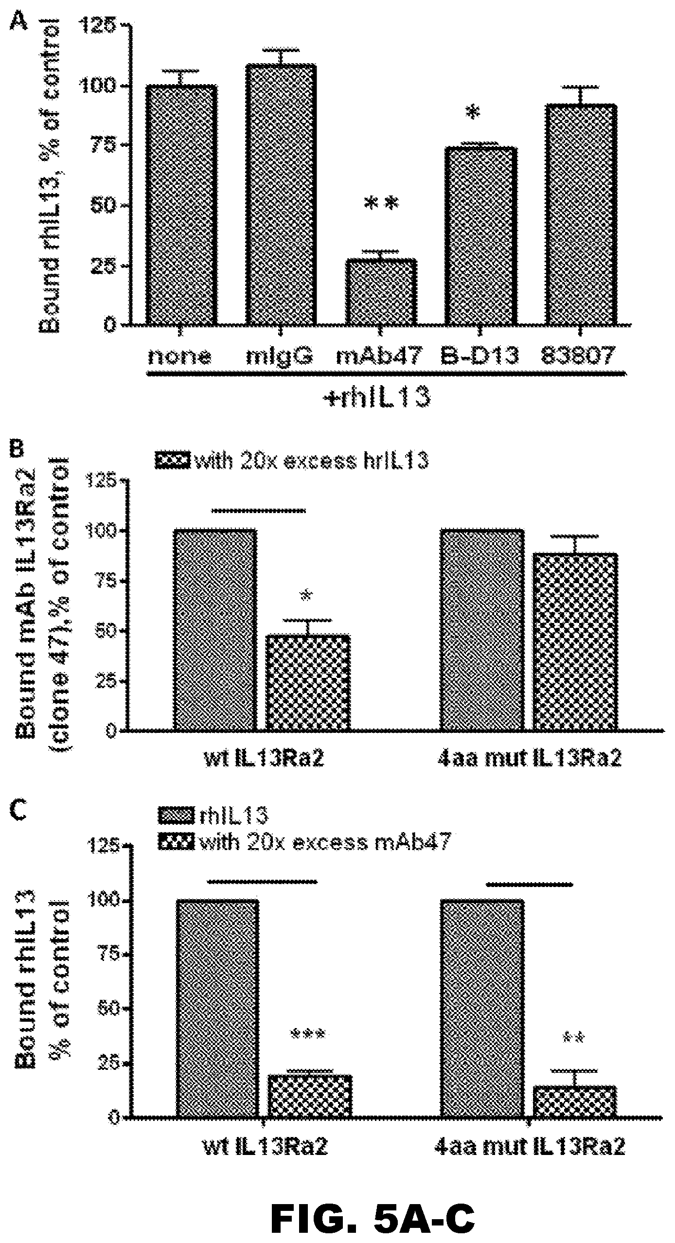
[0040] FIG. 5A-C. The IL13R.alpha.2 (clone 47) mAb competes with rhIL-13 for the binding site of IL13R.alpha.2. A, using a competitive binding plate assay, the IL13R.alpha.2 (clone 47) mAb but not control mIgG or antibody clones 83807, B-D13, and YY-23Z significantly abolished the binding of rhIL-13 to the rhIL13R.alpha.2Fc chimera absorbed to plastic. One-way analysis of variance followed by Dunnett's post hoc test was performed. Data from a single representative experiment are shown. B, recombinant human IL-13 competes with the IL13R.alpha.2 (clone 47) mAb for the binding site of WT IL13R.alpha.2 but not with the 4-amino acid (4aa) mutant IL13R.alpha.2 expressed on the surface of HEK cells. C, the IL13R.alpha.2 (clone 47) mAb competes with rhIL-13 for the binding site of the WT and 4-amino acid mutant form of IL13R.alpha.2. A paired t test was performed. Data represent the summary of three independent experiments shown in B and C. *, p<0.05; **, p<0.01; ***, p<0.001. Error bars represent S.D.
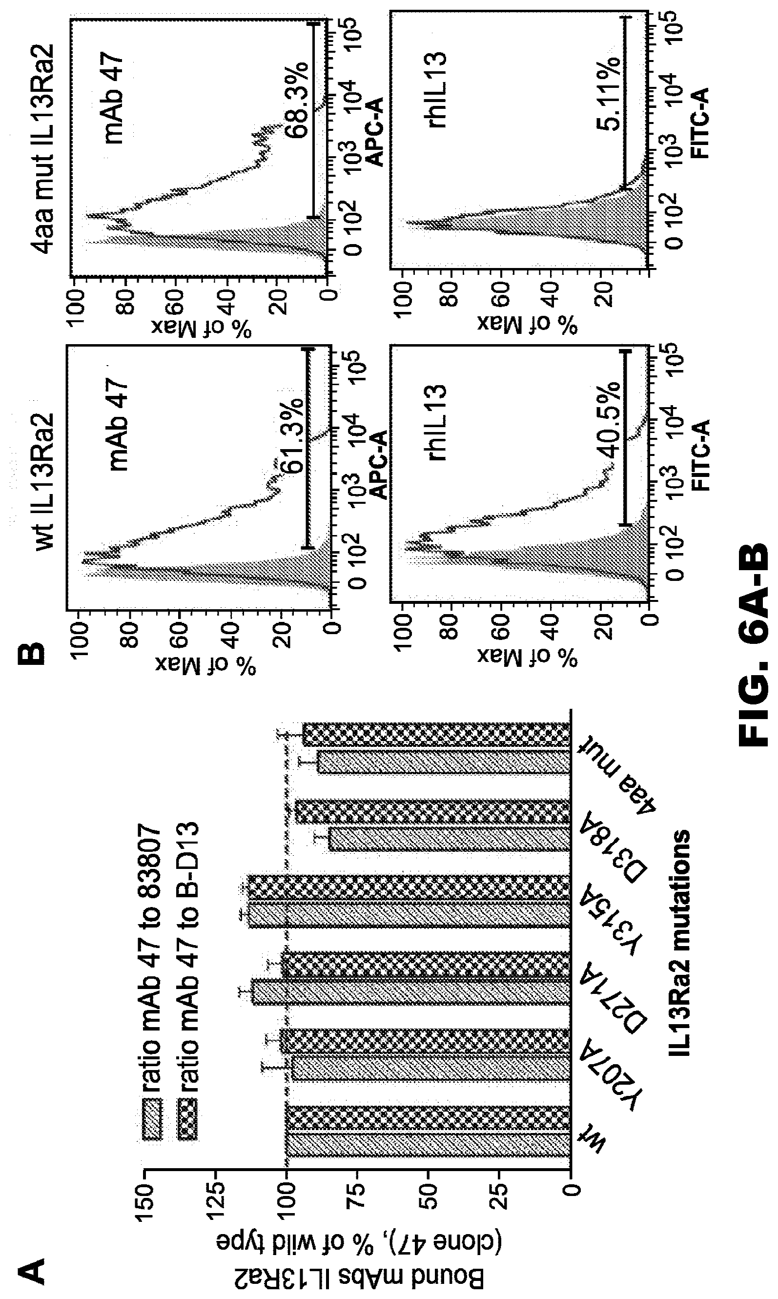
[0041] FIG. 6A-B. The contribution of Tyr207, Asp271, Tyr315, and Asp318 residues of IL13R.alpha.2 to the binding of the IL13R.alpha.2 (clone 47) mAb. A, variants of cDNA encoding individual mutations to Ala or a combinatorial 4-amino acid mutant (4aa mut) of IL13R.alpha.2 was generated. HEK cells were transfected with a control vector or a vector encoding the IL13R.alpha.2 variants. After 48 hours, binding of the IL13R.alpha.2 (clone 47) mAb to the surface of transfected cells was analyzed by flow cytometry. Anti-IL13R.alpha.2 antibody clones 83807 and B-D13 were used as reference antibodies in this assay. Binding of antibodies was determined as the percentage of positive cells. The ratio of bound clones was determined for each IL13R.alpha.2 mutant and compared with that of the wild-type receptor. One-way analysis of variance followed by Dunnett's post hoc test was performed. Data represent a summary of four independent experiments. Error bars represent S.D. B, representative graphs of flow cytometry data demonstrating the binding of clone 47 or rhIL-13 to the WT and 4-amino acid-mutated variant of the IL13R.alpha.2 receptor expressed on the surface of HEK cells. Filled curves: negative control, staining with isotype control IgG+secondary antibody; Open curves: staining with the anti-IL13R.alpha.2 (clone 47) monoclonal antibody+secondary antibody. A, area; APC-A, allophycocyanin area; FITC-A, fluorescein isothiocyanate area.
[0042] FIG. 7A-C. Effect of N-linked glycosylation on the binding of IL13R.alpha.2 to recombinant IL13R.alpha.2. A, binding of IL13R.alpha.2 to control and Pngase F-treated rhIL13R.alpha.2. Plates were coated with hrIL13R.alpha.2 at 1 .mu.g/ml and treated with native buffer or with 1 milliunit/well Pngase F in native buffer for 3 hours at 37.degree. C. An ELISA for binding of the IL13R.alpha.2 (clone 47) mAb in comparison with antibody clones B-D13, 83807, and YY-23Z and rhIL-13 was performed, and the data of one representative experiment from three independent experiments are shown. A paired t test was used to evaluate the difference between control and Pngase F-treated groups (n=4). *, p<0.5; **, p<0.01; ***, p<0.001. B, a Western blot shows the lower molecular weight of Pngase F-treated rhIL13R.alpha.2 due to removal of N-linked glycosylation adducts from the molecule. C, flow cytometry shows the binding of IL13R.alpha.2 mAbs to IL13R.alpha.2-expressing U251 and HEK293 cells treated with 1 milliunit of Pngase F for 1 hour at 37.degree. C. The data are representative of three independent experiments. A paired t test was used to evaluate the difference between control and Pngase F-treated groups. *, p<0.5. MFI, mean fluorescence intensity. Error bars represent S.D.
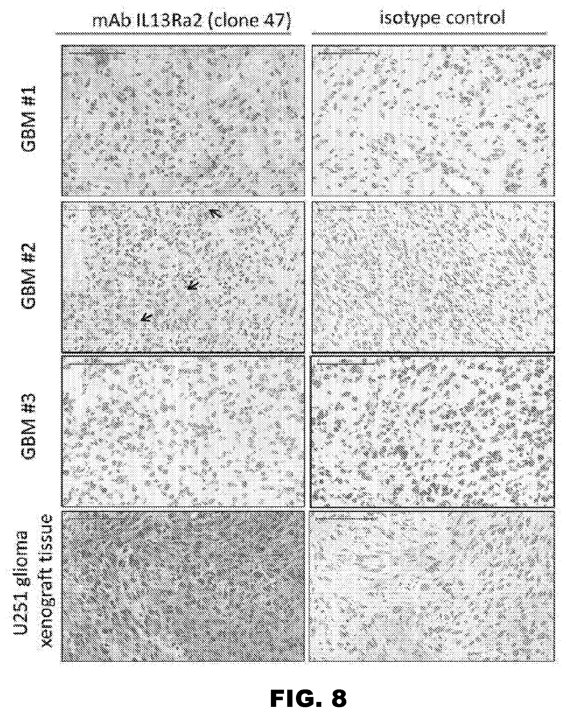
[0043] FIG. 8. The IL13R.alpha.2 (clone 47) mAb recognizes IL13R.alpha.2 in GBM tissues and in a human glioma xenograft. Immunohistochemistry on frozen tissue sections from three human GBM samples and a U251 xenograft was performed with the IL13R.alpha.2 (clone 47) mAb or mIgG at a concentration of 3 .mu.g/ml. Staining of GBM tissues demonstrates positive staining of the majority of cells in sample 1, positive reactivity in only a fraction of the cells in sample 2, and negative staining in sample 3. Staining in all three samples was performed in the same experiment. Positive staining was also detected in U251 xenograft tissue. Arrows point to individual positive cells. Scale bars=100 .mu.m.
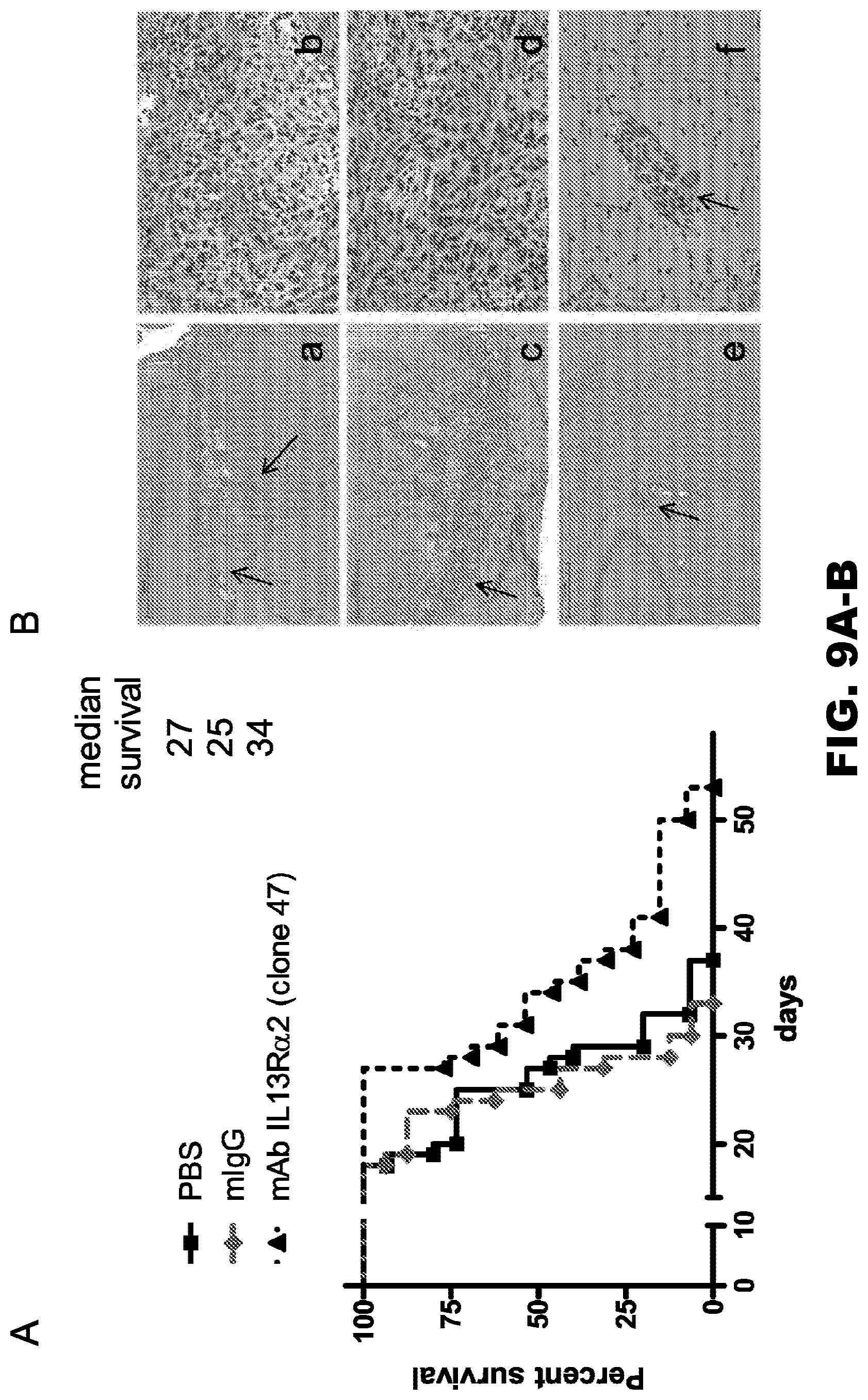
[0044] FIG. 9A-B. The IL13R.alpha.2 (clone 47) mAb improves the survival of mice in an orthotopic human glioma xenograft model. A, the survival of animals injected with U251 glioma cells (2.5.times.10.sup.4) alone or in combination with either control IgG or the IL13R.alpha.2 (clone 47) mAb. B, a representative photomicrograph of 10-.mu.m-thick tissue sections stained with H&E from mice injected with U251 cells alone (panels a and b) or in combination with mIgG (panels c and d) or mAbIL13R.alpha.2 (clone 47) (panels e and f). Arrows point to the tumor and invading cells. Scale bars (panels a, c, and e)=100 .mu.m. Scale bars (panels b, d, and f)=100 .mu.m.
[0045] FIG. 10A-B. A competitive binding assay for the IL13R.alpha.2 (clone 47) mAb to the surface of N10 glioma cells. A. The IL13R.alpha.2 (clone 47) mAb was pre-incubated with 10.times. excess rhIL13R.alpha.2 for 30 minutes on ice. N10 cells were subsequently incubated with isotype control mIgG or IL13R.alpha.2 (clone 47) mAb alone or in the presence of rhIL13R.alpha.2 and bound antibodies were analyzed by flow cytometry. B. N10 glioma cells were pre-incubated either with 10.times. excess rhIL13 (left panel) or with 10.times. excess of IL13R.alpha.2 (clone 47) mAb for 30 minutes on ice (right panel). N10 cells were subsequently incubated with isotype control mIgG, IL13R.alpha.2 (clone 47) mAb or rhIL13. Bound antibodies or rhIL13 were detected with secondary antibodies and analyzed by flow cytometry. Data are presented as % of positive cells.
[0046] FIG. 11. The effects of IL13R.alpha.2 (clone 47) mAb on the survival of mice with an established human U251 glioma. Mice were intracranially-injected with 2.5.times.10.sup.4 U251 glioma cells and treated three days later with a single injection of PBS (n=7) or 10 .mu.g IL13R.alpha.2 (clone 47 or B-D13) mAb (n=7). The analysis of the animal's survival was performed using the Log-rank test. Median survival was determined to be 27 days in the PBS group, versus 23 and 35 days in the groups treated with B-D13 and 47 IL13R.alpha.2 mAb, respectively (p>0.05).
[0047] FIG. 12. Binding of IL13R.alpha.2 clone 47 phages with IL13R.alpha.2hFc in plate ELISA. These data demonstrate that phages presenting scFv IL13R.alpha.2 (clone 47) are positively selected against IL13R.alpha.2Fc chimeric protein after 3 rounds of biopanning.
[0048] FIG. 13. Specificity of binding scFv IL13R.alpha.2 clone 47 with IL13R.alpha.2hFc-competitive assay. These data show that binding of the scFvIL13R.alpha.2 (clone 47) presented on the phage surface to recombinant IL13R.alpha.2 is completely abolished by parental monoclonal antibody (clone 47), but not other antibodies against IL13R.alpha.2. It indicates that scFvIL13R.alpha.2 (clone 47) and parental monoclonal antibody (clone 47) share the epitope (i.e., recognition site) on the IL13R.alpha.2 molecule. Each data point is an average of 3 independent replicates in all figures. Data presented as mean.+-.SEM. ***p<0.001.
[0049] FIG. 14. Binding of soluble scFv IL13R.alpha.2 (clone 47) with IL13R.alpha.2hFc chimera. These data show that soluble scFvIL13R.alpha.2 (clone47) generated in a prokaryotic expression system (E. coli) binds specifically to IL13R.alpha.2Fc recombinant protein. Parental antibody, mAb IL13R.alpha.2 (clone 47), and control mouse IgG served as positive and negative controls, respectively
[0050] FIG. 15. The effect of mesenchymal stem cells secreting scFvIL13R.alpha.2-sTRAIL fusion protein on the U87-IL13R.alpha.2 glioma cell line. These data show that mesenchymal stem cells modified to secrete a genetic fusion of scFvIL13R.alpha.2(clone 47) with TRAIL protein exhibit a therapeutic effect in the IL13R.alpha.2-expressing U87 glioma cell line. The results establish the efficacy of conjugating the scFV to a TRAIL cytokine. The amount of cancer cell killing is equivalent to the use of TRAIL alone without the scFV, but it is expected that the scFV-TRAIL would be less harmful to non-cancer tissues, given the specificity conferred by the scFv targeting IL13R.alpha.2.
[0051] FIG. 16. Schematic maps of retroviral vector encoding IL13R.alpha.2-specific scFv CARs. The CAR consists of the immunoglobulin heavy-chain leader peptide, the IL13R.alpha.2-specific scFv clone 47 (M47), a short hinge (SH) or long hinge (LH), a transmembrane domain (TM) derived from CD28, and a CD28. .zeta. endodomain. LTR: long terminal repeat (retroviral backbone). Domains are identified as block structures. Maps are not to scale.
[0052] FIG. 17. IL13R.alpha.2-scFv CART cell agent: Expression of .alpha.CD3..zeta. relative to .alpha.GAPDH of CAR agent in T cells. SH: short hinge. LH: long hinge.
[0053] FIG. 18A-B. IL13R.alpha.2-scFv CARs are expressed on the surface of T cells. IL13R.alpha.2-CAR T cells were generated by retroviral transduction and CAR expression was determined by FACS analysis. Short hinge CARs were detected with an antibody specific for murine scFV. Long hinge CARs were detected with an antibody specific for the long hinge. Isotype antibody control: open curve; Specific Antibody: filled curve.
[0054] FIG. 19. Functional characterization of IL13R.alpha.2-CAR T cells--Cytotoxicity. Standard .sup.51Chromium cytotoxicity assays were performed with Raji (IL13R.alpha.1-/IL13R.alpha.2-), 293T (IL13R.alpha.1+/IL13R.alpha.2-), 293T genetically modified to express IL13R.alpha.2cells (293T-IL13R.alpha.2; IL13R.alpha.1+/IL13R.alpha.2+), or U373 (IL13R.alpha.1+/IL13R.alpha.2+) cells as targets. As effectors nontransduced (NT) T cells, IL13R.alpha.2-CAR.SH.CD28..zeta. T cells, IL13R.alpha.2-CAR.LH.CD28.zeta. T cells, IL13R.alpha.2-CAR.SH..DELTA. T cells, or IL13R.alpha.2-CAR.LH..DELTA. T cells were used. Only IL13R.alpha.2-CAR.SH.CD28..zeta. T cells and IL13R.alpha.2-CAR.LH.CD28..zeta. T cells killed with IL13R.alpha.2+ target cells (U373 and 293T-IL13R.alpha.2; n=4). T cells expressing nonfunctional CARs (IL13R.alpha.2-CAR.SH.4 and IL13R.alpha.2-CAR.LH..DELTA.) had not cytolytic activity, demonstrating that the killing activity depends on the expression of a functional IL13R.alpha.2-CAR. NT T cells killed none of the targets, further confirming specificity.
[0055] FIG. 20. Functional characterization of IL13R.alpha.2-CAR T cells--IFN.gamma. and IL2 Cytokine secretions. A. NT T cells, IL13R.alpha.2-CAR.SH.CD28.zeta. T cells, IL13R.alpha.2-CAR.LH.CD28..zeta. T cells, IL13R.alpha.2-CAR.SH..DELTA. T cells, or IL13R.alpha.2-CAR.LH..DELTA. T cells were co-cultured with U373 cells for 24 to 48 hours (n=4). Only IL13R.alpha.2-CAR.SH.CD28..zeta. T cells and IL13R.alpha.2-CAR.LH.CD28..zeta. T cells secreted IFN.gamma. demonstrating target cell recognition in contrast to IL13R.alpha.2-CAR.SH..DELTA. T cells, IL13R.alpha.2-CAR.LH..DELTA. T cells or NT T cells. B. NT T cells, IL13R.alpha.2-CAR.SH.CD28..zeta. T cells, IL13R.alpha.2-CAR.LH.CD28..zeta. T cells, IL13R.alpha.2-CAR.SH..DELTA. T cells, or IL13R.alpha.2-CAR.LH..DELTA. T cells were co-cultured with U373 cells for 24 to 48 hours (n=4). Only IL13R.alpha.2-CAR.SH.CD28..zeta. T cells secreted IL2, demonstrating that IL13R.alpha.2-CAR.SH.CD28..zeta. induces superior T cell activation in comparison to IL13R.alpha.2-CAR.LH.CD28..zeta.. IL13R.alpha.2-CAR.SH..DELTA. T cells, IL13R.alpha.2-CAR.LH..DELTA. T cells or NT T cells also did not induce IL2 production.
[0056] FIG. 21. IL13R.alpha.2-SH CARs have anti-glioma activity in vivo. Severe combined immunodeficient (SCID) mice were injected with 1.times.10.sup.5 firefly luciferase expressing U373 cells intracranially. On day 7 mice were treated either with 1.times.10.sup.6 IL13R.alpha.2-CAR.SH.CD28..zeta. T cells, IL13R.alpha.2-CAR.LH.CD28..zeta. T cells, IL13R.alpha.2-CAR.SH..DELTA. T cells, or IL13R.alpha.2-CAR.LH..DELTA. T cells (5 mice per group). Tumor growth was monitored by bioluminescence imaging. Only IL13R.alpha.2-CAR.SH.CD28..zeta. T cells had significant anti-glioma effects with 4/5 mice having a complete response.
[0057] FIG. 22. Properties of m47 CAR T cell agent. The m47-CAR T cells recognize IL13R.alpha.2.sup.+, but not IL13R.alpha.1.sup.+ targets. The data show that the short hinge CD28z-CAR (SH2) T cells perform better in terms of effector function than CD28z-CAR (SH3), CD28z-CAR (LH2), CD28z-CAR (LH3), CD28z-CAR (SH2.DELTA.), or CD28z-CAR (SH3.DELTA.).
[0058] FIG. 23. Functional comparison of m47 CAR T cell agents. Open curve: secondary antibody; Filled curve: IL13R.alpha.2Fc+ secondary antibody.
[0059] FIG. 24. The m47 CAR T cell agent is highly expressed after transduction. Open curve: secondary antibody; Filled curve: IL13R.alpha.2Fc+ secondary antibody.
[0060] FIG. 25. The m47 CAR T cell produce interferon .gamma. and interleukin 2, but only after IL13R.alpha.2 stimulation.
[0061] FIG. 26. IL13R.alpha.2- and IL13R.alpha.1-positive cell lines are made by genetic modification of HEK 293T cells. Filled curve: isotype antibody control; Open curve: specific antibody.
[0062] FIG. 27. The m47 CAR T cells kill only IL13R.alpha.2.sup.+ cell lines. The in vitro experiments provide data establishing that m47 CAR T cells present a recombinant CAR protein on the cell surface that does not recognize IL4R, IL13R.alpha.1 or any receptor other than its specific recognition of IL13R.alpha.2. The specificity of the recognition extends to a specificity for only those cell lines expressing IL13R.alpha.2.
[0063] FIG. 28. In vivo data comparing effect of m47 CAR T cell agent, untreated and NT-treated glioblastoma multiforme xenografts in nude mice. The U373 glioblastoma multiforme xenograft mouse model was used. At day 0, 1.times.10.sup.5 GFP-ffluc U373 cells were administered per mouse. On day 7, 2.times.10.sup.6 m47 CAR T cells or NT cells were administered. Untreated samples did not receive treatment on day 7. No exogenous interleukin 2 was administered and results of the survival analysis were recorded by serial bioluminescence imaging. n=3.
[0064] FIG. 29. The m47 CAR T cell agent prolonged the survival of nude mice with glioblastoma multiforme.
[0065] FIG. 30A-C. Characterization of IL13R.alpha.2-CAR T cells. (A, B) Co-culture assay with recombinant protein demonstrated interferon .gamma. and interleukin 2 production in an IL14R.alpha.2-dependent fashion; (C) Cytolytic activity in standard chromium release assay.
[0066] FIG. 31A-D. Generation of 47 CAR T cells. (A) Scheme of M47 CARs.
[0067] All CARs contained an N-terminal leader sequence, a codon-optimized synthetic gene encoding M47 in scFv format, a spacer region, a CD28 transmembrane domain, and signaling domains derived from CD28 and CD3-t. The spacer region was either the IgG1 hinge (16 amino acids; short spacer region; M47-CAR.SSR.CD28..zeta.) or the IgG1-CH2CH3 domain. LSR..DELTA. and SSR..DELTA. M47-CARs without signaling domains were constructed and served as controls. (C,B) CAR expression was confirmed using FACS analysis. Representative plots (B) and summary data (C) are shown (mean 74.1%-93.3%, n=5-6 per CAR construct). Open curve: secondary antibody; Filled curve: IL13R.alpha.2Fc+ secondary antibody. (D) Expression of full-length 47-CAR.SSR.CD28..zeta. and 47-CAR.LSR.CD28..zeta. by Western blot analysis using a CD3-.zeta. antibody.
[0068] FIG. 32. Phenotypic analysis of 47-CAR T cell lines. CAR T cells were analyzed for CD4 and CD8 surface expression using CD4-PacBlue and CD8-PerCP antibodies (BD Biosciences). The four CAR T cell lines analyzed for surface expression of CD4 and CD8 were SSR..DELTA., SSR.CD28..zeta., LSR..DELTA., and LSR.CD28..zeta.. The histogram provides the results of the analysis, with light gray bars indicating CD4 expression and black bars indicating CD8 expression.
[0069] FIG. 33A-D. 47-CAR T cells release cytokines after stimulation with recombinant IL13R.alpha.2 protein or IL13R.alpha.2-positive cells. 47-CAR or non-transduced (NT) T cells were stimulated with recombinant IL13R.alpha.1, IL13R.alpha.2, or IL4R.alpha. proteins. After 24 hours, IFN.gamma. (A) was measured by ELISA (n=4). T cells expressing 47-CAR constructs, but not controls, expressed significant levels of IFN.gamma. (p<0.001) when stimulated with recombinant IL13R.alpha.2 protein in comparison to IL13R.alpha.1 and IL4R.alpha. stimulated T cells. 47-CAR T cells were co-cultured with Raji, U373 cells, 293T-GFP, and 293T-GFP/IL13R.alpha.2 at a 1:2 E:T ratio. NT and CAR..DELTA. T cells served as controls. (B,C) After 24 hours, cytokines (IFN.gamma., IL2) were measured by ELISA (n=3). (B) U373 and 293T-GFP-IL13R.alpha.2 (IFN.gamma.); SSR..DELTA. vs SSR.CD28..zeta.: p<0.001; LSR..DELTA. vs LSR.CD28..zeta.: p<0.05. (C); U373 and 293T-GFP-IL13R.alpha.2 (IL2); SSR..DELTA. vs SSR.CD28..zeta.: p<0.01; LSR..DELTA. vs LSR.CD28..zeta.: NS. (D) 4-hour cytotoxicity assay at an E:T ratio of 10:1 (n=4).
[0070] FIG. 34. LSR.CD28..zeta. T cells show a self-activation phenotype during ex vivo expansion. T cells were analyzed for phosphor-CD3-t expression using CD247 (pY142)-AF647 antibody (BD Biosciences).
[0071] FIG. 35. Cell surface expression of IL13R.alpha.1 and IL13R.alpha.2. Cell lines were analyzed for IL13R.alpha.1 and IL13R.alpha. expression using primary goat anti-IL13R.alpha.1 and anti-IL13R.alpha.2 antibodies (AF152 and AF146, respectively; R&D) followed by secondary rabbit anti-goat IgG Alexa647 antibody (Life Technologies). Filled curve: isotype antibody control; Open curve: specific antibody.
[0072] FIG. 36A-D. Generation of SSR 47-CARs with CD28.OX40..zeta., CD28.41BB..zeta. or 41BB..zeta. endodomains. (A) Scheme of SSR 47-CARs. (B, C) CAR expression was confirmed using FACS analysis. Representative plots (B) and summary data (C) are shown. 47-CAR.SSR.CD28.OX40..zeta. and 47-CAR.SSR.CD28.41BB..zeta.: mean: 74.6%-77.5% (n=4); 47-CAR.SSR.CD28.41BB..zeta.: mean: 4.9% (n=3). Open curve: secondary antibody; Filled curve: IL13R.alpha.2Fc+ secondary antibody. (D) Expression of 47-CAR.SSR.41BB..zeta., M47-CAR.SSR.OX40.CD28..zeta. and M47-CAR.SSR.41BB.CD28..zeta. by Western blot analysis.
[0073] FIG. 37A-B. Comparison of 47-CAR.SSR.CD28..zeta. 47-CAR.SSR.41BB..zeta. and 47-CAR.SSR.CD28.OX40..zeta. T cells. (A) 47-CAR T cells were co-cultured with U373 cells at a 1:2 E:T ratio. NT and CAR..DELTA. T cells served as controls. After 24 hours, IFN.gamma. and IL2 were measured by ELISA (n=3); SSR..DELTA. vs SSR.CD28..zeta. (U373; IFN.gamma.): p<0.001; SSR..DELTA. vs SSR.41BB..zeta. (U373; IFN.gamma.): p<0.05; SSR..DELTA. vs SSR.CD28.OX40..zeta. for (U373; IFN.gamma.): p<0.001; SSR..DELTA. vs SSR.CD28..zeta. (U373; IL2): p<0.001; SSR..DELTA. vs SSR.41BB..zeta. (U373; IL2): p<0.001; SSR..DELTA. vs SSR.CD28.OX40..zeta. (U373; IL2): p<0.01. (B) 4-hour cytotoxicity assay at an E:T ratio of 10:1 (n=4).
[0074] FIG. 38A-C. Treatment of glioma xenograft with T cells expressing 47-CARs results in tumor regression and improved overall survival. U373 glioma bearing mice were treated on day 7 with SSR.CD28..zeta. (n=9), SSR.41BB..zeta. (n=9) or SSR.OX40.CD28..zeta. (n=9) T cells. SSR..DELTA. CAR T cells (n=7) served as controls. (A) Representative images for each group and (B) quantitative bioluminescence (radiance=photons/sec/cm.sup.2/sr) imaging data for all mice are shown (dotted lines: individual mice; solid lines: median). (C) Kaplan-Meier survival analysis (SSR..DELTA. vs SSR.CD28..zeta.: p=0.0002; SSR..DELTA. vs SSR.41BB..zeta.: p=0.0039; SSR..DELTA. vs SSR.OX40.CD28..zeta.: p=0.0092; SSR.CD28. vs SSR.41BB..zeta.: p=0.4723; SSR.CD28..zeta. vs SSR.OX40.CD28..zeta.: p=0.3582; SSR.41BB..zeta. vs SSR.OX40.CD28..zeta.: p=0.8374).
[0075] FIG. 39A-B. Analysis of U373 cells isolated from recurrent tumors. U373 cells were isolated from recurrent tumors of mice that were treated with 47-CAR T cells. After short-term culture (2 to 7 days), FACS analysis and cytotoxicity assays were performed. (A) FACS analysis for IL13R.alpha.2. (B) 47-CAR T cells killed U373 tumor cells isolated from recurrent tumors in contrast to Raji cells in a standard four-hour cytotoxicity assay for Cr release from labeled cells. Open curve: isotype antibody control; Filled curve: specific antibody.
[0076] FIG. 40A-C. Limited persistence of 47-CART cell in vivo. 47.SSR.CD28.-CAR T cells were transduced to express eGFP.ffLuc. (A) FACS analysis confirmed the expression of the CAR and eGFP.ffLuc transgenes. (B, C) 1.times.10.sup.5 unmodified U373 cells were injected intracranially into mice. On day 7, mice received 2.times.10.sup.6 47. SSR.CD28..zeta..eGFP.ffLuc CAR T cells intracranially using the same tumor coordinates. Bioluminescence imaging was used to monitor T cell persistence.
[0077] FIG. 41A-C. Generation and characterization of LSR-CD28.41BB..zeta. CAR T cells. (A) Scheme of LSR.CD28.41BB..zeta. CAR construct. (B) CAR expression was confirmed using FACS analysis. Representative plot. Open curve: secondary antibody; Filled curve: IL13R.alpha.2Fc+ secondary antibody. (C) LSR.CD28.41BB..zeta. CAR T cells were co-cultured with U373 cells at a 1:2 E:T ratio. NT T cells served as controls. After 24 hours, IFN.gamma. or IL2 was measured by ELISA (n=3).
[0078] FIG. 42A-B. Generation of SSR..alpha..CD28.41BB..zeta. CAR T cells. (A) Scheme of SSR..alpha..CD28.41BB..zeta. CAR construct. (B) CAR expression was tested using FACS analysis (representative plot shown).
[0079] FIG. 43. FACS analysis of PD-L1 expression on U373 cell surface with and without IFN.gamma. stimulation. U373 cells were cultured with or without IFN.gamma. (100 units/ml). After 24 hours, U373 cells were analyzed for PD-L1 expression using a CD271 PE antibody (BD Biosciences).
[0080] FIG. 44A-B. Transgenic expression of IL15 in SSR.CD28..zeta. T cells results in enhanced antigen-dependent IL15 secretion. T cells were stimulated with (A) recombinant proteins or (B) cell lines.
[0081] FIG. 45A-B. Transgenic expression of IL15 results in (A) enhanced in vivo persistence of SSR.CD28..zeta. T cells resulting in improved (B) progression-free survival (PFS).
DETAILED DESCRIPTION
[0082] The disclosure provides binding agents, or partners, that specifically recognize interleukin 13 receptor .alpha.2 (IL13R.alpha.2) for use in diagnosing, preventing, treating or ameliorating a symptom of any of a wide range of cancers characterized by cells presenting IL13R.alpha.2. More particularly, the disclosure provides (i) the sequences of the six complementarity determining regions of a monoclonal antibody (m47) that specifically targets human tumor-associated antigen, i.e., interleukin 13 receptor .alpha.2 (IL13R.alpha.2), and (ii) data demonstrating the functionality of the protein encoded by the heavy and light chain cDNAs in the format of an scFv antibody or conjugate (e.g., fusion) to other functional moieties. The six complementarity determining regions of the m47 monoclonal antibody confer binding specificity for IL13R.alpha.2, consistent with the understanding in the immunological arts. In some embodiments, the scFv comprises the complete heavy and light chain variable regions of antibody m47, or the complete heavy and light chains of antibody m47. In some embodiments, the heavy and light chain fragments comprise, e.g., the m47 CDRs, or the m47 variable regions, and these domains can be arranged in different formats, such as a single-chain variable fragment of an antibody, i.e., a scFv, a diabody, a bi-specific antibody fragment, a tri-specific antibody fragment, a fusion protein with any of a wide variety of therapeutic proteins and/or other moieties, a humanized antibody fragment, a Fab fragment, a Fab' fragment, a F(ab)2' fragment and any other functional format for a bi-functional peptide providing a targeting function and an effector function. Moreover, the single-chain antibody or other arrangements of the protein encoded by the heavy and light chains could be expressed and conjugated to therapeutic carriers (e.g., viruses, cells, nanomaterials) for specific delivery of a therapeutic to an IL13R.alpha.2-expressing tumor. The materials according to the disclosure are also useful in imaging tumor burden.
[0083] The technology addresses the most serious obstacle to progress in immunotherapy, i.e., the virtual absence of defined, tumor-specific antigens that can be predictably found on at least a larger subgroup of human cancers and that can serve as effective targets for cancer eradication. Finding such antigens would move the field beyond the methods for treating CD19/CD20-expressing B cell malignancies.
[0084] The terms used throughout this disclosure are given their ordinary and accustomed meanings in the art, unless a different meaning is made clear from the text when considered in the context of the disclosure as a whole.
[0085] The disclosure describes the development and characterization of a monoclonal antibody (mAb) fragment specific to IL13R.alpha.2 for the therapeutic purpose of targeting IL13R.alpha.2-expressing tumors. The high affinity IL13R.alpha.2 is selectively expressed at a high frequency by glioblastoma multiforme (GBM) as well as several other tumor types. One approach for targeting this tumor-specific receptor utilizes the cognate ligand, IL-13, conjugated to cytotoxic molecules. This approach, however, lacks specificity because the lower affinity receptor for IL-13, IL13R.alpha.1, is widely expressed by normal tissues. A monoclonal antibody (mAb) specific to IL13R.alpha.2 was expected to overcome the lack of specificity afflicting methodologies that recognized both IL13 receptors, i.e., IL13R.alpha.1 as well as IL13R.alpha.2. Such a mAb would be therapeutically useful in targeting and treating IL13R.alpha.2-expressing cancers, including tumors.
[0086] As disclosed herein, hybridoma cell lines were generated and compared for binding affinities to recombinant human IL13R.alpha.2 (rhIL13R.alpha.2). Clone 47 demonstrated binding to the native conformation of IL13R.alpha.2 and was therefore chosen for further studies. Clone 47 bound specifically and with high affinity (KD=1.39.times.10.sup.-9M) to rhIL13R.alpha.2 but not to rhIL13R.alpha.1 or murine IL13R.alpha.2. Furthermore, clone 47 specifically recognized wild-type IL13R.alpha.2 expressed on the surface of CHO and HEK cells as well as several glioma cell lines. Competitive binding assays revealed that clone 47 also significantly inhibited the interaction between human soluble IL-13 and IL13R.alpha.2 receptor. Moreover, N-linked glycosylation of IL13R.alpha.2 contributes in part to the interaction of the antibody to IL13R.alpha.2. In vivo, the IL13R.alpha.2 mAb improved the survival of nude mice intracranially implanted with a human U251 glioma xenograft.
[0087] The IL13R.alpha.2-specific, scFv-based CAR, 47-CAR, constructed as disclosed herein, provided the material used in exploring the influence of long and short spacer regions, as well as endodomains, on its function. While 47-CAR.SSR.CD28..zeta. (i.e., the 47-CAR binding region provided as an scFv joined to a short spacer region as defined herein, in turn joined to an unmodified or chimeric endodomain or T cell cytoplasmic domain) and 47-CAR.LSR.CD28..zeta. (similar construct substituting a long spacer region (LSR)) recognized target cells as judged by IFN.gamma. production, only 47-CAR.SSR.CD28..zeta. induced IL2 production, indicating better T-cell activation. An additional LSR 47-CAR containing a CD28.41BB..zeta. endodomain (FIG. 41) was shown to lack the ability to induce IL2 expression. These observations are consistent with knowledge that scFvs that bind to an epitope in close proximity to the cancer cell membrane, requiring long spacer regions for optimal CAR function, in contrast to scFvs that bind to epitopes distal to the cell membrane. The data disclosed herein indicates that the IL13R.alpha.2 epitope recognized by 47-CARs is located distal to the cell membrane.
[0088] In greater particularity, four SSR 47-CARs were constructed, each with a different endodomain, i.e., CD28..zeta., 41BB..zeta. CD28.OX40..zeta., and CD28.41BB..zeta.. While all four CARs were expressed, as judged by Western blot analysis, no significant cell-surface expression was observed for 47-CAR.SSR.CD28.41BB..zeta.. We explored if changing the transmembrane domain from CD28 to CD8a in 47-CAR.SSR.CD28.41BB..zeta. would result in better cell-surface expression, but no increase in expression was observed. Because 47-CARs.LSR.CD28.41BB..zeta. are expressed on the cell surface (FIG. 42), the result indicates that the interplay between spacer region and endodomain influences CAR cell-surface expression.
[0089] 47-CAR.SSR.CD28..zeta. 47-CAR.SSR.41BB..zeta., and 47-CAR.SSR.CD28.OX40..zeta. T cells had potent antitumor effect in vivo, resulting in a significant survival advantage. While mice treated with 47-CAR.SSR.CD28..zeta. T cells had the longest median survival in comparison to 47-CAR.SSR.41BB..zeta. or 47-CAR.SSR.CD28.OX40..zeta. T-cell treated mice, this difference did not reach significance. The experimental results also showed that addition of a second costimulatory endodomain did not improve antitumor activity in vivo. Limited T-cell persistence in vivo was identified as the principal limitation on therapy. This limitation may be overcome by the transgenic expression of cytokines.sup.36 or by blocking inhibitory molecules that are secreted or present on the surface of gliomal cells. For example, gliomas such as U373 express PD-L1, which is upregulated in the presence of IFN.gamma. (FIG. 43), and could be targeted in future studies.
[0090] The experimental results disclosed herein establish that T cells redirected to IL13R.alpha.2 with 47-CARs have potent anti-tumor activity against glioma cells in vitro, and induce the regression of established GBM xenografts in vivo. 47-CARs are expected to be of value in the treatment of not only IL13R.alpha.2-positive GBMs but also other malignancies in which IL13R.alpha.2 is expressed.
[0091] The experimental results disclosed herein establish that T cells redirected to IL13R.alpha.2 with 47-CARs and that also express IL15 have enhanced anti-tumor activity in the GBM xenografts in vivo.
[0092] The disclosure is based, at least in part, on the discovery that IL13R.alpha.2 is found preferentially on cancer cells such as tumor cells. This receptor functions as a cancer-, or tumor-, specific antigen that has been used to elicit the high-affinity monoclonal antibody m47, along with antigen binding fragments of that antibody. The VL and VH variable regions of the m47 antibody have been engineered into a single chain (sc) variable fragment (scFv) to generate conjugates, such as chimeric antigen receptors (i.e., CARs), for introduction into T cells for adoptive transfer. Thus, CAR-transduced T cells are expected to target a tumor-specific IL13R.alpha.2 epitope, leading to eradication of cancer cells presenting the receptor. It is believed that CAR-transduced T cells recognizing IL13R.alpha.2 will destroy large solid tumors. CAR-transduced T cells, however, target cancer cells only directly and antigen-negative cancer cells may escape. It is expected that CAR-transduced T cells also will be effective in eliminating antigen-negative cancer cells via the bystander effect.
[0093] Disclosed herein are experiments establishing the development of IL13R.alpha.2-specific CARs with a scFv47-based antigen-binding domain (47-CARs). The data show that 47-CARs perform better with a short spacer region, which provides for optimal functionality, and that 47-CAR T cells are able to recognize and kill only IL13R.alpha.2-positive and not IL13R.alpha.1-positive target cells in vitro. In addition, 47-CAR T cells induce tumor regression in an orthotopic xenograft mouse model of GBM, which was associated with a significant survival advantage.
[0094] The protein conjugates according to the disclosure are specific for IL13R.alpha.2, which is associated with cancers, e.g., tumors. In addition, the disclosure provides a polynucleotide encoding one of these cancer-specific binding partners, including polynucleotides comprising codon-optimized coding regions for binding partners specific for an epitope of IL13R.alpha.2. The polynucleotides of the disclosure encode conjugates, or bi-functional polypeptides, useful in diagnosing, preventing, treating, or ameliorating a symptom of cancer, such as any of a variety of human cancers, including those forming solid tumors. Also contemplated are vectors comprising a polynucleotide as disclosed herein, a host cell comprising such a polynucleotide and/or a vector as described above, and methods of treating, preventing or ameliorating a symptom of, a cancer disease, e.g., a solid tumor, a primary cancer site or a metastasized cancer.
[0095] The various forms of conjugates known in the art are contemplated by the disclosure. These conjugates provide exquisitely cancer- as well as protein-specific antibody receptors that can be incorporated into a variety of backbones providing effector function, such as bispecific T cell Engagers (BiTEs) or chimeric antigen receptors (CARs), as noted below. Exemplary conjugates of the disclosure include CARs, fusion proteins, including fusions comprising single-chain variable (antibody) fragment (scFv) multimers or scFv fusions to coding regions encoding products useful in treating cancer, e.g., IL15, IL15R.alpha., or IL15/IL15R.alpha. agent, diabodies, tribodies, tetrabodies, and bispecific bivalent scFvs, including bispecific tandem bivalent scFvs, also known as bispecific T cell engagers, or BiTEs. Any of these conjugate forms, moreover, may exhibit any of various relative structures, as it is known in the art that different domain orders (e.g., H.sub.2N-VH-linker-VL-CO.sub.2H and H.sub.2N-VL-linker-VH-CO.sub.2H) are compatible with specific binding. Higher order forms of the conjugates described herein are also contemplated, such as peptibodies comprising at least one form of the conjugates disclosed herein. The conjugates of the disclosure specifically bind to a cancer-specific epitope (e.g., an IL13R.alpha.2) and the polynucleotides encoding them may be codon-optimized, e.g., for maximal translation, for expression in the targeted cells (e.g., human or mouse cells). Codon optimization in the context of expressing the conjugates of the disclosure, such as CARs, is important to ensuring that production of the protein is both efficient and robust enough to be useful as a source of therapeutic.
[0096] The disclosure also contemplates conjugates in which a targeting moiety (an anti-IL13R.alpha.2 antibody or fragment thereof) is linked to a peptide providing a second function, e.g., an effector function, such as a T cell signaling domain involved in T cell activation, a peptide that affects or modulates an immunological response to cancer cells, or an enzymatic component of a labeling system that results in a CAR encoded by a polynucleotide according to the disclosure, if the coding region for the conjugate is codon-optimized for expression in a target cell. Exemplary conjugates include an anti-IL13R.alpha.2 scFv linked to a hinge, a transmembrane domain, and an effector compound or domain, e.g., CD28, CD3, CD134 (OX40), CD137 (41BB), ICOS, CD40, CD27, or Myd88, thereby yielding a CAR.
[0097] The polynucleotide aspect of the disclosure comprises embodiments in which an unexpected variation on codon optimization in slower-growing higher eukaryotes such as vertebrates, e.g., humans, is provided that is focused on translation optimization (maximizing high-fidelity translation rates) rather than the typical codon optimization used in such organisms, which is designed to accommodate mutational bias and thereby minimize mutation. Also disclosed are the methods of diagnosing, preventing, treating or ameliorating a symptom of a cancer. Schematically described, the polynucleotides comprise a codon-optimized coding region for an antigen receptor specifically recognizing an IL13R.alpha.2 epitope linked to any one of the following: a coding region for a T cell signaling domain involved in T cell activation, a gene product that affects or modulates an immunological response to cancer cells such as an IL15/IL15R.alpha. fusion, or a labeling component such as an enzymatic component of a labeling system. The linked coding regions result in polynucleotides encoding conjugates according to the disclosure, such as BiTEs or chimeric antigen receptors (CARs).
[0098] In methods of diagnosing, preventing, treating or ameliorating a symptom of a cancer, the compositions of the disclosure are typically administered in the form of a conjugate-transduced cell, such as a T cell, an NK cell, or a lymphocyte including, but not limited to, NKT cells, .gamma..delta. T cells, mucosa associated invariant T cells or MAIT cells, or innate lymphoid cells, although administration of a vector comprising a polynucleotide of the disclosure or administration of a polynucleotide of the disclosure are also contemplated, depending on the functionalities of the conjugate. Combining a polynucleotide, vector or host cell of the disclosure with a physiologically suitable buffer, adjuvant or diluent yields a pharmaceutical composition according to the disclosure, and these pharmaceutical compositions are suitable for administration to diagnose, prevent, treat, or ameliorate a symptom of, a cancer.
[0099] In the course of experimental work described herein, hybridoma cell lines were generated and compared for binding affinities to recombinant human IL13R.alpha.2 (rhIL13R.alpha.2). Clone 47 demonstrated binding to the native conformation of IL13R.alpha.2 and was therefore characterized further. Clone 47 bound specifically and with high affinity (KD 1.39.times.10.sup.-9 M) to rhIL13R.alpha.2 but not to rhIL13R.alpha.1 or murine IL13R.alpha.2. Furthermore, clone 47 specifically recognized wild-type IL13R.alpha.2 expressed on the surface of CHO and HEK cells as well as several glioma cell lines. Competitive binding assays revealed that clone 47 also significantly inhibited the interaction between human soluble IL-13 and IL13R.alpha.2 receptor. Moreover, N-linked glycosylation of IL13R.alpha.2 was found to contribute, in part, to the interaction of the antibody with IL13R.alpha.2. In vivo, the IL13R.alpha.2 monoclonal antibody improved the survival of nude mice intracranially implanted with a human U251 glioma xenograft. Collectively, these data establish the efficacy of the immunomodulatory treatment of cancer disclosed herein.
[0100] Overexpression of IL13R.alpha.2 in glioblastoma multiforme (GBM) but not in normal brain tissue uniquely positions this receptor as a candidate for targeting tumor cells. GBM is a highly infiltrative tumor, often making complete surgical removal impossible. Moreover, GBM is highly resistant to radiation and chemotherapy (16), warranting further development of novel and targeted therapies for the treatment of patients.
[0101] A phage display library approach has been used to select small antibody fragments specific to human IL13R.alpha.2, followed by their evaluation in vitro and in vivo (23). Despite the high specificity of interaction with IL13R.alpha.2, conjugation with toxins has failed to increase cytotoxicity in IL13R.alpha.2-expressing glioma and renal cell carcinoma cell lines when compared with the effects of IL-13PE38. The low affinity of generated antibody fragments is the most reasonable explanation for the lack of success. Antibody fragments derived from phage display libraries are known to be lower in affinity and avidity than antibodies generated by conventional hybridoma technology (24). Modifications of those small antibody fragments are often required to enhance their affinity and avidity to targeted proteins. In recent years, monoclonal antibodies have shown increasing success as targeted anticancer and diagnostic agents (25, 26), and a further search for high affinity reagents with restricted specificity to tumor-associated antigens is needed. The experiments disclosed herein were designed to discover, develop, and characterize a high affinity antibody that specifically recognizes IL13R.alpha.2 expressed on the surface of cancer cells. Consistent with that design, disclosed herein are experiments establishing the generation of an antibody possessing the properties critical for immunotherapeutic targeting of IL13R.alpha.2-expressing tumors in vivo, and potentially suitable for various other applications.
[0102] Monoclonal antibodies appear to be valuable research and diagnostic tools as well as therapeutic agents. Monoclonal antibodies specific for tumor-associated antigens have significant advantages over systemic chemotherapies due to the ability to specifically target cancer cells while avoiding interaction with untransformed tissue. Therefore, the search for novel "magic bullets" continues to grow, confirmed by a global market for therapeutic antibodies worth $48 billion as of 2010. Therapeutic antibodies are products of traditional hybridoma technology or screening of libraries for antibody fragments and their subsequent engineering into humanized fragments or full size molecules. Prior to this study, the hybridoma cell line secreting a high affinity antibody to the tumor-specific antigen IL13R.alpha.2 was unavailable to the scientific community. Here, we describe the generation and characterization of a high affinity antibody to the tumor-specific antigen IL13R.alpha.2 and discuss its potential use in different applications.
[0103] The specificity of interaction of newly discovered antibodies to human IL13R.alpha.2 was analyzed by ELISA using the rhIL13R.alpha.2hFc fusion protein, recombinant human IL13R.alpha.2 expressed on the surface of CHO and HEK cells, and several glioma cell lines expressing IL13R.alpha.2 at various levels by flow cytometry. The antibody identified herein, and agent using the binding domain thereof, demonstrated a specificity of interaction to human IL13R.alpha.2 and did not cross-react with human IL13R.alpha.1 or mouse IL13R.alpha.2. Moreover, the specificity of binding to IL13R.alpha.2 was confirmed in competitive binding assays using rhIL13R.alpha.2hFc fusion protein by ELISA or by flow cytometry for detection of IL13R.alpha.2 expressed on the surface of HEK cells. In these assays, IL13R.alpha.2 (clone 47) mAb competed with recombinant human IL-13 for its epitope and was able to block about 80% of the binding between IL-13 and IL13R.alpha.2. Conversely, human recombinant IL-13 was able to block about 50% of antibody binding to IL13R.alpha.2. Similarly, a significant decrease in the binding of IL13R.alpha.2 (clone 47) mAb to N10 glioma cells was observed when rhIL13R2hFc chimera and rhIL-13 were used as competitors. The binding of rhIL-13 to N10 cells was also abolished by IL13R.alpha.2 (clone 47) mAb. These data indicate that the two molecules have significant overlap in their recognition sites for IL13R.alpha.2.
[0104] IL-13 is a small 10-kDa molecule (31), whereas an antibody is about 15 times greater in molecular mass. The ability of rhIL-13 to compete with an antibody for a binding site suggests that the inhibitory property of the antibody is likely due to the specific interaction with amino acid residues contributing to the binding of IL-13 to the cognate receptor rather than to steric hindrance, which can also prevent the interaction of IL-13 with its receptor. Previously, Tyr207, Asp271, Tyr315, and Asp318 were identified as critical residues of IL13R.alpha.2 necessary for interaction with IL-13 (28). In the assays disclosed herein, the binding of IL-13 to a mutant IL13R.alpha.2 carrying a combination of all 4 amino acid mutations to alanine was significantly abolished when compared with the wild-type receptor. Binding of the IL13R.alpha.2 mAb to either the individual or the 4-amino acid mutant form of IL13R.alpha.2, however, was not significantly affected. These findings indicate that Tyr207, Asp271, Tyr315, and Asp318 residues are not critical for the recognition of IL13R.alpha.2 by the IL13R.alpha.2 mAb. The human IL13R.alpha.2 and murine IL13R.alpha.2 are structurally conserved and share 59% amino acid identity (32). Moreover, Tyr207, Asp271, Tyr315, and Asp318 residues are conserved in human and murine IL13R.alpha.2. Absence of binding of the IL13R.alpha.2 mAb to murine IL13R.alpha.2hFc fusion further supports the expectation that these amino acid residues contribute to the binding of IL-13 to IL13R.alpha.2 and are not critical for the interaction of this antibody with the receptor.
[0105] To further characterize the interaction of IL13R.alpha.2 with the antibody and antibody agent disclosed herein, the affinity of the IL13R.alpha.2 mAb was measured and compared with the binding properties of two commercially available antibodies using the surface plasmon resonance method. The affinity of the IL13R.alpha.2 mAb was determined to be equal to 1.39.times.10.sup.-9M, greatly exceeding the affinity of comparable commercially available antibodies by up to 75-fold. In agreement with the affinity studies, the IL13R.alpha.2 mAb (clone 47) demonstrated superiority to two commercial antibodies in binding to the IL13R.alpha.2 expressed on the surface of various glioma cells and in ELISA. Although many properties of antibodies, including the affinity and avidity, in vivo stability, rate of clearance and internalization, tumor penetration, and retention, should be considered prior to specific usage, it has been reported that higher affinity antibodies are better for immunotherapeutic tumor-targeting applications (33). The single chain antibody fragment (scFv) MR1-1 against epidermal growth factor receptor variant III demonstrates about 15-fold higher affinity than the parental scFvMR1 and also showed on average a 244% higher tumor uptake than that for the scFvMR1 (34). It is likely that the high affinity properties of the IL13R.alpha.2 mAb and agent thereof that are disclosed herein will be advantageous for applications utilizing antibodies or associated derivatives for targeting tumor cells expressing IL13R.alpha.2.
[0106] The N-linked glycosylation of IL13R.alpha.2 has been identified as a necessary requirement for efficient binding to IL-13 (30). Taking into consideration that the IL13R.alpha.2 mAb disclosed herein inhibits about 80% of IL-13 binding to the cognate receptor, IL13R.alpha.2, it is reasonable to expect that the binding of this antibody, or an agent containing its binding domain, with the deglycosylated form of IL13R.alpha.2 could also be affected. The IL13R.alpha.2 molecule has four potential sites of N-linked glycosylation. The binding of the antibody to rhIL13R.alpha.2 or to IL13R.alpha.2 expressed on the surface of HEK or U251 cells treated with Pngase F was decreased by 35 and 30%, respectively, when compared with non-treated control. A partial change in binding activity for the clone 47 when compared with clones 83807 and B-D13 suggests that removal of carbohydrate adducts from IL13R.alpha.2 with Pngase F causes conformational changes of the receptor, indirectly affecting the binding of both IL-13 (30) and the IL13R.alpha.2 mAb to IL13R.alpha.2. This also supports the expectation that the antibody binds directly to the IL13R.alpha.2 amino acid backbone rather than interacting with carbohydrate moieties added post-translationally. Supporting this expectation, several studies have previously demonstrated that the conformational profile and structural rigidity of proteins depends on N-linked glycosylation (22, 35-38).
[0107] To investigate the therapeutic properties of the IL13R.alpha.2 mAb and its agent, an in vivo study was performed whereby glioma cells and the IL13R.alpha.2 (clone 47) mAb were intracranially co-injected into brain, or antibody was injected into established tumor-bearing mice. Interestingly, the IL13R.alpha.2 mAb was able to delay tumor progression and improve survival of animals with intracranial U251 glioma xenografts most significantly in the co-injected model, demonstrating a trend in the improvement of median survival in animals with established glioma. Although the underlying mechanism for this antitumor effect remains unclear, the result establishes the therapeutic applicability of this antibody, or its agent (containing the IL13R.alpha.2 binding domain in the form of the six CDR regions, or in the form of the two variable domains of the clone 47 anti-IL13R.alpha.2), alone or in combination with a pharmaceutical carrier, thereby providing therapies for the treatment of IL13R.alpha.2-expressing glial and other lineage tumors. Several antibodies have been shown to mediate a cytotoxic effect in tumors through Fc-mediated activation of complement (39). Antibody-dependent cell-mediated cytotoxicity-induced activation of effector cells can also contribute to the cytotoxic effect of antibodies against targeted cells (40, 41). Anti-IL13R.alpha.2 activity derived from the sera of animals challenged with D5 melanoma cells expressing human IL13R.alpha.2 demonstrates the ability to inhibit cellular growth in vitro (4).
[0108] Cancers amenable to the described treatments include cancers in which IL13R.alpha.2 has been found to be expressed, including glioblastoma; medulloblastoma; Kaposi sarcoma; and head and neck, ovarian, pancreatic, kidney, and colorectal cancers (2, 43-47). Although the role of IL13R.alpha.2 in some cancers is not yet defined, recent reports have demonstrated that IL13R.alpha.2 contributes to the invasive phenotype of ovarian, pancreatic, and colorectal cancers (5, 13). Moreover, Minn et al. (42) have suggested a relationship between IL13R.alpha.2 expression and breast cancer metastasis to the lung. Additionally, Fichtner-Feigl et al. (11) demonstrated that the interaction of IL-13 with IL13R.alpha.2 upregulates TGF-.beta.1, mediating fibrosis in a bleomycin-induced model of lung fibrosis. In light of this finding, it is expected that the anti-IL13R.alpha.2 antibody (clone 47) and binding agents thereof, will be able to attenuate TGF-.beta.1-induced pulmonary fibrosis.
[0109] As disclosed herein, the described experiments led to the generation of an anti-IL13R.alpha.2 antibody and binding agents thereof, all of which are specific to human IL13R.alpha.2. The antibody and its agent possess a high affinity for IL13R.alpha.2 and compete with IL-13 for the binding site on IL13R.alpha.2. The antibody recognizes antigen expressed on the cell surface of glioma cells as well as other IL13R.alpha.2-expressing cells, establishing the suitability for targeting IL13R.alpha.2-expressing tumor cells in vivo. The anti-IL13R.alpha.2 antibody and binding agents thereof are also expected to be efficacious and cost effective in diagnostic imaging, delivery of antibody radionuclide conjugates, bioassays for the detection of IL13R.alpha.2, and as a carrier for therapeutic agents in various types of IL13R.alpha.2-overexpressing tumors.
[0110] In methods of diagnosing, preventing, treating or ameliorating a symptom of a cancer, the compositions of the disclosure are typically administered in the form of conjugate-transduced T cells, although administration of a vector comprising a polynucleotide of the disclosure or administration of a polynucleotide of the disclosure are also contemplated, depending on the functionalities of the conjugate. Combining a polynucleotide, vector or host cell of the disclosure with a physiologically suitable buffer, adjuvant or diluent yields a pharmaceutical composition according to the disclosure, and these pharmaceutical compositions are suitable for administration to diagnose, prevent, treat, or ameliorate a symptom of, a cancer.
[0111] A conjugate according to the disclosure, such as a fusion protein composed of an scFv-receptor for an IL13R.alpha.2 epitope fused to IL15/IL15R.alpha., is also contemplated. It is expected that the fusion protein will eliminate clinical size tumors or only incipient and microdisseminated cancer cells. The disclosure further contemplates the simultaneous targeting of two independent IL13R.alpha.2 epitopes on a human cancer, which may be essential for preventing escape from treatment, such as CAR treatment.
[0112] Simultaneous targeting of different epitopes of IL13R.alpha.2 by CARs should reduce the chance of escape of a cancer subpopulation, which provides a strong reason for identifying additional IL13R.alpha.2 antibody products and/or epitopes.
[0113] The disclosure provides materials and methods that are adaptable and can serve as the basis for a platform technology with considerable growth potential. The cancer-specific nature of IL13R.alpha.2 is expected to provide targets for cancer diagnostics, prophylactics and therapeutics that offer major advantages over previously and presently used targets.
[0114] Consistent with the spirit of the foregoing, the following provides a description of the materials and methods provided herein.
[0115] Disclosed herein are IL13R.alpha.2 binding agents comprising each of the amino acid sequences of NYLMN (SEQ ID NO: 1); RIDPYDGDIDYNQNFKD (SEQ ID NO: 2); GYGTAYGVDY (SEQ ID NO: 3); RASESVDNYGISFMN (SEQ ID NO: 4); AASRQGSG (SEQ ID NO: 5); and QQSKEVPWT (SEQ ID NO: 6). In exemplary aspects, the binding agent comprises each of the foregoing six amino acid sequences in addition to further sequences which provide a framework to support a three-dimensional conformation that binds to IL13R.alpha.2. In exemplary aspects, the IL13R.alpha.2 binding agent comprises one or both of the amino acid sequences of SEQ ID NO: 7 and/or SEQ ID NO: 8. In exemplary aspects, the IL13R.alpha.2 binding agent comprises the amino acid sequence of SEQ ID NO: 7. In exemplary aspects, the IL13R.alpha.2 binding agent comprises the amino acid sequence of SEQ ID NO: 8. In exemplary aspects, the IL13R.alpha.2 binding agent comprises both the amino acid sequences of SEQ ID NO: 7 and SEQ ID NO: 8. In exemplary aspects wherein both the amino acid sequences of SEQ ID NO: 7 and SEQ ID NO: 8 are present in the binding agent, the amino acid sequence of SEQ ID NO: 7 is fused to the amino acid sequence of SEQ ID NO: 8 through a linker. Suitable linkers are known in the art. In exemplary aspects, the linker comprises a short amino acid sequence of about 5 to about 25 amino acids, e.g., about 10 to about 20 amino acids. In exemplary aspects, the linker comprises the amino acid sequence of EEGEFSEAR (SEQ ID NO 10). In exemplary aspects, the linker comprises the amino acid sequence of AKTTPPKLEEGEFSEARV (SEQ ID NO: 80). In exemplary aspects, IL13R.alpha.2 binding agent comprises the amino acid sequence of SEQ ID NO: 13.
[0116] In exemplary embodiments, the binding agent provided herein further comprises additional amino acid sequences. In exemplary aspects, the binding agent further comprises a constant region of a heavy chain and/or a constant region of a light chain. Sequences for heavy and light chain constant regions are publically available. For example, the National Center of Biotechnology Information (NCBI) nucleotide database provides a sequence of the constant region of the IgG1 kappa light chain. See GenBank Accession No. DQ381549.1, incorporated herein by reference. In exemplary aspects, the binding agent comprises an amino acid sequence of SEQ ID NO: 28. In exemplary aspects, the binding agent comprises a modified amino acid sequence of SEQ ID NO: 28. In exemplary aspects, the binding agent comprises an amino acid sequence which is at least 90%, at least 93%, at least 95%, or at least 98% identical to SEQ ID NO: 28. Also, for example, the NCBI nucleotide database provides a sequence of the constant region of the Mus musculus IgG1. See GenBank Accession No. DQ381544.1. In exemplary aspects, the binding agent comprises an amino acid sequence of SEQ ID NO: 29. In exemplary aspects, the binding agent comprises a modified amino acid sequence of SEQ ID NO: 29. In exemplary aspects, the binding agent comprises an amino acid sequence which is at least 90%, at least 93%, at least 95%, or at least 98% identical to SEQ ID NO: 29.
[0117] In exemplary aspects, the IL13R.alpha.2 binding agent is an antibody, or an antigen-binding fragment thereof. In exemplary aspects, the antibody comprises each of the amino acid sequences of SEQ ID NOs: 1-6. In exemplary aspects, the antibody comprises the amino acid sequence of SEQ ID NO: 7 and/or SEQ ID NO: 8. In exemplary aspects, the antibody comprises the amino acid sequences of SEQ ID NO: 7 and SEQ ID NO: 8. In exemplary aspects, the antibody comprises the amino acid sequences of SEQ ID NO: 7 and SEQ ID NO: 8 and the amino acid sequence of SEQ ID NO: 7 is fused to the amino acid sequence of SEQ ID NO: 8 through a linker. In exemplary aspects, the linker comprises a short amino acid sequence of about 5 to about 25 amino acids, e.g., about 10 to about 20 amino acids. In exemplary aspects, the linker comprises the amino acid sequence of EEGEFSEAR (SEQ ID NO 10). In exemplary aspects, the linker comprises the amino acid sequence of AKTTPPKLEEGEFSEARV (SEQ ID NO: 80). In exemplary aspects, the antibody comprises the amino acid sequence of SEQ ID NO: 13.
[0118] In exemplary aspects, the antibody can be any type of immunoglobulin that is known in the art. For instance, the antibody can be of any isotype, e.g., IgA, IgD, IgE, IgG, or IgM. The antibody can be monoclonal or polyclonal. The antibody can be a naturally-occurring antibody, i.e., an antibody isolated and/or purified from a mammal, e.g., mouse, rabbit, goat, horse, chicken, hamster, human, and the like. In this regard, the antibody may be considered to be a mammalian antibody, e.g., a mouse antibody, rabbit antibody, goat antibody, horse antibody, chicken antibody, hamster antibody, human antibody, and the like. The term "isolated" as used herein means having been removed from its natural environment. The term "purified," as used herein relates to the isolation of a molecule or compound in a form that is substantially free of contaminants normally associated with the molecule or compound in a native or natural environment and means having been increased in purity as a result of being separated from other components of the original composition. It is recognized that "purity" is a relative term, and not to be necessarily construed as absolute purity or absolute enrichment or absolute selection. In some aspects, the purity is at least or about 50%, is at least or about 60%, at least or about 70%, at least or about 80%, or at least or about 90% (e.g., at least or about 91%, at least or about 92%, at least or about 93%, at least or about 94%, at least or about 95%, at least or about 96%, at least or about 97%, at least or about 98%, at least or about 99% or is approximately 100%.
[0119] In exemplary aspects, the antibody comprises a constant region of an IgG. In exemplary aspects, the antibody comprises a constant region of an IgG.sub.1. In exemplary aspects, the antibody comprises a constant region of an IgG kappa light chain. For instance, the antibody may comprise the amino acid sequence of SEQ ID NO: 28. In exemplary aspects, the antibody comprises an amino acid sequence that is highly similar to SEQ ID NO: 28. For instance, the antibody may comprise an amino acid sequence having at least 85% sequence identity to SEQ ID NO: 28, or an amino acid sequence having at least 90% sequence identity to SEQ ID NO: 28, or an amino acid sequence having at least 93% sequence identity to SEQ ID NO: 28, or an amino acid sequence having at least 95% sequence identity to SEQ ID NO: 28, or an amino acid sequence having at least 98% sequence identity to SEQ ID NO: 28.
[0120] In exemplary aspects, the antibody comprises a constant region of a Mus musculus IgG.sub.1. For instance, the antibody may comprise the amino acid sequence of SEQ ID NO: 30. In exemplary aspects, the antibody comprises an amino acid sequence which is highly similar to SEQ ID NO: 30. For instance, the antibody may comprise an amino acid sequence having at least 85% sequence identity to SEQ ID NO: 30, or an amino acid sequence having at least 90% sequence identity to SEQ ID NO: 30, or an amino acid sequence having at least 93% sequence identity to SEQ ID NO: 30, or an amino acid sequence having at least 95% sequence identity to SEQ ID NO: 30, or an amino acid sequence having at least 98% sequence identity to SEQ ID NO: 30.
[0121] The anti-IL13R.alpha.2 antibodies and fragments thereof of the disclosure can have any level of affinity or avidity for IL13R.alpha.2. The dissociation constant (K.sub.D) may be any of those exemplary dissociation constants described herein with regard to binding units. Binding constants, including dissociation constants, are determined by methods known in the art, including, for example, methods that utilize the principles of surface plasmon resonance, e.g., methods utilizing a Biacore.TM. system. In accordance with the foregoing, in some embodiments, the antibody is in monomeric form, while in other embodiments, the antibody is in polymeric form. In certain embodiments in which the antibody comprises two or more distinct antigen binding regions or fragments, the antibody is considered bispecific, trispecific, or multi-specific, or bivalent, trivalent, or multivalent, depending on the number of distinct epitopes that are recognized and bound by the binding agent.
[0122] Because the binding agent of the disclosures can compete with IL13 for binding to IL13R.alpha.2, the antibody in exemplary aspects is considered to be a blocking antibody or neutralizing antibody. In some aspects, the K.sub.D of the binding agent is about the same as the K.sub.D of the native ligand, IL13, for IL13R.alpha.2. In some aspects, the K.sub.D of the binding agent is lower (e.g., at least 0.5-fold lower, at least 1-fold lower, at least 2-fold lower, at least 5-fold lower, at least 10-fold lower, at least 25-fold lower, at least 50-fold lower, at least 75-fold lower, at least 100-fold lower) than the K.sub.D of IL13 for IL13R.alpha.2. In exemplary aspects, the K.sub.D is between about 0.0001 nM and about 100 nM. In some embodiments, the K.sub.D is at least or about 0.0001 nM, at least or about 0.001 nM, at least or about 0.01 nM, at least or about 0.1 nM, at least or about 1 nM, or at least or about 10 nM. In some embodiments, the K.sub.D is no more than or about 100 nM, no more than or about 75 nM, no more than or about 50 nM, or no more than or about 25 nM. In exemplary aspects, the antibody has a K.sub.D for human IL13R.alpha.2 that is no greater than about 1.39.times.10.sup.-9M.
[0123] In exemplary aspects, the binding agent, e.g., antibody, or antigen binding fragment thereof, does not bind to human IL13R.alpha.1.
[0124] In exemplary embodiments, the antibody is a genetically engineered antibody, e.g., a single chain antibody, a humanized antibody, a chimeric antibody, a CDR-grafted antibody, an antibody that includes portions of CDR sequences specific for IL13R.alpha.2 (e.g., an antibody that includes CDR sequences of SEQ ID NOs: 1-6), a humaneered or humanized antibody, a bispecific antibody, a trispecific antibody, and the like, as defined in greater detail herein. Genetic engineering techniques also provide the ability to make fully human antibodies in a non-human.
[0125] In some aspects, the antibody is a chimeric antibody. The term "chimeric antibody" is used herein to refer to an antibody containing constant domains from one species and the variable domains from a second, or more generally, containing stretches of amino acid sequence from at least two species.
[0126] In some aspects, the antibody is a humanized antibody. The term "humanized" when used in relation to antibodies is used to refer to antibodies having at least CDR regions from a nonhuman source that are engineered to have a structure and immunological function more similar to true human antibodies than the original source antibodies. For example, humanizing can involve grafting CDR from a non-human antibody, such as a mouse antibody, into a human antibody. Humanizing also can involve select amino acid substitutions to make a non-human sequence look more like a human sequence, as would be known in the art.
[0127] Use of the terms "chimeric or humanized" herein is not meant to be mutually exclusive; rather, is meant to encompass chimeric antibodies, humanized antibodies, and chimeric antibodies that have been further humanized. Except where context otherwise indicates, statements about (properties of, uses of, testing, and so on) chimeric antibodies apply to humanized antibodies, and statements about humanized antibodies pertain also to chimeric antibodies. Likewise, except where context dictates, such statements also should be understood to be applicable to antibodies and antigen binding fragments of such antibodies.
[0128] In some aspects of the disclosure, the binding agent is an antigen binding fragment of an antibody that specifically binds to an IL13R.alpha.2 in accordance with the disclosure. The antigen binding fragment (also referred to herein as "antigen binding portion") may be an antigen binding fragment of any of the antibodies described herein. The antigen binding fragment can be any part of an antibody that has at least one antigen binding site, including, but not limited to, Fab, F(ab').sub.2, dsFv, sFv, scFv, diabodies, triabodies, bis-scFvs, fragments expressed by a Fab expression library, domain antibodies, VhH domains, V-NAR domains, VH domains, VL domains, and the like. Antibody fragments of the invention, however, are not limited to these exemplary types of antibody fragments.
[0129] In exemplary aspects, the IL13R.alpha.2 binding agent is an antigen binding fragment. In exemplary aspects, the antigen binding fragment comprises each of the amino acid sequences of SEQ ID NOs: 1-6. In exemplary aspects, the antigen binding fragment comprises the amino acid sequence of SEQ ID NO: 7 and/or SEQ ID NO: 8. In exemplary aspects, the antigen binding fragment comprises the amino acid sequences of SEQ ID NO: 7 and SEQ ID NO: 8. In exemplary aspects, the antigen binding fragment comprises the amino acid sequences of SEQ ID NO: 7 and SEQ ID NO: 8 and the amino acid sequence of SEQ ID NO: 7 is fused to the amino acid sequence of SEQ ID NO: 8 through a linker. In exemplary aspects, the linker comprises a short amino acid sequence of about 5 to about 25 amino acids, e.g., about 10 to about 20 amino acids. In exemplary aspects, the linker comprises the amino acid sequence of EEGEFSEAR (SEQ ID NO 10). In exemplary aspects, the linker comprises the amino acid sequence of AKTTPPKLEEGEFSEARV (SEQ ID NO: 80). In exemplary aspects, the antigen binding fragment provided herein comprises the amino acid sequence of SEQ ID NO: 13.
[0130] In exemplary aspects, the antigen binding fragment comprises a leader sequence. Optionally, the leader sequence, in some aspects, is located N-terminal to the heavy chain variable region. In exemplary aspects, the antigen binding fragment comprises an Ig kappa leader sequence. Suitable leader sequences are known in the art, and include, for example, an Ig kappa leader sequence of METDTLLLWVLLLWVPGSTGD (SEQ ID NO: 9).
[0131] In exemplary aspects, an antigen binding fragment comprises one more tag sequences. Tag sequences may assist in the production and characterization of the manufactured antigen binding fragment. In exemplary aspects, the antigen binding fragment comprises one or more tag sequences C-terminal to the light chain variable region. Suitable tag sequences are known in the art and include, but are not limited to, Myc tags, His tags, and the like. In exemplary aspects, an antigen binding fragment comprises a Myc tag of GGPEQKLISEEDLN (SEQ ID NO: 11). In exemplary aspects, an antigen binding fragment comprises a His tag sequence of HHHHHH (SEQ ID NO: 12).
[0132] In exemplary aspects, the antigen binding fragment of the disclosures comprises, from the N- to the C-terminus, a leader sequence, a heavy chain variable region, a linker sequence, a light chain variable region, a Myc tag (e.g., SEQ ID NO: 11), and a His tag (e.g., SEQ ID NO: 12). In exemplary aspects, the antigen binding fragment of the disclosure comprises the amino acid sequence of SEQ ID NO: 14.
[0133] In exemplary aspects, the antigen binding fragment is a domain antibody. A domain antibody comprises a functional binding unit of an antibody, and can correspond to the variable regions of either the heavy (V.sub.H) or light (V.sub.L) chains of antibodies. A domain antibody can have a molecular weight of approximately 13 kDa, or approximately one-tenth the weight of a full antibody. Domain antibodies may be derived from full antibodies, such as those described herein. The antigen binding fragments in some embodiments are monomeric or polymeric, bispecific or trispecific, and bivalent or trivalent.
[0134] Antibody fragments that contain the antigen binding, or idiotope, of the antibody molecule share a common idiotype and are contemplated by the disclosure. Such antibody fragments may be generated by techniques known in the art and include, but are not limited to, the F(ab').sub.2 fragment which may be produced by pepsin digestion of the antibody molecule; the Fab' fragments which may be generated by reducing the disulfide bridges of the F(ab').sub.2 fragment, and the two Fab' fragments which may be generated by treating the antibody molecule with papain and a reducing agent.
[0135] In exemplary aspects, the binding agent provided herein is a single-chain variable region fragment (scFv) antibody fragment. An scFv may consist of a truncated Fab fragment comprising the variable (V) domain of an antibody heavy chain linked to a V domain of an antibody light chain via a synthetic peptide, and it can be generated using routine recombinant DNA technology techniques (see, e.g., Janeway et al., Immunobiology, 2.sup.nd Edition, Garland Publishing, New York, (1996)). Similarly, disulfide-stabilized variable region fragments (dsFv) can be prepared by recombinant DNA technology (see, e.g., Reiter et al., Protein Engineering, 7, 697-704 (1994)).
[0136] In exemplary aspects, the IL13R.alpha.2 binding agent provided herein is an scFv. In exemplary aspects, the scFv comprises each of the amino acid sequences of SEQ ID NOs: 1-6. In exemplary aspects, the scFv comprises the amino acid sequence of SEQ ID NO: 7 or SEQ ID NO: 8. In exemplary aspects, the scFv comprises the amino acid sequences of SEQ ID NO: 7 and SEQ ID NO: 8. In exemplary aspects, the scFv comprises the amino acid sequences of SEQ ID NO: 7 and SEQ ID NO: 8 and the amino acid sequence of SEQ ID NO: 7 is fused to the amino acid sequence of SEQ ID NO: 8 through a linker. In exemplary aspects, the linker comprises a short amino acid sequence of about 5 to about 25 amino acids, e.g., about 10 to about 20 amino acids. In exemplary aspects, the linker comprises the amino acid sequence of EEGEFSEAR (SEQ ID NO 10). In exemplary aspects, the linker comprises the amino acid sequence of AKTTPPKLEEGEFSEARV (SEQ ID NO: 80). In exemplary aspects, the scFv provided herein comprises the amino acid sequence of SEQ ID NO: 13.
[0137] Recombinant antibody fragments, e.g., scFvs of the disclosure, can also be engineered to assemble into stable multimeric oligomers of high binding avidity and specificity to different target antigens. Such diabodies (dimers), triabodies (trimers) or tetrabodies (tetramers) are well known in the art. See e.g., Kortt et al., Biomol Eng. 2001 18:95-108, (2001) and Todorovska et al., J Immunol Methods. 248:47-66, (2001).
[0138] In exemplary aspects, the binding agent is a bispecific antibody (bscAb). Bispecific antibodies are molecules comprising two single-chain Fv fragments joined via a glycine-serine linker using recombinant methods. The V light-chain (V.sub.L) and V heavy-chain (V.sub.H) domains of two antibodies of interest in exemplary embodiments are isolated using standard PCR methods. The V.sub.L and V.sub.H cDNAs obtained from each hybridoma are then joined to form a single-chain fragment in a two-step fusion PCR. Bispecific fusion proteins are prepared in a similar manner. Bispecific single-chain antibodies and bispecific fusion proteins are antibody substances included within the scope of the present invention. Exemplary bispecific antibodies are taught in U.S. Patent Application Publication No. 2005-0282233A1 and International Patent Application Publication No. WO 2005/087812, both applications of which are incorporated herein by reference in their entireties.
[0139] In exemplary aspects, the binding agent is a bispecific T-cell engaging antibody (BiTE) containing two scFvs produced as a single polypeptide chain. In exemplary aspects, the binding agent is a BiTE comprising two scFvs, wherein at least one comprises each of the amino acid sequences of SEQ ID NOs: 1-6 or comprises SEQ ID NO: 7 and/or SEQ ID NO: 8. Methods of making and using BiTE antibodies are described in the art. See, e.g., Cioffi et al., Clin Cancer Res 18: 465, Brischwein et al., Mol Immunol 43:1129-43 (2006); Amann M et al., Cancer Res 68:143-51 (2008); Schlereth et al., Cancer Res 65: 2882-2889 (2005); and Schlereth et al., Cancer Immunol Immunother 55:785-796 (2006).
[0140] In exemplary aspects, the binding agent is a dual affinity re-targeting antibody (DART). DARTs are produced as separate polypeptides joined by a stabilizing interchain disulphide bond. In exemplary aspects, the binding agent is a DART comprising an scFv comprising each of the amino acid sequences of SEQ ID NOs: 1-6 or comprises SEQ ID NO: 7 and/or SEQ ID NO: 8. Methods of making and using DART antibodies are described in the art. See, e.g., Rossi et al., MAbs 6: 381-91 (2014); Fournier and Schirrmacher, BioDrugs 27:35-53 (2013); Johnson et al., J Mol Biol 399:436-449 (2010); Brien et al., J Virol 87: 7747-7753 (2013); and Moore et al., Blood 117:4542 (2011).
[0141] In exemplary aspects, the binding agent is a tetravalent tandem diabody (TandAbs) in which an antibody fragment is produced as a non-covalent homodimer folder in a head-to-tail arrangement. In exemplary aspects, the binding agent is a TandAbs comprising an scFv comprising each of the amino acid sequences of SEQ ID NOs: 1-6 or comprises SEQ ID NO: 7 and/or SEQ ID NO: 8. TandAbs are known in the art. See, e.g., McAleese et al., Future Oncol 8: 687-695 (2012); Portner et al., Cancer Immunol Immunother 61:1869-1875 (2012); and Reusch et al., MAbs 6:728 (2014).
[0142] In exemplary aspects, the BiTE, DART, or TandAbs comprises the CDRs of SEQ ID NOs: 1-6. In exemplary aspects, the BiTE, DART, or TandAbs comprises the amino acid sequence of SEQ ID NOs: 7 and 8. In exemplary aspects, the BiTE, DART, or TandAbs comprises SEQ ID NOs: 13.
[0143] Suitable methods of making antibodies are known in the art. For instance, standard hybridoma methods are described in, e.g., Harlow and Lane (eds.), Antibodies: A Laboratory Manual, CSH Press (1988), and CA. Janeway et al. (eds.), Immunobiology, 5.sup.th Ed., Garland Publishing, New York, N.Y. (2001)).
[0144] Monoclonal antibodies for use in the invention may be prepared using any technique that provides for the production of antibody molecules by continuous cell lines in culture. These include, but are not limited to, the hybridoma technique originally described by Koehler and Milstein (Nature 256: 495-497, 1975), the human B-cell hybridoma technique (Kosbor et al., Immunol Today 4:72, 1983; Cote et al., Proc Natl Acad Sci 80: 2026-2030, 1983) and the EBV-hybridoma technique (Cole et al., Monoclonal Antibodies and Cancer Therapy, Alan R Liss Inc, New York N.Y., pp 77-96, (1985).
[0145] Briefly, a polyclonal antibody is prepared by immunizing an animal with an immunogen comprising a polypeptide of the present invention and collecting antisera from that immunized animal. A wide range of animal species can be used for the production of antisera. In some aspects, an animal used for production of anti-antisera is a non-human animal including rabbits, mice, rats, hamsters, goat, sheep, pigs or horses. Because of the relatively large blood volume of rabbits, a rabbit, in some exemplary aspects, is a preferred choice for production of polyclonal antibodies. In an exemplary method for generating a polyclonal antisera immunoreactive with the chosen IL13R.alpha.2 epitope, 50 .mu.g of IL13R.alpha.2 antigen is emulsified in Freund's Complete Adjuvant for immunization of rabbits. At intervals of, for example, 21 days, 50 .mu.g of epitope are emulsified in Freund's Incomplete Adjuvant for boosts. Polyclonal antisera may be obtained, after allowing time for antibody generation, simply by bleeding the animal and preparing serum samples from the whole blood.
[0146] Briefly, in exemplary embodiments, to generate monoclonal antibodies, a mouse is injected periodically with recombinant IL13R.alpha.2 against which the antibody is to be raised (e.g., 10-20 .mu.g IL13R.alpha.2 emulsified in Freund's Complete Adjuvant). The mouse is given a final pre-fusion boost of a IL13R.alpha.2 polypeptide containing the epitope that allows specific recognition of lymphatic endothelial cells in PBS, and four days later the mouse is sacrificed and its spleen removed. The spleen is placed in 10 ml serum-free RPMI 1640, and a single cell suspension is formed by grinding the spleen between the frosted ends of two glass microscope slides submerged in serum-free RPMI 1640, supplemented with 2 mM L-glutamine, 1 mM sodium pyruvate, 100 units/ml penicillin, and 100 .mu.g/ml streptomycin (RPMI) (Gibco, Canada). The cell suspension is filtered through sterile 70-mesh Nitex cell strainer (Becton Dickinson, Parsippany, N.J.), and is washed twice by centrifuging at 200 g for 5 minutes and resuspending the pellet in 20 ml serum-free RPMI. Splenocytes taken from three naive Balb/c mice are prepared in a similar manner and used as a control. NS-1 myeloma cells, kept in log phase in RPMI with 11% fetal bovine serum (FBS) (Hyclone Laboratories, Inc., Logan, Utah) for three days prior to fusion, are centrifuged at 200 g for 5 minutes, and the pellet is washed twice.
[0147] Spleen cells (1.times.10.sup.8) are combined with 2.0.times.10.sup.7 NS-1 cells and centrifuged, and the supernatant is aspirated. The cell pellet is dislodged by tapping the tube, and 1 ml of 37.degree. C. PEG 1500 (50% in 75 mM Hepes, pH 8.0) (Boehringer Mannheim) is added with stirring over the course of 1 minute, followed by the addition of 7 ml of serum-free RPMI over 7 minutes. An additional 8 ml RPMI is added and the cells are centrifuged at 200 g for 10 minutes. After discarding the supernatant, the pellet is resuspended in 200 ml RPMI containing 15% FBS, 100 .mu.M sodium hypoxanthine, 0.4 .mu.M aminopterin, 16 .mu.M thymidine (HAT) (Gibco), 25 units/ml IL-6 (Boehringer Mannheim) and 1.5.times.10.sup.6 splenocytes/ml and plated into 10 Corning flat-bottom 96-well tissue culture plates (Corning, Corning N.Y.).
[0148] On days 2, 4, and 6, after the fusion, 100 .mu.l of medium is removed from the wells of the fusion plates and replaced with fresh medium. On day 8, the fusion is screened by ELISA, testing for the presence of mouse IgG binding to IL13R.alpha.2 as follows. Immulon 4 plates (Dynatech, Cambridge, Mass.) are coated for 2 hours at 37.degree. C. with 100 ng/well of IL13R.alpha.2 diluted in 25 mM Tris, pH 7.5. The coating solution is aspirated and 200 .mu.l/well of blocking solution (0.5% fish skin gelatin (Sigma) diluted in CMF-PBS) is added and incubated for 30 minutes at 37.degree. C. Plates are washed three times with PBS containing 0.05% Tween 20 (PBST) and 50 .mu.l culture supernatant is added. After incubation at 37.degree. C. for 30 minutes, and washing as above, 50 .mu.l of horseradish peroxidase-conjugated goat anti-mouse IgG(Fc) (Jackson ImmunoResearch, West Grove, Pa.) diluted 1:3500 in PBST is added. Plates are incubated as above, washed four times with PBST, and 100 .mu.l substrate, consisting of 1 mg/ml o-phenylene diamine (Sigma) and 0.1 .mu.l/ml 30% H.sub.2O.sub.2 in 100 mM citrate, pH 4.5, are added. The color reaction is stopped after 5 minutes with the addition of 50 .mu.l of 15% H.sub.2SO.sub.4. The A.sub.490 absorbance is determined using a plate reader (Dynatech).
[0149] Selected fusion wells are cloned twice by dilution into 96-well plates and visual scoring of the number of colonies/well after 5 days. The monoclonal antibodies produced by hybridomas are isotyped using the Isostrip system (Boehringer Mannheim, Indianapolis, Ind.).
[0150] When the hybridoma technique is employed, myeloma cell lines may be used. Such cell lines suited for use in hybridoma-producing fusion procedures preferably are non-antibody-producing, have high fusion efficiency, and enzyme deficiencies that render them incapable of growing in certain selective media that support the growth of only the desired fused cells (hybridomas). For example, where the immunized animal is a mouse, one may use P3-X63/Ag8, P3-X63-Ag8.653, NS1/1.Ag 4 1, Sp210-Ag14, FO, NSO/U, MPC-11, MPC11-X45-GTG 1.7 and S194/15XX0 Bul; for rats, one may use R210.RCY3, Y3-Ag 1.2.3, IR983F and 4B210; and U-266, GM1500-GRG2, LICR-LON-HMy2 and UC729-6 are all useful in connection with cell fusions. It should be noted that the hybridomas and cell lines produced by such techniques for producing the monoclonal antibodies are contemplated to be compositions of the disclosure.
[0151] Depending on the host species, various adjuvants may be used to increase an immunological response. Such adjuvants include, but are not limited to, Freund's, mineral gels such as aluminum hydroxide, and surface active substances such as lysolecithin, pluronic polyols, polyanions, peptides, oil emulsions, keyhole limpet hemocyanin, and dinitrophenol. BCG (bacilli Calmette-Guerin) and Corynebacterium parvum are potentially useful human adjuvants.
[0152] Alternatively, other methods, such as EBV-hybridoma methods (Haskard and Archer, J. Immunol. Methods, 74(2), 361-67 (1984),and Roder et al..sub.5 Methods Enzymol., 121, 140-67 (1986)), and bacteriophage vector expression systems (see, e.g., Huse et al., Science, 246, 1275-81 (1989)) that are known in the art may be used. Further, methods of producing antibodies in non-human animals are described in, e.g., U.S. Pat. Nos. 5,545,806, 5,569,825, and 5,714,352, and U.S. Patent Application Publication No. 2002/0197266 A1).
[0153] Antibodies may also be produced by inducing in vivo production in the lymphocyte population or by screening recombinant immunoglobulin libraries or panels of highly specific binding reagents as disclosed in Orlandi et al. (Proc. Natl. Acad. Sci. 86: 3833-3837; 1989), and Winter and Milstein (Nature 349: 293-299, 1991).
[0154] Furthermore, phage display can be used to generate an antibody of the disclosure. In this regard, phage libraries encoding antigen-binding variable (V) domains of antibodies can be generated using standard molecular biology and recombinant DNA techniques (see, e.g., Sambrook et al. (eds.), Molecular Cloning, A Laboratory Manual, 3.sup.rd Edition, Cold Spring Harbor Laboratory Press, New York (2001)). Phage encoding a variable region with the desired specificity are selected for specific binding to the desired antigen, and a complete or partial antibody is reconstituted comprising the selected variable domain. Nucleic acid sequences encoding the reconstituted antibody are introduced into a suitable cell line, such as a myeloma cell used for hybridoma production, such that antibodies having the characteristics of monoclonal antibodies are secreted by the cell (see, e.g., Janeway et al., supra, Huse et al., supra, and U.S. Pat. No. 6,265,150). Related methods also are described in U.S. Pat. Nos. 5,403,484; 5,571,698; 5,837,500; and 5,702,892. The techniques described in U.S. Pat. Nos. 5,780,279; 5,821,047; 5,824,520; 5,855,885; 5,858,657; 5,871,907; 5,969,108; 6,057,098; and 6,225,447, are also contemplated as useful in preparing antibodies according to the disclosure.
[0155] Antibodies can be produced by transgenic mice that are transgenic for specific heavy and light chain immunoglobulin genes. Such methods are known in the art and described in, for example U.S. Pat. Nos. 5,545,806 and 5,569,825, and Janeway et al., supra.
[0156] Methods for generating humanized antibodies are well known in the art and are described in detail in, for example, Janeway et al., supra, U.S. Pat. Nos. 5,225,539; 5,585,089; and 5,693,761; European Patent No. 0239400 B1; and United Kingdom Patent No. 2188638. Humanized antibodies can also be generated using the antibody resurfacing technology described in U.S. Pat. No. 5,639,641 and Pedersen et al., J. Mol. Biol., 235:959-973 (1994).
[0157] Techniques developed for the production of "chimeric antibodies," the splicing of mouse antibody genes to human antibody genes to obtain a molecule with appropriate antigen specificity and biological activity, can be used (Morrison et al., Proc. Natl. Acad. Sci. 81: 6851-6855, 1984; Neuberger et al., Nature 312: 604-608, 1984; and Takeda et al., Nature 314: 452-454; 1985). Alternatively, techniques described for the production of single-chain antibodies (U.S. Pat. No. 4,946,778) can be adapted to produce IL13R.alpha.2-specific single chain antibodies.
[0158] A preferred chimeric or humanized antibody has a human constant region, while the variable region, or at least a CDR, of the antibody is derived from a non-human species. Methods for humanizing non-human antibodies are well known in the art. (see U.S. Pat. Nos. 5,585,089, and 5,693,762). Generally, a humanized antibody has one or more amino acid residues introduced into a CDR region and/or into its framework region from a source which is non-human. Humanization can be performed, for example, using methods described in Jones et al. (Nature 321: 522-525, 1986), Riechmann et al., (Nature, 332: 323-327, 1988) and Verhoeyen et al. (Science 239:1534-1536, 1988), by substituting at least a portion of a rodent complementarity-determining region (CDR) for the corresponding region of a human antibody. Numerous techniques for preparing engineered antibodies are described, e.g., in Owens and Young, J. Immunol. Meth., 168:149-165 (1994). Further changes can then be introduced into the antibody framework to modulate affinity or immunogenicity.
[0159] Consistent with the foregoing description, compositions comprising CDRs may be generated using, at least in part, techniques known in the art to isolate CDRs. Complementarity-determining regions are characterized by six polypeptide loops, three loops for each of the heavy or light chain variable regions. The amino acid position in a CDR is defined by Kabat et al., "Sequences of Proteins of Immunological Interest," U.S. Department of Health and Human Services, (1983), which is incorporated herein by reference. For example, hypervariable regions of human antibodies are roughly defined to be found at residues 28 to 35, from 49-59 and from residues 92-103 of the heavy and light chain variable regions [Janeway et al., supra]. The murine CDRs also are found at approximately these amino acid residues. It is understood in the art that CDR regions may be found within several amino acids of the approximated amino acid positions set forth above. An immunoglobulin variable region also consists of four "framework" regions surrounding the CDRs (FR1-4). The sequences of the framework regions of different light or heavy chains are highly conserved within a species, and are also conserved between human and murine sequences.
[0160] Compositions comprising one, two, and/or three CDRs of a heavy chain variable region or a light chain variable region of a monoclonal antibody are generated. For example, using antibody of hybridoma clone 47 comprising the CDRs having the sequences of SEQ ID NOs: 1-6, polypeptide compositions comprising these CDRs are generated. Polypeptide compositions comprising one, two, three, four, five and/or six complementarity-determining regions of an antibody are also contemplated. Using the conserved framework sequences surrounding the CDRs, PCR primers complementary to these consensus framework sequences are generated to amplify the CDR sequence located between the primer regions. Techniques for cloning and expressing nucleotide and polypeptide sequences are well-established in the art [see e.g., Sambrook et al., Molecular Cloning: A Laboratory Manual, 2.sup.nd Edition, Cold Spring Harbor, N.Y. (1989)]. The amplified CDR sequences are ligated into an appropriate plasmid. The plasmid comprising one, two, three, four, five and/or six cloned CDRs optionally contains additional polypeptide encoding regions linked to the CDR.
[0161] It is contemplated that modified polypeptide compositions comprising one, two, three, four, five, or six CDRs of a heavy or light chain of SEQ ID NOs: 1-6 are generated, wherein a CDR is altered to provide increased specificity or affinity or avidity to the target IL13R.alpha.2. Sites at locations in the CDRs are typically modified in series, e.g., by substituting first with conservative choices (e.g., hydrophobic amino acid substituted for a non-identical hydrophobic amino acid) and then with more dissimilar choices (e.g., hydrophobic amino acid substituted for a charged amino acid), and then deletions or insertions may be made at the target site.
[0162] Framework regions (FR) of a murine antibody are humanized by substituting compatible human framework regions chosen from a large database of human antibody variable sequences, including over twelve hundred human V.sub.H sequences and over one thousand V.sub.L sequences. The database of antibody sequences used for comparison is downloaded from Andrew C. R. Martin's KabatMan web page (http://www.rubic.rdg.ac.uk/abs/). The Kabat method for identifying CDRs provides a means for delineating the approximate CDR and framework regions of any human antibody and comparing the sequence of a murine antibody for similarity to determine the CDRs and FRs. Best matched human V.sub.H and V.sub.L sequences are chosen on the basis of high overall framework matching, similar CDR length, and minimal mismatching of canonical and V.sub.H/V.sub.L contact residues. Human framework regions most similar to the murine sequence are inserted between the murine CDRs. Alternatively, the murine framework region may be modified by making amino acid substitutions of all or part of the native framework region that more closely resemble a framework region of a human antibody.
[0163] "Conservative" amino acid substitutions are made on the basis of similarity in polarity, charge, solubility, hydrophobicity, hydrophilicity, and/or the amphipathic nature of the residues involved. For example, nonpolar (hydrophobic) amino acids include alanine (Ala, A), leucine (Leu, L), isoleucine (Ile, I), valine (Val, V), proline (Pro, P), phenylalanine (Phe, F), tryptophan (Trp, W), and methionine (Met, M); polar neutral amino acids include glycine (Gly, G), serine (Ser, S), threonine (Thr, T), cysteine (Cys, C), tyrosine (Tyr, Y), asparagine (Asn, N), and glutamine (Gln, Q); positively charged (basic) amino acids include arginine (Arg, R), lysine (Lys, K), and histidine (His, H); and negatively charged (acidic) amino acids include aspartic acid (Asp, D) and glutamic acid (Glu, E). "Insertions" or "deletions" are preferably in the range of about 1 to 20 amino acids, more preferably 1 to 10 amino acids. The variation may be introduced by systematically making substitutions of amino acids in a polypeptide molecule using recombinant DNA techniques and assaying the resulting recombinant variants for activity. Nucleic acid alterations can be made at sites that differ in the nucleic acids from different species (variable positions) or in highly conserved regions (constant regions). Methods for expressing polypeptide compositions useful in the invention are described in greater detail below.
[0164] Additionally, another useful technique for generating antibodies for use in the methods of the disclosure may be one which uses a rational design-type approach. The goal of rational design is to produce structural analogs of biologically active polypeptides or compounds with which they interact (agonists, antagonists, inhibitors, peptidomimetics, binding partners, and the like). In this case, the active polypeptides comprise the sequences of SEQ ID NOs: 1-6 disclosed herein. By creating such analogs, it is possible to fashion additional antibodies which are more immunoreactive than the native or natural molecule. In one approach, one would generate a three-dimensional structure for the antibodies or an epitope binding fragment thereof. This could be accomplished by x-ray crystallography, computer modeling or by a combination of both approaches. An alternative approach, "alanine scan," involves the random replacement of residues throughout a molecule with alanine, and the resulting effect on function is determined.
[0165] It also is possible to solve the crystal structure of the specific antibodies. In principle, this approach yields a pharmacore upon which subsequent drug design can be based. It is possible to bypass protein crystallography altogether by generating anti-idiotypic antibodies to a functional, pharmacologically active antibody. As a mirror image of a mirror image, the binding site of anti-idiotype antibody is expected to be an analog of the original antigen. The anti-idiotype antibody is then be used to identify and isolate additional antibodies from banks of chemically- or biologically-produced peptides.
[0166] Chemically synthesized bispecific antibodies may be prepared by chemically crosslinking heterologous Fab or F(ab').sub.2 fragments by means of chemicals such as heterobifunctional reagent succinimidyl-3-(2-pyridyldithiol)-propionate (SPDP, Pierce Chemicals, Rockford, Ill.). The Fab and F(ab').sub.2 fragments can be obtained from intact antibody by digesting it with papain or pepsin, respectively (Karpovsky et al., J. Exp. Med. 160:1686-701, 1984; Titus et al., J. Immunol., 138:4018-22, 1987).
[0167] Methods of testing antibodies for the ability to bind to the epitope of the IL13R.alpha.2, regardless of how the antibodies are produced, are known in the art and include any antibody-antigen binding assay such as, for example, radioimmunoassay (MA), ELISA, Western blot, immunoprecipitation, and competitive inhibition assays (see, e.g., Janeway et al., infra, and U.S. Patent Application Publication No. 2002/0197266 A1).
[0168] Selection of antibodies from an antibody population for purposes herein also include using blood vessel endothelial cells to "subtract" those antibodies that cross-react with epitopes on such cells other than IL13R.alpha.2 epitopes. The remaining antibody population is enriched in antibodies preferential for IL13R.alpha.2 epitopes.
[0169] Aptamers
[0170] Recent advances in the field of combinatorial sciences have identified short polymer sequences (e.g., oligonucleic acid or peptide molecules) with high affinity and specificity to a given target. For example, SELEX technology has been used to identify DNA and RNA aptamers with binding properties that rival mammalian antibodies, the field of immunology has generated and isolated antibodies or antibody fragments which bind to a myriad of compounds, and phage display has been utilized to discover new peptide sequences with very favorable binding properties. Based on the success of these molecular evolution techniques, it is certain that molecules can be created which bind to any target molecule. A loop structure is often involved with providing the desired binding attributes as in the case of aptamers, which often utilize hairpin loops created from short regions without complementary base pairing, naturally derived antibodies that utilize combinatorial arrangement of looped hyper-variable regions and new phage-display libraries utilizing cyclic peptides that have shown improved results when compared to linear peptide phage display results. Thus, sufficient evidence has been generated to indicate that high affinity ligands can be created and identified by combinatorial molecular evolution techniques. For the present disclosure, molecular evolution techniques can be used to isolate binding agents specific for the IL13R.alpha.2 disclosed herein. For more on aptamers, see generally, Gold, L., Singer, B., He, Y. Y., Brody. E., "Aptamers As Therapeutic And Diagnostic Agents," J. Biotechnol. 74:5-13 (2000). Relevant techniques for generating aptamers are found in U.S. Pat. No. 6,699,843, which is incorporated herein by reference in its entirety.
[0171] In some embodiments, the aptamer is generated by preparing a library of nucleic acids; contacting the library of nucleic acids with a growth factor, wherein nucleic acids having greater binding affinity for the growth factor (relative to other library nucleic acids) are selected and amplified to yield a mixture of nucleic acids enriched for nucleic acids with relatively higher affinity and specificity for binding to the growth factor. The processes may be repeated, and the selected nucleic acids mutated and rescreened, whereby a growth factor aptamer is identified. Nucleic acids may be screened to select for molecules that bind to more than target. Binding more than one target can refer to binding more than one simultaneously or competitively. In some embodiments, a binding agent comprises at least one aptamer, wherein a first binding unit binds a first epitope of an IL13R.alpha.2 and a second binding unit binds a second epitope of the IL13R.alpha.2.
[0172] With regard to the binding agents of the compositions of the disclosure, ligand-induced activation of the IL13R.alpha.2 is reduced upon binding of the binding agent to the IL13R.alpha.2. As used herein, the term "reduce" as well as like terms, e.g., "inhibit," do not necessarily imply 100% or a complete reduction or inhibition. Rather, there are varying degrees of reduction or inhibition of which one of ordinary skill in the art recognizes as having a potential benefit or therapeutic effect. Accordingly, in some embodiments, ligand-induced activation of the IL13R.alpha.2 is completely abolished. In some embodiments, ligand-induced activation is substantially reduced, e.g., reduced by about 10% (e.g., by about 20%, by about 30%, by about 40%, by about 50%, by about 60%, by about 70%, by about 80%, by about 90%) or more, as compared to ligand-induced activation of the IL13R.alpha.2 when the binding agent is absent or not bound to the IL13R.alpha.2. Methods of measuring ligand-induced activation of an IL13R.alpha.2 are known in the art, and include, for example, the assays described in the Examples, below.
[0173] Conjugates
[0174] Conjugates comprising a targeting domain and an effector domain are disclosed herein. In exemplary embodiments, the conjugate comprises any one of the binding agents disclosed herein as the targeting domain to localize the conjugate to a cell expressing IL13R.alpha.2, e.g., a tumor cell expressing the same, and an effector domain. In exemplary aspects, the conjugate is a fusion protein. In exemplary aspects, the conjugate is a chimeric protein. As used herein, the term "chimeric" refers to a molecule composed of parts of different origins. A chimeric molecule, as a whole, is non-naturally occurring, e.g., synthetic or recombinant, although the parts which comprise the chimeric molecule may be naturally occurring.
[0175] Exemplary Effector Domains
[0176] As used herein, the term "effector domain" refers to a portion of a conjugate that effects a desired biological function. In exemplary aspects, the effector domain identifies or locates IL13R.alpha.2-expressing cells. For example, the effector domain may be a diagnostic agent, e.g., a radiolabel, a fluorescent label, an enzyme (e.g., that catalyzes a calorimetric or fluorometric reaction), a substrate, a solid matrix, or a carrier (e.g., biotin or avidin). The diagnostic agent in some aspects is an imaging agent. Many appropriate imaging agents are known in the art, as are methods of attaching the labeling agents to the peptides of the invention (see, e.g., U.S. Pat. Nos. 4,965,392; 4,472,509; 5,021,236; and 5,037,630; each incorporated herein by reference). The imaging agents are administered to a subject in a pharmaceutically acceptable carrier, and allowed to accumulate at a target site having the lymphatic endothelial cells. This imaging agent then serves as a contrast reagent for X-ray, magnetic resonance, positron emission tomography, single photon emission computed tomography (SPECT), or sonographic or scintigraphic imaging of the target site. Of course, it should be understood that the imaging may be performed in vitro where tissue from the subject is obtained through a biopsy, and the presence of lymphatic endothelial cells is determined with the aid of the imaging agents described herein in combination with histochemical techniques for preparing and fixing tissues. Paramagnetic ions useful in the imaging agents of the invention include for example chromium (III), manganese (II), iron (III), iron (II), cobalt (II), nickel (II) copper (II), neodymium (III), samarium (III), ytterbium(III), gadolinium (III), vanadium (II), terbium (III), dysprosium (III), holmium (III) and erbium (III). Ions useful for X-ray imaging include, but are not limited to, lanthanum (III), gold (III), lead (II) and particularly bismuth (III). Radioisotopes for diagnostic applications include for example, .sup.211astatine, .sup.14carbon, .sup.51chromium, .sup.36chlorine, .sup.57cobalt, .sup.67copper, .sup.152europium, .sup.67gallium, .sup.3hydrogen, .sup.123iodine, .sup.125iodine, .sup.111indium, .sup.59iron, .sup.32phosphorus, .sup.186rhenium, .sup.75selenium, .sup.35sulphur, .sup.99mtechnicium, .sup.90yttrium, and .sup.99zirconium.
[0177] The effector domain may be one which alters the physico-chemical characteristics of the conjugate, e.g., an effector which confers increased solubility and/or stability and/or half-life, resistance to proteolytic cleavage, modulation of clearance. In exemplary aspects, the effector domain is a polymer, a carbohydrate, or a lipid.
[0178] The polymer may be branched or unbranched. The polymer may be of any molecular weight. The polymer in some embodiments has an average molecular weight of between about 2 kDa to about 100 kDa (the term "about" indicating that in preparations of a water-soluble polymer, some molecules will weigh more, some less, than the stated molecular weight). The average molecular weight of the polymer is in some aspects between about 5 kDa and about 50 kDa, between about 12 kDa to about 40 kDa or between about 20 kDa to about 35 kDa. In some embodiments, the polymer is modified to have a single reactive group, such as an active ester for acylation or an aldehyde for alkylation, so that the degree of polymerization may be controlled. The polymer in some embodiments is water soluble so that the protein to which it is attached does not precipitate in an aqueous environment, such as a physiological environment. In some embodiments when, for example, the composition is used for therapeutic use, the polymer is pharmaceutically acceptable. Additionally, in some aspects, the polymer is a mixture of polymers, e.g., a co-polymer, a block co-polymer. In some embodiments, the polymer is selected from the group consisting of: polyamides, polycarbonates, polyalkylenes and derivatives thereof, including polyalkylene glycols, polyalkylene oxides, polyalkylene terepthalates, polymers of acrylic and methacrylic esters, including poly(methyl methacrylate), poly(ethyl methacrylate), poly(butylmethacrylate), poly(isobutyl methacrylate), poly(hexylmethacrylate), poly(isodecyl methacrylate), poly(lauryl methacrylate), poly(phenyl methacrylate), poly(methyl acrylate), poly(isopropyl acrylate), poly(isobutyl acrylate), and poly(octadecyl acrylate), polyvinyl polymers including polyvinyl alcohols, polyvinyl ethers, polyvinyl esters, polyvinyl halides, poly(vinyl acetate), and polyvinylpyrrolidone, polyglycolides, polysiloxanes, polyurethanes and co-polymers thereof, celluloses including alkyl cellulose, hydroxyalkyl celluloses, cellulose ethers, cellulose esters, nitro celluloses, methyl cellulose, ethyl cellulose, hydroxypropyl cellulose, hydroxy-propyl methyl cellulose, hydroxybutyl methyl cellulose, cellulose acetate, cellulose propionate, cellulose acetate butyrate, cellulose acetate phthalate, carboxylethyl cellulose, cellulose triacetate, and cellulose sulphate sodium salt, polypropylene, polyethylenes including poly(ethylene glycol), poly(ethylene oxide), and poly(ethylene terephthalate), and polystyrene. In some aspects, the polymer is a biodegradable polymer, including a synthetic biodegradable polymer (e.g., polymers of lactic acid and glycolic acid, polyanhydrides, poly(ortho)esters, polyurethanes, poly(butic acid), poly(valeric acid), and poly(lactide-cocaprolactone)), and a natural biodegradable polymer (e.g., alginate and other polysaccharides including dextran and cellulose, collagen, chemical derivatives thereof (substitutions, additions of chemical groups, for example, alkyl, alkylene, hydroxylations, oxidations, and other modifications routinely made by those skilled in the art), albumin and other hydrophilic proteins (e.g., zein and other prolamines and hydrophobic proteins)), as well as any copolymer or mixture thereof. In general, these materials degrade either by enzymatic hydrolysis or exposure to water in vivo, by surface or bulk erosion. In some aspects, the polymer is a bioadhesive polymer, such as a bioerodible hydrogel described by H. S. Sawhney, C. P. Pathak and J. A. Hubbell in Macromolecules, 1993, 26, 581-587, the teachings of which are incorporated herein, polyhyaluronic acids, casein, gelatin, glutin, polyanhydrides, polyacrylic acid, alginate, chitosan, poly(methyl methacrylates), poly(ethyl methacrylates), poly(butylmethacrylate), poly(isobutyl methacrylate), poly(hexylmethacrylate), poly(isodecyl methacrylate), poly(lauryl methacrylate), poly(phenyl methacrylate), poly(methyl acrylate), poly(isopropyl acrylate), poly(isobutyl acrylate), and poly(octadecyl acrylate). In some embodiments, the polymer is a water-soluble polymer or a hydrophilic polymer. Suitable water-soluble polymers are known in the art and include, for example, polyvinylpyrrolidone, hydroxypropyl cellulose (HPC; Klucel), hydroxypropyl methylcellulose (HPMC; Methocel), nitrocellulose, hydroxypropyl ethylcellulose, hydroxypropyl butylcellulose, hydroxypropyl pentylcellulose, methyl cellulose, ethylcellulose (Ethocel), hydroxyethyl cellulose, various alkyl celluloses and hydroxyalkyl celluloses, various cellulose ethers, cellulose acetate, carboxymethyl cellulose, sodium carboxymethyl cellulose, calcium carboxymethyl cellulose, vinyl acetate/crotonic acid copolymers, poly-hydroxyalkyl methacrylate, hydroxymethyl methacrylate, methacrylic acid copolymers, polymethacrylic acid, polymethylmethacrylate, maleic anhydride/methyl vinyl ether copolymers, poly vinyl alcohol, sodium and calcium polyacrylic acid, polyacrylic acid, acidic carboxy polymers, carboxypolymethylene, carboxyvinyl polymers, polyoxyethylene polyoxypropylene copolymer, polymethylvinylether co-maleic anhydride, carboxymethylamide, potassium methacrylate divinylbenzene co-polymer, polyoxyethyleneglycols, polyethylene oxide, and derivatives, salts, and combinations thereof. In some aspects, the water-soluble polymers or mixtures thereof include, but are not limited to, N-linked or O-linked carbohydrates, sugars, phosphates, carbohydrates; sugars; phosphates; polyethylene glycol (PEG) (including the forms of PEG that have been used to derivatize proteins, including mono-(C1-C 10) alkoxy- or aryloxy-polyethylene glycol); monomethoxy-polyethylene glycol; dextran (such as low molecular weight dextran of, for example, about 6 kD), cellulose; other carbohydrate-based polymers, poly-(N-vinyl pyrrolidone), polyethylene glycol, propylene glycol homopolymers, a polypropylene oxide/ethylene oxide co-polymer, polyoxyethylated polyols (e.g., glycerol) and polyvinyl alcohol. Also encompassed by the disclosure are bifunctional crosslinking molecules which may be used to prepare covalently attached multimers. A particularly preferred water-soluble polymer for use herein is polyethylene glycol (PEG). As used herein, polyethylene glycol is meant to encompass any of the forms of PEG that can be used to derivatize other proteins, such as mono-(C1-C10) alkoxy- or aryloxy-polyethylene glycol. PEG is a linear or branched neutral polyether, available in a broad range of molecular weights, and is soluble in water and most organic solvents. PEG is effective at excluding other polymers or peptides when present in water, primarily through its high dynamic chain mobility and hydrophobic nature, thus creating a water shell or hydration sphere when attached to other proteins or polymer surfaces. PEG is nontoxic, non-immunogenic, and approved by the Food and Drug Administration for internal consumption. Proteins or enzymes when conjugated to PEG have demonstrated bioactivity, non-antigenic properties, and decreased clearance rates when administered in animals. F. M. Veronese et al., Preparation and Properties of Monomethoxypoly(ethylene glycol)-modified Enzymes for Therapeutic Applications, in J. M. Harris ed., Poly(Ethylene Glycol) Chemistry--Biotechnical and Biomedical Applications, 127-36, 1992, incorporated herein by reference. Without wishing to be bound by theory, these phenomena may be due to the exclusion properties of PEG in preventing recognition by the immune system. In addition, PEG has been widely used in surface modification procedures to decrease protein adsorption and improve blood compatibility. S. W. Kim et al., Ann. N.Y. Acad. Sci. 516: 116-30 1987; Jacobs et al., Artif. Organs 12: 500-501, 1988; Park et al., J. Poly. Sci, Part A 29:1725-31, 1991, each incorporated herein by reference in its entirety. Hydrophobic polymer surfaces, such as polyurethanes and polystyrene, can be modified by the grafting of PEG (MW 3,400) and employed as nonthrombogenic surfaces. Surface properties (contact angle) can be more consistent with hydrophilic surfaces, due to the hydrating effect of PEG. More importantly, protein (albumin and other plasma proteins) adsorption can be greatly reduced, resulting from the high chain motility, hydration sphere, and protein exclusion properties of PEG. PEG (MW 3,400) was determined as an optimal size in surface immobilization studies, Park et al., J. Biomed. Mat. Res. 26:739-45, 1992, while PEG (MW 5,000) was most beneficial in decreasing protein antigenicity. F. M. Veronese et al., In J. M. Harris, et al., Poly(Ethylene Glycol) Chemistry--Biotechnical and Biomedical Applications, 127-36. Methods for preparing pegylated binding agent polypeptides may comprise the steps of (a) reacting the polypeptide with polyethylene glycol (such as a reactive ester or aldehyde derivative of PEG) under conditions whereby the binding agent polypeptide becomes attached to one or more PEG groups, and (b) obtaining the reaction product(s). In general, the optimal reaction conditions for the acylation reactions will be determined based on known parameters and the desired result. For example, the larger the ratio of PEG:protein, the greater the percentage of poly-pegylated product. In some embodiments, the binding agent will have a single PEG moiety at the N-terminus. See U.S. Pat. No. 8,234,784, incorporated by reference herein.
[0179] In some embodiments, the effector domain is a carbohydrate. In some embodiments, the carbohydrate is a monosaccharide (e.g., glucose, galactose, fructose), a disaccharide (e.g., sucrose, lactose, maltose), an oligosaccharide (e.g., raffinose, stachyose), a polysaccharide (e.g., a starch, amylase, amylopectin, cellulose, chitin, callose, laminarin, xylan, mannan, fucoidan, or galactomannan).
[0180] In some embodiments, the effector domain is a lipid. The lipid, in some embodiments, is a fatty acid, eicosanoid, prostaglandin, leukotriene, thromboxane, N-acyl ethanolamine, glycerolipid (e.g., mono-, di-, tri-substituted glycerols), glycerophospholipid (e.g., phosphatidylcholine, phosphatidylinositol, phosphatidylethanolamine, phosphatidylserine), sphingolipid (e.g., sphingosine, ceramide), sterol lipid (e.g., steroid, cholesterol), prenol lipid, saccharolipid, or a polyketide, oil, wax, cholesterol, sterol, fat-soluble vitamin, monoglyceride, diglyceride, triglyceride, or a phospholipid.
[0181] Lethal Domains
[0182] In exemplary aspects, the effector domain is a lethal domain that confers lethality, such that when the conjugate is localized to a cell expressing IL13R.alpha.2, e.g., a tumor cell expressing the same. The effector domain confers upon the conjugate the ability to kill an IL13R.alpha.2-expressing cell once the binding agent has found and bound to its IL13R.alpha.2 target.
[0183] In exemplary aspects, the effector domain is a cytotoxin (also referred to herein as a "cytotoxic agent"). The cytotoxic agent is any molecule (chemical or biochemical) which is toxic to a cell. In some embodiments, the cytotoxic agent is a chemotherapeutic agent. Chemotherapeutic agents are known in the art and include, but are not limited to, platinum coordination compounds, topoisomerase inhibitors, antibiotics, antimitotic alkaloids and difluoronucleosides, as described in U.S. Pat. No. 6,630,124. In some embodiments, the chemotherapeutic agent is a platinum coordination compound. The term "platinum coordination compound" refers to any tumor cell growth-inhibiting platinum coordination compound that provides the platinum in the form of an ion. In some embodiments, the platinum coordination compound is cis-diamminediaquoplatinum (II)-ion; chloro(diethylenetriamine)-platinum(II)chloride; dichloro(ethylenediamine)-platinum(II), diammine(1,1-cyclobutanedicarboxylato) platinum(II) (carboplatin); spiroplatin; iproplatin; diammine(2-ethylmalonato)-platinum(II); ethylenediaminemalonatoplatinum(II); aqua(1,2-diaminodyclohexane)-sulfatoplatinum(II); (1,2-diaminocyclohexane)malonatoplatinum(II); (4-caroxyphthalato)(1,2-diaminocyclohexane)platinum(II); (1,2-diaminocyclohexane)-(isocitrato)platinum(II); (1,2-diaminocyclohexane)cis(pyruvato)platinum(II); (1,2-diaminocyclohexane)oxalatoplatinum(II); ormaplatin; or tetraplatin. In some embodiments, cisplatin is the platinum coordination compound employed in the compositions and methods of the present invention. Cisplatin is commercially available under the name PLATINOL.TM. from Bristol Myers-Squibb Corporation and is available as a powder for constitution with water, sterile saline or other suitable vehicle. Other platinum coordination compounds suitable for use in the present invention are known and are available commercially and/or can be prepared by conventional techniques. Cisplatin, or cis-dichlorodiammineplatinum II, has been used successfully for many years as a chemotherapeutic agent in the treatment of various human solid malignant tumors. More recently, other diamino-platinum complexes have also shown efficacy as chemotherapeutic agents in the treatment of various human, solid, malignant tumors. Such diamino-platinum complexes include, but are not limited to, spiroplatinum and carboplatinum. Although cisplatin and other diamino-platinum complexes have been widely used as chemotherapeutic agents in humans, they have had to be delivered at high dosage levels that can lead to toxicity problems such as kidney damage.
[0184] In some embodiments, the chemotherapeutic agent is a topoisomerase inhibitor. Topoisomerases are enzymes that are capable of altering DNA topology in eukaryotic cells. They are critical for cellular functions and cell proliferation. Generally, there are two classes of topoisomerases in eukaryotic cells, type I and type II. Topoisomerase I is a monomeric enzyme of approximately 100,000 molecular weight. The enzyme binds to DNA and introduces a transient single-strand break, unwinds the double helix (or allows it to unwind), and subsequently reseals the break before dissociating from the DNA strand. Various topoisomerase inhibitors have recently shown clinical efficacy in the treatment of humans afflicted with ovarian cancer, esophageal cancer or non-small cell lung carcinoma. In some aspects, the topoisomerase inhibitor is camptothecin or a camptothecin analog. Camptothecin is a water-insoluble, cytotoxic alkaloid produced by Camptotheca accuminata trees indigenous to China and Nothapodytes foetida trees indigenous to India. Camptothecin exhibits tumor cell growth-inhibiting activity against a number of tumor cells. Compounds of the camptothecin analog class are typically specific inhibitors of DNA topoisomerase I. By the term "inhibitor of topoisomerase" is meant any tumor cell growth-inhibiting compound that is structurally related to camptothecin. Compounds of the camptothecin analog class include, but are not limited to; topotecan, irinotecan and 9-amino-camptothecin. In additional embodiments, the cytotoxic agent is any tumor cell growth-inhibiting camptothecin analog claimed or described in U.S. Pat. No. 5,004,758; European Patent Application Number 88311366.4 (Publication Number EP 0 321 122); U.S. Pat. No. 4,604,463; European Patent Application Publication Number EP 0 137 145; U.S. Pat. No. 4,473,692; European Patent Application Publication Number EP 0 074 256; U.S. Pat. No. 4,545,880; European Patent Application Publication Number EP 0 074 256; European Patent Application Publication Number EP 0 088 642; Wani et al., J. Med. Chem., 29, 2358-2363 (1986); and Nitta et al., Proc. 14th International Congr. Chemotherapy, Kyoto, 1985, Tokyo Press, Anticancer Section 1, p. 28-30. In particular, the disclosure contemplates a compound called CPT-11. CPT-11 is a camptothecin analog with a 4-(piperidino)-piperidine side chain joined through a carbamate linkage at C-10 of 10-hydroxy-7-ethyl camptothecin. CPT-11 is currently undergoing human clinical trials and is also referred to as irinotecan; Wani et al, J. Med. Chem., 23, 554 (1980); Wani et al., J. Med. Chem., 30, 1774 (1987); U.S. Pat. No. 4,342,776; European Patent Application Publication Number EP 418 099; U.S. Pat. No. 4,513,138; European Patent Application Publication Number EP 0 074 770; U.S. Pat. No. 4,399,276; European Patent Application Publication Number 0 056 692; the entire disclosure of each of which is hereby incorporated by reference. All of the above-listed compounds of the camptothecin analog class are available commercially and/or can be prepared by conventional techniques including those described in the above-listed references. The topoisomerase inhibitor may be selected from the group consisting of topotecan, irinotecan and 9-aminocamptothecin.
[0185] The preparation of numerous compounds of the camptothecin analog class (including pharmaceutically acceptable salts, hydrates and solvates thereof) as well as the preparation of oral and parenteral pharmaceutical compositions comprising such a compound of the camptothecin analog class and an inert, pharmaceutically acceptable carrier or diluent, is extensively described in U.S. Pat. No. 5,004,758; and European Patent Application Number 88311366.4(Publication Number EP 0 321 122), the teachings of each of which are incorporated herein by reference in its entirety.
[0186] In still another embodiment of the invention, the chemotherapeutic agent is an antibiotic compound. Suitable antibiotics include, but are not limited to, doxorubicin, mitomycin, bleomycin, daunorubicin and streptozocin. In some embodiments, the chemotherapeutic agent is an antimitotic alkaloid. In general, antimitotic alkaloids can be extracted from Cantharanthus roseus, and have been shown to be efficacious as anticancer chemotherapy agents. A great number of semi-synthetic derivatives have been studied both chemically and pharmacologically (see, O. Van Tellingen et al, Anticancer Research, 12, 1699-1716 (1992)). The antimitotic alkaloids of the present invention include, but are not limited to, vinblastine, vincristine, vindesine, Taxol and vinorelbine. The latter two antimitotic alkaloids are commercially available from Eli Lilly and Company, and Pierre Fabre Laboratories, respectively (see, U.S. Pat. No. 5,620,985). In one aspect of the disclosure, the antimitotic alkaloid is vinorelbine.
[0187] In another embodiment of the invention, the chemotherapeutic agent is a difluoronucleoside. 2'-deoxy-2',2'-difluoronucleosides are known in the art as having antiviral activity. Such compounds are disclosed and taught in U.S. Pat. Nos. 4,526,988 and 4,808,614. European Patent Application Publication 184,365 discloses that these same difluoronucleosides have oncolytic activity. In certain specific aspects, the 2'-deoxy-2',2'-difluoronucleoside used in the compositions and methods of the disclosure is 2'-deoxy-2',2'-difluorocytidine hydrochloride, also known as gemcitabine hydrochloride. Gemcitabine is commercially available or can be synthesized in a multi-step process as disclosed in U.S. Pat. Nos. 4,526,988, 4,808,614 and 5,223,608, the teachings of each of which are incorporated herein by reference in its entirety.
[0188] In exemplary aspects, the effector domain is an apoptosis tag which causes the IL13R.alpha.2-expressing cell to apoptose. In exemplary aspects, the apoptosis tag is a TRAIL protein, or a portion thereof. In exemplary aspects, the apoptosis tag comprises the amino acid sequence of SEQ ID NO: 27. In exemplary aspects, the conjugate comprises the amino acid sequence of SEQ ID NO: 25.
[0189] In exemplary embodiments, the effector domain is an Fc domain of IgG or other immunoglobulin. For substituents such as an Fc region of human IgG, the fusion can be fused directly to a binding agent or fused through an intervening sequence. For example, a human IgG hinge, CH2 and CH3 region may be fused at either the N-terminus or C-terminus of a binding agent to attach the Fc region. The resulting Fc-fusion agent enables purification via a Protein A affinity column (Pierce, Rockford, Ill.). Peptide and proteins fused to an Fc region can exhibit a substantially greater half-life in vivo than the unfused counterpart. A fusion to an Fc region allows for dimerization/multimerization of the fusion polypeptide. The Fc region may be a naturally occurring Fc region, or may be modified for superior characteristics, e.g., therapeutic qualities, circulation time, reduced aggregation. As noted above, in some embodiments, the binding agent are conjugated, e.g., fused to an immunoglobulin or portion thereof (e.g., variable region, CDR, or Fc region). Known types of immunoglobulins (Ig) include IgG, IgA, IgE, IgD or IgM. The Fc region is a C-terminal region of an Ig heavy chain, which is responsible for binding to Fc receptors that carry out activities such as recycling (which results in prolonged half-life), antibody dependent cell-mediated cytotoxicity (ADCC), and complement dependent cytotoxicity (CDC).
[0190] For example, according to some definitions the human IgG heavy chain Fc region stretches from Cys226 to the C-terminus of the heavy chain. The "hinge region" generally extends from Glu216 to Pro230 of human IgG1 (hinge regions of other IgG isotypes may be aligned with the IgG1 sequence by aligning the cysteines involved in cysteine bonding). The Fc region of an IgG includes two constant domains, CH2 and CH3. The CH2 domain of a human IgG Fc region usually extends from amino acids 231 to amino acid 341. The CH3 domain of a human IgG Fc region usually extends from amino acids 342 to 447. References made to amino acid numbering of immunoglobulins or immunoglobulin fragments, or regions, are all based on Kabat et al. 1991, Sequences of Proteins of Immunological Interest, U.S. Department of Public Health, Bethesda, Md., incorporated herein by reference. In related embodiments, the Fc region may comprise one or more native or modified constant regions from an immunoglobulin heavy chain, other than CH1, for example, the CH2 and CH3 regions of IgG and IgA, or the CH3 and CH4 regions of IgE.
[0191] Suitable conjugate moieties include portions of immunoglobulin sequence that include the FcRn binding site. FcRn, a salvage receptor, is responsible for recycling immunoglobulins and returning them to circulation in the blood. The region of the Fc portion of IgG that binds to the FcRn receptor has been described based on X-ray crystallography (Burmeister et al. 1994, Nature 372:379). The major contact area of the Fc with the FcRn is near the junction of the CH2 and CH3 domains. Fc-FcRn contacts are all within a single Ig heavy chain. The major contact sites include amino acid residues 248, 250-257, 272, 285, 288, 290-291, 308-311, and 314 of the CH2 domain and amino acid residues 385-387, 428, and 433-436 of the CH3 domain.
[0192] Some conjugate moieties may or may not include Fc.gamma.R binding site(s). Fc.gamma.R are responsible for antibody-dependent cell-mediated cytotoxicity (ADCC) and complement-dependent cytotoxicity (CDC). Examples of positions within the Fc region that make a direct contact with Fc.gamma.R are amino acids 234-239 (lower hinge region), amino acids 265-269 (B/C loop), amino acids 297-299 (C'/E loop), and amino acids 327-332 (F/G) loop (Sondermann et al., Nature 406: 267-273, 2000). The lower hinge region of IgE has also been implicated in the FcRI binding (Henry, et al., Biochemistry 36, 15568-15578, 1997). Residues involved in IgA receptor binding are described in Lewis et al., (J Immunol. 175:6694-701, 2005). Amino acid residues involved in IgE receptor binding are described in Sayers et al. (J Biol Chem. 279(34):35320-5, 2004).
[0193] Amino acid modifications may be made to the Fc region of an immunoglobulin. Such variant Fc regions comprise at least one amino acid modification in the CH3 domain of the Fc region (residues 342-447) and/or at least one amino acid modification in the CH2 domain of the Fc region (residues 231-341). Mutations believed to impart an increased affinity for FcRn include T256A, T307A, E380A, and N434A (Shields et al. 2001, J. Biol. Chem. 276:6591). Other mutations may reduce binding of the Fc region to Fc.gamma.RI, Fc.gamma.RIIA, Fc.gamma.RIIB, and/or Fc.gamma.RIIIA without significantly reducing affinity for FcRn. For example, substitution of the Asn at position 297 of the Fc region with Ala or another amino acid removes a highly conserved N-glycosylation site and may result in reduced immunogenicity with concomitant prolonged half-life of the Fc region, as well as reduced binding to Fc.gamma.Rs (Routledge et al. 1995, Transplantation 60:847; Friend et al. 1999, Transplantation 68:1632; Shields et al. 1995, J. Biol. Chem. 276:6591). Amino acid modifications at positions 233-236 of IgG1 have been made that reduce binding to Fc.gamma.Rs (Ward and Ghetie 1995, Therapeutic Immunology 2:77 and Armour et al. 1999, Eur. J. Immunol. 29:2613). Some exemplary amino acid substitutions are described in U.S. Pat. Nos. 7,355,008 and 7,381,408, each of which is incorporated by reference herein in its entirety.
[0194] In some embodiments, the binding agent is fused to alkaline phosphatase (AP). Methods for making Fc or AP fusion agents are provided in WO 02/060950.
[0195] Chimeric Antigen Receptors (CARs)
[0196] In exemplary aspects, the effector domain is a T-cell signaling domain. In exemplary aspects, the conjugate is a chimeric antigen receptor (CAR). Chimeric antigen receptors (CARs) are engineered transmembrane proteins that combine the specificity of an antigen-specific antibody with a T-cell receptor's function. In general, CARs comprise an ectodomain, a spacer region, a transmembrane domain, and an endodomain. The ectodomain of a CAR in exemplary aspects comprises an antigen recognition region, which may be an scFV of an antigen-specific antibody. The ectodomain also in some embodiments comprises a signal peptide which directs the nascent protein into the endoplasmic reticulum. In exemplary aspects, the ectodomain comprises a spacer which links the antigen recognition region to the transmembrane domain. The transmembrane (TM) domain is the portion of the CAR which traverses the cell membrane. In exemplary aspects, the TM domain comprises a hydrophobic alpha helix. In exemplary aspects, the TM domain comprises all or a portion of the TM domain of CD28. In exemplary aspects, the TM domain comprises all or a portion of the TM domain of CD8.alpha.. The endodomain of a CAR comprises one or more signaling domains. In exemplary aspects, the endodomain comprises the zeta chain of CD3, which comprises three copies of the Immunoreceptor Tyrosine-based Activation Motif (ITAM). An ITAM generally comprises a Tyr residue separated by two amino acids from a Leu or Ile. In the case of immune cell receptors, e.g., the T cell receptor and the B cell receptor, the ITAMs occur in multiples (at least two) and each ITAM is separated from another by 6-8 amino acids. The endodomain of CARs may also comprises additional signaling domains, e.g., portions of proteins that are important for downstream signal transduction. In exemplary aspects, the endodomain comprises signaling domains from one or more of CD28, 41BB or 4-1BB (CD137), ICOS, CD27, CD40, OX40 (CD134), or Myd88. Sequences encoding signaling domains of such proteins are provided herein as SEQ ID NOs: 39-42, 68-79, 81, and 83. Methods of making CARs, expressing them in cells, e.g., T-cells, and utilizing the CAR-expressing T-cells in therapy, are known in the art. See, e.g., International Patent Application Publication Nos. WO2014/208760, WO2014/190273, WO2014/186469, WO2014/184143, WO2014180306, WO2014/179759, WO2014/153270, U.S. Application Publication Nos. US20140369977, US20140322212, US20140322275, US20140322183, US20140301993, US20140286973, US20140271582, US20140271635, US20140274909, European Application Publication No. 2814846, each of which are incorporated by reference in their entirety.
[0197] In exemplary aspects, the conjugate of the disclosure is an IL13R.alpha.2-specific chimeric antigen receptor (CAR) comprising a binding agent described herein, a hinge region, and an endodomain comprising a signaling domain of a CD3 zeta chain and a signaling domain of CD28, CD134, and/or CD137. In exemplary aspects, the CAR comprises (A) each of the amino acid sequence of: NYLMN (SEQ ID NO: 1); RIDPYDGDIDYNQNFKD (SEQ ID NO: 2); GYGTAYGVDY (SEQ ID NO: 3); RASESVDNYGISFMN (SEQ ID NO: 4); AASRQGSG (SEQ ID NO: 5); and QQSKEVPWT (SEQ ID NO: 6), (B) a hinge region; and (C) an endodomain comprising a signaling domain of a CD3 zeta chain and a signaling domain of CD28, CD134, and/or CD137. In exemplary aspects, the CD3 zeta chain signaling domain comprises the amino acid sequence of SEQ ID NO: 41. In exemplary aspects, the CAR further comprises a transmembrane (TM) domain based on the TM domain of CD28. In exemplary aspects, the CAR comprises the amino acid sequence of SEQ ID NO: 47. In exemplary aspects, the CAR further comprises a transmembrane (TM) domain based on the TM domain of CD8.alpha.. In exemplary aspects, the CAR comprises the amino acid sequence of SEQ ID NO: 85. In exemplary aspects, the hinge region comprises the amino acid sequence of SEQ ID NO: 35 or SEQ ID NO: 37. In exemplary aspects, the CAR comprises the amino acid sequence of SEQ ID NO: 49 or SEQ ID NO: 51. In exemplary aspects, the endodomain of the CAR of the disclosures comprises the amino acid sequence of SEQ ID NO: 53 or SEQ ID NO: 55. In exemplary aspects, the endodomain of the CAR of the disclosure comprises the amino acid sequence of SEQ ID NO: 87 or SEQ ID NO: 89. In exemplary aspects, the endodomain of the CAR of the disclosure comprises the amino acid sequence of SEQ ID NO: 91, SEQ ID NO: 93 or SEQ ID NO: 95. In exemplary aspects, the endodomain of the CAR of the disclosure comprises the amino acid sequence of SEQ ID NO: 97, SEQ ID NO: 99 or SEQ ID NO: 101.
[0198] In exemplary aspects, the endodomain further comprises a signaling domain of one or more of: CD137, CD134, CD27, CD40, ICOS, and Myd88. In exemplary aspects, the endodomain comprises one or more of the amino acid sequences of SEQ ID NOs: 68, 70, 72, 74, 76, and 78, which provide a sequence comprising a CD27 signaling domain, a sequence comprising a CD40 signaling domain, a sequence comprising a CD134 signaling domain, a sequence comprising a CD137 signaling domain, a sequence comprising an ICOS signaling domain, and a sequence comprising a Myd88 signaling domain, respectively.
[0199] In exemplary aspects, the CAR comprises (A) each of the amino acid sequence of: NYLMN (SEQ ID NO: 1); RIDPYDGDIDYNQNFKD (SEQ ID NO: 2); GYGTAYGVDY (SEQ ID NO: 3); RASESVDNYGISFMN (SEQ ID NO: 4); AASRQGSG (SEQ ID NO: 5); and QQSKEVPWT (SEQ ID NO: 6), (B) a hinge region; (C) an endodomain comprising a signaling domain of a CD3 zeta chain and a signaling domain of CD28 and at least one other signaling domain. In exemplary aspects, the CAR comprises an endodomain comprising a signaling domain of 41BB (CD137). In exemplary aspects the CAR comprises an endodomain comprising an amino acid sequence of SEQ ID NO: 81. In exemplary aspects, the CD137 signaling is N-terminal to a CD3 zeta chain signaling chain. In exemplary aspects, the endodomain comprises the amino acid sequence of SEQ ID NO: 87. In exemplary aspects, the CAR comprises the amino acid sequence of SEQ ID NO: 91. In exemplary aspects, the CAR comprises the amino acid sequence of SEQ ID NO: 97.
[0200] In exemplary aspects, the CAR comprises an endodomain comprising a signaling domain of OX40 (CD134). In exemplary aspects the CAR comprises an endodomain comprising an amino acid sequence of SEQ ID NO: 83. In exemplary aspects, the CD137 signaling is N-terminal to a CD3 zeta chain signaling chain. In exemplary aspects, the endodomain comprises the amino acid sequence of SEQ ID NO: 89. In exemplary aspects, the CAR comprises the amino acid sequence of SEQ ID NO: 95. In exemplary aspects, the CAR comprises the amino acid sequence of SEQ ID NO: 99.
[0201] In exemplary aspects, the CAR comprises (A) each of the amino acid sequence of: NYLMN (SEQ ID NO: 1); RIDPYDGDIDYNQNFKD (SEQ ID NO: 2); GYGTAYGVDY (SEQ ID NO: 3); RASESVDNYGISFMN (SEQ ID NO: 4); AASRQGSG (SEQ ID NO: 5); and QQSKEVPWT (SEQ ID NO: 6), (B) a hinge region; (C) a transmembrane domain of CD8a chain, and (D) an endodomain comprising a signaling domain of a CD3 zeta chain, and, optionally, at least one other signaling domain. In exemplary aspects, the transmembrane domain comprises the amino acid sequence of SEQ ID NO: 85. In exemplary aspects, the CAR further comprises a CD137 signaling domain and a CD3 zeta chain signaling domain. In exemplary aspects, the CAR comprises the amino acid sequence of SEQ ID NO: 93. In exemplary aspects, the CAR comprises the amino acid sequence of SEQ ID NO: 101.
[0202] As an example, sequences of three additional IL13R.alpha.2-specific CARs are provided. One CAR contains a CD8a TM domain, and a 41BB.zeta signaling domain (SEQ ID NO:93 encoded by SEQ ID NO:94). The other two CARs contain a CD28 TM domain and either a CD28.CD134.zeta (SEQ ID NO:99 encoded by SEQ ID NO:100) or CD28.CD137.zeta (SEQ ID NO:101 encoded by SEQ ID NO:102) signaling domain.
[0203] Nucleic Acids, Vectors, Host Cells
[0204] Further provided by the disclosures is a nucleic acid comprising a nucleotide sequence encoding any of the binding agents and conjugates (e.g., chimeric proteins, fusion proteins, CARs) described herein. The nucleic acid may comprise any nucleotide sequence which encodes any of the binding agents and conjugates described herein. In exemplary aspects, the nucleic acid comprises a nucleotide sequence encoding each of the CDRs of SEQ ID NOs: 1-6. In exemplary aspects, the nucleic acid of the disclosures comprises a nucleic acid sequence which encodes a SEQ ID NO: 7 and/or SEQ ID NO: 8. In exemplary aspects, the nucleic acid of the disclosures comprises a nucleic acid sequence which encodes SEQ ID NO: 13 or SEQ ID NO: 14. In exemplary aspects, the nucleic acid provided herein comprises the sequence of SEQ ID NO: 15 and/or SEQ ID NO: 16. In exemplary aspects, the nucleic acid comprises a nucleotide sequence of SEQ ID NO: 66 or 67. In exemplary aspects, the nucleic acid comprises a nucleotide sequence which encodes the sequence of SEQ I DNO: 25. In exemplary aspects, the nucleic acid comprises a nucleotide sequence of SEQ ID NO: 26. In exemplary aspects, the nucleic acid comprises a nucleotide sequence which encodes each of SEQ ID NOs: 1-6 and encodes SEQ ID NO: 28 or 30. In exemplary aspects, the nucleic acid comprises a nucleotide sequence which encodes each of SEQ ID NOs: 1-6 and encodes an amino acid sequence which is at least 90% identical to SEQ ID NO: 28 or 30. In exemplary aspects, the nucleic acid comprises a nucleotide sequence which encodes each of SEQ ID NOs: 1-6 and comprises SEQ ID NO: 29 or 31. In exemplary aspects, the nucleic acid comprises a nucleotide sequence which encodes SEQ ID NO: 33. In exemplary aspects, the nucleic acid comprises a nucleotide sequence of SEQ ID NO: 34. In exemplary aspects, the nucleic acid comprises a nucleotide sequence which encodes each of SEQ ID NOs: 1-6 and encodes SEQ ID NO: 35 or 37. In exemplary aspects, the nucleic acid comprises a nucleotide sequence which encodes each of SEQ ID NOs: 1-6 and comprises SEQ ID NO: 36 or 38. In exemplary aspects, the nucleic acid comprises a nucleotide sequence which encodes each of SEQ ID NOs: 1-6 and encodes SEQ ID NO: 39 or 41. In exemplary aspects, the nucleic acid comprises a nucleotide sequence which encodes each of SEQ ID NOs: 1-6 and comprises SEQ ID NO: 40 or 42. In exemplary aspects, the nucleic acid comprises a nucleotide sequence which encodes each of SEQ ID NOs: 1-6 and encodes SEQ ID NO: 47. In exemplary aspects, the nucleic acid comprises a nucleotide sequence which encodes each of SEQ ID NOs: 1-6 and comprises SEQ ID NO: 48. In exemplary aspects, the nucleic acid comprises a nucleotide sequence which encodes each of SEQ ID NOs: 1-6 and encodes SEQ ID NO: 49 or 51. In exemplary aspects, the nucleic acid comprises a nucleotide sequence which encodes each of SEQ ID NOs: 1-6 and comprises SEQ ID NO: 50 or 52. In exemplary aspects, the nucleic acid comprises a nucleotide sequence which encodes SEQ ID NO: 53 or 55. In exemplary aspects, the nucleic acid comprises a nucleotide sequence which encodes each of SEQ ID NOs: 1-6 and comprises SEQ ID NO: 54 or 56. In exemplary aspects, the nucleic acid comprises a nucleotide sequence which encodes each of SEQ ID NOs: 1-6 and encodes one or more of SEQ ID NOs: 68, 70, 72, 74, 76, and 78. In exemplary aspects, the nucleic acid comprises a nucleotide sequence which encodes each of SEQ ID NOs: 1-6 and comprises one or more of SEQ ID NOs: 69, 71, 73, 75, 77, and 79. In exemplary aspects, the nucleic acid comprises a nucleotide sequence which encodes each of SEQ ID NOs: 1-6 and comprises one or more of SEQ ID NOs: 82, 84, 86, 88, 90, 92, 94, 96. In exemplary aspects, the nucleic acid comprises a nucleotide sequence comprising one of SEQ ID NOs: 98, 100, and 102.
[0205] By "nucleic acid" as used herein includes "polynucleotide," "oligonucleotide," and "nucleic acid molecule," and generally means a polymer of DNA or RNA, which may be single-stranded or double-stranded, synthesized or obtained (e.g., isolated and/or purified) from natural sources, which may contain natural, non-natural or altered nucleotides, and which may contain a natural, non-natural or altered internucleotide linkage, such as a phosphoroamidate linkage or a phosphorothioate linkage, instead of the phosphodiester found between the nucleotides of an unmodified oligonucleotide. It is generally preferred that the nucleic acid does not comprise any insertions, deletions, inversions, and/or substitutions. However, it may be suitable in some instances, as discussed herein, for the nucleic acid to comprise one or more insertions, deletions, inversions, and/or substitutions.
[0206] In exemplary aspects, the nucleic acids of the disclosures are recombinant. As used herein, the term "recombinant" refers to (i) molecules that are constructed outside living cells by joining natural or synthetic nucleic acid segments to nucleic acid molecules that may replicate in a living cell, or (ii) molecules that result from the replication of those described in (i) above. For purposes herein, the replication may be in vitro replication or in vivo replication.
[0207] The nucleic acids in exemplary aspects are constructed based on chemical synthesis and/or enzymatic ligation reactions using procedures known in the art. See, for example, Sambrook et al., supra, and Ausubel et al., supra. For example, a nucleic acid may be chemically synthesized using naturally occurring nucleotides or variously modified nucleotides designed to increase the biological stability of the molecules or to increase the physical stability of the duplex formed upon hybridization (e.g., phosphorothioate derivatives and acridine substituted nucleotides). Examples of modified nucleotides that may be used to generate the nucleic acids include, but are not limited to, 5-fluorouracil, 5-bromouracil, 5-chIorouracil, 5-iodouracil, hypoxanthine, xanthine, 4-acetylcytosine, 5-(carboxyhydroxymethyl) uracil, 5-carboxymethylaminomethyl-2-thiouridme, 5-carboxymethylaminomethyluracil, dihydrouracil, beta-D-galactosylqueosine, inosine, N6-isopentenyladenine, 1-methylguanine, 1-methylinosine, 2,2-dimethylguanine, 2-methyladenine, 2-methylguanine, 3-methylcytosine, 5-methylcytosine, N-substituted adenine, 7-methylguanine, 5-methylammomethyluracil, 5-methoxyaminomethyl-2-thiouracil, beta-D-mannosylqueosine, 5'-methoxycarboxymethyluracil, 5-methoxyuracil, 2-methylthio-N6-isopentenyladenine, uracil-5-oxyacetic acid (v), wybutoxosine, pseudouracil, queosine, 2-thiocytosine, 5-methyl-2-thiouracil, 2-thiouracil, 4-thiouracil, 5-methyluracil, uracil-5-oxyacetic acid methylester, 3-(3-amino-3-N-2-carboxypropyl) uracil, and 2,6-diaminopurine. Alternatively, one or more of the nucleic acids of the disclosures may be purchased from companies, such as Macromolecular Resources (Fort Collins, Colo.) and Synthegen (Houston, Tex.).
[0208] The nucleic acids of the disclosures in exemplary aspects are incorporated into a recombinant expression vector. In this regard, the disclosures provides recombinant expression vectors comprising any of the nucleic acids of the disclosures. For purposes herein, the term "recombinant expression vector" means a genetically-modified oligonucleotide or polynucleotide construct that permits the expression of an mRNA, protein, polypeptide, or peptide by a host cell, when the construct comprises a nucleotide sequence encoding the mRNA, protein, polypeptide, or peptide, and the vector is contacted with the cell under conditions sufficient to have the mRNA, protein, polypeptide, or peptide expressed within the cell. The vectors of the disclosures are not naturally-occurring as a whole. However, parts of the vectors may be naturally-occurring. The inventive recombinant expression vectors may comprise any type of nucleotides, including, but not limited to DNA and RNA, which may be single-stranded or double-stranded, synthesized or obtained in part from natural sources, and which may contain natural, non-natural or altered nucleotides. The recombinant expression vectors may comprise naturally-occurring or non-naturally occurring internucleotide linkages, or both types of linkages. In exemplary aspects, the altered nucleotides or non-naturally occurring internucleotide linkages do not hinder the transcription or replication of the vector.
[0209] The recombinant expression vector of the disclosures may be any suitable recombinant expression vector, and may be used to transform or transfect any suitable host. Suitable vectors include those designed for propagation and expansion or for expression or both, such as plasmids and viruses. The vector may be selected from the group consisting of the pUC series (Fermentas Life Sciences), the pBluescript series (Stratagene, LaJolla, Calif.), the pET series (Novagen, Madison, Wis.), the pGEX series (Pharmacia Biotech, Uppsala, Sweden), and the pEX series (Clontech, Palo Alto, Calif.). Bacteriophage vectors, such as .lamda.GTIO, .lamda.GT1 1, .lamda.ZapII (Stratagene), .lamda.EMBL4, and .lamda.NM1 149, also may be used. Examples of plant expression vectors include pBIO1, pBI101.2, pBI101.3, pBI121 and pBIN19 (Clontech). Examples of animal expression vectors include pEUK-Cl, pMAM and pMAMneo (Clontech). Preferably, the recombinant expression vector is a viral vector, e.g., a retroviral vector.
[0210] The recombinant expression vectors of the disclosures may be prepared using standard recombinant DNA techniques described in, for example, Sambrook et al., supra, and Ausubel et al., supra. Constructs of expression vectors, which are circular or linear, may be prepared to contain a replication system functional in a prokaryotic or eukaryotic host cell. Replication systems may be derived, e.g., from CoIE1, 2.mu. plasmid, .lamda., SV40, bovine papilloma virus, and the like.
[0211] In exemplary aspects, the recombinant expression vector comprises regulatory sequences, such as transcription and translation initiation and termination codons, which are specific to the type of host (e.g., bacterium, fungus, plant, or animal) into which the vector is to be introduced, as appropriate and taking into consideration whether the vector is DNA- or RNA-based.
[0212] The recombinant expression vector may include one or more marker genes, which allow for selection of transformed or transfected hosts. Marker genes include biocide resistance, e.g., resistance to antibiotics, heavy metals, etc., complementation in an auxotrophic host to provide prototrophy, and the like. Suitable marker genes for the inventive expression vectors include, for instance, neomycin/G418 resistance genes, hygromycin resistance genes, histidinol resistance genes, tetracycline resistance genes, and ampicillin resistance genes.
[0213] The recombinant expression vector may comprise a native or normative promoter operably linked to the nucleotide sequence encoding the binding agent or conjugate or to the nucleotide sequence which is complementary to or which hybridizes to the nucleotide sequence encoding the binding agent or conjugate. The selection of promoters, e.g., strong, weak, inducible, tissue-specific and developmental-specific, is within the ordinary skill of the artisan.
[0214] Similarly, the combining of a nucleotide sequence with a promoter is also within the skill of the artisan. The promoter may be a non-viral promoter or a viral promoter, e.g., a cytomegalovirus (CMV) promoter, an SV40 promoter, an RSV promoter, and a promoter found in the long-terminal repeat of the murine stem cell virus.
[0215] The inventive recombinant expression vectors may be designed for either transient expression, for stable expression, or for both. Also, the recombinant expression vectors may be made for constitutive expression or for inducible expression. Further, the recombinant expression vectors may be made to include a suicide gene.
[0216] As used herein, the term "suicide gene" refers to a gene that causes the cell expressing the suicide gene to die. The suicide gene may be a gene that confers sensitivity to an agent, e.g., a drug, upon the cell in which the gene is expressed, and causes the cell to die when the cell is contacted with or exposed to the agent. Suicide genes are known in the art (see, for example, Suicide Gene Therapy: Methods and Reviews. Springer, Caroline J. (Maycer Research UK Centre for Maycer Therapeutics at the Institute of Maycer Research, Sutton, Surrey, UK), Humana Press, 2004) and include, for example, the Herpes Simplex Virus (HSV) thymidine kinase (TK) gene, cytosine deaminase, purine nucleoside phosphorylase, and nitroreductase.
[0217] The disclosures further provides a host cell comprising any of the nucleic acids or vectors described herein. As used herein, the term "host cell" refers to any type of cell that may contain the nucleic acid or vector described herein. In exemplary aspects, the host cell is a eukaryotic cell, e.g., plant, animal, fungi, or algae, or may be a prokaryotic cell, e.g., bacteria or protozoa. In exemplary aspects, the host cells is a cell originating or obtained from a subject, as described herein. In exemplary aspects, the host cell originates from or is obtained from a mammal. As used herein, the term "mammal" refers to any mammal, including, but not limited to, mammals of the order Rodentia, such as mice and hamsters, and mammals of the order Lagomorpha, such as rabbits. It is preferred that the mammals are from the order Carnivora, including Felines (cats) and Canines (dogs). It is more preferred that the mammals are from the order Artiodactyla, including bovines (cows) and swines (pigs) or of the order Perssodactyla, including equines (horses). It is most preferred that the mammals are of the order Primates, Ceboids, or Simoids (monkeys) or of the order Anthropoids (humans and apes). An especially preferred mammal is the human.
[0218] In exemplary aspects, the host cell is a cultured cell or a primary cell, i.e., isolated directly from an organism, e.g., a human. The host cell in exemplary aspects is an adherent cell or a suspended cell, i.e., a cell that grows in suspension. Suitable host cells are known in the art and include, for instance, DH5.alpha. E. coli cells, Chinese hamster ovarian (CHO) cells, monkey VERO cells, T293 cells, COS cells, HEK293 cells, and the like. For purposes of amplifying or replicating the recombinant expression vector, the host cell is preferably a prokaryotic cell, e.g., a DH5a cell. For purposes of producing a binding agent or a conjugate, the host cell is in some aspects a mammalian cell. In exemplary aspects, the host cell is a human cell. While the host cell may be of any cell type, the host cell may originate from any type of tissue, and may be of any developmental stage. In exemplary aspects, the host cell is a hematopoietic stem cell or progenitor cell. See, e.g, Nakamura De Oliveira et al., Human Gene Therapy 24:824-839 (2013). The host cell in exemplary aspects is a peripheral blood lymphocyte (PBL). In exemplary aspects, the host cell is a natural killer cell. In exemplary aspects, the host cell is a T cell.
[0219] For purposes herein, the T cell may be any T cell, such as a cultured T cell, e.g., a primary T cell, or a T cell from a cultured T cell line, e.g., Jurkat, SupTl, etc., or a T cell obtained from a mammal. If obtained from a mammal, the T cell may be obtained from numerous sources, including but not limited to blood, bone marrow, lymph node, the thymus, or other tissues or fluids. T cells may also be enriched for or purified. The T cell may be obtained by maturing hematopoietic stem cells, either in vitro or in vivo, into T cells. In exemplary aspects, the T cell is a human T cell. In exemplary aspects, the T cell is a T cell isolated from a human. The T cell may be any type of T cell, including NKT cell, and may be of any developmental stage, including but not limited to, CD4+/CD8+ double positive T cells, CDA+ helper T cells, e.g., Th1 and Th2 cells, CD8+ T cells (e.g., cytotoxic T cells), peripheral blood mononuclear cells (PBMCs), peripheral blood leukocytes (PBLs), tumor infiltrating cells (TILs), memory T cells, naive T cells, and the like. Preferably, the T cell is a CD8+ T cell or a CD4+ T cell.
[0220] Also provided by the disclosures is a population of cells comprising at least one host cell described herein. The population of cells may be a heterogeneous population comprising the host cell comprising any of the recombinant expression vectors described, in addition to at least one other cell, e.g., a host cell (e.g., a T cell), which does not comprise any of the recombinant expression vectors, or a cell other than a T cell, e.g., a B cell, a macrophage, a neutrophil, an erythrocyte, a hepatocyte, an endothelial cell, an epithelial cells, a muscle cell, a brain cell, etc. Alternatively, the population of cells may be a substantially homogeneous population, in which the population comprises mainly of host cells (e.g., consisting essentially of) comprising the recombinant expression vector. The population also may be a clonal population of cells, in which all cells of the population are clones of a single host cell comprising a recombinant expression vector, such that all cells of the population comprise the recombinant expression vector. In exemplary embodiments of the disclosures, the population of cells is a clonal population comprising host cells expressing a nucleic acid or a vector described herein.
[0221] Pharmaceutical Compositions and Routes of Administration
[0222] In some embodiments of the disclosures, the binding agents, conjugates, nucleic acids, vectors, host cells, or populations of cells, are admixed with a pharmaceutically acceptable carrier. Accordingly, pharmaceutical compositions comprising any of the binding agents, conjugates, nucleic acids, vectors, host cells, or populations of cells described herein and comprising a pharmaceutically acceptable carrier, diluent, or excipient are contemplated.
[0223] The pharmaceutically acceptable carrier is any of those conventionally used and is limited only by physico-chemical considerations, such as solubility and lack of reactivity with the active binding agent(s), and by the route of administration. The pharmaceutically acceptable carriers described herein, for example, vehicles, adjuvants, excipients, and diluents, are well-known to those skilled in the art and are readily available to the public. In one aspect the pharmaceutically acceptable carrier is one that is chemically inert to the active ingredient(s) of the pharmaceutical composition, e.g., the first binding agent and the second binding agent, and one which has no detrimental side effects or toxicity under the conditions of use. The carrier in some embodiments does not produce adverse, allergic, or other untoward reactions when administered to an animal or a human. The pharmaceutical composition in some aspects is free of pyrogens, as well as other impurities that could be harmful to humans or animals. Pharmaceutically acceptable carriers include any and all solvents, dispersion media, coatings, antibacterial and antifungal agents, isotonic and absorption delaying agents and the like; the use of which are well known in the art.
[0224] Acceptable carriers, excipients or stabilizers are nontoxic to recipients and are preferably inert at the dosages and concentrations employed, and include buffers such as phosphate, citrate, or other organic acids; antioxidants such as ascorbic acid; low molecular weight polypeptides; proteins, such as serum albumin, gelatin, or immunoglobulins; hydrophilic polymers such as polyvinylpyrrolidone; amino acids such as glycine, glutamine, asparagine, arginine or lysine; monosaccharides, disaccharides, and other carbohydrates including glucose, mannose, or dextrins; chelating agents such as EDTA; sugar alcohols such as mannitol or sorbitol; salt-forming counterions such as sodium; and/or nonionic surfactants such as Tween, Pluronics or polyethylene glycol (PEG).
[0225] Therapeutic formulations of the compositions useful for practicing the methods disclosed herein, such as polypeptides, polynucleotides, or antibodies, may be prepared for storage by mixing the selected composition having the desired degree of purity with optional physiologically pharmaceutically-acceptable carriers, excipients, or stabilizers (Remington's Pharmaceutical Sciences, 18th edition, A. R. Gennaro, ed., Mack Publishing Company (1990)) in the form of a lyophilized cake or an aqueous solution. Pharmaceutical compositions may be produced by admixing with one or more suitable carriers or adjuvants such as water, mineral oil, polyethylene glycol, starch, talcum, lactose, thickeners, stabilizers, suspending agents, and the like. Such compositions may be in the form of solutions, suspensions, tablets, capsules, creams, salves, ointments, or other conventional forms.
[0226] The composition to be used for in vivo administration should be sterile. This is readily accomplished by filtration through sterile filtration membranes, prior to or following lyophilization and reconstitution. Therapeutic compositions generally are placed into a container having a sterile access port, for example, an intravenous solution bag or vial having a stopper pierceable by a hypodermic injection needle. The pharmaceutical forms suitable for injectable use include sterile aqueous solutions or dispersions and sterile powders for the extemporaneous preparation of sterile injectable solutions or dispersions. In some cases the form should be sterile and should be fluid to the extent that easy syringability exists. It should be stable under the conditions of manufacture and storage and should be preserved against the contaminating action of microorganisms, such as bacteria and fungi. The composition for parenteral administration ordinarily will be stored in lyophilized form or in solution.
[0227] The carrier can be a solvent or dispersion medium containing, for example, water, ethanol, polyol (for example, glycerol, propylene glycol, and liquid polyethylene glycol, and the like), suitable mixtures thereof, and vegetable oils. The proper fluidity can be maintained, for example, by the use of a coating, such as lecithin, by the maintenance of the required particle size in the case of dispersion, and by the use of surfactants. The prevention of the action of microorganisms can be brought about by various antibacterial and/or antifungal agents, for example, parabens, chlorobutanol, phenol, sorbic acid, thimerosal, and the like. In many cases, it will be preferable to include isotonic agents, for example, sugars or sodium chloride. Prolonged absorption of the injectable compositions can be brought about by the inclusion in the compositions of agents delaying absorption, for example, aluminum monostearate and gelatin.
[0228] The choice of carrier will be determined in part by the particular type of binding agents of the pharmaceutical composition, as well as by the particular route used to administer the pharmaceutical composition. Accordingly, there are a variety of suitable formulations of the pharmaceutical composition.
[0229] The pharmaceutical composition of the present disclosures can comprise any pharmaceutically acceptable ingredient including, for example, acidifying agents, additives, adsorbents, aerosol propellants, air displacement agents, alkalizing agents, anticaking agents, anticoagulants, antimicrobial preservatives, antioxidants, antiseptics, bases, binders, buffering agents, chelating agents, coating agents, coloring agents, desiccants, detergents, diluents, disinfectants, disintegrants, dispersing agents, dissolution-enhancing agents, dyes, emollients, emulsifying agents, emulsion stabilizers, fillers, film-forming agents, flavor enhancers, flavoring agents, flow enhancers, gelling agents, granulating agents, humectants, lubricants, mucoadhesives, ointment bases, ointments, oleaginous vehicles, organic bases, pastille bases, pigments, plasticizers, polishing agents, preservatives, sequestering agents, skin penetrants, solubilizing agents, solvents, stabilizing agents, suppository bases, surface active agents, surfactants, suspending agents, sweetening agents, therapeutic agents, thickening agents, tonicity agents, toxicity agents, viscosity-increasing agents, water-absorbing agents, water-miscible cosolvents, water softeners, or wetting agents.
[0230] The pharmaceutical compositions may be formulated to achieve a physiologically compatible pH. In some embodiments, the pH of the pharmaceutical composition may be at least 5, at least 5.5, at least 6, at least 6.5, at least 7, at least 7.5, at least 8, at least 8.5, at least 9, at least 9.5, at least 10, or at least 10.5 up to and including pH 11, depending on the formulation and route of administration. In certain embodiments, the pharmaceutical compositions may comprise buffering agents to achieve a physiologically compatible pH. The buffering agents may include any compounds capable of buffering at the desired pH such as, for example, phosphate buffers (e.g., PBS), triethanolamine, Tris, bicine, TAPS, tricine, HEPES, TES, MOPS, PIPES, cacodylate, MES, and others known in the art.
[0231] In some embodiments, the pharmaceutical composition comprising the binding agents described herein is formulated for parenteral administration, subcutaneous administration, intravenous administration, intramuscular administration, intraarterial administration, intrathecal administration, or interperitoneal administration. In other embodiments, the pharmaceutical composition is administered via nasal, spray, oral, aerosol, rectal, or vaginal administration. The compositions may be administered by infusion, bolus injection or by implantation device.
[0232] The following discussion on routes of administration is merely provided to illustrate exemplary embodiments and should not be construed as limiting the scope of the disclosed subject matter in any way.
[0233] Formulations suitable for oral administration can consist of (a) liquid solutions, such as an effective amount of the composition of the present disclosure dissolved in diluents, such as water, saline, or orange juice; (b) capsules, sachets, tablets, lozenges, and troches, each containing a predetermined amount of the active ingredient, as solids or granules; (c) powders; (d) suspensions in an appropriate liquid; and (e) suitable emulsions. Liquid formulations may include diluents, such as water and alcohols, for example, ethanol, benzyl alcohol, and the polyethylene alcohols, either with or without the addition of a pharmaceutically acceptable surfactant. Capsule forms can be of the ordinary hard- or soft-shelled gelatin type containing, for example, surfactants, lubricants, and inert fillers, such as lactose, sucrose, calcium phosphate, and corn starch. Tablet forms can include one or more of lactose, sucrose, mannitol, corn starch, potato starch, alginic acid, microcrystalline cellulose, acacia, gelatin, guar gum, colloidal silicon dioxide, croscarmellose sodium, talc, magnesium stearate, calcium stearate, zinc stearate, stearic acid, and other excipients, colorants, diluents, buffering agents, disintegrating agents, moistening agents, preservatives, flavoring agents, and other pharmacologically compatible excipients. Lozenge forms can comprise a composition of the disclosure in a flavor, usually sucrose and acacia or tragacanth, as well as pastilles comprising a composition of the disclosure in an inert base, such as gelatin and glycerin, or sucrose and acacia, emulsions, gels, and the like, optionally also containing such excipients as are known in the art.
[0234] The compositions of the disclosure, alone or in combination with other suitable components, can be delivered via pulmonary administration and can be made into aerosol formulations to be administered via inhalation. These aerosol formulations can be placed into pressurized acceptable propellants, such as dichlorodifluoromethane, propane, nitrogen, and the like. They also may be formulated as pharmaceuticals for non-pressured preparations, such as in a nebulizer or an atomizer. Such spray formulations also may be used to spray mucosa. In some embodiments, the composition is formulated into a powder blend or into microparticles or nanoparticles. Suitable pulmonary formulations are known in the art. See, e.g., Qian et al., Int J Pharm 366: 218-220 (2009); Adjei and Garren, Pharmaceutical Research, 7(6): 565-569 (1990); Kawashima et al., J Controlled Release 62(1-2): 279-287 (1999); Liu et al., Pharm Res 10(2): 228-232 (1993); International Patent Application Publication Nos. WO 2007/133747 and WO 2007/141411.
[0235] Topical formulations are well-known to those of skill in the art. Such formulations are particularly suitable in the context of the invention for application to the skin.
[0236] In some embodiments, the pharmaceutical composition described herein is formulated for parenteral administration. For purposes herein, parenteral administration includes, but is not limited to, intravenous, intraarterial, intramuscular, intracerebral, intracerebroventricular, intracardiac, subcutaneous, intraosseous, intradermal, intrathecal, intraperitoneal, retrobulbar, intrapulmonary, intravesical, and intracavernosal injections or infusions. Administration by surgical implantation at a particular site is contemplated as well.
[0237] Formulations suitable for parenteral administration include aqueous and non-aqueous, isotonic sterile injection solutions, which can contain anti-oxidants, buffers, bacteriostats, and solutes that render the formulation isotonic with the blood of the intended recipient, and aqueous and non-aqueous sterile suspensions that can include suspending agents, solubilizers, thickening agents, stabilizers, and preservatives. The term, "parenteral" means not through the alimentary canal but by some other route such as subcutaneous, intramuscular, intraspinal, or intravenous. The composition of the present disclosure can be administered with a physiologically acceptable diluent in a pharmaceutical carrier, such as a sterile liquid or mixture of liquids, including water, saline, aqueous dextrose and related sugar solutions, an alcohol, such as ethanol or hexadecyl alcohol, a glycol, such as propylene glycol or polyethylene glycol, dimethylsulfoxide, glycerol, ketals such as 2,2-dimethyl-1,5,3-dioxolane-4-methanol, ethers, poly(ethyleneglycol) 400, oils, fatty acids, fatty acid esters or glycerides, or acetylated fatty acid glycerides with or without the addition of a pharmaceutically acceptable surfactant, such as a soap or a detergent, a suspending agent, such as pectin, carbomers, methylcellulose, hydroxypropylmethylcellulose, or carboxymethylcellulose, or emulsifying agents and other pharmaceutical adjuvants.
[0238] Oils, which can be used in parenteral formulations include petroleum, animal, vegetable, or synthetic oils. Specific examples of oils include peanut, soybean, sesame, cottonseed, corn, olive, petrolatum, and mineral. Suitable fatty acids for use in parenteral formulations include oleic acid, stearic acid, and isostearic acid. Ethyl oleate and isopropyl myristate are examples of suitable fatty acid esters.
[0239] The parenteral formulations in some embodiments contain preservatives or buffers. In order to minimize or eliminate irritation at the site of injection, such compositions optionally contain one or more nonionic surfactants having a hydrophile-lipophile balance (HLB) of from about 12 to about 17. The quantity of surfactant in such formulations will typically range from about 5% to about 15% by weight. Suitable surfactants include polyethylene glycol sorbitan fatty acid esters, such as sorbitan monooleate and the high molecular weight adducts of ethylene oxide with a hydrophobic base, formed by the condensation of propylene oxide with propylene glycol. The parenteral formulations can be presented in unit-dose or multi-dose sealed containers, such as ampoules and vials, and can be stored in a freeze-dried (lyophilized) condition requiring only the addition of the sterile liquid excipient, for example, water, for injections, immediately prior to use. Extemporaneous injection solutions and suspensions can be prepared from sterile powders, granules, and tablets of the kind previously described and known in the art.
[0240] Injectable formulations are in accordance with the invention. The requirements for effective pharmaceutical carriers for injectable compositions are well-known to those of ordinary skill in the art (see, e.g., Pharmaceutics and Pharmacy Practice, J. B. Lippincott Company, Philadelphia, Pa., Banker and Chalmers, eds., pages 238-250 (1982), and ASHP Handbook on Injectable Drugs, Toissel, 4th ed., pages 622-630 (1986)).
[0241] It will be appreciated by one of skill in the art that, in addition to the above-described pharmaceutical compositions, the composition of the disclosure can be formulated as inclusion complexes, such as cyclodextrin inclusion complexes, or liposomes.
[0242] Dose
[0243] For purposes herein, the amount or dose of the pharmaceutical composition administered is sufficient to effect, e.g., a therapeutic or prophylactic response, in the subject or animal over a reasonable time frame. For example, the dose of the pharmaceutical composition is sufficient to treat or prevent a disease or medical condition in a period of from about 12 hours, about 18 hours, about 1 to 4 days or longer, e.g., 5 days, 6 days, 1 week, 10 days, 2 weeks, 16 to 20 days, or more, from the time of administration. In certain embodiments, the time period is even longer. The dose is determined by the efficacy of the particular pharmaceutical composition and the condition of the animal (e.g., human), as well as the body weight of the animal (e.g., human) to be treated.
[0244] Many assays for determining an administered dose are known in the art. In some embodiments, an assay which comprises comparing the extent to which the binding agents block IL13R.alpha.2-mediated cell growth upon administration of a given dose to a mammal among a set of mammals each of which is given a different dose of binding agents is used to determine a starting dose to be administered to a mammal. The extent to which the binding agents block IL13R.alpha.2 mediated cell growth upon administration of a certain dose can be assayed by methods known in the art.
[0245] The dose of the pharmaceutical composition also will be determined by the existence, nature and extent of any adverse side effects that might accompany the administration of a particular pharmaceutical composition. Typically, the attending physician will decide the dosage of the pharmaceutical composition with which to treat each individual patient, taking into consideration a variety of factors, such as age, body weight, general health, diet, sex, binding agents of the pharmaceutical composition to be administered, route of administration, and the severity of the condition being treated.
[0246] By way of example and not intending to limit the invention, the dose of the binding agent of the present disclosure can be about 0.0001 to about 1 g/kg body weight of the subject being treated/day, from about 0.0001 to about 0.001 g/kg body weight/day, or about 0.01 mg to about 1 g/kg body weight/day. The pharmaceutical composition in some aspects comprise the binding agent of the present disclosure at a concentration of at least A, wherein A is about 0.001 mg/ml, about 0.01 mg/ml, 0 about 1 mg/ml, about 0.5 mg/ml, about 1 mg/ml, about 2 mg/ml, about 3 mg/ml, about 4 mg/ml, about 5 mg/ml, about 6 mg/ml, about 7 mg/ml, about 8 mg/ml, about 9 mg/ml, about 10 mg/ml, about 11 mg/ml, about 12 mg/ml, about 13 mg/ml, about 14 mg/ml, about 15 mg/ml, about 16 mg/ml, about 17 mg/ml, about 18 mg/ml, about 19 mg/ml, about 20 mg/ml, about 21 mg/ml, about 22 mg/ml, about 23 mg/ml, about 24 mg/ml, about 25 mg/ml or higher. In some embodiments, the pharmaceutical composition comprises the binding agent at a concentration of at most B, wherein B is about 30 mg/ml, about 25 mg/ml, about 24 mg/ml, about 23, mg/ml, about 22 mg/ml, about 21 mg/ml, about 20 mg/ml, about 19 mg/ml, about 18 mg/ml, about 17 mg/ml, about 16 mg/ml, about 15 mg/ml, about 14 mg/ml, about 13 mg/ml, about 12 mg/ml, about 11 mg/ml, about 10 mg/ml, about 9 mg/ml, about 8 mg/ml, about 7 mg/ml, about 6 mg/ml, about 5 mg/ml, about 4 mg/ml, about 3 mg/ml, about 2 mg/ml, about 1 mg/ml, or about 0.1 mg/ml. In some embodiments, the compositions may contain an analog at a concentration range of A to B mg/ml, for example, about 0.001 to about 30.0 mg/ml.
[0247] Additional dosing guidance can be gauged from other antibody therapeutics, such as bevacizumab (Avastin.TM. Genentech); Cetuximab (Exbitux.TM. Imclone), Panitumumab (Vectibix.TM. Amgen), and Trastuzumab (Herceptin.TM. Genentech).
[0248] Timing of Administration
[0249] The disclosed pharmaceutical formulations may be administered according to any regimen including, for example, daily (1 time per day, 2 times per day, 3 times per day, 4 times per day, 5 times per day, 6 times per day), every two days, every three days, every four days, every five days, every six days, weekly, bi-weekly, every three weeks, monthly, or bi-monthly. Timing, like dosing can be fine-tuned based on dose-response studies, efficacy, and toxicity data, and initially gauged based on timing used for other antibody therapeutics.
[0250] Controlled Release Formulations
[0251] The pharmaceutical composition is in certain aspects modified into a depot form, such that the manner in which the active ingredients of the pharmaceutical composition (e.g., the binding agents) is released into the body to which it is administered is controlled with respect to time and location within the body (see, for example, U.S. Pat. No. 4,450,150). Depot forms in various aspects, include, for example, an implantable composition comprising a porous or non-porous material, such as a polymer, wherein the binding agents are encapsulated by or diffused throughout the material and/or degradation of the non-porous material. The depot is then implanted into the desired location within the body and the binding agents are released from the implant at a predetermined rate.
[0252] Accordingly, the pharmaceutical composition in certain aspects is modified to have any type of in vivo release profile. In some aspects, the pharmaceutical composition is an immediate release, controlled release, sustained release, extended release, delayed release, or bi-phasic release formulation. Methods of formulating peptides (e.g., peptide binding agents) for controlled release are known in the art. See, for example, Qian et al., J Pharm 374: 46-52 (2009) and International Patent Application Publication Nos. WO 2008/130158, WO2004/033036; WO2000/032218; and WO 1999/040942. Suitable examples of sustained-release preparations include semipermeable polymer matrices in the form of shaped articles, e.g., films, or microcapsules. Sustained release matrices include polyesters, hydrogels, polylactides (U.S. Pat. No. 3,773,919, EP 58,481), copolymers of L-glutamic acid and gamma ethyl-L-glutamate (Sidman, et al., Biopolymers, 22: 547-556 (1983)), poly (2-hydroxyethyl-methacrylate) (Langer, et al., J. Biomed. Mater. Res., 15:167-277 (1981) and Langer, Chem. Tech., 12:98-105 (1982)), ethylene vinyl acetate (Langer, et al, supra) or poly-D(-)-3-hydroxybutyric acid (EP 133,988). Sustained-release compositions also may include liposomes, which can be prepared by any of several methods known in the art (e.g., DE 3,218,121; Epstein, et al., Proc. Natl. Acad. Sci. USA, 82:3688-3692 (1985); Hwang, et al., Proc. Natl. Acad. Sci. USA, 77:4030-4034 (1980); EP 52,322; EP 36,676; EP 88,046; EP 143,949).
[0253] Combinations
[0254] The compositions of the disclosures may be employed alone, or in combination with other agents. In some embodiments, more than one type of binding agent are administered. For example, the administered composition, e.g., pharmaceutical composition, may comprise an antibody as well as an scFv. In some embodiments, the compositions of the disclosure are administered together with another therapeutic agent or diagnostic agent, including any of those described herein. Certain diseases, e.g., cancers, or patients may lend themselves to a treatment of combined agents to achieve an additive or even a synergistic effect compared to the use of any one therapy alone.
[0255] Uses
[0256] Based in part on the data provided herein, the binding agents, conjugates, host cells, populations of cells, and pharmaceutical compositions are useful for treating a neoplasm, tumor, or a cancer.
[0257] For purposes of the present disclosure, the term "treat" and "prevent" as well as words stemming therefrom, as used herein, do not necessarily imply 100% or complete treatment (e.g., cure) or prevention. Rather, there are varying degrees of treatment or prevention of which one of ordinary skill hi the art recognizes as having a potential benefit or therapeutic effect. In this respect, the methods of the present disclosures can provide any amount or any level of treatment or prevention of a cancer in a patient, e.g., a human. Furthermore, the treatment or prevention provided by the method disclosed herein can include treatment or prevention of one or more conditions or symptoms of the disease, e.g., cancer, being treated or prevented. Also, for purposes herein, "prevention" can encompass delaying the onset of the disease, or a symptom or condition thereof.
[0258] The materials and methods described herein are especially useful for inhibiting neoplastic cell growth or spread; particularly neoplastic cell growth for which the IL13R.alpha.2 targeted by the binding agents plays a role.
[0259] Neoplasms treatable by the binding agents, conjugates, host cells, populations of cells, and pharmaceutical compositions of the disclosures include solid tumors, for example, carcinomas and sarcomas. Carcinomas include malignant neoplasms derived from epithelial cells which infiltrate, for example, invade, surrounding tissues and give rise to metastases. Adenocarcinomas are carcinomas derived from glandular tissue, or from tissues that form recognizable glandular structures. Another broad category of cancers includes sarcomas and fibrosarcomas, which are tumors whose cells are embedded in a fibrillar or homogeneous substance, such as embryonic connective tissue. The invention also provides methods of treatment of cancers of myeloid or lymphoid systems, including leukemias, lymphomas, and other cancers that typically are not present as a tumor mass, but are distributed in the vascular or lymphoreticular systems. Further contemplated are methods for treatment of adult and pediatric oncology, growth of solid tumors/malignancies, myxoid and round cell carcinoma, locally advanced tumors, cancer metastases, including lymphatic metastases. The cancers listed herein are not intended to be limiting. Both age (child and adult), sex (male and female), primary and secondary, pre- and post-metastatic, acute and chronic, benign and malignant, anatomical location cancer embodiments and variations are contemplated targets. Cancers are grouped by embryonic origin (e.g., carcinoma, lymphomas, and sarcomas), by organ or physiological system, and by miscellaneous grouping. Particular cancers may overlap in their classification, and their listing in one group does not exclude them from another.
[0260] Carcinomas that may be targeted include adrenocortical, acinar, acinic cell, acinous, adenocystic, adenoid cystic, adenoid squamous cell, cancer adenomatosum, adenosquamous, adnexel, cancer of adrenal cortex, adrenocortical, aldosterone-producing, aldosterone-secreting, alveolar, alveolar cell, ameloblastic, ampullary, anaplastic cancer of thyroid gland, apocrine, basal cell, basal cell, alveolar, comedo basal cell, cystic basal cell, morphea-like basal cell, multicentric basal cell, nodulo-ulcerative basal cell, pigmented basal cell, sclerosing basal cell, superficial basal cell, basaloid, basosquamous cell, bile duct, extrahepatic bile duct, intrahepatic bile duct, bronchioalveolar, bronchiolar, bronchioloalveolar, bronchoalveolar, bronchoalveolar cell, bronchogenic, cerebriform, cholangiocellular, chorionic, choroids plexus, clear cell, cloacogenic anal, colloid, comedo, corpus, cancer of corpus uteri, cortisol-producing, cribriform, cylindrical, cylindrical cell, duct, ductal, ductal cancer of the prostate, ductal cancer in situ (DCIS), eccrine, embryonal, cancer en cuirasse, endometrial, cancer of endometrium, endometroid, epidermoid, cancer ex mixed tumor, cancer ex pleomorphic adenoma, exophytic, fibrolamellar, cancer fibrosum, follicular cancer of thyroid gland, gastric, gelatinform, gelatinous, giant cell, giant cell cancer of thyroid gland, cancer gigantocellulare, glandular, granulose cell, hepatocellular, Hurthle cell, hypernephroid, infantile embryonal, islet cell carcinoma, inflammatory cancer of the breast, cancer in situ, intraductal, intraepidermal, intraepithelial, juvenile embryonal, Kulchitsky-cell, large cell, leptomeningeal, lobular, infiltrating lobular, invasive lobular, lobular cancer in situ (LCIS), lymphoepithelial, cancer medullare, medullary, medullary cancer of thyroid gland, medullary thyroid, melanotic, meningeal, Merkel cell, metatypical cell, micropapillary, cancer molle, mucinous, cancer muciparum, cancer mucocellulare, mucoepidermoid, cancer mucosum, mucous, nasopharyngeal, neuroendocrine cancer of the skin, noninfiltrating, non-small cell, non-small cell lung cancer (NSCLC), oat cell, cancer ossificans, osteoid, Paget's disease of the bone or breast, papillary, papillary cancer of thyroid gland, periampullary, preinvasive, prickle cell, primary intrasseous, renal cell, scar, schistosomal bladder, Schneiderian, scirrhous, sebaceous, signet-ring cell, cancer simplex, small cell, small cell lung cancer (SCLC), spindle cell, cancer spongiosum, squamous, squamous cell, terminal duct, anaplastic thyroid, follicular thyroid, medullary thyroid, papillary thyroid, trabecular cancer of the skin, transitional cell, tubular, undifferentiated cancer of thyroid gland, uterine corpus, verrucous, villous, cancer villosum, yolk sac, squamous cell particularly of the head and neck, esophageal squamous cell, and oral cancers and carcinomas.
[0261] Sarcomas that may be targeted include adipose, alveolar soft part, ameloblastic, avian, botryoid, sarcoma botryoides, chicken, chloromatous, chondroblastic, clear cell sarcoma of kidney, embryonal, endometrial stromal, epithelioid, Ewing's, fascial, fibroblastic, fowl, giant cell, granulocytic, hemangioendothelial, Hodgkin's, idiopathic multiple pigmented hemorrhagic, immunoblastic sarcoma of B cells, immunoblastic sarcoma of T cells, Jensen's, Kaposi's, Kupffer cell, leukocytic, lymphatic, melanotic, mixed cell, multiple, lymphangio, idiopathic hemorrhagic, multipotential primary sarcoma of bone, osteoblastic, osteogenic, parosteal, polymorphous, pseudo-Kaposi, reticulum cell, reticulum cell sarcoma of the brain, rhabdomyosarcoma, Rous, soft tissue, spindle cell, synovial, telangiectatic, sarcoma (osteosarcoma)/malignant fibrous histiocytoma of bone, and soft tissue sarcomas.
[0262] Lymphomas that may targeted include AIDS-related, non-Hodgkin's, Hodgkin's, T-cell, T-cell leukemia/lymphoma, African, B-cell, B-cell monocytoid, bovine malignant, Burkitt's, centrocytic, lymphoma cutis, diffuse, diffuse, large cell, diffuse, mixed small and large cell, diffuse, small cleaved cell, follicular, follicular center cell, follicular, mixed small cleaved and large cell, follicular, predominantly large cell, follicular, predominantly small cleaved cell, giant follicle, giant follicular, granulomatous, histiocytic, large cell, immunoblastic, large cleaved cell, large noncleaved cell, Lennert's, lymphoblastic, lymphocytic, intermediate; lymphocytic, intermediately differentiated, plasmacytoid; poorly differentiated lymphocytic, small lymphocytic, well differentiated lymphocytic, lymphoma of cattle; MALT, mantle cell, mantle zone, marginal zone, Mediterranean lymphoma mixed lymphocytic-histiocytic, nodular, plasmacytoid, pleomorphic, primary central nervous system, primary effusion, small b-cell, small cleaved cell, small noncleaved cell, T-cell lymphomas; convoluted T-cell, cutaneous t-cell, small lymphocytic T-cell, undefined lymphoma, u-cell, undifferentiated, aids-related, central nervous system, cutaneous T-cell, effusion (body cavity-based), thymic lymphoma, and cutaneous T cell lymphomas.
[0263] Leukemias and other blood cell malignancies that may be targeted include acute lymphoblastic, acute myeloid, lymphocytic, chronic myelogenous, hairy cell, lymphoblastic, myeloid, lymphocytic, myelogenous, leukemia, hairy cell, T-cell, monocytic, myeloblastic, granulocytic, gross, hand mirror-cell, basophilic, hemoblastic, histiocytic, leukopenic, lymphatic, Schilling's, stem cell, myelomonocytic, prolymphocytic, micromyeloblastic, megakaryoblastic, megakaryocytic, Rieder cell, bovine, aleukemic, mast cell, myelocytic, plasma cell, subleukemic, multiple myeloma, nonlymphocytic, and chronic myelocytic leukemias.
[0264] Brain and central nervous system (CNS) cancers and tumors that may be targeted include astrocytomas (including cerebellar and cerebral), gliomas (including malignant gliomas, glioblastomas, brain stem gliomas, visual pathway and hypothalamic gliomas), brain tumors, ependymoma, medulloblastoma, supratentorial primitive neuroectodermal tumors, primary central nervous system lymphoma, extracranial germ cell tumor, myelodysplastic syndromes, oligodendroglioma, myelodysplastic/myeloproliferative diseases, myelogenous leukemia, myeloid leukemia, multiple myeloma, myeloproliferative disorders, neuroblastoma, plasma cell neoplasm/multiple myeloma, central nervous system lymphoma, intrinsic brain tumors, astrocytic brain tumors, and metastatic tumor cell invasion in the central nervous system.
[0265] Gastrointestinal cancers that may be targeted include extrahepatic bile duct cancer, colon cancer, colon and rectum cancer, colorectal cancer, gallbladder cancer, gastric (stomach) cancer, gastrointestinal carcinoid tumor, gastrointestinal carcinoid tumors, gastrointestinal stromal tumors, bladder cancers, islet cell carcinoma (endocrine pancreas), pancreatic cancer, islet cell pancreatic cancer, prostate cancer rectal cancer, salivary gland cancer, small intestine cancer, colon cancer, and polyps associated with colorectal neoplasia. A discussion of colorectal cancer is described in Barderas et al., Cancer Research 72: 2780-2790 (2012).
[0266] Bone cancers that may be targeted include osteosarcoma and malignant fibrous histiocytomas, bone marrow cancers, bone metastases, osteosarcoma/malignant fibrous histiocytoma of bone, and osteomas and osteosarcomas. Breast cancers that may be targeted include small cell carcinoma and ductal carcinoma.
[0267] Lung and respiratory cancers that may be targeted include bronchial adenomas/carcinoids, esophagus cancer esophageal cancer, esophageal cancer, hypopharyngeal cancer, laryngeal cancer, hypopharyngeal cancer, lung carcinoid tumor, non-small cell lung cancer, small cell lung cancer, small cell carcinoma of the lungs, mesothelioma, nasal cavity and paranasal sinus cancer, nasopharyngeal cancer, nasopharyngeal cancer, oral cancer, oral cavity and lip cancer, oropharyngeal cancer; paranasal sinus and nasal cavity cancer, and pleuropulmonary blastoma.
[0268] Urinary tract and reproductive cancers that may be targeted include cervical cancer, endometrial cancer, ovarian epithelial cancer, extragonadal germ cell tumor, extracranial germ cell tumor, extragonadal germ cell tumor, ovarian germ cell tumor, gestational trophoblastic tumor, spleen, kidney cancer, ovarian cancer, ovarian epithelial cancer, ovarian germ cell tumor, ovarian low malignant potential tumor, penile cancer, renal cell cancer (including carcinomas), renal cell cancer, renal pelvis and ureter (transitional cell cancer), transitional cell cancer of the renal pelvis, and ureter, gestational trophoblastic tumor, testicular cancer, ureter and renal pelvis, transitional cell cancer, urethral cancer, endometrial uterine cancer, uterine sarcoma, vaginal cancer, vulvar cancer, ovarian carcinoma, primary peritoneal epithelial neoplasms, cervical carcinoma, uterine cancer and solid tumors in the ovarian follicle), superficial bladder tumors, invasive transitional cell carcinoma of the bladder, and muscle-invasive bladder cancer.
[0269] Skin cancers and melanomas (as well as non-melanomas) that may be targeted include cutaneous t-cell lymphoma, intraocular melanoma, tumor progression of human skin keratinocytes, basal cell carcinoma, and squamous cell cancer. Liver cancers that may be targeted include extrahepatic bile duct cancer, and hepatocellular cancers. Eye cancers that may be targeted include intraocular melanoma, retinoblastoma, and intraocular melanoma Hormonal cancers that may be targeted include: parathyroid cancer, pineal and supratentorial primitive neuroectodermal tumors, pituitary tumor, thymoma and thymic carcinoma, thymoma, thymus cancer, thyroid cancer, cancer of the adrenal cortex, and ACTH-producing tumors.
[0270] Miscellaneous other cancers that may be targeted include advanced cancers, AIDS-related, anal cancer, adrenal, cortical, aplastic anemia, aniline, betel or buyo cheek, cerebriform, chimney-sweeps, clay pipe, colloid, contact, cystic, dendritic, cancer a deux, duct, dye workers, encephaloid, cancer en cuirasse, endometrial, endothelial, epithelial, glandular, cancer in situ, kang, kangri, latent, medullary, melanotic, mule-spinners', non-small cell lung, occult cancer, paraffin, pitch workers', scar, schistosomal bladder, scirrhous, lymph node, small cell lung, soft, soot, spindle cell, swamp, tar, and tubular cancers.
[0271] Miscellaneous other cancers that may be targeted also include carcinoid (gastrointestinal and bronchial) Castleman's disease chronic myeloproliferative disorders, clear cell sarcoma of tendon sheaths, Ewing's family of tumors, head and neck cancer, lip and oral cavity cancer, Waldenstrom's macroglobulinemia, metastatic squamous neck cancer with occult primary, multiple endocrine neoplasia syndrome, multiple myeloma/plasma cell neoplasm, Wilms' tumor, mycosis fungoides, pheochromocytoma, sezary syndrome, supratentorial primitive neuroectodermal tumors, unknown primary site, peritoneal effusion, malignant pleural effusion, trophoblastic neo-plasms, and hemangiopericytoma.
[0272] In exemplary aspects, the cancer is any one of the foregoing described in which IL13R.alpha.2 is expressed on the cells of the cancer. In exemplary aspects, the cancer is colon cancer. In exemplary aspects, the cancer is Glioblastoma Multiforme. In exemplary aspects, the method of treating cancer in a subject in need thereof comprises administering to the subject any of the binding agents, conjugates, nucleic acids, vectors, host cells, cell populations, or pharmaceutical compositions described herein, in an amount effective to treat the cancer. In exemplary aspects, the method comprises administering a conjugate described herein. In exemplary aspects, the method comprises administering host cells of the disclosures and the host cells are autologous cells in relation to the subject being treated. In exemplary aspects, the method comprises administering host cells of the disclosures and the host cells are cells obtained from the subject being treated. In exemplary aspects, the cells are T-lymphocytes. In alternative aspects, the cells are natural killer cells.
[0273] The disclosure will be more fully understood by reference to the following examples, which detail exemplary embodiments of the disclosure. The examples should not, however, be construed as limiting the scope of the disclosure.
Example 1
[0274] Materials
[0275] Lipofectamine 2000 and the pEF6/Myc-His vector were obtained from Invitrogen. Monoclonal antibodies to IL13R.alpha.2 (clones YY-23Z and B-D13) and the IsoStrip mouse monoclonal antibody isotyping kit were purchased from Santa Cruz Biotechnology (Santa Cruz, Calif.). The mAb to IL13R.alpha.2 (clone 83807) and recombinant human and mouse IL13R.alpha.2hFc and IL13R.alpha.1hFc chimeras were purchased from R&D Systems (Minneapolis, Minn.). Biotinylated horse anti-mouse antibodies and the Elite kit were obtained from Vector Laboratories (Burlingame, Calif.). 3,3'-Diaminobenzidine substrate was purchased from Dako (Carpinteria, Calif.). Goat anti-mouse antibody conjugated with peroxidase was purchased from Chemicon International (Temicula, Calif.), and Pngase F was purchased from New England Biolabs (Ipswich, Mass.). The QuikChange Lightning.TM. site-directed mutagenesis kit was purchased from Agilent Technologies, Inc. (Santa Clara, Calif.), and the RNeasy Plus.TM. kit was received from Qiagen (Valencia, Calif.). The cDNA iScript.TM. kit, 7.5% Tris-HCl gel, and ImmunStar.TM. WesternC.TM. developing reagent and protein marker were purchased from Bio-Rad. The human IL-13 ELISA kit was purchased from eBioscience (San Diego, Calif.). GBM12 and GBM43 were kindly provided by Dr. David C. James (University of California-San Francisco), and the cDNA encoding human wild-type IL13R.alpha.2 was obtained from Dr. Waldemar Debinski (Wake Forest University). Obtaining the cDNA encoding the human wild-type IL13R.alpha.2 or most other proteins involves the use of well-known techniques and readily available reagents.
[0276] Cell Lines
[0277] U373 (GBM), 293T (human embryonic kidney), and Raji (Burkitt's lymphoma) cell line were purchased from the American Type Culture Collection (ATCC; Manassas, Va.). The generation of U373 cells expressing enhanced green fluorescent protein and firefly luciferase (U373.eGFP.ffLuc), 293T cells expressing green fluorescent protein (293T.GFP) or IL13R.alpha.2 and GFP (293T.IL13R.alpha.2.GFP) were previously reported. See Chow et al., Mol. Ther. 21:629-637 (2013); Krebs et al., Cytotherapy 16:1121-1131 (2014). Cell lines were grown in RPMI or DMEM (Thermo Scientific HyClone, Waltham, Mass.; Lonza, Basel, Switzerland) with 10% fetal calf serum (FCS; HyClone, Logan, Utah) and 2 mM GlutaMAX-I.TM. (Invitrogen, Carlsbad, Calif.). The Characterized Cell Line Core Facility at MD Anderson Cancer Center, Houston, Tex., performed cell line validation.
[0278] Immunization
[0279] To obtain monoclonal antibodies with specificity to native IL13R.alpha.2, the human recombinant IL13R.alpha.2hFc fusion was used for immunization of animals and in all screening assays. Two 6-week-old female BALB/c mice were immunized with intraperitoneal injection of 10 .mu.g of rhIL13R.alpha.2hFc protein in complete Freund's adjuvant followed by intraperitoneal injection of 10 .mu.g of rhIL13R.alpha.2hFc protein in incomplete Freund's adjuvant at a 2-week interval for 2 months. Two weeks after the last intraperitoneal injection and 3 days before the fusion, a boost was performed by the combination of intravenous and intraperitoneal injection of 10 .mu.g of antigen without Freund's adjuvant. The fusion of mouse spleen cells with the mouse myeloma cell line X63.Ag8.653 subclone P301 was performed by using a procedure described by Kohler and Milstein (27). Hybridoma supernatants were assayed for the presence of IL13R.alpha.2 antibodies using an enzyme-linked immunosorbent assay (ELISA). Selected populations were cloned, and supernatants were assayed to identify the clones with strongest binding.
[0280] Generation of CHO Cell Line Expressing Human IL13R.alpha.2
[0281] The cDNA encoding human wild-type IL13R.alpha.2 was amplified with the following primer pair: forward, 5'-GCTTGGTACCGAATGGCTTTCGTTTGCTTGGC-3' (SEQ ID NO: 17) and reverse, 5'-GTTTTTGTTCGAATGTATCACAGAAAAATTCTGG-3' (SEQ ID NO: 18). The purified PCR product was restricted with KpnI and BstBI enzymes, agarose gel-purified, and subsequently cloned into the pEF6/Myc-His vector in a reading frame with Myc and His6 tags. CHO cells were plated at 80% confluence and transfected with a plasmid encoding the IL13R.alpha.2 using Lipofectamine 2000. The following day, 4 .mu.g/ml blasticidin was added for selection of cells that had stably incorporated and expressed the IL13R.alpha.2 transcript. A stable population of cells was further subcloned in 96-well plates at a density of one cell/well. Ten days later, single clones were screened by flow cytometry for cell surface expression of IL13R.alpha.2 using an antibody to IL13R.alpha.2 (clone B-D13). The clone with the highest level of IL13R.alpha.2 expression was selected and expanded for subsequent screening of hybridomas secreting IL13R.alpha.2 antibodies.
[0282] ELISA
[0283] 96-well plates were coated with 50 .mu.l of human or mouse recombinant IL13R.alpha.2hFc or IL13R.alpha.1hFc or human control IgG at a concentration of 1 .mu.g/ml overnight at 4.degree. C. Following washes with TBS-Tween 20 buffer and blocking with 1% nonfat dry milk, 50 .mu.l of purified antibodies, serum, or hybridoma supernatants at various dilutions were applied to the plate and incubated for 1 hour at room temperature. Bound antibodies were detected with goat anti-mouse antibodies conjugated to alkaline phosphatase following the development with alkaline phosphatase substrate. Plates were read at A405 using a UniRead 800 plate reader (BioTek).
[0284] Flow Cytometry
[0285] CHO or HEK cells expressing IL13R.alpha.2; the glioma cell lines A172, N10, U251, U87, and U118; patient-derived GBM12 and GBM43, and primary human astrocytes were stained with IL13R.alpha.2 (clone 47) monoclonal antibody at 1 .mu.g/ml followed by goat anti-mouse Alexa Fluor 647 (1:500). All staining procedures were performed on ice. Samples were analyzed using the BD FACSCanto flow cytometer and FACSDiVa.TM. software.
[0286] For the experiments disclosed in Examples 13-16, a FACSCalibur instrument (BD Bioscience, Mountain View, Calif.) was used to acquire immunofluorescence data that were analyzed with CellQuest (BD) or FCS Express software (De Novo Software, Los Angeles, Calif.). Isotype controls were immunoglobulin G1-fluorescein isothiocyanate (IgG1-FITC; BD Bioscience) and IgG1-phycoerythrin (IgG1-PE; BD Bioscience). SSR 47-CAR expression was detected by staining T cells with an IL13R.alpha.2 chimera followed by Fc-FITC (Milipore) or Fc-PE (SouthernBiotech). LSR 47-CARs were detected using Fc-FITC or Fc-PE. U373 cells were analyzed for PD-L1 expression using a CD271 PE antibody (BD Bioscience). Forward- and side-scatter gating were used to discriminate live cells from dead cells. Cells were collected and washed once with phosphate-buffered saline (PBS) containing 1% FBS (Sigma; FACS buffer) prior to the addition of antibodies. Cell were incubated for 30 minutes on ice in the dark, washed once, and fixed in 0.5% paraformaldehyde/FACS buffer prior to analysis.
[0287] PCR
[0288] To determine the expression of IL13R.alpha.2 in various glioma cells and astrocytes, total RNA was generated from the cell pellets using the RNeasy Plus kit. 200 ng of total RNA was then converted into cDNA using the cDNA iScript kit. The cDNA was further amplified by PCR for IL13R.alpha.2 and GAPDH for 30 cycles using IL13R.alpha.2 and GAPDH primers and visualized on a 1% agarose gel.
[0289] Surface Plasmon Resonance
[0290] The affinity and rates of interaction between IL13R.alpha.2 (clone 47) monoclonal antibody, commercially available IL13R.alpha.2 monoclonal antibodies (clones 83807 and B-D13), and target (rhIL13R.alpha.2) were measured with a Biacore 3000 biosensor through surface plasmon resonance (SPR). The monoclonal antibodies were immobilized (covalently) to the dextran matrix of the sensor chip (CMS) using the amino coupling kit. The carboxyl groups on the sensor surfaces were activated with an injection of a solution containing 0.2M N-ethyl-N'-(3-diethylamino-propyl)-carbodiimide and 0.05M N-hydroxysuccinimide. The immobilization procedure was completed by the injection of 1Methanolamine hydrochloride to block the remaining ester groups. All steps of the immobilization process were carried out at a flow rate of 10 .mu.l/minute. The control surface was prepared similarly with the exception that running buffer was injected rather than monoclonal antibodies. Binding reactions were performed at 25.degree. C. in HBS-P buffer (20 mM HEPES, pH 7.4, 150 mM NaCl, and 0.005% (v/v) surfactant P20) using a flow rate of 20 .mu.l/minute. Target (rhIL13R.alpha.2) was added at various concentrations in the flow during the binding phase. The amount of protein bound to the sensor chip was monitored by the change in refractive index (represented by response units (RU)). The instrument was programmed to perform a series of binding measurements with increasing concentrations of target over the same surface. Triplicate injections of each concentration of target were performed. Sensorgrams (plots of changes in RU on the surface as a function of time) were analyzed using BIAevaluation v4.1. Affinity constants were estimated by curve fitting using a 1:1 binding model.
[0291] Data Preparation and Kinetic Analysis
[0292] The estimation of kinetic parameters was performed by repetitive injections of a range of target concentrations over the immobilized mAbs. Data were prepared by the method of "double referencing." This method utilizes parallel injections of each target sample over a control dextran surface as well as running buffer injections over both the immobilized mAbs and control dextran surfaces. Subtraction of these sensorgrams yielded the control; this was subtracted from the experimental sensorgram. Each data set (consisting of sensorgrams of increasing target concentrations over the same level of immobilized mAbs) was analyzed using various kinetic models. The BIAevaluation v 4.1 software was then used for data analysis. Affinity constants were estimated by curve fitting using a 1:1 binding model. Sensorgram association and dissociation curves were fit locally or globally. The rate of complex formation during the sample injection is described by an equation of the following type: dR/dt=k.sub.aC (R.sub.max-R)-k.sub.dR (for a 1:1 interaction) where R is the SPR signal in RU, C is the concentration of analyte, R.sub.max is the maximum analyte binding capacity in RU, and dR/dt is the rate of change of SPR signal. The early binding phase (300 s) was used to determine the association constant (k.sub.a) between mAb and target. The dissociation phase (k.sub.d) was measured using the rate of decline in RU on introduction of free buffer at the end of target injections. Data were simultaneously fit by the software program (global fitting algorithm), and the dissociation constant (K.sub.D) of the complexes was determined as the ratio k.sub.a/k.sub.d. For quantitative analysis, three independent replicates were performed for each sample. Data are expressed as mean.+-.S.E.
[0293] Competitive Binding Assay
[0294] For the competitive binding plate assay, a 96-well plate was coated with 50 .mu.l of affinity-purified hrIL13R.alpha.2hFc at 1 .mu.g/ml in carbonate buffer, pH 9.6 and stored overnight at 4.degree. C. After washing with PBS containing 0.05% Tween 20, mAbs to IL13R.alpha.2 (10 .mu.g/ml) or control mIgG were added for 30 minutes at room temperature. After washing, 50 .mu.l of purified rhIL-13 in PBS and 0.1% BSA at 10 ng/ml were added for a 1-hour incubation at room temperature and assayed for bound rhIL-13 using detection reagents from a human IL-13 ELISA kit. Separately, HEK cells expressing wild-type IL13R.alpha.2 or 4-amino-acid mutants (see Example 10) in the IL13R.alpha.2 sequence were pretreated with either rhIL-13 or mAb IL13R.alpha.2 (clone 47) at 2 .mu.g/ml for 30 minutes on ice followed by a 1-hour incubation with IL13R.alpha.2 (clone 47) mAb or rhIL-13 at 100 ng/ml, respectively. Binding of rhIL-13 to IL13R.alpha.2 alone or in the presence of competitor was detected with human IL-13 mAb-FITC. Binding of IL13R.alpha.2 (clone 47) mAb to rhIL13R.alpha.2 alone or in the presence of competitor was detected with anti-mouse antibody conjugated to Alexa Fluor 649 and analyzed by flow cytometry.
[0295] Mutagenesis of IL13R.alpha.
[0296] Previously, Tyr.sup.207, Asp.sup.271, Tyr.sup.315, and Asp.sup.318 of the human IL13R.alpha.2 were identified as residues crucial for interaction with human IL-13 (28). To determine whether those residues were important for binding of IL13R.alpha.2 (clone 47) mAb to IL13R.alpha.2, the Tyr.sup.207, Asp.sup.271, Tyr.sup.315, and Asp.sup.318 residues were mutated to Ala separately or at the same time (4-amino-acid mutant) using the QuikChange Lightning site-directed mutagenesis kit according to the manufacturer's recommendations. Sequencing of selected clones was performed using conventional techniques, which confirmed the presence of the selected mutation. HEK cells were transfected with wild-type or mutated variants of IL13R.alpha.2 cDNA in the pEF6 Myc-His vector using Lipofectamine Plus transfection reagent. 48 hours after transfection, the cells were collected and analyzed for binding to IL13R.alpha.2 (clone 47) mAb via flow cytometry.
[0297] Western Blot
[0298] The rhIL13R.alpha.2 was applied to a 7.5% Tris-HCl gel (Bio-Rad) at 200 ng/lane and resolved under reducing conditions. After the transfer of proteins to a PVDF membrane (Bio-Rad) and blocking with 2% nonfat dry milk, the membrane was stained with anti-IL13R.alpha.2 mAb (clones YY-23Z and B-D13) at 2 .mu.g/ml or with supernatant collected from hybridoma clones (diluted 10 times), followed by goat anti-mouse antibody conjugated to peroxidase. ImmunStar.TM. WesternC.TM. was used to develop reactions. Images were captured using a Bio-Rad ChemiDoc imaging system.
[0299] For experiments disclosed in Examples 13-16, cells were dissociated with PBS+3 mM EDTA and lysed in a buffer containing 50 mM Tris, 150 mM NaCl, 5 mM EDTA, 1% Triton X-100 (all from Sigma, St. Louis, Mo.), and protease inhibitors (Thermo Scientific, Waltham, Mass.). Protein concentrations were determined using a Bio-Rad protein assay (Bio-Rad, Hercules, Calif.) with bovine serum albumin (BSA) as the standard. Samples were denatured in Laemmli buffer (Bio-Rad) at 95.degree. C. for 5 minutes. 5 .mu.g of protein were loaded per well and run on a 10% polyacrylamide gel. Proteins were transferred to nitrocellulose membranes (BioRad). Membranes were blocked with 5% milk powder (MP) in Tris-buffered saline (TBS)+0.1% Tween-20 (Sigma) and then probed with anti-CD3..zeta. (sc-1239, Santa Cruz Biotechnology, Inc., Santa Cruz, Calif.) or GAPDH (sc-47724, Santa Cruz Biotechnology, Inc., Santa Cruz, Calif.) mouse monoclonal antibodies followed by a horseradish peroxidase (HRP) conjugated goat anti-mouse IgG antibody (sc-2005, Santa Cruz Biotechnology, Inc.). Blots were developed using SuperSignal West Dura Extended Duration Substrate (Thermo Scientific) and exposed to GeneMate Blue Basic Autoradiography Film (BioExpress, Kaysville, Utah).
[0300] Immunohistochemistry
[0301] The GBM tissues were collected in accordance with a protocol approved by the Institutional Review Board at the University of Chicago. Flash-frozen brain-tumor tissues were cut to a thickness of 10 .mu.m. Tissue sections were fixed with -20.degree. C. methanol and stained for human IL13R.alpha.2 using mouse IL13R.alpha.2 (clone 47) mAb at a concentration of 3 .mu.g/ml or isotype control mIgG1. The bound antibodies were detected with biotinylated horse anti-mouse antibodies (1:100). The antigen-antibody binding was detected by the Elite kit with 3,3'-diaminobenzidine substrate. Slides were analyzed using the CRI Panoramic Scan Whole Slide Scanner and Panoramic Viewer software.
[0302] Animal Study
[0303] All animals were maintained and cared for in accordance with the Institutional Animal Care and Use Committee protocol and according to National Institutes of Health guidelines. The animals used in the experiments were 6- to 7-week-old male athymic nu/nu mice. Mice were anesthetized with an intraperitoneal injection of ketamine hydrochloride/xylazine (25 mg/ml/2.5 mg/ml) mixture. To establish intracranial tumors, a midline cranial incision was made, and a right-sided burr hole was placed 2 mm lateral to the sagittal suture and about 2 mm superior to .lamda.. Animals were positioned in a stereotactic frame, and a Hamilton needle was inserted through the burr hole and advanced 3 mm. Intracranial penetration was followed by (i) injection of 2.5.times.10.sup.4 U251 glioma cells in 2.5 .mu.l of sterile PBS in combination with 200 ng of mIgG or IL13R.alpha.2 (clone 47) mAb or (ii) 3 days post-intracranial injection of glioma cells with PBS or 10 .mu.g of IL13R.alpha.2 (clone 47 or B-D13) mAb as described previously (29, incorporated herein by reference). All mice were monitored for survival. Three animals from each group were sacrificed at day 17, and brains were harvested and frozen for sectioning, hematoxylin and eosin (H&E) staining, and microscopic analysis.
[0304] Animal experiments disclosed in Examples 13-16 followed a protocol approved by the Baylor College of Medicine Institutional Animal Care and Use Committee. Experiments were performed as described in Ahmed et al., Clin. Cancer Res. 16:474-485 (2010) (incorporated herein by reference) with a few modifications. ICR-SCID mice were purchased from Taconic (IcrTac:ICR-Prkdcscid; Fox Chase C.B-17 SCID.TM. ICR; Taconic, Hudson, N.Y.). Male 7- to 9-week-old mice were anesthetized, head were shaved and the mice were immobilized in a Cunningham.TM. Mouse/Neonatal Rat Adaptor (Stoelting, Wood Dale, Ill.) stereotactic apparatus fitted into an E15600 Lab Standard Stereotaxic Instrument (Stoelting), and then scrubbed with 1% povidone-iodine. A 10 mm skin incision was made along the midline. The tip of a 30G 1/2 inch needle mounted on a Hamilton syringe (Hamilton, Reno, Nev.) served as the reference point. A 1 mm burr-hole was drilled into the skull 1 mm anterior and 2 mm to the right of the bregma. 1.times.10.sup.5 U373.eGFP.ffLuc cells in 2.0 .mu.L were injected 3 mm deep to the bregma, corresponding to the center of the right caudate nucleus over 5 minutes. The needle was left in place for 3 minutes to avoid tumor cell extrusion, and then withdrawn over 5 minutes. Seven days after tumor cell injection, animals were treated with 2.times.10.sup.6 effector cells in 2 .mu.L to the same tumor coordinates. The incision was closed with 2-3 interrupted 7.0 Ethilon sutures (Ethicon, Inc., Somerville, N.J.). A subcutaneous injection of 0.03-0.1 mg/kg buprenorphine (Buprenex.RTM. RBH, Hull, England) was given for pain control.
[0305] Generation of Retroviral Vectors Encoding IL13R.alpha.2-scFv-Specific CARs
[0306] A codon-optimized gene was synthesized by GeneArt (Invitrogen, Carlsbad, Calif.) containing the immunoglobulin heavy-chain leader peptide37, and scFv47 flanked by 5' NcoI and 3' BamHI sites. This mini gene was subcloned into SFG retroviral vector containing IL13R.alpha.2-specific CARs (47-CARs) with short or long spacer regions (SSRs, LSRs) and CD28..zeta., CD28.OX40..zeta., CD28.41BB..zeta., or 41BB..zeta. endodomains.5,38,39 All CARs contained a CD28 transmembrane domain except for 47.SSR.CAR.41BB..zeta., which had a CD8.alpha. transmembrane domain. 47.SSR.CAR and 47.LSR.CAR without an endodomain (47.SSR.CAR..DELTA. and 47.LSR.CAR..DELTA.) were generated by PCR cloning. All cloning of the CARs were verified by sequencing (Seqwright, Houston, Tex.). RD114-pseudotyped retroviral particles were generated by transient transfection of 293T cells as previously described in Johnson et al., Sci. Transl. Med. 7:275ra22 (2015), incorporated herein by reference.
[0307] Generation of CAR T Cells
[0308] Human peripheral blood mononuclear cells (PBMCs) from healthy donors were obtained under a Baylor College of Medicine IRB-approved protocol, after informed consent was obtained in accordance with the Declaration of Helsinki. To generate 47-CAR T cells, PBMCs were isolated by Lymphoprep (Greiner Bio-One, Monroe, N.C.) gradient centrifugation and then stimulated on non-tissue culture treated 24-well plates, which were precoated with OKT3 (CRL-8001, ATCC) and CD28 (BD Bioscience, Mountain View, Calif.) antibodies. Recombinant human interleukin-7 (IL7) and IL15 (IL7, 10 ng/mL; IL15, 5 ng/mL; Proleukin; Chiron, Emeryville, Calif.) were added to cultures on day 2 (Xu et al., Blood 123:3750-3759 (2014), incorporated herein by reference). On day 3, OKT3/CD28-stimulated T cells (2.5.times.10.sup.5 cells/well) were transduced on RetroNectin.RTM. (Clontech, Mountainview, Calif.)-coated plates in the presence of IL7 and IL15. On day 5 or 6, T cells were transferred into new wells and subsequently expanded with IL-7 and IL15. Non-transduced (NT) T cells were activated with OKT3/CD28 and expanded in parallel with IL-7 and IL15. 47-CAR expression was determined 3 to 4 days post-transduction.
[0309] Co-Culture Assay
[0310] Recombinant Protein Co-Culture Assay.
[0311] Non-tissue culture 24-well plates were precoated with recombinant human IL13R.alpha.1, IL13R.alpha.2, or IL4R protein, (R&D Systems, Minneapolis, Minn.) at a final concentration of 500 ng/well. Plates were washed once using RPMI, and CAR or NT T cells were plated. After 24 hours, supernatants were harvested and interferon .gamma. (IFN .gamma.) and Interleukin 2 (IL2) release were measured by ELISA according to the manufacturer's instructions (R&D Systems, Minneapolis, Minn.).
[0312] Cell Culture Co-Culture Assay.
[0313] CAR T cells were co-cultured with target cells at a 1:2 effector to target (E:T) ratio in a 24-well plate. NT T cells served as controls. After 24 hours, culture supernatants were harvested, and the presence of IFN.gamma. and IL2 was determined by ELISA according to the manufacturer's instructions (R&D Systems, Minneapolis, Minn.).
[0314] Cytotoxicity Assay
[0315] Standard chromium (.sup.51Cr) release assays were performed as described in Gottschalk et al., Blood 101:1905-1912 (2003), incorporated herein by reference. Briefly, 1.times.10.sup.6 target cells were labeled with 0.1 mCi (3.7MBq).sup.51Cr and mixed with decreasing numbers of effector cells to give effector to target ratios of 40:1, 20:1, 10:1, and 5:1. Target cells incubated in complete medium alone or in 1% Triton X-100 were used to determine spontaneous and maximum .sup.51Cr release, respectively. After 4 hours, supernatants were collected and radioactivity was measured in a gamma counter (Cobra Quantum; PerkinElmer; Wellesley; MA). The mean percentage of specific lysis of triplicate wells was calculated according to the following formula: [test release-spontaneous release]/[maximal release-spontaneous release].times.100.
[0316] Bioluminescence Imaging
[0317] Isofluorane anesthetized animals were imaged using the IVIS.RTM. system (IVIS, Xenogen Corp., Alameda, Calif.) 10-15 minutes after 150 mg/kg D-luciferin (Xenogen) per mouse was injected intraperitoneally. The photons emitted from the luciferase-expressing tumor cells were quantified using Living Image software (Caliper Life Sciences, Hopkinton, Mass.). A pseudo-color image representing light intensity (blue least intense and red most intense) was generated and superimposed over the grayscale reference image. Mice were euthanized when the tumor radiance was greater than 1.times.10.sup.9 on two occasions or when they met euthanasia criteria (neurological deficits, weight loss, signs of distress) in accordance with the Center for Comparative Medicine at Baylor College of Medicine.
[0318] Statistics
[0319] The differences between groups were evaluated by Student's t test or one-way analysis of variance with post hoc comparison Tukey's test or Dunnett's test. For the in vivo survival data, a Kaplan-Meier survival analysis was used, and statistical analysis was performed using a log rank test. P<0.05 was considered statistically significant.
[0320] For the experiments disclosed in Examples 13-16, the in vitro experiments were performed at least in triplicate, and GraphPad Prism 5 software (GraphPad software, Inc., La Jolla, Calif.) was used for statistical analysis. Measurement data were presented as mean.+-.standard deviation (SD). The differences between means were tested by appropriate tests. The significance level used was P<0.05. For the mouse experiments, changes in tumor radiance from baseline at each time point were calculated and compared between groups using t-test. Survival, determined from the time of tumor cell injection, was analyzed by the Kaplan-Meier method and by the log-rank test.
Example 2
Characterization of Antigen and Screening of Hybridoma Clones Secreting Anti-IL13R.alpha.2 Antibodies
[0321] The primary goal of this study was to generate a high affinity monoclonal antibody suitable for targeting of the IL13R.alpha.2 expressed on the surface of tumor cells. We therefore immunized mice and screened the resulting hybridoma clones for reactivity against the antigen, rhIL13R.alpha.2, in its native conformation. A plate-bound ELISA utilizing a hybridoma clone against rhIL13R.alpha.2, YY-23Z, was established for the detection of rhIL13R.alpha.2. The concentration of rhIL13R.alpha.2 absorbed to the plastic at 1 .mu.g/ml was found to be suitable for the detection of antibody binding (FIG. 1A). Next, the rhIL13R.alpha.2hFc was characterized for its "nativity" by utilizing a pair of commercially available antibodies recognizing only the native (found on the cell surface) and denatured (using Western blotting under reducing conditions) forms of IL13R.alpha.2 and for its binding properties to rhIL13R.alpha.2 in ELISA with antibody clones B-D13 and YY-23Z, respectively. Both clones B-D13 and YY-23Z were able to recognize the rhIL13R.alpha.2hFc in a plate-bound ELISA (FIG. 1B). Denaturation of antigen at 95.degree. C. for 5 minutes in the presence of .beta.-mercaptoethanol completely abolished the ability of the antibody clone B-D13 to recognize antigen by ELISA, whereas the YY-23Z clone retained the ability to bind the denatured antigen. Thus, the rhIL13R.alpha.2hFc absorbed to the plastic of ELISA plates containing both native and denatured forms of the protein. Analysis of serum from animals immunized with a fusion of rhIL13R.alpha.2 and hFc revealed the presence of antibodies against both rhIL13R.alpha.2 and human Fc fragment. To select antibodies specific for the IL13R.alpha.2 portion of the fusion, human IgG was included as an additional negative control for the screening of hybridoma populations. Of the 39 screened primary populations, only 15 populations were specific to IL13R.alpha.2, and four were reactive with human IgG. Finally, five clones strongly reacting with native IL13R.alpha.2 were further expanded and recloned. The two clones recognizing only denatured antigen were selected from the separate immunization set with rhIL13R.alpha.2hFc chimera. Supernatants from selected clones were compared for their ability to bind hrIL13R.alpha.2 in a plate-bound ELISA (FIG. 1C) and by Western blotting (FIG. 1D). FIG. 1C shows that clone 47 strongly binds to the antigen in plate-bound ELISA but not by Western blotting, indicating the ability of clone 47 to recognize a native conformation of the antigen. Therefore, clone 47 was selected for further characterization and for further experiments. Clone 47 was found to be of the IgG1 isotype, possessing a .kappa. chain.
Example 3
Specificity of Binding for the IL13R.alpha.2 (Clone 47) mAb to Recombinant Human IL13R.alpha.2 and IL13R.alpha.2 Expressed at the Cell Surface
[0322] We investigated the binding properties of the IL13R.alpha.2 (clone 47) mAb to rhIL13R.alpha.2 versus the commercially available clones 83807 and B-D13 in a plate-bound ELISA. FIG. 2A shows strong and specific binding of clone 47 to rhIL13R.alpha.2 when compared with clones 83807 and B-D13. Clone 47 reached the plateau of binding at the low concentration of 0.05 .mu.g/ml. None of the antibodies showed binding to human IgG utilized as an additional negative control in these experiments. To further verify the specificity of interaction for clone 47 with human IL13R.alpha.2, a clonal line of CHO cells expressing the full size wild-type human IL13R.alpha.2 (clone 6) was generated. Binding of the antibody to control CHO cells transfected with an empty vector was compared with that of CHO cells expressing IL13R.alpha.2. Again, the IL13R.alpha.2 (clone 47) mAb demonstrated strong and specific binding to IL13R.alpha.2 expressed on the cell surface but not to control CHO cells, indicating that this antibody specifically recognizes a native conformation of the IL13R.alpha.2 (FIG. 2B). Clone 47 demonstrated the strongest affinity for IL13R.alpha.2 at the lowest tested concentration of 0.25 .mu.g/ml. Notably, other selected hybridoma clones demonstrated similar specificity of interaction with IL13R.alpha.2 expressed on the cell surface of CHO cells but not with control CHO cells. Data obtained in a plate-bound ELISA also revealed that clone 47 does not interact with the low affinity receptor for IL-13, the IL13R.alpha.1 (FIG. 2C), or mouse recombinant IL13R.alpha.2, further validating the specificity of interaction between clone 47 and IL13R.alpha.2 (FIG. 2D). Clones 83807 and B-D13 did not show binding to mouse rIL13R.alpha.2 in agreement with current understanding of the cross-reactivity of these antibodies with mouse IL13R.alpha.2.
[0323] We next characterized the binding capacity of clone 47 with various glioma cell lines, the patient-derived glioma lines GBM12 and GBM43, and normal human astrocytes. Increased expression of the IL13R.alpha.2 gene relative to normal brain tissue is reported in 44-47% of human GBM resected specimens (3) and in up to 82% (14 of 17) primary cell cultures derived from GBM and normal brain explants (2). FIGS. 3, A and B, show the flow charts of the comparative staining of glioma cells, human astrocytes, and HEK cells expressing recombinant human IL13R.alpha.2 on the cell surface with the IL13R.alpha.2 (clones 47, 83807, and B-D13) mAb. FIGS. 3, A and B, reveal (i) various levels of IL13R.alpha.2 expression on the cell surface and (ii) superior binding of the clone 47 versus clones B-D13 (1.2-4.6-fold difference between the cell lines) and 83807 to the surface of analyzed cell lines. Interestingly, we observed a near complete absence of the binding of clone 83807 to glioma cell lines in contrast to HEK cells expressing IL13R.alpha.2. No binding of clone 47 was detected with normal human astrocytes, confirming the specificity of interaction of clone 47 with human glioma cells expressing IL13R.alpha.2. The expression of IL13R.alpha.2 mRNA in these cells generally correlates with the level of IL13R.alpha.2 expression on the cell surface. Moreover, cells expressing low to no mRNA expression for IL13R.alpha.2, including U118 and primary human astrocytes, demonstrated low to no expression for IL13R.alpha.2 on the cell surface (FIG. 3B). In additional experiments, N10 glioma cells were incubated with either the IL13R.alpha.2 (clone 47) mAb at 1 .mu.g/ml or the IL13R.alpha.2 (clone 47) mAb preincubated with a 10-fold excess of rhIL13R.alpha.2 (FIG. 10) and analyzed by flow cytometry. A significant ablation of interaction between the IL13R.alpha.2 (clone 47) mAb in the presence of a 10-fold excess of rhIL13R.alpha.2 was found when compared with clone 47 alone. Similarly, preincubation of N10 cells with either a 10-fold excess of rhIL-13 or IL13R.alpha.2 (clone 47) mAb almost completely blocked the interaction between the antibody or rhIL-13 and N10 cells (supplemental FIG. 1B), indicating a specificity of recognition between IL13R.alpha.2 expressed on the surface of glioma cells and clone 47 (FIG. 10).
[0324] To verify that the IL13R.alpha.2 (clone 47) mAb possessed the ability to bind IL13R.alpha.2 on the surface of glioma cells in situ, intracranial glioma xenografts of U251 cells expressing green fluorescent protein (GFP) were established in nude mice. Three weeks later, animals were sacrificed, and cells were obtained and placed into in vitro culture conditions. After 48 hours, the cells were collected and stained with control mIgG or IL13R.alpha.2 (clone 47) mAb. Cultured GFP-expressing U251 cells served as a positive control. GFP-positive U251 cells represented about 56% of the total cells (FIG. 3C, panel a), and 96% of the cells were reactive with the IL13R.alpha.2 (clone 47) mAb (FIG. 3C, panel c), whereas GFP-negative cells did not interact with the antibody (FIG. 3C, panel b). These data further confirm that the IL13R.alpha.2 (clone 47) mAb specifically recognizes glioma cells expressing IL13R.alpha.2 in mouse xenografts and is not reactive with other cells from the mouse brain.
Example 4
[0325] Affinity Studies
[0326] Surface plasmon resonance was used to determine the affinity and rate of interaction between the IL13R.alpha.2 (clone 47) mAb and rhIL13R.alpha.2. All measurements were done in comparison with two commercial antibodies against IL13R.alpha.2, clones 83807 and B-D13. FIG. 4 shows the sensorgrams for each antibody. The measurements are summarized in Table 1.
TABLE-US-00001 TABLE 1 Kinetics of monoclonal antibodies binding to the human recombinant IL13R.alpha.2 mAbs to k.sub.a k.sub.d K.sub.D R.sub.max IL13R.alpha.2 1/MS 1/S M RU Clone 47 9.06e4 .+-. 322 1.26e-4 .+-. 1.07e-6 1.39 .times. 10.sup.-9 390 Clone 83807 2.23e4 .+-. 620 2.31e-3 .+-. 1.03e-5 104 .times. 10.sup.-9 250 Clone B-D13 .sup. 1.08e5 .+-. 5.71e3 4.99e-3 .+-. 1.45e-4 46.1 .times. 10.sup.-9 8-16
[0327] The estimation of kinetic parameters was performed as described in Example 1. The dissociation constant (KD) of the complexes was determined as the ratio k.sub.a/k.sub.d. For quantitative analysis, three independent replicates were performed for each sample. Data are expressed as mean.+-.S.E. These data demonstrate that the affinity of IL13R.alpha.2 (clone 47) mAb to recombinant IL13R.alpha.2 exceeds the affinity of commercially available mAb clones 83807 and B-D13 by 75-fold and 33-fold, respectively.
[0328] FIG. 4A shows that clone 47 demonstrates a prolonged and stable association with rhIL13R.alpha.2 measured over a 30-minute time frame, whereas clones 83807 (FIG. 4B) and B-D13 (FIG. 4C) dissociate relatively quickly. The affinity of binding for the IL13R.alpha.2 (clone 47) mAb to rhIL13R.alpha.2 was calculated at 1.39.times.10.sup.-9M. This value exceeded the affinity of the commercially available antibody clones 83807 and B-D13 to rhIL13R.alpha.2 by 75-fold and 33-fold, respectively. Clone 47 demonstrated the highest binding affinity (R.sub.max) to rhIL13R.alpha.2 at 390 RU when compared with 250 and 8-16 RU for clones 83807 and B-D13, respectively. These data indicate that the IL13R.alpha.2 (clone 47) mAb possesses properties superior to clones 83807 and B-D13 as well as demonstrates a higher affinity toward rhIL13R.alpha.2.
Example 5
[0329] A Monoclonal Antibody Competes with rhIL-13 for Binding to IL13R.alpha.2
[0330] To determine whether the IL13R.alpha.2 (clone 47) mAb possesses inhibitory properties, competitive binding assays utilizing a rhIL13R.alpha.2hFc chimera and HEK cells transiently expressing the human IL13R.alpha.2 were performed. The competitive binding assay was set up in a plate-bound ELISA format. The rhIL13R.alpha.2hFc absorbed to the plate served as the target antigen. To determine whether the IL13R.alpha.2 mAb specifically inhibits the binding of IL-13 to rhIL13R.alpha.2, plates were preincubated with a 100-fold excess of mIgG, the IL13R.alpha.2 (clone 47) mAb, or other IL13R.alpha.2 mAb clones, including 83807, YY-23Z, and B-D13, followed by incubation with rhIL13. FIG. 5A shows that the IL13R.alpha.2 (clone 47) mAb significantly abolished the binding of rhIL-13 to rhIL13R.alpha.2, whereas the IL13R.alpha.2 mAb clones B-D13 and 83807 exhibited significantly less competition for binding of human IL-13.
[0331] To further verify the inhibitory properties of the IL13R.alpha.2 (clone 47) mAb, HEK 293T cells were transfected with an agent encoding wild-type or a 4-amino-acid mutant form of IL13R.alpha.2 cDNA in which Tyr207, Asp271, Tyr315, and Asp318 residues were substituted with Ala. Previously, these residues of the human IL13R.alpha.2 were identified as amino acids required for the interaction with the cognate ligand, IL-13. The presence of all four mutations in one molecule has been shown to result in near complete loss of the binding of IL-13 to the mutated form of IL13R.alpha.2 (28). After 48 hours, the cells were pretreated with a 20-fold excess of rhIL-13 or the IL13R.alpha.2 (clone 47) mAb, followed by incubation of the IL13R.alpha.2 (clone 47) mAb or rhIL-13, respectively. FIG. 5B shows about 50% binding inhibition of IL13R.alpha.2 (clone 47) mAb by a 20-fold excess of rhIL-13 to wild-type (WT) IL13R.alpha.2 but not to the 4-amino-acid mutant form of IL13R.alpha.2. A 20-fold excess of antibody abolished the binding of rhIL-13 to IL13R.alpha.2 when expressed on the cell surface by 80%, which is similar to the result observed in plate ELISA. The residual binding of IL-13 to the 4-amino-acid mutant form of IL13R.alpha.2 was further decreased by an excess of the IL13R.alpha.2 (clone 47) mAb (FIG. 5C). Collectively, these data indicate that the IL13R.alpha.2 (clone 47) mAb specifically competes with rhIL-13 for the binding site on IL13R.alpha.2. Also, these data indicate that the IL13R.alpha.2 (clone 47) mAb and IL-13 have a significant overlap in their recognition site of the IL13R.alpha.2 molecule.
Example 6
[0332] Role of the Tyr.sup.207, Asp.sup.271, Tyr.sup.315, and Asp.sup.318 Residues for IL13R.alpha.2 (Clone 47) mAb Binding
[0333] Taking into consideration that IL-13 and the IL13R.alpha.2 (clone 47) monoclonal antibody can significantly compete with one other for binding of IL13R.alpha.2, we determined whether the residues Tyr207, Asp271, Tyr315, and Asp318 contributing to the interaction of IL-13 with IL13R.alpha.2 (28) were also important for binding of the IL13R.alpha.2 (clone 47) mAb to IL13R.alpha.2. The plasmids encoding cDNA for IL13R.alpha.2 carrying individual mutations of Tyr207, Asp271, Tyr315, or Asp318 residues to Ala or a combination of all four mutations in one molecule were generated and transiently expressed in HEK cells. Binding of the IL13R.alpha.2 (clone 47) mAb to wild-type and mutant forms of IL13R.alpha.2 was analyzed by flow cytometry. The IL13R.alpha.2 mAbs 83807 and B-D13 were used as reference antibodies to exclude a possible influence of variations in the level of expression of wild-type or mutated variants of IL13R.alpha.2 on the surface of HEK cells (FIG. 6A). Data were calculated as a ratio of IL13R.alpha.2 (clone 47) binding to IL13R.alpha.2 when compared with both antibody clones 83807 and B-D13. FIG. 6A demonstrates that the binding of IL13R.alpha.2 (clone 47) mAb was not significantly affected by either the individual mutations or the 4-amino-acid mutant form of IL13R.alpha.2 when compared with wild-type receptor. In contrast, binding of IL-13 to the 4-amino-acid mutant form of IL13R.alpha.2 was nearly abolished (FIG. 6B). These data indicate that the Tyr207, Asp271, Tyr315, and Asp318 residues are not crucial for the interaction of IL13R.alpha.2 (clone 47) mAb with IL13R.alpha.2 but are necessary for binding to IL-13.
Example 7
[0334] N-Linked Glycosylation Affects the Affinity of the IL13R.alpha.2 mAb for IL13R.alpha.2
[0335] N-Linked glycosylation has previously been demonstrated to be important for efficient binding of IL-13 to the cognate receptor, IL13R.alpha.2 (30). Taking into consideration the significant overlap in epitope recognition between the IL13R.alpha.2 (clone 47) mAb and IL-13, we expected N-linked glycosylation of IL13R.alpha.2 to contribute to binding of the IL13R.alpha.2 (clone 47) mAb. To confirm this expectation, rhIL13R.alpha.2hFc was treated with Pngase F to remove N-linked glycosylation from the protein. The binding of the IL13R.alpha.2 (clone 47) mAb to control and deglycosylated target protein was investigated. Treatment of rhIL13R.alpha.2 with Pngase F was performed under native conditions (in the absence of SDS) to avoid denaturation of the rhIL13R.alpha.2 affecting the binding of antibodies. Additional mAbs to IL13R.alpha.2 (clones 83807, B-D13, and YY23Z) and rhIL-13 were included in the assay to demonstrate the specificity of binding. In a plate-bound ELISA, binding of the IL13R.alpha.2 (clone 47) mAb to Pngase F-treated IL13R.alpha.2 was decreased by 35% when compared with untreated protein (n=4; p<0.001). The binding of the IL13R.alpha.2 (clone 83807) was reduced by 80% when compared with untreated protein and completely absent for the IL13R.alpha.2 mAbs B-D13 and YY-23Z, respectively (n=4; p<0.001) (FIG. 7A). Binding of rhIL-13 with Pngase F-treated rhIL13R.alpha.2 was also significantly diminished. To verify that Pngase F treatment resulted in deglycosylation of the protein, control and Pngase F-treated rhIL13R.alpha.2hFc protein was resolved by Western blot. FIG. 7B shows that Pngase F-treated protein has a lower molecular weight, confirming the removal of N-linked glycans from the IL13R.alpha.2 molecule. Binding of the IL13R.alpha.2 (clone 47) mAb to Pngase F-treated U251 glioma and HEK 293 cells expressing wild-type IL13R.alpha.2 was also decreased by about 30% (n=3; p<0.05) when compared with control untreated cells (FIG. 7C).
Example 8
[0336] Immunohistochemistry
[0337] The ability of the IL13R.alpha.2 (clone 47) mAb to detect IL13R.alpha.2 was evaluated in fresh frozen tissues. Flash-frozen human GBM samples or the U251 glioma flank xenograft was stained with either isotype control mIgG1 or the IL13R.alpha.2 (clone 47) mAb. FIG. 8 shows positive (brown) staining in the two human GBM samples, albeit with different frequency of positive cells in the sample as well as a U251 glioma cell-based glioma xenograft. Positive staining was detected in two of the three GBM samples analyzed, which is consistent with the expectation that fewer than 50% of primary GBM express IL13R.alpha.2 (3). These data are also consistent with the ability of this antibody to recognize the native form of IL13R.alpha.2 expressed on the cell surface and in ELISA applications, as well as the compromised ability of this mAb to detect denatured antigen by Western blotting.
Example 9
[0338] The IL13R.alpha.2 Monoclonal Antibody Prolongs the Survival of Animals with an Intracranial Glioma Xenograft
[0339] The potential therapeutic properties of the IL13R.alpha.2 (clone 47) mAb were also determined in an orthotopic mouse model of human glioma. U251 glioma cells were intracranially injected into the brain of nude mice alone, in the presence of control mIgG, or with the IL13R.alpha.2 (clone 47) mAb. FIG. 9A shows that animals in the control PBS (n=15) and mIgG (n=16) groups demonstrated a similar median survival of 27 and 25 days, respectively. In contrast, the survival of animals co-injected with the IL13R.alpha.2 (clone 47) mAb (n=13) was significantly increased to a median of 34 days (p=0.0001; mIgG versus the IL13R.alpha.2 mAb group). Analysis of H&E staining of the glioma xenografts from brains collected on day 17 revealed a similar pattern of glioma cell distribution in the brain of control groups. In contrast, the tumor mass in the group of animals co-injected with IL13R.alpha.2 mAb was significantly decreased in size (FIG. 9B). Independently, U251 cells were inoculated in the brains of mice and 3 days later injected through the same burr hole with either PBS or the IL13R.alpha.2 (clone 47 or B-D13) mAb as described previously (29). Interestingly, the mice injected with clone 47 demonstrated improvement in median survival when compared with PBS and clone B-D13 groups (35 days versus 27 and 23 days, respectively; n=7; p>0.05) (FIG. 11), similar to what was found in the co-injection experiment (FIG. 9A). Nevertheless, all animals ultimately succumbed to the disease. These data indicate that the IL13R.alpha.2 (clone 47) mAb shows promise in promoting tumor rejection of IL13R.alpha.2-expressing U251 glioma cells in the mouse brain. This finding leads to the expectation that antibody agent incorporating the IL13R.alpha.2-binding domain of the IL13R.alpha.2 (clone 47) mAb will be efficacious in treating a variety of human and non-human cancers characterized by the presentation of IL13R.alpha.2, such as IL13R.alpha.2-expressing glioma cells and other malignant cell types.
References--for Examples 1-9 and for Citations Throughout the Application Unless Specifically Identified Otherwise
[0340] 1. Debinski, W., Obiri, N. I., Powers, S. K., Pastan, I., and Puri, R. K. (1995) Clin. Cancer Res. 1, 1253-1258. [0341] 2. Joshi, B. H., Plautz, G. E., and Puri, R. K. (2000) Cancer Res. 60, 1168-1172. [0342] 3. Jarboe, J. S., Johnson, K. R., Choi, Y., Lonser, R. R., and Park, J. K. (2007) Cancer Res. 67, 7983-7986. [0343] 4. Kawakami, K., Terabe, M., Kawakami, M., Berzofsky, J. A., and Puri, R. K. (2006) Cancer Res. 66, 4434-4442. [0344] 5. Fujisawa, T., Joshi, B., Nakajima, A., and Puri, R. K. (2009) Cancer Res. 69, 8678-8685. [0345] 6. Debinski, W., Slagle, B., Gibo, D. M., Powers, S. K., and Gillespie, G. Y. (2000) J. Neurooncol. 48, 103-111. [0346] 7. Aman, M. J., Tayebi, N., Obiri, N. I., Puri, R. K., Modi, W. S., and Leonard, W. J. (1996) J. Biol. Chem. 271, 29265-29270. [0347] 8. Gauchat, J. F., Schlagenhauf, E., Feng, N. P., Moser, R., Yamage, M., Jeannin, P., Alouani, S., Elson, G., Notarangelo, L. D., Wells, T., Eugster, H. P., and Bonnefoy, J. Y. (1997) Eur. J. Immunol. 27, 971-978. [0348] 9. Akaiwa, M., Yu, B., Umeshita-Suyama, R., Terada, N., Suto, H., Koga, T., Arima, K., Matsushita, S., Saito, H., Ogawa, H., Fume, M., Hamasaki, N., Ohshima, K., and Izuhara, K. (2001) Cytokine 13, 75-84. [0349] 10. Rahaman, S. O., Sharma, P., Harbor, P. C., Aman, M. J., Vogelbaum, M. A., and Haque, S. J. (2002) Cancer Res. 62, 1103-1109. [0350] 11. Fichtner-Feigl, S., Strober, W., Kawakami, K., Puri, R. K., and Kitani, A. (2006) Nat. Med. 12, 99-106. [0351] 12. Shimamura, T., Fujisawa, T., Husain, S. R., Joshi, B., and Puri, R. K. (2010) Clin. Cancer Res. 16, 577-586. [0352] 13. Fujisawa, T., Joshi, B. H., and Puri, R. K. (2012) Int. J. Cancer 131, 344-356. [0353] 14. Murphy, E. V., Zhang, Y., Zhu, W., and Biggs, J. (1995) Gene 159, 131-135. [0354] 15. Rich, T., Chen, P., Furman, F., Huynh, N., and Israel, M. A. (1996) Gene 180, 125-130. [0355] 16. Stupp, R., Mason, W. P., van den Bent, M. J., Weller, M., Fisher, B., Taphoorn, M. J., Belanger, K., Brandes, A. A., Marosi, C., Bogdahn, U., Curschmann, J., Janzer, R. C., Ludwin, S. K., Gorlia, T., Allgeier, A., Lacombe, D., Cairncross, J. G., Eisenhauer, E., and Mirimanoff, R. O. (2005) N. Engl. J. Med. 352, 987-996. [0356] 17. Kunwar, S., Prados, M. D., Chang, S. M., Berger, M. S., Lang, F. F., Piepmeier, J. M., Sampson, J. H., Ram, Z., Gutin, P. H., Gibbons, R. D., Aldape, K. D., Croteau, D. J., Sherman, J. W., and Puri, R. K. (2007) J. Clin. Oncol. 25, 837-844. [0357] 18. Wykosky, J., Gibo, D. M., Stanton, C., and Debinski, W. (2008) Clin. Cancer Res. 14, 199-208. [0358] 19. Debinski, W., Gibo, D. M., Obiri, N. I., Kealiher, A., and Puri, R. K. (1998) Nat. Biotechnol. 16, 449-453. [0359] 20. Kawakami, M., Kawakami, K., and Puri, R. K. (2002) Mol. Cancer Ther. 1, 999-1007. [0360] 21. Husain, S. R., and Puri, R. K. (2000) Blood 95, 3506-3513. [0361] 22. Bartolazzi, A., Nocks, A., Aruffo, A., Spring, F., and Stamenkovic, I. (1996) J. Cell Biol. 132, 1199-1208. [0362] 23. Kioi, M., Seetharam, S., and Puri, R. K. (2008) Mol. Cancer Ther. 7, 1579-1587. [0363] 24. Pini, A., and Bracci, L. (2000) Curr. Protein Pept. Sci. 1, 155-169. [0364] 25. Ross, J. S., Gray, K., Gray, G. S., Worland, P. J., and Rolfe, M. (2003) Am. J. Clin. Pathol. 119, 472-485. [0365] 26. Souriau, C., and Hudson, P. J. (2003) Expert Opin. Biol. Ther. 3, 305-318. [0366] 27. Kohler, G., and Milstein, C. (1975) Nature 256, 495-497. [0367] 28. Arima, K., Sato, K., Tanaka, G., Kanaji, S., Terada, T., Honjo, E., Kuroki, R., Matsuo, Y., and Izuhara, K. (2005) J. Biol. Chem. 280, 24915-24922. [0368] 29. Sampson, J. H., Crotty, L. E., Lee, S., Archer, G. E., Ashley, D. M., Wikstrand, C. J., Hale, L. P., Small, C., Dranoff, G., Friedman, A. H., Friedman, H. S., and Bigner, D. D. (2000) Proc. Natl. Acad. Sci. U.S.A. 97, 7503-7508. [0369] 30. Kioi, M., Seetharam, S., and Puri, R. K. (2006) FASEB J. 20, 2378-2380. [0370] 31. McKenzie, A. N., Culpepper, J. A., de Waal Malefyt, R., Briere, F., Punnonen, J., Aversa, G., Sato, A., Dang, W., Cocks, B. G., and Menon, S. (1993) Proc. Natl. Acad. Sci. U.S.A. 90, 3735-3739. [0371] 32. Donaldson, D. D., Whitters, M. J., Fitz, L. J., Neben, T. Y., Finnerty, H., Henderson, S. L., O'Hara, R. M., Jr., Beier, D. R., Turner, K. J., Wood, C. R., and Collins, M. (1998) J. Immunol. 161, 2317-2324. [0372] 33. Hansen, H. J., Goldenberg, D. M., Newman, E. S., Grebenau, R., and Sharkey, R. M. (1993) Cancer 71, 3478-3485. [0373] 34. Kuan, C. T., Wikstrand, C. J., Archer, G., Beers, R., Pastan, I., Zalutsky, M. R., and Bigner, D. D. (2000) Int. J. Cancer 88, 962-969. [0374] 35. Imperiali, B., and Rickert, K. W. (1995) Proc. Natl. Acad. Sci. U.S.A. 92, 97-101. [0375] 36. Sondermann, P., Huber, R., Oosthuizen, V., and Jacob, U. (2000) Nature 406, 267-273. [0376] 37. Krapp, S., Mimura, Y., Jefferis, R., Huber, R., and Sondermann, P. (2003) J. Mol. Biol. 325, 979-989. [0377] 38. Fernandes, H., Cohen, S., and Bishayee, S. (2001) J. Biol. Chem. 276, 5375-5383. [0378] 39. Hsu, Y. F., Ajona, D., Corrales, L., Lopez-Picazo, J. M., Gurpide, A., Montuenga, L. M., and Pio, R. (2010) Mol. Cancer 9, 139. [0379] 40. Cartron, G., Watier, H., Golay, J., and Solal-Celigny, P. (2004) Blood 104, 2635-2642. [0380] 41. de Haij, S., Jansen, J. H., Boross, P., Beurskens, F. J., Bakema, J. E., Bos, D. L., Martens, A., Verbeek, J. S., Parren, P. W., van de Winkel, J. G., and Leusen, J. H. (2010) Cancer Res. 70, 3209-3217. [0381] 42. Minn, A. J., Kang, Y., Serganova, I., Gupta, G. P., Giri, D. D., Doubrovin, M., Ponomarev, V., Gerald, W. L., Blasberg, R., and Massague, J. (2005) J. Clin. Investig. 115, 44-55. [0382] 43. Kawakami, M., Kawakami, K., Kasperbauer, J. L., Hinkley, L. L., Tsukuda, M., Strome, S. E., and Puri, R. K. (2003) Clin. Cancer Res. 9, 6381-6388. [0383] 44. Husain, S. R., Obiri, N. I., Gill, P., Zheng, T., Pastan, I., Debinski, W., and Puri, R. K. (1997) Clin. Cancer Res. 3, 151-156. [0384] 45. Puri, R. K., Leland, P., Obiri, N. I., Husain, S. R., Kreitman, R. J., Haas, G. P., Pastan, I., and Debinski, W. (1996) Blood 87, 4333-4339. [0385] 46. Joshi, B. H., Leland, P., and Puri, R. K. (2003) Croat. Med. J. 44, 455-462. [0386] 47. Barderas R, Bartolome R A, Fernandez-Acenero M J, Torres S, Casal J I. Cancer Res. 2012 Jun. 1; 72(11):2780-90.
Example 10
[0387] A Single-Chain Antibody for Selective Targeting of IL13R.alpha.2-Expressing Brain Tumors
[0388] IL13R.alpha.2 is overexpressed in a majority of high-grade astrocytomas and other malignancies, and has been validated as a target for therapeutic applications in various preclinical models. However, current IL13-based therapeutic agents lack specificity due to interaction with the IL13R.alpha.1 receptor, which is widely expressed by normal or healthy cells. The generation of a targeting agent that strictly binds to IL13R.alpha.2 would significantly expand the therapeutic potential for the treatment of IL13R.alpha.2-expressing cancers. Recently, a monoclonal antibody 47 (mAb47) has been developed and extensively characterized. The mAb47 exclusively binds to a native form of human IL13R.alpha.2. Using mAb47, a single-chain antibody (scFv) fragment was engineered from mAb47 expressed by the parental hybridoma cell line. The single-chain antibody (scFv) fragment was tested for its targeting properties as a soluble agent, and an adenovirus (Ad) with a modified fiber incorporating scFv47 as a targeting motif was agented.
[0389] The phage-display approach was utilized for selection of a functional combination of variable heavy (VH) and light (VL) chains from established hybridoma cells producing mAb47. Purified phages displaying scFv47 were tested for their interaction with IL13R.alpha.2hFc recombinant protein, i.e., a fusion of IL13R.alpha.2 and the Fc region of an antibody. A competitive ELISA was utilized to verify that the parental mAb47 and the scFv47 fragment bind to the same epitope. The soluble form of scFv47 expressed in E. coli and CHO cells was analyzed by SDS-PAGE, and tested for stability and targeting properties. To generate IL13R.alpha.2-specific Ad, the fiber of a replication-deficient Ad5 encoding green fluorescent protein was replaced with a chimeric fiber gene composed of a T4 fibritin trimerization domain linked at its C-terminal to scFV47 (AdFFscFv47-CMV-GFP). To generate viral particles, an agent encoding the adenoviral genome was rescued in HEK293F28 cells, propagated, and purified. IL13R.alpha.2.sup.+ and IL13R.alpha.2.sup.- U251 cell lines were established via stable transfection with either control or IL13R.alpha.2-specific shRNAs (U251-IL13R.alpha.2.KO), respectively. The AdFFscFv47-CMV-GFP virus was tested for targeting properties in these U251 cell lines and in IL13R.alpha.2-expressing U87 cells.
[0390] The biopanning-selected pool of phages, as well several individual clones, demonstrated specific binding to IL13R.alpha.2hFc protein, but not to hIgG in plate ELISA. Binding of scFv47-displayed phages to IL13R.alpha.2 was completely abolished by mAb47, but not by control IgG or other tested IL13R.alpha.2 mAbs, thus confirming the same IL13R.alpha.2 epitope was recognized by scFv47 as was recognized by the parental mAb47. Similarly to phage-displayed scFv47, the soluble scFv47 showed specific binding to IL123R.alpha.2, but not to IL13R.alpha.1. Interaction of Ad5FFscFv47-CMV-GFP was also specific to IL13R.alpha.2-expressing U251 cells, as judged by flow cytometry for GFP expression in U251-IL13R.alpha.2.sup.+ versus U251-IL13R.alpha.2.KO cells. Furthermore, GFP expression in cells infected with Ad5FFscFv47-CMV-GFP strongly correlated with the level of surface expression of IL13R.alpha.2. The specificity of viral infection was further validated in a U251 glioma model.
[0391] The data validate scFv47 as a highly selective IL13R.alpha.2 targeting agent that provides a soluble, single-chain biologic useful in diagnosing and treating IL13R.alpha.2-expressing cancers, such as gliomas, colon cancers (see Example 12) and others.
Example 11
[0392] Generation of an IL13R.alpha.2-CAR
[0393] To generate an IL13R.alpha.2-specific T cell, an IL13R.alpha.2-specific chimeric antigen receptor (CAR) was initially constructed. A codon-optimized minigene was synthesized that contained the immunoglobulin heavy-chain leader peptide and the heavy and light chains of the IL13R.alpha.2-specific single-chain variable fragment (scFv) separated by a linker (the scFv was derived from hybridoma 47, Balyasnikova et al. J Biol. Chem. 2012; 287(36):30215-30277). The minigene was subcloned into an SFG retroviral vector containing the human IgG1-CH2CH3 domain, a CD28 transmembrane domain, and costimulatory domains derived from CD28 and the CD3-chain. CD3/CD28-activated human T cells were transduced with RD114-pseudotyped retroviral particles and subsequently expanded using IL2. Functional analysis revealed that T cells expressing IL13R.alpha.2-specific CARs (IL13R.alpha.2-CAR T cells) recognized recombinant IL13R.alpha.2 protein as judged by cytokine production (IFN.gamma. and IL2; FIGS. 19 and 20), and killed IL13R.alpha.2-positive cells in a cytotoxicity assay (FIG. 18). Non-transduced (NT) T cells did not produce cytokines and had no cytolytic activity.
Example 12
[0394] Redirecting T Cells to IL13R.alpha.2 Positive Pediatric Glioma
[0395] IL13R.alpha.2 is aberrantly expressed in Glioblastoma Multiforme and is, therefore, a promising target for CAR T-cell immunotherapy. The antigen recognition domain of CARs normally consists of a single-chain variable fragment (scFv), but current IL13R.alpha.2-specific CARs use IL13 muteins as an antigen recognition domain. IL13 mutein-based CARs, however, have been shown to also recognize IL13R.alpha.1, raising significant safety concerns. To overcome this obstacle, a high affinity IL13R.alpha.2-specific scFv has been agented. This scFv is used in developing a scFv-based IL13R.alpha.2-specific CAR (IL13R.alpha.2-CAR), which, when expressed in T cells, will provide IL13R.alpha.2-CAR T cells having cytotoxic effector function.
[0396] Antigen-specific T cells were incorporated into an effective immunotherapy for diffuse intrinsic pontine glioma (DIPG) and glioblastoma (GBM), which are the most aggressive, uniformly fatal, primary human brain tumors in children. IL13R.alpha.2 is expressed at a high frequency in both DIPG and GBM, but not in normal brain, making it a promising target for T-cell immunotherapy, including scFv-based therapy, scFv-CAR T-cell-based therapy, and scFv fusions to other frameworks providing effector function, such as BiTEs and scFv-CAR-NKs. IL13-binding CARs have been generated using mutated forms of IL13 as CAR binding domains, but these CARs also recognize IL13R.alpha.1, raising significant toxicity concerns.
[0397] To overcome this limitation, a high-affinity IL13R.alpha.2-specific scFv that does not recognize IL13R.alpha.1 was generated. A panel of IL13R.alpha.2-CARs were agented that contain the IL13R.alpha.2-specific scFv as an ectodomain, a short hinge (SH) or a long hinge (LH), a CD28 transmembrane domain, and endodomains that contain signaling domains derived from CD3.zeta. and co-stimulatory molecules (e.g., CD28..zeta., CD137..zeta. CD28.CD137..zeta., CD28.CD134..zeta.). IL13R.alpha.2-CAR T cells were generated by retroviral transduction, and effector function was determined in vitro, using co-culture and cytotoxicity assays, and in vivo, using the U373 brain xenograft model (FIG. 21).
[0398] Expression of all CARs in T cells was similar, as judged by Western blot analyses. CAR cell-surface expression varied, however, depending on the hinge and endodomain of the agent. In cytotoxicity assays, the various IL13R.alpha.2-CAR T cells only killed target cells that expressed IL13R.alpha.2 and not IL13R.alpha.1, confirming specificity (FIG. 18). While all IL13R.alpha.2-CAR T cells secreted significant levels of IFN.gamma. in co-culture assays with the IL13R.alpha.2.sup.+ glioma cell line U373 (FIG. 19), only short-hinge CAR T cells secreted significant amounts of IL2 (FIG. 20). T cells expressing IL13R.alpha.2-CARs with a deleted endodomain (IL13R.alpha.2.DELTA.-CAR) secreted no cytokines, confirming that cytokine production depends on the presence of a functional IL13R.alpha.2-CAR. In vivo, injection of IL13R.alpha.2.SH.CD28.zeta.-CAR T cells into U373-bearing mice resulted in regression of glioma xenografts, as judged by bioluminescence imaging (FIG. 21). IL13R.alpha.2.LH.CD28..zeta.- or IL13R.alpha.2..DELTA.-CAR T cells had no antitumor effects. The data establish that a CAR that only recognizes IL13R.alpha.2 and not IL13R.alpha.1 was generated, and that CAR preferentially targets tumor cells expressing IL13R.alpha.2. Comparison of several IL13R.alpha.2-CARs revealed that a CAR with a SH and a CD28..lamda. endodomain resulted in significant T cell activation, as judged by IL2 production and in vivo anti-glioma activity. The results show that adoptive immunotherapy of primary human brain tumors, e.g., high-grade gliomas, in children is both feasible and promising.
Example 13
[0399] Generation of 47-CAR T Cells
[0400] Two retroviral vectors encoding CARs based on scFv47 (47-CARs; FIG. 31A).sup.24,25 were initially generated. Both CARs contained an N-terminal leader sequence, a codon-optimized synthetic gene encoding scFv47, a spacer region, a CD28 transmembrane domain, and signaling domains derived from CD28 and CD3..zeta. (FIG. 31A). The spacer region was either the IgG1 hinge (16 amino acids; short spacer region (SSR); 47-CAR.SSR.CD28..zeta.) or the IgG1-CH2CH3 domain (293 amino acids; long spacer region (LSR); 47-CAR.LSR.CD28.zeta.). As controls, LSR and SSR 47-CARs without signaling domains were constructed (47-CAR.SSR..DELTA., 47-CAR.LSR..DELTA.; FIG. 31A). CD3/CD28-activated T cells from healthy donors were transduced with RD114-pseudotyped retroviral particles, and 4 to 5 days post-transduction, T-cell phenotype and CAR expression was determined by FACS analysis. CARs were expressed on the cell surface, and the transduction efficiency ranged from 69.2%-98.5% with no significant differences between constructs (FIG. 31B, C). Expression of full-length 47-CAR.SSR.CD28..zeta. and 47-CAR.LSR.CD28..zeta. was confirmed by Western blot using a CD3..zeta. antibody for detection (FIG. 31D). Phenotypic analysis revealed a mixture of CD4- and CD8-positive T cells. While the ratio of CD8- to CD4-positive T cells was about 3:1 for 47-CAR.SSR.CD28..zeta., 47-CAR.SSR..DELTA., and 47-CAR.LSR..DELTA. T-cell lines, it was about 1.5:1 for 47-CAR.LSR.CD28..zeta. (FIG. 2).
Example 14
[0401] 47-CAR T Cells Only Recognize IL13R.alpha.2
[0402] To initially determine the specificity of 47-CARs, T cells expressing 47-CAR.SSR.CD28..zeta., 47-CAR.LSR.CD28..zeta., M47-CAR.SSR..DELTA., or M47-CAR.LSR..DELTA. were cultured on tissue culture plates that were uncoated or coated with recombinant proteins encoding IL13R.alpha.1, IL13R.alpha.2, or IL4R. Non-transduced (NT) T cells and T cells expressing an IL13mutein-CAR.LSR.CD28..zeta.10 that recognizes IL13R.alpha.1 and IL13R.alpha.2, served as controls. T cells expressing 47-CAR.SSR.CD28..zeta. or 47-CAR.LSR.CD28..zeta. produced significant levels of IFN.gamma. (p<0.001) when stimulated with recombinant IL13R.alpha.2 proteins in comparison to IL13R.alpha.1- or IL4R-stimulated T cells (FIG. 33A). In contrast, T cells expressing 47-CAR.SSR..DELTA. or 47-CAR.LSR..DELTA. produced no IFN.gamma. in response to all three proteins, indicating that IFN.gamma. production depends on an intact 47-CAR signaling domain. 47-CAR.LSR.CD28..zeta. T cells also produced low levels of IFN.gamma. without activation, indicating baseline T-cell activation, which was confirmed by intracellular staining for phosphorylated CD3..zeta. (FIG. 34). IL13mutein-CAR.LSR.CD28..zeta. T cells produced significant levels of IFN.gamma. in the presence of IL13R.alpha.1 (p<0.001) and IL13R.alpha.2 (p<0.05) in comparison to NT T cells.
[0403] The specificity of 47-CAR T cells was then confirmed using cell lines that were negative for IL13R.alpha.1 and IL13R.alpha.2 (Raji), positive for IL13R.alpha.1 (293T-GFP cells), or positive for IL13R.alpha.1 and IL13R.alpha.2 (U373, 293T-GFP/IL13R.alpha.2; FIG. 35). T cells expressing 47-CAR.SSR.CD28..zeta., 47-CAR.LSR.CD28..zeta., 47-CAR.SSR..DELTA., or 47-CAR.LSR..DELTA. were co-cultured with Raji, 293T-GFP, or 293T-GFP/IL13R.alpha.2 cells. NT T cells served as controls. After 24 hours, media was collected and the concentrations of IFN.gamma. and IL2 were determined by ELISA. 47-CAR.SSR.CD28. and 47-CAR.LSR.CD28..zeta. T cells produced significant amounts of IFN.gamma. only in the presence of U373 or 293T-GFP/IL13R.alpha.2 cells (FIG. 33B) with SSR.CAR T cells producing significantly more IFN.gamma. than LSR.CAR T cells (p<0.001). 47-CAR.SSR.CD28..zeta. T cells produced also significant amounts of IL2 in the presence of 293T-GFP/IL13R.alpha.2 and U373 cells, while 47-CAR.LSR.CD28..zeta. T cells did not (FIG. 33C). NT-T cells and T cells expressing 47-CAR.SSR..DELTA. or 47-CAR.LSR..DELTA. produced no IFN.gamma. or IL2 in response to any target cells. Finally, we confirmed the specificity of 47-CAR T cells in standard cytotoxicity assays using Raji, 293T-GFP, 293T-GFP/IL13R.alpha.2, and U373 as targets (FIG. 33D).
Example 15
[0404] Generation of Short Spacer Region (SSR) 47-CARs with CD28.OX40/41BB
[0405] While the results described above demonstrated that 47-CAR T cells only recognize IL13R.alpha.2, as judged by cytokine production and cytolytic activity, the results also highlighted differences between LSR and SSR 47-CARs. Because only 47-CAR.SSRs produced IL2 in the presence of IL13R.alpha.2-positive target cells, the focus in the next set of experiments was shifted to 47-CARs with SSRs, and additional CARs were generated with CD28.OX40..zeta., CD28.41BB..zeta. or 41BB..zeta. endodomains (FIG. 36A). CAR T cells were generated by retroviral transduction and CAR expression was determined by FACS analysis (FIG. 36B, C) and Western blot (FIG. 36D). While all CARs were expressed, as judged by Western blot analysis, 47-CAR.SSR.CD28.41BB..zeta. was not expressed on the cell surface, and was excluded from further analysis.
Example 16
[0406] Comparison of Short Spacer Region 47-CARs
[0407] To compare the ability of 47-CAR.SSR T cells to produce IFN.gamma. and IL2 in response to antigen exposure, co-culture assays were performed with U373 cells. T cells expressing 47-CAR.SSR..DELTA. served as controls. All 47-CAR.SSRs with functional endodomains induced IFN.gamma. and IL2 production in the presence of U373 cells; however, 47-CAR.SSR.41BB..zeta. T cells produced significantly less (p<0.05) IFN.gamma. in comparison to 47-CAR.SSR.CD28..zeta. and 47-CAR.SSR.CD28.OX40..zeta. T cells (FIG. 37A). 47-CAR.SSR.CD28..zeta. T cells produced the highest amount of IL2, followed by 47-CAR.SSR.41BB..zeta. and 47-CAR.SSR.CD28.OX40.zeta. T cells. In cytotoxicity assays, no significant difference was observed between all three constructs using Raji, 293T-GFP, 293T-GFP/IL13R.alpha.2, and U373 cells as targets (FIG. 37B).
[0408] Because all three 47-CAR.SSRs T cells with functional endodomains produced IL2, all three constructs were tested in an orthotopic U373 glioma xenograft mouse model in which T cells are directly injected into tumors..sup.6 The model allows for serial bioluminescence imaging because U373 cells are genetically modified to express an eGFP.ffLuc fusion protein (U373.eGFP.ffLuc). On day 0, U373.eGFP.ffLuc cells were injected stereotactically into brains of SCID mice and, on day 7, T cells expressing 47-CAR.SSR.CD28..zeta., 47-CAR.SSR.41BB..zeta., 47-CAR.SSR.CD28.OX40..zeta. or 47-CAR.SSR..DELTA. were injected intratumorally. While mice treated with 47-CAR.SSR..DELTA. T cells showed continuous tumor growth within 4 days of T-cell injection, mice treated with 47-CAR.SSR T cells that had functional endodomains did not (FIG. 38A, B). Comparison of bioluminescence imaging results revealed no significant difference between 47-CAR.SSR..DELTA. T cells and the 47-CAR.SSR T cells groups on the day of T-cell injection. Mice treated with 47-CAR.SSR.CD28..zeta. or 47-CAR.SSR.CD28.OX404 T cells, however, had significantly lower tumor signals as early as one day post-treatment in comparison to mice treated with 47-CAR.SSR..DELTA. T cells (p=0.012; Table 2). This resulted in a significant survival advantage for 47-CAR.SSR.CD28..zeta. or 47-CAR.SSR.CD28.OX40..zeta. T-cell-treated mice (p=0.0002 and p=0.0092; FIG. 40C). While 47-CAR.SSR.41BB..zeta. T-cell-treated mice responded slower, resulting in a significant difference between 47-CAR.SSR..DELTA. T-cell treated on day 14 (p=0.005; Table 2), treatment with this CAR T cell also resulted in a significant survival advantage (p=0.0039; FIG. 5C FIG. 40C). 47-CAR.SSR.CD28. T-cell-treated mice had the longest median survival (84 days). There was no statistical difference, however, in comparison to the median survival of 47-CAR.SSR.41BB..zeta. (63 days) or 47-CAR.SSR.CD28.OX404 (56 days) T-cell-treated mice.
TABLE-US-00002 TABLE 2 Tumor signal comparison P.sup..dagger. Day 7 SSR..DELTA. vs. SSR.41BB..zeta. 0.917 SSR..DELTA. vs. SSR.CD28..zeta. 0.111 SSR..DELTA. vs. SSR.CD28.OX40..zeta. 0.917 Day 8 SSR..DELTA. vs. SSR.41BB..zeta. 0.835 SSR..DELTA. vs. SSR.CD28..zeta. 0.012 SSR..DELTA. vs. SSR.CD28.OX40..zeta. 0.023 Day 14 SSR..DELTA. vs. SSR.41BB..zeta. 0.005 SSR..DELTA. vs. SSR.CD28..zeta. 0.015 SSR..DELTA. vs. SSR.CD28.OX40..zeta. 0.015 Day 21 SSR..DELTA. vs. SSR.41BB..zeta. 0.010 SSR..DELTA. vs. SSR.CD28..zeta. 0.010 SSR..DELTA. vs. SSR.CD28.OX40..zeta. 0.012 Day 28 SSR..DELTA. vs. SSR.41BB..zeta. 0.051 SSR..DELTA. vs. SSR.CD28..zeta. 0.008 SSR..DELTA. vs. SSR.CD28.OX40..zeta. 0.034 .sup..dagger.Wilcoxon rank-sum test
[0409] While 47-CAR T cells had potent anti-glioma activity, mice developed recurrent gliomas. To investingate the etiology of tumor recurrence, U373 cells were isolated from two tumor-bearing mice that had been treated either with 47-CAR.SSR.CD28..zeta. or 47-CAR.SSR.CD28.OX40.zeta. T cells. FACS analysis after short-term culture revealed cell surface expression of IL13R.alpha.2, and these cells were readily killed by 47-CART cells in cytotoxicity assays (FIG. 39). Next, the persistence of T-cells was determined by genetically modifying T cells with 47-CAR.SSR.CD28..zeta. and eGFP.ffLuc (Luc/47-CAR T cells), and injecting them into U373 tumor-bearing mice. T cells persisted for less than 7 days. Without wishing to be bound by theory, limited persistence appears to be the most likely explanation for tumor recurrence (FIG. 40).
References for Examples 13-16
[0410] 1. Okada, H, Kohanbash, G, Zhu, X, Kastenhuber, E R, Hoji, A, Ueda, R et al. (2009). Immunotherapeutic approaches for glioma. Crit Rev Immunol 29: 1-42. [0411] 2. Suryadevara, C M, Verla, T, Sanchez-Perez, L, Reap, E A, Choi, B D, Fecci, P E et al. (2015). Immunotherapy for malignant glioma. Surg Neurol Int 6: S68-S77. [0412] 3. Van Gool, S W (2015). Brain Tumor Immunotherapy: What have We Learned so Far? Front Oncol 5: 98. [0413] 4. Reardon, D A, Freeman, G, Wu, C, Chiocca, E A, Wucherpfennig, K W, Wen, P Y et al. (2014). Immunotherapy advances for glioblastoma. Neuro Oncol 16: 1441-1458. [0414] 5. Ahmed, N, Salsman, V S, Kew, Y, Shaffer, D, Powell, S, Zhang, Y J et al. (2010). HER2-specific T cells target primary glioblastoma stem cells and induce regression of autologous experimental tumors. Clin Cancer Res 16: 474-485. [0415] 6. Chow, K K, Naik, S, Kakarla, S, Brawley, V S, Shaffer, D R, Yi, Z et al. (2013). T Cells Redirected to EphA2 for the Immunotherapy of Glioblastoma. Mol Ther 21: 629-637. [0416] 7. Johnson, L A, Scholler, J, Ohkuri, T, Kosaka, A, Patel, P R, McGettigan, S E et al. (2015). Rational development and characterization of humanized anti-EGFR variant III chimeric antigen receptor T cells for glioblastoma. Sci Transl Med 7: 275ra22. [0417] 8. Brown, C E, Badie, B, Barish, M E, Weng, L, Ostberg, J R, Chang, W C et al. (2015). Bioactivity and Safety of IL13Ralpha2-Redirected Chimeric Antigen Receptor CD8+ T cells in Patients with Recurrent Glioblastoma. Clin Cancer Res [0418] 9. Brown, C E, Starr, R, Aguilar, B, Shami, A F, Martinez, C, D'Apuzzo, M et al. (2012). Stem-like tumor-initiating cells isolated from IL13Ralpha2 expressing gliomas are targeted and killed by IL13-zetakine-redirected T Cells. Clin Cancer Res 18: 2199-2209. [0419] 10. Krebs, S, Chow, K K, Yi, Z, Rodriguez-Cruz, T, Hegde, M, Gerken, C et al. (2014). T cells redirected to interleukin-13Ralpha2 with interleukin-13 mutein-chimeric antigen receptors have anti-glioma activity but also recognize interleukin-13Ralpha1. Cytotherapy 16: 1121-1131. [0420] 11. Kong, S, Sengupta, S, Tyler, B, Bais, A J, Ma, Q, Doucette, S et al. (2012). Suppression of human glioma xenografts with second-generation IL13R-specific chimeric antigen receptor-modified T cells. Clin Cancer Res 18: 5949-5960. [0421] 12. Choi, B D, Suryadevara, C M, Gedeon, P C, Herndon Ii, J E, Sanchez-Perez, L, Bigner, D D et al. (2013). Intracerebral delivery of a third generation EGFRvIII-specific chimeric antigen receptor is efficacious against human glioma. J Clin Neurosci. [0422] 13. Kawakami, M, Kawakami, K, Takahashi, S, Abe, M, Puri, R K (2004). Analysis of interleukin-13 receptor alpha2 expression in human pediatric brain tumors. Cancer 101: 1036-1042. [0423] 14. Okada, H, Low, K L, Kohanbash, G, McDonald, H A, Hamilton, R L, Pollack, I F (2008). Expression of glioma-associated antigens in pediatric brain stem and non-brain stem gliomas. J Neurooncol 88: 245-250. [0424] 15. Joshi, B H, Puri, R A, Leland, P, Varricchio, F, Gupta, G, Kocak, M et al. (2008). Identification of interleukin-13 receptor alpha2 chain overexpression in situ in high-grade diffusely infiltrative pediatric brainstem glioma. Neuro Oncol 10: 265-274. [0425] 16. Brown, C E, Warden, C D, Starr, R, Deng, X, Badie, B, Yuan, Y C et al. (2013). Glioma IL13Ralpha2 is associated with mesenchymal signature gene expression and poor patient prognosis. PLoS One 8: e77769. [0426] 17. Hsi, L C, Kundu, S, Palomo, J, Xu, B, Ficco, R, Vogelbaum, M A et al. (2011). Silencing IL-13Ralpha2 promotes glioblastoma cell death via endogenous signaling. Mol Cancer Ther 10: 1149-1160. [0427] 18. Fichtner-Feigl, S, Strober, W, Kawakami, K, Puri, R K, Kitani, A (2006). IL-13 signaling through the IL-13alpha2 receptor is involved in induction of TGF-beta1 production and fibrosis. Nat Med 12: 99-106. [0428] 19. Dotti, G, Gottschalk, S, Savoldo, B, Brenner, M K (2014). Design and development of therapies using chimeric antigen receptor-expressing T cells. Immunol Rev 257: 107-126. [0429] 20. Sadelain, M, Brentjens, R, Riviere, I (2013). The basic principles of chimeric antigen receptor design. Cancer Discov 3: 388-398. [0430] 21. Jensen, M C, Riddell, S R (2015). Designing chimeric antigen receptors to effectively and safely target tumors. Curr Opin Immunol 33: 9-15. [0431] 22. Brown, C E, Starr, R, Aguilar, B, Shami, A, Martinez, C, D'Apuzzo, M et al. (2012). Stem-like tumor initiating cells isolated from IL13Ralpha2-expressing gliomas are targeted and killed by IL13-zetakine redirected T cells. Clin Cancer Res [0432] 23. Kahlon, K S, Brown, C, Cooper, L J, Raubitschek, A, Forman, S J, Jensen, M C (2004). Specific recognition and killing of glioblastoma multiforme by interleukin 13-zetakine redirected cytolytic T cells. Cancer Res 64: 9160-9166. [0433] 24. Balyasnikova, I V, Wainwright, D A, Solomaha, E, Lee, G, Han, Y, Thaci, B et al. (2012). Characterization and immunotherapeutic implications for a novel antibody targeting interleukin (IL)-13 receptor alpha2. J Biol Chem 287: 30215-30227. [0434] 25. Kim, J, Young, J, Solomaha, E, Kanojia, D, Lesniak, M S, Balyasnikova, I V (2015). A novel single-chain antibody redirects adenovirus to IL13R.alpha.2-expressing brain tumors. (submitted for publication) [0435] 26. Brown, C E, Starr, R, Naranjo, A, Wright, C, Bading, J, Ressler, J et al. (2011). Adoptive Transfer of Autologous IL13-zetakine+Engineered T Cell Clones for the Treatment of Recurrent Glioblastoma: Lessons from the Clinic. Molecular Therapy 19: S136-S137. [0436] 27. Beard, R E, Abate-Daga, D, Rosati, S F, Zheng, Z, Wunderlich, J R, Rosenberg, S A et al. (2013). Gene expression profiling using nanostring digital RNA counting to identify potential target antigens for melanoma immunotherapy. Clin Cancer Res 19: 4941-4950. [0437] 28. Kunwar, S, Prados, M D, Chang, S M, Berger, M S, Lang, F F, Piepmeier, J M et al. (2007). Direct intracerebral delivery of cintredekin besudotox (IL13-PE38QQR) in recurrent malignant glioma: a report by the Cintredekin Besudotox Intraparenchymal Study Group. J Clin Oncol 25: 837-844. [0438] 29. Okada, H, Kalinski, P, Ueda, R, Hoji, A, Kohanbash, G, Donegan, T E et al. (2011). Induction of CD8+ T-cell responses against novel glioma-associated antigen peptides and clinical activity by vaccinations with {alpha}-type 1 polarized dendritic cells and polyinosinic-polycytidylic acid stabilized by lysine and carboxymethylcellulose in patients with recurrent malignant glioma. J Clin Oncol 29: 330-336. [0439] 30. Iwami, K, Shimato, S, Ohno, M, Okada, H, Nakahara, N, Sato, Y et al. (2012). Peptide-pulsed dendritic cell vaccination targeting interleukin-13 receptor alpha2 chain in recurrent malignant glioma patients with HLA-A*24/A*02 allele. Cytotherapy [0440] 31. Kioi, M, Seetharam, S, Puri, R K (2008). Targeting IL-13Ralpha2-positive cancer with a novel recombinant immunotoxin composed of a single-chain antibody and mutated Pseudomonas exotoxin. Mol Cancer Ther 7: 1579-1587. [0441] 32. Haso, W, Lee, D W, Shah, N N, Stetler-Stevenson, M, Yuan, C M, Pastan, I H et al. (2013). Anti-CD22-chimeric antigen receptors targeting B-cell precursor acute lymphoblastic leukemia. Blood 121: 1165-1174. [0442] 33. Hudecek, M, Lupo-Stanghellini, M T, Kosasih, P L, Sommermeyer, D, Jensen, M C, Rader, C et al. (2013). Receptor affinity and extracellular domain modifications affect tumor recognition by ROR1-specific chimeric antigen receptor T cells. Clin Cancer Res 19: 3153-3164. [0443] 34. Guest, R D, Hawkins, R E, Kirillova, N, Cheadle, E J, Arnold, J, O'Neill, A et al. (2005). The role of extracellular spacer regions in the optimal design of chimeric immune receptors: evaluation of four different scFvs and antigens. J Immunother 28: 203-211. [0444] 35. Kunkele, A, Johnson, A J, Rolczynski, L S, Chang, C A, Hoglund, V, Kelly-Spratt, K S et al. (2015). Functional Tuning of CARs Reveals Signaling Threshold above Which CD8+ CTL Antitumor Potency Is Attenuated due to Cell Fas-FasL-Dependent AICD. Cancer Immunol Res 3: 368-379. [0445] 36. Hoyos, V, Savoldo, B, Quintarelli, C, Mahendravada, A, Zhang, M, Vera, J et al. (2010). Engineering CD19-specific T lymphocytes with interleukin-15 and a suicide gene to enhance their anti-lymphoma/leukemia effects and safety. Leukemia 24: 1160-1170. [0446] 37. Wels, W, Harwerth, I M, Zwickl, M, Hardman, N, Groner, B, Hynes, N E (1992). Construction, bacterial expression and characterization of a bifunctional single-chain antibody-phosphatase fusion protein targeted to the human erbB-2 receptor. Biotechnology (N Y) 10: 1128-1132. [0447] 38. Pule, M A, Straathof, K C, Dotti, G, Heslop, H E, Rooney, C M, Brenner, M K (2005). A chimeric T cell antigen receptor that augments cytokine release and supports clonal expansion of primary human T cells. Mol Ther 12: 933-941. [0448] 39. Vera, J, Savoldo, B, Vigouroux, S, Biagi, E, Pule, M, Rossig, C et al. (2006). T lymphocytes redirected against the kappa light chain of human immunoglobulin efficiently kill mature B lymphocyte-derived malignant cells. Blood 108: 3890-3897. [0449] 40. Xu, Y, Zhang, M, Ramos, C A, Durett, A, Liu, E, Dakhova, O et al. (2014). Closely related T-memory stem cells correlate with in vivo expansion of CAR.CD19-T cells and are preserved by IL-7 and IL15. Blood 123: 3750-3759. [0450] 41. Gottschalk, S, Edwards, O L, Sili, U, Huls, M H, Goltsova, T, Davis, A R et al. (2003). Generating CTL against the subdominant Epstein-Barr virus LMP1 antigen for the adoptive Immunotherapy of EBV-associated malignancies. Blood 101: 1905-1912.
Example 17
[0451] Transgenic Expression of IL15 Increases 47-CAR T-Cell Persistence Resulting in Enhanced Anti-Tumor Activity
[0452] IL13R.alpha.2-CAR.CD28..zeta. T cells expressing IL15 (IL13R.alpha.2-CAR.IL15 T cells) were generated by double transducing T cells with retroviruses containing expression cassettes encoding i) IL13R.alpha.2-CAR.CD28..zeta. or ii) IL15, A Nerve Growth Factor Receptor (.DELTA.NGFR), and inducible Caspase 9 (iC9) separated by 2A sequences. Suitable 2A sequences include any 2A sequence known in the art, as exemplified by the 2A amino acid sequence from porcine teschovirus-1 (SEQ ID NO:109) encoded by the polynucleotide sequence set forth as SEQ ID NO:110, the 2A amino acid sequence from Thoseaasigna virus (SEQ ID NO:111) encoded by the polynucleotide sequence set forth as SEQ ID NO:112, the 2A amino acid sequence from Equine rhinitis A virus (ERAV) (SEQ ID NO:113) encoded by the polynucleotide sequence set forth as SEQ ID NO:114, or the 2A amino acid sequence from Foot and Mouth Disease Virus (FMDV) (SEQ ID NO:115) encoded by the polynucleotide sequence set forth as SEQ ID NO:116. Kim et al., PLoS One 6(4):1-8 (2011). The effector function of IL13R.alpha.2-CAR.IL15 T cells was determined in vitro using standard assays, and in the U373 GBM xenograft model.
[0453] Double transduction of CD3/CD28-activated T cells resulted in T-cell lines that expressed both transgenes in 45-50% of T cells. At base line, IL13R.alpha.2-CAR.IL15 T cells produced on average 69.5 pg/ml of IL15. Production was significantly increased after CD3 or antigen-specific T-cell stimulation (176.7 pg/ml; n=6; p<0.001). IL13R.alpha.2-CAR.IL15 T cells were as efficient as IL13R.alpha.2-CAR T cells in killing IL13R.alpha.2-positive GBM cells in vitro. After intratumoral injection into U373 glioma-bearing mice, IL13R.alpha.2-CAR.IL15 T cells persisted significantly longer than IL13R.alpha.2-CAR T cells (p<0.05). This resulted in a significant increase in progression-free (98 versus 49 days; p=0.004) and overall survival (p=0.006) of treated mice.
[0454] The data disclosed in this Example demonstrate that transgenic expression of IL15 enhances the in vivo persistence of IL13R.alpha.2-CAR T cells, resulting in improved anti-glioma activity.
[0455] Each of the references cited herein is hereby incorporated by reference in its entirety or in relevant part, as would be apparent from the context of the citation.
[0456] From the disclosure herein it will be appreciated that, although specific embodiments of the disclosure have been described herein for purposes of illustration, various modifications may be made without deviating from the spirit and scope of the disclosure.
Sequence CWU 1
1
11615PRTArtificial SequenceSynthetic Polypeptide 1Asn Tyr Leu Met
Asn1 5217PRTArtificial SequenceSynthetic Polypeptide 2Arg Ile Asp
Pro Tyr Asp Gly Asp Ile Asp Tyr Asn Gln Asn Phe Lys1 5 10
15Asp310PRTArtificial SequenceSynthetic Polypeptide 3Gly Tyr Gly
Thr Ala Tyr Gly Val Asp Tyr1 5 10415PRTArtificial SequenceSynthetic
Polypeptide 4Arg Ala Ser Glu Ser Val Asp Asn Tyr Gly Ile Ser Phe
Met Asn1 5 10 1558PRTArtificial SequenceSynthetic Polypeptide 5Ala
Ala Ser Arg Gln Gly Ser Gly1 569PRTArtificial SequenceSynthetic
Polypeptide 6Gln Gln Ser Lys Glu Val Pro Trp Thr1
57119PRTArtificial SequenceSynthetic Polypeptide 7Gln Val Gln Leu
Gln Gln Pro Gly Ala Glu Leu Val Arg Pro Gly Ala1 5 10 15Ser Val Lys
Leu Ser Cys Lys Ala Ser Gly Tyr Thr Phe Ser Asn Tyr 20 25 30Leu Met
Asn Trp Val Lys Gln Arg Pro Glu Gln Asp Leu Asp Trp Ile 35 40 45Gly
Arg Ile Asp Pro Tyr Asp Gly Asp Ile Asp Tyr Asn Gln Asn Phe 50 55
60Lys Asp Lys Ala Ile Leu Thr Val Asp Lys Ser Ser Ser Thr Ala Tyr65
70 75 80Met Gln Leu Ser Ser Leu Thr Ser Glu Asp Ser Ala Val Tyr Tyr
Cys 85 90 95Ala Arg Gly Tyr Gly Thr Ala Tyr Gly Val Asp Tyr Trp Gly
Gln Gly 100 105 110Thr Ser Val Thr Val Ser Ser 1158111PRTArtificial
SequenceSynthetic Polypeptide 8Asp Ile Val Leu Thr Gln Ser Pro Ala
Ser Leu Ala Val Ser Leu Gly1 5 10 15Gln Arg Ala Thr Ile Ser Cys Arg
Ala Ser Glu Ser Val Asp Asn Tyr 20 25 30Gly Ile Ser Phe Met Asn Trp
Phe Gln Gln Lys Pro Gly Gln Pro Pro 35 40 45Lys Leu Leu Ile Tyr Ala
Ala Ser Arg Gln Gly Ser Gly Val Pro Ala 50 55 60Arg Phe Ser Gly Ser
Gly Ser Gly Thr Asp Phe Ser Leu Asn Ile His65 70 75 80Pro Met Glu
Glu Asp Asp Thr Ala Met Tyr Phe Cys Gln Gln Ser Lys 85 90 95Glu Val
Pro Trp Thr Phe Gly Gly Gly Thr Lys Leu Glu Ile Lys 100 105
110921PRTArtificial SequenceSynthetic Polypeptide 9Met Glu Thr Asp
Thr Leu Leu Leu Trp Val Leu Leu Leu Trp Val Pro1 5 10 15Gly Ser Thr
Gly Asp 20109PRTArtificial SequenceSynthetic Polypeptide 10Glu Glu
Gly Glu Phe Ser Glu Ala Arg1 51114PRTArtificial SequenceSynthetic
Polypeptide 11Gly Gly Pro Glu Gln Lys Leu Ile Ser Glu Glu Asp Leu
Asn1 5 10126PRTArtificial SequenceSynthetic Polypeptide 12His His
His His His His1 513248PRTArtificial SequenceSynthetic Polypeptide
13Gln Val Gln Leu Gln Gln Pro Gly Ala Glu Leu Val Arg Pro Gly Ala1
5 10 15Ser Val Lys Leu Ser Cys Lys Ala Ser Gly Tyr Thr Phe Ser Asn
Tyr 20 25 30Leu Met Asn Trp Val Lys Gln Arg Pro Glu Gln Asp Leu Asp
Trp Ile 35 40 45Gly Arg Ile Asp Pro Tyr Asp Gly Asp Ile Asp Tyr Asn
Gln Asn Phe 50 55 60Lys Asp Lys Ala Ile Leu Thr Val Asp Lys Ser Ser
Ser Thr Ala Tyr65 70 75 80Met Gln Leu Ser Ser Leu Thr Ser Glu Asp
Ser Ala Val Tyr Tyr Cys 85 90 95Ala Arg Gly Tyr Gly Thr Ala Tyr Gly
Val Asp Tyr Trp Gly Gln Gly 100 105 110Thr Ser Val Thr Val Ser Ser
Ala Lys Thr Thr Pro Pro Lys Leu Glu 115 120 125Glu Gly Glu Phe Ser
Glu Ala Arg Val Asp Ile Val Leu Thr Gln Ser 130 135 140Pro Ala Ser
Leu Ala Val Ser Leu Gly Gln Arg Ala Thr Ile Ser Cys145 150 155
160Arg Ala Ser Glu Ser Val Asp Asn Tyr Gly Ile Ser Phe Met Asn Trp
165 170 175Phe Gln Gln Lys Pro Gly Gln Pro Pro Lys Leu Leu Ile Tyr
Ala Ala 180 185 190Ser Arg Gln Gly Ser Gly Val Pro Ala Arg Phe Ser
Gly Ser Gly Ser 195 200 205Gly Thr Asp Phe Ser Leu Asn Ile His Pro
Met Glu Glu Asp Asp Thr 210 215 220Ala Met Tyr Phe Cys Gln Gln Ser
Lys Glu Val Pro Trp Thr Phe Gly225 230 235 240Gly Gly Thr Lys Leu
Glu Ile Lys 24514297PRTArtificial SequenceSynthetic Polypeptide
14Met Glu Thr Asp Thr Leu Leu Leu Trp Val Leu Leu Leu Trp Val Pro1
5 10 15Gly Ser Thr Gly Asp Gln Val Gln Leu Gln Gln Pro Gly Ala Glu
Leu 20 25 30Val Arg Pro Gly Ala Ser Val Lys Leu Ser Cys Lys Ala Ser
Gly Tyr 35 40 45Thr Phe Ser Asn Tyr Leu Met Asn Trp Val Lys Gln Arg
Pro Glu Gln 50 55 60Asp Leu Asp Trp Ile Gly Arg Ile Asp Pro Tyr Asp
Gly Asp Ile Asp65 70 75 80Tyr Asn Gln Asn Phe Lys Asp Lys Ala Ile
Leu Thr Val Asp Lys Ser 85 90 95Ser Ser Thr Ala Tyr Met Gln Leu Ser
Ser Leu Thr Ser Glu Asp Ser 100 105 110Ala Val Tyr Tyr Cys Ala Arg
Gly Tyr Gly Thr Ala Tyr Gly Val Asp 115 120 125Tyr Trp Gly Gln Gly
Thr Ser Val Thr Val Ser Ser Ala Lys Thr Thr 130 135 140Pro Pro Lys
Leu Glu Glu Gly Glu Phe Ser Glu Ala Arg Val Asp Ile145 150 155
160Val Leu Thr Gln Ser Pro Ala Ser Leu Ala Val Ser Leu Gly Gln Arg
165 170 175Ala Thr Ile Ser Cys Arg Ala Ser Glu Ser Val Asp Asn Tyr
Gly Ile 180 185 190Ser Phe Met Asn Trp Phe Gln Gln Lys Pro Gly Gln
Pro Pro Lys Leu 195 200 205Leu Ile Tyr Ala Ala Ser Arg Gln Gly Ser
Gly Val Pro Ala Arg Phe 210 215 220Ser Gly Ser Gly Ser Gly Thr Asp
Phe Ser Leu Asn Ile His Pro Met225 230 235 240Glu Glu Asp Asp Thr
Ala Met Tyr Phe Cys Gln Gln Ser Lys Glu Val 245 250 255Pro Trp Thr
Phe Gly Gly Gly Thr Lys Leu Glu Ile Lys Ala Ala Ala 260 265 270Arg
Gly Gly Pro Glu Gln Lys Leu Ile Ser Glu Glu Asp Leu Asn Ser 275 280
285Ala Val Asp His His His His His His 290 29515357DNAArtificial
SequenceSynthetic Polynucleotide 15caggtccaac tgcagcagcc tggggctgag
ctggtgaggc ctggggcttc agtgaagctg 60tcctgcaagg cttctggcta cacgttctcc
aactacttga tgaactgggt taagcagagg 120cctgagcaag accttgactg
gattggaagg attgatcctt acgatggtga cattgactac 180aatcaaaact
tcaaggacaa ggccatattg actgtagaca aatcctccag cacagcctac
240atgcaactca gcagcctgac atctgaggac tctgcggtct attactgtgc
aagaggttat 300ggcacggcct atggtgtgga ctactggggt caaggaacct
cagtcaccgt ctcctca 35716333DNAArtificial SequenceSynthetic
Polynucleotide 16gatattgtgc taactcagtc tccagcttct ttggctgtgt
ctctaggaca gagggccacc 60atctcctgca gagccagcga aagtgttgat aattatggca
ttagttttat gaactggttc 120caacagaaac caggacagcc acccaaactc
ctcatctatg ctgcatccag gcaaggatcc 180ggggtccctg ccaggtttag
tggcagtggg tctgggacag acttcagcct caacatccat 240cctatggagg
aggatgatac tgcaatgtat ttctgtcagc aaagtaagga ggttccgtgg
300acgttcggtg gaggcaccaa gctggaaatc aaa 3331732DNAArtificial
SequenceSynthetic Polynucleotide 17gcttggtacc gaatggcttt cgtttgcttg
gc 321834PRTArtificial SequenceSynthetic Polynucleotide 18Gly Thr
Thr Thr Thr Thr Gly Thr Thr Cys Gly Ala Ala Thr Gly Thr1 5 10 15Ala
Thr Cys Ala Cys Ala Gly Ala Ala Ala Ala Ala Thr Thr Cys Thr 20 25
30Gly Gly19146PRTHomo sapiensSIGNAL(1)..(34)mat_peptide(35)..(132)
19Met His Pro Leu Leu Asn Pro Leu Leu Leu Ala Leu Gly Leu Met Ala
-30 -25 -20Leu Leu Leu Thr Thr Val Ile Ala Leu Thr Cys Leu Gly Gly
Phe Ala -15 -10 -5Ser Pro Gly Pro Val Pro Pro Ser Thr Ala Leu Arg
Glu Leu Ile Glu -1 1 5 10Glu Leu Val Asn Ile Thr Gln Asn Gln Lys
Ala Pro Leu Cys Asn Gly15 20 25 30Ser Met Val Trp Ser Ile Asn Leu
Thr Ala Gly Met Tyr Cys Ala Ala 35 40 45Leu Glu Ser Leu Ile Asn Val
Ser Gly Cys Ser Ala Ile Glu Lys Thr 50 55 60Gln Arg Met Leu Ser Gly
Phe Cys Pro His Lys Val Ser Ala Gly Gln 65 70 75Phe Ser Ser Leu His
Val Arg Asp Thr Lys Ile Glu Val Ala Gln Phe 80 85 90Val Lys Asp Leu
Leu Leu His Leu Lys Lys Leu Phe Arg Glu Gly Gln95 100 105 110Phe
Asn201282DNAHomo sapiens 20aagccaccca gcctatgcat ccgctcctca
atcctctcct gttggcactg ggcctcatgg 60cgcttttgtt gaccacggtc attgctctca
cttgccttgg cggctttgcc tccccaggcc 120ctgtgcctcc ctctacagcc
ctcagggagc tcattgagga gctggtcaac atcacccaga 180accagaaggc
tccgctctgc aatggcagca tggtatggag catcaacctg acagctggca
240tgtactgtgc agccctggaa tccctgatca acgtgtcagg ctgcagtgcc
atcgagaaga 300cccagaggat gctgagcgga ttctgcccgc acaaggtctc
agctgggcag ttttccagct 360tgcatgtccg agacaccaaa atcgaggtgg
cccagtttgt aaaggacctg ctcttacatt 420taaagaaact ttttcgcgag
ggacagttca actgaaactt cgaaagcatc attatttgca 480gagacaggac
ctgactattg aagttgcaga ttcatttttc tttctgatgt caaaaatgtc
540ttgggtaggc gggaaggagg gttagggagg ggtaaaattc cttagcttag
acctcagcct 600gtgctgcccg tcttcagcct agccgacctc agccttcccc
ttgcccaggg ctcagcctgg 660tgggcctcct ctgtccaggg ccctgagctc
ggtggaccca gggatgacat gtccctacac 720ccctcccctg ccctagagca
cactgtagca ttacagtggg tgcccccctt gccagacatg 780tggtgggaca
gggacccact tcacacacag gcaactgagg cagacagcag ctcaggcaca
840cttcttcttg gtcttattta ttattgtgtg ttatttaaat gagtgtgttt
gtcaccgttg 900gggattgggg aagactgtgg ctgctagcac ttggagccaa
gggttcagag actcagggcc 960ccagcactaa agcagtggac accaggagtc
cctggtaata agtactgtgt acagaattct 1020gctacctcac tggggtcctg
gggcctcgga gcctcatccg aggcagggtc aggagagggg 1080cagaacagcc
gctcctgtct gccagccagc agccagctct cagccaacga gtaatttatt
1140gtttttcctt gtatttaaat attaaatatg ttagcaaaga gttaatatat
agaagggtac 1200cttgaacact gggggagggg acattgaaca agttgtttca
ttgactatca aactgaagcc 1260agaaataaag ttggtgacag at 128221427PRTHomo
sapiensSIGNAL(1)..(21)mat_peptide(22)..(427) 21Met Glu Trp Pro Ala
Arg Leu Cys Gly Leu Trp Ala Leu Leu Leu Cys -20 -15 -10Ala Gly Gly
Gly Gly Gly Gly Gly Gly Ala Ala Pro Thr Glu Thr Gln-5 -1 1 5 10Pro
Pro Val Thr Asn Leu Ser Val Ser Val Glu Asn Leu Cys Thr Val 15 20
25Ile Trp Thr Trp Asn Pro Pro Glu Gly Ala Ser Ser Asn Cys Ser Leu
30 35 40Trp Tyr Phe Ser His Phe Gly Asp Lys Gln Asp Lys Lys Ile Ala
Pro 45 50 55Glu Thr Arg Arg Ser Ile Glu Val Pro Leu Asn Glu Arg Ile
Cys Leu60 65 70 75Gln Val Gly Ser Gln Cys Ser Thr Asn Glu Ser Glu
Lys Pro Ser Ile 80 85 90Leu Val Glu Lys Cys Ile Ser Pro Pro Glu Gly
Asp Pro Glu Ser Ala 95 100 105Val Thr Glu Leu Gln Cys Ile Trp His
Asn Leu Ser Tyr Met Lys Cys 110 115 120Ser Trp Leu Pro Gly Arg Asn
Thr Ser Pro Asp Thr Asn Tyr Thr Leu 125 130 135Tyr Tyr Trp His Arg
Ser Leu Glu Lys Ile His Gln Cys Glu Asn Ile140 145 150 155Phe Arg
Glu Gly Gln Tyr Phe Gly Cys Ser Phe Asp Leu Thr Lys Val 160 165
170Lys Asp Ser Ser Phe Glu Gln His Ser Val Gln Ile Met Val Lys Asp
175 180 185Asn Ala Gly Lys Ile Lys Pro Ser Phe Asn Ile Val Pro Leu
Thr Ser 190 195 200Arg Val Lys Pro Asp Pro Pro His Ile Lys Asn Leu
Ser Phe His Asn 205 210 215Asp Asp Leu Tyr Val Gln Trp Glu Asn Pro
Gln Asn Phe Ile Ser Arg220 225 230 235Cys Leu Phe Tyr Glu Val Glu
Val Asn Asn Ser Gln Thr Glu Thr His 240 245 250Asn Val Phe Tyr Val
Gln Glu Ala Lys Cys Glu Asn Pro Glu Phe Glu 255 260 265Arg Asn Val
Glu Asn Thr Ser Cys Phe Met Val Pro Gly Val Leu Pro 270 275 280Asp
Thr Leu Asn Thr Val Arg Ile Arg Val Lys Thr Asn Lys Leu Cys 285 290
295Tyr Glu Asp Asp Lys Leu Trp Ser Asn Trp Ser Gln Glu Met Ser
Ile300 305 310 315Gly Lys Lys Arg Asn Ser Thr Leu Tyr Ile Thr Met
Leu Leu Ile Val 320 325 330Pro Val Ile Val Ala Gly Ala Ile Ile Val
Leu Leu Leu Tyr Leu Lys 335 340 345Arg Leu Lys Ile Ile Ile Phe Pro
Pro Ile Pro Asp Pro Gly Lys Ile 350 355 360Phe Lys Glu Met Phe Gly
Asp Gln Asn Asp Asp Thr Leu His Trp Lys 365 370 375Lys Tyr Asp Ile
Tyr Glu Lys Gln Thr Lys Glu Glu Thr Asp Ser Val380 385 390 395Val
Leu Ile Glu Asn Leu Lys Lys Ala Ser Gln 400 405224006DNAHomo
sapiens 22tgccaaggct ccagcccggc cgggctccga ggcgagaggc tgcatggagt
ggccggcgcg 60gctctgcggg ctgtgggcgc tgctgctctg cgccggcggc gggggcgggg
gcgggggcgc 120cgcgcctacg gaaactcagc cacctgtgac aaatttgagt
gtctctgttg aaaacctctg 180cacagtaata tggacatgga atccacccga
gggagccagc tcaaattgta gtctatggta 240ttttagtcat tttggcgaca
aacaagataa gaaaatagct ccggaaactc gtcgttcaat 300agaagtaccc
ctgaatgaga ggatttgtct gcaagtgggg tcccagtgta gcaccaatga
360gagtgagaag cctagcattt tggttgaaaa atgcatctca cccccagaag
gtgatcctga 420gtctgctgtg actgagcttc aatgcatttg gcacaacctg
agctacatga agtgttcttg 480gctccctgga aggaatacca gtcccgacac
taactatact ctctactatt ggcacagaag 540cctggaaaaa attcatcaat
gtgaaaacat ctttagagaa ggccaatact ttggttgttc 600ctttgatctg
accaaagtga aggattccag ttttgaacaa cacagtgtcc aaataatggt
660caaggataat gcaggaaaaa ttaaaccatc cttcaatata gtgcctttaa
cttcccgtgt 720gaaacctgat cctccacata ttaaaaacct ctccttccac
aatgatgacc tatatgtgca 780atgggagaat ccacagaatt ttattagcag
atgcctattt tatgaagtag aagtcaataa 840cagccaaact gagacacata
atgttttcta cgtccaagag gctaaatgtg agaatccaga 900atttgagaga
aatgtggaga atacatcttg tttcatggtc cctggtgttc ttcctgatac
960tttgaacaca gtcagaataa gagtcaaaac aaataagtta tgctatgagg
atgacaaact 1020ctggagtaat tggagccaag aaatgagtat aggtaagaag
cgcaattcca cactctacat 1080aaccatgtta ctcattgttc cagtcatcgt
cgcaggtgca atcatagtac tcctgcttta 1140cctaaaaagg ctcaagatta
ttatattccc tccaattcct gatcctggca agatttttaa 1200agaaatgttt
ggagaccaga atgatgatac tctgcactgg aagaagtacg acatctatga
1260gaagcaaacc aaggaggaaa ccgactctgt agtgctgata gaaaacctga
agaaagcctc 1320tcagtgatgg agataattta tttttacctt cactgtgacc
ttgagaagat tcttcccatt 1380ctccatttgt tatctgggaa cttattaaat
ggaaactgaa actactgcac catttaaaaa 1440caggcagctc ataagagcca
caggtcttta tgttgagtcg cgcaccgaaa aactaaaaat 1500aatgggcgct
ttggagaaga gtgtggagtc attctcattg aattataaaa gccagcaggc
1560ttcaaactag gggacaaagc aaaaagtgat gatagtggtg gagttaatct
tatcaagagt 1620tgtgacaact tcctgaggga tctatacttg ctttgtgttc
tttgtgtcaa catgaacaaa 1680ttttatttgt aggggaactc atttggggtg
caaatgctaa tgtcaaactt gagtcacaaa 1740gaacatgtag aaaacaaaat
ggataaaatc tgatatgtat tgtttgggat cctattgaac 1800catgtttgtg
gctattaaaa ctcttttaac agtctgggct gggtccggtg gctcacgcct
1860gtaatcccag caatttggga gtccgaggcg ggcggatcac tcgaggtcag
gagttccaga 1920ccagcctgac caaaatggtg aaacctcctc tctactaaaa
ctacaaaaat taactgggtg 1980tggtggcgcg tgcctgtaat cccagctact
cgggaagctg aggcaggtga attgtttgaa 2040cctgggaggt ggaggttgca
gtgagcagag atcacaccac tgcactctag cctgggtgac 2100agagcaagac
tctgtctaaa aaacaaaaca aaacaaaaca aaacaaaaaa acctcttaat
2160attctggagt catcattccc ttcgacagca ttttcctctg ctttgaaagc
cccagaaatc 2220agtgttggcc atgatgacaa ctacagaaaa accagaggca
gcttctttgc caagaccttt 2280caaagccatt ttaggctgtt aggggcagtg
gaggtagaat gactccttgg gtattagagt 2340ttcaaccatg aagtctctaa
caatgtattt tcttcacctc tgctactcaa gtagcattta 2400ctgtgtcttt
ggtttgtgct aggcccccgg gtgtgaagca cagacccctt ccaggggttt
2460acagtctatt tgagactcct cagttcttgc cacttttttt tttaatctcc
accagtcatt 2520tttcagacct tttaactcct caattccaac actgatttcc
ccttttgcat tctccctcct 2580tcccttcctt gtagcctttt gactttcatt
ggaaattagg atgtaaatct gctcaggaga 2640cctggaggag cagaggataa
ttagcatctc aggttaagtg tgagtaatct gagaaacaat 2700gactaattct
tgcatatttt gtaacttcca tgtgagggtt ttcagcattg atatttgtgc
2760attttctaaa cagagatgag gtggtatctt cacgtagaac attggtattc
gcttgagaaa 2820aaaagaatag ttgaacctat ttctctttct ttacaagatg
ggtccaggat tcctcttttc 2880tctgccataa atgattaatt aaatagcttt
tgtgtcttac attggtagcc agccagccaa 2940ggctctgttt atgcttttgg
ggggcatata ttgggttcca
ttctcaccta tccacacaac 3000atatccgtat atatcccctc tactcttact
tcccccaaat ttaaagaagt atgggaaatg 3060agaggcattt cccccacccc
atttctctcc tcacacacag actcatatta ctggtaggaa 3120cttgagaact
ttatttccaa gttgttcaaa catttaccaa tcatattaat acaatgatgc
3180tatttgcaat tcctgctcct aggggagggg agataagaaa ccctcactct
ctacaggttt 3240gggtacaagt ggcaacctgc ttccatggcc gtgtagaagc
atggtgccct ggcttctctg 3300aggaagctgg ggttcatgac aatggcagat
gtaaagttat tcttgaagtc agattgaggc 3360tgggagacag ccgtagtaga
tgttctactt tgttctgctg ttctctagaa agaatatttg 3420gttttcctgt
ataggaatga gattaattcc tttccaggta ttttataatt ctgggaagca
3480aaacccatgc ctccccctag ccatttttac tgttatccta tttagatggc
catgaagagg 3540atgctgtgaa attcccaaca aacattgatg ctgacagtca
tgcagtctgg gagtggggaa 3600gtgatctttt gttcccatcc tcttctttta
gcagtaaaat agctgaggga aaagggaggg 3660aaaaggaagt tatgggaata
cctgtggtgg ttgtgatccc taggtcttgg gagctcttgg 3720aggtgtctgt
atcagtggat ttcccatccc ctgtgggaaa ttagtaggct catttactgt
3780tttaggtcta gcctatgtgg attttttcct aacataccta agcaaaccca
gtgtcaggat 3840ggtaattctt attctttcgt tcagttaagt ttttcccttc
atctgggcac tgaagggata 3900tgtgaaacaa tgttaacatt tttggtagtc
ttcaaccagg gattgtttct gtttaacttc 3960ttataggaaa gcttgagtaa
aataaatatt gtctttttgt atgtca 400623380PRTHomo
sapiensSIGNAL(1)..(26)mat_peptide(27)..(380) 23Met Ala Phe Val Cys
Leu Ala Ile Gly Cys Leu Tyr Thr Phe Leu Ile -25 -20 -15Ser Thr Thr
Phe Gly Cys Thr Ser Ser Ser Asp Thr Glu Ile Lys Val-10 -5 -1 1 5Asn
Pro Pro Gln Asp Phe Glu Ile Val Asp Pro Gly Tyr Leu Gly Tyr 10 15
20Leu Tyr Leu Gln Trp Gln Pro Pro Leu Ser Leu Asp His Phe Lys Glu
25 30 35Cys Thr Val Glu Tyr Glu Leu Lys Tyr Arg Asn Ile Gly Ser Glu
Thr 40 45 50Trp Lys Thr Ile Ile Thr Lys Asn Leu His Tyr Lys Asp Gly
Phe Asp55 60 65 70Leu Asn Lys Gly Ile Glu Ala Lys Ile His Thr Leu
Leu Pro Trp Gln 75 80 85Cys Thr Asn Gly Ser Glu Val Gln Ser Ser Trp
Ala Glu Thr Thr Tyr 90 95 100Trp Ile Ser Pro Gln Gly Ile Pro Glu
Thr Lys Val Gln Asp Met Asp 105 110 115Cys Val Tyr Tyr Asn Trp Gln
Tyr Leu Leu Cys Ser Trp Lys Pro Gly 120 125 130Ile Gly Val Leu Leu
Asp Thr Asn Tyr Asn Leu Phe Tyr Trp Tyr Glu135 140 145 150Gly Leu
Asp His Ala Leu Gln Cys Val Asp Tyr Ile Lys Ala Asp Gly 155 160
165Gln Asn Ile Gly Cys Arg Phe Pro Tyr Leu Glu Ala Ser Asp Tyr Lys
170 175 180Asp Phe Tyr Ile Cys Val Asn Gly Ser Ser Glu Asn Lys Pro
Ile Arg 185 190 195Ser Ser Tyr Phe Thr Phe Gln Leu Gln Asn Ile Val
Lys Pro Leu Pro 200 205 210Pro Val Tyr Leu Thr Phe Thr Arg Glu Ser
Ser Cys Glu Ile Lys Leu215 220 225 230Lys Trp Ser Ile Pro Leu Gly
Pro Ile Pro Ala Arg Cys Phe Asp Tyr 235 240 245Glu Ile Glu Ile Arg
Glu Asp Asp Thr Thr Leu Val Thr Ala Thr Val 250 255 260Glu Asn Glu
Thr Tyr Thr Leu Lys Thr Thr Asn Glu Thr Arg Gln Leu 265 270 275Cys
Phe Val Val Arg Ser Lys Val Asn Ile Tyr Cys Ser Asp Asp Gly 280 285
290Ile Trp Ser Glu Trp Ser Asp Lys Gln Cys Trp Glu Gly Glu Asp
Leu295 300 305 310Ser Lys Lys Thr Leu Leu Arg Phe Trp Leu Pro Phe
Gly Phe Ile Leu 315 320 325Ile Leu Val Ile Phe Val Thr Gly Leu Leu
Leu Arg Lys Pro Asn Thr 330 335 340Tyr Pro Lys Met Ile Pro Glu Phe
Phe Cys Asp Thr 345 350241376DNAHomo sapiens 24gtaagaacac
tctcgtgagt ctaacggtct tccggatgaa ggctatttga agtcgccata 60acctggtcag
aagtgtgcct gtcggcgggg agagaggcaa tatcaaggtt ttaaatctcg
120gagaaatggc tttcgtttgc ttggctatcg gatgcttata tacctttctg
ataagcacaa 180catttggctg tacttcatct tcagacaccg agataaaagt
taaccctcct caggattttg 240agatagtgga tcccggatac ttaggttatc
tctatttgca atggcaaccc ccactgtctc 300tggatcattt taaggaatgc
acagtggaat atgaactaaa ataccgaaac attggtagtg 360aaacatggaa
gaccatcatt actaagaatc tacattacaa agatgggttt gatcttaaca
420agggcattga agcgaagata cacacgcttt taccatggca atgcacaaat
ggatcagaag 480ttcaaagttc ctgggcagaa actacttatt ggatatcacc
acaaggaatt ccagaaacta 540aagttcagga tatggattgc gtatattaca
attggcaata tttactctgt tcttggaaac 600ctggcatagg tgtacttctt
gataccaatt acaacttgtt ttactggtat gagggcttgg 660atcatgcatt
acagtgtgtt gattacatca aggctgatgg acaaaatata ggatgcagat
720ttccctattt ggaggcatca gactataaag atttctatat ttgtgttaat
ggatcatcag 780agaacaagcc tatcagatcc agttatttca cttttcagct
tcaaaatata gttaaacctt 840tgccgccagt ctatcttact tttactcggg
agagttcatg tgaaattaag ctgaaatgga 900gcataccttt gggacctatt
ccagcaaggt gttttgatta tgaaattgag atcagagaag 960atgatactac
cttggtgact gctacagttg aaaatgaaac atacaccttg aaaacaacaa
1020atgaaacccg acaattatgc tttgtagtaa gaagcaaagt gaatatttat
tgctcagatg 1080acggaatttg gagtgagtgg agtgataaac aatgctggga
aggtgaagac ctatcgaaga 1140aaactttgct acgtttctgg ctaccatttg
gtttcatctt aatattagtt atatttgtaa 1200ccggtctgct tttgcgtaag
ccaaacacct acccaaaaat gattccagaa tttttctgtg 1260atacatgaag
actttccata tcaagagaca tggtattgac tcaacagttt ccagtcatgg
1320ccaaatgttc aatatgagtc tcaataaact gaatttttct tgcgaatgtt gaaaaa
137625479PRTArtificial SequenceSynthetic Polypeptide 25Met Glu Thr
Asp Thr Leu Leu Leu Trp Val Leu Leu Leu Trp Val Pro1 5 10 15Gly Ser
Thr Gly Asp Ala Ala Gln Pro Ala Gln Val Gln Leu Gln Gln 20 25 30Pro
Gly Ala Glu Leu Val Arg Pro Gly Ala Ser Val Lys Leu Ser Cys 35 40
45Lys Ala Ser Gly Tyr Thr Phe Ser Asn Tyr Leu Met Asn Trp Val Lys
50 55 60Gln Arg Pro Glu Gln Asp Leu Asp Trp Ile Gly Arg Ile Asp Pro
Tyr65 70 75 80Asp Gly Asp Ile Asp Tyr Asn Gln Asn Phe Lys Asp Lys
Ala Ile Leu 85 90 95Thr Val Asp Lys Ser Ser Ser Thr Ala Tyr Met Gln
Leu Ser Ser Leu 100 105 110Thr Ser Glu Asp Ser Ala Val Tyr Tyr Cys
Ala Arg Gly Tyr Gly Thr 115 120 125Ala Tyr Gly Val Asp Tyr Trp Gly
Gln Gly Thr Ser Val Thr Val Ser 130 135 140Ser Ala Lys Thr Thr Pro
Pro Lys Leu Glu Glu Gly Glu Phe Ser Glu145 150 155 160Ala Arg Val
Asp Ile Val Leu Thr Gln Ser Pro Ala Ser Leu Ala Val 165 170 175Ser
Leu Gly Gln Arg Ala Thr Ile Ser Cys Arg Ala Ser Glu Ser Val 180 185
190Asp Asn Tyr Gly Ile Ser Phe Met Asn Trp Phe Gln Gln Lys Pro Gly
195 200 205Gln Pro Pro Lys Leu Leu Ile Tyr Ala Ala Ser Arg Gln Gly
Ser Gly 210 215 220Val Pro Ala Arg Phe Ser Gly Ser Gly Ser Gly Thr
Asp Phe Ser Leu225 230 235 240Asn Ile His Pro Met Glu Glu Asp Asp
Thr Ala Met Tyr Phe Cys Gln 245 250 255Gln Ser Lys Glu Val Pro Trp
Thr Phe Gly Gly Gly Thr Lys Leu Glu 260 265 270Ile Lys Ala Ala Ala
Gly Gly Gly Gly Ser Gly Gly Gly Gly Ser Gly 275 280 285Gly Gly Gly
Ser Thr Ser Glu Glu Thr Ile Ser Thr Val Gln Glu Lys 290 295 300Gln
Gln Asn Ile Ser Pro Leu Val Arg Glu Arg Gly Pro Gln Arg Val305 310
315 320Ala Ala His Ile Thr Gly Thr Arg Gly Arg Ser Asn Thr Leu Ser
Ser 325 330 335Pro Asn Ser Lys Asn Glu Lys Ala Leu Gly Arg Lys Ile
Asn Ser Trp 340 345 350Glu Ser Ser Arg Ser Gly His Ser Phe Leu Ser
Asn Leu His Leu Arg 355 360 365Asn Gly Glu Leu Val Ile His Glu Lys
Gly Phe Tyr Tyr Ile Tyr Ser 370 375 380Gln Thr Tyr Phe Arg Phe Gln
Glu Glu Ile Lys Glu Asn Thr Lys Asn385 390 395 400Asp Lys Gln Met
Val Gln Tyr Ile Tyr Lys Tyr Thr Ser Tyr Pro Asp 405 410 415Pro Ile
Leu Leu Met Lys Ser Ala Arg Asn Ser Cys Trp Ser Lys Asp 420 425
430Ala Glu Tyr Gly Leu Tyr Ser Ile Tyr Gln Gly Gly Ile Phe Glu Leu
435 440 445Lys Glu Asn Asp Arg Ile Phe Val Ser Val Thr Asn Glu His
Leu Ile 450 455 460Asp Met Asp His Glu Ala Ser Phe Phe Gly Ala Phe
Leu Val Gly465 470 475261440DNAArtificial SequenceSynthetic
Polynucleotide 26atggagacag acacactcct gctatgggta ctgctgctct
gggttccagg ttccactggt 60gacgcggccc agccggccca ggtccaactg cagcagcctg
gggctgagct ggtgaggcct 120ggggcttcag tgaagctgtc ctgcaaggct
tctggctaca cgttctccaa ctacttgatg 180aactgggtta agcagaggcc
tgagcaagac cttgactgga ttggaaggat tgatccttac 240gatggtgaca
ttgactacaa tcaaaacttc aaggacaagg ccatattgac tgtagacaaa
300tcctccagca cagcctacat gcaactcagc agcctgacat ctgaggactc
tgcggtctat 360tactgtgcaa gaggttatgg cacggcctat ggtgtggact
actggggtca aggaacctca 420gtcaccgtct cctcagccaa aacgacaccc
ccaaagcttg aagaaggtga attttcagaa 480gcacgcgtag atattgtgct
aactcagtct ccagcttctt tggctgtgtc tctaggacag 540agggccacca
tctcctgcag agccagcgaa agtgttgata attatggcat tagttttatg
600aactggttcc aacagaaacc aggacagcca cccaaactcc tcatctatgc
tgcatccagg 660caaggatccg gggtccctgc caggtttagt ggcagtgggt
ctgggacaga cttcagcctc 720aacatccatc ctatggagga ggatgatact
gcaatgtatt tctgtcagca aagtaaggag 780gttccgtgga cgttcggtgg
aggcaccaag ctggaaatca aagcggccgc tggcggaggc 840ggttcgggcg
gaggtggctc tggcggtggc ggatcaacct ctgaggaaac catttctaca
900gttcaagaaa agcaacaaaa tatttctccc ctagtgagag aaagaggtcc
tcagagagta 960gcagctcaca taactgggac cagaggaaga agcaacacat
tgtcttctcc aaactccaag 1020aatgaaaagg ctctgggccg caaaataaac
tcctgggaat catcaaggag tgggcattca 1080ttcctgagca acttgcactt
gaggaatggt gaactggtca tccatgaaaa agggttttac 1140tacatctatt
cccaaacata ctttcgattt caggaggaaa taaaagaaaa cacaaagaac
1200gacaaacaaa tggtccaata tatttacaaa tacacaagtt atcctgaccc
tatattgttg 1260atgaaaagtg ctagaaatag ttgttggtct aaagatgcag
aatatggact ctattccatc 1320tatcaagggg gaatatttga gcttaaggaa
aatgacagaa tttttgtttc tgtaacaaat 1380gagcacttga tagacatgga
ccatgaagcc agttttttcg gggccttttt agttggctaa 144027187PRTArtificial
SequenceSynthetic Polypeptide 27Thr Ser Glu Glu Thr Ile Ser Thr Val
Gln Glu Lys Gln Gln Asn Ile1 5 10 15Ser Pro Leu Val Arg Glu Arg Gly
Pro Gln Arg Val Ala Ala His Ile 20 25 30Thr Gly Thr Arg Gly Arg Ser
Asn Thr Leu Ser Ser Pro Asn Ser Lys 35 40 45Asn Glu Lys Ala Leu Gly
Arg Lys Ile Asn Ser Trp Glu Ser Ser Arg 50 55 60Ser Gly His Ser Phe
Leu Ser Asn Leu His Leu Arg Asn Gly Glu Leu65 70 75 80Val Ile His
Glu Lys Gly Phe Tyr Tyr Ile Tyr Ser Gln Thr Tyr Phe 85 90 95Arg Phe
Gln Glu Glu Ile Lys Glu Asn Thr Lys Asn Asp Lys Gln Met 100 105
110Val Gln Tyr Ile Tyr Lys Tyr Thr Ser Tyr Pro Asp Pro Ile Leu Leu
115 120 125Met Lys Ser Ala Arg Asn Ser Cys Trp Ser Lys Asp Ala Glu
Tyr Gly 130 135 140Leu Tyr Ser Ile Tyr Gln Gly Gly Ile Phe Glu Leu
Lys Glu Asn Asp145 150 155 160Arg Ile Phe Val Ser Val Thr Asn Glu
His Leu Ile Asp Met Asp His 165 170 175Glu Ala Ser Phe Phe Gly Ala
Phe Leu Val Gly 180 18528107PRTArtificial SequenceSynthetic
Polypeptide 28Arg Ala Asp Ala Ala Pro Thr Val Ser Ile Phe Pro Pro
Ser Ser Glu1 5 10 15Gln Leu Thr Ser Gly Gly Ala Ser Val Val Cys Phe
Leu Asn Asn Phe 20 25 30Tyr Pro Lys Asp Ile Asn Val Lys Trp Lys Ile
Asp Gly Ser Glu Arg 35 40 45Gln Asn Gly Val Leu Asn Ser Trp Thr Asp
Gln Asp Ser Lys Asp Ser 50 55 60Thr Tyr Ser Met Ser Ser Thr Leu Thr
Leu Thr Lys Asp Glu Tyr Glu65 70 75 80Arg His Asn Ser Tyr Thr Cys
Glu Ala Thr His Lys Thr Ser Thr Ser 85 90 95Pro Ile Val Lys Ser Phe
Asn Arg Asn Glu Cys 100 10529324DNAArtificial SequenceSynthetic
Polynucleotide 29cgggctgatg ctgcaccaac tgtatccatc ttcccaccat
ccagtgagca gttaacatct 60ggaggtgcct cagtcgtgtg cttcttgaac aacttctacc
ccaaagacat caatgtcaag 120tggaagattg atggcagtga acgacaaaat
ggcgtcctga acagttggac tgatcaggac 180agcaaagaca gcacctacag
catgagcagc accctcacgt tgaccaagga cgagtatgaa 240cgacataaca
gctatacctg tgaggccact cacaagacat caacttcacc cattgtcaag
300agcttcaaca ggaatgagtg ttag 32430324PRTArtificial
SequenceSynthetic Polypeptide 30Ala Lys Thr Thr Pro Pro Ser Val Tyr
Pro Leu Ala Pro Gly Ser Ala1 5 10 15Ala Gln Thr Asn Ser Met Val Thr
Leu Gly Cys Leu Val Lys Gly Tyr 20 25 30Phe Pro Glu Pro Val Thr Val
Thr Trp Asn Ser Gly Ser Leu Ser Ser 35 40 45Gly Val His Thr Phe Pro
Ala Val Leu Gln Ser Asp Leu Tyr Thr Leu 50 55 60Ser Ser Ser Val Thr
Val Pro Ser Ser Thr Trp Pro Ser Glu Thr Val65 70 75 80Thr Cys Asn
Val Ala His Pro Ala Ser Ser Thr Lys Val Asp Lys Lys 85 90 95Ile Val
Pro Arg Asp Cys Gly Cys Lys Pro Cys Ile Cys Thr Val Pro 100 105
110Glu Val Ser Ser Val Phe Ile Phe Pro Pro Lys Pro Lys Asp Val Leu
115 120 125Thr Ile Thr Leu Thr Pro Lys Val Thr Cys Val Val Val Asp
Ile Ser 130 135 140Lys Asp Asp Pro Glu Val Gln Phe Ser Trp Phe Val
Asp Asp Val Glu145 150 155 160Val His Thr Ala Gln Thr Gln Pro Arg
Glu Glu Gln Phe Asn Ser Thr 165 170 175Phe Arg Ser Val Ser Glu Leu
Pro Ile Met His Gln Asp Trp Leu Asn 180 185 190Gly Lys Glu Phe Lys
Cys Arg Val Asn Ser Ala Ala Phe Pro Ala Pro 195 200 205Ile Glu Lys
Thr Ile Ser Lys Thr Lys Gly Arg Pro Lys Ala Pro Gln 210 215 220Val
Tyr Thr Ile Pro Pro Pro Lys Glu Gln Met Ala Lys Asp Lys Val225 230
235 240Ser Leu Thr Cys Met Ile Thr Asp Phe Phe Pro Glu Asp Ile Thr
Val 245 250 255Glu Trp Gln Trp Asn Gly Gln Pro Ala Glu Asn Tyr Lys
Asn Thr Gln 260 265 270Pro Ile Met Asp Thr Asp Gly Ser Tyr Phe Val
Tyr Ser Lys Leu Asn 275 280 285Val Gln Lys Ser Asn Trp Glu Ala Gly
Asn Thr Phe Thr Cys Ser Val 290 295 300Leu His Glu Gly Leu His Asn
His His Thr Glu Lys Ser Leu Ser His305 310 315 320Ser Pro Gly
Lys31975DNAArtificial SequenceSynthetic Polynucleotide 31gccaaaacga
cacccccatc tgtctatcca ctggcccctg gatctgctgc ccaaactaac 60tccatggtga
ccctgggatg cctggtcaag ggctatttcc ctgagccagt gacagtgacc
120tggaactctg gatccctgtc cagcggtgtg cacaccttcc cagctgtcct
gcagtctgac 180ctctacactc tgagcagctc agtgactgtc ccctccagca
cctggcccag cgagaccgtc 240acctgcaacg ttgcccaccc ggccagcagc
accaaggtgg acaagaaaat tgtgcccagg 300gattgtggtt gtaagccttg
catatgtaca gtcccagaag tatcatctgt cttcatcttc 360cccccaaagc
ccaaggatgt gctcaccatt actctgactc ctaaggtcac gtgtgttgtg
420gtagacatca gcaaggatga tcccgaggtc cagttcagct ggtttgtaga
tgatgtggag 480gtgcacacag ctcagacgca accccgggag gagcagttca
acagcacttt ccgctcagtc 540agtgaacttc ccatcatgca ccaggactgg
ctcaatggca aggagttcaa atgcagggtc 600aacagtgcag ctttccctgc
ccccatcgag aaaaccatct ccaaaaccaa aggcagaccg 660aaggctccac
aggtgtacac cattccacct cccaaggagc agatggccaa ggataaagtc
720agtctgacct gcatgataac agacttcttc cctgaagaca ttactgtgga
gtggcagtgg 780aatgggcagc cagcggagaa ctacaagaac actcagccca
tcatggacac agatggctct 840tacttcgtct acagcaagct caatgtgcag
aagagcaact gggaggcagg aaatactttc 900acctgctctg tgttacatga
gggcctgcac aaccaccata ctgagaagag cctctcccac 960tctcctggta aatga
9753219PRTArtificial SequenceSynthetic Polypeptide 32Met Asp Trp
Ile Trp Arg Ile Leu Phe Leu Val Gly Ala Ala Thr Gly1 5 10 15Ala His
Ser33267PRTArtificial SequenceSynthetic Polypeptide 33Met Asp Trp
Ile Trp Arg Ile Leu Phe Leu Val Gly Ala Ala Thr Gly1 5 10 15Ala His
Ser Gln Val Gln Leu Gln Gln Pro Gly Ala Glu Leu Val Arg 20 25 30Pro
Gly Ala Ser Val Lys Leu Ser Cys Lys Ala Ser Gly Tyr Thr Phe 35
40
45Ser Asn Tyr Leu Met Asn Trp Val Lys Gln Arg Pro Glu Gln Asp Leu
50 55 60Asp Trp Ile Gly Arg Ile Asp Pro Tyr Asp Gly Asp Ile Asp Tyr
Asn65 70 75 80Gln Asn Phe Lys Asp Lys Ala Ile Leu Thr Val Asp Lys
Ser Ser Ser 85 90 95Thr Ala Tyr Met Gln Leu Ser Ser Leu Thr Ser Glu
Asp Ser Ala Val 100 105 110Tyr Tyr Cys Ala Arg Gly Tyr Gly Thr Ala
Tyr Gly Val Asp Tyr Trp 115 120 125Gly Gln Gly Thr Ser Val Thr Val
Ser Ser Ala Lys Thr Thr Pro Pro 130 135 140Lys Leu Glu Glu Gly Glu
Phe Ser Glu Ala Arg Val Asp Ile Val Leu145 150 155 160Thr Gln Ser
Pro Ala Ser Leu Ala Val Ser Leu Gly Gln Arg Ala Thr 165 170 175Ile
Ser Cys Arg Ala Ser Glu Ser Val Asp Asn Tyr Gly Ile Ser Phe 180 185
190Met Asn Trp Phe Gln Gln Lys Pro Gly Gln Pro Pro Lys Leu Leu Ile
195 200 205Tyr Ala Ala Ser Arg Gln Gly Ser Gly Val Pro Ala Arg Phe
Ser Gly 210 215 220Ser Gly Ser Gly Thr Asp Phe Ser Leu Asn Ile His
Pro Met Glu Glu225 230 235 240Asp Asp Thr Ala Met Tyr Phe Cys Gln
Gln Ser Lys Glu Val Pro Trp 245 250 255Thr Phe Gly Gly Gly Thr Lys
Leu Glu Ile Lys 260 26534801DNAArtificial SequenceSynthetic
Polynucleotide 34atggactgga tctggcgcat cctgtttctc gtgggagccg
ccacaggcgc ccattctcag 60gtgcagctgc agcagcctgg cgctgaactc gtgcggccag
gcgcttctgt gaagctgagc 120tgtaaagcca gcggctacac cttcagcaac
tacctgatga actgggtcaa gcagcggccc 180gagcaggacc tggattggat
cggcagaatc gacccctacg acggcgacat cgactacaac 240cagaacttca
aggacaaggc catcctgacc gtggacaaga gcagcagcac cgcctacatg
300cagctgtcca gcctgaccag cgaggacagc gccgtgtact actgcgccag
aggctacggc 360acagcctacg gcgtggacta ttggggccag ggcacaagcg
tgaccgtgtc cagcgccaag 420accacccccc ctaagctgga agagggcgag
ttctccgagg cccgggtgga cattgtgctg 480acacagtctc cagccagcct
ggccgtgtcc ctgggacaga gagccaccat cagctgtagg 540gccagcgaga
gcgtggacaa ctacggcatc agcttcatga attggttcca gcagaagccc
600ggccagcccc ccaagctgct gatctatgcc gccagcagac agggcagcgg
agtgcctgcc 660agattttctg gcagcggctc cggcaccgac ttcagcctga
acatccaccc tatggaagag 720gacgacaccg ccatgtactt ttgccagcag
agcaaagagg tgccctggac ctttggcgga 780ggcaccaagc tggaaatcaa g
80135236PRTArtificial SequenceSynthetic Polypeptide 35Asp Pro Ala
Glu Pro Lys Ser Pro Asp Lys Thr His Thr Cys Pro Pro1 5 10 15Cys Pro
Ala Pro Glu Leu Leu Gly Gly Pro Ser Val Phe Leu Phe Pro 20 25 30Pro
Lys Pro Lys Asp Thr Leu Met Ile Ser Arg Thr Pro Glu Val Thr 35 40
45Cys Val Val Val Asp Val Ser His Glu Asp Pro Glu Val Lys Phe Asn
50 55 60Trp Tyr Val Asp Gly Val Glu Val His Asn Ala Lys Thr Lys Pro
Arg65 70 75 80Glu Glu Gln Tyr Asn Ser Thr Tyr Arg Val Val Ser Val
Leu Thr Val 85 90 95Leu His Gln Asp Trp Leu Asn Gly Lys Glu Tyr Lys
Cys Lys Val Ser 100 105 110Asn Lys Ala Leu Pro Ala Pro Ile Glu Lys
Thr Ile Ser Lys Ala Lys 115 120 125Gly Gln Pro Arg Glu Pro Gln Val
Tyr Thr Leu Pro Pro Ser Arg Asp 130 135 140Glu Leu Thr Lys Asn Gln
Val Ser Leu Thr Cys Leu Val Lys Gly Phe145 150 155 160Tyr Pro Ser
Asp Ile Ala Val Glu Trp Glu Ser Asn Gly Gln Pro Glu 165 170 175Asn
Asn Tyr Lys Thr Thr Pro Pro Val Leu Asp Ser Asp Gly Ser Phe 180 185
190Phe Leu Tyr Ser Lys Leu Thr Val Asp Lys Ser Arg Trp Gln Gln Gly
195 200 205Asn Val Phe Ser Cys Ser Val Met His Glu Ala Leu His Asn
His Tyr 210 215 220Thr Gln Lys Ser Leu Ser Leu Ser Pro Gly Lys
Lys225 230 23536708DNAArtificial SequenceSynthetic Polynucleotide
36gatcccgccg agcccaaatc tcctgacaaa actcacacat gcccaccgtg cccagcacct
60gaactcctgg ggggaccgtc agtcttcctc ttccccccaa aacccaagga caccctcatg
120atctcccgga cccctgaggt cacatgcgtg gtggtggacg tgagccacga
agaccctgag 180gtcaagttca actggtacgt ggacggcgtg gaggtgcata
atgccaagac aaagccgcgg 240gaggagcagt acaacagcac gtaccgtgtg
gtcagcgtcc tcaccgtcct gcaccaggac 300tggctgaatg gcaaggagta
caagtgcaag gtctccaaca aagccctccc agcccccatc 360gagaaaacca
tctccaaagc caaagggcag ccccgagaac cacaggtgta caccctgccc
420ccatcccggg atgagctgac caagaaccag gtcagcctga cctgcctggt
caaaggcttc 480tatcccagcg acatcgccgt ggagtgggag agcaatgggc
aaccggagaa caactacaag 540accacgcctc ccgtgctgga ctccgacggc
tccttcttcc tctacagcaa gctcaccgtg 600gacaagagca ggtggcagca
ggggaacgtc ttctcatgct ccgtgatgca tgaggctctg 660cacaaccact
acacgcagaa gagcctctcc ctgtctccgg gtaaaaaa 7083717PRTArtificial
SequenceSynthetic Polypeptide 37Asp Leu Glu Pro Lys Ser Cys Asp Lys
Thr His Thr Cys Pro Pro Cys1 5 10 15Pro3851DNAArtificial
SequenceSynthetic Polynucleotide 38gatctcgagc ccaaatcttg tgacaaaact
cacacatgcc caccgtgccc g 513968PRTArtificial SequencSynthetic
Polypeptide 39Phe Trp Val Leu Val Val Val Gly Gly Val Leu Ala Cys
Tyr Ser Leu1 5 10 15Leu Val Thr Val Ala Phe Ile Ile Phe Trp Val Arg
Ser Lys Arg Ser 20 25 30Arg Leu Leu His Ser Asp Tyr Met Asn Met Thr
Pro Arg Arg Pro Gly 35 40 45Pro Thr Arg Lys His Tyr Gln Pro Tyr Ala
Pro Pro Arg Asp Phe Ala 50 55 60Ala Tyr Arg Ser6540204DNAArtificial
SequenceSynthetic Polynucleotide 40ttttgggtgc tggtggtggt tggtggagtc
ctggcttgct atagcttgct agtaacagtg 60gcctttatta ttttctgggt gaggagtaag
aggagcaggc tcctgcacag tgactacatg 120aacatgactc cccgccgccc
cgggcccacc cgcaagcatt accagcccta tgccccacca 180cgcgacttcg
cagcctatcg ctcc 20441112PRTArtificial SequencSynthetic Polypeptide
41Arg Val Lys Phe Ser Arg Ser Ala Asp Ala Pro Ala Tyr Gln Gln Gly1
5 10 15Gln Asn Gln Leu Tyr Asn Glu Leu Asn Leu Gly Arg Arg Glu Glu
Tyr 20 25 30Asp Val Leu Asp Lys Arg Arg Gly Arg Asp Pro Glu Met Gly
Gly Lys 35 40 45Pro Arg Arg Lys Asn Pro Gln Glu Gly Leu Tyr Asn Glu
Leu Gln Lys 50 55 60Asp Lys Met Ala Glu Ala Tyr Ser Glu Ile Gly Met
Lys Gly Glu Arg65 70 75 80Arg Arg Gly Lys Gly His Asp Gly Leu Tyr
Gln Gly Leu Ser Thr Ala 85 90 95Thr Lys Asp Thr Tyr Asp Ala Leu His
Met Gln Ala Leu Pro Pro Arg 100 105 11042336DNAArtificial
SequenceSynthetic Polynucleotide 42agagtgaagt tcagcaggag cgcagacgcc
cccgcgtacc agcagggcca gaaccagctc 60tataacgagc tcaatctagg acgaagagag
gagtacgatg ttttggacaa gagacgtggc 120cgggaccctg agatgggggg
aaagccgaga aggaagaacc ctcaggaagg cctgtacaat 180gaactgcaga
aagataagat ggcggaggcc tacagtgaga ttgggatgaa aggcgagcgc
240cggaggggca aggggcacga tggcctttac cagggtctca gtacagccac
caaggacacc 300tacgacgccc ttcacatgca ggccctgccc cctcgc
3364317PRTArtificial SequenceSynthetic Polypeptide 43Phe Trp Val
Leu Val Val Val Gly Gly Val Leu Ala Cys Tyr Ser Leu1 5 10
15Leu44105DNAArtificial SequenceSynthetic Polynucleotide
44ttttgggtgc tggtggtggt tggtggagtc ctggcttgct atagcttgct agtaacagtg
60gcctttatta ttttctgggt gaggagtaag aggagcaggc tcctg
1054524PRTArtificial SequenceSynthetic Polypeptide 45Phe Trp Val
Leu Val Val Val Gly Gly Val Leu Ala Cys Tyr Ser Leu1 5 10 15Leu Val
Thr Val Ala Phe Ile Ile 2046105DNAArtificial SequenceSynthetic
Polynucleotide 46ttttgggtgc tggtggtggt tggtggagtc ctggcttgct
atagcttgct agtaacagtg 60gcctttatta ttttctgggt gaggagtaag aggagcaggc
tcctg 10547180PRTArtificial SequenceSynthetic Polypeptide 47Phe Trp
Val Leu Val Val Val Gly Gly Val Leu Ala Cys Tyr Ser Leu1 5 10 15Leu
Val Thr Val Ala Phe Ile Ile Phe Trp Val Arg Ser Lys Arg Ser 20 25
30Arg Leu Leu His Ser Asp Tyr Met Asn Met Thr Pro Arg Arg Pro Gly
35 40 45Pro Thr Arg Lys His Tyr Gln Pro Tyr Ala Pro Pro Arg Asp Phe
Ala 50 55 60Ala Tyr Arg Ser Arg Val Lys Phe Ser Arg Ser Ala Asp Ala
Pro Ala65 70 75 80Tyr Gln Gln Gly Gln Asn Gln Leu Tyr Asn Glu Leu
Asn Leu Gly Arg 85 90 95Arg Glu Glu Tyr Asp Val Leu Asp Lys Arg Arg
Gly Arg Asp Pro Glu 100 105 110Met Gly Gly Lys Pro Arg Arg Lys Asn
Pro Gln Glu Gly Leu Tyr Asn 115 120 125Glu Leu Gln Lys Asp Lys Met
Ala Glu Ala Tyr Ser Glu Ile Gly Met 130 135 140Lys Gly Glu Arg Arg
Arg Gly Lys Gly His Asp Gly Leu Tyr Gln Gly145 150 155 160Leu Ser
Thr Ala Thr Lys Asp Thr Tyr Asp Ala Leu His Met Gln Ala 165 170
175Leu Pro Pro Arg 18048540DNAArtificial SequenceSynthetic
Polynucleotide 48ttttgggtgc tggtggtggt tggtggagtc ctggcttgct
atagcttgct agtaacagtg 60gcctttatta ttttctgggt gaggagtaag aggagcaggc
tcctgcacag tgactacatg 120aacatgactc cccgccgccc cgggcccacc
cgcaagcatt accagcccta tgccccacca 180cgcgacttcg cagcctatcg
ctccagagtg aagttcagca ggagcgcaga cgcccccgcg 240taccagcagg
gccagaacca gctctataac gagctcaatc taggacgaag agaggagtac
300gatgttttgg acaagagacg tggccgggac cctgagatgg ggggaaagcc
gagaaggaag 360aaccctcagg aaggcctgta caatgaactg cagaaagata
agatggcgga ggcctacagt 420gagattggga tgaaaggcga gcgccggagg
ggcaaggggc acgatggcct ttaccagggt 480ctcagtacag ccaccaagga
cacctacgac gcccttcaca tgcaggccct gccccctcgc 54049419PRTArtificial
SequenceSynthetic Polypeptide 49Asp Pro Ala Glu Pro Lys Ser Pro Asp
Lys Thr His Thr Cys Pro Pro1 5 10 15Cys Pro Ala Pro Glu Leu Leu Gly
Gly Pro Ser Val Phe Leu Phe Pro 20 25 30Pro Lys Pro Lys Asp Thr Leu
Met Ile Ser Arg Thr Pro Glu Val Thr 35 40 45Cys Val Val Val Asp Val
Ser His Glu Asp Pro Glu Val Lys Phe Asn 50 55 60Trp Tyr Val Asp Gly
Val Glu Val His Asn Ala Lys Thr Lys Pro Arg65 70 75 80Glu Glu Gln
Tyr Asn Ser Thr Tyr Arg Val Val Ser Val Leu Thr Val 85 90 95Leu His
Gln Asp Trp Leu Asn Gly Lys Glu Tyr Lys Cys Lys Val Ser 100 105
110Asn Lys Ala Leu Pro Ala Pro Ile Glu Lys Thr Ile Ser Lys Ala Lys
115 120 125Gly Gln Pro Arg Glu Pro Gln Val Tyr Thr Leu Pro Pro Ser
Arg Asp 130 135 140Glu Leu Thr Lys Asn Gln Val Ser Leu Thr Cys Leu
Val Lys Gly Phe145 150 155 160Tyr Pro Ser Asp Ile Ala Val Glu Trp
Glu Ser Asn Gly Gln Pro Glu 165 170 175Asn Asn Tyr Lys Thr Thr Pro
Pro Val Leu Asp Ser Asp Gly Ser Phe 180 185 190Phe Leu Tyr Ser Lys
Leu Thr Val Asp Lys Ser Arg Trp Gln Gln Gly 195 200 205Asn Val Phe
Ser Cys Ser Val Met His Glu Ala Leu His Asn His Tyr 210 215 220Thr
Gln Lys Ser Leu Ser Leu Ser Pro Gly Lys Lys Asp Pro Lys Phe225 230
235 240Trp Val Leu Val Val Val Gly Gly Val Leu Ala Cys Tyr Ser Leu
Leu 245 250 255Val Thr Val Ala Phe Ile Ile Phe Trp Val Arg Ser Lys
Arg Ser Arg 260 265 270Leu Leu His Ser Asp Tyr Met Asn Met Thr Pro
Arg Arg Pro Gly Pro 275 280 285Thr Arg Lys His Tyr Gln Pro Tyr Ala
Pro Pro Arg Asp Phe Ala Ala 290 295 300Tyr Arg Ser Arg Val Lys Phe
Ser Arg Ser Ala Asp Ala Pro Ala Tyr305 310 315 320Gln Gln Gly Gln
Asn Gln Leu Tyr Asn Glu Leu Asn Leu Gly Arg Arg 325 330 335Glu Glu
Tyr Asp Val Leu Asp Lys Arg Arg Gly Arg Asp Pro Glu Met 340 345
350Gly Gly Lys Pro Arg Arg Lys Asn Pro Gln Glu Gly Leu Tyr Asn Glu
355 360 365Leu Gln Lys Asp Lys Met Ala Glu Ala Tyr Ser Glu Ile Gly
Met Lys 370 375 380Gly Glu Arg Arg Arg Gly Lys Gly His Asp Gly Leu
Tyr Gln Gly Leu385 390 395 400Ser Thr Ala Thr Lys Asp Thr Tyr Asp
Ala Leu His Met Gln Ala Leu 405 410 415Pro Pro
Arg501257DNAArtificial SequenceSynthetic Polynucleotide
50gatcccgccg agcccaaatc tcctgacaaa actcacacat gcccaccgtg cccagcacct
60gaactcctgg ggggaccgtc agtcttcctc ttccccccaa aacccaagga caccctcatg
120atctcccgga cccctgaggt cacatgcgtg gtggtggacg tgagccacga
agaccctgag 180gtcaagttca actggtacgt ggacggcgtg gaggtgcata
atgccaagac aaagccgcgg 240gaggagcagt acaacagcac gtaccgtgtg
gtcagcgtcc tcaccgtcct gcaccaggac 300tggctgaatg gcaaggagta
caagtgcaag gtctccaaca aagccctccc agcccccatc 360gagaaaacca
tctccaaagc caaagggcag ccccgagaac cacaggtgta caccctgccc
420ccatcccggg atgagctgac caagaaccag gtcagcctga cctgcctggt
caaaggcttc 480tatcccagcg acatcgccgt ggagtgggag agcaatgggc
aaccggagaa caactacaag 540accacgcctc ccgtgctgga ctccgacggc
tccttcttcc tctacagcaa gctcaccgtg 600gacaagagca ggtggcagca
ggggaacgtc ttctcatgct ccgtgatgca tgaggctctg 660cacaaccact
acacgcagaa gagcctctcc ctgtctccgg gtaaaaaaga tcccaaattt
720tgggtgctgg tggtggttgg tggagtcctg gcttgctata gcttgctagt
aacagtggcc 780tttattattt tctgggtgag gagtaagagg agcaggctcc
tgcacagtga ctacatgaac 840atgactcccc gccgccccgg gcccacccgc
aagcattacc agccctatgc cccaccacgc 900gacttcgcag cctatcgctc
cagagtgaag ttcagcagga gcgcagacgc ccccgcgtac 960cagcagggcc
agaaccagct ctataacgag ctcaatctag gacgaagaga ggagtacgat
1020gttttggaca agagacgtgg ccgggaccct gagatggggg gaaagccgag
aaggaagaac 1080cctcaggaag gcctgtacaa tgaactgcag aaagataaga
tggcggaggc ctacagtgag 1140attgggatga aaggcgagcg ccggaggggc
aaggggcacg atggccttta ccagggtctc 1200agtacagcca ccaaggacac
ctacgacgcc cttcacatgc aggccctgcc ccctcgc 125751200PRTArtificial
SequenceSynthetic Polypeptide 51Asp Leu Glu Pro Lys Ser Cys Asp Lys
Thr His Thr Cys Pro Pro Cys1 5 10 15Pro Asp Pro Lys Phe Trp Val Leu
Val Val Val Gly Gly Val Leu Ala 20 25 30Cys Tyr Ser Leu Leu Val Thr
Val Ala Phe Ile Ile Phe Trp Val Arg 35 40 45Ser Lys Arg Ser Arg Leu
Leu His Ser Asp Tyr Met Asn Met Thr Pro 50 55 60Arg Arg Pro Gly Pro
Thr Arg Lys His Tyr Gln Pro Tyr Ala Pro Pro65 70 75 80Arg Asp Phe
Ala Ala Tyr Arg Ser Arg Val Lys Phe Ser Arg Ser Ala 85 90 95Asp Ala
Pro Ala Tyr Gln Gln Gly Gln Asn Gln Leu Tyr Asn Glu Leu 100 105
110Asn Leu Gly Arg Arg Glu Glu Tyr Asp Val Leu Asp Lys Arg Arg Gly
115 120 125Arg Asp Pro Glu Met Gly Gly Lys Pro Arg Arg Lys Asn Pro
Gln Glu 130 135 140Gly Leu Tyr Asn Glu Leu Gln Lys Asp Lys Met Ala
Glu Ala Tyr Ser145 150 155 160Glu Ile Gly Met Lys Gly Glu Arg Arg
Arg Gly Lys Gly His Asp Gly 165 170 175Leu Tyr Gln Gly Leu Ser Thr
Ala Thr Lys Asp Thr Tyr Asp Ala Leu 180 185 190His Met Gln Ala Leu
Pro Pro Arg 195 20052600PRTArtificial SequenceSynthetic Polypeptide
52Gly Ala Thr Cys Thr Cys Gly Ala Gly Cys Cys Cys Ala Ala Ala Thr1
5 10 15Cys Thr Thr Gly Thr Gly Ala Cys Ala Ala Ala Ala Cys Thr Cys
Ala 20 25 30Cys Ala Cys Ala Thr Gly Cys Cys Cys Ala Cys Cys Gly Thr
Gly Cys 35 40 45Cys Cys Gly Gly Ala Thr Cys Cys Cys Ala Ala Ala Thr
Thr Thr Thr 50 55 60Gly Gly Gly Thr Gly Cys Thr Gly Gly Thr Gly Gly
Thr Gly Gly Thr65 70 75 80Thr Gly Gly Thr Gly Gly Ala Gly Thr Cys
Cys Thr Gly Gly Cys Thr 85 90 95Thr Gly Cys Thr Ala Thr Ala Gly Cys
Thr Thr Gly Cys Thr Ala Gly 100 105 110Thr Ala Ala Cys Ala Gly Thr
Gly Gly Cys Cys Thr Thr Thr Ala Thr 115 120 125Thr Ala Thr Thr Thr
Thr Cys Thr Gly Gly Gly Thr Gly Ala Gly Gly 130 135
140Ala Gly Thr Ala Ala Gly Ala Gly Gly Ala Gly Cys Ala Gly Gly
Cys145 150 155 160Thr Cys Cys Thr Gly Cys Ala Cys Ala Gly Thr Gly
Ala Cys Thr Ala 165 170 175Cys Ala Thr Gly Ala Ala Cys Ala Thr Gly
Ala Cys Thr Cys Cys Cys 180 185 190Cys Gly Cys Cys Gly Cys Cys Cys
Cys Gly Gly Gly Cys Cys Cys Ala 195 200 205Cys Cys Cys Gly Cys Ala
Ala Gly Cys Ala Thr Thr Ala Cys Cys Ala 210 215 220Gly Cys Cys Cys
Thr Ala Thr Gly Cys Cys Cys Cys Ala Cys Cys Ala225 230 235 240Cys
Gly Cys Gly Ala Cys Thr Thr Cys Gly Cys Ala Gly Cys Cys Thr 245 250
255Ala Thr Cys Gly Cys Thr Cys Cys Ala Gly Ala Gly Thr Gly Ala Ala
260 265 270Gly Thr Thr Cys Ala Gly Cys Ala Gly Gly Ala Gly Cys Gly
Cys Ala 275 280 285Gly Ala Cys Gly Cys Cys Cys Cys Cys Gly Cys Gly
Thr Ala Cys Cys 290 295 300Ala Gly Cys Ala Gly Gly Gly Cys Cys Ala
Gly Ala Ala Cys Cys Ala305 310 315 320Gly Cys Thr Cys Thr Ala Thr
Ala Ala Cys Gly Ala Gly Cys Thr Cys 325 330 335Ala Ala Thr Cys Thr
Ala Gly Gly Ala Cys Gly Ala Ala Gly Ala Gly 340 345 350Ala Gly Gly
Ala Gly Thr Ala Cys Gly Ala Thr Gly Thr Thr Thr Thr 355 360 365Gly
Gly Ala Cys Ala Ala Gly Ala Gly Ala Cys Gly Thr Gly Gly Cys 370 375
380Cys Gly Gly Gly Ala Cys Cys Cys Thr Gly Ala Gly Ala Thr Gly
Gly385 390 395 400Gly Gly Gly Gly Ala Ala Ala Gly Cys Cys Gly Ala
Gly Ala Ala Gly 405 410 415Gly Ala Ala Gly Ala Ala Cys Cys Cys Thr
Cys Ala Gly Gly Ala Ala 420 425 430Gly Gly Cys Cys Thr Gly Thr Ala
Cys Ala Ala Thr Gly Ala Ala Cys 435 440 445Thr Gly Cys Ala Gly Ala
Ala Ala Gly Ala Thr Ala Ala Gly Ala Thr 450 455 460Gly Gly Cys Gly
Gly Ala Gly Gly Cys Cys Thr Ala Cys Ala Gly Thr465 470 475 480Gly
Ala Gly Ala Thr Thr Gly Gly Gly Ala Thr Gly Ala Ala Ala Gly 485 490
495Gly Cys Gly Ala Gly Cys Gly Cys Cys Gly Gly Ala Gly Gly Gly Gly
500 505 510Cys Ala Ala Gly Gly Gly Gly Cys Ala Cys Gly Ala Thr Gly
Gly Cys 515 520 525Cys Thr Thr Thr Ala Cys Cys Ala Gly Gly Gly Thr
Cys Thr Cys Ala 530 535 540Gly Thr Ala Cys Ala Gly Cys Cys Ala Cys
Cys Ala Ala Gly Gly Ala545 550 555 560Cys Ala Cys Cys Thr Ala Cys
Gly Ala Cys Gly Cys Cys Cys Thr Thr 565 570 575Cys Ala Cys Ala Thr
Gly Cys Ala Gly Gly Cys Cys Cys Thr Gly Cys 580 585 590Cys Cys Cys
Cys Thr Cys Gly Cys 595 60053688PRTArtificial SequenceSynthetic
Polypeptide 53Met Asp Trp Ile Trp Arg Ile Leu Phe Leu Val Gly Ala
Ala Thr Gly1 5 10 15Ala His Ser Gln Val Gln Leu Gln Gln Pro Gly Ala
Glu Leu Val Arg 20 25 30Pro Gly Ala Ser Val Lys Leu Ser Cys Lys Ala
Ser Gly Tyr Thr Phe 35 40 45Ser Asn Tyr Leu Met Asn Trp Val Lys Gln
Arg Pro Glu Gln Asp Leu 50 55 60Asp Trp Ile Gly Arg Ile Asp Pro Tyr
Asp Gly Asp Ile Asp Tyr Asn65 70 75 80Gln Asn Phe Lys Asp Lys Ala
Ile Leu Thr Val Asp Lys Ser Ser Ser 85 90 95Thr Ala Tyr Met Gln Leu
Ser Ser Leu Thr Ser Glu Asp Ser Ala Val 100 105 110Tyr Tyr Cys Ala
Arg Gly Tyr Gly Thr Ala Tyr Gly Val Asp Tyr Trp 115 120 125Gly Gln
Gly Thr Ser Val Thr Val Ser Ser Ala Lys Thr Thr Pro Pro 130 135
140Lys Leu Glu Glu Gly Glu Phe Ser Glu Ala Arg Val Asp Ile Val
Leu145 150 155 160Thr Gln Ser Pro Ala Ser Leu Ala Val Ser Leu Gly
Gln Arg Ala Thr 165 170 175Ile Ser Cys Arg Ala Ser Glu Ser Val Asp
Asn Tyr Gly Ile Ser Phe 180 185 190Met Asn Trp Phe Gln Gln Lys Pro
Gly Gln Pro Pro Lys Leu Leu Ile 195 200 205Tyr Ala Ala Ser Arg Gln
Gly Ser Gly Val Pro Ala Arg Phe Ser Gly 210 215 220Ser Gly Ser Gly
Thr Asp Phe Ser Leu Asn Ile His Pro Met Glu Glu225 230 235 240Asp
Asp Thr Ala Met Tyr Phe Cys Gln Gln Ser Lys Glu Val Pro Trp 245 250
255Thr Phe Gly Gly Gly Thr Lys Leu Glu Ile Lys Ala Glu Asp Pro Ala
260 265 270Glu Pro Lys Ser Pro Asp Lys Thr His Thr Cys Pro Pro Cys
Pro Ala 275 280 285Pro Glu Leu Leu Gly Gly Pro Ser Val Phe Leu Phe
Pro Pro Lys Pro 290 295 300Lys Asp Thr Leu Met Ile Ser Arg Thr Pro
Glu Val Thr Cys Val Val305 310 315 320Val Asp Val Ser His Glu Asp
Pro Glu Val Lys Phe Asn Trp Tyr Val 325 330 335Asp Gly Val Glu Val
His Asn Ala Lys Thr Lys Pro Arg Glu Glu Gln 340 345 350Tyr Asn Ser
Thr Tyr Arg Val Val Ser Val Leu Thr Val Leu His Gln 355 360 365Asp
Trp Leu Asn Gly Lys Glu Tyr Lys Cys Lys Val Ser Asn Lys Ala 370 375
380Leu Pro Ala Pro Ile Glu Lys Thr Ile Ser Lys Ala Lys Gly Gln
Pro385 390 395 400Arg Glu Pro Gln Val Tyr Thr Leu Pro Pro Ser Arg
Asp Glu Leu Thr 405 410 415Lys Asn Gln Val Ser Leu Thr Cys Leu Val
Lys Gly Phe Tyr Pro Ser 420 425 430Asp Ile Ala Val Glu Trp Glu Ser
Asn Gly Gln Pro Glu Asn Asn Tyr 435 440 445Lys Thr Thr Pro Pro Val
Leu Asp Ser Asp Gly Ser Phe Phe Leu Tyr 450 455 460Ser Lys Leu Thr
Val Asp Lys Ser Arg Trp Gln Gln Gly Asn Val Phe465 470 475 480Ser
Cys Ser Val Met His Glu Ala Leu His Asn His Tyr Thr Gln Lys 485 490
495Ser Leu Ser Leu Ser Pro Gly Lys Lys Asp Pro Lys Phe Trp Val Leu
500 505 510Val Val Val Gly Gly Val Leu Ala Cys Tyr Ser Leu Leu Val
Thr Val 515 520 525Ala Phe Ile Ile Phe Trp Val Arg Ser Lys Arg Ser
Arg Leu Leu His 530 535 540Ser Asp Tyr Met Asn Met Thr Pro Arg Arg
Pro Gly Pro Thr Arg Lys545 550 555 560His Tyr Gln Pro Tyr Ala Pro
Pro Arg Asp Phe Ala Ala Tyr Arg Ser 565 570 575Arg Val Lys Phe Ser
Arg Ser Ala Asp Ala Pro Ala Tyr Gln Gln Gly 580 585 590Gln Asn Gln
Leu Tyr Asn Glu Leu Asn Leu Gly Arg Arg Glu Glu Tyr 595 600 605Asp
Val Leu Asp Lys Arg Arg Gly Arg Asp Pro Glu Met Gly Gly Lys 610 615
620Pro Arg Arg Lys Asn Pro Gln Glu Gly Leu Tyr Asn Glu Leu Gln
Lys625 630 635 640Asp Lys Met Ala Glu Ala Tyr Ser Glu Ile Gly Met
Lys Gly Glu Arg 645 650 655Arg Arg Gly Lys Gly His Asp Gly Leu Tyr
Gln Gly Leu Ser Thr Ala 660 665 670Thr Lys Asp Thr Tyr Asp Ala Leu
His Met Gln Ala Leu Pro Pro Arg 675 680 685542064PRTArtificial
SequenceSynthetic Polynucleotide 54Ala Thr Gly Gly Ala Cys Thr Gly
Gly Ala Thr Cys Thr Gly Gly Cys1 5 10 15Gly Cys Ala Thr Cys Cys Thr
Gly Thr Thr Thr Cys Thr Cys Gly Thr 20 25 30Gly Gly Gly Ala Gly Cys
Cys Gly Cys Cys Ala Cys Ala Gly Gly Cys 35 40 45Gly Cys Cys Cys Ala
Thr Thr Cys Thr Cys Ala Gly Gly Thr Gly Cys 50 55 60Ala Gly Cys Thr
Gly Cys Ala Gly Cys Ala Gly Cys Cys Thr Gly Gly65 70 75 80Cys Gly
Cys Thr Gly Ala Ala Cys Thr Cys Gly Thr Gly Cys Gly Gly 85 90 95Cys
Cys Ala Gly Gly Cys Gly Cys Thr Thr Cys Thr Gly Thr Gly Ala 100 105
110Ala Gly Cys Thr Gly Ala Gly Cys Thr Gly Thr Ala Ala Ala Gly Cys
115 120 125Cys Ala Gly Cys Gly Gly Cys Thr Ala Cys Ala Cys Cys Thr
Thr Cys 130 135 140Ala Gly Cys Ala Ala Cys Thr Ala Cys Cys Thr Gly
Ala Thr Gly Ala145 150 155 160Ala Cys Thr Gly Gly Gly Thr Cys Ala
Ala Gly Cys Ala Gly Cys Gly 165 170 175Gly Cys Cys Cys Gly Ala Gly
Cys Ala Gly Gly Ala Cys Cys Thr Gly 180 185 190Gly Ala Thr Thr Gly
Gly Ala Thr Cys Gly Gly Cys Ala Gly Ala Ala 195 200 205Thr Cys Gly
Ala Cys Cys Cys Cys Thr Ala Cys Gly Ala Cys Gly Gly 210 215 220Cys
Gly Ala Cys Ala Thr Cys Gly Ala Cys Thr Ala Cys Ala Ala Cys225 230
235 240Cys Ala Gly Ala Ala Cys Thr Thr Cys Ala Ala Gly Gly Ala Cys
Ala 245 250 255Ala Gly Gly Cys Cys Ala Thr Cys Cys Thr Gly Ala Cys
Cys Gly Thr 260 265 270Gly Gly Ala Cys Ala Ala Gly Ala Gly Cys Ala
Gly Cys Ala Gly Cys 275 280 285Ala Cys Cys Gly Cys Cys Thr Ala Cys
Ala Thr Gly Cys Ala Gly Cys 290 295 300Thr Gly Thr Cys Cys Ala Gly
Cys Cys Thr Gly Ala Cys Cys Ala Gly305 310 315 320Cys Gly Ala Gly
Gly Ala Cys Ala Gly Cys Gly Cys Cys Gly Thr Gly 325 330 335Thr Ala
Cys Thr Ala Cys Thr Gly Cys Gly Cys Cys Ala Gly Ala Gly 340 345
350Gly Cys Thr Ala Cys Gly Gly Cys Ala Cys Ala Gly Cys Cys Thr Ala
355 360 365Cys Gly Gly Cys Gly Thr Gly Gly Ala Cys Thr Ala Thr Thr
Gly Gly 370 375 380Gly Gly Cys Cys Ala Gly Gly Gly Cys Ala Cys Ala
Ala Gly Cys Gly385 390 395 400Thr Gly Ala Cys Cys Gly Thr Gly Thr
Cys Cys Ala Gly Cys Gly Cys 405 410 415Cys Ala Ala Gly Ala Cys Cys
Ala Cys Cys Cys Cys Cys Cys Cys Thr 420 425 430Ala Ala Gly Cys Thr
Gly Gly Ala Ala Gly Ala Gly Gly Gly Cys Gly 435 440 445Ala Gly Thr
Thr Cys Thr Cys Cys Gly Ala Gly Gly Cys Cys Cys Gly 450 455 460Gly
Gly Thr Gly Gly Ala Cys Ala Thr Thr Gly Thr Gly Cys Thr Gly465 470
475 480Ala Cys Ala Cys Ala Gly Thr Cys Thr Cys Cys Ala Gly Cys Cys
Ala 485 490 495Gly Cys Cys Thr Gly Gly Cys Cys Gly Thr Gly Thr Cys
Cys Cys Thr 500 505 510Gly Gly Gly Ala Cys Ala Gly Ala Gly Ala Gly
Cys Cys Ala Cys Cys 515 520 525Ala Thr Cys Ala Gly Cys Thr Gly Thr
Ala Gly Gly Gly Cys Cys Ala 530 535 540Gly Cys Gly Ala Gly Ala Gly
Cys Gly Thr Gly Gly Ala Cys Ala Ala545 550 555 560Cys Thr Ala Cys
Gly Gly Cys Ala Thr Cys Ala Gly Cys Thr Thr Cys 565 570 575Ala Thr
Gly Ala Ala Thr Thr Gly Gly Thr Thr Cys Cys Ala Gly Cys 580 585
590Ala Gly Ala Ala Gly Cys Cys Cys Gly Gly Cys Cys Ala Gly Cys Cys
595 600 605Cys Cys Cys Cys Ala Ala Gly Cys Thr Gly Cys Thr Gly Ala
Thr Cys 610 615 620Thr Ala Thr Gly Cys Cys Gly Cys Cys Ala Gly Cys
Ala Gly Ala Cys625 630 635 640Ala Gly Gly Gly Cys Ala Gly Cys Gly
Gly Ala Gly Thr Gly Cys Cys 645 650 655Thr Gly Cys Cys Ala Gly Ala
Thr Thr Thr Thr Cys Thr Gly Gly Cys 660 665 670Ala Gly Cys Gly Gly
Cys Thr Cys Cys Gly Gly Cys Ala Cys Cys Gly 675 680 685Ala Cys Thr
Thr Cys Ala Gly Cys Cys Thr Gly Ala Ala Cys Ala Thr 690 695 700Cys
Cys Ala Cys Cys Cys Thr Ala Thr Gly Gly Ala Ala Gly Ala Gly705 710
715 720Gly Ala Cys Gly Ala Cys Ala Cys Cys Gly Cys Cys Ala Thr Gly
Thr 725 730 735Ala Cys Thr Thr Thr Thr Gly Cys Cys Ala Gly Cys Ala
Gly Ala Gly 740 745 750Cys Ala Ala Ala Gly Ala Gly Gly Thr Gly Cys
Cys Cys Thr Gly Gly 755 760 765Ala Cys Cys Thr Thr Thr Gly Gly Cys
Gly Gly Ala Gly Gly Cys Ala 770 775 780Cys Cys Ala Ala Gly Cys Thr
Gly Gly Ala Ala Ala Thr Cys Ala Ala785 790 795 800Gly Gly Cys Cys
Gly Ala Gly Gly Ala Thr Cys Cys Cys Gly Cys Cys 805 810 815Gly Ala
Gly Cys Cys Cys Ala Ala Ala Thr Cys Thr Cys Cys Thr Gly 820 825
830Ala Cys Ala Ala Ala Ala Cys Thr Cys Ala Cys Ala Cys Ala Thr Gly
835 840 845Cys Cys Cys Ala Cys Cys Gly Thr Gly Cys Cys Cys Ala Gly
Cys Ala 850 855 860Cys Cys Thr Gly Ala Ala Cys Thr Cys Cys Thr Gly
Gly Gly Gly Gly865 870 875 880Gly Ala Cys Cys Gly Thr Cys Ala Gly
Thr Cys Thr Thr Cys Cys Thr 885 890 895Cys Thr Thr Cys Cys Cys Cys
Cys Cys Ala Ala Ala Ala Cys Cys Cys 900 905 910Ala Ala Gly Gly Ala
Cys Ala Cys Cys Cys Thr Cys Ala Thr Gly Ala 915 920 925Thr Cys Thr
Cys Cys Cys Gly Gly Ala Cys Cys Cys Cys Thr Gly Ala 930 935 940Gly
Gly Thr Cys Ala Cys Ala Thr Gly Cys Gly Thr Gly Gly Thr Gly945 950
955 960Gly Thr Gly Gly Ala Cys Gly Thr Gly Ala Gly Cys Cys Ala Cys
Gly 965 970 975Ala Ala Gly Ala Cys Cys Cys Thr Gly Ala Gly Gly Thr
Cys Ala Ala 980 985 990Gly Thr Thr Cys Ala Ala Cys Thr Gly Gly Thr
Ala Cys Gly Thr Gly 995 1000 1005Gly Ala Cys Gly Gly Cys Gly Thr
Gly Gly Ala Gly Gly Thr Gly 1010 1015 1020Cys Ala Thr Ala Ala Thr
Gly Cys Cys Ala Ala Gly Ala Cys Ala 1025 1030 1035Ala Ala Gly Cys
Cys Gly Cys Gly Gly Gly Ala Gly Gly Ala Gly 1040 1045 1050Cys Ala
Gly Thr Ala Cys Ala Ala Cys Ala Gly Cys Ala Cys Gly 1055 1060
1065Thr Ala Cys Cys Gly Thr Gly Thr Gly Gly Thr Cys Ala Gly Cys
1070 1075 1080Gly Thr Cys Cys Thr Cys Ala Cys Cys Gly Thr Cys Cys
Thr Gly 1085 1090 1095Cys Ala Cys Cys Ala Gly Gly Ala Cys Thr Gly
Gly Cys Thr Gly 1100 1105 1110Ala Ala Thr Gly Gly Cys Ala Ala Gly
Gly Ala Gly Thr Ala Cys 1115 1120 1125Ala Ala Gly Thr Gly Cys Ala
Ala Gly Gly Thr Cys Thr Cys Cys 1130 1135 1140Ala Ala Cys Ala Ala
Ala Gly Cys Cys Cys Thr Cys Cys Cys Ala 1145 1150 1155Gly Cys Cys
Cys Cys Cys Ala Thr Cys Gly Ala Gly Ala Ala Ala 1160 1165 1170Ala
Cys Cys Ala Thr Cys Thr Cys Cys Ala Ala Ala Gly Cys Cys 1175 1180
1185Ala Ala Ala Gly Gly Gly Cys Ala Gly Cys Cys Cys Cys Gly Ala
1190 1195 1200Gly Ala Ala Cys Cys Ala Cys Ala Gly Gly Thr Gly Thr
Ala Cys 1205 1210 1215Ala Cys Cys Cys Thr Gly Cys Cys Cys Cys Cys
Ala Thr Cys Cys 1220 1225 1230Cys Gly Gly Gly Ala Thr Gly Ala Gly
Cys Thr Gly Ala Cys Cys 1235 1240 1245Ala Ala Gly Ala Ala Cys Cys
Ala Gly Gly Thr Cys Ala Gly Cys 1250 1255 1260Cys Thr Gly Ala Cys
Cys Thr Gly Cys Cys Thr Gly Gly Thr Cys 1265 1270 1275Ala Ala Ala
Gly Gly Cys Thr Thr Cys Thr Ala Thr Cys Cys Cys 1280 1285 1290Ala
Gly Cys Gly Ala Cys Ala Thr Cys Gly Cys Cys Gly Thr Gly 1295
1300
1305Gly Ala Gly Thr Gly Gly Gly Ala Gly Ala Gly Cys Ala Ala Thr
1310 1315 1320Gly Gly Gly Cys Ala Ala Cys Cys Gly Gly Ala Gly Ala
Ala Cys 1325 1330 1335Ala Ala Cys Thr Ala Cys Ala Ala Gly Ala Cys
Cys Ala Cys Gly 1340 1345 1350Cys Cys Thr Cys Cys Cys Gly Thr Gly
Cys Thr Gly Gly Ala Cys 1355 1360 1365Thr Cys Cys Gly Ala Cys Gly
Gly Cys Thr Cys Cys Thr Thr Cys 1370 1375 1380Thr Thr Cys Cys Thr
Cys Thr Ala Cys Ala Gly Cys Ala Ala Gly 1385 1390 1395Cys Thr Cys
Ala Cys Cys Gly Thr Gly Gly Ala Cys Ala Ala Gly 1400 1405 1410Ala
Gly Cys Ala Gly Gly Thr Gly Gly Cys Ala Gly Cys Ala Gly 1415 1420
1425Gly Gly Gly Ala Ala Cys Gly Thr Cys Thr Thr Cys Thr Cys Ala
1430 1435 1440Thr Gly Cys Thr Cys Cys Gly Thr Gly Ala Thr Gly Cys
Ala Thr 1445 1450 1455Gly Ala Gly Gly Cys Thr Cys Thr Gly Cys Ala
Cys Ala Ala Cys 1460 1465 1470Cys Ala Cys Thr Ala Cys Ala Cys Gly
Cys Ala Gly Ala Ala Gly 1475 1480 1485Ala Gly Cys Cys Thr Cys Thr
Cys Cys Cys Thr Gly Thr Cys Thr 1490 1495 1500Cys Cys Gly Gly Gly
Thr Ala Ala Ala Ala Ala Ala Gly Ala Thr 1505 1510 1515Cys Cys Cys
Ala Ala Ala Thr Thr Thr Thr Gly Gly Gly Thr Gly 1520 1525 1530Cys
Thr Gly Gly Thr Gly Gly Thr Gly Gly Thr Thr Gly Gly Thr 1535 1540
1545Gly Gly Ala Gly Thr Cys Cys Thr Gly Gly Cys Thr Thr Gly Cys
1550 1555 1560Thr Ala Thr Ala Gly Cys Thr Thr Gly Cys Thr Ala Gly
Thr Ala 1565 1570 1575Ala Cys Ala Gly Thr Gly Gly Cys Cys Thr Thr
Thr Ala Thr Thr 1580 1585 1590Ala Thr Thr Thr Thr Cys Thr Gly Gly
Gly Thr Gly Ala Gly Gly 1595 1600 1605Ala Gly Thr Ala Ala Gly Ala
Gly Gly Ala Gly Cys Ala Gly Gly 1610 1615 1620Cys Thr Cys Cys Thr
Gly Cys Ala Cys Ala Gly Thr Gly Ala Cys 1625 1630 1635Thr Ala Cys
Ala Thr Gly Ala Ala Cys Ala Thr Gly Ala Cys Thr 1640 1645 1650Cys
Cys Cys Cys Gly Cys Cys Gly Cys Cys Cys Cys Gly Gly Gly 1655 1660
1665Cys Cys Cys Ala Cys Cys Cys Gly Cys Ala Ala Gly Cys Ala Thr
1670 1675 1680Thr Ala Cys Cys Ala Gly Cys Cys Cys Thr Ala Thr Gly
Cys Cys 1685 1690 1695Cys Cys Ala Cys Cys Ala Cys Gly Cys Gly Ala
Cys Thr Thr Cys 1700 1705 1710Gly Cys Ala Gly Cys Cys Thr Ala Thr
Cys Gly Cys Thr Cys Cys 1715 1720 1725Ala Gly Ala Gly Thr Gly Ala
Ala Gly Thr Thr Cys Ala Gly Cys 1730 1735 1740Ala Gly Gly Ala Gly
Cys Gly Cys Ala Gly Ala Cys Gly Cys Cys 1745 1750 1755Cys Cys Cys
Gly Cys Gly Thr Ala Cys Cys Ala Gly Cys Ala Gly 1760 1765 1770Gly
Gly Cys Cys Ala Gly Ala Ala Cys Cys Ala Gly Cys Thr Cys 1775 1780
1785Thr Ala Thr Ala Ala Cys Gly Ala Gly Cys Thr Cys Ala Ala Thr
1790 1795 1800Cys Thr Ala Gly Gly Ala Cys Gly Ala Ala Gly Ala Gly
Ala Gly 1805 1810 1815Gly Ala Gly Thr Ala Cys Gly Ala Thr Gly Thr
Thr Thr Thr Gly 1820 1825 1830Gly Ala Cys Ala Ala Gly Ala Gly Ala
Cys Gly Thr Gly Gly Cys 1835 1840 1845Cys Gly Gly Gly Ala Cys Cys
Cys Thr Gly Ala Gly Ala Thr Gly 1850 1855 1860Gly Gly Gly Gly Gly
Ala Ala Ala Gly Cys Cys Gly Ala Gly Ala 1865 1870 1875Ala Gly Gly
Ala Ala Gly Ala Ala Cys Cys Cys Thr Cys Ala Gly 1880 1885 1890Gly
Ala Ala Gly Gly Cys Cys Thr Gly Thr Ala Cys Ala Ala Thr 1895 1900
1905Gly Ala Ala Cys Thr Gly Cys Ala Gly Ala Ala Ala Gly Ala Thr
1910 1915 1920Ala Ala Gly Ala Thr Gly Gly Cys Gly Gly Ala Gly Gly
Cys Cys 1925 1930 1935Thr Ala Cys Ala Gly Thr Gly Ala Gly Ala Thr
Thr Gly Gly Gly 1940 1945 1950Ala Thr Gly Ala Ala Ala Gly Gly Cys
Gly Ala Gly Cys Gly Cys 1955 1960 1965Cys Gly Gly Ala Gly Gly Gly
Gly Cys Ala Ala Gly Gly Gly Gly 1970 1975 1980Cys Ala Cys Gly Ala
Thr Gly Gly Cys Cys Thr Thr Thr Ala Cys 1985 1990 1995Cys Ala Gly
Gly Gly Thr Cys Thr Cys Ala Gly Thr Ala Cys Ala 2000 2005 2010Gly
Cys Cys Ala Cys Cys Ala Ala Gly Gly Ala Cys Ala Cys Cys 2015 2020
2025Thr Ala Cys Gly Ala Cys Gly Cys Cys Cys Thr Thr Cys Ala Cys
2030 2035 2040Ala Thr Gly Cys Ala Gly Gly Cys Cys Cys Thr Gly Cys
Cys Cys 2045 2050 2055Cys Cys Thr Cys Gly Cys
206055467PRTArtificial SequenceSynthetic Polypeptide 55Met Asp Trp
Ile Trp Arg Ile Leu Phe Leu Val Gly Ala Ala Thr Gly1 5 10 15Ala His
Ser Gln Val Gln Leu Gln Gln Pro Gly Ala Glu Leu Val Arg 20 25 30Pro
Gly Ala Ser Val Lys Leu Ser Cys Lys Ala Ser Gly Tyr Thr Phe 35 40
45Ser Asn Tyr Leu Met Asn Trp Val Lys Gln Arg Pro Glu Gln Asp Leu
50 55 60Asp Trp Ile Gly Arg Ile Asp Pro Tyr Asp Gly Asp Ile Asp Tyr
Asn65 70 75 80Gln Asn Phe Lys Asp Lys Ala Ile Leu Thr Val Asp Lys
Ser Ser Ser 85 90 95Thr Ala Tyr Met Gln Leu Ser Ser Leu Thr Ser Glu
Asp Ser Ala Val 100 105 110Tyr Tyr Cys Ala Arg Gly Tyr Gly Thr Ala
Tyr Gly Val Asp Tyr Trp 115 120 125Gly Gln Gly Thr Ser Val Thr Val
Ser Ser Ala Lys Thr Thr Pro Pro 130 135 140Lys Leu Glu Glu Gly Glu
Phe Ser Glu Ala Arg Val Asp Ile Val Leu145 150 155 160Thr Gln Ser
Pro Ala Ser Leu Ala Val Ser Leu Gly Gln Arg Ala Thr 165 170 175Ile
Ser Cys Arg Ala Ser Glu Ser Val Asp Asn Tyr Gly Ile Ser Phe 180 185
190Met Asn Trp Phe Gln Gln Lys Pro Gly Gln Pro Pro Lys Leu Leu Ile
195 200 205Tyr Ala Ala Ser Arg Gln Gly Ser Gly Val Pro Ala Arg Phe
Ser Gly 210 215 220Ser Gly Ser Gly Thr Asp Phe Ser Leu Asn Ile His
Pro Met Glu Glu225 230 235 240Asp Asp Thr Ala Met Tyr Phe Cys Gln
Gln Ser Lys Glu Val Pro Trp 245 250 255Thr Phe Gly Gly Gly Thr Lys
Leu Glu Ile Lys Asp Leu Glu Pro Lys 260 265 270Ser Cys Asp Lys Thr
His Thr Cys Pro Pro Cys Pro Asp Pro Lys Phe 275 280 285Trp Val Leu
Val Val Val Gly Gly Val Leu Ala Cys Tyr Ser Leu Leu 290 295 300Val
Thr Val Ala Phe Ile Ile Phe Trp Val Arg Ser Lys Arg Ser Arg305 310
315 320Leu Leu His Ser Asp Tyr Met Asn Met Thr Pro Arg Arg Pro Gly
Pro 325 330 335Thr Arg Lys His Tyr Gln Pro Tyr Ala Pro Pro Arg Asp
Phe Ala Ala 340 345 350Tyr Arg Ser Arg Val Lys Phe Ser Arg Ser Ala
Asp Ala Pro Ala Tyr 355 360 365Gln Gln Gly Gln Asn Gln Leu Tyr Asn
Glu Leu Asn Leu Gly Arg Arg 370 375 380Glu Glu Tyr Asp Val Leu Asp
Lys Arg Arg Gly Arg Asp Pro Glu Met385 390 395 400Gly Gly Lys Pro
Arg Arg Lys Asn Pro Gln Glu Gly Leu Tyr Asn Glu 405 410 415Leu Gln
Lys Asp Lys Met Ala Glu Ala Tyr Ser Glu Ile Gly Met Lys 420 425
430Gly Glu Arg Arg Arg Gly Lys Gly His Asp Gly Leu Tyr Gln Gly Leu
435 440 445Ser Thr Ala Thr Lys Asp Thr Tyr Asp Ala Leu His Met Gln
Ala Leu 450 455 460Pro Pro Arg465561401DNAArtificial
SequenceSynthetic Polynucleotide 56atggactgga tctggcgcat cctgtttctc
gtgggagccg ccacaggcgc ccattctcag 60gtgcagctgc agcagcctgg cgctgaactc
gtgcggccag gcgcttctgt gaagctgagc 120tgtaaagcca gcggctacac
cttcagcaac tacctgatga actgggtcaa gcagcggccc 180gagcaggacc
tggattggat cggcagaatc gacccctacg acggcgacat cgactacaac
240cagaacttca aggacaaggc catcctgacc gtggacaaga gcagcagcac
cgcctacatg 300cagctgtcca gcctgaccag cgaggacagc gccgtgtact
actgcgccag aggctacggc 360acagcctacg gcgtggacta ttggggccag
ggcacaagcg tgaccgtgtc cagcgccaag 420accacccccc ctaagctgga
agagggcgag ttctccgagg cccgggtgga cattgtgctg 480acacagtctc
cagccagcct ggccgtgtcc ctgggacaga gagccaccat cagctgtagg
540gccagcgaga gcgtggacaa ctacggcatc agcttcatga attggttcca
gcagaagccc 600ggccagcccc ccaagctgct gatctatgcc gccagcagac
agggcagcgg agtgcctgcc 660agattttctg gcagcggctc cggcaccgac
ttcagcctga acatccaccc tatggaagag 720gacgacaccg ccatgtactt
ttgccagcag agcaaagagg tgccctggac ctttggcgga 780ggcaccaagc
tggaaatcaa ggatctcgag cccaaatctt gtgacaaaac tcacacatgc
840ccaccgtgcc cggatcccaa attttgggtg ctggtggtgg ttggtggagt
cctggcttgc 900tatagcttgc tagtaacagt ggcctttatt attttctggg
tgaggagtaa gaggagcagg 960ctcctgcaca gtgactacat gaacatgact
ccccgccgcc ccgggcccac ccgcaagcat 1020taccagccct atgccccacc
acgcgacttc gcagcctatc gctccagagt gaagttcagc 1080aggagcgcag
acgcccccgc gtaccagcag ggccagaacc agctctataa cgagctcaat
1140ctaggacgaa gagaggagta cgatgttttg gacaagagac gtggccggga
ccctgagatg 1200gggggaaagc cgagaaggaa gaaccctcag gaaggcctgt
acaatgaact gcagaaagat 1260aagatggcgg aggcctacag tgagattggg
atgaaaggcg agcgccggag gggcaagggg 1320cacgatggcc tttaccaggg
tctcagtaca gccaccaagg acacctacga cgcccttcac 1380atgcaggccc
tgccccctcg c 140157257PRTArtificial SequenceSynthetic Polypeptide
57Asp Pro Ala Glu Pro Lys Ser Pro Asp Lys Thr His Thr Cys Pro Pro1
5 10 15Cys Pro Ala Pro Glu Leu Leu Gly Gly Pro Ser Val Phe Leu Phe
Pro 20 25 30Pro Lys Pro Lys Asp Thr Leu Met Ile Ser Arg Thr Pro Glu
Val Thr 35 40 45Cys Val Val Val Asp Val Ser His Glu Asp Pro Glu Val
Lys Phe Asn 50 55 60Trp Tyr Val Asp Gly Val Glu Val His Asn Ala Lys
Thr Lys Pro Arg65 70 75 80Glu Glu Gln Tyr Asn Ser Thr Tyr Arg Val
Val Ser Val Leu Thr Val 85 90 95Leu His Gln Asp Trp Leu Asn Gly Lys
Glu Tyr Lys Cys Lys Val Ser 100 105 110Asn Lys Ala Leu Pro Ala Pro
Ile Glu Lys Thr Ile Ser Lys Ala Lys 115 120 125Gly Gln Pro Arg Glu
Pro Gln Val Tyr Thr Leu Pro Pro Ser Arg Asp 130 135 140Glu Leu Thr
Lys Asn Gln Val Ser Leu Thr Cys Leu Val Lys Gly Phe145 150 155
160Tyr Pro Ser Asp Ile Ala Val Glu Trp Glu Ser Asn Gly Gln Pro Glu
165 170 175Asn Asn Tyr Lys Thr Thr Pro Pro Val Leu Asp Ser Asp Gly
Ser Phe 180 185 190Phe Leu Tyr Ser Lys Leu Thr Val Asp Lys Ser Arg
Trp Gln Gln Gly 195 200 205Asn Val Phe Ser Cys Ser Val Met His Glu
Ala Leu His Asn His Tyr 210 215 220Thr Gln Lys Ser Leu Ser Leu Ser
Pro Gly Lys Lys Asp Pro Lys Phe225 230 235 240Trp Val Leu Val Val
Val Gly Gly Val Leu Ala Cys Tyr Ser Leu Leu 245 250
255His58822DNAArtificial SequenceSynthetic Polynucleotide
58gatcccgccg agcccaaatc tcctgacaaa actcacacat gcccaccgtg cccagcacct
60gaactcctgg ggggaccgtc agtcttcctc ttccccccaa aacccaagga caccctcatg
120atctcccgga cccctgaggt cacatgcgtg gtggtggacg tgagccacga
agaccctgag 180gtcaagttca actggtacgt ggacggcgtg gaggtgcata
atgccaagac aaagccgcgg 240gaggagcagt acaacagcac gtaccgtgtg
gtcagcgtcc tcaccgtcct gcaccaggac 300tggctgaatg gcaaggagta
caagtgcaag gtctccaaca aagccctccc agcccccatc 360gagaaaacca
tctccaaagc caaagggcag ccccgagaac cacaggtgta caccctgccc
420ccatcccggg atgagctgac caagaaccag gtcagcctga cctgcctggt
caaaggcttc 480tatcccagcg acatcgccgt ggagtgggag agcaatgggc
aaccggagaa caactacaag 540accacgcctc ccgtgctgga ctccgacggc
tccttcttcc tctacagcaa gctcaccgtg 600gacaagagca ggtggcagca
ggggaacgtc ttctcatgct ccgtgatgca tgaggctctg 660cacaaccact
acacgcagaa gagcctctcc ctgtctccgg gtaaaaaaga tcccaaattt
720tgggtgctgg tggtggttgg tggagtcctg gcttgctata gcttgctagt
aacagtggcc 780tttattattt tctgggtgag gagtaagagg agcaggctcc tg
8225944PRTArtificial SequenceSynthetic Polypeptide 59Asp Leu Glu
Pro Lys Ser Cys Asp Lys Thr His Thr Cys Pro Pro Cys1 5 10 15Pro Asp
Pro Lys Phe Trp Val Leu Val Val Val Gly Gly Val Leu Ala 20 25 30Cys
Tyr Ser Leu Leu Val Thr Val Ala Phe Ile Ile 35 4060165DNAArtificial
SequenceSynthetic Polynucleotide 60gatctcgagc ccaaatcttg tgacaaaact
cacacatgcc caccgtgccc ggatcccaaa 60ttttgggtgc tggtggtggt tggtggagtc
ctggcttgct atagcttgct agtaacagtg 120gcctttatta ttttctgggt
gaggagtaag aggagcaggc tcctg 16561526PRTArtificial SequenceSynthetic
Polypeptide 61Met Asp Trp Ile Trp Arg Ile Leu Phe Leu Val Gly Ala
Ala Thr Gly1 5 10 15Ala His Ser Gln Val Gln Leu Gln Gln Pro Gly Ala
Glu Leu Val Arg 20 25 30Pro Gly Ala Ser Val Lys Leu Ser Cys Lys Ala
Ser Gly Tyr Thr Phe 35 40 45Ser Asn Tyr Leu Met Asn Trp Val Lys Gln
Arg Pro Glu Gln Asp Leu 50 55 60Asp Trp Ile Gly Arg Ile Asp Pro Tyr
Asp Gly Asp Ile Asp Tyr Asn65 70 75 80Gln Asn Phe Lys Asp Lys Ala
Ile Leu Thr Val Asp Lys Ser Ser Ser 85 90 95Thr Ala Tyr Met Gln Leu
Ser Ser Leu Thr Ser Glu Asp Ser Ala Val 100 105 110Tyr Tyr Cys Ala
Arg Gly Tyr Gly Thr Ala Tyr Gly Val Asp Tyr Trp 115 120 125Gly Gln
Gly Thr Ser Val Thr Val Ser Ser Ala Lys Thr Thr Pro Pro 130 135
140Lys Leu Glu Glu Gly Glu Phe Ser Glu Ala Arg Val Asp Ile Val
Leu145 150 155 160Thr Gln Ser Pro Ala Ser Leu Ala Val Ser Leu Gly
Gln Arg Ala Thr 165 170 175Ile Ser Cys Arg Ala Ser Glu Ser Val Asp
Asn Tyr Gly Ile Ser Phe 180 185 190Met Asn Trp Phe Gln Gln Lys Pro
Gly Gln Pro Pro Lys Leu Leu Ile 195 200 205Tyr Ala Ala Ser Arg Gln
Gly Ser Gly Val Pro Ala Arg Phe Ser Gly 210 215 220Ser Gly Ser Gly
Thr Asp Phe Ser Leu Asn Ile His Pro Met Glu Glu225 230 235 240Asp
Asp Thr Ala Met Tyr Phe Cys Gln Gln Ser Lys Glu Val Pro Trp 245 250
255Thr Phe Gly Gly Gly Thr Lys Leu Glu Ile Lys Ala Glu Asp Pro Ala
260 265 270Glu Pro Lys Ser Pro Asp Lys Thr His Thr Cys Pro Pro Cys
Pro Ala 275 280 285Pro Glu Leu Leu Gly Gly Pro Ser Val Phe Leu Phe
Pro Pro Lys Pro 290 295 300Lys Asp Thr Leu Met Ile Ser Arg Thr Pro
Glu Val Thr Cys Val Val305 310 315 320Val Asp Val Ser His Glu Asp
Pro Glu Val Lys Phe Asn Trp Tyr Val 325 330 335Asp Gly Val Glu Val
His Asn Ala Lys Thr Lys Pro Arg Glu Glu Gln 340 345 350Tyr Asn Ser
Thr Tyr Arg Val Val Ser Val Leu Thr Val Leu His Gln 355 360 365Asp
Trp Leu Asn Gly Lys Glu Tyr Lys Cys Lys Val Ser Asn Lys Ala 370 375
380Leu Pro Ala Pro Ile Glu Lys Thr Ile Ser Lys Ala Lys Gly Gln
Pro385 390 395 400Arg Glu Pro Gln Val Tyr Thr Leu Pro Pro Ser Arg
Asp Glu Leu Thr 405 410 415Lys Asn Gln Val Ser Leu Thr Cys Leu Val
Lys Gly Phe Tyr Pro Ser 420 425 430Asp Ile Ala Val Glu Trp Glu Ser
Asn Gly Gln Pro Glu Asn Asn Tyr 435 440 445Lys Thr Thr Pro Pro Val
Leu Asp Ser Asp Gly Ser Phe Phe Leu Tyr 450 455 460Ser Lys Leu Thr
Val Asp Lys Ser Arg Trp Gln Gln Gly Asn Val Phe465 470 475 480Ser
Cys Ser Val Met His Glu Ala Leu His Asn His
Tyr Thr Gln Lys 485 490 495Ser Leu Ser Leu Ser Pro Gly Lys Lys Asp
Pro Lys Phe Trp Val Leu 500 505 510Val Val Val Gly Gly Val Leu Ala
Cys Tyr Ser Leu Leu His 515 520 525621632DNAArtificial
SequenceSynthetic Polynucleotide 62atggactgga tctggcgcat cctgtttctc
gtgggagccg ccacaggcgc ccattctcag 60gtgcagctgc agcagcctgg cgctgaactc
gtgcggccag gcgcttctgt gaagctgagc 120tgtaaagcca gcggctacac
cttcagcaac tacctgatga actgggtcaa gcagcggccc 180gagcaggacc
tggattggat cggcagaatc gacccctacg acggcgacat cgactacaac
240cagaacttca aggacaaggc catcctgacc gtggacaaga gcagcagcac
cgcctacatg 300cagctgtcca gcctgaccag cgaggacagc gccgtgtact
actgcgccag aggctacggc 360acagcctacg gcgtggacta ttggggccag
ggcacaagcg tgaccgtgtc cagcgccaag 420accacccccc ctaagctgga
agagggcgag ttctccgagg cccgggtgga cattgtgctg 480acacagtctc
cagccagcct ggccgtgtcc ctgggacaga gagccaccat cagctgtagg
540gccagcgaga gcgtggacaa ctacggcatc agcttcatga attggttcca
gcagaagccc 600ggccagcccc ccaagctgct gatctatgcc gccagcagac
agggcagcgg agtgcctgcc 660agattttctg gcagcggctc cggcaccgac
ttcagcctga acatccaccc tatggaagag 720gacgacaccg ccatgtactt
ttgccagcag agcaaagagg tgccctggac ctttggcgga 780ggcaccaagc
tggaaatcaa ggccgaggat cccgccgagc ccaaatctcc tgacaaaact
840cacacatgcc caccgtgccc agcacctgaa ctcctggggg gaccgtcagt
cttcctcttc 900cccccaaaac ccaaggacac cctcatgatc tcccggaccc
ctgaggtcac atgcgtggtg 960gtggacgtga gccacgaaga ccctgaggtc
aagttcaact ggtacgtgga cggcgtggag 1020gtgcataatg ccaagacaaa
gccgcgggag gagcagtaca acagcacgta ccgtgtggtc 1080agcgtcctca
ccgtcctgca ccaggactgg ctgaatggca aggagtacaa gtgcaaggtc
1140tccaacaaag ccctcccagc ccccatcgag aaaaccatct ccaaagccaa
agggcagccc 1200cgagaaccac aggtgtacac cctgccccca tcccgggatg
agctgaccaa gaaccaggtc 1260agcctgacct gcctggtcaa aggcttctat
cccagcgaca tcgccgtgga gtgggagagc 1320aatgggcaac cggagaacaa
ctacaagacc acgcctcccg tgctggactc cgacggctcc 1380ttcttcctct
acagcaagct caccgtggac aagagcaggt ggcagcaggg gaacgtcttc
1440tcatgctccg tgatgcatga ggctctgcac aaccactaca cgcagaagag
cctctccctg 1500tctccgggta aaaaagatcc caaattttgg gtgctggtgg
tggttggtgg agtcctggct 1560tgctatagct tgctagtaac agtggccttt
attattttct gggtgaggag taagaggagc 1620aggctcctga ct
163263312PRTArtificial SequenceSynthetic Polypeptide 63Met Asp Trp
Ile Trp Arg Ile Leu Phe Leu Val Gly Ala Ala Thr Gly1 5 10 15Ala His
Ser Gln Val Gln Leu Gln Gln Pro Gly Ala Glu Leu Val Arg 20 25 30Pro
Gly Ala Ser Val Lys Leu Ser Cys Lys Ala Ser Gly Tyr Thr Phe 35 40
45Ser Asn Tyr Leu Met Asn Trp Val Lys Gln Arg Pro Glu Gln Asp Leu
50 55 60Asp Trp Ile Gly Arg Ile Asp Pro Tyr Asp Gly Asp Ile Asp Tyr
Asn65 70 75 80Gln Asn Phe Lys Asp Lys Ala Ile Leu Thr Val Asp Lys
Ser Ser Ser 85 90 95Thr Ala Tyr Met Gln Leu Ser Ser Leu Thr Ser Glu
Asp Ser Ala Val 100 105 110Tyr Tyr Cys Ala Arg Gly Tyr Gly Thr Ala
Tyr Gly Val Asp Tyr Trp 115 120 125Gly Gln Gly Thr Ser Val Thr Val
Ser Ser Ala Lys Thr Thr Pro Pro 130 135 140Lys Leu Glu Glu Gly Glu
Phe Ser Glu Ala Arg Val Asp Ile Val Leu145 150 155 160Thr Gln Ser
Pro Ala Ser Leu Ala Val Ser Leu Gly Gln Arg Ala Thr 165 170 175Ile
Ser Cys Arg Ala Ser Glu Ser Val Asp Asn Tyr Gly Ile Ser Phe 180 185
190Met Asn Trp Phe Gln Gln Lys Pro Gly Gln Pro Pro Lys Leu Leu Ile
195 200 205Tyr Ala Ala Ser Arg Gln Gly Ser Gly Val Pro Ala Arg Phe
Ser Gly 210 215 220Ser Gly Ser Gly Thr Asp Phe Ser Leu Asn Ile His
Pro Met Glu Glu225 230 235 240Asp Asp Thr Ala Met Tyr Phe Cys Gln
Gln Ser Lys Glu Val Pro Trp 245 250 255Thr Phe Gly Gly Gly Thr Lys
Leu Glu Ile Lys Asp Leu Glu Pro Lys 260 265 270Ser Cys Asp Lys Thr
His Thr Cys Pro Pro Cys Pro Asp Pro Lys Phe 275 280 285Trp Val Leu
Val Val Val Gly Gly Val Leu Ala Cys Tyr Ser Leu Leu 290 295 300Val
Thr Val Ala Phe Ile Ile His305 31064969DNAArtificial
SequenceSynthetic Polynucleotide 64atggactgga tctggcgcat cctgtttctc
gtgggagccg ccacaggcgc ccattctcag 60gtgcagctgc agcagcctgg cgctgaactc
gtgcggccag gcgcttctgt gaagctgagc 120tgtaaagcca gcggctacac
cttcagcaac tacctgatga actgggtcaa gcagcggccc 180gagcaggacc
tggattggat cggcagaatc gacccctacg acggcgacat cgactacaac
240cagaacttca aggacaaggc catcctgacc gtggacaaga gcagcagcac
cgcctacatg 300cagctgtcca gcctgaccag cgaggacagc gccgtgtact
actgcgccag aggctacggc 360acagcctacg gcgtggacta ttggggccag
ggcacaagcg tgaccgtgtc cagcgccaag 420accacccccc ctaagctgga
agagggcgag ttctccgagg cccgggtgga cattgtgctg 480acacagtctc
cagccagcct ggccgtgtcc ctgggacaga gagccaccat cagctgtagg
540gccagcgaga gcgtggacaa ctacggcatc agcttcatga attggttcca
gcagaagccc 600ggccagcccc ccaagctgct gatctatgcc gccagcagac
agggcagcgg agtgcctgcc 660agattttctg gcagcggctc cggcaccgac
ttcagcctga acatccaccc tatggaagag 720gacgacaccg ccatgtactt
ttgccagcag agcaaagagg tgccctggac ctttggcgga 780ggcaccaagc
tggaaatcaa ggatctcgag cccaaatctt gtgacaaaac tcacacatgc
840ccaccgtgcc cggatcccaa attttgggtg ctggtggtgg ttggtggagt
cctggcttgc 900tatagcttgc tagtaacagt ggcctttatt attttctggg
tgaggagtaa gaggagcagg 960ctcctgact 96965747DNAArtificial
SequenceSynthetic Polynucleotide 65tctcaggtgc agctgcagca gcctggcgct
gaactcgtgc ggccaggcgc ttctgtgaag 60ctgagctgta aagccagcgg ctacaccttc
agcaactacc tgatgaactg ggtcaagcag 120cggcccgagc aggacctgga
ttggatcggc agaatcgacc cctacgacgg cgacatcgac 180tacaaccaga
acttcaagga caaggccatc ctgaccgtgg acaagagcag cagcaccgcc
240tacatgcagc tgtccagcct gaccagcgag gacagcgccg tgtactactg
cgccagaggc 300tacggcacag cctacggcgt ggactattgg ggccagggca
caagcgtgac cgtgtccagc 360gccaagacca ccccccctaa gctggaagag
ggcgagttct ccgaggcccg ggtggacatt 420gtgctgacac agtctccagc
cagcctggcc gtgtccctgg gacagagagc caccatcagc 480tgtagggcca
gcgagagcgt ggacaactac ggcatcagct tcatgaattg gttccagcag
540aagcccggcc agccccccaa gctgctgatc tatgccgcca gcagacaggg
cagcggagtg 600cctgccagat tttctggcag cggctccggc accgacttca
gcctgaacat ccaccctatg 660gaagaggacg acaccgccat gtacttttgc
cagcagagca aagaggtgcc ctggaccttt 720ggcggaggca ccaagctgga aatcaag
74766690DNAArtificial SequenceSynthetic Polynucleotide 66caggtccaac
tgcagcagcc tggggctgag ctggtgaggc ctggggcttc agtgaagctg 60tcctgcaagg
cttctggcta cacgttctcc aactacttga tgaactgggt taagcagagg
120cctgagcaag accttgactg gattggaagg attgatcctt acgatggtga
cattgactac 180aatcaaaact tcaaggacaa ggccatattg actgtagaca
aatcctccag cacagcctac 240atgcaactca gcagcctgac atctgaggac
tctgcggtct attactgtgc aagaggttat 300ggcacggcct atggtgtgga
ctactggggt caaggaacct cagtcaccgt ctcctcagat 360attgtgctaa
ctcagtctcc agcttctttg gctgtgtctc taggacagag ggccaccatc
420tcctgcagag ccagcgaaag tgttgataat tatggcatta gttttatgaa
ctggttccaa 480cagaaaccag gacagccacc caaactcctc atctatgctg
catccaggca aggatccggg 540gtccctgcca ggtttagtgg cagtgggtct
gggacagact tcagcctcaa catccatcct 600atggaggagg atgatactgc
aatgtatttc tgtcagcaaa gtaaggaggt tccgtggacg 660ttcggtggag
gcaccaagct ggaaatcaaa 690671488PRTArtificial SequenceSynthetic
Polynucleotide 67Cys Ala Gly Gly Thr Gly Cys Ala Gly Cys Thr Gly
Cys Ala Gly Cys1 5 10 15Ala Gly Cys Cys Thr Gly Gly Cys Gly Cys Thr
Gly Ala Ala Cys Thr 20 25 30Cys Gly Thr Gly Cys Gly Gly Cys Cys Ala
Gly Gly Cys Gly Cys Thr 35 40 45Thr Cys Thr Gly Thr Gly Ala Ala Gly
Cys Thr Gly Ala Gly Cys Thr 50 55 60Gly Thr Ala Ala Ala Gly Cys Cys
Ala Gly Cys Gly Gly Cys Thr Ala65 70 75 80Cys Ala Cys Cys Thr Thr
Cys Ala Gly Cys Ala Ala Cys Thr Ala Cys 85 90 95Cys Thr Gly Ala Thr
Gly Ala Ala Cys Thr Gly Gly Gly Thr Cys Ala 100 105 110Ala Gly Cys
Ala Gly Cys Gly Gly Cys Cys Cys Gly Ala Gly Cys Ala 115 120 125Gly
Gly Ala Cys Cys Thr Gly Gly Ala Thr Thr Gly Gly Ala Thr Cys 130 135
140Gly Gly Cys Ala Gly Ala Ala Thr Cys Gly Ala Cys Cys Cys Cys
Thr145 150 155 160Ala Cys Gly Ala Cys Gly Gly Cys Gly Ala Cys Ala
Thr Cys Gly Ala 165 170 175Cys Thr Ala Cys Ala Ala Cys Cys Ala Gly
Ala Ala Cys Thr Thr Cys 180 185 190Ala Ala Gly Gly Ala Cys Ala Ala
Gly Gly Cys Cys Ala Thr Cys Cys 195 200 205Thr Gly Ala Cys Cys Gly
Thr Gly Gly Ala Cys Ala Ala Gly Ala Gly 210 215 220Cys Ala Gly Cys
Ala Gly Cys Ala Cys Cys Gly Cys Cys Thr Ala Cys225 230 235 240Ala
Thr Gly Cys Ala Gly Cys Thr Gly Thr Cys Cys Ala Gly Cys Cys 245 250
255Thr Gly Ala Cys Cys Ala Gly Cys Gly Ala Gly Gly Ala Cys Ala Gly
260 265 270Cys Gly Cys Cys Gly Thr Gly Thr Ala Cys Thr Ala Cys Thr
Gly Cys 275 280 285Gly Cys Cys Ala Gly Ala Gly Gly Cys Thr Ala Cys
Gly Gly Cys Ala 290 295 300Cys Ala Gly Cys Cys Thr Ala Cys Gly Gly
Cys Gly Thr Gly Gly Ala305 310 315 320Cys Thr Ala Thr Thr Gly Gly
Gly Gly Cys Cys Ala Gly Gly Gly Cys 325 330 335Ala Cys Ala Ala Gly
Cys Gly Thr Gly Ala Cys Cys Gly Thr Gly Thr 340 345 350Cys Cys Ala
Gly Cys Gly Cys Cys Ala Ala Gly Ala Cys Cys Ala Cys 355 360 365Cys
Cys Cys Cys Cys Cys Thr Ala Ala Gly Cys Thr Gly Gly Ala Ala 370 375
380Gly Ala Gly Gly Gly Cys Gly Ala Gly Thr Thr Cys Thr Cys Cys
Gly385 390 395 400Ala Gly Gly Cys Cys Cys Gly Gly Gly Thr Gly Gly
Ala Cys Ala Thr 405 410 415Thr Gly Thr Gly Cys Thr Gly Ala Cys Ala
Cys Ala Gly Thr Cys Thr 420 425 430Cys Cys Ala Gly Cys Cys Ala Gly
Cys Cys Thr Gly Gly Cys Cys Gly 435 440 445Thr Gly Thr Cys Cys Cys
Thr Gly Gly Gly Ala Cys Ala Gly Ala Gly 450 455 460Ala Gly Cys Cys
Ala Cys Cys Ala Thr Cys Ala Gly Cys Thr Gly Thr465 470 475 480Ala
Gly Gly Gly Cys Cys Ala Gly Cys Gly Ala Gly Ala Gly Cys Gly 485 490
495Thr Gly Gly Ala Cys Ala Ala Cys Thr Ala Cys Gly Gly Cys Ala Thr
500 505 510Cys Ala Gly Cys Thr Thr Cys Ala Thr Gly Ala Ala Thr Thr
Gly Gly 515 520 525Thr Thr Cys Cys Ala Gly Cys Ala Gly Ala Ala Gly
Cys Cys Cys Gly 530 535 540Gly Cys Cys Ala Gly Cys Cys Cys Cys Cys
Cys Ala Ala Gly Cys Thr545 550 555 560Gly Cys Thr Gly Ala Thr Cys
Thr Ala Thr Gly Cys Cys Gly Cys Cys 565 570 575Ala Gly Cys Ala Gly
Ala Cys Ala Gly Gly Gly Cys Ala Gly Cys Gly 580 585 590Gly Ala Gly
Thr Gly Cys Cys Thr Gly Cys Cys Ala Gly Ala Thr Thr 595 600 605Thr
Thr Cys Thr Gly Gly Cys Ala Gly Cys Gly Gly Cys Thr Cys Cys 610 615
620Gly Gly Cys Ala Cys Cys Gly Ala Cys Thr Thr Cys Ala Gly Cys
Cys625 630 635 640Thr Gly Ala Ala Cys Ala Thr Cys Cys Ala Cys Cys
Cys Thr Ala Thr 645 650 655Gly Gly Ala Ala Gly Ala Gly Gly Ala Cys
Gly Ala Cys Ala Cys Cys 660 665 670Gly Cys Cys Ala Thr Gly Thr Ala
Cys Thr Thr Thr Thr Gly Cys Cys 675 680 685Ala Gly Cys Ala Gly Ala
Gly Cys Ala Ala Ala Gly Ala Gly Gly Thr 690 695 700Gly Cys Cys Cys
Thr Gly Gly Ala Cys Cys Thr Thr Thr Gly Gly Cys705 710 715 720Gly
Gly Ala Gly Gly Cys Ala Cys Cys Ala Ala Gly Cys Thr Gly Gly 725 730
735Ala Ala Ala Thr Cys Ala Ala Gly Cys Ala Gly Gly Thr Gly Cys Ala
740 745 750Gly Cys Thr Gly Cys Ala Gly Cys Ala Gly Cys Cys Thr Gly
Gly Cys 755 760 765Gly Cys Thr Gly Ala Ala Cys Thr Cys Gly Thr Gly
Cys Gly Gly Cys 770 775 780Cys Ala Gly Gly Cys Gly Cys Thr Thr Cys
Thr Gly Thr Gly Ala Ala785 790 795 800Gly Cys Thr Gly Ala Gly Cys
Thr Gly Thr Ala Ala Ala Gly Cys Cys 805 810 815Ala Gly Cys Gly Gly
Cys Thr Ala Cys Ala Cys Cys Thr Thr Cys Ala 820 825 830Gly Cys Ala
Ala Cys Thr Ala Cys Cys Thr Gly Ala Thr Gly Ala Ala 835 840 845Cys
Thr Gly Gly Gly Thr Cys Ala Ala Gly Cys Ala Gly Cys Gly Gly 850 855
860Cys Cys Cys Gly Ala Gly Cys Ala Gly Gly Ala Cys Cys Thr Gly
Gly865 870 875 880Ala Thr Thr Gly Gly Ala Thr Cys Gly Gly Cys Ala
Gly Ala Ala Thr 885 890 895Cys Gly Ala Cys Cys Cys Cys Thr Ala Cys
Gly Ala Cys Gly Gly Cys 900 905 910Gly Ala Cys Ala Thr Cys Gly Ala
Cys Thr Ala Cys Ala Ala Cys Cys 915 920 925Ala Gly Ala Ala Cys Thr
Thr Cys Ala Ala Gly Gly Ala Cys Ala Ala 930 935 940Gly Gly Cys Cys
Ala Thr Cys Cys Thr Gly Ala Cys Cys Gly Thr Gly945 950 955 960Gly
Ala Cys Ala Ala Gly Ala Gly Cys Ala Gly Cys Ala Gly Cys Ala 965 970
975Cys Cys Gly Cys Cys Thr Ala Cys Ala Thr Gly Cys Ala Gly Cys Thr
980 985 990Gly Thr Cys Cys Ala Gly Cys Cys Thr Gly Ala Cys Cys Ala
Gly Cys 995 1000 1005Gly Ala Gly Gly Ala Cys Ala Gly Cys Gly Cys
Cys Gly Thr Gly 1010 1015 1020Thr Ala Cys Thr Ala Cys Thr Gly Cys
Gly Cys Cys Ala Gly Ala 1025 1030 1035Gly Gly Cys Thr Ala Cys Gly
Gly Cys Ala Cys Ala Gly Cys Cys 1040 1045 1050Thr Ala Cys Gly Gly
Cys Gly Thr Gly Gly Ala Cys Thr Ala Thr 1055 1060 1065Thr Gly Gly
Gly Gly Cys Cys Ala Gly Gly Gly Cys Ala Cys Ala 1070 1075 1080Ala
Gly Cys Gly Thr Gly Ala Cys Cys Gly Thr Gly Thr Cys Cys 1085 1090
1095Ala Gly Cys Gly Cys Cys Ala Ala Gly Ala Cys Cys Ala Cys Cys
1100 1105 1110Cys Cys Cys Cys Cys Thr Ala Ala Gly Cys Thr Gly Gly
Ala Ala 1115 1120 1125Gly Ala Gly Gly Gly Cys Gly Ala Gly Thr Thr
Cys Thr Cys Cys 1130 1135 1140Gly Ala Gly Gly Cys Cys Cys Gly Gly
Gly Thr Gly Gly Ala Cys 1145 1150 1155Ala Thr Thr Gly Thr Gly Cys
Thr Gly Ala Cys Ala Cys Ala Gly 1160 1165 1170Thr Cys Thr Cys Cys
Ala Gly Cys Cys Ala Gly Cys Cys Thr Gly 1175 1180 1185Gly Cys Cys
Gly Thr Gly Thr Cys Cys Cys Thr Gly Gly Gly Ala 1190 1195 1200Cys
Ala Gly Ala Gly Ala Gly Cys Cys Ala Cys Cys Ala Thr Cys 1205 1210
1215Ala Gly Cys Thr Gly Thr Ala Gly Gly Gly Cys Cys Ala Gly Cys
1220 1225 1230Gly Ala Gly Ala Gly Cys Gly Thr Gly Gly Ala Cys Ala
Ala Cys 1235 1240 1245Thr Ala Cys Gly Gly Cys Ala Thr Cys Ala Gly
Cys Thr Thr Cys 1250 1255 1260Ala Thr Gly Ala Ala Thr Thr Gly Gly
Thr Thr Cys Cys Ala Gly 1265 1270 1275Cys Ala Gly Ala Ala Gly Cys
Cys Cys Gly Gly Cys Cys Ala Gly 1280 1285 1290Cys Cys Cys Cys Cys
Cys Ala Ala Gly Cys Thr Gly Cys Thr Gly 1295 1300 1305Ala Thr Cys
Thr Ala Thr Gly Cys Cys Gly Cys Cys Ala Gly Cys 1310 1315 1320Ala
Gly Ala Cys Ala Gly Gly Gly Cys Ala Gly Cys Gly Gly Ala 1325 1330
1335Gly Thr Gly Cys Cys Thr Gly Cys Cys Ala Gly Ala Thr Thr Thr
1340 1345 1350Thr Cys Thr Gly Gly Cys Ala Gly Cys Gly Gly Cys Thr
Cys Cys 1355 1360 1365Gly Gly Cys Ala Cys Cys Gly Ala Cys Thr Thr
Cys Ala Gly
Cys 1370 1375 1380Cys Thr Gly Ala Ala Cys Ala Thr Cys Cys Ala Cys
Cys Cys Thr 1385 1390 1395Ala Thr Gly Gly Ala Ala Gly Ala Gly Gly
Ala Cys Gly Ala Cys 1400 1405 1410Ala Cys Cys Gly Cys Cys Ala Thr
Gly Thr Ala Cys Thr Thr Thr 1415 1420 1425Thr Gly Cys Cys Ala Gly
Cys Ala Gly Ala Gly Cys Ala Ala Ala 1430 1435 1440Gly Ala Gly Gly
Thr Gly Cys Cys Cys Thr Gly Gly Ala Cys Cys 1445 1450 1455Thr Thr
Thr Gly Gly Cys Gly Gly Ala Gly Gly Cys Ala Cys Cys 1460 1465
1470Ala Ala Gly Cys Thr Gly Gly Ala Ala Ala Thr Cys Ala Ala Gly
1475 1480 14856848PRTArtificial SequenceSynthetic Polypeptide 68Gln
Arg Arg Lys Tyr Arg Ser Asn Lys Gly Glu Ser Pro Val Glu Pro1 5 10
15Ala Glu Pro Cys His Tyr Ser Cys Pro Arg Glu Glu Glu Gly Ser Thr
20 25 30Ile Pro Ile Gln Glu Asp Tyr Arg Lys Pro Glu Pro Ala Cys Ser
Pro 35 40 4569144DNAArtificial SequenceSynthetic Polynucleotide
69caacgaagga aatatagatc aaacaaagga gaaagtcctg tggagcctgc agagccttgt
60cgttacagct gccccaggga ggaggagggc agcaccatcc ccatccagga ggattaccga
120aaaccggagc ctgcctgctc cccc 1447062PRTArtificial
SequenceSynthetic Polypeptide 70Lys Lys Val Ala Lys Lys Pro Thr Asn
Lys Ala Pro His Pro Lys Gln1 5 10 15Glu Pro Gln Glu Ile Asn Phe Pro
Asp Asp Leu Pro Gly Ser Asn Thr 20 25 30Ala Ala Pro Val Gln Glu Thr
Leu His Gly Cys Gln Pro Val Thr Gln 35 40 45Glu Asp Gly Lys Glu Ser
Arg Ile Ser Val Gln Glu Arg Gln 50 55 6071186DNAArtificial
SequenceSynthetic Polynucleotide 71aagaaggtgg ccaagaagcc caccaacaag
gccccccacc ctaagcagga accccaggaa 60atcaacttcc ccgacgacct gcccggcagc
aatactgctg ctcccgtgca ggaaaccctg 120cacggctgtc agcctgtgac
ccaggaagat ggcaaagaaa gccggatcag cgtgcaggaa 180cggcag
1867236PRTArtificial SequenceSynthetic Polypeptide 72Arg Asp Gln
Arg Leu Pro Pro Asp Ala His Lys Pro Pro Gly Gly Gly1 5 10 15Ser Phe
Arg Thr Pro Ile Gln Glu Glu Gln Ala Asp Ala His Ser Thr 20 25 30Leu
Ala Lys Ile 3573108DNAArtificial SequenceSynthetic Polynucleotide
73agggaccaga ggctgccccc cgatgcccac aagccccctg ggggaggcag tttccggacc
60cccatccaag aggagcaggc cgacgcccac tccaccctgg ccaagatc
1087442PRTArtificial SequenceSynthetic Polypeptide 74Lys Arg Gly
Arg Lys Lys Leu Leu Tyr Ile Phe Lys Gln Pro Phe Met1 5 10 15Arg Pro
Val Gln Thr Thr Gln Glu Glu Asp Gly Cys Ser Cys Arg Phe 20 25 30Pro
Glu Glu Glu Glu Gly Gly Cys Glu Leu 35 4075126DNAArtificial
SequenceSynthetic Polynucleotide 75aaacggggca gaaagaaact cctgtatata
ttcaaacaac catttatgag accagtacaa 60actactcaag aggaagatgg ctgtagctgc
cgatttccag aagaagaaga aggaggatgt 120gaactg 1267638PRTArtificial
SequenceSynthetic Polypeptide 76Cys Trp Leu Thr Lys Lys Lys Tyr Ser
Ser Ser Val His Asp Pro Asn1 5 10 15Gly Glu Tyr Met Phe Met Arg Ala
Val Asn Thr Ala Lys Lys Ser Arg 20 25 30Leu Thr Asp Val Thr Leu
3577114DNAArtificial SequenceSynthetic Polynucleotide 77tgctggctga
ccaaaaaaaa atatagcagc agcgtgcatg atccgaacgg cgaatatatg 60tttatgcgcg
cggtgaacac cgcgaaaaaa agccgcctga ccgatgtgac cctg
11478172PRTArtificial SequenceSynthetic Polypeptide 78Met Ala Ala
Gly Gly Pro Gly Ala Gly Ser Ala Ala Pro Val Ser Ser1 5 10 15Thr Ser
Ser Leu Pro Leu Ala Ala Leu Asn Met Arg Val Arg Arg Arg 20 25 30Leu
Ser Leu Phe Leu Asn Val Arg Thr Gln Val Ala Ala Asp Trp Thr 35 40
45Ala Leu Ala Glu Glu Met Asp Phe Glu Tyr Leu Glu Ile Arg Gln Leu
50 55 60Glu Thr Gln Ala Asp Pro Thr Gly Arg Leu Leu Asp Ala Trp Gln
Gly65 70 75 80Arg Pro Gly Ala Ser Val Gly Arg Leu Leu Glu Leu Leu
Thr Lys Leu 85 90 95Gly Arg Asp Asp Val Leu Leu Glu Leu Gly Pro Ser
Ile Glu Glu Asp 100 105 110Cys Gln Lys Tyr Ile Leu Lys Gln Gln Gln
Glu Glu Ala Glu Lys Pro 115 120 125Leu Gln Val Ala Ala Val Asp Ser
Ser Val Pro Arg Thr Ala Glu Leu 130 135 140Ala Gly Ile Thr Thr Leu
Asp Asp Pro Leu Gly His Met Pro Glu Arg145 150 155 160Phe Asp Ala
Phe Ile Cys Tyr Cys Pro Ser Asp Ile 165 17079516DNAArtificial
SequenceSynthetic Polynucleotide 79atggctgctg gcggacctgg cgccggatct
gctgctcctg tgtctagcac aagcagcctg 60cctctggccg ccctgaacat gagagtgcgg
agaaggctga gcctgttcct gaacgtgcgg 120acacaggtgg ccgccgattg
gacagccctg gccgaggaaa tggacttcga gtacctggaa 180atccggcagc
tggaaaccca ggccgaccct acaggcagac tgctggatgc ttggcagggc
240agaccaggcg cttctgtggg aaggctgctg gaactgctga ccaagctggg
cagggacgac 300gtgctgctgg aactgggccc tagcatcgaa gaggactgcc
agaagtacat cctgaagcag 360cagcaggaag aggccgagaa gcctctgcag
gtggcagccg tggatagcag cgtgccaaga 420acagccgagc tggccggcat
caccaccctg gatgatcctc tgggccacat gcccgagaga 480ttcgacgcct
tcatctgcta ctgccccagc gacatc 5168018PRTArtificial SequenceSynthetic
Polypeptide 80Ala Lys Thr Thr Pro Pro Lys Leu Glu Glu Gly Glu Phe
Ser Glu Ala1 5 10 15Arg Val8142PRTArtificial SequenceSynthetic
Polypeptide 81Lys Arg Gly Arg Lys Lys Leu Leu Tyr Ile Phe Lys Gln
Pro Phe Met1 5 10 15Arg Pro Val Gln Thr Thr Gln Glu Glu Asp Gly Cys
Ser Cys Arg Phe 20 25 30Pro Glu Glu Glu Glu Gly Gly Cys Glu Leu 35
4082126DNAArtificial SequenceSynthetic Polynucleotide 82aaacggggca
gaaagaaact cctgtatata ttcaaacaac catttatgag accagtacaa 60actactcaag
aggaagatgg ctgtagctgc cgatttccag aagaagaaga aggaggatgt 120gaactg
1268336PRTArtificial SequenceSynthetic Polypeptide 83Arg Asp Gln
Arg Leu Pro Pro Asp Ala His Lys Pro Pro Gly Gly Gly1 5 10 15Ser Phe
Arg Thr Pro Ile Gln Glu Glu Gln Ala Asp Ala His Ser Thr 20 25 30Leu
Ala Lys Ile 3584108DNAArtificial SequenceSynthetic Polynucleotide
84agggaccaga ggctgccccc cgatgcccac aagccccctg ggggaggcag tttccggacc
60cccatccaag aggagcaggc cgacgcccac tccaccctgg ccaagatc
1088530PRTArtificial SequenceSynthetic Polypeptide 85Cys Asp Ile
Tyr Ile Trp Ala Pro Leu Ala Gly Thr Cys Gly Val Leu1 5 10 15Leu Leu
Ser Leu Val Ile Thr Leu Tyr Cys Asn His Arg Asn 20 25
308690DNAArtificial SequenceSynthetic Polynucleotide 86tgtgatatct
acatctgggc gcccttggcc gggacttgtg gggtccttct cctgtcactg 60gttatcaccc
tttactgcaa ccacaggaac 9087154PRTArtificial SequenceSynthetic
Polypeptide 87Lys Arg Gly Arg Lys Lys Leu Leu Tyr Ile Phe Lys Gln
Pro Phe Met1 5 10 15Arg Pro Val Gln Thr Thr Gln Glu Glu Asp Gly Cys
Ser Cys Arg Phe 20 25 30Pro Glu Glu Glu Glu Gly Gly Cys Glu Leu Arg
Val Lys Phe Ser Arg 35 40 45Ser Ala Asp Ala Pro Ala Tyr Gln Gln Gly
Gln Asn Gln Leu Tyr Asn 50 55 60Glu Leu Asn Leu Gly Arg Arg Glu Glu
Tyr Asp Val Leu Asp Lys Arg65 70 75 80Arg Gly Arg Asp Pro Glu Met
Gly Gly Lys Pro Arg Arg Lys Asn Pro 85 90 95Gln Glu Gly Leu Tyr Asn
Glu Leu Gln Lys Asp Lys Met Ala Glu Ala 100 105 110Tyr Ser Glu Ile
Gly Met Lys Gly Glu Arg Arg Arg Gly Lys Gly His 115 120 125Asp Gly
Leu Tyr Gln Gly Leu Ser Thr Ala Thr Lys Asp Thr Tyr Asp 130 135
140Ala Leu His Met Gln Ala Leu Pro Pro Arg145 15088462DNAArtificial
SequenceSynthetic Polynucleotide 88aaacggggca gaaagaaact cctgtatata
ttcaaacaac catttatgag accagtacaa 60actactcaag aggaagatgg ctgtagctgc
cgatttccag aagaagaaga aggaggatgt 120gaactgagag tgaagttcag
caggagcgca gacgcccccg cgtaccagca gggccagaac 180cagctctata
acgagctcaa tctaggacga agagaggagt acgatgtttt ggacaagaga
240cgtggccggg accctgagat ggggggaaag ccgagaagga agaaccctca
ggaaggcctg 300tacaatgaac tgcagaaaga taagatggcg gaggcctaca
gtgagattgg gatgaaaggc 360gagcgccgga ggggcaaggg gcacgatggc
ctttaccagg gtctcagtac agccaccaag 420gacacctacg acgcccttca
catgcaggcc ctgccccctc gc 46289148PRTArtificial SequenceSynthetic
Polypeptide 89Arg Asp Gln Arg Leu Pro Pro Asp Ala His Lys Pro Pro
Gly Gly Gly1 5 10 15Ser Phe Arg Thr Pro Ile Gln Glu Glu Gln Ala Asp
Ala His Ser Thr 20 25 30Leu Ala Lys Ile Arg Val Lys Phe Ser Arg Ser
Ala Asp Ala Pro Ala 35 40 45Tyr Gln Gln Gly Gln Asn Gln Leu Tyr Asn
Glu Leu Asn Leu Gly Arg 50 55 60Arg Glu Glu Tyr Asp Val Leu Asp Lys
Arg Arg Gly Arg Asp Pro Glu65 70 75 80Met Gly Gly Lys Pro Arg Arg
Lys Asn Pro Gln Glu Gly Leu Tyr Asn 85 90 95Glu Leu Gln Lys Asp Lys
Met Ala Glu Ala Tyr Ser Glu Ile Gly Met 100 105 110Lys Gly Glu Arg
Arg Arg Gly Lys Gly His Asp Gly Leu Tyr Gln Gly 115 120 125Leu Ser
Thr Ala Thr Lys Asp Thr Tyr Asp Ala Leu His Met Gln Ala 130 135
140Leu Pro Pro Arg14590444DNAArtificial SequenceSynthetic
Polynucleotide 90agggaccaga ggctgccccc cgatgcccac aagccccctg
ggggaggcag tttccggacc 60cccatccaag aggagcaggc cgacgcccac tccaccctgg
ccaagatcag agtgaagttc 120agcaggagcg cagacgcccc cgcgtaccag
cagggccaga accagctcta taacgagctc 180aatctaggac gaagagagga
gtacgatgtt ttggacaaga gacgtggccg ggaccctgag 240atggggggaa
agccgagaag gaagaaccct caggaaggcc tgtacaatga actgcagaaa
300gataagatgg cggaggccta cagtgagatt gggatgaaag gcgagcgccg
gaggggcaag 360gggcacgatg gcctttacca gggtctcagt acagccacca
aggacaccta cgacgccctt 420cacatgcagg ccctgccccc tcgc
44491222PRTArtificial SequenceSynthetic Polypeptide 91Phe Trp Val
Leu Val Val Val Gly Gly Val Leu Ala Cys Tyr Ser Leu1 5 10 15Leu Val
Thr Val Ala Phe Ile Ile Phe Trp Val Arg Ser Lys Arg Ser 20 25 30Arg
Leu Leu His Ser Asp Tyr Met Asn Met Thr Pro Arg Arg Pro Gly 35 40
45Pro Thr Arg Lys His Tyr Gln Pro Tyr Ala Pro Pro Arg Asp Phe Ala
50 55 60Ala Tyr Arg Ser Lys Arg Gly Arg Lys Lys Leu Leu Tyr Ile Phe
Lys65 70 75 80Gln Pro Phe Met Arg Pro Val Gln Thr Thr Gln Glu Glu
Asp Gly Cys 85 90 95Ser Cys Arg Phe Pro Glu Glu Glu Glu Gly Gly Cys
Glu Leu Arg Val 100 105 110Lys Phe Ser Arg Ser Ala Asp Ala Pro Ala
Tyr Gln Gln Gly Gln Asn 115 120 125Gln Leu Tyr Asn Glu Leu Asn Leu
Gly Arg Arg Glu Glu Tyr Asp Val 130 135 140Leu Asp Lys Arg Arg Gly
Arg Asp Pro Glu Met Gly Gly Lys Pro Arg145 150 155 160Arg Lys Asn
Pro Gln Glu Gly Leu Tyr Asn Glu Leu Gln Lys Asp Lys 165 170 175Met
Ala Glu Ala Tyr Ser Glu Ile Gly Met Lys Gly Glu Arg Arg Arg 180 185
190Gly Lys Gly His Asp Gly Leu Tyr Gln Gly Leu Ser Thr Ala Thr Lys
195 200 205Asp Thr Tyr Asp Ala Leu His Met Gln Ala Leu Pro Pro Arg
210 215 22092666DNAArtificial SequenceSynthetic Polynucleotide
92ttttgggtgc tggtggtggt tggtggagtc ctggcttgct atagcttgct agtaacagtg
60gcctttatta ttttctgggt gaggagtaag aggagcaggc tcctgcacag tgactacatg
120aacatgactc cccgccgccc cgggcccacc cgcaagcatt accagcccta
tgccccacca 180cgcgacttcg cagcctatcg ctccaaacgg ggcagaaaga
aactcctgta tatattcaaa 240caaccattta tgagaccagt acaaactact
caagaggaag atggctgtag ctgccgattt 300ccagaagaag aagaaggagg
atgtgaactg agagtgaagt tcagcaggag cgcagacgcc 360cccgcgtacc
agcagggcca gaaccagctc tataacgagc tcaatctagg acgaagagag
420gagtacgatg ttttggacaa gagacgtggc cgggaccctg agatgggggg
aaagccgaga 480aggaagaacc ctcaggaagg cctgtacaat gaactgcaga
aagataagat ggcggaggcc 540tacagtgaga ttgggatgaa aggcgagcgc
cggaggggca aggggcacga tggcctttac 600cagggtctca gtacagccac
caaggacacc tacgacgccc ttcacatgca ggccctgccc 660cctcgc
66693184PRTArtificial SequenceSynthetic Polypeptide 93Cys Asp Ile
Tyr Ile Trp Ala Pro Leu Ala Gly Thr Cys Gly Val Leu1 5 10 15Leu Leu
Ser Leu Val Ile Thr Leu Tyr Cys Asn His Arg Asn Lys Arg 20 25 30Gly
Arg Lys Lys Leu Leu Tyr Ile Phe Lys Gln Pro Phe Met Arg Pro 35 40
45Val Gln Thr Thr Gln Glu Glu Asp Gly Cys Ser Cys Arg Phe Pro Glu
50 55 60Glu Glu Glu Gly Gly Cys Glu Leu Arg Val Lys Phe Ser Arg Ser
Ala65 70 75 80Asp Ala Pro Ala Tyr Gln Gln Gly Gln Asn Gln Leu Tyr
Asn Glu Leu 85 90 95Asn Leu Gly Arg Arg Glu Glu Tyr Asp Val Leu Asp
Lys Arg Arg Gly 100 105 110Arg Asp Pro Glu Met Gly Gly Lys Pro Arg
Arg Lys Asn Pro Gln Glu 115 120 125Gly Leu Tyr Asn Glu Leu Gln Lys
Asp Lys Met Ala Glu Ala Tyr Ser 130 135 140Glu Ile Gly Met Lys Gly
Glu Arg Arg Arg Gly Lys Gly His Asp Gly145 150 155 160Leu Tyr Gln
Gly Leu Ser Thr Ala Thr Lys Asp Thr Tyr Asp Ala Leu 165 170 175His
Met Gln Ala Leu Pro Pro Arg 18094552DNAArtificial SequenceSynthetic
Polynucleotide 94tgtgatatct acatctgggc gcccttggcc gggacttgtg
gggtccttct cctgtcactg 60gttatcaccc tttactgcaa ccacaggaac aaacggggca
gaaagaaact cctgtatata 120ttcaaacaac catttatgag accagtacaa
actactcaag aggaagatgg ctgtagctgc 180cgatttccag aagaagaaga
aggaggatgt gaactgagag tgaagttcag caggagcgca 240gacgcccccg
cgtaccagca gggccagaac cagctctata acgagctcaa tctaggacga
300agagaggagt acgatgtttt ggacaagaga cgtggccggg accctgagat
ggggggaaag 360ccgagaagga agaaccctca ggaaggcctg tacaatgaac
tgcagaaaga taagatggcg 420gaggcctaca gtgagattgg gatgaaaggc
gagcgccgga ggggcaaggg gcacgatggc 480ctttaccagg gtctcagtac
agccaccaag gacacctacg acgcccttca catgcaggcc 540ctgccccctc gc
55295216PRTArtificial SequenceSynthetic Polypeptide 95Phe Trp Val
Leu Val Val Val Gly Gly Val Leu Ala Cys Tyr Ser Leu1 5 10 15Leu Val
Thr Val Ala Phe Ile Ile Phe Trp Val Arg Ser Lys Arg Ser 20 25 30Arg
Leu Leu His Ser Asp Tyr Met Asn Met Thr Pro Arg Arg Pro Gly 35 40
45Pro Thr Arg Lys His Tyr Gln Pro Tyr Ala Pro Pro Arg Asp Phe Ala
50 55 60Ala Tyr Arg Ser Arg Asp Gln Arg Leu Pro Pro Asp Ala His Lys
Pro65 70 75 80Pro Gly Gly Gly Ser Phe Arg Thr Pro Ile Gln Glu Glu
Gln Ala Asp 85 90 95Ala His Ser Thr Leu Ala Lys Ile Arg Val Lys Phe
Ser Arg Ser Ala 100 105 110Asp Ala Pro Ala Tyr Gln Gln Gly Gln Asn
Gln Leu Tyr Asn Glu Leu 115 120 125Asn Leu Gly Arg Arg Glu Glu Tyr
Asp Val Leu Asp Lys Arg Arg Gly 130 135 140Arg Asp Pro Glu Met Gly
Gly Lys Pro Arg Arg Lys Asn Pro Gln Glu145 150 155 160Gly Leu Tyr
Asn Glu Leu Gln Lys Asp Lys Met Ala Glu Ala Tyr Ser 165 170 175Glu
Ile Gly Met Lys Gly Glu Arg Arg Arg Gly Lys Gly His Asp Gly 180 185
190Leu Tyr Gln Gly Leu Ser Thr Ala Thr Lys Asp Thr Tyr Asp Ala Leu
195 200 205His Met Gln Ala Leu Pro Pro Arg 210
21596648DNAArtificial SequenceSynthetic Polynucleotide 96ttttgggtgc
tggtggtggt tggtggagtc ctggcttgct atagcttgct agtaacagtg 60gcctttatta
ttttctgggt gaggagtaag aggagcaggc tcctgcacag tgactacatg
120aacatgactc cccgccgccc cgggcccacc
cgcaagcatt accagcccta tgccccacca 180cgcgacttcg cagcctatcg
ctccagggac cagaggctgc cccccgatgc ccacaagccc 240cctgggggag
gcagtttccg gacccccatc caagaggagc aggccgacgc ccactccacc
300ctggccaaga tcagagtgaa gttcagcagg agcgcagacg cccccgcgta
ccagcagggc 360cagaaccagc tctataacga gctcaatcta ggacgaagag
aggagtacga tgttttggac 420aagagacgtg gccgggaccc tgagatgggg
ggaaagccga gaaggaagaa ccctcaggaa 480ggcctgtaca atgaactgca
gaaagataag atggcggagg cctacagtga gattgggatg 540aaaggcgagc
gccggagggg caaggggcac gatggccttt accagggtct cagtacagcc
600accaaggaca cctacgacgc ccttcacatg caggccctgc cccctcgc
64897509PRTArtificial SequenceSynthetic Polypeptide 97Met Asp Trp
Ile Trp Arg Ile Leu Phe Leu Val Gly Ala Ala Thr Gly1 5 10 15Ala His
Ser Gln Val Gln Leu Gln Gln Pro Gly Ala Glu Leu Val Arg 20 25 30Pro
Gly Ala Ser Val Lys Leu Ser Cys Lys Ala Ser Gly Tyr Thr Phe 35 40
45Ser Asn Tyr Leu Met Asn Trp Val Lys Gln Arg Pro Glu Gln Asp Leu
50 55 60Asp Trp Ile Gly Arg Ile Asp Pro Tyr Asp Gly Asp Ile Asp Tyr
Asn65 70 75 80Gln Asn Phe Lys Asp Lys Ala Ile Leu Thr Val Asp Lys
Ser Ser Ser 85 90 95Thr Ala Tyr Met Gln Leu Ser Ser Leu Thr Ser Glu
Asp Ser Ala Val 100 105 110Tyr Tyr Cys Ala Arg Gly Tyr Gly Thr Ala
Tyr Gly Val Asp Tyr Trp 115 120 125Gly Gln Gly Thr Ser Val Thr Val
Ser Ser Ala Lys Thr Thr Pro Pro 130 135 140Lys Leu Glu Glu Gly Glu
Phe Ser Glu Ala Arg Val Asp Ile Val Leu145 150 155 160Thr Gln Ser
Pro Ala Ser Leu Ala Val Ser Leu Gly Gln Arg Ala Thr 165 170 175Ile
Ser Cys Arg Ala Ser Glu Ser Val Asp Asn Tyr Gly Ile Ser Phe 180 185
190Met Asn Trp Phe Gln Gln Lys Pro Gly Gln Pro Pro Lys Leu Leu Ile
195 200 205Tyr Ala Ala Ser Arg Gln Gly Ser Gly Val Pro Ala Arg Phe
Ser Gly 210 215 220Ser Gly Ser Gly Thr Asp Phe Ser Leu Asn Ile His
Pro Met Glu Glu225 230 235 240Asp Asp Thr Ala Met Tyr Phe Cys Gln
Gln Ser Lys Glu Val Pro Trp 245 250 255Thr Phe Gly Gly Gly Thr Lys
Leu Glu Ile Lys Asp Leu Glu Pro Lys 260 265 270Ser Cys Asp Lys Thr
His Thr Cys Pro Pro Cys Pro Asp Pro Lys Phe 275 280 285Trp Val Leu
Val Val Val Gly Gly Val Leu Ala Cys Tyr Ser Leu Leu 290 295 300Val
Thr Val Ala Phe Ile Ile Phe Trp Val Arg Ser Lys Arg Ser Arg305 310
315 320Leu Leu His Ser Asp Tyr Met Asn Met Thr Pro Arg Arg Pro Gly
Pro 325 330 335Thr Arg Lys His Tyr Gln Pro Tyr Ala Pro Pro Arg Asp
Phe Ala Ala 340 345 350Tyr Arg Ser Lys Arg Gly Arg Lys Lys Leu Leu
Tyr Ile Phe Lys Gln 355 360 365Pro Phe Met Arg Pro Val Gln Thr Thr
Gln Glu Glu Asp Gly Cys Ser 370 375 380Cys Arg Phe Pro Glu Glu Glu
Glu Gly Gly Cys Glu Leu Arg Val Lys385 390 395 400Phe Ser Arg Ser
Ala Asp Ala Pro Ala Tyr Gln Gln Gly Gln Asn Gln 405 410 415Leu Tyr
Asn Glu Leu Asn Leu Gly Arg Arg Glu Glu Tyr Asp Val Leu 420 425
430Asp Lys Arg Arg Gly Arg Asp Pro Glu Met Gly Gly Lys Pro Arg Arg
435 440 445Lys Asn Pro Gln Glu Gly Leu Tyr Asn Glu Leu Gln Lys Asp
Lys Met 450 455 460Ala Glu Ala Tyr Ser Glu Ile Gly Met Lys Gly Glu
Arg Arg Arg Gly465 470 475 480Lys Gly His Asp Gly Leu Tyr Gln Gly
Leu Ser Thr Ala Thr Lys Asp 485 490 495Thr Tyr Asp Ala Leu His Met
Gln Ala Leu Pro Pro Arg 500 505981527DNAArtificial
SequenceSynthetic Polynucleotide 98atggactgga tctggcgcat cctgtttctc
gtgggagccg ccacaggcgc ccattctcag 60gtgcagctgc agcagcctgg cgctgaactc
gtgcggccag gcgcttctgt gaagctgagc 120tgtaaagcca gcggctacac
cttcagcaac tacctgatga actgggtcaa gcagcggccc 180gagcaggacc
tggattggat cggcagaatc gacccctacg acggcgacat cgactacaac
240cagaacttca aggacaaggc catcctgacc gtggacaaga gcagcagcac
cgcctacatg 300cagctgtcca gcctgaccag cgaggacagc gccgtgtact
actgcgccag aggctacggc 360acagcctacg gcgtggacta ttggggccag
ggcacaagcg tgaccgtgtc cagcgccaag 420accacccccc ctaagctgga
agagggcgag ttctccgagg cccgggtgga cattgtgctg 480acacagtctc
cagccagcct ggccgtgtcc ctgggacaga gagccaccat cagctgtagg
540gccagcgaga gcgtggacaa ctacggcatc agcttcatga attggttcca
gcagaagccc 600ggccagcccc ccaagctgct gatctatgcc gccagcagac
agggcagcgg agtgcctgcc 660agattttctg gcagcggctc cggcaccgac
ttcagcctga acatccaccc tatggaagag 720gacgacaccg ccatgtactt
ttgccagcag agcaaagagg tgccctggac ctttggcgga 780ggcaccaagc
tggaaatcaa ggatctcgag cccaaatctt gtgacaaaac tcacacatgc
840ccaccgtgcc cggatcccaa attttgggtg ctggtggtgg ttggtggagt
cctggcttgc 900tatagcttgc tagtaacagt ggcctttatt attttctggg
tgaggagtaa gaggagcagg 960ctcctgcaca gtgactacat gaacatgact
ccccgccgcc ccgggcccac ccgcaagcat 1020taccagccct atgccccacc
acgcgacttc gcagcctatc gctccaaacg gggcagaaag 1080aaactcctgt
atatattcaa acaaccattt atgagaccag tacaaactac tcaagaggaa
1140gatggctgta gctgccgatt tccagaagaa gaagaaggag gatgtgaact
gagagtgaag 1200ttcagcagga gcgcagacgc ccccgcgtac cagcagggcc
agaaccagct ctataacgag 1260ctcaatctag gacgaagaga ggagtacgat
gttttggaca agagacgtgg ccgggaccct 1320gagatggggg gaaagccgag
aaggaagaac cctcaggaag gcctgtacaa tgaactgcag 1380aaagataaga
tggcggaggc ctacagtgag attgggatga aaggcgagcg ccggaggggc
1440aaggggcacg atggccttta ccagggtctc agtacagcca ccaaggacac
ctacgacgcc 1500cttcacatgc aggccctgcc ccctcgc 152799503PRTArtificial
SequenceSynthetic Polypeptide 99Met Asp Trp Ile Trp Arg Ile Leu Phe
Leu Val Gly Ala Ala Thr Gly1 5 10 15Ala His Ser Gln Val Gln Leu Gln
Gln Pro Gly Ala Glu Leu Val Arg 20 25 30Pro Gly Ala Ser Val Lys Leu
Ser Cys Lys Ala Ser Gly Tyr Thr Phe 35 40 45Ser Asn Tyr Leu Met Asn
Trp Val Lys Gln Arg Pro Glu Gln Asp Leu 50 55 60Asp Trp Ile Gly Arg
Ile Asp Pro Tyr Asp Gly Asp Ile Asp Tyr Asn65 70 75 80Gln Asn Phe
Lys Asp Lys Ala Ile Leu Thr Val Asp Lys Ser Ser Ser 85 90 95Thr Ala
Tyr Met Gln Leu Ser Ser Leu Thr Ser Glu Asp Ser Ala Val 100 105
110Tyr Tyr Cys Ala Arg Gly Tyr Gly Thr Ala Tyr Gly Val Asp Tyr Trp
115 120 125Gly Gln Gly Thr Ser Val Thr Val Ser Ser Ala Lys Thr Thr
Pro Pro 130 135 140Lys Leu Glu Glu Gly Glu Phe Ser Glu Ala Arg Val
Asp Ile Val Leu145 150 155 160Thr Gln Ser Pro Ala Ser Leu Ala Val
Ser Leu Gly Gln Arg Ala Thr 165 170 175Ile Ser Cys Arg Ala Ser Glu
Ser Val Asp Asn Tyr Gly Ile Ser Phe 180 185 190Met Asn Trp Phe Gln
Gln Lys Pro Gly Gln Pro Pro Lys Leu Leu Ile 195 200 205Tyr Ala Ala
Ser Arg Gln Gly Ser Gly Val Pro Ala Arg Phe Ser Gly 210 215 220Ser
Gly Ser Gly Thr Asp Phe Ser Leu Asn Ile His Pro Met Glu Glu225 230
235 240Asp Asp Thr Ala Met Tyr Phe Cys Gln Gln Ser Lys Glu Val Pro
Trp 245 250 255Thr Phe Gly Gly Gly Thr Lys Leu Glu Ile Lys Asp Leu
Glu Pro Lys 260 265 270Ser Cys Asp Lys Thr His Thr Cys Pro Pro Cys
Pro Asp Pro Lys Phe 275 280 285Trp Val Leu Val Val Val Gly Gly Val
Leu Ala Cys Tyr Ser Leu Leu 290 295 300Val Thr Val Ala Phe Ile Ile
Phe Trp Val Arg Ser Lys Arg Ser Arg305 310 315 320Leu Leu His Ser
Asp Tyr Met Asn Met Thr Pro Arg Arg Pro Gly Pro 325 330 335Thr Arg
Lys His Tyr Gln Pro Tyr Ala Pro Pro Arg Asp Phe Ala Ala 340 345
350Tyr Arg Ser Arg Asp Gln Arg Leu Pro Pro Asp Ala His Lys Pro Pro
355 360 365Gly Gly Gly Ser Phe Arg Thr Pro Ile Gln Glu Glu Gln Ala
Asp Ala 370 375 380His Ser Thr Leu Ala Lys Ile Arg Val Lys Phe Ser
Arg Ser Ala Asp385 390 395 400Ala Pro Ala Tyr Gln Gln Gly Gln Asn
Gln Leu Tyr Asn Glu Leu Asn 405 410 415Leu Gly Arg Arg Glu Glu Tyr
Asp Val Leu Asp Lys Arg Arg Gly Arg 420 425 430Asp Pro Glu Met Gly
Gly Lys Pro Arg Arg Lys Asn Pro Gln Glu Gly 435 440 445Leu Tyr Asn
Glu Leu Gln Lys Asp Lys Met Ala Glu Ala Tyr Ser Glu 450 455 460Ile
Gly Met Lys Gly Glu Arg Arg Arg Gly Lys Gly His Asp Gly Leu465 470
475 480Tyr Gln Gly Leu Ser Thr Ala Thr Lys Asp Thr Tyr Asp Ala Leu
His 485 490 495Met Gln Ala Leu Pro Pro Arg 5001001509DNAArtificial
SequenceSynthetic Polynucleotide 100atggactgga tctggcgcat
cctgtttctc gtgggagccg ccacaggcgc ccattctcag 60gtgcagctgc agcagcctgg
cgctgaactc gtgcggccag gcgcttctgt gaagctgagc 120tgtaaagcca
gcggctacac cttcagcaac tacctgatga actgggtcaa gcagcggccc
180gagcaggacc tggattggat cggcagaatc gacccctacg acggcgacat
cgactacaac 240cagaacttca aggacaaggc catcctgacc gtggacaaga
gcagcagcac cgcctacatg 300cagctgtcca gcctgaccag cgaggacagc
gccgtgtact actgcgccag aggctacggc 360acagcctacg gcgtggacta
ttggggccag ggcacaagcg tgaccgtgtc cagcgccaag 420accacccccc
ctaagctgga agagggcgag ttctccgagg cccgggtgga cattgtgctg
480acacagtctc cagccagcct ggccgtgtcc ctgggacaga gagccaccat
cagctgtagg 540gccagcgaga gcgtggacaa ctacggcatc agcttcatga
attggttcca gcagaagccc 600ggccagcccc ccaagctgct gatctatgcc
gccagcagac agggcagcgg agtgcctgcc 660agattttctg gcagcggctc
cggcaccgac ttcagcctga acatccaccc tatggaagag 720gacgacaccg
ccatgtactt ttgccagcag agcaaagagg tgccctggac ctttggcgga
780ggcaccaagc tggaaatcaa ggatctcgag cccaaatctt gtgacaaaac
tcacacatgc 840ccaccgtgcc cggatcccaa attttgggtg ctggtggtgg
ttggtggagt cctggcttgc 900tatagcttgc tagtaacagt ggcctttatt
attttctggg tgaggagtaa gaggagcagg 960ctcctgcaca gtgactacat
gaacatgact ccccgccgcc ccgggcccac ccgcaagcat 1020taccagccct
atgccccacc acgcgacttc gcagcctatc gctccaggga ccagaggctg
1080ccccccgatg cccacaagcc ccctggggga ggcagtttcc ggacccccat
ccaagaggag 1140caggccgacg cccactccac cctggccaag atcagagtga
agttcagcag gagcgcagac 1200gcccccgcgt accagcaggg ccagaaccag
ctctataacg agctcaatct aggacgaaga 1260gaggagtacg atgttttgga
caagagacgt ggccgggacc ctgagatggg gggaaagccg 1320agaaggaaga
accctcagga aggcctgtac aatgaactgc agaaagataa gatggcggag
1380gcctacagtg agattgggat gaaaggcgag cgccggaggg gcaaggggca
cgatggcctt 1440taccagggtc tcagtacagc caccaaggac acctacgacg
cccttcacat gcaggccctg 1500ccccctcgc 1509101471PRTArtificial
SequenceSynthetic Polypeptide 101Met Asp Trp Ile Trp Arg Ile Leu
Phe Leu Val Gly Ala Ala Thr Gly1 5 10 15Ala His Ser Gln Val Gln Leu
Gln Gln Pro Gly Ala Glu Leu Val Arg 20 25 30Pro Gly Ala Ser Val Lys
Leu Ser Cys Lys Ala Ser Gly Tyr Thr Phe 35 40 45Ser Asn Tyr Leu Met
Asn Trp Val Lys Gln Arg Pro Glu Gln Asp Leu 50 55 60Asp Trp Ile Gly
Arg Ile Asp Pro Tyr Asp Gly Asp Ile Asp Tyr Asn65 70 75 80Gln Asn
Phe Lys Asp Lys Ala Ile Leu Thr Val Asp Lys Ser Ser Ser 85 90 95Thr
Ala Tyr Met Gln Leu Ser Ser Leu Thr Ser Glu Asp Ser Ala Val 100 105
110Tyr Tyr Cys Ala Arg Gly Tyr Gly Thr Ala Tyr Gly Val Asp Tyr Trp
115 120 125Gly Gln Gly Thr Ser Val Thr Val Ser Ser Ala Lys Thr Thr
Pro Pro 130 135 140Lys Leu Glu Glu Gly Glu Phe Ser Glu Ala Arg Val
Asp Ile Val Leu145 150 155 160Thr Gln Ser Pro Ala Ser Leu Ala Val
Ser Leu Gly Gln Arg Ala Thr 165 170 175Ile Ser Cys Arg Ala Ser Glu
Ser Val Asp Asn Tyr Gly Ile Ser Phe 180 185 190Met Asn Trp Phe Gln
Gln Lys Pro Gly Gln Pro Pro Lys Leu Leu Ile 195 200 205Tyr Ala Ala
Ser Arg Gln Gly Ser Gly Val Pro Ala Arg Phe Ser Gly 210 215 220Ser
Gly Ser Gly Thr Asp Phe Ser Leu Asn Ile His Pro Met Glu Glu225 230
235 240Asp Asp Thr Ala Met Tyr Phe Cys Gln Gln Ser Lys Glu Val Pro
Trp 245 250 255Thr Phe Gly Gly Gly Thr Lys Leu Glu Ile Lys Asp Leu
Glu Pro Lys 260 265 270Ser Cys Asp Lys Thr His Thr Cys Pro Pro Cys
Pro Asp Pro Lys Cys 275 280 285Asp Ile Tyr Ile Trp Ala Pro Leu Ala
Gly Thr Cys Gly Val Leu Leu 290 295 300Leu Ser Leu Val Ile Thr Leu
Tyr Cys Asn His Arg Asn Lys Arg Gly305 310 315 320Arg Lys Lys Leu
Leu Tyr Ile Phe Lys Gln Pro Phe Met Arg Pro Val 325 330 335Gln Thr
Thr Gln Glu Glu Asp Gly Cys Ser Cys Arg Phe Pro Glu Glu 340 345
350Glu Glu Gly Gly Cys Glu Leu Arg Val Lys Phe Ser Arg Ser Ala Asp
355 360 365Ala Pro Ala Tyr Gln Gln Gly Gln Asn Gln Leu Tyr Asn Glu
Leu Asn 370 375 380Leu Gly Arg Arg Glu Glu Tyr Asp Val Leu Asp Lys
Arg Arg Gly Arg385 390 395 400Asp Pro Glu Met Gly Gly Lys Pro Arg
Arg Lys Asn Pro Gln Glu Gly 405 410 415Leu Tyr Asn Glu Leu Gln Lys
Asp Lys Met Ala Glu Ala Tyr Ser Glu 420 425 430Ile Gly Met Lys Gly
Glu Arg Arg Arg Gly Lys Gly His Asp Gly Leu 435 440 445Tyr Gln Gly
Leu Ser Thr Ala Thr Lys Asp Thr Tyr Asp Ala Leu His 450 455 460Met
Gln Ala Leu Pro Pro Arg465 4701021413DNAArtificial
SequenceSynthetic Polynucleotide 102atggactgga tctggcgcat
cctgtttctc gtgggagccg ccacaggcgc ccattctcag 60gtgcagctgc agcagcctgg
cgctgaactc gtgcggccag gcgcttctgt gaagctgagc 120tgtaaagcca
gcggctacac cttcagcaac tacctgatga actgggtcaa gcagcggccc
180gagcaggacc tggattggat cggcagaatc gacccctacg acggcgacat
cgactacaac 240cagaacttca aggacaaggc catcctgacc gtggacaaga
gcagcagcac cgcctacatg 300cagctgtcca gcctgaccag cgaggacagc
gccgtgtact actgcgccag aggctacggc 360acagcctacg gcgtggacta
ttggggccag ggcacaagcg tgaccgtgtc cagcgccaag 420accacccccc
ctaagctgga agagggcgag ttctccgagg cccgggtgga cattgtgctg
480acacagtctc cagccagcct ggccgtgtcc ctgggacaga gagccaccat
cagctgtagg 540gccagcgaga gcgtggacaa ctacggcatc agcttcatga
attggttcca gcagaagccc 600ggccagcccc ccaagctgct gatctatgcc
gccagcagac agggcagcgg agtgcctgcc 660agattttctg gcagcggctc
cggcaccgac ttcagcctga acatccaccc tatggaagag 720gacgacaccg
ccatgtactt ttgccagcag agcaaagagg tgccctggac ctttggcgga
780ggcaccaagc tggaaatcaa ggatctcgag cccaaatctt gtgacaaaac
tcacacatgc 840ccaccgtgcc cggatcccaa atgtgatatc tacatctggg
cgcccttggc cgggacttgt 900ggggtccttc tcctgtcact ggttatcacc
ctttactgca accacaggaa caaacggggc 960agaaagaaac tcctgtatat
attcaaacaa ccatttatga gaccagtaca aactactcaa 1020gaggaagatg
gctgtagctg ccgatttcca gaagaagaag aaggaggatg tgaactgaga
1080gtgaagttca gcaggagcgc agacgccccc gcgtaccagc agggccagaa
ccagctctat 1140aacgagctca atctaggacg aagagaggag tacgatgttt
tggacaagag acgtggccgg 1200gaccctgaga tggggggaaa gccgagaagg
aagaaccctc aggaaggcct gtacaatgaa 1260ctgcagaaag ataagatggc
ggaggcctac agtgagattg ggatgaaagg cgagcgccgg 1320aggggcaagg
ggcacgatgg cctttaccag ggtctcagta cagccaccaa ggacacctac
1380gacgcccttc acatgcaggc cctgccccct cgc 141310318PRTHomo
sapiensMISC_FEATUREHinge region of hIgG1 103Pro Lys Ser Cys Asp Lys
Thr His Thr Cys Pro Pro Cys Pro Ala Pro1 5 10 15Glu
Leu104291DNAArtificial SequenceSynthetic
Polynucleotidemisc_featureClone 47 heavy chain constant region 1
104gccaaaacga cacccccatc tgtctatcca ctggcccctg gatctgctgc
ccaaactaac 60tccatggtga ccctgggatg cctggtcaag ggctatttcc ctgagccagt
gacagtgacc 120tggaactctg gatccctgtc cagcggtgtg cacaccttcc
cagctgtcct gcagtctgac 180ctctacactc tgagcagctc agtgactgtc
ccctccagca cctggcccag cgagaccgtc 240acctgcaacg ttgcccaccc
ggccagcagc accaaggtgg acaagaaaat t 291105321DNAArtificial
SequenceSynthetic Polynucleotidemisc_featureClone 47 heavy chain
constant region 2 105gtcccagaag tatcatctgt cttcatcttc
cccccaaagc ccaaggatgt gctcaccatt 60actctgactc ctaaggtcac gtgtgttgtg
gtagacatca gcaaggatga tcccgaggtc 120cagttcagct ggtttgtaga
tgatgtggag gtgcacacag ctcagacgca accccgggag 180gagcagttca
acagcacttt ccgctcagtc agtgaacttc ccatcatgca ccaggactgg
240ctcaatggca aggagttcaa atgcagggtc aacagtgcag ctttccctgc
ccccatcgag 300aaaaccatct ccaaaaccaa a 321106287DNAArtificial
SequenceSynthetic Polynucleotidemisc_featureClone 47 heavy chain
constant region 3 106ggcagaccga aggctccaca ggtgtacacc attccacctc
ccaaggagca gatggccaag 60gataaagtca gtctgacctg catgataaca gacttcttcc
ctgaagacat tactgtggag 120tggcagtgga atgggcagcc agcggagaac
tacaagaaca ctcagcccat catggacaca 180gatggctctt acttcgtcta
cagcaagctc aatgtgcaga agagcaactg ggaggcagga 240aatactttca
cctgctctgt gttacatgag ggcctgcaca accacca 287107324DNAArtificial
SequenceSynthetic Polynucleotidemisc_featureClone 47 light chain
constant region 107cgggctgatg ctgcaccaac tgtatccatc ttcccaccat
ccagtgagca gttaacatct 60ggaggtgcct cagtcgtgtg cttcttgaac aacttctacc
ccaaagacat caatgtcaag 120tggaagattg atggcagtga acgacaaaat
ggcgtcctga acagttggac tgatcaggac 180agcaaagaca gcacctacag
catgagcagc accctcacgt tgaccaagga cgagtatgaa 240cgacataaca
gctatacctg tgaggccact cacaagacat caacttcacc cattgtcaag
300agcttcaaca ggaatgagtg ttag 32410839DNAArtificial
SequenceSynthetic Polynucleotidemisc_featureClone 47 hinge region
108gtgcccaggg attgtggttg taagccttgc atatgtaca 3910966DNAPorcine
teschovirus 109ggaagcggag ctactaactt cagcctgctg aagcaggctg
gagacgtgga ggagaaccct 60ggacct 6611022PRTPorcine teschovirus 110Gly
Ser Gly Ala Thr Asn Phe Ser Leu Leu Lys Gln Ala Gly Asp Val1 5 10
15Glu Glu Asn Pro Gly Pro 2011163DNAThoseaasigna virus
111ggaagcggag agggcagagg aagtctgcta acatgcggtg acgtcgagga
gaatcctgga 60cct 6311221PRTThoseaasigna virus 112Gly Ser Gly Glu
Gly Arg Gly Ser Leu Leu Thr Cys Gly Asp Val Glu1 5 10 15Glu Asn Pro
Gly Pro 2011369DNAEquine rhinitis A virus 113ggaagcggac agtgtactaa
ttatgctctc ttgaaattgg ctggagatgt tgagagcaac 60cctggacct
6911423PRTEquine rhinitis A virus 114Gly Ser Gly Gln Cys Thr Asn
Tyr Ala Leu Leu Lys Leu Ala Gly Asp1 5 10 15Val Glu Ser Asn Pro Gly
Pro 2011584DNAFoot and Mouth Disease virus (FMDV) 115ggaagcggag
tgaaacagac tttgaatttt gaccttctca agggaagcgg agtgaaacag 60actttgaatt
ttgaccttct caag 8411625PRTFoot and Mouth Disease virus (FMDV)
116Gly Ser Gly Val Lys Gln Thr Leu Asn Phe Asp Leu Leu Lys Leu Ala1
5 10 15Gly Asp Val Glu Ser Asn Pro Gly Pro 20 25
References
D00000
D00001
D00002
D00003
D00004
D00005
D00006
D00007
D00008
D00009
D00010
D00011
D00012
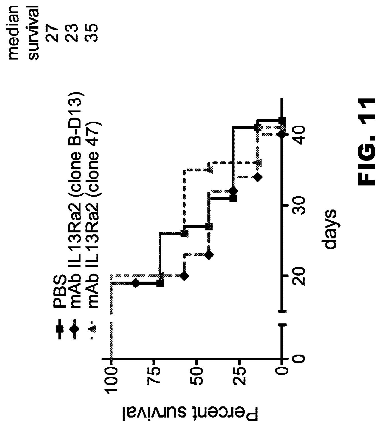
D00013
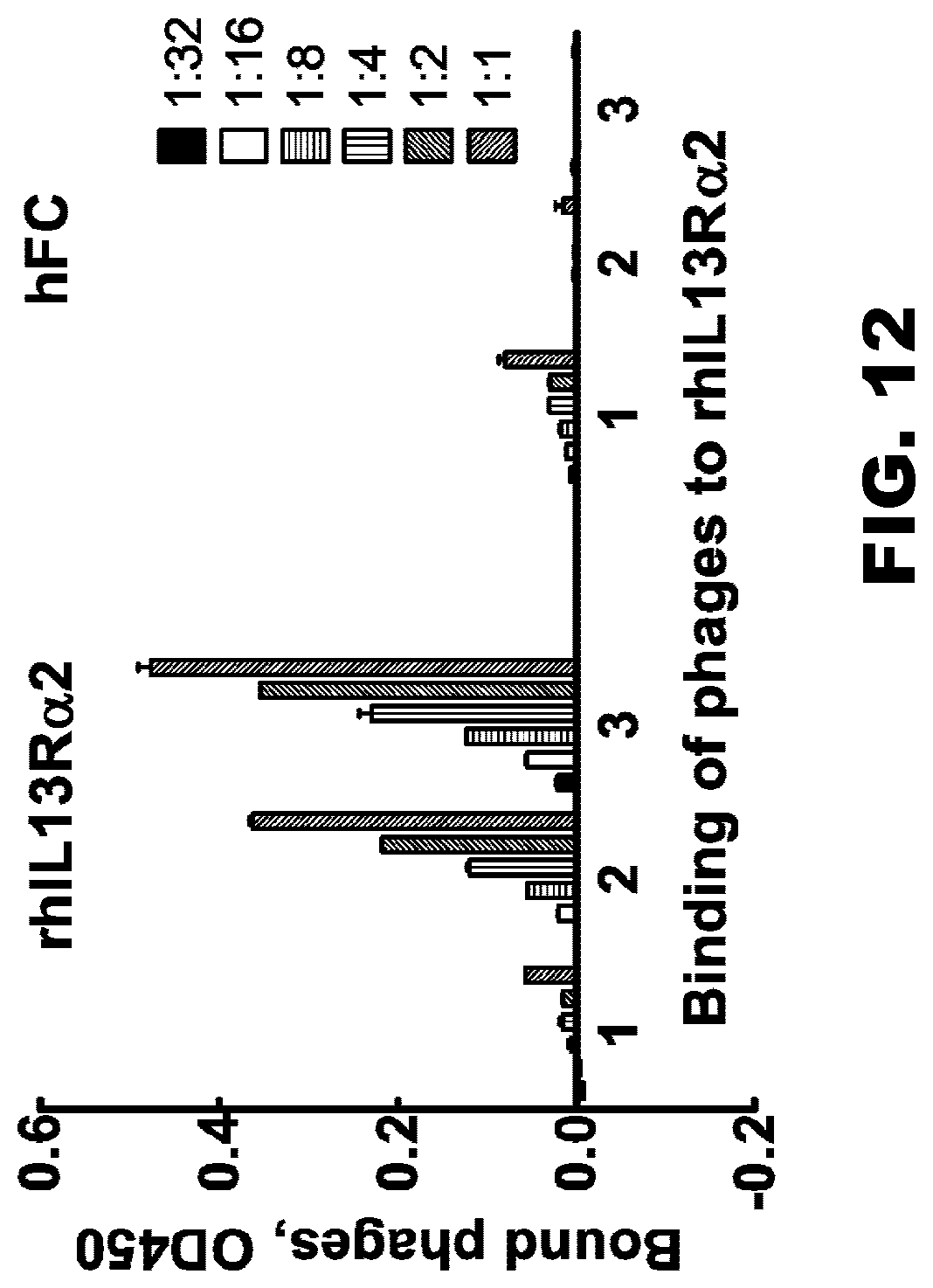
D00014
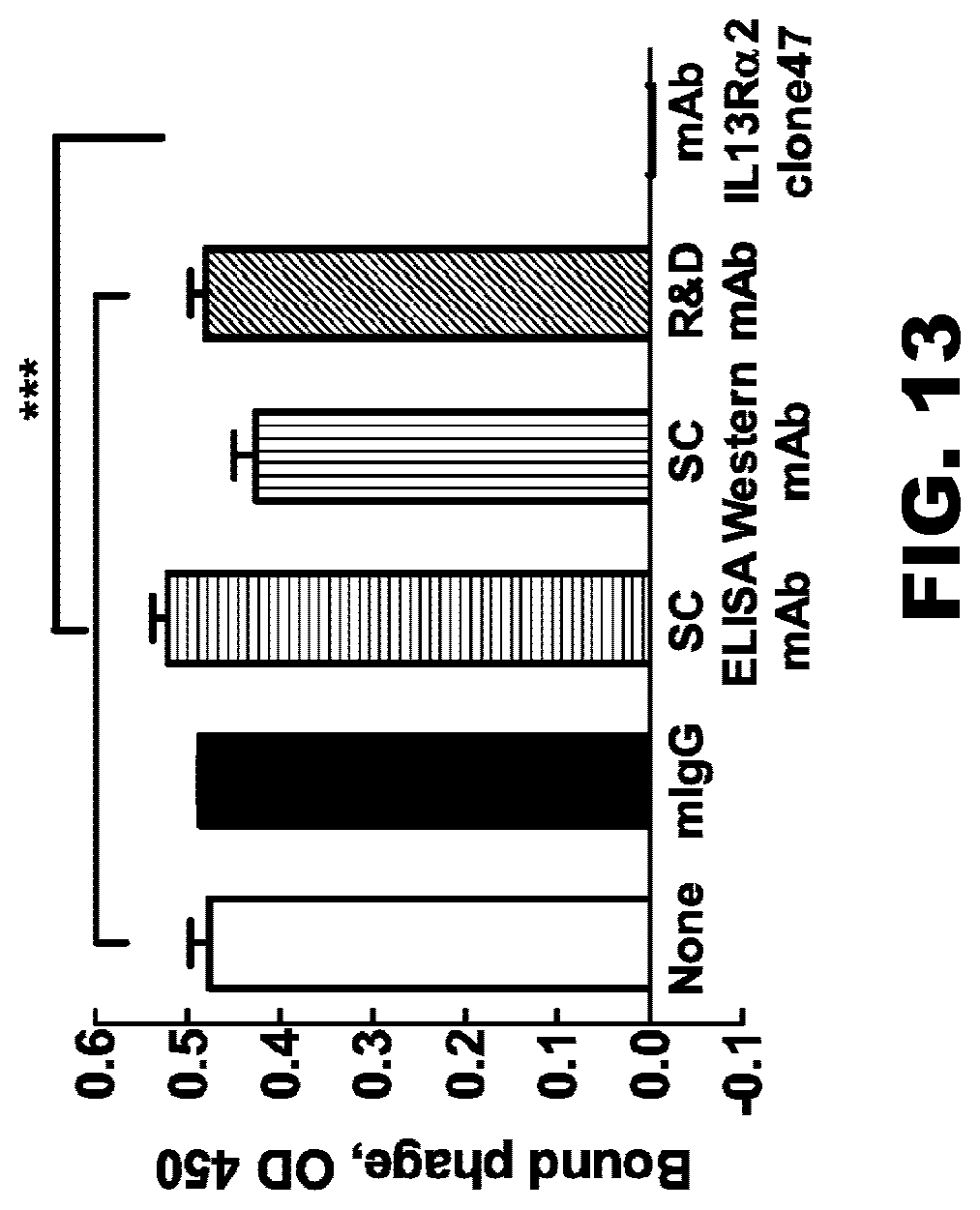
D00015
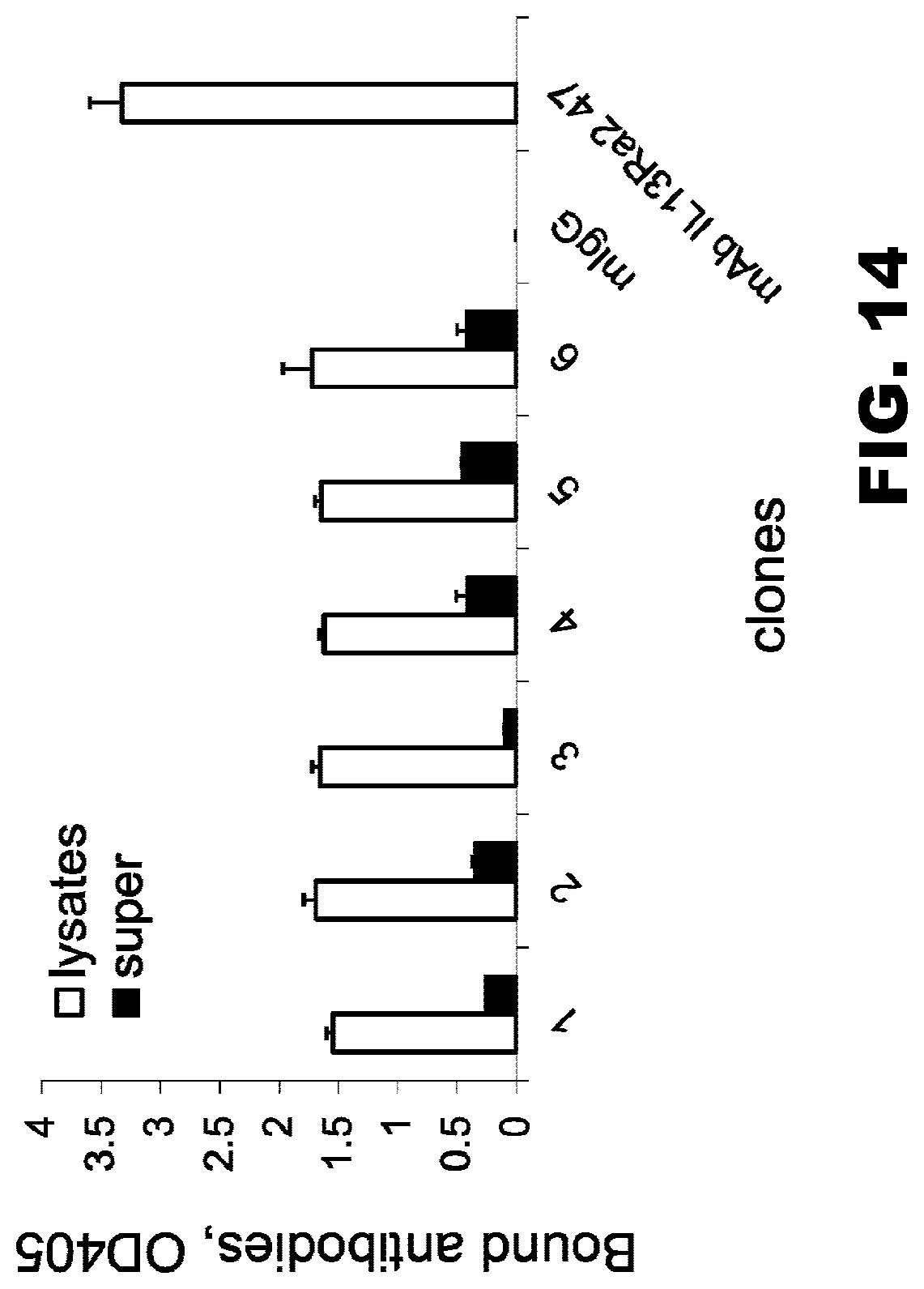
D00016
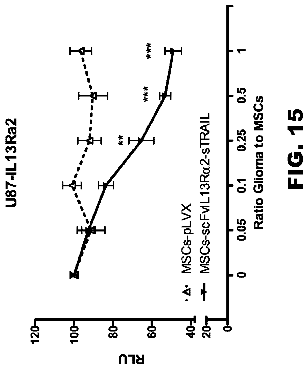
D00017
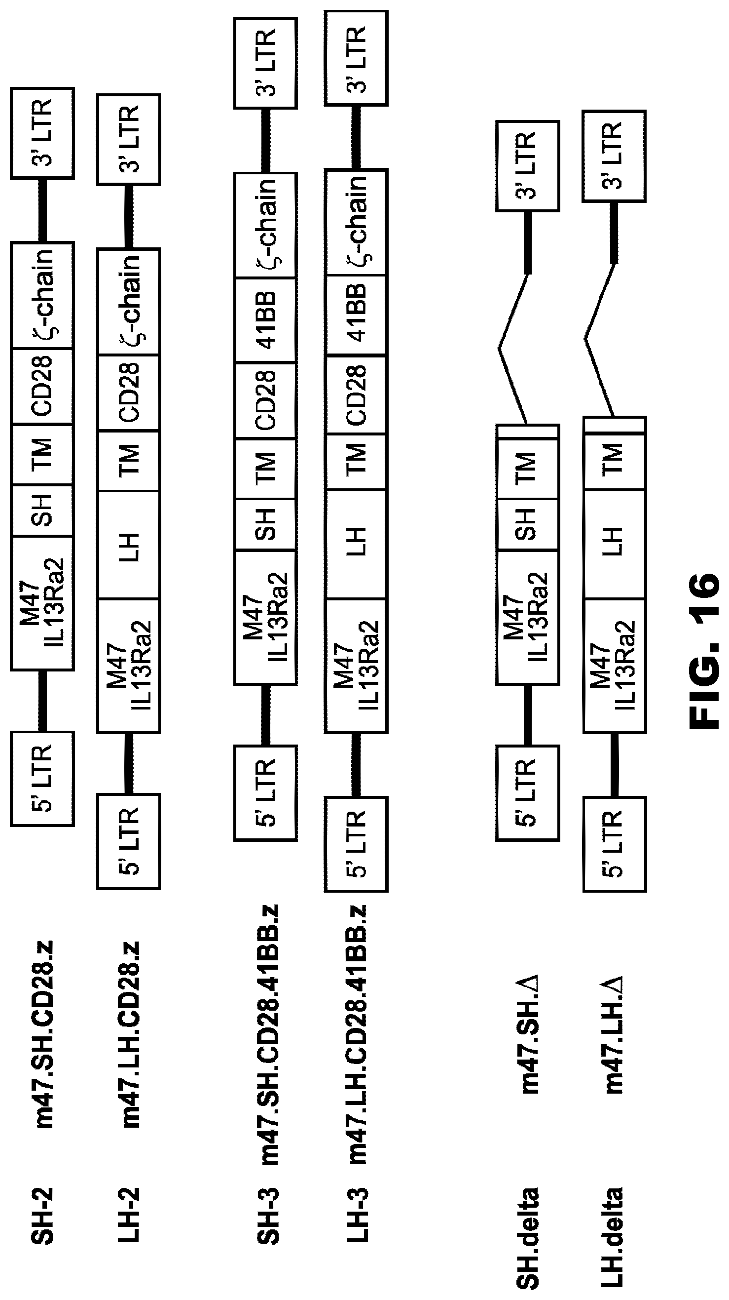
D00018
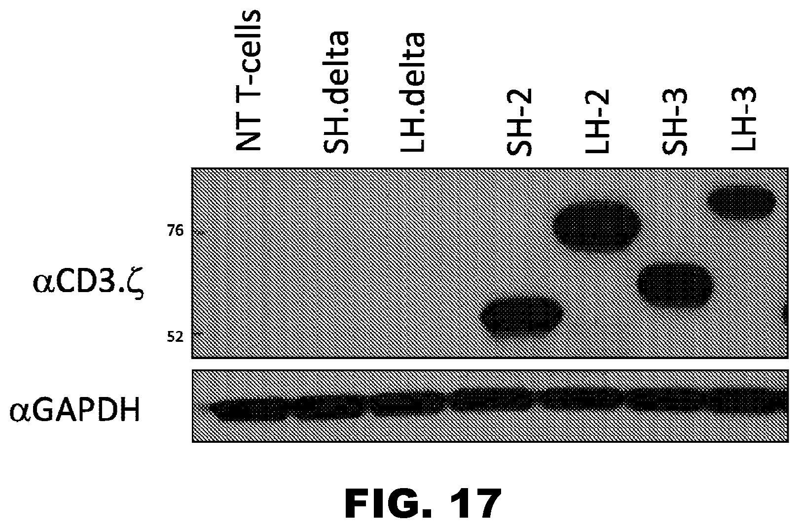
D00019
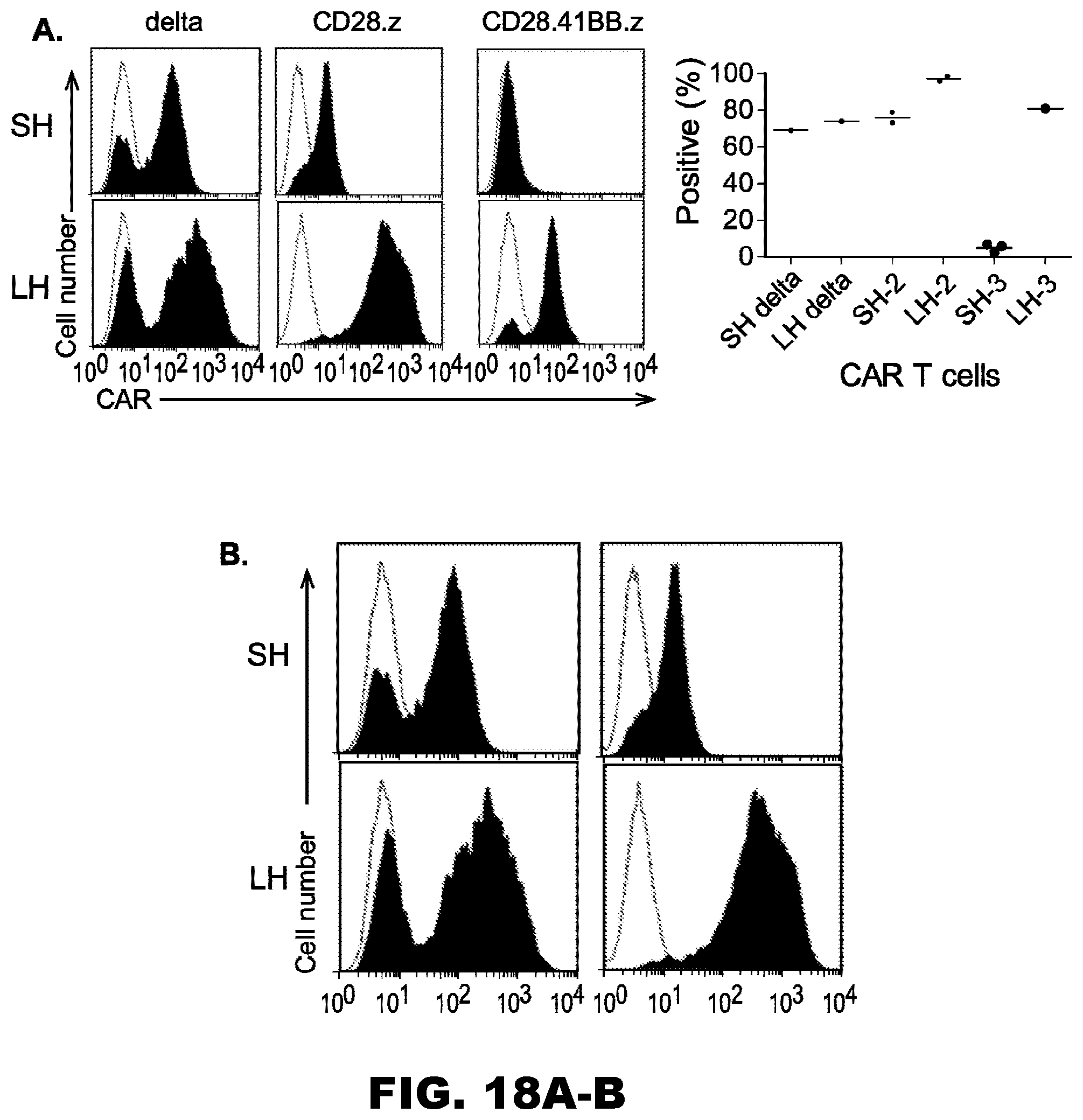
D00020
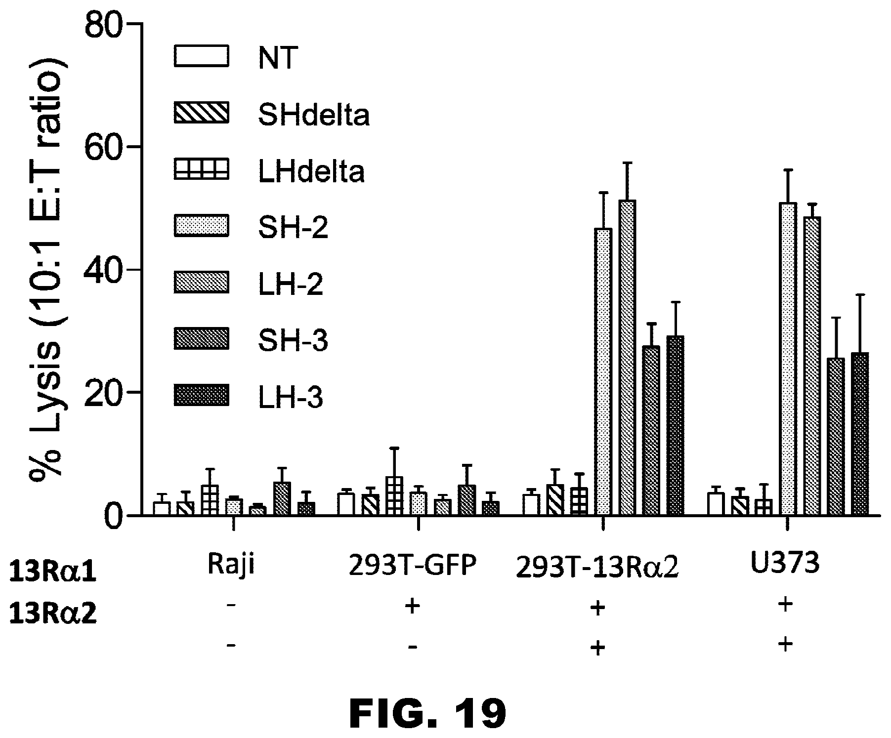
D00021
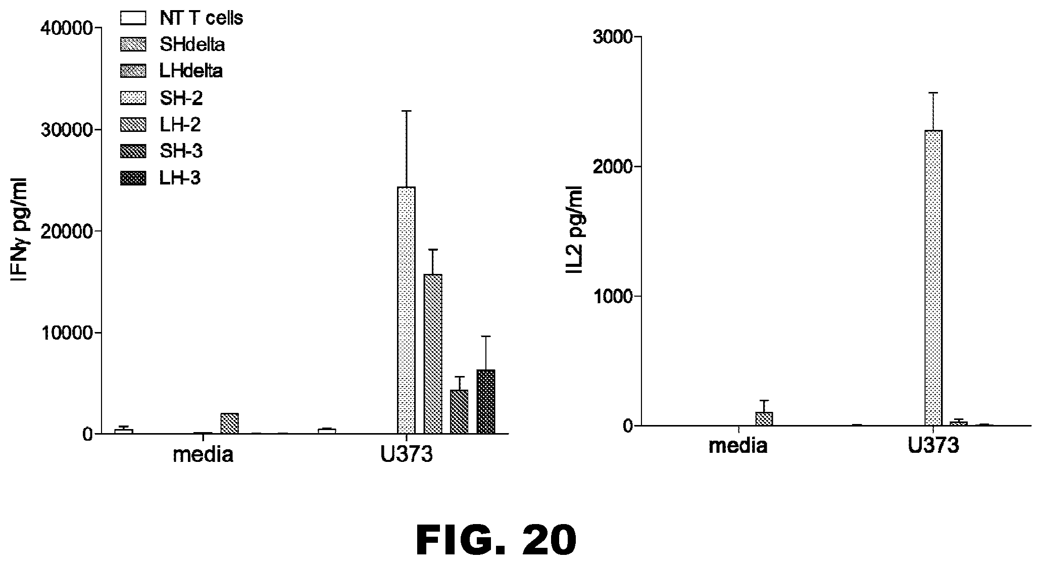
D00022
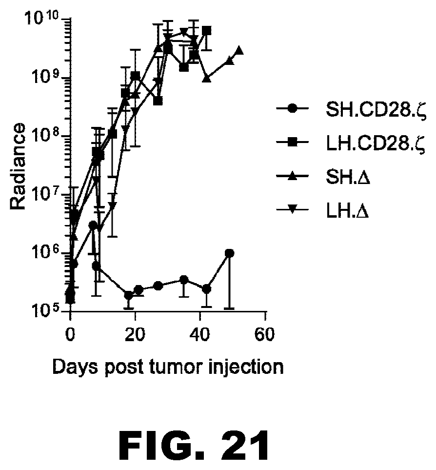
D00023

D00024

D00025
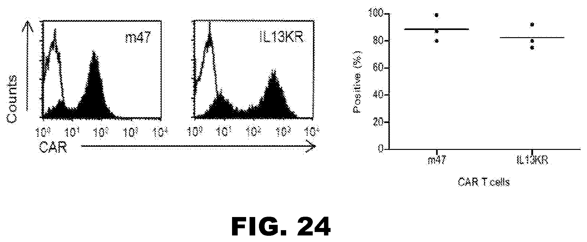
D00026

D00027
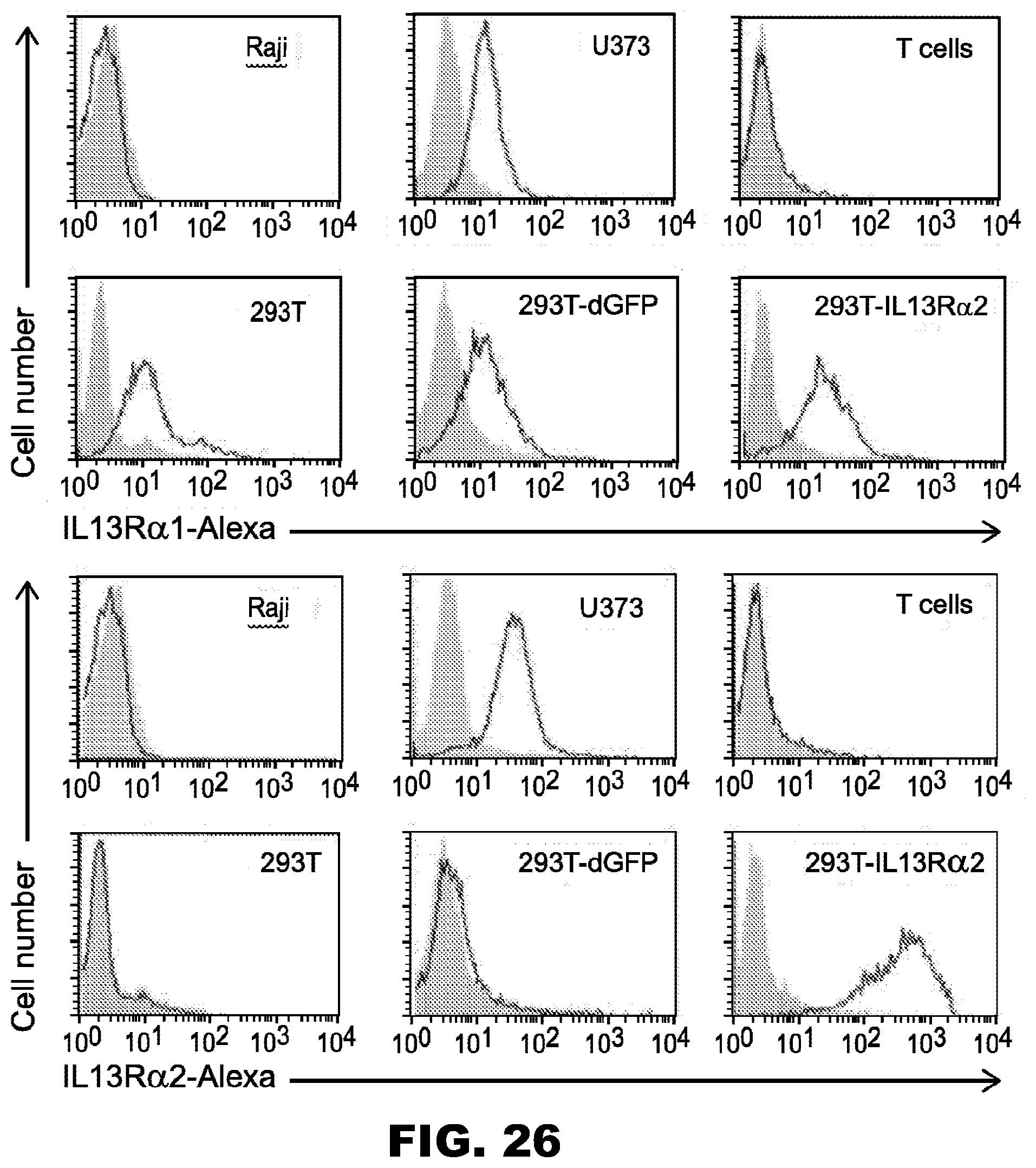
D00028
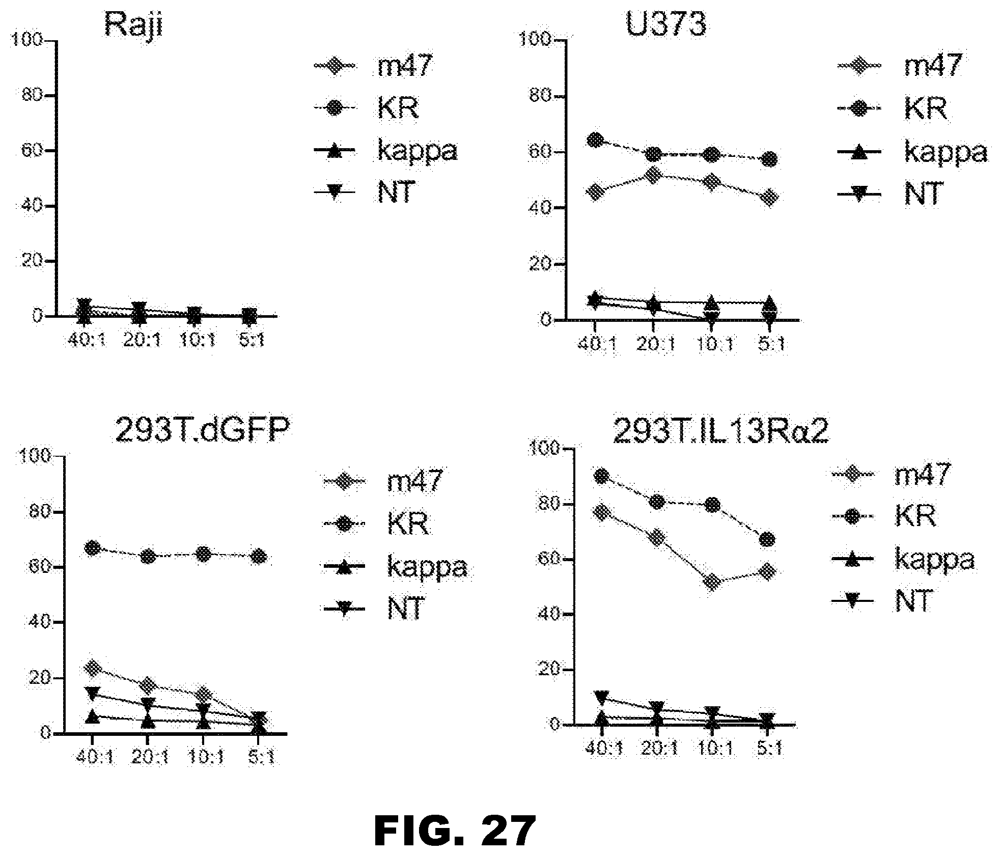
D00029
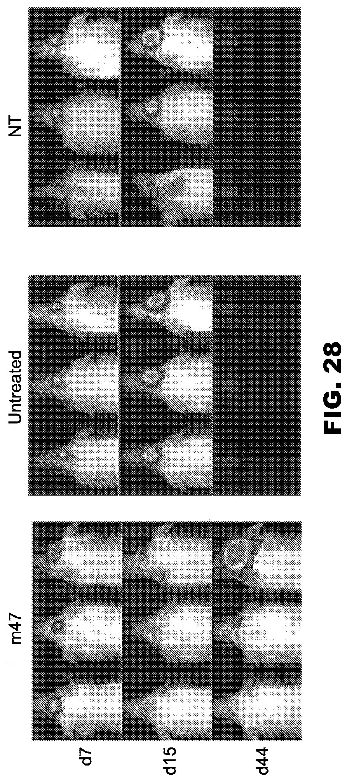
D00030
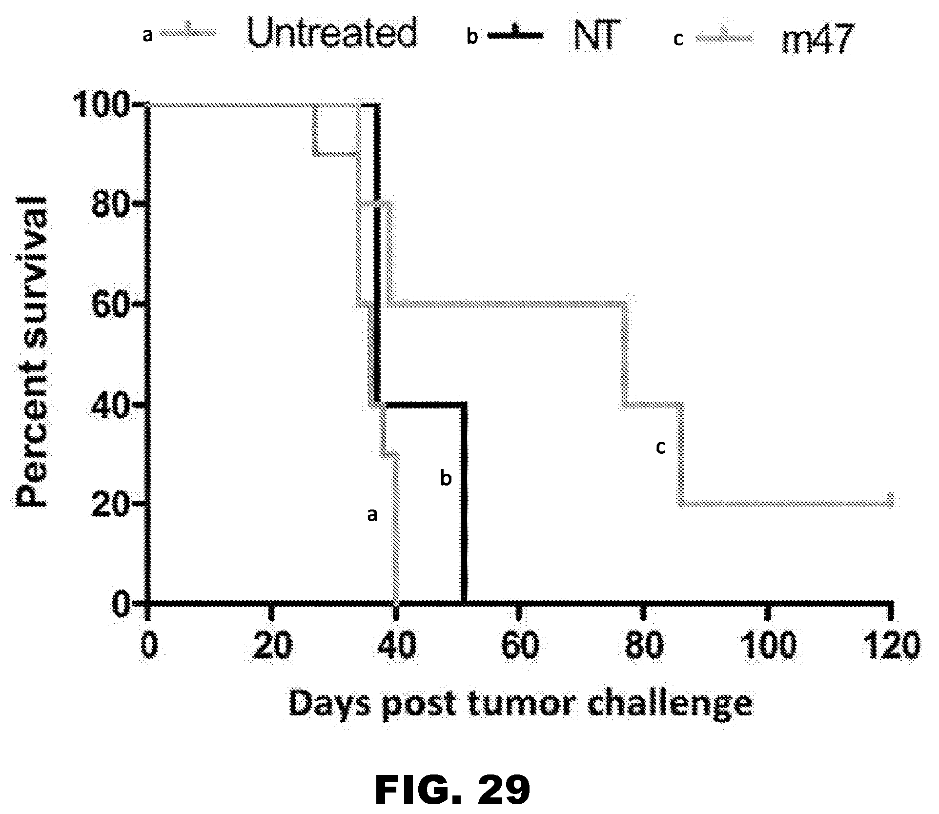
D00031
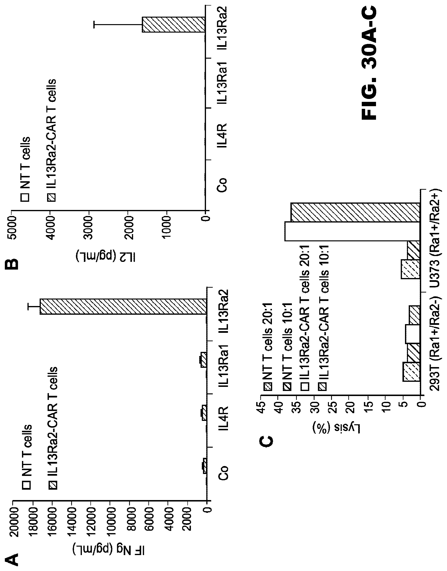
D00032
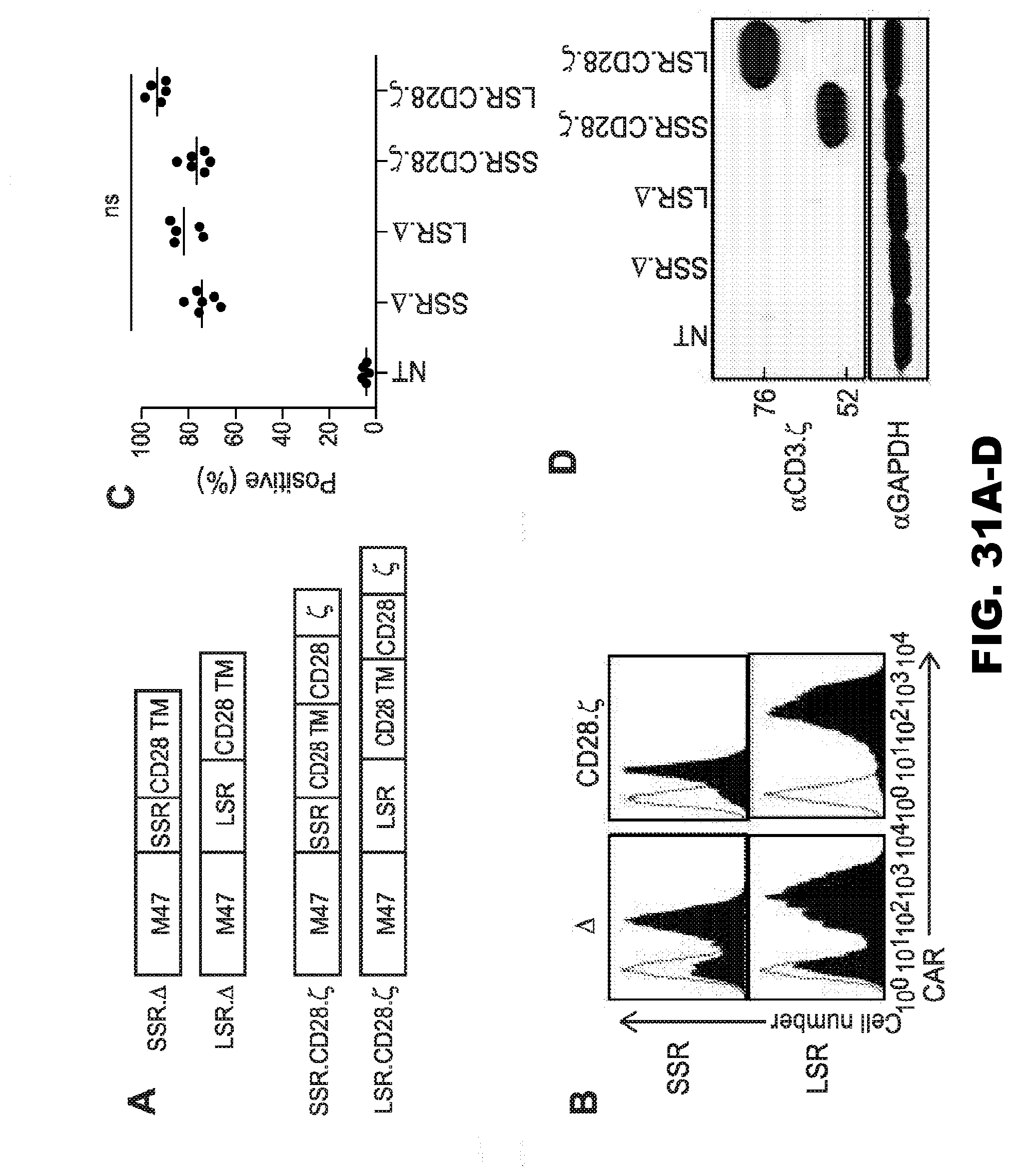
D00033
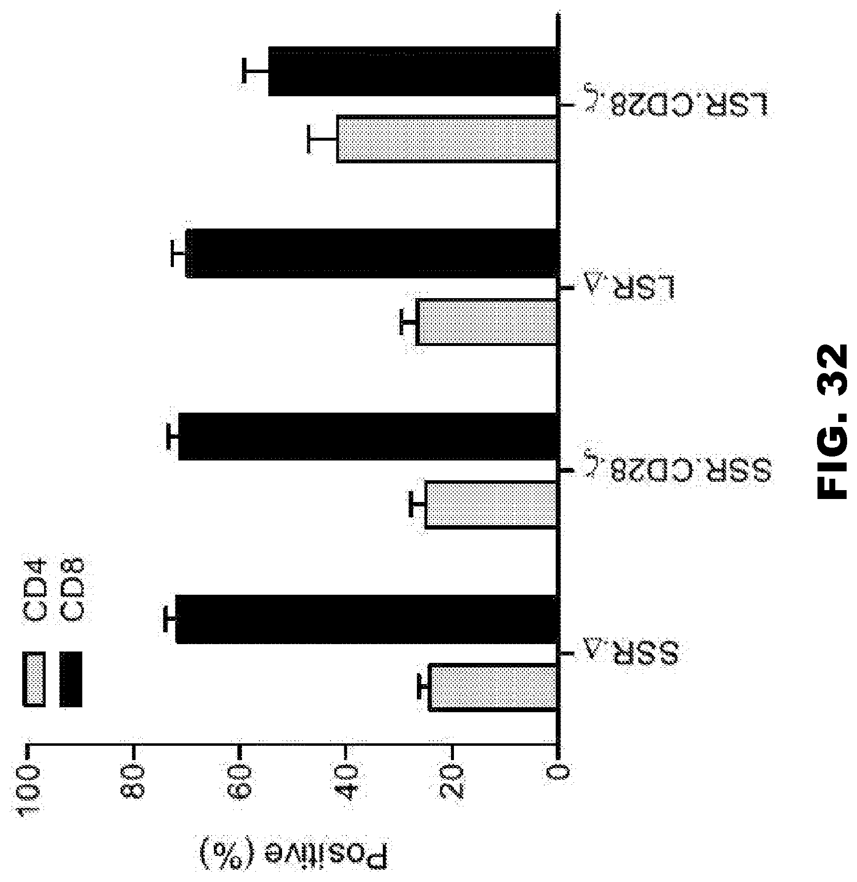
D00034

D00035
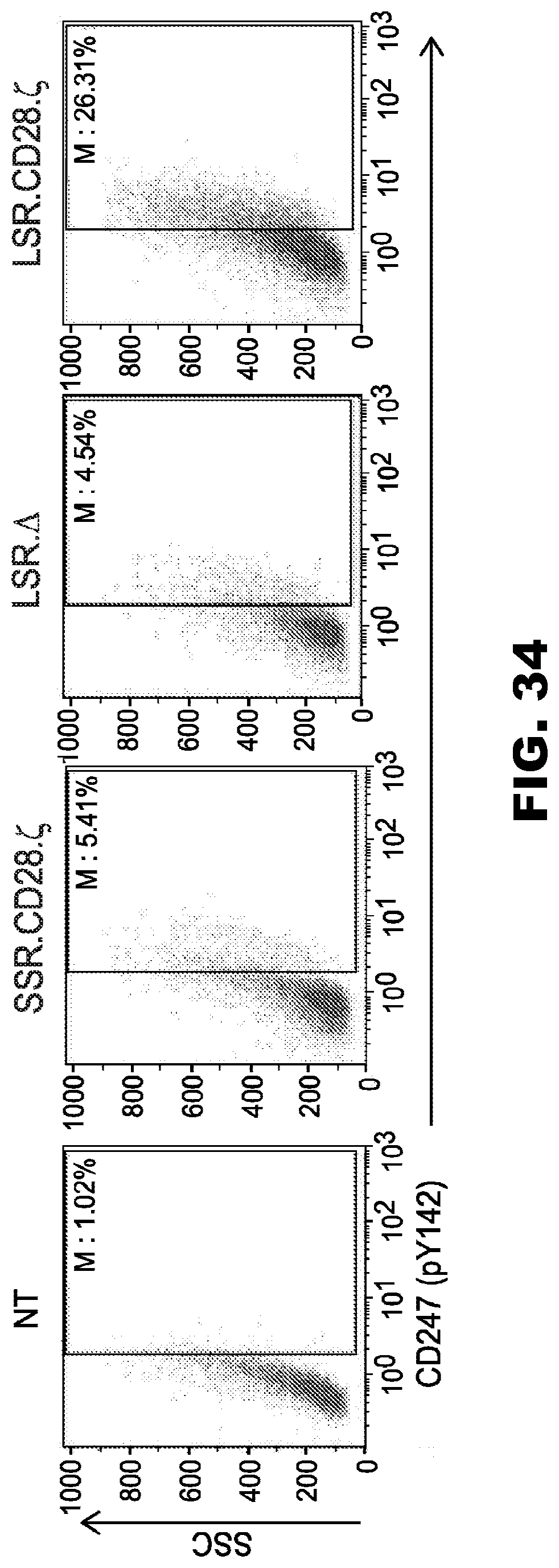
D00036
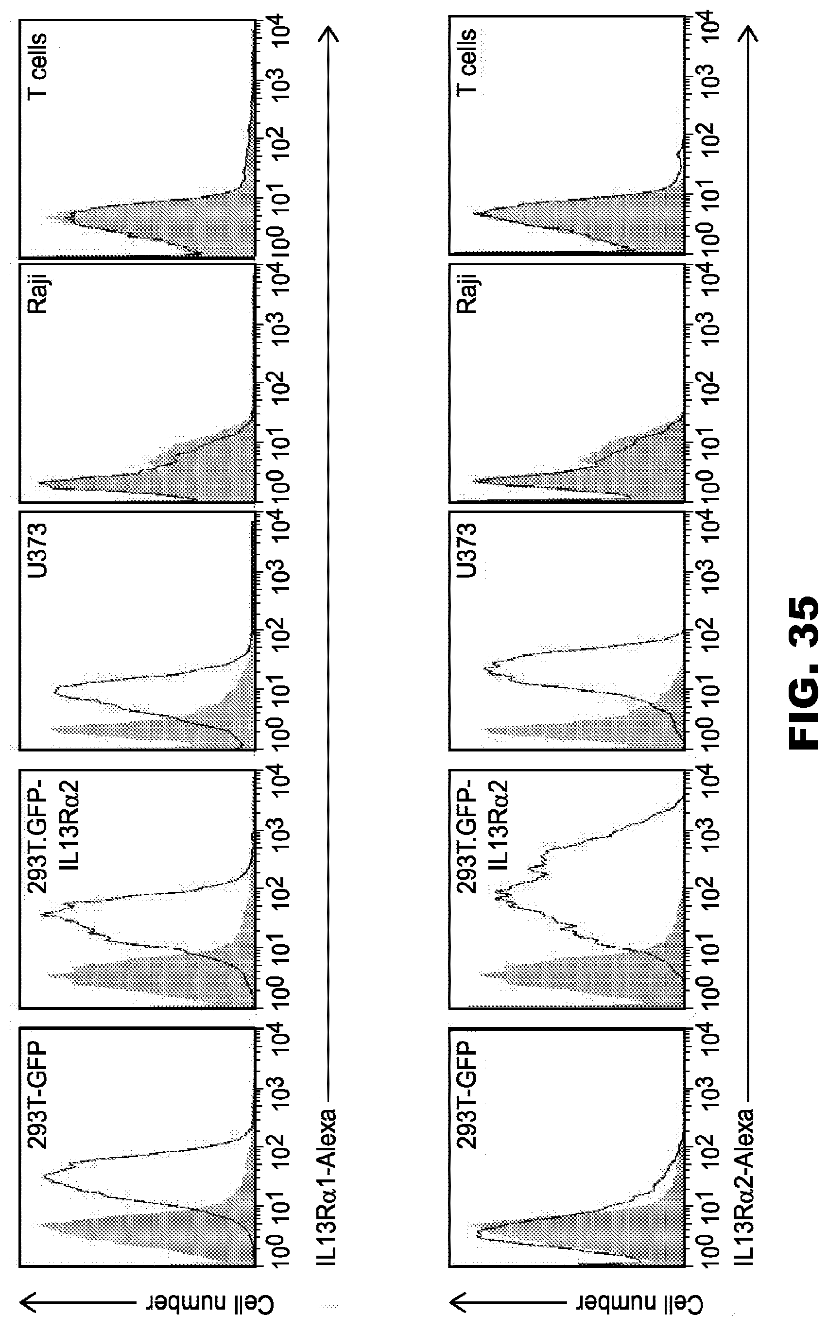
D00037
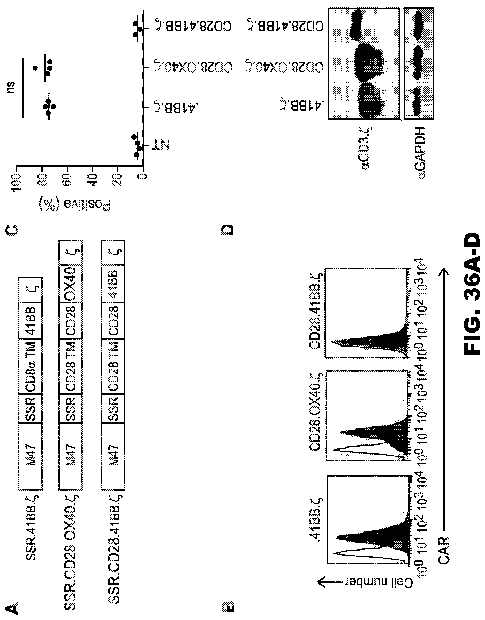
D00038
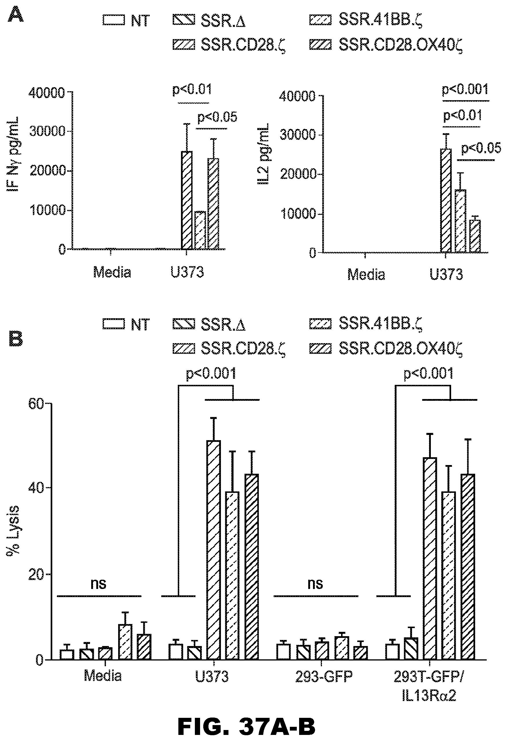
D00039
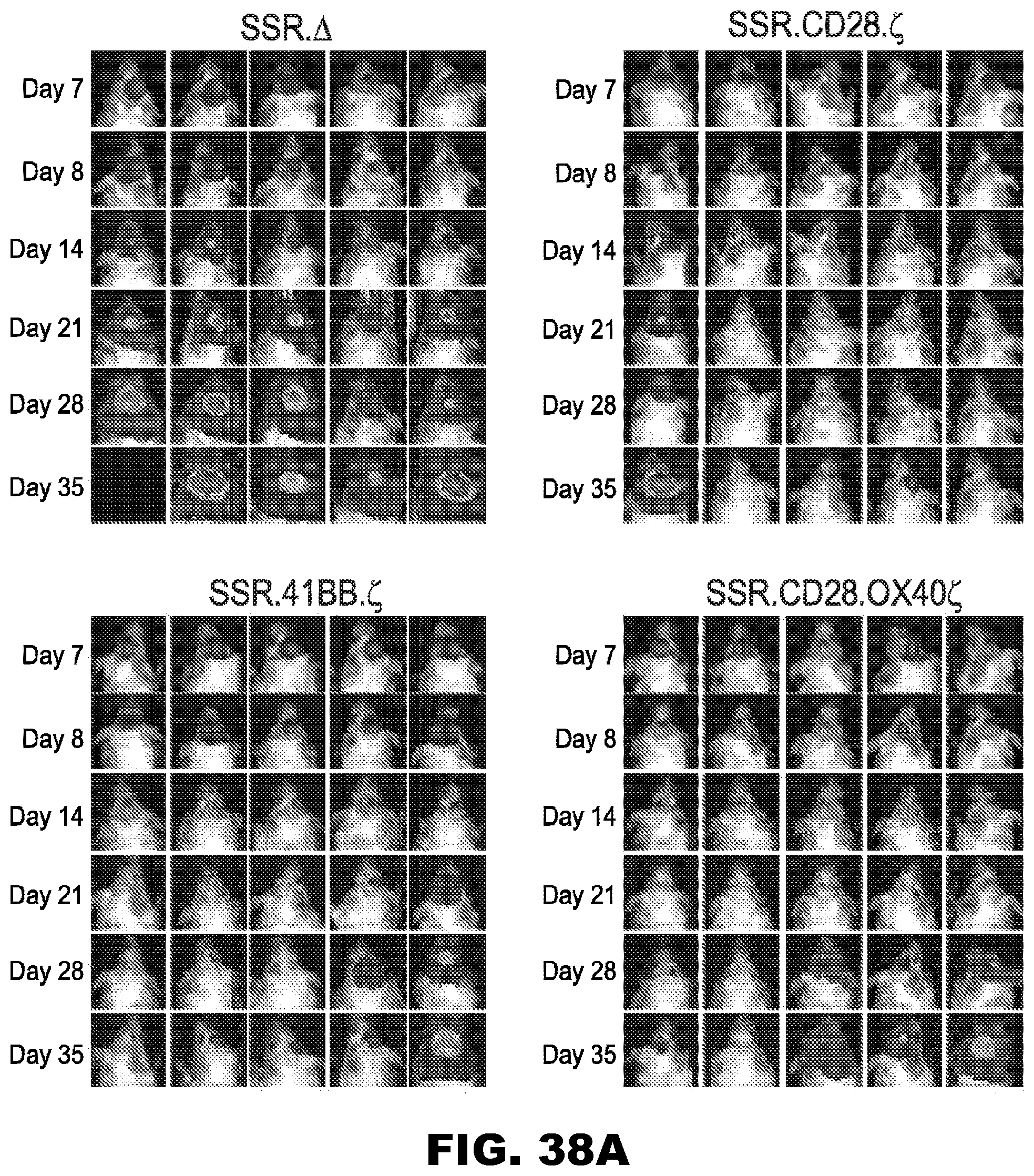
D00040
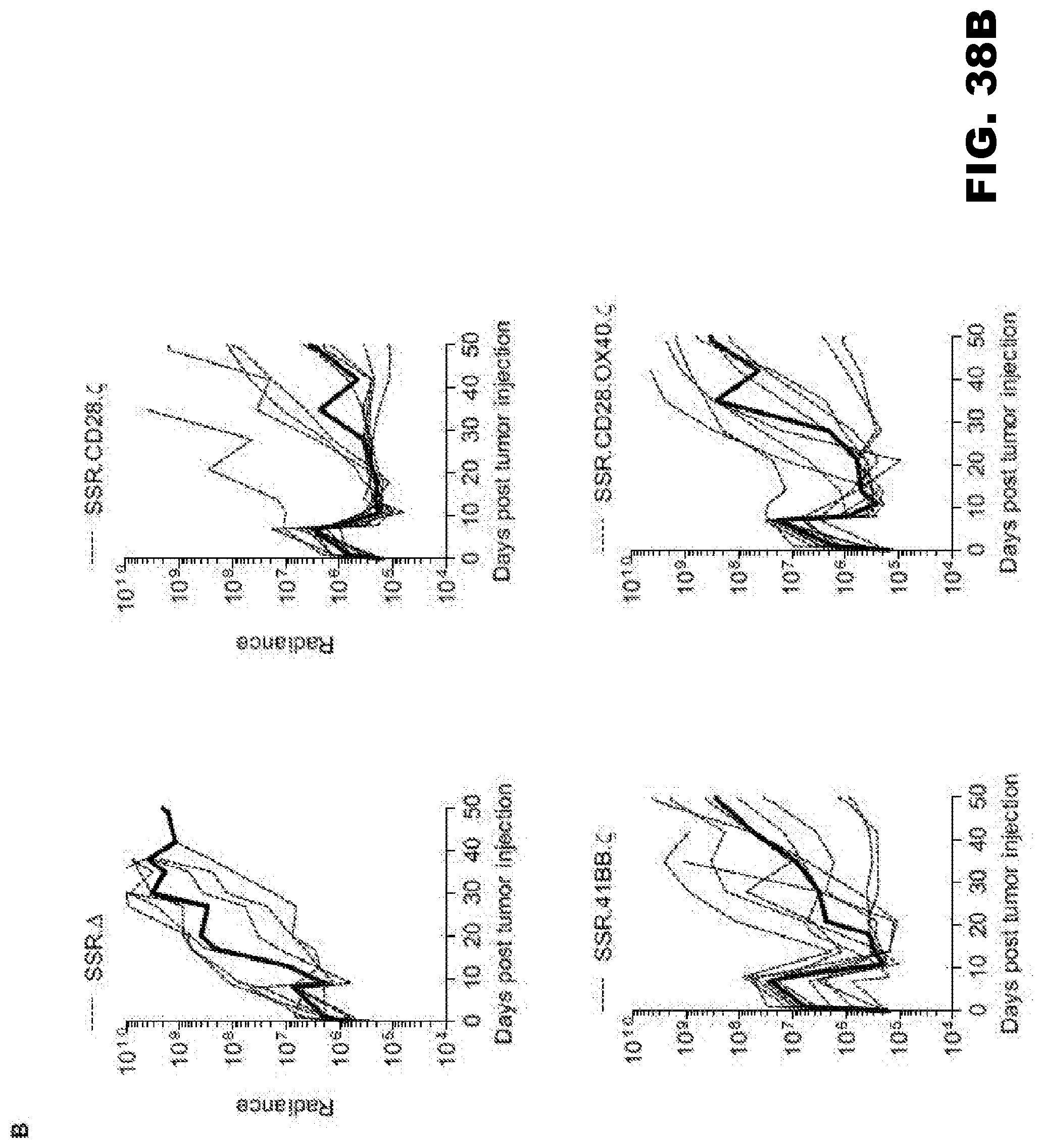
D00041
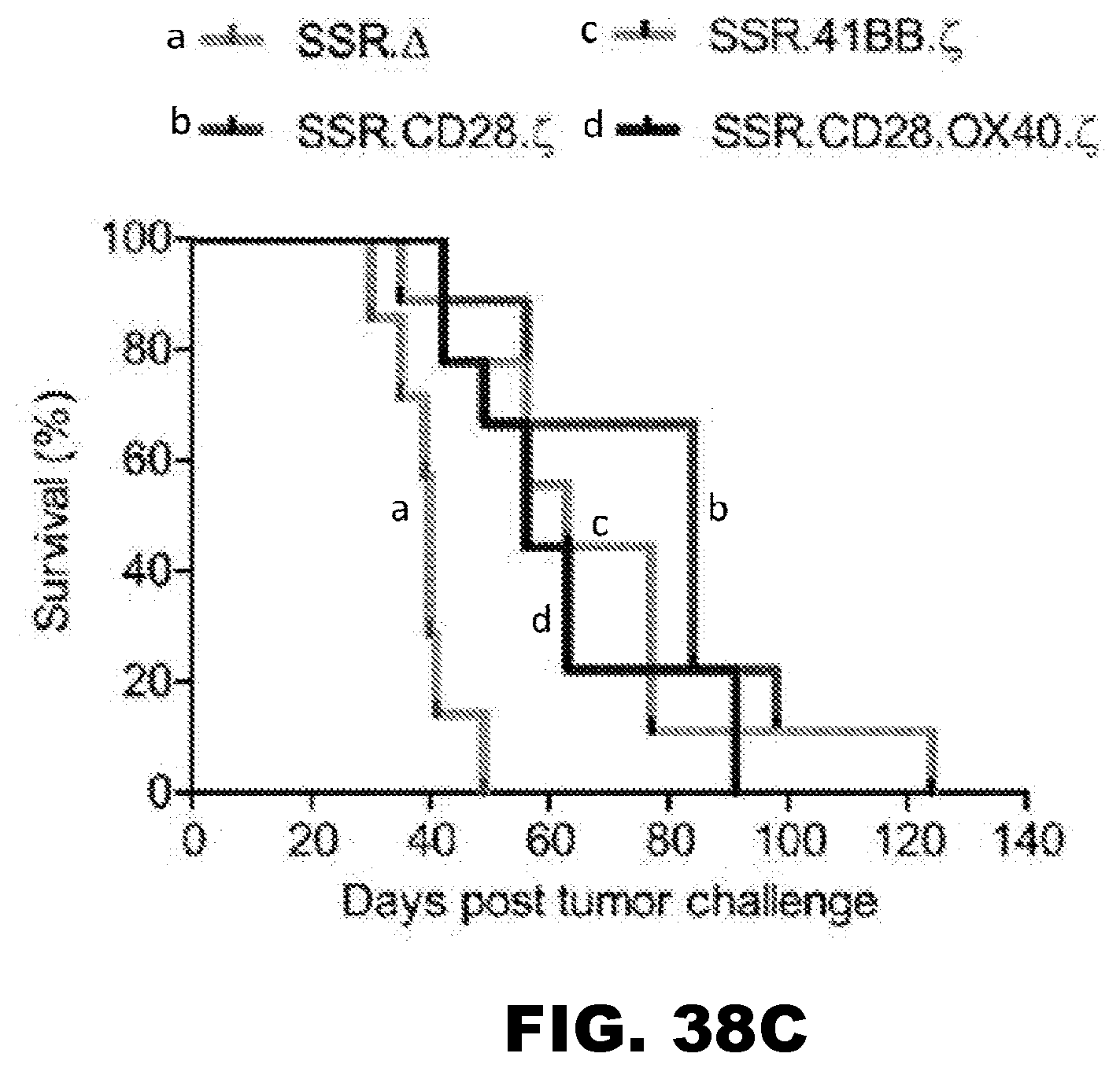
D00042
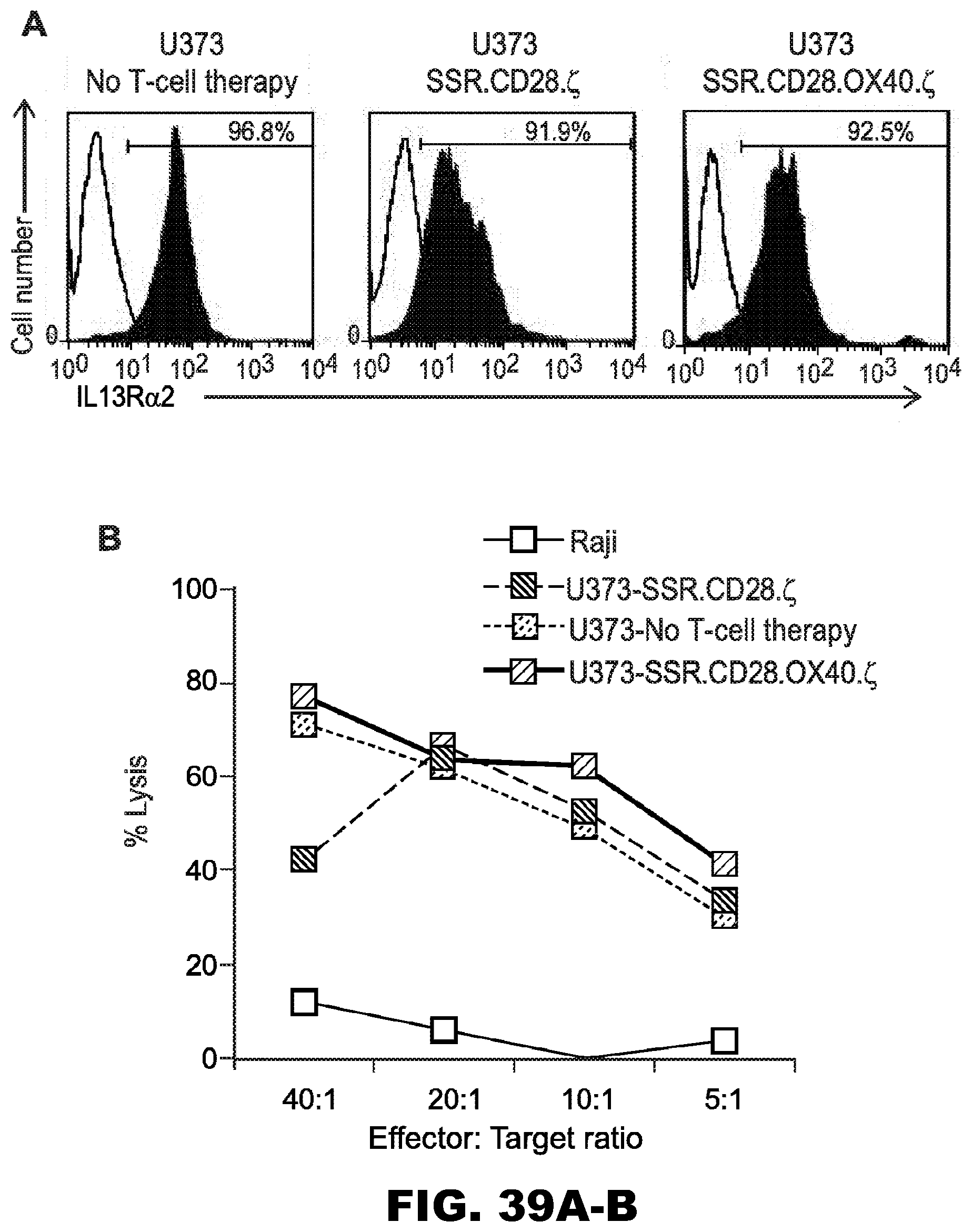
D00043
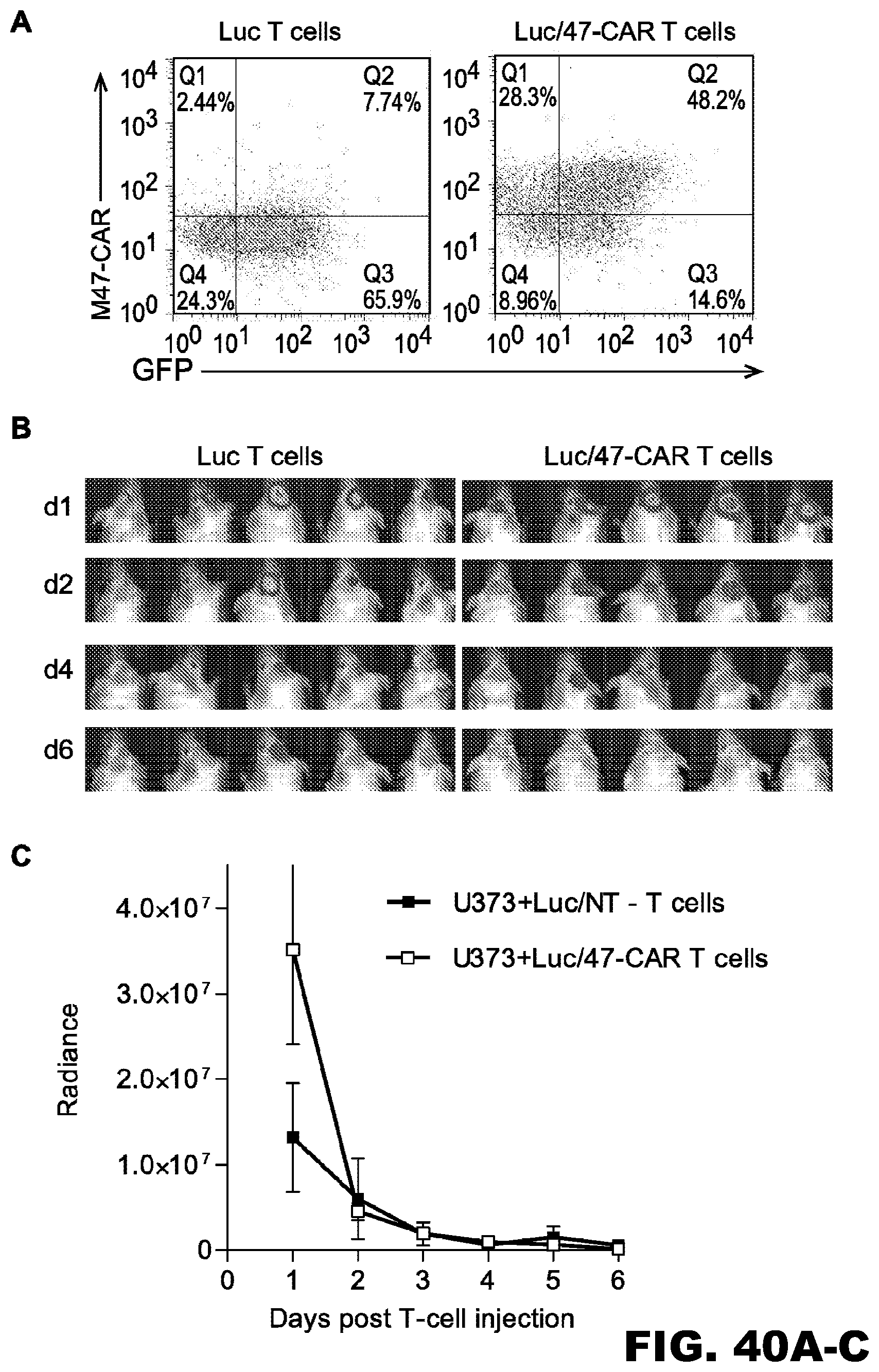
D00044

D00045
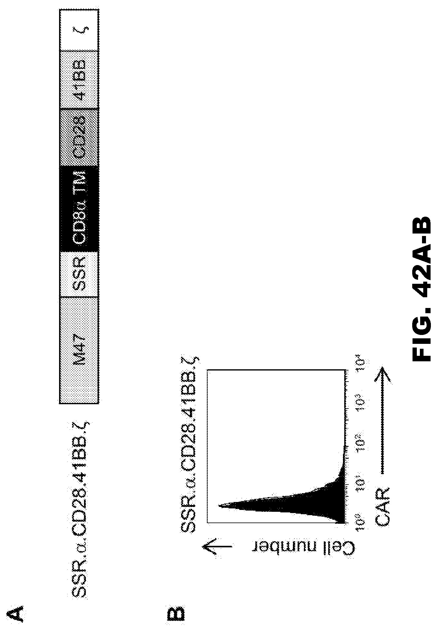
D00046
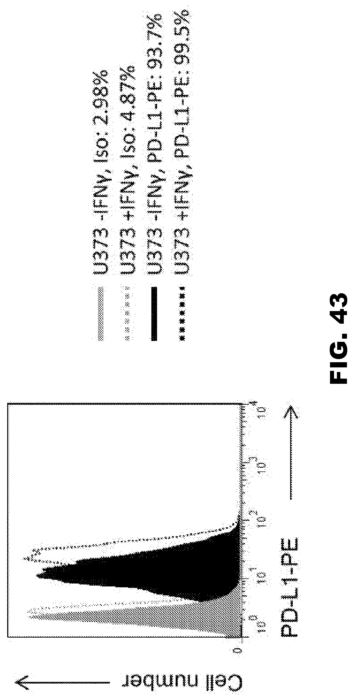
D00047

D00048
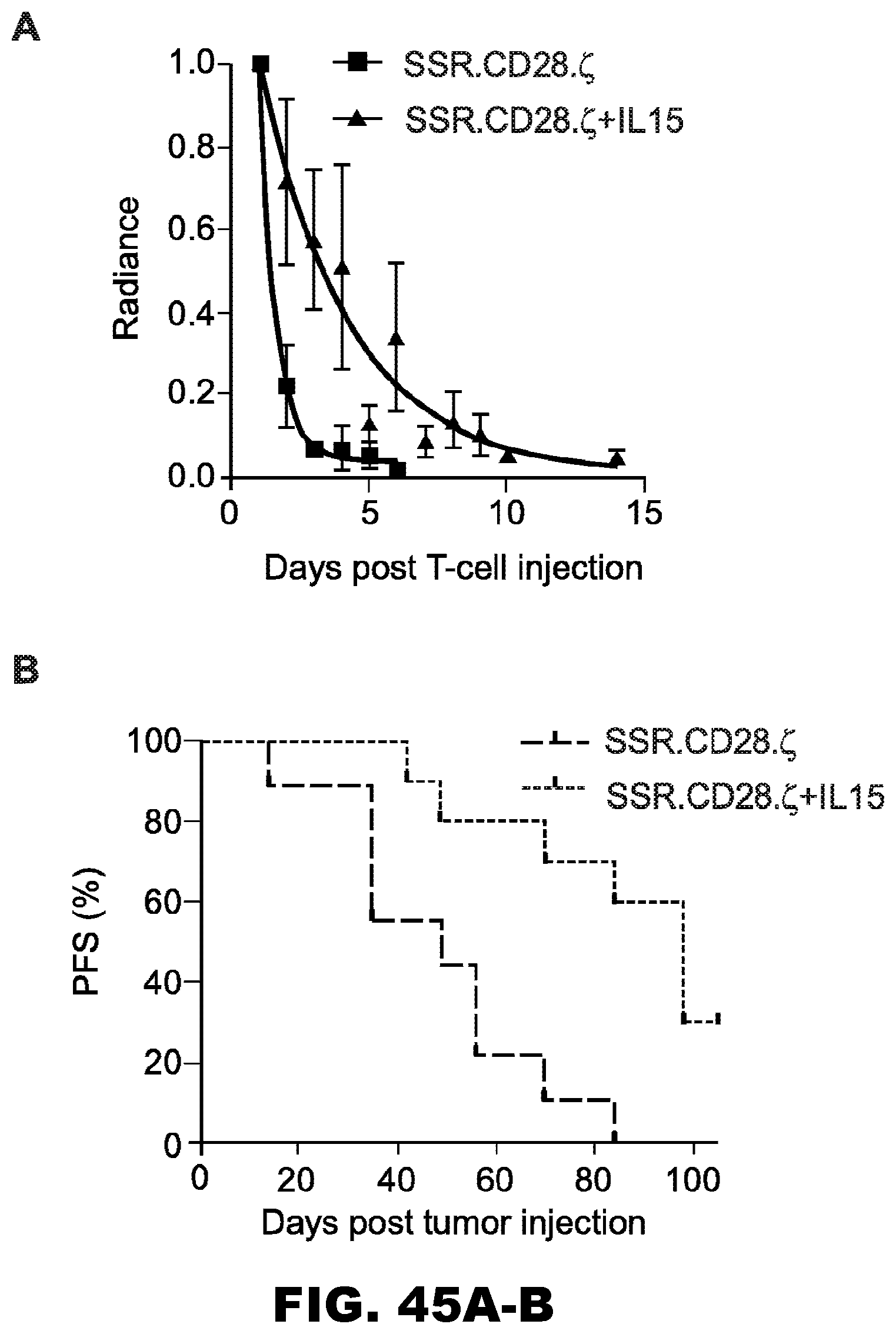
S00001
XML
uspto.report is an independent third-party trademark research tool that is not affiliated, endorsed, or sponsored by the United States Patent and Trademark Office (USPTO) or any other governmental organization. The information provided by uspto.report is based on publicly available data at the time of writing and is intended for informational purposes only.
While we strive to provide accurate and up-to-date information, we do not guarantee the accuracy, completeness, reliability, or suitability of the information displayed on this site. The use of this site is at your own risk. Any reliance you place on such information is therefore strictly at your own risk.
All official trademark data, including owner information, should be verified by visiting the official USPTO website at www.uspto.gov. This site is not intended to replace professional legal advice and should not be used as a substitute for consulting with a legal professional who is knowledgeable about trademark law.