Methods For Treatment Or Prevention Of A Neurological Immunity Disorder
Kipnis; Jonathan ; et al.
U.S. patent application number 16/631991 was filed with the patent office on 2020-06-04 for methods for treatment or prevention of a neurological immunity disorder. The applicant listed for this patent is University of Virginia Patent Foundation. Invention is credited to Jonathan Kipnis, Antoine Louveau.
| Application Number | 20200172624 16/631991 |
| Document ID | / |
| Family ID | 65016224 |
| Filed Date | 2020-06-04 |


View All Diagrams
| United States Patent Application | 20200172624 |
| Kind Code | A1 |
| Kipnis; Jonathan ; et al. | June 4, 2020 |
METHODS FOR TREATMENT OR PREVENTION OF A NEUROLOGICAL IMMUNITY DISORDER
Abstract
Methods of treating, preventing, inhibiting, delaying the onset of, or ameliorating a neurological immunity disorder can include administering an effective amount of a compound comprising an antibody or antigen binding fragment of an antibody to a subject in need of treatment, prevention, inhibition, delay of onset, or amelioration of a neurological immunity disorder. The antibody or the antigen binding fragment of an antibody binds specifically to CD49a.
| Inventors: | Kipnis; Jonathan; (Charlottesville, VA) ; Louveau; Antoine; (Charlottesville, VA) | ||||||||||
| Applicant: |
|
||||||||||
|---|---|---|---|---|---|---|---|---|---|---|---|
| Family ID: | 65016224 | ||||||||||
| Appl. No.: | 16/631991 | ||||||||||
| Filed: | July 19, 2018 | ||||||||||
| PCT Filed: | July 19, 2018 | ||||||||||
| PCT NO: | PCT/US18/42955 | ||||||||||
| 371 Date: | January 17, 2020 |
Related U.S. Patent Documents
| Application Number | Filing Date | Patent Number | ||
|---|---|---|---|---|
| 62534895 | Jul 20, 2017 | |||
| Current U.S. Class: | 1/1 |
| Current CPC Class: | A61P 25/28 20180101; A61K 9/0019 20130101; A61K 9/0085 20130101; C07K 16/2842 20130101; A61K 2039/505 20130101 |
| International Class: | C07K 16/28 20060101 C07K016/28; A61P 25/28 20060101 A61P025/28 |
Claims
1. A method of treating, or preventing, inhibiting, delaying the onset of, or ameliorating a neurological immunity disorder a neurological immunity disorder or a symptom thereof in an animal subject, comprising administering to the subject a therapeutically effective amount of a compound that inhibits integrin signaling.
2. The method of claim 1, wherein the animal subject is a human.
3. The method of claim 1, wherein the compound decreases CD49a function.
4. The method of claim 1, wherein said compound that inhibits integrin signaling is an antibody or an antigen binding fragment which binds to CD49a.
5. The method of claim 4, wherein said antibody or said antigen binding fragment is a monoclonal antibody, such as a human or humanized antibody.
6. The method of claim 1, wherein said compound that inhibits integrin signaling is an antibody or an antigen binding fragment which specifically binds CD49a.
7. The method of claim 6, wherein said antibody or said antigen binding fragment is a monoclonal antibody.
8. The method of claim 1, further comprising identifying a subject in need of said treatment.
9. The method of claim 8, wherein the subject in need of said treatment is susceptible to or suffering from a neurological immunity disorder selected from the group consisting of autism spectrum disorder (ASD), multiple sclerosis (MS), and central nervous system (CNS) injury.
10. The method of any one of claim 1, wherein the administration of the compound is via intracerebroventricular injection, or wherein administration of the compound is via dermal application to the scalp skin of said subject.
11. The method of claim 1, wherein the administration of said compound results in accumulation of immune cells in the brain meninges.
12. The method of claim 1, wherein the administration of said compound results in elevated T cells and natural killer T (NKT) cells in the brain parenchyma.
13. The method of claim 1, wherein the neurological immunity disorder is treated or prevented.
14. The method of claim 6, wherein antibody or antigen binding fragment does not specifically bind to any of CD49b, CD49c, CD49d, CD49e, and/or CD49f.
15. The method of claim 1, wherein the compound that inhibits integrin signaling is administered after the onset of the neurological immunity disorder.
16. The method of claim 15, wherein the administration of the compound after the onset of the neurological immunity disorder reduces clinical symptoms of the neurological immunity disorder.
17. A method of treating, preventing, inhibiting, delaying the onset of, or ameliorating multiple sclerosis (MS) or a symptom thereof in a human subject, comprising administering to the subject a therapeutically effective amount of a CD49c-specific-antibody.
18. The method of claim 17, further comprising identifying a subject in need of said treatment.
19. The method of claim 17, wherein the administration of the CD49a-specific antibody is via intracerebroventricular injection.
20. The method of claim 17, wherein administration of the CD49a-specific blocking antibody is via dermal application to the scalp skin of said subject.
21. The method of claim 17, wherein the CD49a blocking antibody is administered after the onset of the neurological immunity disorder.
22. The method of claim 21, wherein the administration of the CD49a blocking antibody after the onset of the neurological immunity disorder reduces clinical symptoms of the neurological immunity disorder
23. The method of claim 18, wherein the MS is treated.
24. The method of claim 7, wherein the antibody is a human antibody or a humanized antibody.
Description
INCORPORATION BY REFERENCE OF PRIORITY APPLICATION
[0001] This Application claims the benefit of U.S. Provisional App. No. 62/534895, filed Jul. 20, 2017, entitled "Methods for treatment or prevention of a neurological immunity disorder," which is incorporated by reference in its entirety herein.
REFERENCE TO SEQUENCE LISTING,
[0002] The present application is being filed along with a Sequence Listing in electronic format. The Sequence Listing is provided as a file entitled SEQEUNCEUVA007WO.TXT, created and last saved Jul. 12, 2018, which is 10,092 bytes in size. The information in the electronic format of the Sequence Listing is incorporated herein by reference in its entirety.
BACKGROUND
[0003] The central nervous system (CNS) and the immune system have very complex interactions that both control and modulate the function of each others.sup.1-6. Recent work emphasized the role of T cells in the regulation of cognition in mice.sup.7-9. Indeed, mice lacking a functional immune system, notably CD4 T cells, exhibit impaired performance of cognitive tasks. This impairment is rescued by injection of CD4 T cells back into immune deficient mice.sup.7. Under normal conditions, T cells are virtually absent from the brain parenchyma but are enriched in the surrounding of the brain called the meninges.sup.5,8, notably around the major blood vessels in the dura mater, the sinuses.sup.10. It was previously unclear how T cells, localized in the meninges, are able to affect brain function.
[0004] Multiple sclerosis (MS) is characterized by the destruction of the CNS myelin and is considered to be an autoimmune disease. MS results in physical, mental, and/or psychiatric problems. Symptoms may include double vision, muscle weakness, trouble with sensation, or trouble with coordination. There is currently no cure for MS.
[0005] Alzheimer's disease (AD) is a type of dementia that is associated with memory loss, and problems with thinking and behavior. The parenchymal accumulation of neurotoxic amyloid beta (A.beta.) is a central hallmark of AD. There is currently no cure for AD and treatments are limited to reducing and/or slowing the progression of the symptoms.
[0006] Autism spectrum disorder (ASD) is a neurodevelopmental disorder characterized by impaired social interaction, verbal and non-verbal communication, and restricted and repetitive behavior. There is currently no cure for ASD. There is a need in the field for methods of treatment for neurological immunity disorders, including but not limited to MS, AD and ASD. The present disclosure addresses this need.
FIELD
[0007] Embodiments herein relate to methods for treating, preventing, inhibiting or ameliorating a neurological immunity disorder, or a symptom thereof.
SUMMARY
[0008] In some embodiments, the present application provides methods of treating, preventing, inhibiting, delaying the onset of, or ameliorating a neurological immunity disorder or a symptom thereof in an animal subject. The method can comprise administering to the subject a therapeutically effective amount of a compound that inhibits (or blocks) integrin signaling. In some embodiments, methods of treating, preventing, inhibiting, delaying the onset of, or ameliorating a neurological immunity disorder or a symptom thereof in an animal subject are described. In some embodiments, the method comprises administering to the subject a therapeutically effective amount of a compound that decreases or inhibits CD49a function, for example by binding specifically to CD49a. In some embodiments, the method comprises administering to the subject a therapeutically effective amount of an antibody or antigen binding fragment which binds CD49a. In some embodiments, the compound that inhibits integrin signaling is administered after the onset of the neurological immunity disorder, for example at least about 8 days after the onset of the neurological immunity disorder, for example at least 5, 6, 7, 8, 9, 10, 11, 12, 13, 14, 15, 16, 17, 18, 19, 20, 21, 22, 23, 24, 25, 26, 27, or 28 days, including any range between any two of the listed values, for example, including but not limited to the following ranges which are provided for exemplary purposes only: 5-28 days, 5-21 days, 5-14 days, 5-7 days, 7-28 days, 7-21 days, 7-14 days, 10-28 days, 10-21 days, or 10-14 days. In some embodiments, the administration of the compound after the onset of the neurological immunity disorder reduces clinical symptoms of the neurological immunity disorder, which can be measured, for example, by a clinical score. In some embodiments, the compound that inhibits integrin signaling comprises, consists essentially of, or consists of a CD49a inhibiting (or blocking) antibody.
[0009] In some embodiments, the subject is a human The compound can decrease CD49a function. In some embodiments, the compound comprises, consists of, or consists essentially of an antibody that binds specifically to CD49a, or an antigen binding fragment thereof. In some embodiments, the antibody or antigen binding fragment is a monoclonal antibody. In some embodiments, the antibody or antigen binding fragment is a human antibody. In some embodiments, the antibody or antigen binding fragment is a humanized antibody. In some embodiments, the antibody or antigen binding fragment is a chimeric antibody. In some embodiments, the compound that inhibits integrin signaling is an antibody or an antigen binding fragment which specifically binds CD49a. By "binds specifically to CD49a" it is understood that the antibody or antigen binding fragment binds preferentially to CD49a compared to other antigens, but there is no requirement that the antibody or antigen binding fragment bind with absolute specificity only to CD49a. In some embodiments, the antibody or antigen binding fragment binds specifically to CD49a compared to other integrins. In some embodiments, the antibody binds specifically to CD49a, and does not exhibit appreciable binding to any of CD49b, CD49c, CD49d, CD49e, and/or CD49f . Without being limited by theory, it is noted that CD49a-f represent the alpha 1 through 6 chains of beta 1 integrins, and as such, CD49a-f have different structures and CD49b-f are not expected to appreciably cross react with any antibody that binds specifically to CD49a. In some embodiments, the antibody does not bind specifically to any of CD49b, CD49c, CD49d, CD49e, and/or CD49f, including combinations of two or more of the listed molecules.
[0010] In some embodiments the method further comprises the step of identifying a subject in need of treatment. In certain embodiments the subject in need of treatment is susceptible to or suffering from a neurological immunity disorder selected from the group consisting of autism spectrum disorder (ASD), multiple sclerosis (MS), Alzheimer's disease (AD), and central nervous system (CNS) injury.
[0011] In some embodiments, administration of the compound (se.g., an antibody or antigen binding fragment specific for CD49a) is via intracerebroventricular injection. In other embodiments, an ointment comprises the compound and administration is via application of the ointment to the skin (scalp) of said subject. In some embodiments, the ointment comprises the compound and administration is via application of the ointment to the head of the subject, such as on the scalp.
[0012] In some embodiments, the administration of the compound (e.g., an antibody or antigen binding fragment specific for CD49a) results in accumulation of immune cells in the brain meninges. In particular embodiments, the administration of the compound results in elevated T cells and natural killer T (NKT) cells in the brain parenchyma.
[0013] In some embodiments, the present application provides a method of treating MS in a human subject, comprising administering to the subject a therapeutically effective amount of a CD49a inhibiting (or blocking) antibody or antigen binding fragment thereof. In particular embodiments, the method further comprises the step of identifying a subject in need of said treatment. In other embodiments, the administration of the CD49a inhibiting (or blocking) antibody is via intracerebroventricular injection. In still further embodiments, an ointment comprises said CD49a inhibiting (or blocking) antibody and the administration is via application of the ointment to the skin (scalp) of the subject. In some embodiments, an ointment comprises said CD49a inhibiting (or blocking) antibody and the administration is via application of the ointment to the head of the subject, such as on the scalp.
[0014] In some embodiments, the CD49a inhibiting (or blocking) antibody or antigen binding fragment thereof is administered after the onset of the neurological immunity disorder. In some embodiments, the administration of the CD49a inhibiting (or blocking) antibody or antigen binding fragment thereof after the onset of the neurological immunity disorder reduces clinical symptoms of the neurological immunity disorder, which can be measure, for example, by a clinical score.
BRIEF DESCRIPTION OF THE DRAWINGS
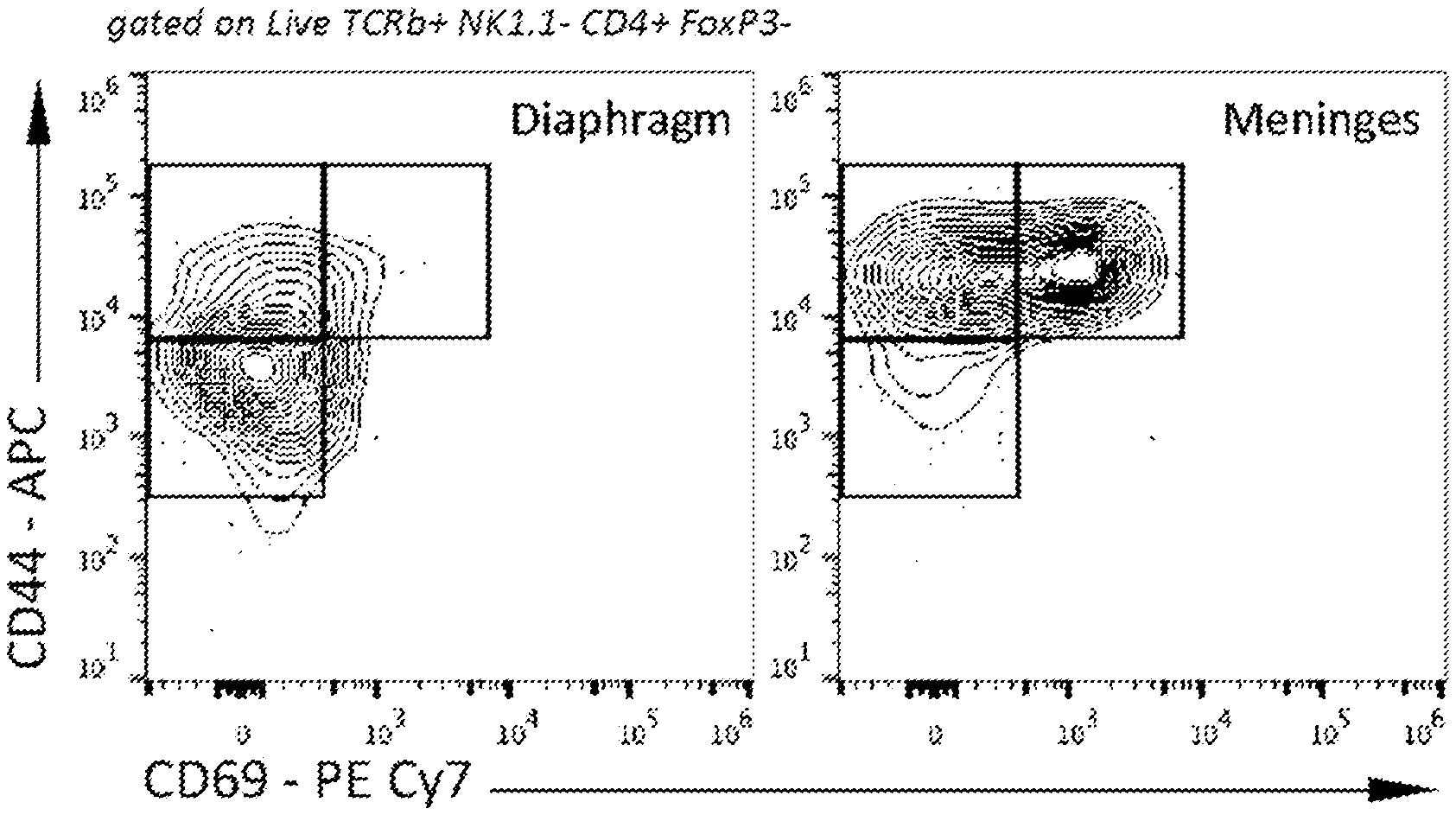
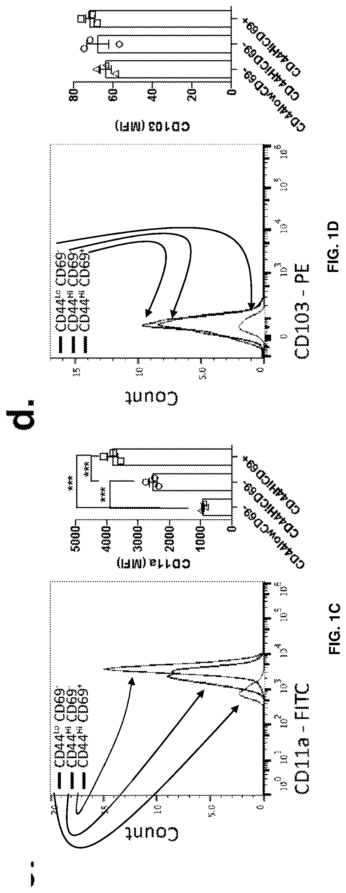
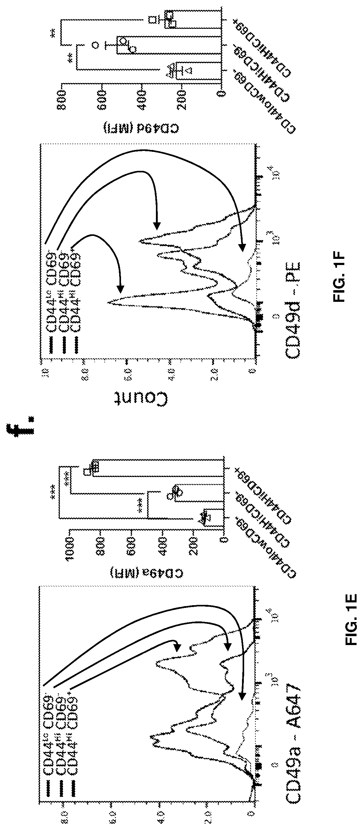
[0015] FIGS. 1A-1F show the presence of two main distinct populations of T cells in meninges of naive mice. FIG. 1A is a representative contour plot of the CD4 T cell populations in the diaphragm and meninges of naive mice. FIG. 1B is a quantification of the percentage of CD44.sup.HighCD69.sup.+, CD44.sup.HighCD69.sup.- and CD44.sup.-CD69.sup.- T cells in the diaphragm and meninges of naive mice. Contrary to the diaphragm, the meninges have two major populations of T cells that can be discriminated by the expression of CD69. FIG. 1C is a representative histogram and quantification of CD11a expression by the meningeal T cell populations. FIG. 1D is a representative histogram and quantification of CD103 expression by meningeal T cell populations. FIG. 1E is a representative histogram and quantification of CD49a expression by meningeal T cell populations. FIG. 1F is a representative histogram and quantification of CD49s expression by meningeal T cell populations. Mean+/-SEM, N=3 mice per group. ***p<0.001, One-way ANOVA with Bonferroni post test. The CD69+ CD4 T cell population also expresses high levels of CD49a and CD11a.
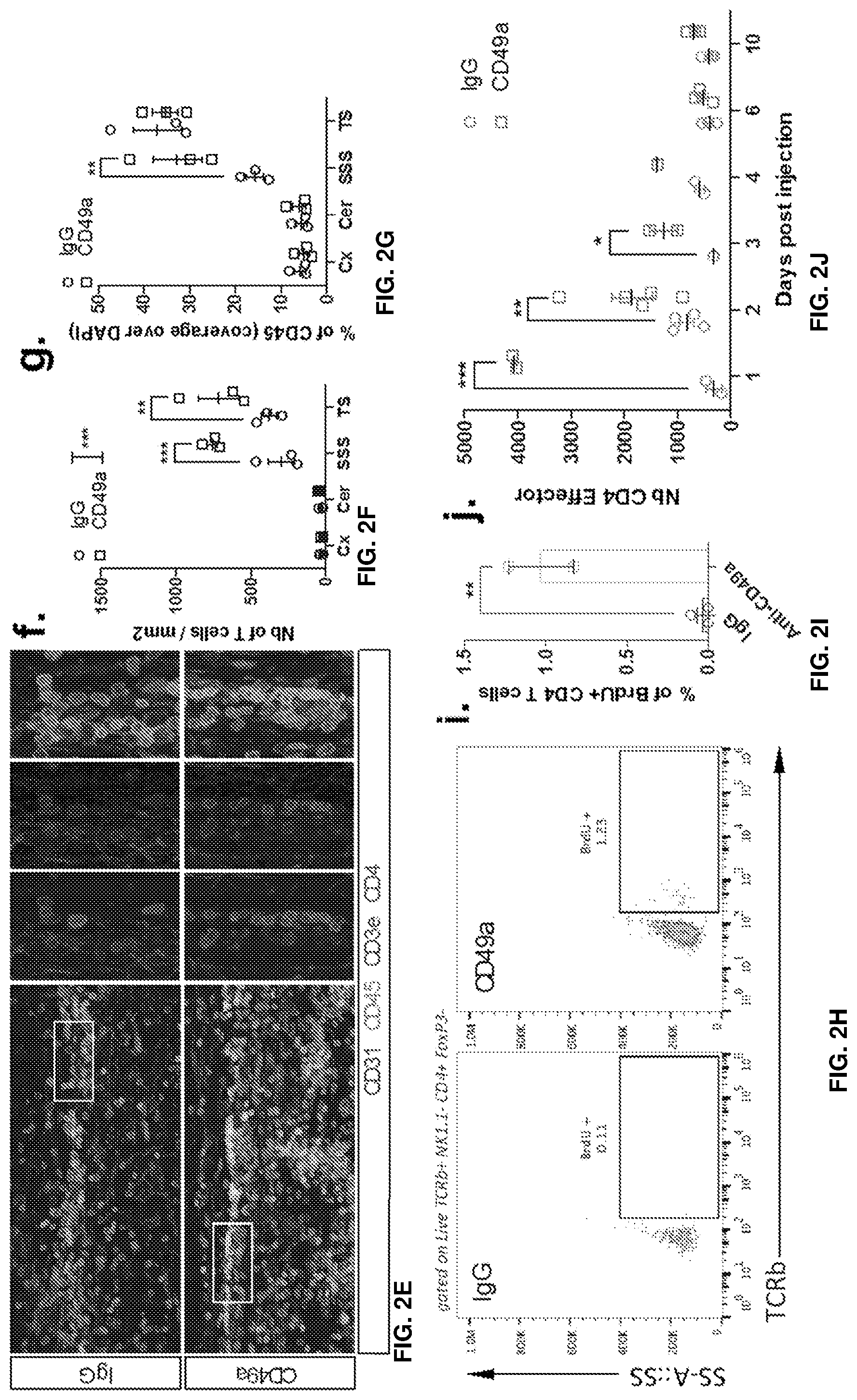
[0016] FIGS. 2A-2J show that blockade of CD49a induces the transient accumulation of immune cells in the meninges. FIG. 2A is a representative histogram of CD49a expression by the different meningeal immune cell populations. FIG. 2B is a quantification of the percentage of CD49a expressing cells within the different immune cell populations in naive meninges. CD49a is not only expressed by the meningeal T cells but also by several other immune cells like monocytes/macrophages, NK, and NKT cells. FIG. 2C is a set of representative dot plots of T cells, NK, and NKT cells in the meninges of mice after IgG or CD49a blocking antibody injection. FIG. 2D is a quantification of the number of different immune cell populations in the meninges after IgG or CD49a blocking antibody injection. FIG. 2E is a set of representative images of CD3, CD4, and CD45 immunostaining in the meninges of mice after IgG or CD49a blocking antibody injection. The CD49a-injected mice exhibited higher levels of CD3e, CD4, and CD45 staining compared to the IgG-injected mice. FIGS. 2F-G is a quantification of the density of CD3.sup.+ T cells (FIG. 2F) and coverage of CD45.sup.+ cells (FIG. 2G) in the different regions of the meninges after IgG or CD49a treatment. FIG. 2H is a set of representative dot plots of BrdU incorporation in the CD4 T cells of the meninges after IgG or CD49a blocking antibody injection. The CD49a-injected mice exhibited higher levels of BrdU staining than the CD4 controls. FIG. 21 is a quantification of the percentage of BrdU+ CD4 T cells in the meninges of IgG and CD49a treated mice. FIG. 2J is a quantification of the number of CD4 effector T cells (TCRb.sup.+CD4.sup.+NK1.1.sup.-FoxP3.sup.-) in the meninges of IgG and CD49a treated mice at different days post injection. Mean+/-SEM, N=3-4 mice per group. *p<0.05, **p<0.01, ***p<0.001, One way ANOVA or Two way ANOVA with Bonferoni post test.
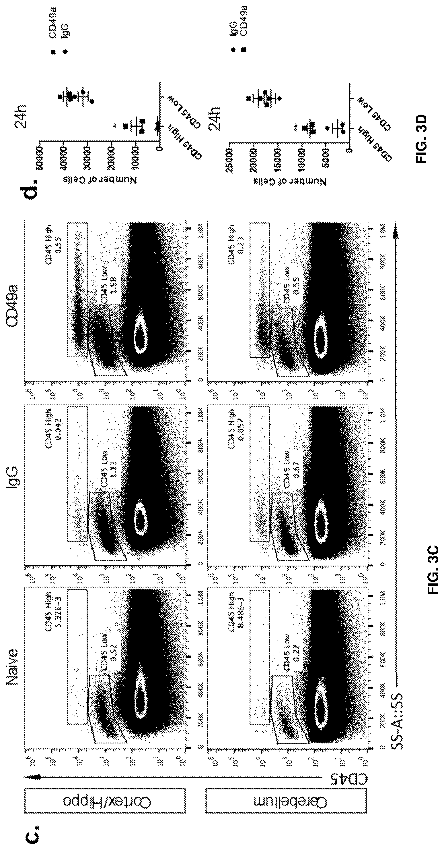
[0017] FIGS. 3A-3E show that blockade of CD49a induces the parenchymal infiltration of immune cells. FIG. 3A is a series of representative images of brain sections of IgG and CD49a treated mice immunostained for immune infiltrate (CD45--red) and astrocytes end feet (AQP4--green). Greater levels of CD45 staining (infiltrating immune cells) were observed in the brain parenchyma CD49a-treated mice compared to the IgG-treated control mice at 48 hours, and even greater levels of CD45 staining were observed in the CD49a-treated mice at 72 hours. FIG. 3B is a quantification of the density of CD45+ cells in the brain parenchyma of IgG and CD49a treated mice at different time post injection. FIG. 3C is a set of representative dot plots of CD45.sup.High and CD45.sup.Low expressing cells in the cortex and cerebellum after IgG and anti-CD49a treated mice. Greater proportions of cerebellum and cortex/hippocampus cells were CD45-high in the anti-CD49a-treated mice compared to IgG-treated controls. FIG. 3D is a quantification of the number of CD45.sup.High and CD45.sup.Low cells in the cortex/hippocampus and cerebellum of mice after IgG and CD49a blockade. FIG. 3E is a graph depicting gating of the phenotype of CD45.sup.High cells in the brain of CD49a treated mice. Mean+/-SEM, N=3-4 mice per group. *p<0.05; **p<0.01, One way ANOVA with Bonferoni post test.
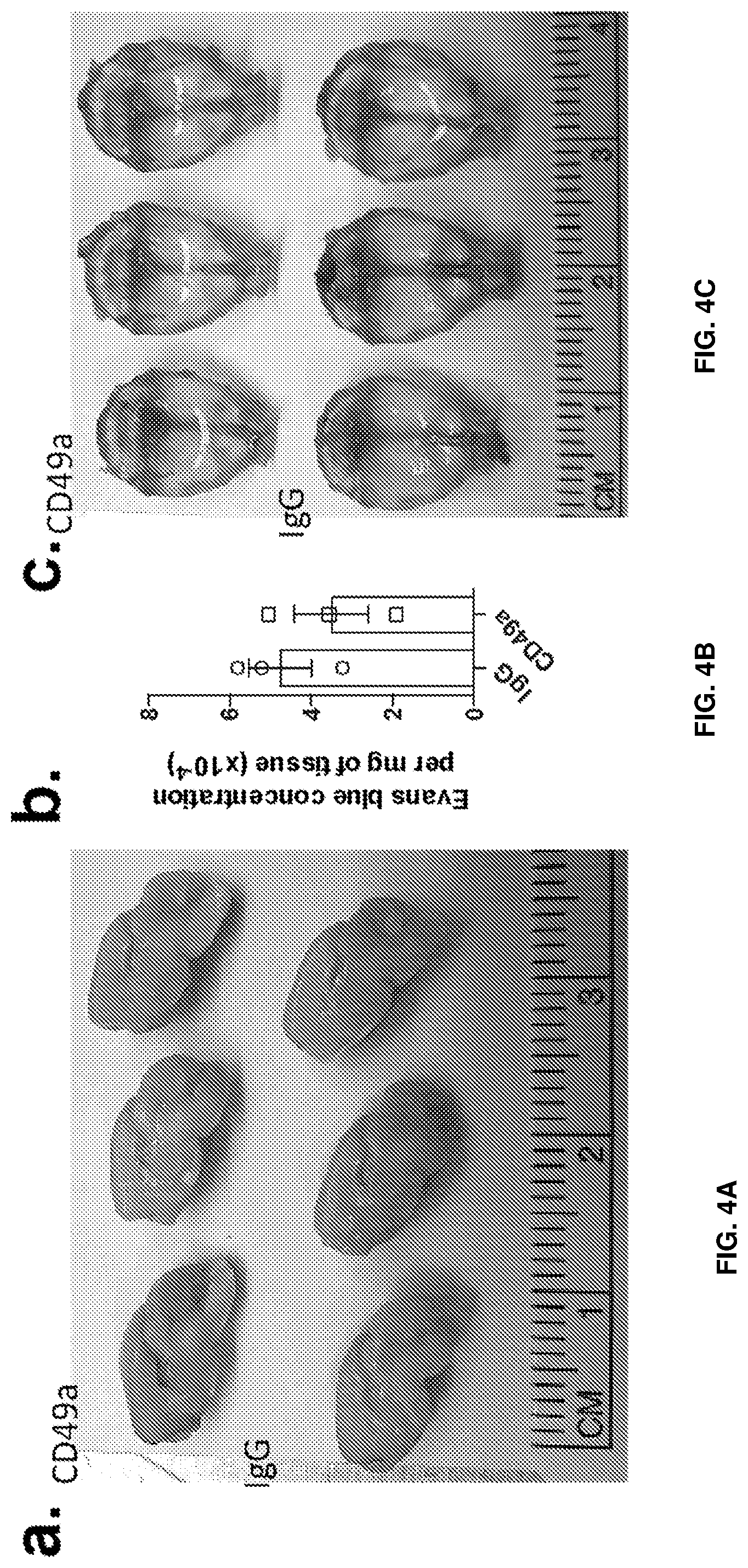
[0018] FIGS. 4A-4E show that infiltration of cells is not due to blood brain barrier opening but rather trans-pial migration. FIG. 4A is a set of representative images of hemi-brain of IgG and anti-CD49a injected mice after i.v. Evans Blue injection. FIG. 4B is a quantification of the Evans Blue concentration in the brain of IgG and anti-CD49a injected mice. FIG. 4C is a set of representative images of meninges of IgG and anti-CD49a injected mice after i.v. Evans Blue injection. FIG. 4D is a diagram of the scheme of the photoconversion of meningeal KiKGR expressing cells. FIG. 4E is a representative dot plot of green (non photoconverted) and red (photoconverted) CD45High cells in the cortex of anti-CD49a treated mice, 24 h after injection.
[0019] FIG. 5 shows the effect of repeated anti-CD49a injection on the development of EAE. Mice were injected i.c.v. with anti-CD49a or IgG antibodies every other day from six days before the induction of EAE to fifteen days after induction. Clinical score of mice treated with IgG and anti-CD49a antibodies. Preliminary data suggest that CD49a treatment limited the development of clinical symptoms of EAE.
[0020] FIGS. 6A-B are each graphs illustrating effects of i.c.m. administration of anti-CD49a antibody on disease progression of EAE. Adult C57BI6 female mice were injected i.c.m. with 5 .mu.l of anti-CD49a antibody (or IgG control) at day 8 post EAE induction (EAE was induced by 200 .mu.g of MOG.sub.35-55+CFA). Mice were subsequently followed daily for disease progression. CD49a-treated mice appeared to have ameliorated progression of symptoms compared to IgG-treated mice.
[0021] FIGS. 7A-B are each graphs showing quantification of immune cells in surgically denervated mice. FIG. 7A shows quantification of the number of CD45+, T cells, and NK cells in the meninges of sham or denervated IgG and CD49a treated mice. (mean.+-.s.e.m.; n=5 mice/group, ***p<0.001, two-way ANOVA). FIG. 7B shows quantification of geometric mean fluorescence intensity for ICAM1, VCAM1 and CD49a by the meningeal endothelial cells of sham or denervated IgG and CD49a treated mice. (mean.+-.s.e.m.; n=5 mice/group, ***p<0.001, two-way ANOVA).
[0022] FIGS. 8A-D are each graphs showing quantification of immune cells in the SSS of mice that underwent meningeal lymphatic ablation with visodyne. FIG. 8A shows quantification of the CD45 coverage in the SSS of mice. (mean.+-.s.e.m.; n=4/5 mice/group). FIG. 8B shows quantification of the MHCII coverage in the SSS of mice. (mean.+-.s.e.m.; n=4/5 mice/group). FIG. 8C shows quantification of the CD3e coverage in the SSS of mice. (mean.+-.s.e.m.; n=4/5 mice/group). FIG. 8G shows quantification of the density of CD3e cells in the SSS of mice. (mean.+-.s.e.m.; n=4/5 mice/group).
[0023] FIGS. 9A-C are each graphs showing clinical effects of anti-CD49a treatment in accordance with some embodiments herein. FIG. 9A shows clinical score of IgG and CD49a treated mice. (mean.+-.s.e.m.; n=36/37 mice/group; **p<0.01; repeated measures two-way ANOVA). FIG. 9B shows incidence of clinical symptoms development of IgG and CD49a treated mice. (mean.+-.s.e.m.; n=36/37 mice/group; ***p<0.001; Log-rank test). FIG. 9C shows clinical score score of symptomatic IgG and CD49a treated mice (mean.+-.s.e.m.; n=24/35 mice/group).
[0024] FIGS. 9D-E are each graphs showing CD45+ expression patterns in IgG and CD49a treated mice induced with EAE. FIG. 9D shows quantification of the CD45 coverage, CD45+ cells density and density of CD45 cluster in the cerebellum and cortex of IgG and CD49a treated mice induced with EAE. (mean.+-.s.e.m.; n=3/10 mice/group) FIG. 9E shows quantification of the CD45 coverage in the spinal cord of IgG and CD49a treated mice induced with EAE. (mean.+-.s.e.m.; n=4/9 mice/group)
[0025] FIGS. 10A-G are each graphs showing cell counts in the meninges of adult WT mice 2 and CD49a KO 4 mice. Shown are endothelial cells (FIG. 10A), ILC I (FIG. 10B), NK cells (FIG. 10C), macrophages (FIG. 10D), ILC (FIG. 10E), and NKT cells (FIG. 10F).
DETAILED DESCRIPTION
[0026] Some embodiments provide methods of treating or preventing a neurological immunity disorder in an animal subject, comprising administering to the subject a therapeutically effective amount of a compound that inhibits integrin signaling. Some embodiments provide methods of treating or preventing a neurological immunity disorder in an animal subject, comprising administering to the subject a therapeutically effective amount of a compound that decreases CD49a function. Some embodiments provide method of treating a neurological immunity disorder in an animal subject, comprising administering to the subject a therapeutically effective amount of an antibody or antigen binding fragment which binds CD49a, for example a human or humanized antibody or antigen binding fragment thereof that binds specifically to CD49a. In some embodiments, the antibody or antigen binding fragment thereof does not bind specifically to any of CD49b, CD49c, CD49d, CD49e, and/or CD49f, including combinations of two or more of these. In some embodiments, the compound blocks integrin signaling. It is noted that wherever a method of treating a disease or disorder with a composition is described herein, the corresponding use of the composition for the treatment of the disease or disorder is also expressly contemplated. For example, wherever a method of treating a neurological immunity disorder with an antibody or antigen binding fragment that binds to CD49a is described herein, an antibody or antigen binding fragment that binds to CD49a for use in treating the neurological immunity disorder is also expressly contemplated. It is to be understood that the embodiments described herein are not limited to specific analytical or synthetic methods as such may, of course, vary. It is also to be understood that the terminology used herein is for the purpose of describing particular embodiments only and is not intended to be limiting. Unless defined otherwise, all technical and scientific terms used herein have the meaning commonly understood by one of ordinary skill in the art to which this disclosure belongs, in view of the present disclosure.
[0027] "Neurological immunity disorders" is used herein according to its customary and ordinary meaning as would be understood by one of ordinary skill in the art in view of the specification, and encompasses neurological disorders with an immune component, for example, MS, Central Nervous System (CNS) injury, AD, and ASD.
[0028] The terms "treatment," "treating," and the like have their customary and ordinary meaning as understood by one of skill in the art in view of this disclosure. They generally refer to obtaining a desired pharmacologic and/or physiologic effect. The effect may be prophylactic in terms of completely or partially preventing a disease or symptom thereof and/or may be therapeutic in terms of a partial or complete cure for a disease and/or adverse effect attributable to the disease. "Treatment" as used herein has is customary and ordinary meaning as understood by one of skill in the art in view of this disclosure, and encompasses any treatment of a disease or symptom in a mammal, and includes any one or more of the following: (a) preventing the disease or a symptom from occurring in a subject which may be predisposed to acquiring the disease or symptom but has not yet been diagnosed as having it; (b) inhibiting the disease or a symptom, e.g., arresting or slowing its development; (c) relieving the disease, e.g., causing regression of the disease; (d) ameliorating one or more symptoms of the disease; (e) delaying the onset of the disease; and (e) reducing the likelihood of occurrence of the disease . The therapeutic agent (such as an anti-CD49a antibody or binding fragment thereof) may be administered before, during or after the onset of disease or injury. The treatment of ongoing disease, where the treatment stabilizes or reduces the undesirable clinical symptoms of the patient, is of particular interest. Such treatment is desirably performed prior to complete loss of function in the affected tissues. The subject therapy will desirably be administered during the symptomatic stage of the disease, and in some cases after the symptomatic stage of the disease.
[0029] As used herein, the term "integrin" has its customary and ordinary meaning as understood by one of skill in the art in view of this disclosure. It refers to proteins that are transmembrane receptors that function to facilitate cell-cell and cell-extracellular matrix interactions. Examples of integrins and integrin subunits expressed in the meninges include CD49a, LFA1, itga11, CD49e, itga8, CD51, CD49f, and itga9.
[0030] As used herein, the singular forms "a," "an," and "the" include plural reference unless the context clearly dictates otherwise. Thus, for example, reference to "a reagent" is reference to one or more reagents and includes equivalents thereof known to those skilled in the art. Additionally, the term "comprises" is intended to include embodiments where the method, apparatus, composition, etc., consists essentially of and/or consists of the listed steps, components, etc. Similarly, the term "consists essentially of" is intended to include embodiments where the method, apparatus, composition, etc., consists of the listed steps, components, etc. It is further noted that the claims may be drafted to exclude any optional element. As such, this statement is intended to serve as antecedent basis for use of such exclusive terminology as "solely," "only," and the like in connection with the recitation of claim elements, or use of a "negative" limitation.
[0031] As used herein, the term "about" is used herein to provide literal support for the exact number that it precedes, as well as a number that differs from the given number without having a substantial effect in the context. If more numerical precision is desired, "about" refers to values that differ by less than .+-.10%. In some embodiments, the term "about" indicates that the number differs from the given number by less than .+-.9%, 8%, 7%, 6%, 5%, 4%, 3%, 2%, or 1%.
[0032] It is appreciated that certain features described herein, which are, for clarity, described separately and/or in the context of separate embodiments, may also be provided in combination in a single embodiment. Conversely, various features of embodiments herein, which are, for brevity, described in the context of a single embodiment, may also be provided separately or in any suitable sub-combination. All combinations of the embodiments described herein are specifically embraced by the present disclosure and are disclosed herein just as if each and every combination was individually and explicitly disclosed. In addition, all sub-combinations of the various embodiments and elements thereof are also specifically embraced by the present disclosure and are disclosed herein just as if each and every such sub-combination was individually and explicitly disclosed herein.
[0033] In some embodiments, a method of treating, preventing, inhibiting, reducing the likelihood of, and/or delaying the onset of a neurological immunity disorder in an animal subject is described. The method can comprise administering to the subject a therapeutically effective amount of a compound that inhibits integrin signaling. The compound can comprise, consist essentially of, or consist of an inhibitor of CD49a, for example an antibody or antigen binding fragment thereof that binds specifically to CD49a. In some embodiments, the antibody or antigen binding fragment thereof that binds specifically to CD49a is a monoclonal antibody. In some embodiments, the neurological immunity disorder is selected from the group autism spectrum disorder (ASD), multiple sclerosis (MS), Alzheimer's disease (AD), and central nervous system (CNS) injury. In some embodiments, the method comprises treating or preventing the neurological immunity disorder, for example, ASD, MS, AD, and/or CNS. In some embodiments, the animal subject is a human. In some embodiments, the compound is formulated for administration to the CNS of the subject, for example intracerebroventricular administration. In some embodiments, the compound is administered to the CNS of the subject, for example intracerebroventricular administration. In some embodiments, the compound is not administered outside the CNS.
[0034] In some embodiments, the method treats prevents, inhibits, reduces the likelihood of, and/or delays the onset of a neurological immunity disorder in a human subject. In some embodiments, the method comprises administering to the subject a therapeutically effective amount of a compound that inhibits CD49a signaling. In some embodiments, the compound comprises, consists essentially of, or consists of an antibody or antigen binding fragment thereof that binds specifically to CD49a. In some embodiments, the compound comprises, consists essentially of, or consists of a monoclonal antibody or antigen binding fragment thereof that binds specifically to CD49a. In some embodiments, the neurological immunity disorder is selected from the group consisting of ASD, MS, AD, and CNS injury. In some embodiments, the method comprises treating or preventing the neurological immunity disorder. In some embodiments, the compound is formulated for administration to the CNS of the subject, for example intracerebroventricular administration. In some embodiments, the compound is administered to the CNS of the subject, for example intracerebroventricular administration. In some embodiments, the compound is not administered outside the CNS.
[0035] In some embodiments, the method treats, prevents, inhibits, reduces the likelihood of, and/or delays the onset of ASD in a human subject. In some embodiments, the method comprises administering to the subject a therapeutically effective amount of a compound that inhibits CD49a signaling. In some embodiments, the compound comprises, consists essentially of, or consists of an antibody or antigen binding fragment thereof that binds specifically to CD49a. In some embodiments, the compound comprises, consists essentially of, or consists of a monoclonal antibody or antigen binding fragment thereof that binds specifically to CD49a. In some embodiments, the antibody, e.g., monoclonal antibody or antigen binding fragment thereof does not specifically bind to any of CD49b, CD49c, CD49d, Cd49e, and/or CD49f. In some embodiments, the method comprises treating or preventing the ASD. In some embodiments, the compound is formulated for administration to the CNS of the subject, for example intracerebroventricular administration. In some embodiments, the compound is administered to the CNS of the subject, for example intracerebroventricular administration. In some embodiments, the compound is not administered outside the CNS.
[0036] In some embodiments, the method treats, prevents, inhibits, reduces the likelihood of, and/or delays the onset of MS in a human subject. In some embodiments, the method comprises administering to the subject a therapeutically effective amount of a compound that inhibits CD49a signaling. In some embodiments, the compound comprises, consists essentially of, or consists of an antibody or antigen binding fragment thereof that binds specifically to CD49a. In some embodiments, the compound comprises, consists essentially of, or consists of a monoclonal antibody or antigen binding fragment thereof that binds specifically to CD49a. In some embodiments, the antibody, e.g., monoclonal antibody or antigen binding fragment thereof does not bind to any of CD49b, CD49c, CD49d, Cd49e, and/or CD49f. In some embodiments, the method comprises treating or preventing the MS. As shown in Example 4, 5, and 7 and FIGS. 5, 6A-B, and 9A-C, administering an antibody inhibitor of CD49a signaling to an EAE subject (a model of MS) in accordance with some embodiments herein delayed the onset of EAE, reduced the incidence of EAE, and improved the clinical score of the EAE subject. Accordingly, it is contemplated that administering an inhibitor of CD49a (such as an antibody or antigen binding fragment thereof that binds specifically to CD49a) in accordance with some embodiments herein can delay the onset of, reduce the incidence of, and/or ameliorate symptoms of MS. In some embodiments, the compound is formulated for administration to the CNS of the subject, for example intracerebroventricular administration. In some embodiments, the compound is administered to the CNS of the subject, for example intracerebroventricular administration. In some embodiments, the compound is not administered outside the CNS.
[0037] In some embodiments, the method treats, prevents, inhibits, reduces the likelihood of, and/or delays the onset of AD in a human subject. The method can comprise administering to the subject a therapeutically effective amount of a compound that inhibits CD49a signaling. The compound can comprise, consist essentially of, or consist of an antibody or antigen binding fragment thereof that binds specifically to CD49a. In some embodiments, the compound comprises, consists essentially of, or consists of a monoclonal antibody or antigen binding fragment thereof that binds specifically to CD49a. In some embodiments, the antibody, e.g., monoclonal antibody or antigen binding fragment thereof does not bind to any of CD49b, CD49c, CD49d, Cd49e, and/or CD49f. In some embodiments, the method comprises treating or preventing the AD. In some embodiments, the compound is formulated for administration to the CNS of the subject, for example intracerebroventricular administration. In some embodiments, the compound is administered to the CNS of the subject, for example intracerebroventricular administration. In some embodiments, the compound is not administered outside the CNS.
[0038] In some embodiments, the method treats, prevents, inhibits, and/or delays the onset of CNS injury in a human subject. The method can comprise administering to the subject a therapeutically effective amount of a compound that inhibits CD49a signaling. The compound can comprise, consist essentially of, or consist of an antibody or antigen binding fragment thereof that binds specifically to CD49a. In some embodiments, the compound comprises, consists essentially of, or consists of a monoclonal antibody or antigen binding fragment thereof that binds specifically to CD49a. In some embodiments, the antibody, e.g., monoclonal antibody or antigen binding fragment thereof does not bind to any of CD49b, CD49c, CD49d, Cd49e, and/or CD49f. In some embodiments, the method comprises treating or preventing the CNS injury. In some embodiments, the compound is formulated for administration to the CNS of the subject, for example intracerebroventricular administration. In some embodiments, the compound is administered to the CNS of the subject, for example intracerebroventricular administration. In some embodiments, the compound is not administered outside the CNS.
[0039] In the method or use of some embodiments, the compound that inhibits integrin signaling is administered after the onset of the neurological immunity disorder, for example at least about 8 days after the onset of the neurological immunity disorder, for example at least 5, 6, 7, 8, 9, 10, 11, 12, 13, 14, 15, 16, 17, 18, 19, 20, 21, 22, 23, 24, 25, 26, 27, or 28 days, including any ranges between any two of the listed values, for example, including but not limited to the following ranges which are provided for exemplary purposes only: 5-28 days, 5-21 days, 5-14 days, 5-7 days, 7-28 days, 7-21 days, 7-14 days, 10-28 days, 10-21 days, or 10-14 days. In the method or use of some embodiments, the administration of the compound after the onset of the neurological immunity disorder reduces clinical symptoms of the neurological immunity disorder, which can be measured, for example, by a clinical score. In the method or use of some embodiments, the compound that inhibits integrin signaling comprises, consists essentially of, or consists of an antibody or antigen binding fragment thereof that binds specifically to CD49a. In the method or use of some embodiments, the compound that inhibits integrin signaling comprises, consists essentially of, or consists of a CD49a inhibiting (or blocking) antibody.
[0040] In the method or use of some embodiments, the method further comprises identifying a subject in need of said treatment. In further embodiments, the subject in need of said treatment is susceptible to or suffering form a neurological immunity disorder selected from the group consisting of autism spectrum disorder (ASD), multiple sclerosis (MS), Alzheimer's disease (AD), and central nervous system (CNS) injury. Identification of such subjects may be made using techniques known to a person of ordinary skill in the art.
[0041] The term "subject" is used herein according to its customary and ordinary meaning as would be understood by one of ordinary skill in the art in view of the specification. It refers to an animal, for example a mammal, such as a human. In the method or use of some embodiments, the animal subject is a human
[0042] In the method or use of some embodiments, inhibiting (or blocking) integrin signaling includes decreasing function of an integrin and/or decreasing function of an integrin subunit such as CD49a. In the method or use of some embodiments, the compound that inhibits integrin signaling decreases the function of a protein selected from the list consisting of CD49a, LFA1, itga11, CD49e, itga8, CD51, CD49f, and itga9. In the method or use of some embodiments, the compound that inhibits integrin signaling decreases CD49a function. In the method or use of some embodiments, the compound binds specifically to CD49a.
[0043] In the method or use of some embodiments, the compound that inhibits integrin signaling is an antibody or an antigen binding fragment which binds to an integrin or an integrin subunit. In some embodiments, the antibody or the antigen binding fragment binds a protein selected from the list consisting of CD49a, LFA1, itga11, CD49e, itga8, CD51, CD49f, and itga9. In some embodiments, the antibody or the antigen binding fragment binds to CD49a. In some embodiments, the antibody or the antigen binding fragment specifically binds a protein selected from the list consisting of CD49a, LFA1, itga11, CD49e, itga8, CD51, CD49f, and itga9. In some embodiments, the antibody or the antigen binding fragment specifically binds CD49a. In some embodiments, the antibody or the antigen binding fragments is a monoclonal antibody, for example a humanized antibody or human antibody.
[0044] An antibody (interchangeably used in plural form) is used herein according to its customary and ordinary meaning as would be understood by one of ordinary skill in the art in view of the specification. It refers to an immunoglobulin molecule capable of specific binding to a target, such as a carbohydrate, polynucleotide, lipid, polypeptide, etc., through at least one antigen recognition site, which is typically located in the variable region of the immunoglobulin molecule. As used herein, the term "antibody", e.g., anti-CD49a antibody, encompasses not only intact (e.g., full-length) polyclonal or monoclonal antibodies, but also antigen-binding fragments thereof (such as Fab, Fab', F(ab')2, Fv), single chain (scFv), mutants thereof, fusion proteins comprising an antibody portion, humanized antibodies, chimeric antibodies, diabodies, nanobodies, linear antibodies, single chain antibodies, multispecific antibodies (e.g., bispecific antibodies) and any other modified configuration of the immunoglobulin molecule that comprises an antigen recognition site of the required specificity, including glycosylation variants of antibodies, amino acid sequence variants of antibodies, and covalently modified antibodies. An antibody, e.g., anti-CD49a antibody in accordance with methods, uses, compositions, and pharmaceutical compositions of some embodiments herein, includes an antibody of any class, such as IgD, IgE, IgG, IgA, or IgM (or sub-class thereof), and the antibody need not be of any particular class. Depending on the antibody amino acid sequence of the constant domain of its heavy chains, immunoglobulins can be assigned to different classes. There are five major classes of immunoglobulins: IgA, IgD, IgE, IgG, and IgM, and several of these may be further divided into subclasses (isotypes), e.g., IgG1, IgG2, IgG3, IgG4, IgA1 and IgA2. The heavy-chain constant domains that correspond to the different classes of immunoglobulins are called alpha, delta, epsilon, gamma, and mu, respectively. The subunit structures and three-dimensional configurations of different classes of immunoglobulins are well known.
[0045] The base structure of an antibody is a tetramer, which includes two heavy chains and two light chains. Each chain comprises a constant region, and a variable region. Generally, the variable region, heavy chain variable region (V.sub.H) and a light chain variable region (V.sub.L), is responsible for binding specificity of the antibody. In a typical antibody, each variable region comprises three complementarity determining regions (CDRs) flanked by four framework (FR) regions. As such, an typical antibody variable region has six CDRs (three heavy chain CDRs, three light chain CDRs), some or all of which are generally involved in binding interactions by the antibody. Each V.sub.H and V.sub.L comprises three CDRs and four FRs, arranged from amino-terminus to carboxy-terminus in the following order: FR1, CDR1, FR2, CDR2, FR3, CDR3, FR4. The framework regions and CDRs can be precisely identified using methodology known in the art, for example, by the Kabat definition, the Chothia definition, the AbM definition, and/or the contact definition, all of which are well known in the art. See, e.g., Kabat, E. A., et al. (1991) Sequences of Proteins of Immunological Interest, Fifth Edition, U.S. Department of Health and Human Services, NIH Publication No. 91-3242, Chothia et al., (1989) Nature 342:877; Chothia, C. et al. (1987) J. Mol. Biol. 196:901-917, Al-lazikani et al (1997) J. Molec. Biol. 273:927-948; and Almagro, J. Mol. Recognit. 17:132-143 (2004). See also hgmp.mrc.ac.uk and bioinf.org.uk/abs).
[0046] The anti-CD49a antibody suitable for methods, uses, compositions, and pharmaceutical compositions of embodiments described herein may be a full-length antibody, which contains two heavy chains and two light chains, each including a variable domain and a constant domain. Alternatively, the anti-CD49a antibody can be an antigen-binding fragment of a full-length antibody. Examples of binding fragments encompassed within the term "antigen-binding fragment" of a full length antibody include (i) a Fab fragment, a monovalent fragment consisting of the V.sub.L, V.sub.H, C.sub.L and C.sub.H1 domains; (ii) a F(ab')2 fragment, a bivalent fragment including two Fab fragments linked by a disulfide bridge at the hinge region; (iii) a Fd fragment consisting of the V.sub.H and CH1 domains; (iv) a Fv fragment consisting of the V.sub.L and V.sub.H domains of a single arm of an antibody, (v) a dAb fragment (Ward et al., (1989) Nature 341:544-546), which consists of a V.sub.H domain; and (vi) an isolated complementarity determining region (CDR) that retains functionality. Furthermore, although the two domains of the Fv fragment, V.sub.L and V.sub.H, are coded for by separate genes, they can be joined, using recombinant methods, by a synthetic linker that enables them to be made as a single protein chain in which the V.sub.L and V.sub.H regions pair to form monovalent molecules known as single chain Fv (scFv). See e.g., Bird et al. (1988) Science 242:423-426; and Huston et al. (1988) Proc. Natl. Acad. Sci. USA 85:5879-5883.
[0047] Anti-CD49a antibodies and methods for producing them are known in the art. For example, US20160017043 provides antibody sequences for anti-CD49a antibodies, which publication is incorporated by reference in its entirety herein, including the drawings and the sequence listing therein. In some embodiments, the anti-CD49a antibody comprises a V.sub.L domain of the V.sub.L domain shown in FIG. 2A of US20160017043 and a V.sub.H domain of the V.sub.H domain shown in FIG. 2B of US20160017043. In some embodiments, the anti-CD49a antibody comprises a V.sub.L domain comprising a light chain CDR1, CDR2, and CDR3 that are light chain CDRs in the sequence shown in FIG. 2A of US20160017043 and a V.sub.H domain comprising a heavy chain CDR1, CDR2, and CDR3 that are heavy chain CDRs the sequence shown in FIG. 2B of US20160017043. In some embodiments, the anti-CD49a antibody comprises a V.sub.L domain of the V.sub.L domain shown in FIG. 3 of US20160017043 and a V.sub.H domain of the V.sub.H domain shown in FIG. 4 of US20160017043. In some embodiments, the anti-CD49a antibody comprises a V.sub.L domain comprising a light chain CDR1, CDR2, and CDR3 that are light chain CDRs in the sequence shown in FIG. 3 of US20160017043 and a V.sub.H domain comprising a heavy chain CDR1, CDR2, and CDR3 that are heavy chain CDRs in the sequence shown in FIG. 4 of US20160017043. In some embodiments, the CDRs are according to the definition of Kabat, Chothia, the Abm, or the contact definition. In some embodiments the anti-CD49a antibody is a human or humanized antibody as described herein.
[0048] In some embodiments, the anti-CD49a antibody comprises a V.sub.L domain that has at least 80%, at least 85%, at least 90% (e.g., 91%, 92%, 93%, 94%), at least 95% (e.g., 96%, 97%, 98%, 99%, 100%) sequence identity with the V.sub.L domain shown in FIG. 2A of US20160017043 and a V.sub.H domain that has at least 80%, at least 85%, at least 90% (e.g., 91%, 92%, 93%, 94%), at least 95% (e.g., 96%, 97%, 98%, 99%, 100%) sequence identity with the V.sub.H domain shown in FIG. 2B of US20160017043. In some embodiments, the anti-CD49a antibody comprises a V.sub.L domain having a sequence that differs from the V.sub.L domain shown in FIG. 2A of US20160017043 by 1, 2, 3, 4, 5, 6, 7, 9, or 10 amino acid residues and a V.sub.H domain having a sequence that differs from the V.sub.H domain shown in FIG. 2B of US20160017043 by 1, 2, 3, 4, 5, 6. 7, 9, or 10 amino acid residues. In some embodiments, the anti-CD49a antibody comprises a V.sub.L domain having a sequence that differs from the V.sub.L domain shown in FIG. 2A of US20160017043 by 1, 2, 3, 4, 5, 6, 7, 9, or 10 amino acid residues and a V.sub.H domain having a sequence of the V.sub.H domain shown in FIG. 2B of US2016001704. In some embodiments, the anti-CD49a antibody comprises a V.sub.L domain having a sequence of the V.sub.L domain shown in FIG. 2A of US20160017043, and a V.sub.H domain having a sequence that differs from the V.sub.H domain shown in FIG. 2B of US20160017043 by 1, 2, 3, 4, 5, 6, 7, 9, or 10 amino acid residues. In some embodiments, the anti-CD49a antibody comprises a V.sub.L domain comprising a light chain CDR1, CDR2, and CDR3 that are light chain CDRs having at least 80%, at least 85%, at least 90% (e.g., 91%, 92%, 93%, 94%), at least 95% (e.g., 96%, 97%, 98%, 99%, 100%) sequence identity with the light chain CDRs of the sequence shown in FIG. 2A of US20160017043 and a V.sub.H domain comprising a heavy chain CDR1, CDR2, and CDR3 that are heavy chain CDRs having at least 80%, at least 85%, at least 90% (e.g., 91%, 92%, 93%, 94%), at least 95% (e.g., 96%, 97%, 98%, 99%, 100%) sequence identity with the heavy chain CDRs of the sequence shown in FIG. 2B of US20160017043. In some embodiments, the anti-CD49a antibody comprises a V.sub.L domain comprising a light chain CDR1, CDR2, and CDR3 that are light chain CDRs having a sequence that differs from the sequence of the light chain CDRs shown in FIG. 2A of US20160017043 by 0, 1, 2, 3, 4, 5, 6, 7, 9, or 10 amino acid residues and a V.sub.H domain comprising a heavy chain CDR1, CDR2, and CDR3 that are heavy chain CDRs having a sequence that differs from the sequence of the heavy chain CDRs shown in FIG. 2B of US20160017043 by 0, 1, 2, 3, 4, 5, 6, 7, 9, or 10 amino acid residues. In some embodiments, the anti-CD49a antibody comprises a V.sub.L domain having at least 80%, at least 85%, at least 90% (e.g., 91%, 92%, 93%, 94%), at least 95% (e.g., 96%, 97%, 98%, 99%, 100%) sequence identity with the V.sub.L domain shown in FIG. 3 of US20160017043 and a V.sub.H domain having at least 80%, at least 85%, at least 90% (e.g., 91%, 92%, 93%, 94%), at least 95% (e.g., 96%, 97%, 98%, 99%, 100%) sequence identity with the V.sub.H domain shown in FIG. 4 of US20160017043. In some embodiments, the anti-CD49a antibody comprises a V.sub.L domain having a sequence that differs from the V.sub.L domain shown in FIG. 3 of US20160017043 by 1, 2, 3, 4, 5, 6, 7, 9 or 10 amino acid residues and a V.sub.H domain having a sequence that differs from the V.sub.H domain shown in FIG. 4 of US20160017043 by 1., 2, 3, 4, 5, 6, 7, 9, or 10 amino acid residues. In some embodiments, the anti-CD49a antibody comprises a V.sub.L domain having a sequence of the V.sub.L domain shown in FIG. 3 of US20160017043 and a V.sub.H domain having a sequence that differs from the V.sub.H domain shown in FIG. 4 of US20160017043 by 1, 2, 3, 4 5, 6, 7, 9, or 10 amino acid residues. In some embodiments, the anti-CD49a antibody comprises a V.sub.L domain having a sequence that differs from the V.sub.L domain shown in FIG. 3 of US20160017043 by 1, 2, 3, 4, 5, 6, 7, 9, or 10 amino acid residues and a V.sub.H domain of the V.sub.H domain shown in FIG. 4 of US20160017043. In some embodiments, the anti-CD49a antibody comprises a V.sub.L domain comprising a light chain CDR1, CDR2, and CDR3 that are light chain CDRs having at least 80%, at least 85%, at least 90% (e.g., 91%, 92%, 93%, 94%), at least 95% (e.g., 96%, 97%, 98%, 99%, 100%) sequence identity with the sequence shown in FIG. 3 of US20160017043 and a V.sub.H domain comprising a heavy chain CDR1, CDR2, and CDR3 that are heavy chain CDRs having at least 80%, at least 85%, at least 90% (e.g., 91%, 92%, 93%, 94%), at least 95% (e.g., 96%, 97%, 98%, 99%, 100%) sequence identity with the heavy chain CDR sequences shown in FIG. 4 of US20160017043. In some embodiments, the anti-CD49a antibody comprises a V.sub.L domain comprising a light chain CDR1, CDR2, and CDR3 that are light chain CDRs having a sequence that differs from the light chain CDR sequences shown in FIG. 3 of US20160017043 by L 2, 3, 4. 5, 6, 7, 9, or 10 amino acid residues and a V.sub.H domain comprising a heavy chain CDR1, CDR2, and CDR3 that are heavy chain CDRs having a sequence that differs from the heavy chain CDR sequences shown in FIG. 4 of US20160017043 by 1, 2, 3, 4, 5, 6, 7, 9, or 10 amino acid residues.
[0049] A number of approaches are available for producing suitable antibodies that specifically bind to CD49a in accordance with methods, uses, compositions, and pharmaceutical compositions of embodiments herein. For example, in some embodiments, a host organism is immunized with an antigen comprising, consisting essentially of, or consisting of CD49a. By way of example, a sequence of CD49a (which may also be referred to as Integrin alpha-1 or VLA-1) is available as Uniprot accession no. P56199 (SEQ ID NO: 1 MAPRPRARPGVAVACCWLLTVVLRCCVSFNVDVKNSMTFSGPVEDMFGYTVQQYE NEEGKWVLIGSPLVGQPKNRTGDVYKCPVGRGESLPCVKLDLPVNTSIPNVTEVKEN MTFGSTLVTNPNGGFLACGPLYAYRCGHLHYTTGICSDVSPTFQVVNSIAPVQECST QLDIVIVLDGSNSIYPWDSVTAFLNDLLERMDIGPKQTQVGIVQYGENVTHEFNLNKY SSTEEVLVAAKKIVQRGGRQTMTALGIDTARKEAFTEARGARRGVKKVMVIVTDGE SHDNHRLKKVIQDCEDENIQRFSIAILGSYNRGNLSTEKFVEEIKSIASEPTEKHFFNVS DELALVTIVKTLGERIFALEATADQSAASFEMEMSQTGFSAHYSQDWVMLGAVGAY DWNGTVVMQKASQIIIPRNTTFNVESTKKNEPLASYLGYTVNSATASSGDVLYIAGQ PRYNHTGQVIIYRMEDGNIKILQTLSGEQIGSYFGSILTTTDIDKDSNTDILLVGAPMY MGTEKEEQGKVYVYALNQTRFEYQMSLEPIKQTCCSSRQHNSCTTENKNEPCGARF GTAIAAVKDLNLDGFNDIVIGAPLEDDHGGAVYIYHGSGKTIRKEYAQRIPSGGDGKT LKFFGQSIHGEMDLNGDGLTDVTIGGLGGAALFWSRDVAVVKVTMNFEPNKVNIQK KNCHMEGKETVCINATVCFDVKLKSKEDTIYEADLQYRVTLDSLRQISRSFFSGTQER KVQRNITVRKSECTKHSFYMLDKHDFQDSVRITLDFNLTDPENGPVLDDSLPNSVHE YIPFAKDCGNKEKCISDLSLHVATTEKDLLIVRSQNDKFNVSLTVKNTKDSAYNTRTI VHYSPNLVFSGIEAIQKDSCESNHNITCKVGYPFLRRGEMVTFKILFQFNTSYLMENV TIYLSATSDSEEPPETLSDNVVNISIPVKYEVGLQFYSSASEYHISIAANETVPEVINSTE DIGNEINIFYLIRKSGSFPMPELKLSISFPNMTSNGYPVLYPTGLSSSENANCRPHIFEDP FSINSGKKMTTSTDHLKRGTILDCNTCKFATITCNLTSSDISQVNVSLILWKPTFIKSYF SSLNLTIRGELRSENASLVLSSSNQKRELAIQISKDGLPGRVPLWVILLSAFAGLLLLML LILALWKIGFFKRPLKKKMEK). By way of example, a polypeptide comprising, consisting essentially of, or consisting of the amino acid sequence of SEQ ID NO: 1 sequence can be used to immunize a host in order to produce antibodies that bind specifically to CD49a in accordance with some embodiments. The host organism can be a non-human mammal such as a mouse, rat, guinea pig, rabbit, donkey, goat, or sheep. Isolated antibody-producing cells can be obtained from the host organism, and the cells (or antibody-encoding nucleic acids thereof) can be screened for antibodies that binds specifically to CD49a. In some embodiments, antibody-producing cells are immortalized using hybridoma technology, and the resultant hybridomas are screened for antibodies that bind specifically to CD49a. In some embodiments, antibody-encoding nucleic acids are isolated from antibody-producing cells, and screened for antibodies that bind specifically to CD49a. An example protocol for screening human B cell nucleic acids is described in Huse et al., Science 246:1275-1281 (1989), which is hereby incorporated by reference in its entirety. In some embodiments, nucleic acids of interest are identified using phage display technology (See, e.g., Dower et al., WO 91/17271 and McCafferty et al., WO 92/01047, each of which is hereby incorporated by reference in its entirety). Phage display technology can also be used to mutagenize variable regions (or portions thereof such as CDRs) of antibodies previously shown to have affinity for CD49a. Variant antibodies can then be screened by phage display for antibodies having desired affinity to CD49a. In some embodiments, the antibody that specifically binds to CD49a is formatted as an antigen binding fragment. Example antigen binding fragments suitable for methods, uses, compositions, and pharmaceutical compositions of some embodiments can comprise, consist essentially of, or consist of a construct selected from the group consisting of Fab, Fab', Fab'-SH, F(ab').sub.2, and Fv fragments; minibodies; diabodies; and single-chain fragments such as single-chain Fv (scFv) molecules. Bispecific or multispecific antibodies or antigen binding fragments are also contemplated in accordance with methods, uses, compositions, and pharmaceutical compositions of some embodiments.
[0050] In some embodiments, for example if human monoclonal antibodies are of interest, the host comprises genetic modifications to produce or facilitate the production of human immunoglobulins. For example, XenoMouse.TM. mice were engineered with fragments of the human heavy chain locus and kappa light chain locus, respectively, which contained core variable and constant region sequences (described in detail Green et al. Nature Genetics 7:13-21 (1994), which is hereby incorporated by reference in its entirety). For example, mice have been engineered to produce antibodies comprising a human variable regions and mouse constant regions. The human heavy chain and light chain variable regions can then be reformatted onto a human constant region to provide a fully human antibody (described in detail in U.S. Pat. No. 6,787,637, which is hereby incorporated by reference in its entirety), For example, in a "minilocus" approach, an exogenous Ig locus is mimicked through the inclusion of pieces (individual genes) from the Ig locus. Thus, one or more V.sub.H genes, one or more DH genes, one or more JH genes, a mu constant region, and a second constant region (preferably a gamma constant region) are formed into a construct for insertion into an animal such as a mouse (See, e.g,. U.S. Pat. No. 5,545,807, which is hereby incorporated by reference in its entirety). Another approach, includes reconstituting SCID mice with human lymphatic cells, e.g., B and/or T cells. The mice are then immunized with an antigen and can generate an immune response against the antigen (See, e.g., U.S. Pat. No. 5,476,996, which is hereby incorporated by reference in its entirety).
[0051] In some embodiments, a host monoclonal antibody is formatted as a chimer antibody or is humanized, so that the antibody comprises at least some human sequences. By way of example, By way of example, an approach for producing humanized antibodies can comprise CDR grafting. For example, an antigen can be delivered to a non-human host (for example a mouse), so that the host produces antibody against the antigen. In some embodiments, monoclonal antibody is generated using hybridoma technology. In some embodiments, V gene utilization in a single antibody producing cell of the host is determined. The CDR's of the host antibody can be grafted onto a human framework. The V genes utilized in the non-human antibody can be compared to a database of human V genes, and the human V genes with the highest homology can be selected, and incorporated into a human variable region framework. See, e.g., Queen, U.S. Pat. No. 5,585,089, which is hereby incorporated by reference in its entirety.
[0052] Isolated oligonucleotides encoding a antibody of interest can be expressed in an expression system, such as a cellular expression system or a cell-free system in order to produce an antibody that binds specifically to CD49a in accordance with methods, uses, compositions, and pharmaceutical compositions of embodiments herein. Exemplary cellular expression systems include yeast (e.g., mammalian cells such as CHO cells or BHK cells, E. coli, insect cells, Saccharomyces, Pichia) transformed with recombinant yeast expression vectors containing the nucleotide sequences encoding antibodies; insect cell systems infected with recombinant virus expression vectors (e.g., baculovirus) containing sequences encoding antibodies; plant cell systems infected with recombinant virus expression vectors (e.g., cauliflower mosaic virus, CaMV; tobacco mosaic virus, TMV) or transformed with recombinant plasmid expression vectors (e.g., Ti plasmid) containing nucleotide sequences encoding antibodies; mammalian cell systems (e.g., COS, CHO, BHK, 293, 3T3) harboring recombinant expression constructs containing promoters derived from the genome of mammalian cells (e.g., metallothionein promoter) or from mammalian viruses.
[0053] In the method or use of some embodiments, the CD49a inhibiting (or blocking) antibody is administered after the onset of the neurological immunity disorder. In the method or use of some embodiments, the administration of the CD49a inhibiting (or blocking) antibody after the onset of the neurological immunity disorder reduces clinical symptoms of the neurological immunity disorder, which can be measured, for example, by a clinical score. Compositions and pharmaceutical compositions
[0054] According to some embodiments, a composition or pharmaceutical composition comprises a compound or therapeutic agent and a pharmaceutically acceptable carrier, adjuvant, or vehicle. In some embodiments, the compound or therapeutic agent of the composition or pharmaceutical composition comprises an agent or compound that inhibits (or blocks) integrin signaling. In some embodiments, the compound or therapeutic agent of the composition or pharmaceutical composition comprises an agent or compound that decreases or inhibits CD49a function. In some embodiments, the compound or therapeutic agent of the composition or pharmaceutical composition comprises an antibody or antigen binding fragment which binds CD49a. In some embodiments, the compound or therapeutic agent of the composition or pharmaceutical composition comprises, consists essentially of, or consists of an antibody or antigen binding fragment thereof that binds specifically to CD49a. The antibody or antigen binding fragment thereof that binds specifically to CD49a can be as described herein. In some embodiments the compound or therapeutic agent of the composition or pharmaceutical composition comprises, consists essentially of, or consists of a monoclonal antibody or antigen binding fragment thereof that binds specifically to CD49a. In some embodiments, the composition or pharmaceutical composition is for use in treating, preventing, inhibiting or ameliorateing the relevant disease, disorder, or condition in a subject in need thereof, e.g., neurological immunity disorder or a symptom thereof, such as autism spectrum disorder (ASD), multiple sclerosis (MS), Alzheimer's disease (AD), and/or central nervous system (CNS) injury. It is contemplated that a composition or pharmaceutical composition comprising, consisting essentially of, or consisting of compound that decreases or inhibits CD49a (for example, and anti-CD49a antibody as described herein) can be used in any method of treating, preventing, inhibiting or ameliorateing the relevant disease, disorder, or condition in a subject in need thereof, e.g., neurological immunity disorder or a symptom thereof, such as autism spectrum disorder (ASD), multiple sclerosis (MS), Alzheimer's disease (AD), and/or central nervous system (CNS) injury as described herein.
[0055] The amount of therapeutic agent in the composition or pharmaceutical composition of some embodiments is an amount effective to treat, prevent, inhibit or ameliorate the relevant disease, disorder, or condition in a subject in need thereof, e.g., neurological immunity disorder or a symptom thereof, such as autism spectrum disorder (ASD), multiple sclerosis (MS), Alzheimer's disease (AD), and/or central nervous system (CNS) injury. In some embodiments, a composition or pharmaceutical composition is formulated for administration to a subject in need of such composition. In some embodiments, the composition or pharmaceutical composition is formulated for oral administration to a subject. In some embodiments, the composition or pharmaceutical composition is formulated for injection into a subject. In some embodiments, the composition or pharmaceutical composition is formulated for topical application to the skin of the subject. In some embodiments, the subject is an animal, for example a mammal, such as a human
[0056] The term "pharmaceutically acceptable carrier," "adjuvant," or "vehicle" is used herein according to its customary and ordinary meaning as would be understood by one of ordinary skill in the art in view of the specification. It refers to a non-toxic carrier, adjuvant, or vehicle that does not destroy the pharmacological activity of the compound or therapeutic with which it is formulated. Pharmaceutically acceptable carriers, adjuvants or vehicles that may be used in the compositions and pharmaceutical compositions of some embodiments herien include, but are not limited to, ion exchangers, alumina, aluminum stearate, lecithin, serum proteins, such as human serum albumin, buffer substances such as phosphates, glycine, sorbic acid, potassium sorbate, partial glyceride mixtures of saturated vegetable fatty acids, water, salts or electrolytes, such as protamine sulfate, disodium hydrogen phosphate, potassium hydrogen phosphate, sodium chloride, zinc salts, colloidal silica, magnesium trisilicate, polyvinyl pyrrolidone, cellulose-based substances, polyethylene glycol, sodium carboxymethylcellulose, polyacrylates, waxes, polyethylene-polyoxypropylene-block polymers, polyethylene glycol and wool fat.
[0057] In some embodiments, the composition or pharmaceutical composition comprising an anti-CD49a antibody comprises a buffer, such as an acetate, histidine, succinate, or phosphate buffer. The buffer can be at a concentration of about 10 mM to about 50 mM, for example, about 20 mM to about 40 mM, such as about 30 mM. For example, the composition can contain a histidine buffer at a concentration of about 10 mM to about 50 mM, for example, about 20 mM to about 40 mM, such as about 30 mM. In one embodiment, the composition contains an acetate buffer at a concentration of about 10 mM to about 50 mM, for example, about 20 mM to about 40 mM, such as about 30 mM.
[0058] In some embodiments, the composition or pharmaceutical composition comprises an excipient, such as sorbitol, sodium chloride (NaCl), sucrose, trehelose, or mannitol. The composition can include an excipient at a concentration of about 100 mM to about 300 mM, for example, 110 mM to about 270 mM, about 120 mM to about 230 mM, or about 130 mM to about 210 mM, about 170 mM to about 200 mM, or about 180 mM to about 200 mM. For example, the composition can contain sorbitol at a concentration of about 180 mM to about 300 mM, for example, about 200 mM to about 300 mM, about 200 mM to about 240 mM, about 230 mM to about 270 mM, or about 240 mM to about 260 mM. In another example, the composition can contain NaCl at a concentration of about 100 mM to about 200 mM, for example, about 110 mM to about 190 mM, about 120 mM to about 180 mM, or about 130 mM to about 170 mM. In another example, the composition can contain sucrose at a concentration of about 200 mM to about 240 mM, about 230 mM to about 270 mM, or about 240 mM to about 260 mM. In another example, the composition can contain trehalose at a concentration of about 200 mM to about 240 mM, about 230 mM to about 270 mM, or about 240 mM to about 260 mM. In yet another example, the composition can contain mannitol at a concentration of about 200 mM to about 240 mM, about 230 mM to about 270 mM, or about 240 mM to about 260 mM.
[0059] In some embodiments, the aqueous composition or pharmaceutical composition comprises a surfactant, e.g., a substance that lowers surface tension of a liquid, such as a polysorbate, for example, polysorbate 80 or polysorbate 20. In some embodiments, the concentration of surfactant is at a concentration of about 0.001% to about 0.5%, about 0.001% to about 0.1%, for example, about 0.005% to about 0.05%, such as about 0.01%. Compositions or pharmaceutical compositions of some embodiments herein may be administered orally, parenterally, by inhalation spray, topically, rectally, nasally, buccally, vaginally or via an implanted reservoir. The term "parenteral" is used herein according to its customary and ordinary meaning as would be understood by one of ordinary skill in the art in view of the specification. It includes subcutaneous, intravenous, intramuscular, intra-articular, intra-synovial, intrasternal, intrathecal, intrahepatic, intralesional and intracranial injection or infusion techniques. The compositions may be administered orally, intraperitoneally or intravenously. Sterile injectable forms of the compositions or pharmaceutical compositions of some embodiments herein may be aqueous or oleaginous suspension. These suspensions may be formulated according to techniques known in the art using suitable dispersing or wetting agents and suspending agents. The sterile injectable preparation may also be a sterile injectable solution or suspension in a non- toxic parenterally acceptable diluent or solvent, for example as a solution in 1,3-butanediol. Among the acceptable vehicles and solvents that may be employed are water, Ringer's solution and isotonic sodium chloride solution. In addition, sterile, fixed oils are conventionally employed as a solvent or suspending medium.
[0060] In some embodiments, the composition or pharmaceutical composition is administered by an oral, intravenous, subcutaneous, intranasal, inhalation, intramuscular, intraocular, intraperitoneal, intratracheal, transdermal, buccal, sublingual, rectal, topical, local injection, or surgical implantation route. In some embodiments, the administration route is oral. In some embodiments, the administration is via injection. In some embodiments, the administration is via local injection. In some embodiments, the administration of the compound is into the cerebrospinal fluid (CSF) of said subject. In some embodiments, the administration of the compound is via intracerebroventricular injection. In some embodiments, the administration is transdermal, e.g., via application of an ointment containing the therapeutic to the head (scalp skin) of said subject.
[0061] To aid in delivery of the composition or pharmaceutical composition, any bland fixed oil may be employed including synthetic mono- or di-glycerides. Fatty acids, such as oleic acid and its glyceride derivatives are useful in the preparation of injectables, as are natural pharmaceutically-acceptable oils, such as olive oil or castor oil, especially in their polyoxyethylated versions. These oil solutions or suspensions may also contain a long-chain alcohol diluent or dispersant, such as carboxymethyl cellulose or similar dispersing agents that are commonly used in the formulation of pharmaceutically acceptable dosage forms including emulsions and suspensions. Other commonly used surfactants, such as Tweens, Spans and other emulsifying agents or bioavailability enhancers which are commonly used in the manufacture of pharmaceutically acceptable solid, liquid, or other dosage forms may also be used for the purposes of formulation.
[0062] Compositions or pharmaceutical compositions of some embodiments may be orally administered in any orally acceptable dosage form including, but not limited to, capsules, tablets, aqueous suspensions or solutions. In the case of tablets for oral use, carriers commonly used include lactose and corn starch. Lubricating agents, such as magnesium stearate, are also typically added. For oral administration in a capsule form, useful diluents include lactose and dried cornstarch. When aqueous suspensions are required for oral use, the active ingredient is combined with emulsifying and suspending agents. If desired, certain sweetening, flavoring or coloring agents may also be added.
[0063] In some embodiments, compositions or pharmaceutically acceptable compositions may be administered in the form of suppositories for rectal administration. These can be prepared by mixing the agent with a suitable non-irritating excipient that is solid at room temperature but liquid at rectal temperature and therefore will melt in the rectum to release the drug. Such materials include cocoa butter, beeswax and polyethylene glycols.
[0064] Compositions or pharmaceutical compositions of some embodiments may also be administered topically, especially when the target of treatment includes areas or organs readily accessible by topical application, such as the skin (e.g., scalp skin), or the lower intestinal tract. Suitable topical formulations are readily prepared for each of these areas or organs.
[0065] Topical application for the lower intestinal tract can be effected in a rectal suppository formulation (see above) or in a suitable enema formulation. Topically-transdermal patches may also be used.
[0066] For topical applications, provided compositions or pharmaceutical compositions of some embodiments may be formulated in a suitable ointment containing the active component suspended or dissolved in one or more carriers. Carriers for topical administration of a therapeutic include, but are not limited to, mineral oil, liquid petrolatum, white petrolatum, propylene glycol, polyoxyethylene, polyoxypropylene compound, emulsifying wax and water. Alternatively, provided pharmaceutically acceptable compositions can be formulated in a suitable lotion or cream containing the active components suspended or dissolved in one or more pharmaceutically acceptable carriers. Suitable carriers include, but are not limited to, mineral oil, sorbitan monostearate, polysorbate 60, cetyl esters wax, cetearyl alcohol, 2-octyldodecanol, benzyl alcohol and water.
[0067] Provided compositions or pharmaceutical compositions of some embodiments may be formulated as micronized suspensions in isotonic, pH adjusted sterile saline, or, preferably, as solutions in isotonic, pH adjusted sterile saline, either with or without a preservative such as benzylalkonium chloride. Alternatively, the pharmaceutically acceptable compositions may be formulated in an ointment such as petrolatum.
[0068] Compositions or pharmaceutical compositions of some embodiments may also be administered by nasal aerosol or inhalation. Such compositions are prepared according to techniques well-known in the art of pharmaceutical formulation and may be prepared as solutions in saline, employing benzyl alcohol or other suitable preservatives, absorption promoters to enhance bioavailability, fluorocarbons, and/or other conventional solubilizing or dispersing agents.
[0069] In some embodiments, compositions or pharmaceutical compositions are formulated for oral administration. Such formulations may be administered with or without food. In some embodiments, pharmaceutically acceptable compositions are administered without food. In some embodiments, compositions or pharmaceutical compositions of are administered with food.
[0070] The amount of therapeutic that may be combined with the carrier materials to produce a composition or pharmaceutical composition in a single dosage form will vary depending upon the host treated, the particular mode of administration, and other factors known to one of ordinary skill. Preferably, provided compositions should be formulated so that a dosage of between 0.01-100 mg/kg body weight/day of the therapeutic agent can be administered to a patient receiving these compositions.
[0071] It should also be understood that a specific dosage and treatment regimen for any particular patient will depend upon a variety of factors, including the activity of the specific therapeutic employed, the age, body weight, general health, sex, diet, time of administration, rate of excretion, drug combination, and the judgment of the treating physician and the severity of the particular disease being treated. The amount of a therapeutic in the composition will also depend upon the particular therapeutic in the composition.
[0072] Compositions or pharmaceutical compositions of some embodiment comprising a therapeutic and a pharmaceutically acceptable excipient, diluent, or carrier, are useful for treating a variety of diseases, disorders or conditions. Such diseases, disorders, or conditions include those described herein. In the method or use of some embodiments, the therapeutically effective amount of the compound is about 0.0002 mg/kg to about 2.0 mg/kg. In further embodiments, said therapeutically effective amount of the compound is about 0.00020 mg/kg, about 0.00030 mg/kg, about 0.00045 mg/kg, about 0.00060 mg/kg, about 0.00085 mg/kg, about 0.001 mg/kg, about 0.0015 mg/kg, about 0.002 mg/kg, about 0.0025 mg/kg, about 0.003 mg/kg, about 0.0035 mg/kg, about 0.004 mg/kg, about 0.0045 mg/kg, about 0.0050 mg/kg, about 0.0055 mg/kg, about 0.006 mg/kg, about 0.0065 mg/kg, about 0.007 mg/kg, about 0.0075 mg/kg, about 0.008 mg/kg, about 0.0085 mg/kg, about 0.009 mg/kg, about 0.0095 mg/kg, about 0.01 mg/kg, about 0.015 mg/kg, about 0.02 mg/kg, about 0.025 mg/kg, about 0.03 mg/kg, about 0.035 mg/kg, about 0.040 mg/kg, about 0.045 mg/kg, about 0.05 mg/kg, about 0.055 mg/kg, about 0.06 mg/kg, about 0.065 mg/kg, about 0.07 mg/kg, about 0.075 mg/kg, about 0.08 mg/kg, about 0.085 mg/kg, about 0.09 mg/kg, about 0.095 mg/kg, about 0.1 mg/kg, about 0.15 mg/kg, about 0.2 mg/kg, about 0.25 mg/kg, about 0.3 mg/kg, about 0.35 mg/kg, about 0.4 mg/kg, about 0.45 mg/kg, about 0.5 mg/kg, about 0.55 mg/kg, about 0.6 mg/kg, about 0.65 mg/kg, about 0.7 mg/kg, about 0.75 mg/kg, about 0.8 mg/kg, about 0.85 mg/kg, about 0.9 mg/kg, about 0.95 mg/kg, about 1.0 mg/kg, about 1.1 mg/kg, about 1.2 mg/kg, about 1.3 mg/kg, about 1.4 mg/kg, about 1.5 mg/kg, about 1.6 mg/kg, about 1.7 mg/kg, about 1.8 mg/kg, about 1.9 mg/kg, or about 2.0 mg/kg.
[0073] In the method or use of some embodiments, said therapeutically effective amount of the compound is less than about 0.00020 mg/kg, about 0.00030 mg/kg, about 0.00045 mg/kg, about 0.00060 mg/kg, about 0.00085 mg/kg, about 0.001 mg/kg, about 0.0015 mg/kg, about 0.002 mg/kg, about 0.0025 mg/kg, about 0.003 mg/kg, about 0.0035 mg/kg, about 0.004 mg/kg, about 0.0045 mg/kg, about 0.0050 mg/kg, about 0.0055 mg/kg, about 0.006 mg/kg, about 0.0065 mg/kg, about 0.007 mg/kg, about 0.0075 mg/kg, about 0.008 mg/kg, about 0.0085 mg/kg, about 0.009 mg/kg, about 0.0095 mg/kg, about 0.01 mg/kg, about 0.015 mg/kg, about 0.02 mg/kg, about 0.025 mg/kg, about 0.03 mg/kg, about 0.035 mg/kg, about 0.040 mg/kg, about 0.045 mg/kg, about 0.05 mg/kg, about 0.055 mg/kg, about 0.06 mg/kg, about 0.065 mg/kg, about 0.07 mg/kg, about 0.075 mg/kg, about 0.08 mg/kg, about 0.085 mg/kg, about 0.09 mg/kg, about 0.095 mg/kg, about 0.1 mg/kg, about 0.15 mg/kg, about 0.2 mg/kg, about 0.25 mg/kg, about 0.3 mg/kg, about 0.35 mg/kg, about 0.4 mg/kg, about 0.45 mg/kg, about 0.5 mg/kg, about 0.55 mg/kg, about 0.6 mg/kg, about 0.65 mg/kg, about 0.7 mg/kg, about 0.75 mg/kg, about 0.8 mg/kg, about 0.85 mg/kg, about 0.9 mg/kg, about 0.95 mg/kg, about 1.0 mg/kg, about 1.1 mg/kg, about 1.2 mg/kg, about 1.3 mg/kg, about 1.4 mg/kg, about 1.5 mg/kg, about 1.6 mg/kg, about 1.7 mg/kg, about 1.8 mg/kg, about 1.9 mg/kg, or about 2.0 mg/kg.
[0074] In the method or use of some embodiments, said therapeutically effective amount of the compound is more than about 0.00020 mg/kg, about 0.00030 mg/kg, about 0.00045 mg/kg, about 0.00060 mg/kg, about 0.00085 mg/kg, about 0.001 mg/kg, about 0.0015 mg/kg, about 0.002 mg/kg, about 0.0025 mg/kg, about 0.003 mg/kg, about 0.0035 mg/kg, about 0.004 mg/kg, about 0.0045 mg/kg, about 0.0050 mg/kg, about 0.0055 mg/kg, about 0.006 mg/kg, about 0.0065 mg/kg, about 0.007 mg/kg, about 0.0075 mg/kg, about 0.008 mg/kg, about 0.0085 mg/kg, about 0.009 mg/kg, about 0.0095 mg/kg, about 0.01 mg/kg, about 0.015 mg/kg, about 0.02 mg/kg, about 0.025 mg/kg, about 0.03 mg/kg, about 0.035 mg/kg, about 0.040 mg/kg, about 0.045 mg/kg, about 0.05 mg/kg, about 0.055 mg/kg, about 0.06 mg/kg, about 0.065 mg/kg, about 0.07 mg/kg, about 0.075 mg/kg, about 0.08 mg/kg, about 0.085 mg/kg, about 0.09 mg/kg, about 0.095 mg/kg, about 0.1 mg/kg, about 0.15 mg/kg, about 0.2 mg/kg, about 0.25 mg/kg, about 0.3 mg/kg, about 0.35 mg/kg, about 0.4 mg/kg, about 0.45 mg/kg, about 0.5 mg/kg, about 0.55 mg/kg, about 0.6 mg/kg, about 0.65 mg/kg, about 0.7 mg/kg, about 0.75 mg/kg, about 0.8 mg/kg, about 0.85 mg/kg, about 0.9 mg/kg, about 0.95 mg/kg, about 1.0 mg/kg, about 1.1 mg/kg, about 1.2 mg/kg, about 1.3 mg/kg, about 1.4 mg/kg, about 1.5 mg/kg, about 1.6 mg/kg, about 1.7 mg/kg, about 1.8 mg/kg, about 1.9 mg/kg, or about 2.0 mg/kg.
[0075] Methods, uses, and compositions of some embodiments include an aqueous pharmaceutical composition, such as a stable aqueous pharmaceutical composition, containing an anti-CD49a antibody at a concentration of about 100 mg/mL to about 225mg/mL, for example, about 110 mg/mL, about 120 mg/mL, about 130 mg/mL, about 140 mg/mL, about 150 mg/mL, about 160 mg/mL, about 170 mg/mL, about 180 mg/mL, about 190 mg/mL, about 200 mg/mL, about 205 mg/mL, about 210 mg/mL, about 215 mg/mL, about 220 mg/mL or about 225 mg/mL.
[0076] In the method or use of some embodiments, the compound is administered into the cerebrospinal fluid (CSF) of the subject. In the method or use of some embodiments, an ointment comprises said compound and the ointment is administered via application of the ointment to the scalp skin of the subject. In the method or use of some embodiments, an ointment comprises said compound and the ointment is administered via application of the ointment to the head of the subject.
[0077] In the method or use of some embodiments, the administration of said compound results in accumulation of immune cells in the brain meninges. In the method or use of some embodiments, the administration of said compound results in elevated T cells and/or natural killer T (NKT) cells in the brain parenchyma.
[0078] A compound referred to herein as one that "blocks" integrin signaling may also be referred to herein as a compound that "inhibits" integrin signaling. It will be understood that use of the term "inhibit" or "block" is not intended to necessitate absolute inhibition (or blockage), and as such inhibition or (blockage) as used herein also includes a decrease, reduction or impairment of the relevant target or function. For example, an antibody or antigen binding fragment thereof that binds specifically to CD49a may be referred to herein as a "CD49a-specific" antibody, "anti-CD49a" antibody, CD49a "inhibiting" antibody, and/or CD49a "blocking" antibody. In the method or use of some embodiments, the compound that inhibits integrin signaling comprises, consists essentially of, or consists of Tysabri (natalizumab) or an antigen binding fragment thereof. In the method or use of some embodiments, the compound that inhibits integrin signaling is a compound other than Tysabri (natalizumab). In the method or use some embodiments, the compound that inhibits integrin signaling comprises, consists of, or consists essentially of Tysabri.RTM. (natalizumab) formulated for administration into the CSF of the subject or as an ointment to the head of the subject. In the method or use of some embodiments, the compound that inhibits integrin signaling comprises, consists essentially of, or consists of ReoPro.RTM. (Abcizimab), Vedolizumab, etrolizumab, anti-av integrin, or Volocixmab, or a combination of two or more of these. In the method or use of some embodiments, the compound that inhibits integrin signaling is ReoPro.RTM. (Abcizimab), Vedolizumab, etrolizumab, anti-av integrin, or Volocixmab. In the method or use of some embodiments, the compound that inhibits integrin signaling is a compound other than ReoPro.RTM. (Abcizimab), Vedolizumab, etrolizumab, anti-av integrin, or Volocixmab.
[0079] In methods, uses, compositions, and pharmaceutical compositions of some embodiments, the anti-CD49a antibody as described herein binds to and inhibits the activity of CD49a by at least 50% (e.g., 60%, 70%, 80%, 90%, 95% or greater, including any increment therein). The apparent inhibition constant (Ki.sup.app or K.sub.i,app), which provides a measure of inhibitor potency, is related to the concentration of inhibitor required to reduce target (e.g., CD49a) activity and is not dependent on target concentrations. The inhibitory activity of an anti-CD49a antibody described herein can be determined by methods known in the art. In some embodiments, the anti-CD49a binds to CD49a with a dissociation constant KD that is numerically lower (indicating tighter binding than) 10.sup.-1, 10.sup.-2, 10.sup.-3, 10.sup.-4, 10.sup.-5, 10.sup.-6, 10.sup.-7, 10.sup.-8, 10.sup.-9, 10.sup.-10, 10.sup.-11, or 10.sup.-12, including ranges between any two of the listed values. A KD can be determined using methods known in the art, for example surface plasmon resonance on a BIACORE apparatus.
[0080] The K.sub.i,.sup.app value of an antibody may be determined by measuring the inhibitory effect of different concentrations of the antibody on the extent of the reaction (e.g., target activity such as CD49a activity); fitting the change in pseudo-first order rate constant (v) as a function of inhibitor concentration to the modified Morrison equation (Equation 1) yields an estimate of the apparent Ki value. For a competitive inhibitor, the Ki.sup.app can be obtained from the y-intercept extracted from a linear regression analysis of a plot of K.sub.i,.sup.app versus substrate concentration.
v = A ( [ E ] - [ I ] - K i app ) + ( [ E ] - [ I ] - K i app ) 2 + 4 [ E ] K i app 2 ( Equation 1 ) ##EQU00001##
[0081] Where A is equivalent to v.sub.olE, the initial velocity (v.sub.o) of the enzymatic reaction in the absence of inhibitor (I) divided by the total enzyme concentration (E).
[0082] In some embodiments, the anti-CD49a antibody described herein has a Kiapp value of 1000, 900, 800, 700, 600, 500, 400, 300, 200, 100, 50, 40, 30, 20, 19, 18, 17, 16, 15, 14, 13, 12, 11, 10, 9, 8, 7, 6, 5 pM or less for the target antigen or antigen epitope, such as an epitope of CD49a. Differences in Kiapp (e.g., for specificity or other comparisons) can be at least 1.5, 2, 3, 4, 5, 10, 15, 20, 37.5, 50, 70, 80, 91, 100, 500, 1000, 10,000 or 105 fold. In some examples, the anti-CD49a antibody inhibits a first antigen (e.g., a first protein in a first conformation or mimic thereof) better relative to a second antigen (e.g., the same first protein in a second conformation or mimic thereof; or a second protein). In some embodiments, any of the anti-CD49a antibodies may be further affinity matured to reduce the Kiapp of the antibody to the target antigen or antigenic epitope thereof.
[0083] In methods, uses, compositions, and pharmaceutical compositions of some embodiments, the anti-CD49a antibody suppresses or inhibits integrin signaling triggered by CD49a by at least 50% (e.g., 60%, 70%, 80%, 90%, 95% or greater, including any increment therein). Such inhibitory activity can be determined by conventional methods.
EXAMPLES
[0084] In the following Examples, CD49a is identified as a marker that can differentiate two distinct populations of meningeal T cells and that blockade of CD49a, using a blocking antibody in vivo, results in the accumulation of numerous populations of immune cells in the meninges and the parenchymal infiltration of NKT and T cells.
Example 1
Naive Meninges are Composed of Distinct Populations of CD4 T Cells
[0085] Meningeal CD4 T cells have been shown to support cognitive function, in part through the secretion of cytokine IL-4.sup.8. In order to further analyze the different populations of T cells that populate the naive meninges, both meninges and diaphragm were isolated and analyzed from adult mice. As described before.sup.8, the majority of T cells in the meninges express CD44 and half of the CD44+ cells also express the activation marker CD69 (FIGS. 1a,b). In recent years, a new population of tissue resident memory T cells (or TRM) was described in mucosal tissues after infection, where they ensure surveillance of the tissue against secondary infection.sup.11-14. One of the markers that characterize the TRM is the high expression of CD69.sup.14. Therefore the CD69- and CD69+ populations of meningeal CD4 T cells were analyzed for the expression of other TRM markers. Indeed the CD69+ population of CD4 T cells of the meninges expresses high levels of CD11a and CD49a, but no CD103 (FIGS. 1c-e), consistent with TRM CD4 T cells identified in the peripheryll.sup.11-14. CD49d, an integrin implicated in the recirculation of T cells in the CNS.sup.8 is mostly express by the CD69- CD4 T cells suggesting that the CD69+ T cells are less likely to be recirculating, a common feature of TRM T cells (FIG. 1f).
Example 2
CD49a is Expressed by Multiple Immune Populations in the Meninges and Its Blockade Results in the Transient Accumulation of Immune Cells in the Meninges
[0086] CD49a is an integrin alpha subunit, expressed by multiple cell types throughout the bodyl.sup.5, notably by immune cells.sup.15, and is especially implicated in homing of immune cells in specific tissues. The expression of CD49a by the immune cells that populate the naive meninges was analyzed. Not only CD4 T cells express CD49a, but also CD8 and NK cells, and to a greater extent NKT cells and monocytes/macrophages (FIGS. 2a,b).
[0087] To test the role of CD49a on meningeal immune cells, CD49a interaction and signaling was blocked by using a blocking antibodyl.sup.6. Surprisingly, intracerebroventricular (i.c.v.) injection of a CD49a-blocking antibody [purchased from BD Biosciences, Catalog No. 553961, Clone Ha4/8] at about 5 .mu.g in 5 .mu.L volume resulted in increased numbers of immune cells previously shown to express high level of CD49a, i.e. T cells, NK cells, and monocytes/macrophages, as soon as 24h after the antibody injection (FIG. 2c,d). CD49a being an integrin allowing the interaction of immune cells with their local ECM, blockade of CD49a might solely facilitate the extraction of the meningeal immune cells during the tissue isolation. To confirm this, immunohistochemistry was used on meningeal whole mount, 24 h after icy injection of the anti-CD49a antibody. Similar to the FACS analysis, there was an increased density of CD45+ and CD3+ T cells around the sinuses of anti-CD49a-injected mice (FIGS. 2e-g). The accumulation of immune cells in such a small window of time can be due to local proliferation or active recruitment of cells in the meninges. To try and answer this question, pulsed mice were pulsed with BrdU to assess the proliferative state of the cells after CD49a treatment. There was an increase of BrdU+ CD4 T cells in the meninges 24h after icy injection of CD49a blocking antibody (FIGS. 2h,i), suggesting, at least in part, that CD49a induces proliferation of meningeal immune cells. The duration of CD49a blocking effect was then tested. Mice were injected i.c.v. with the anti-CD49a antibody and sacrificed at different time points post injection. Analysis of the meningeal T cells number revealed an increased number of meningeal T cells up to 3 days after CD49a blockade (FIG. 2j). Interestingly no change in immune cell numbers was observed in the draining (deep cervical) or control (inguinal) lymph nodes, suggesting a local effect of the CD49a blockade antibody.
Example 3
CD49a Blockade Results in the Parenchymal Infiltration of T Cells and NKT Cells, Most Likely Through a Trans-Pial Migration
[0088] I.c.v. injection of the CD49a blockade antibody results in elevated numbers of immune cells in the meningeal compartment. The next example was to show CD49a blockade also resulted in infiltration of immune cells into the brain parenchyma. Brains from CD49a injected mice were then analyzed by both flow cytometry and IHC for the presence of intraparenchymal immune cells. Labeling of brain slices with anti-CD45 antibody revealed the presence of roundly shaped immune cells within the brain parenchyma of CD49a-injected mice as soon as 24 h after the injection (FIG. 3a). Those cells are not trapped into blood or perivascular spaces, as seen with the AQP4 staining and sometimes form clusters within the parenchyma (FIG. 3a). Similar infiltration can be found for up to 4 days after the anti-CD49a injection (FIG. 4b). FACS analysis of the cortex, cerebellum, and spinal cord of CD49a antibody injected mice revealed a spatial specificity of the infiltrate with no detectable immune infiltrate in the spinal cord of CD49a injected mice but a large infiltrate in both the cortex and cerebellum of injected mice (FIGS. 3c-d). The phenotype of the infiltrated immune cells was assessed and found that the majority of them are TCRb.sup.+CD4.sup.-CD8.sup.-NK1.1.sup.+, but also CD11b.sup.+Ly6C.sup.+, suggesting a population of activated NKT cells. Small populations of CD4.sup.+ and CD8.sup.+ T cells are also found (FIG. 3e).
[0089] Not only is CD49a expressed by immune cells but also by the blood endothelial cells.sup.15. To confirm that the parenchymal infiltration of immune cells upon CD49a blockade is not related to a transient opening of the blood brain barrier (BBB), the integrity of the BBB was tested by injecting Evans Blue in the blood vasculature during the 24 h after CD49a treatment. As seen in FIGS. 4a-c, no Evans Blue was detected in the brain or the meninges of IgG or CD49a treated mice, suggesting that the BBB remained intact during the treatment and that the parenchymal infiltration of immune cells is unlikely to come from an opening of the BBB or the BMB (blood meningeal barrier) Immune cells could however infiltrate the parenchyma directly from the meninges, either by crossing the pia or by infiltrating the Virchow-Robin spaces. To confirm this, the KiKGR mice that bear a photoconvertable protein and enables tracking the cell were used. Meninges of KiKGR mice were photoconverted (Green to Red) with a UV laser following i.c.v. injection of CD49a (FIG. 3d). Twenty-four hours after the injection, brains were harvested and the fluorescence of the infiltrated T cells was analyzed by FACS. Indeed, around 25% of the CD45 high cells found in the brain of CD49a injected mice are photoconverted (red) suggesting that those cells were localized in the meninges during the photoconversion (FIG. 3e). These results strongly suggest that the infiltrated immune cells trafficked from the meninges directly into the brain parenchyma.
[0090] Overall, blocking the integrin signaling through CD49a induces the proliferation and migration of specific immune cells from the meninges to the brain parenchyma.
Example 4
Repetitive Blockade of CD49a Results in a Decrease in EAE Scoring
[0091] Blockade of CD49a interaction and signaling results in the accumulation of T cells and NKT cells in the brain parenchyma of WT mice, likely coming from endogenous meningeal immune cells. The next example shows blocking of CD49a interferes with the development of EAE, the animal model of Multiple Sclerosis, where immune cells, notably T cells, transit through the meninges and also infiltrate the parenchyma. Catheters were inserted into the cisterna magna into mice and were injected every other day with about 5 .mu.g in 5 mL of the CD49a blockade antibodies. At day 6 after beginning of CD49a treatment, EAE was induced by injection of an emulsion of MOG35-55 subcutaneously above the tail. Surprisingly, the repetitive injection of CD49a blocking antibodies decreased the diseases severity compared to IgG injected mice, showing a protective effect of CD49a blockade in the development of EAE (FIG. 5).
[0092] Overall those data show that interfering with an integrin, highly expressed by the meningeal immune cells, is sufficient to induce drastic changes in local immune cell populations and favor the migration of cells into the brain parenchyma. CD49a is an example of one integrin that controls immune cell localization and function within brain borders.
Example 5
Administration of an Antibody that Inhibits CD49a Results in a Decrease in EAE Score
[0093] Adult C57BI6 female mice were injected i.c.m. with 5 .mu.l of anti-CD49a antibody (or IgG control) at day 8 post EAE induction (EAE was induced by 200 .mu.g of MOG.sub.35-55+CFA). Mice were subsequently followed daily for disease progression. The results of this experiment are shown in FIG. 6A. An additional repetition of this experiment is shown in FIG. 6B. CD49a-treated mice show ameliorated progression of symptoms compared to IgG-treated mice.
Example 6
Modulation of a CD49a Blockade
[0094] Adult C57B16 mice where sham operated or denervated (SCG excision). One week after surgery, mice were injected with 5 .mu.g of anti-CD49a (or IgG) and tissues were harvested 24h after. FIG. 7A shows quantification of the number of CD45+, T cells, and NK cells in the meninges of sham or denervated IgG and CD49a treated mice. (mean.+-.s.e.m.; n=5 mice/group, ***p<0.001, two-way ANOVA). FIG. 7B shows quantification of geometric mean fluorescence intensity for ICAM1, VCAM1 and CD49a by the meningeal endothelial cells of sham or denervated IgG and CD49a treated mice. (mean.+-.s.e.m.; n=5 mice/group, ***p<0.001, two-way ANOVA). Thus, administering an inhibitor of CD49a signaling to an EAE subject (a model of MS) in accordance with some embodiments herein increased immune cells in the meninges, regardless of whether the subject was denervated (by excision of the SCG).
[0095] Adult C57B16 mice had their meningeal lymphatic vessels ablated using Visudyne (control mice were injected with PBS). One week after meningeal lymphatic ablation, mice were injected with 5 .mu.g of anti-CD49a (or IgG) and tissues were harvested 24 h after injection. FIG. 8A shows quantification of the CD45 coverage in the SSS of mice. (mean.+-.s.e.m.; n=4/5 mice/group). FIG. 8B shows quantification of the MHCII coverage in the SSS of mice. (mean.+-.s.e.m.; n=4/5 mice/group). FIG. 8C shows Quantification of the CD3e coverage in the SSS of mice. (mean.+-.s.e.m.; n=4/5 mice/group). FIG. 8D shows Quantification of the density of CD3e cells in the SSS of mice. (mean.+-.s.e.m.; n=4/5 mice/group). Thus, administering an inhibitor of CD49a signaling to an EAE subject (a model of MS) in accordance with some embodiments herein increased immune cells in the SSS, regardless of whether the subject had undergone meningeal lymphatic ablation.
Example 7
CD49a Blockade During EAE Results in Decrease Disease Incidence Without Preventing Immune Cells Infiltration
[0096] Adult C57B16 mice were immunized with 200 .mu.g of MOG with CFA supplemented with 2 mg/ml of mycobacterium. At D7 post EAE induction, mice were injected i.c.m. with 5 .mu.g of anti-CD49a (or IgG). FIG. 9A shows clinical score of IgG and CD49a treated mice. (mean.+-.s.e.m.; n=36/37 mice/group; **p<0.01; repeated measures two-way ANOVA). FIG. 9B shows incidence of clinical symptoms development of IgG and CD49a treated mice. (mean.+-.s.e.m.; n=36/37 mice/group; ***p<0.001; Log-rank test). FIG. 9C shows clinical scores of symptomatic IgG and CD49a treated mice (mean.+-.s.e.m.; n=24/35 mice/group). Imaging of CD45+ infiltrate in the cerebellum of IgG and CD49a treated mice induced with EAE showed different patterns of CD45 immune cells in the cerebellum of IgG-treated controls and anti-CD49-treated symptomatic and asymptomatic mice, which are described quantitatively in FIGS. 9D and 9E. FIG. 9D shows quantification of the CD45 coverage, CD45+cells density and density of CD45 cluster in the cerebellum and cortex of IgG and CD49a treated mice induced with EAE. (mean.+-.s.e.m.; n=3/10 mice/group). FIG. 9E shows quantification of the CD45 coverage in the spinal cord of IgG and CD49a treated mice induced with EAE. (mean.+-.s.e.m.; n=4/9 mice/group).
[0097] Thus, administering an inhibitor of CD49a signaling to an EAE subject (a model of MS) in accordance with some embodiments herein delayed the onset of EAE, reduced the incidence of EAE, and improved the clinical score of the EAE subject. Accordingly, it is contemplated that administering an inhibitor of CD49a (such as an antibody or antigen binding fragment thereof that binds specifically to CD49a) in accordance with some embodiments herein can delay the onset of, reduce the incidence of, and/or ameliorate symptoms of MS.
Example 8
Validation of the CD49a-KO Mice
[0098] Meninges from adult CD49a WT and CD49a KO mice were harvested and analyzed by FACS. FIGS. 10A-G shows representative histogram of CD49a expression by the indicated cell in CD49a WT mice 2 and CD49a KO mice 4. Shown are endothelial cells (FIG. 10A), ILC I (FIG. 10B), NK cells (FIG. 10C), macrophages (FIG. 10D), ILC (FIG. 10E), and NKT cells (FIG. 10F). Endothelial cells, macrophages, ILC, NKT cells, and T cells were lower in the CD49a knockouts meninges compared to wild type controls. Thus, the knockout data further demonstrate that inhibiting CD49a in accordance with some embodiments herein reduces counts of macrophages, NKT cells, and T cell in the meninges.
[0099] Each of the following references is incorporated by reference in its entirety herein.
[0100] Louveau, A., Harris, T. H. & Kipnis, J. Revisiting the Mechanisms of CNS Immune Privilege. Trends Immunol. 36, 569-577 (2015).
[0101] Kipnis, J., Gadani, S. & Derecki, N. C. Pro-cognitive properties of T cells. Nat. Rev. Immunol. 12, 663-669 (2012).
[0102] Marin, I. & Kipnis, J. Learning and memory . . . and the immune system. Learn. Mem. Cold Spring Harb. N 20, 601-606 (2013).
[0103] Schwartz, M., Kipnis, J., Rivest, S. & Prat, A. How do immune cells support and shape the brain in health, disease, and aging? J. Neurosci. Off. J. Soc. Neurosci. 33, 17587-17596 (2013).
[0104] Ransohoff, R. M. & Engelhardt, B. The anatomical and cellular basis of immune surveillance in the central nervous system. Nat. Rev. Immunol. 12, 623-635 (2012).
[0105] Andersson, U. & Tracey, K. J. Neural reflexes in inflammation and immunity. J. Exp. Med. 209, 1057-1068 (2012).
[0106] Brynskikh, A., Warren, T., Zhu, J. & Kipnis, J. Adaptive immunity affects learning behavior in mice. Brain. Behay. Immun. 22, 861-869 (2008).
[0107] Derecki, N. C. et al. Regulation of learning and memory by meningeal immunity: a key role for IL-4. J. Exp. Med. 207, 1067-1080 (2010).
[0108] Radjavi, A., Smirnov, I., Derecki, N. & Kipnis, J. Dynamics of the meningeal CD4(+) T-cell repertoire are defined by the cervical lymph nodes and facilitate cognitive task performance in mice. Mol. Psychiatry 19, 531-533 (2014).
[0109] Louveau, A. et al. Structural and functional features of central nervous system lymphatic vessels. Nature (2015). doi:10.1038/nature14432
[0110] Carbone, F. R. Tissue-Resident Memory T Cells and Fixed Immune Surveillance in Nonlymphoid Organs. J. Immunol. Baltim. Md 1950 195, 17-22 (2015).
[0111] Park, C. O. & Kupper, T. S. The emerging role of resident memory T cells in protective immunity and inflammatory disease. Nat. Med. 21, 688-697 (2015).
[0112] Clark, R. A. Resident memory T cells in human health and disease. Sci. Transl. Med. 7, 269rv1 (2015).
[0113] Fan, X. & Rudensky, A. Y. Hallmarks of Tissue-Resident Lymphocytes. Cell 164, 1198-1211 (2016).
[0114] Gardner, H. Integrin .alpha.1.beta.1. Adv. Exp. Med. Biol. 819, 21-39 (2014).
[0115] Chen, Y. et al. CD49a promotes T-cell-mediated hepatitis by driving T helper 1 cytokine and interleukin-17 production. Immunology 141, 388-400 (2014).
Sequence CWU 1
1
111179PRTHomo sapiens 1Met Ala Pro Arg Pro Arg Ala Arg Pro Gly Val
Ala Val Ala Cys Cys1 5 10 15Trp Leu Leu Thr Val Val Leu Arg Cys Cys
Val Ser Phe Asn Val Asp 20 25 30Val Lys Asn Ser Met Thr Phe Ser Gly
Pro Val Glu Asp Met Phe Gly 35 40 45Tyr Thr Val Gln Gln Tyr Glu Asn
Glu Glu Gly Lys Trp Val Leu Ile 50 55 60Gly Ser Pro Leu Val Gly Gln
Pro Lys Asn Arg Thr Gly Asp Val Tyr65 70 75 80Lys Cys Pro Val Gly
Arg Gly Glu Ser Leu Pro Cys Val Lys Leu Asp 85 90 95Leu Pro Val Asn
Thr Ser Ile Pro Asn Val Thr Glu Val Lys Glu Asn 100 105 110Met Thr
Phe Gly Ser Thr Leu Val Thr Asn Pro Asn Gly Gly Phe Leu 115 120
125Ala Cys Gly Pro Leu Tyr Ala Tyr Arg Cys Gly His Leu His Tyr Thr
130 135 140Thr Gly Ile Cys Ser Asp Val Ser Pro Thr Phe Gln Val Val
Asn Ser145 150 155 160Ile Ala Pro Val Gln Glu Cys Ser Thr Gln Leu
Asp Ile Val Ile Val 165 170 175Leu Asp Gly Ser Asn Ser Ile Tyr Pro
Trp Asp Ser Val Thr Ala Phe 180 185 190Leu Asn Asp Leu Leu Glu Arg
Met Asp Ile Gly Pro Lys Gln Thr Gln 195 200 205Val Gly Ile Val Gln
Tyr Gly Glu Asn Val Thr His Glu Phe Asn Leu 210 215 220Asn Lys Tyr
Ser Ser Thr Glu Glu Val Leu Val Ala Ala Lys Lys Ile225 230 235
240Val Gln Arg Gly Gly Arg Gln Thr Met Thr Ala Leu Gly Ile Asp Thr
245 250 255Ala Arg Lys Glu Ala Phe Thr Glu Ala Arg Gly Ala Arg Arg
Gly Val 260 265 270Lys Lys Val Met Val Ile Val Thr Asp Gly Glu Ser
His Asp Asn His 275 280 285Arg Leu Lys Lys Val Ile Gln Asp Cys Glu
Asp Glu Asn Ile Gln Arg 290 295 300Phe Ser Ile Ala Ile Leu Gly Ser
Tyr Asn Arg Gly Asn Leu Ser Thr305 310 315 320Glu Lys Phe Val Glu
Glu Ile Lys Ser Ile Ala Ser Glu Pro Thr Glu 325 330 335Lys His Phe
Phe Asn Val Ser Asp Glu Leu Ala Leu Val Thr Ile Val 340 345 350Lys
Thr Leu Gly Glu Arg Ile Phe Ala Leu Glu Ala Thr Ala Asp Gln 355 360
365Ser Ala Ala Ser Phe Glu Met Glu Met Ser Gln Thr Gly Phe Ser Ala
370 375 380His Tyr Ser Gln Asp Trp Val Met Leu Gly Ala Val Gly Ala
Tyr Asp385 390 395 400Trp Asn Gly Thr Val Val Met Gln Lys Ala Ser
Gln Ile Ile Ile Pro 405 410 415Arg Asn Thr Thr Phe Asn Val Glu Ser
Thr Lys Lys Asn Glu Pro Leu 420 425 430Ala Ser Tyr Leu Gly Tyr Thr
Val Asn Ser Ala Thr Ala Ser Ser Gly 435 440 445Asp Val Leu Tyr Ile
Ala Gly Gln Pro Arg Tyr Asn His Thr Gly Gln 450 455 460Val Ile Ile
Tyr Arg Met Glu Asp Gly Asn Ile Lys Ile Leu Gln Thr465 470 475
480Leu Ser Gly Glu Gln Ile Gly Ser Tyr Phe Gly Ser Ile Leu Thr Thr
485 490 495Thr Asp Ile Asp Lys Asp Ser Asn Thr Asp Ile Leu Leu Val
Gly Ala 500 505 510Pro Met Tyr Met Gly Thr Glu Lys Glu Glu Gln Gly
Lys Val Tyr Val 515 520 525Tyr Ala Leu Asn Gln Thr Arg Phe Glu Tyr
Gln Met Ser Leu Glu Pro 530 535 540Ile Lys Gln Thr Cys Cys Ser Ser
Arg Gln His Asn Ser Cys Thr Thr545 550 555 560Glu Asn Lys Asn Glu
Pro Cys Gly Ala Arg Phe Gly Thr Ala Ile Ala 565 570 575Ala Val Lys
Asp Leu Asn Leu Asp Gly Phe Asn Asp Ile Val Ile Gly 580 585 590Ala
Pro Leu Glu Asp Asp His Gly Gly Ala Val Tyr Ile Tyr His Gly 595 600
605Ser Gly Lys Thr Ile Arg Lys Glu Tyr Ala Gln Arg Ile Pro Ser Gly
610 615 620Gly Asp Gly Lys Thr Leu Lys Phe Phe Gly Gln Ser Ile His
Gly Glu625 630 635 640Met Asp Leu Asn Gly Asp Gly Leu Thr Asp Val
Thr Ile Gly Gly Leu 645 650 655Gly Gly Ala Ala Leu Phe Trp Ser Arg
Asp Val Ala Val Val Lys Val 660 665 670Thr Met Asn Phe Glu Pro Asn
Lys Val Asn Ile Gln Lys Lys Asn Cys 675 680 685His Met Glu Gly Lys
Glu Thr Val Cys Ile Asn Ala Thr Val Cys Phe 690 695 700Asp Val Lys
Leu Lys Ser Lys Glu Asp Thr Ile Tyr Glu Ala Asp Leu705 710 715
720Gln Tyr Arg Val Thr Leu Asp Ser Leu Arg Gln Ile Ser Arg Ser Phe
725 730 735Phe Ser Gly Thr Gln Glu Arg Lys Val Gln Arg Asn Ile Thr
Val Arg 740 745 750Lys Ser Glu Cys Thr Lys His Ser Phe Tyr Met Leu
Asp Lys His Asp 755 760 765Phe Gln Asp Ser Val Arg Ile Thr Leu Asp
Phe Asn Leu Thr Asp Pro 770 775 780Glu Asn Gly Pro Val Leu Asp Asp
Ser Leu Pro Asn Ser Val His Glu785 790 795 800Tyr Ile Pro Phe Ala
Lys Asp Cys Gly Asn Lys Glu Lys Cys Ile Ser 805 810 815Asp Leu Ser
Leu His Val Ala Thr Thr Glu Lys Asp Leu Leu Ile Val 820 825 830Arg
Ser Gln Asn Asp Lys Phe Asn Val Ser Leu Thr Val Lys Asn Thr 835 840
845Lys Asp Ser Ala Tyr Asn Thr Arg Thr Ile Val His Tyr Ser Pro Asn
850 855 860Leu Val Phe Ser Gly Ile Glu Ala Ile Gln Lys Asp Ser Cys
Glu Ser865 870 875 880Asn His Asn Ile Thr Cys Lys Val Gly Tyr Pro
Phe Leu Arg Arg Gly 885 890 895Glu Met Val Thr Phe Lys Ile Leu Phe
Gln Phe Asn Thr Ser Tyr Leu 900 905 910Met Glu Asn Val Thr Ile Tyr
Leu Ser Ala Thr Ser Asp Ser Glu Glu 915 920 925Pro Pro Glu Thr Leu
Ser Asp Asn Val Val Asn Ile Ser Ile Pro Val 930 935 940Lys Tyr Glu
Val Gly Leu Gln Phe Tyr Ser Ser Ala Ser Glu Tyr His945 950 955
960Ile Ser Ile Ala Ala Asn Glu Thr Val Pro Glu Val Ile Asn Ser Thr
965 970 975Glu Asp Ile Gly Asn Glu Ile Asn Ile Phe Tyr Leu Ile Arg
Lys Ser 980 985 990Gly Ser Phe Pro Met Pro Glu Leu Lys Leu Ser Ile
Ser Phe Pro Asn 995 1000 1005Met Thr Ser Asn Gly Tyr Pro Val Leu
Tyr Pro Thr Gly Leu Ser Ser 1010 1015 1020Ser Glu Asn Ala Asn Cys
Arg Pro His Ile Phe Glu Asp Pro Phe Ser1025 1030 1035 1040Ile Asn
Ser Gly Lys Lys Met Thr Thr Ser Thr Asp His Leu Lys Arg 1045 1050
1055Gly Thr Ile Leu Asp Cys Asn Thr Cys Lys Phe Ala Thr Ile Thr Cys
1060 1065 1070Asn Leu Thr Ser Ser Asp Ile Ser Gln Val Asn Val Ser
Leu Ile Leu 1075 1080 1085Trp Lys Pro Thr Phe Ile Lys Ser Tyr Phe
Ser Ser Leu Asn Leu Thr 1090 1095 1100Ile Arg Gly Glu Leu Arg Ser
Glu Asn Ala Ser Leu Val Leu Ser Ser1105 1110 1115 1120Ser Asn Gln
Lys Arg Glu Leu Ala Ile Gln Ile Ser Lys Asp Gly Leu 1125 1130
1135Pro Gly Arg Val Pro Leu Trp Val Ile Leu Leu Ser Ala Phe Ala Gly
1140 1145 1150Leu Leu Leu Leu Met Leu Leu Ile Leu Ala Leu Trp Lys
Ile Gly Phe 1155 1160 1165Phe Lys Arg Pro Leu Lys Lys Lys Met Glu
Lys 1170 1175
D00000
D00001
D00002
D00003
D00004
D00005
D00006
D00007
D00008
D00009
D00010
D00011
D00012
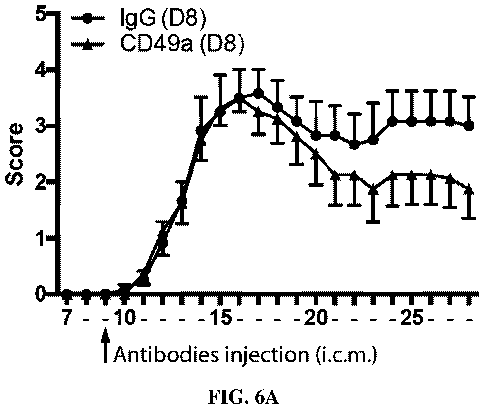
D00013

D00014
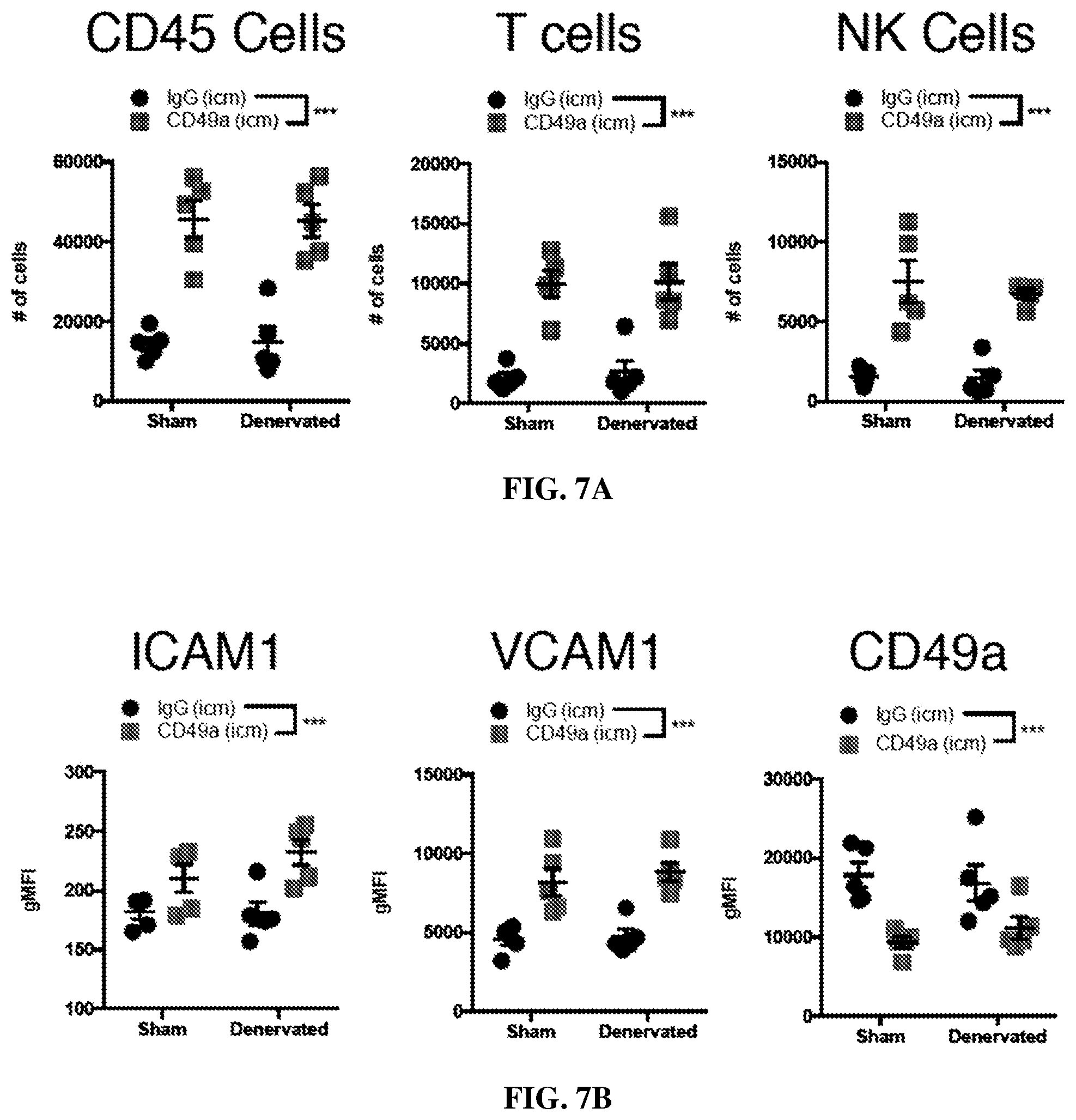
D00015

D00016

D00017
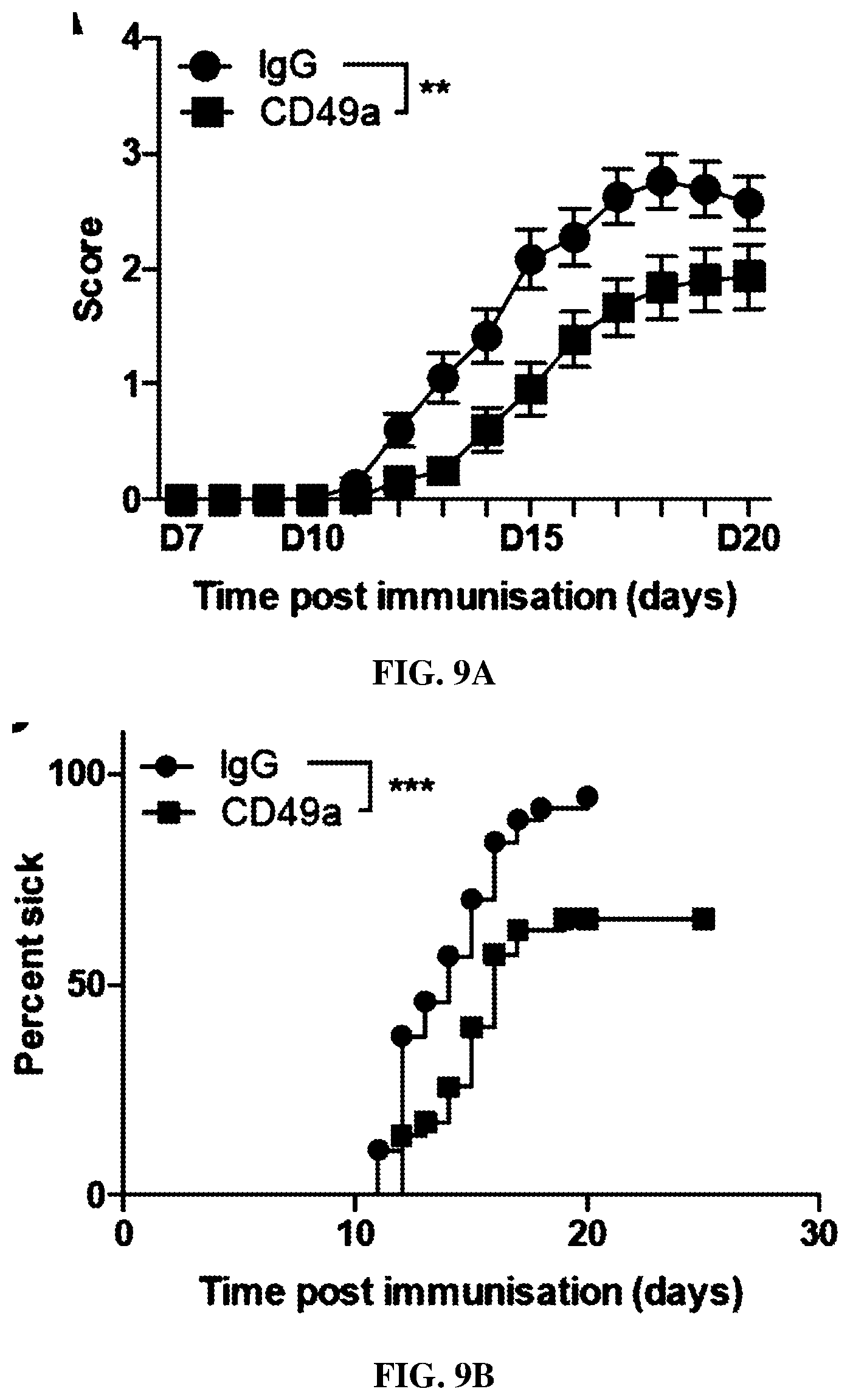
D00018

D00019

D00020
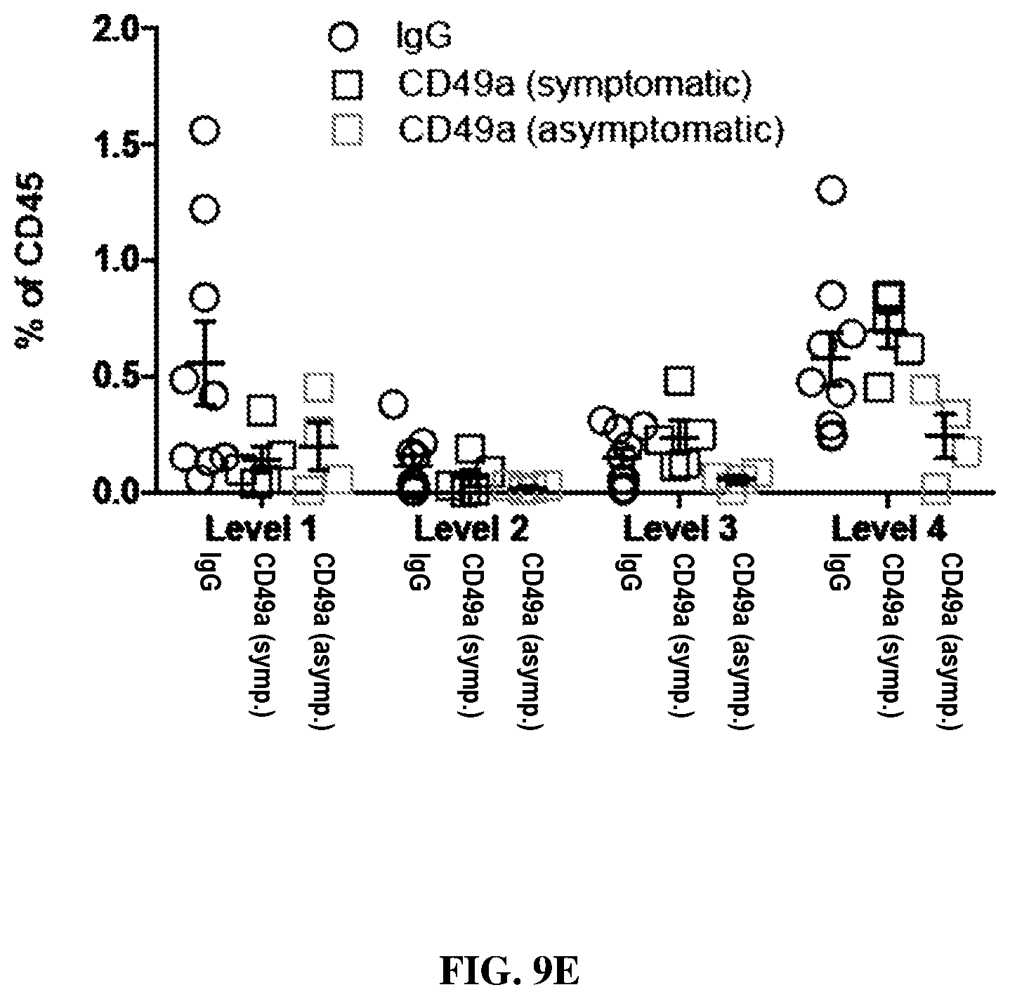
D00021
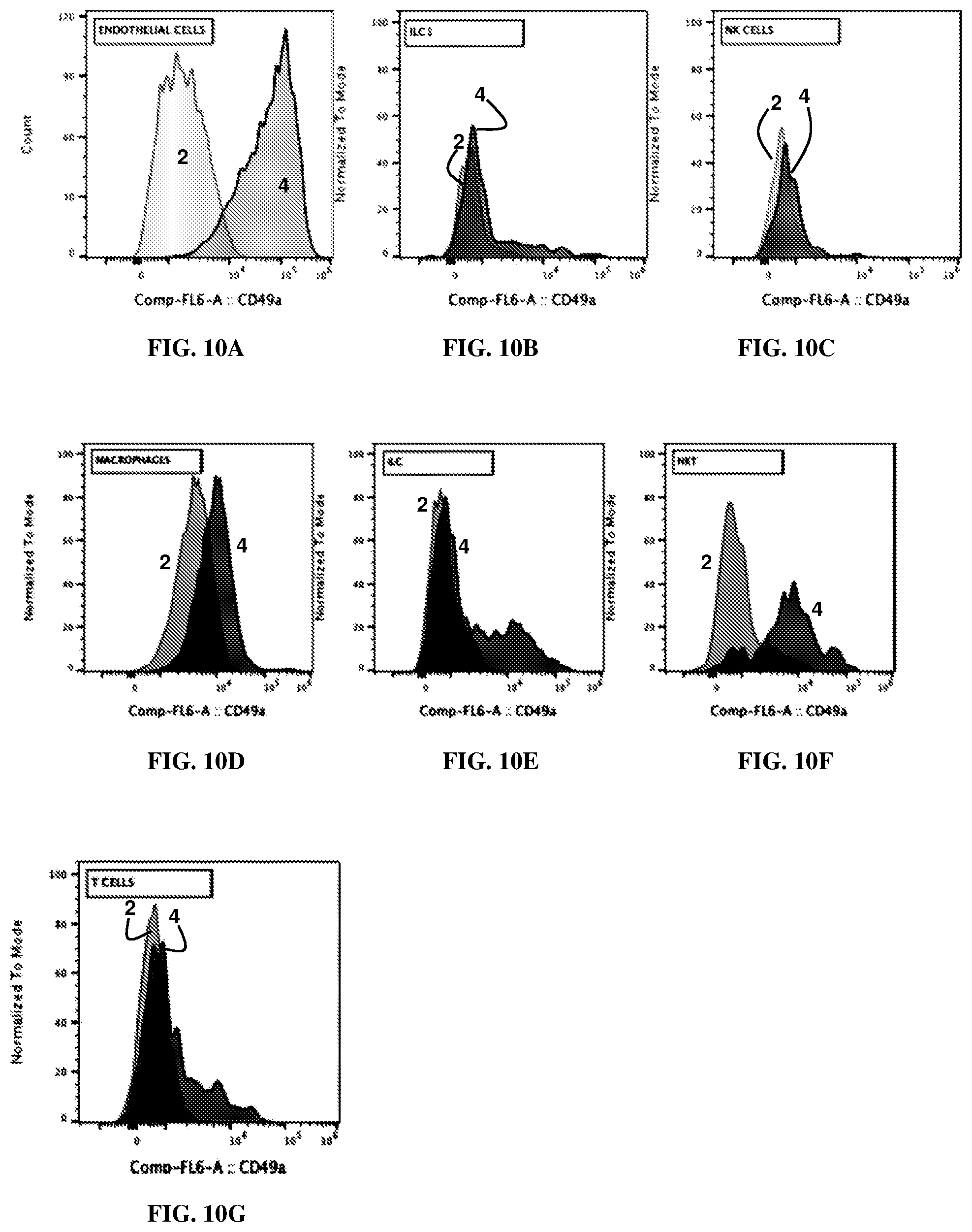

S00001
XML
uspto.report is an independent third-party trademark research tool that is not affiliated, endorsed, or sponsored by the United States Patent and Trademark Office (USPTO) or any other governmental organization. The information provided by uspto.report is based on publicly available data at the time of writing and is intended for informational purposes only.
While we strive to provide accurate and up-to-date information, we do not guarantee the accuracy, completeness, reliability, or suitability of the information displayed on this site. The use of this site is at your own risk. Any reliance you place on such information is therefore strictly at your own risk.
All official trademark data, including owner information, should be verified by visiting the official USPTO website at www.uspto.gov. This site is not intended to replace professional legal advice and should not be used as a substitute for consulting with a legal professional who is knowledgeable about trademark law.