Dosage And Administration Of Anti-c5 Antibodies For Treatment Of Protein-losing Enteropathy In Patients
ADIV; Orly Eshach ; et al.
U.S. patent application number 16/614582 was filed with the patent office on 2020-06-04 for dosage and administration of anti-c5 antibodies for treatment of protein-losing enteropathy in patients. The applicant listed for this patent is Alexion Pharmaceuticals, Inc.. Invention is credited to Orly Eshach ADIV, Camille BEDROSIAN, Hagit Baris FELDMAN, Alina KUROLAP, Susan Faas MCKNIGHT.
| Application Number | 20200172603 16/614582 |
| Document ID | / |
| Family ID | 62620989 |
| Filed Date | 2020-06-04 |
View All Diagrams
| United States Patent Application | 20200172603 |
| Kind Code | A1 |
| ADIV; Orly Eshach ; et al. | June 4, 2020 |
DOSAGE AND ADMINISTRATION OF ANTI-C5 ANTIBODIES FOR TREATMENT OF PROTEIN-LOSING ENTEROPATHY IN PATIENTS
Abstract
Provided are methods for clinical treatment of a protein-losing enteropathy, such as lymphangiectasia, using an anti-C5 antibody, or antigen binding fragment thereof, in patients (e.g., pediatric patients).
| Inventors: | ADIV; Orly Eshach; (Atlit, IL) ; BEDROSIAN; Camille; (Woodbridge, CT) ; FELDMAN; Hagit Baris; (Kochav Yair, IL) ; KUROLAP; Alina; (Haifa, IL) ; MCKNIGHT; Susan Faas; (Old Lyme, CT) | ||||||||||
| Applicant: |
|
||||||||||
|---|---|---|---|---|---|---|---|---|---|---|---|
| Family ID: | 62620989 | ||||||||||
| Appl. No.: | 16/614582 | ||||||||||
| Filed: | May 21, 2018 | ||||||||||
| PCT Filed: | May 21, 2018 | ||||||||||
| PCT NO: | PCT/US2018/033678 | ||||||||||
| 371 Date: | November 18, 2019 |
Related U.S. Patent Documents
| Application Number | Filing Date | Patent Number | ||
|---|---|---|---|---|
| 62509576 | May 22, 2017 | |||
| Current U.S. Class: | 1/1 |
| Current CPC Class: | A61P 1/00 20180101; A61K 2039/545 20130101; C07K 2317/24 20130101; A61P 3/00 20180101; A61K 2039/505 20130101; A61K 2039/54 20130101; A61P 9/00 20180101; C07K 16/18 20130101; A61P 7/00 20180101; C07K 16/40 20130101; C12Q 1/6883 20130101; A61P 37/00 20180101; C07K 2317/565 20130101; G01N 33/574 20130101; C07K 2317/76 20130101; C12Q 2600/156 20130101; G01N 33/6893 20130101 |
| International Class: | C07K 16/18 20060101 C07K016/18; A61P 1/00 20060101 A61P001/00 |
Claims
1. A method of treating a human patient with a protein-losing enteropathy, the method comprising administering to the patient during an administration cycle an effective amount of an anti-C5 antibody, or antigen binding fragment thereof, comprising CDR1, CDR2 and CDR3 heavy chain sequences as set forth in SEQ ID NOs:1, 2 and 3, respectively, and CDR1, CDR2 and CDR3 light chain sequences as set forth in SEQ ID NOs:4, 5 and 6, respectively, wherein the anti-C5 antibody, or antigen binding fragment thereof, is administered at a dose of: i. 600 mg to a patient weighing 10 kg to <20 kg twice per week during weeks 1 and 2 of the administration cycle and once during week 3, followed by 300 mg or 600 mg once per week during weeks 4 and 5 and every two weeks thereafter; ii. 600 mg to a patient weighing 20 kg to <30 kg twice per week during weeks 1 and 2 of the administration cycle, followed by 600 mg once per week during weeks 3, 4 and 5 and every two weeks thereafter; iii. 900 mg to a patient weighing 30 kg to <40 kg twice per week during weeks 1 and 2 of the administration cycle, followed by 900 mg once per week during weeks 3, 4 and 5 and every two weeks thereafter; or iv. 1200 mg to a patient weighing.gtoreq.40 kg twice per week during weeks 1 and 2 of the administration cycle, followed by 1200 mg once per week during weeks 3, 4 and 5 and every two weeks thereafter.
2. A method of treating a human patient with lymphangiectasia, the method comprising administering to the patient during an administration cycle an effective amount of an anti-C5 antibody, or antigen binding fragment thereof, comprising CDR1, CDR2 and CDR3 heavy chain sequences as set forth in SEQ ID NOs:1, 2 and 3, respectively, and CDR1, CDR2 and CDR3 light chain sequences as set forth in SEQ ID NOs:4, 5 and 6, respectively, wherein the anti-C5 antibody, or antigen binding fragment thereof, is administered at a dose of: i. 600 mg to a patient weighing 10 kg to <20 kg twice per week during weeks 1 and 2 of the administration cycle and once during week 3, followed by 300 mg or 600 mg once per week during weeks 4 and 5 and every two weeks thereafter; ii. 600 mg to a patient weighing 20 kg to <30 kg twice per week during weeks 1 and 2 of the administration cycle, followed by 600 mg once per week during weeks 3, 4 and 5 and every two weeks thereafter; iii. 900 mg to a patient weighing 30 kg to <40 kg twice per week during weeks 1 and 2 of the administration cycle, followed by 900 mg once per week during weeks 3, 4 and 5 and every two weeks thereafter; or iv. 1200 mg to a patient weighing.gtoreq.40 kg twice per week during weeks 1 and 2 of the administration cycle, followed by 1200 mg once per week during weeks 3, 4 and 5 and every two weeks thereafter.
3. The method of claim 1, wherein the anti-C5 antibody, or antigen binding fragment thereof, is administered to a patient weighing 10 kg to <20 kg at a dose of 600 mg twice per week during weeks 1 and 2 of the administration cycle and once during week 3, followed by 300 mg or 600 mg once per week during weeks 4 and 5 and every two weeks thereafter.
4. The method of claim 1, wherein the anti-C5 antibody, or antigen binding fragment thereof, is administered to a patient weighing 20 kg to <30 kg at a dose of 600 mg twice per week during weeks 1 and 2 of the administration cycle, followed by 600 mg once per week during weeks 3, 4 and 5 and every two weeks thereafter.
5. The method of claim 1, wherein the anti-C5 antibody, or antigen binding fragment thereof, is administered to a patient weighing 30 kg to <40 kg at a dose of 900 mg twice per week during weeks 1 and 2 of the administration cycle, followed by 900 mg once per week during weeks 3, 4 and 5 and every two weeks thereafter.
6. The method of claim 1, wherein the anti-C5 antibody, or antigen binding fragment thereof, is administered to a patient weighing.gtoreq.40 kg at a dose of 1200 mg twice per week during weeks 1 and 2 of the administration cycle, followed by 1200 mg once per week during weeks 3, 4 and 5 and every two weeks thereafter.
7. The method of claim 1, wherein the anti-C5 antibody, or antigen binding fragment thereof, comprises heavy and/or light chain variable region sequences as set forth in SEQ ID NOs:7 and 8.
8. The method of claim 1, wherein the anti-C5 antibody comprises heavy and/or light chain sequences as set forth in SEQ ID NOs:10 and 11.
9. The method of claim 1, wherein the treatment maintains a serum trough concentration of the anti-C5 antibody, or antigen binding fragment thereof, of 100 .mu.g/mL or greater during the administration cycle.
10. The method of claim 1, wherein the treatment maintains a serum trough concentration of the anti-C5 antibody, or antigen binding fragment thereof, of 200 .mu.g/mL or greater during the administration cycle.
11. The method of claim 1, wherein the anti-C5 antibody, or antigen binding fragment thereof, is formulated for intravenous administration.
12. The method of claim 1, wherein the treatment: (a) produces a shift toward normal levels of serum albumin; (b) results in at least a 1.7-fold increase in serum albumin levels from baseline within 20 days; (c) results in at least a 5-fold increase in serum albumin levels from baseline within 80 days; (d) produces a shift toward normal total protein serum levels; (e) results in at least a 1.5-fold increase in total protein serum levels from baseline within 20 days; (f) results in at least a 2.26-fold increase in total protein serum levels from baseline within 80 days; (g) results in at least a 1.8-fold decrease in serum TNFR1 levels from baseline within 8 days; and/or (h) results in at least a 3.25-fold decrease in serum TNFR1 levels from baseline within 43 days.
13-19. (canceled)
20. The method of claim 1, wherein the treatment produces at least one therapeutic effect selected from the group consisting of: a reduction or cessation in protein loss, edema, diarrhea, ascites, pleural effusion, pericarditis, lymphedema, abdominal pain, fatigue, weight loss and vitamin deficiency.
21. The method of claim 1, wherein the patient is <18 years of age.
22. The method of claim 1, wherein the administration cycle is a total of 27 weeks of treatment.
23. The method of claim 1, wherein the anti-C5 antibody, or antigen binding fragment thereof, is administered at a dose of 300 mg, 600 mg, 900 mg or 1200 mg or every two weeks after the administration cycle for up to two years.
24. A kit for treating protein-losing enteropathy in a human patient, the kit comprising: (a) a dose of an anti-C5 antibody, or antigen binding fragment thereof, comprising CDR1, CDR2 and CDR3 domains of the heavy chain variable region having the sequence set forth in SEQ ID NO:7, and CDR1, CDR2 and CDR3 domains of the light chain variable region having the sequence set forth in SEQ ID NO:8; and (b) instructions for using the anti-C5 antibody, or antigen binding fragment thereof, in a method for treating protein-losing enteropathy in a human patient.
25-29. (canceled)
30. An anti-C5 antibody, or antigen binding fragment thereof, comprising CDR1, CDR2 and CDR3 domains of the heavy chain variable region having the sequence set forth in SEQ ID NO:7, and CDR1, CDR2 and CDR3 domains of the light chain variable region having the sequence set forth in SEQ ID NO:8, for administration in a cycle, wherein the anti-C5 antibody, or antigen binding fragment thereof, is administered: i. 600 mg to a patient weighing 10 kg to <20 kg twice per week during weeks 1 and 2 of the administration cycle and once during week 3, followed by 300 mg or 600 mg once per week during weeks 4 and 5 and every two weeks thereafter; ii. 600 mg to a patient weighing 20 kg to <30 kg twice per week during weeks 1 and 2 of the administration cycle, followed by 600 mg once per week during weeks 3, 4 and 5 and every two weeks thereafter; iii. 900 mg to a patient weighing 30 kg to <40 kg twice per week during weeks 1 and 2 of the administration cycle, followed by 900 mg once per week during weeks 3, 4 and 5 and every two weeks thereafter; or iv. 1200 mg to a patient weighing.gtoreq.40 kg twice per week during weeks 1 and 2 of the administration cycle, followed by 1200 mg once per week during weeks 3, 4 and 5 and every two weeks thereafter.
31. The antibody of claim 30, wherein the antibody is determined to be safe, tolerable, efficacious and sufficiently non-immunogenic after multiple IV doses for use in patients with protein-losing enteropathy.
32. The antibody of claim 30, wherein the antibody is determined to be safe, tolerable, efficacious and sufficiently non-immunogenic after multiple IV doses for use in patients with lymphangiectasia.
33. (canceled)
Description
CROSS-REFERENCE TO RELATED APPLICATIONS
[0001] This application claims the benefit of U.S. Provisional Application Ser. No. 62/509,576, filed on May 22, 2017. The entire contents of the above-referenced provisional patent application are incorporated herein by reference.
SEQUENCE LISTING
[0002] The instant application contains a Sequence Listing which has been submitted electronically in ASCII format and is hereby incorporated by reference in its entirety. Said ASCII copy, created on May 15, 2018, is named AXJ-240PC_SL.txt and is 25,120 bytes in size.
BACKGROUND
[0003] Protein-losing enteropathy (PLE) is a rare condition that is characterized by loss of protein and other nutrients through the intestinal tract. Patients with PLE may suffer from a variety of symptoms including edema, ascites, pleural effusion, pericarditis, lymphedema, diarrhea, abdominal pain, fatigue, weight loss and vitamin deficiency. PLE diagnosis is commonly based on clearance of fecal .alpha.-1 antitrypsin and treatment included management of symptoms and dietary modification.
[0004] PLE is not a single disease, but a complication that arises as a result of mucosal injury or an intestinal disorder. One such disorder is primary intestinal lymphangiectasia (also known as "intestinal lymphangiectasia," "primary intestinal lymphangiectasia" and "PIL"). Though adults may be diagnosed with lymphangiectasia, it is most commonly reported in children and young adults. Diagnosis is based on endoscopic evaluation and histopathological examination of tissue biopsy specimens. Current treatments include low-fat diet plans supplemented with medium-chain triglycerides (MCT), administration of octreotide, and serum albumin transfusions.
[0005] Patients with PLE (e.g., lymphangiectasia), are at risk of substantial morbidity and mortality. Accordingly, it is an object herein to provide methods for treating human patients (e.g., pediatric patients) with PLE (e.g., lymphangiectasia).
SUMMARY
[0006] Provided herein are compositions and methods for treating protein-losing enteropathy (e.g., lymphangiectasia) in a patient (e.g., a pediatric patient<18 years of age), comprising administering to the patient an anti-C5 antibody, or antigen binding fragment thereof, wherein the anti-C5 antibody, or antigen binding fragment thereof, is administered (or is for administration) according to a particular clinical dosage regimen (i.e., according to an initial high and frequent dosing regimen, followed by regular maintenance dosing as albumin levels normalize). In one embodiment, the patient has not previously been treated with a complement inhibitor (e.g., the patient is a complement inhibitor treatment-naive patient).
[0007] An exemplary anti-C5 antibody is eculizumab (Soliris.RTM.). In other embodiments, the antibody comprises the heavy and light chain CDRs or variable regions of eculizumab. Accordingly, in one embodiment, the antibody comprises the CDR1, CDR2 and CDR3 domains of the VH region of eculizumab having the sequence set forth in SEQ ID NO:7, and the CDR1, CDR2 and CDR3 domains of the VL region of eculizumab having the sequence set forth in SEQ ID NO:8. In another embodiment, the antibody comprises heavy chain CDR1, CDR2 and CDR3 domains having the sequences set forth in SEQ ID NOs:1, 2 and 3, respectively, and light chain CDR1, CDR2 and CDR3 domains having the sequences set forth in SEQ ID NOs:4, 5 and 6, respectively. In another embodiment, the antibody comprises a VH region having the amino acid sequence set forth in SEQ ID NO:7. In another embodiment, the antibody comprises a VL region having the amino acid sequence set forth in SEQ ID NO:8. In another embodiment, the antibody comprises VH and VL regions having the amino acid sequences set forth in SEQ ID NO:7 and SEQ ID NO:8, respectively. In another embodiment, the antibody comprises the heavy chain constant region of eculizumab having the sequence set forth in SEQ ID NO:9. In another embodiment, the antibody comprises a heavy chain having the amino acid sequence set forth in SEQ ID NO:10. In another embodiment, the antibody comprises a light chain having the amino acid sequence set forth in SEQ ID NO:11. In another embodiment, the antibody comprises heavy and light chains having the amino acid sequences set forth in SEQ ID NO:10 and SEQ ID NO:11, respectively.
[0008] In another embodiment, the antibody competes for binding with, and/or binds to the same epitope on C5 as, the above-mentioned antibodies. In another embodiment, the antibody has at least about 90% variable region amino acid sequence identity with the above-mentioned antibodies (e.g., at least about 90%, 95% or 99% variable region identity with SEQ ID NO:7 and SEQ ID NO:8).
[0009] In one embodiment, the patient treated according to the methods described herein is an adult who is 18 years or older (.gtoreq.18 years of age). In another embodiment, the patient treated according to the methods described herein is a pediatric patient who is less than 18 years of age (<18 years). In one embodiment, the pediatric patient is less than 12 years of age (<12 years). In another embodiment, the pediatric patient is less than 6 years of age (<6 years). In another embodiment, the pediatric patient is less than 2 years of age (<2 years). In another embodiment, the pediatric patient is less than 1, 2, 3, 4, 5, 6, 7, 8, 9, 10, 11, 12, 13, 14, 15, 16, 17 or 18 years of age.
[0010] In one embodiment, the dose of the anti-C5 antibody, or antigen binding fragment thereof, is based on the weight of the patient. For example, in one embodiment, about 300 mg, about 600 mg, about 900 mg, and/or about 1200 mg of the anti-C5 antibody, or antigen binding fragment thereof, is administered to a patient based on the patient's weight. In one embodiment, 300 mg or 600 mg of the anti-C5 antibody, or antigen binding fragment thereof, is administered to a patient weighing 10 kg to <20 kg. In another embodiment, 600 mg of the anti-C5 antibody, or antigen binding fragment thereof, is administered to a patient weighing 10 kg to <30 kg. In another embodiment, 600 mg of the anti-C5 antibody, or antigen binding fragment thereof, is administered to a patient weighing 20 kg to <30 kg. In another embodiment, 900 mg of the anti-C5 antibody, or antigen binding fragment thereof, is administered to a patient weighing 30 kg to <40 kg. In another embodiment, 1200 mg of the anti-C5 antibody, or antigen binding fragment thereof, is administered to a patient weighing.gtoreq.40 kg. In certain embodiments, dosage regimens are adjusted to provide the optimum desired response (e.g., an effective response).
[0011] In another embodiment, the anti-C5 antibody, or antigen binding fragment thereof, is administered for one or more administration cycles. In one embodiment, the administration cycle is 27 weeks.
[0012] In one embodiment, the treatment comprises an induction phase, wherein the anti-C5 antibody, or antigen binding fragment thereof, is administered twice per week for two weeks, followed by three weekly doses. In another embodiment, the induction phase is followed by a maintenance phase, wherein the anti-C5 antibody, or antigen binding fragment thereof, is administered every two weeks. In another embodiment, the anti-C5 antibody, or antigen binding fragment thereof, is administered twice per week during weeks 1 and 2 of the administration cycle, followed by once per week during weeks 3, 4 and 5 and every two weeks thereafter.
[0013] In another embodiment, a method of treating a human patient with protein-losing enteropathy (PLE) is provided, the method comprising administering to the patient during an administration cycle an effective amount of an anti-C5 antibody, or antigen binding fragment thereof, comprising CDR1, CDR2 and CDR3 heavy chain sequences as set forth in SEQ ID NOs:1, 2 and 3, respectively, and CDR1, CDR2 and CDR3 light chain sequences as set forth in SEQ ID NOs:4, 5 and 6, respectively, wherein the anti-C5 antibody, or antigen binding fragment thereof, is administered at a dose of: i) 600 mg to a patient weighing 10 kg to <20 kg twice per week during weeks 1 and 2 of the administration cycle and once during week 3, followed by 300 mg or 600 mg once per week during weeks 4 and 5 and every two weeks thereafter; ii) 600 mg to a patient weighing 20 kg to <30 kg twice per week during weeks 1 and 2 of the administration cycle, followed by 600 mg once per week during weeks 3, 4 and 5 and every two weeks thereafter; iii) 900 mg to a patient weighing 30 kg to <40 kg twice per week during weeks 1 and 2 of the administration cycle, followed by 900 mg once per week during weeks 3, 4 and 5 and every two weeks thereafter; or iv) 1200 mg to a patient weighing.gtoreq.40 kg twice per week during weeks 1 and 2 of the administration cycle, followed by 1200 mg once per week during weeks 3, 4 and 5 and every two weeks thereafter.
[0014] In another embodiment, a method of treating a human pediatric patient with lymphangiectasia is provided, the method comprising administering to the patient during an administration cycle an effective amount of an anti-C5 antibody, or antigen binding fragment thereof, comprising CDR1, CDR2 and CDR3 heavy chain sequences as set forth in SEQ ID NOs:1, 2 and 3, respectively, and CDR1, CDR2 and CDR3 light chain sequences as set forth in SEQ ID NOs:4, 5 and 6, respectively, wherein the anti-C5 antibody, or antigen binding fragment thereof, is administered at a dose of: i) 600 mg to a patient weighing 10 kg to <20 kg twice per week during weeks 1 and 2 of the administration cycle and once during week 3, followed by 300 mg or 600 mg once per week during weeks 4 and 5 and every two weeks thereafter; ii) 600 mg to a patient weighing 20 kg to <30 kg twice per week during weeks 1 and 2 of the administration cycle, followed by 600 mg once per week during weeks 3, 4 and 5 and every two weeks thereafter; iii) 900 mg to a patient weighing 30 kg to <40 kg twice per week during weeks 1 and 2 of the administration cycle, followed by 900 mg once per week during weeks 3, 4 and 5 and every two weeks thereafter; or iv) 1200 mg to a patient weighing.gtoreq.40 kg twice per week during weeks 1 and 2 of the administration cycle, followed by 1200 mg once per week during weeks 3, 4 and 5 and every two weeks thereafter.
[0015] In another embodiment, 600 mg of the anti-C5 antibody, or antigen binding fragment thereof, is administered to a patient weighing 10 kg to <30 kg.
[0016] In another embodiment, the anti-C5 antibody, or antigen binding fragment thereof, is administered to a patient weighing 10 kg to <20 kg at a dose of 600 mg twice per week during weeks 1 and 2 of the administration cycle and once during week 3, followed by 300 mg or 600 mg once per week during weeks 4 and 5 and every two weeks thereafter.
[0017] In another embodiment, the anti-C5 antibody, or antigen binding fragment thereof, is administered to a patient weighing 20 kg to <30 kg at a dose of 600 mg twice per week during weeks 1 and 2 of the administration cycle, followed by 600 mg once per week during weeks 3, 4 and 5 and every two weeks thereafter.
[0018] In another embodiment, the anti-C5 antibody, or antigen binding fragment thereof, is administered to a patient weighing 30 kg to <40 kg at a dose of 900 mg twice per week during weeks 1 and 2 of the administration cycle, followed by 900 mg once per week during weeks 3, 4 and 5 and every two weeks thereafter.
[0019] In another embodiment, the anti-C5 antibody, or antigen binding fragment thereof, is administered to a patient weighing.gtoreq.40 kg at a dose of 1200 mg twice per week during weeks 1 and 2 of the administration cycle, followed by 1200 mg once per week during weeks 3, 4 and 5 and every two weeks thereafter.
[0020] In another embodiment, the anti-C5 antibody, or antigen binding fragment thereof, is administered every two weeks after completion of the administration cycle. In another embodiment, the anti-C5 antibody, or antigen binding fragment thereof, is administered on a monthly basis (e.g., every four weeks) or every other month basis (e.g., every eight weeks) after completion of the administration cycle. In another embodiment, the anti-C5 antibody, or antigen binding fragment thereof, is administered on a monthly basis or every other month basis for a year after completion of the administration cycle. In another embodiment, the anti-C5 antibody, or antigen binding fragment thereof, is administered on a monthly basis or every other month basis for two, three, four or five years after completion of the administration cycle. In a particular embodiment, the anti-C5 antibody, or antigen binding fragment thereof, is administered on a monthly basis or every other month basis for up to two years after completion of the administration cycle.
[0021] In another aspect, the treatment regimens described are sufficient to maintain particular serum trough concentrations of the anti-C5 antibody, or antigen binding fragment thereof. For example, in one embodiment, the treatment maintains a serum trough concentration of the anti-C5 antibody, or antigen binding fragment thereof, of 50, 55, 60, 65, 70, 75, 80, 85, 90, 95, 100, 105, 110, 115, 120, 125, 130, 135, 140, 145, 150, 155, 160, 165, 170, 175, 180, 185, 190, 200, 205, 210, 215, 220, 225, 230, 240, 245, 250, 255, 260, 265, 270, 280, 290, 300, 305, 310, 315, 320, 325, 330, 335, 340, 345, 350, 355, 360, 365, 370, 375, 380, 385, 390, 395 or 400 .mu.g/mL or greater. In one embodiment, the treatment maintains a serum trough concentration of the anti-C5 antibody, or antigen binding fragment thereof, of 100 .mu.g/mL or greater. In another embodiment, the treatment maintains a serum trough concentration of the anti-C5 antibody, or antigen binding fragment thereof, of 150 .mu.g/mL or greater. In another embodiment, the treatment maintains a serum trough concentration of the anti-C5 antibody, or antigen binding fragment thereof, of 200 .mu.g/mL or greater. In another embodiment, the treatment maintains a serum trough concentration of the anti-C5 antibody, or antigen binding fragment thereof, of 250 .mu.g/mL or greater. In another embodiment, the treatment maintains a serum trough concentration of the anti-C5 antibody, or antigen binding fragment thereof, of 300 .mu.g/mL or greater. In another embodiment, the treatment maintains a serum trough concentration of the anti-C5 antibody, or antigen binding fragment thereof, of between 100 .mu.g/mL and 200 .mu.g/mL. In another embodiment, the treatment maintains a serum trough concentration of the anti-C5 antibody, or antigen binding fragment thereof, of about 175 .mu.g/mL.
[0022] In another embodiment, to obtain an effective response, the anti-C5 antibody is administered to the patient in an amount and with a frequency to maintain at least 50 .mu.g, 55 .mu.g, 60 .mu.g, 65 .mu.g, 70 .mu.g, 75 .mu.g, 80 .mu.s, 85 .mu.s, 90 .mu.s, 95 .mu.g, 100 .mu.g, 105 .mu.s, 110 .mu.s, 115 .mu.g, 120 .mu.g, 125 .mu.g, 130 .mu.s, 135 .mu.g, 140 .mu.g, 145 .mu.s, 150 .mu.g, 155 .mu.g, 160 .mu.s, 165 .mu.g, 170 .mu.s, 175 .mu.g, 180 .mu.s, 185 .mu.g, 190 .mu.g, 195 .mu.g, 200 .mu.s, 205 .mu.g, 210 .mu.g, 215 .mu.g, 220 .mu.g, 225 .mu.s, 230 .mu.g, 235 .mu.s, 240 .mu.g, 245 .mu.g, 250 .mu.g, 255 .mu.g or 260 .mu.g of antibody per milliliter of the patient's blood. In another embodiment, the anti-C5 antibody is administered to the patient in an amount and with a frequency to maintain between 50 .mu.s and 250 .mu.s of antibody per milliliter of the patient's blood. In another embodiment, the anti-C5 antibody is administered to the patient in an amount and with a frequency to maintain between 100 .mu.g and 200 .mu.g of antibody per milliliter of the patient's blood. In another embodiment, the anti-C5 antibody is administered to the patient in an amount and with a frequency to maintain about 175 .mu.g of antibody per milliliter of the patient's blood.
[0023] In another embodiment, to obtain an effective response, the anti-C5 antibody is administered to the patient in an amount and with a frequency to maintain a minimum free C5 concentration. For example, in one embodiment, the anti-C5 antibody is administered to the patient in an amount and with a frequency to maintain a free C5 concentration of 0.2 .mu.g/mL, 0.3 .mu.g/mL, 0.4 .mu.g/mL, 0.5 .mu.g/mL or below.
[0024] The anti-C5 antibodies, or antigen binding fragments thereof, can be administered to a patient by any suitable means. In one embodiment, the antibodies are formulated for intravenous administration.
[0025] In one embodiment, the patient has been vaccinated with a Neisseria meningococcal vaccine prior to receiving the treatment methods described herein. Patients who receive treatment less than two weeks after receiving a meningococcal vaccine can receive treatment with appropriate prophylactic antibiotics until two weeks after vaccination. Vaccines against serotypes A, C, Y, W 135, and B, where available, are recommended to prevent common pathogenic meningococcal serotypes.
[0026] The efficacy of the treatment methods provided herein can be assessed using any suitable means. In one embodiment, for a patient with PLE (e.g., lymphangiectasia), the treatment produces at least one therapeutic effect selected from the group consisting of: a reduction or cessation in protein loss, edema, diarrhea, ascites, pleural effusion, pericarditis, lymphedema, abdominal pain, fatigue, weight loss and vitamin deficiency.
[0027] In another embodiment, the treatment produces a shift toward normal levels of total protein (e.g., total serum protein). For example, in one embodiment, patients treated according to the disclosed methods experience an increase in total protein serum levels to near normal levels or to within about 10% or within about 20% above or below what is considered the normal level of total protein. In another embodiment, the treatment results in at least a 1.0-fold, 1.1-fold, 1.2-fold, 1.3-fold, 1.4-fold, 1.5-fold, 1.6-fold, 1.7-fold, 1.8-fold, 1.9-fold or 2.0-fold increase in total protein serum levels from baseline within 20 days. In a particular embodiment, the treatment results in at least a 1.5-fold increase in total protein serum levels from baseline within 20 days. In another embodiment, the treatment results in at least a 1.5-fold, 1.6-fold, 1.7-fold, 1.8-fold, 1.9-fold, 2.0-fold, 2.1-fold, 2.2-fold, 2.3-fold, 2.4-fold, 2.5-fold, 2.6-fold, 2.7-fold, 2.8-fold, 2.9-fold or 3.0-fold increase in total protein serum levels from baseline within 80 days. In a particular embodiment, the treatment results in at least about a 2.3-fold (e.g., 2.26-fold) increase in total protein serum levels from baseline within 80 days.
[0028] In another embodiment, the treatment produces a shift toward normal serum albumin levels. For example, in one embodiment, patients treated according to the disclosed methods experience an increase in serum albumin levels to near normal levels or to within about 10% or within about 20% above or below what is considered the normal level of serum albumin. In another embodiment, the treatment results in at least a 1.0-fold, 1.1-fold, 1.2-fold, 1.3-fold, 1.4-fold, 1.5-fold, 1.6-fold, 1.7-fold, 1.8-fold, 1.9-fold or 2.0-fold increase in serum albumin levels from baseline within 20 days. In a particular embodiment, the treatment results in at least a 1.7-fold increase in serum albumin levels from baseline within 20 days. In another embodiment, the treatment results in at least a 2.0-fold, 2.1-fold, 2.2-fold, 2.3-fold, 2.4-fold, 2.5-fold, 2.6-fold, 2.7-fold, 2.8-fold, 2.9-fold, 3.0-fold, 3.1-fold, 3.2-fold, 3.3-fold, 3.4-fold, 3.5-fold, 3.6-fold, 3.7-fold, 3.8-fold, 3.9-fold, 4.0-fold, 4.1-fold, 4.2-fold, 4.3-fold, 4.4-fold, 4.5-fold, 4.6-fold, 4.7-fold, 4.8-fold, 4.9-fold, 5.0-fold, 5.1-fold, 5.2-fold, 5.3-fold, 5.4-fold, 5.5-fold, 5.6-fold, 5.7-fold, 5.8-fold, 5.9-fold, 6.0-fold, 6.1-fold, 6.2-fold, 6.3-fold, 6.4-fold, 6.5-fold, 6.6-fold, 6.7-fold, 6.8-fold, 6.9-fold or 7.0-fold increase in serum albumin levels from baseline within 80 days. In another embodiment, the treatment results in at least a 5.0-fold increase in serum albumin levels from baseline within 80 days.
[0029] In another embodiment, the treatment produces a shift toward normal serum TNFR1 levels. For example, in one embodiment, patients treated according to the disclosed methods experience a decrease in serum TNFR1 levels to near normal levels or to within about 10%, or within about 20% above or below what is considered the normal level of serum TNFR1. In another embodiment, the treatment results in at least a 1.0-fold, 1.1-fold, 1.2-fold, 1.3-fold, 1.4-fold, 1.5-fold, 1.6-fold, 1.7-fold, 1.8-fold, 1.9-fold, 2.0-fold, 2.1-fold, 2.2-fold or 2.3-fold decrease in serum TNFR1 levels from baseline within 8 days. In a particular embodiment, the treatment results in at least a 1.8-fold decrease in serum TNFR1 levels from baseline within 8 days. In another embodiment, the treatment results in at least a 2.0-fold, 2.1-fold, 2.2-fold, 2.3-fold, 2.4-fold, 2.5-fold, 2.6-fold, 2.7-fold, 2.8-fold, 2.9-fold, 3.0-fold, 3.1-fold, 3.2-fold, 3.3-fold, 3.4-fold, 3.5-fold, 3.6-fold, 3.7-fold, 3.8-fold, 3.9-fold, 4.0-fold, 4.1-fold, 4.2-fold, 4.3-fold, 4.4-fold, 4.5-fold, 4.6-fold, 4.7-fold, 4.8-fold, 4.9-fold, 5.0-fold, 5.1-fold, 5.2-fold, 5.3-fold, 5.4-fold, 5.5-fold, 5.6-fold, 5.7-fold, 5.8-fold, 5.9-fold, 6.0-fold, 6.1-fold, 6.2-fold, 6.3-fold, 6.4-fold, 6.5-fold, 6.6-fold, 6.7-fold, 6.8-fold, 6.9-fold or 7.0-fold decrease in serum TNFR1 levels from baseline within 43 days. In a particular embodiment, the treatment results in at least about a 3.3-fold (e.g., 3.25-fold) decrease in serum TNFR1 levels from baseline within 43 days.
[0030] In another embodiment, the treatment produces a shift toward normal free C5 levels. In one embodiment, the free C5 levels are decreased to below the limit of detection. In another embodiment, the free C5 levels are decreased to below the limit of detection following the first treatment dose.
[0031] In another embodiment, the treatment produces a shift toward normal membrane attack complex (MAC) deposition on white blood cells (WBCs), including granulocytes and monocytes. For example, in one embodiment, patients treated according to the methods described herein experience about a 60% reduction in MAC deposition on white bloods cells (WBCs) from baseline after 59 days. In another embodiment, the treatment results in a least about a 40%, 50%, 60%, 70% or 80% reduction in MAC deposition on white blood cells (WBCs) from baseline. In another embodiment, the treatment results in at least a 1.0-fold, 1.1-fold, 1.2-fold, 1.3-fold, 1.4-fold, 1.5-fold, 1.6-fold, 1.7-fold, 1.8-fold, 1.9-fold, 2.0-fold, 2.1-fold, 2.2-fold, 2.3-fold, 2.4-fold, 2.5-fold, 2.6-fold, 2.7-fold, 2.8-fold, 2.9-fold, 3.0-fold, 3.1-fold, 3.2-fold, 3.3-fold, 3.4-fold, 3.5-fold, 3.6-fold, 3.7-fold, 3.8-fold, 3.9-fold, 4.0-fold, 4.1-fold, 4.2-fold, 4.3-fold, 4.4-fold, 4.5-fold, 4.6-fold, 4.7-fold, 4.8-fold, 4.9-fold, 5.0-fold, 5.1-fold, 5.2-fold, 5.3-fold, 5.4-fold, 5.5-fold, 5.6-fold, 5.7-fold, 5.8-fold, 5.9-fold, 6.0-fold, 6.1-fold, 6.2-fold, 6.3-fold, 6.4-fold, 6.5-fold, 6.6-fold, 6.7-fold, 6.8-fold, 6.9-fold or 7.0-fold decrease in MAC deposition on white blood cells (WBCs) compared to baseline.
[0032] In another embodiment, total protein levels, serum albumin levels, serum TNFR1 levels, free C5 levels, and/or MAC deposition on white blood cells (WBCs) are used to evaluate responsiveness to a therapy (e.g., an increase in total serum protein and/or serum albumin levels and/or a decrease in serum TNFR1 levels is indicative of an improvement in at least one sign of PLE (e.g., lymphangiectasia).
[0033] In another aspect, an anti-C5 antibody, or antigen binding fragment thereof, is provided, comprising CDR1, CDR2 and CDR3 domains of the heavy chain variable region having the sequence set forth in SEQ ID NO:7, and CDR1, CDR2 and CDR3 domains of the light chain variable region having the sequence set forth in SEQ ID NO:8, for administration to a patient (e.g., a patient having a PLE, such as, for example, lymphangiectasia), wherein the anti-C5 antibody, or antigen binding fragment thereof, is administered at a dose of: i) 600 mg to a patient weighing 10 kg to <20 kg twice per week during weeks 1 and 2 of the administration cycle and once during week 3, followed by 300 mg or 600 mg once per week during weeks 4 and 5 and every two weeks thereafter; ii) 600 mg to a patient weighing 20 kg to <30 kg twice per week during weeks 1 and 2 of the administration cycle, followed by 600 mg once per week during weeks 3, 4 and 5 and every two weeks thereafter; iii) 900 mg to a patient weighing 30 kg to <40 kg twice per week during weeks 1 and 2 of the administration cycle, followed by 900 mg once per week during weeks 3, 4 and 5 and every two weeks thereafter; or iv) 1200 mg to a patient weighing.gtoreq.40 kg twice per week during weeks 1 and 2 of the administration cycle, followed by 1200 mg once per week during weeks 3, 4 and 5 and every two weeks thereafter.
[0034] In one embodiment, the antibody is determined to be safe, tolerable and sufficiently non-immunogenic after multiple IV doses for use in patients with PLE (e.g., lymphangiectasia).
[0035] Further provided are kits that include a pharmaceutical composition containing an anti-C5 antibody, or antigen binding fragment thereof, such as eculizumab, and a pharmaceutically acceptable carrier, in a therapeutically effective amount adapted for use in the methods described herein. In one embodiment, the kit comprises: a) a dose of an anti-C5 antibody, or antigen binding fragment thereof, comprising CDR1, CDR2 and CDR3 domains of the heavy chain variable region having the sequence set forth in SEQ ID NO:7, and CDR1, CDR2 and CDR3 domains of the light chain variable region having the sequence set forth in SEQ ID NO:8; and b) instructions for using the anti-C5 antibody, or antigen binding fragment thereof, according to any of the methods described herein.
[0036] In another embodiment, the kit comprises a dose of an anti-C5 antibody, or antigen binding fragment thereof, wherein the anti-C5 antibody, or antigen binding fragment thereof, is administered to a patient weighing 10 kg to <20 kg at a dose of 600 mg twice per week during weeks 1 and 2 of the administration cycle and once during week 3, followed by 300 mg or 600 mg once per week during weeks 4 and 5 and every two weeks thereafter.
[0037] In another embodiment, the kit comprises a dose of an anti-C5 antibody, or antigen binding fragment thereof, wherein the anti-C5 antibody, or antigen binding fragment thereof, is administered to a patient weighing 20 kg to <30 kg at a dose of 600 mg twice per week during weeks 1 and 2 of the administration cycle, followed by 600 mg once per week during weeks 3, 4 and 5 and every two weeks thereafter.
[0038] In another embodiment, the kit comprises a dose of an anti-C5 antibody, or antigen binding fragment thereof, wherein the anti-C5 antibody, or antigen binding fragment thereof, is administered to a patient weighing 30 kg to <40 kg at a dose of 900 mg twice per week during weeks 1 and 2 of the administration cycle, followed by 900 mg once per week during weeks 3, 4 and 5 and every two weeks thereafter.
[0039] In another embodiment, the kit comprises a dose of an anti-C5 antibody, or antigen binding fragment thereof, is administered to a patient weighing.gtoreq.40 kg at a dose of 1200 mg twice per week during weeks 1 and 2 of the administration cycle, followed by 1200 mg once per week during weeks 3, 4 and 5 and every two weeks thereafter.
BRIEF DESCRIPTION OF THE DRAWINGS
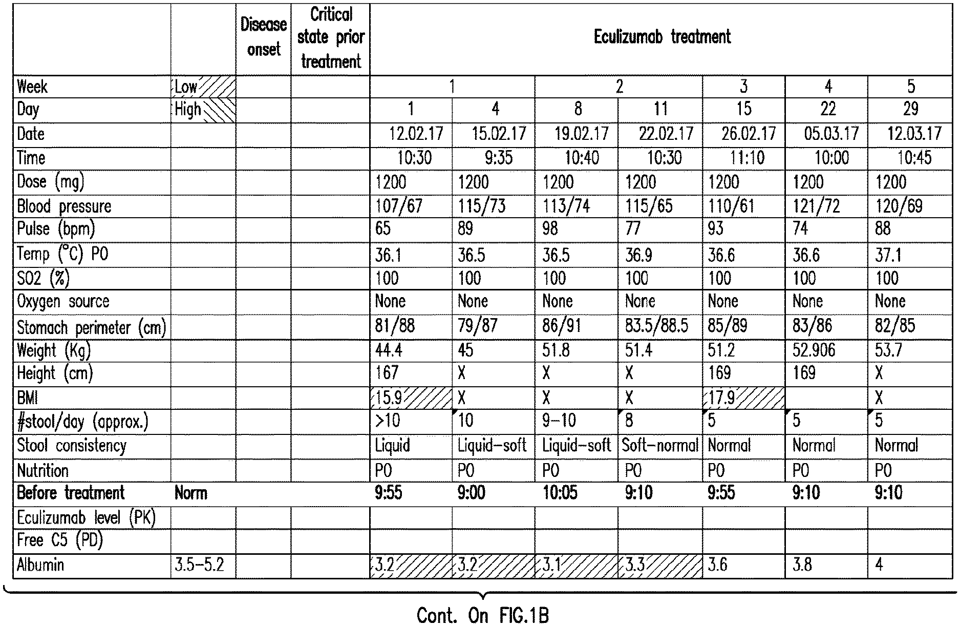
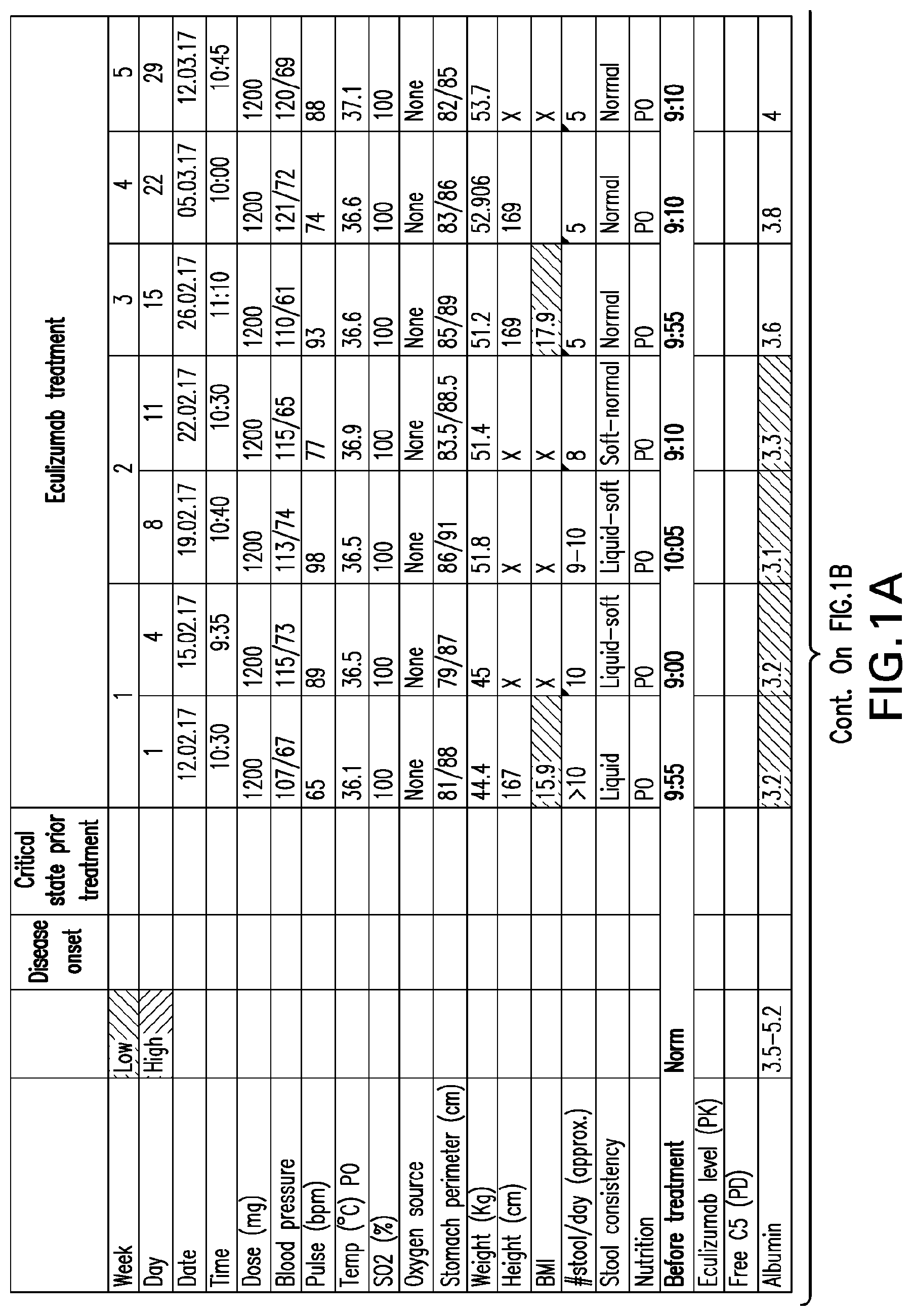
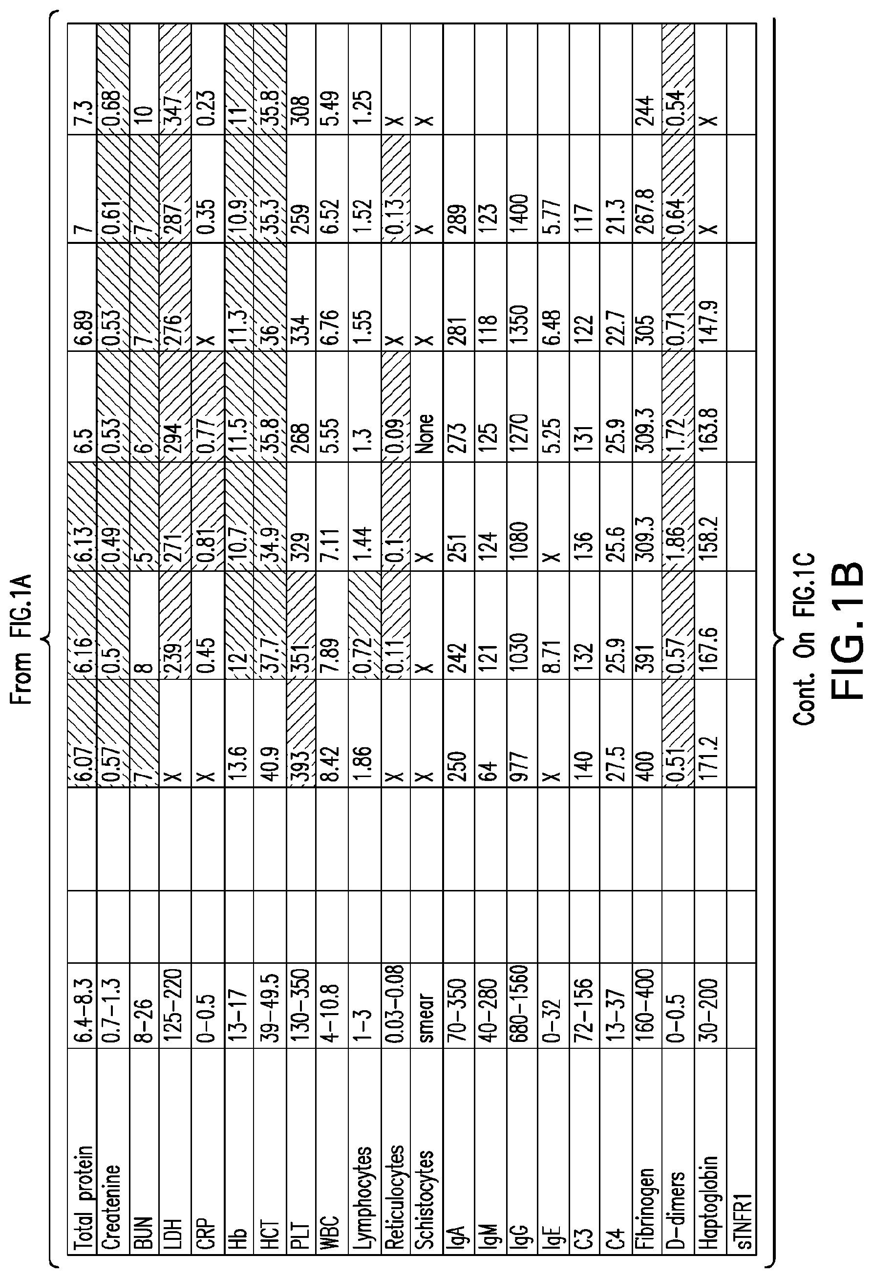
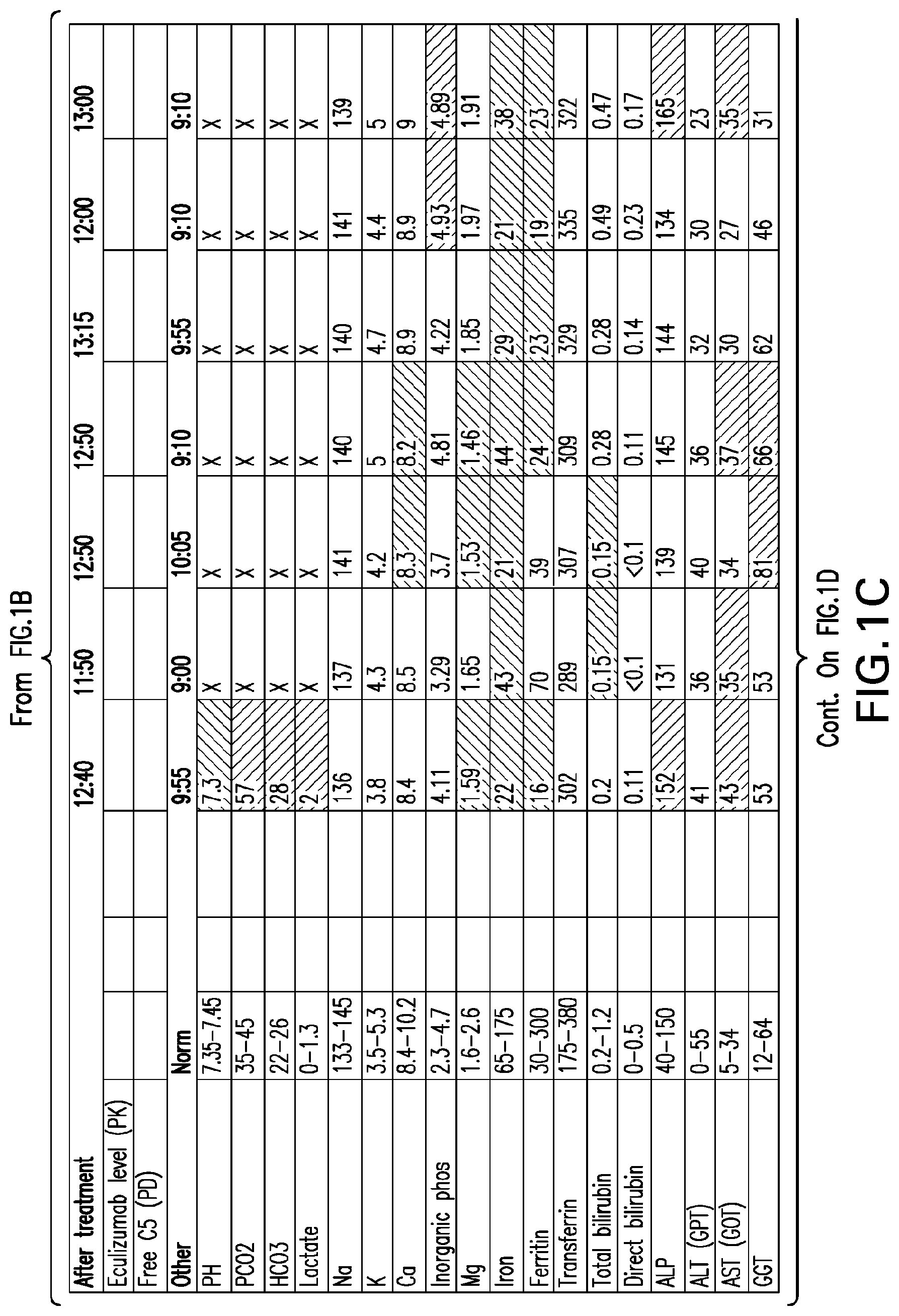
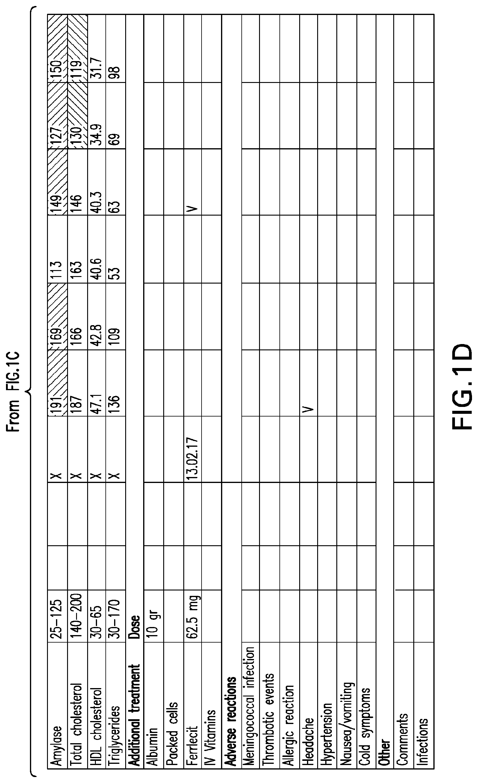
[0040] FIGS. 1A-1D set forth laboratory and related parameters for "Patient A" for treatment weeks 1-5.
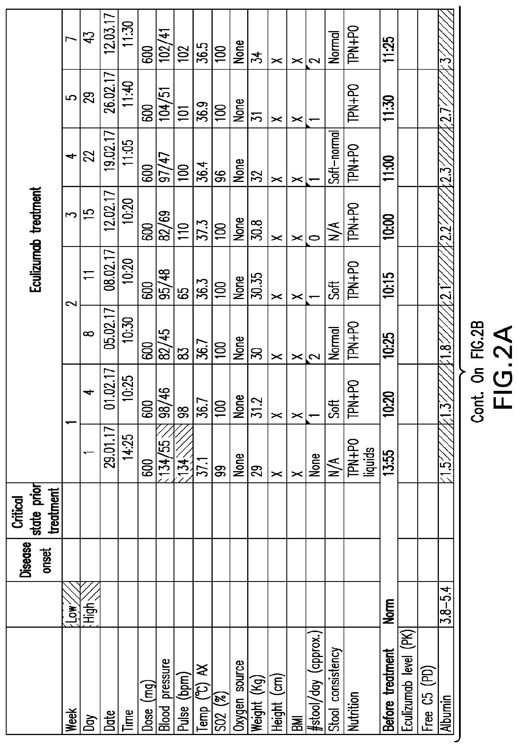
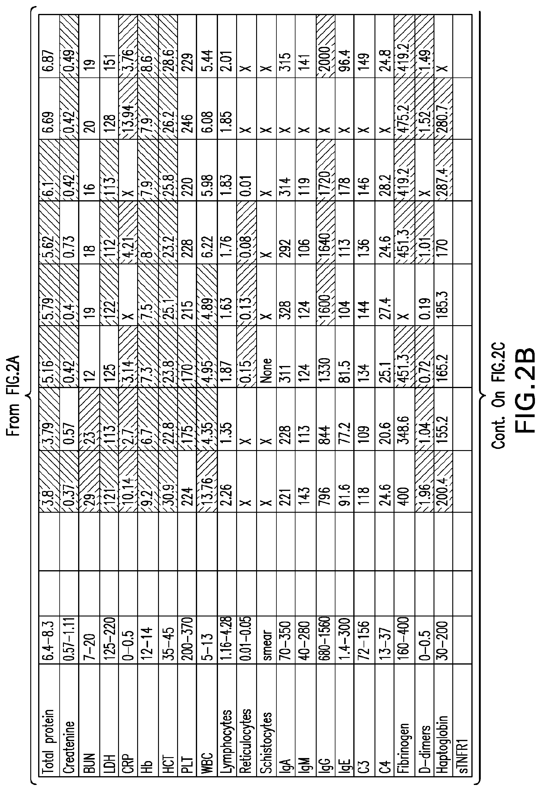
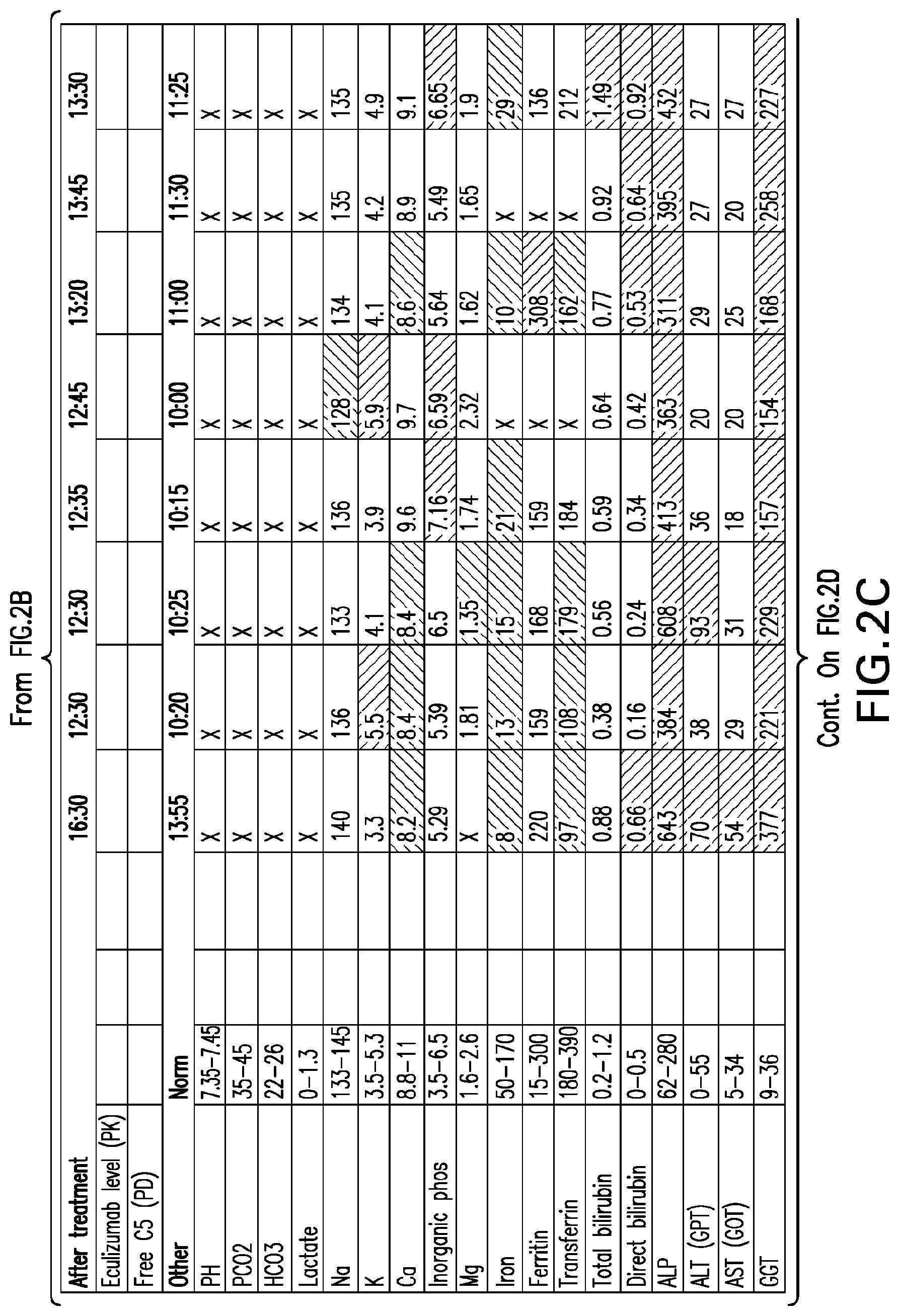
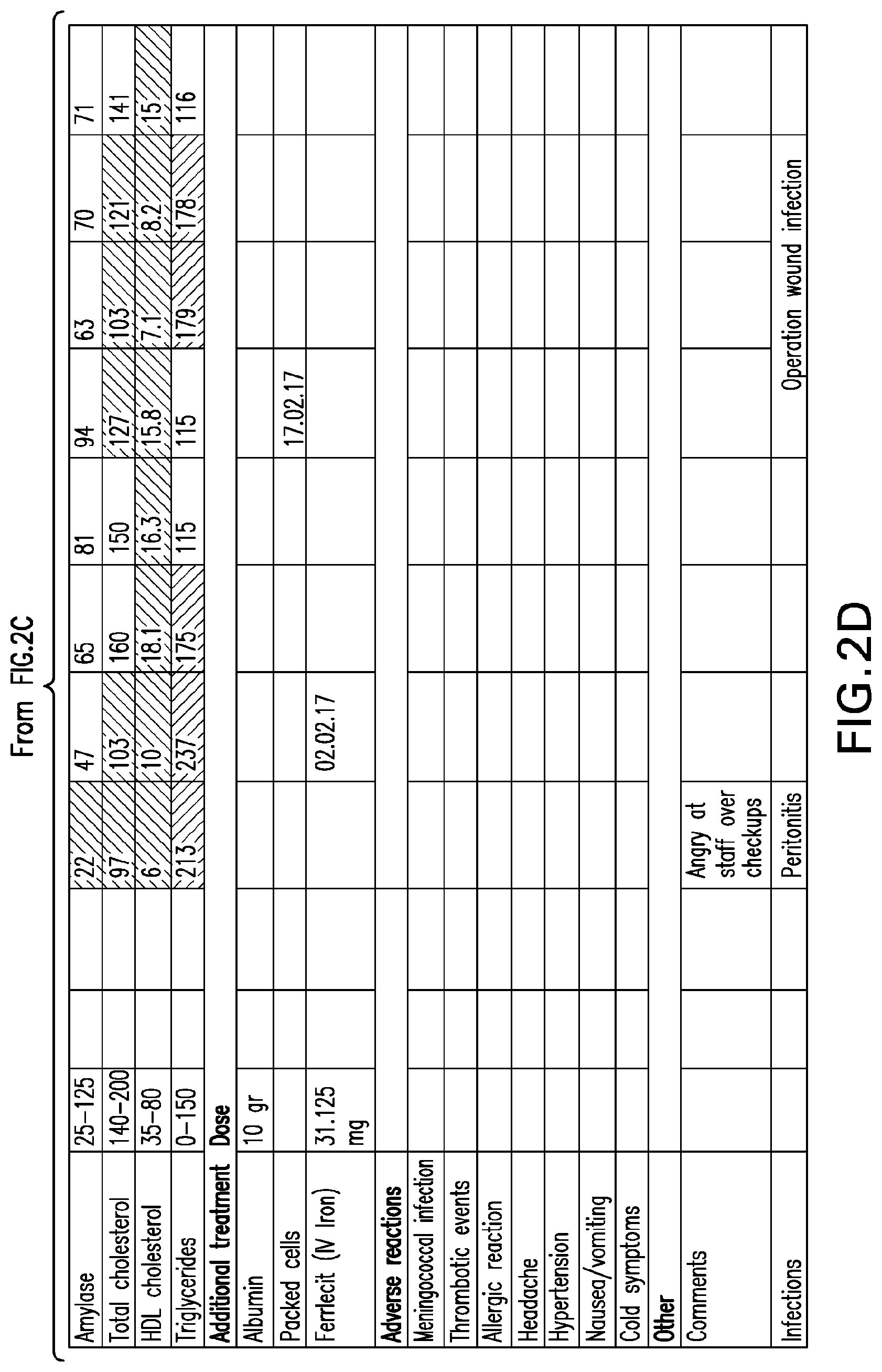
[0041] FIGS. 2A-2D set forth laboratory and related parameters for "Patient B" for treatment weeks 1-5.
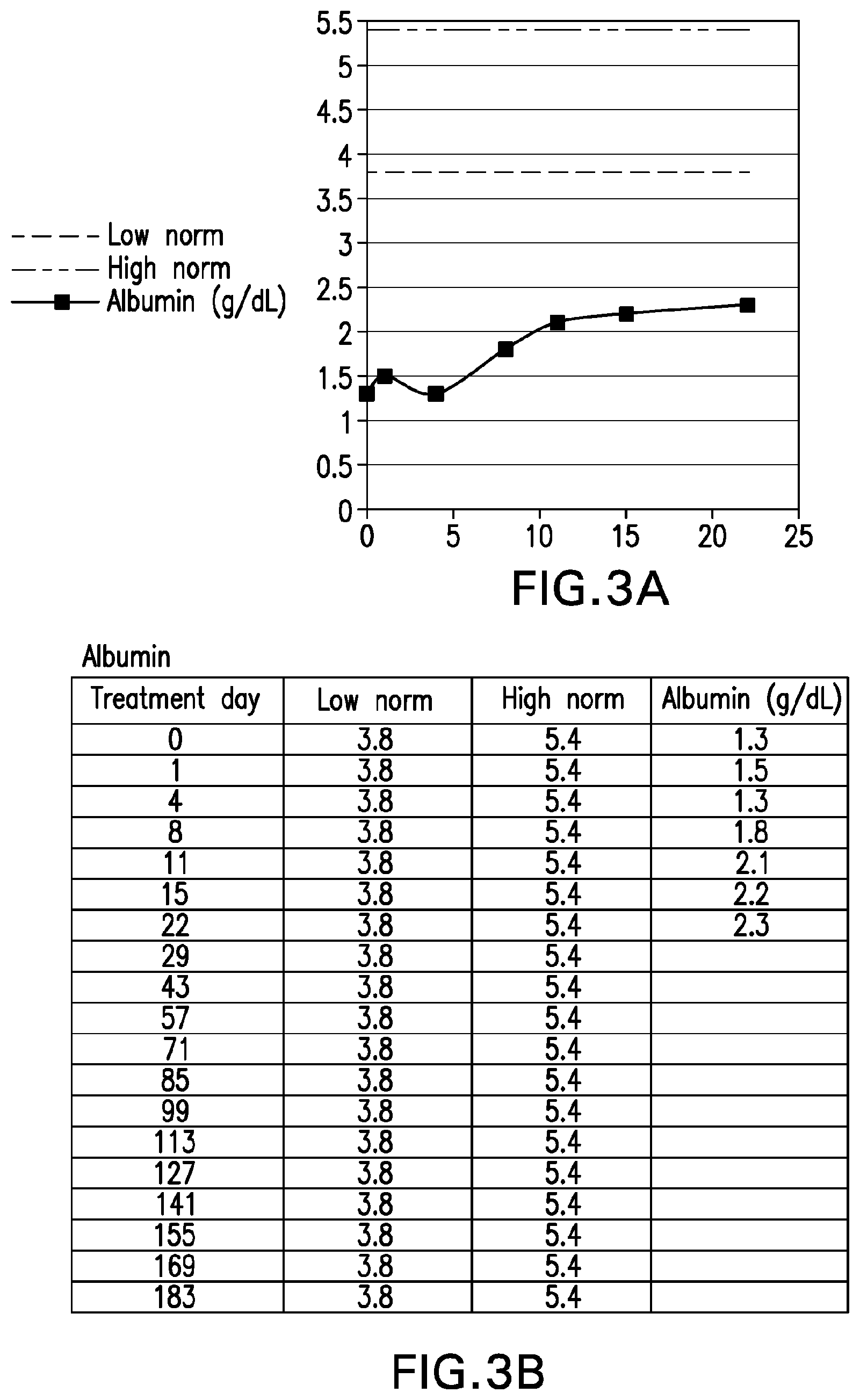
[0042] FIG. 3A is graph depicting serum albumin levels of "Patient B" (compared to low and high normal values) through treatment day 22. FIG. 3B sets forth the raw serum albumin levels for "Patient B" (compared to low and high normal values) through treatment day 22.
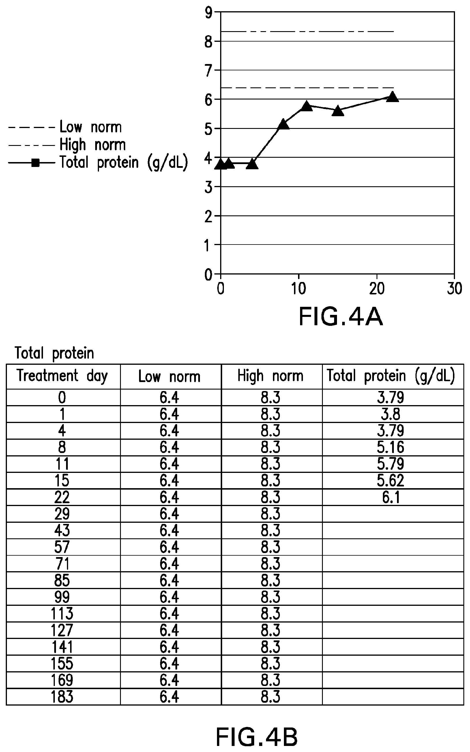
[0043] FIG. 4A is graph depicting total serum protein levels of "Patient B" (compared to low and high normal values) through treatment day 22. FIG. 4B sets forth the raw total serum protein levels for "Patient B" (compared to low and high normal values) through treatment day 22.
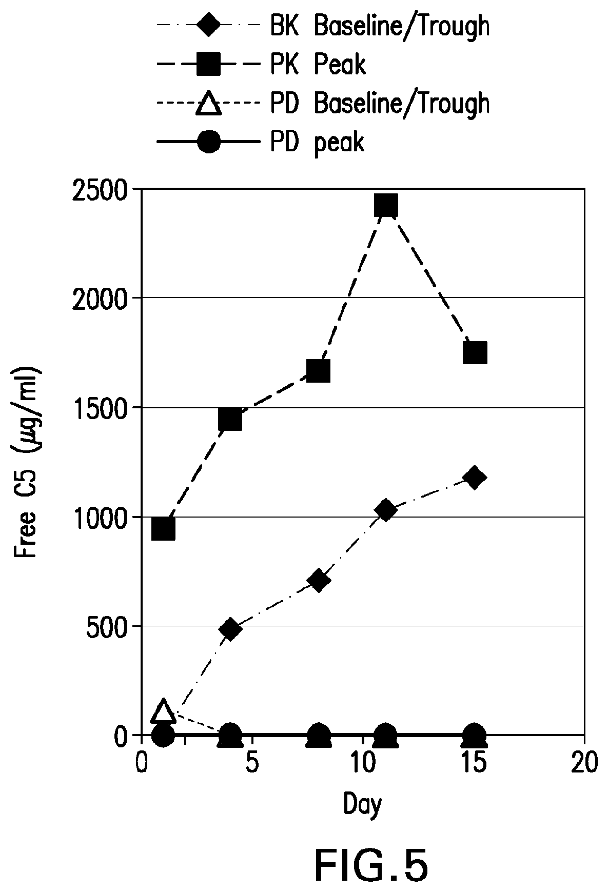
[0044] FIG. 5 is a graph depicting the pharmacokinetic (PK) and pharmacodynamic (PD) baseline/troughs in free C5 .mu.g/mL and peaks for "Patient B" through treatment day 15.
[0045] FIG. 6A is a graph depicting the weight of "Patient B" through treatment day 15. FIG. 6B sets forth the actual weight of "Patient B" on treatment days 1, 4, 8, 11 and 15.
[0046] FIGS. 7A-7H set forth laboratory and related parameters for "Patient C" for treatment weeks 1-17.
[0047] FIG. 8A is graph depicting serum albumin levels of "Patient C" (compared to low and high normal values) through treatment day 85. FIG. 8B sets forth the raw serum albumin levels for "Patient C" (compared to low and high normal values) through treatment day 85.
[0048] FIG. 9A is graph depicting total serum protein levels of "Patient C" (compared to low and high normal values) through treatment day 85. FIG. 9B sets forth the raw total serum protein levels for "Patient C" (compared to low and high normal values) through treatment day 85.
[0049] FIG. 10A is a graph depicting the pharmacokinetic (PK) and pharmacodynamic (PD) baseline/troughs in free C5 .mu.g/mL and peaks for "Patient C" through treatment day 15. FIG. 10B sets forth the raw PK and PD baseline/trough and peak values through treatment day 15.
[0050] FIG. 11A is a graph depicting the weight of "Patient C" through treatment day 86. FIG. 11B sets forth the actual weight of "Patient C" on treatment days 1, 4, 8, 11, 15, 22, 29, 43, 57, 71 and 85.
[0051] FIG. 12 is a graph depicting the serum TNFR1 levels for "Patient C" through treatment day 43 (compared to low and high normal values).
[0052] FIG. 13A shows a pedigree depicting co-segregation of the CD55 variant with PLE. FIG. 13B and FIG. 13C are images of endoscopic and histological findings of intestinal lymphangiectasia in patient 3. FIG. 13B depicts evidence of caviar-like villi (indicated by arrows) in the terminal ileum, and FIG. 13C shows dilated intestinal lymphatics (indicated by arrow), i.e., lymphangiectasia, in duodenal mucosa. FIG. 13D shows sequence chromatograms of a patient (homozygous) and a healthy parent (heterozygous), depicting the CD55 NM_001114752.1: c.43del variant. FIG. 13E shows flow cytometry analysis of CD55 revealing no binding to CD55 antigens on red blood cells of Patient 6 (black) and on neutrophils of Patients 4 and 6 (overlay histogram, black and grey) compared to normal controls (white).
[0053] FIGS. 14A and 14B depict improvement in serum albumin and total protein concentrations from baseline and throughout 100 days of eculizumab treatment. FIG. 14C reveals a striking decrease in the number of bowel movements per day following a single eculizumab dose and up to 100 days of eculizumab treatment. The decrease in number was accompanied with improvement in stool consistency.
[0054] FIG. 15A depicts flow cytometry of MAC on total WBCs, granulocytes, and monocytes in Patient 4 and age-matched controls pre-treatment, after 15 days of treatment, and after 59 days of treatment. FIG. 15B demonstrates the significant reduction in MAC-depositions on cells compared to pre-treatment values.
[0055] FIG. 16 shows flow cytometry of iC3b on total WBCs, granulocytes, and monocytes in Patient 4 and age-matched controls pre-treatment, after 15 days of treatment, and after 59 days of treatment.
[0056] FIGS. 17A-L are pre- and post-treatment photographs of patients. Panels A-E illustrate resolution of acrodermatitis enteroepathica-like rash in Patient 6. Panels F-G reveal reduction in abdominal distension and panels H-J show improvement in acquired ichthyosis in Patient 3. Panels K-L depict abdominal imagine with contrast media in Patient 4 following intestinal obstruction surgery. Panel K fails to depict vast parts of the patient's intestines before eculizumab treatment, while panel L, imaged after 66 days on eculizumab, shows improvement in bowel integrity.
[0057] FIG. 18A depicts albumin (g/dL) and total protein (g/dL) levels post-treatment with eculizumab for patients 6, 4 and 3, as well as total protein low norm (top line) and albumin low norm (bottom line). FIG. 18B depicts the height and body mass index (BMI) improvements for Patient 3 after treatment with eculizumab.
DETAILED DESCRIPTION
I. Definitions
[0058] As used herein, the term "subject" or "patient" is a human patient (e.g., a patient having a PLE, such as lymphangiectasia).
[0059] PLE is a condition that results from lymphatic or intestinal disruption leading to enteric protein loss, usually manifesting as peripheral edema due to hypoalbuminemia (Braamskamp, M. et al., Eur. J. Pediatr., 169:1179-85, 2010). Diagnosis is often confirmed based on fecal .alpha.-1-antitrypsin clearance and symptoms can include edema, ascites, pleural effusion, pericarditis, lymphedema, diarrhea, abdominal pain, fatigue, weight loss and vitamin deficiency (Umar, S. & DiBaise, J., Am. J. Gastroenterol., 105:43-9, 2010). Genetic factors may contribute to the development of PLE, including genes associated with the complement pathway. For the purposes herein, CD55 has been shown to play a role in PLE. CD55 (also knowns as decay-accelerating factor or DAF) regulates complement activity by inhibiting the formation and stability of C3/C5 convertases and accelerating their degradation (Lublin, D., Immunohematology, 21:39-47, 2005; Kim, D. & Song, W., Clin. Immunol., 118:127-36, 2006). Inflammatory bowel disease and primary intestinal lymphangiectasia (PIL) are among the primary causes of PLE (Alexander, J. et al., Pathophysiology, 17:315-35, 2010).
[0060] Lymphangiectasia (also known as "intestinal lymphangiectasia," "primary intestinal lymphangiectasia" and "PIL") is a rare PLE that was first described in 1961 (Waldmann, T. et al., Gastroenterology, 41:197-207, 1961). Since its first documentation, fewer than 200 cases have been reported globally (Alshikho, M et al., Am. J. Case Rep., 17:512-22, 2016). Due to the rarity of lymphangiectasia, its prevalence and etiology are not known. It seldom affects multiple family members and therefore does not appear to be an inherited disease. However, genetics may still play a role in disease onset, as some studies have shown changes in the regulation of genes associated with lymphangiogenesis in patients with lymphangiectasia (Hokari, R. et al., J. Gastroenterol. Hepatol., 23:e88-95, 2008).
[0061] Lymphangiectasia is characterized by the dilation of vessels in the mucosa, submucosa, or subserosa of the intestines, leading to leakage of lymphatic fluid into the gastrointestinal tract. It is typically diagnosed in children, often before the age of three, and afflicts males and females equally. Patients present a range of symptoms, with edema due to protein loss being the most common. Other symptoms may include diarrhea, ascites, pleural effusion, pericarditis, lymphedema, abdominal pain, fatigue, weight loss and/or vitamin deficiency. Laboratory analyses frequently reveal hypoalbuminemia, lymphopenia and/or hypogammaglobulinemia due to the loss of lymph. Diagnosis is confirmed using endoscopy and by histopathological analysis of biopsied tissue. Additional testing may include albumin scintigraphy, ultrasound, computed tomography (CT) scans and lymphoscintigraphy (Vignes, S. & Bellanger, J., Orphanet J. Rare Dis., 3:5, 2008).
[0062] There is currently no cure for lymphangiectasia and patients are treated by management of their symptoms. The most successful intervention is the implementation of a low-fat diet supplemented with medium-chain triglycerides (MCT) (Jeffries, G. et al., N. Engl. J. Med., 270:761-6, 1964; Alfano, V. et al., Nutrition, 16:303-4, 2000). Other treatments include the administration of octreotide (experimental) or infusion with albumin (Ballinger, A. & Farthing, M., Eur. J. Gastroenterol. Hepatol., 10:699-702, 1998). In rare cases, surgery may be required to remove afflicted tissue. Without long-term treatment, lymphangiectasia patients may suffer from severe complications from the disease, including death.
[0063] As used herein, "effective treatment" refers to treatment producing a beneficial effect, e.g., amelioration of at least one symptom of a disease or disorder. A beneficial effect can take the form of an improvement over baseline, e.g., an improvement over a measurement or observation made prior to initiation of therapy according to the method. Effective treatment may refer to alleviation of at least one symptom of a PLE, such as lymphangiectasia (e.g., protein loss, edema, diarrhea, ascites, pleural effusion, pericarditis, lymphedema, abdominal pain, fatigue, weight loss and/or vitamin deficiency).
[0064] The term "effective amount" refers to an amount of an agent that provides the desired biological, therapeutic and/or prophylactic result. That result can be reduction, amelioration, palliation, lessening, delaying and/or alleviation of one or more of the signs, symptoms, or causes of a disease, or any other desired alteration of a biological system. In one example, an "effective amount" is the amount of anti-C5 antibody, or antigen binding fragment thereof, clinically proven to alleviate at least one symptom of a PLE, such as lymphangiectasia (e.g., protein loss, edema, diarrhea, ascites, pleural effusion, pericarditis, lymphedema, abdominal pain, fatigue, weight loss and/or vitamin deficiency). An effective amount can be administered in one or more administrations.
[0065] As used herein, the terms "induction" and "induction phase" are used interchangeably and refer to the first phase of treatment.
[0066] As used herein, the terms "maintenance" and "maintenance phase" are used interchangeably and refer to the second phase of treatment. In certain embodiments, treatment is continued as long as clinical benefit is observed or until unmanageable toxicity or disease progression occurs.
[0067] As used herein, the term "serum trough level" refers to the lowest level that the agent (e.g., the anti-C5 antibody, or antigen binding fragment thereof) or medicine is present in the serum. In contrast, a "peak serum level", refers to the highest level of the agent in the serum. The "average serum level", refers to the mean level of the agent in the serum over time.
[0068] The term "antibody" describes polypeptides comprising at least one antibody-derived antigen binding site (e.g., VH/VL region or Fv, or CDR). Antibodies include known forms of antibodies. For example, the antibody can be a human antibody, a humanized antibody, a camelid antibody, a bispecific antibody, or a chimeric antibody. The antibody also can be a Fab, Fab'2, ScFv, SMIP, Affibody.RTM., nanobody, or a domain antibody. The antibody also can be of any of the following isotypes: IgG1, IgG2, IgG3, IgG4, IgM, IgA1, IgA2, IgAsec, IgD, and IgE. The antibody can be a naturally occurring antibody or an antibody that has been altered by a protein engineering technique (e.g., by mutation, deletion, substitution, conjugation to a non-antibody moiety). For example, an antibody can include one or more variant amino acids (compared to a naturally occurring antibody) that changes a property (e.g., a functional property) of the antibody. For example, numerous such alterations are known in the art that affect, e.g., half-life, effector function, and/or immune responses to the antibody in a patient. The term antibody also includes artificial or engineered polypeptide constructs that comprise at least one antibody-derived antigen binding site.
II. Anti-C5 Antibodies
[0069] The anti-C5 antibodies described herein bind to complement component C5 (e.g., human C5) and inhibit the cleavage of C5 into fragments C5a and C5b.
[0070] Anti-C5 antibodies (or VH/VL domains derived therefrom) suitable for use in the compositions and methods described herein can be generated using methods known in the art. Alternatively, art-recognized anti-C5 antibodies can be used. Antibodies that compete with any of these art-recognized antibodies for binding to C5 also can be used.
[0071] An exemplary anti-C5 antibody is eculizumab (Soliris.RTM.). Eculizumab is a humanized monoclonal antibody (h5G1.1-mAb solution for infusion) with binding specificity uniquely specific for the human complement C5 protein. Eculizumab is described in U.S. Pat. No. 6,355,245, the entire teachings of which are hereby expressly incorporated by reference. Comprised of 1324 amino acids with a molecular mass of approximately 148 kDa, eculizumab was derived from a murine monoclonal antibody (m5G1.1-mAb) that recognizes the human complement component C5.
[0072] In other embodiments, the antibody comprises the heavy and light chain CDRs or variable regions of eculizumab. Accordingly, in one embodiment, the antibody comprises the CDR1, CDR2 and CDR3 domains of the VH region of eculizumab having the sequence set forth in SEQ ID NO:7, and the CDR1, CDR2 and CDR3 domains of the VL region of eculizumab having the sequence set forth in SEQ ID NO:8. In another embodiment, the antibody comprises heavy chain CDR1, CDR2 and CDR3 domains having the sequences set forth in SEQ ID NOs:1, 2 and 3, respectively, and light chain CDR1, CDR2 and CDR3 domains having the sequences set forth in SEQ ID NOs:4, 5 and 6, respectively. In another embodiment, the antibody comprises a VH region having the amino acid sequence set forth in SEQ ID NO:7. In another embodiment, the antibody comprises a VL region having the amino acid sequence set forth in SEQ ID NO:8. In another embodiment, the antibody comprises VH and VL regions having the amino acid sequences set forth in SEQ ID NO:7 and SEQ ID NO:8, respectively. In another embodiment, the antibody comprises the heavy chain constant region of eculizumab having the sequence set forth in SEQ ID NO:9. In another embodiment, the antibody comprises a heavy chain having the amino acid sequence set forth in SEQ ID NO:10. In another embodiment, the antibody comprises a light chain having the amino acid sequence set forth in SEQ ID NO:11. In another embodiment, the antibody comprises heavy and light chains having the amino acid sequences set forth in SEQ ID NO:10 and SEQ ID NO:11, respectively.
[0073] Another exemplary anti-C5 antibody is the 7086 antibody described in U.S. Pat. Nos. 8,241,628 and 8,883,158. In one embodiment, the antibody comprises the heavy and light chain CDRs or variable regions of the 7086 antibody (see U.S. Pat. Nos. 8,241,628 and 8,883,158). In another embodiment, the antibody, or antigen binding fragment thereof, comprises heavy chain CDR1, CDR2 and CDR3 domains having the sequences set forth in SEQ ID NOs:12, 13 and 14, respectively, and light chain CDR1, CDR2 and CDR3 domains having the sequences set forth in SEQ ID NOs:15, 16 and 17, respectively. In another embodiment, the antibody, or antigen binding fragment thereof, comprises the VH region of the 7086 antibody having the sequence set forth in SEQ ID NO:18 and the VL region of the 7086 antibody having the sequence set forth in SEQ ID NO:19.
[0074] Another exemplary anti-C5 antibody is the 8110 antibody also described in U.S. Pat. Nos. 8,241,628 and 8,883,158. In one embodiment, the antibody comprises the heavy and light chain CDRs or variable regions of the 8110 antibody. In another embodiment, the antibody, or antigen binding fragment thereof, comprises heavy chain CDR1, CDR2 and CDR3 domains having the sequences set forth in SEQ ID NOs:20, 21 and 22, respectively, and light chain CDR1, CDR2 and CDR3 domains having the sequences set forth in SEQ ID NOs:23, 24 and 25, respectively. In another embodiment, the antibody comprises the VH region of the 8110 antibody having the sequence set forth in SEQ ID NO:26, and the VL region of the 8110 antibody having the sequence set forth in SEQ ID NO:27.
[0075] Another exemplary anti-C5 antibody is the 305LO5 antibody described in US2016/0176954A1. In one embodiment, the antibody comprises the heavy and light chain CDRs or variable regions of the 305LO5 antibody. In another embodiment, the antibody, or antigen binding fragment thereof, comprises heavy chain CDR1, CDR2 and CDR3 domains having the sequences set forth in SEQ ID NOs:28, 29 and 30, respectively, and light chain CDR1, CDR2 and CDR3 domains having the sequences set forth in SEQ ID NOs:31, 32 and 33, respectively. In another embodiment, the antibody comprises the VH region of the 305LO5 antibody having the sequence set forth in SEQ ID NO:34, and the VL region of the 305LO5 antibody having the sequence set forth in SEQ ID NO:35.
[0076] The exact boundaries of CDRs have been defined differently according to different methods. In some embodiments, the positions of the CDRs or framework regions within a light or heavy chain variable domain can be as defined by Kabat et al. [(1991) "Sequences of Proteins of Immunological Interest." NIH Publication No. 91-3242, U.S. Department of Health and Human Services, Bethesda, Md.]. In such cases, the CDRs can be referred to as "Kabat CDRs" (e.g., "Kabat LCDR2" or "Kabat HCDR1"). In some embodiments, the positions of the CDRs of a light or heavy chain variable region can be as defined by Chothia et al. (Nature, 342:877-83, 1989). Accordingly, these regions can be referred to as "Chothia CDRs" (e.g., "Chothia LCDR2" or "Chothia HCDR3"). In some embodiments, the positions of the CDRs of the light and heavy chain variable regions can be as defined by a Kabat-Chothia combined definition. In such embodiments, these regions can be referred to as "combined Kabat-Chothia CDRs". Thomas et al. (Mol. Immunol., 33:1389-1401, 1996) exemplifies the identification of CDR boundaries according to Kabat and Chothia definitions.
[0077] In one embodiment, the antibody competes for binding with, and/or binds to the same epitope on C5 as, the antibodies described herein. The term "binds to the same epitope" with reference to two or more antibodies means that the antibodies bind to the same segment of amino acid residues, as determined by a given method. Techniques for determining whether antibodies bind to the "same epitope on C5" with the antibodies described herein include, for example, epitope mapping methods, such as, for example, X-ray analyses of crystals of antigen-antibody complexes (which provides atomic resolution of the epitope) and hydrogen/deuterium exchange mass spectrometry (HDX-MS). Other methods monitor the binding of the antibody to peptide antigen fragments or mutated variations of the antigen where loss of binding due to a modification of an amino acid residue within the antigen sequence is often considered an indication of an epitope component. In addition, computational combinatorial methods for epitope mapping can also be used. These methods rely on the ability of the antibody of interest to affinity isolate specific short peptides from combinatorial phage display peptide libraries. Antibodies having the same VH and VL or the same CDR1, 2 and 3 sequences are expected to bind to the same epitope.
[0078] Antibodies that "compete with another antibody for binding to a target" refer to antibodies that inhibit (partially or completely) the binding of the other antibody to the target. Whether two antibodies compete with each other for binding to a target, i.e., whether and to what extent one antibody inhibits the binding of the other antibody to a target, may be determined using known competition experiments. In certain embodiments, an antibody competes with, and inhibits binding of another antibody to a target by at least 10%, 20%, 30%, 40%, 50%, 60%, 70%, 80%, 90% or 100%. The level of inhibition or competition may be different depending on which antibody is the "blocking antibody" (i.e., the antibody that is incubated first with the target). Competing antibodies bind to the same epitope, an overlapping epitope or to adjacent epitopes (e.g., as evidenced by steric hindrance).
[0079] Methods for determining whether an antibody binds to a protein antigen and/or the affinity for an antibody to a protein antigen are known in the art. For example, the binding of an antibody to a protein antigen can be detected and/or quantified using a variety of techniques such as, but not limited to, Western blot, dot blot, surface plasmon resonance (SPR) methods (e.g., BIAcore system), or enzyme-linked immunosorbent assay (ELISA) (Benny K. C. Lo (2004) "Antibody Engineering: Methods and Protocols," Humana Press (ISBN: 1588290921); Johne, B. et al., J. Immunol. Meth., 160:191-8, 1993; Jonsson, U. et al., Ann. Biol. Clin., 51:19-26, 1993; Jonsson, U. et al., Biotechniques, 11:620-7, 1991). In addition, methods for measuring the binding affinity (e.g., dissociation and association constants) are known in the art and set forth in the working examples.
[0080] As used herein, the term "k.sub.a" refers to the rate constant for association of, for example, an antibody to an antigen. The term "k.sub.d" refers to the rate constant for dissociation of, for example, an antibody from the antibody/antigen complex. And the term "K.sub.D" refers to the equilibrium dissociation constant of, for example, an antibody-antigen interaction. The equilibrium dissociation constant is deduced from the ratio of the kinetic rate constants, K.sub.D=k.sub.a/k.sub.d. Such determinations can be measured, for example, at 25 C or 37 C. The kinetics of antibody binding to human C5 can be determined, for example, at pH 8.0, 7.4, 7.0, 6.5 or 6.0 via SPR on a BIAcore 3000 instrument using an anti-Fc capture method to immobilize the antibody.
[0081] In one embodiment, the anti-C5 antibody, or antigen binding fragment thereof, blocks the generation or activity of the C5a and/or C5b active fragments of a C5 protein (e.g., a human C5 protein). Through this blocking effect, the antibodies inhibit, for example, the pro-inflammatory effects of C5a and the generation of the C5b-9 MAC at the surface of a cell.
[0082] Methods for determining whether a particular antibody described herein inhibits C5 cleavage are known in the art. Inhibition of human complement component C5 can reduce the cell lysing ability of complement in a subject's body fluids. Such reductions of the cell lysing ability of complement present in the body fluid(s) can be measured by methods known in the art such as, for example, by a conventional hemolytic assay (Kabat and Mayer (eds.), "Experimental Immunochemistry, 2.sup.nd Edition," 135-240, Springfield, Ill., CC Thomas (1961), pages 135-9), or a conventional variation of that assay such as the chicken erythrocyte hemolysis method (Hillmen, P. et al., N. Engl. J. Med., 350:552, 2004). Methods for determining whether a candidate compound inhibits the cleavage of human C5 into forms C5a and C5b are known in the art (Evans, M. et al., Mol. Immunol., 32:1183-95, 1995). The concentration and/or physiologic activity of C5a and C5b in a body fluid can be measured, for example, by methods known in the art. For C5b, hemolytic assays or assays for soluble C5b-9 as discussed herein can be used. Other assays known in the art can also be used. Using assays of these or other suitable types, candidate agents capable of inhibiting human complement component C5 can be screened.
[0083] Immunological techniques such as, but not limited to, ELISA can be used to measure the protein concentration of C5 and/or its split products to determine the ability of an anti-C5 antibody, or antigen binding fragment thereof, to inhibit conversion of C5 into biologically active products. In some embodiments, C5a generation is measured. In some embodiments, C5b-9 neoepitope-specific antibodies are used to detect the formation of terminal complement.
[0084] Hemolytic assays can be used to determine the inhibitory activity of an anti-C5 antibody, or antigen binding fragment thereof, on complement activation. To determine the effect of an anti-C5 antibody, or antigen binding fragment thereof, on classical complement pathway-mediated hemolysis in a serum test solution in vitro, for example, sheep erythrocytes coated with hemolysin or chicken erythrocytes sensitized with anti-chicken erythrocyte antibody are used as target cells. The percentage of lysis is normalized by considering 100% lysis equal to the lysis occurring in the absence of the inhibitor. In some embodiments, the classical complement pathway is activated by a human IgM antibody, for example, as utilized in the Wieslab.RTM. Classical Pathway Complement Kit (Wieslab.RTM. COMPL CP310, Euro-Diagnostica, Sweden). Briefly, the test serum is incubated with an anti-C5 antibody, or antigen binding fragment thereof, in the presence of a human IgM antibody. The amount of C5b-9 that is generated is measured by contacting the mixture with an enzyme conjugated anti-C5b-9 antibody and a fluorogenic substrate and measuring the absorbance at the appropriate wavelength. As a control, the test serum is incubated in the absence of the anti-C5 antibody, or antigen binding fragment thereof. In some embodiments, the test serum is a C5-deficient serum reconstituted with a C5 polypeptide.
[0085] To determine the effect of an anti-C5 antibody, or antigen binding fragment thereof, on alternative pathway-mediated hemolysis, unsensitized rabbit or guinea pig erythrocytes can be used as the target cells. In some embodiments, the serum test solution is a C5-deficient serum reconstituted with a C5 polypeptide. The percentage of lysis is normalized by considering 100% lysis equal to the lysis occurring in the absence of the inhibitor. In some embodiments, the alternative complement pathway is activated by lipopolysaccharide molecules, for example, as utilized in the Wieslab.RTM. Alternative Pathway Complement Kit (Wieslab.RTM. COMPL AP330, Euro-Diagnostica, Sweden). Briefly, the test serum is incubated with an anti-C5 antibody, or antigen binding fragment thereof, in the presence of lipopolysaccharide. The amount of C5b-9 that is generated is measured by contacting the mixture with an enzyme conjugated anti-C5b-9 antibody and a fluorogenic substrate and measuring the fluorescence at the appropriate wavelength. As a control, the test serum is incubated in the absence of the anti-C5 antibody, or antigen binding fragment thereof.
[0086] In some embodiments, C5 activity, or inhibition thereof, is quantified using a CH50eq assay. The CH50eq assay is a method for measuring the total classical complement activity in serum. This is a lytic assay that uses antibody-sensitized erythrocytes as the activator of the classical complement pathway and various dilutions of the test serum to determine the amount required to give 50% lysis (CH50). The percent hemolysis can be determined, for example, using a spectrophotometer. The CH50eq assay provides an indirect measure of terminal complement complex (TCC) formation, since the TCC themselves are directly responsible for the hemolysis that is measured. The assay is known and commonly practiced by those of skill in the art. Briefly, to activate the classical complement pathway, undiluted serum samples (e.g., reconstituted human serum samples) are added to microassay wells containing the antibody-sensitized erythrocytes to thereby generate TCC. Next, activated sera are diluted in microassay wells, which are coated with a capture reagent (e.g., an antibody that binds to one or more components of the TCC). The TCC present in the activated samples bind to the monoclonal antibodies coating the surface of the microassay wells. The wells are washed and to each well is added a detection reagent that is detectably labeled and recognizes the bound TCC. The detectable label can be, for example, a fluorescent label or an enzymatic label. The assay results are expressed in CH50 unit equivalents per milliliter (CH50 U Eq/mL).
[0087] Inhibition, e.g., as it pertains to terminal complement activity, includes at least a 5 (e.g., at least a 6, 7, 8, 9, 10, 15, 20, 25, 30, 35, 40, 45, 50, 55, or 60) % decrease in the activity of terminal complement in, e.g., a hemolytic assay or CH50eq assay as compared to the effect of a control antibody (or antigen-binding fragment thereof) under similar conditions and at an equimolar concentration. Substantial inhibition, as used herein, refers to inhibition of a given activity (e.g., terminal complement activity) of at least 40 (e.g., at least 45, 50, 55, 60, 65, 70, 75, 80, 85, 90, or 95 or greater) %.
[0088] Anti-C5 antibodies, or antigen-binding fragments thereof described herein, used in the methods described herein can be generated using a variety of art-recognized techniques. Monoclonal antibodies can be obtained by various techniques familiar to those skilled in the art. Briefly, for example, spleen cells from an animal immunized with a desired antigen are immortalized, commonly by fusion with a myeloma cell (Kohler, G. & Milstein, C., Eur. J. Immunol., 6:511-9, 1976). Alternative methods of immortalization include, but are not limited to, transformation with Epstein Barr Virus, oncogenes, or retroviruses, or other methods known in the art. Colonies arising from single immortalized cells are screened for production of antibodies of the desired specificity and affinity for the antigen, and yield of the monoclonal antibodies produced by such cells can be enhanced by various techniques, including injection into the peritoneal cavity of a vertebrate host. Alternatively, one can isolate DNA sequences that encode a monoclonal antibody or a binding fragment thereof by screening a DNA library from human B cells (Huse, W. et al., Science, 246:1275-81, 1989).
III. Compositions
[0089] Provided herein are compositions comprising an anti-C5 antibody, or antigen binding fragment thereof. In one embodiment, the composition comprises an antibody comprising the CDR1, CDR2 and CDR3 domains of the VH region of eculizumab having the sequence set forth in SEQ ID NO:7, and the CDR1, CDR2 and CDR3 domains of the VL region of eculizumab having the sequence set forth in SEQ ID NO:8. In another embodiment, the antibody comprises heavy chain CDR1, CDR2 and CDR3 domains having the sequences set forth in SEQ ID NOs:1, 2 and 3, respectively, and light chain CDR1, CDR2 and CDR3 domains having the sequences set forth in SEQ ID NOs:4, 5 and 6, respectively. In another embodiment, the antibody comprises a VH region having the amino acid sequence set forth in SEQ ID NO:7. In another embodiment, the antibody comprises a VL region having the amino acid sequence set forth in SEQ ID NO:8. In another embodiment, the antibody comprises VH and VL regions having the amino acid sequences set forth in SEQ ID NO:7 and SEQ ID NO:8, respectively. In another embodiment, the antibody comprises a heavy chain having the amino acid sequence set forth in SEQ ID NO:10. In another embodiment, the antibody comprises a light chain having the amino acid sequence set forth in SEQ ID NO:11. In another embodiment, the antibody comprises heavy and light chains having the amino acid sequences set forth in SEQ ID NO:10 and SEQ ID NO:11, respectively.
[0090] The compositions can be formulated as a pharmaceutical solution, e.g., for administration to a subject for the treatment or prevention of PLE (e.g., lymphangiectasia). The pharmaceutical compositions can include a pharmaceutically acceptable carrier. As used herein, a "pharmaceutically acceptable carrier" refers to, and includes, any and all solvents, dispersion media, coatings, antibacterial and antifungal agents, isotonic and absorption delaying agents, and the like that are physiologically compatible. The compositions can include a pharmaceutically acceptable salt, e.g., an acid addition salt or a base addition salt, sugars, carbohydrates, polyols and/or tonicity modifiers.
[0091] The compositions can be formulated according to standard methods (Gennaro (2000) "Remington: The Science and Practice of Pharmacy," 20.sup.th Edition, Lippincott, Williams & Wilkins (ISBN: 0683306472); Ansel et al. (1999) "Pharmaceutical Dosage Forms and Drug Delivery Systems," 7.sup.th Edition, Lippincott Williams & Wilkins Publishers (ISBN: 0683305727); and Kibbe (2000) "Handbook of Pharmaceutical Excipients American Pharmaceutical Association," 3.sup.rd Edition (ISBN: 091733096X)). In some embodiments, a composition can be formulated, for example, as a buffered solution at a suitable concentration and suitable for storage at 2-8 C (e.g., 4 C). In some embodiments, a composition can be formulated for storage at a temperature below 0 C (e.g., -20 C or -80 C). In some embodiments, the composition can be formulated for storage for up to 2 years (e.g., 1 month, 2 months, 3 months, 4 months, 5 months, 6 months, 7 months, 8 months, 9 months, 10 months, 11 months, 1 year, 11/2 years, or 2 years) at 2-8 C (e.g., 4 C). Thus, in some embodiments, the compositions described herein are stable in storage for at least 1 year at 2-8 C (e.g., 4 C).
[0092] The pharmaceutical compositions can be in a variety of forms. These forms include, e.g., liquid, semi-solid and solid dosage forms, such as liquid solutions (e.g., injectable and infusible solutions), dispersions or suspensions, tablets, pills, powders, liposomes and suppositories. The preferred form depends, in part, on the intended mode of administration and therapeutic application. For example, compositions containing a composition intended for systemic or local delivery can be in the form of injectable or infusible solutions. Accordingly, the compositions can be formulated for administration by a parenteral mode (e.g., intravenous, subcutaneous, intraperitoneal, or intramuscular injection). "Parenteral administration," "administered parenterally," and other grammatically equivalent phrases, as used herein, refer to modes of administration other than enteral and topical administration, usually by injection, and include, without limitation, intravenous, intranasal, intraocular, pulmonary, intramuscular, intraarterial, intrathecal, intracapsular, intraorbital, intracardiac, intradermal, intrapulmonary, intraperitoneal, transtracheal, subcutaneous, subcuticular, intraarticular, subcapsular, subarachnoid, intraspinal, epidural, intracerebral, intracranial, intracarotid and intrasternal injection and infusion.
IV. Patient Populations
[0093] Provided herein are compositions and methods for treating a PLE (e.g., lymphangiectasia) in a human patient. In one embodiment, the patient has a PLE. In another embodiment, the patient has lymphangiectasia. In another embodiment, the patient has protein loss, edema, ascites, pleural effusion, pericarditis, lymphedema, diarrhea, abdominal pain, fatigue, weight loss, and/or vitamin deficiency. In one embodiment, the patient is a pediatric patient (e.g., <18 years of age). In another embodiment, a pediatric patient is <12 years of age. In another embodiment, the pediatric patient is <6 years of age. In another embodiment, the pediatric patient is <2 years of age. In another embodiment, the pediatric patient is less than 1, 2, 3, 4, 5, 6, 7, 8, 9, 10, 11, 12, 13, 14, 15, 16, 17 or 18 years of age. In another embodiment, the patient is an adult patient. In another embodiment, an adult patient is .gtoreq.18 years of age.
[0094] In one embodiment, the patient weighs 10 kg to <20 kg. In another embodiment, the patient weighs 10 kg to <30 kg. In another embodiment, the patient weighs 20 kg to <30 kg. In another embodiment, the patient weighs 30 kg to <40 kg. In another embodiment, the patient weighs.gtoreq.40 kg.
[0095] In one embodiment, the patient has a mutation in a gene involved in the complement pathway (e.g., a gene involved with regulating the complement pathway). In a particular embodiment, the patient has a mutation in the CD55 gene. In another embodiment, the patient does not express CD55. In another embodiment, the patient has the CD55 variant NM_001114752.1: c.43del (p.Leu15Serfs*46). In another embodiment, the patient has red blood cell type CROM:-1 [Cr(a-)]. In another embodiment, the patient has red blood cell type CROM:-5 [Dr(a-)]. In another embodiment, the patient has red blood cell type CROM:-6 [Es(a-)]. In another embodiment, the patient has CROK-negative red blood cells. In another embodiment, the patient has the Cromer Inab (CD55-null) phenotype.
V. Outcomes
[0096] Provided herein are methods for treating a PLE (e.g., lymphangiectasia) in a patient comprising administering to the patient an anti-C5 antibody. Symptoms of PLE include, but are not limited to, protein loss, edema, ascites, pleural effusion, pericarditis, lymphedema, diarrhea, abdominal pain, fatigue, weight loss and vitamin deficiency. Patients treated according to the methods disclosed herein experience improvement in at least one symptom of PLE. For example, the treatment may produce at least one therapeutic effect selected from the group consisting of a reduction or cessation in protein loss, edema, ascites, pleural effusion, pericarditis, lymphedema, diarrhea, abdominal pain, fatigue, weight loss or vitamin deficiency.
[0097] In another embodiment, the treatment produces a shift toward normal levels of total protein (e.g., total serum protein). For example, in on embodiment, patients treated according to the disclosed methods experience an increase in total protein serum levels to near normal levels or to within about 10% or within about 20% above or below what is considered the normal level of total protein. In another embodiment, the treatment results in at least a 1.0-fold, 1.1-fold, 1.2-fold, 1.3-fold, 1.4-fold, 1.5-fold, 1.6-fold, 1.7-fold, 1.8-fold, 1.9-fold or 2.0-fold increase in total protein serum levels from baseline within 20 days. In a particular embodiment, the treatment results in at least a 1.5-fold increase in total protein serum levels from baseline within 20 days. In another embodiment, the treatment results in at least a 1.5-fold, 1.6-fold, 1.7-fold, 1.8-fold, 1.9-fold, 2.0-fold, 2.1-fold, 2.2-fold, 2.3-fold, 2.4-fold, 2.5-fold, 2.6-fold, 2.7-fold, 2.8-fold, 2.9-fold, or 3.0-fold increase in total protein serum levels from baseline within 80 days. In a particular embodiment, the treatment results in at least a 2.3-fold (e.g., 2.26-fold) increase in total protein serum levels from baseline within 80 days.
[0098] In another embodiment, the treatment produces a shift toward normal serum albumin levels. For example, in on embodiment, patients treated according to the disclosed methods experience an increase in serum albumin levels to near normal levels or to within about 10% or within about 20% above or below what is considered the normal level of serum albumin. In another embodiment, the treatment results in at least a 1.0-fold, 1.1-fold, 1.2-fold, 1.3-fold, 1.4-fold, 1.5-fold, 1.6-fold, 1.7-fold, 1.8-fold, 1.9-fold or 2.0-fold increase in serum albumin levels from baseline within 20 days. In a particular embodiment, the treatment results in at least a 1.7-fold increase in serum albumin levels from baseline within 20 days. In another embodiment, the treatment results in at least a 2.0-fold, 2.1-fold, 2.2-fold, 2.3-fold, 2.4-fold, 2.5-fold, 2.6-fold, 2.7-fold, 2.8-fold, 2.9-fold, 3.0-fold, 3.1-fold, 3.2-fold, 3.3-fold, 3.4-fold, 3.5-fold, 3.6-fold, 3.7-fold, 3.8-fold, 3.9-fold, 4.0-fold, 4.1-fold, 4.2-fold, 4.3-fold, 4.4-fold, 4.5-fold, 4.6-fold, 4.7-fold, 4.8-fold, 4.9-fold, 5.0-fold, 5.1-fold, 5.2-fold, 5.3-fold, 5.4-fold, 5.5-fold, 5.6-fold, 5.7-fold, 5.8-fold, 5.9-fold, 6.0-fold, 6.1-fold, 6.2-fold, 6.3-fold, 6.4-fold, 6.5-fold, 6.6-fold, 6.7-fold, 6.8-fold, 6.9-fold or 7.0-fold increase in serum albumin levels from baseline within 80 days. In another embodiment, the treatment results in at least a 5-fold increase in serum albumin levels from baseline within 80 days.
[0099] In another embodiment, the treatment produces a shift toward normal serum TNFR1 levels. For example, in on embodiment, patients treated according to the disclosed methods experience a decrease in serum TNFR1 levels to near normal levels or to within 10%, or within 20% above or below what is considered the normal level of serum TNFR1. In another embodiment, the treatment results in at least a 1.0-fold, 1.1-fold, 1.2-fold, 1.3-fold, 1.4-fold, 1.5-fold, 1.6-fold, 1.7-fold, 1.8-fold, 1.9-fold or 2.0-fold, 2.1-fold, 2.2-fold or 2.3-fold decrease in serum TNFR1 levels from baseline within 8 days. In a particular embodiment, the treatment results in at least a 1.8-fold decrease in serum TNFR1 levels from baseline within 8 days. In another embodiment, the treatment results in at least a 2.0-fold, 2.1-fold, 2.2-fold, 2.3-fold, 2.4-fold, 2.5-fold, 2.6-fold, 2.7-fold, 2.8-fold, 2.9-fold, 3.0-fold, 3.1-fold, 3.2-fold, 3.3-fold, 3.4-fold, 3.5-fold, 3.6-fold, 3.7-fold, 3.8-fold, 3.9-fold, 4.0-fold, 4.1-fold, 4.2-fold, 4.3-fold, 4.4-fold, 4.5-fold, 4.6-fold, 4.7-fold, 4.8-fold, 4.9-fold, 5.0-fold, 5.1-fold, 5.2-fold, 5.3-fold, 5.4-fold, 5.5-fold, 5.6-fold, 5.7-fold, 5.8-fold, 5.9-fold, 6.0-fold, 6.1-fold, 6.2-fold, 6.3-fold, 6.4-fold, 6.5-fold, 6.6-fold, 6.7-fold, 6.8-fold, 6.9-fold or 7.0-fold decrease in serum TNFR1 levels from baseline within 43 days. In a particular embodiment, the treatment results in at least a 3.3-fold (e.g., 3.25-fold) decrease in serum TNFR1 levels from baseline within 43 days.
[0100] In another embodiment, the treatment produces a shift toward normal free C5 levels. In one embodiment, the free C5 levels are decreased to below the limit of detection. In another embodiment, the free C5 levels are decreased to below the limit of detection following the first treatment dose.
[0101] In another embodiment, the treatment produces a shift toward normal MAC deposition on WBCs, including granulocytes and WBCs. For example, in one embodiment, patients treated according to the methods disclosed herein experience about a 60% reduction in MAC deposition on WBCs from baseline after 59 days. In another embodiment, the treatment results in a least a 40%, 50%, 60%, 70% or 80% reduction in MAC deposition on WBCs from baseline. In another embodiment, the treatment results in at least a 1.0-fold, 1.1-fold, 1.2-fold, 1.3-fold, 1.4-fold, 1.5-fold, 1.6-fold, 1.7-fold, 1.8-fold, 1.9-fold, 2.0-fold, 2.1-fold, 2.2-fold, 2.3-fold, 2.4-fold, 2.5-fold, 2.6-fold, 2.7-fold, 2.8-fold, 2.9-fold, 3.0-fold, 3.1-fold, 3.2-fold, 3.3-fold, 3.4-fold, 3.5-fold, 3.6-fold, 3.7-fold, 3.8-fold, 3.9-fold, 4.0-fold, 4.1-fold, 4.2-fold, 4.3-fold, 4.4-fold, 4.5-fold, 4.6-fold, 4.7-fold, 4.8-fold, 4.9-fold, 5.0-fold, 5.1-fold, 5.2-fold, 5.3-fold, 5.4-fold, 5.5-fold, 5.6-fold, 5.7-fold, 5.8-fold, 5.9-fold, 6.0-fold, 6.1-fold, 6.2-fold, 6.3-fold, 6.4-fold, 6.5-fold, 6.6-fold, 6.7-fold, 6.8-fold, 6.9-fold or 7.0-fold decrease in MAC deposition on WBCs compared to baseline.
[0102] In another embodiment, total protein levels, serum albumin levels, serum TNFR1 levels, free C5 levels, and/or MAC deposition on WBCs are used to evaluate responsiveness to a therapy (e.g., an increase in total serum protein and/or serum albumin levels and/or a decrease in serum TNFR1 levels is indicative of an improvement in at least one sign of a PLE (e.g., lymphangiectasia).
VI. Kits and Unit Dosage Forms
[0103] Also provided herein are kits which include a pharmaceutical composition containing an anti-C5 antibody, or antigen binding fragment thereof, such as eculizumab, and a pharmaceutically-acceptable carrier, in a therapeutically effective amount adapted for use in the preceding methods. The kits optionally also can include instructions, e.g., comprising administration schedules, to allow a practitioner (e.g., a physician, nurse or patient) to administer the composition contained therein to administer the composition to a patient having a PLE (e.g., lymphangiectasia). The kit also can include a syringe.
[0104] Optionally, the kits include multiple packages of the single-dose pharmaceutical compositions each containing an effective amount of the anti-C5 antibody, or antigen binding fragment thereof (e.g., eculizumab), for a single administration in accordance with the methods provided herein. Instruments or devices necessary for administering the pharmaceutical composition(s) also may be included in the kits. A kit can provide one or more pre-filled syringes containing an amount of the anti-C5 antibody, or antigen binding fragment thereof.
[0105] In one embodiment, the present invention provides a kit for treating a PLE (e.g., lymphangiectasia) in a human patient, the kit comprising: a) a dose of an anti-C5 antibody, or antigen binding fragment thereof, comprising CDR1, CDR2 and CDR3 domains of the heavy chain variable region having the sequence set forth in SEQ ID NO:7, and CDR1, CDR2 and CDR3 domains of the light chain variable region having the sequence set forth in SEQ ID NO:8; and b) instructions for using the anti-C5 antibody, or antigen binding fragment thereof, according to any of the methods described herein.
[0106] In another embodiment, the kit comprises a dose of an anti-C5 antibody, or antigen binding fragment thereof, wherein the anti-C5 antibody, or antigen binding fragment thereof, is administered to a patient weighing 10 to <20 kg at a dose of 600 mg twice per week during weeks 1 and 2 of the administration cycle and once during week 3, followed by 300 mg or 600 mg once per week during weeks 4 and 5 and every two weeks thereafter.
[0107] In another embodiment, the kit comprises a dose of an anti-C5 antibody, or antigen binding fragment thereof, wherein the anti-C5 antibody, or antigen binding fragment thereof, is administered to a patient weighing 20 kg to <30 kg at a dose of 600 mg twice per week during weeks 1 and 2 of the administration cycle, followed by 600 mg once per week during weeks 3, 4 and 5 and every two weeks thereafter.
[0108] In another embodiment, the kit comprises a dose of an anti-C5 antibody, or antigen binding fragment thereof, wherein the anti-C5 antibody, or antigen binding fragment thereof, is administered to a patient weighing 30 to <40 kg at a dose of 900 mg twice per week during weeks 1 and 2 of the administration cycle, followed by 900 mg once per week during weeks 3, 4 and 5 and every two weeks thereafter.
[0109] In another embodiment, the kit comprises a dose of an anti-C5 antibody, or antigen binding fragment thereof, is administered to a patient weighing.gtoreq.40 kg at a dose of 1200 mg twice per week during weeks 1 and 2 of the administration cycle, followed by 1200 mg once per week during weeks 3, 4 and 5 and every two weeks thereafter.
The following examples are merely illustrative and should not be construed as limiting the scope of this disclosure in any way as many variations and equivalents will become apparent to those skilled in the art upon reading the present disclosure. The contents of all references, Genbank entries, patents and published patent applications cited throughout this application are expressly incorporated herein by reference in their entireties.
EXAMPLES
Example 1: Overview of Study
[0110] A clinical study is conducted to explore the safety, tolerability and efficacy of eculizumab in pediatric patients with lymphangiectasia.
[0111] 1. Dosing and Schedule of Assessments
[0112] Eculizumab for the treatment of aHUS is administered intravenously according to the dosing schedule set forth below in Table 1. The schedule of dosing and assessments is set forth below in Tables 2-5.
TABLE-US-00001 TABLE 1 Standard aHUS Dosing for patients <18 years of age Body weight Induction phase Maintenance phase >40 kg 900 mg weeklyX4 doses 1200 mg at week 5; then 1200 mg every 2 weeks 30-40 kg 600 mg weeklyX2 doses 900 mg at week 3; then 900 mg every 2 weeks 20-30 kg 600 mg weeklyX2 doses 600 mg at week 3; then 600 mg every 2 weeks 10-20 kg 600 mg weeklyX1 doses 300 mg at week 2; then 300 mg every 2 weeks 5-10 kg 300 mg weeklyX1 doses 300 mg at week 2; then 300 mg every 3 weeks
TABLE-US-00002 TABLE 2 Schedule of Dosing and Assessments for Patient Between 10 and <20 kg* Treatment Days Prior 1 2 3 4 5 8 11 15 22 29 43 57 71 to 1st Weeks Dose 1 2 3 4 5 7 9 11 dd/mm/yy N. X meningitides: vaccination and antibiotics.sup.1 Dosing 600 600 600 600 600 300 or 300 or 300 or 300 or 300 or eculizumab .sup.2 600 600 600 600 600 PK and PD Baseline Trough Trough Trough Trough Trough Trough Trough Trough Trough analysis.sup.3 & Peak & & & & & & & & & Two 2 mL Peak Peak Peak Peak Peak Peak Peak Peak Peak tubes for pediatric patients Clinical Lab X X X X X X X X X (Hema, Chem. & UA) Tests.sup.4 Hemolytic X X X X X X X Markers.sup.5 Prothrombotic X X X X X X X Measures.sup.6 Pro- X X X X X X X inflammatory Markers.sup.7 Complement X X X X X X X Markers.sup.8 Systolic and X X X X X X X X X Diastolic Blood Pressure Thrombo- X X X X X X X X X embolic events.sup.9 Assessment of - - - To be recorded and reported throughout - - - outcomes such as stool volume, serum albumin and immuno- globulin levels, etc Serious & non - - - To be recorded and reported throughout - - - serious Adverse Events (AEs & SAEs).sup.10 Concomitant - - - To be recorded throughout - - - Medications
TABLE-US-00003 TABLE 3 Schedule of Dosing and Assessments for Patient Between 20 and <30 kg* Treatment Days Prior 1 2 3 4 5 8 11 15 22 29 43 57 71 to 1st Weeks Dose 1 2 3 4 5 7 9 11 dd/mm/yy N. X meningitides: vaccination and antibiotics.sup.1 Dosing 600 600 600 600 600 600 600 600 600 600 eculizumab .sup.2 PK and PD Baseline Trough Trough Trough Trough Trough Trough Trough Trough Trough analysis.sup.3 & Peak & & & & & & & & & Two 2 mL Peak Peak Peak Peak Peak Peak Peak Peak Peak tubes for pediatric patients Clinical Lab X X X X X X X X X (Hema, Chem. & UA) Tests.sup.4 Hemolytic X X X X X X X Markers.sup.5 Prothrombotic X X X X X X X Measures.sup.6 Pro- X X X X X X X inflammatory Markers.sup.7 Complement X X X X X X X Markers.sup.8 Systolic and X X X X X X X X X Diastolic Blood Pressure Thrombo- X X X X X X X X X embolic events.sup.9 Assessment of - - - To be recorded and reported throughout - - - outcomes such as stool volume, serum albumin and immuno- globulin levels, etc Serious & non - - - To be recorded and reported throughout - - - serious Adverse Events (AEs & SAEs).sup.10 Concomitant - - - To be recorded throughout - - - Medications
TABLE-US-00004 TABLE 4 Schedule of Dosing and Assessments for Patient Between 30 and <40 kg* Treatment Days Prior 1 2 3 4 5 8 11 15 22 29 43 57 71 to 1st Weeks Dose 1 2 3 4 5 7 9 11 dd/mm/yy N. X meningitides: vaccination and antibiotics.sup.1 Dosing 900 900 900 900 900 900 900 900 900 900 eculizumab .sup.2 PK and PD Baseline Trough Trough Trough Trough Trough Trough Trough Trough Trough analysis.sup.3 & Peak & & & & & & & & & Two 2 mL Peak Peak Peak Peak Peak Peak Peak Peak Peak tubes for pediatric patients Clinical Lab X X X X X X X X X (Hema, Chem. & UA) Tests.sup.4 Hemolytic X X X X X X X Markers.sup.5 Prothrombotic X X X X X X X Measures.sup.6 Pro- X X X X X X X inflammatory Markers.sup.7 Complement X X X X X X X Markers.sup.8 Systolic and X X X X X X X X X Diastolic Blood Pressure Thrombo- X X X X X X X X X embolic events.sup.9 Assessment of X X - - - To be recorded and reported throughout - - - outcomes such as stool volume, serum albumin and immuno- globulin levels, etc Serious & non - - - To be recorded and reported throughout - - - serious Adverse Events (AEs & SAEs).sup.10 Concomitant - - - To be recorded throughout - - - Medications
TABLE-US-00005 TABLE 5 Schedule of Dosing and Assessments for Patient .gtoreq.40 kg* Treatment Days Prior 1 2 3 4 5 8 11 15 22 29 43 57 71 to 1st Weeks Dose 1 2 3 4 5 7 9 11 dd/mm/yy N. X meningitides: vaccination and antibiotics.sup.1 Dosing 1200 1200 1200 1200 1200 1200 1200 1200 1200 1200 eculizumab .sup.2 PK and PD Baseline Trough Trough Trough Trough Trough Trough Trough Trough Trough analysis.sup.3 & Peak & & & & & & & & & Two 2 mL Peak Peak Peak Peak Peak Peak Peak Peak Peak tubes for pediatric patients Clinical Lab X X X X X X X X X (Hema, Chem. & UA) Tests.sup.4 Hemolytic X X X X X X X Markers.sup.5 Prothrombotic X X X X X X X Measures.sup.6 Pro- X X X X X X X inflammatory Markers.sup.7 Complement X X X X X X X Markers.sup.8 Systolic and X X X X X X X X X Diastolic Blood Pressure Thrombo- X X X X X X X X X embolic events.sup.9 Assessment of - - - To be recorded and reported throughout - - - outcomes such as stool volume, serum albumin and immuno- globulin levels, etc Serious & non - - - To be recorded and reported throughout - - - serious Adverse Events (AEs & SAEs).sup.10 Concomitant - - - To be recorded throughout - - - Medications
Footnotes to Tables 2, 3, 4 and 5
[0113] * Blood volumes drawn do not exceed recommendations for pediatric patients. For patients<2.7 kg, the maximum amounts of blood drawn are 0.8 mL at one time and 2.4 mL during one month. For patients 2.7-3.6 kg, the maximum amounts of blood drawn are 2.5 mL at one time and 23 mL during one month. For patients 3.6-4.5 kg, the maximum amounts of blood drawn are 3.5 mL at one time and 30 mL during one month. For patients 4.5-6.8 kg, the maximum amounts of blood drawn are 5 mL at one time and 40 mL during one month. For patients 7.3-18.2 kg, the maximum amounts of blood drawn are 10 mL at one time and 60-130 mL during one month. For patients 18.6-27.3 kg, the maximum amounts of blood drawn are 20 mL at one time and 140-200 mL during one month. For patients 27.7-29.5 kg, the maximum amounts of blood drawn are 25 mL at one time and 220 mL during one month. For patients 30.0-45.5 kg, the maximum amounts of blood drawn are 30 mL at one time and 240-350 mL during one month. .sup.1 Meningococcal infection: To reduce the risk of meningococcal infection, all patients are vaccinated at least 2 weeks prior to receiving Soliris.RTM. and are re-vaccinated according to current medical guidelines for vaccination use. Tetravalent vaccines against serotypes A, C, Y and W135 and vaccination against serotype B (where available) are used, preferably conjugated ones. Vaccination may not be sufficient to prevent meningococcal infection. Consideration is given to official guidance on the appropriate use of antibacterial agents. If after assessment of the benefit risk balance the physician decides to start eculizumab therapy before 14 days, the physician gives careful consideration in the choice of prophylaxis method against Neisseria infection proposed to the patient until complete immunization (i.e., 14 days). .sup.2 The treatment schedule is as follows: Day 1=First day of dosing. Patients 10 kg to <20 kg or 20 kg to <30 kg receive 600 mg doses on days 1, 4, 8, 11 and 15, followed by 300 mg or 600 mg doses given either weekly or biweekly, beginning on day 22 for weight 10 kg to <20 kg, and 600 mg doses either weekly or biweekly, beginning day 22 for weight 20 kg to <30 kg. Patients 30 kg to <40 kg receive 900 mg doses on days 1, 4, 8, 11 and 15, followed by 900 mg doses given either weekly or biweekly, beginning on day 22. Patients.gtoreq.40 kg receive 1200 mg doses on days 1, 4, 8, 11 and 15, followed by 1200 mg doses given either weekly or biweekly, beginning on day 22. The dosing regimen is To Be Determined based on weight and clinical/laboratory assessments, in particular after Weeks 1 and 2. .sup.3 Baseline and trough serum samples for PK and PD (free C5 levels) testing are taken 5-90 minutes before eculizumab infusion. Peak samples for PK and PD (free C5 levels) testing are taken 60 minutes after the completion of eculizumab infusion. .sup.4 Suggested lab parameters: hemoglobin (Hb), hematocrit (HCT), platelet count, WBC count, creatinine, BUN, serum albumin, and serum immunoglobulins. Samples for laboratory tests are collected 5 to 90 minutes prior to administration of eculizumab. .sup.5 Suggested hemolytic markers: serum lactose dehydrogenase (LDH), plasma haptoglobin, and reticulocytes and schistocytes (blood smear). Blood samples for hemolytic markers are collected 5 to 90 minutes prior to administration of eculizumab. .sup.6 Suggested prothrombotic markers: plasma fibrinogen, serum fibrin split products, and plasma D-dimer. Blood samples for prothrombotic markers are collected 5 to 90 minutes prior to administration of eculizumab. .sup.7 Suggested pro-inflammatory markers: soluble TNFR1 (sTNFR1) and C-reactive protein (CRP). Blood samples for pro-inflammatory markers are collected 5 to 90 minutes prior to administration of eculizumab. .sup.8 Suggested serum complement marker: C3. Blood samples for complement marker are collected 5 to 90 minutes prior to administration of eculizumab. .sup.9 Suggested information on any clinical thromboembolic events including date, location and method of diagnosis. .sup.10 Adverse Reactions and Serious Adverse Reactions are documented in patient's chart.
[0114] 2. Preparation and Administration of Eculizumab
[0115] Each vial of 30 mL contains 300 mg of eculizumab (10 mg/mL) for infusion. The solution is a clear, colorless, pH 7.0 solution. The product contains 5.00 mmol sodium per dose (1 vial), which should be taken into consideration by patients on a controlled sodium diet.
[0116] Reconstitution and dilution is performed in accordance with good practices rules, particularly for the respect of asepsis. The total amount of eculizumab is withdrawn from the vial(s) using a sterile syringe. The recommended dose is transferred to an infusion bag. Eculizumab is diluted to a final concentration of 5 mg/mL by addition to the infusion bag using 0.9% sodium chloride, 0.45% sodium chloride or 5% Dextrose in water. The final volume of a 5 mg/mL diluted solution is 120 mL for 600 mg doses, 180 mL for 900 mg doses, or 240 mL for 1200 mg doses, as set forth in Table 6.
TABLE-US-00006 TABLE 6 Eculizumab Reconstitution Volume of diluent: 0.9% Total Number of Volume of NaCl Or 0.45% volume of Eculizumab Eculizumab NaCl Or 5% adminis- Eculizumab vials concentrate dextrose in tration dose (10 mg/mL) (10 mg/mL) water (5 mg/mL) n .times. 300 mg n vials n .times. 30 mL n .times. 30 mL 2n .times. 30 mL General Rule 300 mg 1 vial 30 mL 30 mL 60 mL 600 mg 2 vials 60 mL 60 mL 120 mL 900 mg 3 vials 90 mL 90 mL 180 mL 1200 mg 4 vials 120 mL 120 mL 240 mL
[0117] The infusion bag containing the diluted solution is gently agitated to ensure thorough mixing of the product and diluents. The diluted solution is allowed to warm to room temperature prior to administration by exposure to ambient air. Any unused portion left in a vial is discarded, as the product contains no preservatives. Any unused medicinal product or waste material is disposed of in accordance with local requirements.
[0118] After dilution, the final concentration of the solution to be infused is 5 mg/mL. After dilution, the medicinal product should be used immediately. Chemical and physical stability has, however, been demonstrated for 24 hours at 2-8 C. Prior to administration, the eculizumab solution is visually inspected for particulate matter and discolouration.
[0119] Eculizumab is administered only via intravenous infusion. Eculizumab is not administered as an intravenous push or bolus injection. The diluted solution of eculizumab is administered by intravenous infusion over 1 to 4 hours via gravity feed, a syringe-type pump, or an infusion pump. It is not necessary to protect the diluted solution of eculizumab from light during administration to the patient.
[0120] Patients are monitored for one hour following infusion. If an adverse event occurs during the administration of eculizumab, the infusion is slowed or stopped at the discretion of the physician. If the infusion is slowed, the total infusion time may not exceed two hours.
[0121] As with all therapeutic proteins, administration of eculizumab may result in infusion reactions or immunogenicity that could cause allergic or hypersensitivity reactions (including anaphylaxis), although immune system disorders within 48 hours of eculizumab administration did not differ from placebo treatment in PNH and non-PNH studies conducted with eculizumab. In clinical trials, no PNH patients experienced an infusion reaction that required discontinuation of eculizumab. Eculizumab administration is interrupted in all patients experiencing severe infusion reactions and appropriate medical therapy administered.
[0122] 1. Vaccination
[0123] Due to its mechanism of action, the use of Soliris.RTM. increases the patient's susceptibility to meningococcal infection (Neisseria meningitidis). These patients might be at risk of disease by uncommon serogroups (such as X), although meningococcal disease due to any serogroup may occur. To reduce the risk of infection, all patients are vaccinated at least two weeks prior to receiving Soliris.RTM. unless the risk of delaying Soliris.RTM. therapy outweighs the risks of developing a meningococcal infection. Patients who are treated with Soliris.RTM. less than two weeks after receiving a meningococcal vaccine receive treatment with appropriate prophylactic antibiotics until two weeks after vaccination. Vaccines against serotypes A, C, Y, W135 and B, where available, are recommended in preventing the commonly pathogenic meningococcal serotypes. Patients are vaccinated or revaccinated according to current national vaccination guidelines for vaccination use.
[0124] All patients are monitored for early signs of meningococcal infection, evaluated immediately if infection is suspected, and treated with antibiotics if necessary. Patients are informed of these signs and symptoms and steps taken to seek medical care immediately. Vaccination may not be sufficient to prevent meningococcal infection. Consideration should be given to official guidance on the appropriate use of antibacterial agents.
[0125] 2. Complications
[0126] One complication includes increased susceptibility to infections with encapsulated bacteria. Accordingly, pneumococcal and meningococcal vaccination is verified prior to treatment. Alternatively, prophylactic Moxypen treatment is provided until vaccination and up to two weeks after vaccination. Patients receiving prophylactic Moxypen treatment are guided to consult a doctor if fever accompanied by headaches, vomiting, myalgia and photophobia arises. Prophylactic antibiotics can be given throughout the entire treatment.
[0127] An adverse reaction is a response that is noxious and unintended and occurs at doses normally used in human for the prophylaxis, diagnosis or therapy of disease or for restoration, correction or modification of physiological function or resulting from a misuse of the drug or product.
[0128] A serious adverse reaction is an adverse reaction that results in death or is life-threatening, results in persistent or significant disability or incapacity, requires inpatient hospitalization or prolongation of existing hospitalization or is a congenital anomaly/birth defect. Important medical events that may not result in death, be life-threatening, or require hospitalization may be considered a serious adverse reaction when, based upon appropriate medical judgment, they may jeopardize the patient and may require medical or surgical intervention to prevent one of the outcomes listed in this definition.
[0129] 3. Long Term Follow-Up Recommendations
[0130] On each hospitalization for eculizumab treatment, vital signs are taken (including blood pressure, pulse, weight and height). Every two weeks, blood is drawn for CBC and differential, full chemistry (including kidney and liver function, electrolytes, blood proteins and lipids, and LDH, CRP). Once a month, blood is drawn for immunoglobulins, complement levels and vitamins A, D and E.
Example 2: Preliminary Results from Clinical Study in Pediatric Lymphangiectasia Patients
[0131] The following is a summary of preliminary data from three patients in an ongoing study conducted substantially according to the protocol described above in Example 1.
[0132] Basic laboratory and related parameters for each of the three patients are set forth in FIG. 1 ("Patient A"), FIG. 2 ("Patient B"), and FIG. 7 ("Patient C"). The weights for "Patient B" and "Patient C" over the course of treatment are set forth in FIGS. 6 and 11, respectively. PK and PD baseline/trough and peaks for "Patient B" and "Patient C" over the course of treatment until day 15 are set forth in FIGS. 5 and 10, respectively. Peak and trough timepoints for "Patient A" (days 29, 43, 57 and 71), "Patient B" (days 43, 57, 71so and 85), and "Patient C" (day 114) are shown in Table 7.
TABLE-US-00007 TABLE 7 Serum Samples for PK and PD of Primary Intestinal Lymphangiectasia Patients Receiving Eculizumab Patient #Ali- ID Timepoint Date Time quots C Day 114 Trough 13 Mar. 2017 9:55 6 Peak 12:10 6 Total 12 Day 43 Trough 12 Mar. 2017 11:25 6 Peak 13:30 6 Day 57 Trough 26 Mar. 2017 10:00 6 B Peak 14:20 5 Day 71 Trough 9 Apr. 2017 11:00 4 Peak 13:30 5 Day 85 Trough 23 Apr. 2017 12:15 6 Peak 14:20 5 Total 43 A Day 29 Trough 12 Mar. 2017 9:10 6 Peak 13:00 6 Day 43 Trough 26 Mar. 2017 10:00 4 Peak 14:00 4 Day 57 Trough 9 Apr. 2017 10:15 4 Peak 13:20 5 Day 71 Trough 23 Apr. 2017 10:15 4 Peak 13:50 4 Total 37 Total 92
[0133] As shown in FIG. 3, serum albumin levels for "Patient B" increased by about 1.7-fold compared to baseline after 20 days of treatment. As shown in FIG. 4, total serum protein levels for "Patient B" increased by about 1.5-fold compared to baseline after 20 days of treatment.
[0134] As shown in FIG. 8, serum albumin levels for "Patient C" increased by about 5-fold compared to baseline after 80 days of treatment. As shown in FIG. 9, total serum protein levels for "Patient C" increased by about 2.3-fold compared to baseline after 80 days of treatment. As shown in FIG. 12, soluble TNFR1 levels decreased by about 1.8-fold after 8 days of treatment and by about 3.25-fold after 43 days of treatment.
[0135] In sum, administration of eculizumab rapidly increased serum albumin and total serum protein levels and decreased soluble TNFR1 levels in pediatric patients with lymphangiectasia.
Example 3: Preliminary Results from Clinical Study in Patients with Protein-Losing Enteropathy Due to Inherited Complement Dysregulation
[0136] A clinical study was conducted in a large consanguineous Muslim-Arab family with six patients suffering from PLE associated with PIL and hypercoagulability. Two of the patients died due to complications from the disease and three patients were enrolled in the clinical study. Genetic analysis identified CD55 loss, suggesting complement dysregulation contributes to disease pathophysiology. This was supported by complement testing and prompted therapy with eculizumab.
I. Study Design
Study participants
[0137] As shown in FIG. 13A, 32 members of an extended Muslim-Arab family were recruited following an institutional Helsinki Ethics Committee approval and informed consent. Phenotype was determined by a medical geneticist and a gastroenterologist for all patients, their parents and siblings. Medical records were considered where available.
Patient 1
[0138] Patient 1 (63 years old) is Patient 3's maternal aunt. She has seven healthy siblings and one affected sister (Patient 2). Their parents are third degree cousins. From the age of 3 years on, she was hospitalized multiple times due to abdominal pain, diarrhea and peripheral and facial edema. The symptoms reportedly improved at puberty. No medical records from her disease follow-ups were available. She suffered from occasional abdominal pain and diarrhea, but available laboratory workup appeared to be generally normal. At age 57 years, endoscopy revealed extensive ulceration of the colon mucosa with necrosis, acute inflammation, an inflammatory effusion mixed with fibrin and a deep mononuclear effusion consisting of lymphocytes and plasma cells. A diagnosis of inflammatory bowel disease was suggested at that time. She was not compliant with medical follow-ups or treatment. She was also diagnosed with asthma, type 2 diabetes mellitus and gastroesophageal reflux.
Patient 2
[0139] Patient 2 is a maternal aunt of Patient 3. She died at the age of 35 years from symptoms similar to the affected children in the extended family. She has one healthy son, who is heterozygous for the CD55 loss of function mutation.
Patient 3
[0140] Patient 3 is a 20 year old affected son of first degree cousins. He has five healthy siblings. He presented at 2.5 years with severe abdominal pain and symptoms consistent with bowel obstruction due to intussusception. Following hospitalization, he was diagnosed with hypoalbuminemia and PLE due to PIL. The diagnosis was supported by laboratory, endoscopy, pathology and imaging findings. Specifically, FIG. 13B and FIG. 13C are images of endoscopic and histological findings of intestinal lymphangiectasia in patient 3. FIG. 13B depicts evidence of caviar-like villi (indicated by arrows) in the terminal ileum, and FIG. 13C shows dilated intestinal lymphatics (indicated by arrow), i.e. lymphangiectasia, in duodenal mucosa. In addition to the hypoalbuminemia (lowest value observed was 1.1 g/dL, normal range 3.5-5.2 g/dL), he had elevated fecal .alpha.1-antitrypsin (A1AT), and low lymphocytes and blood lipids counts. Rectal biopsy revealed edema of the lamina propria with no signs of inflammation or eosinophils. Biopsy from the jejunum was positive for increased mononuclear cells, mildly increased eosinophils, and moderate dilation of the lymphatic vessels in the submucosal layer. Duodenal biopsy showed mostly normal villous architecture. Few villi showed lymphangiectasias in the lamina propria. Abdominal magnetic resonance enterography (MRE) showed mesenterial congestion due to lymphatic dilation, consistent with PIL.
[0141] In the first years following diagnosis he was fed through a nasogastric feeding tube and later per gastrostomy with Pregestimil, which contains 55% fat consisting of middle chain triglycerides (MCT) and extensively hydrolyzed casein, and received supplementary fat-soluble vitamins (ADEK). He showed mild weight gain and height growth, but still suffered from recurrent disease exacerbations. Feeding trials with Monogen, which is high in MCTs (80% of fat), and afterward with Peptamen (70% of fat from MCT and partially hydrolyzed whey) did not improve growth, which was still non-optimal with recurrent abdominal exacerbations. An attempt of total parenteral nutrition (TPN) feeding resulted in weight gain. However, he still suffered intermittently from abdominal pain, severe diarrhea necessitating diapers, electrolyte disturbances, and recurrent bowel obstructions due to small intestine intussusceptions. He had severe scrotal edema and testicular torsion, leading to testicular atrophy. In addition, he had recurrent central line thrombosis with two events of superior vena cava (SVC) syndrome, even under anticoagulation treatment with enoxaparin. Laboratory investigation for hypercoagulability was normal, and enteral loss of anti-coagulation factors was proposed as the etiology for his hypercoagulable state.
[0142] Prior to eculizumab initiation, Patient 3 received Peptamen per-gastrostomy and an oral low fat diet, to which he was not completely adherent, and was a candidate for intestinal transplantation. His weight was stable with albumin.about.3 g/dL, but he still suffered from intermittent exacerbations, ascites and constant diarrhea requiring use of diapers. Due to persistent hypokalemia related to chronic diarrhea, he was treated with supplemental potassium chloride 6 doses of 25 mEq per day. He had poor hair growth and acquired ichthyosis due to lipid and vitamin malabsorption. His poor quality of life led to severe anxiety treated with olanzapine.
Patient 4
[0143] Patient 4 is 10 years old. She was born to first degree cousins, following an uneventful full-term pregnancy. She has three healthy brothers. Her development was normal until age 1 year 3 months, when she presented with diarrhea, severe hypoalbuminemia (lowest recorded albumin was 0.8 g/dL), anemia and a weight gain secondary to anasarca. Pathology findings showed nodular edematous gastric and duodenal mucosa. In light of familial history, PLE was suspected and she was put on a low fat diet and formula enriched with MCT. The diarrhea subsided and albumin levels slightly improved.
[0144] Throughout the years she was treated in several hospitals and had repeated events of bowel obstruction. One of these episodes resulted in emergency surgery, complicated by perforation, peritonitis, life threatening sepsis and acute respiratory distress syndrome (ARDS) that caused irreversible cognitive and psychomotor damage.
[0145] Prior to eculizumab initiation, she was fed per-gastrostomy with Peptamen Junior, with albumin levels at 1-2 g/dL. She had low adherence to medical treatment and dietary recommendations, and did not comply with scheduled follow-ups. She was hospitalized due to severe ascites, requiring repeat drainage and albumin supplementation, and peripheral edema leading to inability to walk. During the hospitalization, she developed bowel obstruction requiring emergency surgery, but due to severe adhesions and swelling, the anatomical structure of the intestines could not be determined, and she was left with multiple abdominal drains. Following the surgery, she suffered from constant severe abdominal pain treated with opioids, had no bowel movements, and was fed by TPN. Abdominal imaging with contrast medium failed to show multiple parts of her intestinal tract.
Patient 5
[0146] Patient 5 was born to first degree cousins and had two healthy brothers. She presented at age 10 months with diarrhea, recurrent bowel obstructions, severe hypoalbuminemia (lowest recorded albumin was 0.5 g/dL) and failure to thrive (FTT). PLE was suspected due to familial history. Biopsies from the small intestine revealed chronic mild inflammation, as well as two hyperplastic lymphatic aggregates in the duodenal lamina propria. However, dilation of the lymphatics was not observed. Capsule endoscopy revealed a protrusion into the small intestine, prompting computed tomography enterography (CTE). CTE was positive for nodular thickening of the proximal and distal small intestine walls, and a suspected intestinal intussusception. A low-fat MCT-rich diet was started, but Patient 5 did not demonstrate improvement in her albumin levels or in her growth. This was probably due to poor compliance. Moreover, she developed severe electrolyte deficiencies, including hypokalemia, hyponatremia and hypomagnesemia.
[0147] At 2.5 years she suffered from hepatic vein thrombosis with a clinical presentation of Budd Chiari syndrome. She was treated with enoxaparin and diuretics. A central line was inserted for TPN administration with no improvement in diarrhea or albumin levels. Furthermore, she had recurrent line infections and sepsis and subsequently SVC syndrome leading to severe respiratory insufficiency. A coagulation panel was positive only for a homozygous MTHFR (MIM #607093) C677T mutation and she received anticoagulation therapy. The line was removed and she was fed by a nasogastric tube, but her medical and dietary condition kept deteriorating. She had severe anasarca and acrodermatitis enteropathica-like rash, leading to failed venous access. She died at age 4 years, due to severe intestinal failure.
Patient 6
[0148] Patient 6 was born prematurely to first degree cousins at week 36 by Caesarean section due to polyhydramnios and breech position. Prenatal genetic evaluation was performed due to polyhydramnios and chromosomal microarray analysis (CMA) was normal. At birth, he weighed 2.4 kg (25-50.sup.th percentile) and exhibited pronounced hypotonia, hydrocephalus and respiratory distress of an unknown etiology. A metabolic workup was normal. A ventriculoperitoneal (VP) shunt was inserted and he was fed by gastrostomy. He displayed global developmental delay. Brain MRI revealed asymmetry of the skull, the hemispheres and the ventricles, periventricular white matter depletion, and thin corpus callosum. The neurological phenotype was not considered to be part of the familial PLE syndrome.
[0149] In view of the family history, Patient 6 was tested and found homozygous for the CD55 c.43del mutation. He was followed-up with by the pediatric gastroenterology unit, but initially exhibited no signs of PLE. At the age of 1 year and 11 months he was hospitalized with acute manifestation of severe diarrhea, hypoalbuminemia (lowest recorded albumin 0.6 g/dL), anasarca and FTT. No proteinuria was observed and elevated fecal A1AT pointed to the diagnoses of PLE. He was immediately put on Pregestimil diet per-gastrostomy, which improved the diarrhea and stabilized his albumin levels at -1 g/dL. Abdominal ultrasonography was positive for thickened edematous and hyperemic intestines with moderate intraperitoneal fluid in the abdomen and pelvis. During hospitalization, an event of focal seizures led to diagnosis of right sagittal sinus vein thrombosis by brain CT. A coagulation panel revealed that he was positive for the heterozygous factor V Leiden mutation and mildly elevated factor VIII. He was treated with enoxaparin.
[0150] Prior to eculizumab initiation, his albumin levels were still very low and he suffered from diarrhea. Due to the severe malabsorption, he developed pronounced acrodermatitis enteroepathica-like rash with secondary fungal infection, which was treated with topical steroids and antifungals without success. He was hospitalized in a critical state, including hypoalbuminemia (1.5 g/dL), and severe anasarca. He received intraosseous albumin infusions and fluids containing electrolytes, with no improvement. His condition rapidly declined and he displayed hypothermia (34.8 C), low blood pressure (88/55) and Cheyne-Stokes respiration, necessitating oxygen supplementation with a non-rebreather mask. His blood-work was positive for respiratory acidosis (pH 7.17, normal 7.35-7.45; pCO.sub.2 88 mmHg, normal 35-45).
Genetic Analysis
[0151] DNA samples from Patients 3, 4 and 5 (Table 8) and one healthy mother underwent whole exome sequencing (WES) on the HiSeq2000 platform (IIlumina, San Diego, Calif., USA) using the Nextera Rapid Capture Enrichment kit (Illumina). Reads were mapped against the reference genome (GRCh37/hg19) and variants were called on the Genoox data analysis platform Ltd. (Tel Aviv, Israel), which also served for data analysis.
[0152] The consanguinity and extended family structure, comprising six individuals diagnosed with PLE of variable severity, suggested autosomal recessive inheritance. Therefore, WES filtering criteria included rare (MAF<0.01 in control populations of European and Middle-Eastern descent) homozygous protein-altering variants (mis sense, nonsense, frameshift, splice-site) in the patients, which were heterozygous in the healthy mother. Variants were prioritized based on predicted functional consequences and relevance to phenotype by literature, OMIM and VarElect (Stelzer et al., BMC Genomics. 2016; 17(52):444). Sanger sequencing was used for variant validation and co-segregation. Primers were designed using ExonPrimer (https://ihg.gsf.de/ihg/ExonPrimer.html). Primers for the CD55 variant included ACGAGGCTTCTGCTTACTGC (forward; SEQ ID NO:36) and CAGAGACCGACTTGGACCTC (reverse, SEQ ID NO:37). Sequencing was performed on an ABI Prism 3500 Genetic Analyzer (Applied Biosystems), following manufacturer protocols. Sequencher 5.3 software (GeneCodes Corporation, Ann Arbor, Mich., USA) was used for variant analysis.
Protein Detection
[0153] Flow cytometry for CD55 was performed using PE-conjugated anti-CD55 (JS11KSC2.3, Beckman Coulter, Brea, Calif., USA) on the Navios flow cytometer (Beckman Coulter) on RBCs and granulocytes. Results were analyzed with FCS Express 6 Plus (De Novo Software, Glendale, Calif., USA).
[0154] In addition, CD55 antigens (Cromer blood typing) were assayed by hemagglutination, using ID-Card Coombs Anti-IgG (Bio-Rad, Hercules, Calif., USA) and human antibodies (anti-Dra, anti-Esa, anti-Cra, anti-CROK) from the National Blood Group Reference Laboratory in-house rare anti-sera collection. Anti-Esa, anti-Cra, and anti-CROK eluates were prepared by adsorption onto antigen-positive red blood cells (RBCs) and eluted using ELU KIT (Immucor, Norcross, Ga., USA). Positive control red blood cells (RBCs) for the specific antigens and negative control .alpha.Chymotrypsin-treated RBCs were included.
Complement Studies
[0155] RBCs and neutrophils were stained to evaluate iC3b (product of C3b cleavage) and MAC deposition. RBCs were stained immediately and neutrophils were isolated and allowed to age in the presence of 10% autologous fresh serum. Then 5 .mu.L of fresh, citrate-anticoagulated blood was diluted with 1 mL PBS.times.1. A total of 100 .mu.L of diluted blood was used for each stain. Neutrophils were isolated as previously described (Mevorach, D., Mol. Immunol., 67:51-5, 2005). Next, 1 .mu.g of either monoclonal anti-human SC5b-9 or iC3b antibodies (Neoantigen, Quidel, San Diego, Calif., USA) was added, and cells were incubated for 30 minutes on ice. After incubation, cells were washed and stained with 1:100 of R-Phycoerythrin-AffiniPure F(ab')2 fragment goat anti-mouse IgG (H+L) secondary antibody (Jackson ImmunoResearch Laboratories, Inc., West Grove Pa., USA). Cells were then incubated for an additional 20 minutes on ice, washed twice, and analyzed using the FACSCalibur flow-cytometer (Becton Dickenson, Franklin Lakes, N.J., USA).
Eculizumab Treatment Protocol and Monitoring
[0156] Compassionate off-label use of eculizumab was authorized by the Rambam Health Care Campus regulatory authorities, and administered following signed informed consent by the patients or their parents. All patients were vaccinated and received prophylactic antibiotics as customary in eculizumab treatment.
[0157] The treatment regimen was adjusted from the PNH/aHUS protocols to account for the enteric protein loss in these patients. The induction phase included two eculizumab infusions per week for two weeks, followed by three weekly doses. Thereafter, patients received infusion every two weeks for maintenance. Dosing was determined by patient weight: 600 mg per treatment for 10-30 kg weight, 900 mg for 30-40 kg weight, and 1200 mg for weight>40 kg.
[0158] Patients were carefully monitored each treatment for vital signs, weight, adverse reactions, and laboratory workup including complete chemistry panel, complete blood count (including smear for schistocytes), complement (C3, C4 free C5) and immunoglobulin levels, C-reactive protein, prothrombotic measures (fibrinogen, d-dimers), and hemolysis markers (LDH, haptoglobin). Response to treatment was evaluated by albumin and total protein levels, bowel movement frequency and consistency, weight, physician assessment, and patient/parent accounts of overall well-being.
II. Results
Clinical Summary
[0159] Comprehensive clinical descriptions for each patient are reported in Table 8. Disease onset was between ages 10 months to 3 years, presenting with generalized edema, severe hypoalbuminemia, diarrhea, distended abdomen and abdominal pain. Patients 2 and 5 died at ages 35 and 4 years, respectively, due to disease complications. PLE diagnosis was supported by laboratory findings of elevated fecal A1AT in Patients 3 and 6, and low lymphocyte counts in most patients. PIL characteristics were only demonstrated in one patient (Patient 3) through endoscopy, pathology and imaging studies showing swollen lymphatics, shown in FIGS. 13D and 13E, consistent with known literature on the complicated demonstration of PIL. Consistent with PLE diagnosis, a low-fat diet with MCT-rich formula and fat-soluble vitamin supplementation, mainly per gastrostomy or total parenteral nutrition (TPN), has been effective in the patients, when compliant.
[0160] Repeated thrombotic events were noted in three of the patients, including recurrent central-line thrombosis (Patients 3 and 5), Budd-Chiari (Patient 5), superior vena cava syndromes (Patients 3, 5 and 6), and sinus-vein thrombosis (Patient 6). Hypercoagulation and clotting panels did not yield a common cause (Table 8).
TABLE-US-00008 TABLE 8 Clinical findings in protein losing enteropathy patients with CD55 loss of function 1 2 3 4 5 6 Gender F F M F F M Age at onset 3 yr, improved Early childhood 2.5 yr 1 yr 3 mo 10 mo 1 yr 11 mo at puberty Current age 63 yr Died at 35 yr 21 yr 11 yr Died at 4 yr 3 yr Clinical presentation before eculizumab treatment Hypoalbuminemia N/A N/A Severe Severe Severe Severe (1.1-3.2 g/dL) (0.8-1.7 g/dL) (0.5-3.1 g/dL) (0.6-1.5 g/L) Edema Yes, improved Peripheral and Peripheral and Peripheral and Anasarca Anasarca ascites ascites ascites Diarrhea Yes Yes Yes, constant Yes* Intermittently Yes, >10/day (>30/day) Thrombosis No No SVC syndrome, No Budd-Chiari Chronic recurrent syndrome, sinus vein SVC thrombosis, syndrome SVC syndrome Dermatologic N/A N/A Acquired Poor hair Acrodermatitis Acrodermatitis findings ichthyosis, growth enterohepatica enterohepatica, poor hair poor hair growth growth Other Gastritis, T2DM, N/A Microcephaly, Asthma, N/A Polyhydramnios myopia, myopia, asthma, irreversible in pregnancy, sensorineural nail clubbing neurological hypotonia, hearing loss, damage due respiratory asthma to severe insufficiency, sepsis hydrocephalus, brain MRI anomalies, GDD Endoscopy N/A N/A Caviar-like villi in Nodular, Normal Mild duodenal terminal ileum, ruffled and edema, nodular inflamed edematous duodenal onion edematous duodenal mucosa intestinal and colon mucosa, vascular ectasias in colon Pathology Ulceration of N/A Moderate dilation Nodular Two Duodenal colon mucosa of jejunal edematous hyperplastic mucosa with with necrosis and lymphatic vessels gastric and lymphatic lymphoid acute in the submucosal duodenal aggregates in follicle inflammation layer, edema of mucosa the duodenal rectal lamina lamina propria propria Imaging N/A N/A Mesenterial Mesenterial Thickened Thickened congestion and lymphadenopathy edematous edematous and lymphatic intestinal wall, hyperemic dilation ascites intestines with moderate intraperitoneal fluid in the abdomen and pelvis Coagulation N/A N/A Mildly elevated Normal HOM HET factor V panel factor VIII MTHFR Leiden, mildly C677T elevated factor VIII Eculizumab treatment outcomes Hypoalbuminemia Resolved Mild (~3 g/dL) Resolved (>4 g/dL) (>4 g/dL) Diarrhea Resolved Resolved* Resolved (2-3/day) (3-4/day) Dermatologic Resolved Resolved Resolved findings Abbreviations: F, female; GDD, global developmental delay; HOM, homozygous; HET, heterozygous; M, male; mo, months; N/A, not available; susp., suspected; SVC, superior vena cava; T2DM, type 2 diabetes mellitus; yr, years. *Patient 4 suffered from diarrhea since disease onset. Before eculizumab treatment she underwent critical abdominal surgery due to abdominal obstruction, after which she had multiple abdominal drains and did not have bowel movements. Following eculizumab treatment, she started passing stool 1-2 times per day.
[0161] Patient 1 (63 years old at time of study) reported repeated hospitalizations for similar complaints, including recurrent peripheral and facial edema since three years of age, which improved at puberty. No medical records from her disease follow-up are available and she continues to suffer from occasional abdominal pain and diarrhea.
Homozygous Frameshift Variant in CD55 Identified in PLE Patients
[0162] WES bioinformatic analyses failed to identify rare deleterious variations in lymphatics-related genes, while unbiased analysis revealed a homozygous frameshift variant in the first exon of CD55. The variant, NM_001114752.1: c.43del (p.Leu15Serfs*46), was validated with Sanger sequencing and co-segregated with the disease in the extended family (n=32), as shown in FIG. 13D. It was absent from public variant databases, including the Greater Middle-East Variome Project, as well as an in-house database of approximately 650 control chromosomes of diverse Israeli ethnicities (Scott, E et al., Nat. Genet., 48:1071-6, 2016).
Lack of CD55 Expression in PLE Patients
[0163] No binding to CD55 antigens was detected by flow cytometry on patients' RBCs (Patients 3, 4 and 6) and granulocytes (Patients 4 and 6) compared to controls, as shown FIG. 13E). Likewise, hemagglutination tests revealed Patients 3 and 5 RBCs were type CROM:-1 [Cr(a-)], CROM:-5 [Dr(a-)], CROM:-6 [Es(a-)], CROK-negative, suggestive of the rare Cromer Inab (CD55-null) phenotype.
Complement Activation
[0164] Both MAC and iC3b were significantly elevated (p<0.001) in patients compared to age-matched controls, as shown in FIGS. 15 and 16, respectively.
Clinical Response to Eculizumab Therapy
[0165] Positive response to eculizumab was observed in all three treated patients after a single treatment. Free C5 levels reduced from 110 .mu.g/mL to below the limit of quantitation following the first dose administration.
[0166] Approval for compassionate therapy was obtained first to Patient 6, a 2.5 year-old boy, who was hospitalized in critical state. He was hypothermic, hypotensive, and exhibited Cheyne-Stokes respiration, necessitating oxygen supplementation with a non-rebreather mask. Blood tests revealed severe hypoalbuminemia (1.5 g/dL) unresponsive to intravenous albumin, pCO.sub.2 retention (88 mmHg, normal 35-45), and respiratory acidosis (pH=7.17, normal 7.35-7.45). He had severe anasarca (extreme generalized edema) and a very pronounced rash consistent with acrodermatitis enterohepatica due to vitamin deficiencies (Table 8; FIG. 17 (panels A-E)). Since disease onset, he had watery diarrhea>10 times per day and required furosemide for adequate urine production. After eculizumab initiation, his condition stabilized within 24 hours, with normalized vital signs and external oxygenation with nasal cannula. The acidosis was rapidly resolved (pCO.sub.2=42 mmHg, pH=7.46). His urination improved and he lost 1.5 kg of weight in fluids within two weeks, essentially resolving the severe anasarca. His rash remarkably improved with supplemental therapy with steroid and antifungal creams and several doses of intravenous vitamins, as shown in FIG. 17 (panels A-E). He was discharged from the hospital after 17 treatment days. Currently, his bowel movements reduced to 3-4 times per day, as shown in FIG. 14, and he is smiling and interacting. His albumin and total protein levels normalized within a month and have been stable thereafter.
[0167] Following the dramatic therapy results of Patient 6, approval was obtained for compassionate therapy for two other patients. Patient 4, a 10 year-old girl, was hospitalized with malnourishment, hypoalbuminemia 1.7 g/dL unresponsive to intravenous albumin, repeat episodes of dehydration, significant ascites, abdominal pain, and edematous legs causing inability to walk. She had an abdominal obstruction and underwent surgery, leaving multiple stomas and abdominal drains. Prior to eculizumab treatment, she was in constant severe abdominal pain requiring opioid treatment. Abdominal imaging with contrast medium failed to demonstrate multiple parts of her intestinal tract, as seen in FIG. 17 (panel K). Following the first dose, her mother noted alleviation of the abdominal pain and she has stopped receiving pain medication. Within several days her appetite returned, she started passing stool, and her albumin returned to stable levels (.about.3 g/dL) with total protein within normal range, as shown in FIG. 14. Repeat imaging after 65 days on eculizumab revealed vast improvement in her abdominal status, shown in FIG. 17 (panel L), and she underwent surgery, which allowed saving enough intestines (approximately 140 cm) for maintaining quality of life. Intestinal biopsy taken during the corrective surgery did not reveal the lymphangiectasias observed in biopsies taken prior to eculizumab treatment. She was discharged after a six month hospitalization with normalized albumin and total protein levels, which remain stable (see FIG. 18A). In addition, she regained control of her bowel movements, which reduced to 2-3 times per days with normal consistency. She weaned off gastrostomy feeding, and the tube was removed.
[0168] Patient 3, a 20 year-old man, was suffering from repeat intussusceptions and abdominal pain episodes, ascites, and secondary bilateral inguinal hernia, as well as hypokalemia requiring significant potassium supplementation. He was a candidate for bowel transplantation and also suffered from acquired ichthyosis due to reduced lipid and vitamin absorption (FIG. 17 (panels F-J)). Although his albumin level was 3.2 g/dL (not as significantly low as the other patients), he reported constant uncontrolled diarrhea requiring a diaper, which hindered quality of life and affected his mood requiring treatment with antidepressants. He reported feeling better after the first eculizumab infusion. Within a week, he stopped wearing a diaper due to reduced frequency and normal consistency of bowel movements (FIG. 14). His ascites slowly improved, as well as the ichthyosis, as shown in FIG. 17 (panels H-J), and he gained 3 cm in height within 43 days of treatment. His albumin and total protein levels normalized within two weeks, as reported in Table 8 and FIG. 14. His ascites, ichthyosis and nail clubbing completely resolved. He stopped taking potassium supplements and his potassium levels remained stable. He no longer requires chronic medication and dietary supplementation, and his gastrostomy was removed. He reports eating a normal diet, including fatty food, without PLE exacerbations. His body mass index (BMI) improved from 15.9 to 21.1, and he grew 6.5 cm in height within one year (see FIG. 18B). At age 21 years, Patient 3, who was previously withdrawn, displays motivation, vitality, and has finally achieved a level of independence commensurate with his age-matched peers. He depends less on his mother's assistance in everyday chores, reports having a good mood without antidepressants, and shows motivation to become more independent.
[0169] The positive clinical response to eculizumab, as observed in all three treated patients after the first treatment, was accompanied by continuous improvement in serum albumin and total protein, and decreased complement activation. The acute disease manifestations resolved, including rapid stabilization of the critical condition of Patient 6 and a marked reduction in the frequency of bowel movements in all three patients. The first sign of attenuated protein loss was normalization of total protein levels within 2-4 weeks; serum albumin levels normalized shortly thereafter. Moreover, dietary restrictions were lifted and patients were able to go back to their daily routines without major complications.
[0170] Free C5 levels decreased to below the limit of detection following the first dose, and MAC deposition on WBCs declined (FIG. 15). In Patient 4, MAC deposition was reduced from 5.2-fold (compared to control) to 2.1-fold in WBCs (p<0.01), from 6.5-fold to 3-fold in granulocytes (p<0.01), and from 4.5-fold to 1.7-fold in monocytes (p<0.01). Overall, 60% decrease in MAC deposition was observed on treatment day 59 compared to pre-treatment levels (FIG. 15). Deposition of iC3b was increased in WBCs and granuloctyes (p<0.05) (FIG. 16). Patient 3 showed similar pattern, and Patient 6, whose MAC and iC3b depositions were studied only post-treatment, showed similar post-treatment depositions as the others.
[0171] No serious adverse events were observed during eculizumab treatment. Patient 3 reported headache and dizziness after the second dose, which was not repeated otherwise. Despite being vaccinated prior to eculizumab initiation and prophylactic antibiotics, Patient 6 had Haemophilus influenzae type b pneumonia, which resolved after intravenous antibiotics. Patient 4 developed mild thrombocytopenia of .about.160.times.10.sup.3/.mu.L (normal: 200-370) after five months of treatment, and currently remains stable. She had an episode of elevated liver enzyme functions, which resolved spontaneously with normal ultrasonography and negative viral workup. Interestingly, all three patients experienced a 1.5-2.5 g/dL drop in hemoglobin levels following the first dose, which improved thereafter with no need for further treatment. No signs of hemolysis were observed, including normal haptoglobin levels.
III. Discussion
[0172] The patients in this study suffer from a novel disorder of PLE associated with PIL and hypercoagulability known as CD55-null PLE (CHAPLE) syndrome, which is caused by bi-allelic CD55 loss-of-function. Since disease onset, all known therapeutic trials failed, including somatostatins and corticosteroids, and patients had to adhere to a low-fat diet and MCT-rich supplements that bypass absorption through the intestinal lymphatics with limited effect. Multiple hospitalizations due to disease exacerbations and complications have been recorded, causing both physical and emotional burden to the patients and their families The emotional stress was further exacerbated by mortality from disease complications in the extended family.
[0173] CD55 encodes a glycophosphatidylinositol (GPI)-linked membrane protein, widely expressed in many tissues, including blood, endothelial cells and the gastrointestinal tract (Lin, F. et al., Immunology, 104:215-25, 2001). CD55 harbors the Cromer blood group antigens and regulates complement activation by preventing formation of new C3/C5 convertases and accelerating decay of existing ones, thus inhibiting C3 and C5 cleavage and activation and protecting cells against complement-induced self-injury. Additional non-complement-dependent functions of CG55 include T-cell immunity regulation and a role in leukocyte cell adhesion through binding to CD97 (Karpus, O. et al., J. Immunol., 190:3740-8, 2013; Fang, C. et al., Blood, 118:1008-14, 2011).
[0174] Congenital CD55 deficiency is very rare. Only nine individuals with Cromer Inab (CD55-null) phenotype have been described to date (Yazer, M. et al., Transfusion, 46:1537-42, 2006). Interestingly, four of these individuals exhibited gastrointestinal phenotypes, including PLE, Crohn's disease and capillary angioma of the small intestine. Phenotypic heterogeneity is apparent both among the current family members and published CD55-null individuals, including asymptomatic or minimal symptoms among homozygotes, suggesting the effect of unknown modifiers.
[0175] The poor clinical history of the studied patients, combined with observed loss of CD55 and evident elevated levels of iC3b (product of C3b cleavage) and MAC, emphasized the need for a tailored intervention. Considering the complement-mediated pathogenicity pathway, treatment with eculizumab was initiated, similar to syndromes such as atypical hemolytic uremic syndrome (aHUS), paroxysmal nocturnal hemoglobinuria (PNH) and recurrent demyelinating neuropathy due to malfunctioning CD59 (Mevorach, D., Mol. Immunol., 67:51-5, 2015; Nevo, Y. et al., Blood, 121:129-35, 2013; Nester, C & Thomas, C., Hematology, 2012:617-25, 2012).
[0176] Treating CHAPLE syndrome patients with eculizumab led to dramatic clinical and laboratory responses, essentially restoring the quality of life of the patients and their parents. Within a month of treatment, signs of acute enteric protein loss were ameliorated, as marked by resolution of constant diarrhea in Patients 3 and 6, and elevation in total protein and albumin levels in all three patients. Moreover, the patients were able to abandon the low-fat diet without recurrence of PLE symptoms. This shows great promise for similar patients in their ability to maintain a normal life. The outcomes of the nutritional improvement included resolution of edema and severe dermatologic conditions, as well as new hair growth observed in all three patients. An unexpected treatment outcome for Patient 3 was his 6.5 cm growth in height at age 20 years, within 12 months of treatment (FIG. 18B). This is consistent with the known continued growth into adulthood of young patients with severe chronic early-onset disorders, such as Gaucher disease.
[0177] Previous studies in CD55-null mice with dextran sulfate sodium (DSS)-induced colitis demonstrated a more severe phenotype compared to wild type controls, whereas CD55 upregulation was observed in patients with gastrointestinal inflammation or metaplasia (Lin, F. et al., J. Immunol., 172:3836-41, 2004; Berstad, A. & Brandtzaeg, P., Gut, 42:522-9, 1998; Mikesch, J. et al., Biochim. Biophys. Acta, 1766:42-52, 2006). These findings are in-line with the suggested immune-regulatory and tissue-protection roles of CD55. Yet, the exact mechanism causing protein loss and fat malabsorption is currently unclear. The role of C3 in lipid metabolism may be of interest in this respect, as one of its cleavage products is C3a-desArg (deactivated C3a), a lipogenic hormone involved in free fatty-acids transport, and triglyceride synthesis and clearance (Barbu, A. et al., Mol. Immunol., 67:101-7, 2015; Murray, I. et al., Endocrinology, 141:1041-9, 2000). Lack of CD55 could lead to C3a-desArg over-production, raising triglycerides turnover and over-assembly of chylomicrons, thus posing heavier loads on the intestinal lymphatics. The substantial response to eculizumab indicates that MAC is possibly involved in the pathogenesis of PLE in the reported family, leading to intestinal tissue damage.
[0178] The recurring thrombotic events observed in three of the patients may be attributed to enteric loss of anticoagulation factors, and hypoalbuminemia causing secondary hepatic synthesis of procoagulants. However, CD55-null PLE patients may suffer from pro-thrombotic complement dysregulation leading to microvascular damage and hypercoagulable state, similar to other disorders of complement dysregulation (Meri, S., Eur. J. Intern. Med., 24:496-502, 2013).
[0179] In conclusion, the CD55-null genotype segregating with PLE and hypercoagulability in this extended family indicates complement dysregulation is essential in the pathogenesis of CD55-related PLE. The beneficial effect of eculizumab, an inhibitor of the terminal complement pathway, provides support to the link between certain forms of PLE and complement activation.
TABLE-US-00009 SEQUENCE SUMMARY SEQ ID NO: 1 GYIFSNYWIQ SEQ ID NO: 2 EILPGSGSTEYTENFKD SEQ ID NO: 3 YFFGSSPNWYFDV SEQ ID NO: 4 GASENIYGALN SEQ ID NO: 5 GATNLAD SEQ ID NO: 6 QNVLNTPLT SEQ ID NO: 7 QVQLVQSGAEVKKPGASVKVSCKASGYIFSNYWIQWVRQAPGQGLEWM GEILPGSGSTEYTENFKDRVTMTRDTSTSTVYMELSSLRSEDTAVYYCA RYFFGSSPNWYFDVWGQGTLVTVSS SEQ ID NO: 8 DIQMTQSPSSLSASVGDRVTITCGASENIYGALNWYQQKPGKAPKLLIY GATNLADGVPSRFSGSGSGTDFTLTISSLQPEDFATYYCQNVLNTPLTF GQGTKVEIK SEQ ID NO: 9 ASTKGPSVFPLAPCSRSTSESTAALGCLVKDYFPEPVTVSWNSGALTSG VHTFPAVLQSSGLYSLSSVVTVPSSNFGTQTYTCNVDHKPSNTKVDKTV ERKCCVECPPCPAPPVAGPSVFLEPPKPKDTLMISRTPEVTCVVVDVSQ EDPEVQFNWYVDGVEVHNAKTKPREEQFNSTYRVVSVLTVLHQDWLNGK EYKCKVSNKGLPSSIEKTISKAKGQPREPQVYTLPPSQEEMTKNQVSLT CLVKGFYPSDIAVEWESNGQPENNYKTTPPVLDSDGSFELYSRLTVDKS RWQEGNVESCSVMHEALHNHYTQKSLSLSLGK SEQ ID NO: 10 QVQLVQSGAEVKKPGASVKVSCKASGYIFSNYWIQWVRQAPGQGLEWM GEILPGSGSTEYTENFKDRVTMTRDTSTSTVYMELSSLRSEDTAVYYCA RYFFGSSPNWYFDVWGQGTLVTVSSASTKGPSVFPLAPCSRSTSESTAA LGCLVKDYFPEPVTVSWNSGALTSGVHTFPAVLQSSGLYSLSSVVTVPS SNEGTQTYTCNVDHKPSNTKVDKTVERKCCVECPPCPAPPVAGPSVFLF PPKPKDTLMISRTPEVTCVVVDVSQEDPEVQFNWYVDGVEVHNAKTKPR EEQFNSTYRVVSVLTVLHQDWLNGKEYKCKVSNKGLPSSIEKTISKAKG QPREPQVYTLPPSQEEMTKNQVSLTCLVKGFYPSDIAVEWESNGQPENN YKTTPPVLDSDGSFFLYSRLTVDKSRWQEGNVFSCSVMHEALHNHYTQK SLSLSLGK SEQ ID NO: 11 DIQMTQSPSSLSASVGDRVTITCGASENIYGALNWYQQKPGKAPKLLIY GATNLADGVPSRFSGSGSGTDFTLTISSLQPEDFATYYCQNVLNTPLTF GQGTKVEIKRTVAAPSVFIFPPSDEQLKSGTASVVCLLNNFYPREAKVQ WKVDNALQSGNSQESVTEQDSKDSTYSLSSTLTLSKADYEKHKVYACEV THQGLSSPVTKSFNRGEC SEQ ID NO: 12 SYAIS SEQ ID NO: 13 GIGPFFGTANYAQKFQG SEQ ID NO: 14 DTPYFDY SEQ ID NO: 15 SGDSIPNYYVY SEQ ID NO: 16 DDSNRPS SEQ ID NO: 17 QSFDSSLNAEV SEQ ID NO: 18 QVQLVQSGAEVKKPGSSVKVSCKASGGTFSSYAISVWRQAPGQGLEWMG GIGPFFGTANYAQKFQGRVTITADESTSTAYMELSSLRSEDTAVYYCAR DTPYFDYWGQGTLVTVSS SEQ ID NO: 19 DIELTQPPSVSVAPGQTARISCSGDSIPNYYVYWYQQKPGQAPVLVIYD DSNRPSGIPERFSGSNSGNTATLTISGTQAEDEADYYCQSFDSSLNAEV FGGGTKLTVL SEQ ID NO: 20 NYIS SEQ ID NO: 21 IIDPDDSYTEYSPSFQG SEQ ID NO: 22 YEYGGFDI SEQ ID NO: 23 SGDNIGNSYVH SEQ ID NO: 24 KDNDRPS SEQ ID NO: 25 GTYDIESYV SEQ ID NO: 26 EVQLVQSGAEVKKPGESLKISCKGSGYSFTNYISWVRQMPGKGLEWMGI IDPDDSYTEYSPSFQGQVTI SADKSISTAYLQWSSLKASDTAMYYCAR YEYGGFDIWGQGTLVTVSS SEQ ID NO: 27 SYELTQPPSVSVAPGQTARISCSGDNIGNSYVHWYQQKPGQAPVLVIYK DNDRPSGIPERFSGSNSGNT ATLTISGTQAEDEADYYCGTYDIESYVF GGGTKLTVL SEQ ID NO: 28 SSYYVA SEQ ID NO: 29 AIYTGSGATYKASWAKG SEQ ID NO: 30 DGGYDYPTHAMHY SEQ ID NO: 31 QASQNIGSSLA SEQ ID NO: 32 GASKTHS SEQ ID NO: 33 QSTKVGSSYGNH SEQ ID NO: 34 QVQLVESGGGLVQPGGSLRLSCAASGFTSHSSYYVAWVRQAPGKGLEWV GAIYTGSGATYKASWAKGRFTISKDTSKNQVVLTMTNMDPVDTATYYCA SDGGYDYPTHAMHYWGQGTLVTVSS SEQ ID NO: 35 DVVMTQSPSSLSASVGDRVTITCQASQNIGSSLAWYQQKPGQAPRLLIY GASKTHSGVPSRFSGSGSGTDFTLTISSLQPEDVATYYCQSTKVGSSYG NHFGGGTKVEIK
Sequence CWU 1
1
37110PRTArtificial Sequencesource/note="Description of Artificial
Sequence Synthetic peptide" 1Gly Tyr Ile Phe Ser Asn Tyr Trp Ile
Gln1 5 10217PRTArtificial Sequencesource/note="Description of
Artificial Sequence Synthetic peptide" 2Glu Ile Leu Pro Gly Ser Gly
Ser Thr Glu Tyr Thr Glu Asn Phe Lys1 5 10 15Asp313PRTArtificial
Sequencesource/note="Description of Artificial Sequence Synthetic
peptide" 3Tyr Phe Phe Gly Ser Ser Pro Asn Trp Tyr Phe Asp Val1 5
10411PRTArtificial Sequencesource/note="Description of Artificial
Sequence Synthetic peptide" 4Gly Ala Ser Glu Asn Ile Tyr Gly Ala
Leu Asn1 5 1057PRTArtificial Sequencesource/note="Description of
Artificial Sequence Synthetic peptide" 5Gly Ala Thr Asn Leu Ala
Asp1 569PRTArtificial Sequencesource/note="Description of
Artificial Sequence Synthetic peptide" 6Gln Asn Val Leu Asn Thr Pro
Leu Thr1 57122PRTArtificial Sequencesource/note="Description of
Artificial Sequence Synthetic polypeptide" 7Gln Val Gln Leu Val Gln
Ser Gly Ala Glu Val Lys Lys Pro Gly Ala1 5 10 15Ser Val Lys Val Ser
Cys Lys Ala Ser Gly Tyr Ile Phe Ser Asn Tyr 20 25 30Trp Ile Gln Trp
Val Arg Gln Ala Pro Gly Gln Gly Leu Glu Trp Met 35 40 45Gly Glu Ile
Leu Pro Gly Ser Gly Ser Thr Glu Tyr Thr Glu Asn Phe 50 55 60Lys Asp
Arg Val Thr Met Thr Arg Asp Thr Ser Thr Ser Thr Val Tyr65 70 75
80Met Glu Leu Ser Ser Leu Arg Ser Glu Asp Thr Ala Val Tyr Tyr Cys
85 90 95Ala Arg Tyr Phe Phe Gly Ser Ser Pro Asn Trp Tyr Phe Asp Val
Trp 100 105 110Gly Gln Gly Thr Leu Val Thr Val Ser Ser 115
1208107PRTArtificial Sequencesource/note="Description of Artificial
Sequence Synthetic polypeptide" 8Asp Ile Gln Met Thr Gln Ser Pro
Ser Ser Leu Ser Ala Ser Val Gly1 5 10 15Asp Arg Val Thr Ile Thr Cys
Gly Ala Ser Glu Asn Ile Tyr Gly Ala 20 25 30Leu Asn Trp Tyr Gln Gln
Lys Pro Gly Lys Ala Pro Lys Leu Leu Ile 35 40 45Tyr Gly Ala Thr Asn
Leu Ala Asp Gly Val Pro Ser Arg Phe Ser Gly 50 55 60Ser Gly Ser Gly
Thr Asp Phe Thr Leu Thr Ile Ser Ser Leu Gln Pro65 70 75 80Glu Asp
Phe Ala Thr Tyr Tyr Cys Gln Asn Val Leu Asn Thr Pro Leu 85 90 95Thr
Phe Gly Gln Gly Thr Lys Val Glu Ile Lys 100 1059326PRTArtificial
Sequencesource/note="Description of Artificial Sequence Synthetic
polypeptide" 9Ala Ser Thr Lys Gly Pro Ser Val Phe Pro Leu Ala Pro
Cys Ser Arg1 5 10 15Ser Thr Ser Glu Ser Thr Ala Ala Leu Gly Cys Leu
Val Lys Asp Tyr 20 25 30Phe Pro Glu Pro Val Thr Val Ser Trp Asn Ser
Gly Ala Leu Thr Ser 35 40 45Gly Val His Thr Phe Pro Ala Val Leu Gln
Ser Ser Gly Leu Tyr Ser 50 55 60Leu Ser Ser Val Val Thr Val Pro Ser
Ser Asn Phe Gly Thr Gln Thr65 70 75 80Tyr Thr Cys Asn Val Asp His
Lys Pro Ser Asn Thr Lys Val Asp Lys 85 90 95Thr Val Glu Arg Lys Cys
Cys Val Glu Cys Pro Pro Cys Pro Ala Pro 100 105 110Pro Val Ala Gly
Pro Ser Val Phe Leu Phe Pro Pro Lys Pro Lys Asp 115 120 125Thr Leu
Met Ile Ser Arg Thr Pro Glu Val Thr Cys Val Val Val Asp 130 135
140Val Ser Gln Glu Asp Pro Glu Val Gln Phe Asn Trp Tyr Val Asp
Gly145 150 155 160Val Glu Val His Asn Ala Lys Thr Lys Pro Arg Glu
Glu Gln Phe Asn 165 170 175Ser Thr Tyr Arg Val Val Ser Val Leu Thr
Val Leu His Gln Asp Trp 180 185 190Leu Asn Gly Lys Glu Tyr Lys Cys
Lys Val Ser Asn Lys Gly Leu Pro 195 200 205Ser Ser Ile Glu Lys Thr
Ile Ser Lys Ala Lys Gly Gln Pro Arg Glu 210 215 220Pro Gln Val Tyr
Thr Leu Pro Pro Ser Gln Glu Glu Met Thr Lys Asn225 230 235 240Gln
Val Ser Leu Thr Cys Leu Val Lys Gly Phe Tyr Pro Ser Asp Ile 245 250
255Ala Val Glu Trp Glu Ser Asn Gly Gln Pro Glu Asn Asn Tyr Lys Thr
260 265 270Thr Pro Pro Val Leu Asp Ser Asp Gly Ser Phe Phe Leu Tyr
Ser Arg 275 280 285Leu Thr Val Asp Lys Ser Arg Trp Gln Glu Gly Asn
Val Phe Ser Cys 290 295 300Ser Val Met His Glu Ala Leu His Asn His
Tyr Thr Gln Lys Ser Leu305 310 315 320Ser Leu Ser Leu Gly Lys
32510448PRTArtificial Sequencesource/note="Description of
Artificial Sequence Synthetic polypeptide" 10Gln Val Gln Leu Val
Gln Ser Gly Ala Glu Val Lys Lys Pro Gly Ala1 5 10 15Ser Val Lys Val
Ser Cys Lys Ala Ser Gly Tyr Ile Phe Ser Asn Tyr 20 25 30Trp Ile Gln
Trp Val Arg Gln Ala Pro Gly Gln Gly Leu Glu Trp Met 35 40 45Gly Glu
Ile Leu Pro Gly Ser Gly Ser Thr Glu Tyr Thr Glu Asn Phe 50 55 60Lys
Asp Arg Val Thr Met Thr Arg Asp Thr Ser Thr Ser Thr Val Tyr65 70 75
80Met Glu Leu Ser Ser Leu Arg Ser Glu Asp Thr Ala Val Tyr Tyr Cys
85 90 95Ala Arg Tyr Phe Phe Gly Ser Ser Pro Asn Trp Tyr Phe Asp Val
Trp 100 105 110Gly Gln Gly Thr Leu Val Thr Val Ser Ser Ala Ser Thr
Lys Gly Pro 115 120 125Ser Val Phe Pro Leu Ala Pro Cys Ser Arg Ser
Thr Ser Glu Ser Thr 130 135 140Ala Ala Leu Gly Cys Leu Val Lys Asp
Tyr Phe Pro Glu Pro Val Thr145 150 155 160Val Ser Trp Asn Ser Gly
Ala Leu Thr Ser Gly Val His Thr Phe Pro 165 170 175Ala Val Leu Gln
Ser Ser Gly Leu Tyr Ser Leu Ser Ser Val Val Thr 180 185 190Val Pro
Ser Ser Asn Phe Gly Thr Gln Thr Tyr Thr Cys Asn Val Asp 195 200
205His Lys Pro Ser Asn Thr Lys Val Asp Lys Thr Val Glu Arg Lys Cys
210 215 220Cys Val Glu Cys Pro Pro Cys Pro Ala Pro Pro Val Ala Gly
Pro Ser225 230 235 240Val Phe Leu Phe Pro Pro Lys Pro Lys Asp Thr
Leu Met Ile Ser Arg 245 250 255Thr Pro Glu Val Thr Cys Val Val Val
Asp Val Ser Gln Glu Asp Pro 260 265 270Glu Val Gln Phe Asn Trp Tyr
Val Asp Gly Val Glu Val His Asn Ala 275 280 285Lys Thr Lys Pro Arg
Glu Glu Gln Phe Asn Ser Thr Tyr Arg Val Val 290 295 300Ser Val Leu
Thr Val Leu His Gln Asp Trp Leu Asn Gly Lys Glu Tyr305 310 315
320Lys Cys Lys Val Ser Asn Lys Gly Leu Pro Ser Ser Ile Glu Lys Thr
325 330 335Ile Ser Lys Ala Lys Gly Gln Pro Arg Glu Pro Gln Val Tyr
Thr Leu 340 345 350Pro Pro Ser Gln Glu Glu Met Thr Lys Asn Gln Val
Ser Leu Thr Cys 355 360 365Leu Val Lys Gly Phe Tyr Pro Ser Asp Ile
Ala Val Glu Trp Glu Ser 370 375 380Asn Gly Gln Pro Glu Asn Asn Tyr
Lys Thr Thr Pro Pro Val Leu Asp385 390 395 400Ser Asp Gly Ser Phe
Phe Leu Tyr Ser Arg Leu Thr Val Asp Lys Ser 405 410 415Arg Trp Gln
Glu Gly Asn Val Phe Ser Cys Ser Val Met His Glu Ala 420 425 430Leu
His Asn His Tyr Thr Gln Lys Ser Leu Ser Leu Ser Leu Gly Lys 435 440
44511214PRTArtificial Sequencesource/note="Description of
Artificial Sequence Synthetic polypeptide" 11Asp Ile Gln Met Thr
Gln Ser Pro Ser Ser Leu Ser Ala Ser Val Gly1 5 10 15Asp Arg Val Thr
Ile Thr Cys Gly Ala Ser Glu Asn Ile Tyr Gly Ala 20 25 30Leu Asn Trp
Tyr Gln Gln Lys Pro Gly Lys Ala Pro Lys Leu Leu Ile 35 40 45Tyr Gly
Ala Thr Asn Leu Ala Asp Gly Val Pro Ser Arg Phe Ser Gly 50 55 60Ser
Gly Ser Gly Thr Asp Phe Thr Leu Thr Ile Ser Ser Leu Gln Pro65 70 75
80Glu Asp Phe Ala Thr Tyr Tyr Cys Gln Asn Val Leu Asn Thr Pro Leu
85 90 95Thr Phe Gly Gln Gly Thr Lys Val Glu Ile Lys Arg Thr Val Ala
Ala 100 105 110Pro Ser Val Phe Ile Phe Pro Pro Ser Asp Glu Gln Leu
Lys Ser Gly 115 120 125Thr Ala Ser Val Val Cys Leu Leu Asn Asn Phe
Tyr Pro Arg Glu Ala 130 135 140Lys Val Gln Trp Lys Val Asp Asn Ala
Leu Gln Ser Gly Asn Ser Gln145 150 155 160Glu Ser Val Thr Glu Gln
Asp Ser Lys Asp Ser Thr Tyr Ser Leu Ser 165 170 175Ser Thr Leu Thr
Leu Ser Lys Ala Asp Tyr Glu Lys His Lys Val Tyr 180 185 190Ala Cys
Glu Val Thr His Gln Gly Leu Ser Ser Pro Val Thr Lys Ser 195 200
205Phe Asn Arg Gly Glu Cys 210125PRTArtificial
Sequencesource/note="Description of Artificial Sequence Synthetic
peptide" 12Ser Tyr Ala Ile Ser1 51317PRTArtificial
Sequencesource/note="Description of Artificial Sequence Synthetic
peptide" 13Gly Ile Gly Pro Phe Phe Gly Thr Ala Asn Tyr Ala Gln Lys
Phe Gln1 5 10 15Gly147PRTArtificial
Sequencesource/note="Description of Artificial Sequence Synthetic
peptide" 14Asp Thr Pro Tyr Phe Asp Tyr1 51511PRTArtificial
Sequencesource/note="Description of Artificial Sequence Synthetic
peptide" 15Ser Gly Asp Ser Ile Pro Asn Tyr Tyr Val Tyr1 5
10167PRTArtificial Sequencesource/note="Description of Artificial
Sequence Synthetic peptide" 16Asp Asp Ser Asn Arg Pro Ser1
51711PRTArtificial Sequencesource/note="Description of Artificial
Sequence Synthetic peptide" 17Gln Ser Phe Asp Ser Ser Leu Asn Ala
Glu Val1 5 1018116PRTArtificial Sequencesource/note="Description of
Artificial Sequence Synthetic polypeptide" 18Gln Val Gln Leu Val
Gln Ser Gly Ala Glu Val Lys Lys Pro Gly Ser1 5 10 15Ser Val Lys Val
Ser Cys Lys Ala Ser Gly Gly Thr Phe Ser Ser Tyr 20 25 30Ala Ile Ser
Val Trp Arg Gln Ala Pro Gly Gln Gly Leu Glu Trp Met 35 40 45Gly Gly
Ile Gly Pro Phe Phe Gly Thr Ala Asn Tyr Ala Gln Lys Phe 50 55 60Gln
Gly Arg Val Thr Ile Thr Ala Asp Glu Ser Thr Ser Thr Ala Tyr65 70 75
80Met Glu Leu Ser Ser Leu Arg Ser Glu Asp Thr Ala Val Tyr Tyr Cys
85 90 95Ala Arg Asp Thr Pro Tyr Phe Asp Tyr Trp Gly Gln Gly Thr Leu
Val 100 105 110Thr Val Ser Ser 11519108PRTArtificial
Sequencesource/note="Description of Artificial Sequence Synthetic
polypeptide" 19Asp Ile Glu Leu Thr Gln Pro Pro Ser Val Ser Val Ala
Pro Gly Gln1 5 10 15Thr Ala Arg Ile Ser Cys Ser Gly Asp Ser Ile Pro
Asn Tyr Tyr Val 20 25 30Tyr Trp Tyr Gln Gln Lys Pro Gly Gln Ala Pro
Val Leu Val Ile Tyr 35 40 45Asp Asp Ser Asn Arg Pro Ser Gly Ile Pro
Glu Arg Phe Ser Gly Ser 50 55 60Asn Ser Gly Asn Thr Ala Thr Leu Thr
Ile Ser Gly Thr Gln Ala Glu65 70 75 80Asp Glu Ala Asp Tyr Tyr Cys
Gln Ser Phe Asp Ser Ser Leu Asn Ala 85 90 95Glu Val Phe Gly Gly Gly
Thr Lys Leu Thr Val Leu 100 105204PRTArtificial
Sequencesource/note="Description of Artificial Sequence Synthetic
peptide" 20Asn Tyr Ile Ser12117PRTArtificial
Sequencesource/note="Description of Artificial Sequence Synthetic
peptide" 21Ile Ile Asp Pro Asp Asp Ser Tyr Thr Glu Tyr Ser Pro Ser
Phe Gln1 5 10 15Gly228PRTArtificial
Sequencesource/note="Description of Artificial Sequence Synthetic
peptide" 22Tyr Glu Tyr Gly Gly Phe Asp Ile1 52311PRTArtificial
Sequencesource/note="Description of Artificial Sequence Synthetic
peptide" 23Ser Gly Asp Asn Ile Gly Asn Ser Tyr Val His1 5
10247PRTArtificial Sequencesource/note="Description of Artificial
Sequence Synthetic peptide" 24Lys Asp Asn Asp Arg Pro Ser1
5259PRTArtificial Sequencesource/note="Description of Artificial
Sequence Synthetic peptide" 25Gly Thr Tyr Asp Ile Glu Ser Tyr Val1
526116PRTArtificial Sequencesource/note="Description of Artificial
Sequence Synthetic polypeptide" 26Glu Val Gln Leu Val Gln Ser Gly
Ala Glu Val Lys Lys Pro Gly Glu1 5 10 15Ser Leu Lys Ile Ser Cys Lys
Gly Ser Gly Tyr Ser Phe Thr Asn Tyr 20 25 30Ile Ser Trp Val Arg Gln
Met Pro Gly Lys Gly Leu Glu Trp Met Gly 35 40 45Ile Ile Asp Pro Asp
Asp Ser Tyr Thr Glu Tyr Ser Pro Ser Phe Gln 50 55 60Gly Gln Val Thr
Ile Ser Ala Asp Lys Ser Ile Ser Thr Ala Tyr Leu65 70 75 80Gln Trp
Ser Ser Leu Lys Ala Ser Asp Thr Ala Met Tyr Tyr Cys Ala 85 90 95Arg
Tyr Glu Tyr Gly Gly Phe Asp Ile Trp Gly Gln Gly Thr Leu Val 100 105
110Thr Val Ser Ser 11527106PRTArtificial
Sequencesource/note="Description of Artificial Sequence Synthetic
polypeptide" 27Ser Tyr Glu Leu Thr Gln Pro Pro Ser Val Ser Val Ala
Pro Gly Gln1 5 10 15Thr Ala Arg Ile Ser Cys Ser Gly Asp Asn Ile Gly
Asn Ser Tyr Val 20 25 30His Trp Tyr Gln Gln Lys Pro Gly Gln Ala Pro
Val Leu Val Ile Tyr 35 40 45Lys Asp Asn Asp Arg Pro Ser Gly Ile Pro
Glu Arg Phe Ser Gly Ser 50 55 60Asn Ser Gly Asn Thr Ala Thr Leu Thr
Ile Ser Gly Thr Gln Ala Glu65 70 75 80Asp Glu Ala Asp Tyr Tyr Cys
Gly Thr Tyr Asp Ile Glu Ser Tyr Val 85 90 95Phe Gly Gly Gly Thr Lys
Leu Thr Val Leu 100 105286PRTArtificial
Sequencesource/note="Description of Artificial Sequence Synthetic
peptide" 28Ser Ser Tyr Tyr Val Ala1 52917PRTArtificial
Sequencesource/note="Description of Artificial Sequence Synthetic
peptide" 29Ala Ile Tyr Thr Gly Ser Gly Ala Thr Tyr Lys Ala Ser Trp
Ala Lys1 5 10 15Gly3013PRTArtificial
Sequencesource/note="Description of Artificial Sequence Synthetic
peptide" 30Asp Gly Gly Tyr Asp Tyr Pro Thr His Ala Met His Tyr1 5
103111PRTArtificial Sequencesource/note="Description of Artificial
Sequence Synthetic peptide" 31Gln Ala Ser Gln Asn Ile Gly Ser Ser
Leu Ala1 5 10327PRTArtificial Sequencesource/note="Description of
Artificial Sequence Synthetic peptide" 32Gly Ala Ser Lys Thr His
Ser1 53312PRTArtificial Sequencesource/note="Description of
Artificial Sequence Synthetic peptide" 33Gln Ser Thr Lys Val Gly
Ser Ser Tyr Gly Asn His1 5 1034123PRTArtificial
Sequencesource/note="Description of Artificial Sequence Synthetic
polypeptide" 34Gln Val Gln Leu Val Glu Ser Gly Gly Gly Leu Val Gln
Pro Gly Gly1 5 10 15Ser Leu Arg Leu Ser Cys Ala Ala Ser Gly Phe Thr
Ser His Ser Ser 20 25 30Tyr Tyr Val Ala Trp Val Arg Gln Ala Pro Gly
Lys Gly Leu Glu Trp 35 40 45Val Gly Ala Ile Tyr Thr Gly Ser Gly Ala
Thr Tyr Lys Ala Ser Trp 50 55 60Ala Lys Gly Arg Phe Thr Ile Ser Lys
Asp Thr Ser Lys Asn Gln Val65
70 75 80Val Leu Thr Met Thr Asn Met Asp Pro Val Asp Thr Ala Thr Tyr
Tyr 85 90 95Cys Ala Ser Asp Gly Gly Tyr Asp Tyr Pro Thr His Ala Met
His Tyr 100 105 110Trp Gly Gln Gly Thr Leu Val Thr Val Ser Ser 115
12035110PRTArtificial Sequencesource/note="Description of
Artificial Sequence Synthetic polypeptide" 35Asp Val Val Met Thr
Gln Ser Pro Ser Ser Leu Ser Ala Ser Val Gly1 5 10 15Asp Arg Val Thr
Ile Thr Cys Gln Ala Ser Gln Asn Ile Gly Ser Ser 20 25 30Leu Ala Trp
Tyr Gln Gln Lys Pro Gly Gln Ala Pro Arg Leu Leu Ile 35 40 45Tyr Gly
Ala Ser Lys Thr His Ser Gly Val Pro Ser Arg Phe Ser Gly 50 55 60Ser
Gly Ser Gly Thr Asp Phe Thr Leu Thr Ile Ser Ser Leu Gln Pro65 70 75
80Glu Asp Val Ala Thr Tyr Tyr Cys Gln Ser Thr Lys Val Gly Ser Ser
85 90 95Tyr Gly Asn His Phe Gly Gly Gly Thr Lys Val Glu Ile Lys 100
105 1103620DNAArtificial Sequencesource/note="Description of
Artificial Sequence Synthetic primer" 36acgaggcttc tgcttactgc
203720DNAArtificial Sequencesource/note="Description of Artificial
Sequence Synthetic primer" 37cagagaccga cttggacctc 20
References
D00000
D00001
D00002
D00003
D00004
D00005
D00006
D00007
D00008
D00009
D00010
D00011
D00012
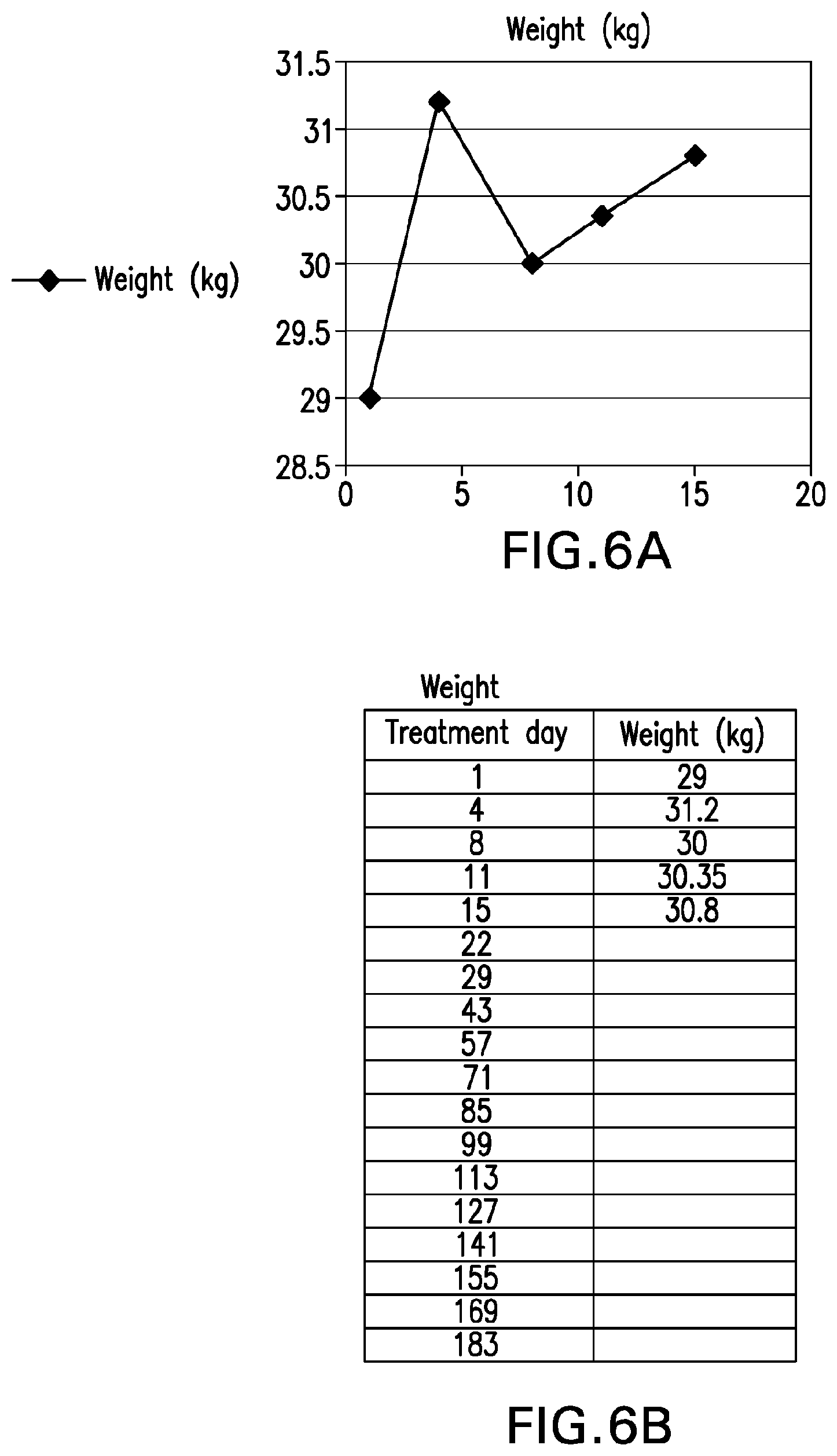
D00013
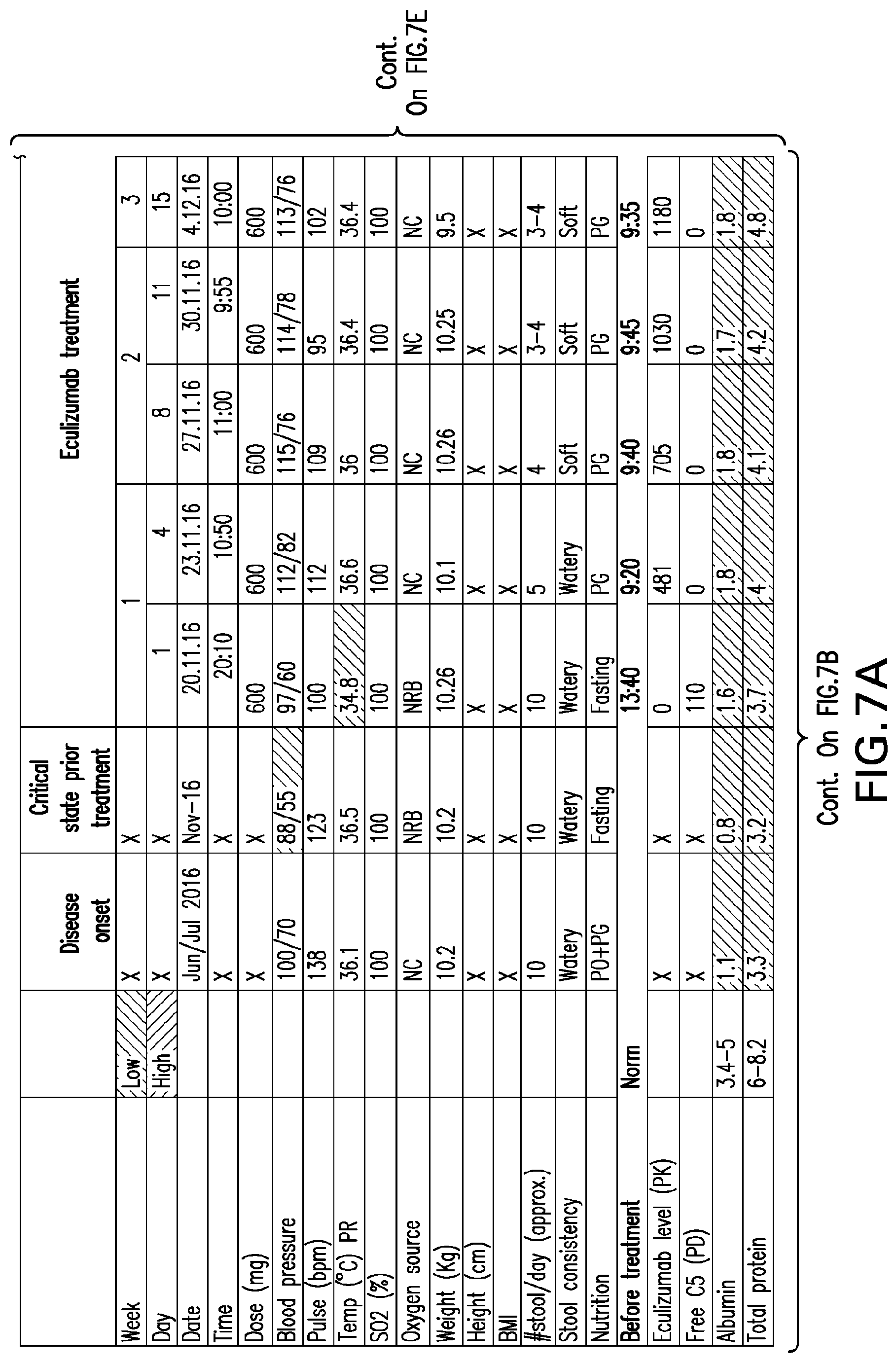
D00014
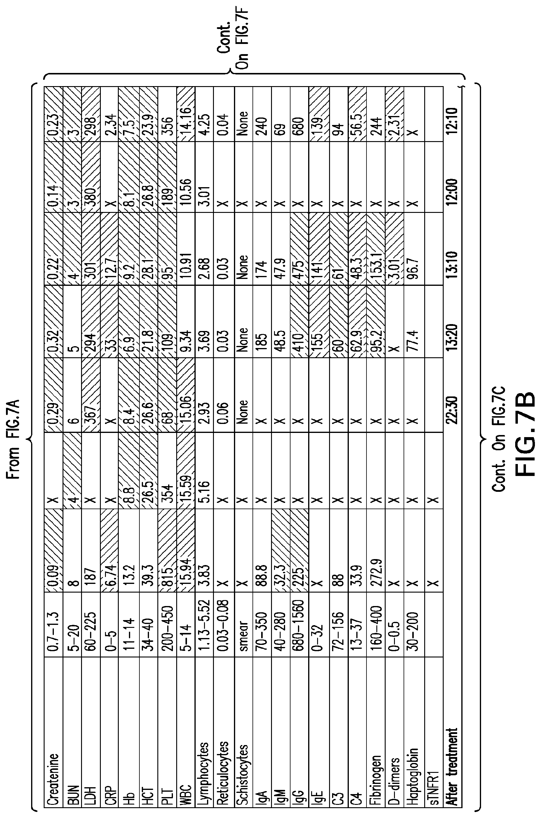
D00015
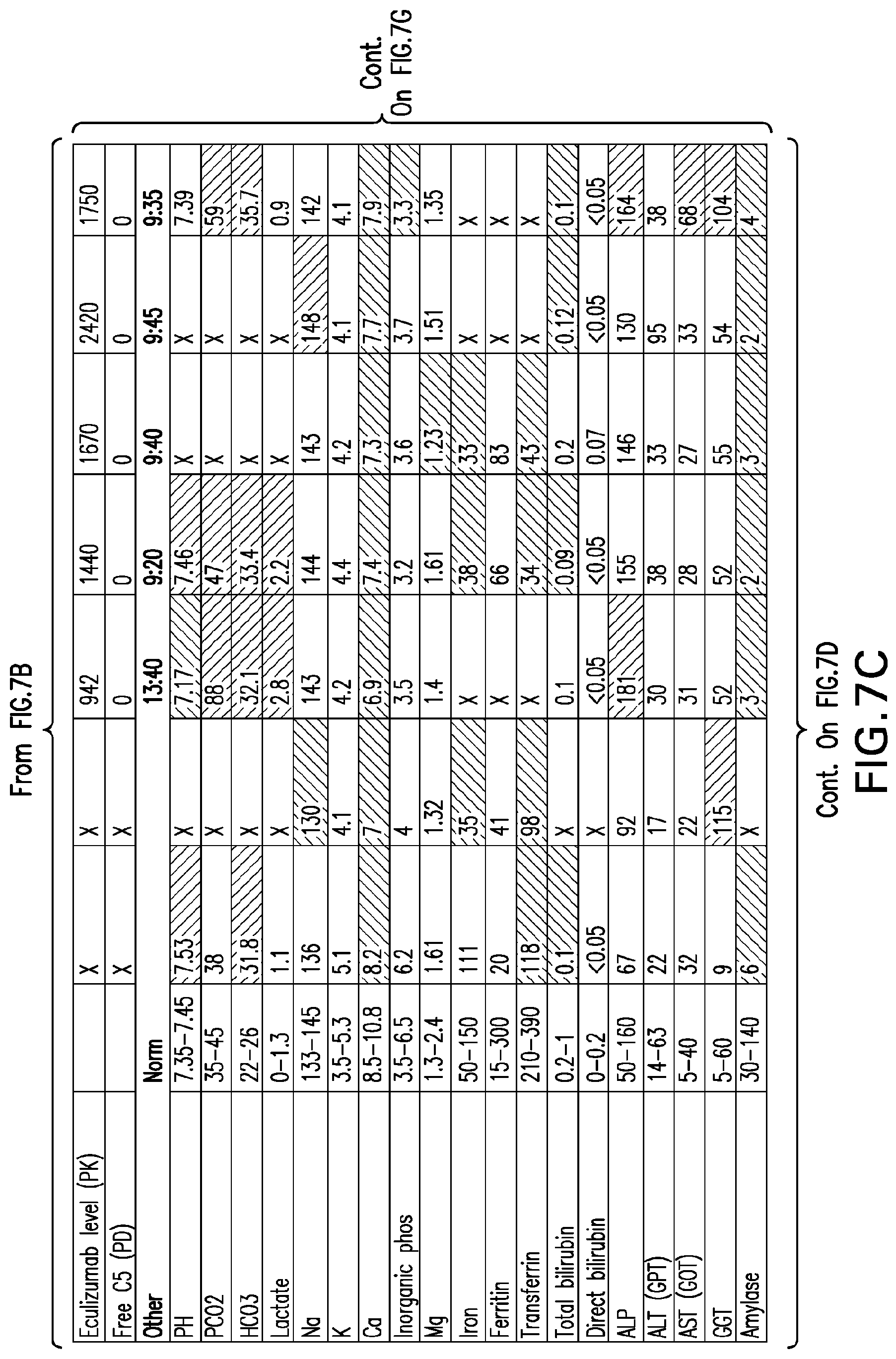
D00016
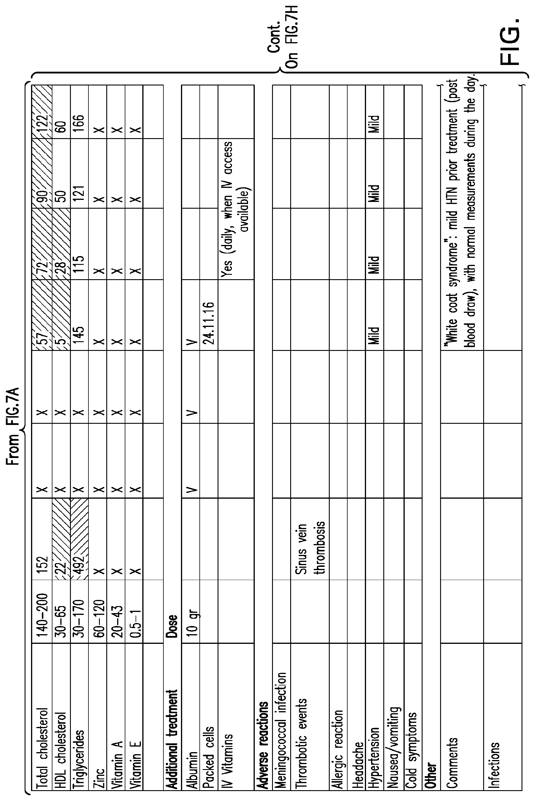
D00017
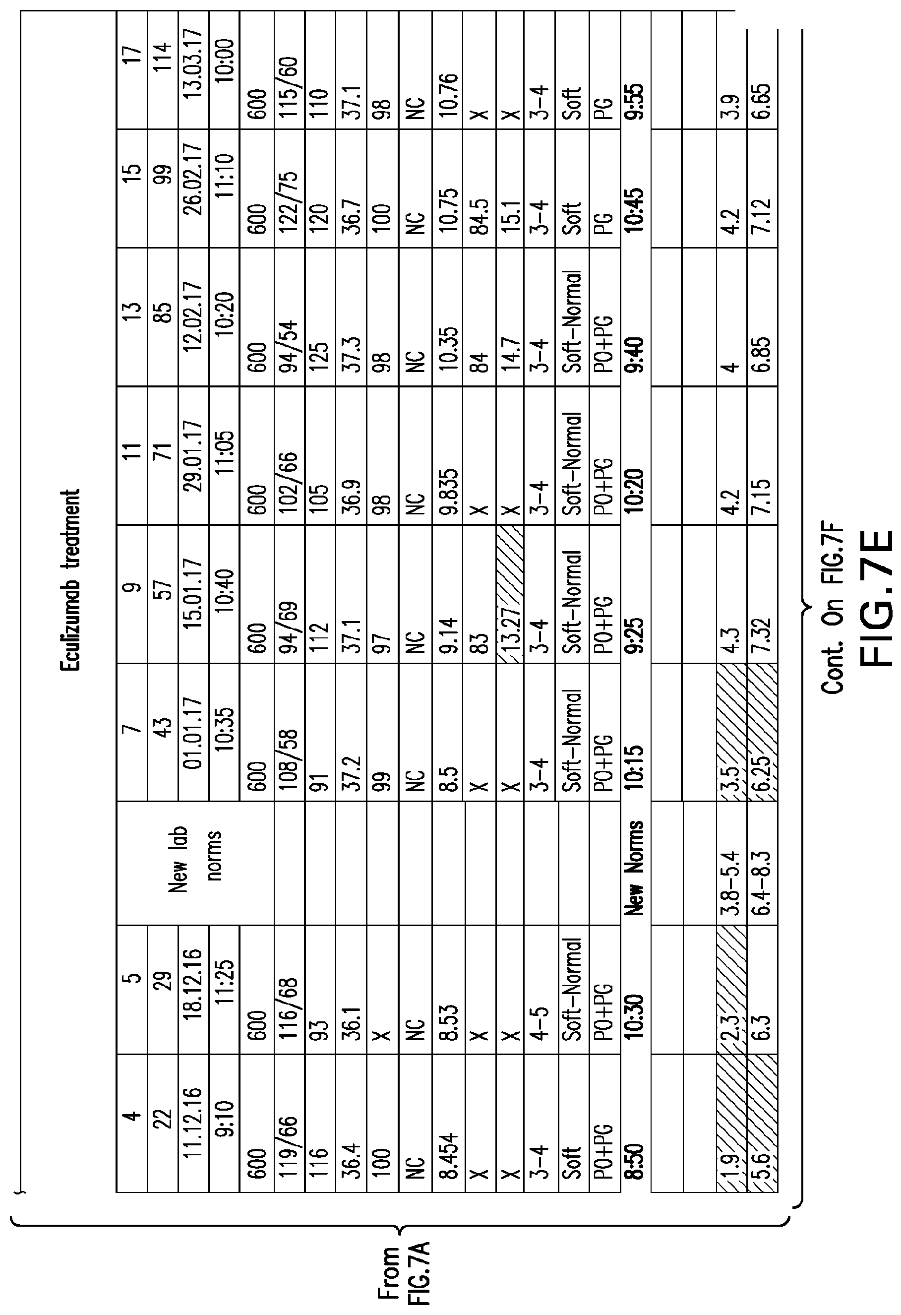
D00018
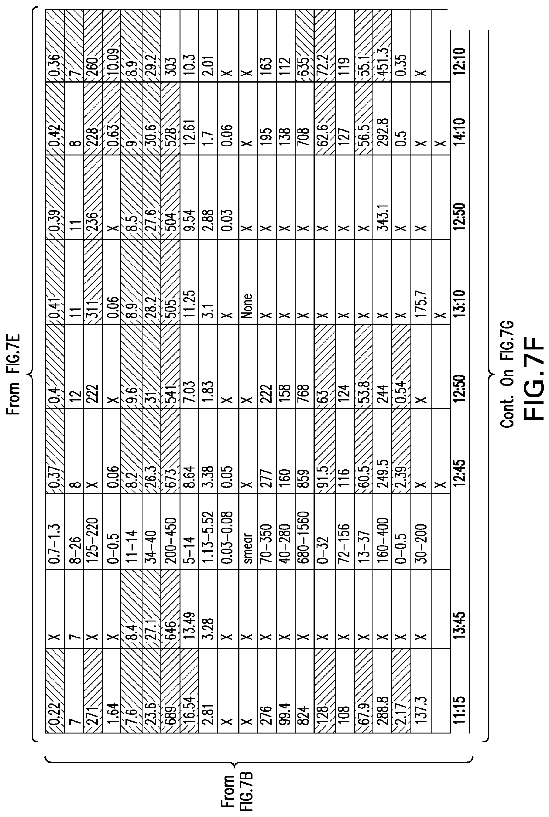
D00019
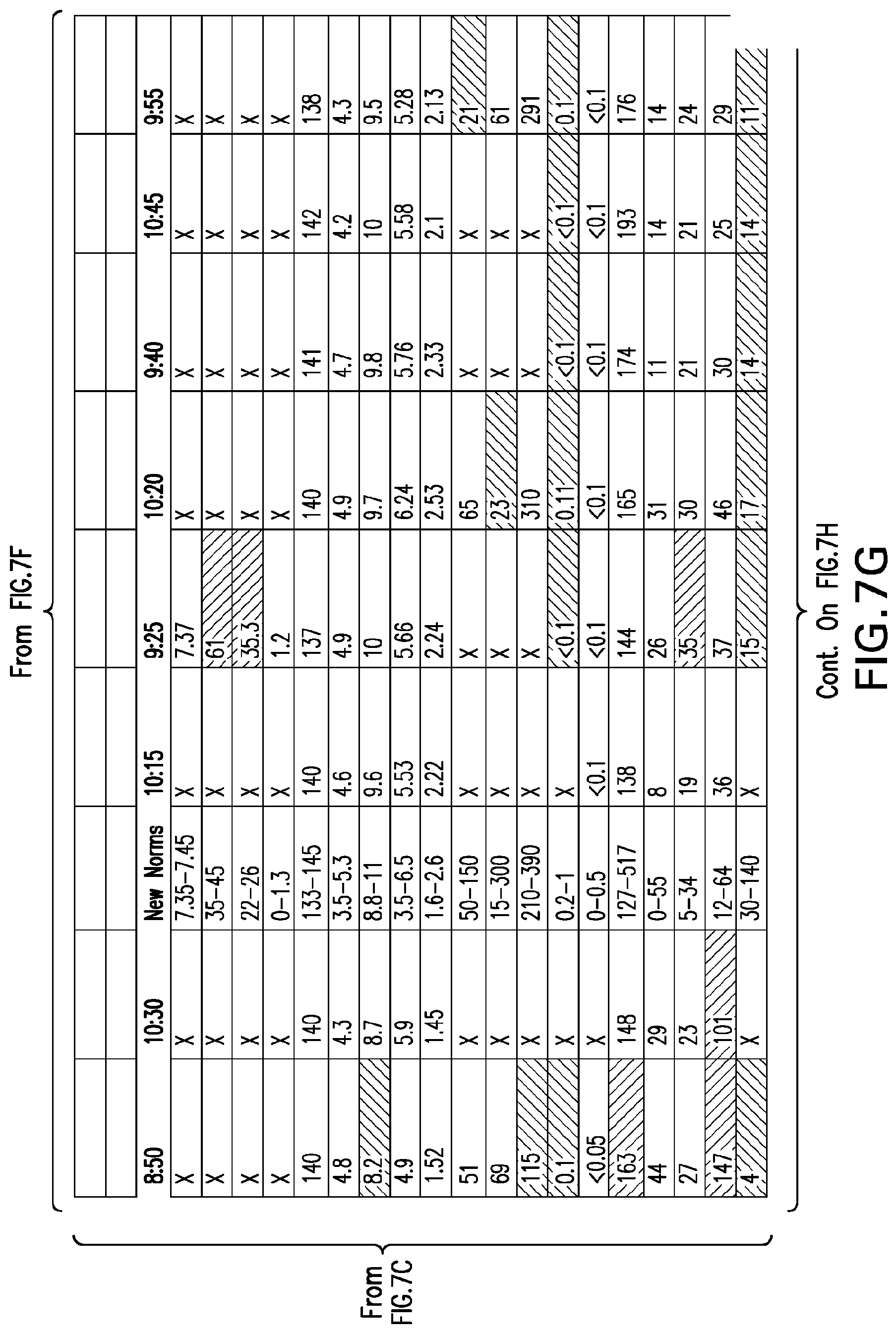
D00020
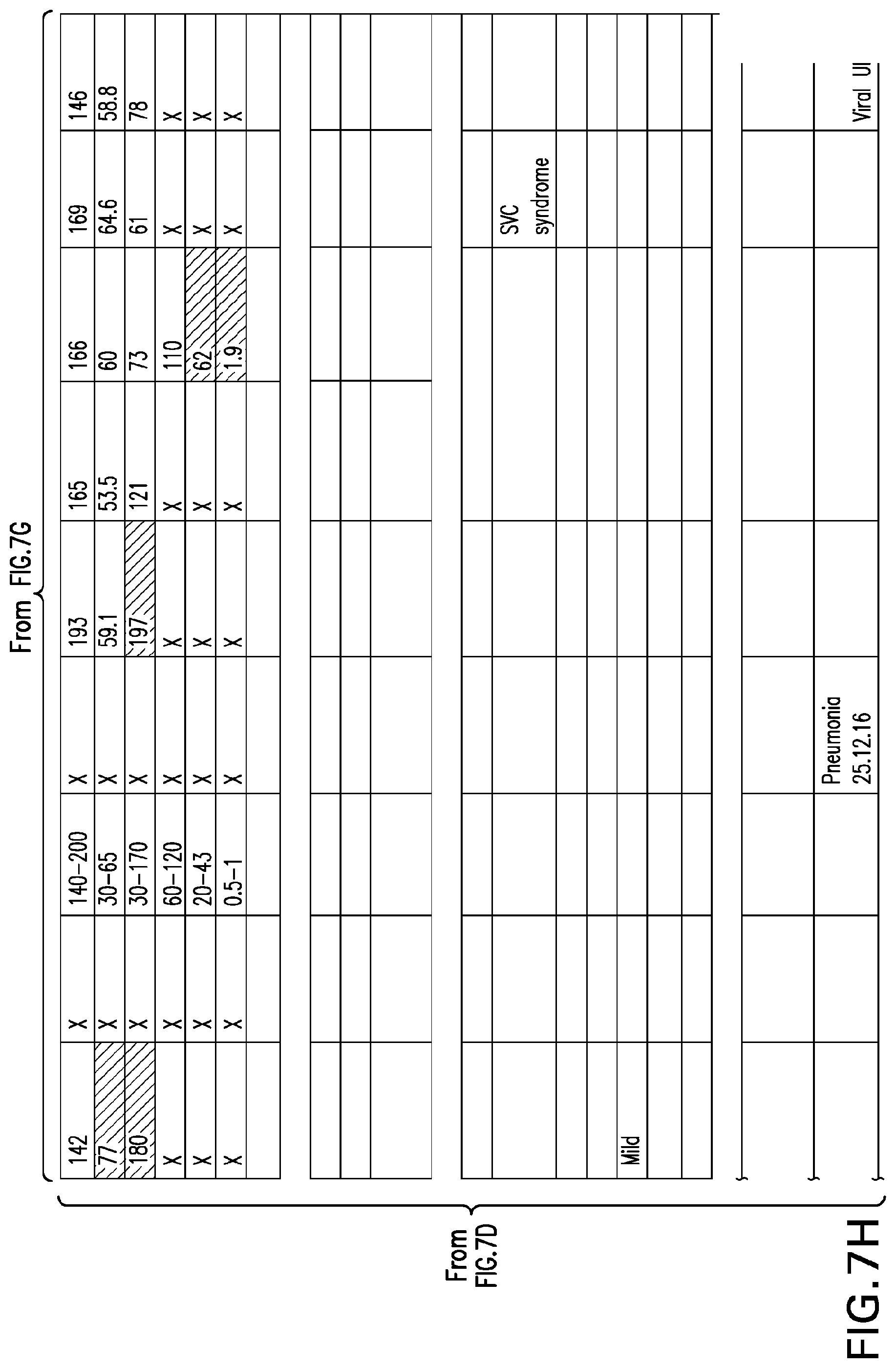
D00021
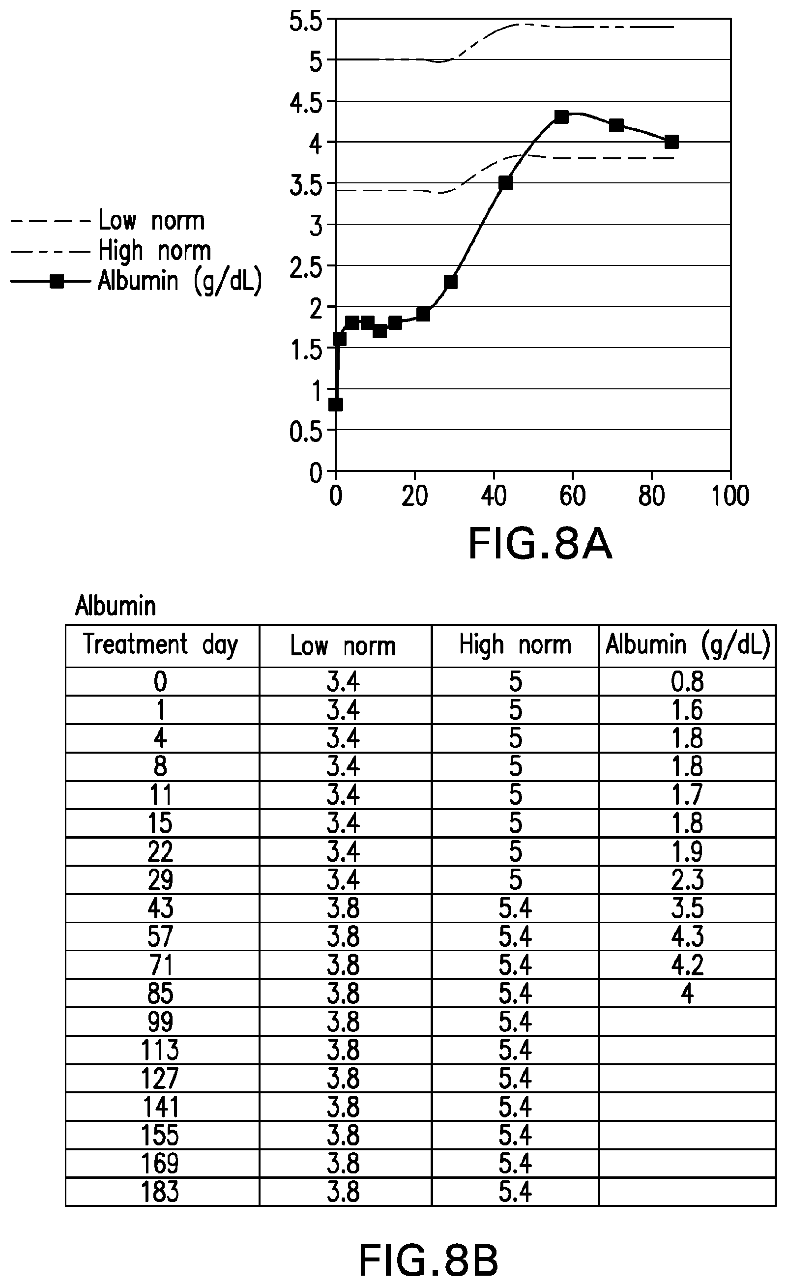
D00022
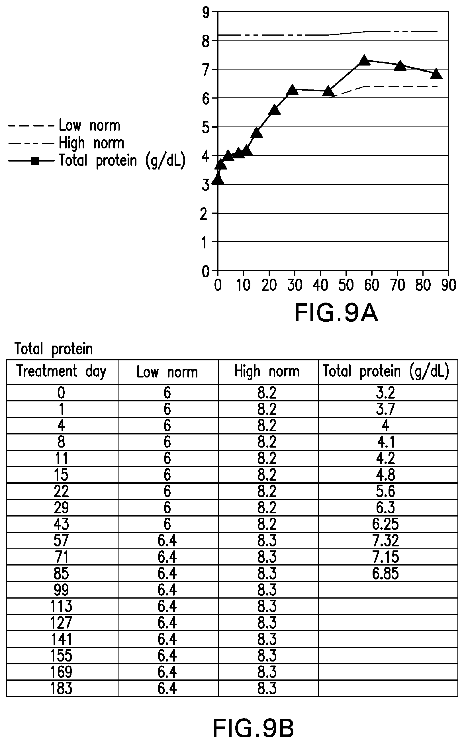
D00023
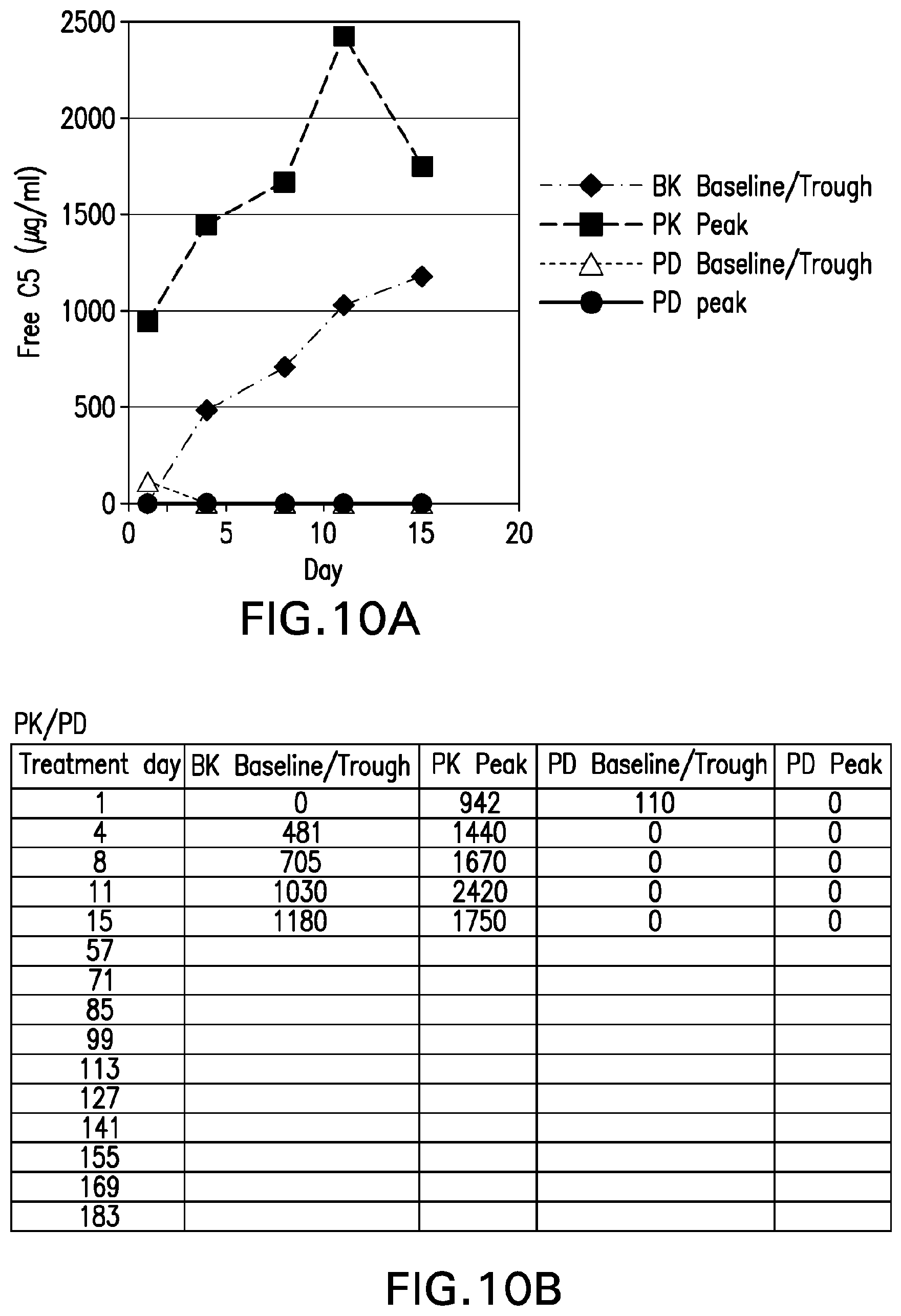
D00024
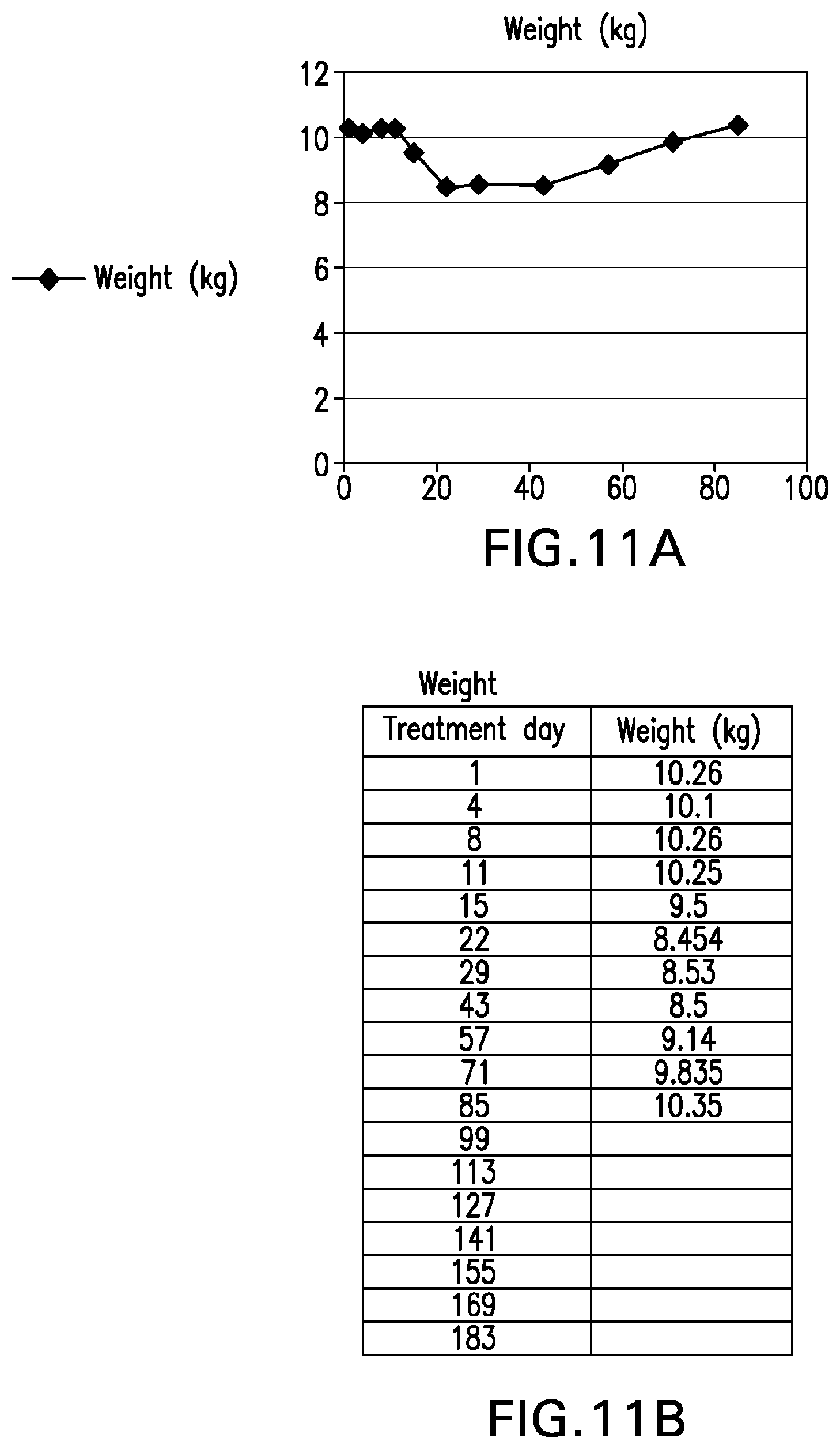
D00025
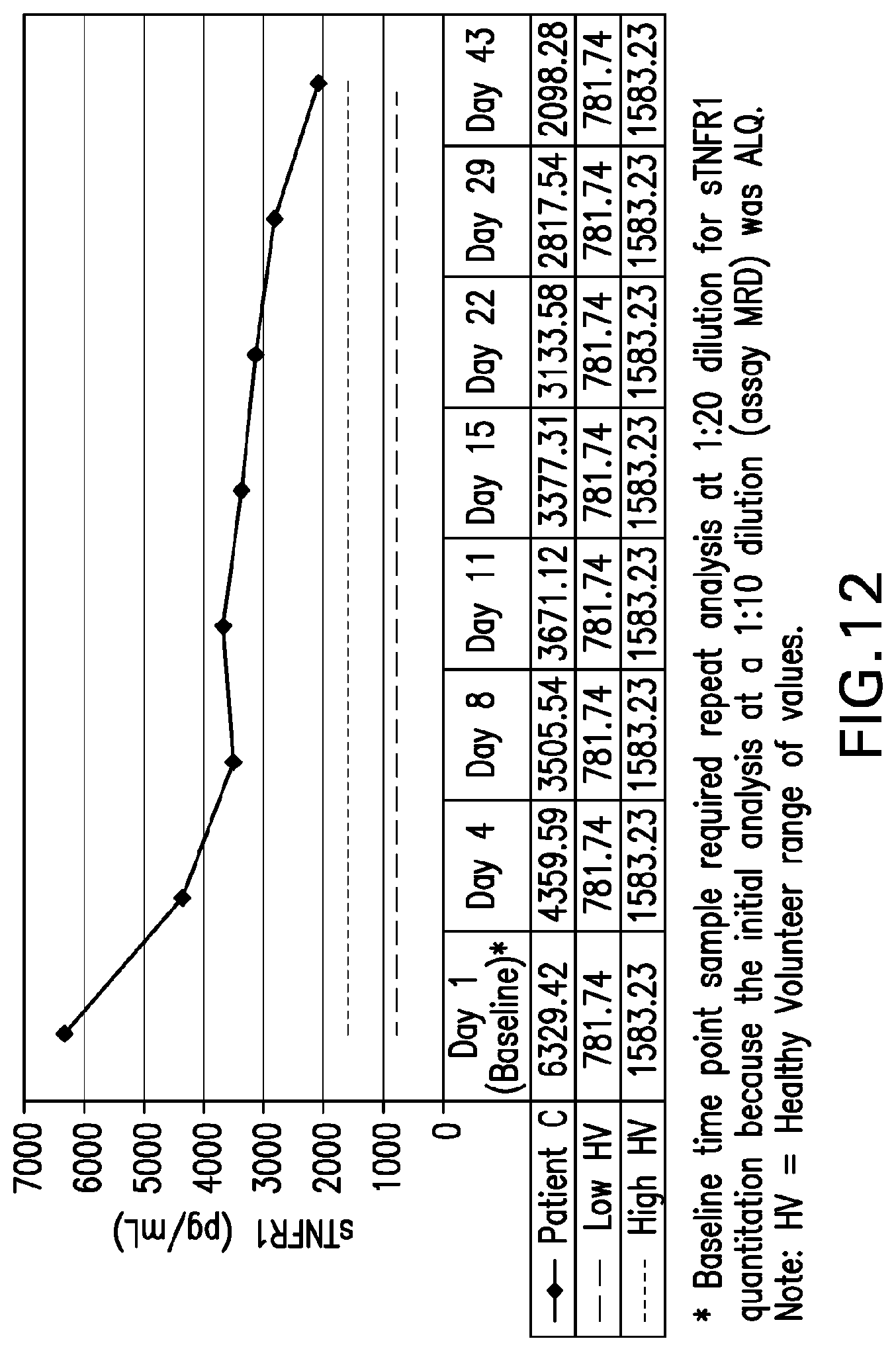
D00026
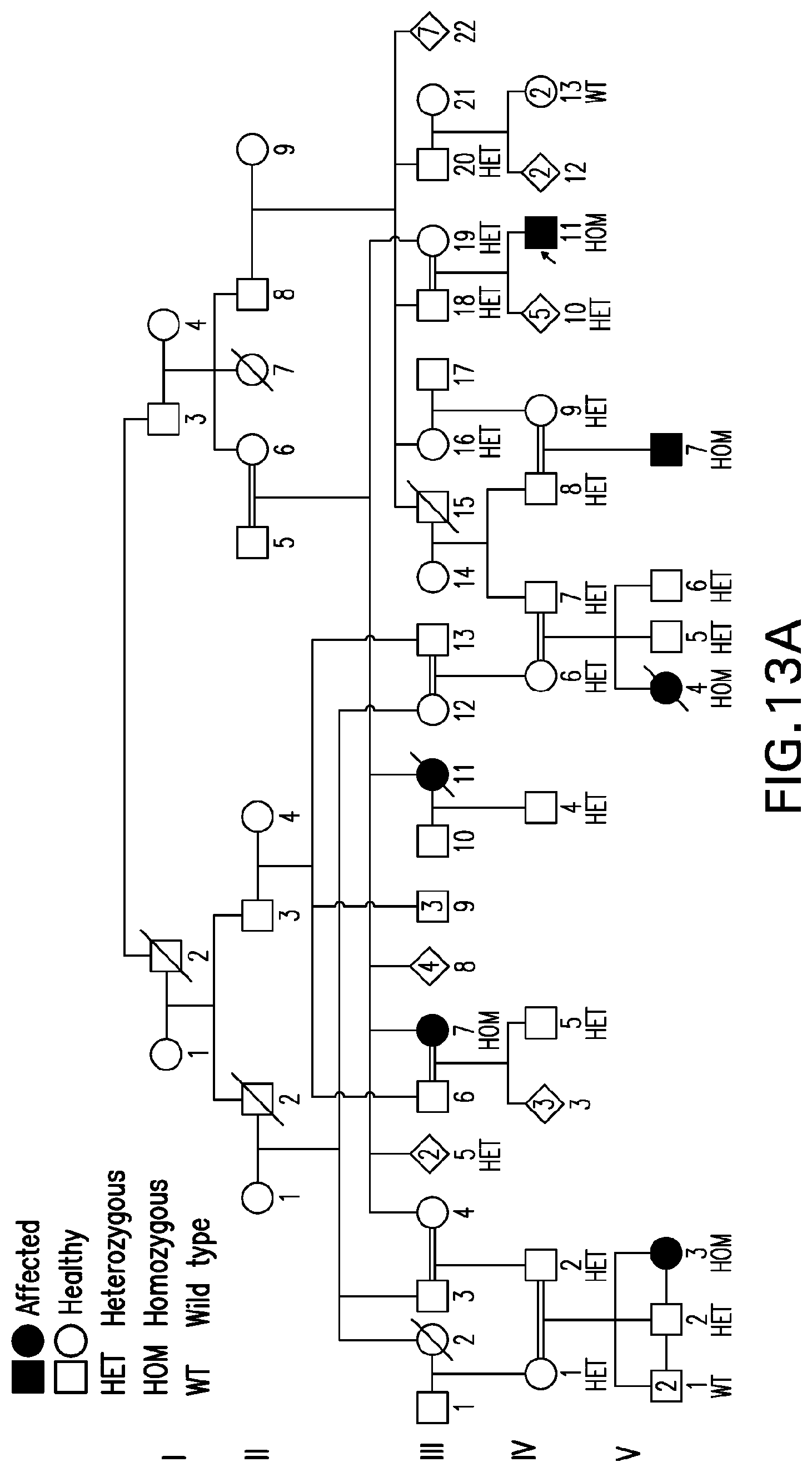
D00027
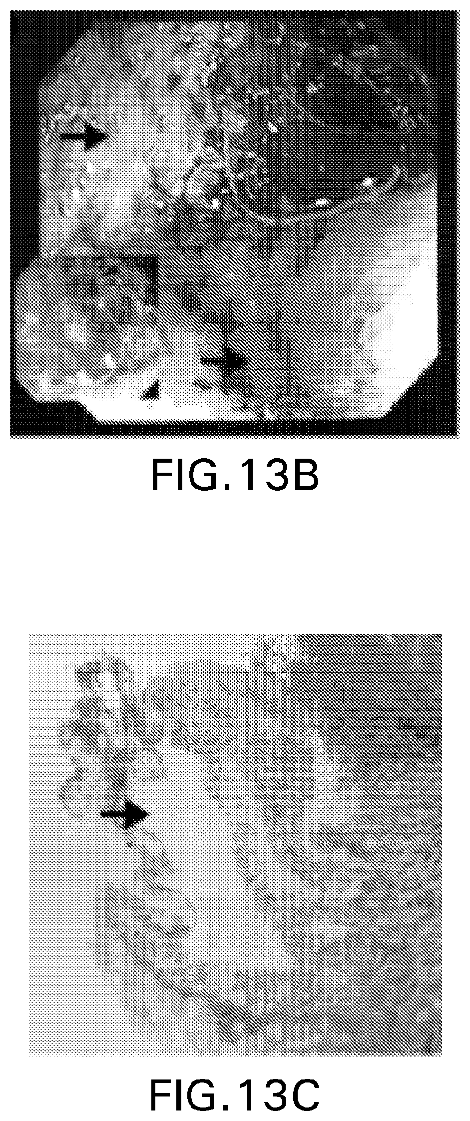
D00028
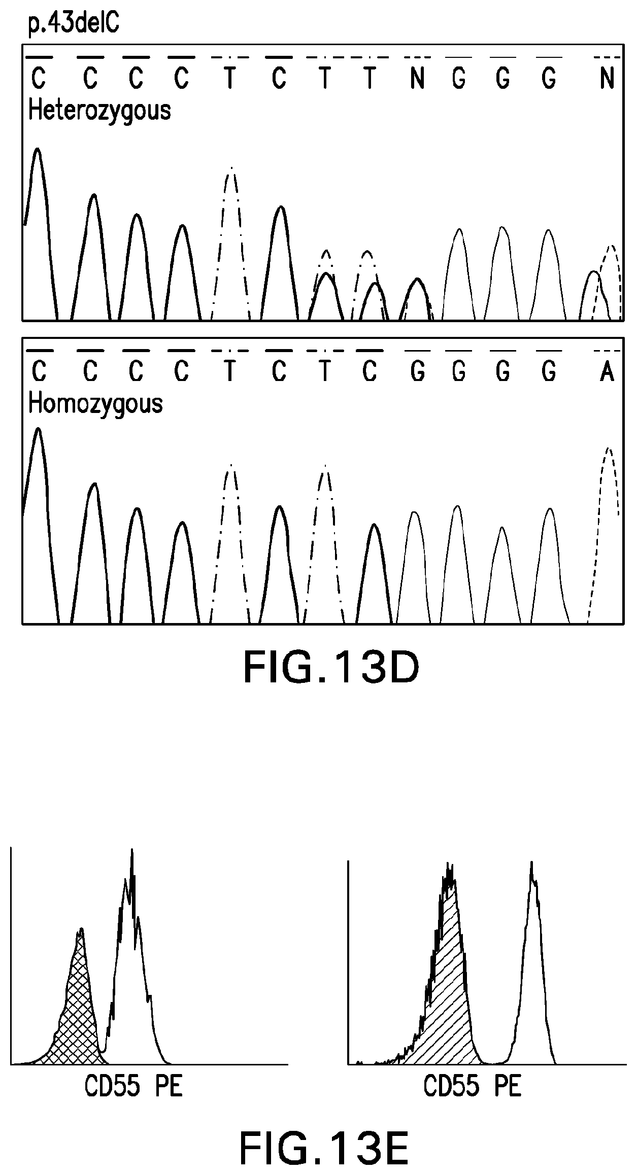
D00029
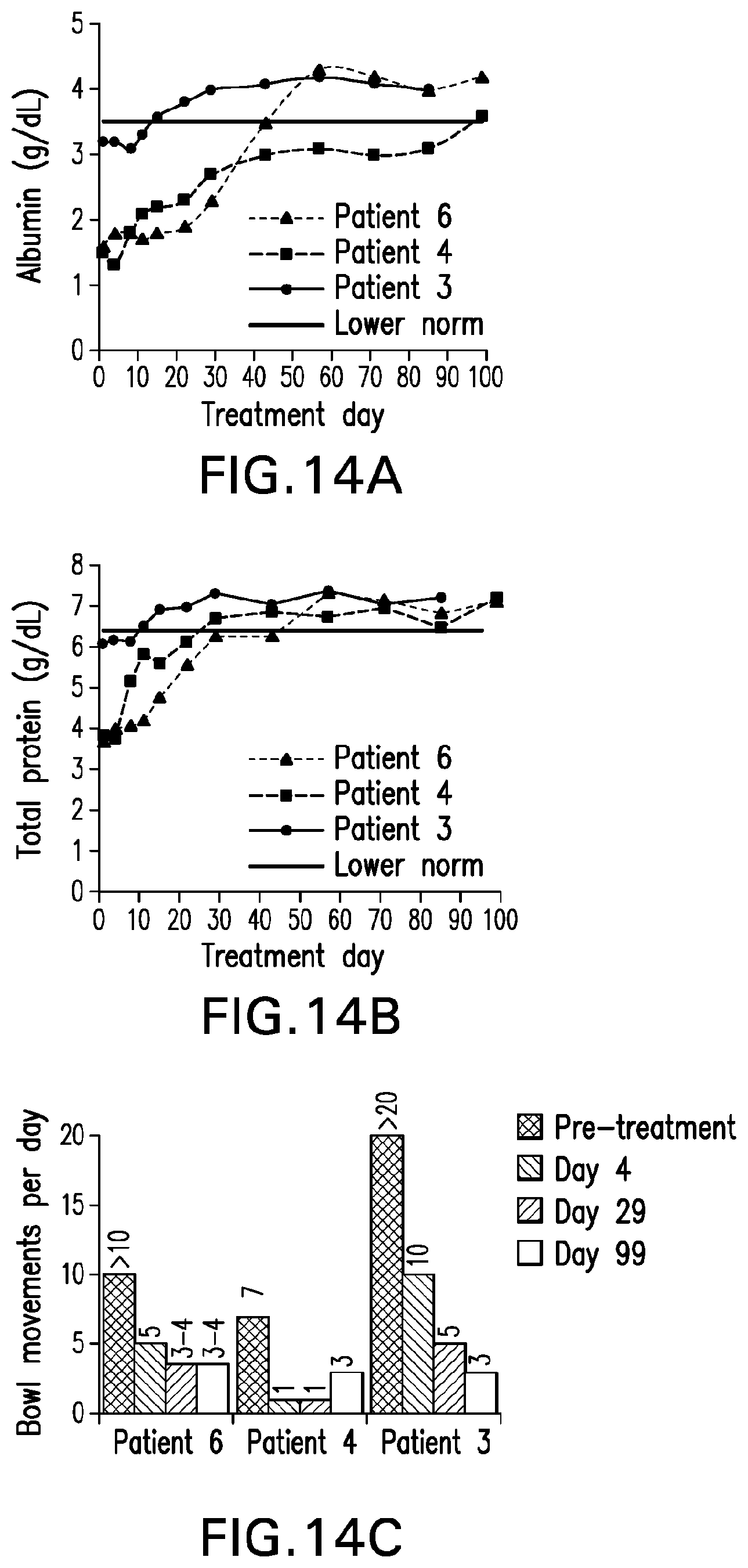
D00030
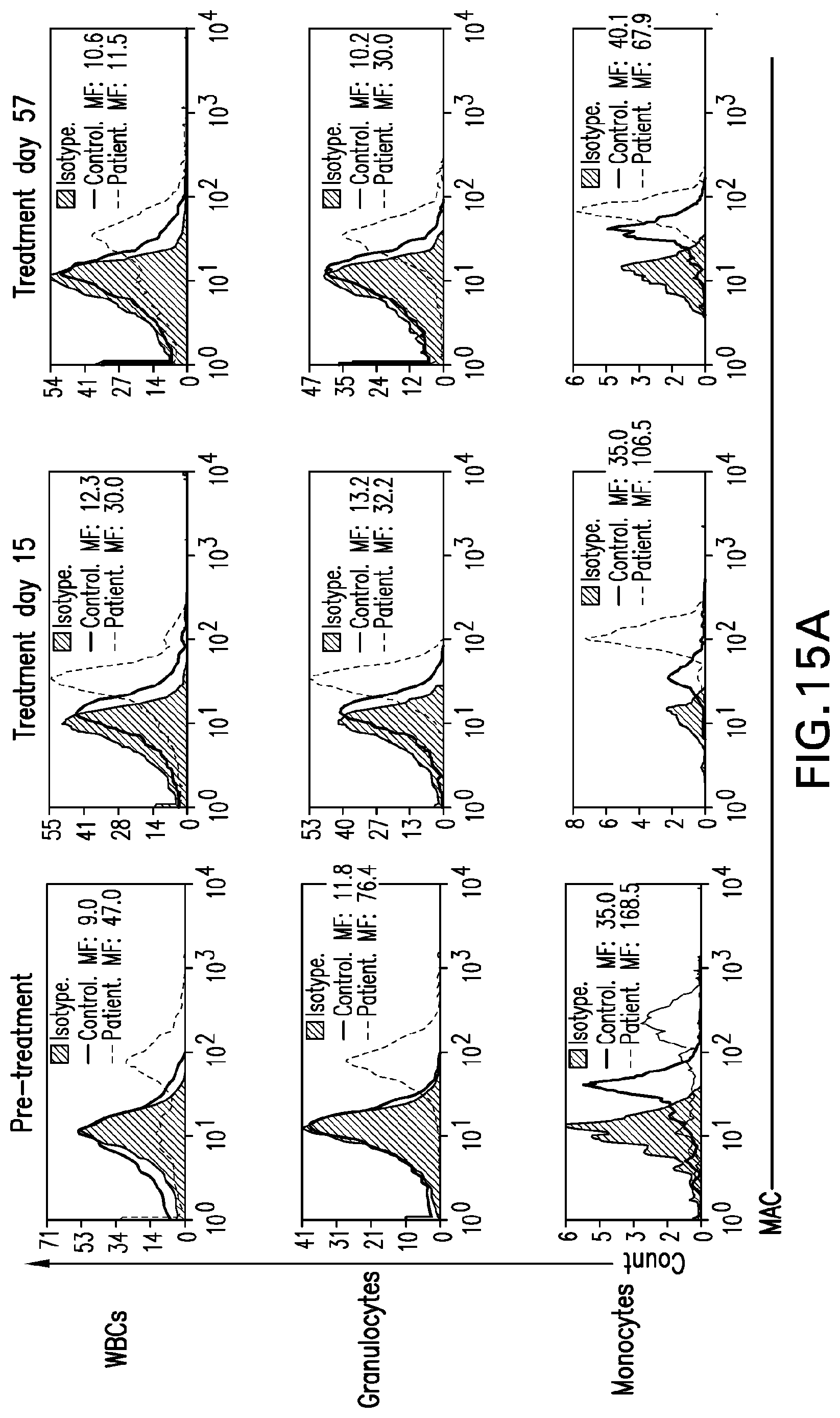
D00031
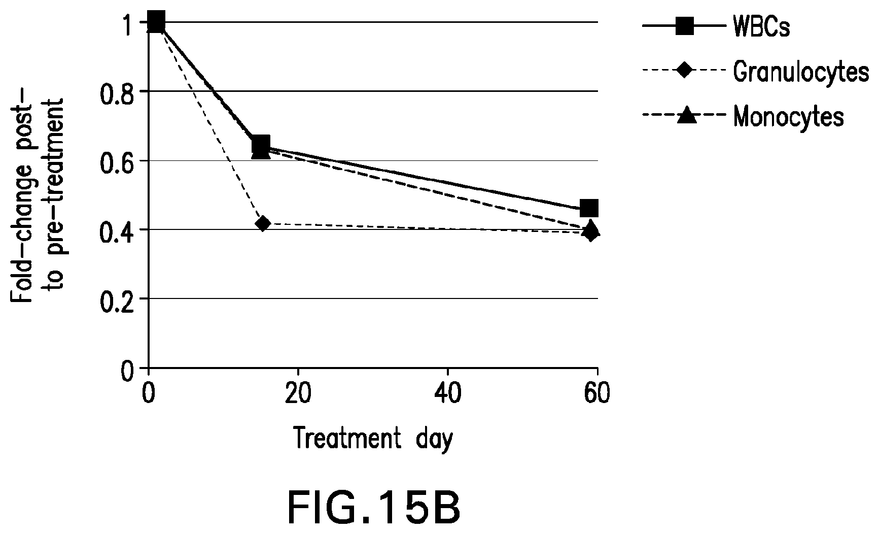
D00032
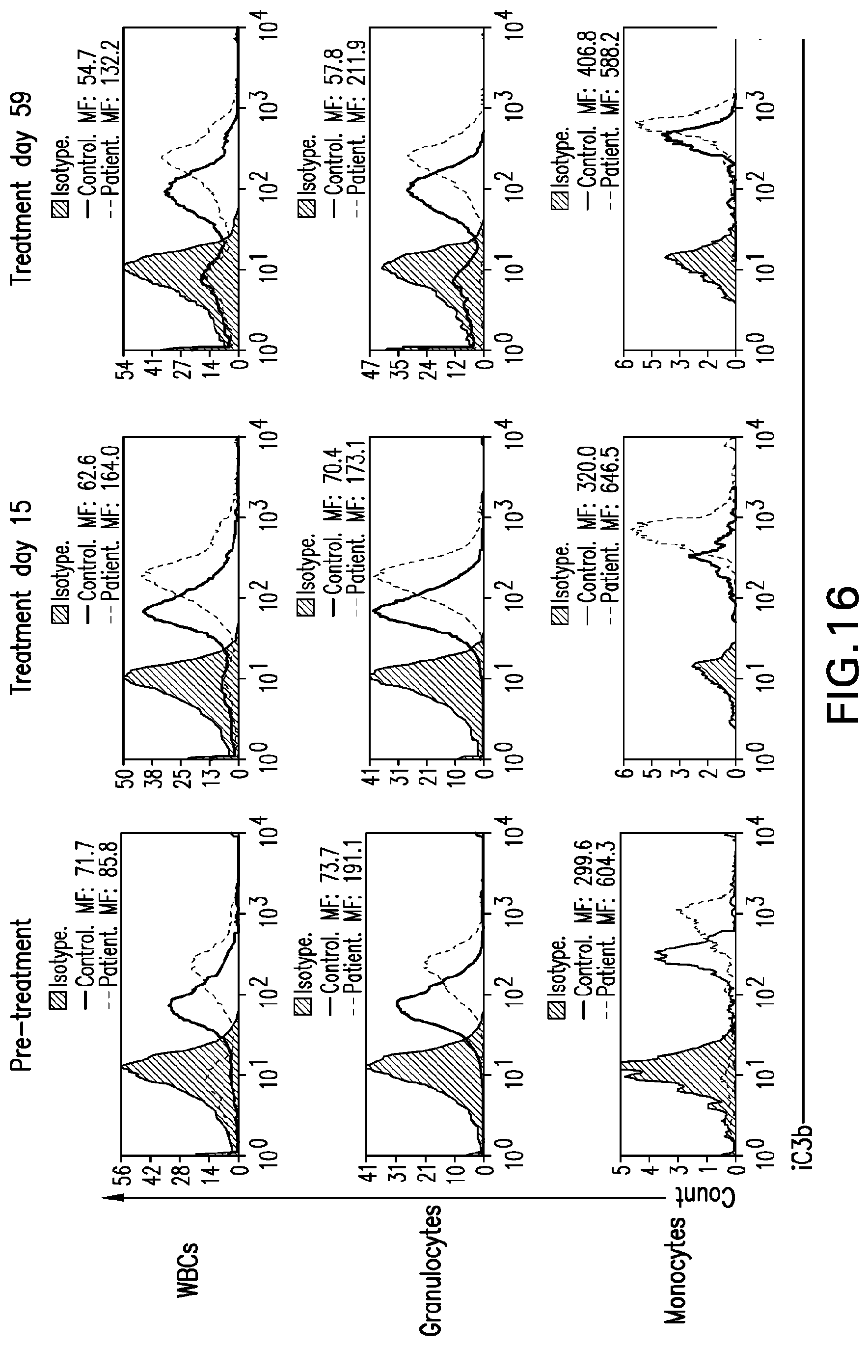
D00033
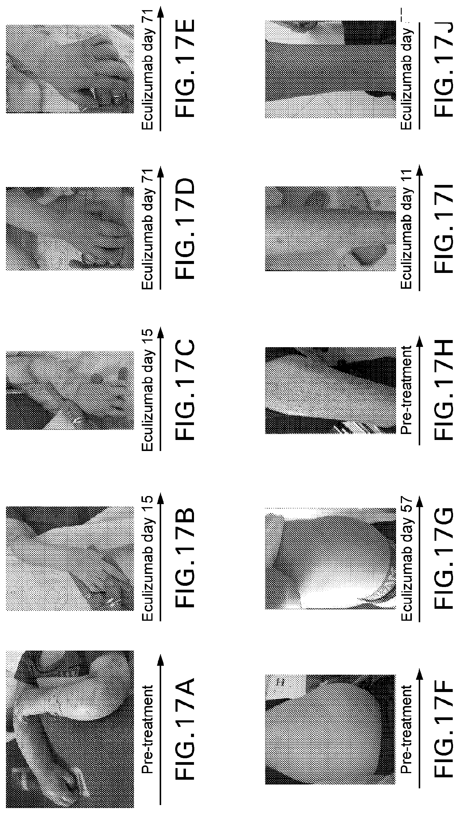
D00034
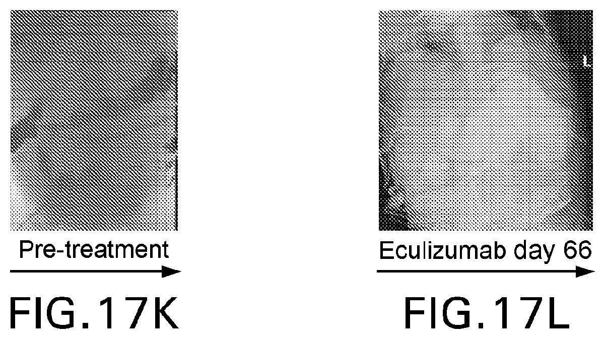
D00035
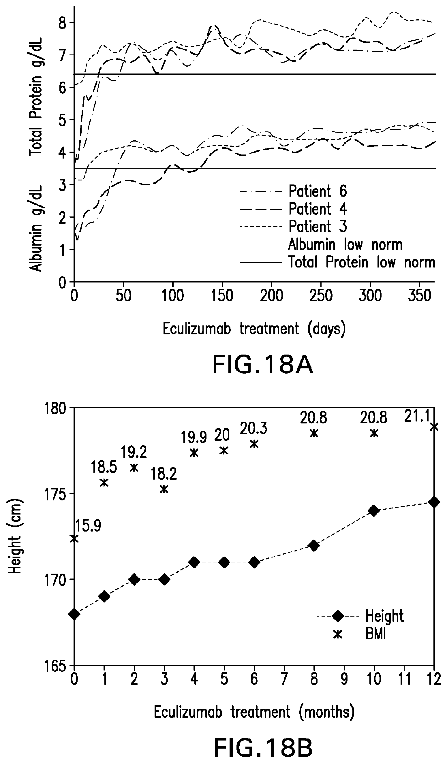
S00001
XML
uspto.report is an independent third-party trademark research tool that is not affiliated, endorsed, or sponsored by the United States Patent and Trademark Office (USPTO) or any other governmental organization. The information provided by uspto.report is based on publicly available data at the time of writing and is intended for informational purposes only.
While we strive to provide accurate and up-to-date information, we do not guarantee the accuracy, completeness, reliability, or suitability of the information displayed on this site. The use of this site is at your own risk. Any reliance you place on such information is therefore strictly at your own risk.
All official trademark data, including owner information, should be verified by visiting the official USPTO website at www.uspto.gov. This site is not intended to replace professional legal advice and should not be used as a substitute for consulting with a legal professional who is knowledgeable about trademark law.