pH-Responsive Ultrasensitive Fluorescent Nanoprobe, Preparation and Using Method Thereof
WANG; Zhongliang ; et al.
U.S. patent application number 16/074778 was filed with the patent office on 2020-05-28 for ph-responsive ultrasensitive fluorescent nanoprobe, preparation and using method thereof. This patent application is currently assigned to Xidian University. The applicant listed for this patent is Xidian University. Invention is credited to Qian JIA, Chaoqiang QIAO, Jie TIAN, Yongdong WANG, Zhongliang WANG, Ruili ZHANG.
| Application Number | 20200164092 16/074778 |
| Document ID | / |
| Family ID | 61110817 |
| Filed Date | 2020-05-28 |


| United States Patent Application | 20200164092 |
| Kind Code | A1 |
| WANG; Zhongliang ; et al. | May 28, 2020 |
pH-Responsive Ultrasensitive Fluorescent Nanoprobe, Preparation and Using Method Thereof
Abstract
The pH-responsive ultrasensitive fluorescent nanoprobe is composed of pH-responsive matrix materials and fluorescent organic small molecule dyes. The pH-responsive matrix materials are calcium phosphate, calcium hydroxyphosphate, fluorapatite, calcium carbonate or ZIF series; the fluorescent organic small molecule dyes are positively charged dyes or negatively charged dyes. The preparation method includes: coating a positively charged dye with a negatively charged matrix material; coating a negatively charged dye with a negatively charged matrix material; and coating a negatively charged dye with a positively charged matrix material. Compared with traditional small molecule fluorescent dyes, the present invention can greatly improve the sensitivity and specificity of fluorescence imaging and achieve ultrasensitive detection of tumor microenvironment response; the specific response probe prepared by the unique properties of the tumor microenvironment has the advantages of high targeting efficacy, low background signal, and high signal-to-noise ratio, and can achieve ultrasensitive detection of tiny tumors.
| Inventors: | WANG; Zhongliang; (Xi'an, CN) ; WANG; Yongdong; (Xi'an, CN) ; ZHANG; Ruili; (Xi'an, CN) ; JIA; Qian; (Xi'an, CN) ; QIAO; Chaoqiang; (Xi'an, CN) ; TIAN; Jie; (Xi'an, CN) | ||||||||||
| Applicant: |
|
||||||||||
|---|---|---|---|---|---|---|---|---|---|---|---|
| Assignee: | Xidian University XI'an CN |
||||||||||
| Family ID: | 61110817 | ||||||||||
| Appl. No.: | 16/074778 | ||||||||||
| Filed: | November 15, 2017 | ||||||||||
| PCT Filed: | November 15, 2017 | ||||||||||
| PCT NO: | PCT/CN2017/111139 | ||||||||||
| 371 Date: | August 2, 2018 |
| Current U.S. Class: | 1/1 |
| Current CPC Class: | A61K 49/0034 20130101; A61K 49/0093 20130101; A61K 49/0032 20130101; A61K 49/0041 20130101; A61K 49/0054 20130101 |
| International Class: | A61K 49/00 20060101 A61K049/00 |
Foreign Application Data
| Date | Code | Application Number |
|---|---|---|
| Aug 16, 2017 | CN | 2017107031485 |
Claims
1. A pH-responsive ultrasensitive fluorescent nanoprobe, comprising a pH-responsive matrix material and a fluorescent organic small molecule dye; wherein the pH-responsive matrix material is a positively charged matrix material or a negatively charged matrix material, comprising calcium phosphate, calcium hydroxyphosphate, fluorapatite, calcium carbonate, and zeolite imidazole framework (ZIF) series; and the fluorescent organic small molecule dye is a positively charged dye or a negatively charged dye; and a plurality of the fluorescent organic small molecule dyes are aggregation by the mutual electrostatic adsorption between a ligand and the fluorescent organic small molecule dyes, and then the pH-responsive matrix material is added for being coordinated with the ligand to form a dye-coated nanoparticle; wherein the ligand is polyacrylic acid (PAA), polyetherimide (PEI) or methylimidazole.
2. The pH-responsive ultrasensitive fluorescent nanoprobe of claim 1, wherein the positively charged dye is IR780, RhB or IR800.
3. The pH-responsive ultrasensitive fluorescent nanoprobe of claim 1, wherein the negatively charged dye is Cy3, Cy5, Cy5.5, Cy7, ICG, ICG-Der-01, ICG-Der-02, ICG-Der-03, IR820, Alexa Fluor 750, Alexa Fluor 700, Alexa Fluor 680, Alexa Fluor 660, Alexa Fluor 647, Alexa Fluor 635, Alexa Fluor 633, Alexa Fluor 610, Alexa Fluor 594, Alexa Fluor 568, Alexa Fluor 555, Alexa Fluor 546, Alexa Fluor 532, Alexa Fluor 514, Alexa Fluor 500, Alexa Fluor 488, or FITC.
4. A preparation method of the pH-responsive ultrasensitive fluorescent nanoprobe of claim 1, comprising the following three methods, specifically: a first method, the positively charged dye is coated with the negatively charged matrix material, and the positively charged dye is adsorbed on PAA molecular chain by electrostatic interaction between the PAA and the positively charged dye, and the PAA after dye adsorption is self-assembled to form a template in isopropyl alcohol, while quenching the fluorescence of the dye by aggregation; the exposed carboxyl on the PAA is coordinated with Ca.sup.2+, and the calcium phosphate is deposited on the surface of the PAA sphere by adjusting the pH and adding phosphate for mineralization to form a dye-coated nanoparticle; a second method, the negatively charged dye is coated with the negatively charged matrix material, and the negatively charged dye is enriched on PEI molecular chain due to the absorption of the negatively charged dye by PEI; meanwhile, a negatively charged PAA is added, and then the PEI absorbs both the dye and the PAA and is self-assembled into a PEI/PAA nanosphere; the exposed carboxyl on the PAA is coordinated with Ca.sup.2+, and the calcium phosphate is deposited on the surface of the PAA sphere by adjusting the pH and adding phosphate for mineralization to form a dye-coated nanoparticle; a third method, the negatively charged dye is coated with the positively charged matrix material, the negatively charged dye molecules are adsorbed on the ligand (methylimidazole) molecules by the mutual electrostatic adsorption between the methylimidazole and the coated dye, meanwhile, the aggregation of the dye is caused, and the metal ions are added for being coordinated with the ligand to form a stable three-dimensional structure, then forming a dye-coated nanoparticle.
5. The preparation method of the pH-responsive ultrasensitive fluorescent nanoprobe of claim 4, wherein in the first method, the pH-responsive matrix materials are calcium phosphate (CaP), calcium carbonate (CaCO.sub.3), calcium hydroxyphosphate, or fluorapatite; the positively charged dyes are IR780, RhB or IR800.
6. The preparation method of the pH-responsive ultrasensitive fluorescent nanoprobe of claim 4, wherein in the second method, the pH-responsive matrix materials are the calcium phosphate (CaP), the calcium carbonate (CaCO.sub.3), the calcium hydroxyphosphate, or the fluorapatite; the negatively charged dyes are ICG, IR820, Alexa Fluor series dyes or Cy series dyes.
7. The preparation method of the pH-responsive ultrasensitive fluorescent nanoprobe of claim 4, wherein in the third method, the pH-responsive matrix materials are MOF materials of ZIF series; the negatively charged dyes are ICG, IR820, Alexa Fluor series dyes or Cy series dyes.
8. A method for using the pH-responsive ultrasensitive fluorescent nanoprobe of claim 1, comprising the following steps: the nanoparticle is injected into a mouse via tail vein, reaches the tumor site through an EPR effect and is enriched in the tumor site; in normal tissues and blood, the pH is neutral or weakly alkaline, and the fluorescent molecules in the nanoparticles are in the state of aggregation quenching; in the tumor site, due to the weak acidic conditions, the matrix material calcium phosphate (CaP) is dissolved and releases the fluorescent molecules, and the fluorescent recovers after the IR780 recovers to the free molecular state, thereby realizing fluorescence enhancement.
9. A CaP/IR780 probe and a CaCO.sub.3/RhB probe prepared by the preparation method of claim 4.
10. A CaP/ICG probe, a CaCO.sub.3/Cy5 probe, and a ZIF-8/ICG probe prepared by the preparation method of claim 4.
11. A CaP/IR780 probe and a CaCO.sub.3/RhB probe prepared by the preparation method of claim 5.
12. A CaP/ICG probe and a CaCO.sub.3/Cy5 probe prepared by the preparation method of claim 6.
13. A ZIF-8/ICG probe prepared by the preparation method of claim 7.
14. The pH-responsive ultrasensitive fluorescent nanoprobe of claim 1, wherein the fluorescent organic small molecule dyes are dyes with aggregation quenching effect; the fluorescent organic small molecule dyes are fluorescence quenched due to being in an aggregation state; the fluorescent organic small molecule dyes are fluorescence recovered due to being in a single molecule state.
Description
CROSS REFERENCE TO RELATED APPLICATIONS
[0001] This application is the national phase entry of International Application No. PCT/CN2017/111139, filed on Nov. 15, 2017, which is based upon and claims priority to Chinese Application No. CN2017107031485, filed on Aug. 16, 2017, the entire contents of which are incorporated herein by reference.
TECHNICAL FIELD
[0002] The present invention relates to the technical field of molecular imaging, and particularly to a pH-responsive ultrasensitive fluorescent nanoprobe, preparation and using method thereof.
BACKGROUND
[0003] Molecular imaging can be used to study the occurrence, development and metastasis of diseases or tumors in vivo because of its ability to realize real-time, non-invasive and dynamic imaging at the living body level. Optical imaging has the advantages of like no radiation, high sensitivity, and low light damage to biological tissues, showing great potential in the research field of in vivo tumor monitoring. Due to the rapid proliferation of cells, tumor tissue tends to cause rapid glucose metabolism, large oxygen consumption, and excessive accumulation of acidic metabolites (such as lactic acid) in the tumor microenvironment. Therefore, the tumor microenvironment is generally characterized by weak acidity, hypoxia, production of a plurality of cytokines and high expression of biological enzymes. Conventional organic small molecule fluorescent probes, because of the non-specific distribution in vivo, will cause organic dyes generally to have the disadvantages of poor bio-distribution specificity, high background signal, poor light stability and easy photobleaching, thus being limited in the application of in vivo imaging, and cannot be applied to the detection of tiny tumors. The use of bio-intelligent responsive nanomaterial-loaded small molecule fluorescent probes can not only improve the light stability of small molecule fluorescent probes, but also achieve the high-efficient and specific enrichment of probes and the pH regulated fluorescence enhancement in the tumor sites, which has become a popular research direction in the field of molecular imaging technology.
SUMMARY
[0004] In view of the problems existing in the prior art, the present invention provides a pH-responsive ultrasensitive fluorescent nanoprobe, preparation and using method thereof.
[0005] The present invention is achieved by a pH-responsive ultrasensitive fluorescent nanoprobe, the pH-responsive ultrasensitive fluorescent nanoprobe is composed of pH-responsive matrix materials and organic small molecule dyes having fluorescent;
[0006] the negatively charged pH-responsive matrix materials are calcium phosphate (abbreviated as CaP), calcium hydroxyphosphate, fluorapatite, and calcium carbonate.
[0007] Further, positively charged pH-responsive matrix materials are zeolite imidazole framework (ZIF) material series;
[0008] the fluorescent organic small molecule dyes are positively charged dyes or negatively charged dyes.
[0009] Further, the positively charged dyes are IR780, RhB, or IR800.
[0010] Further, the negatively charged dyes are Cy3, Cy5, Cy5.5, Cy7, ICG, ICG-Der-01, ICG-Der-02, ICG-Der-03, IR820, Alexa Fluor 750, Alexa Fluor 700, Alexa Fluor 680, Alexa Fluor 660, Alexa Fluor 647, Alexa Fluor 635, Alexa Fluor 633, Alexa Fluor 610, Alexa Fluor 594, Alexa Fluor 568, Alexa Fluor 555, Alexa Fluor 546, Alexa Fluor 532, Alexa Fluor 514, Alexa Fluor 500, Alexa Fluor 488, or FITC.
[0011] Another purpose of the present invention is to provide a method for preparing the pH-responsive ultrasensitive fluorescent nanoprobe, and the method for preparing the pH-responsive ultrasensitive fluorescent nanoprobe includes the following three methods.
[0012] The first method, a positively charged dye is coated with a negatively charged matrix material, the dye is adsorbed on the PAA (polyacrylic acid) molecular chain by electrostatic interaction between the polyacrylic acid and the positively charged dye, and the PAA after dye adsorption is self-assembled to form a template in isopropyl alcohol, while the fluorescence of the dye is quenched by aggregation; the exposed carboxyl on the PAA is coordinated with Ca.sup.2+, and the calcium phosphate is deposited on the surface of the PAA sphere by adjusting the pH and adding phosphate for mineralization to form a dye-coated nanoparticle.
[0013] The second method, a negatively charged dye is coated with a negatively charged matrix material, the dye is enriched on the polyetherimide (PEI) molecular chain through the absorption of the negatively charged dye by PEI; meanwhile, a negatively charged PAA is added, the PEI absorbs both dye and PAA to self-assembled into a PEI/PAA nanospheres; the exposed carboxyl on the PAA is coordinated with Ca.sup.2+, and the calcium phosphate is deposited on the surface of the PAA sphere by adjusting the pH and adding phosphate for mineralization to form a dye-coated nanoparticle.
[0014] The third method, a negatively charged dye is coated with a positively charged matrix material, the dye molecules are adsorbed on the ligand (methylimidazole) molecules by the mutual electrostatic adsorption between the methylimidazole and the coated dye, meanwhile, the aggregation of the dye is caused, and the metal ions are added for being coordinated with the ligand to form a stable three-dimensional structure, thus forming a dye-coated nanoparticle.
[0015] Further, in the first method, the negatively charged pH-responsive matrix materials are calcium phosphate, calcium carbonate, calcium hydroxyphosphate, and fluorapatite; the positively charged dyes are IR780, RhB, or IR800.
[0016] Further, in the second method, the negatively charged pH-responsive matrix materials are calcium phosphate, calcium carbonate, calcium hydroxyphosphate, and fluorapatite; the negatively charged dyes are ICG, IR820, Alexa Fluor series dyes, and Cy series dyes.
[0017] Further, in the third method, the pH-responsive matrix materials are the MOF materials of the ZIF series; the negatively charged dyes are ICG, IR820, Alexa Fluor series dyes, and Cy series dyes.
[0018] Another purpose of the present invention is to provide a method for using the pH-responsive ultrasensitive fluorescent nanoprobe, and the method for using the pH-responsive ultrasensitive fluorescent nanoprobe includes: The nanoparticle is injected into the mouse via tail vein, which reaches the tumor site through the enhanced permeability and retention (EPR) effect and is enriched in the tumor site.
[0019] In normal tissues and blood, the pH is neutral or weakly alkaline, and the fluorescent molecules in the nanoparticles are in the state of aggregation quenching. In the tumor site, due to its weak acidic conditions, the matrix material CaP is dissolved and releases the fluorescent molecules, and the fluorescent recovers after the IR780 recovers to the free molecular state, thereby realizing fluorescence enhancement.
[0020] Another purpose of the present invention is to provide a CaP/IR780 probe and a CaCO.sub.3/RhB probe prepared by the pH-responsive ultrasensitive fluorescent nanoprobe.
[0021] Another purpose of the present invention is to provide a CaP/ICG probe and a CaCO.sub.3/Cy5 probe prepared by the pH-responsive ultrasensitive fluorescent nanoprobe.
[0022] Another purpose of the present invention is to provide a ZIF-8/ICG probe prepared by the pH-responsive ultrasensitive fluorescent nanoprobe.
[0023] The advantages and positive effects of the present invention are: the high-efficiency specificity enrichment and controllable release of the small molecule fluorescent probe at the tumor site can be achieved through the response of matrix materials to the weakly acidic conditions of the tumor microenvironment, the fluorescence intensity of the probe after release is significantly enhanced, and the ultrasensitive detections of the primary lesions and metastatic lesions of tumors are realized; matrix nanomaterials have pH response characteristic, under neutral conditions, fluorescent molecules loaded in nanoparticles are subjected to fluorescence quenching due to being in aggregation state; under the weakly acidic conditions of the tumor microenvironment, the matrix material is dissolved, so that the fluorescent molecules loaded in the nanoparticles recover to the single molecule state, the fluorescence is recovered, the tumor microenvironment acidic-responsive fluorescence enhancement is realized, and accurate imaging and real-time monitoring of early tumors can be realized.
[0024] The present invention can realize the high-efficiency enrichment of nanomaterials at the tumor site through the enhanced permeability and retention (EPR) effect of the nanomaterial at the tumor site, and the tumor-specific fluorescence enhancement can be achieved by utilizing the characteristic of pH response to the tumor microenvironment. Before reaching the tumor site, fluorescence quenching of fluorescent molecules in the probe was caused by the aggregation of the fluorescent molecules loaded in the matrix material, and the fluorescence signal was in a closed (OFF) state; while the probe is in a weakly acidic condition of the tumor microenvironment, the organic fluorescent molecules are released from the matrix material to achieve a pH-responsive "deaggregation" to enable the fluorescent signal recovery (ON). The OFF-ON switch minimizes the background signal of the fluorescent probe and significantly increases the imaging signal-to-noise ratio by nearly three times. Compared with the traditional small molecule fluorescent dyes, the sensitivity and specificity of fluorescence imaging can be greatly improved, and ultrasensitive detection of tumor microenvironment response can be realized; the specificity response probe prepared by the unique properties of the tumor microenvironment has the advantages of high targeting efficacy, low background signal, and high signal-to-noise ratio, and the ultrasensitive detection of tiny tumors smaller than 1 mm can be realized in vivo.
BRIEF DESCRIPTION OF THE DRAWINGS
[0025] FIG. 1 is a flow chart of preparation method of a pH-responsive ultrasensitive fluorescent nanoprobe provided by embodiments of the present invention.
[0026] FIG. 2 is a TEM diagram of CaP/IR780-PEG provided by an embodiment of the present invention.
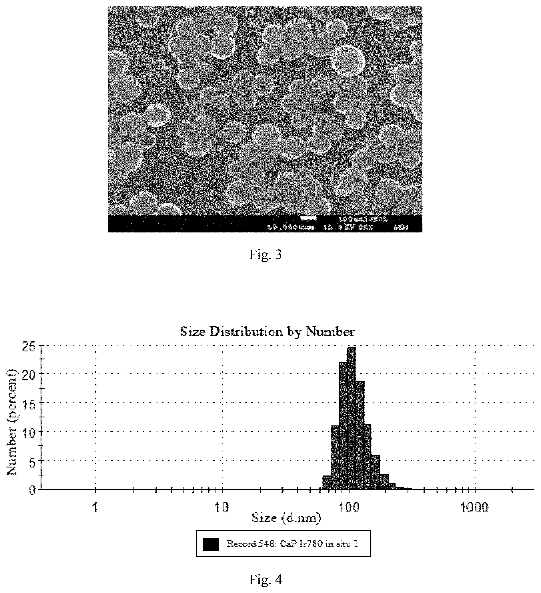
[0027] FIG. 3 is a SEM diagram of CaP/IR780-PEG provided by an embodiment of the present invention.
[0028] FIG. 4 is a DLS diagram of CaP/IR780-PEG provided by an embodiment of the present invention.
[0029] FIG. 5 is a ultraviolet-visible spectroscopy showing pH-responsive characteristic of CaP/IR780 provided by an embodiment of the present invention.
[0030] FIG. 6 is a fluorescence spectrum showing pH-responsive characteristic of CaP/IR780 provided by an embodiment of the present invention.
[0031] FIG. 7 is a SEM diagram of ZIF8/ICG provided by an embodiment of the present invention.
[0032] FIG. 8 is a graph showing stability curves of CaP/IR780 in different media provided by an experiment of the present invention.
[0033] FIG. 9 is a schematic diagram of cytotoxicity experiment of a nanoprobe provided by an experiment of the present invention.
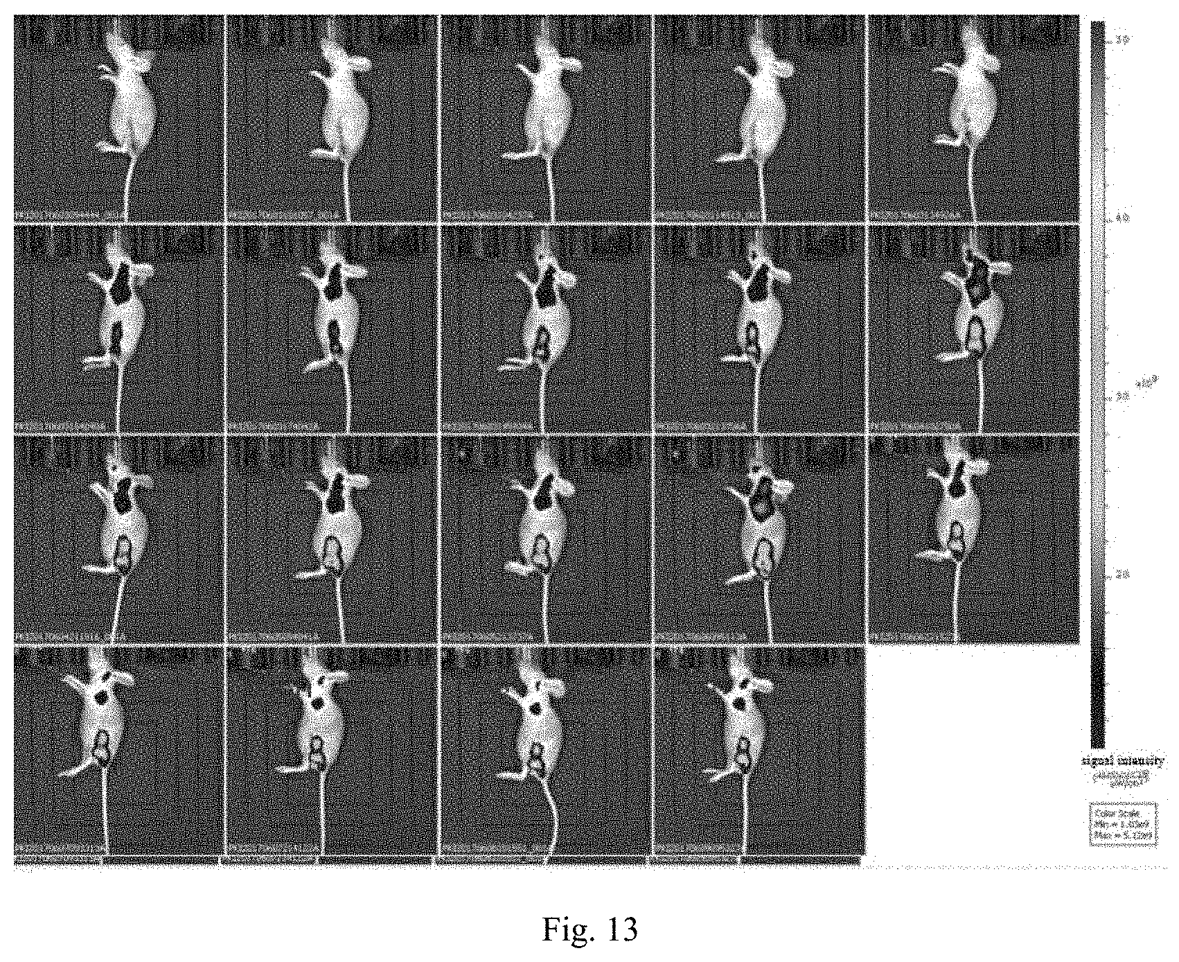
[0034] FIG. 10 is a subcutaneous tumor imaging experiment of a nanoprobe 4T1 provided by an experiment of the present invention.
[0035] FIG. 11 is a diagram of subcutaneous tumor imaging experiment of a nanoprobe MCF7 provided by an experiment of the present invention.
[0036] FIG. 12 is a diagram of brain tumor in situ imaging experiment of a nanoprobe provided by an experiment of the present invention.
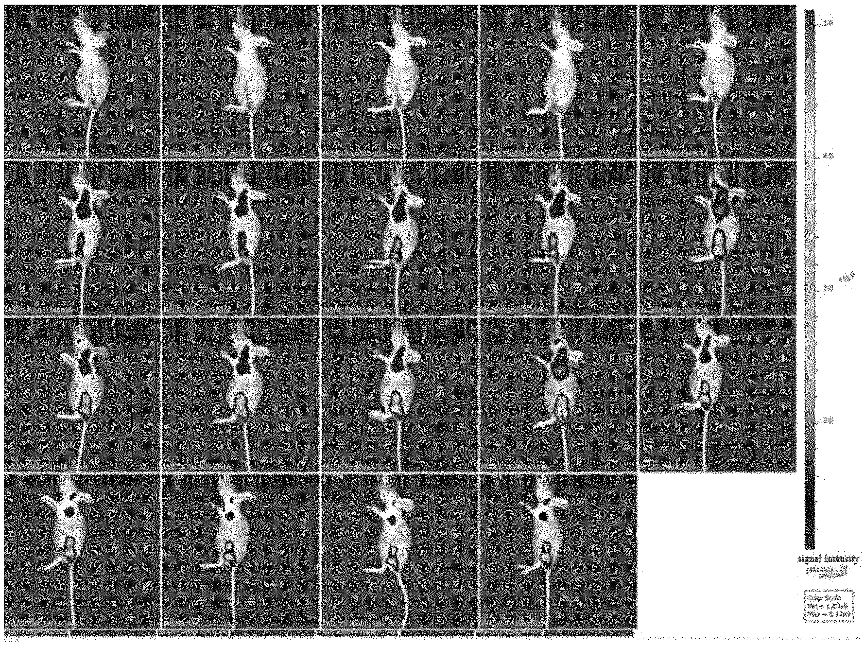
[0037] FIG. 13 is a diagram of lymph node metastasis tumor imaging experiment of a nanoprobe 4T1 provided by an experiment of the present invention.
DETAILED DESCRIPTION OF THE EMBODIMENTS
[0038] In order to make the purpose, technical scheme and advantages of the present invention more clear, the present invention will be further described in detail below with reference to the embodiments. It should be understood that, the specific embodiments described herein are merely intended to explain the present invention rather than limit the present invention.
[0039] The principle of the present invention will be described in detail below with reference to the accompanying drawings.
[0040] The pH-responsive ultrasensitive fluorescent nanoprobes provided by the embodiments of the present invention are composed of pH-responsive matrix materials and fluorescent organic small molecule dyes.
[0041] The pH-responsive matrix material has a fast response to pH, and the pH-responsive dye molecules are released by the response of the pH-responsive matrix material to weakly acidic conditions. The main selection of the pH-responsive matrix material can be as follows: calcium phosphate, calcium hydroxyphosphate, fluorapatite, and calcium carbonate, ZIF series etc.; the matrix materials have the advantages of good biocompatibility, fast pH response, and safe and non-toxic metabolites.
[0042] Fluorescence organic small molecule dyes are the source of fluorescence signal, and the quenching/recovering transformation of the fluorescence is realized by the pH-responsive aggregation and deaggregation to achieve the pH-responsive fluorescence enhancement (OFF-ON). Organic fluorescent small molecules are dye molecules with aggregation quenching effect, the organic fluorescent small molecules can be: positively charged dyes such as IR780, RhB, IR800, etc., or negatively charged dyes such as Cy3, Cy5, Cy5.5, Cy7, ICG, ICG-Der-01, ICG-Der-02, ICG-Der-03, IR820, Alexa Fluor 750, Alexa Fluor 700, Alexa Fluor 680, Alexa Fluor 660, Alexa Fluor 647, Alexa Fluor 635, Alexa Fluor 633, Alexa Fluor 610, Alexa Fluor 594, Alexa Fluor 568, Alexa Fluor 555, Alexa Fluor 546, Alexa Fluor 532, Alexa Fluor 514, Alexa Fluor 500, Alexa Fluor 488, FITC, etc. Fluorescent organic small molecule dyes have the advantages of long emission wavelength, weaker light scattering signal in vivo, deep detection depth and good alkali stability.
[0043] As shown in FIG. 1, a preparation method of a pH-responsive ultrasensitive fluorescent nanoprobe provided by an embodiment of the present invention includes the following steps:
[0044] S101: the dye is adsorbed on the PAA molecular chain by electrostatic interaction between the PAA and the positively charged dye, and the PAA after dye adsorption is self-assembled to form a template in isopropyl alcohol;
[0045] S102: quenching the fluorescence of the dye by aggregation; the exposed carboxyl on the PAA is coordinated with Ca.sup.2+, and the calcium phosphate is deposited on the surface of the PAA sphere by adjusting the pH and adding phosphate for mineralization to form a dye-coated nanoparticle.
[0046] The negatively charged matrix materials can be calcium phosphate, calcium carbonate, calcium hydroxyphosphate, fluorapatite, etc., the positively charged dyes used in the embodiment can be IR780, RhB, IR800, etc. The synthetic probes include CaP/IR780, CaCO.sub.3/RhB, etc.
[0047] The preparation method of the pH-responsive ultrasensitive fluorescent nanoprobe provided by the embodiment of the present invention also includes: a negatively charged dye is coated with a negatively charged matrix material, and the dye is enriched on the PEI molecular chain due to the absorption of the negatively charged dye by PEI; meanwhile, a negatively charged PAA is added, and then the PEI absorbs both dye and PAA and is self-assembled into a PEI/PAA nanospheres; the exposed carboxyl on the PAA is coordinated with Ca.sup.2+, and the calcium phosphate is deposited on the surface of the PAA sphere by adjusting the pH and adding phosphate for mineralization to form a dye-coated nanoparticle.
[0048] The negatively charged matrix materials can be calcium phosphate, calcium carbonate, calcium hydroxyphosphate, fluorapatite, etc., the negatively charged dyes used in the embodiment can be ICCQ IR820, Alexa Fluor series dyes, and Cy series dyes. Probes synthesized by this method include CaP/ICCQ CaCO.sub.3/Cy5, etc.
[0049] The preparation method of the pH-responsive ultrasensitive fluorescent nanoprobe provided by the embodiment of the present invention also includes: a negatively charged dye is coated with a positively charged matrix material, the dye molecules are adsorbed on the ligand (methylimidazole) molecules by the mutual electrostatic adsorption between the ligand and the coated dye, meanwhile, the aggregation of the dye is caused, and the metal ions are added for coordinating with the ligand to form a stable three-dimensional structure, then forming a dye-coated nanoparticle.
[0050] The positively charged matrix materials can be MOF materials of the ZIF series, such as ZIF-8, etc., the negatively charged dyes used in the embodiment can be ICG, IR820, Alexa Fluor series dyes, and Cy series dyes. Probes synthesized by this method include ZIF-8/ICG, etc.
[0051] The specific operating method of the probe prepared by the present invention for tumor imaging in vivo is:
[0052] The nanoparticle is injected into the mouse via tail vein, which reaches the tumor site through the EPR effect and is enriched in the tumor site. In normal tissues and blood, the pH is neutral (or weakly alkaline), and the fluorescent molecules in the nanoparticles are in the state of aggregation quenching. In the tumor site, due to its weak acidic conditions, the matrix material CaP is dissolved and releases the fluorescent molecules, and the fluorescent recovers after the IR780 recovers to the free molecular state, therefore, fluorescence enhancement is realized, and the pH-responsive fluorescence enhancement is realized in the whole imaging process.
[0053] The application principle of the present invention will be further described below with reference to the specific embodiments.
Embodiment 1
[0054] Preparation of CaP/IR780 Nanoprobe
[0055] Synthesis principle: the dye is aggregated by electrostatic interaction between the PAA and IR780 to achieve fluorescence quenching, the PAA after dye adsorption is self-assembled to form a template in isopropyl alcohol, and the calcium phosphate is deposited on the surface of the PAA sphere by mineralization to form a CaP/IR780 nanoparticle.
[0056] Specific synthesis steps: dissolving 60-80 .mu.L 20% PAA (MW=2000) in 10 mL water, adding 5-8 mg Ca(OH).sub.2, stirring to dissolve, adding 50-1000 .mu.g IR780, stirring for 30 min for mixing well. Slowly adding 10-50 mL of isopropanol to form a milky white solution, adding 6-9.6 mg (NH.sub.4).sub.2HPO.sub.4 and stirring for 24 h, centrifuging, and washing for 3 times, redistributing in 5 mL water for standby application. Characterizations of particle size were performed by the transmission electron microscopy (TEM), the scanning electron microscopy (SEM) and the dynamic light scattering (DLS) as shown in FIG. 2, FIG. 3 and FIG. 4; the ultraviolet-visible spectroscopy of the nanoparticles with different dyes coated were measured by the ultraviolet-visible spectrophotometer, respectively, as shown in FIG. 5; the ultraviolet-visible spectroscopy of the nanoparticles with different dyes coated were measured at the pH of 6.5, as shown in FIG. 6. In order to improve the stability of the nanoprobe and the circulation time in the blood of the animals, the surface of the probe is stabilized by PEG, thus improving the stability of the probe. Adding 10 mg of 1-(3-Dimethylaminopropyl)-3-ethylcarbodiimide (EDC), 12 mg of N-Hydroxysuccinimide (NHS) and 5 mg of polyethylene glycol amine (PEG-NH.sub.2) to the above particle dispersion system, adjusting pH to 7.2, stirring at room temperature for 24 h, centrifuging, washing for 3 times with water, and dispersing in 5 mL of phosphate-buffered saline (PBS) for standby application.
Embodiment 2
[0057] Preparation of CaP/RhB Nanoprobe
[0058] Synthesis principle: the dye is aggregated by electrostatic interaction between the PAA and RhB to achieve fluorescence quenching, the PAA after dye adsorption is self-assembled to form a template in isopropyl alcohol, and the calcium phosphate is deposited on the surface of the PAA sphere by mineralization to form a CaP/RhB nanoparticle.
[0059] Specific synthesis steps: dissolving 60-90 .mu.L 20% PAA (MW=4500) in 10 mL water, adding 5-10 mg Ca(OH).sub.2, stirring to dissolve, adding 100-1000 .mu.g RhB, stirring for 30 min for mixing well. Slowly adding 30 mL of isopropanol to form a milky white solution, adding 6.2-12 mg (NH.sub.4).sub.2HPO.sub.4 and stirring for 24 h, centrifuging, and washing for 3 times, redistributing in 5 mL water. In order to improve the stability of the nanoprobe and the circulation time in the blood of the animals, the surface of the probe is stabilized by PEG, thus improving the stability of the probe. Adding 5 mg of PEG-COOH to the above particle dispersion system, adjusting pH to 7.2, stirring at room temperature for 24 hours, centrifuging, washing for 3 times with water, and dispersing in 5 mL of PBS for standby application.
Embodiment 3
[0060] Preparation of CaCO.sub.3/ICG Nanoprobe
[0061] Synthesis principle: the dye is enriched on the PEI molecular chain due to the absorption of the ICG by PEI, and the calcium carbonate is deposited on the surface of the PAA sphere by adjusting the pH and adding carbonate for mineralization to form a ICG-coated CaCO.sub.3 nanoparticle.
[0062] Specific synthesis steps: adding 20-100 .mu.L 0.1M CaCI.sub.2 to 10 mL 10-50 .mu.gmL.sup.-1 ICG solution including 0.2-1 mg PEI, stirring evenly, adding 25-75 .mu.L 0.1M NaHCO.sub.3, reacting at 70.degree. C. for 2 h, centrifuging and dispersing in deionized water. Adding 5 mg HOOC-PEG.sub.5K, adjusting pH to 7.2, stirring overnight, centrifuging, and redispersing in 5 mL of PBS for standby application.
Embodiment 4
[0063] Preparation of CaP/ICG Nanoprobe
[0064] Synthesis principle: the dye is enriched on the PEI molecular chain due to the absorption of the ICG by PEI, meanwhile, a negatively charged PAA is added, and then the PEI absorbs both dye and PAA and is self-assembled into a PEI/PAA nanospheres, the exposed carboxyl on the PAA is coordinated with Ca.sup.2+, and the calcium phosphate is deposited on the surface of the PAA sphere by adjusting the pH and adding phosphate for mineralization to form a ICG-coated CaP nanoparticle.
[0065] Specific synthesis steps: dissolving 50-70 .mu.L 20% PAA (MW=2000) in 10 mL water, adding 5-10 mg Ca(OH).sub.2, stirring to dissolve, dissolving 0.05-1 mg ICG to 0.2-1 mL 0.2 mgmL.sup.-1 PEI solution, shaking for 0.5 h, slowly dropwise adding to the above solution, stirring for 10 min, slowly adding 30 mL of isopropanol to form a milky white solution, adding 6.2-10 mg (NH.sub.4).sub.2HPO.sub.4 and stirring for 24 h, centrifuging, and washing for 3 times, dispersing in 5 mL water. In order to improve the stability of the nanoprobe and the circulation time in the blood of the animals, the surface of the probe is stabilized by PEG, thus improving the stability of the probe. Adding 10 mg of EDC, 12 mg of NHS and 5 mg of PEG-NH.sub.2 to the above particle dispersion system, adjusting pH to 7.2, stirring at room temperature for 24 hours, centrifuging, washing for 3 times with water, and dispersing in 5 mL of PBS for standby application.
Embodiment 5
[0066] Preparation of CaCO.sub.3/IR808 Nanoprobe
[0067] Synthesis principle: the dye is enriched on the PEI molecular chain due to the absorption of the IR808 by PEI, and the calcium carbonate is deposited on the surface of the PAA sphere by adjusting the pH and adding carbonate for mineralization to form a IR808-coated CaCO.sub.3 nanoparticle.
[0068] Specific synthesis steps: adding 5-20 mg CaCI.sub.2 and 0.2-2 mg PEI to 10 mL 50-1000 .mu.gmL.sup.-1 IR808 solution, performing ultrasonic dispersion, and adding 0.2-1 ml 0.1 M NaHCO.sub.3 with vigorous stirring, reacting for 12 h at 25.degree. C., centrifuging and dispersing in 5 mL deionized water. In order to improve the stability of the nanoprobe and the circulation time in the blood of the animals, the surface of the probe is stabilized by PEG, thus improving the stability of the probe. Adding 5 mg PO.sub.4-PEG.sub.5K to the above particle dispersion system, adjusting pH to 7.2, stirring at room temperature for 24 hours, centrifuging, washing for 3 times with water, and dispersing in 5 mL of PBS for standby application.
Embodiment 6
[0069] Preparation of CaP/IR820 Nanoprobe
[0070] Synthesis principle: the dye is enriched on the PEI molecular chain due to the absorption of the IR808 by PEI, meanwhile, a negatively charged PAA is added, and then the PEI absorbs both dye and PAA and is self-assembled into a PEI/PAA nanosphere, the exposed carboxyl on the PAA is coordinated with Ca.sup.2+, and the calcium phosphate is deposited on the surface of the PAA sphere by adjusting the pH and adding phosphate for mineralization to form a IR820-coated CaP nanoparticle.
[0071] Specific synthesis steps: dissolving 50-70 .mu.L 20% PAA (MW=2000) in 10 mL water, adding 5-10 mg Ca(OH).sub.2, stirring to dissolve, dissolving 0.05-1 mg IR820 to 0.2-1 mL 0.2 mgmL.sup.-1 PEI solution, shaking for 0.5 h, slowly dropwise adding to the above solution, stirring for 10 min, slowly adding 30 mL of isopropanol to form a milky white solution, adding 6.2-10 mg (NH.sub.4).sub.2HPO.sub.4 and stirring for 24 h, centrifuging, and washing for 3 times with water, dispersing in 5 mL water.
Embodiment 7
[0072] Preparation of ZIF-8/ICG Nanoprobe
[0073] Synthesis principle: the dye molecules are adsorbed on the ligand (methylimidazole) molecules by the mutual electrostatic adsorption between the methylimidazole and the ICG, meanwhile, the aggregation of the dye is caused, and the zinc ion was added for being coordinated with the ligand to form a stable three-dimensional structure, then forming a dye-coated nanoparticle.
[0074] Specific synthesis steps: dissolving 1 mmol of Zn(NO.sub.3).sub.2 and 1 mmol of 2-methylimidazole in 10 mL of methanol, and adding 5-300 .mu.L of glacial acetic acid, ultrasound for 30 minutes, centrifuging and dispersing in methanol to obtain a solution. The particle size was determined by scanning electron microscopy as shown in FIG. 7. Adding 100 .mu.L of 1 mgmL.sup.-1 ICG methanol solution to the above solution, shaking overnight, centrifuging, and dispersing in 5 mL of methanol. Adding 5 mg of mPEG-COOH for shaking overnight, centrifuging, and dispersing in 5 mL of PBS for standby application.
Embodiment 8
[0075] Preparation of ZIF-8/Cy5.5 Nanoprobe
[0076] Synthesis principle: the dye molecules are adsorbed on the ligand (methylimidazole) molecules by the mutual electrostatic adsorption between the methylimidazole and the Cy5.5, meanwhile, the aggregation of the dye is caused, and the zinc ion was added for being coordinated with the ligand to form a stable three-dimensional structure, then forming a dye-coated nanoparticle.
[0077] Specific synthesis steps: dissolving 1 mmol of Zn(CH.sub.3COO).sub.2 and 1 mmol of 2-methylimidazole in 10 mL of methanol, and adding 5-300 .mu.L of glacial acetic acid, ultrasound for 30 minutes, centrifuging and dispersing in methanol to obtain a solution. Adding 100 .mu.L of 0.5 mgmL.sup.-1 Cy5.5 methanol solution to the above solution, shaking overnight, centrifuging, and dispersing in 5 mL of methanol. Adding 5 mg of mPEG-COOH for shaking overnight, centrifuging, and dispersing in 5 mL of PBS for standby application.
[0078] The application effects of the present invention will be described in detail below with reference to experiments.
[0079] 1. Stability Test of Nanoprobe In Vitro
[0080] Dispersing the prepared nanoprobes in PBS and FBS, respectively, and monitoring the changes of the sizes of the nanoprobes at 0 h, 1 h, 2 h, 4 h, 8 h, and 12 h by DLS, as shown in FIG. 8. Dispersing the prepared nanoprobe in FBS and centrifuging at 0 h, 1 h, 2 h, 4 h, 8 h, 12 h, 24 h, respectively; obtaining the UV-vis spectroscopy of the supernatant, respectively; the absorption intensity of the dye in the supernatant was quantitatively measured at the maximum absorption wavelength, the stability of the dyes in the nanoparticles were determined by measuring the absorption intensity, as shown in FIG. 8.
[0081] 2. pH Responsive Experiment of Nanoparticles In Vitro
[0082] Dispersing the prepared nanoparticles in PBS at pH of 7.2, 6.8 and 6.0, respectively; obtaining the fluorescence spectrum of the solution, and the fluorescence intensity was quantitatively measured at the fluorescence emission wavelength, the pH response characteristics of the nanoparticles were determined by quantitatively measuring the change of fluorescence intensity.
[0083] 3. Cytotoxicity Experiment of Nanoparticles
[0084] Discarding the medium of the U87, MCF-7 and 4T1 cells in exponential growth phase, washing the cells with PBS, digesting with 0.25% trypsin containing 0.02% EDTA, centrifuging and collecting the cells; adding the prepared cell culture medium to prepare a cell suspension with a cell concentration of about 5.times.10.sup.4 cellsmL.sup.-1, adding 100 .mu.L cell suspension per well to a 96-well plate, placing it in the incubator for 12 h. Adding 100 .mu.L of the probe solution with different concentrations (0 .mu.gmL.sup.-1, 5 .mu.gmL.sup.-1, 10 .mu.gmL.sup.-1, 25 .mu.gmL.sup.-1, 50 .mu.gmL.sup.-1, 100 .mu.gmL.sup.-1, 200 .mu.gmL.sup.-1, 300 .mu.gmL.sup.-1) to the 96-well plate, setting the control groups, and incubating with the cells for 12 h, 24 h, respectively; and the MTT assay was performed. Cell viability was calculated according to the following formula: cell viability (%)=(average value of absorption value of cells in experimental group/average value of absorption value of cells in control group).times.100%. Final experimental data were obtained after averaging and standard deviation of the five parallel experimental data for each group. The experimental results are as shown in FIG. 9.
[0085] 4. Breast Cancer Subcutaneous Tumor Detection Experiment of Nanoprobe
[0086] Inoculating the cultured breast cancer cells MCF-7 and 4T1 (1.times.10.sup.6, 100 .mu.L) into the hind limbs of female mice to construct a breast subcutaneous tumor model of the mouse. Measuring the tumor diameter and volume (tumor volume=0.52.times.a.times.b.sup.2, a and b are the long and short diameters of the tumor, respectively) to monitor the states of tumor growth and surface vessel growth. Anesthetizing the tumor-bearing nude mice with a gas, heating the tail vein with a heating pad, injecting 200 .mu.L (1 mgmL.sup.-1) probe into the mice through the tail vein injection, and the fluorescence imaging was performed at different time points (0.5 h, 1 h, 3 h, 6 h, 8 h, 12 h, 24 h, 36 h, 48 h, 72 h, 96 h, 108 h) after injection. By detecting the fluorescence signals, the distribution of probes in vivo and their ability to target (passively) were further examined. As shown in FIGS. 10 and 11.
[0087] 5. Glioma In Situ Detection Experiment of Nanoprobe
[0088] Healthy Babl/c mice, after anesthesia with isoflurane, the head of the mouse was fixed with a brain stereotaxic instrument, and the skin of the head was disinfected with iodophor. The scalp was cut through the median line and the skull was exposed. A cranial drill was used to drill out the bone window at 1.2 mm next to the midline and 1.2 mm by the crown line, a micro-syringe was fixed in the bone window, and pushing the needle downward for 1-2 mm to inoculate 10 .mu.L 1.times.10.sup.6mL.sup.-1 U87 cell fluid, leaving the needle for 5 min after injection, and suturing the scalp. The mice were cultured for 2 weeks to form tumors, and the in-situ glioma model of mice were constructed.
[0089] Anesthetizing the tumor-bearing nude mice with a gas, heating the tail vein with a heating pad, injecting 200 .mu.L (1 mgmL.sup.-1) probe into the mice through the tail vein injection, and the fluorescence imaging was performed at different time points (0.5 h, 1 h, 3 h, 6 h, 8 h, 12 h, 24 h, 36 h, 48 h) after injection. By detecting the fluorescence signals, the distribution of probes in vivo and their ability to target (passively) were further examined. As shown in FIG. 12.
[0090] 6. The Detection Experiment of Lymph Node Metastasis in Glioma
[0091] Removing the lesions of the mice subcutaneously inoculated with breast cancer cell 4T1 in the embodiment 8, after 3 days of feeding, the postoperative nude mice were anesthetized with gas, the tail vein was heated with a heating pad, and 200 .mu.L (1 mgmL.sup.-1) of probe was injected into the mice through the tail vein injection, and the fluorescence imaging was performed at different time points (0.5 h, 1 h, 3 h, 6 h, 8 h, 12 h, 24 h) after injection, and then observing the lymph node metastases. As shown in FIG. 13.
[0092] The foregoing descriptions are merely preferred embodiments of the present invention, which are not used to limit the present invention. Any modifications, equivalent substitutions, improvements within the spirit and principle of the present invention should be included in the protection scope of the present invention.
* * * * *
D00000
D00001
D00002
D00003
D00004
D00005
D00006
D00007
XML
uspto.report is an independent third-party trademark research tool that is not affiliated, endorsed, or sponsored by the United States Patent and Trademark Office (USPTO) or any other governmental organization. The information provided by uspto.report is based on publicly available data at the time of writing and is intended for informational purposes only.
While we strive to provide accurate and up-to-date information, we do not guarantee the accuracy, completeness, reliability, or suitability of the information displayed on this site. The use of this site is at your own risk. Any reliance you place on such information is therefore strictly at your own risk.
All official trademark data, including owner information, should be verified by visiting the official USPTO website at www.uspto.gov. This site is not intended to replace professional legal advice and should not be used as a substitute for consulting with a legal professional who is knowledgeable about trademark law.