Ultrasound-assisted Simulated Digestion Method Of Milk Protein Active Peptide And Application Thereof In Health Foods
REN; Xiaofeng ; et al.
U.S. patent application number 16/632546 was filed with the patent office on 2020-05-21 for ultrasound-assisted simulated digestion method of milk protein active peptide and application thereof in health foods. The applicant listed for this patent is JIANGSU UNIVERSITY. Invention is credited to Xinxiang CHEN, Yuqing DUAN, Ronghai HE, Ting HOU, Qiufang LIANG, Haile MA, Xiaofeng REN, Xiaoming YANG, Xi ZHANG.
| Application Number | 20200157142 16/632546 |
| Document ID | / |
| Family ID | 66437592 |
| Filed Date | 2020-05-21 |


| United States Patent Application | 20200157142 |
| Kind Code | A1 |
| REN; Xiaofeng ; et al. | May 21, 2020 |
ULTRASOUND-ASSISTED SIMULATED DIGESTION METHOD OF MILK PROTEIN ACTIVE PEPTIDE AND APPLICATION THEREOF IN HEALTH FOODS
Abstract
An ultrasound-assisted simulated digestion method of a milk protein active peptide and an application thereof in health foods, pertaining to the technical field of intensive processing of dairy products and preparation of health foods. The method firstly employs ultrasonic pretreatment of casein and .beta.-lactoglobulin, followed by enzymatic hydrolysis with a protease to prepare casein and .beta.-lactoglobulin polypeptide, and traces the activity of the polypeptide by simulating gastrointestinal digestion, and then simulates absorption by intestinal epithelial cells with Caco-2 cells, to characterize a highly active milk protein polypeptide digested by the gastrointestinal tract and absorbed by the Caco-2 cells simulating absorption by the inner wall of the small intestine. The method has identified five such highly active milk protein polypeptides.
| Inventors: | REN; Xiaofeng; (Zhenjiang, CN) ; LIANG; Qiufang; (Zhenjiang, CN) ; MA; Haile; (Zhenjiang, CN) ; DUAN; Yuqing; (Zhenjiang, CN) ; HE; Ronghai; (Zhenjiang, CN) ; YANG; Xiaoming; (Zhenjiang, CN) ; ZHANG; Xi; (Zhenjiang, CN) ; HOU; Ting; (Zhenjiang, CN) ; CHEN; Xinxiang; (Zhenjiang, CN) | ||||||||||
| Applicant: |
|
||||||||||
|---|---|---|---|---|---|---|---|---|---|---|---|
| Family ID: | 66437592 | ||||||||||
| Appl. No.: | 16/632546 | ||||||||||
| Filed: | November 12, 2018 | ||||||||||
| PCT Filed: | November 12, 2018 | ||||||||||
| PCT NO: | PCT/CN2018/114969 | ||||||||||
| 371 Date: | January 21, 2020 |
| Current U.S. Class: | 1/1 |
| Current CPC Class: | A23L 33/19 20160801; C07K 5/0806 20130101; A61K 38/018 20130101; C07K 5/101 20130101; C07K 1/36 20130101; C07K 5/0808 20130101; A23L 33/18 20160801; C07K 14/4717 20130101; C07K 5/1016 20130101; C07K 14/4732 20130101; A23V 2002/00 20130101; C12P 21/06 20130101; A23V 2002/00 20130101; A23V 2200/326 20130101; A23V 2250/54246 20130101; A23V 2250/55 20130101; A23V 2300/48 20130101 |
| International Class: | C07K 1/36 20060101 C07K001/36; A23L 33/18 20060101 A23L033/18; C07K 5/103 20060101 C07K005/103; C07K 14/47 20060101 C07K014/47; C12P 21/06 20060101 C12P021/06 |
Foreign Application Data
| Date | Code | Application Number |
|---|---|---|
| Nov 13, 2017 | CN | 201711111423.0 |
| Nov 13, 2017 | CN | 201711111464.X |
Claims
1. A method for preparing the casein-derived ACE-inhibitory peptides, which is characterized by the following steps: (1) ultrasound treatment of casein: casein with a concentration of 1 g/100 mL-5 g/100 mL was prepared by dissolving in a phosphate buffer (pH 7.8), the above concentration of casein suspension was treated by ultrasound equipment; (2) enzymolysis of casein: after ultrasound treatment, the casein suspensions were preheated to 50.degree. C.-70.degree. C. and adjusted to pH 7.5-8.0 with 1.0 M NaOH, alcalase (the ratio of E/S was 1:20-1:50) (w/w) was added to initial the reaction and the enzymolysis time was 2-4 h, the mixture was heated and maintained at 100.degree. C. for 10 min to terminate the reaction, then the mixture was adjusted to pH 7.0 and centrifuged; the supernatant was collected, desalted, concentrated, and freeze-dried to a powder; (3) simulated GI digestion: casein-derived hydrolysate were subjected to simulated gastric and intestinal digestion, simulated gastric and intestinal fluids were prepared according to the U.S. Pharmacopeia, briefly, casein-derived hydrolysate were digested with gastric fluid at 1:20-1:50 (w/v) for 2-4 h in a shaking incubator with 120-180 rpm at 37.degree. C., then the pH was adjusted to 6.8 and pancreatin was added at 1:100 (w/v) to form the intestinal fluid, the mixture was incubated for 4-6 h to mimic intestinal digestion, the digestion was terminated in boiling water for 10 min, the digests were allowed to cool down and centrifuged at 10,000 g for 10 min to collect the supernatant, which was further centrifuged, desalted, concentrated, and freeze-dried to a powder; (4) simulated intestinal epithelium absorption: the Caco-2 cells transport model was well constructed, a concentration of 20 mg/mL casein hydrolysate digest was prepared by dissolving in an HBSS buffer, absorption of the casein hydrolysate digests was evaluated by adding the digests to the apical (AP) surface, basal (BL) surface samples at 0.5-4 h were collected, desalted, concentrated, and freeze-dried; (5) characterization of the casein-derived peptides: the casein-derived peptides absorbed by Caco-2 cells in step (4) were subjected to liquid chromatography-electrospray ionization tandem mass spectrometry (LC-ESI-MS/MS) analysis, peptides with less than 500 Da were selected; (6) the selected peptides in step (5) were synthesized and assayed for their ACE-inhibitory activity, three casein-derived peptides showed excellent ACE-inhibitory activity and the corresponding amino acid sequences were: Leu-Gln-Pro-Pro; Ala-Pro-Tyr; Leu-Ser-Leu-Pro.
2. The method for preparing the casein-derived ACE-inhibitory peptides according to claim 1, wherein the ultrasound treatment conditions in the step (1) are as follows: treatment time, 10 min-30 min; intermittent ratio 10 s/3 s; temperature 25.degree. C.-40.degree. C.; single-frequency ultrasound at 20, 40 and 60 kHz, dual-frequency simultaneous ultrasound at 20/40, 20/60 and 40/60 kHz and triple-frequency simultaneous ultrasound at 20/40/60 kHz were used for the sample treatment.
3. The method for preparing the casein-derived ACE-inhibitory peptides according to claim 1, wherein the protease of the step (2) is either alcalase, papain, neutral protease, alcalase, and thermolysin; thermolysin is preferred.
4. The casein-derived ACE-inhibitory peptides characterized by the amino acid sequences of Leu-Gln-Pro-Pro; Ala-Pro-Tyr; Leu-Ser-Leu-Pro; the peptide having the amino acid sequences Leu-Gln-Pro-Pro is preferable.
5. (canceled)
6. A method for preparing the .beta.-lactoglobulin-derived anti-inflammatory peptides, which is characterized by the following steps: (1) extraction of .beta.-LG: a concentration of 7% (w/v) whey protein solution was prepared by adding whey protein to 7% NaCl water, adjusting the pH to 2 with HCl, and centrifuging at 5000 rpm for 20 min to collect the supernatant, then the supernatant was dialyzed using a dialysis bag with a molecular weight cut off of 14000 Da, placed in 30 volumes of distilled water for 20 h, and the retentate in the dialysis bag, i.e. .beta.-LG, was collected; (2) ultrasound treatment of .beta.-LG: the .beta.-LG with a concentration of 1 g/mL-4 g/mL was treated by ultrasound equipment; (3) enzymolysis of .beta.-LG: after ultrasound treatment, the .beta.-LG suspensions were preheated to 50.degree. C.-70.degree. C. and adjusted to pH 7.5-8.0 with 1.0 M NaOH, alcalase (the ratio of E/S was 1:20-1:50) (w/w) was added to initial the reaction and the enzymolysis time was 2-4 h, the mixture was heated and maintained at 100.degree. C. for 10 min to terminate the reaction, then the mixture was adjusted to pH 7.0 and centrifuged; the supernatant was collected, desalted, concentrated, and freeze-dried to a powder; (4) simulated GI digestion: .beta.-LG-derived hydrolysate was subjected to simulated gastric and intestinal digestion, simulated gastric and intestinal fluids were prepared according to the U.S. Pharmacopeia, briefly, .beta.-LG-derived hydrolysate was digested with gastric fluid at 1:20-1:50 (w/v) for 2-4 h in a shaking incubator with 120-180 rpm at 37.degree. C., then the pH was adjusted to 6.8 and pancreatin was added at 1:100 (w/v) to form the intestinal fluid, the mixture was incubated for 4-6 h to mimic intestinal digestion, the digestion was terminated in boiling water for 10 min, the digest was cooled down and centrifuged at 10,000 g for 10 min to collect the supernatant, which was further centrifuged, desalted, concentrated, and freeze-dried to a powder; (5) simulated intestinal epithelium absorption: the Caco-2 cells transport model was well constructed, a concentration of 20 mg/mL .beta.-LG hydrolysate digest was prepared by dissolving in an HBSS buffer, absorption of the casein hydrolysate digests was evaluated by adding the digests to the apical (AP) surface, basal (BL) surface samples at 0.5-4 h were collected, desalted, concentrated, and freeze-dried; (6) characterization of the .beta.-LG derived peptides: the .beta.-LG derived peptides absorbed by Caco-2 cells in step (5) were subjected to liquid chromatography-electrospray ionization tandem mass spectrometry (LC-ESI-MS/MS) analysis, the peptides with less than 500 Da were selected out; (7) the selected peptides in step (6) were synthesized and assayed for their anti-inflammatory activity, two .beta.-LG-derived peptides showed excellent inflammatory activity and the corresponding amino acid sequences were: Phe-Tyr-Gln-Ala; Leu-Gln-Tyr.
7. The method for preparing the .beta.-lactoglobulin-derived anti-inflammatory peptides according to claim 6, wherein the ultrasound treatment conditions in the step (2) are as follows: treatment time, 10 min-30 min; intermittent ratio 10 s/3 s; temperature 25.degree. C., single-frequency ultrasound with 20, 28 and 40 kHz, dual-frequency simultaneous ultrasound with 20/40, 20/28 and 28/40 kHz and triple-frequency simultaneous ultrasound with 20/28/40 kHz were used for the sample treatment, the protease of the step (3) is may be alcalase, neutral protease, or thermolysin; the thenuielysin is preferred.
8. The method for preparing the .beta.-lactoglobulin-derived anti-inflammatory peptides according to claim 6, wherein the protease of the step (3) is thermolysin; the used ultrasound is triple-frequency simultaneous ultrasound with 20/28/40 kHz.
9. The .beta.-lactoglobulin-derived anti-inflammatory peptides characterized by the amino acid sequences of: Phe-Tyr-Gln-Ala; Leu-Gln-Tyr.
10. (canceled)
Description
TECHNICAL FIELD
[0001] The present invention provides air innovative method for preparing bioactive peptides from milk protein using ultrasound-assisted enzymolysis coupled to simulated gastrointestinal (GI) digestion and absorption. The invention belongs to the technical field of deep processing of dairy products and preparation of functional food, and the prepared bioactive peptides can be useful in the preparation of functional foods or nutraceuticals.
BACKGROUND ART
[0002] Hypertension refers to a persistently elevated systemic arterial blood pressure, i.e. a systolic blood pressure .gtoreq.140 mm Hg and a diastolic blood pressure .gtoreq.90 mm Hg. Typically, hypertension is associated with clinical or functional impairment of the heart, brain, kidney and other organs. Hypertension is the most common chronic disease in the world and is the most important risk factor for cardiovascular and cerebrovascular diseases. Angiotensin converting enzyme (ACE EC 3.4.15.1) is a dipeptide exopeptidase containing Zn.sup.2+ with a molecular weight between 1.29.times.10.sup.5-1.36.times.10.sup.5 Da, and is widely distributed in human tissues and blood. ACE is a key enzyme in the renin angiotensin system (RAS), responsible for conversion of angiotensin (Ang) I into Ang II, a vasoconstrictor leading to elevated blood pressure. Inhibition of ACE activity is considered to be an important and effective method for the treatment of hypertension. Pharmaceutical drugs targeting ACE have proven successful in lowering high blood pressure; however, food-derived ACE inhibitors are believed to be safer than pharmaceutical drugs due to the avoidance of some drug associated adverse side effects, such as cough, angioedema, et al. Therefore, development of natural ACE inhibitors for the treatment and prevention of hypertension and cardiovascular diseases has become a very popular research topic.
[0003] Chronic inflammation is an underlying contributor to various chronic diseases. Endothelial cells, lining the inner layer of blood vessels, play a vital role in vascular biology, such as regulation of blood vessel tone, hemostasis, neutrophil recruitment, hormone trafficking, and fluid filtration. Vascular inflammation is a key factor that contributes to endothelial dysfunction and has been linked to a variety of disease states, including atherosclerosis, diabetes mellitus, coronary artery disease, hypertension, and hypercholesterolemia. At present, the clinical treatment medicine for inflammation mainly include steroids and non-steroids. Yet there are concerns about the side effects of long-term use of these drugs. The consumption of these drugs also requires a large economic investment. As a result, there has been a substantial increase in public and scientific awareness about natural compounds and their derivatives as safer alternatives to anti-inflammatory drugs.
[0004] Casein, a milk protein is usually prepared by precipitating skim milk at the pH and temperature of 4.6 and 20.degree. C., respectively. The total amount of the protein in milk is about 3.3%, of which about 2.5% is casein. Casein is one of a group of secreted calcium (phosphate) binding phosphoproteins; it mainly exists in five forms: .alpha..sub.s1.sup.-, .alpha..sub.s2.sup.-, .beta..sup.-, .kappa..sup.- and .gamma..sup.- casein, which account for 38%, 10%, 36%, 13% and 3% of the total casein, respectively. Many studies showed that a large amount of casein in milk exists in the form of casein micelles, which are aggregated by calcium caseinate-calcium phosphate system with a diameter of 10-300 nm. Casein is widely used as a nutrient enhancer, thickener, and emulsifier in different food products. However, casein is not easily digested and absorbed in the human body, and its solubility is very low under low acid and acidic conditions, which also limits its application in the food industry.
[0005] Casein is also claimed as a good source of bioactive peptides. Many studies have reported that enzymatic hydrolysis of casein could produce a large number of peptides with biological activity. Miguel et al. reported that a pepsin hydrolysed bovine casein (HBC) showed potent ACE-inhibitory activity, and was 10 times higher in the HBC fraction with molecular mass lower than 3000 Da (Miguel M, Contreras M M, Recio I, et al. ACE-inhibitory and antihypertensive properties of a bovine casein hydrolysate[J]. Food Chemistry, 2009, 112(1):211-214). Zhai Qingxin et al. purified and isolated some bioactive peptides from the enzymatic hydrolysate of casein, and these peptides exhibited excellent antimicrobial activity against Staphylococcus aureus and Escherichia coli (Zhai Qingxin, Zhang Yuanshu. Research Advances of Antibacterial Peptides Derived from Bovine Casein [J]. Biotechnology Communication, 2007, 03: 527-529.). Li Haiqin et al hydrolyzed casein as a raw material with trypsin under appropriate conditions and ultra-filtered through an ultrafiltration membrane to isolate peptides with strong antibacterial activity. The results showed the great inhibitory activity of these peptides against a number of microorganisms, especially Staphylococcus aureus and Escherichia coli. Bioactive peptides released from casein have great potential as functional food/nutraceutical ingredients for improving human health. There are many methods for preparing food-derived peptides with high biological activity, mainly focusing on the optimization enzymatic hydrolysis processes, and the separation and purification of peptides. To exert physiological activity in vivo, bioactive peptides must be absorbed into the blood circulation and reach target organs in an active form after extensive gastrointestinal (GI) digestion. As we know, the GI tract contains a large amount of pepsin and trypsin. Bioactive peptides are expected to be taken orally; the bioactive peptides obtained by the above methods are also composed of amino acids and are susceptible to proteases present in the GI tract. At the same time, the epithelium of the small intestine cells has multiple pathways for the absorption of peptides, such as transport through the gut epithelium, passive diffusion across cells, and cell bypass transport. It is very important to investigate the digestion and absorption in the GI tract of the body for the preparation of bioactive peptides. As described in the Chinese patent "The anti-inflammatory peptides isolated from the viscera of abalone in a wrinkled dish and its use" (201510594885.7) the anti-inflammatory peptides were prepared by the method of simulated GI digestion, but its absorption by intestinal epithelium was ignored. Therefore, the absorption of peptides by intestinal epithelium was ignored in current methods for preparing functional peptides from casein. It cannot truly be simulated to prepare the bioactive peptides by enzymolysis combined with GI digestion and absorption.
[0006] .beta.-Lactoglobulin (.beta.-LG) (.about.68%) is the main fraction of whey proteins, which account for about 10% of total proteins in bovine milk. .beta.-LG with 18277.about.18363 Da is predominantly in the form of a dimer consisting of two monomeric subunits joined by non-covalent bonds; each monomer contains two disulfide bonds, from residues Cys-106 to Cys-119 and from Cys-66 to Cys-160, as well as a free sulfhydryl Cys121. Recent studies have found that .beta.-LG is a good source of bioactive peptides; .beta.-LG-derived peptides were reported to possess excellent antihypertensive activity, ACE inhibitory activity, antibacterial activity, lower serum cholesterol levels, and sedative, analgesic, and soothing effects. Murakami et al. found a tetrapeptide (ALPM) derived from .beta.-LG with strong ACE inhibitory activity. Four peptides obtained by enzymolysis of .beta.-LG exhibited strong antibacterial activity; they were VAGTWY f(15-20), AASDISLLDAQSAPLR f(25-40), IPAVFK f(78-83), VLVLDTDYK f(92-100). However, there have been few reports on the anti-inflammatory activity of .beta.-LG-derived peptides on the vascular endothelium, and there have been no reports on the preparation of .beta.-LG-derived peptides having anti-inflammatory activity.
[0007] There are many methods for preparing .beta.-LG-derived peptides with high biological activity, by mainly focusing on the optimization of enzymatic hydrolysis processes, the separation, and purification of the peptides. To exert physiological activity in vivo, hioactive peptides must be absorbed into the blood circulation to reach the target organs in an active form after extensive gastrointestinal (GI) digestion. As we know, the GI tract contains a large amount of pepsin and trypsin. Bioactive peptides are expected to be taken orally; the bioactive peptides obtained by the above methods are also composed of amino acids and are susceptible to proteases present in the GI tract. At the same time, the epithelium of the small intestine cells has multiple pathways for the absorption of peptides, such as transport through the gut epithelium, passive diffusion across cells, and cell bypass transport. It is very important to research on digestion and absorption in the GI tract of the body for the preparation of bioactive peptides. As described in the Chinese patent, "The anti-inflammatory peptides isolated from the viscera of abalone in a wrinkled dish and its use" (201510594885.7), the anti-inflammatory peptides were prepared by the method of simulated GI digestion, but its absorption by intestinal epithelium was not studied. Therefore, the absorption of peptides by intestinal epithelium was ignored in current methods for preparing functional peptides from .beta.-LG. It cannot truly be simulated to prepare the bioactive peptides by enzymolysis combined with GI digestion and absorption.
SUMMARY
[0008] In view of the above deficiencies, the present invention first used ultrasound pretreatment of casein, followed by enzymatic hydrolysis to prepare peptides with ACE inhibitory activity, and then studied the stability of the ACE inhibitory activity of the casein-derived peptides in simulated GI digestion, and finally screened and characterized the peptides with high ACE-inhibitory activity after absorption by Caco-2 cells simulating the small intestine.
[0009] The objective of the present invention was to identify three new ACE-inhibitory peptides from casein hydrolysate after simulated GI digestion and transport in Caco-2 cells firstly.
[0010] The amino acid sequence of the three new ACE-inhibitory peptides:
[0011] Leu-Gln-Pro-Pro;
[0012] Ala-Pro-Tyr;
[0013] Leu-Ser-Leu-Pro.
[0014] The present invention also sought to provide an innovative method for preparing casein-derived bioactive peptides using ultrasound-assisted enzymolysis coupled to simulated GI digestion and absorption. The steps of the method are as follows: [0015] (1) Ultrasound treatment of casein. Casein with a concentration of 1 g/100 mL-5 g/100 mL was prepared by dissolving in a phosphate buffer (pH 7.8). The above concentration of casein suspension was treated by ultrasound equipment. [0016] (2) Enzymolysis of casein. After ultrasound treatment, the casein suspensions were preheated to 50.degree. C.-70.degree. C. and adjusted to pH 7.5-8.0 with 1.0 M NaOH. Alcalase (the ratio of E/S was 1:20-1:50) (w/w) was added to initial the reaction and the enzymolysis time was 2-4 h. The mixture was heated and maintained at 100.degree. C. for 10 min to terminate the reaction. Then the mixture was adjusted to pH 7.0 and centrifuged; the supernatant was collected, desalted, concentrated, and freeze-dried to a powder. [0017] (3) Simulated GI digestion. Casein-derived hydrolysate were subjected to simulated gastric and intestinal digestion. Simulated gastric and intestinal fluids were prepared according to the U.S. Pharmacopeia. Briefly, casein-derived hydrolysate were digested with gastric fluid at 1:20-1:50 (w/v) for 2-4 h in a shaking incubator with 120-180 rpm at 37.degree. C. Then the pH was adjusted to 6.8 and pancreatin was added at 1:100 (w/v) to form the intestinal fluid. The mixture was incubated for 4-6 h to mimic intestinal digestion. The digestion was terminated in boiling water for 10 min. The digests were allowed to cool down and centrifuged at 10,000 g for 10 min to collect the supernatant, which was further centrifuged, desalted, concentrated, and freeze-dried to a powder. [0018] (4) Simulated intestinal epithelium absorption. The Caco-2 cells transport model was well constructed. A concentration of 20 mg/mL casein hydrolysate digest was prepared by dissolving in an HBSS buffer. Absorption of the casein hydrolysate digests was evaluated by adding the digests to the apical (AP) surface. Basal (BL) surface samples at 0.5-4 h were collected, desahed, concentrated, and freeze-dried. [0019] (5) Characterization of the casein-derived peptides. The casein-derived peptides absorbed by Caco-2 cells in step (4) were subjected to liquid chromatography-electrospray ionization tandem mass spectrometry (LC-ESI-MS/MS) analysis. Peptides with less than 500 Da were selected. [0020] (6) The selected peptides in step (5) were synthesized and assayed for their ACE-inhibitory activity. Three casein-derived peptides showed excellent ACE-inhibitory activity and the corresponding amino acid sequences were:
[0021] Leu-Gln-Pro-Pro;
[0022] Ala-Pro-Tyr;
[0023] Leu-Ser-Leu-Pro.
[0024] The ultrasound treatment conditions in the step (1) are as follow: treatment time, 10 min-30 min; intermittent ratio 10 s/3 s; temperature 25.degree. C.-40.degree. C. Single-frequency ultrasound at 20, 40 and 60 kHz, dual-frequency simultaneous ultrasound at 20/40, 20/60 and 40/60 kHz and triple-frequency simultaneous ultrasound at 20/40/60 kHz were used for the sample treatment.
[0025] The protease used in the step (2) may be either alcalase, papain, neutral protease, alcalase, and theimolysin; theimolvsin is preferred.
[0026] Among the three casein-derived peptides described in the step (5), the peptide having the amino acid sequences Leu-Gln-Pro-Pro is preferable.
[0027] The above three casein-derived ACE-inhibitory peptides could be very useful in functional foods. A capsule or tablet containing the peptides produced by a known method can be used as functional foods or nutraceuticals for assisting blood pressure lowering.
[0028] Another aspect of the present invention is to characterize anti-inflammatory peptides from .beta.-LG. The .beta.-LG was firstly treated with ultrasound, followed by hydrolysis to prepare peptides with anti-inflammatory activity. Then the stability of the anti-inflammatory activity of the .beta.-LG-derived peptides in simulated GI digestion was studied. Finally the peptides with high anti-inflammatory properties after absorption by Caco-2 cells in the simulated small intestine were screened and characterized.
[0029] The present invention also sought to identify two new anti-inflammatory peptides from .beta.-LG hydrolysate after simulated GI digestion and transport in Caco-2 cells.
[0030] The amino acid sequence of the two new anti-inflammatory peptides:
[0031] Phe-Tyr-Gln-Ala;
[0032] Leu-Gln-Tyr.
[0033] The present invention also aimed to provide an innovative method for preparing .beta.-LG-derived bioactive peptides using ultrasound-assisted enzymolysis coupled to simulated GI digestion and absorption. The steps of the method are as follows: [0034] (1) Extraction of .beta.-LG. A concentration of 7% (w/v) whey protein solution was prepared by adding whey protein to 7% NaCl water, adjusting the pH to 2 with HCl, and centrifuging at 5000 rpm for 20 min to collect the supernatant. Then the supernatant was dialyzed using a dialysis bag with a molecular weight cut off of 14000 Da, placed in 30 volumes of distilled water for 20 h, and the retentate in the dialysis bag, i.e. .beta.-LG, was collected. [0035] (2) Ultrasound treatment of .beta.-LG. The casein with a concentration of 1 g/mL-4 g/mL was prepared by dissolving in a phosphate buffer (pH 7.8). The above concentration of casein suspension was treated by ultrasound equipment. [0036] (3) Enzymolysis of .beta.-LG. After ultrasound treatment, the .beta.-LG suspensions were preheated to 50.degree. C.-70.degree. C. and adjusted to pH 7.5-8.0 with 1.0 M NaOH. Alcalase (the ratio of E/S was 1:20-1:50) (w/w) was added to initial the reaction and the enzymolysis time was 2-4 h. The mixture was heated and maintained at 100.degree. C. for 10 min to terminate the reaction. Then the mixture was adjusted to pH 7.0 and centrifuged; the supernatant was collected, desalted, concentrated, and freeze-dried to a powder. [0037] (4) Simulated GI digestion. .beta.-LG-derived hydrolysate was subjected to simulated gastric and intestinal digestion. Simulated gastric and intestinal fluids were prepared according to the U.S. Pharmacopeia. Briefly, .beta.-LG-derived hydrolysate was digested with gastric fluid at 1:20-1:50 (w/v) for 2-4 h in a shaking incubator with 120-180 rpm at 37.degree. C. Then the pH was adjusted to 6.8 and pancreatin was added at 1:100 (w/v) to form the intestinal fluid. The mixture was incubated for 4-6 h to mimic intestinal digestion. The digestion was terminated in boiling water for 10 min. The digest was cooled down and centrifuged at 10,000 g for 10 min to collect the supernatant, which was further centrifuged, desalted, concentrated, and freeze-dried to a powder. [0038] (5) Simulated intestinal epithelium absorption. The Caco-2 cells transport model was well constructed. A concentration of 20 mg/ml .beta.-LG hydrolysate digest was prepared by dissolving in an HBSS buffer. Absorption of the casein hydrolysate digests was evaluated by adding the digests to the apical (AP) surface. Basal (BL) surface samples at 0.5-4 h were collected, desalted, concentrated, and freeze-dried. [0039] (6) Characterization of the .beta.-LG derived peptides. The .beta.-LG derived peptides absorbed by Cacti-2 cells in step (5) were subjected to liquid chromatography-electrospray ionization tandem mass spectrometry (LC-ESI-MS/MS) analysis. The peptides with less than 500 Da were selected out. [0040] (7) The selected peptides in step (6) were synthesized and assayed for their anti-inflammatory activity. Two .beta.-LG-derived peptides showed excellent inflammatory activity and the corresponding amino acid sequences were:
[0041] Phe-Tyr-Gln-Ala;
[0042] Len-Gln-Tyr.
[0043] The ultrasound treatment conditions in the step (2) are as follows: treatment time, 10 min-30 min; intermittent ratio 10 s/3 s; temperature 25.degree. C. Single-frequency ultrasound with 20, 28 and 40 kHz, dual-frequency simultaneous ultrasound with 20/40, 20/28 and 28/40 kHz and triple-frequency simultaneous ultrasound with 20/28/40 kHz were used fbr the sample treatment.
[0044] The protease used in the step (3) may be alcalase, neutral protease, or thermolysin; the thermolysin is preferred.
[0045] For the two casein-derived peptides described in the step (6), the peptide having the amino acid sequences Leu-Gln-Tyr is preferable.
[0046] The two .beta.-LG-derived anti-inflaminatory peptides could be well suited for functional foods. A capsule or tablet containing the above peptides produced by a known method can be used in functional foods or nutraceuticals for assisting blood pressure lowering.
[0047] The advantages of the invention are: [0048] (1) The present invention discloses for the first time an innovative method for characterizing and isolating casein-derived peptides having high ACE-inhibitory activity. The peptides obtained by this method can survive GI digestion and can be directly absorbed by intestinal epithelial cells. [0049] (2) The present invention discloses for the first time an amino acid sequence of three casein-derived peptides having high ACE-inhibitory activity against GI digestion and that can be directly absorbed by intestinal epithelial cells. [0050] (3) The present invention utilizes simulated GI digestion in combination with simulated intestinal epithelial cell absorption for the first tune to study the ACE-inhibitory activity of casein-derived hydrolysate; [0051] (4) The present invention for the first time studied the absorption of the casein-derived hydrolysate digest in the Caco-2 cells. [0052] (5) The present invention discloses for the first time an amino acid sequence of two .beta.-LG-derived peptides having excellent anti-inflammatory activity against GI digestion, and that can be directly absorbed by intestinal epithelial cells. [0053] (6) The present invention discloses for the first time an innovative method for characterizing and isolating .beta.-LG-derived peptides having high anti-inflammatory activity. Further, the peptides obtained by this method can survive GI digestion and be directly absorbed by intestinal epithelial cells. [0054] (7) The present invention for the first time studied the absorption of the .beta.-LG-derived hydrolysate digest in the Caco-2 cells. [0055] (8) The present invention for the first time reported the .beta.-LG-derived hydrolysate exhibited good anti-inflammatory activity on vascular endothelial cells.
BRIEF DESCRIPTION OF DRAWINGS
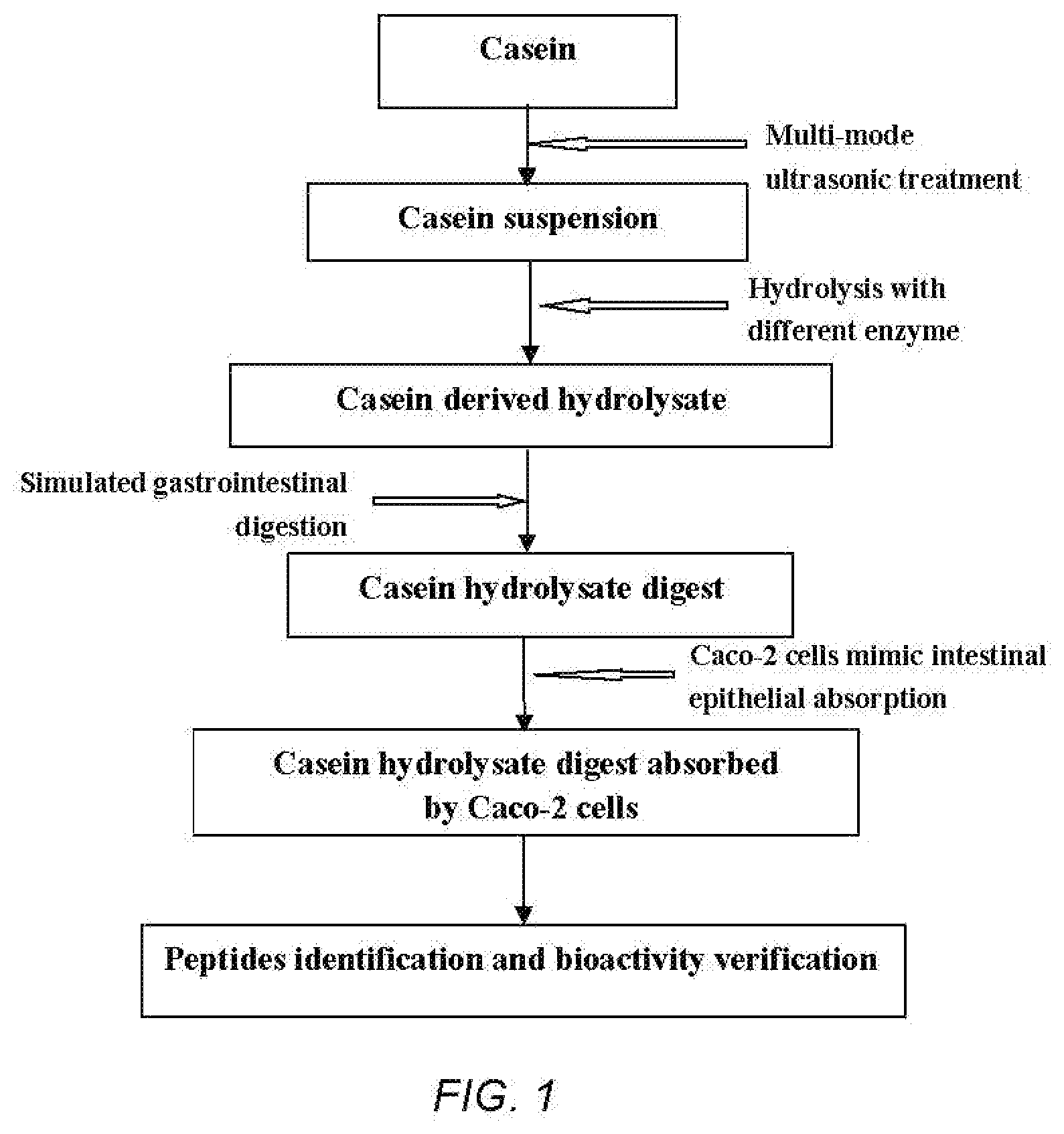
[0056] FIG. 1 is a technical route diagram of ultrasound-assisted enzymolysis coupled to simulated gastrointestinal digestion and absorption to prepare casein-derived peptides.
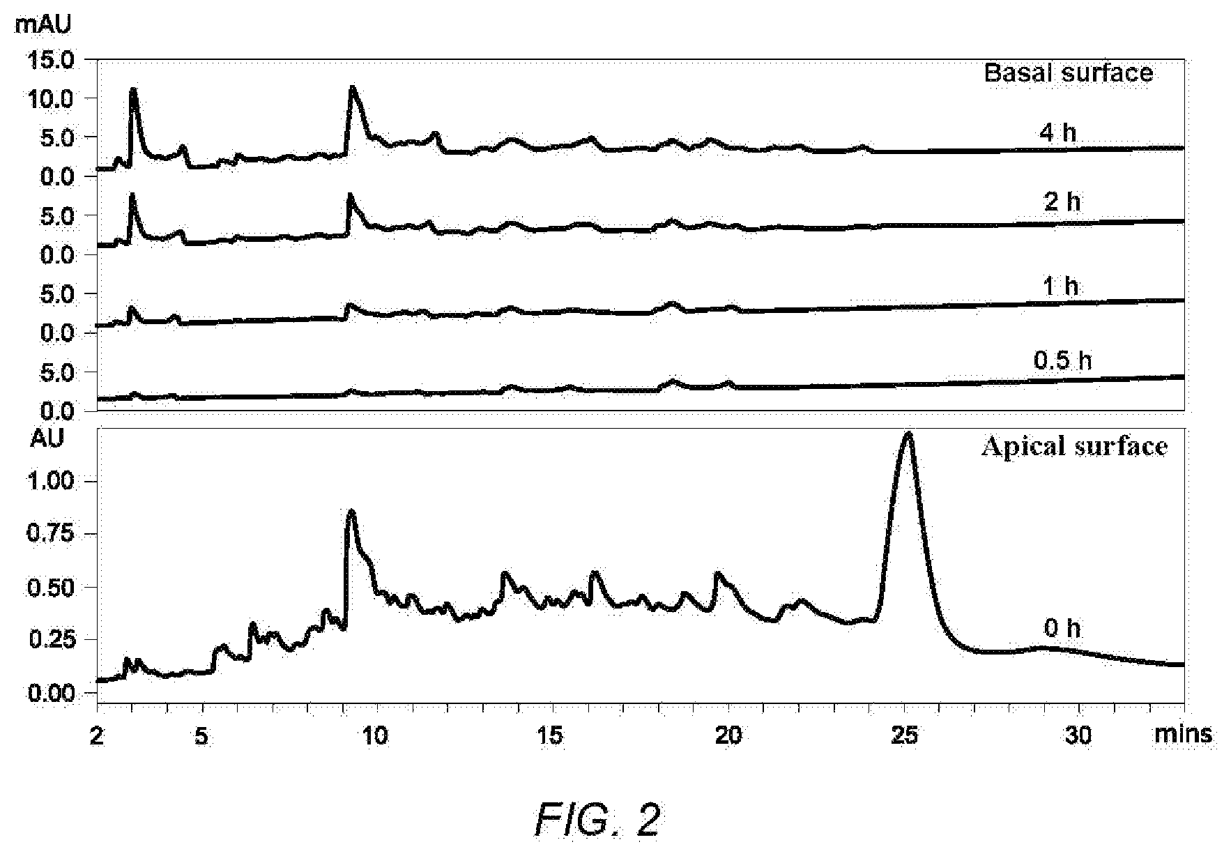
[0057] FIG. 2 shows chromatogaphic profiles of digests derived from casein hydrolysate before and after transcellular transport at 0.5, 1.0, 2.0 and 4.0 h (A).
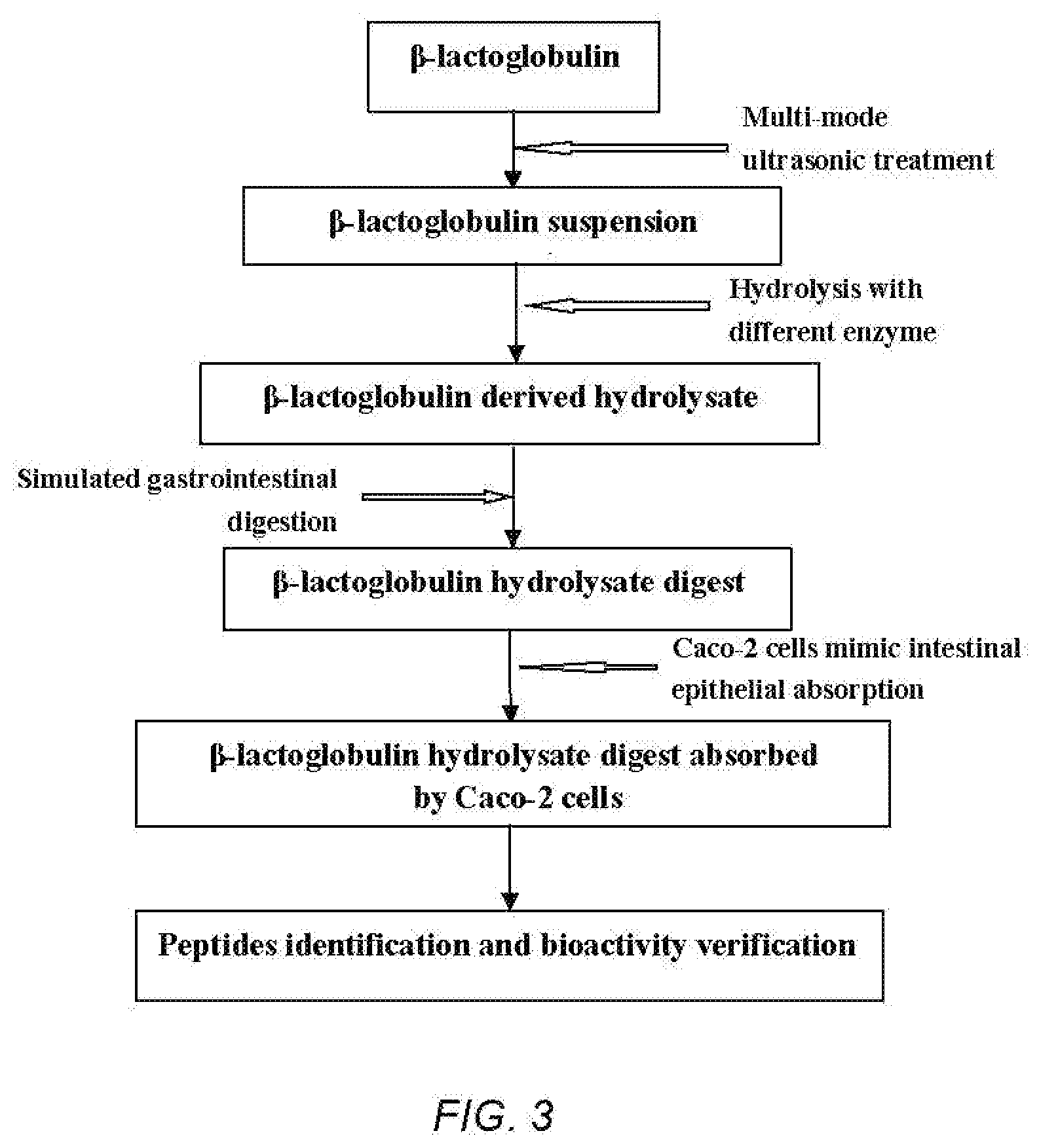
[0058] FIG. 3 is a technical route diagram of ultrasound-assisted enzymolysis coupled to simulated gastrointestinal digestion and absorption to prepare .beta.-LG-derived peptides.
EMBODIMENTS
[0059] 1. Experimental Method
[0060] 1.1. Degree of Hydrolysis (DH) and Protein Conversion Rate
[0061] The DH of casein was determined using the pH-state method, DH is defined as the percentage of cleaved peptide bonds, which was calculated using the equation below:
DH ( % ) = V .times. N .alpha. .times. M .times. h tot .times. 100 % ##EQU00001##
[0062] Where, V is the titrant volume of NaOH (mL), N is the concentration of NaOH (mol/L), .alpha. is the degree of dissociation of .alpha.-NH.sub.2 (0.985 for casein), M is the mass of protein (g), and h.sub.tot is the number of peptide bonds in the substrate: different proteins had different values of h.sub.tot, the empirical value of casein is h.sub.tot=7.35 mol/g.
[0063] The total nitrogen content of the casein protein and its derived hydrolysate was determined by the Kjeldahl method, and the conversion rate of casein was calculated as follows:
Protein conversion rate (%)=hydrolysate nitrogen content/substrate protein nitrogen content*100%.
[0064] 1.2 Measurement of ACE Inhibitory Activity
[0065] The FAPGG was used as the substrate of ACE, each reaction component was added according to the Table, and the ACE inhibition rate of the sample was measured with a microliter plate reader at 340 nm.
[0066] Where X.sub.1 is the absorbance of the control group without protein hydrolysates before reaction, Y.sub.1 is the absorbance of the sample group before the reaction. X.sub.2 is the absorbance of the blank group after the reaction, and Y.sub.2 is the absorbance of the sample group after the reaction. The test was performed five times. The ACE inhibitory activity was calculated as follows
The ACE inhibitory rate (%)=100-(.DELTA.A.sub.sample)/(.DELTA.A.sub.blank).times.100%
[0067] .DELTA.A.sub.sample=X.sub.1-X.sub.2, .DELTA.A.sub.blank=Y.sub.1-Y.sub.2.
TABLE-US-00001 Measurement of ACE inhibitory activity Blank Sample (.mu.L) (.mu.L) ACE (0.1 U/mL) 10 10 FAPGG (1 mmol/L) * 50 50 Matrix buffer ** 40 0 ACE inhibitor 0 40 Note: FAPGG (1.0 mmol/L) was prepared by taking 3.994 mg of FAPGG plus matrix buffer, making up to 10 mL, dissolving and mixing, and then storing at 4.degree. C. in the dark. Matrix buffer ** was prepared by dissolving 1.910 g of HEPES and 1.755 g of NaCl in double distilled water, adjusting the pH to 8.3 with NaOH, and replenishing the water to 100 mL, and storing at 4.degree. C. for later use.
[0068] 1.3 Cell Culture
[0069] The human colon adenocarcinoma cell line, Caco-2 (HTB-37.TM.) was obtained from American-type culture collection (ATCC, Manassas, VA, USA). The cells were grown in Dulbecco's Modified Eagle's Medium (DMEM) supplemented with 10% fetal bovine serum, 2.5% HEPES, 1% non-essential amino acids and 1% antibiotics. Cells were incubated at 37.degree. C. in a humidified atmosphere (5% CO.sub.2). The medium was replaced three times a week, and the cells were subcultured (at 80-90% confluence) by using 0.25% trypsin-EDTA treatment before use in the experiments.
[0070] 1.4 Measurement of Cytotoxicity
[0071] The cell cytotoxic properties were monitored using an Alamar Blue assay. Briefly, Caco-2 cells were grown in 96-well plates at a density of 1.times.10.sup.4 cell/well for 24 h. After 24 h, the medium was changed and the cells were treated with various concentrations (10-50 mg/ml) of casein hydrolysate for another 24 h. After 24 h treatment, the media was discarded, and the fresh medium with 10% Alamar Blue reagent was added and incubated for an additional 4 h at 37.degree. C. The fluorescence intensity of the wells was measured at an excitation wavelength of 560 nm and an emission wavelength of 590 nm. Cell viability is expressed as a percentage compared to untreated cells.
[0072] 1.5 Simulated Intestinal Epithelium Absorption Using Caco-2 Cells
[0073] The samples collected from the AP and BL surfaces of Caco-2 cells were analyzed on an Acquity Ultra-Performance Liquid Chromatograph (UPLC) system with an Acquity UPLC BEH C.sub.18 column (100 mm.times.2.1 mm i.d., 1.7 .mu.m, Waters, Milford, Mass. USA) using an injection volume of 15 .mu.L. Mobile phases were solvent A (1% TFA in Milli-Q water) and solvent B (1% TFA in acetonitrile). The peptides were eluted with a gradient of solvent A (100-75% in 25 min, 75-50% in 25-35 min) at a flow rate of 0.3 ml/min. The elution was monitored at 220 nm. Absorption was expressed as the percentage of total peak area calculated at different time points (0.5 h, 1 h, 2 h and 4 h) in the BL surface as compared to 0 h in the AP surface.
[0074] 1.6 Identification of Casein-Derived Peptides Using UPLC-MS
[0075] The liquid chromatography column used in this study was nanoACQUITY BEH130 C.sub.18 (75 .mu.mx 150 mm, 1.7 .mu.m. The solvent A was acetonitrile (ACN) containing 0.1% formic acid. The peptides were separated using the following gradient: increasing solvent B containing water with 0.1% formic acid from 1% to 6% B in 2 min, to 25% B in 23 min, to 45% B in 15 min, to 75% B in 5 min, to 95% B in 5 min, and keeping at 95% B for 5 min. The mass spectrometer was operated in a positive mode with a capillary voltage of 3.5 kV and a source temperature of 100.degree. C. Spectra were recorded over the m/z ranges of 200-1000 in MS mode and in 50-1990 MS/MS mode. The amino acid sequences of peptides were analyzed using Mass Lynx software (Micromass U.K. Ltd.). Peaks Viewer 4.5 (Bioinfonnatics Solutions Inc., Waterloo, ON, Canada), in combination with manual de novo sequencing was used to process the MS/MS data. Identified peptide sequences were synthesized (>98% purity) by Genscript Corp (Piscataway, N.J.) and used for the bioactivity assays.
EXAMPLE 1
[0076] Ultrasound treatment of casein. Casein with a concentration of 1 g/100 mL was prepared by dissolving in a phosphate buffer (pH 7.8). The above concentration of casein suspension was treated by ultrasound equipment. The ultrasound treatment conditions are as follows: treatment time 30 min; intermittent ratio 10 s/3 s; temperature 25.degree. C. Single-frequency ultrasound with 40 kHz was used for the sample treatment.
[0077] Enzymolysis of casein. After ultrasound treatment, the casein suspensions were preheated to 50.degree. C. and adjusted to pH 8.0 with 1.0 M NaOH. Alcalase (the ratio of E/S was 1:20) (w/w) was added to initialize the reaction, and the enzymolysis time was 2 h. The mixture was heated and maintained at 100.degree. C. for 10 min to terminate the reaction. Then the mixture was adjusted to pH 7.0 and centrifuged; the supernatant was collected, desalted, concentrated, and freeze-dried to a powder. The DH and protein conversion rate (CR) of the casein, and the ACE inhibitory activity of the casein-derived hydrolysate were determined.
[0078] Simulated gastrointestinal digestion. Simulated gastric and intestinal fluids were prepared according to the U.S. Pharmacopeia (USP30-NT25). Briefly, Casein-derived hydrolysates were digested with gastric fluid at 1:20 (w/v) for 4 h in a shaking incubator with 120 rpm at 37.degree. C. Then the pH was adjusted to 6.8 and pancreatin was added at 1:100 (w/v) to form the intestinal fluid. The mixture was incubated for a further 6 h to mimic intestinal digestion. The digestion was terminated in boiling water for 10 min. The digests were allowed to cool down and were centrifuged at 10,000 g for 10 min to collect the supernatant, which was further centrifuged, desalted, concentrated, and freeze-dried to a powder. The ACE inhibitory activity of the 4 h's and 10 h'scasein hydrolysate digest was measured.
[0079] As shown in Table 1, after ultrasound pretreatment, the DH of casein increased from 10.02% to 16.54%, and the protein conversion rate increased from 30.10% to 44.08%. The ACE inhibitory activity of casein hydrolysate, as shown by IC.sub.50 value, was decreased from 64.21 .mu.g/mL to 52.13 .mu.g/mL, indicating ultrasound pretreatment largely improved the ACE inhibitory activity of casein hydrolysate. After simulated gastric digestion and simulated intestinal digestion, the casein hydrolysate digest showed good ACE inhibitory activity, while the IC.sub.50 values were 49.21 .mu.g/mL and 55.19 .mu.g/mL, respectively (Table. 2). The above results indicate that the casein-derived hydrolysate exhibits excellent ACE inhibitory activity after simulated GI digestion.
EXAMPLE 2
[0080] Ultrasound treatment of casein. Casein with a concentration of 2 .mu.g/100 mL was prepared by dissolving in a phosphate buffer (pH 7.8). The above concentraticin of casein suspension was treated by ultrasound equipment. The ultrasound treatment conditions are as follows: treatment time 20 min; intermittent ratio 10 s/3 s; temperature 30.degree. C. Dual-frequency simultaneous ultrasound treatments of 20/40 kHz were used for the sample treatment.
[0081] Enzymolysis of casein. After ultrasound treatment, the casein suspensions were preheated to 55.degree. C. and adjusted to pH 8.0 with 1.0 M NaOH. Neutral protease (the ratio of E/S was 1:30) (w/w) was added to initialize the reaction, and the enzymolysis time was 4 h. The mixture was heated and maintained at 100.degree. C. for 10 min to terminate the reaction. Then the mixture was adjusted to pH 7.0 and centrifuged; the supernatant was collected, desalted, concentrated, and freeze dried to a powder. The DH and CR of the casein, and the ACE inhibitory activity of the casein-derived hydrolysate was determined.
[0082] Simulated gastrointestinal digestion. Simulated gastric and intestinal fluids were prepared according to the U.S. Pharmacopeia (USP30-NF25). Briefly, Casein-derived hydrolysates were digested with gastric fluid at 1:30 (w/v) for 3 h in a shaking incubator with 150 rpm at 37.degree. C. Then the pH was adjusted to 6.8 and pancreatin was added at 1:100 (w/v) to form the intestinal fluid. The mixture was incubated for a further 4 h to mimic intestinal digestion. The digestion was terminated in boiling water for 10 min. The digests were allowed to cool down and centrifuged at 10,000 g for 10 min to collect the supernatant, which was further centrifuged, desalted, concentrated, and freeze-dried to a powder. The ACE inhibitory activity of the 4 h's and 10 h's casein hydrolysate digest was measured.
[0083] As shown in Table 1, after dual-frequency simultaneous ultrasound pretreatment, the DH of casein increased from 5.21% to 9.45%, and the protein conversion rate increased from 18.11% to 22.39%. The ACE inhibitory activity of casein hydrolysate indicated by IC.sub.50 value was decreased from 100.23 .mu.g/mL to 95.21 .mu.g/mL, indicating ultrasound pretreatment largely improved the ACE inhibitory activity of casein hydrolysate. After simulated gastric digestion, the casein hydrolysate digest showed good ACE inhibitory activity; its IC.sub.50 value was 72.11 .mu.g/mL; after simulated intestinal digestion, the casein hydrolysate digest still showed good ACE inhibitory activity, and its IC.sub.50 value was 79.03 .mu.g/mL (Table. 2). The above results indicate that the casein-derived hydrolasate still exhibited excellent ACE inhibitory activity after simulated GI digestion.
EXAMPLE 3
[0084] Ultrasound treatment of casein. Casein with a concentration of 5 g/100 mL was prepared by dissolving in a phosphate buffer (pH 7.8). The above concentration of casein suspension was treated by ultrasound equipment. The ultrasound treatment conditions are as follows: treatment time 10 min; intermittent ratio 10 s/3 s; temperature 40.degree. C. Triple-frequency simultaneous ultrasound treatments of 20/40/60 kHz were used for the sample treatment.
[0085] Enzymolysis of casein. After ultrasound treatment, the casein suspensions were preheated to 70.degree. C. and adjusted to pH 8.0 with 1.0 M NaOH. Papain (the ratio of E/S was 1:50) (w/w) was added to initialize the reaction, and the enzymolysis time was 2 h. The mixture was heated and maintained at 100.degree. C. for 10 min to terminate the reaction. Then the mixture was adjusted to pH 7.0 and centrifuged; the supernatant was collected, desalted, concentrated, and freeze-dried to a powder. The DH and CR of the casein, and the ACE inhibitory activity of the casein-derived hydrolysate were determined.
[0086] Simulated gastrointestinal digestion. Simulated gastric and intestinal fluids were prepared according to the U.S. Pharmacopeia (USP30-NT25). Briefly, Casein-derived hydrolysates were digested with gastric fluid at 1:50 (w/v) for 4 h in a shaking incubator with 180 rpm at 37.degree. C. Then the pH was adjusted to 6.8 and pancreatin was added at 1:100 (w/v) to form the intestinal fluid. The mixture was incubated for a further 6 h to mimic intestinal digestion. The digestion was terminated in boiling water for 10 min. The digests were allowed to cool down and centrifuged at 10,000 g for 10 min to collect the supernatant, which was further centrifuged, desalted, concentrated, and freeze-dried to a powder. The ACE inhibitory activity of the 4 h's and 10 h's casein hydrolysate digest was measured.
[0087] As shown in Table 1, after triple-frequency ultrasound pretreatment, the DH of casein increased from 7.21% to 11.36%, the protein conversion rate increased from 21.98% to 26.02%. The IC.sub.50 value of ACE inhibitory activity of casein hydrolysate was decreased from 97.32 .mu.g/mL to 90.11 .mu.g/mL, indicating ultrasound pretreatment largely improved the ACE inhibitory activity of casein hydrolysate.
[0088] After simulated gastric digestion, the casein hydrolysate digest showed good ACE inhibitory activity, its IC.sub.50 value was 65.32 .mu.g/mL, after simulated intestinal digestion, the casein hydrolysate digest still showed good ACE inhibitory activity, its IC.sub.50 value was 60.31 .mu.g/mL (Table. 2). The above results indicate that the casein-derived hydrolysate still exhibited excellent ACE inhibitory activity after simulated GI digestion.
TABLE-US-00002 TABLE 1 Effects of ultrasound pretreatment on the DH, CR and ACE inhibitory activity of casein hydrolysates by different enzymatic hydrolysis Traditional hydrolysis Ultrasound assisted hydrolysis IC.sub.50 of ACE IC.sub.50 of ACE inhibitory Ultra- inhibitory activity sound activity Enzyme DH (%) CR (%) (.mu.g/mL) mode DH (%) CR (%) (.mu.g/mL) Alcalase 10.02 .+-. 1.78 30.10 .+-. 0.91 64.21 .+-. 9.12 40 kHz 16.54 .+-. 0.99 44.08 .+-. 0.59 52.13 .+-. 6.11 Neutral 5.21 .+-. 0.52 18.11 .+-. 0.37 100.23 .+-. 14.98 20/40 9.45 .+-. 0.27 22.39 .+-. 0.37 95.21 .+-. 8.0 protease kHz Papain 7.21 .+-. 0.49 21.98 .+-. 0.64 97.32 .+-. 10.43 20/40/60 11.36 .+-. 1.18 26.02 .+-. 0.64 90.11 .+-. 11.34 kHz
TABLE-US-00003 TABLE 2 Effects of different casein hydrolysates before gastric digestion, after gastric digestion, intestinal digestion and Caco-2 cells absorption on ACE inhibitory activity IC.sub.50 of ACE inhibitory activity (.mu.g/mL) After Before After After Caco-2 Casein-derived gastric gastric intestinal cells hydrolysates digestion digestion digestion absorption Alcalase 52.13 .+-. 6.11 49.21 .+-. 4.91 55.19 .+-. 5.72 21.37 .+-. 2.07 hydrolysates Neutral protease 95.21 .+-. 8.0 72.11 .+-. 6.42 79.03 .+-. 8.19 hydrolysates Papain 90.11 .+-. 11.34 65.32 .+-. 5.0 60.31 .+-. 6.23 hydrolysates
EXAMPLE 4
[0089] The casein-derived hydrolysate digest prepared in example 1 was subjected to Caco-2 mimicking intestinal endothelial cell absorption.
[0090] The cytotoxicity of the casein-derived hydrolysate digest to Caco-2 cells was first detected. The absorption model of Caco-2 cells mimicking intestinal endothelial cells was built as follows: Caco-2 cells were grown in 12-well Transwell.TM. plates at a concentration of 2.times.10.sup.5 cells/mL. The medium of the cell was replaced every other day. After 21 days of cell culture, some evaluation indicators of the Caco-2 cells were measured, including epithelial cell resistance, alkaline phosphatase activity, and sodium fluorescein leakage test. Before initiation of the transport experiments, the Caco-2 cells were washed by an HBSS buffer; and 0.5 mL of the casein hydrolysate digest (20 mg/mL, dissolved in an HBSS buffer) was added to the AP surface; 1.5 mL of HBSS buffer was added to the BL surface; finally, the Caco-2 cells were incubated for 4 h at 37.degree. C. 0.2 mL of AP surface samples at 0 h and BL surface samples at 0.5, 1, 2, and 4 h were collected for the absorption detection. The AP (apical) surface samples and BL (basolateral) surface samples at 4 h were collected for the ACE inhibitory activity detection, respectively. The casein hydrolysates digests and their absorbed digests were subjected to the amino acid sequences analysis by UPLC-MC. Some small peptides with strong ionic strength were screened out, synthesized, and detected for ACE inhibitory activity.
[0091] As shown in Table 3, the addition of casein-derived hydrolysate digest increased the viability of Caco-2 cells, indicating that casein-derived hydrolysate digest didn't have any toxicity in Caco-2 cells and helped the growth of Caco-2 cells. As shown in FIG. 1, the absorption of the casein-derived hydrolysate digest from the AP to BL surface increased with time and reached 2.31% at 4 h, indicating selective absorption of peptides in Caco-2 cells. The absorbed digest obtained from the BL surface at the end of 4-hour transport study was tested for its in vitro ACE inhibitory effect and compared to that of the casein hydrolysate digest. After absorption by Caco-2 cells, the IC.sub.50 value of the ACE inhibitory activity of the casein hydrolysate digest decreased from 55.19 .mu.g/mL to 21.37 .mu.g/mL (Table 2). The above results showed that the ACE inhibitory activity of casein polypeptide was significantly enhanced by Caco-2 cells mimicking intestinal endothelial cells absorption, which indicates that the absorbed polypeptide displayed stronger ACE inhibitory activity than the digest. The absorbed peptides were subjected to LC-ESI-MS/MS analysis. The small peptides with strong ionic strength were chosen out, synthesized and further assayed for their ACE inhibitory activity. Three peptides with high ACE inhibition, Leu-Gln-Pro-Pro (LQPP), Ala-Pro-Tyr (APY), Leu-Ser-Leu-Pro (LSLP) were chosen out; their IC.sub.50 were 14.21 .mu.M, 19.12 .mu.M, and 21.09 .mu.M, respectively (Table 4).
TABLE-US-00004 TABLE 3 Cytotoxicity of casein hydrolysate derived digests at different concentration in Caco-2 cells Concentration of digest Caco-2 (mg/Ml) viability Control -- 100 Group 1 5 109.8 .+-. 5.3 Group 2 10 119.2 .+-. 8.5 Group 3 20 110.2 .+-. 10.6 Group 4 50 103.6 .+-. 9.8
TABLE-US-00005 TABLE 4 The peptides that are absorbed by Caco-2 cells were sequenced, identified and chemically synthesized to validate the ACE inhibitory activity of the identified peptides. Peptide IC.sub.50 of ACE inhibitory sequence activity (.mu.g/mL) LQPP 14.21 APY 19.12 LSLP 71.09
[0092] Experimental method and examples of ultrasound-assisted simulated digestion and absorption method for .beta.-LG derived inflammatory peptide
[0093] 2. Experimental Method
[0094] 2.1 Degree of Hydrolysis (DH) and Protein Conversion Rate
[0095] The DH of .beta.-LG determined using the pH-state method, DH is defined as the percentage of cleaved peptide bonds, which as calculated using the equation below:
DH ( % ) = V .times. N .alpha. .times. M .times. h tot .times. 100 % ##EQU00002##
[0096] Where, V is the titrant volume of NaOH (mL), N is the concentration of NaOH (mol/L), .alpha. is the degree of dissociation of .alpha.-NH.sub.2 (0.99 for .beta.-LG), M is the mass of protein (g), and h.sub.tot is the number of peptide bonds in the substrate, different proteins had different values of h.sub.tot, the empirical value of .beta.-LG is h.sub.tot=7.35 mol/g.
[0097] The total nitrogen content of the .beta.-LG and its derived hydrolysate was determined by the Kjeldahl method, and the conversion rate of .beta.-LG was calculated as follows:
[0098] Protein conversion rate (%)=hydrolysate nitrogen content/substrate protein nitrogen content*100%
[0099] 2.2 Cell Culture
[0100] The endothelial cell line, EA.hy926 (CRL-2922.TM.), and human colon adenocarcinoma cell line, Caco-2 (HTB-37.TM.), were purchased from American-type culture collection (ATCC, Manassas, Va., USA). DMEM supplemented with 10% FBS, 2.5% HEPES, 1% antibiotics, and 1% non-essential amino acids was used as the cell growth medium. The cells were incubated in a humidified atmosphere with 5% CO.sub.2 at 37.degree. C. The medium was replaced for three times for every week, and the cells were subcultured using 0.25% trypsin-EDTA treatment.
[0101] 2.3 Measurement of Cytotoxicity
[0102] The cell cytotoxic properties were monitored using an Alamar Blue assay. Caco-2 cells were seeded in 96-well plates at a density of 1.times.10.sup.4 cell/well for 24 h. Then the cells were treated with various concentrations (10-50 mg/mL) of .beta.-LG hydrolysate for another 24 h in a fresh medium. After the 24 h treatment, the media was discarded, and the fresh medium with 10% Alamar Blue reagent was added and incubated for 4 h at 37.degree. C. The fluorescence intensity of the wells was measured at an emission wavelength of 590 nm and an excitation wavelength of 560 nm. The viability of the treated cells was expressed as a percentage as compared to untreated cells.
[0103] 2.4 Measurement of Anti-Inflammatory Activity
[0104] The peptides were subjected to study the anti-inflammatory activity: the levels of intercellular adhesion molecule (ICAM-1) and vascular cell adhesion molecule (VCAM-1) expressed in EA.hy926 cells were detected by Western blot as inflammatory biomarkers. The EA. hy926 cells with passage number <12 were grown in 48-well plates. The cells reaching 80-90% confluence were treated with various concentrations (2.5 mg/mL of hydrolysates and digests, 0.2 mM and 3.0 mM of synthetic peptides) of the samples for 18 h. Then the cells were stimulated with TNF-.alpha. at 10 ng/mL and incubated for an additional 6 h in order to induce inflammation.
[0105] After the treatment period, the culture medium of the EA. hy926 cells was discarded and a boiling Laemmle buffer containing 0.2% TritonX-100 and 50 .mu.M dithiothreitol was added to lysate the cells. The cell lysates were then run on 9% sodium dodecyl sulfate polyacrylamide gel electrophoresis. The gels were transferred onto nitrocellulose membranes, and immunoblotted with anti-ICAM-1/anti-VCAM-1 antibodies. The concentration of antibody to .alpha.-tubulin used was 0.4 .mu.g/mL, while that for all other antibodies was 0.1 .mu.g/mL. The protein bands were scanned using Licor Odyssey BioImager (Licor Biosciences, Lincoln, Nebr., USA) and quantified by densitometry using Image Studio Lite 5.2. All the data were expressed as the percentage change of the corresponding positive control (cells treated with TNF-.alpha. alone).
[0106] 2.5 Simulated Intestinal Epithelium Absorption Using Caco-2 Cells
[0107] The samples collected from the AP and BL surfaces of Caco-2 cells were analyzed on an Acquity Ultra-Performance Liquid Chromatograph (UPLC) system with an Acquity UPLC BEH C.sub.18 column (100 mm.times.2.1 mm i.d., 1.7 .mu.m, Waters, Milford, Mass., USA) using an injection volume of 15 .mu.L. Mobile phases were solvent A (1% TFA in Milli-Q water) and solvent B (1% TFA in acetonitrile). The peptides were eluted with a gradient of solvent A (100-75% in 25 min, 75-50% in 25-35 min) at a flow rate of 0.3 ml/min. The elution was monitored at 220 nm. Absorption was expressed as the percentage of total peak area calculated at different time points (0.5 h, 1 h, 2 h and 4 h) in the BL surface as compared to 0 h in the AP surface.
[0108] 2.6 Identification of .beta.-LG -Derived Peptides
[0109] The liquid chromatogaphy column used in this study was nanoACQUITY BEH130 C.sub.18 (75 .mu.m.times.150 mm, 1.7 .mu.m). The solvent A was acetonitrile (ACN) containing 0.1% formic acid. The peptides were separated using the following gradient: solvent B was water with 0.1% formic acid increasing from 1% to 6% B in 2 min, to 25% B in 23 min to 45% B in 15 min, to 75% B in 5 min, to 95% B in 5 min, and keeping at 95% B for 5 min. The mass spectrometer was operated in a positive mode with a capillary voltage of 3.5 kV and a source temperature of 100.degree. C. Spectra were recorded over the m/z ranges of 200-1000 in MS mode and in 50-1990 MS/MS mode. The amino acid sequences of peptides were analyzed using Mass Lynx software (Micromass U.K. Ltd.). Peaks Viewer 4.5 (Bioinformatics Solutions Inc., Waterloo, ON, Canada), in combination with manual de novo sequencing was used to process the MS/MS data. Identified peptide sequences were synthesized (>98% purity) by Genscript Corp (Piscataway, N.J.) and used for the bioactivity assays.
[0110] The method for extracting .beta.-lactoglobulin with the present invention was as follows: A concentration of 7% (w/v) whey protein solution was prepared by adding whey protein to 7% NaCl water, adjusting the pH to 2 with HCl solution, and centrifuging at 5000 rpm for 20 min to collect the supernatant. Then the supernatant was dialyzed using a dialysis bag with a molecular weight cut off of 14000 Da, and placed in 30 volumes of distilled water for 20 h. Then the retentate in the dialysis bag, i.e. .beta.-LG, was collected.
EXAMPLE 5
[0111] Ultrasound treatment of .beta.-LG. The 200 mL, of .beta.-LG with a concentration of 1 g/mL was treated by ultrasound equipment. The ultrasound treatment conditions are as follows: treatment time 30 min; intermittent ratio 10 s/3 s; temperature 30.degree. C. Single frequency ultrasound of 40 kHz was used for the sample treatment.
[0112] Enzymolysis of .beta.-LG. After ultrasound treatment, the .beta.-LG suspensions were preheated to 50.degree. C. and adjusted to pH 8.0 with 1.0 M NaOH. Alcalase (the ratio of E/S was 1:20) (w/w) was added to initialize the reaction, and the enzymolysis time was 2 h. The mixture was heated and maintained at 100.degree. C. for 10 min to terminate the reaction. Then the mixture was adjusted to pH 7.0 and centrifuged; the supernatant was collected, desalted, concentrated, and freeze-dried to a powder. The DH and CR of the .beta.-LG, and the anti-inflammatory activity of .beta.-LG-derived hydrolysate, were determined.
[0113] Simulated GI digestion. Simulated gastric and intestinal fluids were prepared according to the U.S. Pharmacopeia. Briefly, .beta.-LG-derived hydrolysate was digested with gastric fluid at 1:20 (w/v) for 4 h in a shaking incubator with 120 rpm at 37.degree. C. Then the pH was adjusted to 6.8 and pancreatin was added at 1:100 (w/v) to form the intestinal fluid. The mixture was incubated for a further 6 h to mimic intestinal digestion. The digestion was terminated in boiling water for 10 min. The digest was cooled down and centrifuged at 10,000 g for 10 min to collect the supernatant, which was further centrifuged, desalted, concentrated, and freeze-dried to a powder. The anti-inflammatory activity of the 4 h's and 10 h's .beta.-LG hydrolysate digest was measured.
[0114] As shown in Table 5, after single frequency ultrasound pretreatment, the DH of .beta.-LG increased from 10.32% to 13.70%, the protein conversion rate increased from 30.27% to 35.17%. Alcalase hydrolysate showed good anti-inflammatory activity, the expression of the VCAM-1 and ICAM-1 were 42.3% and 62.7%, respectively (Table 6). After simulated gastric digestion, the .beta.-LG hydrolysate digest showed good anti-inflammatory activity, and the expression of the VCAM-1 and ICAM-1 was 48.2% and 55.3%, respectively. After simulated intestinal digestion, the .beta.-LG hydrolysate digest still showed good anti-inflammatory activity, and the expression of the VCAM-1 and ICAM-1 was 50.7% and 63.2%, respectively. Simulated gastrointestinal digestion appeared to have minimal effect on the anti-inflammatory activity of .beta.-LG hydrolysates.
EXAMPLE 6
[0115] Ultrasound treatment of .beta.-LG. The 200 mL of .beta.-LG with a concentration of 4 g/mL was treated by ultrasound equipment. The ultrasound treatment conditions are as follows: treatment time 20 min; intermittent ratio 10 s/3 s: temperature 25.degree. C. Dual-frequency simultaneous ultrasound with 20/28 kHz was used for the sample treatment.
[0116] Enzymolysis of .beta.-LG. After ultrasound treatment, the .beta.-LG suspensions were preheated to 55.degree. C. and adjusted to pH 7.5 with 1.0 M NaOH. Neutral protease (the ratio of E/S was 1:30) (w/w) was added to initial the reaction and the enzymolysis time was 4 h. The mixture was heated and maintained at 100.degree. C. for 10 min to terminate the reaction. Then the mixture was adjusted to pH 7.0 and centrifuged; the supernatant was collected, desalted, concentrated, and freeze-dried to a powder. The DH and CR of the .beta.-LG, and the anti-inflammatory activity of .beta.-LG-derived hydrolysate, were determined.
[0117] Simulated GI digestion. .beta.-LG-derived hydrolysate was subjected to simulated gastric and intestinal digestion. Simulated gastric and intestinal fluids were prepared according to the U.S. Pharmacopeia. Briefly, .beta.-LG-derived hydrolysate was digested with gastric fluid at 1:30 (w/v) for 3 h in a shaking incubator with 150 rpm at 37.degree. C. Then the pH was adjusted to 6.8 and pancreatin was added at 1:100 (w/v) to form the intestinal fluid. The mixture was incubated for a further 4 h to mimic intestinal digestion. The digestion was terminated in boiling water for 10 min. The digest was cooled down and centrifuged at 10,000 g for 10 min to collect the supernatant, which was further centrifuged, desalted, concentrated, and freeze-dried to a powder. The anti-inflammatory activity of the 4 h's and 10 h's .beta.-LG hydrolysate digest was measured.
[0118] As shown in Table 5, after dual-frequency simultaneous ultrasound pretreatment, the DH of .beta.-LG increased from 6.19% to 9.53%, the protein conversion rate increased from 15.11% to 22.34%. Neutral protease hydrolysate showed good anti-inflammatory activity, and the expression of the VCAM-1 and ICAM-1 was 63.2% and 52.3%, respectively. The simulated gastric digestion of the .beta.-LG hydrolysate digest resulted in good anti-inflammatory activity, and the expression of the VCAM-1 and ICAM-1 was 50.3% and 47.3%, respectively. Also, the simulated intestinal digestion of the .beta.-LG hydrolysate digest showed good anti-inflammatory activity, and the expression of the VCAM-1 and ICAM-1 was 53.4% and 53.8%, respectively. Simulated gastrointestinal digestion appeared to have minimal effect on the anti-inflammatory activity of .beta.-LG hydrolysates.
EXAMPLE 7
[0119] Ultrasound treatment of .beta.-LG. The 200 mL of .beta.-LG with a concentration of 4 g/mL was treated by ultrasound equipment. The ultrasound treatment conditions are as follows: treatment time 10 min; intermittent ratio 10 s/3 s; temperature 25.degree. C. Triple-frequency simultaneous ultrasound with 20/28/40 kHz was used for the sample treatment.
[0120] Enzymolysis of .beta.-LG. After ultrasound treatment, the .beta.-LG suspensions were preheated to 70.degree. C. and adjusted to pH 8 with 1.0 M NaOH. Thermolysin (the ratio of E/S was 1:50) (w/w) was added to initial the reaction and the enzymolysis time was 2 h. The mixture was heated and maintained at 100.degree. C. for 10 min to terminate the reaction. Then the mixture was adjusted to pH 7.0 and centrifuged; the supernatant was collected, desalted, concentrated, and freeze-dried to a powder. The DH and CR of the .beta.-LG, and the anti-inflammatory activity of .beta.-LG-derived hydrolysate, were determined.
[0121] Simulated GI digestion. .beta.-LG-derived hydrolysate was subjected to simulated gastric and intestinal digestion. Simulated gastric and intestinal fluids were prepared according to the U.S. Pharmacopeia. Briefly, .beta.-LG-derived hydrolysate was digested with gastric fluid at 1:50 (w/v) for 2 h in a shaking incubator with 180 rpm at 37.degree. C. Then the pH was adjusted to 6.8 and pancreatin was added at 1:100 (w/v) to form the intestinal fluid. The mixture was incubated for a further 4 h to mimic intestinal digestion. The digestion was terminated in boiling water for 10 min. The digest was cooled down and centrifuged at 10,000 g for 10 min to collect the supernatant, which was further centrifuged, desalted, concentrated, and freeze-dried to a powder. The anti-inflammatory activity of the 4 h's and 10 h's .beta.-LG hydrolysate digest was measured.
[0122] As shown in Table 5, after triple-frequency simultaneous ultrasound pretreatment, the DH of .beta.-LG increased from 13.20% to 21.41%, the protein conversion rate increased from 36.90% to 41.02%.
TABLE-US-00006 TABLE 5 Effects of ultrasound pretreatment on the DH, CR of .beta.-LG hydrolysates by different enzymatic hydrolysis Ultrasound assisted hydrolysis Traditional hydrolysis Ultrasound Enzyme DH (%) CR (%) mode DH (%) CR (%) Akalase 10.32 .+-. 1.73 30.27 .+-. 0.91 40 kHz 13.70 .+-. 1.09 35.17 .+-. 1.77 Neutral protease 6.19 .+-. 0.42 15.11 .+-. 0.37 20/28 kHz 9.53 .+-. 0.97 22.34 .+-. 1.45 Thermolysin 13.20 .+-. 0.54 36.90 .+-. 0.64 20/28140 kHz 21.41 .+-. 2.31 41.02 .+-. 2.00
[0123] Thermolysin hydrolysate showed good anti-inflammatory activity, and the expression of the VCAM-1 and ICAM-1 was 48.9% and 36.5%, respectively. After simulated gastric digestion, the .beta.-LG hydrolysate digest showed good anti-inflammatory activity, and the expression of the VCAM-1 and ICAM-1 was 43.3% and 30.1%, respectively. After simulated intestinal digestion, the .beta.-LG hydrolysate digest still showed good anti-inflammatory activity, and the expression of the VCAM-1 and ICAM-1 was 49.1% and 36.2%, respectively (Table 6). Simulated gastrointestinal digestion appeared to have minimal effect on the anti-inflammatory activity of .beta.-LG hydrolysates.
TABLE-US-00007 TABLE 6 Effects of different .beta.-LG hydrolysates before gastric digestion, after gastric digestion, intestinal digestion and Caco-2 cells absorption on TNF-.alpha.-induced VCAM-1 and ICAM-1 protein expression in EA.hy926 cells After intestinal After Caco-2 cells Before gastric digestion After gastric digestion digestion absorption .beta.-LG- TNF- VCAM-1 ICAM-1 VCAM-1 ICAM-1 VCAM-1 ICAM-1 VCAM-1 ICAM-1 derived .alpha. (% TNF-.alpha. (% TNF-.alpha. (% TNF-.alpha. (% TNF-.alpha. (% TNF-.alpha. (% TNF-.alpha. (% TNF-.alpha. (% TNF-.alpha. hydrolysates (6h) alone) alone) alone) alone) alone) alone) alone) alone) -- - 12.1 .+-. 1.1 13.3 .+-. 2.8 14.5 .+-. 0.9 16.2 .+-. 2.0 11.9 .+-. 1.2 12.3 .+-. 0.79 -- + 100 100 100 100 100 100 Alcalase + 42.3 .+-. 7.1 62.7 .+-. 10.5 48.2 .+-. 8.6 55.3 .+-. 6.2 50.7 .+-. 7.0 63.2 .+-. 7.8 hydrolysate Neutral + 63.2 .+-. 7.5 52.3 .+-. 7.3 50.3 .+-. 7.0 47.3 .+-. 7.1 53.4 .+-. 6.5 53.8 .+-. 8.4 protease hydrolysate Thermolysin + 48.9 .+-. 8.8 36.5 .+-. 4.9 43.3 .+-. 6.3 30.1 .+-. 3.3 49.1 .+-. 13.2 36.2 .+-. 5.5 22.1 .+-. 1.7 16.9 .+-. 2.4 hydrolysate
EXAMPLE 8
[0124] The .beta.-LG-derived hydrolysate digest prepared in example 7 was subjected to Caco-2 mimicking intestinal endothelial cell absorption.
[0125] The cytotoxicity of the .beta.-LG-delived hydrolysate digest to Caco-2 cells was first detected. The absorption model of Caco-2 cells mimicking intestinal endothelial cells was built as follows:: Caco-2 cells were grown in 12-well Transwell.RTM. plates at a concentration of 2.times.10.sup.5 cells/mL. The medium of the cell was replaced every other day. After 21 days of cell culture, some evaluation indicators of the Caco-2 cells were measured, including epithelial cell resistance, alkaline phosphatase activity and sodium fluorescein leakage test. Before initiation of the transport experiments, the Caco-2 cells was washed by an HBSS buffer, and 0.5 mL of the .beta.-LG hydrolysate digest (20 mg/mL, dissolved in an HBSS buffer) was adding to the AP surface; 1.5 mL of HBSS buffer was added to the BL surface; then the Caco-2 cells were incubated for 4 h at 37.degree. C. 0.2 mL of AP surface samples at 0 h, and BL surface samples at 0.5, 1, 2, and 4 h were collected for the absorption detection. The AP (apical) surface samples and BL (basolateral) surface samples at 4 h were collected for the anti-inflammatory activity detection, respectively. The .beta.-LG hydrolysates digests and its absorbed digests subjected to the amino acid sequences analysis by UPLC-MC. Some small peptides with strong ionic strength were screened out, synthesized, and detected for anti-inflammatory activity.
TABLE-US-00008 TABLE 7 Cytotoxicity of .beta.-LG hydrolysate derived digests at different concentration in Caco-2 cells Concentration of digest Caco-2 (mg/Ml) viability Control -- 100 Group 1 5 125.8 .+-. 4.7 Group 2 10 124.3 .+-. 9.0 Group 3 20 110.2 .+-. 10.8 Group 4 50 106.5 .+-. 8.0
[0126] As shown in Table 7, the addition of .beta.-LG-derived hydrolysate digest increased the viability of Caco-2 cells, indicating that .beta.-LG-derived hydrolysate digest didn't have any toxicity in Caco-2 cells. As shown in Table 8, the absorption of the .beta.-LG-derived hydrolysate digest from the AP to BL surface increased with time and reached 2.67% at 4 h, indicating selective absorption of peptides in Caco-2 cells. After absorption by Caco-2 cells, the anti-inflammatory activity of the .beta.-LG hydrolysate digest largely increased. The expression of the VCAM-1 and ICAM-1 of the absorption digest was 22.1% and 16.9%. Compared to the thermolysin hydrolysate digest, the expression of VCAM-1 and ICAM-1 of the absorption digest decreased by 17.0% and 19.3% (Table 6).
TABLE-US-00009 TABLE 8 Transcellular absorption of digests derived from .beta.-LG hydrolysates was monitored in Caco-2 cell monolayers at 0.5, 1.0, 2.0 and 4.0 h Absorption Absorption time percentage (h) (%) 0.5 0.22 .+-. 0.01 1 0.71 .+-. 0.02 2 1.35 .+-. 0.01 4 2.67 .+-. 0.03
[0127] The above results showed that the anti-inflammatory activity of .beta.-LG polypeptide was significantly enhanced by Caco-2 cells mimicking intestinal endothelial cells absorption, which indicated that the absorbed polypeptide displayed stronger anti-inflammatory activity than the digest. The .beta.-LG hydrolysate digests and their absorbed digests were subjected to identification and analysis. Some small peptides with strong ionic strength were chosen out, synthesized and further assayed for their anti-inflammatory activity. Two peptides with high anti-inflammatory activity, Phe-Tyr-Gln-Ala (FYQA) Leu-Gln-Tyr (LQY) were chosen out. These two peptides strongly inhibited the expression of VCAM-1 and ICAM-1 of these two peptides, which was 41.3% and 55.6%, 33.7% and 48:2%, respectively (Table 9).
TABLE-US-00010 TABLE 9 The .beta.-LG derived peptides that are absorbed by Caco-2 cells were sequenced, identified and chemically synthesized to validate the anti-inflammatory activity of the identified peptides. Peptide Concentration VCAM-1 ICAM-1 sequence (.mu.M) TNF-.alpha. (TNF-.alpha.) (%TNF-.alpha.) - 16.2 .+-. 7.2 12.6 .+-. 13.3 + 100 100 FYQA 100 + 41.3 .+-. 10.6 56.6 .+-. 9.0 LQY 100 + 33.7 .+-. 6.9 48.2 .+-. 6.3
* * * * *
D00001
D00002
D00003
XML
uspto.report is an independent third-party trademark research tool that is not affiliated, endorsed, or sponsored by the United States Patent and Trademark Office (USPTO) or any other governmental organization. The information provided by uspto.report is based on publicly available data at the time of writing and is intended for informational purposes only.
While we strive to provide accurate and up-to-date information, we do not guarantee the accuracy, completeness, reliability, or suitability of the information displayed on this site. The use of this site is at your own risk. Any reliance you place on such information is therefore strictly at your own risk.
All official trademark data, including owner information, should be verified by visiting the official USPTO website at www.uspto.gov. This site is not intended to replace professional legal advice and should not be used as a substitute for consulting with a legal professional who is knowledgeable about trademark law.