Methods And Compositions For Genome Editing
Watson; Andre Ronald ; et al.
U.S. patent application number 16/393886 was filed with the patent office on 2020-05-14 for methods and compositions for genome editing. The applicant listed for this patent is Ligandal, Inc.. Invention is credited to Christian Foster, Shuailiang Lin, Andre Ronald Watson.
| Application Number | 20200149070 16/393886 |
| Document ID | / |
| Family ID | 68295768 |
| Filed Date | 2020-05-14 |


View All Diagrams
| United States Patent Application | 20200149070 |
| Kind Code | A1 |
| Watson; Andre Ronald ; et al. | May 14, 2020 |
METHODS AND COMPOSITIONS FOR GENOME EDITING
Abstract
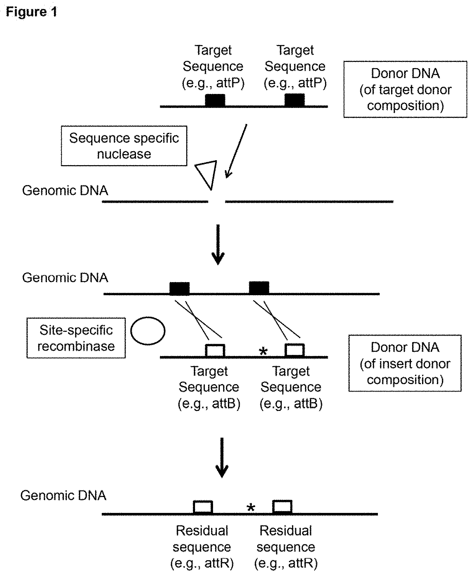
Provided are methods and compositions for genome editing using a delivery vehicle with multiple payloads. In some embodiments the delivery vehicle includes a payload that includes (a) one or more sequence specific nucleases that cleave the cell's genome or one or more nucleic acids encoding same, (b) a first donor DNA, which includes a nucleotide sequence that is inserted into the cell's genome, where insertion of said nucleotide sequence produces, in the cell's genome at the site of insertion, a target sequence (e.g., an attP site) for a site-specific recombinase; (c) the site-specific recombinase (or a nucleic acid encoding same) (e.g., .PHI.C31, .PHI.C31 RDF, Cre, FLP), where the site-specific recombinase recognizes said target sequence; and (d) a second donor DNA, which includes a nucleotide sequence that is inserted into the cell's genome as a result of recognition of said target sequence by the site-specific recombinase.
| Inventors: | Watson; Andre Ronald; (San Francisco, CA) ; Foster; Christian; (Oakland, CA) ; Lin; Shuailiang; (San Francisco, CA) | ||||||||||
| Applicant: |
|
||||||||||
|---|---|---|---|---|---|---|---|---|---|---|---|
| Family ID: | 68295768 | ||||||||||
| Appl. No.: | 16/393886 | ||||||||||
| Filed: | April 24, 2019 |
Related U.S. Patent Documents
| Application Number | Filing Date | Patent Number | ||
|---|---|---|---|---|
| 62661992 | Apr 24, 2018 | |||
| 62685240 | Jun 14, 2018 | |||
| Current U.S. Class: | 1/1 |
| Current CPC Class: | C12N 9/22 20130101; C12N 15/90 20130101; A61K 47/6929 20170801; C07K 2317/622 20130101; A61K 47/645 20170801; C07K 2317/55 20130101; C12N 15/907 20130101; A61P 3/10 20180101; A61K 47/6455 20170801; A61K 9/51 20130101; C07K 14/70596 20130101; B82Y 5/00 20130101; A61P 35/00 20180101; C12N 15/52 20130101; A61K 48/00 20130101; A61P 37/00 20180101; C12N 15/113 20130101; C12N 2310/20 20170501 |
| International Class: | C12N 15/90 20060101 C12N015/90; C12N 9/22 20060101 C12N009/22; C12N 15/113 20060101 C12N015/113; C12N 15/52 20060101 C12N015/52; C07K 14/705 20060101 C07K014/705 |
Claims
1. A method for inserting a donor sequence into a cell's genome, comprising: introducing into a cell, a delivery vehicle with a payload comprising: (a) a nuclease composition, comprising: one or more sequence specific nucleases or one or more nucleic acids that encode the one or more sequence specific nucleases, wherein the one or more sequence specific nucleases cleaves the cell's genome; (b) a target donor composition, comprising: a first donor DNA, which comprises a nucleotide sequence that is inserted into the cell's genome, wherein insertion of said nucleotide sequence produces, in the cell's genome at the site of insertion, a target sequence for a site-specific recombinase; (c) a recombinase composition, comprising: the site-specific recombinase, or a nucleic acid encoding the site-specific recombinase, wherein the site-specific recombinase recognizes said target sequence; and (d) an insert donor composition, comprising: a second donor DNA, which comprises a nucleotide sequence that is inserted into the cell's genome as a result of recognition of said target sequence by the site-specific recombinase.
2. The method of claim 1, wherein insertion of the nucleotide sequence of the first donor DNA of the target donor composition produces a first target sequence for the site-specific recombinase at a first location in the cell's genome and a second target sequence for the site-specific recombinase at a second location in the cell's genome.
3. The method of claim 1, wherein the nuclease composition cleaves the cell's genome at two locations, and wherein the target donor composition comprises two of said first donor DNAs, each of which comprises a nucleotide sequence that is inserted into the cell's genome, thereby producing a first target sequence for the site-specific recombinase at a first location in the cell's genome and a second target sequence for the site-specific recombinase at a second location in the cell's genome.
4-57. (canceled)
58. A method for inserting a donor sequence into a cell's genome, comprising: introducing into a cell, a delivery vehicle with a payload comprising: (a) a nuclease composition, comprising: one or more sequence specific nucleases or one or more nucleic acids that encode the one or more sequence specific nucleases, wherein the one or more sequence specific nucleases cleaves the cell's genome; (b) a target donor composition, comprising: a first donor DNA, which comprises a nucleotide sequence that is inserted into the cell's genome, wherein insertion of said nucleotide sequence produces, in the cell's genome at the site of insertion, a target sequence for a site-specific recombinase; (c) a recombinase composition, comprising: the site-specific recombinase, or a nucleic acid encoding the site-specific recombinase, wherein the site-specific recombinase recognizes said target sequence; and (d) an insert donor composition, comprising: a second donor DNA, which comprises a nucleotide sequence that is inserted into the cell's genome as a result of recognition of said target sequence by the site-specific recombinase, wherein the method comprises introducing a first and a second of said delivery vehicles into the cell, wherein: (1) the nucleotide sequence of the second donor DNA of the first delivery vehicle, that is inserted into the cell's genome, encodes a T cell receptor (TCR) Alpha or Delta subunit, and the nucleotide sequence of the second donor DNA of the second delivery vehicle, that is inserted into the cell's genome, encodes a TCR Beta or Gamma subunit; or (2) the nucleotide sequence of the second donor DNA of the first delivery vehicle, that is inserted into the cell's genome, encodes a T cell receptor (TCR) Alpha or Gamma subunit, and the nucleotide sequence of the second donor DNA of the second delivery vehicle, that is inserted into the cell's genome, encodes a TCR Beta or Delta subunit; or (3) the nucleotide sequence of the second donor DNA of the first delivery vehicle, that is inserted into the cell's genome, encodes the K chain of an IgA, IgD, IgE, IgG, or IgM protein, and the nucleotide sequence of the second donor DNA of the second delivery vehicle, that is inserted into the cell's genome, encodes the A chain of an IgA, IgD, IgE, IgG, or IgM protein.
59. The method of claim 1, wherein the method comprises introducing a first and a second of said delivery vehicles into the cell, wherein the nucleotide sequence of the second donor DNA of the first delivery vehicle, that is inserted into the cell's genome, encodes a T cell receptor (TCR) Alpha or Delta subunit constant region, and wherein the nucleotide sequence of the second donor DNA of the second delivery vehicle, that is inserted into the cell's genome, encodes a TCR Beta or Gamma subunit constant region.
60. The method of claim 1, wherein the method comprises introducing a first and a second of said delivery vehicles into the cell, wherein: (1) the nucleotide sequence of the second donor DNA of the first delivery vehicle is inserted within a nucleotide sequence that functions as a T cell receptor (TCR) Alpha or Delta subunit promoter, and the nucleotide sequence of the second donor DNA of the second delivery vehicle is inserted within a nucleotide sequence that functions as a TCR Beta or Gamma subunit promoter; or (2) the nucleotide sequence of the second donor DNA of the first delivery vehicle is inserted within a nucleotide sequence that functions as a T cell receptor (TCR) Alpha or Gamma subunit promoter, and the nucleotide sequence of the second donor DNA of the second delivery vehicle is inserted within a nucleotide sequence that functions as a TCR Beta or Delta subunit promoter; or (3) the nucleotide sequence of the second donor DNA of the first delivery vehicle is inserted within a nucleotide sequence that functions as a promoter for a K chain of an IgA, IgD, IgE, IgG, or IgM protein, and the nucleotide sequence of the second donor DNA of the second delivery vehicle is inserted within a nucleotide sequence that functions as a promoter for a .lamda. chain of an IgA, IgD, IgE, IgG, or IgM protein.
61-62. (canceled)
63. A composition comprising: (a) a nuclease composition, comprising: one or more sequence specific nucleases or one or more nucleic acids that encode the one or more sequence specific nucleases; (b) a target donor composition, comprising: a first donor DNA that comprises a target sequence for a site-specific recombinase; (c) a recombinase composition, comprising: the site-specific recombinase, or a nucleic acid encoding the site-specific recombinase, wherein the site-specific recombinase recognizes said target sequence; and (d) an insert donor composition, comprising: a second donor DNA, which comprises a nucleotide sequence of interest for insertion into a target cell's genome; wherein (a), (b), (c), and (d) are payloads as part of the same delivery vehicle.
64. The composition of claim 63, wherein the delivery vehicle is a nanoparticle.
65. The composition of claim 64, wherein the nanoparticle comprises a core comprising (a), (b), an anionic polymer composition, a cationic polymer composition, and a cationic polypeptide composition.
66. The composition of claim 64, wherein the nanoparticle comprises a targeting ligand that targets the nanoparticle to a cell surface protein.
67. The composition of claim 66, wherein the cell surface protein is CD47.
68. The composition of claim 67, wherein the targeting ligand is a SIRP.alpha. protein mimetic.
69. The composition of claim 68, wherein the nanoparticle further comprises an endocytosis-triggering ligand.
70. The composition of claim 63, wherein the payloads form one or more deoxyribonucleoprotein complexes or one or more ribo-deoxyribonucleoprotein complexes.
71. The composition of claim 63, wherein the delivery vehicle is a targeting ligand conjugated to a charged polymer polypeptide domain, wherein the targeting ligand provides for targeted binding to a cell surface protein, and wherein the charged polymer polypeptide domain is interacting electrostatically with one or more of the payloads.
72. The composition of claim 71, wherein the delivery vehicle further comprises an anionic polymer interacting with one or more of the payloads and the charged polymer polypeptide domain.
73. The composition of claim 63, wherein the delivery vehicle is a targeting ligand conjugated one or more of the payloads, wherein the targeting ligand provides for targeted binding to a cell surface protein.
74. The composition of claim 63, wherein the delivery vehicle includes a targeting ligand coated upon a water-oil-water emulsion particle, upon an oil-water emulsion micellar particle, upon a multilamellar water-oil-water emulsion particle, upon a multilayered particle, or upon a DNA origami nanobot.
75. The method of claim 66, wherein the targeting ligand is a peptide, an ScFv, a F(ab), a nucleic acid aptamer, or a peptoid.
76. The composition of claim 63, wherein the delivery vehicle is non-viral.
Description
CROSS-REFERENCE
[0001] This application claims the benefit of U.S. Provisional Patent Application No. 62/661,992, filed Apr. 24, 2018, and of U.S. Provisional Patent Application No. 62/685,240, filed Jun. 14, 2018, both of which applications are incorporated herein by reference in their entirety.
SEQUENCE LISTING
[0002] This application contains a Sequence Listing, which was submitted in ASCII format via EFS-Web, and is hereby incorporated by reference in its entirety. The ASCII copy, created on Feb. 3, 2020, is named 2020-02-03_Ligandal-8005U502-Sequence-Listing and is 146 KB in size.
INTRODUCTION
[0003] Genome editing remains an inefficient process in most circumstances. While many techniques exist for performing site specific gene editing, clinical translation is hampered by inadequate delivery technologies--especially when considering in vivo delivery. As such, compositions and methods for efficient genome editing remain an important unmet need.
SUMMARY
[0004] Provided are compositions and methods for genome editing using a delivery vehicle with multiple payloads. In some embodiments, subject methods include introducing a delivery vehicle into a cell, where the delivery vehicle includes a payload that includes (a) one or more sequence specific nucleases that cleave the cell's genome (e.g., a meganuclease, a homing endonuclease, a zinc finger nuclease (ZFN), a TALEN, a type I or type III CRISPR/Cas cleavage complex, a class 2 CRISPR/Cas effector protein--an RNA-guided CRISPR/Cas polypeptide--such as Cas9, CasX, CasY, Cpf1 (Cas12a), Cas13, MAD7, and the like) or one or more nucleic acids that encode the one or more sequence specific nucleases [(a) is referred to herein as a nuclease composition]; (b) a first donor DNA, which includes a nucleotide sequence that is inserted into the cell's genome, where insertion of said nucleotide sequence produces, in the cell's genome at the site of insertion, a target sequence (e.g., an attP site) for a site-specific recombinase [(b) is referred to herein as a target donor composition]; (c) the site-specific recombinase (or a nucleic acid encoding same) (e.g., .PHI.C31, .PHI.C31 RDF, Cre, FLP), where the site-specific recombinase recognizes said target sequence [(c) is referred to herein as a recombinase composition]; and (d) a second donor DNA, which includes a nucleotide sequence that is inserted into the cell's genome as a result of recognition of said target sequence by the site-specific recombinase [(d) is referred to herein as an insert donor composition]. A delivery vehicle that includes (a), (b), (c), and (d) facilitates insertion of large sequences into the genome by insertion of one or more target sites for a site-specific recombinase followed by recombinase-mediated insertion of a nucleotide sequence of interest. In some cases, the inserted nucleotide sequence of interest (from the insert donor composition) is 10 kilobase pairs (kbp) or more (e.g., from 15 kbp to 100 kbp, from 30 kbp to 100 kbp, or from 50 kbp to 100 kbp).
[0005] In some cases insertion of the nucleotide sequence of the first donor DNA produces a first target sequence for the site-specific recombinase at a first location in the cell's genome and a second target sequence for the site-specific recombinase at a second location in the cell's genome (e.g., insertion of two attP sites). In some cases the nuclease composition cleaves the cell's genome at two locations, and the target donor composition includes two of the first donor DNAs, each of which includes a nucleotide sequence that is inserted into the cell's genome, thereby producing a first target sequence for the site-specific recombinase at a first location in the cell's genome and a second target sequence for the site-specific recombinase at a second location in the cell's genome (e.g., insertion of two attP sites). In some cases the second donor DNA includes two target sequences (e.g., attB sites) for the site-specific recombinase, where the two target sequences flank the nucleotide sequence that is inserted into the cell's genome.
[0006] The delivery vehicle can be introduced into a cell/delivered to a cell in vitro, ex vivo, or in vivo. One advantage of delivering multiple payloads as part of the same delivery vehicle (e.g., nanoparticle) is that the efficiency of each payload is not diluted. As an illustrative example, if payload A and payload B are delivered in two separate packages/vehicles (package A and package B, respectively), then the efficiencies are multiplicative, e.g., if package A and package B each have a 1% transfection efficiency, the chance of delivering payload A and payload B to the same cell is 0.01% (1%.times.1%). However, if payload A and payload B are both delivered as part of the same delivery vehicle, then the chance of delivering payload A and payload B to the same cell is 1%, a 100-fold improvement over 0.01%.
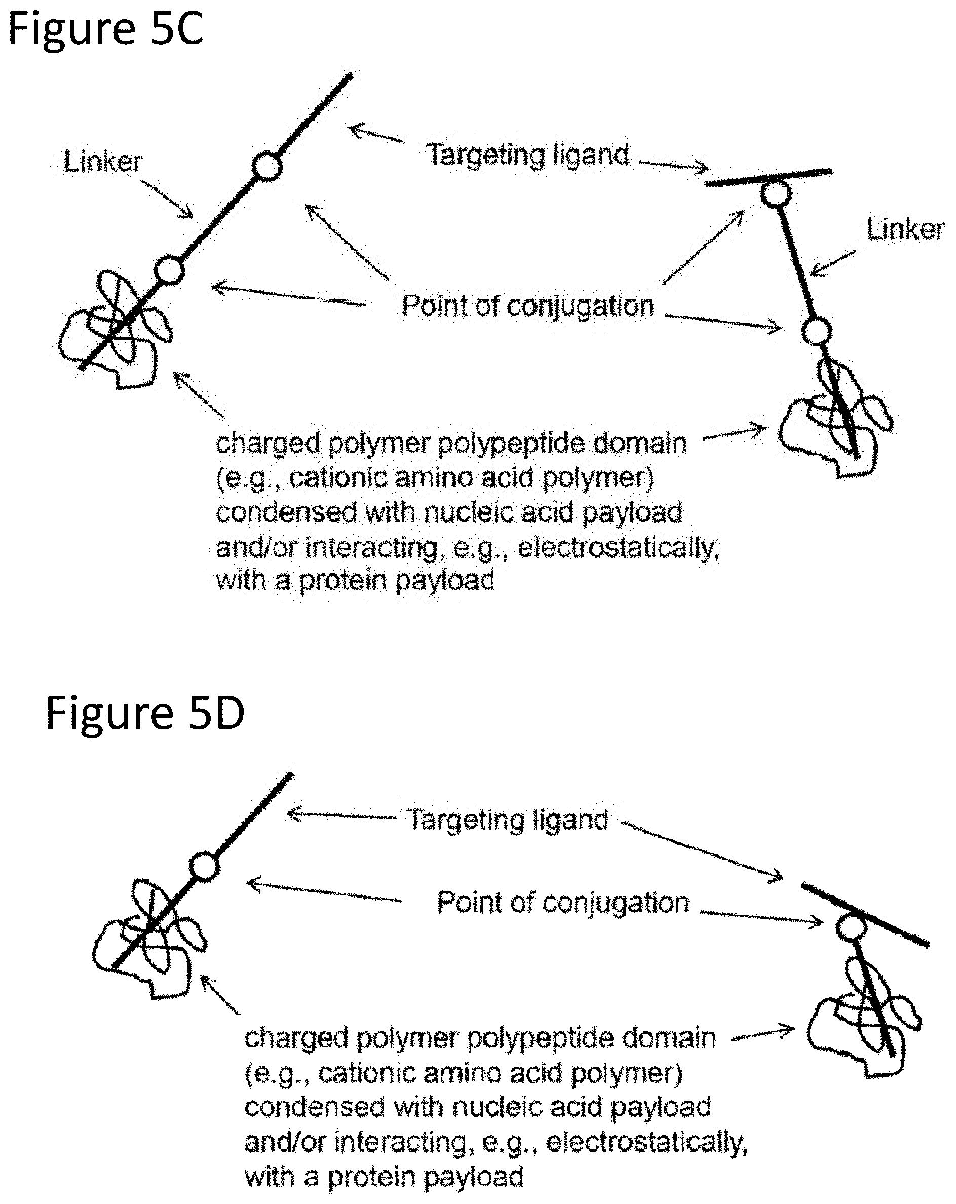
[0007] Delivery vehicles can include, but are not limited to, non-viral vehicles, viral vehicles, nanoparticles (e.g., a nanoparticle that includes a targeting ligand and/or a core comprising an anionic polymer composition, a cationic polymer composition, and a cationic polypeptide composition), liposomes, micelles, water-oil-water emulsion particles, oil-water emulsion micellar particles, multilamellar water-oil-water emulsion particles, a targeting ligand (e.g., peptide targeting ligand) conjugated to a charged polymer polypeptide domain (wherein the targeting ligand provides for targeted binding to a cell surface protein, and the charged polymer polypeptide domain is condensed with a nucleic acid payload and/or is interacting electrostatically with a protein payload), a targeting ligand (e.g., peptide targeting ligand) conjugated to payload (where the targeting ligand provides for targeted binding to a cell surface protein), etc. In some cases payloads are introduced into the cell as deoxyribonucleoprotein complex(s) and/or a ribo-deoxyribonucleoprotein complex(s).
[0008] The provided compositions and methods can be used for genome editing at any locus in any cell type (e.g., to engineer T-cells, e.g., in vivo). For example, a CD8+ T-cell population or mixture of CD8+ and CD4+ T-cells can be programmed to transiently or permanently express an appropriate TCR.alpha./TCR.beta. pair of CDR1, CDR2, and/or CDR3 domains for antigen recognition.
BRIEF DESCRIPTION OF THE DRAWINGS
[0009] The invention is best understood from the following detailed description when read in conjunction with the accompanying drawings. The patent or application file contains at least one drawing executed in color. Copies of this patent or patent application publication with color drawing(s) will be provided by the Office upon request and payment of the necessary fee. It is emphasized that, according to common practice, the various features of the drawings are not to-scale. On the contrary, the dimensions of the various features are arbitrarily expanded or reduced for clarity. Included in the drawings are the following figures.
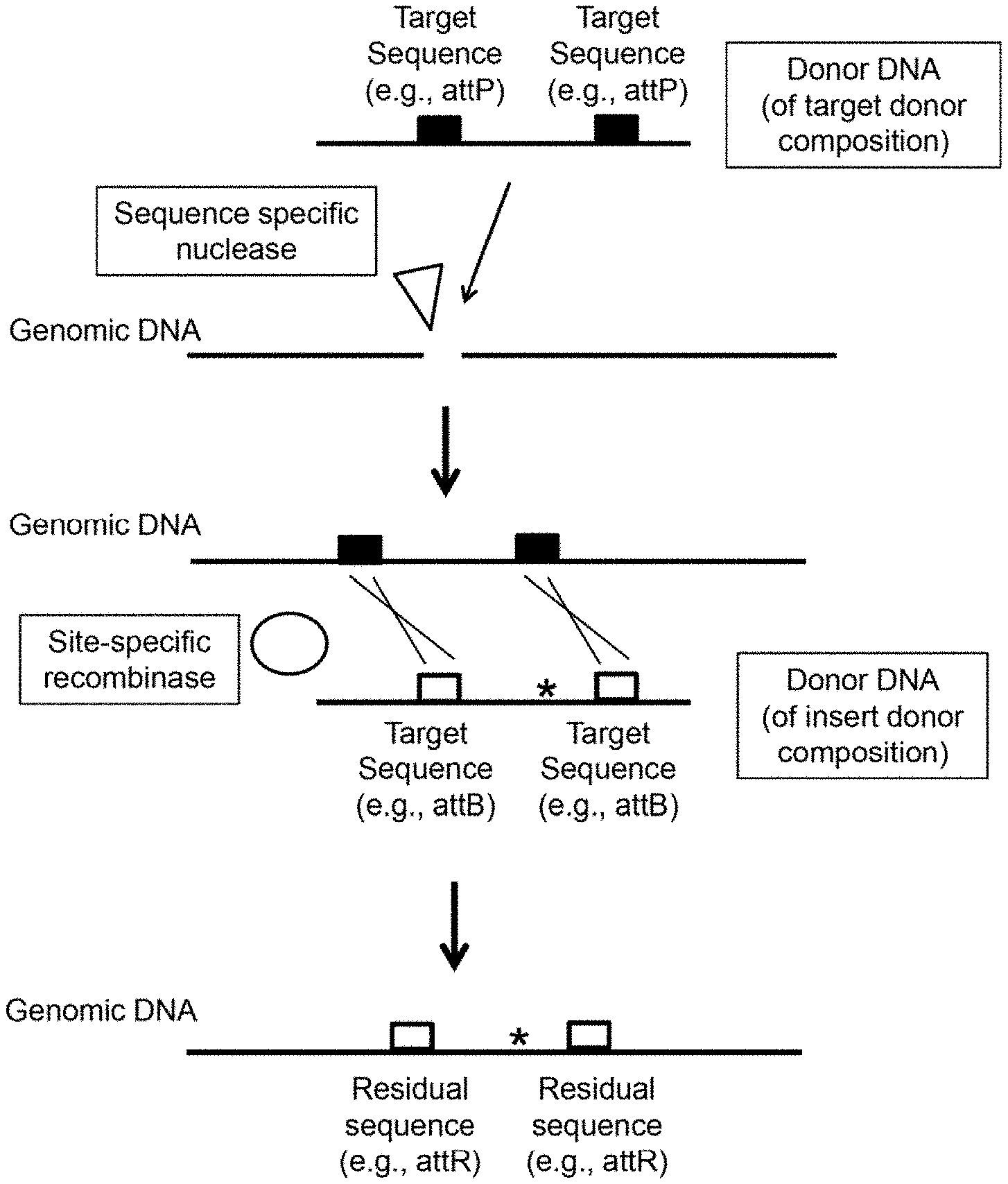
[0010] FIG. 1 depicts a schematic representation of one example of a subject method. In this particular example: two target sequences (e.g., attP sites) are inserted into the genome (using the donor DNA of the target donor composition) after cleavage of the genome with a sequence specific nuclease; two target sequences (e.g., attB sites) are present on the donor DNA of the insert donor composition; and two residual sites (e.g., attR sites) remain present in the genome after insertion of sequence from the donor DNA (of the insert donor composition) using a site-specific recombinase (e.g., PhiC31 .PHI.C31)).
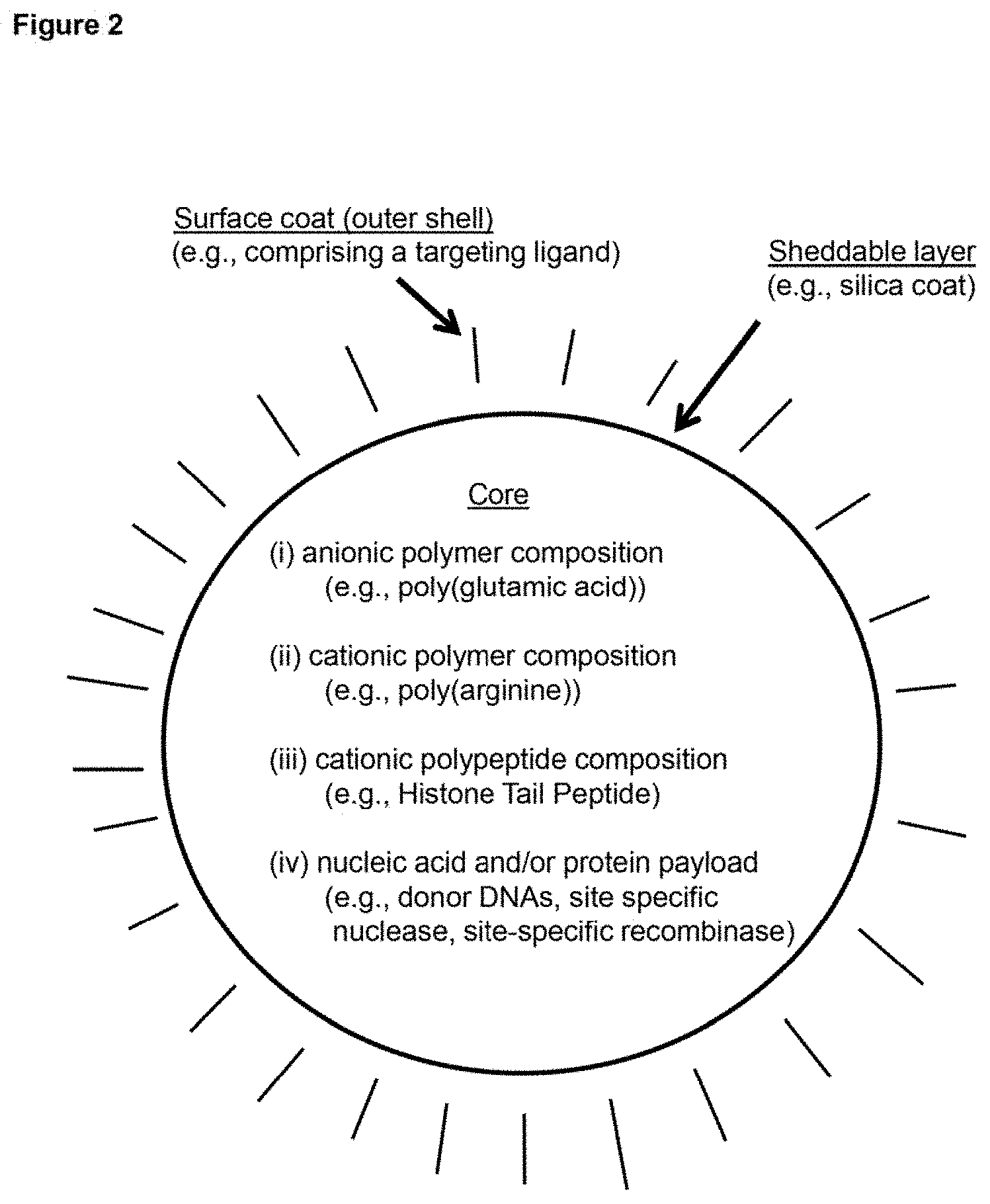
[0011] FIG. 2 depicts a schematic representation of an example embodiment of a delivery vehicle (in the depicted case, one type of nanoparticle).
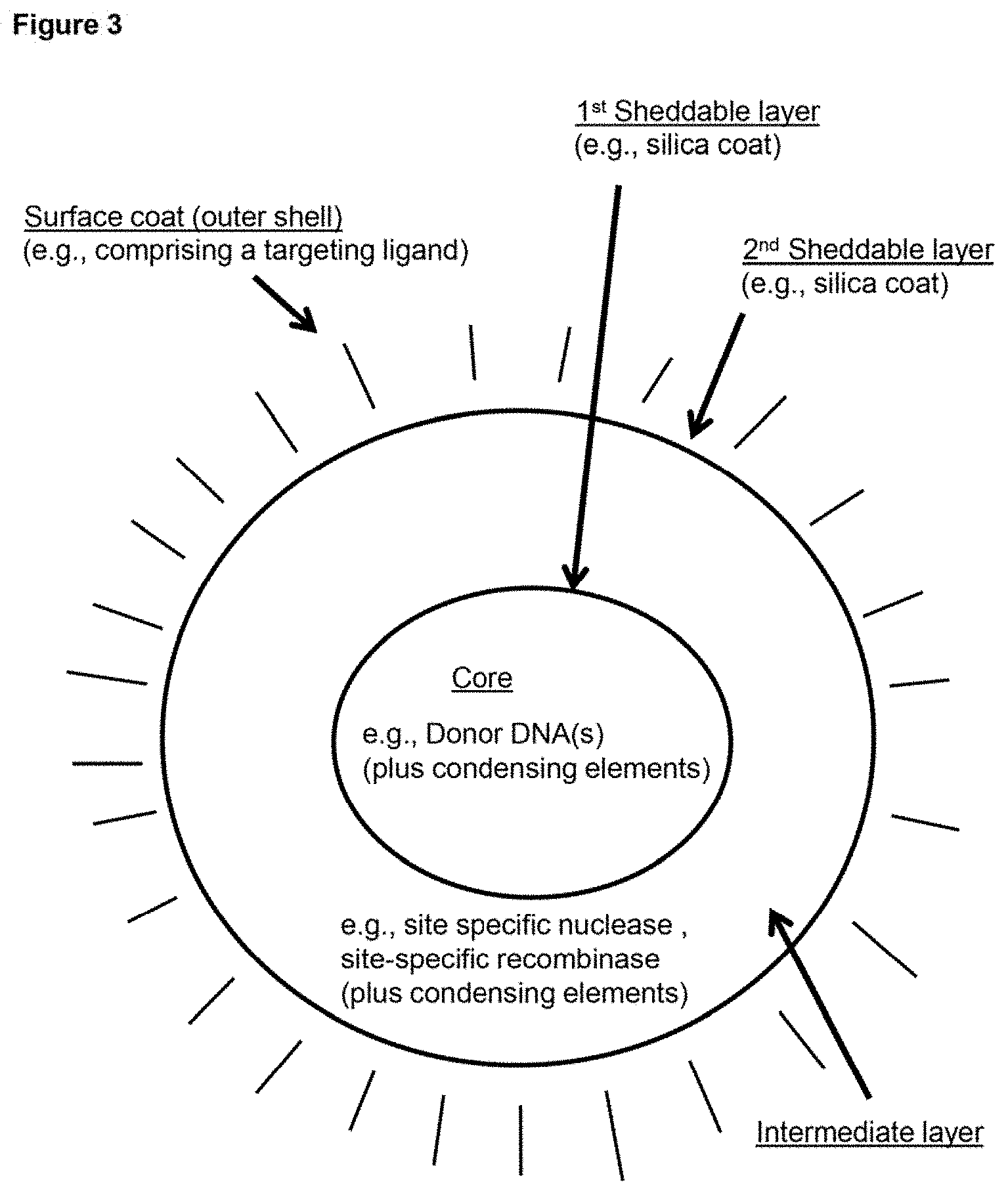
[0012] FIG. 3 depicts a schematic representation of an example embodiment of a delivery vehicle (in the depicted case, one type of nanoparticle). In this case, the depicted nanoparticle is multi-layered, having a core (which includes a first payload) surrounded by a first sheddable layer, which is surrounded by an intermediate layer (which includes an additional payload), which is surrounded by a second sheddable layer, which is surface coated (i.e., includes an outer shell).
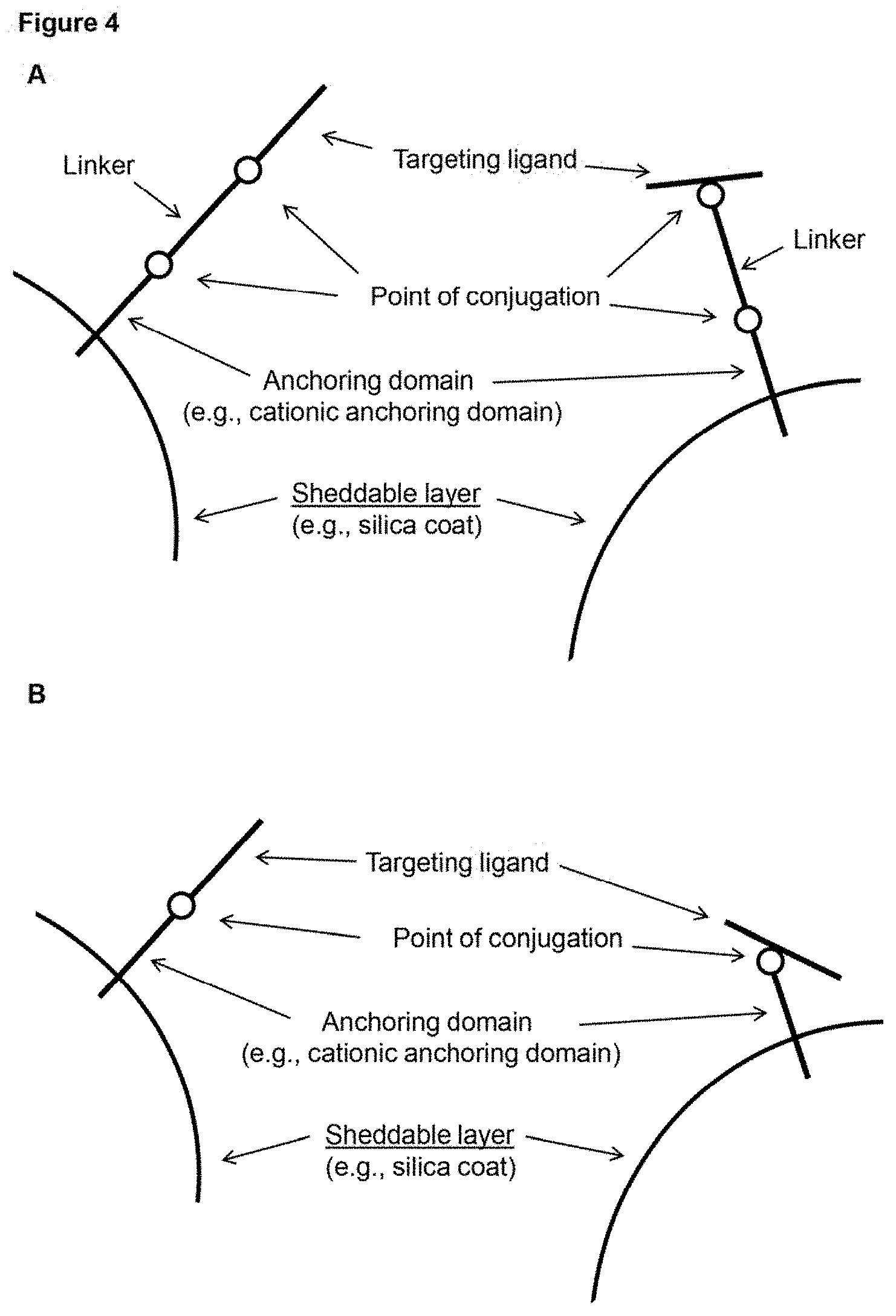
[0013] FIG. 4 (panels A-B) depicts schematic representations of example configurations of a targeting ligand of a surface coat of a subject nanoparticle. The delivery molecules depicted include a targeting ligand conjugated to an anchoring domain that is interacting electrostatically with a sheddable layer of a nanoparticle. Note that the targeting ligand can be conjugated at the N- or C-terminus (left of each panel), but can also be conjugated at an internal position (right of each panel). The molecules in panel A include a linker while those in panel B do not.
[0014] FIG. 5A provides a schematic drawing of an example embodiment of a donor vehicle (in the depicted case, example configurations of a subject delivery molecule). The targeting ligand can be conjugated at the N- or C-terminus (left of the figure), but can also be conjugated at an internal position (right of the figure). This figure shows delivery molecules including a linker as well as a targeting ligand conjugated to a payload.
[0015] FIG. 5B provides a schematic drawing of an example embodiment of a donor vehicle (in the depicted case, example configurations of a subject delivery molecule). The targeting ligand can be conjugated at the N- or C-terminus (left of the figure), but can also be conjugated at an internal position (right of the figure). This figure shows delivery molecules including do not have a linker but do have a targeting ligand conjugated to a payload.
[0016] FIG. 5C provides a schematic drawing of an example embodiment of a donor vehicle (in the depicted case, example configurations of a subject delivery molecule). The targeting ligand can be conjugated at the N- or C-terminus (left of the figure), but can also be conjugated at an internal position (right of the figure). This figure shows delivery molecules including a linker and a targeting ligand conjugated to a charged polymer polypeptide domain that is condensed with a nucleic acid payload (and/or interacting, e.g. electrostatically, with a protein payload).
[0017] FIG. 5D provides a schematic drawing of an example embodiment of a donor vehicle (in the depicted case, example configurations of a subject delivery molecule). The targeting ligand can be conjugated at the N- or C-terminus (left of the figure), but can also be conjugated at an internal position (right of the figure). This figure shows delivery molecules that do not have a linker but do have a targeting ligand conjugated to a charged polymer polypeptide domain that is condensed with a nucleic acid payload (and/or interacting, e.g. electrostatically, with a protein payload).
[0018] FIG. 6 provides non-limiting examples of nuclear localization signals (NLSs) that can be used (e.g., as part of a nanoparticle, e.g., as an NLS-containing peptide; as part of/conjugated to an NLS-containing peptide, an anionic polymer, a cationic polymer, and/or a cationic polypeptide; and the like). The figure is adapted from Kosugi et al., J Biol Chem. 2009 Jan. 2; 284(1):478-85. (Class 1, top to bottom (SEQ ID NOs: 201-221); Class 2, top to bottom (SEQ ID NOs: 222-224); Class 4, top to bottom (SEQ ID NOs: 225-230); Class 3, top to bottom (SEQ ID NOs: 231-245); Class 5, top to bottom (SEQ ID NOs: 246-264)].
[0019] FIG. 7 depicts schematic representations of the mouse hematopoietic cell lineage, and markers that have been identified for various cells within the lineage.
[0020] FIG. 8 depicts schematic representations of the human hematopoietic cell lineage, and markers that have been identified for various cells within the lineage.
[0021] FIG. 9 depicts schematic representations of miRNA factors that can be used to influence cell differentiation and/or proliferation.
[0022] FIG. 10 depicts schematic representations of protein factors that can be used to influence cell differentiation and/or proliferation.
[0023] FIG. 11 depicts the average particle size of nanoparticles with the compositions of Study A.
[0024] FIG. 12 depicts zeta potential of nanoparticles with the compositions of Study A.
[0025] FIG. 13 depicts the polydispersity index of nanoparticles with the compositions of Study A.
[0026] FIG. 14 depicts a chart describing the polydispersity index and stability of nanoparticles with the compositions of Study B.
[0027] FIG. 15 depicts the average particle size of nanoparticles with the compositions of Study B.
[0028] FIG. 16 depicts zeta potential of nanoparticles with the compositions of Study B.
[0029] FIG. 17 depicts the polydispersity index of nanoparticles with the compositions of Study B.
[0030] FIG. 18 depicts a chart describing the polydispersity index and stability of nanoparticles with the compositions of Study B.
[0031] FIG. 19 depicts the particle size distribution of nanoparticles with compositions of Study C.
[0032] FIG. 20 depicts the zeta potential of nanoparticles with the compositions of Study C.
[0033] FIG. 21 depicts the polydispersity index of nanoparticles with the compositions of Study C.
[0034] FIG. 22 depicts the polydispersity index and stability of nanoparticles with the compositions of Study C.
[0035] FIG. 23 depicts the fluorescence values obtained with the SYBR GOLD Inclusion assay of nanoparticles with the compositions of Study C.
[0036] FIG. 24 depicts the characterization of RNP-H2B (mini core) particles.
[0037] FIG. 25 depicts the characterization of RNP-H2B-PLE20:PDE20 (core) particles.
[0038] FIG. 26 depicts the serum stability of RNP-H2B-PLE20:PDE20 (core) particles.
[0039] FIG. 27 depicts the polydispersity evaluation of ligand-modified particles after one day.
[0040] FIG. 28 depicts the polydispersity evaluation of ligand-modified particles after 72 hours or seven days.
[0041] FIG. 29 depicts cells, nuclei, and nanoparticles as measured by automated pipeline sampling.
[0042] FIG. 30 depicts changes in cellular particle colocalization over 14.5 hours after various treatments.
[0043] FIG. 31 depicts the percentage of Cas9-eGFP cells over 14.5 hours after various treatments.
[0044] FIG. 32 depicts nuclear particle integration over 14.5 hours after various treatments.
[0045] FIG. 33 depicts the percentage of Cas9-eGFP cells over 14.5 hours after various treatments.
[0046] FIG. 34A depicts T-cell and PBMC targeting in untreated samples.
[0047] FIG. 34B depicts the human primary pan T-cell data corresponding to FIG. 34A.
[0048] FIG. 34C depicts the human primary PBMCs data corresponding to FIG. 34A.
[0049] FIG. 35A depicts T-cell and PBMC targeting in core particles.
[0050] FIG. 35B depicts the human primary pan T-cell data corresponding to FIG. 35A.
[0051] FIG. 35C depicts the human primary PBMCs data corresponding to FIG. 35A.
[0052] FIG. 36A depicts T-cell and PBMC targeting in samples treated with ligand poly(L-arginine).
[0053] FIG. 36B depicts the human primary pan T-cell data corresponding to FIG. 36A.
[0054] FIG. 36C depicts the human primary PBMCs data corresponding to FIG. 36A.
[0055] FIG. 37A depicts T-cell and PBMC targeting in samples with ligand CD3_CD3e_(4GS)2_9R_N_1.
[0056] FIG. 37B depicts the human primary pan T-cell data corresponding to FIG. 37A.
[0057] FIG. 37C depicts the human primary PBMCs data corresponding to FIG. 37A.
[0058] FIG. 38A depicts T-cell and PBMC targeting in samples with ligand CD8_(4GS)2_9R_N.
[0059] FIG. 38B depicts the human primary pan T-cell data corresponding to FIG. 38A.
[0060] FIG. 38C depicts the human primary PBMCs data corresponding to FIG. 38A.
[0061] FIG. 39A depicts T-cell and PBMC targeting in samples with ligand CD28_mCD80_(4GS)2_9R_N.
[0062] FIG. 39B depicts the human primary pan T-cell data corresponding to FIG. 39A.
[0063] FIG. 39C depicts the human primary PBMCs data corresponding to FIG. 39A.
[0064] FIG. 40A depicts T-cell and PBMC targeting in samples with ligands CD28_mCD86_(4GS)2_9R_N.
[0065] FIG. 40B depicts the human primary pan T-cell data corresponding to FIG. 40A.
[0066] FIG. 40C depicts the human primary PBMCs data corresponding to FIG. 40A.
[0067] FIG. 41A depicts T-cell and PBMC targeting in samples with ligands IL2R_mIL2_4 GS_2_9R_N_1.
[0068] FIG. 41B depicts the human primary pan T-cell data corresponding to FIG. 41A.
[0069] FIG. 41C depicts the human primary PBMCs data corresponding to FIG. 41A.
[0070] FIG. 42A depicts T-cell and PBMC targeting in samples with ligands CD3_CD3e_(4GS)2_9R_N_1 and CD8_(4GS)2_9R_N.
[0071] FIG. 42B depicts the human primary pan T-cell data corresponding to FIG. 42A.
[0072] FIG. 42C depicts the human primary PBMCs data corresponding to FIG. 42A.
[0073] FIG. 43A depicts T-cell and PBMC targeting in samples with ligands CD3_CD3e_(4GS)2_9R_N_1 and CD28_mCD80_(4GS)2_9R_N.
[0074] FIG. 43B depicts the human primary pan T-cell data corresponding to FIG. 43A.
[0075] FIG. 43C depicts the human primary PBMCs data corresponding to FIG. 43A.
[0076] FIG. 44A depicts T-cell and PBMC targeting in samples with ligands CD3_CD3e_(4GS)2_9R_N_1 and CD28_mCD86_(4GS)2_9R_N.
[0077] FIG. 44B depicts the human primary pan T-cell data corresponding to FIG. 44A.
[0078] FIG. 44C depicts the human primary PBMCs data corresponding to FIG. 44A.
[0079] FIG. 45A depicts T-cell and PBMC targeting in samples with ligands CD3_CD3e_(4GS)2_9R_N_1 and IL2R_mIL2_4 GS_2_9R_N_1.
[0080] FIG. 45B depicts the human primary pan T-cell data corresponding to FIG. 45A.
[0081] FIG. 45C depicts the human primary PBMCs data corresponding to FIG. 45A.
[0082] FIG. 46A depicts T-cell and PBMC targeting in samples with ligands CD3_CD3e_(4GS)2_9R_N_1 and poly(L-arginine)n=10.
[0083] FIG. 46B depicts the human primary pan T-cell data corresponding to FIG. 46A.
[0084] FIG. 46C depicts the human primary PBMCs data corresponding to FIG. 46A.
[0085] FIG. 47A depicts T-cell and PBMC targeting in samples with ligands CD2B_mCD80_(4GS)2_9R_N and CD28_mCD86_(4GS)2_9R_N.
[0086] FIG. 47B depicts the human primary pan T-cell data corresponding to FIG. 47A.
[0087] FIG. 47C depicts the human primary PBMCs data corresponding to FIG. 47A.
[0088] FIG. 48A depicts T-cell and PBMC targeting in samples with ligands CD3_CD3e_(4GS)2_9R_N_1, CD2B_mCD86_(4GS)2_9R_N, and CD8_4 GS_2_9R_N_1.
[0089] FIG. 48B depicts the human primary pan T-cell data corresponding to FIG. 48A.
[0090] FIG. 48C depicts the human primary PBMCs data corresponding to FIG. 48A.
[0091] FIG. 49A depicts T-cell and PBMC targeting in samples with ligands CD3_CD3e_(4GS)2_9R_N_1, CD8_4 GS_2_9R_N_1, and IL2R_mIL2_4 GS_2_9R_N_1.
[0092] FIG. 49B depicts the human primary pan T-cell data corresponding to FIG. 49A.
[0093] FIG. 49C depicts the human primary PBMCs data corresponding to FIG. 49A.
[0094] FIG. 50A depicts T-cell and PBMC targeting in samples with ligands CD3_CD3e_(4GS)2_9R_N_1, CD2B_mCD80_(4GS)2_9R_N, and CD8_4 GS_2_9R_N_1.
[0095] FIG. 50B depicts the human primary pan T-cell data corresponding to FIG. 50A.
[0096] FIG. 50C depicts the human primary PBMCs data corresponding to FIG. 50A.
[0097] FIG. 51A depicts T-cell and PBMC targeting in samples with CD3_CD3e_(4GS)2_9R_N_1, CD2B_mCD86_(4GS)2_9R_N, and CD28_mCD80_(4GS_)2_9R_N_1.
[0098] FIG. 51B depicts the human primary pan T-cell data corresponding to FIG. 51A.
[0099] FIG. 51C depicts the human primary PBMCs data corresponding to FIG. 51A.
[0100] FIG. 52A depicts T-cell and PBMC targeting in samples with ligands CD8_4 GS_2_9R_N_1, CD28_mCD80_(4GS)2_9R_N, and CD28_mCD86_(4GS)2_9R_N.
[0101] FIG. 52B depicts the human primary pan T-cell data corresponding to FIG. 52A.
[0102] FIG. 52C depicts the human primary PBMCs data corresponding to FIG. 52A.
[0103] FIG. 53A depicts T-cell and PBMC targeting in samples with ligands CD8_4 GS_2_9R_N_1, CD28_mCD80_(4GS)2_9R_N, and IL2R_mIL2_4 GS_2_9R_N_1.
[0104] FIG. 53B depicts the human primary pan T-cell data corresponding to FIG. 53A.
[0105] FIG. 53C depicts the human primary PBMCs data corresponding to FIG. 53A.
[0106] FIG. 54A depicts T-cell and PBMC targeting in samples with ligands CD8_4 GS_2_9R_N_1, CD28_mCD86_(4GS)2_9R_N, IL2R_mIL2_4 GS_2_9R_N_1.
[0107] FIG. 54B depicts the human primary pan T-cell data corresponding to FIG. 54A.
[0108] FIG. 54C depicts the human primary PBMCs data corresponding to FIG. 54A.
[0109] FIG. 55 depicts the image analysis, Pan-T cell flow analysis, and PBMC flow analysis of samples with different ligands.
[0110] FIG. 56 depicts the image analysis, Pan-T cell flow analysis, and PBMC flow analysis of samples with ligand sets that are different from those of FIG. 55.
[0111] FIG. 57 depicts a general schematic of nanoparticle synthesis. This involves addition of payloads, cationic/anionic peptides, and ligands, demonstrating varying orders of addition and degrees of freedom. Peptide, payload, and ligand examples are given and include different mer lengths and D/L isomers.
[0112] FIG. 58A depicts volumes of PLR50, buffer, and RNP used in the formulation of each nanoparticle added stepwise.
[0113] FIG. 58B depicts volumes of PLR50, buffer, and RNP used in the formulation of each nanoparticle added stepwise continued.
[0114] FIG. 58C depicts volumes of DNA, PLR50, and buffer used in the formulation of each nanoparticle added stepwise.
[0115] FIG. 58D depicts volumes of DNA, PLR50 and buffer used in the formulation of each nanoparticle added stepwise (continued).
[0116] FIG. 58E depicts the nanoparticle well ID (location in 96-well plate where it was synthesized and measured for size, zeta potential, and SYBR fluorescence) and its conversion to Nanoparticle ID (reference to the 96-well cell transfection plate) in FIG. 61I.
[0117] FIG. 59A depicts particle sizes of nanoparticles synthesized in 2C.1.1.1. Particle sizes were measured in triplicate via a Wyatt Mobius Zeta Potential and DLS Detector. Sizes are reported as average hydrodynamic diameter (nm).+-.standard deviation in a heatmap which correlates to the nanoparticle 96-well ID.
[0118] FIG. 59B depicts zeta potentials of nanoparticles synthesized in 2C.1.1.1. Particle zeta potentials were measured in triplicate via a Wyatt Mobius Zeta Potential and DLS Detector. Zeta potentials are reported as average zeta potentials (mV).+-.standard deviation in a heatmap which correlates to the nanoparticle 96-well ID.
[0119] FIG. 59C depicts the average (Overnight) condensation index of each particle in 2C.1.1.1 using SYBR fluorescence assay. The condensation index is calculated as [(Well of Interest Fluorescence-Free DNA Fluorescence)/Free DNA Fluorescence]*100 and is reported as average condensation index.+-.standard deviation in a heatmap which correlates to the nanoparticle 96-well ID. The more condensed nanoparticles will have higher shielding, less fluorescence, and thus a more negative condensation index.
[0120] FIG. 60A depicts the plate layout for 20.1.1.1 transfection of HEK293-GFP cells. NPs were dosed at 20 uL and 10 uL on duplicate plates. Row B (gold highlight) shows Layer 1 Charge Ratios, Row C (orange highlight) shows Outer Layer Charge Ratios, green highlight shows CRISPRMAX transfection controls, grey highlight shows Lipofectamine3000 transfection controls. The DNA Mix includes the ssODN and PhiC31 expression and donor plasmids.
[0121] FIG. 60B depicts 20.1.1.1 Flow results for Day 5 post transfection, monitoring GFP and Alexa647 (NP) fluorescence. Live cells were gated based on FSC/SSC scatter and % GFP- and % NP+ are shown for live gate. No significant GFP KD is observed for NP wells, although lipofection controls give robust results. The 20 uL dose of NP has significant NP signal at Day 5, while 10 uL dose does not. RFP expression was observed only in the lipofection control with Tag-RFP plasmid: 20% RFP+ of Live, well C11 (data not shown). Flow analysis on Day 10 and Day 14 post transfection showed no more NP Alexa-647 signal, no GFP knock-down, and no RFP expression for NP groups. RFP expression decreased over time in plasmid lipofection controls (7% RFP+ Day 10, 2% RFP+ Day 14), while GFP knock-down in CRISPRMAX controls (B10, 010) remained constant, consistent with heritable gene editing.
[0122] FIG. 60C depicts results of 20.1.1.1 Day 5 post-transfection genomic analysis for the RNP+ DNA Mix lipofection control (well 010), showing editing of the GFP locus (25% KD) and HDR (knock-in) of the attP ssODN (6%) using Synthego's ICE-KI analysis of Sanger sequencing data. The guide target sequence is set forth in SEQ ID NO: 278. The sequences in the alignment are set forth from top to bottom in SEQ ID NOs: 304-319.
[0123] FIG. 61A depicts volumes of PLR50, buffer, and RNP used in the formulation of each nanoparticle added stepwise.
[0124] FIG. 61B depicts volumes of PLR50, buffer, and RNP used in the formulation of each nanoparticle added stepwise (continued).
[0125] FIG. 61C depicts volumes of DNA, Ligand Mix, ALEXA-647 nanoparticle label, and buffer used in the formulation of each nanoparticle added stepwise.
[0126] FIG. 61D depicts volumes of PLR50, buffer, and RNP used in the formulation of each nanoparticle added stepwise (continued).
[0127] FIG. 61E depicts the nanoparticle well ID (location in 96-well plate where it was synthesized and measured for size, zeta potential, and SYBR fluorescence) and its conversion to Nanoparticle ID (reference to the 96-well cell transfection plate) in FIG. 61I.
[0128] FIG. 61F depicts particle sizes of nanoparticles synthesized in 2C.2.1.1. Particle sizes were measured in triplicate via a Wyatt Mobius Zeta Potential and DLS Detector. Sizes are reported as average hydrodynamic diameter (nm).+-.standard deviation in a heatmap which correlates to the nanoparticle 96-well ID.
[0129] FIG. 61G depicts zeta potentials of nanoparticles synthesized in 2C.2.1.1. Particle zeta potentials were measured in triplicate via a Wyatt Mobius Zeta Potential and DLS Detector. Zeta potentials are reported as average zeta potentials (mV).+-.standard deviation in a heatmap which correlates to the nanoparticle 96-well ID.
[0130] FIG. 61H depicts the average (Overnight) condensation index of each particle in 2C.2.1.1 using SYBR fluorescence assay. The condensation index is calculated as [(Well of Interest Fluorescence-Free DNA Fluorescence)/Free DNA Fluorescence]*100 and is reported as average condensation index.+-.standard deviation in a heatmap which correlates to the nanoparticle 96-well ID. The more condensed nanoparticles will have higher shielding, less fluorescence, and thus a more negative condensation index.
[0131] FIG. 61I depicts the plate layout for 2C.2.1.1 transfection of stimulated PBMCs. NPs were dosed at 10 uL per well of 60,000 stimulated PBMCs. Row B (gold highlight) shows Layer 1 Charge Ratios, Row C (orange highlight) shows Outer Layer Charge Ratios. Green highlighted wells (B12-G12 and G11) are nucleofection controls. NP18, NP39, and NP25 were the highest performing nanoparticle groups in terms of gene editing efficiency (TCR k/d; 6%, 5% and 5% Sanger sequencing efficiency, respectively). Bolded values indicate TCR locus cutting values >1% for the highlighted samples as determined by Sanger sequencing.
[0132] FIG. 61J depicts cell viability (% Live) of transfected cells from experiment 2C.2.1.1 as measured via flow cytometry. Following PBS wash of nanoparticles from overnight transfection, stimulated PBMCs were gathered for Day 1 flow analysis. Cell Viability was assayed by Annexin V staining. Well E6 had an error.
[0133] FIG. 61K depicts cell viability (% Dead/Apoptotic) of transfected cells from experiment 2C.2.1.1 as measured via flow cytometry. Following PBS wash of nanoparticles from overnight transfection, stimulated PBMCs were gathered for Day 1 flow analysis. Cell Viability was assayed by Annexin V staining. Well E6 had an error. poptotic and dead cells. Well E6 had an error.
[0134] FIG. 61L depicts cellular uptake (AF647-NP+ and EGFP-Cas9+) of various nanoparticle formulations from 2C.2.1.1. Following PBS wash of nanoparticles from overnight transfection, stimulated PBMCs were gathered for Day 1 flow analysis. Shown is GFP (Cas9-GFP) and Alexa647 (NP) fluorescence and % GFP+ and % NP+ are shown for live gate (Annexin V negative). Very little NP signal (Alexa647) is observed in T cells compared to HEK-293 cells, suggesting rapid degradation of the AF647-bound endosomal escape peptide results in the most efficient subcellular Cas9 RNP delivery. Almost all trace NP+ levels (1-2%) are coincident with the highest-value Cas9-GFP signals. CD3 staining showed that >95% of cells were CD3+ (not shown), indicating that CD3/CD28 stimulation resulted in selective proliferation of T Cells.
[0135] FIG. 61M depicts flow analysis of TCR expression on Day 4 post-transfection in 2C.2.1.1. Nucleofection wells with RNP+/-ssODN have robust TCR KD, while nucleofection of all components (F12) and NP wells do not. No NP signal (Alexa647 or Cas9-GFP) is observed, and only the RFP plasmid nucleofection control well (G12) shows RFP expression at 10% of live cells (data not shown).
[0136] FIG. 61N depicts flow analysis of TCR expression on Day 14 post-transfection in 2C.2.1.1. No NP signal (Alexa647 or Cas9-GFP) or RFP expression is observed, even from the plasmid nucleofection control.
[0137] FIG. 61O depicts ICE scores of Day 14 Sanger sequencing data for TRAC locus. High ICE scores show robust editing in nucleofection samples, similar to TCR KD by flow. Three NPs (boxed) showed modest TCR editing by Synthego's ICE platform. No samples showed HDR (knock-in) with the attP ssODN.
[0138] FIG. 61P depicts Sanger sequencing results of 2C.2.1.1, a study performed prior to 2C.1.2.1 whereby cores were further optimized to result in higher nanoparticle uptake efficiencies and subcellular release kinetics. 2C.1.2.1 further included variable ratios of PLE to PDE, along with variable ratios of histone fragments, PLR10, PLR50, NLS-modified histones, and/or endosomal escape--NLS peptide. Prior to decoration in targeting ligands, various nanoparticle cores were assessed for their biological performance (cellular uptake, cellular persistence over time, viability, and gene editing) in an initial set of screens intended to optimize nanoparticle cores for rapid subcellular vs. extended subcellular release, as is further detailed in 2C.1.2.1.
[0139] FIG. 61Q depicts Sanger sequencing results of 2C.2.1.1 on Day 14 post transfection, a study performed prior to 2C.1.2.1 whereby cores were further optimized to result in higher nanoparticle uptake efficiencies and subcellular release kinetics. Prior to decoration in targeting ligands, various nanoparticle cores were assessed for their biological performance (cellular uptake, cellular persistence over time, viability, and gene editing) in an initial set of screens intended to optimize nanoparticle cores for rapid subcellular vs. extended subcellular release. The sequence of the guide target is set forth in SEQ ID NO: 171.
[0140] FIG. 62A depicts volumes of cationic peptides and buffer used in the formulation of each nanoparticle added as step one.
[0141] FIG. 62B depicts volumes of RNP, DNA, or DNA+PLE/PDE used in the formulation of each nanoparticle added as step two. Nanoparticles were incubated for 10 minutes at this stage.
[0142] FIG. 62C depicts volumes of RNP, DNA, or DNA+PLE/PDE used in the formulation of each nanoparticle added as step 3. Nanoparticles were incubated for another 10 minutes at this stage.
[0143] FIG. 62D depicts volumes of cationic peptide, nanoparticle ALEXA-647 label, and buffer used in the formulation of each nanoparticle added as step 3. Nanoparticles were incubated for another 10 minutes at this stage.
[0144] FIG. 62E depicts the nanoparticle well ID (location in 96-well plate where it was synthesized and measured for size, zeta potential, and SYBR fluorescence) and its conversion to Nanoparticle ID (reference to the 96-well cell transfection plate) in FIG. 61I.
[0145] FIG. 62F depicts example calculations of the required cationic polypeptide volume from a 0.1% stock solution to achieve a charge ratio of 10.
[0146] FIG. 62G depicts example calculations of the required cationic polypeptide volume from a 0.1% stock solution to achieve a charge ratio of 4.
[0147] FIG. 62H depicts particle sizes of nanoparticles synthesized in 2C.1.2.1. Particle sizes were measured in triplicate via a Wyatt Mobius Zeta Potential and DLS Detector. Sizes are reported as average hydrodynamic diameter (nm).+-.standard deviation in a heatmap which correlates to the nanoparticle 96-well ID.
[0148] FIG. 62I depicts zeta potentials of nanoparticles synthesized in 2C.1.2.1. Particle zeta potentials were measured in triplicate via a Wyatt Mobius Zeta Potential and DLS Detector. Zeta potentials are reported as average zeta potentials (mV).+-.standard deviation in a heatmap which correlates to the nanoparticle 96-well ID.
[0149] FIG. 62J depicts the average (Overnight) condensation index of each particle in 2C.1.2.1 using SYBR fluorescence assay. The condensation index is calculated as [(Well of Interest Fluorescence-Free DNA Fluorescence)/Free DNA Fluorescence]*100 and is reported as average condensation index.+-.standard deviation in a heatmap which correlates to the nanoparticle 96-well ID. The more condensed nanoparticles will have higher shielding, less fluorescence, and thus a more negative condensation index.
[0150] FIG. 62K depicts the nanoparticle transfection layout for 2C.1.2.1 in HEK293-GFP cells. Groups highlighted yellow are a single (10 ul) dose of the corresponding nanoparticle indicated in FIGS. 62A-62E, while groups highlighted in blue are a double (20 ul) dose of the corresponding nanoparticle indicated in FIGS. 62A-62E. Green highlight shows CRISPRMAX transfection controls, grey highlight shows Lipofectamine3000 transfection controls. The DNA Mix includes the ssODN and PhiC31 expression and donor plasmids.
[0151] FIG. 62L depicts 2C.1.2.1 Flow results for Day 3 post transfection, monitoring Alexa647 fluorescence (NP signal). Live cells were gated based on FSC/SSC scatter and % NP+ is shown for live gate.
[0152] FIG. 62M depicts 2C.1.2.1 flow results for Day 3 post transfection, monitoring GFP fluorescence. Live cells were gated based on FSC/SSC scatter and % GFP- is shown for live gate. RFP expression was observed only in the lipofection control with Tag-RFP plasmid: 51% RFP+ of Live, well B6 (data not shown).
[0153] FIG. 62N depicts Day 3 flow plots of GFP and Alexa647 (NP) for selected NP-transfected samples from 2C.1.2.1 (HEK293-GFP) that show GFP KD in FIG. 62F. Live cell gate (based on FSC/SSC) is shown. Decrease in GFP expression is seen +/-NP signal, suggesting fast degradation of NP peptide shell and release of RNP payload.
[0154] FIG. 62O depicts 2C.1.2.1 Flow results for Day 3 post transfection, monitoring GFP fluorescence Alexa647 fluorescence (NP signal). Live cells were gated based on FSC/SSC scatter and percentages are shown for live gate. Green highlighted wells are top hits for % GFP- (shown in 62F) and pink highlighted wells are top hits for % NP+ (shown in 62E).
[0155] FIG. 62P depicts contrast-enhanced images (top-left, bottom-left; see ImageJ Script) and associated threshold maps (top-right, bottom-right) applied to AF647-labeled nanoparticles transfected into HEK293-GFP cells and corresponding to well E5 of 2C.1.2.1. Bright green areas represent GFP- areas with high degrees of NP-induced fluorescence, whereas red areas indicate GFP+ areas absent of NP-induced fluorescence. NP fluorescence was acquired by a BioTek Cytation 5 Texas Red Filter Cube (Part Number: 1225102), whereby colocalization studies and comparison to flow cytometry results with AF647 (Cy5 channel) demonstrated that NP+ pixels were indistinguishable from RFP+ pixels. These threshold maps were used to generate Pearson coefficients, M1 & M2 coefficients, and overlap coefficients for each well position.
[0156] FIG. 62Q depicts a Costes' threshold map applied to AF647-labeled nanoparticles transfected into HEK293-GFP cells and corresponding to well E5 of 2C.1.2.1 generated on Day 6 post-transfection and corresponding to FIG. 62P. Bright green areas represent GFP- areas with high degrees of NP-induced fluorescence, whereas red areas indicate GFP+ areas absent of NP-induced fluorescence.
[0157] FIG. 62R depicts representative data corresponding to segmentation and Castes' threshold maps for well F5 of 2C.1.2.1 generated on Day 6 post-transfection, and demonstrates automatic thresholding via an imaging script. These threshold maps were used to generate Pearson coefficients, M1 & M2 coefficients, and overlap coefficients for each well position.
[0158] FIG. 62S depicts representative data corresponding to automated generation of threshold maps for well B6 of 2C.1.2.1 (RFP plasmid-only positive control), and demonstrates automatic thresholding via an imaging script. These threshold maps were used to generate Pearson coefficients, M1 & M2 coefficients, and overlap coefficients for each well position in other wells. Confirmation of Texas Red channel (RFP+) in the absence of Cy5 channel (NP-) and visibility of RFP+ as a NP+ indicator were used for further thresholding in this experiment when comparing other wells.
[0159] FIG. 62T depicts RFP and DAPI overlap images. Well B5 displays no Texas Red channel due to the absence of RFP insertions (as seen in B6 RFP plasmid Lipofectamine control). Wells E8, E9 and F9 (nanoparticle groups) display high visibly high degrees of nanoparticle uptake, where certain wells (e.g. E5) display Manders' Coefficients of M1=0.01 (fraction of GFP+ overlapping NP+) vs. M2=0.907 (fraction of NP+ overlapping GFP+). This indicates a strong relationship between particle uptake and the absence of GFP expression.
[0160] FIG. 62U depicts day 3 NP uptake and GFP knockdown of 2C.1.2.1, whereby samples are bimodally sorted according to % GFP- (descending values from B4 to E4 above), and % NP+ (ascending values from E7 to F5 above). Remarkably, NP+ live cell proportions remain similar between days 3 and 6 for the best-performing nanoparticle-uptake groups. This suggests that various components of the particles are efficiently entering the cell, but not efficiently releasing their payloads or reaching the appropriate compartment(s), and that these nanoparticles may have delayed release kinetics. Top-performing GFP knockdown particles on day 3 decreased in relative knockdown efficiency by day 6. Particle degradation is modeled by .DELTA.(Day3NP+ %-Day6NP+ %). Gene editing efficiency as accounts for toxicity of NP+ edited cells is modeled by .DELTA.(Day6GFP- %-Day3GFP- %). Comparison of these two ratios allows for establishing an optimal "core nanoparticle" for subsequent coating in targeting ligands, whereby a balance in % NP+ cells at day 3 and % GFP- cells at day 6 is sought. According to these selection criteria, NP13, NP15, NP06, and NP14 (black rectangle) are top nanoparticle candidates for further ligand-targeted layering and optimization of cellular targeting vs. subcellular release efficiencies. In FIG. 35A, "core particles" (by the same definition, e.g. comprising only anionic and/or cationic polypeptides without ligands) are shown to achieve comparable uptake efficiencies to the lowest-performing groups in 2C.1.2.1 and 2C.2.1.1, whereby decoration in various targeting ligands increases cellular uptake and CRISPR-Cas9 RNP delivery by more than 10.times. efficiency (FIGS. 30-56).
[0161] FIG. 62V depicts day 6 NP uptake and GFP knockdown of 2C.1.2.1, whereby samples are bimodally sorted according to % GFP- (descending values from B4 to E4 above), and % NP+ (ascending values from E7 to F5 above). Remarkably, NP+ live cell proportions remain similar between days 3 and 6 for the best-performing nanoparticle-uptake groups. This suggests that various components of the particles are efficiently entering the cell, but not efficiently releasing their payloads or reaching the appropriate compartment(s), and that these nanoparticles may have delayed release kinetics. Top-performing GFP knockdown particles on day 3 decreased in relative knockdown efficiency by day 6. Particle degradation is modeled by .DELTA.(Day3NP+ %-Day6NP+ %). Gene editing efficiency as accounts for toxicity of NP+ edited cells is modeled by .DELTA.(Day6GFP- %-Day3GFP- %). Comparison of these two ratios allows for establishing an optimal "core nanoparticle" for subsequent coating in targeting ligands, whereby a balance in % NP+ cells at day 3 and % GFP- cells at day 6 is sought. According to these selection criteria, NP13, NP15, NP06, and NP14 (black rectangle) are top nanoparticle candidates for further ligand-targeted layering and optimization of cellular targeting vs. subcellular release efficiencies. In FIG. 35A, "core particles" (by the same definition, e.g. comprising only anionic and/or cationic polypeptides without ligands) are shown to achieve comparable uptake efficiencies to the lowest-performing groups in 2C.1.2.1 and 2C.2.1.1, whereby decoration in various targeting ligands increases cellular uptake and CRISPR-Cas9 RNP delivery by more than 10.times. efficiency (FIGS. 30-56).
[0162] FIG. 62W depicts top-performing day 3 GFP knockdown particles to top-performing day 6 uptake particles. Remarkably, NP+ live cell proportions remain similar between days 3 and 6 for the best-performing nanoparticle-uptake groups and GFP- cells seemingly rely on rapid NP metabolism, while NP that exhibit low toxicities and high uptake percentages exhibit low payload activity. This suggests that various components of the particles are efficiently entering the cell, but not efficiently releasing their payloads or reaching the appropriate compartment(s), and that these nanoparticles may have delayed release kinetics with limited cellular toxicity. Certain orders of addition and formulations may also disrupt gRNA-Cas9 activity due to strong electrostatic interactions. Top-performing GFP knockdown particles on day 3 decreased in relative knockdown efficiency by day 6. Samples E7, E4, F7 and E5 (black rectangle) increased in gene editing efficiency between days 3 and 6, and had the highest ratio of edited cells to NP+ cells of all formulations studied in 2C.1.2.1.
[0163] FIG. 62X depicts comparative Day 3 vs. Day 6 live NP+ cells (% of total live cells that contain nanoparticles). Shown are various formulations in terms of orders of addition, inclusion of PDE/PLE, PLR10, PLR50, and/or histone-derived fragments, and their associated transfection efficiencies of 4/5-component nanoparticles. NP17 is observed to achieve higher transfection efficiencies in E8, at 50% the dose of NP17 in G8, suggesting optimized subcellular trafficking may increase the NP+ cell numbers. Further optimization of condensation indices, zeta potentials and ligand coatings will yield improvements to cell-specificity and subcellular release efficiencies, as evidenced by formulations with approximately 10% lower-than-global-maximum condensation indices performing best in terms of ratio of NP+ live cells vs. GFP- edited live cells (FIGS. 62B-62C) and additional formulation studies on RNP-only delivery systems (FIGS. 11-56). In FIG. 35A, "core particles" (e.g. comprising only anionic and/or cationic polypeptides without ligands) are shown to achieve comparable uptake efficiencies to the lowest-performing groups in 2C.1.2.1 and 2C.2.1.1, whereby decoration in various targeting ligands increases cellular uptake and CRISPR-Cas9 RNP delivery by more than 10.times. efficiency (FIGS. 30-56). Creating a nanoparticle with a sufficiently below-global-maximum condensation index, optionally comprising ratios of histone-derived sequences and PLE and/or PDE, allows for optimized rational selection of targeting ligands and nanoparticle surface chemistries balanced with subcellular trafficking and release capabilities.
[0164] FIG. 62Y depicts comparative Day 3 vs. Day 6 live NP+ cells (% of total live cells that contain nanoparticles or are GFP-, or both) as defined by black rectangles in FIGS. 62M-62O. These particles had an increase in GFP- live cells present following transfection when the two time-points were compared, suggesting delayed or extended release of nanoparticle payloads. Additionally, GFP- live cell frequencies are similar to transfection efficiencies, suggesting efficient subcellular release, degradation of nanoparticles+AF647 fluorophore, and subsequent payload activity. These nanoparticle cores are ideal templates for further layering with targeting ligands as depicted in FIGS. 30-56.
DETAILED DESCRIPTION
[0165] As summarized above, provided are compositions and methods for genome editing using a delivery vehicle with multiple payloads. In some embodiments, subject methods include introducing a delivery vehicle into a cell, where the delivery vehicle includes a payload that includes (a) one or more sequence specific nucleases that cleave the cell's genome (e.g., a meganuclease, a homing endonuclease, a zinc finger nuclease (ZFN), a TALEN, a type I or type III CRISPR/Cas cleavage complex, a class 2 CRISPR/Cas effector protein--an RNA-guided CRISPR/Cas polypeptide--such as Cas9, CasX, CasY, Cpf1 (Cas12a), Cas13, MAD7, and the like) or one or more nucleic acids that encode the one or more sequence specific nucleases [(a) is referred to herein as a nuclease composition]; (b) a first donor DNA, which includes a nucleotide sequence that is inserted into the cell's genome, where insertion of said nucleotide sequence produces, in the cell's genome at the site of insertion, a target sequence (e.g., an attP site) for a site-specific recombinase [(b) is referred to herein as a target donor composition]; (c) the site-specific recombinase (or a nucleic acid encoding same) (e.g., .PHI.C31, .PHI.C31 RDF, Cre, FLP), where the site-specific recombinase recognizes said target sequence [(c) is referred to herein as a recombinase composition]; and (d) a second donor DNA, which includes a nucleotide sequence that is inserted into the cell's genome as a result of recognition of said target sequence by the site-specific recombinase [(d) is referred to herein as an insert donor composition].
[0166] Before the present methods and compositions are described, it is to be understood that this invention is not limited to the particular methods or compositions described, as such may, of course, vary. It is also to be understood that the terminology used herein is for the purpose of describing particular embodiments only, and is not intended to be limiting, since the scope of the present invention will be limited only by the appended claims.
[0167] Where a range of values is provided, it is understood that each intervening value, to the tenth of the unit of the lower limit unless the context clearly dictates otherwise, between the upper and lower limits of that range is also specifically disclosed. Each smaller range between any stated value or intervening value in a stated range and any other stated or intervening value in that stated range is encompassed within the invention. The upper and lower limits of these smaller ranges may independently be included or excluded in the range, and each range where either, neither or both limits are included in the smaller ranges is also encompassed within the invention, subject to any specifically excluded limit in the stated range. Where the stated range includes one or both of the limits, ranges excluding either or both of those included limits are also included in the invention.
[0168] Unless defined otherwise, all technical and scientific terms used herein have the same meaning as commonly understood by one of ordinary skill in the art to which this invention belongs. Although any methods and materials similar or equivalent to those described herein can be used in the practice or testing of the present invention, some potential and preferred methods and materials are now described. All publications mentioned herein are incorporated herein by reference to disclose and describe the methods and/or materials in connection with which the publications are cited. It is understood that the present disclosure supersedes any disclosure of an incorporated publication to the extent there is a contradiction.
[0169] As will be apparent to those of skill in the art upon reading this disclosure, each of the individual embodiments described and illustrated herein has discrete components and features which may be readily separated from or combined with the features of any of the other several embodiments without departing from the scope or spirit of the present invention. Any recited method can be carried out in the order of events recited or in any other order that is logically possible.
[0170] It must be noted that as used herein and in the appended claims, the singular forms "a", "an", and "the" include plural referents unless the context clearly dictates otherwise. Thus, for example, reference to "a cell" includes a plurality of such cells and reference to "the endonuclease" includes reference to one or more endonucleases and equivalents thereof, known to those skilled in the art, and so forth. It is further noted that the claims may be drafted to exclude any element, e.g., any optional element. As such, this statement is intended to serve as antecedent basis for use of such exclusive terminology as "solely," "only" and the like in connection with the recitation of claim elements, or use of a "negative" limitation.
[0171] The publications discussed herein are provided solely for their disclosure prior to the filing date of the present application. Nothing herein is to be construed as an admission that the present invention is not entitled to antedate such publication. Further, the dates of publication provided may be different from the actual publication dates which may need to be independently confirmed.
Methods and Compositions
[0172] Provided are methods and compositions for efficient genome editing. In some embodiments, a subject method includes introducing into a cell, a delivery vehicle with a payload that includes (a) [referred to herein as a nuclease composition] one or more sequence specific nucleases that cleave the cell's genome (or one or more nucleic acids that encode the one or more sequence specific nucleases), (b) [referred to herein as a target donor composition] a first donor DNA that includes a nucleotide sequence that is inserted into the cell's genome, where insertion of said nucleotide sequence produces, in the cell's genome at the site of insertion, a target sequence (target site, e.g., an attP site) for a site-specific recombinase; (c) [referred to herein as a recombinase composition] the site-specific recombinase (or a nucleic acid encoding same), where the site-specific recombinase recognizes said target sequence; and (d) [referred to herein as an insert donor composition] a second donor DNA, which includes a nucleotide sequence of interest that is inserted into the cell's genome as a result of recognition of said target sequence by the site-specific recombinase.
[0173] A nucleic acid encoding (a) a site specific nuclease (also referred to herein as a sequence specific nuclease) or (b) a site-specific recombinase, can be any nucleic acid of interest, e.g., as a nucleic acid payload of a delivery vehicle it can be linear or circular, and can be a plasmid, a viral genome, an RNA, etc. The term "nucleic acid" encompasses modified nucleic acids. In some cases, the nucleic acid molecule can be a mimetic, can include a modified sugar backbone, one or more modified internucleoside linkages (e.g., one or more phosphorothioate and/or heteroatom internucleoside linkages), one or more modified bases, and the like (as long as the nucleic acid can be transcribed and/or translated into the protein).
I. Nuclease Composition (Sequence Specific Nuclease)
[0174] A subject nuclease composition includes one or more sequence specific nucleases (also referred to herein as site-specific nucleases), or one or more nucleic acids encoding the one or more sequence specific nucleases. A subject site specific nuclease is one that can introduce a cut (double stranded or single stranded) in genomic DNA in a sequence specific manner. Some site specific nucleases are engineered proteins (e.g., zinc finger nucleases (ZFNs) and transcription activator-like effector nucleases (TALENs)) and in some cases such proteins are used to generate nicks (single strand breaks) or as protein pairs to generate blunt or staggered ends. In some cases a site specific nuclease is one that generates a blunt double stranded cut (e.g., a class 2 CRISPR/Cas effector protein such as Cas9, CasX, CasY, Cpf1, Cas13, and the like). In some cases a site specific nuclease is one that naturally generates a blunt single strand cut (e.g., a class 2 CRISPR/Cas effector protein such as Cas9). but has been mutated such that the protein is a nickase (cuts only one strand of DNA). Nickase proteins such as a mutated nickase Cas9 can be used to generate single strand breaks or to generate staggered ends by using two guide RNAs that target opposite strands of the target DNA. Thus, in some cases a subject method includes using a sequence specific nickase (e.g., a nickase class 2 CRISPR/Cas effector protein such as a nickase Cas9) with two guide RNAs to generate a staggered cut at (at least) one of two genomic locations. In some cases a subject method includes using a sequence specific nickase (e.g., a nickase class 2 CRISPR/Cas effector protein such as a nickase Cas9) with four guide RNAs to generate two staggered cuts at two genomic locations.
[0175] Any convenient site specific nuclease (e.g., gene editing protein such as any convenient programmable gene editing protein) can be used. Examples of suitable programmable gene editing proteins include but are not limited to transcription activator-like effector nucleases (TALENs), zinc-finger nucleases (ZFNs), type I or type III CRISPR/Cas effector proteins, and Class 2 CRISPR/Cas RNA-guided polypeptides (effectors) such as Cas9, CasX, CasY, Cpf1, Cas13, MAD7, and the like). Examples of site specific nuclease that can be used include but are not limited to transcription activator-like effector nucleases (TALENs); zinc-finger nucleases (ZFNs); type I or type III CRISPR/Cas nucleases; CRISPR/Cas RNA-guided polypeptides (effector proteins) such as Cas9, CasX, CasY, Cpf1, Cas13, MAD7, and the like); meganucleases (e.g., I-SceI, I-CeuI, I-CreI, I-DmoI, I-ChuI, I-DirI, I-FlmuI, I-FlmuII, I-AniI, I-SceIV, I-CsmI, I-PanI, I-PanII, I-PanMI, I-ScelI, I-PpoI, I-SceIII, I-LtrI, I-GpiI, I-GZeI, I-OnuI, I-HjeMI, I-MsoI, I-TevI, I-TevII, I-TevIII, PI-MleI, PI-MtuI, PI-PspI, PI-Tli I, PI-Tli II, PI-SceV, and the like); and homing endonucleases. A site specific nuclease can be delivered (as part of a delivery vehicle) to a cell as protein or as a nucleic acid (RNA or DNA) encoding the protein.
[0176] In some cases a delivery vehicle is used to deliver a nucleic acid encoding a gene editing tool (i.e., a component of a gene editing system, e.g., a site specific cleaving system such as a programmable gene editing system). For example, a nucleic acid payload can include one or more of: (i) a CRISPR/Cas guide RNA, (ii) a DNA encoding a CRISPR/Cas guide RNA, (iii) a DNA and/or RNA encoding a programmable gene editing protein such as a zinc finger protein (ZFP) (e.g., a zinc finger nuclease--ZFN), a transcription activator-like effector (TALE) protein (e.g., fused to a nuclease--TALEN), a type I or type III CRISPR/Cas nuclease, and/or a CRISPR/Cas RNA-guided polypeptide (e.g., Cas9, CasX, CasY, Cpf1, Cas13, MAD7, and the like); (iv) a DNA and/or RNA encoding a meganuclease; (v) a DNA and/or RNA encoding a homing endonuclease; and (iv) a Donor DNA molecule (first and/or second subject donor DNAs).
[0177] In some cases a subject delivery vehicle is used to deliver a protein payload, e.g., a protein such as a ZFN, a TALEN, a type I or type III CRISPR/Cas nuclease, and/or a CRISPR/Cas RNA-guided polypeptide (e.g., Cas9, CasX, CasY, Cpf1, Cas13, MAD7, and the like), a meganuclease, and a homing endonuclease. Cas13, MAD7,
[0178] Depending on the nature of the system and the desired outcome, a gene editing system (e.g. a site specific gene editing system such as a programmable gene editing system) can include a single component (e.g., a ZFP, a ZFN, a TALE, a TALEN, a meganuclease, and the like) or can include multiple components. In some cases a gene editing system includes at least two components. For example, in some cases a gene editing system (e.g. a programmable gene editing system) includes (i) a donor DNA molecule nucleic acid; and (ii) a gene editing protein (e.g., a programmable gene editing protein such as a ZFP, a ZFN, a TALE, a TALEN, a DNA-guided polypeptide such as Natronobacterium gregoryi Argonaute (NgAgo), a type I or type III CRISPR/Cas nuclease, and/or a CRISPR/Cas RNA-guided polypeptide (e.g., Cas9, CasX, CasY, Cpf1, Cas13, MAD7, and the like), or a nucleic acid molecule encoding the gene editing protein (e.g., DNA or RNA such as a plasmid or mRNA). As another example, in some cases a gene editing system (e.g. a programmable gene editing system) includes (i) a CRISPR/Cas guide RNA, or a DNA encoding the CRISPR/Cas guide RNA; and (ii) a CRISPR/CAS RNA-guided polypeptide (e.g., Cas9, CasX, CasY, Cpf1, Cas13, MAD7, and the like), or a nucleic acid molecule encoding the RNA-guided polypeptide (e.g., DNA or RNA such as a plasmid or mRNA). As another example, in some cases a gene editing system (e.g. a programmable gene editing system) includes (i) an NgAgo-like guide DNA; and (ii) a DNA-guided polypeptide (e.g., NgAgo), or a nucleic acid molecule encoding the DNA-guided polypeptide (e.g., DNA or RNA such as a plasmid or mRNA). In some cases a gene editing system (e.g. a programmable gene editing system) includes at least three components: (i) a donor DNA molecule; (ii) a CRISPR/Cas guide RNA, or a DNA encoding the CRISPR/Cas guide RNA; and (iii) a CRISPR/Cas RNA-guided polypeptide (e.g., Cas9, CasX, CasY, or Cpf1), or a nucleic acid molecule encoding the RNA-guided polypeptide (e.g., DNA or RNA such as a plasmid or mRNA). In some cases a gene editing system (e.g. a programmable gene editing system) includes at least three components: (i) a donor DNA molecule; (ii) an NgAgo-like guide DNA, or a DNA encoding the NgAgo-like guide DNA; and (iii) a DNA-guided polypeptide (e.g., NgAgo), or a nucleic acid molecule encoding the DNA-guided polypeptide (e.g., DNA or RNA such as a plasmid or mRNA).
[0179] In some embodiments, a payload of a delivery vehicle includes one or more gene editing tools. The term "gene editing tool" is used herein to refer to one or more components of a gene editing system. Thus, in some cases the payload includes a gene editing system and in some cases the payload includes one or more components of a gene editing system (i.e., one or more gene editing tools). For example, a target cell might already include one of the components of a gene editing system and the user need only add the remaining components. In such a case the payload of a subject nanoparticle does not necessarily include all of the components of a given gene editing system. As such, in some cases a payload includes one or more gene editing tools.
[0180] As an illustrative example, a target cell might already include a gene editing protein (e.g., a ZFP, a TALE, a DNA-guided polypeptide (e.g., NgAgo), a type I or type III CRISPR/Cas nuclease, and/or a CRISPR/Cas RNA-guided polypeptide (e.g., Cas9, CasX, CasY, Cpf1, Cas13, MAD7, and the like, and/or a DNA or RNA encoding the protein, and therefore the payload can include one or more of: (i) a donor DNA molecule; and (ii) a CRISPR/Cas guide RNA, or a DNA encoding the CRISPR/Cas guide RNA; or an NgAgo-like guide DNA. Likewise, the target cell may already include a CRISPR/Cas guide RNA and/or a DNA encoding the guide RNA or an NgAgo-like guide DNA, and the payload can include one or more of: (i) a donor DNA molecule; and (ii) a CRISPR/Cas RNA-guided polypeptide (e.g., Cas9, CasX, CasY, Cpf1, Cas13, MAD7, and the like), or a nucleic acid molecule encoding the RNA-guided polypeptide (e.g., DNA or RNA such as a plasmid or mRNA); or a DNA-guided polypeptide (e.g., NgAgo), or a nucleic acid molecule encoding the DNA-guided polypeptide.
[0181] For additional information related to programmable gene editing tools (e.g., CRISPR/Cas RNA-guided proteins such as Cas9, CasX, CasY, Cpf1 (Cas12a), and Cas13, Zinc finger proteins such as Zinc finger nucleases, TALE proteins such as TALENs, CRISPR/Cas guide RNAs, and the like) refer to, for example, Dreier, et al., (2001) J Biol Chem 276:29466-78; Dreier, et al., (2000) J Mol Biol 303:489-502; Liu, et al., (2002) J Biol Chem 277:3850-6); Dreier, et al., (2005) J Biol Chem 280:35588-97; Jamieson, et al., (2003) Nature Rev Drug Discov 2:361-8; Durai, et al., (2005) Nucleic Acids Res 33:5978-90; Segal, (2002) Methods 26:76-83; Porteus and Carroll, (2005) Nat Biotechnol 23:967-73; Pabo, et al., (2001) Ann Rev Biochem 70:313-40; Wolfe, et al., (2000) Ann Rev Biophys Biomol Struct 29:183-212; Segal and Barbas, (2001) Curr Opin Biotechnol 12:632-7; Segal, et al., (2003) Biochemistry 42:2137-48; Beerli and Barbas, (2002) Nat Biotechnol 20:135-41; Carroll, et al., (2006) Nature Protocols 1:1329; Ordiz, et al., (2002) Proc Natl Acad Sci USA 99:13290-5; Guan, et al., (2002) Proc Natl Acad Sci USA 99:13296-301; Sanjana et al., Nature Protocols, 7:171-192 (2012); Zetsche et al, Cell. 2015 Oct. 22; 163(3):759-71; Makarova et al, Nat Rev Microbiol. 2015 November; 13(11):722-36; Shmakov et al., Mol Cell. 2015 Nov. 5; 60(3):385-97; Jinek et al., Science. 2012 Aug. 17; 337(6096):816-21; Chylinski et al., RNA Biol. 2013 May; 10(5):726-37; Ma et al., Biomed Res Int. 2013; 2013:270805; Hou et al., Proc Natl Acad Sci USA. 2013 Sep. 24; 110(39):15644-9; Jinek et al., Elife. 2013; 2:e00471; Pattanayak et al., Nat Biotechnol. 2013 September; 31(9):839-43; Qi et al, Cell. 2013 Feb. 28; 152(5):1173-83; Wang et al., Cell. 2013 May 9; 153(4):910-8; Auer et. al., Genome Res. 2013 Oct. 31; Chen et. al., Nucleic Acids Res. 2013 Nov. 1; 41(20):e19; Cheng et. al., Cell Res. 2013 October; 23(10):1163-71; Cho et. al., Genetics. 2013 November; 195(3):1177-80; DiCarlo et al., Nucleic Acids Res. 2013 April; 41(7):4336-43; Dickinson et. al., Nat Methods. 2013 October; 10(10):1028-34; Ebina et. al., Sci Rep. 2013; 3:2510; Fujii et. al, Nucleic Acids Res. 2013 Nov. 1; 41(20):e187; Hu et. al., Cell Res. 2013 November; 23(11):1322-5; Jiang et. al., Nucleic Acids Res. 2013 Nov. 1; 41(20):e188; Larson et. al., Nat Protoc. 2013 November; 8(11):2180-96; Mali et. at., Nat Methods. 2013 October; 10(10):957-63; Nakayama et. al., Genesis. 2013 December; 51(12):835-43; Ran et. al., Nat Protoc. 2013 November; 8(11):2281-308; Ran et. al., Cell. 2013 Sep. 12; 154(6):1380-9; Upadhyay et. al., G3 (Bethesda). 2013 Dec. 9; 3(12):2233-8; Walsh et. al., Proc Natl Acad Sci USA. 2013 Sep. 24; 110(39):15514-5; Xie et. al., Mol Plant. 2013 Oct. 9; Yang et. al., Cell. 2013 Sep. 12; 154(6):1370-9; Briner et al., Mol Cell. 2014 Oct. 23; 56(2):333-9; Burstein et al., Nature. 2016 Dec. 22--Epub ahead of print; Gao et al., Nat Biotechnol. 2016 July 34(7):768-73; and Shmakov et al., Nat Rev Microbiol. 2017 March; 15(3):169-182; as well as international patent application publication Nos. WO2002099084; WO00/42219; WO02/42459; WO2003062455; WO03/080809; WO05/014791; WO05/084190; WO08/021207; WO09/042186; WO09/054985; and WO10/065123; U.S. patent application publication Nos. 20030059767, 20030108880, 20140068797; 20140170753; 20140179006; 20140179770; 20140186843; 20140186919; 20140186958; 20140189896; 20140227787; 20140234972; 20140242664; 20140242699; 20140242700; 20140242702; 20140248702; 20140256046; 20140273037; 20140273226; 20140273230; 20140273231; 20140273232; 20140273233; 20140273234; 20140273235; 20140287938; 20140295556; 20140295557; 20140298547; 20140304853; 20140309487; 20140310828; 20140310830; 20140315985; 20140335063; 20140335620; 20140342456; 20140342457; 20140342458; 20140349400; 20140349405; 20140356867; 20140356956; 20140356958; 20140356959; 20140357523; 20140357530; 20140364333; 20140377868; 20150166983; and 20160208243; and U.S. Pat. Nos. 6,140,466; 6,511,808; 6,453,242 8,685,737; 8,906,616; 8,895,308; 8,889,418; 8,889,356; 8,871,445; 8,865,406; 8,795,965; 8,771,945; and 8,697,359; all of which are hereby incorporated by reference in their entirety.
II. Target Donor Composition (First Donor DNA)
[0182] A subject target donor composition includes a first donor DNA. The first donor DNA can be linear or circular and can be in any convenient format (e.g., plasmid, minicircle, linear, etc.). The donor DNA of the target donor composition can include one or more target sequences (target sites) that are inserted into the genome and are then recognized (and utilized) by the site-specific recombinase. In some cases the donor DNA of the target donor composition does not include a target site, but insertion of the donor DNA results in such a target site being present in the genome (e.g., a target site can be generated at the junction of an insert sequence of the donor and the genome, e.g., in some cases where the donor and the genome have sticky ends).
[0183] The donor DNA (the first donor DNA) of the target donor composition can be single stranded or double stranded.
[0184] In some cases, the second donor DNA (the donor DNA of the insert donor composition) has a length of (has a total of) 10 or more base pairs (bp) (e.g., 20 or more, 30 or more, 50 or more, 100 or more, 200 or more, 500 or more, 1,000 or more, 5,000 or more, 10,000 or more, 50,000 or more, or 75,000 or more bp). In other words, in some cases a subject donor DNA has 10 or more bp (e.g., 20 or more, 30 or more, 50 or more, 100 or more, 200 or more, 500 or more, 1,000 or more, 5,000 or more, 10,000 or more, 50,000 or more, or 75,000 or more bp).
[0185] In some cases a subject second donor DNA (the donor DNA of the insert donor composition) has a total of from (has a length of from) 10 base pairs (bp) to 150 kilobase pairs (kbp) [in nucleotides (nt) instead of `bp` if single stranded] (e.g., from 10 bp to 100 kbp, 70 kbp, 10 bp to 50 kbp, 10 bp to 40 kbp, 10 bp to 25 kbp, 10 bp to 15 kbp, 10 bp to 10 kbp, 10 bp to 1 kbp, 10 bp to 750 bp, 10 bp to 500 bp, 10 bp to 250 bp, 10 bp to 150 bp, 10 bp to 100 bp, 10 bp to 50 bp, 18 bp to 150 kbp, 18 bp to 100 kbp, 18 bp to 70 kbp, 18 bp to 50 kbp, 18 bp to 40 kbp, 18 bp to 25 kbp, 18 bp to 15 kbp, 18 bp to 10 kbp, 18 bp to 1 kbp, 18 bp to 750 bp, 18 bp to 500 bp, 18 bp to 250 bp, 18 bp to 150 bp, 25 bp to 150 kbp, 25 bp to 100 kbp, 25 bp to 70 kbp, 25 bp to 50 kbp, 25 bp to 40 kbp, 25 bp to 25 kbp, 25 bp to 15 kbp, 25 bp to 10 kbp, 25 bp to 1 kbp, 25 bp to 750 bp, 25 bp to 500 bp, 25 bp to 250 bp, 25 bp to 150 bp, 50 bp to 150 kbp, 50 bp to 100 kbp, 50 bp to 70 kbp, 50 bp to 50 kbp, 50 bp to 40 kbp, 50 bp to 25 kbp, 50 bp to 15 kbp, 50 bp to 10 kbp, 50 bp to 1 kbp, 50 bp to 750 bp, 50 bp to 500 bp, 50 bp to 250 bp, 50 bp to 150 bp, 100 bp to 150 kbp, 100 bp to 100 kbp, 100 bp to 70 kbp, 100 bp to 50 kbp, 100 bp to 40 kbp, 100 bp to 25 kbp, 100 bp to 15 kbp, 100 bp to 10 kbp, 100 bp to 1 kbp, 100 bp to 750 bp, 100 bp to 500 bp, 100 bp to 250 bp, 200 bp to 150 kbp, 200 bp to 100 kbp, 200 bp to 70 kbp, 200 bp to 50 kbp, 200 bp to 40 kbp, 200 bp to 25 kbp, 200 bp to 15 kbp, 200 bp to 10 kbp, 200 bp to 1 kbp, 200 bp to 750 bp, 200 bp to 500 bp, 10 kbp to 150 kbp, 10 kbp to 100 kbp, 10 kbp to 70 kbp, 10 kbp to 50 kbp, 10 kbp to 40 kbp, 10 kbp to 25 kbp, 50 kbp to 150 kbp, 50 kbp to 100 kbp, 50 kbp to 70 kbp, 70 kbp to 150 kbp, or 70 kbp to 100 kbp). In some cases a subject first donor DNA (the donor DNA of the target donor composition) has a total of from 10 bp to 50 kbp. In some cases a subject first donor DNA (the donor DNA of the target donor composition) has a total of from 10 bp to 10 kbp. In some cases a subject first donor DNA (the donor DNA of the target donor composition) has a total of from 50 kbp to 100 kbp. In some cases a subject first donor DNA (the donor DNA of the target donor composition) has a total of from 10 bp to 10 kbp. In some cases a subject first donor DNA (the donor DNA of the target donor composition) has a total of from 10 bp to 1 kbp. In some cases a subject first donor DNA (the donor DNA of the target donor composition) has a total of from 20 bp to 50 kbp. In some cases a subject first donor DNA (the donor DNA of the target donor composition) has a total of from 20 bp to 10 kbp. In some cases a subject first donor DNA (the donor DNA of the target donor composition) has a total of from 20 bp to 1 kbp.
[0186] In some cases the donor DNA includes one or more of: a mimetic, a modified sugar backbone, a non-natural internucleoside linkages (e.g., one or more phosphorothioate and/or heteroatom internucleoside linkages), a modified base, and the like.
[0187] The nucleotide sequence of the donor DNA (the first donor DNA) of the target donor composition that is inserted into the cell's genome can have any convenient length. For example, in some cases the sequence has a length of (has a total of) 10 or more base pairs (bp) (e.g., 20 or more, 30 or more, 50 or more, 100 or more, 200 or more, 500 or more, 1,000 or more, 5,000 or more, 10,000 or more, 50,000 or more, or 75,000 or more bp). In other words, in some cases the nucleotide sequence of the donor DNA (the first donor DNA) of the target donor composition that is inserted into the cell's genome has 10 or more bp (e.g., 20 or more, 30 or more, 50 or more, 100 or more, 200 or more, 500 or more, 1,000 or more, 5,000 or more, 10,000 or more, 50,000 or more, or 75,000 or more bp).
[0188] In some cases the nucleotide sequence of the donor DNA (the first donor DNA) of the target donor composition that is inserted into the cell's genome has a total of from (has a length of from) 10 base pairs (bp) to 150 kilobase pairs (kbp) [in nucleotides (nt) instead of `bp` if single stranded] (e.g., from 10 bp to 100 kbp, 70 kbp, 10 bp to 50 kbp, 10 bp to 40 kbp, 10 bp to 25 kbp, 10 bp to 15 kbp, 10 bp to 10 kbp, 10 bp to 1 kbp, 10 bp to 750 bp, 10 bp to 500 bp, 10 bp to 250 bp, 10 bp to 150 bp, 10 bp to 100 bp, 10 bp to 50 bp, 18 bp to 150 kbp, 18 bp to 100 kbp, 18 bp to 70 kbp, 18 bp to 50 kbp, 18 bp to 40 kbp, 18 bp to 25 kbp, 18 bp to 15 kbp, 18 bp to 10 kbp, 18 bp to 1 kbp, 18 bp to 750 bp, 18 bp to 500 bp, 18 bp to 250 bp, 18 bp to 150 bp, 25 bp to 150 kbp, 25 bp to 100 kbp, 25 bp to 70 kbp, 25 bp to 50 kbp, 25 bp to 40 kbp, 25 bp to 25 kbp, 25 bp to 15 kbp, 25 bp to 10 kbp, 25 bp to 1 kbp, 25 bp to 750 bp, 25 bp to 500 bp, 25 bp to 250 bp, 25 bp to 150 bp, 50 bp to 150 kbp, 50 bp to 100 kbp, 50 bp to 70 kbp, 50 bp to 50 kbp, 50 bp to 40 kbp, 50 bp to 25 kbp, 50 bp to 15 kbp, 50 bp to 10 kbp, 50 bp to 1 kbp, 50 bp to 750 bp, 50 bp to 500 bp, 50 bp to 250 bp, 50 bp to 150 bp, 100 bp to 150 kbp, 100 bp to 100 kbp, 100 bp to 70 kbp, 100 bp to 50 kbp, 100 bp to 40 kbp, 100 bp to 25 kbp, 100 bp to 15 kbp, 100 bp to 10 kbp, 100 bp to 1 kbp, 100 bp to 750 bp, 100 bp to 500 bp, 100 bp to 250 bp, 200 bp to 150 kbp, 200 bp to 100 kbp, 200 bp to 70 kbp, 200 bp to 50 kbp, 200 bp to 40 kbp, 200 bp to 25 kbp, 200 bp to 15 kbp, 200 bp to 10 kbp, 200 bp to 1 kbp, 200 bp to 750 bp, 200 bp to 500 bp, 10 kbp to 150 kbp, 10 kbp to 100 kbp, 10 kbp to 70 kbp, 10 kbp to 50 kbp, 10 kbp to 40 kbp, 10 kbp to 25 kbp, 50 kbp to 150 kbp, 50 kbp to 100 kbp, 50 kbp to 70 kbp, 70 kbp to 150 kbp, or 70 kbp to 100 kbp). In some cases the nucleotide sequence of the donor DNA (the first donor DNA) of the target donor composition that is inserted into the cell's genome has a total of from 10 bp to 50 kbp. In some cases the nucleotide sequence of the donor DNA (the first donor DNA) of the target donor composition that is inserted into the cell's genome has a total of from 10 bp to 10 kbp. In some cases the nucleotide sequence of the donor DNA (the first donor DNA) of the target donor composition that is inserted into the cell's genome has a total of from 50 kbp to 100 kbp. In some cases the nucleotide sequence of the donor DNA (the first donor DNA) of the target donor composition that is inserted into the cell's genome has a total of from 10 bp to 10 kbp. In some cases the nucleotide sequence of the donor DNA (the first donor DNA) of the target donor composition that is inserted into the cell's genome has a total of from 10 bp to 1 kbp. In some cases the nucleotide sequence of the donor DNA (the first donor DNA) of the target donor composition that is inserted into the cell's genome has a total of from 20 bp to 50 kbp. In some cases the nucleotide sequence of the donor DNA (the first donor DNA) of the target donor composition that is inserted into the cell's genome has a total of from 20 bp to 10 kbp. In some cases the nucleotide sequence of the donor DNA (the first donor DNA) of the target donor composition that is inserted into the cell's genome has a total of from 20 bp to 1 kbp.
[0189] In some embodiments, two target sites are inserted into the genome (in order to accommodate insertion of a single nucleotide sequence of interest from a second donor DNA--an insert donor composition--described in more detail below). In other words, in some embodiments insertion of the nucleotide sequence of the first donor DNA of the target donor composition produces a first target sequence for a site-specific recombinase at a first location in the cell's genome and a second target sequence for a site-specific recombinase at a second location in the cell's genome, This can be accomplished using any convenient approach. For example, in some cases, the two target sites can be present on the same first donor DNA. In some cases, each of the two target sites is inserted into the genome on a separate first donor DNA, and thus the payload of a subject deliver vehicle can in some cases include two different first donor DNAs (e.g., in some cases this would require cleavage of the genome in two different locations, e.g., using two different site-specific nucleases or a single CRISPR/Cas effector protein with at least two different guide RNAs, which could be included as part of the nuclease composition).
[0190] Once two target sites are produced/inserted into the genome (e.g., either using one or two first donor DNAs, and one or multiple site specific nucleases--or one or multiple guide RNAs with a CRISPR/Cas effector protein), the two target sites can be separated by 1,000,000 base pairs (bp) or less (e.g., 500,000 bp or less, 100,000 bp or less, 50,000 bp or less, 10,000 bp or less, 1,000 bp or less, 750 bp or less, or 500 bp or less). In some cases the two target sites are separated by 100,000 bp or less. In some cases the two locations are separated by 50,000 bp or less. In some embodiments, the two target sites are separated by a range of from 5 to 1,000,000 base pairs (bp) (e.g., from 5 to 500,000, 5 to 100,000, 5 to 50,000, 5 to 10,000, 5 to 5,000, 5 to 1,000, 5 to 500, 10 to 1,000,000, 10 to 500,000, 10 to 100,000, 10 to 50,000, 10 to 10,000, 10 to 5,000, 10 to 1,000, 10 to 500, 50 to 1,000,000, 50 to 500,000, 50 to 100,000, 50 to 50,000, 50 to 10,000, 50 to 5,000, 50 to 1,000, 50 to 500, 100 to 1,000,000, 100 to 500,000, 100 to 100,000, 100 to 50,000, 100 to 10,000, 100 to 5,000, 100 to 1,000, 100 to 500, 300 to 1,000,000, 300 to 500,000, 300 to 100,000, 300 to 50,000, 300 to 10,000, 300 to 5,000, 300 to 1,000, 300 to 500, 500 to 1,000,000, 500 to 500,000, 500 to 100,000, 500 to 50,000, 500 to 10,000, 500 to 5,000, 500 to 1,000, 1,000 to 1,000,000, 1,000 to 500,000, 1,000 to 100,000, 1,000 to 50,000, 1,000 to 10,000, or 1,000 to 5,000 bp).
[0191] In some cases the two target sites are separated by a range of from 20 to 1,000,000 bp. In some cases the two target sites are separated by a range of from 20 to 500,000 bp. In some cases the two target sites are separated by a range of from 20 to 150,000 bp. In some cases the two target sites are separated by a range of from 20 to 50,000 bp. In some cases the two target sites are separated by a range of from 20 to 20,000 bp. In some cases the two target sites are separated by a range of from 20 to 15,000 bp. In some cases the two target sites are separated by a range of from 20 to 10,000 bp.
[0192] In some cases the two target sites are separated by a range of from 500 to 1,000,000 bp. In some cases the two target sites are separated by a range of from 500 to 500,000 bp. In some cases the two target sites are separated by a range of from 500 to 150,000 bp. In some cases the two target sites are separated by a range of from 500 to 50,000 bp. In some cases the two target sites are separated by a range of from 500 to 20,000 bp. In some cases the two target sites are separated by a range of from 500 to 15,000 bp. In some cases the two target sites are separated by a range of from 500 to 10,000 bp.
[0193] In some cases the two target sites are separated by a range of from 1,000 to 1,000,000 bp. In some cases the two target sites are separated by a range of from 1,000 to 500,000 bp. In some cases the two target sites are separated by a range of from 1,000 to 150,000 bp. In some cases the two target sites are separated by a range of from 1,000 to 50,000 bp. In some cases the two target sites are separated by a range of from 1,000 to 20,000 bp. In some cases the two target sites are separated by a range of from 1,000 to 15,000 bp. In some cases the two target sites are separated by a range of from 1,000 to 10,000 bp.
[0194] In some cases the two target sites are separated by a range of from 5,000 to 1,000,000 bp. In some cases the two target sites are separated by a range of from 5,000 to 500,000 bp. In some cases the two target sites are separated by a range of from 5,000 to 150,000 bp. In some cases the two target sites are separated by a range of from 5,000 to 50,000 bp. In some cases the two target sites are separated by a range of from 5,000 to 20,000 bp. In some cases the two target sites are separated by a range of from 5,000 to 15,000 bp. In some cases the two target sites are separated by a range of from 5,000 to 10,000 bp.
[0195] If the first donor DNA is double stranded, each end of the donor DNA, independently, can have a 5' or 3' single stranded overhang, or can be a blunt end. For example, in some cases both ends of the donor DNA have a 5' overhang. In some cases both ends of the donor DNA have a 3' overhang. In some cases one end of the donor DNA has a 5' overhang while the other end has a 3' overhang. Each overhang can be any convenient length. In some cases, the length of each overhang can be, independently, 2-200 nucleotides (nt) long (see, e.g., 2-150, 2-100, 2-50, 2-25, 2-20, 2-15, 2-12, 2-10, 2-8, 2-7, 2-6, 2-5, 3-150, 3-100, 3-50, 3-25, 3-20, 3-15, 3-12, 3-10, 3-8, 3-7, 3-6, 3-5, 4-150, 4-100, 4-50, 4-25, 4-20, 4-15, 4-12, 4-10, 4-8, 4-7, 4-6, 5-150, 5-100, 5-50, 5-25, 5-20, 5-15, 5-12, 5-10, 5-8, or 5-7 nt). In some cases the length of each overhang can be, independently, 2-20 nt long. In some cases the length of each overhang can be, independently, 2-15 nt long. In some cases the length of each overhang can be, independently, 2-10 nt long. In some cases the length of each overhang can be, independently, 2-7 nt long.
[0196] In some embodiments the donor DNA has at least one adenylated 3' end.
[0197] Any convenient target site can be used (e.g., can be included in the nucleotide sequence of the first donor DNA that is inserted into the genome). In some cases the target site is 15 or more bp (or nt) long (e.g., 18 or more, 20 or more, 25 or more, or 30 or more bp). In some cases the target site has a length of from 15 to 50 bp (or nt) (e.g., 15 to 45, 15 to 40, 15 to 35, 18 to 50, 18 to 45, 18 to 40, 18 to 35, 20 to 50, 20 to 45, 20 to 40, 20 to 35, 25 to 50, 25 to 45, 25 to 40, 25 to 35, 30 to 50, 30 to 45, 30 to 40, 30 to 35). Examples of target sites include, but are not limited to: LoxP (recognized by Cre), LoxP2722 (recognized by Cre), att [e.g., attB .PHI.C31), attP .PHI.C31), attL .PHI.C31 RDF), attR .PHI.C31 RDF), RS (recognized by R), gix (recognized by Gin)], see, e.g., U.S. Pat. Nos. 9,902,970; 9,783,822; 9,233,174; 8,546,135; 8,399,254; 8,058,506; 7,670,823; and 5,888,732; all of which are hereby incorporated by reference for their teachings related to target sites and/or the corresponding site-specific recombinases).
III. Recombinase Composition (Site-Specific Recombinase)
[0198] A subject recombinase composition includes one or more sequence specific recombinases (also referred to herein as site-specific recombinases), or one or more nucleic acids encoding the one or more sequence specific recombinases. A subject site specific recombinase is one that can recognize one or more target sites (see above) of the genome (after insertion of sequence from the first donor DNA) and the one or more target sites of the second donor DNA (the donor DNA of the insert donor composition)--and catalyze the insertion of a sequence of interest from the second donor DNA into the genome. Several site specific recombinases are known in the art and any convenient site specific recombinase can be used. Examples of site specific recombinases include, but are not limited to: .PHI.C31, .PHI.C31 RDF, Cre, and FLP. In addition, representative site specific recombinases can include, but are not limited to: the integrases of .PHI.C31 (PhiC31), R4, TP901-1, .PHI.BT1 (PhiBT1), Bxb1, RV-1, AA118, U153, and PFC1 (PhiFC1).
IV. Insert Donor Composition (Second Donor DNA)
[0199] A subject insert donor composition includes a second donor DNA. The second donor DNA can be linear or circular and can be in any convenient format (e.g., plasmid, minicircle, linear, etc.). The donor DNA of the insert donor composition includes a nucleotide sequence of interest that is inserted into the genome by the site-specific recombinase. The donor DNA of the insert donor composition can be single stranded or double stranded, as long as the site-specific recombinase can catalyze insertion of the sequence using the target site that was produced during insertion of the sequence of the first donor DNA (of the target donor composition). As such, the second donor DNA will usually include one or more target sties (see, e.g., the target sites discussed above in regard to first donor DNA) that are recognized by the site specific recombinase in order to facilitate insertion of the sequence of interest into the genome. As would be known to one of ordinary skill in the art, in some cases insertion of the second donor DNA can in some cases result in residual sequence left in the genome. For example, if attB and attP target sites are used, attL and/or attR sequence(s) can be left in the genome after insertion is complete.
[0200] In some cases, the second donor DNA (the donor DNA of the insert donor composition) includes one target site. In some cases the second donor DNA (the donor DNA of the insert donor composition) includes one or more target sites (e.g., two or more target sites). In some cases the second donor DNA (the donor DNA of the insert donor composition) includes two target sties. In some cases the nucleotide sequence of interest (the sequence to be inserted into the genome is flanked by two target sites (and in some cases the two target sites are the same, i.e., the nucleotide sequence of interest can in some cases be flanked by two copies of the same target site).
[0201] In some cases, the second donor DNA (the donor DNA of the insert donor composition) has a length of (has a total of) 10 or more base pairs (bp) (e.g., 20 or more, 30 or more, 50 or more, 100 or more, 200 or more, 500 or more, 1,000 or more, 5,000 or more, 10,000 or more, 50,000 or more, or 75,000 or more bp). In other words, in some cases a subject donor DNA has 10 or more bp (e.g., 20 or more, 30 or more, 50 or more, 100 or more, 200 or more, 500 or more, 1,000 or more, 5,000 or more, 10,000 or more, 50,000 or more, or 75,000 or more bp).
[0202] In some cases a subject second donor DNA (the donor DNA of the insert donor composition) has a total of from (has a length of from) 10 base pairs (bp) to 150 kilobase pairs (kbp) [in nucleotides (nt) instead of `bp` if single stranded] (e.g., from 10 bp to 100 kbp, 70 kbp, 10 bp to 50 kbp, 10 bp to 40 kbp, 10 bp to 25 kbp, 10 bp to 15 kbp, 10 bp to 10 kbp, 10 bp to 1 kbp, 10 bp to 750 bp, 10 bp to 500 bp, 10 bp to 250 bp, 10 bp to 150 bp, 10 bp to 100 bp, 10 bp to 50 bp, 18 bp to 150 kbp, 18 bp to 100 kbp, 18 bp to 70 kbp, 18 bp to 50 kbp, 18 bp to 40 kbp, 18 bp to 25 kbp, 18 bp to 15 kbp, 18 bp to 10 kbp, 18 bp to 1 kbp, 18 bp to 750 bp, 18 bp to 500 bp, 18 bp to 250 bp, 18 bp to 150 bp, 25 bp to 150 kbp, 25 bp to 100 kbp, 25 bp to 70 kbp, 25 bp to 50 kbp, 25 bp to 40 kbp, 25 bp to 25 kbp, 25 bp to 15 kbp, 25 bp to 10 kbp, 25 bp to 1 kbp, 25 bp to 750 bp, 25 bp to 500 bp, 25 bp to 250 bp, 25 bp to 150 bp, 50 bp to 150 kbp, 50 bp to 100 kbp, 50 bp to 70 kbp, 50 bp to 50 kbp, 50 bp to 40 kbp, 50 bp to 25 kbp, 50 bp to 15 kbp, 50 bp to 10 kbp, 50 bp to 1 kbp, 50 bp to 750 bp, 50 bp to 500 bp, 50 bp to 250 bp, 50 bp to 150 bp, 100 bp to 150 kbp, 100 bp to 100 kbp, 100 bp to 70 kbp, 100 bp to 50 kbp, 100 bp to 40 kbp, 100 bp to 25 kbp, 100 bp to 15 kbp, 100 bp to 10 kbp, 100 bp to 1 kbp, 100 bp to 750 bp, 100 bp to 500 bp, 100 bp to 250 bp, 200 bp to 150 kbp, 200 bp to 100 kbp, 200 bp to 70 kbp, 200 bp to 50 kbp, 200 bp to 40 kbp, 200 bp to 25 kbp, 200 bp to 15 kbp, 200 bp to 10 kbp, 200 bp to 1 kbp, 200 bp to 750 bp, 200 bp to 500 bp, 10 kbp to 150 kbp, 10 kbp to 100 kbp, 10 kbp to 70 kbp, 10 kbp to 50 kbp, 10 kbp to 40 kbp, 10 kbp to 25 kbp, 50 kbp to 150 kbp, 50 kbp to 100 kbp, 50 kbp to 70 kbp, 70 kbp to 150 kbp, or 70 kbp to 100 kbp). In some cases a subject second donor DNA (the donor DNA of the insert donor composition) has a total of from 10 bp to 50 kbp. In some cases a subject second donor DNA (the donor DNA of the insert donor composition) has a total of from 10 bp to 10 kbp. In some cases a subject second donor DNA (the donor DNA of the insert donor composition) has a total of from 50 kbp to 100 kbp. In some cases a subject second donor DNA (the donor DNA of the insert donor composition) has a total of from 10 bp to 10 kbp. In some cases a subject second donor DNA (the donor DNA of the insert donor composition) has a total of from 10 bp to 1 kbp. In some cases a subject second donor DNA (the donor DNA of the insert donor composition) has a total of from 20 bp to 50 kbp. In some cases a subject second donor DNA (the donor DNA of the insert donor composition) has a total of from 20 bp to 10 kbp. In some cases a subject second donor DNA (the donor DNA of the insert donor composition) has a total of from 20 bp to 1 kbp.
[0203] In some cases the donor DNA includes one or more of: a mimetic, a modified sugar backbone, a non-natural internucleoside linkages (e.g., one or more phosphorothioate and/or heteroatom internucleoside linkages), a modified base, and the like.
[0204] The nucleotide sequence of the donor DNA (the second donor DNA) of the insert donor composition that is inserted into the cell's genome can have any convenient length. For example, in some cases the sequence has a length of (has a total of) 10 or more base pairs (bp) (e.g., 20 or more, 30 or more, 50 or more, 100 or more, 200 or more, 500 or more, 1,000 or more, 5,000 or more, 10,000 or more, 50,000 or more, or 75,000 or more bp). In other words, in some cases the nucleotide sequence of the donor DNA (the second donor DNA) of the insert donor composition that is inserted into the cell's genome has 10 or more bp (e.g., 20 or more, 30 or more, 50 or more, 100 or more, 200 or more, 500 or more, 1,000 or more, 5,000 or more, 10,000 or more, 50,000 or more, or 75,000 or more bp).
[0205] In some cases the nucleotide sequence of the donor DNA (the second donor DNA) of the insert donor composition that is inserted into the cell's genome has a total of from (has a length of from) 10 base pairs (bp) to 150 kilobase pairs (kbp) [in nucleotides (nt) instead of `bp` if single stranded] (e.g., from 10 bp to 100 kbp, 70 kbp, 10 bp to 50 kbp, 10 bp to 40 kbp, 10 bp to 25 kbp, 10 bp to 15 kbp, 10 bp to 10 kbp, 10 bp to 1 kbp, 10 bp to 750 bp, 10 bp to 500 bp, 10 bp to 250 bp, 10 bp to 150 bp, 10 bp to 100 bp, 10 bp to 50 bp, 18 bp to 150 kbp, 18 bp to 100 kbp, 18 bp to 70 kbp, 18 bp to 50 kbp, 18 bp to 40 kbp, 18 bp to 25 kbp, 18 bp to 15 kbp, 18 bp to 10 kbp, 18 bp to 1 kbp, 18 bp to 750 bp, 18 bp to 500 bp, 18 bp to 250 bp, 18 bp to 150 bp, 25 bp to 150 kbp, 25 bp to 100 kbp, 25 bp to 70 kbp, 25 bp to 50 kbp, 25 bp to 40 kbp, 25 bp to 25 kbp, 25 bp to 15 kbp, 25 bp to 10 kbp, 25 bp to 1 kbp, 25 bp to 750 bp, 25 bp to 500 bp, 25 bp to 250 bp, 25 bp to 150 bp, 50 bp to 150 kbp, 50 bp to 100 kbp, 50 bp to 70 kbp, 50 bp to 50 kbp, 50 bp to 40 kbp, 50 bp to 25 kbp, 50 bp to 15 kbp, 50 bp to 10 kbp, 50 bp to 1 kbp, 50 bp to 750 bp, 50 bp to 500 bp, 50 bp to 250 bp, 50 bp to 150 bp, 100 bp to 150 kbp, 100 bp to 100 kbp, 100 bp to 70 kbp, 100 bp to 50 kbp, 100 bp to 40 kbp, 100 bp to 25 kbp, 100 bp to 15 kbp, 100 bp to 10 kbp, 100 bp to 1 kbp, 100 bp to 750 bp, 100 bp to 500 bp, 100 bp to 250 bp, 200 bp to 150 kbp, 200 bp to 100 kbp, 200 bp to 70 kbp, 200 bp to 50 kbp, 200 bp to 40 kbp, 200 bp to 25 kbp, 200 bp to 15 kbp, 200 bp to 10 kbp, 200 bp to 1 kbp, 200 bp to 750 bp, 200 bp to 500 bp, 10 kbp to 150 kbp, 10 kbp to 100 kbp, 10 kbp to 70 kbp, 10 kbp to 50 kbp, 10 kbp to 40 kbp, 10 kbp to 25 kbp, 50 kbp to 150 kbp, 50 kbp to 100 kbp, 50 kbp to 70 kbp, 70 kbp to 150 kbp, or 70 kbp to 100 kbp). In some cases the nucleotide sequence of the donor DNA (the second donor DNA) of the insert donor composition that is inserted into the cell's genome has a total of from 10 bp to 50 kbp. In some cases the nucleotide sequence of the donor DNA (the second donor DNA) of the insert donor composition that is inserted into the cell's genome has a total of from 10 bp to 10 kbp. In some cases the nucleotide sequence of the donor DNA (the second donor DNA) of the insert donor composition that is inserted into the cell's genome has a total of from 50 kbp to 100 kbp. In some cases the nucleotide sequence of the donor DNA (the second donor DNA) of the insert donor composition that is inserted into the cell's genome has a total of from 10 bp to 10 kbp. In some cases the nucleotide sequence of the donor DNA (the second donor DNA) of the insert donor composition that is inserted into the cell's genome has a total of from 10 bp to 1 kbp. In some cases the nucleotide sequence of the donor DNA (the second donor DNA) of the insert donor composition that is inserted into the cell's genome has a total of from 20 bp to 50 kbp. In some cases the nucleotide sequence of the donor DNA (the second donor DNA) of the insert donor composition that is inserted into the cell's genome has a total of from 20 bp to 10 kbp. In some cases the nucleotide sequence of the donor DNA (the second donor DNA) of the insert donor composition that is inserted into the cell's genome has a total of from 20 bp to 1 kbp.
[0206] In some embodiments the donor DNA has at least one adenylated 3' end.
[0207] In some embodiments, insertion of the nucleotide sequence of the second donor DNA into the cell's genome results in operable linkage of the inserted sequence with an endogenous promoter (e.g., (i) a T-cell specific promoter; (ii) a CD3 promoter; (iii) a CD28 promoter; (iv) a stem cell specific promoter; (v) a somatic cell specific promoter; and (vi) a T cell receptor (TCR) Alpha, Beta, Gamma or Delta promoter). In some cases the nucleotide sequence, of the insert donor composition, that is inserted includes a protein-coding sequence that is operably linked to a promoter (e.g., (i) a T-cell specific promoter; (ii) a CD3 promoter; (iii) a CD28 promoter; (iv) a stem cell specific promoter; (v) a somatic cell specific promoter; and (vi) a T cell receptor (TCR) Alpha, Beta, Gamma or Delta promoter).
[0208] In some embodiments the nucleotide sequence (of the second donor DNA) that is inserted into the cell's genome encodes a protein. Any convenient protein can be encoded--examples include but are not limited to: a T cell receptor (TCR) protein; a CDR1, CDR2, or CDR3 region of a T cell receptor (TCR) protein; a chimeric antigen receptor (CAR); a cell-specific targeting ligand that is membrane bound and presented extracellularly; a reporter protein (e.g., a fluorescent protein such as GFP, RFP, CFP, YFP, and fluorescent proteins that fluoresce in far red, in near infrared, etc.). In some embodiments the nucleotide sequence (of the second donor DNA) that is inserted into the cell's genome encodes a multivalent (e.g., heteromultivalent) surface receptor (e.g., in some cases where a T-cell is the target cell). Any convenient multivalent receptor could be used and non-limiting examples include: bispecific or trispecific CARs and/or TCRs, or other affinity tags on immune cells. Such an insertion would cause the targeted cell to express the receptors. In some cases multivalence is achieved by inserting separate receptors whereby the inserted receptors function as an OR gate (one or the other triggers activation), or as an AND gate (receptor signaling is co-stimulatory and homovalent binding won't activate/stimulate cell, e.g., a targeted T-cell). A protein encoded by the inserted DNA (e.g., a CAR, a TCR, a multivalent surface receptor) can be selected such that it binds to (e.g., functions to target the cell, e.g., T-cell to) one or more targets selected from: CD3, CD28, CD90, CD45f, CD34, CD80, CD86, CD19, CD20, CD22, CD3-epsilon, CD3-gamma, CD3-delta; TCR Alpha, TCR Beta, TCR gamma, and/or TCR delta constant regions; 4-1BB, OX40, OX40L, CD62L, ARP5, CCR5, CCR7, CCR10, CXCR3, CXCR4, CD94/NKG2, NKG2A, NKG2B, NKG2C, NKG2E, NKG2H, NKG2D, NKG2F, NKp44, NKp46, NKp30, DNAM, XCR1, XCL1, XCL2, ILT, LIR, Ly49, IL-2, IL-7, IL-10, IL-12, IL-15, IL-18, TNF.alpha., IFN.gamma., TGF-.beta., and .alpha.5.beta.1.
[0209] In some cases the inserted nucleotide sequence encodes a receptor whereby the target that is targeted (bound) by the receptor is specific to an individual's disease (e.g., cancer/tumor). In some cases the inserted nucleotide sequence encodes a heteromultivalent receptor, whereby the combination of targets that are targeted by the heteromultivalent receptor are specific to an individual's disease (e.g., cancer/tumor). As one illustrative example, an individual's cancer (e.g., tumor, e.g., via biopsy) can be sequenced (nucleic acid sequence, proteomics, metabolomics etc.) to identify antigens of diseased cells that can be targets (such as antigens that are overexpressed by or are unique to a tumor relative to control cells of the individual), and a nucleotide sequence encoding a receptor (e.g., heteromultivalent receptor) that binds to one or more of those targets (e.g., 2 or more, 3 or more, 5 or more, 10 or more, 15 or more, or about 20 of those targets) can be inserted into an immune cell (e.g., an NK cell, a B-Cell, a T-Cell, e.g., using a CAR or TCR) so that the immune cell specifically targets the individual's disease cells (e.g., tumor cells). As such, the inserted nucleotide sequence (of the second donor DNA) can be designed to be diagnostically responsive--in the sense that the encoded receptor(s) (e.g., heteromultivalent receptor(s)) can be designed after receiving unique insights related to a patient's proteomics, genomics or metabolomics (e.g., through sequencing etc.)--thus generating an avid and specific immune system response. In this way, immune cells (such as NK cells, B cell, T cells, and the like) can be genome edited to express receptors such as CAR and/or TCR proteins (e.g., heteromultivalent versions) that are designed to be effective against an individual's own disease (e.g., cancer). In some cases, regulatory T cells can be given similar avidity for tissues affected by autoimmunity following diagnostically-responsive medicine.
[0210] In some cases the nucleotide sequence, of the second donor DNA that is inserted into the cell's genome includes a protein-coding nucleotide sequence that does not have introns. In some cases the nucleotide sequence that does not have introns encodes all or a portion of a TCR protein.
[0211] In some embodiments more than one delivery vehicle is introduced into a target cell. For example, in some cases a subject method includes introducing a first and a second of said delivery vehicles into the cell, where the nucleotide sequence of the second donor DNA of the first delivery vehicle, that is inserted into the cell's genome, encodes a T cell receptor (TCR) Alpha or Delta subunit, and the nucleotide sequence of the second donor DNA of the second delivery vehicle, that is inserted into the cell's genome, encodes a TCR Beta or Gamma subunit. In some cases a subject method includes introducing a first and a second of said delivery vehicles into the cell, where the nucleotide sequence of the second donor DNA of the first delivery vehicle, that is inserted into the cell's genome, encodes a T cell receptor (TCR) Alpha or Delta subunit constant region, and the nucleotide sequence of the second donor DNA of the second delivery vehicle, that is inserted into the cell's genome, encodes a TCR Beta or Gamma subunit constant region.
[0212] In some cases a subject method includes introducing a first and a second of said delivery vehicles into the cell, wherein the nucleotide sequence of the second donor DNA of the first delivery vehicle is inserted within a nucleotide sequence that functions as a T cell receptor (TCR) Alpha or Delta subunit promoter, and the nucleotide sequence of the second donor DNA of the second delivery vehicle is inserted within a nucleotide sequence that functions as a TCR Beta or Gamma subunit promoter. For more information related to TCR proteins and CDRs, see, e.g., Dash et al., Nature. 2017 Jul. 6; 547(7661):89-93. Epub 2017 Jun. 21; and Glanville et al., Nature. 2017 Jul. 6; 547(7661):94-98. Epub 2017 Jun. 21.
[0213] In some cases a subject method includes introducing a first and a second of said delivery vehicles into the cell, where the nucleotide sequence of the second donor DNA of the first delivery vehicle, that is inserted into the cell's genome, encodes a T cell receptor (TCR) Alpha or Gamma subunit, and the nucleotide sequence of the second donor DNA of the second delivery vehicle, that is inserted into the cell's genome, encodes a TCR Beta or Delta subunit. In some cases a subject method includes introducing a first and a second of said delivery vehicles into the cell, where the nucleotide sequence of the second donor DNA of the first delivery vehicle, that is inserted into the cell's genome, encodes a T cell receptor (TCR) Alpha or Delta subunit constant region, and the nucleotide sequence of the second donor DNA of the second delivery vehicle, that is inserted into the cell's genome, encodes a TCR Beta or Gamma subunit constant region. In some cases a subject method includes introducing a first and a second of said delivery vehicles into the cell, wherein the nucleotide sequence of the second donor DNA of the first delivery vehicle is inserted within a nucleotide sequence that functions as a T cell receptor (TCR) Alpha or Gamma subunit promoter, and the nucleotide sequence of the second donor DNA of the second delivery vehicle is inserted within a nucleotide sequence that functions as a TCR Beta or Delta subunit promoter. For more information related to TCR proteins and CDRs, see, e.g., Dash et al., Nature. 2017 Jul. 6; 547(7661):89-93. Epub 2017 Jun. 21; and Glanville et al., Nature. 2017 Jul. 6; 547(7661):94-98. Epub 2017 Jun. 21.
Delivery Vehicles/Payloads
[0214] In some embodiments, subject compositions (nuclease composition, target donor composition, recombinase composition, and/or insert donor composition) are delivered to a cell as payloads of the same delivery vehicle). For example, in some cases, (i) a subject first donor DNA; (ii) one or more sequence specific nucleases (such as a meganuclease, a Homing Endonuclease, a Zinc Finger Nuclease, a TALEN, a CRISPR/Cas effector protein) (or more nucleic acids encoding one or more sequence specific nucleases); (iii) a site specific recombinase; and (iv) a second donor DNA, are payloads of the same delivery vehicle. In some such cases the payloads bind together and form one or more: ribonucleoprotein complexes (e.g., a complex that includes a protein and an RNA, e.g., a CRISPR/Cas effector protein and a guide RNA), deoxyribonucleoprotein complexes (e.g., a complex that includes the DNA and protein), and/or ribo-deoxyribonucleoprotein complexes (e.g., a complex that includes protein, DNA, and RNA).
[0215] Delivery vehicles can include, but are not limited to, non-viral vehicles, viral vehicles, nanoparticles (e.g., a nanoparticle that includes a targeting ligand and/or a core comprising an anionic polymer composition, a cationic polymer composition, and a cationic polypeptide composition), liposomes, micelles, water-oil-water emulsion particles, oil-water emulsion micellar particles, multilamellar water-oil-water emulsion particles, a targeting ligand (e.g., peptide targeting ligand) conjugated to a charged polymer polypeptide domain (wherein the targeting ligand provides for targeted binding to a cell surface protein, and the charged polymer polypeptide domain is condensed with a nucleic acid payload and/or is interacting electrostatically with a protein payload), a targeting ligand (e.g., peptide targeting ligand) conjugated to payload (where the targeting ligand provides for targeted binding to a cell surface protein).
[0216] In some cases, a delivery vehicle is a water-oil-water emulsion particle. In some cases, a delivery vehicle is an oil-water emulsion micellar particle. In some cases, a delivery vehicle is a multilamellar water-oil-water emulsion particle. In some cases, a delivery vehicle is a multilayered particle. In some cases, a delivery vehicle is a DNA origami nanobot. For any of the above a payload (nucleic acid and/or protein) can be inside of the particle, either covalently, bound as nucleic acid complementary pairs, or within a water phase of a particle. In some cases a delivery vehicle includes a targeting ligand, e.g., in some cases a targeting ligand (described in more detail elsewhere herein) coated upon a water-oil-water emulsion particle, upon an oil-water emulsion micellar particle, upon a multilamellar water-oil-water emulsion particle, upon a multilayered particle, or upon a DNA origami nanobot. In some cases a delivery vehicle has a metal particle core, and the payload (e.g., donor DNA and/or site specific nuclease--or nucleic acid encoding same) can be conjugated to (covalently bound to) the metal core.
Nanoparticles
[0217] Nanoparticles of the disclosure include a payload, which can be made of nucleic acid and/or protein. For example, in some cases a subject nanoparticle is used to deliver a nucleic acid payload (e.g., a DNA and/or RNA). In some cases the core of the nanoparticle includes the payload(s). In some such cases a nanoparticle core can also include an anionic polymer composition, a cationic polymer composition, and a cationic polypeptide composition. In some cases the nanoparticle has a metallic core and the payload associates with (in some cases is conjugated to, e.g., the outside of) the core. In some embodiments, the payload is part of the nanoparticle core. Thus the core of a subject nanoparticle can include nucleic acid, DNA, RNA, and/or protein. Thus, in some cases a subject nanoparticle includes nucleic acid (DNA and/or RNA) and protein. In some cases a subject nanoparticle core includes a ribonucleoprotein (RNA and protein) complex. In some cases a subject nanoparticle core includes a deoxyribonucleoprotein (DNA and protein, e.g., donor DNA and ZFN, TALEN, or CRISPR/Cas effector protein) complex. In some cases a subject nanoparticle core includes a ribo-deoxyribonucleoprotein (RNA and DNA and protein, e.g., a guide RNA, a donor DNA and a CRISPR/Cas effector protein) complex. In some cases a subject nanoparticle core includes PNAs. In some cases a subject core includes PNAs and DNAs.
[0218] A subject nucleic acid payload can include a morpholino backbone structure. In some case, a subject nucleic acid payload (e.g., a donor DNA and/or a nucleic acid encoding a sequence specific nuclease and/or a nucleic acid encoding a recombinase) can have one or more locked nucleic acids (LNAs). Suitable sugar substituent groups include methoxy (--O--CH.sub.3), aminopropoxy (--OCH.sub.2CH.sub.2CH.sub.2NH.sub.2), allyl (--CH.sub.2--CH.dbd.CH.sub.2), --O-allyl (--O--H.sub.2--CH.dbd.CH.sub.2) and fluoro (F). 2'-sugar substituent groups may be in the arabino (up) position or ribo (down) position. Suitable base modifications include synthetic and natural nucleobases such as 5-methylcytosine (5-me-C), 5-hydroxymethyl cytosine, xanthine, hypoxanthine, 2-aminoadenine, 6-methyl and other alkyl derivatives of adenine and guanine, 2-propyl and other alkyl derivatives of adenine and guanine, 2-thiouracil, 2-thiothymine and 2-thiocytosine, 5-halouracil and cytosine, 5-propynyl (--C.dbd.C--CH.sub.3) uracil and cytosine and other alkynyl derivatives of pyrimidine bases, 6-azo uracil, cytosine and thymine, 5-uracil (pseudouracil), 4-thiouracil, 8-halo, 8-amino, 8-thiol, 8-thioalkyl, 8-hydroxyl and other 8-substituted adenines and guanines, 5-halo particularly 5-bromo, 5-trifluoromethyl and other 5-substituted uracils and cytosines, 7-methylguanine and 7-methyladenine, 2-F-adenine, 2-aminoadenine, 8-azaguanine and 8-azaadenine, 7-deazaguanine and 7-deazaadenine and 3-deazaguanine and 3-deazaadenine. Further modified nucleobases include tricyclic pyrimidines such as phenoxazine cytidine(1H-pyrimido(5,4-b)(1,4)benzoxazin-2(3H)-one), phenothiazine cytidine (1H-pyrimido(5,4-b)(1,4)benzothiazin-2(3H)-one), G-clamps such as a substituted phenoxazine cytidine (e.g. 9-(2-aminoethoxy)-H-pyrimido(5,4-(b) (1,4)benzoxazin-2(3H)-one), carbazole cytidine (2H-pyrimido(4,5-b)indol-2-one), pyridoindole cytidine (H-pyrido(3',2':4,5)pyrrolo(2,3-d)pyrimidin-2-one).
[0219] In some cases, a nucleic acid payload can include a conjugate moiety (e.g., one that enhances the activity, stability, cellular distribution or cellular uptake of the nucleic acid payload). These moieties or conjugates can include conjugate groups covalently bound to functional groups such as primary or secondary hydroxyl groups. Conjugate groups include, but are not limited to, intercalators, reporter molecules, polyamines, polyamides, polyethylene glycols, polyethers, groups that enhance the pharmacodynamic properties of oligomers, and groups that enhance the pharmacokinetic properties of oligomers. Suitable conjugate groups include, but are not limited to, cholesterols, lipids, phospholipids, biotin, phenazine, folate, phenanthridine, anthraquinone, acridine, fluoresceins, rhodamines, coumarins, and dyes. Groups that enhance the pharmacodynamic properties include groups that improve uptake, enhance resistance to degradation, and/or strengthen sequence-specific hybridization with the target nucleic acid. Groups that enhance the pharmacokinetic properties include groups that improve uptake, distribution, metabolism or excretion of a subject nucleic acid.
[0220] Any convenient polynucleotide can be used as a subject nucleic acid payload that is not the donor DNA (e.g., for delivering a site specific nuclease and/or a site specific recombinase). Examples include but are not limited to: species of RNA and DNA including mRNA, m1A modified mRNA (monomethylation at position 1 of Adenosine), morpholino RNA, peptoid and peptide nucleic acids, cDNA, DNA origami, DNA and RNA with synthetic nucleotides, DNA and RNA with predefined secondary structures, and multimers and oligomers of the aforementioned.
[0221] As noted above, more than one payload is delivered as part of the same package (delivery vehicle) (e.g., nanoparticle), e.g., in some cases different payloads are part of different cores. One advantage of delivering multiple payloads as part of the same delivery vehicle (e.g., nanoparticle) is that the efficiency of each payload is not diluted. As an illustrative example, if payload A and payload B are delivered in two separate packages/vehicles (package A and package B, respectively), then the efficiencies are multiplicative, e.g., if package A and package B each have a 1% transfection efficiency, the chance of delivering payload A and payload B to the same cell is 0.01% (1%.times.1%). However, if payload A and payload B are both delivered as part of the same delivery vehicle, then the chance of delivering payload A and payload B to the same cell is 1%, a 100-fold improvement over 0.01%.
[0222] Likewise, in a scenario where package A and package B each have a 0.1% transfection efficiency, the chance of delivering payload A and payload B to the same cell is 0.0001% (0.1%.times.0.1%). However, if payload A and payload B are both delivered as part of the same package (e.g., part of the same nanoparticle--package A) in this scenario, then the chance of delivering payload A and payload B to the same cell is 0.1%, a 1000-fold improvement over 0.0001%.
[0223] As such, in some embodiments, one or more gene editing tools (e.g., as described above) and a donor DNA are delivered in combination with (e.g., as part of the same nanoparticle) a protein (and/or a DNA or mRNA encoding same) and/or a non-coding RNA that increases genomic editing efficiency. In some cases, one or more gene editing tools (e.g., as described above) and a donor DNA are delivered in combination with (e.g., as part of the same nanoparticle) a protein (and/or a DNA or mRNA encoding same) and/or a non-coding RNA that controls cell division and/or differentiation.
[0224] As non-limiting examples of the above, in some embodiments one or more gene editing tools and a donor DNA can be delivered in combination with one or more of: SCF (and/or a DNA or mRNA encoding SCF), HoxB4 (and/or a DNA or mRNA encoding HoxB4), BCL-XL (and/or a DNA or mRNA encoding BCL-XL), SIRT6 (and/or a DNA or mRNA encoding SIRT6), a nucleic acid molecule (e.g., an siRNA and/or an LNA) that suppresses miR-155, a nucleic acid molecule (e.g., an siRNA, an shRNA, a microRNA) that reduces ku70 expression, and a nucleic acid molecule (e.g., an siRNA, an shRNA, a microRNA) that reduces ku80 expression.
[0225] For examples of microRNAs that can be delivered in combination with a gene editing tool (e.g., a site specific nuclease) and a donor DNA, see FIG. 9. For example, the following microRNAs can be used for the following purposes: for blocking differentiation of a pluripotent stem cell toward ectoderm lineage: miR-430/427/302 (see, e.g., MiR Base accession: MI0000738, MI0000772, MI0000773, MI0000774, MI0006417, MI0006418, MI0000402, MI0003716, MI0003717, and MI0003718); for blocking differentiation of a pluripotent stem cell toward endoderm lineage: miR-109 and/or miR-24 (see, e.g., MiR Base accession: MI0000080, MI0000081, MI0000231, and MI0000572); for driving differentiation of a pluripotent stem cell toward endoderm lineage: miR-122 (see, e.g., MiR Base accession: MI0000442 and MI0000256) and/or miR-192 (see, e.g., MiR Base accession: MI0000234 and MI0000551); for driving differentiation of an ectoderm progenitor cell toward a keratinocyte fate: miR-203 (see, e.g., MiR Base accession: MI0000283, MI0017343, and MI0000246); for driving differentiation of a neural crest stem cell toward a smooth muscle fate: miR-145 (see, e.g., MiR Base accession: MI0000461, MI0000169, and MI0021890); for driving differentiation of a neural stem cell toward a glial cell fate and/or toward a neuron fate: miR-9 (see, e.g., MiR Base accession: MI0000466, MI0000467, MI0000468, MI0000157, MI0000720, and MI0000721) and/or miR-124a (see, e.g., MiR Base accession: MI0000443, MI0000444, MI0000445, MI0000150, MI0000716, and MI0000717); for blocking differentiation of a mesoderm progenitor cell toward a chondrocyte fate: miR-199a (see, e.g., MiR Base accession: MI0000242, MI0000281, MI0000241, and MI0000713); for driving differentiation of a mesoderm progenitor cell toward an osteoblast fate: miR-296 (see, e.g., MiR Base accession: MI0000747 and MI0000394) and/or miR-2861 (see, e.g., MiR Base accession: MI0013006 and MI0013007); for driving differentiation of a mesoderm progenitor cell toward a cardiac muscle fate: miR-1 (see, e.g., MiR Base accession: MI0000437, MI0000651, MI0000139, MI0000652, MI0006283); for blocking differentiation of a mesoderm progenitor cell toward a cardiac muscle fate: miR-133 (see, e.g., MiR Base accession: MI0000450, MI0000451, MI0000822, MI0000159, MI0000820, MI0000821, and MI0021863); for driving differentiation of a mesoderm progenitor cell toward a skeletal muscle fate: miR-214 (see, e.g., MiR Base accession: MI0000290 and MI0000698), miR-206 (see, e.g., MiR Base accession: MI0000490 and MI0000249), miR-1 and/or miR-26a (see, e.g., MiR Base accession: MI0000083, MI0000750, MI0000573, and MI0000706); for blocking differentiation of a mesoderm progenitor cell toward a skeletal muscle fate: miR-133 (see, e.g., MiR Base accession: MI0000450, MI0000451, MI0000822, MI0000159, MI0000820, MI0000821, and MI0021863), miR-221 (see, e.g., MiR Base accession: MI0000298 and MI0000709), and/or miR-222 (see, e.g., MiR Base accession: MI0000299 and MI0000710); for driving differentiation of a hematopoietic progenitor cell toward differentiation: miR-223 (see, e.g., MiR Base accession: MI0000300 and MI0000703); for blocking differentiation of a hematopoietic progenitor cell toward differentiation: miR-128a (see, e.g., MiR Base accession: MI0000447 and MI0000155) and/or miR-181a (see, e.g., MiR Base accession: MI0000269, MI0000289, MI0000223, and MI0000697); for driving differentiation of a hematopoietic progenitor cell toward a lymphoid progenitor cell: miR-181 (see, e.g., MiR Base accession: MI0000269, MI0000270, MI0000271, MI0000289, MI0000683, MI0003139, MI0000223, MI0000723, MI0000697, MI0000724, MI0000823, and MI0005450); for blocking differentiation of a hematopoietic progenitor cell toward a lymphoid progenitor cell: miR-146 (see, e.g., MiR Base accession: MI0000477, MI0003129, MI0003782, MI0000170, and MI0004665); for blocking differentiation of a hematopoietic progenitor cell toward a myeloid progenitor cell: miR-155, miR-24a, and/or miR-17 (see, e.g., MiR Base accession: MI0000071 and MI0000687); for driving differentiation of a lymphoid progenitor cell toward a T cell fate: miR-150 (see, e.g., MiR Base accession: MI0000479 and MI0000172); for blocking differentiation of a myeloid progenitor cell toward a granulocyte fate: miR-223 (see, e.g., MiR Base accession: MI0000300 and MI0000703); for blocking differentiation of a myeloid progenitor cell toward a monocyte fate: miR-17-5p (see, e.g., MiR Base accession: MIMAT0000070 and MIMAT0000649), miR-20a (see, e.g., MiR Base accession: MI0000076 and MI0000568), and/or miR-106a (see, e.g., MiR Base accession: MI0000113 and MI0000406); for blocking differentiation of a myeloid progenitor cell toward a red blood cell fate: miR-150 (see, e.g., MiR Base accession: MI0000479 and MI0000172), miR-155, miR-221 (see, e.g., MiR Base accession: MI0000298 and MI0000709), and/or miR-222 (see, e.g., MiR Base accession: MI0000299 and MI0000710); and for driving differentiation of a myeloid progenitor cell toward a red blood cell fate: miR-451 (see, e.g., MiR Base accession: MI0001729, MI0017360, MI0001730, and MI0021960) and/or miR-16 (see, e.g., MiR Base accession: MI0000070, MI0000115, MI0000565, and MI0000566).
[0226] For examples of signaling proteins (e.g., extracellular signaling proteins) that can be delivered (e.g., as protein or as DNA or RNA encoding the protein) in combination with a gene editing tool, the first and second donor DNAs, and the recombinase (or nucleic acid encoding same), see FIG. 10. The same proteins can be used as part of the outer shell of a subject nanoparticle in a similar manner as a targeting ligand, e.g., for the purpose of biasing differentiation in target cells that receive the nanoparticle. For example, the following signaling proteins (e.g., extracellular signaling proteins) can be used for the following purposes: for driving differentiation of a hematopoietic stem cell toward a common lymphoid progenitor cell lineage: IL-7 (see, e.g., NCBI Gene ID 3574); for driving differentiation of a hematopoietic stem cell toward a common myeloid progenitor cell lineage: IL-3 (see, e.g., NCBI Gene ID 3562), GM-CSF (see, e.g., NCBI Gene ID 1437), and/or M-CSF (see, e.g., NCBI Gene ID 1435); for driving differentiation of a common lymphoid progenitor cell toward a B-cell fate: IL-3, IL-4 (see, e.g., NCBI Gene ID: 3565), and/or IL-7; for driving differentiation of a common lymphoid progenitor cell toward a Natural Killer Cell fate: IL-15 (see, e.g., NCBI Gene ID 3600); for driving differentiation of a common lymphoid progenitor cell toward a T-cell fate: IL-2 (see, e.g., NCBI Gene ID 3558), IL-7, and/or Notch (see, e.g., NCBI Gene IDs 4851, 4853, 4854, 4855); for driving differentiation of a common lymphoid progenitor cell toward a dendritic cell fate: Flt-3 ligand (see, e.g., NCBI Gene ID 2323); for driving differentiation of a common myeloid progenitor cell toward a dendritic cell fate: Flt-3 ligand, GM-CSF, and/or TNF-alpha (see, e.g., NCBI Gene ID 7124); for driving differentiation of a common myeloid progenitor cell toward a granulocyte-macrophage progenitor cell lineage: GM-CSF; for driving differentiation of a common myeloid progenitor cell toward a megakaryocyte-erythroid progenitor cell lineage: IL-3, SCF (see, e.g., NCBI Gene ID 4254), and/or Tpo (see, e.g., NCBI Gene ID 7173); for driving differentiation of a megakaryocyte-erythroid progenitor cell toward a megakaryocyte fate: IL-3, IL-6 (see, e.g., NCBI Gene ID 3569), SCF, and/or Tpo; for driving differentiation of a megakaryocyte-erythroid progenitor cell toward a erythrocyte fate: erythropoietin (see, e.g., NCBI Gene ID 2056); for driving differentiation of a megakaryocyte toward a platelet fate: IL-11 (see, e.g., NCBI Gene ID 3589) and/or Tpo; for driving differentiation of a granulocyte-macrophage progenitor cell toward a monocyte lineage: GM-CSF and/or M-CSF; for driving differentiation of a granulocyte-macrophage progenitor cell toward a myeloblast lineage: GM-CSF; for driving differentiation of a monocyte toward a monocyte-derived dendritic cell fate: Flt-3 ligand, GM-CSF, IFN-alpha (see, e.g., NCBI Gene ID 3439), and/or IL-4; for driving differentiation of a monocyte toward a macrophage fate: IFN-gamma, IL-6, IL-10 (see, e.g., NCBI Gene ID 3586), and/or M-CSF; for driving differentiation of a myeloblast toward a neutrophil fate: G-CSF (see, e.g., NCBI Gene ID 1440), GM-CSF, IL-6, and/or SCF; for driving differentiation of a myeloblast toward a eosinophil fate: GM-CSF, IL-3, and/or IL-5 (see, e.g., NCBI Gene ID 3567); and for driving differentiation of a myeloblast toward a basophil fate: G-CSF, GM-CSF, and/or IL-3.
[0227] Examples of proteins that can be delivered (e.g., as protein and/or a nucleic acid such as DNA or RNA encoding the protein) in combination with the other payloads described herein include but are not limited to: SOX17, HEX, OSKM (Oct4/Sox2/Klf4/c-myc), and/or bFGF (e.g., to drive differentiation toward hepatic stem cell lineage); HNF4a (e.g., to drive differentiation toward hepatocyte fate); Poly (I:C), BMP-4, bFGF, and/or 8-Br-cAMP (e.g., to drive differentiation toward endothelial stem cell/progenitor lineage); VEGF (e.g., to drive differentiation toward arterial endothelium fate); Sox-2, Brn4, Myt1I, Neurod2, AsclI (e.g., to drive differentiation toward neural stem cell/progenitor lineage); and BDNF, FCS, Forskolin, and/or SHH (e.g., to drive differentiation neuron, astrocyte, and/or oligodendrocyte fate).
[0228] Examples of signaling proteins (e.g., extracellular signaling proteins) that can be delivered (e.g., as protein and/or a nucleic acid such as DNA or RNA encoding the protein) with the other payloads described herein include but are not limited to: cytokines (e.g., IL-2 and/or IL-15, e.g., for activating CD8+ T-cells); ligands and or signaling proteins that modulate one or more of the Notch, Wnt, and/or Smad signaling pathways; SCF; stem cell differentiating factors (e.g. Sox2, Oct3/4, Nanog, Klf4, c-Myc, and the like); and temporary surface marker "tags" and/or fluorescent reporters for subsequent isolation/purification/concentration. For example, a fibroblast may be converted into a neural stem cell via delivery of Sox2, while it will turn into a cardiomyocyte in the presence of Oct3/4 and small molecule "epigenetic resetting factors." In a patient with Huntington's disease or a CXCR4 mutation, these fibroblasts may respectively encode diseased phenotypic traits associated with neurons and cardiac cells. By delivering gene editing corrections and these factors in a single package, the risk of deleterious effects due to one or more, but not all of the factors/payloads being introduced can be significantly reduced.
[0229] Because the timing and/or location of payload release can be controlled (described in more detail elsewhere in this disclosure), the packaging of multiple payloads in the same package (e.g., same nanoparticle) does not preclude one from achieving different release times/rates and/or locations for different payloads. For example the release of the above proteins (and/or a DNAs or mRNAs encoding same) and/or non-coding RNAs can be controlled separately from the release of the one or more gene editing tools that are part of the same package. For example, proteins and/or nucleic acids (e.g., DNAs, mRNAs, non-coding RNAs, miRNAs) that control cell proliferation and/or differentiation can be released earlier than the one or more gene editing tools or can be released later than the one or more gene editing tools. This can be achieved, e.g., by using more than one sheddable layer and/or by using more than one core (e.g., where one core has a different release profile than the other, e.g., uses a different D- to L-isomer ratio, uses a different ESP:ENP:EPP profile, and the like). In this way, a donor and nuclease may be released in a stepwise manner that allows for optimal editing and insertion efficiencies. Likewise, it may in some cases be desirable to release the first donor DNA and the nuclease (or nucleic acid encoding same) prior to releasing the recombinase (or nucleic acid encoding same) and the second donor DNA.
Nanoparticle Core
[0230] The core of a subject nanoparticle can include an anionic polymer composition (e.g., poly(glutamic acid)), a cationic polymer composition (e.g., poly(arginine), a cationic polypeptide composition (e.g., a histone tail peptide), and a payload (e.g., nucleic acid and/or protein payload, e.g., first and second donor DNAs (e.g., one or more of each), a site specific nuclease or a nucleic acid encoding the site-specific nuclease, and a site specific recombinase or a nucleic acid encoding same). In some cases the core is generated by condensation of a cationic amino acid polymer and payload in the presence of an anionic amino acid polymer (and in some cases in the presence of a cationic polypeptide of a cationic polypeptide composition). In some embodiments, condensation of the components that make up the core can mediate increased transfection efficiency compared to conjugates of cationic polymers with a payload. Inclusion of an anionic polymer in a nanoparticle core may prolong the duration of intracellular residence of the nanoparticle and release of payload.
[0231] For the cationic and anionic polymer compositions of the core, ratios of D-isomer polymers to L-isomer polymers can be controlled in order to control the timed release of payload, where increased ratio of D-isomer polymers to L-isomer polymers leads to increased stability (reduced payload release rate), which for example can enable longer lasting gene expression from a payload delivered by a subject nanoparticle. In some cases modifying the ratio of D-to-L isomer polypeptides within the nanoparticle core can cause gene expression profiles (e.g., expression of a protein encoded by a payload molecule) to be on the order of from 1-90 days (e.g. from 1-80, 1-70, 1-60, 1-50, 1-40, 1-30, 1-25, 1-20, 1-15, 1-10, 3-90, 3-80, 3-70, 3-60, 3-50, 3-40, 3-30, 3-25, 3-20, 3-15, 3-10, 5-90, 5-80, 5-70, 5-60, 5-50, 5-40, 5-30, 5-25, 5-20, 5-15, or 5-10 days). The control of payload release (e.g., when delivering a gene editing tool), can be particularly effective for performing genomic edits e.g., in some cases where homology-directed repair is desired.
[0232] In some embodiments, a nanoparticle includes a core and a sheddable layer encapsulating the core, where the core includes: (a) an anionic polymer composition; (b) a cationic polymer composition; (c) a cationic polypeptide composition; and (d) a nucleic acid and/or protein payload, where one of (a) and (b) includes a D-isomer polymer of an amino acid, and the other of (a) and (b) includes an L-isomer polymer of an amino acid, and where the ratio of the D-isomer polymer to the L-isomer polymer is in a range of from 10:1 to 1.5:1 (e.g., from 8:1 to 1.5:1, 6:1 to 1.5:1, 5:1 to 1.5:1, 4:1 to 1.5:1, 3:1 to 1.5:1, 2:1 to 1.5:1, 10:1 to 2:1; 8:1 to 2:1, 6:1 to 2:1, 5:1 to 2:1, 10:1 to 3:1; 8:1 to 3:1, 6:1 to 3:1, 5:1 to 3:1, 10:1 to 4:1; 4:1 to 2:1, 6:1 to 4:1, or 10:1 to 5:1), or from 1:1.5 to 1:10 (e.g., from 1:1.5 to 1:8, 1:1.5 to 1:6, 1:1.5 to 1:5, 1:1.5 to 1:4, 1:1.5 to 1:3, 1:1.5 to 1:2, 1:2 to 1:10, 1:2 to 1:8, 1:2 to 1:6, 1:2 to 1:5, 1:2 to 1:4, 1:2 to 1:3, 1:3 to 1:10, 1:3 to 1:8, 1:3 to 1:6, 1:3 to 1:5, 1:4 to 1:10, 1:4 to 1:8, 1:4 to 1:6, or 1:5 to 1:10). In some such cases, the ratio of the D-isomer polymer to the L-isomer polymer is not 1:1. In some such cases, the anionic polymer composition includes an anionic polymer selected from poly(D-glutamic acid) (PDEA) and poly(D-aspartic acid) (PDDA), where (optionally) the cationic polymer composition can include a cationic polymer selected from poly(L-arginine), poly(L-lysine), poly(L-histidine), poly(L-ornithine), and poly(L-citrulline). In some cases the cationic polymer composition comprises a cationic polymer selected from poly(D-arginine), poly(D-lysine), poly(D-histidine), poly(D-ornithine), and poly(D-citrulline), where (optionally) the anionic polymer composition can include an anionic polymer selected from poly(L-glutamic acid) (PLEA) and poly(L-aspartic acid) (PLDA).
[0233] In some embodiments, a nanoparticle includes a core and a sheddable layer encapsulating the core, where the core includes: (i) an anionic polymer composition; (ii) a cationic polymer composition; (iii) a cationic polypeptide composition; and (iv) a nucleic acid and/or protein payload, wherein (a) said anionic polymer composition includes polymers of D-isomers of an anionic amino acid and polymers of L-isomers of an anionic amino acid; and/or (b) said cationic polymer composition includes polymers of D-isomers of a cationic amino acid and polymers of L-isomers of a cationic amino acid. In some such cases, the anionic polymer composition comprises a first anionic polymer selected from poly(D-glutamic acid) (PDEA) and poly(D-aspartic acid) (PDDA); and comprises a second anionic polymer selected from poly(L-glutamic acid) (PLEA) and poly(L-aspartic acid) (PLDA). In some cases, the cationic polymer composition comprises a first cationic polymer selected from poly(D-arginine), poly(D-lysine), poly(D-histidine), poly(D-ornithine), and poly(D-citrulline); and comprises a second cationic polymer selected from poly(L-arginine), poly(L-lysine), poly(L-histidine), poly(L-ornithine), and poly(L-citrulline). In some cases, the polymers of D-isomers of an anionic amino acid are present at a ratio, relative to said polymers of L-isomers of an anionic amino acid, in a range of from 10:1 to 1:10. In some cases, the polymers of D-isomers of a cationic amino acid are present at a ratio, relative to said polymers of L-isomers of a cationic amino acid, in a range of from 10:1 to 1:10.
Nanoparticle Components (Timing)
[0234] In some embodiments, timing of payload release can be controlled by selecting particular types of proteins, e.g., as part of the core (e.g., part of a cationic polypeptide composition, part of a cationic polymer composition, and/or part of an anionic polymer composition). For example, it may be desirable to delay payload release for a particular range of time, or until the payload is present at a particular cellular location (e.g., cytosol, nucleus, lysosome, endosome) or under a particular condition (e.g., low pH, high pH, etc.). As such, in some cases a protein is used (e.g., as part of the core) that is susceptible to a specific protein activity (e.g., enzymatic activity), e.g., is a substrate for a specific protein activity (e.g., enzymatic activity), and this is in contrast to being susceptible to general ubiquitous cellular machinery, e.g., general degradation machinery. A protein that is susceptible to a specific protein activity is referred to herein as an `enzymatically susceptible protein` (ESP). Illustrative examples of ESPs include but are not limited to: (i) proteins that are substrates for matrix metalloproteinase (MMP) activity (an example of an extracellular activity), e.g., a protein that includes a motif recognized by an MMP; (ii) proteins that are substrates for cathepsin activity (an example of an intracellular endosomal activity), e.g., a protein that includes a motif recognized by a cathepsin; and (iii) proteins such as histone tails peptides (HTPs) that are substrates for methyltransferase and/or acetyltransferase activity (an example of an intracellular nuclear activity), e.g., a protein that includes a motif that can be enzymatically methylated/de-methylated and/or a motif that can be enzymatically acetylated/de-acetylated. For example, in some cases a nucleic acid payload is condensed with a protein (such as a histone tails peptide) that is a substrate for acetyltransferase activity, and acetylation of the protein causes the protein to release the payload--as such, one can exercise control over payload release by choosing to use a protein that is more or less susceptible to acetylation.
[0235] In some cases, a core of a subject nanoparticle includes an enzymatically neutral polypeptide (ENP), which is a polypeptide homopolymer (i.e., a protein having a repeat sequence) where the polypeptide does not have a particular activity and is neutral. For example, unlike NLS sequences and HTPs, both of which have a particular activity, ENPs do not.
[0236] In some cases, a core of a subject nanoparticle includes an enzymatically protected polypeptide (EPP), which is a protein that is resistant to enzymatic activity. Examples of PPs include but are not limited to: (i) polypeptides that include D-isomer amino acids (e.g., D-isomer polymers), which can resist proteolytic degradation; and (ii) self-sheltering domains such as a polyglutamine repeat domains (e.g., QQQQQQQQQQ) (SEQ ID NO: 170).
[0237] By controlling the relative amounts of susceptible proteins (ESPs), neutral proteins (ENPs), and protected proteins (EPPs), that are part of a subject nanoparticle (e.g., part of the nanoparticle core), one can control the release of payload. For example, use of more ESPs can in general lead to quicker release of payload than use of more EPPs. In addition, use of more ESPs can in general lead to release of payload that depends upon a particular set of conditions/circumstances, e.g., conditions/circumstances that lead to activity of proteins (e.g., enzymes) to which the ESP is susceptible.
Anionic Polymer Composition of a Nanoparticle
[0238] An anionic polymer composition can include one or more anionic amino acid polymers. For example, in some cases a subject anionic polymer composition includes a polymer selected from: poly(glutamic acid)(PEA), poly(aspartic acid)(PDA), and a combination thereof. In some cases a given anionic amino acid polymer can include a mix of aspartic and glutamic acid residues. Each polymer can be present in the composition as a polymer of L-isomers or D-isomers, where D-isomers are more stable in a target cell because they take longer to degrade. Thus, inclusion of D-isomer poly(amino acids) in the nanoparticle core delays degradation of the core and subsequent payload release. The payload release rate can therefore be controlled and is proportional to the ratio of polymers of D-isomers to polymers of L-isomers, where a higher ratio of D-isomer to L-isomer increases duration of payload release (i.e., decreases release rate). In other words, the relative amounts of D- and L-isomers can modulate the nanoparticle core's timed release kinetics and enzymatic susceptibility to degradation and payload release.
[0239] In some cases an anionic polymer composition of a subject nanoparticle includes polymers of D-isomers and polymers of L-isomers of an anionic amino acid polymer (e.g., poly(glutamic acid)(PEA) and poly(aspartic acid)(PDA)). In some cases the D- to L-isomer ratio is in a range of from 10:1-1:10 (e.g., from 8:1-1:10, 6:1-1:10, 4:1-1:10, 3:1-1:10, 2:1-1:10, 1:1-1:10, 10:1-1:8, 8:1-1:8, 6:1-1:8, 4:1-1:8, 3:1-1:8, 2:1-1:8, 1:1-1:8, 10:1-1:6, 8:1-1:6, 6:1-1:6, 4:1-1:6, 3:1-1:6, 2:1-1:6, 1:1-1:6, 10:1-1:4, 8:1-1:4, 6:1-1:4, 4:1-1:4, 3:1-1:4, 2:1-1:4, 1:1-1:4, 10:1-1:3, 8:1-1:3, 6:1-1:3, 4:1-1:3, 3:1-1:3, 2:1-1:3, 1:1-1:3, 10:1-1:2, 8:1-1:2, 6:1-1:2, 4:1-1:2, 3:1-1:2, 2:1-1:2, 1:1-1:2, 10:1-1:1, 8:1-1:1, 6:1-1:1, 4:1-1:1, 3:1-1:1, or 2:1-1:1).
[0240] Thus, in some cases an anionic polymer composition includes a first anionic polymer (e.g., amino acid polymer) that is a polymer of D-isomers (e.g., selected from poly(D-glutamic acid) (PDEA) and poly(D-aspartic acid) (PDDA)); and includes a second anionic polymer (e.g., amino acid polymer) that is a polymer of L-isomers (e.g., selected from poly(L-glutamic acid) (PLEA) and poly(L-aspartic acid) (PLDA)). In some cases the ratio of the first anionic polymer (D-isomers) to the second anionic polymer (L-isomers) is in a range of from 10:1-1:10 (e.g., from 8:1-1:10, 6:1-1:10, 4:1-1:10, 3:1-1:10, 2:1-1:10,1:1-1:10,10:1-1:8,8:1-1:8,6:1-1:8,4:1-1:8,3:1-1:8, 2:1-1:8, 1:1-1:8, 10:1-1:6, 8:1-1:6, 6:1-1:6, 4:1-1:6, 3:1-1:6, 2:1-1:6, 1:1-1:6, 10:1-1:4, 8:1-1:4, 6:1-1:4, 4:1-1:4, 3:1-1:4, 2:1-1:4, 1:1-1:4, 10:1-1:3, 8:1-1:3, 6:1-1:3, 4:1-1:3, 3:1-1:3, 2:1-1:3, 1:1-1:3, 10:1-1:2, 8:1-1:2, 6:1-1:2, 4:1-1:2, 3:1-1:2, 2:1-1:2, 1:1-1:2, 10:1-1:1, 8:1-1:1, 6:1-1:1, 4:1-1:1, 3:1-1:1, or 2:1-1:1)
[0241] In some embodiments, an anionic polymer composition of a core of a subject nanoparticle includes (e.g., in addition to or in place of any of the foregoing examples of anionic polymers) a glycosaminoglycan, a glycoprotein, a polysaccharide, poly(mannuronic acid), poly(guluronic acid), heparin, heparin sulfate, chondroitin, chondroitin sulfate, keratan, keratan sulfate, aggrecan, poly(glucosamine), or an anionic polymer that comprises any combination thereof.
[0242] In some embodiments, an anionic polymer within the core can have a molecular weight in a range of from 1-200 kDa (e.g., from 1-150, 1-100, 1-50, 5-200, 5-150, 5-100, 5-50, 10-200, 10-150, 10-100, 10-50, 15-200, 15-150, 15-100, or 15-50 kDa). As an example, in some cases an anionic polymer includes poly(glutamic acid) with a molecular weight of approximately 15 kDa.
[0243] In some cases, an anionic amino acid polymer includes a cysteine residue, which can facilitate conjugation, e.g., to a linker, an NLS, and/or a cationic polypeptide (e.g., a histone or HTP). For example, a cysteine residue can be used for crosslinking (conjugation) via sulfhydryl chemistry (e.g., a disulfide bond) and/or amine-reactive chemistry. Thus, in some embodiments an anionic amino acid polymer (e.g., poly(glutamic acid) (PEA), poly(aspartic acid) (PDA), poly(D-glutamic acid) (PDEA), poly(D-aspartic acid) (PDDA), poly(L-glutamic acid) (PLEA), poly(L-aspartic acid) (PLDA)) of an anionic polymer composition includes a cysteine residue. In some cases the anionic amino acid polymer includes cysteine residue on the N- and/or C-terminus. In some cases the anionic amino acid polymer includes an internal cysteine residue.
[0244] In some cases, an anionic amino acid polymer includes (and/or is conjugated to) a nuclear localization signal (NLS) (described in more detail below). Thus, in some embodiments an anionic amino acid polymer (e.g., poly(glutamic acid) (PEA), poly(aspartic acid) (PDA), poly(D-glutamic acid) (PDEA), poly(D-aspartic acid) (PDDA), poly(L-glutamic acid) (PLEA), poly(L-aspartic acid) (PLDA)) of an anionic polymer composition includes (and/or is conjugated to) one or more (e.g., two or more, three or more, or four or more) NLSs. In some cases the anionic amino acid polymer includes an NLS on the N- and/or C-terminus. In some cases the anionic amino acid polymer includes an internal NLS.
[0245] In some cases, an anionic polymer is added prior to a cationic polymer when generating a subject nanoparticle core.
Cationic Polymer Composition of a Nanoparticle
[0246] A cationic polymer composition can include one or more cationic amino acid polymers. For example, in some cases a subject cationic polymer composition includes a polymer selected from: poly(arginine)(PR), poly(lysine)(PK), poly(histidine)(PH), poly(ornithine), poly(citrulline), and a combination thereof. In some cases a given cationic amino acid polymer can include a mix of arginine, lysine, histidine, ornithine, and citrulline residues (in any convenient combination). Each polymer can be present in the composition as a polymer of L-isomers or D-isomers, where D-isomers are more stable in a target cell because they take longer to degrade. Thus, inclusion of D-isomer poly(amino acids) in the nanoparticle core delays degradation of the core and subsequent payload release. The payload release rate can therefore be controlled and is proportional to the ratio of polymers of D-isomers to polymers of L-isomers, where a higher ratio of D-isomer to L-isomer increases duration of payload release (i.e., decreases release rate). In other words, the relative amounts of D- and L-isomers can modulate the nanoparticle core's timed release kinetics and enzymatic susceptibility to degradation and payload release.
[0247] In some cases a cationic polymer composition of a subject nanoparticle includes polymers of D-isomers and polymers of L-isomers of an cationic amino acid polymer (e.g., poly(arginine)(PR), poly(lysine)(PK), poly(histidine)(PH), poly(ornithine), poly(citrulline)). In some cases the D- to L-isomer ratio is in a range of from 10:1-1:10 (e.g., from 8:1-1:10, 6:1-1:10, 4:1-1:10, 3:1-1:10, 2:1-1:10,1:1-1:10,10:1-1:8,8:1-1:8,6:1-1:8,4:1-1:8,3:1-1:8, 2:1-1:8, 1:1-1:8, 10:1-1:6, 8:1-1:6, 6:1-1:6, 4:1-1:6, 3:1-1:6, 2:1-1:6, 1:1-1:6, 10:1-1:4, 8:1-1:4, 6:1-1:4, 4:1-1:4, 3:1-1:4, 2:1-1:4, 1:1-1:4, 10:1-1:3, 8:1-1:3, 6:1-1:3, 4:1-1:3, 3:1-1:3, 2:1-1:3, 1:1-1:3, 10:1-1:2, 8:1-1:2, 6:1-1:2, 4:1-1:2, 3:1-1:2, 2:1-1:2, 1:1-1:2, 10:1-1:1, 8:1-1:1, 6:1-1:1, 4:1-1:1, 3:1-1:1, or 2:1-1:1).
[0248] Thus, in some cases a cationic polymer composition includes a first cationic polymer (e.g., amino acid polymer) that is a polymer of D-isomers (e.g., selected from poly(D-arginine), poly(D-lysine), poly(D-histidine), poly(D-ornithine), and poly(D-citrulline)); and includes a second cationic polymer (e.g., amino acid polymer) that is a polymer of L-isomers (e.g., selected from poly(L-arginine), poly(L-lysine), poly(L-histidine), poly(L-ornithine), and poly(L-citrulline)). In some cases the ratio of the first cationic polymer (D-isomers) to the second cationic polymer (L-isomers) is in a range of from 10:1-1:10 (e.g., from 8:1-1:10, 6:1-1:10, 4:1-1:10, 3:1-1:10, 2:1-1:10, 1:1-1:10, 10:1-1:8, 8:1-1:8, 6:1-1:8, 4:1-1:8, 3:1-1:8, 2:1-1:8, 1:1-1:8, 10:1-1:6, 8:1-1:6, 6:1-1:6, 4:1-1:6, 3:1-1:6, 2:1-1:6, 1:1-1:6, 10:1-1:4, 8:1-1:4, 6:1-1:4, 4:1-1:4, 3:1-1:4, 2:1-1:4, 1:1-1:4, 10:1-1:3, 8:1-1:3, 6:1-1:3, 4:1-1:3, 3:1-1:3, 2:1-1:3, 1:1-1:3, 10:1-1:2, 8:1-1:2, 6:1-1:2, 4:1-1:2, 3:1-1:2, 2:1-1:2, 1:1-1:2, 10:1-1:1, 8:1-1:1, 6:1-1:1, 4:1-1:1, 3:1-1:1, or 2:1-1:1)
[0249] In some embodiments, an cationic polymer composition of a core of a subject nanoparticle includes (e.g., in addition to or in place of any of the foregoing examples of cationic polymers) poly(ethylenimine), poly(amidoamine) (PAMAM), poly(aspartamide), polypeptoids (e.g., for forming "spiderweb"-like branches for core condensation), a charge-functionalized polyester, a cationic polysaccharide, an acetylated amino sugar, chitosan, or a cationic polymer that comprises any combination thereof (e.g., in linear or branched forms).
[0250] In some embodiments, an cationic polymer within the core can have a molecular weight in a range of from 1-200 kDa (e.g., from 1-150, 1-100, 1-50, 5-200, 5-150, 5-100, 5-50, 10-200, 10-150, 10-100, 10-50, 15-200, 15-150, 15-100, or 15-50 kDa). As an example, in some cases an cationic polymer includes poly(L-arginine), e.g., with a molecular weight of approximately 29 kDa. As another example, in some cases a cationic polymer includes linear poly(ethylenimine) with a molecular weight of approximately 25 kDa (PEI). As another example, in some cases a cationic polymer includes branched poly(ethylenimine) with a molecular weight of approximately 10 kDa. As another example, in some cases a cationic polymer includes branched poly(ethylenimine) with a molecular weight of approximately 70 kDa. In some cases a cationic polymer includes PAMAM.
[0251] In some cases, a cationic amino acid polymer includes a cysteine residue, which can facilitate conjugation, e.g., to a linker, an NLS, and/or a cationic polypeptide (e.g., a histone or HTP). For example, a cysteine residue can be used for crosslinking (conjugation) via sulfhydryl chemistry (e.g., a disulfide bond) and/or amine-reactive chemistry. Thus, in some embodiments a cationic amino acid polymer (e.g., poly(arginine)(PR), poly(lysine)(PK), poly(histidine)(PH), poly(ornithine), and poly(citrulline), poly(D-arginine)(PDR), poly(D-lysine)(PDK), poly(D-histidine)(PDH), poly(D-ornithine), and poly(D-citrulline), poly(L-arginine)(PLR), poly(L-lysine)(PLK), poly(L-histidine)(PLH), poly(L-ornithine), and poly(L-citrulline)) of a cationic polymer composition includes a cysteine residue. In some cases the cationic amino acid polymer includes cysteine residue on the N- and/or C-terminus. In some cases the cationic amino acid polymer includes an internal cysteine residue.
[0252] In some cases, a cationic amino acid polymer includes (and/or is conjugated to) a nuclear localization signal (NLS) (described in more detail below). Thus, in some embodiments a cationic amino acid polymer (e.g., poly(arginine)(PR), poly(lysine)(PK), poly(histidine)(PH), poly(ornithine), and poly(citrulline), poly(D-arginine)(PDR), poly(D-lysine)(PDK), poly(D-histidine)(PDH), poly(D-ornithine), and poly(D-citrulline), poly(L-arginine)(PLR), poly(L-lysine)(PLK), poly(L-histidine)(PLH), poly(L-ornithine), and poly(L-citrulline)) of a cationic polymer composition includes (and/or is conjugated to) one or more (e.g., two or more, three or more, or four or more) NLSs. In some cases the cationic amino acid polymer includes an NLS on the N- and/or C-terminus. In some cases the cationic amino acid polymer includes an internal NLS.
Cationic Polypeptide Composition of a Nanoparticle
[0253] In some embodiments the cationic polypeptide composition of a nanoparticle can mediate stability, subcellular compartmentalization, and/or payload release. As one example, fragments of the N-terminus of histone proteins, referred to generally as histone tail peptides, within a subject nanoparticle core are in some case not only capable of being deprotonated by various histone modifications, such as in the case of histone acetyltransferase-mediated acetylation, but may also mediate effective nuclear-specific unpackaging of components (e.g., a payload) of a nanoparticle core. In some cases a cationic polypeptide composition includes a histone and/or histone tail peptide (e.g., a cationic polypeptide can be a histone and/or histone tail peptide). In some cases a cationic polypeptide composition includes an NLS-containing peptide (e.g., a cationic polypeptide can be an NLS-containing peptide). In some cases, a cationic polypeptide composition includes one or more NLS-containing peptides separated by cysteine residues to facilitate crosslinking. In some cases a cationic polypeptide composition includes a peptide that includes a mitochondrial localization signal (e.g., a cationic polypeptide can be a peptide that includes a mitochondrial localization signal).
Sheddable Layer (Sheddable Coat) of a Nanoparticle
[0254] In some embodiments, a subject nanoparticle includes a sheddable layer (also referred to herein as a "transient stabilizing layer") that surrounds (encapsulates) the core. In some cases a subject sheddable layer can protect the payload before and during initial cellular uptake. For example, without a sheddable layer, much of the payload can be lost during cellular internalization. Once in the cellular environment, a sheddable layer `sheds` (e.g., the layer can be pH- and/or or glutathione-sensitive), exposing the components of the core.
[0255] In some cases a subject sheddable layer includes silica. In some cases, when a subject nanoparticle includes a sheddable layer (e.g., of silica), greater intracellular delivery efficiency can be observed despite decreased probability of cellular uptake. Without wishing to be bound by any particular theory, coating a nanoparticle core with a sheddable layer (e.g., silica coating) can seal the core, stabilizing it until shedding of the layer, which leads to release of the payload (e.g., upon processing in the intended subcellular compartment). Following cellular entry through receptor-mediated endocytosis, the nanoparticle sheds its outermost layer, the sheddable layer degrades in the acidifying environment of the endosome or reductive environment of the cytosol, and exposes the core, which in some cases exposes localization signals such as nuclear localization signals (NLSs) and/or mitochondrial localization signals. Moreover, nanoparticle cores encapsulated by a sheddable layer can be stable in serum and can be suitable for administration in vivo.
[0256] Any desired sheddable layer can be used, and one of ordinary skill in the art can take into account where in the target cell (e.g., under what conditions, such as low pH) they desire the payload to be released (e.g., endosome, cytosol, nucleus, lysosome, and the like). Different sheddable layers may be more desirable depending on when, where, and/or under what conditions it would be desirable for the sheddable coat to shed (and therefore release the payload). For example, a sheddable layer can be acid labile. In some cases the sheddable layer is an anionic sheddable layer (an anionic coat). In some cases the sheddable layer comprises silica, a peptoid, a polycysteine, and/or a ceramic (e.g., a bioceramic). In some cases the sheddable includes one or more of: calcium, manganese, magnesium, iron (e.g., the sheddable layer can be magnetic, e.g., Fe.sub.3MnO.sub.2), and lithium. Each of these can include phosphate or sulfate. As such, in some cases the sheddable includes one or more of: calcium phosphate, calcium sulfate, manganese phosphate, manganese sulfate, magnesium phosphate, magnesium sulfate, iron phosphate, iron sulfate, lithium phosphate, and lithium sulfate; each of which can have a particular effect on how and/or under which conditions the sheddable layer will `shed.` Thus, in some cases the sheddable layer includes one or more of: silica, a peptoid, a polycysteine, a ceramic (e.g., a bioceramic), calcium, calcium phosphate, calcium sulfate, calcium oxide, hydroxyapatite, manganese, manganese phosphate, manganese sulfate, manganese oxide, magnesium, magnesium phosphate, magnesium sulfate, magnesium oxide, iron, iron phosphate, iron sulfate, iron oxide, lithium, lithium phosphate, and lithium sulfate (in any combination thereof) (e.g., the sheddable layer can be a coating of silica, peptoid, polycysteine, a ceramic (e.g., a bioceramic), calcium phosphate, calcium sulfate, manganese phosphate, manganese sulfate, magnesium phosphate, magnesium sulfate, iron phosphate, iron sulfate, lithium phosphate, lithium sulfate, or a combination thereof). In some cases the sheddable layer includes silica (e.g., the sheddable layer can be a silica coat). In some cases the sheddable layer includes an alginate gel.
[0257] In some cases different release times for different payloads are desirable. For example, in some cases it is desirable to release a payload early (e.g., within 0.5-7 days of contacting a target cell) and in some cases it is desirable to release a payload late (e.g., within 6 days-30 days of contacting a target cell). For example, in some cases it may be desirable to release a payload (e.g., a first donor DNA and a gene editing tool such as a CRISPR/Cas guide RNA, a DNA molecule encoding said CRISPR/Cas guide RNA, a CRISPR/Cas RNA-guided polypeptide, and/or a nucleic acid molecule encoding said CRISPR/Cas RNA-guided polypeptide) within 0.5-7 days of contacting a target cell (e.g., within 0.5-5 days, 0.5-3 days, 1-7 days, 1-5 days, or 1-3 days of contacting a target cell). In some cases it may be desirable to release a payload (e.g., a second Donor DNA molecule and a recombinase or a nucleic acid encoding same) within 6-40 days of contacting a target cell (e.g., within 6-30, 6-20, 6-15, 7-40, 7-30, 7-20, 7-15, 9-40, 9-30, 9-20, or 9-15 days of contacting a target cell). In some cases release times can be controlled by delivering nanoparticles having different payloads at different times. In some cases release times can be controlled by delivering nanoparticles at the same time (as part of different formulations or as part of the same formulation), where the components of the nanoparticle are designed to achieve the desired release times. For example, one may use a sheddable layer that degrades faster or slower, core components that are more or less resistant to degradation, core components that are more or less susceptible to de-condensation, etc.--and any or all of the components can be selected in any convenient combination to achieve the desired timing.
[0258] In some cases it is desirable for the payloads to be released at different times. This can be achieved in a number of different ways. For example, a nanoparticle can have more than one core, where one core is made with components that can release the payload early (e.g., within 0.5-7 days of contacting a target cell, e.g., within 0.5-5 days, 0.5-3 days, 1-7 days, 1-5 days, or 1-3 days of contacting a target cell) (e.g., a first donor DNA and/or a genome editing tool such as a ZFP or nucleic acid encoding the ZFP, a TALE or a nucleic acid encoding the TALE, a ZFN or nucleic acid encoding the ZFN, a TALEN or a nucleic acid encoding the TALEN, a CRISPR/Cas guide RNA or DNA molecule encoding the CRISPR/Cas guide RNA, a CRISPR/Cas RNA-guided polypeptide or a nucleic acid molecule encoding the CRISPR/Cas RNA-guided polypeptide, and the like) and the other is made with components that can release the payload (e.g., a second Donor DNA molecule and/or a recombinase--or nucleic acid encoding same) later (e.g., within 6-40 days of contacting a target cell, e.g., within 6-30, 6-20, 6-15, 7-40, 7-30, 7-20, 7-15, 9-40, 9-30, 9-20, or 9-15 days of contacting a target cell).
[0259] As another example, a nanoparticle can include more than one sheddable layer, where the outer sheddable layer is shed (releasing a payload) prior to an inner sheddable layer being shed (releasing another payload). In some cases, the inner payload is a first donor DNA molecule and one or more gene editing tools (e.g., a ZFN or nucleic acid encoding the ZFN, a TALEN or a nucleic acid encoding the TALEN, a CRISPR/Cas guide RNA or DNA molecule encoding the CRISPR/Cas guide RNA, a CRISPR/Cas RNA-guided polypeptide or a nucleic acid molecule encoding the CRISPR/Cas RNA-guided polypeptide, and the like) and the outer payload is a second donor DNA and a sequence specific recombinase (or nucleic encoding same). The inner and outer payloads can be any desired payload and either or both can include, for example, one or more siRNAs and/or one or more mRNAs. As such, in some cases a nanoparticle can have more than one sheddable layer and can be designed to release one payload early (e.g., within 0.5-7 days of contacting a target cell, e.g., within 0.5-5 days, 0.5-3 days, 1-7 days, 1-5 days, or 1-3 days of contacting a target cell) (e.g., a donor DNA and/or a genome editing tool such as a ZFP or nucleic acid encoding the ZFP, a TALE or a nucleic acid encoding the TALE, a ZFN or nucleic acid encoding the ZFN, a TALEN or a nucleic acid encoding the TALEN, a CRISPR/Cas guide RNA or DNA molecule encoding the CRISPR/Cas guide RNA, a CRISPR/Cas RNA-guided polypeptide or a nucleic acid molecule encoding the CRISPR/Cas RNA-guided polypeptide, and the like), and another payload (e.g., a second Donor DNA molecule and/or a recombinase) later (e.g., within 6-40 days of contacting a target cell, e.g., within 6-30, 6-20, 6-15, 7-40, 7-30, 7-20, 7-15, 9-40, 9-30, 9-20, or 9-15 days of contacting a target cell).
[0260] In some embodiments (e.g., in embodiments described above), time of altered gene expression can be used as a proxy for the time of payload release. As an illustrative example, if one desires to determine if a payload has been released by day 12, one can assay for the desired result of nanoparticle delivery on day 12. For example, if the desired result was to express a protein of interest, e.g., by inserting a DNA sequence encoding the protein of interest, then the expression of the protein of interest can be assayed/monitored to determine if the payload has been released. As yet another example, if the desired result was to alter the genome of the target cell, e.g., via cleaving genomic DNA and/or inserting a sequence of a donor DNA molecule, the expression from the targeted locus and/or the presence of genomic alterations can be assayed/monitored to determine if the payload has been released.
[0261] As such, in some cases a sheddable layer provides for a staged release of nanoparticle components. For example, in some cases, a nanoparticle has more than one (e.g., two, three, or four) sheddable layers. For example, for a nanoparticle with two sheddable layers, such a nanoparticle can have, from inner-most to outer-most: a core, e.g., with a first payload; a first sheddable layer, an intermediate layer e.g., with a second payload; and a second sheddable layer surrounding the intermediate layer (see, e.g., FIG. 3). Such a configuration (multiple sheddable layers) facilitates staged release of various desired payloads. As a further illustrative example, a nanoparticle with two sheddable layers (as described above) can include a donor DNA and/or one or more desired gene editing tools in the core (e.g., one or more of: a Donor DNA molecule, a CRISPR/Cas guide RNA, a DNA encoding a CRISPR/Cas guide RNA, and the like), and another desired gene editing tool in the intermediate layer (e.g., a second donor DNA and recombinase or a nucleic acid encoding same)--in any desired combination.
Alternative packaging (e.g., lipid formulations)
[0262] In some embodiments, a subject core (e.g., including any combination of components and/or configurations described above) is part of a lipid-based delivery system, e.g., a cationic lipid delivery system (see, e.g., Chesnoy and Huang, Annu Rev Biophys Biomol Struct. 2000, 29:27-47; Hirko et al., Curr Med Chem. 2003 Jul. 10(14):1185-93; and Liu et al., Curr Med Chem. 2003 Jul. 10(14):1307-15). In some cases a subject core (e.g., including any combination of components and/or configurations described above) is not surrounded by a sheddable layer. As noted above a core can include an anionic polymer composition (e.g., poly(glutamic acid)), a cationic polymer composition (e.g., poly(arginine), a cationic polypeptide composition (e.g., a histone tail peptide), and a payload (e.g., nucleic acid and/or protein payload).
[0263] In some cases in which the core is part of a lipid-based delivery system, the core is designed with timed and/or positional (e.g., environment-specific) release in mind. For example, in some cases the core includes ESPs, ENPs, and/or EPPs, and in some such cases these components are present at ratios such that payload release is delayed until a desired condition (e.g., cellular location, cellular condition such as pH, presence of a particular enzyme, and the like) is encountered by the core (e.g., described above). In some such embodiments the core includes polymers of D-isomers of an anionic amino acid and polymers of L-isomers of an anionic amino acid, and in some cases the polymers of D- and L-isomers are present, relative to one another, within a particular range of ratios (e.g., described above). In some cases the core includes polymers of D-isomers of a cationic amino acid and polymers of L-isomers of a cationic amino acid, and in some cases the polymers of D- and L-isomers are present, relative to one another, within a particular range of ratios (e.g., described above). In some cases the core includes polymers of D-isomers of an anionic amino acid and polymers of L-isomers of a cationic amino acid, and in some cases the polymers of D- and L-isomers are present, relative to one another, within a particular range of ratios (e.g., described above). In some cases the core includes polymers of L-isomers of an anionic amino acid and polymers of D-isomers of a cationic amino acid, and in some cases the polymers of D- and L-isomers are present, relative to one another, within a particular range of ratios (e.g., described elsewhere herein). In some cases the core includes a protein that includes an NLS (e.g., described elsewhere herein). In some cases the core includes an HTP (e.g., described elsewhere herein).
[0264] Cationic lipids are nonviral vectors that can be used for gene delivery and have the ability to condense plasmid DNA. After synthesis of N-[1-(2,3-dioleyloxy)propyl]-N,N,N-trimethylammonium chloride for lipofection, improving molecular structures of cationic lipids has been an active area, including head group, linker, and hydrophobic domain modifications. Modifications have included the use of multivalent polyamines, which can improve DNA binding and delivery via enhanced surface charge density, and the use of sterol-based hydrophobic groups such as 3B-[N--(N',N'-dimethylaminoethane)-carbamoyl] cholesterol, which can limit toxicity. Helper lipids such as dioleoyl phosphatidylethanolamine (DOPE) can be used to improve transgene expression via enhanced liposomal hydrophobicity and hexagonal inverted-phase transition to facilitate endosomal escape. In some cases a lipid formulation includes one or more of: DLin-DMA, DLin-K-DMA, DLin-KC2-DMA, DLin-MC3-DMA, 98N12-5, C12-200, a cholesterol a PEG-lipid, a lipidopolyamine, dexamethasone-spermine (DS), and disubstituted spermine (D.sub.2S) (e.g., resulting from the conjugation of dexamethasone to polyamine spermine). DLin-DMA, DLin-K-DMA, DLin-KC2-DMA, 98N12-5, C12-200 and DLin-MC3-DMA can be synthesized by methods outlined in the art (see, e.g., Heyes et. al, J. Control Release, 2005, 107, 276-287; Semple et. al, Nature Biotechnology, 2010, 28, 172-176; Akinc et. al, Nature Biotechnology, 2008, 26, 561-569; Love et. al, PNAS, 2010, 107, 1864-1869; international patent application publication WO2010054401; all of which are hereby incorporated by reference in their entirety.
[0265] Examples of various lipid-based delivery systems include, but are not limited to those described in the following publications: international patent publication No. WO2016081029; U.S. patent application publication Nos. US20160263047 and US20160237455; and U.S. Pat. Nos. 9,533,047; 9,504,747; 9,504,651; 9,486,538; 9,393,200; 9,326,940; 9,315,828; and 9,308,267; all of which are hereby incorporated by reference in their entirety.
[0266] As such, in some cases a subject core is surrounded by a lipid (e.g., a cationic lipid such as a LIPOFECTAMINE transfection reagent). In some cases a subject core is present in a lipid formulation (e.g., a lipid nanoparticle formulation). A lipid formulation can include a liposome and/or a lipoplex. A lipid formulation can include a Spontaneous Vesicle Formation by Ethanol Dilution (SNALP) liposome (e.g., one that includes cationic lipids together with neutral helper lipids which can be coated with polyethylene glycol (PEG) and/or protamine).
[0267] A lipid formulation can be a lipidoid-based formulation. The synthesis of lipidoids has been extensively described and formulations containing these compounds can be included in a subject lipid formulation (see, e.g., Mahon et al., Bioconjug Chem. 2010 21:1448-1454; Schroeder et al., J Intern Med. 2010 267:9-21; Akinc et al., Nat Biotechnol. 2008 26:561-569; Love et al., Proc Natl Acad Sci USA. 2010 107:1864-1869; and Siegwart et al., Proc Natl Acad Sci USA. 2011 108:12996-3001; all of which are incorporated herein by reference in their entirety). In some cases a subject lipid formulation can include one or more of (in any desired combination): 1,2-Dioleoyl-sn-glycero-3-phosphatidylcholine (DOPC); 1,2-Dioleoyl-sn-glycero-3-phosphatidylethanolamine (DOPE); N-[1-(2,3-Dioleyloxy)prophyl]N,N,N-trimethylammonium chloride (DOTMA); 1,2-Dioleoyloxy-3-trimethylammonium-propane (DOTAP); Dioctadecylamidoglycylspermine (DOGS); N-(3-Aminopropyl)-N,N-dimethyl-2,3-bis(dodecyloxy)-1 (GAP-DLRIE); propanaminium bromide; cetyltrimethylammonium bromide (CTAB); 6-Lauroxyhexyl ornithinate (LHON); 1-(2,3-Dioleoyloxypropyl)-2,4,6-trimethylpyridinium (20c); 2,3-Dioleyloxy-N-[2(sperminecarboxamido-ethyl]-N,N-dimethyl-1 (DOSPA); propanaminium trifluoroacetate; 1,2-Dioleyl-3-trimethylammonium-propane (DOPA); N-(2-Hydroxyethyl)-N,N-dimethyl-2,3-bis(tetradecyloxy)-1 (MDRIE); propanaminium bromide; dimyristooxypropyl dimethyl hydroxyethyl ammonium bromide (DMRI); 3.beta.-[N--(N',N'-Dimethylaminoethane)-carbamoyl]cholesterol DC-Chol; bis-guanidium-tren-cholesterol (BGTC); 1,3-Diodeoxy-2-(6-carboxy-spermyl)-propylamide (DOSPER); Dimethyloctadecylammonium bromide (DDAB); Dioctadecylamidoglicylspermidin (DSL); rac-[(2,3-Dioctadecyloxypropyl)(2-hydroxyethyl)]-dimethylammonium (CLIP-1); chloride rac-[2(2,3-Dihexadecyloxypropyl (CLIP-6); oxymethyloxy)ethyl]trimethylammonium bromide; ethyldimyristoylphosphatidylcholine (EDMPC); 1,2-Distearyloxy-N,N-dimethyl-3-aminopropane (DSDMA); 1,2-Dimyristoyl-trimethylammonium propane (DMTAP); O,O'-Dimyristyl-N-lysyl aspartate (DMKE); 1,2-Distearoyl-sn-glycero-3-ethylphosphocholine (DSEPC); N-Palmitoyl D-erythro-sphingosyl carbamoyl-spermine (CCS); N-t-Butyl-NO-tetradecyl-3-tetradecylaminopropionamidine; diC14-amidine; octadecenolyoxy[ethyl-2-heptadecenyl-3 hydroxyethyl] imidazolinium (DOTIM); chloride N1-Cholesteryloxycarbonyl-3,7-diazanonane-1,9-diamine (CDAN); 2-[3-[bis(3-aminopropyl)amino]propylamino]-N-[2-[di(tetradecyl)am- ino]-2-oxoethyl]acetamide (RPR209120); ditetradecylcarbamoylme-ethyl-acetamide; 1,2-dilinoleyloxy-3-dimethylaminopropane (DLinDMA); 2,2-dilinoleyl-4-dimethylaminoethyl[1,3]-dioxolane; DLin-KC2-DMA; dilinoleyl-methyl-4-dimethylaminobutyrate; DLin-MC3-DMA; DLin-K-DMA; 98N12-5; C12-200; a cholesterol; a PEG-lipid; a lipiopolyamine; dexamethasone-spermine (DS); and disubstituted spermine (D25).
Surface Coat (Outer Shell) of a Nanoparticle
[0268] In some cases, the sheddable layer (the coat), is itself coated by an additional layer, referred to herein as an "outer shell," "outer coat," or "surface coat." A surface coat can serve multiple different functions. For example, a surface coat can increase delivery efficiency and/or can target a subject nanoparticle to a particular cell type. The surface coat can include a peptide, a polymer, or a ligand-polymer conjugate. The surface coat can include a targeting ligand. For example, an aqueous solution of one or more targeting ligands (with or without linker domains) can be added to a coated nanoparticle suspension (suspension of nanoparticles coated with a sheddable layer). For example, in some cases the final concentration of protonated anchoring residues (of an anchoring domain) is between 25 and 300 .mu.M. In some cases, the process of adding the surface coat yields a monodispersed suspension of particles with a mean particle size between 50 and 150 nm and a zeta potential between 0 and -10 mV.
[0269] In some cases, the surface coat interacts electrostatically with the outermost sheddable layer. For example, in some cases, a nanoparticle has two sheddable layers (e.g., from inner-most to outer-most: a core, e.g., with a first payload; a first sheddable layer, an intermediate layer e.g., with a second payload; and a second sheddable layer surrounding the intermediate layer), and the outer shell (surface coat) can interact with (e.g., electrostatically) the second sheddable layer. In some cases, a nanoparticle has only one sheddable layer (e.g., an anionic silica layer), and the outer shell can in some cases electrostatically interact with the sheddable layer.
[0270] Thus, in cases where the sheddable layer (e.g., outermost sheddable layer) is anionic (e.g., in some cases where the sheddable layer is a silica coat), the surface coat can interact electrostatically with the sheddable layer if the surface coat includes a cationic component. For example, in some cases the surface coat includes a delivery molecule in which a targeting ligand is conjugated to a cationic anchoring domain. The cationic anchoring domain interacts electrostatically with the sheddable layer and anchors the delivery molecule to the nanoparticle. Likewise, in cases where the sheddable layer (e.g., outermost sheddable layer) is cationic, the surface coat can interact electrostatically with the sheddable layer if the surface coat includes an anionic component.
[0271] In some embodiments, the surface coat includes a cell penetrating peptide (CPP). In some cases, a polymer of a cationic amino acid can function as a CPP (also referred to as a `protein transduction domain`--PTD), which is a term used to refer to a polypeptide, polynucleotide, carbohydrate, or organic or inorganic compound that facilitates traversing a lipid bilayer, micelle, cell membrane, organelle membrane, or vesicle membrane. A PTD attached to another molecule (e.g., embedded in and/or interacting with a sheddable layer of a subject nanoparticle), which can range from a small polar molecule to a large macromolecule and/or a nanoparticle, facilitates the molecule traversing a membrane, for example going from extracellular space to intracellular space, or cytosol to within an organelle (e.g., the nucleus).
[0272] Examples of CPPs include but are not limited to a minimal undecapeptide protein transduction domain (corresponding to residues 47-57 of HIV-1 TAT comprising YGRKKRRQRRR (SEQ ID NO: 160); a polyarginine sequence comprising a number of arginines sufficient to direct entry into a cell (e.g., 3, 4, 5, 6, 7, 8, 9, 10, or 10-50 arginines); a VP22 domain (Zender et al. (2002) Cancer Gene Ther. 9(6):489-96); an Drosophila Antennapedia protein transduction domain (Noguchi et al. (2003) Diabetes 52(7):1732-1737); a truncated human calcitonin peptide (Trehin et al. (2004) Pharm. Research 21:1248-1256); polylysine (Wender et al. (2000) Proc. Natl. Acad. Sci. USA 97:13003-13008); RRQRRTSKLMKR (SEQ ID NO: 161); Transportan GWTLNSAGYLLGKINLKALAALAKKIL (SEQ ID NO: 162); KALAWEAKLAKALAKALAKHLAKALAKALKCEA (SEQ ID NO: 163); and RQIKIWFQNRRMKWKK (SEQ ID NO: 164). Example CPPs include but are not limited to: YGRKKRRQRRR (SEQ ID NO: 160), RKKRRQRRR (SEQ ID NO: 165), an arginine homopolymer of from 3 arginine residues to 50 arginine residues, RKKRRQRR (SEQ ID NO: 166), YARAAARQARA (SEQ ID NO: 167), THRLPRRRRRR (SEQ ID NO: 168), and GGRRARRRRRR (SEQ ID NO: 169). In some embodiments, the CPP is an activatable CPP (ACPP) (Aguilera et al. (2009) lntegr Biol (Camb) June; 1(5-6): 371-381). ACPPs comprise a polycationic CPP (e.g., Arg9 or "R9") connected via a cleavable linker to a matching polyanion (e.g., Glu9 or "E9"), which reduces the net charge to nearly zero and thereby inhibits adhesion and uptake into cells. Upon cleavage of the linker, the polyanion is released, locally unmasking the polyarginine and its inherent adhesiveness, thus "activating" the ACPP to traverse the membrane
[0273] In some cases a CPP can be added to the nanoparticle by contacting a coated core (a core that is surrounded by a sheddable layer) with a composition (e.g., solution) that includes the CPP. The CPP can then interact with the sheddable layer (e.g., electrostatically).
[0274] In some cases, the surface coat includes a polymer of a cationic amino acid (e.g., a poly(arginine) such as poly(L-arginine) and/or poly(D-arginine), a poly(lysine) such as poly(L-lysine) and/or poly(D-lysine), a poly(histidine) such as poly(L-histidine) and/or poly(D-histidine), a poly(ornithine) such as poly(L-ornithine) and/or poly(D-ornithine), poly(citrulline) such as poly(L-citrulline) and/or poly(D-citrulline), and the like). As such, in some cases the surface coat includes poly(arginine), e.g., poly(L-arginine).
[0275] In some embodiments, the surface coat includes a heptapeptide such as selank (TKPRPGP--SEQ ID NO: 147) (e.g., N-acetyl selank) and/or semax (MEHFPGP--SEQ ID NO: 148) (e.g., N-acetyl semax). As such, in some cases the surface coat includes selank (e.g., N-acetyl selank). In some cases the surface coat includes semax (e.g., N-acetyl semax).
[0276] In some embodiments the surface coat includes a delivery molecule. A delivery molecule includes a targeting ligand and in some cases the targeting ligand is conjugated to an anchoring domain (e.g. a cationic anchoring domain or anionic anchoring domain). In some cases a targeting ligand is conjugated to an anchoring domain (e.g. a cationic anchoring domain or anionic anchoring domain) via an intervening linker.
Multivalent Surface Coat
[0277] In some cases the surface coat includes any one or more of (in any desired combination): (i) one or more of the above described polymers, (ii) one or more targeting ligands, one or more CPPs, and one or more heptapeptides. For example, in some cases a surface coat can include one or more (e.g., two or more, three or more) targeting ligands, but can also include one or more of the above described cationic polymers. In some cases a surface coat can include one or more (e.g., two or more, three or more) targeting ligands, but can also include one or more CPPs. Further, a surface coat may include any combination of glycopeptides to promote stealth functionality, that is, to prevent serum protein adsorption and complement activity. This may be accomplished through Azide-alkyne click chemistry, coupling a peptide containing propargyl modified residues to azide containing derivatives of sialic acid, neuraminic acid, and the like.
[0278] In some cases, a surface coat includes a combination of targeting ligands that provides for targeted binding to CD34 and heparin sulfate proteoglycans. For example, poly(L-arginine) can be used as part of a surface coat to provide for targeted binding to heparin sulfate proteoglycans. As such, in some cases, after surface coating a nanoparticle with a cationic polymer (e.g., poly(L-arginine)), the coated nanoparticle is incubated with hyaluronic acid, thereby forming a zwitterionic and multivalent surface.
[0279] In some embodiments, the surface coat is multivalent. A multivalent surface coat is one that includes two or more targeting ligands (e.g., two or more delivery molecules that include different ligands). An example of a multimeric (in this case trimeric) surface coat (outer shell) is one that includes the targeting ligands stem cell factor (SCF) (which targets c-Kit receptor, also known as CD117), CD70 (which targets CD27), and SH2 domain-containing protein 1A (SH2D1A) (which targets CD150). For example, in some cases, to target hematopoietic stem cells (HSCs) [KLS (c-Kit.sup.+ Lin.sup.- Sca-1.sup.+) and CD27.sup.+/IL-7Ra.sup.-/CD150.sup.+/CD34.sup.-], a subject nanoparticle includes a surface coat that includes a combination of the targeting ligands SCF, CD70, and SH2 domain-containing protein 1A (SH2D1A), which target c-Kit, CD27, and CD150, respectively (see, e.g., Table 1). In some cases, such a surface coat can selectively target HSPCs and long-term HSCs (c-Kit+/Lin-/Sca-1+/CD27+/IL-7Ra-/CD150+/CD34-) over other lymphoid and myeloid progenitors.
[0280] In some example embodiments, all three targeting ligands (SCF, CD70, and SH2D1A) are anchored to the nanoparticle via fusion to a cationic anchoring domain (e.g., a poly-histidine such as 6H, a poly-arginine such as 9R, and the like). For example, (1) the targeting polypeptide SCF (which targets c-Kit receptor) can include XMEGICRNRVTNNVKDVTKLVANLPKDYMITLKYVPGMDVLPSHCWISEMVVQLSDSLTDLLDKFS NISEGLSNYSIIDKLVNIVDDLVECVKENSSKDLKKSFKSPEPRLFTPEEFFRIFNRSIDAFKDFVVAS ETSDCVVSSTLSPEKDSRVSVTKPFMLPPVAX (SEQ ID NO: 194), where the X is a cationic anchoring domain (e.g., a poly-histidine such as 6H, a poly-arginine such as 9R, and the like), e.g., which can in some cases be present at the N- and/or C-terminal end, or can be embedded within the polypeptide sequence; (2) the targeting polypeptide CD70 (which targets CD27) can include XPEEGSGCSVRRRPYGCVLRAALVPLVAGLVICLVVCIQRFAQAQQQLPLESLGWDVAELQLNHT GPQQDPRLYWQGGPALGRSFLHGPELDKGQLRIHRDGIYMVHIQVTLAICSSTTASRHHPTTLAV GICSPASRSISLLRLSFHQGCTIASQRLTPLARGDTLCTNLTGTLLPSRNTDETFFGVQVWVRPX (SEQ ID NO: 195), where the X is a cationic anchoring domain (e.g., a poly-histidine such as 6H, a poly-arginine such as 9R, and the like), e.g., which can in some cases be present at the N- and/or C-terminal end, or can be embedded within the polypeptide sequence; and (3) the targeting polypeptide SH2D1A (which targets CD150) can include XSSGLVPRGSHMDAVAVYHGKISRETGEKLLLATGLDGSYLLRDSESVPGVYCLCVLYHGYIYTYR VSQTETGSWSAETAPGVHKRYFRKIKNLISAFQKPDQGIVIPLQYPVEKKSSARSTQGTTGIREDP DVCLKAP (SEQ ID NO: 196), where the X is a cationic anchoring domain (e.g., a poly-histidine such as 6H, a poly-arginine such as 9R, and the like), e.g., which can in some cases be present at the N- and/or C-terminal end, or can be embedded within the polypeptide sequence (e.g., such as MGSSXSSGLVPRGSHMDAVAVYHGKISRETGEKLLLATGLDGSYLLRDSESVPGVYCLCVLYHGY IYTYRVSQTETGSWSAETAPGVHKRYFRKIKNLISAFQKPDQGIVIPLQYPVEKKSSARSTQGTTGI REDPDVCLKAP (SEQ ID NO: 197)).
[0281] As noted above, nanoparticles of the disclosure can include multiple targeting ligands (as part of a surface coat) in order to target a desired cell type, or in order to target a desired combination of cell types. Examples of cells of interest within the mouse and human hematopoietic cell lineages are depicted in FIGS. 7-8, along with markers that have been identified for those cells. For example, various combinations of cell surface markers of interest include, but are not limited to: [Mouse] (i) CD150; (ii) Sca1, cKit, CD150; (iii) CD150 and CD49b; (iv) Sca1, cKit, CD150, and CD49b; (v) CD150 and Flt3; (vi) Sca1, cKit, CD150, and Flt3; (vii) Flt3 and CD34; (viii) Flt3, CD34, Sca1, and cKit; (ix) Flt3 and CD127; (x) Sca1, cKit, Flt3, and CD127; (xi) CD34; (xii) cKit and CD34; (xiii) CD16/32 and CD34; (xiv) cKit, CD16/32, and CD34; and (xv) cKit; and [Human] (i) CD90 and CD49f; (ii) CD34, CD90, and CD49f; (iii) CD34; (iv) CD45RA and CD10; (v) CD34, CD45RA, and CD10; (vi) CD45RA and CD135; (vii) CD34, CD38, CD45RA, and CD135; (viii) CD135; (ix) CD34, CD38, and CD135; and (x) CD34 and CD38. Thus, in some cases a surface coat includes one or more targeting ligands that provide targeted binding to a surface protein or combination of surface proteins selected from: [Mouse] (i) CD150; (ii) Sca1, cKit, CD150; (iii) CD150 and CD49b; (iv) Sca1, cKit, CD150, and CD49b; (v) CD150 and Flt3; (vi) Sca1, cKit, CD150, and Flt3; (vii) Flt3 and CD34; (viii) Flt3, CD34, Sca1, and cKit; (ix) Flt3 and CD127; (x) Sca1, cKit, Flt3, and CD127; (xi) CD34; (xii) cKit and CD34; (xiii) CD16/32 and CD34; (xiv) cKit, CD16/32, and CD34; and (xv) cKit; and [Human] (i) CD90 and CD49f; (ii) CD34, CD90, and CD49f; (iii) CD34; (iv) CD45RA and CD10; (v) CD34, CD45RA, and CD10; (vi) CD45RA and CD135; (vii) CD34, CD38, CD45RA, and CD135; (viii) CD135; (ix) CD34, CD38, and CD135; and (x) CD34 and CD38. Because a subject nanoparticle can include more than one targeting ligand, and because some cells include overlapping markers, multiple different cell types can be targeted using combinations of surface coats, e.g., in some cases a surface coat may target one specific cell type while in other cases a surface coat may target more than one specific cell type (e.g., 2 or more, 3 or more, 4 or more cell types). For example, any combination of cells within the hematopoietic lineage can be targeted. As an illustrative example, targeting CD34 (using a targeting ligand that provides for targeted binding to CD34) can lead to nanoparticle delivery of a payload to several different cells within the hematopoietic lineage (see, e.g., FIGS. 7-8).
Delivery Molecules
[0282] Provided are delivery molecules that include a targeting ligand (a peptide) conjugated to (i) a protein or nucleic acid payload, or (ii) a charged polymer polypeptide domain. The targeting ligand provides for (i) targeted binding to a cell surface protein, and in some cases (ii) engagement of a long endosomal recycling pathway. In some cases when the targeting ligand is conjugated to a charged polymer polypeptide domain, the charged polymer polypeptide domain interacts with (e.g., is condensed with) a nucleic acid payload and/or a protein payload. In some cases the targeting ligand is conjugated via an intervening linker. Refer to FIGS. 5A-D for examples of different possible conjugation strategies (i.e., different possible arrangements of the components of a subject delivery molecule). In some cases, the targeting ligand provides for targeted binding to a cell surface protein, but does not necessarily provide for engagement of a long endosomal recycling pathway. Thus, also provided are delivery molecules that include a targeting ligand (e.g., peptide targeting ligand) conjugated to a protein or nucleic acid payload, or conjugated to a charged polymer polypeptide domain, where the targeting ligand provides for targeted binding to a cell surface protein (but does not necessarily provide for engagement of a long endosomal recycling pathway).
[0283] In some cases, the delivery molecules disclosed herein are designed such that a nucleic acid or protein payload reaches its extracellular target (e.g., by providing targeted biding to a cell surface protein) and is preferentially not destroyed within lysosomes or sequestered into `short` endosomal recycling endosomes. Instead, delivery molecules of the disclosure can provide for engagement of the `long` (indirect/slow) endosomal recycling pathway, which can allow for endosomal escape and/or or endosomal fusion with an organelle.
[0284] For example, in some cases, .beta.-arrestin is engaged to mediate cleavage of seven-transmembrane GPCRs (McGovern et al., Handb Exp Pharmacol. 2014; 219:341-59; Goodman et al., Nature. 1996 Oct. 3; 383(6599):447-50; Zhang et al., J Biol Chem. 1997 Oct. 24; 272(43):27005-14) and/or single-transmembrane receptor tyrosine kinases (RTKs) from the actin cytoskeleton (e.g., during endocytosis), triggering the desired endosomal sorting pathway. Thus, in some embodiments the targeting ligand of a delivery molecule of the disclosure provides for engagement of 8-arrestin upon binding to the cell surface protein (e.g., to provide for signaling bias and to promote internalization via endocytosis following orthosteric binding).
Charged Polymer Polypeptide Domain
[0285] In some case a targeting ligand (e.g., of a subject delivery molecule) is conjugated to a charged polymer polypeptide domain (an anchoring domain such as a cationic anchoring domain or an anionic anchoring domain) (see e.g., FIG. 4 and FIGS. 5A-D). Charged polymer polypeptide domains can include repeating residues (e.g., cationic residues such as arginine, lysine, histidine). In some cases, a charged polymer polypeptide domain (an anchoring domain) has a length in a range of from 3 to 30 amino acids (e.g., from 3-28, 3-25, 3-24, 3-20, 4-30, 4-28, 4-25, 4-24, or 4-20 amino acids; or e.g., from 4-15, 4-12, 5-30, 5-28, 5-25, 5-20, 5-15, 5-12 amino acids). In some cases, a charged polymer polypeptide domain (an anchoring domain) has a length in a range of from 4 to 24 amino acids. In some cases, a charged polymer polypeptide domain (an anchoring domain) has a length in a range of from 5 to 10 amino acids. Suitable examples of a charged polymer polypeptide domain include, but are not limited to: RRRRRRRRR (9R)(SEQ ID NO: 15) and HHHHHH (6H)(SEQ ID NO: 16).
[0286] A charged polymer polypeptide domain (a cationic anchoring domain, an anionic anchoring domain) can be any convenient charged domain (e.g., cationic charged domain). For example, such a domain can be a histone tail peptide (HTP) (described elsewhere herein in more detail). In some cases a charged polymer polypeptide domain includes a histone and/or histone tail peptide (e.g., a cationic polypeptide can be a histone and/or histone tail peptide). In some cases a charged polymer polypeptide domain includes an NLS-containing peptide (e.g., a cationic polypeptide can be an NLS-containing peptide). In some cases a charged polymer polypeptide domain includes a peptide that includes a mitochondrial localization signal (e.g., a cationic polypeptide can be a peptide that includes a mitochondrial localization signal).
[0287] In some cases, a charged polymer polypeptide domain of a subject delivery molecule is used as a way for the delivery molecular to interact with (e.g., interact electrostatically, e.g., for condensation) the payload (e.g., nucleic acid payload and/or protein payload).
[0288] In some cases, a charged polymer polypeptide domain of a subject delivery molecule is used as an anchor to coat the surface of a nanoparticle with the delivery molecule, e.g., so that the targeting ligand is used to target the nanoparticle to a desired cell/cell surface protein (see e.g., FIG. 4). Thus, in some cases, the charged polymer polypeptide domain interacts electrostatically with a charged stabilization layer of a nanoparticle. For example, in some cases a nanoparticle includes a core (e.g., including a nucleic acid, protein, and/or ribonucleoprotein complex payload) that is surrounded by a stabilization layer (e.g., a silica, peptoid, polycysteine, or calcium phosphate coating). In some cases, the stabilization layer has a negative charge and a positively charged polymer polypeptide domain can therefore interact with the stabilization layer, effectively anchoring the delivery molecule to the nanoparticle and coating the nanoparticle surface with a subject targeting ligand (see, e.g., FIG. 4). In some cases, the stabilization layer has a positive charge and a negatively charged polymer polypeptide domain can therefore interact with the stabilization layer, effectively anchoring the delivery molecule to the nanoparticle and coating the nanoparticle surface with a subject targeting ligand. Conjugation can be accomplished by any convenient technique and many different conjugation chemistries will be known to one of ordinary skill in the art. In some cases the conjugation is via sulfhydryl chemistry (e.g., a disulfide bond). In some cases the conjugation is accomplished using amine-reactive chemistry. In some cases, the targeting ligand and the charged polymer polypeptide domain are conjugated by virtue of being part of the same polypeptide.
[0289] In some cases a charged polymer polypeptide domain (cationic) can include a polymer selected from: poly(arginine)(PR), poly(lysine)(PK), poly(histidine)(PH), poly(ornithine), poly(citrulline), and a combination thereof. In some cases a given cationic amino acid polymer can include a mix of arginine, lysine, histidine, ornithine, and citrulline residues (in any convenient combination). Polymers can be present as a polymer of L-isomers or D-isomers, where D-isomers are more stable in a target cell because they take longer to degrade. Thus, inclusion of D-isomer poly(amino acids) delays degradation (and subsequent payload release). The payload release rate can therefore be controlled and is proportional to the ratio of polymers of D-isomers to polymers of L-isomers, where a higher ratio of D-isomer to L-isomer increases duration of payload release (i.e., decreases release rate). In other words, the relative amounts of D- and L-isomers can modulate the release kinetics and enzymatic susceptibility to degradation and payload release.
[0290] In some cases a cationic polymer includes D-isomers and L-isomers of an cationic amino acid polymer (e.g., poly(arginine)(PR), poly(lysine)(PK), poly(histidine)(PH), poly(ornithine), poly(citrulline)). In some cases the D- to L-isomer ratio is in a range of from 10:1-1:10 (e.g., from 8:1-1:10, 6:1-1:10, 4:1-1:10, 3:1-1:10, 2:1-1:10, 1:1-1:10, 10:1-1:8, 8:1-1:8, 6:1-1:8, 4:1-1:8, 3:1-1:8, 2:1-1:8, 1:1-1:8, 10:1-1:6, 8:1-1:6, 6:1-1:6, 4:1-1:6, 3:1-1:6, 2:1-1:6, 1:1-1:6, 10:1-1:4, 8:1-1:4, 6:1-1:4, 4:1-1:4, 3:1-1:4, 2:1-1:4, 1:1-1:4, 10:1-1:3, 8:1-1:3, 6:1-1:3, 4:1-1:3, 3:1-1:3, 2:1-1:3, 1:1-1:3, 10:1-1:2, 8:1-1:2, 6:1-1:2, 4:1-1:2, 3:1-1:2, 2:1-1:2, 1:1-1:2, 10:1-1:1, 8:1-1:1, 6:1-1:1, 4:1-1:1, 3:1-1:1, or 2:1-1:1).
[0291] Thus, in some cases a cationic polymer includes a first cationic polymer (e.g., amino acid polymer) that is a polymer of D-isomers (e.g., selected from poly(D-arginine), poly(D-lysine), poly(D-histidine), poly(D-ornithine), and poly(D-citrulline)); and includes a second cationic polymer (e.g., amino acid polymer) that is a polymer of L-isomers (e.g., selected from poly(L-arginine), poly(L-lysine), poly(L-histidine), poly(L-ornithine), and poly(L-citrulline)). In some cases the ratio of the first cationic polymer (D-isomers) to the second cationic polymer (L-isomers) is in a range of from 10:1-1:10 (e.g., from 8:1-1:10, 6:1-1:10, 4:1-1:10, 3:1-1:10, 2:1-1:10, 1:1-1:10, 10:1-1:8, 8:1-1:8, 6:1-1:8, 4:1-1:8, 3:1-1:8, 2:1-1:8, 1:1-1:8, 10:1-1:6, 8:1-1:6, 6:1-1:6, 4:1-1:6, 3:1-1:6, 2:1-1:6, 1:1-1:6, 10:1-1:4, 8:1-1:4, 6:1-1:4, 4:1-1:4, 3:1-1:4, 2:1-1:4, 1:1-1:4, 10:1-1:3, 8:1-1:3, 6:1-1:3, 4:1-1:3, 3:1-1:3, 2:1-1:3, 1:1-1:3, 10:1-1:2, 8:1-1:2, 6:1-1:2, 4:1-1:2, 3:1-1:2, 2:1-1:2, 1:1-1:2, 10:1-1:1, 8:1-1:1, 6:1-1:1, 4:1-1:1, 3:1-1:1, or 2:1-1:1)
[0292] In some embodiments, a cationic polymer includes (e.g., in addition to or in place of any of the foregoing examples of cationic polymers) poly(ethylenimine), poly(amidoamine) (PAMAM), poly(aspartamide), polypeptoids (e.g., for forming "spiderweb"-like branches for core condensation), a charge-functionalized polyester, a cationic polysaccharide, an acetylated amino sugar, chitosan, or a cationic polymer that includes any combination thereof (e.g., in linear or branched forms).
[0293] In some embodiments, an cationic polymer can have a molecular weight in a range of from 1-200 kDa (e.g., from 1-150, 1-100, 1-50, 5-200, 5-150, 5-100, 5-50, 10-200, 10-150, 10-100, 10-50, 15-200, 15-150, 15-100, or 15-50 kDa). As an example, in some cases a cationic polymer includes poly(L-arginine), e.g., with a molecular weight of approximately 29 kDa. As another example, in some cases a cationic polymer includes linear poly(ethylenimine) with a molecular weight of approximately 25 kDa (PEI). As another example, in some cases a cationic polymer includes branched poly(ethylenimine) with a molecular weight of approximately 10 kDa. As another example, in some cases a cationic polymer includes branched poly(ethylenimine) with a molecular weight of approximately 70 kDa. In some cases a cationic polymer includes PAMAM.
[0294] In some cases, a cationic amino acid polymer includes a cysteine residue, which can facilitate conjugation, e.g., to a linker, an NLS, and/or a cationic polypeptide (e.g., a histone or HTP). For example, a cysteine residue can be used for crosslinking (conjugation) via sulfhydryl chemistry (e.g., a disulfide bond) and/or amine-reactive chemistry. Thus, in some embodiments a cationic amino acid polymer (e.g., poly(arginine)(PR), poly(lysine)(PK), poly(histidine)(PH), poly(ornithine), and poly(citrulline), poly(D-arginine)(PDR), poly(D-lysine)(PDK), poly(D-histidine)(PDH), poly(D-ornithine), and poly(D-citrulline), poly(L-arginine)(PLR), poly(L-lysine)(PLK), poly(L-histidine)(PLH), poly(L-ornithine), and poly(L-citrulline)) of a cationic polymer composition includes a cysteine residue. In some cases the cationic amino acid polymer includes cysteine residue on the N- and/or C-terminus. In some cases the cationic amino acid polymer includes an internal cysteine residue.
[0295] In some cases, a cationic amino acid polymer includes (and/or is conjugated to) a nuclear localization signal (NLS) (described in more detail below). Thus, in some embodiments a cationic amino acid polymer (e.g., poly(arginine)(PR), poly(lysine)(PK), poly(histidine)(PH), poly(ornithine), and poly(citrulline), poly(D-arginine)(PDR), poly(D-lysine)(PDK), poly(D-histidine)(PDH), poly(D-ornithine), and poly(D-citrulline), poly(L-arginine)(PLR), poly(L-lysine)(PLK), poly(L-histidine)(PLH), poly(L-ornithine), and poly(L-citrulline)) includes one or more (e.g., two or more, three or more, or four or more) NLSs. In some cases the cationic amino acid polymer includes an NLS on the N- and/or C-terminus. In some cases the cationic amino acid polymer includes an internal NLS.
[0296] In some cases, the charged polymer polypeptide domain is condensed with a nucleic acid payload and/or a protein payload (see e.g., FIGS. 5A-D). In some cases, the charged polymer polypeptide domain interacts electrostatically with a protein payload. In some cases, the charged polymer polypeptide domain is co-condensed with silica, salts, and/or anionic polymers to provide added endosomal buffering capacity, stability, and, e.g., optional timed release. In some cases, a charged polymer polypeptide domain of a subject delivery molecule is a stretch of repeating cationic residues (such as arginine, lysine, and/or histidine), e.g., in some 4-25 amino acids in length or 4-15 amino acids in length. Such a domain can allow the delivery molecule to interact electrostatically with an anionic sheddable matrix (e.g., a co-condensed anionic polymer). Thus, in some cases, a subject charged polymer polypeptide domain of a subject delivery molecule is a stretch of repeating cationic residues that interacts (e.g., electrostatically) with an anionic sheddable matrix and with a nucleic acid and/or protein payload. Thus, in some cases a subject delivery molecule interacts with a payload (e.g., nucleic acid and/or protein) and is present as part of a composition with an anionic polymer (e.g., co-condenses with the payload and with an anionic polymer).
[0297] The anionic polymer of an anionic sheddable matrix (i.e., the anionic polymer that interacts with the charged polymer polypeptide domain of a subject delivery molecule) can be any convenient anionic polymer/polymer composition. Examples include, but are not limited to: poly(glutamic acid) (e.g., poly(D-glutamic acid) (PDE), poly(L-glutamic acid) (PLE), both PDE and PLE in various desired ratios, etc.) In some cases, PDE is used as an anionic sheddable matrix. In some cases, PLE is used as an anionic sheddable matrix (anionic polymer). In some cases, PDE is used as an anionic sheddable matrix (anionic polymer). In some cases, PLE and PDE are both used as an anionic sheddable matrix (anionic polymer), e.g., in a 1:1 ratio (50% PDE, 50% PLE).
[0298] Anionic Polymer
[0299] An anionic polymer can include one or more anionic amino acid polymers. For example, in some cases a subject anionic polymer composition includes a polymer selected from: poly(glutamic acid)(PEA), poly(aspartic acid)(PDA), and a combination thereof. In some cases a given anionic amino acid polymer can include a mix of aspartic and glutamic acid residues. Each polymer can be present in the composition as a polymer of L-isomers or D-isomers, where D-isomers are more stable in a target cell because they take longer to degrade. Thus, inclusion of D-isomer poly(amino acids) can delay degradation and subsequent payload release. The payload release rate can therefore be controlled and is proportional to the ratio of polymers of D-isomers to polymers of L-isomers, where a higher ratio of D-isomer to L-isomer increases duration of payload release (i.e., decreases release rate). In other words, the relative amounts of D- and L-isomers can modulate the nanoparticle core's timed release kinetics and enzymatic susceptibility to degradation and payload release.
[0300] In some cases an anionic polymer composition includes polymers of D-isomers and polymers of L-isomers of an anionic amino acid polymer (e.g., poly(glutamic acid)(PEA) and poly(aspartic acid)(PDA)). In some cases the D- to L-isomer ratio is in a range of from 10:1-1:10 (e.g., from 8:1-1:10, 6:1-1:10, 4:1-1:10, 3:1-1:10, 2:1-1:10, 1:1-1:10, 10:1-1:8, 8:1-1:8, 6:1-1:8, 4:1-1:8, 3:1-1:8, 2:1-1:8, 1:1-1:8, 10:1-1:6, 8:1-1:6, 6:1-1:6, 4:1-1:6, 3:1-1:6, 2:1-1:6, 1:1-1:6, 10:1-1:4, 8:1-1:4, 6:1-1:4, 4:1-1:4, 3:1-1:4, 2:1-1:4, 1:1-1:4, 10:1-1:3, 8:1-1:3, 6:1-1:3, 4:1-1:3, 3:1-1:3, 2:1-1:3, 1:1-1:3, 10:1-1:2, 8:1-1:2, 6:1-1:2, 4:1-1:2, 3:1-1:2, 2:1-1:2, 1:1-1:2, 10:1-1:1, 8:1-1:1, 6:1-1:1, 4:1-1:1, 3:1-1:1, or 2:1-1:1).
[0301] Thus, in some cases an anionic polymer composition includes a first anionic polymer (e.g., amino acid polymer) that is a polymer of D-isomers (e.g., selected from poly(D-glutamic acid) (PDEA) and poly(D-aspartic acid) (PDDA)); and includes a second anionic polymer (e.g., amino acid polymer) that is a polymer of L-isomers (e.g., selected from poly(L-glutamic acid) (PLEA) and poly(L-aspartic acid) (PLDA)). In some cases the ratio of the first anionic polymer (D-isomers) to the second anionic polymer (L-isomers) is in a range of from 10:1-1:10 (e.g., from 8:1-1:10, 6:1-1:10, 4:1-1:10, 3:1-1:10, 2:1-1:10, 1:1-1:10, 10:1-1:8, 8:1-1:8, 6:1-1:8, 4:1-1:8, 3:1-1:8, 2:1-1:8, 1:1-1:8, 10:1-1:6, 8:1-1:6, 6:1-1:6, 4:1-1:6, 3:1-1:6, 2:1-1:6, 1:1-1:6, 10:1-1:4, 8:1-1:4, 6:1-1:4, 4:1-1:4, 3:1-1:4, 2:1-1:4, 1:1-1:4, 10:1-1:3, 8:1-1:3, 6:1-1:3, 4:1-1:3, 3:1-1:3, 2:1-1:3, 1:1-1:3, 10:1-1:2, 8:1-1:2, 6:1-1:2, 4:1-1:2, 3:1-1:2, 2:1-1:2, 1:1-1:2, 10:1-1:1, 8:1-1:1, 6:1-1:1, 4:1-1:1, 3:1-1:1, or 2:1-1:1)
[0302] In some embodiments, an anionic polymer composition includes (e.g., in addition to or in place of any of the foregoing examples of anionic polymers) a glycosaminoglycan, a glycoprotein, a polysaccharide, poly(mannuronic acid), poly(guluronic acid), heparin, heparin sulfate, chondroitin, chondroitin sulfate, keratan, keratan sulfate, aggrecan, poly(glucosamine), or an anionic polymer that comprises any combination thereof.
[0303] In some embodiments, an anionic polymer can have a molecular weight in a range of from 1-200 kDa (e.g., from 1-150, 1-100, 1-50, 5-200, 5-150, 5-100, 5-50, 10-200, 10-150, 10-100, 10-50, 15-200, 15-150, 15-100, or 15-50 kDa). As an example, in some cases an anionic polymer includes poly(glutamic acid) with a molecular weight of approximately 15 kDa.
[0304] In some cases, an anionic amino acid polymer includes a cysteine residue, which can facilitate conjugation, e.g., to a linker, an NLS, and/or a cationic polypeptide (e.g., a histone or HTP). For example, a cysteine residue can be used for crosslinking (conjugation) via sulfhydryl chemistry (e.g., a disulfide bond) and/or amine-reactive chemistry. Thus, in some embodiments an anionic amino acid polymer (e.g., poly(glutamic acid) (PEA), poly(aspartic acid) (PDA), poly(D-glutamic acid) (PDEA), poly(D-aspartic acid) (PDDA), poly(L-glutamic acid) (PLEA), poly(L-aspartic acid) (PLDA)) of an anionic polymer composition includes a cysteine residue. In some cases the anionic amino acid polymer includes cysteine residue on the N- and/or C-terminus. In some cases the anionic amino acid polymer includes an internal cysteine residue.
[0305] In some cases, an anionic amino acid polymer includes (and/or is conjugated to) a nuclear localization signal (NLS) (described in more detail below). Thus, in some embodiments an anionic amino acid polymer (e.g., poly(glutamic acid) (PEA), poly(aspartic acid) (PDA), poly(D-glutamic acid) (PDEA), poly(D-aspartic acid) (PDDA), poly(L-glutamic acid) (PLEA), poly(L-aspartic acid) (PLDA)) of an anionic polymer composition includes (and/or is conjugated to) one or more (e.g., two or more, three or more, or four or more) NLSs. In some cases the anionic amino acid polymer includes an NLS on the N- and/or C-terminus. In some cases the anionic amino acid polymer includes an internal NLS.
[0306] In some cases, an anionic polymer is conjugated to a targeting ligand.
Linker
[0307] In some embodiments a targeting ligand is conjugated to an anchoring domain (e.g., a cationic anchoring domain, an anionic anchoring domain) or to a payload via an intervening linker. The linker can be a protein linker or non-protein linker. A linker can in some cases aid in stability, prevent complement activation, and/or provide flexibility to the ligand relative to the anchoring domain.
[0308] Conjugation of a targeting ligand to a linker or a linker to an anchoring domain can be accomplished in a number of different ways. In some cases the conjugation is via sulfhydryl chemistry (e.g., a disulfide bond, e.g., between two cysteine residues). In some cases the conjugation is accomplished using amine-reactive chemistry. In some cases, a targeting ligand includes a cysteine residue and is conjugated to the linker via the cysteine residue; and/or an anchoring domain includes a cysteine residue and is conjugated to the linker via the cysteine residue. In some cases, the linker is a peptide linker and includes a cysteine residue. In some cases, the targeting ligand and a peptide linker are conjugated by virtue of being part of the same polypeptide; and/or the anchoring domain and a peptide linker are conjugated by virtue of being part of the same polypeptide.
[0309] In some cases, a subject linker is a polypeptide and can be referred to as a polypeptide linker. It is to be understood that while polypeptide linkers are contemplated, non-polypeptide linkers (chemical linkers) are used in some cases. For example, in some embodiments the linker is a polyethylene glycol (PEG) linker. Suitable protein linkers include polypeptides of between 4 amino acids and 60 amino acids in length (e.g., 4-50, 4-40, 4-30, 4-25, 4-20, 4-15, 4-10, 6-60, 6-50, 6-40, 6-30, 6-25, 6-20, 6-15, 6-10, 8-60, 8-50, 8-40, 8-30, 8-25, 8-20, or 8-15 amino acids in length).
[0310] In some embodiments, a subject linker is rigid (e.g., a linker that includes one or more proline residues). One non-limiting example of a rigid linker is GAPGAPGAP (SEQ ID NO: 17). In some cases, a polypeptide linker includes a C residue at the N- or C-terminal end. Thus, in some case a rigid linker is selected from: GAPGAPGAPC (SEQ ID NO: 18) and CGAPGAPGAP (SEQ ID NO: 19).
[0311] Peptide linkers with a degree of flexibility can be used. Thus, in some cases, a subject linker is flexible. The linking peptides may have virtually any amino acid sequence, bearing in mind that flexible linkers will have a sequence that results in a generally flexible peptide. The use of small amino acids, such as glycine and alanine, are of use in creating a flexible peptide. The creation of such sequences is routine to those of skill in the art. A variety of different linkers are commercially available and are considered suitable for use. Example linker polypeptides include glycine polymers (G).sub.n, glycine-serine polymers (including, for example, (GS).sub.n, GSGGS.sub.n (SEQ ID NO: 20), GGSGGS.sub.n (SEQ ID NO: 21), and GGGS.sub.n (SEQ ID NO: 22), where n is an integer of at least one), glycine-alanine polymers, alanine-serine polymers. Example linkers can comprise amino acid sequences including, but not limited to, GGSG (SEQ ID NO: 23), GGSGG (SEQ ID NO: 24), GSGSG (SEQ ID NO: 25), GSGGG (SEQ ID NO: 26), GGGSG (SEQ ID NO: 27), GSSSG (SEQ ID NO: 28), and the like. The ordinarily skilled artisan will recognize that design of a peptide conjugated to any elements described above can include linkers that are all or partially flexible, such that the linker can include a flexible linker as well as one or more portions that confer less flexible structure. Additional examples of flexible linkers include, but are not limited to: GGGGGSGGGGG (SEQ ID NO: 29) and GGGGGSGGGGS (SEQ ID NO: 30). As noted above, in some cases, a polypeptide linker includes a C residue at the N- or C-terminal end. Thus, in some cases a flexible linker includes an amino acid sequence selected from: GGGGGSGGGGGC (SEQ ID NO: 31), CGGGGGSGGGGG (SEQ ID NO: 32), GGGGGSGGGGSC (SEQ ID NO: 33), and CGGGGGSGGGGS (SEQ ID NO: 34).
[0312] In some cases, a subject polypeptide linker is endosomolytic. Endosomolytic polypeptide linkers include but are not limited to: KALA (SEQ ID NO: 35) and GALA (SEQ ID NO: 36). As noted above, in some cases, a polypeptide linker includes a C residue at the N- or C-terminal end. Thus, in some cases a subject linker includes an amino acid sequence selected from: CKALA (SEQ ID NO: 37), KALAC (SEQ ID NO: 38), CGALA (SEQ ID NO: 39), and GALAC (SEQ ID NO: 40).
Illustrative Examples of Sulfhydryl Coupling Reactions
[0313] (e.g., for conjugation via sulfhydryl chemistry, e.g., using a cysteine residue) [0314] (e.g., for conjugating a targeting ligand or glycopeptide to a linker, conjugating a targeting ligand or glycopeptide to an anchoring domain (e.g., cationic anchoring domain), conjugating a linker to an anchoring domain (e.g., cationic anchoring domain), and the like)
[0315] Disulfide Bond
[0316] Cysteine residues in the reduced state, containing free sulfhydryl groups, readily form disulfide bonds with protected thiols in a typical disulfide exchange reaction.
##STR00001##
[0317] Thioether/Thioester Bond
[0318] Sulfhydryl groups of cysteine react with maleimide and acyl halide groups, forming stable thioether and thioester bonds respectively.
[0319] Maleimide
##STR00002##
[0320] Acyl Halide
##STR00003##
[0321] Azide-alkyne Cycloaddition
[0322] This conjugation is facilitated by chemical modification of the cysteine residue to contain an alkyne bond, or by the use of an L-propargyl amino acid derivative (e.g., L-propargyl cysteine--pictured below) in synthetic peptide preparation (e.g., solid phase synthesis). Coupling is then achieved by means of Cu promoted click chemistry.
##STR00004##
Examples of Targeting Ligands
[0323] Examples of targeting ligands include, but are not limited to, those that include the following amino acid sequences:
TABLE-US-00001 SCF (targets/binds to c-Kit receptor) (SEQ ID NO: 184) EGICRNRVTNNVKDVTKLVANLPKDYMITLKYVPGMDVLPSHCWISE MVVQLSDSLTDLLDKFSNISEGLSNYSIIDKLVNIVDDLVECVKENS SKDLKKSFKSPEPRLFTPEEFFRIFNRSIDAFKDFVVASETSDCVVS STLSPEKDSRVSVTKPFMLPPVA; CD70 (targets/binds to CD27) (SEQ ID NO: 185) PEEGSGCSVRRRPYGCVLRAALVPLVAGLVICLVVCIQRFAQAQQQL PLESLGWDVAELQLNHTGPQQDPRLYWQGGPALGRSFLHGPELDKGQ LRIHRDGIYMVHIQVTLAICSSTTASRHHPTTLAVGICSPASRSISL LRLSFHQGCTIASQRLTPLARGDTLCTNLTGTLLPSRNTDETFFGVQ WVRP; and SH2 domain-containing protein 1A (SH2D1A) (targets/binds to CD150) (SEQ ID NO: 186) SSGLVPRGSHMDAVAVYHGKISRETGEKLLLATGLDGSYLLRDSESV PGVYCLCVLYHGYIYTYRVSQTETGSWSAETAPGVHKRYFRKIKNLI SAFQKPDQGIVIPLQYPVEKKSSARSTQGTTGIREDPDVCLKAP
Thus, non-limiting examples of targeting ligands (which can be used alone or in combination with other targeting ligands) include:
TABLE-US-00002 9R-SCF (SEQ ID NO: 189) RRRRRRRRRMEGICRNRVTNNVKDVTKLVANLPKDYMITLKYVPGMDVLPS HCWISEMVVQLSDSLTDLLDKFSNISEGLSNYSIIDKLVNIVDDLVECVKE NSSKDLKKSFKSPEPRLFTPEEFFRIFNRSIDAFKDFVVASETSDCVVSST LSPEKDSRVSVTKPFMLPPVA 9R-CD70 (SEQ ID NO: 190) RRRRRRRRRPEEGSGCSVRRRPYGCVLRAALVPLVAGLVICLVVCIQRFAQ AQQQLPLESLGWDVAELQLNHTGPQQDPRLYWQGGPALGRSFLHGPELDKG QLRIHRDGIYMVHIQVTLAICSSTTASRHHPTTLAVGICSPASRSISLLRL SFHQGCTIASQRLTPLARGDTLCTNLTGTLLPSRNTDETFFGVQVVVRP CD70-9R (SEQ ID NO: 191) PEEGSGCSVRRRPYGCVLRAALVPLVAGLVICLVVCIQRFAQAQQQLPLES LGWDVAELQLNHTGPQQDPRLYWQGGPALGRSFLHGPELDKGQLRIHRDGI YMVHIQVTLAICSSTTASRHHPTTLAVGICSPASRSISLLRLSFHQGCTIA SQRLTPLARGDTLCTNLTGTLLPSRNTDETFFGVQVVVRPRRRRRRRRR 6H-SH2D1A (SEQ ID NO: 192) MGSSHHHHHHSSGLVPRGSHMDAVAVYHGKISRETGEKLLLATGLDGSYLL RDSESVPGVYCLCVLYHGYIYTYRVSQTETGSWSAETAPGVHKRYFRKIKN LISAFQKPDQGIVIPLQYPVEKKSSARSTQGTTGIREDPDVCLKAP 6H-SH2D1A (SEQ ID NO: 193) RRRRRRRRRSSGLVPRGSHMDAVAVYHGKISRETGEKLLLATGLDGSYLLR DSESVPGVYCLCVLYHGYIYTYRVSQTETGSWSAETAPGVHKRYFRKIKNL ISAFQKPDQGIVIPLQYPVEKKSSARSTQGTTGIREDPDVCLKAP Illustrative examples of delivery molecules and components (0a) Cysteine conjugation anchor 1 (CCA1)[anchoring domain (e.g., cationic anchoring domain)-linker (GAPGAPGAP)-cysteine] (SEQ ID NO: 41) RRRRRRRRR GAPGAPGAP C (0b) Cysteine conjugation anchor 2 (CCA2) [cysteine-linker (GAPGAPGAP)-anchoring domain (e.g., cationic anchoring domain) (SEQ ID NO: 42) C GAPGAPGAP RRRRRRRRR (1a) .alpha.5.beta.1 ligand [anchoring domain (e.g., cationic anchoring domain)-linker (GAPGAPGAP)-Targeting ligand] (SEQ ID NO: 45) RRRRRRRRR GAPGAPGAP RRETAWA (1b) .alpha.5.beta.1 ligand [Targeting ligand-linker (GAPGAPGAP)-anchoring domain (e.g., cationic anchoring domain)] (SEQ ID NO: 46) RRETAWA GAPGAPGAP RRRRRRRRR (1c) .alpha.5.beta.1 ligand-Cys left (SEQ ID NO: 19) CGAPGAPGAP Note: This can be conjugated to CCA1 (see above) either via sulfhydryl chemistry (e.g., a disulfide bond) or amine-reactive chemistry. (1d) .alpha.5.beta.1 ligand-Cys right (SEQ ID NO: 18) GAPGAPGAPC Note: This can be conjugated to CCA2 (see above) either via sulfhydryl chemistry (e.g., a disulfide bond) or amine-reactive chemistry. (2a) RGD .alpha.5.beta.1 ligand [anchoring domain (e.g., cationic anchoring domain)-linker (GAPGAPGAP)-Targeting ligand] (SEQ ID NO: 47) RRRRRRRRR GAPGAPGAP RGD (2b) RGD a5b1 ligand [Target ligand-linker (GAPGAPGAP)-anchoring domain (e.g., cationic anchoring domain)] (SEQ ID NO: 48) RGD GAPGAPGAP RRRRRRRRR (2c) RGD ligand-Cys left (SEQ ID NO: 49) CRGD Note: This can be conjugated to CCA1 (see above) either via sulfhydryl chemistry (e.g., a disulfide bond) or amine-reactive chemistry. (2d) RGD ligand-Cys right (SEQ ID NO: 50) RGDC Note: This can be conjugated to CCA2 (see above) either via sulfhydryl chemistry (e.g., a disulfide bond) or amine-reactive chemistry. (3a) Transferrin ligand [anchoring domain (e.g., cationic anchoring domain)-linker (GAPGAPGAP)-Targeting ligand] (SEQ ID NO: 51) RRRRRRRRR GAPGAPGAP THRPPMWSPVWP (3b) Transferrin ligand [Target ligand-linker (GAPGAPGAP)-anchoring domain (e.g., cationic anchoring domain)] (SEQ ID NO: 52) THRPPMWSPVWP GAPGAPGAP RRRRRRRRR (3c) Transferrin ligand-Cys left (SEQ ID NO: 53) CTHRPPMWSPVWP (SEQ ID NO: 54) CPTHRPPMWSPVWP Note: This can be conjugated to CCA1 (see above) either via sulfhydryl chemistry (e.g., a disulfide bond) or amine-reactive chemistry. (3d) Transferrin ligand-Cys right (SEQ ID NO: 55) THRPPMWSPVWPC Note: This can be conjugated to CCA2 (see above) eithervia sulfhydryl chemistry (e.g., a disulfide bond) or amine-reactive chemistry. (4a) E-selectin ligand [1-21] [anchoring domain (e.g., cationic anchoring domain)-linker (GAPGAPGAP)-Targeting ligand] (SEQ ID NO: 56) RRRRRRRRR GAPGAPGAP MIASQFLSALTLVLLIKESGA (4b) E-selectin ligand [1-21] [Target ligand-linker (GAPGAPGAP)-anchoring domain (e.g., cationic anchoring domain)] (SEQ ID NO: 57) MIASQFLSALTLVLLIKESGA GAPGAPGAP RRRRRRRRR (4c) E-selectin ligand [1-21]-Cys left (SEQ ID NO: 58) CMIASQFLSALTLVLLIKESGA Note: This can be conjugated to CCA1 (see above) either via sulfhydryl chemistry (e.g., a disulfide bond) or amine-reactive chemistry. (4d) E-selectin ligand [1-21]-Cys right (SEQ ID NO: 59) MIASQFLSALTLVLLIKESGAC Note: This can be conjugated to CCA2 (see above) either via sulfhydryl chemistry (e.g., a disulfide bond) or amine-reactive chemistry. (5a) FGF fragment [26-47] [anchoring domain (e.g., cationic anchoring domain)-linker (GAPGAPGAP)-Targeting ligand] (SEQ ID NO: 60) RRRRRRRRR GAPGAPGAP KNGGFFLRIHPDGRVDGVREKS Note: This can be conjugated to CCA1 (see above) either via sulfhydryl chemistry (e.g., a disulfide bond) or amine-reactive chemistry. (5b) FGF fragment [26-47 [Target ligand-linker (GAPGAPGAP)-anchoring domain (e.g., cationic anchoring domain)] (SEQ ID NO: 61) KNGGFFLRIHPDGRVDGVREKS GAPGAPGAP RRRRRRRRR Note: This can be conjugated to CCA1 (see above) either via sulfhydryl chemistry (e.g., a disulfide bond) or amine-reactive chemistry. (5c) FGF fragment [25-47]-Cys on left is native (SEQ ID NO: 43) CKNGGFFLRIHPDGRVDGVREKS Note: This can be conjugated to CCA1 (see above) either via sulfhydryl chemistry (e.g., a disulfide bond) or amine-reactive chemistry. (5d) FGF fragment [26-47]-Cys right (SEQ ID NO: 44) KNGGFFLRIHPDGRVDGVREKSC Note: This can be conjugated to CCA2 (see above) either via sulfhydryl chemistry (e.g., a disulfide bond) or amine-reactive chemistry. (6a) Exendin (S11C) [1-39] (SEQ ID NO: 2) HGEGTFTSDLCKQMEEEAVRLFIEWLKNGGPSSGAPPPS Note: This can be conjugated to CCA1 (see above) either via sulfhydryl chemistry (e.g., a disulfide bond) or amine-reactive chemistry.
Targeting Ligand
[0324] A variety of targeting ligands (e.g., as part of a subject delivery molecule, e.g., as part of a nanoparticle) can be used and numerous different targeting ligands are envisioned. In some embodiments the targeting ligand is a fragment (e.g., a binding domain) of a wild type protein. For example, in some cases a peptide targeting ligand of a subject delivery molecule can have a length of from 4-50 amino acids (e.g., from 4-40, 4-35, 4-30, 4-25, 4-20, 4-15, 5-50, 5-40, 5-35, 5-30, 5-25, 5-20, 5-15, 7-50, 7-40, 7-35, 7-30, 7-25, 7-20, 7-15, 8-50, 8-40, 8-35, 8-30, 8-25, 8-20, or 8-15 amino acids). The targeting ligand can be a fragment of a wild type protein, but in some cases has a mutation (e.g., insertion, deletion, substitution) relative to the wild type amino acid sequence (i.e., a mutation relative to a corresponding wild type protein sequence). For example, a targeting ligand can include a mutation that increases or decreases binding affinity with a target cell surface protein.
[0325] In some cases the targeting ligand is an antigen-binding region of an antibody (F(ab)). In some cases the targeting ligand is an ScFv. "Fv" is the minimum antibody fragment which contains a complete antigen-recognition and binding site. In a two-chain Fv species, this region consists of a dimer of one heavy- and one light-chain variable domain in tight, non-covalent association. In a single-chain Fv species (scFv), one heavy- and one light-chain variable domain can be covalently linked by a flexible peptide linker such that the light and heavy chains can associate in a "dimeric" structure analogous to that in a two-chain Fv species. For a review of scFv see Pluckthun, in The Pharmacology of Monoclonal Antibodies, vol. 113, Rosenburg and Moore eds., Springer-Verlag, New York, pp. 269-315 (1994).
[0326] In some cases a targeting ligand includes a viral glycoprotein, which in some cases binds to ubiquitous surface markers such as heparin sulfate proteoglycans, and may induce micropinocytosis (and/or macropinocytosis) in some cell populations through membrane ruffling associated processes. Poly(L-arginine) is another example targeting ligand that can also be used for binding to surface markers such as heparin sulfate proteoglycans.
[0327] In some cases a targeting ligand is coated upon a particle surface (e.g., nanoparticle surface) either electrostatically or utilizing covalent modifications to the particle surface or one or more polymers on the particle surface. In some cases, a targeting ligand can include a mutation that adds a cysteine residue, which can facilitate conjugation to a linker and/or an anchoring domain (e.g., cationic anchoring domain). For example, cysteine can be used for crosslinking (conjugation) via sulfhydryl chemistry (e.g., a disulfide bond) and/or amine-reactive chemistry.
[0328] In some cases, a targeting ligand includes an internal cysteine residue. In some cases, a targeting ligand includes a cysteine residue at the N- and/or C-terminus. In some cases, in order to include a cysteine residue, a targeting ligand is mutated (e.g., insertion or substitution), e.g., relative to a corresponding wild type sequence. As such, any of the targeting ligands described herein can be modified by inserting and/or substituting in a cysteine residue (e.g., internal, N-terminal, C-terminal insertion of or substitution with a cysteine residue).
[0329] By "corresponding" wild type sequence is meant a wild type sequence from which the subject sequence was or could have been derived (e.g., a wild type protein sequence having high sequence identity to the sequence of interest). In some cases, a "corresponding" wild type sequence is one that has 85% or more sequence identity (e.g., 90% or more, 92% or more, 95% or more, 97% or more, 98% or more, 99% or more, 99.5% or more, or 100% sequence identity) over the amino acid stretch of interest. For example, for a targeting ligand that has one or more mutations (e.g., substitution, insertion) but is otherwise highly similar to a wild type sequence, the amino acid sequence to which it is most similar may be considered to be a corresponding wild type amino acid sequence.
[0330] A corresponding wild type protein/sequence does not have to be 100% identical (e.g., can be 85% or more identical, 90% or more identical, 95% or more identical, 98% or more identical, 99% or more identical, etc.) (outside of the position(s) that is modified), but the targeting ligand and corresponding wild type protein (e.g., fragment of a wild protein) can bind to the intended cell surface protein, and retain enough sequence identity (outside of the region that is modified) that they can be considered homologous. The amino acid sequence of a "corresponding" wild type protein sequence can be identified/evaluated using any convenient method (e.g., using any convenient sequence comparison/alignment software such as BLAST, MUSCLE, T-COFFEE, etc.).
[0331] Examples of targeting ligands that can be used as part of a surface coat (e.g., as part of a delivery molecule of a surface coat) include, but are not limited to, those listed in Table 1. Examples of targeting ligands that can be used as part of a subject delivery molecule include, but are not limited to, those listed in Table 3 (many of the sequences listed in Table 3 include the targeting ligand (e.g., SNRWLDVK for row 2) (SEQ ID NO: 298) conjugated to a cationic polypeptide domain, e.g., 9R, 6R, etc., via a linker (e.g., GGGGSGGGGS) (SEQ ID NO: 30). Examples of amino acid sequences that can be included in a targeting ligand include, but are not limited to: NPKLTRMLTFKFY (SEQ ID NO: 296) (IL2), TSVGKYPNTGYYGD (SEQ ID NO: 297) (CD3), SNRWLDVK (Siglec) (SEQ ID NO: 298), EKFILKVRPAFKAV (SEQ ID NO: 299) (SCF); EKFILKVRPAFKAV (SEQ ID NO: 299) (SCF), EKFILKVRPAFKAV (SEQ ID NO: 299) (SCF), SNYSIIDKLVNIVDDLVECVKENS (SEQ ID NO: 302) (cKit), and Ac-SNYSAibADKAibANAibADDAibAEAibAKENS (SEQ ID NO: 303) (cKit). Thus in some cases a targeting ligand includes an amino acid sequence that has 85% or more (e.g., 90% or more, 95% or more, 98% or more, 99% or more, or 100%) sequence identity with NPKLTRMLTFKFY (SEQ ID NO: 296) (IL2), TSVGKYPNTGYYGD (SEQ ID NO: 297) (CD3), SNRWLDVK (Siglec) (SEQ ID NO: 298), EKFILKVRPAFKAV (SEQ ID NO: 299) (SCF); EKFILKVRPAFKAV (SEQ ID NO: 299) (SCF), EKFILKVRPAFKAV (SEQ ID NO: 299) (SCF), or SNYSIIDKLVNIVDDLVECVKENS (SEQ ID NO: 302) (cKit).
TABLE-US-00003 TABLE 1 Examples of Targeting ligands Cell Surface Protein Targeting Ligand Sequence SEQ ID NO: Family B GPCR Exendin HGEGTFTSDLSKQMEEEAVRLFIEWLKNGGPSSG 1 APPPS Exendin (S11C) HGEGTFTSDLCKQMEEEAVRLFIEWLKNGGPSSG 2 APPPS FGF receptor FGF fragment KRLYCKNGGFFLRIHPDGRVDGVREKSDPHIKLQL 3 QAEERGVVSIKGVCANRYLAMKEDGRLLASKCVT DECFFFERLESNNYNTY FGF fragment KNGGFFLRIHPDGRVDGVREKS 4 FGF fragment HFKDPK 5 FGF fragment LESNNYNT 6 E-selectin MIASQFLSALTLVLLIKESGA 7 L-selectin MVFPWRCEGTYWGSRNILKLWVVVTLLCCDFLIHH 8 GTHC MIFPWKCQSTQRDLWNIFKLWGVVTMLCCDFLAH 9 HGTDC MIFPWKCQSTQRDLWNIFKLWGVVTMLCC 10 P-selectin PSGL-1 MAVGASGLEGDKMAGAMPLQLLLLLILLGPGNSL 271 (SELPLG) QLWDTWADEAEKALGPLLARDRRQATEYEYLDY DFLPETEPPEMLRNSTDTTPLTGPGTPESTTVEPA ARRSTGLDAGGAVTELTTELANMGNLSTDSAAME IQTTQPAATEAQTTQPVPTEAQTTPLAATEAQTTR LTATEAQTTPLAATEAQTTPPAATEAQTTQPTGLE AQTTAPAAMEAQTTAPAAMEAQTTPPAAMEAQTT QTTAMEAQTTAPEATEAQTTQPTATEAQTTPLAA MEALSTEPSATEALSMEPTTKRGLFIPFSVSSVTH KGIPMAASNLSVNYPVGAPDHISVKQCLLAILILAL VATIFFVCTVVLAVRLSRKGHMYPVRNYSPTEMV CISSLLPDGGEGPSATANGGLSKAKSPGLTPEPR EDREGDDLTLHSFLP E-selectin ESL-1 MAACGRVRRMFRLSAALHLLLLFAAGAEKLPGQG 272 (GLG1) VHSQGQGPGANFVSFVGQAGGGGPAGQQLPQL PQSSQLQQQQQQQQQQQQPQPPQPPFPAGGPP ARRGGAGAGGGWKLAEEESCREDVTRVCPKHT WSNNLAVLECLQDVREPENEISSDCNHLLWNYKL NLTTDPKFESVAREVCKSTITEIKECADEPVGKGY MVSCLVDHRGNITEYQCHQYITKMTAIIFSDYRLIC GFMDDCKNDINILKCGSIRLGEKDAHSQGEVVSCL EKGLVKEAEEREPKIQVSELCKKAILRVAELSSDD FHLDRHLYFACRDDRERFCENTQAGEGRVYKCLF NHKFEESMSEKCREALTTRQKLIAQDYKVSYSLAK SCKSDLKKYRCNVENLPRSREARLSYLLMCLESA VHRGRQVSSECQGEMLDYRRMLMEDFSLSPEIIL SCRGEIEHHCSGLHRKGRTLHCLMKVVRGEKGNL GMNCQQALQTLIQETDPGADYRIDRALNEACESVI QTACKHIRSGDPMILSCLMEHLYTEKMVEDCEHR LLELQYFISRDWKLDPVLYRKCQGDASRLCHTHG WNETSEFMPQGAVFSCLYRHAYRTEEQGRRLSR ECRAEVQRILHQRAMDVKLDPALQDKCLIDLGKW CSEKTETGQELECLQDHLDDLVVECRDIVGNLTEL ESEDIQIEALLMRACEPIIQNFCHDVADNQIDSGDL MECLIQNKHQKDMNEKCAIGVTHFQLVQMKDFRF SYKFKMACKEDVLKLCPNIKKKVDVVICLSTTVRN DTLQEAKEHRVSLKCRRQLRVEELEMTEDIRLEP DLYEACKSDIKNFCSAVQYGNAQIIECLKENKKQL STRCHQKVFKLQETEMMDPELDYTLMRVCKQMIK RFCPEADSKTMLQCLKQNKNSELMDPKCKQMITK RQITQNTDYRLNPMLRKACKADIPKFCHGILTKAK DDSELEGQVISCLKLRYADQRLSSDCEDQIRIIIQE SALDYRLDPQLQLHCSDEISSLCAEEAAAQEQTG QVEECLKVNLLKIKTELCKKEVLNMLKESKADIFVD PVLHTACALDIKHHCAAITPGRGRQMSCLMEALE DKRVRLQPECKKRLNDRIEMWSYAAKVAPADGFS DLAMQVMTSPSKNYILSVISGSICILFLIGLMCGRIT KRVTRELKDRLQYRSETMAYKGLVWSQDVTGSP A PSGL-1 (SELPLG) See above 271 CD44 MDKFVWWHAAWGLCLVPLSLAQIDLNITCRFAGVF 273 HVEKNGRYSISRTEAADLCKAFNSTLPTMAQMEK ALSIGFETCRYGFIEGHVVIPRIHPNSICAANNTGV YILTSNTSQYDTYCFNASAPPEEDCTSVTDLPNAF DGPITITIVNRDGTRYVQKGEYRTNPEDIYPSNPTD DDVSSGSSSERSSTSGGYIFYTFSTVHPIPDEDSP WITDSTDRIPATTLMSTSATATETATKRQETWDW FSWLFLPSESKNHLHTTTQMAGTSSNTISAGWEP NEENEDERDRHLSFSGSGIDDDEDFISSTISTTPR AFDHTKQNQDVVTQWNPSHSNPEVLLQTTTRMTD VDRNGTTAYEGNWNPEAHPPLIHHEHHEEEETPH STSTIQATPSSTTEETATQKEQWFGNRWHEGYR QTPKEDSHSTTGTAAASAHTSHPMQGRTTPSPE DSSVVTDFFNPISHPMGRGHQAGRRMDMDSSHSI TLQPTANPNTGLVEDLDRTGPLSMTTQQSNSQSF STSHEGLEEDKDHPTTSTLTSSNRNDVTGGRRDP NHSEGSTTLLEGYTSHYPHTKESRTFIPVTSAKTG SFGVTAVTVGDSNSNVNRSLSGDQDTFHPSGGS HTTHGSESDGHSHGSQEGGANTTSGPIRTPQIPE WLIILASLLALALILAVCIAVNSRRRCGQKKKLVI NSGNGAVEDRKPSGLNGEASKSQEMVHLVNKESSE TPDQFMTADETRNLQNVDMKIGV DR3 MEQRPRGCAAVAAALLLVLLGARAQGGTRSPRC 274 (TNFRSF25) DCAGDFHKKIGLFCCRGCPAGHYLKAPCTEPCGN STCLVCPQDTFLAWENHHNSECARCQACDEQAS QVALENCSAVADTRCGCKPGWFVECQVSQCVSS SPFYCQPCLDCGALHRHTRLLCSRRDTDCGTCLP GFYEHGDGCVSCPTPPPSLAGAPWGAVQSAVPL SVAGGRVGVFVVVQVLLAGLVVPLLLGATLTYTYR HCWPHKPLVTADEAGMEALTPPPATHLSPLDSAH TLLAPPDSSEKICTVQLVGNSVVTPGYPETQEALC PQVTWSWDQLPSRALGPAAAPTLSPESPAGSPA MMLQPGPQLYDVMDAVPARRWKEFVRTLGLREA EIEAVEVEIGRFRDQQYEMLKRWRQQQPAGLGA VYAALERMGLDGCVEDLRSRLQRGP LAMP1 MAAPGSARRPLLLLLLLLLLGLMHCASAAMFMVK 275 NGNGTACIMANFSAAFSVNYDTKSGPKNMTFDLP SDATVVLNRSSCGKENTSDPSLVIAFGRGHTLTLN FTRNATRYSVQLMSFVYNLSDTHLFPNASSKEIKT VESITDIRADIDKKYRCVSGTQVHMNNVTVTLHDA TIQAYLSNSSFSRGETRCEQDRPSPTTAPPAPPS PSPSPVPKSPSVDKYNVSGTNGTCLLASMGLQLN LTYERKDNTTVTRLLNINPNKTSASGSCGAHLVTL ELHSEGTTVLLFQFGMNASSSRFFLQGIQLNTILP DARDPAFKAANGSLRALQATVGNSYKCNAEEHV RVTKAFSVNIFKVVVVQAFKVEGGQFGSVEECLLD ENSMLIPIAVGGALAGLVLIVLIAYLVGRKRSHAGY QTI LAMP2 MVCFRLFPVPGSGLVLVCLVLGAVRSYALELNLTD 276 SENATCLYAKWQMNFTVRYETTNKTYKTVTISDH GTVTYNGSICGDDQNGPKIAVQFGPGFSWIANFT KAASTYSIDSVSFSYNTGDNTTFPDAEDKGILTVD ELLARIPLNDLFRCNSLSTLEKNDVVQHYWDVLV QAFVQNGTVSTNEFLCDKDKTSTVAPTIHTTVPSP TTTPTPKEKPEAGTYSVNNGNDTCLLATMGLQLNI TQDKVASVININPNTTHSTGSCRSHTALLRLNSSTI KYLDFVFAVKNENRFYLKEVNISMYLVNGSVFSIA NNNLSYWDAPLGSSYMCNKEQTVSVSGAFQINTF DLRVQPFNVTQGKYSTAQDCSADDDNFLVPIAVG AALAGVLILVLLAYFIGLKHHHAGYEQF Mac2-BP MTPPRLFWVVVLLVAGTQGVNDGDMRLADGGAT 277 (galectin 3 binding NQGRVEIFYRGQWGTVCDNLWDLTDASVVCRAL protein) GFENATQALGRAAFGQGSGPIMLDEVQCTGTEAS (LGALS3BP) LADCKSLGWLKSNCRHERDAGVVCTNETRSTHTL DLSRELSEALGQIFDSQRGCDLSISVNVQGEDALG FCGHTVILTANLEAQALWKEPGSNVTMSVDAECV PMVRDLLRYFYSRRIDITLSSVKCFHKLASAYGAR QLQGYCASLFAILLPQDPSFQMPLDLYAYAVATGD ALLEKLCLQFLAWNFEALTQAEAWPSVPTDLLQLL LPRSDLAVPSELALLKAVDTWSWGERASHEEVEG LVEKIRFPMMLPEELFELQFNLSLYWSHEALFQKK TLQALEFHTVPFQLLARYKGLNLTEDTYKPRIYTSP TWSAFVTDSSWSARKSQLVYQSRRGPLVKYSSD YFQAPSDYRYYPYQSFQTPQHPSFLFQDKRVSW SLVYLPTIQSCWNYGFSCSSDELPVLGLTKSGGS DRTIAYENKALMLCEGLFVADVTDFEGWKAAIPSA LDTNSSKSTSSFPCPAGHFNGFRTVIRPFYLTNSS GVD Transferrin Transferrin ligand THRPPMWSPVWP 11 receptor .alpha.5.beta.1 integrin .alpha.5.beta.ligand RRETAWA 12 RGD RGDGW 181 integrin Integrin binding (Ac)-GCGYGRGDSPG-(NH2) 188 peptide GCGYGRGDSPG 182 .alpha.5.beta.3 integrin a5.beta.3 ligand DGARYCRGDCFDG 187 rabies virus YTIWMPENPRPGTPCDIFTNSRGKRASNGGGG 183 glycoprotein(RVG) c-Kit receptor stem cell factor EGICRNRVTNNVKDVTKLVANLPKDYMITLKYVPG 184 (CD117) (SCF) MDVLPSHCWISEMVVQLSDSLTDLLDKFSNISEGL SNYSIIDKLVNIVDDLVECVKENSSKDLKKSFKSPE PRLFTPEEFFRIFNRSIDAFKDFVVASETSDCVVSS TLSPEKDSRVSVTKPFMLPPVA CD27 CD70 PEEGSGCSVRRRPYGCVLRAALVPLVAGLVICLV 185 VCIQRFAQAQQQLPLESLGWDVAELQLNHTGPQ QDPRLYWQGGPALGRSFLHGPELDKGQLRIHRD GIYMVHIQVTLAICSSTTASRHHPTTLAVGICSPAS RSISLLRLSFHQGCTIASQRLTPLARGDTLCTNLTG TLLPSRNTDETFFGVQVVVRP CD150 SH2 domain- SSGLVPRGSHMDAVAVYHGKISRETGEKLLLATG 186 containing protein LDGSYLLRDSESVPGVYCLCVLYHGYIYTYRVSQT 1A (SH2D1A) ETGSWSAETAPGVHKRYFRKIKNLISAFQKPDQGI VIPLQYPVEKKSSARSTQGTTGIREDPDVCLKAP IL2R IL2 NPKLTRMLTFKFY 296 CD3 Cde3-epsilon NFYLYRA-NH2 320 CD8 peptide-HLA-A*2402 RYPLTFGWCF-NH2 321 CD28 CD80 VVLKYEKDAFKR 322 CD28 CD86 ENLVLNE 323
[0332] A targeting ligand (e.g., of a delivery molecule) can include the amino acid sequence RGD and/or an amino acid sequence having 85% or more sequence identity (e.g., 90% or more, 95% or more, 97% or more, 98% or more, 99% or more, 99.5% or more, or 100% sequence identity) with the amino acid sequence set forth in any one of SEQ ID NOs: 1-12. In some cases, a targeting ligand includes the amino acid sequence RGD and/or the amino acid sequence set forth in any one of SEQ ID NOs: 1-12. In some embodiments, a targeting ligand can include a cysteine (internal, C-terminal, or N-terminal), and can also include the amino acid sequence RGD and/or an amino acid sequence having 85% or more sequence identity (e.g., 90% or more, 95% or more, 97% or more, 98% or more, 99% or more, 99.5% or more, or 100% sequence identity) with the amino acid sequence set forth in any one of SEQ ID NOs: 1-12.
[0333] A targeting ligand (e.g., of a delivery molecule) can include the amino acid sequence RGD and/or an amino acid sequence having 85% or more sequence identity (e.g., 90% or more, 95% or more, 97% or more, 98% or more, 99% or more, 99.5% or more, or 100% sequence identity) with the amino acid sequence set forth in any one of SEQ ID NOs: 1-12 and 181-187. In some cases, a targeting ligand includes the amino acid sequence RGD and/or the amino acid sequence set forth in any one of SEQ ID NOs: 1-12 and 181-187. In some embodiments, a targeting ligand can include a cysteine (internal, C-terminal, or N-terminal), and can also include the amino acid sequence RGD and/or an amino acid sequence having 85% or more sequence identity (e.g., 90% or more, 95% or more, 97% or more, 98% or more, 99% or more, 99.5% or more, or 100% sequence identity) with the amino acid sequence set forth in any one of SEQ ID NOs: 1-12 and 181-187.
[0334] A targeting ligand (e.g., of a delivery molecule) can include the amino acid sequence RGD and/or an amino acid sequence having 85% or more sequence identity (e.g., 90% or more, 95% or more, 97% or more, 98% or more, 99% or more, 99.5% or more, or 100% sequence identity) with the amino acid sequence set forth in any one of SEQ ID NOs: 1-12, 181-187, and 271-277. In some cases, a targeting ligand includes the amino acid sequence RGD and/or the amino acid sequence set forth in any one of SEQ ID NOs: 1-12, 181-187, and 271-277. In some embodiments, a targeting ligand can include a cysteine (internal, C-terminal, or N-terminal), and can also include the amino acid sequence RGD and/or an amino acid sequence having 85% or more sequence identity (e.g., 90% or more, 95% or more, 97% or more, 98% or more, 99% or more, 99.5% or more, or 100% sequence identity) with the amino acid sequence set forth in any one of SEQ ID NOs: 1-12, 181-187, and 271-277.
[0335] In some cases, a targeting ligand (e.g., of a delivery molecule) can include an amino acid sequence having 85% or more sequence identity (e.g., 90% or more, 95% or more, 97% or more, 98% or more, 99% or more, 99.5% or more, or 100% sequence identity) with the amino acid sequence set forth in any one of SEQ ID NOs: 181-187, and 271-277. In some cases, a targeting ligand includes the amino acid sequence set forth in any one of SEQ ID NOs: 181-187, and 271-277. In some embodiments, a targeting ligand can include a cysteine (internal, C-terminal, or N-terminal), and can also include an amino acid sequence having 85% or more sequence identity (e.g., 90% or more, 95% or more, 97% or more, 98% or more, 99% or more, 99.5% or more, or 100% sequence identity) with the amino acid sequence set forth in any one of SEQ ID NOs: 181-187, and 271-277.
[0336] In some cases, a targeting ligand (e.g., of a delivery molecule) can include an amino acid sequence having 85% or more sequence identity (e.g., 90% or more, 95% or more, 97% or more, 98% or more, 99% or more, 99.5% or more, or 100% sequence identity) with the amino acid sequence set forth in any one of SEQ ID NOs: 181-187. In some cases, a targeting ligand includes the amino acid sequence set forth in any one of SEQ ID NOs: 181-187. In some embodiments, a targeting ligand can include a cysteine (internal, C-terminal, or N-terminal), and can also include an amino acid sequence having 85% or more sequence identity (e.g., 90% or more, 95% or more, 97% or more, 98% or more, 99% or more, 99.5% or more, or 100% sequence identity) with the amino acid sequence set forth in any one of SEQ ID NOs: 181-187.
[0337] In some cases, a targeting ligand (e.g., of a delivery molecule) can include an amino acid sequence having 85% or more sequence identity (e.g., 90% or more, 95% or more, 97% or more, 98% or more, 99% or more, 99.5% or more, or 100% sequence identity) with the amino acid sequence set forth in any one of SEQ ID NOs: 271-277. In some cases, a targeting ligand includes the amino acid sequence set forth in any one of SEQ ID NOs: 271-277. In some embodiments, a targeting ligand can include a cysteine (internal, C-terminal, or N-terminal), and can also include an amino acid sequence having 85% or more sequence identity (e.g., 90% or more, 95% or more, 97% or more, 98% or more, 99% or more, 99.5% or more, or 100% sequence identity) with the amino acid sequence set forth in any one of SEQ ID NOs: 271-277.
[0338] The terms "targets" and "targeted binding" are used herein to refer to specific binding. The terms "specific binding," "specifically binds," and the like, refer to non-covalent or covalent preferential binding to a molecule relative to other molecules or moieties in a solution or reaction mixture (e.g., an antibody specifically binds to a particular polypeptide or epitope relative to other available polypeptides, a ligand specifically binds to a particular receptor relative to other available receptors). In some embodiments, the affinity of one molecule for another molecule to which it specifically binds is characterized by a K.sub.d (dissociation constant) of 10.sup.-5 M or less (e.g., 10.sup.-6 M or less, 10.sup.-7 M or less, 10.sup.-8 M or less, 10.sup.-9 M or less, 10.sup.-10 M or less, 10.sup.-11 M or less, 10.sup.-12 M or less, 10.sup.-13 M or less, 10.sup.-14 M or less, 10.sup.-15 M or less, or 10.sup.-16 M or less). "Affinity" refers to the strength of binding, increased binding affinity correlates with a lower K.sub.d.
[0339] In some cases, the targeting ligand provides for targeted binding to a cell surface protein selected from a family B G-protein coupled receptor (GPCR), a receptor tyrosine kinase (RTK), a cell surface glycoprotein, and a cell-cell adhesion molecule. Consideration of a ligand's spatial arrangement upon receptor docking can be used to accomplish a desired functional selectivity and endosomal sorting biases, e.g., so that the structure function relationship between the ligand and the target is not disrupted due to the conjugation of the targeting ligand to the payload or anchoring domain (e.g., cationic anchoring domain). For example, conjugation to a nucleic acid, protein, ribonucleoprotein, or anchoring domain (e.g., cationic anchoring domain) could potentially interfere with the binding cleft(s).
[0340] Thus, in some cases, where a crystal structure of a desired target (cell surface protein) bound to its ligand is available (or where such a structure is available for a related protein), one can use 3D structure modeling and sequence threading to visualize sites of interaction between the ligand and the target. This can facilitate, e.g., selection of internal sites for placement of substitutions and/or insertions (e.g., of a cysteine residue).
[0341] As an example, in some cases, the targeting ligand provides for binding to a family B G protein coupled receptor (GPCR) (also known as the `secretin-family`). In some cases, the targeting ligand provides for binding to both an allosteric-affinity domain and an orthosteric domain of the family B GPCR to provide for the targeted binding and the engagement of long endosomal recycling pathways, respectively.
[0342] G-protein-coupled receptors (GPCRs) share a common molecular architecture (with seven putative transmembrane segments) and a common signaling mechanism, in that they interact with G proteins (heterotrimeric GTPases) to regulate the synthesis of intracellular second messengers such as cyclic AMP, inositol phosphates, diacylglycerol and calcium ions. Family B (the secretin-receptor family or `family 2`) of the GPCRs is a small but structurally and functionally diverse group of proteins that includes receptors for polypeptide hormones and molecules thought to mediate intercellular interactions at the plasma membrane (see e.g., Harmar et al., Genome Biol. 2001; 2(12):REVIEWS3013). There have been important advances in structural biology as relates to members of the secretin-receptor family, including the publication of several crystal structures of their N-termini, with or without bound ligands, which work has expanded the understanding of ligand binding and provides a useful platform for structure-based ligand design (see e.g., Poyner et al., Br J Pharmacol. 2012 May; 166(1):1-3).
[0343] For example, one may desire to use a subject delivery molecule to target the pancreatic cell surface protein GLP1R (e.g., to target -islets) using the Exendin-4 ligand, or a derivative thereof (e.g., a cysteine substituted Exendin-4 targeting ligand such as that presented as SEQ ID NO: 2). Because GLP1R is abundant within the brain and pancreas, a targeting ligand that provides for targeting binding to GLP1R can be used to target the brain and pancreas. Thus, targeting GLP1R facilitates methods (e.g., treatment methods) focused on treating diseases (e.g., via delivery of one or more gene editing tools) such as Huntington's disease (CAG repeat expansion mutations), Parkinson's disease (LRRK2 mutations), ALS (SOD1 mutations), and other CNS diseases. Targeting GLP1R also facilitates methods (e.g., treatment methods) focused on delivering a payload to pancreatic .beta.-islets for the treatment of diseases such as diabetes mellitus type I, diabetes mellitus type II, and pancreatic cancer (e.g., via delivery of one or more gene editing tools).
[0344] When targeting GLP1R using a modified version of exendin-4, an amino acid for cysteine substitution and/or insertion (e.g., for conjugation to a nucleic acid payload) can be identified by aligning the Exendin-4 amino acid sequence, which is HGEGTFTSDLSKQMEEEAVRLFIEWLKNGGPSSGAPPPS (SEQ ID NO. 1), to crystal structures of glucagon-GCGR (4ERS) and GLP1-GLP1R-ECD complex (PDB: 310L), using PDB 3 dimensional renderings, which may be rotated in 3D space in order to anticipate the direction that a cross-linked complex must face in order not to disrupt the two binding clefts. When a desirable cross-linking site (e.g., site for substitution/insertion of a cysteine residue) of a targeting ligand (that targets a family B GPCR) is sufficiently orthogonal to the two binding clefts of the corresponding receptor, high-affinity binding may occur as well as concomitant long endosomal recycling pathway sequestration (e.g., for optimal payload release). The cysteine substitution at amino acid positions 10, 11, and/or 12 of SEQ ID NO: 1 confers bimodal binding and specific initiation of a Gs-biased signaling cascade, engagement of beta arrestin, and receptor dissociation from the actin cytoskeleton. In some cases, this targeting ligand triggers internalization of the nanoparticle via receptor-mediated endocytosis, a mechanism that is not engaged via mere binding to the GPCR's N-terminal domain without concomitant orthosteric site engagement (as is the case with mere binding of the affinity strand, Exendin-4 [31-39]).
[0345] In some cases, a subject targeting ligand includes an amino acid sequence having 85% or more (e.g., 90% or more, 95% or more, 98% or more, 99% or more, or 100%) identity to the exendin-4 amino acid sequence (SEQ ID NO: 1). In some such cases, the targeting ligand includes a cysteine substitution or insertion at one or more of positions corresponding to L10, S11, and K12 of the amino acid sequence set forth in SEQ ID NO: 1. In some cases, the targeting ligand includes a cysteine substitution or insertion at a position corresponding to S11 of the amino acid sequence set forth in SEQ ID NO: 1. In some cases, a subject targeting ligand includes an amino acid sequence having the exendin-4 amino acid sequence (SEQ ID NO: 1). In some cases, the targeting ligand is conjugated (with or without a linker) to an anchoring domain (e.g., a cationic anchoring domain).
[0346] As another example, in some cases a targeting ligand according to the present disclosure provides for binding to a receptor tyrosine kinase (RTK) such as fibroblast growth factor (FGF) receptor (FGFR). Thus in some cases the targeting ligand is a fragment of an FGF (i.e., comprises an amino acid sequence of an FGF). In some cases, the targeting ligand binds to a segment of the RTK that is occupied during orthosteric binding (e.g., see the examples section below). In some cases, the targeting ligand binds to a heparin-affinity domain of the RTK. In some cases, the targeting ligand provides for targeted binding to an FGF receptor and comprises an amino acid sequence having 85% or more sequence identity (e.g., 90% or more, 95% or more, 97% or more, 98% or more, 99% or more, 99.5% or more, or 100% sequence identity) with the amino acid sequence KNGGFFLRIHPDGRVDGVREKS (SEQ ID NO: 4). In some cases, the targeting ligand provides for targeted binding to an FGF receptor and comprises the amino acid sequence set forth as SEQ ID NO: 4.
[0347] In some cases, small domains (e.g., 5-40 amino acids in length) that occupy the orthosteric site of the RTK may be used to engage endocytotic pathways relating to nuclear sorting of the RTK (e.g., FGFR) without engagement of cell-proliferative and proto-oncogenic signaling cascades, which can be endemic to the natural growth factor ligands. For example, the truncated bFGF (tbFGF) peptide (a.a.30-115), contains a bFGF receptor binding site and a part of a heparin-binding site, and this peptide can effectively bind to FGFRs on a cell surface, without stimulating cell proliferation. The sequences of tbFGF are KRLYCKNGGFFLRIHPDGRVDGVREKSDPHIKLQLQAEERGVVSIKGVCANRYLAMKEDGRLLAS KCVTDECFFFERLESNNYNTY (SEQ ID NO: 13) (see, e.g., Cai et al., Int J Pharm. 2011 Apr. 15; 408(1-2):173-82).
[0348] In some cases, the targeting ligand provides for targeted binding to an FGF receptor and comprises the amino acid sequence HFKDPK (SEQ ID NO: 5) (see, e.g., the examples section below). In some cases, the targeting ligand provides for targeted binding to an FGF receptor, and comprises the amino acid sequence LESNNYNT (SEQ ID NO: 6) (see, e.g., the examples section below).
[0349] In some cases, a targeting ligand according to the present disclosure provides for targeted binding to a cell surface glycoprotein. In some cases, the targeting ligand provides for targeted binding to a cell-cell adhesion molecule. For example, in some cases, the targeting ligand provides for targeted binding to CD34, which is a cell surface glycoprotein that functions as a cell-cell adhesion factor, and which is protein found on hematopoietic stem cells (e.g., of the bone marrow). In some cases, the targeting ligand is a fragment of a selectin such as E-selectin, L-selectin, or P-selectin (e.g., a signal peptide found in the first 40 amino acids of a selectin). In some cases a subject targeting ligand includes sushi domains of a selectin (e.g., E-selectin, L-selectin, P-selectin).
[0350] In some cases, the targeting ligand comprises an amino acid sequence having 85% or more sequence identity (e.g., 90% or more, 95% or more, 97% or more, 98% or more, 99% or more, 99.5% or more, or 100% sequence identity) with the amino acid sequence MIASQFLSALTLVLLIKESGA (SEQ ID NO: 7). In some cases, the targeting ligand comprises the amino acid sequence set forth as SEQ ID NO: 7. In some cases, the targeting ligand comprises an amino acid sequence having 85% or more sequence identity (e.g., 90% or more, 95% or more, 97% or more, 98% or more, 99% or more, 99.5% or more, or 100% sequence identity) with the amino acid sequence MVFPWRCEGTYWGSRNILKLWVWVTLLCCDFLIHHGTHC (SEQ ID NO: 8). In some cases, the targeting ligand comprises the amino acid sequence set forth as SEQ ID NO: 8. In some cases, targeting ligand comprises an amino acid sequence having 85% or more sequence identity (e.g., 90% or more, 95% or more, 97% or more, 98% or more, 99% or more, 99.5% or more, or 100% sequence identity) with the amino acid sequence MIFPWKCQSTQRDLWNIFKLWGWTMLCCDFLAHHGTDC (SEQ ID NO: 9). In some cases, targeting ligand comprises the amino acid sequence set forth as SEQ ID NO: 9. In some cases, targeting ligand comprises an amino acid sequence having 85% or more sequence identity (e.g., 90% or more, 95% or more, 97% or more, 98% or more, 99% or more, 99.5% or more, or 100% sequence identity) with the amino acid sequence MIFPWKCQSTQRDLWNIFKLWGVVTMLCC (SEQ ID NO: 10). In some cases, targeting ligand comprises the amino acid sequence set forth as SEQ ID NO: 10.
[0351] Fragments of selectins that can be used as a subject targeting ligand (e.g., a signal peptide found in the first 40 amino acids of a selectin) can in some cases attain strong binding to specifically-modified sialomucins, e.g., various Sialyl Lewisx modifications/O-sialylation of extracellular CD34 can lead to differential affinity for P-selectin, L-selectin and E-selectin to bone marrow, lymph, spleen and tonsillar compartments. Conversely, in some cases a targeting ligand can be an extracellular portion of CD34. In some such cases, modifications of sialylation of the ligand can be utilized to differentially target the targeting ligand to various selectins.
[0352] In some cases, a targeting ligand according to the present disclosure provides for targeted binding to E-selectin. E-selectin can mediate the adhesion of tumor cells to endothelial cells and ligands for E-selectin can play a role in cancer metastasis. As an example, P-selectin glycoprotein-1 (PSGL-1) (e.g., derived from human neutrophils) can function as a high-efficiency ligand for E-selectin (e.g., expressed by the endothelium), and a subject targeting ligand can therefore in some cases include the PSGL-1 amino acid sequence (or a fragment thereof the binds to E-selectin). As another example, E-selectin ligand-1 (ESL-1) can bind E-selectin and a subject targeting ligand can therefore in some cases include the ESL-1 amino acid sequence (or a fragment thereof the binds to E-selectin). In some cases, a targeting ligand with the PSGL-1 and/or ESL-1 amino acid sequence (or a fragment thereof the binds to E-selectin) bears one or more sialyl Lewis modifications in order to bind E-selectin. As another example, in some cases CD44, death receptor-3 (DR3), LAMP1, LAMP2, and Mac2-BP can bind E-selectin and a subject targeting ligand can therefore in some cases include the amino acid sequence (or a fragment thereof the binds to E-selectin) of any one of: CD44, death receptor-3 (DR3), LAMP1, LAMP2, and Mac2-BP.
[0353] In some cases, a targeting ligand according to the present disclosure provides for targeted binding to P-selectin. In some cases PSGL-1 can provide for such targeted binding. In some cases a subject targeting ligand can therefore in some cases include the PSGL-1 amino acid sequence (or a fragment thereof the binds to P-selectin). In some cases, a targeting ligand with the PSGL-1 amino acid sequence (or a fragment thereof the binds to P-selectin) bears one or more sialyl Lewis modifications in order to bind P-selectin.
[0354] In some cases, a targeting ligand according to the present disclosure provides for targeted binding to a target selected from: CD3, CD28, CD90, CD45f, CD34, CD80, CD86, CD19, CD20, CD22, CD47, CD3-epsilon, CD3-gamma, CD3-delta; TCR Alpha, TCR Beta, TCR gamma, and/or TCR delta constant regions; 4-1BB, OX40, OX40L, CD62L, ARP5, CCR5, CCR7, CCR10, CXCR3, CXCR4, CD94/NKG2, NKG2A, NKG2B, NKG2C, NKG2E, NKG2H, NKG2D, NKG2F, NKp44, NKp46, NKp30, DNAM, XCR1, XCL1, XCL2, ILT, LIR, Ly49, IL-2, IL-7, IL-10, IL-12, IL-15, IL-18, TNF.alpha., IFN.gamma., TGF-.beta., and .alpha.5.beta.1.
[0355] In some cases, a targeting ligand according to the present disclosure provides for targeted binding to a transferrin receptor. In some such cases, the targeting ligand comprises an amino acid sequence having 85% or more sequence identity (e.g., 90% or more, 95% or more, 97% or more, 98% or more, 99% or more, 99.5% or more, or 100% sequence identity) with the amino acid sequence THRPPMWSPVWP (SEQ ID NO: 11). In some cases, targeting ligand comprises the amino acid sequence set forth as SEQ ID NO: 11.
[0356] In some cases, a targeting ligand according to the present disclosure provides for targeted binding to an integrin (e.g., .alpha.5.beta.1 integrin). In some such cases, the targeting ligand comprises an amino acid sequence having 85% or more sequence identity (e.g., 90% or more, 95% or more, 97% or more, 98% or more, 99% or more, 99.5% or more, or 100% sequence identity) with the amino acid sequence RRETAWA (SEQ ID NO: 12). In some cases, targeting ligand comprises the amino acid sequence set forth as SEQ ID NO: 12. In some cases, the targeting ligand comprises an amino acid sequence having 85% or more sequence identity (e.g., 90% or more, 95% or more, 97% or more, 98% or more, 99% or more, 99.5% or more, or 100% sequence identity) with the amino acid sequence RGDGW (SEQ ID NO: 181). In some cases, targeting ligand comprises the amino acid sequence set forth as SEQ ID NO: 181. In some cases, the targeting ligand comprises the amino acid sequence RGD.
[0357] In some cases, a targeting ligand according to the present disclosure provides for targeted binding to an integrin. In some such cases, the targeting ligand comprises an amino acid sequence having 85% or more sequence identity (e.g., 90% or more, 95% or more, 97% or more, 98% or more, 99% or more, 99.5% or more, or 100% sequence identity) with the amino acid sequence GCGYGRGDSPG (SEQ ID NO: 182). In some cases, the targeting ligand comprises the amino acid sequence set forth as SEQ ID NO: 182. In some cases such a targeting ligand is acetylated on the N-terminus and/or amidated (NH2) on the C-terminus.
[0358] In some cases, a targeting ligand according to the present disclosure provides for targeted binding to an integrin (e.g., .alpha.5.beta.3 integrin). In some such cases, the targeting ligand comprises an amino acid sequence having 85% or more sequence identity (e.g., 90% or more, 95% or more, 97% or more, 98% or more, 99% or more, 99.5% or more, or 100% sequence identity) with the amino acid sequence DGARYCRGDCFDG (SEQ ID NO: 187). In some cases, the targeting ligand comprises the amino acid sequence set forth as SEQ ID NO: 187.
[0359] In some embodiments, a targeting ligand used to target the brain includes an amino acid sequence from rabies virus glycoprotein (RVG) (e.g., YTIWMPENPRPGTPCDIFTNSRGKRASNGGGG (SEQ ID NO: 183)). In some such cases, the targeting ligand comprises an amino acid sequence having 85% or more sequence identity (e.g., 90% or more, 95% or more, 97% or more, 98% or more, 99% or more, 99.5% or more, or 100% sequence identity) with the amino acid sequence set forth as SEQ ID NO: 183. As for any of targeting ligand (as described elsewhere herein), RVG can be conjugated and/or fused to an anchoring domain (e.g., 9R peptide sequence). For example, a subject delivery molecule used as part of a surface coat of a subject nanoparticle can include the sequence YTIWMPENPRPGTPCDIFTNSRGKRASNGGGGRRRRRRRRR (SEQ ID NO: 180).
[0360] In some cases, a targeting ligand according to the present disclosure provides for targeted binding to c-Kit receptor. In some such cases, the targeting ligand comprises an amino acid sequence having 85% or more sequence identity (e.g., 90% or more, 95% or more, 97% or more, 98% or more, 99% or more, 99.5% or more, or 100% sequence identity) with the amino acid sequence set forth as SEQ ID NO: 184. In some cases, the targeting ligand comprises the amino acid sequence set forth as SEQ ID NO: 184.
[0361] In some cases, a targeting ligand according to the present disclosure provides for targeted binding to CD27. In some such cases, the targeting ligand comprises an amino acid sequence having 85% or more sequence identity (e.g., 90% or more, 95% or more, 97% or more, 98% or more, 99% or more, 99.5% or more, or 100% sequence identity) with the amino acid sequence set forth as SEQ ID NO: 185. In some cases, the targeting ligand comprises the amino acid sequence set forth as SEQ ID NO: 185.
[0362] In some cases, a targeting ligand according to the present disclosure provides for targeted binding to CD150. In some such cases, the targeting ligand comprises an amino acid sequence having 85% or more sequence identity (e.g., 90% or more, 95% or more, 97% or more, 98% or more, 99% or more, 99.5% or more, or 100% sequence identity) with the amino acid sequence set forth as SEQ ID NO: 186. In some cases, the targeting ligand comprises the amino acid sequence set forth as SEQ ID NO: 186.
[0363] In some embodiments, a targeting ligand provides for targeted binding to KLS CD27+/IL-7Ra-/CD150+/CD34- hematopoietic stem and progenitor cells (HSPCs). For example, a gene editing tool(s) (described elsewhere herein) can be introduced in order to disrupt expression of a BCL11a transcription factor and consequently generate fetal hemoglobin. As another example, the beta-globin (HBB) gene may be targeted directly to correct the altered E7V substitution with a corresponding homology-directed repair donor DNA molecule. As one illustrative example, a CRISPR/Cas RNA-guided polypeptide (e.g., Cas9, CasX, CasY, Cpf1) can be delivered with an appropriate guide RNA such that it will bind to loci in the HBB gene and create double-stranded or single-stranded breaks in the genome, initiating genomic repair. In some cases, a Donor DNA molecule (single stranded or double stranded) is introduced (as part of a payload) and is release for 14-30 days while a guide RNA/CRISPR/Cas protein complex (a ribonucleoprotein complex) can be released over the course of from 1-7 days.
[0364] In some embodiments, a targeting ligand provides for targeted binding to CD4+ or CD8+ T-cells, hematopoietic stem and progenitor cells (HSPCs), or peripheral blood mononuclear cells (PBMCs), in order to modify the T-cell receptor. For example, a gene editing tool(s) (described elsewhere herein) can be introduced in order to modify the T-cell receptor. The T-cell receptor may be targeted directly and substituted with a corresponding donor DNA molecule for a novel T-cell receptor. As one example, a CRISPR/Cas RNA-guided polypeptide (e.g., Cas9, CasX, CasY, Cpf1) can be delivered with an appropriate guide RNA such that it will bind to loci in the TCR gene and create double-stranded or single-stranded breaks in the genome, initiating insertion of a first donor DNA (that has one or more target sequences for a sequence specific recombinase), and a nucleotide sequence of interest is inserted from the second donor DNA by a recombinase that recognizes the target sequence(s) that was inserted by the first donor DNA. In some cases, a Donor DNA molecule (single stranded or double stranded) is introduced (as part of a payload). It would be evident to skilled artisans that other CRISPR guide RNA and donor sequences, targeting beta-globin, CCR5, the T-cell receptor, or any other gene of interest, and/or other expression vectors may be employed in accordance with the present disclosure.
[0365] In some embodiments, a targeting ligand is a nucleic acid aptamer. In some embodiments, a targeting ligand is a peptoid.
[0366] Also provided are delivery molecules with two different peptide sequences that together constitute a targeting ligand. For example, in some cases a targeting ligand is bivalent (e.g., heterobivalent). In some cases, cell-penetrating peptides and/or heparin sulfate proteoglycan binding ligands are used as heterobivalent endocytotic triggers along with any of the targeting ligands of this disclosure. A heterobivalent targeting ligand can include an affinity sequence from one of targeting ligand and an orthosteric binding sequence (e.g., one known to engage a desired endocytic trafficking pathway) from a different targeting ligand.
Anchoring Domain
[0367] In some embodiments, a delivery molecule includes a targeting ligand conjugated to an anchoring domain (e.g., cationic anchoring domain, an anionic anchoring domain). In some cases a subject delivery vehicle includes a payload that is condensed with and/or interacts electrostatically the anchoring domain (e.g., a delivery molecule can be the delivery vehicle used to deliver the payload). In some cases the surface coat of a nanoparticle includes such a delivery molecule with an anchoring domain, and in some such cases the payload is in the core (interacts with the core) of such a nanoparticle. See the above section describing charged polymer polypeptide domains for additional details related to anchoring domains.
Histone Tail Peptide (HTPs)
[0368] In some embodiments a cationic polypeptide composition of a subject nanoparticle includes a histone peptide or a fragment of a histone peptide, such as an N-terminal histone tail (e.g., a histone tail of an H1, H2 (e.g., H2A, H2AX, H2B), H3, or H4 histone protein). A tail fragment of a histone protein is referred to herein as a histone tail peptide (HTP). Because such a protein (a histone and/or HTP) can condense with a nucleic acid payload as part of the core of a subject nanoparticle, a core that includes one or more histones or HTPs (e.g., as part of the cationic polypeptide composition) is sometimes referred to herein as a nucleosome-mimetic core. Histones and/or HTPs can be included as monomers, and in some cases form dimers, trimers, tetramers and/or octamers when condensing a nucleic acid payload into a nanoparticle core. In some cases HTPs are not only capable of being deprotonated by various histone modifications, such as in the case of histone acetyltransferase-mediated acetylation, but may also mediate effective nuclear-specific unpackaging of components of the core (e.g., release of a payload). Trafficking of a core that includes a histone and/or HTP may be reliant on alternative endocytotic pathways utilizing retrograde transport through the Golgi and endoplasmic reticulum. Furthermore, some histones include an innate nuclear localization sequence and inclusion of an NLS in the core can direct the core (including the payload) to the nucleus of a target cell.
[0369] In some embodiments a subject cationic polypeptide composition includes a protein having an amino acid sequence of an H2A, H2AX, H2B, H3, or H4 protein. In some cases a subject cationic polypeptide composition includes a protein having an amino acid sequence that corresponds to the N-terminal region of a histone protein. For example, the fragment (an HTP) can include the first 5, 10, 15, 20, 25, 30, 35, 40, 45, or 50 N-terminal amino acids of a histone protein. In some cases, a subject HTP includes from 5-50 amino acids (e.g., from 5-45, 5-40, 5-35, 5-30, 5-25, 5-20, 8-50, 8-45, 8-40, 8-35, 8-30, 10-50, 10-45, 10-40, 10-35, or 10-30 amino acids) from the N-terminal region of a histone protein. In some cases a subject a cationic polypeptide includes from 5-150 amino acids (e.g., from 5-100, 5-50, 5-35, 5-30, 5-25, 5-20, 8-150, 8-100, 8-50, 8-40, 8-35, 8-30, 10-150, 10-100, 10-50, 10-40, 10-35, or 10-30 amino acids).
[0370] In some cases a cationic polypeptide (e.g., a histone or HTP, e.g., H1, H2, H2A, H2AX, H2B, H3, or H4) of a cationic polypeptide composition includes a post-translational modification (e.g., in some cases on one or more histidine, lysine, arginine, or other complementary residues). For example, in some cases the cationic polypeptide is methylated (and/or susceptible to methylation/demethylation), acetylated (and/or susceptible to acetylation/deacetylation), crotonylated (and/or susceptible to crotonylation/decrotonylation), ubiquitinylated (and/or susceptible to ubiquitinylation/deubiquitinylation), phosphorylated (and/or susceptible to phosphorylation/dephosphorylation), SUMOylated (and/or susceptible to SUMOylation/deSUMOylation), farnesylated (and/or susceptible to farnesylation/defarnesylation), sulfated (and/or susceptible to sulfation/desulfation) or otherwise post-translationally modified. In some cases a cationic polypeptide (e.g., a histone or HTP, e.g., H1, H2, H2A, H2AX, H2B, H3, or H4) of a cationic polypeptide composition is p300/CBP substrate (e.g., see example HTPs below, e.g., SEQ ID NOs: 129-130). In some cases a cationic polypeptide (e.g., a histone or HTP, e.g., H1, H2, H2A, H2AX, H2B, H3, or H4) of a cationic polypeptide composition includes one or more thiol residues (e.g., can include a cysteine and/or methionine residue) that is sulfated or susceptible to sulfation (e.g., as a thiosulfate sulfurtransferase substrate). In some cases a cationic polypeptide (e.g., a histone or HTP, e.g., H1, H2, H2A, H2AX, H2B, H3, or H4) of a cationic polypeptide is amidated on the C-terminus. Histones H2A, H2B, H3, and H4 (and/or HTPs) may be monomethylated, dimethylated, or trimethylated at any of their lysines to promote or suppress transcriptional activity and alter nuclear-specific release kinetics.
[0371] A cationic polypeptide can be synthesized with a desired modification or can be modified in an in vitro reaction. Alternatively, a cationic polypeptide (e.g., a histone or HTP) can be expressed in a cell population and the desired modified protein can be isolated/purified. In some cases the cationic polypeptide composition of a subject nanoparticle includes a methylated HTP, e.g., includes the HTP sequence of H3K4(Me3)--includes the amino acid sequence set forth as SEQ ID NO: 75 or 88). In some cases a cationic polypeptide (e.g., a histone or HTP, e.g., H1, H2, H2A, H2AX, H2B, H3, or H4) of a cationic polypeptide composition includes a C-terminal amide.
[0372] Examples of Histones and HTPs
[0373] Examples Include but are not Limited to the Following Sequences:
TABLE-US-00004 H2A (SEQ ID NO: 62) SGRGKQGGKARAKAKTRSSR [1-20] (SEQ ID NO: 63) SGRGKQGGKARAKAKTRSSRAGLQFPVGRVHRLLRKGGG [1-39] (SEQ ID NO: 64) MSGRGKQGGKARAKAKTRSSRAGLQFPVGRVHRLLRKGNYAERVGAGAPV YLAAVLEYLTAEILELAGNAARDNKKTRIIPRHLQLAIRNDEELNKLLGK VTIAQGGVLPNIQAVLLPKKTESHHKAKGK [1-130] H2AX (SEQ ID NO: 65) CKATQASQEY [1-143] (SEQ ID NO: 66) KKTSATVGPKAPSGGKKATQASQEY [KK 120-129] (SEQ ID NO: 67) MSGRGKTGGKARAKAKSRSSRAGLQFPVGRVHRLLRKGHYAERVGAGAPV YLAAVLEYLTAEILELAGNAARDNKKTRIIPRHLQLAIRNDEELNKLLGG VTIAQGGVLPNIQAVLLPKKTSATVGPKAPSGGKKATQASQEY [1-143] H2B (SEQ ID NO: 68) PEPA-K(cr)-SAPAPK [1-11 H2BK5(cr)] [cr: crotonylated (crotonylation)] (SEQ ID NO: 69) PEPAKSAPAPK [1-11] (SEQ ID NO: 70) AQKKDGKKRKRSRKE [21-35] (SEQ ID NO: 71) MPEPAKSAPAPKKGSKKAVTKAQKKDGKKRKRSRKESYSIYVYKVLKQVH PDTGISSKAMGIMNSFVNDIFERIAGEASRLAHYNKRSTITSREIQTAVR LLLPGELAKHAVSEGTKAVTKYTSSK[1-126] H3 (SEQ ID NO: 72) ARTKQTAR [1-8] (SEQ ID NO: 73) ART-K(Me1)-QTARKS [1-8 H3K4(Me1)] (SEQ ID NO: 74) ART-K(Me2)-QTARKS [1-8 H3K4(Me2)] (SEQ ID NO: 75) ART-K(Me3)-QTARKS [1-8 H3K4(Me3)] (SEQ ID NO: 76) ARTKQTARK-pS-TGGKA [1-15 H3pS10] (SEQ ID NO: 77) ARTKQTARKSTGGKAPRKWC-NH2 [1-18 WC, amide] (SEQ ID NO: 78) ARTKQTARKSTGG-K(Ac)-APRKQ [1-19 H3K14(Ac)] (SEQ ID NO: 79) ARTKQTARKSTGGKAPRKQL [1-20] (SEQ ID NO: 80) ARTKQTAR-K(Ac)-STGGKAPRKQL [1-20 H3K9(Ac)] (SEQ ID NO: 81) ARTKQTARKSTGGKAPRKQLA [1-21] (SEQ ID NO: 82) ARTKQTAR-K(Ac)-STGGKAPRKQLA [1-21 H3K9(Ac)] (SEQ ID NO: 83) ARTKQTAR-K(Me2)-STGGKAPRKQLA [1-21 H3K9(Me1)] (SEQ ID NO: 84) ARTKQTAR-K(Me2)-STGGKAPRKQLA [1-21 H3K9(Me2)] (SEQ ID NO: 85) ARTKQTAR-K(Me2)-STGGKAPRKQLA [1-21 H3K9(Me3)] (SEQ ID NO: 86) ART-K(Me1)-QTARKSTGGKAPRKQLA [1-21 H3K4(Me1)] (SEQ ID NO: 87) ART-K(Me2)-QTARKSTGGKAPRKQLA [1-21 H3K4(Me2)] (SEQ ID NO: 88) ART-K(Me3)-QTARKSTGGKAPRKQLA [1-21 H3K4(Me3)] (SEQ ID NO: 89) ARTKQTAR-K(Ac)-pS-TGGKAPRKQLA [1-21 H3K9(Ac), pS10] (SEQ ID NO: 90) ART-K(Me3)-QTAR-K(Ac)-pS-TGGKAPRKQLA [1-21 H3K4(Me3), K9(Ac), pS10] (SEQ ID NO: 91) ARTKQTARKSTGGKAPRKQLAC [1-21 Cys] (SEQ ID NO: 92) ARTKQTAR-K(Ac)-STGGKAPRKQLATKA [1-24 H3K9(Ac)] (SEQ ID NO: 93) ARTKQTAR-K(Me3)-STGGKAPRKQLATKA [1-24 H3K9(Me3)] (SEQ ID NO: 94) ARTKQTARKSTGGKAPRKQLATKAA [1-25] (SEQ ID NO: 95) ART-K(Me3)-QTARKSTGGKAPRKQLATKAA [1-25 H3K4(Me3)] (SEQ ID NO: 96) TKQTAR-K(Me1)-STGGKAPR [3-17 H3K9(Me1)] (SEQ ID NO: 97) TKQTAR-K(Me2)-STGGKAPR [3-17 H3K9(Me2)] (SEQ ID NO: 98) TKQTAR-K(Me3)-STGGKAPR [3-17 H3K9(Me3)] (SEQ ID NO: 99) KSTGG-K(Ac)-APRKQ [9-19 H3K14(Ac) (SEQ ID NO: 100) QTARKSTGGKAPRKQLASK [5-23] (SEQ ID NO: 101) APRKQLATKAARKSAPATGGVKKPH [15-39] (SEQ ID NO: 102) ATKAARKSAPATGGVKKPHRYRPG [21-44] (SEQ ID NO: 103) KAARKSAPA [23-31] (SEQ ID NO: 104) KAARKSAPATGG [23-34] (SEQ ID NO: 105) KAARKSAPATGGC [23-34 Cys] (SEQ ID NO: 106) KAAR-K(Ac)-SAPATGG [H3K27(Ac)] (SEQ ID NO: 107) KAAR-K(Me1)-SAPATGG [H3K27(Me1)] (SEQ ID NO: 108) KAAR-K(Me2)-SAPATGG [H3K27(Me2)] (SEQ ID NO: 109) KAAR-K(Me3)-SAPATGG [H3K27(Me3)] (SEQ ID NO: 110) AT-K(Ac)-AARKSAPSTGGVKKPHRYRPG [21-44 H3K23(Ac)] (SEQ ID NO: 111) ATKAARK-pS-APATGGVKKPHRYRPG [21-44 pS28] (SEQ ID NO: 112) ARTKQTARKSTGGKAPRKQLATKAARKSAPATGGV [1-35] (SEQ ID NO: 113) STGGV-K(Me1)-KPHRY [31-41 H3K36(Me1)] (SEQ ID NO: 114) STGGV-K(Me2)-KPHRY [31-41 H3K36(Me2)] (SEQ ID NO: 115) STGGV-K(Me3)-KPHRY [31-41 H3K36(Me3)] (SEQ ID NO: 116) GTVALREIRRYQ-K(Ac)-STELLIR [44-63 H3K56(Ac)] (SEQ ID NO: 117) ARTKQTARKSTGGKAPRKQLATKAARKSAPATGGVKKPHR YRPGTVALRE [1-50] (SEQ ID NO: 118) TELLIRKLPFQRLVREIAQDF-K(Me1)-TDLRFQSAAI [H3K79(Me1)] (SEQ ID NO: 119) EIAQDFKTDLR [73-83 (SEQ ID NO: 120) EIAQDF-K(Ac)-TDLR [73-83 H3K79(Ac)] (SEQ ID NO: 121) EIAQDF-K(Me3)-TDLR [73-83 H3K79(Me3)] (SEQ ID NO: 122) RLVREIAQDFKTDLRFQSSAV [69-89] (SEQ ID NO: 123) RLVREIAQDFK-(Me1)-TDLRFQSSAV [69-89 H3K79(Me1), amide] (SEQ ID NO: 124) RLVREIAQDFK-(Me2)-TDLRFQSSAV [69-89 H3K79(Me2), amide] (SEQ ID NO: 125) RLVREIAQDFK-(Me3)-TDLRFQSSAV [69-89 H3K79(Me3), amide] (SEQ ID NO: 126) KRVTIMPKDIQLARRIRGERA [116-136] (SEQ ID NO: 127) MARTKQTARKSTGGKAPRKQLATKVARKSAPATGGVKKPHRYRPGT VALREIRRYQKSTELLIRKLPFQRLMREIAQDFKTDLRFQSSAVMA LQEACESYLVGLFEDTNLCVIHAKRVTIMPKDIQLARRIRGERA [1-136] H4 (SEQ ID NO: 128) SGRGKGG [1-7] (SEQ ID NO: 129) RGKGGKGLGKGA [4-12] (SEQ ID NO: 130) SGRGKGGKGLGKGGAKRHRKV [1-21] (SEQ ID NO: 131) KGLGKGGAKRHRKVLRDNWC-NH2 [8-25 WC, amide] (SEQ ID NO: 132) SGRG-K(Ac)-GG-K(Ac)-GLG-K(Ac)-GGA-K(Ac) RHRKVLRDNGSGSK [1-25 H4K5, 8, 12, 16(Ac)] (SEQ ID NO: 133) SGRGKGGKGLGKGGAKRHRK-NH2 [1-20 H4 PRMT7(protein arginine methyltransferase 7)
Substrate, M1] (SEQ ID NO: 134) SGRG-K(Ac)-GGKGLGKGGAKRHRK [1-20 H4K5 (Ac)] (SEQ ID NO: 135) SGRGKGG-K(Ac)-GLGKGGAKRHRK [1-20 H4K8 (Ac)] (SEQ ID NO: 136) SGRGKGGKGLG-K(Ac)-GGAKRHRK [1-20 H4K12 (Ac)] (SEQ ID NO: 137) SGRGKGGKGLGKGGA-K(Ac)-RHRK [1-20 H4K16 (Ac)] (SEQ ID NO: 138) KGLGKGGAKRHRKVLRDNWC-NH2 [1-25 WC, amide] (SEQ ID NO: 139) MSGRGKGGKGLGKGGAKRHRKVLRDNIQGITKPAIRRLARRGGVKRI SGLIYEETRGVLKVFLENVIRDAVTYTEHAKRKTVTAMDVVYALKRQ GRTLYGFGG [1-103]
[0374] As such, a cationic polypeptide of a subject cationic polypeptide composition can include an amino acid sequence having the amino acid sequence set forth in any of SEQ ID NOs: 62-139. In some cases a cationic polypeptide of subject a cationic polypeptide composition includes an amino acid sequence having 80% or more sequence identity (e.g., 85% or more, 90% or more, 95% or more, 98% or more, 99% or more, or 100% sequence identity) with the amino acid sequence set forth in any of SEQ ID NOs: 62-139. In some cases a cationic polypeptide of subject a cationic polypeptide composition includes an amino acid sequence having 90% or more sequence identity (e.g., 95% or more, 98% or more, 99% or more, or 100% sequence identity) with the amino acid sequence set forth in any of SEQ ID NOs: 62-139. The cationic polypeptide can include any convenient modification, and a number of such contemplated modifications are discussed above, e.g., methylated, acetylated, crotonylated, ubiquitinylated, phosphorylated, SUMOylated, farnesylated, sulfated, and the like.
[0375] In some cases a cationic polypeptide of a cationic polypeptide composition includes an amino acid sequence having 80% or more sequence identity (e.g., 85% or more, 90% or more, 95% or more, 98% or more, 99% or more, or 100% sequence identity) with the amino acid sequence set forth in SEQ ID NO: 94. In some cases a cationic polypeptide of a cationic polypeptide composition includes an amino acid sequence having 95% or more sequence identity (e.g., 98% or more, 99% or more, or 100% sequence identity) with the amino acid sequence set forth in SEQ ID NO: 94. In some cases a cationic polypeptide of a cationic polypeptide composition includes the amino acid sequence set forth in SEQ ID NO: 94. In some cases a cationic polypeptide of a cationic polypeptide composition includes the sequence represented by H3K4(Me3) (SEQ ID NO: 95), which comprises the first 25 amino acids of the human histone 3 protein, and tri-methylated on the lysine 4 (e.g., in some cases amidated on the C-terminus).
[0376] In some embodiments a cationic polypeptide (e.g., a histone or HTP, e.g., H1, H2, H2A, H2AX, H2B, H3, or H4) of a cationic polypeptide composition includes a cysteine residue, which can facilitate conjugation to: a cationic (or in some cases anionic) amino acid polymer, a linker, an NLS, and/or other cationic polypeptides (e.g., in some cases to form a branched histone structure). For example, a cysteine residue can be used for crosslinking (conjugation) via sulfhydryl chemistry (e.g., a disulfide bond) and/or amine-reactive chemistry. In some cases the cysteine residue is internal. In some cases the cysteine residue is positioned at the N-terminus and/or C-terminus. In some cases, a cationic polypeptide (e.g., a histone or HTP, e.g., H1, H2, H2A, H2AX, H2B, H3, or H4) of a cationic polypeptide composition includes a mutation (e.g., insertion or substitution) that adds a cysteine residue. Examples of HTPs that include a cysteine include but are not limited to:
TABLE-US-00005 (SEQ ID NO: 140) CKATQASQEY-from H2AX (SEQ ID NO: 141) ARTKQTARKSTGGKAPRKQLAC-from H3 (SEQ ID NO: 142) ARTKQTARKSTGGKAPRKWC (SEQ ID NO: 143) KAARKSAPATGGC-from H3 (SEQ ID NO: 144) KGLGKGGAKRHRKVLRDNWC-from H4 (SEQ ID NO: 145) MARTKQTARKSTGGKAPRKQLATKVARKSAPATGGVKKPHRYRPGTVALR EIRRYQKSTELLIRKLPFQRLMREIAQDFKTDLRFQSSAVMALQEACESY LVGLFEDTNLCVIHAKRVTIMPKDIQLARRIRGERA-from H3
[0377] In some embodiments a cationic polypeptide (e.g., a histone or HTP, e.g., H1, H2, H2A, H2AX, H2B, H3, or H4) of a cationic polypeptide composition is conjugated to a cationic (and/or anionic) amino acid polymer of the core of a subject nanoparticle. As an example, a histone or HTP can be conjugated to a cationic amino acid polymer (e.g., one that includes poly(lysine)), via a cysteine residue, e.g., where the pyridyl disulfide group(s) of lysine(s) of the polymer are substituted with a disulfide bond to the cysteine of a histone or HTP.
Modified/Branching Structure
[0378] In some embodiments a cationic polypeptide of a subject a cationic polypeptide composition has a linear structure. In some embodiments a cationic polypeptide of a subject a cationic polypeptide composition has a branched structure.
[0379] For example, in some cases, a cationic polypeptide (e.g., HTPs, e.g., HTPs with a cysteine residue) is conjugated (e.g., at its C-terminus) to the end of a cationic polymer (e.g., poly(L-arginine), poly(D-lysine), poly(L-lysine), poly(D-lysine)), thus forming an extended linear polypeptide. In some cases, one or more (two or more, three or more, etc.) cationic polypeptides (e.g., HTPs, e.g., HTPs with a cysteine residue) are conjugated (e.g., at their C-termini) to the end(s) of a cationic polymer (e.g., poly(L-arginine), poly(D-lysine), poly(L-lysine), poly(D-lysine)), thus forming an extended linear polypeptide. In some cases the cationic polymer has a molecular weight in a range of from 4,500-150,000 Da).
[0380] As another example, in some cases, one or more (two or more, three or more, etc.) cationic polypeptides (e.g., HTPs, e.g., HTPs with a cysteine residue) are conjugated (e.g., at their C-termini) to the side-chains of a cationic polymer (e.g., poly(L-arginine), poly(D-lysine), poly(L-lysine), poly(D-lysine)), thus forming a branched structure (branched polypeptide). Formation of a branched structure by components of the nanoparticle core (e.g., components of a subject cationic polypeptide composition) can in some cases increase the amount of core condensation (e.g., of a nucleic acid payload) that can be achieved. Thus, in some cases it is desirable to used components that form a branched structure. Various types of branches structures are of interest, and examples of branches structures that can be generated (e.g., using subject cationic polypeptides such as HTPs, e.g., HTPs with a cysteine residue; peptoids, polyamides, and the like) include but are not limited to: brush polymers, webs (e.g., spider webs), graft polymers, star-shaped polymers, comb polymers, polymer networks, dendrimers, and the like.
[0381] In some cases, a branched structure includes from 2-30 cationic polypeptides (e.g., HTPs) (e.g., from 2-25, 2-20, 2-15, 2-10, 2-5, 4-30, 4-25, 4-20, 4-15, or 4-10 cationic polypeptides), where each can be the same or different than the other cationic polypeptides of the branched structure. In some cases the cationic polymer has a molecular weight in a range of from 4,500-150,000 Da). In some cases, 5% or more (e.g., 10% or more, 20% or more, 25% or more, 30% or more, 40% or more, or 50% or more) of the side-chains of a cationic polymer (e.g., poly(L-arginine), poly(D-lysine), poly(L-lysine), poly(D-lysine)) are conjugated to a subject cationic polypeptide (e.g., HTP, e.g., HTP with a cysteine residue). In some cases, up to 50% (e.g., up to 40%, up to 30%, up to 25%, up to 20%, up to 15%, up to 10%, or up to 5%) of the side-chains of a cationic polymer (e.g., poly(L-arginine), poly(D-lysine), poly(L-lysine), poly(D-lysine)) are conjugated to a subject cationic polypeptide (e.g., HTP, e.g., HTP with a cysteine residue). Thus, an HTP can be branched off of the backbone of a polymer such as a cationic amino acid polymer.
[0382] In some cases formation of branched structures can be facilitated using components such as peptoids (polypeptoids), polyamides, dendrimers, and the like. For example, in some cases peptoids (e.g., polypeptoids) are used as a component of a nanoparticle core, e.g., in order to generate a web (e.g., spider web) structure, which can in some cases facilitate condensation of the nanoparticle core.
[0383] One or more of the natural or modified polypeptide sequences herein may be modified with terminal or intermittent arginine, lysine, or histidine sequences. In one embodiment, each polypeptide is included in equal amine molarities within a nanoparticle core. In this embodiment, each polypeptide's C-terminus can be modified with 5R (5 arginines). In some embodiments, each polypeptide's C-terminus can be modified with 9R (9 arginines). In some embodiments, each polypeptide's N-terminus can be modified with 5R (5 arginines). In some embodiments, each polypeptide's N-terminus can be modified with 9R (9 arginines). In some cases, an H2A, H2B, H3 and/or H4 histone fragment (e.g., HTP) are each bridged in series with a FKFL Cathepsin B proteolytic cleavage domain or RGFFP Cathepsin D proteolytic cleavage domain. In some cases, an H2A, H2B, H3 and/or H4 histone fragment (e.g., HTP) can be bridged in series by a 5R (5 arginines), 9R (9 arginines), 5K (5 lysines), 9K (9 lysines), 5H (5 histidines), or 9H (9 histidines) cationic spacer domain. In some cases, one or more H2A, H2B, H3 and/or H4 histone fragments (e.g., HTPs) are disulfide-bonded at their N-terminus to protamine.
[0384] To illustrate how to generate a branched histone structure, example methods of preparation are provided. One example of such a method includes the following: covalent modification of equimolar ratios of Histone H2AX [134-143], Histone H3 [1-21 Cys], Histone H3 [23-34 Cys], Histone H4 [8-25 WC] and SV40 T-Ag-derived NLS can be performed in a reaction with 10% pyridyl disulfide modified poly(L-Lysine) [MW=5400, 18000, or 45000 Da; n=30, 100, or 250]. In a typical reaction, a 29 .mu.L aqueous solution of 700 .mu.M Cys-modified histone/NLS (20 nmol) can be added to 57 .mu.L of 0.2 M phosphate buffer (pH 8.0). Second, 14 .mu.L of 100 .mu.M pyridyl disulfide protected poly(lysine) solution can then be added to the histone solution bringing the final volume to 100 .mu.L with a 1:2 ratio of pyridyl disulfide groups to Cysteine residues. This reaction can be carried out at room temperature for 3 h. The reaction can be repeated four times and degree of conjugation can be determined via absorbance of pyridine-2-thione at 343 nm.
[0385] As another example, covalent modification of a 0:1, 1:4, 1:3, 1:2, 1:1, 1:2, 1:3, 1:4, or 1:0 molar ratio of Histone H3 [1-21 Cys] peptide and Histone H3 [23-34 Cys] peptide can be performed in a reaction with 10% pyridyl disulfide modified poly(L-Lysine) or poly(L-Arginine) [MW=5400, 18000, or 45000 Da; n=30, 100, or 250]. In a typical reaction, a 29 .mu.L aqueous solution of 700 .mu.M Cys-modified histone (20 nmol) can be added to 57 .mu.L of 0.2 M phosphate buffer (pH 8.0). Second, 14 .mu.L of 100 .mu.M pyridyl disulfide protected poly(lysine) solution can then be added to the histone solution bringing the final volume to 100 .mu.L with a 1:2 ratio of pyridyl disulfide groups to Cysteine residues. This reaction can be carried out at room temperature for 3 h. The reaction can be repeated four times and degree of conjugation can be determined via absorbance of pyridine-2-thione at 343 nm.
[0386] In some cases, an anionic polymer is conjugated to a targeting ligand.
Nuclear Localization Sequence (NLS)
[0387] In some embodiments a cationic polypeptide (e.g., a histone or HTP, e.g., H1, H2, H2A, H2AX, H2B, H3, or H4) of a cationic polypeptide composition includes (and/or is conjugated to) one or more (e.g., two or more, three or more, or four or more) nuclear localization sequences (NLSs). Thus in some cases the cationic polypeptide composition of a subject nanoparticle includes a peptide that includes an NLS. In some cases a histone protein (or an HTP) of a subject nanoparticle includes one or more (e.g., two or more, three or more) natural nuclear localization signals (NLSs). In some cases a histone protein (or an HTP) of a subject nanoparticle includes one or more (e.g., two or more, three or more) NLSs that are heterologous to the histone protein (i.e., NLSs that do not naturally occur as part of the histone/HTP, e.g., an NLS can be added by humans). In some cases the HTP includes an NLS on the N- and/or C-terminus.
[0388] In some embodiments a cationic amino acid polymer (e.g., poly(arginine)(PR), poly(lysine)(PK), poly(histidine)(PH), poly(ornithine), poly(citrulline), poly(D-arginine)(PDR), poly(D-lysine)(PDK), poly(D-histidine)(PDH), poly(D-ornithine), poly(D-citrulline), poly(L-arginine)(PLR), poly(L-lysine)(PLK), poly(L-histidine)(PLH), poly(L-ornithine), or poly(L-citrulline)) of a cationic polymer composition includes (and/or is conjugated to) one or more (e.g., two or more, three or more, or four or more) NLSs. In some cases the cationic amino acid polymer includes an NLS on the N- and/or C-terminus. In some cases the cationic amino acid polymer includes an internal NLS.
[0389] In some embodiments an anionic amino acid polymer (e.g., poly(glutamic acid) (PEA), poly(aspartic acid) (PDA), poly(D-glutamic acid) (PDEA), poly(D-aspartic acid) (PDDA), poly(L-glutamic acid) (PLEA), or poly(L-aspartic acid) (PLDA)) of an anionic polymer composition includes (and/or is conjugated to) one or more (e.g., two or more, three or more, or four or more) NLSs. In some cases the anionic amino acid polymer includes an NLS on the N- and/or C-terminus. In some cases the anionic amino acid polymer includes an internal NLS.
[0390] Any convenient NLS can be used (e.g., conjugated to a histone, an HTP, a cationic amino acid polymer, an anionic amino acid polymer, and the like). Examples include, but are not limited to Class 1 and Class 2 `monopartite NLSs`, as well as NLSs of Classes 3-5 (see, e.g., FIG. 6, which is adapted from Kosugi et al., J Biol Chem. 2009 Jan. 2; 284(1):478-85). In some cases, an NLS has the formula: (K/R) (K/R) X.sub.10-12(K/R).sub.3-5. In some cases, an NLS has the formula: K(K/R)X(K/R).
[0391] In some embodiments a cationic polypeptide of a cationic polypeptide composition includes one more (e.g., two or more, three or more, or four or more) NLSs. In some cases the cationic polypeptide is not a histone protein or histone fragment (e.g., is not an HTP). Thus, in some cases the cationic polypeptide of a cationic polypeptide composition is an NLS-containing peptide.
[0392] In some cases, the NLS-containing peptide includes a cysteine residue, which can facilitate conjugation to: a cationic (or in some cases anionic) amino acid polymer, a linker, histone protein for HTP, and/or other cationic polypeptides (e.g., in some cases as part of a branched histone structure). For example, a cysteine residue can be used for crosslinking (conjugation) via sulfhydryl chemistry (e.g., a disulfide bond) and/or amine-reactive chemistry. In some cases the cysteine residue is internal. In some cases the cysteine residue is positioned at the N-terminus and/or C-terminus. In some cases, an NLS-containing peptide of a cationic polypeptide composition includes a mutation (e.g., insertion or substitution) (e.g., relative to a wild type amino acid sequence) that adds a cysteine residue.
[0393] Examples of NLSs that can be used as an NLS-containing peptide (or conjugated to any convenient cationic polypeptide such as an HTP or cationic polymer or cationic amino acid polymer or anionic amino acid polymer) include but are not limited to (some of which include a cysteine residue):
TABLE-US-00006 (SEQ ID NO: 151) PKKKRKV (T-ag NLS) (SEQ ID NO: 152) PKKKRKVEDPYC-5V40 T-Ag-derived NLS (SEQ ID NO: 153) PKKKRKVGPKKKRKVGPKKKRKVGPKKKRKVGC (NLS SV40) (SEQ ID NO: 154) CYGRKKRRQRRR-N-terminal cysteine of cysteine-TAT (SEQ ID NO: 155) CSIPPEVKFNKPFVYLI (SEQ ID NO: 156) DRQIKIWFQNRRMKWKK (SEQ ID NO: 157) PKKKRKVEDPYC-C-term cysteine of an SV40 T-Ag-derived NLS (SEQ ID NO: 158) PAAKRVKLD [cMyc NLS]
[0394] For non-limiting examples of NLSs that can be used, see, e.g., Kosugi et al., J Biol Chem. 2009 Jan. 2; 284(1):478-85, e.g., see FIG. 6 of this disclosure.
Mitochondrial Localization Signal
[0395] In some embodiments a cationic polypeptide (e.g., a histone or HTP, e.g., H1, H2, H2A, H2AX, H2B, H3, or H4), an anionic polymer, and/or a cationic polymer of a subject nanoparticle includes (and/or is conjugated to) one or more (e.g., two or more, three or more, or four or more) mitochondrial localization sequences. Any convenient mitochondrial localization sequence can be used. Examples of mitochondrial localization sequences include but are not limited to: PEDEIWLPEPESVDVPAKPISTSSMMMP (SEQ ID NO: 149), a mitochondrial localization sequence of SDHB, mono/di/triphenylphosphonium or other phosphoniums, VAMP 1A, VAMP 1B, the 67 N-terminal amino acids of DGAT2, and the 20 N-terminal amino acids of Bax.
Delivery
[0396] As noted above, in some embodiments a subject method includes introducing a delivery vehicle (e.g., a nanoparticle, a delivery molecule, etc.) into a target cell, which can in some cases be accomplished by contacting the target cell with the delivery vehicle. If the target cell is in vivo, the introducing can be accomplished by administering the delivery vehicle to an individual. A subject delivery vehicle (e.g., nanoparticle, delivery molecule, etc.) can be delivered to any desired target cell, e.g., any desired eukaryotic cell.
[0397] In some cases the target cell is in vitro (e.g., the cell is in culture), e.g., the cell can be a cell of an established tissue culture cell line. In some cases the target cell is ex vivo (e.g., the cell is a primary cell (or a recent descendant) isolated from an individual, e.g. a patient). In some cases, the target cell is in vivo and is therefore inside of (part of) an organism.
[0398] The components described herein, e.g., as payloads of a delivery vehicle, may be introduced to a subject (i.e., administered to an individual) via any convenient route--examples include but are not limited to: systemic, local, parenteral, subcutaneous (s.c.), intravenous (i.v.), intracranial (i.c.), intraspinal, intraocular, intradermal (i.d.), intramuscular (i.m.), intralymphatic or into spinal fluid. The components/delivery vehicle may be introduced by injection (e.g., systemic injection, direct local injection, local injection into or near a tumor and/or a site of tumor resection, etc.), catheter, or the like. Examples of methods for local delivery (e.g., delivery to a tumor and/or cancer site) include, e.g., by bolus injection, e.g. by a syringe, e.g. into a joint, tumor, or organ, or near a joint, tumor, or organ; e.g., by continuous infusion, e.g. by cannulation, e.g. with convection (see e.g. US Application No. 20070254842, incorporated here by reference).
[0399] The number of administrations of treatment to a subject may vary. Introducing a delivery vehicle, into an individual may be a one-time event; but in certain situations, such treatment may elicit improvement for a limited period of time and require an on-going series of repeated treatments. In other situations, multiple administrations of a delivery vehicle may be required before an effect is observed. As will be readily understood by one of ordinary skill in the art, the exact protocols depend upon the disease or condition, the stage of the disease and parameters of the individual being treated.
[0400] A "therapeutically effective dose" or "therapeutic dose" is an amount sufficient to effect desired clinical results (i.e., achieve therapeutic efficacy). A therapeutically effective dose can be administered in one or more administrations. For purposes of this disclosure, a therapeutically effective dose of a delivery vehicle is an amount that is sufficient, when administered to the individual, to palliate, ameliorate, stabilize, reverse, prevent, slow or delay the progression of a disease state/ailment.
[0401] An example therapeutic intervention is one that creates resistance to HIV infection in addition to ablating any retroviral DNA that has been integrated into the host genome. T-cells are directly affected by HIV and thus a hybrid blood targeting strategy for CD34+ and CD45+ cells may be explored. For example, an effective therapeutic intervention may include simultaneously targeting HSCs and T-cells and delivering an ablation (and replacement sequence) to the CCR5-.DELTA.32 and gag/rev/pol genes through multiple guided nucleases (e.g., within a single particle).
[0402] In some cases, the target cell is a mammalian cell (e.g., a rodent cell, a mouse cell, a rat cell, an ungulate cell, a cow cell, a sheep cell, a pig cell, a horse cell, a camel cell, a rabbit cell, a canine (dog) cell, a feline (cat) cell, a primate cell, a non-human primate cell, a human cell). Any cell type can be targeted, and in some cases specific targeting of particular cells depends on the presence of targeting ligands (e.g., as part of a surface coat of a nanoparticle, as part of a delivery molecule, etc), where the targeting ligands provide for targeting binding to a particular cell type. For example, cells that can be targeted include but are not limited to bone marrow cells, hematopoietic stem cells (HSCs), long-term HSCs, short-term HSCs, hematopoietic stem and progenitor cells (HSPCs), peripheral blood mononuclear cells (PBMCs), myeloid progenitor cells, lymphoid progenitor cells, T-cells, B-cells (e.g., via targeting CD19, CD20, CD22), NKT cells, NK cells, dendritic cells, monocytes, granulocytes, erythrocytes, megakaryocytes, mast cells, basophils, eosinophils, neutrophils, macrophages (e.g., via targeting CD47 via SIRP.alpha.-mimetic peptides), erythroid progenitor cells (e.g., HUDEP cells), megakaryocyte-erythroid progenitor cells (MEPs), common myeloid progenitor cells (CMPs), multipotent progenitor cells (MPPs), hematopoietic stem cells (HSCs), short term HSCs (ST-HSCs), IT-HSCs, long term HSCs (LT-HSCs), endothelial cells, neurons, astrocytes, pancreatic cells, pancreatic .beta.-islet cells, muscle cells, skeletal muscle cells, cardiac muscle cells, hepatic cells, fat cells, intestinal cells, cells of the colon, and cells of the stomach.
[0403] Examples of various applications (e.g., for targeting neurons, cells of the pancreas, hematopoietic stem cells and multipotent progenitors, etc.) are discussed above, e.g., in the context of targeting ligands. For example, Hematopoietic stem cells and multipotent progenitors can be targeted for gene editing (e.g., insertion) in vivo. Even editing 1% of bone marrow cells in vivo (approximately 15 billion cells) would target more cells than an ex vivo therapy (approximately 10 billion cells). As another example, pancreatic cells (e.g., .beta. islet cells) can be targeted, e.g., to treat pancreatic cancer, to treat diabetes, etc. As another example, somatic cells in the brain such as neurons can be targeted (e.g., to treat indications such as Huntington's disease, Parkinson's (e.g., LRRK2 mutations), and ALS (e.g., SOD1 mutations)). In some cases this can be achieved through direct intracranial injections.
[0404] As another example, endothelial cells and cells of the hematopoietic system (e.g., megakaryocytes and/or any progenitor cell upstream of a megakaryocyte such as a megakaryocyte-erythroid progenitor cell (MEP), a common myeloid progenitor cell (CMP), a multipotent progenitor cell (MPP), a hematopoietic stem cells (HSC), a short term HSC (ST-HSC), an IT-HSC, a long term HSC (LT-HSC)--see, e.g., FIGS. 7-8) can be targeted with a subject nanoparticle (or subject viral or non-viral delivery vehicle) to treat Von Willebrand's disease. For example, a cell (e.g., an endothelial cell, a megakaryocyte and/or any progenitor cell upstream of a megakaryocyte such as an MEP, a CMP, an MPP, an HSC such as an ST-HSC, an IT-HSC, and/or an LT-HSC) harboring a mutation in the gene encoding von Willebrand factor (VWF) can be targeted (in vitro, ex vivo, in vivo) in order to edit (and correct) the mutated gene, e.g., by introducing a replacement sequence (e.g., via delivery of a donor DNA). In some of the above cases (e.g., in cases related to treating Von Willebrand's disease, in cases related to targeting a cell harboring a mutation in the gene encoding VWF), a subject targeting ligand provides for targeted binding to E-selectin.
[0405] Methods and compositions of this disclosure can be used to treat any number of diseases, including any disease that is linked to a known causative mutation, e.g., a mutation in the genome. For example, methods and compositions of this disclosure can be used to treat sickle cell disease, thalassemia, HIV, myelodysplastic syndromes, JAK2-mediated polycythemia vera, JAK2-mediated primary myelofibrosis, JAK2-mediated leukemia, and various hematological disorders. As additional non-limiting examples, the methods and compositions of this disclosure can also be used for B-cell antibody generation, immunotherapies (e.g., delivery of a checkpoint blocking reagent), and stem cell differentiation applications.
[0406] In some embodiments, a targeting ligand provides for targeted binding to KLS CD27+/IL-7Ra-/CD150+/CD34- hematopoietic stem and progenitor cells (HSPCs). For example, the beta-globin (HBB) gene may be targeted directly to correct the altered E7V substitution with an appropriate donor DNA molecule (of an insert donor composition). As one illustrative example, a CRISPR/Cas RNA-guided polypeptide (e.g., Cas9, CasX, CasY, Cpf1) can be delivered with an appropriate guide RNA(s) such that it will bind to loci in the HBB gene and create a cut in the genome, initiating insertion of a sequence from an introduced first donor DNA (target donor composition). The target site(s) produced by said insertion provide a target for a site-specific recombinase, which catalyzes insertion of a nucleotide sequence of interest from a second donor DNA. In some cases, a Donor DNA molecule (single stranded or double stranded) is introduced (as part of a payload) and is release for 14-30 days while a guide RNA/CRISPR/Cas protein complex (a ribonucleoprotein complex) can be released over the course of from 1-7 days.
[0407] In some embodiments, a targeting ligand provides for targeted binding to CD4+ or CD8+ T-cells, hematopoietic stem and progenitor cells (HSPCs), or peripheral blood mononuclear cells (PBMCs), in order to modify the T-cell receptor. For example, a gene editing tool(s) (described elsewhere herein) can be introduced in order to modify the T-cell receptor. The T-cell receptor may be targeted directly and substituted with a corresponding homology-directed repair donor DNA molecule for a novel T-cell receptor. As one example, a CRISPR/Cas RNA-guided polypeptide (e.g., Cas9, CasX, CasY, Cpf1) can be delivered with an appropriate guide RNA(s) such that it will bind to loci in the HBB gene and create a cut in the genome, initiating insertion of a sequence from an introduced first donor DNA (target donor composition). The target site(s) produced by said insertion provide a target for a site-specific recombinase, which catalyzes insertion of a nucleotide sequence of interest from a second donor DNA. It would be evident to skilled artisans that other CRISPR guide RNA and donor sequences, targeting beta-globin, CCR5, the T-cell receptor, or any other gene of interest, and/or other expression vectors may be employed in accordance with the present disclosure.
[0408] In some cases, a subject method is used to target a locus that encodes a T cell receptor (TCR), which in some cases has nearly 100 domains and as many as 1,000,000 base pairs with the constant region separated from the V(D)J regions by .about.100,000 base pairs or more. In some cases insertion of the donor DNA occurs within a nucleotide sequence that encodes a T cell receptor (TCR) protein. In some such cases the sequence of the donor DNA (of the insert donor composition) that is inserted into the genome encodes amino acids of a CDR1, CDR2, or CDR3 region of the TCR protein. See, e.g., Dash et al., Nature. 2017 Jul. 6; 547(7661):89-93. Epub 2017 Jun. 21; and Glanville et al., Nature. 2017 Jul. 6; 547(7661):94-98. Epub 2017 Jun. 21.
[0409] In some cases a subject method is used to insert genes while placing them under the control of (in operable linkage with) specific enhancers as a fail-safe to genome engineering. If the insertion fails, the enhancer is disrupted leading to the subsequent gene and any possible indels being unlikely to express. If the gene insertion succeeds, a new gene can be inserted with a stop codon at its end, which is particularly useful for multi-part genes such as the TCR locus. In some cases, the subject methods can be used to insert a chimeric antigen receptor (CAR) or other construct into a T-cell, or to cause a B-cell to create a specific antibody or alternative to an antibody (such as a nanobody, shark antibody, etc.).
[0410] In some cases the sequence of the donor DNA (of the insert donor composition) that is inserted into the genome encodes a chimeric antigen receptor (CAR). In some such cases, insertion of the donor DNA results in operable linkage of the nucleotide sequence encoding the CAR to an endogenous T-cell promoter (i.e., expression of the CAR will be under the control of an endogenous promoter). In some cases the sequence of the donor DNA (of the insert donor composition) that is inserted into the genome includes a nucleotide sequence that is operably linked to a promoter and encodes a chimeric antigen receptor (CAR)--and thus the inserted CAR will be under the control of the promoter that was present on the donor DNA.
[0411] In some cases the sequence of the donor DNA (of the insert donor composition) that is inserted into the genome includes a nucleotide sequence encoding a cell-specific targeting ligand that is membrane bound and presented extracellularly. In some cases, insertion of said donor DNA results in operable linkage of the nucleotide sequence encoding the cell-specific targeting ligand to an endogenous promoter. In some cases the sequence of the donor DNA (of the insert donor composition) that is inserted into the genome includes a promoter operably linked to the sequence that encodes a cell-specific targeting ligand that is membrane bound and presented extracellularly--and therefore, after insertion of the sequence, expression of the membrane bound targeting ligand will be under the control of the promoter that was present on the donor DNA.
[0412] In some embodiments, insertion of a sequence of the donor DNA (of the insert donor composition) occurs within a nucleotide sequence that encodes a T cell receptor (TCR) Alpha or Delta subunit. In some cases, insertion of a sequence of the donor DNA (of the insert donor composition) occurs within a nucleotide sequence that encodes a TCR Beta or Gamma subunit. In some cases a subject method and/or composition includes two donor DNAs. In some such cases insertion of one sequence of the donor DNA (of the insert donor composition) occurs within a nucleotide sequence that encodes a T cell receptor (TCR) Alpha or Delta subunit and insertion of the sequence of the other donor DNA (of the insert donor composition) occurs within a nucleotide sequence that encodes a T cell receptor (TCR) Beta or Gamma subunit.
[0413] In some embodiments, insertion of a sequence of the donor DNA (of the insert donor composition) occurs within a nucleotide sequence that encodes a T cell receptor (TCR) Alpha or Delta subunit constant region. In some cases insertion of a sequence of the donor DNA (of the insert donor composition) occurs within a nucleotide sequence that encodes a T cell receptor (TCR) Beta or Gamma subunit constant region. In some cases a subject method and/or composition includes two donor DNAs. In some such cases insertion of one sequence of the donor DNA (of the insert donor composition) occurs within a nucleotide sequence that encodes a T cell receptor (TCR) Alpha or Delta subunit constant region and insertion of the sequence of the other donor DNA (of the insert donor composition) occurs within a nucleotide sequence that encodes a T cell receptor (TCR) Beta or Gamma subunit constant region.
[0414] In some embodiments, insertion of a sequence of the donor DNA (of the insert donor composition) occurs within a nucleotide sequence that functions as a T cell receptor (TCR) Alpha or Delta subunit promoter. In some cases insertion of a sequence of the donor DNA (of the insert donor composition) occurs within a nucleotide sequence that functions as a T cell receptor (TCR) Beta or Gamma subunit promoter. In some cases a subject method and/or composition includes two donor DNAs. In some such cases insertion of one sequence of the donor DNA (of the insert donor composition) occurs within a nucleotide sequence that functions as a T cell receptor (TCR) Alpha or Delta subunit promoter and insertion of the sequence of the other donor DNA (of the insert donor composition) occurs within a nucleotide sequence that functions as a T cell receptor (TCR) Beta or Gamma subunit promoter.
[0415] In some embodiments, insertion of a sequence of the donor DNA (of the insert donor composition) occurs within a nucleotide sequence that encodes a T cell receptor (TCR) Alpha or Gamma subunit. In some cases, insertion of a sequence of the donor DNA (of the insert donor composition) occurs within a nucleotide sequence that encodes a TCR Beta or Delta subunit. In some cases a subject method and/or composition includes two donor DNAs. In some such cases insertion of one sequence of the donor DNA (of the insert donor composition) occurs within a nucleotide sequence that encodes a T cell receptor (TCR) Alpha or Gamma subunit and insertion of the sequence of the other donor DNA (of the insert donor composition) occurs within a nucleotide sequence that encodes a T cell receptor (TCR) Beta or Delta subunit.
[0416] In some embodiments, insertion of a sequence of the donor DNA (of the insert donor composition) occurs within a nucleotide sequence that encodes a T cell receptor (TCR) Alpha or Gamma subunit constant region. In some cases insertion of a sequence of the donor DNA (of the insert donor composition) occurs within a nucleotide sequence that encodes a T cell receptor (TCR) Beta or Delta subunit constant region. In some cases a subject method and/or composition includes two donor DNAs. In some such cases insertion of one sequence of the donor DNA (of the insert donor composition) occurs within a nucleotide sequence that encodes a T cell receptor (TCR) Alpha or Gamma subunit constant region and insertion of the sequence of the other donor DNA (of the insert donor composition) occurs within a nucleotide sequence that encodes a T cell receptor (TCR) Beta or Delta subunit constant region.
[0417] In some embodiments, insertion of a sequence of the donor DNA (of the insert donor composition) occurs within a nucleotide sequence that functions as a T cell receptor (TCR) Alpha or Gamma subunit promoter. In some cases insertion of a sequence of the donor DNA (of the insert donor composition) occurs within a nucleotide sequence that functions as a T cell receptor (TCR) Beta or Delta subunit promoter. In some cases a subject method and/or composition includes two donor DNAs. In some such cases insertion of one sequence of the donor DNA (of the insert donor composition) occurs within a nucleotide sequence that functions as a T cell receptor (TCR) Alpha or Gamma subunit promoter and insertion of the sequence of the other donor DNA (of the insert donor composition) occurs within a nucleotide sequence that functions as a T cell receptor (TCR) Beta or Delta subunit promoter.
[0418] In some embodiment, insertion of a sequence of the donor DNA (of the insert donor composition) results in operable linkage of the inserted sequence with a T cell receptor (TCR) Alpha, Beta, Gamma or Delta endogenous promoter. In some cases, the inserted sequence includes a protein-coding nucleotide sequence that is operably linked to a TCR Alpha, Beta, Gamma or Delta promoter such that after insertion, the protein-coding sequence will remain operably linked to (under the control of) the promoter present in the donor DNA. In some cases insertion of said sequence results in operable linkage of the inserted sequence (e.g., a protein-coding nucleotide sequence such as a CAR, TCR-alpha, TCR-beta, TCR-gamma, or TCR-Delta sequence) with a CD3 or CD28 promoter. In some cases the inserted sequence of the donor DNA (of the insert donor composition) includes a protein-coding nucleotide sequence that is operably linked to a promoter (e.g., a T-cell specific promoter). In some cases insertion of the sequence of the donor DNA (of the insert donor composition) results in operable linkage of the inserted sequence with an endogenous promoter (e.g., a stem cell specific or somatic cell specific endogenous promoter). In some cases the inserted sequence of the donor DNA (of the insert donor composition) includes a nucleotide sequence that encodes a reporter protein (e.g., fluorescent protein such as GFP, RFP, YFP, CFP, a near-IR and/or far red reporter protein, etc., e.g., for evaluating gene editing efficiency). In some cases the inserted sequence includes a protein-coding nucleotide sequence (e.g., one that encodes all or a portion of a TCR protein) that does not have introns.
[0419] In some cases a subject method (and/or subject compositions) can be used for insertion of sequence for applications such as insertion of fluorescent reporters (e.g., a fluorescent protein such green fluorescent protein (GFP)/red fluorescent protein (RFP)/near-IR/far-red, and the like), e.g., into the C- and/or N-termini of any encoded protein of interest such as transmembrane proteins.
Co-Delivery (not Necessarily a Nanoparticle of the Disclosure)
[0420] As noted above, one advantage of delivering multiple payloads as part of the same package (delivery vehicle) is that the efficiency of each payload is not diluted. In some embodiments a first donor DNA, one or more site specific nucleases (or one or more nucleic acids encoding same), a second donor DNA, and a site specific recombinase (or a nucleic acid encoding same) are payloads of the same delivery vehicle. In some embodiments, a donor DNA and/or one or more gene editing tools (e.g., as described elsewhere herein) is delivered in combination with (e.g., as part of the same package/delivery vehicle, where the delivery vehicle does not need to be a nanoparticle of the disclosure) a protein (and/or a DNA or mRNA encoding same) and/or a non-coding RNA that increases genomic editing efficiency. In some embodiments, one or more gene editing tools is delivered in combination with (e.g., as part of the same package/delivery vehicle, where the delivery vehicle does not need to be a nanoparticle of the disclosure) a protein (and/or a DNA or mRNA encoding same) and/or a non-coding RNA that controls cell division and/or differentiation. For example, in some cases one or more gene editing tools is delivered in combination with (e.g., as part of the same package/delivery vehicle, where the delivery vehicle does not need to be a nanoparticle of the disclosure) a protein (and/or a DNA or mRNA encoding same) and/or a non-coding RNA that controls cell division. In some cases one or more gene editing tools is delivered in combination with (e.g., as part of the same package/delivery vehicle, where the delivery vehicle does not need to be a nanoparticle of the disclosure) a protein (and/or a DNA or mRNA encoding same) and/or a non-coding RNA that controls differentiation. In some cases, one or more gene editing tools is delivered in combination with (e.g., as part of the same package/delivery vehicle, where the delivery vehicle does not need to be a nanoparticle of the disclosure) a protein (and/or a DNA or mRNA encoding same) and/or a non-coding RNA that biases the cell DNA repair machinery.
[0421] As noted above, in some cases the delivery vehicle does not need to be a nanoparticle of the disclosure. For example, in some cases the delivery vehicle is viral and in some cases the delivery vehicle is non-viral. Examples of non-viral delivery systems include materials that can be used to co-condense multiple nucleic acid payloads, or combinations of protein and nucleic acid payloads. Examples include, but are not limited to: (1) lipid based particles such as zwitterionic or cationic lipids, and exosome or exosome-derived vesicles; (2) inorganic/hybrid composite particles such as those that include ionic complexes co-condensed with nucleic acids and/or protein payloads, and complexes that can be condensed from cationic ionic states of Ca, Mg, Si, Fe and physiological anions such as O.sup.2-, OH, PO.sub.4.sup.3-, SO.sub.4.sup.2-; (3) carbohydrate delivery vehicles such as cyclodextrin and/or alginate; (4) polymeric and/or co-polymeric complexes such as poly(amino-acid) based electrostatic complexes, poly(Amido-Amine), and cationic poly(B-Amino Ester); and (5) virus like particles (e.g., protein and nucleic acid based). Examples of viral delivery systems include but are not limited to: AAV, adenoviral, retroviral, and lentiviral.
Examples of Payloads for Co-Delivery
[0422] In some embodiments the payload components described herein can be delivered in combination with (e.g., as part of the same package/delivery vehicle, where the delivery vehicle does not need to be a nanoparticle of the disclosure) one or more of: SCF (and/or a DNA or mRNA encoding SCF), HoxB4 (and/or a DNA or mRNA encoding HoxB4), BCL-XL (and/or a DNA or mRNA encoding BCL-XL), SIRT6 (and/or a DNA or mRNA encoding SIRT6), a nucleic acid molecule (e.g., an siRNA and/or an LNA) that suppresses miR-155, a nucleic acid molecule (e.g., an siRNA, an shRNA, a microRNA) that reduces ku70 expression, and a nucleic acid molecule (e.g., an siRNA, an shRNA, a microRNA) that reduces ku80 expression.
[0423] For examples of microRNAs (delivered as RNAs or as DNA encoding the RNAs) that can be delivered together, see FIG. 9. For example, the following microRNAs can be used for the following purposes: for blocking differentiation of a pluripotent stem cell toward ectoderm lineage: miR-430/427/302; for blocking differentiation of a pluripotent stem cell toward endoderm lineage: miR-109 and/or miR-24; for driving differentiation of a pluripotent stem cell toward endoderm lineage: miR-122 and/or miR-192; for driving differentiation of an ectoderm progenitor cell toward a keratinocyte fate: miR-203; for driving differentiation of a neural crest stem cell toward a smooth muscle fate: miR-145; for driving differentiation of a neural stem cell toward a glial cell fate and/or toward a neuron fate: miR-9 and/or miR-124a; for blocking differentiation of a mesoderm progenitor cell toward a chondrocyte fate: miR-199a; for driving differentiation of a mesoderm progenitor cell toward an osteoblast fate: miR-296 and/or miR-2861; for driving differentiation of a mesoderm progenitor cell toward a cardiac muscle fate: miR-1; for blocking differentiation of a mesoderm progenitor cell toward a cardiac muscle fate: miR-133; for driving differentiation of a mesoderm progenitor cell toward a skeletal muscle fate: miR-214, miR-206, miR-1 and/or miR-26a; for blocking differentiation of a mesoderm progenitor cell toward a skeletal muscle fate: miR-133, miR-221, and/or miR-222; for driving differentiation of a hematopoietic progenitor cell toward differentiation: miR-223; for blocking differentiation of a hematopoietic progenitor cell toward differentiation: miR-128a and/or miR-181a; for driving differentiation of a hematopoietic progenitor cell toward a lymphoid progenitor cell: miR-181; for blocking differentiation of a hematopoietic progenitor cell toward a lymphoid progenitor cell: miR-146; for blocking differentiation of a hematopoietic progenitor cell toward a myeloid progenitor cell: miR-155, miR-24a, and/or miR-17; for driving differentiation of a lymphoid progenitor cell toward a T cell fate: miR-150; for blocking differentiation of a myeloid progenitor cell toward a granulocyte fate: miR-223; for blocking differentiation of a myeloid progenitor cell toward a monocyte fate: miR-17-5p, miR-20a, and/or miR-106a; for blocking differentiation of a myeloid progenitor cell toward a red blood cell fate: miR-150, miR-155, miR-221, and/or miR-222; and for driving differentiation of a myeloid progenitor cell toward a red blood cell fate: miR-451 and/or miR-16.
[0424] For examples of signaling proteins (e.g., extracellular signaling proteins) that can be delivered together with the components described herein (e.g., first and second donor DNAs, one or more gene editing tools (e.g., as described elsewhere herein), and a sequence specific recombinase--or a nucleic acid encoding same), see FIG. 10. For example, the following signaling proteins (e.g., extracellular signaling proteins) (e.g., delivered as protein or as a nucleic acid such as DNA or RNA encoding the protein) can be used for the following purposes: for driving differentiation of a hematopoietic stem cell toward a common lymphoid progenitor cell lineage: IL-7; for driving differentiation of a hematopoietic stem cell toward a common myeloid progenitor cell lineage: IL-3, GM-CSF, and/or M-CSF; for driving differentiation of a common lymphoid progenitor cell toward a B-cell fate: IL-3, IL-4, and/or IL-7; for driving differentiation of a common lymphoid progenitor cell toward a Natural Killer Cell fate: IL-15; for driving differentiation of a common lymphoid progenitor cell toward a T-cell fate: IL-2, IL-7, and/or Notch; for driving differentiation of a common lymphoid progenitor cell toward a dendritic cell fate: Flt-3 ligand; for driving differentiation of a common myeloid progenitor cell toward a dendritic cell fate: Flt-3 ligand, GM-CSF, and/or TNF-alpha; for driving differentiation of a common myeloid progenitor cell toward a granulocyte-macrophage progenitor cell lineage: GM-CSF; for driving differentiation of a common myeloid progenitor cell toward a megakaryocyte-erythroid progenitor cell lineage: IL-3, SCF, and/or Tpo; for driving differentiation of a megakaryocyte-erythroid progenitor cell toward a megakaryocyte fate: IL-3, IL-6, SCF, and/or Tpo; for driving differentiation of a megakaryocyte-erythroid progenitor cell toward a erythrocyte fate: erythropoietin; for driving differentiation of a megakaryocyte toward a platelet fate: IL-11 and/or Tpo; for driving differentiation of a granulocyte-macrophage progenitor cell toward a monocyte lineage: GM-CSF and/or M-CSF; for driving differentiation of a granulocyte-macrophage progenitor cell toward a myeloblast lineage: GM-CSF; for driving differentiation of a monocyte toward a monocyte-derived dendritic cell fate: Flt-3 ligand, GM-CSF, IFN-alpha, and/or IL-4; for driving differentiation of a monocyte toward a macrophage fate: IFN-gamma, IL-6, IL-10, and/or M-CSF; for driving differentiation of a myeloblast toward a neutrophil fate: G-CSF, GM-CSF, IL-6, and/or SCF; for driving differentiation of a myeloblast toward a eosinophil fate: GM-CSF, IL-3, and/or IL-5; and for driving differentiation of a myeloblast toward a basophil fate: G-CSF, GM-CSF, and/or IL-3.
[0425] Examples of proteins that can be delivered (e.g., as protein and/or a nucleic acid such as DNA or RNA encoding the protein) together with the components described herein include but are not limited to: SOX17, HEX, OSKM (Oct4/Sox2/Klf4/c-myc), and/or bFGF (e.g., to drive differentiation toward hepatic stem cell lineage); HNF4a (e.g., to drive differentiation toward hepatocyte fate); Poly (I:C), BMP-4, bFGF, and/or 8-Br-cAMP (e.g., to drive differentiation toward endothelial stem cell/progenitor lineage); VEGF (e.g., to drive differentiation toward arterial endothelium fate); Sox-2, Brn4, Myt11, Neurod2, Ascl1 (e.g., to drive differentiation toward neural stem cell/progenitor lineage); and BDNF, FCS, Forskolin, and/or SHH (e.g., to drive differentiation neuron, astrocyte, and/or oligodendrocyte fate).
[0426] Examples of signaling proteins (e.g., extracellular signaling proteins) that can be delivered (e.g., as protein and/or a nucleic acid such as DNA or RNA encoding the protein) together with the components described herein include but are not limited to: cytokines (e.g., IL-2 and/or IL-15, e.g., for activating CD8+ T-cells); ligands and or signaling proteins that modulate one or more of the Notch, Wnt, and/or Smad signaling pathways; SCF; stem cell programming factors (e.g. Sox2, Oct3/4, Nanog, Klf4, c-Myc, and the like); and temporary surface marker "tags" and/or fluorescent reporters for subsequent isolation/purification/concentration. For example, a fibroblast may be converted into a neural stem cell via delivery of Sox2, while it will turn into a cardiomyocyte in the presence of Oct3/4 and small molecule "epigenetic resetting factors." In a patient with Huntington's disease or a CXCR4 mutation, these fibroblasts may respectively encode diseased phenotypic traits associated with neurons and cardiac cells. By delivering gene editing corrections and these factors in a single package, the risk of deleterious effects due to one or more, but not all of the factors/payloads being introduced can be significantly reduced.
[0427] Applications include in vivo approaches wherein a cell death cue may be conditional upon a gene edit not being successful, and cell differentiation/proliferation/activation is tied to a tissue/organ-specific promoter and/or exogenous factor. A diseased cell receiving a gene edit may activate and proliferate, but due to the presence of another promoter-driven expression cassette (e.g. one tied to the absence of tumor suppressor such as p21 or p53), those cells will subsequently be eliminated. The cells expressing desired characteristics, on the other hand, may be triggered to further differentiate into the desired downstream lineages.
Kits
[0428] Also within the scope of the disclosure are kits. For example, in some cases a subject kit can include one or more of (in any combination): (i) a first donor DNA (described elsewhere herein); (ii) one or more site specific nucleases (or one or more nucleic acids encoding same) such as a ZFN pair, a TALEN pair, a nickase Ca9, a Cpf1, etc.; (iii) a second donor DNA (described elsewhere herein); (iv) a sequence specific recombinase (or nucleic acid encoding same); (v) a targeting ligand, (vi) a linker, (vii) a targeting ligand conjugated to a linker, (viii) a targeting ligand conjugated to an anchoring domain (e.g., with or without a linker), (ix) an agent for use as a sheddable layer (e.g., silica), (x) an additional payload, e.g., an siRNA or a transcription template for an siRNA or shRNA; a gene editing tool, and the like, (xi) a polymer that can be used as a cationic polymer, (xii) a polymer that can be used as an anionic polymer, (xiii) a polypeptide that can be used as a cationic polypeptide, e.g., one or more HTPs, and (xiv) a subject viral or non-viral delivery vehicle. In some cases, a subject kit can include instructions for use. Kits typically include a label indicating the intended use of the contents of the kit. The term label includes any writing, or recorded material, e.g., computer-readable media, supplied on or with the kit, or which otherwise accompanies the kit.
First Illustrative Example of Nanoparticle Synthesis
[0429] Procedures were performed within a sterile, dust free environment (BSL-II hood). Gastight syringes were sterilized with 70% ethanol before rinsing 3 times with filtered nuclease free water, and were stored at 4.degree. C. before use. Surfaces were treated with RNAse inhibitor prior to use.
[0430] Nanoparticle Core
[0431] A first solution (an anionic solution) was prepared by combining the appropriate amount of payload (in this case plasmid DNA (EGFP-N1 plasmid) with an aqueous mixture (an `anionic polymer composition`) of poly(D-glutamic Acid) and poly(L-glutamic acid). This solution was diluted to the proper volume with 10 mM Tris-HCl at pH 8.5. A second solution (a cationic solution), which was a combination of a `cationic polymer composition` and a `cationic polypeptide composition`, was prepared by diluting a concentrated solution containing the appropriate amount of condensing agents to the proper volume with 60 mM HEPES at pH 5.5. In this case, the `cationic polymer composition` was poly(L-arginine) and the `cationic polypeptide composition` was 16 .mu.g of H3K4(me3) (tail of histone H3, tri methylated on K4).
[0432] Precipitation of nanoparticle cores in batches less than 200 .mu.l can be carried out by dropwise addition of the condensing solution to the payload solution in glass vials or low protein binding centrifuge tubes followed by incubation for 30 minutes at 4.degree. C. For batches greater than 200 .mu.l, the two solutions can be combined in a microfluidic format (e.g., using a standard mixing chip (e.g. Dolomite Micromixer) or a hydrodynamic flow focusing chip). Optimal input flowrates can be determined such that the resulting suspension of nanoparticle cores is monodispersed, exhibiting a mean particle size below 100 nm.
[0433] In this case, the two equal volume solutions from above (one of cationic condensing agents and one of anionic condensing agents) were prepared for mixing. For the solution of cationic condensing agents, polymer/peptide solutions were added to one protein low bind tube (eppendorf) and were then diluted with 60 mM HEPES (pH 5.5) to a total volume of 100 .mu.l (as noted above). This solution was kept at room temperature while preparing the anionic solution. For the solution of anionic condensing agents, the anionic solutions were chilled on ice with minimal light exposure. 10 .mu.g of nucleic acid in aqueous solution (roughly 1 .mu.g/.mu.l) and 7 .mu.g of aqueous poly (D-Glutamic Acid) [0.1%] were diluted with 10 mM Tris-HCl (pH 8.5) to a total volume of 100 .mu.l (as noted above).
[0434] Each of the two solutions was filtered using a 0.2 micron syringe filter and transferred to its own Hamilton 1 ml Gastight Syringe (Glass, (insert product number). Each syringe was placed on a Harvard Pump 11 Elite Dual Syringe Pump. The syringes were connected to appropriate inlets of a Dolomite Micro Mixer chip using tubing, and the syringe pump was run at 120 .mu.l/min for a 100 .mu.l total volume. The resulting solution included the core composition (which now included nucleic acid payload, anionic components, and cationic components).
Core Stabilization (Adding a Sheddable Layer)
[0435] To coat the core with a sheddable layer, the resulting suspension of nanoparticle cores was then combined with a dilute solution of sodium silicate in 10 mM Tris HCl (pH8.5, 10-500 mM) or calcium chloride in 10 mM PBS (pH 8.5, 10-500 mM), and allowed to incubate for 1-2 hours at room temperature. In this case, the core composition was added to a diluted sodium silicate solution to coat the core with an acid labile coating of polymeric silica (an example of a sheddable layer). To do so, 10 .mu.l of stock Sodium Silicate (Sigma) was first dissolved in 1.99 ml of Tris buffer (10 mM Tris pH=8.5, 1:200 dilution) and was mixed thoroughly. The Silicate solution was filtered using a sterile 0.1 micron syringe filter, and was transferred to a sterile Hamilton Gastight syringe, which was mounted on a syringe pump. The core composition from above was also transferred to a sterile Hamilton Gastight syringe, which was also mounted on the syringe pump. The syringes were connected to the appropriate inlets of a Dolomite Micro Mixer chip using PTFE tubing, and the syringe pump was run at 120 .mu.l/min.
[0436] Stabilized (coated) cores can be purified using standard centrifugal filtration devices (100 kDa Amicon Ultra, Millipore) or dialysis in 30 mM HEPES (pH 7.4) using a high molecular weight cutoff membrane. In this case, the stabilized (coated) cores were purified using a centrifugal filtration device. The collected coated nanoparticles (nanoparticle solution) were washed with dilute PBS (1:800) or HEPES and filtered again (the solution can be resuspended in 500 .mu.l sterile dispersion buffer or nuclease free water for storage). Effective silica coating was demonstrated. The stabilized cores had a size of 110.6 nm and zeta potential of -42.1 mV (95%).
Surface Coat (Outer Shell)
[0437] Addition of a surface coat (also referred to as an outer shell), sometimes referred to as "surface functionalization," was accomplished by electrostatically grafting ligand species (in this case Rabies Virus Glycoprotein fused to a 9-Arg peptide sequence as a cationic anchoring domain--`RVG9R`) to the negatively charged surface of the stabilized (in this case silica coated) nanoparticles. Beginning with silica coated nanoparticles that were filtered and resuspended in dispersion buffer or water, the final volume of each nanoparticle dispersion was determined, as was the desired amount of polymer or peptide to add such that the final concentration of protonated amine group was at least 75 uM. The desired surface constituents were added and the solution was sonicated for 20-30 seconds prior to incubate for 1 hour. Centrifugal filtration was performed at 300 kDa (the final product can be purified using standard centrifugal filtration devices, e.g., 300-500 kDa from Amicon Ultra Millipore, or dialysis, e.g., in 30 mM HEPES (pH 7.4) using a high molecular weight cutoff membrane), and the final resuspension was in either cell culture media or dispersion buffer. In some cases, optimal outer shell addition yields a monodispersed suspension of particles with a mean particle size between 50 and 150 nm and a zeta potential between 0 and -10 mV. In this case, the nanoparticles with an outer shell had a size of 115.8 nm and a Zeta potential of -3.1 mV (100%).
Second Illustrative Example of Nanoparticle Synthesis
[0438] Nanoparticles were synthesized at room temperature, 37C or a differential of 37C and room temperature between cationic and anionic components. Solutions were prepared in aqueous buffers utilizing natural electrostatic interactions during mixing of cationic and anionic components. At the start, anionic components were dissolved in Tris buffer (30 mM-60 mM; pH=7.4-9) or HEPES buffer (30 mM, pH=5.5) while cationic components were dissolved in HEPES buffer (30 mM-60 mM, pH=5-6.5).
[0439] Specifically, payloads (e.g., genetic material (RNA or DNA), genetic material-protein-nuclear localization signal polypeptide complex (ribonucleoprotein), or polypeptide) were reconstituted in a basic, neutral or acidic buffer. For analytical purposes, the payload was manufactured to be covalently tagged with or genetically encode a fluorophore. Wth pDNA payloads, a Cy5-tagged peptide nucleic acid (PNA) specific to AGAGAG tandem repeats was used to fluorescently tag fluorescent reporter vectors and fluorescent reporter-therapeutic gene vectors. A timed-release component that may also serve as a negatively charged condensing species (e.g. poly(glutamic acid)) was also reconstituted in a basic, neutral or acidic buffer. Targeting ligands with a wild-type derived or wild-type mutated targeting peptide conjugated to a linker-anchor sequence were reconstituted in acidic buffer. In the case where additional condensing species or nuclear localization signal peptides were included in the nanoparticle, these were also reconstituted in buffer as 0.03% w/v working solutions for cationic species, and 0.015% w/v for anionic species. Experiments were also conducted with 0.1% w/v working solutions for cationic species and 0.1% w/v for anionic species. All polypeptides, except those complexing with genetic material, were sonicated for ten minutes to improve solubilization.
Exemplary Non-Limiting Aspects of the Disclosure
[0440] Aspects, including embodiments, of the present subject matter described above may be beneficial alone or in combination, with one or more other aspects or embodiments. Without limiting the foregoing description, certain non-limiting aspects of the disclosure are provided below in SET A and SET B. As will be apparent to those of ordinary skill in the art upon reading this disclosure, each of the individually numbered aspects may be used or combined with any of the preceding or following individually numbered aspects. This is intended to provide support for all such combinations of aspects and is not limited to combinations of aspects explicitly provided below. It will be apparent to one of ordinary skill in the art that various changes and modifications can be made without departing from the spirit or scope of the invention.
Set A
[0441] 1. A method for inserting a donor sequence into a cell's genome, comprising: introducing into a cell, a delivery vehicle with a payload comprising:
[0442] (a) a nuclease composition, comprising: one or more sequence specific nucleases or one or more nucleic acids that encode the one or more sequence specific nucleases, wherein the one or more sequence specific nucleases cleaves the cell's genome;
[0443] (b) a target donor composition, comprising: a first donor DNA, which comprises a nucleotide sequence that is inserted into the cell's genome, wherein insertion of said nucleotide sequence produces, in the cell's genome at the site of insertion, a target sequence for a site-specific recombinase;
[0444] (c) a recombinase composition, comprising: the site-specific recombinase, or a nucleic acid encoding the site-specific recombinase, wherein the site-specific recombinase recognizes said target sequence; and
[0445] (d) an insert donor composition, comprising: a second donor DNA, which comprises a nucleotide sequence that is inserted into the cell's genome as a result of recognition of said target sequence by the site-specific recombinase.
2. The method of 1, wherein insertion of the nucleotide sequence of the first donor DNA of the target donor composition produces a first target sequence for the site-specific recombinase at a first location in the cell's genome and a second target sequence for the site-specific recombinase at a second location in the cell's genome. 3. The method of 1, wherein the nuclease composition cleaves the cell's genome at two locations, and wherein the target donor composition comprises two of said first donor DNAs, each of which comprises a nucleotide sequence that is inserted into the cell's genome, thereby producing a first target sequence for the site-specific recombinase at a first location in the cell's genome and a second target sequence for the site-specific recombinase at a second location in the cell's genome. 4. The method of 2 or 3, wherein the first and second locations in the cell's genome are separated by 1,000,000 base pairs or less. 5. The method of 2 or 3, wherein the first and second locations in the cell's genome are separated by 100,000 base pairs or less. 6. The method of 2 or 3, wherein the nucleotide sequence, of the insert donor composition, that is inserted into the cell's genome has a length of from 10 base pairs (bp) to 100 kilobase pairs (kbp). 7. The method of any one of 1-6, wherein the second donor DNA comprises two target sequences for the site-specific recombinase, wherein the two target sequences flank the nucleotide sequence that is inserted into the cell's genome. 8. The method of any one of 1-7, wherein the target sequence for the site-specific recombinase is selected from: an attB site, an attP site, an attL site, an attR site, a loxP site, and an FRT site. 9. The method of any one of 1-8, wherein the site-specific recombinase is selected from: .PHI.C31, .PHI.C31 RDF, Cre, and FLP. 10. The method of any one of 1-9, wherein at least one of the one or more sequence specific nucleases is selected from: a meganuclease, a homing endonuclease, a zinc finger nuclease (ZFN), and a transcription activator-like effector nuclease (TALEN). 11. The method of any one of 1-9, wherein at least one of the one or more sequence specific nucleases is a Class 2 CRISPR/Cas effector protein. 12. The method of 11, wherein the Class 2 CRISPR/Cas effector protein is selected from Cas9 and cpf1. 13. The method of 11 or 12, wherein the nuclease composition comprises one or more CRISPR/Cas guide nucleic acids or one or more nucleic acids encoding the CRISPR/Cas guide nucleic acids. 14. The method of any one of 1-13, wherein the delivery vehicle is non-viral. 15. The method of any one of 1-14, wherein the delivery vehicle is a nanoparticle. 16. The method of 15, wherein, in addition to the payload, the nanoparticle comprises a core comprising an anionic polymer composition, a cationic polymer composition, and a cationic polypeptide composition. 17. The method of 16, wherein said anionic polymer composition comprises an anionic polymer selected from poly(glutamic acid) and poly(aspartic acid). 18. The method of 16 or 17, wherein said cationic polymer composition comprises a cationic polymer selected from poly(arginine), poly(lysine), poly(histidine), poly(ornithine), and poly(citrulline). 19. The method of any one of 16-18, wherein nanoparticle further comprises a sheddable layer encapsulating the core. 20. The method of 19, wherein the sheddable layer is an anionic coat or a cationic coat. 21. The method of 19 or 20, wherein the sheddable layer comprises one or more of: silica, a peptoid, a polycysteine, calcium, calcium oxide, hydroxyapatite, calcium phosphate, calcium sulfate, manganese, manganese oxide, manganese phosphate, manganese sulfate, magnesium, magnesium oxide, magnesium phosphate, magnesium sulfate, iron, iron oxide, iron phosphate, and iron sulfate. 22. The method of any one of 19-21, wherein the nanoparticle further comprises a surface coat surrounding the sheddable layer. 23. The method of 22, wherein the surface coat comprises a cationic or anionic anchoring domain that interacts electrostatically with the sheddable layer. 24. The method of 22 or 23, wherein the surface coat comprises one or more targeting ligands. 25. The method of 24, wherein the one or more targeting ligands are selected from: a peptide, an ScFv, a F(ab), a nucleic acid aptamer, or a peptoid. 26. The method of 22 or 23, wherein the surface coat comprises one or more targeting ligands selected from the group consisting of: rabies virus glycoprotein (RVG) fragment, ApoE-transferrin, lactoferrin, melanoferritin, ovotransferritin, L-selectin, E-selectin, P-selectin, sialylated peptides, polysialylated O-linked peptides, TPO, EPO, PSGL-1, ESL-1, CD44, death receptor-3 (DR3), LAMP1, LAMP2, Mac2-BP, stem cell factor (SCF), CD70, SH2 domain-containing protein 1A (SH2D1A), a exendin-4, GLP1, RGD, a Transferrin ligand, an FGF fragment, succinic acid, a bisphosphonate, a hematopoietic stem cell chemotactic lipid, sphingosine, ceramide, sphingosine-1-phosphate, ceramide-1-phosphate, and an active targeting fragment of any of the above. 27. The method of 22 or 23, wherein the surface coat comprises one or more targeting ligands that provides for targeted binding to a target selected from: CD3, CD28, CD90, CD45f, CD34, CD80, CD86, CD19, CD20, CD22, CD3-epsilon, CD3-gamma, CD3-delta; TCR Alpha, TCR Beta, TCR gamma, and/or TCR delta constant regions; 4-1 BB, OX40, OX40L, CD62L, ARP5, CCR5, CCR7, CCR10, CXCR3, CXCR4, CD94/NKG2, NKG2A, NKG2B, NKG2C, NKG2E, NKG2H, NKG2D, NKG2F, NKp44, NKp46, NKp30, DNAM, XCR1, XCL1, XCL2, ILT, LIR, Ly49, IL-2, IL-7, IL-10, IL-12, IL-15, IL-18, TNF.alpha., IFN.gamma., TGF-.beta., and .alpha.5.beta.1. 28. The method of 22 or 23, wherein the surface coat comprises one or more targeting ligands that provides for targeted binding to target cells selected from: bone marrow cells, hematopoietic stem cells (HSCs), hematopoietic stem and progenitor cells (HSPCs), peripheral blood mononuclear cells (PBMCs), myeloid progenitor cells, lymphoid progenitor cells, T-cells, B-cells, NKT cells, NK cells, dendritic cells, monocytes, granulocytes, erythrocytes, megakaryocytes, mast cells, basophils, eosinophils, neutrophils, macrophages, erythroid progenitor cells, megakaryocyte-erythroid progenitor cells (MEPs), common myeloid progenitor cells (CMPs), multipotent progenitor cells (MPPs), hematopoietic stem cells (HSCs), short term HSCs (ST-HSCs), IT-HSCs, long term HSCs (LT-HSCs), endothelial cells, neurons, astrocytes, pancreatic cells, pancreatic .beta.-islet cells, liver cells, muscle cells, skeletal muscle cells, cardiac muscle cells, hepatic cells, fat cells, intestinal cells, cells of the colon, and cells of the stomach. 29. The method of any one of 1-14, wherein the delivery vehicle is a targeting ligand conjugated to the payload, wherein the targeting ligand provides for targeted binding to a cell surface protein. 30. The method of any one of 1-14, wherein the delivery vehicle is a targeting ligand conjugated to a charged polymer polypeptide domain, wherein the targeting ligand provides for targeted binding to a cell surface protein, and wherein the charged polymer polypeptide domain is condensed with a nucleic acid payload and/or is interacting electrostatically with a protein payload. 31. The method of 29 or 30, wherein the targeting ligand is a peptide, an ScFv, a F(ab), a nucleic acid aptamer, or a peptoid. 32. The method of 30, wherein the charged polymer polypeptide domain has a length in a range of from 3 to 30 amino acids. 33. The method of any one of 30-32, wherein the delivery vehicle further comprises an anionic polymer interacting with the payload and the charged polymer polypeptide domain. 34. The method of 33, wherein the anionic polymer is selected from poly(glutamic acid) and poly(aspartic acid). 35. The method of any one of 29-34, wherein the targeting ligand has a length of from 5-50 amino acids. 36. The method of any one of 29-35, wherein the targeting ligand provides for targeted binding to a cell surface protein selected from a family B G-protein coupled receptor (GPCR), a receptor tyrosine kinase (RTK), a cell surface glycoprotein, and a cell-cell adhesion molecule. 37. The method of any one of 29-35, wherein the targeting ligand is selected from the group consisting of: rabies virus glycoprotein (RVG) fragment, ApoE-transferrin, lactoferrin, melanoferritin, ovotransferritin, L-selectin, E-selectin, P-selectin, sialylated peptides, polysialylated O-linked peptides, TPO, EPO, PSGL-1, ESL-1, CD44, death receptor-3 (DR3), LAMP1, LAMP2, Mac2-BP, stem cell factor (SCF), CD70, SH2 domain-containing protein 1A (SH2D1A), a exendin-4, GLP1, RGD, a Transferrin ligand, an FGF fragment, succinic acid, a bisphosphonate, a hematopoietic stem cell chemotactic lipid, sphingosine, ceramide, sphingosine-1-phosphate, ceramide-1-phosphate, and an active targeting fragment of any of the above. 38. The method of any one of 29-35, wherein the targeting ligand provides for targeted binding to a target selected from: CD3, CD28, CD90, CD45f, CD34, CD80, CD86, CD19, CD20, CD22, CD3-epsilon, CD3-gamma, CD3-delta; TCR Alpha, TCR Beta, TCR gamma, and/or TCR delta constant regions; 4-1BB, OX40, OX40L, CD62L, ARP5, CCR5, CCR7, CCR10, CXCR3, CXCR4, CD94/NKG2, NKG2A, NKG2B, NKG2C, NKG2E, NKG2H, NKG2D, NKG2F, NKp44, NKp46, NKp30, DNAM, XCR1, XCL1, XCL2, ILT, LIR, Ly49, IL-2, IL-7, IL-10, IL-12, IL-15, IL-18, TNF.alpha., IFN.gamma., TGF-.beta., and .alpha.5.beta.1. 39. The method of any one of 29-35, wherein the targeting ligand provides for binding to a cell type selected from the group consisting of: bone marrow cells, hematopoietic stem cells (HSCs), long-term HSCs, short-term HSCs, hematopoietic stem and progenitor cells (HSPCs), peripheral blood mononuclear cells (PBMCs), myeloid progenitor cells, lymphoid progenitor cells, T-cells, B-cells, NKT cells, NK cells, dendritic cells, monocytes, granulocytes, erythrocytes, megakaryocytes, mast cells, basophils, eosinophils, neutrophils, macrophages, erythroid progenitor cells (e.g., HUDEP cells), megakaryocyte-erythroid progenitor cells (MEPs), common myeloid progenitor cells (CMPs), multipotent progenitor cells (MPPs), hematopoietic stem cells (HSCs), short term HSCs (ST-HSCs), IT-HSCs, long term HSCs (LT-HSCs), endothelial cells, neurons, astrocytes, pancreatic cells, pancreatic .beta.-islet cells, muscle cells, skeletal muscle cells, cardiac muscle cells, hepatic cells, fat cells, intestinal cells, cells of the colon, and cells of the stomach. 40. The method of any one of 1-39, wherein insertion of the nucleotide sequence of the second donor DNA into the cell's genome results in operable linkage of the inserted sequence with an endogenous promoter. 41. The method of 40, wherein the endogenous promoter is selected from the group consisting of: (i) a T-cell specific promoter; (ii) a CD3 promoter; (iii) a CD28 promoter; (iv) a stem cell specific promoter; (v) a somatic cell specific promoter; and (vi) a T cell receptor (TCR) Alpha, Beta, Gamma or Delta promoter. 42. The method of any one of 1-39, wherein the nucleotide sequence, of the insert donor composition, that is inserted includes a protein-coding sequence that is operably linked to a promoter. 43. The method of 42, wherein the promoter is selected from the group consisting of: (i) a T-cell specific promoter; (ii) a CD3 promoter; (iii) a CD28 promoter; (iv) a stem cell specific promoter; (v) a somatic cell specific promoter; and (vi) a T cell receptor (TCR) Alpha, Beta, Gamma or Delta promoter. 44. The method of any one of 1-43, wherein the nucleotide sequence, of the second donor DNA, that is inserted into the cell's genome encodes a T cell receptor (TCR) protein. 45. The method of any one of 1-43, wherein the nucleotide sequence, of the second donor DNA, that is inserted into the cell's genome encodes a CDR1, CDR2, or CDR3 region of a T cell receptor (TCR) protein. 46. The method of any one of 1-43, wherein the nucleotide sequence, of the second donor DNA, that is inserted into the cell's genome encodes a chimeric antigen receptor (CAR). 47. The method of 46, wherein insertion of the nucleotide sequence that encodes the CAR results in operable linkage of the nucleotide sequence that encodes the CAR with an endogenous T-cell specific promoter. 48. The method of any one of 1-43, wherein the nucleotide sequence, of the second donor DNA, that is inserted into the cell's genome encodes a multivalent surface receptor. 49. The method of 48, wherein the cell is a T-cell. 50. The method of 48 or 49, wherein the multivalent surface receptor is a bispecific or trispecific chimeric antigen receptor (CAR) or T cell receptor (TCR). 51. The method of any one of 1-43, wherein the nucleotide sequence, of the second donor DNA, that is inserted into the cell's genome encodes a cell-specific targeting ligand that is membrane bound and presented extracellularly. 52. The method of any one of 1-43, wherein the nucleotide sequence, of the second donor DNA, that is inserted into the cell's genome encodes a reporter protein. 53. The method of 52, wherein the reporter protein is a fluorescent protein. 54. The method of 52 or 53, wherein the nucleotide sequence that encodes the reporter protein is operably linked to a cell-specific or tissue-specific promoter. 55. The method of 52 or 53, wherein the nucleotide sequence that encodes the reporter protein is operably linked to a constitutive promoter. 56. The method of any one of 1-55, wherein the nucleotide sequence, of the second donor DNA, that is inserted into the cell's genome includes a protein-coding nucleotide sequence that does not have introns. 57. The method of 56, wherein the nucleotide sequence that does not have introns encodes all or a portion of a TCR protein. 58. The method of any one of 1-57, wherein the method comprises introducing a first and a second of said delivery vehicles into the cell,
[0446] wherein the nucleotide sequence of the second donor DNA of the first delivery vehicle, that is inserted into the cell's genome, encodes a T cell receptor (TCR) Alpha or Delta subunit, and
[0447] wherein the nucleotide sequence of the second donor DNA of the second delivery vehicle, that is inserted into the cell's genome, encodes a TCR Beta or Gamma subunit.
59. The method of any one of 1-57, wherein the method comprises introducing a first and a second of said delivery vehicles into the cell,
[0448] wherein the nucleotide sequence of the second donor DNA of the first delivery vehicle, that is inserted into the cell's genome, encodes a T cell receptor (TCR) Alpha or Delta subunit constant region, and
[0449] wherein the nucleotide sequence of the second donor DNA of the second delivery vehicle, that is inserted into the cell's genome, encodes a TCR Beta or Gamma subunit constant region.
60. The method of any one of 1-57, wherein the method comprises introducing a first and a second of said delivery vehicles into the cell,
[0450] wherein the nucleotide sequence of the second donor DNA of the first delivery vehicle is inserted within a nucleotide sequence that functions as a T cell receptor (TCR) Alpha or Delta subunit promoter, and
[0451] wherein the nucleotide sequence of the second donor DNA of the second delivery vehicle is inserted within a nucleotide sequence that functions as a TCR Beta or Gamma subunit promoter.
61. The method of any one of 1-60, wherein the cell is a mammalian cell. 62. The method of any one of 1-61, wherein the cell is a human cell. 63. A composition comprising:
[0452] (a) a nuclease composition, comprising: one or more sequence specific nucleases or one or more nucleic acids that encode the one or more sequence specific nucleases;
[0453] (b) a target donor composition, comprising: a first donor DNA that comprises a target sequence for a site-specific recombinase;
[0454] (c) a recombinase composition, comprising: the site-specific recombinase, or a nucleic acid encoding the site-specific recombinase, wherein the site-specific recombinase recognizes said target sequence; and
[0455] (d) an insert donor composition, comprising: a second donor DNA, which comprises a nucleotide sequence of interest for insertion into a target cell's genome;
[0456] wherein (a), (b), (c), and (d) are payloads as part of the same delivery vehicle.
64. The composition of 63, wherein the delivery vehicle is a nanoparticle. 65. The composition of 64, wherein the nanoparticle comprises a core comprising (a), (b), an anionic polymer composition, a cationic polymer composition, and a cationic polypeptide composition. 66. The composition of 64 or 65, wherein the nanoparticle comprises a targeting ligand that targets the nanoparticle to a cell surface protein. 67. The composition of any one of 63-66, wherein the payloads form one or more deoxyribonucleoprotein complexes or one or more ribo-deoxyribonucleoprotein complexes. 68. The composition of any one of 63-67, wherein the delivery vehicle is a targeting ligand conjugated to a charged polymer polypeptide domain, wherein the targeting ligand provides for targeted binding to a cell surface protein, and wherein the charged polymer polypeptide domain is interacting electrostatically with one or more of the payloads. 69. The composition of 68, wherein the delivery vehicle further comprises an anionic polymer interacting with one or more of the payloads and the charged polymer polypeptide domain. 70. The composition of any one of 63-67, wherein the delivery vehicle is a targeting ligand conjugated one or more of the payloads, wherein the targeting ligand provides for targeted binding to a cell surface protein. 71. The composition of any one of 63-67, herein the delivery vehicle includes a targeting ligand coated upon a water-oil-water emulsion particle, upon an oil-water emulsion micellar particle, upon a multilamellar water-oil-water emulsion particle, upon a multilayered particle, or upon a DNA origami nanobot. 72. The method of any one of 66-71, wherein the targeting ligand is a peptide, an ScFv, a F(ab), a nucleic acid aptamer, or a peptoid. 73. The composition of any one of 63-67, wherein the delivery vehicle is non-viral.
Set B
[0457] 1. A method for inserting a donor sequence into a cell's genome, comprising: introducing into a cell, a delivery vehicle with a payload comprising:
[0458] (a) a nuclease composition, comprising: one or more sequence specific nucleases or one or more nucleic acids that encode the one or more sequence specific nucleases, wherein the one or more sequence specific nucleases cleaves the cell's genome;
[0459] (b) a target donor composition, comprising: a first donor DNA, which comprises a nucleotide sequence that is inserted into the cell's genome, wherein insertion of said nucleotide sequence produces, in the cell's genome at the site of insertion, a target sequence for a site-specific recombinase;
[0460] (c) a recombinase composition, comprising: the site-specific recombinase, or a nucleic acid encoding the site-specific recombinase, wherein the site-specific recombinase recognizes said target sequence; and
[0461] (d) an insert donor composition, comprising: a second donor DNA, which comprises a nucleotide sequence that is inserted into the cell's genome as a result of recognition of said target sequence by the site-specific recombinase.
2. The method of 1, wherein insertion of the nucleotide sequence of the first donor DNA of the target donor composition produces a first target sequence for the site-specific recombinase at a first location in the cell's genome and a second target sequence for the site-specific recombinase at a second location in the cell's genome. 3. The method of 1, wherein the nuclease composition cleaves the cell's genome at two locations, and wherein the target donor composition comprises two of said first donor DNAs, each of which comprises a nucleotide sequence that is inserted into the cell's genome, thereby producing a first target sequence for the site-specific recombinase at a first location in the cell's genome and a second target sequence for the site-specific recombinase at a second location in the cell's genome. 4. The method of 2 or 3, wherein the first and second locations in the cell's genome are separated by 1,000,000 base pairs or less. 5. The method of 2 or 3, wherein the first and second locations in the cell's genome are separated by 100,000 base pairs or less. 6. The method of 2 or 3, wherein the nucleotide sequence, of the insert donor composition, that is inserted into the cell's genome has a length of from 10 base pairs (bp) to 100 kilobase pairs (kbp). 7. The method of any one of 1-6, wherein the second donor DNA comprises two target sequences for the site-specific recombinase, wherein the two target sequences flank the nucleotide sequence that is inserted into the cell's genome. 8. The method of any one of 1-7, wherein the target sequence for the site-specific recombinase is selected from: an attB site, an attP site, an attL site, an attR site, a loxP site, and an FRT site. 9. The method of any one of 1-8, wherein the site-specific recombinase is selected from: .PHI.C31, .PHI.C31 RDF, Cre, and FLP. 10. The method of any one of 1-9, wherein at least one of the one or more sequence specific nucleases is selected from: a meganuclease, a homing endonuclease, a zinc finger nuclease (ZFN), and a transcription activator-like effector nuclease (TALEN). 11. The method of any one of 1-9, wherein at least one of the one or more sequence specific nucleases is a Class 2 CRISPR/Cas effector protein. 12. The method of 11, wherein the Class 2 CRISPR/Cas effector protein is selected from Cas9 and cpf1. 13. The method of 11 or 12, wherein the nuclease composition comprises one or more CRISPR/Cas guide nucleic acids or one or more nucleic acids encoding the CRISPR/Cas guide nucleic acids. 14. The method of any one of 1-13, wherein the delivery vehicle is non-viral. 15. The method of any one of 1-14, wherein the delivery vehicle is a nanoparticle. 16. The method of 15, wherein, in addition to the payload, the nanoparticle comprises a core comprising an anionic polymer composition, a cationic polymer composition, and a cationic polypeptide composition. 17. The method of 16, wherein said anionic polymer composition comprises an anionic polymer selected from poly(glutamic acid) and poly(aspartic acid). 18. The method of 16 or 17, wherein said cationic polymer composition comprises a cationic polymer selected from poly(arginine), poly(lysine), poly(histidine), poly(ornithine), and poly(citrulline). 19. The method of any one of 16-18, wherein nanoparticle further comprises a sheddable layer encapsulating the core. 20. The method of 19, wherein the sheddable layer is an anionic coat or a cationic coat. 21. The method of 19 or 20, wherein the sheddable layer comprises one or more of: silica, a peptoid, a polycysteine, calcium, calcium oxide, hydroxyapatite, calcium phosphate, calcium sulfate, manganese, manganese oxide, manganese phosphate, manganese sulfate, magnesium, magnesium oxide, magnesium phosphate, magnesium sulfate, iron, iron oxide, iron phosphate, and iron sulfate. 22. The method of any one of 19-21, wherein the nanoparticle further comprises a surface coat surrounding the sheddable layer. 23. The method of 22, wherein the surface coat comprises a cationic or anionic anchoring domain that interacts electrostatically with the sheddable layer. 24. The method of 22 or 23, wherein the surface coat comprises one or more targeting ligands. 25. The method of 24, wherein the one or more targeting ligands are selected from: a peptide, an ScFv, a F(ab), a nucleic acid aptamer, or a peptoid. 26. The method of 22 or 23, wherein the surface coat comprises one or more targeting ligands selected from the group consisting of: rabies virus glycoprotein (RVG) fragment, ApoE-transferrin, lactoferrin, melanoferritin, ovotransferritin, L-selectin, E-selectin, P-selectin, sialylated peptides, polysialylated O-linked peptides, TPO, EPO, PSGL-1, ESL-1, CD44, death receptor-3 (DR3), LAMP1, LAMP2, Mac2-BP, stem cell factor (SCF), CD70, SH2 domain-containing protein 1A (SH2D1A), a exendin-4, GLP1, RGD, a Transferrin ligand, an FGF fragment, succinic acid, a bisphosphonate, a hematopoietic stem cell chemotactic lipid, sphingosine, ceramide, sphingosine-1-phosphate, ceramide-1-phosphate, IL2, CD80, CD86, CD8 epsilon, peptide-HLA-A*2402, and an active targeting fragment of any of the above. 27. The method of 22 or 23, wherein the surface coat comprises one or more targeting ligands that provides for targeted binding to a target selected from: CD3, CD8, CD4, CD28, CD90, CD45f, CD34, CD80, CD86, CD19, CD20, CD22, CD47, CD3-epsilon, CD3-gamma, CD3-delta; TCR Alpha, TCR Beta, TCR gamma, and/or TCR delta constant regions; 4-1 BB, OX40, OX40L, CD62L, ARP5, CCR5, CCR7, CCR10, CXCR3, CXCR4, CD94/NKG2, NKG2A, NKG2B, NKG2C, NKG2E, NKG2H, NKG2D, NKG2F, NKp44, NKp46, NKp30, DNAM, XCR1, XCL1, XCL2, ILT, LIR, Ly49, IL2R, IL7R, IL10R, IL12R, IL15R, IL18R, TNF.alpha., IFN.gamma., TGF-.beta., and .alpha.5.beta.1. 28. The method of 22 or 23, wherein the surface coat comprises one or more targeting ligands that provides for targeted binding to target cells selected from: bone marrow cells, hematopoietic stem cells (HSCs), hematopoietic stem and progenitor cells (HSPCs), peripheral blood mononuclear cells (PBMCs), myeloid progenitor cells, lymphoid progenitor cells, T-cells, B-cells, NKT cells, NK cells, dendritic cells, monocytes, granulocytes, erythrocytes, megakaryocytes, mast cells, basophils, eosinophils, neutrophils, macrophages, erythroid progenitor cells, megakaryocyte-erythroid progenitor cells (MEPs), common myeloid progenitor cells (CMPs), multipotent progenitor cells (MPPs), hematopoietic stem cells (HSCs), short term HSCs (ST-HSCs), IT-HSCs, long term HSCs (LT-HSCs), endothelial cells, neurons, astrocytes, pancreatic cells, pancreatic .beta.-islet cells, liver cells, muscle cells, skeletal muscle cells, cardiac muscle cells, hepatic cells, fat cells, intestinal cells, cells of the colon, and cells of the stomach. 29. The method of any one of 1-14, wherein the delivery vehicle is a targeting ligand conjugated to the payload, wherein the targeting ligand provides for targeted binding to a cell surface protein. 30. The method of any one of 1-14, wherein the delivery vehicle is a targeting ligand conjugated to a charged polymer polypeptide domain, wherein the targeting ligand provides for targeted binding to a cell surface protein, and wherein the charged polymer polypeptide domain is condensed with a nucleic acid payload and/or is interacting electrostatically with a protein payload. 31. The method of 29 or 30, wherein the targeting ligand is a peptide, an ScFv, a F(ab), a nucleic acid aptamer, or a peptoid. 32. The method of 30, wherein the charged polymer polypeptide domain has a length in a range of from 3 to 30 amino acids. 33. The method of any one of 30-32, wherein the delivery vehicle further comprises an anionic polymer interacting with the payload and the charged polymer polypeptide domain. 34. The method of 33, wherein the anionic polymer is selected from poly(glutamic acid) and poly(aspartic acid). 35. The method of any one of 29-34, wherein the targeting ligand has a length of from 5-50 amino acids. 36. The method of any one of 29-35, wherein the targeting ligand provides for targeted binding to a cell surface protein selected from: a family B G-protein coupled receptor (GPCR), a receptor tyrosine kinase (RTK), a cell surface glycoprotein, and a cell-cell adhesion molecule. 37. The method of any one of 29-35, wherein the targeting ligand is selected from the group consisting of: rabies virus glycoprotein (RVG) fragment, ApoE-transferrin, lactoferrin, melanoferritin, ovotransferritin, L-selectin, E-selectin, P-selectin, sialylated peptides, polysialylated O-linked peptides, TPO, EPO, PSGL-1, ESL-1, CD44, death receptor-3 (DR3), LAMP1, LAMP2, Mac2-BP, stem cell factor (SCF), CD70, SH2 domain-containing protein 1A (SH2D1A), exendin, exendin-S11C, GLP1, RGD, a Transferrin ligand, an FGF fragment, an .alpha.5.beta.1 ligand, IL2, Cde3-epsilon, peptide-HLA-A*2402, CD80, CD86, succinic acid, a bisphosphonate, a hematopoietic stem cell chemotactic lipid, sphingosine, ceramide, sphingosine-1-phosphate, ceramide-1-phosphate, and an active targeting fragment of any of the above. 38. The method of any one of 29-35, wherein the targeting ligand provides for targeted binding to a target selected from: CD3, CD28, CD90, CD45f, CD34, CD80, CD86, CD19, CD20, CD22, CD47, CD3-epsilon, CD3-gamma, CD3-delta; TCR Alpha, TCR Beta, TCR gamma, and/or TCR delta constant regions; 4-1BB, OX40, OX40L, CD62L, ARP5, CCR5, CCR7, CCR10, CXCR3, CXCR4, CD94/NKG2, NKG2A, NKG2B, NKG2C, NKG2E, NKG2H, NKG2D, NKG2F, NKp44, NKp46, NKp30, DNAM, XCR1, XCL1, XCL2, ILT, LIR, Ly49, IL-2, IL-7, IL-10, IL-12, IL-15, IL-18, TNF.alpha., IFN.gamma., TGF-.beta., and .alpha.5.beta.1. 39. The method of any one of 29-35, wherein the targeting ligand provides for binding to a cell type selected from the group consisting of: bone marrow cells, hematopoietic stem cells (HSCs), long-term HSCs, short-term HSCs, hematopoietic stem and progenitor cells (HSPCs), peripheral blood mononuclear cells (PBMCs), myeloid progenitor cells, lymphoid progenitor cells, T-cells, B-cells, NKT cells, NK cells, dendritic cells, monocytes, granulocytes, erythrocytes, megakaryocytes, mast cells, basophils, eosinophils, neutrophils, macrophages, erythroid progenitor cells (e.g., HUDEP cells), megakaryocyte-erythroid progenitor cells (MEPs), common myeloid progenitor cells (CMPs), multipotent progenitor cells (MPPs), hematopoietic stem cells (HSCs), short term HSCs (ST-HSCs), IT-HSCs, long term HSCs (LT-HSCs), endothelial cells, neurons, astrocytes, pancreatic cells, pancreatic .beta.-islet cells, muscle cells, skeletal muscle cells, cardiac muscle cells, hepatic cells, fat cells, intestinal cells, cells of the colon, and cells of the stomach. 40. The method of any one of 1-39, wherein insertion of the nucleotide sequence of the second donor DNA into the cell's genome results in operable linkage of the inserted sequence with an endogenous promoter. 41. The method of 40, wherein the endogenous promoter is selected from the group consisting of: (i) a T-cell specific promoter; (ii) a CD3 promoter; (iii) a CD28 promoter; (iv) a stem cell specific promoter; (v) a somatic cell specific promoter; (vi) a T cell receptor (TCR) Alpha, Beta, Gamma or Delta promoter; (v) a B-cell specific promoter; (vi) a CD19 promoter; (vii) a CD20 promoter; (viii) a CD22 promoter; (ix) a B29 promoter; and (x) a T-cell or B-cell V(D)J-specific promoter. 42. The method of any one of 1-39, wherein the nucleotide sequence, of the insert donor composition, that is inserted includes a protein-coding sequence that is operably linked to a promoter. 43. The method of 42, wherein the promoter is selected from the group consisting of: (i) a T-cell specific promoter; (ii) a CD3 promoter; (iii) a CD28 promoter; (iv) a stem cell specific promoter; (v) a somatic cell specific promoter; (vi) a T cell receptor (TCR) Alpha, Beta, Gamma or Delta promoter; (v) a B-cell specific promoter; (vi) a CD19 promoter; (vii) a CD20 promoter; (viii) a CD22 promoter; (ix) a B29 promoter; and (x) a T-cell or B-cell V(D)J-specific promoter. 44. The method of any one of 1-43, wherein the nucleotide sequence, of the second donor DNA, that is inserted into the cell's genome encodes (i) a T cell receptor (TCR) protein; (ii) an IgA, IgD, IgE, IgG, or IgM protein; or (iii) the K or A chains of an IgA, IgD, IgE, IgG, or IgM protein. 45. The method of any one of 1-43, wherein the nucleotide sequence, of the second donor DNA, that is inserted into the cell's genome encodes a CDR1, CDR2, or CDR3 region of a T cell receptor (TCR) protein. 46. The method of any one of 1-43, wherein the nucleotide sequence, of the second donor DNA, that is inserted into the cell's genome encodes a chimeric antigen receptor (CAR). 47. The method of 46, wherein insertion of the nucleotide sequence that encodes the CAR results in operable linkage of the nucleotide sequence that encodes the CAR with an endogenous T-cell specific promoter. 48. The method of any one of 1-43, wherein the nucleotide sequence, of the second donor DNA, that is inserted into the cell's genome encodes a multivalent surface receptor. 49. The method of 48, wherein the cell is a T-cell. 50. The method of 48 or 49, wherein the multivalent surface receptor is a bispecific or trispecific chimeric antigen receptor (CAR) or T cell receptor (TCR). 51. The method of any one of 1-43, wherein the nucleotide sequence, of the second donor DNA, that is inserted into the cell's genome encodes a cell-specific targeting ligand that is membrane bound and presented extracellularly. 52. The method of any one of 1-43, wherein the nucleotide sequence, of the second donor DNA, that is inserted into the cell's genome encodes a reporter protein. 53. The method of 52, wherein the reporter protein is a fluorescent protein. 54. The method of 52 or 53, wherein the nucleotide sequence that encodes the reporter protein is operably linked to a cell-specific or tissue-specific promoter. 55. The method of 52 or 53, wherein the nucleotide sequence that encodes the reporter protein is operably linked to a constitutive promoter. 56. The method of any one of 1-55, wherein the nucleotide sequence, of the second donor DNA, that is inserted into the cell's genome includes a protein-coding nucleotide sequence that does not have introns. 57. The method of 56, wherein the nucleotide sequence that does not have introns encodes all or a portion of a TCR protein or an Immunoglobulin. 58. The method of any one of 1-57, wherein the method comprises introducing a first and a second of said delivery vehicles into the cell, wherein: [0462] (1) the nucleotide sequence of the second donor DNA of the first delivery vehicle, that is inserted into the cell's genome, encodes a T cell receptor (TCR) Alpha or Delta subunit, and the nucleotide sequence of the second donor DNA of the second delivery vehicle, that is inserted into the cell's genome, encodes a TCR Beta or Gamma subunit; or [0463] (2) the nucleotide sequence of the second donor DNA of the first delivery vehicle, that is inserted into the cell's genome, encodes a T cell receptor (TCR) Alpha or Gamma subunit, and the nucleotide sequence of the second donor DNA of the second delivery vehicle, that is inserted into the cell's genome, encodes a TCR Beta or Delta subunit; or
[0464] (3) the nucleotide sequence of the second donor DNA of the first delivery vehicle, that is inserted into the cell's genome, encodes the K chain of an IgA, IgD, IgE, IgG, or IgM protein, and the nucleotide sequence of the second donor DNA of the second delivery vehicle, that is inserted into the cell's genome, encodes the A chain of an IgA, IgD, IgE, IgG, or IgM protein. 59. The method of any one of 1-57, wherein the method comprises introducing a first and a second of said delivery vehicles into the cell,
[0465] wherein the nucleotide sequence of the second donor DNA of the first delivery vehicle, that is inserted into the cell's genome, encodes a T cell receptor (TCR) Alpha or Delta subunit constant region, and
[0466] wherein the nucleotide sequence of the second donor DNA of the second delivery vehicle, that is inserted into the cell's genome, encodes a TCR Beta or Gamma subunit constant region.
60. The method of any one of 1-57, wherein the method comprises introducing a first and a second of said delivery vehicles into the cell, wherein: [0467] (1) the nucleotide sequence of the second donor DNA of the first delivery vehicle is inserted within a nucleotide sequence that functions as a T cell receptor (TCR) Alpha or Delta subunit promoter, and the nucleotide sequence of the second donor DNA of the second delivery vehicle is inserted within a nucleotide sequence that functions as a TCR Beta or Gamma subunit promoter; or [0468] (2) the nucleotide sequence of the second donor DNA of the first delivery vehicle is inserted within a nucleotide sequence that functions as a T cell receptor (TCR) Alpha or Gamma subunit promoter, and the nucleotide sequence of the second donor DNA of the second delivery vehicle is inserted within a nucleotide sequence that functions as a TCR Beta or Delta subunit promoter; or [0469] (3) the nucleotide sequence of the second donor DNA of the first delivery vehicle is inserted within a nucleotide sequence that functions as a promoter for a K chain of an IgA, IgD, IgE, IgG, or IgM protein, and the nucleotide sequence of the second donor DNA of the second delivery vehicle is inserted within a nucleotide sequence that functions as a promoter for a .lamda. chain of an IgA, IgD, IgE, IgG, or IgM protein. 61. The method of any one of 1-60, wherein the cell is a mammalian cell. 62. The method of any one of 1-61, wherein the cell is a human cell. 63. A composition comprising:
[0470] (a) a nuclease composition, comprising: one or more sequence specific nucleases or one or more nucleic acids that encode the one or more sequence specific nucleases;
[0471] (b) a target donor composition, comprising: a first donor DNA that comprises a target sequence for a site-specific recombinase;
[0472] (c) a recombinase composition, comprising: the site-specific recombinase, or a nucleic acid encoding the site-specific recombinase, wherein the site-specific recombinase recognizes said target sequence; and
[0473] (d) an insert donor composition, comprising: a second donor DNA, which comprises a nucleotide sequence of interest for insertion into a target cell's genome;
[0474] wherein (a), (b), (c), and (d) are payloads as part of the same delivery vehicle.
64. The composition of 63, wherein the delivery vehicle is a nanoparticle. 65. The composition of 64, wherein the nanoparticle comprises a core comprising (a), (b), an anionic polymer composition, a cationic polymer composition, and a cationic polypeptide composition. 66. The composition of 64 or 65, wherein the nanoparticle comprises a targeting ligand that targets the nanoparticle to a cell surface protein. 67. The composition of 66, wherein the cell surface protein is CD47. 68. The composition of 67, wherein the targeting ligand is a SIRP.alpha. protein mimetic (which prevents macrophage uptake) (e.g., an external fragment of SIRP.alpha.). 69. The composition of 68, wherein the nanoparticle further comprises an endocytosis-triggering ligand. 70. The composition of any one of 63-69, wherein the payloads form one or more deoxyribonucleoprotein complexes or one or more ribo-deoxyribonucleoprotein complexes. 71. The composition of any one of 63-70, wherein the delivery vehicle is a targeting ligand conjugated to a charged polymer polypeptide domain, wherein the targeting ligand provides for targeted binding to a cell surface protein, and wherein the charged polymer polypeptide domain is interacting electrostatically with one or more of the payloads. 72. The composition of 71, wherein the delivery vehicle further comprises an anionic polymer interacting with one or more of the payloads and the charged polymer polypeptide domain. 73. The composition of any one of 63-70, wherein the delivery vehicle is a targeting ligand conjugated one or more of the payloads, wherein the targeting ligand provides for targeted binding to a cell surface protein. 74. The composition of any one of 63-70, herein the delivery vehicle includes a targeting ligand coated upon a water-oil-water emulsion particle, upon an oil-water emulsion micellar particle, upon a multilamellar water-oil-water emulsion particle, upon a multilayered particle, or upon a DNA origami nanobot. 75. The method of any one of 66-71, wherein the targeting ligand is a peptide, an ScFv, a F(ab), a nucleic acid aptamer, or a peptoid. 76. The composition of any one of 63-70, wherein the delivery vehicle is non-viral.
EXPERIMENTAL
[0475] The following examples are put forth so as to provide those of ordinary skill in the art with a complete disclosure and description of how to make and use the present invention, and are not intended to limit the scope of the invention nor are they intended to represent that the experiments below are all or the only experiments performed. Efforts have been made to ensure accuracy with respect to numbers used (e.g., amounts, temperature, etc.) but some experimental errors and deviations should be accounted for. Unless indicated otherwise, parts are parts by weight, molecular weight is weight average molecular weight, temperature is in degrees Centigrade, and pressure is at or near atmospheric.
[0476] All publications and patent applications cited in this specification are herein incorporated by reference as if each individual publication or patent application were specifically and individually indicated to be incorporated by reference.
[0477] The present invention has been described in terms of particular embodiments found or proposed to comprise preferred modes for the practice of the invention. It will be appreciated by those of skill in the art that, in light of the present disclosure, numerous modifications and changes can be made in the particular embodiments exemplified without departing from the intended scope of the invention. For example, due to codon redundancy, changes can be made in the underlying DNA sequence without affecting the protein sequence. Moreover, due to biological functional equivalency considerations, changes can be made in protein structure without affecting the biological action in kind or amount. All such modifications are intended to be included within the scope of the appended claims.
Example 1
[0478] Cellular uptake and phenotype were characterized with flow cytometry and high-content screening, quantifying delivery efficiency and gene editing across various subpopulations of cells.
[0479] In these experiments, unstimulated human primary Pan-T Cells (a mixture of CD4+ and CD8+ T-cells) and peripheral blood mononuclear cells (PBMCs) were flash transfected (30 minutes incubation with nanoparticles), washed twice with PBS containing 10 ug/ml heparan sulfate, and analyzed 24 hours later on an Attune N.times.T flow cytometer. Cells were stained with antibodies specific for CD4 and CD8, and transduction of EGFP-tagged Cas9 was quantified in each subpopulation.
[0480] Exemplary data collected from this example is shown in FIGS. 34A-54C.
Example 2: Multimodal Datasets
[0481] Cellular uptake and phenotype were characterized with flow cytometry and high-content screening, quantifying delivery efficiency and gene editing across various subpopulations of cells.
[0482] In these experiments, unstimulated human primary Pan-T Cells (a mixture of CD4+ and CD8+ T cells) and peripheral blood mononuclear cells (PBMCs) were flash transfected (30 minutes incubation with nanoparticles), washed twice with PBS containing 10 ug/ml heparan sulfate, and analyzed 24 hours later on an Attune N.times.T flow cytometer. Cells were stained with antibodies specific for CD4 and CD8, and transduction of EGFP-tagged Cas9 was quantified in each subpopulation. The following tables show comparisons of imaging performed 1 h post-transfection utilizing a BioTek Cytation 5 Imaging Reader with a 40.times. objective vs. flow cytometry data gathered at 24 h. We believe that 24 h time-points determine cellular internalization, whereas early time-points determine cellular affinity. Unsupervised learning was utilized for determining cellular affinity at the 1 h time-point from images, and imaging data was compared to cellular uptake at the 24 h time-point as assessed via flow cytometry.
[0483] Exemplary data collected from this example is shown in FIGS. 55-56.
Example 3: Optimization of Nanoparticle Cores for 4/5-Component Delivery
[0484] In the following experiments, three rounds of screening were performed to determine an optimal set of core formulations for co-delivery of a CRISPR-Cas9 RNP targeting the GFP or TCR locus, ssDNA for inserting an attP landing site, plasmid encoding a recombinase, and plasmid possessing an attB site for RFP insertion into the attP target locus. These experiments are performed prior to subsequent optimization of targeting ligand densities as detailed in Example 2 in a separate use-case (CRISPR-EGFP RNP delivery only).
[0485] Exemplary data collected from this example is shown in FIGS. 57-62Y.
[0486] Physicochemical studies, namely SYBR assays, allow for determining payload condensation indices and subsequently understanding the relative percentage of genetic material that is condensed into a nanoparticle following various co-delivery formulation syntheses. Additionally, various particle embodiments include histone-derived and NLS-decorated sequences that may serve to be "transcriptionally active" substrates for histone-modifying enzymes and acetyl CoA, in addition to serving as crosslinking peptides due to their cysteine modifications throughout their electrostatic chains. In many cases, the most favorable payload condensation indices are closely correlated to particle sizes <200 nm and stable zeta potentials, as well as high degrees of particle uptake and/or gene editing.
[0487] In these illustrative examples, 3 iterative cycles (FIGS. 58A-62Y) were performed to demonstrate that screening of the various electrostatic peptides, without targeting ligands, allows for optimization of nanoparticle "cores" to achieve functional gene editing, loading of all relevant payloads into nanoparticles (efficient SYBR condensation indices of 5-component nanoparticles with CRISPR-Cas9 RNP, ssDNA ODN, and two plasmids), and up to 58.9% transfection efficiencies (corresponding to 3.52% GFP k/d from CRISPR RNP) and 19.8% and 19.6% efficient GFP k/d efficiencies (corresponding to 18.6% particle uptake and 14.6% particle uptake, respectively). The particles in these studies are exceedingly stable and shield of the nucleic acid cargoes efficiently, and lead to efficient cellular targeting with variable subcellular release and functional editing efficiencies. The variability in % live cells that are nanoparticle+ cells between days 3 and 6 (two imaging time-points) is also apparent, whereby inclusion of variable poly(L-glutamic acid) to poly(D-glutamic acid) ratios alongside variable ratios of histone fragments (H2A and H2B as well as an NLS-modified histone fragment) and an endosomolytic, AF647-labeled functional peptide serve to generate various degrees of 1) subcellular release efficiency (NP+ cells vs. edited cells) and/or 2) extended vs. "quick-release" (6d+) residence of nanoparticles within the cellular environment (FIGS. 62U-62Y).
[0488] As can be seen from FIG. 60I, many cells are GFP- with nanoparticle transfections, and the percentage of GFP- cells (bottom half of flow plot: y-axis represents GFP intensity and x-axis represents NP-Alexa647 intensity) in representative groups varies considerably in terms of GFP+NP+ cells vs. GFP+NP- cells, presumably due to a) varying condensation efficiencies of the given electrostatic polymers, b) varying subcellular trafficking efficiency of the electrostatic polymers, c) varying compartment-specific nuclear release of the electrostatic polymer-bound payloads, and/or d) varying timed release profiles associated with a given "layer" leading to variability in functional genome editing potential of the given formulation.
1. Nanoparticle Synthesis
[0489] Peptides were synthesized using standard Fmoc-based solid-phase peptide synthesis (SPPS). Sequences were synthesized from the C- to N-direction. The first Fmoc-protected amino acid was coupled onto NovaPeg Rink amide resin (Millipore Sigma) using 1-[Bis(dimethylamino)methylene]-1H-1,2,3-triazolo[4,5-b]pyridinium 3-oxide hexafluorophosphate (HATU) with N-methyl morpholine (NMM) in dimethyl formamide (DMF). The Fmoc protecting group was removed using 20% 4-methyl piperidine (4PIP) in DMF. Subsequent coupling and deprotection were performed for each amino acid in the peptide polymer. The completed peptide was cleaved from the resin and globally deprotected using 5 mL of a cleavage cocktail (4.5 mL trifluoroacetic acid:250 uL water:250 uL triisopropyl silane) and mixed for 90 minutes. Cleaved peptide was collected by passing the resin and cocktail solution through a disposable column equipped with a frit. Peptide was precipitated from the TFA solution using 50 mL of cold diethyl ether (4.degree. C.). Diethyl ether was removed and crude peptide was washed with additional cold ether (2.times.50 mL) and dried under a stream of nitrogen gas. Crude peptide was dissolved in 20% acetonitrile (ACN) in water (.about.5 mL) and fractionated by reverse-phase chromatography. Purified fractions were combined, frozen, and lyophilized to yield purified peptide as a powder.
[0490] The following materials were purchased:
TABLE-US-00007 NLS-Cas9-GFP (Genscript Z03393) NLS-Cas9-NLS (Aldevron 9212) LL285 (Synthego)-guide sequence: (SEQ ID NO: 278) CTCGTGACCACCCTGACCTA (ref. Glaser et al. 2016) (SEQ ID NO: 279) LL295 IDT- gcgatgccacctacggcaagctgaccctgaagttcatctgcaccaccgg caagctgcccgtgccctggcccaccctcgtgaccaccctgacCCCCAAC TGGGGTAACCTTTGAGTTCTCTCAGTTGGGGGctacggcgtgcagtgct tcagccgctaccccgacca LL224 (Synthego) guide sequence: (SEQ ID NO: 280) AGAGTCTCTCAGCTGGTACA (ref. Roth et al. 2018) (SEQ ID NO: 281) LL294 IDT- GTCCCACAGATATCCAGAACCCTGACCCTGCCGTGTCCCCAACTGGGGT AACCTTTGAGTTCTCTCAGTTGGGGGACCAGCTGAGAGACTCTAAATCC AGTGACAAGTCTGTCTGCCTATTCACCGATTTTGATTCTCAAACAAATG TGTCACAAAGTAAGGATTC pLL312 System Biosciences FC200-PA1 pLL313 System Biosciences FC550A-1 pTagRFP-N Evrogen FP142
TABLE-US-00008 TABLE 1 Nanoparticle Formulation key Molecular ID Name Isomer Weight (g/mol) en1 NLS-Cas9-GFP L 186229 en4 NLS-Cas9-NLS L 160171 LL285 sgRNA GFP -- -- LL295 attP ssODN GFP -- -- LL224 sgRNA TRAC -- -- LL294 attP ssODN TRAC -- -- pLL312 PhiC31 expression plasmid -- -- (CMV promoter) pLL313 PhiC31 donor plasmid -- -- (EF1a promoter::RFP) cpp1 Poly-arginine[10] L 1580 cpp2 Poly-arginine [50] L 9600 app4 Poly-Lysine [20] L 3000 app5 Poly-Lysine [20] D 3000 app4 + app5 (1:1) PLE/PDE Mix L/D 3000 cpp10 H2B-3C L 2385 cpp12 H2A-3C L 2410 cpp13 NLS-H2A-3C-NLS L 3690 c10 + c12 + c13 Histone Mix L 2828 (1:1:1) cl11 CD3_Targeting L 3094 cl24 Alexa-647 + Endosomal L 4890 Escape Peptide cl25 CD4_Targeting L 2943 cl11 + cl25 (1:1) Ligand Mix L 3019 cpp1 + [c10 + c12 + PLR10 + Histone Mix L 1704 c13(1:1:1)] (10:1) cpp2 + [c10 + c12 + PLR50 + Histone Mix L 8922 c13(1:1:1)] (10:1) Peptide Sequences: ID Sequence cpp1 (R)10 (SEQ ID NO: 282) cpp2 (R)50 (SEQ ID NO: 283) cpp10 CEVSSKGATICKKGFKKAVVKCA-NH2 (SEQ ID NO: 284) cpp12 CSGRGKQGCKARAKAKTRSSRCA-NH2 (SEQ ID NO: 285) cpp13 KKKRKSCRGKQGCKARAKAKTRSSRCAKKKRK (SEQ ID NO: 286) app4 (E)20 (SEQ ID NO: 287) app5 (E)20 (SEQ ID NO: 287) cll11 RRRRRRRRRGGGGSGGGGSNFYLYLRA-NH2 (SEQ ID NO: 289) cll24 KKKRKKKKRKGGGGSC(Alexa647) (SEQ ID NO: 290) cll25 GGGGSSFKFLFDIIKKIAESF-NH2 (SEQ ID NO: 290) cll25 RRRRRRRRRGGGGSGGGGSFTDNAKTI-NH2 (SEQ ID NO: 291)
Ligands (cll11, cll25) include a 9.times. arginine anchor. Histone fragments (cpp10, cpp12, cpp13) are modified with cysteine groups to help form cross-linked meshes. Cpp13 is also modified with a nuclear localization signal.
[0491] Overview: This experiment was conducted iteratively in 3 subparts. 2 subparts (titled 2C.1.1.1 and 2C.1.2.1) involve a 4 component payload nanoparticle transfection of HEK293-GFP stable cells. The payload is composed of: NLS-Cas9-NLS Ribonucleoprotein with sgRNA targeting the GFP locus, ssODN encoding attP with asymmetric homology arms, PhiC31 integrase expression plasmid, and attB donor plasmid encoding RFP-T2A-PuromycinR under EF1a promoter. Another subpart (titled 2C.2.1.1) involves a 4/5-component payload nanoparticle transfection of CD3/CD28 bead stimulated peripheral blood mononuclear cells (PBMCs). The payload is composed of: GFP- Cas9-NLS Ribonucleoprotein with sgRNA targeting the TRAC locus, ssODN encoding attP with asymmetric homology arms, PhiC31 integrase expression plasmid, and attB donor plasmid encoding RFP-T2A-PuromycinR under EF1a promoter.
[0492] 2C.1.1.1:
[0493] Cas9 Ribonucleoprotein (RNP) was composed using a 1.2:1 sgRNA:Cas9 ratio and incubated for 30 minutes at room temperature. LL285 and LL295 were hydrated from lyophilized state to 0.1% w/v with ultra-pure water (mili-Q water). pLL312 and pLL313 were diluted to 0.05% w/v with water and a stock 1:50 pLL313:pLL312 was created as the plasmid stock. Peptides were diluted to 0.1% w/v using 0.1M Bis-Tris pH 8.5. 48 unique nanoparticles were formed at a final volume of 100 ul in a stepwise manner with varying amounts of 50-mer Poly-L-Arginine (PLR50) to vary the charge ratio (ratio of total charge of molecule A to molecule B) between PLR50 (+50 total charge per molecule) and RNP (-151 total charge per molecule) in the following order of addition:
[0494] 1. PLR50
[0495] 2. RNP
[0496] 3. DNA mix (ssDNA+plasmid stock)
[0497] 4. PLR50
[0498] 5. Buffer
[0499] Layers 1 and 2 were combined and incubated for 10 min then layer 3 was added to layers 1 and 2 and incubated for an additional 10 minutes. Layer 4 was then added and incubated with the previous layers. Finally, buffer was added to each formulation to bring the total volume to 100 uL. Charge ratios of the layer 1 (PLR50:RNP) ranged from 6-35 and charge ratio of layer 4 (PLR50:RNP) ranged from 4-40.
[0500] Each unique formulation had 2500 ng ssDNA and 800 ng pDNA implying that every dose (10 ul) delivered 250 ng ssDNA and 80 ng pDNA. Each unique formulation had 10 pmol of Cas9 implying that every dose (10 ul) delivered 1 pmol of Cas9. For this experiment, 2 plates of 40K HEK293-GFP were transfected, dosed at 10 uL and 20 uL NP per well.
[0501] 2C.1.2.1:
[0502] Cas9 RNP was composed using a 1.2:1 sgRNA:Cas9 ratio and incubated for 30 minutes at room temperature. LL285 and LL295 were hydrated from lyophilized state to 0.1% w/v with ultra-pure water (mili-Q water). pLL312 and pLL313 were diluted to 0.05% w/v with water and a stock 1:50 pLL313:pLL312 was created as the plasmid stock. Peptides were diluted to 0.1% w/v using 0.1M Bis-Tris pH 8.5. 20 unique nanoparticles were formed at a final volume of 100 ul with varying orders of addition using the following components and orders:
TABLE-US-00009 PLR50 -> PLR10 -> PLR50 + PLR10 + PLR50 -> PLR10 -> PLR50 + PLR10 + PLR50 -> PLR10 -> RNP -> RNP -> Histones-> Histones -> RNP -> RNP -> Histones -> Histones -> DNA mix -> DNA mix -> DNA mix -> DNA mix -> RNP -> RNP -> DNA mix + DNA mix + RNP -> RNP -> RNP -> RNP -> PLR50 PLR10 DNA mix -> DNA mix-> PDE/PLE -> PDE/PLE -> DNA mix + DNA mix + PLR50 PLR10 PLR50 + PLR10 + PLR50 PLR10 PLE/PDE -> PDE/PLE -> Histones Histones PLR50 + PLR10 + Histones Histones PLR50 + PLR10+ PLR50 -> PLR10 -> PLR50 + PLR10 + Histones -> Histones -> Histones -> Histones -> Histones -> Histones -> DNA mix + DNA mix + Histones -> Histones -> RNP -> RNP -> RNP -> RNP -> DNA mix -> DNA mix -> PDE/PLE -> PDE/PLE -> DNA mix + DNA mix + DNA mix -> DNA mix -> DNA mix -> DNA mix -> RNP -> RNP -> RNP -> RNP -> PDE/PLE -> PDE/PLE -> PLR50 PLR10 PLE/PDE -> PLE/PDE -> PLR50 + PLR10+ PLR50 PLR10 RNP -> RNP -> PLR50 PLR10 Histones Histones PLR50 + PLR10 + Histones Histones
[0503] Each layer was incubated for 10 minutes prior to the addition of the next layer. Each unique formulation had 2500 ng ssDNA and 800 ng pDNA implying that every dose (10 ul) delivered 250 ng ssDNA and 80 ng pDNA. Each unique formulation had 15 pmol of Cas9 implying that every dose (10 ul) delivered 1.5 pmol of Cas9. Charge ratios between the initial and final layer of cationic peptide:RNP was constant at a ratio of 10 and 4 respectively. Detailed overview is included in FIGS. 61F and 61G:
[0504] After formation, nanoparticles were diluted to two concentrations with Opti-Mem Reduced Serum Media (10 ul NP+90 ul Media or 20 ul NP+80 ul Media). Cells that were seeded at a density of 20 k were treated with 100 ul of either of the diluted NP solutions and left overnight.
[0505] 2C.2.1.1:
[0506] Cas9 RNP was composed using a 1.2:1 sgRNA:Cas9 ratio and incubated for 30 minutes at room temperature. LL224 and LL294 were hydrated from lyophilized state to 0.1% w/v with ultra-pure water (mili-Q water). pLL312 and pLL313 were diluted to 0.05% w/v with water and a stock 1:50 pLL313:pLL312 was created as the plasmid stock. Peptides were diluted to 0.1% w/v using 0.1M Bis-Tris pH 8.5. 48 unique nanoparticles were formed at a final volume of 100 ul in a stepwise manner with varying amounts of 50-mer Poly-L-Arginine (PLR50) and Ligand mix to vary the charge ratio (ratio of total charge of molecule A to molecule B) between PLR50 (+50 total charge per molecule) to RNP (-151 total charge per molecule) and Ligand Mix (+9.5 total charge per molecule) to RNP in the following order of addition:
[0507] 1. PLR50
[0508] 2. RNP
[0509] 3. DNA mix (ssDNA+plasmid stock)
[0510] 4. Ligand mix (CD3, CD4)
[0511] 5. Buffer
[0512] Layers 1 and 2 were combined and incubated for 10 min then layer 3 was added to layers 1 and 2 and incubated for an additional 10 minutes. The ligand mix was then added to the previous layers and incubated for 10 minutes. Finally buffer was added to each formulation to bring the total volume to 100 uL. Charge ratios of layer one PLR50:RNP ranged from 2.6-16 and charge ratio of layer three Ligand Mix:RNP ranged from 2-10. Each unique formulation had 2500 ng ssDNA and 800 ng pDNA implying that every dose (10 ul) delivered 250 ng ssDNA and 80 ng pDNA. Each unique formulation had 15 pmol of Cas9 implying that every dose (10 ul) delivered 1.5 pmol of Cas9.
[0513] After formation, nanoparticles were diluted with Opti-Mem Reduced Serum Media (10 ul NP+90 ul Media). Cells that were seeded on 2 plates, based on the length of stimulation, were treated with 100 ul of the diluted NP solution and left overnight. Cells were then washed with PBS and fresh media was added.
2. SYBR Inclusion Assay
[0514] Synergy H1 Hybrid Multi-Mode Plate Reader was used to make fluorescence measurements for the SYBR Inclusion Assay. SYBR.RTM. Gold Nucleic Acid fluorescent stain (ThermoFisher Scientific) binds DNA and RNA molecules very strongly, with more than 1000-fold signal enhancement upon binding. SYBR GOLD dye was used as an indicator for condensation of the nucleic acid payloads in the nanoparticles and as an estimate for the amount of unencapsulated/free payload. In doing so, nanoparticle candidates were screened that were more promising in encapsulating their payloads (indicated by low SYBR fluorescence) compared to the others (indicated by higher fluorescence). Overnight kinetic measurements were recorded for each nanoparticle sample (N=1) to estimate for stability of the nanoparticle packaging over time. 20 uL of the finished nanoparticle product was mixed with SYBR GOLD working solution (SYBR diluted 10,00.times. in TE Buffer pH 7.8-8.0) to make a total volume of 100 uL for measurements. Naked/free DNA and RNA payloads were used as controls (N=3) to establish baseline fluorescence. Background subtraction was performed by measuring fluorescence of formulation buffer in SYBR working solution. All measurements were recorded at excitation 485/emission 528. The output is represented as the condensation index is calculated as [(Well of Interest Fluorescence-Free DNA Fluorescence)/Free DNA Fluorescence]*100 and is reported as average condensation index.+-.standard deviation in a heatmap which correlates to the nanoparticle 96-well ID. The more condensed nanoparticles will have higher shielding, less fluorescence, and thus a more negative condensation index. 3. Particle size and zeta potential determination--Wyatt Technology's Mobius was used to measure the hydrodynamic diameter and zeta potential of the nanoparticles by dynamic light scattering and electrophoretic mobility. A total of three measurements per sample were acquired using the following parameters: 2 second acquisition time, 20 acquisitions per measurement, 2V voltage amplitude, 10 Hz electric field, and 15 second PALS collection period. 4. Cell culture--HEK293/EGFP-AAVS1 Stable cell line (SL573, GeneCopoeia, Inc., Rockville, Md.) was cultured in DMEM supplemented with 10% FBS and 0.5 ug/mL Puromycin (Gibco A1113802) and passaged with 0.25% Trypsin-EDTA (Sigma 59428C) per manufacturer's instructions. Cryopreserved Human Peripheral Blood Mononuclear cells (PBMCs) from StemCell Technologies (70025.2) were thawed in RPMI supplemented with 10% FBS, 50 I.U./mL rIL-2 (PeproTech 200-02). One day post-thaw, cells were stimulated with CD3/CD28 Dynabeads (ThermoFisher 11131D) in RPMI supplemented with 10% FBS, rIL-2 50 I.U./mL, rIL-7 (Gibco PHC0075) 5 ng/mL, rIL-15 (Gibco PHC9154) 5 ng/mL at 1.times.10e6 cell/mL. After 2 days of stimulation, Dynabeads were magnetically removed and the stimulated T Cells were cultured in RPMI 10% FBS 125 I.U./mL rIL-2. Transfections were performed at least one day following Dynabead removal. All cells were maintained in 100 U/mL Pen/Strep (ThermoFisher 15-140-122). 5. Cell transfection--Nanoparticles were diluted 1:10 or 2:10 in serum-free Opti-MEM (ThermoFisher 11058021) for a dose of 10 uL or 20 uL of NP mix (in 100 uL total) per well. HEK293-GFP were plated at 20-40,000 cells/well and Stimulated T Cells at 60,000 cells/well. Cells were incubated in NP overnight, then washed in PBS and cultured as usual. Lipofection of HEK293-GFP with Lipofectamine3000 (ThermoFisher L3000008) for DNA and CRISPRMAX (ThermoFisher CMAX00008) for RNP+/-DNA was carried out per manufacturer's instructions. Nucleofection of stimulated T Cells was performed with Lonza 4D-Nucleofector and P3 Primary Cell kit. 1.times.10e6 cells in 20 uL cuvettes were electroporated with equivalent doses (scaled) of RNP and DNA as the nanoparticle transfections. Pulse EH-115 was used for RNP only, while pulse EO-115 was used with payloads containing DNA. 6. Microscopy--Cells were seeded on lysine-coated plates, labeled with Hoechst 33342 (ThermoFisher H3570) and images collected daily on BioTek Cytation 5 daily to observe cell viability, nanoparticles (Alexa647), and GFP and RFP expression. Image analysis was performed on each sample through the following script in Fiji (ImageJ) to determine Pearson coefficients, overlap coefficients, and to generate Costes' maps of colocalization between channels via the following script and associated outputs. Exemplary thresholding, Costes' masks, and colocalization script outputs are shown in FIGS. 60K-60N. NP uptake is found to highly correspond to GFP- pixels in the top-performing nanoparticle groups.
[0515] Script for Calling, Enhancing Contrast and Subsequently Masking May Include:
TABLE-US-00010 function action1(input, output1, filename) { open(input + filename); run("Enhance Contrast...", "saturated=0.3 normalize"); run("8-bit"); saveAs(output1 + filename); } function action2(input, output, filename) { open(input + filename); run("Enhance Contrast...", "saturated=0.3 normalize"); run("Remove Outliers...", "radius=1 threshold=10000 which=Bright"); setAutoThreshold("MaxEntropy dark"); run("Convert to Mask"); saveAs(output + filename); } input = "/Users/.../2C1.2.1 Imaging/"; output1 = "/Users/../V2.0/2C1.2.1 Enhanced Contrast (Auto)"; output2 = "/Users/.../V2.0/2C1.2.1 Masked (Auto)"; list = getFileList(input); for (i = 0; i < list.length; i++) action1(input, output1, list[i]); list = getFileList(output1); for (i = 0; i < list.length; i++) action2(output1, output2, list[i]); //SHOWN FOR B6 open("/Users/.../2C1.2.1 Imaging/B6_1_GFP_001.tif); run("Enhance Contrast...", "saturated=0.3 normalize"); run("Remove Outliers...", "radius=1 threshold=10000 which=Bright"); setAutoThreshold("MaxEntropy dark"); run("Convert to Mask"); saveAs("Tiff", "/Users/.../V2.0/B6_1_GFP_processed_001.tif"); //SHOWN FOR B6 open("/Users/.../2C1.2.1 Imaging/B6_1_Texas Red_001.tif"); run("Enhance Contrast...", "saturated=0.3 normalize"); run("Remove Outliers...", "radius=1 threshold=10000 which=Dark"); setAutoThreshold("MaxEntropy dark"); run("Convert to Mask"); run("Remove Outliers...", "radius=1 threshold=100 which=Bright"); saveAs("Tiff', "/Users/.../V1.0/B6_1_Texas Red_processed_001.tif); //SHOWN FOR B6 open("/Users/.../2C1.2.1 Imaging/B6_1_DAPI_001.tif"); run("Enhance Contrast...", "saturated=0.3 normalize"); run("Remove Outliers...", "radius=1 threshold=10000 which=Bright"); call("ij.plugin.frame.ThresholdAdjustersetMode", "Red"); setAutoThreshold("MaxEntropy dark"); run("Convert to Mask"); saveAs("Tiff", "/Users/.../V2.0/B6_1_DAPI_processed_001.tif"); //SHOWN FOR B6 open("/Users/.../2C1.2.1 Imaging/B6_1_Cy5_001.tif"); setThreshold(2743,65535); run("Convert to Mask"); run("Remove Outliers...", "radius=1 threshold=100 which=Bright"); saveAs("Tiff run("Images to Stack", "name=B6_1_Montage.tif title=B6_1 use keep"); run("Make Montage...", "columns=4 rows=2 scale=0.25 label"); saveAs("Tiff", "/Users/...V2.0/B6_1_montage_001.tif"); run("Close All"); run("JACoP ");
[0516] Resulting Outputs:
Image A: E5_1_GFP_8-bit-enhanced_001. tif
Image B: E5_1_Texas Red_8-bit-enhanced_001. tif
Pearson's Coefficient:
[0517] r=-0.086
Overlap Coefficient:
[0518] r=0.587 r{circumflex over ( )}2=k1.times.k2: k1=0.844 k2=0.409 Using thresholds (thrA=28 and thrB=196)
Overlap Coefficient:
[0519] r=0.915 r{circumflex over ( )}2=k1.times.k2: k1=1.504 k2=0.556 Manders' Coefficients (original): M1=0.997 (fraction of A overlapping B) M2=0.998 (fraction of B overlapping A) Manders' Coefficients (using threshold value of 28 for imgA and 196 for imgB): M1=0.01 (fraction of A overlapping B) M2=0.907 (fraction of B overlapping A) Costes' automatic threshold set to 255 for imgA & 255 for imgB
Pearson's Coefficient:
[0520] r=0.0 (1.0 below thresholds)
M1=0.0 & M2=0.0
[0521] 7. Flow cytometry--Flow cytometry was performed on Attune N.times.T (ThermoFisher A24858) and data analyzed with FlowJo. Direct fluorescence was monitored for GFP, RFP, and Alexa647 (nanoparticle label). Cell viability assays included Zombie NIR Fixable Viability kit (Biolegend 423106), Annexin V Pacific Blue (ThermoFisher A35122). For PBMCs, the following antibodies were used: TCR.alpha./.beta.-PE-Cy7 (IP26) 1:100 (ThermoFisher 25-9986-42), CD8a-Super Bright 600 (RPA-T8) 1:33 (ThermoFisher 63-0088-42), CD4-PE (RPA-T4) 1:100 (ThermoFisher 12-0049-42), CD4-APC-eFluor780 (RPA-T4) 1:200 (ThermoFisher 47-0049-42), CD3-APC-eFluor780 (OKT3) 1:100 (ThermoFisher 47-0037-41). 8. PCR--Cells were washed with PBS and genomic DNA was harvested using Quick Extract (Lucigen, QE09050) per manufacturer's instructions. PCR was performed using primers flanking the sgRNA cutting sequence, outside of the ssODN homology arms: GFP forward--5'-atggtgagcaagggcgagg (SEQ ID NO: 324); GFP reverse--5'-cacgaactccagcaggaccatg (SEQ ID NO: 325); TRAC forward--5'-CCAGCCTAAGTTGGGGAGAC (SEQ ID NO: 292); TRAC reverse--5'-GTGACTGCGTGAGACTGACT (SEQ ID NO: 293). Sequencing--PCR products were validated by E-gel (Thermo fisher scientific, G401001) and sent to Genewiz for Sanger sequencing using a primer upstream of the sgRNA target sequence: GFP 5'-gagctgttcaccggggtggt (SEQ ID NO: 294); TRAC 5'-CTGAGTCCCAGTCCATCACGA (SEQ ID NO: 295). Sanger sequencing chromatograms were analysed using Synthego's Inference of CRISPR Edits (ICE) and ICE knock-in program.
Sequence CWU 1
1
325139PRTArtificial sequencesynthetic sequence 1His Gly Glu Gly Thr
Phe Thr Ser Asp Leu Ser Lys Gln Met Glu Glu1 5 10 15Glu Ala Val Arg
Leu Phe Ile Glu Trp Leu Lys Asn Gly Gly Pro Ser 20 25 30Ser Gly Ala
Pro Pro Pro Ser 35239PRTArtificial sequencesynthetic sequence 2His
Gly Glu Gly Thr Phe Thr Ser Asp Leu Cys Lys Gln Met Glu Glu1 5 10
15Glu Ala Val Arg Leu Phe Ile Glu Trp Leu Lys Asn Gly Gly Pro Ser
20 25 30Ser Gly Ala Pro Pro Pro Ser 35386PRTArtificial
sequencesynthetic sequence 3Lys Arg Leu Tyr Cys Lys Asn Gly Gly Phe
Phe Leu Arg Ile His Pro1 5 10 15Asp Gly Arg Val Asp Gly Val Arg Glu
Lys Ser Asp Pro His Ile Lys 20 25 30Leu Gln Leu Gln Ala Glu Glu Arg
Gly Val Val Ser Ile Lys Gly Val 35 40 45Cys Ala Asn Arg Tyr Leu Ala
Met Lys Glu Asp Gly Arg Leu Leu Ala 50 55 60Ser Lys Cys Val Thr Asp
Glu Cys Phe Phe Phe Glu Arg Leu Glu Ser65 70 75 80Asn Asn Tyr Asn
Thr Tyr 85422PRTArtificial sequencesynthetic sequence 4Lys Asn Gly
Gly Phe Phe Leu Arg Ile His Pro Asp Gly Arg Val Asp1 5 10 15Gly Val
Arg Glu Lys Ser 2056PRTArtificial sequencesynthetic sequence 5His
Phe Lys Asp Pro Lys1 568PRTArtificial sequencesynthetic sequence
6Leu Glu Ser Asn Asn Tyr Asn Thr1 5721PRTArtificial
sequencesynthetic sequence 7Met Ile Ala Ser Gln Phe Leu Ser Ala Leu
Thr Leu Val Leu Leu Ile1 5 10 15Lys Glu Ser Gly Ala
20838PRTArtificial sequencesynthetic sequence 8Met Val Phe Pro Trp
Arg Cys Glu Gly Thr Tyr Trp Gly Ser Arg Asn1 5 10 15Ile Leu Lys Leu
Trp Val Trp Thr Leu Leu Cys Cys Asp Phe Leu Ile 20 25 30His His Gly
Thr His Cys 35938PRTArtificial sequencesynthetic sequence 9Met Ile
Phe Pro Trp Lys Cys Gln Ser Thr Gln Arg Asp Leu Trp Asn1 5 10 15Ile
Phe Lys Leu Trp Gly Trp Thr Met Leu Cys Cys Asp Phe Leu Ala 20 25
30His His Gly Thr Asp Cys 351028PRTArtificial sequencesynthetic
sequence 10Met Ile Phe Pro Trp Lys Cys Gln Ser Thr Gln Arg Asp Leu
Trp Asn1 5 10 15Ile Phe Lys Leu Trp Gly Trp Thr Met Leu Cys Cys 20
251112PRTArtificial sequencesynthetic sequence 11Thr His Arg Pro
Pro Met Trp Ser Pro Val Trp Pro1 5 10127PRTArtificial
sequencesynthetic sequence 12Arg Arg Glu Thr Ala Trp Ala1
51386PRTArtificial sequencesynthetic sequence 13Lys Arg Leu Tyr Cys
Lys Asn Gly Gly Phe Phe Leu Arg Ile His Pro1 5 10 15Asp Gly Arg Val
Asp Gly Val Arg Glu Lys Ser Asp Pro His Ile Lys 20 25 30Leu Gln Leu
Gln Ala Glu Glu Arg Gly Val Val Ser Ile Lys Gly Val 35 40 45Cys Ala
Asn Arg Tyr Leu Ala Met Lys Glu Asp Gly Arg Leu Leu Ala 50 55 60Ser
Lys Cys Val Thr Asp Glu Cys Phe Phe Phe Glu Arg Leu Glu Ser65 70 75
80Asn Asn Tyr Asn Thr Tyr 85147PRTArtificial sequencesynthetic
sequencemisc_feature(1)..(7)Xaa can be any naturally occurring
amino acid 14Xaa Xaa Xaa Xaa Xaa Xaa Xaa1 5159PRTArtificial
sequencesynthetic sequence 15Arg Arg Arg Arg Arg Arg Arg Arg Arg1
5166PRTArtificial sequencesynthetic sequence 16His His His His His
His1 5179PRTArtificial sequencesynthetic sequence 17Gly Ala Pro Gly
Ala Pro Gly Ala Pro1 51810PRTArtificial sequencesynthetic sequence
18Gly Ala Pro Gly Ala Pro Gly Ala Pro Cys1 5 101910PRTArtificial
sequencesynthetic sequence 19Cys Gly Ala Pro Gly Ala Pro Gly Ala
Pro1 5 10205PRTArtificial sequencesynthetic
sequenceMISC_FEATURE(1)..(5)The amino acids from positions 1 to 5
may be repeated n times. 20Gly Ser Gly Gly Ser1 5216PRTArtificial
sequencesynthetic sequenceMISC_FEATURE(1)..(6)The amino acids at
positions 1 to 6 may be repeated n times. 21Gly Gly Ser Gly Gly
Ser1 5224PRTArtificial sequencesynthetic
sequenceMISC_FEATURE(1)..(4)The amino acids at positions 1 to 4 may
be repeated n times. 22Gly Gly Gly Ser1234PRTArtificial
sequencesynthetic sequence 23Gly Gly Ser Gly1245PRTArtificial
sequencesynthetic sequence 24Gly Gly Ser Gly Gly1 5255PRTArtificial
sequencesynthetic sequence 25Gly Ser Gly Ser Gly1 5265PRTArtificial
sequencesynthetic sequence 26Gly Ser Gly Gly Gly1 5275PRTArtificial
sequencesynthetic sequence 27Gly Gly Gly Ser Gly1 5285PRTArtificial
sequencesynthetic sequence 28Gly Ser Ser Ser Gly1
52911PRTArtificial sequencesynthetic sequence 29Gly Gly Gly Gly Gly
Ser Gly Gly Gly Gly Gly1 5 103011PRTArtificial sequencesynthetic
sequence 30Gly Gly Gly Gly Gly Ser Gly Gly Gly Gly Ser1 5
103112PRTArtificial sequencesynthetic sequence 31Gly Gly Gly Gly
Gly Ser Gly Gly Gly Gly Gly Cys1 5 103212PRTArtificial
sequencesynthetic sequence 32Cys Gly Gly Gly Gly Gly Ser Gly Gly
Gly Gly Gly1 5 103312PRTArtificial sequencesynthetic sequence 33Gly
Gly Gly Gly Gly Ser Gly Gly Gly Gly Ser Cys1 5 103412PRTArtificial
sequencesynthetic sequence 34Cys Gly Gly Gly Gly Gly Ser Gly Gly
Gly Gly Ser1 5 10354PRTArtificial sequencesynthetic sequence 35Lys
Ala Leu Ala1364PRTArtificial sequencesynthetic sequence 36Gly Ala
Leu Ala1375PRTArtificial sequencesynthetic sequence 37Cys Lys Ala
Leu Ala1 5385PRTArtificial sequencesynthetic sequence 38Lys Ala Leu
Ala Cys1 5395PRTArtificial sequencesynthetic sequence 39Cys Gly Ala
Leu Ala1 5405PRTArtificial sequencesynthetic sequence 40Gly Ala Leu
Ala Cys1 54119PRTArtificial sequencesynthetic sequence 41Arg Arg
Arg Arg Arg Arg Arg Arg Arg Gly Ala Pro Gly Ala Pro Gly1 5 10 15Ala
Pro Cys4219PRTArtificial sequencesynthetic sequence 42Cys Gly Ala
Pro Gly Ala Pro Gly Ala Pro Arg Arg Arg Arg Arg Arg1 5 10 15Arg Arg
Arg4323PRTArtificial sequencesynthetic sequence 43Cys Lys Asn Gly
Gly Phe Phe Leu Arg Ile His Pro Asp Gly Arg Val1 5 10 15Asp Gly Val
Arg Glu Lys Ser 204423PRTArtificial sequencesynthetic sequence
44Lys Asn Gly Gly Phe Phe Leu Arg Ile His Pro Asp Gly Arg Val Asp1
5 10 15Gly Val Arg Glu Lys Ser Cys 204525PRTArtificial
sequencesynthetic sequence 45Arg Arg Arg Arg Arg Arg Arg Arg Arg
Gly Ala Pro Gly Ala Pro Gly1 5 10 15Ala Pro Arg Arg Glu Thr Ala Trp
Ala 20 254625PRTArtificial sequencesynthetic sequence 46Arg Arg Glu
Thr Ala Trp Ala Gly Ala Pro Gly Ala Pro Gly Ala Pro1 5 10 15Arg Arg
Arg Arg Arg Arg Arg Arg Arg 20 254721PRTArtificial
sequencesynthetic sequence 47Arg Arg Arg Arg Arg Arg Arg Arg Arg
Gly Ala Pro Gly Ala Pro Gly1 5 10 15Ala Pro Arg Gly Asp
204821PRTArtificial sequencesynthetic sequence 48Arg Gly Asp Gly
Ala Pro Gly Ala Pro Gly Ala Pro Arg Arg Arg Arg1 5 10 15Arg Arg Arg
Arg Arg 20494PRTArtificial sequencesynthetic sequence 49Cys Arg Gly
Asp1504PRTArtificial sequencesynthetic sequence 50Arg Gly Asp
Cys15130PRTArtificial sequencesynthetic sequence 51Arg Arg Arg Arg
Arg Arg Arg Arg Arg Gly Ala Pro Gly Ala Pro Gly1 5 10 15Ala Pro Thr
His Arg Pro Pro Met Trp Ser Pro Val Trp Pro 20 25
305230PRTArtificial sequencesynthetic sequence 52Thr His Arg Pro
Pro Met Trp Ser Pro Val Trp Pro Gly Ala Pro Gly1 5 10 15Ala Pro Gly
Ala Pro Arg Arg Arg Arg Arg Arg Arg Arg Arg 20 25
305313PRTArtificial sequencesynthetic sequence 53Cys Thr His Arg
Pro Pro Met Trp Ser Pro Val Trp Pro1 5 105414PRTArtificial
sequencesynthetic sequence 54Cys Pro Thr His Arg Pro Pro Met Trp
Ser Pro Val Trp Pro1 5 105513PRTArtificial sequencesynthetic
sequence 55Thr His Arg Pro Pro Met Trp Ser Pro Val Trp Pro Cys1 5
105639PRTArtificial sequencesynthetic sequence 56Arg Arg Arg Arg
Arg Arg Arg Arg Arg Gly Ala Pro Gly Ala Pro Gly1 5 10 15Ala Pro Met
Ile Ala Ser Gln Phe Leu Ser Ala Leu Thr Leu Val Leu 20 25 30Leu Ile
Lys Glu Ser Gly Ala 355739PRTArtificial sequencesynthetic sequence
57Met Ile Ala Ser Gln Phe Leu Ser Ala Leu Thr Leu Val Leu Leu Ile1
5 10 15Lys Glu Ser Gly Ala Gly Ala Pro Gly Ala Pro Gly Ala Pro Arg
Arg 20 25 30Arg Arg Arg Arg Arg Arg Arg 355822PRTArtificial
sequencesynthetic sequence 58Cys Met Ile Ala Ser Gln Phe Leu Ser
Ala Leu Thr Leu Val Leu Leu1 5 10 15Ile Lys Glu Ser Gly Ala
205922PRTArtificial sequencesynthetic sequence 59Met Ile Ala Ser
Gln Phe Leu Ser Ala Leu Thr Leu Val Leu Leu Ile1 5 10 15Lys Glu Ser
Gly Ala Cys 206040PRTArtificial sequencesynthetic sequence 60Arg
Arg Arg Arg Arg Arg Arg Arg Arg Gly Ala Pro Gly Ala Pro Gly1 5 10
15Ala Pro Lys Asn Gly Gly Phe Phe Leu Arg Ile His Pro Asp Gly Arg
20 25 30Val Asp Gly Val Arg Glu Lys Ser 35 406140PRTArtificial
sequencesynthetic sequence 61Lys Asn Gly Gly Phe Phe Leu Arg Ile
His Pro Asp Gly Arg Val Asp1 5 10 15Gly Val Arg Glu Lys Ser Gly Ala
Pro Gly Ala Pro Gly Ala Pro Arg 20 25 30Arg Arg Arg Arg Arg Arg Arg
Arg 35 406220PRTArtificial sequencesynthetic sequence 62Ser Gly Arg
Gly Lys Gln Gly Gly Lys Ala Arg Ala Lys Ala Lys Thr1 5 10 15Arg Ser
Ser Arg 206339PRTArtificial sequencesynthetic sequence 63Ser Gly
Arg Gly Lys Gln Gly Gly Lys Ala Arg Ala Lys Ala Lys Thr1 5 10 15Arg
Ser Ser Arg Ala Gly Leu Gln Phe Pro Val Gly Arg Val His Arg 20 25
30Leu Leu Arg Lys Gly Gly Gly 3564130PRTArtificial
sequencesynthetic sequence 64Met Ser Gly Arg Gly Lys Gln Gly Gly
Lys Ala Arg Ala Lys Ala Lys1 5 10 15Thr Arg Ser Ser Arg Ala Gly Leu
Gln Phe Pro Val Gly Arg Val His 20 25 30Arg Leu Leu Arg Lys Gly Asn
Tyr Ala Glu Arg Val Gly Ala Gly Ala 35 40 45Pro Val Tyr Leu Ala Ala
Val Leu Glu Tyr Leu Thr Ala Glu Ile Leu 50 55 60Glu Leu Ala Gly Asn
Ala Ala Arg Asp Asn Lys Lys Thr Arg Ile Ile65 70 75 80Pro Arg His
Leu Gln Leu Ala Ile Arg Asn Asp Glu Glu Leu Asn Lys 85 90 95Leu Leu
Gly Lys Val Thr Ile Ala Gln Gly Gly Val Leu Pro Asn Ile 100 105
110Gln Ala Val Leu Leu Pro Lys Lys Thr Glu Ser His His Lys Ala Lys
115 120 125Gly Lys 1306510PRTArtificial sequencesynthetic sequence
65Cys Lys Ala Thr Gln Ala Ser Gln Glu Tyr1 5 106625PRTArtificial
sequencesynthetic sequence 66Lys Lys Thr Ser Ala Thr Val Gly Pro
Lys Ala Pro Ser Gly Gly Lys1 5 10 15Lys Ala Thr Gln Ala Ser Gln Glu
Tyr 20 2567143PRTArtificial sequencesynthetic sequence 67Met Ser
Gly Arg Gly Lys Thr Gly Gly Lys Ala Arg Ala Lys Ala Lys1 5 10 15Ser
Arg Ser Ser Arg Ala Gly Leu Gln Phe Pro Val Gly Arg Val His 20 25
30Arg Leu Leu Arg Lys Gly His Tyr Ala Glu Arg Val Gly Ala Gly Ala
35 40 45Pro Val Tyr Leu Ala Ala Val Leu Glu Tyr Leu Thr Ala Glu Ile
Leu 50 55 60Glu Leu Ala Gly Asn Ala Ala Arg Asp Asn Lys Lys Thr Arg
Ile Ile65 70 75 80Pro Arg His Leu Gln Leu Ala Ile Arg Asn Asp Glu
Glu Leu Asn Lys 85 90 95Leu Leu Gly Gly Val Thr Ile Ala Gln Gly Gly
Val Leu Pro Asn Ile 100 105 110Gln Ala Val Leu Leu Pro Lys Lys Thr
Ser Ala Thr Val Gly Pro Lys 115 120 125Ala Pro Ser Gly Gly Lys Lys
Ala Thr Gln Ala Ser Gln Glu Tyr 130 135 1406811PRTArtificial
sequencesynthetic sequenceMISC_FEATURE(5)..(5)The amino acid at
position 5 is crotonylated. 68Pro Glu Pro Ala Lys Ser Ala Pro Ala
Pro Lys1 5 106911PRTArtificial sequencesynthetic sequence 69Pro Glu
Pro Ala Lys Ser Ala Pro Ala Pro Lys1 5 107015PRTArtificial
sequencesynthetic sequence 70Ala Gln Lys Lys Asp Gly Lys Lys Arg
Lys Arg Ser Arg Lys Glu1 5 10 1571126PRTArtificial
sequencesynthetic sequence 71Met Pro Glu Pro Ala Lys Ser Ala Pro
Ala Pro Lys Lys Gly Ser Lys1 5 10 15Lys Ala Val Thr Lys Ala Gln Lys
Lys Asp Gly Lys Lys Arg Lys Arg 20 25 30Ser Arg Lys Glu Ser Tyr Ser
Ile Tyr Val Tyr Lys Val Leu Lys Gln 35 40 45Val His Pro Asp Thr Gly
Ile Ser Ser Lys Ala Met Gly Ile Met Asn 50 55 60Ser Phe Val Asn Asp
Ile Phe Glu Arg Ile Ala Gly Glu Ala Ser Arg65 70 75 80Leu Ala His
Tyr Asn Lys Arg Ser Thr Ile Thr Ser Arg Glu Ile Gln 85 90 95Thr Ala
Val Arg Leu Leu Leu Pro Gly Glu Leu Ala Lys His Ala Val 100 105
110Ser Glu Gly Thr Lys Ala Val Thr Lys Tyr Thr Ser Ser Lys 115 120
125728PRTArtificial sequencesynthetic sequence 72Ala Arg Thr Lys
Gln Thr Ala Arg1 57310PRTArtificial sequencesynthetic
sequenceMISC_FEATURE(4)..(4)The amino acid at position 4 is
attached to one methyl group. 73Ala Arg Thr Lys Gln Thr Ala Arg Lys
Ser1 5 107410PRTArtificial sequencesynthetic
sequenceMISC_FEATURE(4)..(4)The amino acid at position 4 is
attached to two methyl groups. 74Ala Arg Thr Lys Gln Thr Ala Arg
Lys Ser1 5 107510PRTArtificial sequencesynthetic
sequenceMISC_FEATURE(4)..(4)The amino acid at position 4 is
attached to three methyl groups. 75Ala Arg Thr Lys Gln Thr Ala Arg
Lys Ser1 5 107615PRTArtificial sequencesynthetic
sequenceMISC_FEATURE(10)..(10)The amino acid at position 10 is
phosphorylated. 76Ala Arg Thr Lys Gln Thr Ala Arg Lys Ser Thr Gly
Gly Lys Ala1 5 10 157720PRTArtificial sequencesynthetic sequence
77Ala Arg Thr Lys Gln Thr Ala Arg Lys Ser Thr Gly Gly Lys Ala Pro1
5 10 15Arg Lys Trp Cys 207819PRTArtificial sequencesynthetic
sequenceMISC_FEATURE(14)..(14)The amino acid at position 14 is
attached to an acyl group. 78Ala Arg Thr Lys Gln Thr Ala Arg Lys
Ser Thr Gly Gly Lys Ala Pro1 5 10 15Arg Lys Gln7920PRTArtificial
sequencesynthetic sequence 79Ala Arg Thr Lys Gln Thr Ala Arg Lys
Ser Thr Gly Gly Lys Ala Pro1 5 10 15Arg Lys Gln Leu
208020PRTArtificial sequencesynthetic
sequenceMISC_FEATURE(9)..(9)The amino acid at position 9 is
attached to an acyl group. 80Ala Arg Thr Lys Gln Thr Ala Arg Lys
Ser Thr Gly Gly Lys Ala Pro1 5 10 15Arg Lys Gln Leu
208121PRTArtificial sequencesynthetic sequence 81Ala Arg Thr Lys
Gln Thr Ala Arg Lys Ser Thr Gly Gly Lys Ala Pro1 5 10 15Arg Lys Gln
Leu Ala 208221PRTArtificial sequencesynthetic
sequenceMISC_FEATURE(9)..(9)The amino acid at position 9 is
attached to an acyl group. 82Ala Arg Thr Lys Gln Thr Ala Arg Lys
Ser Thr Gly Gly Lys Ala Pro1 5 10 15Arg
Lys Gln Leu Ala 208321PRTArtificial sequencesynthetic
sequenceMISC_FEATURE(9)..(9)The amino acid at position 9 is
attached to two methyl groups. 83Ala Arg Thr Lys Gln Thr Ala Arg
Lys Ser Thr Gly Gly Lys Ala Pro1 5 10 15Arg Lys Gln Leu Ala
208421PRTArtificial sequencesynthetic
sequenceMISC_FEATURE(9)..(9)The amino acid at position 9 is
attached to two methyl groups. 84Ala Arg Thr Lys Gln Thr Ala Arg
Lys Ser Thr Gly Gly Lys Ala Pro1 5 10 15Arg Lys Gln Leu Ala
208521PRTArtificial sequencesynthetic
sequenceMISC_FEATURE(9)..(9)The amino acid at position 9 is
attached to two methyl groups. 85Ala Arg Thr Lys Gln Thr Ala Arg
Lys Ser Thr Gly Gly Lys Ala Pro1 5 10 15Arg Lys Gln Leu Ala
208621PRTArtificial sequencesynthetic
sequenceMISC_FEATURE(4)..(4)The amino acid at position 4 is
attached to a methyl group. 86Ala Arg Thr Lys Gln Thr Ala Arg Lys
Ser Thr Gly Gly Lys Ala Pro1 5 10 15Arg Lys Gln Leu Ala
208721PRTArtificial sequencesynthetic
sequenceMISC_FEATURE(4)..(4)The amino acid at position 4 is
attached to two methyl groups. 87Ala Arg Thr Lys Gln Thr Ala Arg
Lys Ser Thr Gly Gly Lys Ala Pro1 5 10 15Arg Lys Gln Leu Ala
208821PRTArtificial sequencesynthetic
sequenceMISC_FEATURE(4)..(4)The amino acid at position 4 is
attached to three methyl groups. 88Ala Arg Thr Lys Gln Thr Ala Arg
Lys Ser Thr Gly Gly Lys Ala Pro1 5 10 15Arg Lys Gln Leu Ala
208921PRTArtificial sequencesynthetic
sequenceMISC_FEATURE(9)..(9)The amino acid at position 9 is
attached to an acyl group.MISC_FEATURE(10)..(10)The amino acid at
position 10 is phosphorylated. 89Ala Arg Thr Lys Gln Thr Ala Arg
Lys Ser Thr Gly Gly Lys Ala Pro1 5 10 15Arg Lys Gln Leu Ala
209021PRTArtificial sequencesynthetic
sequenceMISC_FEATURE(4)..(4)The amino acid at positoin 4 is
attached to three methyl groups.MISC_FEATURE(9)..(9)The amino acid
at position 9 is attached to an acyl
group.MISC_FEATURE(10)..(10)The amino acid at position 10 is
phosphorylated. 90Ala Arg Thr Lys Gln Thr Ala Arg Lys Ser Thr Gly
Gly Lys Ala Pro1 5 10 15Arg Lys Gln Leu Ala 209122PRTArtificial
sequencesynthetic sequence 91Ala Arg Thr Lys Gln Thr Ala Arg Lys
Ser Thr Gly Gly Lys Ala Pro1 5 10 15Arg Lys Gln Leu Ala Cys
209224PRTArtificial sequencesynthetic
sequenceMISC_FEATURE(9)..(9)The amino acid at position 9 is
attached to an acyl group. 92Ala Arg Thr Lys Gln Thr Ala Arg Lys
Ser Thr Gly Gly Lys Ala Pro1 5 10 15Arg Lys Gln Leu Ala Thr Lys Ala
209324PRTArtificial sequencesynthetic
sequenceMISC_FEATURE(9)..(9)The amino acid at position 9 is
attached to three methyl groups. 93Ala Arg Thr Lys Gln Thr Ala Arg
Lys Ser Thr Gly Gly Lys Ala Pro1 5 10 15Arg Lys Gln Leu Ala Thr Lys
Ala 209425PRTArtificial sequencesynthetic sequence 94Ala Arg Thr
Lys Gln Thr Ala Arg Lys Ser Thr Gly Gly Lys Ala Pro1 5 10 15Arg Lys
Gln Leu Ala Thr Lys Ala Ala 20 259525PRTArtificial
sequencesynthetic sequenceMISC_FEATURE(4)..(4)The amino acid at
positoin 4 is attached to three methyl groups. 95Ala Arg Thr Lys
Gln Thr Ala Arg Lys Ser Thr Gly Gly Lys Ala Pro1 5 10 15Arg Lys Gln
Leu Ala Thr Lys Ala Ala 20 259615PRTArtificial sequencesynthetic
sequenceMISC_FEATURE(7)..(7)The amino acid at position 7 is
attached to a methyl group. 96Thr Lys Gln Thr Ala Arg Lys Ser Thr
Gly Gly Lys Ala Pro Arg1 5 10 159715PRTArtificial sequencesynthetic
sequenceMISC_FEATURE(7)..(7)The amino acid at position 7 is
attached to two methyl groups. 97Thr Lys Gln Thr Ala Arg Lys Ser
Thr Gly Gly Lys Ala Pro Arg1 5 10 159815PRTArtificial
sequencesynthetic sequenceMISC_FEATURE(7)..(7)The amino aid at
position 7 is attached to three methyl groups. 98Thr Lys Gln Thr
Ala Arg Lys Ser Thr Gly Gly Lys Ala Pro Arg1 5 10
159911PRTArtificial sequencesynthetic
sequenceMISC_FEATURE(6)..(6)The amino acid at position 6 is
attached to an acyl group. 99Lys Ser Thr Gly Gly Lys Ala Pro Arg
Lys Gln1 5 1010019PRTArtificial sequencesynthetic sequence 100Gln
Thr Ala Arg Lys Ser Thr Gly Gly Lys Ala Pro Arg Lys Gln Leu1 5 10
15Ala Ser Lys10125PRTArtificial sequencesynthetic sequence 101Ala
Pro Arg Lys Gln Leu Ala Thr Lys Ala Ala Arg Lys Ser Ala Pro1 5 10
15Ala Thr Gly Gly Val Lys Lys Pro His 20 2510224PRTArtificial
sequencesynthetic sequence 102Ala Thr Lys Ala Ala Arg Lys Ser Ala
Pro Ala Thr Gly Gly Val Lys1 5 10 15Lys Pro His Arg Tyr Arg Pro Gly
201039PRTArtificial sequencesynthetic sequence 103Lys Ala Ala Arg
Lys Ser Ala Pro Ala1 510412PRTArtificial sequencesynthetic sequence
104Lys Ala Ala Arg Lys Ser Ala Pro Ala Thr Gly Gly1 5
1010513PRTArtificial sequencesynthetic sequence 105Lys Ala Ala Arg
Lys Ser Ala Pro Ala Thr Gly Gly Cys1 5 1010612PRTArtificial
sequencesynthetic sequenceMISC_FEATURE(5)..(5)The amino acid at
position 5 is attached to an acyl group. 106Lys Ala Ala Arg Lys Ser
Ala Pro Ala Thr Gly Gly1 5 1010712PRTArtificial sequencesynthetic
sequenceMISC_FEATURE(5)..(5)The amino acid at position 5 is
attached to a methyl group. 107Lys Ala Ala Arg Lys Ser Ala Pro Ala
Thr Gly Gly1 5 1010812PRTArtificial sequencesynthetic
sequenceMISC_FEATURE(5)..(5)The amino acid at position 5 is
attached to two methyl groups. 108Lys Ala Ala Arg Lys Ser Ala Pro
Ala Thr Gly Gly1 5 1010912PRTArtificial sequencesynthetic
sequenceMISC_FEATURE(5)..(5)The amino acid at positoin 5 is
attached to three methyl groups. 109Lys Ala Ala Arg Lys Ser Ala Pro
Ala Thr Gly Gly1 5 1011024PRTArtificial sequencesynthetic
sequenceMISC_FEATURE(3)..(3)The amino acid at position 3 is
attached to an acyl group. 110Ala Thr Lys Ala Ala Arg Lys Ser Ala
Pro Ser Thr Gly Gly Val Lys1 5 10 15Lys Pro His Arg Tyr Arg Pro Gly
2011124PRTArtificial sequencesynthetic
sequenceMISC_FEATURE(8)..(8)The amino acid at position 8 is
phosphorylated. 111Ala Thr Lys Ala Ala Arg Lys Ser Ala Pro Ala Thr
Gly Gly Val Lys1 5 10 15Lys Pro His Arg Tyr Arg Pro Gly
2011235PRTArtificial sequencesynthetic sequence 112Ala Arg Thr Lys
Gln Thr Ala Arg Lys Ser Thr Gly Gly Lys Ala Pro1 5 10 15Arg Lys Gln
Leu Ala Thr Lys Ala Ala Arg Lys Ser Ala Pro Ala Thr 20 25 30Gly Gly
Val 3511311PRTArtificial sequencesynthetic
sequenceMISC_FEATURE(6)..(6)The amino acid at position 6 is
attached to a methyl group. 113Ser Thr Gly Gly Val Lys Lys Pro His
Arg Tyr1 5 1011411PRTArtificial sequencesynthetic
sequenceMISC_FEATURE(6)..(6)The amino acid at positoin 6 is
attached to two methyl groups. 114Ser Thr Gly Gly Val Lys Lys Pro
His Arg Tyr1 5 1011511PRTArtificial sequencesynthetic
sequenceMISC_FEATURE(6)..(6)The amino acid at position 6 is
attached to three methyl groups. 115Ser Thr Gly Gly Val Lys Lys Pro
His Arg Tyr1 5 1011620PRTArtificial sequencesynthetic
sequenceMISC_FEATURE(13)..(13)The amino acid at position 13 is
attached to an acyl group. 116Gly Thr Val Ala Leu Arg Glu Ile Arg
Arg Tyr Gln Lys Ser Thr Glu1 5 10 15Leu Leu Ile Arg
2011750PRTArtificial sequencesynthetic sequence 117Ala Arg Thr Lys
Gln Thr Ala Arg Lys Ser Thr Gly Gly Lys Ala Pro1 5 10 15Arg Lys Gln
Leu Ala Thr Lys Ala Ala Arg Lys Ser Ala Pro Ala Thr 20 25 30Gly Gly
Val Lys Lys Pro His Arg Tyr Arg Pro Gly Thr Val Ala Leu 35 40 45Arg
Glu 5011833PRTArtificial sequencesynthetic
sequenceMISC_FEATURE(22)..(22)The amino acid at position 22 is
attached to a methyl group.misc_feature(23)..(23)Xaa can be any
naturally occurring amino acid 118Thr Glu Leu Leu Ile Arg Lys Leu
Pro Phe Gln Arg Leu Val Arg Glu1 5 10 15Ile Ala Gln Asp Phe Lys Xaa
Thr Asp Leu Arg Phe Gln Ser Ala Ala 20 25 30Ile11911PRTArtificial
sequencesynthetic sequence 119Glu Ile Ala Gln Asp Phe Lys Thr Asp
Leu Arg1 5 1012011PRTArtificial sequencesynthetic
sequenceMISC_FEATURE(7)..(7)The amino acid at position 7 is
attached to an acyl group. 120Glu Ile Ala Gln Asp Phe Lys Thr Asp
Leu Arg1 5 1012111PRTArtificial sequencesynthetic
sequenceMISC_FEATURE(7)..(7)The amino acid at position 7 is
attached to three methyl groups. 121Glu Ile Ala Gln Asp Phe Lys Thr
Asp Leu Arg1 5 1012221PRTArtificial sequencesynthetic sequence
122Arg Leu Val Arg Glu Ile Ala Gln Asp Phe Lys Thr Asp Leu Arg Phe1
5 10 15Gln Ser Ser Ala Val 2012321PRTArtificial sequencesynthetic
sequenceMISC_FEATURE(11)..(12)A methyl group is present between the
amino acids at positions 11 and 12. 123Arg Leu Val Arg Glu Ile Ala
Gln Asp Phe Lys Thr Asp Leu Arg Phe1 5 10 15Gln Ser Ser Ala Val
2012421PRTArtificial sequencesynthetic
sequenceMISC_FEATURE(11)..(12)Two methyl groups are present between
the amino acids at positions 11 and 12. 124Arg Leu Val Arg Glu Ile
Ala Gln Asp Phe Lys Thr Asp Leu Arg Phe1 5 10 15Gln Ser Ser Ala Val
2012521PRTArtificial sequencesynthetic
sequenceMISC_FEATURE(11)..(12)Three methyl groups are positioned
between the amino acids at positions 11 and 12. 125Arg Leu Val Arg
Glu Ile Ala Gln Asp Phe Lys Thr Asp Leu Arg Phe1 5 10 15Gln Ser Ser
Ala Val 2012621PRTArtificial sequencesynthetic sequence 126Lys Arg
Val Thr Ile Met Pro Lys Asp Ile Gln Leu Ala Arg Arg Ile1 5 10 15Arg
Gly Glu Arg Ala 20127136PRTArtificial sequencesynthetic sequence
127Met Ala Arg Thr Lys Gln Thr Ala Arg Lys Ser Thr Gly Gly Lys Ala1
5 10 15Pro Arg Lys Gln Leu Ala Thr Lys Val Ala Arg Lys Ser Ala Pro
Ala 20 25 30Thr Gly Gly Val Lys Lys Pro His Arg Tyr Arg Pro Gly Thr
Val Ala 35 40 45Leu Arg Glu Ile Arg Arg Tyr Gln Lys Ser Thr Glu Leu
Leu Ile Arg 50 55 60Lys Leu Pro Phe Gln Arg Leu Met Arg Glu Ile Ala
Gln Asp Phe Lys65 70 75 80Thr Asp Leu Arg Phe Gln Ser Ser Ala Val
Met Ala Leu Gln Glu Ala 85 90 95Cys Glu Ser Tyr Leu Val Gly Leu Phe
Glu Asp Thr Asn Leu Cys Val 100 105 110Ile His Ala Lys Arg Val Thr
Ile Met Pro Lys Asp Ile Gln Leu Ala 115 120 125Arg Arg Ile Arg Gly
Glu Arg Ala 130 1351287PRTArtificial sequencesynthetic sequence
128Ser Gly Arg Gly Lys Gly Gly1 512912PRTArtificial
sequencesynthetic sequence 129Arg Gly Lys Gly Gly Lys Gly Leu Gly
Lys Gly Ala1 5 1013021PRTArtificial sequencesynthetic sequence
130Ser Gly Arg Gly Lys Gly Gly Lys Gly Leu Gly Lys Gly Gly Ala Lys1
5 10 15Arg His Arg Lys Val 2013120PRTArtificial sequencesynthetic
sequence 131Lys Gly Leu Gly Lys Gly Gly Ala Lys Arg His Arg Lys Val
Leu Arg1 5 10 15Asp Asn Trp Cys 2013230PRTArtificial
sequencesynthetic sequenceMISC_FEATURE(5)..(5)The amino acid at
position 5 is attached to an acyl group.MISC_FEATURE(8)..(8)The
amino acid at position 8 is attached to an acyl
group.MISC_FEATURE(12)..(12)The amino acid at position 12 is
attached to an acyl group.MISC_FEATURE(16)..(16)The amino acid at
position 16 is attached to an acyl group. 132Ser Gly Arg Gly Lys
Gly Gly Lys Gly Leu Gly Lys Gly Gly Ala Lys1 5 10 15Arg His Arg Lys
Val Leu Arg Asp Asn Gly Ser Gly Ser Lys 20 25 3013320PRTArtificial
sequencesynthetic sequence 133Ser Gly Arg Gly Lys Gly Gly Lys Gly
Leu Gly Lys Gly Gly Ala Lys1 5 10 15Arg His Arg Lys
2013420PRTArtificial sequencesynthetic
sequenceMISC_FEATURE(5)..(5)The amino acid at position 5 is
attached to an acyl group. 134Ser Gly Arg Gly Lys Gly Gly Lys Gly
Leu Gly Lys Gly Gly Ala Lys1 5 10 15Arg His Arg Lys
2013520PRTArtificial sequencesynthetic
sequenceMISC_FEATURE(8)..(8)The amino acid at position 8 is
attached to an acyl group. 135Ser Gly Arg Gly Lys Gly Gly Lys Gly
Leu Gly Lys Gly Gly Ala Lys1 5 10 15Arg His Arg Lys
2013620PRTArtificial sequencesynthetic
sequenceMISC_FEATURE(12)..(12)The amino acid at position 12 is
attached to an acyl group. 136Ser Gly Arg Gly Lys Gly Gly Lys Gly
Leu Gly Lys Gly Gly Ala Lys1 5 10 15Arg His Arg Lys
2013720PRTArtificial sequencesynthetic
sequenceMISC_FEATURE(16)..(16)The amino acid at position 16 is
attached to an acyl group. 137Ser Gly Arg Gly Lys Gly Gly Lys Gly
Leu Gly Lys Gly Gly Ala Lys1 5 10 15Arg His Arg Lys
2013820PRTArtificial sequencesynthetic sequence 138Lys Gly Leu Gly
Lys Gly Gly Ala Lys Arg His Arg Lys Val Leu Arg1 5 10 15Asp Asn Trp
Cys 20139103PRTArtificial sequencesynthetic sequence 139Met Ser Gly
Arg Gly Lys Gly Gly Lys Gly Leu Gly Lys Gly Gly Ala1 5 10 15Lys Arg
His Arg Lys Val Leu Arg Asp Asn Ile Gln Gly Ile Thr Lys 20 25 30Pro
Ala Ile Arg Arg Leu Ala Arg Arg Gly Gly Val Lys Arg Ile Ser 35 40
45Gly Leu Ile Tyr Glu Glu Thr Arg Gly Val Leu Lys Val Phe Leu Glu
50 55 60Asn Val Ile Arg Asp Ala Val Thr Tyr Thr Glu His Ala Lys Arg
Lys65 70 75 80Thr Val Thr Ala Met Asp Val Val Tyr Ala Leu Lys Arg
Gln Gly Arg 85 90 95Thr Leu Tyr Gly Phe Gly Gly
10014010PRTArtificial sequencesynthetic sequence 140Cys Lys Ala Thr
Gln Ala Ser Gln Glu Tyr1 5 1014122PRTArtificial sequencesynthetic
sequence 141Ala Arg Thr Lys Gln Thr Ala Arg Lys Ser Thr Gly Gly Lys
Ala Pro1 5 10 15Arg Lys Gln Leu Ala Cys 2014220PRTArtificial
sequencesynthetic sequence 142Ala Arg Thr Lys Gln Thr Ala Arg Lys
Ser Thr Gly Gly Lys Ala Pro1 5 10 15Arg Lys Trp Cys
2014313PRTArtificial sequencesynthetic sequence 143Lys Ala Ala Arg
Lys Ser Ala Pro Ala Thr Gly Gly Cys1 5 1014420PRTArtificial
sequencesynthetic sequence 144Lys Gly Leu Gly Lys Gly Gly Ala Lys
Arg His Arg Lys Val Leu Arg1 5 10 15Asp Asn Trp Cys
20145136PRTArtificial sequencesynthetic sequence 145Met Ala Arg Thr
Lys Gln Thr Ala Arg Lys Ser Thr Gly Gly Lys Ala1 5 10 15Pro Arg Lys
Gln Leu Ala Thr Lys Val Ala Arg Lys Ser Ala Pro Ala 20 25 30Thr Gly
Gly Val Lys Lys Pro His Arg Tyr Arg Pro Gly Thr Val Ala 35 40 45Leu
Arg Glu Ile Arg Arg Tyr Gln Lys Ser Thr Glu Leu Leu Ile Arg 50 55
60Lys Leu Pro Phe Gln Arg Leu Met Arg Glu Ile Ala Gln Asp Phe Lys65
70 75 80Thr Asp Leu Arg Phe Gln Ser Ser Ala Val Met Ala Leu Gln Glu
Ala 85 90 95Cys Glu Ser Tyr Leu Val Gly Leu Phe Glu Asp
Thr Asn Leu Cys Val 100 105 110Ile His Ala Lys Arg Val Thr Ile Met
Pro Lys Asp Ile Gln Leu Ala 115 120 125Arg Arg Ile Arg Gly Glu Arg
Ala 130 1351467PRTArtificial sequencesynthetic
sequencemisc_feature(1)..(7)Xaa can be any naturally occurring
amino acid 146Xaa Xaa Xaa Xaa Xaa Xaa Xaa1 51477PRTArtificial
sequencesynthetic sequence 147Thr Lys Pro Arg Pro Gly Pro1
51487PRTArtificial sequencesynthetic sequence 148Met Glu His Phe
Pro Gly Pro1 514928PRTArtificial sequencesynthetic sequence 149Pro
Glu Asp Glu Ile Trp Leu Pro Glu Pro Glu Ser Val Asp Val Pro1 5 10
15Ala Lys Pro Ile Ser Thr Ser Ser Met Met Met Pro 20
251507PRTArtificial sequencesynthetic
sequencemisc_feature(1)..(7)Xaa can be any naturally occurring
amino acid 150Xaa Xaa Xaa Xaa Xaa Xaa Xaa1 51517PRTArtificial
sequencesynthetic sequence 151Pro Lys Lys Lys Arg Lys Val1
515212PRTArtificial sequencesynthetic sequence 152Pro Lys Lys Lys
Arg Lys Val Glu Asp Pro Tyr Cys1 5 1015333PRTArtificial
sequencesynthetic sequence 153Pro Lys Lys Lys Arg Lys Val Gly Pro
Lys Lys Lys Arg Lys Val Gly1 5 10 15Pro Lys Lys Lys Arg Lys Val Gly
Pro Lys Lys Lys Arg Lys Val Gly 20 25 30Cys15412PRTArtificial
sequencesynthetic sequence 154Cys Tyr Gly Arg Lys Lys Arg Arg Gln
Arg Arg Arg1 5 1015517PRTArtificial sequencesynthetic sequence
155Cys Ser Ile Pro Pro Glu Val Lys Phe Asn Lys Pro Phe Val Tyr Leu1
5 10 15Ile15617PRTArtificial sequencesynthetic sequence 156Asp Arg
Gln Ile Lys Ile Trp Phe Gln Asn Arg Arg Met Lys Trp Lys1 5 10
15Lys15712PRTArtificial sequencesynthetic sequence 157Pro Lys Lys
Lys Arg Lys Val Glu Asp Pro Tyr Cys1 5 101589PRTArtificial
sequencesynthetic sequence 158Pro Ala Ala Lys Arg Val Lys Leu Asp1
51597PRTArtificial sequencesynthetic
sequencemisc_feature(1)..(7)Xaa can be any naturally occurring
amino acid 159Xaa Xaa Xaa Xaa Xaa Xaa Xaa1 516011PRTArtificial
sequencesynthetic sequence 160Tyr Gly Arg Lys Lys Arg Arg Gln Arg
Arg Arg1 5 1016112PRTArtificial sequencesynthetic sequence 161Arg
Arg Gln Arg Arg Thr Ser Lys Leu Met Lys Arg1 5 1016227PRTArtificial
sequencesynthetic sequence 162Gly Trp Thr Leu Asn Ser Ala Gly Tyr
Leu Leu Gly Lys Ile Asn Leu1 5 10 15Lys Ala Leu Ala Ala Leu Ala Lys
Lys Ile Leu 20 2516333PRTArtificial sequencesynthetic sequence
163Lys Ala Leu Ala Trp Glu Ala Lys Leu Ala Lys Ala Leu Ala Lys Ala1
5 10 15Leu Ala Lys His Leu Ala Lys Ala Leu Ala Lys Ala Leu Lys Cys
Glu 20 25 30Ala16416PRTArtificial sequencesynthetic sequence 164Arg
Gln Ile Lys Ile Trp Phe Gln Asn Arg Arg Met Lys Trp Lys Lys1 5 10
151659PRTArtificial sequencesynthetic sequence 165Arg Lys Lys Arg
Arg Gln Arg Arg Arg1 51668PRTArtificial sequencesynthetic sequence
166Arg Lys Lys Arg Arg Gln Arg Arg1 516711PRTArtificial
sequencesynthetic sequence 167Tyr Ala Arg Ala Ala Ala Arg Gln Ala
Arg Ala1 5 1016811PRTArtificial sequencesynthetic sequence 168Thr
His Arg Leu Pro Arg Arg Arg Arg Arg Arg1 5 1016911PRTArtificial
sequencesynthetic sequence 169Gly Gly Arg Arg Ala Arg Arg Arg Arg
Arg Arg1 5 1017010PRTArtificial sequencesynthetic sequence 170Gln
Gln Gln Gln Gln Gln Gln Gln Gln Gln1 5 1017120DNAArtificial
sequencesynthetic sequence 171agagtctctc agctggtaca
201727PRTArtificial sequencesynthetic
sequencemisc_feature(1)..(7)Xaa can be any naturally occurring
amino acid 172Xaa Xaa Xaa Xaa Xaa Xaa Xaa1 51737PRTArtificial
sequencesynthetic sequencemisc_feature(1)..(7)Xaa can be any
naturally occurring amino acid 173Xaa Xaa Xaa Xaa Xaa Xaa Xaa1
51747PRTArtificial sequencesynthetic
sequencemisc_feature(1)..(7)Xaa can be any naturally occurring
amino acid 174Xaa Xaa Xaa Xaa Xaa Xaa Xaa1 51757PRTArtificial
sequencesynthetic sequencemisc_feature(1)..(7)Xaa can be any
naturally occurring amino acid 175Xaa Xaa Xaa Xaa Xaa Xaa Xaa1
51767PRTArtificial sequencesynthetic
sequencemisc_feature(1)..(7)Xaa can be any naturally occurring
amino acid 176Xaa Xaa Xaa Xaa Xaa Xaa Xaa1 51777PRTArtificial
sequencesynthetic sequencemisc_feature(1)..(7)Xaa can be any
naturally occurring amino acid 177Xaa Xaa Xaa Xaa Xaa Xaa Xaa1
51787PRTArtificial sequencesynthetic
sequencemisc_feature(1)..(7)Xaa can be any naturally occurring
amino acid 178Xaa Xaa Xaa Xaa Xaa Xaa Xaa1 51797PRTArtificial
sequencesynthetic sequencemisc_feature(1)..(7)Xaa can be any
naturally occurring amino acid 179Xaa Xaa Xaa Xaa Xaa Xaa Xaa1
518041PRTArtificial sequencesynthetic sequence 180Tyr Thr Ile Trp
Met Pro Glu Asn Pro Arg Pro Gly Thr Pro Cys Asp1 5 10 15Ile Phe Thr
Asn Ser Arg Gly Lys Arg Ala Ser Asn Gly Gly Gly Gly 20 25 30Arg Arg
Arg Arg Arg Arg Arg Arg Arg 35 401815PRTArtificial
sequencesynthetic sequence 181Arg Gly Asp Gly Trp1
518211PRTArtificial sequencesynthetic sequence 182Gly Cys Gly Tyr
Gly Arg Gly Asp Ser Pro Gly1 5 1018332PRTArtificial
sequencesynthetic sequence 183Tyr Thr Ile Trp Met Pro Glu Asn Pro
Arg Pro Gly Thr Pro Cys Asp1 5 10 15Ile Phe Thr Asn Ser Arg Gly Lys
Arg Ala Ser Asn Gly Gly Gly Gly 20 25 30184164PRTArtificial
sequencesynthetic sequence 184Glu Gly Ile Cys Arg Asn Arg Val Thr
Asn Asn Val Lys Asp Val Thr1 5 10 15Lys Leu Val Ala Asn Leu Pro Lys
Asp Tyr Met Ile Thr Leu Lys Tyr 20 25 30Val Pro Gly Met Asp Val Leu
Pro Ser His Cys Trp Ile Ser Glu Met 35 40 45Val Val Gln Leu Ser Asp
Ser Leu Thr Asp Leu Leu Asp Lys Phe Ser 50 55 60Asn Ile Ser Glu Gly
Leu Ser Asn Tyr Ser Ile Ile Asp Lys Leu Val65 70 75 80Asn Ile Val
Asp Asp Leu Val Glu Cys Val Lys Glu Asn Ser Ser Lys 85 90 95Asp Leu
Lys Lys Ser Phe Lys Ser Pro Glu Pro Arg Leu Phe Thr Pro 100 105
110Glu Glu Phe Phe Arg Ile Phe Asn Arg Ser Ile Asp Ala Phe Lys Asp
115 120 125Phe Val Val Ala Ser Glu Thr Ser Asp Cys Val Val Ser Ser
Thr Leu 130 135 140Ser Pro Glu Lys Asp Ser Arg Val Ser Val Thr Lys
Pro Phe Met Leu145 150 155 160Pro Pro Val Ala185192PRTArtificial
sequencesynthetic sequence 185Pro Glu Glu Gly Ser Gly Cys Ser Val
Arg Arg Arg Pro Tyr Gly Cys1 5 10 15Val Leu Arg Ala Ala Leu Val Pro
Leu Val Ala Gly Leu Val Ile Cys 20 25 30Leu Val Val Cys Ile Gln Arg
Phe Ala Gln Ala Gln Gln Gln Leu Pro 35 40 45Leu Glu Ser Leu Gly Trp
Asp Val Ala Glu Leu Gln Leu Asn His Thr 50 55 60Gly Pro Gln Gln Asp
Pro Arg Leu Tyr Trp Gln Gly Gly Pro Ala Leu65 70 75 80Gly Arg Ser
Phe Leu His Gly Pro Glu Leu Asp Lys Gly Gln Leu Arg 85 90 95Ile His
Arg Asp Gly Ile Tyr Met Val His Ile Gln Val Thr Leu Ala 100 105
110Ile Cys Ser Ser Thr Thr Ala Ser Arg His His Pro Thr Thr Leu Ala
115 120 125Val Gly Ile Cys Ser Pro Ala Ser Arg Ser Ile Ser Leu Leu
Arg Leu 130 135 140Ser Phe His Gln Gly Cys Thr Ile Ala Ser Gln Arg
Leu Thr Pro Leu145 150 155 160Ala Arg Gly Asp Thr Leu Cys Thr Asn
Leu Thr Gly Thr Leu Leu Pro 165 170 175Ser Arg Asn Thr Asp Glu Thr
Phe Phe Gly Val Gln Trp Val Arg Pro 180 185 190186138PRTArtificial
sequencesynthetic sequence 186Ser Ser Gly Leu Val Pro Arg Gly Ser
His Met Asp Ala Val Ala Val1 5 10 15Tyr His Gly Lys Ile Ser Arg Glu
Thr Gly Glu Lys Leu Leu Leu Ala 20 25 30Thr Gly Leu Asp Gly Ser Tyr
Leu Leu Arg Asp Ser Glu Ser Val Pro 35 40 45Gly Val Tyr Cys Leu Cys
Val Leu Tyr His Gly Tyr Ile Tyr Thr Tyr 50 55 60Arg Val Ser Gln Thr
Glu Thr Gly Ser Trp Ser Ala Glu Thr Ala Pro65 70 75 80Gly Val His
Lys Arg Tyr Phe Arg Lys Ile Lys Asn Leu Ile Ser Ala 85 90 95Phe Gln
Lys Pro Asp Gln Gly Ile Val Ile Pro Leu Gln Tyr Pro Val 100 105
110Glu Lys Lys Ser Ser Ala Arg Ser Thr Gln Gly Thr Thr Gly Ile Arg
115 120 125Glu Asp Pro Asp Val Cys Leu Lys Ala Pro 130
13518713PRTArtificial sequencesynthetic sequence 187Asp Gly Ala Arg
Tyr Cys Arg Gly Asp Cys Phe Asp Gly1 5 1018811PRTArtificial
sequencesynthetic sequence 188Gly Cys Gly Tyr Gly Arg Gly Asp Ser
Pro Gly1 5 10189174PRTArtificial sequencesynthetic sequence 189Arg
Arg Arg Arg Arg Arg Arg Arg Arg Met Glu Gly Ile Cys Arg Asn1 5 10
15Arg Val Thr Asn Asn Val Lys Asp Val Thr Lys Leu Val Ala Asn Leu
20 25 30Pro Lys Asp Tyr Met Ile Thr Leu Lys Tyr Val Pro Gly Met Asp
Val 35 40 45Leu Pro Ser His Cys Trp Ile Ser Glu Met Val Val Gln Leu
Ser Asp 50 55 60Ser Leu Thr Asp Leu Leu Asp Lys Phe Ser Asn Ile Ser
Glu Gly Leu65 70 75 80Ser Asn Tyr Ser Ile Ile Asp Lys Leu Val Asn
Ile Val Asp Asp Leu 85 90 95Val Glu Cys Val Lys Glu Asn Ser Ser Lys
Asp Leu Lys Lys Ser Phe 100 105 110Lys Ser Pro Glu Pro Arg Leu Phe
Thr Pro Glu Glu Phe Phe Arg Ile 115 120 125Phe Asn Arg Ser Ile Asp
Ala Phe Lys Asp Phe Val Val Ala Ser Glu 130 135 140Thr Ser Asp Cys
Val Val Ser Ser Thr Leu Ser Pro Glu Lys Asp Ser145 150 155 160Arg
Val Ser Val Thr Lys Pro Phe Met Leu Pro Pro Val Ala 165
170190201PRTArtificial sequencesynthetic sequence 190Arg Arg Arg
Arg Arg Arg Arg Arg Arg Pro Glu Glu Gly Ser Gly Cys1 5 10 15Ser Val
Arg Arg Arg Pro Tyr Gly Cys Val Leu Arg Ala Ala Leu Val 20 25 30Pro
Leu Val Ala Gly Leu Val Ile Cys Leu Val Val Cys Ile Gln Arg 35 40
45Phe Ala Gln Ala Gln Gln Gln Leu Pro Leu Glu Ser Leu Gly Trp Asp
50 55 60Val Ala Glu Leu Gln Leu Asn His Thr Gly Pro Gln Gln Asp Pro
Arg65 70 75 80Leu Tyr Trp Gln Gly Gly Pro Ala Leu Gly Arg Ser Phe
Leu His Gly 85 90 95Pro Glu Leu Asp Lys Gly Gln Leu Arg Ile His Arg
Asp Gly Ile Tyr 100 105 110Met Val His Ile Gln Val Thr Leu Ala Ile
Cys Ser Ser Thr Thr Ala 115 120 125Ser Arg His His Pro Thr Thr Leu
Ala Val Gly Ile Cys Ser Pro Ala 130 135 140Ser Arg Ser Ile Ser Leu
Leu Arg Leu Ser Phe His Gln Gly Cys Thr145 150 155 160Ile Ala Ser
Gln Arg Leu Thr Pro Leu Ala Arg Gly Asp Thr Leu Cys 165 170 175Thr
Asn Leu Thr Gly Thr Leu Leu Pro Ser Arg Asn Thr Asp Glu Thr 180 185
190Phe Phe Gly Val Gln Trp Val Arg Pro 195 200191201PRTArtificial
sequencesynthetic sequence 191Pro Glu Glu Gly Ser Gly Cys Ser Val
Arg Arg Arg Pro Tyr Gly Cys1 5 10 15Val Leu Arg Ala Ala Leu Val Pro
Leu Val Ala Gly Leu Val Ile Cys 20 25 30Leu Val Val Cys Ile Gln Arg
Phe Ala Gln Ala Gln Gln Gln Leu Pro 35 40 45Leu Glu Ser Leu Gly Trp
Asp Val Ala Glu Leu Gln Leu Asn His Thr 50 55 60Gly Pro Gln Gln Asp
Pro Arg Leu Tyr Trp Gln Gly Gly Pro Ala Leu65 70 75 80Gly Arg Ser
Phe Leu His Gly Pro Glu Leu Asp Lys Gly Gln Leu Arg 85 90 95Ile His
Arg Asp Gly Ile Tyr Met Val His Ile Gln Val Thr Leu Ala 100 105
110Ile Cys Ser Ser Thr Thr Ala Ser Arg His His Pro Thr Thr Leu Ala
115 120 125Val Gly Ile Cys Ser Pro Ala Ser Arg Ser Ile Ser Leu Leu
Arg Leu 130 135 140Ser Phe His Gln Gly Cys Thr Ile Ala Ser Gln Arg
Leu Thr Pro Leu145 150 155 160Ala Arg Gly Asp Thr Leu Cys Thr Asn
Leu Thr Gly Thr Leu Leu Pro 165 170 175Ser Arg Asn Thr Asp Glu Thr
Phe Phe Gly Val Gln Trp Val Arg Pro 180 185 190Arg Arg Arg Arg Arg
Arg Arg Arg Arg 195 200192149PRTArtificial sequencesynthetic
sequence 192Met Gly Ser Ser His His His His His His Ser Ser Gly Leu
Val Pro1 5 10 15Arg Gly Ser His Met Asp Ala Val Ala Val Tyr His Gly
Lys Ile Ser 20 25 30Arg Glu Thr Gly Glu Lys Leu Leu Leu Ala Thr Gly
Leu Asp Gly Ser 35 40 45Tyr Leu Leu Arg Asp Ser Glu Ser Val Pro Gly
Val Tyr Cys Leu Cys 50 55 60Val Leu Tyr His Gly Tyr Ile Tyr Thr Tyr
Arg Val Ser Gln Thr Glu65 70 75 80Thr Gly Ser Trp Ser Ala Glu Thr
Ala Pro Gly Val His Lys Arg Tyr 85 90 95Phe Arg Lys Ile Lys Asn Leu
Ile Ser Ala Phe Gln Lys Pro Asp Gln 100 105 110Gly Ile Val Ile Pro
Leu Gln Tyr Pro Val Glu Lys Lys Ser Ser Ser 115 120 125Ala Arg Ser
Thr Gln Gly Thr Thr Gly Ile Arg Glu Asp Pro Asp Val 130 135 140Cys
Leu Lys Ala Pro145193147PRTArtificial sequencesynthetic sequence
193Arg Arg Arg Arg Arg Arg Arg Arg Arg Ser Ser Gly Leu Val Pro Arg1
5 10 15Gly Ser His Met Asp Ala Val Ala Val Tyr His Gly Lys Ile Ser
Arg 20 25 30Glu Thr Gly Glu Lys Leu Leu Leu Ala Thr Gly Leu Asp Gly
Ser Tyr 35 40 45Leu Leu Arg Asp Ser Glu Ser Val Pro Gly Val Tyr Cys
Leu Cys Val 50 55 60Leu Tyr His Gly Tyr Ile Tyr Thr Tyr Arg Val Ser
Gln Thr Glu Thr65 70 75 80Gly Ser Trp Ser Ala Glu Thr Ala Pro Gly
Val His Lys Arg Tyr Phe 85 90 95Arg Lys Ile Lys Asn Leu Ile Ser Ala
Phe Gln Lys Pro Asp Gln Gly 100 105 110Ile Val Ile Pro Leu Gln Tyr
Pro Val Glu Lys Lys Ser Ser Ala Arg 115 120 125Ser Thr Gln Gly Thr
Thr Gly Ile Arg Glu Asp Pro Asp Val Cys Leu 130 135 140Lys Ala
Pro145194165PRTArtificial sequencesynthetic
sequenceMISC_FEATURE(1)..(1)The amino acid at position 1 may be
attached to a cationic anchoring domain.MISC_FEATURE(165)..(165)The
amino acid at position 165 may be attached to a cationic anchoring
domain. 194Met Glu Gly Ile Cys Arg Asn Arg Val Thr Asn Asn Val Lys
Asp Val1 5 10 15Thr Lys Leu Val Ala Asn Leu Pro Lys Asp Tyr Met Ile
Thr Leu Lys 20 25 30Tyr Val Pro Gly Met Asp Val Leu Pro Ser His Cys
Trp Ile Ser Glu 35 40 45Met Val Val Gln Leu Ser Asp Ser Leu Thr Asp
Leu Leu Asp Lys Phe 50 55 60Ser Asn Ile Ser Glu Gly Leu Ser Asn Tyr
Ser Ile Ile Asp Lys Leu65 70 75 80Val Asn Ile Val Asp Asp Leu Val
Glu Cys Val Lys Glu Asn Ser Ser 85 90 95Lys Asp Leu Lys Lys Ser Phe
Lys Ser Pro Glu Pro Arg Leu Phe Thr 100
105 110Pro Glu Glu Phe Phe Arg Ile Phe Asn Arg Ser Ile Asp Ala Phe
Lys 115 120 125Asp Phe Val Val Ala Ser Glu Thr Ser Asp Cys Val Val
Ser Ser Thr 130 135 140Leu Ser Pro Glu Lys Asp Ser Arg Val Ser Val
Thr Lys Pro Phe Met145 150 155 160Leu Pro Pro Val Ala
165195192PRTArtificial sequencesynthetic
sequenceMISC_FEATURE(1)..(1)The amino acid at position 1 may be
attached to a cationic anchoring domainMISC_FEATURE(195)..(195)The
amino acid at position 195 may be attached to a cationic anchoring
domain 195Pro Glu Glu Gly Ser Gly Cys Ser Val Arg Arg Arg Pro Tyr
Gly Cys1 5 10 15Val Leu Arg Ala Ala Leu Val Pro Leu Val Ala Gly Leu
Val Ile Cys 20 25 30Leu Val Val Cys Ile Gln Arg Phe Ala Gln Ala Gln
Gln Gln Leu Pro 35 40 45Leu Glu Ser Leu Gly Trp Asp Val Ala Glu Leu
Gln Leu Asn His Thr 50 55 60Gly Pro Gln Gln Asp Pro Arg Leu Tyr Trp
Gln Gly Gly Pro Ala Leu65 70 75 80Gly Arg Ser Phe Leu His Gly Pro
Glu Leu Asp Lys Gly Gln Leu Arg 85 90 95Ile His Arg Asp Gly Ile Tyr
Met Val His Ile Gln Val Thr Leu Ala 100 105 110Ile Cys Ser Ser Thr
Thr Ala Ser Arg His His Pro Thr Thr Leu Ala 115 120 125Val Gly Ile
Cys Ser Pro Ala Ser Arg Ser Ile Ser Leu Leu Arg Leu 130 135 140Ser
Phe His Gln Gly Cys Thr Ile Ala Ser Gln Arg Leu Thr Pro Leu145 150
155 160Ala Arg Gly Asp Thr Leu Cys Thr Asn Leu Thr Gly Thr Leu Leu
Pro 165 170 175Ser Arg Asn Thr Asp Glu Thr Phe Phe Gly Val Gln Trp
Val Arg Pro 180 185 190196138PRTArtificial sequencesynthetic
sequenceMISC_FEATURE(1)..(1)The amino acid at position 1 may be
attached to a cationic anchoring domain. 196Ser Ser Gly Leu Val Pro
Arg Gly Ser His Met Asp Ala Val Ala Val1 5 10 15Tyr His Gly Lys Ile
Ser Arg Glu Thr Gly Glu Lys Leu Leu Leu Ala 20 25 30Thr Gly Leu Asp
Gly Ser Tyr Leu Leu Arg Asp Ser Glu Ser Val Pro 35 40 45Gly Val Tyr
Cys Leu Cys Val Leu Tyr His Gly Tyr Ile Tyr Thr Tyr 50 55 60Arg Val
Ser Gln Thr Glu Thr Gly Ser Trp Ser Ala Glu Thr Ala Pro65 70 75
80Gly Val His Lys Arg Tyr Phe Arg Lys Ile Lys Asn Leu Ile Ser Ala
85 90 95Phe Gln Lys Pro Asp Gln Gly Ile Val Ile Pro Leu Gln Tyr Pro
Val 100 105 110Glu Lys Lys Ser Ser Ala Arg Ser Thr Gln Gly Thr Thr
Gly Ile Arg 115 120 125Glu Asp Pro Asp Val Cys Leu Lys Ala Pro 130
135197142PRTArtificial sequencesynthetic
sequenceMISC_FEATURE(4)..(5)A cationic anchoring domain may be
present between the amino acids at positions 4 and 5. 197Met Gly
Ser Ser Ser Ser Gly Leu Val Pro Arg Gly Ser His Met Asp1 5 10 15Ala
Val Ala Val Tyr His Gly Lys Ile Ser Arg Glu Thr Gly Glu Lys 20 25
30Leu Leu Leu Ala Thr Gly Leu Asp Gly Ser Tyr Leu Leu Arg Asp Ser
35 40 45Glu Ser Val Pro Gly Val Tyr Cys Leu Cys Val Leu Tyr His Gly
Tyr 50 55 60Ile Tyr Thr Tyr Arg Val Ser Gln Thr Glu Thr Gly Ser Trp
Ser Ala65 70 75 80Glu Thr Ala Pro Gly Val His Lys Arg Tyr Phe Arg
Lys Ile Lys Asn 85 90 95Leu Ile Ser Ala Phe Gln Lys Pro Asp Gln Gly
Ile Val Ile Pro Leu 100 105 110Gln Tyr Pro Val Glu Lys Lys Ser Ser
Ala Arg Ser Thr Gln Gly Thr 115 120 125Thr Gly Ile Arg Glu Asp Pro
Asp Val Cys Leu Lys Ala Pro 130 135 1401987PRTArtificial
sequencesynthetic sequencemisc_feature(1)..(7)Xaa can be any
naturally occurring amino acid 198Xaa Xaa Xaa Xaa Xaa Xaa Xaa1
51997PRTArtificial sequencesynthetic
sequencemisc_feature(1)..(7)Xaa can be any naturally occurring
amino acid 199Xaa Xaa Xaa Xaa Xaa Xaa Xaa1 52007PRTArtificial
sequencesynthetic sequencemisc_feature(1)..(7)Xaa can be any
naturally occurring amino acid 200Xaa Xaa Xaa Xaa Xaa Xaa Xaa1
520112PRTArtificial sequencesynthetic sequence 201Leu Pro Lys Lys
Arg Lys Phe Ser Glu Ile Ser Ser1 5 1020210PRTArtificial
sequencesynthetic sequence 202Lys Arg Lys Arg Trp Glu Asn Asp Ile
Pro1 5 1020310PRTArtificial sequencesynthetic sequence 203Lys Arg
Lys Arg Trp Glu Asn Asn Ile Pro1 5 1020412PRTArtificial
sequencesynthetic sequence 204Thr Gly Gly Val Met Lys Arg Lys Arg
Gly Ser Val1 5 1020512PRTArtificial sequencesynthetic sequence
205Pro Ile Leu Pro Leu Lys Arg Arg Arg Gly Ser Pro1 5
1020612PRTArtificial sequencesynthetic sequence 206Thr Tyr Ser Gly
Val Lys Arg Lys Arg Asn Val Val1 5 1020712PRTArtificial
sequencesynthetic sequence 207Thr His Ile Gly Tyr Lys Arg Lys Arg
Asp Ser Val1 5 1020812PRTArtificial sequencesynthetic sequence
208Leu Ser Gly Thr Lys Arg Lys Arg Ala Tyr Phe Ile1 5
1020912PRTArtificial sequencesynthetic sequence 209Gln Arg Arg Leu
Leu Lys Arg Lys Arg Gly Ser Leu1 5 1021012PRTArtificial
sequencesynthetic sequence 210Gln Ile Gly Lys Lys Arg Lys Arg Asp
Tyr Leu Asp1 5 1021112PRTArtificial sequencesynthetic sequence
211Lys Arg Gly Lys Arg Lys Arg Leu Val Arg Pro Trp1 5
1021212PRTArtificial sequencesynthetic sequence 212Lys Lys Gly Lys
Arg Lys Arg Leu Val Arg Pro Trp1 5 1021312PRTArtificial
sequencesynthetic sequence 213Pro Ser Arg Lys Arg Lys Arg Glu Ser
Asp His Ile1 5 1021412PRTArtificial sequencesynthetic sequence
214Pro Ser Arg Lys Arg Lys Arg Asp His Tyr Ala Val1 5
1021512PRTArtificial sequencesynthetic sequence 215Ile Ser Arg Lys
Arg Lys Arg Asp Leu Glu Phe Val1 5 1021612PRTArtificial
sequencesynthetic sequence 216Ile Thr Arg Lys Arg Lys Arg Asp Leu
Val Phe Thr1 5 1021712PRTArtificial sequencesynthetic sequence
217Glu Pro Asn Pro Arg Lys Arg Lys Arg Ser Glu Leu1 5
1021812PRTArtificial sequencesynthetic sequence 218Thr Ser Pro Ser
Arg Lys Arg Lys Trp Asp Gln Val1 5 1021912PRTArtificial
sequencesynthetic sequence 219Thr Leu Glu Arg Lys Arg Lys Leu Ala
Val Leu Tyr1 5 1022012PRTArtificial sequencesynthetic sequence
220Arg Arg Arg Lys Arg Arg Arg Glu Trp Glu Asp Phe1 5
1022112PRTArtificial sequencesynthetic sequence 221His Arg Tyr Cys
Gly Lys Arg Arg Arg Arg Thr Arg1 5 1022211PRTArtificial
sequencesynthetic sequence 222Ser Val Leu Gly Lys Arg Ser Arg Thr
Trp Glu1 5 1022312PRTArtificial sequencesynthetic sequence 223Tyr
Gly Arg Val Ser Lys Arg Pro Arg Tyr Gln Phe1 5 1022411PRTArtificial
sequencesynthetic sequence 224Arg Lys Arg Gly Arg Lys Arg Phe Arg
Ser Val1 5 1022512PRTArtificial sequencesynthetic sequence 225Lys
Arg Lys Tyr Ala Val Phe Leu Glu Ser Gln Asn1 5 1022612PRTArtificial
sequencesynthetic sequence 226Lys Arg Lys Tyr Ser Ile Tyr Leu Gly
Ser Gln Ser1 5 1022712PRTArtificial sequencesynthetic sequence
227Lys Arg Lys Trp Met Ala Phe Val Met Gly Asp Pro1 5
1022812PRTArtificial sequencesynthetic sequence 228Lys Arg Lys Cys
Ala Val Phe Leu Glu Gly Gln Asn1 5 1022912PRTArtificial
sequencesynthetic sequence 229Ile Pro Arg Lys Arg Ser Phe Ala Glu
Leu Tyr Asp1 5 1023012PRTArtificial sequencesynthetic sequence
230Arg Leu Thr Pro Arg Lys Arg Ala Phe Ser Glu Val1 5
102319PRTArtificial sequencesynthetic sequence 231Lys Arg Ser Trp
Ser Met Ala Phe Cys1 523210PRTArtificial sequencesynthetic sequence
232Lys Arg Thr Lys Ala Gln Ala Phe Thr Glu1 5 1023312PRTArtificial
sequencesynthetic sequence 233Lys Arg Pro Tyr Ser Ile Ala Phe Pro
Leu Gly Gln1 5 1023414PRTArtificial sequencesynthetic sequence
234Arg Arg Arg Ser Val Leu Lys Arg Ser Trp Ser Val Ala Phe1 5
1023512PRTArtificial sequencesynthetic sequence 235Lys Arg Arg Tyr
Ser Asp Ala Phe Arg Leu Pro Val1 5 1023612PRTArtificial
sequencesynthetic sequence 236Lys Arg Lys Tyr Ser Asp Ala Phe Gly
Leu Pro Val1 5 1023712PRTArtificial sequencesynthetic sequence
237Ile Gly Arg Lys Arg Gly Tyr Ser Val Ala Phe Gly1 5
1023812PRTArtificial sequencesynthetic sequence 238Ile Gly Arg Lys
Arg Val Trp Ala Val Ala Phe Tyr1 5 1023912PRTArtificial
sequencesynthetic sequence 239Trp Ala Gly Arg Lys Arg Thr Trp Arg
Asp Ala Phe1 5 1024012PRTArtificial sequencesynthetic sequence
240Ser Ser His Arg Lys Arg Lys Phe Ser Asp Ala Phe1 5
102417PRTArtificial sequencesynthetic sequence 241Pro Ser His Arg
Lys Arg Ile1 524212PRTArtificial sequencesynthetic sequence 242Thr
Ala His Arg Lys Arg Lys Phe Ser Asp Ala Phe1 5 1024312PRTArtificial
sequencesynthetic sequence 243Arg Val Gln Arg Lys Arg Lys Trp Ser
Glu Ala Phe1 5 1024412PRTArtificial sequencesynthetic sequence
244Arg Leu Thr Arg Lys Arg Cys Tyr Asp Cys Ala Phe1 5
1024512PRTArtificial sequencesynthetic sequence 245Leu Val Asn Arg
Lys Arg Arg Tyr Trp Glu Ala Phe1 5 1024612PRTArtificial
sequencesynthetic sequence 246Leu Gly Lys Arg Tyr Asp Arg Asp Trp
Asp Tyr Lys1 5 1024712PRTArtificial sequencesynthetic sequence
247Arg Ser Ser Gly Ile Leu Gly Lys Arg Lys Phe Glu1 5
1024812PRTArtificial sequencesynthetic sequence 248Val His Lys Thr
Val Leu Gly Lys Arg Lys Tyr Trp1 5 1024912PRTArtificial
sequencesynthetic sequence 249Ser Ile Leu Gly Lys Arg Lys Asn Arg
Asp Pro Ser1 5 1025012PRTArtificial sequencesynthetic
sequencemisc_feature(6)..(6)Xaa can be any naturally occurring
amino acid 250Gln Ser Val Leu Gly Xaa Arg Lys Ser Arg Pro Phe1 5
1025112PRTArtificial sequencesynthetic sequence 251Thr Val His Leu
Gly Lys Arg Arg Leu Arg Pro Trp1 5 1025212PRTArtificial
sequencesynthetic sequence 252Arg Val Leu Gly Lys Arg Lys Thr Gly
Arg Ser Pro1 5 1025311PRTArtificial sequencesynthetic sequence
253Val Leu Gly Lys Arg Lys Arg Asp Asp Cys Trp1 5
1025412PRTArtificial sequencesynthetic sequence 254His Gly Arg Gln
Val Leu Gly Ile Lys Arg Lys Arg1 5 102555PRTArtificial
sequencesynthetic sequence 255Ser Val Leu Gly Ile1
525612PRTArtificial sequencesynthetic sequence 256Ser Val Leu Gly
Lys Arg Lys Arg His His Leu Asp1 5 1025712PRTArtificial
sequencesynthetic sequence 257Pro Val Leu Gly Lys Arg Lys Arg Ser
Leu Ser Ser1 5 1025812PRTArtificial sequencesynthetic sequence
258Arg Val Leu Gly Lys Arg Lys Arg Glu Asp Arg Pro1 5
1025912PRTArtificial sequencesynthetic sequence 259Ile Leu Gly Lys
Arg Lys Arg Ser His His Pro Tyr1 5 1026012PRTArtificial
sequencesynthetic sequence 260Pro Ile Leu Gly Lys Arg Lys Arg His
Leu Phe Leu1 5 1026112PRTArtificial sequencesynthetic sequence
261Leu Leu Gly Lys Arg Lys Arg Pro Ser Ile Glu His1 5
1026212PRTArtificial sequencesynthetic sequence 262Ser Met Leu Gly
Lys Arg Lys Arg Cys Ile Ile Ser1 5 1026312PRTArtificial
sequencesynthetic sequence 263Thr Leu Gly Lys Arg Lys Arg Ile Ser
Cys Val Thr1 5 1026412PRTArtificial sequencesynthetic sequence
264Asp Thr Arg Leu Gly Lys Arg Lys Arg Arg Pro Trp1 5
102657PRTArtificial sequencesynthetic
sequencemisc_feature(1)..(7)Xaa can be any naturally occurring
amino acid 265Xaa Xaa Xaa Xaa Xaa Xaa Xaa1 52667PRTArtificial
sequencesynthetic sequencemisc_feature(1)..(7)Xaa can be any
naturally occurring amino acid 266Xaa Xaa Xaa Xaa Xaa Xaa Xaa1
52677PRTArtificial sequencesynthetic
sequencemisc_feature(1)..(7)Xaa can be any naturally occurring
amino acid 267Xaa Xaa Xaa Xaa Xaa Xaa Xaa1 52687PRTArtificial
sequencesynthetic sequencemisc_feature(1)..(7)Xaa can be any
naturally occurring amino acid 268Xaa Xaa Xaa Xaa Xaa Xaa Xaa1
52697PRTArtificial sequencesynthetic
sequencemisc_feature(1)..(7)Xaa can be any naturally occurring
amino acid 269Xaa Xaa Xaa Xaa Xaa Xaa Xaa1 52707PRTArtificial
sequencesynthetic sequencemisc_feature(1)..(7)Xaa can be any
naturally occurring amino acid 270Xaa Xaa Xaa Xaa Xaa Xaa Xaa1
5271428PRTArtificial sequencesynthetic sequence 271Met Ala Val Gly
Ala Ser Gly Leu Glu Gly Asp Lys Met Ala Gly Ala1 5 10 15Met Pro Leu
Gln Leu Leu Leu Leu Leu Ile Leu Leu Gly Pro Gly Asn 20 25 30Ser Leu
Gln Leu Trp Asp Thr Trp Ala Asp Glu Ala Glu Lys Ala Leu 35 40 45Gly
Pro Leu Leu Ala Arg Asp Arg Arg Gln Ala Thr Glu Tyr Glu Tyr 50 55
60Leu Asp Tyr Asp Phe Leu Pro Glu Thr Glu Pro Pro Glu Met Leu Arg65
70 75 80Asn Ser Thr Asp Thr Thr Pro Leu Thr Gly Pro Gly Thr Pro Glu
Ser 85 90 95Thr Thr Val Glu Pro Ala Ala Arg Arg Ser Thr Gly Leu Asp
Ala Gly 100 105 110Gly Ala Val Thr Glu Leu Thr Thr Glu Leu Ala Asn
Met Gly Asn Leu 115 120 125Ser Thr Asp Ser Ala Ala Met Glu Ile Gln
Thr Thr Gln Pro Ala Ala 130 135 140Thr Glu Ala Gln Thr Thr Gln Pro
Val Pro Thr Glu Ala Gln Thr Thr145 150 155 160Pro Leu Ala Ala Thr
Glu Ala Gln Thr Thr Arg Leu Thr Ala Thr Glu 165 170 175Ala Gln Thr
Thr Pro Leu Ala Ala Thr Glu Ala Gln Thr Thr Pro Pro 180 185 190Ala
Ala Thr Glu Ala Gln Thr Thr Gln Pro Thr Gly Leu Glu Ala Gln 195 200
205Thr Thr Ala Pro Ala Ala Met Glu Ala Gln Thr Thr Ala Pro Ala Ala
210 215 220Met Glu Ala Gln Thr Thr Pro Pro Ala Ala Met Glu Ala Gln
Thr Thr225 230 235 240Gln Thr Thr Ala Met Glu Ala Gln Thr Thr Ala
Pro Glu Ala Thr Glu 245 250 255Ala Gln Thr Thr Gln Pro Thr Ala Thr
Glu Ala Gln Thr Thr Pro Leu 260 265 270Ala Ala Met Glu Ala Leu Ser
Thr Glu Pro Ser Ala Thr Glu Ala Leu 275 280 285Ser Met Glu Pro Thr
Thr Lys Arg Gly Leu Phe Ile Pro Phe Ser Val 290 295 300Ser Ser Val
Thr His Lys Gly Ile Pro Met Ala Ala Ser Asn Leu Ser305 310 315
320Val Asn Tyr Pro Val Gly Ala Pro Asp His Ile Ser Val Lys Gln Cys
325 330 335Leu Leu Ala Ile Leu Ile Leu Ala Leu Val Ala Thr Ile Phe
Phe Val 340 345 350Cys Thr Val Val Leu Ala Val Arg Leu Ser Arg Lys
Gly His Met Tyr 355 360 365Pro Val Arg Asn Tyr Ser Pro Thr Glu Met
Val Cys Ile Ser Ser Leu 370 375 380Leu Pro Asp Gly Gly Glu Gly Pro
Ser Ala Thr Ala Asn Gly Gly Leu385 390 395 400Ser Lys Ala Lys Ser
Pro Gly Leu Thr Pro Glu Pro Arg Glu Asp
Arg 405 410 415Glu Gly Asp Asp Leu Thr Leu His Ser Phe Leu Pro 420
4252721203PRTArtificial sequencesynthetic sequence 272Met Ala Ala
Cys Gly Arg Val Arg Arg Met Phe Arg Leu Ser Ala Ala1 5 10 15Leu His
Leu Leu Leu Leu Phe Ala Ala Gly Ala Glu Lys Leu Pro Gly 20 25 30Gln
Gly Val His Ser Gln Gly Gln Gly Pro Gly Ala Asn Phe Val Ser 35 40
45Phe Val Gly Gln Ala Gly Gly Gly Gly Pro Ala Gly Gln Gln Leu Pro
50 55 60Gln Leu Pro Gln Ser Ser Gln Leu Gln Gln Gln Gln Gln Gln Gln
Gln65 70 75 80Gln Gln Gln Gln Pro Gln Pro Pro Gln Pro Pro Phe Pro
Ala Gly Gly 85 90 95Pro Pro Ala Arg Arg Gly Gly Ala Gly Ala Gly Gly
Gly Trp Lys Leu 100 105 110Ala Glu Glu Glu Ser Cys Arg Glu Asp Val
Thr Arg Val Cys Pro Lys 115 120 125His Thr Trp Ser Asn Asn Leu Ala
Val Leu Glu Cys Leu Gln Asp Val 130 135 140Arg Glu Pro Glu Asn Glu
Ile Ser Ser Asp Cys Asn His Leu Leu Trp145 150 155 160Asn Tyr Lys
Leu Asn Leu Thr Thr Asp Pro Lys Phe Glu Ser Val Ala 165 170 175Arg
Glu Val Cys Lys Ser Thr Ile Thr Glu Ile Lys Glu Cys Ala Asp 180 185
190Glu Pro Val Gly Lys Gly Tyr Met Val Ser Cys Leu Val Asp His Arg
195 200 205Gly Asn Ile Thr Glu Tyr Gln Cys His Gln Tyr Ile Thr Lys
Met Thr 210 215 220Ala Ile Ile Phe Ser Asp Tyr Arg Leu Ile Cys Gly
Phe Met Asp Asp225 230 235 240Cys Lys Asn Asp Ile Asn Ile Leu Lys
Cys Gly Ser Ile Arg Leu Gly 245 250 255Glu Lys Asp Ala His Ser Gln
Gly Glu Val Val Ser Cys Leu Glu Lys 260 265 270Gly Leu Val Lys Glu
Ala Glu Glu Arg Glu Pro Lys Ile Gln Val Ser 275 280 285Glu Leu Cys
Lys Lys Ala Ile Leu Arg Val Ala Glu Leu Ser Ser Asp 290 295 300Asp
Phe His Leu Asp Arg His Leu Tyr Phe Ala Cys Arg Asp Asp Arg305 310
315 320Glu Arg Phe Cys Glu Asn Thr Gln Ala Gly Glu Gly Arg Val Tyr
Lys 325 330 335Cys Leu Phe Asn His Lys Phe Glu Glu Ser Met Ser Glu
Lys Cys Arg 340 345 350Glu Ala Leu Thr Thr Arg Gln Lys Leu Ile Ala
Gln Asp Tyr Lys Val 355 360 365Ser Tyr Ser Leu Ala Lys Ser Cys Lys
Ser Asp Leu Lys Lys Tyr Arg 370 375 380Cys Asn Val Glu Asn Leu Pro
Arg Ser Arg Glu Ala Arg Leu Ser Tyr385 390 395 400Leu Leu Met Cys
Leu Glu Ser Ala Val His Arg Gly Arg Gln Val Ser 405 410 415Ser Glu
Cys Gln Gly Glu Met Leu Asp Tyr Arg Arg Met Leu Met Glu 420 425
430Asp Phe Ser Leu Ser Pro Glu Ile Ile Leu Ser Cys Arg Gly Glu Ile
435 440 445Glu His His Cys Ser Gly Leu His Arg Lys Gly Arg Thr Leu
His Cys 450 455 460Leu Met Lys Val Val Arg Gly Glu Lys Gly Asn Leu
Gly Met Asn Cys465 470 475 480Gln Gln Ala Leu Gln Thr Leu Ile Gln
Glu Thr Asp Pro Gly Ala Asp 485 490 495Tyr Arg Ile Asp Arg Ala Leu
Asn Glu Ala Cys Glu Ser Val Ile Gln 500 505 510Thr Ala Cys Lys His
Ile Arg Ser Gly Asp Pro Met Ile Leu Ser Cys 515 520 525Leu Met Glu
His Leu Tyr Thr Glu Lys Met Val Glu Asp Cys Glu His 530 535 540Arg
Leu Leu Glu Leu Gln Tyr Phe Ile Ser Arg Asp Trp Lys Leu Asp545 550
555 560Pro Val Leu Tyr Arg Lys Cys Gln Gly Asp Ala Ser Arg Leu Cys
His 565 570 575Thr His Gly Trp Asn Glu Thr Ser Glu Phe Met Pro Gln
Gly Ala Val 580 585 590Phe Ser Cys Leu Tyr Arg His Ala Tyr Arg Thr
Glu Glu Gln Gly Arg 595 600 605Arg Leu Ser Arg Glu Cys Arg Ala Glu
Val Gln Arg Ile Leu His Gln 610 615 620Arg Ala Met Asp Val Lys Leu
Asp Pro Ala Leu Gln Asp Lys Cys Leu625 630 635 640Ile Asp Leu Gly
Lys Trp Cys Ser Glu Lys Thr Glu Thr Gly Gln Glu 645 650 655Leu Glu
Cys Leu Gln Asp His Leu Asp Asp Leu Val Val Glu Cys Arg 660 665
670Asp Ile Val Gly Asn Leu Thr Glu Leu Glu Ser Glu Asp Ile Gln Ile
675 680 685Glu Ala Leu Leu Met Arg Ala Cys Glu Pro Ile Ile Gln Asn
Phe Cys 690 695 700His Asp Val Ala Asp Asn Gln Ile Asp Ser Gly Asp
Leu Met Glu Cys705 710 715 720Leu Ile Gln Asn Lys His Gln Lys Asp
Met Asn Glu Lys Cys Ala Ile 725 730 735Gly Val Thr His Phe Gln Leu
Val Gln Met Lys Asp Phe Arg Phe Ser 740 745 750Tyr Lys Phe Lys Met
Ala Cys Lys Glu Asp Val Leu Lys Leu Cys Pro 755 760 765Asn Ile Lys
Lys Lys Val Asp Val Val Ile Cys Leu Ser Thr Thr Val 770 775 780Arg
Asn Asp Thr Leu Gln Glu Ala Lys Glu His Arg Val Ser Leu Lys785 790
795 800Cys Arg Arg Gln Leu Arg Val Glu Glu Leu Glu Met Thr Glu Asp
Ile 805 810 815Arg Leu Glu Pro Asp Leu Tyr Glu Ala Cys Lys Ser Asp
Ile Lys Asn 820 825 830Phe Cys Ser Ala Val Gln Tyr Gly Asn Ala Gln
Ile Ile Glu Cys Leu 835 840 845Lys Glu Asn Lys Lys Gln Leu Ser Thr
Arg Cys His Gln Lys Val Phe 850 855 860Lys Leu Gln Glu Thr Glu Met
Met Asp Pro Glu Leu Asp Tyr Thr Leu865 870 875 880Met Arg Val Cys
Lys Gln Met Ile Lys Arg Phe Cys Pro Glu Ala Asp 885 890 895Ser Lys
Thr Met Leu Gln Cys Leu Lys Gln Asn Lys Asn Ser Glu Leu 900 905
910Met Asp Pro Lys Cys Lys Gln Met Ile Thr Lys Arg Gln Ile Thr Gln
915 920 925Asn Thr Asp Tyr Arg Leu Asn Pro Met Leu Arg Lys Ala Cys
Lys Ala 930 935 940Asp Ile Pro Lys Phe Cys His Gly Ile Leu Thr Lys
Ala Lys Asp Asp945 950 955 960Ser Glu Leu Glu Gly Gln Val Ile Ser
Cys Leu Lys Leu Arg Tyr Ala 965 970 975Asp Gln Arg Leu Ser Ser Asp
Cys Glu Asp Gln Ile Arg Ile Ile Ile 980 985 990Gln Glu Ser Ala Leu
Asp Tyr Arg Leu Asp Pro Gln Leu Gln Leu His 995 1000 1005Cys Ser
Asp Glu Ile Ser Ser Leu Cys Ala Glu Glu Ala Ala Ala 1010 1015
1020Gln Glu Gln Thr Gly Gln Val Glu Glu Cys Leu Lys Val Asn Leu
1025 1030 1035Leu Lys Ile Lys Thr Glu Leu Cys Lys Lys Glu Val Leu
Asn Met 1040 1045 1050Leu Lys Glu Ser Lys Ala Asp Ile Phe Val Asp
Pro Val Leu His 1055 1060 1065Thr Ala Cys Ala Leu Asp Ile Lys His
His Cys Ala Ala Ile Thr 1070 1075 1080Pro Gly Arg Gly Arg Gln Met
Ser Cys Leu Met Glu Ala Leu Glu 1085 1090 1095Asp Lys Arg Val Arg
Leu Gln Pro Glu Cys Lys Lys Arg Leu Asn 1100 1105 1110Asp Arg Ile
Glu Met Trp Ser Tyr Ala Ala Lys Val Ala Pro Ala 1115 1120 1125Asp
Gly Phe Ser Asp Leu Ala Met Gln Val Met Thr Ser Pro Ser 1130 1135
1140Lys Asn Tyr Ile Leu Ser Val Ile Ser Gly Ser Ile Cys Ile Leu
1145 1150 1155Phe Leu Ile Gly Leu Met Cys Gly Arg Ile Thr Lys Arg
Val Thr 1160 1165 1170Arg Glu Leu Lys Asp Arg Leu Gln Tyr Arg Ser
Glu Thr Met Ala 1175 1180 1185Tyr Lys Gly Leu Val Trp Ser Gln Asp
Val Thr Gly Ser Pro Ala 1190 1195 1200273742PRTArtificial
sequencesynthetic sequence 273Met Asp Lys Phe Trp Trp His Ala Ala
Trp Gly Leu Cys Leu Val Pro1 5 10 15Leu Ser Leu Ala Gln Ile Asp Leu
Asn Ile Thr Cys Arg Phe Ala Gly 20 25 30Val Phe His Val Glu Lys Asn
Gly Arg Tyr Ser Ile Ser Arg Thr Glu 35 40 45Ala Ala Asp Leu Cys Lys
Ala Phe Asn Ser Thr Leu Pro Thr Met Ala 50 55 60Gln Met Glu Lys Ala
Leu Ser Ile Gly Phe Glu Thr Cys Arg Tyr Gly65 70 75 80Phe Ile Glu
Gly His Val Val Ile Pro Arg Ile His Pro Asn Ser Ile 85 90 95Cys Ala
Ala Asn Asn Thr Gly Val Tyr Ile Leu Thr Ser Asn Thr Ser 100 105
110Gln Tyr Asp Thr Tyr Cys Phe Asn Ala Ser Ala Pro Pro Glu Glu Asp
115 120 125Cys Thr Ser Val Thr Asp Leu Pro Asn Ala Phe Asp Gly Pro
Ile Thr 130 135 140Ile Thr Ile Val Asn Arg Asp Gly Thr Arg Tyr Val
Gln Lys Gly Glu145 150 155 160Tyr Arg Thr Asn Pro Glu Asp Ile Tyr
Pro Ser Asn Pro Thr Asp Asp 165 170 175Asp Val Ser Ser Gly Ser Ser
Ser Glu Arg Ser Ser Thr Ser Gly Gly 180 185 190Tyr Ile Phe Tyr Thr
Phe Ser Thr Val His Pro Ile Pro Asp Glu Asp 195 200 205Ser Pro Trp
Ile Thr Asp Ser Thr Asp Arg Ile Pro Ala Thr Thr Leu 210 215 220Met
Ser Thr Ser Ala Thr Ala Thr Glu Thr Ala Thr Lys Arg Gln Glu225 230
235 240Thr Trp Asp Trp Phe Ser Trp Leu Phe Leu Pro Ser Glu Ser Lys
Asn 245 250 255His Leu His Thr Thr Thr Gln Met Ala Gly Thr Ser Ser
Asn Thr Ile 260 265 270Ser Ala Gly Trp Glu Pro Asn Glu Glu Asn Glu
Asp Glu Arg Asp Arg 275 280 285His Leu Ser Phe Ser Gly Ser Gly Ile
Asp Asp Asp Glu Asp Phe Ile 290 295 300Ser Ser Thr Ile Ser Thr Thr
Pro Arg Ala Phe Asp His Thr Lys Gln305 310 315 320Asn Gln Asp Trp
Thr Gln Trp Asn Pro Ser His Ser Asn Pro Glu Val 325 330 335Leu Leu
Gln Thr Thr Thr Arg Met Thr Asp Val Asp Arg Asn Gly Thr 340 345
350Thr Ala Tyr Glu Gly Asn Trp Asn Pro Glu Ala His Pro Pro Leu Ile
355 360 365His His Glu His His Glu Glu Glu Glu Thr Pro His Ser Thr
Ser Thr 370 375 380Ile Gln Ala Thr Pro Ser Ser Thr Thr Glu Glu Thr
Ala Thr Gln Lys385 390 395 400Glu Gln Trp Phe Gly Asn Arg Trp His
Glu Gly Tyr Arg Gln Thr Pro 405 410 415Lys Glu Asp Ser His Ser Thr
Thr Gly Thr Ala Ala Ala Ser Ala His 420 425 430Thr Ser His Pro Met
Gln Gly Arg Thr Thr Pro Ser Pro Glu Asp Ser 435 440 445Ser Trp Thr
Asp Phe Phe Asn Pro Ile Ser His Pro Met Gly Arg Gly 450 455 460His
Gln Ala Gly Arg Arg Met Asp Met Asp Ser Ser His Ser Ile Thr465 470
475 480Leu Gln Pro Thr Ala Asn Pro Asn Thr Gly Leu Val Glu Asp Leu
Asp 485 490 495Arg Thr Gly Pro Leu Ser Met Thr Thr Gln Gln Ser Asn
Ser Gln Ser 500 505 510Phe Ser Thr Ser His Glu Gly Leu Glu Glu Asp
Lys Asp His Pro Thr 515 520 525Thr Ser Thr Leu Thr Ser Ser Asn Arg
Asn Asp Val Thr Gly Gly Arg 530 535 540Arg Asp Pro Asn His Ser Glu
Gly Ser Thr Thr Leu Leu Glu Gly Tyr545 550 555 560Thr Ser His Tyr
Pro His Thr Lys Glu Ser Arg Thr Phe Ile Pro Val 565 570 575Thr Ser
Ala Lys Thr Gly Ser Phe Gly Val Thr Ala Val Thr Val Gly 580 585
590Asp Ser Asn Ser Asn Val Asn Arg Ser Leu Ser Gly Asp Gln Asp Thr
595 600 605Phe His Pro Ser Gly Gly Ser His Thr Thr His Gly Ser Glu
Ser Asp 610 615 620Gly His Ser His Gly Ser Gln Glu Gly Gly Ala Asn
Thr Thr Ser Gly625 630 635 640Pro Ile Arg Thr Pro Gln Ile Pro Glu
Trp Leu Ile Ile Leu Ala Ser 645 650 655Leu Leu Ala Leu Ala Leu Ile
Leu Ala Val Cys Ile Ala Val Asn Ser 660 665 670Arg Arg Arg Cys Gly
Gln Lys Lys Lys Leu Val Ile Asn Ser Gly Asn 675 680 685Gly Ala Val
Glu Asp Arg Lys Pro Ser Gly Leu Asn Gly Glu Ala Ser 690 695 700Lys
Ser Gln Glu Met Val His Leu Val Asn Lys Glu Ser Ser Glu Thr705 710
715 720Pro Asp Gln Phe Met Thr Ala Asp Glu Thr Arg Asn Leu Gln Asn
Val 725 730 735Asp Met Lys Ile Gly Val 740274426PRTArtificial
sequencesynthetic sequence 274Met Glu Gln Arg Pro Arg Gly Cys Ala
Ala Val Ala Ala Ala Leu Leu1 5 10 15Leu Val Leu Leu Gly Ala Arg Ala
Gln Gly Gly Thr Arg Ser Pro Arg 20 25 30Cys Asp Cys Ala Gly Asp Phe
His Lys Lys Ile Gly Leu Phe Cys Cys 35 40 45Arg Gly Cys Pro Ala Gly
His Tyr Leu Lys Ala Pro Cys Thr Glu Pro 50 55 60Cys Gly Asn Ser Thr
Cys Leu Val Cys Pro Gln Asp Thr Phe Leu Ala65 70 75 80Trp Glu Asn
His His Asn Ser Glu Cys Ala Arg Cys Gln Ala Cys Asp 85 90 95Glu Gln
Ala Ser Gln Val Ala Leu Glu Asn Cys Ser Ala Val Ala Asp 100 105
110Thr Arg Cys Gly Cys Lys Pro Gly Trp Phe Val Glu Cys Gln Val Ser
115 120 125Gln Cys Val Ser Ser Ser Pro Phe Tyr Cys Gln Pro Cys Leu
Asp Cys 130 135 140Gly Ala Leu His Arg His Thr Arg Leu Leu Cys Ser
Arg Arg Asp Thr145 150 155 160Asp Cys Gly Thr Cys Leu Pro Gly Phe
Tyr Glu His Gly Asp Gly Cys 165 170 175Val Ser Cys Pro Thr Pro Pro
Pro Ser Leu Ala Gly Ala Pro Trp Gly 180 185 190Ala Val Gln Ser Ala
Val Pro Leu Ser Val Ala Gly Gly Arg Val Gly 195 200 205Val Phe Trp
Val Gln Val Leu Leu Ala Gly Leu Val Val Pro Leu Leu 210 215 220Leu
Gly Ala Thr Leu Thr Tyr Thr Tyr Arg His Cys Trp Pro His Lys225 230
235 240Pro Leu Val Thr Ala Asp Glu Ala Gly Met Glu Ala Leu Thr Pro
Pro 245 250 255Pro Ala Thr His Leu Ser Pro Leu Asp Ser Ala His Thr
Leu Leu Ala 260 265 270Pro Pro Asp Ser Ser Glu Lys Ile Cys Thr Val
Gln Leu Val Gly Asn 275 280 285Ser Trp Thr Pro Gly Tyr Pro Glu Thr
Gln Glu Ala Leu Cys Pro Gln 290 295 300Val Thr Trp Ser Trp Asp Gln
Leu Pro Ser Arg Ala Leu Gly Pro Ala305 310 315 320Ala Ala Pro Thr
Leu Ser Pro Glu Ser Pro Ala Gly Ser Pro Ala Met 325 330 335Met Leu
Gln Pro Gly Pro Gln Leu Tyr Asp Val Met Asp Ala Val Pro 340 345
350Ala Arg Arg Trp Lys Glu Phe Val Arg Thr Leu Gly Leu Arg Glu Ala
355 360 365Glu Ile Glu Ala Val Glu Val Glu Ile Gly Arg Phe Arg Asp
Gln Gln 370 375 380Tyr Glu Met Leu Lys Arg Trp Arg Gln Gln Gln Pro
Ala Gly Leu Gly385 390 395 400Ala Val Tyr Ala Ala Leu Glu Arg Met
Gly Leu Asp Gly Cys Val Glu 405 410 415Asp Leu Arg Ser Arg Leu Gln
Arg Gly Pro 420 425275417PRTArtificial sequencesynthetic sequence
275Met Ala Ala Pro Gly Ser Ala Arg Arg Pro Leu Leu Leu Leu Leu Leu1
5 10 15Leu Leu Leu Leu Gly Leu Met His Cys Ala Ser Ala Ala Met Phe
Met 20 25 30Val Lys Asn Gly Asn Gly Thr Ala Cys Ile Met Ala Asn Phe
Ser Ala 35 40 45Ala Phe Ser Val Asn Tyr Asp Thr
Lys Ser Gly Pro Lys Asn Met Thr 50 55 60Phe Asp Leu Pro Ser Asp Ala
Thr Val Val Leu Asn Arg Ser Ser Cys65 70 75 80Gly Lys Glu Asn Thr
Ser Asp Pro Ser Leu Val Ile Ala Phe Gly Arg 85 90 95Gly His Thr Leu
Thr Leu Asn Phe Thr Arg Asn Ala Thr Arg Tyr Ser 100 105 110Val Gln
Leu Met Ser Phe Val Tyr Asn Leu Ser Asp Thr His Leu Phe 115 120
125Pro Asn Ala Ser Ser Lys Glu Ile Lys Thr Val Glu Ser Ile Thr Asp
130 135 140Ile Arg Ala Asp Ile Asp Lys Lys Tyr Arg Cys Val Ser Gly
Thr Gln145 150 155 160Val His Met Asn Asn Val Thr Val Thr Leu His
Asp Ala Thr Ile Gln 165 170 175Ala Tyr Leu Ser Asn Ser Ser Phe Ser
Arg Gly Glu Thr Arg Cys Glu 180 185 190Gln Asp Arg Pro Ser Pro Thr
Thr Ala Pro Pro Ala Pro Pro Ser Pro 195 200 205Ser Pro Ser Pro Val
Pro Lys Ser Pro Ser Val Asp Lys Tyr Asn Val 210 215 220Ser Gly Thr
Asn Gly Thr Cys Leu Leu Ala Ser Met Gly Leu Gln Leu225 230 235
240Asn Leu Thr Tyr Glu Arg Lys Asp Asn Thr Thr Val Thr Arg Leu Leu
245 250 255Asn Ile Asn Pro Asn Lys Thr Ser Ala Ser Gly Ser Cys Gly
Ala His 260 265 270Leu Val Thr Leu Glu Leu His Ser Glu Gly Thr Thr
Val Leu Leu Phe 275 280 285Gln Phe Gly Met Asn Ala Ser Ser Ser Arg
Phe Phe Leu Gln Gly Ile 290 295 300Gln Leu Asn Thr Ile Leu Pro Asp
Ala Arg Asp Pro Ala Phe Lys Ala305 310 315 320Ala Asn Gly Ser Leu
Arg Ala Leu Gln Ala Thr Val Gly Asn Ser Tyr 325 330 335Lys Cys Asn
Ala Glu Glu His Val Arg Val Thr Lys Ala Phe Ser Val 340 345 350Asn
Ile Phe Lys Val Trp Val Gln Ala Phe Lys Val Glu Gly Gly Gln 355 360
365Phe Gly Ser Val Glu Glu Cys Leu Leu Asp Glu Asn Ser Met Leu Ile
370 375 380Pro Ile Ala Val Gly Gly Ala Leu Ala Gly Leu Val Leu Ile
Val Leu385 390 395 400Ile Ala Tyr Leu Val Gly Arg Lys Arg Ser His
Ala Gly Tyr Gln Thr 405 410 415Ile276410PRTArtificial
sequencesynthetic sequence 276Met Val Cys Phe Arg Leu Phe Pro Val
Pro Gly Ser Gly Leu Val Leu1 5 10 15Val Cys Leu Val Leu Gly Ala Val
Arg Ser Tyr Ala Leu Glu Leu Asn 20 25 30Leu Thr Asp Ser Glu Asn Ala
Thr Cys Leu Tyr Ala Lys Trp Gln Met 35 40 45Asn Phe Thr Val Arg Tyr
Glu Thr Thr Asn Lys Thr Tyr Lys Thr Val 50 55 60Thr Ile Ser Asp His
Gly Thr Val Thr Tyr Asn Gly Ser Ile Cys Gly65 70 75 80Asp Asp Gln
Asn Gly Pro Lys Ile Ala Val Gln Phe Gly Pro Gly Phe 85 90 95Ser Trp
Ile Ala Asn Phe Thr Lys Ala Ala Ser Thr Tyr Ser Ile Asp 100 105
110Ser Val Ser Phe Ser Tyr Asn Thr Gly Asp Asn Thr Thr Phe Pro Asp
115 120 125Ala Glu Asp Lys Gly Ile Leu Thr Val Asp Glu Leu Leu Ala
Ile Arg 130 135 140Ile Pro Leu Asn Asp Leu Phe Arg Cys Asn Ser Leu
Ser Thr Leu Glu145 150 155 160Lys Asn Asp Val Val Gln His Tyr Trp
Asp Val Leu Val Gln Ala Phe 165 170 175Val Gln Asn Gly Thr Val Ser
Thr Asn Glu Phe Leu Cys Asp Lys Asp 180 185 190Lys Thr Ser Thr Val
Ala Pro Thr Ile His Thr Thr Val Pro Ser Pro 195 200 205Thr Thr Thr
Pro Thr Pro Lys Glu Lys Pro Glu Ala Gly Thr Tyr Ser 210 215 220Val
Asn Asn Gly Asn Asp Thr Cys Leu Leu Ala Thr Met Gly Leu Gln225 230
235 240Leu Asn Ile Thr Gln Asp Lys Val Ala Ser Val Ile Asn Ile Asn
Pro 245 250 255Asn Thr Thr His Ser Thr Gly Ser Cys Arg Ser His Thr
Ala Leu Leu 260 265 270Arg Leu Asn Ser Ser Thr Ile Lys Tyr Leu Asp
Phe Val Phe Ala Val 275 280 285Lys Asn Glu Asn Arg Phe Tyr Leu Lys
Glu Val Asn Ile Ser Met Tyr 290 295 300Leu Val Asn Gly Ser Val Phe
Ser Ile Ala Asn Asn Asn Leu Ser Tyr305 310 315 320Trp Asp Ala Pro
Leu Gly Ser Ser Tyr Met Cys Asn Lys Glu Gln Thr 325 330 335Val Ser
Val Ser Gly Ala Phe Gln Ile Asn Thr Phe Asp Leu Arg Val 340 345
350Gln Pro Phe Asn Val Thr Gln Gly Lys Tyr Ser Thr Ala Gln Asp Cys
355 360 365Ser Ala Asp Asp Asp Asn Phe Leu Val Pro Ile Ala Val Gly
Ala Ala 370 375 380Leu Ala Gly Val Leu Ile Leu Val Leu Leu Ala Tyr
Phe Ile Gly Leu385 390 395 400Lys His His His Ala Gly Tyr Glu Gln
Phe 405 410277585PRTArtificial sequencesynthetic sequence 277Met
Thr Pro Pro Arg Leu Phe Trp Val Trp Leu Leu Val Ala Gly Thr1 5 10
15Gln Gly Val Asn Asp Gly Asp Met Arg Leu Ala Asp Gly Gly Ala Thr
20 25 30Asn Gln Gly Arg Val Glu Ile Phe Tyr Arg Gly Gln Trp Gly Thr
Val 35 40 45Cys Asp Asn Leu Trp Asp Leu Thr Asp Ala Ser Val Val Cys
Arg Ala 50 55 60Leu Gly Phe Glu Asn Ala Thr Gln Ala Leu Gly Arg Ala
Ala Phe Gly65 70 75 80Gln Gly Ser Gly Pro Ile Met Leu Asp Glu Val
Gln Cys Thr Gly Thr 85 90 95Glu Ala Ser Leu Ala Asp Cys Lys Ser Leu
Gly Trp Leu Lys Ser Asn 100 105 110Cys Arg His Glu Arg Asp Ala Gly
Val Val Cys Thr Asn Glu Thr Arg 115 120 125Ser Thr His Thr Leu Asp
Leu Ser Arg Glu Leu Ser Glu Ala Leu Gly 130 135 140Gln Ile Phe Asp
Ser Gln Arg Gly Cys Asp Leu Ser Ile Ser Val Asn145 150 155 160Val
Gln Gly Glu Asp Ala Leu Gly Phe Cys Gly His Thr Val Ile Leu 165 170
175Thr Ala Asn Leu Glu Ala Gln Ala Leu Trp Lys Glu Pro Gly Ser Asn
180 185 190Val Thr Met Ser Val Asp Ala Glu Cys Val Pro Met Val Arg
Asp Leu 195 200 205Leu Arg Tyr Phe Tyr Ser Arg Arg Ile Asp Ile Thr
Leu Ser Ser Val 210 215 220Lys Cys Phe His Lys Leu Ala Ser Ala Tyr
Gly Ala Arg Gln Leu Gln225 230 235 240Gly Tyr Cys Ala Ser Leu Phe
Ala Ile Leu Leu Pro Gln Asp Pro Ser 245 250 255Phe Gln Met Pro Leu
Asp Leu Tyr Ala Tyr Ala Val Ala Thr Gly Asp 260 265 270Ala Leu Leu
Glu Lys Leu Cys Leu Gln Phe Leu Ala Trp Asn Phe Glu 275 280 285Ala
Leu Thr Gln Ala Glu Ala Trp Pro Ser Val Pro Thr Asp Leu Leu 290 295
300Gln Leu Leu Leu Pro Arg Ser Asp Leu Ala Val Pro Ser Glu Leu
Ala305 310 315 320Leu Leu Lys Ala Val Asp Thr Trp Ser Trp Gly Glu
Arg Ala Ser His 325 330 335Glu Glu Val Glu Gly Leu Val Glu Lys Ile
Arg Phe Pro Met Met Leu 340 345 350Pro Glu Glu Leu Phe Glu Leu Gln
Phe Asn Leu Ser Leu Tyr Trp Ser 355 360 365His Glu Ala Leu Phe Gln
Lys Lys Thr Leu Gln Ala Leu Glu Phe His 370 375 380Thr Val Pro Phe
Gln Leu Leu Ala Arg Tyr Lys Gly Leu Asn Leu Thr385 390 395 400Glu
Asp Thr Tyr Lys Pro Arg Ile Tyr Thr Ser Pro Thr Trp Ser Ala 405 410
415Phe Val Thr Asp Ser Ser Trp Ser Ala Arg Lys Ser Gln Leu Val Tyr
420 425 430Gln Ser Arg Arg Gly Pro Leu Val Lys Tyr Ser Ser Asp Tyr
Phe Gln 435 440 445Ala Pro Ser Asp Tyr Arg Tyr Tyr Pro Tyr Gln Ser
Phe Gln Thr Pro 450 455 460Gln His Pro Ser Phe Leu Phe Gln Asp Lys
Arg Val Ser Trp Ser Leu465 470 475 480Val Tyr Leu Pro Thr Ile Gln
Ser Cys Trp Asn Tyr Gly Phe Ser Cys 485 490 495Ser Ser Asp Glu Leu
Pro Val Leu Gly Leu Thr Lys Ser Gly Gly Ser 500 505 510Asp Arg Thr
Ile Ala Tyr Glu Asn Lys Ala Leu Met Leu Cys Glu Gly 515 520 525Leu
Phe Val Ala Asp Val Thr Asp Phe Glu Gly Trp Lys Ala Ala Ile 530 535
540Pro Ser Ala Leu Asp Thr Asn Ser Ser Lys Ser Thr Ser Ser Phe
Pro545 550 555 560Cys Pro Ala Gly His Phe Asn Gly Phe Arg Thr Val
Ile Arg Pro Phe 565 570 575Tyr Leu Thr Asn Ser Ser Gly Val Asp 580
58527820DNAArtificial sequencesynthetic sequence 278ctcgtgacca
ccctgaccta 20279166DNAArtificial sequencesynthetic sequence
279gcgatgccac ctacggcaag ctgaccctga agttcatctg caccaccggc
aagctgcccg 60tgccctggcc caccctcgtg accaccctga cccccaactg gggtaacctt
tgagttctct 120cagttggggg ctacggcgtg cagtgcttca gccgctaccc cgacca
16628020DNAArtificial sequencesynthetic sequence 280agagtctctc
agctggtaca 20281166DNAArtificial sequencesynthetic sequence
281gtcccacaga tatccagaac cctgaccctg ccgtgtcccc aactggggta
acctttgagt 60tctctcagtt gggggaccag ctgagagact ctaaatccag tgacaagtct
gtctgcctat 120tcaccgattt tgattctcaa acaaatgtgt cacaaagtaa ggattc
16628210PRTArtificial sequencesynthetic sequence 282Arg Arg Arg Arg
Arg Arg Arg Arg Arg Arg1 5 1028350PRTArtificial sequencesynthetic
sequence 283Arg Arg Arg Arg Arg Arg Arg Arg Arg Arg Arg Arg Arg Arg
Arg Arg1 5 10 15Arg Arg Arg Arg Arg Arg Arg Arg Arg Arg Arg Arg Arg
Arg Arg Arg 20 25 30Arg Arg Arg Arg Arg Arg Arg Arg Arg Arg Arg Arg
Arg Arg Arg Arg 35 40 45Arg Arg 5028423PRTArtificial
sequencesynthetic sequence 284Cys Glu Val Ser Ser Lys Gly Ala Thr
Ile Cys Lys Lys Gly Phe Lys1 5 10 15Lys Ala Val Val Lys Cys Ala
2028523PRTArtificial sequencesynthetic sequence 285Cys Ser Gly Arg
Gly Lys Gln Gly Cys Lys Ala Arg Ala Lys Ala Lys1 5 10 15Thr Arg Ser
Ser Arg Cys Ala 2028632PRTArtificial sequencesynthetic sequence
286Lys Lys Lys Arg Lys Ser Cys Arg Gly Lys Gln Gly Cys Lys Ala Arg1
5 10 15Ala Lys Ala Lys Thr Arg Ser Ser Arg Cys Ala Lys Lys Lys Arg
Lys 20 25 3028720PRTArtificial sequencesynthetic sequence 287Glu
Glu Glu Glu Glu Glu Glu Glu Glu Glu Glu Glu Glu Glu Glu Glu1 5 10
15Glu Glu Glu Glu 202887PRTArtificial sequencesynthetic
sequencemisc_feature(1)..(7)Xaa can be any naturally occurring
amino acid 288Xaa Xaa Xaa Xaa Xaa Xaa Xaa1 528927PRTArtificial
sequencesynthetic sequence 289Arg Arg Arg Arg Arg Arg Arg Arg Arg
Gly Gly Gly Gly Ser Gly Gly1 5 10 15Gly Gly Ser Asn Phe Tyr Leu Tyr
Leu Arg Ala 20 2529037PRTArtificial sequencesynthetic
sequenceMISC_FEATURE(16)..(17)Alexa647 is present between the amino
acids a positions 16 and 17. 290Lys Lys Lys Arg Lys Lys Lys Lys Arg
Lys Gly Gly Gly Gly Ser Cys1 5 10 15Gly Gly Gly Gly Ser Ser Phe Lys
Phe Leu Phe Asp Ile Ile Lys Lys 20 25 30Ile Ala Glu Ser Phe
3529127PRTArtificial sequencesynthetic sequence 291Arg Arg Arg Arg
Arg Arg Arg Arg Arg Gly Gly Gly Gly Ser Gly Gly1 5 10 15Gly Gly Ser
Phe Thr Asp Asn Ala Lys Thr Ile 20 2529220DNAArtificial
sequencesynthetic sequence 292ccagcctaag ttggggagac
2029320DNAArtificial sequencesynthetic sequence 293gtgactgcgt
gagactgact 2029420DNAArtificial sequencesynthetic sequence
294gagctgttca ccggggtggt 2029521DNAArtificial sequencesynthetic
sequence 295ctgagtccca gtccatcacg a 2129613PRTArtificial
sequencesynthetic sequence 296Asn Pro Lys Leu Thr Arg Met Leu Thr
Phe Lys Phe Tyr1 5 1029714PRTArtificial sequencesynthetic sequence
297Thr Ser Val Gly Lys Tyr Pro Asn Thr Gly Tyr Tyr Gly Asp1 5
102988PRTArtificial sequencesynthetic sequence 298Ser Asn Arg Trp
Leu Asp Val Lys1 529914PRTArtificial sequencesynthetic sequence
299Glu Lys Phe Ile Leu Lys Val Arg Pro Ala Phe Lys Ala Val1 5
1030014PRTArtificial sequencesynthetic sequence 300Glu Lys Phe Ile
Leu Lys Val Arg Pro Ala Phe Lys Ala Val1 5 103017PRTArtificial
sequencesynthetic sequencemisc_feature(1)..(7)Xaa can be any
naturally occurring amino acid 301Xaa Xaa Xaa Xaa Xaa Xaa Xaa1
530224PRTArtificial sequencesynthetic sequence 302Ser Asn Tyr Ser
Ile Ile Asp Lys Leu Val Asn Ile Val Asp Asp Leu1 5 10 15Val Glu Cys
Val Lys Glu Asn Ser 2030324PRTArtificial sequencesynthetic
sequencemisc_feature(1)..(1)The amino acid at position 1 is
attached to an acyl group.misc_feature(5)..(5)Xaa is
aminoisobutyric acid.misc_feature(9)..(9)Xaa is aminoisobutyric
acid.misc_feature(12)..(12)Xaa is aminoisobutyric
acid.misc_feature(16)..(16)Xaa is aminoisobutyric
acid.misc_feature(19)..(19)Xaa is aminoisobutyric acid. 303Ser Asn
Tyr Ser Xaa Ala Asp Lys Xaa Ala Asn Xaa Ala Asp Asp Xaa1 5 10 15Ala
Glu Xaa Ala Lys Glu Asn Ser 2030471DNAArtificial sequencesynthetic
sequence 304ggcccaccct cgtgaccacc ctgacctacg gcgtgcagtg cttcagccgc
taccccgacc 60acatgaagca g 7130575DNAArtificial sequencesynthetic
sequencemisc_feature(26)..(26)n is a, c, g, or t 305ggcccaccct
cgtgaccacc ctgacnctac ggcgtgcagt gcttcagccg ctaccccgac 60cacatgaagc
agcac 7530675DNAArtificial sequencesynthetic sequence 306ggcccaccct
cgtgaccacc ctgaccccca actggggtaa cctttgagtt ctctcagttg 60ggggctacgg
cgtgc 7530774DNAArtificial sequencesynthetic sequence 307ggcccaccct
cgtgaccacc ctgactacgg cgtgcagtgc ttcagccgct accccgacca 60catgaagcag
cacg 7430871DNAArtificial sequencesynthetic sequence 308ggcccaccct
cgtgaccacc ctgacggcgt gcagtgcttc agccgctacc ccgaccacat 60gaagcagcac
g 7130975DNAArtificial sequencesynthetic
sequencemisc_feature(26)..(27)n is a, c, g, or t 309ggcccaccct
cgtgaccacc ctgacnncta cggcgtgcag tgcttcagcc gctaccccga 60ccacatgaag
cagca 7531065DNAArtificial sequencesynthetic sequence 310ggcccaccct
cgtgaccacc ctgaccagtg cttcagccgc taccccgacc acatgaagca 60gcacg
6531170DNAArtificial sequencesynthetic sequence 311ggcccaccct
cgtgaccacc ctgacgcgtg cagtgcttca gccgctaccc cgaccacatg 60aagcagcacg
7031255DNAArtificial sequencesynthetic sequence 312ggcccaccct
cgtgaccacc cttcagccgc taccccgacc acatgaagca gcacg
5531365DNAArtificial sequencesynthetic sequence 313ggcccaccct
cgtgaccacc ctgagcagtg cttcagccgc taccccgacc acatgaagca 60gcacg
6531452DNAArtificial sequencesynthetic sequence 314ggcccaccct
cgtgacgctt cagccgctac cccgaccaca tgaagcagca cg 5231566DNAArtificial
sequencesynthetic sequence 315ggcccaccct cgtgaccacc ggcgtgcagt
gcttcagccg ctaccccgac cacatgaagc 60agcacg 6631664DNAArtificial
sequencesynthetic sequence 316ggcccaccct cgtgaccacc ctgacgcagt
gcttcagccg cacccgacca catgaagcag 60cacg 6431775DNAArtificial
sequencesynthetic
sequencemisc_feature(26)..(40)n is a, c, g, or t 317ggcccaccct
cgtgaccacc ctgacnnnnn nnnnnnnnnn ctacggcgtg cagtgcttca 60gccgctaccc
cgacc 7531873DNAArtificial sequencesynthetic sequence 318ggcccaccct
cgtgaccacc ctgacacggc gtgcagtgct tcagccgcta ccccgaccac 60atgaagcagc
acg 7331975DNAArtificial sequencesynthetic
sequencemisc_feature(26)..(33)n is a, c, g, or t 319ggcccaccct
cgtgaccacc ctgacnnnnn nnnctacggc gtgcagtgct tcagccgcta 60ccccgaccac
atgaa 753207PRTArtificial sequencesynthetic sequence 320Asn Phe Tyr
Leu Tyr Arg Ala1 532110PRTArtificial sequencesynthetic sequence
321Arg Tyr Pro Leu Thr Phe Gly Trp Cys Phe1 5 1032212PRTArtificial
sequencesynthetic sequence 322Val Val Leu Lys Tyr Glu Lys Asp Ala
Phe Lys Arg1 5 103237PRTArtificial sequencesynthetic sequence
323Glu Asn Leu Val Leu Asn Glu1 532419DNAArtificial
sequencesynthetic sequence 324atggtgagca agggcgagg
1932522DNAArtificial sequencesynthetic sequence 325cacgaactcc
agcaggacca tg 22
D00000
D00001
D00002
D00003
D00004
D00005
D00006
D00007
D00008

D00009
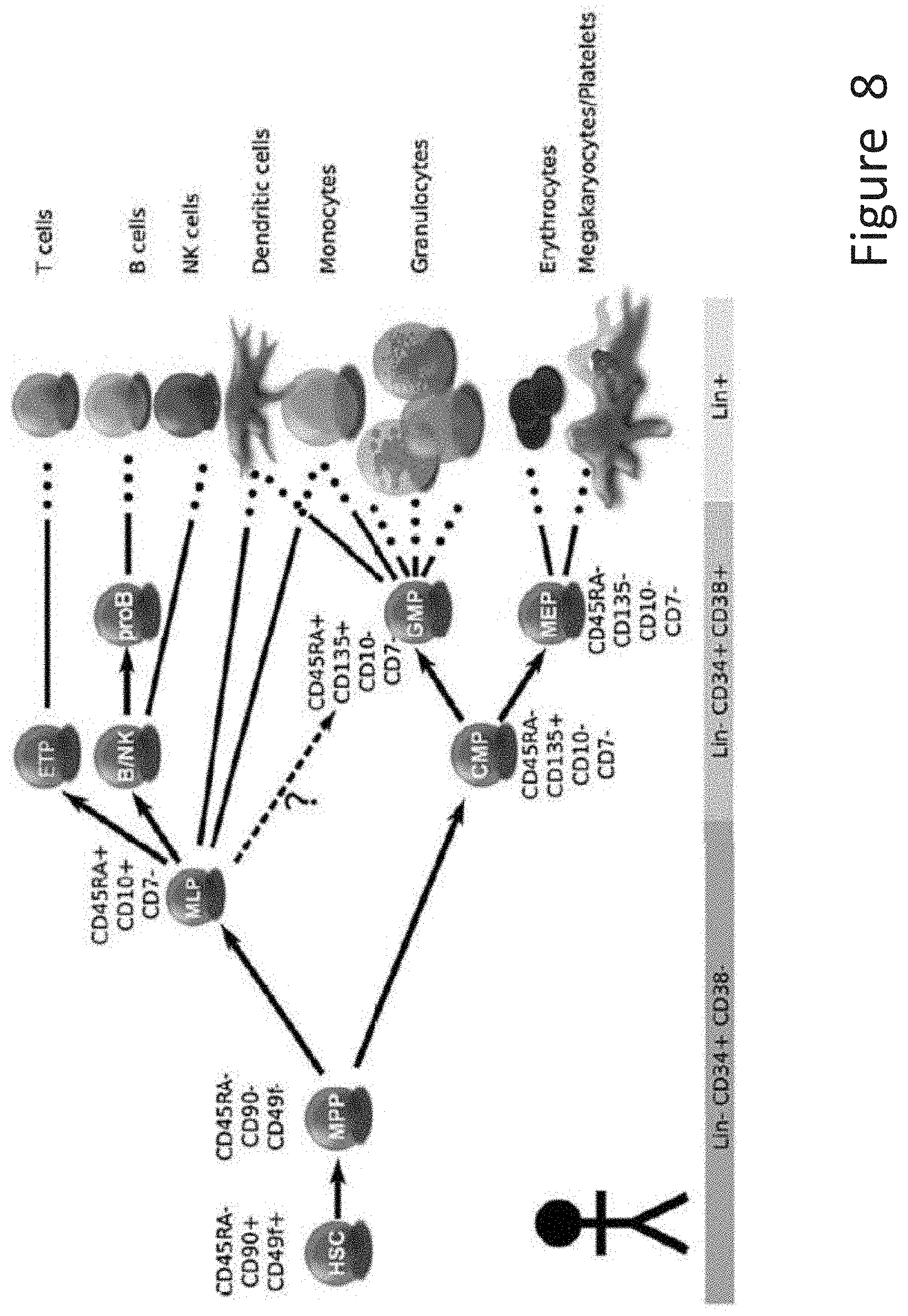
D00010

D00011

D00012
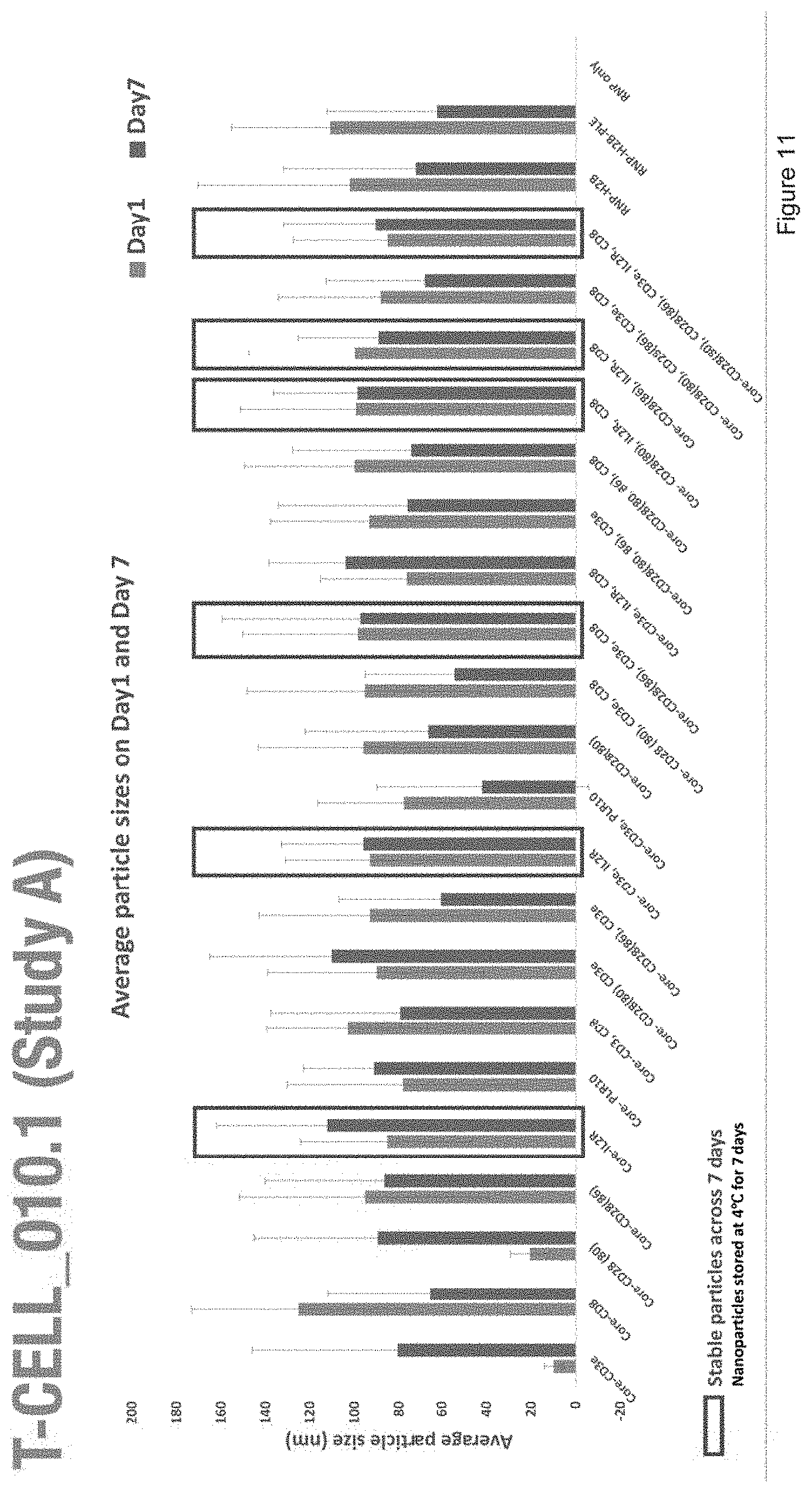
D00013
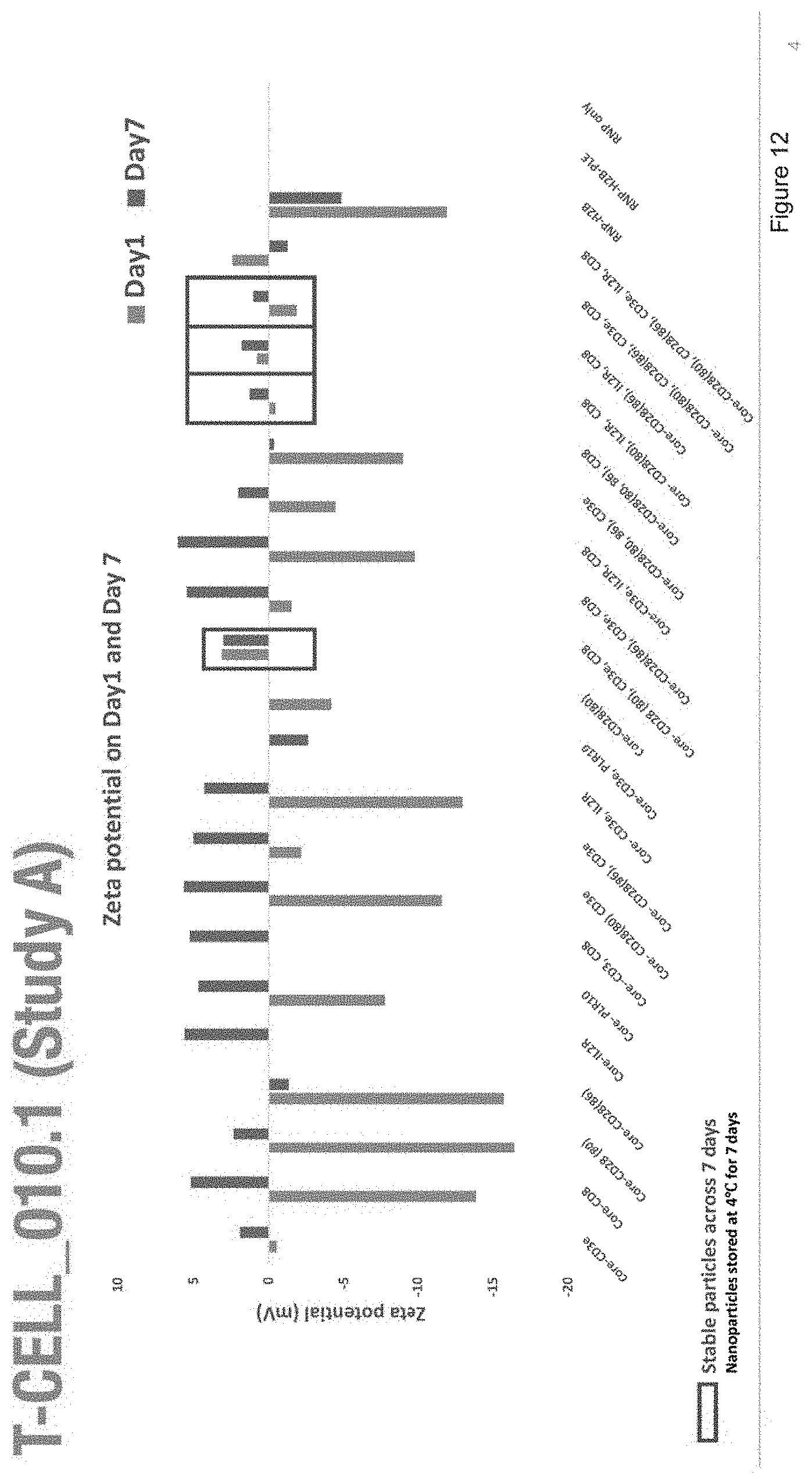
D00014
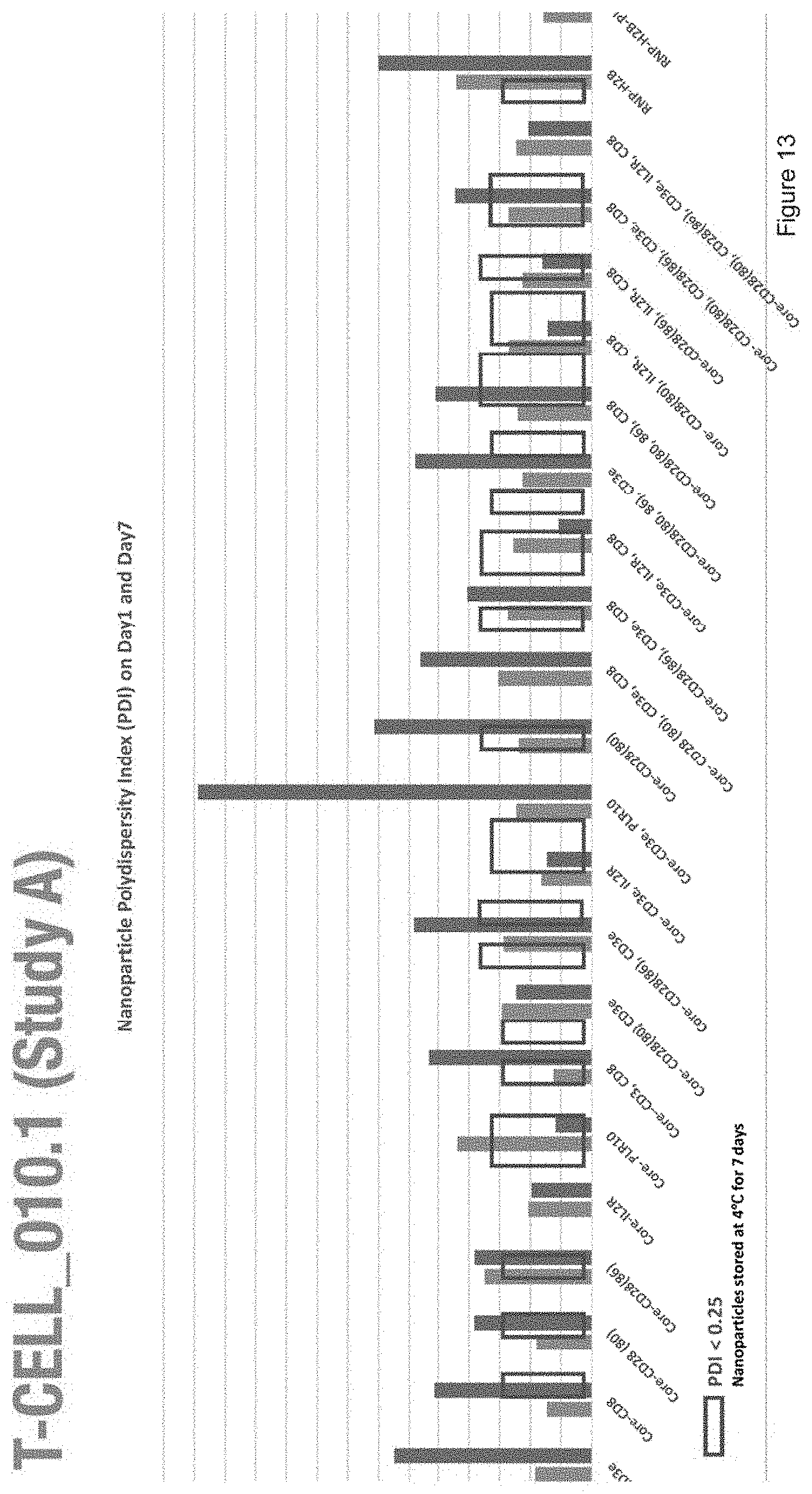
D00015

D00016
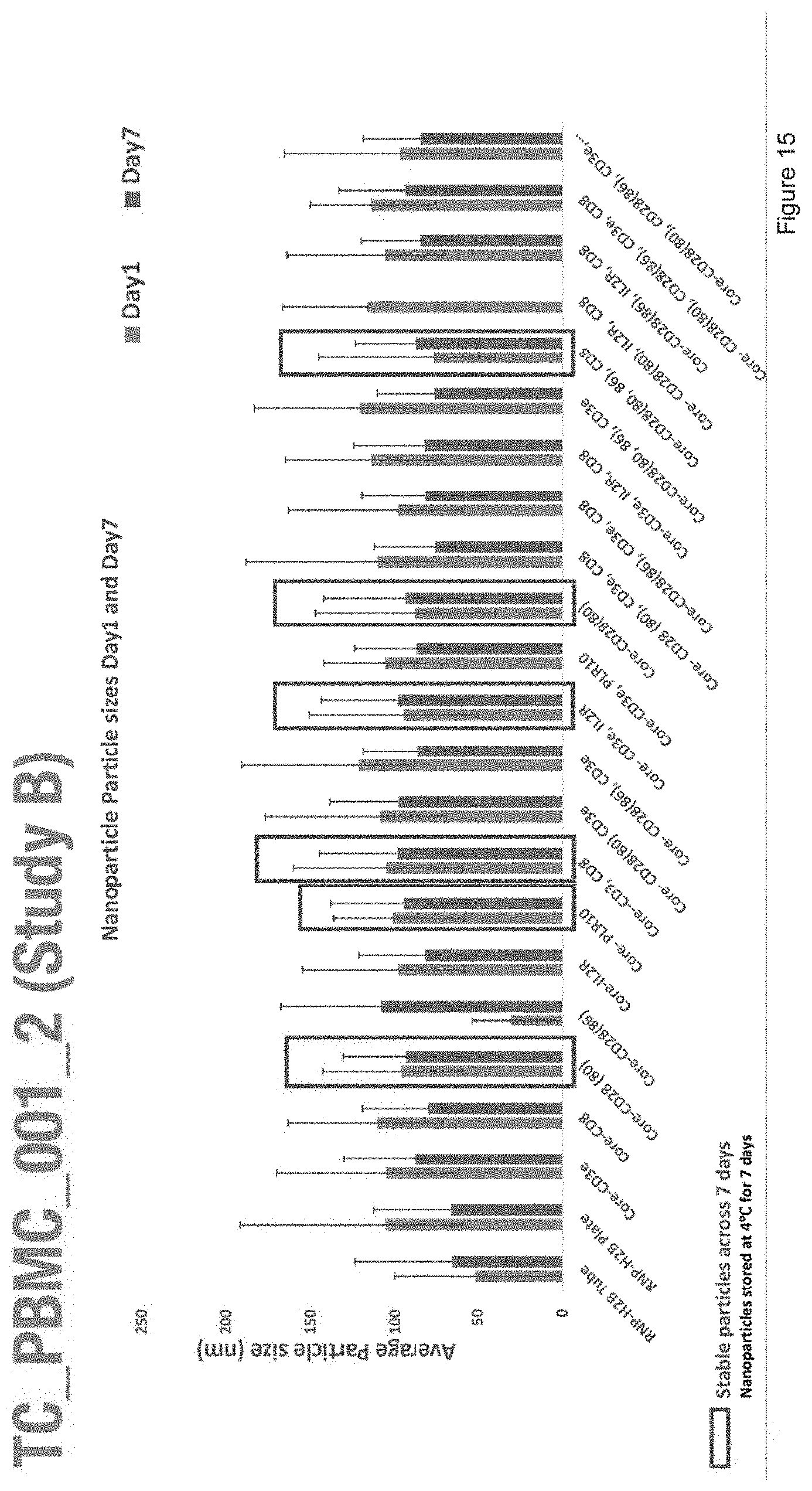
D00017
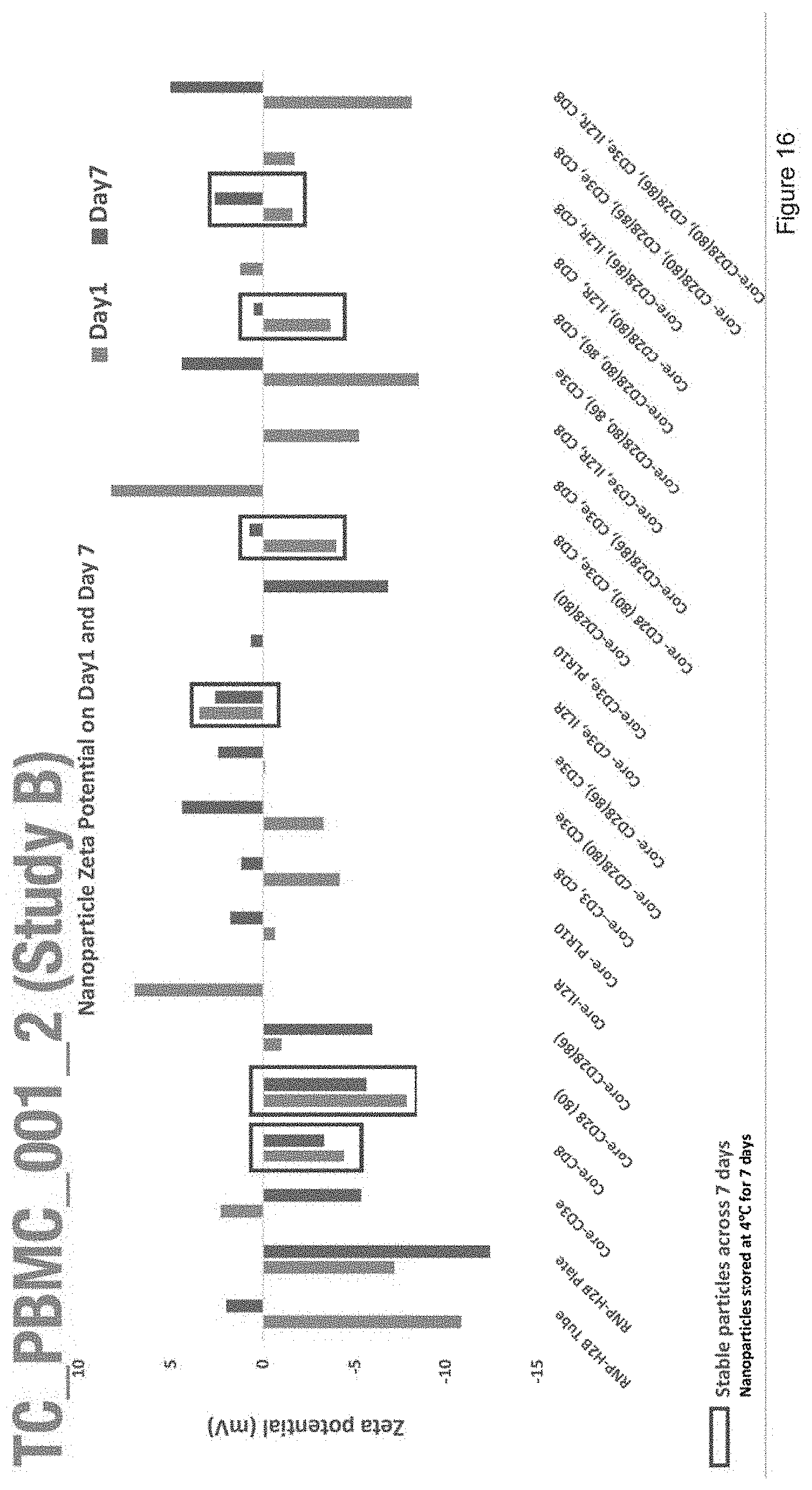
D00018
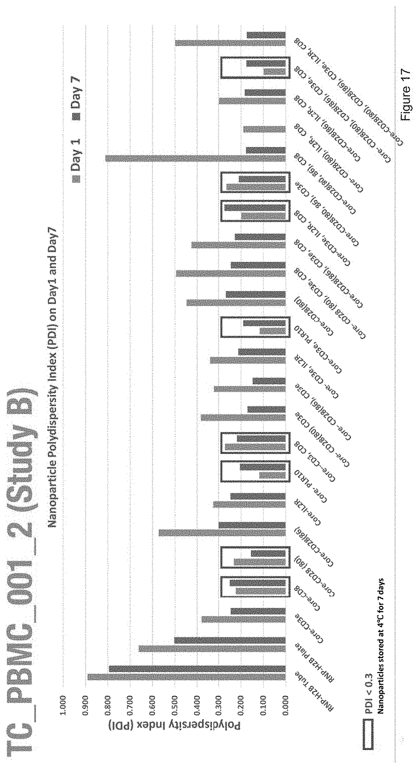
D00019
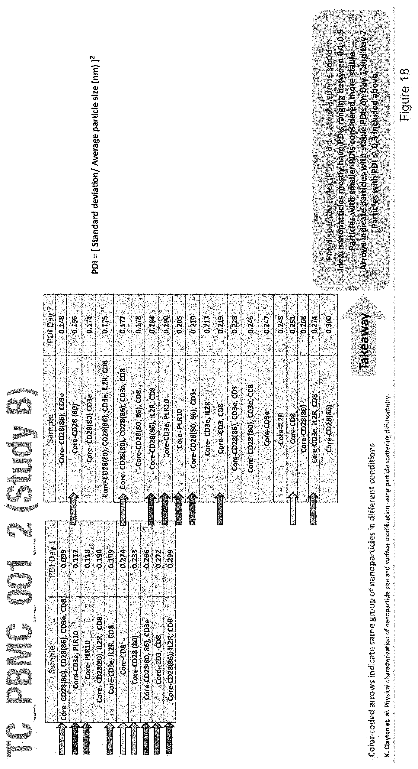
D00020
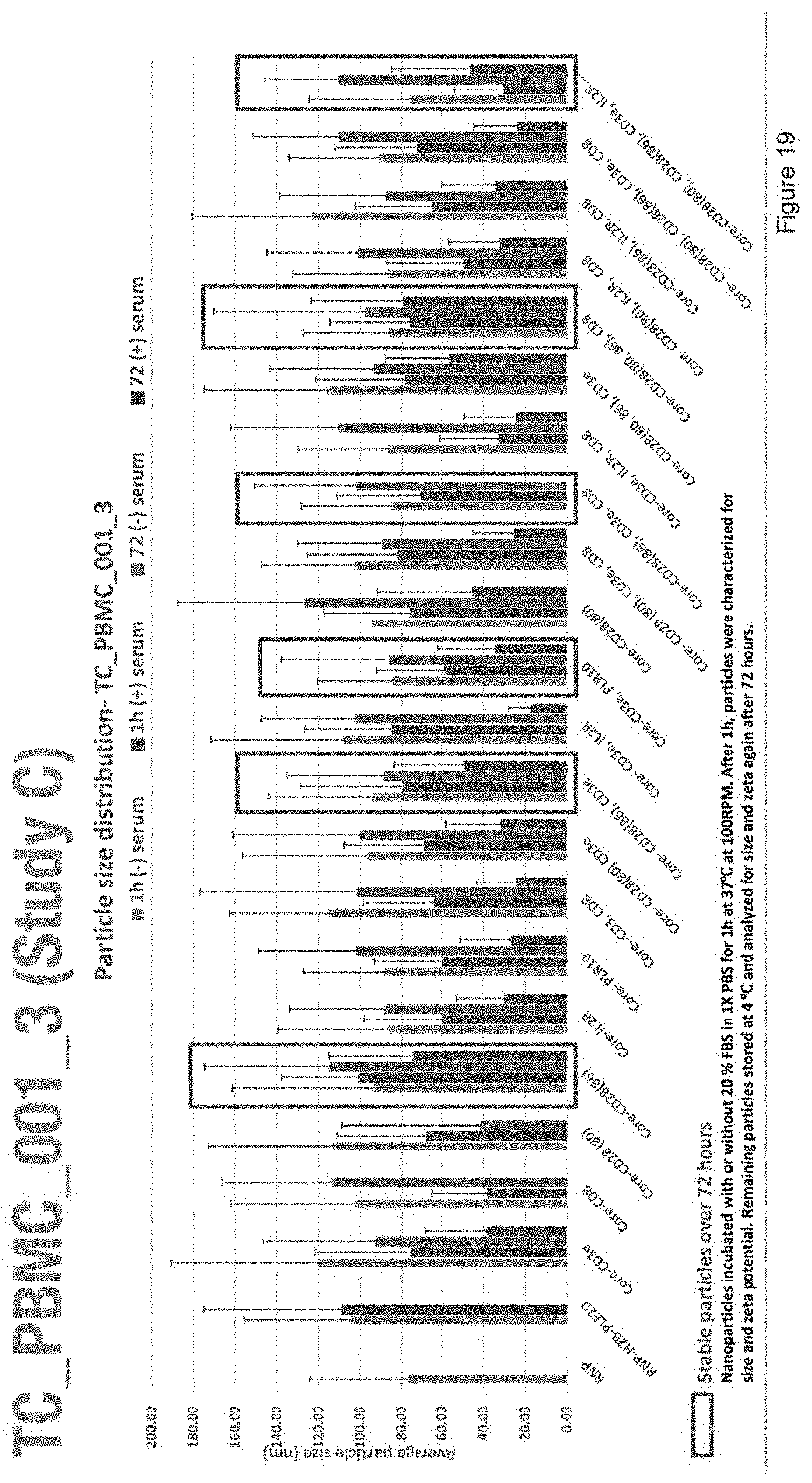
D00021

D00022
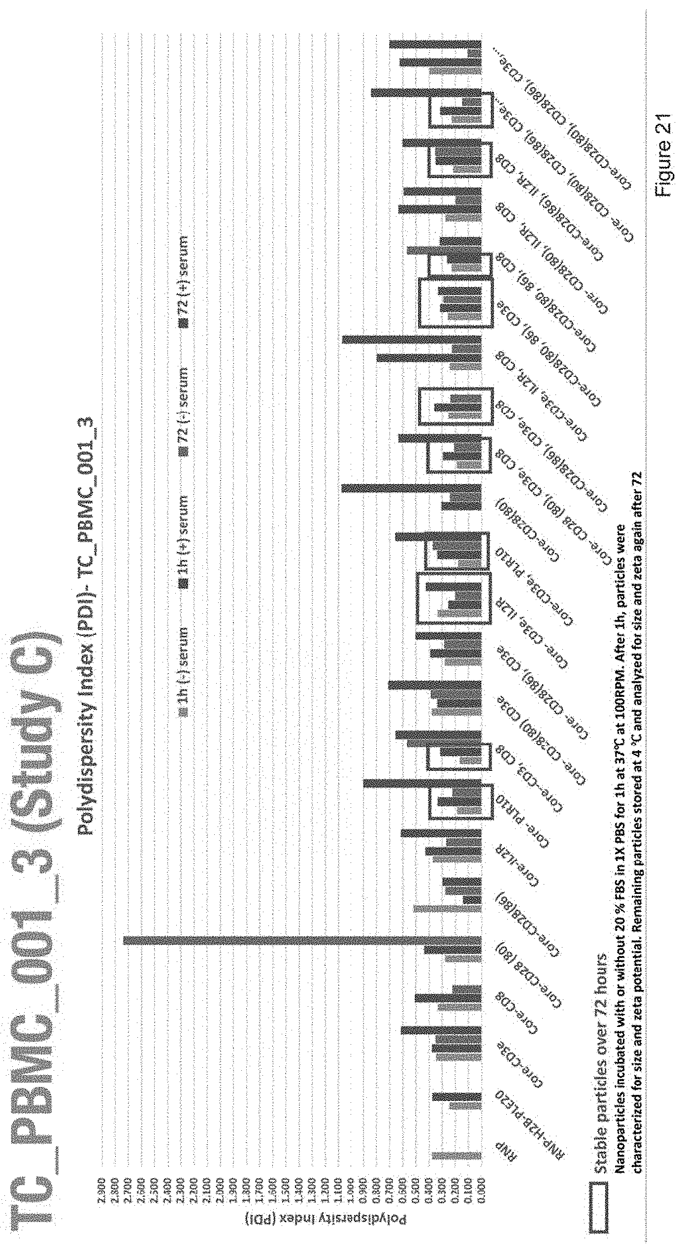
D00023

D00024
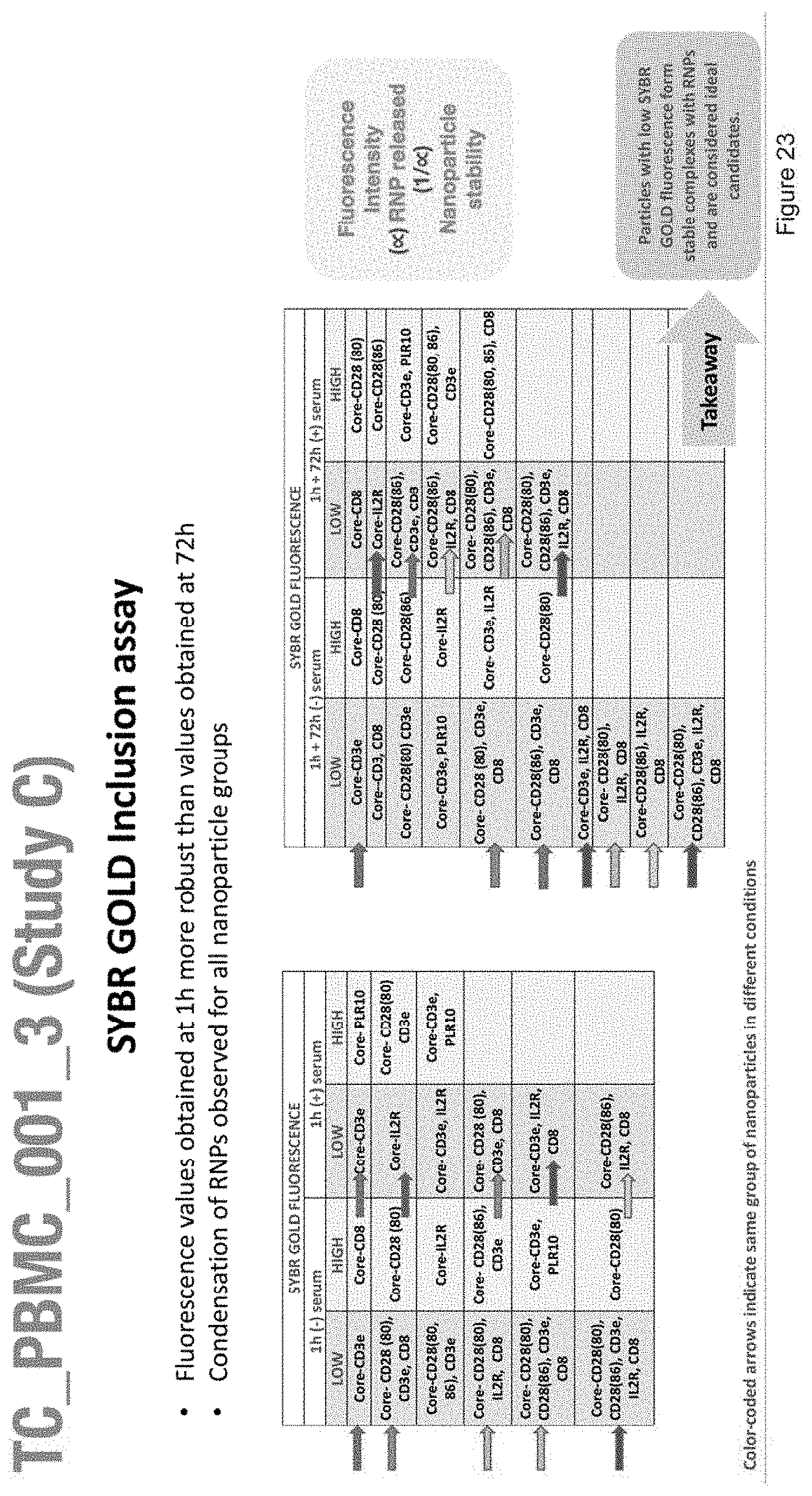
D00025
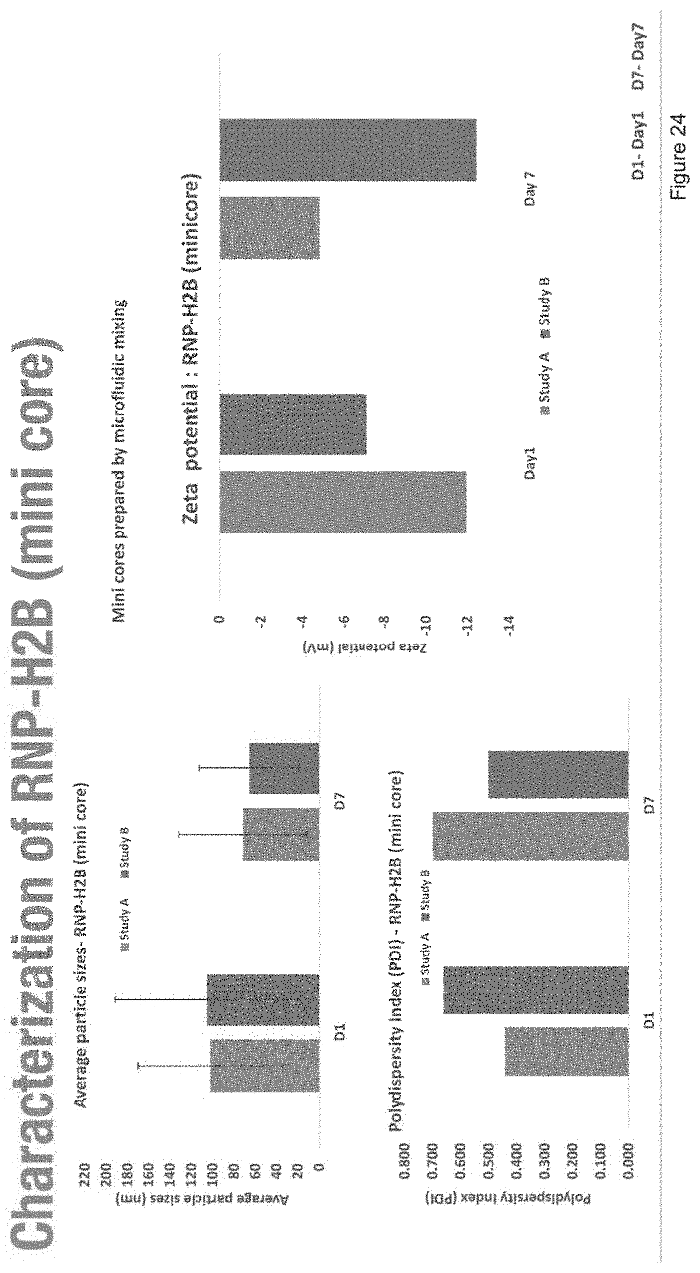
D00026

D00027

D00028
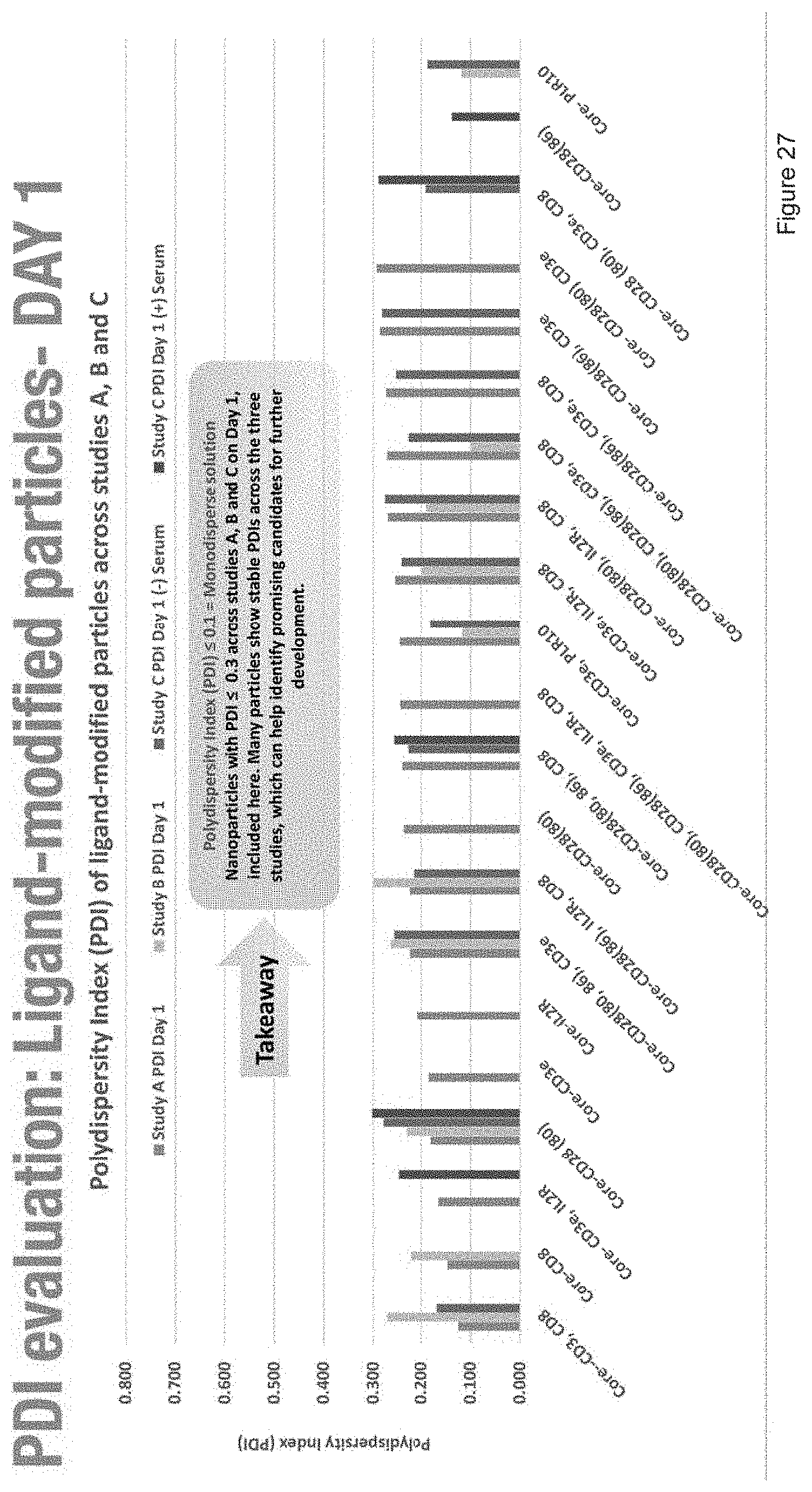
D00029
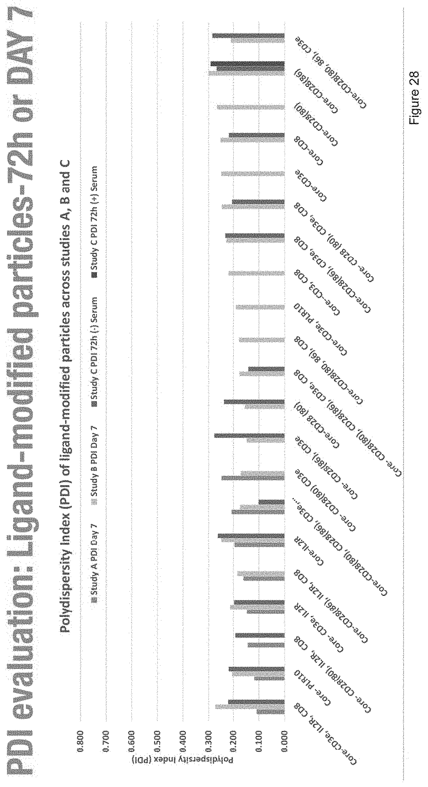
D00030

D00031
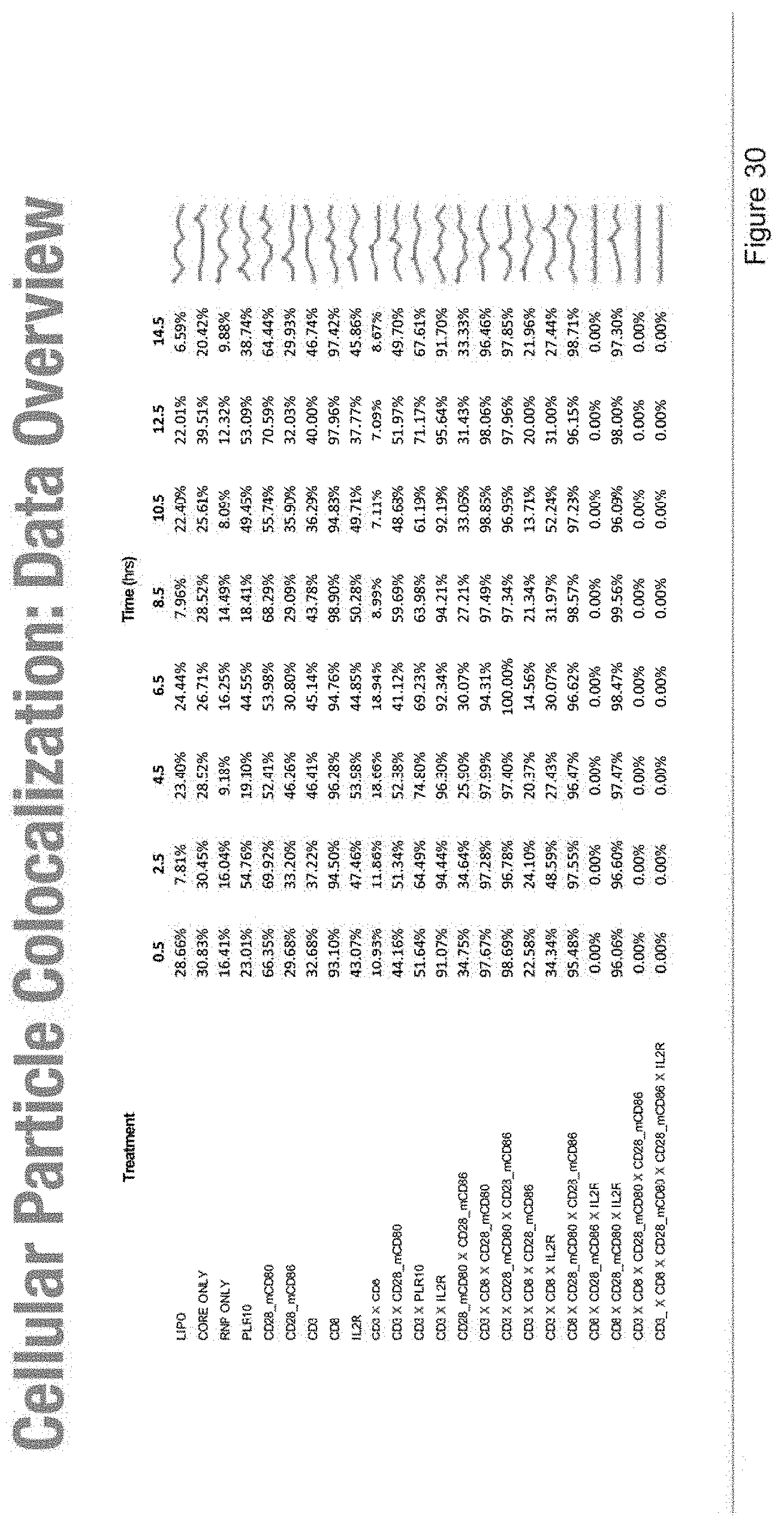
D00032

D00033
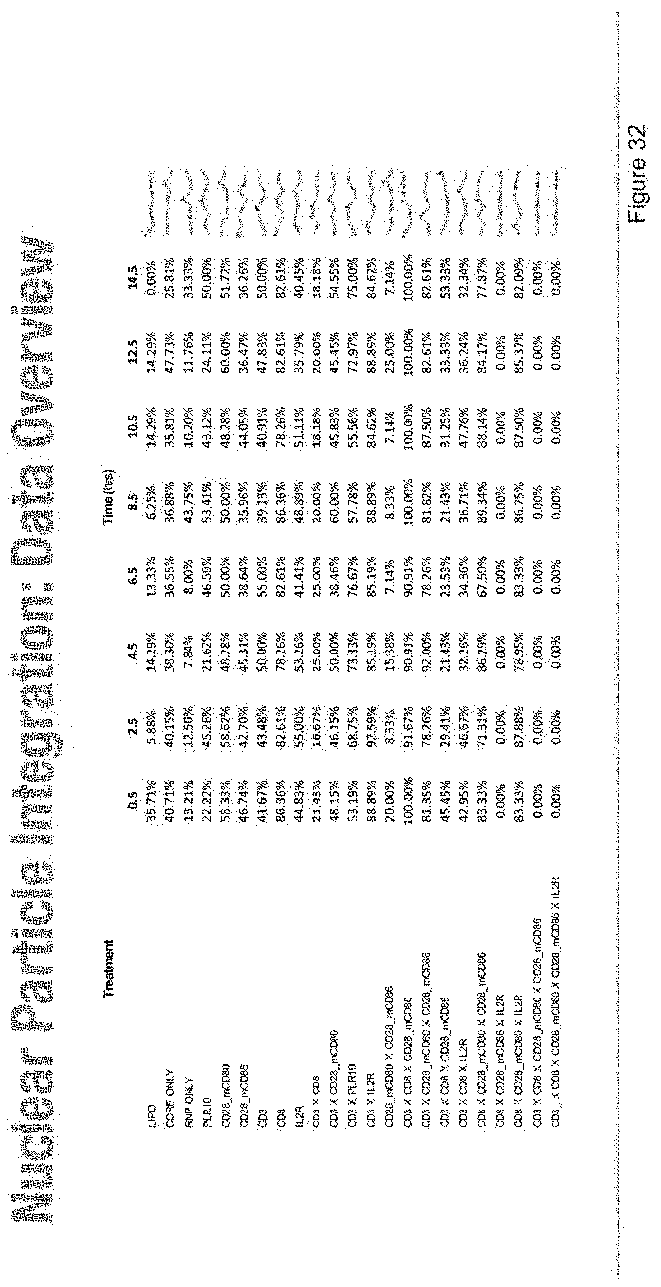
D00034

D00035
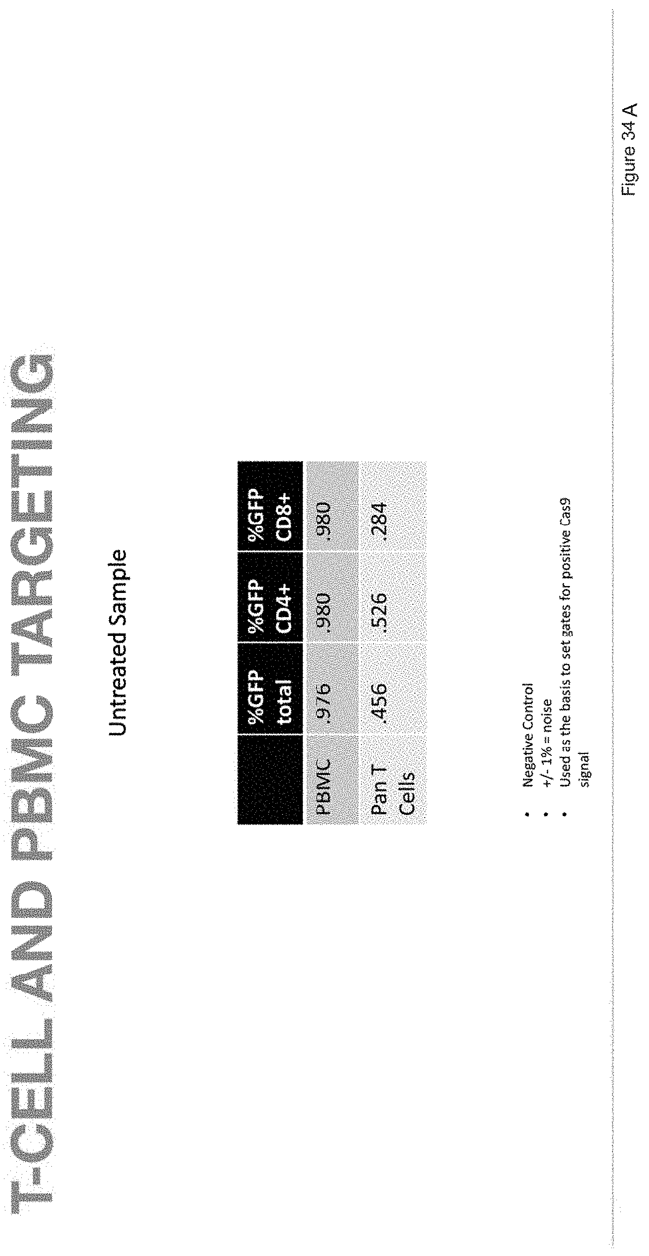
D00036
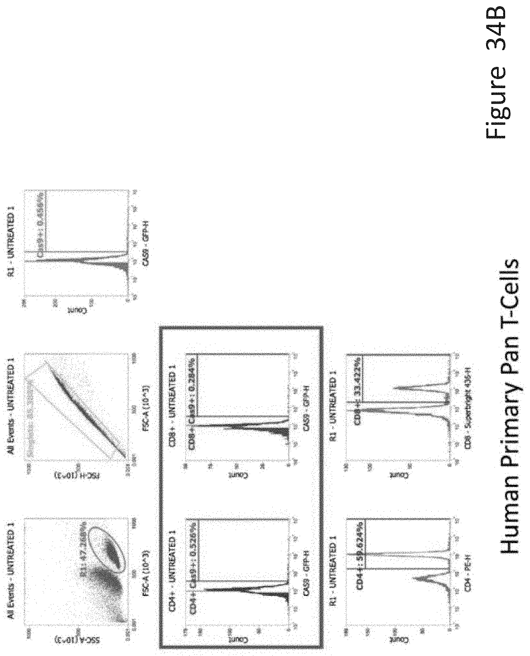
D00037

D00038

D00039

D00040
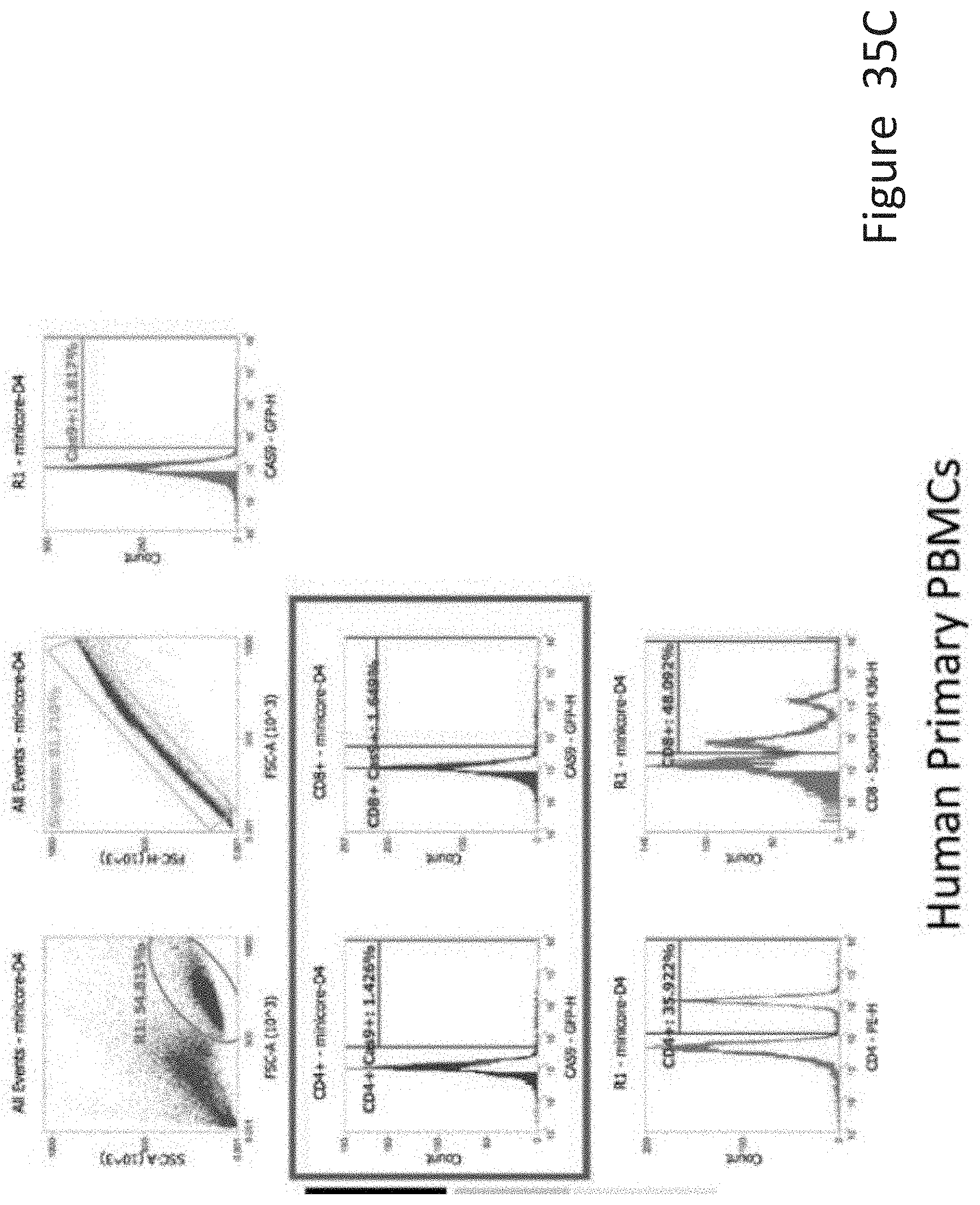
D00041
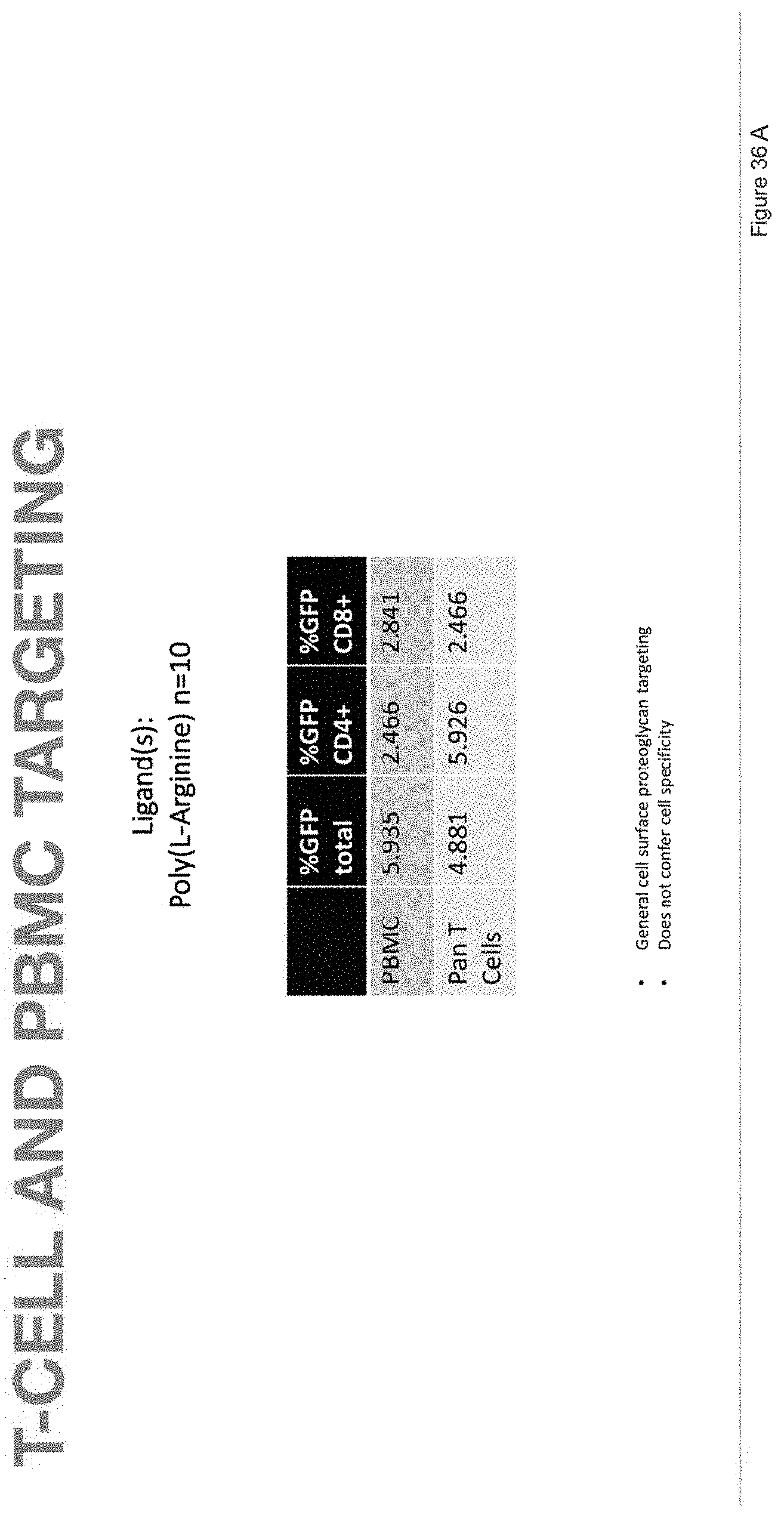
D00042
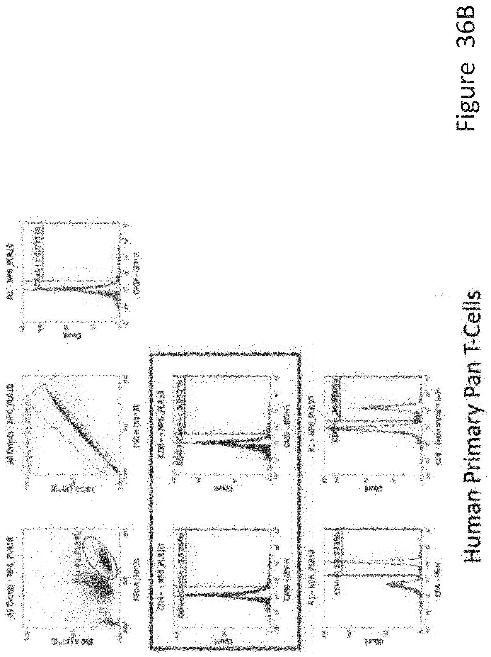
D00043
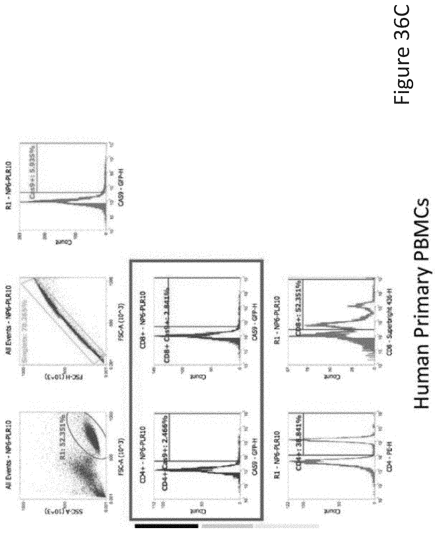
D00044

D00045
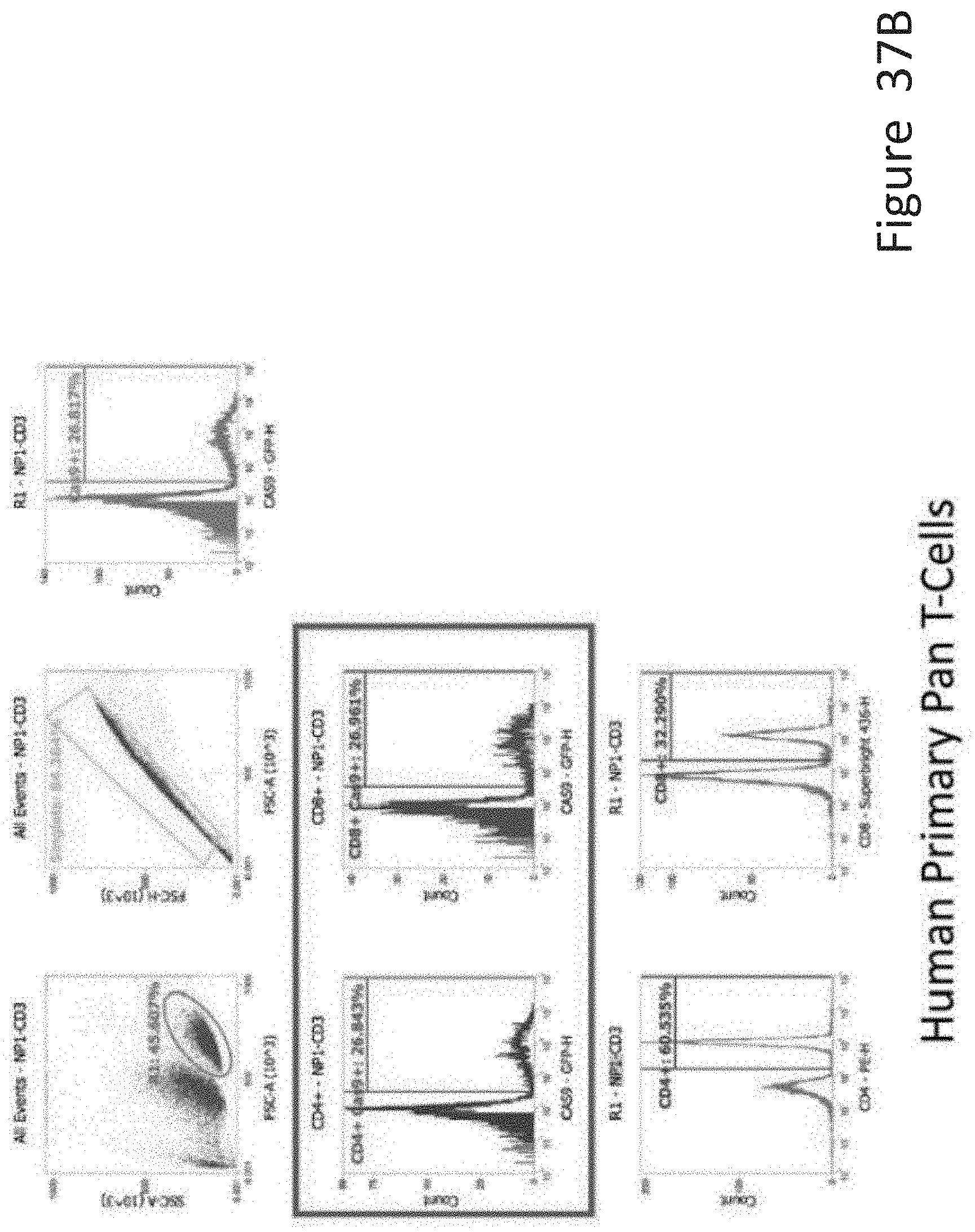
D00046
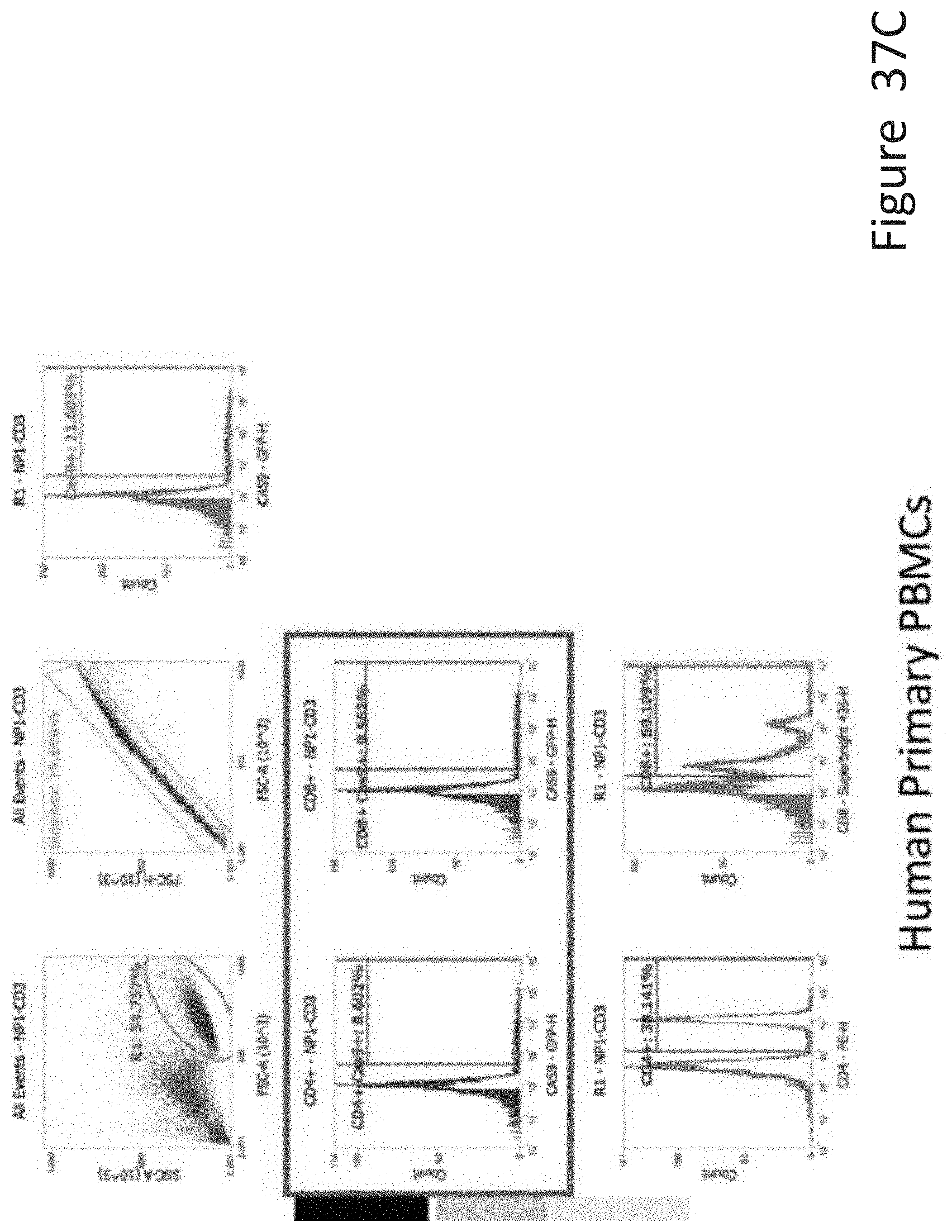
D00047
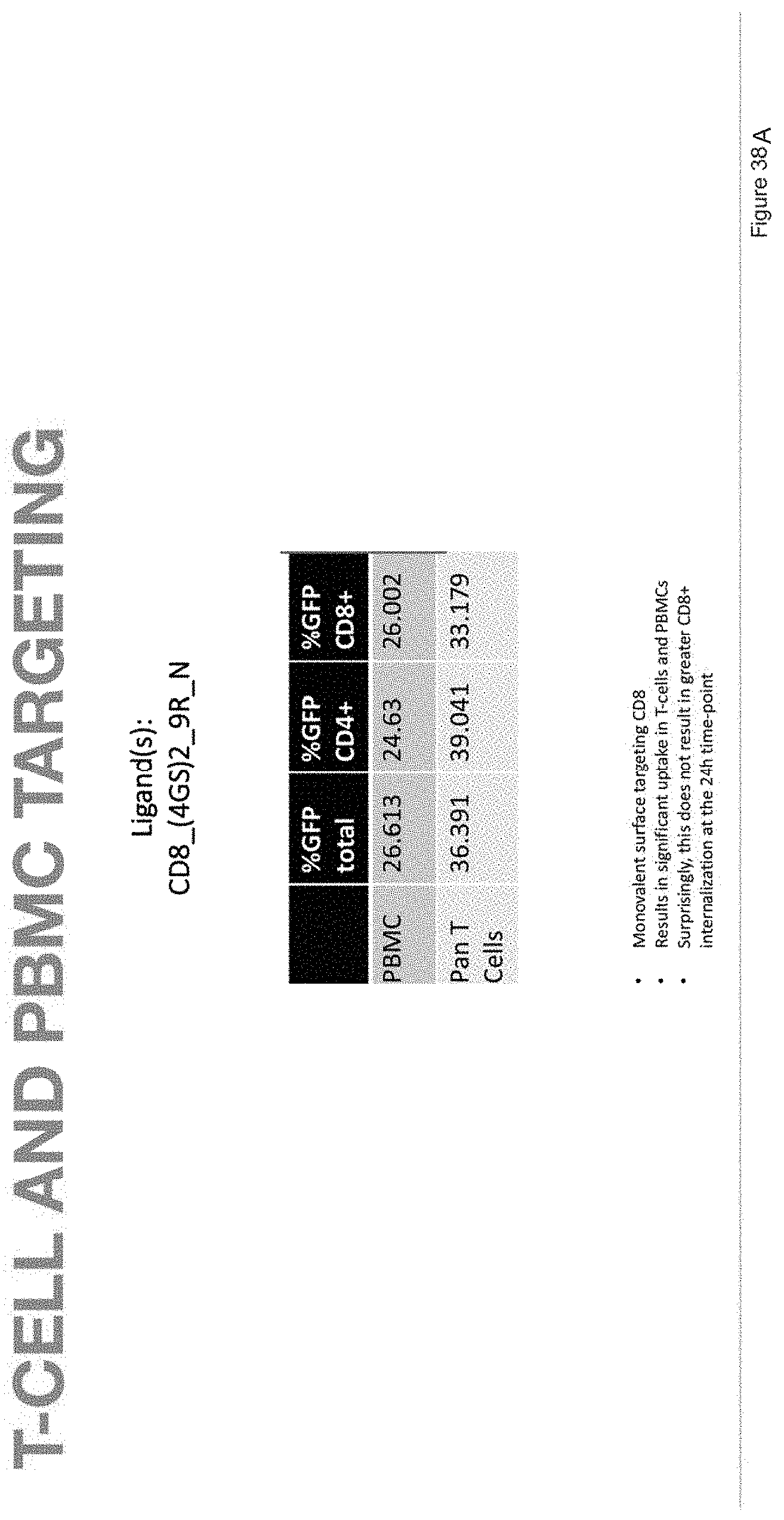
D00048

D00049
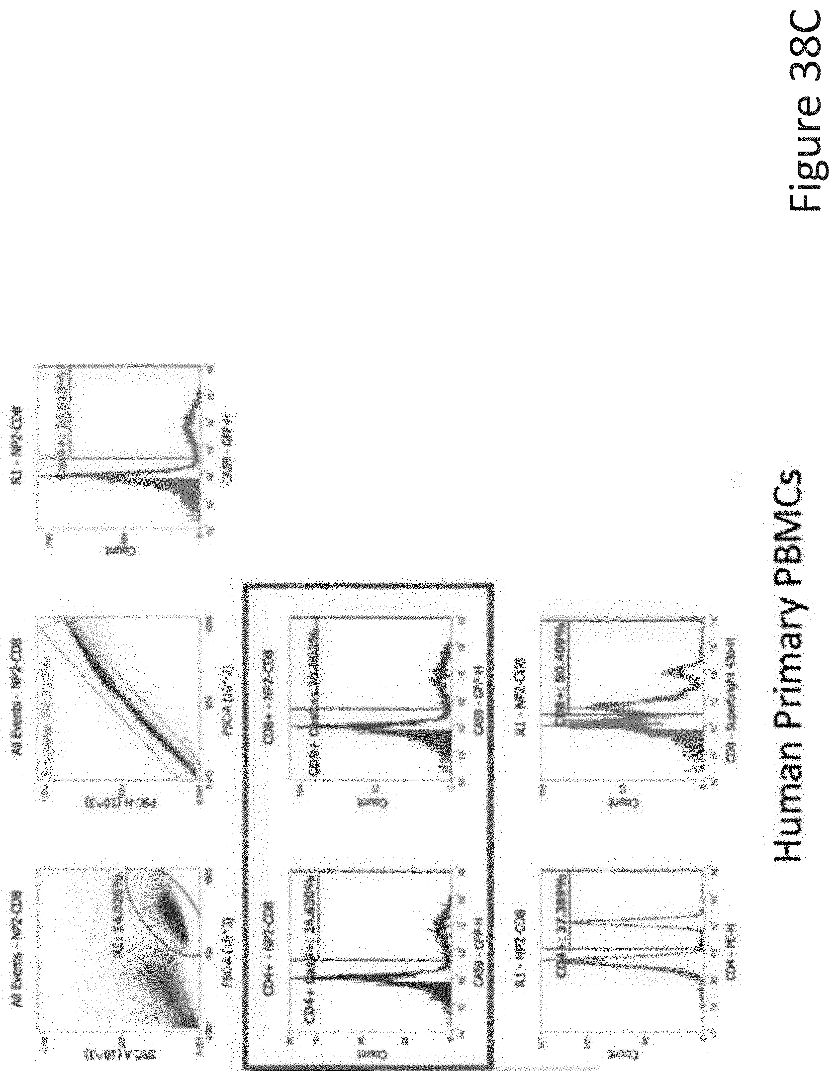
D00050

D00051
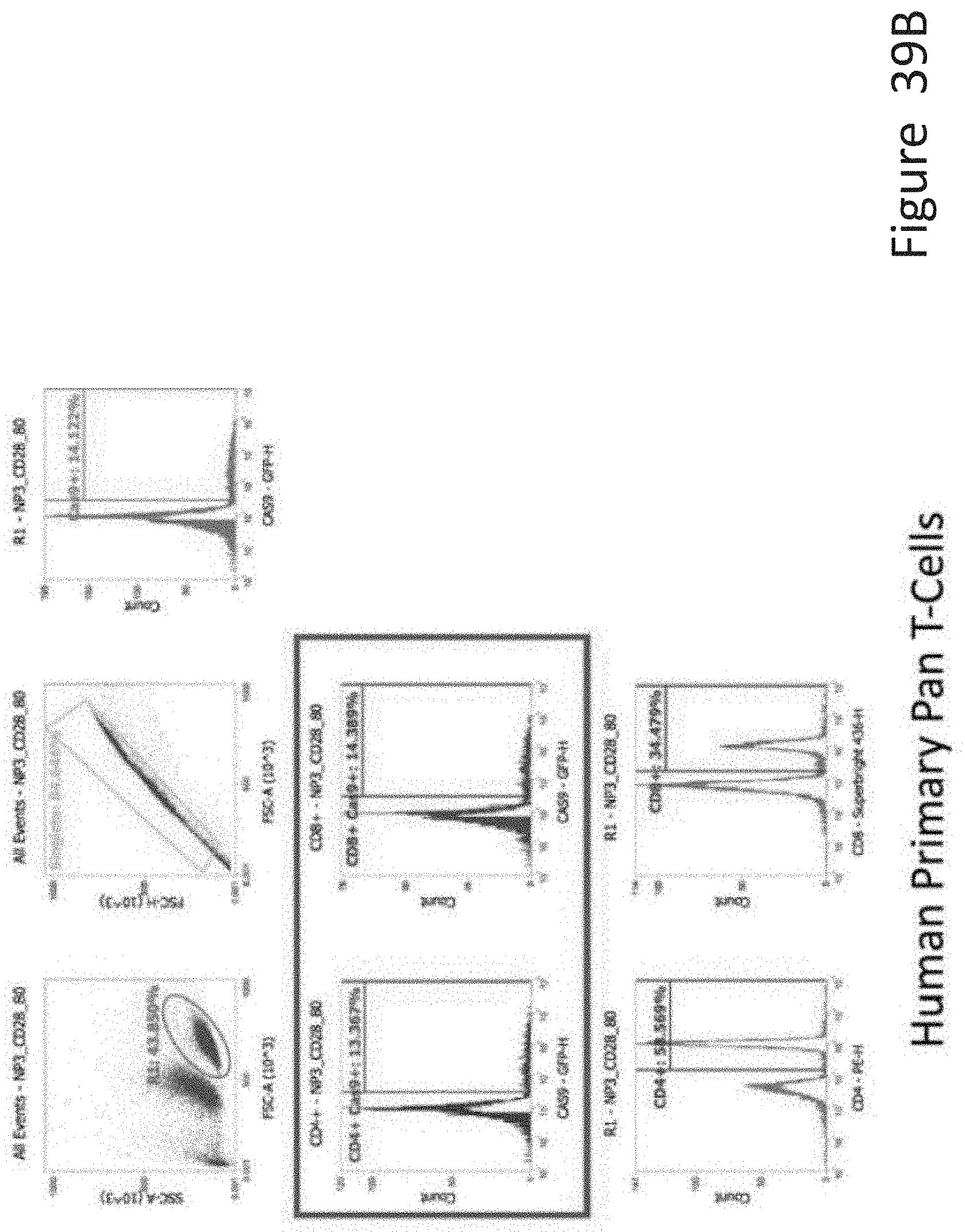
D00052

D00053

D00054

D00055

D00056

D00057
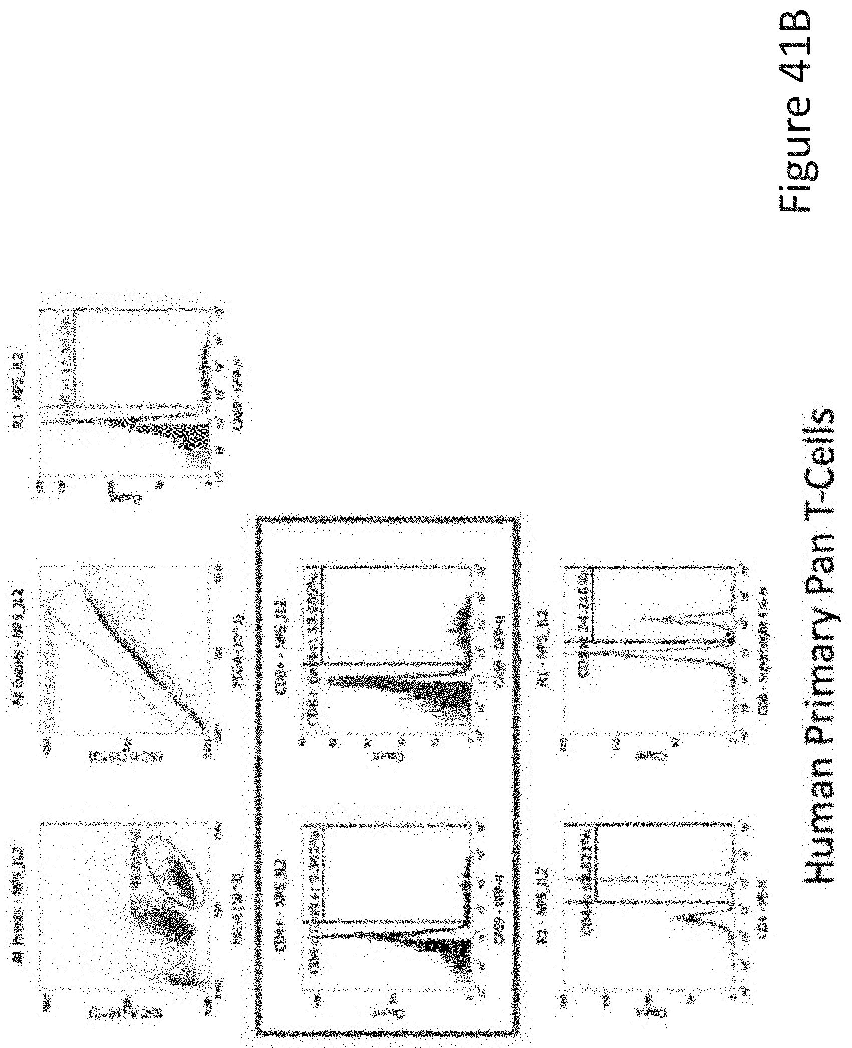
D00058
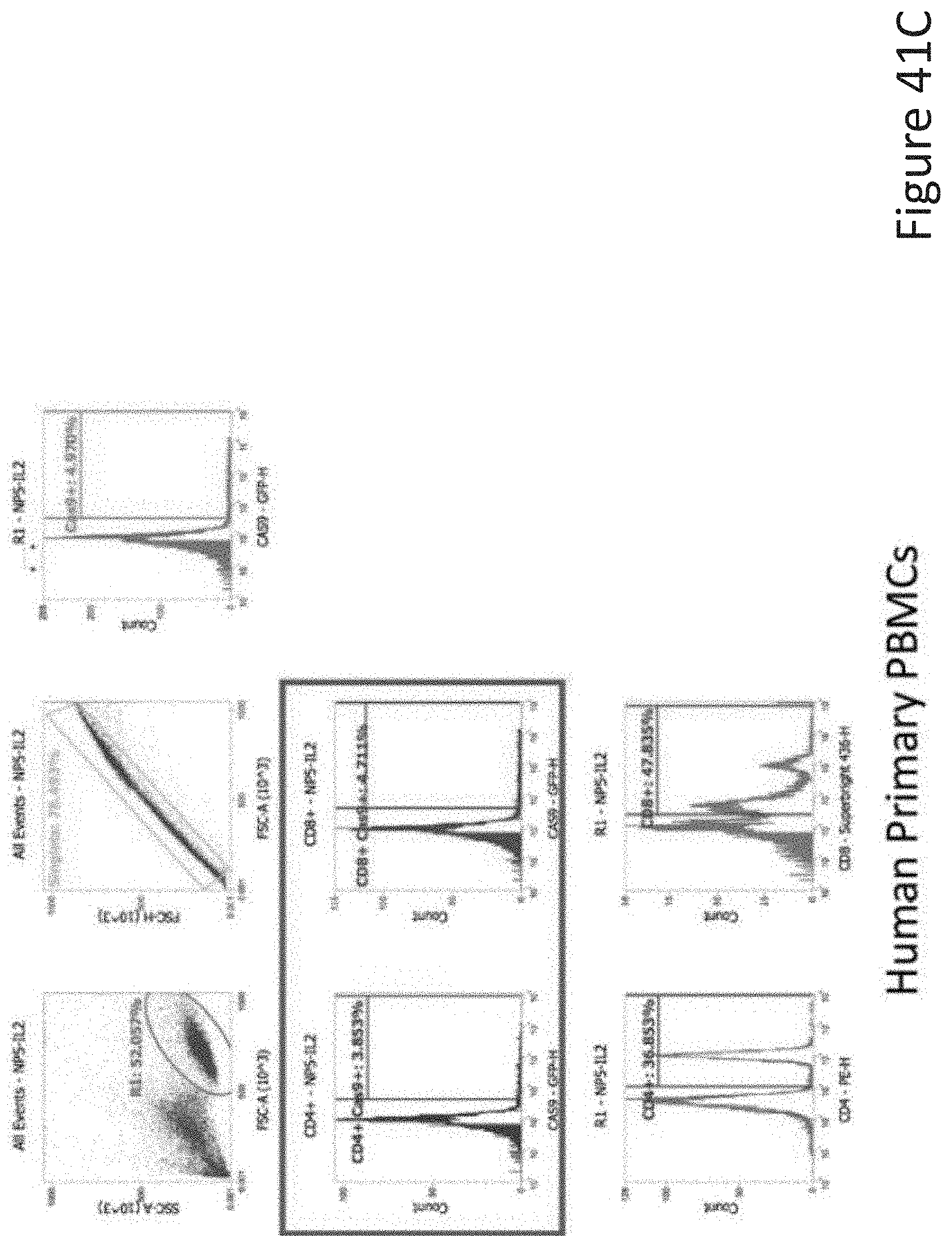
D00059
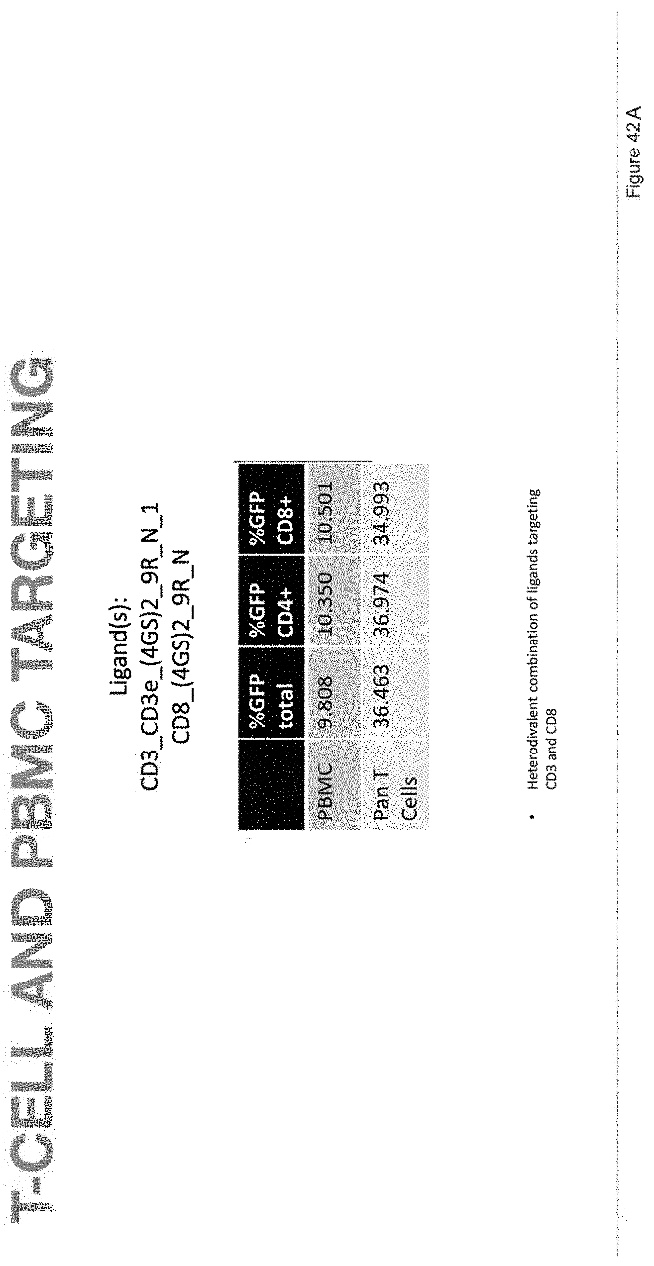
D00060

D00061

D00062
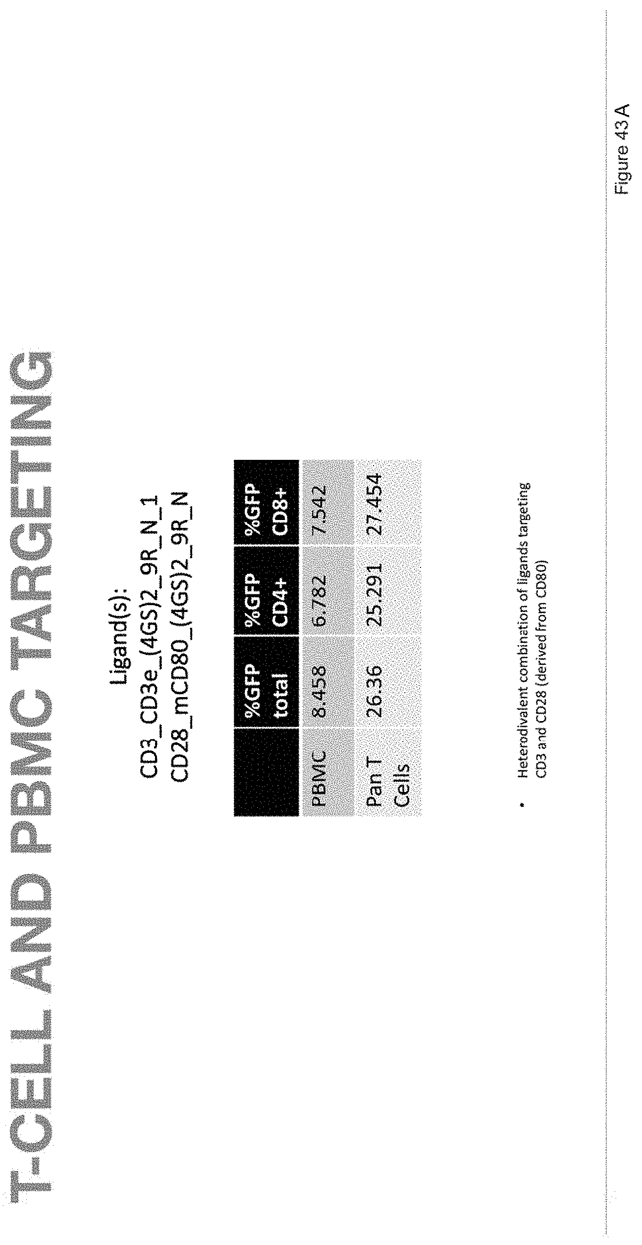
D00063

D00064
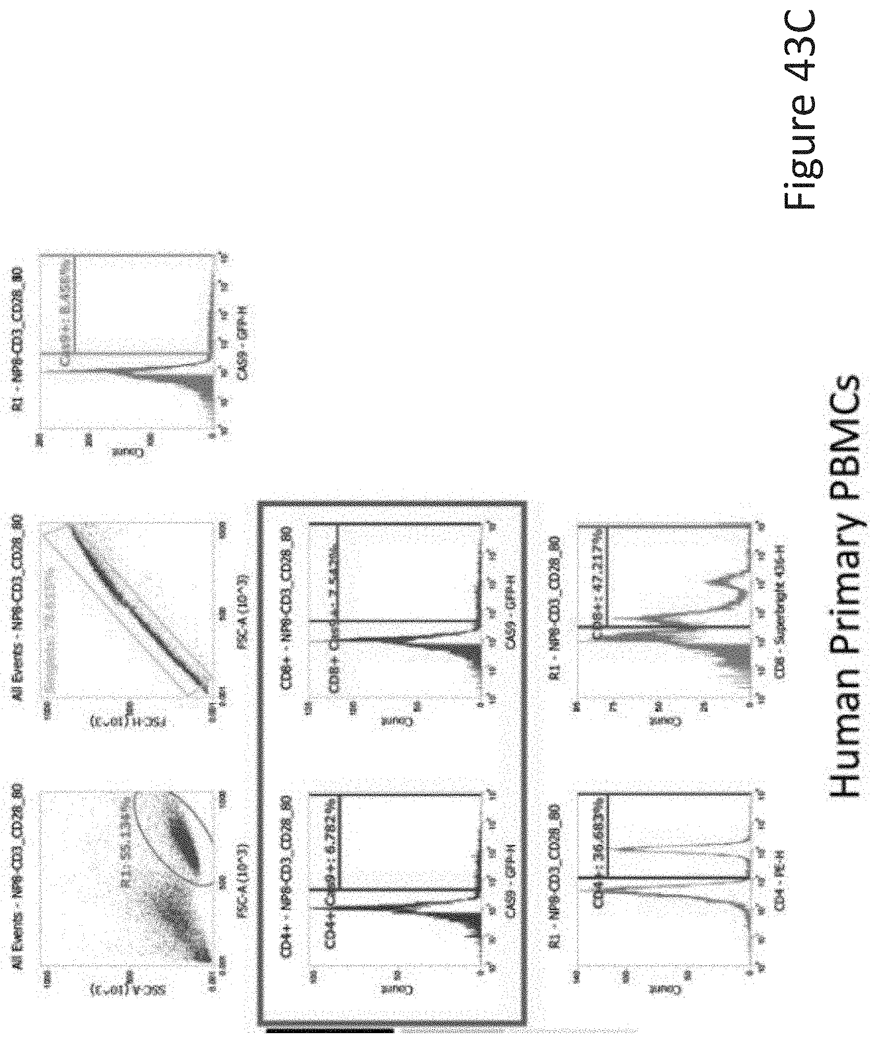
D00065

D00066

D00067
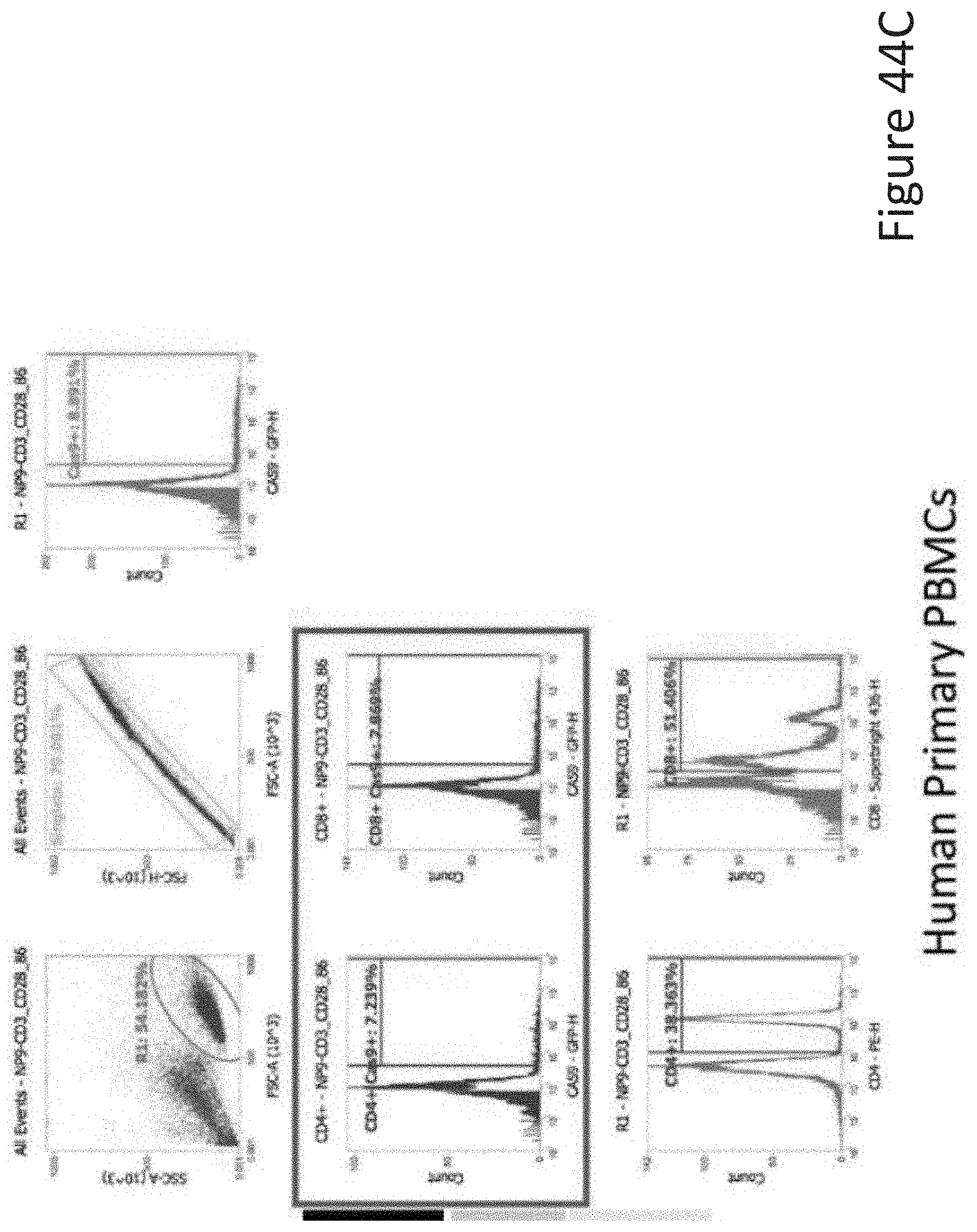
D00068

D00069
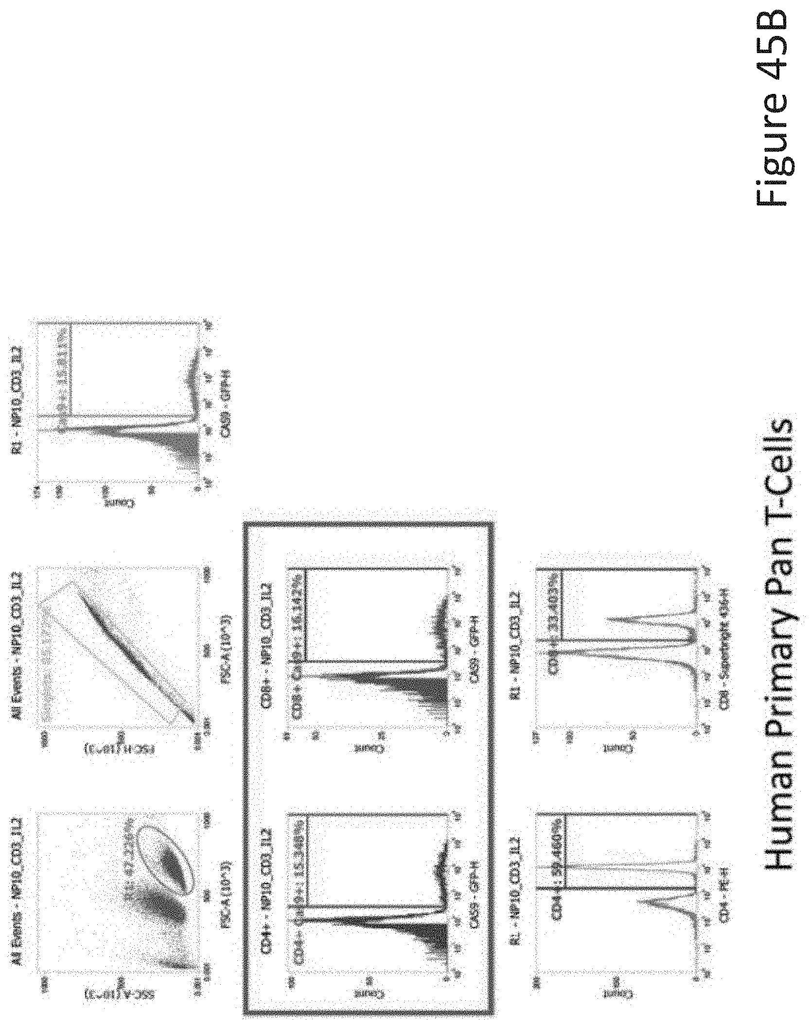
D00070

D00071

D00072
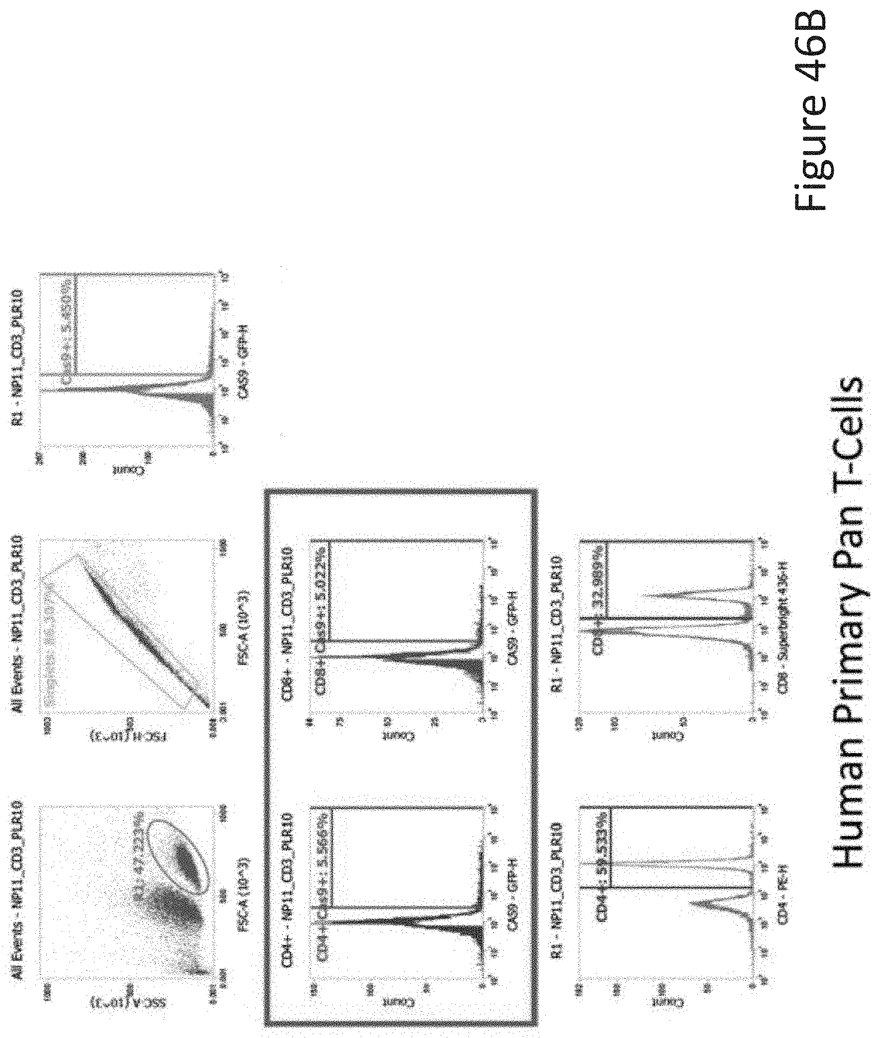
D00073
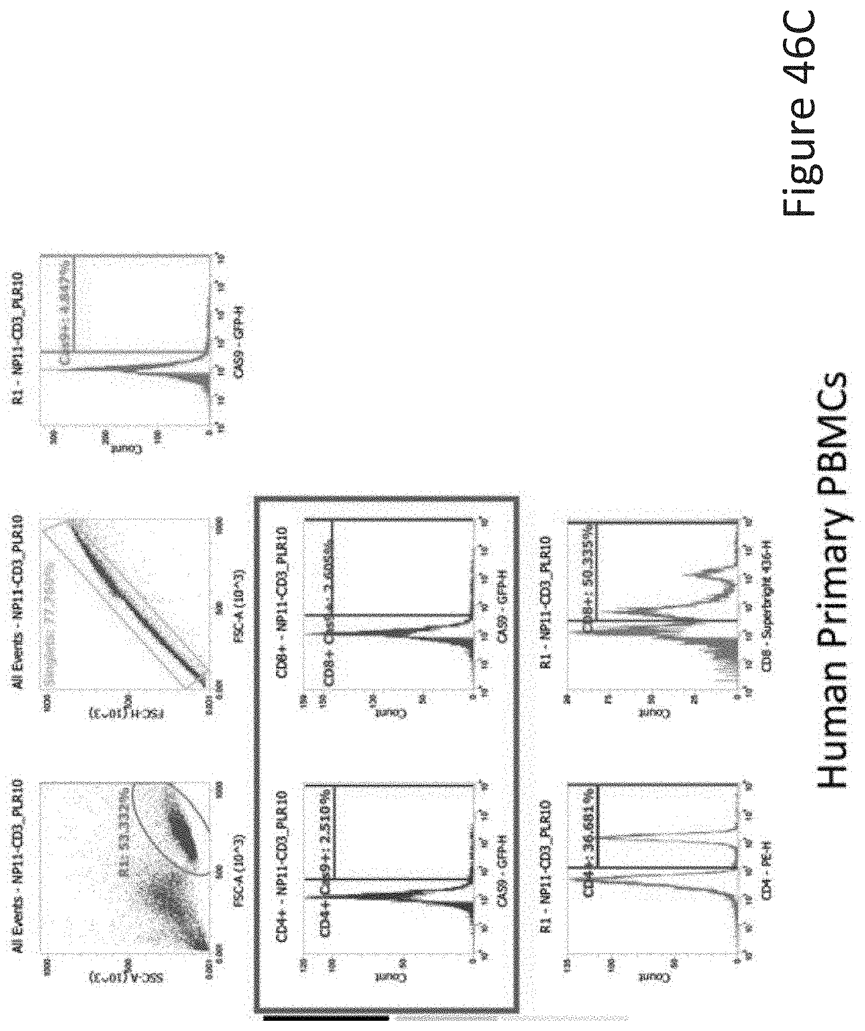
D00074

D00075
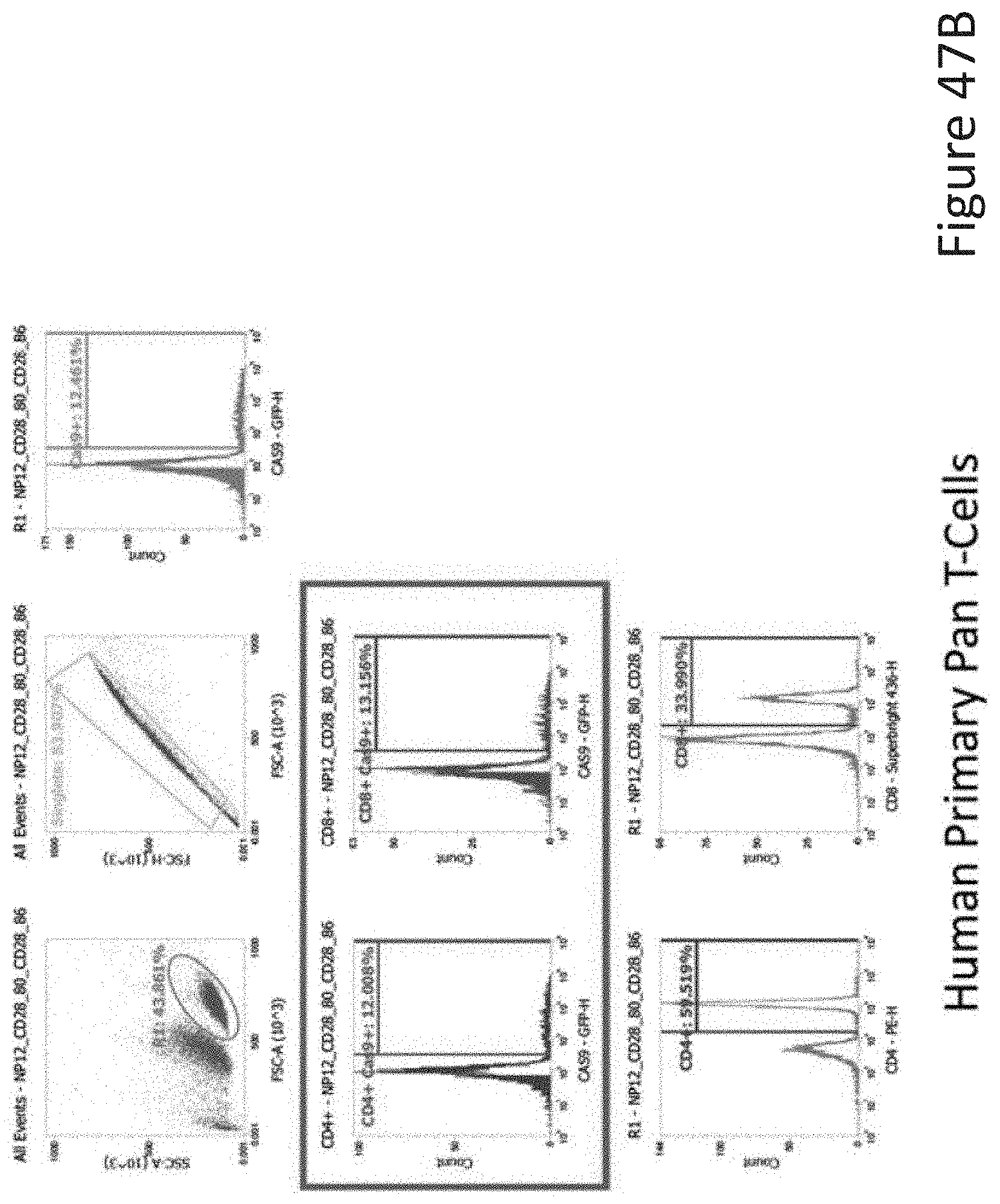
D00076
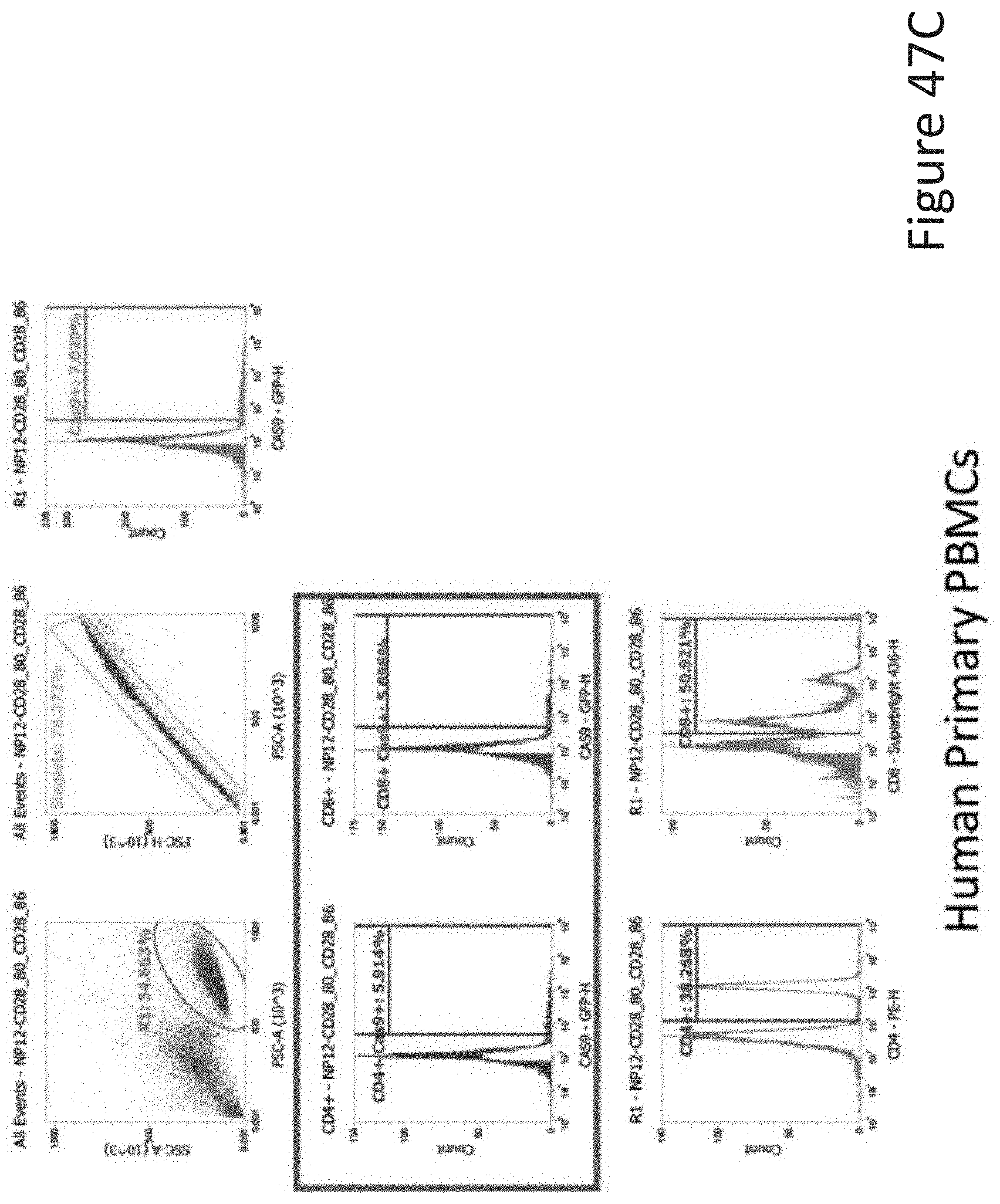
D00077
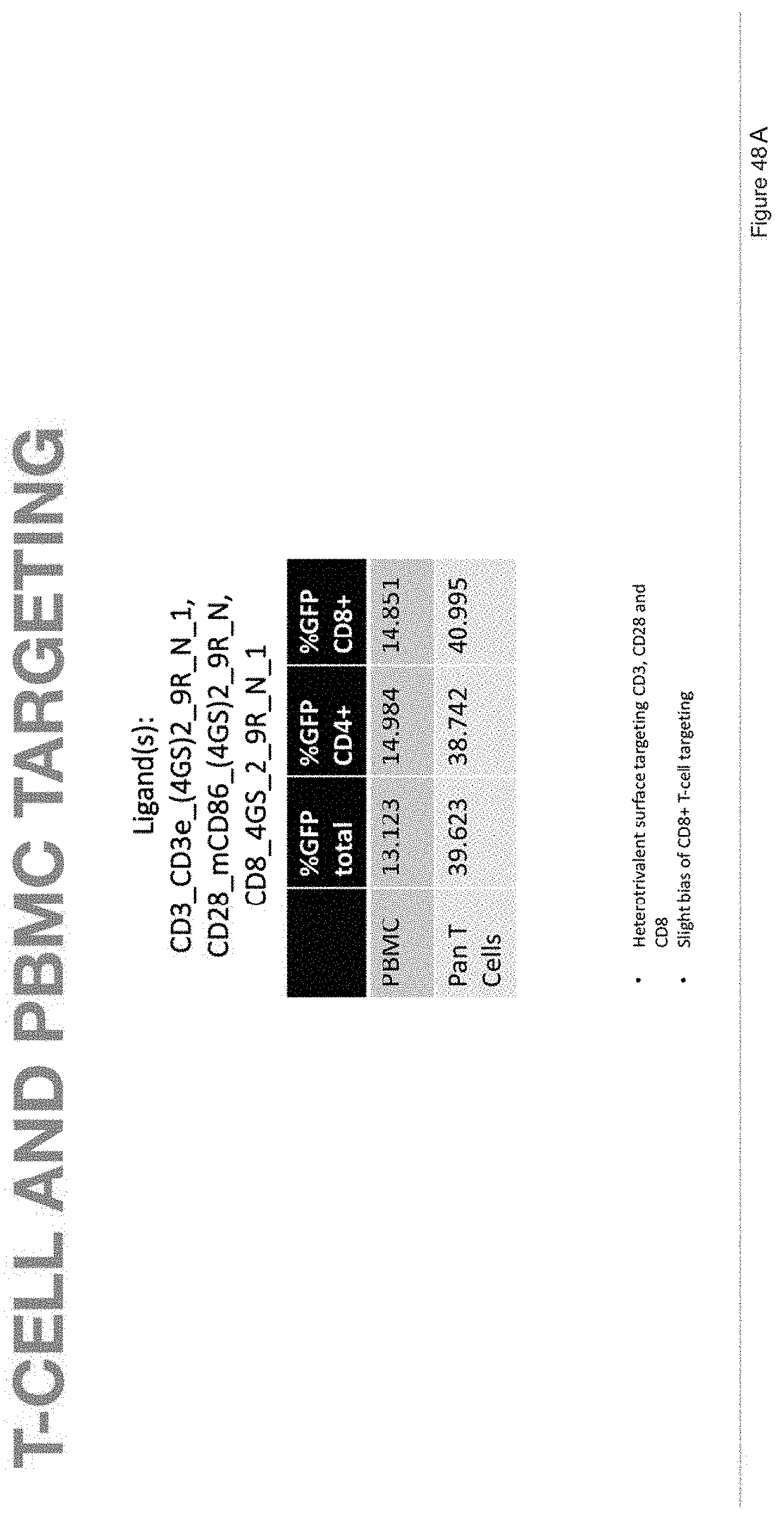
D00078

D00079

D00080
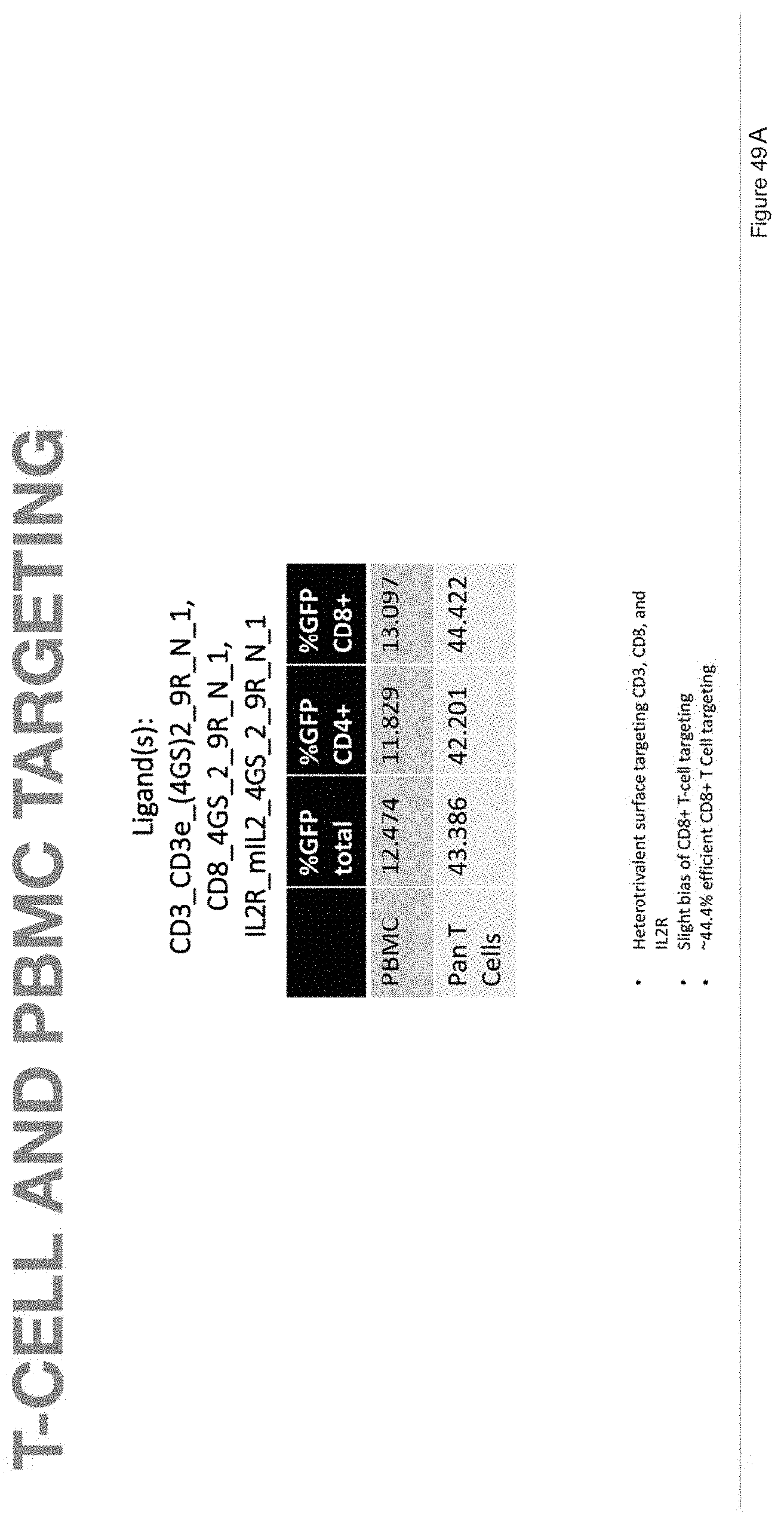
D00081

D00082

D00083
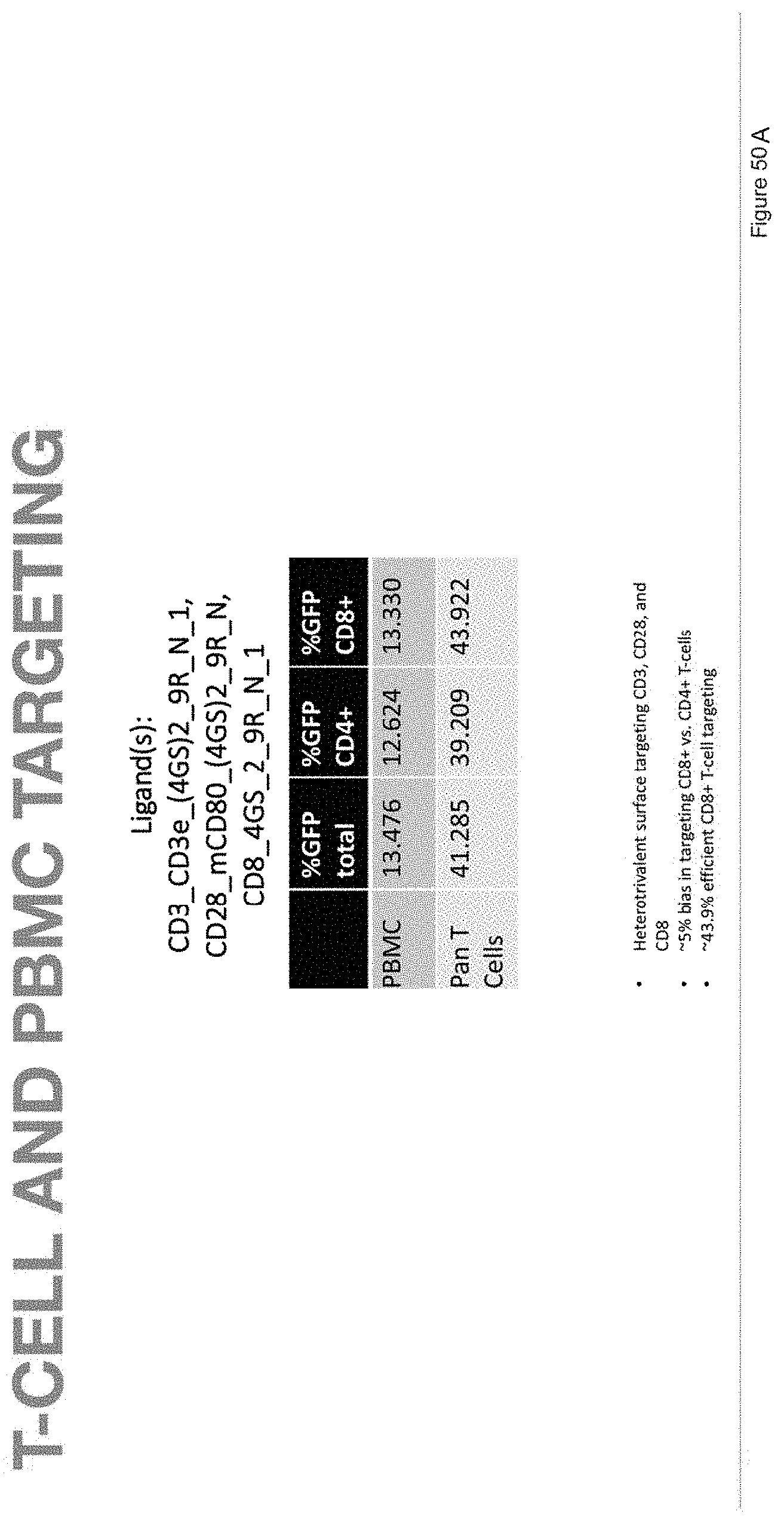
D00084

D00085
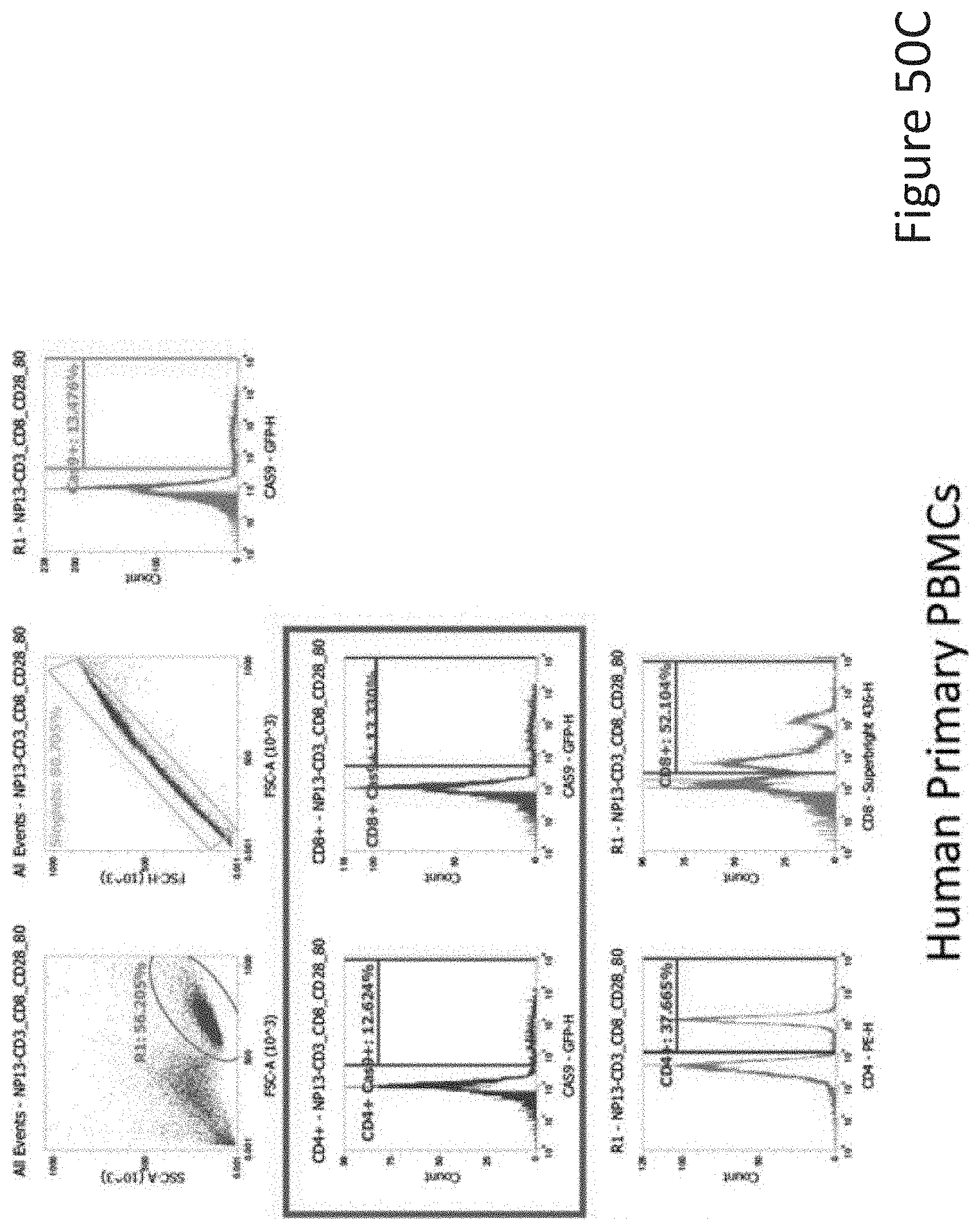
D00086

D00087
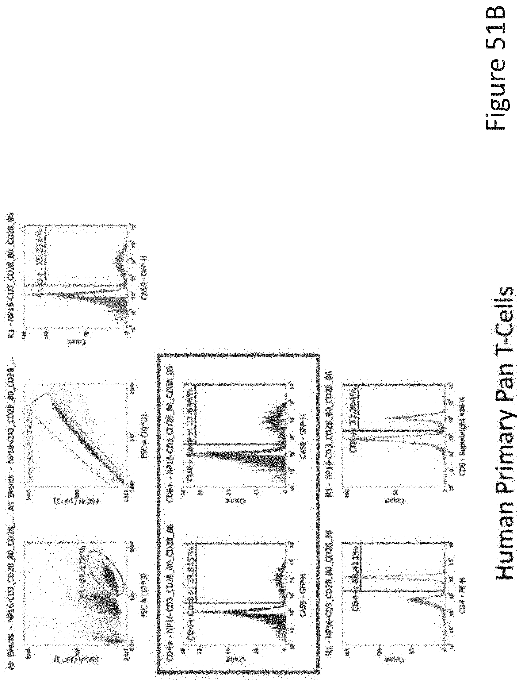
D00088
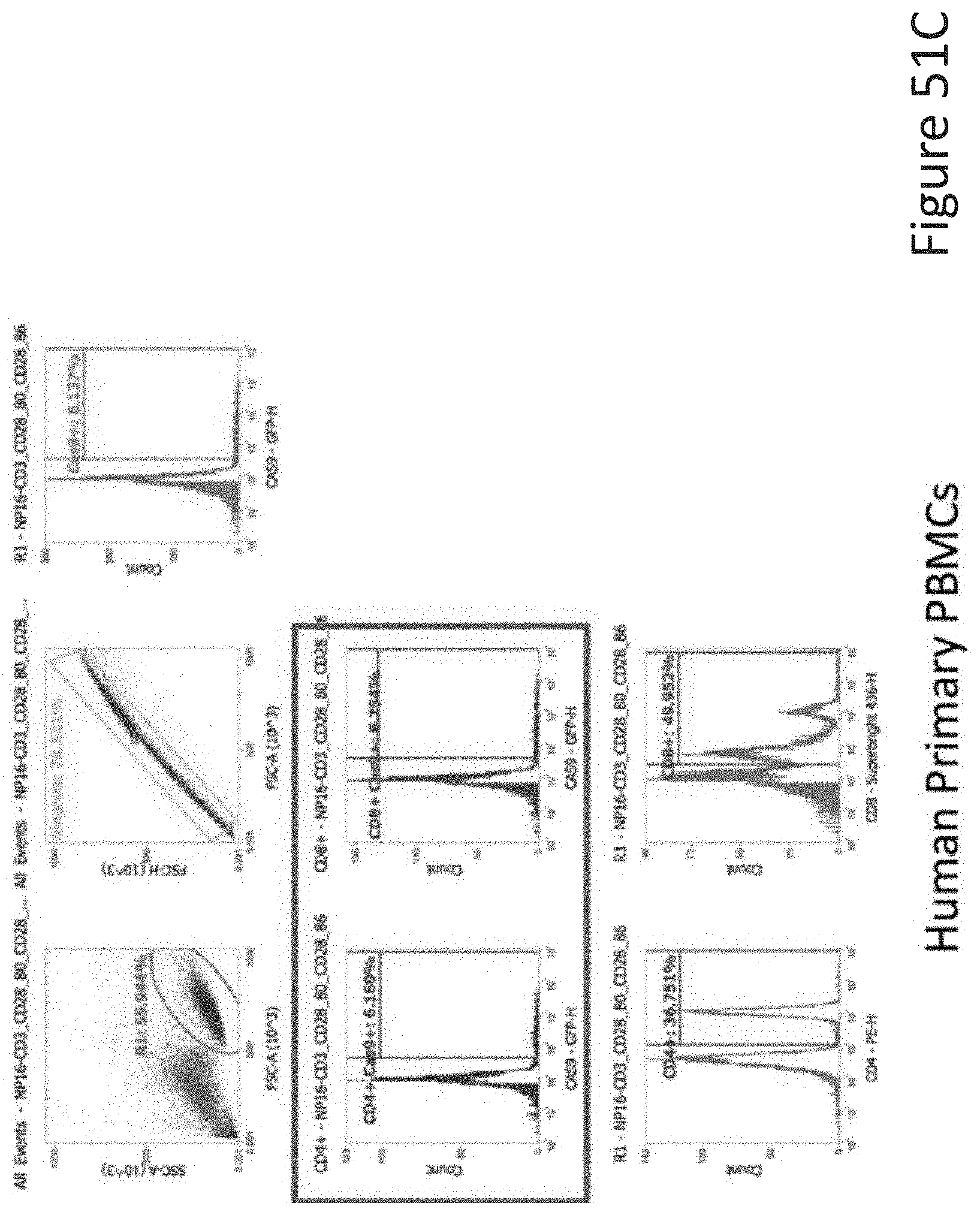
D00089

D00090

D00091

D00092

D00093
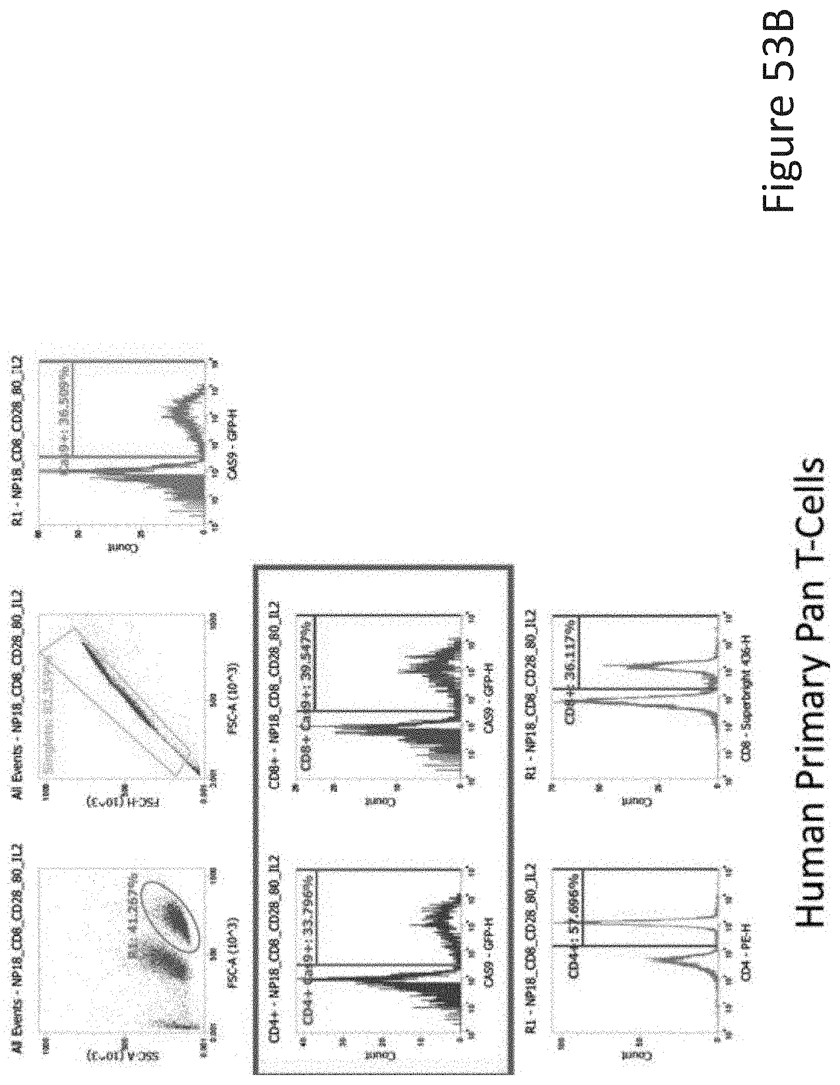
D00094
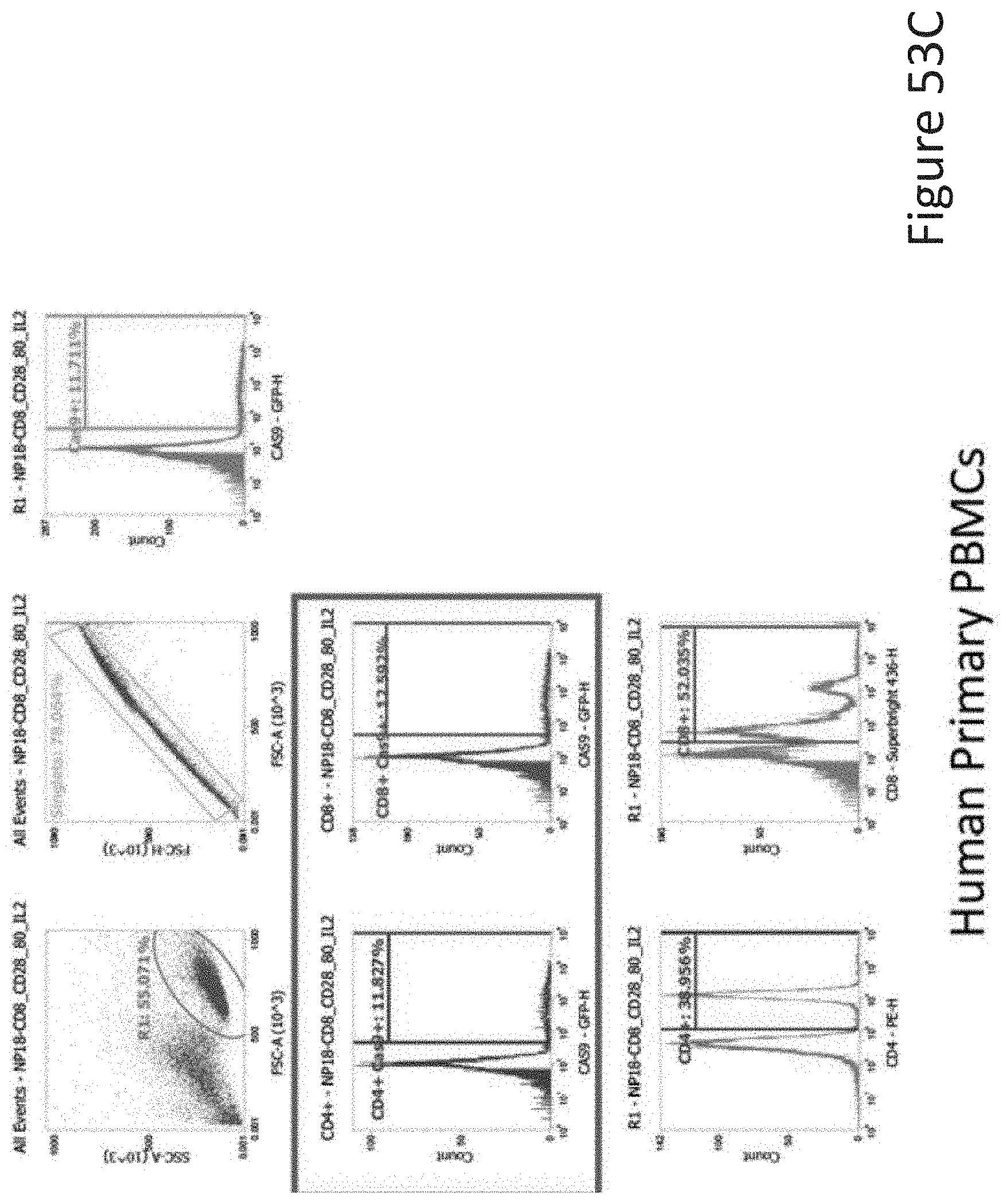
D00095

D00096
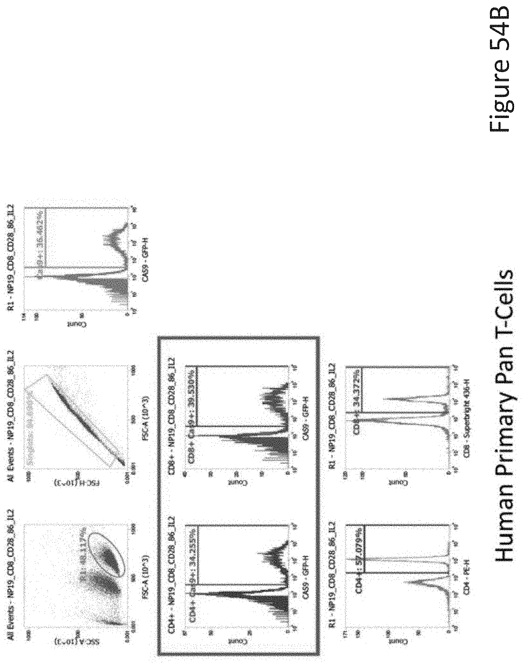
D00097
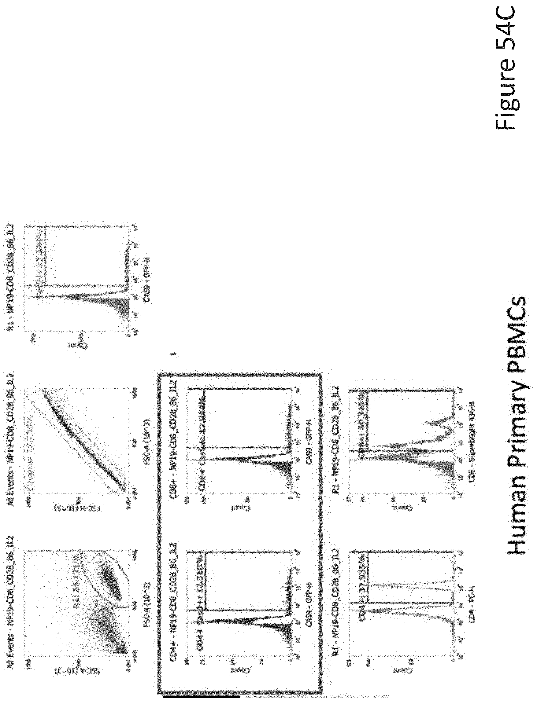
D00098

D00099
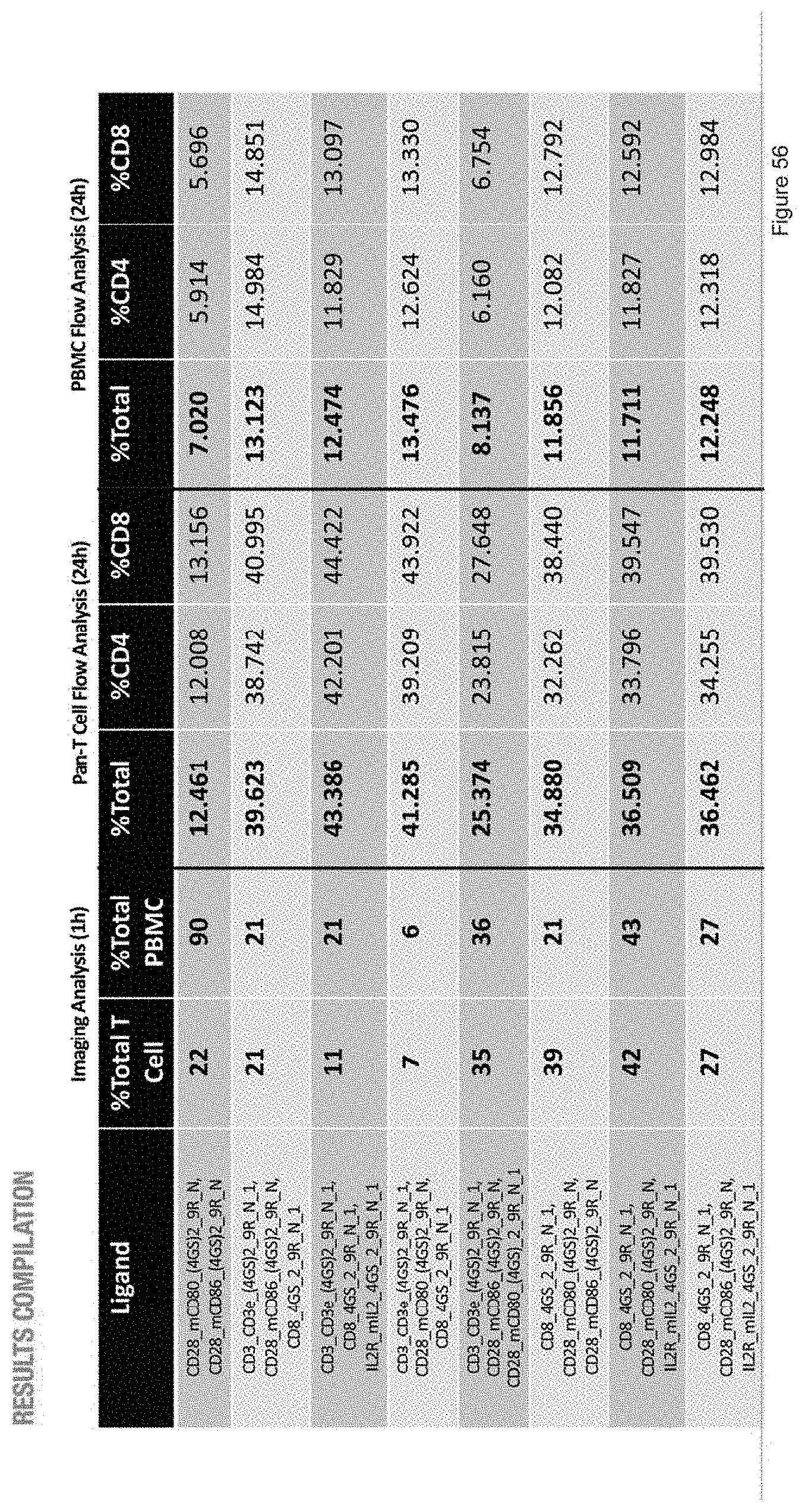
D00100
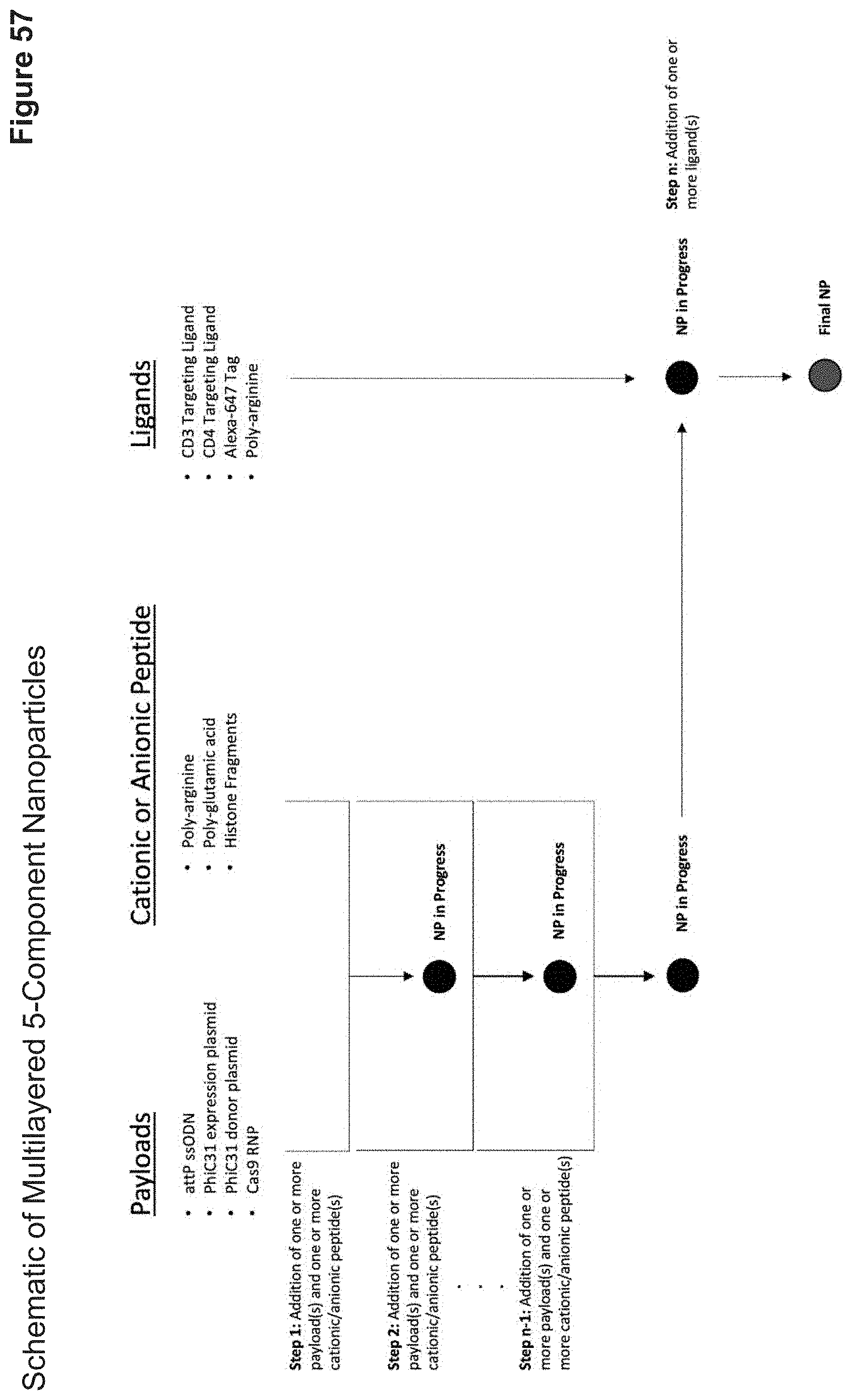
D00101
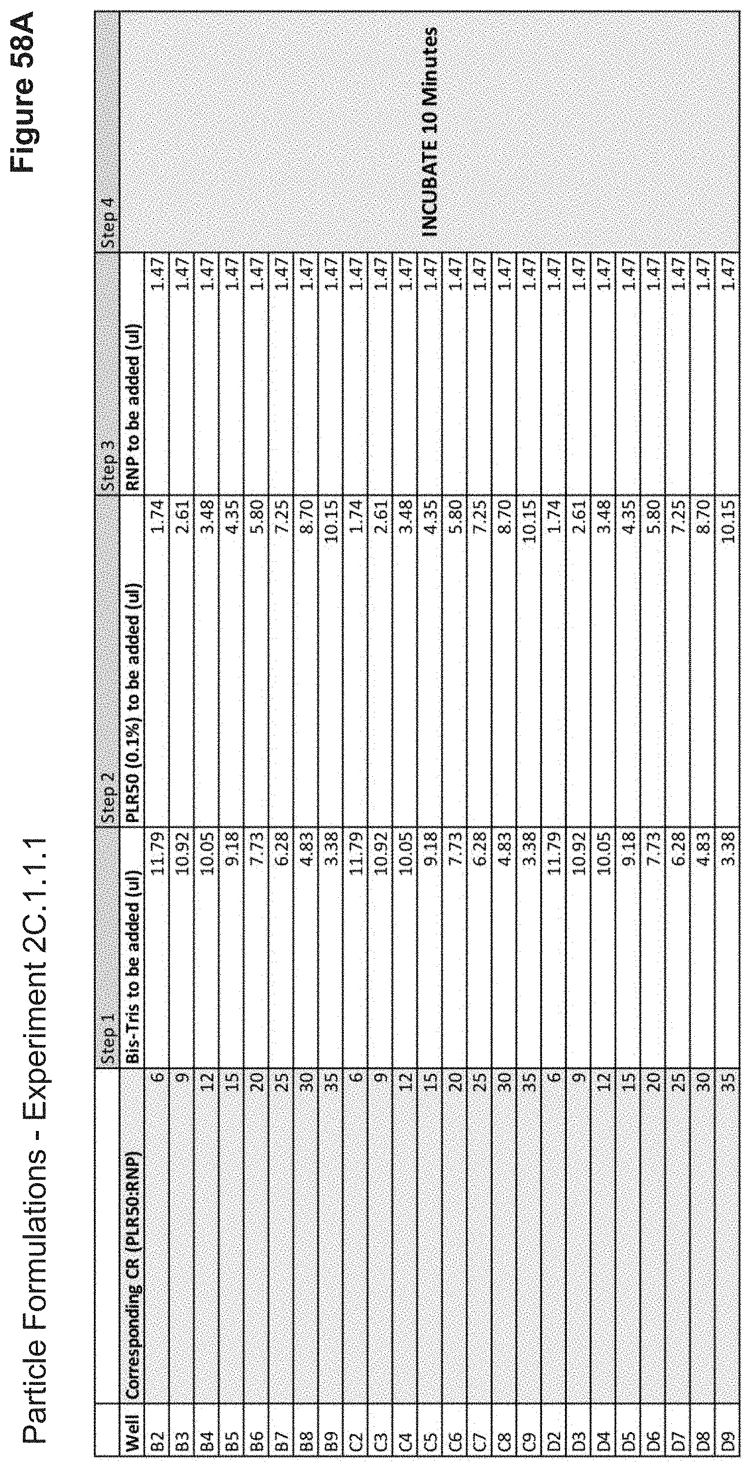
D00102
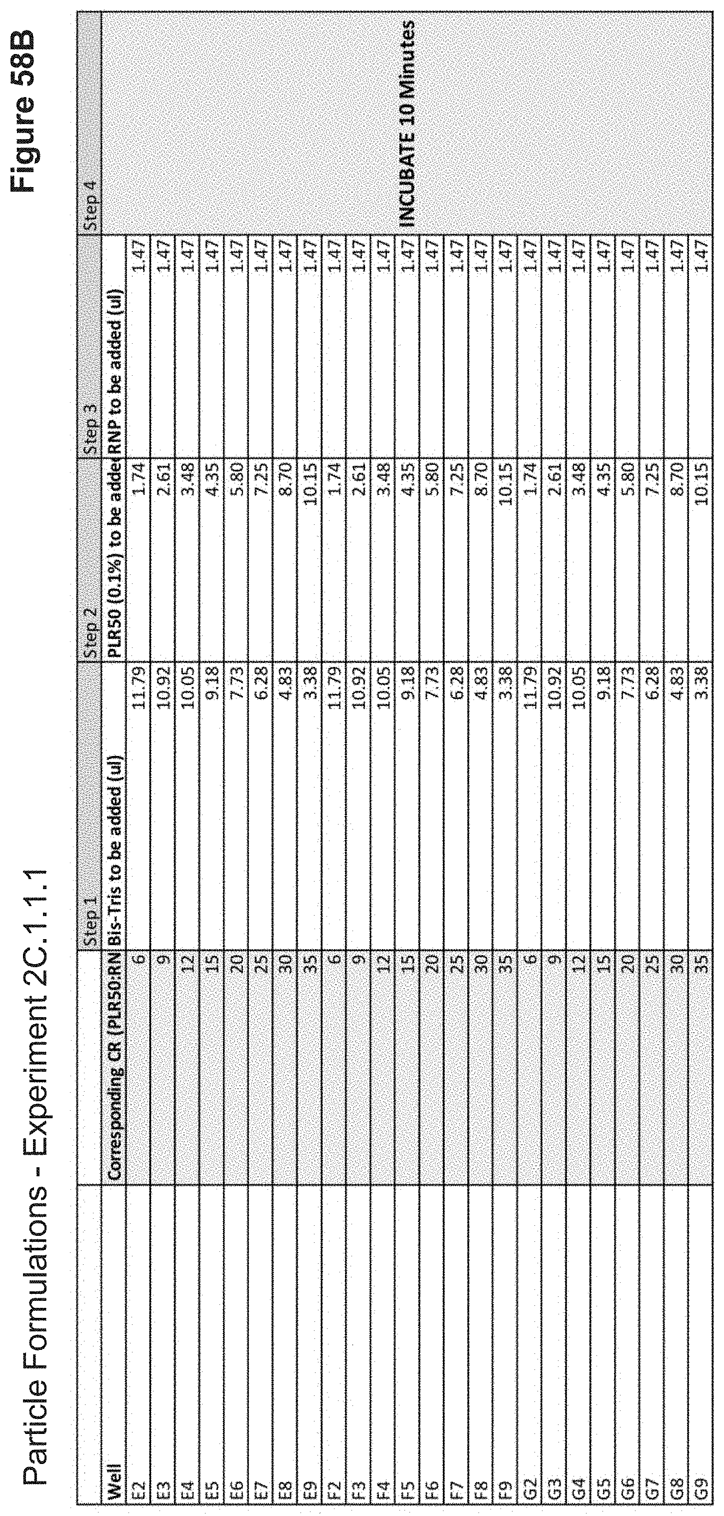
D00103
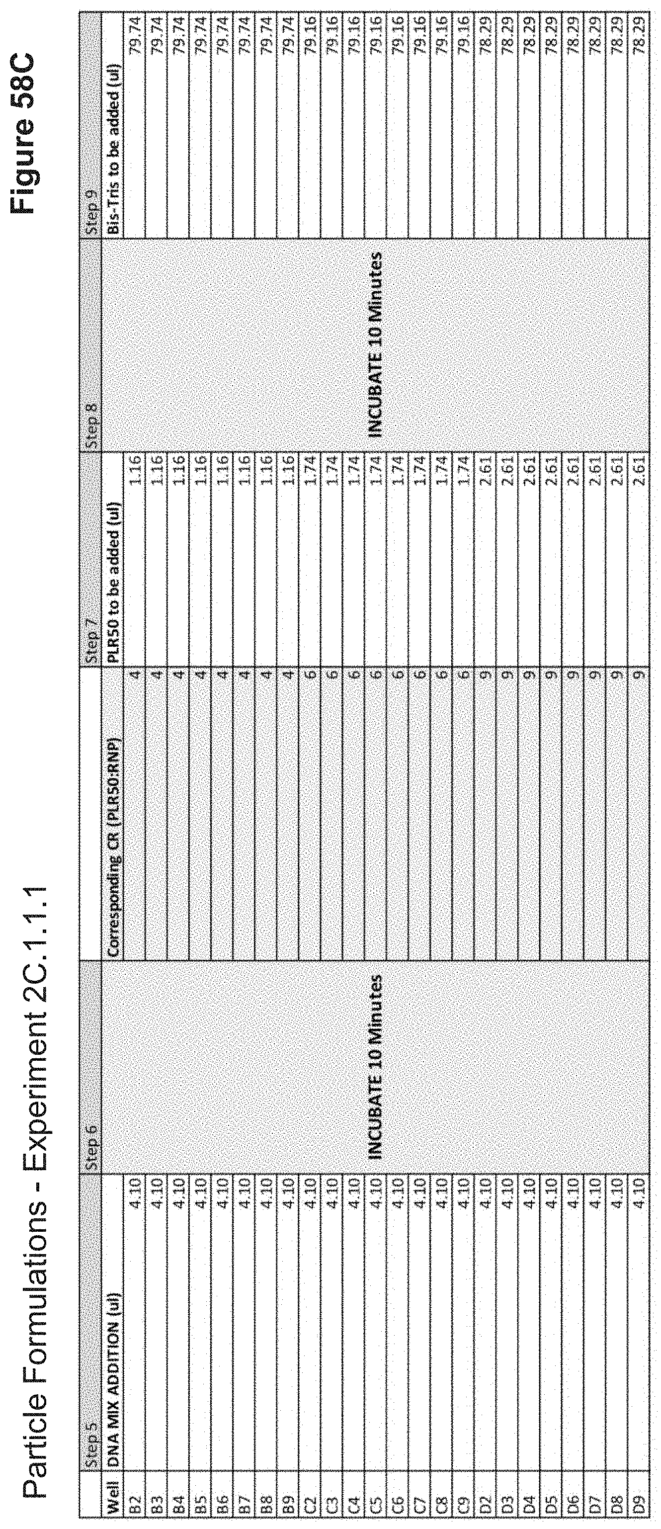
D00104
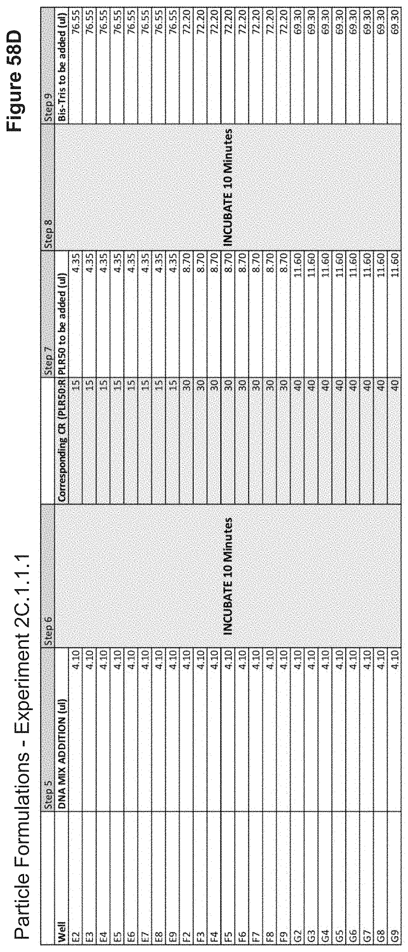
D00105

D00106
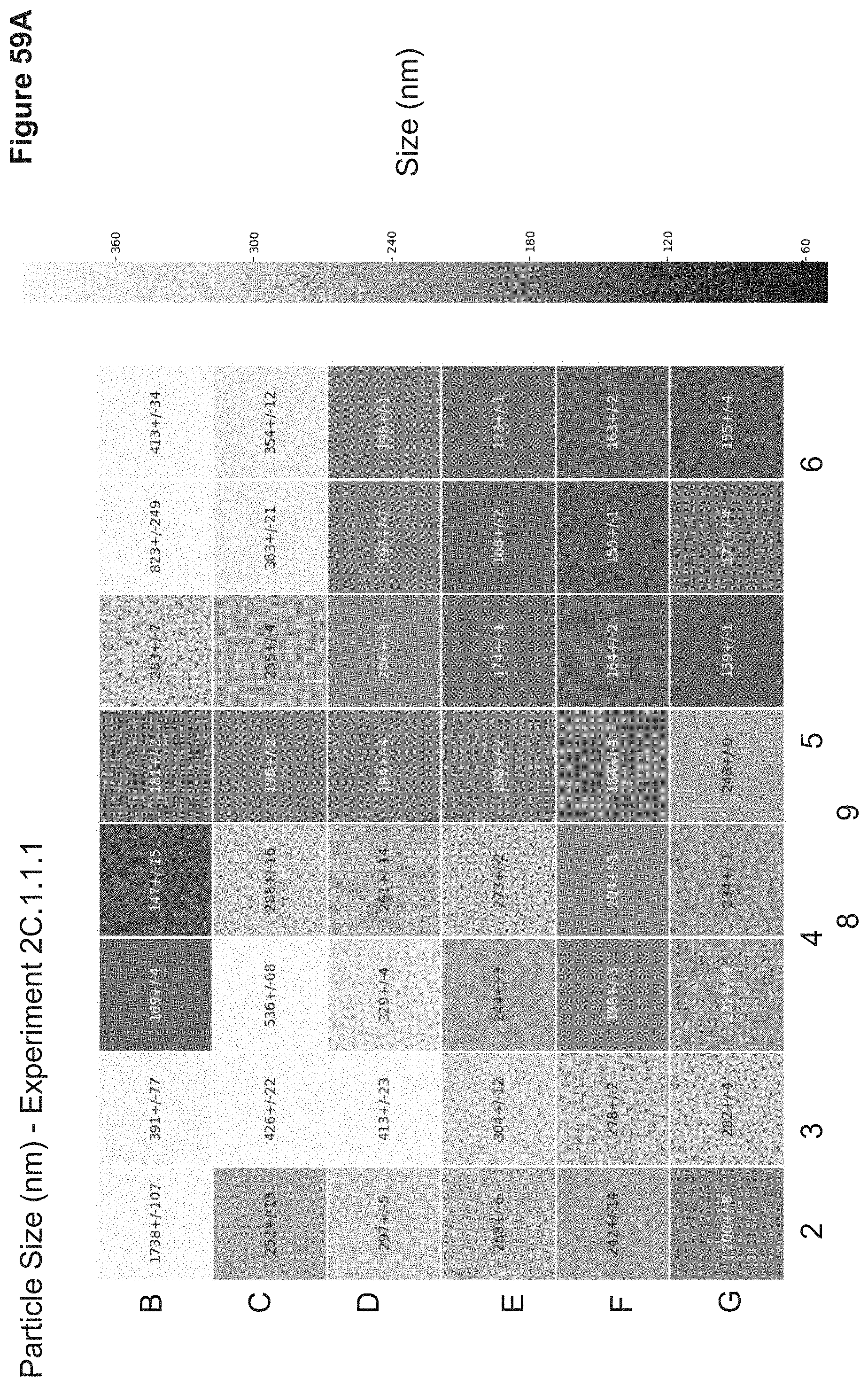
D00107
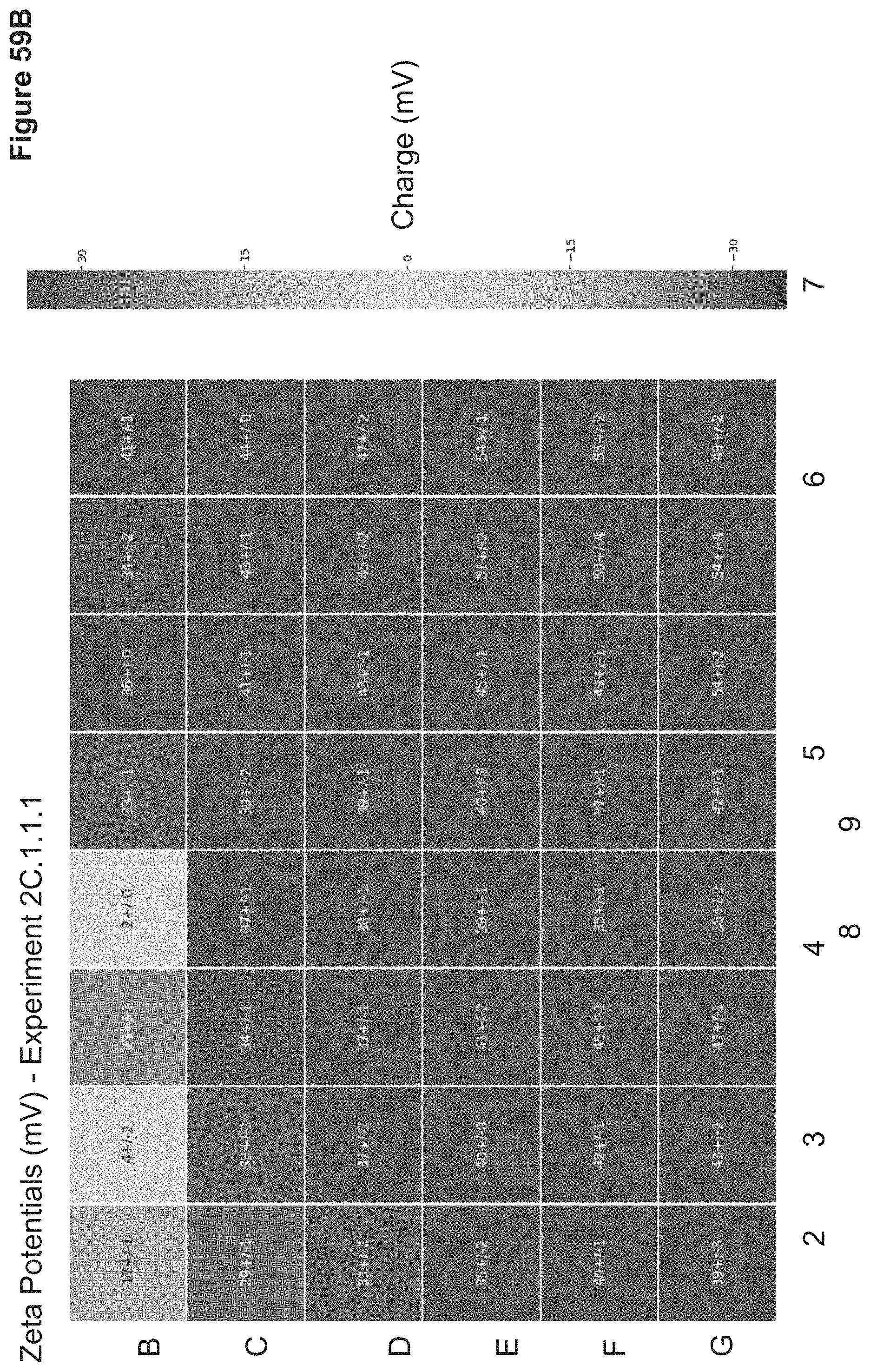
D00108
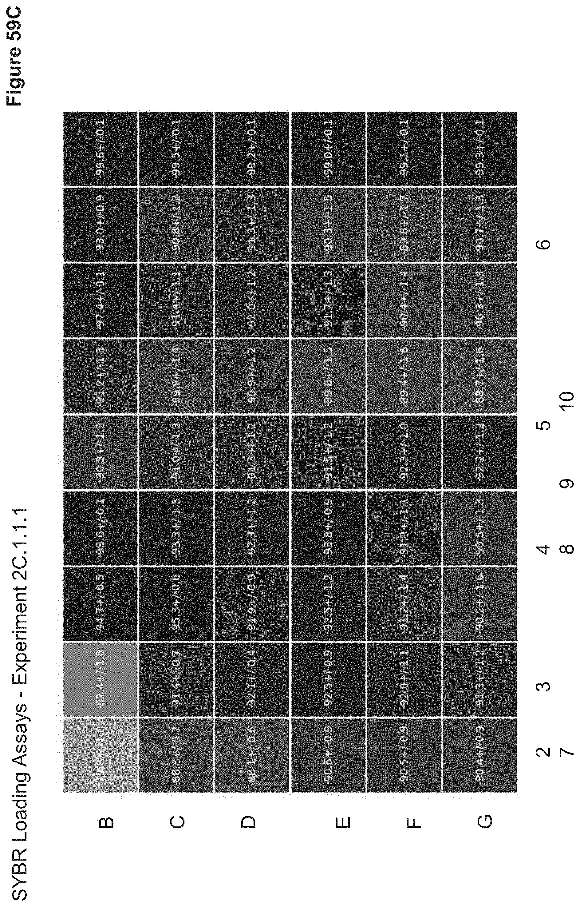
D00109
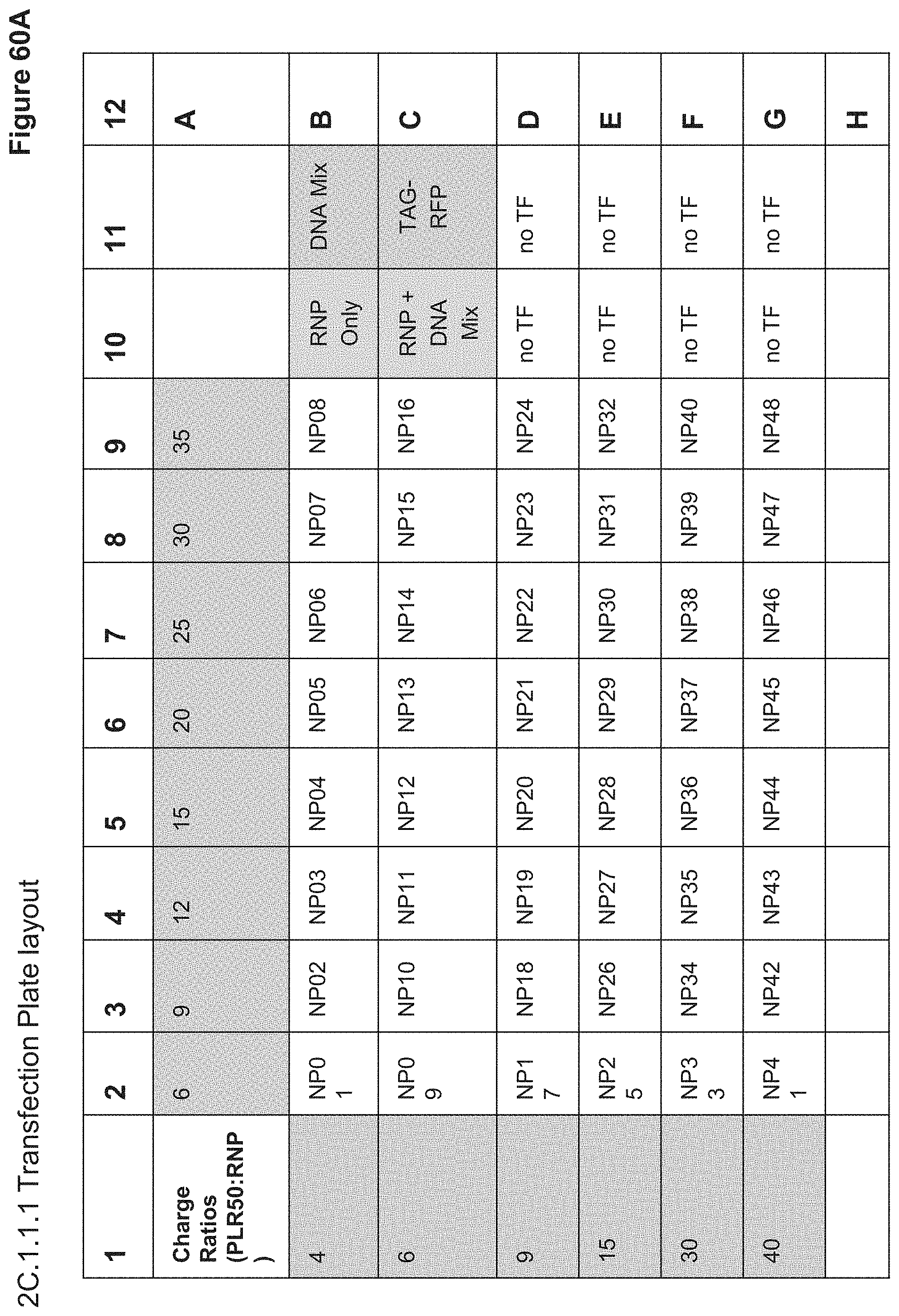
D00110
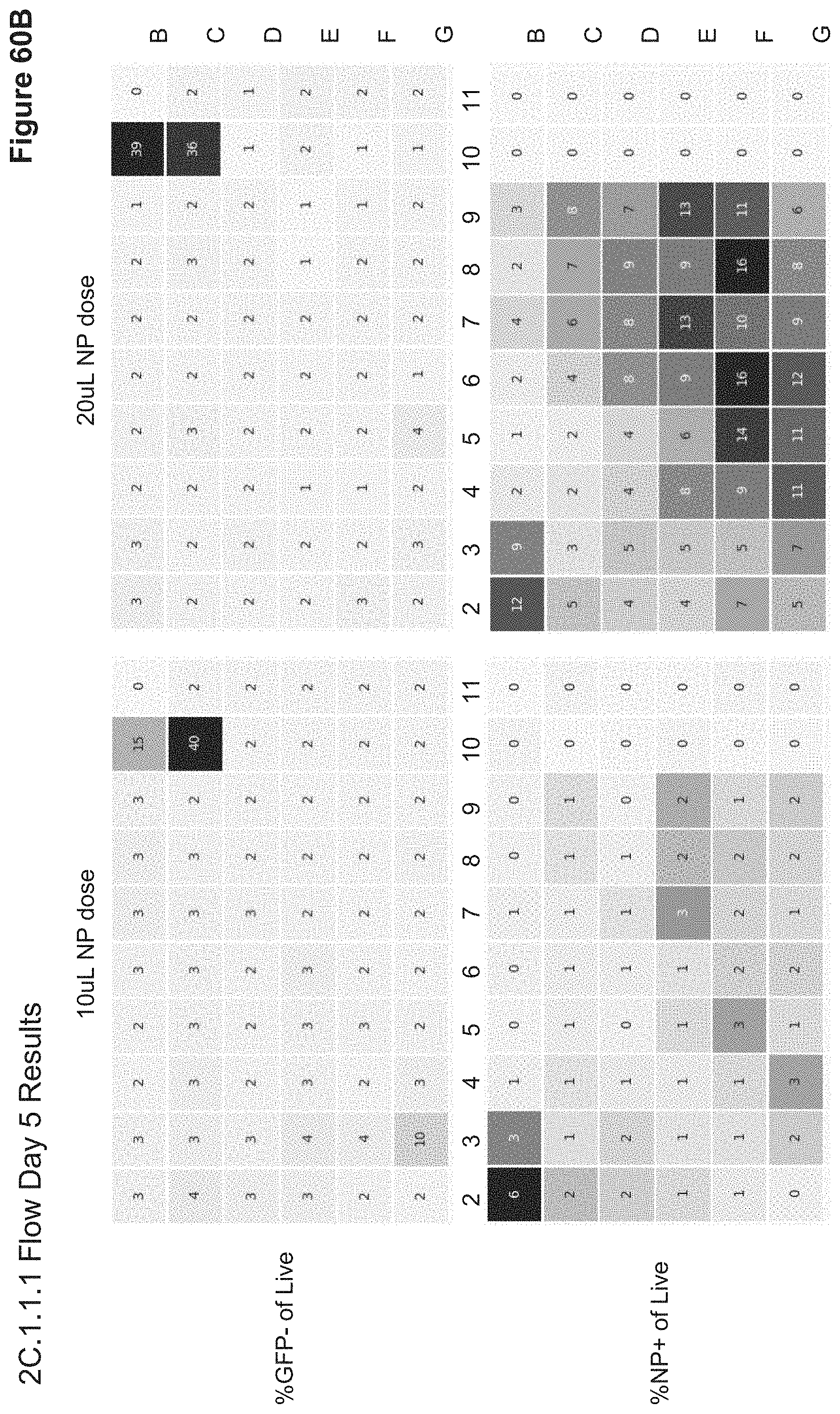
D00111

D00112
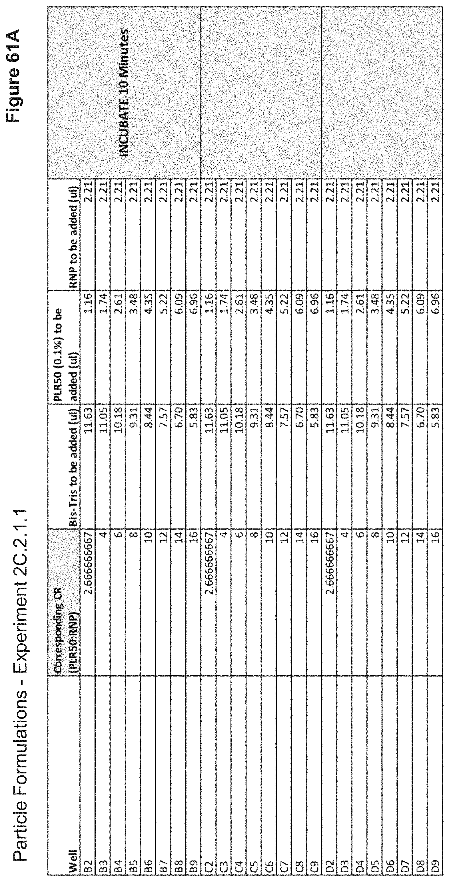
D00113
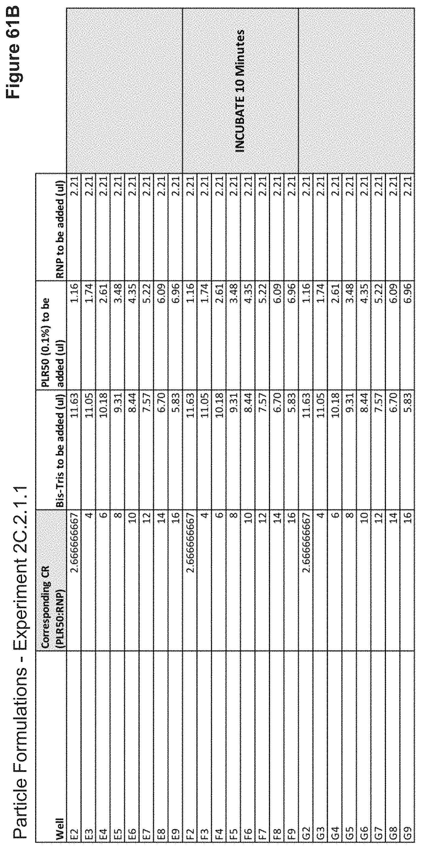
D00114

D00115

D00116

D00117
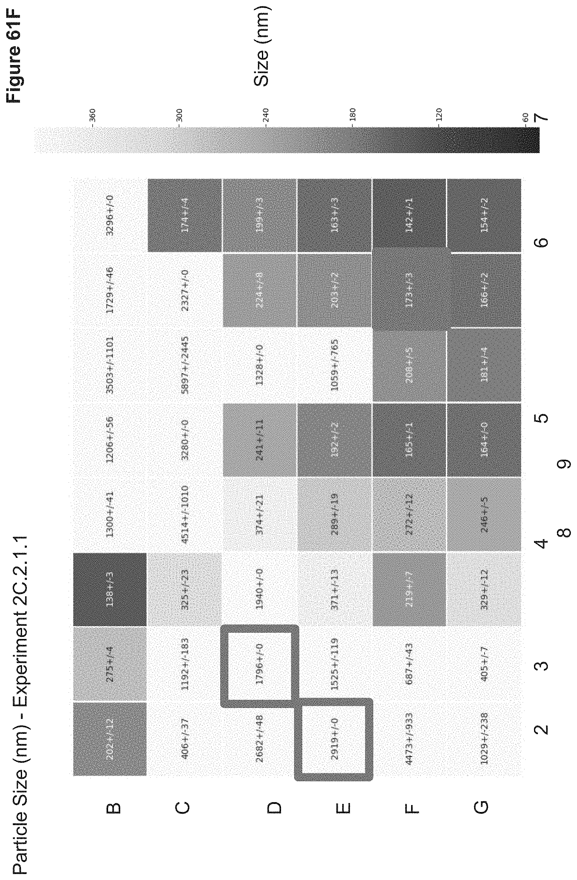
D00118

D00119
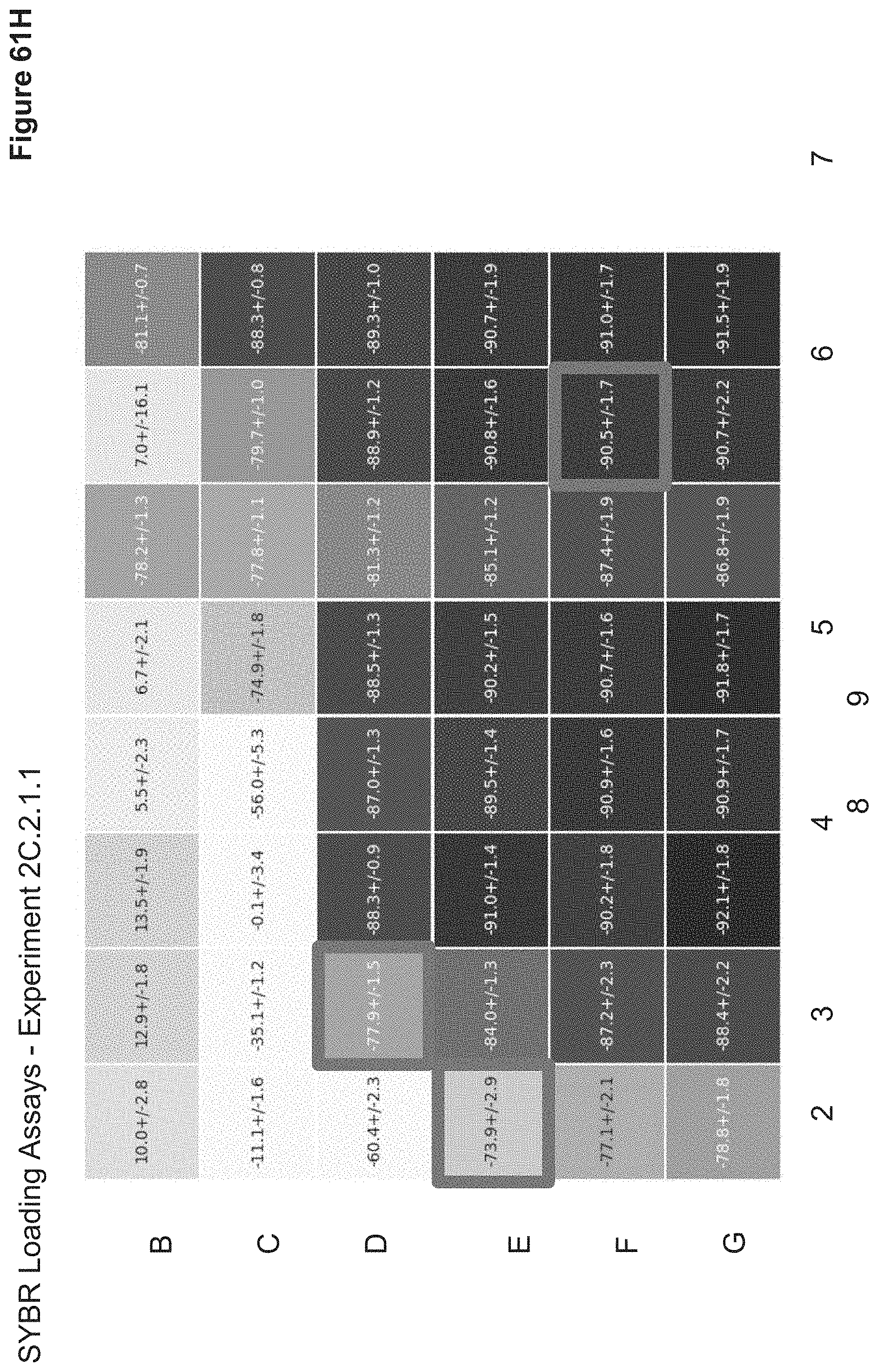
D00120
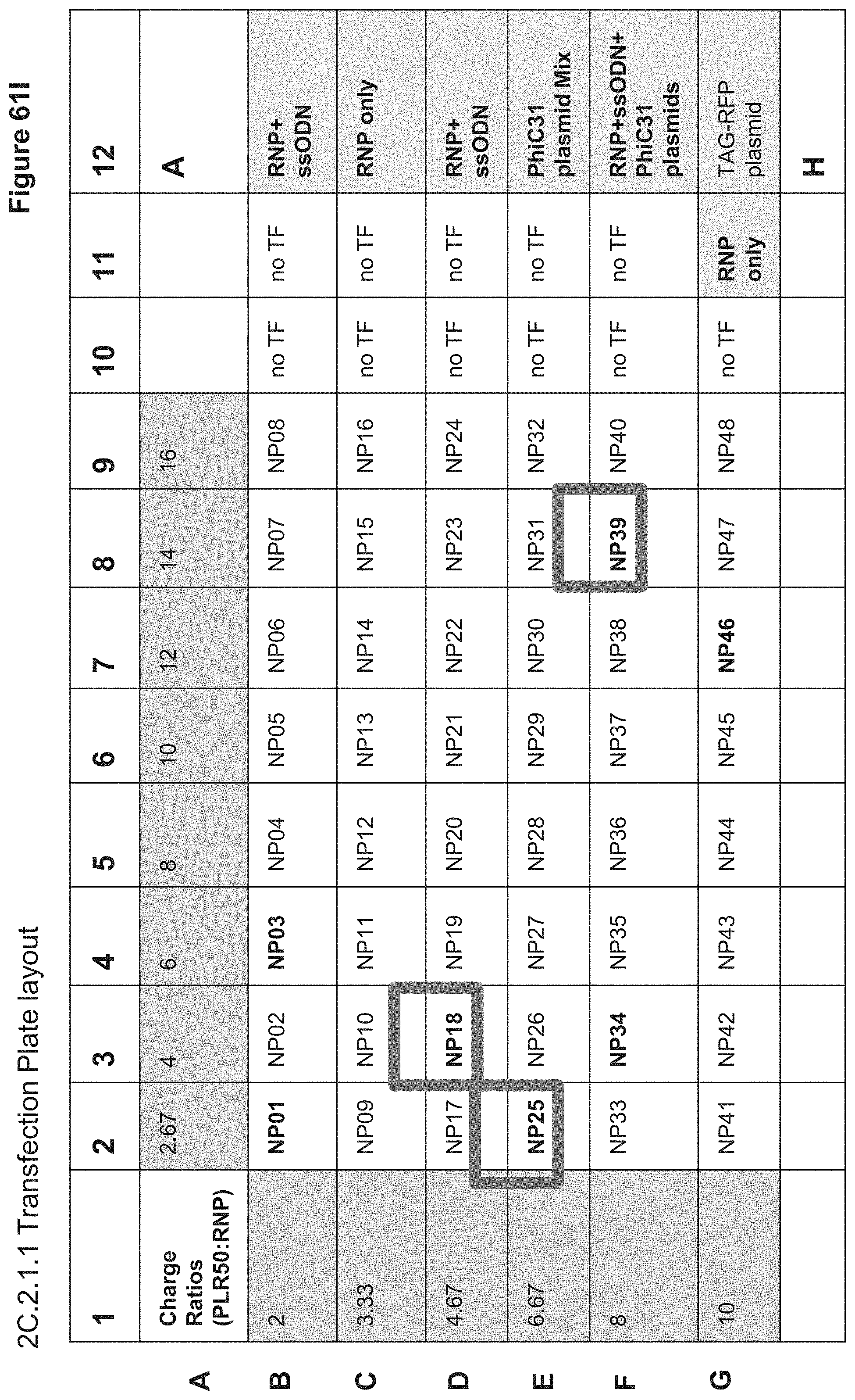
D00121
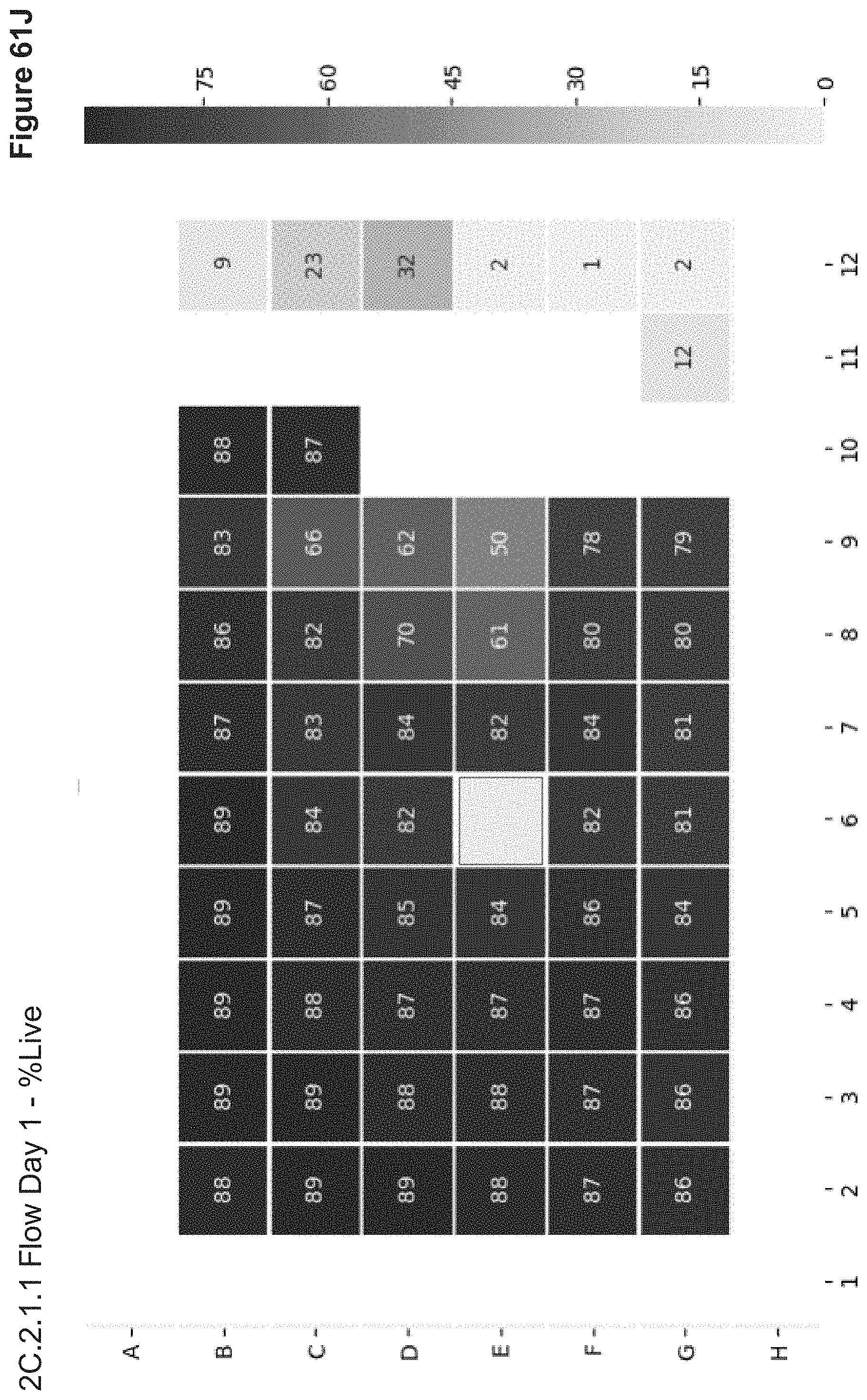
D00122
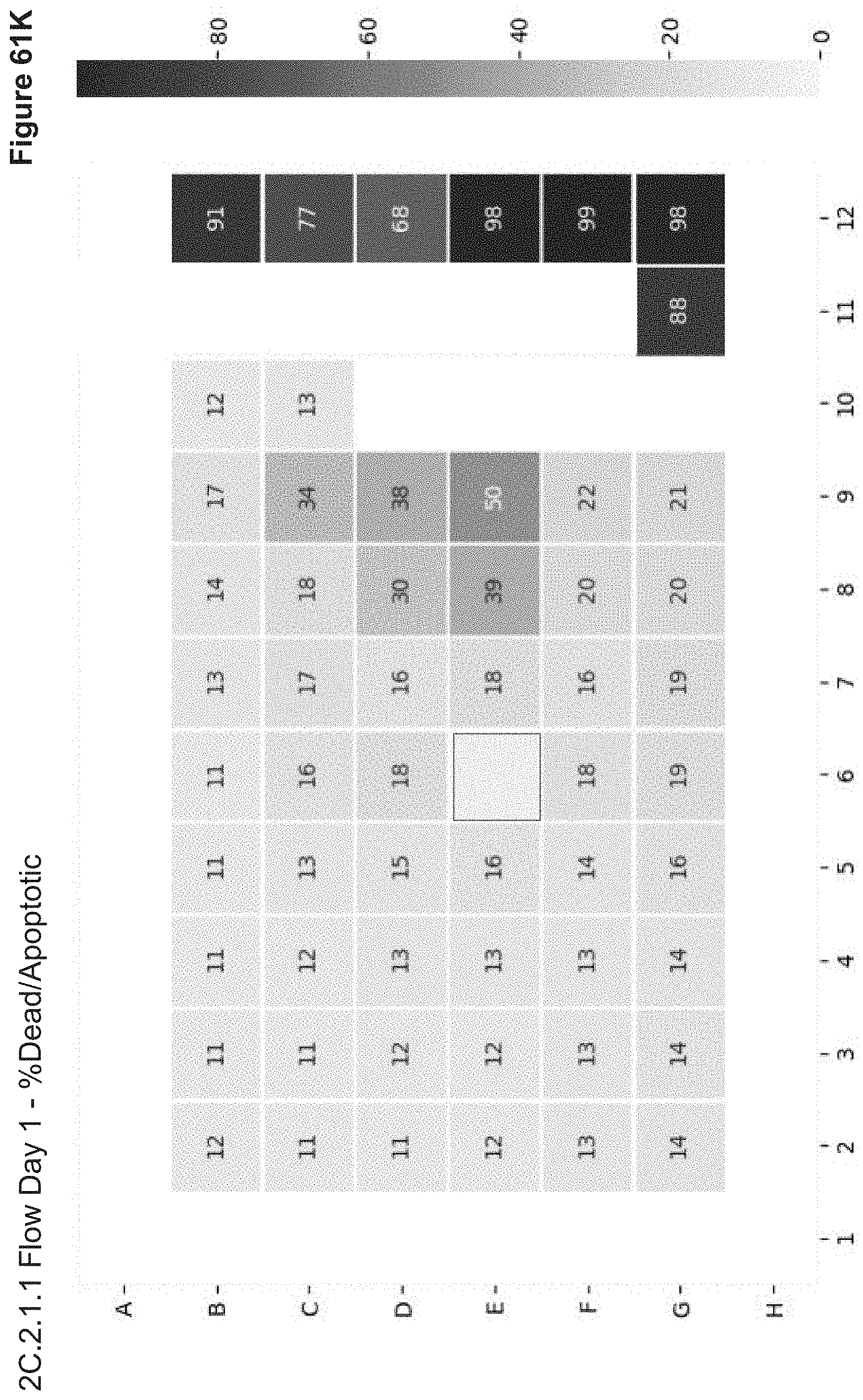
D00123
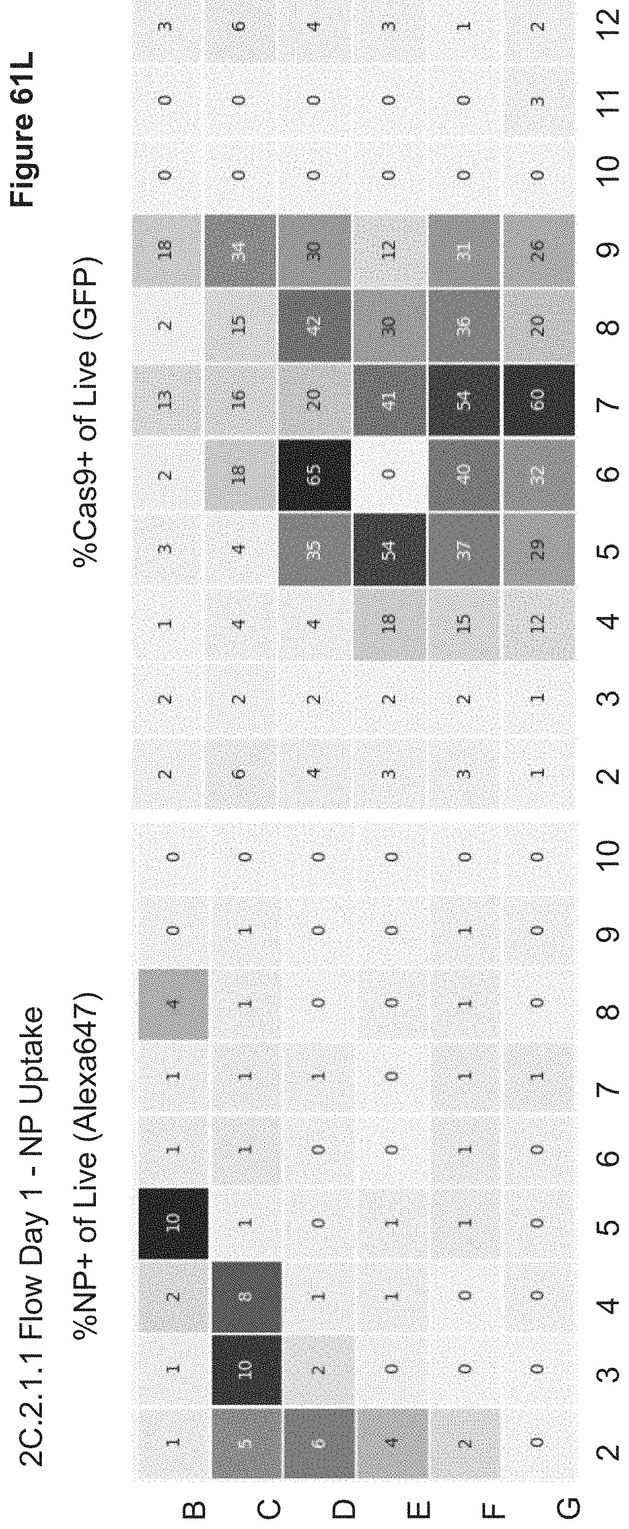
D00124
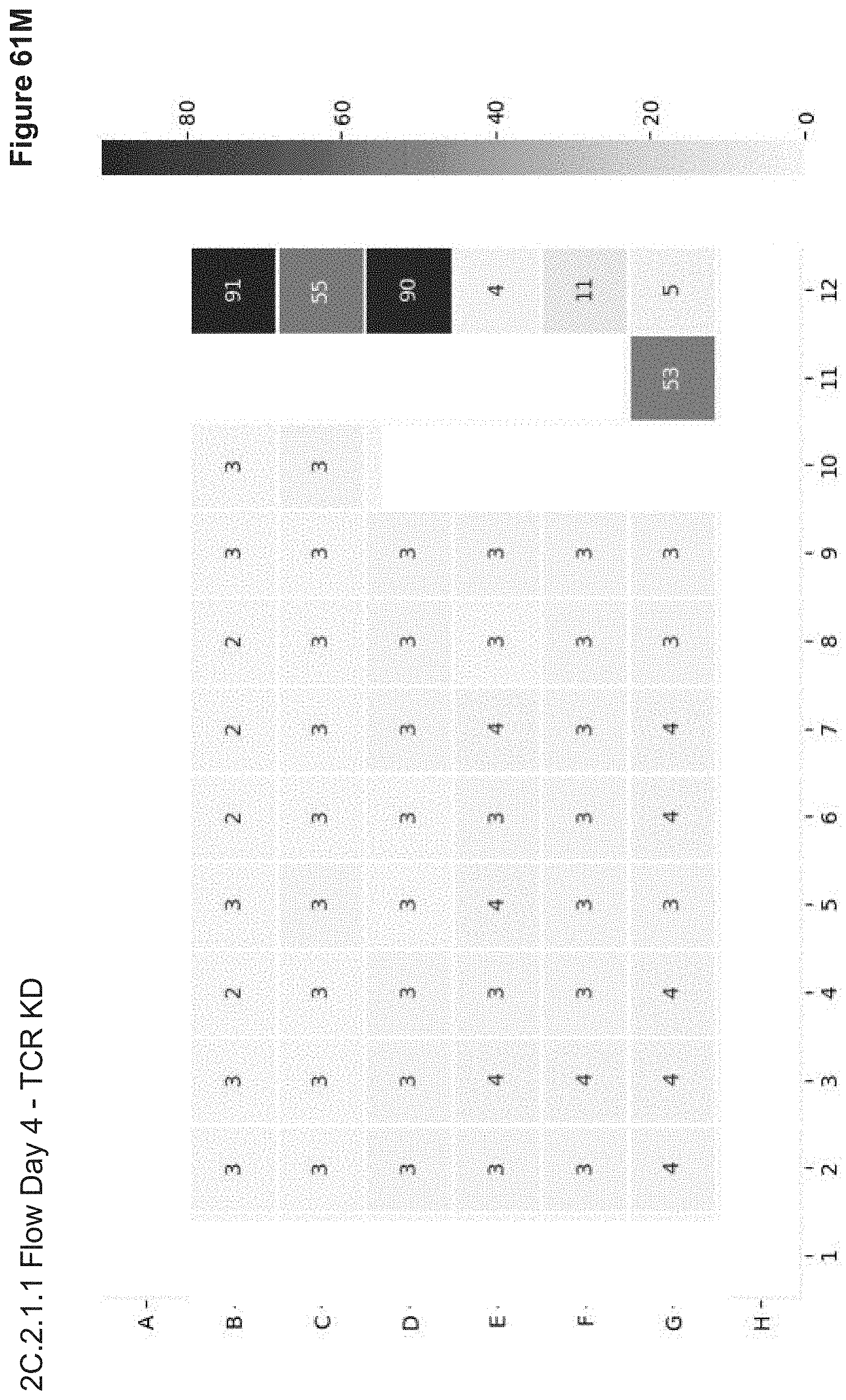
D00125
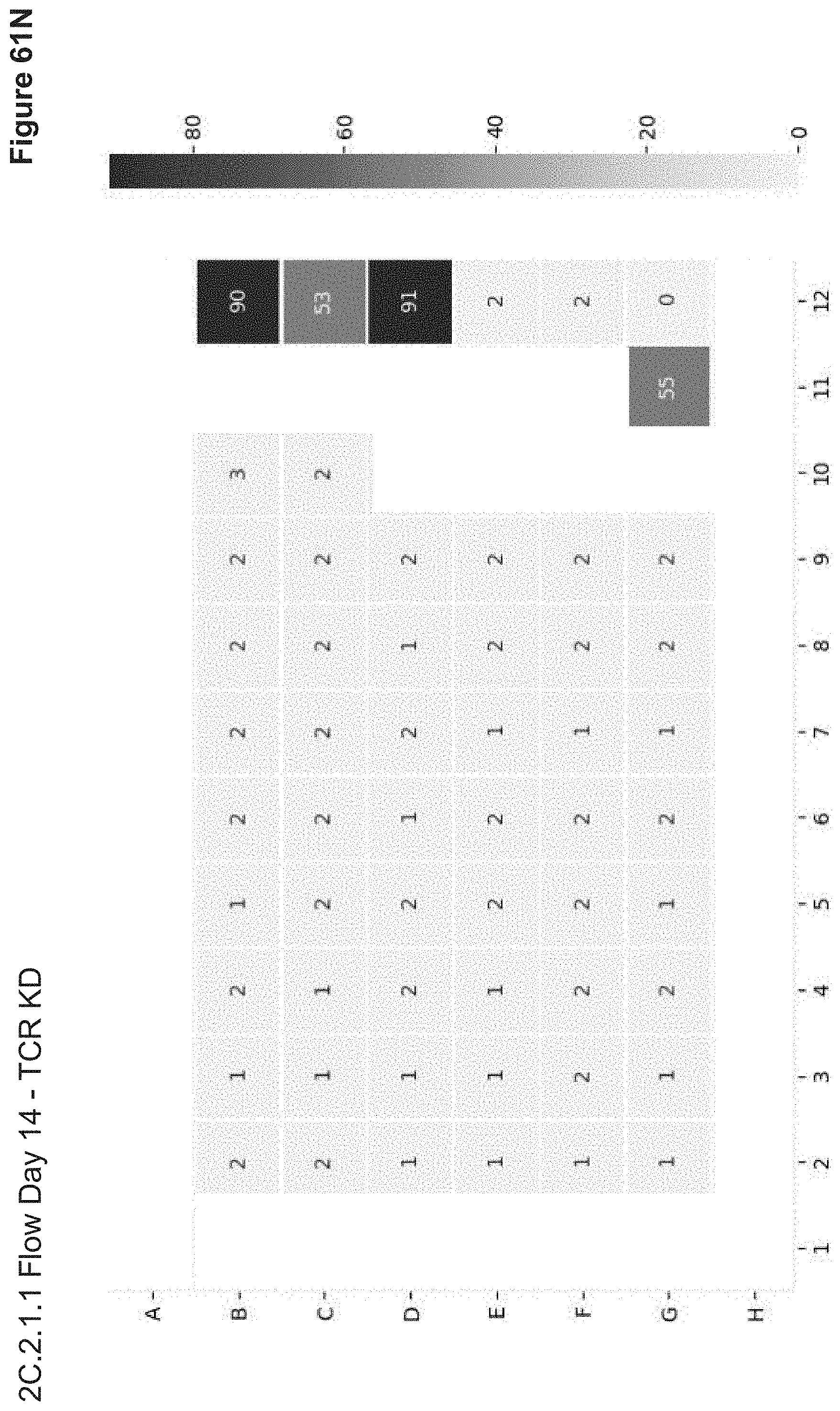
D00126
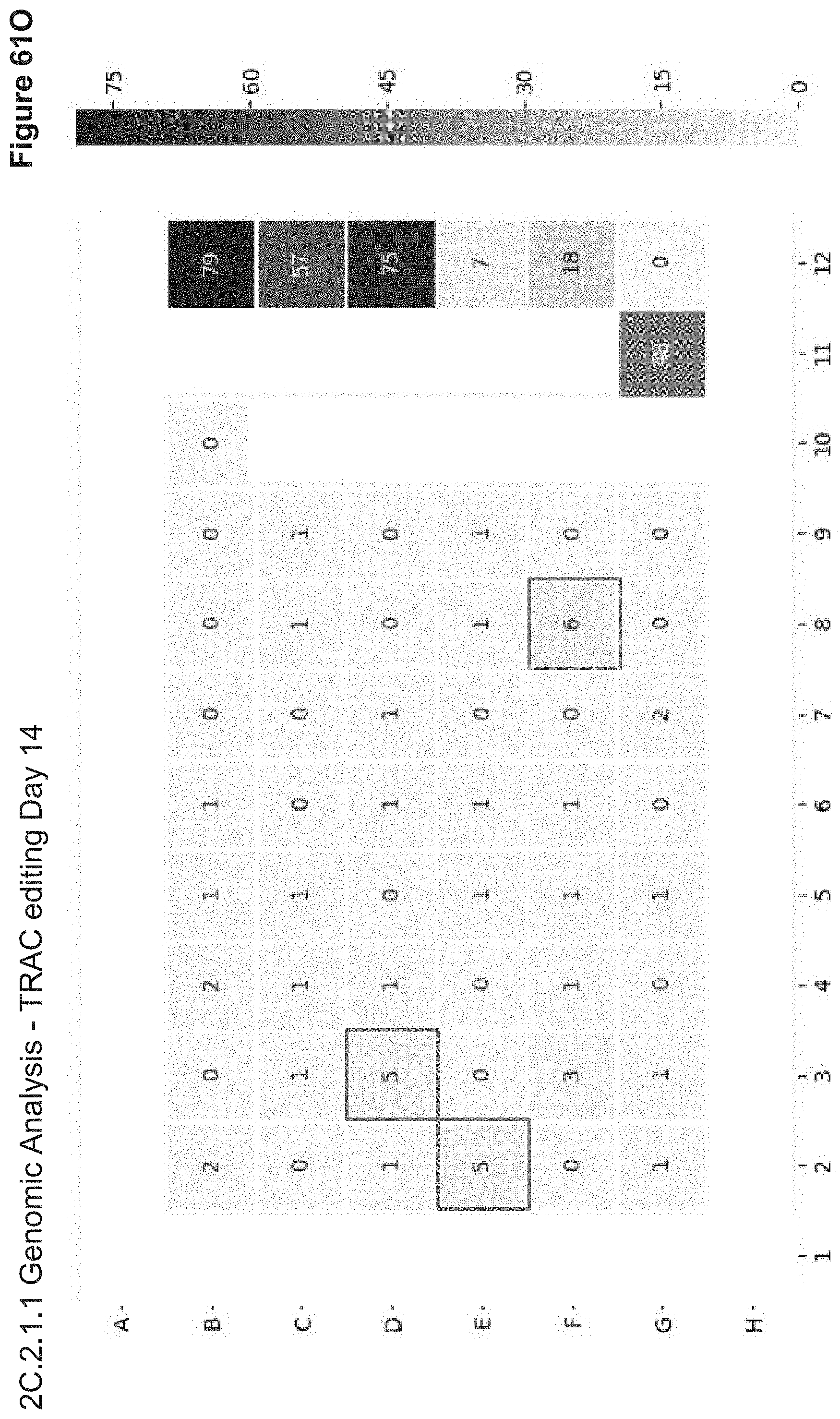
D00127
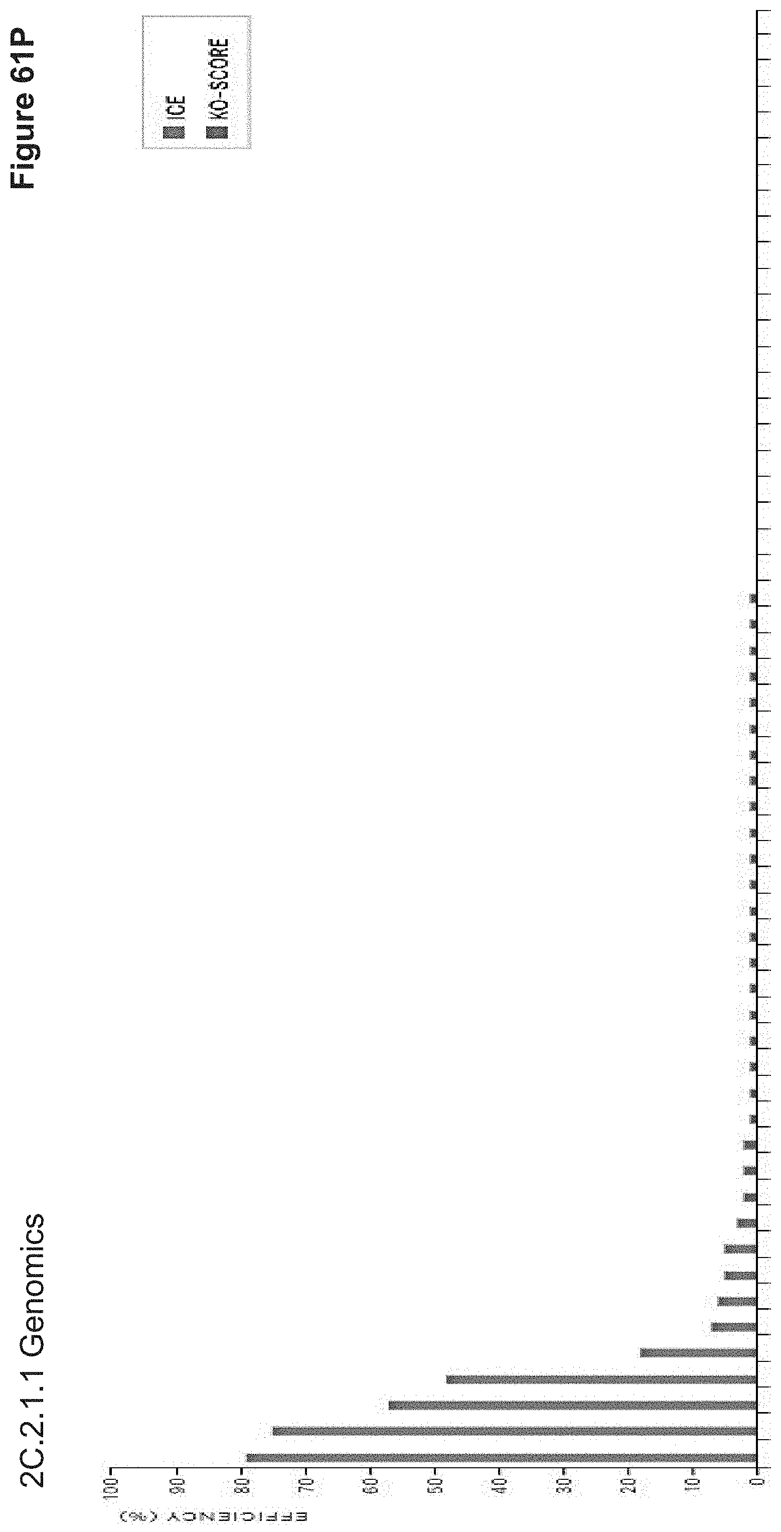
D00128

D00129
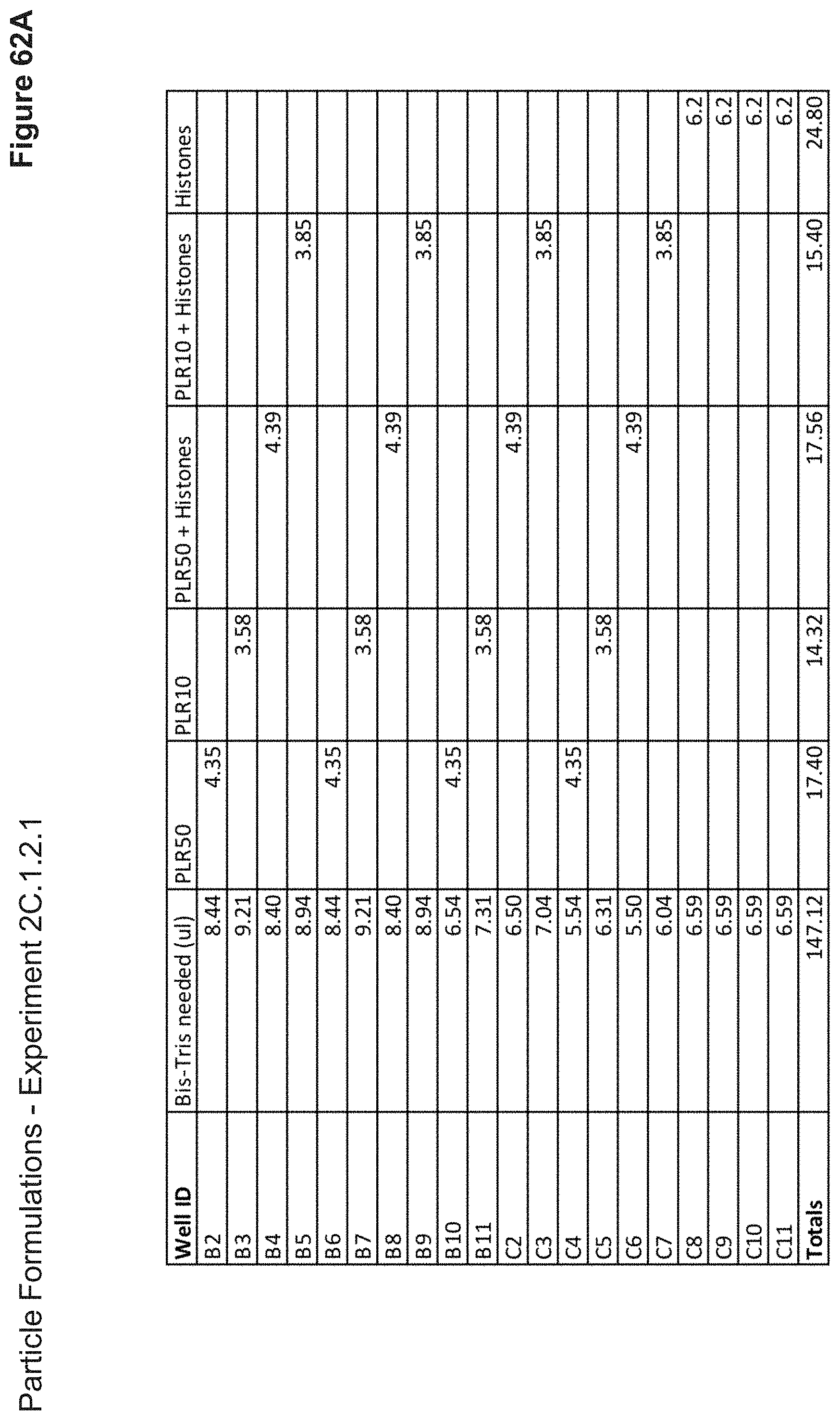
D00130
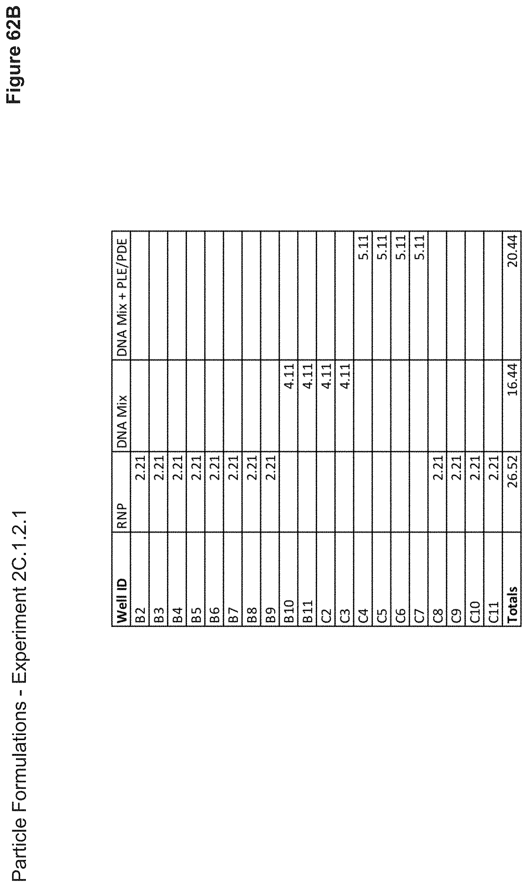
D00131
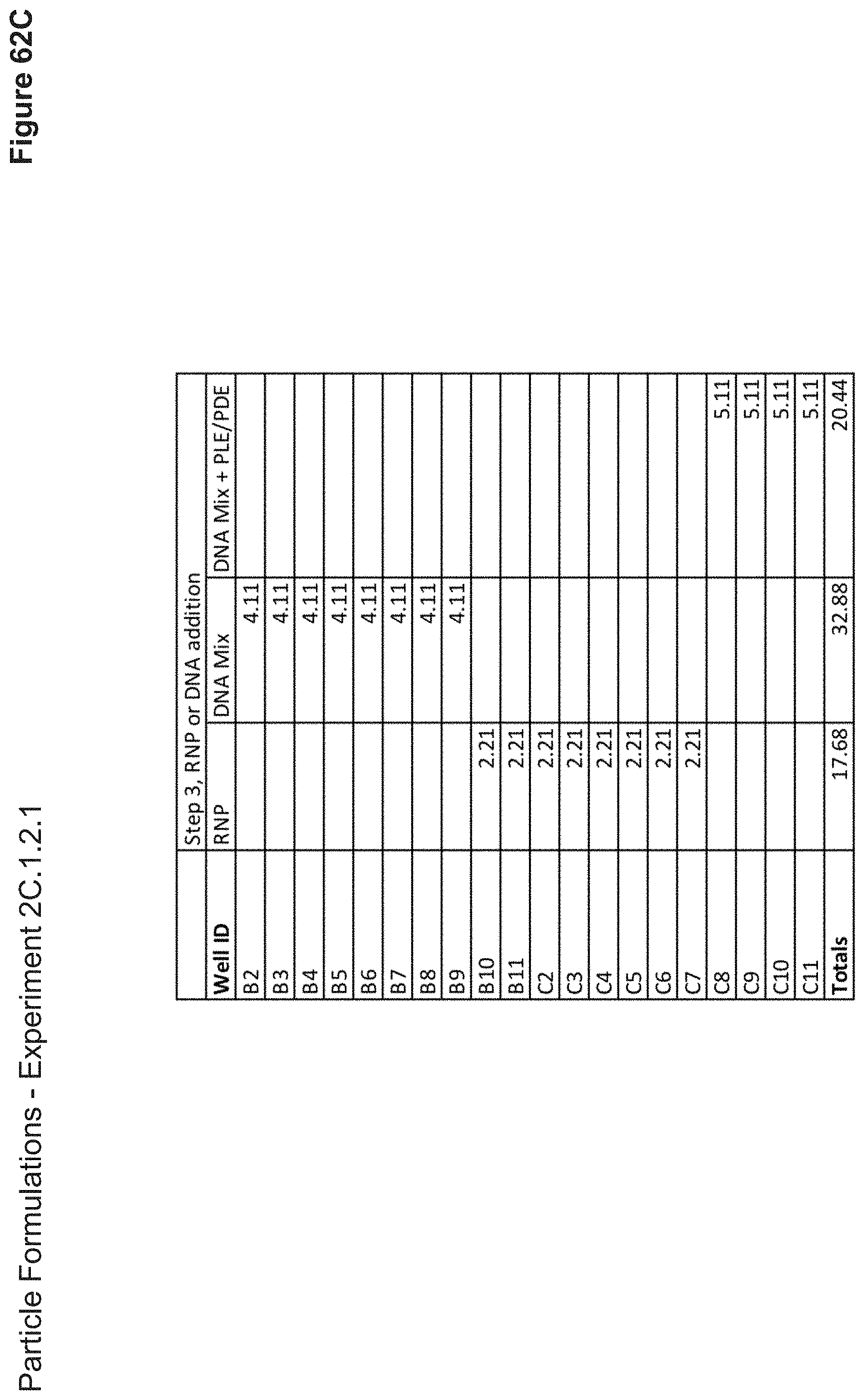
D00132

D00133
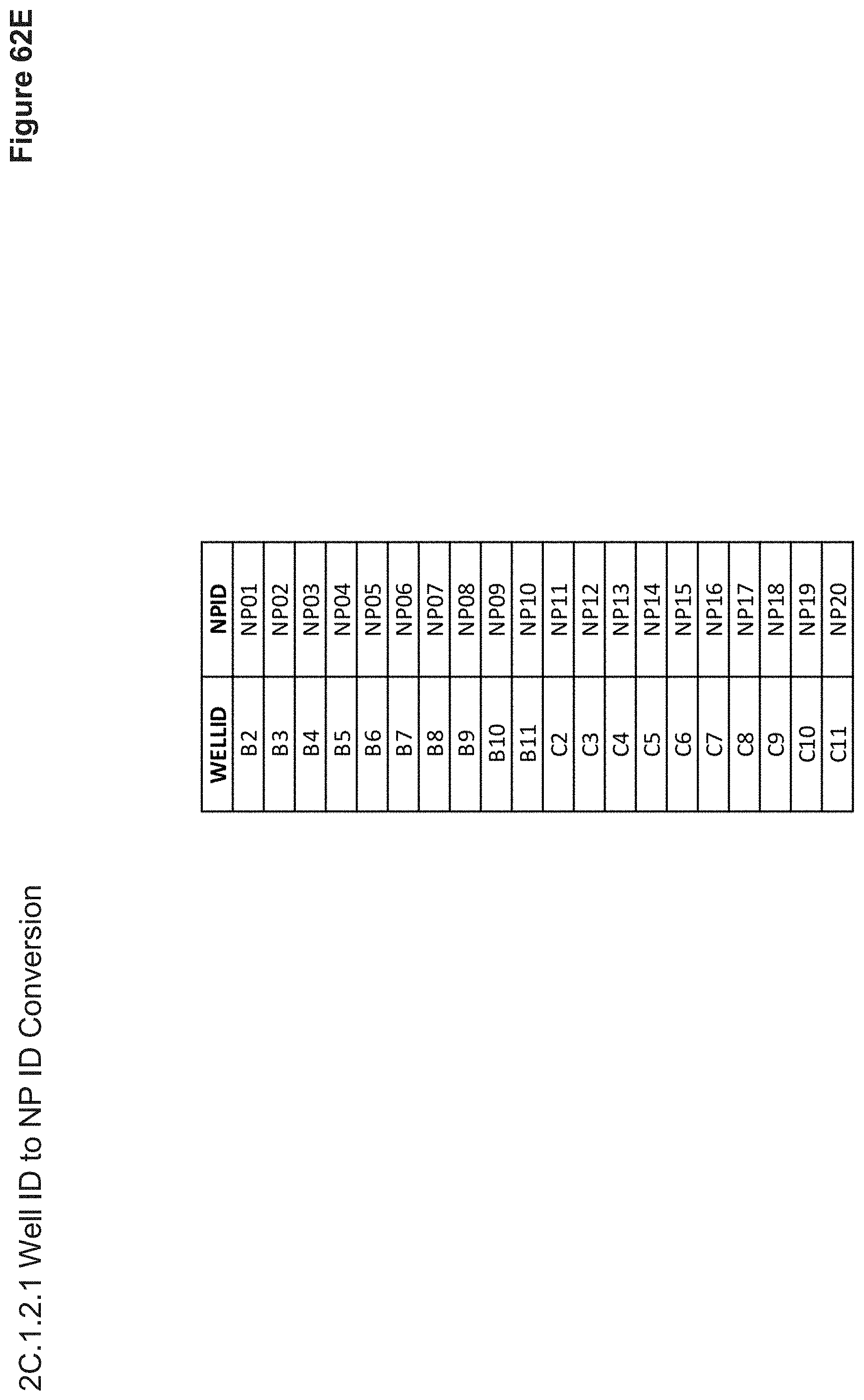
D00134
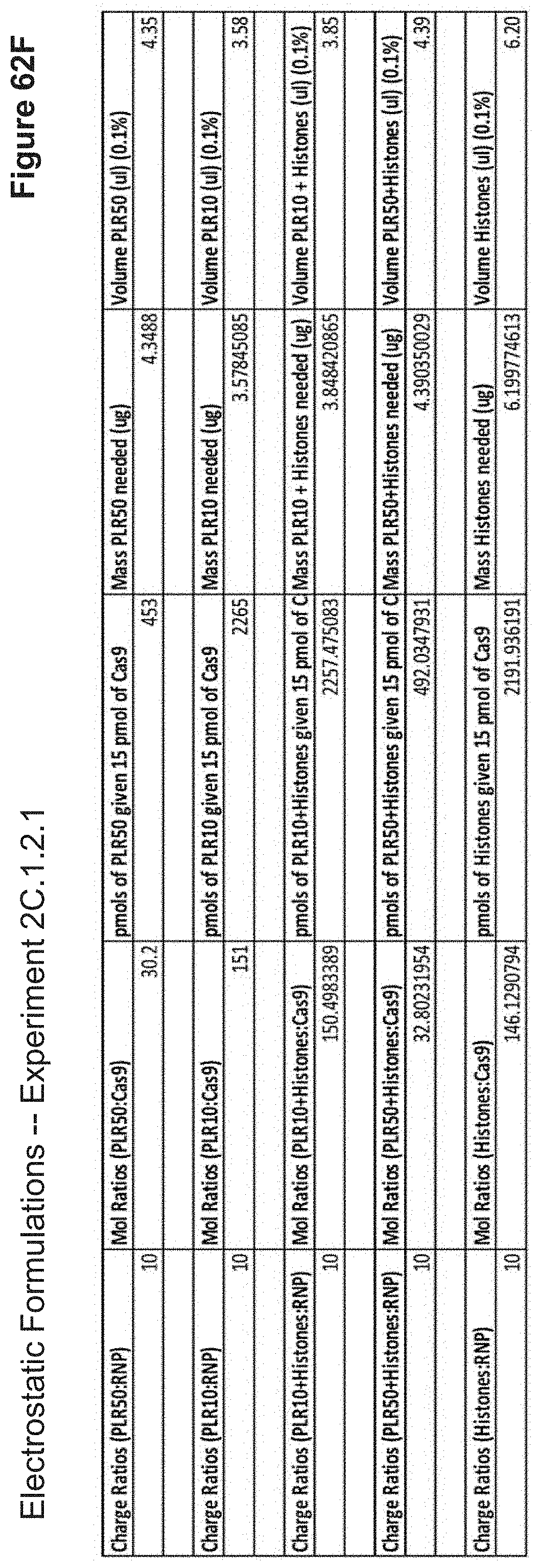
D00135

D00136
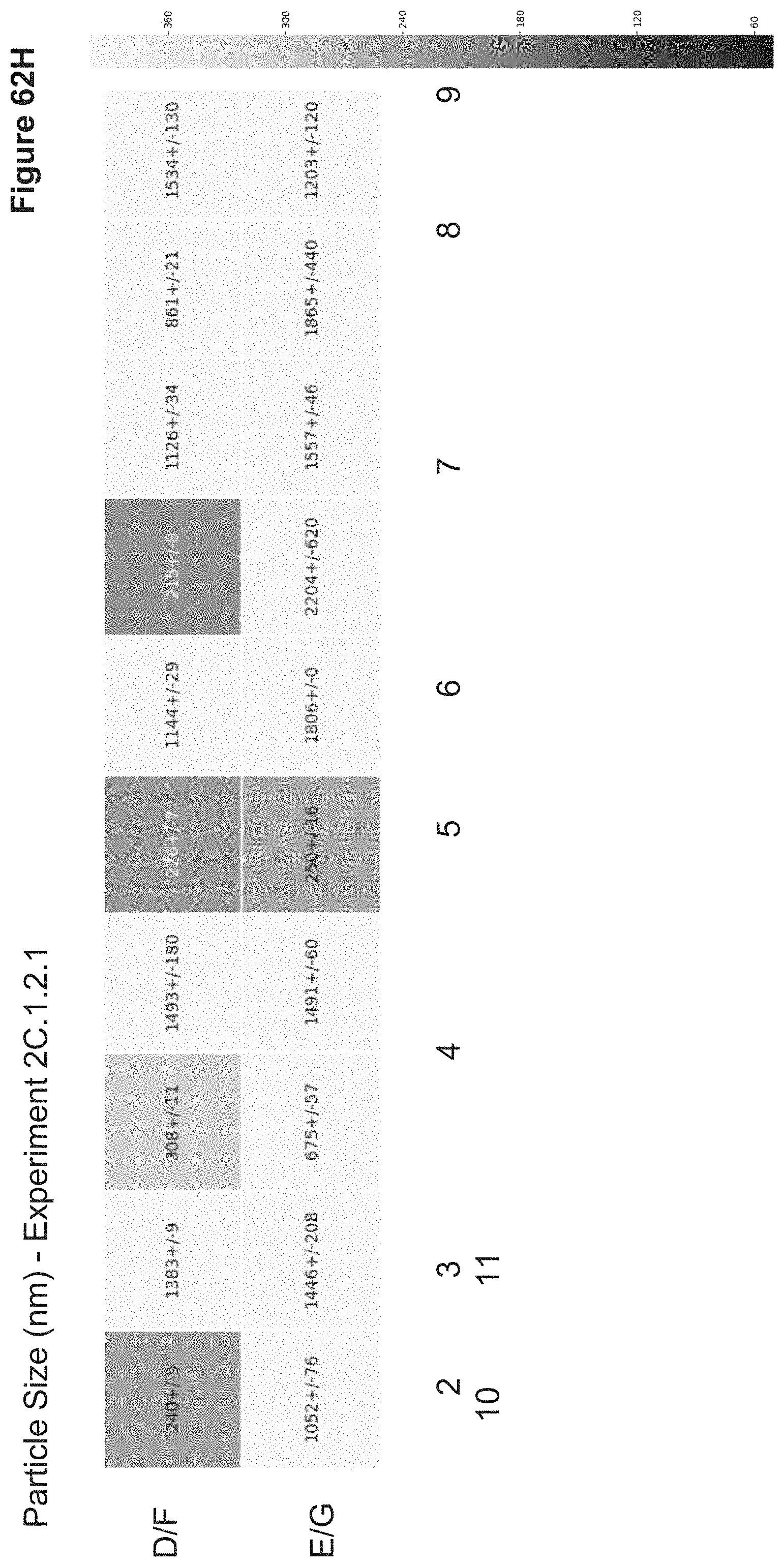
D00137
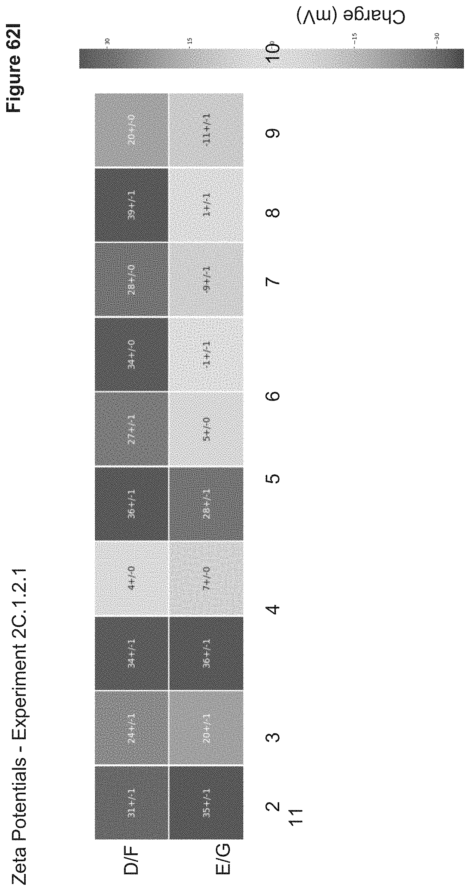
D00138

D00139

D00140
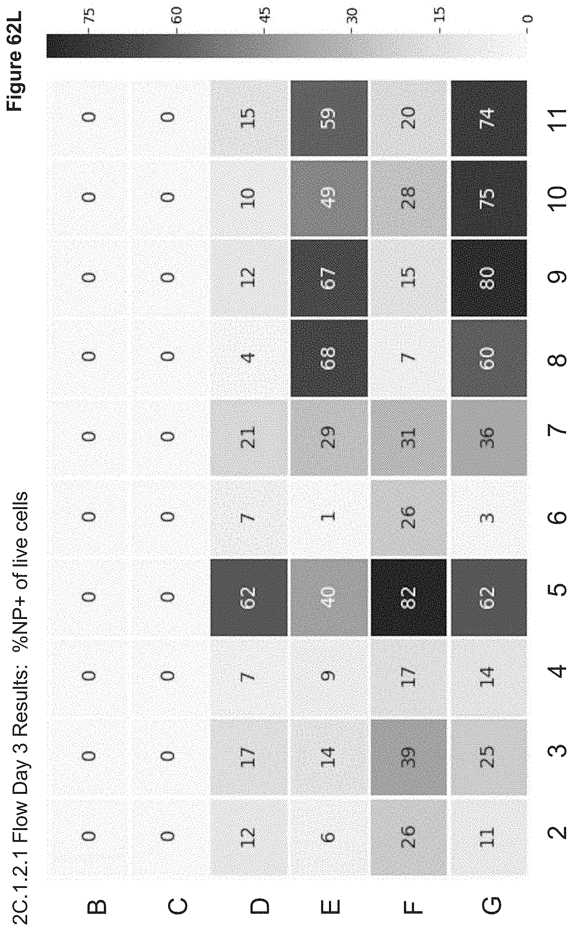
D00141
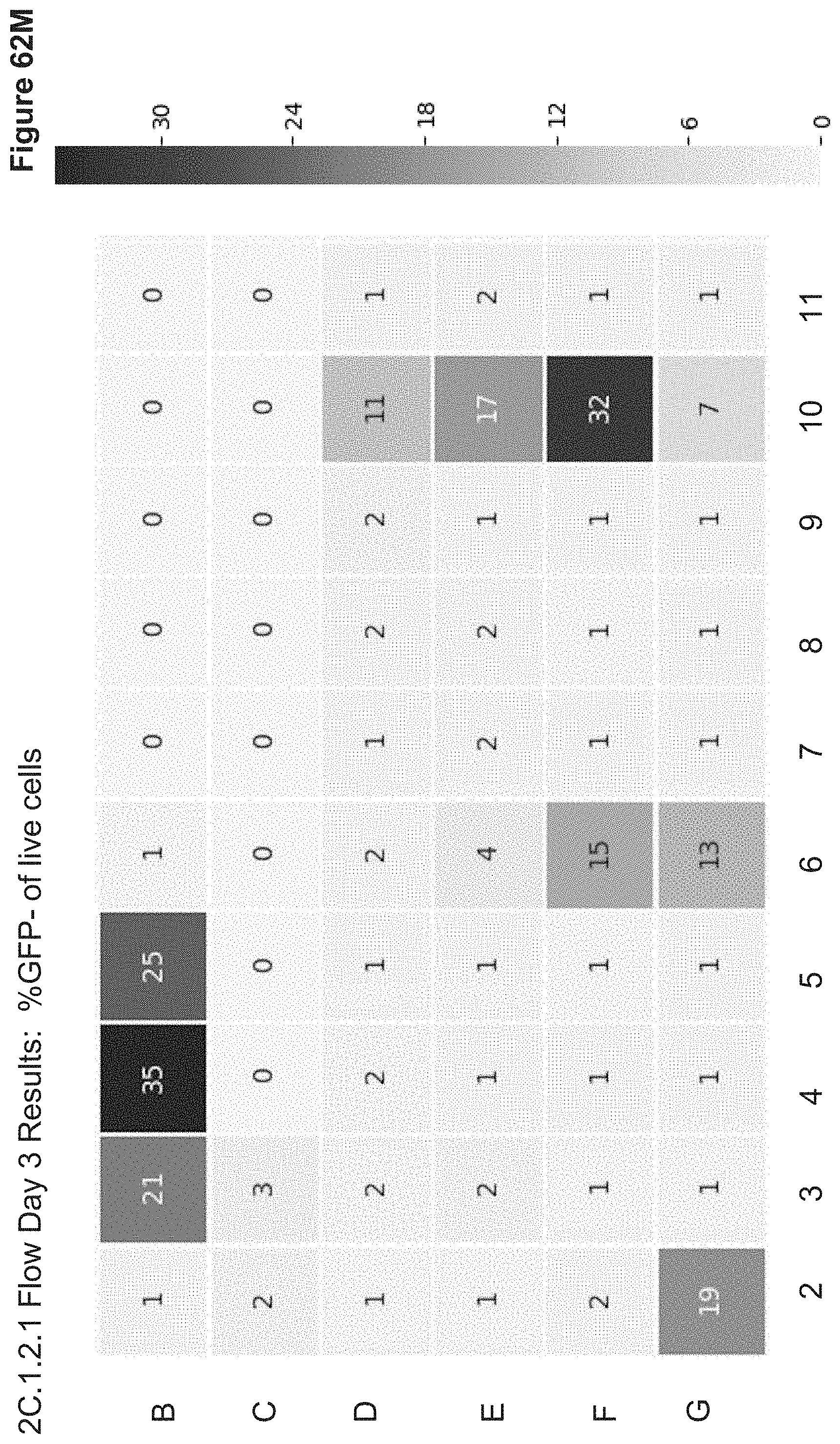
D00142

D00143
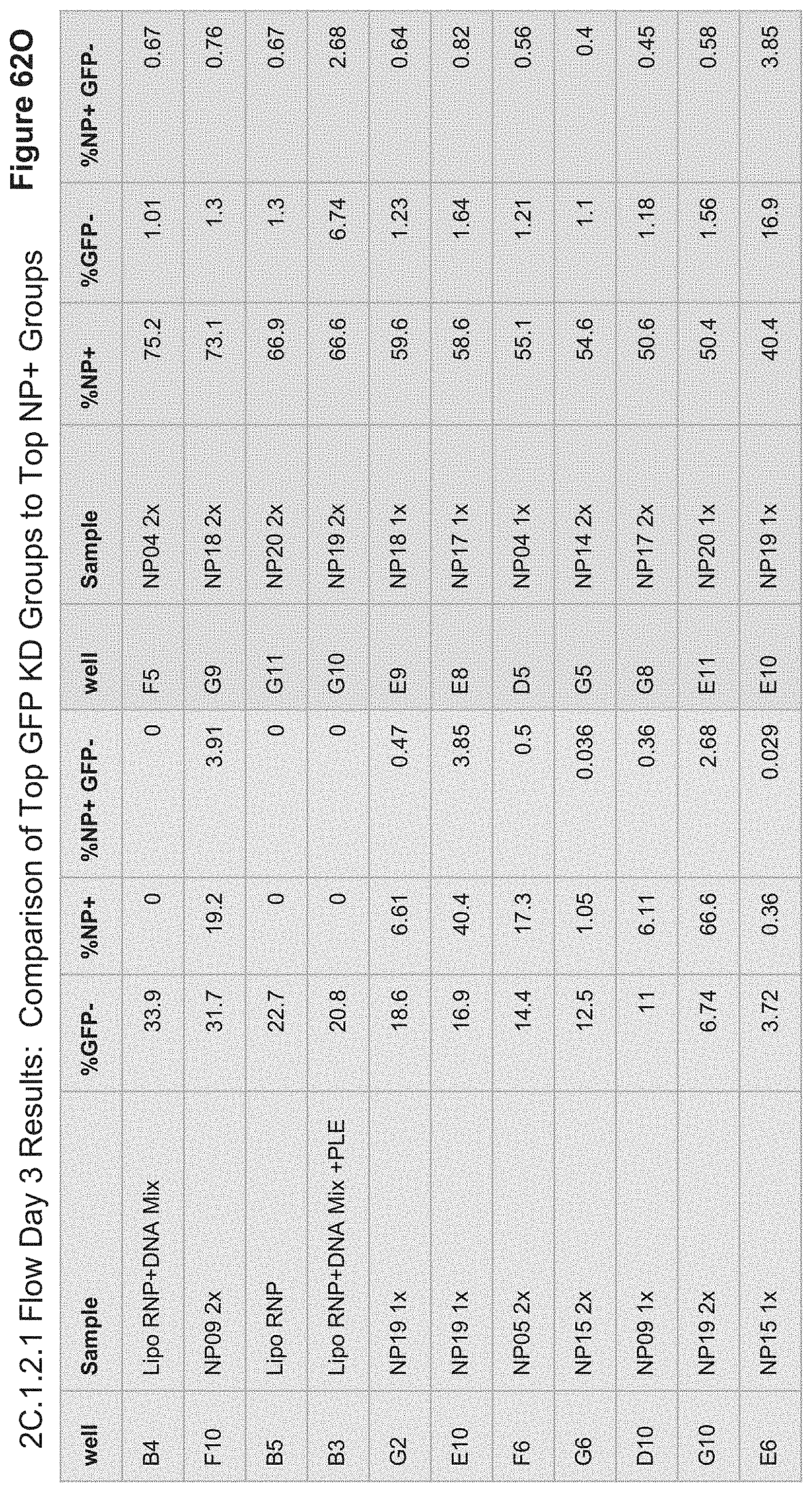
D00144

D00145

D00146

D00147
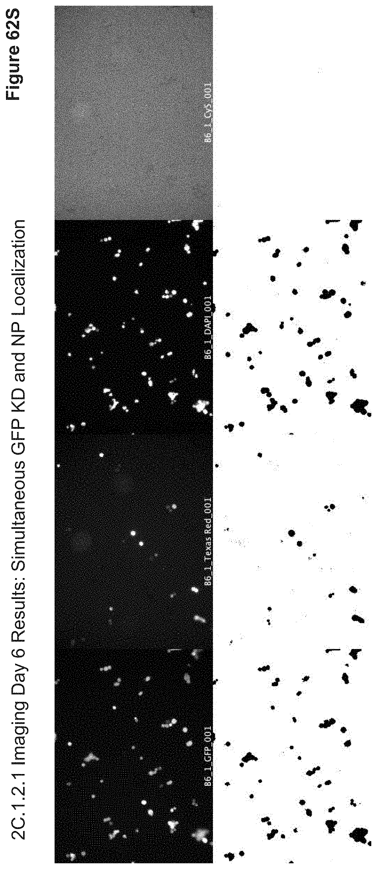
D00148
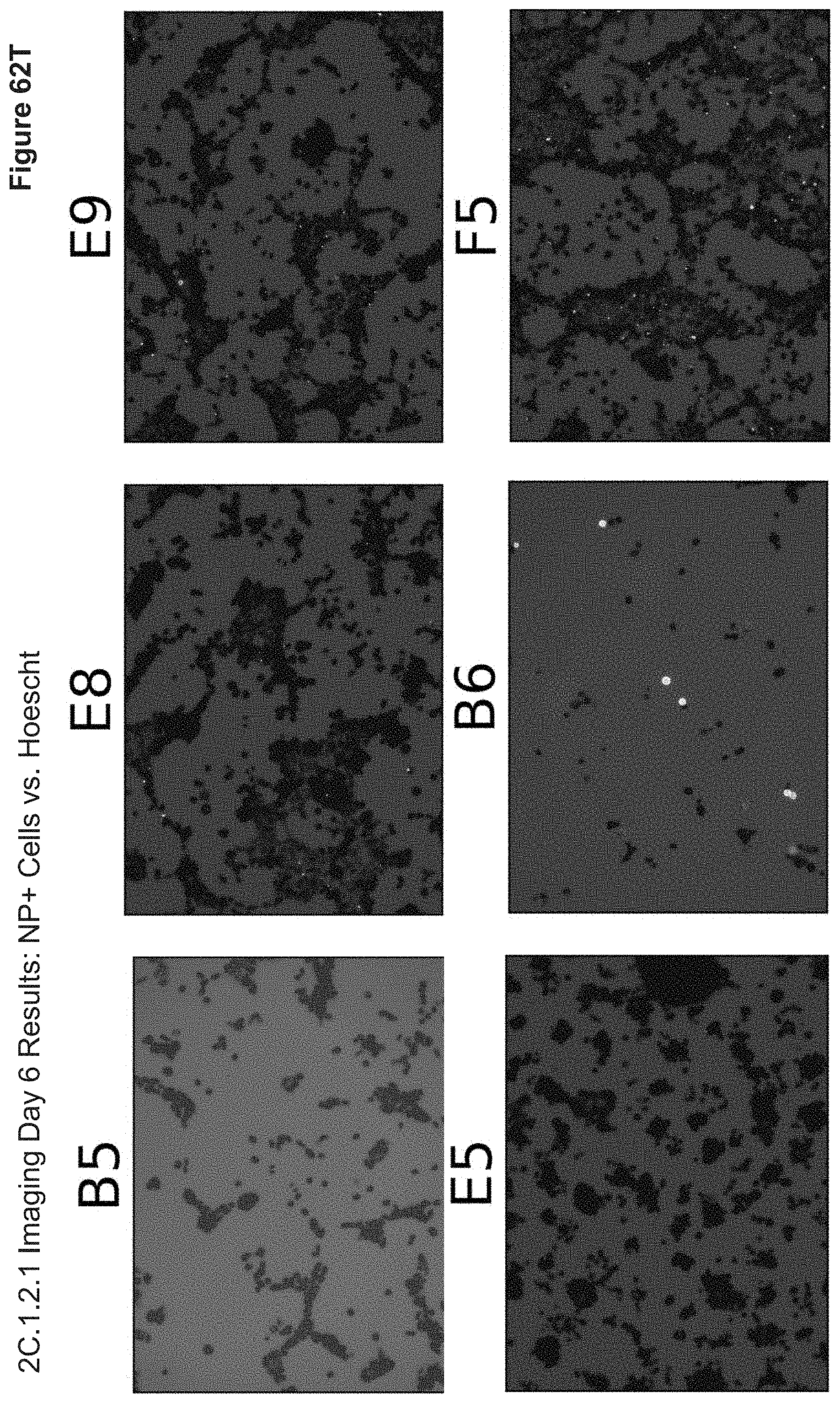
D00149

D00150
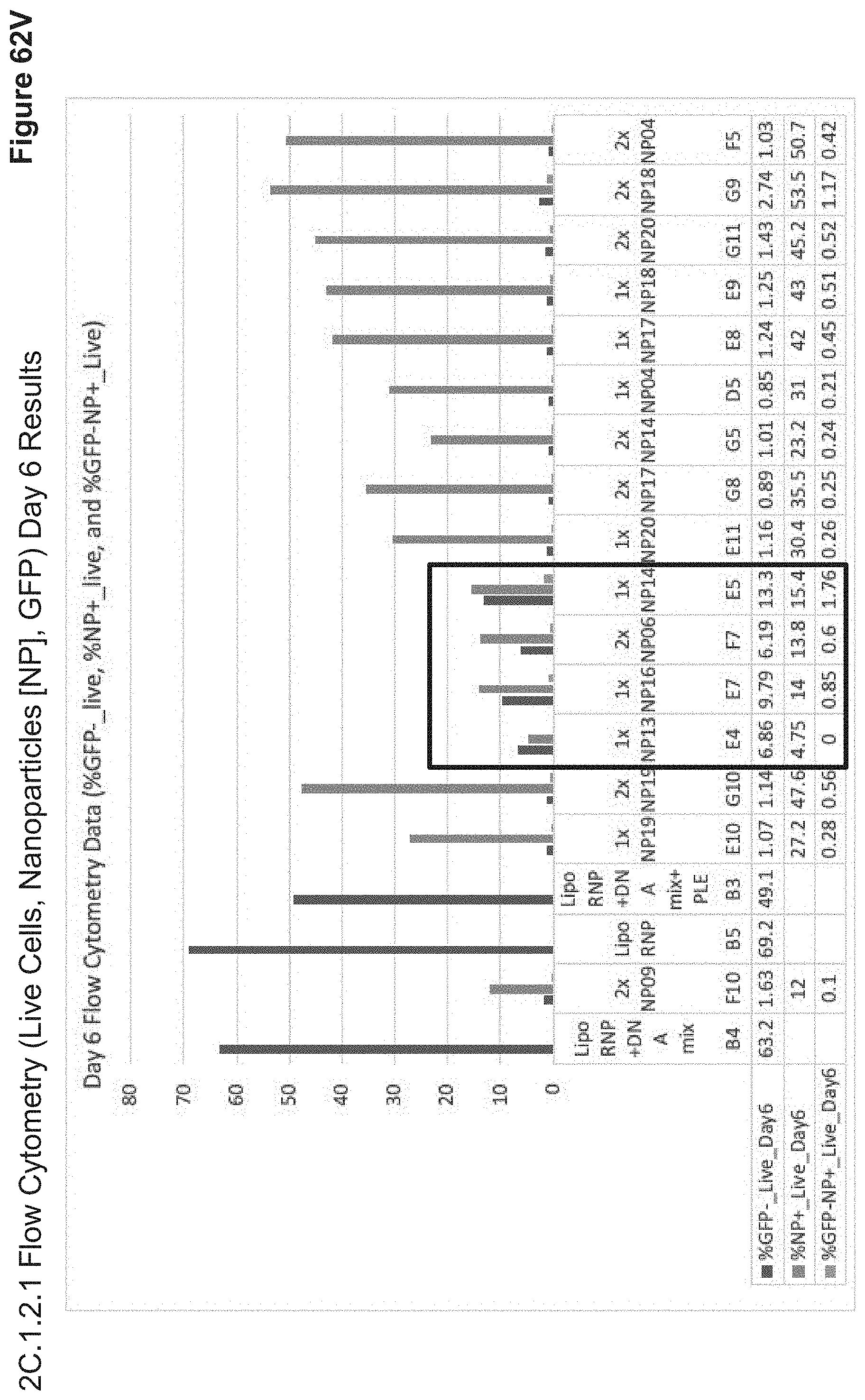
D00151

D00152
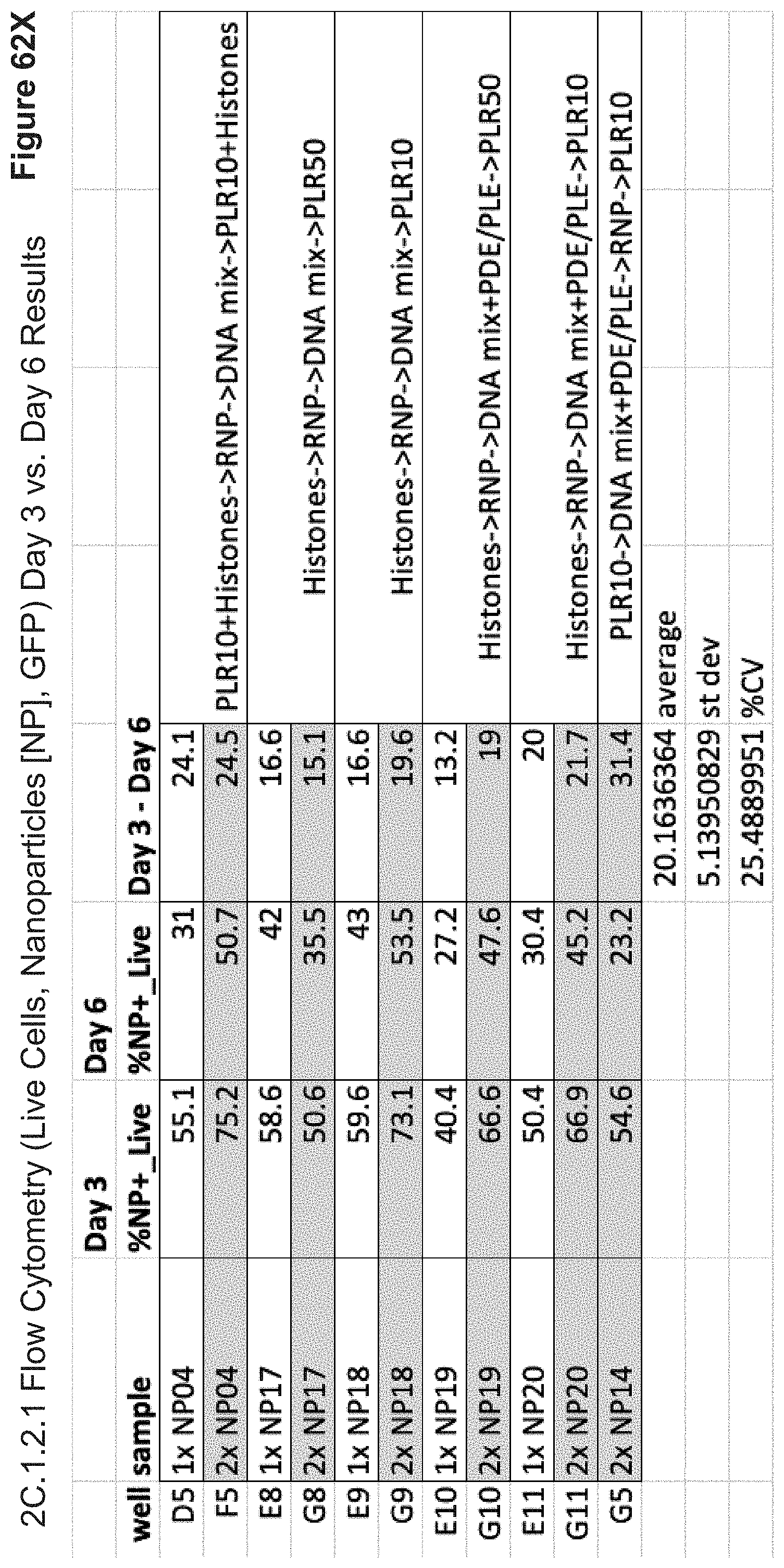
D00153

S00001
XML
uspto.report is an independent third-party trademark research tool that is not affiliated, endorsed, or sponsored by the United States Patent and Trademark Office (USPTO) or any other governmental organization. The information provided by uspto.report is based on publicly available data at the time of writing and is intended for informational purposes only.
While we strive to provide accurate and up-to-date information, we do not guarantee the accuracy, completeness, reliability, or suitability of the information displayed on this site. The use of this site is at your own risk. Any reliance you place on such information is therefore strictly at your own risk.
All official trademark data, including owner information, should be verified by visiting the official USPTO website at www.uspto.gov. This site is not intended to replace professional legal advice and should not be used as a substitute for consulting with a legal professional who is knowledgeable about trademark law.