Cellular Delivery of DNA Intercalating Agents
Getts; Robert C. ; et al.
U.S. patent application number 16/568702 was filed with the patent office on 2020-05-14 for cellular delivery of dna intercalating agents. The applicant listed for this patent is Genisphere, LLC Lankenau Institute of Medical Research. Invention is credited to Emanuela Dylgjeri, Mindy George-Weinstein, Jacquelyn Gerhart, Robert C. Getts, James Kadushin, Kelly Rhodes.
| Application Number | 20200148763 16/568702 |
| Document ID | / |
| Family ID | 50736166 |
| Filed Date | 2020-05-14 |


| United States Patent Application | 20200148763 |
| Kind Code | A1 |
| Getts; Robert C. ; et al. | May 14, 2020 |
Cellular Delivery of DNA Intercalating Agents
Abstract
Compositions and methods for targeted delivery of active agents to cells are provided. The compositions comprise a wholly or partially double-stranded synthetic DNA carrier, and an active agent intercalated in double-stranded portions of the DNA carrier. The DNA carrier may also be linked to a targeting agent. The compositions are useful for delivering an active agent into a targeted cell type, for example a cytotoxic agent.
| Inventors: | Getts; Robert C.; (Collegeville, PA) ; Kadushin; James; (Gilbertsville, PA) ; George-Weinstein; Mindy; (Wynnewood, PA) ; Gerhart; Jacquelyn; (Wynnewood, PA) ; Dylgjeri; Emanuela; (Hatfield, PA) ; Rhodes; Kelly; (Hatfield, PA) | ||||||||||
| Applicant: |
|
||||||||||
|---|---|---|---|---|---|---|---|---|---|---|---|
| Family ID: | 50736166 | ||||||||||
| Appl. No.: | 16/568702 | ||||||||||
| Filed: | September 12, 2019 |
Related U.S. Patent Documents
| Application Number | Filing Date | Patent Number | ||
|---|---|---|---|---|
| 15631226 | Jun 23, 2017 | |||
| 16568702 | ||||
| 14778716 | Sep 21, 2015 | |||
| PCT/US2014/031192 | Mar 19, 2014 | |||
| 15631226 | ||||
| 61803863 | Mar 21, 2013 | |||
| Current U.S. Class: | 1/1 |
| Current CPC Class: | A61K 47/549 20170801; Y02A 50/30 20180101; A61P 35/00 20180101; A61K 47/6849 20170801; Y02A 50/411 20180101; A61K 31/704 20130101; A61K 47/62 20170801; C07K 16/28 20130101; A61K 47/6859 20170801 |
| International Class: | C07K 16/28 20060101 C07K016/28; A61K 31/704 20060101 A61K031/704; A61K 47/68 20060101 A61K047/68; A61K 47/54 20060101 A61K047/54; A61K 47/62 20060101 A61K047/62 |
Claims
1. A composition for delivery of an active agent to cells or tissues comprising a wholly or partially double-stranded synthetic DNA carrier, an active agent intercalated in double-stranded portions of the DNA carrier, and a targeting agent linked to the DNA carrier, wherein the targeting agent is selected from the group consisting of a) an antibody or peptide that binds to the transferrin receptor, and b) an antibody or peptide that binds to G8 antigen.
2. The composition of claim 1, wherein the active agent is intercalated in the double stranded DNA through hydrogen bonding.
3. The composition of claim 1 wherein the DNA dendrimer comprises 3-216 hybridized single strands of DNA.
4. The composition of claim 1, wherein the active agent is selected from the group consisting of chemotherapeutics, anti-infective agents, antimalarial agents, antiviral agents and antifungal agents.
5. The composition of claim 4, wherein the active agent is berberine, an acridine, daunomycin, doxorubicin, daunorubicin, dactinomycin, cisplatin, carboplatin or thalidomide.
6. A pharmaceutical composition comprising the composition of claim 1 and a pharmaceutically acceptable excipient.
7. The pharmaceutical composition of claim 6, wherein the targeting agent is an antibody that binds to G8 antigen.
8. The pharmaceutical composition of claim 7, wherein the targeting agent is a monoclonal antibody that binds to G8 antigen and the active agent is doxorubicin.
9. The pharmaceutical composition of claim 6, wherein the targeting agent is a peptide that binds to transferrin receptor.
10. The pharmaceutical composition of claim 9, wherein the active agent is doxorubicin.
11. A method of making the composition of claim 1, comprising assembling single-stranded DNA oligonucleotides to form wholly or partially double-stranded DNA, linking the targeting agent to the wholly or partially double-stranded DNA, and contacting the wholly or partially double-stranded DNA with an active agent such that the active agent intercalates into double-stranded portions of the DNA.
12. The method of claim 11, wherein the single-stranded DNA oligonucleotides are assembled simultaneously with contacting the wholly or partially double-stranded DNA with the active agent.
13. The method of claim 11, wherein the wholly or partially double-stranded DNA is contacted with the active agent after assembly.
14. The method of claim 13, further comprising linking the targeting agent to the wholly or partially double-stranded DNA before contacting with the active agent.
15. A method of delivering an active agent to a cell or tissue comprising contacting the cell or tissue with the pharmaceutical composition of claim 6.
16. A method of treating a disease or condition in a patient comprising administering to the patient the pharmaceutical composition of claim 6.
17. The method of claim 16, wherein the targeting agent is an antibody or peptide that binds to G8 antigen, and the pharmaceutical composition is administered for prevention of posterior capsular opacification or for reducing the incidence of posterior capsular opacification.
18. The method of claim 17, wherein the targeting agent is a monoclonal antibody that binds to G8 antigen and the active agent is doxorubicin.
19. The method of claim 16, wherein the targeting agent is an antibody or peptide that binds to the transferrin receptor, and the pharmaceutical composition is administered for depletion of pancreatic tumor cells.
20. The method of claim 19, wherein the targeting agent is a peptide that binds to the transferrin receptor and the active agent is doxorubicin.
Description
CROSS REFERENCE TO RELATED APPLICATIONS
[0001] This is a continuation application of co-pending U.S. patent application Ser. No. 15/631,226, filed Jun. 23, 2017, which is a divisional application of U.S. patent application Ser. No. 14/778,716, filed Sep. 21, 2015, now abandoned, which is the National Stage entry of International Application No. PCT/US2014/031192, filed Mar. 19, 2014, which claims the benefit of U.S. Provisional Patent Application No. 61/803,863, filed Mar. 21, 2013, the disclosures of which are incorporated herein by reference in their entireties.
CROSS-REFERENCE TO SEQUENCE LISTING
[0002] The Sequence Listing text file created Jun. 18, 2014 (874 B) and identified as "00869100.txt" is hereby incorporated by reference.
TECHNICAL FIELD
[0003] The invention relates to delivery of active agents to cells using DNA carriers, wherein the active agent intercalates into the DNA of the carrier. The DNA carrier may further include a targeting agent that binds the DNA carrier to a particular cell type, thus facilitating delivery of the intercalated active agent to the cell.
BACKGROUND
[0004] Non-covalent interactions between active agents and DNA generally occur either by groove binding or intercalation. In particular, the field of anti-cancer therapies has produced several chemotherapeutic drugs that interact with DNA by intercalation, a process that involves insertion of a planar drug molecule between DNA base pairs. This results in a decrease in the helical turn and lengthening of the DNA. The association constants of intercalation may be 10.sup.5 to 10.sup.-11 M.sup.-1 due to favorable hydrophobic bonding, ionic bonding, hydrogen bonding and van der Waals forces. Intercalation is generally associated with fused ring structures, but non-fused ring systems are also known. Intercalation is recognized as a significant contributor to successful use of these compounds as antitumor, antineoplastic, antimalarial, antibiotic, and antifungal agents, because most are genotoxic, particularly when basic, cationic, or electrophilic functional groups are present.
[0005] DNA dendrimers are complex, highly branched molecules built from interconnected natural or synthetic monomeric oligonucleotide subunits. A DNA dendrimer is constructed from DNA monomers, each of which is made from two DNA strands that share a region of sequence complementarity located in the central portion of each strand (aka the "waist"), but have four non-complementary single-stranded "arms" for hybridization to the "arms" of other monomers. Monomers are assembled into dendrimers beginning with a single monomer which is hybridized to additional monomers via the complementary arms. Additional layers are added in a similar manner by hybridization of monomers to the arms of the monomers in the previous layer. By varying the number of layers and which arms are hybridized to additional monomers, DNA dendrimers of different sizes and shapes are produced. In order to prevent DNA dendrimers from falling apart over time, chemical "spot welds" may be added to the growing assembly using UV light via the intercalation and activation of psoralen cross-linkers. Dendrimers are typically purified according to their size and molecular weight on denaturing sucrose gradients after ultracentrifugation.
[0006] DNA dendrimers may also be covalently or non-covalently bound to a large variety of different types of molecules and particles, generally via linkage to the un-hybridized arms present on the outer layer. These molecules and particles may be signaling and targeting devices on the DNA dendrimers, allowing targeting of DNA dendrimers to specific molecular targets and detection of the binding of the dendrimers to the targets via detection of the signaling moieties. Signal generating moieties include a large number of fluorescent dyes, haptens, enzymes and other molecular materials, as well as particles such as gold nanoparticles and quantum dots. Targeting devices include DNA, RNA and PNA oligonucleotides, antibodies, antibody fragments, haptens, aptamers, peptides and others. Because the outside layer of monomers contains many un-hybridized arms, DNA dendrimer constructs act as signal amplifiers in a large variety of in-vitro applications, generally for the detection of specific nucleic acids and proteins, but also as detection devices in electronic devices. Applications include signal amplification on DNA and protein microarrays, ELISAs and ELOSAs, Luminex bead assays, in-situ hybridization, and others. The use of labeled and targeted DNA dendrimers has been extensively published in research studies and these materials are available as commercial research products sold or produced by Genisphere LLC (Hatfield, Pa.).
[0007] DNA dendrimers have also been shown to have potential use as delivery and transfection devices in both in-vitro and in-vivo applications. See, e.g., U.S. 2005/0089890, WO 2008/147526 and WO 2010/017544, each of which is incorporated by reference in its entirety. Specifically, DNA dendrimers are bound with targeting devices (e.g. an antibody specific for a cell surface feature capable of eliciting an cellular endocytotic internalization event) which bind to surface features on cells targeted to receive the delivery of a cargo (e.g. a drug). Cargos passively associated with the targeted DNA dendrimer enter the cell simply by spatial association with the dendrimer, but cargos may also be bound to the dendrimer via a number of attachment strategies, including linking to the outer arms.
SUMMARY
[0008] The present invention relates to an improved method for delivery of active agents to cells using DNA carriers. The DNA carrier comprises DNA, at least a portion of which is in double-stranded form, and an active agent that intercalates in the double-stranded DNA. In certain embodiments the DNA carrier also comprises a targeting agent that binds to a molecule, cell, or tissue where the active agent is to be delivered. In particular embodiments, the DNA carrier is a DNA dendrimer having the active agent intercalated in the double-stranded portions of the dendrimer. Upon binding of the DNA carrier to the targeted molecule, cell or tissue, the dendrimer is internalized by cells into the endosomes where the acidic pH facilitates release of the active agent from the DNA to produce a cytotoxic or other biological effect.
BRIEF DESCRIPTION OF THE DRAWINGS
[0009] FIG. 1A shows labeling of living cells with LYSOSENSOR dye. FIG. 1B shows the cells containing internalized dendrimers. FIG. 1C is a merged image of FIG. 1A and FIG. 1B showing that the dendrimers are internalized into acidic compartments of the cells.
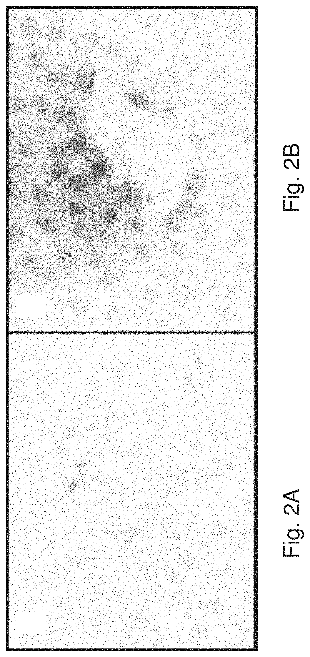
[0010] FIGS. 2A-2D show the results of Example 2: untreated cells (FIG. 2A), untargeted Dox dendrimers (FIG. 2B), G8 mAb conjugated Dox dendrimers (FIG. 2C and FIG. 2D).
DETAILED DESCRIPTION
[0011] Before describing several exemplary embodiments of the invention, it is to be understood that the invention is not limited to the details of construction or process steps set forth in the following description. The invention is capable of other embodiments and of being practiced or being carried out in various ways.
[0012] Reference throughout this specification to "one embodiment," "certain embodiments," "one or more embodiments" or "an embodiment" means that a particular feature, structure, material, or characteristic described in connection with the embodiment is included in at least one embodiment of the invention. Thus, the appearances of the phrases such as "in one or more embodiments," "in certain embodiments," "in one embodiment" or "in an embodiment" in various places throughout this specification are not necessarily referring to the same embodiment of the invention. Furthermore, the particular features, structures, materials, or characteristics may be combined in any suitable manner in one or more embodiments.
[0013] In a first aspect, the invention relates to synthetic DNA carriers for delivery of intercalating active agents to cells or tissues, wherein the DNA is wholly or partially double-stranded, and the DNA carrier comprises an active agent intercalated in the double-stranded portions of the DNA. The oligonucleotide component of the synthetic DNA carrier may be a linear or a branched oligonucleotide. Each double-stranded DNA portion of the DNA carrier is 5 bp in length or longer, for example about 20-35 bp, about 35-50 bp, about 50-100 bp, or about 100-200 bp. The synthetic DNA carriers are constructed in vitro and preferably comprise multiple double-stranded portions to increase the load of the active agent. Examples of synthetic DNA carriers comprising multiple double-stranded portions include DNA dendrimers. If the synthetic DNA carrier comprises a linear oligonucleotide, the oligonucleotide may comprise a single double-stranded portion or multiple double-stranded portions. The overall length of such linear oligonucleotides in the DNA carriers is typically at least 10 nt, for example 10-20 nt, 10-50 nt, 10-80 nt, 10-150 nt, or 10-200 nt in length.
[0014] In one embodiment, the synthetic DNA carrier may further comprise a targeting agent that binds to a selected cell or tissue type to bind the DNA carrier to the selected cell or tissue type, thus facilitating delivery of the active agent to the selected cell or tissue type. Binding of the targeting agent to the selected cell or tissue type may be via binding of the targeting agent to a cell surface protein or cell surface receptor. Suitable targeting agents include antibodies, ligands or peptides that bind to the cell surface protein or cell surface receptor. The targeting agent is typically linked to the DNA dendrimer arms either covalently or non-covalently (e.g., the targeting agent is linked to an oligonucleotide that is complementary to a dendrimer arm and hybridized to the dendrimer arm).
[0015] Examples of cell surface receptors and proteins that may be targeted by the targeting agent include the transferrin receptor (TfR/CD71), the HIV-1 envelope glycoprotein (Env), malarial antigen markers, and the folate receptor. Preferably, the cell surface receptor or protein is specifically expressed on infected, damaged or diseased cells, or is overexpressed on infected, damaged or diseased cells. Specific examples of targeting agents include an anti-transferrin antibody or a TfR-targeting peptide such as THRPPMWSPVWP (SEQ ID NO:1) that binds to the transferrin receptor, which is overexpressed in various cancers. As an example, the targeting agent may also be the G8 monoclonal antibody which binds to a protein of approximately 170 kDa found on the surface of Myo/Nog cells and their derivatives. Myo/Nog cells are identified by expression of mRNA for the skeletal muscle specific transcription factor MyoD, the bone morphogenetic protein (BMP) inhibitor noggin and the cell surface G8 epitope. Other examples of useful targeting moieties include those that bind to PDGF receptor, EGFR family receptors, ICAM, CD receptors, MMP-9, interleukin receptors, folic acid receptor, and others known in the art.
[0016] In another embodiment, the active agent intercalated in the double-stranded portions of the DNA carrier is a chemotherapeutic agent, an anti-infective agent, an antimalarial agent, and antiviral agent or an antifungal agent. Specific examples of such agents include berberine (antifungal, antineoplastic), acridines (e.g., proflavine or quinacrine, antiseptic), cancer chemotherapeutics (e.g., daunomycin, doxorubicin, daunorubicin, dactinomycin, cisplatin, carboplatin, voreloxin), cryptolepine (antimalarial cytotoxic) and thalidomide (treatment of multiple myeloma).
[0017] In any of the foregoing embodiments, the DNA carrier may comprise a DNA dendrimer, including a 2-layer, 4-layer, 6-layer or 8-layer DNA dendrimer. The size of the DNA dendrimer is selected as needed to facilitate delivery of the active agent to cells and tissues (e.g., via the circulatory system or local injection), and to maximize uptake of the DNA carrier by the cell. In general, the DNA carrier comprises about 3-216 oligonucletoide single strands.
[0018] In a second aspect, the invention relates to pharmaceutical compositions comprising the DNA carrier in any of the foregoing embodiments and at least one pharmaceutically acceptable excipient. The pharmaceutically acceptable excipients are any suitable vehicle which can be used to formulate the DNA carriers for administration to a patient, cells or tissues to achieve a therapeutic or other biological result. The excipients are selected from those excipients that are compatible with double-stranded DNA (i.e., they will not denature the DNA carrier prior to delivery to the selected cell or tissue) as well as compatible with the active agent and the targeting agent, if present. Such pharmaceutically excipients include physiological buffers, saccharides and oligosaccharides, stabilizers, thickeners, lubricants, waxes, chelating agents, surfactants, diluents, anti-oxidants, binders, preservatives, coloring agents, emulsifiers, lipid micelles, or other conventional constituents of a pharmaceutical formulation. In certain embodiments the pharmaceutical composition is formulated for parenteral delivery to a patient, for example by injection or infusion. In this case it is preferable that the DNA carrier comprises a targeting agent so that the DNA carrier is directed from the circulation to the desired cell or tissue type. In other embodiments, the pharmaceutical composition may be adapted for oral administration or for local administration to the cell or tissue where delivery of the active agent is desired (e.g., topical formulations).
[0019] In a further aspect, the invention relates to methods of making the DNA carriers described in any of the foregoing embodiments. In one embodiment, the DNA carriers are assembled by hybridizing single-stranded DNA to form wholly or partially double-stranded DNA, and combining the wholly or partially double-stranded DNA with the active agent under conditions to allow the active agent to intercalate into the double-stranded portions. Excess active agent is removed, for example by chromatography, to produce the DNA carrier. In a specific embodiment, chromatography to remove excess active agent is performed using a spin column.
[0020] In an alternative embodiment, the active agent may be incorporated in the DNA carrier during hybridization of the single-stranded DNA to assemble the wholly or partially double-stranded DNA, followed by removal of excess active agent as described above.
[0021] In a further embodiment, the methods of making the DNA carriers comprise assembly of a DNA dendrimer, combining the DNA dendrimer with the active agent under conditions to allow the active agent to intercalate into the double-stranded portions of the DNA dendrimer, and removing excess active agent, for example by chromatography (e.g., a spin column).
[0022] In a specific embodiment, in which the DNA carrier is linked to a targeting agent, the DNA carrier is assembled from single-stranded DNA to form wholly or partially double-stranded DNA, and the targeting agent is linked to the assembled DNA. The linkage of the targeting agent to the DNA may be accomplished by covalent attachment as is known in the art, or it may be a non-covalent linkage such as hybridization. Such methods include, for example, linkage via disulfide bonds, N-hydroxysuccinimide (NHS) ester dependent condensation reactions, bifunctional cross-linking, use of polycationic compounds to bridge the targeting agent to the DNA carrier via charge-charge interactions, or direct or indirect hybridization to the DNA carrier as described in WO2010/017544. For example, a single stranded capture oligonucleotide is appended to the arms of a DNA dendrimer, and single-stranded oligonucleotides complementary to the capture sequence are linked to the targeting agent. The complementary sequence on the targeting agent is hybridized to the capture sequence on the DNA dendrimer to link the targeting agent to the DNA dendrimer. The hybridized capture and complementary sequences may optionally be cross-linked. The active agent may be incorporated in the DNA carrier during assembly of the wholly or partially double-stranded DNA or after linkage of the targeting agent to the DNA carrier. If the targeting agent is hybridized to the DNA carrier via a capture sequence, as described above, the double-stranded DNA formed by the linkage can also intercalate the active agent.
[0023] In an alternative embodiment of making a DNA carrier linked to a targeting agent, a single-stranded oligonucleotide is attached to the targeting agent in a first step, and then hybridized to a complementary single-stranded oligonucleotide sequence to form the wholly or partially double-stranded DNA of the DNA carrier.
[0024] In any of the foregoing methods of making the DNA carriers, the DNA dendrimer may be assembled by any of the methods known in the art, for example as described by T. W. Nilsen, et al. J. Theor. Biol. (1997) 187, 273-284.
[0025] In yet a further aspect, the invention relates to methods of delivering an active agent to cells or tissues comprising contacting the cells or tissues with a DNA carrier according to any of the foregoing embodiments such that the DNA carrier is taken up by the cell, and the active agent is released inside the cell to produce a cytotoxic or other biological effect on the cell. The cells or tissues may be contacted with the DNA carrier in vivo or in vitro. If cells are contacted with the DNA carrier in vitro, the cells will typically be in cell culture and the DNA carrier is added to the culture medium, incubated with the cells for a period of time to allow binding to the cell surface, and the excess DNA carrier is removed. After sufficient time for endocytosis and release of the active agent, the cytotoxic or biological activity of the active agent on the cells can be visualized microscopically. If the cells are contacted with the DNA carrier in vivo, the DNA carrier is administered to the patient or animal via injection, infusion, local administration to the site of desired action, or by other appropriate methods, for a period of time to allow binding to the cell surface and endocytosis. The cytotoxic or biological activity of the active agent released in the cells can be determined by monitoring a physiological response or by microscopic examination of samples of the affected tissues.
[0026] In a further aspect, the invention relates to methods of treating a disease or condition by delivering the DNA carriers to a patient in need thereof, using the foregoing in vivo methods of delivery active agents to cells or tissues. For treatment specificity, the targeting agent is generally selected to be specific for the cell type in which the cytotoxic or other biological activity is desired. The active agent is selected to be therapeutically active against the disease or condition being treated, and the DNA carrier is administered to the patient in amount and for a period of time sufficient to treat, cure or improve the symptoms of the disease or condition. Diseases and conditions treatable by administration of the DNA carriers include any disease or condition for which an active agent is available that intercalates into double-stranded DNA. Non-limiting examples include cancer, malaria, fungal diseases, viral diseases, fibrotic diseases, and the like.
[0027] To effect treatment, the synthetic DNA carrier may be administered locally to the tissue or cells to be treated, for example by topical application or injection into the tissue or cells. Alternatively, the synthetic DNA carrier may be administered systemically, for example by intravenous injection or infusion, or by oral administration. In either embodiment, a targeting agent linked to the DNA carrier, if present, facilitates binding of the DNA carrier to the cell type to be treated and delivery of the active agent to that cell type.
[0028] In a specific embodiment, posterior capsular opacification (PCO) is prevented, or the incidence thereof is reduced, by administration of a DNA carrier comprising DNA dendrimers linked to a monoclonal antibody targeting the G8 epitope, and a DNA-intercalating cytotoxin. PCO occurs in certain adults and children after cataract surgery, and arises from differentiating lens epithelial cells and myofibroblasts that affect the transparency of the capsule surrounding the lens. There is currently no effective treatment for preventing PCO. It has been discovered that myofibroblasts in human lens tissue originate from Myo/Nog cells that express the skeletal muscle specific transcription factor MyoD, the bone morphogenetic protein inhibitor noggin, and a cell surface molecule recognized by the G8 monoclonal antibody (mAb). Depletion of Myo/Nog cells in explant cultures of human lens tissue procured from patients undergoing cataract surgery was accomplished by lysing them with DNA carriers (DNA dendrimers) according to the invention, using the G8 mAb as the targeting agent and intercalated doxorubicin. Myo/Nog cells were specifically eliminated, as shown in the following examples. From this data, it can be extrapolated that the accumulation of myofibroblasts over time would also be prevented.
EXAMPLES
Example 1: Internalization of G8 mAb Conjugated Dendrimers in Rhabdomyosarcoma Cells
[0029] 2-Layer DNA Dendrimer Preparation:
[0030] DNA dendrimers were manufactured as previously disclosed (see, e.g., U.S. Pat. Nos. 5,175,270, 5,484,904, 5,487,973, 6,110,687 and 6,274,723, each of which is incorporated by reference in its entirety). Briefly, a DNA dendrimer was constructed from DNA monomers, each of which is made from two DNA strands that share a region of sequence complementarily located in the central portion of each strand. The two strands anneal to form the monomer the resulting structure can be described as having a central double-stranded "waist" bordered by four single-stranded "arms". This waist-plus-arms structure comprises the basic 3DNA.RTM. monomer. The single-stranded arms at the ends of each of the five monomer types are designed to interact with one another in precise and specific ways. Base-pairing between the arms of complementary monomers allows directed assembly of the dendrimer through sequential addition of monomer layers. Assembly of each layer of the dendrimer includes a cross-linking process where the strands of DNA are covalently bonded to each other, thereby forming a completely covalent molecule impervious to denaturing conditions that otherwise would cause deformation of the dendrimer structure. In addition, 38 base oligonucleotides that serve as complementary capture oligos are ligated to the 5' ends of available dendrimer arms via a simple T4 DNA ligase dependent ligation reaction, as follows:
[0031] Attaching a Capture Sequence to a DNA Dendrimer:
[0032] To attach the G8 momoclonal Antibody (G8mAb) to the DNA Dendrimer, a capture sequence was first ligated to 10-15% of the dendrimer arms. The complementary oligonucleotide to this capture sequence was conjugated to the G8 monoclonal antibody using commercially available chemistry (Solulink, www.solulink.com) and hybridized in a molar ratio to occupy all of the available capture sequences. Approximately 2-5 G8mAbs were attached per dendrimer molecule as summarized below.
[0033] Small (15-100 nucleotides) DNA or RNA capture oligonucleotides (or other biochemical analogs) were covalently attached to the ends of the dendrimer arms via a simple nucleic acid ligation reaction utilizing a bridging oligonucleotide that overlaps adjacent portions of the dendrimer arm and the capture oligonucleotide, thereby bridging the capture oligonucleotide to the end of the dendrimer arm. The bridging oligonucleotide overlapped as least 5 bases of each of the adjacent dendrimer arm and capture oligonucleotide sequences to facilitate the ligation activity of a nucleic acid ligase enzyme (preferably T4 DNA ligase enzyme), with at least 7 bases of overlap of each sequence preferred. The bridging oligonucleotide may also serve as a nucleic acid blocker for its complementary sequences when the dendrimer is used for specific targeting of non-dendrimer nucleic acids or other molecules.
[0034] The following components were added to a microfuge tube: [0035] 1. 2 layer DNA dendrimer (500 ng/.mu.L) in 1.times.TE buffer 5.4 .mu.L (2680 ng) [0036] 2. a(-)LIG-BR7 Bridging oligo (14mer) (50 ng/.mu.L) 2.7 .mu.L (134 ng) [0037] 3. 10.times. Ligase buffer 10.2 .mu.L [0038] 4. Nuclease free water 81.7 .mu.L [0039] 5. Cap03 capture oligo (38mer) (50 ng/.mu.L) 4.0 .mu.L (200 ng) [0040] 6. T4 DNA Ligase (1 U/.mu.L) 10.0 .mu.L (10 units)
[0041] The first four reactants were added together, heated to 65.degree. C. and cooled to room temperature. The 5th and 6th reactants were then added and incubated for 45 minutes. The ligation reaction was stopped by adding 2.8 .mu.L of 0.5M EDTA solution. Non-ligated oligonucleotide was removed via the use of 100 k cutoff centrifugal filters (Millipore Corp.), and during the purification washes, the buffer was changed into 1.times. sterile PBS, pH7.4. The capture oligonucleotide is linked to a first single-stranded surface arm of the dendrimer.
[0042] Antibody Attachment and Fluorescent Labeling of the DNA Dendrimer:
[0043] In order to target the cell surface and initiate internalization, a G8 monoclonal antibody was coupled to a DNA dendrimer. The G8 monoclonal antibody was covalently attached to an oligonucleotide that is complementary to the capture sequence which was previously ligated to the dendrimer. Briefly, the capture sequence complement (cplCap03), 5'-TTCTCGTGTTCCGTTTGT ACTCTAAGGTGGATTTTT-3' (SEQ ID NO:2), was covalently coupled using commercial chemistry to G8 monoclonal antibody (Solulink) by the 3' end and purified by HPLC to remove excess reagents. These conjugates were then hybridized to dendrimer capture sequences during the final assembly of the reagents (refer to 3DNA preparation section below). Additionally, a synthetic oligonuclotide (IDT Technologies) complementary to the outer surface arms of the DNA dendrimer was prepared having two fluorescent labels (Cy3), one each on the 3' and 5' prime ends. This oligonucleotide was then hybridized to the outer arms of the above-prepared G8 mAb target dendrimer in an amount equal to 18 moles oligonucleotide to 1 mole DNA dendrimer. The G8 mAb targeted Cy3 labeled DNA dendrimer was used for targeting and internalization studies on Rhabdomyosarcoma cells.
[0044] Internalization of the G8 Monoclonal Antibody Conjugated with Cy3 Labeled Dendrimers in Rhabdomyosarcoma Cells:
[0045] Human embryonal and alveolar rhabdomyosarcoma cells (cell lines 136 and 2061 from ATCC) were cultured on glass coverslips in 100 mm tissue culture dishes (12 coverslips/plate) in 8 ml medium for 24 hours. The 136 cell line was cultured in DMEM containing 10% fetal bovine serum. The 2061 cell line was cultured in RPMI containing 10% FBS. Coverslips were transferred to 35 mm tissue culture dishes (2 per coverslips/dish) containing 1 ml of the following media: [0046] 1. Culture media containing LysoSensor Green DND 189 stock solution (1 mM) (Molecular Probes/Invitrogen) diluted 1:500 in (2 uM working solution). [0047] 2. Culture media containing 2 uM LysoSensor plus G8:4n Dendrimer-Cy3 diluted 1:75.
[0048] Cells were incubated at 37.degree. C. in 5% CO.sub.2 in air for 1 hour. Cells were rinsed in 37.degree. C. PBS and fixed in 2% paraformaldeyde for 10 minutes. Cells were visualized by epifluorescence microscopy. Various dilutions and incubation times were tested. The 1:500 Lysosensor plus 1:75 G8:dendrimer incubated for 1 hour yielded the best results. The results are displayed in FIG. 1A, FIG. 1B and FIG. 1C, which show successful internalization of the labeled dendrimers in Rhabdomyosarcoma cells. FIG. 1A shows cells labeled with LYSOSENSOR dye that fluoresces green at acid pH (concentrated in the dark spots within the cells in FIG. 1A). FIG. 1B shows red fluorescence from Cy3 labeled dendrimers within the cells (concentrated in the dark spots within the cells in FIG. 1B). FIG. 1C is a merged image of FIG. 1A and FIG. 1B, showing co-localization of the green and the red signals, which is visualized as yellow, and shown as the dark spots indicated by the arrows within the cell in FIG. 1C). These results confirm that the G8 mAb coupled to the DNA dendrimer is internalized into acidic compartments of the cell (i.e., the lysosomes).
Example 2: Targeted Depletion of Myo/Nog Cells
[0049] 2-Layer DNA Dendrimer Preparation:
[0050] 2-Layer DNA dendrimers were prepared with attached capture sequences as described in Example 1. The capture sequence was ligated to 10-15% of the dendrimer arms.
[0051] Attaching G8 mAb to the DNA Dendrimer:
[0052] The G8 monoclonal antibody (G8 mAb) was also attached to the DNA Dendrimer as described in Example 1. First, the 3' end of the oligonucleotide complementary to the capture sequence (cplCap03) was conjugated to the G8 monoclonal antibody using commercially available chemistry (Solulink, www.solulink.com). The conjugate was purified by HPLC to remove excess reagents. The capture sequence and the complement of the capture sequence were then hybridized in a molar ratio to occupy all of the available capture sequences. Approximately 2-5 G8 mAbs were attached per dendrimer molecule as summarized below.
[0053] Preparation of Doxorubicin Double Stranded Oligonucleotide G8 Antibodies:
[0054] 1.282 .mu.g of G8 mAb oligonucleotide conjugate (as prepared above) was combined with 1.282 .mu.g of the complementary oligonucleotide sequence (SEQ ID NO:2) and 1500 .mu.l of 200 .mu.M Dox solution, and the final volume was adjusted to 3000 .mu.l using sterile 1.times.PBS according to the table below. The mixture was incubated at 37.degree. C. for 30 minutes. The final concentration of Dox in 3 mL solution was 100 .mu.M. The reagent was purified using Quickspin High Capacity Sephadex G50 Columns (Roche) according to the manufacture's protocol. Sixteen microliters of 5M NaCl (Life Technologies) was added to achieve an 86 mM NaCl concentration. The final Doxorubicin was determined using fluorometry.
TABLE-US-00001 Component Conc Mass req Volume (.mu.l) 2 G8 Ab oligo conjugate 27 ng/.mu.l 1282.1 47.5 (mouse IgM MGW) 3 1X PBS-sterile + Dox 200 .mu.M 1500.0 4 1X PBS-sterile 1452.5
[0055] Preparation of Doxorubicin 2-Layer Dendrimers (No Targeting):
[0056] 2 Layer Cap 03 dendrimer (3 .mu.g) was mixed with 1500 ul of 200 .mu.M Dox solution and the final volume was adjusted to 3000 .mu.l using sterile 1.times.PBS according to the table below and incubated at 37.degree. C. for 30 minutes. The final concentration of Dox in 3 mL solution was 100 .mu.M and final dendrimer concentration was 10 ng/.mu.l). The reagent was purified using Quickspin High Capacity Sephadex G50 Columns (Roche) according to the manufacture's protocol. Sixteen microliters of 5 M NaCl (Life Technologies) was added to achieve an 86 mM NaCl concentration. The final Doxorubicin and Dendrimer concentrations were determined using fluorometry and UV/Vis spectroscopy.
TABLE-US-00002 Component Conc Mass req Volume (ul) 1 2-Layer Cap03 359.2 ng/.mu.l 30000.0 83.5 2 1X PBS-sterile + Dox 200 .mu.M 1500.0 3 1X PBS-sterile 1416.5 3000.000 .mu.l total vol
[0057] Preparation of Doxorubicin 2-Layer G8 Targeted Dendrimers:
[0058] 2 Layer Cap 03 dendrimer (3 .mu.g) was mixed with 1.282 .mu.g of G8 mAb oligonucleotide conjugate (as prepared above) and 1500 .mu.l of 200 .mu.M Dox solution, and the final volume was adjusted to 3000 .mu.l using sterile 1.times.PBS according to the table below. The mixture was incubated at 37.degree. C. for 30 minutes. The final concentration of Dox in 3 mL solution was 100 .mu.M and final dendrimer concentration was 10 ng/.mu.l). The reagent was purified using Quickspin High Capacity Sephadex G50 Columns (Roche) according to the manufacture's protocol. Sixteen microliters of 5 M NaCl (Life Technologies) was added to achieve an 86 mM NaCl concentration. The final Doxorubicin and Dendrimer concentrations were determined using fluorometry and UV/Vis spectroscopy.
TABLE-US-00003 Component Conc Mass req Volume (ul) 1 2-Layer Cap03 359.2 ng/.mu.l 30000.0 83.5 2 G8 Ab oligo conjugate 27 ng/.mu.l 1282.1 47.5 (mouse IgM MGW) 3 1X PBS-sterile +Dox 200 .mu.M 1500.0 4 1X PBS-sterile 1369.0
[0059] Targeting Myo/Nog Cells in Human Lens Tissue with the G8 Monoclonal Antibody Conjugated with DNA Dendrimers Intercalated with Doxorubicin:
[0060] Human anterior lens tissue removed by capsulorhexis during cataract surgery was collected in DMEM/F12 medium containing 1% Penicillin/Streptomycin (DF). Lens tissue was transferred to an 8-well chamber tissue culture slide containing 240 .mu.l of DF containing the following: [0061] 1. No additions [0062] 2. G8Ab/Cap03 oligo/CPL--Dox diluted 1:8 in DF [0063] 3. 2-layer G8 Ab/Cap03 oligo/Cpl Oligo--diluted 1:8 in DF [0064] 4. 2-layer Cap03--high diluted 1:8 in DF
[0065] Lenses floated in the chamber until they sank to the bottom of the slide. They were cultured for 24 hours in 5% CO.sub.2 in air at 37.degree. C. Tissues were rinsed in PBS and fixed in 2% paraformaldehyde for 10 minutes then double labeled with the G8 antibody and an anti-mouse IgM conjugated with Alexa 488 and reagents from the ROCHE TUNEL kit which fluoresce under the rhodamine channel.
[0066] The percentages of G8+ and TUNEL+ cells, the percentage of the G8+ cells with TUNEL, and the percentage of TUNEL+ cells with G8 were determined by epifluorescence microscopy. The results are shown in the following table:
TABLE-US-00004 % G8 % TUNEL % G8+ % TUNEL+ with TUNEL with G8 Untx 2 0.8 4 13 2-layer:high dox 4 2 0 0 G8:2-layer:high dox 3 7 100 47 G8:ds DNA:high dox 17 19 97 90 Note: this culture had a lot of cell loss. Only 214 cells left on the lens
[0067] Less than 1% of cells were TUNEL+ in untreated cultures (FIG. 2A). In addition, dendrimers with Dox alone (DNA:DOX) did not induce apoptosis in Myo/Nog cells or G8-negative lens cells (FIG. 2B) as evidenced by a lack of red staining. However, dendrimers conjugated to G8 mAb with Dox (G8:DNA:DOX) specifically induced apoptosis in Myo/Nog cells (FIG. 2C and FIG. 2D, showing two different sections of the culture with different numbers of positive cells). G8-containing cells are stained green, and cells undergoing apoptosis (TUNEL+) are stained red. The TUNEL+ cells in FIG. 2C (red) are indicated by the black spots and the TUNEL+ cells in FIG. 2D (red) are indicated by the dark gray spots. The small black flecks are the green stained G8-containing cells.
[0068] The G8 mAb conjugated to the double-stranded oligo with intercalated Dox also specifically induced apoptosis in Myo/Nog cells (data not shown).
Example 3: Targeted Depletion of Pancreatic Tumor Cells
[0069] 2-Layer DNA Dendrimer Preparation:
[0070] 2-layer DNA dendrimers are prepared as described in Example 1, including attachment of the capture sequence to 10-15% of the dendrimer arms.
[0071] In place of the G8 mAb, a transferrin receptor (TfR/CD71) targeting peptide THRPPMWSPVWP (TfR peptide, SEQ ID NO:1) is linked to the dendrimer. The cplCap03 oligonucleotide, which is complementary to the capture sequence, is covalently coupled by the 3' end to the TfR peptide using commercially available chemistry (Bio-Synthesis, www.biosyn.com), purified by HPLC to remove excess reagents, and hybridized in a molar ratio to occupy all of the available capture sequences. Approximately 2-5 peptides are attached per dendrimer molecule as summarized below.
[0072] Preparation of Doxorubicin Double Stranded Oligonucleotide TfR Peptide:
[0073] The TfR peptide oligonucleotide conjugate (as prepared above) is hybridized to capture oligonucleotides and combined with Dox as described in Example 1 for preparation of the G8 mAb double-stranded oligonucleotide conjugate with Dox.
[0074] Preparation of Doxorubicin 2-Layer Dendrimers (No Targeting):
[0075] Doxorubicin 2-layer dendrimers without TfR peptide targeting are also prepared as previously described.
[0076] Preparation of Doxorubicin 2-Layer TfR Peptide Targeted Dendrimers:
[0077] The 2-layer TfR peptide-targeted dendrimers prepared above were combined with Dox as described in Example 1 for preparation of the G8 mAb dendrimer conjugate with Dox.
[0078] In Vivo Mouse Study:
[0079] To induce tumor formation in mice 2.times.10.sup.5 PAN02-Luc cells (murine pancreatic cancer cells that stably express firefly luciferase) in 20 ml Matrigel are injected directly into the pancreas of B6 mice. Four weeks later, the mice are retro-orbitally injected with 100 .mu.l of 1.times.PBS (negative control), TfR peptide double-stranded DNA oligonucleotide with doxorubicin, untargeted DNA dendrimer with doxorubicin or TfR peptide targeted DNA dendrimer with doxorubicin. Injections are made two times per week for 2 weeks for a total of 4 doses. The mice are sacrificed 3 days after the last dose, and the tumor, liver, spleen are fixed in 10% formalin for 2 hours, and paraffin-embedded sections are prepared and mounted on slides for further studies. To determine the level of cell death as a result of the treatment, a TUNEL assay is performed using In situ Cell Death Detection Kit (Roche).
[0080] Tumors in mice treated with the TfR-targeted DNA dendrimer with intercalated doxorubicin and with the TfR-targeted double-stranded oligonucleotide display significantly more cell death than either the PBS buffer control or the untargeted DNA dendrimer with Dox.
[0081] Although the invention herein has been described with reference to particular embodiments, it is to be understood that these embodiments are merely illustrative of the principles and applications of the present invention. It will be apparent to those skilled in the art that various modifications and variations can be made to the method and apparatus of the present invention without departing from the spirit and scope of the invention. Thus, it is intended that the present invention include modifications and variations that are within the scope of the appended claims and their equivalents.
Sequence CWU 1
1
2112PRTArtificial SequenceTfR/CD71 targeting peptide 1Thr His Arg
Pro Pro Met Trp Ser Pro Val Trp Pro1 5 10236DNAArtificial
SequenceCapture Oligonucleotide Complement 2ttctcgtgtt ccgtttgtac
tctaaggtgg attttt 36
References
D00001
D00002
D00003
D00004
S00001
XML
uspto.report is an independent third-party trademark research tool that is not affiliated, endorsed, or sponsored by the United States Patent and Trademark Office (USPTO) or any other governmental organization. The information provided by uspto.report is based on publicly available data at the time of writing and is intended for informational purposes only.
While we strive to provide accurate and up-to-date information, we do not guarantee the accuracy, completeness, reliability, or suitability of the information displayed on this site. The use of this site is at your own risk. Any reliance you place on such information is therefore strictly at your own risk.
All official trademark data, including owner information, should be verified by visiting the official USPTO website at www.uspto.gov. This site is not intended to replace professional legal advice and should not be used as a substitute for consulting with a legal professional who is knowledgeable about trademark law.