Compositions And Methods For Entrapping Protein On A Surface
Hogan; Michael E.
U.S. patent application number 16/741661 was filed with the patent office on 2020-05-14 for compositions and methods for entrapping protein on a surface. The applicant listed for this patent is PURE TRANSPLANT SOLUTIONS L.L.C.. Invention is credited to Michael E. Hogan.
| Application Number | 20200147580 16/741661 |
| Document ID | / |
| Family ID | 51896236 |
| Filed Date | 2020-05-14 |


| United States Patent Application | 20200147580 |
| Kind Code | A1 |
| Hogan; Michael E. | May 14, 2020 |
COMPOSITIONS AND METHODS FOR ENTRAPPING PROTEIN ON A SURFACE
Abstract
The present invention provides a formulation to link protein to a solid support that comprises one or more proteins, Oligo-dT and one or more non-volatile, water-soluble protein solvents, solutes or combination thereof in an aqueous solution. Further provided is a method of attaching a protein to a surface of a substrate. The formulations provided herein are contacted onto the substrate surface, printed thereon and air dried. The substrate surface is irradiated with UV light to induce thymidine photochemical crosslinking via the thymidine moieties of the Oligo-dT.
| Inventors: | Hogan; Michael E.; (Tucson, AZ) | ||||||||||
| Applicant: |
|
||||||||||
|---|---|---|---|---|---|---|---|---|---|---|---|
| Family ID: | 51896236 | ||||||||||
| Appl. No.: | 16/741661 | ||||||||||
| Filed: | January 13, 2020 |
Related U.S. Patent Documents
| Application Number | Filing Date | Patent Number | ||
|---|---|---|---|---|
| 16154347 | Oct 8, 2018 | 10556218 | ||
| 16741661 | ||||
| 15668169 | Aug 3, 2017 | 10105674 | ||
| 16154347 | ||||
| 14120278 | May 14, 2014 | 9751069 | ||
| 15668169 | ||||
| 61823065 | May 14, 2013 | |||
| Current U.S. Class: | 1/1 |
| Current CPC Class: | B01J 2219/00659 20130101; B01J 2219/00637 20130101; B01J 19/123 20130101; B01J 2219/00605 20130101; C08F 2/48 20130101; B01J 19/0046 20130101; B01J 2219/00626 20130101; B01J 2219/00612 20130101; C07K 17/14 20130101; B01J 2219/00725 20130101; C07K 17/10 20130101; C07K 19/00 20130101 |
| International Class: | B01J 19/12 20060101 B01J019/12; C08F 2/48 20060101 C08F002/48; C07K 19/00 20060101 C07K019/00; B01J 19/00 20060101 B01J019/00; C07K 17/14 20060101 C07K017/14; C07K 17/10 20060101 C07K017/10 |
Claims
1. A composition for measuring binding to HLA proteins, comprising: a substrate having a surface; and a first array of HLA protein spots indirectly attached to the surface of the substrate, wherein each HLA protein within each spot is entrapped within a matrix that retains the native three dimensional structure of the HLA protein while the HLA protein is indirectly attached to the surface.
2. The composition of claim 1, wherein the matrix that retains the native three dimensional structure of the HLA protein comprises a cross-linked Oligo-dT network bound to the surface of the substrate.
3. The composition of claim 2, wherein each Oligo-dT in the network comprises 30-100 nucleotides.
4. The composition of claim 1, wherein the first array of HLA protein spots comprises glycerol, propanediol, sorbitol or trehalose.
5. The composition of claim 4, wherein the first array of HLA protein spots comprises glycerol, propanediol, sorbitol or trehalose in a concentration of 0.5% to 1%.
6. The composition of claim 1, wherein the first array of HLA protein spots comprises the HLA proteins at a concentration of 5 .mu.g/ml-500 .mu.g/ml.
7. The composition of claim 1, further comprising a second array of HLA protein spots indirectly attached to the surface of the substrate, wherein the second array of HLA protein spots comprises different HLA proteins than the first array of HLA protein spots.
8. The composition of claim 1, wherein the surface of the substrate comprises an amino-silane layer or an epoxy-silane layer.
9. The composition of claim 8, wherein the substrate is a metal, glass or ceramic substrate.
10. A formulation to link protein to a solid support, comprising: one or more proteins; Oligo-dT; and one or more non-volatile, water-soluble protein solvents, solutes or combination thereof in an aqueous solution, wherein the water-soluble protein solvent comprises propanediol or the water-soluble protein solvent and solids comprise propanediol and at least one of sucrose, trehalose or sorbitol.
11. The formulation of claim 10, wherein said sucrose, trehalose or sorbitol is present at a mass ratio of about 0.5:1 up to about 4:1 relative to propanediol.
12. The formulation of claim 10, wherein the water soluble protein solvents and solids comprise glycerol and propanediol and at least one of sucrose, trehalose or sorbitol.
13. The formulation of claim 10, wherein the solid support is an amino-silane layer disposed upon an underlying surface.
14. The formulation of claim 13, wherein said underlying surface is a metal surface, a glass surface or a ceramic surface.
Description
RELATED APPLICATIONS
[0001] This application is a Continuation Application of U.S. application Ser. No. 16/154,347, filed Oct. 8, 2018, which is a divisional application of U.S. application Ser. No. 15/668,169, filed Aug. 3, 2017, now allowed; which is a divisional application of U.S. application Ser. No. 14/120,278, filed May 14, 2014, now U.S. Pat. No. 9,751,069, issued Sep. 5, 2017; which claims benefit of priority under 35 U.S.C. .sctn. 119(e) of provisional application U.S. Ser. No. 61/823,065, filed May 14, 2013, the entirety of which are hereby incorporated by reference.
FIELD
[0002] The present invention relates generally to the fields of microarrays and protein chemistry. More specifically, the present invention relates to a formulation and methods for entrapping protein on a surface.
BACKGROUND
[0003] It is well known that proteins may be attached to surfaces, typically by covalent attachment of the protein directly to the solid substrate, or by covalent attachment to polymers that had previously been attached to the surface, or by physical entrapment of the protein into pores within the solid surface itself, or by simple adsorption of the protein to the surface of the microarray. Although such means of attachment allow for a higher concentration of protein, there is a loss in functionality due to chemical modification of the surface. Sol-gels have been used to entrap proteins on solid supports. However, acceptable sol-gels are limited to those without undesirable properties of gelling in the pin during printing, irreproducible spot sizes cracking, poor adhesion, incompatibility with entrapped components, or reducing activity of the entrapped protein. None of these methods of attachment or entrapment enable site-addressable, self-assembly of a 3 dimensional protein structure on a microarray.
[0004] Thus, there is a recognized need in the art for improved formulations and methods for physically entrapping protein on a microarray surface without direct attachment, binding or adsorption to the surface. Specifically, the prior art is deficient in aqueous crosslinkable formulations comprising Oligo-dT and protein(s) that can be entrapped and preserved in a native protein state in a high concentration on the microarray. The present invention fulfills this longstanding need and desire in the art.
SUMMARY
[0005] The present invention is directed to a formulation to link protein to a solid support. The formulation comprises one or more proteins, Oligo-dT and one or more non-volatile, water-soluble protein solvents, solutes or combination thereof in an aqueous solution.
[0006] The present invention also is directed to a formulation to link protein to a solid support. The formulation comprises one or more proteins, Oligo-dT, and glycerol or glycerol and at least one of sucrose, trehalose or sorbitol in an aqueous solution.
[0007] The present invention is directed further to a method of attaching a protein to a surface of a substrate. The method comprises contacting an aqueous formulation containing Oligo dT and a non-volatile, non-aqueous solvent or solute or combination thereof and the protein onto the substrate surface and printing the formulation onto the surface. The substrate surface is air dried and irradiated with UV light to induce thymidine photochemical crosslinking via the thymidine moieties of the Oligo-dT, thereby attaching the protein' to the surface of the substrate.
[0008] Other and further aspects, features, and advantages of the present invention will be apparent from the following description of the presently preferred embodiments of the invention given for the purpose of disclosure.
[0009] The foregoing has outlined rather broadly the features of the present disclosure in order that the detailed description that follows may be better understood. Additional features and advantages of the disclosure will be described hereinafter, which form the subject of the claims.
BRIEF DESCRIPTION OF THE DRAWINGS
[0010] In order that the manner in which the above-recited and other enhancements and objects of the disclosure are obtained, a more particular description of the disclosure briefly described above will be rendered by reference to specific embodiments thereof which are illustrated in the appended drawings. Understanding that these drawings depict only typical embodiments of the disclosure and are therefore not to be considered limiting of its scope, the disclosure will be described with additional specificity and detail through the use of the accompanying drawings in which:
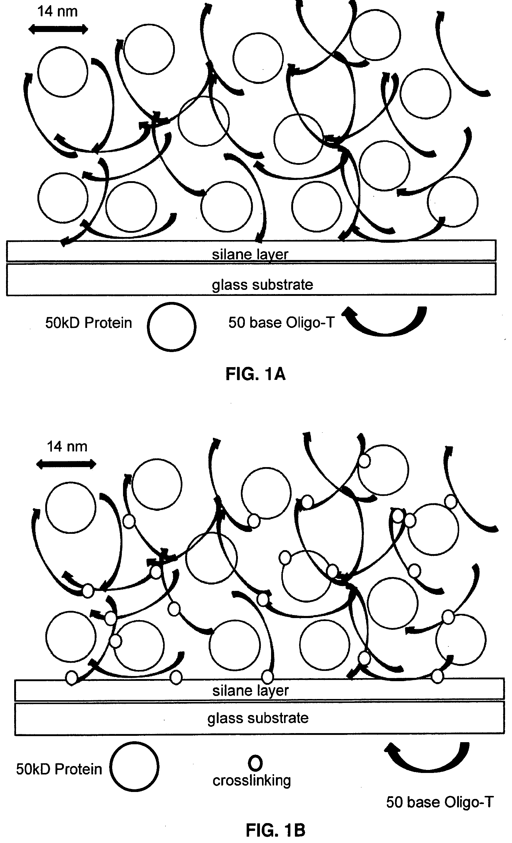
[0011] FIG. 1A illustrates photocrosslinking to generate an Oligo-dT+Protein matrix on a microarray surface.
[0012] FIG. 1B illustrates the Oligo-dT+Protein matrix of FIG. 1A after UV crosslinking.
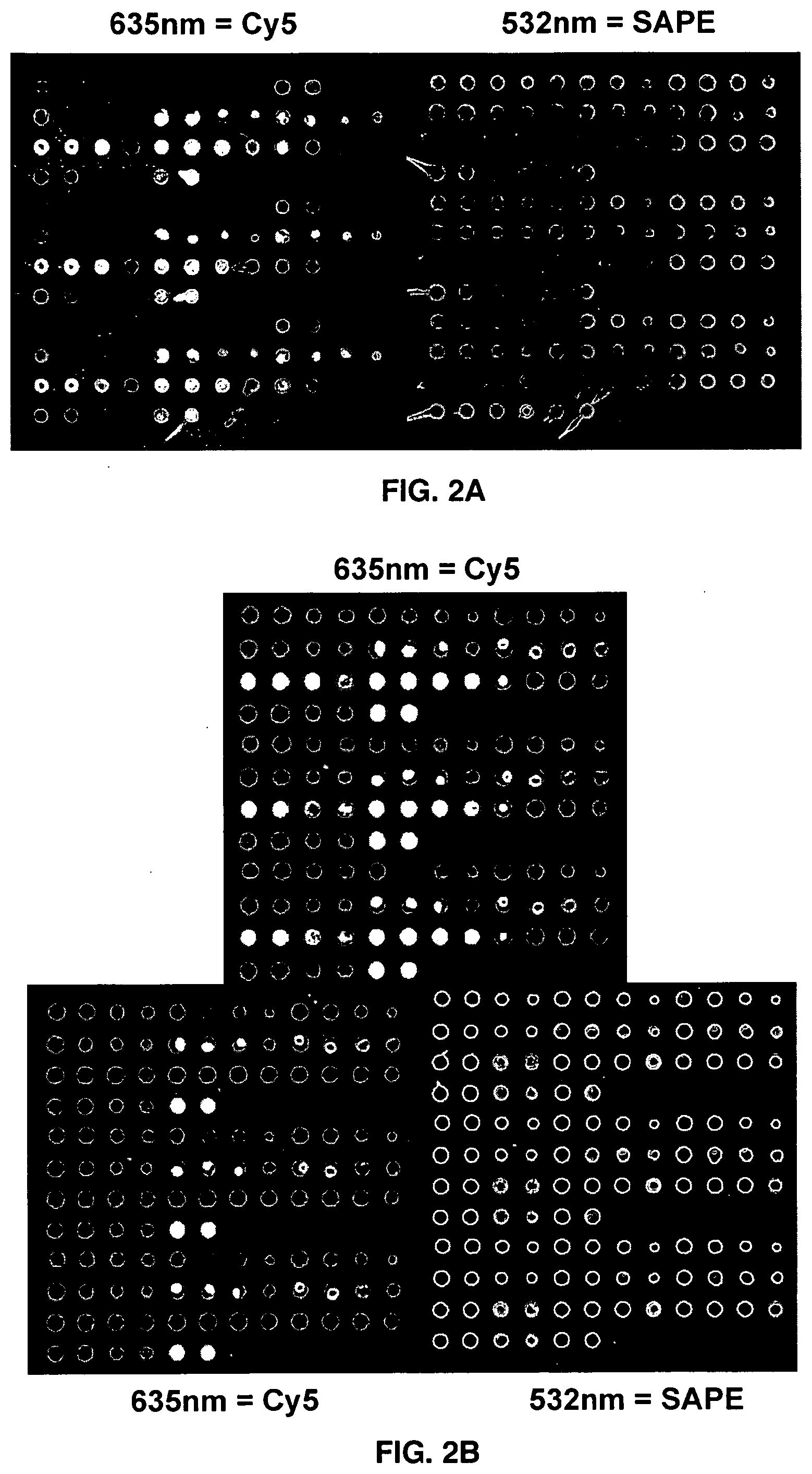
[0013] FIG. 2A is a fluorescent microarray image of a bovine serum albumin (BSA) microarray on an amino silane surface.
[0014] FIG. 2B is a fluorescent microarray image of a BSA microarray on an epoxy silane surface.
[0015] FIG. 3 depicts SAPE (Cy-3) signals normalized to OligoT (Cy-5) signals from a BSA microarray printed on an amino silane microarray surface, as in FIG. 2A.
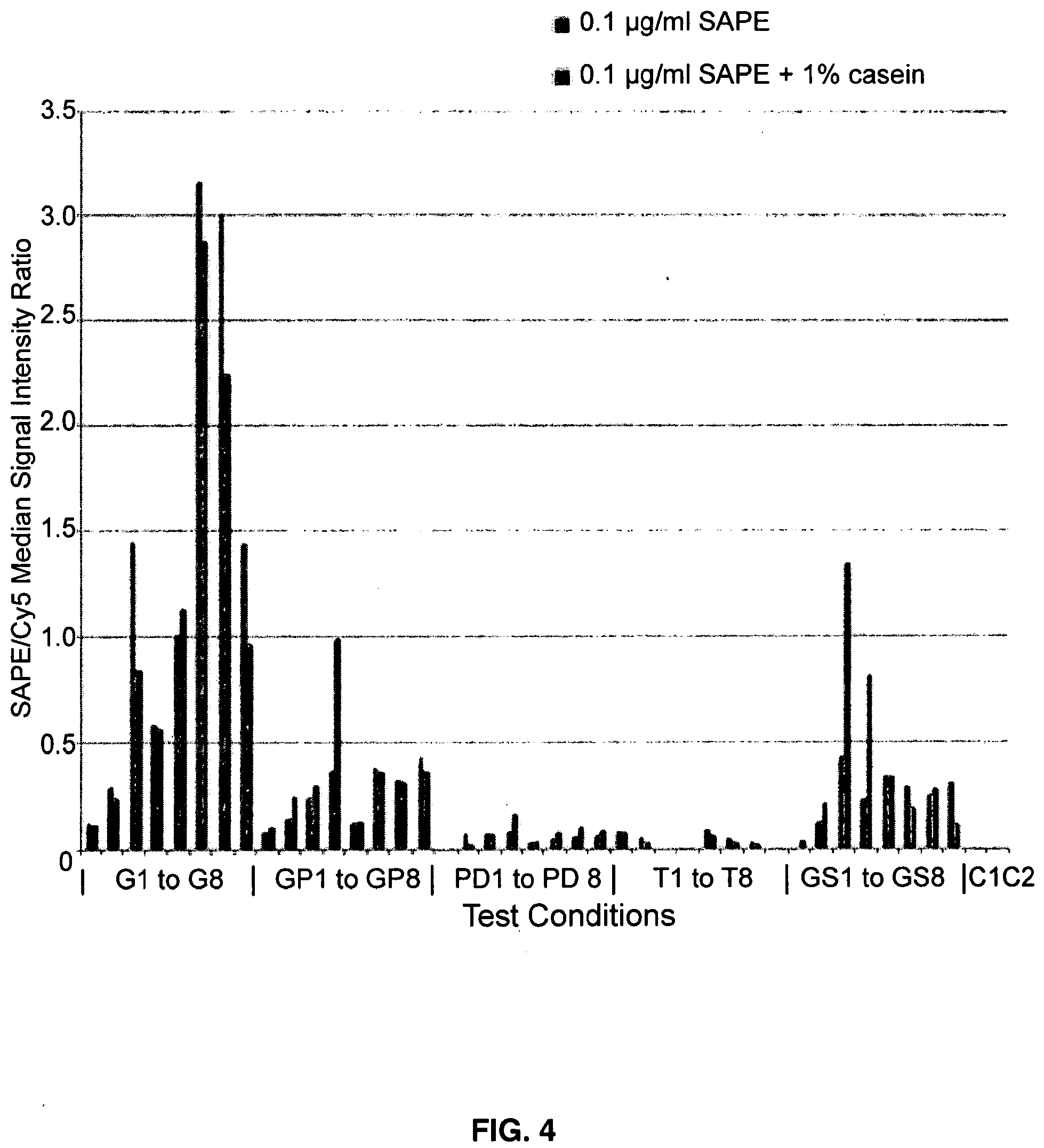
[0016] FIG. 4 depicts SAPE (Cy-3) signals normalized to OligoT (Cy-5) signals from a BSA microarray printed on an epoxy silane microarray surface, as in FIG. 2B.
DETAILED DESCRIPTION
[0017] The particulars shown herein are by way of example and for purposes of illustrative discussion of the preferred embodiments of the present disclosure only and are presented in the cause of providing what is believed to be the most useful and readily understood description of the principles and conceptual aspects of various embodiments of the disclosure. In this regard, no attempt is made to show structural details of the disclosure in more detail than is necessary for the fundamental understanding of the disclosure, the description taken with the drawings making apparent to those skilled in the art how the several forms of the disclosure may be embodied in practice.
[0018] The following definitions and explanations are meant and intended to be controlling in any future construction unless clearly and unambiguously modified in the following examples or when application of the meaning renders any construction meaningless or essentially meaningless. In cases where the construction of the term would render it meaningless or essentially meaningless, the definition should be taken from Webster's Dictionary 3.sup.rd Edition.
[0019] As used herein, the term "or" in the claims refers to "and/or" unless explicitly indicated to refer to alternatives only or the alternatives are mutually exclusive, although the disclosure supports a definition that refers to only alternatives and "and/or".
[0020] As used herein, the term "about" refers to a numeric value, including, for example, whole numbers, fractions, and percentages, whether or not explicitly indicated. The term "about" generally refers to a range of numerical values, e.g., +/-5-1 0% of the recited value, that one of ordinary skill in the art would consider equivalent to the recited value, e.g., having the same function or result. In some instances, the term "about" may include numerical values that are rounded to the nearest significant figure.
[0021] In one embodiment of the present invention, there is provided a formulation to link protein to a solid support, comprising: one or more proteins; Oligo-dT; and one or more non- volatile, water-soluble protein solvents, solutes or combination thereof in an aqueous solution. In one aspect, the water soluble protein solvent may comprise glycerol or the water soluble protein solvent and solutes may comprise glycerol and at least one of sucrose, trehalose or sorbitol. In this aspect the sucrose, trehalose or sorbitol may be present at a mass ratio of about 0.5:1 up to about 4:1 relative to glycerol. In another aspect, the water soluble protein solvent may comprise propanediol or the water soluble protein solvent and solids may comprise propanediol and at least one of sucrose, trehalose or sorbitol. In this aspect the mass ratio of the sucrose, trehalose or sorbitol is as described supra relative to propanediol. In yet another aspect, the water soluble protein solvents and solids may comprise glycerol and propanediol and at least one of sucrose, trehalose or sorbitol. In yet another aspect, the formulation may be applied to or disposed on a solid support such as an amino-silane layer upon an underlying surface. In this aspect representative underlying surfaces may be, but are not limited to a metal surface, a glass surface or a ceramic surface. In yet another aspect, the formulation may be applied to or disposed on a solid support such as an epoxy-silane layer upon an underlying surface. In this aspect a representative underlying surface is a metal.
[0022] In this embodiment and aspects thereof, in the formulation of the present invention, the Oligo-dT may be about 30 bases to about 100 bases in length, preferably about 50 bases long and may be contained in the formulation in a concentration of at least 1 .mu.g/ml, preferably about 1 .mu.g/ml (1 .mu.M) to about 100 .mu.g/ml (100 .mu.M). Also, preferably, the protein may be about 50 kD to 250 kD in mass and may be contained in the formulation in a concentration of at least 10 .mu.g/ml (10 .mu.M). In addition the non-volatile solvent and solutes may be formulated in water at about 1% by mass. Furthermore, representative proteins which may be linked or attached to a surface include, but are not limited to an. immunoglobulin, a glycoprotein, a viral protein, an intact virus, albumin, an HLA, or an enzyme.
[0023] In another embodiment of the present invention, there is provided a formulation to link protein to a solid support, comprising one or more proteins; Oligo-dT; and glycerol or glycerol and at least one of sucrose, trehalose or sorbitol in an aqueous solution. In this embodiment the glycerol, sucrose, trehalose and sorbitol may be formulated as described supra. In this embodiment and any aspect thereof, the Oligo-dT size and concentration, the protein size, concentration and type and the solid support all are as described supra.
[0024] In yet another embodiment of the present invention, there is provided a method of attaching a protein to a surface of a substrate, comprising the steps of: contacting the aqueous formulation containing Oligo dT and one or more non-volatile, water-soluble protein solvents, solutes or combination thereof and one or more proteins as described herein onto the substrate surface; printing said formulation onto the substrate surface; air-drying the substrate surface; irradiating the substrate surface with UV light to induce thymidine photochemical crosslinking via the thymidine moieties of the Oligo-dT, thereby attaching the protein to the surface of the substrate. In aspects of this embodiment, representative water soluble protein solvents and solutes are as, and may be formulated as, described supra. Also in this embodiment and all aspects thereof, the Oligo-dT size and concentration, the protein size, concentration and type and the solid support all are as described supra.
[0025] Described herein are methods and chemical formulations or compositions to link proteins to a solid surface, such as a microarray surface, to generate a biochemical or diagnostic binding assay. The surface attachment is produced by protein entrapment in a polymer network which is formed around the protein, locally, by photo-crosslinking of Oligo-dT, a photoreactive polymer. Oligo-dT, plus one or more proteins and one or more non-volatile, water-soluble solvents and solutes are applied to the solid surface, locally, as a water solution. Upon application of that water solution to the solid surface the water complement of the solution is allowed to evaporate away, yielding a concentrated, water-depleted phase comprising Oligo-dT, protein, solvents, solutes which is then crosslinked, photochemically, to entrap the protein within the resulting crosslinked, polymeric Oligo-dT network. The nonvolatile, water soluble solvents and solutes are chosen so that, subsequent to evaporative water loss, the resulting water-depleted phase remains principally non-crystalline, thereby mitigating protein damage by microcrystal formation. Proteins are linked to an underlying surface, indirectly, rather than by direct chemical linkage to the surface and in a way such that, subsequent to evaporative water depletion and UV crosslinking, the protein becomes entrapped in the crosslinked Oligo-dT network which was created around it. The above combination of indirect photochemical Oligo-dT network entrapment, plus retention of a non-crystalline phase upon water depletion, gives rise to preservation of a native protein state on the solid support, which is then available, subsequent to rehydration, to bind to analytes applied in water solution, as the basis for a binding, or diagnostic or public health screening assay.
[0026] More particularly, the Oligo-dT polymer chains are utilized as a linker mediating protein attachment to an underlying microarray surf ace. Oligo-dT is used a protein surface linker based on its capacity to engage in photocrosslinking. Briefly, Oligo-dT can readily be coprinted in @0.5.times. to 10.times. mole excess with any number of proteins of interest, e.g., albumin, antibodies, enzymes, HLA or any other water-soluble protein. At time of printing, the anionic Oligo-dT adsorbs, non-covalently, to the underlying cationic amino-silane surface via formation of electrostatic bonds. If applied to an epoxy silane coated surface, it can associate with the surface via a combination of covalent linkage to the epoxide and h-bonding to ring opened epoxide dials. The microarray spot is then allowed to air-dry over several minutes. In the present invention, protein printing occurs with one of several water-soluble (but non-volatile) solutes, in a buffered water solution, which upon air drying, becomes a water-depleted fluid, which retains solubilization of the protein and eliminates buffer salt crystal formation, which would have occurred if the water-soluble non-aqueous solutes were not added. Several solutes and solute mixtures can be used, such as for example, including but not limited to glycerol; Glycerol with propanediol; propanediol; glycerol with sorbitol; glycerol with propane diol and sorbitol; propanediol and sorbitol; glycerol with trehalose; glycerol with propane diol and trehalose; and propanediol and trehalose. Protein can be printed at one of several concentrations (250 .mu.g/ml-5 .mu.g/ml); 500 .mu.g/ml; 250 .mu.g/ml; 100 .mu.g/ml; 50 .mu.g/ml; 20 .mu.g/ml; and 10 .mu.g/ml.
[0027] In all cases, after printing and air-drying, the resulting microarrays (FIG. 1A) can be subjected to standard UV-Crosslinking at @300 mjoule, to photo-crosslink link the Oligo-dT (via T-T bonding) into a loosely-crosslinked matrix and in some cases to covalently link some of the protein to one or more nearby Oligo-dT molecules in the locally-generated Oligo-dT matrix (FIG. 1B).
[0028] Subsequent to UV crosslinking, the microarray is then ready for use. Standard binding steps can be employed: to be performed at lab ambient temperature or at elevated temperature: (1) Prebinding: Ordinary buffered solution with a blocking agent to obscure unused surface sites; (2) Binding: Ordinary buffered solution with a blocking agent to obscure unused surface sites; (3) Washing: Ordinary buffered solution with a blocking agent to obscure unused surface sites; and/or (4) Dry and Image.
[0029] As would be immediately recognizable to a person having ordinary skill in this art, the formulations and methods of the present invention may be used to fabricate a protein microarray via contact, or piezoelectric or ink jet printing onto a suitable solid support or to fabricate a protein biosensor via contact. Alternatively, the formulation when placed in contact with an aqueous biological sample, the microarray may be used as an in vitro diagnostic test or when placed in contact with an aqueous biological sample, the biosensor is used as an in vitro diagnostic test.
[0030] The following examples are given for the purpose of illustrating various embodiments of the invention and are not meant to limit the present invention in any fashion.
EXAMPLES
Example 1--Structure of a Representative Microarray, to be Printed for the Purposes of Testing Oliqo-dT Entrapment of Protein on a Microarray Surface
[0031] A single protein, for example, bovine albumin, can be printed to form a microarray. Each spot in the microarray can differ in the protein concentration, the Oligo-dT concentration and the water soluble, non-volatile solute to be added (at @1% by mass) at time of printing. Buffer concentration can be as needed, but 2 mM Na2Phos, pH 8.4 is representative.
TABLE-US-00001 TABLE 1 Formulation Of The Individual Microarray Spots To Vary Protein, Oligo-Dt And Solvent Composition In A 12 .times. 12 Microarray Left 6 .times. 13 segment of 12 .times. 12 Microarray Protein cone 250 ug/ml 250 ug/ml 100 ug/ml 100 ug/ml 50 ug/ml 50 ug/ml OligoT 50-mer 50-mer 50-mer 50-mer 50-mer 50-mer 100 uM 100 uM 100 uM 100 uM 100 uM 100 uM Glycerol Glycerol Glycerol Glycerol Glycerol Glycerol OligoT 50-mer 50-mer 50-mer 50-mer 50-mer 50-mer 50 uM 50 uM 50 uM 50 uM 50 uM 50 uM Glycerol Glycerol Glycerol Glycerol Glycerol Glycerol OligoT 50-mer 50-mer 50-mer 50-mer 50-mer 50-mer 25 uM 25 uM 25 uM 25 uM 25 uM 25 uM Glycerol Glycerol Glycerol Glycerol Glycerol Glycerol OligoT 50-mer 50-mer 50-mer 50-mer 50-mer 50-mer 100 uM 100 uM 100 uM 100 uM 100 uM 100 uM G-PD G-PD G-PD G-PD G-PD G-PD OligoT 50-mer 50-mer 50-mer 50-mer 50-mer 50-mer 50 uM 50 uM 50 uM 50 uM 50 uM 50 uM G-PD G-PD G-PD G-PD G-PD G-PD OligoT 50-mer 50-mer 50-mer 50-mer 50-mer 50-mer 25 uM 25 uM 25 uM 25 uM 25 uM 25 uM G-PD G-PD G-PD G-PD G-PD G-PD OligoT 50-mer 50-mer 50-mer 50-mer 50-mer 50-mer 100 uM 100 uM 100 uM 100 uM 100 uM 100 uM PD PD PD PD PD PD OligoT 50-mer 50-mer 50-mer 50-mer 50-mer 50-mer 50 uM 50 uM 50 uM 50 uM 50 uM 50 uM PD PD PD PD PD PD OligoT 50-mer 50-mer 50-mer 50-mer 50-mer 50-mer 25 uM 25 uM 25 uM 25 uM 25 uM 25 uM PD PD PD PD PD PD OligoT 50-mer 50-mer 50-mer 50-mer 50-mer 50-mer 100 uM 100 uM 100 uM 100 uM 100 uM 100 uM Trehalose Trehalose Trehalose Trehalose Trehalose Trehalose OligoT 50-mer 50-mer 50-mer 50-mer 50-mer 50-mer 50 uM 50 uM 50 uM 50 uM 50 uM 50 uM Trehalose Trehalose Trehalose Trehalose Trehalose Trehalose OligoT 50-mer 50-mer 50-mer 50-mer 50-mer 50-mer 25 uM 25 uM 25 uM 25 uM 25 uM 25 uM Trehalose Trehalose Trehalose Trehalose Trehalose Trehalose Right 6 .times. 13 segment of 12 .times. 12 Microarray Protein cone 20 ug/ml 20 ug/ml 10 ug/ml 10 ug/ml 5 ug/ml 5 ug/ml OligoT 50-mer 50-mer 50-mer 50-mer 50-mer 50-mer 100 uM 100 uM 100 uM 100 uM 100 uM 100 uM Glycerol Glycerol Glycerol Glycerol Glycerol Glycerol OligoT 50-mer 50-mer 50-mer 50-mer 50-mer 50-mer 50 uM 50 uM 50 uM 50 uM 50 uM 50 uM Glycerol Glycerol Glycerol Glycerol Glycerol Glycerol OligoT 50-mer 50-mer 50-mer 50-mer 50-mer 50-mer 25 uM 25 uM 25 uM 25 uM 25 uM 25 uM Glycerol Glycerol Glycerol Glycerol Glycerol Glycerol OligoT 50-mer 50-mer 50-mer 50-mer 50-mer 50-mer 100 uM 100 uM 100 uM 100 uM 100 uM 100 uM G-PD G-PD G-PD G-PD G-PD G-PD OligoT 50-mer 50-mer 50-mer 50-mer 50-mer 50-mer 50 uM 50 uM 50 uM 50 uM 50 uM 50 uM G-PD G-PD G-PD G-PD G-PD G-PD OligoT 50-mer 50-mer 50-mer 50-mer 50-mer 50-mer 25 uM 25 uM 25 uM 25 uM 25 uM 25 uM G-PD G-PD G-PD G-PD G-PD G-PD OligoT 50-mer 50-mer 50-mer 50-mer 50-mer 50-mer 100 uM 100 uM 100 uM 100 uM 100 uM 100 uM PD PD PD PD PD PD OligoT 50-mer 50-mer 50-mer 50-mer 50-mer 50-mer Left 6 .times. 13 segment of 12 .times. 12 Microarray Protein cone 250 ug/ml 250 ug/ml 100 ug/ml 100 ug/ml 50 ug/ml 50 ug/ml PD PD PD PD PD PD OligoT 50-mer 50-mer 50-mer 50-mer 50-mer 50-mer 25 uM 25 uM 25 uM 25 uM 25 uM 25 uM PD PD PD PD PD PD OligoT 50-mer 50-mer 50-mer 50-mer 50-mer 50-mer 100 uM 100 uM 100 uM 100 uM 100 uM 100 uM Trehalose Trehalose Trehalose Trehalose Trehalose Trehalose OligoT 50-mer 50-mer 50-mer 50-mer 50-mer 50-mer 50 uM 50 uM 50 uM 50 uM 50 uM 50 uM Trehalose Trehalose Trehalose Trehalose Trehalose Trehalose OligoT 50-mer 50-mer 50-mer 50-mer 50-mer 50-mer 25 uM 25 uM 25 uM 25 uM 25 uM 25 uM Trehalose Trehalose Trehalose Trehalose Trehalose Trehalose OligoT 20 ua/ml 20 ua/ml 10 ua/ml 10 ua/ml 5 ua/ml 5 ua/ml Glycerol: Glycerol at 1% by mass; G-P: Glycerol-1,2 Propanediol 1:1 at 1% by mass; PD: 1,2 Propanediol at 1% by mass; Trehalose: Trehalose at 1% by mass
EXAMPLE2
Quantitative Considerations: Addition of Oligo-dT & Protein Prior to Crosslinking on the Microarray Surface
[0032] It is well known that nucleic acid strands, especially those rich in thymidine, such as a simple repeating DNA oligomer dTn, i.e. Oligo-dT, become photochemically crosslinked to one another upon irradiation in the 250 nm to 230 nm range, due to photochemical excitation of the thymidine ring, followed by addition to the C4-C5 bond of another T or C: or if in proximity, to a nearby protein, especially lysine or cysteine side chains or aromatic amino acid side chains tyrosine or tryptophan. The present invention exploits the photochemistry of Oligo-dT, or Oligo-U if an RNA oligomer, to create within a microarray spot, local formation of a crosslinked Oligo-dT (Oligo-U) matrix which can physically entrap proteins which are applied along with Oligo-dT at time of microarray printing and before UV crosslinking (FIGS. 1A-1B).
[0033] Water soluble proteins in the 50 kD to 250 kD range are typically 10 nm to 20 nm in diameter. One can for example, employ bovine albumin, which has a mass of 67 kD and a measured nearly spherical radius of 7 nm. The rise per repeat of single stranded nucleic acids is @0.5 nm per base. Consequently a 50 base long Oligo-dT molecule would present a total contour length of @25 nm, roughly 2.times. the diameter of the albumin.
[0034] If Oligo-dT were mixed in molar excess with a protein such as albumin, in a water solution that contained about 1% of a water-soluble fluid solute, like glycerol, propanol, or glycerol-propanediol, or a water-soluble solid solute such as trehalose or sucrose or sorbitol, then allowed to air-dry to evaporate away the water in it, the resulting water-depleted phase will concentrate the protein and Oligo-dT molecules in it about 100-fold. Below, several calculations are displayed for the result of such concentration, for a representative 50 kd protein with a diameter of 14 nm which is similar to a globular protein such as bovine albumin.
Microarray fabrication, of the kind to be exploited in the present invention is a type of nanotechnology, where individual microarray elements or "spots" are applied to a surface as nanoliter droplets, which typically form spots on a surface that are about 100 microns in diameter. The present invention describes by calculation, the effect of depositing a typical 1 nanoliter droplet on a microarray surface, under conditions such that, as described above, air drying of the water in it will cause non-volatile solutes, such as glycerol, or propanediol alone or with solids like trehalose or sorbitol, to be concentrated 100-fold within the 100 .mu.m spot, thereby reducing the thickness of the spot.
Microarray Spot Fluid Thickness Calculation:
[0035] Subsequent to Air-Drying Assume 1.times.10-9 L print volume=1.times.10-6 cm3. [0036] Assume 100-fold air-drying to 1.times.10-11 L=1.times.10-8 cm3 [0037] Assume Spot Diameter=1.times.10-2 cm (100 um). Spot Area=1.times.10-4 cm2 [0038] Film Thickness=Print Volume after Air-Drying/Spot Area=[1.times.10-8 cm3]/1.times.104 cm2=1.times.10-4 cm=1 um [0039] Assuming 1 nL print of 1% non-volatile solutes: [0040] Final Spot Shape upon 100-fold evaporative concentration. [0041] Spot Width=100um and Spot Thickness=1 um Surface Coverage Calculations [0042] Assuming that a Protein is @14 nm in diameter, its 2D projection=150 nm22). If the Microarray spot is 100 um wide, its area=0.75.times.10-8 m2=75.times.10+8 nm2. [0043] So one 100 um spot will be covered by @0.5.times.lo+8 Protein molecules, as a one molecule thick layer [0044] If contact print volume=1 nL, at: [0045] 5 uM=3.times.lo+9 molecules per spot yields 30 molecule deep Protein layer, post evaporation; [0046] 2 uM=1.5.times.lo+9 molecules per spot yields 12 molecule deep Protein layer, post evaporation; [0047] 1 uM=0.75.times.lo+9 molecules per spot yields 1.5 molecule deep Protein layer, post evaporation; [0048] 0.4 uM=0.3.times.10+9 molecules per spot yields 0.6 molecule deep Protein layer, post evaporation; [0049] 0.2 uM=0.15.times.lo+9 molecules per spot yields 0.3 molecule deep Protein layer, post evaporation; and [0050] 0.1 uM=0.08.times.lo+9 molecules per spot yields 0.15 molecule deep Protein layer, post evaporation.
TABLE-US-00002 [0050] TABLE IIA Protein And Oligo-Dt Concentration Subsequent To Air-Drying At 1% Of Non-Volatile Solute To Affect A Controlled 100-Fold Volume Decrease Average Concentration Protein-Protein Protein Print Protein Print After Drying Molecular Separation Concentration Concentration 100-Fold Upon Drying 250 .mu.g/ml 5 .mu.M 500 .mu.M 75 nm 100 .mu.g/ml 2 .mu.M 200 .mu.M 100 nm 50 .mu.g/ml 1 .mu.M 100 .mu.M 130 nm 20 .mu.g/ml 0.4 .mu.M 40 .mu.M 170 nm 10 .mu.g/ml 0.2 .mu.M 20 .mu.M 220 nm 5 .mu.g/ml 0.1 .mu.M 10 .mu.M 270 nm
TABLE-US-00003 TABLE IIB Oligo-Dt Concentration Subsequent To Air-Drying At 1% Of Non-Volatile Solute (100-Fold Volume Decrease) Oligo-dT Concentration Average Molecular Print After Drying Separation Concentration 100-Fold Upon Drying 100 .mu.M 10 mM 28 nm 50 .mu.M 5 mM 35 nm 25 .mu.M 2.5 mM 44 nm 10 .mu.M 1.0 mM 60 nm 2 .mu.M 0.2 mM 100 nm 1 .mu.M 0.1 mM 130 nm
[0051] The calculations in Table IIa and Table IIB suggest that in an ordinary microarray spot that is @100 um in diameter, containing 1% by volume of a non-volatile, water miscible solvent (like glycerol or propanediol or non-volatile water-soluble solutes such as trehalose or sorbitol) ordinary air-drying will result in a spot that is about 1 urn thick. The calculations also show that if a @50 kD protein is printed at @250 ug/ml, the average separation between 14 nm wide protein molecules will be reduced by concentration to @75 nm, or about Sx the protein diameter (FIG. 1A). If printed at @10 uM, Oligo-dT will also concentrate 100 fold, to yield an average separation of only about 60 nm, or about 4.times. the diameter of the protein, thus yielding a dense network of Oligo-dT molecules surrounding the protein molecules between them (FIG. 1A). If desired, the effective pore size of the Oligo-dT network could be increased to an average separation of 100 nm if the Oligo-dT were applied at 2 uM prior to 100-fold air dying, or conversely, If the Oligo-dT were applied at 50 uM, in the original water-containing phase the pore size upon drying could be reduced to about 35 nm.
[0052] While still water-free, due to the dense proximity of Oligo-dT strands, photochemical crosslinking will be efficient: between Oligo-dT strands and also between Oligo-dT and the protein (FIG. 1B). Interestingly, at 250 ug/ml, the same calculation suggest that protein molecules will on average "pile" to form a layer on the microarray surface that is about 12 protein molecules deep within the water depleted, 1 um thick micro array spot. Thus, FIG. 1B represents about 200 um (about 1/Y.sup.11)of such a 1 um-thick desiccated microarray spot. Upon rehydration of that 1 um thick Oligo-dT+protein layer, wetting will cause the layer to swell, the separation between protein molecules to increase, thus preparing the protein, while still entrapped in the crosslinked Oligo-dT matrix, for subsequent microarray-based binding interaction with water-soluble analytes of interest.
EXAMPLE 3
Protein Attachment to Solid Surfaces
[0053] The present invention is novel in that protein is not linked to the surface directly, nor to a preformed polymer field, or to pores in the solid support or by adsorption to the microarray surface. Instead, the protein is applied to the microarray surface with Oligo-dT, which although not a high polymer, forms an extended polymeric matrix within a microarray spot, subsequent to controlled evaporative concentration, followed by photochemical crosslinking.
[0054] The physical and chemical entrapment of protein within that Oligo-dT matrix is created locally, only within the spots comprising sites of microarray fluid deposition, thereby allowing site-addressable, self-assembly of a 3 dimensional protein structure such as that in FIGS. 1A-1B. The key components of the invention are the length of the Oligo-dT (typically @50 bases), the ratio of Oligo-dT to protein (typically 1/1 to 10/1 on a mole basis), and the ability to control the final concentration of protein and Oligo-dT after ordinary evaporation, by including watermiscible, non-volatile solvents and solutes, typically at 1% by mass, so that the protein & Oligo-dT will concentrate@ 100-fold prior to UV crosslinking.
[0055] A number of such water-soluble non-volatile solvents and solutes may be used, but they must all share the property that, upon evaporative water loss, and, importantly, protein remains soluble and in a stable folded form, i.e., does not denature, in the water-depleted state. There are several such solvents and solutes of that kind, including glycerol, propanediol, butanediol, trehalose, sorbitol, sucrose and mixtures thereof which are known to retain protein folding even when the protein remains in them with little to no added water.
A Bovine Serum Albumin Microarray
[0056] The present invention describes using bovine serum albumin (BSA) as the protein in a microarray: This BSA has been produced with a biotin modification, so that streptavidin phycoerythrin (SAPE) can bind to the biotin of it, and after imaging of the green PE fluorescence (532 nm) serve to localize the BSA protein on the microarray surface. The BSA is applied to the surface as a solution with CY-5 labelled Oligo dT (50 bases long) along with an excess of unlabeled Oligo-dT (50 bases long) which may be imaged via the red CY-5 fluorescence (635 nm). Table III provides a detailed composition of that microarray.
TABLE-US-00004 TABLE III Bovine Serum Albumin (BSA) Microarray Design To Yield Data In FIGS. 2A-2B G-PD G-PD G-PD G-PD G-PD G-PD OligoT 50-mer 50-mer 50-mer 50-mer 50-mer 50-mer 100 .mu.M 100 .mu.M 100 .mu.M 100 .mu.M 25 .mu.M 25 .mu.M trehalose trehalose trehalose trehalose trehalose trehalose OligoT 50-mer 50-mer 50-mer 50-mer control control 25 .mu.M 25 .mu.M 25 .mu.M 25 .mu.M G-S G-S G-S G-S OligoT 50-mer 50-mer 50-mer 50-mer 50-mer 50-mer 100 .mu.M 100 .mu.M 100 .mu.M 100 .mu.M 25 .mu.M 25 .mu.M glycerol glycerol glycerol glycerol glycerol glycerol OligoT 50-mer 50-mer 50-mer 50-mer 50-mer 50-mer 25 .mu.M 25 .mu.M 25 .mu.M 25 .mu.M 100 .mu.M 100 .mu.M G-PD G-PD G-PD G-PD G-PD G-PD OligoT 50-mer 50-mer 50-mer 50-mer 50-mer 50-mer 100 .mu.M 100 .mu.M 100 .mu.M 100 .mu.M 25 .mu.M 25 .mu.M trehalose trehalose trehalose trehalose trehalose trehalose OligoT 50-mer 50-mer 50-mer 50-mer control control 25 .mu.M 25 .mu.M 25 .mu.M 25 .mu.M G-S G-S G-S G-S OligoT 50-mer 50-mer 50-mer 50-mer 50-mer 50-mer 100 .mu.M 100 .mu.M 100 .mu.M 100 .mu.M 25 .mu.M 25 .mu.M glycerol glycerol glycerol glycerol glycerol glycerol OligoT 50-mer 50-mer 50-mer 50-mer 50-mer 50-mer 25 .mu.M 25 .mu.M 25 .mu.M 25 .mu.M 100 .mu.M 100 .mu.M G-PD G-PD G-PD G-PD G-PD G-PD OligoT 50-mer 50-mer 50-mer 50-mer 50-mer 50-mer 100 .mu.M 100 .mu.M 100 .mu.M 100 .mu.M 25 .mu.M 25 .mu.M trehalose trehalose trehalose trehalose trehalose trehalose OligoT 50-mer 50-mer 50-mer 50-mer control control 25 .mu.M 25 .mu.M 25 .mu.M 25 .mu.M G-S G-S G-S G-S Right 6 .times. 13 segment of 12 .times. 12 Microarray [BSA] cone 50 ua/ml 20 ua/ml 250 ua/ml 100 ua/ml 50 ua/ml 20 ua/ml OligoT 50-mer 50-mer 50-mer 50-mer 50-mer 50-mer 25 .mu.M 25 .mu.M 100 .mu.M 100 .mu.M 25 .mu.M 25 .mu.M glycerol glycerol G-PD G-PD G-PD G-PD OligoT 50-mer 50-mer 50-mer 50-mer 50-mer 50-mer 100 .mu.M 100 .mu.M 25 .mu.M 25 .mu.M 25 .mu.M 25 .mu.M PD PD PD PD PD PD OligoT 50-mer 50-mer 50-mer 50-mer 50-mer 50-mer 25 .mu.M 25 .mu.M 100 .mu.M 100 .mu.M 25 .mu.M 25 .mu.M trehalose trehalose G-S G-S G-S G-S OligoT OligoT 50-mer 50-mer 50-mer 50-mer 50-mer 50-mer 25 .mu.M 25 .mu.M 100 .mu.M 100 .mu.M 100 .mu.M 100 .mu.M glycerol glycerol G-PD G-PD G-PD G-PD OligoT 50-mer 50-mer 50-mer 50-mer 50-mer 50-mer 100 .mu.M 100 .mu.M 25 .mu.M 25 .mu.M 25 .mu.M 25 .mu.M PD PD PD PD PD PD OligoT 50-mer 50-mer 50-mer 50-mer 50-mer 50-mer 25 .mu.M 25 .mu.M 100 .mu.M 100 .mu.M 25 .mu.M 25 .mu.M trehalose trehalose G-S G-S G-S G-S OligoT OligoT 50-mer 50-mer 50-mer 50-mer 50-mer 50-mer 25 .mu.M 25 .mu.M 100 .mu.M 100 .mu.M 100 .mu.M 100 .mu.M alvcerol alvcerol G-PD G-PD G-PD G-PD OligoT 50-mer 50-mer 50-mer 50-mer 50-mer 50-mer 100 .mu.M 100 .mu.M 25 .mu.M 25 .mu.M 25 .mu.M 25 .mu.M PD PD PD PD PD PD OligoT 50-mer 50-mer 50-mer 50-mer 50-mer 50-mer 25 .mu.M 25 .mu.M 100 .mu.M 100 .mu.M 100 .mu.M 100 .mu.M trehalose trehalose G-S G-S G-S G-S OligoT
[0057] To the combination of BSA and Oligo-dT was added any one of a number of non-volatile, non-aqueous solvents and solutes including glycerol (G), glycerol-propanediol (GP) trehalose (T) and glycerol-sorbitol (GS). The resulting solution was printed onto either amino-silane coated or epoxysilane coated glass microarray substrates, using contact printing to form 100 um diameter spots, roughly 200 urn on center. The print volume (@500 pL) was found to air-dry within 10 min, as assessed by fluorescent imaging of the microarray surface (Axon Imager).
[0058] Subsequent to air drying, the microarrays were crosslinked by exposure to 300 mJ of UV light (Stratolinker) to induce thymidine photochemical crosslinking via the thymidine moieties of the Oligo-dT. Subsequent to crosslinking, the slides were subjected to two 5 minute washing steps with a Tween 20 containing buffer, followed by administration of SAPE, to bind to the biotinylated BSA, so that it may be localized and quantitated by fluorescent imaging. In some cases, the SAPE staining was performed with 1% casein in the SAPE binding buffer, to provide for additional blocking of spurious SAPE binding to regions not modified with BSA. Subsequent to completion of the binding step (performed at room temp) a series of washes in protein free binding buffer and two water washes were performed (also at Room Temp) followed by air drying and imaging on an Axon laser scanner: at 532 nm (green for SAPE) and 635 nm (red, for Cy-5 labelled Oligo-dT).
[0059] A microarray design was used which presented various combinations of BSA, Oligo-dT and non-aqueous solvent and solute concentration (Table III). Representative images obtained of those microarrays, after binding and washing are presented in FIG. 2A (formed on an amino silane surface) or in FIG. 28 (formed on an epoxysilane surface).
[0060] As can be seen from the (red) CY-5 labelled Oligo-dT images the microarray printing and UV crosslinking procedure has permanently linked the Oligo-dT complement to the microarray surface on both an underlying aminosilane (FIG. 2A) and epoxysilane surface (FIG. 2B). As seen from the corresponding images generated via SAPE staining of biotinylated BSA (FIGS. 2A-2B, Right panels) the microarray printing and UV crosslinking procedure has also permanently linked the BSA protein to the surface, so that it may bind to SAPE.
[0061] Those image data have been quantified spot by spot and presented in FIG. 3 and FIG. 4 as the ratio of SAPE/Cy5 signal intensity, which is a measure of the ratio of BSA to Oligo-dT in each spot, as a function of variation in the several parameters described in Table III. FIG. 3 describes those ratios when the microarray is printed upon the aminosilane coated microarray substrate and FIG. 4 the corresponding data on the epoxysilane coated microarray substrate. Table IV provides the test conditions under which the ratios in FIGS. 3 and 4 were obtained. In Tables Va, Vb and Vc the numerical values used to generate FIGS. 3 and 4 are provided.
TABLE-US-00005 TABLE IV Sodium Biotinylated 50 mer Cy5 Abbre- Phosphate BSA Oligo-T Oligo-T Adjuvant viation pH 8.4 (mM) (.mu.g/ml) (.mu.M) (.mu.M) Glycerol 1% G1 2 250 100 1 G2 2 100 100 1 G3 2 50 100 1 G4 2 20 100 1 G5 2 250 25 1 G6 2 100 25 1 G7 2 50 25 1 G8 2 20 25 1 Glycerol- GP1 2 250 100 1 Propanedio GP2 2 100 100 1 0.5%-0.5% GP3 2 50 100 1 GP4 2 20 100 1 GP5 2 250 25 1 GP6 2 100 25 1 GP? 2 50 25 1 GP8 2 20 25 1 Propanediol PD1 2 250 100 1 1% PD2 2 100 100 1 PD3 2 50 100 1 PD4 2 20 100 1 PD5 2 250 25 1 PD6 2 100 25 1 PD? 2 50 25 1 PD8 2 20 25 1 Trehalose T1 2 250 100 1 1% T2 2 100 100 1 T3 2 50 100 1 T4 2 20 100 1 T5 2 250 25 1 T6 2 100 25 1 T7 2 50 25 1 TB 2 20 25 1 Glycerol- GS1 2 250 100 1 Sorbitol GS2 2 100 100 1 0.5%-0.5% GS3 2 50 100 1 GS4 2 20 100 1 GS5 2 250 25 1 GS6 2 100 25 1 GS? 2 50 25 1 GS8 2 20 25 1 Control C1 150 250 0 1 Trehalose C2 150 250 100 1 0.5%
TABLE-US-00006 TABLE VA SAPE @ 0.1 ua/ml 635 nm 635 nm SAPE/ Signal Signal 532 nm Cy5 Test Intensity Intensity Signal SAPE Signal Con- Post Post SAPE Decrease Signal Intensity dition PrintinQ bindinQ (%) Intensity Ratio G1 16955 9416 44 1206 0.13 G2 6330 4121 35 1206 0.29 G3 1037 753 27 1092 1.45 G4 1939 1665 14 970 0.58 G5 1832 991 46 1002 1.01 G6 534 337 37 1066 3.16 G7 504 366 27 1104 3.02 GB 254 138 46 199 1.44 GP1 22508 13284 41 1110 0.08 GP2 11357 7742 32 1067 0.14 GP3 6701 4608 31 1109 0.24 GP4 572 567 1 208 0.37 GP5 16316 7741 53 975 0.13 GP6 5105 2814 45 1072 0.38 GP? 5534 3356 39 1088 0.32 GP8 2547 2142 16 935 0.44 PD1 65535 62387 5 903 0.01 PD2 37757 13239 65 949 0.07 PD3 25895 8991 65 671 0.07 PD4 7542 4459 41 377 0.08 PD5 52806 26927 49 796 0.03 PD6 43311 15303 65 810 0.05 PD? 33466 17870 47 972 0.05 PD8 35202 14819 58 952 0.06 T1 65535 9356 86 805 0.09 T2 65535 16762 74 974 0.06 T3 65535 20414 69 423 0.02 T4 65535 30171 54 353 0.01 T5 65535 10151 85 930 0.09 T6 65535 20582 69 991 0.05 T7 65535 32239 51 991 0.03 TB 65535 36545 44 631 0.02 GS1 65535 38155 42 761 0.02 GS2 11220 5398 52 696 0.13 GS3 2844 1649 42 711 0.43 GS4 5606 3358 40 779 0.23 GS5 32347 2626 92 907 0.35 GS6 7226 2089 71 605 0.29 GS? 5039 1391 72 344 0.25 GS8 3797 1475 61 451 0.31 C1 65535 65535 0 730 0.01 C2 65535 65535 0 609 0.01
TABLE-US-00007 TABLE VB SAPE @0.1 ua/ml +1 % casein 635 nm 635 nm SAPE/ Signal Signal 532 nm Cy5 Test Intensity Intensity Signal SAPE Signal Con- Post Post SAPE Decrease Signal Intensity dition PrintinQ bindinQ (%) Intensity Ratio G1 12131 7829 35 924 0.12 G2 5625 3932 30 961 0.24 G3 1081 891 18 749 0.84 G4 1150 1140 1 647 0.57 G5 1326 822 38 937 1.14 G6 501 372 26 1071 2.88 G7 611 480 21 1077 2.24 GB 150 139 7 135 0.97 GP1 15734 10458 34 1177 0.11 GP2 7716 5523 28 1398 0.25 GP3 6435 4714 27 1415 0.30 GP4 137 124 9 123 0.99 GPS 12493 6449 48 867 0.13 GP6 3741 2450 35 909 0.37 GP? 3796 2701 29 859 0.32 GP8 3034 2173 28 791 0.36 PD1 65535 65535 0 835 0.01 PD2 49436 40764 18 847 0.02 PD3 15169 8673 43 665 0.08 PD4 3159 2420 23 401 0.17 PD5 50343 30750 39 1201 0.04 PD6 35607 14057 61 1227 0.09 PD? 18201 9594 47 1072 0.11 PD8 21455 10282 52 940 0.09 T1 65535 9538 85 799 0.08 T2 65535 15743 76 559 0.04 T3 65535 17025 74 193 0.01 T4 65535 19488 70 381 0.02 T5 65535 9878 85 657 0.07 T6 65535 17010 74 573 0.03 T7 65535 24874 62 662 0.03 TB 65535 31337 52 376 0.01 GS1 64880 25578 61 1071 0.04 GS2 10017 5088 49 1120 0.22 GS3 1718 824 52 1112 1.35 GS4 2662 1400 47 1143 0.82 GS5 23631 2382 90 814 0.34 GS6 6148 2138 65 412 0.19 GS? 2023 686 66 193 0.28 GS8 2577 1150 55 135 0.12 No SAPE 635nm 635 nm Signal Signal Intensity Intensity Signal Test Post Post SAPE Decrease Condition Printing binding (%) G1 12204 7757 36 G2 4491 3106 31 G3 1123 881 22 G4 1107 909 17 G5 1164 747 36 G6 471 349 26 G7 526 388 26 GB 149 115 23 GP1 17385 11358 35 GP2 8325 6067 27 GP3 6290 4843 23 GP4 158 143 9 GP5 8795 5222 41 GP6 3563 2312 35 GP? 2999 2373 21 GP8 3017 2260 25 PD1 65535 65535 0 PD2 65328 65202 0 PD3 17243 13924 19 PD4 1887 1389 26 PD5 42002 31619 25 PD6 29609 16240 45 PD? 13903 6796 51 PD8 22972 9982 57 T1 65535 8988 86 T2 65535 8687 87 T3 65535 9271 86 T4 58049 10853 81 T5 65535 7114 89 T6 65535 10515 84 T7 65535 12761 81 TB 65535 18387 72 GS1 65535 25670 61 GS2 10199 5146 50 GS3 1652 789 52 GS4 2339 1011 57
[0062] The trends obtained from FIGS. 3 and 4 are instructive. Overall, Oligo-dT crosslinking to permanently affix it onto the surface is efficient for both surfaces, more-less independent of the supporting non-aqueous solvent and solutes added. However, when the resulting SAPE/Cy-5 ratio was obtained, it was seen that glycerol (G) glycerol-propanediol (GP) and glycerol-sorbitol (GS) each provide for substantial biotin-BSA interaction with its cognate streptavidinphycoerythrin (SAPE) conjugate. In contrast, it is seen that Trehalose (T) and to a lesser extent Propanediol (PO) provide for very poor BSA interaction due to poor protein association to the surface or protein disruption on the surface or both.
[0063] The above Examples demonstrate that Oligo-dT mediated UV crosslinking allows a protein such as BSA to be linked to a microarray surface to form, upon air-drying, a principally water free phase containing a number of nonvolatile water miscible solvents and solutes. The data show that, for BSA, certain solvent-solute pairs, e.g., glycerol and glycerol propane diol, appear to be superior to trehalose and propandiol in the present case of BSA attachment to the microarray surface.
[0064] All of the compositions and methods disclosed and claimed herein can be made and executed without undue experimentation in light of the present disclosure. While the compositions and methods of this disclosure have been described in terms of preferred embodiments, it will be apparent to those of skill in the art that variations may be applied to the compositions and methods and in the steps or in the sequence of steps of the methods described herein without departing from the concept, spirit and scope of the disclosure. More specifically, it will be apparent that certain agents which are both chemically related may be substituted for the agents described herein while the same or similar results would be achieved. All such similar substitutes and modifications apparent to those skilled in the art are deemed to be within the spirit, scope and concept of the disclosure as defined by the appended claims.
* * * * *
D00000
D00001
D00002
D00003
D00004
XML
uspto.report is an independent third-party trademark research tool that is not affiliated, endorsed, or sponsored by the United States Patent and Trademark Office (USPTO) or any other governmental organization. The information provided by uspto.report is based on publicly available data at the time of writing and is intended for informational purposes only.
While we strive to provide accurate and up-to-date information, we do not guarantee the accuracy, completeness, reliability, or suitability of the information displayed on this site. The use of this site is at your own risk. Any reliance you place on such information is therefore strictly at your own risk.
All official trademark data, including owner information, should be verified by visiting the official USPTO website at www.uspto.gov. This site is not intended to replace professional legal advice and should not be used as a substitute for consulting with a legal professional who is knowledgeable about trademark law.