Methods And Systems For Preventing, Correcting, Transforming, And Modifying Facial, Aesthetics, And Consulting Patients Regardin
DE MAIO DOMINGOS; Mauricio
U.S. patent application number 16/483400 was filed with the patent office on 2020-05-14 for methods and systems for preventing, correcting, transforming, and modifying facial, aesthetics, and consulting patients regardin. The applicant listed for this patent is Mauricio DE MAIO DOMINGOS. Invention is credited to Mauricio DE MAIO DOMINGOS.
| Application Number | 20200146615 16/483400 |
| Document ID | / |
| Family ID | 61837777 |
| Filed Date | 2020-05-14 |


View All Diagrams
| United States Patent Application | 20200146615 |
| Kind Code | A1 |
| DE MAIO DOMINGOS; Mauricio | May 14, 2020 |
METHODS AND SYSTEMS FOR PREVENTING, CORRECTING, TRANSFORMING, AND MODIFYING FACIAL, AESTHETICS, AND CONSULTING PATIENTS REGARDING THE SAME
Abstract
Provided herein are MD Codes, MD DYNA Codes, MD ASA, and Next Human system, and the methods of using them to diagnose and treat aesthetic conditions or disorders.
| Inventors: | DE MAIO DOMINGOS; Mauricio; (Sao Paulo, BR) | ||||||||||
| Applicant: |
|
||||||||||
|---|---|---|---|---|---|---|---|---|---|---|---|
| Family ID: | 61837777 | ||||||||||
| Appl. No.: | 16/483400 | ||||||||||
| Filed: | February 10, 2018 | ||||||||||
| PCT Filed: | February 10, 2018 | ||||||||||
| PCT NO: | PCT/IB2018/000207 | ||||||||||
| 371 Date: | August 2, 2019 |
Related U.S. Patent Documents
| Application Number | Filing Date | Patent Number | ||
|---|---|---|---|---|
| 62457761 | Feb 10, 2017 | |||
| 62477312 | Mar 27, 2017 | |||
| 62479150 | Mar 30, 2017 | |||
| 62479139 | Mar 30, 2017 | |||
| Current U.S. Class: | 1/1 |
| Current CPC Class: | G16H 40/63 20180101; A61Q 19/00 20130101; A61K 38/4893 20130101; G16H 50/20 20180101; G06T 7/0012 20130101; A61K 31/728 20130101; A61K 8/735 20130101; G06T 2207/30201 20130101; G16H 10/60 20180101; A61K 8/65 20130101; G06T 11/00 20130101; A61B 5/442 20130101; G16H 20/40 20180101; G06T 2207/30088 20130101; A61K 8/64 20130101 |
| International Class: | A61B 5/00 20060101 A61B005/00; G06T 7/00 20060101 G06T007/00; G16H 50/20 20060101 G16H050/20; G16H 20/40 20060101 G16H020/40; G16H 10/60 20060101 G16H010/60; G16H 40/63 20060101 G16H040/63; A61Q 19/00 20060101 A61Q019/00 |
Claims
1.-145. (canceled)
146. A method of providing a subject with a consultation, comprising: identifying a condition or disorder in the subject; identifying at least one or more MD Codes useful for treating the condition or disorder; wherein the MD Codes indicate injection sites on the face or neck of the subject; generating a treatment plan to inject a therapeutically effective amount of a pharmaceutical composition at the injection sites identified by the MD Codes; and optionally visualizing the treatment plan on a graphical display.
147. The method of claim 146, wherein the visualizing comprises overlaying any of the MD Codes set forth herein with an image of the subject.
148. The method of claim 147, wherein the subject suffers from a cosmetic condition, defect, or disease.
149. The method of claim 148, wherein the subject suffers from an aesthetic condition, defect, or disease.
150. The method of claim 149, wherein the subject suffers from a dermatology condition, defect, or disease.
151. A method of treating a subject in need thereof, the method comprising: providing a subject; assessing the subject based on an MD ASA which stands for Multidimensional Aesthetic Scan Assessment diagram; wherein the MD ASA indicate an assessment tool that will help injectors to analyze the face in details and better diagnose the needs of each subject; treating and diagnosing the subject in need thereof by injecting a therapeutically effective amount of a pharmaceutical composition comprising a member selected from the group consisting of hyaluronic acid (HA), Botox, fillers, and combinations thereof.
152. The method of claim 151, wherein assessing the subject comprising scoring an aesthetic hierarchy.
153. The method of claim 152, wherein the sites are on the neck or face.
154. The method of claim 153, wherein the method further comprises injecting a pharmaceutical composition comprising a member selected from the group consisting of hyaluronic acid (HA), Botox, fillers, and combinations thereof; and repeating the injecting at least once per year for at least five (5) years.
155. The method of claim 154, wherein the pharmaceutical composition comprises a member selected from the group consisting of Botulinum Toxin--Type A (Botox), collagen, hyaluronic acid, antibiotics, anti-inflammatory drugs, steroids and combinations thereof
156. The method of claim 154, wherein the pharmaceutical composition is Botulinum Toxin--Type A, or long-chain Hyaluronic Acid (HA) product, or composition comprises HA, or composition is deoxycholic acid, or comprises a vitamin.
157. A method of treating a subject in need thereof providing a map of risk factors; identifying the risk factors present in a subject in need thereof; treating the subject in need thereof by injecting a pharmaceutical composition comprising a member selected from the group consisting of hyaluronic acid (HA), Botox, fillers, and combinations thereof.
158. The method of claim 157, wherein the risk factors comprise intrinsic factors selected from the group consisting of genetic factors, or environmental factors, or family health history, or family aesthetic history.
159. The method of claim 158, wherein identifying the risk factors comprises overlaying a picture of the subject in need thereof with a picture of the subject's parent of the same gender or with a picture of the subject's child of the same gender.
160. The method of claim 159, wherein the pharmaceutical composition is Botulinum Toxin--Type A.
161. The method of claim 160, wherein the pharmaceutical composition is a long-chain Hyaluronic Acid (HA) product.
162. The method of claim 161, wherein the pharmaceutical composition comprises HA.
163. The method of claim 162, wherein the pharmaceutical composition is deoxycholic acid.
164. The method of any one of claim 163, wherein the visualizing comprises overlaying any of the injection sites set forth herein with an image of the subject.
165. The method of claim 164, wherein the subject in need thereof suffers from an aesthetic condition.
166. The method of claim 165, wherein the subject in need thereof suffers from a dermatology condition.
167. The method of claim 165, wherein the method comprising injecting at least 0.5 mL of the therapeutically effective amount of the pharmaceutical composition, and injecting the therapeutically effective amount of the pharmaceutical composition at a depth of penetration of at least 2-6 mm.
168. The method of claim 167, wherein pharmaceutical composition is a cellular body.
169. A method of treating a subject in need thereof, comprising: diagnosing a subject thereby identifying a condition or disorder; selecting a treatment using MD codes, a MD codes equation, or a MD codes formula, wherein the MD codes indicate injection sites on face or neck of the subject; and injecting an effective amount of a pharmaceutical composition at each of the injection sites.
170. The method of claim 169, wherein the pharmaceutical composition is a hyaluronic acid (HA) product or Botulinum Toxin--Type A.
171. The method of claim 170, wherein the concentration of HA is 17.5 mg/ml or 20 mg/ml.
172. The method of claim 171, wherein the MD codes are selected from the group consisting of: L codes, F codes, T codes, Lp codes, E codes, M codes, G codes, C codes, O codes, Jw codes, Tt codes, and N codes, and wherein the L codes are L1, L2, L3, L4, L5, L6, L7, L8; F codes are F1, F2, F3; T code are T1, T2; E codes are E1, E2, E3; G codes are G1, G2; O codes are O1, O2, O3; T codes are Tt1, Tt2, Tt3; Ck codes are Ck1, Ck5; NL codes are NL1, NL2, NL3; Lp codes are Lp1, Lp2, Lp3, Lp4, Lp5, Lp6, Lp7, Lp8; M codes are M1, M2, M3; C codes are C1, C2, C3, C4, C5, C6; Jw codes are Jw1, Jw2, Jw3, Jw4, Jw5; and N codes are N1, N2, N3, N4, N5.
173. A method of treating a subject in need thereof, comprising: diagnosing a subject thereby identifying a condition or disorder; selecting a treatment using MD DYNA codes, wherein MD DYNA codes indicate injection sites on face or neck of the subject; and injecting an effective amount of a pharmaceutical composition at each of the injection sites.
174. The method of claim 173, wherein the condition or disorder is an aesthetic condition or disorder.
175. The method of claim 174, wherein the pharmaceutical composition is selected from the group consisting of hyaluronic acid (HA), HA-based fillers, HA-derivatives, botulinum toxin-type A (BoNT-A), fillers, and combinations thereof.
176. The method of claim 175, wherein the MD DYNA codes for periorbital expression are selected from the group consisting of F, C, P, and 00c, and wherein the MD DYNA Codes for perioral expression are selected from the group consisting of N, LAN, LLS, ZMi, Ami, DSN, 00, R, DAO, DLI, M, and PL and wherein the injection is for chemical myomodulation, and the pharmaceutical composition is botulinum toxin-type A.
177. The method of claim 176, wherein if presence of glabella and forehead lines is identified, select MD DYNA Codes C+F, to reduce lines; if presence of chin and neck lines is identified, select MD DYNA Codes M+PL, to reduce lines; if presence of downturn of the corners of the mouth is identified, select MD DYNA Codes DAO, to correct downturn.
178. The method of claim 177, wherein the injection is for mechanical myomodulation, and the pharmaceutical composition is HA, HAbased fillers, or HA-derivatives.
179. The method of claim 178, wherein if skin wrinkling when pouting is identified, select MD DYNA Codes M, to reduce skin lines; if presence of gummy smile is identified, select MD DYNA Codes selection: DSN+LAN, to correct gummy smile and Lift nose tip; if presence of asymmetrical smile, select MD DYNA Codes selection: Zmj+Zmi; to lift the corner of the mouth, specially left side.
180. The method of any of claim 179, wherein MD ASA is used to assess a saggy look comprising assessing all facial arcas, but prioritising the following: Sign 1: Saggy cheeks--Anatomic units: Cheeks Sign 2: Deep nasolabial folds--Anatomic units: Nasolabial folds Sign 3: Marionette lines--Anatomic units: Marionette lines Sign 4: Jowls--Anatomic units: Chin, Jawline Sign 5: Poor skin quality--Anatomic units: Skin Sign 6: Submental fat--Anatomic units: Submental arca.
181. The method of any of claim 180, wherein MDA ASA is used to assess a sad look in the periorbital arca comprising assess all arcas, but prioritizing Sign 1: Low brows--Anatomic units: Eyebrows, Temples Sign 2: Eyebags/Downturn of corner of the eye--Anatomic units: Cheeks, Eyelids, Tear trough. Sign 3: Lines around the eyes--Anatomic units: Glabellar lines, Lateral canthallorbital lines. Sign 4: Poor skin quality--Anatomic units: Periorbital skin.
182. The method of claim 181, wherein MDA ASA.TM. is used to assess a sad look in the periorbital arca, comprising assessing all arcas and prioritizing Sign 1: Saggy cheeks--Anatomic units: Cheeks Sign 2: Deep nasolabial folds--Anatomic units: Nasolabial folds Sign 3: Lack of lip structure--Anatomic units: Lips Sign 4: Downturn or oral commissures--Anatomic units: Lips Sign 5: Deep marionette lines--Anatomic units: Chin, Marionette lines Sign 6: Poor skin quality--Anatomic units: Perioral skin.
183. The method of claim 182, wherein MDA ASA.TM. is used to assess a tired look, comprising assessing all areas and prioritizing Sign 1: Low brows--Anatomic units: Eyebrows, Temples Sign 2: Eyebags--Anatomic units: Cheeks, Tear trough Sign 3: Saggy cheeks--Anatomic units: Cheeks Sign 4: Lines around the eyes--Anatomic units: Glabellar lines, Lateral canthallorbital lines. Sign 5: Poor skin quality--Anatomic units: Skin.
184. The method of claim 183, wherein MDA ASA is used to assess an angry look, comprising assessing all arcas and prioritizing Sign 1: Lines around the eyes--Anatomic units: Glabellar lines, Lateral canthallorbital lines Sign 2: Tension in the lips and chin--Anatomic units: Lips, Oral commissures, Chin.
185. The method of claim 184, wherein MDA ASA is used to assess the facial shape, comprising assessing all arcas and prioritizing Sign 1: To modify a square, round or heavy face--Anatomic units: Upper cheek, Lower cheek, Chin Sign 2: Submental fat--Anatomic units: Submental arca.
186. The method of claim 185, wherein MDA ASA is used to assess the need of a more feminine/softer look, comprising assessing all arcas, but prioritizing Sign 1: Prominent forehead--Anatomic units: Forehead Sign 2: Sunken temples--Anatomic units: Temples Sign 3: Lines around the eyes--Anatomic units: Glabellar lines, Lateral canthallorbital lines Sign 4: To create higher brows--Anatomic units: Eyebrows Sign 5: To create fullness in the upper cheek--Anatomic units: Upper cheek Sign 6: To create definition/fullness in the lower cheek--Anatomic units: Lower cheek Sign 7: To create full and defined lips--Anatomic units: Lips Sign 8: To create a triangular chin--Anatomic units: Chin Sign 9: Poor skin quality--Anatomic units: Skin Sign 10: Submental fat--Anatomic units: Submental arca.
187. The method of claim 186, wherein MDA ASA is used to assess the need of a more masculine look, comprising assessing all arcas, but prioritizing Sign 1: To create a projected supraorbital ridge--Anatomic units: Eyebrows Sign 2: To create a square chin--Anatomic units: Chin Sign 3: To create a strong jawline--Anatomic units: Jawline Sign 4: To create strong cheekbones--Anatomic units: Cheeks Sign 5: To define and slim the face--Anatomic units: Cheeks Sign 6: Submental fat--Anatomic units: Submental arca.
188. The method of claim 187, wherein MDA ASA is used to assess the need of a more attractive look (upper and midface), comprising assessing all arcas, but prioritizing Sign 1: Volume loss in the forehead--Anatomic units: Forehead Sign 2: Volume loss in the temples--Anatomic units: Temples Sign 3: Low eyebrows--Anatomic units: Eyebrows Sign 4: Lines around the eyes--Anatomic units: Glabellar lines, Lateral canthallorbital lines. Sign 5: Eyebags/prominent tear trough--Anatomic units: Cheeks, Tear trough. Sign 6: Saggy cheeks--Anatomic units: Cheeks Sign 7: Deep nasolabial folds--Anatomic units: Nasolabial folds Sign 8: Hollow cheeks--Anatomic units: Cheeks Sign 9: Marionette lines--Anatomic units: Marionette lines Sign 10: Deflated and wrinkled lips--Anatomic units: Lips Sign 11: Jawline sagginess--Anatomic units: Jawline Sign 12: Ageing chin--Anatomic units: Chin Sign 13: Poor skin quality--Anatomic units: Skin Sign 14: Submental fat--Anatomic units: Submental arca.
189. The method of any of claim 188, wherein MDA ASA is used to assess the need of a more attractive look with ethnicity specificities including to assess the need of a more attractive look for a Caucasian subject, comprising assessing all areas, but prioritizing Sign 1: To create high cheekbones--Anatomic units: Cheeks Sign 2: To create full and defined lips--Anatomic units: Lips Sign 3: Lines around the eyes--Anatomic units: Glabellar lines, Lateral canthal/orbital lines; to assess the need of a more attractive look for an Asian subject, comprising assessing all areas, but prioritizing Sign 1: Sharp/strong forehead--Anatomic units: Forehead Sign 2: Sunken temples--Anatomic units: Temples Sign 3: Lines around the eyes--Anatomic units: Glabellar lines, Lateral canthallorbital lines Sign 4: To enhance eye shape--Anatomic units: Cheeks, Tear trough Sign 5: To slim the face--Anatomic units: Lower cheek Sign 6: To enhance the chin--Anatomic units: Chin; to assess the need of a more attractive look for na Indian or Middle Easter subject, comprising assessing all areas, but prioritising Sign 1: Lines around the eyes--Anatomic units: Glabellar lines, Lateral canthallorbital lines Sign 2: Low brows--Anatomic units: Eyebrows Sign 3: Prominent tear trough--Anatomic units: Cheeks, Tear trough Sign 4: Poorly defined/deflated lips--Anatomic units: Lips to assess the need of a more attractive look for a subject of African descent, comprising assessing ali arcas, but prioritising Sign 1: To enhance midface shape--Anatomic units: Cheeks Sign 2: To enhance eye shape--Anatomic units: Cheeks, Tear trough Sign 3: To enhance the Anatomic units: Chin.
190. A system for providing a subject with a consultation, the system having means to identify a cosmetic, and aesthetic and/or a dermatologic condition or disorder in the subject; identifying at least one or more MD Codes/MD ASA Codes/MD DYNA Codes useful for treating the condition or disorder; wherein the MD Codes/MD ASA Codes/MD DYNA Codes indicate injection sites on the face or neck of the subject; generating a treatment plan to inject a therapeutically effective amount of a pharmaceutical composition at the injection sides sites identified by the MD Codes/MD ASA Codes/MD DYNA Codes; and visualizing the treatment plan on a graphical display.
191. The system of claim 190, wherein it comprises means for visualizing overlying any of the injection sites of one or more MD Codes/MD ASA Codes/MD DYNA Codes with an image of the subject.
192. The system of claim 190, further comprising visualizing the injection sites.
193. The system of claim 192, further comprising visualizing the injection sites on a computer screen.
194. The system of claim 193, wherein the visualizing comprises overlaying any of the injection sites set forth herein with an image of the subject in need thereof
195. The system of claim 194, wherein the risk factors comprise intrinsic factors selected from the group consisting of genetic factors, or environmental factors, or family health history, or family aesthetic history.
196. The system of claim 195, wherein identifying the risk factors comprises overlaying a picture of the subject in need thereof with a picture of the subject's parent of the same gender or with a picture of the subject's child of the same gender.
Description
CROSS-REFERENCE TO RELATED APPLICATIONS
[0001] This application claims benefits to U.S. Provisional Application No. 62/457,761 filed Feb. 10, 2017, U.S. Provisional Application No. 62/479,139 filed Mar. 30, 2017, U.S. Provisional Application No. 62/479,150 filed Mar. 30, 2017, and U.S. Provisional Application No. 62/477,312 filed Mar. 27, 2017, the disclosures of which are incorporated by reference in their entirety herein.
FIELD
[0002] The present disclosure concerns methods and systems for injecting or delivering pharmaceutical compositions, cosmetic compositions, or combinations thereof.
[0003] The present disclosure concerns methods and systems for diagnosing a subject in need of aesthetic treatments and method of treating such a subject with injectables including injecting said injectables at specific injection sites on the facial muscles or neck of a subject in need thereof.
[0004] The present disclosure concerns methods and systems for diagnosing a subject in need of aesthetic and reconstructive treatments and method of identifying treatments and treatment areas which should be addresses and threated for the patient with injectables.
[0005] The present disclosure concerns methods and systems for modulating the aging process in a human, including, but not limited to, injecting or delivering pharmaceutical compositions, cosmetic compositions, or combinations thereof.
BACKGROUND
[0006] Prior to the effective filing date of the instant application, standardized methods of treating patients with injectable fillers and botulinum toxin type A in aesthetics were not established and a variety of different techniques were introduced in the medical practice with inconsistent and varied results. Recipients of these non-standardized methods of treatment had, after their treatments, undesirable results such as unnatural appearances, frozen looks, big deformed lips, and other related undesirable results.
[0007] What is therefore needed in the progression of facial medical aesthetic are improved methods and systems for using injectables.
[0008] Current methods of treating facial aesthetic conditions or disorders are inadequate. Methods currently known do not properly consider the dynamic, expressive nature of the face when treating facial conditions and disorders. Over time, the current methods of treating result in unnatural appearances. With respect to methods of rehabilitating or repairing facial aesthetic conditions after an impairment or injury, the currently known methods are also inadequate for providing satisfactory results because these methods do not consider the underlying face dynamics including the muscle and bone structure. There are other problems in the relevant field which render the currently known methods of treating facial aesthetic conditions or disorders or of rehabilitating repairing facial aesthetic conditions after an impairment or injury in adequate.
[0009] For example, injectables are typically delivered as a filler treatment. Injectable filler treatments are normally used to fill static wrinkles, folds, and localized areas of volume loss, whereas neuromodulators (botulinum toxin type A) are used to address excessive muscle activity. However, a more comprehensive understanding of the role of muscle function in facial appearance, taking into account biomechanical concepts such as the balance of activity among synergistic and antagonistic muscle groups, is critical to achieving an attractive, youthful appearance with facial aesthetic treatments. The currently known methods do not take these factors into account.
[0010] Deficits in facial structure--whether related to changes with age, congenital abnormalities or nerve or muscle disorders--can yield aberrant muscle action reflected at the skin and across the face. Every line that we see in the face is a manifestation of structural deficiencies and the loss of balance between muscle antagonist pairs and synergist groups. As a face ages over time, structural support is lost due to bone loss, reduction of facial angles, and fat pad volume loss and displacement. Muscle action is altered, which affects the balance in activity between muscles. Antagonists may overcompensate for the reduced power of the muscle resulting in other negative side effects. The aforementioned currently known methods do not consider the interactions between facial structure and muscle action, which shows an unbalanced activity in muscle synergists and antagonists.
[0011] The classical treatment for dynamic lines is the injection of botulinum toxin type A. This approach leads to the temporary block of release of acetylcholine at the level of the neuro motor plaque. As a result, interruption of muscle contraction is achieved. Although it is the conventional tool to combat dynamic lines, undesirable results are typical including the frozen look appearance, potential of asymmetries and a sensation of heaviness in specific areas such as forehead and around the mouth. In case of any inadvertent reaction such as excessive muscle weakening, there is no antidote that can revert the clinical issue completely. In patients suffering from facial palsy, botulinum toxin type A is used to weaken the hemi facial normal side that tends to become overactive due to lack of movement on the paralyzed side. Nerve reconstructive methods have been helpful to animate the paralyzed side with very complex surgical procedures delivered by specialized surgeons. The presence of scars and downtime may impact the adherence of treatment by patients.
[0012] Yet missing from the facial aesthetic literature is a comprehensive discussion both of how aging affects the numerous synergist and antagonist groups in the facial musculature and how those changing interactions should be considered in the methods of treating facial aesthetic treatments. An understanding of the biomechanics of facial muscles and the effects of loss of stability on muscle action will help physicians better assess the underlying etiology of the characteristics of an individual aging face, and will allow that physician to more appropriately plan a course of treatment with neuromodulators and fillers to achieve an aesthetically pleasing outcome. Conversely, failure to fully understand the effects of loss of support (due to aging or congenital structural deficiency) on muscle action and interaction, and failure to take those interactions into account in a developing a treatment plan, can result in inadequate or inappropriate treatment, which produces an unnatural appearance with negative secondary effects distant from the site of injection. Theories of facial aging have largely focused on changes in skin, underlying fat, and bone that result in sagging and folds, while the role of muscle aging has generally been neglected. The complementary and distinct ways in which injectable fillers and neuromodulators have generally been used for rejuvenation and improvement of facial aesthetics illustrate how changes in aging skin and fat are considered separately from muscle action. Injectable fillers are customarily used to fill static wrinkles, folds, and localized areas of volume loss. Neuromodulators are used to reduce muscle movement in overacting muscles, for example to diminish hyperdynamic lines or correct position or asymmetry by reducing muscle activity. Experience treating patients with structural facial deficits has demonstrated, however, that structural support for stable muscle contraction together with balanced muscle activity across the face are reflected at the skin surface and are essential for the appearance of a typical youthful individual.
[0013] Health care providers tend to inject injectable in a person's face using the person's skin lines, wrinkles and concavities as the guide as to where to inject. However, no system is yet known for guiding where to inject these injectable, at least not one which includes a method of analyzing a face's proportions, volume, symmetry and intrinsic characteristics and which provides a method of treatment based on the patient's unique features. Without such a system, patients are often unsatisfied with their results or the results do not look natural.
[0014] Moreover, there is yet to be a system which accounts for the unique aesthetic differences between ethnicities. See FIG. 2.
[0015] People age differently, some better than others, primarily due to two factors: intrinsic (genetic) and extrinsic (environmental) aging. Environmental aging, for example, relates to sun damage, smoking, alcohol intake and poor nutrition. Genetic aging, for example, relates to aging side effects caused by genetic factors.
[0016] What is needed in the relevant art is, for example, an easy-to-use, simple and friendly diagnostic tool for assessing facial features, for example, based on key facial signs and indicators, and identifying the correct injectable treatment plan. What is also needed is a diagnostic tool for assessing facial features and identifying the correct injectable treatment plan which a patient can be treated with at an early enough time to effectively prevent, reverse, or delay the side effects of the aging process. What is needed is as a diagnostic tool to clarify the key facial signs on the face's aesthetic assessment to provide a correct treatment planning and thus maintain a youthful facial appearance, slow down and reverse the symptoms and negative side effects of aging.
[0017] With the advent of new technology, medicine, advances in healthcare and our understanding of biology underling health, as well as other multifactor reasons, humans are living longer than ever. Although the exception, it is not uncommon for persons to live at least one-hundred years old. See FIG. 1 as evidence.
[0018] In developing countries, interestingly, the human lifespan is also increasing due to a variety of factors. See FIG. 2. Some factors include an improving economy and educational system, a reduction in deaths from infectious diseases, improved nutrition and other reasons. For example, women born in South Korea in the year 2030 are expected to have an average lifespan of about ninety (90) years. Women born in France in the year 2030 are expected to have an average lifespan of about eight-eight and four-tenths (88.4) years. Women born in Japan in the year 2030 are expected to have an average lifespan of about eight-eight and six-tenths (88.6) years. See FIG. 3 and https://medicalxpress.com/news/2017-02-average-life-countries.html.
[0019] Despite humans living longer, humans are not necessarily living longer well. For example, see the study by S. Jay Olshansky (http://www.npr.org/sections/health-shots/2016/10/05/496532976/has-the-hu- man-lifespan-hit-the-ceiling), which highlights certain genetic factors involved with aging that result in negative aging side effects. For example, aging includes accumulating damaged DNA and other cellular molecules. In order to continue to age but to live longer well, it would be desirable to slow down the aging process.
[0020] Current medical treatments focus on treating diseases after symptoms manifest. However, with the aging process, treating aging side effects after they manifest is insufficient as many of these effects are permanent. Therefore, what is needed are methods for treating aging before the symptoms of aging are observed.
[0021] For example, facial aging side effects can be ameliorated to some extent by treating patients with injectable fillers, botulinum toxin type A, lasers, chemical peels, plastic surgery among others secondary methods. However, this treatment is insufficient to completely reverse these effects because the treatment is delivered too late in time.
[0022] Once a disease is diagnosed, proper treatment is delivered. The limitation is timing. Mature and senior individuals are much more complex to improve and impossible to revert the damage caused by the aging process in most cases.
[0023] One challenge with treating aging is that many indicators of aging are difficult to identify. Unlike a painful tooth which might direct a patient to seek the help of a dentist, aging problems are not pre-empted by painful signals which alert a person to take corrective action. Instead, persons usually become aware that the negative side effects of aging have occurred too late in time to completely reverse or ameliorate the effect.
[0024] Some aging negative side effects include the following. Skin is negatively affecting in most humans starting around twenty (20) years of age due to a reduction in collagen production and skin elasticity as well as problems shedding and regenerating new skin. Lung capacity significantly decreases with age after forty (40) years old. Bone densities decrease around twenty (20) years of age, and do so particularly for post-menopausal women--a bone-thinning condition known as osteoporosis. Around the age of thirty-five (35), women's breasts lose tissue and fat, reduce in size and fullness, and around the age of forty (40) sagging may occur and the areola may shrink considerably. Kidney functions decrease around fifty (50) years of age or more. Male hair loss and hair-color fading usually begins in when they are about thirty (30) years old, which may be due to changes in testosterone levels and hair follicles shrinking, both an effect of aging. Each new hair is thinner than the previous one. Eventually, all that remains is a much smaller hair follicle and a thin stump of hair that does not grow out to the skin surface. Female fertility begins to decline around the age of thirty-five (35). Some men experience benign prostatic hyperplasia around the age of fifty (50). Bladder control begins to decline around the age of sixty-five (65). Aging problems include many other negative side effects known in the relevant field.
[0025] With this in mind, while people are living longer than ever before, people do not age well and suffer from the side effects of aging, particularly at the oldest ages. What is needed, therefore, are systems and methods for treating patients, or preventing the negative side effects of aging from manifesting, so they can live longer and better than they otherwise would without such treatment. In particular, what is needed are systems and methods for treating facial aesthetics and health. Set forth herein are such systems and methods, as well as other solutions to unmet problems in the relevant field.
SUMMARY
[0026] In an embodiment, set forth herein is a method of treating a subject having a condition or disorder, including providing a subject having a condition or disorder; selecting at least one or more MD Codes to treat the condition or disorder, wherein the MD Codes indicate injection sites on the face or neck of the subject; and injecting a therapeutically effective amount of a pharmaceutical composition at the injection sides.
[0027] In another embodiment, set forth herein is a method of providing a subject with a consultation, including identifying a condition or disorder in the subject; identifying at least one or more MD Codes useful for treating the condition or disorder, wherein the MD Codes indicate injection sites on the face or neck of the subject; generating a treatment plan to inject a therapeutically effective amount of a pharmaceutical composition at the injection sides identified by the MD Codes; and optionally visualizing the treatment plan on a graphical display.
[0028] In another embodiment, set forth herein is a method of developing a treatment plan using the MD Codes.
[0029] In another embodiment, set forth herein is software which models the interaction of the MD Codes with a particular patient.
[0030] Set forth herein is a system for treating aesthetic conditions and myomodulation (i.e., facial muscle dynamics), wherein the system includes using the DYNA Codes which are injection sites used to guide the treatment of a patient's aesthetic conditions by providing specific injection sites at which to inject certain injectable compositions. Set forth herein is a system for modulating myomodulation (i.e., facial muscle dynamics), wherein the system includes using the DYNA Codes which are injection sites used to guide the treatment of a patient's aesthetic conditions by providing specific injection sites at which to inject certain injectable compositions. In some embodiments, the systems and methods, herein, include any of the disclosure in U.S. Provisional Patent Application No. 62/457,761, filed Feb. 10, 2017, entitled METHODS AND SYSTEMS FOR PREVENTING, CORRECTING, TRANSFORMING, AND MODIFYING FACIAL AESTHETICS, AND CONSULTING PATIENTS REGARDING THE SAME, the entire contents of which are herein incorporated by reference in their entirety for all purposes. In some embodiments, including any of the foregoing, the systems and methods, herein, include any of the disclosure in U.S. Provisional Patent Application No. 62/477,312, filed Mar. 27, 2017, entitled NEXT HUMAN METHODS AND SYSTEMS, the entire contents of which are herein incorporated by reference in their entirety for all purposes.
In an embodiment, set forth herein is a method of treating an aesthetic condition or disorder in a subject having such a condition or disorder, including providing a subject having an aesthetic condition or disorder; selecting at least one or more injection sites using the DYNA Codes set forth herein and in the Figures, to treat the condition or disorder, wherein the injection sites are on the face or neck of the subject; and injecting a therapeutically effective amount of a pharmaceutical composition at the injection sides.
[0031] In another embodiment, set forth herein is a method of providing a subject with a consultation, including identifying an aesthetic condition or disorder in the subject; identifying at least one or more injection sites useful for treating the aesthetic condition or disorder, wherein the injection sites are on the face or neck of the subject; generating a treatment plan to inject a therapeutically effective amount of a pharmaceutical composition at the injection sides; and optionally visualizing the treatment plan on a graphical display.
[0032] In another embodiment, set forth herein is a method of developing a treatment plan using the DYNA Codes described herein.
[0033] In another embodiment, set forth herein is software which models the interaction of the methods of treating a subject described herein with a particular patient.
[0034] Set forth herein is a system for identifying aesthetic conditions and aging conditions, wherein the system includes an MD ASA diagram or series of MD ASA diagrams which are used to assess a patient's aesthetic conditions and used to treat the patient in order to address, ameliorate or improve their aesthetic condition. In some embodiments, the systems and methods, herein, include any of the disclosure in U.S. Provisional Patent Application No. 62/457,761, filed Feb. 10, 2017, entitled METHODS AND SYSTEMS FOR PREVENTING, CORRECTING, TRANSFORMING, AND MODIFYING FACIAL AESTHETICS, AND CONSULTING PATIENTS REGARDING THE SAME, the entire contents of which are herein incorporated by reference in their entirety for all purposes.
[0035] Set forth herein is a system for treating an aesthetic conditions and aging conditions, wherein the system includes an MD ASA diagram or series of MD ASA diagrams which are used to assess a patient's aesthetic conditions and used to treat the patient in order to address, ameliorate or improve their aesthetic condition. In some embodiments, the systems and methods, herein, include any of the disclosure in U.S. Provisional Patent Application No. 62/457,761, filed Feb. 10, 2017, entitled METHODS AND SYSTEMS FOR PREVENTING, CORRECTING, TRANSFORMING, AND MODIFYING FACIAL AESTHETICS, AND CONSULTING PATIENTS REGARDING THE SAME, the entire contents of which are herein incorporated by reference in their entirety for all purposes.
[0036] In an embodiment, set forth herein is a method for identifying an aesthetic condition or disorder in a subject having such a condition or disorder, including providing a subject having an aesthetic condition or disorder; selecting at least one or more injection sites, set forth herein using the MD ASA diagram, to treat the condition or disorder, wherein the injection sites are on the face or neck of the subject.
[0037] In an embodiment, set forth herein is a method for treating an aesthetic condition or disorder in a subject having such a condition or disorder, including providing a subject having an aesthetic condition or disorder; selecting at least one or more injection sites, set forth herein using the MD ASA diagram, to treat the condition or disorder, wherein the injection sites are on the face or neck of the subject.
[0038] In another embodiment, set forth herein is a method of providing a subject with a consultation, including identifying an aesthetic condition or disorder in the subject; identifying at least one or more injection sites useful for treating the aesthetic condition or disorder, wherein the injection sites are on the face or neck of the subject; generating a treatment plan to inject a therapeutically effective amount of a pharmaceutical composition at the injection sides; and optionally visualizing the treatment plan on a graphical display.
[0039] In another embodiment, set forth herein is a method of developing a treatment plan using the MD ASA diagrams described herein.
[0040] Set forth herein is a system to assess aesthetic and health indicators and using these indicators to maintain or preserve a youthful appearance while aging. Also set forth herein is a system to assess aesthetic and health indicators and for using these indicators to generate beneficial aging responses. Some of the indicators are associated with negatively affecting the aesthetics and health status of an aging human. These are called aesthetic and health indicators. If these indicators are identified and addressed (e.g., corrected), at a sufficiently early stage, the treatments set forth herein perpetuate optimal conditions for physical appearance and health. Using the treatment methods set forth herein, humans who are in need of such treatment are able to age without experiencing the natural aging process and instead will be comparatively healthier and younger-looking than they otherwise would be in the absence of this treatment. In some embodiments, the systems and methods, herein, include any of the disclosure in U.S. Provisional Patent Application No. 62/457,761, filed Feb. 10, 2017, entitled METHODS AND SYSTEMS FOR PREVENTING, CORRECTING, TRANSFORMING, AND MODIFYING FACIAL AESTHETICS, AND CONSULTING PATIENTS REGARDING THE SAME, the entire contents of which are herein incorporated by reference in their entirety for all purposes.
[0041] In an embodiment, set forth herein is a method of treating aging in a subject having an aging condition or disorder, including providing a subject having an aging condition or disorder; selecting at least one or more injection sites, set forth herein, to treat the condition or disorder, wherein the injection sites are on the face or neck of the subject; and injecting a therapeutically effective amount of a pharmaceutical composition at the injection sides.
[0042] In another embodiment, set forth herein is a method of providing a subject with a consultation, including identifying an aging condition or disorder in the subject; identifying at least one or more injection sites useful for treating the aging condition or disorder, wherein the injection sites are on the face or neck of the subject; generating a treatment plan to inject a therapeutically effective amount of a pharmaceutical composition at the injection sides; and optionally visualizing the treatment plan on a graphical display.
[0043] In another embodiment, set forth herein is a method of developing a treatment plan using the Aging Trigger Point (ATP) and health markers (HM) described herein.
[0044] In another embodiment, set forth herein is software which models the interaction of the methods of treating a subject described herein with a particular patient.
DETAILED DESCRIPTION
MD Codes
BRIEF DESCRIPTION OF DRAWINGS
[0045] FIG. 1. shows MD Codes for Botulinum Toxin--Type A. Glabellar lines are shown on the surface (top) and topographical (bottom) anatomy.
[0046] FIG. 2. shows MD Codes for Botulinum Toxin--Type A. Lateral canthallorbital lines are shown on surface (top left, bottom left, bottom right) and topographical (top right) anatomy.
[0047] FIG. 3 shows the MD Codes for Botulinum Toxin--Type A. Forehead lines are shown on the surface (top) and topographical (bottom) anatomy.
[0048] FIG. 4 shows the MD Codes for long-chain Hyaluronic Acid products. Shown in the figure are the 3-point forehead reshape: F1, F2, and F3; also the 2-point glabellar reshape: G1, G2; also the 3-point tear through shape: Tt1, Tt2, and Tt3; also the 8-point lip reshape: Lp1, Lp2, Lp3, Lp4, Lp5, Lp6, Lp7, and Lp8; and also the 6-point chin reshape: C1, C2, C3, C4, C5, C6.
[0049] FIG. 5 shows the MD Codes for long-chain Hyaluronic Acid products. Shown in the figure are the 3-point eyebrow reshape: E1, E2, E3; also the 2-point temple reshape: T1, T2; also the 3-point lateral periorbital reshape: O1, O2, O3; also the 5-point cheek reshape: Ck1, Ck2, Ck3, Ck4, Ck5; also the 3-point nasolabial reshape: NL1, NL2, NL3; also the 3-point marionette line reshape: M1, M2, M3; and also the 5-point jawline reshape: Jw1, Jw2, Jw3, Jw4, Jw5.
[0050] FIG. 6 shows MD Codes for HA-based skin boosters.
[0051] FIG. 7 shows MD Codes for deoxycholic acid for reduction of submental fat.
[0052] FIG. 8 shows MD Codes for common facial signs.
[0053] FIG. 9 shows MD Codes for looking less saggy.
[0054] FIG. 10-14 shows MD Codes for looking less sad.
[0055] FIG. 15-17 shows MD Codes for looking tired.
[0056] FIG. 18-19 shows MD Codes for looking less angry.
[0057] FIG. 20 shows MD Codes for looking slimmer.
DESCRIPTION
[0058] Set forth herein, are MD Codes. These MD Codes provide unique injection patterns, injection sites, and related injection instructions for injecting various compositions into aesthetic facial unit (e.g cheek, chin, lips, etc) and subunits thereof. By injecting the compositions described herein using the injection sites provided by the MD Codes, different and improved treatment results are possible.
[0059] The MD CODES provide a method for injecting treatments and for providing treatment plans. The purpose of the instant disclosure is to expand from the treatment of isolated facial areas, and expand the treatment of conditions to include emotional conditions/attributes using the MD Codes equations, MD Codes formulas, MD Codes for long chains of hyaluronic acid (HA) polymers, MD Codes for toxin protein derived from the bacterium Clostridium botulinum (botulinum toxin type A), MD Codes for HA-based skin boosters, and/or MD Codes for deoxycholic acid (for reduction of submental fat). Also set forth herein are methods for consulting a patient, and developing a treatment plan, or developing a financial plan, related to the use of the MD Codes
[0060] The MD Codes are a series of precise sites that were created to guide injections on the face. The injection sites are divided into anatomical areas. Each injection site is represented by letters and numbers. The letters represent the anatomical area and the numbers refer to subunits and may also indicate the sequence in which the injections may be potentially delivered.
[0061] With reference to the Figures filed herewith, the letters in the figures represent the name of the anatomical area. The numbers in the figures represent the subunits of the anatomical unit. For example, number 1 represents the priority area and number 3 usually represents the alert zone. The number positions are as follows. Superscript (x.sup.2) refers to upper area such as in upper lip, and subscript (x.sub.2) refers to lower area such as lower lip. Right (r) or left (l) refers to the left or right areas of the treated site, respectively. The colors represent priority areas, alert areas, need of support, and the like. The shapes represent the technical delivery of the product.
[0062] Each MD Code for botulinum toxin type A (see FIGS. 1-3) is depicted with a combination of the following. The letters refer for the anatomical unit (e.g. G=glabella). The numbers correspond to the anatomical subunit (e.g. G1=subunit 1 of the glabella). The number location indicates the side of the face (e.g. G1toxR is the injection site on the right-hand side). The number positions are as follows. Superscript (X.sup.n) refers to upper areas (e.g. F2-=upper part of forehead unit 2), and subscript (X.sub.n) refers to lower areas (e.g. F2=lower part of forehead unit 2).
[0063] In some examples, the MD Codes include a system for injecting facial treatments into a subject. In some example, the MD Codes provide a unique treatment of the facial areas by providing a unique site or pattern or sites in which to inject a facial treatment. The MD Codes can be tailored to individual patients or to particular conditions. The MD Codes can be used to provide an individualized, holistic treatment plan. In some examples, the MD Codes provide an injection guide that may prevent, correct and transform the facial aging.
[0064] In some examples, set forth herein are methods of consulting a patient about the use of the MD Codes. In these examples, the MD Codes are a helpful tool to be used during communication between injectors, doctors, and other related health care or cosmetic provides, and patients to understand the patient's treatment plan, motivation, and expected results. In these examples, the MD Codes are a helpful tool to be used during communication between injectors, doctors, and other related health care or cosmetic provides, and patients to evaluate a given product, volume and budget calculations, with respect to the treatment plan with the patients.
[0065] Set forth herein are MD Codes, Equations and Formulas.
[0066] The MD Codes equations focus on a specific unit of the face and consist of the MD Codes that guide treatments of a specific facial sign or deficiency. The MD Codes formulas combine all of the MD Codes equations that commonly define a specific emotional attribute. There is one Formula per emotional attribute. Each Formula includes all of the facial units that should be assessed and considered for treatment, and the product options available. In other words, each MD Codes formula is a recipe that will guide treatment of the key facial signs that are usually present in patients with a specific emotional attribute. While treatment should always be tailored to the individual needs of the patient, the MD Codes formulas provide a starting point that can be individualized during clinical assessment. Most of the MD Codes formulas may be applied universally to patients of all ethnicities and genders, but there are some formulas where specific guidance is given for different gender and patient populations.
[0067] In some examples, the formula for Botulinum Toxin--Type A are the 3-point forehead reshape: F1, F2, and F3. In some examples, the formula are the 2-point glabellar reshape: G1, G2. In some examples, the formula are the 3-point tear through shape: Tt1, Tt2, and Tt3. In some examples, the formula are the 8-point lip reshape: Lp1, Lp2, Lp3, Lp4, Lp5, Lp6, Lp7, and Lp8. In some examples, the formula are the 6-point chin reshape: C1, C2, C3, C4, C5, C6.
[0068] In some examples, the formula for the long-chain Hyaluronic Acid products are the 3-point eyebrow reshape: E1, E2, E3. In some examples, the formula for the long-chain Hyaluronic Acid products are the 2-point temple reshape: T1, T2. 3-point lateral periorbital reshape: O1, O2, O3. In some examples, the formula for the long-chain Hyaluronic Acid products are the 5-point cheek reshape: Ck1, Ck2, Ck3, Ck4, Ck5. In some examples, the formula for the long-chain Hyaluronic Acid products are the 3-point nasolabial reshape: NL1, NL2, NL3. In some examples, the formula for the long-chain Hyaluronic Acid products are the 3-point marionette line reshape: M1, M2, M3. In some examples, the formula for the long-chain Hyaluronic Acid products are the 5-point jawline reshape: Jw1, Jw2, Jw3, Jw4, Jw5.
[0069] In some examples, the methods of treatment using the MD Codes for HA (hyaluronic acid) provide structure, correct lines and folds in the face and provide a non-surgical option for the saggy skin. In other examples, the methods of treatment using the MD Codes for botulinum toxin type A aim to provide the treatment of dynamic lines by blocking excessive facial muscle contraction.
[0070] In some examples, the new MD Codes include the concept of treating emotional attributes through equations and formulas which combine the MD Codes for long chains of hyaluronic acid (HA) polymers, the MD Codes for botulinum toxin type A, the MD Codes for HA-based skin boosters, and MD Codes for deoxycholic acid (for reduction of submental fat).
[0071] In some examples, set forth herein are methods of consulting a patient. In these examples, the injector determines how the patient's appears is closely related to how she/he feels. To help the patient achieve the look that will make she/he feels and appears the way she/he desires, the injector will have to work together to form a cohesive treatment plan that will help the achievement of the best results possible. To achieve so, the injector has to determine: (i) how the patient would like her/his appearance to make she/he feels; and (ii) the areas that need to be addressed to help achieve that objective. With this determination, the MD Codes are selected for use with the patient.
[0072] In some examples, the injector ascertains the following categories of emotional attributes which best reflect how the patient would like to look and feel after the treatment: (1) the patient wants to look less tired, less angry, less sad, to have a less saggy appearance, to look more youthful; (2) the patient wants to look more attractive, (3) the patient wants her/his face to look slimmer, to have softer features.
[0073] In some examples, the patient is then advised to identify the top three areas out of the following listed she/he believes will help she/he looks and feels the desired way: (1) on the upper face: forehead lines, forehead shape, frown lines, crow's feet, temple depression, and/or brow shape; (2) on the mid face: under eye area/dark circles, saggy cheeks, nose shape, and/or smile lines; (3) on the lower face: lip volume and definition, lip projection, vertical lines above the lip, and lines around the mouth; (4) with respect to the skin quality: dryness, oiliness, roughness, creepy skin, skin depressions and/or skin pigmentation; (5) on the chin and jawline: fullness under the chin (double chin), chin shape and projection, jowls, and/or jawline; and, (6) on the neck and chest: decolletage and/or neck lines. From this determination, a treatment plan using the MD Codes is provided.
[0074] The MD Codes are an advanced treatment approach that divides the face into precise areas and uses a personalized facial aesthetic treatment plan to help create the most desirable look for a particular patient. As an added benefit, the MD Codes, in some examples, address multiple aesthetic goals at once and thereby provide a higher treatment value. By combining advanced techniques and different product types, the MD Codes help the patient to look and feel her/his best. The MD Codes treatment approach is customizable and will vary by person according to age, gender, ethnicity and treatment goals. Each of the following desired outcomes will be based on a treatment plan agreed to between the injector and patient.
[0075] In some examples, the MD Codes for botulinum toxin type A cause the relaxation of the muscles that cause lines and wrinkles. They soften frown lines and smooth crow's feet. The MD Codes for long-chain HA products, in some examples, restore lost volume, contours and redefine features and treat unwanted lines. As a consequence, they lift brows and temples, softens dark circles, lift and contour cheeks, soften smile lines, enhance and define lips, soften line around the mouth, and contour chin and jawline. The MD Codes for skin boosters smooth, hydrate and improve the skin elasticity, improving the skin quality on the face and the neck. The MD Codes for deoxycholic acid (for reduction of submental fat) are addressed to treat unwanted fat under the chin, also known as double chin, thus improving the chin profile.
[0076] For methods of making a patient look less saggy, the MD Codes equations and formulas thereto lift and project the cheek for a more defined look; soften smile lines; smooth marionette lines; contour the chin and jawline; and, reduce the appearance of a double chin.
[0077] For methods of making a patient look less sad, the MD Codes equations and formulas thereto soften frown lines (vertical lines between the eyebrows) and crow's feet; lift and project the cheek for a more defined look; lift and project eyebrows; make the area under the eye look less tired and puffy; add volume to temple area; soften smile lines and marionette lines; add volume to the patient's lips, while forming ideal lip structure; and, improve overall skin quality and hydration.
[0078] For methods of making a patient look less tired, the MD Codes equations and formulas thereto soften frown lines (vertical lines between the eyebrows) and crow's feet; lift and project the cheek for a more defined look; make the area under the eye look less tired and puffy, for a more energetic look; lift and project the eyebrow; open up the eye area; and, improve the overall skin quality and hydration.
[0079] For low brows (eyebrows, temples): E1+E2+E3+T1+T2
[0080] For eye bags, cheeks, and tear trough, Ck1+Ck2+Ck3+Tt1+Tt2+Tt3.
[0081] For methods of making a patient look less angry, the MD Codes equations and formulas thereto soften frown lines (vertical lines between the eyebrows) and crow's feet, improve the appearance of deep lines between the eyebrows, lift and project the cheek for a more defined look, and, add volume to your lips, while forming ideal lip structure.
[0082] For methods of making a patient have a slimmer face, the MD Codes equations and formulas thereto lift and project the cheek for a more defined look; form the ideal shape for your chin; and, reduce the appearance of a double chin and will create a more defined jawline and an overall slimming appearance.
[0083] For methods of making a patient look more feminine/softer, the MD Codes equations and formulas thereto lift and project the cheek for a more defined look; remove sunken look, adding fullness to cheeks; lift and project the eyebrow and open up the eye area; add volume to your lips, while forming ideal lip structure; contour chin and reduce appearance of a double chin; and improve overall skin quality and hydration. If the desired outcome is to look more masculine (for male patients), the MD Codes equations and formulas thereto lift and projects the cheek for a more defined look; lift and project the eyebrow; open up the eye area; contour the chin; and reduce the appearance of a double chin.
[0084] For methods of making a patient look younger, the MD Codes equations and formulas thereto soften frown lines (vertical lines between the eyebrows) and crow's feet; lift and project the cheek for a more defined look; remove sunken look, adding fullness to the cheeks; make the area under the eye look less tired and puffy; add volume to temple area, lifts and projects the eyebrow and opens up the eye area; soften smile lines and smooth marionette lines; add volume to your lips, while forming ideal lip structure; contour chin/jawline and reduce appearance of a double chin; improve overall skin quality and hydration.
[0085] For methods of making a patient look more attractive (for Caucasian patients), the MD Codes equations and formulas thereto lift and project the cheek for a more defined look; add volume to your lips, while forming ideal lip structure; and, improve overall skin quality and hydration.
[0086] For methods of making a patient look more attractive (for Asian patients), the MD Codes equations and formulas thereto lift and project the cheek for a more defined look, form the ideal shape for your chin, and, reduce the appearance of a double chin and create a more defined jawline and overall slimming appearance.
[0087] For methods of making a patient look more attractive (for Indian and/or Middle Eastern patients), the MD Codes equations and formulas thereto soften frown lines (vertical lines between the eyebrows) and crow's feet; lift and project the cheek for a more defined look; make the area under the eye look less tired and puffy; lift and project the eyebrow; open up the eye area; and, improve overall skin quality and hydration.
[0088] For methods of making a patient look more attractive (for African descent patients), the MD Codes equations and formulas thereto lift and project the cheek for a more defined look; form the ideal shape for your chin; and, make the area under the eye look less tired and puffy.
[0089] The MD Codes performed can be written down on a passport so that both patient and injector can follow up the treatment.
[0090] In some examples, the conditions treated using the MD Codes include, but are not limited to, looking younger, looking less tired, more attractive, skin booster, reducing underlying fat.
[0091] In some examples, the products used with the MD Codes include but are not limited to Delkyra and/or Volet.
F. EXAMPLES
Example 1
[0092] In this example, MD Code equations for common facial signs, e.g., look less saggy are shown
[0093] For the anatomic unit: cheeks. Equation for Saggy Cheeks, Ck1+Ck2+Ck3+Ck4.
[0094] For the anatomic unit: nasolabial folds. Equation for Deep nasolabial folds, NL1+NL2+NL3.
[0095] For the anatomic unit: marionette lines. Equation for Marionette Lines, M1+M2+M3.
[0096] For the anatomic unit: jawline. Equation for Jowls, C1+C2+C3+JW1+JW2+JW3+JW4+JW5.
[0097] For the anatomic unit: skin. Equation for poor skin quality, SK.
[0098] For the anatomic unit: submental area. Equation for Submental fat, SMF.
[0099] In this example, the composition (e.g., Botox) was injected at the sited indicated by the MD Codes.
[0100] The embodiments and examples described above are intended to be merely illustrative and non-limiting. Those skilled in the art will recognize or will be able to ascertain using no more than routine experimentation, numerous equivalents of specific compounds, materials and procedures. All such equivalents are considered to be within the scope and are encompassed by the appended claims.
[0101] Additional embodiments are described below. References are made to the drawings described in this section.
[0102] Embodiment 1, a method of treating a subject having a condition or disorder, comprising: [0103] providing a subject having a condition or disorder; [0104] selecting at least one or more MD Codes to treat the condition or disorder, wherein the MD Codes indicate injection sites on the face or neck of the subject; and [0105] injecting a therapeutically effective amount of a pharmaceutical composition at the injection sides.
[0106] Embodiment 2, a method of providing a subject with a consultation, comprising: [0107] identifying a condition or disorder in the subject; [0108] identifying at least one or more MD Codes useful for treating the condition or disorder; wherein the MD Codes indicate injection sites on the face or neck of the subject; [0109] generating a treatment plan to inject a therapeutically effective amount of a pharmaceutical composition at the injection sides identified by the MD Codes; and [0110] optionally visualizing the treatment plan on a graphical display.
[0111] Embodiment 3, the method of any one of embodiments 1 or 2, wherein the pharmaceutical composition is Botulinum Toxin--Type A.
[0112] Embodiment 4, the method of any one of embodiments 1 or 2, wherein the pharmaceutical composition is a long-chain Hyaluronic Acid (HA) product.
[0113] Embodiment 5, the method of any one of embodiments 1 or 2, wherein the pharmaceutical composition comprises HA.
[0114] Embodiment 6, the method of any one of embodiments 1 or 2, wherein the pharmaceutical composition is deoxycholic acid.
[0115] Embodiment 7, the method of any one of embodiments 1 or 2, wherein the MD Codes are substantially as set forth in any one of FIGS. 1-7.
[0116] Embodiment 8, the method of any one of embodiments 1 or 2, wherein the MD Codes are set forth in any one of FIGS. 1-7.
[0117] Embodiment 9, the method of any one of embodiments 1 or 2, comprising visualizing the MD Codes.
[0118] Embodiment 10, the method of any one of embodiments 1 or 2, comprising visualizing the MD Codes on a computer screen.
[0119] Embodiment 11, the method of any one of embodiments 1, 2, 9, or 10, wherein the visualizing comprises overlaying any of the MD Codes set forth herein with an image of the subject.
[0120] Embodiment 12, the method of embodiment 11, wherein the subject suffers from a cosmetic condition, defect, or disease.
[0121] Embodiment 13, the method of embodiment 11, wherein the subject suffers from an aesthetic condition, defect, or disease.
[0122] Embodiment 14, the method of embodiment 11, wherein the subject suffers from a dermatology condition, defect, or disease.
[0123] Embodiment 15, a system for practicing the method of any one of embodiments 1 or 2.
Dyna Codes Methods and Systems
BRIEF DESCRIPTION OF DRAWINGS
[0124] FIG. 1 shows the Dyna Codes muscle location--frontal view.
[0125] FIG. 2 shows Dyna Codes for split face muscles vs surface topography--frontal view.
[0126] FIGS. 3 and 4 show Dyna Codes Muscle location--Oblique view.
[0127] FIG. 5 shows the Muscle pulley and lever systems.
[0128] FIG. 6 shows the aging and treatment effects on muscle action.
[0129] FIG. 7 shows Dyna Code symbols: muscle letters, side location, and injection above the muscle fibers.
[0130] FIG. 8 shows Dyna Code symbols: muscle letters, side location, and injection above the muscle fibers.
[0131] FIG. 9 shows Dyna Code symbols: muscle letters, side location, and injection above the muscle fibers.
[0132] FIG. 10 shows the results of Example 1.
[0133] FIG. 11 shows the results of Example 2.
[0134] FIG. 12 shows the results of Example 3.
[0135] FIG. 13 shows the results of Example 4.
[0136] FIG. 14 shows the results of Example 5.
DESCRIPTION
[0137] Set forth here are methods and systems, include the DYNA CODES shown in FIGS. 1-4 and 7-9, for treating facial muscle dynamics, wherein the treating includes injecting hyaluronic acid based products, HA derivatives (HA with oligo elements, vitamins, etc) and non-HA products into the face or neck of a subject in need thereof, at the injection sites indicated in FIGS. 1-4 and 7-9.
[0138] In some examples, the methods herein include injecting injectables into the face or next, at the injection sites indicated by the sites in FIGS. 1-4 and 7-9, to improve facial aesthetics. In some examples, the methods result in a natural facial expression.
[0139] In some examples, the methods repair an impaired facial expression. For example, the impairment may be facial palsy. For example, the impairment may be poor congenital structures and the effects due to aging.
[0140] In an embodiment, set forth herein is a method of treating aging in a subject having an aging condition or disorder, including providing a subject having an aging condition or disorder; selecting at least one or more injection sites set forth in FIGS. 1-4 and 7-9, to treat the condition or disorder, wherein the injection sites are on the face or neck of the subject; and injecting a therapeutically effective amount of a pharmaceutical composition at the injection sides.
[0141] In some examples, the methods here are useful for providing dynamic beauty.
[0142] In some examples, the methods here are useful for providing dynamic aging.
[0143] In an embodiment, set forth herein is a method of rehabilitating facial aesthetics in a subject having an aging condition or disorder, including providing a subject having an aging condition or disorder; selecting at least one or more injection sites, set forth herein, to treat the condition or disorder, wherein the injection sites are on the face or neck of the subject; and injecting a therapeutically effective amount of a pharmaceutical composition at the injection sides. In some examples, this includes maintaining the appearance of an age which is less than the actual age of the subject. In some examples, the rehabilitation is a treatment provided after facial nerve impairment and/or the presence of muscles with suboptimal contraction patterns
[0144] In some examples, the methods herein include myomodulating facial or neck muscles. Myomodulating includes controlling muscle contraction patterns and movement, for example, to achieve a particular aesthetic result. The methods herein show how to inject compositions at the sites indicated in FIGS. 1-4 and 7-9 to control muscle contraction patterns and movement.
[0145] In some examples, the methods herein include rehabilitating the motricity of patients suffering from facial palsy and other disorders that affect facial expression.
[0146] In some examples, including any of the foregoing, the methods herein include slowing down aging of a person.
[0147] In some examples, including any of the foregoing, the methods herein include slowing down the aesthetic effects of aging of a person.
[0148] In some examples, including any of the foregoing, the methods herein include slowing down the aesthetic effects of aging of tissue in a person. In some examples, the tissue is skin tissue.
[0149] In some examples, including any of the foregoing, the methods herein include treating volume loss. In some examples, the treatment of volume loss is early enough to block extreme muscle excursion and recruitment of synergists, which slows the formation of hyperdynamic lines.
[0150] In some examples, a subject in need of treatment has structural deficiencies which result in unbalanced muscle action which is reflected in a deformation of the skin surface. In some examples, including any of the foregoing, the methods herein include reestablishing natural structural conditions and rebalancing muscle action to restore the facial appearance to that of a subject when they were younger.
[0151] In some examples, including any of the foregoing, the methods herein include controlling the future appearance of the face.
[0152] As shown in FIGS. 1-4 and 7-9, the DYNA CODES include a list of injection sites based on the topography of the mimetic muscles. Every DYNA CODE is named after the abbreviation of the muscle in the scientific Latin nomenclature for fast recognition. For example, Zygomaticus major muscle is coded as Zmj. The level of injection changes the muscle behavior. If the injection of a given HA product is delivered over the muscle fibers, muscle contraction is reduced and the specific facial area will move less (hypotonic status). On the contrary, if the HA product is delivered under the muscle fiber, a lever effect will result and a more powerful muscle contraction will take place, lifting the facial structure (hypertonic status). If the HA product is delivered within the muscle fibers, muscle isometric status takes place and the facial structures keeps its position and resists gravity, reducing the aspect of saggy skin.
[0153] In some examples, including any of the foregoing, the methods herein include injecting an injectable filler treatment, rebalancing muscle activity, and, or, correcting the facial appearance at rest and on animation. Properly placed, a filler injection can alter the mechanical strength of facial muscle fibers, either strengthening a muscle that has lost lifting power or reducing over-contraction. The over-stretching of facial muscle fibers that occurs with an increasing distance between origin and insertion weakens the muscle.
[0154] In some examples, including any of the foregoing, the methods herein include injecting a bolus of filler under a muscle to increase the convexity, to bring the origin and the insertion closer together, and to strengthen the muscle contraction.
[0155] In some examples, including any of the foregoing, the methods herein include injecting a bolus of filler over a muscle to decrease the convexity, stretching the muscle fibers and, or, reducing its power. In some of these examples, the muscle is a shortened muscle, with a reduced distance between origin and insertion due to a structural deficiency, may have excessive power of contraction.
[0156] In some examples, including any of the foregoing, the methods herein include injecting a bolus of filler within the fibers of a given muscle to strengthen isometric contraction, which reduces the pull of the muscle on its antagonists
Understanding Muscle Action in the Face
[0157] Facial mimetic muscles have several characteristics that differentiate them from skeletal muscle, and this may play a role in bone and soft tissue interactions within the aging face. Generally, facial mimetic muscles have their origin in the bone of the face and insert on the skin and among the fibers of other muscles, with no tendons, except for the sphincter muscles. Under histologic examination, smaller fiber size and greater variation in fiber size are observed in facial mimetic muscle compared with limb muscles. Facial mimetic muscles contain predominantly type II (fast-twitch) muscle fibers that typically contract quickly in brief bursts, and are not able to sustain contraction for long periods of time. Finally, facial mimetic muscles appear to lack typical muscle spindles, which function in resetting resting tone.
[0158] Together with these characteristics, three key concepts underlie the role of muscle action on facial appearance: length-tension relationship, muscle pulley and lever systems, and the action of synergist muscle groups and antagonist muscle pairs.
Length-Tension Relationship
[0159] The pulling force that a muscle produces is described in part by the length-tension relationship. The length-tension relationship relates to two components in a muscle model: a contractile component (active tension, produced by contraction of the muscle) and an elastic component (passive tension, resulting from the elasticity of associated tendon and connective tissues). Peak force is produced by the contractile component of the muscle at resting length, and is reduced if the muscle fiber is either shortened or stretched. Passive tension increases with increasing length in the elastic component: as the connective tissue associated with the muscle is pulled, it resists and pulls back when released. For facial mimetic muscles that have no tendons and insert in the skin, the elasticity of the skin and connective tissue contributes to the elastic component of the length-tension relationship. Elastic and contractile tension together produce the total tension.
[0160] Loss of skin elasticity in aging alters the length-tension relationship for facial mimetic muscles. As the elasticity of skin diminishes with age, the contribution of the elastic component to muscle tension decreases and the contractile component must work harder to return to rest after contraction and maintain the resting position of the skin. Moreover, the loss of elasticity results in stretching of the muscle: Loss of elasticity leads to sagging of the skin, and because facial muscles insert in the skin, sagging, together with the decrease in fat compartment and bone bulk, results in an increase in the distance between the origin and insertion of facial muscles. The muscles stretch and lose power. Lacking muscle spindles, the facial mimetic muscles may fail to reset resting tone to compensate for the change in tension.
Muscle Pulley and Lever Systems
[0161] Within the body, biomechanical fixed pulley systems alter the angle of action of muscles, and levers increase their mechanical advantage, enhancing muscle force or displacement. An example of such a biomechanical system in the body is the patella (FIG. 5.). The patella provides a pivot surface that changes the direction of the pull of quadriceps and acts as a lever fulcrum that increases its mechanical advantage by reducing the amount of force required for the quadriceps muscle to extend the leg. Similar systems are also found in the facial musculature. The lateral sub-orbicularis oculi fat pad (SOOF), located at the lateral/inferior orbital rim and deep to the orbicularis oculi and zygomaticus major, acts as a pulley glide planel and a lever fulcrum for the zygomaticus major muscle (FIG. 5.). Pulling over the SOOF provides a mechanical advantage to the zygomaticus major, which lifts the corners of the mouth in a smile. In aging, the loss of structure beneath the muscle, either from loss of bone or the loss and/or ptosis of fat, can diminish the fulcrum effect, reducing the muscle's force.
Muscle Synerglsc and Antagonists
[0162] Antagonist muscle pairs working in balance contribute to a normal youthful appearance of the face. Levators and depressors work in opposition, as explained in Table 1. The interaction between the levators and the depressors underlies facial appearance both at rest and in dynamic expression. In young persons, levators are stronger than depressors, but they are balanced by the action of their depressor antagonists as they also pull against gravity. However, with changes in bone and/or soft tissue in aging, the balance between antagonist muscles can become disrupted, with effects across the face. For example, if a levator muscle loses power due to stretching or loss of underlying structure, the lever effect is diminished and the antagonist (depressor) is freed to act with reduced opposition. This would lead, for example, to a downturn of the oral commissure. Synergist muscles work together as a group, and their relative activity may shift during the aging process. The synergy of the levators of the upper lip is an example. In this example, in a young person, the zygomaticus major and minor muscles play an important role in making the corner of the mouth tilt up in a smile. As the zygomaticus major loses lifting power in aging, the relative role of the risorious muscle increases and produces a more horizontal smile, and contraction of the alaeque nasi labii superioris levator (ANL) muscle leads to upward rotation of the nasal flare and downturn of the tip of the nose. Finally, when zygomaticus major lifting capacity is further diminished, the depressor anguli oris (DAO) muscle predominates, and a "DAO smile," with the corners of the mouth downturned, is observed. In addition to aging effects on the action of muscles themselves, mechanisms that underlie the balancing of activity between synergist and antagonist muscles also decline with age. Effects of the loss of balance between synergist and antagonistic pairs are observed earliest in dynamic facial expression. In dynamic facial expression, hyperdynamic lines resulting from overactive muscles are first apparent when the face is in motion, and subsequently become evident when the face is at rest.
[0163] The modulation of muscle action in aging is illustrated through an examination of the effects of aging on the zygomatic smile. In the typical youthful face, intact bony structure and fat pads produce convexities of the face, including the ogee curve formed by the zygomatic arch and malar fat. The SOOF (sub orbicularis oculi fat) provides structure for the zygomaticus major muscle, acting both as a glide plane and as a lever fulcrum that enhances the pulling force of the muscle.
[0164] As a person ages, the loss of skin elasticity and volume loss and displacement in midface fat compartments alter zygomaticus major muscle action. The zygomaticus major has its origin in the zygomatic bone and inserts in soft tissue in the modiolus area at the corner of the mouth. The distance between its origin and insertion increases when skin sags and the modiolus falls. See, for example, FIG. 6--Aging and treatment effects on muscle action. The zygomaticus major is stretched and loses resting tension and power in contraction. At the same time, the mechanical advantage of the lever effect over the lateral SOOF is reduced as SOOF volume is depleted. Finally, as the zygomaticus major loses power, it can no longer adequately counterbalance contraction of its antagonist, the DAO.
[0165] The result of these changes is alteration in both static facial appearance, due to lax muscle fibers with inadequate resting tone, and in dynamic action, due to inadequate power in contraction and over-action of muscle antagonists. Laxity in the zygomaticus major may be visible at rest as increased nasolabial folds or a downturn of the corners of the mouth. Dynamic effects of the change in zygomaticus muscle power may be reflected in an inability to sustain isometric contraction to maintain a symmetrical smile, a loss of opposition to depressor anguli oris leading to downturned oral commissures and marionette lines, and increased tension in orbicularis oculi resulting in descending lateral eyebrow.
[0166] Each DYNA CODE site is represented by letters and symbols. These letters stand for the muscle targeted, the side where the respective muscle is located on the face, and the symbols correspond to the depth of the injection in relation to the muscle addressed. Thus the initial letters represent the referred muscle. The suffixes represented by the "r" or "1" letters indicate the side of the face where the targeted muscle is. The symbols may be either a straight line or a dotted circumference positioned around the letters. The straight line may be positioned either above or under the letters of the targeted muscle. Each of these symbols represents different depth of the injections.
[0167] In FIGS. 7-9, the first three letters indicate the muscle (ZMj=zygomaticus major). The suffix represented either by the letter "r" ou "l" indicates the side of the face where the target muscle is located (ZMjr=zygomaticus major located on the right hand side of the face). The straight line indicates the depth of the injection, which may be either above or under the muscle fibers if positioned above or underneath the Dyna Code symbol, respectively (FIGS. 7 and 8.). The dotted circumference around the Dyna Code indicates that HA should be injected within the muscle fibers (FIG. 9.).
TABLE-US-00001 TABLE 1 Dyna Codes Levators Origin Insertion Action F Frontalis Anterior edge of the Into the forehead skin The superior frontalis galea aponeurotica above the eyebrow portion is responsible for the descent of the anterior hairline. The inferior frontalis portion raises the eyebrow C Corrugator At the medial portion Into the underside of the The corrugator draws the of the arcus frontalis and into the eyebrow downward and superciliaris of the dermis at the junction medially, producing the frontal bone, 0.5 cm between the middle and vertical wrinkles of the from the midline. The lateral thirds of the forehead. It is the origin is about 0.7 to eyebrow (the corrugator "frowning" muscle, and 1.0 cm wide dimple) 4.2 to 4.5 cm from may be regarded as the the midline principal muscle in the expression of suffering P Procerus At the aponeurosis of Into the skin overlaying the The proceurs draws the the nasalis pars nasal root between the medial part of the eyebrow transversa muscle, the eyebrows. medially downward. lower part of the nasal bone periosteum and the upper part of the perichondrium of the superior lateral nasal cartilage OOC Orbicularis oculi Pars orbitalis originates In its superior portion pars Pars orbitalis - the on the medial canthal orbitalis inserts into the sphincter muscle around tendon and on the skin of the eyebrow and the eye moves the eyebrow orbital margin inferiorly into the skin of medially and downward extending to the supra the jugal area. Pars and depresses the lateral orbital foramen in the palpebralis adheres directly part of the brow. The upper part on the to the skin since there is orbicularis oculi is the orbital margin (tear neither fat nor depressor of the lateral part trough) in the lower subcutaneous tissue. The of the eyebrow involved in part. Pars palpebralis lateral inferior part of the the lateral muscular originates on the orbicularis oculi is attached balance medial canthal tendon, to the posterior portion of Pars palpebralis - the lateral canthal the orbital rim by the Involuntary movement to tendon and the orbital retaining ligament close the eye, i.e., the blink posterior lachrymal that represents, along with reflex crest the tear trough, the distinction between the pars orbitalis and pars palpebralis. This ligament crosses the orbicularis oculi muscle to reach the skin N Nasalis At the incisive fossa of The pars transversa fibres The pars transversa fibres the maxila rise medially forming a compress the lateral nasal triangle to insert into the cartilages and close the aponeurosis over the nasal nostrils. The pars alaris bones, where they merge dilates and opens the with the contralateral pars nostrils transversa fibres and the aponeurosis of the procerus muscle. The pars alaris inserts into the alar facial crease and adjacent skin of the alar lobule. Laterally, its fibres interdigitate with the levator labii superioris ANL Alaeque nasi labii Upper part of the nasal Cartilage of the ala of the The alaeque nasi labii superioris levator process of the superior nose and upper lip, blended superioris levator dilates maxillary bone with the orbicularis oris nostril, elevates upper lip and labii superioris levator LSL Labii superioris Lower margin of orbit Muscle of the upper lip Elevates upper lip levator immediately above the infraorbital foramen LAO Levator anguli oris Canine fossa of Angle of the mouth, Raises angle of the mouth maxilla, immediately intermingling with the below the infraorbital fibers of the zygomaticus foramen major, depressor anguli oris, and oibicularis oris B Buccinator External alveolar It blends into fibers of The buccinators margins of maxilla and orbicularis oris compresses cheek against mandible by molar teeth and gums; directs teeth, to maxillary food between molars; tubercle and pterygoid retracts cheek from teeth hamulus and posterior when mouth is closing to mylohyoid line prevent biting cheek; respectively, then via expels air and liquid pterygomandibular raphe between bones ZMj Zygomaticus major Zygomatic bone, in Angle of the mouth, Draws corner of mouth into front of the zygomatic blending with the fibers of a smile or grin suture levator anguli oris, orbicularis oris, and depressor anguli oris ZMi Zygomaticus minor Zygomatic bone, Orbicularis oris, at the Draws upper lip upward immediately behind the outer margin of the labii and outward zygomatic suture superioris levator DSN Depressor septi nasi Incisive fossa of the Septum and the back part Draws the ala and the tip of maxilla of the ala of the nose the nose downward, constricting the aperture of the nares OO Orbicularis oris Near midline on Mucous membrane of Closes the lips, protrudes anterior surface of margin of lips and raphe the lips maxilla and mandible with buccinator at and modiolus at angle modiolus of mouth R Risorius Risorius arises from Risorius blends with other The risorius works by zygomatic arch, muscles of mouth around expanding and contracting parotid fascia and lips at its insertion (upper in an upward and outward masseteric fascia lip, Lip commissure) motion. It works with other facial muscles to create a whole facial motion that pulls the lips and face back. The risorius retracts the angle of the mouth to produce a smile, although an insincere-looking one that does not involve the skin around the eyes. DAO Depressor anguli External oblique line of Angle of the mouth, Depresses the angle of the oris lower jaw, continuous continuous with the mouth with the platysma orbicularis oris and risorius DLI Depressor labii External oblique line of Integument of the lower Draws lower lip downward inferioris lower jaw, between the lip, blending with the symphysis and mental orbicularis oris and foramen, continuous opposite depressor labii with the platysma inferioris M Mentalis (levator Incisive fossa, external Integument of the chin Raises and protrudes lower menti) to the symphysis of the lip (pouting) lower jaw PL Platysma Fascia of the upper part Skin and subcutaneous Depresses the lower jaw, of the pectoralis and tissue of the lower part of draws lower lip and angle deltoid muscles the face, blending with the of the mouth downward muscles at the angle and lower part of the mouth
EXAMPLES
Example 1--Case 1
[0168] Case 1 involved a mature woman with an asymmetric smile. On the right-hand side, she presented a zygomatic smile, but on the left she showed a DAO pattern. Before treatment, the left cheek sagged due to a lower position of the cheek, yielding a more prominent nasolabial fold. Zygomaticus major had lost its lifting power on her left-hand side and the balance between weakened zygomaticus major and its antagonist, DAO, had been lost. DAO was now free to pull down the corner of the mouth. Case 1 patient was treated with hyaluronic acid (HA) injected in a bolus at the bone at 2 sites on the zygomatic arch under zygomaticus major. Providing structural support to the zygomaticus major increases mechanical advantage, strengthens the muscle, and rebalances activity with its antagonists. The chin was also injected in the labiomental angle on the left-hand side with HA, which mechanically blocked DAO and decreased the power of its downward pull. After treatment, the action of zygomaticus major and DAO was rebalanced. The upward lift of zygomaticus major on the corner of the mouth was increased, the corner of the mouth was lifted up, and the nasolabial fold was less noticeable.
[0169] The results of this Example are shown in FIG. 10. Case 1: zygomatic smile. The patient was treated on the left-hand side only. HA was injected at the bone, 0.1 mL at the zygomatic arch and 0.1 mL at the zygomatic eminence, using a 27-g needle. HA was injected in labiomental angle (0.7 mL) and chin apex (0.3 mL) using a 25-g blunt microcannula.)
Example 2--Case 2
[0170] A lack of structural support due to bone deficiency can result in aberrant muscular contraction and surface deformation. Case 2 illustrates the contribution of a deficiency of the anterior nasal spine to gummy smile, and correction using injectable filler treatment. In the example the subject was a young Asian patient presented a lack of projection of the anterior nasal spine, retruded underdeveloped columella, and a deficit in the projection of the upper maxilla. The lack of structural support resulted in excessive muscle contraction which was unbalanced by mechanical opponents: on animation, over-contraction of ANLSL, labii superioris levator, and zygomaticus minor resulted in a gummy smile (i.e., a smile which showed a substantial amount of the person's oral gums), shown in FIG. 11. It was observed that the depressor septi nasi was over-contracted due to a lack of support at the level of the bone, which resulted in a collapse of the tip of the nose and a widening of the nasal flare. In this Example, HA was injected into the empty space along the premaxilla at the projection of the anterior nasal spine compensates for the bone deficiency. During smile, there was a reduction in the contraction power of the upper lip levators and the upward retraction of the upper lip is decreased. The aberrant contraction of lip levators and depressor septi nasi was corrected indirectly by mechanical blocking with HA in the anterior nasal spine. The tip of the nose was elevated and the smile line was correctly positioned at the level of the upper part of the central and lateral incisors.
[0171] The results of this Example are shown in FIG. 11. Case 2: gummy smile. Treatment was a single bolus of HA (0.7 mL) injected using a 25-g cannula in the pre maxilla, at the bone at the projection of the anterior nasal spine. Before (left) and 6 months after (right) treatment. The lack of support at the premaxilla distorts the nose position and presents a gummy smile. After a mechanical block of depressor septi nasii using HA was provided, the smile was more limited, which corrected both the gummy smile and the distortion of the nose.
Example 3--Case 3
[0172] In Case 3, a young woman had no apparent deficiency at rest. However, on animation, a lack of proper bone support in the chin was evident. When pouting, her mentalis was activated and over-contracted, which resulted in an upward rotation of the chin and protrusion of the lower lip with excessive skin wrinkling and deformation. After treatment, the patient's pout was normal. The presence of HA at the labiomental angle and chin apex provided stability for the contracting muscle and blocked the upward rotation and consequent skin wrinkling. Note that the extreme over-contraction of mentalis was corrected while preserving proper mentalis action and the patient was still able to protrude the lower lip. When over-contraction of mentalis was treated with onabotulinumtoxin A, the ability to evert the lower lip can be reduced or lost depending on the dose.
[0173] The results of this Example are shown in FIG. 12. Case 3: lack of proper bone support in the chin. The patient was treated with total of 4 mL HA; 1 mL was injected per side into the labiomental angle and 2 mL was injected in the chin apex using a 25-g cannula. Pout; before (left) and 6 months after (right) treatment. Before treatment, the lack of support in the chin caused the mentalis to over-contract and give a peau d'orange appearance. The platysma is also activated when pouting. After mechanical block in her chin, the patient is able to protrude lower lip without skin wrinkling or recruitment of platysma. Controlling over-contraction of the chin also reduces future hypertonic platysmal bands.
Case 4
[0174] The fourth case shows a young woman with notable distortion on animation (kissing, pouting). When one purses their lips, the upper and lower lips are directed by the nose and chin position due to the stability at the nasolabial angle and the labiomental angle, respectively. With support at these two sites, the direction of movement in a kiss is horizontal. The patient presented here lacked support at the level of the labiomental angle and chin. Instability at the labiomental angle perturbed the contraction of orbicularis oris. When she was asked to purse her lips (kiss), this instability led to the collapse of obicularis oris during contraction, and both upper and lower lips drop down, resulting in distortion. In pouting, the normal action of mentalis is to protrude the lower lip. In this case, however, her mentalis action lacked coordination due to instability in the labiomental angle. When she asked to pout, the patient couldn't properly protrude the lip. Instead, her lower lip everts toward the oral cavity and hides the upper lip. Treatment with HA in the chin and HA in the lip border allows the patient to produce a natural appearance in a kiss and pout. With support in the soft tissue of chin and lips, her mentalis and orbicularis oris contracted in a more balanced, stable, and organized way. The nose and the chin are very important structural boundaries that enable proper orbicularis oris contraction when pursing. The chin and lower lip are improved by support to mentalis in the labiomental angle and chin apex. The upper lip is improved both directly by HA injection and indirectly by the more organized contraction in the lower lip.
[0175] The results of this Example are shown in FIG. 13. Case 4: absence of projection at the chin. HA was injected into the labiomental angle (0.5 mL per side) and chin apex (1 mL) using a 25-g cannula. HA was injected in the lip border (1 mL each in cupid's bow and lip border) using 27-g needle. Purse (kiss); before (left) and 6 months after (right) treatment. Aberrant pursing movement is due to a lack of support of chin and lips. After treatment in the labiomental angle and chin, the patient was able to correctly contract the orbicularis oris without deformation.
Example--Case 5
[0176] The fifth case shows a man with facial asymmetry but with a normal facial nerve function observed at rest and on animation. At rest, it was observed that on his left side, the ANL was over-contracting and the upper lip was slightly elevated. Upon animation, both ANL and labii superioris levator were over-contracted. To correct this, the upper lip was pulled higher on the patient's left as he smiles. The labiomental angle was empty on the left-hand side, and this lack of barrier allowed free movement of depressor labii inferioris. Over-contraction of depressor labii inferioris pulled the lower lip down, which resulted in excessive showing of the inferior (i.e., lower) teeth. The subject was also observed to contract his mentalis and rotate the chin slightly upward. HA treatment at the bone on the zygomatic arch increased the pull of zygomaticus major, which in turn reduced the over-contraction of the synergistic medial upper lip levators. Treatment in the labiomental angle and chin apex provided stability and reduced the action of depressor labii inferioris and mentalis, improving the position of the lower lip. After treatment of the cheek and chin, the patient had a more balanced smile with a uniform upper lip smile line and reduced show of the inferior teeth.
[0177] The results of this Example are shown in FIG. 14. Case 7: asymmetry. HA was injected in the left cheek, 0.1 mL at the zygomatic arch and 0.2 mL at the zygomatic eminence, using a 27-g needle. HA was also injected in the labiomental angle (1 mL) on left and at the chin apex (0.5 mL) using a 25-g cannula. Distortion on animation; before (left) and immediately after (right) treatment. Filler treatment of lip levators and depressors controls distortion both at rest and on animation.
[0178] The embodiments and examples described above are intended to be merely illustrative and non-limiting. Those skilled in the art will recognize or will be able to ascertain using no more than routine experimentation, numerous equivalents of specific compounds, materials and procedures. All such equivalents are considered to be within the scope and are encompassed by the appended claims.
[0179] Additional embodiments are described below. References are made to the drawings described in this section.
[0180] Embodiment 1, a method of treating a subject in need thereof, the method comprising: [0181] providing a subject; [0182] treating the subject in need thereof by injecting a therapeutically effective amount of a pharmaceutical composition comprising a member selected from the group consisting of hyaluronic acid (HA), HA-based fillers, HA-derivatives, Botulinum Toxin--Type A (Botox), fillers, and combinations thereof, [0183] wherein the treating comprises injecting a patient at sites substantially set forth in any one of FIGS. 1-4 and 7-9.
[0184] Embodiment 2, the method of claim 1, wherein the HA-derivatives are selected from the group consisting of vitamins and HA-oligomers.
[0185] Embodiment 3, the method of any one of embodiments 1-2, wherein the sites are on the neck or face.
[0186] Embodiment 4, the method of any one of embodiments 1-2, wherein the sites are under the facial muscle.
[0187] Embodiment 5, the method of any one of embodiments 1-2, wherein the sites are over the facial muscle.
[0188] Embodiment 6, the method of any one of embodiments 1-2, wherein the sites are within the facial muscle fiber.
[0189] Embodiment 7, the method of any one of embodiments 1-6, wherein the sites are on the chin.
[0190] Embodiment 8, the method of any one of embodiments 1-6, wherein the sites are on the chin at the labiomental angle.
[0191] Embodiment 9, the method of any one of embodiments 1-6, wherein the sites are on the lower lips.
[0192] Embodiment 10, the method of any one of embodiments 1-6, wherein the sites are on the empty space along the premaxilla.
[0193] Embodiment 11, the method of any one of embodiments 1-10, wherein the subject is in need of aesthetic treatment.
[0194] Embodiment 12, the method of any one of embodiments 1-11, wherein the method further comprises [0195] injecting a pharmaceutical composition comprising a member selected from the group consisting of hyaluronic acid (HA), Botox, fillers, and combinations thereof; and [0196] repeating the injecting at least once per year for at least five (5) years.
[0197] Embodiment 13, the method of any one of embodiments 1-12, wherein the pharmaceutical composition further comprises a member selected from the group consisting of collagen, antibiotics, anti-inflammatory drugs, steroids and combinations thereof.
[0198] Embodiment 14, the method of any one of embodiments 1-11, wherein the pharmaceutical composition is Botulinum Toxin--Type A.
[0199] Embodiment 15, the method of any one of embodiments 1-11, wherein the pharmaceutical composition is a long-chain Hyaluronic Acid (HA) product.
[0200] Embodiment 16, the method of any one of embodiments 1-11, wherein the pharmaceutical composition comprises HA.
[0201] Embodiment 17, the method of any one of embodiments 1-11, wherein the pharmaceutical composition is deoxycholic acid.
[0202] Embodiment 18, the method of any one of embodiments 1-11, wherein the pharmaceutical composition comprises a vitamin.
[0203] Embodiment 19, the method of any one of embodiment 1-18, wherein the injection sites are set forth in any one of FIGS. 1-4 and 7-9.
[0204] Embodiment 20, the method of embodiment 19, wherein the injection site is set forth in FIG. 1.
[0205] Embodiment 21, the method of embodiment 19, wherein the injection site is set forth in FIG. 2.
[0206] Embodiment 22, the method of embodiment 19, wherein the injection site is set forth in FIG. 3.
[0207] Embodiment 23, the method of embodiment 19, wherein the injection site is set forth in FIG. 4.
[0208] Embodiment 24, the method of embodiment 19, wherein the injection site is set forth in FIG. 7.
[0209] Embodiment 25, the method of embodiment 19, wherein the injection site is set forth in FIG. 8.
[0210] Embodiment 26, the method of embodiment 19, wherein the injection site is set forth in FIG. 9.
[0211] Embodiment 27, the method of any one of embodiments 1-25, further comprising visualizing the injection sites.
[0212] Embodiment 28, the method of any one of embodiments 1-27, further comprising visualizing the injection sites on a computer screen.
[0213] Embodiment 29, the method of any one of embodiments 1-28, wherein the visualizing comprises overlaying any of the injection sites set forth herein with an image of the subject in need thereof.
[0214] Embodiment 30, the method of any one of embodiments 1-29, wherein the subject suffers from an aesthetic condition.
[0215] Embodiment 31, the method of any one of embodiments 1-29, wherein the subject suffers from a dermatology condition.
[0216] Embodiment 32, the method of any one of embodiments 1-31, wherein the method comprising injecting at least 0.5 mL of the therapeutically effective amount of the pharmaceutical composition.
[0217] Embodiment 33, the method of any one of embodiments 1-32, wherein the method comprising injecting the therapeutically effective amount of the pharmaceutical composition at a depth of penetration of at least 2-6 mm.
[0218] Embodiment 34, the method of any one of embodiments 1-33, wherein pharmaceutical composition is a cellular body.
[0219] Embodiment 35, a system for practicing the method of any one of embodiments 1-33.
[0220] Embodiment 36, an injector designed for practicing the method of any one of embodiments 1-35.
MD ASA Methods and Systems
BRIEF DESCRIPTION OF DRAWINGS
[0221] FIG. 1 describes the acronym MD ASA: Multi-dimensional Aesthetic Scan Assessment.
[0222] FIG. 2: shows that MD ASA is a new tool for an aesthetic systematic assessment.
[0223] FIG. 3 shows the aesthetic hierarchy: H1 to H5
[0224] FIGS. 4 and 5 show H1: Full face
[0225] FIGS. 6 and 7 show H2: Facial thirds and neck
[0226] FIGS. 8 to 10 show H3: Facial units
[0227] FIGS. 11 and 12 show H4: Facial subunits
[0228] FIGS. 13 and 14 show H5: Distractions
[0229] FIG. 15 show a clinical example: a 56-year-old patient
[0230] FIGS. 16 to 19 show H2: Facial thirds and neck
[0231] FIGS. 20 and 21 show Vertical scanning of the front face
[0232] FIGS. 22 and 23 show Vertical scanning of the oblique face into five levels
[0233] FIGS. 24 to 31 show "Best side of the face" symmetry analysis on a young woman
[0234] FIGS. 32 to 35 show "Best side of the face" symmetry analysis on a mature woman
[0235] FIGS. 36 to 64 show how to assess the genetic aging in frontal, oblique, and profile views
[0236] FIG. 65 show emotional attributes.
[0237] FIG. 66: show an assessment flow: weakening the downgrading attributes and reinforcing the upgrading ones
[0238] Case studies are shown in FIGS. 67-107
[0239] FIGS. 67 to 71: How to defeat the saggy appearance
[0240] FIGS. 72 to 75: How to defeat the sad appearance
[0241] FIGS. 76 to 78: How to defeat the tired appearance
[0242] FIGS. 79 to 82: How to defeat the angry appearance
[0243] FIGS. 83 to 85: How to enhance the slimmer appearance
[0244] FIGS. 86 to 89: How to enhance a more feminine (softer) appearance
[0245] FIG. 90: How to enhance a more masculine appearance (for male patients)
[0246] FIGS. 91 to 94: How to enhance a youthful appearance
[0247] FIGS. 95 to 98: How to enhance a more attractive appearance
[0248] FIGS. 99 to 103: How to enhance a more attractive appearance in an Asian patient
[0249] FIGS. 104 to 106: How to enhance a more attractive appearance in a Middle Eastern patient
[0250] FIG. 107: How to enhance a more attractive appearance for Afro descent patients.
DESCRIPTION
[0251] MD ASA is an acronym which stands for Multi-dimensional Aesthetic Scan Assessment (FIG. 1).
[0252] Using the MD ASA methods herein, facial signs are identified and treated before the negative side effects of aging are apparent, in order to maintain a youthful facial appearance and slow down, and in some examples, reverse the aging process.
[0253] In some examples, including any of the foregoing, the methods include visualizing a photo of the patient's face in multiple positions and with specific facial expressions. This process aids in the identification of the facial sites to treat.
[0254] In some examples, including any of the foregoing, the methods include a continuous process of facial analysis.
[0255] In some examples, including any of the foregoing, the methods include assessing or visualizing facial problems. In some examples, this includes visualizing facial problems a frontal view, right oblique view, left oblique view, right full profile, left full profile, chin down eyes up, and/or chin up eyes up.
[0256] In some examples, including any of the foregoing, the methods include assessing or visualizing facial attributes including, but not limited to the following, at rest, angry (frowning), surprise (eyebrows lifted), full smile, kissing (purse the lips), pouting (eversion lower lip), and grimace (neck).
[0257] In some examples, including any of the foregoing, the methods include assessing the subject to determine the treatment plant.
[0258] In some examples, including any of the foregoing, the methods include providing an aesthetic hierarchy. In methods herein, the aesthetic hierarchy is the first parameter established before other steps set forth herein are practiced. In these examples, the aesthetic hierarchy identifies anatomical facial structures. In some examples, the first hierarchy is the full face and the last one the distractions noticed on each of the facial subunits (see FIG. 3). The aesthetic hierarchy is divided into five (5) different levels, as follows:
[0259] The first level is H1. H1 corresponds to the full-face analysis, focusing on its message and perception of overall harmony (FIGS. 4 and 5).
[0260] The second level is H2. H2 corresponds to the face's division into thirds, including the neck, and focuses on symmetry, position, and proportion (FIGS. 6 and 7). In methods herein, a fraction of an image of a patients face is separated from the entire image of the patient's face in order to determine the H2 level.
[0261] The third level is H3. H3 corresponds to the facial units which may be divided into five (5) different units, such as but not limited to the forehead and temples, glabella and eyebrows, eyelids, cheeks and nose, lips, and chin and neck. H3 also corresponds to the facial units which may be grouped into two (2) major areas--the periorbital and perioral areas as illustrated in FIGS. 8-10;
[0262] The forth level is H4. H4 corresponds to facial subunits (FIGS. 11 and 12).
[0263] The fifth level is H5. H5 relates to distractions that can be spotted on each of the facial subunits (FIGS. 13 and 14).
[0264] As an example, a 56-year-old patient is shown in FIG. 15. Based on this patient, the following aesthetic hierarchy is provided.
[0265] The patient has an H1 which is a tired, sad and unbalanced face.
[0266] Once H2, H3, and H4 are duly assessed, it is possible for the injector to identify her facial signs at H5, which comprises glabellar lines, crow's feet, low brows, skin excess in the upper eyelids, volume loss in temples, perioral and chin lines, prominent nasolabial folds, marionette lines, and platysmal bands (FIG. 15).
[0267] In some examples, H2 includes facial horizontal scanning, in which all components, proportion, symmetry, volume should be taken into account considering the horizontal classic division of the face into thirds the neck included in it (FIGS. 16-19).
[0268] In some examples, including any of the foregoing, following the division of the face into horizontal portions, the MD ASA diagram is used to vertically scan the face.
[0269] In some examples, including any of the foregoing, the methods include a frontal view wherein the vertical assessment follows the standard division of the face into equal one-fifth slices (herein named after levels--"L"), acting L1 as the focal point, and L2 and L3 mirrored portions on each side of the face (FIGS. 20 and 21).
[0270] In some examples, including any of the foregoing, the methods include an oblique view, wherein the one-fifth division is used to analyze on each of the five levels: L1 shows the assessment of the face, its proportion, structure, analysis of jawline and neck; L2 shows the assessment of the forehead, eyebrow, eyes, cheek, nasolabial fold, lip, chin and neck; L3 shows the assessment of the eyebrow-nose outline, as well as the nose-lip-chin complex; L4 shows the assessment of the forehead volume, eyebrow position, upper and lower eyelids, cheek volume, and lip volume; and L5 shows the assessment of the forehead and supraorbital contours, ogee curve and lower cheek shape (FIGS. 22 and 23).
[0271] In some examples, including any of the foregoing, the methods include using the MD ASA to generate an ideal face. For example, when analyzing the bony forehead, three important aesthetic characteristics are considered: the general shape, the slope, and the morphology of the supraorbital bar. The outline, in profile and oblique views, varies from round to flat with, sometimes, an inferior concavity defining the supraorbital ridge. In the frontal view, the forehead's ideal width is about twice its height in the frontal view. In the profile view, its shape is slightly more vertical and transversal in convexity in females, whereas it has a more inferior concavity defining the supraorbital ridge in males. Based on this information, the patient is treated.
[0272] In some examples, ideal temple shape varies from slightly concave to slightly convex depending on the volume of temporal muscle and subcutaneous fat.
[0273] In some examples, ideal eyebrow shape is symmetric one. In the frontal view, the eyebrow presents a gentle curve without angularity. The medial and central portions are wider than the lateral portion. The eyebrow peak is located on a vertical plane passing slightly lateral to or touching the lateral limbus. The eyebrow location should ideally be presented with normal intercanthal distance, the brow head starts just above the medial canthus. The medial brow lies on the orbital ridge, the central brow lies on the ridge and the lateral aspect lies just above the ridge in females. In males, it generally lies along the ridge or below it. In some examples, the methods herein include methods of treating a patient, comprising injecting an injectable comprising a pharmaceutical composition at specific injection sites in the patient's face or neck, thereby maintaining the eyebrow with the any or one of the aforementioned features. In some examples, the methods herein include methods of treating a patient, comprising injecting an injectable comprising a pharmaceutical composition at specific injection sites in the patient's face or neck, thereby reversing the aging process so that the patient obtains an eyebrow with any or at least one of the aforementioned features.
[0274] In the frontal view, the eyes should be positioned slightly inclined upwards, with upward lateral canthal tilt. The upper eyelid should cover the iris by approximately 1 to 2 mm, having minimal or even none scleral show between the lower eyelid and iris. In some examples, the methods herein include methods of treating a patient, comprising injecting an injectable comprising a pharmaceutical composition comprising an injectable at specific injection sites in the patient's face or neck, thereby maintaining the eye with any or at least one of the aforementioned features. In some examples, the methods herein include methods of treating a patient, comprising injecting an injectable comprising a pharmaceutical composition at specific injection sites in the patient's face or neck, thereby reversing the aging process so that the patient obtains an eye with any or at least one of the aforementioned features.
[0275] In the frontal view, the symmetry of the nose may be assessed by the vertical midline that divides the glabella, the nasal bridge, the nasal tip and the Cupid's bow. In the oblique view it should present a graceful outline descending from the supraorbital ridge onto the nasal dorsum and the nasal tip. In the profile view, a gentle outline without an evident hump should be observed. The frontal nasal angle ranges from 115.degree.-135.degree. in females and from 110.degree.-120.degree. in males. The nasal labial angle ranges from 95.degree.-110.degree. in females and from 90.degree.-95.degree. in males. In some examples, the methods herein include methods of treating a patient, comprising injecting an injectable comprising a pharmaceutical composition comprising an injectable at specific injection sites in the patient's face or neck, thereby maintaining the symmetry of the nose with any or at least one of the aforementioned features. In some examples, the methods herein include methods of treating a patient, comprising injecting an injectable comprising a pharmaceutical composition at specific injection sites in the patient's face or neck, thereby reversing the aging process so that the patient obtains a symmetry of the nose having any or at least one of the aforementioned features.
[0276] The lip should appear as a complete seal or minimal space between upper and lower lips in its relaxed state. The mean mouth width in Caucasians measures approximately 50 mm. The nasolabial sulcus is almost undetectable at rest. The philtrum columns, the Cupid's bow, and both upper and lower lip borders should be well defined. With respect to volume, the lower lip volume should exceed the upper one. In the profile view, the Ricketts' E-Line should be assessed by means of the fact that the distance of the upper lip should be 4 mm behind and the lower lip should be 2 mm behind this line. While smiling spontaneously, the patient should be able to display the upper anterior teeth, especially due to more upper lip elevation. At the same time, there should be absence or limited display of the inferior teeth, as well as minimal gingiva show. Presence of moderate buccal corridors should be observed. When assessing the patient's teeth, in the frontal view, well-proportioned and aligned teeth should be observed with dental occlusion classified as class I. In some examples, the methods herein include methods of treating a patient, comprising injecting an injectable comprising a pharmaceutical composition comprising an injectable at specific injection sites in the patient's face or neck, thereby maintaining the lips with any or at least one of the aforementioned features. In some examples, the methods herein include methods of treating a patient, comprising injecting an injectable comprising a pharmaceutical composition at specific injection sites in the patient's face or neck, thereby reversing the aging process so that the patient obtains lips having any or at least one of the aforementioned features.
[0277] The chin ideal characteristics also depend on the patient's gender. The normal thickness of the chin pad soft tissue is 8-10 mm. In the frontal view, it should be symmetrical and slightly more triangular in females and square in males. The jawline should be uninterrupted and well defined on a younger face, regardless the patient's gender. In some examples, the methods herein include methods of treating a patient, comprising injecting an injectable comprising a pharmaceutical composition comprising an injectable at specific injection sites in the patient's face or neck, thereby maintaining the symmetry of the chin pad with any or at least one of the aforementioned features. In some examples, the methods herein include methods of treating a patient, comprising injecting an injectable comprising a pharmaceutical composition at specific injection sites in the patient's face or neck, thereby reversing the aging process so that the patient obtains a chin pad having any or at least one of the aforementioned features.
[0278] In some examples, including any of the foregoing, the methods include overlaying images of a patient's face with their own face, in a different position or showing a different part of the face. In some of these examples, the methods include showing the patient's face by twisting 180.degree. the photo of half of their face and then juxtaposing the two halves to recreate a variation of the patient's face. By doing so the physician will be able to determine on which side the eyebrows are higher positioned, which eye is bigger, and in which side the chin is better outlined. Once this is established, the injector consequently knows what subunits should be addressed to achieve symmetry at least (FIGS. 24-31). However, this method may not be useful to assess mature patients as multiple distractions lead to a more challenging assessment and thus diagnostic conclusion. In such a situation other tools and parameters offered within MD ASA will be helpful to proper assess the patient and establish and adequate treatment planning (FIGS. 32-35).
[0279] FIGS. 36-64 show how to use MD ASA to assess the genetic aging in frontal, oblique, and profile views and using the parameters established above.
[0280] Therefore in the front view, at H1, the assessment should focus on the messages of the face, i.e. an overall perception of proportion and harmony. At H2 and upper third, the relationship of forehead height and width, temples, position of eyebrows, forehead lines, and the glabellar lines should be observed. At mid third, subunits such as upper and lower eyelids, anterior cheek, nose tip, and upper portion of the nasolabial fold (NLF) are able to be analyzed. The assessment of the lower third should focus on central and lower NLF, upper lip, philtrum columns, vermillion, oral commissure, marionette lines, chin, and prejowl. In some of the methods herein, the methods include provide a H1, H2, H3, H4, and/or H5 determination of a patient.
[0281] In some examples, the methods include assessing, on the neck, the existence of saggy skin, submental fat, medial platysmal bands, lateral platysmal bands, and horizontal neck lines.
[0282] In some examples, the methods include assessing, from a vertical-scanned perspective, the face symmetry. This symmetry analysis should include the observation of eyebrow position, nose deviation, chin deviation and platysmal bands. At L3, analysis should focus on the impact of the central part of the face showing the relationship between the cheeks and nasolabial folds, as well as on the relationship of eyes-nose-chin complex. At this level, symmetry analysis may include temples, the zygomatic arch, ear, masseter, and facial widths (bitemporal, bizygomatic, and bigonial).
[0283] In some examples, L2 analysis focuses on central forehead and glabella areas, relationship of the nose and mouth width, chin apex and central neck. At this level, with respect to symmetry, assessment should focus on medial eyebrow, medial canthus, nasal flare, NLF, vermillion volume, prejowl sulcus, central and lateral eyebrow, upper eyelid, lateral canthus, cheek, masseter, and jawline.
[0284] In some examples, L1 assesses the symmetry of the head of eyebrow, the intercanthal distance, the nasal base width, the Philtrum columns, the Cupid's bow, the medial and lateral lip tubercles, chin apex, and neck lines.
[0285] The analysis of portions of half of the face (hemiface) indicates for the observer how and where to focus on certain relationships among structures, such as: (i) forehead, central brow, lateral canthus, midpupillary line, anterior cheek, and jawline; (ii) forehead height, medial canthus, midpupillary line, NLF, oral commissure, and pre-jowl; and (iii) medial brow, nasal flare, upper and lower lips, and chin.
[0286] In some examples, the methods include assessing the triangular and oval central features and relationships between the units of the central face, such as eyes, nose, lips, and chin.
[0287] In some examples, in the vertical segmentation, L1 at the oblique view offers the possibility of analyzing the jawline contour and the neck. At L2 the following structures should be focused: Forehead, eyebrow, eyes, cheek, NLF, lip, chin, and neck. At L3 one should observe the eyebrow-nose outline and the nose-lip-chin complex. At L4, details should be under the spot: analysis should focus on forehead volume, eyebrow position, upper and lower eyelids, cheek volume, and lip volume and contour. At L5 in the oblique view, attention should be paid to the outlines: forehead, supraorbital, ogee curve, and lower cheek outline.
[0288] The analysis of portions of the face at rest in full profile enables the observer to focus on: (i) eyebrow position, cheek volume, nose projection, lip projection, chin projection, mental cervical angle, mandible angle, and submental area; (ii) forehead outline, nose breakpoint, eye position, cheek projection and position, NLF, oral commissure, marionette lines, and submental area; and (iii) forehead outline, frontal nasal angle, nose projection, nasal labial angle, upper lip projection, lower lip projection, labial mental angle, and chin projection.
[0289] In some examples, set forth herein are methods of provide a method of treatment which first includes consulting a patient. In some of these examples, a health care provide will perform any or at least one of the following steps: Asking patients how they feel about how they look can help unlock the cause of their dissatisfaction and allow physicians to properly assess them and to create a treatment plan that will address their real concerns. "Do they feel like they look tired or sad?" "Do they want to look slimmer or younger?" These emotional attributes provide the clues to the areas and features of the face that may need improvement and present a new, holistic approach in treatments to facial rejuvenation (FIG. 65). The assessment and treatment planning should focus on weakening the downgrading attributes appointed by the patient and at the same time create or reinforce the upgrading ones (FIG. 66).
[0290] In some examples, including any of the foregoing, the methods include using the MD ASA to facilitate communication between patient and physician. In some examples, this includes visualizing a treatment plan using a computer to show the patient how the treatment will be administered to the patient and what the expected results of the treatment are.
[0291] Using the methods herein, and the MD ASA diagrams, indicators of the face as a primary assessment are used to diagnosis anatomical units and/or features responsible for causing such a negative aesthetic or health condition in a patient. Based on this, a treatment plan is provided to the patient.
[0292] In some examples, the following four negative emotional attributes and/or features are treated and thereby improved.
[0293] The first negative emotional attribute is a saggy appearance.
[0294] The second negative emotional attribute is a sad appearance.
[0295] The third negative emotional attribute is a tired appearance.
[0296] The fourth negative emotional attribute is an angry appearance.
[0297] In some examples, the methods herein include methods of treating a patient, comprising injecting an injectable comprising a pharmaceutical composition comprising an injectable at specific injection sites in the patient's face or neck, to treat and thereby improve or reverse any one or more of the aforementioned negative emotional attributes. In some examples, the methods herein include methods of treating a patient, comprising injecting an injectable comprising a pharmaceutical composition at specific injection sites in the patient's face or neck, to treat and thereby improve or reverse any one or more of the aforementioned negative emotional attributes.
[0298] In some examples, the negative emotional attributes also include facial shape which lack uniform proportions.
[0299] In some examples, the negative emotional attributes also include facial shape which lack symmetric proportions.
[0300] In some examples, the negative emotional attributes also include a less feminine (or soft)/masculine appearance.
[0301] In some examples, the negative emotional attributes also include a less youthful appearance as compared to the actual age of the patient.
[0302] In some examples, the negative emotional attributes also include a less attractive aesthetic.
[0303] In some examples, the negative emotional attributes also include a desire by the patient to have a more attractive aesthetic.
MD ASA Diagram
[0304] In some examples, emotional or physical attributes are assessed according to anatomical units and subunits that may be considered unfavourable, acceptable, favourable or very positive. These emotional or physical attributes are rated from 0 to 3 respectively, as follows: 0=unfavourable, 1=acceptable, 2=favourable, and 3=very positive. In some examples, all facial signs are rated as "very positive". For example, all facial signs may be very positive in young and very attractive individuals. The more "3"-grade a given patient is received, the closer this individual is to the ideal look.
[0305] In some examples, the facial distractions within the MD ASA diagram are classified as key facial signs may result from structural deficiencies (congenital or acquired) and/or aging process. The facial distractions may range from absent to severe, as follows: 0=absent, 1=mild, 2=moderate and 3=severe. The key facial signs will be considered deductions and will lead to downgrading the overall appearance. Both signs and distractions are built under a 4-point grading. The final result will guide the injector on assessing the right diagnosis and consequently which facial area should be properly addressed and with which priority.
[0306] In some examples, the facial assessment is divided into three different steps. In some of these examples, the methods include an assessment by a health care provider or physician.
[0307] In some of these examples, the methods include an assessment by the patient him/herself.
[0308] In some of these examples, the methods include an assessment which combines the one by the health care provider or physician and the patient.
[0309] In some examples, using the MD ASA, the health care provider or physician and the patient communicate about the patient's features, their emotional aesthetic attributes, and the proposed method of treatment.
[0310] In some examples, using the MD ASA, the health care provider or physician and/or the patient assesses the aesthetic treatments,
[0311] In some examples, the methods include treating a point of distraction on their face.
[0312] In some examples, the methods include treating a point of distraction on their face such as frown lines.
[0313] In some examples, the methods include treating a point of distraction on their face such as prominent nasolabial folds.
[0314] In some examples, the methods include treating a point of distraction on their face such as crow's feet. In some examples, the methods include treating a point of distraction on their face such as cheek lines.
[0315] In some examples, the methods include treating a point of distraction on their face such as jowls.
[0316] In some examples, the methods include treating a point of distraction on their face such as saggy skin in the neck.
EXAMPLES
Example 1--Treating a Sagging Aesthetic
[0317] FIGS. 67-71 show clinical studies regarding a method of treatment which uses the MD ASA diagram to treat a saggy appearance as well as how to use the diagram rating system with respect to two of the facial indicators associated with that emotional attribute.
[0318] The patient was assessed as set forth in FIGS. 67-71.
[0319] After the assessment, the patient received treatment in the cheeks and chin to improve the saggy appearance.
Example 2--Treating a Sad Aesthetic
[0320] FIGS. 72-75 show clinical studies regarding a method of treatment which uses the MD ASA diagram to treat a sad appearance.
[0321] The patient was assessed as in FIGS. 72-75
[0322] Using the MD ASA, patient received treatment in the eyebrows, temples, cheeks, tear trough, lips and chin.
Example 3--Treating a Tired Aesthetic
[0323] FIGS. 76-78 show clinical studies regarding a method of treatment which uses the MD ASA diagram to treat a tired appearance.
[0324] The patient's aesthetics had been negatively impacted by age.
[0325] The patient was assessed as set forth in FIGS. 67-71.
[0326] Using the MD ASA diagrams, the patient was treated.
[0327] Patient received treatment in the cheeks and tear trough. Patient also received treatment in the lips and chin.
[0328] As a result of the treatment, the patient looked less tired.
[0329] As a result of the treatment, the patient also had other improved attributes. These improved attributes included looking less angry and less sad. This improved attributes included looking younger and more attractive
Example 4--Treating an Angry Aesthetic
[0330] FIGS. 79-82 show clinical studies regarding a method of treatment which uses the MD ASA diagram to treat an angry appearance.
[0331] The patient was assessed as set forth in the Figures.
[0332] Using the MD ASA diagrams, the patient was treated.
[0333] The patient to receive treatment in the lips and chin. Patient also received treatment in the cheeks.
[0334] As a result of the treatment, the patient benefited. The aesthetics of the patients' face improved such that she did not look angry or have the appearance of a non-approachable person.
Example 5--Enhancing a Slimmer Aesthetic
[0335] FIGS. 83-85 show clinical studies regarding a method of treatment which uses the MD ASA diagram to treat the slimmer appearance by enhancing it, i.e., by making the slimmer appearance appear more slim.
[0336] In this example, the MD ASA diagrams are used to treat the patient resulting in a change in the shape of her face. In this example, the patient obtained a more oval facial shape and a slimmer appearance. The cheek and chin were re-shaped using the MD ASA.
Example 6--Enhancing a More Feminine Aesthetic
[0337] FIGS. 86-89 show clinical studies regarding a method of treatment which uses the MD ASA diagram to enhance a more feminine (softer) appearance (for female patients).
[0338] Using the MD ASA diagrams, improvements in the patient's aesthetics occurred, including rounding the angles of the face, erasing negative messages and providing fuller lips, a and a softer look.
[0339] FIG. 90 shows how the MD ASA diagram were used to enhance a more masculine appearance (for male patients).
Example 7--Enhancing a Youthful Aesthetic
[0340] FIGS. 91-94 show clinical studies regarding a method of treatment which uses the MD ASA diagram to enhance a youthful appearance.
[0341] The patient had negative aesthetic attributes such as looking older.
[0342] The patient was assessed as set forth in the Figures.
[0343] Using the MD ASA diagrams, the patient was treated.
[0344] The patient to receive treatment with injectables.
[0345] As a result of the treatment, the patient benefited by looking younger.
Example 8--Enhancing a More Attractive Aesthetic
[0346] FIGS. 95-98 show clinical studies regarding a method of treatment which uses the MD ASA diagram to enhance a more attractive appearance in a Caucasian patient.
[0347] The patient was assessed as set forth in the Figures.
[0348] Using the MD ASA diagrams, the patient was treated.
[0349] The patient to receive treatment in the lips and chin. Patient also received treatment in the cheeks.
[0350] As a result of the treatment, the patient benefited by having looking younger and other positive features.
Example 9--Enhancing a More Attractive Aesthetic
[0351] FIGS. 99-103 show clinical studies regarding a method of treatment which uses the MD ASA diagram to enhance a more attractive appearance in an Asian patient by reducing the appearance of, for example, "tiredness" and "anger".
[0352] The patient was assessed as set forth in the Figures.
[0353] Using the MD ASA diagrams, the patient was treated.
[0354] The patient to receive treatment in the lips and chin. Patient also received treatment in the cheeks.
Example 10--Enhancing a More Attractive Aesthetic
[0355] FIGS. 104-106 show clinical studies regarding a method of treatment which uses the MD ASA diagram to enhance a more attractive appearance in a Middle Eastern patient including reducing the appearance of being tired.
[0356] In this example, the triangle of beauty--eyes and lips--was reinforced while using the MD ASA diagrams.
[0357] The patient was assessed as set forth in the Figures.
[0358] Using the MD ASA diagrams, the patient was treated.
[0359] The patient to receive treatment in the lips and chin. Patient also received treatment in the cheeks.
[0360] FIG. 107 shows how to use the MD ASA diagram to enhance a more attractive appearance for Afro descent patients.
[0361] The embodiments and examples described above are intended to be merely illustrative and non-limiting. Those skilled in the art will recognize or will be able to ascertain using no more than routine experimentation, numerous equivalents of specific compounds, materials and procedures. All such equivalents are considered to be within the scope and are encompassed by the appended claims.
[0362] Embodiment 1, a method of treating a subject in need thereof, the method comprising: [0363] providing a subject; [0364] assessing the subject based on an MD ASA diagram; [0365] treating the subject in need thereof by injecting a therapeutically effective amount of a pharmaceutical composition comprising a member selected from the group consisting of hyaluronic acid (HA), Botox, fillers, and combinations thereof.
[0366] Embodiment 2, the method of embodiment 1, wherein assessing the subject comprising scoring an aesthetic hierarchy.
[0367] Embodiment 3, the method of any one of embodiments 1-2, wherein assessing the subject comprising scoring an aesthetic hierarchy substantially as set forth in FIG. 3.
[0368] Embodiment 4, the method of any one of embodiments 1-3, wherein the treating comprises injecting a patient at sites set forth herein, in the figures, or incorporated by reference.
[0369] Embodiment 5, the method of any one of embodiments 1-4, wherein the sites are on the neck or face.
[0370] Embodiment 6, the method of any one of embodiments 1-5, wherein the subject is in need of aesthetic treatment.
[0371] Embodiment 7, the method of any one of embodiments 1-6, wherein the method further comprises [0372] injecting a pharmaceutical composition comprising a member selected from the group consisting of hyaluronic acid (HA), Botox, fillers, and combinations thereof; and [0373] repeating the injecting at least once per year for at least five (5) years.
[0374] Embodiment 8, the method of any one of embodiments 1-7, wherein the pharmaceutical composition comprises a member selected from the group consisting of Botulinum Toxin--Type A (Botox), collagen, hyaluronic acid, antibiotics, anti-inflammatory drugs, steroids and combinations thereof
[0375] Embodiment 9, the method of any one of embodiments 1-7, wherein the pharmaceutical composition is Botulinum Toxin--Type A.
[0376] Embodiment 10, the method of any one of embodiments 1-7, wherein the pharmaceutical composition is a long-chain Hyaluronic Acid (HA) product.
[0377] Embodiment 11, the method of any one of embodiments 1-7, wherein the pharmaceutical composition comprises HA.
[0378] Embodiment 12, the method of any one of embodiments 1-7, wherein the pharmaceutical composition is deoxycholic acid.
[0379] Embodiment 13, the method of any one of embodiments 1-7, wherein the pharmaceutical composition comprises a vitamin.
[0380] Embodiment 14, the method of any one of embodiments 1-12, wherein the injection sites are set forth in any one of the Figures.
[0381] Embodiment 15, the method of any one of embodiments 1-13, further comprising visualizing the injection sites.
[0382] Embodiment 16, the method of any one of embodiments 1-14, further comprising visualizing the injection sites on a computer screen.
[0383] Embodiment 17, the method of any one of embodiments 1-15, wherein the visualizing comprises overlaying any of the injection sites set forth herein with an image of the subject in need thereof.
[0384] Embodiment 18, the method of any one of embodiments 1-16, wherein the subject suffers from an aesthetic condition.
[0385] Embodiment 19, the method of any one of embodiments 1-17, wherein the subject suffers from a dermatology condition.
[0386] Embodiment 20, the method of any one of embodiments 1-17, wherein the method comprising injecting at least 0.5 mL of the therapeutically effective amount of the pharmaceutical composition.
[0387] Embodiment 21, the method of any one of embodiments 1-17, wherein the method comprising injecting the therapeutically effective amount of the pharmaceutical composition at a depth of penetration of at least 2-6 mm.
[0388] Embodiment 22, the method of any one of embodiments 1-17, wherein pharmaceutical composition is a cellular body.
[0389] Embodiment 23, a system for practicing the method of any one of embodiments 1-20.
[0390] Embodiment 24, an injector designed for practicing the method of any one of embodiments 1-20.
Next Human Methods and Systems
BRIEF DESCRIPTION OF DRAWINGS
[0391] FIG. 4 shows a method of consulting a patient for treatment.
[0392] FIG. 5 shows the human face: Surface and bone levels.
[0393] FIG. 6 shows the sixteen (16) SATP: Frontal view.
[0394] FIG. 7 shows the sixteen (16) SATP: Right oblique view.
[0395] FIG. 8 shows the sixteen (16) SATP: Right profile view.
[0396] FIG. 9 identifies the SATP through genetic aging.
[0397] FIG. 10 shows the eight (8) BAPT: Front view.
[0398] FIG. 11 shows the eight (8) BAPT: Right oblique view.
[0399] FIG. 12 shows the eight (8) BAPT: Right profile view.
[0400] FIG. 13 shows the BATP and SATP through genetic aging.
[0401] FIG. 15 shows the eight (8) HM.
[0402] FIGS. 16 and 17 shows the eight (8) HM and reference ranges.
[0403] FIG. 20 shows an example of aging sign evolution in non-related female individuals.
[0404] FIG. 21 shows an example of aging sign evolution in non-related female individuals--oblique view.
[0405] FIG. 22 shows an example of patient assessment which includes overlaying a picture of a mother and a daughter.
[0406] FIG. 23 shows a genetic aging diagram.
[0407] FIG. 24 shows an example of an aging process which is negatively affected by gravity.
[0408] FIG. 26 shows SATP locations suitable for use herein.
[0409] FIG. 27 shows SATP locations suitable for use herein: Profile view.
[0410] FIG. 28 shows BATP locations suitable for use herein.
[0411] FIG. 29--shows BATP locations suitable for use herein: Profile view.
[0412] FIG. 30 shows before and after images of a subject treated with a Hyaluronic acid-based injectable. The details are discussed in Example 2.
[0413] FIG. 31 shows before and after images of a subject treated with a Hyaluronic acid-based injectable. The details are discussed in Example 2.
[0414] FIG. 32 shows before and after images of a subject treated with a Hyaluronic acid-based injectable: Profile view. The details are discussed in Example 2.
[0415] FIG. 33 shows before and after images of a subject treated with a Hyaluronic acid-based injectable: Profile view. The details are discussed in Example 2.
[0416] FIG. 34 shows the treatment with ATP described in Example 2.
[0417] FIG. 35 shows the treatment with ATP described in Example 2.
[0418] FIG. 36 shows an example treatment plan.
[0419] FIG. 37 shows an example method of assessing a subject using palpation.
[0420] FIG. 38 shows the ATP (SATP and BATP) 5-point scale.
[0421] FIG. 40 shows an example of the NEXT HUMAN follow-up plan.
DESCRIPTION
[0422] Set forth herein is a set of methods and systems useful for designing and generating the NEXT HUMAN, which is a human who has aged but without all of the negative side effects associated with aging.
[0423] In some examples, set forth herein are Aging Trigger Points (ATP). These ATP identify subjects or particular areas for a subject that, if treated at an early stage, will slow down the degradation of the facial aesthetic aging process. In some examples, by treating the ATP, the aging process is reversed. In some examples, by treating the ATP, the aging process is stopped.
[0424] In some examples, the methods of treatment are provided before the subject has aged beyond the point where his or her aging process can be reversed or stopped.
[0425] In some examples, provided herein are health markers (HM).
[0426] In some examples, a health-care provider (e.g., a doctor) identifies these HM on a subject to provide a treatment plan for that subject for proactive self-care and early detection of diseases.
[0427] In some examples, including any of the foregoing, set forth herein is a method of evaluating the ATP and HM in a subject. A health care provider, such as but not limited to a doctor, may provide this evaluating. In some examples, the ATP are related to aesthetic procedures which are addressed by the HCP.
[0428] In some examples, set forth herein is a method of early detection of health risk factors and unfavorable aesthetic aging signs. In some further examples, the methods include treating a subject based on this early detection.
[0429] In some examples, the methods include treating a patient to maintain the appearance of that patient when they were 25 years of age.
[0430] In some examples, the methods include treating a patient to maintain the appearance of that patient when they were 26 years of age.
[0431] In some examples, the methods include treating a patient to maintain the appearance of that patient when they were 27 years of age.
[0432] In some examples, the methods include treating a patient to maintain the appearance of that patient when they were 28 years of age.
[0433] In some examples, the methods include treating a patient to maintain the appearance of that patient when they were 29 years of age.
[0434] In some examples, the methods include treating a patient to maintain the appearance of that patient when they were 30 years of age.
[0435] In some examples, the methods include treating a patient to maintain the appearance of that patient when they were 31 years of age.
[0436] In some examples, the methods include treating a patient to maintain the appearance of that patient when they were 32 years of age.
[0437] In some examples, the methods include treating a patient to maintain the appearance of that patient when they were 33 years of age.
[0438] In some examples, the methods include treating a patient to maintain the appearance of that patient when they were 34 years of age.
[0439] In some examples, the methods include treating a patient to maintain the appearance of that patient when they were 35 years of age.
[0440] In some examples, the methods include treating a patient to maintain the appearance of that patient when they were 36 years of age.
[0441] In some examples, the methods include treating a patient to maintain the appearance of that patient when they were 37 years of age.
[0442] In some examples, the methods include treating a patient to maintain the appearance of that patient when they were 38 years of age.
[0443] In some examples, the methods include treating a patient to maintain the appearance of that patient when they were 39 years of age.
[0444] In some examples, the methods include treating a patient to maintain the appearance of that patient when they were 40 years of age.
[0445] The NEXT HUMAN system herein includes treating patients based on two basic indicators. The first indicator is referred to herein as Aging Trigger Points (ATP). ATP is subdivided into BATP and SATP. The second indicator is Health Markers (HM).
[0446] Bone provides a framework for the soft tissue envelope and structural support. Bone density peaks in early adulthood and then declines at a rate of approximately 1% to 2% per year, and in women bone loss accelerates during the perimenopausal period. Bone loss in facial structure with aging, including in the orbital aperture and mandible, can contribute to the appearance of the aging face. Loss of bone structure or density may result in hypoplasia, and may also affect muscle function and stability, ultimately altering the aesthetic appearance of the face, including its harmony and proportion.
[0447] Aging skin is characterized by a decrease in collagen levels, fragmentation of the dermal collagen matrix, an increase in elastogenesis, and accumulation of elastic fibers. Fragmentation of collagen leads to a collapse of fibroblasts, which in turn reduces collagen production. Reduced elasticity is significantly associated with increased sagging. Constant movement of muscles without proper fat and bone support will lead to lines which represent fractures in the dermis represented by lines, folds and depressions.
[0448] The SATP refer to topographic locations and are represented as specific surface locations within the aesthetic unit of the face. They are listed from top down and from medial to lateral on the skin surface. On these locations, the appearance of aging signs are visible in the form of lines, folds, shallow depressions and surface displacement of structures such as in saggy skin. There are 16 SATP that must be assessed to identify the presence of negative features (FIGS. 5-8). If those distractions are present at an early stage (20's or 30's) they may result in unfavorable future aging (FIG. 9).
[0449] The deep anatomical structures that are found at the bone level (BATP) may influence negatively the aging process and result from unfavorable embryological development or trauma after development. There are 8 BATP at the bone level which represent the bone sutures (FIGS. 10-12). Six sutures perpetuate in adulthood and two are usually fused at birth. Each point is named after the abbreviation of the sutures and is numbered from top down and from medial to lateral in the skull. Structural deficiencies such as chin retrusion, malocclusion are examples of indicators of unfavorable aging if not treated early. The deep fat pads also present an important role in aging. Proper volume and location are desirable. Volume loss and displacement of fat pads lead to exponential domino effect in aging process.
[0450] The worse the aesthetic distractions are, even worse and faster they get. Facial aging is a non-stoppable domino effect and does not revert without external help. In Aesthetics, the signal that is commonly seen is the presence of static lines, folds and saggy skin. Although these distractions can be corrected nowadays, timing is really important. Efforts should be made to avoid the exponential decay of the ATP. Early diagnosis and correction of the BATP and SATP may slow down and revert aging signs (FIG. 13).
Aging Trigger Points (ATP)
[0451] Humans age at a different pace. Genetics may favorably or unfavorably influence the aging process. The environment may favorably or unfavorably influence the aging process and may deteriorate one's health and aesthetic appearance.
[0452] Early assessment of ATP allows for the correction of unfavorable features as soon as possible so that a higher degree of severity is not reached and a more favorable aging process will occur.
[0453] The ATP are described at different levels related to aesthetic appearance of the face.
[0454] In some examples herein, the methods include two groups of ATP: Deep and superficial.
[0455] As used herein, deep ATP is named after BATP and refers to Bone Aging Trigger Points. Superficial ATP are named after SATP and refer to Surface Aging Trigger Points.
Health Markers (HM)
[0456] In some examples, the HM are selected from the group consisting of HM1, HM2, HM3, HM4, HM5, HM6, HM7, and HM8. See FIG. 15.
[0457] A list of the 10 most current main causes of death is found below (World Health Organization, 2016) (FIG. 14):
1. Heart disease: 9.6M (17.2%) 2. Chronic respiratory diseases: 7.7M (13.7%)
3. Stroke: 6.2M (11.1%)
4. Cancer: 4M (7.1%)
[0458] 5. Kidney and liver diseases: 2.2M (4.1%)
6. Diabetes: 1.5M (2.8%)
[0459] 7. Alzheimer's disease: 1.5M (2.7%)
8. Accidents: 1.3M (2.4%)
9. HIV/AIDS: 1M (1.9%)
10. Suicide: 0.7M (1.4%)
[0460] There are 8 basic health markers that include metabolic, cardiovascular measurements, and sexual hormones evaluation. Habits such as alcohol intake, smoking and physical exercises are also assessed.
The Aesthetic Consultation with HCP
[0461] In some examples, prior to treating a subject, the following steps are performed.
[0462] In some examples, a patient is in need of treatment for glabella lines and nasolabial folds
[0463] In some examples, the methods herein include. Step 1: HCP asks the 4 questions after listening to patient's motivation (FIG. 2): 1--How old are you?; 2--How old do you think you look?; 3--How old do you feel inside?; and 4--At what age do you consider you looked and felt your best?
[0464] In some examples, the patient's responses are recorded.
[0465] In some examples, the methods herein include Step 2: HCP shows the diagram how the aging process evolves and changes according to decades of life (FIGS. 20 and 21).
[0466] In some examples, the methods herein include Step 3: HCP explains genetic aging by showing genetically similar mothers and daughters' assessment in a split face (real patients and/or diagrams) (FIGS. 22 and 23), highlighting the differences in the degree of severity with aging. HCP shows the impact of gravity when the mother and daughter tilt chin down and look up (FIG. 24).
[0467] In some examples, the methods herein include Step 4: HCP explains the concept of Active Positive Aging: Patients should be in control of their aging process once they are properly educated and aware of the possible tools (FIG. 25).
[0468] In some examples, the methods herein include Step 5: Aging Trigger Points. A diagram is shown to describe the location and where injections should be delivered to prevent, correct, and revert ATP (FIGS. 6 and 10).
[0469] In some examples, the methods herein include Step 6: HCP shows some pictures (before and after--B&A) of real patients and/or diagrams to clarify the purpose of treatment and what can be achieve. Some potential results are shown in FIGS. 26-33.
[0470] In some examples, the methods herein include Step 7: HCP explains that some patients will require more sessions to achieve the desired outcome. HCP shows some pictures and/or diagrams to demonstrate B&A results (FIGS. 34 and 35).
[0471] In some examples, the methods herein include Step 8: HCP introduces the HM diagram and discusses habits and indicates exams (FIG. 15).
[0472] In some examples, the methods herein include Step 9: HCP discusses the aesthetic treatment plan for the patient (FIG. 36) and proceeds identifying and assessing the ATP (SATP and BATP) and their degree of severity.
ATP Assessment
[0473] In some examples, set forth herein is a method of treating a patient.
[0474] In some examples, a subject is provided. In some of these examples, the subject is a human. In certain examples, the subject is a female. In some other examples, the subject is a male. In some of these examples, the subject is a person between the age of 20 and 30. In some of these examples, the subject is a person between the age of 30 and 40. In some of these examples, the subject is a person between the age of 40 and 50. In some of these examples, the subject is a person between the age of 50 and 60. In some of these examples, the subject is a person between the age of 60 and 70. In some of these examples, the subject is a person between the age of 70 and 80. In some of these examples, the subject is a person between the age of 80 and 30. In some of these examples, the subject is a person between the age of 90 and 100.
[0475] In some of these examples, the subject is a male between the age of 20 and 30. In some of these examples, the subject is a male between the age of 30 and 40. In some of these examples, the subject is a male between the age of 40 and 50. In some of these examples, the subject is a male between the age of 50 and 60. In some of these examples, the subject is a male between the age of 60 and 70. In some of these examples, the subject is a male between the age of 70 and 80. In some of these examples, the subject is a male between the age of 80 and 30. In some of these examples, the subject is a male between the age of 90 and 100.
[0476] In some of these examples, the subject is a female between the age of 20 and 30. In some of these examples, the subject is a female between the age of 30 and 40. In some of these examples, the subject is a female between the age of 40 and 50. In some of these examples, the subject is a female between the age of 50 and 60. In some of these examples, the subject is a female between the age of 60 and 70. In some of these examples, the subject is a female between the age of 70 and 80. In some of these examples, the subject is a female between the age of 80 and 30. In some of these examples, the subject is a female between the age of 90 and 100.
[0477] In some examples, including any of the foregoing, the methods herein include indicators that show if ATP are present on the individual's face. In some of these examples, the SATP are identified through visual assessment in several positions at rest and on animation. In some other of these examples, the BATP are assessed by visual assessment and palpation to verify bone structure and fat pad volume (FIG. 37).
[0478] It is noted that fat pads are located superficially and deep to facial muscles. Superficial fat provides volume and mobility to the skin. Deep fat also provides volume to the face, but supplies support and a gliding plane for muscle movement as well. Fat compartments have been identified lying beneath muscles throughout the face, including, for example, the orbicularis oculi and zygomaticus major, the orbicularis oralis, and the mentalis. Volume loss, displacement of fat pads, and displacement of fat within fat pads occur in aging, which can alter the action of the overlying muscle. Changes with age differ for different fat pads; some areas increase in volume with age, such as submental and lateral nasolabial fold areas, while age-related volume loss is common in the midface and the periorbital area.
[0479] In some examples, including any of the foregoing, the methods herein include assessing the SATP in facial aesthetic units such as forehead, cheeks, lips, among others and neck. The presence of aging signs (lines, folds, saggy skin) are to be assessed and quantified as absent (0), mild (1), moderate (2), severe (3) and very severe (4), varying from 0 up to 4, respectively (FIG. 38).
[0480] In some examples, the assessment is made at rest, on animation and tilting down the chin and the eyes looking up. The SATP are rarely present at childhood, however they appear first on animation and tilting down and then at rest. In adulthood, the SATP are worse with tilting down position, on animation and at rest. When a specific SATP is visible at rest and severe in degree at early ages, it denotes bad aesthetic prognosis.
[0481] In some examples, including any of the foregoing, the methods herein include assessing the BATP are also assessed in different positions at rest and animation. The frontal view will enable the assessment of proportion and symmetry. The profile view provides the information regarding straight profile (favorable) and challenging convex and concave profiles (unfavorable).
[0482] In some examples, including any of the foregoing, the methods herein include identifying the presence of SATP and BATP. In certain examples, the health care provider (HCP) will quantify the degree of severity. There are many validating scales in the literature to assess lines, folds and volume. A 5-point scale is also used to assess the degree of the severity of both ATP, whose range varies between 0 (=none/absent), 1 (=mild), 2 (=moderate), 3 (=severe), and 4 (=very severe).
[0483] In some examples, including any of the foregoing, the methods herein include treating the subject to revert aging signs. In some of these examples, the same scale can be used to verify the degree of improvement (FIG. 38).
[0484] In some examples, including any of the foregoing, the methods herein include assessing the Step 10: Follow up plan to check the improvement based on the five-point scale and address other SATP or BATP (FIGS. 39 and 40).
[0485] In some examples, including any of the foregoing, the methods herein include assessing the a continuous follow-up plan based on the methods herein is provided to prevent or revert the aging process.
EXAMPLES
Example 1--General Method of Treatment
[0486] A patient is provided. A doctor assesses the patient and determines if the patient possesses certain factors such as those set forth in the figures herein. For example, the patient is assessed as having a small chin.
[0487] In view of the small chin assessment, the doctor communicates to the patient that they will eventually get a saggy neck if they do not treat their small chin condition over time.
[0488] The doctor then decides that the patient should be injected with hyaluronic acid (HA) at MD ASA sites described be the figures herein.
Example 2--Specific Method of Treatment
[0489] A patient was provided. See FIG. 26. The patient was assessed as having depressions and/or volume lost condition at sites ZA, IO, Mx, CF, LP, and LM as shown in FIG. 26. The patient was assessed as having a condition at sites ZA, Mx, LP, and LM as shown in FIG. 27. The patient was assessed as having a condition at sites TZ, ZM, and MS as shown in FIG. 28. The patient was assessed as having a condition at sites TZ, ZM, and MS as shown in FIG. 29.
[0490] The results of this treatment are shown in FIG. 30.
[0491] The results of this treatment are also shown in FIG. 31.
[0492] The results of this treatment are shown in FIG. 32.
[0493] The results of this treatment are shown in FIG. 33.
[0494] The results of this treatment are shown in FIG. 34.
[0495] The results of this treatment are shown in FIG. 35.
[0496] The embodiments and examples described above are intended to be merely illustrative and non-limiting. Those skilled in the art will recognize or will be able to ascertain using no more than routine experimentation, numerous equivalents of specific compounds, materials and procedures. All such equivalents are considered to be within the scope and are encompassed by the appended claims.
[0497] Embodiment 1, a method of treating a subject in need thereof [0498] providing a map of risk factors; [0499] identifying the risk factors present in a subject in need thereof; [0500] treating the subject in need thereof by injecting a pharmaceutical composition comprising a member selected from the group consisting of hyaluronic acid (HA), Botox, fillers, and combinations thereof.
[0501] Embodiment 2, the method of embodiment 1, wherein the risk factors comprise intrinsic factors selected from the group consisting of genetic factors.
[0502] Embodiment 3, the method of embodiment 1, wherein the risk factors comprise extrinsic factors selected from the group consisting of environmental factors.
[0503] Embodiment 4, the method of embodiment 3, wherein the risk factors comprise family health history.
[0504] Embodiment 5, the method of embodiment 3, wherein the risk factors comprise family aesthetic history.
[0505] Embodiment 6, the method of any one of embodiments 1-5, wherein the sites are on the neck or face.
[0506] Embodiment 7, the method of any one of embodiments 1-6, wherein the subject is in need of aesthetic treatment.
[0507] Embodiment 8, the method of embodiment 1, wherein identifying the risk factors comprises overlaying a picture of the subject in need thereof with a picture of the subject's parent of the same gender.
[0508] Embodiment 9, the method of embodiment 1, wherein identifying the risk factors comprises overlaying a picture of the subject in need thereof with a picture of the subject's child of the same gender.
[0509] Embodiment 10, the method of any one of embodiments 1-9, wherein the method further comprises [0510] injecting a pharmaceutical composition comprising a member selected from the group consisting of hyaluronic acid (HA), Botox, fillers, and combinations thereof; and [0511] repeating the injecting continually every year or 2 years.
[0512] Embodiment 11, the method of any one of embodiments 1-10, wherein the subject in need thereof has an aging condition or disorder.
[0513] Embodiment 12, the method of any one of embodiments 1-11, wherein the pharmaceutical composition comprises a member selected from the group consisting of Botulinum Toxin--Type A (Botox), collagen, hyaluronic acid, antibiotics, anti-inflammatory drugs, steroids and combinations thereof
[0514] Embodiment 13, the method of embodiment 12, wherein the pharmaceutical composition is Botulinum Toxin--Type A.
[0515] Embodiment 14, the method of embodiment 12, wherein the pharmaceutical composition is a long-chain Hyaluronic Acid (HA) product.
[0516] Embodiment 15, the method of embodiment 12, wherein the pharmaceutical composition comprises HA.
[0517] Embodiment 16, the method of embodiment 12, wherein the pharmaceutical composition is deoxycholic acid.
[0518] Embodiment 17, the method of any one of embodiments 1-16, wherein the injection sites are substantially as set forth in any one of FIGS. 6-8, 10-12, 26-29, and 40.
[0519] Embodiment 18, the method of any one of embodiments 1-17, wherein the injection sites are as set forth in any one of FIGS. 6-8, 10-12, 26-29, and 40.
[0520] Embodiment 19, the method of any one of embodiments 1-18, comprising visualizing the injection sites.
[0521] Embodiment 20, the method of any one of embodiments 1-19, comprising visualizing the injection sites on a computer screen.
[0522] Embodiment 21, the method of any one of embodiments 1-20, wherein the visualizing comprises overlaying any of the injection sites set forth herein with an image of the subject.
[0523] Embodiment 22, the method of any one of embodiments 1-21, wherein the subject in need thereof suffers from an aesthetic condition.
[0524] Embodiment 23, the method of any one of embodiments 1-22, wherein the subject in need thereof suffers from a dermatology condition.
[0525] Embodiment 24, the method of any one of embodiments 1-23, wherein the method comprising injecting at least 0.5 mL of the therapeutically effective amount of the pharmaceutical composition.
[0526] Embodiment 25, the method of any one of embodiments 1-24, wherein the method comprising injecting the therapeutically effective amount of the pharmaceutical composition at a depth of penetration of at least 2-6 mm or at the level at the bone.
[0527] Embodiment 26, the method of any one of embodiments 1-25, wherein pharmaceutical composition is a cellular body.
[0528] Embodiment 27, a system for practicing the method of any one of embodiments 1-27.
[0529] Embodiment 28, an injector designed for practicing the method of any one of claims 1-27.
MD Codes, MD DYNA Codes, MD ASA
BRIEF DESCRIPTION OF DRAWINGS
[0530] FIG. 174 shows MD Codes.TM., MD DYNA Codes.TM., and MD ASA.TM..
[0531] FIG. 175 shows application of MD ASA at rest and on animation.
[0532] FIG. 176 shows the MD Codes.TM..
[0533] FIG. 177 shows the L codes.
[0534] FIG. 178 shows injection at L1 site.
[0535] FIG. 179 shows injection at L2 site.
[0536] FIG. 180 shows injection at L3 site.
[0537] FIG. 181 shows injection at L4 site.
[0538] FIG. 182 shows injection at L5 site.
[0539] FIG. 183 shows injection at L6 site.
[0540] FIG. 184 shows injection at L7 site.
[0541] FIG. 185 shows injection at L8 site.
[0542] FIG. 186 shows the F codes.
[0543] FIG. 187 shows injection at F1 site.
[0544] FIG. 188 shows injection at F2 site.
[0545] FIG. 189 shows injection at F3 site.
[0546] FIG. 190 shows the T codes.
[0547] FIG. 191 shows injection at T1 site.
[0548] FIG. 192 shows injection at T2 site.
[0549] FIG. 193 shows E codes.
[0550] FIG. 194 shows injection at E1, E2, E3 sites.
[0551] FIG. 195 shows G codes and injection at G1 and G2 sites.
[0552] FIG. 196 shows O codes.
[0553] FIG. 197 shows injection at O1, O2, O3 sites.
[0554] FIG. 198 shows the Tt codes.
[0555] FIG. 199 shows injection at Tt1 site.
[0556] FIG. 200 shows injection at Tt2 site.
[0557] FIG. 201 shows injection at Tt3 site.
[0558] FIG. 202 shows the Ck codes.
[0559] FIG. 203 shows injection at Ck1 site.
[0560] FIG. 204 shows injection at Ck1 site, top model look.
[0561] FIG. 205 shows injection at Ck2 site.
[0562] FIG. 206 shows injection at Ck3 site, with needle.
[0563] FIG. 207 shows injection at Ck3 site, with cannula.
[0564] FIG. 208 shows injection at Ck4 site.
[0565] FIG. 209 shows injection at Ck6 site.
[0566] FIG. 210 shows the NL codes.
[0567] FIG. 211 shows injection at Nl1 site, with needle.
[0568] FIG. 212 shows injection at Nl1, NL2, and NL3 sites.
[0569] FIG. 213 shows the Lp codes.
[0570] FIG. 214 shows injection at Lp1 site.
[0571] FIG. 215 shows injection at Lp2 site.
[0572] FIG. 216 shows injection at Lp3 site.
[0573] FIG. 217 shows injection at Lp4 site.
[0574] FIG. 218 shows injection at Lp5 site.
[0575] FIG. 219 shows injection at Lp6 site.
[0576] FIG. 220 shows injection at Lp7 site.
[0577] FIG. 221 shows injection at Lp8 site.
[0578] FIG. 222 shows the M codes.
[0579] FIG. 223 shows injection at M1, M2, and M3 sites.
[0580] FIG. 224 shows the C codes.
[0581] FIG. 225 shows injection at C1 site.
[0582] FIG. 226 shows injection at C2 site, with cannula.
[0583] FIG. 227 shows injection at C2 site, with needle.
[0584] FIG. 228 shows injection at C3 site.
[0585] FIG. 229 shows injection at C4 site.
[0586] FIG. 230 shows injection at C5 site.
[0587] FIG. 231 shows injection at C6 site.
[0588] FIG. 232 shows the Jw codes.
[0589] FIG. 233 shows injection at Jw1 site.
[0590] FIG. 234 shows injection at Jw2 site.
[0591] FIG. 235 shows injection at Jw3 site.
[0592] FIG. 236 shows injection at Jw4 site.
[0593] FIG. 237 shows injection at Jw5 site.
[0594] FIG. 238 shows the N codes.
[0595] FIG. 239 shows injection at N1 site.
[0596] FIG. 240 shows injection at N2 site.
[0597] FIG. 241 shows injection at N3 site.
[0598] FIG. 242 shows injection at N4 site.
[0599] FIG. 243 shows injection at N5 site.
[0600] FIG. 244 shows MD Codes.TM. Formulas to achieve a less saggy look.
[0601] FIG. 245 shows a case study (50 years old): I want to look less saggy.
[0602] FIG. 246 shows MD Codes.TM. Formulas to achieve a less sad look in the perioral area.
[0603] FIG. 247 shows MD Codes.TM. Formulas to achieve a less sad look in the periorbital area.
[0604] FIG. 248 shows a case study (42 years old): I want to look less sad (periorbital and perioral areas).
[0605] FIG. 249 shows MD Codes.TM. Formulas to achieve a less tired look.
[0606] FIG. 250 shows a case study (27 years old): I want to look less tired.
[0607] FIG. 251 shows MD Codes.TM. Formulas to achieve a less angry look.
[0608] FIG. 252 shows a case study (32 years old): I want to look less angry.
[0609] FIG. 253 shows MD Codes.TM. Formulas to achieve a slimmer look.
[0610] FIG. 254 shows a case study (37 years old): I want to look slimmer.
[0611] FIGS. 255 and 256 show MD Codes Formulas to achieve a more feminine/softer look.
[0612] FIG. 257 shows Case study (48 years old): I want to look more feminine.
[0613] FIG. 258 shows MD Codes.TM. Formulas to achieve a more masculine look.
[0614] FIGS. 259 and 260 show a case study (40 years old): I want to look more masculine.
[0615] FIGS. 261 and 262 show MD Codes' Formulas to achieve a younger look (upper and midface)
[0616] FIGS. 263, 264, 265, and 266 show a case study (52 years old): I want to look younger.
[0617] FIG. 267 shows MD Codes.TM. Formulas to achieve a more attractive look for Caucasian patients.
[0618] FIG. 268 shows a case study (25 years old, Caucasian): I want to look more attractive.
[0619] FIGS. 269 and 270 show MD Codes Formulas to achieve a more attractive look for Asian patients.
[0620] FIG. 271 shows Case study (33 years old, Asian): I want to look more attractive
[0621] FIG. 272 shows MD Codes.TM. Formulas to achieve a more attractive look for Indian or Middle Eastern patients.
[0622] FIG. 273 shows a case study (39 years old, Middle Eastern): I want to look more attractive.
[0623] FIG. 274 shows MD Codes.TM. Formulas to achieve a more attractive look for African patients.
[0624] FIGS. 275 to 285 show a case study: How to use the emotional attributes and the MD Codes.TM. to fulfill the patient's demand to look more attractive.
[0625] FIGS. 286 to 303 show a case study: How to use the emotional attributes and the MD Codes.TM. to fulfill the patient's demand to look younger.
[0626] FIGS. 304 to 321 show a case study: How to use the emotional attributes and the MD Codes.TM. to fulfill the patient's demand to look younger, less tire, and less saggy.
[0627] FIGS. 322 to 343 show a case study: How to use the emotional attributes and the MD Codes.TM. to fulfill the patient's demand to look less saggy, less tire, and look slimmer.
[0628] FIGS. 344 to 367 show a case study: How to use the emotional attributes and the MD Codes.TM. to fulfill the patient's demand to look less saggy, less sad, and younger.
[0629] FIG. 368 shows four concepts of Multi-Dimensional Aesthetic Scan Assessment (MD ASA.TM.).
[0630] FIG. 369 shows dynamic beauty.
[0631] FIG. 370 shows dynamic ageing.
[0632] FIGS. 371 and 372 show facial expressions change with age
[0633] FIG. 373 shows figure with MD DYNA Codes.TM. with the respective muscle in frontal view.
[0634] FIG. 374 shows figure with MD DYNA Codes.TM. with the respective muscle in oblique view.
[0635] FIG. 375 shows the superficial musculo-aponeurotic system (SMAS).
[0636] FIG. 376 shows muscles function in synergist/antagonist pairs.
[0637] FIG. 377 shows MD DYNA Codes.TM. for Periorbital expression.
[0638] FIG. 378 shows the muscles of periorbital expression.
[0639] FIG. 379 shows MD DYNA Codes.TM.: F.
[0640] FIG. 380 shows MD DYNA Codes.TM.: F, injection.
[0641] FIG. 381 shows MD DYNA Codes.TM.: C
[0642] FIG. 382 shows MD DYNA Codes.TM.: C, injection.
[0643] FIG. 383 shows MD DYNA Codes.TM.: P.
[0644] FIG. 384 shows MD DYNA Codes.TM.: P, injection.
[0645] FIG. 385 shows MD DYNA Codes.TM.: OOc.
[0646] FIG. 386 shows MD DYNA Codes.TM.: OOc, injection.
[0647] FIG. 387 shows MD DYNA Codes.TM. for Perioral expression.
[0648] FIG. 388 shows the muscles of perioral expression.
[0649] FIG. 389 shows MD DYNA Codes.TM.: N
[0650] FIG. 390 shows MD DYNA Codes.TM.: N, injection.
[0651] FIG. 391 shows MD DYNA Codes.TM.: LAN.
[0652] FIG. 392 shows MD DYNA Codes.TM.: LAN, injection.
[0653] FIG. 393 shows MD DYNA Codes.TM.: LLS.
[0654] FIG. 394 shows MD DYNA Codes.TM.: LLS, injection.
[0655] FIG. 395 shows MD DYNA Codes.TM.: Zmi.
[0656] FIG. 396 shows MD DYNA Codes.TM.: Zmi, injection.
[0657] FIG. 397 shows MD DYNA Codes.TM.: Zmj.
[0658] FIG. 398 shows MD DYNA Codes.TM.: Zmj, injection.
[0659] FIG. 399 shows MD DYNA Codes.TM.: DSN
[0660] FIG. 400 shows MD DYNA Codes.TM.: DSN, injection.
[0661] FIG. 401 shows MD DYNA Codes.TM.: OO.
[0662] FIG. 402 shows MD DYNA Codes.TM.: OO, injection.
[0663] FIG. 403 shows MD DYNA Codes.TM.: R.
[0664] FIG. 404 shows MD DYNA Codes.TM.: R, injection.
[0665] FIG. 405 shows MD DYNA Codes.TM.: DAO.
[0666] FIG. 406 shows MD DYNA Codes.TM.: DAO, injection.
[0667] FIG. 407 shows MD DYNA Codes.TM.: DLI.
[0668] FIG. 408 shows MD DYNA Codes.TM.: DLI, injection.
[0669] FIG. 409 shows MD DYNA Codes.TM.: M.
[0670] FIG. 410 shows MD DYNA Codes.TM.: M, injection.
[0671] FIG. 411 shows MD DYNA Codes.TM.: PL.
[0672] FIG. 412 shows MD DYNA Codes.TM.: PL, injection.
[0673] FIGS. 413 and 414 show Neuromodulation: Producing flaccid muscle paralysis through chemical denervation.
[0674] FIG. 415 shows chemical myomodulation in practice, female, 40 y.o.
[0675] FIG. 416 shows MD DYNA Codes.TM.: Treatment chart.
[0676] FIGS. 417-420 show hand positions of the injections.
[0677] FIGS. 421-425 show results of the chemical myomodulation.
[0678] FIG. 426 shows chemical myomodulation in practice, male, 41 y.o.
[0679] FIG. 427 shows MD DYNA Codes.TM.: Treatment chart.
[0680] FIGS. 428 and 429 show hand positions of the injections.
[0681] FIGS. 430 and 431 show results of the chemical myomodulation.
[0682] FIG. 432 shows Chemical myomodulation in practice, Female, 31 y.o.
[0683] FIG. 433 shows MD DYNA Codes.TM.: Treatment chart.
[0684] FIG. 434 shows before and after treatment.
[0685] FIG. 435 shows hand position of the injection.
[0686] FIG. 436 shows hyaluronic acid injectables replace lost volume.
[0687] FIG. 437 shows a case study, Female, 35 y.o.
[0688] FIG. 438 shows MD DYNA Codes.TM.: Treatment chart.
[0689] FIG. 439 shows correction of chin wrinkling with mechanical myomodulation may preserve mentalis activity.
[0690] FIGS. 440 and 441 show hand positions of the injections.
[0691] FIG. 442 shows a case study, Female, 25 y.o.
[0692] FIG. 443 shows MD DYNA Codes.TM.: Treatment chart.
[0693] FIG. 444 shows before and after treatment.
[0694] FIG. 445 shows correction of gummy smile with mechanical myomodulation is longer lasting than with chemical one.
[0695] FIG. 446 shows hand position.
[0696] FIG. 447 shows a case study, female, 60 y.o.
[0697] FIG. 448 shows MD DYNA Codes.TM.: Treatment chart.
[0698] FIG. 449 shows before and after the treatment, both mouth corners are lifted and a symmetrical smile was obtained.
[0699] FIGS. 450-453 show hand positions of the injections.
[0700] FIG. 454 shows Faces are books written in foreign languages.
[0701] FIG. 455 shows how MD ASA works.
[0702] FIGS. 456-459 show patient's request: Make me look more attractive! and assessment
[0703] FIGS. 460-462 show Patient's request: Make me look more attractive! and assessment.
[0704] FIG. 463 shows three clinical tools: MD Codes.TM., MD ASA.TM., and MD DYNA.TM.
[0705] FIG. 464 shows facial assessment.
[0706] FIG. 465 shows the aesthetic hierarchy.
[0707] FIG. 466 shows how aesthetic hierarchy works.
[0708] FIG. 467 shows prioritising treatment areas.
[0709] FIG. 468 shows the Aesthetic journey: The patient and injector view.
[0710] FIG. 469 shows the message of the full face, a reminder of the emotional attributes.
[0711] FIG. 470 shows H1: Full face Patient feels.
[0712] FIG. 471 shows H1: Full face Patient feels
[0713] FIG. 472 shows H2: Facial thirds and neck, Patient presents.
[0714] FIG. 473 shows H2: Upper and mid thirds Patient presents.
[0715] FIG. 474 shows H2: Lower third and neck Patient presents.
[0716] FIG. 475 shows H3: Periorbital and Perioral analysis Patient expresses.
[0717] FIG. 476 shows H3: Periorbital and Perioral analysis Patient expresses.
[0718] FIG. 477 shows H3: Periorbital and Perioral analysis Patient expresses.
[0719] FIG. 478 shows H3: Periorbital: Patient expresses
[0720] FIG. 479 shows H2: Lower third: Symmetry and Proportion Patient presents.
[0721] FIG. 480 shows H4: Facial Units Patient needs.
[0722] FIGS. 481-483 show H4: Facial subunits Patient needs.
[0723] FIG. 484 shows H5: Distractions Patient wants.
[0724] FIG. 485 shows How to assess a saggy look.
[0725] FIGS. 486-488 show Case study (50 years old): I want to look less saggy.
[0726] FIGS. 489-491 show Case study (50 years old): I want to look less saggy.
[0727] FIG. 492 shows How to assess a sad look in the periorbital area.
[0728] FIG. 493 shows How to assess a sad look in the perioral area.
[0729] FIGS. 494-497 show Case study (42 years old): I want to look less sad.
[0730] FIG. 498-501 show Case study (23 years old): I want to look less sad.
[0731] FIG. 502 shows How to assess a tired look.
[0732] FIGS. 503-505 show Case study (27 years old): I want to look less tired.
[0733] FIGS. 506-509 shows Case study (40 years old): I want to look less tired.
[0734] FIG. 510 shows How to assess an angry look.
[0735] FIGS. 511-514 shows Case study (32 years old): I want to look less angry.
[0736] FIG. 515 shows How to assess the facial shape.
[0737] FIGS. 516-518 show Case study (37 years): I want to look slimmer.
[0738] FIGS. 519 and 520 show How to assess the need of a more feminine/softer look.
[0739] FIGS. 521-524 show Case study (48 years): I want to look more feminine.
[0740] FIG. 525 show How to assess the need of a more masculine look.
[0741] FIGS. 526-529 show Case study (40 years old): I want to look more masculine.
[0742] FIG. 530 shows How to assess the need of a more attractive look (upper and midface).
[0743] FIG. 531 shows How to assess the need of a more attractive look (low face and neck)
[0744] FIG. 532 shows How to assess the need of a more attractive look For Caucasian patients.
[0745] FIGS. 533-535 show Case study (25 years old): I want to look more attractive.
[0746] FIGS. 536-537 show How to assess the need of a more attractive look For Asian patients.
[0747] FIGS. 538-541 show Case study (33 years old): I want to look more attractive.
[0748] FIG. 542 shows How to assess the need of a more attractive look For Indian or Middle Easter patients.
[0749] FIGS. 543-545 show Case study (39 years old): I want to look more attractive.
[0750] FIG. 546 shows How to assess the need of a more attractive look For patients of African descent
[0751] FIGS. 547-555 show Example of a clinical use of MD ASA.TM..
[0752] FIG. 556 shows How to assess the need of a younger look (upper and midface).
[0753] FIG. 557 shows How to assess the need of a younger look (lower face and neck).
[0754] FIGS. 558-565 show Case study (52 years old): I want to look younger.
DESCRIPTION
[0755] As used herein, the term "MD codes" refers to the codes presented by MD Codes.TM.. The MD Codes.TM. are the structural codes. They are designed to raise awareness that each aesthetic facial unit (e.g. cheek, chin, lips) comprises subunits that must be respected during injection. These subunits--the MD Codes.TM.--are the structural sites that aim to create or recreate the most desirable facial architecture for patients. The MD codes have the following features: they are the structural codes; they build the foundation by creating facial structure, and they are for facial enhancement at rest. See FIG. 174.
[0756] As used herein, the term "MD DYNA codes" refers to the codes presented by MD DYNA Codes.TM.. The MD DYNA Codes.TM. are the dynamic codes. They are injection techniques for the precise placement of product of muscle activity, designed to deliver natural facial expression and avoid or correct an abnormal facial appearance on animation. They have the following features: they are dynamic codes; they optimise expression by modulating muscle function; and they are for facial enhancement on animation. See FIG. 174.
[0757] As used herein, the term "MD ASA" refers to the codes represented by MD ASA.TM. (Multi-Dimensional Aesthetic Scan Assessment) is a facial assessment tool. It introduces a system of aesthetic hierarchy to organise the patient assessment process and train the brain to decipher complex and unveiled signs of the face. MD ASA.TM. can be administered when the face is at rest and on animation. MD ASA is a system for facial assessment. It trains the brain to decipher complex facial messages. It is used for assessment at rest and on animation. See FIG. 174.
[0758] The above three clinical tools can be used individually or together. A sophisticated approach to facial aesthetics starts with proper assessment and diagnosis. For this MD ASA.TM. can be applied at rest and on animation. When the face is at rest, MD ASA.TM. can help to identify the emotional attributes and determine the required structural improvements (the MD Codes.TM.) to enhance overall facial appearance. When the face is animated, MD ASA.TM. is applied to assess muscle function and determine the proper technique and precise placement of product to optimise facial expression (the MD DYNA Codes.TM.). The above three systems provide refined approach to treatment and provide clear and friendly guidelines for the use of injectables in medical aesthetics. See FIG. 175.
MD Codes.TM.
[0759] The face can be split into separate anatomical units (e.g. eyebrows, cheeks, lips, etc.). The MD Codes.TM. are a series of precise sites (or subunits) within each unit that are designed to guide injection. They are based on the principle that facial units have to be rebuilt, or treated, in an architectural mode.
[0760] Each injection site is represented by a combination of letters and numbers. The letter represents the anatomical unit. The number indicates the subunit and the sequence in which the injections may potentially be delivered. The most important injection site in a particular area is represented by the number 1 and this should usually be the starting point. Injection site 3 is typically an alert zone, and care should be taken in these areas. Every patient will have a specific sequence and may not need all of the MD Codes.TM. in the anatomical unit--each patient should be assessed and treated individually.
[0761] Besides letters and numbers, the injection sites are also coloured and represented by different shapes. The colour red represents an `alert zone`, indicating that the injector should be aware of and avoid important anatomical structures in the area, such as neurovascular bundles. Shapes are used to indicate technical delivery: a triangle indicates fanning; a circle indicates a bolus; and a rectangle indicates linear injection.
[0762] Summary:
[0763] Each code can be depicted with a combination of the following: [0764] Letters: The anatomical area (e.g. Ck-cheeks) [0765] Numbers: The subunits of the anatomical unit (e.g. Ck1=zygomatic arch; Ck2=zygomatic eminence) [0766] Number location: The side of the face (e.g. Ck1r is the zygomatic arch on the right-hand side; Ck1l is the zygomatic arch on the left-hand side) [0767] Number position: Superscript (X.sup.n) refers to upper areas (e.g. Lp.sup.1=vermillion body of the upper lip); Subscript (X.sub.n) refers to lower areas (e.g. Lp.sub.1=vermillion body of the lower lip) [0768] Colours: Alert areas denoted in red [0769] Shapes: Technical delivery of the product.
[0770] The MD codes are shown in FIG. 176 and listed in Table below.
TABLE-US-00002 The MD Codes .TM. The 8-point lift, L codes The 5-point cheek re-shape, Ck codes L1, L2, L3, L4, L5, L6, L7, L8 Ck1, Ck2, Ck3, Ck4, Ck5 The 3-point forehead reshape, F codes The 3-point nasolabial reshape, NL codes F1, F2, F3 NL1, NL2, N13 The 2-point temple reshape, T codes The 8-point lip reshape, Lp codes T1, T2 Lp1, Lp2, Lp3, Lp4, Lp5, Lp6, Lp7, Lp8 The 3-point eyebrow reshape, E codes The 3-point marionette lines reshape, M codes E1, E2, E3 M1, M2, M3 The 2-point glabellar reshape, G codes The 6-point chin reshape, C codes G1, G2 C1, C2, C3, C4, C5, C6 The 3-ppint lateral periorbital reshape, O codes The 5-point jawline reshape, Jw codes 01, 02, 03 Jw1, Jw2, Jw3, Jw4, Jw5 The 3-point tear trough reshape, Tt codes The 5-point nose reshape, N codes Tt1, Tt2, Tt3 N1, N2, N3, N4, N5
[0771] The codes and the injections corresponding to the codes are described below. The corresponding injection area, product, volume, volume range, injection technique, injection device, targeted structure, and alert area are described.
[0772] MD Codes.TM.--L codes, the 8-point lift, L1, L2, L3, L4, L5, L6, L7, L8. See FIG. 177.
[0773] Injection--L1, referring to FIG. 178. L1 is the first point of the 8-point lift. The injection site of the L1 is the zygomaticotemporal suture. Injection in L1 is recommended with needle, small bolus technique, 0.1 to 0.5 ml of HA 20 mg/ml, into the bone level. This technique supports the cheek, and therefore, treats sagginess, hollowness of infraorbital area and tear trough. It also gives support to the eyebrow and lower eyelid and improves prominent nasolabial fold, oral commissure and marionette lines. L1 is recommended as the starting point of any treatment. Be wary of the Zygomaticofacial artery. If one bolus is insufficient, injection in L1 can also be delivered in 3 small boluses technique, 0.1 ml each, with needle, into the bone level. This technique improves cheek support and give more lifting effect.
[0774] Injection--L2, referring to FIG. 179. L2 is the second point of the 8-point lift. The injection site of the L2 is the zygomatic eminence. Alert Area is Zygomaticofacial artery. Aspiration is highly recommended when injecting with needle. Injection in L2 is recommended with needle, bolus technique, 0.2 to 0.4 ml of HA 20 mg/ml, into the bone level. The injection described above is per side. This technique will provide projection of the cheek, shortening of palpebrar-malar sulcus, improvement of eye shape and nasolabial fold.
[0775] Injection--L3, referring to FIG. 180. L3 is the third point of the 8-point lift. The injection site of the L3 is in the anteromedial cheek. It improves the medial lid-cheek junction and softens the tear trough. Alert Area is infraorbital foramen. Injection in L3 can be delivered in different layers. The layers to be considered include bone, the deep malar fat pad and the medial SOOF--sub orbicularis oculi fat. Needles should be used when bone structure is needed and it must be delivered laterally to the mid pupillary line, small bolus technique with 0.3 to 0.5 ml into the bone. The treatment of the medial SOOF should be performed with blunt cannulas, to restore the deep malar fat pad, especially over the projection of the infraorbital foramen, 0.2 to 1.0 ml, fanning technique into the fat pad. The product selection in L3, HA 20 mg/ml or HA 17.5 mg/ml, will depend on the degree of retrusion in the L3 area which should be assessed in the profile view. If the cheek covers the nasal flare, HA 17.5 mg/ml is recommended to give support to the lower eyelid. Also, ask the patient to smile. If there is excessive projection of the L3 area (the ping pong ball), HA 17.5 mg/ml is preferable. HA 20 mg/ml should be used when L3 area presents a significant concavity, even when smiling.
[0776] Injection--L4, referring to FIG. 181. L4 is the fourth point of the 8-point lift. It is located at the nasolabial fold. Inject L4 with HA 17.5 mg/ml, 0.1 to 0.5 ml with a cannula or needle, linear technique into the dermal level. *Injections with needles at the subcutaneous level present high risk of complication such as necrosis if facial artery is hit. In case of bone retrusion, HA 20 mg/ml can be used. A small bolus of 0.1 to 0.3 ml should be delivered with needle into the bone level. Aspiration is mandatory. Be wary of the facial artery. Injecting slightly medial to the nasolabial crease is highly recommended. In Asian patients the facial artery may lie in the nasolabial fold.
[0777] Injection--L5, referring to FIG. 182. L5 is the fifth point and is located at the marionette lines. Inject L5 with HA 17.5 mg/ml, 0.1 to 0.5 ml, linear technique with needle or cannula into sub dermal level. Be wary of the facial artery.
[0778] Injection--L6, referring to FIG. 183. L6 is the sixth point of the 8-point lift. The injection site of the L6 is the pre jowl sulcus. Inject L6 with HA 20 mg/ml, 0.5 to 1.0 ml, using fanning or linear technique, with cannula into the fat pad. L6 can also be injected using small bolus technique with needle. L6 reduces the prominence of the prejowl sulcus.
[0779] Injection--L7, referring to FIG. 184. L7 is the first point of the jawline. The injection site of the L7 is in the mandibular angle. Inject L7 with HA 20 mg/ml, 0.5 to 1.0 ml, using small bolus or fanning technique with needle into the fat pad for lifting effect (indicated for females.) L7 with HA 20 mg/ml, 0.5 to 1.0 ml, using small bolus with needle into the bone for widening the jaw line (usually recommended for man). L7 can also be injected with cannula, 0.5 to 1.0 ml, into the fat pad.
[0780] Injection--L8, referring to FIG. 185. L8 is the eighth point of the 8-point-lift. L8 is located in parotid and submalar region. The injection addresses the sunken area at the parotid level, preauricular volume loss and lifts the jawline. Alert Area: Parotid gland, buccal nerve and facial vein and artery. In large areas the use of a cannula is preferable. Inject L8 with a needle in small areas or a cannula for larger ones, fanning technique, 0.5 to 1.0 ml, into the subcutaneous layer. Pinch the skin first, if the skin is movable HA 20 mg/ml is recommended, if the skin is not easily movable and attached to deep planes, HA 17.5 mg/ml should be used.
[0781] MD Codes.TM.--F codes, Forehead F1, F2, F3. See FIG. 186.
[0782] Injection--F1, referring to FIG. 187. F1 is the first point of the forehead, located in the medial forehead. When line treatment is required, inject HA 15 mg/ml, 0.1 to 0.3 ml, micro-aliquot with needle, into dermal level. When contour treatment is required, inject 0.1 to 0.5 ml of HA 17.5 mg/ml with cannula, fanning technique, into the bone level. Or small bolus technique with needle into the bone level, massage is mandatory. Be wary of the supraorbital artery.
[0783] Injection--F2, referring to FIG. 188. F2 is the second point of the forehead, located in the lateral forehead. When line treatment is required, inject HA 15 mg/ml, 0.1 to 0.3 ml, micro-aliquot with needle, into dermal level. When contour treatment is required, inject 0.1 to 0.5 ml of HA 17.5 mg/ml with cannula, fanning technique, into the bone level. Or small bolus technique with needle into the bone level, massage is mandatory. Be wary of the superficial temporal artery.
[0784] Injection--F3, referring to FIG. 189. F3 is the third point of the forehead, located in the central forehead. When line treatment is required, inject HA 15 mg/ml, 0.1 to 0.3 ml, micro-aliquot with needle, into dermal level. When contour treatment is required, inject 0.1 to 0.5 ml of HA 17.5 mg/ml with cannula, fanning technique, into the bone level. Or small bolus technique with needle into the bone level, massage is mandatory. Be wary of the supratrochlear artery. The injection described above is in midline.
[0785] MD Codes.TM.--T codes, Temple T1, T2. See FIG. 190.
[0786] Injection--T1, referring to FIG. 191. T1 is the first point of the temple. It is located in the anterior temple. T1 should be injected with HA 20 mg/ml from 0.5 to 1.0 ml for visible results. Anterior temple should be addressed performing small bolus technique with needle into the bone level. Be wary of the frontal artery, superficial temporal artery and deep temporal arteries. When injected, patients may feel bearable pain in maxilla.
[0787] Injection--T2, referring to FIG. 192. T2 is the second point. It is located in the posterior temple. T2 is indicated with HA 20 mg/ml from 0.5 to 2.0 ml, small bolus technique with needle into the bone level. Alert Zone: Superficial temporal artery and deep temporal arteries.
[0788] MD Codes.TM.--E codes, Eyebrow E, E1, E2, E3. See FIG. 193.
[0789] Injection--E1+E2+E3, referring to FIG. 194. E1 is the first point, it is the tail of the eyebrow. Inject in the eyebrow codes are recommended with HA 15 mg/ml, 0.1 to 0.4 ml, micro-aliquot technique with cannula, into the fat pad (ROOF). E1 can be injected alone, with needle, into the bone level. E2 is located in the center eyebrow. Inject in the eyebrow codes are recommended with HA 15 mg/ml, 0.1 to 0.3 ml, linear technique. With cannula, into the fat pad (ROOF). E3 is the head eyebrow. Inject in the eyebrow codes are recommended with HA 15 mg/ml, 0.1 to 0.3 ml, linear technique with cannula, into the fat pad (ROOF). Inject slowly and protect the upper eyelid with your finger. Be wary of the supraorbital and supratrochlear foramens.
[0790] MD Codes.TM. G codes, Glabella G, G1, G2. See FIG. 195.
[0791] Injection--G1+G2, referring to FIG. 195. G1 is used for the treatment of static glabela lines. Injections must be very superficial with needles in micro-aliquots. G2 is for glabella reshape and must be addressed with cannulas at the supraperiosteal level. Be wary of the supratrochlear artery. Injection to G1 is per side. Injection to G2 is in midline.
[0792] MD Codes.TM.--O codes, Lateral periorbital O1, O2, O3. See FIG. 196.
[0793] Injection--O1+O2+O3, referring to FIG. 197. O1 is located in the central lateral periorbital area. Inject with HA 15 mg/ml, 0.2 to 0.5 ml, micro aliquot technique with cannula into the submuscular compartment. O2 is in the lower lateral periorbital area. Inject with HA 15 mg/ml, 0.2 to 0.5 ml, micro aliquot technique with cannula into the submuscular compartment. O3 is the upper lateral periorbital area. Inject with HA 15 mg/ml, 0.1 to 0.5 ml, micro aliquot technique with cannula into the submuscular compartment. Orbital codes improve the appearance of lines and volume loss in the lateral periorbital area. Be wary injecting into the lower and upper eyelid. If lines are to be directly addressed, the use of needles into deep dermal level is advisable.
[0794] MD Codes.TM.--Tt codes, Tear trough Tt1, Tt2, Tt3. See FIG. 198.
[0795] Injection--Tt1, referring to FIG. 199. Tt1 is the first point of the tear trough, located in the central infraorbital area. The use of cannula into the fat pad is preferable to reduce bleeding and risk of intravascular injury. Always use your finger to control where the tip of the cannula is and protect the eye globe, vessels or any other dangerous structure. Inject 0.1 to 0.4 ml of HA 15 mg/ml with cannula or needle into bone level, using micro-aliquot technique. In this code we must be wary of the infraorbital artery.
[0796] Injection--Tt2, referring to FIG. 200. Tt2 is the second point, located in the lateral infraorbital area, or, the palpebral malar sulcus. Be wary of the eye globe. Inject with HA 15 mg/ml, 0.1 to 0.3 ml with cannula or needle into bone level, using micro-aliquot technique. Alert Zone: Eye globe.
[0797] Injection--Tt3, referring to FIG. 201. Tt3 is the third point, its injection site is the medial infraorbital. Inject with HA 15 mg/ml, 0.1 to 0.3 ml with cannula or needle into bone level, using micro-aliquot. Be wary of the angular artery.
[0798] MD Codes.TM.--Ck codes, Cheek, Ck1, Ck2, Ck3, Ck4, Ck5. See FIG. 202.
[0799] Injection--Ck1--3 points, referring to FIG. 203. Ck1 is the first point of the cheek. The injection site of the Ck1 is the zygomaticotemporal suture. The zygomatic arch, or cheek bone, is formed by the zygomatic process of temporal bone and the temporal process of the zygomatic bone. Injection in Ck1 is recommended with needle, small bolus technique, 0.1 to 0.5 ml of HA 20 mg/ml, into the bone level. This technique supports the cheek, and therefore, treats sagginess, hollowness of infraorbital area and tear trough. It also gives support to the eyebrow and lower eyelid and improves prominent nasolabial fold, oral commissure and marionette lines. Ck1 is recommended as the starting point of any treatment. Be wary of the Zygomaticofacial artery. If one bolus is insufficient, injection in Ck1 can also be delivered in 3 small boluses technique, 0.1 ml each, with needle, into the bone level. This technique improves cheek support and give more lifting effect.*
[0800] Injection--Ck1--Top Model Look, referring to FIG. 204. After injecting with needle, one may also consider the "top model look" delivered with cannula, linear technique, 0.5 to 1.0 ml of HA 20 mg/ml, into the fat pad, to create structure along the zygomatic arch. This technique achieves a more sophisticated look. Be wary of the Zygomaticofacial artery.
[0801] Injection--Ck2, referring to FIG. 205. Ck2 is the second point of the cheek. The injection site of the Ck2 is the zygomatic eminence. The zygomatic is a diamond shaped bone that forms the anterior and lateral projection to the mid face. Alert Area: Zygomaticofacial artery. Aspiration highly recommended when injecting with needle. Injection in Ck2 is recommended with needle, small bolus technique, 0.1 to 0.4 ml of HA 20 mg/ml, into the bone level. This technique will provide projection of the cheek, shortening of palpebrar-malar sulcus, improvement of eye shape and nasolabial fold.
[0802] Injection--Ck3--Needle, referring to FIG. 206. Ck3 is the third point of the cheek. The injection site of the Ck3 is in the anteromedial cheek. Alert Area: Infraorbital foramen. Ck3 injection improves the medial lid-cheek junction and softens the tear trough. Injection in Ck3 can be delivered in different layers. The layers to be considered include bone, the deep malar fat pad and the medial SOOF--sub orbicularis oculi fat. Needles should be used when bone structure is needed and it must be delivered laterally to the mid pupillary line, small bolus technique 0.2 to 0.5 ml into the bone. HA 20 mg/ml should be used.
[0803] Injection--Ck3--Cannula, referring to FIG. 207. In Ck3, the treatment of the medial SOOF should be performed with blunt cannulas, to restore the deep malar fat pad, especially over the projection of the infraorbital foramen, 0.5 to 1.0 ml, fanning technique into the fat pad. The product selection in Ck3, HA 20 mg/ml or HA 17.5 mg/ml, will depend on the degree of retrusion in the Ck3 area which should be assessed in the profile view. If the cheek covers the nasal flare, HA 17.5 mg/ml is recommended to give support to the lower eyelid. Also, ask the patient to smile. If there is excessive projection of the Ck3 area (the ping pong ball), HA 17.5 mg/ml is preferable. HA 20 mg/ml should be used when Ck3 area presents a significant concavity, even when smiling. Alert Area: Infraorbital foramen.
[0804] Injection--Ck4, referring to FIG. 208. Ck4 is the fourth point of the cheek. Ck4 is located in submalar region, under the zygomatic arch, in the lateral lower cheek, parotid area. The Ck4 injection addresses the sunken area at the parotid level, preauricular volume loss and lifts the jawline. The temporal-cheek fat is the most lateral compartment of the cheek. This fat lies in direct contact with the parotid gland and connects the temporal fat to the cervical subcutaneous fat. Alert Area: Parotid gland. In large areas the use of a cannula is preferable. Inject Ck4 with a needle in small areas or a cannula for larger ones, fanning technique, 0.5 to 1.5 ml, into the subcutaneous layer. Pinch the skin first, if the skin is movable HA 20 mg/ml is recommended, if the skin is not easily movable and attached to deep planes, HA 17.5 mg/ml should be used.
[0805] Injection--Ck5, referring to FIG. 209. Ck5 is the fifth point of the cheek. The location of the Ck5 is the submalar area. It is an inverted triangular area of midfacial depression and is limited superiorly by the prominence of the zygoma, medially by the nasolabial fold, and laterally by the body of the masseter muscle. Alert Area: Be aware of the location of facial artery and buccal nerve. Inject Ck5 preferably with a cannula, fanning technique, 0.5 to 1.0 ml, into subcutaneous layer. HA 20 mg/ml should be used when there is too much volume loss in the sunken area of Ck5. Otherwise, HA 17.5 mg/ml is recommended. Intraoral massage is advisable after injecting Ck5.
[0806] MD Code.TM.--NL codes, Nasolabial fold NL1, NL2, NL3. See FIG. 210.
[0807] Injection--NL1 (Needle), referring to FIG. 211. The injection site of NL1 is the upper part of the nasolabial fold. In case of bone retrusion, HA 17.5 mg/ml can be used. A small bolus of 0.1 to 0.3 ml should be delivered with needle into the bone level. Aspiration is mandatory. Be wary of the facial artery. Injecting slightly medial to the nasolabial crease is highly recommended. In Asian patients the facial artery may lie in the nasolabial fold.
[0808] Injection--NL1+NL2+NL3, referring to FIG. 212. Cannula: Severe nasolabial folds should be addressed with subcutaneous approach and injections should be delivered with cannulas using HA 17.5 mg/ml. Inject NL1 with HA 17.5 mg/ml, 0.2 to 0.5 ml with a cannula linear technique into the subcutaneous. NL2 is the central nasolabial fold. Inject NL2 with HA 17.5 mg/ml, 0.2 to 0.4 ml linear technique with needle into sub dermal level and cannula into the subcutaneous. Needle: NL3 the lower nasolabial fold. Inject NL3 with HA 17.5 mg/ml, 0.1 to 0.2 ml, linear technique, with needle into sub dermal level. Alert Area: Facial Artery. Injections with needles at the subcutaneous level present high risk of complication such as necrosis if facial artery is hit.
[0809] MD Codes.TM.--Lp codes, Lips, Lp1, Lp2, Lp3, Lp4, Lp5, Lp6, Lp7, Lp8. See FIG. 213.
[0810] Injection--Lp1, referring to FIG. 214. Lp1 is the first point of the lip. The injection site of the Lp1 is the upper and lower vermillion body. Lp1 superscript is the vermillion body of the upper lip and Lp1 subscript is in the lower lip. Lp1 is injected with HA 17.5 mg/ml into the vermillion body, at the mucosa or submucosa level, 0.05 to 0.25 ml per quadrant, with linear anterograde technique, with cannula, to promote lip augmentation and avoid vascular injury. If needle is to be chosen, HA 17.5 mg/ml should be delivered in aliquots of 0.05-0.25 ml per point into the vermillion body, more superficially. Lp1 promotes lip augmentation and projection. Massage is advisable to reduce irregularities. Be wary of the labial arteries.
[0811] Injection--Lp2, referring to FIG. 215. Lp2 is the second point of the lip. The injection site of the Lp2 is the cupid's bow. Inject Lp2 with HA 17.5 mg/ml, 0.02 to 0.1 ml, retrograde linear technique, with a needle into the mucosa. Gives structure to the cupid's bow area.
[0812] Injection--Lp3, referring to FIG. 216. Lp3 is the third point of the lip. The injection site of the Lp3 is the lip border. Lp3 superscript refers to upper lip and subscript to the lower lip. Lp3 is injected with HA 17.5 mg/ml, linear technique, into the mucosa. Inject 0.05 to 0.3 ml per quadrant with retrograde linear technique, with needle into the mucosa for more precise contouring. Lp3 gives structure to the white line/lip border. Indirectly decreases perioral lines.
[0813] Injection--Lp4, referring to FIG. 217. Lp4 is the fourth point of the lip. The injection site of the Lp4 is the medial tubercle of the upper lip. Lp4 is injected with HA 17.5 mg/ml, 0.05 to 0.1 ml, small bolus technique into the mucosa. Needle is recommended for precise injection. The entry points may either be from the mucosa or skin. Cannula can be used when the entry point is lateral and when the treatment of Lp1 is being performed. Lp4 provides projection or fullness of the medial tubercle of the upper lip. Be wary of the superior labial artery when injecting with needles.
[0814] Injection--Lp5, referring to FIG. 218. Lp5 is the fifth point of the lip. The injection site of the Lp5 are the lateral tubercles of the lower lip. Lp5 is injected with HA 17.5 mg/ml, 0.05 to 0.1 ml, small bolus technique into the mucosa. Needle is recommended for precise injection. The entry point is usually through the skin. Cannula can be used when the entry point is lateral and when the treatment of Lp1 is being performed. Provides projection or fullness of the lateral tubercles of the lower lip. Be wary of the inferior labial artery when injecting with needles.
[0815] Injection--Lp6, referring to FIG. 219. Lp6 is the sixth point of the lip. The injection site of the Lp6 is the oral commissure. Inject Lp6 with HA 17.5 mg/ml, 0.05 to 0.15 ml, linear or fanning technique with a cannula (deep plane), or linear with needle (superficial plane) into the mucosa. Lp6 lifts and corrects the downturn of the corner of the mouth.
[0816] Injection--Lp7, referring to FIG. 220. Lp7 is the seventh point of the lip. The injection site of the Lp7 is the philtrum column. Lp7 is injected with HA 17.5 mg/ml, 0.02 to 0.1 ml, linear technique, with needle into the subdermal level. Lp7 gives structure and defines philtrum columns.
[0817] Injection--Lp8, referring to FIG. 221. Lp8 is the eighth point of the lip. The injection sites of the Lp8 are the cutaneous part of the upper and lower lips where the perioral lines are. Inject Lp8 with HA 15 mg/ml, 0.1 to 0.2 ml per quadrant, micro aliquot technique with a cannula or needle, into the subdermal level. Another option is HA 17.5 mg/ml, 0.2 to 0.5 ml per quadrant, fanning technique with a cannula, into the subcutaneous layer when the perioral area is deflated or advancement of the cutaneous part of the upper and lower lips is desirable. Lp8 corrects the perioral lines. It may also correct the deflated aspect of the perioral area. Lp8 should also be addressed when advancement of upper (Lp8) and lower (Lp8) is required. Over injection into Lp8 may lead to flattening, elongation and widening of the upper lip producing an aged appearance.
[0818] MD Codes.TM.--M codes, Marionette line M1, M2, M3. See FIG. 222.
[0819] Injection--M1+M2+M3, referring to FIG. 223. M1 is the first point and is located at the upper part of the marionette line. Inject M1 with HA 17.5 mg/ml, 0.1 to 0.2 ml, linear technique with needle into sub dermal level. M2 is the second point located at the central part. Inject M2 with HA 17.5 mg/ml, 0.2 to 0.4 ml, linear technique with needle into sub dermal level. M3, the third point, is the lower part of the marionette line. Inject M3 with HA 17.5 mg/ml, 0.2 to 0.4 ml, linear technique with needle into sub dermal level.
[0820] MD Codes--C codes, Chin C1, C2, C3, C4, C5, C6. See FIG. 224.
[0821] Injection--C1, referring to FIG. 225. C1 is the first point of the chin. The injection site of the C1 is the labiomental sulcus. Inject C1 with 0.5 ml to 1.0 ml using linear or fanning technique, preferably with cannulas into the subcutaneous level. HA 20 mg/ml is used in C1. The injection reduces the protrusion of the lower lip, gives support to the oral commissure and elongates the chin.
[0822] Injection--C2 with Cannula, referring to FIG. 226. C2 is the second point of the chin. The injection site of the C2 is the chin apex. Inject C2 with HA 20 mg/ml, 0.3 to 0.5 ml per side, using small bolus technique into the subcutaneous. Cannula is used when C1 also needs to be treated, needle is used when a single point in the midline is required. C2 improves chin height, that is, the vertical dimension.
[0823] Injection--C2 with Needle, referring to FIG. 227.
[0824] Injection--C3, referring to FIG. 228. C3 is the third point of the chin. The injection site of the C3 is the anterior chin, to project the pogonion. Inject C3 with HA 20 mg/ml, 0.3 ml to 0.5 ml, using small bolus technique into the bone level. It improves anterior projection of the chin. Be wary of the mental artery, do not go too lateral.
[0825] Injection--C4, referring to FIG. 229. C4 is the fourth point of the chin. The entry point of the C4 is the submental, area and the product is delivered in midline of the soft tissue menton. Inject C4 with HA 20 mg/ml, 0.3 to 0.5 ml, small bolus technique, with needle into the fat pad. C4 shortens the long chin by rotating it upwards. It improves anterior projection and creates the sulcus in a flat labial-mental angle.
[0826] Injection--C5, referring to FIG. 230. C5 is the fifth point of the chin. The injection site of the C5 is the lateral lower chin. Inject C5 with HA 20 mg/ml, 0.2 to 0.5 ml, small bolus technique, needle into the bone level. C5 widens the chin and gives lateral support, producing a squarer chin. This point is recommended for men.
[0827] Injection--C6, referring to FIG. 231. C6 is the sixth point of the chin. The injection site of the C6 is the pre jowl area and sulcus. Inject C6 with HA 20 mg/ml, 0.5 to 1.0 ml, using fanning technique with cannula into the fat pad. C6 can also be injected using small bolus technique with needle into the bone to improve the sulcus. C6 reduces the prominence of the prejowl area and sulcus.
[0828] MD Codes.TM.--Jw codes, Jawline Jw1, Jw2, Jw3, Jw4, Jw5. See FIG. 232.
[0829] Injection--Jw1, referring to FIG. 233. Jw1 is the first point of the jawline. The injection site of the Jw1 is in the mandibular angle. Inject Jw1 with HA 20 mg/ml, 0.5 to 1.0 ml, using small bolus or fanning technique with needle into the fat pad for lifting effect (indicated for females). Jw1 with HA 20 mg/ml, 0.5 to 1.0 ml, using small bolus with needle into the bone for widening the jaw line (usually recommended for man). Jw1 can also be injected with cannula, 0.5 to 1.0 ml, linear technique into the fat pad.
[0830] Injection--Jw2, referring to FIG. 234. Jw2 is the second point of the jawline. The injection site of the Jw2 is the pre-auricular area. Injection at Jw2 area in recommended with HA 20 mg/ml or HA 17.5 mg/ml, 0.5 to 1.0 ml, using fanning technique with cannula into the subcutaneous level. Pinch the skin first, if the skin is movable HA 20 mg/ml is recommended; if the skin is not easily movable and attached to deep planes, HA 17.5 mg/ml should be used. Alert Zone: Be wary of the parotid gland.
[0831] Injection--Jw3, referring to FIG. 235. Jw3 is the third point of the jawline, it is located at the mandible body. Injection at Jw3 area is recommended with HA 20 mg/ml or HA 17.5 mg/ml, 0.5 to 1.0 ml, using linear technique with cannula into the subcutaneous level. HA 20 mg/ml is used when more volume is required, while HA 17.5 mg/ml gives support. Alert Zone: Be wary of facial artery.
[0832] Injection--Jw4, referring to FIG. 236. Jw4 is the fourth point of the jawline. The injection site of the Jw4 is the prejowl. Injection in Jw4 is recommended with HA 20 mg/ml, 0.5 to 1.0 ml, using linear technique with cannula, into the fat pad.
[0833] Injection--Jw5, referring to FIG. 237. Jw5 is the fifth point of the jawline. The injection site of the Jw5 is the chin. Jw5 is injected with HA 20 mg/ml, 0.5 to 1.0 ml, with cannula into the fat pad. Small bolus technique is used for projection and linear technique is used for contour.
[0834] MD Codes.TM.--N codes, Nose, N1, N2, N3, N4, N5. See FIG. 238.
[0835] Injection--N1, referring to FIG. 239. N1 is the first point of the nose. The injection site of the N1 is the anterior nasal spine. N1 is injected with HA 20 mg/ml, 0.1 to 0.3 ml, small bolus technique, with needle at the bone level. N1 lifts the tip of the nose. Be wary of the columellar artery.
[0836] Injection--N2, referring to FIG. 240. N2 is the second point of the nose. The injection site of the N2 is the anterior septur. N2 is injected with HA 20 mg/ml, 0.1 to 0.3 ml, small bolus technique, with needle at the septum cartilage level. N2 corrects a retruded columella. Be wary of the columellar artery.
[0837] Injection--N3, referring to FIG. 241. N3 is the third point of the nose. The injection site of the N3 is the frontal nasal angle. N3 is injected with HA 20 mg/ml, 0.1 to 0.3 ml, small bolus technique, with needle at the bone level. N3 improves the nasal hump. Be wary of the supratrochlear artery.
[0838] Injection--N4, referring to FIG. 242. N4 is the fourth point of the nose. The injection site of the N4 is the frontal nasal angle. N4 is injected with HA 20 mg/ml, 0.1 to 0.2 ml, linear technique, with needle into the bone level. N4 projects the bony nasal dorsur. Be wary of the dorsal nasal artery.
[0839] Injection--N5, referring to FIG. 243. N5 is the fifth point of the nose. The injection site of the N5 is the cartilaginous dorsur. N5 is injected with HA 20 mg/ml, 0.1 to 0.2 ml, linear technique, with needle into the bone level. N5 projects the cartilaginous dorsum. Be wary of the dorsal nasal artery.
MD CODES Equations
[0840] As used herein, the term "MD CODES equations" refers to equations presented by the MD CODES.TM. Equations.
[0841] The MD CODES.TM. Equations focus on a specific unit of the face and consist of the MD Codes.TM. that guide treatment of a specific facial sign or deficiency. For example, one facial sign that contributes to an appearance of tiredness is low brows. To improve low brows we must treat both the eyebrows and temples. Therefore, the MD Codes.TM. Equation for low brows contains the MD Codes.TM. for the eyebrows and temples. The MD Codes.TM. Equations are for guidance only and treatment must always be tailored to the individual needs of the patient.
MD CODES Formulas
[0842] As used herein, the term "MD CODES formulas" refers to the formulas represented by The MD Codes.TM. Formulas.
[0843] The MD Codes.TM. Formulas combine all of the MD Codes.TM. Equations that commonly define a specific emotional attribute. There is one Formula per emotional attribute. Each Formula includes all of the facial units that should be assessed and considered for treatment, and all of the product options available. To look less tired, we may need to treat beyond low eyebrows, we should also address, where appropriate, eyebags, nasolabial folds etc. While most of the MD Codes.TM. Formulas may be applied universally to patients of all ethnicities and genders, there are some Formulas where specific guidance is given for different patient populations.
[0844] The MD Codes.TM. Formulas are not ready-made treatment plans. The patient may not need every Code listed in the Formula, and there may be other areas of the face that are also having a negative impact on appearance. Treatment plans should always be tailored to the individual needs of the patient. The Formulas are a guide that can help identify the facial areas most likely to be deficient in a patient with a specific attribute and provide a starting point when creating an individualised, holistic treatment plan.
[0845] Provided herein is a MD Codes formula to achieve a less saggy look (see FIG. 244), as follows: [0846] Sign 1: Saggy cheeks--Anatomic units: Cheeks, Equation=Ck1+Ck2+Ck3+Ck4 [0847] Sign 2: Deep nasolabial folds--Anatomic units: Nasolabial folds, Equation=NL1+NL2+NL3 [0848] Sign 3: Marionette lines--Anatomic units: Marionette lines, Equation=M1+M2+M3 [0849] Sign 4: Jowls Anatomic units: Chin Jawline, Equation=C1+C2+C6 and Jw1+Jw2+Jw3+Jw4+Jw5 [0850] Sign 5: Poor skin quality, --Anatomic units: Skin, Equation=SK [0851] Sign 6: Submental fat--Anatomic units: Submental area, Equation=SMF
[0852] Provided herein is an example, a case study (50 years old): I want to look less saggy. See FIG. 245.
[0853] Provided herein is a MD Codes formula to achieve a less sad look in the perioral area (see FIG. 246), as follows: [0854] Sign 1: Saggy cheeks--Anatomic units: Cheeks, Equation=Ck1+Ck2+Ck3+Ck4 [0855] Sign 2: Deep nasolabial folds--Anatomic units: Nasolabial folds, Equation=NL1+NL2+NL3 [0856] Sign 3: Lack of lip structure--Anatomic units: Lips, Equation=Lp1+Lp2+Lp3+Lp4+Lp5+Lp7+Lp8 [0857] Sign 4: Downturn or oral commissures--Anatomic units: Lips, Equation=Lp6. [0858] Sign 5: Deep marionette lines--Anatomic units: Chin, Marionette lines, Equation=C1+C2+C6, and M1+M2+M3. [0859] Sign 6: Poor skin quality--Anatomic units: Perioral skin, Equation=SK.
[0860] Provided herein is a MD Codes formulas to achieve a less sad look in the periorbital area (see FIG. 247), as follows: [0861] Sign 1: Low brows--Anatomic units: Eyebrows, Temples, Equation=E1+E2+E3, T1+T2 [0862] Sign 2: Downturn of corner of the eye--Anatomic units: Cheeks, Tear trough, Equation=Ck1+Ck2+Ck3, Tt1+Tt2+Tt3 [0863] Sign 3: Lines around the eyes--Anatomic units: Glabellar lines, Lateral canthal/orbital lines, Equation=G1+G2, O1+O2+O3 [0864] Sign 4: Poor skin quality--Anatomic units: Periorbital skin, Equation=SK
[0865] Provided herein is an example, a case study (42 years old): I want to look less sad (periorbital and perioral areas). See FIG. 248.
[0866] Provided herein is a MD Codes formula to achieve a less tired look (see FIG. 249), as follows: [0867] Sign 1: Low brows--Anatomic units: Eyebrows, Temples, Equation=E1+E2+E3, T1+T2 [0868] Sign 2: Eyebags--Anatomic units: Cheeks, Tear trough, Equation=Ck1+Ck2+Ck3, Tt1+Tt2+Tt3 [0869] Sign 3: Saggy cheeks--Anatomic units: Cheeks, Equation=Ck1+Ck2+Ck3+Ck4 [0870] Sign 4: Lines around the eyes--Anatomic units: Glabellar lines, Lateral canthal/orbital lines, Equation=G1+G2, O1+O2+O3 [0871] Sign 5: Poor skin quality--Anatomic units: Skin, Equation=SK
[0872] Provided herein is an example, a case study (27 years old): I want to look less tired. See FIG. 250.
[0873] Provided herein is a MD Codes.TM. formula to achieve a less angry look (see FIG. 251), as follows: [0874] Sign 1: Lines around the eyes--Anatomic units: Glabellar lines, Lateral canthallorbital lines, Equation=G1+G2, O1+O2+O3 [0875] Sign 2: Tension in the lips and chin--Anatomic units: Lips, Oral commissures, Chin, Equation=Lp1+Lp2+Lp3+Lp6, C1+C2+C3
[0876] Provided herein is an example, a case study (32 years old): I want to look less angry. See FIG. 252.
[0877] Provided herein is a MD Codes formula to achieve a slimmer look (see FIG. 253), as follows: [0878] Sign 1: To modify a square, round or heavy face--Anatomic units: Upper cheek, Lower cheek, Chin, Equation=Ck1+Ck2+Ck4, C1+C2+C3+C4+C6. [0879] Sign 2: Submental fat--Anatomic units: Submental area, Equation=SMF
[0880] Provided herein is an example, a case study (37 years old): I want to look slimmer. See FIG. 254.
[0881] Provided herein is a MD Codes formula to achieve a more feminine/softer look (see FIGS. 255 and 256), as follows: [0882] Sign 1: Prominent forehead--Anatomic units: Forehead, Equation=F1+F2+F3 [0883] Sign 2: Sunken temples--Anatomic units: Temples, Equation=T1+T2 [0884] Sign 3: Lines around the eyes--Anatomic units: Glabellar lines, Lateral canthallorbital lines, Equation=G1+G2, O1+O2+O3 [0885] Sign 4: To create higher brows--Anatomic units: Eyebrow, Equation=E1+E2+E3 [0886] Sign 5: To create fullness in the upper cheek--Anatomic units: Upper cheek, Equation=Ck1+Ck2+Ck3 [0887] Sign 6: To create definition/fullness in the lower cheek--Anatomic units: Lower cheek, Equation=Ck4+Ck5 [0888] Sign 7: To create full and defined lips--Anatomic units: Lips, Equation=Lp1+Lp2+Lp3+Lp4+Lp5+Lp6+Lp7 [0889] Sign 8: To create a triangular chin--Anatomic units: Chin, Equation=C1+C2. [0890] Sign 9: Poor skin quality--Anatomic units: Skin, Equation=SK [0891] Sign 10: Submental fat--Anatomic units: Submental area, Equation=SMF
[0892] Provided herein is an example, a case study (48 years old): I want to look more feminine. See FIG. 257.
[0893] Provided herein is a MD Codes formulas to achieve a more masculine look (see FIG. 258), as follows: [0894] Sign 1: To create a projected supraorbital ridge--Anatomic units: Eyebrows, Equation=E1+E2+E3 [0895] Sign 2: To create a square chin--Anatomic units: Chin, Equation=C1+C2+C5 [0896] Sign 3: To create a strong jawline--Anatomic units: Jawline, Equation=Jw1+Jw2+Jw3+Jw4+Jw5 [0897] Sign 4: To create strong cheekbones--Anatomic units: Cheeks, Equation=Ck1+Ck2 [0898] Sign 5: To define and slim the face--Anatomic units: Cheeks, Equation=Ck4 [0899] Sign 6: Submental fat--Anatomic units: Submental area, Equation=SMF
[0900] Provided herein is an example, a case study (40 years old): I want to look more masculine. See FIGS. 259 and 260.
[0901] Provided herein is a MD Codes formula to achieve a younger look (upper and midface) (see FIGS. 261 and 262), as follows: [0902] Sign 1: Volume loss in the forehead--Anatomic units: Forehead, Equation=F1+F2+F3 [0903] Sign 2: Volume loss in the temples--Anatomic units: Temples, Equation=T1+T2 [0904] Sign 3: Low eyebrows--Anatomic units: Eyebrows, Equation=E1+E2+E3 [0905] Sign 4: Lines around the eyes Anatomic units: Glabellar lines, Lateral canthallorbital lines, Equation=G1+G2, O1+O2+O3 [0906] Sign 5: Eyebags--Anatomic units: Cheeks, Tear trough, Equation=Ck1+Ck2+Ck3, Tt1+Tt2+Tt3 [0907] Sign 6: Saggy cheeks--Anatomic units: Cheeks, Equation=Ck1+Ck2+Ck3+Ck4 [0908] Sign 7: Deep nasolabial folds--Anatomic units: Nasolabial folds, Equation=NL1+NL2+NL3. [0909] Sign 8: Deflated and hollow cheeks--Anatomic units: Cheeks, Equation=Ck1+Ck2+Ck3+Ck4+Ck5 [0910] Sign 9: Marionette lines--Anatomic units: Marionette lines, Equation=M1+M2+M3 [0911] Sign 10: Deflated and wrinkled lips--Anatomic units: Lips, Equation=Lp1+Lp2+Lp3+Lp4+Lp5+Lp6+Lp7+Lp8 [0912] Sign 11: Jawline sagginess--Anatomic units: Jawline, Equation=Jw1+Jw2+Jw3+Jw4+Jw5. [0913] Sign 12: Ageing chin--Anatomic units: Chin, Equation=C1+C2+C3+C4+C6 [0914] Sign 13: Poor skin quality--Anatomic units: Skin, Equation=SK [0915] Sign 14: Submental fat--Anatomic units: Submental area, Equation=SMF
[0916] Provided herein is an example, a case study (52 years old): I want to look younger. See FIGS. 263-266.
[0917] Provided herein is a MD Codes formulas to achieve a more attractive look for Caucasian patients (see FIG. 267), as follows: [0918] Sign 1: To create high cheekbones--Anatomic units: Cheeks, Equation=Ck1+Ck2+Ck3. [0919] Sign 2: To create full and defined lips--Anatomic units: Lips, Equation=Lp1+Lp2+Lp3+Lp4+Lp5+Lp6+Lp7. [0920] Sign 3: Lines around the eyes--Anatomic units: Glabellar lines, Lateral canthallorbital lines, Equation=G1+G2, O1+O2+O3
[0921] Provided herein is an example, a case study (25 years old, Caucasian): I want to look more attractive. See FIG. 268.
[0922] Provided herein is a MD Codes formulas to achieve a more attractive look for Asian patients (see FIGS. 269 and 270), as follows: [0923] Sign 1: Sharp/strong forehead--Anatomic units: Forehead, Equation=F1+F2+F3 [0924] Sign 2: Sunken temples--Anatomic units: Temples, Equation=T1+T2 [0925] Sign 3: Lines around the eyes--Anatomic units: Glabellar lines, Lateral canthallorbital lines, Equation=G1+G2, O1+O2+O3 [0926] Sign 4: To enhance eye shape--Anatomic units: Cheeks, Tear trough, Equation=Ck1+Ck2+Ck3, Tt1+Tt2+Tt3. [0927] Sign 5: To slim the face--Anatomic units: Lower cheek, Equation=Ck4 [0928] Sign 6: To enhance the chin--Anatomic units: Chin, Equation=C1+C2+C3+C4+C6.
[0929] Provided herein is an example, a case study (33 years old, Asian): I want to look more attractive. See FIG. 271.
[0930] Provided herein is a MD Codes formula to achieve a more attractive look for Indian or Middle Eastern patients (see FIG. 272), as follows: [0931] Sign 1: Lines around the eyes--Anatomic units: Glabellar lines, Lateral canthallorbital lines, Equation=G1+G2, O1+O2+O3. [0932] Sign 2: Low brows--Anatomic units: Eyebrows, Equation=E1+E2+E3. [0933] Sign 3: Prominent tear trough--Anatomic units: Cheeks, Tear trough, Equation=Ck1+Ck2+Ck3, Tt1+Tt2+Tt3. [0934] Sign 4: Poorly defined/deflated lips--Anatomic units: Lips, Equation=Lp1+Lp2+Lp3+Lp4+Lp5+Lp6+Lp7.
[0935] Provided herein is an example, a case study (39 years old, Middle Eastern): I want to look more attractive. See FIG. 273.
[0936] Provided herein is MD Codes.TM. Formulas to achieve a more attractive look for African patients (see FIG. 274), as follows: Assess all areas, but prioritise: [0937] Sign 1: To enhance midface shape--Anatomic units: Cheeks, Equation=Ck1+Ck2+Ck4. [0938] Sign 2: To enhance eye shape--Anatomic units: Cheeks, Tear trough Equation=Ck1+Ck2+Ck3, Tt1+Tt2+Tt3. [0939] Sign 3: To enhance the chin--Anatomic units: Chin, Equation=C1+C2+C3+C4+C6.
[0940] Provided herein is a method of using MD Codes.TM. through the emotional attributes. Patients often point to an area or feature of their face that they want to correct. However, this is rarely the cause of their dissatisfaction with their appearance. They look in the mirror and see something they don't like and identify a line or fold that they believe is the cause. Treating only this line or fold will not solve their problem and can lead to disappointment.
[0941] Asking patients how they feel about how they look can help unlock the cause of their dissatisfaction and allow you to create a treatment plan that will address their real concerns. Do they feel like they look tired or sad? Do they want to look slimmer or younger?These emotional attributes provide the clues to the areas and features of the face that may need improvement and present a new, holistic approach to facial revitalisation.
Reduce Negative Emotional Attributes
[0942] I want to look less saggy I want to look less sad I want to look less tired I want to look less angry
Enhance Positive Emotional Attributes
[0943] I want to look slimmer I want to look more feminine/softer I want to look younger I want to look more attractive
[0944] Provided herein is a method for treating emotional attributes. It is not unusual that a patient will request the improvement of positive emotional attributes first. However, a practioner need to educate them about proper treatment sequence. Practioners need to "clean the house" and reduce the negative messages first, before enhancing the positive attributes. There are eight different emotional attributes that patients may want or need to improve, but how does this impact their treatment? What are the treatment options for someone who wants to look less tired, for example?
[0945] Patients with a particular attribute often have deficiencies in the same facial areas or units. For example, an appearance of tiredness often results from deficiencies in periorbital and infraorbital subunits. By identifying the facial architecture that contributes most often to the appearance of a particular attribute, we can identify the MD Codes.TM. that would most likely benefit these patients.
EXAMPLES
[0946] Example 1, How to use the emotional attributes and the MD Codes.TM. to fulfill the patient's demand to look more attractive. See FIGS. 275 to 285 that show a case study.
[0947] Example 2, How to use the emotional attributes and the MD Codes.TM. to fulfill the patient's demand to look younger see FIGS. 286 to 303 that show a case study.
[0948] Example 3, How to use the emotional attributes and the MD Codes.TM. to fulfill the patient's demand to look younger, less tire, and less saggy. See FIGS. 304 to 321 that show a case study.
[0949] Example 4, How to use the emotional attributes and the MD Codes.TM. to fulfill the patient's demand to look less saggy, less tire, and look slimmer. See FIGS. 322 to 343 that show a case study.
[0950] Example 5, How to use the emotional attributes and the MD Codes.TM. to fulfill the patient's demand to look less saggy, less sad, and Younger. See FIGS. 344 to 367 that show a case study.
MD DYNA Codes.TM.
[0951] The MD DYNA Codes.TM. are the dynamic codes. It is a friendly system with clear and precise information to deliver injection techniques for the precise placement of product impacting muscle activity, designed to deliver natural facial expression and avoid or correct an abnormal facial appearance on animation.
[0952] The MD DYNA Codes.TM. provide for tools that move beyond facial revitalisation at rest. People are used to assessing the face at rest and are skilled at optimising static features and deficiencies. The emotional attributes approach has taught that resting faces often portray messages or emotions that do not reflect how people feel on the inside. The MD Codes.TM. and MD Codes.TM. Formulas have provided the tools to help us improve these emotional attributes and consequently outcomes for patients. But the effect facial expression has on people's appearance and on how people portray themselves to others must be considered.
[0953] It is not enough to only improve appearance when the face is at rest. A common fear patients have is that their results will look unnatural, and this usually occurs when doctors do not consider the effect of the treatment on facial animation. Patients are looking for natural results at rest and on animation, which is the most challenging aspect to deliver with injectables.
[0954] With this in mind, a new concept--patient expresses should be considered, for example what are the favourable signs that are present on animation but absent at rest? What does this tell about patient's aesthetic needs? Are there unfavourable signs that could be improved? How should the treatment plans be adapted to preserve the favourable signs, optimise the unfavourable signs, and maintain natural expression?
[0955] The present invention provides four concepts to help individualize treatment aiming to provide the best possible treatment for each patient. See FIG. 368. As discussed herein, Multi-Dimensional Aesthetic Scan Assessment (MD ASA.TM.) assesses the following: [0956] Patient wants--Patient perceptions of their appearance [0957] Patient needs--Expert opinion of clinical priorities [0958] Patient feels--Patient's emotional motivation for seeking treatment [0959] Patient expresses--Facial dynamics of natural expression
[0960] Beauty is dynamic. People think one should look better when smile. But it is not true for everyone. Patients who upgrade their appearance when they smile are generally easier to treat than those who do not. A key feature of dynamic beauty is symmetrical animation, as well as the absence of distracting lines on the skin. A patient at rest, at age 58, she presents negative messages such as tiredness, sadness and sagginess. When the face animates into a smile, all of these messages disappear. Why? Because her cheeks are fuller, her jowls and marionette lines disappear and her eyebags reduce in severity. This indicates what need to done to improve her appearance at rest, and which areas are at risk of looking unnatural on animation after treatment if an injector is not careful with his/her technique?This example clearly shows the power of dynamic beauty even in mature patients. The challenge for injectors is to treat her at rest and maintain the attractiveness that she had on animation without any treatment. See FIG. 369,
[0961] Ageing is a dynamic process. The ageing process affects multiple facial layers: the bony skeleton, fat pads, mimetic muscles and skin. Volume loss at the bone and fat pad levels may impact muscle behaviour by reducing soft tissue support, reducing the facial height and altering the distance from muscle origin and insertion points. Over the years, the impact of constant muscle activity on the skin may produce localised fractures known as lines. Ageing leads to an increment of facial lines both at rest and on animation. Ageing is a dynamic process and its impact may be reduced if patients are assessed on animation. In youth, lines will first appear when the face is animated but with age these same lines will gradually become visible and aesthetic at rest as skin changes become permanent. The concept of dynamic ageing will help us understand the role of muscle activity during the ageing process and to find treatments to reduce the negative impact of muscles on the skin surface. See FIG. 370.
[0962] See FIGS. 371 and 273 for examples of facial expressions change with age. Although the human face can express a multitude of positive and negative emotions, herein the present invention focus on the most common features that can be improved with injectables.
[0963] The mimetic muscles of the face are involved in expression of emotions. Emotions are often revealed through the gestures of face and body rather than verbal communication, and may be conveyed through a range of microexpressions. Because of this complexity of movement the face must be viewed as a three-dimensional moving unit, not an isolated photograph.
[0964] Facial (mimetric) muscles are responsible for expression and appearance of a face. Mimetic muscles work differently in comparison with the other skeletal muscles because they move skin instead of joints, which may play a role in bone and tissue interactions within the ageing face. They are thin, flat muscles that act either as dilators, as elevators and depressors of the eyebrows and mouth, or as sphincters of facial orifices. Generally, facial mimetic muscles have their origin in the bone of the face and insert on the skin and among the fibres of other muscles, with no tendons, except for the sphincteric muscles.
[0965] Differences may also be seen under histologic examination, where smaller fibre sizes and greater variation in fibre size may be observed in facial mimetic muscle compared with limb muscles. Facial muscles contain mostly type II muscle fibres that typically contract quickly in brief bursts. These muscles are not able to sustain contraction for long periods of time. Finally, facial mimetic muscles appear to lack typical muscle spindles, which function in resetting resting tone. Three key concepts affect the role of muscle action on facial appearance: muscle pulley and lever systems, length-tension relationship and the action of synergist muscle groups and antagonist muscle pairs. Facial expressions communicated via mimetic muscles are most visible in two key regions--the periorbital and perioral areas.
[0966] FIG. 373 shows figure with MD DYNA Codes.TM. with the respective muscle in frontal view. FIG. 374 shows figure with MD DYNA Codes.TM. with the respective muscle in oblique view. MD DYNA Codes.TM. with the respective muscle are as follows:
TABLE-US-00003 MD DYNA Codes .TM. Name of Muscle F Frontalis C Corrugator Supercilii P Procerus OOc Orbicularis Oculi N Nasalis LAN Levator Labii Superioris Alaeque Nasi LLS Levator Labii Superior ZMj Zygomaticus Major Zmi Zygomaticus Minor DSN Depressor Septi Nasi OO Orbicularis Oris R Risorius DAO Depressor Anguli Oris DLI Depressor Labii Inferioris M Mentalis PL Platysma
[0967] The superficial musculo-aponeurotic system (SMAS) is a fibrofatty superficial facial fascia first consisting of an envelope of collagen fibres, elastic fibres, and fat cells where the mimetic muscles and overlying dermis are enveloped and interconnected. Considerable variability exists in the histologic appearance of the SMAS. It plays an important functional role in appearance and in facial expression, as it is responsible for connecting the facial muscles to the dermis. See FIG. 375.
[0968] Muscles function in synergist/antagonist pairs plays a crucial role in facial expression. The role of functional synergist and antagonist muscle groups and pairs is understood to be crucial in biomechanical efficiency of movement around joints. Consideration of this concept is largely limited in literature to the opposing actions of brow elevators and depressors, which has guided clinical practice. The role of muscle synergists and antagonists in other facial areas has historically received less attention, perhaps because their effects are not immediately obvious. Muscles rarely act in total isolation. Antagonistic muscle action may be defined as muscles working together to allow coordinated movement, while muscles that hold a steady position so that the muscle flexing action is more effective are said to be synergistic. Throughout the face, the action of functional synergist and antagonist muscles contribute to facial movement and appearance. Contraction of the frontalis muscle with its synergistic occipitalis and antagonistic action of the procerus, corrugator supercilii, orbicularis oculi is illustrated in FIG. 376.
[0969] Ageing can disrupt the synergist/antagonist relationship. The fine balance that exists between synergistic and antagonistic mimetic muscles pairs allows for a myriad of facial expressions. If the action of one muscle is completely eliminated, it can cause an imbalance that significantly changes the face's emotive ability. In the mimetic muscles, such an imbalance, may result from structural deficiencies or changes in muscle action due to natural ageing, and is articulated in both the appearance and the expressive ability of the face.
[0970] Control and alteration of facial expression can be a powerful tool, which is why it is so important to learn how to observe the face in motion. In spite of this, facial aesthetic literature often avoids fully discussing how aging affects the numerous synergist and antagonist pairs and how changing interactions in the facial musculature should be considered in planning and manipulation of facial aesthetic treatments. Understanding the complex interactions of facial muscles and the effects of loss of stability on muscle action will help physicians better assess the basic morphology of the aging characteristics that are specific to each individual face. This should allow the physician to personalize a course of treatment with neuromodulators and fillers to achieve an aesthetically pleasing outcome adapted to suit each patient.
[0971] In contrast, failure to fully understand the interplay between mimetic muscular components and failure to take those interactions into account in a developing a treatment plan, can result in inadequate or inappropriate treatment. The outcome of such treatment may result in an unnatural appearance with negative secondary effects distant from the site of injection. The complex musculature and variable anatomy of the well-balanced face as well as its many expressive functions suggest that planning and careful technique are critical for achieving optimal aesthetic outcomes.
[0972] The anatomy of facial expression and periorbital muscle relationships are illustrated in various figures disclosed herein. See FIGS. 377-386.
[0973] MD DYNA Codes.TM. for periorbital expression (including corresponding mucles and actions) and the corresponding treatments are provided below.
TABLE-US-00004 MD DYNA Name of Codes .TM. Muscle Action Treatment F Frontalis Elevator: Lifts of the eyebrows and See FIG. creates the forehead horizontal 380 lines. Expresses surprise or fear C Corrugator Depressor: Depresses the brow and See FIG. Supercilii pulls it medially, as in frowning. 382 Expresses concentration or anger P Procerus Depressor: Depresses lower See FIG. forehead skin in the midline to 384 create a horizontal crease at the bridge of the nose. Expresses concentration OOc Orbicularis Sphincter muscle of the eyelids See FIG. Oculi plays a role in various ocular 386 reflexes. Action of the orbital portion is voluntary and includes furrowing above the bridge of the nose, narrowing of the palpebral fissure, and bunching and protrusion of the eyebrows and eyelid closure.
[0974] The anatomy of facial expression and perioral muscle relationships are illustrated in various figures disclosed herein. See FIGS. 387-412.
[0975] MD DYNA Codes.TM. for perioral expression (including corresponding mucles and actions) and the corresponding treatments are provided below.
TABLE-US-00005 MD DYNA Name of Codes .TM. Muscle Action Treatment N Nasalis Assists in widening the nares and in See FIG. 390 elongating the nose LAN Levator Labii Levator: Dilates the nostril and elevates See FIG. 392 Superioris Alaeque the upper lip in a snarling appearance of Nasi disgust or distain LLS Levator Labii Elevator: Elevates the upper lip See FIG. 394 Superior ZMj Zygomaticus Elevator: Elevates the upper lip, See FIG. 396 Major exposing the maxillary teeth and assists in deepening the nasolabial furrow Zmi Zygomaticus Elevator: Draws the corner of the mouth See FIG. 398 Minor into a smile or grin DSN Depressor Septi Pulls the nasal tip downward See FIG. 400 Nasi OO Orbicularis Oris Depressor: Acts as a sphincter around See FIG. 402 the mouth and its fibres interlace with all other facial muscles that act on the mouth. The `kissing muscle` R Risorius Pulls the mouth corners laterally into a See FIG. 404 grin or smile DAO Depressor Anguli Draws the angle of the mouth sideways See FIG. 406 Oris and downwards, expressing sadness DLI Depressor Labii Depresses the lower lip laterally and See FIG. 408 Inferioris assists in eversion to express sadness M Mentalis Everts the lower lip and rotates the chin See FIG. 410 upwards, expressing doubt or disdain PL Platysma Draws the corners of the mouth See FIG. 412 inferiorly and widens it (as in expressions of sadness and fright). Also draws the skin of the neck superiorly when teeth are clenched
[0976] In some embodiments, the MD DYNA Codes.TM. is provided for chemical myomodulation.
[0977] Underlying principles of chemical myomodulation include neuromodulation: producing flaccid muscle paralysis through chemical denervation. Neuromodulators are used to reduce muscle movement in overacting muscles, for example to diminish hyperdynamic lines or correct position or asymmetry by reducing muscle activity. For BOTOX.RTM./VISTABEL.RTM. to work, the neurotoxin BoNTA undergoes a 4 steps process of 1) cell binding; 2) internalisation; 3) translocation and cleavage; and 4) blockage of acetylcholine release. See FIGS. 413 and 414.
[0978] Practice of chemical myomodulation of the present invention are provided in the following cases studies.
[0979] Provided herein is a method of chemical myomodulation (see FIG. 415-425), comprising [0980] Step 1: Diagnosis: Presence of glabella and forehead lines [0981] Step 2: Objective: Lines reduction [0982] Step 3: MD DYNA Codes.TM. selection: C+F [0983] Step 4: After treatment assessment.
[0984] Provided herein is a method of chemical myomodulation (see FIG. 426-431), comprising: [0985] Step 1: Diagnosis: Presence of chin and neck lines [0986] Step 2: Objective: Lines reduction [0987] Step 3: MD DYNA Codes.TM. selection, M+PL [0988] Step 4: After treatment assessment.
[0989] Provided herein is a method of chemical myomodulation (see FIG. 432-435), comprising: [0990] Step 1: Diagnosis: Presence of downturn of the corners of the mouth [0991] Step 2: Objective: Downturn correction [0992] Step 3: MD DYNA Codes.TM. selection: DAO [0993] Step 4: After treatment assessment
[0994] Hyaluronic acid injectables replace lost volume. See FIG. 436. Precise injection of hyaluronic acid can return structures to their correct location. A hyaluronic acid bolus replaces lost volume to rebuild the fulcrum, increasing the mechanical advantage of the levator muscle and strengthening its power of contraction. The extra volume reduces sagging, but also indirectly balances contraction of the depressor muscle, reversing some of the changes triggered by ageing. Thus, the impact of dynamic ageing can be reduced with mechanical myomodulation with injectables.
[0995] In some embodiments, the MD DYNA Codes.TM. is provided for mechanical myomodulation. Practice of mechanical myomodulation of the present invention are provided in the following cases studies.
[0996] Provided herein is a method of mechanical myomodulation-correction of chin wrinkling (see FIG. 437-441), comprising: [0997] Step 1: Diagnosis: Skin wrinkling when pouting. [0998] Step 2: Objective: Reduce skin lines [0999] Step 3: MD DYNA Codes.TM. selection: M [1000] Step 4: After treatment assessment.
[1001] Correction of chin wrinkling with mechanical myomodulation may preserve mentalis activity.
[1002] Provided herein is a method of mechanical myomodulation-correction of gummy smile (see FIG. 442-446), comprising: [1003] Step 1: Diagnosis: Presence of gummy smile. [1004] Step 2: Objective: Correct gummy smile+Lift nose tip [1005] Step 3: MD DYNA Codes.TM. selection: DSN+LAN [1006] Step 4: After treatment assessment.
[1007] Correction of gummy smile with mechanical myomodulation is longer lasting than with chemical one.
[1008] Provided herein is a method of mechanical myomodulation--correction of asymmetrical smile (see FIG. 447-453), comprising: [1009] Step 1: Diagnosis: Presence of asymmetrical smile. [1010] Step 2: Objective: Lift the corner of the mouth, specially her left side. [1011] Step 3: MD DYNA Codes.TM. selection: Zmj+Zmi [1012] Step 4: After treatment assessment.
[1013] Both mouth corners are lifted and a symmetrical smile was obtained.
[1014] MD ASA.TM.
[1015] Provided herein is a facial assessment tool called MD ASA. As used herein, the term "MD ASA" refers to the facial assessment tool or system represented by MD ASA.TM..
[1016] MD ASA.TM. stands for Multi-Dimensional Aesthetic Scan Assessment. It is a facial assessment tool and consists a system of aesthetic hierarchy. It is developed for organizing the patient's assessment and diagnosis. It systematically defines the process of facial evaluation, helping injectors to decipher the complex and unveiled signs of the face both at rest and on animation. MD ASA.TM. is a system created to organize the assessment process and train the brain to focus on the unveiled signs of the face.
[1017] In social and professional relationships, people relate to each other through the messages of our faces. This is how we speak with the world. However, when people turn eyes onto oneself, people often zoom in on the tiny distractions seen in the magnifying mirror. This is noticeable when injectors consult with their patients. When asked what bothers them, or what they want to improve, they usually point to a distraction that they see in frontal view when they look at themselves in the mirror. However, they are often unaware that this will not change the overall message of the face or how they are perceived by others. Therefore, treatment of this distraction is unlikely to give them the satisfaction with their appearance that they are looking for. The MD ASA provides for treatment of the message, not the distraction.
[1018] It is time to understand that every line or distraction people see in their faces is not caused only by a local problem, but has started elsewhere. A classical example is of prominent nasolabial folds that result from a saggy cheek. The challenge is to read the library of different books that visit healthcare professionals in their clinics and are often written in unfamiliar languages. Faces of different genders, ages, ethnicities and genetic structures, and with diverse triggers of environmental ageing are seen. How can the treatment areas be prioritised? How can injectors become the expert that can educate patients on the need to treat "the cause" and not "the symptom" ? The MD ASA provides for treatment of the cause of a problem, not the symptom.
[1019] The MD ASA.TM. system stands for Multi-Dimensional Aesthetic Scan Assessment, and was created to organise the assessment process. To achieve this, it is needed to re-train the brain to focus on the cause of the symptoms that patients present--the lines, dark circles or folds. MD ASA.TM. will help the injector expand the patient's understanding of what we can do to improve not only a distraction but also the message of the face.
[1020] Currently, there is a lack of proper diagnosis in medical aesthetics, which can negatively impact patient outcomes and restrict the growth of the aesthetic industry. MD ASA.TM. was developed to help injectors to feel more confident in their treatment approach and better apply their scientific artistry.
[1021] MD ASA.TM. introduces a new system of aesthetic hierarchy for facial assessment that retrains the brain to decipher facial messages at rest and on animation, and to translate the emotional attributes into key areas for pre- and post-treatment assessment. MD ASA.TM. teaches injectors how to indirectly treat problem areas and helps patients to understand the need for a full-face approach.
[1022] The MDA ASA.TM. aesthetic hierarchy methodically assesses different areas and features of the face, both in isolation and as part of the overall message of the face. This new system evaluates symmetry and proportion within different facial units, assesses structural deficiencies within facial subunits and identifies distractions that can negatively impact patient satisfaction.
[1023] As patients often have a limited budget, the MD ASA.TM. system is a useful tool to facilitate treatment prioritisation. By combining clinical assessment of severity with patient perceptions of acceptability, a measured and focused discussion of priorities can be had.
[1024] It is needed to take patients on a challenging journey that will make them shift focus from the tiny distractions on their faces and expand their vision to the overall message. For this specialist clinical tools--MD ASA.TM., the MD DYNA Codes.TM., and the MD Codes.TM.--are needed. See FIG. 463.
[1025] During the ageing process, human face changes faster than people's inner perception of chronological ageing. There is a disconnect or misalignment between what we feel inside and what our face transmits to the world. The concept of "the face speaks" is a landmark in facial assessment and will provide the clues as to which direction we should go in with our treatment plans. The face speaks clearly but it is not always easy to interpret. Faces are books written in foreign languages, and therefore, there is a need for a "dictionary" to help decipher the puzzle of the "ageing process." See FIG. 464.
[1026] The aesthetic hierarchy forms the basis of MD ASA.TM. and defines the systematic process of facial evaluation, all the way from full facial assessment to assessment of individual distractions of the face. Each section combines patient wants and needs with clinical opinion, ensuring both high-quality aesthetic outcomes and patient satisfaction. See FIG. 465.
[1027] The aesthetic hierarchy are as the follows in the following order: H1: Full face, H2: Facial thirds and neck, H3: Periorbital and Perioral analysis, H4: Facial Units, H5: Facial Subunits/Distractions. [1028] What is to be analysed according to the aesthetic hierarchy is as follows (see FIG. 466): [1029] H1: Full face: Analysis, Patient feels: Emotional attributes [1030] H2: Facial thirds and neck: Analysis, Patient presents: Symmetry and proportion [1031] H3: Periorbital and Perioral: Analysis, Patient expresses: Animation [1032] H4: Facial Units: Analysis, Patient needs: Key facial areas [1033] H5: Subunits/Distractions: Analysis, Patient wants
[1034] Combining patient wants with clinical opinion of patient needs will allow an injector to develop an accurate and satisfying treatment plan. Both the patient and the injector should use the qualitative checklist below to rate each area within the aesthetic hierarchy before treatment. See FIG. 467. [1035] Patient* scale: love, like, live with, hate (* Patient subjective assessment) [1036] Injector** scale: ideal, favourable, acceptable, unfavorable (** Related to patient's age, gender and ethnicity) Prioritizing treatment areas can be based on the above scales. A "priority scale" will help an injector to identify which areas of each aesthetic hierarchy you should focus on first, enabling successful priority planning for treatment. For example, when assessing the facial thirds and neck, an injector should prioritize each area on a scale of 1-4, with 1 being the highest priority and 4 the lowest.
[1037] It is important for patients to understand their own aesthetic journey and how a full facial assessment approach will lead to their best possible outcome. Therefore, with patients the ultimate aim of MD ASA.TM. is to move patients up the hierarchy to H1, where they observe the face as a complete message and understand the need for a full-face treatment approach. It is important for injectors to understand the different elements that contribute to a negative or sub-optimal facial message, and learn how to indirectly treat problem areas. Therefore, with injectors the ultimate aim of MD ASA.TM. is to move down the hierarchy to H5, where the facial messages--the emotional attributes they are so familiar with now--are systematically broken down into the key areas and issues for improvement. See FIG. 468.
[1038] When assessing the full face it is important to take into account the emotional attributes that patients may want to improve (patient feels). Some attributes are negative (downgrading) and should be erased or weakened. Some are positive (upgrading) and should be created or enhanced. Downgrading attributes include but not limited to less tired, less sad, less angry, less saggy. Upgrading attributes include but not limited to younger, more attractive, more feminine, slimmer. See FIG. 469.
[1039] H1: Full face Patient feels--For each patient, one must begin at the basics, by ranking the top three most important emotional attributes. Focus on the full face to identify the overall facial messages, the presence of upgrading or downgrading attributes, and the key emotional attributes that should be corrected or enhanced. Does the patient look tired or sad, saggy or angry? Which positive attribute is easier to reach? Youth? Attractiveness? Feminine features? Slim appearance? See FIG. 470.
[1040] Full face assessment should also determine overall facial harmony, proportion and symmetry. What is the injector's general impression of these aspects--are they ideal, favourable, acceptable or unacceptable? Does this change when the face is animated into a smile? As a general rule, patients that "upgrade" their appearance when they smile are usually technically easier to treat than those who downgrade or remain the same. Focusing on H1 it can be difficult for detailing the areas--the facial units--needed to inject to achieve our treatment goals. Therefore, there is a need to look at the face from a different perspective, and move on to the next part of the aesthetic hierarchy: H2. See FIG. 471.
[1041] The injector can use the following scale to assess each facial area/unit: ideal, favourable, acceptable, unacceptable. See FIG. 471.
[1042] H2: Facial thirds and neck, Patient presents--This section of the aesthetic hierarchy splits the face into thirds (upper, mid, lower) and the neck. This allows an injector to assess each third in isolation, and pinpoint precise areas of asymmetry or unbalanced proportion, such as the relationship of forehead height and width in the upper face or the position of the upper and lower eyelids in the mid face. An injector should focus on each third and the neck as individual areas, instead of in relation to each other, to ensure objective assessment of each section. The facial elements an injector should assess in each section are listed below. Assessment of the facial thirds and neck should be combined with completion of both the qualitative checklist and the priority scale. See FIG. 472. [1043] Upper third: forehead height and width; temples; position of eyebrows; forehead lines; glabellar lines. See FIG. 473. [1044] Mid third: upper eyelid; lower eyelid; anterior cheek; nose tip; upper nasolabial fold. See FIG. 473 [1045] Lower third: central and lower nasolabial folds; upper lip; lower lip; philtrum columns; vermilion; oral commissure; marionette lines; chin; prejowl. See FIG. 474. [1046] Neck: saggy skin; submental fat; medial platysmal bands; lateral platysmal bands; horizontal neck lines. See FIG. 474.
[1047] H3: Periorbital and Perioral analysis, Patient expresses--This section of the hierarchy focuses on the periorbital and perioral units, which are the most expressive of the face. Aging signs often appear when the face is animated before they become visible at rest, giving the injector a glimpse into the future and an opportunity to take action early. By analysing the face when it is animated into different expressions, such as smiling or kissing, one can evaluate the impact on appearance and identify which unit should be prioritised for treatment. As a general rule, patients that "upgrade" their appearance when they smile are usually easier to treat than those who downgrade or remain the same. See FIG. 475.
[1048] Facial units can be assessed as follows (see FIG. 376-479): [1049] H3: Periorbital: Relationship between eyebrows and eye size, Horizontal eye proportion. [1050] H2: Lower third: Symmetry and Proportion, Does patient improve on animation? [1051] H3: Perioral: Nose-to-lip distance, Lip-to-chin distance, Which is the patient's `best side`?
[1052] H4: Facial Units Patient needs-Splitting the face into its facial subunits allows the injector to assess the patients' needs, combining his/her clinical opinion with their personal treatment objectives (patient needs and wants). One should focus on each MD Codes.TM. subunit individually, as opposed to in relation to each other, to ensure objective assessment of each section. Facial elements an injector should assess in each subunit are Forehead, Eyebrow, Tear trough, Cheek sagginess, Nasolabial fold, Chin shape. Assessment of each facial subunit should be combined with completion of both the qualitative checklist and the priority scale. See FIGS. 480-483.
[1053] H5: Facial subunits/Distractions, Patient wants-Patients focus on the specific distractions on their faces (patient wants=lines and folds), which are the triggers that lead them to seek treatment. Those distractions should be assessed at rest and on animation. One should aim to verify the degree of severity and how much those distractions downgrade the overall appearance of the patient. Important distractions should be identified and an understanding of the patient's acceptability (in particular `live with` versus `hate`) will help to prioritise treatment and facilitate patient education. In H5 the experts can focus on the areas that received "acceptable" or "unfavourable" ratings. The objective of treating an area which is considered "acceptable" is to try to make it "favourable" or "ideal." This is a beautification or transformation. Treatment of the areas that received "unfavourable" remarks are the top priority for correction. Sometimes what the patient asks for is not treatable or is much more complex than the patient may think. At this point proper patient education is needed.
[1054] An example of some of the distractions the injector should assess in the full face is described in FIG. 484. Assessment of each distraction should be combined with completion of both the qualitative checklist and the priority scale.
[1055] In some embodiments, the MDA ASA.TM. is used to assess a saggy look as follows. See FIG. 485, How to assess a saggy look: Assess all areas, but prioritise [1056] Sign 1: Saggy cheeks--Anatomic units: Cheeks [1057] Sign 2: Deep nasolabial folds--Anatomic units: Nasolabial folds [1058] Sign 3: Marionette lines--Anatomic units: Marionette lines [1059] Sign 4: Jowls--Anatomic units: Chin, Jawline [1060] Sign 5: Poor skin quality--Anatomic units: Skin [1061] Sign 6: Submental fat--Anatomic units: Submental area Rate each area on the scale of: ideal, favourable, acceptable, unacceptable.
[1062] FIGS. 486-488 show a case study (50 years old): I want to look less saggy. FIGS. 489-491 show a case study (50 years old): I want to look less saggy.
[1063] In some embodiments, the MDA ASA.TM. is used to assess a sad look in the periorbital area as follows. See FIG. 492, How to assess a sad look in the periorbital area: Assess all areas, but prioritize [1064] Sign 1: Low brows--Anatomic units: Eyebrows, Temples [1065] Sign 2: Eyebags/Downturn of corner of the eye--Anatomic units: Cheeks, Eyelids, Tear trough. [1066] Sign 3: Lines around the eyes--Anatomic units: Glabellar lines, Lateral canthallorbital lines. [1067] Sign 4: Poor skin quality--Anatomic units: Periorbital skin Rate each area on the scale of: ideal, favourable, acceptable, unacceptable.
[1068] In some embodiments, the MDA ASA.TM. is used to assess a sad look in the periorbital area as follows. See FIG. 493, How to assess a sad look in the perioral area: Assess all areas, but prioritize: [1069] Sign 1: Saggy cheeks--Anatomic units: Cheeks [1070] Sign 2: Deep nasolabial folds--Anatomic units: Nasolabial folds [1071] Sign 3: Lack of lip structure--Anatomic units: Lips [1072] Sign 4: Downturn or oral commissures--Anatomic units: Lips [1073] Sign 5: Deep marionette lines--Anatomic units: Chin, Marionette lines [1074] Sign 6: Poor skin quality--Anatomic units: Perioral skin Rate each area on the scale of: ideal, favourable, acceptable, unacceptable.
[1075] FIGS. 494-487 show a case study (42 years old): I want to look less sad. FIGS. 498-501 show a case study (23 years old): I want to look less sad.
[1076] In some embodiments, the MDA ASA.TM. is used to assess a tired look as follows. See FIG. 502, How to assess a tired look: Assess all areas, but prioritise: [1077] Sign 1: Low brows--Anatomic units: Eyebrows, Temples [1078] Sign 2: Eyebags--Anatomic units: Cheeks, Tear trough [1079] Sign 3: Saggy cheeks--Anatomic units: Cheeks [1080] Sign 4: Lines around the eyes--Anatomic units: Glabellar lines, Lateral canthallorbital lines. [1081] Sign 5: Poor skin quality--Anatomic units: Skin Rate each area on the scale of: ideal, favourable, acceptable, unacceptable
[1082] FIGS. 503-505 show a case study (27 years old): I want to look less tired. FIGS. 506-509 show a case study (40 years old): I want to look less tired.
[1083] In some embodiments, the MDA ASA.TM. is used to assess an angry look as follows. FIG. 510, How to assess an angry look: Assess all areas, but prioritise [1084] Sign 1: Lines around the eyes--Anatomic units: Glabellar lines, Lateral canthallorbital lines [1085] Sign 2: Tension in the lips and chin--Anatomic units: Lips, Oral commissures, Chin Rate each area on the scale of: ideal, favourable, acceptable, unacceptable
[1086] FIGS. 511-514 show a case study (32 years old): I want to look less angry.
[1087] In some embodiments, the MDA ASA.TM. is used to assess the facial shape as follows. See FIG. 515, How to assess the facial shape: [1088] Sign 1: To modify a square, round or heavy face--Anatomic units: Upper cheek, Lower cheek, Chin [1089] Sign 2: Submental fat--Anatomic units: Submental area Rate each area on the scale of: ideal, favourable, acceptable, unacceptable
[1090] FIGS. 516-518 show Case study (37 years): I want to look slimmer.
[1091] In some embodiments, the MDA ASA.TM. is used to assess the need of a more feminine/softer look as follows. See FIGS. 519 and 520, How to assess the need of a more feminine/softer look: Assess all areas, but prioritise [1092] Sign 1: Prominent forehead--Anatomic units: Forehead [1093] Sign 2: Sunken temples--Anatomic units: Temples [1094] Sign 3: Lines around the eyes--Anatomic units: Glabellar lines, Lateral canthallorbital lines [1095] Sign 4: To create higher brows--Anatomic units: Eyebrows [1096] Sign 5: To create fullness in the upper cheek--Anatomic units: Upper cheek [1097] Sign 6: To create definition/fullness in the lower cheek--Anatomic units: Lower cheek [1098] Sign 7: To create full and defined lips--Anatomic units: Lips [1099] Sign 8: To create a triangular chin--Anatomic units: Chin [1100] Sign 9: Poor skin quality--Anatomic units: Skin [1101] Sign 10: Submental fat--Anatomic units: Submental area [1102] Rate each area on the scale of: ideal, favourable, acceptable, unacceptable
[1103] FIGS. 521-524 show a case study (48 years): I want to look more feminine.
[1104] In some embodiments, the MDA ASA.TM. is used to assess the need of a more masculine look as follows. See FIG. 525, How to assess the need of a more masculine look: Assess all areas, but prioritise: [1105] Sign 1: To create a projected supraorbital ridge--Anatomic units: Eyebrows [1106] Sign 2: To create a square chin--Anatomic units: Chin [1107] Sign 3: To create a strong jawline--Anatomic units: Jawline [1108] Sign 4: To create strong cheekbones--Anatomic units: Cheeks [1109] Sign 5: To define and slim the face--Anatomic units: Cheeks [1110] Sign 6: Submental fat--Anatomic units: Submental area [1111] Rate each area on the scale of: ideal, favourable, acceptable, unacceptable
[1112] FIGS. 526-529 show a case study (40 years): I want to look more masculine.
[1113] In some embodiments, the MDA ASA.TM. is used to assess the need of a more attractive look (upper and midface) as follows. See FIG. 530, How to assess the need of a more attractive look (upper and midface): Assess all areas, but prioritise [1114] Sign 1: Volume loss in the forehead--Anatomic units: Forehead [1115] Sign 2: Volume loss in the temples--Anatomic units: Temples [1116] Sign 3: Low eyebrows--Anatomic units: Eyebrows [1117] Sign 4: Lines around the eyes--Anatomic units: Glabellar lines, Lateral canthallorbital lines.
[1118] Sign 5: Eyebags/prominent tear trough--Anatomic units: Cheeks, Tear trough. [1119] Sign 6: Saggy cheeks--Anatomic units: Cheeks [1120] Sign 7: Deep nasolabial folds--Anatomic units: Nasolabial folds.
[1121] In some embodiments, the MDA ASA.TM. is used to assess the need of a more attractive look (low face and neck) as follows. See FIG. 531, How to assess the need of a more attractive look (low face and neck): Assess all areas, but prioritise [1122] Sign 8: Hollow cheeks--Anatomic units: Cheeks [1123] Sign 9: Marionette lines--Anatomic units: Marionette lines [1124] Sign 10: Deflated and wrinkled lips--Anatomic units: Lips [1125] Sign 11: Jawline sagginess--Anatomic units: Jawline [1126] Sign 12: Ageing chin--Anatomic units: Chin [1127] Sign 13: Poor skin quality--Anatomic units: Skin [1128] Sign 14: Submental fat--Anatomic units: Submental area [1129] Rate each area on the scale of: ideal, favourable, acceptable, unacceptable
[1130] In some embodiments, the MDA ASA.TM. is used to assess the need of a more attractive look with ethnicity specificities as follows.
[1131] Patient's desires relating to attractiveness may vary depending on their ethnicity. There are some common desires requested by patients of Caucasian, Asian, Indian or Middle Eastern and African descent and MD ASA.TM. provides specific assessment for each ethnicity.
[1132] In some embodiments, the MDA ASA.TM. is used to assess the need of a more attractive look for Caucasian patient as follows. See FIG. 532, How to assess the need of a more attractive look for Caucasian patients: Assess all areas, but prioritise [1133] Sign 1: To create high cheekbones--Anatomic units: Cheeks [1134] Sign 2: To create full and defined lips--Anatomic units: Lips [1135] Sign 3: Lines around the eyes--Anatomic units: Glabellar lines, Lateral canthallorbital lines [1136] Rate each area on the scale of: ideal, favourable, acceptable, unacceptable
[1137] FIGS. 533-535 show a case study (25 years old): I want to look more attractive.
[1138] In some embodiments, the MDA ASA.TM. is used to assess the need of a more attractive look for Asian patients as follows. See FIGS. 536-537, How to assess the need of a more attractive look for Asian patients: Assess all areas, but prioritize [1139] Sign 1: Sharp/strong forehead--Anatomic units: Forehead [1140] Sign 2: Sunken temples--Anatomic units: Temples [1141] Sign 3: Lines around the eyes--Anatomic units: Glabellar lines, Lateral canthal/orbital lines [1142] Sign 4: To enhance eye shape--Anatomic units: Cheeks, Tear trough [1143] Sign 5: To slim the face--Anatomic units: Lower cheek [1144] Sign 6: To enhance the chin--Anatomic units: Chin [1145] Rate each area on the scale of: ideal, favourable, acceptable, unacceptable
[1146] FIGS. 538-541 show a case study (33 years old): I want to look more attractive.
[1147] In some embodiments, the MDA ASA.TM. is used to assess the need of a more attractive look for Indian or Middle Easter patients as follows. See FIG. 542, How to assess the need of a more attractive look for Indian or Middle Easter patients, Assess all areas, but prioritise: [1148] Sign 1: Lines around the eyes--Anatomic units: Glabellar lines, Lateral canthallorbital lines [1149] Sign 2: Low brows--Anatomic units: Eyebrows [1150] Sign 3: Prominent tear trough--Anatomic units: Cheeks, Tear trough [1151] Sign 4: Poorly defined/deflated lips--Anatomic units: Lips [1152] Rate each area on the scale of: ideal, favourable, acceptable, unacceptable
[1153] FIG. 543-545 show a case study (39 years old): I want to look more attractive.
[1154] In some embodiments, the MDA ASA.TM. is used to assess the need of a more attractive look for patients of African descente as follows. See FIG. 546, How to assess the need of a more attractive look for patients of African descente: Assess all areas, but prioritise [1155] Sign 1: To enhance midface shape--Anatomic units: Cheeks [1156] Sign 2: To enhance eye shape--Anatomic units: Cheeks, Tear trough [1157] Sign 3: To enhance the chin--Anatomic units: Chin [1158] Rate each area on the scale of: ideal, favourable, acceptable, unacceptable
EXAMPLES
[1159] Example 1, FIGS. 547-555 show Example of a clinical use of MD ASA.TM..
[1160] Example 2, FIG. 556 shows How to assess the need of a younger look (upper and midface).
[1161] Example 3, FIG. 557 shows How to assess the need of a younger look (lower face and neck).
[1162] Example 4, FIGS. 558-565 show Case study (52 years old): I want to look younger.
* * * * *
References
D00000
D00001
D00002
D00003
D00004
D00005
D00006
D00007
D00008
D00009
D00010
D00011
D00012

D00013

D00014

D00015

D00016

D00017

D00018
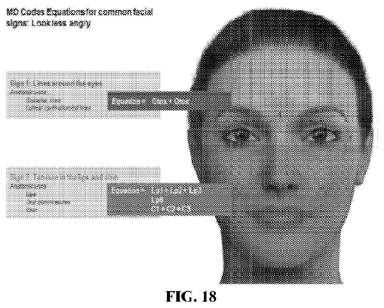
D00019

D00020

D00021

D00022

D00023

D00024

D00025

D00026

D00027

D00028

D00029

D00030

D00031

D00032

D00033

D00034

D00035

D00036

D00037

D00038

D00039

D00040

D00041

D00042

D00043

D00044

D00045

D00046

D00047

D00048

D00049

D00050

D00051

D00052

D00053

D00054

D00055

D00056

D00057

D00058

D00059

D00060

D00061

D00062

D00063

D00064

D00065

D00066

D00067

D00068

D00069

D00070

D00071

D00072

D00073

D00074

D00075

D00076

D00077

D00078

D00079

D00080

D00081

D00082

D00083

D00084

D00085

D00086

D00087

D00088

D00089

D00090

D00091

D00092

D00093

D00094

D00095

D00096

D00097

D00098

D00099

D00100

D00101

D00102

D00103

D00104

D00105

D00106

D00107

D00108
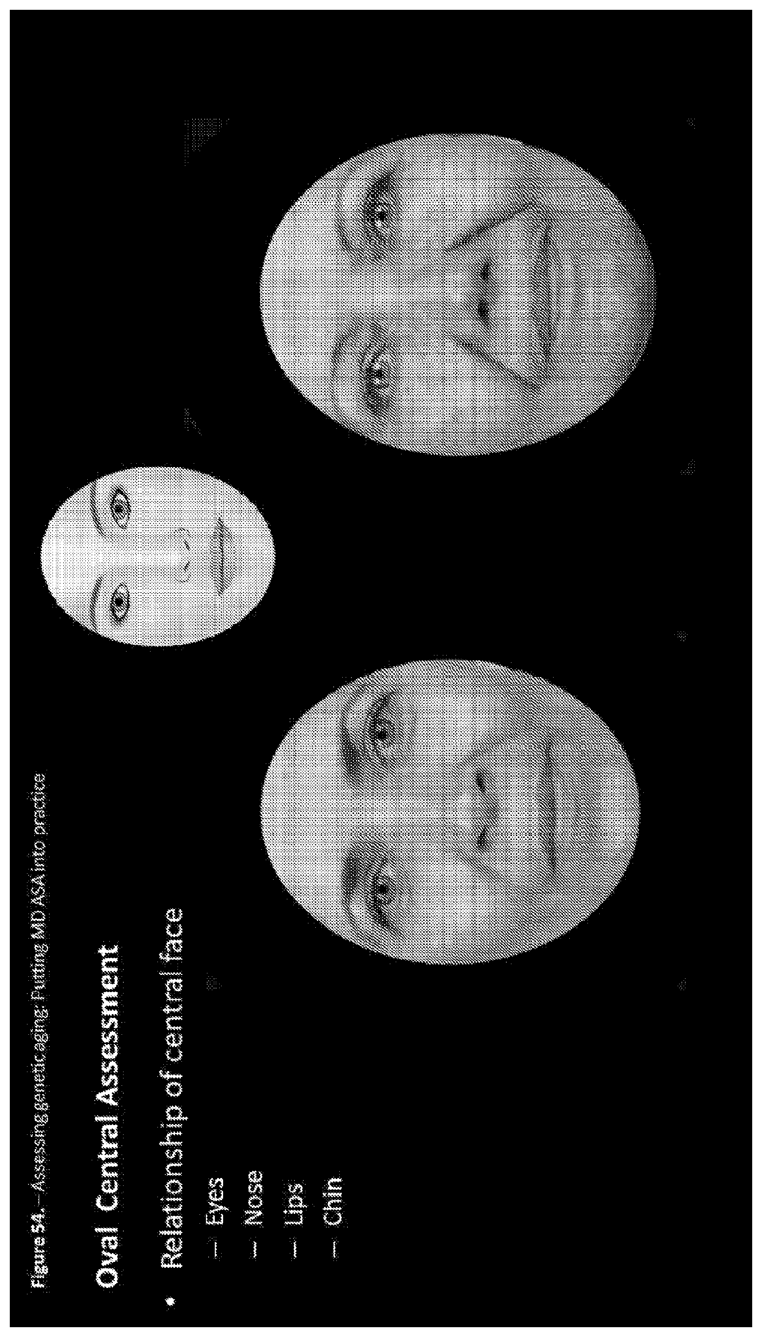
D00109

D00110

D00111

D00112

D00113

D00114

D00115

D00116

D00117

D00118

D00119

D00120

D00121

D00122

D00123

D00124

D00125

D00126

D00127

D00128

D00129

D00130

D00131

D00132

D00133

D00134

D00135

D00136

D00137

D00138

D00139

D00140

D00141

D00142

D00143

D00144

D00145
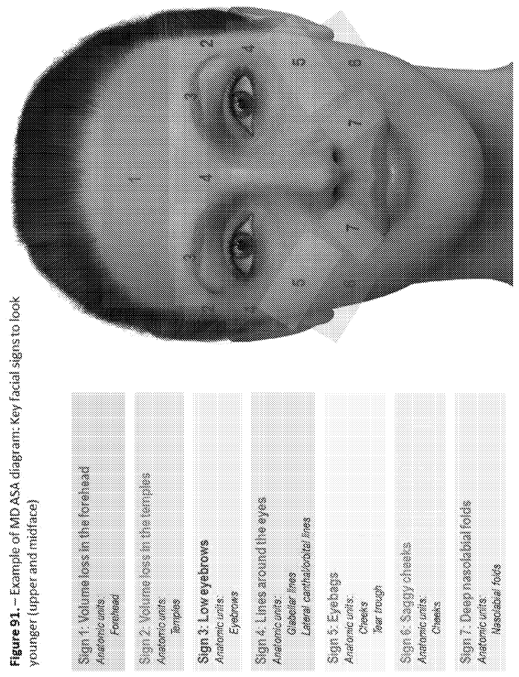
D00146

D00147

D00148

D00149

D00150

D00151

D00152

D00153

D00154

D00155

D00156

D00157

D00158

D00159

D00160

D00161

D00162

D00163

D00164

D00165

D00166

D00167

D00168

D00169

D00170

D00171

D00172

D00173

D00174

D00175

D00176

D00177
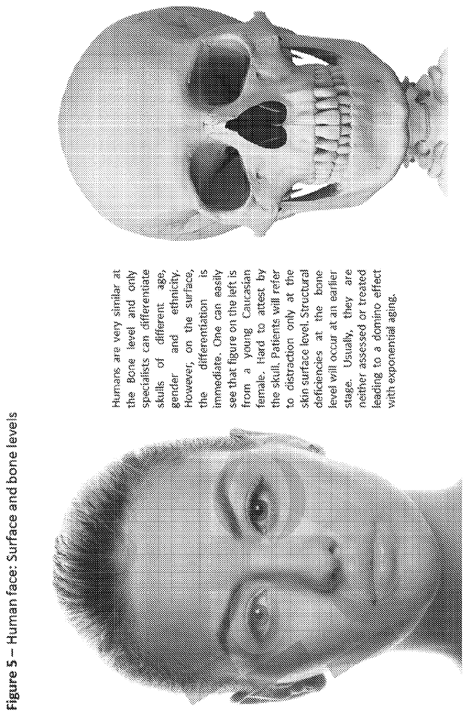
D00178

D00179

D00180

D00181
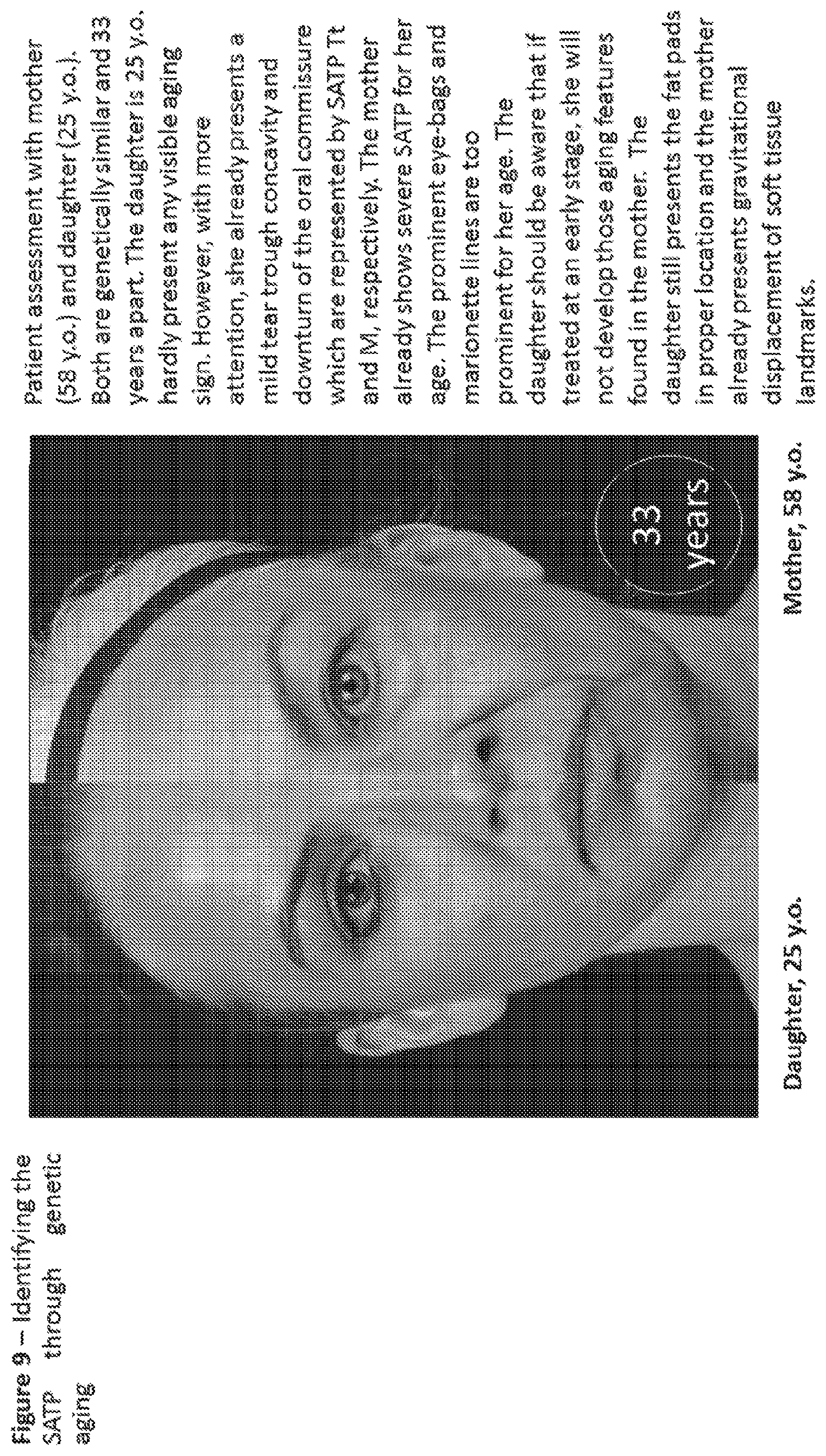
D00182

D00183

D00184

D00185

D00186

D00187

D00188

D00189

D00190

D00191

D00192

D00193

D00194

D00195
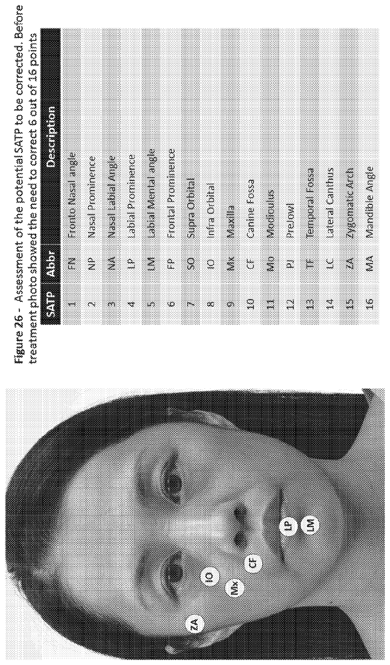
D00196

D00197

D00198

D00199

D00200

D00201

D00202

D00203

D00204

D00205

D00206

D00207

D00208

D00209

D00210

D00211

D00212

D00213

D00214

D00215

D00216

D00217

D00218
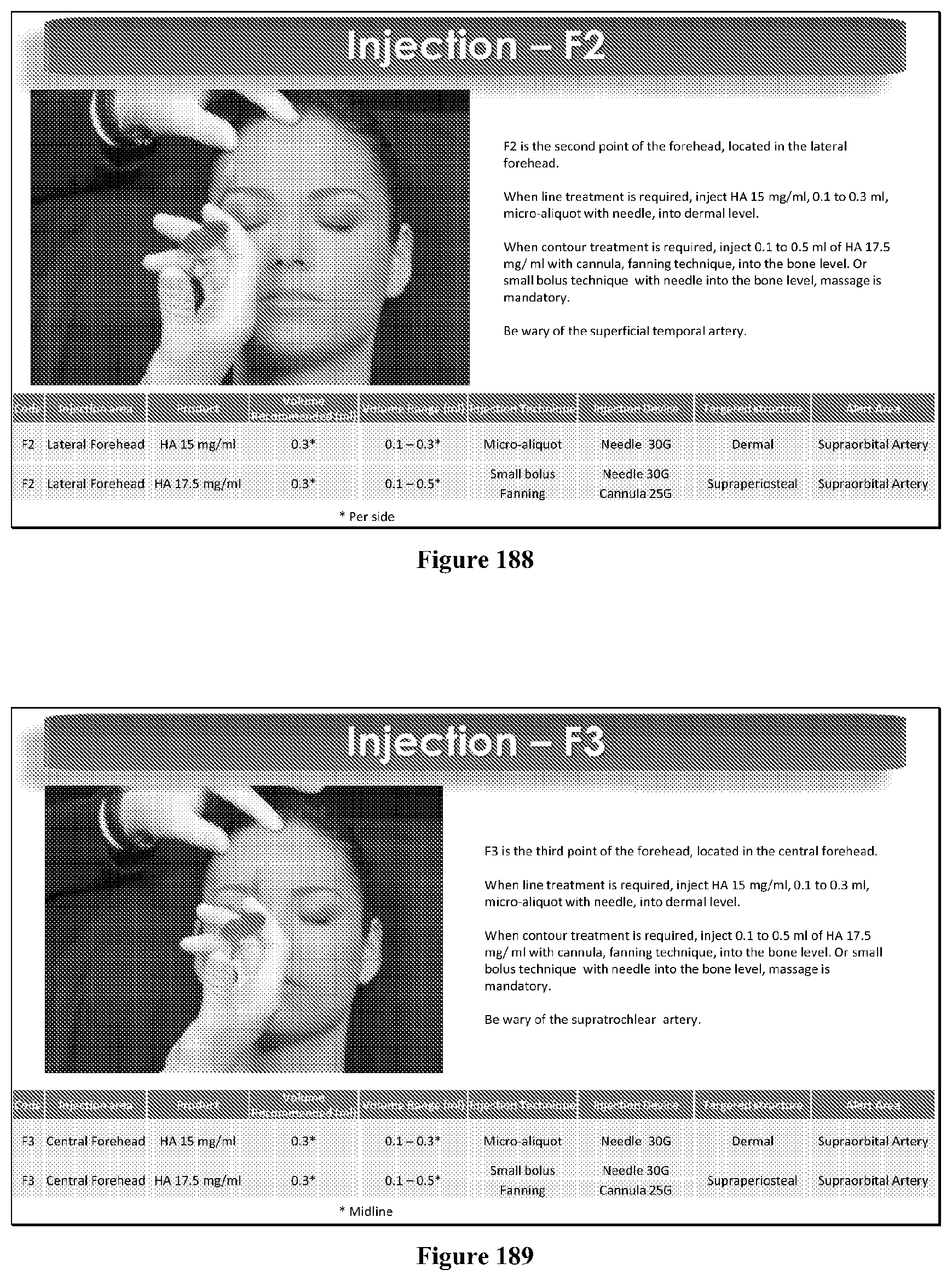
D00219

D00220

D00221

D00222

D00223

D00224

D00225

D00226

D00227

D00228

D00229

D00230

D00231

D00232

D00233

D00234

D00235

D00236

D00237

D00238

D00239

D00240

D00241

D00242

D00243

D00244

D00245

D00246

D00247
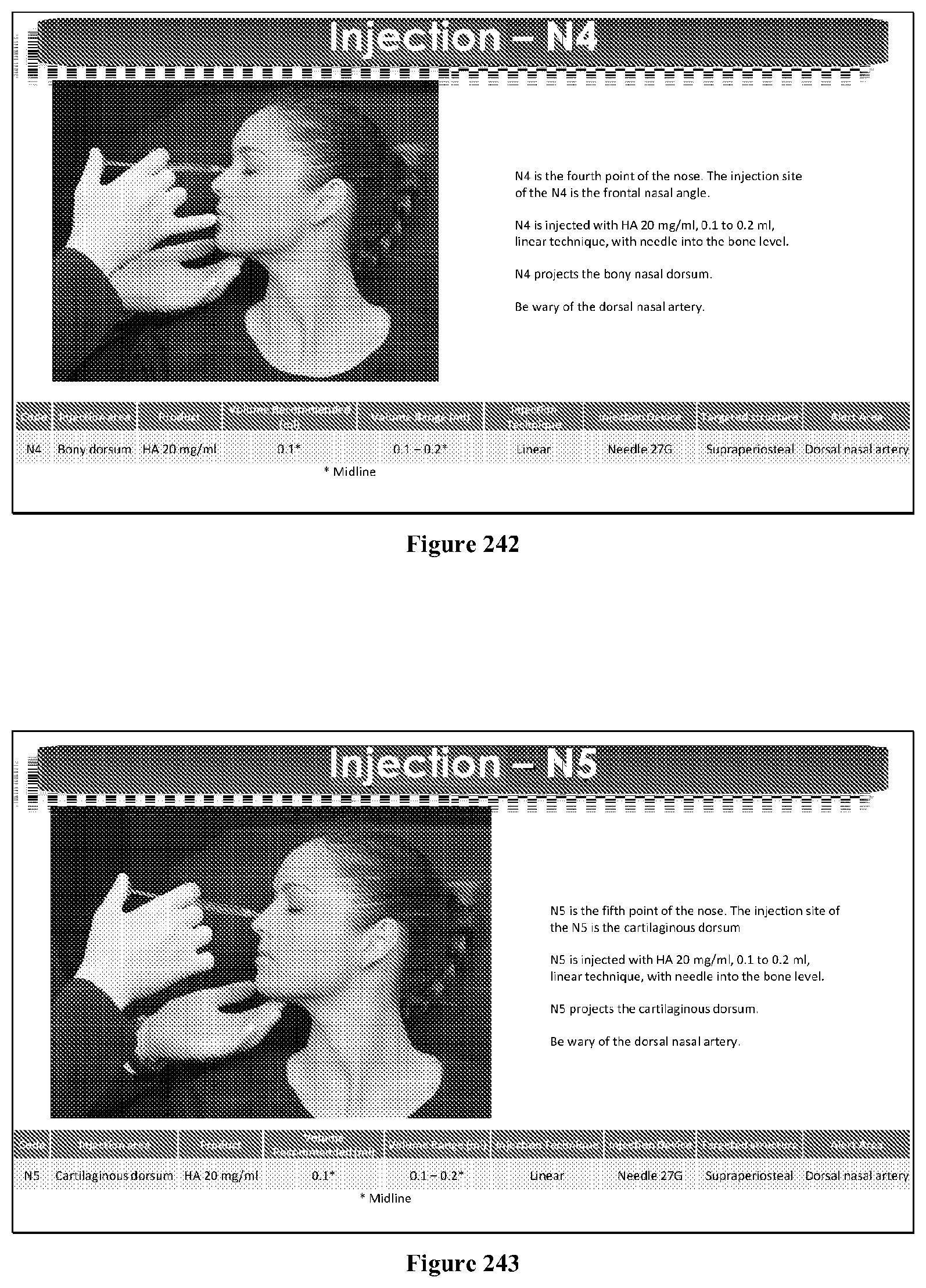
D00248

D00249
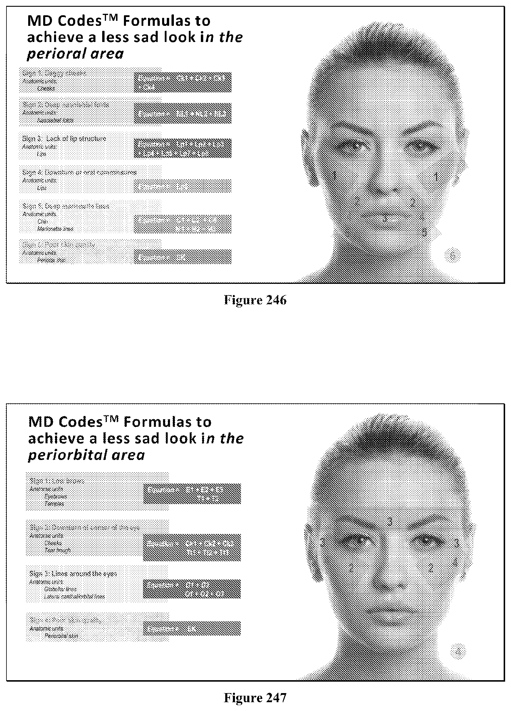
D00250

D00251

D00252

D00253

D00254

D00255

D00256

D00257

D00258

D00259

D00260

D00261

D00262

D00263

D00264

D00265

D00266

D00267

D00268

D00269

D00270

D00271

D00272

D00273

D00274

D00275

D00276

D00277

D00278

D00279

D00280

D00281

D00282

D00283

D00284

D00285
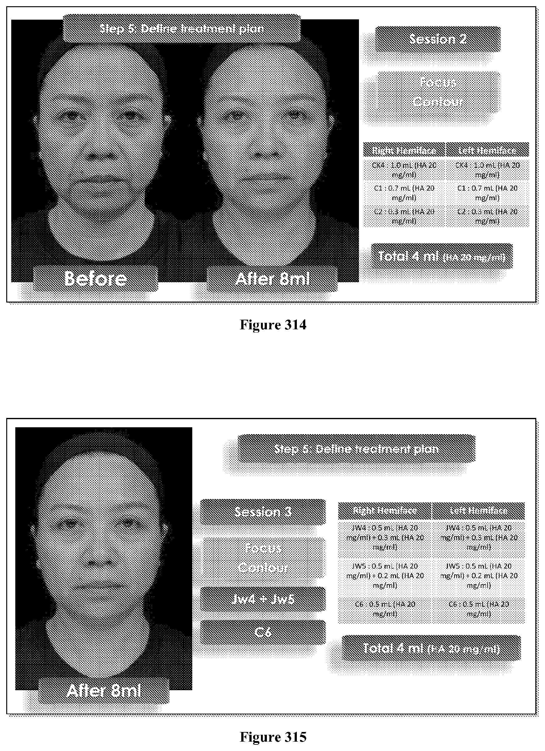
D00286

D00287

D00288

D00289

D00290

D00291

D00292

D00293

D00294

D00295

D00296

D00297

D00298

D00299

D00300

D00301

D00302

D00303

D00304

D00305

D00306

D00307

D00308

D00309

D00310

D00311

D00312

D00313

D00314

D00315

D00316

D00317

D00318

D00319

D00320

D00321

D00322

D00323

D00324

D00325

D00326

D00327

D00328

D00329

D00330

D00331

D00332

D00333

D00334

D00335

D00336

D00337

D00338

D00339

D00340

D00341

D00342

D00343

D00344

D00345

D00346

D00347

D00348

D00349

D00350

D00351

D00352

D00353

D00354

D00355

D00356

D00357

D00358

D00359

D00360

D00361

D00362

D00363

D00364

D00365

D00366

D00367

D00368

D00369

D00370

D00371

D00372

D00373

D00374

D00375

D00376

D00377

D00378

D00379

D00380

D00381

D00382

D00383

D00384

D00385

D00386

D00387

D00388

D00389

D00390

D00391

D00392

D00393
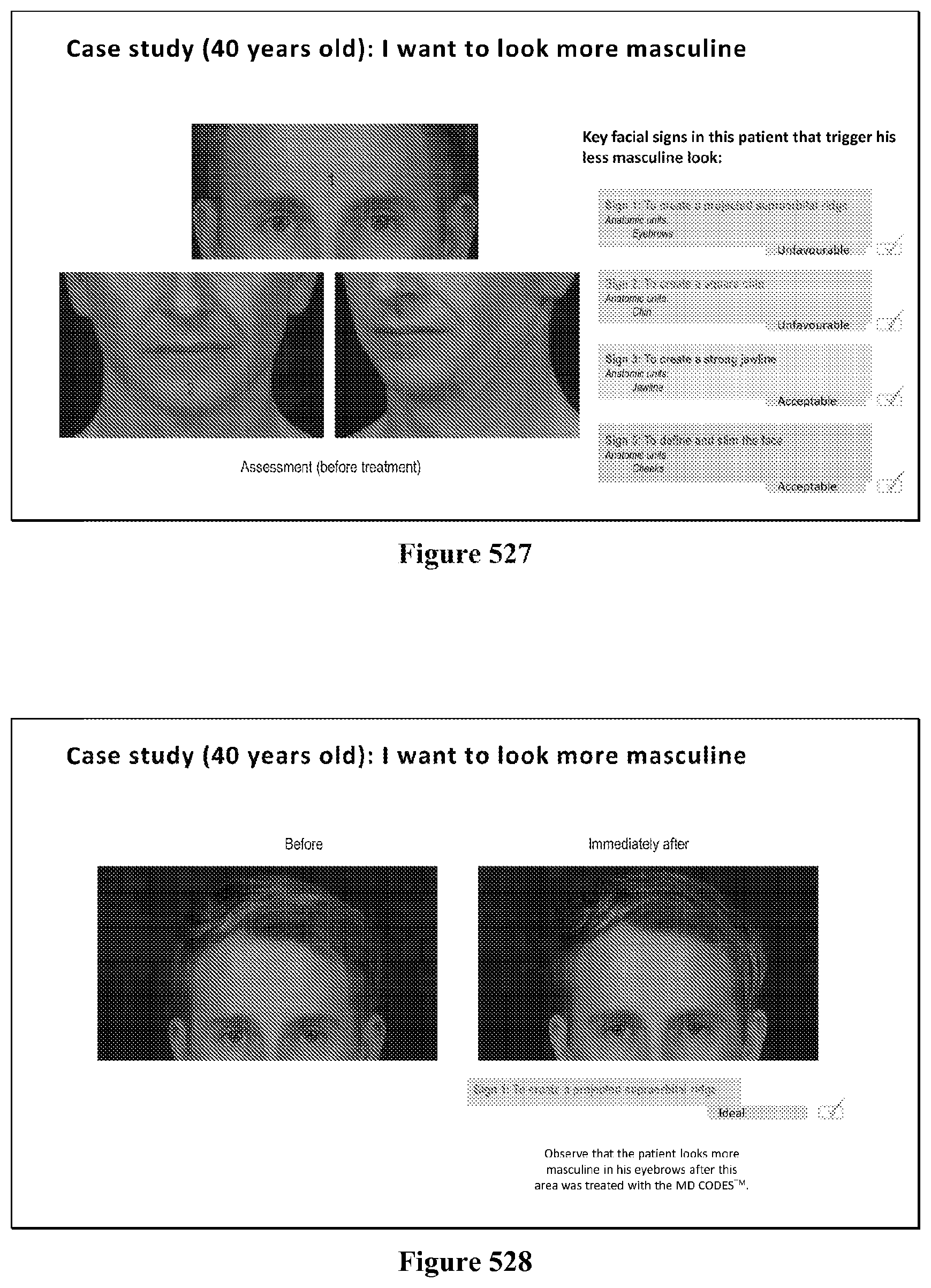
D00394

D00395
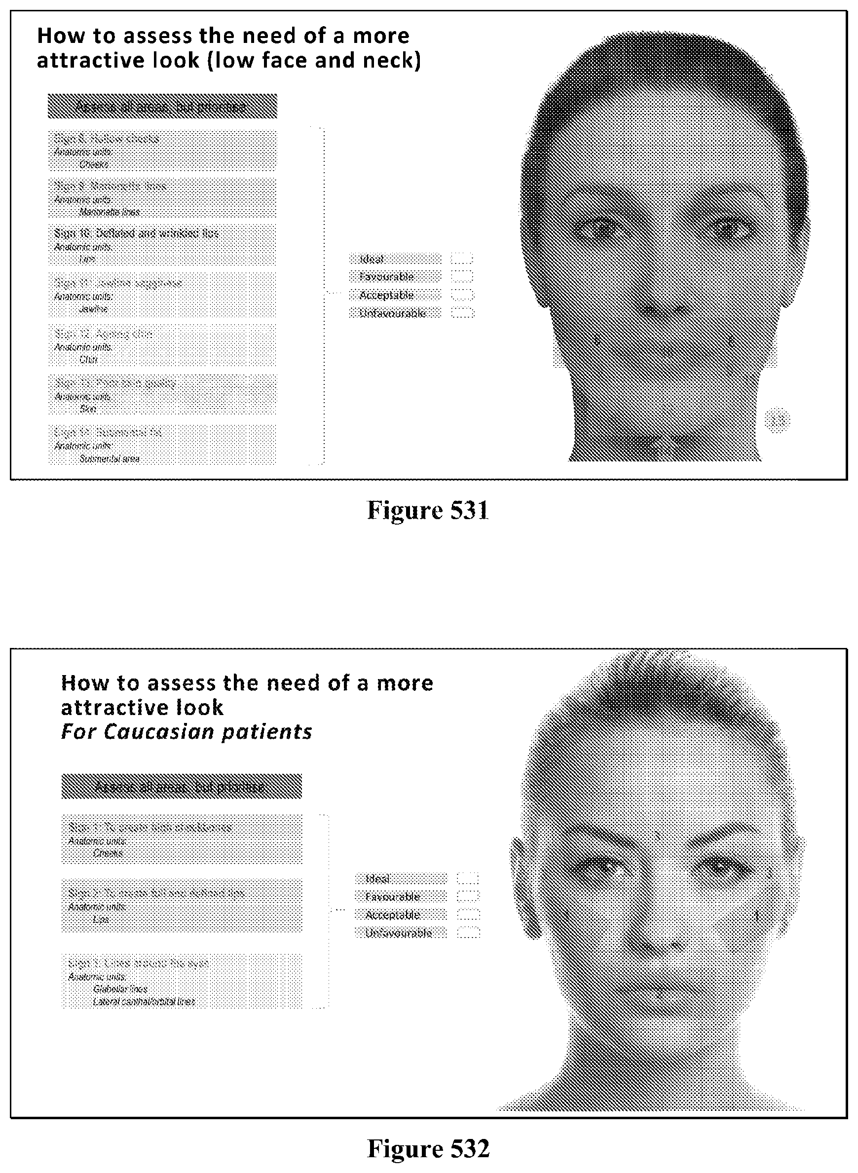
D00396

D00397

D00398

D00399

D00400

D00401

D00402

D00403

D00404

D00405

D00406

D00407

D00408

D00409

D00410

D00411

D00412

D00413

XML
uspto.report is an independent third-party trademark research tool that is not affiliated, endorsed, or sponsored by the United States Patent and Trademark Office (USPTO) or any other governmental organization. The information provided by uspto.report is based on publicly available data at the time of writing and is intended for informational purposes only.
While we strive to provide accurate and up-to-date information, we do not guarantee the accuracy, completeness, reliability, or suitability of the information displayed on this site. The use of this site is at your own risk. Any reliance you place on such information is therefore strictly at your own risk.
All official trademark data, including owner information, should be verified by visiting the official USPTO website at www.uspto.gov. This site is not intended to replace professional legal advice and should not be used as a substitute for consulting with a legal professional who is knowledgeable about trademark law.