Anti-il-1r3 Antibodies For Use In Inflammatory Conditions
FISCHER; Stephan ; et al.
U.S. patent application number 16/612052 was filed with the patent office on 2020-05-07 for anti-il-1r3 antibodies for use in inflammatory conditions. The applicant listed for this patent is MAB DISCOVERY GMBH. Invention is credited to Karsten BECKMANN, Stephan FISCHER.
| Application Number | 20200140559 16/612052 |
| Document ID | / |
| Family ID | 58698976 |
| Filed Date | 2020-05-07 |


View All Diagrams
| United States Patent Application | 20200140559 |
| Kind Code | A1 |
| FISCHER; Stephan ; et al. | May 7, 2020 |
ANTI-IL-1R3 ANTIBODIES FOR USE IN INFLAMMATORY CONDITIONS
Abstract
The present invention relates to methods for treating medical conditions and/or disorders characterized by uncontrolled or abnormal expression of members of the IL1R3 signaling pathway such as IL-1.alpha., IL-I.beta., IL-33, IL-36, IL1RA and/or IL1R3, as well as variants thereof. More specifically, the present invention relates to anti-IL1R3 antibodies for use in the treatment of an IL1R3-mediated inflammatory condition and/or disorder in a subject. Such conditions and disorders include but are not limited to inflammatory diseases, immune disorders, fibrotic disorders, eosinophilic disorders, infection, pain, a central nervous system disorder, an ophthalmologic disorder, Hereditary Systemic Inflammatory Diseases, and Systemic and Local Inflammatory Diseases and cancer associated chronic inflammation.
| Inventors: | FISCHER; Stephan; (Weilheim, DE) ; BECKMANN; Karsten; (Vaterstetten, DE) | ||||||||||
| Applicant: |
|
||||||||||
|---|---|---|---|---|---|---|---|---|---|---|---|
| Family ID: | 58698976 | ||||||||||
| Appl. No.: | 16/612052 | ||||||||||
| Filed: | May 8, 2018 | ||||||||||
| PCT Filed: | May 8, 2018 | ||||||||||
| PCT NO: | PCT/EP2018/061846 | ||||||||||
| 371 Date: | November 8, 2019 |
| Current U.S. Class: | 1/1 |
| Current CPC Class: | C07K 2317/565 20130101; C07K 2317/71 20130101; A61K 2039/505 20130101; A61P 29/00 20180101; C07K 2317/76 20130101; C07K 16/2866 20130101 |
| International Class: | C07K 16/28 20060101 C07K016/28; A61P 29/00 20060101 A61P029/00 |
Foreign Application Data
| Date | Code | Application Number |
|---|---|---|
| May 8, 2017 | EP | 17169953.1 |
Claims
1. A method for treating an IL1R3-mediated inflammatory condition and/or disorder in a subject, comprising administering to the subject an anti-IL1R3 antibody.
2. The method of claim 1, wherein the inflammatory condition and/or disorder is selected from the group consisting of an inflammatory condition, an immune disorder, a fibrotic disorder, an eosinophilic disorder, an infection, pain, a central nervous system disorder, an ophthalmologic disorder, Hereditary Systemic Inflammatory Diseases, and Systemic and Local Inflammatory Diseases.
3. The method of claim 2, wherein the inflammatory condition is a metabolic rheumatic disorder associated with hyperuricemia.
4. The method of claim 3, wherein the metabolic rheumatic disorder is selected from the group of gout, pseudogout, drug-induced gout and chronic active (refractory) gout.
5. The method of claim 4, wherein the IL1R3 antagonistic antibody is administered in combination with a therapeutic agent for the treatment of gout.
6. The method of claim 2, wherein the inflammatory condition is a cancer associated chronic inflammation.
7. The method of claim 6, wherein the IL1R3 antagonistic antibody is administered in combination with one or more cytotoxic, cytostatic and/or targeted anti-cancer agents.
8. The method of claim 6, wherein the subject is characterized in being resistant to treatment with one or more cytotoxic, cytostatic and/or targeted anti-cancer agents.
9. The method of claim 1, wherein the antibody has reduced or is lacking its effector functions.
10. The method of claim 1, wherein the antibody does not induce immune cell depletion.
11. The method of claim 9, wherein the antibody does not induce ADCC.
12. The method of claim 9, wherein said antibody comprises at least amino acid substitutions L234A and L235A of the human IgG1 Fc region or S228P and L235E of the human IgG4 Fc region or a corresponding functional mutation.
13. The method of claim 1, wherein the subject is a human subject and the antibody comprises a) a heavy chain variable region (VH) comprising the complementary determining regions comprising CDR-H1, CDR-H2, and CDR-H3 wherein the CDR-H1 region comprises an amino acid sequence selected from the group of SEQ ID NO: 69-85, wherein the CDR-H2 region comprises an amino acid sequence selected from the group of SEQ ID NO: 86-102, and wherein the CDR-H3 region comprises an amino acid sequence selected from the group of SEQ ID NO: 103-119; and b) a light chain variable region (VL) comprising the complementary determining regions comprising CDR-L1, CDR-L2, and CDR-L3 wherein the CDR-L1 region comprises an amino acid sequence selected from the group of SEQ ID NO: 120-136, wherein the CDR-L2 region comprises an amino acid sequence selected from the group of SEQ ID NO: 137-153, and wherein the CDR-L3 region comprises an amino acid sequence selected from the group of SEQ ID NO: 154-170, and SEQ ID NO: 175.
14. The method of claim 1, wherein the subject is a human subject and the antibody comprises a heavy chain variable (VH) region that is at least 90% identical to a VH region selected from the group consisting of VH regions of SEQ ID NO: 1 to 34 and 173, and a light chain variable (VL) region that is at least 90% identical to a VL region selected from the group consisting of VL regions of SEQ ID NO: 35 to 68 and 174.
15. The method of claim 1, wherein the subject is a mouse and the antibody comprises a) a heavy chain variable region (VH) comprising the complementary determining regions comprising CDR-H1, CDR-H2, and CDR-H3 wherein the CDR-H1 region comprises the amino acid sequence of SEQ ID NO: 178, wherein the CDR-H2 region comprises the amino acid sequence of SEQ ID NO: 179, and wherein the CDR-H3 region comprises the amino acid sequence of SEQ ID NO: 180; and b) a light chain variable region (VL) comprising the complementary determining regions comprising CDR-L1, CDR-L2, and CDR-L3 wherein the CDR-L1 region comprises the amino acid sequence of SEQ ID NO: 181, wherein the CDR-L2 region comprises the amino acid sequence of SEQ ID NO: 182, and wherein the CDR-L3 region comprises an amino acid sequence of SEQ ID NO: 183.
16. The method of claim 1, wherein the subject is a mouse and the antibody comprises a heavy chain variable (VH) region that is at least 90% identical to the VH region of SEQ ID NO: 176, and a light chain variable (VL) region that is at least 90% identical to the VL region of SEQ ID NO: 177.
Description
FIELD OF INVENTION
[0001] The invention pertains to methods for treating medical conditions and/or disorders characterized by uncontrolled or abnormal expression of members of the IL1R3 signaling pathway such as IL-1.alpha., IL-1.beta., IL-33, IL-36, IL1RA and/or IL1R3. More specifically, the present invention relates to methods and uses of anti-IL-1R3 antibodies in inflammatory conditions and/or disorders.
BACKGROUND
[0002] The interleukin 1 receptor accessory protein (IL1RAP), also called IL1R3, is a coreceptor of type 1 interleukin 1 receptor (IL1R1) and is indispensable for transmission of IL-1 signaling. Upon binding of IL-1, IL-1R1 associates with IL-1RAcP forming a functional signaling receptor complex, which stimulates NFkB activity.
[0003] IL-33, its receptor ST2, and IL-1RAcP form also a complex (IL-33/ST2/IL-1RAcP) with a similar activity in regard to NFkB activation as the IL-1.beta./IL-1R1/IL-1RAcP complex. IL-36 (IL-36.alpha. (IL-1F6), IL-36.beta. (IL-1F8), and IL-36.gamma. (IL-1F9)), their receptor IL-36R, and IL-1RAcP form also a complex (IL-36/II-36R/IL-1RAcP) with a similar activity in regard to NFkB activation as the IL-13/IL-1R1/IL-1RAcP complex.
[0004] The Interleukin-1 (IL-1) pathway is a cellular signaling pathway that plays a crucial role in the mammalian
[0005] inflammatory response and is associated with a wide range of immunologic, metabolic, physiological and hematopoietic activities. The IL-1 family includes three structurally related cytokines: IL-1 alpha, IL-1 beta and IL-1 receptor antagonist (IL-1 ra). Of the three, IL-1 alpha and IL-1 beta are proinflammatory agonists while IL-1 receptor antagonist (IL-1ra) functions to block IL-1 alpha and IL-1 beta activity. All known biological functions of IL-1 are mediated through the type I IL-1 R. IL-1 alpha, IL-1 beta and IL-1 ra bind the type I IL-1 R with high affinity. In contrast, IL-1 beta binds the type II IL-1R with high affinity and IL-1 alpha and IL-1ra bind the type II IL-1 R with a low affinity. The type II IL-1 R has a severely truncated cytoplasmic domain and upon binding to IL-1 does not transduce signal to a cell, but instead is involved in regulating an IL-1-mediated response by acting as a decoy receptor.
[0006] IL-1 production is triggered by infections, microbial toxins, inflammatory agents and allergic reactions. Overall
[0007] the main functions of IL-1 is to regulate the amplitude and duration of the immune and inflammatory response at the sites of inflammation or allergic immune reaction. When excess IL-1 is produced or IL-1 expression is not appropriately regulated disease states can develop.
[0008] Accordingly, IL-1 has been implicated in a variety of inflammatory and immunoregulatory diseases and conditions. It has been proposed that a systemic or localized excess of IL-1 contributes to the incidence of numerous medical disorders. Further to this proposal, it has been shown that IL-1ra, which blocks IL-1 alpha and IL-1 beta activity, has varying degrees of efficacy in treating some diseases thought to be mediated by IL-1 signaling.
[0009] It has been suggested that the suppression of IL-1 might be beneficial in patients suffering from various disorders
[0010] characterized by abnormal or excessive IL-1 expression or IL-1 activity. The IL-1ra and ICE inhibitors have met with limited degrees of success as therapeutics for diseases associated with IL-1 activity. Although progress has been made in devising effective treatment for such diseases, improved medicaments and methods of treatment are needed.
[0011] Unfortunately, existing IL-1 pathway inhibitors such as Kineret, ilaris or Arcalyst have certain disadvantages which impede their therapeutic use. For example, Kineret (IL-1Ra) has a short half-life and therefore needs very frequent treatment intervals (daily). Antibodies targeting single cytokines (e.g. Ilaris) allow redundant signaling/activities of other IL-1 family cytokines to occur. In addition, IL-1 family cytokines are known to elicit both pro-inflammatory and IL-1R3-independent anti-inflammatory signaling, both of which are interfered with when targeting the cytokines or their alpha-chain receptors. In contrast, the antibodies of the present invention combine the advantages of infrequent treatment intervals, concomitant inhibition of different IL-1 family cytokine mediated signaling and specificity with regard to blocking pro-inflammatory signaling pathways.
[0012] Therefore, the present invention provides for novel and improved methods for treating conditions associated with an uncontrolled expression of members of the IL1R3 signaling pathway with the favorable features described in the following.
SUMMARY OF INVENTION
[0013] The present invention relates to methods for treating medical conditions and/or disorders characterized by uncontrolled or abnormal expression of members of the IL1R3 signaling pathway such as IL-1.alpha., IL-1.beta., IL-33, IL-36, IL1RA and/or IL1R3, as well as variants thereof.
[0014] More specifically, the present invention relates anti-IL1R3 antibodies for use in the treatment of an IL1R3-mediated inflammatory condition and/or disorder in a subject.
[0015] Such conditions and disorders include but are not limited to inflammatory diseases, immune disorders, fibrotic disorders, eosinophilic disorders, infection, pain, a central nervous system disorder, an ophthalmologic disorder, Hereditary Systemic Inflammatory Diseases, and Systemic and Local Inflammatory Diseases and cancer associated chronic inflammation.
Definitions
[0016] The term "rabbit" according to the invention means an animal of the members of the taxonomic order Lagomorpha, which includes the families (hares and rabbits) and Ochotonidae (pikas), preferably of genus Oryctolagus.
[0017] The term "antibody" encompasses the various forms of antibody structures including, but not being limited to, whole antibodies and antibody fragments as long as it shows the properties according to the invention.
[0018] The term "rabbit monoclonal antibody" according to the invention means a monoclonal antibody produced by immunizing a rabbit and isolated from a antigen producing cell of said rabbit as well as such an antibody which is further modified, preferably a humanized antibody, a chimeric antibody, a fragment thereof, or a further genetically engineered and recombinant produced antibody as long as the characteristic properties according to the invention are retained. Preferably the antibody is from a B cell or a rabbit hybridoma cell of said rabbit.
[0019] The term "antibody producing cell" according to the invention means a rabbit B cell which produce antibodies, preferably a B cell or rabbit hybridoma cell.
[0020] "Native antibodies" are usually heterotetrameric glycoproteins composed of two identical light (L) chains and two identical heavy (H) chains. Each light chain is linked to a heavy chain by one covalent disulfide bond, while the number of disulfide linkages varies among the heavy chains of different immunoglobulin isotypes. Each heavy and light chain also has regularly spaced intrachain disulfide bridges. Each heavy chain has at one end a variable domain (VH) followed by a number of constant domains. Each light chain has a variable domain at one end (VL) and a constant domain at its other end. The constant domain of the light chain is aligned with the first constant domain of the heavy chain, and the light-chain variable domain is aligned with the variable domain of the heavy chain. Particular amino acid residues are believed to form an interface between the light chain and heavy chain variable domains.
[0021] "Percent (%) amino acid sequence identity" with respect to a peptide or polypeptide sequence is defined as the percentage of amino acid residues in a candidate sequence that are identical with the amino acid residues in the specific peptide or polypeptide sequence, after aligning the sequences and introducing gaps, if necessary, to achieve the maximum percent sequence identity, and not considering any conservative substitutions as part of the sequence identity. Alignment for purposes of determining percent amino acid sequence identity can be achieved in various ways that are within the skill in the art, for instance, using publicly available computer software such as BLAST, BLAST-2, ALIGN or Megalign (DNASTAR) software.
[0022] The term "VL (or VH) region" has the same meaning as VL (or VH) domain.
[0023] The terms "Fc receptor" or "FcR" according to the invention refers to a human receptor that binds to the Fc region of an antibody. FcRs bind IgG antibodies and include receptors of the Fc.gamma.RI, Fc.gamma.RII, and Fc.gamma.RIII subclasses, including allelic variants and alternatively spliced forms of these receptors. Fc.gamma.RII receptors include Fc.gamma.RIIA (an "activating receptor") and Fc.gamma.RIIB (an "inhibiting receptor"), which have similar amino acid sequences that differ primarily in the cytoplasmic domains thereof. Activating receptor Fc.gamma.RIIA contains an immunoreceptor tyrosine-based activation motif (ITAM) in its cytoplasmic domain. Inhibiting receptor Fc.gamma.RIIB contains an immunoreceptor tyrosine-based inhibition motif (ITIM) in its cytoplasmic domain (see review M. in Daeron, Annu. Rev. Immunol. 15:203-234 (1997)). FcRIIIA (CD16a) meditates ADCC. FcRs are reviewed in Ravetch and Kinet, Annu. Rev. Immunol 9:457-92 (1991); Capel et al, Immunomethods 4:25-34 (1994); and de Haas et al, J. Lab. CHn. Med. 126:330-41 (1995). These and all other FcRs are encompassed by the term "FcR" herein. The term also includes the neonatal receptor, FcRn, which is responsible for the transfer of maternal IgGs to the fetus (Guyer et al, J. Immunol. 117:587 (1976) and Kim et al, J. Immunol. 24:249 (1994)) and mediates slower catabolism, thus longer half-life.
[0024] The term "antibody effector function(s)," or "effector function" as used herein refers to a function contributed by an Fc effector domain(s) of an IgG (e.g., the Fc region of an immunoglobulin). Such function can be effected by, for example, binding of an Fc effector domain(s) to an Fc receptor on an immune cell with phagocytic or lytic activity or by binding of an Fc effector domain(s) to components of the complement system. Typical effector functions are ADCC, ADCP and CDC.
[0025] An "antibody fragment" refers to a molecule other than an intact antibody that comprises a portion of an intact antibody that binds the antigen to which the intact antibody binds. Examples of antibody fragments include but are not limited to Fv, Fab, Fab', Fab'-SH, F(ab')2; diabodies; linear antibodies; single-chain antibody molecules (e.g. scFv); and multispecific antibodies formed from antibody fragments.
[0026] An "antibody that binds to the same epitope" as a reference antibody refers to an antibody that blocks binding of the reference antibody to its antigen in a competition assay by 50% or more, and conversely, the reference antibody blocks binding of the antibody to its antigen in a competition assay by 50% or more. An exemplary competition assay is provided herein. "Antibody-dependent cell-mediated cytotoxicity" and "ADCC" refer to a cell-mediated reaction in which nonspecific cytotoxic cells that express FcRs (e.g. Natural Killer (NK) cells, neutrophils, and macrophages) recognize bound antibody on a target cell and subsequently cause lysis of the target cell. The primary cells for mediating ADCC, NK cells, express Fc.gamma.RIII only, whereas monocytes express Fc.gamma.RI, Fc.gamma.RII and FCYRIII.
[0027] The term "Antibody-dependent cellular phagocytosis" and "ADCP" refer to a process by which antibody-coated cells are internalized, either in whole or in part, by phagocytic immune cells (e.g., macrophages, neutrophils and dendritic cells) that bind to an immunoglobulin Fc region.
[0028] C1q'' is a polypeptide that includes a binding site for the Fc region of an immunoglobulin. C1q together with two serine proteases, C1r and C1s, forms the complex C1, the first component of the complement dependent cytotoxicity (CDC) pathway. Human C1q can be purchased commercially from, e.g. Quidel, San Diego, Calif.
[0029] The "class" of an antibody refers to the type of constant domain or constant region possessed by its heavy chain. There are five major classes of antibodies: IgA, IgD, IgE, IgG, and IgM, and several of these may be further divided into subclasses (isotypes), e.g., IgG.sub.1, IgG.sub.2, IgG.sub.3, IgG.sub.4, IgA.sub.1, and IgA.sub.2. The heavy chain constant domains that correspond to the different classes of immunoglobulins are called a, .delta., .epsilon., .gamma., and .mu., respectively.
[0030] An "effective amount" of an agent, e.g., a pharmaceutical formulation, refers to an amount effective, at dosages and for periods of time necessary, to achieve the desired therapeutic or prophylactic result.
[0031] The term "Fc region" herein is used to define a C-terminal region of an immunoglobulin heavy chain that contains at least a portion of the constant region. The term includes native sequence Fc regions and variant Fc regions. Unless otherwise specified herein, numbering of amino acid residues in the Fc region or constant region is according to the EU numbering system, also called the EU index, as described in Kabat, et al., Sequences of Proteins of Immunological Interest, 5th Ed. Public Health Service, National Institutes of Health, Bethesda, Md. (1991).
[0032] A "variant Fc region" comprises an amino acid sequence which differs from that of a "native" or "wildtype" sequence Fc region by virtue of at least one "amino acid modification" as herein defined. The term "Fc-variant" as used herein refers to a polypeptide comprising a modification in an Fc domain. The Fc variants of the present invention are defined according to the amino acid modifications that compose them. Thus, for example, P329G is an Fc variant with the substitution of proline with glycine at position 329 relative to the parent Fc polypeptide, wherein the numbering is according to the EU index. The identity of the wildtype amino acid may be unspecified, in which case the aforementioned variant is referred to as P329G. For all positions discussed in the present invention, numbering is according to the EU index. The EU index or EU index as in Kabat or EU numbering scheme refers to the numbering of the EU antibody (Edelman, et al., Proc Natl Acad Sci USA 63 (1969) 78-85, hereby entirely incorporated by reference.) The modification can be an addition, deletion, or substitution. Substitutions can include naturally occurring amino acids and non-naturally occurring amino acids. Variants may comprise non-natural amino acids. Examples include U.S. Pat. No. 6,586,207; WO 98/48032; WO 03/073238; US 2004/0214988 A1; WO 05/35727 A2; WO 05/74524 A2; Chin, J. W., et al., Journal of the American Chemical Society 124 (2002) 9026-9027; Chin, J. W., and Schultz, P. G., ChemBioChem 11 (2002) 1135-1137; Chin, J. W., et al., PICAS United States of America 99 (2002) 11020-11024; and, Wang, L., and Schultz, P. G., Chem. (2002) 1-10, all entirely incorporated by reference.
[0033] The term "Fc region-containing polypeptide" refers to a polypeptide, such as an antibody or immunoadhesin (see definitions below), which comprises an Fc region.
[0034] The terms "Fc receptor" or "FcR" are used to describe a receptor that binds to the Fc region of an antibody. A FcR which binds an IgG antibody (a gamma receptor) includes receptors of the Fc.gamma.RI, Fc.gamma.RII, and Fc.gamma.RIII subclasses, including allelic variants and alternatively spliced forms of these receptors. Fc.gamma.RII receptors include Fc.gamma.RIIA (an "activating receptor") and Fc.gamma.RIIB (an "inhibiting receptor"), which have similar amino acid sequences that differ primarily in the cytoplasmic domains thereof. Activating receptor Fc.gamma.RIIA contains an immunoreceptor tyrosine-based activation motif (ITAM) in its cytoplasmic domain. Inhibiting receptor Fc.gamma.RIIB contains an immunoreceptor tyrosine-based inhibition motif (ITIM) in its cytoplasmic domain, (see review in Daeron, M., Annu. Rev. Immunol. 15 (1997) 203-234). FcRs are reviewed in Ravetch, and Kinet, Annu. Rev. Immunol 9 (1991) 457-492; Capel, et al., Immunomethods 4 (1994) 25-34; and de Haas, et al., J. Lab. Clin. Med. 126 (1995) 330-41. Other FcRs, including those to be identified in the future, are encompassed by the term "FcR" herein. The term also includes the neonatal receptor, FcRn, which is responsible for the transfer of maternal IgGs to the fetus (Guyer, et al., J. Immunol. 117 (1976) 587 and Kim, et al., J. Immunol. 24 (1994) 249).
[0035] By "IgG Fc ligand" as used herein is meant a molecule, preferably a polypeptide, from any organism that binds to the Fc region of an IgG antibody to form an Fc/Fc ligand complex. Fc ligands include but are not limited to Fc.gamma.Rs, Fc.gamma.Rs, Fc.gamma.Rs, FcRn, C1q, C3, mannan binding lectin, mannose receptor, staphylococcal protein A, streptococcal protein G, and viral Fc.gamma.R. Fc ligands also include Fc receptor homologs (FcRH), which are a family of Fc receptors that are homologous to the Fc.gamma.Rs (Davis, et al., Immunological Reviews 190 (2002) 123-136, entirely incorporated by reference). Fc ligands may include undiscovered molecules that bind Fc. Particular IgG Fc ligands are FcRn and Fc gamma receptors. By "Fc ligand" as used herein is meant a molecule, preferably a polypeptide, from any organism that binds to the Fc region of an antibody to form an Fc/Fc ligand complex.
[0036] By "Fc gamma receptor", "Fc.gamma.R" or "FcgammaR" as used herein is meant any member of the family of proteins that bind the IgG antibody Fc region and is encoded by an Fc.gamma.R gene. In humans, this family includes but is not limited to Fc.gamma.RI (CD64), including isoforms Fc.gamma.RIA, Fc.gamma.RIB, and Fc.gamma.RIC; Fc.gamma.RII (CD32), including isoforms Fc.gamma.RIIA (including allotypes H131 and R131), Fc.gamma.RIIB (including Fc.gamma.RIIB-I and Fc.gamma.RIIB-2), and Fc.gamma.RIIc; and Fc.gamma.RIII (CD 16), including isoforms Fc.gamma.RIIIA (including allotypes VI 58 and F158) and Fc.gamma.RIIIb (including allotypes Fc.gamma.RIIB-NAI and Fc.gamma.RIIB-NA2) (Jefferis, et al., Immunol Lett 82
[0037] (2002) 57-65, entirely incorporated by reference), as well as any undiscovered human Fc.gamma.Rs or Fc.gamma.R isoforms or allotypes. An Fc.gamma.R may be from any organism, including but not limited to humans, mice, rats, rabbits, and monkeys. Mouse Fc.gamma.Rs include but are not limited to Fc.gamma.RI (CD64), Fc.gamma.RII (CD32), Fc.gamma.RIII (CD 16), and FCYRIII-2 (CD 16-2), as well as any undiscovered mouse Fc.gamma.Rs or Fc.gamma.R isoforms or allotypes.
[0038] By "FcRn" or "neonatal Fc Receptor" as used herein is meant a protein that binds the IgG antibody Fc region and is encoded at least in part by an FcRn gene. The FcRn may be from any organism, including but not limited to humans, mice, rats, rabbits, and monkeys. As is known in the art, the functional FcRn protein comprises two polypeptides, often referred to as the heavy chain and light chain. The light chain is beta-2-microglobulin and the heavy chain is encoded by the FcRn gene. Unless otherwise noted herein, FcRn or an FcRn protein refers to the complex of FcRn heavy chain with beta-2-microglobulin.
[0039] An "immunoconjugate" means an antibody conjugated to one or more cytotoxic agents, such as a chemotherapeutic agent, a drug, a growth inhibitory agent, a toxin, another antibody or a radioactive isotope.
[0040] "Antibody fragments" comprise a portion of a full-length antibody, preferably the variable regions thereof, or at least the antigen binding site thereof. Examples of antibody fragments include diabodies, Fab fragments, and single-chain antibody molecules. scFv antibodies are, e.g., described in Huston, J. S., Methods in Enzymol. 203 (1991) 46-88.
[0041] The terms "monoclonal antibody" or "monoclonal antibody composition" as used herein refer to a preparation of antibody molecules of a single amino acid composition.
[0042] The term "humanized antibody" or "humanized version of an antibody" also refers to antibodies for which both heavy and light chains are humanized as a result of antibody engineering. A humanized chain is typically a chain in which the V-region amino acid sequence has been changed so that, analyzed as a whole, is closer in homology to a human germline sequence than to the germline sequence of the species of origin. Humanization assessment is based on the resulting amino acid sequence and not on the methodology per se.
[0043] The terms "specifically binding, against target, or anti-target antibody", as used herein, refer to binding of the antibody to the respective antigen (target), measured by ELISA, wherein said ELISA preferably comprises coating the respective antigen to a solid support, adding said antibody under conditions to allow the formation of an immune complex with the respective antigen or protein, detecting said immune complex by measuring the Optical Density values (OD) using a secondary antibody binding to an antibody according to the invention and using a peroxidase-mediated color development.
[0044] The term "antigen" according to the invention refers to the antigen used for immunization or a protein comprising said antigen as part of its protein sequence. For example, for immunization a fragment of the extracellular domain of a protein (e.g. the first 20 amino acids) can be used and for detection/assay and the like the extracellular domain of the protein or the full-length protein can be used.
[0045] The term "specifically binding" or "specifically recognized" herein means that an antibody exhibits appreciable affinity for an antigen and, preferably, does not exhibit significant cross-reactivity.
[0046] "Appreciable" binding affinity includes binding with an affinity of at least 10.sup.-7M, specifically at least 10.sup.-8M, more specifically at least 10.sup.-9M, or even more specifically at least 10.sup.-10M.
[0047] An antibody that "does not exhibit significant cross-reactivity" is one that will not appreciably bind to an undesirable other protein. An antibody specific for an epitope according to the invention will, for example, not significantly cross-react with other epitopes on IL-1R3. Specific binding can be determined according to any art-recognized means for determining such binding, e.g. by competitive binding assays (e.g. ELISA).
[0048] All protein terms as used herein refers to the human proteins. If a protein from another species is meant, this is explicitly mentioned.
[0049] The term "IL-1alpha"", as used herein, refers to human IL-1 (UniProtKB P01583). The term "IL-1beta"", as used herein, refer to human IL-1beta (UniProtKB P01584). IL-1 stimulates thymocyte proliferation by inducing IL-2 release, B-cell maturation and proliferation, and fibroblast growth factor activity. IL-1 proteins are involved in the inflammatory response, being identified as endogenous pyrogens (UniProtKB).
[0050] The term "IL-33"", as used herein, refers to human IL-33 (UniProtKB 095760), a cytokine that binds to and signals through the IL1RL1/ST2 receptor which in turn activates NF-kappa-B and MAPK signaling pathways in target cells (UniProtKB).
[0051] The term "IL-36"", as used herein, refers to human IL-36alpha (UniProtKB Q9UHA7, IL-36beta (UniProtKB Q9NZH7) and or IL-36gamma (UniProtKB Q9NZH8). IL-36 are cytokines that bind to and signal through the IL1RL2/IL-36R receptor which in turn activates NF-kappa-B and MAPK signaling pathways in target cells linked to a pro-inflammatory response. IL-36 seems to be involved in skin inflammatory response by acting on keratinocytes, dendritic cells and indirectly on T cells to drive tissue infiltration, cell maturation and cell proliferation (UniProtKB).
[0052] The term "NFkB" as used herein, refer to human nuclear factor NF-kappa-B, which consists of p105 subunit (P19838) and p100 subunit (Q00653).
[0053] "Inhibition of NFkB" is measured according to the invention as inhibition of NFkB dependent luciferase gene expression in human cells. Such methods are e.g. described in Windheim M. et al., Mol. Cell. Biol. 28 (2008) 1783-1791; Huang J. et al. PNAS USA 94 (1997) 12829-12832; Xiaoxia L. et al., Mol. Cell, Biol. 19 (1999) 4643-4652. If murine NFkB is meant herein it is explicitly mentioned.
[0054] The "variable region (or domain) of an antibody according to the invention" (variable region of a light chain (VL), variable region of a heavy chain (VH)) as used herein denotes each of the pair of light and heavy chain regions which are involved directly in binding the antibody to the antigen. The variable light and heavy chain regions have the same general structure and each region comprises four framework (FR) regions whose sequences are widely conserved, connected by three complementary determining regions, CDRs. The antibody according to the invention comprises a VH region and a VL region or parts thereof, which are both together sufficient for the specific binding to the respective antigen.
[0055] The term "antigen-binding portion of an antibody" when used herein refer to the amino acid residues of an antibody which are responsible for antigen-binding. The antigen-binding portion of an antibody comprises preferably amino acid residues from the "complementary determining regions" or "CDRs". The CDR sequences are defined, and their residues are numbered, according to Kabat et al, Sequences of Proteins of Immunological Interest, 5th Ed. Public Health Service, National Institutes of Health, Bethesda, Md. (1991). Using this numbering system, the actual linear amino acid sequence may contain fewer or additional amino acids corresponding to a shortening of, or insertion into, a FR or CDR of the variable region. The Kabat numbering of residues may be determined for a given antibody by alignment at regions of homology of the sequence of the antibody with a "standard" Kabat numbered sequence.
[0056] The phrases "parenteral administration" and "administered parenterally" as used herein means modes of administration other than enteral and topical administration, usually by injection, and includes, without limitation, intravenous, intramuscular, intra-arterial, intrathecal, intracapsular, intraorbital, intracardiac, intradermal, intraperitoneal, transtracheal, subcutaneous, subcuticular, intra-articular, subcapsular, subarachnoid, intraspinal, epidural and intrasternal injection and infusion.
[0057] The term "cancer" as used herein may be, for example, lung cancer, non-small cell lung (NSCL) cancer, bronchioloalveolar cell lung cancer, bone cancer, pancreatic cancer, skin cancer, cancer of the head or neck, cutaneous or intraocular melanoma, uterine cancer, ovarian cancer, rectal cancer, cancer of the anal region, stomach cancer, gastric cancer, colon cancer, breast cancer, uterine cancer, carcinoma of the fallopian tubes, carcinoma of the endometrium, carcinoma of the cervix, carcinoma of the vagina, carcinoma of the vulva, Hodgkin's Disease, cancer of the esophagus, cancer of the small intestine, cancer of the endocrine system, cancer of the thyroid gland, cancer of the parathyroid gland, cancer of the adrenal gland, sarcoma of soft tissue, cancer of the urethra, cancer of the penis, prostate cancer, cancer of the bladder, cancer of the kidney or ureter, renal cell carcinoma, carcinoma of the renal pelvis, mesothelioma, hepatocellular cancer, biliary cancer, neoplasms of the central nervous system (CNS), spinal axis tumors, brain stem glioma, glioblastoma multiforme, astrocytomas, schwanomas, ependymonas, medulloblastomas, meningiomas, squamous cell carcinomas, pituitary adenoma, lymphoma, lymphocytic leukemia, including refractory versions of any of the above cancers, or a combination of one or more of the above cancers.
DETAILED DESCRIPTION OF THE INVENTION
[0058] As outlined in the introduction of this application, there are several difficulties in providing for suitable methods to treat inflammatory conditions that are mediated through an uncontrolled expression of members of the IL1R3 pathway. The present invention is meeting this need and provides for methods of treating, inhibiting, or ameliorating inflammatory conditions and/or disorders, including an immune disorder, a fibrotic disorder, an eosinophilic disorder, an infection, pain, a central nervous system disorder, an ophthalmologic disorder, Hereditary Systemic Inflammatory Diseases, and Systemic and Local Inflammatory Diseases.
[0059] The present invention relates to a method of treating, inhibiting or ameliorating an inflammatory condition and/or disorder in a subject in need thereof, the method comprising administering to the subject a therapeutically effective amount of an antagonistic IL1R3 antibody.
[0060] In particular, the invention encompasses anti-IL1R3 antibody for use in the treatment of an IL1R3-mediated inflammatory condition and/or disorder in a subject.
[0061] Such inflammatory condition and/or disorder may be selected from the group consisting of an inflammatory condition, an immune disorder, a fibrotic disorder, an eosinophilic disorder, an infection, pain, a central nervous system disorder, an ophthalmologic disorder, Hereditary Systemic Inflammatory Diseases, and Systemic and Local Inflammatory Diseases.
[0062] In one aspect of the invention, the inflammatory disorder treated is an IL-1 dependent diseases. The disease can be a Systemic and Local Inflammatory Diseases such as Schnitzler Syndrome, Behcet Disease, secondary amyloidosis, Henoch-Schonlein purpura, Idiopathic recurrent pericarditis, Systemic-onset Juvenile Idiopathic Arthritis, Adult Onset Still Disease, Macrophage Activation Syndrome, Sweet's Syndrome/neutrophilic dermatoses, neutrophilic panniculitis, Erdheim-Chester/histiocytoses, SAPHO, PFAPA, Multicentric Castleman Disease, Jessner-Kanof Disease, Primary Sjoegren Syndrome Fatigue, Kawasaki Disease, Colitis in Chronic Granulomatous Disease, Hidradenitis Suppurativa, Autoimmune Inner Ear Disease, Severe Traumatic Brain Injury), or a Hereditary Systemic Inflammatory Diseases such as Familial Mediterranean fever (FMF), CAPS, TRAPSa, HIDS, PAPA, PASH, DIRA, Blau syndrome/granulomatous arthritis, mevalonate kinase deficiency, Majeed Syndrome, NLRP12 Autoinflammatory Syndrome.
[0063] In preferred aspects of the invention, the inflammatory condition is selected from the group of COPD, inflammatory skin diseases, psoriasis, generalized pustular psoriasis (GPP), Inflammatory bowel disease (IBD), asthma, atopic dermatitis, Idiopathic pulmonary fibrosis, peritonitis, rheumatoid arthritis (RA), or a metabolic rheumatic disorder associated with hyperuricemia.
[0064] In one preferred embodiment, the inflammatory condition is a metabolic rheumatic disorder associated with hyperuricemia. The metabolic rheumatic disorder can be selected from the group of gout, pseudogout, drug-induced gout and chronic active (refractory) gout.
[0065] For the treatment of said metabolic rheumatic disorders in accordance with one aspect of the invention, an IL1R3 antagonistic antibody is administered. Said IL1R3 antagonistic antibody can be administered in combination with a therapeutic agent for the treatment of gout.
[0066] It can be administered simultaneously with a therapeutic agent for the treatment of gout. But it can also be administered sequentially with a therapeutic agent for the treatment of gout.
[0067] Another aspect of the invention encompasses methods for treatment of an inflammatory condition and/or disorder, wherein the inflammatory condition is a cancer associated chronic inflammation.
[0068] The invention also relates to anti-IL1R3 antibodies for the treatment of an inflammatory condition and/or disorder, wherein the inflammatory condition is a cancer associated chronic inflammation.
[0069] The term "cancer" as used herein may be, for example, lung cancer, non-small cell lung (NSCL) cancer, bronchioloalveolar cell lung cancer, bone cancer, pancreatic cancer, skin cancer, cancer of the head or neck, cutaneous or intraocular melanoma, uterine cancer, ovarian cancer, rectal cancer, cancer of the anal region, stomach cancer, gastric cancer, colon cancer, breast cancer, uterine cancer, carcinoma of the fallopian tubes, carcinoma of the endometrium, carcinoma of the cervix, carcinoma of the vagina, carcinoma of the vulva, Hodgkin's Disease, cancer of the esophagus, cancer of the small intestine, cancer of the endocrine system, cancer of the thyroid gland, cancer of the parathyroid gland, cancer of the adrenal gland, sarcoma of soft tissue, cancer of the urethra, cancer of the penis, prostate cancer, cancer of the bladder, cancer of the kidney or ureter, renal cell carcinoma, carcinoma of the renal pelvis, mesothelioma, hepatocellular cancer, biliary cancer, neoplasms of the central nervous system (CNS), spinal axis tumors, brain stem glioma, glioblastoma multiforme, astrocytomas, schwanomas, ependymonas, medulloblastomas, meningiomas, squamous cell carcinomas, pituitary adenoma, lymphoma, lymphocytic leukemia, including refractory versions of any of the above cancers, or a combination of one or more of the above cancers.
[0070] Preferably, the antibody according to the invention is used for the treatment of a cancer associated chronic inflammation wherein the cancer is selected from the group consisting of pancreatic cancer, liver cancer, lung cancer (associated with inflammation caused by asbestos, infections, smoking, silica), non-small-cell-lung cancer, colorectal cancer/colitis-associated cancer (associated with Inflammatory bowel disease), stomach cancer, gastric cancer, chronic gastritis associated gastric cancer, estrogen-receptor-positive breast cancer, head and neck squamous cell carcinoma, Mesothelioma, Gall bladder cancer (chronic cholecystitis associated), ovarian cancer, bladder cancer, prostate cancer, E. coli-infection associated prostate cancer, Thyroid cancer, Hodgkin disease, MALT lymphoma, salivary gland cancer, melanoma, endometriosis associated endometrial carcinoma, Barrett's esophagitis associated Esophageal cancer.
[0071] Preferably, the cancer is breast cancer, colon cancer, lung cancer, pancreatic cancer, liver cancer, non-small-cell-lung cancer, colorectal cancer, stomach cancer, gastric cancer, estrogen-receptor-positive breast cancer, head and neck squamous cell carcinoma, Mesothelioma, Gall bladder cancer, ovarian cancer, bladder cancer, prostate cancer, Thyroid cancer, Hodgkin disease, MALT lymphoma, salivary gland cancer, or melanoma.
[0072] The antibodies of the invention can be used for treating a cancer associated chronic inflammation as some tumors are caused or promoted by tumor micro-environment cells secreting inflammatory cytokines such as IL-1.alpha., IL-1.beta., IL-33, IL-36. In some instances, expression of such cytokines results in the formation of tumor resistance.
[0073] Therefore, in one aspect of the invention, the antibody is used for the treatment of subjects, wherein the subjects comprise a tumor, such as a solid tumor, and show tumor resistance or insufficient response to cytotoxic, cytostatic or targeted/immunotherapy. Preferably, the subjects are human subjects, e.g. cancer patients.
[0074] A concomitant or sequential use of cytokine inhibitors and anti-cancer compounds significantly improves the response rate of such treatments or can break tumor resistance.
[0075] The present invention therefore also encompasses a method for the treatment of a subject, wherein the subject is characterized in being resistant or showing insufficient response to treatment with one or more cytotoxic, cytostatic or targeted anti-cancer agents.
[0076] In one aspect of this invention, said IL1R3 antibody is administered in combination with one or more cytotoxic, cytostatic or targeted anti-cancer agents.
[0077] In one aspect of the invention, said IL1R3 antagonistic antibody is administered simultaneously with one or more cytotoxic, cytostatic or targeted anti-cancer agents. In another aspect, the IL1R3 antagonistic antibody is administered sequentially with one or more cytotoxic, cytostatic or targeted anti-cancer agents.
[0078] In the latter case, it is preferred that the antibody is administered after treatment with one or more cytotoxic, cytostatic or targeted anti-cancer agents.
[0079] The cytotoxic or cytostatic anti-cancer agents according to the invention can be taxanes, anthracyclins, alkylating agents, Histone Deacetylase Inhibitors, Topoisomerase inhibitors, kinase inhibitors, nucleotide analogs, peptide antibiotics, and platinum-based agents.
[0080] Preferably the targeted anti-cancer agents are used in targeted therapy and selected from one of the following, or combinations thereof: anti-EGFR compounds such as cetuximab, gefitinib, erlotinib, lapatinib, panitumumab, and anti-HER2 compounds such as trastuzumab, ado-trastuzumab emtansine, pertuzumab.
[0081] It is further preferred that the targeted anti-cancer agents are targeted checkpoint inhibitors. These can be but are not limited to: anti-PD1 compounds such as pembrolizumab, and nivolumab, and anti-PDL1 compounds such as atezolizumab, Avelumab, and Durvalumab, and anti-CTLA-4 compounds such as Ipilimumab and Tremelimumab.
[0082] The present invention provides for a significantly improved response rate to targeted cancer therapy as a broad spectrum of the inhibition of cytokine induced signaling is achieved. Such an activity in cancer indications is not achieved through a direct depletion activity of cancer cells (as done by several compounds of prior art) but through the inhibition of the cancer associated inflammation by modulating IL1R3 signaling pathways.
[0083] The antibodies of the present invention provide for a very advantageous activity profile because they enable the effective inhibition of cancer associated chronic inflammation and, at the same time, avoid undesired side effects, because they do not affect the viability of targeted cells that express IL-1R3.
[0084] The present invention therefore provides for several advantages over other IL-1 family targeting therapies. The commercially compound Kineret (IL-1Ra) has a short half-life and very frequent treatment intervals (daily). Antibodies targeting single cytokines (e.g. Ilaris) allow redundant signaling/activities of other IL-1 family cytokines to occur. In addition, IL-1 family cytokines are known to elicit both pro-inflammatory and IL-1R3-independent anti-inflammatory signaling, both of which are interfered with when targeting the cytokines or their alpha-chain receptors. In contrast, the antibodies of the present invention combine the advantages of infrequent treatment intervals, concomitant inhibition of different IL-1 family cytokine mediated signaling and specificity with regard to blocking pro-inflammatory signaling pathways.
[0085] The antibodies as described herein are preferably monoclonal antibodies with high affinity, high specificity, and potent neutralizing activity against IL-1R3. The present invention therefore also encompasses IL-1R3 antibody, with high affinity and specificity for IL-1R 3, with potent IL-1R3 neutralizing activity, and improved stability.
[0086] In one preferred embodiment of the invention, the antibodies have reduced effector functions. Preferably, the antibodies according to the invention show reduced or no Fc.gamma.-receptor signaling. Further preferred, the antibody does not induce ADCC.
[0087] It is another aspect of the invention that the anti-IL1R3 antibodies according comprises at least amino acid substitutions L234A and L235A of the human IgG1 Fc region or S228P and L235E of the human IgG4 Fc region or a corresponding functional mutation in another organism.
[0088] In one embodiment of the invention, the anti-L1R3 antibody comprises [0089] a) a heavy chain variable region (VH) comprising the complementary determining regions comprising CDR-H1, CDR-H2, and CDR-H3 [0090] wherein the CDR-H1 region comprises an amino acid sequence selected from the group of SEQ ID NO: 69-85, and 178 [0091] wherein the CDR-H2 region comprises an amino acid sequence selected from the group of SEQ ID NO: 86-102, and 179 [0092] and wherein the CDR-H3 region comprises an amino acid sequence selected from the group of SEQ ID NO: 103-119 and 180; and [0093] b) a light chain variable region (VL) comprising the complementary determining regions comprising CDR-L1, CDR-L2, and CDR-L3 [0094] wherein the CDR-L1 region comprises an amino acid sequence selected from the group of SEQ ID NO: 120-136 and 181, [0095] wherein the CDR-L2 region comprises an amino acid sequence selected from the group of SEQ ID NO: 137-153 and 182, and [0096] wherein the CDR-L3 region comprises an amino acid sequence selected from the group of SEQ ID NO: 154-170, and SEQ ID NO: 175 and 183.
[0097] In one embodiment, said antibody of the invention comprises a substitution at position 2 of CDR-L3. Said substitution may be a cysteine to serine substitution.
[0098] In one aspect of the invention, the subject is a human subject and the antibody comprises [0099] a) a heavy chain variable region (VH) comprising the complementary determining regions comprising CDR-H1, CDR-H2, and CDR-H3 [0100] wherein the CDR-H1 region comprises an amino acid sequence selected from the group of SEQ ID NO: 69-85, [0101] wherein the CDR-H2 region comprises an amino acid sequence selected from the group of SEQ ID NO: 86-102, [0102] and wherein the CDR-H3 region comprises an amino acid sequence selected from the group of SEQ ID NO: 103-119; and [0103] b) a light chain variable region (VL) comprising the complementary determining regions comprising CDR-L1, CDR-L2, and CDR-L3 [0104] wherein the CDR-L1 region comprises an amino acid sequence selected from the group of SEQ ID NO: 120-136, [0105] wherein the CDR-L2 region comprises an amino acid sequence selected from the group of SEQ ID NO: 137-153, and [0106] wherein the CDR-L3 region comprises an amino acid sequence selected from the group of SEQ ID NO: 154-170, and SEQ ID NO: 175.
[0107] In another aspect of the invention, the subject is a mouse and the antibody comprises [0108] a) a heavy chain variable region (VH) comprising the complementary determining regions comprising CDR-H1, CDR-H2, and CDR-H3 [0109] wherein the CDR-H1 region comprises the amino acid sequence of SEQ ID NO: 178, [0110] wherein the CDR-H2 region comprises the amino acid sequence of SEQ ID NO: 179, [0111] and wherein the CDR-H3 region comprises the amino acid sequence of SEQ ID NO: 180; and [0112] b) a light chain variable region (VL) comprising the complementary determining regions comprising CDR-L1, CDR-L2, and CDR-L3 [0113] wherein the CDR-L1 region comprises the amino acid sequence of SEQ ID NO: 181, [0114] wherein the CDR-L2 region comprises the amino acid sequence of SEQ ID NO: 182, and [0115] wherein the CDR-L3 region comprises an amino acid sequence of SEQ ID NO: 183.
[0116] In other embodiments of the invention, the antibody comprises a heavy chain variable (VH) region that is at least 90% identical to a VH region selected from the group consisting of VH regions of SEQ ID NO: 1 to 34 and 173 and 176, and a light chain variable (VL) region that is at least 90% identical to a VL region selected from the group consisting of VL regions of SEQ ID NO: 35 to 68 and 174 and 177.
[0117] In one embodiment to the invention, the antibody is a humanized antibody.
[0118] In such embodiments, the subject is a human subject and the antibody comprises a heavy chain variable (VH) region that is at least 90% identical to a VH region selected from the group consisting of VH regions of SEQ ID NO: 1 to 34 and 173, and a light chain variable (VL) region that is at least 90% identical to a VL region selected from the group consisting of VL regions of SEQ ID NO: 35 to 68 and 174.
[0119] In another embodiment, the subject is a mouse and the antibody comprises a heavy chain variable (VH) region that is at least 90% identical to the VH region of SEQ ID NO: 176, and a light chain variable (VL) region that is at least 90% identical to the VL region of SEQ ID NO: 177.
[0120] The antibodies according to the invention are in one embodiment, antibodies that bind specifically to IL-1R3 or a fragment or derivative thereof that contains at least a portion of said antibody that is sufficient to confer IL-1R3 binding specificity, and may comprise a heavy chain variable (VH) region that is at least 60% identical, preferably at least 70% identical, more preferably at least 80% identical, more preferably at least 90% identical to a VH region selected from the group consisting of VH regions of SEQ ID NO: 1 to 34 and 173 and 176.
[0121] In one embodiment, said antibodies comprise a heavy chain variable region (VH) sequence having at least 90%, 91%, 92%, 93%, 94%, 95%, 96%, 97%, 98%, 99%, or 100% sequence identity to an amino acid sequence selected from the group of VH sequences according to the invention.
[0122] In certain embodiments, a VH sequence having at least 90%, 91%, 92%, 93%, 94%, 95%, 96%, 97%, 98%, or 99% identity contains substitutions (e.g., conservative substitutions), insertions, or deletions relative to the reference sequence, whereby the antibody retains the ability to bind specifically according to the invention to the respective antigen.
[0123] The present invention also relates to an antibody that specifically binds to IL-1R3 or a fragment or derivative thereof and contains at least a portion of said antibody that is sufficient to confer IL-1R3 binding specificity, and comprises a light chain variable (VL) region that is least 60% identical, preferably at least 70% identical, more preferably at least 80% identical, more preferably at least 90% identical to a VL region selected from the group consisting of VL regions of SEQ ID NO: 35 to 68 and 174 and 177.
[0124] Said antibody may comprise a light chain variable region (VL) having at least 90%, 91%, 92%, 93%, 94%, 95%, 96%, 97%, 98%, 99%, or 100% sequence identity to the amino acid sequence of the VL sequences according to the invention.
[0125] In certain embodiments, a VL sequence having at least 90%, 91%, 92%, 93%, 94%, 95%, 96%, 97%, 98%, or 99% identity contains substitutions (e.g., conservative substitutions), insertions, or deletions relative to the reference sequence, whereby the antibody retains the ability to bind specifically to the respective antigen.
[0126] In certain embodiments, a total of 1 to 10 amino acids have been substituted, inserted and/or deleted in said VL sequences. In certain embodiments, the substitutions, insertions, or deletions occur in regions outside the CDRs (i.e., in the FRs). The invention also comprises affinity matured antibodies which can be produced according to methods known in the art. Marks et al. Bio/Technology 10:779-783 (1992) describes affinity maturation by VH and VL domain shuffling. Random mutagenesis of CDR and/or framework residues is described by: Barbas et al., Proc Nat. Acad. Sci, USA 91: 3809-3813 (1994); Schier et al., Gene 169: 147-155 (1995); Yelton et al., J. Immunol. 1 55:1994-2004 (1995); Jackson et al., J. Immunol. 1 54(7):3310-9 (1995); and Hawkins et al., J. Mol. Biol. 226:889-896 (1992) and WO2010108127.
[0127] In certain embodiments, a total of 1 to 10 amino acids have been substituted, inserted and/or deleted in each of said VH or VL sequences. In one embodiment, the antibody of the invention comprises a substitution at position 90 of the VH or VL sequence. It is preferred that the amino acid at position 90 is substituted by a serine. This substitution is preferably at position 90 of the light chain variable region (VL). In a preferred embodiment, the cysteine at position 90 of SEQ ID. NO: 62 is substituted by a serine. However, the antibodies of this invention are not limited to an amino acid substitution at position 90 but may comprise any substitution, deletion or insertion that leads to a functional antibody possessing the properties of the antibodies of this invention. Therefore, the VL and VH sequences of the antibodies of this invention may also comprise further mutations at different positions.
[0128] In certain embodiments, substitutions, insertions, or deletions occur in regions outside the CDRs (i.e., in the FRs).
[0129] In other embodiments, the substitutions, insertions, or deletions occur in regions inside the CDRs. In one preferred embodiment, the antibody of the invention comprises a substitution at position 2 of CDR-L3. It is preferred that this substitution is cysteine to serine. In one embodiment, said substitution is in SEQ ID NO: 164.
[0130] The present invention also encompasses an antibody that specifically binds to IL-1R3 or a fragment or derivative thereof that contains at least a portion of said antibody that is sufficient to confer IL-1R3 binding specificity, wherein the antibody comprises a heavy chain variable region (VH) comprising an amino acid sequence selected from the group of SEQ ID NO: 1 to 34 and 173 and 176.
[0131] Preferably, the heavy chain variable region (VH) sequence is SEQ ID NO:1, alternatively SEQ ID NO:2, or SEQ ID NO:3, SEQ ID NO:4, SEQ ID NO:5, SEQ ID NO:6, SEQ ID NO:7, SEQ ID NO:8, SEQ ID NO:9, SEQ ID NO:10, SEQ ID NO:11, SEQ ID NO:12, SEQ ID NO:13, SEQ ID NO:14, SEQ ID NO:15, SEQ ID NO:16, SEQ ID NO:17, SEQ ID NO:18, SEQ ID NO:19, SEQ ID NO:20, SEQ ID NO:21, SEQ ID NO:22, SEQ ID NO:23, SEQ ID NO:24, SEQ ID NO:25, SEQ ID NO:26, SEQ ID NO:27, SEQ ID NO:28, or SEQ ID NO:29, SEQ ID NO:30, SEQ ID NO:31, SEQ ID NO:32, SEQ ID NO:33, or alternatively SEQ ID NO:34 or 173 and 176.
[0132] Furthermore, the invention relates to methods in which said antibody that specifically binds to IL-1R3 or a fragment or derivative thereof that contains at least a portion of said antibody that is sufficient to confer IL-1R3 binding specificity, comprises a light chain variable region (VL) comprising an amino acid sequence selected from the group of SEQ ID NO: 35 to 68 and 174 and 177.
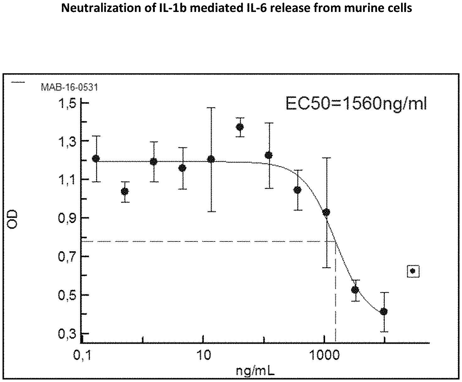
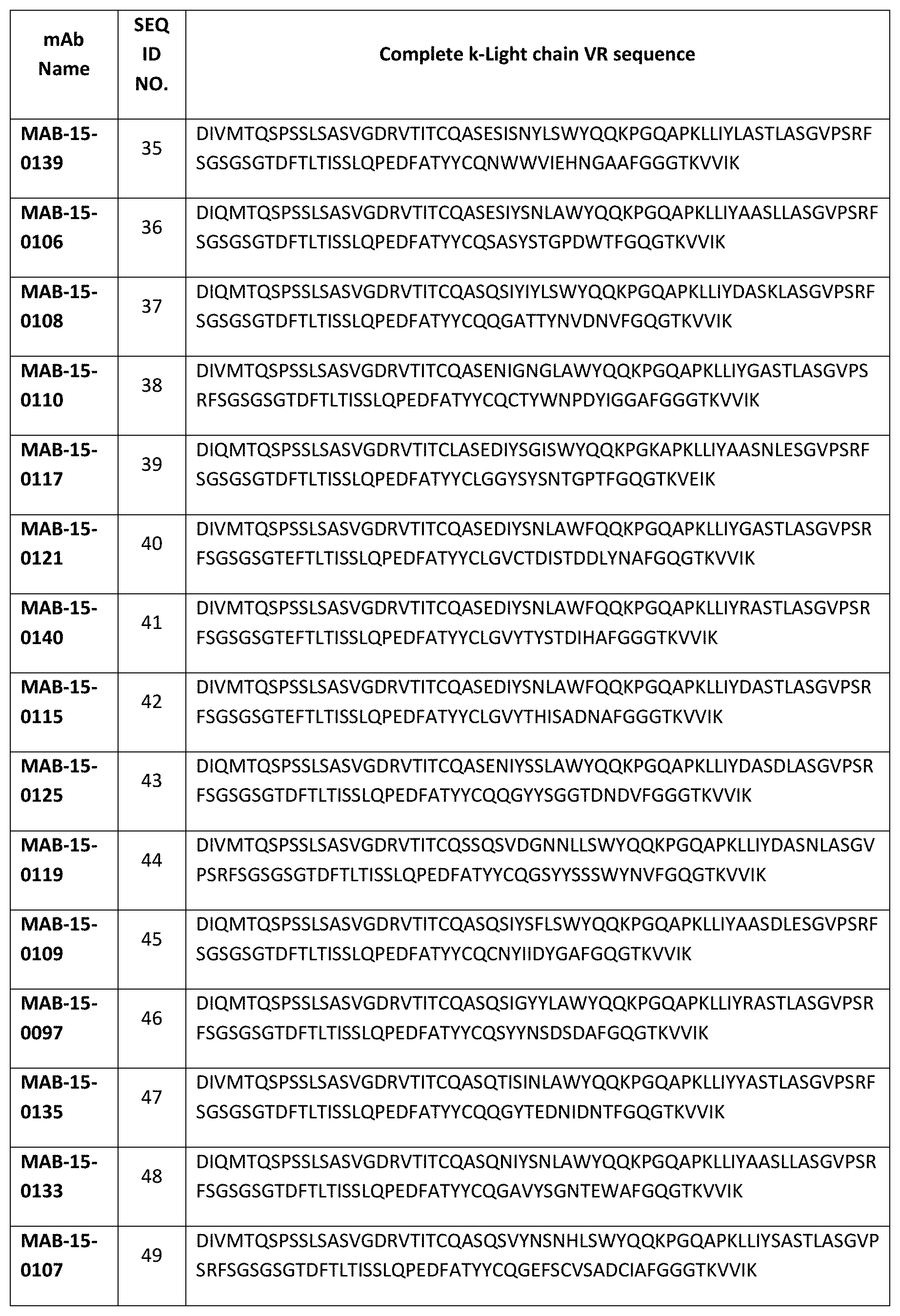
[0133] Even more preferred, the light chain variable region (VL) sequence is SEQ ID NO:35, or SEQ ID NO:36, SEQ ID NO:37, SEQ ID NO:38, SEQ ID NO:39, SEQ ID NO:40, SEQ ID NO:41, SEQ ID NO:42, SEQ ID NO:43, SEQ ID NO:44, SEQ ID NO:45, SEQ ID NO:46, SEQ ID NO:47, SEQ ID NO:48, SEQ ID NO:49, SEQ ID NO:50, SEQ ID NO:51, SEQ ID NO:52, SEQ ID NO:53, SEQ ID NO:54, SEQ ID NO:55, SEQ ID NO:56, SEQ ID NO:57, SEQ ID NO:58, SEQ ID NO:59, SEQ ID NO:60, SEQ ID NO:61, SEQ ID NO:62, SEQ ID NO:63, SEQ ID NO:64, SEQ ID NO:65, SEQ ID NO:66, SEQ ID NO:67, or alternatively SEQ ID NO:68 or 174 or 177.
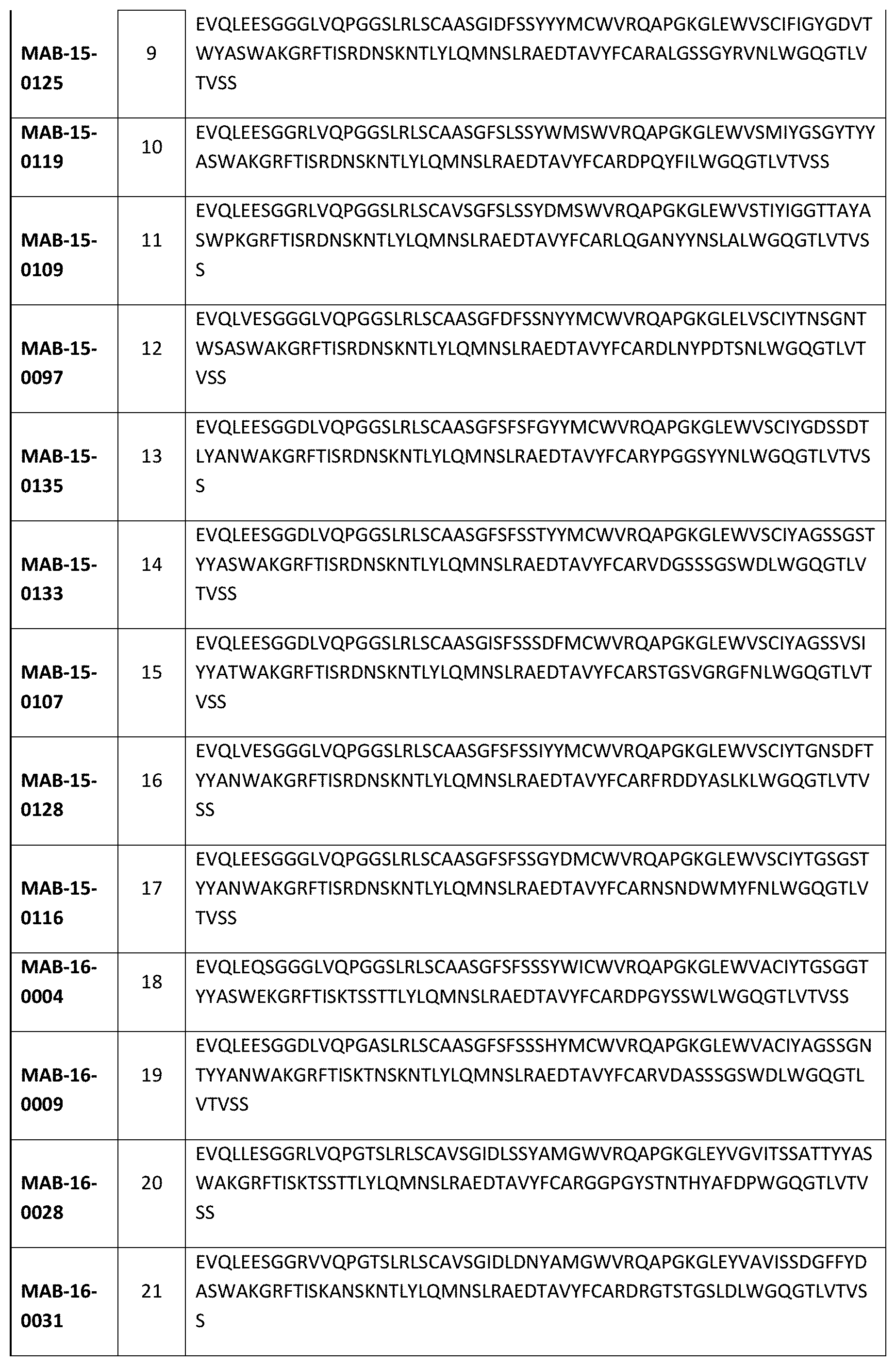
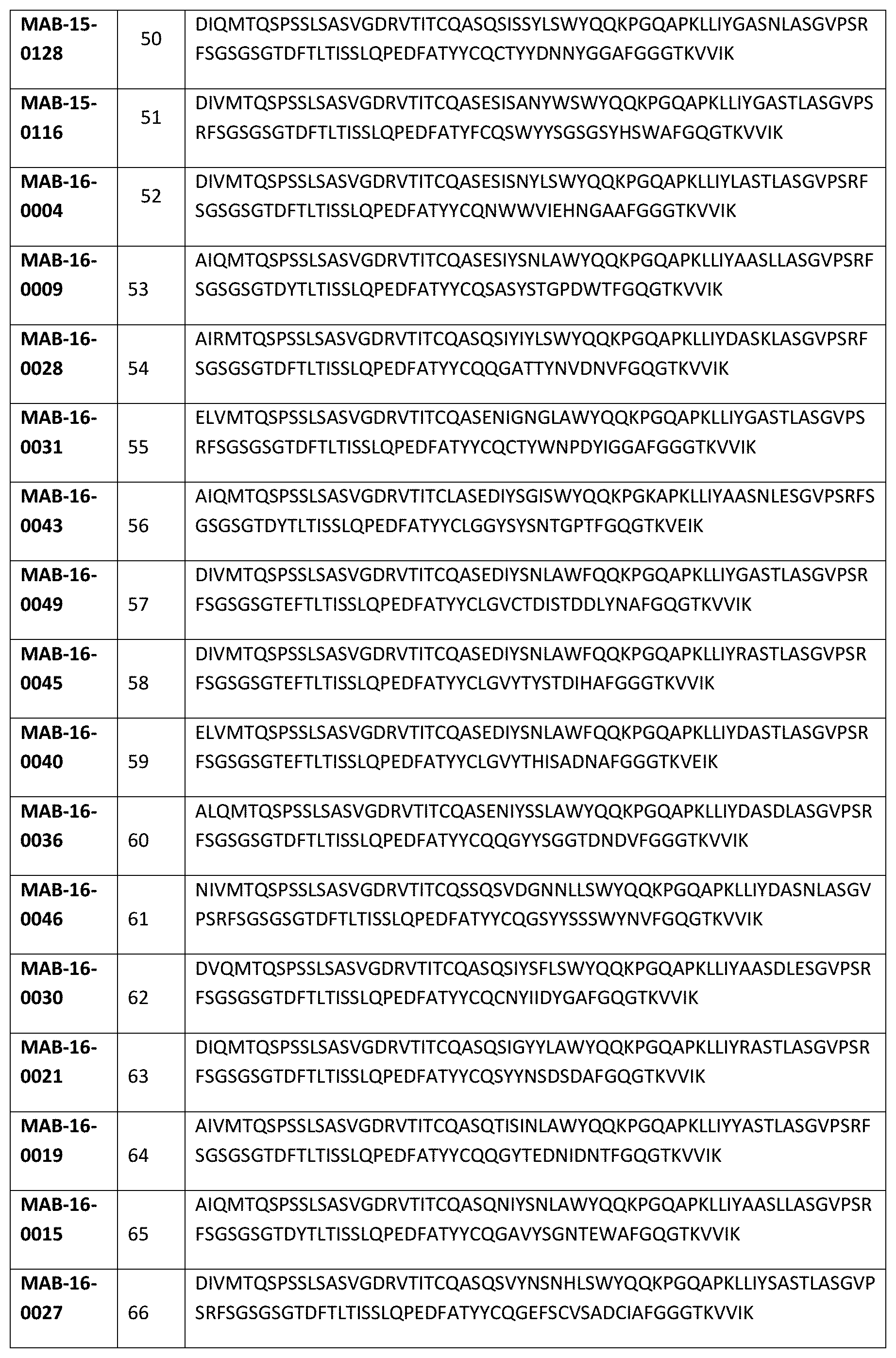
[0134] A antibody according to the invention that specifically binds to IL-1R3 or a fragment or derivative thereof that contains at least a portion of said antibody that is sufficient to confer IL-1R3 binding specificity, also comprises a VH region and a VL region comprising the respective CDR1, CDR2 and CDR3 regions of an antibody selected from the group consisting of MAB-15-0139, MAB-15-0106MAB-15-0108, MAB-15-0110, MAB-15-0117, MAB-15-0121, MAB-15-0140, MAB-15-0115, MAB-15-0125, MAB-15-0119, MAB-15-0109, MAB-15-0097, MAB-15-0135, MAB-15-0133, MAB-15-0107, MAB-15-0128, MAB-15-0116, MAB-16-0004, MAB-16-0009, MAB-16-0028, MAB-16-0031, MAB-16-0043, MAB-16-0049, MAB-16-0045, MAB-16-0040, MAB-16-0036, MAB-16-0046, MAB-16-0030, MAB-16-0021, MAB-16-0019, MAB-16-0015, MAB-16-0027, MAB-16-0048, MAB-16-0041, MAB-16-0149, MAB-16-0150, MAB-16-0531.
[0135] In one embodiment, the antibody that specifically binds to IL-1R3 or a fragment or derivative thereof that contains at least a portion of said antibody that is sufficient to confer IL-1R3 binding specificity, comprises SEQ ID NO.: 1 and 35, or SEQ ID NO.: 2 and 36. An antibody according to the invention may also comprise SEQ ID NO.: 3 and 37, or SEQ ID NO.: 4 and 38, or SEQ ID NO.: 5 and 39., or SEQ ID NO.: 6 and 40., or SEQ ID NO.: 7 and 41., or SEQ ID NO.: 8 and 42, or SEQ ID NO.: 9 and 43, or SEQ ID NO.: 10 and 44, or SEQ ID NO.: 11 and 45, or SEQ ID NO.: 12 and 46. Alternatively, an antibody according to the invention comprises SEQ ID NO.: 13 and 47, or SEQ ID NO.: 14 and 48, or SEQ ID NO.: 15 and 49, or SEQ ID NO.: 16 and 50, or SEQ ID NO.: 17 and 51, or SEQ ID NO.: 18 and 52, or SEQ ID NO.: 19 and 53, or SEQ ID NO.: 20 and 54, or SEQ ID NO.: 21 and 55, or SEQ ID NO.: 22 and 56, or SEQ ID NO.: 23 and 57, or SEQ ID NO.: 24 and 58, or SEQ ID NO.: 25 and 59, or SEQ ID NO.: 26 and 60, or SEQ ID NO.: 27 and 61.
[0136] Alternatively, an antibody according to the invention comprises SEQ ID NO.: 28 and 62, or SEQ ID NO.: 29 and 63, or SEQ ID NO.: 30 and 64, or SEQ ID NO.: 31 and 65, or SEQ ID NO.: 32 and 66, or SEQ ID NO.: 33 and 67, or SEQ ID NO.: 34 and 68, or SEQ ID NO.: 173 and 54, or SEQ ID NO.: 28 and 174, or SEQ ID NO.: 176 and 177.
[0137] Most preferably, the antibody that specifically binds to IL-1R3 or a fragment or derivative thereof that contains at least a portion of said antibody that is sufficient to confer IL-1R3 binding specificity comprises the constant region sequences CR-H (SEQ ID NO. 172) and CR-L (SEQ ID NO. 171) and a VH region selected from the group of SEQ ID NO: 1 to 34 and 173 and 176 and a VL region selected from the group of SEQ ID NO: 35 to 68 and 174 and 177.
[0138] The antibody that specifically binds to IL-1R3 or a fragment or derivative thereof that contains at least a portion of said antibody that is sufficient to confer IL-1R3 binding specificity, also comprises the constant region sequences CR-H (SEQ ID NO. 172) and CR-L (SEQ ID NO. 171) and a VH region and a VL region comprising the respective CDR1, CDR2 and CDR3 regions of an antibody selected from the group consisting of MAB-15-0139, MAB-15-0106, MAB-15-0108, MAB-15-0110, MAB-15-0117, MAB-15-0121, MAB-15-0140, MAB-15-0115, MAB-15-0125, MAB-15-0119, MAB-15-0109, MAB-15-0097, MAB-15-0135, MAB-15-0133, MAB-15-0107, MAB-15-0128, MAB-15-0116, MAB-16-0004, MAB-16-0009, MAB-16-0028, MAB-16-0031, MAB-16-0043, MAB-16-0049, MAB-16-0045, MAB-16-0040, MAB-16-0036, MAB-16-0046, MAB-16-0030, MAB-16-0021, MAB-16-0019, MAB-16-0015, MAB-16-0027, MAB-16-0048, MAB-16-0041, MAB-16-0149 and MAB-16-150, and MAB-16-0531.
[0139] According to the preferred therapeutic application of the antibodies according to the invention, the effector functions (such as ADCC) of the antibodies of the invention are reduced or lacking. In contrast to other antibodies of prior art, such as CAN04 (e.g. WO 2015/132602 A1), the antibodies of the invention avoid unwanted depletion of immune cells.
[0140] Preferably, the antibodies according to the invention show reduced or no Fc.gamma.-receptor signaling.
[0141] Therefore, the invention also relates to an antibody, wherein said antibody comprises at least amino acid substitutions at L234A and L235A of the human IgG1 Fc region or S228P and L235E of the human IgG4 Fc region, or a functional equivalent mutation.
[0142] In one embodiment according to the invention, the antibody is a humanized IgG1.sub.LALA, antibody.
[0143] In another embodiment, the antibody is a mouse-IgG2a.sub.LALA antibody.
[0144] In one embodiment according to the invention, the antibody inhibits IL-1R3 induced NFkB activity.
[0145] In another embodiment, the antibody that specifically binds to IL-1R3 or a fragment or derivative thereof that contains at least a portion of said antibody that is sufficient to confer IL-1R3 binding specificity, binds to the same epitope as an antibody selected from the group of antibodies MAB-15-0139, MAB-15-0106, MAB-15-0108, MAB-15-0110, MAB-15-0117, MAB-15-0121, MAB-15-0140, MAB-15-0115, MAB-15-0125, MAB-15-0119, MAB-15-0109, MAB-15-0097, MAB-15-0135, MAB-15-0133, MAB-15-0107, MAB-15-0128, MAB-15-0116, MAB-16-0004, MAB-16-0009, MAB-16-0028, MAB-16-0031, MAB-16-0043, MAB-16-0049, MAB-16-0045, MAB-16-0040, MAB-16-0036, MAB-16-0046, MAB-16-0030, MAB-16-0021, MAB-16-0019, MAB-16-0015, MAB-16-0027, MAB-16-0048, MAB-16-0041, MAB-16-0149, MAB-16-150 and MAB-16-0531.
[0146] The antibodies according to the invention have the advantage to be very potent when it comes to binding to their target. They exhibit a strong binding capacity to their antigen, IL1R3, but not to other receptors. The binding properties of the antibodies were studied in biochemical enzyme-linked immunosorbent assays (ELISA) and cell binding analysis (flow cytometry) and are exemplified in FIGS. 2, 6 and 7.
[0147] Preferred antibodies according to the invention, show a half maximal effective concentration (EC50) of less than 30 ng/ml, preferably of less than 20 ng/ml. In other embodiments, they show an EC50 of less than 15 ng/ml, 10 ng/ml or of less than 5 ng/ml. A preferred antibody according to the invention shows an EC50 of 16.3 ng/ml in a biochemical ELISA experiment (cf. FIG. 7).
[0148] The antibodies according to the invention also show a very strong binding to their antigen in experiments in which human IL1R3 is expressed in different cell lines while the antibodies do not bind cell lines not expressing human IL1R3 (e.g. NIH-3T3, cf. FIG. 5).
[0149] In the IL1R3 high-expressing cell line SK-MEL-30 (cf. FIG. 6, Example 4) the antibodies exhibit an EC50 of preferably less than 400 ng/ml, more preferably less than 350 ng/ml, or less than 310 ng/ml.
[0150] In one preferred embodiment encompassed by the invention, the antibody according to the invention inhibits IL-1alpha and/or IL-1beta stimulated NFkB activity. FIGS. 3, 4 and 8 exemplify the strong inhibitory activity of the antibodies according to the invention.
[0151] In one embodiment, the antibody that specifically binds to IL-1R3 or a fragment or derivative thereof that contains at least a portion of said antibody that is sufficient to confer IL-1R3 binding specificity, inhibits IL-1alpha stimulated NFkB activity.
[0152] In another embodiment, the antibody that specifically binds to IL-1R3 or a fragment or derivative thereof that contains at least a portion of said antibody that is sufficient to confer IL-1R3 binding specificity inhibits IL-1beta stimulated NFkB activity.
[0153] It is preferred that an antibody according to the invention inhibits IL-1beta stimulated NFkB activity in HEK293T/17-FR cells with an EC50 of less than 100 ng/ml, preferably of less than 95 ng/ml, 85 ng/ml, 75 ng/ml, 65 ng/ml, 55 ng/ml, 45 ng/ml, 35 ng/ml, 25 ng/ml, 20 ng/ml and most preferred of less than 15 ng/ml (e.g. cf. FIG. 3).
[0154] It is further preferred that an antibody according to the invention inhibits IL-1alpha stimulated NFkB activity with an EC50 of less than 1000 ng/ml, preferably of less than 500 ng/ml, 300 ng/ml, 200 ng/ml, and most preferred of less than 100 ng/ml (e.g. cf. FIG. 8) in A549-NFkB-RE-Luc cells.
[0155] It is further preferred that an antibody according to the invention inhibits IL-1beta stimulated NFkB activity with an EC50 of less than 700 ng/ml, preferably of less than 600 ng/ml, 300 ng/ml, 200 ng/ml, 100 ng/ml and most preferred of less than 50 ng/ml in A549-NFkB-RE-Luc cells (e.g. cf. FIG. 8).
[0156] The invention also encompasses a antibody, wherein said antibody inhibits NFkB activity stimulated by a complex selected from the group consisting of IL-1.beta./IL-1R1/IL-1RAcP, IL-1.alpha./IL-1R1/IL-1RAcP IL-33/ST2/IL-1RAcP, and/or IL-36/II-36R/IL-1RAcP.
[0157] Moreover, an antibody according to invention inhibits in a concentration of 10 .mu.g/ml (rabbit IgG isotype has a molecular weight of 150 KD) NFkB expression in A549-NFkB-RE-Luc cell lysates (Steady-Glo.TM. Luciferase Assay System; Promega; Cat. No. E2510) stimulated with 0.1 ng/ml human IL-1alpha, human IL-1beta, IL-33 and/or IL-36 (molecular weight see UniProtKB/Swiss-Prot), for 50% or more, preferably for 70% or more, preferably for 80% or more preferably for 90% and more, and more preferably for 95% or more, related to the same assay without said antibody according to the invention.
[0158] In one embodiment, the antibody according to the invention inhibits IL-1alpha, IL-1beta, IL-33, and/or IL-36, respectively, stimulated luciferase activity in HEK 293T/17 cells (HEK 293T/17-FR cells transfected with luciferase under control of NF-kB reporter gene)), HEK-Blue-IL33.TM. cells (Invivogen) or HEK-293/17-IF cells.
[0159] Preferably, said IL-1alpha, stimulated luciferase activity is inhibited by 50% or more, preferably by 70% or more, preferably by 80% or more, preferably by 90% and more, and more preferably by 95% or more. Preferably, said IL-1alpha, stimulated luciferase activity is inhibited by 95%.
[0160] Preferably, said IL-1beta, stimulated luciferase activity is inhibited by 50% or more, preferably by 70% or more, preferably by 80% or more, preferably by 90% and more, and more preferably by 95% or more. Preferably, said IL-1beta, stimulated luciferase activity is inhibited by 95%.
[0161] Preferably, said IL-33, stimulated luciferase activity is inhibited by 50% or more, preferably by 70% or more, preferably by 80% or more, preferably by 90% and more, and more preferably by 95% or more. Preferably, said IL-33, stimulated luciferase activity is inhibited by 95%.
[0162] Preferably, said IL-36, stimulated luciferase activity is inhibited by 50% or more, preferably by 70% or more, preferably by 80% or more, preferably by 90% and more, and more preferably by 95% or more. Preferably, said IL-36, stimulated luciferase activity is inhibited by 95%.
[0163] Furthermore, the antibodies according to the invention inhibit human IL-1a and IL-1b mediated IL-6 release and are superior to polyclonal antibodies. This potent inhibitory activity is shown and exemplified in FIG. 9. In these experiments, the EC50 values demonstrate that humanized anti-IL-1R3 IgG1-LALA antibodies are superior to that of goat-anti-human-IL1-R3 polyclonal antibody AF676 (R&D Systems). In preferred embodiments of the invention, the antibodies inhibit human IL-a mediated IL-6 release with an EC50 of less than 2500 ng/ml, preferably of less than 1500 ng/ml, less than 1000 ng/ml, less than 600 ng/ml, less than 400 ng/ml, or less than 300 ng/ml. It is also preferred that the antibodies of the invention inhibit human IL-b mediated IL-6 release with an EC50 of less than 500 ng/ml, preferably of less than 400 ng/ml, less than 300 ng/ml, less than 200 ng/ml, or less than 150 ng/ml.
[0164] In another embodiment according to the invention, the antibodies inhibit human IL-33 mediated NfkB-signaling. FIG. 10 exemplifies the inhibitory activity of selected antibodies according to the invention in HEK-Blue-IL33.TM. cells and demonstrates the superiority over polyclonal antibodies. In preferred embodiments of the invention, the antibodies inhibit human IL-33 mediated NfkB-signaling with an EC50 of less than 20000 ng/ml, preferably of less than 18000 ng/ml, less than 3000 ng/ml, less than 1000 ng/ml, less than 500 ng/ml, or less than 400 ng/ml.
[0165] The antibodies of the invention may also inhibit human IL-36 mediated NfkB-signaling (FIG. 11). Preferably, they inhibit human IL-36 mediated NfkB-signaling at an EC50 of less than 100 ng/ml, preferably at less than 50 ng/ml, less than 40 ng/ml, less than 30 ng/ml, less than 20 ng/ml, or less than 15 ng/ml.
[0166] Strikingly, the inventors found that the antibodies according to the invention inhibit cytokine release mediated by various different stimuli. For example, the antibodies inhibit cytokine release mediated by IL-1a, IL-33 and IL-36a. Results of a selected antibody are shown in FIG. 12. For example, the antibody MAB-16-0030 inhibits cytokine release mediated by all three stimuli, while IL-1Ra affects only IL-1a mediated cytokine release.
[0167] Diseases associated with acute or chronic inflammation are maintained or establish by the action of multiple cytokines either at the same time or consecutively. Early "alarmins" such as IL-1a and IL-33 may trigger other cytokines including IL-1b and IL-36 to establish a strong inflammatory environment. Therefore, the concomitant inhibition of signaling mediated by multiple cytokines exerts efficacious control of inflammatory processes. It is a key aspect of the antibodies of the invention that they inhibit multi-cytokine signaling via the blockage of the IL1R3 receptor.
[0168] Binding of antibodies to immune cells may result in cell depleting and deleterious effects, e.g. by direct induction of apoptotic signaling pathways, stimulation of excessive cytokine release or antibody-dependent cellular cytotoxicity (ADCC).
[0169] Importantly, the antibodies according to the invention do not affect the viability of immune cells. For example, they do not affect the viability of human peripheral blood mononuclear cells (PBMCs) and they do not induce IL-6 release in PBMCs (cf. FIG. 13).
[0170] The antibodies according to the invention, do not only inhibit the functional activation of cytokine release in different cell lines as described above, but also in PMBCs or whole blood cells from donors. They inhibit cytokine release mediated by different specific or complex stimuli. For example, they inhibit activation of PBMCs stimulated with LPS, heat-inactivated Candida albicans, IL-12/IL-33 or anti-CD3/CD28 antibodies (cf. FIGS. 14 and 15).
[0171] Also, in one embodiment, the anti-IL-1R3 IgG1-LALA antibodies according to the invention are able to inhibit release of IFNg, IL-6, TNF-.alpha., IL-13, IL-17 and IL-10 in mixed lymphocyte reactions (cf. FIG. 16).
[0172] The method of the present invention also encompasses the administering to a patient a pharmaceutically effective amount of the antibody, or derivative or fragment thereof according to the invention in form of a pharmaceutical composition.
[0173] Such a pharmaceutically composition may comprise a pharmaceutically acceptable carrier and a therapeutically effective amount of the antibody that specifically binds to the IL-1R3 or a fragment or derivative thereof that contains at least a portion of said antibody that is sufficient to confer IL-1R3 binding specificity, according to the invention.
[0174] As used herein, "pharmaceutical carrier" includes any and all solvents, dispersion media, coatings, antibacterial and antifungal agents, isotonic and absorption delaying agents, and the like that are physiologically compatible. Preferably, the carrier is suitable for intravenous, intramuscular, subcutaneous, parenteral, spinal or epidermal administration (e.g. by injection or infusion).
[0175] A composition of the present invention can be administered by a variety of methods known in the art. As will be appreciated by the skilled artisan, the route and/or mode of administration will vary depending upon the desired results. To administer a compound of the invention by certain routes of administration, it may be necessary to coat the compound with, or co-administer the compound with, a material to prevent its inactivation. For example, the compound may be administered to a subject in an appropriate carrier, for example, liposomes, or a diluent. Pharmaceutically acceptable diluents include saline and aqueous buffer solutions. Pharmaceutical carriers include sterile aqueous solutions or dispersions and sterile powders for the extemporaneous preparation of sterile injectable solutions or dispersion. The use of such media and agents for pharmaceutically active substances is known in the art.
[0176] The phrases "parenteral administration" and "administered parenterally" as used herein means modes of administration other than enteral and topical administration, usually by injection, and includes, without limitation, intravenous, intramuscular, intra-arterial, intrathecal, intracapsular, intraorbital, intracardiac, intradermal, intraperitoneal, transtracheal, subcutaneous, subcuticular, intra-articular, subcapsular, subarachnoid, intraspinal, epidural and intrasternal injection and infusion.
[0177] These compositions may also contain adjuvants such as preservatives, wetting agents, emulsifying agents and dispersing agents. Prevention of presence of microorganisms may be ensured both by sterilization procedures, supra, and by the inclusion of various antibacterial and antifungal agents, for example, paraben, chlorobutanol, phenol, sorbic acid, and the like. It may also be desirable to include isotonic agents, such as sugars, sodium chloride, and the like into the compositions. In addition, prolonged absorption of the injectable pharmaceutical form may be brought about by the inclusion of agents which delay absorption such as aluminum monostearate and gelatin.
[0178] Regardless of the route of administration selected, the compounds of the present invention, which may be used in a suitable hydrated form, and/or the pharmaceutical compositions of the present invention, are formulated into pharmaceutically acceptable dosage forms by conventional methods known to those of skill in the art. Actual dosage levels of the active ingredients in the pharmaceutical compositions of the present invention may be varied so as to obtain an amount of the active ingredient which is effective to achieve the desired therapeutic response for a particular patient, composition, and mode of administration, without being toxic to the patient. The selected dosage level will depend upon a variety of pharmacokinetic factors including the activity of the particular compositions of the present invention employed, the route of administration, the time of administration, the rate of excretion of the particular compound being employed, the duration of the treatment, other drugs, compounds and/or materials used in combination with the particular compositions employed, the age, sex, weight, condition, general health and prior medical history of the patient being treated, and like factors well known in the medical arts.
[0179] In another aspect, the present invention relates to an antibody that specifically binds to the mouse-IL-1R3 receptor or a fragment or derivative thereof, wherein the antibody comprises a heavy chain variable (VH) region that is at least 90% identical to a VH region of SEQ ID NO: 176, and a light chain variable (VL) region that is at least 90% identical to a VL region of SEQ ID NO: 177.
[0180] Preferably, said antibody comprises a heavy chain variable region (VH) sequence having at least 90%, 91%, 92%, 93%, 94%, 95%, 96%, 97%, 98%, 99%, or 100% sequence identity to an amino acid sequence selected from the group of VH sequences according to the invention.
[0181] In certain embodiments, a VH sequence having at least 90%, 91%, 92%, 93%, 94%, 95%, 96%, 97%, 98%, or 99% identity contains substitutions (e.g., conservative substitutions), insertions, or deletions relative to the reference sequence, whereby the antibody retains the ability to bind specifically according to the invention to the respective antigen.
[0182] In another aspect of the invention, said antibody comprises a light chain variable region (VL) sequence having at least 90%, 91%, 92%, 93%, 94%, 95%, 96%, 97%, 98%, 99%, or 100% sequence identity to an amino acid sequence selected from the group of VL sequences according to the invention.
[0183] In certain embodiments, a VL sequence having at least 90%, 91%, 92%, 93%, 94%, 95%, 96%, 97%, 98%, or 99% identity contains substitutions (e.g., conservative substitutions), insertions, or deletions relative to the reference sequence, whereby the antibody retains the ability to bind specifically according to the invention to the respective antigen.
[0184] It is preferred that said antibody that specifically binds to the mouse-IL-1R3 receptor or a fragment or derivative thereof, has reduced or is lacking its effector functions.
[0185] Preferably, the antibodies according to the invention show reduced or no Fc.gamma.-receptor signaling. It is further preferred that they do not induce ADCC.
[0186] In one embodiment, the antibody of the invention that specifically binds to the mouse-IL-1R3 receptor or a fragment or derivative thereof, is mouse IgG2a with mutations L234A and L235A in its FC part (amino acid positions according to EU numbering index).
[0187] Another aspect of the invention relates to an antibody that specifically binds to the mouse-IL-1R3 receptor or a fragment or derivative thereof, for use in pre-clinical studies. Such studies can be carried out in animal models, preferably in murine disease models. Such model systems include but are not limited to monosodium urate crystal (MSU)-induced mouse peritonitis model, Serum transfer induced rheumatoid arthritis, collagen-induced arthritis, antibody-induced arthritis, collagen antibody-induced arthritis, K/BxN antibody transfer arthritis, imiquimod-induced inflammatory skin disease/psoriasis, Epidermolysis bullosa acquisita model, Thioglycollate induced peritonitis, Immunecomplex induced peritonitis, bleomycin induced lung fibrosis, xenotranplantation psoriasis, tobacco smoke-induced lung inflammation COPD, tracheal instillation of elastase COPD, Fluorescein isothiocyanate (FITC) induced lung injury/fibrosis, Radiation-Induced Fibrosis, Silica induced lung fibrosis, asbestos fibre induced lung fibrosis, DSS induced colitis, Trinitrobenzene Sulfonic Acid induced Colitis, Oxazolone induced Colitis, Adoptive Transfer Colitis, house dust mite (HDM), cockroach, or Alternaria alternate induced asthma, ovalbumin induced asthma, papain-induced lung inflammation, epicutaneous sensitization atopic dermatitis model (ovalbumin, House dust mite (HDM), Hapten, S. aureus), xenograft or patient-derived-xenograft tumor growth models using immunocompromised or humanized mice, grafting of syngeneic tumors, chemical induced skin tumor models, DSS-induced colorectal tumor models.
[0188] Preferably, the antibody is used in the monosodium urate crystal (MSU)-induced mouse peritonitis model.
EXAMPLES
[0189] The following examples are used in conjunction with the figures and tables to illustrate the invention.
Example 1: Human-IL-1R3 Biochemical ELISA
[0190] Assay Principle:
[0191] NUNC Maxisorp 384well microtiter plates are coated with Fc-tagged human IL-1R3. After a blocking process, specific antibodies from B-cell supernatants bind to the antigen and are then detected by a POD-labeled antibody. Samples are tested 1:2 diluted.
[0192] Materials: [0193] Plates: 384well NUNC Maxisorp plates; Cat. No. 464718 [0194] Proteins: Fc-tagged human IL-1R3 (Conc. 1.5 mg/ml; Assay Conc. 0.54 .mu.g/ml) [0195] Standard Ab: P013-02 (Conc. 1 mg/ml; Start Assay Conc. 2 .mu.g/ml) [0196] Detection Ab: Anti-rabbit IgG, peroxidase-linked species-specific whole antibody (from donkey) (ECL); GE; Cat. No. NA9340; assay dilution: 1:5000 [0197] PBS: Buffers in a Box, Premixed PBS Buffer, 10.times.; Roche Applied Sciences; Cat. No. 11666789001 [0198] BSA: Bovine Serum Albumin Fraction V from bovine serum; Roche Applied Sciences; Cat. No. 10735086001 [0199] Tween 20: Tween 20; Carl Roth; Cat. No. 9127.2 [0200] TMB: TMB Solution; Life Technologies; Cat. No. SB02 [0201] HCl: 1M Titripur Hydrochloric Acid; Merck; Cat. No. 1090571000 [0202] ELISA Buffer: PBS, 0.5% BSA, 0.05% Tween [0203] Wash Buffer: PBS, 0.1% Tween [0204] Block Buffer: PBS, 2% BSA, 0.05% Tween
[0205] Procedure: [0206] 1. Add 12.5 .mu.L Fc-tagged human IL-1R3 (0.51 .mu.g/ml) in PBS to a 384well NUNC Maxisorp plate and incubate for 1h at RT. [0207] 2. Wash 3.times. with 901l Wash Buffer. [0208] 3. Add 90 .mu.L Blocking buffer to each well and incubate for 1h at RT. [0209] 4. Wash 3.times. with Wash Buffer. [0210] 5. Add 12.5 .mu.L antibody in Elisa Buffer and incubate for 1h at RT. [0211] 6. Wash 3.times. with Wash Buffer. [0212] 7. Add 12.5 .mu.L 1:5000 POD-Antibody in Elisa Buffer and incubate for 1h at RT. [0213] 8. Wash 6.times. with Wash Buffer. [0214] 9. Add 15 .mu.L TMB. [0215] 10. Add 15 .mu.L HCl after sufficient development. [0216] 11. Read absorbance at 450 nm/620 nm.
Example 2: Characterization of h-IL-1R3-Specific Antibodies Inhibiting hIL-1R3-Receptor by Luciferase Reporter Experiment
[0217] Assay Principle:
[0218] 293T/17-FR cells, which express a NF-kB-RE firefly luciferase reporter, are seeded into Poly-D-Lysin-Cell culture plates. After stimulation with IL-1b the 293T/17-FR lysate is tested for activated NF-kB using the Steady-Glo Luciferase Assay Kit. Supernatants with functional antibodies bind to hIL-1R3 and inhibit the NF-kB activation, which is shown in low signal.
[0219] Materials: [0220] Plates: Cell plate: 384well PDL Costar Cell Culture plate; Cat. No. 3844 [0221] Assay plate: 384well lumitrac white-plate; Corning; Cat. No. 3572 [0222] Cells: 293T/17-FR; assay conc. 250.000cells/ml [0223] Proteins: IL-1b (Conc. 0.03 mg/ml; Assay Conc. 115 pg/ml; Working Conc. 230 pg/ml) [0224] Standard Ab: P013_06 (Conc. 0.2 mg/ml; Start Working Conc. 61 .mu.g/ml) [0225] Kit: Steady-Glo Luciferase Assay System; Promega; Cat. No. E2510 [0226] Cell-Medium: DMEM Medium; PAN Biotech; Cat. No. P04-04510 [0227] FCS: Fetal Bovine Serum, HyClone; Thermo; Cat. No. St30070.03 [0228] 293T/17-FR Medium: DMEM Medium, 10% FCS, (+20 .mu.g/ml Hygromycin-B)
[0229] Procedure: [0230] 1. Cell Culture Procedure: [0231] Split confluent 293T/17-FR cells every Monday (seed out: 5.times.106 cells/T175 flask) and Friday (seed out: 3.times.106 cells/T175 flask) using trypsin/EDTA (incubate just for 30 sec at RT). [0232] 2. Seed cells (0.25.times.106 cells/ml) in 25 .mu.l DMEM+10% FCS to a 384-well PDL-plate (Corning cat #3844) and incubate over night at 37.degree. C. and 5% CO2. [0233] 3. Aspirate media and add 12.5 .mu.l antibodies in Conditioned Medium or just Conditioned Medium and incubate for 30 min at 37.degree. C. and 5% CO2 (program: 3 Aspiration and Sample transfer) [0234] 4. Add 12.5 .mu.l IL-1b in DMEM+10% FCS and incubate for 5 hours at 37.degree. C. and 5% CO2. [0235] 5. Equilibrate cultured cells to RT for 10 min. [0236] 6. Add 25 .mu.l Steady-Glo Reagent and mix several times with pipette [0237] 7. Wait 5 minutes before transfer 451l supernatant to a 384-well lumitrac white plate (Corning Cat#3572) [0238] 8. Measure luminescence in Tecan Reader: Integration Time: 0.5 sec
Example 3: Inhibition of NF.kappa.B-Expression of A549-NF.kappa.B-RE-Luc Stable Transfected Cells after Stimulation with IL-1 (.alpha./.beta.)
[0239] Assay Principle:
[0240] A549-NF.kappa.B-RE-Luc stable transfected cells (Signosis) are pipetted to a 384-well plate and incubated overnight. On day 2 anti-L1R3 antibodies are allowed to bind to A549-NFkB-RE-Luc stable transfected cells, which are then stimulated by addition of IL-1 (.alpha. or .beta.). This results in transcription of the luciferase gene due to NF.kappa.B signaling pathway activation and can be measured by cell lysis and addition of luciferin.
[0241] It is tested whether antibodies can inhibit the activation of NFkB pathway and therefore lower the luminescence signal.
[0242] Materials: [0243] Plates: 384-well Low Flange White Flat Bottom Polystyrene TC-Treated Microplates Sterile; Corning; Cat. No. 3570 [0244] Proteins: IL-1 .alpha. (P026_09); Recombinant Human IL-1alpha/IL-1F1; 10 .mu.g/mL; R&D Systems; Cat. No. 200-LA-002 [0245] IL-1 .beta. (P026_10); Recombinant Human IL-1beta/IL-1F2; 25 pg/mL; R&D Systems; Cat. No. 200-LB-005 [0246] Standard Ab: MAB-15-0115; MAB Discovery GmbH; 2.51 mg/ml; working conc. 10 .mu.g/ml [0247] Cells: A549-NF.kappa.B-RE-Luc stable transfected cells; Signosis; Cat. No. SL-0014 [0248] Medium: DMEM; PAN; Cat. No. P04-04510 [0249] FCS: Fetal Bovine Serum South Africa Low IgG; PAN; Cat. No. 1552-P120909 [0250] Pen/Strep: 10,000 U Penicillin/ml; 10 mg Streptomycin/ml; PAN Biotech; Cat. No. P06-07100 [0251] Detaching Agent: Trypsin-EDTA 1.times.; PAN; Cat. No. P10-023100 (4 mL for T175/2 mL for T75; .sup..about.8 min 37.degree. C.) [0252] Cell-Medium: DMEM, 10% FCS, 1% Pen/Strep [0253] Detection Kit: Steady-Glo.TM. Luciferase Assay System; Promega; Cat. No. E2510
[0254] Procedure: [0255] 1. Cultivate A549-NF.kappa.B-RE-Luc stable transfected cells (1,7E+04 cells/cm.sup.2 for 3 days; 2.28E+04 cells/cm.sup.2 for 2 days) in Cell-Medium. Do not go beyond 10 passages! [0256] 2. Plate out 40,000 A549-NF.kappa.B-RE-Luc stable transfected cells in 25 .mu.L medium per well (conc.=1.6.times.10.sup.6cells/mL) to a white cell-culture treated 384 well plate with flat bottom.
[0257] Incubate over night at 37.degree. C./5% CO.sub.2. [0258] 3. Aspirate medium from plate and add 10 .mu.L sample or standard in medium to plate using CyBio pipetting roboter (Program: "Medium removal and sample transfer" in folder P026/NF.kappa.B). Incubate for 1 h at 37.degree. C./5% CO.sub.2. [0259] 4. Add 10 .mu.L IL-1 (.alpha. or .beta.) in medium to plate using CyBio pipetting roboter (Program: "Transfer from reservoir" in folder P026/NF.kappa.B) (working conc.: 0.2 ng/mL; assay conc.: 0.1 ng/mL) and incubate 5 h at 37.degree. C./5% CO.sub.2.
[0260] Before performing step 4, dissolve Steady-Glo substrate in Steady-Glo buffer according to Steady-Glo protocol and equilibrate this solution and the assay plate to RT. [0261] 5. Add 20 .mu.L Steady-Glo mix, mix thoroughly to guarantee proper cell lysis. Incubate at RT, 10 min. [0262] 6. Determine the relative luminescence units of each well, using a microplate reader set to 500 ms integration time (program: Lumineszenz-384).
Example 4: Cell Binding Analysis
[0263] A549 and NIH-3T3 cells were cultured in DMEM+10% FCS. HEK-293 cells were cultured in DMEM+15% FCS and SK-MEL-30 in RPMI+10% FBS. Cells were harvested using Accumax (Sigma), washed with PBS and resuspended in stain buffer (BD Pharmingen). Anti-IL-1R3 antibodies were incubated with the cells in stain buffer for 30 minutes at 4.degree. C. at a concentration of 10 lag/ml. For EC50 SK-MEL-30 cell binding analysis, cells were incubated in a 1:2 dilution series starting with 20 .mu.g/ml. Cells were washed with stain buffer and incubated with Alexa-488 labelled goat-anti-human secondary antibody (Dianova) for 30 minutes at 4.degree. C. Cells were washed with stain buffer and resuspended in buffer containing 1:100 diluted DRAQ7 (Abcam) dead cell stain. Cells were analysed using a BD Accuri C6 Sampler flow cytometer. Fitting curve and EC50 calculation was done using Excel (Microsoft) and XLfit (IDBS).
Example 5: Biochemical Human-IL-1R3 ELISA
[0264] Nunc 384-well Maxisorp plates were coated with recombinant Fc-tagged hIL-1R3 (Ser21-Glu359) at a concentration of 0.25 .mu.g/ml in PBS for 60 minutes at room temperature. Plates were washed three times with wash buffer (PBS 0.1% Tween) and blocked with PBS, 0.2% BSA, 0.05% Tween for 60 minutes at room temperature. After three washes with wash buffer, antibodies were added in ELISA buffer (PBS, 0.5% BSA, 0.05% Tween) at concentrations ranging from 6 to 0.03 .mu.g/ml (1:3 dilution series) and were incubated for 60 min at room temperature. Plates were washed three times with wash buffer, followed by incubation with anti-human-IgG peroxidase-linked, species specific F(ab)2 Fragment (goat, AbD Serotec) at a dilution of 1:5000 in ELISA Buffer for 60 minutes at room temperature. Plates were washed six times with wash buffer before TMB substrate solution (Invitrogen, 15 .mu.l/well) was added. After 5 minutes of incubation, stop solution (1M HCl, 15 .mu.l/well) was added and absorbance (450 nm/620 nm) measured using a Tecan M1000 plate reader. Fitting curves and EC50 calculation were done using Excel (Microsoft) and XLfit (IDBS).
Example 6: IL-1.alpha. and IL-1.beta. Functional Neutralization Assay
[0265] A-549-NF.kappa.B-RE-Luc (Signosis) were cultivated in DMEM, 10% FCS, 1% Pen/Strep for 5 days before they were seeded out in 384-well white flat bottom polystyrene tissue-culture-treated microplates (Corning) at a cell density of 40,000 cells/well in 25 .mu.l medium. Cells were incubated over night at 37.degree. C./5% CO.sub.2. Medium was removed by aspiration and monoclonal or polyclonal (goat-anti-human-IL-1R3, AF676, R&D Systems) antibodies added at various concentrations in a volume of 10l medium and incubated for 60 minutes at 37.degree. C./5% CO.sub.2. Recombinant human IL-1.alpha. or IL-1.beta. (R&D Systems) proteins were added in 10 .mu.l medium to a final concentration of 0.1 ng/ml and plates were incubated for 5 hours at 37.degree. C./5% CO.sub.2. 201l Steady-Glo.TM. (Promega) solution were added to each well, mixed thoroughly and plates were incubated for 10 minutes at room temperature before luminescence was measured using a Tecan M1000 plate reader. Fitting curves and EC50 calculation were done using Excel (Microsoft) and XLfit (IDBS).
Example 7: IL-1.alpha. and IL-1.beta. Functional Neutralization Assay--A-549 IL6-Release Assay
[0266] A549 cells were seeded out at a density of 6,000 cells/well in 25 .mu.l medium in 384-well clear cell culture treated plates (Corning) in DMEM, 10% FCS, 1% Pen/Strep. Cells were incubated over night at 37.degree. C./5% CO.sub.2. Medium was removed by aspiration and monoclonal or polyclonal (goat-anti-human-IL-1R3, AF676, R&D Systems) antibodies were added at various concentrations in a volume of 12.5 .mu.l medium and incubated for three hours at 37.degree. C./5% CO.sub.2. Recombinant human IL-1.alpha. or IL-1.beta. (R&D Systems) proteins were added in 12.51l medium to a final concentration of 0.1 ng/ml and plates were incubated for 48 hours at 37.degree. C./5% CO.sub.2. Secreted human-IL-6 levels in the cell supernatant were measured using the DuoSet human IL-6 ELISA kit (R&D Systems, Cat.No. DY206-05) according to the manufacturer's instructions. Fitting curves and EC50 calculation were done using Excel (Microsoft) and XLfit (IDBS).
Example 8: IL-33 Functional Neutralization Assay
[0267] HEK-Blue.TM. IL-33 cells (InvivoGen) were cultivated in DMEM, 10% FCS for 5 days before they were seeded out in 384-well clear, flat bottom, cell culture treated microplates (Corning) at a cell density of 25,000 cells/well in 15 .mu.l medium. Various concentrations of monoclonal or polyclonal (goat-anti-human-IL-1R3, AF676, R&D Systems) antibodies were added in a volume of 5l medium and plates were incubated for 60 minutes at 37.degree. C./5% CO.sub.2. Recombinant human IL-33 (R&D Systems) protein was added in 5 .mu.l medium to a final concentration of 5 ng/ml and plates were incubated over night at 37.degree. C./5% CO.sub.2. 5 .mu.cell supernatants were transferred to clear, flat bottom polystyrene NBS.TM. microplates (Corning) containing 20 .mu.l 2.times.QUANTI-Blue reagent (InvivoGen). Plates were incubated at 37.degree. C. for 45 minutes and optical density measured at 655 nm using a Tecan M1000 plate reader. Fitting curves and EC50 calculation were done using Excel (Microsoft) and XLfit (IDBS).
Example 9: IL-36 Functional Neutralization Assay
[0268] HEK293/17-IF Cells (MAB Discovery GmbH) were cultivated in DMEM, 10% FCS, 20 .mu.g/ml hygromycin for 5 days before they were seeded out in 384-well white, flat bottom, cell culture treated plates (Corning) at a cell density of 30,000 cells/well in 20 .mu.l medium. Cells were incubated over night at 37.degree. C./5% CO.sub.2. Medium was removed by aspiration and various concentrations of monoclonal or polyclonal (goat-anti-human-IL-1R3, AF676, R&D Systems) antibodies were added in a volume of 10 .mu.l medium. Plates were incubated for 60 minutes at 37.degree. C./5% CO.sub.2. Recombinant human IL-36g (R&D Systems) protein was added in 101l medium to a final concentration of 15 ng/ml and plates were incubated for 5 hours at 37.degree. C./5% CO.sub.2. 20 .mu.l Steady-Glo.TM. (Promega) solution was added to each well, mixed thoroughly and plates were incubated for 10 minutes at room temperature before luminescence was read using a Tecan M1000 plate reader. Fitting curves and EC50 calculation were done using Excel (Microsoft) and XLfit (IDBS).
Example 10: Neutralization of IL-1.alpha., IL-33 and IL-36.alpha.
[0269] The functions of anti-IL-1R3 antibodies were tested on three different cell lines with either IL-1.alpha., IL-33 or IL-36.alpha. to determine the impact on the signaling pathways involving the three IL-1 receptors (IL-1R1, -R4 or -R6) dependent upon IL-1R3 for signaling.
[0270] The human epithelial lung cell line A549 was stimulated with IL-1.alpha. as a model of IL-1 dependent diseases such as auto-inflammatory diseases. The cell line was cultured in T75 flasks (37.degree. C., 5% CO.sub.2) in complete F-12K media (10% FCS, 1% Pen/Strep) and split on average 2 times/week, not exceeding 15 passages before assaying. A549 cells were seeded out (50.000/well) in a 96 flat-bottom plate, rested for 3 hrs before pre-incubating 1 hr with MAB-16-0030 (20 .mu.g/mL-1 .mu.g/mL) or IL-1Ra (10 .mu.g/mL). Cells were then stimulated with recombinant human IL-1.alpha. (50 pg/mL, Peprotech) for 24 hrs before harvesting supernatants and assaying for IL-6 production (Duoset ELISA, RnD Systems).
[0271] A human mast cell line (HMC-1) was investigated for IL-33-dependent induction of IL-8 production. The cell line was cultured in T75 flasks (37.degree. C., 5% CO.sub.2) in complete Iscove's modified Dulbeccos's medium (IMDM, 10% FCS, 1% Pen/Strep) and split on average 3 times/week, not having a cell density above 2*10.sup.6/mL nor exceeding 15 passages before assaying. HMC-1 cells were seeded out (30.000/well) in a 96 flat-bottom plate, rested for 3 hrs before pre-incubating 1 hr with MAB-16-0030 (20 .mu.g/mL-1 .mu.g/mL) or IL-1Ra (10 .mu.g/mL). Cells were then stimulated with recombinant human IL-33 (20 ng/mL, RnD systems) for 24 hrs before harvesting supernatants and assaying for IL-8 production (Duoset ELISA, RnD Systems).
[0272] The impact on IL-36 signaling was investigated using a human keratinocytic cell line (HaCaT). The cell line was cultured in T75 flasks (37.degree. C., 5% CO.sub.2) in complete DMEM (10% FCS, 1% Pen/Strep) and split on average 3 times/week not exceeding 15 passages before assaying. HaCaT cells were seeded out (50.000/well) in a 96 flat-bottom plate, rested for 3 hrs before pre-incubating 1 hr with MAB-16-0030 (20 .mu.g/mL-1 .mu.g/mL) or IL-1Ra (10 .mu.g/mL). Cells were then stimulated with recombinant human IL-36.alpha. (50 ng/mL, RnD systems) for 24 hrs before harvesting supernatants and assaying for IL-8 production (Duoset ELISA, RnD Systems).
Example 11: Viability and IL-6 Release of PBMC
[0273] The impact of anti-hIL-1R3 antibody MAB-16-0030 on the viability of unstimulated PBMCs (500.000/well) from three healthy donors was tested using a conventional MTT reduction assay. Briefly, PBMCs (200 .mu.L) were incubated with either media alone or MAB-16-0030 (20 .mu.g/mL). After 24 hrs, 3 and 5 days, PBMCs were incubated for 2 hrs with MTT (20 .mu.L), before measuring absorbance at 570 nM on an ELISA reader. Using the known linearity between absorbance and viable cells converting MTT, the number of viable cells was calculated using media alone as the control set to 100%. At the same day of MIT analysis, supernatants from PBMCs incubated under same conditions and from same donors, were harvested and subsequently assayed for IL-6 production (Duoset ELISA, RnD systems) to evaluate any possible stimulatory effect of MAB-16-0030 alone.
Example 12: Functional Blockage of PBMCs
[0274] Freshly isolated PBMCs from healthy donors were used to evaluate the impact of MAB-16-0030 on human cells stimulated with diverse antigens. For all stimuli, the experiments were carried out using 500.000 PBMCs/well, stimulating in a total volume of 200 .mu.L. Cells were seeded out and incubated with either media alone, MAB-16-0030 (20-0.1 .mu.g/mL) or IL-1Ra (0 .mu.g/mL) for 1 hr before stimulation. The following stimuli were used; LPS (10 ng/mL, 24 hrs, RPMI no FCS), anti-human CD3/CD28 (1.25 .mu.g/mL; 0.5 .mu.g/mL (eBioscience) 3 days, RPMI 10% FCS), IL-12/-33 (2 ng/mL; 20 ng/mL (Peprotech; RnD Systems)), 3 days, RPMI 10% FCS) or heat-inactivated Candida albicans (0.5*10.sup.6/mL, 5 days, RPMI 10% FCS). After stimulation, supernatants were harvested and assayed for cytokine production using Duoset ELISAs (RnD Systems) according to manufactures protocol.
Example 13: Functional Blockage of Immune Cells in Whole Blood
[0275] Heat-inactivated Candida albicans were used to stimulate whole blood. Freshly harvested blood from healthy donors (EDTA tubes) were distributed in micro-centrifuge tubes (250 .mu.L/tube) and pre-incubated with either media alone (RPMI, no FCS), MAB-16-0030 (20-0.1 .mu.g/mL) or IL-1Ra (10 .mu.g/mL) for 1 hr before stimulation with Candida albicans (0.5*10.sup.6/mL) to a final volume of 1 mL. After 24 hrs incubation (37.degree. C., 5% CO.sub.2), supernatants were harvested and assayed for cytokine production by ELISA (Duoset, RnD Systems).
Example 14: Mixed Lymphocyte Reactions (MLR)
[0276] PBMCs from healthy, non-matching donors were mixed in a 1:1 ratio (250.000/donor) and incubated for 5 days (RPMI, 10% FCS) with either media alone, MAB-16-0030 (20-1 .mu.g/mL) or IL-1Ra (101 .mu.g/mL). Cytokine production were assayed using a Quansys multiplex platform according to manufacturer's protocol.
Example 15: NFkB Luciferase Gene Reporter Assay
[0277] NFkB Luciferase Reporter NIH 3T3 cell (Signosis) were seeded out at 20,000 cells in 25 .mu.L DMEM, 10% FCS, 1% Pen/Strep medium per well (conc.=0.8.times.10.sup.6 cells/mL) in white cell-culture treated, flat bottom 384 well plate. Cells were incubated over night at 37.degree. C./5% CO.sub.2. Medium was aspirated and 12.5 .mu.L of the antibody solution with MAB-16-0531 was added to the cells at diverse concentrations. After incubation for 1 h at 37.degree. C./5% CO.sub.2, 12.5 .mu.L mouse-IL-1.beta. was added in medium to a final concentration of 50 .mu.g/ml. Cells were incubated for 5 h at 37.degree. C./5% CO.sub.2. 25 .mu.l Steady-Glo.TM. (Promega) solution was added to each well, mixed thoroughly and plates were incubated for 10 minutes at room temperature before luminescence was measured using a Tecan M1000 plate reader. Fitting curves and EC50 calculation were obtained by using Excel (Microsoft) and XLfit (IDBS).
Example 16: NIH-3T3 IL6-Release Assay
[0278] NIH 3T3 cell were seeded out at 12,500 cells in 15 .mu.L DMEM+1% FCS medium per well (conc.=0.83.times.10.sup.6 cells/mL) in a cell-culture treated, flat bottom 384 well plate. Cells were incubated for 2 hours at 37.degree. C./5% CO.sub.2. 10 .mu.L antibody MAB-16-0531 was added in medium to the cells at diverse concentrations. After incubation for 1.5 h at 37.degree. C./5% CO.sub.2, 25 .mu.L human-IL-1.beta. was added in medium to a final concentration of 50 .mu.g/ml. Cells were incubated over night at 37.degree. C./5% CO.sub.2. Secreted mouse-L6 in culture supernatants was quantified by ELISA (DuoSet ELISA; R&D Systems; Cat. No. 840171) according to manufacturer's instructions. Fitting curves and EC50 calculation were done using Excel (Microsoft) and XLfit (IDBS).
Example 17: Monosodium Urate Crystal (MSU)-Induced Mouse Peritonitis Model
[0279] 3 mg of MSU (Invitrogen) per mouse was used for intraperitoneal injection (I.P) as a stimulant, and anti-IL-1R3 (MAB-16-0531, 500 .mu.g/mouse) or IL-1Ra (10 mg/kg) for inhibition. Saline was used both as control for stimulation and inhibition. The four groups were comprised of 4 mice as control of stimuli (Saline (inhib.)+Saline(stimuli)), 8 mice with MSU alone (Saline+MSU), 8 mice with MAB-16-0531 (500 .mu.g/mouse+MSU) and 8 mice with IL-1Ra (10 mg/kg+MSU). MAB-16-0531 and IL-1Ra were injected I.P. 1 hr before MSU stimulation. 6 hours after MSU or saline injection the mice were euthanized. Blood was collected in micro-centrifuge tubes containing EDTA and peritoneal fluid collected by lavage using 10 mL of ice-cold PBS. Bone-marrow cells were isolated, and organs immediately frozen in liquid nitrogen. The number of cells in the peritoneal fluid was counted (HESKA HemaTrue).
Example 18: Neutrophil Activation and Cytokine Production in MSU Peritonitis
[0280] Neutrophils are the most abundant cell type in gout and activation correlates with elastase production. IP fluid and IP cell lysates from Example 17 were analyzed for levels of neutrophil elastase marker and MPO levels. IP lysates were prepared in TritonX 0.5%. Cytokines were measured in both IP fluid and IP cell lysates. For low abundance cytokines in the IP fluid, 8 mL of fluid was concentrated (end range: 4.7-10.9.times. concentrated) in pre-boiled (15 min) dialysis membranes (MWCO 3.5 kDa., Spectra/Por3 Dialysis membrane, Spectrumlabs) submerged in polyethylene glycol (MN 6-8000, Sigma Aldrich) as the hygroscopic solution at 4.degree. C. Initial concentrations were calculated by dividing with the dialysis concentration factor. Spleen and whole blood lysates were assayed for cytokine levels and normalized to protein concentration of the lysates. Cytokine concentrations were measured according to protocol using Duoset ELISA (R&D Systems) or multiplex assays (Quansys Biosciences). Protein levels in spleen lysates and IP fluid were assayed with a standard Bradford assay, using protein assay dye reagent (BioRad) and BSA as standard. As shown in FIG. 20 a-c, treatment with a-mIL1R3 antibody MAB-16-0531 significantly reduces MSU-induced intracellular Elastase and Elastase in the IP fluid. IL-6, G-CSF, KC and CCL-2 levels are strongly reduced in the IP fluid in MAB-16-0531 mice. Systemically, MPO and KC are also reduced in spleen lysates, IL-6 and G-CSF are reduced in plasma (FIG. 20 d).
Example 19: OVA Allergic Asthma In Vivo Model
[0281] An OVA-induced allergic asthma model has been applied as an IL-33 dependent in vivo model. Wild-type 6 weeks old C57BL/6 male mice (Jackson Laboratories) were sensitized IP using OVA (15 .mu.g/100 .mu.L, Sigma-Aldrich) mixed 1:1 with Imject Alum Adjuvant (100 .mu.L, ThermoFischer). Mice were injected at day 1, 14 and 21. At day 25-28 mice were injected IP with MAB-16-0531 (500 .mu.g/mouse, MAB Discovery) or mouse IgG2a-LALA control (500 .mu.g/mouse, MAB Discovery, groups "Vehicle" and "OVA"). Intratracheal instillation of OVA (50 .mu.g/mouse in 50 .mu.L) was done 30 min post-injection at day 26-28 in short-term carbon dioxide anesthesia. Bronchioalveolar lavage (BAL) was carried out by inserting a catheter into the trachea and washing the airways with 3.times.1 ml washes. This was followed by cell phenotyping using flow cytometry. The following monoclonal antibodies (mAbs) against mouse targets were used for flow cytometry following mouse Fc Block (BD Biosciences): PE-Cy7 CD11c (N418), PerCP-Cy5.5 CD11b (M1/70), FITC-Ly6G (1A8-Ly6G) (all from eBioscience); PE-Siglec-F (E250-4440) (from BD Biosciences) (stained in DPBS containing 10 mg/ml BSA, 0.1 mg/ml NaN.sub.3). Cells were analyzed on a Canto II flow cytometer using FlowJo software (Treestar). BAL cells were enumerated by trypan blue exclusion. Following BAL, lungs were inflated with pre-warmed 3% low melt agarose in DPBS and allowed to cool before removal into phosphate buffered formalin overnight. Lungs were embedded in paraffin, sectioned and stained with H&E and PAS. Histology was evaluated using a Leica DM2000 LED microscope and LAS V4.12 software. FIG. 21 a-b shows that MAB-16-0531 treatment significantly reduced total BAL white blood cell count (WBC), eosinophils and neutrophils. In FIG. 21 c, lung section HE staining confirm a reduced pathology by MAB-16-0531 treatment and PAS staining demonstrates a reduced amount of mucus producing goblet cells in the airways (PAS).
Example 20: Imiquimod Induced Psoriasis In Vivo Model
[0282] IL-36 has a prominent role in skin inflammation and mutations in the IL-36Ra are linked to human pustular psoriasis. Therefore, application of an anti-1L1R3 therapy to skin inflammatory diseases has been tested using an imiquimod induced psoriasis in vivo model. WT twelve weeks old C57BL/6 mice (Jackson Laboratories) were back-skin shaved and treated with hair removal cream (Nair). Application of 75 mg IMQ (Aldara 5% IMQ) or control cream (Vaseline cream) was done daily (day 1-5) on back-skin (IMQ; IMQ (n=10) and a-mIL1R3 (n=9) group, control cream; vehicle group (n=3)) and IP injections every other day (day 1, 3 and 5). Vehicle and IMQ groups received mouse IgG control (20 mg/kg, MAB Discovery), treatment group received MAB-16-0531 (20 mg/kg, MAB Discovery). Ears were concurrently applied with control cream on left ear (all mice; 5 mg Vaseline cream) and group appropriate stimuli cream on right ear (Vehicle; 5 mg Vaseline cream, IMQ and a-mIL1R3; 5 mg IMQ cream).
[0283] Body weight was monitored daily. Mice were euthanized day 6, left and right ear thickness was measured using a gauge. Whole blood cell counts were assessed using a HemaTrue analyzer (HESKA). Pictures of back-skin were visually scored (erythema and scaling, score 0 (no response) to 4 (maximum affected)) by 6 blinded people. RNA was isolated from skin puncture biopsies. RNA was extracted from lysates and cDNA was produced from 0.8 .mu.g RNA (NanoDrop) (High Capacity cDNA Reverse Transcription Kit (Applied Biosystems)). 50 ng cDNA was used in real time qPCR reaction using SYBR Green master mix (Applied Biosystems) and 0.1 .mu.M of cytokine specific primer. GAPDH was used as reference gene, and ratios calculated using the Pfaffl method including analyzed primer efficiencies.
[0284] Myeloperoxidase (MPO) levels were measured in a freshly obtained skin biopsy by ELISA. MPO levels were normalized to total protein measurements (determined by the Bradford method). FIGS. 22 and 23 demonstrate that treatment with MAB-16-0531 significantly reduces imiquimod-induced skin inflammation which is also associated with a reduction in skin granulocyte infiltration, MPO levels and IL17F mRNA expression.
Example 21: Activation of Fc-Receptor Mediated Effector Function by IgG1 and IgG1-LALA Anti-IL1R3 Antibodies
[0285] To test the activity of IgG-1 and IgG1-LALA versions of a humanized anti-IL1R3 antibody in eliciting Fc-mediated effector cell functions such as ADCC, MAB-16-0030 was produced as an IgG1 or IgG1-LALA antibody. hIL1R3 expressing target cells SK-MEL-30 cells were seeded in a 384-well tissue-culture treated plate at a density of 2500 cells/well in 25 .mu.l RPMI medium containing 10% FCS. 24h after seeding, 4000 effector cells/well (ADCC Bioassay Effector cells, Jurkat, Promega Cat.#G701A) were added in RPMI medium containing 4% low-IgG-FCS. Antibodies were then added to final concentrations ranging from 10000 to 0.002 ng/ml and the plate was incubated for 6 hours at 37.degree. C. and 5% CO.sub.2. Activation of NF-kB signaling in luciferase gene reporter Jurkat cells was measured according to manufacturer's instructions (Bio-Glo Luciferase Assay) and using a Tecan M1000 microplate reader. "Fold of induction" values represent RLU (antibody treated-background)/RLU (no antibody control-background). Fitting curves and EC50 calculation were obtained by using Excel (Microsoft) and XLfit (IDBS). As shown in FIG. 24, only the IgG1-version of MAB-16-0030 induces NF-kB signaling in effector Jurkat reporter cells, while effector cell activation with the IgG1-LALA version is abolished.
FIGURE LEGEND
[0286] FIG. 1: Sequences (amino acids in one letter code) [0287] Complete sequences of Variable Regions (VR): [0288] Heavy chain: VH complete: SEQ ID NO: 1-34 and SEQ ID NO: 173 and 176 [0289] Light chain: VL complete: SEQ ID NO: 35-68 and SEQ ID NO: 174 and 177 [0290] Complementary Determining Regions (CDR): [0291] Heavy Chain: CDRH1: SEQ ID NO: 69-85 and 178 [0292] CDRH2: SEQ ID NO: 86-102 and 179 [0293] CDRH3: SEQ ID NO: 103-119 and 180 [0294] Light Chain: CDRL1: SEQ ID NO: 120-136 and 181 [0295] CDRL2: SEQ ID NO: 137-153 and 182 [0296] CDRL3: SEQ ID NO: 154-170 and 175 and 183 [0297] Constant Regions (CR): [0298] Light Chain: CR-L: SEQ ID NO: 171 [0299] Heavy Chain: CR-H: SEQ ID NO: 172
[0300] In the following Figures, AF676 is a commercial polyclonal antibody preparation purchased from the following link: https://www.rndsystems.com/products/human-il-1-racp-il-1-r3-antibody af676
[0301] FIG. 2: Human IL-1R3 ELISA [0302] A 384-microtiter plate was coated with human II-1R3 protein representing the human extracellular domain of IL-1R3 (0.5 mg/ml, at least 1h). After an intensive washing step followed by a blocking step, antibodies were added (12.51l per well) and incubated for 1h at RT. Unbound antibody was washed out intensively. The amount of bound antibody was identified by incubating the microtiter plate with a Peroxidase labelled anti-human detection antibody (1h at RT). The Peroxidase reaction was initiated by adding TMB and measuring the absorbance at 450 nm/620 nm.
[0303] FIG. 3: HEK293 reporter assay [0304] HEK293T/17-FR cells were stable transfected with the pGL4.32[luc2P/NF-.kappa.B-RE/Hygro] vector (Promega) and seeded in 384well PDL Costar Cell Culture plates followed by 30 min incubation with the antibodies. The cells were then stimulated with IL-1R for 5 hours before the NF-kB activity was determined using the Steady-Glo Luciferase Assay Kit (Promega) according to manufacturer's protocol.
[0305] FIG. 4: NF.kappa.B luciferase reporter assay using an A549 stable cell line [0306] A549-NFkB-RE-Luc stable transfected cells (purchased from Signosis) have been cultivated for 3 days (1,7E+04 cells/cm.sup.3). 384-well low flange white flat bottom polystyrene TC-treated microtiter plates (Corning) were filled with 4.times.10.sup.4 cells per well. After a cultivation period of 10h the cells were incubated with the antibodies for 1 h before the cells were stimulated with 10 .mu.l IL-1.beta. for another 5 h. NFkB modulation has been measured by Steady-Glo.TM. Luciferase Assay System (Promega) determining relative luminescence units of each well in relation to non-stimulated cells.
[0307] FIG. 5: Cell binding analysis: Binding to IL-1R3 expressing cells [0308] Humanized anti-IL-1R3 IgG1-LALA antibodies were tested for binding to cell lines with different IL-1R3 receptor densities using flow cytometry. Humanized anti-IL-1R3 IgG1-LALA antibodies bind to low- and high-IL-1R3-expressing cell lines. Antibodies do not bind to mouse NIH-3T3 cells. Experiments were carried out according to the method described in Example 4.
[0309] FIG. 6: Cell binding analysis: Cell binding on human-IL-1R3 high expressing cell line SK-MEL-30 [0310] EC50 cell binding values of humanized anti-IL-1R3 IgG1-LALA antibodies were determined by binding to high-IL-1R3-expressing cell line SK-MEL-30 using flow cytometry. Humanized anti-IL-1R3 IgG1-LALA antibodies MAB-16-0030 and MAB-16-0149 show cell binding of 307 and 306 ng/ml, respectively. Experiments were carried out according to the method described in Example 4.
[0311] FIG. 7: Human-IL-1R3 biochemical ELISA [0312] Binding of humanized anti-IL-1R3 IgG1-LALA antibodies to recombinant human IL-1R3 protein was tested in biochemical ELISA. Exemplified antibodies show EC50 binding values of 16.3 ng/ml and 29.1 ng/ml, respectively. Experiments were carried out according to the method described in Example 5.
[0313] FIG. 8: Inhibition of human IL-1a and IL-1b mediated NfKB signaling in A549-NFkB-RE-Luc cells [0314] Functional neutralization of IL-1a and IL-1b was tested in a cell based gene reporter assay using A549-NFkB-RE-Luc cells stimulated with 0.1 ng/ml IL-1a and IL-1b, respectively. Humanized anti-IL-1R3 IgG1-LALA antibodies show EC50 values superior to that of goat-anti-human-IL1-R3 polyclonal antibody AF676 (R&D Systems). Experiments were carried out according to the method described in Example 6.
[0315] FIG. 9: IL-1a and IL-113 functional neutralization assay--Inhibition of human IL-1a and IL-1b mediated IL-6 release by A-549 cells [0316] Neutralization of IL-1a and IL-1b mediated cellular release of IL-6 by humanized anti-IL-1R3 IgG1-LALA antibodies was tested using A-549 cells. EC50 values demonstrate that humanized anti-IL-1R3 IgG1-LALA antibodies are superior to that of goat-anti-human-IL1-R3 polyclonal antibody AF676 (R&D Systems). Experiments were carried out according to the method described in Example 7.
[0317] FIG. 10: IL-33 functional neutralization assay--Inhibition of human IL-33 mediated NfkB-signaling in HEK-Blue-IL33.TM. cells [0318] Neutralization of IL-33 mediated cell signaling by humanized anti-IL-1R3 IgG1-LALA antibodies was tested using IL-33 stimulated gene reporter HEK-Blue-IL33.TM. cells (InvivoGen). EC50 values demonstrate that humanized anti-IL-1R3 IgG1-LALA antibodies are superior to that of goat-anti-human-IL1-R3 polyclonal antibody AF676 (R&D Systems). Experiments were carried out according to the method described in Example 8.
[0319] FIG. 11: IL-36 functional neutralization assay--Inhibition of human IL-36 mediated NfkB-signaling in HEK-293/17-IF cells [0320] Neutralization of IL-36 mediated cell signaling by humanized anti-IL-1R3 IgG1-LALA antibodies was tested using IL-36g stimulated gene reporter HEK-293/17-IF cells. Typical humanized anti-IL-1R3 IgG1-LALA antibodies show EC50 values superior to that of goat-anti-human-IL1-R3 polyclonal antibody AF676 (R&D Systems). Experiments were carried out according to the method described in Example 9.
[0321] FIG. 12: Neutralization of IL-1a, IL-33 and IL-36a mediated cellular cytokine release [0322] Neutralization of IL-1a, IL-33 and IL-36a mediated cellular cytokine release was tested using specific IL-1a, IL-33 and IL-36.alpha. dependent cell systems. The Inhibition of cytokine release by A representative humanized anti-IL-1R3 IgG1-LALA antibody according to the invention (MAB-16-0030) was tested and compared to IL-1Ra. While the antibody according to the invention was able to inhibit cytokine release mediated by all three stimuli, IL-1Ra affected only IL-1a mediated cytokine release. Experiments were carried out according to the method described in Example 10.
[0323] FIG. 13: Viability and IL-6 release of unstimulated PBMC treated with a humanized anti-IL-1R3 IgG1-LALA antibody [0324] Binding of antibodies to immune cells may result in cell depleting and deleterious effects, e.g. by direct induction of apoptotic signaling pathways, stimulation of excessive cytokine release or antibody-dependent cellular cytotoxicity (ADCC). To exclude that humanized anti-IL-1R3 IgG1-LALA antibodies directly affect the viability of PBMCs, the viability of PBMCs of three donors and IL-6 release was investigated after incubation with different concentrations of a representative humanized anti-IL-1R3 IgG1-LALA antibody according to the invention (MAB-16-0030) for 1,3 and 5 days. Neither viability nor IL-6 release was affected. These results support that the humanized anti-IL-1R3 IgG1-LALA antibodies block IL-1R3 function on immune cells without circumstantial cell-depletion and induction of cell-deleterious effects. Experiments were carried out according to the method described in Example 11.
[0325] FIG. 14: Functional blockage of PBMCs activated with different stimuli [0326] To test whether humanized anti-IL-1R3 IgG1-LALA antibodies inhibit activation of PBMCs stimulated with specific or complex stimuli, PBMCs of 10 donors were stimulated with LPS, heat-inactivated Candida albicans, IL-12/IL-33 or anti-CD3/CD28 antibodies. A representative humanized anti-IL-1R3 IgG1-LALA antibody according to the invention (MAB-16-0030) was able to inhibit cytokine release mediated by all tested stimuli. Experiments were carried out according to the method described in Example 12.
[0327] FIG. 15: Functional blockage of immune cells in whole blood activated with Candida albicans [0328] To test whether humanized anti-IL-1R3 IgG1-LALA antibodies inhibit activation of immune cells in whole blood, whole blood of 8 donors was stimulated with heat-inactivated Candida albicans. A representative humanized anti-IL-1R3 IgG1-LALA antibody according to the invention (MAB-16-0030) was able to inhibit Candida induced IL-6 cytokine release. Experiments were carried out according to the method described in Example 13.
[0329] FIG. 16: Blockage of cytokine release in Mixed Lymphocyte Reactions (MLR) [0330] The ability of humanized anti-IL-1R3 IgG1-LALA antibodies to block release of diverse cytokines was tested in mixed lymphocyte reactions (MLR) using PBMCs of healthy, unmatched donors. A representative humanized anti-IL-1R3 IgG1-LALA antibody according to the invention (MAB-16-0030) was able to inhibit release of IFNg, IL-6, TNF-a, IL-13, IL-17 and IL-10. Experiments were carried out according to the method described in Example 14.
[0331] FIG. 17: Neutralization of IL-1b mediated signaling in murine cells [0332] Functional neutralization of IL-1b by an anti-mouse-IL-1R3 antibody according to the invention was tested using a mouse NIH-3T3 cell based NFkB luciferase gene reporter assay. A representative antibody according to the invention (MAB-16-0531) showed dose-dependent inhibition of IL-1b mediated NFkB signaling with an EC50 of 805 ng/ml. Experiments were carried out according to the method described in Example 15.
[0333] FIG. 18: Neutralization of IL-1b mediated IL-6 release from murine cells [0334] Inhibition of IL-6 release from mouse cells by an anti-mouse-IL-1R3 antibody according to the invention was tested in IL-1b stimulated NIH-3T3 cells. A representative antibody according to the invention (MAB-16-0531) showed dose-dependent inhibition of IL-6 release with an EC50 of 1560 ng/ml. Experiments were carried out according to the method described in Example 16.
[0335] FIG. 19: Monosodium urate crystal (MSU)-induced mouse peritonitis model [0336] To test the rationale of applying an anti-IL-R3 therapy to gout patients, a monosodium urate crystal (MSU)-induced mouse peritonitis model was used. The administration of MSU produces a strong inflammatory response in the peritoneal cavity, inducing the influx of several inflammatory cells (such as monocytes and neutrophils), mimicking a classic inflammatory profile of an acute gout response. A representative anti-L1R3 antibody according to the invention (MAB-16-0531) or IL-1Ra were administered I.P. one hour before injection of 3 mg MSU in the peritoneum. Measurements of cells in the peritoneal cavity show that anti-IL-1R3 antibody treatment significantly inhibits MSU induced infiltration of lymphocytes, monocytes and granulocytes. Experiments were carried out according to the method described in Example 17.
[0337] FIG. 20: Neutrophil activation and cytokine production in MSU peritonitis [0338] Attenuation of neutrophil activation including local and systemic markers of inflammation in MSU peritonitis. (a) Levels of the neutrophil protease Elastase in IP fluid. (b) Total levels of intracellular Elastase in cells from IP fluid. (c) Total levels of intracellular MPO in cells from IP fluid. (d) Cyto- and chemokine concentrations locally (IP fluid) and systemically. WB; lysed whole blood, norm.; normalized per mg of protein in spleen. MSU; vehicle+MSU. a-mIL1R3; a-mIL1R3+MSU. IL 1Ra; IL 1Ra+MSU. Inhibitor treated groups compared to MSU stimulated group. * p<0.05, ** p<0.01, *** p<0.001, **** p<0.0001 (Student's t-test, mean+SEM, except: (b), G CSF, KC (WB) and KC (spleen); Mann-Whitney U-test, median with IQR).
[0339] FIG. 21: OVA allergic asthma in vivo model [0340] a-mIL1R3 antibody decreases OVA induced cell influx in vivo. (a) Total WBC from BAL fluid. (b) Flow defined cell differentials from (a). (c) Histology from group average (WBC) thick dot marked mice using H&E and PAS stained lung tissue. Comparisons between OVA stimulated+a-mIL1R3 (MAB-16-0531) treated mice and OVA alone. * p<0.05, ** p<0.01, *** p<0.001 (Student's t-test). Mean+SEM.
[0341] FIG. 22: Imiquimod psoriasis in vivo model [0342] (a) IL 36.alpha. mRNA induction in skin. (b) Fold change in ear thickness between IMQ and control cream applied ears. (c) Visual scoring by n=6 blinded people at end of study (0; unaffected--4; maximum affected (erythema and scaling)). (d) MPO concentration in skin, normalized per mg of protein. (e) Spearman correlation between data from (c) and (d). (e) Granulocyte concentrations in whole blood. Comparisons between inhibitor treated and IMQ stimulated groups. * p<0.05, ** p<0.01, *** p<0.001, **** p<0.0001, ((b,f) Mann-Whitney U-test (d,e) Student's t-test, mean.+-.SEM except (d) median with IQR).
[0343] FIG. 23: Imiquimod psoriasis in vivo model [0344] IL36.alpha./.beta./.gamma., IL17F and TNFa mRNA levels in skin biopsies of imiquimod-treated mice
[0345] FIG. 24: Effector cell mediated function of anti-IL1R3 IgG1 and IgG1-LALA antibodies [0346] Anti-IL1R3 antibody MAB-16-0030, either containing an IgG1-LALA or an IgG1 Fc-part was tested for activation of Fc-receptor mediated NF-kB signaling in effector cells. Binding of increasing concentrations of the IgG1 antibody to hIL1R3 expressing SK-MEL-30 cells elicit Fc-receptor mediated signaling in Jurkat gene reporter effector cells in contrast to the IgG1-LALA antibody
Sequence CWU 1
1
1831118PRTArtificial Sequencehumanized antibody 1Glu Val Gln Leu
Glu Glu Ser Gly Gly Gly Leu Val Gln Pro Gly Gly1 5 10 15Ser Leu Arg
Leu Ser Cys Ala Ala Ser Gly Phe Ser Phe Ser Ser Ser 20 25 30Tyr Trp
Ile Cys Trp Val Arg Gln Ala Pro Gly Lys Gly Leu Glu Trp 35 40 45Val
Ser Cys Ile Tyr Thr Gly Ser Gly Gly Thr Tyr Tyr Ala Ser Trp 50 55
60Glu Lys Gly Arg Phe Thr Ile Ser Arg Asp Asn Ser Lys Asn Thr Leu65
70 75 80Tyr Leu Gln Met Asn Ser Leu Arg Ala Glu Asp Thr Ala Val Tyr
Phe 85 90 95Cys Ala Arg Asp Pro Gly Tyr Ser Ser Trp Leu Trp Gly Gln
Gly Thr 100 105 110Leu Val Thr Val Ser Ser 1152122PRTArtificial
Sequencehumanized antibody 2Glu Val Gln Leu Glu Glu Ser Gly Gly Asp
Leu Val Gln Pro Gly Gly1 5 10 15Ser Leu Arg Leu Ser Cys Ala Ala Ser
Gly Phe Ser Phe Ser Ser Ser 20 25 30His Tyr Met Cys Trp Val Arg Gln
Ala Pro Gly Lys Gly Leu Glu Trp 35 40 45Val Ser Cys Ile Tyr Ala Gly
Ser Ser Gly Asn Thr Tyr Tyr Ala Asn 50 55 60Trp Ala Lys Gly Arg Phe
Thr Ile Ser Arg Asp Asn Ser Lys Asn Thr65 70 75 80Leu Tyr Leu Gln
Met Asn Ser Leu Arg Ala Glu Asp Thr Ala Val Tyr 85 90 95Phe Cys Ala
Arg Val Asp Ala Ser Ser Ser Gly Ser Trp Asp Leu Trp 100 105 110Gly
Gln Gly Thr Leu Val Thr Val Ser Ser 115 1203123PRTArtificial
Sequencehumanized antibody 3Glu Val Gln Leu Glu Glu Ser Gly Gly Arg
Leu Val Gln Pro Gly Gly1 5 10 15Ser Leu Arg Leu Ser Cys Ala Val Ser
Gly Ile Asp Leu Ser Ser Tyr 20 25 30Ala Met Gly Trp Val Arg Gln Ala
Pro Gly Lys Gly Leu Glu Tyr Val 35 40 45Ser Val Ile Thr Ser Ser Ala
Thr Thr Tyr Tyr Ala Ser Trp Ala Lys 50 55 60Gly Arg Phe Thr Ile Ser
Arg Asp Asn Ser Lys Asn Thr Leu Tyr Leu65 70 75 80Gln Met Asn Ser
Leu Arg Ala Glu Asp Thr Ala Val Tyr Phe Cys Ala 85 90 95Arg Gly Gly
Pro Gly Tyr Ser Thr Asn Thr His Tyr Ala Phe Asp Pro 100 105 110Trp
Gly Gln Gly Thr Leu Val Thr Val Ser Ser 115 1204119PRTArtificial
Sequencehumanized antibody 4Glu Val Gln Leu Glu Glu Ser Gly Gly Arg
Val Val Gln Pro Gly Arg1 5 10 15Ser Leu Arg Leu Ser Cys Ala Val Ser
Gly Ile Asp Leu Asp Asn Tyr 20 25 30Ala Met Gly Trp Val Arg Gln Ala
Pro Gly Lys Gly Leu Glu Tyr Val 35 40 45Ala Val Ile Ser Ser Asp Gly
Phe Phe Tyr Asp Ala Ser Trp Ala Lys 50 55 60Gly Arg Phe Thr Ile Ser
Arg Asp Asn Ser Lys Asn Thr Leu Tyr Leu65 70 75 80Gln Met Asn Ser
Leu Arg Ala Glu Asp Thr Ala Val Tyr Phe Cys Ala 85 90 95Arg Asp Arg
Gly Thr Ser Thr Gly Ser Leu Asp Leu Trp Gly Gln Gly 100 105 110Thr
Leu Val Thr Val Ser Ser 1155125PRTArtificial Sequencehumanized
antibody 5Glu Val Gln Leu Glu Glu Ser Gly Gly Arg Leu Val Gln Pro
Gly Gly1 5 10 15Ser Leu Arg Leu Ser Cys Ala Ala Ser Gly Phe Ser Leu
Ser Ser Tyr 20 25 30Tyr Met Ser Trp Val Arg Gln Ala Pro Gly Lys Gly
Leu Glu Trp Val 35 40 45Ser Ile Ile Ser Gly Ser Ala Ser Thr Tyr Tyr
Ala Thr Trp Ala Lys 50 55 60Gly Arg Phe Thr Ile Ser Arg Asp Asn Ser
Lys Asn Thr Leu Tyr Leu65 70 75 80Gln Met Asn Ser Leu Arg Ala Glu
Asp Thr Ala Val Tyr Phe Cys Ala 85 90 95Arg Thr His Tyr Ala Ala Val
Ala Gly Tyr Gly Tyr Ala Ser Arg Leu 100 105 110Asp Leu Trp Gly Gln
Gly Thr Leu Val Thr Val Ser Ser 115 120 1256120PRTArtificial
Sequencehumanized antibody 6Glu Val Gln Leu Glu Glu Ser Gly Gly Asp
Leu Val Gln Pro Gly Gly1 5 10 15Ser Leu Arg Leu Ser Cys Ala Ala Ser
Gly Phe Ser Phe Ser Ser Asn 20 25 30Tyr Trp Ile Cys Trp Val Arg Gln
Ala Pro Gly Lys Gly Leu Glu Leu 35 40 45Val Ser Cys Ile Tyr Thr Ser
Thr Gly Asn Thr Trp Tyr Ala Ser Trp 50 55 60Ala Lys Gly Arg Phe Thr
Ile Ser Arg Asp Asn Ser Lys Asn Thr Leu65 70 75 80Tyr Leu Gln Met
Asn Ser Leu Arg Ala Glu Asp Thr Ala Val Tyr Phe 85 90 95Cys Ala Arg
Asp Leu Leu Val Val Thr Ser Phe Asn Leu Trp Gly Gln 100 105 110Gly
Thr Leu Val Thr Val Ser Ser 115 1207120PRTArtificial
Sequencehumanized antibody 7Glu Val Gln Leu Glu Glu Ser Gly Gly Asp
Leu Val Gln Pro Gly Gly1 5 10 15Ser Leu Arg Leu Ser Cys Ala Ala Ser
Gly Phe Ser Phe Ser Ser Ser 20 25 30Tyr Tyr Met Cys Trp Val Arg Gln
Ala Pro Gly Lys Gly Leu Glu Trp 35 40 45Val Ser Cys Ile Tyr Ala Gly
Ser Ser Gly Val Thr Tyr Tyr Ala Ser 50 55 60Trp Ala Lys Gly Arg Phe
Thr Ile Ser Arg Asp Asn Ser Lys Asn Thr65 70 75 80Leu Tyr Leu Gln
Met Asn Ser Leu Arg Ala Glu Asp Thr Ala Val Tyr 85 90 95Phe Cys Ala
Ser Glu Thr Asp Gly Asn Tyr Phe Asn Leu Trp Gly Gln 100 105 110Gly
Thr Leu Val Thr Val Ser Ser 115 1208120PRTArtificial
Sequencehumanized antibody 8Glu Val Gln Leu Glu Gln Ser Gly Gly Gly
Leu Val Gln Pro Gly Gly1 5 10 15Ser Leu Arg Leu Ser Cys Ala Ala Ser
Gly Phe Ser Leu Ser Thr Ser 20 25 30Tyr Trp Arg Cys Trp Val Arg Gln
Ala Pro Gly Lys Gly Leu Glu Trp 35 40 45Val Ser Cys Ile Tyr Ala Gly
Ser Gly Asp Val Thr Tyr Tyr Ala Asn 50 55 60Trp Val Asn Gly Arg Phe
Thr Ile Ser Arg Asp Asn Ser Lys Asn Thr65 70 75 80Leu Tyr Leu Gln
Met Asn Ser Leu Arg Ala Glu Asp Thr Ala Val Tyr 85 90 95Tyr Cys Ala
Ser Gly Val Gly Phe Gly Tyr Phe Asn Leu Trp Gly Gln 100 105 110Gly
Thr Leu Val Thr Val Ser Ser 115 1209122PRTArtificial
Sequencehumanized antibody 9Glu Val Gln Leu Glu Glu Ser Gly Gly Gly
Leu Val Gln Pro Gly Gly1 5 10 15Ser Leu Arg Leu Ser Cys Ala Ala Ser
Gly Ile Asp Phe Ser Ser Tyr 20 25 30Tyr Tyr Met Cys Trp Val Arg Gln
Ala Pro Gly Lys Gly Leu Glu Trp 35 40 45Val Ser Cys Ile Phe Ile Gly
Tyr Gly Asp Val Thr Trp Tyr Ala Ser 50 55 60Trp Ala Lys Gly Arg Phe
Thr Ile Ser Arg Asp Asn Ser Lys Asn Thr65 70 75 80Leu Tyr Leu Gln
Met Asn Ser Leu Arg Ala Glu Asp Thr Ala Val Tyr 85 90 95Phe Cys Ala
Arg Ala Leu Gly Ser Ser Gly Tyr Arg Val Asn Leu Trp 100 105 110Gly
Gln Gly Thr Leu Val Thr Val Ser Ser 115 12010115PRTArtificial
Sequencehumanized antibody 10Glu Val Gln Leu Glu Glu Ser Gly Gly
Arg Leu Val Gln Pro Gly Gly1 5 10 15Ser Leu Arg Leu Ser Cys Ala Ala
Ser Gly Phe Ser Leu Ser Ser Tyr 20 25 30Trp Met Ser Trp Val Arg Gln
Ala Pro Gly Lys Gly Leu Glu Trp Val 35 40 45Ser Met Ile Tyr Gly Ser
Gly Tyr Thr Tyr Tyr Ala Ser Trp Ala Lys 50 55 60Gly Arg Phe Thr Ile
Ser Arg Asp Asn Ser Lys Asn Thr Leu Tyr Leu65 70 75 80Gln Met Asn
Ser Leu Arg Ala Glu Asp Thr Ala Val Tyr Phe Cys Ala 85 90 95Arg Asp
Pro Gln Tyr Phe Ile Leu Trp Gly Gln Gly Thr Leu Val Thr 100 105
110Val Ser Ser 11511120PRTArtificial Sequencehumanized antibody
11Glu Val Gln Leu Glu Glu Ser Gly Gly Arg Leu Val Gln Pro Gly Gly1
5 10 15Ser Leu Arg Leu Ser Cys Ala Val Ser Gly Phe Ser Leu Ser Ser
Tyr 20 25 30Asp Met Ser Trp Val Arg Gln Ala Pro Gly Lys Gly Leu Glu
Trp Val 35 40 45Ser Thr Ile Tyr Ile Gly Gly Thr Thr Ala Tyr Ala Ser
Trp Pro Lys 50 55 60Gly Arg Phe Thr Ile Ser Arg Asp Asn Ser Lys Asn
Thr Leu Tyr Leu65 70 75 80Gln Met Asn Ser Leu Arg Ala Glu Asp Thr
Ala Val Tyr Phe Cys Ala 85 90 95Arg Leu Gln Gly Ala Asn Tyr Tyr Asn
Ser Leu Ala Leu Trp Gly Gln 100 105 110Gly Thr Leu Val Thr Val Ser
Ser 115 12012120PRTArtificial Sequencehumanized antibody 12Glu Val
Gln Leu Val Glu Ser Gly Gly Gly Leu Val Gln Pro Gly Gly1 5 10 15Ser
Leu Arg Leu Ser Cys Ala Ala Ser Gly Phe Asp Phe Ser Ser Asn 20 25
30Tyr Tyr Met Cys Trp Val Arg Gln Ala Pro Gly Lys Gly Leu Glu Leu
35 40 45Val Ser Cys Ile Tyr Thr Asn Ser Gly Asn Thr Trp Ser Ala Ser
Trp 50 55 60Ala Lys Gly Arg Phe Thr Ile Ser Arg Asp Asn Ser Lys Asn
Thr Leu65 70 75 80Tyr Leu Gln Met Asn Ser Leu Arg Ala Glu Asp Thr
Ala Val Tyr Phe 85 90 95Cys Ala Arg Asp Leu Asn Tyr Pro Asp Thr Ser
Asn Leu Trp Gly Gln 100 105 110Gly Thr Leu Val Thr Val Ser Ser 115
12013119PRTArtificial Sequencehumanized antibody 13Glu Val Gln Leu
Glu Glu Ser Gly Gly Asp Leu Val Gln Pro Gly Gly1 5 10 15Ser Leu Arg
Leu Ser Cys Ala Ala Ser Gly Phe Ser Phe Ser Phe Gly 20 25 30Tyr Tyr
Met Cys Trp Val Arg Gln Ala Pro Gly Lys Gly Leu Glu Trp 35 40 45Val
Ser Cys Ile Tyr Gly Asp Ser Ser Asp Thr Leu Tyr Ala Asn Trp 50 55
60Ala Lys Gly Arg Phe Thr Ile Ser Arg Asp Asn Ser Lys Asn Thr Leu65
70 75 80Tyr Leu Gln Met Asn Ser Leu Arg Ala Glu Asp Thr Ala Val Tyr
Phe 85 90 95Cys Ala Arg Tyr Pro Gly Gly Ser Tyr Tyr Asn Leu Trp Gly
Gln Gly 100 105 110Thr Leu Val Thr Val Ser Ser
11514122PRTArtificial Sequencehumanized antibody 14Glu Val Gln Leu
Glu Glu Ser Gly Gly Asp Leu Val Gln Pro Gly Gly1 5 10 15Ser Leu Arg
Leu Ser Cys Ala Ala Ser Gly Phe Ser Phe Ser Ser Thr 20 25 30Tyr Tyr
Met Cys Trp Val Arg Gln Ala Pro Gly Lys Gly Leu Glu Trp 35 40 45Val
Ser Cys Ile Tyr Ala Gly Ser Ser Gly Ser Thr Tyr Tyr Ala Ser 50 55
60Trp Ala Lys Gly Arg Phe Thr Ile Ser Arg Asp Asn Ser Lys Asn Thr65
70 75 80Leu Tyr Leu Gln Met Asn Ser Leu Arg Ala Glu Asp Thr Ala Val
Tyr 85 90 95Phe Cys Ala Arg Val Asp Gly Ser Ser Ser Gly Ser Trp Asp
Leu Trp 100 105 110Gly Gln Gly Thr Leu Val Thr Val Ser Ser 115
12015122PRTArtificial Sequencehumanized antibody 15Glu Val Gln Leu
Glu Glu Ser Gly Gly Asp Leu Val Gln Pro Gly Gly1 5 10 15Ser Leu Arg
Leu Ser Cys Ala Ala Ser Gly Ile Ser Phe Ser Ser Ser 20 25 30Asp Phe
Met Cys Trp Val Arg Gln Ala Pro Gly Lys Gly Leu Glu Trp 35 40 45Val
Ser Cys Ile Tyr Ala Gly Ser Ser Val Ser Ile Tyr Tyr Ala Thr 50 55
60Trp Ala Lys Gly Arg Phe Thr Ile Ser Arg Asp Asn Ser Lys Asn Thr65
70 75 80Leu Tyr Leu Gln Met Asn Ser Leu Arg Ala Glu Asp Thr Ala Val
Tyr 85 90 95Phe Cys Ala Arg Ser Thr Gly Ser Val Gly Arg Gly Phe Asn
Leu Trp 100 105 110Gly Gln Gly Thr Leu Val Thr Val Ser Ser 115
12016121PRTArtificial Sequencehumanized antibody 16Glu Val Gln Leu
Val Glu Ser Gly Gly Gly Leu Val Gln Pro Gly Gly1 5 10 15Ser Leu Arg
Leu Ser Cys Ala Ala Ser Gly Phe Ser Phe Ser Ser Ile 20 25 30Tyr Tyr
Met Cys Trp Val Arg Gln Ala Pro Gly Lys Gly Leu Glu Trp 35 40 45Val
Ser Cys Ile Tyr Thr Gly Asn Ser Asp Phe Thr Tyr Tyr Ala Asn 50 55
60Trp Ala Lys Gly Arg Phe Thr Ile Ser Arg Asp Asn Ser Lys Asn Thr65
70 75 80Leu Tyr Leu Gln Met Asn Ser Leu Arg Ala Glu Asp Thr Ala Val
Tyr 85 90 95Phe Cys Ala Arg Phe Arg Asp Asp Tyr Ala Ser Leu Lys Leu
Trp Gly 100 105 110Gln Gly Thr Leu Val Thr Val Ser Ser 115
12017120PRTArtificial Sequencehumanized antibody 17Glu Val Gln Leu
Glu Glu Ser Gly Gly Gly Leu Val Gln Pro Gly Gly1 5 10 15Ser Leu Arg
Leu Ser Cys Ala Ala Ser Gly Phe Ser Phe Ser Ser Gly 20 25 30Tyr Asp
Met Cys Trp Val Arg Gln Ala Pro Gly Lys Gly Leu Glu Trp 35 40 45Val
Ser Cys Ile Tyr Thr Gly Ser Gly Ser Thr Tyr Tyr Ala Asn Trp 50 55
60Ala Lys Gly Arg Phe Thr Ile Ser Arg Asp Asn Ser Lys Asn Thr Leu65
70 75 80Tyr Leu Gln Met Asn Ser Leu Arg Ala Glu Asp Thr Ala Val Tyr
Phe 85 90 95Cys Ala Arg Asn Ser Asn Asp Trp Met Tyr Phe Asn Leu Trp
Gly Gln 100 105 110Gly Thr Leu Val Thr Val Ser Ser 115
12018117PRTArtificial Sequencehumanized antibody 18Glu Val Gln Leu
Glu Gln Ser Gly Gly Gly Leu Val Gln Pro Gly Gly1 5 10 15Ser Leu Arg
Leu Ser Cys Ala Ala Ser Gly Phe Ser Phe Ser Ser Ser 20 25 30Tyr Trp
Ile Cys Trp Val Arg Gln Ala Pro Gly Lys Gly Leu Glu Trp 35 40 45Val
Ala Cys Ile Tyr Thr Gly Ser Gly Gly Thr Tyr Tyr Ala Ser Trp 50 55
60Glu Lys Gly Arg Phe Thr Ile Ser Lys Thr Ser Ser Thr Thr Leu Tyr65
70 75 80Leu Gln Met Asn Ser Leu Arg Ala Glu Asp Thr Ala Val Tyr Phe
Cys 85 90 95Ala Arg Asp Pro Gly Tyr Ser Ser Trp Leu Trp Gly Gln Gly
Thr Leu 100 105 110Val Thr Val Ser Ser 11519122PRTArtificial
Sequencehumanized antibody 19Glu Val Gln Leu Glu Glu Ser Gly Gly
Asp Leu Val Gln Pro Gly Ala1 5 10 15Ser Leu Arg Leu Ser Cys Ala Ala
Ser Gly Phe Ser Phe Ser Ser Ser 20 25 30His Tyr Met Cys Trp Val Arg
Gln Ala Pro Gly Lys Gly Leu Glu Trp 35 40 45Val Ala Cys Ile Tyr Ala
Gly Ser Ser Gly Asn Thr Tyr Tyr Ala Asn 50 55 60Trp Ala Lys Gly Arg
Phe Thr Ile Ser Lys Thr Asn Ser Lys Asn Thr65 70 75 80Leu Tyr Leu
Gln Met Asn Ser Leu Arg Ala Glu Asp Thr Ala Val Tyr 85 90 95Phe Cys
Ala Arg Val Asp Ala Ser Ser Ser Gly Ser Trp Asp Leu Trp 100 105
110Gly Gln Gly Thr Leu Val Thr Val Ser Ser 115
12020122PRTArtificial Sequencehumanized antibody 20Glu Val Gln Leu
Leu Glu Ser Gly Gly Arg Leu Val Gln Pro Gly Thr1 5 10 15Ser Leu Arg
Leu Ser Cys Ala Val Ser Gly Ile Asp Leu Ser Ser Tyr 20 25 30Ala Met
Gly Trp Val Arg Gln Ala Pro Gly Lys Gly Leu Glu Tyr Val 35 40 45Gly
Val Ile Thr Ser Ser Ala Thr Thr Tyr Tyr Ala Ser Trp Ala Lys 50
55
60Gly Arg Phe Thr Ile Ser Lys Thr Ser Ser Thr Thr Leu Tyr Leu Gln65
70 75 80Met Asn Ser Leu Arg Ala Glu Asp Thr Ala Val Tyr Phe Cys Ala
Arg 85 90 95Gly Gly Pro Gly Tyr Ser Thr Asn Thr His Tyr Ala Phe Asp
Pro Trp 100 105 110Gly Gln Gly Thr Leu Val Thr Val Ser Ser 115
12021119PRTArtificial Sequencehumanized antibody 21Glu Val Gln Leu
Glu Glu Ser Gly Gly Arg Val Val Gln Pro Gly Thr1 5 10 15Ser Leu Arg
Leu Ser Cys Ala Val Ser Gly Ile Asp Leu Asp Asn Tyr 20 25 30Ala Met
Gly Trp Val Arg Gln Ala Pro Gly Lys Gly Leu Glu Tyr Val 35 40 45Ala
Val Ile Ser Ser Asp Gly Phe Phe Tyr Asp Ala Ser Trp Ala Lys 50 55
60Gly Arg Phe Thr Ile Ser Lys Ala Asn Ser Lys Asn Thr Leu Tyr Leu65
70 75 80Gln Met Asn Ser Leu Arg Ala Glu Asp Thr Ala Val Tyr Phe Cys
Ala 85 90 95Arg Asp Arg Gly Thr Ser Thr Gly Ser Leu Asp Leu Trp Gly
Gln Gly 100 105 110Thr Leu Val Thr Val Ser Ser
11522123PRTArtificial Sequencehumanized antibody 22Gln Val Gln Leu
Glu Glu Ser Gly Gly Arg Leu Val Gln Pro Gly Thr1 5 10 15Ser Leu Arg
Leu Ser Cys Ala Ala Ser Gly Phe Ser Leu Ser Ser Tyr 20 25 30Tyr Met
Ser Trp Val Arg Gln Ala Pro Gly Lys Gly Leu Glu Trp Val 35 40 45Ala
Ile Ile Ser Gly Ser Ala Ser Thr Tyr Tyr Ala Thr Trp Ala Lys 50 55
60Gly Arg Phe Thr Ile Ser Lys Thr Ser Thr Thr Leu Tyr Leu Gln Met65
70 75 80Asn Ser Leu Arg Ala Glu Asp Thr Ala Val Tyr Phe Cys Ala Arg
Thr 85 90 95His Tyr Ala Ala Val Ala Gly Tyr Gly Tyr Ala Ser Arg Leu
Asp Leu 100 105 110Trp Gly Gln Gly Thr Leu Val Thr Val Ser Ser 115
12023120PRTArtificial Sequencehumanized antibody 23Gln Val Gln Leu
Gln Glu Ser Gly Gly Asp Leu Val Gln Pro Gly Gly1 5 10 15Ser Leu Arg
Leu Ser Cys Ala Ala Ser Gly Phe Ser Phe Ser Ser Asn 20 25 30Tyr Trp
Ile Cys Trp Val Arg Gln Ala Pro Gly Lys Gly Leu Glu Leu 35 40 45Val
Ala Cys Ile Tyr Thr Ser Thr Gly Asn Thr Trp Tyr Ala Ser Trp 50 55
60Ala Lys Gly Arg Phe Thr Ile Ser Arg Asp Asn Ser Lys Asn Thr Leu65
70 75 80Tyr Leu Gln Met Asn Ser Leu Arg Ala Glu Asp Thr Ala Val Tyr
Phe 85 90 95Cys Ala Arg Asp Leu Leu Val Val Thr Ser Phe Asn Leu Trp
Gly Gln 100 105 110Gly Thr Leu Val Thr Val Ser Ser 115
12024119PRTArtificial Sequencehumanized antibody 24Glu Val Gln Leu
Val Glu Ser Gly Gly Asp Leu Val Gln Pro Gly Ala1 5 10 15Ser Leu Arg
Leu Ser Cys Ala Ala Ser Gly Phe Ser Phe Ser Ser Ser 20 25 30Tyr Tyr
Met Cys Trp Val Arg Gln Ala Pro Gly Lys Gly Leu Glu Trp 35 40 45Val
Ala Cys Ile Tyr Ala Gly Ser Ser Gly Val Thr Tyr Tyr Ala Ser 50 55
60Trp Ala Lys Gly Arg Phe Thr Ile Ser Asp Thr Ser Ser Thr Thr Leu65
70 75 80Tyr Leu Gln Met Asn Ser Leu Arg Ala Glu Asp Thr Ala Val Tyr
Phe 85 90 95Cys Ala Ser Glu Thr Asp Gly Asn Tyr Phe Asn Leu Trp Gly
Gln Gly 100 105 110Thr Leu Val Thr Val Ser Ser
11525120PRTArtificial Sequencehumanized antibody 25Glu Val Gln Leu
Glu Gln Ser Gly Gly Gly Leu Val Gln Pro Gly Gly1 5 10 15Ser Leu Arg
Leu Ser Cys Ala Ala Ser Gly Phe Ser Leu Ser Thr Ser 20 25 30Tyr Trp
Arg Cys Trp Val Arg Gln Ala Pro Gly Lys Gly Leu Glu Trp 35 40 45Val
Ala Cys Ile Tyr Ala Gly Ser Gly Asp Val Thr Tyr Tyr Ala Asn 50 55
60Trp Val Asn Gly Arg Phe Thr Ile Ser Arg Asp Asn Ser Lys Ser Thr65
70 75 80Leu Tyr Leu Gln Met Asn Ser Leu Arg Ala Glu Asp Thr Ala Val
Tyr 85 90 95Tyr Cys Ala Ser Gly Val Gly Phe Gly Tyr Phe Asn Leu Trp
Gly Gln 100 105 110Gly Thr Leu Val Thr Val Ser Ser 115
12026122PRTArtificial Sequencehumanized antibody 26Glu Val Gln Leu
Glu Glu Ser Gly Gly Gly Leu Val Gln Pro Gly Gly1 5 10 15Ser Leu Arg
Leu Ser Cys Ala Ala Ser Gly Ile Asp Phe Ser Ser Tyr 20 25 30Tyr Tyr
Met Cys Trp Val Arg Gln Ala Pro Gly Lys Gly Leu Glu Trp 35 40 45Val
Ala Cys Ile Phe Ile Gly Tyr Gly Asp Val Thr Trp Tyr Ala Ser 50 55
60Trp Ala Lys Gly Arg Phe Thr Ile Ser Lys Ala Asn Ser Lys Asn Thr65
70 75 80Leu Tyr Leu Gln Met Asn Ser Leu Arg Ala Glu Asp Thr Ala Val
Tyr 85 90 95Phe Cys Ala Arg Ala Leu Gly Ser Ser Gly Tyr Arg Val Asn
Leu Trp 100 105 110Gly Gln Gly Thr Leu Val Thr Val Ser Ser 115
12027113PRTArtificial Sequencehumanized antibody 27Gln Val Gln Leu
Glu Glu Ser Gly Gly Arg Leu Val Gln Pro Gly Ala1 5 10 15Ser Leu Arg
Leu Ser Cys Ala Ala Ser Gly Phe Ser Leu Ser Ser Tyr 20 25 30Trp Met
Ser Trp Val Arg Gln Ala Pro Gly Lys Gly Leu Glu Trp Val 35 40 45Ala
Met Ile Tyr Gly Ser Gly Tyr Thr Tyr Tyr Ala Ser Trp Ala Lys 50 55
60Gly Arg Phe Thr Ile Ser Thr Thr Ser Thr Thr Leu Tyr Leu Gln Met65
70 75 80Asn Ser Leu Arg Ala Glu Asp Thr Ala Val Tyr Phe Cys Ala Arg
Asp 85 90 95Pro Gln Tyr Phe Ile Leu Trp Gly Gln Gly Thr Leu Val Thr
Val Ser 100 105 110Ser28120PRTArtificial Sequencehumanized antibody
28Glu Val Gln Leu Glu Glu Ser Gly Gly Arg Leu Val Gln Pro Gly Thr1
5 10 15Ser Leu Arg Leu Ser Cys Ala Val Ser Gly Phe Ser Leu Ser Ser
Tyr 20 25 30Asp Met Ser Trp Val Arg Gln Ala Pro Gly Lys Gly Leu Glu
Trp Val 35 40 45Ser Thr Ile Tyr Ile Gly Gly Thr Thr Ala Tyr Ala Ser
Trp Pro Lys 50 55 60Gly Arg Phe Thr Ile Ser Lys Thr Asn Ser Lys Asn
Thr Leu Tyr Leu65 70 75 80Gln Met Asn Ser Leu Arg Ala Glu Asp Thr
Ala Val Tyr Phe Cys Ala 85 90 95Arg Leu Gln Gly Ala Asn Tyr Tyr Asn
Ser Leu Ala Leu Trp Gly Gln 100 105 110Gly Thr Leu Val Thr Val Ser
Ser 115 12029119PRTArtificial Sequencehumanized antibody 29Gln Val
Gln Leu Val Glu Ser Gly Gly Gly Leu Val Gln Pro Gly Gly1 5 10 15Ser
Leu Arg Leu Ser Cys Ala Ala Ser Gly Phe Asp Phe Ser Ser Asn 20 25
30Tyr Tyr Met Cys Trp Val Arg Gln Ala Pro Gly Lys Gly Leu Glu Leu
35 40 45Val Ala Cys Ile Tyr Thr Asn Ser Gly Asn Thr Trp Ser Ala Ser
Trp 50 55 60Ala Lys Gly Arg Phe Thr Ile Ser Lys Thr Asn Ser Thr Thr
Leu Tyr65 70 75 80Leu Gln Met Asn Ser Leu Arg Ala Glu Asp Thr Ala
Val Tyr Phe Cys 85 90 95Ala Arg Asp Leu Asn Tyr Pro Asp Thr Ser Asn
Leu Trp Gly Gln Gly 100 105 110Thr Leu Val Thr Val Ser Ser
11530119PRTArtificial Sequencehumanized antibody 30Glu Val Gln Leu
Glu Glu Ser Gly Gly Asp Leu Val Gln Pro Gly Gly1 5 10 15Ser Leu Arg
Leu Ser Cys Ala Ala Ser Gly Phe Ser Phe Ser Phe Gly 20 25 30Tyr Tyr
Met Cys Trp Val Arg Gln Ala Pro Gly Lys Gly Leu Glu Trp 35 40 45Val
Ala Cys Ile Tyr Gly Asp Ser Ser Asp Thr Leu Tyr Ala Asn Trp 50 55
60Ala Lys Gly Arg Phe Thr Ile Ser Lys Thr Asn Ser Lys Asn Thr Leu65
70 75 80Tyr Leu Gln Met Asn Ser Leu Arg Ala Glu Asp Thr Ala Val Tyr
Phe 85 90 95Cys Ala Arg Tyr Pro Gly Gly Ser Tyr Tyr Asn Leu Trp Gly
Gln Gly 100 105 110Thr Leu Val Thr Val Ser Ser
11531120PRTArtificial Sequencehumanized antibody 31Gln Val Gln Leu
Gln Glu Ser Gly Gly Asp Leu Val Gln Pro Gly Ala1 5 10 15Ser Leu Arg
Leu Ser Cys Ala Ala Ser Gly Phe Ser Phe Ser Ser Thr 20 25 30Tyr Tyr
Met Cys Trp Val Arg Gln Ala Pro Gly Lys Gly Leu Glu Trp 35 40 45Val
Ala Cys Ile Tyr Ala Gly Ser Ser Gly Ser Thr Tyr Tyr Ala Ser 50 55
60Trp Ala Lys Gly Arg Phe Thr Ile Ser Lys Asn Ser Ser Thr Leu Tyr65
70 75 80Leu Gln Met Asn Ser Leu Arg Ala Glu Asp Thr Ala Val Tyr Phe
Cys 85 90 95Ala Arg Val Asp Gly Ser Ser Ser Gly Ser Trp Asp Leu Trp
Gly Gln 100 105 110Gly Thr Leu Val Thr Val Ser Ser 115
12032121PRTArtificial Sequencehumanized antibody 32Glu Val Gln Leu
Glu Glu Ser Gly Gly Asp Leu Val Gln Pro Gly Ala1 5 10 15Ser Leu Arg
Leu Ser Cys Ala Ala Ser Gly Ile Ser Phe Ser Ser Ser 20 25 30Asp Phe
Met Cys Trp Val Arg Gln Ala Pro Gly Lys Gly Leu Glu Trp 35 40 45Val
Ala Cys Ile Tyr Ala Gly Ser Ser Val Ser Ile Tyr Tyr Ala Thr 50 55
60Trp Ala Lys Gly Arg Phe Thr Ile Ser Lys Ala Ser Ser Thr Thr Leu65
70 75 80Tyr Leu Gln Met Asn Ser Leu Arg Ala Glu Asp Thr Ala Val Tyr
Phe 85 90 95Cys Ala Arg Ser Thr Gly Ser Val Gly Arg Gly Phe Asn Leu
Trp Gly 100 105 110Gln Gly Thr Leu Val Thr Val Ser Ser 115
12033121PRTArtificial Sequencehumanized antibody 33Glu Val Gln Leu
Val Glu Ser Gly Gly Gly Leu Val Gln Pro Gly Gly1 5 10 15Ser Leu Arg
Leu Ser Cys Ala Ala Ser Gly Phe Ser Phe Ser Ser Ile 20 25 30Tyr Tyr
Met Cys Trp Val Arg Gln Ala Pro Gly Lys Gly Leu Glu Trp 35 40 45Val
Gly Cys Ile Tyr Thr Gly Asn Ser Asp Phe Thr Tyr Tyr Ala Asn 50 55
60Trp Ala Lys Gly Arg Phe Thr Ile Ser Arg Asp Asn Ser Lys Ser Thr65
70 75 80Leu Tyr Leu Gln Met Asn Ser Leu Arg Ala Glu Asp Thr Ala Val
Tyr 85 90 95Phe Cys Ala Arg Phe Arg Asp Asp Tyr Ala Ser Leu Lys Leu
Trp Gly 100 105 110Gln Gly Thr Leu Val Thr Val Ser Ser 115
12034120PRTArtificial Sequencehumanized antibody 34Gln Val Gln Leu
Gln Glu Ser Gly Gly Gly Leu Val Gln Pro Gly Gly1 5 10 15Ser Leu Arg
Leu Ser Cys Thr Ala Ser Gly Phe Ser Phe Ser Ser Gly 20 25 30Tyr Asp
Met Cys Trp Val Arg Gln Ala Pro Gly Lys Gly Leu Glu Trp 35 40 45Val
Gly Cys Ile Tyr Thr Gly Ser Gly Ser Thr Tyr Tyr Ala Asn Trp 50 55
60Ala Lys Gly Arg Phe Thr Ile Ser Lys Asp Asn Ser Lys Thr Thr Leu65
70 75 80Tyr Leu Gln Met Asn Ser Leu Arg Ala Glu Asp Thr Ala Val Tyr
Phe 85 90 95Cys Ala Arg Asn Ser Asn Asp Trp Met Tyr Phe Asn Leu Trp
Gly Gln 100 105 110Gly Thr Leu Val Thr Val Ser Ser 115
12035110PRTArtificial Sequencehumanized antibody 35Asp Ile Val Met
Thr Gln Ser Pro Ser Ser Leu Ser Ala Ser Val Gly1 5 10 15Asp Arg Val
Thr Ile Thr Cys Gln Ala Ser Glu Ser Ile Ser Asn Tyr 20 25 30Leu Ser
Trp Tyr Gln Gln Lys Pro Gly Gln Ala Pro Lys Leu Leu Ile 35 40 45Tyr
Leu Ala Ser Thr Leu Ala Ser Gly Val Pro Ser Arg Phe Ser Gly 50 55
60Ser Gly Ser Gly Thr Asp Phe Thr Leu Thr Ile Ser Ser Leu Gln Pro65
70 75 80Glu Asp Phe Ala Thr Tyr Tyr Cys Gln Asn Trp Trp Val Ile Glu
His 85 90 95Asn Gly Ala Ala Phe Gly Gly Gly Thr Lys Val Val Ile Lys
100 105 11036110PRTArtificial Sequencehumanized antibody 36Asp Ile
Gln Met Thr Gln Ser Pro Ser Ser Leu Ser Ala Ser Val Gly1 5 10 15Asp
Arg Val Thr Ile Thr Cys Gln Ala Ser Glu Ser Ile Tyr Ser Asn 20 25
30Leu Ala Trp Tyr Gln Gln Lys Pro Gly Gln Ala Pro Lys Leu Leu Ile
35 40 45Tyr Ala Ala Ser Leu Leu Ala Ser Gly Val Pro Ser Arg Phe Ser
Gly 50 55 60Ser Gly Ser Gly Thr Asp Phe Thr Leu Thr Ile Ser Ser Leu
Gln Pro65 70 75 80Glu Asp Phe Ala Thr Tyr Tyr Cys Gln Ser Ala Ser
Tyr Ser Thr Gly 85 90 95Pro Asp Trp Thr Phe Gly Gln Gly Thr Lys Val
Val Ile Lys 100 105 11037110PRTArtificial Sequencehumanized
antibody 37Asp Ile Gln Met Thr Gln Ser Pro Ser Ser Leu Ser Ala Ser
Val Gly1 5 10 15Asp Arg Val Thr Ile Thr Cys Gln Ala Ser Gln Ser Ile
Tyr Ile Tyr 20 25 30Leu Ser Trp Tyr Gln Gln Lys Pro Gly Gln Ala Pro
Lys Leu Leu Ile 35 40 45Tyr Asp Ala Ser Lys Leu Ala Ser Gly Val Pro
Ser Arg Phe Ser Gly 50 55 60Ser Gly Ser Gly Thr Asp Phe Thr Leu Thr
Ile Ser Ser Leu Gln Pro65 70 75 80Glu Asp Phe Ala Thr Tyr Tyr Cys
Gln Gln Gly Ala Thr Thr Tyr Asn 85 90 95Val Asp Asn Val Phe Gly Gln
Gly Thr Lys Val Val Ile Lys 100 105 11038111PRTArtificial
Sequencehumanized antibody 38Asp Ile Val Met Thr Gln Ser Pro Ser
Ser Leu Ser Ala Ser Val Gly1 5 10 15Asp Arg Val Thr Ile Thr Cys Gln
Ala Ser Glu Asn Ile Gly Asn Gly 20 25 30Leu Ala Trp Tyr Gln Gln Lys
Pro Gly Gln Ala Pro Lys Leu Leu Ile 35 40 45Tyr Gly Ala Ser Thr Leu
Ala Ser Gly Val Pro Ser Arg Phe Ser Gly 50 55 60Ser Gly Ser Gly Thr
Asp Phe Thr Leu Thr Ile Ser Ser Leu Gln Pro65 70 75 80Glu Asp Phe
Ala Thr Tyr Tyr Cys Gln Cys Thr Tyr Trp Asn Pro Asp 85 90 95Tyr Ile
Gly Gly Ala Phe Gly Gly Gly Thr Lys Val Val Ile Lys 100 105
11039110PRTArtificial Sequencehumanized antibody 39Asp Ile Gln Met
Thr Gln Ser Pro Ser Ser Leu Ser Ala Ser Val Gly1 5 10 15Asp Arg Val
Thr Ile Thr Cys Leu Ala Ser Glu Asp Ile Tyr Ser Gly 20 25 30Ile Ser
Trp Tyr Gln Gln Lys Pro Gly Lys Ala Pro Lys Leu Leu Ile 35 40 45Tyr
Ala Ala Ser Asn Leu Glu Ser Gly Val Pro Ser Arg Phe Ser Gly 50 55
60Ser Gly Ser Gly Thr Asp Phe Thr Leu Thr Ile Ser Ser Leu Gln Pro65
70 75 80Glu Asp Phe Ala Thr Tyr Tyr Cys Leu Gly Gly Tyr Ser Tyr Ser
Asn 85 90 95Thr Gly Pro Thr Phe Gly Gln Gly Thr Lys Val Glu Ile Lys
100 105 11040113PRTArtificial Sequencehumanized antibody 40Asp Ile
Val Met Thr Gln Ser Pro Ser Ser Leu Ser Ala Ser Val Gly1 5 10 15Asp
Arg Val Thr Ile Thr Cys Gln Ala Ser Glu Asp Ile Tyr Ser Asn 20 25
30Leu Ala Trp Phe Gln Gln Lys Pro Gly Gln Ala Pro Lys Leu Leu Ile
35 40 45Tyr Gly Ala Ser Thr Leu Ala Ser
Gly Val Pro Ser Arg Phe Ser Gly 50 55 60Ser Gly Ser Gly Thr Glu Phe
Thr Leu Thr Ile Ser Ser Leu Gln Pro65 70 75 80Glu Asp Phe Ala Thr
Tyr Tyr Cys Leu Gly Val Cys Thr Asp Ile Ser 85 90 95Thr Asp Asp Leu
Tyr Asn Ala Phe Gly Gln Gly Thr Lys Val Val Ile 100 105
110Lys41110PRTArtificial Sequencehumanized antibody 41Asp Ile Val
Met Thr Gln Ser Pro Ser Ser Leu Ser Ala Ser Val Gly1 5 10 15Asp Arg
Val Thr Ile Thr Cys Gln Ala Ser Glu Asp Ile Tyr Ser Asn 20 25 30Leu
Ala Trp Phe Gln Gln Lys Pro Gly Gln Ala Pro Lys Leu Leu Ile 35 40
45Tyr Arg Ala Ser Thr Leu Ala Ser Gly Val Pro Ser Arg Phe Ser Gly
50 55 60Ser Gly Ser Gly Thr Glu Phe Thr Leu Thr Ile Ser Ser Leu Gln
Pro65 70 75 80Glu Asp Phe Ala Thr Tyr Tyr Cys Leu Gly Val Tyr Thr
Tyr Ser Thr 85 90 95Asp Ile His Ala Phe Gly Gly Gly Thr Lys Val Val
Ile Lys 100 105 11042110PRTArtificial Sequencehumanized antibody
42Asp Ile Val Met Thr Gln Ser Pro Ser Ser Leu Ser Ala Ser Val Gly1
5 10 15Asp Arg Val Thr Ile Thr Cys Gln Ala Ser Glu Asp Ile Tyr Ser
Asn 20 25 30Leu Ala Trp Phe Gln Gln Lys Pro Gly Gln Ala Pro Lys Leu
Leu Ile 35 40 45Tyr Asp Ala Ser Thr Leu Ala Ser Gly Val Pro Ser Arg
Phe Ser Gly 50 55 60Ser Gly Ser Gly Thr Glu Phe Thr Leu Thr Ile Ser
Ser Leu Gln Pro65 70 75 80Glu Asp Phe Ala Thr Tyr Tyr Cys Leu Gly
Val Tyr Thr His Ile Ser 85 90 95Ala Asp Asn Ala Phe Gly Gly Gly Thr
Lys Val Val Ile Lys 100 105 11043111PRTArtificial Sequencehumanized
antibody 43Asp Ile Gln Met Thr Gln Ser Pro Ser Ser Leu Ser Ala Ser
Val Gly1 5 10 15Asp Arg Val Thr Ile Thr Cys Gln Ala Ser Glu Asn Ile
Tyr Ser Ser 20 25 30Leu Ala Trp Tyr Gln Gln Lys Pro Gly Gln Ala Pro
Lys Leu Leu Ile 35 40 45Tyr Asp Ala Ser Asp Leu Ala Ser Gly Val Pro
Ser Arg Phe Ser Gly 50 55 60Ser Gly Ser Gly Thr Asp Phe Thr Leu Thr
Ile Ser Ser Leu Gln Pro65 70 75 80Glu Asp Phe Ala Thr Tyr Tyr Cys
Gln Gln Gly Tyr Tyr Ser Gly Gly 85 90 95Thr Asp Asn Asp Val Phe Gly
Gly Gly Thr Lys Val Val Ile Lys 100 105 11044112PRTArtificial
Sequencehumanized antibody 44Asp Ile Val Met Thr Gln Ser Pro Ser
Ser Leu Ser Ala Ser Val Gly1 5 10 15Asp Arg Val Thr Ile Thr Cys Gln
Ser Ser Gln Ser Val Asp Gly Asn 20 25 30Asn Leu Leu Ser Trp Tyr Gln
Gln Lys Pro Gly Gln Ala Pro Lys Leu 35 40 45Leu Ile Tyr Asp Ala Ser
Asn Leu Ala Ser Gly Val Pro Ser Arg Phe 50 55 60Ser Gly Ser Gly Ser
Gly Thr Asp Phe Thr Leu Thr Ile Ser Ser Leu65 70 75 80Gln Pro Glu
Asp Phe Ala Thr Tyr Tyr Cys Gln Gly Ser Tyr Tyr Ser 85 90 95Ser Ser
Trp Tyr Asn Val Phe Gly Gln Gly Thr Lys Val Val Ile Lys 100 105
11045108PRTArtificial Sequencehumanized antibody 45Asp Ile Gln Met
Thr Gln Ser Pro Ser Ser Leu Ser Ala Ser Val Gly1 5 10 15Asp Arg Val
Thr Ile Thr Cys Gln Ala Ser Gln Ser Ile Tyr Ser Phe 20 25 30Leu Ser
Trp Tyr Gln Gln Lys Pro Gly Gln Ala Pro Lys Leu Leu Ile 35 40 45Tyr
Ala Ala Ser Asp Leu Glu Ser Gly Val Pro Ser Arg Phe Ser Gly 50 55
60Ser Gly Ser Gly Thr Asp Phe Thr Leu Thr Ile Ser Ser Leu Gln Pro65
70 75 80Glu Asp Phe Ala Thr Tyr Tyr Cys Gln Cys Asn Tyr Ile Ile Asp
Tyr 85 90 95Gly Ala Phe Gly Gln Gly Thr Lys Val Val Ile Lys 100
10546108PRTArtificial Sequencehumanized antibody 46Asp Ile Gln Met
Thr Gln Ser Pro Ser Ser Leu Ser Ala Ser Val Gly1 5 10 15Asp Arg Val
Thr Ile Thr Cys Gln Ala Ser Gln Ser Ile Gly Tyr Tyr 20 25 30Leu Ala
Trp Tyr Gln Gln Lys Pro Gly Gln Ala Pro Lys Leu Leu Ile 35 40 45Tyr
Arg Ala Ser Thr Leu Ala Ser Gly Val Pro Ser Arg Phe Ser Gly 50 55
60Ser Gly Ser Gly Thr Asp Phe Thr Leu Thr Ile Ser Ser Leu Gln Pro65
70 75 80Glu Asp Phe Ala Thr Tyr Tyr Cys Gln Ser Tyr Tyr Asn Ser Asp
Ser 85 90 95Asp Ala Phe Gly Gln Gly Thr Lys Val Val Ile Lys 100
10547110PRTArtificial Sequencehumanized antibody 47Asp Ile Val Met
Thr Gln Ser Pro Ser Ser Leu Ser Ala Ser Val Gly1 5 10 15Asp Arg Val
Thr Ile Thr Cys Gln Ala Ser Gln Thr Ile Ser Ile Asn 20 25 30Leu Ala
Trp Tyr Gln Gln Lys Pro Gly Gln Ala Pro Lys Leu Leu Ile 35 40 45Tyr
Tyr Ala Ser Thr Leu Ala Ser Gly Val Pro Ser Arg Phe Ser Gly 50 55
60Ser Gly Ser Gly Thr Asp Phe Thr Leu Thr Ile Ser Ser Leu Gln Pro65
70 75 80Glu Asp Phe Ala Thr Tyr Tyr Cys Gln Gln Gly Tyr Thr Glu Asp
Asn 85 90 95Ile Asp Asn Thr Phe Gly Gln Gly Thr Lys Val Val Ile Lys
100 105 11048110PRTArtificial Sequencehumanized antibody 48Asp Ile
Gln Met Thr Gln Ser Pro Ser Ser Leu Ser Ala Ser Val Gly1 5 10 15Asp
Arg Val Thr Ile Thr Cys Gln Ala Ser Gln Asn Ile Tyr Ser Asn 20 25
30Leu Ala Trp Tyr Gln Gln Lys Pro Gly Gln Ala Pro Lys Leu Leu Ile
35 40 45Tyr Ala Ala Ser Leu Leu Ala Ser Gly Val Pro Ser Arg Phe Ser
Gly 50 55 60Ser Gly Ser Gly Thr Asp Phe Thr Leu Thr Ile Ser Ser Leu
Gln Pro65 70 75 80Glu Asp Phe Ala Thr Tyr Tyr Cys Gln Gly Ala Val
Tyr Ser Gly Asn 85 90 95Thr Glu Trp Ala Phe Gly Gln Gly Thr Lys Val
Val Ile Lys 100 105 11049113PRTArtificial Sequencehumanized
antibody 49Asp Ile Val Met Thr Gln Ser Pro Ser Ser Leu Ser Ala Ser
Val Gly1 5 10 15Asp Arg Val Thr Ile Thr Cys Gln Ala Ser Gln Ser Val
Tyr Asn Ser 20 25 30Asn His Leu Ser Trp Tyr Gln Gln Lys Pro Gly Gln
Ala Pro Lys Leu 35 40 45Leu Ile Tyr Ser Ala Ser Thr Leu Ala Ser Gly
Val Pro Ser Arg Phe 50 55 60Ser Gly Ser Gly Ser Gly Thr Asp Phe Thr
Leu Thr Ile Ser Ser Leu65 70 75 80Gln Pro Glu Asp Phe Ala Thr Tyr
Tyr Cys Gln Gly Glu Phe Ser Cys 85 90 95Val Ser Ala Asp Cys Ile Ala
Phe Gly Gly Gly Thr Lys Val Val Ile 100 105
110Lys50110PRTArtificial Sequencehumanized antibody 50Asp Ile Gln
Met Thr Gln Ser Pro Ser Ser Leu Ser Ala Ser Val Gly1 5 10 15Asp Arg
Val Thr Ile Thr Cys Gln Ala Ser Gln Ser Ile Ser Ser Tyr 20 25 30Leu
Ser Trp Tyr Gln Gln Lys Pro Gly Gln Ala Pro Lys Leu Leu Ile 35 40
45Tyr Gly Ala Ser Asn Leu Ala Ser Gly Val Pro Ser Arg Phe Ser Gly
50 55 60Ser Gly Ser Gly Thr Asp Phe Thr Leu Thr Ile Ser Ser Leu Gln
Pro65 70 75 80Glu Asp Phe Ala Thr Tyr Tyr Cys Gln Cys Thr Tyr Tyr
Asp Asn Asn 85 90 95Tyr Gly Gly Ala Phe Gly Gly Gly Thr Lys Val Val
Ile Lys 100 105 11051114PRTArtificial Sequencehumanized antibody
51Asp Ile Val Met Thr Gln Ser Pro Ser Ser Leu Ser Ala Ser Val Gly1
5 10 15Asp Arg Val Thr Ile Thr Cys Gln Ala Ser Glu Ser Ile Ser Ala
Asn 20 25 30Tyr Trp Ser Trp Tyr Gln Gln Lys Pro Gly Gln Ala Pro Lys
Leu Leu 35 40 45Ile Tyr Gly Ala Ser Thr Leu Ala Ser Gly Val Pro Ser
Arg Phe Ser 50 55 60Gly Ser Gly Ser Gly Thr Asp Phe Thr Leu Thr Ile
Ser Ser Leu Gln65 70 75 80Pro Glu Asp Phe Ala Thr Tyr Phe Cys Gln
Ser Trp Tyr Tyr Ser Gly 85 90 95Ser Gly Ser Tyr His Ser Trp Ala Phe
Gly Gln Gly Thr Lys Val Val 100 105 110Ile Lys52110PRTArtificial
Sequencehumanized antibody 52Asp Ile Val Met Thr Gln Ser Pro Ser
Ser Leu Ser Ala Ser Val Gly1 5 10 15Asp Arg Val Thr Ile Thr Cys Gln
Ala Ser Glu Ser Ile Ser Asn Tyr 20 25 30Leu Ser Trp Tyr Gln Gln Lys
Pro Gly Gln Ala Pro Lys Leu Leu Ile 35 40 45Tyr Leu Ala Ser Thr Leu
Ala Ser Gly Val Pro Ser Arg Phe Ser Gly 50 55 60Ser Gly Ser Gly Thr
Asp Phe Thr Leu Thr Ile Ser Ser Leu Gln Pro65 70 75 80Glu Asp Phe
Ala Thr Tyr Tyr Cys Gln Asn Trp Trp Val Ile Glu His 85 90 95Asn Gly
Ala Ala Phe Gly Gly Gly Thr Lys Val Val Ile Lys 100 105
11053110PRTArtificial Sequencehumanized antibody 53Ala Ile Gln Met
Thr Gln Ser Pro Ser Ser Leu Ser Ala Ser Val Gly1 5 10 15Asp Arg Val
Thr Ile Thr Cys Gln Ala Ser Glu Ser Ile Tyr Ser Asn 20 25 30Leu Ala
Trp Tyr Gln Gln Lys Pro Gly Gln Ala Pro Lys Leu Leu Ile 35 40 45Tyr
Ala Ala Ser Leu Leu Ala Ser Gly Val Pro Ser Arg Phe Ser Gly 50 55
60Ser Gly Ser Gly Thr Asp Tyr Thr Leu Thr Ile Ser Ser Leu Gln Pro65
70 75 80Glu Asp Phe Ala Thr Tyr Tyr Cys Gln Ser Ala Ser Tyr Ser Thr
Gly 85 90 95Pro Asp Trp Thr Phe Gly Gln Gly Thr Lys Val Val Ile Lys
100 105 11054110PRTArtificial Sequencehumanized antibody 54Ala Ile
Arg Met Thr Gln Ser Pro Ser Ser Leu Ser Ala Ser Val Gly1 5 10 15Asp
Arg Val Thr Ile Thr Cys Gln Ala Ser Gln Ser Ile Tyr Ile Tyr 20 25
30Leu Ser Trp Tyr Gln Gln Lys Pro Gly Gln Ala Pro Lys Leu Leu Ile
35 40 45Tyr Asp Ala Ser Lys Leu Ala Ser Gly Val Pro Ser Arg Phe Ser
Gly 50 55 60Ser Gly Ser Gly Thr Asp Phe Thr Leu Thr Ile Ser Ser Leu
Gln Pro65 70 75 80Glu Asp Phe Ala Thr Tyr Tyr Cys Gln Gln Gly Ala
Thr Thr Tyr Asn 85 90 95Val Asp Asn Val Phe Gly Gln Gly Thr Lys Val
Val Ile Lys 100 105 11055111PRTArtificial Sequencehumanized
antibody 55Glu Leu Val Met Thr Gln Ser Pro Ser Ser Leu Ser Ala Ser
Val Gly1 5 10 15Asp Arg Val Thr Ile Thr Cys Gln Ala Ser Glu Asn Ile
Gly Asn Gly 20 25 30Leu Ala Trp Tyr Gln Gln Lys Pro Gly Gln Ala Pro
Lys Leu Leu Ile 35 40 45Tyr Gly Ala Ser Thr Leu Ala Ser Gly Val Pro
Ser Arg Phe Ser Gly 50 55 60Ser Gly Ser Gly Thr Asp Phe Thr Leu Thr
Ile Ser Ser Leu Gln Pro65 70 75 80Glu Asp Phe Ala Thr Tyr Tyr Cys
Gln Cys Thr Tyr Trp Asn Pro Asp 85 90 95Tyr Ile Gly Gly Ala Phe Gly
Gly Gly Thr Lys Val Val Ile Lys 100 105 11056110PRTArtificial
Sequencehumanized antibody 56Ala Ile Gln Met Thr Gln Ser Pro Ser
Ser Leu Ser Ala Ser Val Gly1 5 10 15Asp Arg Val Thr Ile Thr Cys Leu
Ala Ser Glu Asp Ile Tyr Ser Gly 20 25 30Ile Ser Trp Tyr Gln Gln Lys
Pro Gly Lys Ala Pro Lys Leu Leu Ile 35 40 45Tyr Ala Ala Ser Asn Leu
Glu Ser Gly Val Pro Ser Arg Phe Ser Gly 50 55 60Ser Gly Ser Gly Thr
Asp Tyr Thr Leu Thr Ile Ser Ser Leu Gln Pro65 70 75 80Glu Asp Phe
Ala Thr Tyr Tyr Cys Leu Gly Gly Tyr Ser Tyr Ser Asn 85 90 95Thr Gly
Pro Thr Phe Gly Gln Gly Thr Lys Val Glu Ile Lys 100 105
11057113PRTArtificial Sequencehumanized antibody 57Asp Ile Val Met
Thr Gln Ser Pro Ser Ser Leu Ser Ala Ser Val Gly1 5 10 15Asp Arg Val
Thr Ile Thr Cys Gln Ala Ser Glu Asp Ile Tyr Ser Asn 20 25 30Leu Ala
Trp Phe Gln Gln Lys Pro Gly Gln Ala Pro Lys Leu Leu Ile 35 40 45Tyr
Gly Ala Ser Thr Leu Ala Ser Gly Val Pro Ser Arg Phe Ser Gly 50 55
60Ser Gly Ser Gly Thr Glu Phe Thr Leu Thr Ile Ser Ser Leu Gln Pro65
70 75 80Glu Asp Phe Ala Thr Tyr Tyr Cys Leu Gly Val Cys Thr Asp Ile
Ser 85 90 95Thr Asp Asp Leu Tyr Asn Ala Phe Gly Gln Gly Thr Lys Val
Val Ile 100 105 110Lys58110PRTArtificial Sequencehumanized antibody
58Asp Ile Val Met Thr Gln Ser Pro Ser Ser Leu Ser Ala Ser Val Gly1
5 10 15Asp Arg Val Thr Ile Thr Cys Gln Ala Ser Glu Asp Ile Tyr Ser
Asn 20 25 30Leu Ala Trp Phe Gln Gln Lys Pro Gly Gln Ala Pro Lys Leu
Leu Ile 35 40 45Tyr Arg Ala Ser Thr Leu Ala Ser Gly Val Pro Ser Arg
Phe Ser Gly 50 55 60Ser Gly Ser Gly Thr Glu Phe Thr Leu Thr Ile Ser
Ser Leu Gln Pro65 70 75 80Glu Asp Phe Ala Thr Tyr Tyr Cys Leu Gly
Val Tyr Thr Tyr Ser Thr 85 90 95Asp Ile His Ala Phe Gly Gly Gly Thr
Lys Val Val Ile Lys 100 105 11059110PRTArtificial Sequencehumanized
antibody 59Glu Leu Val Met Thr Gln Ser Pro Ser Ser Leu Ser Ala Ser
Val Gly1 5 10 15Asp Arg Val Thr Ile Thr Cys Gln Ala Ser Glu Asp Ile
Tyr Ser Asn 20 25 30Leu Ala Trp Phe Gln Gln Lys Pro Gly Gln Ala Pro
Lys Leu Leu Ile 35 40 45Tyr Asp Ala Ser Thr Leu Ala Ser Gly Val Pro
Ser Arg Phe Ser Gly 50 55 60Ser Gly Ser Gly Thr Glu Phe Thr Leu Thr
Ile Ser Ser Leu Gln Pro65 70 75 80Glu Asp Phe Ala Thr Tyr Tyr Cys
Leu Gly Val Tyr Thr His Ile Ser 85 90 95Ala Asp Asn Ala Phe Gly Gly
Gly Thr Lys Val Glu Ile Lys 100 105 11060111PRTArtificial
Sequencehumanized antibody 60Ala Leu Gln Met Thr Gln Ser Pro Ser
Ser Leu Ser Ala Ser Val Gly1 5 10 15Asp Arg Val Thr Ile Thr Cys Gln
Ala Ser Glu Asn Ile Tyr Ser Ser 20 25 30Leu Ala Trp Tyr Gln Gln Lys
Pro Gly Gln Ala Pro Lys Leu Leu Ile 35 40 45Tyr Asp Ala Ser Asp Leu
Ala Ser Gly Val Pro Ser Arg Phe Ser Gly 50 55 60Ser Gly Ser Gly Thr
Asp Phe Thr Leu Thr Ile Ser Ser Leu Gln Pro65 70 75 80Glu Asp Phe
Ala Thr Tyr Tyr Cys Gln Gln Gly Tyr Tyr Ser Gly Gly 85 90 95Thr Asp
Asn Asp Val Phe Gly Gly Gly Thr Lys Val Val Ile Lys 100 105
11061112PRTArtificial Sequencehumanized antibody 61Asn Ile Val Met
Thr Gln Ser Pro Ser Ser Leu Ser Ala Ser Val Gly1 5 10 15Asp Arg Val
Thr Ile Thr Cys Gln Ser Ser Gln Ser Val Asp Gly Asn 20 25 30Asn Leu
Leu Ser Trp Tyr Gln Gln Lys Pro Gly Gln Ala Pro Lys Leu 35 40 45Leu
Ile Tyr Asp Ala Ser Asn Leu
Ala Ser Gly Val Pro Ser Arg Phe 50 55 60Ser Gly Ser Gly Ser Gly Thr
Asp Phe Thr Leu Thr Ile Ser Ser Leu65 70 75 80Gln Pro Glu Asp Phe
Ala Thr Tyr Tyr Cys Gln Gly Ser Tyr Tyr Ser 85 90 95Ser Ser Trp Tyr
Asn Val Phe Gly Gln Gly Thr Lys Val Val Ile Lys 100 105
11062108PRTArtificial Sequencehumanized antibody 62Asp Val Gln Met
Thr Gln Ser Pro Ser Ser Leu Ser Ala Ser Val Gly1 5 10 15Asp Arg Val
Thr Ile Thr Cys Gln Ala Ser Gln Ser Ile Tyr Ser Phe 20 25 30Leu Ser
Trp Tyr Gln Gln Lys Pro Gly Gln Ala Pro Lys Leu Leu Ile 35 40 45Tyr
Ala Ala Ser Asp Leu Glu Ser Gly Val Pro Ser Arg Phe Ser Gly 50 55
60Ser Gly Ser Gly Thr Asp Phe Thr Leu Thr Ile Ser Ser Leu Gln Pro65
70 75 80Glu Asp Phe Ala Thr Tyr Tyr Cys Gln Cys Asn Tyr Ile Ile Asp
Tyr 85 90 95Gly Ala Phe Gly Gln Gly Thr Lys Val Val Ile Lys 100
10563108PRTArtificial Sequencehumanized antibody 63Asp Ile Gln Met
Thr Gln Ser Pro Ser Ser Leu Ser Ala Ser Val Gly1 5 10 15Asp Arg Val
Thr Ile Thr Cys Gln Ala Ser Gln Ser Ile Gly Tyr Tyr 20 25 30Leu Ala
Trp Tyr Gln Gln Lys Pro Gly Gln Ala Pro Lys Leu Leu Ile 35 40 45Tyr
Arg Ala Ser Thr Leu Ala Ser Gly Val Pro Ser Arg Phe Ser Gly 50 55
60Ser Gly Ser Gly Thr Asp Phe Thr Leu Thr Ile Ser Ser Leu Gln Pro65
70 75 80Glu Asp Phe Ala Thr Tyr Tyr Cys Gln Ser Tyr Tyr Asn Ser Asp
Ser 85 90 95Asp Ala Phe Gly Gln Gly Thr Lys Val Val Ile Lys 100
10564110PRTArtificial Sequencehumanized antibody 64Ala Ile Val Met
Thr Gln Ser Pro Ser Ser Leu Ser Ala Ser Val Gly1 5 10 15Asp Arg Val
Thr Ile Thr Cys Gln Ala Ser Gln Thr Ile Ser Ile Asn 20 25 30Leu Ala
Trp Tyr Gln Gln Lys Pro Gly Gln Ala Pro Lys Leu Leu Ile 35 40 45Tyr
Tyr Ala Ser Thr Leu Ala Ser Gly Val Pro Ser Arg Phe Ser Gly 50 55
60Ser Gly Ser Gly Thr Asp Phe Thr Leu Thr Ile Ser Ser Leu Gln Pro65
70 75 80Glu Asp Phe Ala Thr Tyr Tyr Cys Gln Gln Gly Tyr Thr Glu Asp
Asn 85 90 95Ile Asp Asn Thr Phe Gly Gln Gly Thr
References
D00000
D00001
D00002
D00003
D00004
D00005
D00006
D00007
D00008
D00009
D00010
D00011
D00012

D00013

D00014
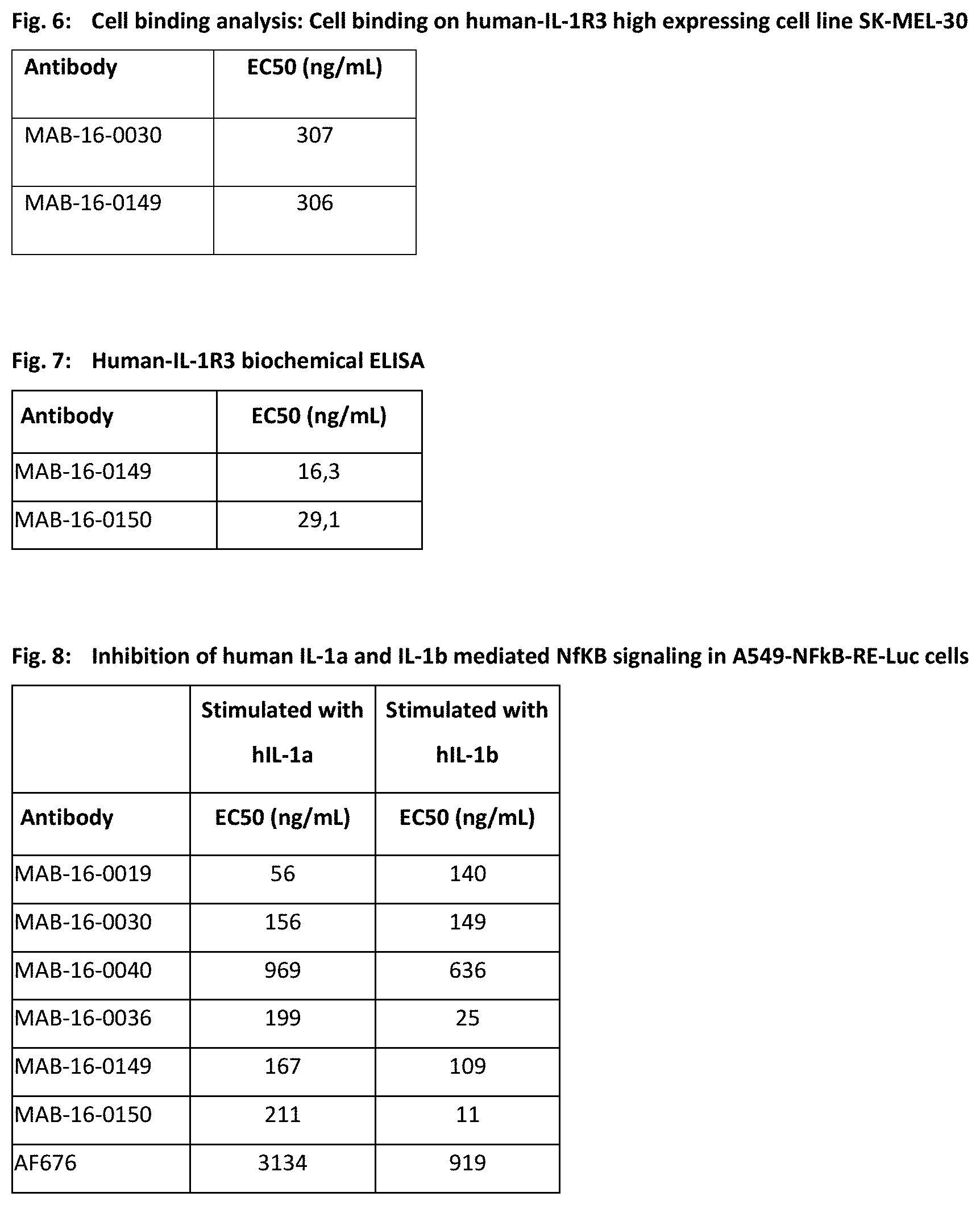
D00015

D00016

D00017

D00018
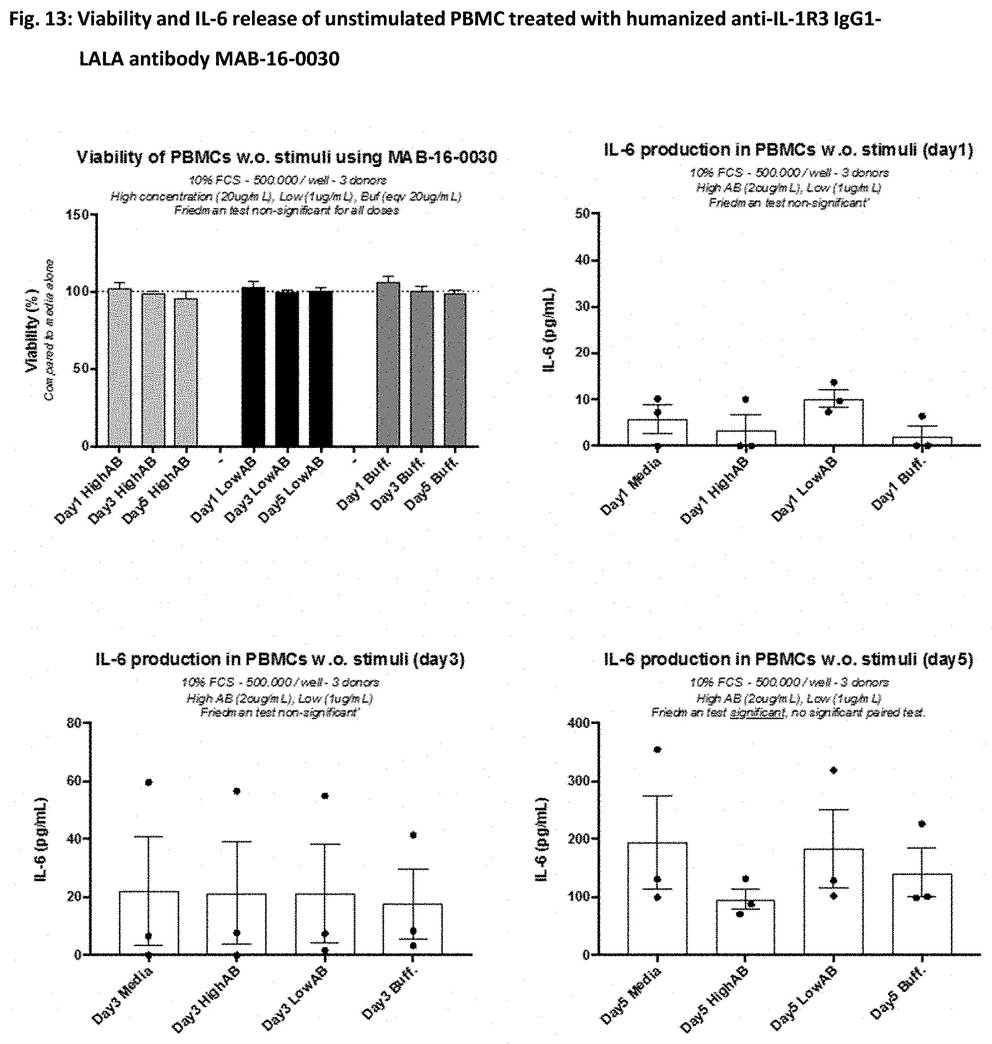
D00019
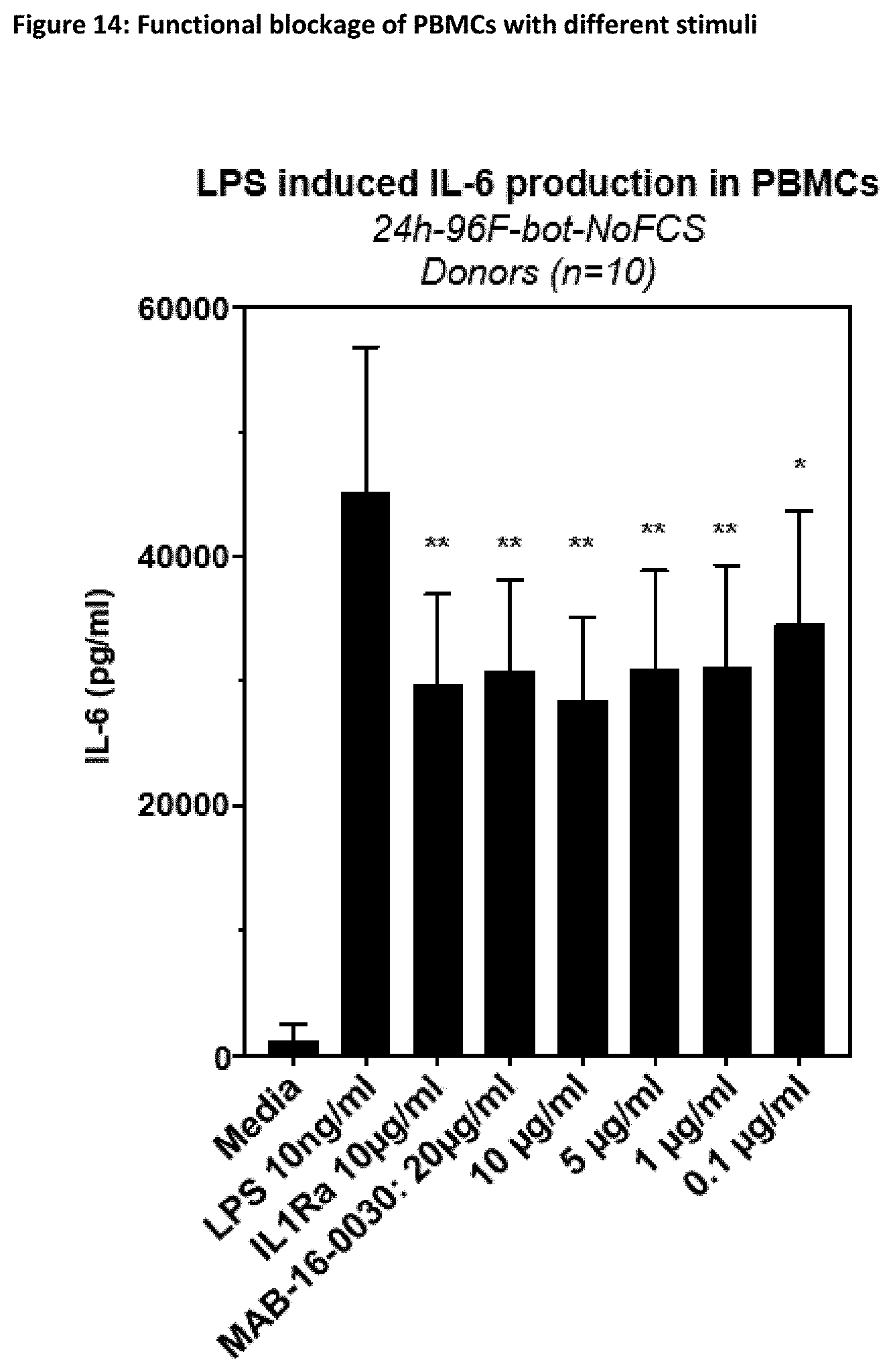
D00020
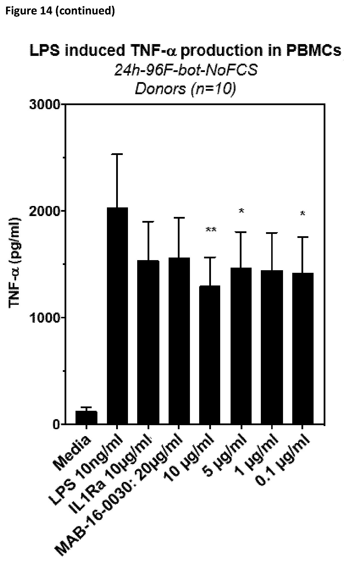
D00021
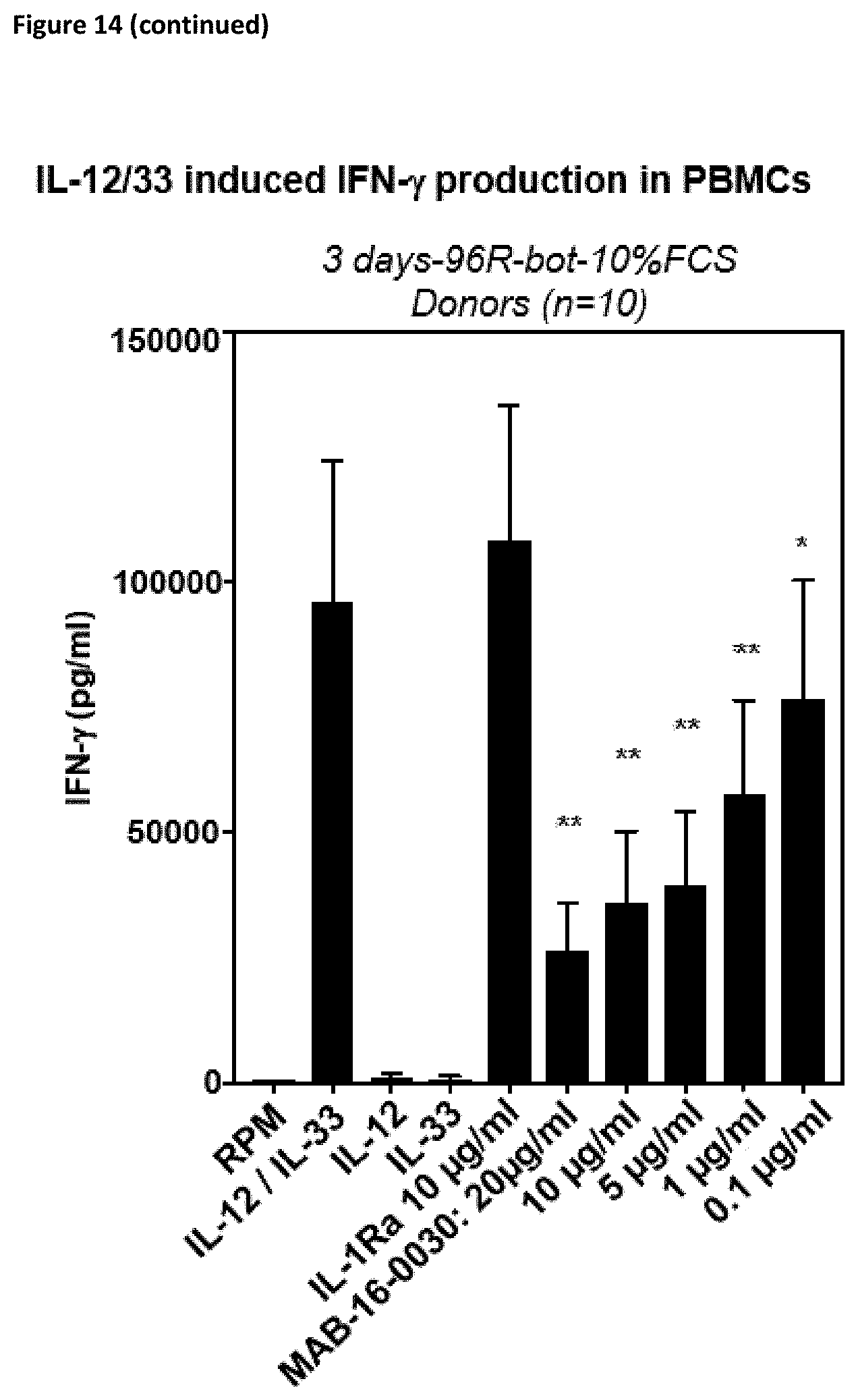
D00022
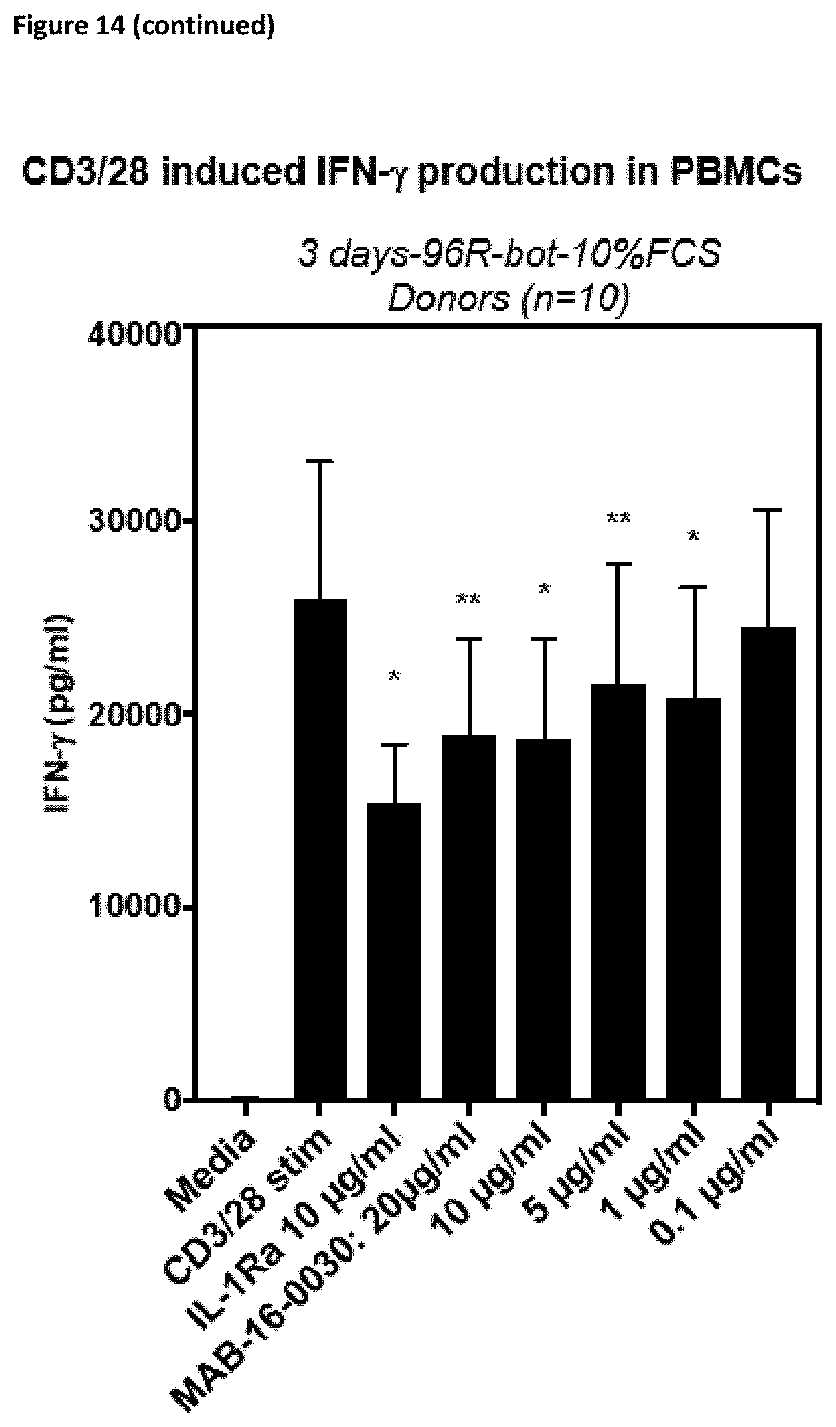
D00023
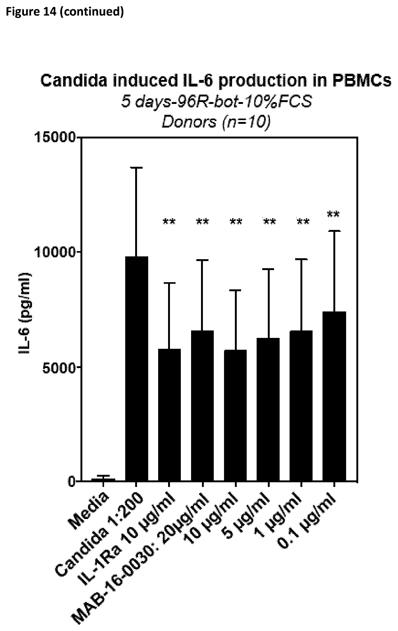
D00024
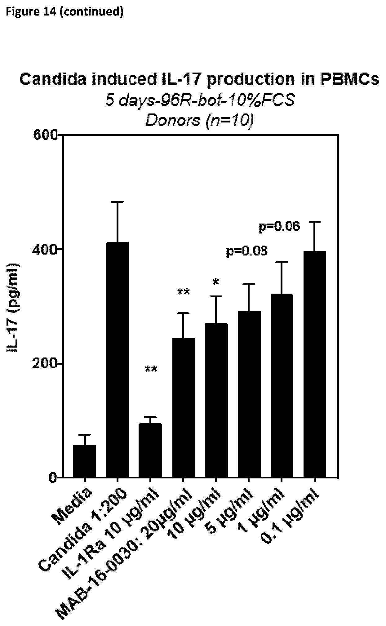
D00025

D00026

D00027

D00028

D00029
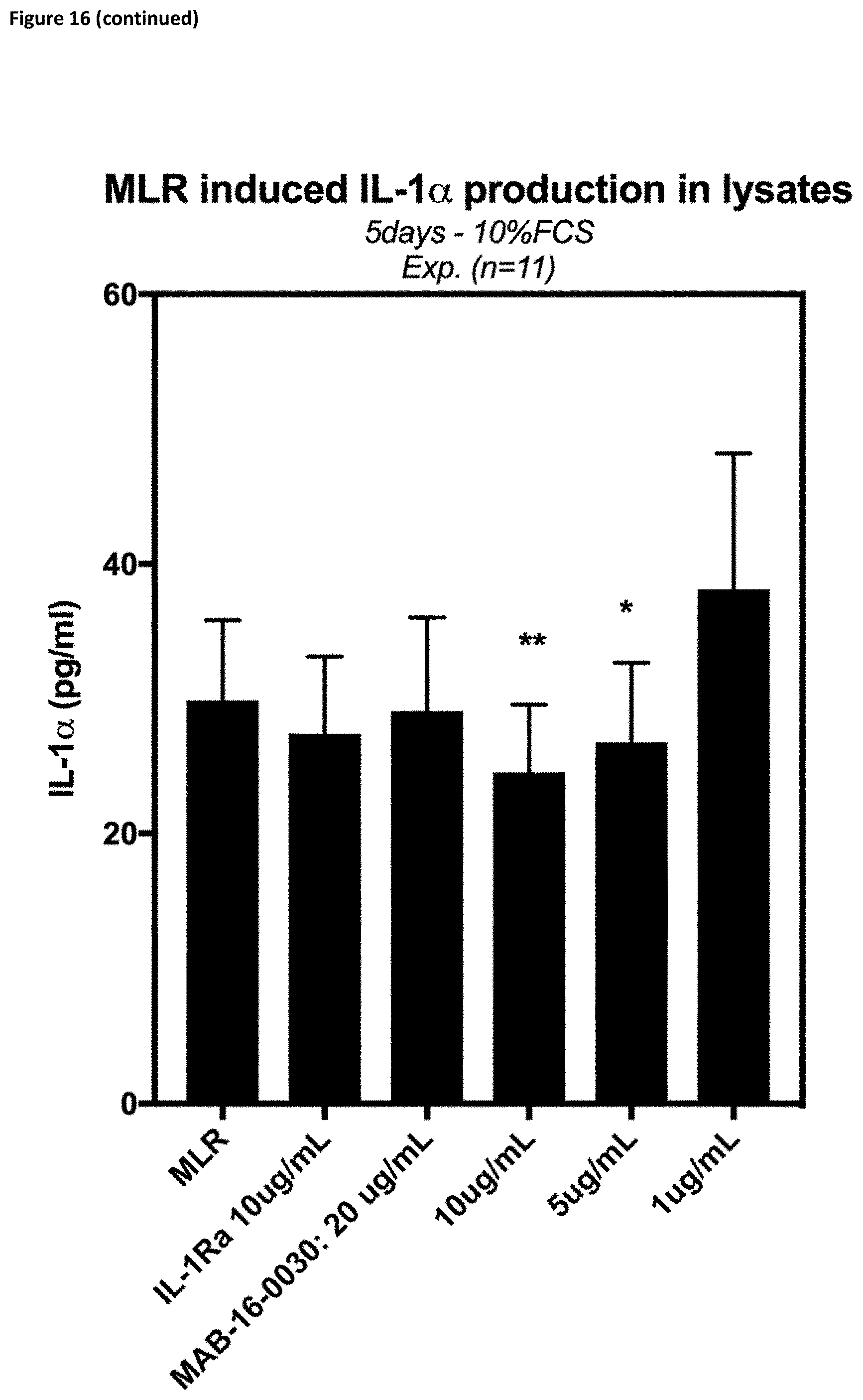
D00030

D00031

D00032
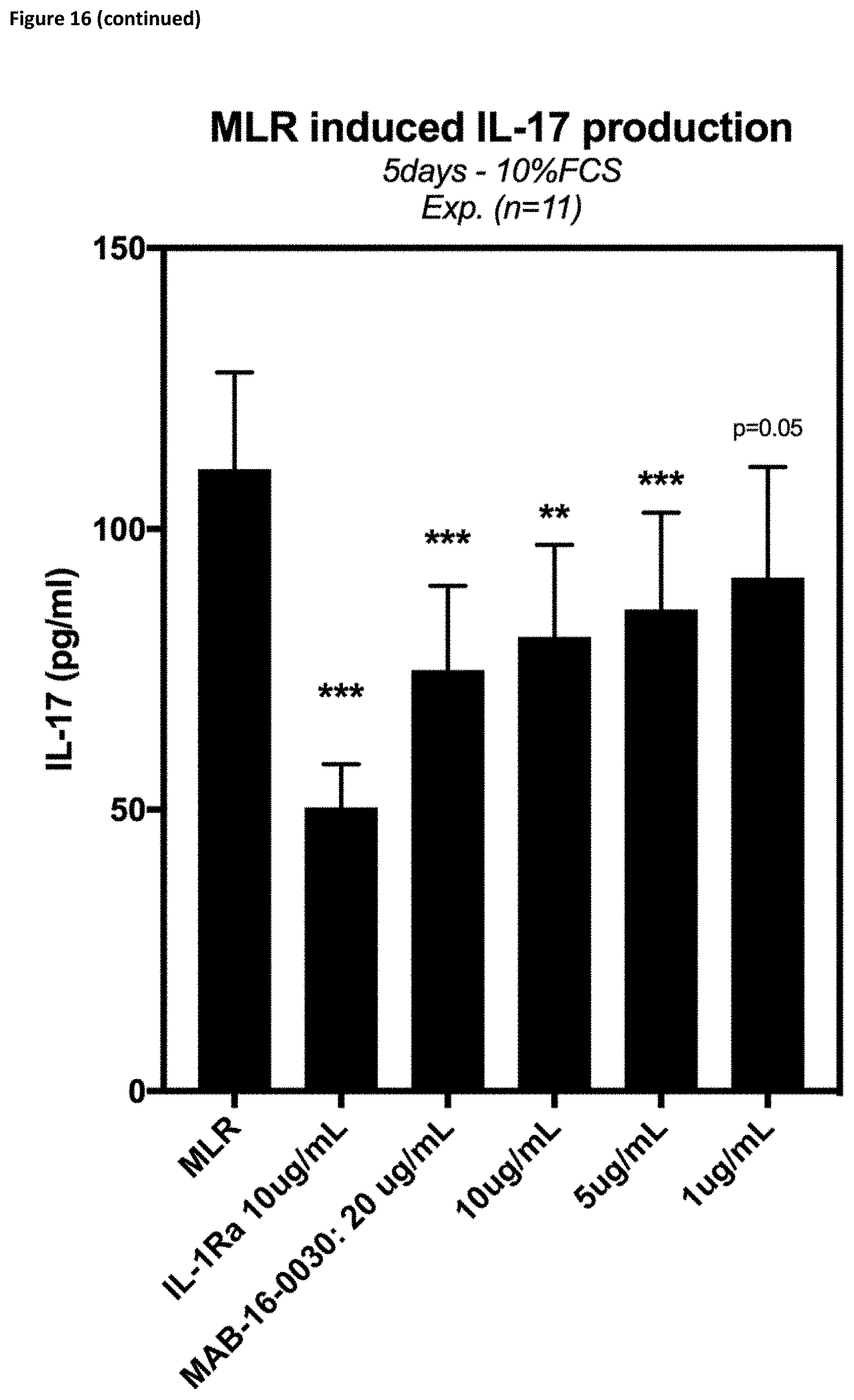
D00033
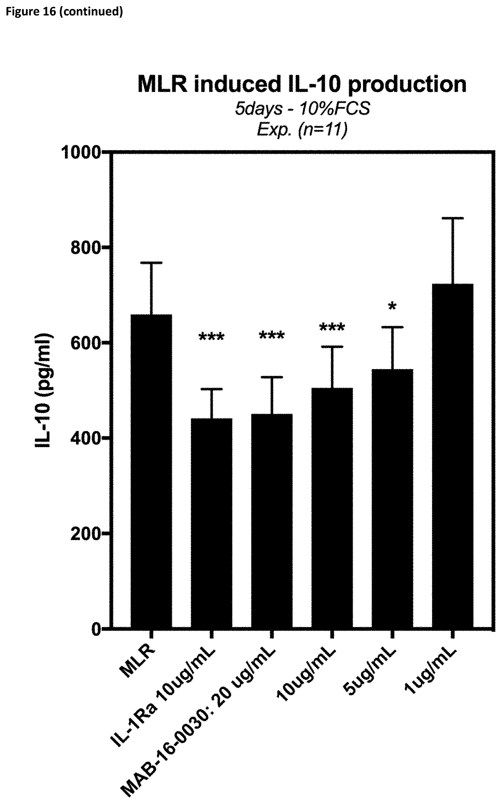
D00034

D00035

D00036

D00037
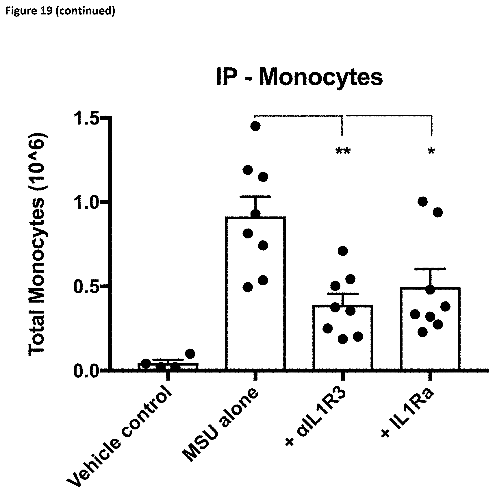
D00038

D00039

D00040
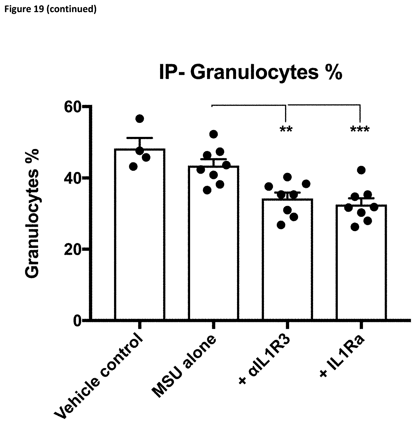
D00041
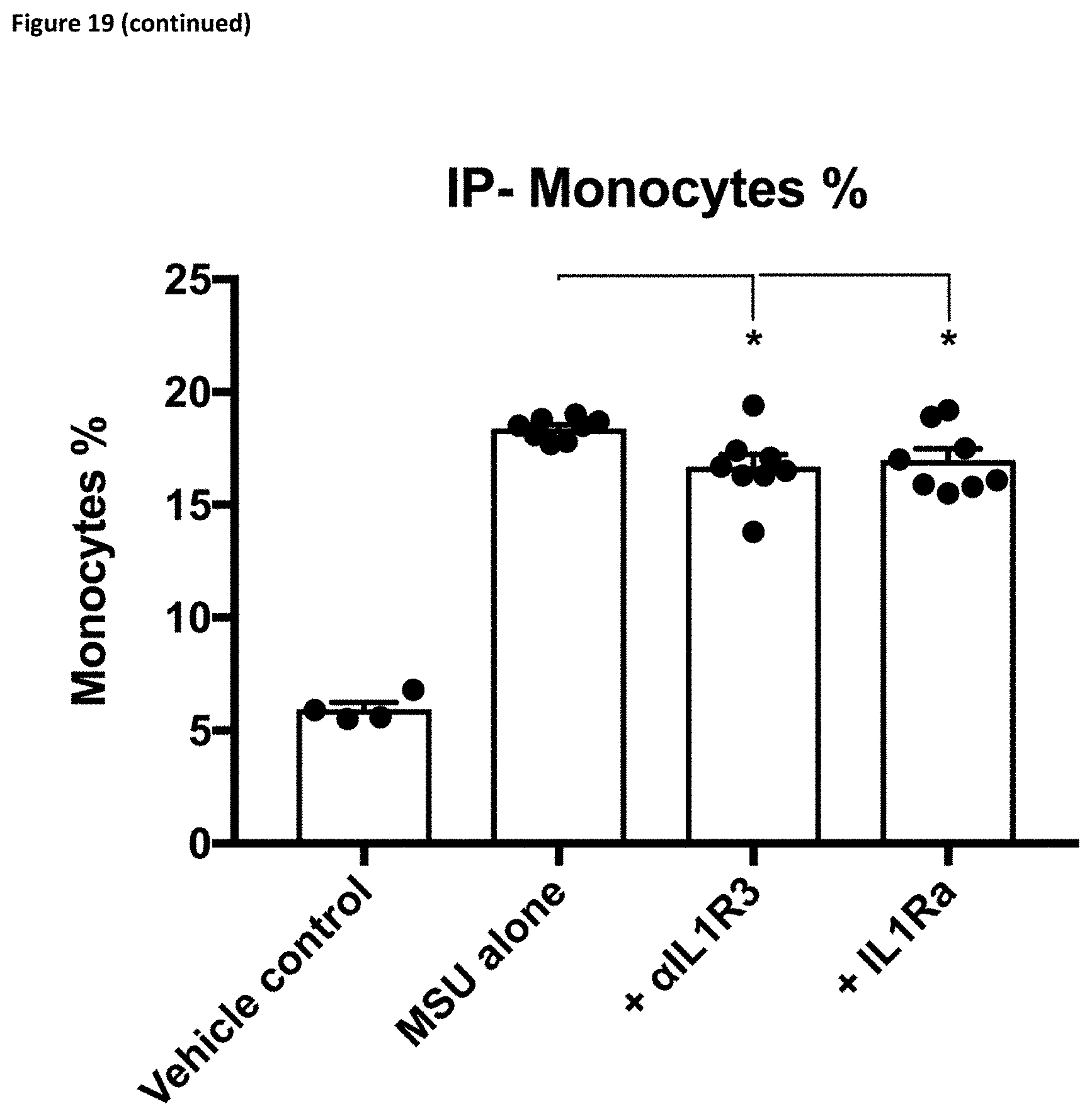
D00042

D00043

D00044

D00045
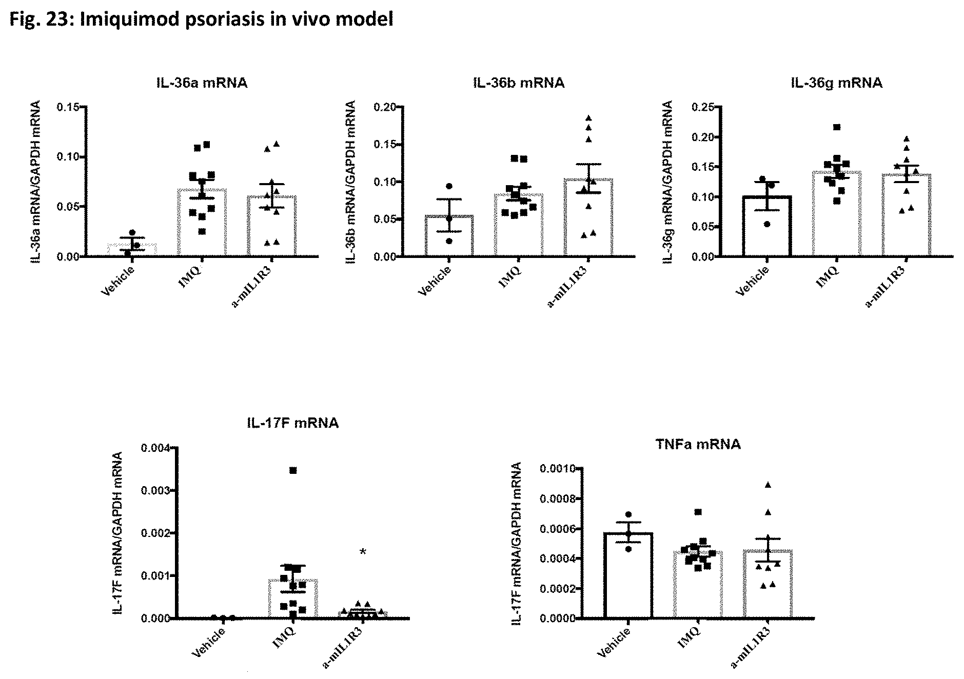
D00046

S00001
XML
uspto.report is an independent third-party trademark research tool that is not affiliated, endorsed, or sponsored by the United States Patent and Trademark Office (USPTO) or any other governmental organization. The information provided by uspto.report is based on publicly available data at the time of writing and is intended for informational purposes only.
While we strive to provide accurate and up-to-date information, we do not guarantee the accuracy, completeness, reliability, or suitability of the information displayed on this site. The use of this site is at your own risk. Any reliance you place on such information is therefore strictly at your own risk.
All official trademark data, including owner information, should be verified by visiting the official USPTO website at www.uspto.gov. This site is not intended to replace professional legal advice and should not be used as a substitute for consulting with a legal professional who is knowledgeable about trademark law.