Mit Biomarkers And Methods Using The Same
Durinck; Steffen ; et al.
U.S. patent application number 16/597678 was filed with the patent office on 2020-05-07 for mit biomarkers and methods using the same. This patent application is currently assigned to Genentech, Inc.. The applicant listed for this patent is Genentech, Inc.. Invention is credited to Steffen Durinck, Bijay Jaiswal, Zora Modrusan, Somasekar Seshagiri, Eric Stawiski, Na Zhang.
| Application Number | 20200138944 16/597678 |
| Document ID | / |
| Family ID | 53366303 |
| Filed Date | 2020-05-07 |


View All Diagrams
| United States Patent Application | 20200138944 |
| Kind Code | A1 |
| Durinck; Steffen ; et al. | May 7, 2020 |
MIT BIOMARKERS AND METHODS USING THE SAME
Abstract
Provided are therapies related to the treatment of pathological conditions, such as cancer.
| Inventors: | Durinck; Steffen; (Orinda, CA) ; Jaiswal; Bijay; (San Mateo, CA) ; Modrusan; Zora; (Fremont, CA) ; Seshagiri; Somasekar; (San Carlos, CA) ; Stawiski; Eric; (South San Francisco, CA) ; Zhang; Na; (South San Francisco, CA) | ||||||||||
| Applicant: |
|
||||||||||
|---|---|---|---|---|---|---|---|---|---|---|---|
| Assignee: | Genentech, Inc. South San Francisco CA |
||||||||||
| Family ID: | 53366303 | ||||||||||
| Appl. No.: | 16/597678 | ||||||||||
| Filed: | October 9, 2019 |
Related U.S. Patent Documents
| Application Number | Filing Date | Patent Number | ||
|---|---|---|---|---|
| 15372951 | Dec 8, 2016 | |||
| 16597678 | ||||
| PCT/US2015/032294 | May 22, 2015 | |||
| 15372951 | ||||
| 62109775 | Jan 30, 2015 | |||
| 62059362 | Oct 3, 2014 | |||
| 62002612 | May 23, 2014 | |||
| Current U.S. Class: | 1/1 |
| Current CPC Class: | G01N 2800/60 20130101; G01N 33/574 20130101; A61P 35/00 20180101; A61P 13/12 20180101; G01N 2800/50 20130101; C12Q 2600/158 20130101; G01N 2800/7028 20130101; C12Q 1/6886 20130101 |
| International Class: | A61K 39/395 20060101 A61K039/395; C12Q 1/6886 20060101 C12Q001/6886; A61P 13/12 20060101 A61P013/12; A61P 35/00 20060101 A61P035/00; G01N 33/574 20060101 G01N033/574 |
Claims
1. A method for determining MiT biomarker expression, comprising the step of determining whether a sample from an individual expresses MiT biomarker.
2. The method of claim 1, wherein MiT biomarker is selected from the group consisting of: MITF, TFEB, TFEC, TFE3 and SBNO2.
3-6. (canceled)
7. The method of claim 1, wherein presence of biomarker is indicated by the presence of elevated biomarker expression level.
8. The method of claim 1, wherein one or more biomarker comprises a MiT translocation or inversion.
9. The method of claim 8, wherein the MiT translocation is a MITF translocation.
10. The method of claim 9, wherein the MITF translocation comprises ACTG1 and MITF, ACTG1 exon 3, ACTG1 exon 3 and MITF exon 3, or SEQ ID NO:13 and/or SEQ ID NO:30.
11-15. (canceled)
16. The method of claim 10, wherein the MITF translocation is detectable by primers which consist of or comprise SEQ ID NO:9, 10, 11 and/or 12.
17. The method of claim 8, wherein the MITF translocation is driven by the ACTG1 promoter.
18. The method of claim 17, wherein the MITF translocation comprises AP3S1 and MITF, AP3S1 exon 3, ACTG1 exon 3 and MITF exon 3 or AP3S1 promoter.
19-21. (canceled)
22. The method of claim 8, wherein the translocation is a TFEB translocation.
23. The method of claim 22, wherein the TFEB translocation comprises CLTC and TFEB, CLTC exon 17, CLTC exon 17 and TFEB exon 6, or SEQ ID NO:19.
24-27. (canceled)
28. The method of claim 23, wherein the TFEB translocation is detectable by primers which consist of or comprise SEQ ID NO:15, 16, 17 and/or 18.
29. The method of claim 23, wherein the TFEB translocation is driven by the CLTC promoter.
30. The method of claim 8, wherein the translocation is a SBNO2 inversion.
31. The method of claim 30, wherein the SBNO2 translocation inversion comprises MIDN and SBNO2, MIDN promoter, MIDN promoter and SBNO2 exon 1, or SEQ ID NO:25.
32-35. (canceled)
36. The method of claim 30, wherein the SBNO2 inversion is detectable by primers which consist of or comprise SEQ ID NO:21, 22, 23, and/or 25
37. The method of claim 30, wherein the SBNO2 inversion is driven by the CLTC promoter.
38. The methods of claim 8, wherein the MiT translocation results in elevated expression levels of MET, elevated activity and/or activation of MET, elevated expression levels of BIRC7, or elevated activity and/or activation of BIRC7.
39-41. (canceled)
42. The method of claim 8, wherein the translocation is a somatic translocation, an intra-chromosomal translocation, an inter-chromosomal translocation, an inversion, a deletion, or a translocation fusion polynucleotide and/or a functional translocation fusion polypeptide.
43-47. (canceled)
48. The method of claim 8, wherein the sample is a cancer sample.
49. The method of claim 48, wherein the cancer is squamous cell cancer, lung cancer, small-cell lung cancer (SCLC), non-small cell lung cancer (NSCLC), adenocarcinoma of the lung, squamous carcinoma of the lung, cancer of the peritoneum, hepatocellular cancer, gastric cancer, gastrointestinal cancer, stomach cancer, pancreatic cancer, glioblastoma, cervical cancer, ovarian cancer, liver cancer, bladder cancer, hepatoma, breast cancer, colon cancer, rectal cancer, colorectal cancer, endometrial cancer, uterine carcinoma, salivary gland carcinoma, kidney or renal cancer, prostate cancer, vulval cancer, thyroid cancer, hepatic carcinoma, anal carcinoma, penile carcinoma, testicular cancer, esophageal cancer, tumors of the biliary tract, head and neck cancer, renal cell carcinoma (RCC), non-clear cell renal cell carcinoma (nccRCC) or translocation RCC (tRCC).
50-52. (canceled)
53. A method of treating cancer in an individual comprising administering to the individual an effective amount of a MiT antagonist, wherein treatment is based upon the individual having cancer comprising MiT overexpression.
54. The method of claim 53, wherein the cancer comprises a MiT translocation or the individual has been found to have cancer comprising a MiT translocation or a sample obtained from the individual comprises a MiT translocation, the method comprising providing an effective amount of a MiT antagonist.
55-57. (canceled)
58. A method of identifying an individual with cancer who is more or less likely to exhibit benefit from treatment with an anti-cancer therapy comprising a MiT antagonist or predicting whether an individual with cancer is more or less likely to respond effectively to treatment with an anti-cancer therapy comprising a MiT antagonist or predicting the response or lack of response of an individual with cancer to an anti-cancer therapy comprising a MiT antagonist, the method comprising: determining presence or absence of a MiT translocation in a sample obtained from the individual, wherein presence of the MiT translocation in the sample indicates that the individual is more likely to exhibit benefit from treatment with the anti-cancer therapy comprising the MiT antagonist or absence of the MiT translocation indicates that the individual is less likely to exhibit benefit from treatment with the anti-cancer therapy comprising the MiT antagonist, or presence of the MiT translocation indicates that the individual is more likely to respond effectively to treatment with the MiT antagonist and absence of the MiT translocation indicates that the individual is less likely to respond effectively to treatment with the MiT antagonist, or presence of the MiT translocation is predictive of response of the individual to the anti-cancer therapy comprising the MiT antagonist and absence of the MiT translocation is predictive of lack of response of the individual to the anti-cancer therapy comprising the MiT antagonist.
59-60. (canceled)
61. The method of claim 58, wherein the method further comprises administering to the individual an effective amount of a MiT antagonist
62. A method of inhibiting proliferation of a nccRCC cancer cell or treating nccRCC in an individual comprising contacting the cancer cell with or administering to the individual an effective amount of a MiT-translocation antagonist.
63. (canceled)
64. The method of claim 62, wherein the cancer or cancer cell comprises MiT translocation, MiT overexpression or BIRC7 overexpression.
65-112. (canceled)
Description
CROSS REFERENCE TO RELATED APPLICATIONS
[0001] This application is a continuation of U.S. application Ser. No. 15/372,951, filed Dec. 8, 2016, which is a continuation of International Application No. PCT/US2015/032294, having an international filing date of May 22, 2015, which claims the benefit of priority of provisional U.S. Application No. 62/002,612, filed May 23, 2014; U.S. Application No. 62/059,362, filed Oct. 3, 2014; U.S. application No. 62/109,775, filed Jan. 30, 2015, which are hereby incorporated by reference in their entirety.
SEQUENCE LISTING
[0002] The instant application contains a Sequence Listing which has been submitted via EFS-Web and is hereby incorporated by reference in its entirety. Said ASCII copy, created on Oct. 8, 2019, is named P05829-US-4_Sequence_Listing.txt and is 84,040 bytes in size.
FIELD
[0003] Provided are therapies related to the treatment of pathological conditions, such as cancer.
BACKGROUND
[0004] Kidney cancer accounts for .about.60,000 new cases and .about.13,000 deaths annually in the United States (Siegel et al., 2013). About 85% of kidney cancers are renal cell carcinoma (RCC), which arise from the renal epithelium. Clear cell RCC (ccRCC), which constitutes 75% of RCCs, is the best characterized kidney cancer subtype (Pena-Llopis et al., 2012; Sato et al., 2013; TCGA, 2013). The remaining 25% of RCCs broadly classified as non-clear cell RCCs (nccRCCs) represent distinct tumor subtypes, including papillary (pRCC; 10-15%) and chromophobe (chRCC; 4-5%) (Osunkoya, 2010; Picken, 2010; Young, 2010; Yusenko, 2010a, b). In needle core biopsies, chRCC is at times difficult to distinguish from renal oncocytoma (RO), a benign kidney epithelial tumor with an incidence rate of .about.5% (Osunkoya, 2010; Picken, 2010; Young, 2010; Yusenko, 2010a, b). Their diagnosis remains a challenge and is compounded by the presence of mixed tumors that show features of both RO and chRCC (Osunkoya, 2010; Picken, 2010; Young, 2010; Yusenko, 2010a, b). Other nccRCC types include collecting duct (<1%), translocation (tRCC; rare) and medullary (rare). About 4-5% of tumors remain unclassified (Bellmunt and Dutcher, 2013). While infrequent, tRCC tend to affect adolescents and young adults and are particularly devastating. Once nccRCCs metastasize, the disease generally remains incurable. While several drugs have recently been approved for metastatic RCC, registration trials involved almost exclusively patients with ccRCC, and there are no treatments with demonstrated efficacy in nccRCC subtypes (Bellmunt and Dutcher, 2013).
[0005] There remains a need to better understand the pathogenesis of cancers, in particular, human renal cell carcinomas and also to identify new therapeutic targets.
SUMMARY
[0006] Methods are provided for determining MiT biomarker expression (determining presence of MiT biomarker), comprising the step of determining whether a sample from an individual expresses MiT biomarker. In some embodiments, MiT is MITF. In some embodiments, MiT is TFEB. In some embodiments, MiT is TFEC. In some embodiments, MiT is TFE3. In some embodiments, MiT is SBNO2.
[0007] In some embodiments of any of the methods of the invention, presence of biomarker is indicated by the presence of elevated biomarker expression level (e.g., compared to reference expression level). In some embodiments, one or more biomarker comprises a translocation or inversion (e.g., rearrangement and/or fusion) of one or more genes selected from MITF, TFEB, TFE3, TFEC, and/or SBNO2. In some embodiments, translocation is a MITF translocation. In some embodiments, the MITF translocation comprises ACTG1 and MITF. In some embodiments, the MITF translocation comprises ACTG1 exon 3. In some embodiments, the MITF translocation comprises ACTG1 exon 3 and MITF exon 3. In some embodiments, MITF translocation comprises SEQ ID NO:13 and/or 30. In some embodiments, MITF translocation comprises SEQ ID NO: 30. In some embodiments, the MITF translocation is detectable by primers which consist of or comprise SEQ ID NO:11 and/or 12. In some embodiments, the MITF translocation is detectable by primers which consist of or comprise SEQ ID NO:9, 10, 11 and/or 12. In some embodiments, the MITF translocation is driven by the ACTG1 promoter. In some embodiments, the MITF translocation comprises AP3S1 and MITF. In some embodiments, the MITF translocation comprises AP3S1 exon 3. In some embodiments, the MITF translocation comprises ACTG1 exon 3 and MITF exon 3. In some embodiments, wherein the MITF translocation is driven by the AP3S1 promoter. In some embodiments, the translocation is a TFEB translocation. In some embodiments, the TFEB translocation comprises CLTC and TFEB. In some embodiments, the TFEB translocation comprises CLTC exon 17. In some embodiments, the TFEB translocation comprises CLTC exon 17 and TFEB exon 6. In some embodiments, the TFEB translocation comprises SEQ ID NO:19. In some embodiments, the TFEB translocation is detectable by primers which consist of or comprise SEQ ID NO:17 and/or 18. In some embodiments, the TFEB translocation is detectable by primers which consist of or comprise SEQ ID NO:15, 16, 17 and/or 18. In some embodiments, TFEB translocation is driven by the CLTC promoter. In some embodiments, the translocation is a SBNO2 inversion. In some embodiments, SBNO2 translocation inversion comprises MIDN and SBNO2. In some embodiments, SBNO2 inversion comprises MIDN promoter. In some embodiments, the SBNO2 inversion comprises MIDN promoter and SBNO2 exon 1. In some embodiments, the SBNO2 inversion comprises SEQ ID NO:25. In some embodiments, the SBNO2 inversion is detectable by primers which consist of or comprise SEQ ID NO:23 and/or 24. In some embodiments, the SBNO2 inversion is detectable by primers which consist of or comprise SEQ ID NO:21, 22, 23, and/or 25. In some embodiments, SBNO2 inversion is driven by the CLTC promoter.
[0008] In some embodiments of any of the methods of the invention, the MiT translocation results in elevated expression levels of MET (e.g., compared to a reference without the MiT translocation).
[0009] In some embodiments of any of the methods of the invention, the MiT translocation (e.g., rearrangement and/or fusion) results in elevated activity and/or activation of MET (e.g., compared to a reference without the MiT translocation).
[0010] In some embodiments of any of the methods of the invention, the MiT (e.g., MITF, TFEB, TFE3, TFEC, and/or SBNO2) translocation (e.g., rearrangement and/or fusion) results in elevated expression levels of BIRC7 (e.g., compared to a reference without the MiT translocation).
[0011] In some embodiments of any of the methods of the invention, the MiT translocation (e.g., rearrangement and/or fusion) results in elevated activity and/or activation of BIRC7 (e.g., compared to a reference without the MiT translocation.
[0012] In some embodiments of any of the methods of the invention, the translocation is a somatic translocation.
[0013] In some embodiments of any of the methods of the invention, the translocation is an intra-chromosomal translocation.
[0014] In some embodiments of any of the methods of the invention, the translocation is an inter-chromosomal translocation.
[0015] In some embodiments of any of the methods of the invention, the translocation is an inversion
[0016] In some embodiments of any of the methods of the invention, the translocation is a deletion.
[0017] In some embodiments of any of the methods of the invention, the translocation is a translocation fusion polynucleotide (e.g., functional MiT-translocation fusion polynucleotide) and/or functional translocation fusion polypeptide (e.g., functional MiT-translocation fusion polypeptide).
[0018] In some embodiments of any of the methods of the invention, the sample is a cancer sample.
[0019] In some embodiments of any of the methods of the invention, the cancer is squamous cell cancer (e.g., epithelial squamous cell cancer), lung cancer including small-cell lung cancer (SCLC), non-small cell lung cancer (NSCLC), adenocarcinoma of the lung and squamous carcinoma of the lung, cancer of the peritoneum, hepatocellular cancer, gastric or stomach cancer including gastrointestinal cancer, pancreatic cancer, glioblastoma, cervical cancer, ovarian cancer, liver cancer, bladder cancer, hepatoma, breast cancer (including metastatic breast cancer), colon cancer, rectal cancer, colorectal cancer, endometrial or uterine carcinoma, salivary gland carcinoma, kidney or renal cancer, prostate cancer, vulval cancer, thyroid cancer, hepatic carcinoma, anal carcinoma, penile carcinoma, testicular cancer, esophageal cancer, tumors of the biliary tract, as well as head and neck cancer.
[0020] In some embodiments of any of the methods of the invention, the cancer is renal cell carcinoma (RCC). In some embodiments, the RCC is non-clear cell renal cell carcinoma (nccRCC) or translocation RCC (tRCC). In some embodiments, the RCC is nccRCC.
[0021] Provided are methods of treating cancer in an individual comprising administering to the individual an effective amount of a MiT antagonist, wherein treatment is based upon the individual having cancer comprising MiT overexpression. In some embodiments, the cancer comprises a MiT translocation, the method comprising providing an effective amount of a MiT antagonist.
[0022] Provided herein are methods of treating cancer in an individual provided that the individual has been found to have cancer comprising a MiT translocation, the method comprising administering to the individual an effective amount of a MiT antagonist.
[0023] Provided herein are methods of treating cancer in an individual, the method comprising: determining that a sample obtained from the individual comprises a MiT translocation, and administering an effective amount of an anti-cancer therapy comprising a MiT antagonist to the individual, whereby the cancer is treated.
[0024] Provided herein are methods of treating cancer, comprising: (a) selecting an individual having cancer, wherein the cancer comprising a MiT translocation; and (b) administering to the individual thus selected an effective amount of a MiT antagonist, whereby the cancer is treated.
[0025] Provided herein are methods of identifying an individual with cancer who is more or less likely to exhibit benefit from treatment with an anti-cancer therapy comprising a MiT antagonist, the method comprising: determining presence or absence of a MiT translocation in a sample obtained from the individual, wherein presence of the MiT translocation in the sample indicates that the individual is more likely to exhibit benefit from treatment with the anti-cancer therapy comprising the MiT antagonist or absence of the MiT translocation indicates that the individual is less likely to exhibit benefit from treatment with the anti-cancer therapy comprising the MiT antagonist.
[0026] Provided herein are methods of predicting whether an individual with cancer is more or less likely to respond effectively to treatment with an anti-cancer therapy comprising a MiT antagonist, the method comprising determining a MiT translocation, whereby presence of the MiT translocation indicates that the individual is more likely to respond effectively to treatment with the MiT antagonist and absence of the MiT translocation indicates that the individual is less likely to respond effectively to treatment with the MiT antagonist.
[0027] Provided herein are methods of predicting the response or lack of response of an individual with cancer to an anti-cancer therapy comprising a MiT antagonist comprising detecting in a sample obtained from the individual presence or absence of a MiT translocation, wherein presence of the MiT translocation is predictive of response of the individual to the anti-cancer therapy comprising the MiT antagonist and absence of the MiT translocation is predictive of lack of response of the individual to the anti-cancer therapy comprising the MiT antagonist.
[0028] In some embodiments of any of the methods of the invention, the methods further comprises administering to the individual an effective amount of a MiT antagonist.
[0029] Provided herein are methods of inhibiting proliferation of a nccRCC cancer cell comprising contacting the cancer cell with an effective amount of a MiT-translocation antagonist.
[0030] Provided herein are methods of treating nccRCC in an individual comprising administering to the individual an effective amount of a MiT antagonist. In some embodiments, the MiT antagonist is a MiT-translocation antagonist.
[0031] In some embodiments of any of the methods of the invention, the cancer or cancer cell comprises MiT translocation.
[0032] In some embodiments of any of the methods of the invention, the cancer or cancer cell comprises MiT overexpression.
[0033] In some embodiments of any of the methods of the invention, the cancer or cancer cell comprises BIRC7 overexpression.
[0034] In some embodiments of any of the methods of the invention, MiT is MITF.
[0035] In some embodiments of any of the methods of the invention, MiT is TFEB.
[0036] In some embodiments of any of the methods of the invention, MiT is TFEC.
[0037] In some embodiments of any of the methods of the invention, MiT is TFE3.
[0038] In some embodiments of any of the methods of the invention, MiT is SBNO2.
[0039] In some embodiments of any of the methods of the invention, MiT translocation is a MITF translocation.
[0040] In some embodiments of any of the methods of the invention, MiT translocation is a TFEB translocation.
[0041] In some embodiments of any of the methods of the invention, MiT translocation is a TFEC translocation.
[0042] In some embodiments of any of the methods of the invention, MiT translocation is a TFE3 translocation.
[0043] In some embodiments, the MiT translocation is detected using any of the methods of determining MiT translocation (detecting presence of MiT translocation) disclosed herein.
[0044] In some embodiments of any of the methods of the invention, the cancer or cancer is squamous cell cancer (e.g., epithelial squamous cell cancer), lung cancer including small-cell lung cancer (SCLC), non-small cell lung cancer (NSCLC), adenocarcinoma of the lung and squamous carcinoma of the lung, cancer of the peritoneum, hepatocellular cancer, gastric or stomach cancer including gastrointestinal cancer, pancreatic cancer, glioblastoma, cervical cancer, ovarian cancer, liver cancer, bladder cancer, hepatoma, breast cancer (including metastatic breast cancer), colon cancer, rectal cancer, colorectal cancer, endometrial or uterine carcinoma, salivary gland carcinoma, kidney or renal cancer, prostate cancer, vulval cancer, thyroid cancer, hepatic carcinoma, anal carcinoma, penile carcinoma, testicular cancer, esophageal cancer, tumors of the biliary tract, as well as head and neck cancer.
[0045] In some embodiments of any of the methods of the invention, the cancer or cancer is renal cell carcinoma (RCC). In some embodiments, the RCC is non-clear cell renal cell carcinoma (nccRCC) or translocation RCC (tRCC). In some embodiments, the RCC is nccRCC.
[0046] In some embodiments of any of the methods of the invention, the MiT antagonist is an antibody, binding polypeptide, small molecule, or polynucleotide.
[0047] In some embodiments of any of the methods of the invention, the MiT antagonist is a MET antagonist.
[0048] In some embodiments of any of the methods of the invention, the MiT antagonist is a BIRC7 antagonist.
[0049] In some embodiments of any of the methods of the invention, the MiT antagonist is a MITF antagonist.
[0050] In some embodiments of any of the methods of the invention, the MiT antagonist is a TFEB antagonist.
[0051] In some embodiments of any of the methods of the invention, the MiT antagonist is a TFEC antagonist.
[0052] In some embodiments of any of the methods of the invention, the MiT antagonist is a TFE3 antagonist.
[0053] In some embodiments of any of the methods of the invention, the MiT antagonist binds MITF translocation. In some embodiments, the MITF translocation comprises ACTG1 and MITF. In some embodiments, MITF translocation comprises ACTG1 exon 3. In some embodiments, MITF translocation comprises ACTG1 exon 3 and MITF exon 3. In some embodiments, the MITF translocation comprises SEQ ID NO:13 and/or 30. In some embodiments, the MITF translocation comprises SEQ ID NO: 30. In some embodiments, the MITF translocation is driven by the ACTG1 promoter. In some embodiments, the MITF translocation comprises AP3S1 and MITF. In some embodiments, MITF translocation comprises AP3S1 exon 3. In some embodiments, the MITF translocation comprises ACTG1 exon 3 and MITF exon 3. In some embodiments, the MITF translocation is driven by the AP3S1 promoter. In some embodiments, MiT antagonist binds TFEB translocation. In some embodiments, the TFEB translocation comprises CLTC and TFEB. In some embodiments, the TFEB translocation comprises CLTC exon 17. In some embodiments, the TFEB translocation comprises CLTC exon 17 and TFEB exon 6. In some embodiments, TFEB translocation comprises SEQ ID NO:19. In some embodiments, TFEB translocation is driven by the CLTC promoter. In some embodiments, the MiT antagonist binds a SBNO2 translocation. In some embodiments, the SBNO2 translocation is an inversion. In some embodiments, the SBNO2 inversion comprises MIDN and SBNO2. In some embodiments, SBNO2 inversion comprises MIDN promoter. In some embodiments, the SBNO2 inversion comprises MIDN promoter and SBNO2 exon 1. In some embodiments, the SBNO2 inversion comprises SEQ ID NO:25. In some embodiments, the SBNO2 inversion is driven by the CLTC promoter.
[0054] In some embodiments of any of the methods of the invention, the methods further comprises administering an additional therapeutic agent.
BRIEF DESCRIPTION OF THE SEVERAL VIEWS OF THE DRAWINGS
[0055] FIGS. 1A, 1B, 1C and 1D. FISH (fluorescence in situ hybridization) image showing (1A) TFEB amplification in tumor cells, (1B) TFE3 in a normal cells, (1C, 1D) TFE3 (1C) and TFEB (1D) gene rearrangement in tumor cells. DNA probe set (Agilent Technologies, CA) that hybridized to 5' or 3' side of TFE3 gene in chromosome band Xp11.2 or TFEB gene in chromosome band 6p21 were used. TFE3 probes used: TFE3 5' Xp11 labeled red and TFE3 3' Xp11 labeled green. TFEB probes used: TFEB 5' 6p21.1 labeled red and TFEB 3' 6p21.1 labeled green. An intact copy of the gene produced a yellow (Y) signal from the merge of red (R) and green (G) labels (FIG. 1B). In FIG. 1C, consistent with TFE3 translocation (fusion) we observed patterns involving separate red and green signals (seen in more than 80% of the cells) in a male patient. Similarly in FIG. 1D, consistent with TFEB translocation we observe patterns involving clearly separate red and green signals. In FIG. 1A, 90% of the cells showed polysomy of the TFEB gene and .about.25% showed extra copy of the 3'TFEB signal consistent with the amplification detected on the SNP array.
[0056] FIG. 2. Boxplot of TFE3 expression in the indicated nccRCC subtypes.
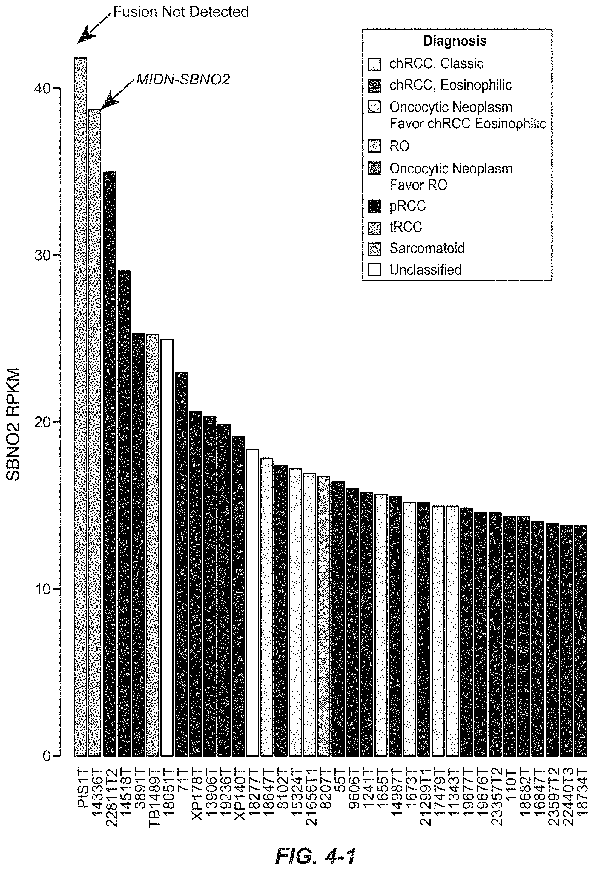
[0057] FIGS. 3A-1, 3A-2 and 3B. MIDN-SBNO2 gene fusion. (3A-1, 3A-2) Cartoon depicting the location, orientation and exon-intron architecture of MIDN-SBNO2 fusion on the genome. The read evidence for MIDN(e1)-SBNO2(e2) fusion identified using RNA-seq data are shown. Representative Sanger sequencing chromatogram of the RT-PCR derived products confirming the MIDN(e1)-SBNO2(e2) fusion junction. (3B) Schematic of the resulting MIDN-SBNO2 fusion protein. FIG. 3A-1 discloses SEQ ID NO: 34. FIG. 3A-2 discloses SEQ ID NOS: 35-41, respectively, in order of appearance.
[0058] FIGS. 4, 4-1, 4-2, 4-3 and 4-4. FIGS. 4-1, 4-2, 4-3, and 4-4 depict the barplot of SBNO2 expression in tumors as measured by RNA-seq. FIG. 4 depicts FIGS. 4-1, 4-2, 4-3, and 4-4 in a single graph.
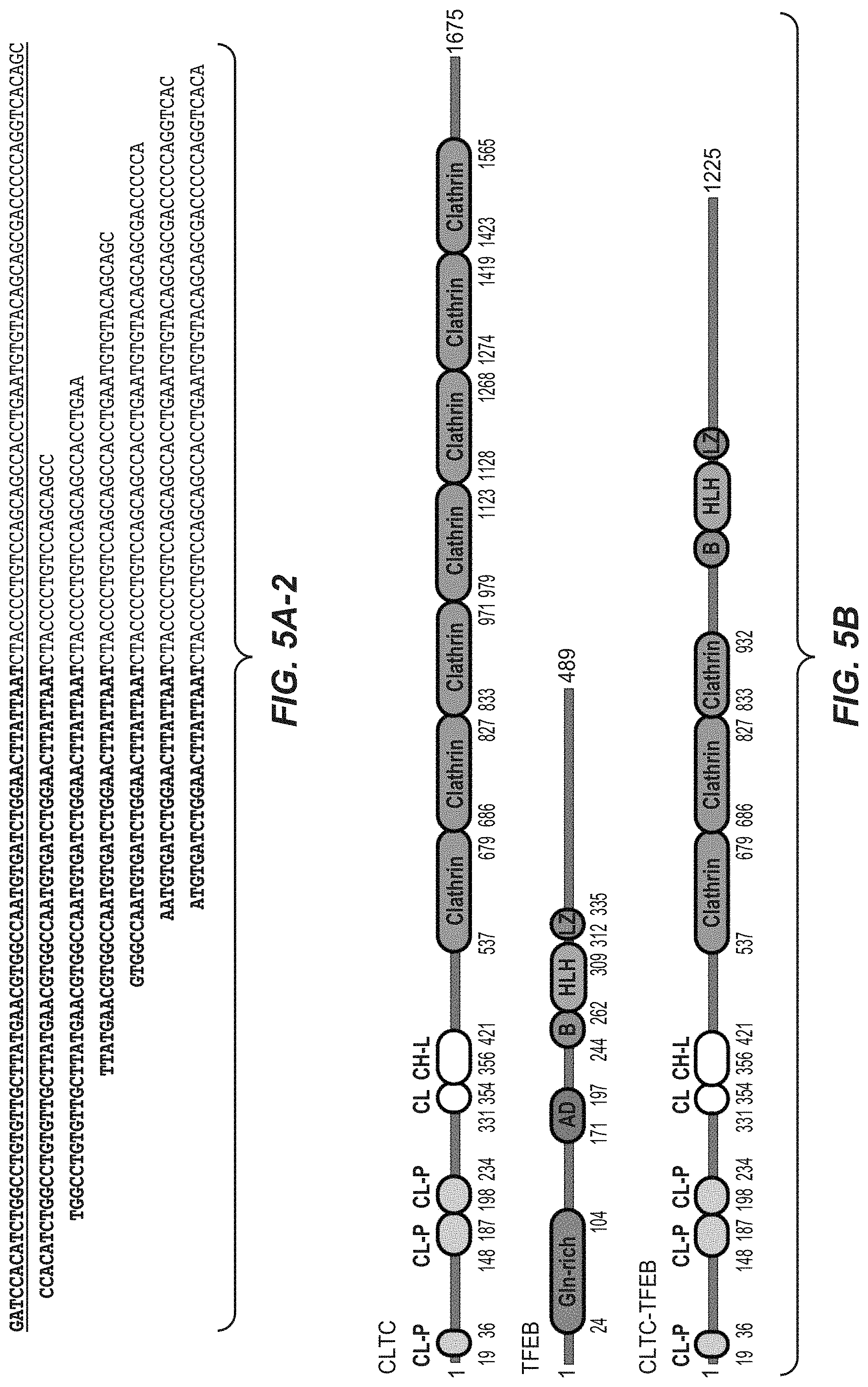
[0059] FIGS. 5A-1, 5A-2 and 5B. CLTC-TFEB gene fusion. (5A-1, 5A-2) Cartoon depicting the location, orientation and exon-intron architecture of CLTC-TFEB fusion on the genome. The read evidence for CLTC(e17)-TFEB(e6) fusion identified using RNA-seq data are shown. Representative Sanger sequencing chromatogram of the RT-PCR derived products confirming the CLTC(e17)-TFEB(e6) fusion junction. (5B) Schematic of the resulting CLTC-TFEB fusion protein. CL-P--clathrin_propel; CL--clathrin-link; CH-L--clathrin_H_link; Gln-rich--glycine rich; AD--activation domain; B--basic; HLH--helix-loop-helix & LZ--leucine zipper. FIG. 5A-1 discloses SEQ ID NO: 42; FIG. 5A-2 discloses SEQ ID NOS: 43-49, respectively, in order of appearance.
[0060] FIGS. 6A, 6A-1, 6A-2, 6B, 6C, 6D, 6E, 6F and 6G. (6A-1, 6A-2, 6B, 6C) RNA-seq based classification of nccRCC. (6D) Copy number ratio plot depicting a focal TFEB amplification in sample 1216T. (6E) Boxplot of TFEB expression in tumors show a high level of TFEB expression in sample 1216T. (6F) Plot of copy number ratio in sample 1686T. (6G) Plot of copy number ratio in sample 1686T. FIG. 6A depicts how FIGS. 6A-1 and 6A-2 should be viewed.
[0061] FIGS. 7A-1, 7A-2, 7B, and 7C. MITF gene fusion. (7A-1, 7A-2) Cartoon depicting the location, orientation, exon-intron architecture of ACTG1-MITF fusion on the genome, the read evidence for ACTG1(e3)-MITF(e3) fusion identified using RNA-seq data and a representative Sanger sequencing chromatogram of RT-PCR derived product confirming the ACTG1(e3)-MITF(e3) fusion junction are shown. FIG. 7A-1 discloses SEQ ID NO: 50. FIG. 7A-2 discloses SEQ ID NOS: 51-57, respectively, in order of appearance. (7B) Schematic of the ACTG1-MITF fusion protein. (7C) MITF expression in tumor harboring the MITF fusion AD--activation domain; B--basic; HLH--helix-loop-helix & LZ--leucine zipper.
[0062] FIGS. 8A, 8B, 8C, 8D and 8E. ACTG1-MITF gene fusion promotes anchorage independent growth. (8A) Expression of MITF target genes in HEK293T cells transfected with MITF WT or fusion constructs. The values shown are from three replicates. (error bar represents SEM; **p<0.01; ***p<0.001). (8B) Stability of MITF fusion protein overtime in HEK293T cells transfected with indicated constructs following cycloheximide treatment, assessed using Western blot. (8C) Western blot showing the expression of Flag-tagged ACTG1, MITF and ACTG1-MITF fusion proteins in NIH3T3 cell expressing the indicated constructs. Hsp90 was used as a loading control. (8D) Representative images depicting the colony formation by NIH3T3 cells stably expressing the indicated constructs (EV=Empty Vector). (8E) Quantification of the number of colonies (>300 uM diameter) shown in FIG. 8D. Data shown are mean.+-.SEM (n=3, ***p<0.001).
[0063] FIG. 9. Expression levels of WT MITF and ACTG1-MITF in transiently transfected 293T cells. This was used to normalize the expression of target genes show in FIG. 8A.
[0064] FIGS. 10A, 10B, 10B-1, 10B-2, 10B-3 and 10B-4. Expression of BIRC7 in tumors with MITF/TFE translocation or fusion compared to samples without a translocation. Expression of BIRC7 was significant in samples with MITF/TFE event (t-test p-value 0.002308467). FIG. 10B depicts FIGS. 10B-1, 10B-2, 10B-3, and 10B-4 in a single graph.
DETAILED DESCRIPTION
I. Definitions
[0065] The term "MITF" refers herein to a native MITF from any vertebrate source, including mammals such as primates (e.g., humans) and rodents (e.g., mice and rats), unless otherwise indicated. The term encompasses "full-length," unprocessed MITF as well as any form of MITF that results from processing in the cell. The term also encompasses naturally occurring variants of MITF, e.g., splice variants or allelic variants. The sequence of an exemplary human MITF nucleic acid sequence is SEQ ID NO:2.
[0066] "MITF variant" or variations thereof, means a MITF polypeptide or polynucleotide, generally being or encoding an active MITF polypeptide, as defined herein having at least about 80% amino acid sequence identity with any of the MITF as disclosed herein. Such MITF variants include, for instance, MITF wherein one or more nucleic acid or amino acid residues are added or deleted. Ordinarily, an MITF variant will have at least about 80% sequence identity, alternatively at least about 81%, 82%, 83%, 84%, 85%, 86%, 87%, 88%, 89%, 90%, 91%, 92%, 93%, 94%, 95%, 96%, 97%, 98%, or 99% sequence identity, to MITF as disclosed herein. Ordinarily, MITF variant are at least about 10 residues in length, alternatively at least about 20, 30, 40, 50, 60, 70, 80, 90, 100, 110, 120, 130, 140, 150, 160, 170, 180, 190, 200, 210, 220, 230, 240, 250, 260, 270, 280, 290, 300, 310, 320, 330, 340, 350, 360, 370, 380, 390, 400, 410, 420, 430, 440, 450, 460, 470, 480, 490, 500, 510, 520, 530, 540, 550, 560, 570, 580, 590, 600 in length, or more. Optionally, MITF variant will have or encode a sequence having no more than one conservative amino acid substitution as compared to MITF, alternatively no more than 2, 3, 4, 5, 6, 7, 8, 9, or 10 conservative amino acid substitution as compared to MITF.
[0067] The term "TFEB" refers herein to a native TFEB from any vertebrate source, including mammals such as primates (e.g., humans) and rodents (e.g., mice and rats), unless otherwise indicated. The term encompasses "full-length," unprocessed TFEB as well as any form of TFEB that results from processing in the cell. The term also encompasses naturally occurring variants of TFEB, e.g., splice variants or allelic variants. The sequence of an exemplary human TFEB nucleic acid sequence is SEQ ID NO:4.
[0068] "TFEB variant" or variations thereof, means a TFEB polypeptide or polynucleotide, generally being or encoding an active TFEB polypeptide, as defined herein having at least about 80% amino acid sequence identity with any of the TFEB as disclosed herein. Such TFEB variants include, for instance, TFEB wherein one or more nucleic acid or amino acid residues are added or deleted. Ordinarily, an TFEB variant will have at least about 80% sequence identity, alternatively at least about 81%, 82%, 83%, 84%, 85%, 86%, 87%, 88%, 89%, 90%, 91%, 92%, 93%, 94%, 95%, 96%, 97%, 98%, or 99% sequence identity, to TFEB as disclosed herein. Ordinarily, TFEB variant are at least about 10 residues in length, alternatively at least about 20, 30, 40, 50, 60, 70, 80, 90, 100, 110, 120, 130, 140, 150, 160, 170, 180, 190, 200, 210, 220, 230, 240, 250, 260, 270, 280, 290, 300, 310, 320, 330, 340, 350, 360, 370, 380, 390, 400, 410, 420, 430, 440, 450, 460, 470, 480, 490, 500, 510, 520, 530, 540, 550, 560, 570, 580, 590, 600 in length, or more. Optionally, TFEB variant will have or encode a sequence having no more than one conservative amino acid substitution as compared to TFEB, alternatively no more than 2, 3, 4, 5, 6, 7, 8, 9, or 10 conservative amino acid substitution as compared to TFEB.
[0069] The term "TFE3" refers herein to a native TFE3 from any vertebrate source, including mammals such as primates (e.g., humans) and rodents (e.g., mice and rats), unless otherwise indicated. The term encompasses "full-length," unprocessed TFE3 as well as any form of TFE3 that results from processing in the cell. The term also encompasses naturally occurring variants of TFE3, e.g., splice variants or allelic variants. The sequence of an exemplary human TFE3 nucleic acid sequence is SEQ ID NO:6.
[0070] "TFE3 variant" or variations thereof, means a TFE3 polypeptide or polynucleotide, generally being or encoding an active TFE3 polypeptide, as defined herein having at least about 80% amino acid sequence identity with any of the TFE3 as disclosed herein. Such TFE3 variants include, for instance, TFE3 wherein one or more nucleic acid or amino acid residues are added or deleted. Ordinarily, an TFE3 variant will have at least about 80% sequence identity, alternatively at least about 81%, 82%, 83%, 84%, 85%, 86%, 87%, 88%, 89%, 90%, 91%, 92%, 93%, 94%, 95%, 96%, 97%, 98%, or 99% sequence identity, to TFE3 as disclosed herein. Ordinarily, TFE3 variant are at least about 10 residues in length, alternatively at least about 20, 30, 40, 50, 60, 70, 80, 90, 100, 110, 120, 130, 140, 150, 160, 170, 180, 190, 200, 210, 220, 230, 240, 250, 260, 270, 280, 290, 300, 310, 320, 330, 340, 350, 360, 370, 380, 390, 400, 410, 420, 430, 440, 450, 460, 470, 480, 490, 500, 510, 520, 530, 540, 550, 560, 570, 580, 590, 600 in length, or more. Optionally, TFE3 variant will have or encode a sequence having no more than one conservative amino acid substitution as compared to TFE3, alternatively no more than 2, 3, 4, 5, 6, 7, 8, 9, or 10 conservative amino acid substitution as compared to TFE3.
[0071] The term "TFEC" refers herein to a native TFEC from any vertebrate source, including mammals such as primates (e.g., humans) and rodents (e.g., mice and rats), unless otherwise indicated. The term encompasses "full-length," unprocessed TFEC as well as any form of TFEC that results from processing in the cell. The term also encompasses naturally occurring variants of TFEC, e.g., splice variants or allelic variants. The sequence of an exemplary human TFEC nucleic acid sequence is SEQ ID NO:8.
[0072] "TFEC variant" or variations thereof, means a TFEC polypeptide or polynucleotide, generally being or encoding an active TFEC polypeptide, as defined herein having at least about 80% amino acid sequence identity with any of the TFEC as disclosed herein. Such TFEC variants include, for instance, TFEC wherein one or more nucleic acid or amino acid residues are added or deleted. Ordinarily, an TFEC variant will have at least about 80% sequence identity, alternatively at least about 81%, 82%, 83%, 84%, 85%, 86%, 87%, 88%, 89%, 90%, 91%, 92%, 93%, 94%, 95%, 96%, 97%, 98%, or 99% sequence identity, to TFEC as disclosed herein. Ordinarily, TFEC variant are at least about 10 residues in length, alternatively at least about 20, 30, 40, 50, 60, 70, 80, 90, 100, 110, 120, 130, 140, 150, 160, 170, 180, 190, 200, 210, 220, 230, 240, 250, 260, 270, 280, 290, 300, 310, 320, 330, 340, 350, 360, 370, 380, 390, 400, 410, 420, 430, 440, 450, 460, 470, 480, 490, 500, 510, 520, 530, 540, 550, 560, 570, 580, 590, 600 in length, or more. Optionally, TFEC variant will have or encode a sequence having no more than one conservative amino acid substitution as compared to TFEC, alternatively no more than 2, 3, 4, 5, 6, 7, 8, 9, or 10 conservative amino acid substitution as compared to TFEC.
[0073] The term "MiT" refers to the proteins MITF, TFEB, TFE3, TFEC, and SBNO2.
[0074] The term "MiT translocation" refers herein to a MiT wherein a portion of a broken chromosome including, for example, polynucleotide encoding MiT, variant, or fragment thereof or a second gene, variant, or fragment thereof, reattaches in a different chromosome location, for example, a chromosome location different from MiT native location or a chromosome location in and/or around the MiT native location which is different from the second gene's native location. The MiT translocation may be a MITF translocation, TFEB translocation, TFE3 translocation, TFEC translocation and/or SBNO2 translocation.
[0075] The term "MITF translocation" refers herein to a MITF wherein a portion of a broken chromosome including, for example, polynucleotide encoding MITF, variant, or fragment thereof or a second gene, variant, or fragment thereof, reattaches in a different chromosome location, for example, a chromosome location different from MITF native location or a chromosome location in and/or around the MITF native location which is different from the second gene's native location.
[0076] The term "TFEB translocation" refers herein to a TFEB wherein a portion of a broken chromosome including, for example, polynucleotide encoding TFEB, variant, or fragment thereof or a second gene, variant, or fragment thereof, reattaches in a different chromosome location, for example, a chromosome location different from TFEB native location or a chromosome location in and/or around the TFEB native location which is different from the second gene's native location.
[0077] The term "TFE3 translocation" refers herein to a TFE3 wherein a portion of a broken chromosome including, for example, polynucleotide encoding TFE3, variant, or fragment thereof or a second gene, variant, or fragment thereof, reattaches in a different chromosome location, for example, a chromosome location different from TFE3 native location or a chromosome location in and/or around the TFE3 native location which is different from the second gene's native location.
[0078] The term "TFEC translocation" refers herein to a TFEC wherein a portion of a broken chromosome including, for example, polynucleotide encoding TFEC, variant, or fragment thereof or a second gene, variant, or fragment thereof, reattaches in a different chromosome location, for example, a chromosome location different from TFEC native location or a chromosome location in and/or around the TFEC native location which is different from the second gene's native location.
[0079] The term "SBNO2 translocation" refers herein to a SBNO2 wherein a portion of a broken chromosome including, for example, polynucleotide encoding SBNO2, variant, or fragment thereof or a second gene, variant, or fragment thereof, reattaches in a different chromosome location, for example, a chromosome location different from SBNO2 native location or a chromosome location in and/or around the SBNO2 native location which is different from the second gene's native location.
[0080] The term "MiT-translocation fusion polynucleotide" refers herein to the nucleic acid sequence of a MiT translocation gene product or fusion polynucleotide. The MiT-translocation fusion polynucleotide may be a MITF-translocation fusion polynucleotide, TFEB-translocation fusion polynucleotide, TFE3-translocation fusion polynucleotide, TFEC-translocation fusion polynucleotide and/or SBNO2-translocation fusion polynucleotide. The term "MiT-translocation fusion polypeptide" refers herein to the amino acid sequence of a MiT translocation gene product or fusion polynucleotide. The MiT-translocation fusion polypeptide may be a MITF-translocation fusion polypeptide, TFEB-translocation fusion polypeptide, TFE3-translocation fusion polypeptide, TFEC-translocation fusion polypeptide and/or SBNO2-translocation fusion polypeptide.
[0081] The term "MiT-translocation antagonist" as defined herein is any molecule that partially or fully blocks, inhibits, or neutralizes a biological activity mediated by a MiT-translocation fusion polypeptide. In some embodiments such antagonist binds to MiT-translocation fusion polypeptide. According to one embodiment, the antagonist is a polypeptide. According to another embodiment, the antagonist is a small molecule antagonist. According to another embodiment, the antagonist is a polynucleotide antagonist. The MiT translocation may be a MITF-translocation antagonist, TFEB-translocation antagonist, TFE3-translocation antagonist, TFEC-translocation antagonist and/or SBNO2-translocation antagonist.
[0082] The term "MITF-translocation antagonist" as defined herein is any molecule that partially or fully blocks, inhibits, or neutralizes a biological activity mediated by a MITF-translocation fusion polypeptide. In some embodiments such antagonist binds to MITF-translocation fusion polypeptide. According to one embodiment, the antagonist is a polypeptide. According to another embodiment, the antagonist is a small molecule antagonist. According to another embodiment, the antagonist is a polynucleotide antagonist.
[0083] The term "TFEB-translocation antagonist" as defined herein is any molecule that partially or fully blocks, inhibits, or neutralizes a biological activity mediated by a TFEB-translocation fusion polypeptide. In some embodiments such antagonist binds to TFEB-translocation fusion polypeptide. According to one embodiment, the antagonist is a polypeptide. According to another embodiment, the antagonist is a small molecule antagonist. According to another embodiment, the antagonist is a polynucleotide antagonist.
[0084] The term "TFE3-translocation antagonist" as defined herein is any molecule that partially or fully blocks, inhibits, or neutralizes a biological activity mediated by a TFE3-translocation fusion polypeptide. In some embodiments such antagonist binds to TFE3-translocation fusion polypeptide. According to one embodiment, the antagonist is a polypeptide. According to another embodiment, the antagonist is a small molecule antagonist. According to another embodiment, the antagonist is a polynucleotide antagonist.
[0085] The term "TFEC-translocation antagonist" as defined herein is any molecule that partially or fully blocks, inhibits, or neutralizes a biological activity mediated by a TFEC-translocation fusion polypeptide. In some embodiments such antagonist binds to TFEC-translocation fusion polypeptide. According to one embodiment, the antagonist is a polypeptide. According to another embodiment, the antagonist is a small molecule antagonist. According to another embodiment, the antagonist is a polynucleotide antagonist.
[0086] The term "SBNO2-translocation antagonist" as defined herein is any molecule that partially or fully blocks, inhibits, or neutralizes a biological activity mediated by a SBNO2-translocation fusion polypeptide. In some embodiments such antagonist binds to SBNO2-translocation fusion polypeptide. According to one embodiment, the antagonist is a polypeptide. According to another embodiment, the antagonist is a small molecule antagonist. According to another embodiment, the antagonist is a polynucleotide antagonist.
[0087] The term "MET pathway antagonist" as defined herein is any molecule that partially or fully blocks, inhibits, or neutralizes a biological activity mediated by the MET pathway. In some embodiments such antagonist binds to a MET pathway polypeptide. According to one embodiment, the antagonist is a polypeptide. According to another embodiment, the antagonist is an antibody antagonist. According to another embodiment, the antagonist is a small molecule antagonist. According to another embodiment, the antagonist is a polynucleotide antagonist.
[0088] The term "BIRC7 pathway antagonist" as defined herein is any molecule that partially or fully blocks, inhibits, or neutralizes a biological activity mediated by the BIRC7 pathway. In some embodiments such antagonist binds to a BIRC7 pathway polypeptide. According to one embodiment, the antagonist is a polypeptide. According to another embodiment, the antagonist is an antibody antagonist. According to another embodiment, the antagonist is a small molecule antagonist. According to another embodiment, the antagonist is a polynucleotide antagonist.
[0089] "Polynucleotide" or "nucleic acid" as used interchangeably herein, refers to polymers of nucleotides of any length, and include DNA and RNA. The nucleotides can be deoxyribonucleotides, ribonucleotides, modified nucleotides or bases, and/or their analogs, or any substrate that can be incorporated into a polymer by DNA or RNA polymerase or by a synthetic reaction. A polynucleotide may comprise modified nucleotides, such as methylated nucleotides and their analogs. A sequence of nucleotides may be interrupted by non-nucleotide components. A polynucleotide may comprise modification(s) made after synthesis, such as conjugation to a label. Other types of modifications include, for example, "caps," substitution of one or more of the naturally occurring nucleotides with an analog, internucleotide modifications such as, for example, those with uncharged linkages (e.g., methyl phosphonates, phosphotriesters, phosphoamidates, carbamates, etc.) and with charged linkages (e.g., phosphorothioates, phosphorodithioates, etc.), those containing pendant moieties, such as, for example, proteins (e.g., nucleases, toxins, antibodies, signal peptides, ply-L-lysine, etc.), those with intercalators (e.g., acridine, psoralen, etc.), those containing chelators (e.g., metals, radioactive metals, boron, oxidative metals, etc.), those containing alkylators, those with modified linkages (e.g., alpha anomeric nucleic acids, etc.), as well as unmodified forms of the polynucleotides(s). Further, any of the hydroxyl groups ordinarily present in the sugars may be replaced, for example, by phosphonate groups, phosphate groups, protected by standard protecting groups, or activated to prepare additional linkages to additional nucleotides, or may be conjugated to solid or semi-solid supports. The 5' and 3' terminal OH can be phosphorylated or substituted with amines or organic capping group moieties of from 1 to 20 carbon atoms. Other hydroxyls may also be derivatized to standard protecting groups. Polynucleotides can also contain analogous forms of ribose or deoxyribose sugars that are generally known in the art, including, for example, 2'-O-methyl-, 2'-O-allyl-, 2'-fluoro- or 2'-azido-ribose, carbocyclic sugar analogs, .alpha.-anomeric sugars, epimeric sugars such as arabinose, xyloses or lyxoses, pyranose sugars, furanose sugars, sedoheptuloses, acyclic analogs, and basic nucleoside analogs such as methyl riboside. One or more phosphodiester linkages may be replaced by alternative linking groups. These alternative linking groups include, but are not limited to, embodiments wherein phosphate is replaced by P(O)S ("thioate"), P(S)S ("dithioate"), (O)NR.sub.2 ("amidate"), P(O)R, P(O)OR', CO, or CH2 ("formacetal"), in which each R or R' is independently H or substituted or unsubstituted alkyl (1-20 C) optionally containing an ether (--O--) linkage, aryl, alkenyl, cycloalkyl, cycloalkenyl or araldyl. Not all linkages in a polynucleotide need be identical. The preceding description applies to all polynucleotides referred to herein, including RNA and DNA.
[0090] "Oligonucleotide," as used herein, refers to generally single-stranded, synthetic polynucleotides that are generally, but not necessarily, less than about 200 nucleotides in length. The terms "oligonucleotide" and "polynucleotide" are not mutually exclusive. The description above for polynucleotides is equally and fully applicable to oligonucleotides.
[0091] The term "primer" refers to a single stranded polynucleotide that is capable of hybridizing to a nucleic acid and following polymerization of a complementary nucleic acid, generally by providing a free 3'-OH group.
[0092] The term "small molecule" refers to any molecule with a molecular weight of about 2000 Daltons or less, preferably of about 500 Daltons or less.
[0093] The terms "host cell," "host cell line," and "host cell culture" are used interchangeably and refer to cells into which exogenous nucleic acid has been introduced, including the progeny of such cells. Host cells include "transformants" and "transformed cells," which include the primary transformed cell and progeny derived therefrom without regard to the number of passages. Progeny may not be completely identical in nucleic acid content to a parent cell, but may contain mutations. Mutant progeny that have the same function or biological activity as screened or selected for in the originally transformed cell are included herein.
[0094] The term "vector," as used herein, refers to a nucleic acid molecule capable of propagating another nucleic acid to which it is linked. The term includes the vector as a self-replicating nucleic acid structure as well as the vector incorporated into the genome of a host cell into which it has been introduced. Certain vectors are capable of directing the expression of nucleic acids to which they are operatively linked. Such vectors are referred to herein as "expression vectors."
[0095] An "isolated" antibody is one which has been separated from a component of its natural environment. In some embodiments, an antibody is purified to greater than 95% or 99% purity as determined by, for example, electrophoretic (e.g., SDS-PAGE, isoelectric focusing (IEF), capillary electrophoresis) or chromatographic (e.g., ion exchange or reverse phase HPLC). For review of methods for assessment of antibody purity, see, e.g., Flatman et al., J. Chromatogr. B 848:79-87 (2007).
[0096] An "isolated" nucleic acid refers to a nucleic acid molecule that has been separated from a component of its natural environment. An isolated nucleic acid includes a nucleic acid molecule contained in cells that ordinarily contain the nucleic acid molecule, but the nucleic acid molecule is present extrachromosomally or at a chromosomal location that is different from its natural chromosomal location.
[0097] The term "antibody" herein is used in the broadest sense and encompasses various antibody structures, including but not limited to monoclonal antibodies, polyclonal antibodies, multispecific antibodies (e.g., bispecific antibodies), and antibody fragments so long as they exhibit the desired antigen-binding activity.
[0098] An "antibody fragment" refers to a molecule other than an intact antibody that comprises a portion of an intact antibody that binds the antigen to which the intact antibody binds. Examples of antibody fragments include but are not limited to Fv, Fab, Fab', Fab'-SH, F(ab').sub.2; diabodies; linear antibodies; single-chain antibody molecules (e.g., scFv); and multispecific antibodies formed from antibody fragments.
[0099] An "antibody that binds to the same epitope" as a reference antibody refers to an antibody that blocks binding of the reference antibody to its antigen in a competition assay by 50% or more, and conversely, the reference antibody blocks binding of the antibody to its antigen in a competition assay by 50% or more. An exemplary competition assay is provided herein.
[0100] The terms "full length antibody," "intact antibody," and "whole antibody" are used herein interchangeably to refer to an antibody having a structure substantially similar to a native antibody structure or having heavy chains that contain an Fc region as defined herein.
[0101] The term "monoclonal antibody" as used herein refers to an antibody obtained from a population of substantially homogeneous antibodies, i.e., the individual antibodies comprising the population are identical and/or bind the same epitope, except for possible variant antibodies, e.g., containing naturally occurring mutations or arising during production of a monoclonal antibody preparation, such variants generally being present in minor amounts. In contrast to polyclonal antibody preparations, which typically include different antibodies directed against different determinants (epitopes), each monoclonal antibody of a monoclonal antibody preparation is directed against a single determinant on an antigen. Thus, the modifier "monoclonal" indicates the character of the antibody as being obtained from a substantially homogeneous population of antibodies, and is not to be construed as requiring production of the antibody by any particular method. For example, the monoclonal antibodies to be used in accordance with the present invention may be made by a variety of techniques, including but not limited to the hybridoma method, recombinant DNA methods, phage-display methods, and methods utilizing transgenic animals containing all or part of the human immunoglobulin loci, such methods and other exemplary methods for making monoclonal antibodies being described herein.
[0102] "Native antibodies" refer to naturally occurring immunoglobulin molecules with varying structures. For example, native IgG antibodies are heterotetrameric glycoproteins of about 150,000 Daltons, composed of two identical light chains and two identical heavy chains that are disulfide-bonded. From N- to C-terminus, each heavy chain has a variable region (VH), also called a variable heavy domain or a heavy chain variable domain, followed by three constant domains (CH1, CH2, and CH3). Similarly, from N- to C-terminus, each light chain has a variable region (VL), also called a variable light domain or a light chain variable domain, followed by a constant light (CL) domain. The light chain of an antibody may be assigned to one of two types, called kappa (.kappa.) and lambda (.lamda.), based on the amino acid sequence of its constant domain.
[0103] The term "chimeric" antibody refers to an antibody in which a portion of the heavy and/or light chain is derived from a particular source or species, while the remainder of the heavy and/or light chain is derived from a different source or species.
[0104] A "human antibody" is one which possesses an amino acid sequence which corresponds to that of an antibody produced by a human or a human cell or derived from a non-human source that utilizes human antibody repertoires or other human antibody-encoding sequences. This definition of a human antibody specifically excludes a humanized antibody comprising non-human antigen-binding residues.
[0105] A "humanized" antibody refers to a chimeric antibody comprising amino acid residues from non-human HVRs and amino acid residues from human FRs. In certain embodiments, a humanized antibody will comprise substantially all of at least one, and typically two, variable domains, in which all or substantially all of the HVRs (e.g., CDRs) correspond to those of a non-human antibody, and all or substantially all of the FRs correspond to those of a human antibody. A humanized antibody optionally may comprise at least a portion of an antibody constant region derived from a human antibody. A "humanized form" of an antibody, e.g., a non-human antibody, refers to an antibody that has undergone humanization.
[0106] The "class" of an antibody refers to the type of constant domain or constant region possessed by its heavy chain. There are five major classes of antibodies: IgA, IgD, IgE, IgG, and IgM, and several of these may be further divided into subclasses (isotypes), e.g., IgG.sub.1, IgG.sub.2, IgG.sub.3, IgG.sub.4, IgA.sub.1, and IgA.sub.2. The heavy chain constant domains that correspond to the different classes of immunoglobulins are called .alpha., .delta., .epsilon., .gamma., and .mu., respectively.
[0107] "Effector functions" refer to those biological activities attributable to the Fc region of an antibody, which vary with the antibody isotype. Examples of antibody effector functions include: Clq binding and complement dependent cytotoxicity (CDC); Fc receptor binding; antibody-dependent cell-mediated cytotoxicity (ADCC); phagocytosis; down regulation of cell surface receptors (e.g., B cell receptor); and B cell activation.
[0108] The term "Fc region" herein is used to define a C-terminal region of an immunoglobulin heavy chain that contains at least a portion of the constant region. The term includes native sequence Fc regions and variant Fc regions. In one embodiment, a human IgG heavy chain Fc region extends from Cys226, or from Pro230, to the carboxyl-terminus of the heavy chain. However, the C-terminal lysine (Lys447) of the Fc region may or may not be present. Unless otherwise specified herein, numbering of amino acid residues in the Fc region or constant region is according to the EU numbering system, also called the EU index, as described in Kabat et al., Sequences of Proteins of Immunological Interest, 5th Ed. Public Health Service, National Institutes of Health, Bethesda, Md., 1991.
[0109] "Framework" or "FR" refers to variable domain residues other than hypervariable region (HVR) residues. The FR of a variable domain generally consists of four FR domains: FR1, FR2, FR3, and FR4. Accordingly, the HVR and FR sequences generally appear in the following sequence in VH (or VL): FR1-H1(L1)-FR2-H2(L2)-FR3-H3(L3)-FR4.
[0110] A "human consensus framework" is a framework which represents the most commonly occurring amino acid residues in a selection of human immunoglobulin VL or VH framework sequences. Generally, the selection of human immunoglobulin VL or VH sequences is from a subgroup of variable domain sequences. Generally, the subgroup of sequences is a subgroup as in Kabat et al., Sequences of Proteins of Immunological Interest, Fifth Edition, NIH Publication 91-3242, Bethesda Md. (1991), vols. 1-3. In one embodiment, for the VL, the subgroup is subgroup kappa I as in Kabat et al., supra. In one embodiment, for the VH, the subgroup is subgroup III as in Kabat et al., supra.
[0111] An "acceptor human framework" for the purposes herein is a framework comprising the amino acid sequence of a light chain variable domain (VL) framework or a heavy chain variable domain (VH) framework derived from a human immunoglobulin framework or a human consensus framework, as defined below. An acceptor human framework "derived from" a human immunoglobulin framework or a human consensus framework may comprise the same amino acid sequence thereof, or it may contain amino acid sequence changes. In some embodiments, the number of amino acid changes are 10 or less, 9 or less, 8 or less, 7 or less, 6 or less, 5 or less, 4 or less, 3 or less, or 2 or less. In some embodiments, the VL acceptor human framework is identical in sequence to the VL human immunoglobulin framework sequence or human consensus framework sequence.
[0112] The term "variable region" or "variable domain" refers to the domain of an antibody heavy or light chain that is involved in binding the antibody to antigen. The variable domains of the heavy chain and light chain (VH and VL, respectively) of a native antibody generally have similar structures, with each domain comprising four conserved framework regions (FRs) and three hypervariable regions (HVRs). (See, e.g., Kindt et al., Kuby Immunology, 6.sup.th ed., W.H. Freeman and Co., page 91 (2007).) A single VH or VL domain may be sufficient to confer antigen-binding specificity. Furthermore, antibodies that bind a particular antigen may be isolated using a VH or VL domain from an antibody that binds the antigen to screen a library of complementary VL or VH domains, respectively. See, e.g., Portolano et al., J. Immunol. 150:880-887 (1993); Clarkson et al., Nature 352:624-628 (1991).
[0113] The term "hypervariable region" or "HVR," as used herein, refers to each of the regions of an antibody variable domain which are hypervariable in sequence and/or form structurally defined loops ("hypervariable loops"). Generally, native four-chain antibodies comprise six HVRs; three in the VH (H1, H2, H3), and three in the VL (L1, L2, L3). HVRs generally comprise amino acid residues from the hypervariable loops and/or from the "complementarity determining regions" (CDRs), the latter being of highest sequence variability and/or involved in antigen recognition. Exemplary hypervariable loops occur at amino acid residues 26-32 (L1), 50-52 (L2), 91-96 (L3), 26-32 (H1), 53-55 (H2), and 96-101 (H3). (Chothia and Lesk, J. Mol. Biol. 196:901-917 (1987).) Exemplary CDRs (CDR-L1, CDR-L2, CDR-L3, CDR-H1, CDR-H2, and CDR-H3) occur at amino acid residues 24-34 of L1, 50-56 of L2, 89-97 of L3, 31-35B of H1, 50-65 of H2, and 95-102 of H3. (Kabat et al., Sequences of Proteins of Immunological Interest, 5th Ed. Public Health Service, National Institutes of Health, Bethesda, Md. (1991).) With the exception of CDR1 in VH, CDRs generally comprise the amino acid residues that form the hypervariable loops. CDRs also comprise "specificity determining residues," or "SDRs," which are residues that contact antigen. SDRs are contained within regions of the CDRs called abbreviated-CDRs, or a-CDRs. Exemplary a-CDRs (a-CDR-L1, a-CDR-L2, a-CDR-L3, a-CDR-H1, a-CDR-H2, and a-CDR-H3) occur at amino acid residues 31-34 of L1, 50-55 of L2, 89-96 of L3, 31-35B of H1, 50-58 of H2, and 95-102 of H3. (See Almagro and Fransson, Front. Biosci. 13:1619-1633 (2008).) Unless otherwise indicated, HVR residues and other residues in the variable domain (e.g., FR residues) are numbered herein according to Kabat et al., supra.
[0114] "Affinity" refers to the strength of the sum total of noncovalent interactions between a single binding site of a molecule (e.g., an antibody) and its binding partner (e.g., an antigen). Unless indicated otherwise, as used herein, "binding affinity" refers to intrinsic binding affinity which reflects a 1:1 interaction between members of a binding pair (e.g., antibody and antigen). The affinity of a molecule X for its partner Y can generally be represented by the dissociation constant (Kd). Affinity can be measured by common methods known in the art, including those described herein. Specific illustrative and exemplary embodiments for measuring binding affinity are described in the following.
[0115] An "affinity matured" antibody refers to an antibody with one or more alterations in one or more hypervariable regions (HVRs), compared to a parent antibody which does not possess such alterations, such alterations resulting in an improvement in the affinity of the antibody for antigen.
[0116] The terms "anti-MiT antibody" and "an antibody that binds to MiT" refer to an antibody that is capable of binding MiT polypeptide with sufficient affinity such that the antibody is useful as a diagnostic and/or therapeutic agent in targeting MiT. In one embodiment, the extent of binding of an anti-MiT antibody to an unrelated, non-MiT polypeptide is less than about 10% of the binding of the antibody to MiT-translocation fusion polypeptides measured, e.g., by a radioimmunoassay (RIA). In certain embodiments, an antibody that binds to MiT has a dissociation constant (Kd) of .ltoreq.1 .mu.M, .ltoreq.100 nM, .ltoreq.10 nM, .ltoreq.1 nM, .ltoreq.0.1 nM, .ltoreq.0.01 nM, or .ltoreq.0.001 nM (e.g., 10.sup.-8 M or less, e.g., from 10.sup.-8M to 10.sup.-13M, e.g., from 10.sup.-9M to 10.sup.-13 M). MiT may be a MITF translocation, TFEB translocation, TFE3 translocation, TFEC translocation and/or SBNO2 translocation.
[0117] The terms "anti-MiT-translocation antibody" and "an antibody that binds to MiT-translocation fusion polypeptide" refer to an antibody that is capable of binding MiT-translocation fusion polypeptide with sufficient affinity such that the antibody is useful as a diagnostic and/or therapeutic agent in targeting MiT translocation. In one embodiment, the extent of binding of an anti-MiT translocation antibody to an unrelated, non-MiT-translocation fusion polypeptide, and/or nontranslocated-MiT polypeptide is less than about 10% of the binding of the antibody to R-spondin-translocation fusion polypeptides measured, e.g., by a radioimmunoassay (RIA). In certain embodiments, an antibody that binds to MiT-translocation fusion polypeptide has a dissociation constant (Kd) of .ltoreq.1 .mu.M, .ltoreq.100 nM, .ltoreq.10 nM, .ltoreq.1 nM, .ltoreq.0.1 nM, .ltoreq.0.01 nM, or .ltoreq.0.001 nM (e.g., 10.sup.-8 M or less, e.g., from 10.sup.-8M to 10.sup.-13M, e.g., from 10.sup.-9M to 10.sup.-13 M). In certain embodiments, an anti-MiT translocation antibody binds to an epitope of MiT translocation that is unique among MiT translocations. MiT translocation may be a MITF translocation, TFEB translocation, TFE3 translocation, TFEC translocation and/or SBNO2 translocation.
[0118] A "blocking" antibody or an "antagonist" antibody is one which inhibits or reduces biological activity of the antigen it binds. Preferred blocking antibodies or antagonist antibodies substantially or completely inhibit the biological activity of the antigen.
[0119] A "naked antibody" refers to an antibody that is not conjugated to a heterologous moiety (e.g., a cytotoxic moiety) or radiolabel. The naked antibody may be present in a pharmaceutical formulation.
[0120] An "immunoconjugate" is an antibody conjugated to one or more heterologous molecule(s), including but not limited to a cytotoxic agent.
[0121] "Percent (%) amino acid sequence identity" with respect to a reference polypeptide sequence is defined as the percentage of amino acid residues in a candidate sequence that are identical with the amino acid residues in the reference polypeptide sequence, after aligning the sequences and introducing gaps, if necessary, to achieve the maximum percent sequence identity, and not considering any conservative substitutions as part of the sequence identity. Alignment for purposes of determining percent amino acid sequence identity can be achieved in various ways that are within the skill in the art, for instance, using publicly available computer software such as BLAST, BLAST-2, ALIGN or Megalign (DNASTAR) software. Those skilled in the art can determine appropriate parameters for aligning sequences, including any algorithms needed to achieve maximal alignment over the full length of the sequences being compared. For purposes herein, however, % amino acid sequence identity values are generated using the sequence comparison computer program ALIGN-2. The ALIGN-2 sequence comparison computer program was authored by Genentech, Inc., and the source code has been filed with user documentation in the U.S. Copyright Office, Washington D.C., 20559, where it is registered under U.S. Copyright Registration No. TXU510087. The ALIGN-2 program is publicly available from Genentech, Inc., South San Francisco, Calif., or may be compiled from the source code. The ALIGN-2 program should be compiled for use on a UNIX operating system, including digital UNIX V4.0D. All sequence comparison parameters are set by the ALIGN-2 program and do not vary.
[0122] In situations where ALIGN-2 is employed for amino acid sequence comparisons, the % amino acid sequence identity of a given amino acid sequence A to, with, or against a given amino acid sequence B (which can alternatively be phrased as a given amino acid sequence A that has or comprises a certain % amino acid sequence identity to, with, or against a given amino acid sequence B) is calculated as follows:
100 times the fraction X/Y
[0123] where X is the number of amino acid residues scored as identical matches by the sequence alignment program ALIGN-2 in that program's alignment of A and B, and where Y is the total number of amino acid residues in B. It will be appreciated that where the length of amino acid sequence A is not equal to the length of amino acid sequence B, the % amino acid sequence identity of A to B will not equal the % amino acid sequence identity of B to A. Unless specifically stated otherwise, all % amino acid sequence identity values used herein are obtained as described in the immediately preceding paragraph using the ALIGN-2 computer program.
[0124] The term "detection" includes any means of detecting, including direct and indirect detection.
[0125] The term "biomarker" as used herein refers to an indicator, e.g., predictive, diagnostic, and/or prognostic, which can be detected in a sample. The biomarker may serve as an indicator of a particular subtype of a disease or disorder (e.g., cancer) characterized by certain, molecular, pathological, histological, and/or clinical features. In some embodiments, the biomarker is a gene. In some embodiments, the biomarker is a variation (e.g., mutation and/or polymorphism) of a gene. In some embodiments, the biomarker is a translocation. Biomarkers include, but are not limited to, polynucleotides (e.g., DNA, and/or RNA), polypeptides, polypeptide and polynucleotide modifications (e.g., posttranslational modifications), carbohydrates, and/or glycolipid-based molecular markers.
[0126] The "presence," "amount," or "level" of a biomarker associated with an increased clinical benefit to an individual is a detectable level in a biological sample. These can be measured by methods known to one skilled in the art and also disclosed herein. The expression level or amount of biomarker assessed can be used to determine the response to the treatment.
[0127] The terms "level of expression" or "expression level" in general are used interchangeably and generally refer to the amount of a biomarker in a biological sample. "Expression" generally refers to the process by which information (e.g., gene-encoded and/or epigenetic) is converted into the structures present and operating in the cell. Therefore, as used herein, "expression" may refer to transcription into a polynucleotide, translation into a polypeptide, or even polynucleotide and/or polypeptide modifications (e.g., posttranslational modification of a polypeptide). Fragments of the transcribed polynucleotide, the translated polypeptide, or polynucleotide and/or polypeptide modifications (e.g., posttranslational modification of a polypeptide) shall also be regarded as expressed whether they originate from a transcript generated by alternative splicing or a degraded transcript, or from a post-translational processing of the polypeptide, e.g., by proteolysis. "Expressed genes" include those that are transcribed into a polynucleotide as mRNA and then translated into a polypeptide, and also those that are transcribed into RNA but not translated into a polypeptide (for example, transfer and ribosomal RNAs).
[0128] "Elevated expression," "elevated expression levels," or "elevated levels" refers to an increased expression or increased levels of a biomarker in an individual relative to a control, such as an individual or individuals who are not suffering from the disease or disorder (e.g., cancer) or an internal control (e.g., housekeeping biomarker).
[0129] "Reduced expression," "reduced expression levels," or "reduced levels" refers to a decrease expression or decreased levels of a biomarker in an individual relative to a control, such as an individual or individuals who are not suffering from the disease or disorder (e.g., cancer) or an internal control (e.g., housekeeping biomarker).
[0130] The term "housekeeping biomarker" refers to a biomarker or group of biomarkers (e.g., polynucleotides and/or polypeptides) which are typically similarly present in all cell types. In some embodiments, the housekeeping biomarker is a "housekeeping gene." A "housekeeping gene" refers herein to a gene or group of genes which encode proteins whose activities are essential for the maintenance of cell function and which are typically similarly present in all cell types.
[0131] "Amplification," as used herein generally refers to the process of producing multiple copies of a desired sequence. "Multiple copies" mean at least two copies. A "copy" does not necessarily mean perfect sequence complementarity or identity to the template sequence. For example, copies can include nucleotide analogs such as deoxyinosine, intentional sequence alterations (such as sequence alterations introduced through a primer comprising a sequence that is hybridizable, but not complementary, to the template), and/or sequence errors that occur during amplification.
[0132] The term "multiplex-PCR" refers to a single PCR reaction carried out on nucleic acid obtained from a single source (e.g., an individual) using more than one primer set for the purpose of amplifying two or more DNA sequences in a single reaction.
[0133] "Stringency" of hybridization reactions is readily determinable by one of ordinary skill in the art, and generally is an empirical calculation dependent upon probe length, washing temperature, and salt concentration. In general, longer probes require higher temperatures for proper annealing, while shorter probes need lower temperatures. Hybridization generally depends on the ability of denatured DNA to reanneal when complementary strands are present in an environment below their melting temperature. The higher the degree of desired homology between the probe and hybridizable sequence, the higher the relative temperature which can be used. As a result, it follows that higher relative temperatures would tend to make the reaction conditions more stringent, while lower temperatures less so. For additional details and explanation of stringency of hybridization reactions, see Ausubel et al., Current Protocols in Molecular Biology, Wiley Interscience Publishers, (1995).
[0134] "Stringent conditions" or "high stringency conditions", as defined herein, can be identified by those that: (1) employ low ionic strength and high temperature for washing, for example 0.015 M sodium chloride/0.0015 M sodium citrate/0.1% sodium dodecyl sulfate at 50.degree. C.; (2) employ during hybridization a denaturing agent, such as formamide, for example, 50% (v/v) formamide with 0.1% bovine serum albumin/0.1% Ficoll/0.1% polyvinylpyrrolidone/50 mM sodium phosphate buffer at pH 6.5 with 750 mM sodium chloride, 75 mM sodium citrate at 42.degree. C.; or (3) overnight hybridization in a solution that employs 50% formamide, 5.times.SSC (0.75 M NaCl, 0.075 M sodium citrate), 50 mM sodium phosphate (pH 6.8), 0.1% sodium pyrophosphate, 5.times.Denhardt's solution, sonicated salmon sperm DNA (50 .mu.g/ml), 0.1% SDS, and 10% dextran sulfate at 42.degree. C., with a 10 minute wash at 42.degree. C. in 0.2.times.SSC (sodium chloride/sodium citrate) followed by a 10 minute high-stringency wash consisting of 0.1.times.SSC containing EDTA at 55.degree. C.
[0135] "Moderately stringent conditions" can be identified as described by Sambrook et al., Molecular Cloning: A Laboratory Manual, New York: Cold Spring Harbor Press, 1989, and include the use of washing solution and hybridization conditions (e.g., temperature, ionic strength and % SDS) less stringent that those described above. An example of moderately stringent conditions is overnight incubation at 37.degree. C. in a solution comprising: 20% formamide, 5.times.SSC (150 mM NaCl, 15 mM trisodium citrate), 50 mM sodium phosphate (pH 7.6), 5.times.Denhardt's solution, 10% dextran sulfate, and 20 mg/ml denatured sheared salmon sperm DNA, followed by washing the filters in 1.times.SSC at about 37-50.degree. C. The skilled artisan will recognize how to adjust the temperature, ionic strength, etc. as necessary to accommodate factors such as probe length and the like.
[0136] The term "diagnosis" is used herein to refer to the identification or classification of a molecular or pathological state, disease or condition (e.g., cancer). For example, "diagnosis" may refer to identification of a particular type of cancer. "Diagnosis" may also refer to the classification of a particular subtype of cancer, e.g., by histopathological criteria, or by molecular features (e.g., a subtype characterized by expression of one or a combination of biomarkers (e.g., particular genes or proteins encoded by said genes)).
[0137] The term "aiding diagnosis" is used herein to refer to methods that assist in making a clinical determination regarding the presence, or nature, of a particular type of symptom or condition of a disease or disorder (e.g., cancer). For example, a method of aiding diagnosis of a disease or condition (e.g., cancer) can comprise detecting certain biomarkers in a biological sample from an individual.
[0138] The term "sample," as used herein, refers to a composition that is obtained or derived from a subject and/or individual of interest that contains a cellular and/or other molecular entity that is to be characterized and/or identified, for example based on physical, biochemical, chemical and/or physiological characteristics. For example, the phrase "disease sample" and variations thereof refers to any sample obtained from a subject of interest that would be expected or is known to contain the cellular and/or molecular entity that is to be characterized. Samples include, but are not limited to, primary or cultured cells or cell lines, cell supernatants, cell lysates, platelets, serum, plasma, vitreous fluid, lymph fluid, synovial fluid, follicular fluid, seminal fluid, amniotic fluid, milk, whole blood, blood-derived cells, urine, cerebro-spinal fluid, saliva, sputum, tears, perspiration, mucus, tumor lysates, and tissue culture medium, tissue extracts such as homogenized tissue, tumor tissue, cellular extracts, and combinations thereof.
[0139] By "tissue sample" or "cell sample" is meant a collection of similar cells obtained from a tissue of a subject or individual. The source of the tissue or cell sample may be solid tissue as from a fresh, frozen and/or preserved organ, tissue sample, biopsy, and/or aspirate; blood or any blood constituents such as plasma; bodily fluids such as cerebral spinal fluid, amniotic fluid, peritoneal fluid, or interstitial fluid; cells from any time in gestation or development of the subject. The tissue sample may also be primary or cultured cells or cell lines. Optionally, the tissue or cell sample is obtained from a disease tissue/organ. The tissue sample may contain compounds which are not naturally intermixed with the tissue in nature such as preservatives, anticoagulants, buffers, fixatives, nutrients, antibiotics, or the like.
[0140] A "reference sample", "reference cell", "reference tissue", "control sample", "control cell", or "control tissue", as used herein, refers to a sample, cell, tissue, standard, or level that is used for comparison purposes. In one embodiment, a reference sample, reference cell, reference tissue, control sample, control cell, or control tissue is obtained from a healthy and/or non-diseased part of the body (e.g., tissue or cells) of the same subject or individual. For example, healthy and/or non-diseased cells or tissue adjacent to the diseased cells or tissue (e.g., cells or tissue adjacent to a tumor). In another embodiment, a reference sample is obtained from an untreated tissue and/or cell of the body of the same subject or individual. In yet another embodiment, a reference sample, reference cell, reference tissue, control sample, control cell, or control tissue is obtained from a healthy and/or non-diseased part of the body (e.g., tissues or cells) of an individual who is not the subject or individual. In even another embodiment, a reference sample, reference cell, reference tissue, control sample, control cell, or control tissue is obtained from an untreated tissue and/or cell of the body of an individual who is not the subject or individual.
[0141] For the purposes herein a "section" of a tissue sample is meant a single part or piece of a tissue sample, e.g., a thin slice of tissue or cells cut from a tissue sample. It is understood that multiple sections of tissue samples may be taken and subjected to analysis, provided that it is understood that the same section of tissue sample may be analyzed at both morphological and molecular levels, or analyzed with respect to both polypeptides and polynucleotides.
[0142] By "correlate" or "correlating" is meant comparing, in any way, the performance and/or results of a first analysis or protocol with the performance and/or results of a second analysis or protocol. For example, one may use the results of a first analysis or protocol in carrying out a second protocols and/or one may use the results of a first analysis or protocol to determine whether a second analysis or protocol should be performed. With respect to the embodiment of polynucleotide analysis or protocol, one may use the results of the polynucleotide expression analysis or protocol to determine whether a specific therapeutic regimen should be performed.
[0143] "Individual response" or "response" can be assessed using any endpoint indicating a benefit to the individual, including, without limitation, (1) inhibition, to some extent, of disease progression (e.g., cancer progression), including slowing down and complete arrest; (2) a reduction in tumor size; (3) inhibition (i.e., reduction, slowing down or complete stopping) of cancer cell infiltration into adjacent peripheral organs and/or tissues; (4) inhibition (i.e. reduction, slowing down or complete stopping) of metasisis; (5) relief, to some extent, of one or more symptoms associated with the disease or disorder (e.g., cancer); (6) increase in the length of progression free survival; and/or (9) decreased mortality at a given point of time following treatment.
[0144] The phrase "substantially similar," as used herein, refers to a sufficiently high degree of similarity between two numeric values (generally one associated with a molecule and the other associated with a reference/comparator molecule) such that one of skill in the art would consider the difference between the two values to not be of statistical significance within the context of the biological characteristic measured by said values (e.g., Kd values). The difference between said two values may be, for example, less than about 20%, less than about 10%, and/or less than about 5% as a function of the reference/comparator value. The phrase "substantially normal" refers to substantially similar to a reference (e.g., normal reference).
[0145] The phrase "substantially different," refers to a sufficiently high degree of difference between two numeric values (generally one associated with a molecule and the other associated with a reference/comparator molecule) such that one of skill in the art would consider the difference between the two values to be of statistical significance within the context of the biological characteristic measured by said values (e.g., Kd values). The difference between said two values may be, for example, greater than about 10%, greater than about 20%, greater than about 30%, greater than about 40%, and/or greater than about 50% as a function of the value for the reference/comparator molecule.
[0146] The word "label" when used herein refers to a detectable compound or composition. The label is typically conjugated or fused directly or indirectly to a reagent, such as a polynucleotide probe or an antibody, and facilitates detection of the reagent to which it is conjugated or fused. The label may itself be detectable (e.g., radioisotope labels or fluorescent labels) or, in the case of an enzymatic label, may catalyze chemical alteration of a substrate compound or composition which results in a detectable product.
[0147] An "effective amount" of an agent refers to an amount effective, at dosages and for periods of time necessary, to achieve the desired therapeutic or prophylactic result.
[0148] A "therapeutically effective amount" of a substance/molecule of the invention, agonist or antagonist may vary according to factors such as the disease state, age, sex, and weight of the individual, and the ability of the substance/molecule, agonist or antagonist to elicit a desired response in the individual. A therapeutically effective amount is also one in which any toxic or detrimental effects of the substance/molecule, agonist or antagonist are outweighed by the therapeutically beneficial effects. A "prophylactically effective amount" refers to an amount effective, at dosages and for periods of time necessary, to achieve the desired prophylactic result. Typically but not necessarily, since a prophylactic dose is used in subjects prior to or at an earlier stage of disease, the prophylactically effective amount will be less than the therapeutically effective amount.
[0149] The term "pharmaceutical formulation" refers to a preparation which is in such form as to permit the biological activity of an active ingredient contained therein to be effective, and which contains no additional components which are unacceptably toxic to a subject to which the formulation would be administered.
[0150] A "pharmaceutically acceptable carrier" refers to an ingredient in a pharmaceutical formulation, other than an active ingredient, which is nontoxic to a subject. A pharmaceutically acceptable carrier includes, but is not limited to, a buffer, excipient, stabilizer, or preservative.
[0151] As used herein, "treatment" (and grammatical variations thereof such as "treat" or "treating") refers to clinical intervention in an attempt to alter the natural course of the individual being treated, and can be performed either for prophylaxis or during the course of clinical pathology. Desirable effects of treatment include, but are not limited to, preventing occurrence or recurrence of disease, alleviation of symptoms, diminishment of any direct or indirect pathological consequences of the disease, preventing metastasis, decreasing the rate of disease progression, amelioration or palliation of the disease state, and remission or improved prognosis. In some embodiments, antibodies of the invention are used to delay development of a disease or to slow the progression of a disease.
[0152] The terms "cancer" and "cancerous" refer to or describe the physiological condition in mammals that is typically characterized by unregulated cell growth/proliferation. Examples of cancer include, but are not limited to, carcinoma, lymphoma (e.g., Hodgkin's and non-Hodgkin's lymphoma), blastoma, sarcoma, and leukemia. More particular examples of such cancers include squamous cell cancer, small-cell lung cancer, non-small cell lung cancer, adenocarcinoma of the lung, squamous carcinoma of the lung, cancer of the peritoneum, hepatocellular cancer, gastrointestinal cancer, pancreatic cancer, glioma, cervical cancer, ovarian cancer, liver cancer, bladder cancer, hepatoma, breast cancer, colon cancer, colorectal cancer, endometrial or uterine carcinoma, salivary gland carcinoma, kidney cancer, liver cancer, prostate cancer, vulval cancer, thyroid cancer, hepatic carcinoma, leukemia and other lymphoproliferative disorders, and various types of head and neck cancer. In some embodiments, the cancer is renal cell carcinoma (e.g., non-clear cell renal cell carcinoma (nccRCC), translocation RCC (tRCC)), melanoma, perivascular epithelioid tumor, alveolar soft part sarcoma, clear cell sarcoma, angiomyolipoma, lymphangioleiomyomatoma, clear cell sugar lung tumor, pancreatic, uterine, schwannoma, cellular blue nevi, neurofibroma, germ cell tumor, lymphoma, liposarcoma.
[0153] The term "anti-cancer therapy" refers to a therapy useful in treating cancer. Examples of anti-cancer therapeutic agents include, but are limited to, e.g., chemotherapeutic agents, growth inhibitory agents, cytotoxic agents, agents used in radiation therapy, anti-angiogenesis agents, apoptotic agents, anti-tubulin agents, and other agents to treat cancer, anti-CD20 antibodies, platelet derived growth factor inhibitors (e.g., Gleevec.TM. (Imatinib Mesylate)), a COX-2 inhibitor (e.g., celecoxib), interferons, cytokines, antagonists (e.g., neutralizing antibodies) that bind to one or more of the following targets PDGFR-beta, BlyS, APRIL, BCMA receptor(s), TRAIL/Apo2, and other bioactive and organic chemical agents, etc. Combinations thereof are also included in the invention.
[0154] The term "cytotoxic agent" as used herein refers to a substance that inhibits or prevents a cellular function and/or causes cell death or destruction. Cytotoxic agents include, but are not limited to, radioactive isotopes (e.g., At.sup.211, I.sup.131, I.sup.125, Y.sup.90, Re.sup.186, Re.sup.188, sm.sup.153, Bi.sup.212, P.sup.32, Pb.sup.212 and radioactive isotopes of Lu); chemotherapeutic agents or drugs (e.g., methotrexate, adriamicin, vinca alkaloids (vincristine, vinblastine, etoposide), doxorubicin, melphalan, mitomycin C, chlorambucil, daunorubicin or other intercalating agents); growth inhibitory agents; enzymes and fragments thereof such as nucleolytic enzymes; antibiotics; toxins such as small molecule toxins or enzymatically active toxins of bacterial, fungal, plant or animal origin, including fragments and/or variants thereof; and the various antitumor or anticancer agents disclosed below.
[0155] A "chemotherapeutic agent" refers to a chemical compound useful in the treatment of cancer. Examples of chemotherapeutic agents include alkylating agents such as thiotepa and cyclosphosphamide (CYTOXAN.RTM.); alkyl sulfonates such as busulfan, improsulfan and piposulfan; aziridines such as benzodopa, carboquone, meturedopa, and uredopa; ethylenimines and methylamelamines including altretamine, triethylenemelamine, triethylenephosphoramide, triethylenethiophosphoramide and trimethylomelamine; acetogenins (especially bullatacin and bullatacinone); delta-9-tetrahydrocannabinol (dronabinol, MARINOL.RTM.); beta-lapachone; lapachol; colchicines; betulinic acid; a camptothecin (including the synthetic analogue topotecan (HYCAMTIN.RTM.), CPT-11 (irinotecan, CAMPTOSAR.RTM.), acetylcamptothecin, scopolectin, and 9-aminocamptothecin); bryostatin; callystatin; CC-1065 (including its adozelesin, carzelesin and bizelesin synthetic analogues); podophyllotoxin; podophyllinic acid; teniposide; cryptophycins (particularly cryptophycin 1 and cryptophycin 8); dolastatin; duocarmycin (including the synthetic analogues, KW-2189 and CB1-TM1); eleutherobin; pancratistatin; a sarcodictyin; spongistatin; nitrogen mustards such as chlorambucil, chlornaphazine, chlorophosphamide, estramustine, ifosfamide, mechlorethamine, mechlorethamine oxide hydrochloride, melphalan, novembichin, phenesterine, prednimustine, trofosfamide, uracil mustard; nitrosoureas such as carmustine, chlorozotocin, fotemustine, lomustine, nimustine, and ranimnustine; antibiotics such as the enediyne antibiotics (e. g., calicheamicin, especially calicheamicin gammalI and calicheamicin omegaI1 (see, e.g., Nicolaou et al., Angew. Chem Intl. Ed. Engl., 33: 183-186 (1994)); CDP323, an oral alpha-4 integrin inhibitor; dynemicin, including dynemicin A; an esperamicin; as well as neocarzinostatin chromophore and related chromoprotein enediyne antibiotic chromophores), aclacinomysins, actinomycin, authramycin, azaserine, bleomycins, cactinomycin, carabicin, carminomycin, carzinophilin, chromomycins, dactinomycin, daunorubicin, detorubicin, 6-diazo-5-oxo-L-norleucine, doxorubicin (including ADRIAMYCIN.RTM., morpholino-doxorubicin, cyanomorpholino-doxorubicin, 2-pyrrolino-doxorubicin, doxorubicin HCl liposome injection (DOXIL.RTM.), liposomal doxorubicin TLC D-99 (MYOCET.RTM.), pegylated liposomal doxorubicin (CAELYX.RTM.), and deoxydoxorubicin), epirubicin, esorubicin, idarubicin, marcellomycin, mitomycins such as mitomycin C, mycophenolic acid, nogalamycin, olivomycins, peplomycin, porfiromycin, puromycin, quelamycin, rodorubicin, streptonigrin, streptozocin, tubercidin, ubenimex, zinostatin, zorubicin; anti-metabolites such as methotrexate, gemcitabine (GEMZAR.RTM.), tegafur (UFTORAL.RTM.), capecitabine (XELODA.RTM.), an epothilone, and 5-fluorouracil (5-FU); folic acid analogues such as denopterin, methotrexate, pteropterin, trimetrexate; purine analogs such as fludarabine, 6-mercaptopurine, thiamiprine, thioguanine; pyrimidine analogs such as ancitabine, azacitidine, 6-azauridine, carmofur, cytarabine, dideoxyuridine, doxifluridine, enocitabine, floxuridine; androgens such as calusterone, dromostanolone propionate, epitiostanol, mepitiostane, testolactone; anti-adrenals such as aminoglutethimide, mitotane, trilostane; folic acid replenisher such as frolinic acid; aceglatone; aldophosphamide glycoside; aminolevulinic acid; eniluracil; amsacrine; bestrabucil; bisantrene; edatraxate; defofamine; demecolcine; diaziquone; elfornithine; elliptinium acetate; an epothilone; etoglucid; gallium nitrate; hydroxyurea; lentinan; lonidainine; maytansinoids such as maytansine and ansamitocins; mitoguazone; mitoxantrone; mopidanmol; nitraerine; pentostatin; phenamet; pirarubicin; losoxantrone; 2-ethylhydrazide; procarbazine; PSK.RTM. polysaccharide complex (JHS Natural Products, Eugene, Oreg.); razoxane; rhizoxin; sizofiran; spirogermanium; tenuazonic acid; triaziquone; 2,2',2'-trichlorotriethylamine; trichothecenes (especially T-2 toxin, verracurin A, roridin A and anguidine); urethan; vindesine (ELDISINE.RTM., FILDESIN.RTM.); dacarbazine; mannomustine; mitobronitol; mitolactol; pipobroman; gacytosine; arabinoside ("Ara-C"); thiotepa; taxoid, e.g., paclitaxel (TAXOL.RTM.), albumin-engineered nanoparticle formulation of paclitaxel (ABRAXANE.TM.), and docetaxel (TAXOTERE.RTM.); chloranbucil; 6-thioguanine; mercaptopurine; methotrexate; platinum agents such as cisplatin, oxaliplatin (e.g., ELOXATIN.RTM.), and carboplatin; vincas, which prevent tubulin polymerization from forming microtubules, including vinblastine (VELBAN.RTM.), vincristine (ONCOVIN.RTM.), vindesine (ELDISINE.RTM., FILDESIN.RTM.), and vinorelbine (NAVELBINE.RTM.); etoposide (VP-16); ifosfamide; mitoxantrone; leucovorin; novantrone; edatrexate; daunomycin; aminopterin; ibandronate; topoisomerase inhibitor RFS 2000; difluoromethylornithine (DMFO); retinoids such as retinoic acid, including bexarotene (TARGRETIN.RTM.); bisphosphonates such as clodronate (for example, BONEFOS.RTM. or OSTAC.RTM.), etidronate (DIDROCAL.RTM.), NE-58095, zoledronic acid/zoledronate (ZOMETA.RTM.), alendronate (FOSAMAX.RTM.), pamidronate (AREDIA.RTM.), tiludronate (SKELID.RTM.), or risedronate (ACTONEL.RTM.); troxacitabine (a 1,3-dioxolane nucleoside cytosine analog); antisense oligonucleotides, particularly those that inhibit expression of genes in signaling pathways implicated in aberrant cell proliferation, such as, for example, PKC-alpha, Raf, H-Ras, and epidermal growth factor receptor (EGF-R); vaccines such as THERATOPE.RTM. vaccine and gene therapy vaccines, for example, ALLOVECTIN.RTM. vaccine, LEUVECTIN.RTM. vaccine, and VAXID.RTM. vaccine; topoisomerase 1 inhibitor (e.g., LURTOTECAN.RTM.); rmRH (e.g., ABARELIX.RTM.); BAY439006 (sorafenib; Bayer); SU-11248 (sunitinib, SUTENT.RTM., Pfizer); perifosine, COX-2 inhibitor (e.g., celecoxib or etoricoxib), proteosome inhibitor (e.g., PS341); bortezomib (VELCADE.RTM.); CCI-779; tipifarnib (R11577); orafenib, ABT510; Bcl-2 inhibitor such as oblimersen sodium (GENASENSE.RTM.); pixantrone; EGFR inhibitors (see definition below); tyrosine kinase inhibitors (see definition below); serine-threonine kinase inhibitors such as rapamycin (sirolimus, RAPAMUNE.RTM.); farnesyltransferase inhibitors such as lonafarnib (SCH 6636, SARASAR.TM.); and pharmaceutically acceptable salts, acids or derivatives of any of the above; as well as combinations of two or more of the above such as CHOP, an abbreviation for a combined therapy of cyclophosphamide, doxorubicin, vincristine, and prednisolone; and FOLFOX, an abbreviation for a treatment regimen with oxaliplatin (ELOXATIN.TM.) combined with 5-FU and leucovorin.
[0156] Chemotherapeutic agents as defined herein include "anti-hormonal agents" or "endocrine therapeutics" which act to regulate, reduce, block, or inhibit the effects of hormones that can promote the growth of cancer. They may be hormones themselves, including, but not limited to: anti-estrogens with mixed agonist/antagonist profile, including, tamoxifen (NOLVADEX.RTM.), 4-hydroxytamoxifen, toremifene (FARESTON.RTM.), idoxifene, droloxifene, raloxifene (EVISTA.RTM.), trioxifene, keoxifene, and selective estrogen receptor modulators (SERMs) such as SERM3; pure anti-estrogens without agonist properties, such as fulvestrant (FASLODEX.RTM.), and EM800 (such agents may block estrogen receptor (ER) dimerization, inhibit DNA binding, increase ER turnover, and/or suppress ER levels); aromatase inhibitors, including steroidal aromatase inhibitors such as formestane and exemestane (AROMASINC), and nonsteroidal aromatase inhibitors such as anastrazole (ARIMIDEX.RTM.), letrozole (FEMARA.RTM.) and aminoglutethimide, and other aromatase inhibitors include vorozole (RIVISOR.RTM.), megestrol acetate (MEGASEC), fadrozole, and 4(5)-imidazoles; lutenizing hormone-releaseing hormone agonists, including leuprolide (LUPRON.RTM. and ELIGARD.RTM.), goserelin, buserelin, and tripterelin; sex steroids, including progestines such as megestrol acetate and medroxyprogesterone acetate, estrogens such as diethylstilbestrol and premarin, and androgens/retinoids such as fluoxymesterone, all transretionic acid and fenretinide; onapristone; anti-progesterones; estrogen receptor down-regulators (ERDs); anti-androgens such as flutamide, nilutamide and bicalutamide; and pharmaceutically acceptable salts, acids or derivatives of any of the above; as well as combinations of two or more of the above.
[0157] The term "prodrug" as used in this application refers to a precursor or derivative form of a pharmaceutically active substance that is less cytotoxic to tumor cells compared to the parent drug and is capable of being enzymatically activated or converted into the more active parent form. See, e.g., Wilman, "Prodrugs in Cancer Chemotherapy" Biochemical Society Transactions, 14, pp. 375-382, 615th Meeting Belfast (1986) and Stella et al., "Prodrugs: A Chemical Approach to Targeted Drug Delivery," Directed Drug Delivery, Borchardt et al., (ed.), pp. 247-267, Humana Press (1985). The prodrugs of this invention include, but are not limited to, phosphate-containing prodrugs, thiophosphate-containing prodrugs, sulfate-containing prodrugs, peptide-containing prodrugs, D-amino acid-modified prodrugs, glycosylated prodrugs, .beta.-lactam-containing prodrugs, optionally substituted phenoxyacetamide-containing prodrugs or optionally substituted phenylacetamide-containing prodrugs, 5-fluorocytosine and other 5-fluorouridine prodrugs which can be converted into the more active cytotoxic free drug. Examples of cytotoxic drugs that can be derivatized into a prodrug form for use in this invention include, but are not limited to, those chemotherapeutic agents described above.
[0158] A "growth inhibitory agent" when used herein refers to a compound or composition which inhibits growth of a cell (e.g., a cell whose growth is dependent upon a MiT gene and/or MITF translocation expression either in vitro or in vivo). Examples of growth inhibitory agents include agents that block cell cycle progression (at a place other than S phase), such as agents that induce G1 arrest and M-phase arrest. Classical M-phase blockers include the vincas (vincristine and vinblastine), taxanes, and topoisomerase II inhibitors such as doxorubicin, epirubicin, daunorubicin, etoposide, and bleomycin. Those agents that arrest G1 also spill over into S-phase arrest, for example, DNA alkylating agents such as tamoxifen, prednisone, dacarbazine, mechlorethamine, cisplatin, methotrexate, 5-fluorouracil, and ara-C. Further information can be found in The Molecular Basis of Cancer, Mendelsohn and Israel, eds., Chapter 1, entitled "Cell cycle regulation, oncogenes, and antineoplastic drugs" by Murakami et al., (WB Saunders: Philadelphia, 1995), especially p. 13. The taxanes (paclitaxel and docetaxel) are anticancer drugs both derived from the yew tree. Docetaxel (TAXOTERE.RTM., Rhone-Poulenc Rorer), derived from the European yew, is a semisynthetic analogue of paclitaxel (TAXOL.RTM., Bristol-Myers Squibb). Paclitaxel and docetaxel promote the assembly of microtubules from tubulin dimers and stabilize microtubules by preventing depolymerization, which results in the inhibition of mitosis in cells.
[0159] By "radiation therapy" is meant the use of directed gamma rays or beta rays to induce sufficient damage to a cell so as to limit its ability to function normally or to destroy the cell altogether. It will be appreciated that there will be many ways known in the art to determine the dosage and duration of treatment. Typical treatments are given as a one time administration and typical dosages range from 10 to 200 units (Grays) per day.
[0160] An "individual" or "subject" is a mammal. Mammals include, but are not limited to, domesticated animals (e.g., cows, sheep, cats, dogs, and horses), primates (e.g., humans and non-human primates such as monkeys), rabbits, and rodents (e.g., mice and rats). In certain embodiments, the individual or subject is a human.
[0161] The term "concurrently" is used herein to refer to administration of two or more therapeutic agents, where at least part of the administration overlaps in time. Accordingly, concurrent administration includes a dosing regimen when the administration of one or more agent(s) continues after discontinuing the administration of one or more other agent(s).
[0162] By "reduce" or "inhibit" is meant the ability to cause an overall decrease of 20%, 30%, 40%, 50%, 60%, 70%, 75%, 80%, 85%, 90%, 95%, or greater. Reduce or inhibit can refer to the symptoms of the disorder being treated, the presence or size of metastases, or the size of the primary tumor.
[0163] The term "package insert" is used to refer to instructions customarily included in commercial packages of therapeutic products, that contain information about the indications, usage, dosage, administration, combination therapy, contraindications and/or warnings concerning the use of such therapeutic products.
[0164] An "article of manufacture" is any manufacture (e.g., a package or container) or kit comprising at least one reagent, e.g., a medicament for treatment of a disease or disorder (e.g., cancer), or a probe for specifically detecting a biomarker described herein. In certain embodiments, the manufacture or kit is promoted, distributed, or sold as a unit for performing the methods described herein.
[0165] A "target audience" is a group of people or an institution to whom or to which a particular medicament is being promoted or intended to be promoted, as by marketing or advertising, especially for particular uses, treatments, or indications, such as individuals, populations, readers of newspapers, medical literature, and magazines, television or internet viewers, radio or internet listeners, physicians, drug companies, etc.
[0166] As is understood by one skilled in the art, reference to "about" a value or parameter herein includes (and describes) embodiments that are directed to that value or parameter per se. For example, description referring to "about X" includes description of "X".
[0167] It is understood that aspect and embodiments of the invention described herein include "consisting" and/or "consisting essentially of" aspects and embodiments. As used herein, the singular form "a", "an", and "the" includes plural references unless indicated otherwise.
II. Methods and Uses
[0168] Provided herein are methods utilizing a MiT antagonist. In particular, provided herein are methods utilizing a MiT-translocation antagonist. For example, provided herein are methods of inhibiting cell proliferation of a cancer cell comprising contacting the cancer cell with an effective amount of a MiT-translocation antagonist. Also provided herein are methods of treating cancer in an individual comprising administering to the individual an effective amount of a MiT-translocation antagonist. In some embodiments, the cancer or cancer comprises a MiT translocation.
[0169] Also provided herein are methods of treating cancer in an individual comprising administering to the individual an effective amount of an anti-cancer therapy, wherein treatment is based upon the individual having cancer comprising one or more biomarkers. In some embodiments, the anti-cancer therapy comprises a MiT antagonist. For example, provided are methods of treating cancer in an individual comprising administering to the individual an effective amount of a MiT antagonist, wherein treatment is based upon the individual having cancer comprising MiT overexpression and/or a MiT translocation. For example, provided are methods of treating cancer in an individual comprising administering to the individual an effective amount of a MiT antagonist, wherein treatment is based upon the individual having cancer comprising MET overexpression and/or BIRC7 overexpression. In some embodiments, the MiT antagonist is MITF, TFEB, TFE3, TFEC and/or SBNO2 antagonist). In some embodiments, the MiT antagonist is a MiT-translocation antagonist (e.g., a MITF-, TFEB-, TFE3-, TFEC- and/or SBNO2-translocation antagonist). In some embodiments, the MiT antagonist is a MET pathway antagonist. In some embodiments, the MiT antagonist is a BIRC7 pathway antagonist.
[0170] Also provided herein are methods of treating nccRCC in an individual comprising administering to the individual an effective amount of an anti-cancer therapy, wherein treatment is based upon the individual nccRCC cancer comprising one or more biomarkers. In some embodiments, the anti-cancer therapy comprises a MiT antagonist. For example, provided are methods of treating nccRCC in an individual comprising administering to the individual an effective amount of a MiT antagonist, wherein treatment is based upon the individual having cancer comprising MiT overexpression and/or a MiT translocation. For example, provided are methods of treating nccRCC in an individual comprising administering to the individual an effective amount of a MiT antagonist, wherein treatment is based upon the individual having cancer comprising MET overexpression and/or BIRC7 overexpression. In some embodiments, the MiT antagonist is MITF, TFEB, TFE3, TFEC and/or SBNO2 antagonist). In some embodiments, the MiT antagonist is a MiT-translocation antagonist (e.g., a MITF-, TFEB-, TFE3-, TFEC- and/or SBNO2-translocation antagonist). In some embodiments, the MiT antagonist is a MET pathway antagonist. In some embodiments, the MiT antagonist is a BIRC7 pathway antagonist.
[0171] Further provided herein are methods of treating cancer in an individual provided that the individual has been found to have cancer comprising one or more biomarkers, the treatment comprising administering to the individual an effective amount of an anti-cancer therapy. In some embodiments, the anti-cancer therapy comprises a MiT antagonist. For example, provided herein are methods of treating cancer in an individual provided that the individual has been found to have cancer comprising a MiT translocation, the treatment comprising administering to the individual an effective amount of a MiT antagonist. For example, provided herein are methods of treating cancer in an individual provided that the individual has been found to have cancer comprising MET overexpression and/or BIRC7 overexpression, the treatment comprising administering to the individual an effective amount of a MiT antagonist. In some embodiments, the MiT antagonist is a MiT-translocation antagonist. In some embodiments, the MiT antagonist is a MET pathway antagonist. In some embodiments, the MiT antagonist is a BIRC7 pathway antagonist.
[0172] Further provided herein are methods of treating nccRCC in an individual provided that the individual has been found to have nccRCC comprising one or more biomarkers, the treatment comprising administering to the individual an effective amount of an anti-cancer therapy. In some embodiments, the anti-cancer therapy comprises a MiT antagonist. For example, provided herein are methods of treating nccRCC in an individual provided that the individual has been found to have nccRCC comprising a MiT translocation, the treatment comprising administering to the individual an effective amount of a MiT antagonist. I For example, provided herein are methods of treating nccRCC in an individual provided that the individual has been found to have nccRCC comprising MET overexpression and/or BIRC7 overexpression, the treatment comprising administering to the individual an effective amount of a MiT antagonist. n some embodiments, the MiT antagonist is a MiT-translocation antagonist. In some embodiments, the MiT antagonist is a MET pathway antagonist. In some embodiments, the MiT antagonist is a BIRC7 pathway antagonist.
[0173] Provided herein are methods of treating a cancer cell, wherein the cancer cell comprises one or more biomarkers, the method comprising providing an effective amount of a MiT antagonist. For example, provided herein are methods of treating a cancer cell, wherein the cancer cell comprises an MiT translocation, the method comprising providing an effective amount of a MiT antagonist. For example, provided herein are methods of treating a cancer cell, wherein the cancer cell comprises MET overexpression and/or BIRC7 overexpression, the method comprising providing an effective amount of a MiT antagonist. In some embodiments, the MiT antagonist is a MiT-translocation antagonist. In some embodiments, the MiT antagonist is a MET pathway antagonist. In some embodiments, the MiT antagonist is a BIRC7 pathway antagonist.
[0174] Provided herein are methods of treating a nccRCC cell, wherein the cancer cell comprises one or more biomarkers, the method comprising providing an effective amount of a MiT antagonist. For example, provided herein are methods of treating a nccRCC cancer cell, wherein the cancer cell comprises an MiT translocation, the method comprising providing an effective amount of a MiT antagonist. For example, provided herein are methods of treating a nccRCC cancer cell, wherein the cancer cell comprises MET overexpression and/or BIRC7 overexpression, the method comprising providing an effective amount of a MiT antagonist. In some embodiments, the MiT antagonist is a MiT-translocation antagonist. In some embodiments, the MiT antagonist is a MET pathway antagonist. In some embodiments, the MiT antagonist is a BIRC7 pathway antagonist.
[0175] Provided herein are methods for treating cancer in an individual, the method comprising: determining that a sample obtained from the individual comprises one or more biomarkers, and administering an effective amount of an anti-cancer therapy comprising a MiT antagonist to the individual, whereby the cancer is treated. For example, provided herein are methods for treating cancer in an individual, the method comprising: determining that a sample obtained from the individual comprises MiT overexpression and/or MiT translocation, and administering an effective amount of an anti-cancer therapy comprising a MiT antagonist to the individual, whereby the cancer is treated.
[0176] In some embodiments, the MiT antagonist is a MiT-translocation antagonist. In some embodiments, the MiT antagonist is a MET pathway antagonist. In some embodiments, the MiT antagonist is a BIRC7 pathway antagonist.
[0177] Provided herein are methods for treating nccRCC in an individual, the method comprising: determining that a sample obtained from the individual comprises one or more biomarkers, and administering an effective amount of an anti-cancer therapy comprising a MiT antagonist to the individual, whereby the cancer is treated. For example, provided herein are methods for treating nccRCC in an individual, the method comprising: determining that a sample obtained from the individual comprises MiT overexpression and/or MiT translocation, and administering an effective amount of an anti-cancer therapy comprising a MiT antagonist to the individual, whereby the nccRCC is treated. For example, provided herein are methods for treating nccRCC in an individual, the method comprising: determining that a sample obtained from the individual comprises MET overexpression and/or BIRC7 overexpression and administering an effective amount of an anti-cancer therapy comprising a MiT antagonist to the individual, whereby the nccRCC is treated. In some embodiments, the MiT antagonist is a MiT-translocation antagonist. In some embodiments, the MiT antagonist is a MET pathway antagonist. In some embodiments, the MiT antagonist is a BIRC7 pathway antagonist.
[0178] Provided herein are also methods of treating cancer, comprising: (a) selecting an individual having cancer, wherein the cancer comprises one or more biomarkers; and (b) administering to the individual thus selected an effective amount of a MiT antagonist, whereby the cancer is treated. For example, provided herein are also methods of treating cancer, comprising: (a) selecting an individual having cancer, wherein the cancer comprises a MiT translocation; and (b) administering to the individual thus selected an effective amount of a MiT antagonist, whereby the cancer is treated. In some embodiments, the MiT antagonist is a MiT-translocation antagonist. In some embodiments, the MiT antagonist is a MET pathway antagonist. In some embodiments, the MiT antagonist is a BIRC7 pathway antagonist.
[0179] Provided herein are also methods of treating nccRCC, comprising: (a) selecting an individual having nccRCC, wherein the nccRCC comprises one or more biomarkers; and (b) administering to the individual thus selected an effective amount of a MiT antagonist, whereby the nccRCC is treated. For example, provided herein are also methods of treating nccRCC, comprising: (a) selecting an individual having cancer, wherein the nccRCC comprises a MiT translocation; and (b) administering to the individual thus selected an effective amount of a MiT antagonist, whereby the nccRCC is treated. In some embodiments, the MiT antagonist is a MiT-translocation antagonist. In some embodiments, the MiT antagonist is a MET pathway antagonist. In some embodiments, the MiT antagonist is a BIRC7 pathway antagonist. For example, provided herein are also methods of treating nccRCC, comprising: (a) selecting an individual having cancer, wherein the nccRCC comprises MET overexpression and/or BIRC7 overexpression; and (b) administering to the individual thus selected an effective amount of a MiT antagonist, whereby the nccRCC is treated. In some embodiments, the MiT antagonist is a MiT-translocation antagonist. In some embodiments, the MiT antagonist is a MET pathway antagonist. In some embodiments, the MiT antagonist is a BIRC7 pathway antagonist.
[0180] Further provided herein are methods of identifying an individual with cancer who is more or less likely to exhibit benefit from treatment with an anti-cancer therapy, the method comprising: determining presence or absence of one or more biomarkers in a sample obtained from the individual, wherein presence of the one or more biomarkers in the sample indicates that the individual is more likely to exhibit benefit from treatment with the anti-cancer therapy or absence of the one or more biomarkers indicates that the individual is less likely to exhibit benefit from treatment with the anti-cancer therapy. In some embodiments, the anti-cancer therapy comprises a MiT antagonist. For example, provided herein are methods of identifying an individual with cancer who is more or less likely to exhibit benefit from treatment with an anti-cancer therapy comprising a MiT antagonist, the method comprising: determining presence or absence of MiT overexpression and/or MiT translocation in a sample obtained from the individual, wherein presence of the MiT overexpression and/or MiT translocation in the sample indicates that the individual is more likely to exhibit benefit from treatment with the anti-cancer therapy comprising the MiT antagonist or absence of the MiT overexpression and/or MiT translocation indicates that the individual is less likely to exhibit benefit from treatment with the anti-cancer therapy comprising the MiT antagonist. In some embodiments, the method further comprises administering an effective amount of a MiT antagonist. In some embodiments, the MiT antagonist is a MiT-translocation antagonist. In some embodiments, the MiT antagonist is a MET pathway antagonist. In some embodiments, the MiT antagonist is a BIRC7 pathway antagonist.
[0181] Further provided herein are methods of identifying an individual with nccRCC who is more or less likely to exhibit benefit from treatment with an anti-cancer therapy, the method comprising: determining presence or absence of one or more biomarkers in a sample obtained from the individual, wherein presence of the one or more biomarkers in the sample indicates that the individual is more likely to exhibit benefit from treatment with the anti-cancer therapy or absence of the one or more biomarkers indicates that the individual is less likely to exhibit benefit from treatment with the anti-cancer therapy. In some embodiments, the anti-cancer therapy comprises a MiT antagonist. For example, provided herein are methods of identifying an individual with nccRCC who is more or less likely to exhibit benefit from treatment with an anti-cancer therapy comprising a MiT antagonist, the method comprising: determining presence or absence of MiT overexpression and/or MiT translocation in a sample obtained from the individual, wherein presence of the MiT overexpression and/or MiT translocation in the sample indicates that the individual is more likely to exhibit benefit from treatment with the anti-cancer therapy comprising the MiT antagonist or absence of the MiT overexpression and/or MiT translocation indicates that the individual is less likely to exhibit benefit from treatment with the anti-cancer therapy comprising the MiT antagonist. For example, provided herein are methods of identifying an individual with nccRCC who is more or less likely to exhibit benefit from treatment with an anti-cancer therapy comprising a MiT antagonist, the method comprising: determining presence or absence of MET overexpression and/or BIRC7 overexpression in a sample obtained from the individual, wherein presence of the MiT overexpression and/or MiT translocation in the sample indicates that the individual is more likely to exhibit benefit from treatment with the anti-cancer therapy comprising the MiT antagonist or absence of the MiT overexpression and/or MiT translocation indicates that the individual is less likely to exhibit benefit from treatment with the anti-cancer therapy comprising the MiT antagonist. In some embodiments, the method further comprises administering an effective amount of a MiT antagonist. In some embodiments, the MiT antagonist is a MiT-translocation antagonist. In some embodiments, the MiT antagonist is a MET pathway antagonist. In some embodiments, the MiT antagonist is a BIRC7 pathway antagonist.
[0182] Provided herein are methods for predicting whether an individual with cancer is more or less likely to respond effectively to treatment with an anti-cancer therapy comprising a MiT antagonist, the method comprising determining one or more biomarkers, whereby presence of the one or more biomarkers indicates that the individual is more likely to respond effectively to treatment with the MiT antagonist and absence of the one or more biomarkers indicates that the individual is less likely to respond effectively to treatment with the MiT antagonist. For example, provided herein are methods for predicting whether an individual with cancer is more or less likely to respond effectively to treatment with an anti-cancer therapy comprising a MiT antagonist, the method comprising determining a MiT overexpression and/or MiT translocation, whereby presence of the MiT overexpression and/or MiT translocation indicates that the individual is more likely to respond effectively to treatment with the MiT antagonist and absence of the MiT overexpression and/or MiT translocation indicates that the individual is less likely to respond effectively to treatment with the MiT antagonist. In some embodiments, the method further comprises administering an effective amount of a MiT antagonist. In some embodiments, the MiT antagonist is a MiT-translocation antagonist. In some embodiments, the MiT antagonist is a MET pathway antagonist. In some embodiments, the MiT antagonist is a BIRC7 pathway antagonist.
[0183] Provided herein are methods for predicting whether an individual with nccRCC is more or less likely to respond effectively to treatment with an anti-cancer therapy comprising a MiT antagonist, the method comprising determining one or more biomarkers, whereby presence of the one or more biomarkers indicates that the individual is more likely to respond effectively to treatment with the MiT antagonist and absence of the one or more biomarkers indicates that the individual is less likely to respond effectively to treatment with the MiT antagonist. For example, provided herein are methods for predicting whether an individual with nccRCC is more or less likely to respond effectively to treatment with an anti-cancer therapy comprising a MiT antagonist, the method comprising determining a MiT overexpression and/or MiT translocation, whereby presence of the MiT overexpression and/or MiT translocation indicates that the individual is more likely to respond effectively to treatment with the MiT antagonist and absence of the MiT overexpression and/or MiT translocation indicates that the individual is less likely to respond effectively to treatment with the MiT antagonist. For example, provided herein are methods for predicting whether an individual with nccRCC is more or less likely to respond effectively to treatment with an anti-cancer therapy comprising a MiT antagonist, the method comprising determining MET overexpression and/or BIRC7 overexpression, whereby presence of the MET overexpression and/or BIRC7 overexpression indicates that the individual is more likely to respond effectively to treatment with the MiT antagonist and absence of the MiT overexpression and/or MiT translocation indicates that the individual is less likely to respond effectively to treatment with the MiT antagonist. In some embodiments, the method further comprises administering an effective amount of a MiT antagonist. In some embodiments, the MiT antagonist is a MiT-translocation antagonist. In some embodiments, the MiT antagonist is a MET pathway antagonist. In some embodiments, the MiT antagonist is a BIRC7 pathway antagonist.
[0184] Provided herein are methods of predicting the response or lack of response of an individual with cancer to an anti-cancer therapy comprising a MiT antagonist comprising detecting in a sample obtained from the individual presence or absence of one or more biomarkers, wherein presence of the one or more biomarkers is predictive of response of the individual to the anti-cancer therapy comprising the MiT antagonist and absence of the one or more biomarkers is predictive of lack of response of the individual to the anti-cancer therapy comprising the MiT antagonist. For example, provided herein are methods of predicting the response or lack of response of an individual with cancer to an anti-cancer therapy comprising a MiT antagonist comprising detecting in a sample obtained from the individual presence or absence of MiT overexpression and/or MiT translocation, wherein presence of the MiT overexpression and/or MiT translocation is predictive of response of the individual to the anti-cancer therapy comprising the MiT antagonist and absence of the MiT overexpression and/or MiT translocation is predictive of lack of response of the individual to the anti-cancer therapy comprising the MiT antagonist. In some embodiments, the method further comprises administering an effective amount of a MiT antagonist. In some embodiments, the MiT antagonist is a MiT-translocation antagonist. In some embodiments, the MiT antagonist is a MET pathway antagonist. In some embodiments, the MiT antagonist is a BIRC7 pathway antagonist.
[0185] Provided herein are methods of predicting the response or lack of response of an individual with nccRCC to an anti-cancer therapy comprising a MiT antagonist comprising detecting in a sample obtained from the individual presence or absence of one or more biomarkers, wherein presence of the one or more biomarkers is predictive of response of the individual to the anti-cancer therapy comprising the MiT antagonist and absence of the one or more biomarkers is predictive of lack of response of the individual to the anti-cancer therapy comprising the MiT antagonist. For example, provided herein are methods of predicting the response or lack of response of an individual with nccRCC to an anti-cancer therapy comprising a MiT antagonist comprising detecting in a sample obtained from the individual presence or absence of MiT overexpression and/or MiT translocation, wherein presence of the MiT overexpression and/or MiT translocation is predictive of response of the individual to the anti-cancer therapy comprising the MiT antagonist and absence of the MiT overexpression and/or MiT translocation is predictive of lack of response of the individual to the anti-cancer therapy comprising the MiT antagonist. For example, provided herein are methods of predicting the response or lack of response of an individual with nccRCC to an anti-cancer therapy comprising a MiT antagonist comprising detecting in a sample obtained from the individual presence or absence of MET expression and/or BIRC7 expression, wherein presence of the MET expression and/or BIRC7 expression is predictive of response of the individual to the anti-cancer therapy comprising the MiT antagonist and absence of the MET expression and/or BIRC7 expression is predictive of lack of response of the individual to the anti-cancer therapy comprising the MiT antagonist. In some embodiments, the method further comprises administering an effective amount of a MiT antagonist. In some embodiments, the MiT antagonist is a MiT-translocation antagonist. In some embodiments, the MiT antagonist is a MET pathway antagonist. In some embodiments, the MiT antagonist is a BIRC7 pathway antagonist.
[0186] In some embodiments of any of the methods, the one or more biomarkers comprise one or more genes selected from the group consisting of MET, HIF1A, APEX1, and BIRC7. In some embodiments, the presence of one or more biomarkers comprises the presence of a variation (e.g., polymorphism or mutation) of one or more genes selected from the group consisting of MET, HIF1A, APEX1, and BIRC7. In some embodiments, the variation (e.g., polymorphism or mutation) is a somatic variation (e.g., polymorphism or mutation).
[0187] In some embodiments of any of the methods, the one or more biomarkers comprise one or more MiT (e.g., MITF, TFEB, TFE3, TFEC, and/or SBNO2). In some embodiments, presence of the one or more biomarkers is indicated by the presence of elevated expression levels (e.g., compared to reference) of one or more MiT (e.g., MITF, TFEB, TFE3, TFEC, and/or SBNO2). In some embodiments, expression is polypeptide expression. In some embodiments, expression is nucleic acid expression. In some embodiments, the one or more biomarkers comprises MITF. In some embodiments, the one or more biomarkers comprises TFEB. In some embodiments, the one or more biomarkers comprises TFE3. In some embodiments, the one or more biomarkers comprises TFEC. In some embodiments, the one or more biomarkers comprises SBNO2.
[0188] In some embodiments of any of the methods, the one or more biomarkers comprise MET and/or BIRC7 In some embodiments, presence of the one or more biomarkers is indicated by the presence of elevated expression levels (e.g., compared to reference) of MET and/or BIRC7. In some embodiments, expression is polypeptide expression. In some embodiments, expression is nucleic acid expression. In some embodiments, the one or more biomarkers comprises MET. In some embodiments, the one or more biomarkers comprises BIRC7.
[0189] In some embodiments of any of the methods, the one or more biomarkers comprise one or more of MITF, TFEB, TFE3, TFEC, SBNO2, MET and/or BIRC7). In some embodiments, presence of the one or more biomarkers is indicated by the presence of elevated expression levels (e.g., compared to reference) of one or more of MITF, TFEB, TFE3, TFEC, SBNO2, MET and/or BIRC7.
[0190] In some embodiments of any of the methods, the one or more biomarkers comprise a translocation (e.g., inversion, rearrangement and/or fusion) of one or more genes selected from MITF, TFEB, TFE3, TFEC, and/or SBNO2. In some embodiments, the presence of one or more biomarkers comprises the presence of a translocation (e.g., rearrangement and/or fusion) of one or more genes selected from MITF, TFEB, TFE3, TFEC, and/or SBNO2.
[0191] In some embodiments of any of the methods, the one or more biomarkers comprise a translocation (e.g., rearrangement and/or fusion) of one or more genes selected from MITF, TFEB, TFE3, TFEC, and/or SBNO2, and one or more of MET and/or BIRC7. In some embodiments, the presence of one or more biomarkers comprises the presence of a translocation (e.g., rearrangement and/or fusion) of one or more genes selected from MITF, TFEB, TFE3, TFEC, and/or SBNO2, and overexpression of one or more of MET and/or BIRC7.
[0192] In some embodiments of any of the methods, the translocation (e.g., rearrangement and/or fusion) is a MITF translocation (e.g., rearrangement and/or fusion). In some embodiments, the MITF translocation (e.g., rearrangement and/or fusion) comprises ACTG1 and MITF. In some embodiments, the MITF translocation (e.g., rearrangement and/or fusion) comprises ACTG1 exon 3. In some embodiments, the MITF translocation (e.g., rearrangement and/or fusion) comprises ACTG1 exon 3 and MITF exon 3. In some embodiments, the MITF translocation (e.g., rearrangement and/or fusion) comprises SEQ ID NO:13 and/or 30. In some embodiments, the MITF translocation (e.g., rearrangement and/or fusion) comprises SEQ ID NO: 30. In some embodiments, the MITF translocation (e.g., rearrangement and/or fusion) is detectable by primers which include SEQ ID NO:11 and/or 12. In some embodiments, the MITF translocation (e.g., rearrangement and/or fusion) is detectable by primers which include SEQ ID NO:9, 10, 11 and/or 12. In some embodiments, the MITF translocation (e.g., rearrangement and/or fusion) is driven by the ACTG1 promoter. In some embodiments, the MITF translocation (e.g., rearrangement and/or fusion) comprises AP3S1 and MITF. In some embodiments, the MITF translocation (e.g., rearrangement and/or fusion) comprises AP3S1 exon 3. In some embodiments, the MITF translocation (e.g., rearrangement and/or fusion) comprises ACTG1 exon 3 and MITF exon 3. In some embodiments, the MITF translocation (e.g., rearrangement and/or fusion) is driven by the AP3S1 promoter. In some embodiments, the ASP3S1-MITF translocation is present in a clear cell RCC.
[0193] In some embodiments of any of the methods, the translocation (e.g., rearrangement and/or fusion) is a TFEB translocation (e.g., rearrangement and/or fusion). In some embodiments, the TFEB translocation (e.g., rearrangement and/or fusion) comprises CLTC and TFEB. In some embodiments, the TFEB translocation (e.g., rearrangement and/or fusion) comprises CLTC exon 17. In some embodiments, the TFEB translocation (e.g., rearrangement and/or fusion) comprises CLTC exon 17 and TFEB exon 6. In some embodiments, the TFEB translocation (e.g., rearrangement and/or fusion) comprises SEQ ID NO:19. In some embodiments, the TFEB translocation (e.g., rearrangement and/or fusion) is detectable by primers which include SEQ ID NO:17 and/or 18. In some embodiments, the TFEB translocation (e.g., rearrangement and/or fusion) is detectable by primers which include SEQ ID NO:15, 16, 17 and/or 18. In some embodiments, the TFEB translocation (e.g., rearrangement and/or fusion) is driven by the CLTC promoter.
[0194] In some embodiments of any of the methods, the translocation (e.g., rearrangement and/or fusion) is a SBNO2 translocation (e.g., rearrangement and/or fusion). In some embodiments, the SBNO2 translocation (e.g., rearrangement and/or fusion) comprises MIDN and SBNO2. In some embodiments, the SBNO2 (e.g., rearrangement and/or fusion) comprises MIDN promoter. In some embodiments, the SBNO2 translocation (e.g., rearrangement and/or fusion) comprises MIDN promoter and SBNO2 exon 1. In some embodiments, the SBNO2 translocation (e.g., rearrangement and/or fusion) comprises SEQ ID NO:25. In some embodiments, the SBNO2 translocation (e.g., rearrangement and/or fusion) is detectable by primers which include SEQ ID NO:23 and/or 24. In some embodiments, the SBNO2 translocation (e.g., rearrangement and/or fusion) is detectable by primers which include SEQ ID NO:21, 22, 23, and/or 25. In some embodiments, the SBNO2 translocation (e.g., rearrangement and/or fusion) is driven by the CLTC promoter.
[0195] In some embodiments, the MiT (e.g., MITF, TFEB, TFE3, TFEC, and/or SBNO2) translocation (e.g., rearrangement and/or fusion) results in elevated expression levels of MiT (e.g., compared to a reference without the MiT translocation). In some embodiments, the MiT translocation (e.g., rearrangement and/or fusion) results in elevated activity and/or activation of MiT (e.g., compared to a reference without the MiT translocation.
[0196] In some embodiments, the MiT (e.g., MITF, TFEB, TFE3, TFEC, and/or SBNO2) translocation (e.g., rearrangement and/or fusion) results in elevated expression levels of MET (e.g., compared to a reference without the MiT translocation). In some embodiments, the MiT translocation (e.g., rearrangement and/or fusion) results in elevated activity and/or activation of MET (e.g., compared to a reference without the MiT translocation.
[0197] In some embodiments, the MiT (e.g., MITF, TFEB, TFE3, TFEC, and/or SBNO2) translocation (e.g., rearrangement and/or fusion) results in elevated expression levels of BIRC7 (e.g., compared to a reference without the MiT translocation). In some embodiments, the MiT translocation (e.g., rearrangement and/or fusion) results in elevated activity and/or activation of BIRC7 (e.g., compared to a reference without the MiT translocation.
[0198] In some embodiments, a MiT (e.g., MITF, TFEB, TFE3, TFEC, and/or SBNO2) mutation results in elevated expression levels of MiT (e.g., compared to a reference without the MiT translocation). In some embodiments, the MiT translocation mutation results in elevated activity and/or activation of MiT (e.g., compared to a reference without the MiT translocation. In some embodiments, the MITF mutation is one or more of the following: G259A, A260G, A260G, T403G, G426C, A/T (-3), G712A, G1120A. In some embodiments, the MITF mutation is one or more of the following: E318K, 1212M and E213D 4T.DELTA.2B, L135V, L142F, G244R, D380N.
[0199] In some embodiments, the MiT (e.g., MITF, TFEB, TFE3, TFEC, and/or SBNO2) mutation results in elevated expression levels of MET (e.g., compared to a reference without the MiT translocation). In some embodiments, the MiT (e.g., MITF, TFEB, TFE3, TFEC, and/or SBNO2) mutation results in elevated activity and/or activation of MET (e.g., compared to a reference without the MiT translocation.
[0200] In some embodiments, the MiT (e.g., MITF, TFEB, TFE3, TFEC, and/or SBNO2) mutation results in elevated expression levels of BIRC7 (e.g., compared to a reference without the MiT translocation). In some embodiments, the MiT (e.g., MITF, TFEB, TFE3, TFEC, and/or SBNO2) mutation results in elevated activity and/or activation of BIRC7 (e.g., compared to a reference without the MiT translocation.
[0201] In some embodiments of any of the translocation (e.g., rearrangement and/or fusion), the translocation (e.g., rearrangement and/or fusion) is a somatic translocation (e.g., rearrangement and/or fusion). In some embodiments, the translocation (e.g., rearrangement and/or fusion) is an intra-chromosomal translocation (e.g., rearrangement and/or fusion). In some embodiments, the translocation (e.g., rearrangement and/or fusion) is an inter-chromosomal translocation (e.g., rearrangement and/or fusion). In some embodiments, the translocation (e.g., rearrangement and/or fusion) is an inversion. In some embodiments, the translocation (e.g., rearrangement and/or fusion) is a deletion. In some embodiments, the translocation (e.g., rearrangement and/or fusion) is a functional translocation fusion polynucleotide (e.g., functional MiT-translocation fusion polynucleotide) and/or functional translocation fusion polypeptide (e.g., functional MiT-translocation fusion polypeptide). In some embodiments, the functional translocation fusion polypeptide (e.g., functional MiT-translocation fusion polypeptide) activates a pathway known to be modulated by one of the translocated genes (e.g., BIRC7 pathway). In some embodiments, the methods of determining pathway activation are known in the art and include luciferase reporter assays as described herein.
[0202] Examples of cancers and cancer cells include, but are not limited to, carcinoma, lymphoma, blastoma (including medulloblastoma and retinoblastoma), sarcoma (including liposarcoma and synovial cell sarcoma), neuroendocrine tumors (including carcinoid tumors, gastrinoma, and islet cell cancer), mesothelioma, schwannoma (including acoustic neuroma), meningioma, adenocarcinoma, melanoma, and leukemia or lymphoid malignancies. More particular examples of such cancers include The method of claim 81, wherein the MiT antagonist binds.
[0203] Presence and/or expression levels/amount of a biomarker (e.g., MiT translocation or MiT mutation) can be determined qualitatively and/or quantitatively based on any suitable criterion known in the art, including but not limited to DNA, mRNA, cDNA, proteins, protein fragments and/or gene copy number. In certain embodiments, presence and/or expression levels/amount of a biomarker in a first sample is increased as compared to presence/absence and/or expression levels/amount in a second sample. In certain embodiments, presence/absence and/or expression levels/amount of a biomarker in a first sample is decreased as compared to presence and/or expression levels/amount in a second sample. In certain embodiments, the second sample is a reference sample, reference cell, reference tissue, control sample, control cell, or control tissue. Additional disclosures for determining presence/absence and/or expression levels/amount of a gene are described herein.
[0204] In some embodiments of any of the methods, elevated expression refers to an overall increase of about any of 10%, 20%, 30%, 40%, 50%, 60%, 70%, 80%, 90%, 95%, 96%, 97%, 98%, 99% or greater, in the level of biomarker (e.g., protein or nucleic acid (e.g., gene or mRNA)), detected by standard art known methods such as those described herein, as compared to a reference sample, reference cell, reference tissue, control sample, control cell, or control tissue. In certain embodiments, the elevated expression refers to the increase in expression level/amount of a biomarker in the sample wherein the increase is at least about any of 1.5.times., 1.75.times., 2.times., 3.times., 4.times., 5.times., 6.times., 7.times., 8.times., 9.times., 10.times., 25.times., 50.times., 75.times., or 100.times. the expression level/amount of the respective biomarker in a reference sample, reference cell, reference tissue, control sample, control cell, or control tissue. In some embodiments, elevated expression refers to an overall increase of greater than about 1.5 fold, about 1.75 fold, about 2 fold, about 2.25 fold, about 2.5 fold, about 2.75 fold, about 3.0 fold, or about 3.25 fold as compared to a reference sample, reference cell, reference tissue, control sample, control cell, control tissue, or internal control (e.g., housekeeping gene).
[0205] In some embodiments of any of the methods, reduced expression refers to an overall reduction of about any of 10%, 20%, 30%, 40%, 50%, 60%, 70%, 80%, 90%, 95%, 96%, 97%, 98%, 99% or greater, in the level of biomarker (e.g., protein or nucleic acid (e.g., gene or mRNA)), detected by standard art known methods such as those described herein, as compared to a reference sample, reference cell, reference tissue, control sample, control cell, or control tissue. In certain embodiments, reduced expression refers to the decrease in expression level/amount of a biomarker in the sample wherein the decrease is at least about any of 0.9.times., 0.8.times., 0.7.times., 0.6.times., 0.5.times., 0.4.times., 0.3.times., 0.2.times., 0.1.times., 0.05.times., or 0.01.times.the expression level/amount of the respective biomarker in a reference sample, reference cell, reference tissue, control sample, control cell, or control tissue.
[0206] In one aspect, provided are methods for determining MiT translocation expression (determining presence of MiT translocation), comprising the step of determining whether a sample from a patient (e.g., from a patient's cancer) has MiT translocation.
[0207] In some embodiments of any of the methods, the one or more biomarkers comprise one or more MiT (e.g., MITF, TFEB, TFE3, TFEC, and/or SBNO2). In some embodiments, presence of the one or more biomarkers is indicated by the presence of elevated expression levels (e.g., compared to reference) of one or more MiT (e.g., MITF, TFEB, TFE3, TFEC, and/or SBNO2). In some embodiments, expression is polypeptide expression. In some embodiments, expression is nucleic acid expression. In some embodiments, the one or more biomarkers comprises MITF. In some embodiments, the one or more biomarkers comprises TFEB. In some embodiments, the one or more biomarkers comprises TFE3. In some embodiments, the one or more biomarkers comprises TFEC. In some embodiments, the one or more biomarkers comprises SBNO2.
[0208] In some embodiments of any of the methods, the one or more biomarkers comprise MET and/or BIRC7 In some embodiments, presence of the one or more biomarkers is indicated by the presence of elevated expression levels (e.g., compared to reference) of MET and/or BIRC7. In some embodiments, expression is polypeptide expression. In some embodiments, expression is nucleic acid expression. In some embodiments, the one or more biomarkers comprises MET. In some embodiments, the one or more biomarkers comprises BIRC7.
[0209] In some embodiments of any of the methods, the one or more biomarkers comprise one or more of MITF, TFEB, TFE3, TFEC, SBNO2, MET and/or BIRC7). In some embodiments, presence of the one or more biomarkers is indicated by the presence of elevated expression levels (e.g., compared to reference) of one or more of MITF, TFEB, TFE3, TFEC, SBNO2, MET and/or BIRC7.
[0210] In some embodiments of any of the methods, the one or more biomarkers comprise a translocation (e.g., rearrangement and/or fusion) of one or more genes selected from MITF, TFEB, TFE3, TFEC, and/or SBNO2. In some embodiments, the presence of one or more biomarkers comprises the presence of a translocation (e.g., rearrangement and/or fusion) of one or more genes selected from MITF, TFEB, TFE3, TFEC, and/or SBNO2.
[0211] In some embodiments of any of the methods, the one or more biomarkers comprise a translocation (e.g., rearrangement and/or fusion) of one or more genes selected from MITF, TFEB, TFE3, TFEC, and/or SBNO2, and one or more of MET and/or BIRC7. In some embodiments, the presence of one or more biomarkers comprises the presence of a translocation (e.g., rearrangement and/or fusion) of one or more genes selected from MITF, TFEB, TFE3, TFEC, and/or SBNO2, and overexpression of one or more of MET and/or BIRC7.
[0212] In some embodiments of any of the methods, the translocation (e.g., rearrangement and/or fusion) is a MITF translocation (e.g., rearrangement and/or fusion). In some embodiments, the MITF translocation (e.g., rearrangement and/or fusion) comprises ACTG1 and MITF. In some embodiments, the MITF translocation (e.g., rearrangement and/or fusion) comprises ACTG1 exon 3. In some embodiments, the MITF translocation (e.g., rearrangement and/or fusion) comprises ACTG1 exon 3 and MITF exon 3. In some embodiments, the MITF translocation (e.g., rearrangement and/or fusion) comprises SEQ ID NO:13 and/or 30. In some embodiments, the MITF translocation (e.g., rearrangement and/or fusion) comprises SEQ ID NO: 30. In some embodiments, the MITF translocation (e.g., rearrangement and/or fusion) is detectable by primers which include SEQ ID NO:11 and/or 12. In some embodiments, the MITF translocation (e.g., rearrangement and/or fusion) is detectable by primers which include SEQ ID NO:9, 10, 11 and/or 12. In some embodiments, the MITF translocation (e.g., rearrangement and/or fusion) is driven by the ACTG1 promoter. In some embodiments, the MITF translocation (e.g., rearrangement and/or fusion) comprises AP3S1 and MITF. In some embodiments, the MITF translocation (e.g., rearrangement and/or fusion) comprises AP3S1 exon 3. In some embodiments, the MITF translocation (e.g., rearrangement and/or fusion) comprises ACTG1 exon 3 and MITF exon 3. In some embodiments, the MITF translocation (e.g., rearrangement and/or fusion) is driven by the AP3S1 promoter. In some embodiments, the ASP3S1-MITF translocation is present in a clear cell RCC.
[0213] In some embodiments of any of the methods, the translocation (e.g., rearrangement and/or fusion) is a TFEB translocation (e.g., rearrangement and/or fusion). In some embodiments, the TFEB translocation (e.g., rearrangement and/or fusion) comprises CLTC and TFEB. In some embodiments, the TFEB translocation (e.g., rearrangement and/or fusion) comprises CLTC exon 17. In some embodiments, the TFEB translocation (e.g., rearrangement and/or fusion) comprises CLTC exon 17 and TFEB exon 6. In some embodiments, the TFEB translocation (e.g., rearrangement and/or fusion) comprises SEQ ID NO:19. In some embodiments, the TFEB translocation (e.g., rearrangement and/or fusion) is detectable by primers which include SEQ ID NO:17 and/or 18. In some embodiments, the TFEB translocation (e.g., rearrangement and/or fusion) is detectable by primers which include SEQ ID NO:15, 16, 17 and/or 18. In some embodiments, the TFEB translocation (e.g., rearrangement and/or fusion) is driven by the CLTC promoter.
[0214] In some embodiments of any of the methods, the translocation (e.g., rearrangement and/or fusion) is a SBNO2 translocation (e.g., rearrangement and/or fusion). In some embodiments, the SBNO2 translocation (e.g., rearrangement and/or fusion) comprises MIDN and SBNO2. In some embodiments, the SBNO2 translocation (e.g., rearrangement and/or fusion) comprises MIDN promoter. In some embodiments, the SBNO2 translocation (e.g., rearrangement and/or fusion) comprises MIDN promoter and SBNO2 exon 1. In some embodiments, the SBNO2 translocation (e.g., rearrangement and/or fusion) comprises SEQ ID NO:25. In some embodiments, the SBNO2 translocation (e.g., rearrangement and/or fusion) is detectable by primers which include SEQ ID NO:23 and/or 24. In some embodiments, the SBNO2 translocation (e.g., rearrangement and/or fusion) is detectable by primers which include SEQ ID NO:21, 22, 23, and/or 25. In some embodiments, the SBNO2 translocation (e.g., rearrangement and/or fusion) is driven by the CLTC promoter.
[0215] In some embodiments, the MiT (e.g., MITF, TFEB, TFE3, TFEC, and/or SBNO2) translocation (e.g., rearrangement and/or fusion) results in elevated expression levels of MiT (e.g., compared to a reference without the MiT translocation). In some embodiments, the MiT translocation (e.g., rearrangement and/or fusion) results in elevated activity and/or activation of MiT (e.g., compared to a reference without the MiT translocation.
[0216] In some embodiments, the MiT (e.g., MITF, TFEB, TFE3, TFEC, and/or SBNO2) translocation (e.g., rearrangement and/or fusion) results in elevated expression levels of MET (e.g., compared to a reference without the MiT translocation). In some embodiments, the MiT translocation (e.g., rearrangement and/or fusion) results in elevated activity and/or activation of MET (e.g., compared to a reference without the MiT translocation.
[0217] In some embodiments, the MiT (e.g., MITF, TFEB, TFE3, TFEC, and/or SBNO2) translocation (e.g., rearrangement and/or fusion) results in elevated expression levels of BIRC7 (e.g., compared to a reference without the MiT translocation). In some embodiments, the MiT translocation (e.g., rearrangement and/or fusion) results in elevated activity and/or activation of BIRC7 (e.g., compared to a reference without the MiT translocation.
[0218] In some embodiments, a MiT (e.g., MITF, TFEB, TFE3, TFEC, and/or SBNO2) mutation results in elevated expression levels of MiT (e.g., compared to a reference without the MiT translocation). In some embodiments, the MiT translocation mutation results in elevated activity and/or activation of MiT (e.g., compared to a reference without the MiT translocation. In some embodiments, the MITF mutation is one or more of the following: G259A, A260G, A260G, T403G, G426C, A/T (-3), G712A, G1120A. In some embodiments, the MITF mutation is one or more of the following: E318K, 1212M and E213D 4T.DELTA.2B, L135V, L142F, G244R, D380N.
[0219] In some embodiments, the MiT (e.g., MITF, TFEB, TFE3, TFEC, and/or SBNO2) mutation results in elevated expression levels of MET (e.g., compared to a reference without the MiT translocation). In some embodiments, the MiT (e.g., MITF, TFEB, TFE3, TFEC, and/or SBNO2) mutation results in elevated activity and/or activation of MET (e.g., compared to a reference without the MiT translocation.
[0220] In some embodiments, the MiT (e.g., MITF, TFEB, TFE3, TFEC, and/or SBNO2) mutation results in elevated expression levels of BIRC7 (e.g., compared to a reference without the MiT translocation). In some embodiments, the MiT (e.g., MITF, TFEB, TFE3, TFEC, and/or SBNO2) mutation results in elevated activity and/or activation of BIRC7 (e.g., compared to a reference without the MiT translocation.
[0221] In some embodiments of any of the translocation (e.g., rearrangement and/or fusion), the translocation (e.g., rearrangement and/or fusion) is a somatic translocation (e.g., rearrangement and/or fusion). In some embodiments, the translocation (e.g., rearrangement and/or fusion) is an intra-chromosomal translocation (e.g., rearrangement and/or fusion). In some embodiments, the translocation (e.g., rearrangement and/or fusion) is an inter-chromosomal translocation (e.g., rearrangement and/or fusion). In some embodiments, the translocation (e.g., rearrangement and/or fusion) is an inversion. In some embodiments, the translocation (e.g., rearrangement and/or fusion) is a deletion. In some embodiments, the translocation (e.g., rearrangement and/or fusion) is a functional translocation fusion polynucleotide (e.g., functional MiT-translocation fusion polynucleotide) and/or functional translocation fusion polypeptide (e.g., functional MiT-translocation fusion polypeptide). In some embodiments, the functional translocation fusion polypeptide (e.g., functional MiT-translocation fusion polypeptide) activates a pathway known to be modulated by one of the translocated genes (e.g., BIRC7 pathway). In some embodiments, the methods of determining pathway activation are known in the art and include luciferase reporter assays as described herein.
[0222] Presence and/or expression level/amount of various biomarkers in a sample can be analyzed by a number of methodologies, many of which are known in the art and understood by the skilled artisan, including, but not limited to, immunohistochemical ("IHC"), Western blot analysis, immunoprecipitation, molecular binding assays, ELISA, ELIFA, fluorescence activated cell sorting ("FACS"), MassARRAY, proteomics, quantitative blood based assays (as for example Serum ELISA), biochemical enzymatic activity assays, in situ hybridization, Southern analysis, Northern analysis, whole genome sequencing, polymerase chain reaction ("PCR") including quantitative real time PCR ("qRT-PCR") and other amplification type detection methods, such as, for example, branched DNA, SISBA, TMA and the like), RNA-Seq, FISH, microarray analysis, gene expression profiling, and/or serial analysis of gene expression ("SAGE"), as well as any one of the wide variety of assays that can be performed by protein, gene, and/or tissue array analysis. Typical protocols for evaluating the status of genes and gene products are found, for example in Ausubel et al., eds., 1995, Current Protocols In Molecular Biology, Units 2 (Northern Blotting), 4 (Southern Blotting), 15 (Immunoblotting) and 18 (PCR Analysis). Multiplexed immunoassays such as those available from Rules Based Medicine or Meso Scale Discovery ("MSD") may also be used.
[0223] In some embodiments, presence and/or expression level/amount of a biomarker is determined using a method comprising: (a) performing gene expression profiling, PCR (such as rtPCR), RNA-seq, microarray analysis, SAGE, MassARRAY technique, or FISH on a sample (such as a subject cancer sample); and b) determining presence and/or expression level/amount of a biomarker in the sample. In some embodiments, the microarray method comprises the use of a microarray chip having one or more nucleic acid molecules that can hybridize under stringent conditions to a nucleic acid molecule encoding a gene mentioned above or having one or more polypeptides (such as peptides or antibodies) that can bind to one or more of the proteins encoded by the genes mentioned above. In one embodiment, the PCR method is qRT-PCR. In one embodiment, the PCR method is multiplex-PCR. In some embodiments, gene expression is measured by microarray. In some embodiments, gene expression is measured by qRT-PCR. In some embodiments, expression is measured by multiplex-PCR.
[0224] Methods for the evaluation of mRNAs in cells are well known and include, for example, hybridization assays using complementary DNA probes (such as in situ hybridization using labeled riboprobes specific for the one or more genes, Northern blot and related techniques) and various nucleic acid amplification assays (such as RT-PCR using complementary primers specific for one or more of the genes, and other amplification type detection methods, such as, for example, branched DNA, SISBA, TMA and the like).
[0225] Samples from mammals can be conveniently assayed for mRNAs using Northern, dot blot or PCR analysis. In addition, such methods can include one or more steps that allow one to determine the levels of target mRNA in a biological sample (e.g., by simultaneously examining the levels a comparative control mRNA sequence of a "housekeeping" gene such as an actin family member). Optionally, the sequence of the amplified target cDNA can be determined.
[0226] Optional methods of the invention include protocols which examine or detect mRNAs, such as target mRNAs, in a tissue or cell sample by microarray technologies. Using nucleic acid microarrays, test and control mRNA samples from test and control tissue samples are reverse transcribed and labeled to generate cDNA probes. The probes are then hybridized to an array of nucleic acids immobilized on a solid support. The array is configured such that the sequence and position of each member of the array is known. For example, a selection of genes whose expression correlates with increased or reduced clinical benefit of anti-angiogenic therapy may be arrayed on a solid support. Hybridization of a labeled probe with a particular array member indicates that the sample from which the probe was derived expresses that gene.
[0227] According to some embodiments, presence and/or expression level/amount is measured by observing protein expression levels of an aforementioned gene. In certain embodiments, the method comprises contacting the biological sample with antibodies to a biomarker (e.g., anti-MiT antibody, anti-MiT-translocation antibodies) described herein under conditions permissive for binding of the biomarker, and detecting whether a complex is formed between the antibodies and biomarker. Such method may be an in vitro or in vivo method. In one embodiment, an antibody is used to select subjects eligible for therapy with MiT antagonist, in particular MiT-translocation antagonist, e.g., a biomarker for selection of individuals.
[0228] In certain embodiments, the presence and/or expression level/amount of biomarker proteins in a sample is examined using IHC and staining protocols. IHC staining of tissue sections has been shown to be a reliable method of determining or detecting presence of proteins in a sample. In one aspect, expression level of biomarker is determined using a method comprising: (a) performing IHC analysis of a sample (such as a subject cancer sample) with an antibody; and b) determining expression level of a biomarker in the sample. In some embodiments, IHC staining intensity is determined relative to a reference value.
[0229] IHC may be performed in combination with additional techniques such as morphological staining and/or fluorescence in-situ hybridization. Two general methods of IHC are available; direct and indirect assays. According to the first assay, binding of antibody to the target antigen is determined directly. This direct assay uses a labeled reagent, such as a fluorescent tag or an enzyme-labeled primary antibody, which can be visualized without further antibody interaction. In a typical indirect assay, unconjugated primary antibody binds to the antigen and then a labeled secondary antibody binds to the primary antibody. Where the secondary antibody is conjugated to an enzymatic label, a chromogenic or fluorogenic substrate is added to provide visualization of the antigen. Signal amplification occurs because several secondary antibodies may react with different epitopes on the primary antibody.
[0230] The primary and/or secondary antibody used for IHC typically will be labeled with a detectable moiety. Numerous labels are available which can be generally grouped into the following categories: (a) Radioisotopes, such as 35S, 14C, 125I, 3H, and 131I; (b) colloidal gold particles; (c) fluorescent labels including, but are not limited to, rare earth chelates (europium chelates), Texas Red, rhodamine, fluorescein, dansyl, Lissamine, umbelliferone, phycocrytherin, phycocyanin, or commercially available fluorophores such SPECTRUM ORANGE7 and SPECTRUM GREEN7 and/or derivatives of any one or more of the above; (d) various enzyme-substrate labels are available and U.S. Pat. No. 4,275,149 provides a review of some of these. Examples of enzymatic labels include luciferases (e.g., firefly luciferase and bacterial luciferase; U.S. Pat. No. 4,737,456), luciferin, 2,3-dihydrophthalazinediones, malate dehydrogenase, urease, peroxidase such as horseradish peroxidase (HRPO), alkaline phosphatase, .beta.-galactosidase, glucoamylase, lysozyme, saccharide oxidases (e.g., glucose oxidase, galactose oxidase, and glucose-6-phosphate dehydrogenase), heterocyclic oxidases (such as uricase and xanthine oxidase), lactoperoxidase, microperoxidase, and the like.
[0231] Examples of enzyme-substrate combinations include, for example, horseradish peroxidase (HRPO) with hydrogen peroxidase as a substrate; alkaline phosphatase (AP) with para-Nitrophenyl phosphate as chromogenic substrate; and .beta.-D-galactosidase (.beta.-D-Gal) with a chromogenic substrate (e.g., p-nitrophenyl-.beta.-D-galactosidase) or fluorogenic substrate (e.g., 4-methylumbelliferyl-.beta.-D-galactosidase). For a general review of these, see U.S. Pat. Nos. 4,275,149 and 4,318,980.
[0232] Specimens thus prepared may be mounted and coverslipped. Slide evaluation is then determined, e.g., using a microscope, and staining intensity criteria, routinely used in the art, may be employed. In some embodiments, a staining pattern score of about 1+ or higher is diagnostic and/or prognostic. In certain embodiments, a staining pattern score of about 2+ or higher in an IHC assay is diagnostic and/or prognostic. In other embodiments, a staining pattern score of about 3 or higher is diagnostic and/or prognostic. In one embodiment, it is understood that when cells and/or tissue from a tumor or colon adenoma are examined using IHC, staining is generally determined or assessed in tumor cell and/or tissue (as opposed to stromal or surrounding tissue that may be present in the sample).
[0233] In alternative methods, the sample may be contacted with an antibody specific for said biomarker (e.g., anti-MiT antibody, anti-MiT-translocation antibody) under conditions sufficient for an antibody-biomarker complex to form, and then detecting said complex. The presence of the biomarker may be detected in a number of ways, such as by Western blotting and ELISA procedures for assaying a wide variety of tissues and samples, including plasma or serum. A wide range of immunoassay techniques using such an assay format are available, see, e.g., U.S. Pat. Nos. 4,016,043, 4,424,279 and 4,018,653. These include both single-site and two-site or "sandwich" assays of the non-competitive types, as well as in the traditional competitive binding assays. These assays also include direct binding of a labeled antibody to a target biomarker.
[0234] Exemplary MITF antibodies include c5 (abcam), D5 (abcam), HPA003259 (Sigma), and anti-MITF antibodies are further described and exemplified herein. Exemplary TFEB antibodies include ab113372 (abcam), ab113372 (abcam), LS-B5907 (LSBio), and further anti-TFEB antibodies are described and exemplified herein. Exemplary TFEC antibodies include 13547-1-AP (proteinTech), and further anti-TFEC antibodies are described and exemplified herein. Exemplary TFE3 antibodies include MRQ-37 (Ventana), HPA023881 (Sigma), and further anti-TFE3 antibodies are described and exemplified herein. Exemplary MET antibodies include SP44 (Ventana), Met4. Exemplary BIRC7 antibodies include 1D12 (OriGene).
[0235] Presence and/or expression level/amount of a selected biomarker in a tissue or cell sample may also be examined by way of functional or activity-based assays. For instance, if the biomarker is an enzyme, one may conduct assays known in the art to determine or detect the presence of the given enzymatic activity in the tissue or cell sample.
[0236] In certain embodiments, the samples are normalized for both differences in the amount of the biomarker assayed and variability in the quality of the samples used, and variability between assay runs. Such normalization may be accomplished by detecting and incorporating the expression of certain normalizing biomarkers, including well known housekeeping genes, such as ACTB. Alternatively, normalization can be based on the mean or median signal of all of the assayed genes or a large subset thereof (global normalization approach). On a gene-by-gene basis, measured normalized amount of a subject tumor mRNA or protein is compared to the amount found in a reference set. Normalized expression levels for each mRNA or protein per tested tumor per subject can be expressed as a percentage of the expression level measured in the reference set. The presence and/or expression level/amount measured in a particular subject sample to be analyzed will fall at some percentile within this range, which can be determined by methods well known in the art.
[0237] In certain embodiments, relative expression level of a gene is determined as follows:
[0238] Relative expression gene1 sample1=2 exp (Ct housekeeping gene-Ct gene1) with Ct determined in a sample.
[0239] Relative expression gene1 reference RNA=2 exp (Ct housekeeping gene-Ct gene1) with Ct determined in the reference sample.
[0240] Normalized relative expression gene1 sample 1=(relative expression gene1 sample 1/relative expression gene1 reference RNA).times.100
[0241] Ct is the threshold cycle. The Ct is the cycle number at which the fluorescence generated within a reaction crosses the threshold line.
[0242] All experiments are normalized to a reference RNA, which is a comprehensive mix of RNA from various tissue sources (e.g., reference RNA #636538 from Clontech, Mountain View, Calif.). Identical reference RNA is included in each qRT-PCR run, allowing comparison of results between different experimental runs.
[0243] In one embodiment, the sample is a clinical sample. In another embodiment, the sample is used in a diagnostic assay. In some embodiments, the sample is obtained from a primary or metastatic tumor. Tissue biopsy is often used to obtain a representative piece of tumor tissue. Alternatively, tumor cells can be obtained indirectly in the form of tissues or fluids that are known or thought to contain the tumor cells of interest. For instance, samples of lung cancer lesions may be obtained by resection, bronchoscopy, fine needle aspiration, bronchial brushings, or from sputum, pleural fluid or blood. Genes or gene products can be detected from cancer or tumor tissue or from other body samples such as urine, sputum, serum or plasma. The same techniques discussed above for detection of target genes or gene products in cancerous samples can be applied to other body samples. Cancer cells may be sloughed off from cancer lesions and appear in such body samples. By screening such body samples, a simple early diagnosis can be achieved for these cancers. In addition, the progress of therapy can be monitored more easily by testing such body samples for target genes or gene products.
[0244] In certain embodiments, a reference sample, reference cell, reference tissue, control sample, control cell, or control tissue is a single sample or combined multiple samples from the same subject or individual that are obtained at one or more different time points than when the test sample is obtained. For example, a reference sample, reference cell, reference tissue, control sample, control cell, or control tissue is obtained at an earlier time point from the same subject or individual than when the test sample is obtained. Such reference sample, reference cell, reference tissue, control sample, control cell, or control tissue may be useful if the reference sample is obtained during initial diagnosis of cancer and the test sample is later obtained when the cancer becomes metastatic.
[0245] In certain embodiments, a reference sample, reference cell, reference tissue, control sample, control cell, or control tissue is a combined multiple samples from one or more healthy individuals who are not the subject or individual. In certain embodiments, a reference sample, reference cell, reference tissue, control sample, control cell, or control tissue is a combined multiple samples from one or more individuals with a disease or disorder (e.g., cancer) who are not the subject or individual. In certain embodiments, a reference sample, reference cell, reference tissue, control sample, control cell, or control tissue is pooled RNA samples from normal tissues or pooled plasma or serum samples from one or more individuals who are not the subject or individual. In certain embodiments, a reference sample, reference cell, reference tissue, control sample, control cell, or control tissue is pooled RNA samples from tumor tissues or pooled plasma or serum samples from one or more individuals with a disease or disorder (e.g., cancer) who are not the subject or individual.
[0246] In some embodiments of any of the methods, the MiT antagonist is a MiT-translocation antagonist. In some embodiments of any of the methods, the MiT antagonist in particular MiT-translocation antagonist is an antibody, binding polypeptide, binding small molecule, or polynucleotide. In some embodiments, the MiT antagonist in particular MiT-translocation antagonist is an antibody. In some embodiments, the antibody is a monoclonal antibody. In some embodiments, the antibody is a human, humanized, or chimeric antibody. In some embodiments, the antibody is an antibody fragment and the antibody fragment binds MiT polypeptide in particular MiT antagonist and/or MiT-translocation fusion polypeptide.
[0247] In some embodiments of any of the methods, the individual according to any of the above embodiments may be a human.
[0248] In some embodiments of any of the methods, the method comprises administering to an individual having such cancer an effective amount of a MiT antagonist in particular MiT-translocation antagonist. In one such embodiment, the method further comprises administering to the individual an effective amount of at least one additional therapeutic agent, as described below. In some embodiments, the individual may be a human.
[0249] The MiT antagonist, in particular MiT-translocation antagonist, described herein can be used either alone or in combination with other agents in a therapy. For instance, a MiT antagonist, in particular MiT-translocation antagonist, described herein may be co-administered with at least one additional therapeutic agent including another MiT antagonist. In certain embodiments, an additional therapeutic agent is a chemotherapeutic agent.
[0250] Such combination therapies noted above encompass combined administration (where two or more therapeutic agents are included in the same or separate formulations), and separate administration, in which case, administration of the MiT antagonist, in particular MiT-translocation antagonist, can occur prior to, simultaneously, and/or following, administration of the additional therapeutic agent and/or adjuvant. MiT antagonist, in particular MiT-translocation antagonist, can also be used in combination with radiation therapy.
[0251] A MiT antagonist, in particular MiT-translocation antagonist (e.g., an antibody, binding polypeptide, and/or small molecule) described herein (and any additional therapeutic agent) can be administered by any suitable means, including parenteral, intrapulmonary, and intranasal, and, if desired for local treatment, intralesional administration. Parenteral infusions include intramuscular, intravenous, intraarterial, intraperitoneal, or subcutaneous administration. Dosing can be by any suitable route, e.g., by injections, such as intravenous or subcutaneous injections, depending in part on whether the administration is brief or chronic. Various dosing schedules including but not limited to single or multiple administrations over various time-points, bolus administration, and pulse infusion are contemplated herein.
[0252] MiT antagonist, in particular MiT antagonist (e.g., an antibody, binding polypeptide, and/or small molecule) described herein may be formulated, dosed, and administered in a fashion consistent with good medical practice. Factors for consideration in this context include the particular disorder being treated, the particular mammal being treated, the clinical condition of the individual, the cause of the disorder, the site of delivery of the agent, the method of administration, the scheduling of administration, and other factors known to medical practitioners. The MiT antagonist, in particular MiT antagonist, need not be, but is optionally formulated with one or more agents currently used to prevent or treat the disorder in question. The effective amount of such other agents depends on the amount of the MiT antagonist, in particular MiT antagonist, present in the formulation, the type of disorder or treatment, and other factors discussed above. These are generally used in the same dosages and with administration routes as described herein, or about from 1 to 99% of the dosages described herein, or in any dosage and by any route that is empirically/clinically determined to be appropriate.
[0253] For the prevention or treatment of disease, the appropriate dosage of a MiT antagonist, in particular MiT antagonist, described herein (when used alone or in combination with one or more other additional therapeutic agents) will depend on the type of disease to be treated, the severity and course of the disease, whether the MiT antagonist, in particular MiT antagonist, is administered for preventive or therapeutic purposes, previous therapy, the subject's clinical history and response to the MiT antagonist, and the discretion of the attending physician. The MiT antagonist, in particular MiT antagonist, is suitably administered to the individual at one time or over a series of treatments. One typical daily dosage might range from about 1 .mu.g/kg to 100 mg/kg or more, depending on the factors mentioned above. For repeated administrations over several days or longer, depending on the condition, the treatment would generally be sustained until a desired suppression of disease symptoms occurs. Such doses may be administered intermittently, e.g., every week or every three weeks (e.g., such that the individual receives from about two to about twenty, or e.g., about six doses of the MiT antagonist). An initial higher loading dose, followed by one or more lower doses may be administered. An exemplary dosing regimen comprises administering. However, other dosage regimens may be useful. The progress of this therapy is easily monitored by conventional techniques and assays.
[0254] It is understood that any of the above formulations or therapeutic methods may be carried out using an immunoconjugate of the invention in place of or in addition to the MiT antagonist, in particular MiT antagonist.
III. Therapeutic Compositions
[0255] Provided herein are MiT antagonists useful in the methods described herein. In some embodiments, the MiT antagonists are an antibody, binding polypeptide, binding small molecule, and/or polynucleotide. In some embodiments, the MiT antagonists are MET pathway antagonists. In some embodiments, the MiT antagonists are BIRC7 antagonists.
[0256] Antibody Antagonists
[0257] In some embodiments, the MET pathway antagonist is an antibody. In one embodiment, the medicament is an antibody, including but not limited to an antibody which binds to human c-met. In some embodiments, the antibody interferes with (e.g., blocks) c-met binding to hepatocyte growth factor (HGF). In some embodiments, the antibody binds to c-met. In some embodiments, the antibody binds to HGF. In one embodiment, the anti-c-met antibody is onartuzumab. Other anti-c-met antibodies suitable for use in the methods of the invention are described herein and known in the art. For example, anti-c-met antibodies disclosed in WO05/016382 (including but not limited to antibodies 13.3.2, 9.1.2, 8.70.2, 8.90.3); an anti-c-met antibodies produced by the hybridoma cell line deposited with ICLC number PD 03001 at the CBA in Genoa, or that recognizes an epitope on the extracellular domain of the .beta. chain of the HGF receptor, and said epitope is the same as that recognized by the monoclonal antibody); anti-c-met antibodies disclosed in WO2007/126799 (including but not limited to 04536, 05087, 05088, 05091, 05092, 04687, 05097, 05098, 05100, 05101, 04541, 05093, 05094, 04537, 05102, 05105, 04696, 04682); anti c-met antibodies disclosed in WO2009/007427 (including but not limited to an antibody deposited at CNCM, Institut Pasteur, Paris, France, on Mar. 14, 2007 under the number 1-3731, on Mar. 14, 2007 under the number 1-3732, on Jul. 6, 2007 under the number 1-3786, on Mar. 14, 2007 under the number 1-3724; an anti-c-met antibody disclosed in 20110129481; an anti-c-met antibody disclosed in US20110104176; an anti-c-met antibody disclosed in WO2009/134776; an anti-c-met antibody disclosed in WO2010/059654; an anti-c-met antibody disclosed in WO2011020925 (including but not limited to an antibody secreted from a hybridoma deposited at the CNCM, Institut Pasteur, Paris, France, on Mar. 12, 2008 under the number 1-3949 and the hybridoma deposited on Jan. 14, 2010 under the number 1-4273).
[0258] In some embodiments, the MET pathway antagonist is an anti-hepatocyte growth factor (HGF) antibody, including but not limited to, humanized anti-HGF antibody TAK701, rilotumumab, Ficlatuzumab, and/or humanized antibody 2B8 described in WO2007/143090. In some embodiments, the anti-HGF antibody is an anti-HGF antibody described in U.S. Pat. No. 7,718,174B2.
[0259] In some embodiments, the BIRC7 antagonist is an anti-BIRC7 antibody. Exemplary antibodies are known in the art.
[0260] In a further aspect, antibody embodiments, in particular, according to any of the above embodiments, may incorporate any of the features, singly or in combination, as described in Sections below:
[0261] Antibody Affinity
[0262] In certain embodiments, an antibody provided herein has a dissociation constant (Kd) of .ltoreq.1 .mu.M. In one embodiment, Kd is measured by a radiolabeled antigen binding assay (RIA) performed with the Fab version of an antibody of interest and its antigen as described by the following assay. Solution binding affinity of Fabs for antigen is measured by equilibrating Fab with a minimal concentration of (.sup.125I)-labeled antigen in the presence of a titration series of unlabeled antigen, then capturing bound antigen with an anti-Fab antibody-coated plate (see, e.g., Chen et al., J. Mol. Biol. 293:865-881(1999)). To establish conditions for the assay, MICROTITER.RTM. multi-well plates (Thermo Scientific) are coated overnight with 5 .mu.g/ml of a capturing anti-Fab antibody (Cappel Labs) in 50 mM sodium carbonate (pH 9.6), and subsequently blocked with 2% (w/v) bovine serum albumin in PBS for two to five hours at room temperature (approximately 23.degree. C.). In a non-adsorbent plate (Nunc #269620), 100 pM or 26 pM [.sup.125I]-antigen are mixed with serial dilutions of a Fab of interest (e.g., consistent with assessment of the anti-VEGF antibody, Fab-12, in Presta et al., Cancer Res. 57:4593-4599 (1997)). The Fab of interest is then incubated overnight; however, the incubation may continue for a longer period (e.g., about 65 hours) to ensure that equilibrium is reached. Thereafter, the mixtures are transferred to the capture plate for incubation at room temperature (e.g., for one hour). The solution is then removed and the plate washed eight times with 0.1% polysorbate 20 (TWEEN-20.RTM.) in PBS. When the plates have dried, 150 .mu.l/well of scintillant (MICROSCINT-20.TM.; Packard) is added, and the plates are counted on a TOPCOUNT.TM. gamma counter (Packard) for ten minutes. Concentrations of each Fab that give less than or equal to 20% of maximal binding are chosen for use in competitive binding assays.
[0263] According to another embodiment, Kd is measured using surface plasmon resonance assays using a BIACORE.RTM.-2000 or a BIACORE.RTM.-3000 (BIAcore, Inc., Piscataway, N.J.) at 25.degree. C. with immobilized antigen CMS chips at .about.10 response units (RU). Briefly, carboxymethylated dextran biosensor chips (CMS, BIACORE, Inc.) are activated with N-ethyl-N'-(3-dimethylaminopropyl)-carbodiimide hydrochloride (EDC) and N-hydroxysuccinimide (NHS) according to the supplier's instructions. Antigen is diluted with 10 mM sodium acetate, pH 4.8, to 5 .mu.g/ml (.about.0.2 .mu.M) before injection at a flow rate of 5 .mu.l/minute to achieve approximately 10 response units (RU) of coupled protein. Following the injection of antigen, 1 M ethanolamine is injected to block unreacted groups. For kinetics measurements, two-fold serial dilutions of Fab (0.78 nM to 500 nM) are injected in PBS with 0.05% polysorbate 20 (TWEEN-20.TM.) surfactant (PBST) at 25.degree. C. at a flow rate of approximately 25 .mu.l/min Association rates (k.sub.on) and dissociation rates (k.sub.off) are calculated using a simple one-to-one Langmuir binding model (BIACORE.RTM. Evaluation Software version 3.2) by simultaneously fitting the association and dissociation sensorgrams. The equilibrium dissociation constant (Kd) is calculated as the ratio k.sub.off/k.sub.on. See, e.g., Chen et al., J. Mol. Biol. 293:865-881 (1999). If the on-rate exceeds 10.sup.6M.sup.-1s.sup.-1 by the surface plasmon resonance assay above, then the on-rate can be determined by using a fluorescent quenching technique that measures the increase or decrease in fluorescence emission intensity (excitation=295 nm; emission=340 nm, 16 nm band-pass) at 25.degree. C. of a 20 nM anti-antigen antibody (Fab form) in PBS, pH 7.2, in the presence of increasing concentrations of antigen as measured in a spectrometer, such as a stop-flow equipped spectrophometer (Aviv Instruments) or a 8000-series SLM-AMINCO.TM. spectrophotometer (ThermoSpectronic) with a stirred cuvette.
[0264] Antibody Fragments
[0265] In certain embodiments, an antibody provided herein is an antibody fragment. Antibody fragments include, but are not limited to, Fab, Fab', Fab'-SH, F(ab')2, Fv, and scFv fragments, and other fragments described below. For a review of certain antibody fragments, see Hudson et al., Nat. Med. 9:129-134 (2003). For a review of scFv fragments, see, e.g., Pluckthun, in The Pharmacology of Monoclonal Antibodies, vol. 113, Rosenburg and Moore eds., (Springer-Verlag, New York), pp. 269-315 (1994); see also WO 93/16185; and U.S. Pat. Nos. 5,571,894 and 5,587,458. For discussion of Fab and F(ab')2 fragments comprising salvage receptor binding epitope residues and having increased in vivo half-life, see U.S. Pat. No. 5,869,046.
[0266] Diabodies are antibody fragments with two antigen-binding sites that may be bivalent or bispecific. See, for example, EP 404,097; WO 1993/01161; Hudson et al., Nat. Med. 9:129-134 (2003); and Hollinger et al., Proc. Natl. Acad. Sci. USA 90: 6444-6448 (1993). Triabodies and tetrabodies are also described in Hudson et al., Nat. Med. 9:129-134 (2003).
[0267] Single-domain antibodies are antibody fragments comprising all or a portion of the heavy chain variable domain or all or a portion of the light chain variable domain of an antibody. In certain embodiments, a single-domain antibody is a human single-domain antibody (Domantis, Inc., Waltham, Mass.; see, e.g., U.S. Pat. No. 6,248,516 B1).
[0268] Antibody fragments can be made by various techniques, including but not limited to proteolytic digestion of an intact antibody as well as production by recombinant host cells (e.g., E. coli or phage), as described herein.
[0269] Chimeric and Humanized Antibodies
[0270] In certain embodiments, an antibody provided herein is a chimeric antibody. Certain chimeric antibodies are described, e.g., in U.S. Pat. No. 4,816,567; and Morrison et al., Proc. Natl. Acad. Sci. USA, 81:6851-6855 (1984)). In one example, a chimeric antibody comprises a non-human variable region (e.g., a variable region derived from a mouse, rat, hamster, rabbit, or non-human primate, such as a monkey) and a human constant region. In a further example, a chimeric antibody is a "class switched" antibody in which the class or subclass has been changed from that of the parent antibody. Chimeric antibodies include antigen-binding fragments thereof.
[0271] In certain embodiments, a chimeric antibody is a humanized antibody. Typically, a non-human antibody is humanized to reduce immunogenicity to humans, while retaining the specificity and affinity of the parental non-human antibody. Generally, a humanized antibody comprises one or more variable domains in which HVRs, e.g., CDRs, (or portions thereof) are derived from a non-human antibody, and FRs (or portions thereof) are derived from human antibody sequences. A humanized antibody optionally will also comprise at least a portion of a human constant region. In some embodiments, some FR residues in a humanized antibody are substituted with corresponding residues from a non-human antibody (e.g., the antibody from which the HVR residues are derived), e.g., to restore or improve antibody specificity or affinity.
[0272] Humanized antibodies and methods of making them are reviewed, e.g., in Almagro and Fransson, Front. Biosci. 13:1619-1633 (2008), and are further described, e.g., in Riechmann et al., Nature 332:323-329 (1988); Queen et al., Proc. Nat'l Acad. Sci. USA 86:10029-10033 (1989); U.S. Pat. Nos. 5,821,337, 7,527,791, 6,982,321, and 7,087,409; Kashmiri et al., Methods 36:25-34 (2005) (describing SDR (a-CDR) grafting); Padlan, Mol. Immunol. 28:489-498 (1991) (describing "resurfacing"); Dall'Acqua et al., Methods 36:43-60 (2005) (describing "FR shuffling"); and Osbourn et al., Methods 36:61-68 (2005) and Klimka et al., Br. J. Cancer, 83:252-260 (2000) (describing the "guided selection" approach to FR shuffling).
[0273] Human framework regions that may be used for humanization include but are not limited to: framework regions selected using the "best-fit" method (see, e.g., Sims et al., J. Immunol. 151:2296 (1993)); framework regions derived from the consensus sequence of human antibodies of a particular subgroup of light or heavy chain variable regions (see, e.g., Carter et al., Proc. Natl. Acad. Sci. USA, 89:4285 (1992); and Presta et al., J. Immunol., 151:2623 (1993)); human mature (somatically mutated) framework regions or human germline framework regions (see, e.g., Almagro and Fransson, Front. Biosci. 13:1619-1633 (2008)); and framework regions derived from screening FR libraries (see, e.g., Baca et al., J. Biol. Chem. 272:10678-10684 (1997) and Rosok et al., J. Biol. Chem. 271:22611-22618 (1996)).
[0274] Human Antibodies
[0275] In certain embodiments, an antibody provided herein is a human antibody. Human antibodies can be produced using various techniques known in the art. Human antibodies are described generally in van Dijk and van de Winkel, Curr. Opin. Pharmacol. 5: 368-74 (2001) and Lonberg, Curr. Opin. Immunol. 20:450-459 (2008).
[0276] Human antibodies may be prepared by administering an immunogen to a transgenic animal that has been modified to produce intact human antibodies or intact antibodies with human variable regions in response to antigenic challenge. Such animals typically contain all or a portion of the human immunoglobulin loci, which replace the endogenous immunoglobulin loci, or which are present extrachromosomally or integrated randomly into the animal's chromosomes. In such transgenic mice, the endogenous immunoglobulin loci have generally been inactivated. For review of methods for obtaining human antibodies from transgenic animals, see Lonberg, Nat. Biotech. 23:1117-1125 (2005). See also, e.g., U.S. Pat. Nos. 6,075,181 and 6,150,584 describing XENOMOUSE.TM. technology; U.S. Pat. No. 5,770,429 describing HuMab.RTM. technology; U.S. Pat. No. 7,041,870 describing K-M MOUSE.RTM. technology, and U.S. Patent Application Publication No. US 2007/0061900, describing VelociMouse.RTM. technology). Human variable regions from intact antibodies generated by such animals may be further modified, e.g., by combining with a different human constant region.
[0277] Human antibodies can also be made by hybridoma-based methods. Human myeloma and mouse-human heteromyeloma cell lines for the production of human monoclonal antibodies have been described. (See, e.g., Kozbor J. Immunol., 133: 3001 (1984); and Boerner et al., J. Immunol., 147: 86 (1991).) Human antibodies generated via human B-cell hybridoma technology are also described in Li et al., Proc. Natl. Acad. Sci. USA, 103:3557-3562 (2006). Additional methods include those described, for example, in U.S. Pat. No. 7,189,826 (describing production of monoclonal human IgM antibodies from hybridoma cell lines) and Ni, Xiandai Mianyixue, 26(4):265-268 (2006) (describing human-human hybridomas). Human hybridoma technology (Trioma technology) is also described in Vollmers and Brandlein, Histology and Histopathology, 20(3):927-937 (2005) and Vollmers and Brandlein, Methods and Findings in Experimental and Clin. Pharma., 27(3):185-91 (2005).
[0278] Human antibodies may also be generated by isolating Fv clone variable domain sequences selected from human-derived phage display libraries. Such variable domain sequences may then be combined with a desired human constant domain. Techniques for selecting human antibodies from antibody libraries are described below.
[0279] Library-Derived Antibodies
[0280] Antibodies of the invention may be isolated by screening combinatorial libraries for antibodies with the desired activity or activities. For example, a variety of methods are known in the art for generating phage display libraries and screening such libraries for antibodies possessing the desired binding characteristics. Such methods are reviewed, e.g., in Hoogenboom et al., in METHODS IN MOL. BIOL. 178:1-37 (O'Brien et al., ed., Human Press, Totowa, N.J., 2001) and further described, e.g., in the McCafferty et al., Nature 348:552-554; Clackson et al., Nature 352: 624-628 (1991); Marks et al., J. Mol. Biol. 222: 581-597 (1992); Marks and Bradbury, in METHODS IN MOL. BIOL. 248:161-175 (Lo, ed., Human Press, Totowa, N.J., 2003); Sidhu et al., J. Mol. Biol. 338(2): 299-310 (2004); Lee et al., J. Mol. Biol. 340(5): 1073-1093 (2004); Fellouse, Proc. Natl. Acad. Sci. USA 101(34): 12467-12472 (2004); and Lee et al., J. Immunol. Methods 284(1-2): 119-132(2004).
[0281] In certain phage display methods, repertoires of VH and VL genes are separately cloned by polymerase chain reaction (PCR) and recombined randomly in phage libraries, which can then be screened for antigen-binding phage as described in Winter et al., Ann. Rev. Immunol., 12: 433-455 (1994). Phage typically display antibody fragments, either as single-chain Fv (scFv) fragments or as Fab fragments. Libraries from immunized sources provide high-affinity antibodies to the immunogen without the requirement of constructing hybridomas. Alternatively, the naive repertoire can be cloned (e.g., from human) to provide a single source of antibodies to a wide range of non-self and also self antigens without any immunization as described by Griffiths et al., EMBO J, 12: 725-734 (1993). Finally, naive libraries can also be made synthetically by cloning unrearranged V-gene segments from stem cells, and using PCR primers containing random sequence to encode the highly variable CDR3 regions and to accomplish rearrangement in vitro, as described by Hoogenboom and Winter, J Mol. Biol., 227: 381-388 (1992). Patent publications describing human antibody phage libraries include, for example: U.S. Pat. No. 5,750,373, and US Patent Publication Nos. 2005/0079574, 2005/0119455, 2005/0266000, 2007/0117126, 2007/0160598, 2007/0237764, 2007/0292936, and 2009/0002360.
[0282] Antibodies or antibody fragments isolated from human antibody libraries are considered human antibodies or human antibody fragments herein.
[0283] 6. Multispecific Antibodies
[0284] In certain embodiments, an antibody provided herein is a multispecific antibody, e.g., a bispecific antibody. Multispecific antibodies are monoclonal antibodies that have binding specificities for at least two different sites. In certain embodiments, one of the binding specificities is for MiT polypeptide such as an R-spondin-translocation fusion polypeptide and the other is for any other antigen. In certain embodiments, bispecific antibodies may bind to two different epitopes of MiT polypeptide such as an R-spondin-translocation fusion polypeptide. Bispecific antibodies may also be used to localize cytotoxic agents to cells which express MiT polypeptide such as an R-spondin-translocation fusion polypeptide. Bispecific antibodies can be prepared as full length antibodies or antibody fragments.
[0285] Techniques for making multispecific antibodies include, but are not limited to, recombinant co-expression of two immunoglobulin heavy chain-light chain pairs having different specificities (see Milstein and Cuello, Nature 305: 537 (1983)), WO 93/08829, and Traunecker et al., EMBO J. 10: 3655 (1991)), and "knob-in-hole" engineering (see, e.g., U.S. Pat. No. 5,731,168). Multi-specific antibodies may also be made by engineering electrostatic steering effects for making antibody Fc-heterodimeric molecules (WO 2009/089004A1); cross-linking two or more antibodies or fragments (see, e.g., U.S. Pat. No. 4,676,980, and Brennan et al., Science, 229: 81 (1985)); using leucine zippers to produce bi-specific antibodies (see, e.g., Kostelny et al., J. Immunol., 148(5):1547-1553 (1992)); using "diabody" technology for making bispecific antibody fragments (see, e.g., Hollinger et al., Proc. Natl. Acad. Sci. USA, 90:6444-6448 (1993)); and using single-chain Fv (sFv) dimers (see, e.g., Gruber et al., J. Immunol., 152:5368 (1994)); and preparing trispecific antibodies as described, e.g., in Tutt et al., J. Immunol. 147: 60 (1991).
[0286] Engineered antibodies with three or more functional antigen binding sites, including "Octopus antibodies," are also included herein (see, e.g., US 2006/0025576).
[0287] The antibody or fragment herein also includes a "Dual Acting FAb" or "DAF" comprising an antigen binding site that binds to a MiT polypeptide such as an R-spondin-translocation fusion polypeptide as well as another, different antigen (see, US 2008/0069820, for example).
[0288] 7. Antibody Variants
[0289] Glycosylation Variants
[0290] In certain embodiments, an antibody provided herein is altered to increase or decrease the extent to which the antibody is glycosylated. Addition or deletion of glycosylation sites to an antibody may be conveniently accomplished by altering the amino acid sequence such that one or more glycosylation sites is created or removed.
[0291] Where the antibody comprises an Fc region, the carbohydrate attached thereto may be altered. Native antibodies produced by mammalian cells typically comprise a branched, biantennary oligosaccharide that is generally attached by an N-linkage to Asn297 of the CH2 domain of the Fc region. See, e.g., Wright et al., TIBTECH 15:26-32 (1997). The oligosaccharide may include various carbohydrates, e.g., mannose, N-acetyl glucosamine (GlcNAc), galactose, and sialic acid, as well as a fucose attached to a GlcNAc in the "stem" of the biantennary oligosaccharide structure. In some embodiments, modifications of the oligosaccharide in an antibody of the invention may be made in order to create antibody variants with certain improved properties.
[0292] In one embodiment, antibody variants are provided having a carbohydrate structure that lacks fucose attached (directly or indirectly) to an Fc region. For example, the amount of fucose in such antibody may be from 1% to 80%, from 1% to 65%, from 5% to 65% or from 20% to 40%. The amount of fucose is determined by calculating the average amount of fucose within the sugar chain at Asn297, relative to the sum of all glycostructures attached to Asn 297 (e.g., complex, hybrid and high mannose structures) as measured by MALDI-TOF mass spectrometry, as described in WO 2008/077546, for example. Asn297 refers to the asparagine residue located at about position 297 in the Fc region (Eu numbering of Fc region residues); however, Asn297 may also be located about .+-.3 amino acids upstream or downstream of position 297, i.e., between positions 294 and 300, due to minor sequence variations in antibodies. Such fucosylation variants may have improved ADCC function. See, e.g., US Patent Publication Nos. US 2003/0157108 (Presta, L.); US 2004/0093621 (Kyowa Hakko Kogyo Co., Ltd). Examples of publications related to "defucosylated" or "fucose-deficient" antibody variants include: US 2003/0157108; WO 2000/61739; WO 2001/29246; US 2003/0115614; US 2002/0164328; US 2004/0093621; US 2004/0132140; US 2004/0110704; US 2004/0110282; US 2004/0109865; WO 2003/085119; WO 2003/084570; WO 2005/035586; WO 2005/035778; WO2005/053742; WO2002/031140; Okazaki et al., J. Mol. Biol. 336:1239-1249 (2004); Yamane-Ohnuki et al., Biotech. Bioeng. 87: 614 (2004). Examples of cell lines capable of producing defucosylated antibodies include Lec13 CHO cells deficient in protein fucosylation (Ripka et al., Arch. Biochem. Biophys. 249:533-545 (1986); US 2003/0157108, Presta, L; and WO 2004/056312, Adams et al., especially at Example 11), and knockout cell lines, such as alpha-1,6-fucosyltransferase gene, FUT8, knockout CHO cells (see, e.g., Yamane-Ohnuki et al., Biotech. Bioeng. 87: 614 (2004); Kanda, Y. et al., Biotechnol. Bioeng., 94(4):680-688 (2006); and WO2003/085107).
[0293] Antibodies variants are further provided with bisected oligosaccharides, e.g., in which a biantennary oligosaccharide attached to the Fc region of the antibody is bisected by GlcNAc. Such antibody variants may have reduced fucosylation and/or improved ADCC function. Examples of such antibody variants are described, e.g., in WO 2003/011878 (Jean-Mairet et al.); U.S. Pat. No. 6,602,684 (Umana et al.); and US 2005/0123546 (Umana et al.). Antibody variants with at least one galactose residue in the oligosaccharide attached to the Fc region are also provided. Such antibody variants may have improved CDC function. Such antibody variants are described, e.g., in WO 1997/30087 (Patel et al.); WO 1998/58964 (Raju, S.); and WO 1999/22764 (Raju, S.).
[0294] Fc Region Variants
[0295] In certain embodiments, one or more amino acid modifications may be introduced into the Fc region of an antibody provided herein, thereby generating an Fc region variant. The Fc region variant may comprise a human Fc region sequence (e.g., a human IgG1, IgG2, IgG3 or IgG4 Fc region) comprising an amino acid modification (e.g., a substitution) at one or more amino acid positions.
[0296] In certain embodiments, the invention contemplates an antibody variant that possesses some but not all effector functions, which make it a desirable candidate for applications in which the half life of the antibody in vivo is important yet certain effector functions (such as complement and ADCC) are unnecessary or deleterious. In vitro and/or in vivo cytotoxicity assays can be conducted to confirm the reduction/depletion of CDC and/or ADCC activities. For example, Fc receptor (FcR) binding assays can be conducted to ensure that the antibody lacks Fc.gamma.R binding (hence likely lacking ADCC activity), but retains FcRn binding ability. The primary cells for mediating ADCC, NK cells, express Fc(RIII only, whereas monocytes express Fc(RI, Fc(RII and Fc(RIII. FcR expression on hematopoietic cells is summarized in Table 3 on page 464 of Ravetch and Kinet, Annu. Rev. Immunol. 9:457-492 (1991). Non-limiting examples of in vitro assays to assess ADCC activity of a molecule of interest is described in U.S. Pat. No. 5,500,362 (see, e.g., Hellstrom, I. et al., Proc. Nat'l Acad. Sci. USA 83:7059-7063 (1986)) and Hellstrom, I et al., Proc. Nat'l Acad. Sci. USA 82:1499-1502 (1985); U.S. Pat. No. 5,821,337 (see Bruggemann, M. et al., J. Exp. Med. 166:1351-1361 (1987)). Alternatively, non-radioactive assays methods may be employed (see, for example, ACTI.TM. non-radioactive cytotoxicity assay for flow cytometry (CellTechnology, Inc. Mountain View, Calif.; and CytoTox 96.RTM. non-radioactive cytotoxicity assay (Promega, Madison, Wis.). Useful effector cells for such assays include peripheral blood mononuclear cells (PBMC) and Natural Killer (NK) cells. Alternatively, or additionally, ADCC activity of the molecule of interest may be assessed in vivo, e.g., in a animal model such as that disclosed in Clynes et al., Proc. Natl. Acad. Sci. USA 95:652-656 (1998). C1q binding assays may also be carried out to confirm that the antibody is unable to bind C1q and hence lacks CDC activity. See, e.g., C1q and C3c binding ELISA in WO 2006/029879 and WO 2005/100402. To assess complement activation, a CDC assay may be performed (see, for example, Gazzano-Santoro et al., J. Immunol. Methods 202:163 (1996); Cragg, M. S. et al., Blood 101:1045-1052 (2003); and Cragg, M. S. and M. J. Glennie, Blood 103:2738-2743 (2004)). FcRn binding and in vivo clearance/half life determinations can also be performed using methods known in the art (see, e.g., Petkova, S. B. et al., Int'l. Immunol. 18(12):1759-1769 (2006)).
[0297] Antibodies with reduced effector function include those with substitution of one or more of Fc region residues 238, 265, 269, 270, 297, 327 and 329 (U.S. Pat. No. 6,737,056). Such Fc mutants include Fc mutants with substitutions at two or more of amino acid positions 265, 269, 270, 297 and 327, including the so-called "DANA" Fc mutant with substitution of residues 265 and 297 to alanine (U.S. Pat. No. 7,332,581).
[0298] Certain antibody variants with improved or diminished binding to FcRs are described. (See, e.g., U.S. Pat. No. 6,737,056; WO 2004/056312, and Shields et al., J. Biol. Chem. 9(2): 6591-6604 (2001).) In certain embodiments, an antibody variant comprises an Fc region with one or more amino acid substitutions which improve ADCC, e.g., substitutions at positions 298, 333, and/or 334 of the Fc region (EU numbering of residues). In some embodiments, alterations are made in the Fc region that result in altered (i.e., either improved or diminished) Clq binding and/or Complement Dependent Cytotoxicity (CDC), e.g., as described in U.S. Pat. No. 6,194,551, WO 99/51642, and Idusogie et al., J. Immunol. 164: 4178-4184 (2000).
[0299] Antibodies with increased half lives and improved binding to the neonatal Fc receptor (FcRn), which is responsible for the transfer of maternal IgGs to the fetus (Guyer et al., J. Immunol. 117:587 (1976) and Kim et al., J. Immunol. 24:249 (1994)), are described in US2005/0014934A1 (Hinton et al.). Those antibodies comprise an Fc region with one or more substitutions therein which improve binding of the Fc region to FcRn. Such Fc variants include those with substitutions at one or more of Fc region residues: 238, 256, 265, 272, 286, 303, 305, 307, 311, 312, 317, 340, 356, 360, 362, 376, 378, 380, 382, 413, 424 or 434, e.g., substitution of Fc region residue 434 (U.S. Pat. No. 7,371,826). See also Duncan & Winter, Nature 322:738-40 (1988); U.S. Pat. Nos. 5,648,260; 5,624,821; and WO 94/29351 concerning other examples of Fc region variants.
[0300] Cysteine Engineered Antibody Variants
[0301] In certain embodiments, it may be desirable to create cysteine engineered antibodies, e.g., "thioMAbs," in which one or more residues of an antibody are substituted with cysteine residues. In particular embodiments, the substituted residues occur at accessible sites of the antibody. By substituting those residues with cysteine, reactive thiol groups are thereby positioned at accessible sites of the antibody and may be used to conjugate the antibody to other moieties, such as drug moieties or linker-drug moieties, to create an immunoconjugate, as described further herein. In certain embodiments, any one or more of the following residues may be substituted with cysteine: V205 (Kabat numbering) of the light chain; A118 (EU numbering) of the heavy chain; and 5400 (EU numbering) of the heavy chain Fc region. Cysteine engineered antibodies may be generated as described, e.g., in U.S. Pat. No. 7,521,541.
[0302] Immunoconjugates
[0303] Further provided herein are immunoconjugates comprising an anti-MiT antibody such as an R-spondin-translocation fusion polypeptide herein conjugated to one or more cytotoxic agents, such as chemotherapeutic agents or drugs, growth inhibitory agents, toxins (e.g., protein toxins, enzymatically active toxins of bacterial, fungal, plant, or animal origin, or fragments thereof), or radioactive isotopes.
[0304] In one embodiment, an immunoconjugate is an antibody-drug conjugate (ADC) in which an antibody is conjugated to one or more drugs, including but not limited to a maytansinoid (see U.S. Pat. Nos. 5,208,020, 5,416,064 and European Patent EP 0 425 235 B1); an auristatin such as monomethylauristatin drug moieties DE and DF (MMAE and MMAF) (see U.S. Pat. Nos. 5,635,483 and 5,780,588, and 7,498,298); a dolastatin; a calicheamicin or derivative thereof (see U.S. Pat. Nos. 5,712,374, 5,714,586, 5,739,116, 5,767,285, 5,770,701, 5,770,710, 5,773,001, and 5,877,296; Hinman et al., Cancer Res. 53:3336-3342 (1993); and Lode et al., Cancer Res. 58:2925-2928 (1998)); an anthracycline such as daunomycin or doxorubicin (see Kratz et al., Current Med. Chem. 13:477-523 (2006); Jeffrey et al., Bioorganic & Med. Chem. Letters 16:358-362 (2006); Torgov et al., Bioconj. Chem. 16:717-721 (2005); Nagy et al., Proc. Natl. Acad. Sci. USA 97:829-834 (2000); Dubowchik et al., Bioorg. &Med. Chem. Letters 12:1529-1532 (2002); King et al., J. Med. Chem. 45:4336-4343 (2002); and U.S. Pat. No. 6,630,579); methotrexate; vindesine; a taxane such as docetaxel, paclitaxel, larotaxel, tesetaxel, and ortataxel; a trichothecene; and CC1065.
[0305] In another embodiment, an immunoconjugate comprises an antibody as described herein conjugated to an enzymatically active toxin or fragment thereof, including but not limited to diphtheria A chain, nonbinding active fragments of diphtheria toxin, exotoxin A chain (from Pseudomonas aeruginosa), ricin A chain, abrin A chain, modeccin A chain, alpha-sarcin, Aleurites fordii proteins, dianthin proteins, Phytolaca americana proteins (PAPI, PAPII, and PAP-S), Momordica charantia inhibitor, curcin, crotin, Sapaonaria officinalis inhibitor, gelonin, mitogellin, restrictocin, phenomycin, enomycin, and the tricothecenes.
[0306] In another embodiment, an immunoconjugate comprises an antibody as described herein conjugated to a radioactive atom to form a radioconjugate. A variety of radioactive isotopes are available for the production of radioconjugates. Examples include At.sup.211, I.sup.131, I.sup.125, Y.sup.90, Re.sup.186, Re.sup.188, Sm.sup.153, Bi.sup.212, P.sup.32, Pb.sup.212 and radioactive isotopes of Lu. When the radioconjugate is used for detection, it may comprise a radioactive atom for scintigraphic studies, for example Tc.sup.99 or 1.sup.123, or a spin label for nuclear magnetic resonance (NMR) imaging (also known as magnetic resonance imaging, MRI), such as iodine-123 again, iodine-131, indium-111, fluorine-19, carbon-13, nitrogen-15, oxygen-17, gadolinium, manganese or iron.
[0307] Conjugates of an antibody and cytotoxic agent may be made using a variety of bifunctional protein coupling agents such as N-succinimidyl-3-(2-pyridyldithio) propionate (SPDP), succinimidyl-4-(N-maleimidomethyl) cyclohexane-1-carboxylate (SMCC), iminothiolane (IT), bifunctional derivatives of imidoesters (such as dimethyl adipimidate HCl), active esters (such as disuccinimidyl suberate), aldehydes (such as glutaraldehyde), bis-azido compounds (such as bis (p-azidobenzoyl) hexanediamine), bis-diazonium derivatives (such as bis-(p-diazoniumbenzoyl)-ethylenediamine), diisocyanates (such as toluene 2,6-diisocyanate), and bis-active fluorine compounds (such as 1,5-difluoro-2,4-dinitrobenzene). For example, a ricin immunotoxin can be prepared as described in Vitetta et al., Science 238:1098 (1987). Carbon-14-labeled 1-isothiocyanatobenzyl-3-methyldiethylene triaminepentaacetic acid (MX-DTPA) is an exemplary chelating agent for conjugation of radionucleotide to the antibody. See WO94/11026. The linker may be a "cleavable linker" facilitating release of a cytotoxic drug in the cell. For example, an acid-labile linker, peptidase-sensitive linker, photolabile linker, dimethyl linker or disulfide-containing linker (Chari et al., Cancer Res. 52:127-131 (1992); U.S. Pat. No. 5,208,020) may be used.
[0308] The immunuoconjugates or ADCs herein expressly contemplate, but are not limited to such conjugates prepared with cross-linker reagents including, but not limited to, BMPS, EMCS, GMBS, HBVS, LC-SMCC, MBS, MPBH, SBAP, SIA, SIAB, SMCC, SMPB, SMPH, sulfo-EMCS, sulfo-GMBS, sulfo-KMUS, sulfo-MBS, sulfo-SIAB, sulfo-SMCC, and sulfo-SMPB, and SVSB (succinimidyl-(4-vinylsulfone)benzoate) which are commercially available (e.g., from Pierce Biotechnology, Inc., Rockford, Ill., U.S.A).
[0309] C. Binding Polypeptides
[0310] Provided herein are MiT binding polypeptide antagonists for use as a MiT antagonist in any of the methods described herein. MiT binding polypeptide antagonists are polypeptides that bind, preferably specifically, to a MiT polypeptide. In some embodiments of any of the MiT binding polypeptide antagonists, the MiT binding polypeptide antagonist is a chimeric polypeptide.
[0311] In some embodiments of any of the binding polypeptides, the MiT binding polypeptide antagonist is a MITF binding polypeptide antagonist. In some embodiment, the MiT binding polypeptide antagonist is a MITF-translocation binding polypeptide antagonist. In some embodiments of any of the binding polypeptides, the MiT binding polypeptide antagonist is a TFEB binding polypeptide antagonist. In some embodiments, the MiT binding polypeptide antagonist is a TFEB-translocation binding polypeptide antagonist. In some embodiments of any of the binding polypeptides, the MiT binding polypeptide antagonist is a TFEC binding polypeptide antagonist. In some embodiments, the MiT small molecule antagonist is a TFEC-translocation binding polypeptide antagonist. In some embodiments of any of the small molecules, the MiT binding polypeptide antagonist is a TFE3 binding polypeptide antagonist. In some embodiment, the MiT binding polypeptide antagonist is a TFE3-translocation binding polypeptide antagonist.
[0312] In some embodiments of any of the binding polypeptides, the binding polypeptide binds to a MITF-translocation fusion polypeptide. In some embodiments, binding polypeptide specifically binds a MITF-translocation fusion polypeptide, but do not substantially bind wild-type MITF and/or a second gene of the translocation. In some embodiments, the MITF-translocation fusion polypeptide comprises ACTG1 and MITF. In some embodiments, the MITF translocation (e.g., rearrangement and/or fusion) comprises ACTG1 exon 3. In some embodiments, the MITF translocation (e.g., rearrangement and/or fusion) comprises ACTG1 exon 3 and MITF exon 3. In some embodiments, the MITF-translocation fusion polypeptide comprises SEQ ID NO:30 and/or 14. In some embodiments, the MITF translocation (e.g., rearrangement and/or fusion) comprises AP3S1 and MITF. In some embodiments, the MITF translocation (e.g., rearrangement and/or fusion) comprises AP3S1 exon 3. In some embodiments, the MITF translocation (e.g., rearrangement and/or fusion) comprises ACTG1 exon 3 and MITF exon 3. In some embodiments, the MITF translocation (e.g., rearrangement and/or fusion) is driven by the AP3S1 promoter.
[0313] In some embodiments of any of the binding polypeptides, the binding polypeptide binds to a TFEB-translocation fusion polypeptide. In some embodiments, binding polypeptide specifically binds a TFEB-translocation fusion polypeptide, but do not substantially bind wild-type TFEC and/or a second gene of the translocation. In some embodiments, the TFEB-translocation fusion polypeptide comprises CLTC and TFEB. In some embodiments, the TFEB translocation (e.g., rearrangement and/or fusion) comprises CLTC exon 17. In some embodiments, the TFEB translocation (e.g., rearrangement and/or fusion) comprises CLTC exon 17 and TFEB exon 6. In some embodiments, the SBNO2 translocation (e.g., rearrangement and/or fusion) comprises MIDN promoter and SBNO2 exon 1. In some embodiments, the MITF-translocation fusion polypeptide comprises SEQ ID NO:20 and/or 31.
[0314] In some embodiments of any of the binding polypeptides, the binding polypeptide binds to a TFEC-translocation fusion polypeptide. In some embodiments, binding polypeptide specifically binds a TFEC-translocation fusion polypeptide, but do not substantially bind wild-type TFEC and/or a second gene of the translocation.
[0315] In some embodiments of any of the binding polypeptides, the binding polypeptide binds to a TFE3-translocation fusion polypeptide. In some embodiments, binding polypeptide specifically binds a TFE3-translocation fusion polypeptide, but do not substantially bind wild-type TFE3 and/or a second gene of the translocation.
[0316] In some embodiments, the MiT binding polypeptide antagonist is a MET pathway binding polypeptide antagonist. For example, C-met receptor molecules or fragments thereof that specifically bind to HGF can be used in the methods of the invention, e.g., to bind to and sequester the HGF protein, thereby preventing it from signaling. Preferably, the c-met receptor molecule, or HGF binding fragment thereof, is a soluble form. In some embodiments, a soluble form of the receptor exerts an inhibitory effect on the biological activity of the c-met protein by binding to HGF, thereby preventing it from binding to its natural receptors present on the surface of target cells. Also included are c-met receptor fusion proteins, examples of which are described below. In some embodiments, the MET pathway binding polypeptide antagonist comprises a soluble form of the c-met receptor, including a chimeric receptor protein. HGF molecules or fragments thereof that specifically bind to c-met and block or reduce activation of c-met, thereby preventing it from signaling, can be used in the methods of the invention.
[0317] In some embodiments, the MiT binding polypeptide antagonist is a BIRC7 binding polypeptide antagonist. In one embodiment the BIRC7 binding polypeptide antagonist comprises EERTCKVCLDRAVSIVFVPCGHLVCAECAPGLQLCPICRAPVRSRVRTFL (SEQ ID NO: 32). In one embodiment the BIRC7 binding polypeptide antagonist comprises CRAPVRSRVRTFLS (SEQ ID NO: 33).
[0318] Binding polypeptides may be chemically synthesized using known polypeptide synthesis methodology or may be prepared and purified using recombinant technology. Binding polypeptides are usually at least about 5 amino acids in length, alternatively at least about 6, 7, 8, 9, 10, 11, 12, 13, 14, 15, 16, 17, 18, 19, 20, 21, 22, 23, 24, 25, 26, 27, 28, 29, 30, 31, 32, 33, 34, 35, 36, 37, 38, 39, 40, 41, 42, 43, 44, 45, 46, 47, 48, 49, 50, 51, 52, 53, 54, 55, 56, 57, 58, 59, 60, 61, 62, 63, 64, 65, 66, 67, 68, 69, 70, 71, 72, 73, 74, 75, 76, 77, 78, 79, 80, 81, 82, 83, 84, 85, 86, 87, 88, 89, 90, 91, 92, 93, 94, 95, 96, 97, 98, 99, or 100 amino acids in length or more, wherein such binding polypeptides that are capable of binding, preferably specifically, to a target, MiT polypeptide, as described herein. Binding polypeptides may be identified without undue experimentation using well known techniques. In this regard, it is noted that techniques for screening polypeptide libraries for binding polypeptides that are capable of specifically binding to a polypeptide target are well known in the art (see, e.g., U.S. Pat. Nos. 5,556,762, 5,750,373, 4,708,871, 4,833,092, 5,223,409, 5,403,484, 5,571,689, 5,663,143; PCT Publication Nos. WO 84/03506 and WO84/03564; Geysen et al., Proc. Natl. Acad. Sci. USA., 81:3998-4002 (1984); Geysen et al., Proc. Natl. Acad. Sci. USA., 82:178-182 (1985); Geysen et al., in Synthetic Peptides as Antigens, 130-149 (1986); Geysen et al., J. Immunol. Meth., 102:259-274 (1987); Schoofs et al., J. Immunol., 140:611-616 (1988), Cwirla, S. E. et al., (1990) Proc. Natl. Acad. Sci. USA, 87:6378; Lowman, H B et al., (1991) Biochemistry, 30:10832; Clackson, T. et al., (1991) Nature, 352: 624; Marks, J. D. et al., (1991), J. Mol. Biol., 222:581; Kang, A. S. et al., (1991) Proc. Natl. Acad. Sci. USA, 88:8363, and Smith, G. P. (1991) Current Opin. Biotechnol., 2:668).
[0319] In this regard, bacteriophage (phage) display is one well known technique which allows one to screen large polypeptide libraries to identify member(s) of those libraries which are capable of specifically binding to a target polypeptide, MiT polypeptide. Phage display is a technique by which variant polypeptides are displayed as fusion proteins to the coat protein on the surface of bacteriophage particles (Scott, J. K. and Smith, G. P. (1990) Science, 249: 386). The utility of phage display lies in the fact that large libraries of selectively randomized protein variants (or randomly cloned cDNAs) can be rapidly and efficiently sorted for those sequences that bind to a target molecule with high affinity. Display of peptide (Cwirla, S. E. et al., (1990) Proc. Natl. Acad. Sci. USA, 87:6378) or protein (Lowman, H. B. et al., (1991) Biochemistry, 30:10832; Clackson, T. et al., (1991) Nature, 352: 624; Marks, J. D. et al., (1991), J. Mol. Biol., 222:581; Kang, A. S. et al., (1991) Proc. Natl. Acad. Sci. USA, 88:8363) libraries on phage have been used for screening millions of polypeptides or oligopeptides for ones with specific binding properties (Smith, G. P. (1991) Current Opin. Biotechnol., 2:668). Sorting phage libraries of random mutants requires a strategy for constructing and propagating a large number of variants, a procedure for affinity purification using the target receptor, and a means of evaluating the results of binding enrichments. U.S. Pat. Nos. 5,223,409, 5,403,484, 5,571,689, and 5,663,143.
[0320] Although most phage display methods have used filamentous phage, lambdoid phage display systems (WO 95/34683; U.S. Pat. No. 5,627,024), T4 phage display systems (Ren et al., Gene, 215: 439 (1998); Zhu et al., Cancer Research, 58(15): 3209-3214 (1998); Jiang et al., Infection &Immunity, 65(11): 4770-4777 (1997); Ren et al., Gene, 195(2):303-311 (1997); Ren, Protein Sci., 5: 1833 (1996); Efimov et al., Virus Genes, 10: 173 (1995)) and T7 phage display systems (Smith and Scott, Methods in Enzymology, 217: 228-257 (1993); U.S. Pat. No. 5,766,905) are also known.
[0321] Additional improvements enhance the ability of display systems to screen peptide libraries for binding to selected target molecules and to display functional proteins with the potential of screening these proteins for desired properties. Combinatorial reaction devices for phage display reactions have been developed (WO 98/14277) and phage display libraries have been used to analyze and control bimolecular interactions (WO 98/20169; WO 98/20159) and properties of constrained helical peptides (WO 98/20036). WO 97/35196 describes a method of isolating an affinity ligand in which a phage display library is contacted with one solution in which the ligand will bind to a target molecule and a second solution in which the affinity ligand will not bind to the target molecule, to selectively isolate binding ligands. WO 97/46251 describes a method of biopanning a random phage display library with an affinity purified antibody and then isolating binding phage, followed by a micropanning process using microplate wells to isolate high affinity binding phage. The use of Staphylococcus aureus protein A as an affinity tag has also been reported (Li et al., (1998) Mol Biotech., 9:187). WO 97/47314 describes the use of substrate subtraction libraries to distinguish enzyme specificities using a combinatorial library which may be a phage display library. A method for selecting enzymes suitable for use in detergents using phage display is described in WO 97/09446. Additional methods of selecting specific binding proteins are described in U.S. Pat. Nos. 5,498,538, 5,432,018, and WO 98/15833.
[0322] Methods of generating peptide libraries and screening these libraries are also disclosed in U.S. Pat. Nos. 5,723,286, 5,432,018, 5,580,717, 5,427,908, 5,498,530, 5,770,434, 5,734,018, 5,698,426, 5,763,192, and 5,723,323.
[0323] D. Binding Small Molecules
[0324] Provided herein are MiT small molecule antagonists for use as a MiT antagonist in any of the methods described herein.
[0325] In some embodiments of any of the small molecules, the MiT small molecule antagonist is a MITF small molecule antagonist. In some embodiment, the MiT small molecule antagonist is a MITF-translocation small molecule antagonist. In some embodiments of any of the small molecules, the MiT small molecule antagonist is a TFEB small molecule antagonist. In some embodiment, the MiT small molecule antagonist is a TFEB-translocation small molecule antagonist. In some embodiments of any of the small molecules, the MiT small molecule antagonist is a TFEC small molecule antagonist. In some embodiment, the MiT small molecule antagonist is a TFEC-translocation small molecule antagonist. In some embodiments of any of the small molecules, the MiT small molecule antagonist is a TFE3 small molecule antagonist. In some embodiment, the MiT small molecule antagonist is a TFE3-translocation small molecule antagonist.
[0326] In some embodiments of any of the small molecules, the small molecule binds to a MITF-translocation fusion polypeptide. In some embodiments, small molecule specifically binds a MITF-translocation fusion polypeptide, but do not substantially bind wild-type MITF and/or a second gene of the translocation. In some embodiments, the MITF-translocation fusion polypeptide comprises ACTG1 and MITF. In some embodiments, the MITF translocation (e.g., rearrangement and/or fusion) comprises ACTG1 exon 3. In some embodiments, the MITF translocation (e.g., rearrangement and/or fusion) comprises ACTG1 exon 3 and MITF exon 3. In some embodiments, the MITF-translocation fusion polypeptide comprises SEQ ID NO:30 and/or 14. In some embodiments, the MITF translocation (e.g., rearrangement and/or fusion) comprises AP3S1 and MITF. In some embodiments, the MITF translocation (e.g., rearrangement and/or fusion) comprises AP3S1 exon 3. In some embodiments, the MITF translocation (e.g., rearrangement and/or fusion) comprises ACTG1 exon 3 and MITF exon 3. In some embodiments, the MITF translocation (e.g., rearrangement and/or fusion) is driven by the AP3S1 promoter.
[0327] In some embodiments of any of the small molecules, the small molecule binds to a TFEB-translocation fusion polypeptide. In some embodiments, small molecule specifically binds a TFEB-translocation fusion polypeptide, but do not substantially bind wild-type TFEC and/or a second gene of the translocation. In some embodiments, the TFEB-translocation fusion polypeptide comprises CLTC and TFEB. In some embodiments, the TFEB translocation (e.g., rearrangement and/or fusion) comprises CLTC exon 17. In some embodiments, the TFEB translocation (e.g., rearrangement and/or fusion) comprises CLTC exon 17 and TFEB exon 6. In some embodiments, the SBNO2 translocation (e.g., rearrangement and/or fusion) comprises MIDN promoter and SBNO2 exon 1. In some embodiments, the MITF-translocation fusion polypeptide comprises SEQ ID NO:20 and/or 31.
[0328] In some embodiments of any of the small molecules, the small molecule binds to a TFEC-translocation fusion polypeptide. In some embodiments, small molecule specifically binds a TFEC-translocation fusion polypeptide, but do not substantially bind wild-type TFEC and/or a second gene of the translocation.
[0329] In some embodiments of any of the small molecules, the small molecule binds to a TFE3-translocation fusion polypeptide. In some embodiments, small molecule specifically binds a TFE3-translocation fusion polypeptide, but do not substantially bind wild-type TFE3 and/or a second gene of the translocation.
[0330] Small molecules are preferably organic molecules other than binding polypeptides or antibodies as defined herein that bind, preferably specifically, to MiT polypeptide as described herein. Organic small molecules may be identified and chemically synthesized using known methodology (see, e.g., PCT Publication Nos. WO00/00823 and WO00/39585). Organic small molecules are usually less than about 2000 Daltons in size, alternatively less than about 1500, 750, 500, 250 or 200 Daltons in size, wherein such organic small molecules that are capable of binding, preferably specifically, to a polypeptide as described herein may be identified without undue experimentation using well known techniques. In this regard, it is noted that techniques for screening organic small molecule libraries for molecules that are capable of binding to a polypeptide target are well known in the art (see, e.g., PCT Publication Nos. WO00/00823 and WO00/39585). Organic small molecules may be, for example, aldehydes, ketones, oximes, hydrazones, semicarbazones, carbazides, primary amines, secondary amines, tertiary amines, N-substituted hydrazines, hydrazides, alcohols, ethers, thiols, thioethers, disulfides, carboxylic acids, esters, amides, ureas, carbamates, carbonates, ketals, thioketals, acetals, thioacetals, aryl halides, aryl sulfonates, alkyl halides, alkyl sulfonates, aromatic compounds, heterocyclic compounds, anilines, alkenes, alkynes, diols, amino alcohols, oxazolidines, oxazolines, thiazolidines, thiazolines, enamines, sulfonamides, epoxides, aziridines, isocyanates, sulfonyl chlorides, diazo compounds, acid chlorides, or the like.
[0331] In some embodiments of any of the small molecules, the MiT small molecule antagonist is a c-met small molecule antagonist. In one embodiment, the c-met antagonist binds c-met extracellular domain. In some embodiments, the c-met small molecule antagonist binds c-met kinase domain. In some embodiments, the c-met small molecule antagonist competes for c-met binding with HGF. In some embodiments, the c-met small molecule antagonist competes for HGF binding to c-met. In some embodiments, the c-met small molecule antagonist binds HGF.
[0332] In certain embodiments, the c-met small molecule antagonist is any one of: SGX-523, Crizotinib; JNJ-38877605 (CAS no. 943540-75-8), BMS-698769, PHA-665752 (Pfizer), SU5416, INC-280 (Incyte; SU11274 (Sugen; [(3Z)-N-(3-chlorophenyl)-3-({3,5-dimethyl-4-[(4-methylpiperazin-1-yl)carb- onyl]-1H-pyrrol-2-yl}methylene)-N-methyl-2-oxoindoline-5-sulfonamide; CAS no. 658084-23-21), Foretinib, MGCD-265 (MethylGene; MGCD-265 targets the c-MET, VEGFR1, VEGFR2, VEGFR3, Ron and Tie-2 receptors; CAS no. 875337-44-3), Tivantinib (ARQ 197), LY-2801653 (Lilly), LY2875358 (Lilly), MP-470, EMD 1214063 (Merck Sorono), EMD 1204831 (Merck Serono), NK4, Cabozantinib (carbozantinib is a dual inhibitor of met and VEGFR2), MP-470 (SuperGen; is an inhibitor of c-KIT, MET, PDGFR, Flt3, and AXL), Comp-1, E7050 (Cas no. 1196681-49-8; E7050 is a dual c-met and VEGFR2 inhibitor (Esai); MK-2461 (Merck; N-((2R)-1,4-Dioxan-2-ylmethyl)-N-methyl-N'-[3-(1-methyl-1H-pyrazol-4-yl)-- 5-oxo-5H-benzo[4,5]cyclohepta[1,2-b]pyridin-7-yl]sulfamide; CAS no. 917879-39-1); MK8066 (Merck), PF4217903 (Pfizer), AMG208 (Amgen), SGX-126, RP1040, LY2801653, AMG458, EMD637830, BAY-853474, DP-3590. In certain embodiments, the c-met small molecule antagonist is any one or more of crizotinib, tivantinib, carbozantinib, MGCD-265, h224G11, DN-30, MK-2461, E7050, MK-8033, PF-4217903, AMG208, JNJ-38877605, EMD1204831, INC-280, LY-2801653, SGX-126, RP1040, LY2801653, BAY-853474, and/or LA480. In certain embodiments, c-met small molecule antagonist is any one or more of crizotinib, tivantinib, carbozantinib, MGCD-265, and/or foretinib. In certain embodiments, the c-met small molecule antagonist is any one or more of crizotinib, foretinib or carbozantinib.
[0333] In some embodiments of any of the small molecules, the MiT small molecule antagonist is a BIRC7 pathway small molecule antagonist. In some embodiments, the BIRC7 pathway small molecule antagonist is any one or more of: MV1, BV6, GDC-0152, LBW242, SM-164, HGS1029, TL32711.
[0334] In some embodiments, the BIRC7 pathway antagonist is a monovalent antagonist. In some embodiments, the BIRC7 pathway antagonist is a bivalent antagonist.
[0335] E. Antagonist Polynucleotides
[0336] Provided herein are MiT polynucleotide antagonists for use as a MiT antagonist in any of the methods described herein. The polynucleotide may be an antisense nucleic acid and/or a ribozyme. The antisense nucleic acids comprise a sequence complementary to at least a portion of an RNA transcript of a MiT gene. However, absolute complementarity, although preferred, is not required. In some embodiments, MiT polynucleotide is a MITF polynucleotide. In some embodiments, MiT polynucleotide is a TFEB polynucleotide. In some embodiments, MiT polynucleotide is a TFE3 polynucleotide. In some embodiments, MiT polynucleotide is a TFEC polynucleotide. In some embodiments, MiT polynucleotide is a SBNO2 polynucleotide.
[0337] In some embodiments of any of the polynucleotide antagonists, the polynucleotide binds to a MITF polynucleotide. In some embodiment, the polynucleotide binds to a TFEB-translocation polynucleotide. In some embodiments of any of the polynucleotides, the polynucleotide binds to a TFEB polynucleotide. In some embodiment, the polynucleotide binds to a TFEB-translocation polynucleotide. In some embodiments of any of the polynucleotide antagonists, the polynucleotide binds to a TFEC polynucleotide. In some embodiment, the polynucleotide binds to a TFEC-translocation polynucleotide. In some embodiments of any of the polynucleotide antagonists, the polynucleotide binds to a TFE3 polynucleotide. In some embodiment, the polynucleotide binds to a TFE3-translocation polynucleotide.
[0338] In some embodiments of any of the polynucleotide antagonists, the polynucleotide binds to a MITF-translocation fusion polypeptide. In some embodiments, the polynucleotide specifically binds a MITF-translocation fusion polynucleotide, but does not substantially bind wild-type MITF polynucleotide and/or a second gene of the translocation. In some embodiments, the MITF-translocation fusion polynucleotide comprises ACTG1 and MITF. In some embodiments, the MITF translocation (e.g., rearrangement and/or fusion) comprises ACTG1 exon 3. In some embodiments, the MITF translocation (e.g., rearrangement and/or fusion) comprises ACTG1 exon 3 and MITF exon 3. In some embodiments, the MITF-translocation fusion polynucleotide comprises SEQ ID NO:13. In some embodiments, the MITF translocation (e.g., rearrangement and/or fusion) comprises AP3S1 and MITF. In some embodiments, the MITF translocation (e.g., rearrangement and/or fusion) comprises AP3S1 exon 3. In some embodiments, the MITF translocation (e.g., rearrangement and/or fusion) comprises ACTG1 exon 3 and MITF exon 3. In some embodiments, the MITF translocation (e.g., rearrangement and/or fusion) is driven by the AP3S1 promoter.
[0339] In some embodiments of any of the polynucleotide antagonists, the polynucleotide binds to a TFEB-translocation fusion polynucleotide. In some embodiments, the polynucleotide specifically binds a TFEB-translocation fusion polynucleotide, but does not substantially bind wild-type TFEC polynucleotide and/or a second gene of the translocation. In some embodiments, the TFEB-translocation fusion polynucleotide comprises CLTC and TFEB. In some embodiments, the TFEB translocation (e.g., rearrangement and/or fusion) comprises CLTC exon 17. In some embodiments, the TFEB translocation (e.g., rearrangement and/or fusion) comprises CLTC exon 17 and TFEB exon 6. In some embodiments, the TFEB-translocation fusion polynucleotide comprises SEQ ID NO: 19.
[0340] In some embodiments of any of the polynucleotide antagonists, the polynucleotide binds to a TFEC-translocation fusion polynucleotide. In some embodiments, the polynucleotide specifically binds a TFEC-translocation fusion polynucleotide, but does not substantially bind wild-type TFEC and/or a second gene of the translocation.
[0341] In some embodiments of any of the polynucleotide antagonists, the polynucleotide binds to a TFE3-translocation fusion polynucleotide. In some embodiments, polynucleotide specifically binds a TFE3-translocation fusion polynucleotide, but do not substantially bind wild-type TFE3 polynucleotide and/or a second gene of the translocation.
[0342] A sequence "complementary to at least a portion of an RNA," referred to herein, means a sequence having sufficient complementarity to be able to hybridize with the RNA, forming a stable duplex; in the case of double stranded MiT antisense nucleic acids, a single strand of the duplex DNA may thus be tested, or triplex formation may be assayed. The ability to hybridize will depend on both the degree of complementarity and the length of the antisense nucleic acid. Generally, the larger the hybridizing nucleic acid, the more base mismatches with an MiT RNA it may contain and still form a stable duplex (or triplex as the case may be). One skilled in the art can ascertain a tolerable degree of mismatch by use of standard procedures to determine the melting point of the hybridized complex.
[0343] Polynucleotides that are complementary to the 5' end of the message, e.g., the 5' untranslated sequence up to and including the AUG initiation codon, should work most efficiently at inhibiting translation. However, sequences complementary to the 3' untranslated sequences of mRNAs have been shown to be effective at inhibiting translation of mRNAs as well. See generally, Wagner, R., 1994, Nature 372:333-335. Thus, oligonucleotides complementary to either the 5'- or 3'-non-translated, non-coding regions of the MiT gene, could be used in an antisense approach to inhibit translation of endogenous MiT mRNA. Polynucleotides complementary to the 5' untranslated region of the mRNA should include the complement of the AUG start codon.
[0344] Antisense polynucleotides complementary to mRNA coding regions are less efficient inhibitors of translation but could be used in accordance with the invention. Whether designed to hybridize to the 5'-, 3'- or coding region of MiT mRNA, antisense nucleic acids should be at least six nucleotides in length, and are preferably oligonucleotides ranging from 6 to about 50 nucleotides in length. In specific aspects the oligonucleotide is at least 10 nucleotides, at least 17 nucleotides, at least 25 nucleotides or at least 50 nucleotides.
[0345] In one embodiment, the MiT antisense nucleic acid of the invention is produced intracellularly by transcription from an exogenous sequence. For example, a vector or a portion thereof, is transcribed, producing an antisense nucleic acid (RNA) of the MiT gene. Such a vector would contain a sequence encoding the MiT antisense nucleic acid. Such a vector can remain episomal or become chromosomally integrated, as long as it can be transcribed to produce the desired antisense RNA. Such vectors can be constructed by recombinant DNA technology methods standard in the art. Vectors can be plasmid, viral, or others know in the art, used for replication and expression in vertebrate cells. Expression of the sequence encoding MiT, or fragments thereof, can be by any promoter known in the art to act in vertebrate, preferably human cells. Such promoters can be inducible or constitutive. Such promoters include, but are not limited to, the SV40 early promoter region (Bernoist and Chambon, Nature 29:304-310 (1981), the promoter contained in the 3' long terminal repeat of Rous sarcoma virus (Yamamoto et al., Cell 22:787-797 (1980), the herpes thymidine promoter (Wagner et al., Proc. Natl. Acad. Sci. U.S.A. 78:1441-1445 (1981), the regulatory sequences of the metallothionein gene (Brinster et al., Nature 296:39-42 (1982)), etc.
[0346] F. Antibody and Binding Polypeptide Variants
[0347] In certain embodiments, amino acid sequence variants of the antibodies and/or the binding polypeptides provided herein are contemplated. For example, it may be desirable to improve the binding affinity and/or other biological properties of the antibody and/or binding polypeptide. Amino acid sequence variants of an antibody and/or binding polypeptides may be prepared by introducing appropriate modifications into the nucleotide sequence encoding the antibody and/or binding polypeptide, or by peptide synthesis. Such modifications include, for example, deletions from, and/or insertions into and/or substitutions of residues within the amino acid sequences of the antibody and/or binding polypeptide. Any combination of deletion, insertion, and substitution can be made to arrive at the final construct, provided that the final construct possesses the desired characteristics, e.g., target-binding.
[0348] In certain embodiments, antibody variants and/or binding polypeptide variants having one or more amino acid substitutions are provided. Sites of interest for substitutional mutagenesis include the HVRs and FRs. Conservative substitutions are shown in Table 1 under the heading of "conservative substitutions." More substantial changes are provided in Table 1 under the heading of "exemplary substitutions," and as further described below in reference to amino acid side chain classes Amino acid substitutions may be introduced into an antibody and/or binding polypeptide of interest and the products screened for a desired activity, e.g., retained/improved antigen binding, decreased immunogenicity, or improved ADCC or CDC.
TABLE-US-00001 TABLE 1 Original Preferred Residue Exemplary Substitutions Substitutions Ala (A) Val; Leu; Ile Val Arg (R) Lys; Gln; Asn Lys Asn (N) Gln; His; Asp, Lys; Arg Gln Asp (D) Glu; Asn Glu Cys (C) Ser; Ala Ser Gln (Q) Asn; Glu Asn Glu (E) Asp; Gln Asp Gly (G) Ala Ala His (H) Asn; Gln; Lys; Arg Arg Ile (I) Leu; Val; Met; Ala; Phe; Norleucine Leu Leu (L) Norleucine; Ile; Val; Met; Ala; Phe Ile Lys (K) Arg; Gln; Asn Arg Met (M) Leu; Phe; Ile Leu Phe (F) Trp; Leu; Val; Ile; Ala; Tyr Tyr Pro (P) Ala Ala Ser (S) Thr Thr Thr (T) Val; Ser Ser Trp (W) Tyr; Phe Tyr Tyr (Y) Trp; Phe; Thr; Ser Phe Val (V) Ile; Leu; Met; Phe; Ala; Norleucine Leu
[0349] Amino acids may be grouped according to common side-chain properties: [0350] (1) hydrophobic: Norleucine, Met, Ala, Val, Leu, Ile; [0351] (2) neutral hydrophilic: Cys, Ser, Thr, Asn, Gln; [0352] (3) acidic: Asp, Glu; [0353] (4) basic: His, Lys, Arg; [0354] (5) residues that influence chain orientation: Gly, Pro; [0355] (6) aromatic: Trp, Tyr, Phe.
[0356] Non-conservative substitutions will entail exchanging a member of one of these classes for another class.
[0357] One type of substitutional variant involves substituting one or more hypervariable region residues of a parent antibody (e.g., a humanized or human antibody). Generally, the resulting variant(s) selected for further study will have modifications (e.g., improvements) in certain biological properties (e.g., increased affinity, reduced immunogenicity) relative to the parent antibody and/or will have substantially retained certain biological properties of the parent antibody. An exemplary substitutional variant is an affinity matured antibody, which may be conveniently generated, e.g., using phage display-based affinity maturation techniques such as those described herein. Briefly, one or more HVR residues are mutated and the variant antibodies displayed on phage and screened for a particular biological activity (e.g., binding affinity).
[0358] Alterations (e.g., substitutions) may be made in HVRs, e.g., to improve antibody affinity. Such alterations may be made in HVR "hotspots," i.e., residues encoded by codons that undergo mutation at high frequency during the somatic maturation process (see, e.g., Chowdhury, Methods Mol. Biol. 207:179-196 (2008)), and/or SDRs (a-CDRs), with the resulting variant VH or VL being tested for binding affinity. Affinity maturation by constructing and reselecting from secondary libraries has been described, e.g., in Hoogenboom et al., in METHODS IN MOL. BIOL. 178:1-37 (O'Brien et al., ed., Human Press, Totowa, N.J., (2001).) In some embodiments of affinity maturation, diversity is introduced into the variable genes chosen for maturation by any of a variety of methods (e.g., error-prone PCR, chain shuffling, or oligonucleotide-directed mutagenesis). A secondary library is then created. The library is then screened to identify any antibody variants with the desired affinity. Another method to introduce diversity involves HVR-directed approaches, in which several HVR residues (e.g., 4-6 residues at a time) are randomized. HVR residues involved in antigen binding may be specifically identified, e.g., using alanine scanning mutagenesis or modeling. CDR-H3 and CDR-L3 in particular are often targeted.
[0359] In certain embodiments, substitutions, insertions, or deletions may occur within one or more HVRs so long as such alterations do not substantially reduce the ability of the antibody to bind antigen. For example, conservative alterations (e.g., conservative substitutions as provided herein) that do not substantially reduce binding affinity may be made in HVRs. Such alterations may be outside of HVR "hotspots" or SDRs. In certain embodiments of the variant VH and VL sequences provided above, each HVR either is unaltered, or contains no more than one, two or three amino acid substitutions.
[0360] A useful method for identification of residues or regions of the antibody and/or the binding polypeptide that may be targeted for mutagenesis is called "alanine scanning mutagenesis" as described by Cunningham and Wells (1989) Science, 244:1081-1085. In this method, a residue or group of target residues (e.g., charged residues such as arg, asp, his, lys, and glu) are identified and replaced by a neutral or negatively charged amino acid (e.g., alanine or polyalanine) to determine whether the interaction of the antibody with antigen is affected. Further substitutions may be introduced at the amino acid locations demonstrating functional sensitivity to the initial substitutions. Alternatively, or additionally, a crystal structure of an antigen-antibody complex to identify contact points between the antibody and antigen. Such contact residues and neighboring residues may be targeted or eliminated as candidates for substitution. Variants may be screened to determine whether they contain the desired properties.
[0361] Amino acid sequence insertions include amino- and/or carboxyl-terminal fusions ranging in length from one residue to polypeptides containing a hundred or more residues, as well as intrasequence insertions of single or multiple amino acid residues. Examples of terminal insertions include an antibody with an N-terminal methionyl residue. Other insertional variants of the antibody molecule include the fusion to the N- or C-terminus of the antibody to an enzyme (e.g., for ADEPT) or a polypeptide which increases the serum half-life of the antibody.
[0362] G. Antibody and Binding Polypeptide Derivatives
[0363] In certain embodiments, an antibody and/or binding polypeptide provided herein may be further modified to contain additional nonproteinaceous moieties that are known in the art and readily available. The moieties suitable for derivatization of the antibody and/or binding polypeptide include but are not limited to water soluble polymers. Non-limiting examples of water soluble polymers include, but are not limited to, polyethylene glycol (PEG), copolymers of ethylene glycol/propylene glycol, carboxymethylcellulose, dextran, polyvinyl alcohol, polyvinyl pyrrolidone, poly-1,3-dioxolane, poly-1,3,6-trioxane, ethylene/maleic anhydride copolymer, polyaminoacids (either homopolymers or random copolymers), and dextran or poly(n-vinyl pyrrolidone)polyethylene glycol, propropylene glycol homopolymers, prolypropylene oxide/ethylene oxide co-polymers, polyoxyethylated polyols (e.g., glycerol), polyvinyl alcohol, and mixtures thereof. Polyethylene glycol propionaldehyde may have advantages in manufacturing due to its stability in water. The polymer may be of any molecular weight, and may be branched or unbranched. The number of polymers attached to the antibody and/or binding polypeptide may vary, and if more than one polymer is attached, they can be the same or different molecules. In general, the number and/or type of polymers used for derivatization can be determined based on considerations including, but not limited to, the particular properties or functions of the antibody and/or binding polypeptide to be improved, whether the antibody derivative and/or binding polypeptide derivative will be used in a therapy under defined conditions, etc.
[0364] In another embodiment, conjugates of an antibody and/or binding polypeptide to nonproteinaceous moiety that may be selectively heated by exposure to radiation are provided. In one embodiment, the nonproteinaceous moiety is a carbon nanotube (Kam et al., Proc. Natl. Acad. Sci. USA 102: 11600-11605 (2005)). The radiation may be of any wavelength, and includes, but is not limited to, wavelengths that do not harm ordinary cells, but which heat the nonproteinaceous moiety to a temperature at which cells proximal to the antibody and/or binding polypeptide-nonproteinaceous moiety are killed.
[0365] H. Recombinant Methods and Compositions
[0366] Antibodies and/or binding polypeptides may be produced using recombinant methods and compositions, e.g., as described in U.S. Pat. No. 4,816,567. In one embodiment, isolated nucleic acid encoding an anti-MiT antibody. Such nucleic acid may encode an amino acid sequence comprising the VL and/or an amino acid sequence comprising the VH of the antibody (e.g., the light and/or heavy chains of the antibody). In a further embodiment, one or more vectors (e.g., expression vectors) comprising such nucleic acid encoding the antibody and/or binding polypeptide are provided. In a further embodiment, a host cell comprising such nucleic acid is provided. In one such embodiment, a host cell comprises (e.g., has been transformed with): (1) a vector comprising a nucleic acid that encodes an amino acid sequence comprising the VL of the antibody and an amino acid sequence comprising the VH of the antibody, or (2) a first vector comprising a nucleic acid that encodes an amino acid sequence comprising the VL of the antibody and a second vector comprising a nucleic acid that encodes an amino acid sequence comprising the VH of the antibody. In one embodiment, the host cell is eukaryotic, e.g., a Chinese Hamster Ovary (CHO) cell or lymphoid cell (e.g., Y0, NS0, Sp20 cell). In one embodiment, a method of making an antibody such as an anti-MiT antibody and/or binding polypeptide is provided, wherein the method comprises culturing a host cell comprising a nucleic acid encoding the antibody and/or binding polypeptide, as provided above, under conditions suitable for expression of the antibody and/or binding polypeptide, and optionally recovering the antibody and/or polypeptide from the host cell (or host cell culture medium).
[0367] For recombinant production of an antibody such as an anti-MiT antibody and/or a binding polypeptide, nucleic acid encoding the antibody and/or the binding polypeptide, e.g., as described above, is isolated and inserted into one or more vectors for further cloning and/or expression in a host cell. Such nucleic acid may be readily isolated and sequenced using conventional procedures (e.g., by using oligonucleotide probes that are capable of binding specifically to genes encoding the heavy and light chains of the antibody).
[0368] Suitable host cells for cloning or expression of vectors include prokaryotic or eukaryotic cells described herein. For example, antibodies may be produced in bacteria, in particular when glycosylation and Fc effector function are not needed. For expression of antibody fragments and polypeptides in bacteria, see, e.g., U.S. Pat. Nos. 5,648,237, 5,789,199, and 5,840,523. (See also Charlton, METHODS IN MOL. BIOL., Vol. 248 (B.K.C. Lo, ed., Humana Press, Totowa, N.J., 2003), pp. 245-254, describing expression of antibody fragments in E. coli.) After expression, the antibody may be isolated from the bacterial cell paste in a soluble fraction and can be further purified.
[0369] In addition to prokaryotes, eukaryotic microbes such as filamentous fungi or yeast are suitable cloning or expression hosts for vectors, including fungi and yeast strains whose glycosylation pathways have been "humanized," resulting in the production of an antibody with a partially or fully human glycosylation pattern. See Gerngross, Nat. Biotech. 22:1409-1414 (2004), and Li et al., Nat. Biotech. 24:210-215 (2006).
[0370] Suitable host cells for the expression of glycosylated antibody and/or glycosylated binding polypeptides are also derived from multicellular organisms (invertebrates and vertebrates). Examples of invertebrate cells include plant and insect cells. Numerous baculoviral strains have been identified which may be used in conjunction with insect cells, particularly for transfection of Spodoptera frugiperda cells.
[0371] Plant cell cultures can also be utilized as hosts. See, e.g., U.S. Pat. Nos. 5,959,177, 6,040,498, 6,420,548, 7,125,978, and 6,417,429 (describing PLANTIBODIES.TM. technology for producing antibodies in transgenic plants).
[0372] Vertebrate cells may also be used as hosts. For example, mammalian cell lines that are adapted to grow in suspension may be useful. Other examples of useful mammalian host cell lines are monkey kidney CV1 line transformed by SV40 (COS-7); human embryonic kidney line (293 or 293 cells as described, e.g., in Graham et al., J. Gen Vivol. 36:59 (1977)); baby hamster kidney cells (BHK); mouse sertoli cells (TM4 cells as described, e.g., in Mather, Biol. Reprod. 23:243-251 (1980)); monkey kidney cells (CV1); African green monkey kidney cells (VERO-76); human cervical carcinoma cells (HELA); canine kidney cells (MDCK; buffalo rat liver cells (BRL 3A); human lung cells (W138); human liver cells (Hep G2); mouse mammary tumor (MMT 060562); TRI cells, as described, e.g., in Mather et al., Annals N.Y. Acad. Sci. 383:44-68 (1982); MRC 5 cells; and FS4 cells. Other useful mammalian host cell lines include Chinese hamster ovary (CHO) cells, including DHFR.sup.- CHO cells (Urlaub et al., Proc. Natl. Acad. Sci. USA 77:4216 (1980)); and myeloma cell lines such as Y0, NS0 and Sp2/0. For a review of certain mammalian host cell lines suitable for antibody production and/or binding polypeptide production, see, e.g., Yazaki and Wu, METHODS IN MOL. BIOL., Vol. 248 (B.K.C. Lo, ed., Humana Press, Totowa, N.J.), pp. 255-268 (2003).
[0373] While the description relates primarily to production of antibodies and/or binding polypeptides by culturing cells transformed or transfected with a vector containing antibody- and binding polypeptide-encoding nucleic acid. It is, of course, contemplated that alternative methods, which are well known in the art, may be employed to prepare antibodies and/or binding polypeptides. For instance, the appropriate amino acid sequence, or portions thereof, may be produced by direct peptide synthesis using solid-phase techniques [see, e.g., Stewart et al., Solid-Phase Peptide Synthesis, W.H. Freeman Co., San Francisco, Calif. (1969); Merrifield, J. Am. Chem. Soc., 85:2149-2154 (1963)]. In vitro protein synthesis may be performed using manual techniques or by automation. Automated synthesis may be accomplished, for instance, using an Applied Biosystems Peptide Synthesizer (Foster City, Calif.) using manufacturer's instructions. Various portions of the antibody and/or binding polypeptide may be chemically synthesized separately and combined using chemical or enzymatic methods to produce the desired antibody and/or binding polypeptide.
IV. Methods of Screening and/or Identifying MiT Antagonists With Desired Function
[0374] Techniques for generating MiT antagonists such as antibodies, binding polypeptides, and/or small molecules have been described above. Additional MiT antagonists such as anti-MiT antibodies, binding polypeptides, small molecules, and/or polynucleotides provided herein may be identified, screened for, or characterized for their physical/chemical properties and/or biological activities by various assays known in the art.
[0375] Provided herein are methods of screening for and/or identifying a MiT antagonist which inhibits MiT signaling, induces cancer cell cycle arrest, inhibits cancer cell proliferation, and/or promotes cancer cell death said method comprising: (a) contacting (i) a cancer cell, cancer tissue, and/or cancer sample, wherein the cancer cell, cancer tissue, and/or cancer comprises one or more biomarkers, and (ii) a reference cancer cell, reference cancer tissue, and/or reference cancer sample with a MiT candidate antagonist, (b) determining presence or absence of MiT binding, the level of MiT signaling, distribution of cell cycle stage, level of cell proliferation, and/or level of cancer cell death, whereby decreased level of MiT signaling, a difference in distribution of cell cycle stage, decreased level of cell proliferation, and/or increased level of cancer cell death between the cancer cell, cancer tissue, and/or cancer sample, wherein the cancer cell, cancer tissue, and/or cancer comprises one or more biomarkers, and reference cancer cell, reference cancer tissue, and/or reference cancer sample identifies the MiT candidate antagonist as an MiT antagonist which binds MiT, inhibits MiT signaling, induces cancer cell cycle arrest, inhibits cancer cell proliferation, and/or promotes cancer cell cancer death. In some embodiments, the MiT antagonist is a MET pathway antagonist. In some embodiments, the MiT antagonist is a BIRC7 pathway antagonist.
[0376] Further provided herein are methods of screening for and/or identifying a MiT antagonist which binds MiT, inhibits MiT signaling, induces cancer cell cycle arrest, inhibits cancer cell proliferation, and/or promotes cancer cell death said method comprising: (a) contacting a cancer cell, cancer tissue, and/or cancer sample, wherein the cancer cell, cancer tissue, and/or cancer comprises one or more biomarkers with a MiT candidate antagonist, (b) determining presence or absence or MiT binding, the level of MiT signaling, distribution of cell cycle stage, level of cell proliferation, and/or level of cancer cell death to the cancer cell, cancer tissue, and/or cancer sample in the absence of the MiT candidate antagonist, whereby decreased level of MiT binding, MiT signaling, a difference in distribution of cell cycle stage, decreased level of cell proliferation, and/or increased level of cancer cell death between the cancer cell, cancer tissue, and/or cancer sample in the presence of the MiT candidate antagonist and the cancer cell, cancer tissue, and/or cancer sample in the absence of the MiT candidate antagonist identifies the MiT candidate antagonist as an MiT antagonist which does not bind MiT, inhibits MiT signaling, induces cancer cell cycle arrest, inhibits cancer cell proliferation, and/or promotes cancer cell cancer death. In some embodiments, the MiT antagonist is a MET pathway antagonist. In some embodiments, the MiT antagonist is a BIRC7 pathway antagonist.
[0377] In some embodiments of any of the methods, the one or more biomarkers comprise one or more MiT (e.g., MITF, TFEB, TFE3, TFEC, and/or SBNO2). In some embodiments, presence of the one or more biomarkers is indicated by the presence of elevated expression levels (e.g., compared to reference) of one or more MiT (e.g., MITF, TFEB, TFE3, TFEC, and/or SBNO2). In some embodiments, expression is polypeptide expression. In some embodiments, expression is nucleic acid expression. In some embodiments, the one or more biomarkers comprises MITF. In some embodiments, the one or more biomarkers comprises TFEB. In some embodiments, the one or more biomarkers comprises TFE3. In some embodiments, the one or more biomarkers comprises TFEC. In some embodiments, the one or more biomarkers comprises SBNO2.
[0378] In some embodiments of any of the methods, the one or more biomarkers comprise MET and/or BIRC7 In some embodiments, presence of the one or more biomarkers is indicated by the presence of elevated expression levels (e.g., compared to reference) of MET and/or BIRC7. In some embodiments, expression is polypeptide expression. In some embodiments, expression is nucleic acid expression. In some embodiments, the one or more biomarkers comprises MET. In some embodiments, the one or more biomarkers comprises BIRC7.
[0379] In some embodiments of any of the methods, the one or more biomarkers comprise one or more MiT mutation (e.g., mutation(s) in one or more of MITF, TFEB, TFE3, TFEC, and/or SBNO2).
[0380] In some embodiments of any of the articles of manufacture, the one or more biomarkers comprise a amplification of one or more genes selected from MITF, TFEB, TFE3, TFEC, and/or SBNO2.
[0381] In some embodiments of any of the methods, the one or more biomarkers comprise a translocation or inversion (e.g., rearrangement and/or fusion) of one or more genes selected from MITF, TFEB, TFE3, TFEC, and/or SBNO2. In some embodiments, the presence of one or more biomarkers comprises the presence of a translocation (e.g., rearrangement and/or fusion) of one or more genes selected from MITF, TFEB, TFE3, TFEC, and/or SBNO2.
[0382] In some embodiments of any of the methods, the one or more biomarkers comprise a translocation or inversion (e.g., rearrangement and/or fusion) of one or more genes selected from MITF, TFEB, TFE3, TFEC, and/or SBNO2, and one or more of MET and/or BIRC7. In some embodiments, the presence of one or more biomarkers comprises the presence of a translocation (e.g., rearrangement and/or fusion) of one or more genes selected from MITF, TFEB, TFE3, TFEC, and/or SBNO2, and overexpression of one or more of MET and/or BIRC7.
[0383] In some embodiments of any of the methods, the translocation (e.g., rearrangement and/or fusion) is a MITF translocation (e.g., rearrangement and/or fusion). In some embodiments, the MITF translocation (e.g., rearrangement and/or fusion) comprises ACTG1 and MITF. In some embodiments, the MITF translocation (e.g., rearrangement and/or fusion) comprises ACTG1 exon 3. In some embodiments, the MITF translocation (e.g., rearrangement and/or fusion) comprises ACTG1 exon 3 and MITF exon 3. In some embodiments, the MITF translocation (e.g., rearrangement and/or fusion) comprises SEQ ID NO:13 and/or 30. In some embodiments, the MITF translocation (e.g., rearrangement and/or fusion) comprises SEQ ID NO: 30. In some embodiments, the MITF translocation (e.g., rearrangement and/or fusion) is detectable by primers which include SEQ ID NO:11 and/or 12. In some embodiments, the MITF translocation (e.g., rearrangement and/or fusion) is detectable by primers which include SEQ ID NO:9, 10, 11 and/or 12. In some embodiments, the MITF translocation (e.g., rearrangement and/or fusion) is driven by the ACTG1 promoter. In some embodiments, the MITF translocation (e.g., rearrangement and/or fusion) comprises AP3S1 and MITF. In some embodiments, the MITF translocation (e.g., rearrangement and/or fusion) comprises AP3S1 exon 3. In some embodiments, the MITF translocation (e.g., rearrangement and/or fusion) comprises ACTG1 exon 3 and MITF exon 3. In some embodiments, the MITF translocation (e.g., rearrangement and/or fusion) is driven by the AP3S1 promoter. In some embodiments, the ASP3S1-MITF translocation is present in a clear cell RCC.
[0384] In some embodiments of any of the methods, the translocation (e.g., rearrangement and/or fusion) is a TFEB translocation (e.g., rearrangement and/or fusion). In some embodiments, the TFEB translocation (e.g., rearrangement and/or fusion) comprises CLTC and TFEB. In some embodiments, the TFEB translocation (e.g., rearrangement and/or fusion) comprises CLTC exon 17. In some embodiments, the TFEB translocation (e.g., rearrangement and/or fusion) comprises CLTC exon 17 and TFEB exon 6. In some embodiments, the TFEB translocation (e.g., rearrangement and/or fusion) comprises SEQ ID NO:19. In some embodiments, the TFEB translocation (e.g., rearrangement and/or fusion) is detectable by primers which include SEQ ID NO:17 and/or 18. In some embodiments, the TFEB translocation (e.g., rearrangement and/or fusion) is detectable by primers which include SEQ ID NO:15, 16, 17 and/or 18. In some embodiments, the TFEB translocation (e.g., rearrangement and/or fusion) is driven by the CLTC promoter.
[0385] In some embodiments of any of the methods, the translocation (e.g., rearrangement and/or fusion) is a SBNO2 translocation (e.g., rearrangement and/or fusion). In some embodiments, the SBNO2 translocation (e.g., rearrangement and/or fusion) comprises MIDN and SBNO2. In some embodiments, the SBNO2 translocation (e.g., rearrangement and/or fusion) comprises MIDN promoter. In some embodiments, the SBNO2 translocation (e.g., rearrangement and/or fusion) comprises MIDN promoter and SBNO2 exon 1. In some embodiments, the SBNO2 translocation (e.g., rearrangement and/or fusion) comprises SEQ ID NO:25. In some embodiments, the SBNO2 translocation (e.g., rearrangement and/or fusion) is detectable by primers which include SEQ ID NO:23 and/or 24. In some embodiments, the SBNO2 translocation (e.g., rearrangement and/or fusion) is detectable by primers which include SEQ ID NO:21, 22, 23, and/or 25. In some embodiments, the SBNO2 translocation (e.g., rearrangement and/or fusion) is driven by the CLTC promoter.
[0386] Methods of determining the level of MiT signaling are known in the art and are described in the Examples herein. In some embodiments, the levels of MiT signaling are determined using a luciferase reporter assay as described in the Examples. In some embodiments, the MiT antagonist inhibits MiT signaling by reducing the level of MiT signaling by about any of 10, 20, 30, 40, 50, 60, 70, 80, 90, or 100%.
[0387] The growth inhibitory effects of a MiT antagonist described herein may be assessed by methods known in the art, e.g., using cells which express MiT either endogenously or following transfection with the respective gene(s). For example, appropriate tumor cell lines, and MiT polypeptide-transfected cells may be treated with a MiT antagonist described herein at various concentrations for a few days (e.g., 2-7) days and stained with crystal violet or MTT or analyzed by some other colorimetric assay. Another method of measuring proliferation would be by comparing .sup.3H-thymidine uptake by the cells treated in the presence or absence an antibody, binding polypeptide, small molecule, and/or polynucleotides of the invention. After treatment, the cells are harvested and the amount of radioactivity incorporated into the DNA quantitated in a scintillation counter. Appropriate positive controls include treatment of a selected cell line with a growth inhibitory antibody known to inhibit growth of that cell line. Growth inhibition of tumor cells in vivo can be determined in various ways known in the art.
[0388] Methods of determining the distribution of cell cycle stage, level of cell proliferation, and/or level of cell death are known in the art. In some embodiments, cancer cell cycle arrest is arrest in G1.
[0389] In some embodiments, the MiT antagonist will inhibit cancer cell proliferation of the cancer cell, cancer tissue, or cancer sample in vitro or in vivo by about 25-100% compared to the untreated cancer cell, cancer tissue, or cancer sample, more preferably, by about 30-100%, and even more preferably by about 50-100% or about 70-100%. For example, growth inhibition can be measured at a MiT antagonist concentration of about 0.5 to about 30 .mu.g/ml or about 0.5 nM to about 200 nM in cell culture, where the growth inhibition is determined 1-10 days after exposure of the tumor cells to the MiT candidate antagonist. The MiT antagonist is growth inhibitory in vivo if administration of the MiT candidate antagonist at about 1 .mu.g/kg to about 100 mg/kg body weight results in reduction in tumor size or reduction of tumor cell proliferation within about 5 days to 3 months from the first administration of the MiT candidate antagonist, preferably within about 5 to 30 days.
[0390] To select for a MiT antagonists which induces cancer cell death, loss of membrane integrity as indicated by, e.g., propidium iodide (PI), trypan blue or 7AAD uptake may be assessed relative to a reference. A PI uptake assay can be performed in the absence of complement and immune effector cells. MiT-expressing tumor cells are incubated with medium alone or medium containing the appropriate a MiT antagonist. The cells are incubated for a 3-day time period. Following each treatment, cells are washed and aliquoted into 35 mm strainer-capped 12.times.75 tubes (1 ml per tube, 3 tubes per treatment group) for removal of cell clumps. Tubes then receive PI (10 .mu.g/ml). Samples may be analyzed using a FACSCAN.RTM. flow cytometer and FACSCONVERT.RTM. CellQuest software (Becton Dickinson). Those MiT antagonists that induce statistically significant levels of cell death as determined by PI uptake may be selected as cell death-inducing antibodies, binding polypeptides, small molecules, and/or polynucleotides.
[0391] To screen for MiT antagonists which bind to an epitope on or interact with a polypeptide bound by an antibody of interest, a routine cross-blocking assay such as that described in Antibodies, A Laboratory Manual, Cold Spring Harbor Laboratory, Ed Harlow and David Lane (1988), can be performed. This assay can be used to determine if a candidate MiT antagonist binds the same site or epitope as a known antibody. Alternatively, or additionally, epitope mapping can be performed by methods known in the art. For example, the antibody and/or binding polypeptide sequence can be mutagenized such as by alanine scanning, to identify contact residues. The mutant antibody is initially tested for binding with polyclonal antibody and/or binding polypeptide to ensure proper folding. In a different method, peptides corresponding to different regions of a polypeptide can be used in competition assays with the candidate antibodies and/or polypeptides or with a candidate antibody and/or binding polypeptide and an antibody with a characterized or known epitope.
[0392] In some embodiments of any of the methods of screening and/or identifying, the MiT candidate antagonist is an antibody, binding polypeptide, small molecule, or polynucleotide. In some embodiments, the MiT candidate antagonist is an antibody. In some embodiments, the MiT antagonist antagonist is a small molecule.
[0393] In one aspect, a MiT antagonist is tested for its antigen binding activity, e.g., by known methods such as ELISA, Western blot, etc.
V. Pharmaceutical Formulations
[0394] Pharmaceutical formulations of a MiT antagonist as described herein are prepared by mixing such antibody having the desired degree of purity with one or more optional pharmaceutically acceptable carriers (REMINGTON'S PHARMA. SCI. 16th edition, Osol, A. Ed. (1980)), in the form of lyophilized formulations or aqueous solutions. In some embodiments, the MiT antagonist is a small molecule, an antibody, binding polypeptide, and/or polynucleotide. Pharmaceutically acceptable carriers are generally nontoxic to recipients at the dosages and concentrations employed, and include, but are not limited to: buffers such as phosphate, citrate, and other organic acids; antioxidants including ascorbic acid and methionine; preservatives (such as octadecyldimethylbenzyl ammonium chloride; hexamethonium chloride; benzalkonium chloride; benzethonium chloride; phenol, butyl or benzyl alcohol; alkyl parabens such as methyl or propyl paraben; catechol; resorcinol; cyclohexanol; 3-pentanol; and m-cresol); low molecular weight (less than about 10 residues) polypeptides; proteins, such as serum albumin, gelatin, or immunoglobulins; hydrophilic polymers such as polyvinylpyrrolidone; amino acids such as glycine, glutamine, asparagine, histidine, arginine, or lysine; monosaccharides, disaccharides, and other carbohydrates including glucose, mannose, or dextrins; chelating agents such as EDTA; sugars such as sucrose, mannitol, trehalose or sorbitol; salt-forming counter-ions such as sodium; metal complexes (e.g., Zn-protein complexes); and/or non-ionic surfactants such as polyethylene glycol (PEG). Exemplary pharmaceutically acceptable carriers herein further include interstitial drug dispersion agents such as soluble neutral-active hyaluronidase glycoproteins (sHASEGP), for example, human soluble PH-20 hyaluronidase glycoproteins, such as rHuPH20 (HYLENEX.RTM., Baxter International, Inc.). Certain exemplary sHASEGPs and methods of use, including rHuPH20, are described in US Patent Publication Nos. 2005/0260186 and 2006/0104968. In one aspect, a sHASEGP is combined with one or more additional glycosaminoglycanases such as chondroitinases.
[0395] Exemplary lyophilized formulations are described in U.S. Pat. No. 6,267,958. Aqueous antibody formulations include those described in U.S. Pat. No. 6,171,586 and WO 2006/044908, the latter formulations including a histidine-acetate buffer.
[0396] The formulation herein may also contain more than one active ingredients as necessary for the particular indication being treated, preferably those with complementary activities that do not adversely affect each other. Such active ingredients are suitably present in combination in amounts that are effective for the purpose intended.
[0397] Active ingredients may be entrapped in microcapsules prepared, for example, by coacervation techniques or by interfacial polymerization, for example, hydroxymethylcellulose or gelatin-microcapsules and poly-(methylmethacylate) microcapsules, respectively, in colloidal drug delivery systems (for example, liposomes, albumin microspheres, microemulsions, nano-particles and nanocapsules) or in macroemulsions. Such techniques are disclosed in REMINGTON'S PHARMA. SCI. 16th edition, Osol, A. Ed. (1980).
[0398] Sustained-release preparations may be prepared. Suitable examples of sustained-release preparations include semipermeable matrices of solid hydrophobic polymers containing the MiT antagonist, which matrices are in the form of shaped articles, e.g., films, or microcapsules.
[0399] The formulations to be used for in vivo administration are generally sterile. Sterility may be readily accomplished, e.g., by filtration through sterile filtration membranes.
VI. Articles of Manufacture
[0400] In another aspect of the invention, an article of manufacture containing materials useful for the treatment, prevention and/or diagnosis of the disorders described above is provided. The article of manufacture comprises a container and a label or package insert on or associated with the container. Suitable containers include, for example, bottles, vials, syringes, IV solution bags, etc. The containers may be formed from a variety of materials such as glass or plastic. The container holds a composition which is by itself or combined with another composition effective for treating, preventing and/or diagnosing the condition and may have a sterile access port (for example the container may be an intravenous solution bag or a vial having a stopper pierceable by a hypodermic injection needle). At least one active agent in the composition is a MiT antagonist (e.g., R-spondin antagonist, e.g., R-spondin-translocation antagonist) described herein. The label or package insert indicates that the composition is used for treating the condition of choice. Moreover, the article of manufacture may comprise (a) a first container with a composition contained therein, wherein the composition comprises a MiT antagonist (e.g., R-spondin antagonist, e.g., R-spondin-translocation antagonist); and (b) a second container with a composition contained therein, wherein the composition comprises a further cytotoxic or otherwise therapeutic agent.
[0401] In some embodiments, the article of manufacture comprises a container, a label on said container, and a composition contained within said container; wherein the composition includes one or more reagents (e.g., primary antibodies that bind to one or more biomarkers or probes and/or primers to one or more of the biomarkers described herein), the label on the container indicating that the composition can be used to evaluate the presence of one or more biomarkers in a sample, and instructions for using the reagents for evaluating the presence of one or more biomarkers in a sample. The article of manufacture can further comprise a set of instructions and materials for preparing the sample and utilizing the reagents. In some embodiments, the article of manufacture may include reagents such as both a primary and secondary antibody, wherein the secondary antibody is conjugated to a label, e.g., an enzymatic label. In some embodiments, the article of manufacture one or more probes and/or primers to one or more of the biomarkers described herein.
[0402] In some embodiments of any of the articles of manufacture, the one or more biomarkers comprise one or more MiT (e.g., MITF, TFEB, TFE3, TFEC, and/or SBNO2). In some embodiments, presence of the one or more biomarkers is indicated by the presence of elevated expression levels (e.g., compared to reference) of one or more MiT (e.g., MITF, TFEB, TFE3, TFEC, and/or SBNO2). In some embodiments, expression is polypeptide expression. In some embodiments, expression is nucleic acid expression. In some embodiments, the one or more biomarkers comprises MITF. In some embodiments, the one or more biomarkers comprises TFEB. In some embodiments, the one or more biomarkers comprises TFE3. In some embodiments, the one or more biomarkers comprises TFEC. In some embodiments, the one or more biomarkers comprises SBNO2.
[0403] In some embodiments of any of the articles of manufacture, the one or more biomarkers comprise MET and/or BIRC7 In some embodiments, presence of the one or more biomarkers is indicated by the presence of elevated expression levels (e.g., compared to reference) of MET and/or BIRC7. In some embodiments, expression is polypeptide expression. In some embodiments, expression is nucleic acid expression. In some embodiments, the one or more biomarkers comprises MET. In some embodiments, the one or more biomarkers comprises BIRC7.
[0404] In some embodiments of any of the articles of manufacture, the one or more biomarkers comprise one or more of MITF, TFEB, TFE3, TFEC, SBNO2, MET and/or BIRC7). In some embodiments, presence of the one or more biomarkers is indicated by the presence of elevated expression levels (e.g., compared to reference) of one or more of MITF, TFEB, TFE3, TFEC, SBNO2, MET and/or BIRC7.
[0405] In some embodiments of any of the articles of manufacture, the one or more biomarkers comprise a translocation (e.g., rearrangement and/or fusion) of one or more genes selected from MITF, TFEB, TFE3, TFEC, and/or SBNO2. In some embodiments, the presence of one or more biomarkers comprises the presence of a translocation (e.g., rearrangement and/or fusion) of one or more genes selected from MITF, TFEB, TFE3, TFEC, and/or SBNO2.
[0406] In some embodiments of any of the articles of manufacture, the one or more biomarkers comprise a mutation of one or more genes selected from MITF, TFEB, TFE3, TFEC, and/or SBNO2.
[0407] In some embodiments of any of the articles of manufacture, the one or more biomarkers comprise a amplification of one or more genes selected from MITF, TFEB, TFE3, TFEC, and/or SBNO2.
[0408] In some embodiments of any of the articles of manufacture, the one or more biomarkers comprise a translocation (e.g., rearrangement and/or fusion) of one or more genes selected from MITF, TFEB, TFE3, TFEC, and/or SBNO2, and one or more of MET and/or BIRC7. In some embodiments, the presence of one or more biomarkers comprises the presence of a translocation (e.g., rearrangement and/or fusion) of one or more genes selected from MITF, TFEB, TFE3, TFEC, and/or SBNO2, and overexpression of one or more of MET and/or BIRC7.
[0409] In some embodiments of any of the articles of manufacture, the translocation (e.g., rearrangement and/or fusion) is a MITF translocation (e.g., rearrangement and/or fusion). In some embodiments, the MITF translocation (e.g., rearrangement and/or fusion) comprises ACTG1 and MITF. In some embodiments, the MITF translocation (e.g., rearrangement and/or fusion) comprises ACTG1 exon 3. In some embodiments, the MITF translocation (e.g., rearrangement and/or fusion) comprises ACTG1 exon 3 and MITF exon 3. In some embodiments, the MITF translocation (e.g., rearrangement and/or fusion) comprises SEQ ID NO:13 and/or 30. In some embodiments, the MITF translocation (e.g., rearrangement and/or fusion) is detectable by primers which include SEQ ID NO:11 and/or 12. In some embodiments, the MITF translocation (e.g., rearrangement and/or fusion) is detectable by primers which include SEQ ID NO:9, 10, 11 and/or 12. In some embodiments, the MITF translocation (e.g., rearrangement and/or fusion) is driven by the ACTG1 promoter. In some embodiments, the MITF translocation (e.g., rearrangement and/or fusion) comprises AP3S1 and MITF. In some embodiments, the MITF translocation (e.g., rearrangement and/or fusion) comprises AP3S1 exon 3. In some embodiments, the MITF translocation (e.g., rearrangement and/or fusion) comprises ACTG1 exon 3 and MITF exon 3. In some embodiments, the MITF translocation (e.g., rearrangement and/or fusion) is driven by the AP3S1 promoter.
[0410] In some embodiments of any of the articles of manufacture, the translocation (e.g., rearrangement and/or fusion) is a TFEB translocation (e.g., rearrangement and/or fusion). In some embodiments, the TFEB translocation (e.g., rearrangement and/or fusion) comprises CLTC and TFEB. In some embodiments, the TFEB translocation (e.g., rearrangement and/or fusion) comprises CLTC and TFEB. In some embodiments, the TFEB translocation (e.g., rearrangement and/or fusion) comprises CLTC exon 17. In some embodiments, the TFEB translocation (e.g., rearrangement and/or fusion) comprises CLTC exon 17 and TFEB exon 6. In some embodiments, the TFEB translocation (e.g., rearrangement and/or fusion) comprises SEQ ID NO:19. In some embodiments, the TFEB translocation (e.g., rearrangement and/or fusion) is detectable by primers which include SEQ ID NO:17 and/or 18. In some embodiments, the TFEB translocation (e.g., rearrangement and/or fusion) is detectable by primers which include SEQ ID NO:15, 16, 17 and/or 18. In some embodiments, the TFEB translocation (e.g., rearrangement and/or fusion) is driven by the CLTC promoter.
[0411] In some embodiments of any of the articles of manufacture, the translocation (e.g., rearrangement and/or fusion) is a SBNO2 inversion (e.g., rearrangement and/or fusion). In some embodiments, the SBNO2 inversion (e.g., rearrangement and/or fusion) comprises MIDN and SBNO2. In some embodiments, the SBNO2 inversion (e.g., rearrangement and/or fusion) comprises MIDN promoter and SBNO2 exon 1. In some embodiments, the SBNO2 inversion (e.g., rearrangement and/or fusion) comprises SEQ ID NO:25. In some embodiments, the SBNO2 inversion (e.g., rearrangement and/or fusion) is detectable by primers which include SEQ ID NO:23 and/or 24. In some embodiments, the SBNO2 inversion (e.g., rearrangement and/or fusion) is detectable by primers which include SEQ ID NO:21, 22, 23, and/or 25. In some embodiments, the SBNO2 inversion (e.g., rearrangement and/or fusion) is driven by the CLTC promoter.
[0412] In some embodiments of any of the articles of manufacture, the articles of manufacture comprise primers. In some embodiments, the primers are any of SEQ ID NO: 9, 10, 11, 12, 15, 16, 17, 18, 21, 22, 23, and/or 24. In some embodiments, the primers are any one or more of SEQ ID NOs: 9, 10, 11, and/or 12. In some embodiments, the primers are any one or more of SEQ ID NOs: 15, 16, 17, and/or 18. In some embodiments, the primers are any one or more of SEQ ID NOs: 21, 22, 23, and/or 24.
[0413] In some embodiments of any of the article of manufacture, the MiT antagonist an antibody, binding polypeptide, small molecule, or polynucleotide. In some embodiments, the MiT antagonist is a small molecule. In some embodiments, the MiT antagonist is an antibody. In some embodiments, the antibody is a monoclonal antibody. In some embodiments, the antibody is a human, humanized, or chimeric antibody. In some embodiments, the antibody is an antibody fragment and the antibody fragment binds MiT polypeptide.
[0414] The article of manufacture in this embodiment of the invention may further comprise a package insert indicating that the compositions can be used to treat a particular condition. Alternatively, or additionally, the article of manufacture may further comprise a second (or third) container comprising a pharmaceutically-acceptable buffer, such as bacteriostatic water for injection (BWFI), phosphate-buffered saline, Ringer's solution and dextrose solution. It may further include other materials desirable from a commercial and user standpoint, including other buffers, diluents, filters, needles, and syringes.
[0415] Other optional components in the article of manufacture include one or more buffers (e.g., block buffer, wash buffer, substrate buffer, etc), other reagents such as substrate (e.g., chromogen) which is chemically altered by an enzymatic label, epitope retrieval solution, control samples (positive and/or negative controls), control slide(s) etc.
[0416] It is understood that any of the above articles of manufacture may include an immunoconjugate described herein in place of or in addition to a MiT antagonist.
TABLE-US-00002 Sequences SEQ ID Name SEQUENCE NO: Human MITF MLEMLEYNHYQVQTHLENPTKYHIQQAQRQQVKQYLSTTLANKHA 1 amino acid NQVLSLPCPNQPGDHVMPPVPGSSAPNSPMAMLTLNSNCEKEGFYK sequence FEEQNRAESECPGMNTHSRASCMQMDDVIDDIISLESSYNEEILGLM DPALQMANTLPVSGNLIDLYGNQGLPPPGLTISNSCPANLPNIKRELT ACIFPTESEARALAKERQKKDNHNLIERRRRFNINDRIKELGTLIPKSN DPDMRWNKGTILKASVDYIRKLQREQQRAKELENRQKKLEHANRH LLLRIQELEMQARAHGLSLIPSTGLCSPDLVNRIIKQEPVLENCSQDLL QHHADLTCTTTLDLTDGTITFNNNLGTGTEANQAYSVPTKMGSKLE DILMDDTLSPVGVTDPLLSSVSPGASKTSSRRSSMSMEETEHTC Human MITF ctcgggatac cttgtttata gtaccttctc tttgccagtc catcttcaaa ttggaattat agaaagtaga 2 nucleic acid gggagggata gtctaccgtc tctcactgga ttggtgccac ctaaaacattgttatgctgg aaatgctaga sequence atataatcac tatcaggtgc agacccacct cgaaaaccccaccaagtacc acatacagca agcccaacgg cagcaggtaa agcagtacct ttctaccactttagcaaata aacatgccaa ccaagtcctg agcttgccat gtccaaacca gcctggcgatcatgtcatgc caccggtgcc ggggagcagc gcacccaaca gccccatggc tatgcttacgcttaactcca actgtgaaaa agagggattt tataagtttg aagagcaaaa cagggcagagagc gagtgcc caggcatgaa cacacattca cgagcgtcct gtatgcagat ggatgatgtaatcgatgaca tcattagcct agaatcaagt tataatgagg aaatcttggg cttgatggatcctgctttgc aaatggcaaa tacgttgcct gtctcgggaa acttgattga tctttatggaaaccaaggtc tgcccccacc aggcctcacc atcagcaact cctgtccagc caaccttcccaacataaaaa gggagctcac agcgtgtatt tttcccacag agtctgaagc aagagcactggccaaagaga ggcagaaaaa ggacaatcac aacctgattg aacgaagaag aagatttaacataaatgacc gcattaaaga actaggtact ttgattccca agtcaaatga tccagacatgcgctggaaca agggaaccat cttaaaagca tccgtggact atatccgaaa gttgcaacgagaacagcaac gcgcaaaaga acttgaaaac cgacagaaga aactggagca cgccaaccggcatttgttgc tcagaataca ggaacttgaa atgcaggctc gagctcatgg actttccctt attccatcca cgggtctctg ctctccagat ttggtgaatc ggatcatcaa gcaagaaccc gttcttgaga actgcagcca agacctcctt cagcatcatg cagacctaac ctgtacaaca actctcgatc tcacggatgg caccatcacc ttcaacaaca acctcggaac tgggactgag gccaaccaag cctatagtgt ccccacaaaa atgggatcca aactggaaga catcctgatg gacgacaccc tttctcccgt cggtgtcact gatccactcc tttcctcagt gtcccccgga gcttccaaaa caagcagccg gaggagcagt atgagcatgg aagagacgga gcacacttgt tagcgaatcc tccctgcact gcattcgcac aaactgcttc ctttcttgat tcgtagattt aataacttac ctgaaggggt tttcttgata attttccttt aatatgaaat tttttttcat gctttatcaa tagcccagga tatattttat ttttagaatt ttgtgaaaca gacttgtata ttctatttta caactacaaa tgcctccaaa gtattgtaca aataagtgtg cagtatctgt gaactgaatt caccacagac tttagctttc tgagcaagag gattttgcgt cagagaaatg tctgtccatt tttattcagg ggaaacttga tttgagattt ttatgcctgt gacttccttg gaaatcaaat gtaaagttta attgaaagaa tgtaaagcaa ccaaaaagaa aaaaaaaaag aaagaaagag gaaaagaaat ccatactaac ccttttccat tttataaatg tattgattca ttggtactgc cttaaagata cagtacccct ctagctttgt ttagtcttta tactgcaaac tatttaaaga aatatgtatt ctgtaaaaga aaaaaaaaat gcggcctttt catgaggatc gtctggttag aaaacataac tgataccaac cgaaactgaa gggagttaga ccaaggctct gaaatataaa gtctaatctt gctctctttt attctgtgct gttacagttt tcttcatcaa tgagtgtgat ccagtttttc ataagatatt ttattttgaa atggaaatta atgtcctctc aaagtaaaat attgaggagc actgaaagta tgttttactt tttttttatt ttatttttgc ttttgataag aaaaccgaac tgggcatatt tctaattggc tttactattt ttatttttaa attatgtttt actgttcatt tgatttgtac agattcttta ttatcattgt tcttttcaat atatttgtat taatttgtaa gaatatgcat cttaaaatgg caagttttcc atatttttac aactcactgg tggttttccg cattctttgt acacccatga aagaaaactt ttatgcaagg tcttgcattt aaaagacagc tttgcgaata ttttgtaaat tacagtctca ctcagaactg tttttggaca catttaaggt gtagtattaa taggttaaaa ccaggctttc tagaaagaat aaacttacat atttattttt aggacatgaa aatagcaata ttcttggaga ttgataacca tagcattaat acgcccatta tggtcattta aattggggtt tatttcagca aacttgttga atttattttt aagaaagaaa tactgtattg ggaagttact gttacttgat aacaatgttt taacaagaag caatgttata aagttagttt cagtgcatta tctacttgtg tagtcctatg caataacagt agtgttacat gtatcaagcc tagatgtttt atacagatgc catatagtgt tatgagccag gctgttgaat ggaatttctc agtagcagcc tacaactgaa tagcaagtgg cataaagcat atccattcag aatgaagtgc cttaaatata gcagtagtct tttttggact agcactgact gaactgtaat gtaggggaaa gtttcatgat ggtatctata gtcaagacga acatgtagca tggtgcctat gtagacaata taagagcttc caattttcct tcagatattt ttaatattaa atatatttta gtgacagagt gccaacttct ttcatcagga aaccttattc aggagggttt ttaaaaagtg tttaaatgtc aaatgtgaat tggtgatggg tgatggaggg ttcagagagg agtgatcgtc agatgtgtga atggacggtt taggtgaaaa taatcaactg catagttccc atgcacgctg ggcaatgaga atccttggaa acattggtga tgctatcagt tttatagctt tatttcttaa gggggtaggg aaaattagtt cccattcttt caaccccctt aactgtatag ctcttttcct agaatagtga cgcaaatctg catgaacagc taattgtacc atagtgttca ttgatacaat catagcattg tctatttttc tcttcatatt tatatggggg ggagggcgct ggatgcaaaa gttgaagatc gtgatgctat gatgttagtt ttccttagct gattttgagg gtttttaaaa ataaagcaag gttgactaac ctacggccac gggaacagga ccatggttaa gcaaccatat agaaagcttt gttgaaagaa agtatggcat cttgtaccac tgccctgact gtcacaactc ctaaccttgc cattgcctgc ctccccctcc ccttctcctt aagagacaat ttctgcaggt ggcaggtgag caagcccagg agaatgctgc aatcttgggg gtggttttat ttatttcttt tttgccaaat agagtgtgga ttcatttcag gggctagcta agccaagagg cagtggtttg ggcttgttgt ttgtaacaag aaaatgatcc acaccactcc cccgattccc gggtgcagaa ttgtaactcg gggttgggcc tctatatgga gtgaccaaaa tgccaaaatt gtccatctgc ctctgagtag ggcaatggaa ataccaaacc ttctgacttt gccaaaaagc atacaagcaa cctggtcata cataggatga caaaattctt tctggttgtt tttaaacaat aaagcaataa gaacaaatac aatacatagg aagttaaaag cacaaaggaa tgaacttatt aatatttttg aaaaatgcac tgggaaaaag ttgatgtcaa taacagtata aaacagccct atttcttgat aaaaaatgac aaatgactgt ctcttgcgga tgcttggtac tgtaatgtta ataatagtca cctgctgttg gatgcagcaa taatttctgt atggtccata gcactgtata ttatggatcg atattaatgt atccaatgaa ataatcgact tgttcttgat agcctcatta aagcatttgg tttttcacat ag Human TFEB MASRIGLRMQLMREQAQQEEQRERMQQQAVMHYMQQQQQQQQQ 3 amino acid QLGGPPTPAINTPVHFQSPPPVPGEVLKVQSYLENPTSYHLQQSQHQ sequence KVREYLSETYGNKFAAHISPAQGSPKPPPAASPGVRAGHVLSSSAGN SAPNSPMAMLHIGSNPERELDDVIDNIMRLDDVLGYINPEMQMPNTL PLSSSHLNVYSSDPQVTASLVGVTSSSCPADLTQKRELTDAESRALA KERQKKDNHNLIERRRRFNINDRIKELGMLIPKANDLDVRWNKGTIL KASVDYIRRMQKDLQKSRELENHSRRLEMTNKQLWLRIQELEMQA RVHGLPTTSPSGMNMAELAQQVVKQELPSEEGPGEALMLGAEVPDP EPLPALPPQAPLPLPTQPPSPFHHLDFSHSLSFGGREDEGPPGYPEPLA PGHGSPFPSLSKKDLDLMLLDDSLLPLASDPLLSTMSPEASKASSRRS SFSMEEGDVL Human TFEB ggccgggcgggcatgggccttcccggcccggagctgggagtcgaaggggcgggaggcgtgatg- gtgaa 4 nucleic acid ctcgcaagaagtttgagggacgcgcgggccccgcgcccactccccctccaccggacacggctggggccg sequence gcgatgcctgagagggggtcggaggacgcagtgaacatatatgcatgtacagtgtggatcctcat- ctgaga ggagggagatgaaaacacacccacctcacaggctgttgtgaggactaagggtgcggcagtgcctggtacat gggagccagcgccggcagccaccatggcgtcacgcatagggttgcgcatgcagctcatgcgggagcagg cgcagcaggaggagcagcgggagcgcatgcagcaacaggctgtcatgcattacatgcagcagcagcagc agcagcaacagcagcagctcggagggccgcccaccccggccatcaatacccccgtccacttccagtcgcc accacctgtgcctggggaggtgttgaaggtgcagtcctacctggagaatcccacatcctaccatctgcagca gtcgcagcatcagaaggtgcgggagtacctgtccgagacctatgggaacaagtttgctgcccacatcagcc cagcccagggctctccgaaacccccaccagccgcctccccaggggtgcgagctggacacgtgctgtcctc ctccgctggcaacagtgctcccaatagccccatggccatgctgcacattggctccaaccctgagagggagtt ggatgatgtcattgacaacattatgcgtctggacgatgtccttggctacatcaatcctgaaatgcagatgccc- a acacgctacccctgtccagcagccacctgaatgtgtacagcagcgacccccaggtcacagcctccctggtg ggcgtcaccagcagctcctgccctgcggacctgacccagaagcgagagctcacagatgctgagagcagg gccctggccaaggagcggcagaagaaagacaatcacaacttaattgaaaggagacgaaggttcaacatca atgaccgcatcaaggagttgggaatgctgatccccaaggccaatgacctggacgtgcgctggaacaaggg caccatcctcaaggcctctgtggattacatccggaggatgcagaaggacctgcaaaagtccagggagctgg agaaccactctcgccgcctggagatgaccaacaagcagctctggctccgtatccaggagctggagatgcag gctcgagtgcacggcctccctaccacctccccgtccggcatgaacatggctgagctggcccagcaggtggt gaagcaggagctgcctagcgaagagggcccaggggaggccctgatgctgggggctgaggtccctgaccc tgagccactgccagctctgcccccgcaagccccgctgcccctgcccacccagccaccatccccattccatc acctggacttcagccacagcctgagctttgggggcagggaggacgagggtcccccgggctaccccgaac ccctggcgccggggcatggctccccattccccagcctgtccaagaaggatctggacctcatgctcctggac gactcactgctaccgctggcctctgatccacttctgtccaccatgtcccccgaggcctccaaggccagcagc cgccggagcagcttcagcatggaggagggcgatgtgctgtgaccctggctgcccctgtgccagggaacag gggccggcctgggggctgggagggccaggggcacctccctcccacccttcaggctgcactgtgtgtgaag tagccacctgccctgcctccctcctccccgttggcccctgtttggacttagtgcctgtctggcagcctgtggg- g tcaggagaagcacccccagggcagccctcttgactggcgcagtgggaagaggccttcagcccctctcccg gagatggaatcgcggggcagggaggggcagggtgttctagaggtgagaagagggcctggtggagattcc ctgtcttctgagcccgagcccctcattaccagtgaaggacatgcttgaggggttcgggaagctcctcatctga ggcaactggtcctgggggtgctcaggcctgccUittgggactcagatggcaggaggtccaccccgcagcct ggtcctcggctctcccacaggtgggcaccccccactttggtgctaatagctctccaccaggtggtgtgagcg cgggggctgccagaagcgggaggggtcactgccggaagagcagctgccctccgacccctcactttgtgcc tttagtaaacactgtgctttgtaaaaaaaaaaaggccgggcgggcatgggccttcccggcccggagctggg agtcgaaggggcgggaggcgtgatggtgaactcgcaagaagtttgagggacgcgcgggccccgcgccca ctccccctccaccggacacggctggggccggcgatgcctgagagggggtcggaggacgcagtgaacata tatgcatgtacagtgtggatcctcatctgagaggagggagatgaaaacacacccacctcacaggctgttgtg aggactaagggtgcggcagtgcctggtacatgggagccagcgccggcagccaccatggcgtcacgcata gggttgcgcatgcagctcatgcgggagcaggcgcagcaggaggagcagcgggagcgcatgcagcaaca ggctgtcatgcattacatgcagcagcagcagcagcagcaacagcagcagctcggagggccgcccacccc ggccatcaatacccccgtccacttccagtcgccaccacctgtgcctggggaggtgttgaaggtgcagtccta cctggagaatcccacatcctaccatctgcagcagtcgcagcatcagaaggtgcgggagtacctgtccgaga cctatgggaacaagtttgctgcccacatcagcccagcccagggctctccgaaacccccaccagccgcctcc ccaggggtgcgagctggacacgtgctgtcctcctccgctggcaacagtgctcccaatagccccatggccat gctgcacattggctccaaccctgagagggagttggatgatgtcattgacaacattatgcgtctggacgatgtc cttggctacatcaatcctgaaatgcagatgcccaacacgctacccctgtccagcagccacctgaatgtgtaca gcagcgacccccaggtcacagcctccctggtgggcgtcaccagcagctcctgccctgcggacctgaccca gaagcgagagctcacagatgctgagagcagggccctggccaaggagcggcagaagaaagacaatcaca acttaattgaaaggagacgaaggttcaacatcaatgaccgcatcaaggagttgggaatgctgatccccaagg ccaatgacctggacgtgcgctggaacaagggcaccatcctcaaggcctctgtggattacatccggaggatg cagaaggacctgcaaaagtccagggagctggagaaccactctcgccgcctggagatgaccaacaagcag ctctggctccgtatccaggagctggagatgcaggctcgagtgcacggcctccctaccacctccccgtccgg catgaacatggctgagctggcccagcaggtggtgaagcaggagctgcctagcgaagagggcccagggga ggccctgatgctgggggctgaggtccctgaccctgagccactgccagctctgcccccgcaagccccgctg cccctgcccacccagccaccatccccattccatcacctggacttcagccacagcctgagctttgggggcag ggaggacgagggtcccccgggctaccccgaacccctggcgccggggcatggctccccattccccagcct gtccaagaaggatctggacctcatgctcctggacgactcactgctaccgctggcctctgatccacttctgtcc- a ccatgtcccccgaggcctccaaggccagcagccgccggagcagcttcagcatggaggagggcgatgtgc tgtgaccctggctgcccctgtgccagggaacaggggccggcctgggggctgggagggccaggggcacct ccctcccacccttcaggctgcactgtgtgtgaagtagccacctgccctgcctccctcctccccgttggcccct gtttggacttagtgcctgtctggcagcctgtggggtcaggagaagcacccccagggcagccctcttgactgg cgcagtgggaagaggccttcagcccctctcccggagatggaatcgcggggcagggaggggcagggtgtt ctagaggtgagaagagggcctggtggagattccctgtcttctgagcccgagcccctcattaccagtgaagga catgcttgaggggttcgggaagctcctcatctgaggcaactggtcctgggggtgctcaggcctgcctttttgg gactcagatggcaggaggtccaccccgcagcctggtcctcggctctcccacaggtgggcaccccccacttt ggtgctaatagctctccaccaggtggtgtgagcgcgggggctgccagaagcgggaggggtcactgccgga agagcagctgccctccgacccctcactttgtgcctttagtaaacactgtgctttgtaaaaaaaaaaa Human TFE3 MSHAAEPARDGVEASAEGPRAVFVLLEERRPADSAQLLSLNSLLPES 5 amino acid GIVADIELENVLDPDSFYELKSQPLPLRSSLPISLQATPATPATLSASSS sequence AGGSRTPAMSSSSSSRVLLRQQLMRAQAQEQERRERREQAAAAPFP SPAPASPAISVVGVSAGGHTLSRPPPAQVPREVLKVQTHLENPTRYH LQQARRQQVKQYLSTTLGPKLASQALTPPPGPASAQPLPAPEAAHTT GPTGSAPNSPMALLTIGSSSEKEIDDVIDEIISLESSYNDEMLSYLPGGT TGLQLPSTLPVSGNLLDVYSSQGVATPAITVSNSCPAELPNIKREISET EAKALLKERQKKDNHNLIERRRRFNINDRIKELGTLIPKSSDPEMRW NKGTILKASVDYIRKLQKEQQRSKDLESRQRSLEQANRSLQLRIQELE LQAQIHGLPVPPTPGLLSLATTSASDSLKPEQLDIEEEGRPGAATFHV GGGPAQNAPHQQPPAPPSDALLDLHFPSDHLGDLGDPFHLGLEDILM EEEEGVVGGLSGGALSPLRAASDPLLSSVSPAVSKASSRRSSFSMEEE S Human TFE3 ttcctgtggagtttccccatccctgggaggagggaggagggggaagaagacgagggggaggag- ggcggt 6 nucleic acid cgtccggggttaggttgagggggggcgtcggtccgttctgggcgggggatgactcacagcccatcccatct
sequence ccccgacgccgcccgcccgcgcagtgctagctccatggcttagcggaggaggcggcggtggcgag- ctgg ggggaggggggactcttattttgttagggggaccgggccgaggcccgaccggcctggcagggctcgccc ggggccgggcgtcatgtctcatgcggccgaaccagctcgggatggcgtagaggccagcgcggagggccc tcgagccgtgttcgtgctgttggaggagcgcaggccggccgactcggctcagctgctcagcctgaactcttt gcttccggaatccgggattgttgctgacatagaattagaaaacgtccttgatcctgacagcttctacgagctc- a aaagccaacccttaccccttcgctcaagcctcccaatatcactgcaggccacaccagccaccccagctaca ctctctgcatcgtcttctgcagggggctccaggacccctgccatgtcgtcatcttcttcatcgagggtcttgc- tg cggcagcagctaatgcgggcccaggcgcaggagcaggagaggcgtgagcgtcgggaacaggccgccg cggctcccttccccagtcctgcacctgcctctcctgccatctctgtggttggcgtctctgctgggggccacac- a ttgagccgtccaccccctgctcaggtgcccagggaggtgctcaaggtgcagacccatctggagaacccaac gcgctaccacctgcagcaggcgcgccggcagcaggtgaaacagtacctgtccaccacactcgggcccaa gctggcttcccaggccctcaccccaccgccggggcccgcaagtgcccagccactgcctgcccctgaggct gcccacactaccggccccacaggcagtgcgcccaacagccccatggcgctgctcaccatcgggtccagct cagagaaggagattgatgatgtcattgatgagatcatcagcctggagtccagttacaatgatgaaatgctcag ctatctgcccggaggcaccacaggactgcagctccccagcacgctgcctgtgtcagggaatctgcttgatgt gtacagtagtcaaggcgtggccacaccagccatcactgtcagcaactcctgcccagctgagctgcccaaca tcaaacgggagatctctgagaccgaggcaaaggcccttttgaaggaacggcagaagaaagacaatcacaa cctaattgagcgtcgcaggcgattcaacattaacgacaggatcaaggaactgggcactctcatccctaagtc cagtgacccggagatgcgctggaacaagggcaccatcctgaaggcctctgtggattatatccgcaagctgc agaaggagcagcagcgctccaaagacctggagagccggcagcgatccctggagcaggccaaccgcagc ctgcagctccgaattcaggaactagaactgcaggcccagatccatggcctgccagtacctcccactccagg gctgctttccttggccacgacttcggcttctgacagcctcaagccagagcagctggacattgaggaggaggg caggccaggcgcagcaacgttccatgtaggggggggacctgcccagaatgctccccatcagcagccccct gcaccgccctcagatgcccttctggacctgcactttcccagcgaccacctgggggacctgggagaccccttc cacctggggctggaggacattctgatggaggaggaggagggggtggtgggaggactgtcggggggtgcc ctgtccccactgcgggctgcctccgatcccctgctctcttcagtgtcccctgctgtctccaaggccagcagcc gccgcagcagcttcagcatggaagaggagtcctgatcaggcctcacccctcccctgggactttcccaccca ggaaaggaggaccagtcaggatgaggccccgccttttcccccaccctcccatgagactgccctgcccaggt atcctgggggaagaggagatgtgatcaggccccacccctgtaatcaggcaaggaggaggagtcagatgag gccctgcaccttccccaaaggaaccgcccagtgcaggtatttcagaaggagaaggctggagaaggacatg agatcagggcctgccccctggggatcacagcctcacccctgcccctgtgggactcatccttgcccaggtga gggaaggagacaggatgaggtctcgaccctgtcccctagggactgtcctagccaggtctcctgggaaagg gagatgtcaggatgttgctccatcctttgtcttggaaccaccagtctagtccgtcctggcacagaagaggagt- c aagtaatggaggtcccagccctgggggtttaagctctgccccttccccatgaaccctgccctgctctgcccag gcaaggaacagaagtgaggatgagacccagccccttcccctgggaactctcctggccttctaggaatggag gagccaggccccaccccttccctataggaacagcccagcacaggtatttcaggtgtgaaagaatcagtagg accaggccaccgctagtgcttgtggagatcacagccccacccttgtccctcagcaacatcccatctaagcatt ccacactgcagggaggagtggtacttaagctcccctgccttaacctgggaccaacctgacctaacctaggag ggctctgagccaaccttgctcttggggaaggggacagattatgaaatttcatggatgaattttccagacctat- at ctggagtgagaggcccccacccttgggcagagtcctgccttcttccttgaggggcagtttgggaaggtgatg ggtattagtgggggactgagttcaggttaccagaaccagtacctcagtattctttttcaacatgtagggcaag- a ggatgaaggaaggggctatcctgggacctccccagcccaggaaaaactggaagccttcccccagcaagg cagaagcttggaggagggagtaaaagcatattgtaccccctcatagtttatctgatttlittattgctccgca- tac tgagaatctaggccaccccaacctctgttccccacccagttcttcatttggaggaatcaccccatttcagagt- ta tcaagagacactcccccctccattcccacccctcatacctacacccaaggttgtcagctttggattgctgggg ccaggccccatggagggtatactgaggggtctataggtttgtgattaaaataataaaagctaggcgtgtttga- t gcgcttttaactttggcaaaaaaaaaaaaaaaaaaa Human TFEC MTLDHQIINPTLKWSQPAVPSGGPLVQHAHTTLDSDAGLTENPLTKL 7 amino acid LAIGKEDDNAQWHMEDVIEDIIGMESSFKEEGADSPLLMQRTLSGSIL sequence DVYSGEQGISPINMGLTSASCPSSLPMKREITETDTRALAKERQKKD NHNLIERRRRYNINYRIKELGTLIPKSNDPDMRWNKGTILKASVEYIK WLQKEQQRARELEHRQKKLEQANRRLLLRIQELEIQARTHGLPTLAS LGTVDLGAHVTKQQSHPEQNSVDYCQQLTVSQGPSPELCDQAIAFS DPLSYFTDLSFSAALKEEQRLDGMLLDDTISPFGTDPLLSATSPAVSK ESSRRSSFSSDDGDEL Human TFEC acttctcttttctctttcctcaactaactggattccaaccagcccaaagtattcattactcac- ggctagatcgtttac 8 nucleic acid tttggttgtcccttctggcatggtgcatatgttatgggaagagggattataatttggtgctgtttgtagagat- gaca sequence acactgataaaatccactcattgctggtcccagcacacctggaaagttctgcaaggcctcagcta- cagaaag cccagagacagaaagtaaactctttcatgacccttgatcatcagatcatcaatccaactcttaaatggtcaca- a cctgcagtgccaagtggtgggcctcttgtgcagcatgcacacacaactctggacagtgatgctggcctcaca gaaaacccactcaccaagttactagctattgggaaagaagatgacaatgcacaatggcatatggaggacgtt attgaggatataatcggtatggaatcaagttttaaagaggaaggagcagactctcctctgctaatgcaaagaa cattatctggaagtattttggatgtgtatagcggtgaacaaggaatttcaccaattaacatggggcttacaag- tg cttcttgtccaagtagtctaccaatgaaaagagaaattacagaaactgacactagagctttagcaaaagagag acaaaaaaaggacaaccacaacctcattgaaagaagaagaaggtataatattaattaccgaatcaaggagct tggcactcttattccaaagtctaatgatcctgatatgcgctggaacaaaggaaccattctaaaagcatcagtg- g agtacatcaagtggctacaaaaagaacaacagagagcccgagaattggaacacagacagaagaaattaga gcaggctaacaggcgacttctacttcggattcaggaactagaaattcaggctcgtactcatggtctgccaacc ctggcttcacttggcacggttgatttaggtgctcatgtcaccaaacagcagagccatcctgagcagaattcag- t agactattgccaacaactgactgtgtctcaggggccaagccctgagctctgtgatcaagctatagccttttct- g atcctttgtcatacttcacagatttatcatttagtgctgcattgaaagaggaacaaagattggatggcatgct- att ggatgacacaatctctccatttggaacagatcctctgctatctgccacttcccctgcagtttccaaagaaagc- a gtaggagaagtagctttagctcagatgatggtgatgaattataagaaataaacagacccaattcatcaactgg aaagcaattctatgctggtgctatgcaattatgctctgtgtacatatgagctaggcttattlittacttaaag- gaat gtgttgttcatgaaaaactgatagaagcaacagaagaattcgcaggaagaaaaatcatagtgttaatgaatta- t tgagggcgaaaaaaaggtgttttcttctttgactacggagtccaaatccacttaaattctgttttcctgaaaa- gag gtacagcataagaaatagctctttattgatgttttaaaagcagcaacttggtggtgtactactggaactaatg- act gcaaagtgttaaacgactgaaatatacaaacagtctcttagttactcatttccatcttctcttcaactttcac- atca gtcttccggaatcaagatcaacatatcaggtggtcattgcctttctccattgtctagtagacatgtctaaagt- tca aactttataggataaataaatgtataatagattatctgtcacttgtggttgaaaggcaaatctacaataaatg- tga gaattttccacaataaaatatggataacttataaaaacattggttactaaaattagatcctcattttattgta- gttggt tcaattacactaattctaaaagcatccatgcatatttatatctccagtctctgttcaggaaaaggaacatatt- gaat attttcctcaggaatatggaccagaattgtatcccttcacacaaacatacacatacacatatgcacatcattc- ag gtagtatatgttcttttgttttcttcatgcttctgactgcatcagaatcacattccaaattctcttttcttat- gaagaag agatgtcagatcatcaattttagtaaataaaatataaaatgtccccctgcaaggacagttttcaggtacttaa- aa cttttcatcagtattggacagaaatcaattagttgttgatttggtttttctccaaatggataaaatattgaaa- attgaa ttgccaattgacaaaataattattacaacaaactatttcttattattttcagttctgagaggaacgtaaggtt- ctattt ctataaacacttagagtgtcctatgatctttggttgcactgttagcatttattataagcacttataactatga- tgcttc atttagatttttatctcttgcgcttgttttaggttaggaaattaagttaccaagcacgttgctctgtgctgga- cctcc aagagtgatcatccgatcaatgagtattcatggaggcatactcagaactgcagtgagtcctgagaaaatgtag aagagttgaaaaagatatggtcttactcctaaggtagttaatgagaatgcatgaaaaacaaaaacaaaagata gaagacatatagaataaaaaatctaagcataaactgtaaatacagaaggtcagagaaaatgaaaatgactga caaactttatgggaacagcagaatttgacttctctttgaaaggcattaattgagtgaaagtgagggcatataa- a ggaataataccaaccaaatgataaaagtgagaataataggggaagtatctttataggactttaattagaaaga- t ctggatgccctagaagatgtctgttggggagttctgaagcataagactgacaacaggttgtgaaggcccttag aaaactatggtgaaataatatagtattaatataatagaaacaaatctttttagtagcagggaggatgctaaaa- ac taaaatatatattactaatgagtgagatgtaatctttcatgaatattaagctttgggataaattttgaaagga- tctttt agttgcttctcacatggaaaatatgtcactatgaaatgtgagaccctaatgctgataaaagtagaaaaatagt- a aatcattatggtataataattatctcaggcttagcttgtatcatttgctcagaatttggagaattaaaataat- tttatt gtttgacatggaaaaataattactaacttctgggtacagtcattaaagcctgatagataaattcatctgaaat- atg ccatgtgaaacagctattagatacatcttctcaataattttcaagacgatattacatacttttaattttataa- caaaat tccacttcattatctgagttgataatttgattaaatgttaaaaattatagataaggaatatattttggagttc- atagaa aacacaccactttattagatagagactggcttcagttctcattttatctgctcttctcatttcttcatatttt- gagtaga cttgcactgatgatgtcattactcagtcattattttctgttctgttaaggtacaactactgggccttgaaatc- tatcct tcacaatcttggctgagataaggacttggcacaggatgatagagcctggacacaagagatctggagaggga actgctatttgctgctacattcagattatgagatgtcagggaactgtaaaaagggtaatgaatttttaaagag- agt aaagagttatgctgtgtcccggtcaactgaaatacattaaaaattaattagatggtagttcttaactttttaa- ggca gataccttgagaatctagtgagttatagaccctctccctagaaaagtatagaactgtttatatggacaataat- tca catgcattcagagatcttagggatctcctgaatgttttcttggatcccaagtgaaaaattcctgaatcaggtc- tctt aggtgtgcggttaatgtcatgtataacaatggggggacccttacttatctaaatataattatcccaatcctaa- taa tgatgaggttgcataggaaagtaatagtgtaccaaactttagattattcactaaaaaagttgttcatttatga- agta gtcgtttatcaaacatgtcctccccactcagcaaactatgccttctatttattatatatgggtcagtttcact- ataatt actgagtgttataaacatctgaccataacattttgaaatgatgcaaataaatttccaaacaaaaatagtgtga- att taaaagcaaattatttgagtatctaagaaacaagatagacttctagaaaaatttgactctctagaatatttct- tgca gaaatgagatttttcataatagtaaaagaggcatatgtttatcaaacaatgctgtcacaaaaagcatcaactg- ta atgggactattaatgcataattattgatttattcattcaattaatatacaattatcccttttcatttaaagat- ttaattca taattacaattataataaaacttcctttaaagtaagatacaataattttattgtttttcattcttttttcaac- aaaatatc ccatccaatcattattatattattaaatattggctgctttacttggattcacattaaacagccctttccaact- tccaa ttgtcttaaaataatgatgacctcctgtgagtagatacagctctttacaatttttttctttagtgccttttct- tcttgaat ttttcctatatcaaatggagaatatatgtacagatggtattttctcagtttataggcatatcagtgaccatgg- ctttc tttatataggtttttaaaaaagccctaaataataaatagccagatgagctggggacattgagaaatagccttc- ct cttcctttttcaactcatttttttcccacctacatgactgtaaatcaaatatttaatagctcttacttaaaaa- aacaga tacaaagaatgtcttgatttggtgtgctcatttaccataatgtcatgaggggaattagatttcacaactttaa- aag gaatatatttttattttattttgaaaaactgagtcatataggaattttcttatacttcaaggcatcatggaaa- cactttt ttcctgaggatattgtggaatttaaacgacaaataaataaatggcataactaagtgaccaaatttattacaat- g tctttgaccctattcaaacactttaggtatttactgaccgtctgatgtgtaagatgtggaataaaactggaat- caat taattatttcactgtgttatcagcgcaagatcaaccatctgggtttcttaaagacacccgaaggattgaattt- tgtt tcagtattgataatggcatagtctctatgtgctacatggaattacatcatttatccctccagtgccctatatg- ttgat aagtatgtcagtttgacttagtatacatatatacagagatttcattacatttacctaataaatacaaaataca- tttcg agtgttactgatctcctatgttacatgagcctcctgtaatcattgtgcattgatgttccaatgttttattgtt- tgtatga attttaatttgaaaacaaggaaacaatccaaaagcagaaaaaatagcttttcttaaaattttcagtgctacat- tttc cctctgaggtccatagagatttgaatgtataggagattatccgaaaacagctattttgattaaaaaatatatc- tcc caggattcaaccaacttaatgatgaagtactattgtctactgctttatacataaaagggaactttttatctgc- ttgta aagggatttttatgtgtatttctgcataatcagatgacttctattgtgttttctactgatgaaattctctgta- aaatgtc tttttcttacattatccaacaggcataaagaataacagtaaagacttttgtgtttgtaatactacctctttta- tccctg cactactgttttattgcaaaaattctatattgtcactgtattttttccatagaatataaattttgttcttgtg- ctaaagct ggtagtttatgtagcagacaaaatatacaaataaaagaagagactgattttgctgaaagaattatatataatc- aa gaagttacatattgttctttaaatatgatactgaattttaaaagcaaacgaaattcaagaatcttatctaaca- gcat agcagttgcttatggcatacaaggctaaaattaattcagctatttaatcttaataattattatgtagttaaaa- atcttt gactttaatagtgttttacatatacaaatagctgaagtaacattcctataattttaatctgacattggttaga- tcaag aaaacattgttaataagactgtagaatagtaattattgctattatcatattaataacaaagtaatgtgtctta- ttact aagaaaatggagaactttggtgtactttaatacatacaaaaatctttgtaaaaataccttaaaatgtaccaat-
attt tctttgcatatattaaatgaaagactataattatgaaatgtt ACTG1-MITF CCCAGCAGGAGCAGGAGCAGGAGCGGGAGCGGGATCCCCAGCA 9 translocation 5' GGAGCAGGAGCGGGAGCGG fusion sequence ACTG1-MITF CTGCCTGTGTCAGGGAATCTGCTTGATGTGTACAGTAGTCAAGGC 10 translocation 3' GTGGCCACACCAGCCATCAC fusion sequence ACTG1-MITF CAT TGA GCA TGG CAT CGT CAC 11 translocation 5' primer sequence ACTG1-MITF GGT TTG GAC ATG GCA AGC TC 12 translocation 3' primer sequence A ACTG1-MITF CATTGAGCATGGCATCGTCACCAACTGGGACGACATGGAGAAGA 13 translocation TCTGGCACCACACCTTCTACAACGAGCTGCGCGTGGCCCCGGAGG nucleic acid AGCACCCAGTGCTGCTGACCGAGGCCCCCCTGAACCCCAAGGCC sequence AACAGAGAGAAGATGACTCAGGTGCAGACCCACCTCGAAAACCC CACCAAGTACCACATACAGCAAGCCCAACGGCAGCAGGTAAAGC AGTACCTTTCTACCACTTTAGCAAATAAACATGCCAACCAAGTCC TGAGCTTGCCATGTCCAAACC A ACTG1-MITF IEHGIVTNWDDMEKIWHHTFYNELRVAPEEHPVLLTEAPLNPKANRE 14 translocation KMTQVQTHLENPTKYHIQQAQRQQVKQYLSTTLANKHANQVLSLP peptide sequence CPN CLTC-TFEB TGAGAAGAGAGATCCACATCTGGCCTGTGTTGCTTATGAACGTGG 15 translocation 5' CCAATGTGATCTGGAACTTATTAAT fusion sequence CLTC-TFEB CTACCCCTGTCCAGCAGCCACCTGAATGTGTACAGCAGCGACCCC 16 translocation 3' CAGGTCACAGCCTCCCTGGTG fusion sequence CLTC-TFEB GCT TCT GCC TTG GCT AGA GG 17 translocation 5' primer sequence CLTC-TFEB GTC GCT GCT GTA CAC ATT CAG 18 translocation 3' primer sequence A CLTC-TFEB GCTTCTGCCTTGGCTAGAGGCCAGAATTCATGAGGGCTGTGAGGA 19 translocation GCCTGCTACTCACAATGCCTTAGCCAAAATCTACATAGACAGTAA nucleic acid TAACAACCCGGAGAGATTTCTTCGTGAAAATCCCTACTATGACAG sequence TCGCGTTGTTGGAAAGTATTGTGAGAAGAGAGATCCACATCTGGC CTGTGTTGCTTATGAACGTGGCCAATGTGATCTGGAACTTATTAA TCTACCCCTGTCCAGCAGCCACCTGAATGTGTACAGCAGCGAC A CLTC-TFEB LLPWLEARIHEGCEEPATHNALAKIYIDSNNNPERFLRENPYYDSRV 20 translocation VGKYCEKRDPHLACVAYERGQCDLELINLPLSSSHLNVYSSD peptide sequence MIDN-SBNO2 CGGCCAGCGCGCATTCGGCCCCGGACGAAGGTACTCGCAGCACT 21 inversion 5' TGGAGCGCAGAACCGGCCGCGCCCG fusion sequence MIDN-SBNO2 ATCATGATGCTGCCGCCACCGCCGCCACCACGGAGCGAGAAGCC 22 inversion 3' CAGATAGACGCCCCGGCGGCCCCGGG fusion sequence MIDN-SBNO2 GTA CTC GCA GCA CTT GGA GC 23 inversion 5' primer sequence MIDN-SBNO2 ATC TGG GCT TCT CGC TCC GTG 24 inversion 3' primer sequence MIDN-SBNO2 GTACTCGCAGCACTTGGAGCGCAGAACCGGCCGCGCCCGATCAT 25 inversion GATGCTGCCGCCACCGCCGCCACCACGGAGCGAGAAGCCCAGAT nucleic acid sequence ACTG1-MITF ATGGAAGAAGAGATCGCCGCGCTGGTCATTGACAATGGCTCCGG 26 nucleic acid CATGTGCAAAGCTGGTTTTGCTGGGGACGACGCTCCCCGAGCCGT GTTTCCTTCCATCGTCGGGCGCCCCAGACACCAGGGCGTCATGGT GGGCATGGGCCAGAAGGACTCCTACGTGGGCGACGAGGCCCAGA GCAAGCGTGGCATCCTGACCCTGAAGTACCCCATTGAGCATGGCA TCGTCACCAACTGGGACGACATGGAGAAGATCTGGCACCACACC TTCTACAACGAGCTGCGCGTGGCCCCGGAGGAGCACCCAGTGCT GCTGACCGAGGCCCCCCTGAACCCCAAGGCCAACAGAGAGAAGA TGACTCAGGTGCAGACCCACCTCGAAAACCCCACCAAGTACCAC ATACAGCAAGCCCAACGGCAGCAGGTAAAGCAGTACCTTTCTAC CACTTTAGCAAATAAACATGCCAACCAAGTCCTGAGCTTGCCATG TCCAAACCAGCCTGGCGATCATGTCATGCCACCGGTGCCGGGGA GCAGCGCACCCAACAGCCCCATGGCTATGCTTACGCTTAACTCCA ACTGTGAAAAAGAGGGATTTTATAAGTTTGAAGAGCAAAACAGG GCAGAGAGCGAGTGCCCAGGCATGAACACACATTCACGAGCGTC CTGTATGCAGATGGATGATGTAATCGATGACATCATTAGCCTAGA ATCAAGTTATAATGAGGAAATCTTGGGCTTGATGGATCCTGCTTT GCAAATGGCAAATACGTTGCCTGTCTCGGGAAACTTGATTGATCT TTATGGAAACCAAGGTCTGCCCCCACCAGGCCTCACCATCAGCAA CTCCTGTCCAGCCAACCTTCCCAACATAAAAAGGGAGCTCACAGA GTCTGAAGCAAGAGCACTGGCCAAAGAGAGGCAGAAAAAGGAC AATCACAACCTGATTGAACGAAGAAGAAGATTTAACATAAATGA CCGCATTAAAGAACTAGGTACTTTGATTCCCAAGTCAAATGATCC AGACATGCGCTGGAACAAGGGAACCATCTTAAAAGCATCCGTGG ACTATATCCGAAAGTTGCAACGAGAACAGCAACGCGCAAAAGAA CTTGAAAACCGACAGAAGAAACTGGAGCACGCCAACCGGCATTT GTTGCTCAGAATACAGGAACTTGAAATGCAGGCTCGAGCTCATG GACTTTCCCTTATTCCATCCACGGGTCTCTGCTCTCCAGATTTGGT GAATCGGATCATCAAGCAAGAACCCGTTCTTGAGAACTGCAGCC AAGACCTCCTTCAGCATCATGCAGACCTAACCTGTACAACAACTC TCGATCTCACGGATGGCACCATCACCTTCAACAACAACCTCGGAA CTGGGACTGAGGCCAACCAAGCCTATAGTGTCCCCACAAAAATG GGATCCAAACTGGAAGACATCCTGATGGACGACACCCTTTCTCCC GTCGGTGTCACTGATCCACTCCTTTCCTCAGTGTCCCCCGGAGCTT CCAAAACAAGCAGCCGGAGGAGCAGTATGAGCATGGAAGAGAC GGAGCACACTTGTTAGCGAATCCTCCCTGCACTGCATTCGCACAA ACTGCTTCCTTTCTTGATTCGTAGATTTAATAACTTACCTGAAGGG GTTTTCTTGATAATTTTCCTTTAATATGAAATTTTTTTTCATGCTTT ATCAATAGCCCAGGATATATTTTATTTTTAGAATTTTGTGAAACA GACTTGTATATTCTATTTTACAACTACAAATGCCTCCAAAGTATT GTACAAATAAGTGTGCAGTATCTGTGAACTGAATTCACCACAGAC TTTAGCTTTCTGAGCAAGAGGATTTTGCGTCAGAGAAATGTCTGT CCATTTTTATTCAGGGGAAACTTGATTTGAGATTTTTATGCCTGTG ACTTCCTTGGAAATCAAATGTAAAGTTTAATTGAAAGAATGTAAA GCAACCAAAAAGAAAAAAAAAAAGAAAGAAAGAGGAAAAGAAA TCCATACTAACCCTTTTCCATTTTATAAATGTATTGATTCATTGGT ACTGCCTTAAAGATACAGTACCCCTCTAGCTTTGTTTAGTCTTTAT ACTGCAAACTATTTAAAGAAATATGTATTCTGTAAAAGAAAAAA AAAATGCGGCCTTTTCATGAGGATCGTCTGGTTAGAAAACATAAC TGATACCAACCGAAACTGAAGGGAGTTAGACCAAGGCTCTGAAA TATAAAGTCTAATCTTGCTCTCTTTTATTCTGTGCTGTTACAGTTT TCTTCATCAATGAGTGTGATCCAGTTTTTCATAAGATATTTTATTT TGAAATGGAAATTAATGTCCTCTCAAAGTAAAATATTGAGGAGC ACTGAAAGTATGTTTTACTTTTTTTTTATTTTATTTTTGCTTTTGAT AAGAAAACCGAACTGGGCATATTTCTAATTGGCTTTACTATTTTT ATTTTTAAATTATGTTTTACTGTTCATTTGATTTGTACAGATTCTT TATTATCATTGTTCTTTTCAATATATTTGTATTAATTTGTAAGAAT ATGCATCTTAAAATGGCAAGTTTTCCATATTTTTACAACTCACTG GTGGTTTTCCGCATTCTTTGTACACCCATGAAAGAAAACTTTTAT GCAAGGTCTTGCATTTAAAAGACAGCTTTGCGAATATTTTGTAAA TTACAGTCTCACTCAGAACTGTTTTTGGACACATTTAAGGTGTAG TATTAATAGGTTAAAACCAGGCTTTCTAGAAAGAATAAACTTACA TATTTATTTTTAGGACATGAAAATAGCAATATTCTTGGAGATTGA TAACCATAGCATTAATACGCCCATTATGGTCATTTAAATTGGGGT TTATTTCAGCAAACTTGTTGAATTTATTTTTAAGAAAGAAATACT GTATTGGGAAGTTACTGTTACTTGATAACAATGTTTTAACAAGAA GCAATGTTATAAAGTTAGTTTCAGTGCATTATCTACTTGTGTAGT CCTATGCAATAACAGTAGTGTTACATGTATCAAGCCTAGATGTTT TATACAGATGCCATATAGTGTTATGAGCCAGGCTGTTGAATGGAA TTTCTCAGTAGCAGCCTACAACTGAATAGCAAGTGGCATAAAGC ATATCCATTCAGAATGAAGTGCCTTAAATATAGCAGTAGTCTTTT TTGGACTAGCACTGACTGAACTGTAATGTAGGGGAAAGTTTCATG ATGGTATCTATAGTCAAGACGAACATGTAGCATGGTGCCTATGTA GACAATATAAGAGCTTCCAATTTTCCTTCAGATATTTTTAATATT AAATATATTTTAGTGACAGAGTGCCAACTTCTTTCATCAGGAAAC CTTATTCAGGAGGGTTTTTAAAAAGTGTTTAAATGTCAAATGTGA ATTGGTGATGGGTGATGGAGGGTTCAGAGAGGAGTGATCGTCAG ATGTGTGAATGGACGGTTTAGGTGAAAATAATCAACTGCATAGTT CCCATGCACGCTGGGCAATGAGAATCCTTGGAAACATTGGTGAT GCTATCAGTTTTATAGCTTTATTTCTTAAGGGGGTAGGGAAAATT AGTTCCCATTCTTTCAACCCCCTTAACTGTATAGCTCTTTTCCTAG AATAGTGACGCAAATCTGCATGAACAGCTAATTGTACCATAGTGT TCATTGATACAATCATAGCATTGTCTATTTTTCTCTTCATATTTAT ATGGGGGGGAGGGCGCTGGATGCAAAAGTTGAAGATCGTGATGC TATGATGTTAGTTTTCCTTAGCTGATTTTGAGGGTTTTTAAAAATA AAGCAAGGTTGACTAACCTACGGCCACGGGAACAGGACCATGGT TAAGCAACCATATAGAAAGCTTTGTTGAAAGAAAGTATGGCATC TTGTACCACTGCCCTGACTGTCACAACTCCTAACCTTGCCATTGCC TGCCTCCCCCTCCCCTTCTCCTTAAGAGACAATTTCTGCAGGTGGC AGGTGAGCAAGCCCAGGAGAATGCTGCAATCTTGGGGGTGGTTT TATTTATTTCTTTTTTGCCAAATAGAGTGTGGATTCATTTCAGGGG CTAGCTAAGCCAAGAGGCAGTGGTTTGGGCTTGTTGTTTGTAACA AGAAAATGATCCACACCACTCCCCCGATTCCCGGGTGCAGAATTG TAACTCGGGGTTGGGCCTCTATATGGAGTGACCAAAATGCCAAA ATTGTCCATCTGCCTCTGAGTAGGGCAATGGAAATACCAAACCTT CTGACTTTGCCAAAAAGCATACAAGCAACCTGGTCATACATAGG ATGACAAAATTCTTTCTGGTTGTTTTTAAACAATAAAGCAATAAG AACAAATACAATACATAGGAAGTTAAAAGCACAAAGGAATGAAC TTATTAATATTTTTGAAAAATGCACTGGGAAAAAGTTGATGTCAA TAACAGTATAAAACAGCCCTATTTCTTGATAAAAAATGACAAATG ACTGTCTCTTGCGGATGCTTGGTACTGTAATGTTAATAATAGTCA CCTGCTGTTGGATGCAGCAATAATTTCTGTATGGTCCATAGCACT GTATATTATGGATCGATATTAATGTATCCAATGAAATAATCGACT TGTTCTTGATAGCCTCATTAAAGCATTTGGTTTTTCACAT ACTG1-MITF MEEEIAALVIDNGSGMCKAGFAGDDAPRAVFPSIVGRPRHQGVMVG 27 protein MGQKDSYVGDEAQSKRGILTLKYPIEHGIVTNWDDMEKIWHHTFYN ELRVAPEEHPVLLTEAPLNPKANREKMTQVQTHLENPTKYHIQQAQ RQQVKQYLSTTLANKHANQVLSLPCPNQPGDHVMPPVPGSSAPNSP MAMLTLNSNCEKEGFYKFEEQNRAESECPGMNTHSRASCMQMDDV IDDIISLESSYNEEILGLMDPALQMANTLPVSGNLIDLYGNQGLPPPGL TISNSCPANLPNIKRELTESEARALAKERQKKDNHNLIERRRRFNIND RIKELGTLIPKSNDPDMRWNKGTILKASVDYIRKLQREQQRAKELEN RQKKLEHANRHLLLRIQELEMQARAHGLSLIPSTGLCSPDLVNRIIKQ EPVLENCSQDLLQHHADLTCTTTLDLTDGTITFNNNLGTGTEANQAY SVPTKMGSKLEDILMDDTLSPVGVTDPLLSSVSPGASKTSSRRSSMS MEETEHTC CLTC-TFEB ATGGCCCAGATTCTGCCAATTCGTTTTCAGGAGCATCTCCAGCTC 28 nucleic acid CAGAACCTGGGTATCAACCCAGCAAACATTGGCTTCAGTACCCTG ACTATGGAGTCTGACAAATTCATCTGCATTAGAGAAAAAGTAGG AGAGCAGGCCCAGGTGGTAATCATTGATATGAATGACCCAAGTA ATCCAATTCGAAGACCAATTTCAGCAGACAGCGCCATCATGAATC CAGCTAGCAAAGTAATTGCACTGAAAGCTGGGAAAACTCTTCAG ATTTTTAACATTGAAATGAAAAGTAAAATGAAGGCTCATACCATG ACTGATGATGTCACCTTTTGGAAATGGATCTCTTTGAATACGGTT GCTCTTGTTACGGATAATGCAGTTTATCACTGGAGTATGGAAGGA GAGTCTCAGCCAGTGAAAATGTTTGATCGCCATTCTAGCCTTGCA GGGTGCCAGATTATCAATTACCGTACAGATGCAAAACAAAAGTG GTTACTTCTGACTGGTATATCTGCACAGCAAAATCGTGTGGTGGG AGCTATGCAGCTATATTCTGTAGATAGGAAAGTGTCTCAGCCCAT TGAAGGACATGCAGCTAGCTTTGCACAGTTTAAGATGGAAGGAA ATGCAGAAGAATCAACGTTATTTTGTTTTGCAGTTCGGGGCCAAG CTGGAGGGAAGTTACATATTATTGAAGTTGGCACACCACCTACAG GGAACCAGCCCTTTCCAAAGAAGGCAGTGGATGTCTTCTTTCCTC CAGAAGCACAAAATGATTTTCCTGTTGCAATGCAGATCAGTGAA AAGCATGATGTGGTGTTCTTGATAACCAAGTATGGTTATATCCAC CTCTATGATCTTGAGACTGGTACCTGCATCTACATGAATAGAATC AGTGGAGAAACAATTTTTGTTACTGCACCTCATGAAGCCACAGCT GGAATAATTGGAGTAAACAGAAAGGGACAAGTTCTGTCAGTGTG TGTGGAAGAAGAAAACATAATTCCTTACATCACCAATGTTCTACA AAATCCTGATTTGGCTCTGAGAATGGCTGTACGTAATAACTTAGC CGGTGCTGAAGAACTCTTTGCCCGGAAATTTAATGCTCTTTTTGC CCAGGGAAATTACTCGGAGGCAGCAAAGGTGGCTGCTAATGCAC CAAAGGGAATTCTTCGTACTCCAGACACTATCCGTCGGTTCCAGA GTGTCCCAGCCCAGCCAGGTCAAACTTCTCCTCTACTTCAGTACT TTGGTATCCTTTTGGACCAGGGACAGCTCAACAAATACGAATCCT TAGAGCTTTGTAGGCCTGTACTTCAGCAAGGGCGAAAACAGCTTT TGGAGAAATGGTTAAAAGAAGATAAGCTGGAATGTTCTGAAGAA CTGGGTGATCTTGTGAAATCTGTGGACCCTACATTGGCACTTAGT GTGTACCTAAGGGCTAACGTCCCAAATAAAGTCATTCAGTGCTTT GCAGAAACAGGTCAAGTCCAAAAGATTGTTTTATATGCTAAAAA AGTTGGATACACTCCAGATTGGATATTTCTGCTGAGAAATGTAAT GCGAATCAGTCCAGATCAGGGACAGCAGTTTGCCCAAATGTTAG TTCAAGATGAAGAGCCTCTTGCTGACATCACACAGATTGTAGATG TCTTTATGGAATACAATCTAATTCAGCAGTGTACTGCATTCTTGCT TGATGCTCTGAAGAATAATCGCCCATCTGAAGGTCCTTTACAGAC GCGGTTACTTGAGATGAACCTTATGCATGCGCCTCAAGTTGCAGA TGCTATTCTAGGCAATCAGATGTTCACACATTATGACCGGGCTCA TATTGCTCAACTGTGTGAAAAGGCTGGCCTACTGCAGCGTGCATT AGAACATTTCACTGATTTATATGATATAAAACGTGCAGTGGTTCA CACCCATCTTCTTAACCCTGAGTGGTTAGTCAACTACTTTGGTTCC TTATCAGTAGAAGACTCCCTAGAATGTCTCAGAGCCATGCTGTCT GCCAACATCCGTCAGAATCTGCAGATTTGTGTTCAGGTGGCTTCT AAATATCATGAACAACTGTCAACTCAGTCTCTGATTGAACTTTTT GAATCTTTCAAGAGTTTTGAAGGTCTCTTTTATTTTCTGGGATCCA TTGTTAACTTTAGCCAGGACCCAGATGTGCACTTTAAATATATTC AGGCAGCTTGCAAGACTGGGCAAATCAAAGAAGTAGAAAGAATC TGTAGAGAAAGCAACTGCTACGATCCTGAGCGAGTCAAGAATTT
TCTTAAGGAAGCAAAACTAACAGATCAGCTACCACTTATCATTGT GTGTGATCGATTTGACTTTGTCCATGATTTGGTGCTCTATTTATAT AGAAATAATCTTCAAAAGTATATAGAGATATATGTACAGAAGGT GAATCCAAGTCGACTTCCTGTAGTTATTGGAGGATTACTTGATGT TGACTGTTCTGAAGATGTCATAAAAAACTTGATTCTTGTTGTAAG AGGTCAATTCTCTACTGATGAGCTTGTTGCTGAGGTTGAAAAAAG AAACAGATTGAAACTGCTTCTGCCTTGGCTAGAGGCCAGAATTCA TGAGGGCTGTGAGGAGCCTGCTACTCACAATGCCTTAGCCAAAAT CTACATAGACAGTAATAACAACCCGGAGAGATTTCTTCGTGAAA ATCCCTACTATGACAGTCGCGTTGTTGGAAAGTATTGTGAGAAGA GAGATCCACATCTGGCCTGTGTTGCTTATGAACGTGGCCAATGTG ATCTGGAACTTATTAATCTACCCCTGTCCAGCAGCCACCTGAATG TGTACAGCAGCGACCCCCAGGTCACAGCCTCCCTGGTGGGCGTCA CCAGCAGCTCCTGCCCTGCGGACCTGACCCAGAAGCGAGAGCTC ACAGATGCTGAGAGCAGGGCCCTGGCCAAGGAGCGGCAGAAGAA AGACAATCACAACTTAATTGAAAGGAGACGAAGGTTCAACATCA ATGACCGCATCAAGGAGTTGGGAATGCTGATCCCCAAGGCCAAT GACCTGGACGTGCGCTGGAACAAGGGCACCATCCTCAAGGCCTC TGTGGATTACATCCGGAGGATGCAGAAGGACCTGCAAAAGTCCA GGGAGCTGGAGAACCACTCTCGCCGCCTGGAGATGACCAACAAG CAGCTCTGGCTCCGTATCCAGGAGCTGGAGATGCAGGCTCGAGTG CACGGCCTCCCTACCACCTCCCCGTCCGGCATGAACATGGCTGAG CTGGCCCAGCAGGTGGTGAAGCAGGAGCTGCCTAGCGAAGAGGG CCCAGGGGAGGCCCTGATGCTGGGGGCTGAGGTCCCTGACCCTG AGCCACTGCCAGCTCTGCCCCCGCAAGCCCCGCTGCCCCTGCCCA CCCAGCCACCATCCCCATTCCATCACCTGGACTTCAGCCACAGCC TGAGCTTTGGGGGCAGGGAGGACGAGGGTCCCCCGGGCTACCCC GAACCCCTGGCGCCGGGGCATGGCTCCCCATTCCCCAGCCTGTCC AAGAAGGATCTGGACCTCATGCTCCTGGACGACTCACTGCTACCG CTGGCCTCTGATCCACTTCTGTCCACCATGTCCCCCGAGGCCTCC AAGGCCAGCAGCCGCCGGAGCAGCTTCAGCATGGAGGAGGGCGA TGTGCTGTGACCCTGGCTGCCCCTGTGCCAGGGAACAGGGGCCGG CCTGGGGGCTGGGAGGGCCAGGGGCACCTCCCTCCCACCCTTCAG GCTGCACTGTGTGTGAAGTAGCCACCTGCCCTGCCTCCCTCCTCC CCGTTGGCCCCTGTTTGGACTTAGTGCCTGTCTGGCAGCCTGTGG GGTCAGGAGAAGCACCCCCAGGGCAGCCCTCTTGACTGGCGCAG TGGGAAGAGGCCTTCAGCCCCTCTCCCGGAGATGGAATCGCGGG GCAGGGAGGGGCAGGGTGTTCTAGAGGTGAGAAGAGGGCCTGGT GGAGATTCCCTGTCTTCTGAGCCCGAGCCCCTCATTACCAGTGAA GGACATGCTTGAGGGGTTCGGGAAGCTCCTCATCTGAGGCAACT GGTCCTGGGGGTGCTCAGGCCTGCCTTTTTGGGACTCAGATGGCA GGAGGTCCACCCCGCAGCCTGGTCCTCGGCTCTCCCACAGGTGGG CACCCCCCACTTTGGTGCTAATAGCTCTCCACCAGGTGGTGTGAG CGCGGGGGCTGCCAGAAGCGGGAGGGGTCACTGCCGGAAGAGCA GCTGCCCTCCGACCCCTCACTTTGTGCCTTTAGTAAACACTGTGCT TTGT CLTC-TFEB MAQILPIRFQEHLQLQNLGINPANIGFSTLTMESDKFICIREKVGEQAQ 29 protein VVIIDMNDPSNPIRRPISADSAIMNPASKVIALKAGKTLQIFNIEMKSK MKAHTMTDDVTFWKWISLNTVALVTDNAVYHWSMEGESQPVKMF DRHSSLAGCQIINYRTDAKQKWLLLTGISAQQNRVVGAMQLYSVDR KVSQPIEGHAASFAQFKMEGNAEESTLFCFAVRGQAGGKLHIIEVGT PPTGNQPFPKKAVDVFFPPEAQNDFPVAMQISEKHDVVFLITKYGYI HLYDLETGTCIYMNRISGETIFVTAPHEATAGIIGVNRKGQVLSVCVE EENIIPYITNVLQNPDLALRMAVRNNLAGAEELFARKFNALFAQGNY SEAAKVAANAPKGILRTPDTIRRFQSVPAQPGQTSPLLQYFGILLDQG QLNKYESLELCRPVLQQGRKQLLEKWLKEDKLECSEELGDLVKSVD PTLALSVYLRANVPNKVIQCFAETGQVQKIVLYAKKVGYTPDWIFLL RNVMRISPDQGQQFAQMLVQDEEPLADITQIVDVFMEYNLIQQCTAF LLDALKNNRPSEGPLQTRLLEMNLMHAPQVADAILGNQMFTHYDR AHIAQLCEKAGLLQRALEHFTDLYDIKRAVVHTHLLNPEWLVNYFG SLSVEDSLECLRAMLSANIRQNLQICVQVASKYHEQLSTQSLIELFES FKSFEGLFYFLGSIVNFSQDPDVHFKYIQAACKTGQIKEVERICRESN CYDPERVKNFLKEAKLTDQLPLIIVCDRFDFVHDLVLYLYRNNLQKY IEIYVQKVNPSRLPVVIGGLLDVDCSEDVIKNLILVVRGQFSTDELVA EVEKRNRLKLLLPWLEARIHEGCEEPATHNALAKIYIDSNNNPERFLR ENPYYDSRVVGKYCEKRDPHLACVAYERGQCDLELINLPLSSSHLN VYSSDPQVTASLVGVTSSSCPADLTQKRELTDAESRALAKERQKKD NHNLIERRRRFNINDRIKELGMLIPKANDLDVRWNKGTILKASVDYIR RMQKDLQKSRELENHSRRLEMTNKQLWLRIQELEMQARVHGLPTT SPSGMNMAELAQQVVKQELPSEEGPGEALMLGAEVPDPEPLPALPP QAPLPLPTQPPSPFHHLDFSHSLSFGGREDEGPPGYPEPLAPGHGSPFP SLSKKDLDLMLLDDSLLPLASDPLLSTMSPEASKASSRRSSFSMEEG DVL A ACTG1-MITF KMTQVQTH 30 translocation peptide sequence A CLTC-TFEB DLELINLPLSS 31 translocation peptide sequence
EXAMPLES
[0417] The following are examples of methods and compositions of the invention. It is understood that various other embodiments may be practiced, given the general description provided above.
Materials and Methods for Examples
[0418] Samples, DNA and RNA Preps:
[0419] 167 human primary nccRCC samples and their adjacent normal tissue were analyzed in this study. The nccRCC samples included 67 pRCC, 49 chRCC, 35 RO, 8 unclassified type carcinomas, 6 tRCC and 2 samples with sarcomatoid dedifferentiation (Table 2 and 3).
TABLE-US-00003 TABLE 2 Sample summary table RNA-Seq Paired (Tumor + matched Subtype Tumor Normal Normal) Papillary 64 50 48 Chromophobe 46 33 32 Oncocytoma 35 29 29 Unclassified 7 6 5 Translocation 5 3 3 Sarcomatoid 2 2 2 Total: 159 123 119
TABLE-US-00004 TABLE 3 Sample Information Patient Inventory Primary Histological % tumor RNA-Seq discovery/ ID Sample Name Tissue Diagnosis Subtype content validation cohort 23 159T Kidney papillary papillary 95 discovery 25 1216T Kidney papillary papillary 65 discovery 47 1626T Kidney papillary papillary 90 discovery 57 9305T Kidney papillary papillary 85 discovery 58 XP140T Kidney papillary papillary 85 discovery 59 9606T Kidney papillary papillary 90 discovery 60 9638T Kidney papillary papillary 90 discovery 61 XP178T Kidney papillary papillary 90 discovery 63 14518T Kidney papillary papillary 75 discovery 64 14987T Kidney papillary papillary 80 discovery 65 16880T Kidney papillary papillary 90 discovery 66 17301T Kidney papillary papillary 90 discovery 67 18355T Kidney papillary papillary 85 discovery 68 18682T Kidney papillary papillary 70 discovery 69 18734T Kidney papillary papillary 90 discovery 70 18981T Kidney papillary papillary 85 discovery 71 19236T Kidney papillary papillary 75 discovery 72 15486T Kidney papillary papillary 85 discovery 73 13367T Kidney papillary papillary 85 discovery 74 13763T Kidney papillary papillary 70 discovery 75 13906T Kidney papillary papillary 80 discovery 76 16784T Kidney papillary papillary 75 discovery 77 16864T Kidney papillary papillary 75 discovery 78 16847T Kidney papillary papillary 85 discovery 79 18246T Kidney papillary papillary 90 discovery 80 19676T Kidney papillary papillary 90 discovery 81 19677T Kidney papillary papillary 80 discovery 82 20035T Kidney papillary papillary 95 discovery 83 17213T Kidney papillary papillary 80 discovery 84 4445T Kidney papillary papillary 85 discovery 85 12620T Kidney papillary papillary 85 discovery 86 3891T Kidney papillary papillary 65 discovery 87 2774T Kidney papillary papillary 85 discovery 88 55T Kidney papillary papillary 80 discovery 89 1241T Kidney papillary papillary 80 discovery 90 1337T Kidney papillary papillary 80 discovery 91 8264T Kidney papillary papillary 70 discovery 92 005T Kidney papillary papillary 85 discovery 93 71T Kidney papillary papillary 90 discovery 94 110T Kidney papillary papillary 95 discovery 95 25T Kidney papillary papillary 90 discovery 96 310T Kidney papillary papillary 80 discovery 98 4471T Kidney papillary papillary 95 discovery 101 8102T Kidney papillary papillary 75 discovery 102 17958T Kidney papillary papillary 70 discovery 103 6854T Kidney papillary papillary 80 discovery 114 XP458aT1 Kidney papillary papillary 75 discovery 116 XP348aT2 Kidney papillary papillary 90 discovery 2151 21587T1 Kidney papillary papillary 85 validation 2154 22300-2 Kidney papillary papillary 90 validation 2155 22301T1 Kidney papillary papillary 80 validation 2156 22440T3 Kidney papillary papillary 85 validation 2158 22589T2 Kidney papillary papillary 90 validation 2162 22810T1 Kidney papillary papillary 70 validation 2163 22811T2 Kidney papillary papillary 60 validation 2167 21825T1 Kidney papillary papillary 90 validation 2174 23128T2 Kidney papillary papillary 85 validation 2183 21299T1 Kidney papillary papillary 70 validation 2184 XP408T2 Kidney papillary papillary 95 validation 2185 XP467T1 Kidney papillary papillary 95 validation 2190 23049T1 Kidney papillary papillary 80 validation 2196 23357T2 Kidney papillary papillary 80 validation 2197 23597T2 Kidney papillary papillary 90 validation 2199 23699T1 Kidney papillary papillary 95 validation 2202 XP429T2 Kidney papillary papillary 95 validation 2204 024T2 Kidney papillary papillary 80 validation 2207 015bT1 Kidney papillary papillary 80 validation 1 9335T Kidney chromophobe, chromophobe 95 discovery classic 2 12335T Kidney chromophobe, chromophobe 95 discovery classic 3 XP238T Kidney chromophobe, chromophobe 85 discovery eosinophilic 4 15324T Kidney chromophobe, chromophobe 95 discovery classic 5 15473T Kidney chromophobe, chromophobe 95 discovery classic 6 16429T Kidney chromophobe, chromophobe 95 discovery classic 7 16373T Kidney chromophobe, chromophobe 75 discovery eosinophilic 8 17542T Kidney chromophobe, chromophobe 85 discovery classic 9 17827T Kidney chromophobe, chromophobe 95 discovery eosinophilic 10 18647T Kidney chromophobe, chromophobe 95 discovery classic 11 19752T Kidney chromophobe, chromophobe 95 discovery classic 12 9716T Kidney chromophobe, chromophobe 90 discovery classic 13 11343T Kidney chromophobe, chromophobe 95 discovery classic 14 1212T Kidney chromophobe, chromophobe 90 discovery classic 15 1655T Kidney chromophobe, chromophobe 85 discovery classic 16 1673T Kidney chromophobe, chromophobe 90 discovery classic 17 7640T Kidney chromophobe, chromophobe 80 discovery classic 19 81T Kidney chromophobe, chromophobe 85 discovery classic 21 17460T Kidney chromophobe, chromophobe 80 discovery eosinophilic 22 114T Kidney chromophobe, chromophobe 85 discovery classic 24 152T Kidney chromophobe, chromophobe 90 discovery classic 26 17479T Kidney chromophobe, chromophobe 95 discovery classic 27 18673T Kidney chromophobe, chromophobe 85 discovery eosinophilic 28 19181T Kidney chromophobe, chromophobe 95 discovery eosinophilic 29 18918T Kidney chromophobe, chromophobe 90 discovery eosinophilic 51 219T Kidney Renal oncocytic chromophobe 90 discovery neoplasm, favor chromophobe, eosinophilic 99 297T Kidney chromophobe, chromophobe 95 discovery classic 2150 21523T2 Kidney chromophobe, chromophobe 80 validation classic 2153 22214T1 Kidney chromophobe, chromophobe 95 validation classic 2159 22684T3 Kidney chromophobe, chromophobe 95 validation classic 2160 22592T2 Kidney chromophobe, chromophobe 95 validation classic 2165 21656T1 Kidney chromophobe, chromophobe 95 validation classic 2166 21639T2 Kidney chromophobe, chromophobe 90 validation eosinophilic 2170 20287T1 Kidney chromophobe, chromophobe 90 validation classic 2171 20433T2 Kidney chromophobe, chromophobe 95 validation classic 2175 23129T1 Kidney chromophobe, chromophobe 90 validation classic 2176 20286T2 Kidney chromophobe, chromophobe 95 validation classic 2177 20452T1 Kidney chromophobe, chromophobe 95 validation eosinophilic 2178 20706T1 Kidney chromophobe, chromophobe 80 validation eosinophilic 2181 21202T1 Kidney chromophobe, chromophobe 95 validation classic 2186 22913T1 Kidney chromophobe, chromophobe 95 validation classic 2187 22880-2 Kidney chromophobe, chromophobe 90 validation classic 2188 22868-2 Kidney chromophobe, chromophobe 75 validation classic 2191 23291T1 Kidney chromophobe, chromophobe 75 validation eosinophilic 2193 23314T2 Kidney chromophobe, chromophobe 90 validation classic 2198 23494T2 Kidney chromophobe, chromophobe 90 validation classic 2203 XP505T2 Kidney chromophobe, chromophobe 90 validation eosinophilic 2205 011cT2 Kidney chromophobe, chromophobe 95 validation classic 2208 016aT1 Kidney chromophobe, chromophobe 90 validation classic 20 9787T Kidney oncocytoma oncocytoma 80 discovery 31 18593T Kidney oncocytoma oncocytoma 90 discovery 32 18990T Kidney Renal oncocytic oncocytoma 95 discovery neoplasm, favor oncocytoma 33 XP174T Kidney Renal oncocytic oncocytoma 70 discovery neoplasm, favor oncocytoma 34 9720T Kidney oncocytoma oncocytoma 90 discovery 35 11795T Kidney oncocytoma oncocytoma 75 discovery 36 13730T Kidney oncocytoma oncocytoma 90 discovery 38 17624T Kidney oncocytoma oncocytoma 95 discovery 39 14465T Kidney oncocytoma oncocytoma 90 discovery 40 19091T Kidney oncocytoma oncocytoma 90 discovery 41 XP370T Kidney oncocytoma oncocytoma 90 discovery 42 006T Kidney oncocytoma oncocytoma 95 discovery 43 7139T Kidney oncocytoma oncocytoma 90 discovery 44 7243T Kidney Renal oncocytic oncocytoma 95 discovery neoplasm, favor oncocytoma 45 13067T Kidney Renal oncocytic oncocytoma 90 discovery neoplasm, favor oncocytoma 46 3138T Kidney oncocytoma oncocytoma 90 discovery 48 1907T Kidney oncocytoma oncocytoma 90 discovery 49 7630T Kidney oncocytoma oncocytoma 85 discovery 50 15579T Kidney oncocytoma oncocytoma 85 discovery 52 327T Kidney oncocytoma oncocytoma 95 discovery 53 189T Kidney oncocytoma oncocytoma 85 discovery 54 1274T Kidney oncocytoma oncocytoma 80 discovery 55 59T Kidney oncocytoma oncocytoma 85 discovery 56 4489T Kidney oncocytoma oncocytoma 85 discovery 100 882T Kidney oncocytoma oncocytoma 80 discovery 2152 21978T2 Kidney oncocytoma oncocytoma 90 validation 2157 22453T1 Kidney oncocytoma oncocytoma 85 validation 2161 22808T1 Kidney oncocytoma oncocytoma 80 validation 2168 21841T2 Kidney oncocytoma oncocytoma 90 validation 2182 21254T2 Kidney oncocytoma oncocytoma 85 validation 2189 23027T1 Kidney oncocytoma oncocytoma 95 validation 2192 23251-2 Kidney oncocytoma oncocytoma 95 validation 2195 XP513T1 Kidney oncocytoma oncocytoma 95 validation 2201 23130T1 Kidney oncocytoma oncocytoma 75 validation 2206 013cT1 Kidney oncocytoma oncocytoma 95 validation 104 8207T Kidney sarcomatoid sarcomatoid 70 discovery 107 123T Kidney sarcomatoid sarcomatoid 80 discovery 108 14336T Kidney translocation translocation 85 discovery 109 PtS1T Kidney translocation translocation 90 discovery (Lung met) 110 TB1489T Kidney translocation translocation 80 discovery 111 PtS10T Kidney translocation translocation 70 discovery 2164 22702T1 Kidney translocation translocation 80 validation 2200 XP558T1 Kidney translocation translocation 95 validation 18 8432T Kidney unclassified unclassified 70 discovery 30 18277T Kidney unclassified unclassified 85 discovery 97 4274T Kidney unclassified unclassified 95 discovery 112 XP213T Kidney unclassified unclassified 90 discovery 113 18051T Kidney unclassified unclassified 75 discovery 115 XP462aT2 Kidney unclassified unclassified 70 discovery 2173 23042T1 Kidney unclassified unclassified 85 validation 2179 20825T1 Kidney unclassified unclassified 85 validation
[0420] Samples used in the study were obtained from patients undergoing surgery for a renal mass or metastasis for RCC at Saint Paul University Hospital, Parkland Memorial Hospital and Zale Lipshy University Hospital. These hospitals represent a tertiary care referral centers (Saint Paul and Zale Lipshy) as well as a county hospital (Parkland Memorial) and serve a wide variety of patients of multiple ethnicities including Caucasian, Hispanic, African-American, Asian and South Asian. Patients were excluded if they were known to have HIV, HBV, HCV or TB infections.
[0421] This study was conducted with appropriate IRB approval and written patient informed consent. Human tissue samples were de-identified prior to their use and are not considered human subject research under the US Department of Human and Health Services regulations and related guidance (45 CFR Part 46). Tumor and adjacent normal kidney samples were frozen fresh in liquid nitrogen and stored at -80.degree. C. Perpendicular sections immediately flanking 1-3 mm thick fragments of all frozen tumor and normal tissue were reviewed by a pathologist to confirm the diagnosis and tumor content.sup.1. Basic demographic information for the patient samples in the study, where available, is included in Table 3. Tissue processing as well as simultaneous extraction of high-quality genomic DNA and total RNA from the same samples were performed as previously described.sup.1.
[0422] RNA-Seq:
[0423] We obtained RNA-seq data for 159 tumor samples (119 with data for tumor/normal pairs). RNA-seq libraries were prepared using TruSeq RNA Sample Preparation kit (Illumina, CA). The libraries were multiplexed three per lane and sequenced on HiSeq 2000 to obtain on average .about.68 million paired-end (2.times.75 bp) reads per sample.
[0424] Sequence data processing: All sequencing reads were evaluated for quality using the Bioconductor ShortRead package.sup.2. To confirm that all samples were identified correctly, all exome and RNA-seq data variants that overlapped with the Illumina 2.5 M array data were compared and checked for consistency. An all-against-all sample comparison was done on germline variants to confirm the patient matched tumor-normal pairing prior to additional data analysis.
[0425] Variant Calling:
[0426] Sequencing reads were mapped to UCSC human genome (GRCh37/hg19) using BWA software.sup.3 set to default parameters. Local realignment, duplicate marking and raw variant calling were performed as described previously.sup.4. Somatic variant calling on tumor and its matched normal BAM file was performed using Strelka.sup.5. We used a minimum Strelka variant quality score of 1 to filter the variants. Known germline variants represented in dbSNP Build 131.sup.6 or 6515 previously published normal exomes.sup.7, but not represented in COSMIC v62.sup.8, were filtered out for all samples. In addition germline variants that were present in both the tumor and normal samples were removed. To evaluate the performance of this algorithm we randomly selected 178 protein-altering variants and validated them using Sequenom nucleic acid technology as described previously.sup.9. Of these, 92% (164/178) validated as somatic. All variants that were invalidated by Sequenom were removed from the final set. Variants labeled VALIDATED:RNA-Seq show confirmed expression of the variant in the RNA-seq data. In addition to dbSNP variant filtering described above, unpaired samples had their initial called variants filtered against normal variants from this data set as well as normals from a previously published colon data set.sup.10. The effect of all non-synonymous somatic mutations on gene function was predicted using PolyPhen.sup.11, SIFT.sup.12, and Condel.sup.13. All variants were annotated using Ensembl (release 63, www.ensembl.org).
[0427] RNA-Seq Data Analysis:
[0428] RNA-seq reads were aligned to the human genome version NCBI GRCh37 using GSNAP.sup.20. Expression counts per gene were obtained by counting the number of reads aligned concordantly within a pair and uniquely to each gene locus as defined by NCBI and Ensembl gene annotations, and RefSeq mRNA sequences. Differential gene expression analysis performed using edgeR.sup.21 and DESeq2.sup.22 was used to compute the variance stabilized expression values for plotting the expression heatmaps. Variants in RNA-seq data were determined using the GATK.sup.4.
[0429] Gene Fusion Detection and Validation:
[0430] Putative fusions were identified using a computational pipeline we have developed called GSTRUCT-fusions.sup.18. Only fusion events that had at least 3 reads mapping to the fusion junction and were not found in any of the normal samples were included for further consideration. Furthermore we removed events that included unannotated exons or fusion partners that had closely related sequence as these are likely false positives. Validation of gene fusions was done using a RT-PCR with nccRCC tumor and matched normal samples as previously described. .sup.18
[0431] Cells and Plasmids:
[0432] NIH3T3 and HEK293T cells obtained from Genentech cell bank were maintained in DMEM supplemented with 10% FBS. Clones expressing c-terminally Myc/DDK tagged MITF, ACTG1 and MET from pCMV6 expression vector were purchased from Origene, MD. ACTG1-MITF fusion with a 3' c-terminally Myc/DDK tag sequence was generated using splicing by overlap PCR and cloned in pCMV6. CA).
[0433] Fish Analysis:
[0434] Three microns sections of FFPE (Formalin-fixed, paraffin-embedded) tissue were mounted on positively-charged glass slides. Selection of tissue and the target areas on the hematoxylin and eosin (H & E)-stained slide was performed by a board certified pathologist (PK). Using the H&E slide as a reference, target areas were etched with a diamond-tipped etcher on the back of the unstained slide to be assayed. Pretreatment, hybridization and post-washes are performed according to the microwave method in the DAKO Histology FISH Accessory kit guide (SSK5799CE_001/EFG/LMA/2012). DNA probe sets for TFE3 (Xp11.2) and TFEB (6p21.1) were obtained from Agilent Technologies, CA.
[0435] MITF Stability Analysis:
[0436] HEK293T (1.times.10.sup.5 cells/well) were transfected with pCMV6-MITF (0.5 .mu.g) and pCMV6-ACTG1-MITF (0.3 .mu.g) using Fugene 6 according to manufacturer's instructions (Roche, CA). We used a lower amount of pCMV6-ACTG1-MITF in transfections as we found its expression to be higher compared to the wildtype MITF. At 24 h post transfection cells were treated with 50 .mu.g/ml cycloheximide (Sigma, MO) to block translation. Sample were processed and subject to western blot as described previously.sup.28. MITF proteins were assessed by western blot using mouse-anti c-myc antibody (9E10, Genentech Inc., CA). hsp90 expression was assessed using rabbit anti-hsp90 antibody (Santa Cruz Biotechnology, CA) and was used as loading control. Expression was analyzed using appropriate secondary antibodies on a LI-COR Odyssey imager (LI-COR Biotechnology, NE).
[0437] Western Blot Analysis:
[0438] Cell lysates from NIH3T3 (5.times.10.sup.6 cell) stably expressing either wildtype or mutant was prepared and used for western blotting as described previously.sup.28. Phosphorylation status of MET was assessed using anti-phosho-Met (Y1234/35) antibody (Cell signaling, CA). Expression of MET, ACTG1, MITF, ACTG1-MITF, hsp90 in NIH3T3 stable lines was assessed by western blot using ant-Flag antibody (Sigma, MO) or anti-hsp90 antibody (Santa Cruz, Calif.) as indicated in the figure. Immunoblot was performed using appropriate secondary antibodies as described previously.sup.28.
[0439] Anchorage Independent Growth:
[0440] The assay was performed as previously described.sup.28. Briefly, NIH3T3 20,000 cells stably expressing Flag-tagged MET-WT, MET mutants, ACTG1, MITF or ACTG1-MITF were mixed with 0.35% agar in DMEM (high glucose) and plated in triplicate on 0.5% base agar in a 6-well plate. Plated cells were then overlaid with complete growth media (1 ml) and incubated at 37.degree. C. The number of colonies formed in each plate was assessed using GelCount (Oxford Optronix Ltd, UK) after 3 weeks. Student's t-test (two tailed) was used for statistical analyses to compare treatment groups using GraphPad Prism 5.00 (GraphPad Software, San Diego, Calif.). A p-value <0.05 was considered statistically significant (*p<0.05 and **p<0.01).
[0441] Cell Growth Assay:
[0442] NIH3T3 stable cells expressing wild type or D153Y-mutant MET were plated in complete medium. After 24 hrs complete medium was replaced with serum free medium and treated with indicated concentration of recombinant HGF (R&D system, MN). Cell growth were measured after 3 days with Cell Titer-Glo luminescence cell viability kit (Promega Corp., WI) as described previously.sup.28. Student's t-test (two tailed) was used for statistical analyses to compare treatment groups using GraphPad Prism 5.00 (GraphPad Software, San Diego, Calif.). A p-value <0.05 was considered statistically significant (*p<0.05 and **p<0.01).
[0443] Quantitative PCR Analysis:
[0444] RNA (lug) isolated at 24 h post transfection from cells transfected with MITF or MITF fusion construct using RNeasy mini kit (Qiagen), was reverse-transcribed to produce cDNA using SuperScript.RTM. VILO.TM. Master Mix (Life Technologies, CA). cDNA was then diluted and used for quantitative PCR using Taqman Gene Expression Master Mix on ViiA 7 Real Time PCR System (Life Technologies, CA). Primer and probe sets (20.times.) used for Taqman Gene Expression Assays were obtained from Life Technologies. The primer and probe sets used include GAPDH (Hs02758991_g1), MITF (Hs01117294_m1), HIF1A (Hs00153153_m1), MET (Hs01565584_m1), and APEX1 (Hs00959050_g1). The relative gene expression values were normalized against GAPDH expression and then further normalized to MITF mRNA levels in the transfected HEK293T cells.
Results
[0445] Gene fusions, besides mutations, copy number changes and expression alterations, are increasingly recognized to play a significant driver role in solid tumors.sup.64. Some of the MiTF basic helix-loop-helix (bHLH) transcription factors, TFE3, TFEB, TFEC, and MITF.sup.65 are deregulated in cancers. Translocations involving TFE3 and TFEB are known in tRCCs. Detection of TFE3 expression by IHC and/or its translocation with FISH has been used in classifying TFE3 fusion positive tRCCs. TFEB translocations are low frequency events and are often missed in the clinic. We analyzed our RNA-seq data to evaluate its utility for discovery of novel fusions and detection of known fusions in tRCCs. Of the samples classified as tRCC by TFE3 IHC, we found evidence for the previously reported ASPSCR1-TFE3 fusion.sup.18,66 67 and PRCC-TFE3 fusion.sup.67. This was further confirmed by FISH (FIG. 1B, 1C). We did not detect fusion events involving TFE3 (or other MiTF members) in the two tRCC samples (14336T and PtS1T) even though they showed elevated TFE3 expression (FIG. 2). Further, we did not find evidence for TFE3 amplification in these samples, suggesting the involvement of an alternate mechanism leading to its upregulation. However, in one of the tRCC sample (14336T) lacking TFE3 fusion, we identified a fusion involving midnolin (MIDN), a nucleolar protein, and strawberry notch homolog 2 (SBNO2), DExD/H helicase family corepressor. MIDN and SBNO2 are located on chromosome 19p13.3 and are encoded by opposite strands (FIG. 3A-1, 3A-2, 3B). The observed fusion is likely due to a genomic inversion in 19p13.3, that when transcribed and spliced places the non-coding exon of MIDN at the 5' end of the second exon of SBNO2. This results in a transcript coding for full length SBNO2 that is under the control of MIDN promoter (FIG. 3A-1, 3A-2, 3B). Interestingly, the sample with MIDN-SBNO2 had the second highest level of SBNO2 expression (FIGS. 4-1, 4-2, 4-3, 4-4). Further, the second tRCC sample (PtS1T) lacking TFE3 fusion, had the highest level of SBNO2 expression, although the exact mechanism leading to upregulation of SBNO2 remains to be determined (FIGS. 4-1, 4-2, 4-3, 4-4). Recently, SBNO2 has been shown to play a critical role in bone homeostasis through activation of MITF.sup.68. However, whether SBNO2 can modulate the levels and transcriptional activity of other MiTF members including TFE3 needs further investigation.
[0446] In addition to TFE3 fusions, we found an unreported gene fusion involving CLTC and TFEB (CLTC-TFEB) in an nccRCC sample (8432T; FIG. 5A-1, 5A-2, 5B) that was designated as unclassified. We confirmed this fusion using FISH (FIG. 1D). The CLTC-TFEB codes for an in-frame fusion protein that contains the activation and HLH domains of TFEB, as observed in other known TFEB fusions.sup.66 indicating that it is likely functional.
[0447] Similarly, we found PRCC-TFE3 fusion in another unclassified sample (20825T1). Based on the presence of fusions both 8432T and 20825T1 were re-classified as tRCC following additional pathology review.
[0448] A pRCC sample (1216T), showed amplification of a 490 Kb region in chromosome 6 that included TFEB (FIG. 6A-1, 6A-2). We confirmed the amplification event using FISH (FIG. 1A). Consistent with the amplification, this sample had the highest level of TFEB expression compared to all other samples (FIG. 6E) indicating that, besides previously known TFEB translocations in tRCC, amplification might be an additional cancer-relevant TFEB alteration in pRCC subtype. Given the TFEB amplification we re-evaluated the histology of this sample and found that it also had features consistent with tRCC.
[0449] Although ccRCC predisposing germline mutations in MITF have been discovered, no gene fusions involving MITF in nccRCC, to our knowledge, have been reported.sup.66. We have in this study identified ACTG1-MITF gene fusion involving ACTG1 and MITF (FIG. 7A-1, 7A-2, 7B, 7C). We validated and confirmed the ACTG1-MITF to be somatic (FIG. 7A-1, 7A-2, 7B, 7C). The sample (159T) expressing the ACTG1-MITF fusion was histologically classified as pRCC. The fusion protein encoded by ACTG1-MITF is about the same size as the wildtype MITF, except that the first 118 amino acids of MITF were replaced by the N-terminal 121 amino acids of ACTG1 (FIG. 7B). We found that the tumor expressing the fusion had a higher level of MITF compared to the matched normal (FIG. 7C). To further understand the ACTG1-MITF fusion protein, we transfected a cDNA expressing the fusion protein in HEK293T cells and tested the expression of known MITF target genes.sup.69,70. We found that the ACTG1-MITF fusion showed a significant increase in the induction of HIF1A, MET and APEX1 gene compared to the wildtype MITF (FIG. 8A, 8B, 8C, 8D, 8E and FIG. 9). Given that ccRCC predisposing mutation in MITF is thought to function by increasing its stability.sup.71, we assessed the ACTG1-MITF fusion for its stability by following its turnover in cells. We found the ACTG1-MITF protein to be more stable compared to the wildtype MITF (FIG. 8B). Further, we tested the transforming ability of ACTG1-MITF by stably expressing the fusion and wildtype MITF proteins in NIH3T3 cells and assessed them for anchorage independent growth (FIG. 8C, 8D, 8E). We found that the ACTG1-MITF expressing cells, compared to wildtype MITF had a significant number of anchorage independent colonies (FIG. 8D-8E). Taken together, these data suggest that the MITF fusion, like the TFE3/TFEB fusions, can contribute to tumorigenesis in nccRCC.
[0450] The MiTF proteins homo- or heterodimerize with other family members in various combinations and bind similar DNA elements to modulate gene expression.sup.72. Given this, we assessed the samples with MiTF fusions/amplification for genes upregulated in common between them that could serve as drug targets. We found a majority (4/5) of the MiTF fusion/amplification samples showed elevated BIRC7 expression (FIG. 10A, 10B-1, 10B-2, 10B-3, 10B-4). BIRC7, an anti-apoptotic protein, is a MITF target gene that is known to be upregulated in several cancers.sup.69,70. Our data suggests that BIRC7 expression may aid in the diagnosis of translocation carcinomas and those that overexpress MiTF members Small molecule BIRC7 inhibitors that sensitize cancer cells to apoptosis are in clinical development.sup.69 and may prove effective in treating the MiTF fusion positive or overexpressing tumors that currently remain intractable.
[0451] Translocations involving MiTF members TFEB and TFE3 are known in tRCCs. However, the current histological diagnosis does not rely on a comprehensive assessment of the MiTF fusions. We show RNA-seq to be effective and comprehensive in detecting known TFEB and TFE3 fusions. It is also useful in discovering unreported fusions like ACTG1-MITF and CLTC-TFEB. Also, RNA-seq showed the absence of fusions in samples that were classified as tRCC solely based on TFE3 expression. However, the fusion negative samples overexpressed TFE3, though the precise mechanism of this upregulation warrants further investigation. Our analysis illustrates the complexity involved in classifying tRCC samples based on histology alone and calls for the use of integrative histogenomics approach for diagnosis.
[0452] Our comprehensive analysis of translocations using RNA-seq identified ACTG1-MITF fusion involving MITF and previously unreported TFEB fusion involving a new fusion partner CLTC, expanding the fusions observed in nccRCC. We show that the MITF fusion is capable of inducing the expression of downstream target genes and is more stable compared to wildtype MITF. In a subset of nccRCCs the upregulation of MiTF members appear to be a common underlying mechanism that is achieved either through gene fusion, amplification or other, yet to be discovered mechanism. This suggests that the tumors with MiTF fusions, encompassing the tRCCs, and/or MiTF (MITF/TFE3/TFEB) overexpression likely form a distinct nccRCC MiTF-high subtype.
[0453] Our efforts to identify drug targets in the MiTF-high subtype showed that a majority of these tumors express the anti-apoptotic protein, BIRC7. This suggests that the MitF-high tumors may be candidates for therapy involving a BIRC7 inhibitor that can sensitize these tumors to induction of apoptosis.
PARTIAL REFERENCE LIST
[0454] 1. Argani, P., Olgac, S., Tickoo, S. K., Goldfischer, M., Moch, H., Chan, D. Y., Eble, J. N., Bonsib, S. M., Jimeno, M., Lloreta, J., et al. (2007). Xp11 translocation renal cell carcinoma in adults: expanded clinical, pathologic, and genetic spectrum. The American journal of surgical pathology 31, 1149-1160. [0455] 2. Beleut, M., Zimmermann, P., Baudis, M., Bruni, N., Buhlmann, P, Laule, O., Luu, V. D., Gruissem, W., Schraml, P., and Moch, H. (2012). Integrative genome-wide expression profiling identifies three distinct molecular subgroups of renal cell carcinoma with different patient outcome. BMC Cancer 12, 310. [0456] 3. Bellmunt, J., and Dutcher, J. (2013). Targeted therapies and the treatment of non-clear cell renal cell carcinoma. Annals of oncology: official journal of the European Society for Medical Oncology/ESMO 24, 1730-1740. [0457] 4. Burwinkel, B., Scott, J. W., Buhrer, C., van Landeghem, F. K., Cox, G. F., Wilson, C. J., Grahame Hardie, D., and Kilimann, M. W. (2005). Fatal congenital heart glycogenosis caused by a recurrent activating R531Q mutation in the gamma 2-subunit of AMP-activated protein kinase (PRKAG2), not by phosphorylase kinase deficiency. Am J Hum Genet 76, 1034-1049. [0458] 5. Calderaro, J., Labrune, P., Morcrette, G., Rebouissou, S., Franco, D., Prevot, S., Quaglia, A., Bedossa, P., Libbrecht, L., Terracciano, L., et al. (2013). Molecular characterization of hepatocellular adenomas developed in patients with glycogen storage disease type I. Journal of hepatology 58, 350-357. [0459] 6. Carling, D., Mayer, F. V., Sanders, M. J., and Gamblin, S. J. (2011). AMP-activated protein kinase: nature's energy sensor. Nat Chem Biol 7, 512-518. [0460] 7. Cheng, H., Fukushima, T., Takahashi, N., Tanaka, H., and Kataoka, H. (2009). Hepatocyte growth factor activator inhibitor type 1 regulates epithelial to mesenchymal transition through membrane-bound serine proteinases. Cancer Res 69, 1828-1835. [0461] 8. Choueiri, T. K., Vaishampayan, U., Rosenberg, J. E., Logan, T. F., Harzstark, A. L., Bukowski, R. M., Rini, B. I., Srinivas, S., Stein, M. N., Adams, L. M., et al. (2013). Phase II and biomarker study of the dual MET/VEGFR2 inhibitor foretinib in patients with papillary renal cell carcinoma. Journal of clinical oncology: official journal of the American Society of Clinical Oncology 31, 181-186. [0462] 9. Contractor, H., Zariwala, M., Bugert, P., Zeisler, J., and Kovacs, G. (1997). Mutation of the p53 tumour suppressor gene occurs preferentially in the chromophobe type of renal cell tumour. The Journal of pathology 181, 136-139. [0463] 10. Davies, J. K., Wells, D. J., Liu, K., Whitrow, H. R., Daniel, T. D., Grignani, R., Lygate, C. A., Schneider, J. E., Noel, G., Watkins, H., et al. (2006). Characterization of the role of gamma2 R531G mutation in AMP-activated protein kinase in cardiac hypertrophy and Wolff-Parkinson-White syndrome. American journal of physiology Heart and circulatory physiology 290, H1942-1951. [0464] 11. de Moor, R. A., Schweizer, J. J., van Hoek, B., Wasser, M., Vink, R., and Maaswinkel-Mooy, P. D. (2000). Hepatocellular carcinoma in glycogen storage disease type IV. Arch Dis Child 82, 479-480. [0465] 12. Dynek, J. N., Chan, S. M., Liu, J., Zha, J., Fairbrother, W. J., and Vucic, D. (2008). Microphthalmia-associated transcription factor is a critical transcriptional regulator of melanoma inhibitor of apoptosis in melanomas. Cancer Res 68, 3124-3132. [0466] 13. Farley, M. N., Schmidt, L. S., Mester, J. L., Pena-Llopis, S., Pavia-Jimenez, A., Christie, A., Vocke, C. D., Ricketts, C. J., Peterson, J., Middelton, L., et al. (2013). A novel germline mutation in BAP1 predisposes to familial clear-cell renal cell carcinoma. Molecular cancer research: MCR 11, 1061-1071. [0467] 14. Forbes, S. A., Tang, G., Bindal, N., Bamford, S., Dawson, E., Cole, C., Kok, C. Y., Jia, M., Ewing, R., Menzies, A., et al. (2010). COSMIC (the Catalogue of Somatic Mutations in Cancer): a resource to investigate acquired mutations in human cancer. Nucleic Acids Res 38, D652-657. [0468] 15. Fu, L., Wang, G., Shevchuk, M. M., Nanus, D. M., and Gudas, L. J. (2011). Generation of a Mouse Model of Von Hippel-Lindau Kidney Disease Leading to Renal Cancers by Expression of a Constitutively Active Mutant of HIF1 Cancer Res 71, 6848-6856. [0469] 16. Gad, S., Lefevre, S. H., Khoo, S. K., Giraud, S., Vieillefond, A., Vasiliu, V., Ferlicot, S., Molinie, V., Denoux, Y., Thiounn, N., et al. (2007). Mutations in BHD and TP53 genes, but not in HNF1beta gene, in a large series of sporadic chromophobe renal cell carcinoma. Br J Cancer 96, 336-340. [0470] 17. Gandino, L., Longati, P., Medico, E., Prat, M., and Comoglio, P. M. (1994). Phosphorylation of serine 985 negatively regulates the hepatocyte growth factor receptor kinase. The Journal of biological chemistry 269, 1815-1820. [0471] 18. Gonzalez-Perez, A., and Lopez-Bigas, N. (2011). Improving the assessment of the outcome of nonsynonymous SNVs with a consensus deleteriousness score, Condel. Am J Hum Genet 88, 440-449. [0472] 19. Hagenkord, J. M., Gatalica, Z., Jonasch, E., and Monzon, F. A. (2011). Clinical genomics of renal epithelial tumors. Cancer genetics 204, 285-297. [0473] 20. Hakimi, A. A., Pham, C. G., and Hsieh, J. J. (2013). A clear picture of renal cell carcinoma. Nat
[0474] Genet 45, 849-850. [0475] 21. Haq, R., and Fisher, D. E. (2011). Biology and clinical relevance of the micropthalmia family of transcription factors in human cancer. Journal of clinical oncology: official journal of the American Society of Clinical Oncology 29, 3474-3482. [0476] 22. Hoek, K. S., Schlegel, N. C., Eichhoff, O. M., Widmer, D. S., Praetorius, C., Einarsson, S. O., Valgeirsdottir, S., Bergsteinsdottir, K., Schepsky, A., Dummer, R., et al. (2008). Novel MITF targets identified using a two-step DNA microarray strategy. Pigment cell & melanoma research 21, 665-676. [0477] 23. Jeon, S. M., Chandel, N. S., and Hay, N. (2012). AMPK regulates NADPH homeostasis to promote tumour cell survival during energy stress. Nature 485, 661-665. [0478] 24. Kan, Z., Jaiswal, B. S., Stinson, J., Janakiraman, V., Bhatt, D., Stern, H. M., Yue, P., Haverty, P. M., Bourgon, R., Zheng, J., et al. (2010). Diverse somatic mutation patterns and pathway alterations in human cancers. Nature 466, 869-873. [0479] 25. Lager, D. J., Huston, B. J., Timmerman, T. G., and Bonsib, S. M. (1995). Papillary renal tumors. Morphologic, cytochemical, and genotypic features. Cancer 76, 669-673. [0480] 26. Lawrence, M. S., Stojanov, P., Polak, P., Kryukov, G. V., Cibulskis, K., Sivachenko, A., Carter, S. L., Stewart, C., Mermel, C. H., Roberts, S. A., et al. (2013). Mutational heterogeneity in cancer and the search for new cancer-associated genes. Nature 499, 214-218. [0481] 27. Linehan, W. M., and Ricketts, C. J. (2013). The metabolic basis of kidney cancer. Semin Cancer
[0482] Biol 23, 46-55. [0483] 28. Manzia, T. M., Angelico, R., Toti, L., Cillis, A., Ciano, P., Orlando, G., Anselmo, A., Angelico, M., and Tisone, G. (2011). Glycogen Storage Disease Type Ia and VI Associated With Hepatocellular Carcinoma: Two Case Reports. Transplantation Proceedings 43, 1181-1183. [0484] 29. Maruyama, K., Uematsu, S., Kondo, T., Takeuchi, O., Martino, M. M., Kawasaki, T., and Akira, S. (2013). Strawberry notch homologue 2 regulates osteoclast fusion by enhancing the expression of DC-STAMP. The Journal of experimental medicine 210, 1947-1960. [0485] 30. Miller, M., Ginalski, K., Lesyng, B., Nakaigawa, N., Schmidt, L., and Zbar, B. (2001). Structural basis of oncogenic activation caused by point mutations in the kinase domain of the MET proto-oncogene: modeling studies. Proteins 44, 32-43. [0486] 31. Monzon, F. A., Hagenkord, J. M., Lyons-Weiler, M. A., Balani, J. P., Parwani, A. V., Sciulli, C. M., Li, J., Chandran, U. R., Bastacky, S. I., and Dhir, R. (2008). Whole genome SNP arrays as a potential diagnostic tool for the detection of characteristic chromosomal aberrations in renal epithelial tumors. Modern pathology: an official journal of the United States and Canadian Academy of Pathology, Inc 21, 599-608. [0487] 32. Nagashima, Y., Kuroda, N., and Yao, M. (2013). Transition of organizational category on renal cancer. Jap J Clin Oncol 43, 233-242. [0488] 33. Ng, P. C., and Henikoff, S. (2002). Accounting for human polymorphisms predicted to affect protein function. Genome Res 12, 436-446. [0489] 34. Osunkoya, A. O. (2010). Editorial comment to molecular pathology of chromophobe renal cell carcinoma: a review. International journal of urology: official journal of the Japanese Urological Association 17, 600-601. [0490] 35. Palmer, R. E., Lee, S. B., Wong, J. C., Reynolds, P. A., Zhang, H., Truong, V., Oliner, J. D., Gerald, W. L., and Haber, D. A. (2002). Induction of BAIAP3 by the EWS-WT1 chimeric fusion implicates regulated exocytosis in tumorigenesis. Cancer Cell 2, 497-505. [0491] 36. Palmieri, F. (2013). The mitochondrial transporter family SLC25: identification, properties and physiopathology. Molecular aspects of medicine 34, 465-484. [0492] 37. Pena-Llopis, S., and Brugarolas, J. (2013). Simultaneous isolation of high-quality DNA, RNA, miRNA and proteins from tissues for genomic applications. Nature protocols 8, 2240-2255. [0493] 38. Pena-Llopis, S., Vega-Rubin-de-Celis, S., Liao, A., Leng, N., Pavia-Jimenez, A., Wang, S., Yamasaki, T., Zhrebker, L., Sivanand, S., Spence, P., et al. (2012). BAP1 loss defines a new class of renal cell carcinoma. Nat Genet 44, 751-759. [0494] 39. Picken, M. M. (2010). Editorial comment from Dr Picken to Molecular pathology of renal oncocytoma: a review. International journal of urology: official journal of the Japanese Urological Association 17, 613-614. [0495] 40. Pleasance, E. D., Stephens, P. J., O'Meara, S., McBride, D. J., Meynert, A., Jones, D., Lin, M.-L., Beare, D., Lau, K. W., Greenman, C., et al. (2009). A small-cell lung cancer genome with complex signatures of tobacco exposure. Nature 463, 184-190. [0496] 41. Popova, T., Hebert, L., Jacquemin, V., Gad, S., Caux-Moncoutier, V., Dubois-d'Enghien, C., Richaudeau, B., Renaudin, X., Sellers, J., Nicolas, A., et al. (2013). Germline BAP1 mutations predispose to renal cell carcinomas. Am J Hum Genet 92, 974-980. [0497] 42. Ramensky, V., Bork, P., and Sunyaev, S. (2002). Human non-synonymous SNPs: server and survey. Nucleic Acids Res 30, 3894-3900. [0498] 43. Righi, L., Rapa, I., Votta, A., Papotti, M., and Sapino, A. (2012) Human achaete-scute homolog-1 expression in neuroendocrine breast carcinoma. Virchows Archiv: an international journal of pathology 460, 415-421. [0499] 44. Rohan, S., Tu, J. J., Kao, J., Mukherjee, P., Campagne, F., Zhou, X. K., Hyjek, E., Alonso, M. A., and Chen, Y. T. (2006). Gene expression profiling separates chromophobe renal cell carcinoma from oncocytoma and identifies vesicular transport and cell junction proteins as differentially expressed genes. Clinical cancer research: an official journal of the American Association for Cancer Research 12, 6937-6945. [0500] 45. Sato, Y., Yoshizato, T., Shiraishi, Y., Maekawa, S., Okuno, Y., Kamura, T., Shimamura, T., Sato-Otsubo, A., Nagae, G., Suzuki, H., et al. (2013). Integrated molecular analysis of clear-cell renal cell carcinoma. Nat Genet 45, 860-867. [0501] 46. Schmidt, L., Duh, F. M., Chen, F., Kishida, T., Glenn, G., Choyke, P., Scherer, S. W., Zhuang, Z., Lubensky, I., Dean, M., et al. (1997). Germline and somatic mutations in the tyrosine kinase domain of the MET proto-oncogene in papillary renal carcinomas. Nat Genet 16, 68-73. [0502] 47. Schmidt, L., Junker, K., Nakaigawa, N., Kinjerski, T., Weirich, G., Miller, M., Lubensky, I., Neumann, H P, Brauch, H., Decker, J., et al. (1999). Novel mutations of the MET proto-oncogene in papillary renal carcinomas. Oncogene 18, 2343-2350. [0503] 48. Scott, J. W., Ross, F. A., Liu, J. K., and Hardie, D. G. (2007). Regulation of AMP-activated protein kinase by a pseudosubstrate sequence on the gamma subunit. The EMBO journal 26, 806-815. [0504] 49. Siegel, R., Naishadham, D., and Jemal, A. (2013). Cancer statistics, 2013. CA: a cancer journal for clinicians 63, 11-30. [0505] 50. Srigley, J. R., Delahunt, B., Eble, J. N., Egevad, L., Epstein, J. I., Grignon, D., Hes, O., Moch, H., Montironi, R., Tickoo, S. K., et al. (2013). The International Society of Urological Pathology (ISUP) Vancouver Classification of Renal Neoplasia. The American journal of surgical pathology 37, 1469-1489. [0506] 51. Steinberg, G. R., and Kemp, B. E. (2009). AMPK in Health and Disease. Physiol Rev 89, 1025-1078. [0507] 52. Steingrimsson, E., Copeland, N. G., and Jenkins, N. A. (2004). Melanocytes and the microphthalmia transcription factor network. Annu Rev Genet 38, 365-411. [0508] 53. TCGA (2013). Comprehensive molecular characterization of clear cell renal cell carcinoma. Nature 499, 43-49. [0509] 54. Toker, L., Bersudsky, Y., Plaschkes, I., Chalifa-Caspi, V., Berry, G. T., Buccafusca, R., Moechars, D., Belmaker, R. H., and Agam, G. (2014). Inositol-related gene knockouts mimic lithium's effect on mitochondrial function. Neuropsychopharmacology: official publication of the American College of Neuropsychopharmacology 39, 319-328. [0510] 55. Venkateswarlu, K., and Cullen, P. J. (1999). Molecular cloning and functional characterization of a human homologue of centaurin-alpha. Biochem Biophys Res Commun 262, 237-244. [0511] 56. Wang, W., Marimuthu, A., Tsai, J., Kumar, A., Krupka, H. I., Zhang, C., Powell, B., Suzuki, Y., Nguyen, H., Tabrizizad, M., et al. (2006). Structural characterization of autoinhibited c-Met kinase produced by coexpression in bacteria with phosphatase. Proceedings of the National Academy of Sciences of the United States of America 103, 3563-3568. [0512] 57. Weimer, J. M., Chattopadhyay, S., Custer, A. W., and Pearce, D. A. (2005). Elevation of Hook1 in a disease model of Batten disease does not affect a novel interaction between Ankyrin G and Hook1. Biochem Biophys Res Commun 330, 1176-1181. [0513] 58. Yokoyama, S., Woods, S. L., Boyle, G. M., Aoude, L. G., MacGregor, S., Zismann, V., Gartside, M., Cust, A. E., Haq, R., Harland, M., et al. (2011). A novel recurrent mutation in MITF predisposes to familial and sporadic melanoma. Nature 480, 99-103. [0514] 59. Young, A. N. (2010). Editorial comment from Dr Young to Molecular pathology of renal oncocytoma: A review. International journal of urology: official journal of the Japanese Urological Association 17, 612-613. [0515] 60. Yusenko, M. V. (2010a). Molecular pathology of chromophobe renal cell carcinoma: a review. International journal of urology: official journal of the Japanese Urological Association 17, 592-600. [0516] 61. Yusenko, M. V. (2010b). Molecular pathology of renal oncocytoma: a review. International journal of urology: official journal of the Japanese Urological Association 17, 602-612. [0517] 62. Yusenko, M. V., Kuiper, R. P., Boethe, T., Ljungberg, B., van Kessel, A. G., and Kovacs, G. (2009). High-resolution DNA copy number and gene expression analyses distinguish chromophobe renal cell carcinomas and renal oncocytomas. BMC Cancer 9, 152. [0518] 63. Zimmermann, P (2006). The prevalence and significance of PDZ domain-phosphoinositide interactions. Biochimica et biophysica acta 1761, 947-956.
[0519] Although the foregoing invention has been described in some detail by way of illustration n and example for purposes of clarity of understanding, the descriptions and examples should not be construed as limiting the scope of the invention. The disclosures of all patent and scientific literature cited herein are expressly incorporated in their entirety by reference.
Sequence CWU 1
1
571419PRTHomo sapiens 1Met Leu Glu Met Leu Glu Tyr Asn His Tyr Gln
Val Gln Thr His Leu1 5 10 15Glu Asn Pro Thr Lys Tyr His Ile Gln Gln
Ala Gln Arg Gln Gln Val 20 25 30Lys Gln Tyr Leu Ser Thr Thr Leu Ala
Asn Lys His Ala Asn Gln Val 35 40 45Leu Ser Leu Pro Cys Pro Asn Gln
Pro Gly Asp His Val Met Pro Pro 50 55 60Val Pro Gly Ser Ser Ala Pro
Asn Ser Pro Met Ala Met Leu Thr Leu65 70 75 80Asn Ser Asn Cys Glu
Lys Glu Gly Phe Tyr Lys Phe Glu Glu Gln Asn 85 90 95Arg Ala Glu Ser
Glu Cys Pro Gly Met Asn Thr His Ser Arg Ala Ser 100 105 110Cys Met
Gln Met Asp Asp Val Ile Asp Asp Ile Ile Ser Leu Glu Ser 115 120
125Ser Tyr Asn Glu Glu Ile Leu Gly Leu Met Asp Pro Ala Leu Gln Met
130 135 140Ala Asn Thr Leu Pro Val Ser Gly Asn Leu Ile Asp Leu Tyr
Gly Asn145 150 155 160Gln Gly Leu Pro Pro Pro Gly Leu Thr Ile Ser
Asn Ser Cys Pro Ala 165 170 175Asn Leu Pro Asn Ile Lys Arg Glu Leu
Thr Ala Cys Ile Phe Pro Thr 180 185 190Glu Ser Glu Ala Arg Ala Leu
Ala Lys Glu Arg Gln Lys Lys Asp Asn 195 200 205His Asn Leu Ile Glu
Arg Arg Arg Arg Phe Asn Ile Asn Asp Arg Ile 210 215 220Lys Glu Leu
Gly Thr Leu Ile Pro Lys Ser Asn Asp Pro Asp Met Arg225 230 235
240Trp Asn Lys Gly Thr Ile Leu Lys Ala Ser Val Asp Tyr Ile Arg Lys
245 250 255Leu Gln Arg Glu Gln Gln Arg Ala Lys Glu Leu Glu Asn Arg
Gln Lys 260 265 270Lys Leu Glu His Ala Asn Arg His Leu Leu Leu Arg
Ile Gln Glu Leu 275 280 285Glu Met Gln Ala Arg Ala His Gly Leu Ser
Leu Ile Pro Ser Thr Gly 290 295 300Leu Cys Ser Pro Asp Leu Val Asn
Arg Ile Ile Lys Gln Glu Pro Val305 310 315 320Leu Glu Asn Cys Ser
Gln Asp Leu Leu Gln His His Ala Asp Leu Thr 325 330 335Cys Thr Thr
Thr Leu Asp Leu Thr Asp Gly Thr Ile Thr Phe Asn Asn 340 345 350Asn
Leu Gly Thr Gly Thr Glu Ala Asn Gln Ala Tyr Ser Val Pro Thr 355 360
365Lys Met Gly Ser Lys Leu Glu Asp Ile Leu Met Asp Asp Thr Leu Ser
370 375 380Pro Val Gly Val Thr Asp Pro Leu Leu Ser Ser Val Ser Pro
Gly Ala385 390 395 400Ser Lys Thr Ser Ser Arg Arg Ser Ser Met Ser
Met Glu Glu Thr Glu 405 410 415His Thr Cys24472DNAHomo sapiens
2ctcgggatac cttgtttata gtaccttctc tttgccagtc catcttcaaa ttggaattat
60agaaagtaga gggagggata gtctaccgtc tctcactgga ttggtgccac ctaaaacatt
120gttatgctgg aaatgctaga atataatcac tatcaggtgc agacccacct
cgaaaacccc 180accaagtacc acatacagca agcccaacgg cagcaggtaa
agcagtacct ttctaccact 240ttagcaaata aacatgccaa ccaagtcctg
agcttgccat gtccaaacca gcctggcgat 300catgtcatgc caccggtgcc
ggggagcagc gcacccaaca gccccatggc tatgcttacg 360cttaactcca
actgtgaaaa agagggattt tataagtttg aagagcaaaa cagggcagag
420agcgagtgcc caggcatgaa cacacattca cgagcgtcct gtatgcagat
ggatgatgta 480atcgatgaca tcattagcct agaatcaagt tataatgagg
aaatcttggg cttgatggat 540cctgctttgc aaatggcaaa tacgttgcct
gtctcgggaa acttgattga tctttatgga 600aaccaaggtc tgcccccacc
aggcctcacc atcagcaact cctgtccagc caaccttccc 660aacataaaaa
gggagctcac agcgtgtatt tttcccacag agtctgaagc aagagcactg
720gccaaagaga ggcagaaaaa ggacaatcac aacctgattg aacgaagaag
aagatttaac 780ataaatgacc gcattaaaga actaggtact ttgattccca
agtcaaatga tccagacatg 840cgctggaaca agggaaccat cttaaaagca
tccgtggact atatccgaaa gttgcaacga 900gaacagcaac gcgcaaaaga
acttgaaaac cgacagaaga aactggagca cgccaaccgg 960catttgttgc
tcagaataca ggaacttgaa atgcaggctc gagctcatgg actttccctt
1020attccatcca cgggtctctg ctctccagat ttggtgaatc ggatcatcaa
gcaagaaccc 1080gttcttgaga actgcagcca agacctcctt cagcatcatg
cagacctaac ctgtacaaca 1140actctcgatc tcacggatgg caccatcacc
ttcaacaaca acctcggaac tgggactgag 1200gccaaccaag cctatagtgt
ccccacaaaa atgggatcca aactggaaga catcctgatg 1260gacgacaccc
tttctcccgt cggtgtcact gatccactcc tttcctcagt gtcccccgga
1320gcttccaaaa caagcagccg gaggagcagt atgagcatgg aagagacgga
gcacacttgt 1380tagcgaatcc tccctgcact gcattcgcac aaactgcttc
ctttcttgat tcgtagattt 1440aataacttac ctgaaggggt tttcttgata
attttccttt aatatgaaat tttttttcat 1500gctttatcaa tagcccagga
tatattttat ttttagaatt ttgtgaaaca gacttgtata 1560ttctatttta
caactacaaa tgcctccaaa gtattgtaca aataagtgtg cagtatctgt
1620gaactgaatt caccacagac tttagctttc tgagcaagag gattttgcgt
cagagaaatg 1680tctgtccatt tttattcagg ggaaacttga tttgagattt
ttatgcctgt gacttccttg 1740gaaatcaaat gtaaagttta attgaaagaa
tgtaaagcaa ccaaaaagaa aaaaaaaaag 1800aaagaaagag gaaaagaaat
ccatactaac ccttttccat tttataaatg tattgattca 1860ttggtactgc
cttaaagata cagtacccct ctagctttgt ttagtcttta tactgcaaac
1920tatttaaaga aatatgtatt ctgtaaaaga aaaaaaaaat gcggcctttt
catgaggatc 1980gtctggttag aaaacataac tgataccaac cgaaactgaa
gggagttaga ccaaggctct 2040gaaatataaa gtctaatctt gctctctttt
attctgtgct gttacagttt tcttcatcaa 2100tgagtgtgat ccagtttttc
ataagatatt ttattttgaa atggaaatta atgtcctctc 2160aaagtaaaat
attgaggagc actgaaagta tgttttactt tttttttatt ttatttttgc
2220ttttgataag aaaaccgaac tgggcatatt tctaattggc tttactattt
ttatttttaa 2280attatgtttt actgttcatt tgatttgtac agattcttta
ttatcattgt tcttttcaat 2340atatttgtat taatttgtaa gaatatgcat
cttaaaatgg caagttttcc atatttttac 2400aactcactgg tggttttccg
cattctttgt acacccatga aagaaaactt ttatgcaagg 2460tcttgcattt
aaaagacagc tttgcgaata ttttgtaaat tacagtctca ctcagaactg
2520tttttggaca catttaaggt gtagtattaa taggttaaaa ccaggctttc
tagaaagaat 2580aaacttacat atttattttt aggacatgaa aatagcaata
ttcttggaga ttgataacca 2640tagcattaat acgcccatta tggtcattta
aattggggtt tatttcagca aacttgttga 2700atttattttt aagaaagaaa
tactgtattg ggaagttact gttacttgat aacaatgttt 2760taacaagaag
caatgttata aagttagttt cagtgcatta tctacttgtg tagtcctatg
2820caataacagt agtgttacat gtatcaagcc tagatgtttt atacagatgc
catatagtgt 2880tatgagccag gctgttgaat ggaatttctc agtagcagcc
tacaactgaa tagcaagtgg 2940cataaagcat atccattcag aatgaagtgc
cttaaatata gcagtagtct tttttggact 3000agcactgact gaactgtaat
gtaggggaaa gtttcatgat ggtatctata gtcaagacga 3060acatgtagca
tggtgcctat gtagacaata taagagcttc caattttcct tcagatattt
3120ttaatattaa atatatttta gtgacagagt gccaacttct ttcatcagga
aaccttattc 3180aggagggttt ttaaaaagtg tttaaatgtc aaatgtgaat
tggtgatggg tgatggaggg 3240ttcagagagg agtgatcgtc agatgtgtga
atggacggtt taggtgaaaa taatcaactg 3300catagttccc atgcacgctg
ggcaatgaga atccttggaa acattggtga tgctatcagt 3360tttatagctt
tatttcttaa gggggtaggg aaaattagtt cccattcttt caaccccctt
3420aactgtatag ctcttttcct agaatagtga cgcaaatctg catgaacagc
taattgtacc 3480atagtgttca ttgatacaat catagcattg tctatttttc
tcttcatatt tatatggggg 3540ggagggcgct ggatgcaaaa gttgaagatc
gtgatgctat gatgttagtt ttccttagct 3600gattttgagg gtttttaaaa
ataaagcaag gttgactaac ctacggccac gggaacagga 3660ccatggttaa
gcaaccatat agaaagcttt gttgaaagaa agtatggcat cttgtaccac
3720tgccctgact gtcacaactc ctaaccttgc cattgcctgc ctccccctcc
ccttctcctt 3780aagagacaat ttctgcaggt ggcaggtgag caagcccagg
agaatgctgc aatcttgggg 3840gtggttttat ttatttcttt tttgccaaat
agagtgtgga ttcatttcag gggctagcta 3900agccaagagg cagtggtttg
ggcttgttgt ttgtaacaag aaaatgatcc acaccactcc 3960cccgattccc
gggtgcagaa ttgtaactcg gggttgggcc tctatatgga gtgaccaaaa
4020tgccaaaatt gtccatctgc ctctgagtag ggcaatggaa ataccaaacc
ttctgacttt 4080gccaaaaagc atacaagcaa cctggtcata cataggatga
caaaattctt tctggttgtt 4140tttaaacaat aaagcaataa gaacaaatac
aatacatagg aagttaaaag cacaaaggaa 4200tgaacttatt aatatttttg
aaaaatgcac tgggaaaaag ttgatgtcaa taacagtata 4260aaacagccct
atttcttgat aaaaaatgac aaatgactgt ctcttgcgga tgcttggtac
4320tgtaatgtta ataatagtca cctgctgttg gatgcagcaa taatttctgt
atggtccata 4380gcactgtata ttatggatcg atattaatgt atccaatgaa
ataatcgact tgttcttgat 4440agcctcatta aagcatttgg tttttcacat ag
44723476PRTHomo sapiens 3Met Ala Ser Arg Ile Gly Leu Arg Met Gln
Leu Met Arg Glu Gln Ala1 5 10 15Gln Gln Glu Glu Gln Arg Glu Arg Met
Gln Gln Gln Ala Val Met His 20 25 30Tyr Met Gln Gln Gln Gln Gln Gln
Gln Gln Gln Gln Leu Gly Gly Pro 35 40 45Pro Thr Pro Ala Ile Asn Thr
Pro Val His Phe Gln Ser Pro Pro Pro 50 55 60Val Pro Gly Glu Val Leu
Lys Val Gln Ser Tyr Leu Glu Asn Pro Thr65 70 75 80Ser Tyr His Leu
Gln Gln Ser Gln His Gln Lys Val Arg Glu Tyr Leu 85 90 95Ser Glu Thr
Tyr Gly Asn Lys Phe Ala Ala His Ile Ser Pro Ala Gln 100 105 110Gly
Ser Pro Lys Pro Pro Pro Ala Ala Ser Pro Gly Val Arg Ala Gly 115 120
125His Val Leu Ser Ser Ser Ala Gly Asn Ser Ala Pro Asn Ser Pro Met
130 135 140Ala Met Leu His Ile Gly Ser Asn Pro Glu Arg Glu Leu Asp
Asp Val145 150 155 160Ile Asp Asn Ile Met Arg Leu Asp Asp Val Leu
Gly Tyr Ile Asn Pro 165 170 175Glu Met Gln Met Pro Asn Thr Leu Pro
Leu Ser Ser Ser His Leu Asn 180 185 190Val Tyr Ser Ser Asp Pro Gln
Val Thr Ala Ser Leu Val Gly Val Thr 195 200 205Ser Ser Ser Cys Pro
Ala Asp Leu Thr Gln Lys Arg Glu Leu Thr Asp 210 215 220Ala Glu Ser
Arg Ala Leu Ala Lys Glu Arg Gln Lys Lys Asp Asn His225 230 235
240Asn Leu Ile Glu Arg Arg Arg Arg Phe Asn Ile Asn Asp Arg Ile Lys
245 250 255Glu Leu Gly Met Leu Ile Pro Lys Ala Asn Asp Leu Asp Val
Arg Trp 260 265 270Asn Lys Gly Thr Ile Leu Lys Ala Ser Val Asp Tyr
Ile Arg Arg Met 275 280 285Gln Lys Asp Leu Gln Lys Ser Arg Glu Leu
Glu Asn His Ser Arg Arg 290 295 300Leu Glu Met Thr Asn Lys Gln Leu
Trp Leu Arg Ile Gln Glu Leu Glu305 310 315 320Met Gln Ala Arg Val
His Gly Leu Pro Thr Thr Ser Pro Ser Gly Met 325 330 335Asn Met Ala
Glu Leu Ala Gln Gln Val Val Lys Gln Glu Leu Pro Ser 340 345 350Glu
Glu Gly Pro Gly Glu Ala Leu Met Leu Gly Ala Glu Val Pro Asp 355 360
365Pro Glu Pro Leu Pro Ala Leu Pro Pro Gln Ala Pro Leu Pro Leu Pro
370 375 380Thr Gln Pro Pro Ser Pro Phe His His Leu Asp Phe Ser His
Ser Leu385 390 395 400Ser Phe Gly Gly Arg Glu Asp Glu Gly Pro Pro
Gly Tyr Pro Glu Pro 405 410 415Leu Ala Pro Gly His Gly Ser Pro Phe
Pro Ser Leu Ser Lys Lys Asp 420 425 430Leu Asp Leu Met Leu Leu Asp
Asp Ser Leu Leu Pro Leu Ala Ser Asp 435 440 445Pro Leu Leu Ser Thr
Met Ser Pro Glu Ala Ser Lys Ala Ser Ser Arg 450 455 460Arg Ser Ser
Phe Ser Met Glu Glu Gly Asp Val Leu465 470 47544728DNAHomo sapiens
4ggccgggcgg gcatgggcct tcccggcccg gagctgggag tcgaaggggc gggaggcgtg
60atggtgaact cgcaagaagt ttgagggacg cgcgggcccc gcgcccactc cccctccacc
120ggacacggct ggggccggcg atgcctgaga gggggtcgga ggacgcagtg
aacatatatg 180catgtacagt gtggatcctc atctgagagg agggagatga
aaacacaccc acctcacagg 240ctgttgtgag gactaagggt gcggcagtgc
ctggtacatg ggagccagcg ccggcagcca 300ccatggcgtc acgcataggg
ttgcgcatgc agctcatgcg ggagcaggcg cagcaggagg 360agcagcggga
gcgcatgcag caacaggctg tcatgcatta catgcagcag cagcagcagc
420agcaacagca gcagctcgga gggccgccca ccccggccat caataccccc
gtccacttcc 480agtcgccacc acctgtgcct ggggaggtgt tgaaggtgca
gtcctacctg gagaatccca 540catcctacca tctgcagcag tcgcagcatc
agaaggtgcg ggagtacctg tccgagacct 600atgggaacaa gtttgctgcc
cacatcagcc cagcccaggg ctctccgaaa cccccaccag 660ccgcctcccc
aggggtgcga gctggacacg tgctgtcctc ctccgctggc aacagtgctc
720ccaatagccc catggccatg ctgcacattg gctccaaccc tgagagggag
ttggatgatg 780tcattgacaa cattatgcgt ctggacgatg tccttggcta
catcaatcct gaaatgcaga 840tgcccaacac gctacccctg tccagcagcc
acctgaatgt gtacagcagc gacccccagg 900tcacagcctc cctggtgggc
gtcaccagca gctcctgccc tgcggacctg acccagaagc 960gagagctcac
agatgctgag agcagggccc tggccaagga gcggcagaag aaagacaatc
1020acaacttaat tgaaaggaga cgaaggttca acatcaatga ccgcatcaag
gagttgggaa 1080tgctgatccc caaggccaat gacctggacg tgcgctggaa
caagggcacc atcctcaagg 1140cctctgtgga ttacatccgg aggatgcaga
aggacctgca aaagtccagg gagctggaga 1200accactctcg ccgcctggag
atgaccaaca agcagctctg gctccgtatc caggagctgg 1260agatgcaggc
tcgagtgcac ggcctcccta ccacctcccc gtccggcatg aacatggctg
1320agctggccca gcaggtggtg aagcaggagc tgcctagcga agagggccca
ggggaggccc 1380tgatgctggg ggctgaggtc cctgaccctg agccactgcc
agctctgccc ccgcaagccc 1440cgctgcccct gcccacccag ccaccatccc
cattccatca cctggacttc agccacagcc 1500tgagctttgg gggcagggag
gacgagggtc ccccgggcta ccccgaaccc ctggcgccgg 1560ggcatggctc
cccattcccc agcctgtcca agaaggatct ggacctcatg ctcctggacg
1620actcactgct accgctggcc tctgatccac ttctgtccac catgtccccc
gaggcctcca 1680aggccagcag ccgccggagc agcttcagca tggaggaggg
cgatgtgctg tgaccctggc 1740tgcccctgtg ccagggaaca ggggccggcc
tgggggctgg gagggccagg ggcacctccc 1800tcccaccctt caggctgcac
tgtgtgtgaa gtagccacct gccctgcctc cctcctcccc 1860gttggcccct
gtttggactt agtgcctgtc tggcagcctg tggggtcagg agaagcaccc
1920ccagggcagc cctcttgact ggcgcagtgg gaagaggcct tcagcccctc
tcccggagat 1980ggaatcgcgg ggcagggagg ggcagggtgt tctagaggtg
agaagagggc ctggtggaga 2040ttccctgtct tctgagcccg agcccctcat
taccagtgaa ggacatgctt gaggggttcg 2100ggaagctcct catctgaggc
aactggtcct gggggtgctc aggcctgcct ttttgggact 2160cagatggcag
gaggtccacc ccgcagcctg gtcctcggct ctcccacagg tgggcacccc
2220ccactttggt gctaatagct ctccaccagg tggtgtgagc gcgggggctg
ccagaagcgg 2280gaggggtcac tgccggaaga gcagctgccc tccgacccct
cactttgtgc ctttagtaaa 2340cactgtgctt tgtaaaaaaa aaaaggccgg
gcgggcatgg gccttcccgg cccggagctg 2400ggagtcgaag gggcgggagg
cgtgatggtg aactcgcaag aagtttgagg gacgcgcggg 2460ccccgcgccc
actccccctc caccggacac ggctggggcc ggcgatgcct gagagggggt
2520cggaggacgc agtgaacata tatgcatgta cagtgtggat cctcatctga
gaggagggag 2580atgaaaacac acccacctca caggctgttg tgaggactaa
gggtgcggca gtgcctggta 2640catgggagcc agcgccggca gccaccatgg
cgtcacgcat agggttgcgc atgcagctca 2700tgcgggagca ggcgcagcag
gaggagcagc gggagcgcat gcagcaacag gctgtcatgc 2760attacatgca
gcagcagcag cagcagcaac agcagcagct cggagggccg cccaccccgg
2820ccatcaatac ccccgtccac ttccagtcgc caccacctgt gcctggggag
gtgttgaagg 2880tgcagtccta cctggagaat cccacatcct accatctgca
gcagtcgcag catcagaagg 2940tgcgggagta cctgtccgag acctatggga
acaagtttgc tgcccacatc agcccagccc 3000agggctctcc gaaaccccca
ccagccgcct ccccaggggt gcgagctgga cacgtgctgt 3060cctcctccgc
tggcaacagt gctcccaata gccccatggc catgctgcac attggctcca
3120accctgagag ggagttggat gatgtcattg acaacattat gcgtctggac
gatgtccttg 3180gctacatcaa tcctgaaatg cagatgccca acacgctacc
cctgtccagc agccacctga 3240atgtgtacag cagcgacccc caggtcacag
cctccctggt gggcgtcacc agcagctcct 3300gccctgcgga cctgacccag
aagcgagagc tcacagatgc tgagagcagg gccctggcca 3360aggagcggca
gaagaaagac aatcacaact taattgaaag gagacgaagg ttcaacatca
3420atgaccgcat caaggagttg ggaatgctga tccccaaggc caatgacctg
gacgtgcgct 3480ggaacaaggg caccatcctc aaggcctctg tggattacat
ccggaggatg cagaaggacc 3540tgcaaaagtc cagggagctg gagaaccact
ctcgccgcct ggagatgacc aacaagcagc 3600tctggctccg tatccaggag
ctggagatgc aggctcgagt gcacggcctc cctaccacct 3660ccccgtccgg
catgaacatg gctgagctgg cccagcaggt ggtgaagcag gagctgccta
3720gcgaagaggg cccaggggag gccctgatgc tgggggctga ggtccctgac
cctgagccac 3780tgccagctct gcccccgcaa gccccgctgc ccctgcccac
ccagccacca tccccattcc 3840atcacctgga cttcagccac agcctgagct
ttgggggcag ggaggacgag ggtcccccgg 3900gctaccccga acccctggcg
ccggggcatg gctccccatt ccccagcctg tccaagaagg 3960atctggacct
catgctcctg gacgactcac tgctaccgct ggcctctgat ccacttctgt
4020ccaccatgtc ccccgaggcc tccaaggcca gcagccgccg gagcagcttc
agcatggagg 4080agggcgatgt gctgtgaccc tggctgcccc tgtgccaggg
aacaggggcc ggcctggggg 4140ctgggagggc caggggcacc tccctcccac
ccttcaggct gcactgtgtg tgaagtagcc 4200acctgccctg cctccctcct
ccccgttggc ccctgtttgg acttagtgcc tgtctggcag 4260cctgtggggt
caggagaagc acccccaggg cagccctctt gactggcgca gtgggaagag
4320gccttcagcc cctctcccgg agatggaatc gcggggcagg gaggggcagg
gtgttctaga 4380ggtgagaaga gggcctggtg gagattccct gtcttctgag
cccgagcccc tcattaccag 4440tgaaggacat gcttgagggg ttcgggaagc
tcctcatctg aggcaactgg tcctgggggt 4500gctcaggcct gcctttttgg
gactcagatg gcaggaggtc caccccgcag cctggtcctc 4560ggctctccca
caggtgggca ccccccactt tggtgctaat agctctccac caggtggtgt
4620gagcgcgggg gctgccagaa gcgggagggg tcactgccgg aagagcagct
gccctccgac 4680ccctcacttt gtgcctttag taaacactgt gctttgtaaa aaaaaaaa
47285575PRTHomo sapiens 5Met Ser His Ala Ala Glu Pro Ala Arg Asp
Gly Val Glu Ala Ser Ala1 5 10 15Glu Gly Pro Arg Ala Val Phe Val Leu
Leu Glu Glu Arg Arg Pro Ala 20 25 30Asp Ser Ala Gln Leu Leu Ser Leu
Asn Ser Leu Leu Pro Glu Ser Gly 35 40 45Ile
Val Ala Asp Ile Glu Leu Glu Asn Val Leu Asp Pro Asp Ser Phe 50 55
60Tyr Glu Leu Lys Ser Gln Pro Leu Pro Leu Arg Ser Ser Leu Pro Ile65
70 75 80Ser Leu Gln Ala Thr Pro Ala Thr Pro Ala Thr Leu Ser Ala Ser
Ser 85 90 95Ser Ala Gly Gly Ser Arg Thr Pro Ala Met Ser Ser Ser Ser
Ser Ser 100 105 110Arg Val Leu Leu Arg Gln Gln Leu Met Arg Ala Gln
Ala Gln Glu Gln 115 120 125Glu Arg Arg Glu Arg Arg Glu Gln Ala Ala
Ala Ala Pro Phe Pro Ser 130 135 140Pro Ala Pro Ala Ser Pro Ala Ile
Ser Val Val Gly Val Ser Ala Gly145 150 155 160Gly His Thr Leu Ser
Arg Pro Pro Pro Ala Gln Val Pro Arg Glu Val 165 170 175Leu Lys Val
Gln Thr His Leu Glu Asn Pro Thr Arg Tyr His Leu Gln 180 185 190Gln
Ala Arg Arg Gln Gln Val Lys Gln Tyr Leu Ser Thr Thr Leu Gly 195 200
205Pro Lys Leu Ala Ser Gln Ala Leu Thr Pro Pro Pro Gly Pro Ala Ser
210 215 220Ala Gln Pro Leu Pro Ala Pro Glu Ala Ala His Thr Thr Gly
Pro Thr225 230 235 240Gly Ser Ala Pro Asn Ser Pro Met Ala Leu Leu
Thr Ile Gly Ser Ser 245 250 255Ser Glu Lys Glu Ile Asp Asp Val Ile
Asp Glu Ile Ile Ser Leu Glu 260 265 270Ser Ser Tyr Asn Asp Glu Met
Leu Ser Tyr Leu Pro Gly Gly Thr Thr 275 280 285Gly Leu Gln Leu Pro
Ser Thr Leu Pro Val Ser Gly Asn Leu Leu Asp 290 295 300Val Tyr Ser
Ser Gln Gly Val Ala Thr Pro Ala Ile Thr Val Ser Asn305 310 315
320Ser Cys Pro Ala Glu Leu Pro Asn Ile Lys Arg Glu Ile Ser Glu Thr
325 330 335Glu Ala Lys Ala Leu Leu Lys Glu Arg Gln Lys Lys Asp Asn
His Asn 340 345 350Leu Ile Glu Arg Arg Arg Arg Phe Asn Ile Asn Asp
Arg Ile Lys Glu 355 360 365Leu Gly Thr Leu Ile Pro Lys Ser Ser Asp
Pro Glu Met Arg Trp Asn 370 375 380Lys Gly Thr Ile Leu Lys Ala Ser
Val Asp Tyr Ile Arg Lys Leu Gln385 390 395 400Lys Glu Gln Gln Arg
Ser Lys Asp Leu Glu Ser Arg Gln Arg Ser Leu 405 410 415Glu Gln Ala
Asn Arg Ser Leu Gln Leu Arg Ile Gln Glu Leu Glu Leu 420 425 430Gln
Ala Gln Ile His Gly Leu Pro Val Pro Pro Thr Pro Gly Leu Leu 435 440
445Ser Leu Ala Thr Thr Ser Ala Ser Asp Ser Leu Lys Pro Glu Gln Leu
450 455 460Asp Ile Glu Glu Glu Gly Arg Pro Gly Ala Ala Thr Phe His
Val Gly465 470 475 480Gly Gly Pro Ala Gln Asn Ala Pro His Gln Gln
Pro Pro Ala Pro Pro 485 490 495Ser Asp Ala Leu Leu Asp Leu His Phe
Pro Ser Asp His Leu Gly Asp 500 505 510Leu Gly Asp Pro Phe His Leu
Gly Leu Glu Asp Ile Leu Met Glu Glu 515 520 525Glu Glu Gly Val Val
Gly Gly Leu Ser Gly Gly Ala Leu Ser Pro Leu 530 535 540Arg Ala Ala
Ser Asp Pro Leu Leu Ser Ser Val Ser Pro Ala Val Ser545 550 555
560Lys Ala Ser Ser Arg Arg Ser Ser Phe Ser Met Glu Glu Glu Ser 565
570 57563467DNAHomo sapiens 6ttcctgtgga gtttccccat ccctgggagg
agggaggagg gggaagaaga cgagggggag 60gagggcggtc gtccggggtt aggttgaggg
ggggcgtcgg tccgttctgg gcgggggatg 120actcacagcc catcccatct
ccccgacgcc gcccgcccgc gcagtgctag ctccatggct 180tagcggagga
ggcggcggtg gcgagctggg gggagggggg actcttattt tgttaggggg
240accgggccga ggcccgaccg gcctggcagg gctcgcccgg ggccgggcgt
catgtctcat 300gcggccgaac cagctcggga tggcgtagag gccagcgcgg
agggccctcg agccgtgttc 360gtgctgttgg aggagcgcag gccggccgac
tcggctcagc tgctcagcct gaactctttg 420cttccggaat ccgggattgt
tgctgacata gaattagaaa acgtccttga tcctgacagc 480ttctacgagc
tcaaaagcca acccttaccc cttcgctcaa gcctcccaat atcactgcag
540gccacaccag ccaccccagc tacactctct gcatcgtctt ctgcaggggg
ctccaggacc 600cctgccatgt cgtcatcttc ttcatcgagg gtcttgctgc
ggcagcagct aatgcgggcc 660caggcgcagg agcaggagag gcgtgagcgt
cgggaacagg ccgccgcggc tcccttcccc 720agtcctgcac ctgcctctcc
tgccatctct gtggttggcg tctctgctgg gggccacaca 780ttgagccgtc
caccccctgc tcaggtgccc agggaggtgc tcaaggtgca gacccatctg
840gagaacccaa cgcgctacca cctgcagcag gcgcgccggc agcaggtgaa
acagtacctg 900tccaccacac tcgggcccaa gctggcttcc caggccctca
ccccaccgcc ggggcccgca 960agtgcccagc cactgcctgc ccctgaggct
gcccacacta ccggccccac aggcagtgcg 1020cccaacagcc ccatggcgct
gctcaccatc gggtccagct cagagaagga gattgatgat 1080gtcattgatg
agatcatcag cctggagtcc agttacaatg atgaaatgct cagctatctg
1140cccggaggca ccacaggact gcagctcccc agcacgctgc ctgtgtcagg
gaatctgctt 1200gatgtgtaca gtagtcaagg cgtggccaca ccagccatca
ctgtcagcaa ctcctgccca 1260gctgagctgc ccaacatcaa acgggagatc
tctgagaccg aggcaaaggc ccttttgaag 1320gaacggcaga agaaagacaa
tcacaaccta attgagcgtc gcaggcgatt caacattaac 1380gacaggatca
aggaactggg cactctcatc cctaagtcca gtgacccgga gatgcgctgg
1440aacaagggca ccatcctgaa ggcctctgtg gattatatcc gcaagctgca
gaaggagcag 1500cagcgctcca aagacctgga gagccggcag cgatccctgg
agcaggccaa ccgcagcctg 1560cagctccgaa ttcaggaact agaactgcag
gcccagatcc atggcctgcc agtacctccc 1620actccagggc tgctttcctt
ggccacgact tcggcttctg acagcctcaa gccagagcag 1680ctggacattg
aggaggaggg caggccaggc gcagcaacgt tccatgtagg ggggggacct
1740gcccagaatg ctccccatca gcagccccct gcaccgccct cagatgccct
tctggacctg 1800cactttccca gcgaccacct gggggacctg ggagacccct
tccacctggg gctggaggac 1860attctgatgg aggaggagga gggggtggtg
ggaggactgt cggggggtgc cctgtcccca 1920ctgcgggctg cctccgatcc
cctgctctct tcagtgtccc ctgctgtctc caaggccagc 1980agccgccgca
gcagcttcag catggaagag gagtcctgat caggcctcac ccctcccctg
2040ggactttccc acccaggaaa ggaggaccag tcaggatgag gccccgcctt
ttcccccacc 2100ctcccatgag actgccctgc ccaggtatcc tgggggaaga
ggagatgtga tcaggcccca 2160cccctgtaat caggcaagga ggaggagtca
gatgaggccc tgcaccttcc ccaaaggaac 2220cgcccagtgc aggtatttca
gaaggagaag gctggagaag gacatgagat cagggcctgc 2280cccctgggga
tcacagcctc acccctgccc ctgtgggact catccttgcc caggtgaggg
2340aaggagacag gatgaggtct cgaccctgtc ccctagggac tgtcctagcc
aggtctcctg 2400ggaaagggag atgtcaggat gttgctccat cctttgtctt
ggaaccacca gtctagtccg 2460tcctggcaca gaagaggagt caagtaatgg
aggtcccagc cctgggggtt taagctctgc 2520cccttcccca tgaaccctgc
cctgctctgc ccaggcaagg aacagaagtg aggatgagac 2580ccagcccctt
cccctgggaa ctctcctggc cttctaggaa tggaggagcc aggccccacc
2640ccttccctat aggaacagcc cagcacaggt atttcaggtg tgaaagaatc
agtaggacca 2700ggccaccgct agtgcttgtg gagatcacag ccccaccctt
gtccctcagc aacatcccat 2760ctaagcattc cacactgcag ggaggagtgg
tacttaagct cccctgcctt aacctgggac 2820caacctgacc taacctagga
gggctctgag ccaaccttgc tcttggggaa ggggacagat 2880tatgaaattt
catggatgaa ttttccagac ctatatctgg agtgagaggc ccccaccctt
2940gggcagagtc ctgccttctt ccttgagggg cagtttggga aggtgatggg
tattagtggg 3000ggactgagtt caggttacca gaaccagtac ctcagtattc
tttttcaaca tgtagggcaa 3060gaggatgaag gaaggggcta tcctgggacc
tccccagccc aggaaaaact ggaagccttc 3120ccccagcaag gcagaagctt
ggaggagggt tgtaaaagca tattgtaccc cctcatttgt 3180ttatctgatt
tttttattgc tccgcatact gagaatctag gccaccccaa cctctgttcc
3240ccacccagtt cttcatttgg aggaatcacc ccatttcaga gttatcaaga
gacactcccc 3300cctccattcc cacccctcat acctacaccc aaggttgtca
gctttggatt gctggggcca 3360ggccccatgg agggtatact gaggggtcta
taggtttgtg attaaaataa taaaagctag 3420gcgtgtttga tgcgctttta
actttggcaa aaaaaaaaaa aaaaaaa 34677347PRTHomo sapiens 7Met Thr Leu
Asp His Gln Ile Ile Asn Pro Thr Leu Lys Trp Ser Gln1 5 10 15Pro Ala
Val Pro Ser Gly Gly Pro Leu Val Gln His Ala His Thr Thr 20 25 30Leu
Asp Ser Asp Ala Gly Leu Thr Glu Asn Pro Leu Thr Lys Leu Leu 35 40
45Ala Ile Gly Lys Glu Asp Asp Asn Ala Gln Trp His Met Glu Asp Val
50 55 60Ile Glu Asp Ile Ile Gly Met Glu Ser Ser Phe Lys Glu Glu Gly
Ala65 70 75 80Asp Ser Pro Leu Leu Met Gln Arg Thr Leu Ser Gly Ser
Ile Leu Asp 85 90 95Val Tyr Ser Gly Glu Gln Gly Ile Ser Pro Ile Asn
Met Gly Leu Thr 100 105 110Ser Ala Ser Cys Pro Ser Ser Leu Pro Met
Lys Arg Glu Ile Thr Glu 115 120 125Thr Asp Thr Arg Ala Leu Ala Lys
Glu Arg Gln Lys Lys Asp Asn His 130 135 140Asn Leu Ile Glu Arg Arg
Arg Arg Tyr Asn Ile Asn Tyr Arg Ile Lys145 150 155 160Glu Leu Gly
Thr Leu Ile Pro Lys Ser Asn Asp Pro Asp Met Arg Trp 165 170 175Asn
Lys Gly Thr Ile Leu Lys Ala Ser Val Glu Tyr Ile Lys Trp Leu 180 185
190Gln Lys Glu Gln Gln Arg Ala Arg Glu Leu Glu His Arg Gln Lys Lys
195 200 205Leu Glu Gln Ala Asn Arg Arg Leu Leu Leu Arg Ile Gln Glu
Leu Glu 210 215 220Ile Gln Ala Arg Thr His Gly Leu Pro Thr Leu Ala
Ser Leu Gly Thr225 230 235 240Val Asp Leu Gly Ala His Val Thr Lys
Gln Gln Ser His Pro Glu Gln 245 250 255Asn Ser Val Asp Tyr Cys Gln
Gln Leu Thr Val Ser Gln Gly Pro Ser 260 265 270Pro Glu Leu Cys Asp
Gln Ala Ile Ala Phe Ser Asp Pro Leu Ser Tyr 275 280 285Phe Thr Asp
Leu Ser Phe Ser Ala Ala Leu Lys Glu Glu Gln Arg Leu 290 295 300Asp
Gly Met Leu Leu Asp Asp Thr Ile Ser Pro Phe Gly Thr Asp Pro305 310
315 320Leu Leu Ser Ala Thr Ser Pro Ala Val Ser Lys Glu Ser Ser Arg
Arg 325 330 335Ser Ser Phe Ser Ser Asp Asp Gly Asp Glu Leu 340
34586700DNAHomo sapiens 8acttctcttt tctctttcct caactaactg
gattccaacc agcccaaagt attcattact 60cacggctaga tcgtttactt tggttgtccc
ttctggcatg gtgcatatgt tatgggaaga 120gggattataa tttggtgctg
tttgtagaga tgacaacact gataaaatcc actcattgct 180ggtcccagca
cacctggaaa gttctgcaag gcctcagcta cagaaagccc agagacagaa
240agtaaactct ttcatgaccc ttgatcatca gatcatcaat ccaactctta
aatggtcaca 300acctgcagtg ccaagtggtg ggcctcttgt gcagcatgca
cacacaactc tggacagtga 360tgctggcctc acagaaaacc cactcaccaa
gttactagct attgggaaag aagatgacaa 420tgcacaatgg catatggagg
acgttattga ggatataatc ggtatggaat caagttttaa 480agaggaagga
gcagactctc ctctgctaat gcaaagaaca ttatctggaa gtattttgga
540tgtgtatagc ggtgaacaag gaatttcacc aattaacatg gggcttacaa
gtgcttcttg 600tccaagtagt ctaccaatga aaagagaaat tacagaaact
gacactagag ctttagcaaa 660agagagacaa aaaaaggaca accacaacct
cattgaaaga agaagaaggt ataatattaa 720ttaccgaatc aaggagcttg
gcactcttat tccaaagtct aatgatcctg atatgcgctg 780gaacaaagga
accattctaa aagcatcagt ggagtacatc aagtggctac aaaaagaaca
840acagagagcc cgagaattgg aacacagaca gaagaaatta gagcaggcta
acaggcgact 900tctacttcgg attcaggaac tagaaattca ggctcgtact
catggtctgc caaccctggc 960ttcacttggc acggttgatt taggtgctca
tgtcaccaaa cagcagagcc atcctgagca 1020gaattcagta gactattgcc
aacaactgac tgtgtctcag gggccaagcc ctgagctctg 1080tgatcaagct
atagcctttt ctgatccttt gtcatacttc acagatttat catttagtgc
1140tgcattgaaa gaggaacaaa gattggatgg catgctattg gatgacacaa
tctctccatt 1200tggaacagat cctctgctat ctgccacttc ccctgcagtt
tccaaagaaa gcagtaggag 1260aagtagcttt agctcagatg atggtgatga
attataagaa ataaacagac ccaattcatc 1320aactggaaag caattctatg
ctggtgctat gcaattatgc tctgtgtttc atatgttgct 1380ttggcttatt
ttttttctta aaggaatgtg ttgttcatga aaaactgata gaagcaacag
1440aagaattcgc aggaagaaaa atcatagtgt taatgaatta ttgagggcga
aaaaaaggtg 1500ttttcttctt tgactacgga gtccaaatcc acttaaattc
tgttttcctg aaaagaggta 1560cagcataaga aatagctctt tattgatgtt
ttaaaagcag caacttggtg gtgtactact 1620ggaactaatg actgcaaagt
gttaaacgac tgaaatatac aaacagtctc ttagttactc 1680atttccatct
tctcttcaac tttcacatca gtcttccgga atcaagatca acatatcagg
1740tggtcattgc ctttctccat tgtctagtag acatgtctaa agttcaaact
ttataggata 1800aataaatgta taatagatta tctgtcactt gtggttgaaa
ggcaaatcta caataaatgt 1860gagaattttc cacaataaaa tatggataac
ttataaaaac attggttact aaaattagat 1920cctcatttta ttgtagttgg
ttcaattaca ctaattctaa aagcatccat gcatatttat 1980atctccagtc
tctgttcagg aaaaggaaca tattgaatat tttcctcagg aatatggacc
2040agaattgtat cccttcacac aaacatacac atacacatat gcacatcatt
caggtagtat 2100atgttctttt gttttcttca tgcttctgac tgcatcagaa
tcacattcca aattctcttt 2160tcttatgaag aagagatgtc agatcatcaa
ttttagtaaa taaaatataa aatgtccccc 2220tgcaaggaca gttttcaggt
acttaaaact tttcatcagt attggacaga aatcaattag 2280ttgttgattt
ggtttttctc caaatggata aaatattgaa aattgaattg ccaattgaca
2340aaataattat tacaacaaac tatttcttat tattttcagt tctgagagga
acgtaaggtt 2400ctatttctat aaacacttag agtgtcctat gatctttggt
tgcactgtta gcatttatta 2460taagcactta taactatgat gcttcattta
gatttttatc tcttgcgctt gttttaggtt 2520aggaaattaa gttaccaagc
acgttgctct gtgctggacc tccaagagtg atcatccgat 2580caatgagtat
tcatggaggc atactcagaa ctgcagtgag tcctgagaaa atgtagaaga
2640gttgaaaaag atatggtctt actcctaagg tagttaatga gaatgcatga
aaaacaaaaa 2700caaaagatag aagacatata gaataaaaaa tctaagcata
aactgtaaat acagaaggtc 2760agagaaaatg aaaatgactg acaaacttta
tgggaacagc agaatttgac ttctctttga 2820aaggcattaa ttgagtgaaa
gtgagggcat ataaaggaat aataccaacc aaatgataaa 2880agtgagaata
ataggggaag tatctttata ggactttaat tagaaagatc tggatgccct
2940agaagatgtc tgttggggag ttctgaagca taagactgac aacaggttgt
gaaggccctt 3000agaaaactat ggtgaaataa tatagtatta atataataga
aacaaatctt tttagtagca 3060gggaggatgc taaaaactaa aatatatatt
actaatgagt gagatgtaat ctttcatgaa 3120tattaagctt tgggataaat
tttgaaagga tcttttagtt gcttctcaca tggaaaatat 3180gtcactatga
aatgtgagac cctaatgctg ataaaagtag aaaaatagta aatcattatg
3240gtataataat tatctcaggc ttagcttgta tcatttgctc agaatttgga
gaattaaaat 3300aattttattg tttgacatgg aaaaataatt actaacttct
gggtacagtc attaaagcct 3360gatagataaa ttcatctgaa atatgccatg
tgaaacagct attagataca tcttctcaat 3420aattttcaag acgatattac
atacttttaa ttttataaca aaattccact tcattatctg 3480agttgataat
ttgattaaat gttaaaaatt atagataagg aatatatttt ggagttcata
3540gaaaacacac cactttatta gatagagact ggcttcagtt ctcattttat
ctgctcttct 3600catttcttca tattttgagt agacttgcac tgatgatgtc
attactcagt cattattttc 3660tgttctgtta aggtacaact actgggcctt
gaaatctatc cttcacaatc ttggctgaga 3720taaggacttg gcacaggatg
atagagcctg gacacaagag atctggagag ggaactgcta 3780tttgctgcta
cattcagatt atgagatgtc agggaactgt aaaaagggta atgaattttt
3840aaagagagta aagagttatg ctgtgtcccg gtcaactgaa atacattaaa
aattaattag 3900atggtagttc ttaacttttt aaggcagata ccttgagaat
ctagtgagtt atagaccctc 3960tccctagaaa agtatagaac tgtttatatg
gacaataatt cacatgcatt cagagatctt 4020agggatctcc tgaatgtttt
cttggatccc aagtgaaaaa ttcctgaatc aggtctctta 4080ggtgtgcggt
taatgtcatg tataacaatg gggggaccct tacttatcta aatataatta
4140tcccaatcct aataatgatg aggttgcata ggaaagtaat agtgtaccaa
actttagatt 4200attcactaaa aaagttgttc atttatgaag tagtcgttta
tcaaacatgt cctccccact 4260cagcaaacta tgccttctat ttattatata
tgggtcagtt tcactataat tactgagtgt 4320tataaacatc tgaccataac
attttgaaat gatgcaaata aatttccaaa caaaaatagt 4380gtgaatttaa
aagcaaatta tttgagtatc taagaaacaa gatagacttc tagaaaaatt
4440tgactctcta gaatatttct tgcagaaatg agatttttca taatagtaaa
agaggcatat 4500gtttatcaaa caatgctgtc acaaaaagca tcaactgtaa
tgggactatt aatgcataat 4560tattgattta ttcattcaat taatatacaa
ttatcccttt tcatttaaag atttaattca 4620taattacaat tataataaaa
cttcctttaa agtaagatac aataatttta ttgtttttca 4680ttcttttttc
aacaaaatat cccatccaat cattttttat attattaaat attggctgct
4740ttttcttgga ttcacattaa acagcccttt ccaacttcca attgtcttaa
aataatgatg 4800acctcctgtg agtagataca gctctttaca atttttttct
ttagtgcctt ttcttcttga 4860atttttccta tatcaaatgg agaatatatg
tacagatggt attttctcag tttataggca 4920tatcagtgac catggctttc
tttatatagg tttttaaaaa agccctaaat aataaatagc 4980cagatgagct
ggggacattg agaaatagcc ttcctcttcc tttttcaact catttttttc
5040ccacctacat gactgtaaat caaatattta atagctctta cttaaaaaaa
cagatacaaa 5100gaatgtcttg atttggtgtg ctcatttacc ataatgtcat
gaggggaatt agatttcaca 5160actttaaaag gaatatattt ttattttatt
ttgaaaaact gagtcatata ggaattttct 5220tatacttcaa ggcatcatgg
aaacactttt ttcctgtttg gatattgtgg aatttaaacg 5280ttcaaataaa
taaatggcat aactaagtgt tccaaatttt tttacaatgt ctttgaccct
5340attcaaacac tttaggtatt tactgaccgt ctgatgtgta agatgtggaa
taaaactgga 5400atcaattaat tatttcactg tgttatcagc gcaagatcaa
ccatctgggt ttcttaaaga 5460cacccgaagg attgaatttt gtttcagtat
tgataatggc atagtctcta tgtgctacat 5520ggaattacat catttatccc
tccagtgccc tatatgttga taagtatgtc agtttgactt 5580agtatacata
tatacagaga tttcattaca tttacctaat aaatacaaaa tacatttcga
5640gtgttactga tctcctatgt tacatgagcc tcctgtaatc attgtgcatt
gatgttccaa 5700tgttttattg tttgtatgaa ttttaatttg aaaacaagga
aacaatccaa aagcagaaaa 5760aatagctttt cttaaaattt tcagtgctac
attttccctc tgaggtccat agagatttga 5820atgtatagga gattatccga
aaacagctat tttgattaaa aaatatatct cccaggattc 5880aaccaactta
atgatgaagt actattgtct actgctttat acataaaagg gaacttttta
5940tctgcttgta aagggatttt tatgtgtatt tctgcataat cagatgactt
ctattgtgtt 6000ttctactgat gaaattctct gtaaaatgtc tttttcttac
attatccaac aggcataaag 6060aataacagta aagacttttg tgtttgtaat
actacctctt ttatccctgc actactgttt 6120tattgcaaaa attctatatt
gtcactgtat tttttccata gaatataaat tttgttcttg 6180tgctaaagct
ggtagtttat gtagcagaca aaatatacaa ataaaagaag agactgattt
6240tgctgaaaga attatatata atcaagaagt tacatattgt tctttaaata
tgatactgaa 6300ttttaaaagc aaacgaaatt caagaatctt atctaacagc
atagcagttg cttatggcat 6360acaaggctaa aattaattca gctatttaat
cttaataatt attatgtagt taaaaatctt 6420tgactttaat agtgttttac
atatacaaat agctgaagta acattcctat aattttaatc 6480tgacattggt
tagatcaaga aaacattgtt aataagactg tagaatttgt aattattgct
6540atttttcatt tttaataaca aagtaatgtg tcttattttc taagaaaatg
gagaactttg 6600gtgtacttta atacatacaa aaatctttgt aaaaatacct
taaaatgtac caatattttc 6660tttgcatata ttaaatgaaa gactataatt
atgaaatgtt 6700962DNAArtificial SequenceDescription of Artificial
Sequence Synthetic oligonucleotide 9cccagcagga gcaggagcag
gagcgggagc gggatcccca gcaggagcag gagcgggagc 60gg
621065DNAArtificial SequenceDescription of Artificial Sequence
Synthetic oligonucleotide 10ctgcctgtgt cagggaatct gcttgatgtg
tacagtagtc aaggcgtggc cacaccagcc 60atcac 651121DNAArtificial
SequenceDescription of Artificial Sequence Synthetic primer
11cattgagcat ggcatcgtca c 211220DNAArtificial SequenceDescription
of Artificial Sequence Synthetic primer 12ggtttggaca tggcaagctc
2013287DNAArtificial SequenceDescription of Artificial Sequence
Synthetic polynucleotide 13cattgagcat ggcatcgtca ccaactggga
cgacatggag aagatctggc accacacctt 60ctacaacgag ctgcgcgtgg ccccggagga
gcacccagtg ctgctgaccg aggcccccct 120gaaccccaag gccaacagag
agaagatgac tcaggtgcag acccacctcg aaaaccccac 180caagtaccac
atacagcaag cccaacggca gcaggtaaag cagtaccttt ctaccacttt
240agcaaataaa catgccaacc aagtcctgag cttgccatgt ccaaacc
2871495PRTArtificial SequenceDescription of Artificial Sequence
Synthetic polypeptide 14Ile Glu His Gly Ile Val Thr Asn Trp Asp Asp
Met Glu Lys Ile Trp1 5 10 15His His Thr Phe Tyr Asn Glu Leu Arg Val
Ala Pro Glu Glu His Pro 20 25 30Val Leu Leu Thr Glu Ala Pro Leu Asn
Pro Lys Ala Asn Arg Glu Lys 35 40 45Met Thr Gln Val Gln Thr His Leu
Glu Asn Pro Thr Lys Tyr His Ile 50 55 60Gln Gln Ala Gln Arg Gln Gln
Val Lys Gln Tyr Leu Ser Thr Thr Leu65 70 75 80Ala Asn Lys His Ala
Asn Gln Val Leu Ser Leu Pro Cys Pro Asn 85 90 951570DNAArtificial
SequenceDescription of Artificial Sequence Synthetic
oligonucleotide 15tgagaagaga gatccacatc tggcctgtgt tgcttatgaa
cgtggccaat gtgatctgga 60acttattaat 701666DNAArtificial
SequenceDescription of Artificial Sequence Synthetic
oligonucleotide 16ctacccctgt ccagcagcca cctgaatgtg tacagcagcg
acccccaggt cacagcctcc 60ctggtg 661720DNAArtificial
SequenceDescription of Artificial Sequence Synthetic primer
17gcttctgcct tggctagagg 201821DNAArtificial SequenceDescription of
Artificial Sequence Synthetic primer 18gtcgctgctg tacacattca g
2119268DNAArtificial SequenceDescription of Artificial Sequence
Synthetic polynucleotide 19gcttctgcct tggctagagg ccagaattca
tgagggctgt gaggagcctg ctactcacaa 60tgccttagcc aaaatctaca tagacagtaa
taacaacccg gagagatttc ttcgtgaaaa 120tccctactat gacagtcgcg
ttgttggaaa gtattgtgag aagagagatc cacatctggc 180ctgtgttgct
tatgaacgtg gccaatgtga tctggaactt attaatctac ccctgtccag
240cagccacctg aatgtgtaca gcagcgac 2682089PRTArtificial
SequenceDescription of Artificial Sequence Synthetic polypeptide
20Leu Leu Pro Trp Leu Glu Ala Arg Ile His Glu Gly Cys Glu Glu Pro1
5 10 15Ala Thr His Asn Ala Leu Ala Lys Ile Tyr Ile Asp Ser Asn Asn
Asn 20 25 30Pro Glu Arg Phe Leu Arg Glu Asn Pro Tyr Tyr Asp Ser Arg
Val Val 35 40 45Gly Lys Tyr Cys Glu Lys Arg Asp Pro His Leu Ala Cys
Val Ala Tyr 50 55 60Glu Arg Gly Gln Cys Asp Leu Glu Leu Ile Asn Leu
Pro Leu Ser Ser65 70 75 80Ser His Leu Asn Val Tyr Ser Ser Asp
852169DNAArtificial SequenceDescription of Artificial Sequence
Synthetic oligonucleotide 21cggccagcgc gcattcggcc ccggacgaag
gtactcgcag cacttggagc gcagaaccgg 60ccgcgcccg 692270DNAArtificial
SequenceDescription of Artificial Sequence Synthetic
oligonucleotide 22atcatgatgc tgccgccacc gccgccacca cggagcgaga
agcccagata gacgccccgg 60cggccccggg 702320DNAArtificial
SequenceDescription of Artificial Sequence Synthetic primer
23gtactcgcag cacttggagc 202421DNAArtificial SequenceDescription of
Artificial Sequence Synthetic primer 24atctgggctt ctcgctccgt g
212588DNAArtificial SequenceDescription of Artificial Sequence
Synthetic oligonucleotide 25gtactcgcag cacttggagc gcagaaccgg
ccgcgcccga tcatgatgct gccgccaccg 60ccgccaccac ggagcgagaa gcccagat
88264659DNAArtificial SequenceDescription of Artificial Sequence
Synthetic polynucleotide 26atggaagaag agatcgccgc gctggtcatt
gacaatggct ccggcatgtg caaagctggt 60tttgctgggg acgacgctcc ccgagccgtg
tttccttcca tcgtcgggcg ccccagacac 120cagggcgtca tggtgggcat
gggccagaag gactcctacg tgggcgacga ggcccagagc 180aagcgtggca
tcctgaccct gaagtacccc attgagcatg gcatcgtcac caactgggac
240gacatggaga agatctggca ccacaccttc tacaacgagc tgcgcgtggc
cccggaggag 300cacccagtgc tgctgaccga ggcccccctg aaccccaagg
ccaacagaga gaagatgact 360caggtgcaga cccacctcga aaaccccacc
aagtaccaca tacagcaagc ccaacggcag 420caggtaaagc agtacctttc
taccacttta gcaaataaac atgccaacca agtcctgagc 480ttgccatgtc
caaaccagcc tggcgatcat gtcatgccac cggtgccggg gagcagcgca
540cccaacagcc ccatggctat gcttacgctt aactccaact gtgaaaaaga
gggattttat 600aagtttgaag agcaaaacag ggcagagagc gagtgcccag
gcatgaacac acattcacga 660gcgtcctgta tgcagatgga tgatgtaatc
gatgacatca ttagcctaga atcaagttat 720aatgaggaaa tcttgggctt
gatggatcct gctttgcaaa tggcaaatac gttgcctgtc 780tcgggaaact
tgattgatct ttatggaaac caaggtctgc ccccaccagg cctcaccatc
840agcaactcct gtccagccaa ccttcccaac ataaaaaggg agctcacaga
gtctgaagca 900agagcactgg ccaaagagag gcagaaaaag gacaatcaca
acctgattga acgaagaaga 960agatttaaca taaatgaccg cattaaagaa
ctaggtactt tgattcccaa gtcaaatgat 1020ccagacatgc gctggaacaa
gggaaccatc ttaaaagcat ccgtggacta tatccgaaag 1080ttgcaacgag
aacagcaacg cgcaaaagaa cttgaaaacc gacagaagaa actggagcac
1140gccaaccggc atttgttgct cagaatacag gaacttgaaa tgcaggctcg
agctcatgga 1200ctttccctta ttccatccac gggtctctgc tctccagatt
tggtgaatcg gatcatcaag 1260caagaacccg ttcttgagaa ctgcagccaa
gacctccttc agcatcatgc agacctaacc 1320tgtacaacaa ctctcgatct
cacggatggc accatcacct tcaacaacaa cctcggaact 1380gggactgagg
ccaaccaagc ctatagtgtc cccacaaaaa tgggatccaa actggaagac
1440atcctgatgg acgacaccct ttctcccgtc ggtgtcactg atccactcct
ttcctcagtg 1500tcccccggag cttccaaaac aagcagccgg aggagcagta
tgagcatgga agagacggag 1560cacacttgtt agcgaatcct ccctgcactg
cattcgcaca aactgcttcc tttcttgatt 1620cgtagattta ataacttacc
tgaaggggtt ttcttgataa ttttccttta atatgaaatt 1680ttttttcatg
ctttatcaat agcccaggat atattttatt tttagaattt tgtgaaacag
1740acttgtatat tctattttac aactacaaat gcctccaaag tattgtacaa
ataagtgtgc 1800agtatctgtg aactgaattc accacagact ttagctttct
gagcaagagg attttgcgtc 1860agagaaatgt ctgtccattt ttattcaggg
gaaacttgat ttgagatttt tatgcctgtg 1920acttccttgg aaatcaaatg
taaagtttaa ttgaaagaat gtaaagcaac caaaaagaaa 1980aaaaaaaaga
aagaaagagg aaaagaaatc catactaacc cttttccatt ttataaatgt
2040attgattcat tggtactgcc ttaaagatac agtacccctc tagctttgtt
tagtctttat 2100actgcaaact atttaaagaa atatgtattc tgtaaaagaa
aaaaaaaatg cggccttttc 2160atgaggatcg tctggttaga aaacataact
gataccaacc gaaactgaag ggagttagac 2220caaggctctg aaatataaag
tctaatcttg ctctctttta ttctgtgctg ttacagtttt 2280cttcatcaat
gagtgtgatc cagtttttca taagatattt tattttgaaa tggaaattaa
2340tgtcctctca aagtaaaata ttgaggagca ctgaaagtat gttttacttt
ttttttattt 2400tatttttgct tttgataaga aaaccgaact gggcatattt
ctaattggct ttactatttt 2460tatttttaaa ttatgtttta ctgttcattt
gatttgtaca gattctttat tatcattgtt 2520cttttcaata tatttgtatt
aatttgtaag aatatgcatc ttaaaatggc aagttttcca 2580tatttttaca
actcactggt ggttttccgc attctttgta cacccatgaa agaaaacttt
2640tatgcaaggt cttgcattta aaagacagct ttgcgaatat tttgtaaatt
acagtctcac 2700tcagaactgt ttttggacac atttaaggtg tagtattaat
aggttaaaac caggctttct 2760agaaagaata aacttacata tttattttta
ggacatgaaa atagcaatat tcttggagat 2820tgataaccat agcattaata
cgcccattat ggtcatttaa attggggttt atttcagcaa 2880acttgttgaa
tttattttta agaaagaaat actgtattgg gaagttactg ttacttgata
2940acaatgtttt aacaagaagc aatgttataa agttagtttc agtgcattat
ctacttgtgt 3000agtcctatgc aataacagta gtgttacatg tatcaagcct
agatgtttta tacagatgcc 3060atatagtgtt atgagccagg ctgttgaatg
gaatttctca gtagcagcct acaactgaat 3120agcaagtggc ataaagcata
tccattcaga atgaagtgcc ttaaatatag cagtagtctt 3180ttttggacta
gcactgactg aactgtaatg taggggaaag tttcatgatg gtatctatag
3240tcaagacgaa catgtagcat ggtgcctatg tagacaatat aagagcttcc
aattttcctt 3300cagatatttt taatattaaa tatattttag tgacagagtg
ccaacttctt tcatcaggaa 3360accttattca ggagggtttt taaaaagtgt
ttaaatgtca aatgtgaatt ggtgatgggt 3420gatggagggt tcagagagga
gtgatcgtca gatgtgtgaa tggacggttt aggtgaaaat 3480aatcaactgc
atagttccca tgcacgctgg gcaatgagaa tccttggaaa cattggtgat
3540gctatcagtt ttatagcttt atttcttaag ggggtaggga aaattagttc
ccattctttc 3600aaccccctta actgtatagc tcttttccta gaatagtgac
gcaaatctgc atgaacagct 3660aattgtacca tagtgttcat tgatacaatc
atagcattgt ctatttttct cttcatattt 3720atatgggggg gagggcgctg
gatgcaaaag ttgaagatcg tgatgctatg atgttagttt 3780tccttagctg
attttgaggg tttttaaaaa taaagcaagg ttgactaacc tacggccacg
3840ggaacaggac catggttaag caaccatata gaaagctttg ttgaaagaaa
gtatggcatc 3900ttgtaccact gccctgactg tcacaactcc taaccttgcc
attgcctgcc tccccctccc 3960cttctcctta agagacaatt tctgcaggtg
gcaggtgagc aagcccagga gaatgctgca 4020atcttggggg tggttttatt
tatttctttt ttgccaaata gagtgtggat tcatttcagg 4080ggctagctaa
gccaagaggc agtggtttgg gcttgttgtt tgtaacaaga aaatgatcca
4140caccactccc ccgattcccg ggtgcagaat tgtaactcgg ggttgggcct
ctatatggag 4200tgaccaaaat gccaaaattg tccatctgcc tctgagtagg
gcaatggaaa taccaaacct 4260tctgactttg ccaaaaagca tacaagcaac
ctggtcatac ataggatgac aaaattcttt 4320ctggttgttt ttaaacaata
aagcaataag aacaaataca atacatagga agttaaaagc 4380acaaaggaat
gaacttatta atatttttga aaaatgcact gggaaaaagt tgatgtcaat
4440aacagtataa aacagcccta tttcttgata aaaaatgaca aatgactgtc
tcttgcggat 4500gcttggtact gtaatgttaa taatagtcac ctgctgttgg
atgcagcaat aatttctgta 4560tggtccatag cactgtatat tatggatcga
tattaatgta tccaatgaaa taatcgactt 4620gttcttgata gcctcattaa
agcatttggt ttttcacat 465927523PRTArtificial SequenceDescription of
Artificial Sequence Synthetic polypeptide 27Met Glu Glu Glu Ile Ala
Ala Leu Val Ile Asp Asn Gly Ser Gly Met1 5 10 15Cys Lys Ala Gly Phe
Ala Gly Asp Asp Ala Pro Arg Ala Val Phe Pro 20 25 30Ser Ile Val Gly
Arg Pro Arg His Gln Gly Val Met Val Gly Met Gly 35 40 45Gln Lys Asp
Ser Tyr Val Gly Asp Glu Ala Gln Ser Lys Arg Gly Ile 50 55 60Leu Thr
Leu Lys Tyr Pro Ile Glu His Gly Ile Val Thr Asn Trp Asp65 70 75
80Asp Met Glu Lys Ile Trp His His Thr Phe Tyr Asn Glu Leu Arg Val
85 90 95Ala Pro Glu Glu His Pro Val Leu Leu Thr Glu Ala Pro Leu Asn
Pro 100 105 110Lys Ala Asn Arg Glu Lys Met Thr Gln Val Gln Thr His
Leu Glu Asn 115 120 125Pro Thr Lys Tyr His Ile Gln Gln Ala Gln Arg
Gln Gln Val Lys Gln 130 135 140Tyr Leu Ser Thr Thr Leu Ala Asn Lys
His Ala Asn Gln Val Leu Ser145 150 155 160Leu Pro Cys Pro Asn Gln
Pro Gly Asp His Val Met Pro Pro Val Pro 165 170 175Gly Ser Ser Ala
Pro Asn Ser Pro Met Ala Met Leu Thr Leu Asn Ser 180 185 190Asn Cys
Glu Lys Glu Gly Phe Tyr Lys Phe Glu Glu Gln Asn Arg Ala 195 200
205Glu Ser Glu Cys Pro Gly Met Asn Thr His Ser Arg Ala Ser Cys Met
210 215 220Gln Met Asp Asp Val Ile Asp Asp Ile Ile Ser Leu Glu Ser
Ser Tyr225 230 235 240Asn Glu Glu Ile Leu Gly Leu Met Asp Pro Ala
Leu Gln Met Ala Asn 245 250 255Thr Leu Pro Val Ser Gly Asn Leu Ile
Asp Leu Tyr Gly Asn Gln Gly 260 265 270Leu Pro Pro Pro Gly Leu Thr
Ile Ser Asn Ser Cys Pro Ala Asn Leu 275 280 285Pro Asn Ile Lys Arg
Glu Leu Thr Glu Ser Glu Ala Arg Ala Leu Ala 290 295 300Lys Glu Arg
Gln Lys Lys Asp Asn His Asn Leu Ile Glu Arg Arg Arg305 310 315
320Arg Phe Asn Ile Asn Asp Arg Ile Lys Glu Leu Gly Thr Leu Ile Pro
325 330 335Lys Ser Asn Asp Pro Asp Met Arg Trp Asn Lys Gly Thr Ile
Leu Lys 340 345 350Ala Ser Val Asp Tyr Ile Arg Lys Leu Gln Arg Glu
Gln Gln Arg Ala 355 360 365Lys Glu Leu Glu Asn Arg Gln Lys Lys Leu
Glu His Ala Asn Arg His 370 375 380Leu Leu Leu Arg Ile Gln Glu Leu
Glu Met Gln Ala Arg Ala His Gly385 390 395 400Leu Ser Leu Ile Pro
Ser Thr Gly Leu Cys Ser Pro Asp Leu Val Asn 405 410 415Arg Ile Ile
Lys Gln Glu Pro Val Leu Glu Asn Cys Ser Gln Asp Leu 420 425 430Leu
Gln His His Ala Asp Leu Thr Cys Thr Thr Thr Leu Asp Leu Thr 435 440
445Asp Gly Thr Ile Thr Phe Asn Asn Asn Leu Gly Thr Gly Thr Glu Ala
450 455 460Asn Gln Ala Tyr Ser Val Pro Thr Lys Met Gly Ser Lys Leu
Glu Asp465 470 475 480Ile Leu Met Asp Asp Thr Leu Ser Pro Val Gly
Val Thr Asp Pro Leu 485 490 495Leu Ser Ser Val Ser Pro Gly Ala Ser
Lys Thr Ser Ser Arg Arg Ser 500 505 510Ser Met Ser Met Glu Glu Thr
Glu His Thr Cys 515 520284298DNAArtificial SequenceDescription of
Artificial Sequence Synthetic polynucleotide 28atggcccaga
ttctgccaat tcgttttcag gagcatctcc agctccagaa cctgggtatc 60aacccagcaa
acattggctt cagtaccctg actatggagt ctgacaaatt catctgcatt
120agagaaaaag taggagagca ggcccaggtg gtaatcattg atatgaatga
cccaagtaat 180ccaattcgaa gaccaatttc agcagacagc gccatcatga
atccagctag caaagtaatt 240gcactgaaag ctgggaaaac tcttcagatt
tttaacattg aaatgaaaag taaaatgaag 300gctcatacca tgactgatga
tgtcaccttt tggaaatgga tctctttgaa tacggttgct 360cttgttacgg
ataatgcagt ttatcactgg agtatggaag gagagtctca gccagtgaaa
420atgtttgatc gccattctag ccttgcaggg tgccagatta tcaattaccg
tacagatgca 480aaacaaaagt ggttacttct gactggtata tctgcacagc
aaaatcgtgt ggtgggagct 540atgcagctat attctgtaga taggaaagtg
tctcagccca ttgaaggaca tgcagctagc 600tttgcacagt ttaagatgga
aggaaatgca gaagaatcaa cgttattttg ttttgcagtt 660cggggccaag
ctggagggaa gttacatatt attgaagttg gcacaccacc tacagggaac
720cagccctttc caaagaaggc agtggatgtc ttctttcctc cagaagcaca
aaatgatttt 780cctgttgcaa tgcagatcag tgaaaagcat gatgtggtgt
tcttgataac caagtatggt 840tatatccacc tctatgatct tgagactggt
acctgcatct acatgaatag aatcagtgga 900gaaacaattt ttgttactgc
acctcatgaa gccacagctg gaataattgg agtaaacaga 960aagggacaag
ttctgtcagt gtgtgtggaa gaagaaaaca taattcctta catcaccaat
1020gttctacaaa atcctgattt ggctctgaga atggctgtac gtaataactt
agccggtgct 1080gaagaactct ttgcccggaa atttaatgct ctttttgccc
agggaaatta ctcggaggca 1140gcaaaggtgg ctgctaatgc accaaaggga
attcttcgta ctccagacac tatccgtcgg 1200ttccagagtg tcccagccca
gccaggtcaa acttctcctc tacttcagta ctttggtatc 1260cttttggacc
agggacagct caacaaatac gaatccttag agctttgtag gcctgtactt
1320cagcaagggc gaaaacagct tttggagaaa tggttaaaag aagataagct
ggaatgttct 1380gaagaactgg gtgatcttgt gaaatctgtg gaccctacat
tggcacttag tgtgtaccta 1440agggctaacg tcccaaataa agtcattcag
tgctttgcag aaacaggtca agtccaaaag 1500attgttttat atgctaaaaa
agttggatac actccagatt ggatatttct gctgagaaat 1560gtaatgcgaa
tcagtccaga tcagggacag cagtttgccc aaatgttagt tcaagatgaa
1620gagcctcttg ctgacatcac acagattgta gatgtcttta tggaatacaa
tctaattcag 1680cagtgtactg cattcttgct tgatgctctg aagaataatc
gcccatctga aggtccttta 1740cagacgcggt tacttgagat gaaccttatg
catgcgcctc aagttgcaga tgctattcta 1800ggcaatcaga tgttcacaca
ttatgaccgg gctcatattg ctcaactgtg tgaaaaggct 1860ggcctactgc
agcgtgcatt agaacatttc actgatttat atgatataaa acgtgcagtg
1920gttcacaccc atcttcttaa ccctgagtgg ttagtcaact actttggttc
cttatcagta 1980gaagactccc tagaatgtct cagagccatg ctgtctgcca
acatccgtca gaatctgcag 2040atttgtgttc ag
References
D00000
D00001
D00002
D00003
D00004
D00005
D00006
D00007
D00008
D00009
D00010
D00011
D00012

D00013

D00014

D00015

D00016

D00017

D00018

D00019
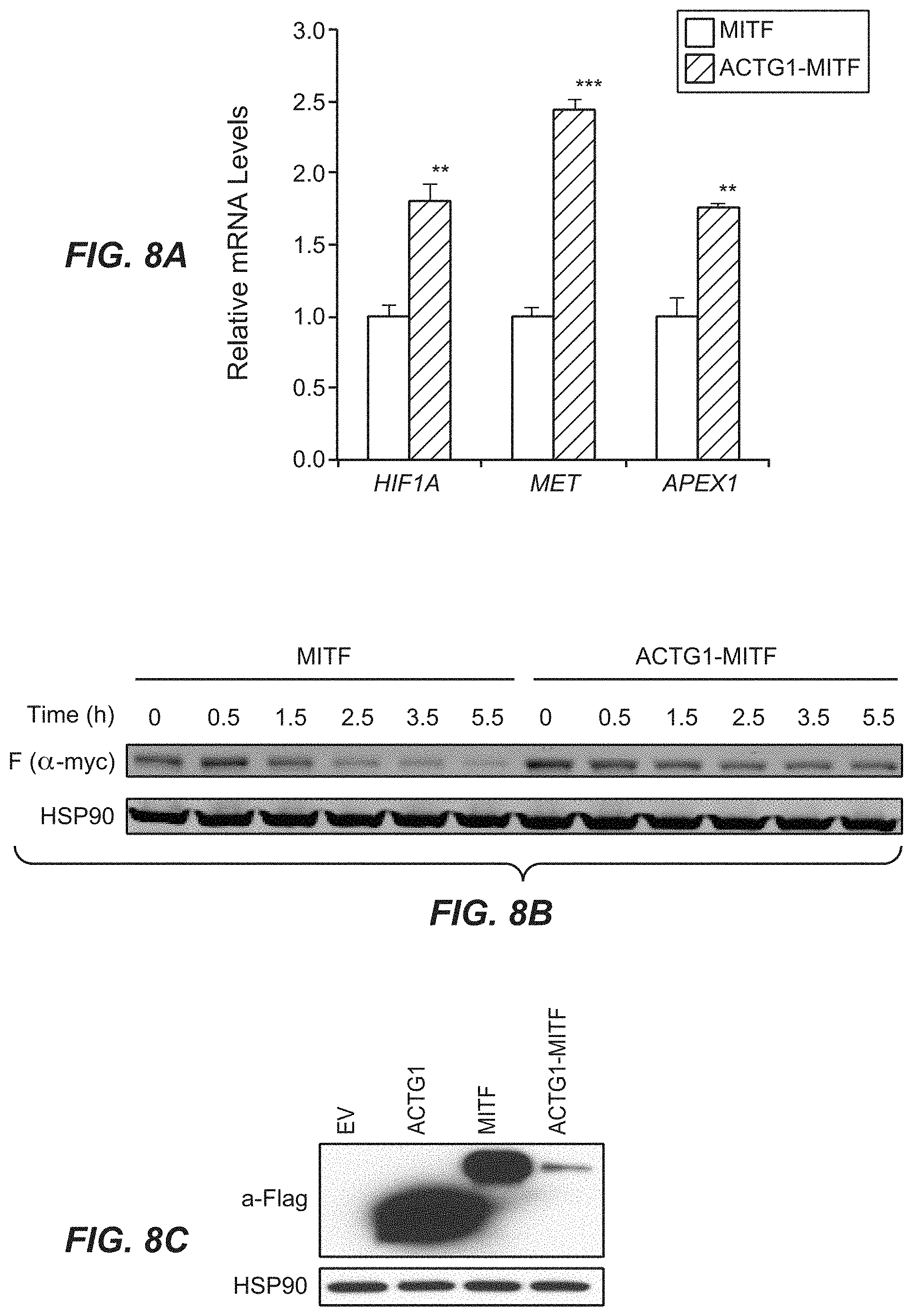
D00020

D00021

D00022

D00023
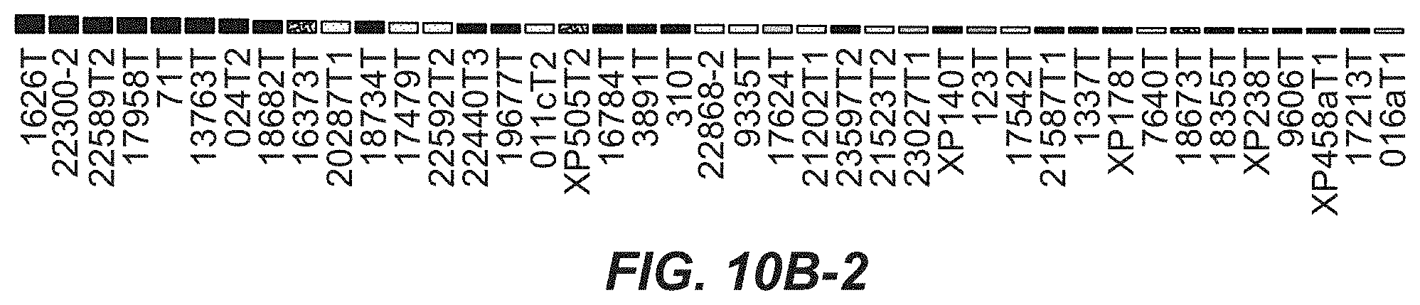
D00024

D00025

S00001
XML
uspto.report is an independent third-party trademark research tool that is not affiliated, endorsed, or sponsored by the United States Patent and Trademark Office (USPTO) or any other governmental organization. The information provided by uspto.report is based on publicly available data at the time of writing and is intended for informational purposes only.
While we strive to provide accurate and up-to-date information, we do not guarantee the accuracy, completeness, reliability, or suitability of the information displayed on this site. The use of this site is at your own risk. Any reliance you place on such information is therefore strictly at your own risk.
All official trademark data, including owner information, should be verified by visiting the official USPTO website at www.uspto.gov. This site is not intended to replace professional legal advice and should not be used as a substitute for consulting with a legal professional who is knowledgeable about trademark law.