Marker For Detecting Highly Pathogenic Influenza Virus And Use Thereof
KIM; Kyun-Hwan ; et al.
U.S. patent application number 16/464866 was filed with the patent office on 2020-04-30 for marker for detecting highly pathogenic influenza virus and use thereof. This patent application is currently assigned to Dandi Bioscience Inc. The applicant listed for this patent is DANDI BIOSCIENCE INC. Invention is credited to Young Ho BYUN, Kyun-Hwan KIM, Hye Min LEE, Eun Sook PARK, Yeong-Min PARK, Baik Lin SEONG.
| Application Number | 20200132688 16/464866 |
| Document ID | / |
| Family ID | 62241747 |
| Filed Date | 2020-04-30 |


View All Diagrams
| United States Patent Application | 20200132688 |
| Kind Code | A1 |
| KIM; Kyun-Hwan ; et al. | April 30, 2020 |
MARKER FOR DETECTING HIGHLY PATHOGENIC INFLUENZA VIRUS AND USE THEREOF
Abstract
Provided are a marker for detecting a highly pathogenic influenza virus including a protein mutant prepared by substituting the amino acids 68 and 69 of a PB1-F2 protein, a composition for detecting a highly pathogenic virus including an agent for measuring the protein mutant, and a detection kit including the same, a method for detecting a highly pathogenic virus including measuring the protein mutant, an antiviral composition against influenza A virus including an inhibitor of binding between a PB1-F2 protein in which the amino acids 68 and 69 are substituted and DDX3, and a method for screening an antiviral substance against influenza A virus.
| Inventors: | KIM; Kyun-Hwan; (Seoul, KR) ; PARK; Eun Sook; (Seoul, KR) ; PARK; Yeong-Min; (Seoul, KR) ; SEONG; Baik Lin; (Seoul, KR) ; BYUN; Young Ho; (Seoul, KR) ; LEE; Hye Min; (Seoul, KR) | ||||||||||
| Applicant: |
|
||||||||||
|---|---|---|---|---|---|---|---|---|---|---|---|
| Assignee: | Dandi Bioscience Inc Seoul KR |
||||||||||
| Family ID: | 62241747 | ||||||||||
| Appl. No.: | 16/464866 | ||||||||||
| Filed: | November 29, 2017 | ||||||||||
| PCT Filed: | November 29, 2017 | ||||||||||
| PCT NO: | PCT/KR2017/013843 | ||||||||||
| 371 Date: | May 29, 2019 |
| Current U.S. Class: | 1/1 |
| Current CPC Class: | G01N 33/6893 20130101; C07K 14/005 20130101; G01N 33/569 20130101; G01N 33/56983 20130101; G01N 33/505 20130101; C07K 2319/00 20130101; G01N 33/574 20130101; C12N 2760/16122 20130101 |
| International Class: | G01N 33/569 20060101 G01N033/569; C07K 14/005 20060101 C07K014/005; G01N 33/50 20060101 G01N033/50 |
Goverment Interests
STATEMENT REGARDING GOVERNMENT RIGHTS
[0002] The present invention was undertaken with the support of 1) Control of cytokine storm based on the mechanism of pathogenicity of influenza PB1-F2 derived from 1918 spanish strain No. A103001 grant funded by the Ministry of Health & Welfare and 2) Needle-free Vaccine Delivery Development No. HI13C0826 grant funded by the Ministry of Health & Welfare.
Foreign Application Data
| Date | Code | Application Number |
|---|---|---|
| Nov 29, 2016 | KR | 10-2016-0159926 |
Claims
1. A marker composition for detecting a highly pathogenic virus, comprising: a protein mutant prepared by substituting the amino acids 68 and 69 of a PB1-F2 protein consisting of the amino acid sequence of SEQ ID NO: 1.
2. The marker composition of claim 1, wherein the protein mutant is prepared by substituting the amino acids 68 and 69 with threonine and proline, respectively.
3. The marker composition of claim 2, wherein the protein mutant consists of the amino acid sequence of SEQ ID NO: 2.
4. The marker composition of claim 1, wherein the virus is an influenza virus.
5. A composition for detecting a highly pathogenic virus, comprising: an agent for measuring a protein mutant prepared by substituting the amino acids 68 and 69 of a PB1-F2 protein consisting of the amino acid sequence of SEQ ID NO: 1.
6. The composition of claim 5, wherein the protein mutant is prepared by substituting the amino acids 68 and 69 with threonine and proline, respectively.
7. The composition of claim 6, wherein the protein mutant consists of the amino acid sequence of SEQ ID NO: 2.
8. The composition of claim 5, wherein the virus is an influenza virus.
9. The composition of claim 5, wherein the agent for measuring the protein mutant is an antibody specifically binding to the protein.
10. A kit for detecting a highly pathogenic virus, comprising: the composition of claim 5.
11. A method for detecting a highly pathogenic virus, comprising: measuring a protein mutant prepared by substituting the amino acids 68 and 69 of a PB1-F2 protein consisting of the amino acid sequence of SEQ ID NO: 1.
12. The method of claim 11, wherein the protein mutant is prepared by substituting the amino acids 68 and 69 with threonine and proline, respectively.
13. The method of claim 12, wherein the protein mutant consists of the amino acid sequence of SEQ ID NO: 2.
14. The method of claim 11, wherein the virus is an influenza virus.
15. An antiviral composition against influenza A virus, comprising: an inhibitor of binding between DEAD box protein 3 (DDX3) and a PB1-F2 protein as an active ingredient, wherein the PB1-F2 protein is prepared by substituting the amino acids 68 and 69 of the amino acid sequence of SEQ ID NO: 1.
16. The composition of claim 15, wherein the DDX3 consists of the amino acid sequence of SEQ ID NO: 3.
17. The composition of claim 15, wherein the binding inhibitor is any one selected from the group consisting of a nucleic acid, a compound, a microbial culture medium or extract, a natural substance extract, a peptide, a substrate analog, an aptamer, and an antibody.
18. The composition of claim 15, wherein the PB1-F2 protein is prepared by substituting the amino acids 68 and 69 with threonine and proline, respectively.
19. The composition of claim 18, wherein the PB1-F2 protein consists of the amino acid sequence of SEQ ID NO: 2.
20. The composition of claim 15, wherein the composition increases production of intracellular interferon beta (IFN.beta.).
21. A method for screening an antiviral substance against influenza A virus, comprising: (a) in vitro treating cells with a candidate substance; (b) measuring binding between DDX3 and a PB1-F2 protein in the cells; and (c) selecting a substance decreasing the binding between the DDX3 and the PB1-F2 protein as an antiviral substance against influenza A virus, compared to a group which is not treated with a candidate substance, wherein the PB1-F2 protein may be prepared by substituting the amino acids 68 and 69 of the amino acid sequence of SEQ ID NO: 1.
22. The method of claim 21, wherein the candidate substance is selected from the group consisting of a nucleic acid, a compound, a microbial culture medium or extract, a natural substance extract, a peptide, a substrate analog, an aptamer, and an antibody.
23. The method of claim 22, wherein the nucleic acid is selected from the group consisting of siRNA, shRNA, microRNA, antisense RNA, an aptamer, a locked nucleic acid (LNA), a peptide nucleic acid (PNA), and a morpholino.
24. The method of claim 21, wherein step (b) is executed using a method selected from the group consisting of western blotting, immunoprecipitation, immunohistochemistry, and immunofluorescence.
25. The method of claim 21, wherein the PB1-F2 protein is prepared by substituting the amino acids 68 and 69 with threonine and proline, respectively.
26. The method of claim 25, wherein the PB1-F2 protein consists of the amino acid sequence of SEQ ID NO: 2.
27. A method for treating influenza A virus, comprising: administering an antiviral composition comprising an inhibitor of binding between DEAD box protein 3 (DDX3) and a PB1-F2 protein as an active ingredient into a subject.
28. (canceled)
Description
CROSS-REFERENCE TO RELATED APPLICATION
[0001] This application is a 371 of PCT/KR2017/013843, filed Nov. 29, 2017, which claims the benefit of priority from Korean Patent Application No. 10-2016-0159926, filed Nov. 29, 2016, the contents of each of which are incorporated herein by reference in its entirety.
SEQUENCE LISTING
[0003] The Sequence Listing submitted in text format (.txt) filed on Oct. 1, 2019, named "SequenceListing.txt", created on Oct. 1, 2019 (12.6 KB), is incorporated herein by reference.
Technical Field
[0004] The present invention relates to a marker for detecting a highly pathogenic influenza virus and a use thereof, and more particularly, to a marker for detecting a highly pathogenic influenza virus, which includes a protein mutant prepared by substituting the amino acids 68 and 69 of a PB1-F2 protein, a composition for detecting a highly pathogenic virus, which includes an agent for measuring the protein mutant and a detection kit including the same, a method for detecting a highly pathogenic virus, which includes measuring the protein mutant, an antiviral composition against influenza A virus, which includes an inhibitor of binding between a PB1-F2 protein in which the amino acids 68 and 69 are substituted and Dead box protein 3 (DDX3) as an active ingredient, and a method for screening an antiviral substance against influenza A virus.
Background Art
[0005] Influenza A virus (IAV) is a pathogen capable of infecting both humans and animals, and a virus which caused Spanish flu resulting in the deaths of 50 million people in 1918. PB1-F2 is a non-structural protein of an influenza virus encoded by a part from the +1 open reading frame to a PB1 gene. Until now, through various studies, PB1-F2 has been reported to have various functions including apoptosis induction and inhibition of innate immunity, and has been known as a significant factor exhibiting virality associated with pathogenicity in a very highly pathogenic influenza virus. It has been reported that this protein contributes to the pathogenesis of influenza by inhibiting production of cytokines, increasing immunopathology of secondary bacterial infection, and delaying viral clearance during the infection of IAV in mouse models.
[0006] In the first defense mechanism against influenza virus infection, type I interferon (type I IFN) is a significant factor for antiviral immunity of a host, and a regulator for adaptive immunity. When a host is infected with a virus such as an influenza virus or another pathogen, three types of main proteins known as pattern-recognition receptors (PRRs) inducing innate immunity recognize pathogen-associated molecular patterns (PAMPs) of pathogens. Such PRRs include toll-like receptors (TLRs), retinoic acid inducible gene-I (RIG-I)-like receptors (RLRs), and nucleotide-binding domain-leucine-rich repeat-containing molecules (NLRs), and when a host is infected with an influenza virus, RIG-I serves as main sensor of viral RNA to induce the production of type I IFN. In such a pathway, formation of a complex of DDX3 and a different phosphokinase has been known to be essential for induction of the production of type I IFN.
[0007] Although an IFN system has strong antiviral activity, influenza viruses have also been evolved to attenuate an IFN response for replication and proliferation thereof in a host. For example, it was reported that an NS1 protein of a highly pathogenic virus such as the H5N1 avian influenza virus has a strong inhibitory effect on type I IFN, and rapid collection of neutrophils, a serious lung damage, and rapid secretion of inflammatory cytokines are induced in Ifnar1-/-mice (Proc Natl Acad Sci U S A 2002;99:10736-10741).
[0008] In the present invention, in order to investigate the influence of a PB1-F2 protein of a highly pathogenic 1918 strain on the pathogenic mechanism of IAV infection, the correlation between viral virulence and the PB1-F2 protein was examined, and a molecular mechanism related to inhibition of a type I IFN response was to be identified.
DISCLOSURE
Technical Problem
[0009] As a result of the investigation of the influence of a PB1-F2 protein in a highly pathogenic 1918 strain of IAV on virulence and the correlation therebetween, the inventors first identified the correlation between the 1918 PB1-F2 protein and high pathogenicity of the virus and the molecular mechanism thereof by confirming that low stability of the virus is mediated by the amino acids 68 and 69 on the sequence of the PB1-F2 protein of the 1918 strain, and virulence of the virus is increased by inhibiting the expression of INF.beta. inducing an antiviral response through binding to intracellular DDX3, and based on this finding, the present invention was completed.
[0010] Therefore, the present invention is directed to providing a marker composition for detecting a highly pathogenic influenza virus, which includes a protein mutant prepared by substituting the amino acids 68 and 69 of a PB1-F2 protein consisting of an amino acid sequence of SEQ ID NO: 1.
[0011] In addition, the present invention is directed to providing a composition for detecting a highly pathogenic virus, which includes an agent for measuring the protein mutant, and a kit for detecting a highly pathogenic virus, which includes the composition.
[0012] In addition, the present invention is directed to providing a method for detecting a highly pathogenic virus, which includes measuring the protein mutant.
[0013] In addition, the present invention is directed to providing an antiviral composition against IAV, which includes an inhibitor of binding between DDX3 and a PB1-F2 protein as an active ingredient.
[0014] In addition, the present invention is directed to providing a method for screening an antiviral substance against IAV.
[0015] However, technical problems to be solved in the present invention are not limited to the above-described problems, and other problems which are not described herein will be fully understood by those of ordinary skill in the art from the following descriptions.
Technical Solution
[0016] To achieve the objects of the present invention, the present invention provides a marker composition for detecting a highly pathogenic influenza virus, which includes a protein mutant prepared by substituting the amino acids 68 and 69 of a PB1-F2 protein consisting of an amino acid sequence of SEQ ID NO: 1.
[0017] In addition, the present invention provides a composition for detecting a highly pathogenic virus, which includes an agent for measuring a protein mutant prepared by substituting the amino acids 68 and 69 of a PB1-F2 protein consisting of an amino acid sequence of SEQ ID NO: 1, and a detection kit including the same.
[0018] In addition, the present invention provides a method for detecting a highly pathogenic virus, which includes measuring a protein mutant prepared by substituting the amino acids 68 and 69 of a PB1-F2 protein consisting of an amino acid sequence of SEQ ID NO: 1.
[0019] In one exemplary embodiment of the present invention, the protein mutant may be prepared by substituting the amino acids 68 and 69 with threonine and proline, respectively.
[0020] In another exemplary embodiment of the present invention, the protein mutant may consist of an amino acid sequence of SEQ ID NO: 2.
[0021] In still another exemplary embodiment of the present invention, the virus may be an influenza virus.
[0022] In yet another exemplary embodiment of the present invention, the agent for measuring the protein mutant may be an antibody specifically binding to the protein.
[0023] In addition, the present invention provides an antiviral composition against IAV, which includes an inhibitor of binding between DDX3 and a PB1-F2 protein as an active ingredient, and the PB1-F2 protein may be prepared by substituting the amino acids 68 and 69 on the amino acid sequence of SEQ ID NO: 1.
[0024] In one exemplary embodiment of the present invention, the DDX3 may consist of an amino acid sequence of SEQ ID NO: 3.
[0025] In another exemplary embodiment of the present invention, the binding inhibitor may be any one selected from the group consisting of a nucleic acid, a compound, a microbial culture medium or extract, a natural substance extract, a peptide, a substrate analog, an aptamer, and an antibody.
[0026] In yet another exemplary embodiment of the present invention, the PB1-F2 protein may be prepared by substituting the amino acids 68 and 69 with threonine and proline, respectively.
[0027] In yet another exemplary embodiment of the present invention, the PB1-F2 protein may consist of an amino acid sequence of SEQ ID NO: 2.
[0028] In yet another exemplary embodiment of the present invention, the composition may increase the production of interferon beta (IFN.beta.) in cells.
[0029] In addition, the present invention may provide a method for screening an antiviral substance against IAV, which includes:
[0030] (a) in vitro treating cells with a candidate substance;
[0031] (b) measuring binding between DDX3 and a PB1-F2 protein in the cells; and
[0032] (c) selecting a substance decreasing the binding between the DDX3 and the PB1-F2 protein as an antiviral substance against IAV, compared to a candidate substance untreated group, and
[0033] the PB1-F2 protein may be prepared by substituting the amino acids 68 and 69 of an amino acid sequence of SEQ ID NO: 1.
[0034] In one exemplary embodiment of the present invention, the candidate substance may be selected from the group consisting of a nucleic acid, a compound, a microbial culture medium or extract, a natural substance extract, a peptide, a substrate analog, an aptamer, and an antibody.
[0035] In another exemplary embodiment of the present invention, the nucleic acid may be selected from the group consisting of siRNA, shRNA, microRNA, antisense RNA, an aptamer, a locked nucleic acid (LNA), a peptide nucleic acid (PNA), and a morpholino.
[0036] In still another exemplary embodiment of the present invention, step (b) is executed using a method selected from the group consisting of western blotting, immunoprecipitation, immunohistochemistry, and immunofluorescence.
[0037] In addition, the present invention provides a method for treating IAV, which includes administering an antiviral composition including an inhibitor of binding between DDX3 and a PB1-F2 protein as an active ingredient into a subject.
[0038] Moreover, the present invention provides a use of an antiviral composition for treating IAV, which includes an inhibitor of binding between DDX3 and a PB1-F2 protein as an active ingredient.
Advantageous Effects
[0039] From the pathogenic mechanism of IAV infection, the inventors first identified that low stability of the virus is mediated by the amino acids 68 and 69 on the sequence of a PB1-F2 protein of a 1918 strain using the highly pathogenic 1918 strain and a low-pathogenic PR8 strain, and virulence of the virus is increased by inhibiting expression of IFN(3 inducing an antiviral response through binding to intracellular DDX3, a protein mutant prepared by substituting the amino acids 68 and 69 on the PB1-F2 protein sequence of an influenza virus can be used as a marker for detecting a highly pathogenic virus, and a highly pathogenic virus can be effectively detected by measuring the mutant.
[0040] In addition, the viral mechanism of the evasion of innate immunity by the PB1-F2 protein mutant, which was newly identified in the present invention, can provide new understanding for developing an antiviral agent, and the antiviral composition according to the present invention can be effectively used in development of an antiviral agent.
DESCRIPTION OF DRAWINGS
[0041] FIG. 1 shows that a PB1-F2 protein of the 1918 strain of IAV (hereinafter referred to as 1918 PB1-F2) has low stability, confirmed by measuring an expression level of the PB1-F2 protein through western blotting after A549 and U937 cells are infected with each of a PR8 strain (PR8), a PR8 strain of the virus whose amino acid sequence is changed with that of 1918 PB1-F2 (PR8-PB1-F2(1918)) and a PR8 strain of the virus from which a PB1-F2 protein is depleted (hereinafter referred to as PR8-PB1-F2(-)) at MOI 1 or 5 for 24 hours.
[0042] FIGS. 2a and 2b show that a 1918 PB1-F2 protein is degraded according to a proteasome-dependent pathway, where FIG. 2a shows the result of measuring PB1-F2 mRNA and protein expression levels through RT-PCR and western blotting after A549 cells are transfected with Flag-tagged PR8 or 1918 PB1-F2 expression plasmids and treated with MG132, a proteasome inhibitor, and FIG. 2b shows the result of ubiquitination analysis, representing that the PB1-F2 protein is degraded by a ubiquitin-proteasome system.
[0043] FIGS. 3a to 3d show the results of identifying a molecular determinant of the stability of a 1918 PB1-F2 protein, where FIG. 3a illustrates various mutant plasmids of PR8 and 1918 PB1-F2, FIG. 3b shows the results of RT-PCR and western blotting for measuring PB1-F2 mRNA and protein expression levels after A549 cells are transfected with each of PB1-F2 chimeric mutants such as PR8-N+1918-C and 1918-N+PR8-C, FIG. 3c shows PB1-F2 mRNA and protein expression levels measured by the same method as used in FIG. 3b after cells are transfected with a c-terminal point mutant expression plasmid prepared by substituting some amino acids on the PR8 PB1-F2 sequence with those of 1918 PB1-F2, and FIG. 3d shows PB1-F2 mRNA and protein expression levels measured by the same method as used in FIG. 3b after PB1-F2 protein backbones of the PR8 and 1918 strains in which the amino acids 68 and 69 are substituted are cloned, and then cells are transfected with each type of plasmids.
[0044] FIGS. 4a to 4d show that IFN.beta. induction is inhibited by a 1918 PB1-F2 protein, where FIG. 4a shows NF-KB luciferase activity and mRNA expression levels of pro-inflammatory cytokines (IL6, IL 1(3, and IL32) in cells transfected with PR8 and 1918 PB1-F2 expression plasmids, FIG. 4b shows the results of semi-quantitative PCR and real-time PCR for measuring IFNI3 mRNA expression levels in A549 and U937 cells transfected with PR8 and 1918 PB1-F2 expression plasmids, FIG. 4c shows the result of luciferase reporter analysis for measuring a promoter activity of IFN.beta. in U937 cells, and FIG. 4d shows the results of RT-PCR and western blotting for measuring intracellular IFN.beta. mRNA and protein expression levels after A549 and U937 cells are infected with PR8 and 1918 strains of IAV at MOI 1.
[0045] FIGS. 5a and 5b show the correlation between proteasome-dependent degradation of a 1918 PB1-F2 protein and inhibition of type I IFN induction, where FIG. 5a shows the results of semi-quantitative RT-PCR and western blotting for measuring IFN.beta. mRNA and protein expression levels depending on the treatment of a proteasome inhibitor such as MG132 in A549 and U937 cells transfected with PR8 and 1918 PB1-F2 expression plasmids, and FIG. 5b shows the results of semi-quantitative RT-PCR and western blotting for measuring IFN.beta. mRNA and protein expression levels after cells are transfected with a mutant expression plasmid prepared by substituting the amino acids 68 and 69 on a PR8 PB1-F2 sequence with those of 1918 PB1-F2.
[0046] FIGS. 6a to 6f show the influence of the amino acids 68 and 69 of 1918 PB1-F2 on pathogenicity of the 1918 strain of IAV, where FIGS. 6a and 6b show changes in body weight and survival rates of mice measured for 14 days after PR8 and 1918 strains of influenza virus were intranasally administered into the mice at 5.times.10.sup.2PFU and 1.times.10.sup.3PFU, respectively, FIG. 6c shows the body weights and survival rates after mice are infected with a mutant influenza virus prepared by substituting the amino acids 68 and 69 of the PR8 PB1-F2 protein with those of 1918 PB1-F2, and FIGS. 6d to 6f show the body weights and survival rates after mice are infected with the same mutant influenza virus as used in FIG. 6c at various contents (6.times.10.sup.2, 8.times.10.sup.2, and 1.times.10.sup.3PFU).
[0047] FIGS. 7a to 7d show that IFN.beta. induction is inhibited by 1918 PB1-F2 in an IAV-infected model, where FIG. 7a shows the result of western blotting for measuring expression levels of viral proteins such as PB1-F2, nucleoprotein (NP), and hemagglutinin (HA) in mouse lung tissue two days after mice are infected with PR8, 1918, and PB1-F2-depleted 1918 (PB1-F2(-)) viruses at 5.times..times.10.sup.2PFU, FIG. 7b shows the result of a plaque assay for measuring the titer of each IAV using a lung tissue lysate of each group of mice of FIG. 7a, FIG. 7c shows the result of RT-PCR for measuring IFN.beta. mRNA expression levels using lung tissue of each group of mice of FIG. 7a, and FIG. 7d shows the result of ELISA for measuring IFN.beta. protein levels secreted from lungs of each group of mice of FIG. 7a.
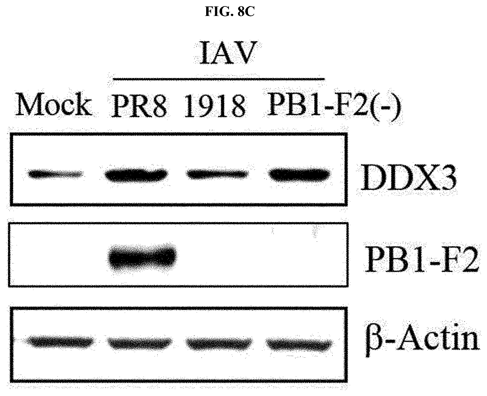
[0048] FIGS. 8a to 8f show a mechanism of inhibiting IFN.beta. induction by binding between 1918 PB1-F2 and DDX3, where FIG. 8a shows proteins having a function associated with viral infection among proteins deduced to be interacted with 1918 PB1-F2, FIG. 8b shows the results of confirming binding between PB1-F2 (1918 strain) and a DDX3 protein under the condition of MG132 treatment after A549 cells are transfected with a DDX3 expression plasmid (HA-tagged DDX3) and a PB1-F2 expression plasmid (Flag-tagged PB1-F2) of a PR8 or 1918 strain and subjected to IP, or transfected with a mutant (I68T, L69P, or I68T/L69P) expression plasmid prepared by substituting the amino acids 68 and 69 of PR8 PB1-F2 with those of 1918 PB1-F2, FIG. 8c is the result of measuring expression levels of DDX3 proteins through western blotting after A549 cells are infected with PR8, 1918, and PR8-PB1-F2(-) viruses, FIG. 8d is the result of measuring expression levels of DDX3 proteins in lung tissue after mice are infected with IAV, FIG. 8e shows the result of checking nuclear translocation of IRF3 by 1918 PB1-F2, and FIG. 8f shows the result of analyzing changes in IFN.beta. mRNA expression after A549 cells are transfected with PR8, 1918 PB1-F2 expression plasmids, and a DDX3 expression plasmid.
[0049] FIGS. 9a to 9d show that pathogenicity of the 1918 PB1-F2 influenza virus is decreased due to treatment of recombinant DDX3 in an in vivo model, where FIG. 9a illustrates a process of an in vivo experiment, FIG. 9b shows the survival rates measured for two weeks after a recombinant DDX3 protein is administered to mouse models infected with 1918 IAV, FIG. 9c shows a 1918 IAV titer measured in lung tissue of the mouse model, and FIG. 9d shows the IFN.beta. mRNA expression levels in lung tissue of the mouse models.
[0050] FIG. 10 illustrates the mechanism of inducing high pathogenicity through evasion of innate immunity of the 1918 strain of IAV according to the present invention.
MODE FOR INVENTION
[0051] As a result of investigation of the influence of a PB1-F2 protein on virulence in a highly pathogenic 1918 strain of IAV and the correlation therebetween, the inventors confirmed that low stability of the virus is mediated by the amino acids 68 and 69 on the PB1-F2 protein sequence of the 1918 strain, and viral virulence is increased by inhibiting IFNI3 expression inducing an antiviral response through the binding between the PB1-F2 protein and intracellular DDX3, and also confirmed that the IFN.beta. expression is restored by injecting a recombinant DDX3 protein into a mouse infected with 1918 PB1-F2 influenza virus, and the viral pathogenicity is decreased due to an increased survival rate. Therefore, the correlation between the 1918 PB1-F2 protein and the high pathogenicity of the virus and a molecular mechanism thereof were first identified, and based on these, the present invention was completed.
[0052] Accordingly, the present invention provides a marker composition for detecting a highly pathogenic influenza virus, which includes a protein mutant prepared by substituting the amino acids 68 and 69 of a PB1-F2 protein consisting of an amino acid sequence of SEQ ID NO: 1.
[0053] In the present invention, the PB1-F2 protein is preferably a non-structural protein of an influenza virus encoded by a part from the +1 open reading frame to a PB1 gene. The protein has been known to induce apoptosis by mediating the efflux of cytochrome c when binding to the mitochondria in CD8 T cells and alveolar macrophages, and it has been reported that the protein increases severity in primary viral and secondary bacterial infections, and the protein is associated with high pathogenicity of an influenza virus.
[0054] Accordingly, in exemplary embodiment of the present invention, to examine the influence of the PB1-F2 protein on high pathogenicity of the 1918 strain of IAV, the PB1-F2 proteins of the 1918 strain and a low-pathogenic PR8 strain are compared to each other, thereby first identifying the correlation between the high pathogenicity and PB1-F2, and its molecular mechanism.
[0055] In one exemplary embodiment of the present invention, it was confirmed that, compared to the low-pathogenic PR8 strain of IAV, the PB1-F2 protein of the highly pathogenic 1918 strain has considerably low stability, which is caused by rapid protein degradation using a ubiquitin-proteasome system (refer to Examples 2 and 3).
[0056] In another exemplary embodiment of the present invention, as a result of analyzing expression patterns of the PB1-F2 proteins using a variety of PB1-F2 protein mutants prepared by substituting amino acids to find the reason for induction of degradation only in the PB1-F2 protein derived from the highly pathogenic 1918 strain, it was confirmed that the amino acids 68 and 69 on the amino acid sequence of the PB1-F2 protein affect stability of the protein (refer to Example 4).
[0057] In another exemplary embodiment of the present invention, in order to examine the influence of the instability of the PB1-F2 protein on a host defense system in IAV 1918 infection, the influence of the PB1-F2 protein on induction of type I IFN playing a very important role in the defense against a virus in innate immunity was analyzed. As a result, it was confirmed that, unlike the PR8 strain, IFN(3 expression is inhibited by PB1-F2 of the 1918 strain, and a promoter activity of the PB1-F2 of the 1918 strain is also inhibited (refer to Example 5). In addition, by confirming that such a phenomenon does not occur when proteasome-dependent degradation is inhibited, and that the amino acids 68 and 69 identified to determine instability of the 1918 PB1-F2 protein affect the inhibitory response of type I IFN induction, it was confirmed that there is a correlation between the instability due to the proteasome-dependent degradation of the 1918 PB1-F2 protein and the inhibitory response of the type I IFN induction (refer to Example 6).
[0058] In still another exemplary embodiment of the present invention, it was confirmed that, among mouse models infected with each of the PR8 and 1918 strains of influenza virus, high virulence is exhibited in the mouse model infected with the 1918 strain of influenza virus, and it was also confirmed by using protein mutants prepared by substituting the amino acids 68 and/or 69 of a PB1-F2 protein that the amino acids at 68 and 69 residues of the PB1-F2 protein contribute to the high pathogenicity of the 1918 strain of IAV (refer to Example 7).
[0059] In yet another exemplary embodiment of the present invention, it can be known that the PB1-F2 protein of the 1918 strain inhibited IFN.beta. induction in the IAV-infected model, and therefore, due to improper viral clearance, a viral titer was maintained at a high level (refer to Example 8).
[0060] According to the exemplary embodiment of the present invention, a protein in which the amino acids 68 and 69 of the PB1-F2 protein are substituted, and preferably, a PB1-F2 protein mutant prepared by substituting the amino acids 68 and 69 with threonine and proline, respectively, like the 1918 strain, may be used as a marker for detecting a highly pathogenic influenza virus and used to detect a highly pathogenic virus by measuring the mutant.
[0061] Therefore, the present invention provides a composition for detecting a highly pathogenic virus, which includes an agent for measuring a protein mutant prepared by substituting the amino acids 68 and 69 of a PB1-F2 protein consisting of an amino acid sequence of SEQ ID NO: 1, and a kit for detecting a highly pathogenic virus, which includes the composition.
[0062] In the present invention, the protein mutant may be prepared by substituting the amino acids 68 and 69 with threonine and proline, respectively, and may consist of an amino acid of SEQ ID NO: 2.
[0063] In the present invention, the virus is preferably an influenza virus, but the present invention is not limited thereto.
[0064] In the present invention, the agent for measuring the protein mutant may be an antibody specifically binding to the protein, but the present invention is not limited thereto.
[0065] The term "antibody" used herein includes an immunoglobulin molecule immunologically having a reactivity with a specific antigen, and encompasses both of monoclonal and polyclonal antibodies. In addition, the antibody includes forms produced by genetic engineering such as a chimeric antibody (e.g., a humanized murine antibody) and a heterogeneous binding antibody (e.g., a bispecific antibody).
[0066] The detection kit of the present invention is composed of a composition, solution, or a device including one or more different components which are suitable for an analysis method.
[0067] In addition, the present invention provides a method for detecting a highly pathogenic virus, which includes measuring a protein mutant prepared by substituting the amino acids 68 and 69 of a PB1-F2 protein consisting of an amino acid sequence of SEQ ID NO: 1.
[0068] In yet another exemplary embodiment of the present invention, it was confirmed that the inhibitory response of IFNI3 induction by the PB1-F2 protein of the 1918 strain occurs by inhibiting the function of DDX3 through the binding between the PB1-F2 protein and the intracellular DDX3 protein (refer to Example 9), and even in the presence of the PB1-F2 protein of the 1918 strain, the inhibition of the IF1\113 induction is restored by treatment of a recombinant DDX3 protein, resulting in the induction of viral clearance (refer to Example 10).
[0069] Therefore, like the 1918 strain, by inhibiting the binding between the PB1-F2 protein prepared by substituting the amino acids 68 and 69 with threonine and proline, respectively, and intracellular DDX3, proliferation of highly pathogenic IAV may be inhibited.
[0070] Accordingly, in another aspect of the present invention, the present invention provides an antiviral composition against IAV, which includes an inhibitor of binding between DDX3 and a PB1-F2 protein as an active ingredient, and the PB1-F2 protein may be prepared by substituting the amino acids 68 and 69 of an amino acid sequence of SEQ ID NO: 1.
[0071] In the present invention, the PB1-F2 protein is prepared by substituting the amino acids 68 and 69 with threonine and proline, respectively, and may consist of an amino acid sequence of SEQ ID NO: 2.
[0072] The term "antiviral" used herein refers to weakening or dissipating the action of a virus having invaded a body by inhibiting viral proliferation in the body, and more specifically, by inhibiting viral proliferation by suppressing nucleic acid synthesis of a virus, gene expression of a virus, or viral replication, and in the present invention, this term is used for IAV, and more preferably, the 1918 strain of IAV (A/Brevig Mission/1/1918(H1N1)).
[0073] The DDX3, serving as a DEAD box family RNA helicase having various functions in cells, is involved in various stages of gene expression, that is, transcription, maturation of nucleic and mitochondrial mRNA, initiation of translation, and rearrangement of ribosomes and spliceosomes, and also involved in replication of hepatitis C virus (HCV) RNA, and it has been reported that the expression of DDX3 is reduced when liver cancer occurs due to HBV infection, and DDX3 is known to serve as a tumor-inhibitory protein. In addition, DDX3 is known to be involved in IFNI3 induction caused by TANK-binding kinase 1 (TBK1) and I.kappa.-B kinase-epsilon (f.kappa.BK.epsilon.)-dependent IRF3 activation. The DDX3 protein may consist of an amino acid sequence of SEQ ID NO: 3.
[0074] In the present invention, the binding inhibitor may be any one selected from the group consisting of a nucleic acid, a compound, a microbial culture medium or extract, a natural substance extract, a peptide, a substrate analog, an aptamer, and an antibody, but the present invention is not limited thereto.
[0075] In still another aspect of the present invention, the present invention provides a method for screening an antiviral substance against IAV, which includes:
[0076] (a) in vitro treating cells with a candidate substance;
[0077] (b) measuring binding between DDX3 and a PB1-F2 protein in the cells; and
[0078] (c) selecting a substance decreasing the binding between the DDX3 and the PB1-F2 protein as an antiviral substance against IAV, compared to a group which is not treated with a candidate substance, and the PB1-F2 protein may be prepared by substituting the amino acids 68 and 69 on an amino acid sequence of SEQ ID NO: 1.
[0079] In the present invention, the candidate substance may be selected from the group consisting of a nucleic acid, a compound, a microbial culture medium or extract, a natural substance extract, a peptide, a substrate analog, an aptamer, and an antibody, and the nucleic acid may be selected from the group consisting of siRNA, shRNA, microRNA, antisense RNA, an aptamer, LNA, PNA, and a morpholino, but the present invention is not limited thereto.
[0080] In step (b), the measurement of the binding between DDX3 and the PB1-F2 protein may be carried out using a method selected from the group consisting of western blotting, immunoprecipitation, immunohistochemistry and immunofluorescence, but the present invention is not limited thereto.
[0081] Hereinafter, exemplary embodiments will be provided to help in understanding of the present invention. However, the following examples are merely provided to more easily understand the present invention, and the scope of the present invention is not limited to the following examples.
EXAMPLES
Example 1. Preparation and Experimental Methods
[0082] 1-1. Cell culture
[0083] A549 and 293T cells were incubated in a Dulbecco's modified Eagle's medium (DMEM, Gibco BRL, Gaithersburg, Md.) containing 10% fetal bovine serum (FBS, Gibco-BRL, Gaithersburg, Md.) inactivated by thermal treatment and 1% penicillin/streptomycin (Gibco-BRL, Gaithersburg, Md.) at 37.degree. C. with 5% CO.sub.2. U937 cells were incubated in an RPMI medium (Gibco-BRL, Gaithersburg, Md.) containing 10% FBS and 1% penicillin/streptomycin under the same conditions as used for the above cells. Transfection was carried out using Lipofectamine 2000 (Invitrogen) according to the manufacturer's protocols.
[0084] 1-2. Preparation of plasmids
[0085] PR8 PB1-F2 and 1918 PB1-F2 expression plasmids were cloned using pcDNA3.1 (+) vectors (Invitrogen) at EcoR I and Xho I restriction sites by PCR. Chimeric mutants, that is, mutants of an N-terminal domain of PR8 strain-derived PB1-F2 and a C-terminal domain of a 1918 strain; and an N-terminal of 1918 strain-derived PB1-F2 and a C-terminal domain of a PR8 strain, were amplified by PCR and then cloned in pcDNA3.1(+) vectors. In addition, PR8-derived PB1-F2 mutants prepared by amino acid substitution (R59K, R60Q, R59K/R60Q, R59K/R60Q/N66S, R59K/R60Q/N66S/I68T, and R59K/R60Q/N66S/I68T/L69P) were amplified by PCR and then cloned in pcDNA3.1(+) vectors. Primer sequences used in the experiment are shown in Table 1 below. In addition, a DDX3 expression plasmid was cloned in a pcDNA3.1(+) vector with Hind III and Xho I restriction sites, and IRF3 and TLR3 expression vectors were provided from a different research team of Yonsei University.
TABLE-US-00001 TABLE 1 SEQ ID Primer Sequence NO: PB1-F2 Forward 5'-acc gaa ttc atg gac tac aag gat gac gac-3' 4 Reverse 5'-acc ctc gag cta ctc gtg ttt gct gaa-3 5 R59K Forward 5'-gtg tat tgg aag cga tgg ctt tcc ttg-3' 6 Reverse 5'-caa gga aag cca tcg ctt cca ata cac-3' 7 R59K/R60Q Forward 5'-gtg tat tgg agg caa tgg ctt tcc ttg-3' 8 Reverse 5'-caa gga aag cca ttg cct cca ata cac-3 9 R59K/R60Q/N66S Forward 5'-gtg tat tgg aag cga tgg ctt tcc ttg-3' 10 Reverse 5'-caa gga aag cca tcg ctt cca ata cac-3' 11 R59K/R60Q/N66S/ Forward 5'-ctt tcc ttg agg aat ccc acc ccg-3' 12 I68T Reverse 5'-cgg ggt ggg aft cct caa gga aag-3' 13 R59K/R60Q/N66S/ Forward 5'-ctt gag gag tcc cat ccc ggt atc ttt-3' 14 I68T/L69P Reverse 5'-caa aga tac cgg gat ggg act cct caa-3' 15 I68T Forward 5'-ttg agg aat ccc acc ctg gta ttt ttg-3' 16 Reverse 5'-caa aaa tac cag ggt ggg att cct caa-3' 17 L69P Forward 5'-agg aat ccc atc ccg gta ttt ttg aaa-3' 18 Reverse 5'-ttt caa aaa tac cgg gat ggg aft cct-3' 19 I68T/L69P Forward 5'-ttg agg aat ccc acc ccg gta ttt ttg aaa-3' 20 Reverse 5'-ttt caa aaa tac cgg ggt ggg aft cct caa-3' 21 T68I Forward 5'-ctt gag gag tcc cat ccc ggt atc ttt g-3' 22 Reverse 5'-caa aga tac cgg gat ggg act cct caa-3' 23 P69L Forward 5'-gga gtc cca ccc tggta tct ttg aaa ac-3' 24 Reverse 5'-gtt ttc aaa gat acc agg gtg gga ctc c-3' 25 T68I/P69L Forward 5'-ttg agg agt ccc atc ctg gta tct ttg aaa-3' 26 Reverse 5'-ttt caa aga tac cag gat ggg act cct caa-3' 27
[0086] 1-3. Antibodies and reagents
[0087] An anti-FLAG M2 monoclonal antibody, anti-HA, and an anti-.beta.-actin antibody were purchased from Sigma (St. Louis, Mo.), and a Lamin A/C antibody was purchased from Cell Signaling Technology (Beverly, Mass.). Mouse polyclonal antibodies for detecting viral PB1-F2 proteins were prepared using a full sequence of recombinant PB1-F2 protein expressed in E. coli. A DDX3 antibody, and anti-mouse and anti-rabbit IgG horseradish peroxidase (HRP) antibodies were purchased from Santa Cruz Biotechnology (Santa Cruz, Calif.), and an anti-NP antibody was obtained from rabbits immunized with an NP protein (LabFrontier). MG132 and Poly(I:C) used in this example were purchased from Calbiochem (Germany) and InvivoGen, respectively.
[0088] 1-4. Influenza viruses
[0089] Influenza A/Puerto Rico/8/34(H1N1) virus (IAV (PR8)), PB1-F2 protein-depleted virus (IAV PB1-F2(-)), or a virus in which the amino acid sequence was substituted with a PB1-F2 protein of A/Brevig Mission/1/1918(H1N1) virus (IAV(1918)) in a PR8 virus backbone were used for in vitro and in vivo experiments. To prepare a PB1-F2 mutant virus using site-specific mutation, the 68I1e(ATC) and 69Leu(CTG) residues in a PBI-F2 protein of the A/Puerto Rico/8/34(H1N1) were substituted with Thr(ACC) and Pro(CCG), respectively. To obtain a recombinant virus expressing a mutated PB1-F2 protein, reverse genetics technology was used. Simply, seven cDNAs encoding a wild-type gene part and one mutated PB1 part were cloned in pHW2000 vectors and then transfected together into 293T cells. After 3 days, a supernatant was recovered to perform a plaque assay. The purified plaque was inoculated into MDCK cells to amplify viruses.
[0090] 1-5. Influenza virus infection
[0091] 6- to 8-week old female Balb/c mice were anesthetized and then infected with influenza viruses intranasally at 50 .mu.L. The animal experiment was carried out by obtaining the approval of the Animal Experiment Ethics Committee of Konkuk University. For influenza virus infection into cells, A549 and U937 cells were washed with PBS and infected with influenza viruses at MOI 1. After 24 hours, the cells were recovered to perform RT-PCR and western blotting.
[0092] 1-6. Real-time PCR
[0093] Cells and mouse tissue were lysed with TRIzol to extract total RNA. Using 2 .mu.g of the extracted RNA and M-MLV reverse transcriptase (iNtRON, Seoul, Korea), a reaction solution was prepared to have a final volume of 20 .mu.L, thereby synthesizing cDNA, and then PCR was carried out using the cDNA as a template. The PCR was carried out under conditions of primary denaturation at 94.degree. C. for 5 minutes, and 25 to 30 cycles of 94.degree. C. (30 sec), 55 to 60.degree. C. (30 sec) and 72.degree. C. (30 sec), and final elongation at 72.degree. C. for 5 minutes. Primer sequences used in the experiment are shown in Table 2 below. Quantitative real-time PCR was carried out using a SYBR Green PCR Master Mix (Applied Biosystems), and PCR amplification was performed using a real-time PCR apparatus manufactured by Applied Biosystems (ABI7500). Quantitative analysis of relative mRNA expression levels was performed using a .DELTA..DELTA.Ct method, and the result is represented as a relative n-fold difference with respect to a calibrator (RQ=2.sup.-.DELTA..DELTA.ct).
TABLE-US-00002 TABLE 2 SEQ ID Primer Sequence NO: IFN.beta. Forward 5'-gcc tgg cif cca tca tga ac-3' 28 Reverse 5'-gag gca tca act gac agg tc-3 29 PB1-F2 Forward 5'-atg gga ccg gaa cag gat aca cca-3' 30 Reverse 5'-cta ctc gtg ttt gct gaa caa cct-3' 31 IL-1.beta. Forward 5'-tca ggc agg ccg cgt cag tt-3' 32 Reverse 5'-ttg ctg tga gtc ccg gag cgt-3 33 IL-6 Forward 5'-agc gcc ttc ggt cca gtt gc-3' 34 Reverse 5'-tgc cag tgc ctc ttt gct gct-3' 35 IL-32 Forward 5'-gaa ggc ccg aat ggt aat gc-3' 36 Reverse 5'-tcg gca ccg taa tcc atc tc-3' 37 GAPDH Forward 5'-cgt ctt cac cac cat gga ga-3' 38 Reverse 5'-cgg cca tca cgc cac agt ft-3' 39
[0094] 1-7. Western blotting
[0095] Cells were treated with a lysis buffer (25 mmol/L Tris-HCl, pH 7.5, 1% NP40, and protease cocktail) and centrifuged, thereby obtaining a supernatant from which intracellular proteins were eluted. 50 .mu.g of proteins were loaded on a 12 to 15% acrylamide gel to perform SDS-PAGE, thereby separating the proteins by size, and then western blotting was performed. Protein detection using chemical fluorescence was carried out using ECL detection reagents (GE Healthcare, Buckinghamshire, UK), and expression levels of a target protein were determined using a Bio-Imaging Analyzer (LAS-4000, Fuji, Tokyo, Japan).
[0096] 1-8. Luciferase reporter assay
[0097] Cells were transfected using Lipofectamine 2000, and after 48 hours, luciferase activity measured using a luciferase assay system (Promega, Madison, Wis., USA). .beta.-galactosidase activity was measured for all samples, and the results were calibrated. The experiment was performed independently three times, and data were represented as mean.+-.standard deviation (SD).
[0098] 1-9. Measurement of IFN.beta. content
[0099] A mouse lung tissue lysate was subjected to centrifugation at 10,000.times.g for 5 minutes, thereby obtaining a supernatant, and the supernatant was used to measure an IFN.beta. protein level using an IFN.beta. ELISA kit (R&D) according to the manufacturer's protocols.
[0100] 1-10. Preparation of protein expression plasmids
[0101] A pGE-LysRS-R9-DDX3 expression plasmid encoding LysRS-R9-DDX3 was manufactured using a pGE-LysRS-4 vector consisting of T7 promoter-LysRS-TEV protease recognition sequence-multicloning sites (Kpnl-BamHI-EcoRV-SalI-HindIII) and a histidine tag. The LysRS-R9-DDX3 gene was amplified by PCR using a primer sequence of Table 3 below, and an amplification product was cleaved with KpnI/SalI and introduced into the KpnI/SalI site of pGE-LysRS-4.
TABLE-US-00003 TABLE 3 SEQ ID Primer Sequence NO: LysRS-R9- Forward 5'-gtc acg ggt acc cgt cgc cgt cgc cgt cgc cgt 40 DDX3 cgc cgt atg agt cat gtg gca gtg-3' Reverse 5'-gtc acg gtc gac gtt acc cca cca gtc aac ccc 41 ctg gga gtt a-3'
Example 2. Analysis of PB1-F2 Protein Stability of IAV 1918 Strain
[0102] According to various studies, it has been known that the PB1-F2 protein of influenza virus has various functions, and recently, it has been reported that high morbidity of the 1918 pandemic influenza is associated with the PB1-F2 protein of a 1918 strain. Therefore, in this example, in order to examine molecular and functional characteristics of the 1918 influenza virus PB1-F2 protein, first, the PB1-F2 proteins of A/Brevig Mission/1/1918 (H1N1) (hereinafter, 1918 strain) and A/Puerto Rico/8/1934 (H1N1) (hereinafter, PR8 strain) influenza viruses were comparatively analyzed.
[0103] To this end, A549 and U937 cells were infected with the influenza viruses, that is, a PR8 strain (PR8), a virus of the PR8 strain in which the amino acid sequence was mutated with the PB1-F2 protein of a 1918 strain (PR8-PB1-F2 (1918)), and a PB1-F2 protein-depleted PR8 strain (PR8-PB1-F2(-)) at MOI 1 or 5 for 24 hours, and then subjected to western blotting to observe an expression pattern of the PB1-F2 proteins. As a result, as shown in FIG. 1, it was confirmed that the PR8 PB1-F2 protein is expressed, but the 1918 PB1-F2 protein was not detected. According to this result, it can be known that the PB1-F2 protein of the 1918 strain exhibits lower stability than that of the PR8 strain.
Example 3. Confirmation of Degradation of 1918 PB1-F2 Protein By Proteasome-Dependent Pathway
[0104] Based on the result of Example 2, in order to see whether different PB1-F2 protein expression patterns between the PR8 strain and the 1918 strain are caused by degradation of a proteasome-mediated protein, the expression patterns of the PB1-F2 proteins were observed under a condition in which proteasome inhibitor MG132 was treated.
[0105] More specifically, A549 cells were transfected with each of Flag-tagged PB1-F2 expression plasmids of the PR8 strain and the 1918 strain, and treated with MG132 for 6 hours, and then the cells were collected. PB1-F2 mRNA and protein expression levels were analyzed through RT-PCR and western blotting. As a result, as shown in FIG. 2a, it was confirmed that, regardless of the treatment of MG132, 1918 PB1-F2 mRNA was detected, but the PB1-F2 protein was expressed only when MG132 was treated. According to this result, it can be known that the low stability of the 1918 PB1-F2 protein was associated with a proteasome-dependent pathway.
[0106] A ubiquitin-proteasome system is known to induce protein degradation and regulate functions of various proteins. To confirm whether the PB1-F2 protein is degraded by the ubiquitin-proteasome system, a ubiquitination assay was carried out. As a result, as shown in FIG. 2b, it can be known that the PB1-F2 protein is degraded by a ubiquitin-dependent proteasome pathway.
Example 4. Identification of Molecular Determinant of 1918 PB1-F2 Protein Stability
[0107] Based on the results of Examples 2 and 3, in order to identify a molecular determinant determining stability of the PB1-F2 protein, a variety of PR8 and 1918 PB1-F2 mutant plasmids were manufactured and are shown in FIG. 3a.
[0108] More specifically, A549 cells were transfected with each of PB1-F2 chimeric mutants PR8-N+1918-C and 1918-N+PR8-C as shown in FIG. 3a, and then PB1-F2 mRNA and protein expression levels were measured by RT-PCR and western blotting in the same manner as in Example 3. As a result, as shown in FIG. 3b, when the proteasome inhibitor MG132 was not treated, the PB1-F2 protein was not expressed in the PR8-N+1918-C-tranfected cells, and therefore it can be known that a part determining the stability of the protein is present at the C-terminal part of PB1-F2.
[0109] Afterward, in order to more specifically examine a stability determining part of the PB1-F2 protein, C-terminal point mutants were manufactured using a method of substituting some amino acids of the PR8 PB1-F2 sequence with those of 1918 PB1-F2, and then RT-PCR and western blotting were performed. As a result, as shown in FIG. 3c, it was confirmed that, when R59K/R60Q/N66S mutants were introduced while MG132 was not treated, the PB1-F2 protein was expressed, but when R59K/R60Q/N66S/I68T mutants and R59K/R60Q/N66S/I68T/L69P mutants were introduced, the PB1-F2 protein was not expressed. Therefore, it can be known that the amino acids 68 and 69 of the PB1-F2 protein are very important in the stability of the protein.
[0110] Further, to reconfirm that the amino acids 68 and 69 of the PB1-F2 protein are factors that determine instability of the protein, a plasmid was cloned after the amino acids 68 and 69 on the PR8 strain-derived PB1-F2 protein were substituted with those on the 1918 strain-derived PB1-F2 protein, and vice versa, and then each plasmid was transfected into cells, followed by RT-PCR and western blotting. As a result, as shown in FIG. 3d, when each or all of the amino acids 68 and 69 were substituted while MG132 was not treated, it was confirmed that expression of the PB1-F2 protein was inhibited in the backbone of the PR8 strain, and the protein expression was increased in the backbone of the 1918 strain. Accordingly, it can be known that the stability of the PB1-F2 protein was dependent on the amino acids 68 and 69.
[0111] Moreover, by the analysis of an intracellular position of each PB1-F2 clone, the PR8 PB1-F2 protein was primarily located in the mitochondria, but the 1918 PB1-F2 protein was dispersed in the cytoplasm or present in the nucleus. Therefore, it can be known that the amino acids 68 and 69 of the PB1-F2 protein are important for determining the intracellular position of the PB1-F2 protein. Such results indicate that Ile68 and Leu69 are molecular factors that determine the stability of the PB1-F2 protein.
Example 5. Confirmation of Inhibition of INF.beta. Secretion By 1918 PB1-F2 Protein
[0112] To examine the influence of instability of the 1918 PB1-F2 protein on a host, mRNA expression and NF-kB luciferase activity of a pro-inflammatory cytokine were analyzed. As a result, as shown in FIG. 4a, it was confirmed that there were no significant difference in the PB1-F2 proteins between the PR8 and 1918 strains.
[0113] A type I IFN response which is a main component of the innate immunity system is known to be very important in defense against viral pathogens. For example, according to various studies, it has been reported that the type I IFN plays a very important role in a host defense system against influenza infection. Therefore, to verify whether INF.beta. induction is influenced by the PB1-F2 protein, the inventors carried out semi-quantitative PCR and real-time PCR after A549 and U937 cells were transfected with plasmids expressing the PB1-F2 protein of each of the PR8 or 1918 strain.
[0114] As a result, as shown in FIG. 4b, it was observed that 1918 PB1-F2 strongly inhibits intracellular IFN.beta. induction as opposed to the PR8 strain among the two types of cells. In addition, as shown in FIG. 4c, it was confirmed that U937 cells were transfected with an IFN.beta. luciferase reporter plasmid and a PB1-F2 or NS1 expression plasmid, treated with polyI:C before 12 hours of cell recovery, and subjected to luciferase analysis, resulting in inhibition of IFN.beta. promoter activity by 1918 PB1-F2.
[0115] Based on the above result, in order to investigate whether expression of the 1918 PB1-F2 protein in influenza virus-infected cells substantially affects expression of an IFN.beta. gene, A549 and U937 cells were inflected with IAV, that is, PR8 and 1918 strains at MOI 1, and then RT-PCR and western blotting were performed to observe a change in IFN.beta. mRNA and protein expression in cells.
[0116] As a result, as shown in FIG. 4d, it was confirmed that the expression of IFN.beta. mRNA in cells induced by viral infection was inhibited by the 1918 PB1-F2 protein.
[0117] Such results indicate that the type I IFN response is inhibited by the 1918 PB1-F2 protein in virus-infected cells.
Example 6. Investigation of Correlation Between Proteasome-Dependent Degradation of 1918 PB1-F2 Protein and Inhibition of Type I IFN Induction
[0118] From the results of Examples 2 and 3, it was confirmed that the 1918 PB1-F2 protein has significantly low stability, and based on this, it was intended to examine if there is a correlation between the stability of 1918 PB1-F2 and the inhibitory performance by the protein on IFN.beta. induction. To this end, A549 and U937 cells were transfected with PR8 and 1918 PB1-F2 expression plasmids, and after 18 hours, treated with a proteasome inhibitor such as MG132 for 6 hours, and then IFN.beta. expression was observed by semi-quantitative RT-PCR and western blotting.
[0119] As a result, as shown in FIG. 5a, it was confirmed that the IFN.beta. mRNA and protein expression was inhibited by 1918 PB1-F2 when MG132 was not treated, but the IFN.beta. expression was not inhibited when MG132 was treated. Such a result indicates that the stability of PB1-F2 has an important effect on the inhibition of IFN.beta. induction by 1918 PB1-F2.
[0120] Further, to verify if the amino acids 68 and 69 of 1918 PB1-F2 identified as the molecular factors determining stability of the PB1-F2 protein affect inhibition of the IFN.beta. expression, after mutants in which the amino acids 68 and 69 of PB1-F2 were transfected into the A549 cells, IFN.beta. expression was observed by semi-quantitative RT-PCR and western blotting.
[0121] As a result, as shown in FIG. 5b, it was confirmed that when the amino acids 68 and 69 of PB1-F2 were substituted with tryptophan (T) and/or proline (P), mRNA and protein expression of IFN.beta. (I68T, L69P, and I68T/L69P) was inhibited.
[0122] Such results indicate that there is a correlation between the proteasome-dependent degradation of the 1918 PB1-F2 protein and the strong expression inhibitory performance of the type I IFN.
Example 7. Investigation of Influence of Amino Acids 68 and 69 of 1918 PB1-F2 On Pathogenicity of IAV 1918 Strain
[0123] To examine the influence of the PB1-F2 protein on viral pathogenicity when a host is infected with the 1918 strain of IAV, a change in body weight and a survival rate of a mouse were observed for 14 days after influenza viruses of PR8 or 1918 strains were intranasally administered into the mouse at 5.times.10.sup.2PFU or 1.times.10.sup.3PFU.
[0124] As a result, as shown in FIGS. 6a and 6b, it was confirmed that the mouse exhibits higher virulence when infected with the influenza viruses of the 1918 strain, compared to those of the PR8 strain.
[0125] Further, from the results of Examples 4 and 6, it was confirmed that the amino acids 68 and 69 of the PB1-F2 protein are factors that determine instability of the protein and IFN.beta. induction, and based on this, it was intended to verify if the amino acid position has an important effect on the pathogenicity of influenza viruses. To this end, it was observed that mice were infected with mutated influenza viruses prepared by substituting the amino acids 68 and 69 of the PR8 PB1-F2 protein with those of the 1918 PB1-F2, and then changes in body weight and survival rate were observed.
[0126] As a result, as shown in FIG. 6c, compared to the mice infected with the PR8 strain and the PB1-F2 protein-depleted virus (PB1-F2(-)), the body weight of the mice infected with the mutant viruses (I68T, L69P, and I68T/L69P) was decreased to a similar degree as that of the mice infected with the 1918 PB1-F2 viruses.
[0127] Furthermore, in order to investigate the influence of the amino acids located at the above positions on the virulence of 1918 PB1-F2, mice were infected with I68T, L69P, and I68T/L69P mutant viruses at various contents (6.times.10.sup.2,8.times.10.sup.2, and 1.times.10.sup.3PFU), and then body weights and survival rates were measured. As a result, as shown in FIGS. 6d to 6f, it was confirmed that all of the amino acids 68 and 69 of the 1918 PB1-F2 protein contribute to viral virulence.
[0128] From the above results, it can be known that the amino acids 68 and 69 of the PB1-F2 protein contribute to high pathogenicity of the 1918 strain of IAV.
Example 8. Confirmation of In Vivo Inhibition of IFN.beta. Induction By 1918 PB1-F2
[0129] To verify if the 1918 PB1-F2 protein substantially inhibits the IFN.beta. induction in an influenza virus-infected model, mice were infected with each type of influenza viruses such as PR8, 1918, or PB1-F2-depleted 1918 (PB1-F2(-)) at 5.times.10.sup.2PFU, and after two days, expression levels of viral proteins such as PB1-F2,NP, and HA in mouse lung tissue were analyzed by westem blotting. As a result, as shown in FIG. 7a, it was confirmed that HA and NP viral proteins were more highly expressed in the mice infected with the 1918 strain of virus than in the mice infected with the PR8 strain of virus.
[0130] To examine whether the expression level of such a viral protein is associated with virus replication, a virus titer was measured on a lung tissue lysate through plaque assay. As a result, as shown in FIG. 7b, it was confirmed that a titer of the 1918 strain virus was approximately 10-fold higher than those of the PR8 strain virus and the PB1-F2-depleted virus, indicating that there is a defect on the process of viral clearance of the 1918 strain.
[0131] Therefore, based on the result, to verify if infection with the 1918 strain virus inhibits the IFN.beta. induction in the lung tissue of mice infected with each type of virus, the expression level of IFN.beta. mRNA was measured by performing RT-PCR. As a result, as shown in FIG. 7c, it was confirmed that only when infected with the 1918 strain of virus, the IFN.beta. expression is considerably inhibited.
[0132] In addition, when an amount of the IFN.beta. protein secreted from the lung of a mouse infected with each type of virus was measured through ELISA, as shown in FIG. 7d, it was confirmed that the amount of the IFN.beta. protein secreted from the mouse infected with the 1918 strain of virus was lower than those of the mice infected with the PR8 strain of virus and the PB1-F2-depleted virus.
[0133] The results indicate that PB1-F2 inhibits the IFN.beta. induction in the mouse infected with the 1918 strain of virus.
Example 9. Identification of Inhibition of IFN.beta. Induction Through Binding Between 1918 PB1-F2 and DDX3
[0134] To identify a molecular mechanism for inhibiting IFN.beta. induction by 1918 PB1-F2, proteins interacting with 1918 PB1-F2 were analyzed by IP. As a result, it was seen through LC-MS/MS analyses that a total of 134 types of proteins interacted with 1918 PB1-F2. Further, biological functions of the proteins interacting with PB1-F2 were analyzed using the ingenuity pathway analysis (IPA) program, and the analysis focused on viral infection-related proteins. As a result, as shown in FIG. 8a, DDX3X, HSP90AA1, and HSPD1 proteins were deduced, and it is known that these proteins are associated with viral infection and thus interact with TBK1, IRF3 and INFA2 proteins.
[0135] Therefore, in order to assess the interaction between 1918 PB1-F2 and DDX3 by focusing on DDX3 deduced from the above result, A549 cells were transfected with a plasmid expressing DDX3 (HA-tagged DDX3) and a PR8 or 1918 PB1-F2 expression plasmid (Flag-tagged PB1-F2), and then subjected to IP under the condition of MG132 treatment. As a result, as shown in FIG. 8b, it was confirmed that 1918 PB1-F2 binds to DDX3. Further, it was also confirmed that mutant proteins (I68T, L69P, and I68T/L69P) prepared by substituting the amino acids 68 and 69 of PR8 PB1-F2 with those of 1918 PB1-F2 interacted with DDX3 by transfecting expression plasmids of the mutant proteins and performing the same experiment as described above.
[0136] Subsequently, to verify whether DDX3 was inhibited by the 1918 PB1-F2 protein, A549 cells were infected with each influenza virus. As a result, as shown in FIG. 8c, DDX3 expression was decreased by the 1918 PB1-F2 protein. In addition, it was confirmed that intracellular DDX3 expression was decreased as shown in FIG. 8d even in the lung tissue of a mouse infected with each type of influenza virus.
[0137] Further, since IRF3 phosphorylation and nuclear translocation occur in the IFN.beta. induction, the inventors assessed nuclear translocation of IRF3 to investigate whether the nuclear translocation of IRF3 was inhibited by 1918 PB1-F2. As a result, as shown in FIG. 8e, while the nuclear translocation of IRF3 was decreased by the 1918 PB1-F2 protein, such a phenomenon was restored by DDX3 addition. Such results indicate that IFN.beta. induction is inhibited by the interaction of 1918 PB1-F2 with DDX3.
[0138] To verify the result again, A549 cells were transfected with the PR8 and 1918 PB1-F2 expression plasmids and the DDX3 expression plasmid, and then a change in expression level of IFN.beta. mRNA was analyzed by trans-complementation assay, and as a result, as shown in FIG. 8f, it was confirmed that IFN.beta. mRNA expression is decreased by 1918 PB1-F2, but Fn.beta. mRNA expression is increased again by DDX3 expression.
[0139] The results indicate that IFN.beta. induction was inhibited by binding the 1918 PB1-F2 protein to DDX3.
Example 10. In Vivo Confirmation of Decrease In Pathogenicity of 1918 PB1-F2 Influenza Virus By Treatment of Recombinant DDX3
[0140] To prove a mechanism of inhibiting IFN.beta. secretion by the interaction between 1918 PB1-F2 and DDX3 identified by the results of the above examples, the role of DDX3 in IFN.beta. induction was verified. To this end, an in vivo experiment was carried out to verify whether the inhibition of the IFN.beta. induction can be repaired by the administration of the recombinant DDX3 protein into a mouse model infected with the 1918 influenza virus, and an experimental process is illustrated in FIG. 9a.
[0141] As a result, as shown in FIG. 9b, it was confirmed that all the DDX3-administered mice are alive for 2 weeks. In addition, as the result of measuring the virus titer in the mouse lung, as shown in FIG. 9c, it was confirmed that the virus titer in the DDX3 protein-administered mice was approximately 37-fold lower than those of the control groups, indicating that viral clearance occurs due to DDX3 addition. In addition, as shown in FIG. 9d, it was confirmed that intracellular IFN.beta. expression is increased due to expression of the DDX3 protein in mouse lung tissue.
[0142] Through the above results, it was seen that DDX3 protects the mouse from 1918 PB1-F2 viral infection. In addition, in FIG. 10, an innate immunity evasion model of the highly pathogenic 1918 strain of IAV identified by the example was illustrated.
[0143] It would be understood by those of ordinary skill in the art that the above description of the present invention is exemplary, and the exemplary embodiments disclosed herein can be easily modified into other specific forms without departing from the technical spirit or essential features of the present invention. Therefore, the exemplary embodiments described above should be interpreted as illustrative and not limited in any aspect.
INDUSTRIAL APPLICABILITY
[0144] The viral mechanism of the evasion of innate immunity by the PB1-F2 protein mutants newly identified in the present invention can provide a new understanding for developing an antiviral agent, and an antiviral composition according to the present invention can be effectively used in the development of an antiviral agent.
Sequence CWU 1
1
41187PRTArtificial SequencePB1-F2 1Met Gly Gln Glu Gln Asp Thr Pro
Trp Ile Leu Ser Thr Gly His Ile1 5 10 15Ser Thr Gln Lys Arg Gln Asp
Gly Gln Gln Thr Pro Lys Leu Glu His 20 25 30Arg Asn Ser Thr Arg Leu
Met Gly His Cys Gln Lys Thr Met Asn Gln 35 40 45Val Val Met Pro Lys
Gln Ile Val Tyr Trp Lys Gln Trp Leu Ser Leu 50 55 60Arg Asn Pro Ile
Leu Val Phe Leu Lys Thr Arg Val Leu Lys Arg Trp65 70 75 80Arg Leu
Phe Ser Lys His Glu 85290PRTArtificial SequencePB1-F2 mutant 2Met
Gly Gln Glu Gln Asp Thr Pro Trp Ile Leu Ser Thr Gly His Ile1 5 10
15Ser Thr Gln Lys Arg Glu Asp Gly Gln Gln Thr Arg Lys Leu Glu His
20 25 30His Asn Ser Thr Arg Leu Met Asp His Cys Gln Lys Thr Met Asn
Gln 35 40 45Val Val Met Pro Lys Gln Ile Val Tyr Trp Lys Gln Trp Leu
Ser Leu 50 55 60Arg Ser Pro Thr Pro Val Ser Leu Lys Thr Arg Val Leu
Lys Arg Trp65 70 75 80Arg Leu Phe Ser Lys His Glu Trp Thr Ser 85
903612PRTArtificial SequenceDDX3 3Met Ser His Val Ala Val Glu Asn
Ala Leu Gly Leu Asp Gln Gln Phe1 5 10 15Ala Gly Leu Asp Leu Asn Ser
Ser Asp Asn Gln Ser Gly Gly Ser Thr 20 25 30Ala Ser Lys Gly Arg Tyr
Ile Pro Pro His Leu Arg Asn Arg Glu Ala 35 40 45Thr Lys Gly Phe Tyr
Asp Lys Asp Ser Ser Gly Trp Ser Ser Ser Lys 50 55 60Asp Lys Asp Ala
Tyr Ser Ser Phe Gly Ser Arg Ser Asp Ser Arg Gly65 70 75 80Lys Ser
Ser Phe Phe Ser Asp Arg Gly Ser Gly Ser Arg Gly Arg Phe 85 90 95Asp
Asp Arg Gly Arg Ser Asp Tyr Asp Gly Ile Gly Ser Arg Gly Asp 100 105
110Arg Ser Gly Phe Gly Lys Phe Glu Arg Gly Gly Asn Ser Arg Trp Cys
115 120 125Asp Lys Ser Asp Glu Asp Asp Trp Ser Lys Pro Leu Pro Pro
Ser Glu 130 135 140Arg Leu Glu Gln Glu Leu Phe Ser Gly Gly Asn Thr
Gly Ile Asn Phe145 150 155 160Glu Lys Tyr Asp Asp Ile Pro Val Glu
Ala Thr Gly Asn Asn Cys Pro 165 170 175Pro His Ile Glu Ser Phe Ser
Asp Val Glu Met Gly Glu Ile Ile Met 180 185 190Gly Asn Ile Glu Leu
Thr Arg Tyr Thr Arg Pro Thr Pro Val Gln Lys 195 200 205His Ala Ile
Pro Ile Ile Lys Glu Lys Arg Asp Leu Met Ala Cys Ala 210 215 220Gln
Thr Gly Ser Gly Lys Thr Ala Ala Phe Leu Leu Pro Ile Leu Ser225 230
235 240Gln Ile Tyr Ser Asp Gly Pro Gly Glu Ala Leu Arg Ala Met Lys
Glu 245 250 255Asn Gly Arg Tyr Gly Arg Arg Lys Gln Tyr Pro Ile Ser
Leu Val Leu 260 265 270Ala Pro Thr Arg Glu Leu Ala Val Gln Ile Tyr
Glu Glu Ala Arg Lys 275 280 285Phe Ser Tyr Arg Ser Arg Val Arg Pro
Cys Val Val Tyr Gly Gly Ala 290 295 300Asp Ile Gly Gln Gln Ile Arg
Asp Leu Glu Arg Gly Cys His Leu Leu305 310 315 320Val Ala Thr Pro
Gly Arg Leu Val Asp Met Met Glu Arg Gly Lys Ile 325 330 335Gly Leu
Asp Phe Cys Lys Tyr Leu Val Leu Asp Glu Ala Asp Arg Met 340 345
350Leu Asp Met Gly Phe Glu Pro Gln Ile Arg Arg Ile Val Glu Gln Asp
355 360 365Thr Met Pro Pro Lys Gly Val Arg His Thr Met Met Phe Ser
Ala Thr 370 375 380Phe Pro Lys Glu Ile Gln Met Leu Ala Arg Asp Phe
Leu Asp Glu Tyr385 390 395 400Ile Phe Leu Ala Val Gly Arg Val Gly
Ser Thr Ser Glu Asn Ile Thr 405 410 415Gln Lys Val Val Trp Val Glu
Glu Ser Asp Lys Arg Ser Phe Leu Leu 420 425 430Asp Leu Leu Asn Ala
Thr Gly Lys Asp Ser Leu Thr Leu Val Phe Val 435 440 445Glu Thr Lys
Lys Gly Ala Asp Ser Leu Glu Asp Phe Leu Tyr His Glu 450 455 460Gly
Tyr Ala Cys Thr Ser Ile His Gly Asp Arg Ser Gln Arg Asp Arg465 470
475 480Glu Glu Ala Leu His Gln Phe Arg Ser Gly Lys Ser Pro Ile Leu
Val 485 490 495Ala Thr Ala Val Ala Ala Arg Gly Leu Asp Ile Ser Asn
Val Lys His 500 505 510Val Ile Asn Phe Asp Leu Pro Ser Asp Ile Glu
Glu Tyr Val His Arg 515 520 525Ile Gly Arg Thr Gly Arg Val Gly Asn
Leu Gly Leu Ala Thr Ser Phe 530 535 540Phe Asn Glu Arg Asn Ile Asp
Tyr Arg Gln Ser Ser Gly Ala Ser Ser545 550 555 560Ser Ser Phe Ser
Ser Ser Arg Ala Ser Ser Ser Arg Ser Gly Gly Gly 565 570 575Gly His
Gly Ser Ser Arg Gly Phe Gly Gly Gly Gly Tyr Gly Gly Phe 580 585
590Tyr Asn Ser Asp Gly Tyr Gly Gly Asn Tyr Asn Ser Gln Gly Val Asp
595 600 605Trp Trp Gly Asn 610430DNAArtificial
SequencePB1-F2_Forward 4accgaattca tggactacaa ggatgacgac
30527DNAArtificial SequencePB1-F2_Reverse 5accctcgagc tactcgtgtt
tgctgaa 27627DNAArtificial SequenceR59K_Forward 6gtgtattgga
agcgatggct ttccttg 27727DNAArtificial SequenceR59K_Reverse
7caaggaaagc catcgcttcc aatacac 27827DNAArtificial
SequenceR59K/R60Q_Forward 8gtgtattgga ggcaatggct ttccttg
27927DNAArtificial SequenceR59K/R60Q_Reverse 9caaggaaagc cattgcctcc
aatacac 271027DNAArtificial SequenceR59K/R60Q/N66S_Forward
10gtgtattgga agcgatggct ttccttg 271127DNAArtificial
SequenceR59K/R60Q/N66S_Reverse 11caaggaaagc catcgcttcc aatacac
271224DNAArtificial SequenceR59K/R60Q/N66S/I68T_Forward
12ctttccttga ggaatcccac cccg 241324DNAArtificial
SequenceR59K/R60Q/N66S/I68T_Reverse 13cggggtggga ttcctcaagg aaag
241427DNAArtificial SequenceR59K/R60Q/N66S/I68T/L69P_Forward
14cttgaggagt cccatcccgg tatcttt 271527DNAArtificial
SequenceR59K/R60Q/N66S/I68T/L69P_Reverse 15caaagatacc gggatgggac
tcctcaa 271627DNAArtificial SequenceI68T_Forward 16ttgaggaatc
ccaccctggt atttttg 271727DNAArtificial SequenceI68T_Reverse
17caaaaatacc agggtgggat tcctcaa 271827DNAArtificial
SequenceL69P_Forward 18aggaatccca tcccggtatt tttgaaa
271927DNAArtificial SequenceL69P_Reverse 19tttcaaaaat accgggatgg
gattcct 272030DNAArtificial SequenceI68T/L69P_Forward 20ttgaggaatc
ccaccccggt atttttgaaa 302130DNAArtificial SequenceI68T/L69P_Reverse
21tttcaaaaat accggggtgg gattcctcaa 302228DNAArtificial
SequenceT68I_Forward 22cttgaggagt cccatcccgg tatctttg
282327DNAArtificial SequenceT68I_Reverse 23caaagatacc gggatgggac
tcctcaa 272428DNAArtificial SequenceP69L_Forward 24ggagtcccac
cctggtatct ttgaaaac 282528DNAArtificial SequenceP69L_Reverse
25gttttcaaag ataccagggt gggactcc 282630DNAArtificial
SequenceT68I/P69L_Forward 26ttgaggagtc ccatcctggt atctttgaaa
302730DNAArtificial SequenceT68I/P69L_Reverse 27tttcaaagat
accaggatgg gactcctcaa 302820DNAArtificial SequenceIFNb_Forward
28gcctggcttc catcatgaac 202920DNAArtificial SequenceIFNb_Reverse
29gaggcatcaa ctgacaggtc 203024DNAArtificial SequencePB1-F2_Forward
30atgggaccgg aacaggatac acca 243124DNAArtificial
SequencePB1-F2_Reverse 31ctactcgtgt ttgctgaaca acct
243220DNAArtificial SequenceIL-1b_Forward 32tcaggcaggc cgcgtcagtt
203321DNAArtificial SequenceIL-1b_Reverse 33ttgctgtgag tcccggagcg t
213420DNAArtificial SequenceIL-6_Forward 34agcgccttcg gtccagttgc
203521DNAArtificial SequenceIL-6_Reverse 35tgccagtgcc tctttgctgc t
213620DNAArtificial SequenceIL-32_Forward 36gaaggcccga atggtaatgc
203720DNAArtificial SequenceIL-32_Reverse 37tcggcaccgt aatccatctc
203820DNAArtificial SequenceGAPDH_Forward 38cgtcttcacc accatggaga
203920DNAArtificial SequenceGAPDH_Reverse 39cggccatcac gccacagttt
204057DNAArtificial SequenceLysRS-R9-DDX3_Forward 40gtcacgggta
cccgtcgccg tcgccgtcgc cgtcgccgta tgagtcatgt ggcagtg
574143DNAArtificial SequenceLysRS-R9-DDX3_Reverse 41gtcacggtcg
acgttacccc accagtcaac cccctgggag tta 43
D00001
D00002
D00003
D00004
D00005
D00006
D00007
D00008
D00009
D00010
D00011
D00012
D00013

D00014

D00015

D00016

D00017

D00018

D00019

D00020

D00021

D00022

D00023

D00024

D00025
D00026

D00027

D00028

D00029

D00030

D00031

D00032

D00033

S00001
XML
uspto.report is an independent third-party trademark research tool that is not affiliated, endorsed, or sponsored by the United States Patent and Trademark Office (USPTO) or any other governmental organization. The information provided by uspto.report is based on publicly available data at the time of writing and is intended for informational purposes only.
While we strive to provide accurate and up-to-date information, we do not guarantee the accuracy, completeness, reliability, or suitability of the information displayed on this site. The use of this site is at your own risk. Any reliance you place on such information is therefore strictly at your own risk.
All official trademark data, including owner information, should be verified by visiting the official USPTO website at www.uspto.gov. This site is not intended to replace professional legal advice and should not be used as a substitute for consulting with a legal professional who is knowledgeable about trademark law.