Elimination Of Pd-l1-positive Malignancies By Pd-l1 Chimeric Antigen Receptor-expressing Nk Cells
Klingemann; Hans G. ; et al.
U.S. patent application number 16/529159 was filed with the patent office on 2020-04-30 for elimination of pd-l1-positive malignancies by pd-l1 chimeric antigen receptor-expressing nk cells. The applicant listed for this patent is NANTKWEST, INC.. Invention is credited to Laurent H. Boissel, Abhijit Dandapat, Hans G. Klingemann.
| Application Number | 20200129552 16/529159 |
| Document ID | / |
| Family ID | 70327521 |
| Filed Date | 2020-04-30 |
View All Diagrams
| United States Patent Application | 20200129552 |
| Kind Code | A1 |
| Klingemann; Hans G. ; et al. | April 30, 2020 |
ELIMINATION OF PD-L1-POSITIVE MALIGNANCIES BY PD-L1 CHIMERIC ANTIGEN RECEPTOR-EXPRESSING NK CELLS
Abstract
Provided herein are compositions of NK-92.TM. cells that express a combination of PD-L1 CAR, CD16 and IL2, and the method of using these cells to reduce tumor cells and cells in tumor microenvironment (e.g., MDSCs or TAMs) and treat cancer.
| Inventors: | Klingemann; Hans G.; (San Diego, CA) ; Boissel; Laurent H.; (San Diego, CA) ; Dandapat; Abhijit; (San Diego, CA) | ||||||||||
| Applicant: |
|
||||||||||
|---|---|---|---|---|---|---|---|---|---|---|---|
| Family ID: | 70327521 | ||||||||||
| Appl. No.: | 16/529159 | ||||||||||
| Filed: | August 1, 2019 |
Related U.S. Patent Documents
| Application Number | Filing Date | Patent Number | ||
|---|---|---|---|---|
| 62753740 | Oct 31, 2018 | |||
| Current U.S. Class: | 1/1 |
| Current CPC Class: | C07K 16/2827 20130101; C07K 14/70535 20130101; A61K 39/39541 20130101; A61K 2039/5156 20130101; A61K 39/001129 20180801; C07K 14/5443 20130101; A61K 35/17 20130101; A61K 38/00 20130101; A61K 2039/5154 20130101; C07K 2317/732 20130101; C07K 14/7051 20130101; C07K 2319/03 20130101; A61P 35/04 20180101; C07K 2317/622 20130101; C07K 14/55 20130101; A61K 39/39541 20130101; A61K 2300/00 20130101 |
| International Class: | A61K 35/17 20060101 A61K035/17; C07K 14/735 20060101 C07K014/735; C07K 14/725 20060101 C07K014/725; C07K 14/55 20060101 C07K014/55; C07K 14/54 20060101 C07K014/54; C07K 16/28 20060101 C07K016/28; A61P 35/04 20060101 A61P035/04 |
Claims
1. A recombinant NK-92 cell expressing a PD-L1 CAR and a Fc receptor.
2. The NK-92 cell of claim 1, wherein the NK-92 cell comprises a multi-cistronic construct and wherein the multi-cistronic construct encodes the PD-L1 CAR and the Fc receptor.
3. The NK-92 cell of claim 1, wherein the Fc receptor is a CD16.
4. The NK-92 cell of claim 1, wherein the Fc receptor comprises SEQ ID NO: 2.
5. The NK-92 cell of claim 2, wherein the multi-cistronic transgene further comprises a sequence that encodes an IL-2 or a variant thereof or IL-15 or a variant thereof.
6. The NK-92 cell of claim 2, wherein the PD-L1 CAR, the Fc receptor, and/or the IL2 are encoded by codon-optimized nucleic acid sequence.
7. The NK-92 cell of claim 5, wherein the IL-2 variant is erIL-2 or wherein the IL-15 variant is erIL-15.
8. The NK-92 cell of claim 7, wherein the coding sequences for one or more of the PD-L1 CAR, the Fc receptor, erIL-15, or erIL-2 are codon-optimized for expression in a human system.
9. The NK-92 cell of claim 1, wherein NK-92 cell is capable of killing a PD-L1-expressing cell.
10. The NK-92 cell of claim 1, wherein the PD-L1-expressing cell is a myeloid-derived suppressor cell (MDSC), a tumor associated macrophage (TAM), or a tumor cell.
11. The NK-92 cell of claim 1, wherein the PD-L1 CAR comprises a scFv antibody fragment.
12. The NK-92 cell of claim 11, wherein the scFv antibody fragment has an amino acid sequence of SEQ ID NO: 10.
13. The NK-92 cell of claim 2, wherein the multi-cistronic construct comprises a sequence of SEQ ID NO: 11, wherein the sequence encodes the scFv antibody fragment.
14. The NK-92 cell of claim 1, wherein the NK-92 cell comprises a sequence encoding a self-cleaving peptide, wherein the sequence is located between the PD-L1 CAR and CD16, and wherein the sequence allows equimolar expression of the PD-L1 CAR and the FcR.
15. The NK-92 cell of claim 1, wherein the NK-92 cell comprises an internal ribosomal entry sequence (IRES) between the sequence encoding CD16 and the sequence encoding IL-2 or a variant thereof or the sequence encoding IL-15 or a variant thereof.
16. The NK-92 cell of claim 1, wherein the direct cytotoxicity of the NK-92 cell on PD-L1-expressing cells is 40-100% when the effector to target ratio is 10.
17. The NK-92 cell of claim 1, wherein the direct cytotoxicity of the NK-92 cell on PD-L1 expressing cells is higher than of the aNK cell.
18. The NK-92 cell of claim 1, wherein the ADCC activity of the NK-92 cell is at 20%-60% when the effector to target ratio is 10.
19. The NK-92 cell of claim 1, wherein the PD-L1 CAR comprises a sequence that shares at least 90% identity to SEQ ID NO: 10.
20. The NK-92 cell of claim 1, wherein the PD-L1 CAR comprises a cytoplasmic signaling domain.
21. The NK-92 cell of claim 20, wherein the cytoplasmic signaling domain is Fc epsilon receptor gamma (Fc.epsilon.RI.gamma.) and/or a CD3 zeta signaling domain.
22. A method for killing a PD-L1 expressing cell, comprising incubating the myeloid-derived suppressor cells (MDSC), tumor associated macrophages (TAM), or tumor cells with a plurality of recombinant NK-92 cells expressing a PD-L1 CAR and a Fc receptor, whereby killing the MDSC, the TAM, or the tumor cell.
23. The method of claim 22, wherein the PD-L1 expressing cell is a tumor cell or a cell in a tumor microenvironment.
24. The method of claim 22, wherein the cell in the microenvironment is a myeloid-derived suppressor cell (MDSC) or a tumor associated macrophage.
25. The method of claim 24, wherein the MDSC cell express CD14 or CD15.
26. The method of claim 22, wherein the TAM express CD68 and one or more of the CD206, CD204, or CD163.
27. A method of treating a cancer in a subject, comprising administering to the subject a therapeutically effective amount of a composition to the subject, the composition comprising a plurality of recombinant NK-92 cells, wherein the recombinant NK-92.RTM. cells express a PD-L1 CAR and a Fc receptor.
28. The method of claim 27, wherein the cancer is selected from the group consisting of melanoma, breast cancer, ovarian cancer, gastric cancer, prostate cancer, squamous cell carcinoma, head and neck cancer, colon cancer, pancreatic cancer, uterine cancer, renal cell cancer, glioblastoma, medulloblastoma, sarcoma, and lung cancer.
29. The method of claim 27, wherein the cells are administered intravenously and/or intratumorally.
30. A method of administering NK cells to an individual, comprising administering a first composition comprising NK cells that express PD-L1 CAR and a second composition comprising primary NK cells.
Description
[0001] This application claims priority to our copending US Provisional patent application with the Ser. No. 62/753,740, which was filed Oct. 31, 2018, and which is incorporated by reference herein.
SEQUENCE LISTING
[0002] The content of the ASCII text file of the sequence listing named 104077_0006 PCT_ST25_REV006, which is 40 kb in size was created on Jul. 26, 2019 and electronically submitted via EFS-Web along with the present application is incorporated by reference in its entirety.
BACKGROUND
[0003] Cancer cells in a solid tumor are able to form a tumor microenvironment in their surroundings to support the growth and metastasis of the cancer cells. A tumor microenvironment is the cellular environment in which the tumor exists, including surrounding blood vessels, immune cells, fibroblasts, other cells, soluble factors, signaling molecules, an extracellular matrix, and mechanical cues that can promote neoplastic transformation, support tumor growth and invasion, protect the tumor from host immunity, foster therapeutic resistance, and provide niches for dormant metastases to thrive. The tumor and its surrounding microenvironment are closely related and interact constantly. Tumors can influence their microenvironment by releasing extracellular signals, promoting tumor angiogenesis and inducing peripheral immune tolerance, while the immune cells in the microenvironment can affect the growth and evolution of cancerous cells. See Swarts et al. "Tumor Microenvironment Complexity: Emerging Roles in Cancer Therapy," Cancer Res, vol., 72, pages 2473-2480, 2012.
[0004] Natural killer (NK) cells are cytotoxic lymphocytes that constitute a major component of the innate immune system. Natural killer (NK) cells, generally representing about 10-15% of circulating lymphocytes, bind and kill targeted cells, including virus-infected cells and many malignant cells, non-specifically with regard to antigen and without prior immune sensitization. Herberman et al., Science 214:24 (1981) Killing of targeted cells occurs by inducing cell lysis. NK cells used for this purpose are isolated from the peripheral blood lymphocyte ("PBL") fraction of blood from the subject, expanded in cell culture in order to obtain sufficient numbers of cells, and then re-infused into the subject. Such autologous NK cells have shown some effectiveness in both ex vivo therapy and in vivo treatment. However, such therapy is limited to autologous contexts, and further complicated by the fact that not all NK cells are cytolytic.
[0005] Currently, CAR-T therapy has become the common therapy for targeting immune cells in the tumor microenvironment. However, because many of the target antigens are also expressed on normal precursor cells, these CAR-T therapies often cause cytopenias and reduction of myeloid progenitors in in vivo models, suggesting that permanently expressed tumor antigen-specific CAR-T cells would have unacceptable toxicity for the patients. In addition, CAR-T technology relies on engineering autologous T-cells, which results in significant patient-to-patient variability, as well as exclusion of a number of patients whose T-cells cannot be expanded. Thus, a need remains for an effective cancer therapy that target both the tumor cells and the cells in the tumor microenvironment.
BRIEF SUMMARY
[0006] In some embodiments, this disclosure provides a modified NK-92.RTM. cell expressing a PD-L1 CAR and a Fc receptor. In some embodiments, the modified NK-92.RTM. cell comprises a multi-cistronic construct and wherein the multi-cistronic construct encodes the PD-L1 CAR and the Fc receptor. In some embodiments, the Fc receptor is a CD16. In some embodiments, the Fc receptor comprises SEQ ID NO: 2. In some embodiments, the multi-cistronic transgene further comprises a sequence that encodes an IL-2 or a variant thereof. In some embodiments, the PD-L1 CAR, the Fc receptor, and/or the IL2 are encoded by codon-optimized nucleic acid sequence. In some embodiments, the IL-2 variant is erIL-2.
[0007] In some embodiments, the coding sequences for one or more of the PD-L1 CAR, the Fc receptor, or erIL-2 are codon-optimized for expression in a human system. In some embodiments, the modified NK-92.RTM. cell is capable of killing a PD-L1-expressing cell. In some embodiments, the PD-L1-expressing cell is a myeloid-derived suppressor cell (MDSC), or a tumor cell. In some embodiments, the PD-L1 CAR comprises a scFv antibody fragment. In some embodiments, the modified NK-92.RTM. cell comprises a sequence encoding a self-cleaving peptide, wherein the sequence is located between the PD-L1 CAR and CD16, and wherein the sequence allows equimolar expression of the PD-L1 CAR and the FcR. In some embodiments, the modified NK-92.RTM. cell comprises an internal ribosomal entry sequence (IRES) between the sequence encoding CD16 and the sequence encoding IL-2 or a variant thereof.
[0008] In some embodiments, the direct cytotoxicity of the modified NK-92 cell on PD-L1-expressing cells is 40-100% when the effector to target ratio is 10. In some embodiments, the direct cytotoxicity of the modified NK-92.RTM. cell on PD-L1 expressing cells is higher than of the aNK.RTM. cell. In some embodiments, the ADCC activity of the modified NK-92.RTM. cell is at 20%-60% when the effector to target ratio is 10. In some embodiments, the PD-L1 CAR comprises a sequence that shares at least 90% identity to SEQ ID NO: 10 (and particularly to the CDR sequences within SEQ ID NO:10). In some embodiments, this disclosure provides a kit comprising a pharmaceutical composition comprising the modified NK-92.RTM. cell disclosed above. In some embodiments, this disclosure provides a method for generating a modified NK92.RTM. cell comprising providing a vector, wherein the vector encodes a PD-L1 CAR and a CD16, and introducing the vector into the NK-92.RTM. cells to generate the modified NK-92.RTM. cell.
[0009] In some embodiments, the vector further comprises a sequence that encodes an IL-2. In some embodiments, the vector comprises a sequence encoding a self-cleaving peptide, wherein the sequence is located between CAR and CD16, and wherein the sequence allows equimolar expression of CAR and CD16. In some embodiments, the vector comprises an internal ribosomal entry sequence (IRES) between the CD16 coding sequence and the IL-2 coding sequence. In some embodiments, this disclosure provides a method for killing a PD-L1 expressing cell, comprising incubating the myeloid-derived suppressor cells (MDSC), tumor associated macrophages (TAM), or tumor cells with a plurality of modified NK-92.RTM. cells of any of claims 1-17, whereby killing the MDSC, the TAM, or the tumor cell.
[0010] In some embodiments, the PD-L1 expressing cell is a tumor cell or a cell in a tumor microenvironment. In some embodiments, the cell in the microenvironment is a myeloid-derived suppressor cell (MDSC) or a tumor associated macrophage (TAM). In some embodiments, the MDSC cell express CD14 or CD15. In some embodiments, the TAM express CD68 and one or more of the CD206, CD204, or CD163. In some embodiments, this disclosure provides a method for killing myeloid-derived suppressor cells (MDSC), tumor associated macrophages, or tumor cells in a subject, comprising administering a therapeutically effective amount of a composition to the subject, the composition comprising a plurality of the modified NK-92.RTM. cells described above.
[0011] In some embodiments, about 1.times.10.sup.8 to about 1.times.10.sup.11 modified cells per m.sup.2 of body surface area of the subject are administered to the subject. In some embodiments, this disclosure provides a method of treating a cancer in a subject, comprising administering to the subject a therapeutically effective amount of a composition to the subject, the composition comprising a plurality of any of the modified NK-92.RTM. cells described above. In some embodiments, the cancer is selected from the group consisting of melanoma, breast cancer, ovarian cancer, gastric cancer, prostate cancer, squamous cell carcinoma, head and neck cancer, colon cancer, pancreatic cancer, uterine cancer, renal cell cancer, glioblastoma, medulloblastoma, sarcoma, and lung cancer. In some embodiments, the cells are administered intravenously. In some embodiments, the cells are administered intratumorally.
[0012] In some embodiments, this disclosure provides a method for killing myeloid-derived suppressor cells (MDSC) or tumor cells in a subject, comprising administering a therapeutically effective amount of a first composition and a second composition to the subject, wherein the first composition comprises a plurality of NK-92.RTM. cells, wherein the second composition comprises an anti-PD-L1 antibody.
[0013] In some embodiments, the NK-92.RTM. cells express a Fc receptor. In some embodiments, the NK-92.RTM. cells are haNK.RTM. cells. In some embodiments, the second composition is Avelumab.
[0014] The foregoing general description and the following detailed description are exemplary and explanatory and are intended to provide further explanation of the disclosure. Other objects, advantages and novel features will be readily apparent to those skilled in the art.
BRIEF DESCRIPTION OF THE DRAWINGS
[0015] The objects, features and advantages will be more readily appreciated upon reference to the following disclosure when considered in conjunction with the accompanying drawings.
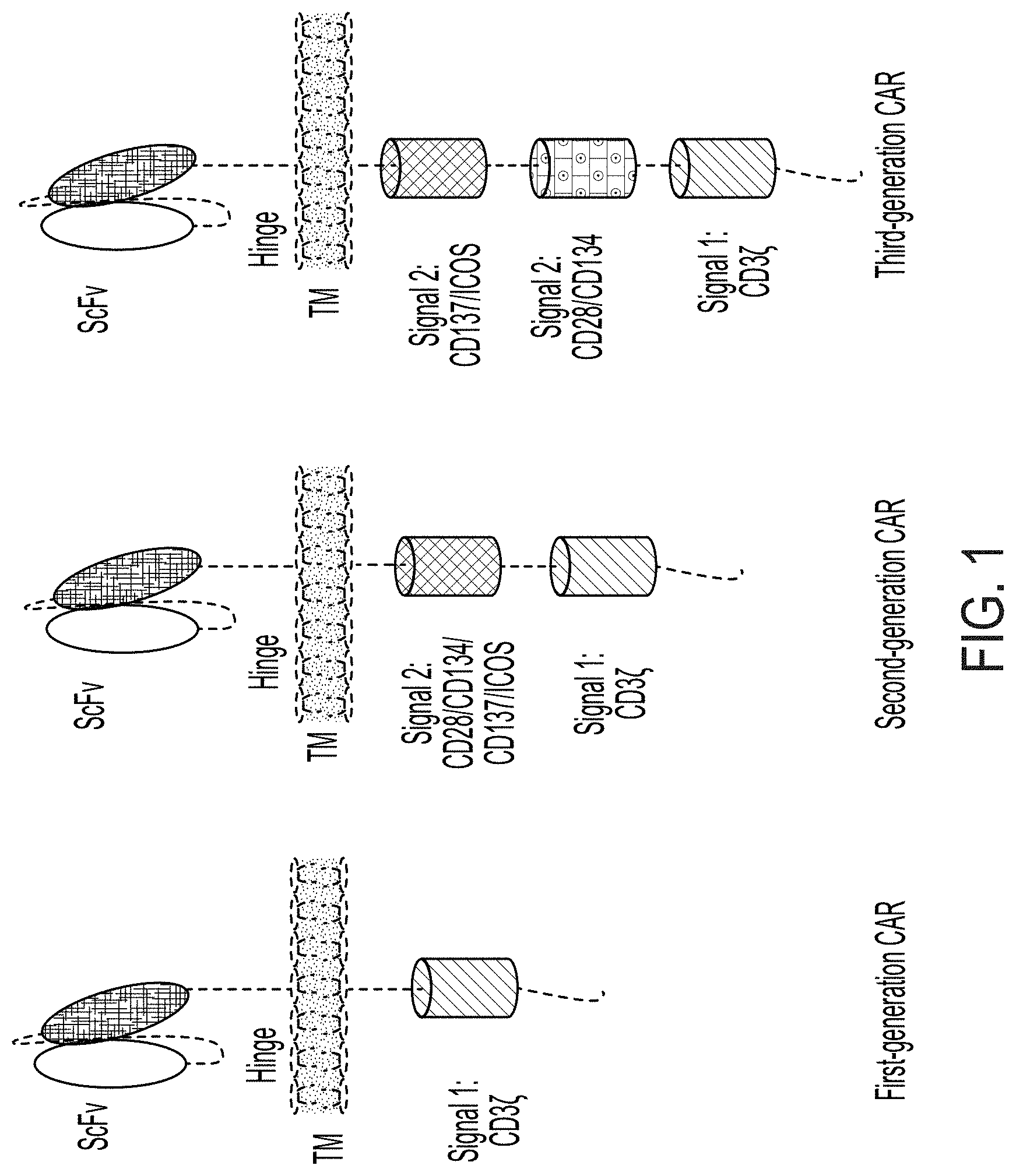
[0016] FIG. 1 is a schematic representation of the structure domains of first, second, and third-generation of CARs.
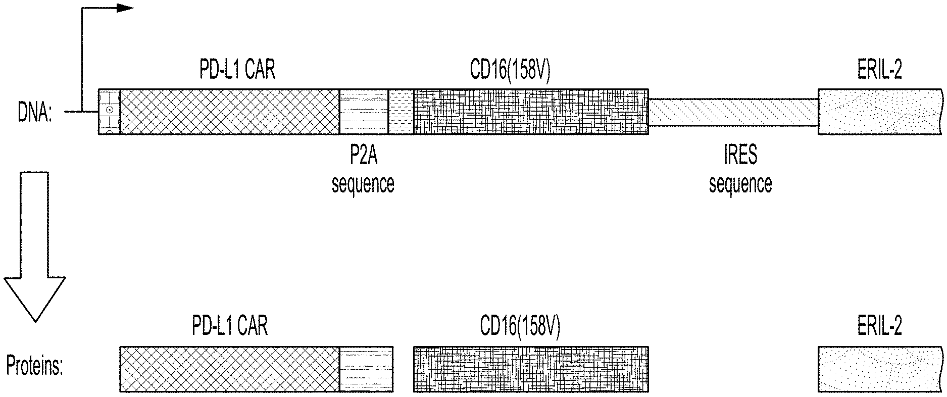
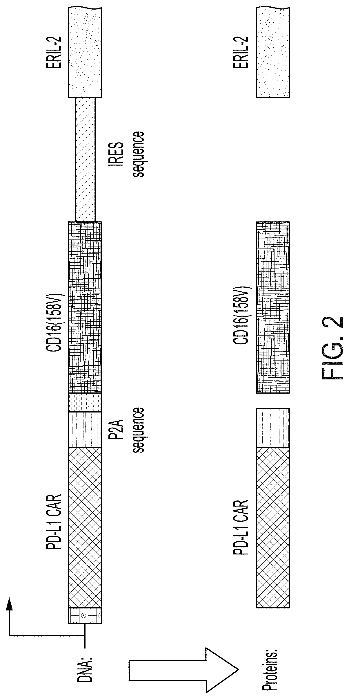
[0017] FIG. 2 shows the components of a tricistronic plasmid comprising a CAR coding sequence, a P2A sequence, a CD16 coding sequence, and an erIL-2 coding sequence.
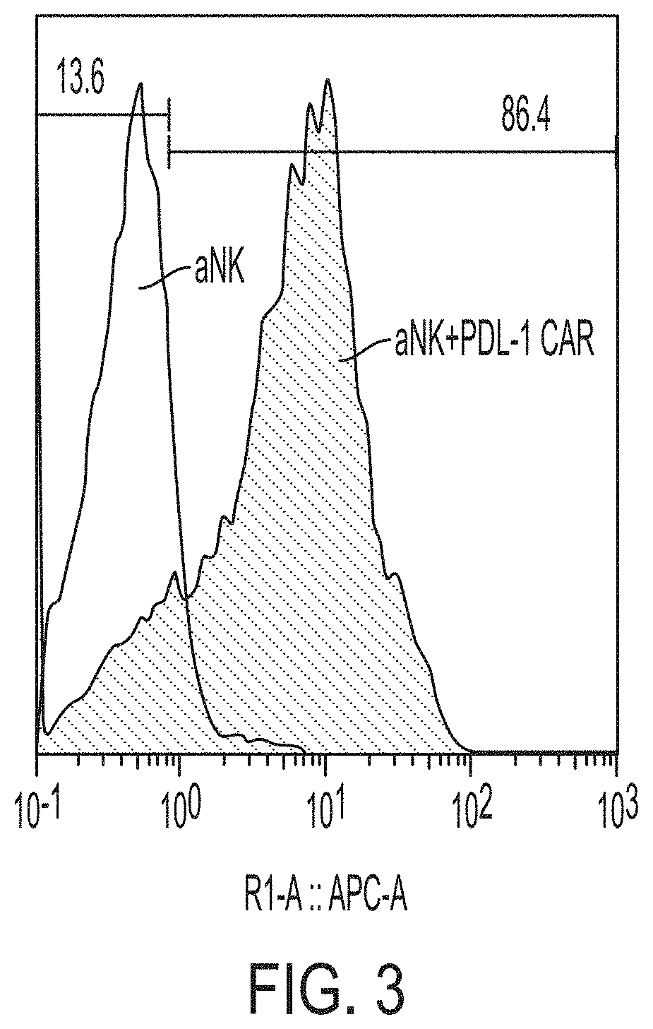
[0018] FIG. 3 shows results of flow cytometric analysis of the expression of PD-L1-CAR on modified NK-92.RTM. cells.
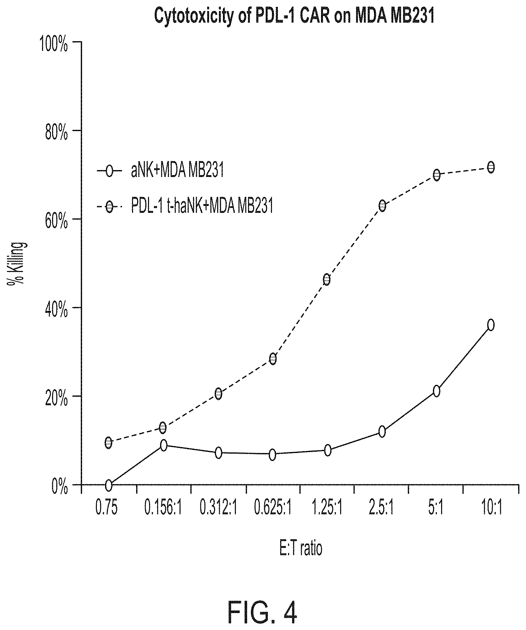
[0019] FIG. 4 shows the cytotoxic effect of PD-L1 t-haNK cells on MDA MB 231 cells. The parental aNK.TM. cells were used as control cells.
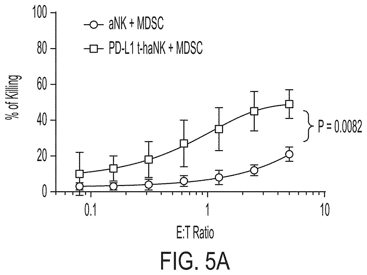
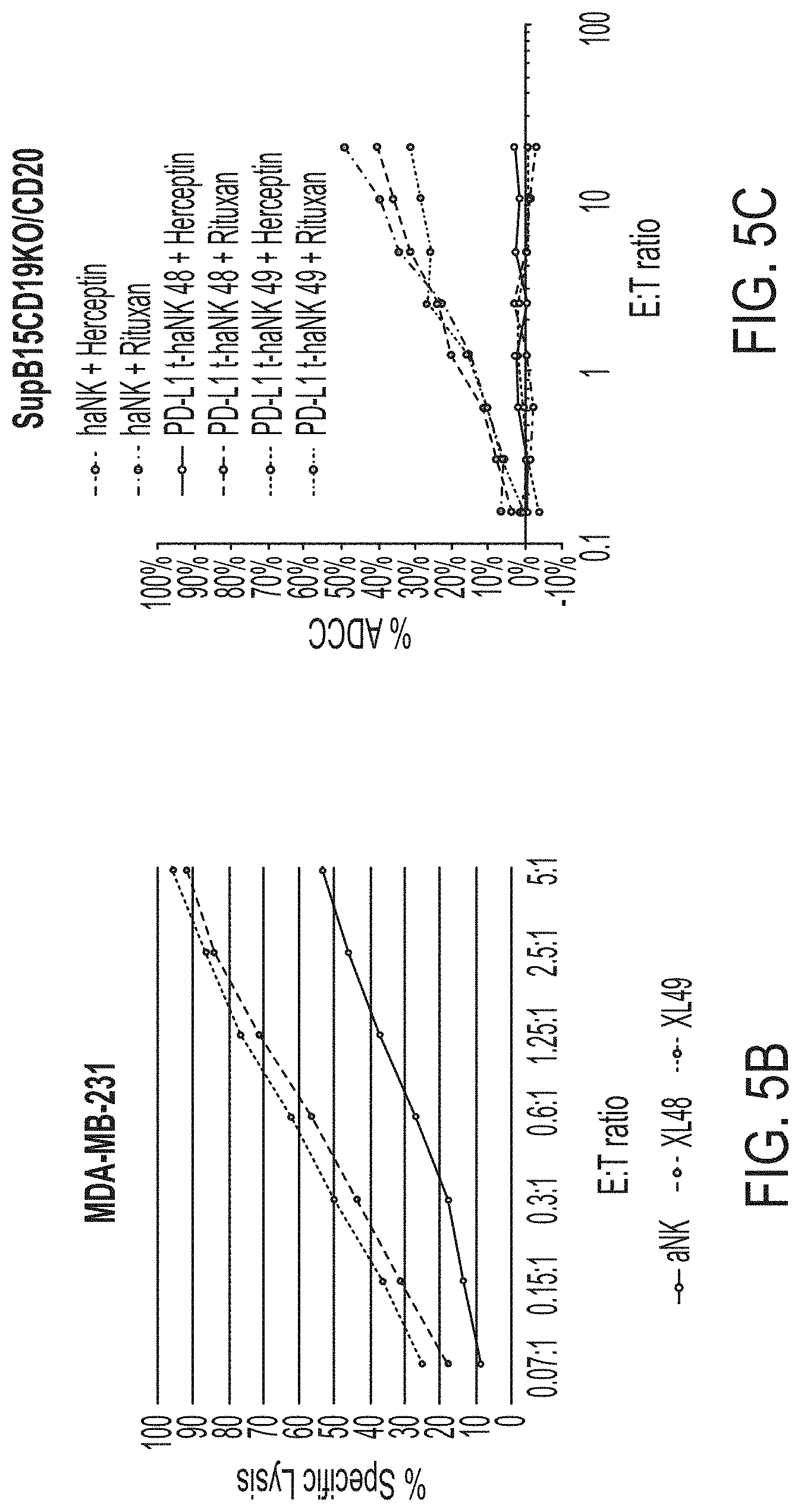
[0020] FIG. 5A shows the cytotoxic effect of PD-L1 t-haNK cells on myeloid-derived suppressor cells (MDSCs). FIG. 5B shows the cytotoxic effect of PD-L1 t-haNK cells on aNK.TM.-resistant, PD-L1-positive MDA-MB-231 cell line. XL-48 and XL-49 are two PD-L1 t-haNK populations expressing CARs comprising two different scFv domain derived from two different anti-PD-L1 antibodies. FIG. 5C shows the antibody-dependent cell-mediated cytotoxicity (ADCC) activity of PD-L1 t-haNK cells, when combined with the anti-CD20 antibody Rituximab, on engineered SUP-B15 cells. These engineered SUP-B15 cells express CD20 but not CD19. Herceptin was used as a control antibody.
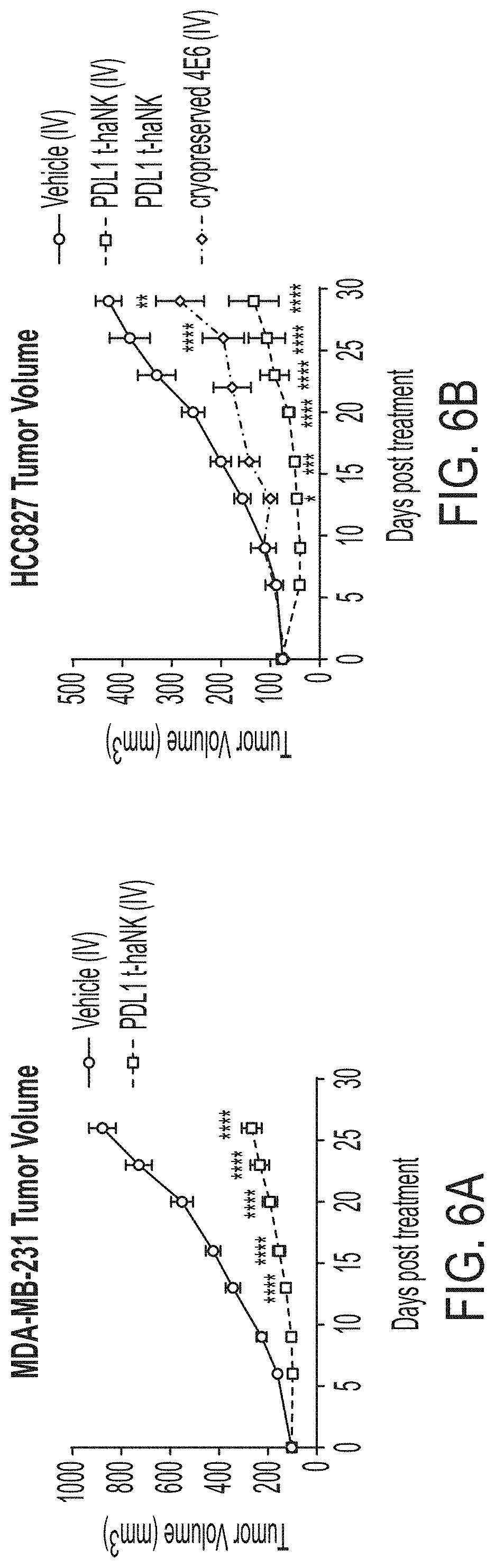
[0021] FIG. 6A shows in vivo tumor growth of MDA-MB-231 derived tumors in mice treated with vehicle and PD-L1 t-haNK cells; FIG. 6B shows in vivo tumor growth of HCC827 derived tumors in mice treated with vehicle and PD-L1 t-haNK cells using i.v. administration.
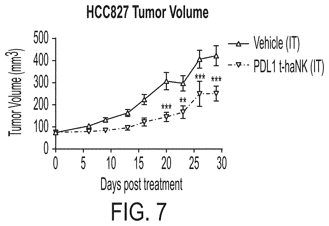
[0022] FIG. 7 shows in vivo tumor growth of HCC827 derived tumors in mice treated with vehicle and PD-L1 t-haNK cells using i.t. administration.
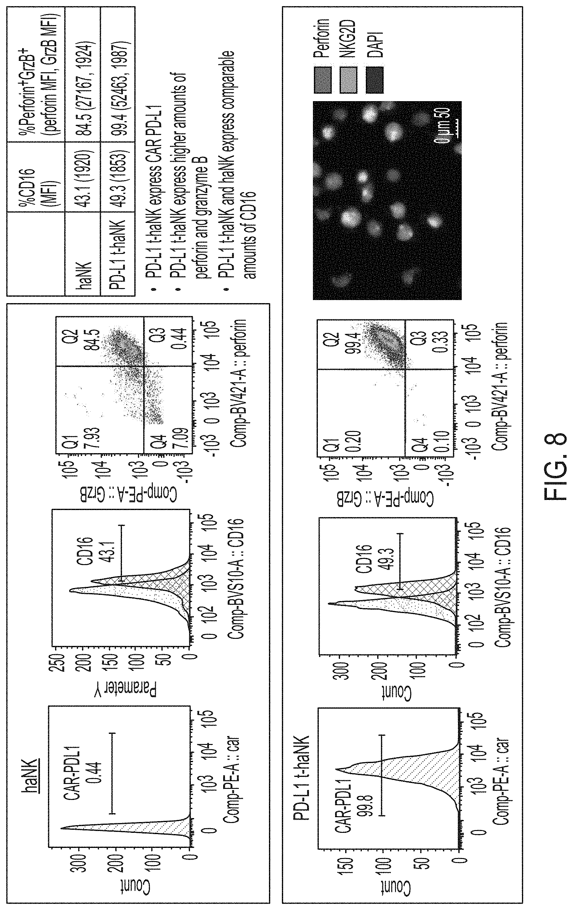
[0023] FIG. 8 shows exemplary differences between PD-L1 t-haNK cells and haNK cells.
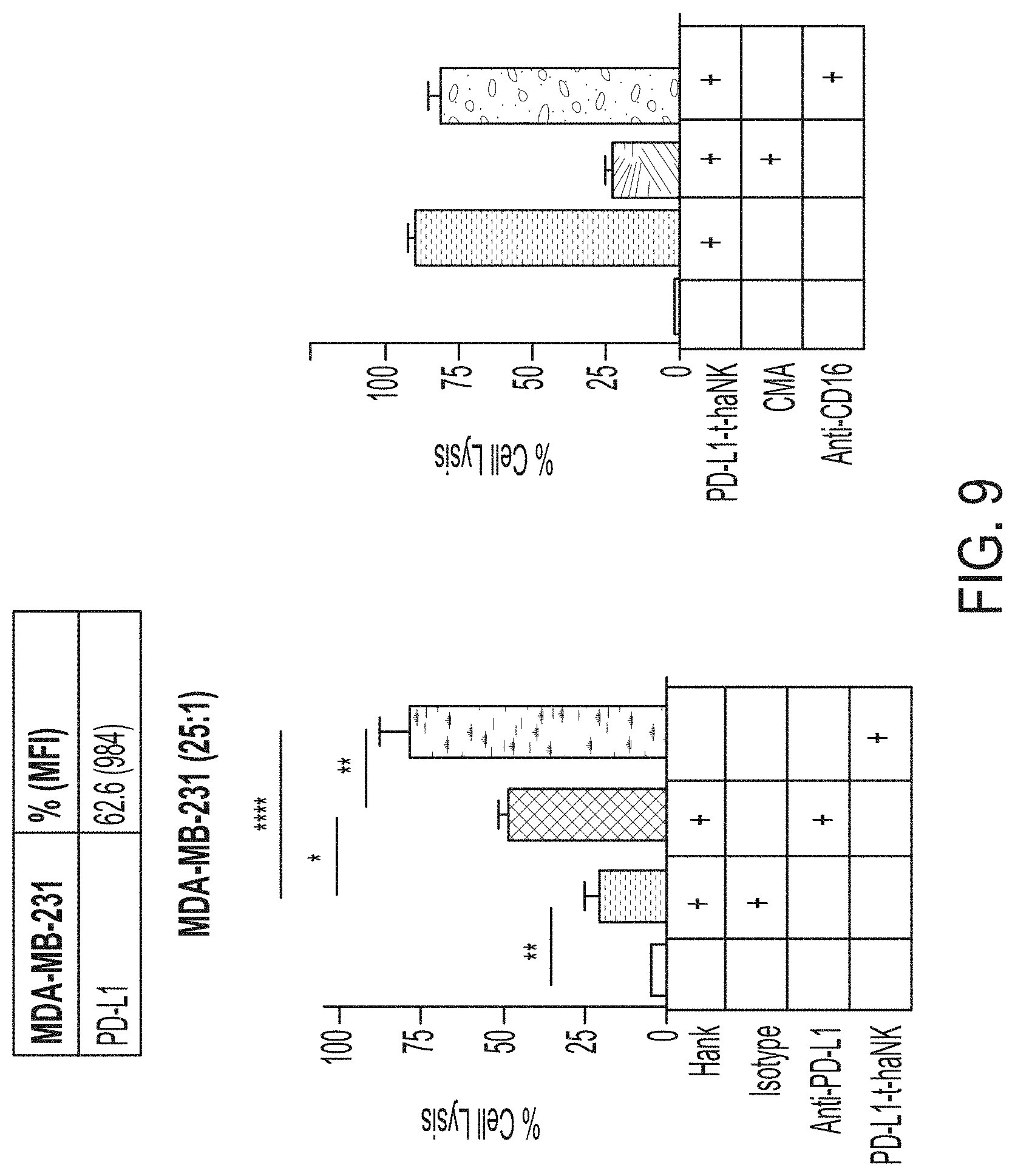
[0024] FIG. 9 shows exemplary data comparing cytotoxicity of PD-L1 t-haNK cells and haNK.RTM. cells against MDA-MB-231 cells.
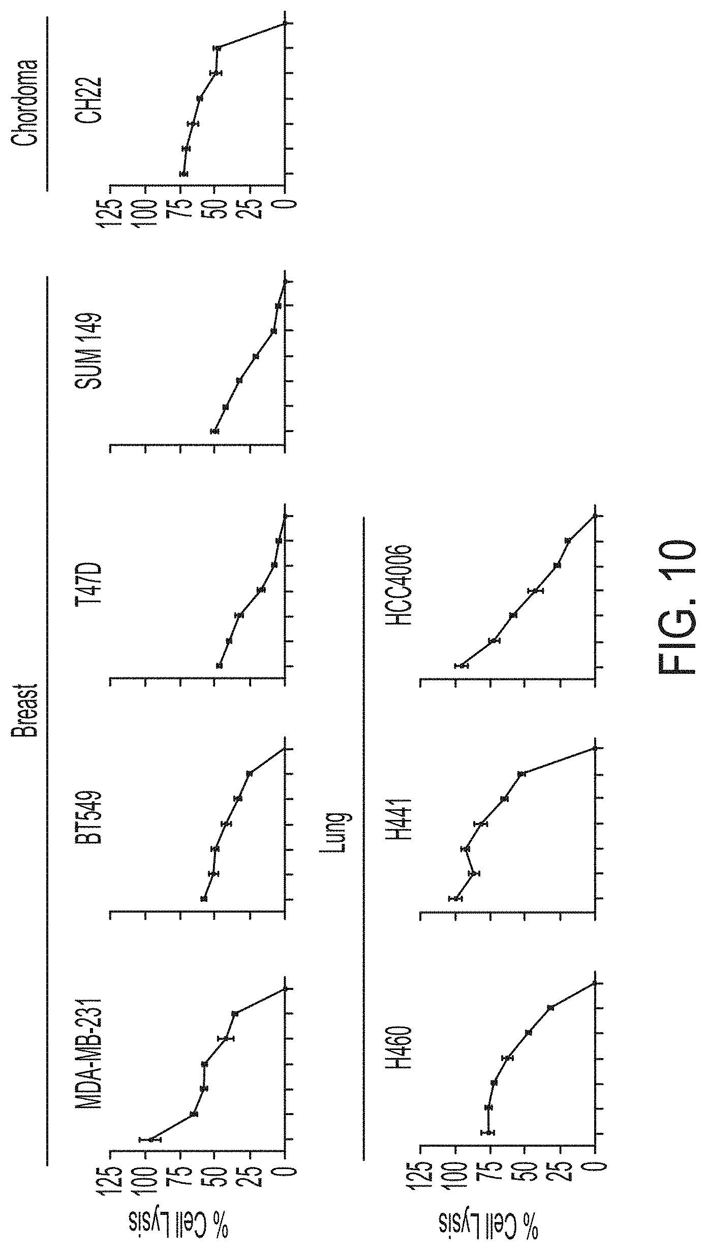
[0025] FIG. 10 shows exemplary data comparing cytotoxicity of PD-L1 t-haNK cells against various tumor cells.
[0026] FIG. 11 shows exemplary data comparing tracking of PD-L1 t-haNK cells to tumors established from MDA-MB-231 cells and PD-L1 knock-out MDA-MB-231 cells.
[0027] FIG. 12 shows exemplary data comparing tumor growth from MDA-MB-231 cells and PD-L1 knock-out MDA-MB-231 cells in animals treated with PD-L1 t-haNK cells.
[0028] FIG. 13 shows exemplary data demonstrating cytotoxicity of PD-L1 t-haNK cells against MDSCs
DETAILED DESCRIPTION
Overview
[0029] This disclosure provides NK-92.TM. cells that express a combination of a PD-L1 CAR, a Fc receptor, and an IL2. These cells can target both tumor cells and cells in the tumor microenvironment, effectively treating cancer.
Terminology
[0030] Unless defined otherwise, all technical and scientific terms used herein have the same meaning as commonly understood by one of ordinary skill in the art.
[0031] In this specification and in the claims that follow, reference will be made to a number of terms that shall be defined to have the following meanings:
[0032] The terminology used herein is for the purpose of describing particular embodiments only and is not intended to be limiting. As used herein, the singular forms "a," "an" and "the" are intended to include the plural forms as well, unless the context clearly indicates otherwise. Thus, for example, reference to "a natural killer cell" includes a plurality of natural killer cells.
[0033] All numerical designations, e.g., pH, temperature, time, concentration, amounts, and molecular weight, including ranges, are approximations which are varied (+) or (-) by increments of 0.1 or 1.0, where appropriate. It is to be understood, although not always explicitly stated, that all numerical designations may be preceded by the term "about."
[0034] As used herein, "+", when used to indicate the presence of a particular cellular marker, means that the cellular marker is detectably present in fluorescence activated cell sorting over an isotype control; or is detectable above background in quantitative or semi-quantitative RT-PCR.
[0035] As used herein, "-", when used to indicate the presence of a particular cellular marker, means that the cellular marker is not detectably present in fluorescence activated cell sorting over an isotype control; or is not detectable above background in quantitative or semi-quantitative RT-PCR.
[0036] As will be understood by one skilled in the art, for any and all purposes, particularly in terms of providing a written description, all ranges disclosed herein also encompass any and all possible subranges and combinations of subranges thereof. Any listed range can be easily recognized as sufficiently describing and enabling the same range being broken down into at least equal halves, thirds, quarters, fifths, tenths, etc. As a non-limiting example, each range discussed herein can be readily broken down into a lower third, middle third and upper third, etc. As will also be understood by one skilled in the art all language such as "up to," "at least," "greater than," "less than," and the like, include the number recited and refer to ranges which can be subsequently broken down into subranges as discussed above. Finally, as will be understood by one skilled in the art, a range includes each individual member. Thus, for example, a group having 1-3 cells refers to groups having 1, 2, or 3 cells. Similarly, a group having 1-5 cells refers to groups having 1, 2, 3, 4, or 5 cells, and so forth.
[0037] As used herein, the term "substantially the same", used interchangeably with the term "comparable", or "substantially similar", when referring to certain quantifiable properties of the NK-92.TM. cells, such as cytotoxicity, viability or cell doubling time, etc., refers to the that the two measurements of these properties are no more than 15% different, no more than 10%, no more than 8%, or no more than 5% different from each other.
[0038] It is also to be understood, although not always explicitly stated, that the reagents described herein are merely exemplary and that equivalents of such are known in the art.
[0039] For purposes of this invention and unless indicated otherwise, the term "NK-92.TM." is intended to refer to the original NK-92.TM. cell lines as well as NK-92.TM. cell lines, clones of NK-92.TM. cells, and NK-92.TM. cells that have been modified (e.g., by introduction of exogenous genes). NK-92.TM. cells and exemplary and non-limiting modifications thereof are described in U.S. Pat. Nos. 7,618,817; 8,034,332; 8,313,943; 9,181,322; 9,150,636; and published U.S. application Ser. No. 10/008,955, all of which are incorporated herein by reference in their entireties, and include wild type NK-92.TM., NK-92.TM.-CD16, NK-92.TM.-CD16-.gamma., NK-92.TM.-CD16-.zeta., NK-92.TM.-CD16(F176V), NK-92.TM. MI, and NK-92.TM. CI. NK-92.TM. cells are known to persons of ordinary skill in the art, to whom such cells are readily available from NantKwest, Inc.
[0040] As used herein, the term "NK-92.TM. cells" refers to natural killer cells derived from the highly potent unique cell line described in Gong et al. (Leukemia, April; 8(4): 652-8 (1994)), rights to which are owned by NantKwest (hereafter, "NK-92.TM. cells")
[0041] As used herein, the term "aNK.TM. cells" refers to unmodified natural killer cells derived from the highly potent unique cell line described in Gong et al. (Leukemia, April; 8(4): 652-8 (1994)), rights to which are owned by NantKwest (hereafter, "aNK.TM. cells")
[0042] As used herein, the term "haNK.RTM. cells" refers to natural killer cells derived from the highly potent unique cell line described in Gong et al. (Leukemia, April; 8(4): 652-8 (1994)), rights to which are owned by NantKwest, modified to express CD16 on the cell surface (hereafter, "CD16+NK-92.TM. cells" or "haNK.RTM. cells")
[0043] As used herein, the term "taNK.RTM. cells" refers to natural killer cells derived from the highly potent unique cell line described in Gong et al. (Leukemia, April; 8(4): 652-8 (1994)), rights to which are owned by NantKwest, modified to express a chimeric antigen receptor (hereafter, "CAR-modified NK-92.TM. cells" or "taNK.RTM. cells")
[0044] As used herein, the term "t-haNK.TM." cells refers to natural killer cells derived from the highly potent unique cell line described in Gong et al. (Leukemia, April; 8(4): 652-8 (1994)), which are owned by NantkWest, modified to express CD16 on the cell surface and to express a chimeric antigen receptor (hereafter, "CAR-modified CD16+NK-92.TM. cells" or "t-haNK cells"). In some embodiments, the tumor specific antigen is PD-L1, and these NK-92.TM. cells are referred to as PD-L1 t-haNK cells.
[0045] As used herein, the term "multi-cistronic construct," refers to a recombinant DNA construct that is to be transcribed into a single mRNA molecule and the single mRNA molecule encodes two or more transgenes. The multi-cistronic construct is referred to as bicistronic construct if it encodes two transgenes, and tricistronic construct if it encodes three genes, and quadrocistronic construct if it encodes four genes, and so on.
[0046] The term "chimeric antigen receptor" (CAR), as used herein, refers to an extracellular antigen-binding domain that is fused to an intracellular signaling domain. CARs can be expressed in T cells or NK cells to increase cytotoxicity. In general, the extracellular antigen-binding domain is a scFv that is specific for an antigen found on a cell of interest. A CAR-expressing NK-92.TM. cell is targeted to cells expressing certain antigens on the cell surface, based on the specificity of the scFv domain. The scFv domain can be engineered to recognize any antigen, including tumor-specific antigens and virus-specific antigens. For example, PD-L1 CAR recognizes PD-L1, a cell surface marker expressed by some cancers.
[0047] The term "tumor-specific antigen" as used herein refers to antigens that are present on a cancer or neoplastic cell but not detectable on a normal cell derived from the same tissue or lineage as the cancer cell. Tumor-specific antigens, as used herein, also refers to tumor-associated antigens, that is, antigens that are expressed at a higher level on a cancer cell as compared to a normal cell derived from the same tissue or lineage as the cancer cell.
[0048] As used herein, the term "target," when referring to targeting of a tumor, refers to the ability of NK-92.TM. cells to recognize and kill a tumor cell (i.e., target cell). The term "targeted" in this context refers, for example, to the ability of a CAR expressed by the NK-92.TM. cell to recognize and bind to a cell surface antigen expressed by the tumor.
[0049] The term "antibody" refers to an intact immunoglobulin of any isotype, or a fragment thereof that can compete with the intact antibody for specific binding to the target antigen, and includes chimeric, humanized, fully human, and bispecific antibodies. An intact antibody generally comprises at least two full-length heavy chains and two full-length light chains, but in some instances can include fewer chains such as antibodies naturally occurring in camelids which can comprise only heavy chains. Antibodies can be derived solely from a single source, or can be "chimeric," such that different portions of the antibody are derived from two different antibodies. The antigen binding proteins, antibodies, or binding fragments can be produced in hybridomas, by recombinant DNA techniques, or by enzymatic or chemical cleavage of intact antibodies. Unless otherwise indicated, the term "antibody" includes, in addition to antibodies comprising two full-length heavy chains and two full-length light chains, derivatives, variants, fragments, and muteins thereof. Furthermore, unless explicitly excluded, antibodies include monoclonal antibodies, bispecific antibodies, minibodies, domain antibodies, synthetic antibodies (sometimes referred to herein as "antibody mimetics"), chimeric antibodies, humanized antibodies, human antibodies, antibody fusions (sometimes referred to herein as "antibody conjugates"), and fragments thereof, respectively. In some embodiments, the term also includes peptibodies.
[0050] The term "subject` refers to a non-human animal, including mammals, such as cats, dogs, cows, horses, pigs, sheep, and goats, and humans. The term subject also refers to a patient in need of treatment for a disease described herein.
[0051] "Optional" or "optionally" means that the subsequently described event or circumstance can or cannot occur, and that the description includes instances where the event or circumstance occurs and instances where it does not.
[0052] The term "comprising" is intended to mean that the compositions and methods include the recited elements, but not excluding others. "Consisting essentially of," when used to define compositions and methods, shall mean excluding other elements of any essential significance to the combination. For example, a composition consisting essentially of the elements as defined herein would not exclude other elements that do not materially affect the basic and novel characteristic(s) of the claims. "Consisting of" means excluding more than trace amount of other ingredients and substantial method steps. Embodiments defined by each of these transition terms are within the scope of the disclosure.
[0053] As used herein, the terms "cytotoxic" and "cytolytic", when used to describe the activity of effector cells such as NK cells, are intended to be synonymous. In general, cytotoxic activity relates to killing of target cells by any of a variety of biological, biochemical, or biophysical mechanisms. Cytolysis refers more specifically to activity in which the effector lyses the plasma membrane of the target cell, thereby destroying its physical integrity. This results in the killing of the target cell. Without wishing to be bound by theory, it is believed that the cytotoxic effect of NK cells is due to cytolysis.
[0054] The term "kill" with respect to a cell/cell population is directed to include any type of manipulation that will lead to the death of that cell/cell population.
[0055] The term "cytokine" or "cytokines" refers to the general class of biological molecules which effect cells of the immune system. Exemplary cytokines include but are not limited to FLT3 ligand, interferons and interleukins (IL), in particular IL-2, IL-12, IL-15, IL-18 and IL-21.
[0056] The terms "patient," "subject," "individual," and the like are used interchangeably herein, and refer to any animal, or cells thereof whether in vitro or in situ, amenable to the methods described herein. In certain non-limiting embodiments, the patient, subject or individual is a human.
[0057] The term "treating" or "treatment" covers the treatment of a disease or disorder described herein, in a subject, such as a human, and includes: (i) inhibiting a disease or disorder, i.e., arresting its development; (ii) relieving a disease or disorder, i.e., causing regression of the disorder; (iii) slowing progression of the disorder; and/or (iv) inhibiting, relieving, or slowing progression of one or more symptoms of the disease or disorder. The term "administering" or "administration" of a monoclonal antibody or a natural killer cell to a subject includes any route of introducing or delivering the antibody or cells to perform the intended function. Administration can be carried out by any route suitable for the delivery of the cells or monoclonal antibody. Thus, delivery routes can include intravenous, intramuscular, intraperitoneal, or subcutaneous delivery. In some embodiments the modified NK-92.TM. cells are administered directly to the tumor, e.g., by injection into the tumor. In some embodiments the modified NK-92.TM. cells described herein are administered parenterally, e.g., by injection, infusion or implantation (subcutaneous, intravenous, intramuscular, intravesicularly, or intraperitoneal).
[0058] The term "expression" refers to the production of a gene product.
[0059] As used herein, the terms "cytotoxic" when used to describe the activity of effector cells such as NK cells, relates to killing of target cells by any of a variety of biological, biochemical, or biophysical mechanisms.
[0060] The terms "decrease," "reduced," "reduction," and "decrease" are all used herein to refer to a decrease by at least 10% as compared to a reference level, for example a decrease by at least about 20%, or at least about 30%, or at least about 40%, or at least about 50%, or at least about 60%, or at least about 70%, or at least about 80%, or at least about 90% or up to and including a 100% decrease (i.e. absent level as compared to a reference sample), or any decrease between 10-100% as compared to a reference level.
[0061] The term "cancer" refers to all types of cancer, neoplasm, or malignant tumors found in mammals, including leukemia, carcinomas and sarcomas. Exemplary cancers include cancer of the brain, breast, cervix, colon, head & neck, liver, kidney, lung, non-small cell lung, melanoma, mesothelioma, ovary, sarcoma, stomach, uterus and medulloblastoma. Additional examples include, Hodgkin's Disease, Non-Hodgkin's Lymphoma, multiple myeloma, neuroblastoma, ovarian cancer, rhabdomyosarcoma, primary thrombocytosis, primary macroglobulinemia, primary brain tumors, cancer, malignant pancreatic insulanoma, malignant carcinoid, urinary bladder cancer, premalignant skin lesions, testicular cancer, lymphomas, thyroid cancer, neuroblastoma, esophageal cancer, genitourinary tract cancer, malignant hypercalcemia, endometrial cancer, adrenal cortical cancer, neoplasms of the endocrine and exocrine pancreas, and prostate cancer.
[0062] The term "therapeutically effective amount" or "effective amount" refers to the amount required to ameliorate the symptoms of a disease relative to an untreated patient. The effective amount of active compound(s) used to practice the present disclosure for therapeutic treatment of a disease varies depending upon the manner of administration, the age, body weight, and general health of the subject. Ultimately, the attending physician or veterinarian will decide the appropriate amount and dosage regimen. Such amount is referred to as an "effective" amount.
[0063] The term "tumor microenvironment" refers to a cellular environment in which the tumor exists, including surrounding blood vessels, immune cells, fibroblasts, bone marrow-derived inflammatory cells, signaling molecules and the extracellular matrix. Exemplary types of cells in tumor microenvironment include, but are not limited to, myeloid derived suppressor cells (MDSC) and tumor associated macrophages (TAMs).
[0064] The term "immune cells" refers to cells of hematopoietic origin that are involved in the specific recognition of antigens Immune cells include antigen presenting cells (APCs), such as dendritic cells or macrophages, B cells, T cells, natural killer cells, myeloid derived suppressor cells (MDSC), myeloid cells, such as monocytes, macrophages, eosinophils, mast cells, basophils, and granulocytes.
[0065] Titles or subtitles may be used in the specification for the convenience of a reader, which are not intended to influence the scope of the present disclosure. Additionally, some terms used in this specification are more specifically defined below.
MDSCS
[0066] The myeloid derived suppressor cells (MDSC), are one of the main suppressor cells in the tumor microenvironment. The microenvironment of tumors prevents immune active cells such as NK cells to interact with tumor cells, attack and kill them. These negative paralyzing effects can be mediated by metabolites and secretory product of suppressor cells that are present in the tumor microenvironment.
[0067] MDSCs are regulators of immune responses in cancer and other pathological conditions, such as myelodysplastic syndrome (MDS) (See, e.g., Bronte et al., Nature Communications, 6 Jul. 2016, 7:12150, DOI: 10.1038/ncomms12150; Eksioglu et al., "Novel Therapeutic Approach to Improve Hematopoiesis in low risk MDS by Targeting myeloid-derived suppressor cells with The Fc-engineered CD33 Antibody BI 836858," Leukemia. 2017 October; 31(10): 2172-2180. doi:10.1038/leu.2017.21). Myeloid-derived suppressor cells are a heterogenous group of immune cells from the myeloid lineage, such as early myeloid progenitors, immature granulocytes, macrophages and dendritic cells at different stages of differentiation. Myeloid-derived suppressor cells strongly expand in pathological situations such as chronic infections and cancer, as a result of an altered haematopoiesis (see, e.g., Eksioglu et al., "Novel Therapeutic Approach to Improve Hematopoiesis in low risk MDS by Targeting myeloid-derived suppressor cells with The Fc-engineered CD33 Antibody BI 836858," Leukemia. 2017 October; 31(10): 2172-2180. doi:10.1038/leu.2017.21).
[0068] Myeloid-derived suppressor cells are discriminated from other myeloid cell types in which they possess strong immunosuppressive activities rather than immunostimulatory properties. Similar to other myeloid cells, myeloid-derived suppressor cells interact with other immune cell types including T cells, dendritic cells, macrophages and natural killer cells to regulate their functions. Myeloid-derived suppressor cells can suppress both the cytotoxic activities of natural killer (NK) cells and NKT cells, and the adaptive immune response mediated by CD4+ and CD8+ T cells. Although their mechanisms of action are not well understood, clinical and experimental evidence has shown that cancer tissues with high infiltration of myeloid-derived suppressor cells are associated with poor patient prognosis and resistance to therapies.
[0069] Accumulation of MDSC in the peripheral circulation has been related to extent of disease, and correlates with stage. MDSC have primarily been implicated in promoting tumor growth by suppressing antitumor immunity. There is also compelling evidence MDSC are also involved in angiogenesis and metastatic spread.
[0070] Two main subsets of MDSC have been identified in cancer patients: a monocytic subset, characterized by expression of CD14, and a granulocytic subset characterized by expression of CD15. Both subsets of MDSC actively suppress host immunity through a variety of mechanisms including production of reactive oxygen species and arginase. Just as in humans, accumulation of monocytic and granulocytic MDSC has been noted in the bone marrow, spleen, peripheral circulation, and tumors of tumor bearing mice. Successful targeting of MDSC in mice is associated with improved immune responses, delayed tumor growth, improved survival, and increased efficacy of vaccine therapy. In the tumor monocytic derived MDSC rapidly differentiate to tumor associated macrophages (TAM).
Tumor Associated Macrophages
[0071] Tumors are often associated with an immune infiltrate as part of the reactive stroma that is enriched for macrophages. Typically macrophages are categorized into M1 and M2 macrophages, which have opposing effects on tumor growth: M1 macrophages inhibit tumor cell growth while M2 macrophages promote tumor development. Tumor cells coax macrophages to M2-like phenotype via chemokine and polarizing cytokines, aiding their escape from destruction, promoting their development. These M2 macrophages are commonly referred to as tumor-associated macrophages (TAMs). The TAMs reside in the tumor microenvironment and play an important role in facilitating tumor growth by promoting neovascularization and matrix degradation. Consequently, many tumors with a high number of TAMs have an increased tumor growth rate, local proliferation and distant metastasis.
[0072] TAMs express CD68 as well as other markers, for example, some TAMs express one or more of the following markers CD206, CD204, or CD163.
NK-92.TM. Cells
[0073] NK-92.TM. is a cytolytic cancer cell line which was discovered in the blood of a subject suffering from a non-Hodgkins lymphoma and then immortalized in vitro. NK-92.TM. cells are derived from NK cells, but lack the major inhibitory receptors that are displayed by normal NK cells, while retaining the majority of the activating receptors. NK-92.TM. cells do not, however, attack normal cells nor do they elicit an unacceptable immune rejection response in humans. Characterization of the NK-92.TM. cell line is disclosed in WO 1998/049268 and U.S. Patent Application Publication No. 2002-0068044. NK-92.TM. cells have been evaluated as a therapeutic agent in the treatment of certain cancers.
Vectors
[0074] Described herein are vectors for transfecting cells to produce the modified cells described herein. In one embodiment, the vectors described herein are transient expression vectors. Exogenous transgenes introduced using such vectors are not integrated in the nuclear genome of the cell; therefore, in the absence of vector replication, the foreign transgenes will be degraded or diluted over time.
[0075] In one embodiment, the vectors described herein allow for stable transfection of cells. In one embodiment, the vector allows incorporation of the transgene(s) into the genome of the cell. In one embodiment, the vectors have a positive selection marker. Positive selection markers include any genes that allow the cell to grow under conditions that would kill a cell not expressing the gene. Non-limiting examples include antibiotic resistance, e.g. geneticin (Neo gene from Tn5).
[0076] In one embodiment, the vector is a plasmid vector. In one embodiment, the vector is a viral vector. As would be understood by one of skill in the art, any suitable vector can be used. Suitable vectors are well-known in the art.
[0077] In some embodiments, the cells are transfected with mRNA encoding the protein of interest (e.g., a CAR). Transfection of mRNA results in transient expression of the protein. In one embodiment, transfection of mRNA into NK-92.TM. cells is performed immediately prior to administration of the cells. In one embodiment, "immediately prior" to administration of the cells refers to between about 15 minutes and about 48 hours prior to administration. Preferably, mRNA transfection is performed about 5 hours to about 24 hours prior to administration.
PD-L1
[0078] Programmed death-ligand (PD-L1) is an inhibitory ligand that binds to PD-1 to suppress T cell activation. PD-L1 is constitutively expressed and induced in tumor cells. PD-L1 is also expressed in MDSCs. It has been reported that the number of PD-L1-expressing MDSCs increased significantly in tumor-bearing mice as compared to tumor free mice, and that PD-L1 expression is significantly higher in tumor-infiltrating MDSCs as compared to those in lymphoid organs. See, Lu et al., J. Immunol., May 1, 2017, 198 (1 Supplement) 124.9. PD-L1 is also expressed in tumor-associated macrophages (TAMs) and that TAM expression of PD-L1 can directly induce T cell apoptosis after binding its receptor. Kuang et al., J. Exp. Med. 2009; 206:1327-1337.
CARs
[0079] Phenotypic changes distinguishing a tumor cell from normal cells derived from the same tissue are often associated with one or more changes in the expression of specific gene products, including the loss of normal cell surface components or the gain of others (i.e., antigens not detectable in corresponding normal, non-cancerous tissue). The antigens which are expressed in neoplastic or tumor cells, but not in normal cells, or which are expressed in neoplastic cells at levels substantially above those found in normal cells, have been termed "tumor-specific antigens" or "tumor-associated antigens." Tumor-specific antigens have been used as targets for cancer immunotherapies. One such therapy utilizes chimeric antigen receptors (CARs) expressed on the surface of immune cells, including T cells and NK cells, to improve cytotoxicity against cancer cells. CARs comprise a single-chain variable fragment (scFv) linked to at least one intracellular signaling domain. The scFv recognizes and binds an antigen on the target cell (e.g., a cancer cell) and triggers effector cell activation. The signaling domains contain immunoreceptor tyrosine-based activation domains (ITAMs) that are important for intracellular signaling by the receptor.
[0080] The present disclosure provides NK-92.TM. cells that have been engineered to express at least a chimeric antigen receptor (CAR) on the cell surface. CARs combine an extracellular antigen-recognizing part (usually derived from the variable domain of a specific antibody to an intracellular signaling domain (either single or with additional co-stimulatory elements) that can trigger a cytolytic response once a specific antigen is recognized. There are multiple types of CARs, which all can be used in the application. The first generation of CARs contains one cytoplasmic signaling domain. The signaling domain can be from e.g., the Fc epsilon receptor gamma (Fc.epsilon.RI.gamma.) which contains one ITAM, or from CD3.zeta., which contains three ITAMs. It is believed that CD3.zeta. CARs are more efficient at tumor eradication than Fc.epsilon.RI.gamma. CARs. See, e.g., Haynes, et al. 2001, J. Immunology 166:182-187; Cartellieri, et al. 2010, J. Biomed and Biotech, Vol. 2010, Article ID 956304. The second and third generation CARs combine multiple signaling domains, e.g., the cytoplasmic signaling domain of CD3.zeta. and costimulatory signaling domains, such as CD28/CD134/CD137/ICOS and CD28/CD134 to a single CAR to promote the activation and proliferation of the NK-92.TM. cells. Thus, in some embodiments, the PD-L1 CAR expressed by the PD-L1 t-haNK cells comprises a hinge region from CD8, and/or a transmembrane domain of CD28. In some embodiments, the PD-L1 CAR comprises a cytoplasmic signaling domain of Fc.epsilon.RI.gamma.. In some embodiments, the PD-L1 CAR comprises the cytoplasmic signaling domain of CD3.zeta.. Examples of the hinge region, the transmembrane domain of CD28 and the cytoplasmic signaling domain of Fc.epsilon.RI.gamma. or CD3.zeta. are disclosed in U.S. Provisional application No. 62/674,936, the entire content of which is herein incorporated by reference. While prior publications such as Haynes, et al. 2001, J. Immunology 166:182-187 and Cartellieri, et al. 2010, J. Biomed and Biotech, Vol. 2010, Article ID 956304, had disclosed that CD3.zeta. CARs may be more efficient at tumor eradication than Fc.epsilon.RI.gamma. CARs, in this case, the inventors have surprisingly and unexpectedly found that such is not the case for the cells, compositions, and methods disclosed herein. In fact, the inventors found that NK-92 cells expressing a first-generation CAR comprising an intracellular domain from Fc.epsilon.RI.gamma., which has only one ITAM domain, have equal or higher cytotoxic activity against cancer cells expressing the antigen recognized by the CAR than NK-92 cells expressing CARs with a CD3.zeta. signaling domain, which has three ITAM domains, even where these ITAM domains were combined with other signaling domains (i.e., second or third generation CARs; data not shown here). Exemplary CARs are schematically illustrated in FIG. 1. Notably, the IgE receptor (Fc.epsilon.RI) in its native context includes two gamma chains coupled to each other via a disulfide bond and is normally expressed only in eosinophils, basophils, and epidermal Langerhans cells. The inventors also made the unexpected finding that a CAR comprising an intracellular domain from Fc.epsilon.RI.gamma. was expressed at higher levels on the surface of NK-92 cells than other CARs, especially those comprising the CD3.zeta. signaling domain.
[0081] Optionally, the CAR is specific for PD-L1. In some embodiments, PD-L1 is a human PD-L1. In some embodiments, the PD-L1 CAR comprises an amino acid sequence set forth as SEQ ID NO: 10. In some embodiments, the PD-L1 CAR has an amino acid sequence of SEQ ID NO: 14.
[0082] In some embodiments, the PD-L1 CAR polypeptide comprises a sequence that shares at least 90, 91, 92, 93, 94, 95, 96, 97, 98, or 99% identity to SEQ ID NO:10 or the CDR sequence portions within SEQ ID NO:10. In some embodiments, an epitope tag peptide, such as FLAG, myc, polyhistidine, or V5 can be added to the amino terminal domain of the polypeptide to assist in cell surface detection by using anti-epitope tag peptide monoclonal or polyclonal antibodies.
[0083] In examples, variant polypeptides are made using methods known in the art such as oligonucleotide-mediated (site-directed) mutagenesis, alanine scanning, and PCR mutagenesis. Site direct mutagenesis (Carter, 1986; Zoller and Smith, 1987), cassette mutagenesis, restriction selection mutagenesis (Wells et al., 1985) or other known techniques can be performed on the cloned DNA to produce CD16 variants (Ausubel, 2002; Sambrook and Russell, 2001).
[0084] In some embodiments, a polynucleotide encoding a PD-L1 CAR is mutated to alter the amino acid sequence encoding for CAR without altering the function of the CAR. For example, polynucleotide substitutions leading to amino acid substitutions at "non-essential" amino acid residues can be made in SEQ ID NO:9, which is a codon-optimized sequence encoding the scFv portion of the PD-L1 CAR.
[0085] Conservative substitutions in SEQ ID NO:9 whereby an amino acid of one class is replaced with another amino acid of the same class, fall within the scope of the disclosed variants as long as the substitution does not materially alter the activity of the polypeptide. Conservative substitutions are well known to one of skill in the art. Non-conservative substitutions that affect (1) the structure of the polypeptide backbone, such as a .beta.-sheet or .alpha.-helical conformation, (2) the charge, (3) the hydrophobicity, or (4) the bulk of the side chain of the target site can modify polypeptide function or immunological identity. Non-conservative substitutions entail exchanging a member of one of these classes for another class. Substitutions may be introduced into conservative substitution sites or more preferably into non-conserved sites.
[0086] In examples, variant polypeptides are produced using methods known in the art such as oligonucleotide-mediated (site-directed) mutagenesis, alanine scanning, and PCR mutagenesis. Site direct mutagenesis (Carter, 1986; Zoller and Smith, 1987), cassette mutagenesis, restriction selection mutagenesis (Wells et al., 1985) or other known techniques can be performed on the cloned DNA to produce variants (Ausubel, 2002; Sambrook and Russell, 2001).
[0087] Optionally, the PD-L1 t-haNK cells can be used to treat cancer, in particular, a cancer that express PD-L1. Optionally, the cancer is selected from the group consisting of leukemia (including acute leukemias (e.g., acute lymphocytic leukemia, acute myelocytic leukemia (including myeloblastic, promyelocytic, myelomonocytic, monocytic, and erythroleukemia)) and chronic leukemias (e.g., chronic myelocytic (granulocytic) leukemia and chronic lymphocytic leukemia), polycythemia vera, lymphomas (e.g., Hodgkin's disease and non-Hodgkin's disease), multiple myeloma, Waldenstrom's macroglobulinemia, heavy chain disease, solid tumors including, but not limited to, sarcomas and carcinomas such as fibrosarcoma, myxosarcoma, liposarcoma, chondrosarcoma, osteogenic sarcoma, chordoma, angiosarcoma, endotheliosarcoma, lymphangiosarcoma, lymphangioendotheliosarcoma, synovioma, mesothelioma, Ewing's tumor, leiomyosarcoma, rhabdomyosarcoma, colon carcinoma, pancreatic cancer, breast cancer, ovarian cancer, prostate cancer, squamous cell carcinoma, basal cell carcinoma, adenocarcinoma, sweat gland carcinoma, sebaceous gland carcinoma, papillary carcinoma, papillary adenocarcinomas, cystadenocarcinoma, medullary carcinoma, bronchogenic carcinoma, renal cell carcinoma, hepatoma, bile duct carcinoma, choriocarcinoma, seminoma, embryonal carcinoma, Wilm's tumor, cervical cancer, testicular tumor, lung carcinoma, small cell lung carcinoma, bladder carcinoma, epithelial carcinoma, glioma, astrocytoma, medulloblastoma, craniopharyngioma, ependymoma, pinealoma, hemangioblastoma, acoustic neuroma, oligodendroglioma, menangioma, melanoma, neuroblastoma and retinoblastoma.
Fc Receptors
[0088] In some embodiments, the NK-92.TM. cells are modified to express at least one Fc receptor, such that the at least one Fc receptor is displayed on the cell surface of the NK-92.TM. cell. Fc receptors bind to the Fc portion of antibodies. Several Fc receptors are known, and differ according to their preferred ligand, affinity, expression, and effect following binding to the antibody.
TABLE-US-00001 TABLE 1 Illustrative Fc receptors Principal Affinity antibody for Effect following binding to Receptor name ligand ligand Cell distribution antibody Fc.gamma.RI (CD64) IgG1 and High Macrophages Phagocytosis IgG3 (Kd~10.sup.-9M) Neutrophils Cell activation Eosinophils Activation of respiratory Dendritic cells burst Induction of microbe killing Fc.gamma.RIIA (CD32) IgG Low Macrophages Phagocytosis (Kd > 10.sup.-7M) Neutrophils Degranulation (eosinophils) Eosinophils Platelets Langerhans cells Fc.gamma.RIIB1 (CD32) IgG Low B Cells No phagocytosis (Kd > 10.sup.-7M) Mast cells Inhibition of cell activity Fc.gamma.RIIB2 (CD32) IgG Low Macrophages Phagocytosis (Kd > 10.sup.-7M) Neutrophils Inhibition of cell activity Eosinophils Fc.gamma.RIIIA (CD16a) IgG Low NK cells Induction of antibody- (Kd > 10.sup.-6M) Macrophages (certain dependent cell-mediated tissues) cytotoxicity (ADCC) Induction of cytokine release by macrophages Fc.gamma.RIIIB (CD16b) IgG Low Eosinophils Induction of microbe (Kd > 10.sup.-6M) Macrophages killing Neutrophils Mast cells Follicular dendritic cells Fc.epsilon.RI IgE High Mast cells Degranulation (Kd~10.sup.-10M) Eosinophils Phagocytosis Basophils Langerhans cells Monocytes Fc.epsilon.RII (CD23) IgE Low B cells Possible adhesion molecule (Kd > 10.sup.-7M) Eosinophils IgE transport across human Langerhans cells intestinal epithelium Positive-feedback mechanism to enhance allergic sensitization (B cells) Fc.alpha.RI (CD89) IgA Low Monocytes Phagocytosis (Kd > 10.sup.-6M) Macrophages Induction of microbe Neutrophils killing Eosinophils Fc.alpha./.mu.R IgA and IgM High for B cells Endocytosis IgM, Mesangial cells Induction of microbe Mid for IgA Macrophages killing FcRn IgG Monocytes Transfers IgG from a Macrophages mother to fetus through the Dendritic cells placenta Epithelial cells Transfers IgG from a Endothelial cells mother to infant in milk Hepatocytes Protects IgG from degradation
[0089] In some embodiments NK-92.TM. cells are modified to express an Fc receptor protein on the cell surface.
[0090] In some embodiments, the Fc receptor is CD16. For purposes of this disclosure, specific amino acid residues of CD16 are designated with reference to SEQ ID NO:2, or to SEQ ID NO:1, which differs at one position relative to SEQ ID NO:2. Thus, an amino acid residue "at position 158" of a CD16 polypeptide is the amino acid residue that corresponds to position 158 of SEQ ID NO:2 (or SEQ ID NO:1), when the CD16 polypeptide and SEQ ID NO:2 are maximally aligned. In some embodiments, NK-92.TM. cells are modified to express a human CD16 that has a phenylalanine at position 158 of the mature form of the protein, e.g., SEQ ID NO:1. In typical embodiments, NK-92.TM. cells are modified to express a high affinity form of human CD16 having a valine at position 158 of the mature form of the protein, e.g., SEQ ID NO:2. Position 158 of the mature protein corresponds to position 176 of the CD16 sequence that includes the native signal peptide. In some embodiments, a CD16 polypeptide is encoded by a polynucleotide that encodes the precursor (i.e., has a native signal peptide) polypeptide sequence of SEQ ID NO:3 or of SEQ ID NO:4. Thus, in one embodiment, the Fc receptor comprises Fc.gamma.RIII-A (CD16). In some embodiments, the NK-92.TM. cells are genetically modified to express an Fc receptor encoding a polypeptide having at least 90% sequence identity with SEQ ID NO:1 (Fc.gamma.RIII-A or CD16 having a phenylalanine at position 158 (F-158); or at least 90% identity to SEQ ID NO:2 (CD16 having a valine at position 158 (F158V), higher affinity form).
[0091] In some embodiments, a polynucleotide encoding a CD16 polypeptide has at least about 70% polynucleotide sequence identity with a polynucleotide sequence encoding a full-length, including signal peptide, naturally occurring CD16 that has a phenylalanine at position 176 of the full-length CD16 (which corresponds to position 158 of the mature CD16 protein). In some embodiments, a polynucleotide encoding a CD16 polypeptide has at least about 70% polynucleotide sequence identity with a polynucleotide sequence encoding a full-length, including the signal peptide, naturally occurring CD16 that has a valine at position 176 (which corresponds to position 158 of the mature protein). In some embodiments, a polynucleotide encoding CD16 has at least 70%, 80%, 90%, or 95% identity to SEQ ID NO:5 and comprises a codon encoding valine at the position of the polynucleotide that encodes position 176 of the full-length, including the signal peptide, CD16 polypeptide. In some embodiments, a polynucleotide encoding CD16 comprises SEQ ID NO:5, but with a codon encoding valine at position 176 of the full-length CD16.
[0092] In some embodiments, the CD16 polynucleotide encodes a polypeptide having at least 70%, 80%, 90%, or 95% identity to SEQ ID NO:1 or SEQ ID NO:2. In some embodiments, the polynucleotide encodes a polypeptide having at least 70% 80%, 90%, or 95% identity to SEQ ID NO:2 and comprises a valine at position 158 as determined with reference to SEQ ID NO:2. In some embodiments the polynucleotide encodes SEQ ID NO:2. In some embodiments, a CD16 polynucleotide encodes an extracellular domain of CD16 with or without the signal sequence, or any other fragment of a full length CD16, or a chimeric receptor encompassing at least partial sequence of CD16 fused to an amino acid sequence of another protein. In other embodiments, an epitope tag peptide, such as FLAG, myc, polyhistidine, or V5 can be added to the amino terminal domain of the mature polypeptide to assist in cell surface detection by using anti-epitope tag peptide monoclonal or polyclonal antibodies.
[0093] In some embodiments, homologous CD16 polynucleotides may be about 150 to about 700, about 750, or about 800 polynucleotides in length, although CD16 variants having more than 700 to 800 polynucleotides are within the scope of the disclosure.
[0094] Homologous polynucleotide sequences include those that encode polypeptide sequences coding for variants of CD16. Homologous polynucleotide sequences also include naturally occurring allelic variations related to SEQ ID NO:1. Transfection of an NK-92.TM. cell with any polynucleotide encoding a polypeptide having the amino acid sequence shown in either SEQ ID. NO: 1 or SEQ ID NO: 2, a naturally occurring variant thereof, or a sequence that is at least 70% identical, or at least 80%, 90%, or 95% identical to SEQ ID. NO: 1 or SEQ ID NO: 2 is within the scope of the disclosure. In some embodiments, homologous polynucleotide sequences encode conservative amino acid substitutions in SEQ ID. NO: 1 or SEQ ID NO: 2. In some embodiments, NK-92.TM. cells are transfected using a degenerate homologous CD16 polynucleotide sequence that differs from a native polynucleotide sequence, but encodes the same polypeptide.
[0095] In other examples, cDNA sequences having polymorphisms that change the CD16 amino acid sequences are used to modify the NK-92.TM. cells, such as, for example, the allelic variations among individuals that exhibit genetic polymorphisms in CD16 genes. In other examples, CD16 genes from other species that have a polynucleotide sequence that differs from the sequence of SEQ ID NO:1 are used to modify NK-92.TM. cells.
[0096] Variant polypeptides can be made using methods known in the art such as oligonucleotide-mediated (site-directed) mutagenesis, alanine scanning, and PCR mutagenesis. Site direct mutagenesis (Carter, 1986; Zoller and Smith, 1987), cassette mutagenesis, restriction selection mutagenesis (Wells et al., 1985) or other known techniques can be performed on the cloned DNA to produce CD16 variants (Ausubel, 2002; Sambrook and Russell, 2001).
[0097] In some embodiments, a polynucleotide encoding a CD16 is mutated to alter the amino acid sequence encoding for CD16 without altering the function of CD16. For example, polynucleotide substitutions leading to amino acid substitutions at "non-essential" amino acid residues can be made in SEQ ID NO:1 or SEQ ID NO:2.
[0098] Conservative substitutions in SEQ ID. NO:1 or SEQ ID NO:2, whereby an amino acid of one class is replaced with another amino acid of the same class, fall within the scope of the disclosed CD16 variants as long as the substitution does not materially alter the activity of the polypeptide. Conservative substitutions are well known to one of skill in the art. Non-conservative substitutions that affect (1) the structure of the polypeptide backbone, such as a .beta.-sheet or .alpha.-helical conformation, (2) the charge, (3) the hydrophobicity, or (4) the bulk of the side chain of the target site can modify CD16 polypeptide function or immunological identity. Non-conservative substitutions entail exchanging a member of one of these classes for another class. Substitutions may be introduced into conservative substitution sites or more preferably into non-conserved sites.
[0099] In some embodiments, CD16 polypeptide variants are at least 200 amino acids in length and have at least 70% amino acid sequence identity, or at least 80%, or at least 90% identity to SEQ ID NO:1 or SEQ ID NO:2. In some embodiments, CD16 polypeptide variants are at least 225 amino acid in length and have at least 70% amino acid sequence identity, or at least 80%, or at least 90% identity to SEQ ID NO:1 or SEQ ID NO:2. In some embodiments, CD16 polypeptide variants have a valine at position 158 as determined with reference to SEQ ID NO:2.
[0100] In some embodiments a nucleic acid encoding a CD16 polypeptide may encode a CD16 fusion protein. A CD16 fusion polypeptide includes any portion of CD16 or an entire CD16 fused with a non-CD16 polypeptide. Fusion polypeptides are conveniently created using recombinant methods. For example, a polynucleotide encoding a CD16 polypeptide such as SEQ ID NO:1 or SEQ ID NO:2 is fused in-frame with a non-CD16 encoding polynucleotide (such as a polynucleotide sequence encoding a signal peptide of a heterologous protein). In some embodiment, a fusion polypeptide may be created in which a heterologous polypeptide sequence is fused to the C-terminus of CD16 or is positioned internally in the CD16. Typically, up to about 30% of the CD16 cytoplasmic domain may be replaced. Such modification can enhance expression or enhance cytotoxicity (e.g., ADCC responsiveness). In other examples, chimeric proteins, such as domains from other lymphocyte activating receptors, including but not limited to Ig-a, Ig-B, CD3-e, CD3-d, DAP-12 and DAP-10, replace a portion of the CD16 cytoplasmic domain.
[0101] Fusion genes can be synthesized by conventional techniques, including automated DNA synthesizers and PCR amplification using anchor primers that give rise to complementary overhangs between two consecutive gene fragments that can subsequently be annealed and reamplified to generate a chimeric gene sequence (Ausubel, 2002). Many vectors are commercially available that facilitate sub-cloning CD16 in-frame to a fusion moiety.
Cytokines
[0102] The cytotoxicity of NK-92 cells is dependent on the presence of cytokines (e.g., interleukin-2 (IL-2)). The cost of using exogenously added IL-2 needed to maintain and expand NK-92 cells in commercial scale culture is significant. The administration of IL-2 to human subjects in sufficient quantity to continue activation of NK92 cells would cause adverse side effects.
[0103] In one embodiment, NK-92.TM. cells are modified to express at least one cytokine. In particular, the at least one cytokine is IL-2 (SEQ ID NO:6), IL-12, IL-15, IL-18, IL-21, or a variant thereof. In some embodiments, the cytokine is IL-2 or a variant thereof. In certain embodiments, the IL-2 is a variant that is targeted to the endoplasmic reticulum. In some embodiments, the cytokine is IL-15 or a variant thereof. In certain embodiments, the IL-15 is a variant that is targeted to the endoplasmic reticulum.
[0104] In one embodiment, the IL-2 is cloned and expressed with a signal sequence that directs the IL-2 to the endoplasmic reticulum (erIL-2) (SEQ ID NO: 7). This permits expression of IL-2 at levels sufficient for autocrine activation, but without releasing IL-2 extracellularly. See Konstantinidis et al "Targeting IL-2 to the endoplasmic reticulum confines autocrine growth stimulation to NK-92.TM. cells" Exp Hematol. 2005 February; 33(2):159-64. Continuous activation of the FcR-expressing NK-92 cells can be prevented, e.g., by the presence of the suicide gene.
Suicide Gene
[0105] The term "suicide gene" refers to a transgene that allows for the negative selection of cells expressing the suicide gene. A suicide gene is used as a safety system, allowing cells expressing the gene to be killed by introduction of a selective agent. This is desirable in case the recombinant gene causes a mutation leading to uncontrolled cell growth, or the cells themselves are capable of such growth. A number of suicide gene systems have been identified, including the herpes simplex virus thymidine kinase (TK) gene, the cytosine deaminase gene, the varicella-zoster virus thymidine kinase gene, the nitroreductase gene, the Escherichia coli gpt gene, and the E. coli Deo gene. Typically, the suicide gene encodes for a protein that has no ill effect on the cell but, in the presence of a specific compound, will kill the cell. Thus, the suicide gene is typically part of a system.
[0106] In one embodiment, the suicide gene is active in NK-92.TM. cells. In one embodiment, the suicide gene is the thymidine kinase (TK) gene. The TK gene may be a wild-type or mutant TK gene (e.g., tk30, tk75, sr39tk). Cells expressing the TK protein can be killed using ganciclovir.
[0107] In another embodiment, the suicide gene is cytosine deaminase, which is toxic to cells in the presence of 5-fluorocytosine. Garcia-Sanchez et al. "Cytosine deaminase adenoviral vector and 5-fluorocytosine selectively reduce breast cancer cells 1 million-fold when they contaminate hematopoietic cells: a potential purging method for autologous transplantation." Blood. 1998 Jul. 15; 92(2):672-82.
[0108] In another embodiment, the suicide gene is cytochrome P450, which is toxic in the presence of ifosfamide or cyclophosphamide. See, e.g. Touati et al. "A suicide gene therapy combining the improvement of cyclophosphamide tumor cytotoxicity and the development of an anti-tumor immune response." Curr Gene Ther. 2014; 14(3):236-46.
[0109] In another embodiment, the suicide gene is iCasp9. Di Stasi, (2011) "Inducible apoptosis as a safety switch for adoptive cell therapy." N Engl J Med 365: 1673-1683. See also Morgan, "Live and Let Die: A New Suicide Gene Therapy Moves to the Clinic" Molecular Therapy (2012); 20: 11-13. iCasp9 induces apoptosis in the presence of a small molecule, AP1903. AP1903 is biologically inert small molecule, that has been shown in clinical studies to be well tolerated, and has been used in the context of adoptive cell therapy.
Codon Optimization
[0110] In some embodiments, the sequence of the constructs used to transform the aNK cells are codon-optimized to maximize expression efficiency of PD-L1 CAR, CD16, and/or erIL-2 in human systems. Codon optimization is typically performed by modifying a nucleic acid sequence by replacing at least one, more than one, or a significant number, of codons in the native sequence with codons that are more frequently or most frequently used in the gene of the expression system. Codon optimization can be used to the rate of translation or to produce recombinant RNA transcripts having desirable properties, such as a longer half-life, as compared with transcripts produced using a non-optimized sequence. Methods for codon optimization are readily available, for example, GeneArt.TM., from Thermo Fisher Scientific (Waltham, Mass.); Optimizer, accessible free of charge at http://genomes.urv.es/OPTIMIZER, and GeneGPS.RTM. Expression Optimization Technology from DNA 2.0 (Newark, Calif.). In particular embodiments, the coding sequence for PD-L1 CAR is codon-optimized and comprises the sequence (scFv portion) as set forth in SEQ ID NO: 9, which encodes the protein sequence of SEQ ID NO:10. In some embodiments, the codon-optimized PD-L1 CAR coding sequence is the sequence set forth in SEQ ID NO: 14, which encodes the protein sequence of SEQ ID NO:15.
Transgene Expression
[0111] Transgenes can be engineered into an expression vector by any mechanism known to those of skill in the art. Where multiple transgenes are to be inserted into a cell, transgenes may be engineered into the same expression vector or a different expression vector.
[0112] In some embodiments, the cells are transfected with mRNA encoding the transgenic protein to be expressed.
[0113] Transgenes and mRNA can be introduced into the NK-92.TM. cells using any transfection method known in the art, including, by way of non-limiting example, infection, electroporation, lipofection, nucleofection, or "gene-gun."
NK-92.TM. Cells that Express a Pd-L1 CAR
[0114] This disclosure provides a modified NK-92.TM. cell expressing a PD-L1 CAR and a FcR. Optionally, the modified NK-92.TM. cell further expresses an IL-2.
[0115] In some embodiments, the modified NK-92.TM. cells comprises a multi-cistronic transgene and the multi-cistronic transgene encodes the chimeric antigen receptor and the Fc receptor, and optionally IL-2.
[0116] In some embodiments, the FcR is a CD16. In some embodiments, the CD16 is a high affinity CD16, which comprises or consists of SEQ ID NO:2. In some embodiments the IL-2 is erIL-2, which comprises or consists of SEQ ID NO: 7.
[0117] In some embodiments, the CAR-coding sequence and the CD16-coding sequence are separated by a P2A sequence (SEQ ID NO: 8 ggaagcggagctactaacttcagcctgctgaagcaggctggagacgtggaggagaaccctggacct). This configuration allows equimolar expression of CAR and CD16 from a single mRNA.
[0118] In some embodiments, the CD16 coding sequence and the erIL-2-coding sequence are separated by an internal ribosomal entry sequence (IRES) that allows internal translation initiation.
[0119] In some embodiments, the modified NK-92.TM. cells comprises a tricistronic construct which expresses a CAR, a high affinity CD16, and an erIL-2 from a single mRNA. In some embodiments, the tricistronic construct comprises the sequence as set forth in SEQ ID NO: 11. The integration of the CAR enables effector cells to specifically engage and kill target cells that express a target recognized by the CAR; the integration of CD16 enables ADCC when combined with a therapeutic monoclonal antibody; and erIL2, which allows cell expansion in absence of exogenous IL-2 and maintains selective pressure for transgene expression. One illustrative tricistronic construct is shown in FIG. 2, and an exemplary protein sequence for PD-L1 CAR and CD16 fusion protein is shown in SEQ ID NO: 12.
[0120] To produce modified NK-92.TM. cells expressing a CAR and a CD16 (e.g., the high affinity CD16), and an erIL-2, the multi-cistronic plasmid is introduced into the aNK.TM. cells by, for example, electroporation. The transformed NK-92.TM. cells are grown in media free of IL-2, and individual clones can be selected from the transformed NK-92.TM. cells by limiting dilution cloning and characterized based on criteria, which include, for example, high levels of CAR and CD16 expression, cytotoxicity, ADCC, growth rate, and/or IL-2 secretion. Suitable clones may also express surface markers, e.g., CD3, CD16, CD54, CD56, NKG2D, and/or NKp30 in levels substantially similar to that of the aNK.TM. cells. Optionally, whole genome sequencing (WGS) are performed to determine the transgene integration site. Clones meeting one or more of these criteria can be selected for further development and used to treat patients in clinic.
Expression
[0121] Expression of IL-2 can be confirmed by the capability of the modified NK-92.TM. cells in IL-2 free conditions. Expression of the CAR and CD16 can be measured by flow cytometry. For NK-92.TM. cells that have been transformed with the tricistronic construct comprising the coding sequences of CAR, CD16, and IL-2 (e.g., erIL-2, SEQ ID NO: 13), typically at least 70%, at least 80%, at least 85% of the transformed cells that are able to grow IL-2-free conditions also show high expression levels of both CAR and CD16.
[0122] Optionally, IL-2 secretion levels of the transformed NK-92.TM. cells can be measured at various time points using methods well known in the art, for example, by ELISA.
[0123] In some embodiments, the IL-2 levels in the culture supernatant are measured to determine the levels of IL-2 released to the cell culture medium. In some embodiments, the IL-2 levels in the cell pellets are measured to assess total intracellular levels of IL-2. In some embodiments, both the IL-2 amount in the supernatant and the IL-2 amount in the cell pellets are measured to determine the total amount of IL-2 produced by the transformed NK-92.TM. cells.
[0124] Optionally, other surface markers of the transformed NK-92.TM. cells can be measured by flow cytometry. These markers include, but are not limited to, CD54, CD56, NKG2D, NKp30, and CD3. Suitable clones are those that have demonstrated substantially similar expression levels of these markers to those of aNK.TM. cells under the same growth conditions.
Cytotoxicity
[0125] Optionally, cytotoxicity of the NK-92.TM. cells transformed with the tricistronic plasmid can also be tested using methods well known in the art. Cytotoxicity of NK-92.TM. cells can be reflected by their direct cytotoxicity or ADCC activity. Direct cytotoxicity of the produced NK-92.TM. cells, the ability to target and kill aberrant cells, such as tumor cells, can be assessed by methods well known in the art, for example, a .sup.51Cr release assay (Gong et al. (Leukemia, April; 8(4): 652-8 (1994)) using the procedure described by Klingemann et al. (Cancer Immunol. Immunother. 33:395-397 (1991)). In some embodiments, the target cells express an antigen that can be recognized by the CAR expressed on the surface of the t-haNK cells. Briefly, .sup.51Cr-labeled target cells are mixed with NK-92.TM. cells and are lysed. The percentage of specific cytotoxicity can be calculated based on the amount of released .sup.51Cr. See Patent Pub. No. US20020068044.
[0126] Optionally, the cytotoxicity of the NK-92.RTM. cells transformed with the tricistronic plasmid can be assessed using a flow-based in cytotoxicity assay. Effector cells (the NK-92.RTM. cells) and fluorophore-labeled target cells, e.g., tumor cells, are mixed at different effector to target ratios. Propidium Iodide (PI) can be added to the cells and samples can be analyzed a flow cytometer. Preferrably the fluorophore that is used to label the target cells can be distinguished from PI by in a flow cytometer. In some embodiments, the fluorophore is CFSE. In some embodiments, the fluorophore is PKHGL67. The cytotoxicity can be determined by the % of PI-positive cells within the fluorophore-positive target population
[0127] Alternatively, direct cytotoxicity of the produced NK-92.TM. cells can also be assessed using a calcein release assay. For example, the NK-92.TM. cells (referred to as the effector in the assay) can be mixed with the calcein loaded target cells (referred to as target in the assay) at certain ratios. After incubation for a period of time, the calcein released from the target cells can be assessed, e.g., by a fluorescence plate reader. The ratio of the effector and target used in the assay may vary, optionally the effector:target ratio may be 20:1, 15:1, 10:1, 8:1, or 5:1; preferably the effector:target ratio is 10:1. The target cells can be any cells that express an antigen molecule that can be recognized by the CAR on the NK-92.TM. cells (t-haNK cells). For example, MDA MB 231 cells can be recognized by the PD-L1 CAR and are target cells for PD-L1 t-haNK cells. The values of cytotoxicity of NK-92.TM. cells may vary depending on the type of target cells used as well as the effector:target ratio. In general, the NK-92.TM. cells produced using the methods described herein can have a cytotoxicity of 60-100%, e.g., 70-100% or 80-100%. In some cases, the NK-92.TM. cells may have a cytotoxicity of 80-100%, e.g., 82-100%, 85-100%, 87-100%, 88-100%, or 89-100%, by a calcein release assay when using an effector:target ratio of 1:10.
[0128] Optionally, the cytotoxicity of NK-92.TM. cells, e.g., t-haNK cells, that is assessed is the antibody dependent cytotoxicity (ADCC). Methods for measuring the ADCC activity of NK-92.TM. cells are similar to the methods of measuring direct cytotoxicity as described above except that an antibody that can recognize the target cell is also added. The Fc receptor of the NK cells recognizes the cell-bound antibodies and triggers cytolytic reaction and killing the target cells. In one illustrative example, the t-haNK cells can be incubated with Herceptin (an anti-Her2 antibody) and SKBr3 (target cells) and killing of the SKBr3 cells can be measured by the release of internal components of the target cells, e.g., .sup.51Cr or calcein, as described above.
Doubling Time
[0129] The growth rate of the NK-92.TM. cells, e.g., t-haNK cells, can be assessed using cell doubling time, i.e., the time it takes for the cells to proliferate to reach twice the initial cell number. The doubling time is reversely related to the growth rate of the NK-92.TM. cells; the greater the doubling time, the lower the growth rate.
WGS
[0130] Optionally, whole genome sequencing (WGS) of the modified NK-92.TM. cells are performed to identify the insertion site of the multi-cistronic construct.
Therapeutic Applications
[0131] This disclosure also provides a method to treat any type of cancer in a subject at any stage of the disease. Non-limiting examples of the suitable cancers include carcinoma, melanoma, or sarcoma. In some embodiments, the invention is used to treat cancer of hemopoietic origin such as leukemia or lymphoma. In some embodiments, the cancer is a solid tumor.
[0132] In some embodiments, the method to treat any type of cancer in a subject comprises administering to the patient a therapeutically effective amount of the NK-92.TM. cells as described above, wherein the thereby treating cancer. In some embodiments, the NK-92.TM. cells express a Fc receptor, e.g., a high affinity Fc receptor that has the sequence set forth in SEQ ID NO:2. In some embodiments, the NK-92.TM. cells express a PD-L1 CAR, a Fc receptor and an IL-2. In some embodiments, the modified NK-92.TM. cells comprise a multi-cistronic construct and wherein the multi-cistronic construct encodes the chimeric antigen receptor and the Fc receptor.
[0133] Also provided are methods of treating a subject in need thereof with the modified NK-92.TM. cells as described herein. In some embodiments, the subject or patient is suffering from cancer or an infectious disease, such as a viral infection.
[0134] The modified NK-92.TM. cells can be administered to an individual by absolute numbers of cells, e.g., said individual can be administered from about 1000 cells/injection to up to about 10 billion cells/injection, such as at about, at least about, or at most about, 1.times.10.sup.8, 1.times.10.sup.7, 5.times.10.sup.7, 1.times.10.sup.6, 5.times.10.sup.6, 1.times.10.sup.5, 5.times.10.sup.5, 1.times.10.sup.4, 5.times.10.sup.4, 1.times.10.sup.3, 5.times.10.sup.3 (and so forth) NK-92.TM. cells per injection, or any ranges between any two of the numbers, end points inclusive. Therefore, this disclosure also provides a composition comprising a plurality of NK-92.TM. cells, wherein the number of cells are 1.times.10.sup.8, 1.times.10.sup.7, 5.times.10.sup.7, 1.times.10.sup.6, 5.times.10.sup.6, 1.times.10.sup.5, 5.times.10.sup.5, 1.times.10.sup.4, 5.times.10.sup.4, 1.times.10.sup.3, or 5.times.10.sup.3 (and so forth).
[0135] In other embodiments, said individual can be administered from about 1000 cells/injection/m.sup.2 to up to about 10 billion cells/injection/m.sup.2, such as at about, at least about, or at most about, 1.times.10.sup.8/m.sup.2, 1.times.10.sup.7/m.sup.2, 5.times.10.sup.7/m.sup.2, 1.times.10.sup.6/m.sup.2, 5.times.10.sup.6/m.sup.2, 1.times.10.sup.5/m.sup.2, 5.times.10.sup.5/m.sup.2, 1.times.10.sup.4/m.sup.2, 5.times.10.sup.4/m.sup.2, 1.times.10.sup.3/m.sup.2, 5.times.10.sup.3/m.sup.2 (and so forth) NK-92.TM. cells per injection, or any ranges between any two of the numbers, end points inclusive.
[0136] In other embodiments, NK-92.TM. cells can be administered to such individual by relative numbers of cells, e.g., said individual can be administered about 1000 cells to up to about 10 billion cells per kilogram of the individual, such as at about, at least about, or at most about, 1.times.10.sup.8, 1.times.10.sup.7, 5.times.10.sup.7, 1.times.10.sup.6, 5.times.10.sup.6, 1.times.10.sup.5, 5.times.10.sup.5, 1.times.10.sup.4, 5.times.10.sup.4, 1.times.10.sup.3, or 5.times.10.sup.3 (and so forth) NK-92.TM. cells per kilogram of the individual, or any ranges between any two of the numbers, end points inclusive.
[0137] In other embodiments, the total dose may be calculated by m.sup.2 of body surface area, including about 1.times.10.sup.11, 1.times.10.sup.10, 1.times.10.sup.9, 1.times.10.sup.8, 1.times.10.sup.7, per m.sup.2, or any ranges between any two of the numbers, end points inclusive. The average person is about 1.6 to about 1.8 m.sup.2. In a preferred embodiment, between about 1 billion and about 3 billion NK-92.TM. cells are administered to a patient. In other embodiments, the amount of NK-92.TM. cells injected per dose may calculated by m.sup.2 of body surface area, including 1.times.10.sup.11, 1.times.10.sup.10, 1.times.10.sup.9, 1.times.10.sup.8, 1.times.10.sup.7, per m.sup.2. The average body surface area for a person is 1.6-1.8 m.sup.2.
[0138] In other embodiments, NK-92.TM. cells can be administered to such individual by relative numbers of cells, e.g., said individual can be administered about 1000 cells to up to about 10 billion cells per kilogram of the individual, such as at about, at least about, or at most about, 1.times.10.sup.8, 1.times.10.sup.7, 5.times.10.sup.7, 1.times.10.sup.6, 5.times.10.sup.6, 1.times.10.sup.5, 5.times.10.sup.5, 1.times.10.sup.4, 5.times.10.sup.4, 1.times.10.sup.3, or 5.times.10.sup.3 (and so forth) NK-92.TM. cells per kilogram of the individual, or any ranges between any two of the numbers, end points inclusive.
[0139] NK-92.TM. cells can be administered once to a patient with cancer or they can be administered multiple times, e.g., once every 1, 2, 3, 4, 5, 6, 7, 8, 9, 10, 11, 12, 13, 14, 15, 16, 17, 18, 19, 20, 21, 22 or 23 hours, or once every 1, 2, 3, 4, 5, 6 or 7 days, or once every 1, 2, 3, 4, 5, 6, 7, 8, 9, 10 or more weeks during therapy, or any ranges between any two of the numbers, end points inclusive.
[0140] In some embodiments, NK-92.TM. cells are administered in a composition comprising the NK-92.TM. cells and a medium, such as human serum or an equivalent thereof. In some embodiments, the medium comprises human serum albumin. In some embodiments, the medium comprises human plasma. In some embodiments, the medium comprises about 1% to about 15% human serum or human serum equivalent. In some embodiments, the medium comprises about 1% to about 10% human serum or human serum equivalent. In some embodiments, the medium comprises about 1% to about 5% human serum or human serum equivalent. In a preferred embodiment, the medium comprises about 2.5% human serum or human serum equivalent. In some embodiments, the serum is human AB serum. In some embodiments, a serum substitute that is acceptable for use in human therapeutics is used instead of human serum. Such serum substitutes may be known in the art, or developed in the future. Although concentrations of human serum over 15% can be used, it is contemplated that concentrations greater than about 5% will be cost-prohibitive. In some embodiments, NK-92.TM. cells are administered in a composition comprising NK-92.TM. cells and an isotonic liquid solution that supports cell viability. In some embodiments, NK-92.TM. cells are administered in a composition that has been reconstituted from a cryopreserved sample.
[0141] Pharmaceutically acceptable compositions comprising the NK-92.TM. cells can include a variety of carriers and excipients. A variety of aqueous carriers can be used, e.g., buffered saline and the like. These solutions are sterile and generally free of undesirable matter. Suitable carriers and excipients and their formulations are described in Remington: The Science and Practice of Pharmacy, 21st Edition, David B. Troy, ed., Lippicott Williams & Wilkins (2005). By pharmaceutically acceptable carrier is meant a material that is not biologically or otherwise undesirable, i.e., the material is administered to a subject without causing undesirable biological effects or interacting in a deleterious manner with the other components of the pharmaceutical composition in which it is contained. If administered to a subject, the carrier is optionally selected to minimize degradation of the active ingredient and to minimize adverse side effects in the subject. As used herein, the term pharmaceutically acceptable is used synonymously with physiologically acceptable and pharmacologically acceptable. A pharmaceutical composition will generally comprise agents for buffering and preservation in storage and can include buffers and carriers for appropriate delivery, depending on the route of administration.
[0142] These compositions for use in in vivo or in vitro may be sterilized by sterilization techniques employed for cells. The compositions may contain acceptable auxiliary substances as required to approximate physiological conditions such as pH adjusting and buffering agents, toxicity adjusting agents and the like, for example, sodium acetate, sodium chloride, potassium chloride, calcium chloride, sodium lactate and the like. The concentration of cells in these formulations and/or other agents can vary and will be selected primarily based on fluid volumes, viscosities, body weight and the like in accordance with the particular mode of administration selected and the subject's needs.
[0143] In one embodiment, NK-92.TM. cells are administered to the patient in conjunction with one or more other treatments or agent for the cancer being treated. In some embodiments, the one or more other treatments for the cancer being treated include, for example, an antibody, radiation, chemotherapeutic, stem cell transplantation, or hormone therapy.
[0144] In some embodiments, NK-92.TM. cells and the other cancer agent/treatment are administered simultaneously or approximately simultaneously (e.g., within about 1, 5, 10, 15, 20, or 30 minutes of each other). In some embodiments, the NK-92.TM. cells and the other cancer agent/treatment are administered sequentially. In some embodiments, the other cancer treatment/agent is administered one, two, or three days after the administration of the NK-92.TM. cells.
[0145] In one embodiment, the other cancer agent is an antibody. In one embodiment, NK-92.TM. cells are administered in conjunction with an antibody targeting the diseased cells. In one embodiment, NK-92.TM. cells and an antibody are administered to the patient together, e.g., in the same formulation; separately, e.g., in separate formulations, concurrently; or can be administered separately, e.g., on different dosing schedules or at different times of the day. When administered separately, the antibody can be administered via any suitable route, such as intravenous or intra-tumoral injection.
[0146] In some embodiments, NK-92.TM. cells of the present disclosure are used in combination with therapeutic antibodies and/or other anti-cancer agents. Therapeutic antibodies may be used to target cells that express cancer-associated or tumor-associated markers. Examples of cancer therapeutic monoclonal antibodies are shown in Table 4. In some embodiments, the NK-92.TM. cells express a Fc receptor, e.g., a high affinity Fc receptor that has the sequence set forth in SEQ ID NO:2. In some embodiments, the NK-92.TM. cells are haNK.RTM. cells. In one embodiment, the therapeutic antibody is avelumab.
TABLE-US-00002 TABLE 2 Illustrative therapeutic monoclonal antibodies Examples of FDA-approved therapeutic monoclonal antibodies Brand Indication Antibody name Company Target (Targeted disease) Alemtuzumab Campath .RTM. Genzyme CD52 Chronic lymphocytic leukemia Brentuximab Adcetris .RTM. CD30 Anaplastic large cell vedotin lymphoma (ALCL) and Hodgkin lymphoma Cetuximab Erbitux .RTM. Bristol-Myers epidermal growth Colorectal cancer, Head and Squibb/Eli factor receptor neck cancer Lilly/Merck KGaA Gemtuzumab Mylotarg .RTM. Wyeth CD33 Acute myelogenous leukemia (with calicheamicin) Ibritumomab Zevalin .RTM. Spectrum CD20 Non-Hodgkin tiuxetan Pharmaceuticals, lymphoma (with yttrium- Inc. 90 or indium-111) Ipilimumab Yervoy .RTM. blocks CTLA-4 Melanoma (MDX-101) Ofatumumab Arzerra .RTM. CD20 Chronic lymphocytic leukemia Palivizumab Synagis .RTM. MedImmune an epitope of the RSV Respiratory Syncytial Virus F protein Panitumumab Vectibix .RTM. Amgen epidermal growth Colorectal cancer factor receptor Rituximab Rituxan .RTM. Biogen CD20 Non-Hodgkin lymphoma Mabthera .RTM. Idec/Genentech Tositumomab Bexxar .RTM. GlaxoSmithKline CD20 Non-Hodgkin lymphoma Trastuzumab Herceptin .RTM. Genentech ErbB2 Breast cancer Blinatunomab bispecific CD19- Philadelphia chromosome- directed CD3 T-cell negative relapsed or engager refractory B cell precursor acute lymphoblastic leukemia (ALL) Avelumab Bavencio .RTM. Merck KGaA anti-PD-L1 Non-small cell lung cancer, and Pfizer and metastatic Merkel cell Eli Lilly carcinoma; gastic cancer, breast cancer, ovarian cancer, bladder cancer, melanoma, meothelioma, including metastatic or locally advanced solid tumors Daratumumab CD38 Multiple myeloma Elotuzumab a SLAMF7-directed Multiple myeloma (also known as CD319) immunostimulatory antibody
[0147] Administration of such NK-92.TM. cells may be carried out simultaneously with the administration of the monoclonal antibody, or in a sequential manner. In some embodiments, the NK-92.TM. cells are administered to the subject after the subject has been treated with the monoclonal antibody. Alternatively, the NK-92.TM. cells may be administered at the same time, e.g., within 24 hours, of the monoclonal antibody.
[0148] In some embodiments, NK-92.TM. cells are administered intravenously. In some embodiments the NK-92.TM. cells are infused directly into the bone marrow.
[0149] Therefore, this disclosure provides a method of treating cancer or viral infection in a patient in need thereof, the method comprising administering to the patient a therapeutically effective amount of the NK-92.TM. cells disclosed herein to thereby treating cancer.
Kits
[0150] Also disclosed are kits for the treatment of cancer or an infectious disease using compositions comprising a plurality of NK-92.TM. cells as described herein. In some embodiments, the kits of the present disclosure may also include at least one monoclonal antibody. The NK-92.TM. cell included in the kit expresses a CAR and a Fc receptor. In some embodiments, the NK-92.TM. cell further expresses an IL-2, e.g., an erIL-2, or IL-15, e.g., an erIL-15. In some embodiments, the NK-92.TM. cell comprises a multi-cistronic construct and wherein the multi-cistronic construct encodes the chimeric antigen receptor, the Fc receptor, and optionally IL-2 or IL-15.
[0151] In certain embodiments, the kit may contain additional compounds such as therapeutically active compounds or drugs that are to be administered before, at the same time or after administration of NK-92.TM. cells. Examples of such compounds include an antibody, vitamins, minerals, fludrocortisone, ibuprofen, lidocaine, quinidine, chemotherapeutic, etc.
[0152] In various embodiments, instructions for use of the kits will include directions to use the kit components in the treatment of a cancer or an infectious disease. The instructions may further contain information regarding how to handle the NK-92.TM. cells (e.g., thawing and/or culturing). The instructions may further include guidance regarding the dosage and frequency of administration.
[0153] In certain embodiments, the kit further comprises one or more containers filled with one or more compositions described herein, e.g., a composition comprising NK-92.TM. cells as described herein. Optionally associated with such containers can be a label indicating the kit is for treating a cancer, such as those described herein. Optionally the label also includes a notice in the form prescribed by a governmental agency regulating the manufacture, use or sale of pharmaceuticals or biological products, which notice reflects approval by the agency of manufacture, use or sale for human administration.
[0154] Disclosed are materials, compositions, and components that can be used for, can be used in conjunction with, can be used in preparation for, or are products of the disclosed methods and compositions. These and other materials are disclosed herein, and it is understood that when combinations, subsets, interactions, groups, etc. of these materials are disclosed that while specific reference of each various individual and collective combinations and permutations of these compounds may not be explicitly disclosed, each is specifically contemplated and described herein. For example, if a method is disclosed and discussed and a number of modifications that can be made to a number of molecules including the method are discussed, each and every combination and permutation of the method, and the modifications that are possible are specifically contemplated unless specifically indicated to the contrary. Likewise, any subset or combination of these is also specifically contemplated and disclosed. This concept applies to all aspects of this disclosure including, but not limited to, steps in methods using the disclosed compositions. Thus, if there are a variety of additional steps that can be performed, it is understood that each of these additional steps can be performed with any specific method steps or combination of method steps of the disclosed methods, and that each such combination or subset of combinations is specifically contemplated and should be considered disclosed.
EXAMPLES
[0155] The following examples are for illustrative purposes only and should not be interpreted as limitations. There are a variety of alternative techniques and procedures available to those of skill in the art which would similarly permit one to successfully perform the examples below.
Example 1: Producing the PD-L1 CAR Modified NK-92.TM. Cells
[0156] PD-L1 CAR was cloned into a bicistronic plasmid pNEUKv1 FcR_IL-2 vector that also contained CD16 and erIL-2 transgenes. The tricistronic plasmids were electroporated into the aNK.TM. cells. The PD-L1 CAR-expressing NK-92.TM. cells were selected by IL-2-depleted media because untransformed aNK.TM. cells, being IL-2 dependent, could not survive in IL-2 depleted media.
Limiting Dilution Cloning
[0157] An aliquot of a polyclonal PD-L1 t-haNK pool culture diluted to a density of 1.5 cells/ml in growth medium without IL-2 supplementation. This cell suspension was aliquoted in 96-well plates at a volume of 200 .mu.l per well, corresponding to 0.3 cells per well on average. The plates were incubated at 37.degree. C. for 10 days, then visually checked for cell growth. Growing cultures, now named clones, were picked and transferred to larger vessels for further expansion and characterization.
Example 2: Phenotypes of the Modified NK-92.TM. Cells
[0158] Expression of PD-L1 CAR in PD-L1 t-haNK cells were measured by flow cytometry and the results showed that more than 86.4% of cells from the PD-L1 t-haNK lines had stable CAR expression. FIG. 3.
Example 3: Cytotoxicity of PD-L1 t-haNK Cells on Target Cell Lines
[0159] The cytotoxicity of t-haNK cells were analyzed by incubating with respective target cells. MDA-MB-231 cells, which express PD-L1, were used as target cells for PD-L1 t-haNK cells. The results show that the PD-L1 t-haNK cells effectively killed their respective target cells. See FIG. 4.
[0160] The cytotoxicity of PD-L1 t-haNK cells on MDSCs were also tested on MDSCs. MDSCs used in the experiments were generated form peripheral blood mononuclear cells (PBMCs) obtained from blood and separated on a Ficoll gradient. MDSCs were further enriched by positive magnetic selection for CD11b and expanded in numbers in culture media supplemented recombinant GM-CSF and IL-6 (Goedegebuure et al, 2011, Current Cancer Drug Targets, Vol. 11, issue 6, 2011). The MDSCs were then exposed at various effector to target ratios (E:T ratio) to the PD-L1 t-haNK cells.
[0161] As shown in FIG. 5A, PD-L1 t-haNK cells effectively lysed (killed) the MDSCs. The cytotoxicity of the PD-L1-t-haNK cells was at least 50% higher than that of the parental aNK.TM. cells; a significantly higher percentage (at least 50% higher) of target cells were killed by either t-haNK cell lines. As the MDSCs are one of the main suppressor cells in the tumor microenvironment, this results show that these PD-L1 t-haNK cells can be used to effectively treat solid tumor. These results also suggest that PD-L1 t-haNK cells function by first eliminating MDSCs from tumor microenvironment through CAR mediated cytotoxicity, and then killing the tumor cells by t-haNK cells themselves or by other immune cells or specific tumor target therapy.
[0162] FIG. 5B shows that PD-L1 t-haNK cells enhanced specific killing of the aNK.TM.-resistant, PD-L1-positive MDA-MB-231 cell line. XL-48 and XL-49 are two PD-L1 t-haNK populations expressing CARs comprising two different scFv domain derived from two different anti-PD-L1 antibodies. FIG. 5C shows that PD-L1 t-haNK cells had antibody-dependent cell-mediated cytotoxicity (ADCC) activity against engineered SUP-B15 cells (CD19.sup.-, CD20.sup.+) when combined with the anti-CD20 antibody Rituximab, and the ADCC activity was comparable to that of haNK.RTM. cells expressing the CD16(158V) receptor only. The anti-Her2 antibody Herceptin was used in the experiments as a control antibody.
[0163] The inventors further investigated in several in vivo experiments the activity of PD-L1 t-haNK cells. More particularly, NSG mice (JAX), females, 9-10 weeks old, were used for the MDA-MB-231 model (24 animals using fresh cells) and HCC827 model (24 animals using fresh cells and 6 animals using cryopreserved cells). The MDA-MB-231 model was a human breast adenocarcinoma model, while the HCC827 was a human lung adenocarcinoma model. Mice were inoculated subcutaneous on both flanks and average tumor burden upon treatment initiation was 100 mm.sup.3 (MDA-MB-231) and 75-80 mm.sup.3 (HCC827). Anti-PDL1 t-haNK, freshly prepared, irradiated, were give at a concentration of 5E7 cells/mL, while anti-PDL1 t-haNK, cryopreserved, irradiated, were given at a concentration of 2E7 cells/mL. Vehicle control was growth medium alone. Administration was i.v. and intratumoral. Dosages for IV: Freshly prepared cells: 1E7 cells/dose in 200 .mu.L, and cryopreserved cells: 4E6 cells/dose in 200 .mu.L. Intratumoral dosing was at 2.5E6 cells/tumor/dose in 50 .mu.L. Dosing frequency was twice a week for 4 consecutive weeks. First day of dosing was determined to be Day 1.
[0164] Notably, as is shown in FIG. 6A and FIG. 6B, freshly prepared PD-L1 t-haNK cells (1E7 cells/dose) led to marked and long-lasting tumor growth inhibition in both MDA-MB-231 and HCC827 models when administered intravenously: Here, for MDA-MB-231 tumor stasis was observed with TGI on Day 16 was 84% (peak), and TGI on Day 26 was 79% (last measurement). For HCC827 tumor regression was observed with TGI on Day 16 at 120% (peak) and TGI on Day 29 at 84% (study end). Cryopreserved PD-L1 t-haNK cells (4E6 cells/dose) also showed statistically significant efficacy in suppressing tumor growth compared to vehicle control. Here, TGI on Day 26 was 60% (peak) and TGI on Day 29 was 40% (study end).
[0165] Moreover, freshly prepared PD-L1 t-haNK cells (1E7 cells/dose) also led to a significant reduction of metastatic disease burden in the MDA-MB-231 model as compared to vehicle. While 100% of all animals in the control developed metastatic disease, only 50% of the animals treated with the PD-L1 t-haNK cells developed metastases (all single organ findings). See Table below.
TABLE-US-00003 TABLE 3 PD-L1 t-haNK treatment reduced metastatic disease burden in multiple organs in MDA-MB-231 tumor-bearing mice Group Animal Macroscopic lesions found in: A (vehicle) 1 Liver, lungs 2 Ax LNs, liver, lungs 3 Ax LN (left), liver, lungs 4 Liver, lungs 5 Ax LNs, spleen, liver, lungs 6 Ax LNs, liver, lungs B (PD-L1 t-haNK) 1 None 2 Lungs 3 Ax LNs 4 None 5 Ax LN (left) 6 None
[0166] When administered intratumorally, significant tumor growth inhibition was also observed in the HCC827 model, but not in the MDA-MB-231 model. Here, the TGI for HCC827 was 70% on Day 20 (peak), and 49% on Day 29 (study end) as shown in FIG. 7.
[0167] Therefore, it should be noted that PDL1 t-haNK cells demonstrated remarkable efficacy in the two subcutaneous tumor models. Specifically, IV dosing of freshly prepared PD-L1 t-haNK cells at the dosing level of 1E7 cells/dose, twice a week for 4 weeks, showed marked anti-tumor efficacy in both of the subcutaneous xenograft models tested. The treatment resulted in tumor stasis in MDA-MB-231 tumor-bearing mice, with a peak TGI of 84% on Day 16 and an end-of-study TGI of 79% (P<0.0001 for both time points by 2-way ANOVA followed by multiple comparison by Tukey test), and tumor regression in the HCC827 model, with a peak TGI of 120% on Day 16 and an end-of-study TGI of 84% (P<0.0001). IV dosing of cryopreserved PD-L1 t-haNK cells at the dosing level of 4E6 cells/dose, twice a week for 4 weeks, also showed significant therapeutic efficacy in the HCC827 tumor model, reaching a peak TGI of 60% (P<0.0001), and an end-of-study TGI of 40% (P<0.01).
[0168] IT dosing of freshly prepared PD-L1 t-haNK cells at the dosing level of 2.5E6 cells/dose/tumor, twice a week for 4 weeks, effectively suppressed the growth of HCC827 tumors, resulting in a peak TGI of 70% on Day 20 and an end-of-study TGI of 49% (P<0.001). The MDA-MB-231 tumors, however, were not sensitive to PD-L1 t-haNK cells administered intratumorally.
[0169] In still further experiments, the inventors compared expression of various markers in PD-L1 t-haNK cells versus haNK cells, and selected results are shown in FIG. 8. As is readily apparent, PD-L1 t-haNK cells expressed very high quantities of the PD-L1 CAR while also expressing substantial quantities of CD16. More notably, PD-L1 t-haNK cells had an increased expression in perforin and granzyme B, which is likely contributing to the enhanced cytotoxicity of the PD-L1 t-haNK cells as is shown in FIG. 9. Here, in the PD-L1high cell line (MDA-MB-231) PD-L1 t-haNK outperformed haNK, and anti-PD-L1 CAR-mediated killing by PD-L1 t-haNK outperformed anti-PD-L1 Ab mediated ADCC with haNK. Moreover, it was observed that killing was dependent on perforin/granzyme (killing activity significantly abrogated by concanamycin-a (perforin/granzyme inactivator)), and killing was not affected by anti-CD16.
[0170] The inventors still further investigated if PD-L1 t-haNK cells were also cytotoxic against various tumor cells in vitro. FIG. 10 shows exemplary results where irradiated PD-L1 t-haNK were co-cultured with different tumors including breast (n=4), lung (n=3), colon (n=2), urogenital (n=2), chordoma and ovarian cell lines. Varying degrees of killing were observed for each cell line, with diminishing killing capacity observed as E:T ratio decreases. Notably, 13/13 cell lines were killed by PD-L1 t-haNK.
[0171] In yet further experiments, the inventors investigated whether PD-L1 t-haNK cells would traffic into a tumor in vivo. As can be seen from the results in FIG. 11, PD-L1 t-haNK cells tracked to PD-L1 expressing MDA-MB-231 TNBC tumors (significant over PD-L1 null). Moreover, the IP route of administration of PD-L1 t-haNK cells mediated significantly greater levels of PD-L1 t-haNK cell accumulation than IV administered cells. Here, mice were inoculated with MDA-MB-231 cells and PD-L1 knock-out MDA-MB-231 cells. The flow of the PD-L1 t-haNK cells was monitored after 24 and 72 hours for both cell lines. Clearly, the PD-L1 t-haNK cell tracked to tumors with PD-L1 expression. Further results are shown after 21 days ex vivo where once more PD-L1 t-haNK cell tracked to tumors with PD-L1 expression.
[0172] Tumor growth curves were measured in vivo using the same model and exemplary results are shown in FIG. 12. Notably, PD-L1 t-haNK cells mediated significant antitumor activity of after only one injection, which was maintained. PD-L1 t-haNK cells also mediated significant antitumor activity on MDA-MB-231 PD-L1 KO cells (day 36).
[0173] Human MDSCs were also tested for susceptibility to cytotoxicity PD-L1 t-haNK cells. To that end, PBMCs were cultured for 7 days in the presence of IL-1b, IL-6, PGE2, TGFb1, TNFa, VEGF, and GM-CSF, and expanded cells were selected using CD33 selection. Upon confirmation of the MDSC phenotype (CD11b+, HLA-DRneg, CD33), a functional cytotoxicity assay was performed, and exemplary results are shown in FIG. 13. As can be seen from the figure, MDSCs are also effectively killed by PD-L1 t-haNK cells. In this context, it should be noted that M2 macrophages are also deemed suitable targets for PD-L1 t-haNK cell mediated cell killing as M2 macrophages also express PD-L1 (see e.g., BMC Cancer (2015) 15:577 DOI 10.1186/s12885-015-1546-9).
Sequence CWU 1
1
151236PRTHomo sapiensmisc_featureLow Affinity Immunoglobulin Gamma
Fc Region Receptor III-A amino acid sequence (mature form) 1Arg Thr
Glu Asp Leu Pro Lys Ala Val Val Phe Leu Glu Pro Gln Trp1 5 10 15Tyr
Arg Val Leu Glu Lys Asp Ser Val Thr Leu Lys Cys Gln Gly Ala 20 25
30Tyr Ser Pro Glu Asp Asn Ser Thr Gln Trp Phe His Asn Glu Ser Leu
35 40 45Ile Ser Ser Gln Ala Ser Ser Tyr Phe Ile Asp Ala Ala Thr Val
Asp 50 55 60Asp Ser Gly Glu Tyr Arg Cys Gln Thr Asn Leu Ser Thr Leu
Ser Asp65 70 75 80Pro Val Gln Leu Glu Val His Ile Gly Trp Leu Leu
Leu Gln Ala Pro 85 90 95Arg Trp Val Phe Lys Glu Glu Asp Pro Ile His
Leu Arg Cys His Ser 100 105 110Trp Lys Asn Thr Ala Leu His Lys Val
Thr Tyr Leu Gln Asn Gly Lys 115 120 125Gly Arg Lys Tyr Phe His His
Asn Ser Asp Phe Tyr Ile Pro Lys Ala 130 135 140Thr Leu Lys Asp Ser
Gly Ser Tyr Phe Cys Arg Gly Leu Phe Gly Ser145 150 155 160Lys Asn
Val Ser Ser Glu Thr Val Asn Ile Thr Ile Thr Gln Gly Leu 165 170
175Ala Val Ser Thr Ile Ser Ser Phe Phe Pro Pro Gly Tyr Gln Val Ser
180 185 190Phe Cys Leu Val Met Val Leu Leu Phe Ala Val Asp Thr Gly
Leu Tyr 195 200 205Phe Ser Val Lys Thr Asn Ile Arg Ser Ser Thr Arg
Asp Trp Lys Asp 210 215 220His Lys Phe Lys Trp Arg Lys Asp Pro Gln
Asp Lys225 230 2352236PRTHomo sapiensmisc_featureHigh Affinity
Variant F158V Immunoglobulin Gamma Fc Region Receptor III-A amino
acid sequence (mature form) 2Arg Thr Glu Asp Leu Pro Lys Ala Val
Val Phe Leu Glu Pro Gln Trp1 5 10 15Tyr Arg Val Leu Glu Lys Asp Ser
Val Thr Leu Lys Cys Gln Gly Ala 20 25 30Tyr Ser Pro Glu Asp Asn Ser
Thr Gln Trp Phe His Asn Glu Ser Leu 35 40 45Ile Ser Ser Gln Ala Ser
Ser Tyr Phe Ile Asp Ala Ala Thr Val Asp 50 55 60Asp Ser Gly Glu Tyr
Arg Cys Gln Thr Asn Leu Ser Thr Leu Ser Asp65 70 75 80Pro Val Gln
Leu Glu Val His Ile Gly Trp Leu Leu Leu Gln Ala Pro 85 90 95Arg Trp
Val Phe Lys Glu Glu Asp Pro Ile His Leu Arg Cys His Ser 100 105
110Trp Lys Asn Thr Ala Leu His Lys Val Thr Tyr Leu Gln Asn Gly Lys
115 120 125Gly Arg Lys Tyr Phe His His Asn Ser Asp Phe Tyr Ile Pro
Lys Ala 130 135 140Thr Leu Lys Asp Ser Gly Ser Tyr Phe Cys Arg Gly
Leu Val Gly Ser145 150 155 160Lys Asn Val Ser Ser Glu Thr Val Asn
Ile Thr Ile Thr Gln Gly Leu 165 170 175Ala Val Ser Thr Ile Ser Ser
Phe Phe Pro Pro Gly Tyr Gln Val Ser 180 185 190Phe Cys Leu Val Met
Val Leu Leu Phe Ala Val Asp Thr Gly Leu Tyr 195 200 205Phe Ser Val
Lys Thr Asn Ile Arg Ser Ser Thr Arg Asp Trp Lys Asp 210 215 220His
Lys Phe Lys Trp Arg Lys Asp Pro Gln Asp Lys225 230 2353254PRTHomo
sapiensmisc_featureLow Affinity Immunoglobulin Gamma Fc Region
Receptor III-A amino acid sequence (precursor form) 3Met Trp Gln
Leu Leu Leu Pro Thr Ala Leu Leu Leu Leu Val Ser Ala1 5 10 15Gly Met
Arg Thr Glu Asp Leu Pro Lys Ala Val Val Phe Leu Glu Pro 20 25 30Gln
Trp Tyr Arg Val Leu Glu Lys Asp Ser Val Thr Leu Lys Cys Gln 35 40
45Gly Ala Tyr Ser Pro Glu Asp Asn Ser Thr Gln Trp Phe His Asn Glu
50 55 60Ser Leu Ile Ser Ser Gln Ala Ser Ser Tyr Phe Ile Asp Ala Ala
Thr65 70 75 80Val Asp Asp Ser Gly Glu Tyr Arg Cys Gln Thr Asn Leu
Ser Thr Leu 85 90 95Ser Asp Pro Val Gln Leu Glu Val His Ile Gly Trp
Leu Leu Leu Gln 100 105 110Ala Pro Arg Trp Val Phe Lys Glu Glu Asp
Pro Ile His Leu Arg Cys 115 120 125His Ser Trp Lys Asn Thr Ala Leu
His Lys Val Thr Tyr Leu Gln Asn 130 135 140Gly Lys Gly Arg Lys Tyr
Phe His His Asn Ser Asp Phe Tyr Ile Pro145 150 155 160Lys Ala Thr
Leu Lys Asp Ser Gly Ser Tyr Phe Cys Arg Gly Leu Phe 165 170 175Gly
Ser Lys Asn Val Ser Ser Glu Thr Val Asn Ile Thr Ile Thr Gln 180 185
190Gly Leu Ala Val Ser Thr Ile Ser Ser Phe Phe Pro Pro Gly Tyr Gln
195 200 205Val Ser Phe Cys Leu Val Met Val Leu Leu Phe Ala Val Asp
Thr Gly 210 215 220Leu Tyr Phe Ser Val Lys Thr Asn Ile Arg Ser Ser
Thr Arg Asp Trp225 230 235 240Lys Asp His Lys Phe Lys Trp Arg Lys
Asp Pro Gln Asp Lys 245 2504254PRTHomo sapiensmisc_featureHigh
Affinity Variant Immunoglobulin Gamma Fc Region Receptor III-A
amino acid sequence (precursor form) 4Met Trp Gln Leu Leu Leu Pro
Thr Ala Leu Leu Leu Leu Val Ser Ala1 5 10 15Gly Met Arg Thr Glu Asp
Leu Pro Lys Ala Val Val Phe Leu Glu Pro 20 25 30Gln Trp Tyr Arg Val
Leu Glu Lys Asp Ser Val Thr Leu Lys Cys Gln 35 40 45Gly Ala Tyr Ser
Pro Glu Asp Asn Ser Thr Gln Trp Phe His Asn Glu 50 55 60Ser Leu Ile
Ser Ser Gln Ala Ser Ser Tyr Phe Ile Asp Ala Ala Thr65 70 75 80Val
Asp Asp Ser Gly Glu Tyr Arg Cys Gln Thr Asn Leu Ser Thr Leu 85 90
95Ser Asp Pro Val Gln Leu Glu Val His Ile Gly Trp Leu Leu Leu Gln
100 105 110Ala Pro Arg Trp Val Phe Lys Glu Glu Asp Pro Ile His Leu
Arg Cys 115 120 125His Ser Trp Lys Asn Thr Ala Leu His Lys Val Thr
Tyr Leu Gln Asn 130 135 140Gly Lys Gly Arg Lys Tyr Phe His His Asn
Ser Asp Phe Tyr Ile Pro145 150 155 160Lys Ala Thr Leu Lys Asp Ser
Gly Ser Tyr Phe Cys Arg Gly Leu Val 165 170 175Gly Ser Lys Asn Val
Ser Ser Glu Thr Val Asn Ile Thr Ile Thr Gln 180 185 190Gly Leu Ala
Val Ser Thr Ile Ser Ser Phe Phe Pro Pro Gly Tyr Gln 195 200 205Val
Ser Phe Cys Leu Val Met Val Leu Leu Phe Ala Val Asp Thr Gly 210 215
220Leu Tyr Phe Ser Val Lys Thr Asn Ile Arg Ser Ser Thr Arg Asp
Trp225 230 235 240Lys Asp His Lys Phe Lys Trp Arg Lys Asp Pro Gln
Asp Lys 245 2505765DNAHomo sapiensmisc_featurePolynucleotide
Encoding the Low Affinity Immunoglobulin Gamma Fc Region Receptor
III-A (Precursor) 5atgtggcagc tgctcctccc aactgctctg ctacttctag
tttcagctgg catgcggact 60gaagatctcc caaaggctgt ggtgttcctg gagcctcaat
ggtacagggt gctcgagaag 120gacagtgtga ctctgaagtg ccagggagcc
tactcccctg aggacaattc cacacagtgg 180tttcacaatg agagcctcat
ctcaagccag gcctcgagct acttcattga cgctgccaca 240gtcgacgaca
gtggagagta caggtgccag acaaacctct ccaccctcag tgacccggtg
300cagctagaag tccatatcgg ctggctgttg ctccaggccc ctcggtgggt
gttcaaggag 360gaagacccta ttcacctgag gtgtcacagc tggaagaaca
ctgctctgca taaggtcaca 420tatttacaga atggcaaagg caggaagtat
tttcatcata attctgactt ctacattcca 480aaagccacac tcaaagacag
cggctcctac ttctgcaggg ggctttttgg gagtaaaaat 540gtgtcttcag
agactgtgaa catcaccatc actcaaggtt tggcagtgtc aaccatctca
600tcattctttc cacctgggta ccaagtctct ttctgcttgg tgatggtact
cctttttgca 660gtggacacag gactatattt ctctgtgaag acaaacattc
gaagctcaac aagagactgg 720aaggaccata aatttaaatg gagaaaggac
cctcaagaca aatga 7656153PRTHomo sapiensmisc_featureWild-Type IL-2
6Met Tyr Arg Met Gln Leu Leu Ser Cys Ile Ala Leu Ser Leu Ala Leu1 5
10 15Val Thr Asn Ser Ala Pro Thr Ser Ser Ser Thr Lys Lys Thr Gln
Leu 20 25 30Gln Leu Glu His Leu Leu Leu Asp Leu Gln Met Ile Leu Asn
Gly Ile 35 40 45Asn Asn Tyr Lys Asn Pro Lys Leu Thr Arg Met Leu Thr
Phe Lys Phe 50 55 60Tyr Met Pro Lys Lys Ala Thr Glu Leu Lys His Leu
Gln Cys Leu Glu65 70 75 80Glu Glu Leu Lys Pro Leu Glu Glu Val Leu
Asn Leu Ala Gln Ser Lys 85 90 95Asn Phe His Leu Arg Pro Arg Asp Leu
Ile Ser Asn Ile Asn Val Ile 100 105 110Val Leu Glu Leu Lys Gly Ser
Glu Thr Thr Phe Met Cys Glu Tyr Ala 115 120 125Asp Glu Thr Ala Thr
Ile Val Glu Phe Leu Asn Arg Trp Ile Thr Phe 130 135 140Cys Gln Ser
Ile Ile Ser Thr Leu Thr145 1507160PRTHomo sapiensmisc_featureer-IL2
7Met Tyr Arg Met Gln Leu Leu Ser Cys Ile Ala Leu Ser Leu Ala Leu1 5
10 15Val Thr Asn Ser Ala Pro Thr Ser Ser Ser Thr Lys Lys Thr Gln
Leu 20 25 30Gln Leu Glu His Leu Leu Leu Asp Leu Gln Met Ile Leu Asn
Gly Ile 35 40 45Asn Asn Tyr Lys Asn Pro Lys Leu Thr Arg Met Leu Thr
Phe Lys Phe 50 55 60Tyr Met Pro Lys Lys Ala Thr Glu Leu Lys His Leu
Gln Cys Leu Glu65 70 75 80Glu Glu Leu Lys Pro Leu Glu Glu Val Leu
Asn Leu Ala Gln Ser Lys 85 90 95Asn Phe His Leu Arg Pro Arg Asp Leu
Ile Ser Asn Ile Asn Val Ile 100 105 110Val Leu Glu Leu Lys Gly Ser
Glu Thr Thr Phe Met Cys Glu Tyr Ala 115 120 125Asp Glu Thr Ala Thr
Ile Val Glu Phe Leu Asn Arg Trp Ile Thr Phe 130 135 140Cys Gln Ser
Ile Ile Ser Thr Leu Thr Gly Ser Glu Lys Asp Glu Leu145 150 155
160866DNAArtificial SequenceArtificial P2A sequencemisc_featureP2A
sequence 8ggaagcggag ctactaactt cagcctgctg aagcaggctg gagacgtgga
ggagaaccct 60ggacct 669807DNAArtificial SequenceArtificial codon
optimized PD-L1 scFv sequence 9atggactgga tctggcggat tctgtttctc
gtgggagctg ccacaggcgc tcattctgct 60cagcctgcca acatccagat gacccagtct
ccatcttctg tgtctgcatc tgtaggagac 120agagtcacca tcacttgtcg
ggcgagtcag gatattagcc gctggttagc ctggtatcag 180cagaaaccag
ggaaagcccc taaactcctg atctatgctg catccagttt gcaaagtggg
240gtcccatcga ggttcagcgg cagtggatct gggacagatt tcgctctcac
tatcagcagc 300ctgcagcctg aagattttgc aacttactat tgtcaacagg
ctgacagtcg tttctcgatc 360accttcggcc aagggacacg actggagatt
aaaggcggcg gaggaagcgg aggcggagga 420tctgggggcg gaggctctgg
cggaggggga tctgaggtgc agctggtgca gtctggggga 480ggcttggtac
agcctggggg gtccctgaga ctctcctgtg cagcctctgg attcaccttc
540agtagctata gcatgaactg ggtccgccag gctccaggga aggggctgga
gtgggtttca 600tacattagta gtagtagtag taccatacag tacgcagact
ctgtgaaggg ccgattcacc 660atctccagag acaatgccaa gaactcactg
tatctgcaaa tgaacagcct gagagacgag 720gacacggctg tgtattactg
tgcgagaggg gactactact acggtatgga cgtctggggc 780caagggacca
cggtcaccgt gagctca 80710269PRTArtificial SequenceArtificial PD-L1
scFv sequence 10Met Asp Trp Ile Trp Arg Ile Leu Phe Leu Val Gly Ala
Ala Thr Gly1 5 10 15Ala His Ser Ala Gln Pro Ala Asn Ile Gln Met Thr
Gln Ser Pro Ser 20 25 30Ser Val Ser Ala Ser Val Gly Asp Arg Val Thr
Ile Thr Cys Arg Ala 35 40 45Ser Gln Asp Ile Ser Arg Trp Leu Ala Trp
Tyr Gln Gln Lys Pro Gly 50 55 60Lys Ala Pro Lys Leu Leu Ile Tyr Ala
Ala Ser Ser Leu Gln Ser Gly65 70 75 80Val Pro Ser Arg Phe Ser Gly
Ser Gly Ser Gly Thr Asp Phe Ala Leu 85 90 95Thr Ile Ser Ser Leu Gln
Pro Glu Asp Phe Ala Thr Tyr Tyr Cys Gln 100 105 110Gln Ala Asp Ser
Arg Phe Ser Ile Thr Phe Gly Gln Gly Thr Arg Leu 115 120 125Glu Ile
Lys Gly Gly Gly Gly Ser Gly Gly Gly Gly Ser Gly Gly Gly 130 135
140Gly Ser Gly Gly Gly Gly Ser Glu Val Gln Leu Val Gln Ser Gly
Gly145 150 155 160Gly Leu Val Gln Pro Gly Gly Ser Leu Arg Leu Ser
Cys Ala Ala Ser 165 170 175Gly Phe Thr Phe Ser Ser Tyr Ser Met Asn
Trp Val Arg Gln Ala Pro 180 185 190Gly Lys Gly Leu Glu Trp Val Ser
Tyr Ile Ser Ser Ser Ser Ser Thr 195 200 205Ile Gln Tyr Ala Asp Ser
Val Lys Gly Arg Phe Thr Ile Ser Arg Asp 210 215 220Asn Ala Lys Asn
Ser Leu Tyr Leu Gln Met Asn Ser Leu Arg Asp Glu225 230 235 240Asp
Thr Ala Val Tyr Tyr Cys Ala Arg Gly Asp Tyr Tyr Tyr Gly Met 245 250
255Asp Val Trp Gly Gln Gly Thr Thr Val Thr Val Ser Ser 260
265116772DNAArtificial SequenceArtificial tricistronic construct
encoding PD- L1 CAR, CD16, and erIL-2anti PD-L1 scFv
sequence(1552)..(2358)CD8 hinge(2367)..(2559)CD28
transmembrane(2560)..(2635)FceRIg signaling domain(2636)..(2766)P2A
sequence(2767)..(2832)CD16 sequence(2833)..(3597)ERIL-2
sequence(4199)..(4681) 11tgtatttaga aaaataaaca aataggggtt
ccgcgcacat ttccccgaaa agtgccacct 60gacgtcgacg gatcgggaga tctcccgatc
ccctatggtg cactctcagt acaatctgct 120ctgatgccgc atagttaagc
cagtatctgc tccctgcttg tgtgttggag gtcgctgagt 180agtgcgcgag
caaaatttaa gctacaacaa ggcaaggctt gaccgacaat tgcatgaaga
240atctgcttag ggttaggcgt tttgcgctgc ttcgggatcc gctgaccaaa
agagcaccaa 300aggcgccctg accttcagcc cctacctgcg ctccggtgcc
cgtcagtggg cagagcgcac 360atcgcccaca gtccccgaga agttgggggg
aggggtcggc aattgaaccg gtgcctagag 420aaggtggcgc ggggtaaact
gggaaagtga tgtcgtgtac tggctccgcc tttttcccga 480gggtggggga
gaaccgtata taagtgcagt agtcgccgtg aacgttcttt ttcgcaacgg
540gtttgccgcc agaacacagg taagtgccgt gtgtggttcc cgcgggcctg
gcctctttac 600gggttatggc ccttgcgtgc cttgaattac ttccacctgg
ctgcagtacg tgattcttga 660tcccgagctt cgggttggaa gtgggtggga
gagttcgagg ccttgcgctt aaggagcccc 720ttcgcctcgt gcttgagttg
aggcctggcc tgggcgctgg ggccgccgcg tgcgaatctg 780gtggcacctt
cgcgcctgtc tcgctgcttt cgataagtct ctagccattt aaaatttttg
840atgacctgct gcgacgcttt ttttctggca agatagtctt gtaaatgcgg
gccaagatct 900gcacactggt atttcggttt ttggggccgc gggcggcgac
ggggcccgtg cgtcccagcg 960cacatgttcg gcgaggcggg gcctgcgagc
gcggccaccg agaatcggac gggggtagtc 1020tcaagctggc cggcctgctc
tggtgcctgg cctcgcgccg ccgtgtatcg ccccgccctg 1080ggcggcaagg
ctggcccggt cggcaccagt tgcgtgagcg gaaagatggc cgcttcccgg
1140ccctgctgca gggagctcaa aatggaggac gcggcgctcg ggagagcggg
cgggtgagtc 1200acccacacaa aggaaaaggg cctttccgtc ctcagccgtc
gcttcatgtg actccacgga 1260gtaccgggcg ccgtccaggc acctcgatta
gttctcgagc ttttggagta cgtcgtcttt 1320aggttggggg gaggggtttt
atgcgatgga gtttccccac actgagtggg tggagactga 1380agttaggcca
gcttggcact tgatgtaatt ctccttggaa tttgcccttt ttgagtttgg
1440atcttggttc attctcaagc ctcagacagt ggttcaaagt ttttttcttc
catttcaggt 1500gtcgtgataa tacgactcac tatagggaga cccaagctgg
aattcgccac catggactgg 1560atctggcgga ttctgtttct cgtgggagct
gccacaggcg ctcattctgc tcagcctgcc 1620aacatccaga tgacccagtc
tccatcttct gtgtctgcat ctgtaggaga cagagtcacc 1680atcacttgtc
gggcgagtca ggatattagc cgctggttag cctggtatca gcagaaacca
1740gggaaagccc ctaaactcct gatctatgct gcatccagtt tgcaaagtgg
ggtcccatcg 1800aggttcagcg gcagtggatc tgggacagat ttcgctctca
ctatcagcag cctgcagcct 1860gaagattttg caacttacta ttgtcaacag
gctgacagtc gtttctcgat caccttcggc 1920caagggacac gactggagat
taaaggcggc ggaggaagcg gaggcggagg atctgggggc 1980ggaggctctg
gcggaggggg atctgaggtg cagctggtgc agtctggggg aggcttggta
2040cagcctgggg ggtccctgag actctcctgt gcagcctctg gattcacctt
cagtagctat 2100agcatgaact gggtccgcca ggctccaggg aaggggctgg
agtgggtttc atacattagt 2160agtagtagta gtaccataca gtacgcagac
tctgtgaagg gccgattcac catctccaga 2220gacaatgcca agaactcact
gtatctgcaa atgaacagcc tgagagacga ggacacggct 2280gtgtattact
gtgcgagagg ggactactac tacggtatgg acgtctgggg ccaagggacc
2340acggtcaccg tgagctcagc ggccgcgctg agcaacagca tcatgtactt
cagccacttc 2400gtgcctgtgt tcctgcctgc caagcctaca acaacaccag
cccctagacc tccaacccct 2460gcccctacaa ttgcctctca gcctctgtct
ctgaggcccg aagcttgtag acctgctgct 2520ggcggagctg tgcacaccag
aggactggat ttcgcctgct tttgggtgct ggtggtcgtg 2580ggcggagtgc
tggcttgtta ttctctgctg gtcaccgtgg ccttcatcat cttttgggtc
2640cgactgaaga tccaggtccg aaaggccgcc atcaccagct acgagaagtc
tgatggcgtg 2700tacaccggcc
tgagcaccag aaaccaggaa acctacgaga cactgaagca cgagaagccc
2760ccccagggat ctggagctac taacttcagc ctgctgaagc aggctggaga
cgtggaggag 2820aaccctggac ctatgtggca gctgctgctg cctacagctc
tcctgctgct ggtgtccgcc 2880ggcatgagaa ccgaggatct gcctaaggcc
gtggtgttcc tggaacccca gtggtacaga 2940gtgctggaaa aggacagcgt
gaccctgaag tgccagggcg cctacagccc cgaggacaat 3000agcacccagt
ggttccacaa cgagagcctg atcagcagcc aggccagcag ctacttcatc
3060gacgccgcca ccgtggacga cagcggcgag tatagatgcc agaccaacct
gagcaccctg 3120agcgaccccg tgcagctgga agtgcacatc ggatggctgc
tgctgcaggc ccccagatgg 3180gtgttcaaag aagaggaccc catccacctg
agatgccact cttggaagaa caccgccctg 3240cacaaagtga cctacctgca
gaacggcaag ggcagaaagt acttccacca caacagcgac 3300ttctacatcc
ccaaggccac cctgaaggac tccggctcct acttctgcag aggcctcgtg
3360ggcagcaaga acgtgtccag cgagacagtg aacatcacca tcacccaggg
cctggccgtg 3420tctaccatca gcagcttttt cccacccggc taccaggtgt
ccttctgcct cgtgatggtg 3480ctgctgttcg ccgtggacac cggcctgtac
ttcagcgtga aaacaaacat cagaagcagc 3540acccgggact ggaaggacca
caagttcaag tggcggaagg acccccagga caagtgaaat 3600tccgcccctc
tccccccccc ccctctccct cccccccccc taacgttact ggccgaagcc
3660gcttggaata aggccggtgt gcgtttgtct atatgttatt ttccaccata
ttgccgtctt 3720ttggcaatgt gagggcccgg aaacctggcc ctgtcttctt
gacgagcatt cctaggggtc 3780tttcccctct cgccaaagga atgcaaggtc
tgttgaatgt cgtgaaggaa gcagttcctc 3840tggaagcttc ttgaagacaa
acaacgtctg tagcgaccct ttgcaggcag cggaaccccc 3900cacctggcga
caggtgcctc tgcggccaaa agccacgtgt ataagataca cctgcaaagg
3960cggcacaacc ccagtgccac gttgtgagtt ggatagttgt ggaaagagtc
aaatggctct 4020cctcaagcgt attcaacaag gggctgaagg atgcccagaa
ggtaccccat tgtatgggat 4080ctgatctggg gcctcggtgc acatgcttta
catgtgttta gtcgaggtta aaaaaacgtc 4140taggcccccc gaaccacggg
gacgtggttt tcctttgaaa aacacgataa ccgccaccat 4200gtaccggatg
cagctgctga gctgtatcgc cctgtctctg gccctcgtga ccaacagcgc
4260ccctaccagc agcagcacca agaaaaccca gctgcagctg gaacatctgc
tgctggacct 4320gcagatgatc ctgaacggca tcaacaacta caagaacccc
aagctgaccc ggatgctgac 4380cttcaagttc tacatgccca agaaggccac
cgaactgaaa catctgcagt gcctggaaga 4440ggaactgaag cccctggaag
aagtgctgaa cctggcccag agcaagaact tccacctgag 4500gcccagggac
ctgatcagca acatcaacgt gatcgtgctg gaactgaaag gcagcgagac
4560aaccttcatg tgcgagtacg ccgacgagac agctaccatc gtggaatttc
tgaaccggtg 4620gatcaccttc tgccagagca tcatcagcac cctgaccggc
tccgagaagg acgagctgtg 4680agcggccgcc cgctgatcag cctcgaacga
gatttcgatt ccaccgccgc cttctatgaa 4740aggttgggct tcggaatcgt
tttccgggac gccggctgga tgatcctcca gcgcggggat 4800ctcatgctgg
agttcttcgc ccaccccaac ttgtttattg cagcttataa tggttacaaa
4860taaagcaata gcatcacaaa tttcacaaat aaagcatttt tttcactgca
ttctagttgt 4920ggtttgtcca aactcatcaa tgtatcttat catgtctgtg
cggtgggctc tatggcttct 4980gaggcggaaa gaaccagctg gggctctagg
gggtatcccc ggatcctgag caaaaggcca 5040gcaaaaggcc aggaaccgta
aaaaggccgc gttgctggcg tttttccata ggctccgccc 5100ccctgacgag
catcacaaaa atcgacgctc aagtcagagg tggcgaaacc cgacaggact
5160ataaagatac caggcgtttc cccctggaag ctccctcgtg cgctctcctg
ttccgaccct 5220gccgcttacc ggatacctgt ccgcctttct cccttcggga
agcgtggcgc tttctcatag 5280ctcacgctgt aggtatctca gttcggtgta
ggtcgttcgc tccaagctgg gctgtgtgca 5340cgaacccccc gttcagcccg
accgctgcgc cttatccggt aactatcgtc ttgagtccaa 5400cccggtaaga
cacgacttat cgccactggc agcagccact ggtaacagga ttagcagagc
5460gaggtatgta ggcggtgcta cagagttctt gaagtggtgg cctaactacg
gctacactag 5520aagaacagta tttggtatct gcgctctgct gaagccagtt
accttcggaa aaagagttgg 5580tagctcttga tccggcaaac aaaccaccgc
tggtagcggt ggtttttttg tttgcaagca 5640gcagattacg cgcagaaaaa
aaggatctca agaagatcct ttgatctttt ctacggggtc 5700tgacgctcag
tggaacgaaa actcacgtta agggattttg gtcatgagat tatcaaaaag
5760gatcttcacc tagatccttt taaattaaaa atgaagtttt aaatcaatct
aaagtatata 5820tgagtaaact tggtctgaca gttaccaatg cttaatcagt
gaggcaccta tctcagcgat 5880ctgtctattt cgttcatcca tagttgcctg
actccccgtc gtgtagataa ctacgatacg 5940ggagggctta ccatctggcc
ccagtgctgc aatgataccg cgagaaccac gctcaccggc 6000tccagattta
tcagcaataa accagccagc cggaagggcc gagcgcagaa gtggtcctgc
6060aactttatcc gcctccatcc agtctattaa ttgttgccgg gaagctagag
taagtagttc 6120gccagttaat agtttgcgca acgttgttgc cattgctaca
ggcatcgtgg tgtcacgctc 6180gtcgtttggt atggcttcat tcagctccgg
ttcccaacga tcaaggcgag ttacatgatc 6240ccccatgttg tgcaaaaaag
cggttagctc cttcggtcct ccgatcgttg tcagaagtaa 6300gttggccgca
gtgttatcac tcatggttat ggcagcactg cataattctc ttactgtcat
6360gccatccgta agatgctttt ctgtgactgg tgagtactca accaagtcat
tctgagaata 6420gtgtatgcgg cgaccgagtt gctcttgccc ggcgtcaata
cgggataata ccgcgccaca 6480tagcagaact ttaaaagtgc tcatcattgg
aaaacgttct tcggggcgaa aactctcaag 6540gatcttaccg ctgttgagat
ccagttcgat gtaacccact cgtgcaccca actgatcttc 6600agcatctttt
actttcacca gcgtttctgg gtgagcaaaa acaggaaggc aaaatgccgc
6660aaaaaaggga ataagggcga cacggaaatg ttgaatactc atactcttcc
tttttcaata 6720ttattgaagc atttatcagg gttattgtct catgagcgga
tacatatttg aa 677212681PRTArtificial SequenceArtificial sequence
for PD-L1 CAR and CD16 fusion proteinCD8 hinge(273)..(336)CD28
transmembrane(337)..(364)FceRIg signaling domain(365)..(405)P2A
sequence(406)..(427)CD16 sequence(428)..(681) 12Met Asp Trp Ile Trp
Arg Ile Leu Phe Leu Val Gly Ala Ala Thr Gly1 5 10 15Ala His Ser Ala
Gln Pro Ala Asn Ile Gln Met Thr Gln Ser Pro Ser 20 25 30Ser Val Ser
Ala Ser Val Gly Asp Arg Val Thr Ile Thr Cys Arg Ala 35 40 45Ser Gln
Asp Ile Ser Arg Trp Leu Ala Trp Tyr Gln Gln Lys Pro Gly 50 55 60Lys
Ala Pro Lys Leu Leu Ile Tyr Ala Ala Ser Ser Leu Gln Ser Gly65 70 75
80Val Pro Ser Arg Phe Ser Gly Ser Gly Ser Gly Thr Asp Phe Ala Leu
85 90 95Thr Ile Ser Ser Leu Gln Pro Glu Asp Phe Ala Thr Tyr Tyr Cys
Gln 100 105 110Gln Ala Asp Ser Arg Phe Ser Ile Thr Phe Gly Gln Gly
Thr Arg Leu 115 120 125Glu Ile Lys Gly Gly Gly Gly Ser Gly Gly Gly
Gly Ser Gly Gly Gly 130 135 140Gly Ser Gly Gly Gly Gly Ser Glu Val
Gln Leu Val Gln Ser Gly Gly145 150 155 160Gly Leu Val Gln Pro Gly
Gly Ser Leu Arg Leu Ser Cys Ala Ala Ser 165 170 175Gly Phe Thr Phe
Ser Ser Tyr Ser Met Asn Trp Val Arg Gln Ala Pro 180 185 190Gly Lys
Gly Leu Glu Trp Val Ser Tyr Ile Ser Ser Ser Ser Ser Thr 195 200
205Ile Gln Tyr Ala Asp Ser Val Lys Gly Arg Phe Thr Ile Ser Arg Asp
210 215 220Asn Ala Lys Asn Ser Leu Tyr Leu Gln Met Asn Ser Leu Arg
Asp Glu225 230 235 240Asp Thr Ala Val Tyr Tyr Cys Ala Arg Gly Asp
Tyr Tyr Tyr Gly Met 245 250 255Asp Val Trp Gly Gln Gly Thr Thr Val
Thr Val Ser Ser Ala Ala Ala 260 265 270Leu Ser Asn Ser Ile Met Tyr
Phe Ser His Phe Val Pro Val Phe Leu 275 280 285Pro Ala Lys Pro Thr
Thr Thr Pro Ala Pro Arg Pro Pro Thr Pro Ala 290 295 300Pro Thr Ile
Ala Ser Gln Pro Leu Ser Leu Arg Pro Glu Ala Cys Arg305 310 315
320Pro Ala Ala Gly Gly Ala Val His Thr Arg Gly Leu Asp Phe Ala Cys
325 330 335Phe Trp Val Leu Val Val Val Gly Gly Val Leu Ala Cys Tyr
Ser Leu 340 345 350Leu Val Thr Val Ala Phe Ile Ile Phe Trp Val Arg
Leu Lys Ile Gln 355 360 365Val Arg Lys Ala Ala Ile Thr Ser Tyr Glu
Lys Ser Asp Gly Val Tyr 370 375 380Thr Gly Leu Ser Thr Arg Asn Gln
Glu Thr Tyr Glu Thr Leu Lys His385 390 395 400Glu Lys Pro Pro Gln
Gly Ser Gly Ala Thr Asn Phe Ser Leu Leu Lys 405 410 415Gln Ala Gly
Asp Val Glu Glu Asn Pro Gly Pro Met Trp Gln Leu Leu 420 425 430Leu
Pro Thr Ala Leu Leu Leu Leu Val Ser Ala Gly Met Arg Thr Glu 435 440
445Asp Leu Pro Lys Ala Val Val Phe Leu Glu Pro Gln Trp Tyr Arg Val
450 455 460Leu Glu Lys Asp Ser Val Thr Leu Lys Cys Gln Gly Ala Tyr
Ser Pro465 470 475 480Glu Asp Asn Ser Thr Gln Trp Phe His Asn Glu
Ser Leu Ile Ser Ser 485 490 495Gln Ala Ser Ser Tyr Phe Ile Asp Ala
Ala Thr Val Asp Asp Ser Gly 500 505 510Glu Tyr Arg Cys Gln Thr Asn
Leu Ser Thr Leu Ser Asp Pro Val Gln 515 520 525Leu Glu Val His Ile
Gly Trp Leu Leu Leu Gln Ala Pro Arg Trp Val 530 535 540Phe Lys Glu
Glu Asp Pro Ile His Leu Arg Cys His Ser Trp Lys Asn545 550 555
560Thr Ala Leu His Lys Val Thr Tyr Leu Gln Asn Gly Lys Gly Arg Lys
565 570 575Tyr Phe His His Asn Ser Asp Phe Tyr Ile Pro Lys Ala Thr
Leu Lys 580 585 590Asp Ser Gly Ser Tyr Phe Cys Arg Gly Leu Val Gly
Ser Lys Asn Val 595 600 605Ser Ser Glu Thr Val Asn Ile Thr Ile Thr
Gln Gly Leu Ala Val Ser 610 615 620Thr Ile Ser Ser Phe Phe Pr
References
D00000
D00001
D00002
D00003
D00004
D00005
D00006
D00007
D00008
D00009
D00010
D00011
D00012
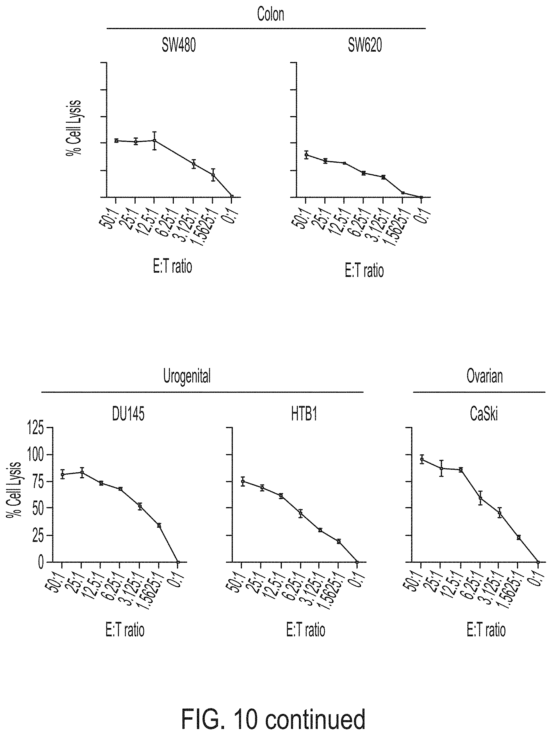
D00013
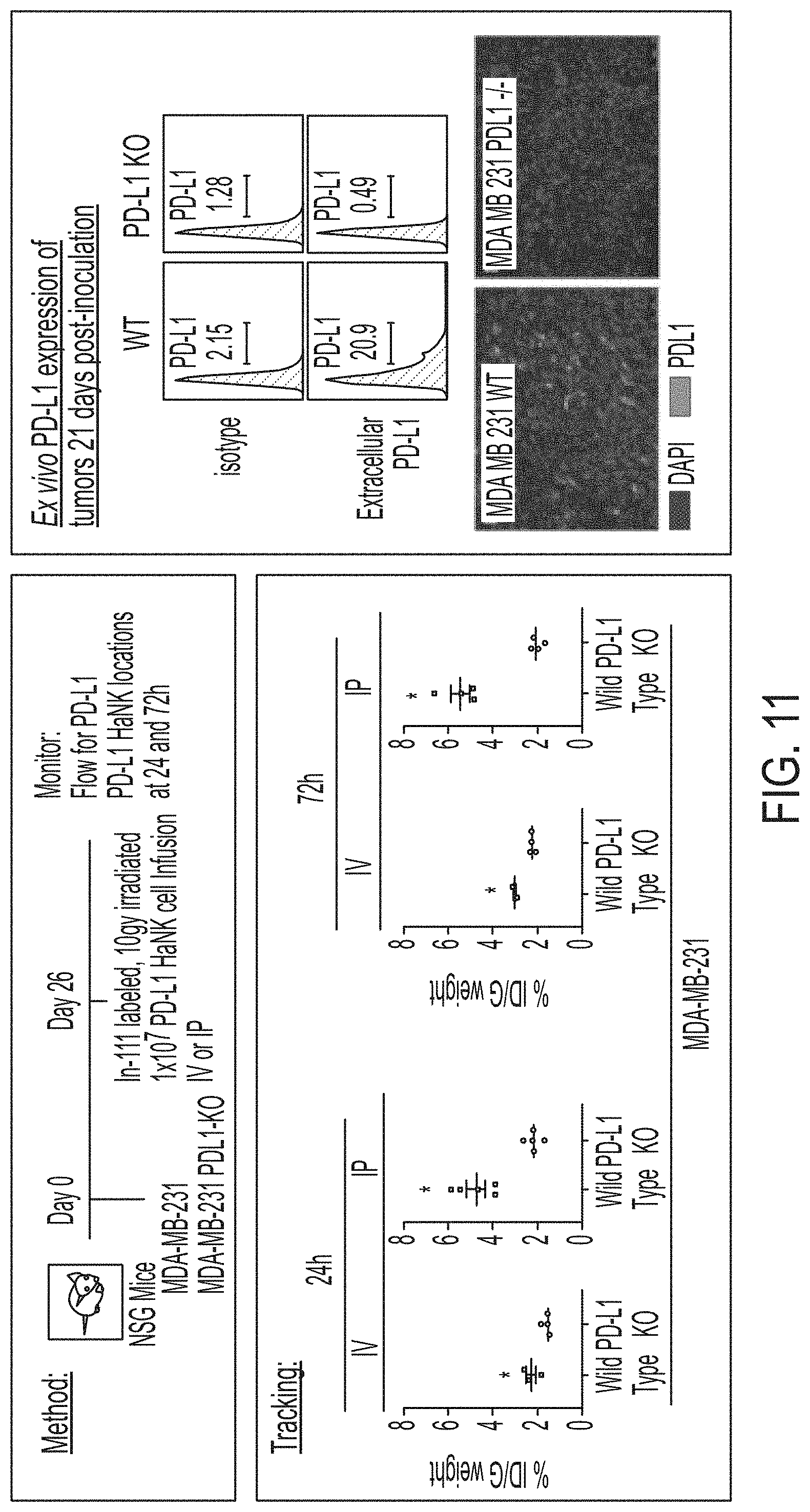
D00014
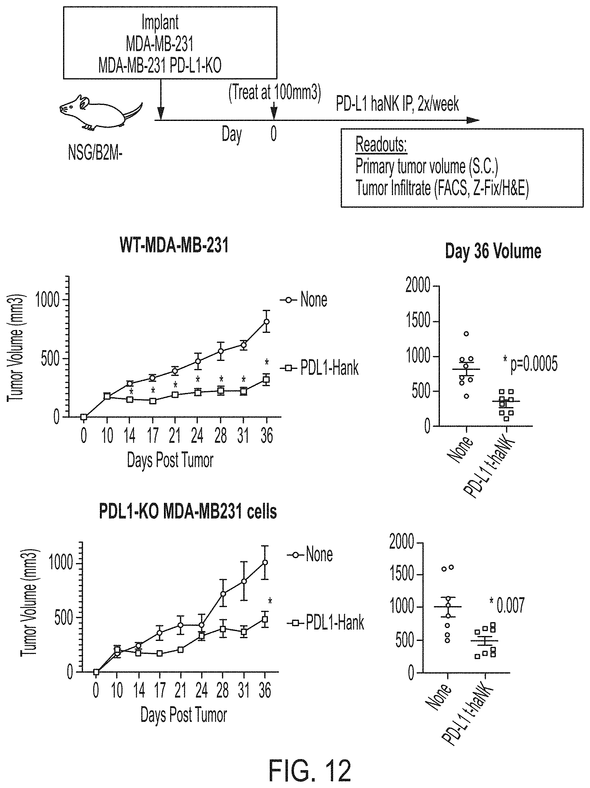
D00015
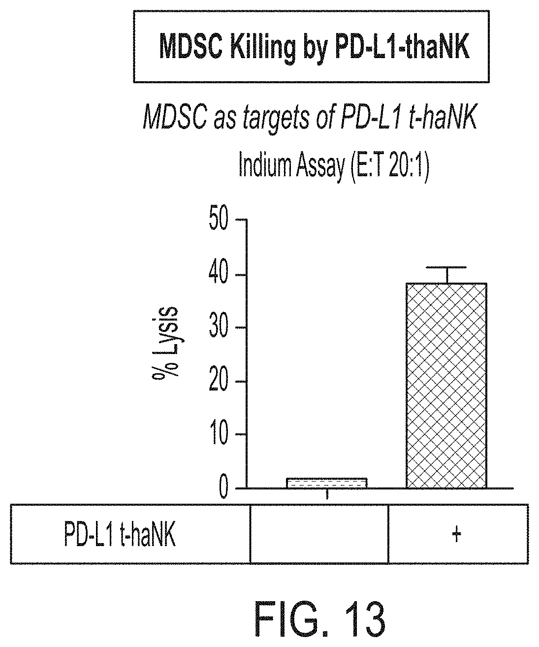
S00001
XML
uspto.report is an independent third-party trademark research tool that is not affiliated, endorsed, or sponsored by the United States Patent and Trademark Office (USPTO) or any other governmental organization. The information provided by uspto.report is based on publicly available data at the time of writing and is intended for informational purposes only.
While we strive to provide accurate and up-to-date information, we do not guarantee the accuracy, completeness, reliability, or suitability of the information displayed on this site. The use of this site is at your own risk. Any reliance you place on such information is therefore strictly at your own risk.
All official trademark data, including owner information, should be verified by visiting the official USPTO website at www.uspto.gov. This site is not intended to replace professional legal advice and should not be used as a substitute for consulting with a legal professional who is knowledgeable about trademark law.