Combined Preparations For The Treatment Of Cancer Or Infection
TRIEBEL; Frederic ; et al.
U.S. patent application number 16/733829 was filed with the patent office on 2020-04-23 for combined preparations for the treatment of cancer or infection. This patent application is currently assigned to IMMUTEP S.A.S.. The applicant listed for this patent is IMMUTEP S.A.S.. Invention is credited to Chrystelle BRIGNONE, Frederic TRIEBEL.
| Application Number | 20200121757 16/733829 |
| Document ID | / |
| Family ID | 52597438 |
| Filed Date | 2020-04-23 |


View All Diagrams
| United States Patent Application | 20200121757 |
| Kind Code | A1 |
| TRIEBEL; Frederic ; et al. | April 23, 2020 |
COMBINED PREPARATIONS FOR THE TREATMENT OF CANCER OR INFECTION
Abstract
Combined preparations, and pharmaceutical compositions, comprising: (a) LAG-3 protein, or a derivative thereof that is able to bind to MHC class II molecules; and (b) a programmed cell death protein-1 (PD-1) pathway inhibitor, are described. The PD-1 pathway inhibitor, such as an anti-PD-1 antibody or an anti-PD-L1 antibody, and a soluble derivative of LAG-3, acting as an APC activator, together synergistically activate T cells (in particular, CD8.sup.+ T cells). Use of the combined preparations and compositions as medicaments, in particular for the treatment of cancer or infection, and to methods for the treatment of cancer or infection, is described.
| Inventors: | TRIEBEL; Frederic; (Versailles, FR) ; BRIGNONE; Chrystelle; (Chatenay-Malabry, FR) | ||||||||||
| Applicant: |
|
||||||||||
|---|---|---|---|---|---|---|---|---|---|---|---|
| Assignee: | IMMUTEP S.A.S. Orsay FR |
||||||||||
| Family ID: | 52597438 | ||||||||||
| Appl. No.: | 16/733829 | ||||||||||
| Filed: | January 3, 2020 |
Related U.S. Patent Documents
| Application Number | Filing Date | Patent Number | ||
|---|---|---|---|---|
| 15542466 | Jul 10, 2017 | |||
| PCT/EP2016/050321 | Jan 8, 2016 | |||
| 16733829 | ||||
| Current U.S. Class: | 1/1 |
| Current CPC Class: | A61K 38/177 20130101; Y02A 50/30 20180101; Y02A 50/382 20180101; C07K 2319/32 20130101; A61P 31/22 20180101; C07K 2317/70 20130101; Y02A 50/387 20180101; Y02A 50/467 20180101; Y02A 50/475 20180101; Y02A 50/393 20180101; Y02A 50/409 20180101; A61K 2300/00 20130101; C07K 2319/30 20130101; A61P 35/04 20180101; A61K 38/1774 20130101; A61K 39/39558 20130101; A61P 31/16 20180101; C07K 16/2818 20130101; A61K 39/3955 20130101; C07K 2317/76 20130101; A61K 38/177 20130101; A61K 2300/00 20130101; A61K 39/3955 20130101; A61K 2300/00 20130101 |
| International Class: | A61K 38/17 20060101 A61K038/17; A61P 31/16 20060101 A61P031/16; A61P 35/04 20060101 A61P035/04; A61P 31/22 20060101 A61P031/22; A61K 39/395 20060101 A61K039/395; C07K 16/28 20060101 C07K016/28 |
Foreign Application Data
| Date | Code | Application Number |
|---|---|---|
| Jan 9, 2015 | GB | 1500374.2 |
Claims
1-63. (canceled)
64. A method of preventing, treating or ameliorating a cancer in a subject, the method comprising administering to a subject in need thereof an effective amount of: a LAG-3 protein, or a derivative of LAG-3 protein that is able to bind to MHC class II molecules, wherein the derivative of LAG-3 protein comprises: a 30 amino acid extra-loop sequence GPPAAAPGHPLAPGPHPAAPSSWGPRPRRY (SEQ ID NO:2) of domain D1 of human LAG-3 protein; or a variant of the 30 amino acid extra-loop sequence GPPAAAPGHPLAPGPHPAAPSSWGPRPRRY (SEQ ID NO:2) of domain D1 of human LAG-3 protein, wherein the variant comprises one or more conservative amino acid substitutions, and has at least 70% amino acid identity with the 30 amino acid extra-loop sequence; and a programmed cell death protein-1 (PD-1) pathway inhibitor, wherein the PD-1 pathway inhibitor is selected from the group consisting of an anti-PD-1 antibody, and a derivative or fragment of an anti-PD-1 antibody that retains ability to inhibit binding of PD-1 to PD-L1 and/or PD-L2.
65. A method according to claim 64, wherein the LAG-3 protein, or derivative thereof, and the PD-1 pathway inhibitor are administered sequentially or co-administered to the subject.
66. A method according to claim 65, wherein the LAG-3 protein, or derivative thereof, is administered after the PD-1 pathway inhibitor.
67. A method according to claim 64, wherein the LAG-3 protein, or derivative thereof, is administered to the subject at a dose which is a molar equivalent of 0.25-30 mg of LAG-3Ig fusion protein IMP321.
68. A method according to claim 64, wherein a plurality of doses of the LAG-3 protein, or derivative thereof, is administered to the subject, and/or a plurality of doses of the PD-1 pathway inhibitor is administered to the subject.
69. A method according to claim 64, wherein the PD-1 pathway inhibitor is pembrolizumab or nivolumab.
70. A method according to claim 64, wherein the LAG-3 protein or derivative thereof, and the PD-1 pathway inhibitor, are administered in any of the combinations of dosage amounts shown in the Table below: TABLE-US-00029 Human dose of LAG-3 protein or derivative thereof Type of PD-1 Dose of PD-1 pathway (given as a mg dose of pathway inhibitor: mg/kg [mg IMP321, or a molar inhibitor dose for 70 kg human] equivalent thereof) Anti-PD-1 0.001-5 mg/kg [0.07-350 mg] 0.25-30 mg antibody 0.001-2.5 mg/kg [0.07-175 mg] 0.25-30 mg 0.001-1 mg/kg [0.07-70 mg] 0.25-30 mg 0.001-<1 mg/kg [0.07-<70 mg] 0.25-30 mg 0.001-0.5 mg/kg [0.07-35 mg] 0.25-30 mg 0.001-0.1 mg/kg [0.07-7 mg] 0.25-30 mg 0.002-5 mg/kg [0.14-350 mg] 0.25-30 mg 0.002-2.5 mg/kg [0.14-175 mg] 0.25-30 mg 0.002-1 mg/kg [0.14-70 mg] 0.25-30 mg 0.002-<1 mg/kg [0.14-<70 mg] 0.25-30 mg 0.002-0.5 mg/kg [0.14-35 mg] 0.25-30 mg 0.002-0.1 mg/kg [0.14-7 mg] 0.25-30 mg Anti-PD-1 0.001-5 mg/kg [0.07-350 mg] 1-30 mg antibody 0.001-2.5 mg/kg [0.07-175 mg] 1-30 mg 0.001-1 mg/kg [0.07-70 mg] 1-30 mg 0.001-<1 mg/kg [0.07-<70 mg] 1-30 mg 0.001-0.5 mg/kg [0.07-35 mg] 1-30 mg 0.001-0.1 mg/kg [0.07-7 mg] 1-30 mg 0.002-5 mg/kg [0.14-350 mg] 1-30 mg 0.002-2.5 mg/kg [0.14-175 mg] 1-30 mg 0.002-1 mg/kg [0.14-70 mg] 1-30 mg 0.002-<1 mg/kg [0.14-<70 mg] 1-30 mg 0.002-0.5 mg/kg [0.14-35 mg] 1-30 mg 0.002-0.1 mg/kg [0.14-7 mg] 1-30 mg Anti-PD-1 0.001-5 mg/kg [0.07-350 mg] 6-30 mg antibody 0.001-2.5 mg/kg [0.07-175 mg] 6-30 mg 0.001-1 mg/kg [0.07-70 mg] 6-30 mg 0.001-<1 mg/kg [0.07-<70 mg] 6-30 mg 0.001-0.5 mg/kg [0.07-35 mg] 6-30 mg 0.001-0.1 mg/kg [0.07-7 mg] 6-30 mg 0.002-5 mg/kg [0.14-350 mg] 6-30 mg 0.002-2.5 mg/kg [0.14-175 mg] 6-30 mg 0.002-1 mg/kg [0.14-70 mg] 6-30 mg 0.002-<1 mg/kg [0.14-<70 mg] 6-30 mg 0.002-0.5 mg/kg [0.14-35 mg] 6-30 mg 0.002-0.1 mg/kg [0.14-7 mg] 6-30 mg Pembrolizumab 0.001-5 mg/kg [0.07-350 mg] 0.25-30 mg 0.001-2.5 mg/kg [0.07-175 mg] 0.25-30 mg 0.001-1 mg/kg [0.07-70 mg] 0.25-30 mg 0.001-<1 mg/kg [0.07-<70 mg] 0.25-30 mg 0.001-0.5 mg/kg [0.07-35 mg] 0.25-30 mg 0.001-0.1 mg/kg [0.07-7 mg] 0.25-30 mg 0.002-5 mg/kg [0.14-350 mg] 0.25-30 mg 0.002-2.5 mg/kg [0.14-175 mg] 0.25-30 mg 0.002-1 mg/kg [0.14-70 mg] 0.25-30 mg 0.002-<1 mg/kg [0.14-<70 mg] 0.25-30 mg 0.002-0.5 mg/kg [0.14-35 mg] 0.25-30 mg 0.002-0.1 mg/kg [0.14-7 mg] 0.25-30 mg Pembrolizumab 0.001-5 mg/kg [0.07-350 mg] 1-30 mg 0.001-2.5 mg/kg [0.07-175 mg] 1-30 mg 0.001-1 mg/kg [0.07-70 mg] 1-30 mg 0.001-<1 mg/kg [0.07-<70 mg] 1-30 mg 0.001-0.5 mg/kg [0.07-35 mg] 1-30 mg 0.001-0.1 mg/kg [0.07-7 mg] 1-30 mg 0.002-5 mg/kg [0.14-350 mg] 1-30 mg 0.002-2.5 mg/kg [0.14-175 mg] 1-30 mg 0.002-1 mg/kg [0.14-70 mg] 1-30 mg 0.002-1 mg/kg [0.14-70 mg] 1-30 mg 0.002-<1 mg/kg [0.14-<70 mg] 1-30 mg 0.002-0.5 mg/kg [0.14-35 mg] 1-30 mg Pembrolizumab 0.001-5 mg/kg [0.07-350 mg] 6-30 mg 0.001-2.5 mg/kg [0.07-175 mg] 6-30 mg 0.001-1 mg/kg [0.07-70 mg] 6-30 mg 0.001-<1 mg/kg [0.07-<70 mg] 6-30 mg 0.001-0.5 mg/kg [0.07-35 mg] 6-30 mg 0.001-0.1 mg/kg [0.07-7 mg] 6-30 mg 0.002-5 mg/kg [0.14-350 mg] 6-30 mg 0.002-2.5 mg/kg [0.14-175 mg] 6-30 mg 0.002-1 mg/kg [0.14-70 mg] 6-30 mg 0.002-<1 mg/kg [0.14-<70 mg] 6-30 mg 0.002-0.5 mg/kg [0.14-35 mg] 6-30 mg 0.002-0.1 mg/kg [0.14-7 mg] 6-30 mg Nivolumab 0.001-5 mg/kg [0.07-350 mg] 0.25-30 mg 0.001-2.5 mg/kg [0.07-175 mg] 0.25-30 mg 0.001-1 mg/kg [0.07-70 mg] 0.25-30 mg 0.001-<1 mg/kg [0.07-<70 mg] 0.25-30 mg 0.001-0.5 mg/kg [0.07-35 mg] 0.25-30 mg 0.001-0.1 mg/kg [0.07-7 mg] 0.25-30 mg 0.002-5 mg/kg [0.14-350 mg] 0.25-30 mg 0.002-2.5 mg/kg [0.14-175 mg] 0.25-30 mg 0.002-1 mg/kg [0.14-70 mg] 0.25-30 mg 0.002-<1 mg/kg [0.14-<70 mg] 0.25-30 mg 0.002-0.5 mg/kg [0.14-35 mg] 0.25-30 mg 0.002-0.1 mg/kg [0.14-7 mg] 0.25-30 mg Nivolumab 0.001-5 mg/kg [0.07-350 mg] 1-30 mg 0.001-2.5 mg/kg [0.07-175 mg] 1-30 mg 0.001-1 mg/kg [0.07-70 mg] 1-30 mg 0.001-<1 mg/kg [0.07-<70 mg] 1-30 mg 0.001-0.5 mg/kg [0.07-35 mg] 1-30 mg 0.001-0.1 mg/kg [0.07-7 mg] 1-30 mg 0.002-5 mg/kg [0.14-350 mg] 1-30 mg 0.002-2.5 mg/kg [0.14-175 mg] 1-30 mg 0.002-1 mg/kg [0.14-70 mg] 1-30 mg 0.002-<1 mg/kg [0.14-<70 mg] 1-30 mg 0.002-0.5 mg/kg [0.14-35 mg] 1-30 mg 0.002-0.1 mg/kg [0.14-7 mg] 1-30 mg Nivolumab 0.001-5 mg/kg [0.07-350 mg] 6-30 mg 0.001-2.5 mg/kg [0.07-175 mg] 6-30 mg 0.001-1 mg/kg [0.07-70 mg] 6-30 mg 0.001-<1 mg/kg [0.07-<70 mg] 6-30 mg 0.001-0.5 mg/kg [0.07-35 mg] 6-30 mg 0.001-0.1 mg/kg [0.07-7 mg] 6-30 mg 0.002-5 mg/kg [0.14-350 mg] 6-30 mg 0.002-2.5 mg/kg [0.14-175 mg] 6-30 mg 0.002-1 mg/kg [0.14-70 mg] 6-30 mg 0.002-<1 mg/kg [0.14-<70 mg] 6-30 mg 0.002-0.5 mg/kg [0.14-35 mg] 6-30 mg 0.002-0.1 mg/kg [0.14-7 mg] 6-30 mg
71. A method according to claim 64, wherein the derivative of LAG-3 protein comprises an amino acid sequence that has at least 70% amino acid identity with domain D1, and optionally domain D2, of LAG-3 protein, or at least 70% amino acid identity with domains D1, D2, D3, and optionally D4, of LAG-3 protein, preferably human LAG-3 protein.
72. A method according to claim 64, wherein the derivative of LAG-3 protein is fused to Immunoglobulin Fc sequence.
73. A method according to claim 64, wherein the derivative of LAG-3 protein is the recombinant soluble human LAG-3Ig fusion protein IMP321.
74. A method according to claim 64, wherein: the derivative of LAG-3 protein is the recombinant soluble human LAG-3Ig fusion protein IMP321, and the PD-1 pathway inhibitor is pembrolizumab; or the derivative of LAG-3 protein is the recombinant soluble human LAG-3Ig fusion protein IMP321, and the PD-1 pathway inhibitor is nivolumab.
75. A method according to claim 64, wherein the cancer is skin, lung (especially squamous or nonsquamous non-small-cell lung carcinoma, NSCLC), ovarian, renal, colon, colorectal, breast, gastric, esophageal, pancreatic, bladder, urothelial, or liver cancer, or a melanoma (for example, metastatic malignant melanoma), a prostate cancer (for example hormone refractory prostate adenocarcinoma), a head and neck cancer (for example, squamous cell carcinoma of the head and neck), a cervical cancer, a thyroid cancer, a glioblastoma, a glioma, leukemia, a lymphoma (for example, a B cell lymphoma), an adrenal gland cancer, an AIDS-associated cancer, an alveolar soft part sarcoma, an astrocytic tumor, bone cancer, a brain and spinal cord cancer, a metastatic brain tumor, a carotid body tumor, a chondrosarcoma, a chordoma, a chromophobe renal cell carcinoma, a clear cell carcinoma, cutaneous benign fibrous histiocytoma, a desmoplastic small round cell tumor, an ependymoma, a Ewing's tumor, an extraskeletal myxoid chondrosarcoma, a fibrogenesis imperfecta ossium, a fibrous dysplasia of the bone, a gallbladder or bile duct cancer, a gestational trophoblastic disease, a germ cell tumor, a haematological malignancy, hepatocellular carcinoma, an islet cell tumor, a Kaposi's sarcoma, a kidney cancer, a lipoma/benign lipomatous tumor, a liposarcoma/malignant lipomatous tumor, a medulloblastoma, a meningioma, a Merkel cell carcinoma, a multiple endocrine neoplasia, a multiple myeloma, a myelodysplasia syndrome, a neuroblastoma, a neuroendocrine tumor, a papillary thyroid carcinoma, a parathyroid tumor, a pediatric cancer, a peripheral nerve sheath tumor, a phaeochromocytoma, a pituitary tumor, a prostate cancer, a posterior uveal melanoma, a rare hematologic disorder, a renal metastatic cancer, a rhabdoid tumor, a rhabdomysarcoma, a sarcoma, a soft-tissue sarcoma, a squamous cell cancer, a stomach cancer, a synovial sarcoma, a testicular cancer, a thymic carcinoma, a thymoma, a thyroid metastatic cancer, or a uterine cancer.
76. A method according to claim 64, wherein the LAG-3 protein or the derivative of LAG-3 protein is an APC activator.
77. A method according to claim 64, wherein the variant has at least 95% amino acid identity with the 30 amino acid extra-loop sequence.
78. A method according to claim 64, wherein the subject is a human subject.
79. A method of preventing, treating or ameliorating a cancer in a subject, the method comprising administering to a subject in need thereof an effective amount of: a LAG-3 protein, or a derivative of LAG-3 protein that is able to bind to MHC class II molecules, wherein the derivative of LAG-3 protein comprises: a 30 amino acid extra-loop sequence GPPAAAPGHPLAPGPHPAAPSSWGPRPRRY (SEQ ID NO:2) of domain D1 of human LAG-3 protein; or a variant of the 30 amino acid extra-loop sequence GPPAAAPGHPLAPGPHPAAPSSWGPRPRRY (SEQ ID NO:2) of domain D1 of human LAG-3 protein, wherein the variant comprises one or more conservative amino acid substitutions, and has at least 70% amino acid identity with the 30 amino acid extra-loop sequence; and a programmed cell death protein-1 (PD-1) pathway inhibitor, wherein the PD-1 pathway inhibitor is selected from the group consisting of an anti-PD-L1 antibody, and a derivative or fragment of an anti-PD-L1 antibody that retains ability to inhibit binding of PD-L1 to PD-1.
80. A method according to claim 79, wherein the LAG-3 protein, or derivative thereof, and the PD-1 pathway inhibitor are administered sequentially or co-administered to the subject.
81. A method according to claim 80, wherein the LAG-3 protein, or derivative thereof, is administered after the PD-1 pathway inhibitor.
82. A method according to claim 79, wherein the LAG-3 protein, or derivative thereof, is administered to the subject at a dose which is a molar equivalent of 0.25-30 mg of LAG-3Ig fusion protein IMP321.
83. A method according to claim 79, wherein a plurality of doses of the LAG-3 protein, or derivative thereof, is administered to the subject, and/or a plurality of doses of the PD-1 pathway inhibitor is administered to the subject.
84. A method according to claim 79, wherein the PD-1 pathway inhibitor is BMS-936559, MEDI4736, MPDL3280A, or MSB0010718C.
85. A method according to claim 79, wherein the LAG-3 protein or derivative thereof, and the PD-1 pathway inhibitor, are administered in any of the combinations of dosage amounts shown in the Table below: TABLE-US-00030 Human dose of LAG-3 protein or derivative thereof Type of PD-1 Dose of PD-1 pathway (given as a mg dose of pathway inhibitor: mg/kg [mg IMP321, or a molar inhibitor dose for 70 kg human] equivalent thereof) Anti-PD-L1 0.001-5 mg/kg [0.07-350 mg] 0.25-30 mg antibody 0.001-2.5 mg/kg [0.07-175 mg] 0.25-30 mg 0.001-1 mg/kg [0.07-70 mg] 0.25-30 mg 0.001-<1 mg/kg [0.07-<70 mg] 0.25-30 mg 0.001-0.5 mg/kg [0.07-35 mg] 0.25-30 mg 0.001-0.1 mg/kg [0.07-7 mg] 0.25-30 mg 0.002-5 mg/kg [0.14-350 mg] 0.25-30 mg 0.002-2.5 mg/kg [0.14-175 mg] 0.25-30 mg 0.002-1 mg/kg [0.14-70 mg] 0.25-30 mg 0.002-<1 mg/kg [0.14-<70 mg] 0.25-30 mg 0.002-0.5 mg/kg [0.14-35 mg] 0.25-30 mg 0.002-0.1 mg/kg [0.14-7 mg] 0.25-30 mg Anti-PD-L1 0.001-5 mg/kg [0.07-350 mg] 1-30 mg antibody 0.001-2.5 mg/kg [0.07-175 mg] 1-30 mg 0.001-1 mg/kg [0.07-70 mg] 1-30 mg 0.001-<1 mg/kg [0.07-<70 mg] 1-30 mg 0.001-0.5 mg/kg [0.07-35 mg] 1-30 mg 0.001-0.1 mg/kg [0.07-7 mg] 1-30 mg 0.002-5 mg/kg [0.14-350 mg] 1-30 mg 0.002-2.5 mg/kg [0.14-175 mg] 1-30 mg 0.002-1 mg/kg [0.14-70 mg] 1-30 mg 0.002-<1 mg/kg [0.14-<70 mg] 1-30 mg 0.002-0.5 mg/kg [0.14-35 mg] 1-30 mg 0.002-0.1 mg/kg [0.14-7 mg] 1-30 mg Anti-PD-L1 0.001-5 mg/kg [0.07-350 mg] 6-30 mg antibody 0.001-2.5 mg/kg [0.07-175 mg] 6-30 mg 0.001-1 mg/kg [0.07-70 mg] 6-30 mg 0.001-<1 mg/kg [0.07-<70 mg] 6-30 mg 0.001-0.5 mg/kg [0.07-35 mg] 6-30 mg 0.001-0.1 mg/kg [0.07-7 mg] 6-30 mg 0.002-5 mg/kg [0.14-350 mg] 6-30 mg 0.002-2.5 mg/kg [0.14-175 mg] 6-30 mg 0.002-1 mg/kg [0.14-70 mg] 6-30 mg 0.002-<1 mg/kg [0.14-<70 mg] 6-30 mg 0.002-0.5 mg/kg [0.14-35 mg] 6-30 mg 0.002-0.1 mg/kg [0.14-7 mg] 6-30 mg BMS-936559 0.001-5 mg/kg [0.07-350 mg] 0.25-30 mg 0.001-2.5 mg/kg [0.07-175 mg] 0.25-30 mg 0.001-1 mg/kg [0.07-70 mg] 0.25-30 mg 0.001-<1 mg/kg [0.07-<70 mg] 0.25-30 mg 0.001-0.5 mg/kg [0.07-35 mg] 0.25-30 mg 0.001-0.1 mg/kg [0.07-7 mg] 0.25-30 mg 0.002-5 mg/kg [0.14-350 mg] 0.25-30 mg 0.002-2.5 mg/kg [0.14-175 mg] 0.25-30 mg 0.002-1 mg/kg [0.14-70 mg] 0.25-30 mg 0.002-<1 mg/kg [0.14-<70 mg] 0.25-30 mg 0.002-0.5 mg/kg [0.14-35 mg] 0.25-30 mg 0.002-0.1 mg/kg [0.14-7 mg] 0.25-30 mg BMS-936559 0.001-5 mg/kg [0.07-350 mg] 1-30 mg 0.001-2.5 mg/kg [0.07-175 mg] 1-30 mg 0.001-1 mg/kg [0.07-70 mg] 1-30 mg 0.001-<1 mg/kg [0.07-<70 mg] 1-30 mg 0.001-0.5 mg/kg [0.07-35 mg] 1-30 mg 0.001-0.1 mg/kg [0.07-7 mg] 1-30 mg 0.002-5 mg/kg [0.14-350 mg] 1-30 mg 0.002-2.5 mg/kg [0.14-175 mg] 1-30 mg 0.002-1 mg/kg [0.14-70 mg] 1-30 mg 0.002-<1 mg/kg [0.14-<70 mg] 1-30 mg 0.002-0.5 mg/kg [0.14-35 mg] 1-30 mg 0.002-0.1 mg/kg [0.14-7 mg] 1-30 mg BMS-936559 0.001-5 mg/kg [0.07-350 mg] 6-30 mg 0.001-2.5 mg/kg [0.07-175 mg] 6-30 mg 0.001-1 mg/kg [0.07-70 mg] 6-30 mg 0.001-<1 mg/kg [0.07-<70 mg] 6-30 mg 0.001-0.5 mg/kg [0.07-35 mg] 6-30 mg 0.001-0.1 mg/kg [0.07-7 mg] 6-30 mg 0.002-5 mg/kg [0.14-350 mg] 6-30 mg 0.002-2.5 mg/kg [0.14-175 mg] 6-30 mg 0.002-1 mg/kg [0.14-70 mg] 6-30 mg 0.002-<1 mg/kg [0.14-<70 mg] 6-30 mg 0.002-0.5 mg/kg [0.14-35 mg] 6-30 mg 0.002-0.1 mg/kg [0.14-7 mg] 6-30 mg MPDL3280A 0.001-5 mg/kg [0.07-350 mg] 0.25-30 mg 0.001-2.5 mg/kg [0.07-175 mg] 0.25-30 mg 0.001-1 mg/kg [0.07-70 mg] 0.25-30 mg 0.001-<1 mg/kg [0.07-<70 mg] 0.25-30 mg 0.001-0.5 mg/kg [0.07-35 mg] 0.25-30 mg 0.001-0.1 mg/kg [0.07-7 mg] 0.25-30 mg 0.002-5 mg/kg [0.14-350 mg] 0.25-30 mg 0.002-2.5 mg/kg [0.14-175 mg] 0.25-30 mg 0.002-1 mg/kg [0.14-70 mg] 0.25-30 mg 0.002-<1 mg/kg [0.14-<70 mg] 0.25-30 mg 0.002-0.5 mg/kg [0.14-35 mg] 0.25-30 mg 0.002-0.1 mg/kg [0.14-7 mg] 0.25-30 mg MPDL3280A 0.001-5 mg/kg [0.07-350 mg] 1-30 mg 0.001-2.5 mg/kg [0.07-175 mg] 1-30 mg 0.001-1 mg/kg [0.07-70 mg] 1-30 mg 0.001-<1 mg/kg [0.07-<70 mg] 1-30 mg 0.001-0.5 mg/kg [0.07-35 mg] 1-30 mg 0.001-0.1 mg/kg [0.07-7 mg] 1-30 mg 0.002-5 mg/kg [0.14-350 mg] 1-30 mg 0.002-2.5 mg/kg [0.14-175 mg] 1-30 mg 0.002-1 mg/kg [0.14-70 mg] 1-30 mg 0.002-<1 mg/kg [0.14-<70 mg] 1-30 mg 0.002-0.5 mg/kg [0.14-35 mg] 1-30 mg 0.002-0.1 mg/kg [0.14-7 mg] 1-30 mg MPDL3280A 0.001-5 mg/kg [0.07-350 mg] 6-30 mg 0.001-2.5 mg/kg [0.07-175 mg] 6-30 mg 0.001-1 mg/kg [0.07-70 mg] 6-30 mg 0.001-<1 mg/kg [0.07-<70 mg] 6-30 mg 0.001-0.5 mg/kg [0.07-35 mg] 6-30 mg 0.001-0.1 mg/kg [0.07-7 mg] 6-30 mg 0.002-5 mg/kg [0.14-350 mg] 6-30 mg 0.002-2.5 mg/kg [0.14-175 mg] 6-30 mg 0.002-1 mg/kg [0.14-70 mg] 6-30 mg 0.002-<1 mg/kg [0.14-<70 mg] 6-30 mg 0.002-0.5 mg/kg [0.14-35 mg] 6-30 mg 0.002-0.1 mg/kg [0.14-7 mg] 6-30 mg
86. A method according to claim 79, wherein the derivative of LAG-3 protein comprises an amino acid sequence that has at least 70% amino acid identity with domain D1, and optionally domain D2, of LAG-3 protein, or at least 70% amino acid identity with domains D1, D2, D3, and optionally D4, of LAG-3 protein, preferably human LAG-3 protein.
87. A method according to claim 79, wherein the derivative of LAG-3 protein is fused to Immunoglobulin Fc sequence.
88. A method according to claim 79, wherein the derivative of LAG-3 protein is the recombinant soluble human LAG-3Ig fusion protein IMP321.
89. A method according to claim 79, wherein: the derivative of LAG-3 protein is the recombinant soluble human LAG-3Ig fusion protein IMP321, and the PD-1 pathway inhibitor is BMS-936559; the derivative of LAG-3 protein is the recombinant soluble human LAG-3Ig fusion protein IMP321, and the PD-1 pathway inhibitor is MEDI4736; the derivative of LAG-3 protein is the recombinant soluble human LAG-3Ig fusion protein IMP321, and the PD-1 pathway inhibitor is MPDL3280A; or the derivative of LAG-3 protein is the recombinant soluble human LAG-3Ig fusion protein IMP321, and the PD-1 pathway inhibitor is MSB0010718C.
90. A method according to claim 79, wherein the cancer is skin, lung (especially squamous or nonsquamous non-small-cell lung carcinoma, NSCLC), ovarian, renal, colon, colorectal, breast, gastric, esophageal, pancreatic, bladder, urothelial, or liver cancer, or a melanoma (for example, metastatic malignant melanoma), a prostate cancer (for example hormone refractory prostate adenocarcinoma), a head and neck cancer (for example, squamous cell carcinoma of the head and neck), a cervical cancer, a thyroid cancer, a glioblastoma, a glioma, leukemia, a lymphoma (for example, a B cell lymphoma), an adrenal gland cancer, an AIDS-associated cancer, an alveolar soft part sarcoma, an astrocytic tumor, bone cancer, a brain and spinal cord cancer, a metastatic brain tumor, a carotid body tumor, a chondrosarcoma, a chordoma, a chromophobe renal cell carcinoma, a clear cell carcinoma, cutaneous benign fibrous histiocytoma, a desmoplastic small round cell tumor, an ependymoma, a Ewing's tumor, an extraskeletal myxoid chondrosarcoma, a fibrogenesis imperfecta ossium, a fibrous dysplasia of the bone, a gallbladder or bile duct cancer, a gestational trophoblastic disease, a germ cell tumor, a haematological malignancy, hepatocellular carcinoma, an islet cell tumor, a Kaposi's sarcoma, a kidney cancer, a lipoma/benign lipomatous tumor, a liposarcoma/malignant lipomatous tumor, a medulloblastoma, a meningioma, a Merkel cell carcinoma, a multiple endocrine neoplasia, a multiple myeloma, a myelodysplasia syndrome, a neuroblastoma, a neuroendocrine tumor, a papillary thyroid carcinoma, a parathyroid tumor, a pediatric cancer, a peripheral nerve sheath tumor, a phaeochromocytoma, a pituitary tumor, a prostate cancer, a posterior uveal melanoma, a rare hematologic disorder, a renal metastatic cancer, a rhabdoid tumor, a rhabdomysarcoma, a sarcoma, a soft-tissue sarcoma, a squamous cell cancer, a stomach cancer, a synovial sarcoma, a testicular cancer, a thymic carcinoma, a thymoma, a thyroid metastatic cancer, or a uterine cancer.
91. A method according to claim 79, wherein the LAG-3 protein or the derivative of LAG-3 protein is an APC activator.
92. A method according to claim 79, wherein the variant has at least 95% amino acid identity with the 30 amino acid extra-loop sequence.
93. A method according to claim 79, wherein the subject is a human subject.
Description
CROSS REFERENCE TO RELATED APPLICATION
[0001] This application is a divisional of U.S. patent application Ser. No. 15/542,466 filed Jul. 10, 2017, which is a National Stage Entry of International Application No. PCT/EP2016/050321 filed Jan. 8, 2016, the entire contents of which are incorporated herein by reference herein.
FIELD OF THE DISCLOSURE
[0002] This disclosure relates to combined preparations and to pharmaceutical compositions, and their use as medicaments, in particular for the treatment of cancer or infection, and to methods for the treatment of cancer or infection.
INCORPORATION BY REFERENCE OF SEQUENCE LISTING
[0003] The Sequence Listing in an ASCII text file, named as 35156A_SequenceListing.txt of 5 KB, created on Jan. 2, 2020, and submitted to the United States Patent and Trademark Office via EFS-Web, is incorporated herein by reference.
BACKGROUND
[0004] Upon emerging from the thymus, naive T cells circulate in blood through lymph nodes and seek foreign ("nonself") antigens presented by specific antigen-presenting cells (APCs), typically dendritic cells. T cells can recognize not only pathogen-associated antigens but also abnormally expressed self-proteins--indicating mutated or transformed tumorigenic cells--as "nonself." If T cells encounter their specific antigen in the context of appropriate costimulatory molecules, the cells become activated and upregulate activation and homing molecules. These T cells, termed effector T cells, are able to enter inflamed tissues in search of infected or cancerous cells. Among other functions, effector T cells can produce inflammatory cytokines and/or cytolytic granules, leading to apoptosis or necrosis of infected or tumor cells.
[0005] Throughout the duration of an immune response, local and systemic down-regulatory forces minimize damage to healthy cells and tissues. These can involve immunosuppressive cytokines, regulatory T cells (Tregs), and negative signaling from other cells. Tumor antigen-specific T cells display impaired effector function and an exhausted phenotype characterized by decreased production of pro-inflammatory cytokines and hypo-responsiveness to antigenic restimulation. This is mediated by cell-extrinsic mechanisms, such as regulatory T cells (Treg), and cell-intrinsic mechanisms, such as inhibitory molecules that are up-regulated on exhausted, tumor infiltrating lymphocytes (TILs).
[0006] Immune checkpoint pathways strongly downregulate T-cell activation with the intent of keeping nascent T-cell responses in check and reducing the likelihood of an immune attack against normal tissues. During tumorigenesis, however, cancer cells may exploit these co-inhibitory pathways to resist detection or avoid elimination by the adaptive immune system.
[0007] The programmed cell death protein-1 (PD-1) is a critical checkpoint molecule that is expressed by T cells upon activation. The PD-1 checkpoint pathway is thought to act primarily in peripheral tissues to dampen ongoing immune responses and/or to prevent damage to self-tissues. PD-1 is expressed by B cells, natural killer (NK) cells, dendritic cells, and activated monocytes, in addition to T cells. PD-1 ligands--which include PD-L1 and PD-L2, among others--are expressed by macrophages and monocytes, and these can be induced in numerous cell types in an inflammatory environment.
[0008] The ability of nonimmune cells to express ligands for PD-1, primarily PD-L1, is exploited by tumors as one way to avoid immune attack. Tumor cells can also down-regulate antigen expression to avoid detection. In addition, production of immunosuppressive mediators and retention of Tregs and immune suppressor cells within the tumor microenvironment can dampen antitumor immune responses.
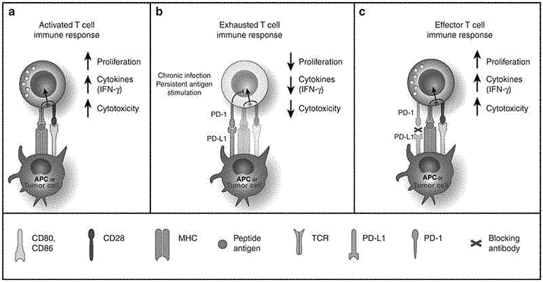
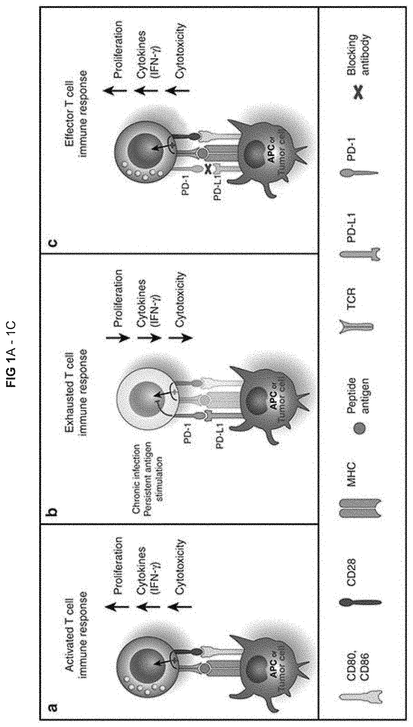
[0009] FIG. 1 (taken from Harvey, Clinical Pharmacology & Therapeutics, 2014, Vol. 96(2), pages 214-223) depicts the role of the PD-1 pathway in tumor immune evasion and the mechanism of action of PD-1 pathway blockade: (a) PD-1 in T-cell activation. T cells are activated via (i) binding of MHC plus peptide on an APC to the TCR and then (ii) binding of APC CD80/86 to T-cell CD28. In patients with cancer, tumor cells can also serve as APCs. Upon T-cell activation, PD-1 expression is induced; (b) PD-1 in T cell exhaustion. In situations of chronic infection or persistent stimulation, PD-L1 signals through T-cell PD-1 to "turn off" T cells in order to minimize damage to healthy tissue (activation signaling is blocked). Tumor cells can upregulate PD-L1 in order to "turn off" T cells that might destroy them. (c) Blocking the PD-1/PD-L1 signaling pathway allows T cells to maintain their effector functions. In patients with cancer, activated tumor-specific T cells can kill tumor cells and secrete cytokines that activate/recruit other immune cells to participate in the antitumor response.
[0010] Cloning of PD-1 is described by Ishida, et al. (The EMBO Journal (1992), vol. 11(11), p. 3887-3895). The sequence of human PD-1 cDNA is recorded under GenBank Accession No. NM_005018. The sequence of human PD-L1 cDNA is given at GenBank Accession No. AF233516, and the sequence of human PD-L2 cDNA is given at GenBank Accession No. NM_025239.
[0011] In September 2014, the US Food and Drug Administration (FDA) granted accelerated approval to Keytruda (pembrolizumab) for treatment of patients with advanced or unresectable melanoma who are no longer responding to other drugs. Keytruda (Merck & Co.) is a humanized monoclonal IgG4 antibody against PD-1. It comprises variable region sequences of a very-high-affinity mouse antihuman PD-1 antibody grafted into a human IgG4 immunoglobulin, with an alteration to increase stability. Keytruda blocks binding of PD-1 to PD-L1 and PD-L2.
[0012] In December 2014, the US FDA also granted accelerated approval to Opdivo (nivolumab), a new treatment for patients with unresectable or metastatic melanoma who no longer respond to other drugs. Opdivo (Bristol-Myers Squibb) is a fully human monoclonal IgG4 antibody against PD-1 that blocks binding of PD-1 to PD-L1 and PD-L2.
[0013] Nivolumab has undergone the most extensive clinical evaluation in lung cancer among the PD-1 pathway inhibitors. Evidence of activity both as a monotherapy in squamous and nonsquamous non-small-cell lung carcinoma (NSCLC) and in combination with conventional chemotherapy has been demonstrated in patients with NSCLC.
[0014] Pembrolizumab is being evaluated in an ongoing clinical trial in patients with NSCLC (NCT01295827).
[0015] A number of other promising agents targeting the PD-1 pathway (PD-1 pathway inhibitors) are in clinical development (see Table 1.1 below):
TABLE-US-00001 TABLE 1.1 PD-1 pathway inhibitors in clinical development, besides pembrolizumab and nivolumab (taken from Table 1 of Harvey, Clinical Pharmacology & Therapeutics, 2014, Vol. 96(2), pages 214-223) Compound name Description of molecule Mechanism of action Company AMP-224 Recombinant fusion protein: Binds to PD-1; depletion of Amplimmune/ extracellular domain of PD-L2 PD-1 high- expressing T cells GlaxoSmithKline and the Fc region of human IgG (exhausted effector cells) BMS-936559 High-affinity, fully human, Blocks binding of PD-L1 to Bristol-Myers PD-L1-specific, IgG4 monoclonal PD-1 and CD80 Squibb antibody MEDI4736 Fully human, high-affinity Blocks binding of PD-L1 to MedImmune monoclonal anti-PD-L1 antibody PD-1 and CD80 MPDL3280A Human anti-PD-L1 monoclonal Blocks binding of PD-L1 to Roche/Genentech antibody containing an engineered PD-1 and CD80 IgG Fc domain to prevent ADCC Pidilizumab Humanized anti-PD-1 IgG1 Blocks binding of PD-1 to CureTech/Teva monoclonal antibody PD-L1 and PD-L2 ADCC, antibody-dependent cell-mediated cytotoxicity; IgG, immunoglobulin G; PD-1, programmed death-1; PD-L1, PD ligand 1.
[0016] A further PD-1 pathway inhibitor in clinical development is Avelumab (also known as MSB0010718C), a fully human anti-PD-L1 IgG1 monoclonal antibody, under co-development by Merck KGaA and Pfizer.
[0017] Despite the recent FDA approval of Keytruda and Opdivo for the treatment of advanced melanoma, and promising results against NSCLC in clinical trials from agents targeting the PD-1 pathway, there remains a need to provide more effective cancer treatments, to provide treatments that are effective for a wider number of cancer patients, to provide effective treatments for other cancers, and to provide effective cancer treatments with reduced side effects.
[0018] The lymphocyte activation gene 3 (LAG-3) is a CD4 homolog type I membrane protein with four extracellular immunoglobulin superfamily domains. Similar to CD4, LAG-3 oligomerizes at the surfaces of T cells and binds to MHC class II molecules on antigen-presenting cells (APCs) but with significantly higher affinity than CD4. LAG-3 is expressed on activated CD4.sup.+ and CD8.sup.+ T lymphocytes where it associates with the CD3/T cell receptor complex at the cell surface and negatively regulates signal transduction. As a consequence, it negatively regulates T cell proliferation, function, and homeostasis. LAG-3 is upregulated on exhausted T cells compared with effector or memory T cells. LAG-3 is also upregulated on tumor infiltrating lymphocytes (TILs), and blockade of LAG-3 using anti-LAG-3 antibody can enhance anti-tumour T cell responses.
[0019] Blackburn et al (Nat Immunol. 2009; 10(1): 29-37) describe coregulation of CD8.sup.+ T cell exhaustion during chronic viral infection by multiple inhibitory receptors. Using a mouse model of chronic lymphocytic choriomeningitis virus (LCMV), the authors demonstrate that exhausted antigen-specific CD8.sup.+ T cells had increased expression of up to seven inhibitory receptors (PD-1, LAG3, 2B4, CD160, CTLA-4, PIR-B and GP49) compared to memory or naive CD8.sup.+ T cells. Co-expression of multiple distinct inhibitory receptors was associated with greater T cell exhaustion and more severe infection. Blockade of the T cell inhibitory receptors PD-1 and LAG-3 (using anti-PD-L1 and anti-LAG-3 antibodies) improved T cell responses and diminished viral load in vivo.
[0020] Woo et al (Cancer Research 2011; 72(4): 917-927) describe co-expression of PD-1 and LAG-3 on tumor-infiltrating CD4.sup.+ and CD8.sup.+ T cells in transplantable tumors. Dual anti-LAG-3/anti-PD-1 antibody treatment cured most mice of established tumors that were largely resistant to single antibody treatment.
[0021] On the basis of the immunomodulatory role of LAG-3 on T cell function in chronic infection and cancer, the predicted mechanism of action for LAG-3-specific monoclonal antibodies is to inhibit the negative regulation of tumour-specific effector T cells.
[0022] LAG-3 also encodes an alternative splice variant that is translated to a soluble form of LAG-3 (sLAG-3). As a soluble molecule, LAG-3 activates antigen-presenting cells (APCs) through MHC class II signalling, leading to increased antigen-specific T-cell responses in vivo (Triebel, Trends Immunol., 2003, 24: 619-622).
[0023] The principal antitumor immune response is mediated through the activation of type 1 cytotoxic (Tc1) CD8 T cells, NK cells, and monocytes/macrophages. In short-term ex vivo assays, a soluble form of LAG-3 protein (IMP321) induces an appropriate cytotoxic-type response in peripheral blood mononuclear cells (PBMCs) (Brignone et al, Journal of Immunology, 2007, 179: 4202-4211). IMP321 binds to a minority of MHC class II.sup.+ cells in PBMCs, including all myeloid dendritic cells, and a small fraction of monocytes. Four hours after addition of IMP321 to PBMCs, these myeloid cells produce TNF-.alpha. and CCL4. At 18 hours, 1% of CD8.sup.+ T cells and 3.7% NK cells produce Tc1 cytokines such as IFN-.alpha. and/or TNF-.alpha.. Early APC activation by IMP321 is needed for this Tc1-type activation because pure sorted CD8.sup.+ T cells could not be activated by IMP321. Only antigen-experienced, fully differentiated granzyme.sup.+ CD8 T cells (effector and effector memory but not naive or central memory T cells) are induced by IMP321 to full Tc1 activation.
[0024] It has now been found that a PD-1 pathway inhibitor (an anti-PD-1 antibody, or an anti-PD-L1 antibody) and a soluble derivative of LAG-3 (IMP321), acting as an APC activator, together synergistically activate T cells (in particular, CD8.sup.+ T cells) in vitro.
[0025] This synergistic activation of T cells is surprising. In the dual anti-LAG-3/anti-PD-1 antibody treatment described by Woo et al (supra), the anti-LAG-3 antibody is believed to be inhibiting the negative regulation of tumour-specific effector T cells by LAG-3, whereas the soluble derivative of LAG-3 (IMP321) is believed to be acting through a different mechanism, as an APC activator.
BRIEF DESCRIPTION OF THE DRAWINGS
[0026] FIGS. 1A-1C. Depict the role of the PD-1 pathway in tumor immune evasion and the mechanism of action of PD-1 pathway blockade (APC, antigen-presenting cell; IFN-.gamma., interferon-.gamma.; MHC, major histocompatibility complex; PD-1, programmed death-1; PD-L1, PD ligand 1; TCR, T-cell receptor). (A) Active T cell immune response, (B) Exhausted T cell immune response, (C) Effector T cell immune response.
[0027] FIGS. 2A-2C. Show the effect of LAG-3Ig and anti-PD1 antibody on the secretion of IFN-.gamma. induced by antigenic stimulation. (A) Donor 1, (B) Donor 2, (C) Donor 3.
[0028] FIG. 3. Shows the effect of LAG-3Ig and anti-PD1 antibody on the secretion of IFN-.gamma. induced by antigenic stimulation.
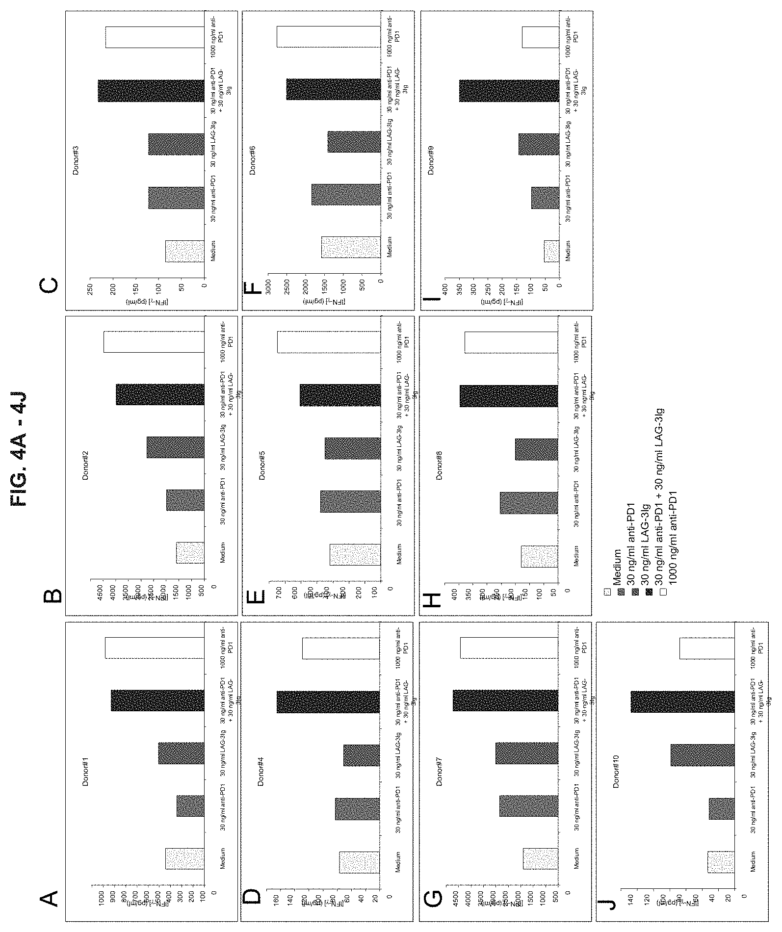
[0029] FIGS. 4A-4J. Show the effect of LAG-3Ig and anti-PD1 antibody on the secretion of IFN-.gamma. induced by antigenic stimulation. (A) through (J) show data from Donor 1 through Donor 10.
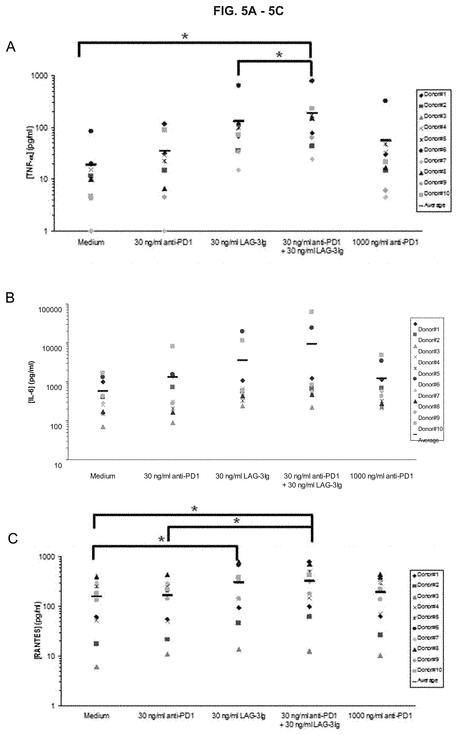
[0030] FIGS. 5A-5C. Show the effect of LAG-3Ig and anti-PD1 antibody on the secretion of (A) TNF.alpha., (B) IL-6, and (C) RANTES induced by antigenic stimulation.
[0031] FIGS. 6A-6E. Show the effect of LAG-3Ig and anti-PD1 antibody on the expression of activation markers induced by antigenic stimulation. Note that the condition for the final column in each graph of FIG. 6 is "1000 ng/ml anti-PD1+30 ng/ml LAG-3Ig", rather than "30 ng/ml anti-PD1+1000 ng/ml LAG-3Ig" as indicated in the Figure. (A) Percent (%) of Lag3+ in CD8+ cells; (B) % of CD69+ in CD8+ cells; (C) % of CD25 in CD8+ cells; (D) % of activation markers in CD8+ cells; (E) % of Lag3+, CD69+, CD25+ in CD8+ cells.
[0032] FIG. 7. Shows an illustration of derivatives of LAG-3 protein fused to Immunoglobulin Fc (IgFc) sequence.
[0033] FIGS. 8A-8B. Show binding of LAG-3 derivatives to MHC class II-positive cells. (A) Comparison of IMP321 with LAG-3 D1D4-linker2-IG, LAG-3 D1D2-linker2-Ig and hIG1. (B) Comparison of IMP321 with IMP321 R75A and hIG1.
[0034] FIG. 9. Shows inhibition of binding of a LAG-3 derivative to MHC class II-positive cells by antibodies that block binding of LAG-3 to MHC class II molecules.
[0035] FIGS. 10A-10B. Show activation of THP-1 cells by LAG-3 derivatives, as determined by CCL4 secretion. (A) Comparison of IMP321 with LAG-3 D1D4-linker2-IG, LAG-3 D1D2-linker2-Ig and hIG1. (B) Comparison of IMP321 with IMP321 R75A and hIG1.
[0036] FIGS. 11A-11B. Show activation of THP-1 cells by LAG-3 derivatives, as determined by TNF-.alpha. secretion. (A) Comparison of IMP321 with LAG-3 D1D4-linker2-IG, LAG-3 D1D2-linker2-Ig and hIG1. (B) Comparison of IMP321 with IMP321 R75A and hIG1.
[0037] FIGS. 12A-12B. (A) & (B) Inhibition of LAG derivative-induced monocyte activation by antibodies that block binding of LAG-3 to MHC class II molecules (17B4 and 11E3).
[0038] FIG. 13. Shows activation of antigen-presenting cells (APCs) by LAG-3 derivatives.
[0039] FIG. 14. Shows activation of CD8-positive T cells by LAG-3 derivatives.
[0040] FIG. 15. Shows the amino acid sequence of mature human LAG-3 protein (SEQ ID NO.: 1). The four extracellular Ig superfamily domains are at amino acid residues: 1-149 (D1); 150-239 (D2); 240-330 (D3); and 331-412 (D4). The amino acid sequence of the extra-loop structure of the D1 domain of human LAG-3 protein is shown underlined in bold.
[0041] FIGS. 16A-16E. Show the effect of LAG-3Ig and anti-PD-L1 antibody on the expression of activation markers induced by antigenic stimulation. (A) Percent (%) of Lag3+ in CD8+ cells; (B) % of CD69+ in CD8+ cells; (C) % of CD25 in CD8+ cells; (D) % of activation markers in CD8+ cells; (E) % of Lag3+, CD69+, CD25+ in CD8+ cells.
[0042] FIGS. 17A-17D. Show the effect of LAG-3Ig and different anti-PD-1 antibodies (Ab1 and Ab2) on the secretion of IFN-.gamma. and TNF-.alpha. induced by antigenic stimulation. (A) Effect of Ab1 on IFN-.gamma.; (B) Effect of Ab1 on TNF-.alpha.; (C) Effect of Ab2 on IFN-.gamma.; (D) Effect of Ab2 on TNF-.alpha..
[0043] FIGS. 18A-18H. Show the effect of LAG-3Ig and different anti-PD-L1 antibodies (Ab3, Ab4, Ab5, and Ab6) on the secretion of IFN-.gamma. and TNF-.alpha. induced by antigenic stimulation. (A) Effect of Ab3 on IFN-.gamma.; (B) Effect of Ab3 on TNF-.alpha.; (C) Effect of Ab4 on IFN-.gamma.; (D) Effect of Ab4 on TNF-.alpha.. (E) Effect of Ab5 on IFN-.gamma.; (F) Effect of Ab5 on TNF-.alpha.; (G) Effect of Ab6 on IFN-.gamma.; (H) Effect of Ab6 on TNF-.alpha..
[0044] FIG. 19. Shows the effect of different LAG-3 derivatives (IMP321, IMP321 R75A, LAG3 D1D4-linker2-Ig) and anti-PD-1 antibody on the secretion of IFN-.gamma. induced by antigenic stimulation.
DETAILED DESCRIPTION
[0045] According to the instant disclosure, there is provided a combined preparation, which comprises: (a) LAG-3 protein, or a derivative thereof that is able to bind to MHC class II molecules; and (b) a PD-1 pathway inhibitor.
[0046] The term "combined preparation" as used herein refers to a "kit of parts" in the sense that the combination components (a) and (b) as defined above can be dosed independently or by use of different fixed combinations with distinguished amounts of the combination components (a) and (b). The components can be administered simultaneously or one after the other. If the components are administered one after the other, preferably the time interval between administration is chosen such that the therapeutic effect of the combined use of the components is greater than the effect which would be obtained by use of only any one of the combination components (a) and (b).
[0047] The components of the combined preparation may be present in one combined unit dosage form, or as a first unit dosage form of component (a) and a separate, second unit dosage form of component (b). The ratio of the total amounts of the combination component (a) to the combination component (b) to be administered in the combined preparation can be varied, for example in order to cope with the needs of a patient sub-population to be treated, or the needs of the single patient, which can be due, for example, to the particular disease, age, sex, or body weight of the patient.
[0048] Preferably, there is at least one beneficial effect, for example an enhancing of the effect of the PD-1 pathway inhibitor, or an enhancing of the effect of the LAG-3 protein, or derivative thereof, or a mutual enhancing of the effect of the combination components (a) and (b), for example a more than additive effect, additional advantageous effects, fewer side effects, less toxicity, or a combined therapeutic effect compared with an effective dosage of one or both of the combination components (a) and (b), and very preferably a synergism of the combination components (a) and (b).
[0049] A combined preparation of the invention may be provided as a pharmaceutical combined preparation for administration to a mammal, preferably a human. The LAG-3 protein, or derivative thereof, may optionally be provided together with a pharmaceutically acceptable carrier, excipient, or diluent, and/or the PD-1 pathway inhibitor may optionally be provided together with a pharmaceutically acceptable carrier, excipient, or diluent.
[0050] The LAG-3, or derivative thereof, may be present at a dose which is a molar equivalent of 0.25-30 mg, 1-30 mg, or 6-30 mg of the LAG-3 derivative LAG-3Ig fusion protein IMP321. Doses of 6-30 mg per subcutaneous (s.c.) injection of IMP321 have been shown to be safe and provide an acceptable systemic exposure based on the results of pharmacokinetics data obtained in metastatic renal cell cancer patients. A blood concentration of IMP321 superior to 1 ng/ml for at least 24 hours after s.c. injection is obtained in patients injected with IMP321 doses of more than 6 mg.
[0051] A combined preparation of the invention may comprise a plurality of doses of the LAG-3 protein, or derivative thereof.
[0052] The PD-1 pathway inhibitor may be an agent that inhibits binding of PD-1 to PD-L1 and/or PD-L2. In particular, the agent may inhibit binding of human PD-1 to human PD-L1 and/or human PD-L2. The agent may inhibit binding of PD-1 to PD-L1 and/or PD-L2 by at least 50%, 60%, 70%, 80%, or 90%. Suitable assays for determining binding of PD-1 to PD-L1 or PD-L2, by Surface Plasmon Resonance (SPR) analysis, or flow cytometry analysis, are described in Ghiotto et al (Int. Immunol. August 2010; 22(8): 651-660). The agent may inhibit binding of PD-1 to PD-L1 and/or PD-L2, for example, by binding to PD-1, to PD-L1, or to PD-L2. The agent may be an antibody, suitably a monoclonal antibody, such as a human or humanized monoclonal antibody. The agent may be a fragment or derivative of an antibody that retains ability to inhibit binding of PD-1 to PD-L1 and/or PD-L2.
[0053] Examples of anti-PD-1 antibodies suitable for use according to the invention include: Pembrolizumab (MK-3475), a humanized monoclonal IgG4 antibody; Nivolumab, a fully human monoclonal IgG4 antibody; Pidilizumab (CT-011), a humanized IgG1 monoclonal antibody. An example of a PD-1 pathway inhibitor that binds to PD-1, but is not an antibody, is AMP-224. AMP-224 is a recombinant fusion protein of the extracellular domain of PD-L2 and the Fc region of human IgG. AMP-224 causes depletion of PD-1 high-expressing T cells. Examples of anti-PD-L1 antibodies suitable for use according to the invention include: BMS-936559, a fully human IgG4 monoclonal antibody; MED14736 (Durvalumab), a fully human, monoclonal antibody; MPDL3280A, a human monoclonal antibody containing an engineered IgG Fc domain to prevent ADCC; Avelumab (also known as MSB0010718C), a fully human anti-PD-L1 IgG1 monoclonal antibody.
[0054] The dose of the PD-1 pathway inhibitor will depend on the particular PD-1 pathway inhibitor being used. In general, a typically prescribed dose of a PD-1 pathway inhibitor for a human subject may be 0.1 to 10 mg/kg, for example 0.1 to 1 mg/kg, or 1 to 10 mg/kg. The term "typically prescribed dose" is used herein to include a dose which is the same as the dose, or within the dosage range, that is safe and therapeutically effective for administration to a subject (suitably a human subject) as a monotherapy, or that is approved by the appropriate regulatory authority for administration to a subject (suitably a human subject) as a monotherapy. Examples of typically prescribed human doses of known PD-1 pathway inhibitors when used as a monotherapy include:
Pembrolizumab (MK-3475): 2-10 mg/kg every two or three weeks. For example, the US FDA has approved administration of 2 mg/kg Keytruda (pembrolizumab) as an intravenous infusion over 30 minutes every 3 weeks; Nivolumab: 0.1-10 mg/kg every two weeks. For example, the US FDA has approved administration of 3 mg/kg Opdivo (nivolumab) as an intravenous infusion over 60 minutes every 2 weeks; BMS-936559: 0.3-10 mg/kg every two weeks.
[0055] The PD-1 pathway inhibitor may be administered by any suitable route, for example parenterally (including by subcutaneous, intravenous, or intramuscular injection). Currently approved or in-development PD-1 pathway inhibitors are administered as an intravenous infusion.
[0056] A combined preparation of the invention may comprise a plurality of doses of the PD-1 pathway inhibitor.
[0057] The LAG-3 protein may be an isolated natural or recombinant LAG-3 protein. The LAG-3 protein may comprise an amino sequence of LAG-3 protein from any suitable species, such as a primate or murine LAG-3 protein, but preferably a human LAG-3 protein. The amino acid sequence of human and murine LAG-3 protein is provided in FIG. 1 of Huard et al (Proc. Natl. Acad. Sci. USA, 11: 5744-5749, 1997). The sequence of human LAG-3 protein is repeated in FIG. 15 below (SEQ ID NO: 1). The amino acid sequences of the four extracellular Ig superfamily domains (D1, D2, D3, and D4) of human LAG-3 are also identified in FIG. 1 of Huard et al., at amino acid residues: 1-149 (D1); 150-239 (D2); 240-330 (D3); and 331-412 (D4).
[0058] Derivatives of LAG-3 protein include soluble fragments, variants, or mutants of LAG-3 protein that are able to bind MHC class II molecules. Several derivatives of LAG-3 protein are known that are able to bind to MHC class II molecules. Many examples of such derivatives are described in Huard et al (Proc. Natl. Acad. Sci. USA, 11: 5744-5749, 1997).
[0059] This document describes characterization of the MHC class II binding site on LAG-3 protein. Methods for making mutants of LAG-3 are described, as well as a quantitative cellular adhesion assay for determining the ability of LAG-3 mutants to bind class II-positive Daudi cells. Binding of several different mutants of LAG-3 to MHC class II molecules was determined. Some mutations were able to reduce class II binding, while other mutations increased the affinity of LAG-3 for class II molecules. Many of the residues essential for binding MHC class II proteins are clustered at the base of a large 30 amino acid extra-loop structure in the LAG-3 D1 domain. The amino acid sequence of the extra-loop structure of the D1 domain of human LAG-3 protein is GPPAAAPGHPLAPGPHPAAPSSWGPRPRRY (SEQ ID NO: 2), the underlined sequence in FIG. 15.
[0060] The LAG-3 protein derivative may comprise the 30 amino acid extra-loop sequence of the human LAG-3 D1 domain, or a variant of such sequence with one or more conservative amino acid substitutions. The variant may comprise amino acid sequence that has at least 70%, 80%, 90%, or 95% amino acid identity with the 30 amino acid extra-loop sequence of the human LAG-3 D1 domain.
[0061] The derivative of LAG-3 protein may comprise an amino acid sequence of domain D1, and optionally domain D2, of LAG-3 protein, preferably human LAG-3 protein.
[0062] The derivative of LAG-3 protein may comprise an amino acid sequence that has at least 70%, 80%, 90%, or 95% amino acid identity with domain D1, or with domain D1 and D2, of LAG-3 protein, preferably human LAG-3 protein.
[0063] The derivative of LAG-3 protein may comprise an amino acid sequence of domains D1, D2, D3, and optionally D4, of LAG-3 protein, preferably human LAG-3 protein.
[0064] The derivative of LAG-3 protein may comprise an amino acid sequence that has at least 70%, 80%, 90%, or 95% amino acid identity with domain D1, D2, and D3, or with domain D1, D2, D3, and D4, of LAG-3 protein, preferably human LAG-3.
[0065] Sequence identity between amino acid sequences can be determined by comparing an alignment of the sequences. When an equivalent position in the compared sequences is occupied by the same amino acid, then the molecules are identical at that position. Scoring an alignment as a percentage of identity is a function of the number of identical amino acids at positions shared by the compared sequences. When comparing sequences, optimal alignments may require gaps to be introduced into one or more of the sequences to take into consideration possible insertions and deletions in the sequences. Sequence comparison methods may employ gap penalties so that, for the same number of identical molecules in sequences being compared, a sequence alignment with as few gaps as possible, reflecting higher relatedness between the two compared sequences, will achieve a higher score than one with many gaps. Calculation of maximum percent identity involves the production of an optimal alignment, taking into consideration gap penalties.
[0066] Suitable computer programs for carrying out sequence comparisons are widely available in the commercial and public sector. Examples include MatGat (Campanella et al., 2003, BMC Bioinformatics 4: 29; program available from matgat the bitincka website), Gap (Needleman & Wunsch, 1970, J. Mol. Biol. 48: 443-453), FASTA (Altschul et al., 1990, J. Mol. Biol. 215: 403-410; program available from the EBI FASTA website), Clustal W 2.0 and X 2.0 (Larkin et al., 2007, Bioinformatics 23: 2947-2948; program available from the EBI ClustalW2 website) and EMBOSS Pairwise Alignment Algorithms (Needleman & Wunsch, 1970, supra; Kruskal, 1983, In: Time warps, string edits and macromolecules: the theory and practice of sequence comparison, Sankoff & Kruskal (eds), pp 1-44, Addison Wesley; programs available from the EBI ALIGN website). All programs may be run using default parameters.
[0067] For example, sequence comparisons may be undertaken using the "needle" method of the EMBOSS Pairwise Alignment Algorithms, which determines an optimum alignment (including gaps) of two sequences when considered over their entire length and provides a percentage identity score. Default parameters for amino acid sequence comparisons ("Protein Molecule" option) may be Gap Extend penalty: 0.5, Gap Open penalty: 10.0, Matrix: Blosum 62.
[0068] The sequence comparison may be performed over the full length of the reference sequence.
[0069] The LAG-3 protein derivative may be fused to Immunoglobulin Fc amino acid sequence, preferably human IgG1 Fc amino acid sequence, optionally by a linker amino acid sequence.
[0070] The ability of a derivative of LAG-3 protein to bind to MHC class II molecules may be determined using a quantitative cellular adhesion assay as described in Huard et al (supra). The affinity of a derivative of LAG-3 protein for MHC class II molecules may be at least 20%, 30%, 40%, 50%, 60%, 70%, 80%, 90%, or 100% of the affinity of human LAG-3 protein for class II molecules. Preferably the affinity of a derivative of LAG-3 protein for MHC class II molecules is at least 50% of the affinity of human LAG-3 protein for class II molecules. [0071] Examples of suitable derivatives of LAG-3 protein that are able to bind MHC class II molecules include derivatives comprising: [0072] amino acid residues 23 to 448 of the human LAG-3 sequence; [0073] amino acid sequence of domains D1 and D2 of LAG-3; [0074] amino acid sequence of domains D1 and D2 of LAG-3 with an amino acid substitution at one or more of the following positions: position 73 where ARG is substituted with GLU; position 75 where ARG is substituted with ALA or GLU; position 76 where ARG is substituted with GLU; position 30 where ASP is substituted with ALA; position 56 where HIS is substituted with ALA; position 77 where TYR is substituted with PHE; position 88 where ARG is substituted with ALA; position 103 where ARG is substituted with ALA; position 109 where ASP is substituted with GLU; position 115 where ARG is substituted with ALA; [0075] amino acid sequence of domain D1 of LAG-3 with a deletion of amino acid residues 54 to 66; [0076] a recombinant soluble human LAG-3Ig fusion protein (IMP321)--a 200-kDa dimer produced in Chinese hamster ovary cells transfected with a plasmid encoding for the extracellular domain of hLAG-3 fused to the human IgG1 Fc. The sequence of IMP321 is given in SEQ ID NO: 17 of US 2011/0008331.
[0077] According to the invention there is also provided a pharmaceutical composition, which comprises (a) LAG-3 protein, or a derivative thereof that is able to bind to MHC class II molecules; (b) a PD-1 pathway inhibitor; and (c) a pharmaceutically acceptable carrier, excipient, or diluent.
[0078] According to the invention there is further provided a combined preparation, or pharmaceutical composition, of the invention for use as a medicament.
[0079] The invention also provides a combined preparation, or pharmaceutical composition, of the invention for preventing, treating, or ameliorating cancer.
[0080] There is further provided according to the invention use of a combined preparation, or pharmaceutical composition, of the invention in the manufacture of a medicament for preventing, treating, or ameliorating cancer.
[0081] There is also provided according to the invention a method of preventing, treating, or ameliorating cancer, which comprises administering LAG-3 protein, or a derivative thereof that is able to bind to MHC class II molecules, and a PD-1 pathway inhibitor, to a subject in need of such prevention, treatment, or amelioration.
[0082] We have appreciated that combined preparations and compositions of the invention may also be used for the prevention, treatment, or amelioration of infection, in particular chronic or persistent infection.
[0083] During acute infection, activated pathogen-specific cytotoxic CD8 T lymphocytes (CTLs) proliferate and acquire effector functions, such as cytokine production and cytotoxic capability, which enable them to effectively clear infection. Following clearance, a small pool of pathogen-specific memory T cells remain that have the ability to very rapidly reactivate and acquire their killing functions following re-exposure to the same pathogen.
[0084] However, during chronic infection this does not occur, as pathogen-specific CTLs are found to be functionally deficient and unable to eliminate infection. These exhausted CTLs are defined by their impaired proliferative capacity, cytokine production and loss of cytotoxic capabilities (see FIG. 1(b), and review of Hofmeyer et al., Journal of Biomedicine and Biotechnology, Volume 2011, Article ID 451694).
[0085] This phenomenon was originally defined using a well-established mouse model of chronic viral infection in mice, lymphocytic choriomeningitis virus (LCMV) (Zajac, et al., The Journal of Experimental Medicine, vol. 188, no. 12, pp. 2205-2213, 1998; Gallimore, et al., The Journal of Experimental Medicine, vol. 187, no. 9, pp. 1383-1393, 1998). The Armstrong strain of LCMV causes an acute infection that is cleared by the immune system, generating a robust CTL memory. On the other hand, the Clone 13 strain of LCMV establishes a chronic infection in mice that renders CTLs exhausted and unable to clear infection. Additionally, as compared to normal T cells, exhausted CTLs have metabolic deficiencies and altered expression of genes involved in chemotaxis, adhesion, and migration (Wherry, et al., Immunity, vol. 27, no. 4, pp. 670-684, 2007).
[0086] In a study conducted to reveal mechanisms that lead to exhaustion, the genetic profile of exhausted CTLs from a chronic LMCV infection was compared to that of functional CTLs responding to an acute LCMV infection (Barber, et al., Nature, vol. 439, no. 7077, pp. 682-687, 2006). Exhausted CTLs were found to have significant overexpression of PD-1, whereas the functional LCMV-specific CTLs had no appreciable expression of PD-1. Expression of PD-1 was found to correlate with the defined functional impairment seen in exhausted T cells and, in turn, higher viral loads. Blocking the PD-1/PD-L1 pathway, with an anti-PD-L1 antibody, in chronically infected mice resulted in enhanced CTL response that caused a decrease in viral loads. PD-1 expression by exhausted CTLs is dependent on persisting antigen-specific stimulation, as loss of presentation of specific epitope during chronic infection leads to functional restoration and decreased PD-1 expression on epitope-specific CTLs (Blattman, et al., Journal of Virology, vol. 83, no. 9, pp. 4386-4394, 2009). Persistent antigen stimulation during chronic viral infection has a progressive effect on loss of CTL function and correlated increase in PD-1 expression, meaning that more exhausted CTLs (PD-1.sup.hi) are less susceptible to functional rescue by PD-1 blocking than others (PD-1.sup.int) (Blackburn, et al., Proceedings of the National Academy of Sciences of the United States of America, vol. 105, no. 39, pp. 15016-15021, 2008).
[0087] According to the invention, there is further provided a combined preparation, or pharmaceutical composition, of the invention for use in preventing, treating, or ameliorating an infection.
[0088] There is also provided according to the invention use of a combined preparation, or pharmaceutical composition, of the invention in the manufacture of a medicament for preventing, treating, or ameliorating an infection.
[0089] There is also provided according to the invention a method of preventing, treating, or ameliorating an infection, which comprises administering LAG-3 protein, or a derivative thereof that is able to bind to MHC class II molecules, and a PD-1 pathway inhibitor, to a subject in need of such prevention, treatment, or amelioration.
[0090] In particular embodiments, the infection is a chronic or persistent infection. The term "chronic or persistent infection" is used herein to refer to an infection by a pathogen that has induced a classical CTL response in an infected subject, but the infection has not been cleared, resulting in the presence of exhausted PD-1-expressing, pathogen-specific CTLs with impaired proliferative capacity, cytokine production and loss of cytotoxic capabilities. Examples of infections that may be treated according to the invention include viral, bacterial, fungal, or protozoan infections, especially chronic or persistent viral, bacterial, fungal, or protozoan infections.
[0091] The viral infection may be caused by, for example, an adenovirus, an adeno-associated virus, a B virus (macacine herpesvirus I), a BK virus, a bunyavirus, a chikungunya virus, a cocksackie virus, a coronavirus, a cytomegalovirus, an eastern equine encephalitis virus, an ebola virus, an enterovirus, an Epstein-Barr virus, a hantavirus, a hepatitis A virus, a hepatitis B virus, a hepatitis C virus, a hepatitis D virus, a hepatitis E virus, a herpes virus, a herpes simplex virus 1, a herpes simplex virus 2, a human foamy virus, a human herpes virus 3, a human herpes virus 5, a human herpes virus 6, a human herpes virus 7, a human immunodeficiency virus, a human papillomavirus, a human 1-lymphotropic virus, a human T-cell leukemia virus I, a human T-cell leukemia virus II, an influenza virus, a JC virus, a JEV, a Kaposi's sarcoma-associated herpesvirus, a Lassa virus, a lymphocytic choriomeningitis virus, a Marburg virus, a measles virus, a mumps virus, a Nipah virus, a norovirus, a Norwalk virus, an orthoreovirus, a parainfluenza virus, a parvovirus, a poliovirus, a rabies virus, a reovirus, a respiratory syncytial virus, rhinovirus, a Rift Valley fever virus, a rotavirus, rubella virus, a smallpox virus, a St Louis encephalitis virus, a variola major virus, a variola minor virus, a vericella-zoster virus, a West Nile virus, a western equine encephalitis virus, or a yellow fever virus).
[0092] In particular embodiments, the viral infection is caused by a hepatitis virus (for example, a hepatitis B virus, a hepatitis C virus), a lentivirus (for example, a human immunodeficiency virus), or a herpes virus (for example, a herpes simplex virus 1, a herpes simplex virus 2).
[0093] The bacterial infection may be caused by, for example, Escherichia coli, Clostridium difficile, Salmonella thyphimurium, Pseudomonas aeruginosa, Vibrio cholerae, Neisseria gonorrhoeae, Helicobacter pylori, Hemophilus influenzae, Shigella dysenteriae, Staphylococcus aureus, Mycobacterium tuberculosis, Streptococcus pneumonia, or Chlamydia trachomatis.
[0094] The fungal infection may be caused by, for example, Candida, Aspergillus, Cryptococcus, Coccidioides, Histoplasma, Pneumocystis, or Stachybotrys.
[0095] The protozoan infection may be caused by, for example, Amoebozoa, Excavata, Chromalveolata, Entamoeba, Plasmodium, Giardia, Trypanosoma, Coccidia, Besnoitia, Dicrocoelium, or Leishmania.
[0096] There is further provided according to the invention a combined preparation, or pharmaceutical composition, of the invention for use in preventing, treating, or ameliorating a disease, disorder, or condition that can be prevented, treated, or ameliorated by activation of T cells, in particular by activation of CD8-positive T cells.
[0097] There is also provided according to the invention use of a combined preparation, or pharmaceutical composition, of the invention in the manufacture of a medicament for preventing, treating, or ameliorating a disease, disorder, or condition that can be prevented, treated, or ameliorated by activation of T cells, in particular by activation of CD8-positive T cells.
[0098] There is also provided according to the invention a method of preventing, treating, or ameliorating a disease, disorder, or condition that can be prevented, treated, or ameliorated by activation of T cells, in particular by activation of CD8-positive T cells, which comprises administering LAG-3 protein, or a derivative thereof that is able to bind to MHC class II molecules, and a PD-1 pathway inhibitor, to a subject in need of such prevention, treatment, or amelioration.
[0099] In some embodiments, the disease, disorder, or condition that can be prevented, treated, or ameliorated by activation of T cells may exclude cancer.
[0100] There is also provided according to the invention a combined preparation, or pharmaceutical composition, of the invention for use in enhancing a T cell-mediated immune response, in particular a CD8-positive T cell-mediated immune response.
[0101] The invention also provides use of a combined preparation, or pharmaceutical composition, of the invention in the manufacture of a medicament for enhancing a T cell-mediated immune response, in particular a CD8-positive T cell-mediated immune response.
[0102] According to the invention there is further provided a method of enhancing a T cell-mediated immune response, in particular a CD8-positive T cell-mediated immune response, which comprises administering LAG-3 protein, or a derivative thereof that is able to bind to MHC class II molecules, and a PD-1 pathway inhibitor, to a subject in need of such enhanced T cell-mediated immune response.
[0103] In some embodiments, enhancement of the T cell-mediated immune response, or CD8-positive T cell-mediated immune response, may exclude the prevention, treatment, or amelioration of cancer.
[0104] The LAG-3 protein, or derivative thereof, and the PD-1 pathway inhibitor may be administered sequentially to the subject, i.e. the LAG-3 protein, or derivative thereof, may be administered before, with, or after the PD-1 pathway inhibitor.
[0105] The LAG-3 protein, or derivative thereof, and the PD-1 pathway inhibitor may be administered to the subject within 96 hours, 72 hours, 48 hours, 24 hours, or 12 hours, of each other.
[0106] Alternatively, the LAG-3 protein, or derivative thereof, and the PD-1 pathway inhibitor may be co-administered to the subject, for example as a composition comprising the LAG-3 protein, or derivative thereof, and the PD-1 pathway inhibitor, or by simultaneous administration of separate doses of the LAG-3 protein, or derivative thereof, and the PD-1 pathway inhibitor.
[0107] According to some embodiments, a plurality of doses of the LAG-3 protein, or derivative thereof, and/or a plurality of doses of the PD-1 pathway inhibitor, is administered to the subject.
[0108] According to some embodiments, a dose of the LAG-3 protein, or derivative thereof, is administered before, with, or after each administration of two or more doses of the PD-1 pathway inhibitor.
[0109] For example, a dose of the LAG-3 protein, or derivative thereof, may be administered within 96 hours, 72 hours, 48 hours, 24 hours, or 12 hours, of each administration of two or more doses of the PD-1 pathway inhibitor.
[0110] The choice of appropriate dosages of the components used in combination therapy according to the present invention can be determined and optimized by the skilled person, for example, by observation of the patient, including the patient's overall health, and the response to the combination therapy. Optimization, for example, may be necessary if it is determined that a patient is not exhibiting the desired therapeutic effect or conversely, if the patient is experiencing undesirable or adverse side effects that are too many in number or are of a troublesome severity.
[0111] The doses of the components used in combination therapy according to the invention should be chosen to provide a therapeutically effective amount of the components in combination.
[0112] An "effective amount" of the combination therapy may be an amount that results in a reduction of at least one pathological parameter associated with cancer. For example, in some embodiments, an effective amount of the combination therapy is an amount that is effective to achieve a reduction of at least about 10%, 20%, 30%, 40%, 50%, 60%, 70%, 80%, or 90%, in the pathological parameter, compared to the expected reduction in the parameter associated with the cancer without the combination therapy. For example, the pathological parameter may be tumor growth, or tumor growth rate.
[0113] Alternatively, an "effective amount" of the combination therapy may be an amount that results in an increase in a clinical benefit associated with cancer treatment. For example, in some embodiments, an "effective amount" of the combination therapy is an amount that is effective to achieve an increase of at least about 10%, 20%, 30%, 40%, 50%, 60%, 70%, 80%, or 90%, in the clinical benefit, compared to the expected clinical benefit without the combination therapy. For example, the clinical benefit may be tumor response rate, progression-free survival, overall survival, or increased sensitization to subsequent treatments.
[0114] Alternatively, an "effective amount" of the combination therapy may be an amount that results in a change of at least one beneficial parameter relating to cancer treatment. For example, in some embodiments, an "effective amount" of the combination therapy is an amount that is effective to achieve a change of at least about 10%, 20%, 30%, 40%, 50%, 60%, 70%, 80%, or 90%, in the parameter, compared to the expected change in the parameter relating to cancer treatment without the combination therapy. For example, the parameter may be an increase in the number of circulating tumor antigen-specific CD8.sup.+ T cells, or a reduction in the number of tumor antigen-specific regulatory T cells, or an increase in the number of activated T cells, in particular activated CD8.sup.+ T cells, a reduction in the number of exhausted antigen-specific CD8.sup.+ T cells, or an increase in the number of circulating functional (i.e. non-exhausted) antigen-specific CD8.sup.+ T cells.
[0115] In embodiments relating to treatment of infection, an "effective amount" of the combination therapy may be an amount that results in a reduction of at least one pathological parameter associated with infection. For example, in some embodiments, an effective amount of the combination therapy is an amount that is effective to achieve a reduction of at least about 10%, 20%, 30%, 40%, 50%, 60%, 70%, 80%, or 90%, in the pathological parameter, compared to the expected reduction in the parameter associated with the infection without the combination therapy. For example, the pathological parameter may be viral load (for example, the number of viral particles or amount of viral DNA per ml of blood), bacterial load (for example, the amount of bacterial DNA per ml of blood, or the number of bacterial colonies after a 1-21 day growth period on different agar plates).
[0116] Suitable methods of measuring viral and bacterial load are well-known to those of ordinary skill in the art. For example, methods of measuring viral load by ELISA are compared in Goldschmidt et al. (Clinical and Diagnostic Laboratory Immunology, July 1998, p. 513-518). Methods of measuring viral load using different commercial assays for detection of viral nucleic acid are compared in Holguin et al. (Eur J Clin Microbiol Infect Dis. 1999 April; 18(4):256-9) and Swenson et al. (J. Clin. Microbiol. 2014 February; 52(2): 517-523). An example of a paper describing measurement of bacterial load by real-time PCR is Nadkarni et al. (Microbiology (2002), 148, 257-266). This paper cites Bergey's Manual of Determinative Bacteriology, now superseded by Bergey's Manual of Systematic Bacteriology, 2.sup.nd Edition. A molecular bacterial load assay is described by Honeyborne et al. (J. Clin. Microbiol. 2011 49:3905-3911, and J. Clin. Microbiol. 2014 August; 52(8):3064-7). A list of FDA-approved screening assays to measure viral and bacterial loads can be found on the FDA website.
[0117] Alternatively, an "effective amount" of the combination therapy may be an amount that results in an increase in a clinical benefit associated with treatment of infection. For example, in some embodiments, an "effective amount" of the combination therapy is an amount that is effective to achieve an increase of at least about 10%, 20%, 30%, 40%, 50%, 60%, 70%, 80%, or 90%, in the clinical benefit, compared to the expected clinical benefit without the combination therapy.
[0118] Alternatively, an "effective amount" of the combination therapy may be an amount that results in a change of at least one beneficial parameter relating to treatment of infection. For example, in some embodiments, an "effective amount" of the combination therapy is an amount that is effective to achieve a change of at least about 10%, 20%, 30%, 40%, 50%, 60%, 70%, 80%, or 90%, in the parameter, compared to the expected change in the parameter relating to treatment without the combination therapy. For example, the parameter may be an increase in the number of activated T cells, in particular activated CD8.sup.+ T cells, an increase in the number of circulating functional (i.e. non-exhausted) antigen-specific CD8.sup.+ T cells, or a reduction in the number of exhausted antigen-specific CD8.sup.+ T cells, or a reduction in the number of antigen-specific regulatory T cells.
[0119] According to the invention, combination treatment may be employed to increase the therapeutic effect of the PD-1 pathway inhibitor, or LAG-3 protein, or derivative thereof, compared with the effect of the PD-1 pathway inhibitor, or LAG-3 protein, or derivative thereof, as a monotherapy, or to decrease the doses of the individual components in the resulting combinations while preventing or further reducing the risk of unwanted or harmful side effects of the individual components.
[0120] In one embodiment, the LAG-3 protein, or derivative thereof, and the PD-1 pathway inhibitor are each prescribed at a dose that is within a typically prescribed dose range for each compound as a monotherapy. The compounds may be prescribed as separate dosages or as a combination dosage. Such combinations provide increased efficacy compared with the effect of either compound as a monotherapy.
[0121] In another embodiment, the LAG-3 protein, or derivative thereof, and the PD-1 pathway inhibitor are each prescribed at a dose that is below a typically prescribed dose for each component as a monotherapy, but at doses that have therapeutic efficacy in combination. The components may be prescribed as separate dosages or as a combination dosage. The dosages of the components in combination may be selected to provide a similar level of therapeutic efficacy as the LAG-3 protein, or derivative thereof, or the PD-1 pathway inhibitor as a monotherapy, but with the advantage that the lower doses of the LAG-3 protein, or derivative thereof, and the PD-1 pathway inhibitor reduce the risk of adverse side effects compared to the prescribed dosages of each compound as a monotherapy.
[0122] In another embodiment, the prescribed dosage of the PD-1 pathway inhibitor is within a typically prescribed dose range for monotherapy, and the LAG-3 protein, or derivative thereof, is prescribed at a dosage that is below a typically prescribed dose for monotherapy.
[0123] In a further embodiment, the prescribed dosage of the PD-1 pathway inhibitor is below a typically prescribed dose for monotherapy, and the LAG-3 protein, or derivative thereof, is prescribed at a dosage that is within a typically prescribed dose range for monotherapy.
[0124] Preferred dosages below the typically prescribed dose for monotherapy are doses that are up to 50%, or up to 25%, of the typically prescribed dose. For example, dosages below the typically prescribed dose for monotherapy may be doses that are 1-50%, 1-25%, 1-10%, 2-50%, 2-25%, 2-10%, of the typically prescribed dose of the PD-1 pathway inhibitor and/or the LAG-3 protein, or derivative thereof.
[0125] A typically prescribed dose of a LAG-3 protein, or derivative thereof, for monotherapy in a human subject may be a dose that is molar equivalent of 0.25-30 mg, 1-30 mg, or 6-30 mg of the LAG-3 derivative LAG-3Ig fusion protein IMP321.
[0126] A typically prescribed dose of a PD-1 pathway inhibitor for monotherapy in a human subject may be 0.1 to 10 mg/kg, 0.1 to 1 mg/kg, or 1 to 10 mg/kg. For example, a typically prescribed dose of pembrolizumab for monotherapy in a human subject may be 2-10 mg/kg, for example 2 mg/kg, a typically prescribed dose of nivolumab for monotherapy in a human subject may be 0.1-10 mg/kg, for example 3 mg/kg, and a typically prescribed dose of BMS-936559 for monotherapy in a human subject may be 0.3-10 mg/kg.
[0127] In particular embodiments of combined preparations or compositions of the invention, the prescribed dosage of the PD-1 pathway inhibitor is below a typically prescribed dose for monotherapy, for example 1-50%, 1-25%, 1-20%, 1-10%, 2-50%, 2-25%, 2-20%, 2-10%, 0.1-50%, 0.1-25%, 0.1-20%, 0.1-10%, <20%, <10%, 0.1-<20%, 0.1-<10%, 0.01-<20%, or 0.01-<10% of the typically prescribed dose of the PD-1 pathway inhibitor.
[0128] Examples of suitable doses of the PD-1 pathway inhibitor and LAG-3 protein, or derivative thereof, according to the invention, are set out in Table 1.2 below:
TABLE-US-00002 TABLE 1.2 Examples of doses of the PD-1 pathway inhibitor and LAG-3 protein or derivative thereof, according to embodiments of combined preparations or compositions of the invention Human dose of LAG-3 protein or derivative thereof Type of PD-1 Dose of PD-1 pathway (given as a mg dose of pathway inhibitor: mg/kg [mg IMP321, or a molar inhibitor dose for 70 kg human] equivalent thereof) Anti-PD-1 0.001-5 mg/kg [0.07-350 mg] 0.25-30 mg antibody or 0.001-2.5 mg/kg [0.07-175 mg] 0.25-30 mg anti-PD-L1 0.001-1 mg/kg [0.07-70 mg] 0.25-30 mg antibody 0.001-<1 mg/kg [0.07-<70 mg] 0.25-30 mg 0.001-0.5 mg/kg [0.07-35 mg] 0.25-30 mg 0.001-0.1 mg/kg [0.07-7 mg] 0.25-30 mg 0.002-5 mg/kg [0.14-350 mg] 0.25-30 mg 0.002-2.5 mg/kg [0.14-175 mg] 0.25-30 mg 0.002-1 mg/kg [0.14-70 mg] 0.25-30 mg 0.002-<1 mg/kg [0.14-<70 mg] 0.25-30 mg 0.002-0.5 mg/kg [0.14-35 mg] 0.25-30 mg 0.002-0.1 mg/kg [0.14-7 mg] 0.25-30 mg Anti-PD-1 0.001-5 mg/kg [0.07-350 mg] 1-30 mg antibody or 0.001-2.5 mg/kg [0.07-175 mg] 1-30 mg anti-PD-L1 0.001-1 mg/kg [0.07-70 mg] 1-30 mg antibody 0.001-<1 mg/kg [0.07-<70 mg] 1-30 mg 0.001-0.5 mg/kg [0.07-35 mg] 1-30 mg 0.001-0.1 mg/kg [0.07-7 mg] 1-30 mg 0.002-5 mg/kg [0.14-350 mg] 1-30 mg 0.002-2.5 mg/kg [0.14-175 mg] 1-30 mg 0.002-1 mg/kg [0.14-70 mg] 1-30 mg 0.002-<1 mg/kg [0.14-<70 mg] 1-30 mg 0.002-0.5 mg/kg [0.14-35 mg] 1-30 mg 0.002-0.1 mg/kg [0.14-7 mg] 1-30 mg Anti-PD-1 0.001-5 mg/kg [0.07-350 mg] 6-30 mg antibody or 0.001-2.5 mg/kg [0.07-175 mg] 6-30 mg anti-PD-L1 0.001-1 mg/kg [0.07-70 mg] 6-30 mg antibody 0.001-<1 mg/kg [0.07-<70 mg] 6-30 mg 0.001-0.5 mg/kg [0.07-35 mg] 6-30 mg 0.001-0.1 mg/kg [0.07-7 mg] 6-30 mg 0.002-5 mg/kg [0.14-350 mg] 6-30 mg 0.002-2.5 mg/kg [0.14-175 mg] 6-30 mg 0.002-1 mg/kg [0.14-70 mg] 6-30 mg 0.002-<1 mg/kg [0.14-<70 mg] 6-30 mg 0.002-0.5 mg/kg [0.14-35 mg] 6-30 mg 0.002-0.1 mg/kg [0.14-7 mg] 6-30 mg Pembrolizumab 0.001-5 mg/kg [0.07-350 mg] 0.25-30 mg 0.001-2.5 mg/kg [0.07-175 mg] 0.25-30 mg 0.001-1 mg/kg [0.07-70 mg] 0.25-30 mg 0.001-<1 mg/kg [0.07-<70 mg] 0.25-30 mg 0.001-0.5 mg/kg [0.07-35 mg] 0.25-30 mg 0.001-0.1 mg/kg [0.07-7 mg] 0.25-30 mg 0.002-5 mg/kg [0.14-350 mg] 0.25-30 mg 0.002-2.5 mg/kg [0.14-175 mg] 0.25-30 mg 0.002-1 mg/kg [0.14-70 mg] 0.25-30 mg 0.002-<1 mg/kg [0.14-<70 mg] 0.25-30 mg 0.002-0.5 mg/kg [0.14-35 mg] 0.25-30 mg 0.002-0.1 mg/kg [0.14-7 mg] 0.25-30 mg Pembrolizumab 0.001-5 mg/kg [0.07-350 mg] 1-30 mg 0.001-2.5 mg/kg [0.07-175 mg] 1-30 mg 0.001-1 mg/kg [0.07-70 mg] 1-30 mg 0.001-<1 mg/kg [0.07-<70 mg] 1-30 mg 0.001-0.5 mg/kg [0.07-35 mg] 1-30 mg 0.001-0.1 mg/kg [0.07-7 mg] 1-30 mg 0.002-5 mg/kg [0.14-350 mg] 1-30 mg 0.002-2.5 mg/kg [0.14-175 mg] 1-30 mg 0.002-1 mg/kg [0.14-70 mg] 1-30 mg 0.002-1 mg/kg [0.14-70 mg] 1-30 mg 0.002-<1 mg/kg [0.14-<70 mg] 1-30 mg 0.002-0.5 mg/kg [0.14-35 mg] 1-30 mg Pembrolizumab 0.001-5 mg/kg [0.07-350 mg] 6-30 mg 0.001-2.5 mg/kg [0.07-175 mg] 6-30 mg 0.001-1 mg/kg [0.07-70 mg] 6-30 mg 0.001-<1 mg/kg [0.07-<70 mg] 6-30 mg 0.001-0.5 mg/kg [0.07-35 mg] 6-30 mg 0.001-0.1 mg/kg [0.07-7 mg] 6-30 mg 0.002-5 mg/kg [0.14-350 mg] 6-30 mg 0.002-2.5 mg/kg [0.14-175 mg] 6-30 mg 0.002-1 mg/kg [0.14-70 mg] 6-30 mg 0.002-<1 mg/kg [0.14-<70 mg] 6-30 mg 0.002-0.5 mg/kg [0.14-35 mg] 6-30 mg 0.002-0.1 mg/kg [0.14-7 mg] 6-30 mg Nivolumab 0.001-5 mg/kg [0.07-350 mg] 0.25-30 mg 0.001-2.5 mg/kg [0.07-175 mg] 0.25-30 mg 0.001-1 mg/kg [0.07-70 mg] 0.25-30 mg 0.001-<1 mg/kg [0.07-<70 mg] 0.25-30 mg 0.001-0.5 mg/kg [0.07-35 mg] 0.25-30 mg 0.001-0.1 mg/kg [0.07-7 mg] 0.25-30 mg 0.002-5 mg/kg [0.14-350 mg] 0.25-30 mg 0.002-2.5 mg/kg [0.14-175 mg] 0.25-30 mg 0.002-1 mg/kg [0.14-70 mg] 0.25-30 mg 0.002-<1 mg/kg [0.14-<70 mg] 0.25-30 mg 0.002-0.5 mg/kg [0.14-35 mg] 0.25-30 mg 0.002-0.1 mg/kg [0.14-7 mg] 0.25-30 mg Nivolumab 0.001-5 mg/kg [0.07-350 mg] 1-30 mg 0.001-2.5 mg/kg [0.07-175 mg] 1-30 mg 0.001-1 mg/kg [0.07-70 mg] 1-30 mg 0.001-<1 mg/kg [0.07-<70 mg] 1-30 mg 0.001-0.5 mg/kg [0.07-35 mg] 1-30 mg 0.001-0.1 mg/kg [0.07-7 mg] 1-30 mg 0.002-5 mg/kg [0.14-350 mg] 1-30 mg 0.002-2.5 mg/kg [0.14-175 mg] 1-30 mg 0.002-1 mg/kg [0.14-70 mg] 1-30 mg 0.002-<1 mg/kg [0.14-<70 mg] 1-30 mg 0.002-0.5 mg/kg [0.14-35 mg] 1-30 mg 0.002-0.1 mg/kg [0.14-7 mg] 1-30 mg Nivolumab 0.001-5 mg/kg [0.07-350 mg] 6-30 mg 0.001-2.5 mg/kg [0.07-175 mg] 6-30 mg 0.001-1 mg/kg [0.07-70 mg] 6-30 mg 0.001-<1 mg/kg [0.07-<70 mg] 6-30 mg 0.001-0.5 mg/kg [0.07-35 mg] 6-30 mg 0.001-0.1 mg/kg [0.07-7 mg] 6-30 mg 0.002-5 mg/kg [0.14-350 mg] 6-30 mg 0.002-2.5 mg/kg [0.14-175 mg] 6-30 mg 0.002-1 mg/kg [0.14-70 mg] 6-30 mg 0.002-<1 mg/kg [0.14-<70 mg] 6-30 mg 0.002-0.5 mg/kg [0.14-35 mg] 6-30 mg 0.002-0.1 mg/kg [0.14-7 mg] 6-30 mg BMS-936559 0.001-5 mg/kg [0.07-350 mg] 0.25-30 mg 0.001-2.5 mg/kg [0.07-175 mg] 0.25-30 mg 0.001-1 mg/kg [0.07-70 mg] 0.25-30 mg 0.001-<1 mg/kg [0.07-<70 mg] 0.25-30 mg 0.001-0.5 mg/kg [0.07-35 mg] 0.25-30 mg 0.001-0.1 mg/kg [0.07-7 mg] 0.25-30 mg 0.002-5 mg/kg [0.14-350 mg] 0.25-30 mg 0.002-2.5 mg/kg [0.14-175 mg] 0.25-30 mg 0.002-1 mg/kg [0.14-70 mg] 0.25-30 mg 0.002-<1 mg/kg [0.14-<70 mg] 0.25-30 mg 0.002-0.5 mg/kg [0.14-35 mg] 0.25-30 mg 0.002-0.1 mg/kg [0.14-7 mg] 0.25-30 mg BMS-936559 0.001-5 mg/kg [0.07-350 mg] 1-30 mg 0.001-2.5 mg/kg [0.07-175 mg] 1-30 mg 0.001-1 mg/kg [0.07-70 mg] 1-30 mg 0.001-<1 mg/kg [0.07-<70 mg] 1-30 mg 0.001-0.5 mg/kg [0.07-35 mg] 1-30 mg 0.001-0.1 mg/kg [0.07-7 mg] 1-30 mg 0.002-5 mg/kg [0.14-350 mg] 1-30 mg 0.002-2.5 mg/kg [0.14-175 mg] 1-30 mg 0.002-1 mg/kg [0.14-70 mg] 1-30 mg 0.002-<1 mg/kg [0.14-<70 mg] 1-30 mg 0.002-0.5 mg/kg [0.14-35 mg] 1-30 mg 0.002-0.1 mg/kg [0.14-7 mg] 1-30 mg BMS-936559 0.001-5 mg/kg [0.07-350 mg] 6-30 mg 0.001-2.5 mg/kg [0.07-175 mg] 6-30 mg 0.001-1 mg/kg [0.07-70 mg] 6-30 mg 0.001-<1 mg/kg [0.07-<70 mg] 6-30 mg 0.001-0.5 mg/kg [0.07-35 mg] 6-30 mg 0.001-0.1 mg/kg [0.07-7 mg] 6-30 mg 0.002-5 mg/kg [0.14-350 mg] 6-30 mg 0.002-2.5 mg/kg [0.14-175 mg] 6-30 mg 0.002-1 mg/kg [0.14-70 mg] 6-30 mg 0.002-<1 mg/kg [0.14-<70 mg] 6-30 mg 0.002-0.5 mg/kg [0.14-35 mg] 6-30 mg 0.002-0.1 mg/kg [0.14-7 mg] 6-30 mg MPDL3280A 0.001-5 mg/kg [0.07-350 mg] 0.25-30 mg 0.001-2.5 mg/kg [0.07-175 mg] 0.25-30 mg 0.001-1 mg/kg [0.07-70 mg] 0.25-30 mg 0.001-<1 mg/kg [0.07-<70 mg] 0.25-30 mg 0.001-0.5 mg/kg [0.07-35 mg] 0.25-30 mg 0.001-0.1 mg/kg [0.07-7 mg] 0.25-30 mg 0.002-5 mg/kg [0.14-350 mg] 0.25-30 mg 0.002-2.5 mg/kg [0.14-175 mg] 0.25-30 mg 0.002-1 mg/kg [0.14-70 mg] 0.25-30 mg 0.002-<1 mg/kg [0.14-<70 mg] 0.25-30 mg 0.002-0.5 mg/kg [0.14-35 mg] 0.25-30 mg 0.002-0.1 mg/kg [0.14-7 mg] 0.25-30 mg MPDL3280A 0.001-5 mg/kg [0.07-350 mg] 1-30 mg 0.001-2.5 mg/kg [0.07-175 mg] 1-30 mg 0.001-1 mg/kg [0.07-70 mg] 1-30 mg 0.001-<1 mg/kg [0.07-<70 mg] 1-30 mg 0.001-0.5 mg/kg [0.07-35 mg] 1-30 mg 0.001-0.1 mg/kg [0.07-7 mg] 1-30 mg 0.002-5 mg/kg [0.14-350 mg] 1-30 mg 0.002-2.5 mg/kg [0.14-175 mg] 1-30 mg 0.002-1 mg/kg [0.14-70 mg] 1-30 mg 0.002-<1 mg/kg [0.14-<70 mg] 1-30 mg 0.002-0.5 mg/kg [0.14-35 mg] 1-30 mg 0.002-0.1 mg/kg [0.14-7 mg] 1-30 mg MPDL3280A 0.001-5 mg/kg [0.07-350 mg] 6-30 mg 0.001-2.5 mg/kg [0.07-175 mg] 6-30 mg 0.001-1 mg/kg [0.07-70 mg] 6-30 mg 0.001-<1 mg/kg [0.07-<70 mg] 6-30 mg 0.001-0.5 mg/kg [0.07-35 mg] 6-30 mg 0.001-0.1 mg/kg [0.07-7 mg] 6-30 mg 0.002-5 mg/kg [0.14-350 mg] 6-30 mg 0.002-2.5 mg/kg [0.14-175 mg] 6-30 mg 0.002-1 mg/kg [0.14-70 mg] 6-30 mg 0.002-<1 mg/kg [0.14-<70 mg] 6-30 mg 0.002-0.5 mg/kg [0.14-35 mg] 6-30 mg 0.002-0.1 mg/kg [0.14-7 mg] 6-30 mg
[0129] The LAG-3 derivative may be any of the LAG-3 derivatives described above, or shown in FIG. 7. In particular embodiments, the LAG-3 derivative is IMP321.
[0130] When administered in separate dosages, the LAG-3 protein, or derivative thereof, and the PD-1 pathway inhibitor may be administered substantially simultaneously (for example, within about 60 minutes, about 50 minutes, about 40 minutes, about 30 minutes, about 20 minutes, about 10 minutes, about 5 minutes, or about 1 minute of each other) or separated in time by about 1 hour, about 2 hours, about 4 hours, about 6 hours, about 10 hours, about 12 hours, about 24 hours, about 36 hours, about 72 hours, or about 96 hours, or more.
[0131] The skilled person will be able to determine, and optimise, a suitable time course for sequential administration, depending on the particular combination of the LAG-3 protein, or derivative thereof, and the PD-1 pathway inhibitor. The time course is preferably selected such that there is at least one beneficial effect, for example an enhancing of the effect of the LAG-3 protein, or derivative thereof, or the PD-1 pathway inhibitor, or a mutual enhancing of the effect of the combination components, for example a more than additive effect, additional advantageous effects, fewer side effects, less toxicity, or a combined therapeutic effect compared with a non-effective dosage of one or both of the combination components, and very preferably a synergism of the combination components.
[0132] It will be appreciated that the optimum time course will depend on factors such as the time taken for the peak plasma concentration of the compound to be reached after administration, and the elimination half-life of each compound. Preferably the time difference is less than the half-life of the first component to be administered.
[0133] The skilled person will also be able to determine appropriate timing for administration. In certain embodiments, the PD-1 pathway inhibitor may be administered in the morning, and the LAG-3 protein, or derivative thereof, administered at least once later in the day. In other embodiments, the PD-1 pathway inhibitor and LAG-3 protein, or derivative thereof, may be administered at substantially the same time.
[0134] In some embodiments, the PD-1 pathway inhibitor may be administered to the subject, for example, by a medical practitioner, and the subject may be provided with a dose of the LAG-3 protein, or derivative thereof, for example in a pre-filled syringe, to administer later (for example later the same day, or the next day).
[0135] The PD-1 pathway inhibitor and LAG-3 protein, or derivative thereof, may be administered daily, weekly, every two weeks, every three weeks, monthly, every 2 months, 3 months, 4 months, 5 months, 6 months, 7 months, 8 months, 9 months, 10 months, 11 months, 1 year, 2 years, 3 years, 4 years, 5 years, or more.
[0136] The subject may receive doses of the PD-1 pathway inhibitor and LAG-3 protein, or derivative thereof, over a period of weeks, months, or years. For example, 1 week, 2 weeks, 3 weeks, 1 month, 2 months, 3 months, 4 months, 5 months, 6 months, 7 months, 8 months, 9 months, 10 months, 11 months, 1 year, 2 years, 3 years, 4 years, 5 years, or more.
[0137] The subject may be a mammalian subject, suitably a human subject.
[0138] Cancers that may be treated according to the invention include cancers in which tumor cells of the cancer express PD-L1 and/or PD-L2 (i.e. PD-L1- and/or PD-L2-positive cancers).
[0139] PD-L1 expression has been detected in lung, ovary, renal, and colon carcinomas and in malignant melanoma but not in normal tissues, including the lung, uterus, kidney, colon, or skin (Benson et al, Blood 116, 2286-2294 (2010); Blank et al, Int. J. Cancer 119, 317-327 (2006); Dong, et al, Nat. Med. 8, 793-800 (2002)). PD-L1 expression by tumor cells is associated with a worse prognosis in breast cancer, gastric cancer, esophageal cancer, hepatocellular carcinoma, malignant melanoma, ovarian cancer, pancreatic cancer, renal cell carcinoma, and urothelial cancer (Zou & Chen, Nat. Rev. Immunol. 8, 467-477 (2008)). There is also evidence that human tumors can express PD-L2 (Rozali, et al, Clin. Dev. Immunol. 2012, 656340 (2012); Karim, et al, Clin. Cancer Res. 15, 6341-6347 (2009)). Non-small cell lung cancer- (NSCLC-) associated fibroblasts constitutively express both PD-L1 and PD-L2. Decreased survival in patients with PD-L2-positive (vs. PD-L2-negative), esophageal, ovarian, or hepatocellular cancer has also been described.
[0140] Cancers that may be treated according to the invention also include cancers in which tumor-infiltrating lymphocytes (TILs), especially CD8.sup.+ TILs, express PD-1, or cancers in which TILs express higher levels of PD-1 than circulating lymphocytes.
[0141] In both NSCLC and melanoma patients, higher levels of PD-1 were observed on TILs than on circulating lymphocytes (Blank, et al, Int. J. Cancer 119, 317-327 (2006); Zhang et al, Cell. Mol. Immunol. 7, 389-395 (2010)). In the peripheral blood of vaccinated melanoma patients, both melanoma antigen-specific cytotoxic lymphocytes and Tregs expressed PD-1 (Wang, et al, Int. Immunol. 21, 1065-1077 (2009)). There was also a negative correlation between tumor PD-L2 expression and the presence of CD8.sup.+ TILs in esophageal cancer (Rozali, et al, Clin. Dev. Immunol. 2012, 656340 (2012)).
[0142] CD8.sup.+ TILs isolated from NSCLCs had increased expression of PD-1 and impaired functional responses (in vitro proliferation and inflammatory cytokine production) as compared with circulating CD8.sup.+ T cells or CD8.sup.+ T cells from healthy volunteers. Addition of anti-PD-L1 antibody significantly improved the ability of the CD8.sup.+ TILs to proliferate and produce interferon-.gamma. in vitro (Zhang, et al, Cell. Mol. Immunol. 7, 389-395 (2010)). In a similar study using cultures of tumor-derived dendritic cells and TILs from ovarian cancer patients, addition of anti-PD-L1 antibody significantly increased interferon-.gamma. production by TILs in response to tumor antigens. When these TILs were transferred to immunodeficient mice bearing the ovarian tumors, reduced tumor growth was seen as compared with that of mice in control groups (Curiel, et al, Nat. Med. 9, 562-567 (2003)).
[0143] In particular, cancers that may be treated according to the invention include skin, lung (especially squamous or nonsquamous NSCLC), ovarian, renal, colon, colorectal, breast, gastric, esophageal, pancreatic, bladder, urothelial, and liver cancer.
[0144] Other examples of cancers that may be treated according to the invention include a melanoma (for example, metastatic malignant melanoma), a prostate cancer (for example hormone refractory prostate adenocarcinoma), a head and neck cancer (for example, squamous cell carcinoma of the head and neck), a cervical cancer, a thyroid cancer, a glioblastoma, a glioma, leukemia, a lymphoma (for example, a B cell lymphoma), an adrenal gland cancer, an AIDS-associated cancer, an alveolar soft part sarcoma, an astrocytic tumor, bone cancer, a brain and spinal cord cancer, a metastatic brain tumor, a carotid body tumor, a chondrosarcoma, a chordoma, a chromophobe renal cell carcinoma, a clear cell carcinoma, cutaneous benign fibrous histiocytoma, a desmoplastic small round cell tumor, an ependymoma, a Ewing's tumor, an extraskeletal myxoid chondrosarcoma, a fibrogenesis imperfecta ossium, a fibrous dysplasia of the bone, a gallbladder or bile duct cancer, a gestational trophoblastic disease, a germ cell tumor, a haematological malignancy, hepatocellular carcinoma, an islet cell tumor, a Kaposi's sarcoma, a kidney cancer, a lipoma/benign lipomatous tumor, a liposarcoma/malignant lipomatous tumor, a medulloblastoma, a meningioma, a Merkel cell carcinoma, a multiple endocrine neoplasia, a multiple myeloma, a myelodysplasia syndrome, a neuroblastoma, a neuroendocrine tumor, a papillary thyroid carcinoma, a parathyroid tumor, a pediatric cancer, a peripheral nerve sheath tumor, a phaeochromocytoma, a pituitary tumor, a prostate cancer, a posterior uveal melanoma, a rare hematologic disorder, a renal metastatic cancer, a rhabdoid tumor, a rhabdomysarcoma, a sarcoma, a soft-tissue sarcoma, a squamous cell cancer, a stomach cancer, a synovial sarcoma, a testicular cancer, a thymic carcinoma, a thymoma, a thyroid metastatic cancer, or a uterine cancer.
[0145] In general, the components of a combination of the invention, or a composition of the invention, may be administered by known means, in any suitable formulation, by any suitable route. In some embodiments, the LAG-3 protein, or derivative thereof, is administered parenterally (including by subcutaneous, intravenous, or intramuscular injection). In some embodiments, the PD-1 pathway inhibitor is administered intravenously. In particular embodiments, the LAG-3 protein, or derivative thereof, is administered subcutaneously, and the PD-1 pathway inhibitor is administered intravenously.
[0146] Suitable pharmaceutical compositions and dosage forms may be prepared using conventional methods known to those in the field of pharmaceutical formulation and described in the relevant texts and literature, for example, in Remington: The Science and Practice of Pharmacy (Easton, Pa.: Mack Publishing Co., 1995).
[0147] It is especially advantageous to formulate combinations or compositions of the invention in unit dosage form for ease of administration and uniformity of dosage. The term "unit dosage forms" as used herein refers to physically discrete units suited as unitary dosages for the individuals to be treated. That is, the compositions are formulated into discrete dosage units each containing a predetermined, "unit dosage" quantity of an active agent calculated to produce the desired therapeutic effect in association with the required pharmaceutical carrier. The specifications of unit dosage forms of the invention are dependent on the unique characteristics of the active agent to be delivered. Dosages can further be determined by reference to the usual dose and manner of administration of the ingredients. It should be noted that, in some cases, two or more individual dosage units in combination provide a therapeutically effective amount of the active agent, for example, two tablets or capsules taken together may provide a therapeutically effective dosage, such that the unit dosage in each tablet or capsule is approximately 50% of the therapeutically effective amount.
[0148] Preparations according to the invention for parenteral administration include sterile aqueous and non-aqueous solutions, suspensions, and emulsions. Injectable aqueous solutions contain the active agent in water-soluble form. Examples of non-aqueous solvents or vehicles include fatty oils, such as olive oil and corn oil, synthetic fatty acid esters, such as ethyl oleate or triglycerides, low molecular weight alcohols such as propylene glycol, synthetic hydrophilic polymers such as polyethylene glycol, liposomes, and the like. Parenteral formulations may also contain adjuvants such as solubilizers, preservatives, wetting agents, emulsifiers, dispersants, and stabilizers, and aqueous suspensions may contain substances that increase the viscosity of the suspension, such as sodium carboxymethyl cellulose, sorbitol, and dextran. Injectable formulations may be rendered sterile by incorporation of a sterilizing agent, filtration through a bacteria-retaining filter, irradiation, or heat. They can also be manufactured using a sterile injectable medium. The active agent may also be in dried, e.g., lyophilized, form that may be rehydrated with a suitable vehicle immediately prior to administration via injection.
[0149] In addition to the formulations described previously, the active agent may be formulated as a depot preparation for controlled release of the active agent, preferably sustained release over an extended time period. These sustained release dosage forms are generally administered by implantation (for example, subcutaneously or intramuscularly or by intramuscular injection).
[0150] Combined preparations of the invention may be packaged with instructions for administration of the components on the combination. The instructions may be recorded on a suitable recording medium or substrate. For example, the instructions may be printed on a substrate, such as paper or plastic. The instructions may be present as a package insert, in the labeling of the container or components thereof (i.e., associated with the packaging or sub-packaging). In other embodiments, the instructions are present as an electronic storage data file present on a suitable computer readable storage medium, for example, CD-ROM, diskette. Some or all components of the combined preparation may be packaged in suitable packaging to maintain sterility.
[0151] Embodiments of the invention are described below, by way of example only, with reference to the accompanying drawings.
[0152] In the Examples, Tables, and Figures below, the term "anti-PD1 antibody" is used synonymously with "anti-PD-1 antibody", and the term "anti-PDL1 antibody" is used synonymously with "anti-PD-L1 antibody".
EXAMPLES
Example 1
Effect of LAG-3Ig and Anti-PD1 Antibody on the Secretion of IFN-.gamma. Induced by Antigenic Stimulation
[0153] This example demonstrates the effect of a soluble derivative of LAG-3 (LAG-3Ig, also known as IMP321), and an anti-PD1 antibody, on T-cell activation in vitro using an IFN-.gamma. secretion assay. Peripheral blood mononuclear cells (PBMCs) include lymphocytes (T cells, B cells, and NK cells), monocytes, and dendritic cells. IFN-.gamma. is predominantly secreted by activated CD4+ and CD8+ memory and effector T cells and by NK cells upon activation. After re-stimulation with specific antigen in vitro, secretion of IFN-.gamma. is induced.
[0154] PBMCs from three healthy donors (0.2.times.10.sup.6 cells/well, at 1.times.10.sup.6M/ml in Complete Roswell Park Memorial Institute (RPMI)+10% Foetal Bovine Serum (FBS)) were incubated with a pool of peptides covering the sequence of human cytomegalovirus (CMV) pp65 in triplicate (PepTivator.RTM. CMV pp65 form Miltenyi Biotec, Cat. #130-093-435), in the presence or the absence of 30 ng/ml of LAG-3Ig and indicated concentrations of anti-PD1 mAb (clone EH12.1, BD biosciences, Cat. #562138). The pool of peptides consisted mainly of 15-mer sequences, with an 11 amino acid overlap, covering the complete sequence of the pp65 protein of human CMV strain AD169 (Swiss-Prot Acc. No. P06725).
[0155] The T cell response was evaluated by measuring the concentration of IFN-.gamma. in cell supernatants two days post-stimulation using BD Cytometric Bead Array.
[0156] The concentrations of IFN-.gamma. present in the pooled triplicates for each donor are recorded in Table 2 below. FIG. 2 shows the concentrations of IFN-.gamma. plotted against the concentration of anti-PD1 mAb for each donor.
TABLE-US-00003 TABLE 2 Secretion of IFN-.gamma. induced by antigen in the presence of anti-PD1 antibody with and without LAG-3Ig [IFN-.gamma.] (pg/ml) Donor No: Anti-PD1 1 2 3 antibody No 30 ng/ml No 30 ng/ml No 30 ng/ml (ng/ml) LAG-3Ig LAG-3Ig LAG-3Ig LAG-3Ig LAG-3Ig LAG-3Ig 0 345 409 1095 2269 85 122 3 510 2812 188 10 350 785 1292 3151 78 211 30 246 828 1491 3484 122 232 100 439 2467 149 300 657 3856 1000 881 3970 216
[0157] The results show that secretion of IFN-.gamma. was dramatically increased when the PBMCs were incubated in the presence of 30 ng/ml LAG-3Ig and lower concentrations of anti-PD1 antibody, compared with anti-PD1 antibody alone. For example, for each donor, the increase in concentration of IFN-.gamma. above the background level (i.e. the concentration of IFN-.gamma. in the absence of anti-PD1 and LAG-3Ig) in the presence of 30 ng/ml LAG-3Ig and 30 ng/ml anti-PD1 antibody was greater than the sum of the corresponding increase in the presence of 30 ng/ml LAG-3Ig alone and 30 ng/ml anti-PD1 antibody alone, as shown in Table 3 below. The effect of the combination of LAG-3Ig and anti-PD1 antibody for each donor was, therefore, synergistic.
TABLE-US-00004 TABLE 3 Increase in IFN-.gamma. concentration above background induced by antigen in the presence of 30 ng/ml anti-PD1 antibody and/or 30 ng/ml LAG-3Ig Donor No. Stimulation condition 1 2 3 30 ng/ml LAG-3Ig 64 1174 37 30 ng/ml anti-PD-1 -99 396 37 antibody 30 ng/ml LAG-3Ig + 483 2389 147 30 ng/ml anti-PD-1 antibody
[0158] The results also show that secretion of IFN-.gamma. induced by a combination of LAG-3Ig and a relatively low concentration of anti-PD1 antibody (30 ng/ml) was equivalent to secretion of IFN-.gamma. induced by a much higher concentration (300 ng/ml-1000 ng/ml, 10 to over 30 times higher) of anti-PD1 antibody alone. For Donor Nos. 1 and 3, similar concentrations of IFN-.gamma. were secreted when PBMCs were incubated with 30 ng/ml anti-PD1 and 30 ng/ml LAG-3Ig compared with 1000 ng/ml anti-PD1 antibody alone. For Donor No. 2, similar concentrations of IFN-.gamma. were secreted when PBMCs were incubated with 30 ng/ml anti-PD1 and 30 ng/ml LAG-3Ig compared with 300 ng/ml and 1000 ng/ml anti-PD1 antibody alone.
[0159] It was concluded from these results that in vitro T cell response (as measured by IFN-.gamma. secretion) induced by relatively low doses of anti-PD1 antibody is synergistically increased (by approximately .about.7.5, 1.5, and 2 times for Donor Nos. 1, 2, and 3, respectively) by a soluble LAG-3 derivative. It was also concluded that an equivalent in vitro T cell response is obtained using approximately 10-30 times less anti-PD1 antibody if this is combined with a soluble LAG-3 derivative.
Example 2
Effect of LAG-3Ig and Anti-PD1 Antibody on the Secretion of IFN-.gamma. Induced by Antigenic Stimulation
[0160] This example demonstrates the effect of a soluble derivative of LAG-3 (LAG-3Ig), and an anti-PD1 antibody, on T-cell activation in vitro using an IFN-.gamma. secretion assay.
[0161] PBMCs from 10 healthy donors (0.2.times.10.sup.6 cells/well, at 1.times.10.sup.6M/ml in Complete RPMI+10% FBS) were incubated with a pool of peptides covering the sequence of CMV pp65 in triplicate (PepTivator.RTM. CMV pp65 form Miltenyi Biotec, Cat. #130-093-435), without any additive (Medium), with 30 ng/ml or 1000 ng/ml of anti-PD1 mAb (clone EH12.1, BD biosciences Cat. #562138), with 30 ng/ml of LAG-3Ig, or with 30 ng/ml of LAG-3Ig and 30 ng/ml of anti-PD1 mAb. The T cell response was evaluated by measuring the concentration of IFN-.gamma. in cell supernatants two days post-stimulation using BD Cytometric Bead Array.
[0162] The concentrations of IFN-.gamma. in the pooled triplicates for each condition of stimulation, for each donor, are recorded in Table 4 below. The mean of the results obtained for the 10 donors are shown in Table 5. The results for each donor are also plotted in FIGS. 3 and 4. The statistical differences (*p<0.05) are shown in black in FIG. 3.
TABLE-US-00005 TABLE 4 Secretion of IFN-.gamma. induced by antigen in the presence of anti-PD1 antibody with and without LAG-3Ig Stimulation condition [IFN-.gamma.] (pg/ml) [Anti-PD1] [LAG-3Ig] Donor (ng/ml) (ng/ml) 1 2 3 4 5 6 7 8 9 10 -- -- 345 1095 85 57 319 1583 1405 130 53 33 30 -- 246 1491 122 63 378 1836 2355 204 97 31 -- 30 409 2269 122 51 350 1406 2511 150 142 79 30 30 828 3484 232 145 506 2510 4217 347 350 128 1000 -- 881 3970 216 109 650 2765 3913 329 130 68
TABLE-US-00006 TABLE 5 Mean IFN-.gamma. concentration for each different stimulation condition Stimulation condition Increase in mean [Anti-PD1] [LAG-3Ig] Mean [IFN-.gamma.] [IFN-.gamma.] (pg/ml) above (ng/ml) (ng/ml) (pg/ml) mean background -- -- 510 -- 30 -- 682 172 -- 30 749 239 30 30 1275 765 1000 -- 1303 793
[0163] The results show that secretion of IFN-.gamma. was much higher for each donor when the PBMCs were incubated in the presence of 30 ng/ml LAG-3Ig and 30 ng/ml anti-PD1 antibody, compared with 30 ng/ml LAG-3Ig or 30 ng/ml anti-PD1 antibody alone. Table 5 shows that the increase in mean concentration of IFN-.gamma. above the mean background level (i.e. the mean concentration of IFN-.gamma. in the absence of anti-PD1 and LAG-3Ig) in the presence of 30 ng/ml LAG-3Ig and 30 ng/ml anti-PD1 antibody was greater than the sum of the corresponding increase in the presence of 30 ng/ml LAG-3Ig alone and 30 ng/ml anti-PD1 antibody alone (i.e. 765>239+172). The effect of the combination of LAG-3Ig and anti-PD1 antibody was, therefore, synergistic.
[0164] The results also show that secretion of IFN-.gamma. induced by a combination of LAG-3Ig and a relatively low concentration of anti-PD1 antibody (30 ng/ml) was equivalent to secretion of IFN-.gamma. induced by a much higher concentration (1000 ng/ml, over 30 times higher) of anti-PD1 antibody alone.
[0165] It was concluded from these results that in vitro T cell response (as measured by IFN-.gamma. secretion) induced by relatively low doses of anti-PD1 antibody is synergistically increased (by approximately 2 times on average) by a soluble LAG-3 derivative. It was also concluded that an equivalent in vitro T cell response is obtained using over 30 times less anti-PD1 antibody if this is combined with a soluble LAG-3 derivative.
Example 3
Effect of LAG-3Ig and Anti-PD1 Antibody on the Secretion of TNF-.alpha., IL-6, RANTES Induced by Antigenic Stimulation
[0166] This example demonstrates the effect of a soluble derivative of LAG-3 (LAG-3Ig), and an anti-PD1 antibody, on T-cell activation in vitro by measuring secretion of TNF-.alpha., IL-6 and RANTES (CCL5).
[0167] PBMCs from 10 healthy donors (0.2.times.10.sup.6 cells/well, at 1.times.10.sup.6M/ml in Complete RPMI+10% FBS) were incubated with a pool of peptides covering the sequence of CMV pp65 in triplicate (PepTivator.RTM. CMV pp65 form Miltenyi Biotec, Cat. #130-093-435), without any additive (Medium), with 30 ng/ml or 1000 ng/ml of anti-PD1 mAb (clone EH12.1, BD biosciences Cat. #562138), with 30 ng/ml of LAG-3Ig, or with 30 ng/ml of LAG-3Ig and 30 ng/ml of anti-PD1 mAb.
[0168] The T cell response was evaluated by measuring the concentration of TNF-.alpha., IL-6 and RANTES (CCL5) in cell supernatants 2 days post-stimulation using BD Cytometric Bead Array.
[0169] The concentration of cytokines/chemokines in the pooled triplicates for each condition of stimulation, for each donor, are recorded in Tables 6-8 below. The mean of the results obtained for the 10 donors are shown in Table 9, and the increase of the means above mean background is shown in Table 10. The results for each donor are also plotted in FIG. 5, and the statistical differences (*p<0.05) are shown in black.
TABLE-US-00007 TABLE 6 Secretion of TNF-.alpha. induced by antigen in the presence of anti-PD1 antibody with and without LAG-3Ig Stimulation condition [TNF-.alpha.] (pg/ml) [Anti-PD1] [LAG-3Ig] Donor (ng/ml) (ng/ml) 1 2 3 4 5 6 7 8 9 10 -- -- 20 12 16 20 85 1 10 4 5 30 -- 31 15 29 23 116 1 7 4 90 -- 30 67 36 97 100 645 15 123 34 72 30 30 78 44 143 172 793 24 152 64 229 1000 -- 30 15 33 47 325 4 17 6 22
TABLE-US-00008 TABLE 7 Secretion of IL-6 induced by antigen in the presence of anti-PD1 antibody with and without LAG-3Ig Stimulation condition [IL-6] (pg/ml) [Anti-PD1] [LAG-3Ig] Donor (ng/ml) (ng/ml) 1 2 3 4 5 6 7 8 9 10 -- -- 1000 401 71 255 150 1316 284 171 420 1661 30 -- 1420 716 91 312 208 1523 303 168 278 8026 -- 30 1073 563 245 668 326 19703 466 432 568 11324 30 30 1254 671 223 815 546 24102 560 471 833 62195 1000 -- 1130 690 231 210 323 3411 576 276 426 4858
TABLE-US-00009 TABLE 8 Secretion of RANTES (CCL5) induced by antigen in the presence of anti-PD1 antibody with and without LAG-3Ig Stimulation condition [RANTES (CCL5)] (pg/ml) [Anti-PD1] [LAG-3Ig] Donor (ng/ml) (ng/ml) 1 2 3 4 5 6 7 8 9 10 -- -- 62 18 6 52 249 174 302 393 135 182 30 -- 54 22 11 51 253 210 296 435 142 217 -- 30 93 46 14 142 392 689 341 785 146 385 30 30 100 61 13 148 497 785 319 712 180 430 1000 -- 64 26 10 70 300 357 321 435 141 217
TABLE-US-00010 TABLE 9 Mean TNF-.alpha., IL-6 and RANTES (CCL5) concentration for each different stimulation condition Stimulation condition [Anti-PD1] [LAG-3Ig] Mean [TNF-.alpha.] Mean [IL-6] Mean [CCL5] (ng/ml) (ng/ml) (pg/ml) (pg/ml) (pg/ml) -- -- 19 573 157 30 -- 35 1305 169 -- 30 132 3537 303 30 30 189 9167 324 1000 -- 55 1213 194
TABLE-US-00011 TABLE 10 Increase in mean TNF-.alpha., IL-6 and RANTES (CCL5) concentration above mean background for each different stimulation condition Increase in Increase in Increase in mean [TNF-.alpha.] mean [IL-6] mean [CCL5] Stimulation condition (pg/ml) (pg/ml) (pg/ml) [Anti-PD1] [LAG-3Ig] above mean above mean above mean (ng/ml) (ng/ml) background background background 30 -- 16 732 12 -- 30 113 2964 146 30 30 170 8594 167 1000 -- 36 640 37
[0170] The results show that secretion of IL-6 was much higher for each donor when the PBMCs were incubated in the presence of 30 ng/ml LAG-3Ig and 30 ng/ml anti-PD1 antibody, compared with 30 ng/ml LAG-3Ig or 30 ng/ml anti-PD1 antibody alone. Table 10 shows that the increase in mean concentration of IL-6 above the mean background level (i.e. the mean concentration of IL-6 in the absence of anti-PD1 and LAG-3Ig) in the presence of 30 ng/ml LAG-3Ig and 30 ng/ml anti-PD1 antibody was greater than the sum of the corresponding increase in the presence of 30 ng/ml LAG-3Ig alone and 30 ng/ml anti-PD1 antibody alone (i.e. 8594>732+2964). The effect of the combination of LAG-3Ig and anti-PD1 antibody was, therefore, synergistic.
[0171] The results also show that secretion of IL-6 induced by a combination of LAG-3Ig and a relatively low concentration of anti-PD1 antibody (30 ng/ml) was equivalent to secretion of IL-6 induced by a much higher concentration (1000 ng/ml, over 30 times higher) of anti-PD1 antibody alone.
[0172] It was concluded from these results that in vitro T cell response (as measured by IL-6 secretion) induced by relatively low doses of anti-PD1 antibody is synergistically increased (by over 2.3 times on average) by a soluble LAG-3 derivative.
Example 4
Effect of LAG-3Ig and Anti-PD1 Antibody on the Expression of Activation Markers Induced by Antigenic Stimulation
[0173] This example demonstrates the effect of a soluble derivative of LAG-3 (LAG-3Ig), and an anti-PD1 antibody, on the expression of T cell activation markers.
[0174] PBMCs from 7 healthy donors (0.2.times.10.sup.6 cells/well, at 1.times.10.sup.6M/ml in Complete RPMI+10% FBS) were incubated with a pool of peptides covering the sequence of CMV pp65 in triplicate (PepTivator.RTM. CMV pp65 form Miltenyi Biotec, Cat. #130-093-435), without any additive (Medium), with 30 ng/ml or 1000 ng/ml of anti-PD1 mAb (clone EH12.1, BD biosciences Cat. #562138), with 30 ng/ml of LAG-3Ig, or with 30 ng/ml of LAG-3Ig and 30 or 1000 ng/ml of anti-PD1 mAb.
[0175] The T cell response was evaluated by phenotyping the cells for the expression of three activation markers (LAG-3, CD69 and CD25) two days post-stimulation by flow cytometry.
[0176] The percentage of CD8 cells expressing LAG-3, CD69, or CD25, at least one of the three activation markers (LAG-3, CD69, or CD25), or all three of the activation markers (LAG-3, CD69, and CD25), in the pooled triplicates, for each condition of stimulation, is recorded in Tables 11-15 below. The mean of the results obtained for the 7 donors are shown in Table 16, and the increase of the means above mean background is shown in Table 17. The results for each donor are also plotted in FIG. 6, and the statistical differences (*p<0.05) are shown in black.
TABLE-US-00012 TABLE 11 Percentage of CD8 cells expressing LAG-3 for each different stimulation condition Stimulation condition Percentage of CD8 cells expressing LAG-3 [Anti-PD1] [LAG-3Ig] Donor (ng/ml) (ng/ml) 1 2 3 4 5 6 7 -- -- 0.39 0.33 0.36 0.89 0.45 1.13 1.09 30 -- 0.29 0.28 0.56 1.40 0.49 1.38 2.03 1000 -- 0.43 0.25 0.69 2.04 0.65 1.43 1.97 -- 30 0.57 0.36 0.98 1.36 1.28 1.00 2.65 30 30 0.80 0.54 1.68 2.66 1.97 1.83 3.29 1000 30 0.72 0.57 2.25 3.08 2.59 2.47 3.91
TABLE-US-00013 TABLE 12 Percentage of CD8 cells expressing CD69 for each different stimulation condition Stimulation condition Percentage of CD8 cells expressing CD69 [Anti-PD1] [LAG-3Ig] Donor (ng/ml) (ng/ml) 1 2 3 4 5 6 7 -- -- 3.67 4.57 8.44 14.28 5.29 4.59 7.93 30 -- 3.73 6.03 12.26 17.65 6.44 6.23 11.62 1000 -- 3.91 7.76 13.36 17.70 7.04 7.80 12.14 -- 30 5.91 6.49 8.59 13.42 15.95 7.92 11.41 30 30 8.23 5.62 14.30 19.12 17.69 12.30 13.39 1000 30 7.82 6.09 14.47 19.37 17.33 12.97 14.67
TABLE-US-00014 TABLE 13 Percentage of CD8 cells expressing CD25 for each different stimulation condition Stimulation condition Percentage of CD8 cells expressing CD25 [Anti-PD1] [LAG-3Ig] Donor (ng/ml) (ng/ml) 1 2 3 4 5 6 7 -- -- 1.27 0.51 1.53 2.67 1.41 0.07 0.41 30 -- 1.21 0.78 1.87 3.13 1.79 0.14 1.32 1000 -- 1.36 0.87 1.79 3.25 2.35 0.26 1.04 -- 30 0.99 0.76 3.56 3.52 6.13 0.08 1.06 30 30 1.84 0.73 5.59 4.67 7.80 0.39 1.36 1000 30 1.67 0.79 5.91 4.41 8.09 0.52 1.59
TABLE-US-00015 TABLE 14 Percentage of CD8 cells expressing any one of the three activation markers (LAG-3, CD69, or CD25) for each different stimulation condition Percentage of CD8 cells expressing Stimulation any one of the three activation condition markers (LAG-3, CD69, or CD25) [Anti-PD1] [LAG-3Ig] Donor (ng/ml) (ng/ml) 1 2 3 4 5 6 7 -- -- 4.23 4.82 8.79 14.66 5.68 5.34 8.41 30 -- 4.24 6.34 12.58 18.09 7.09 7.12 12.19 1000 -- 4.51 8.07 13.77 18.25 7.76 8.65 12.68 -- 30 6.43 6.97 9.52 14.02 17.17 8.44 12.11 30 30 9.04 6.09 15.39 19.75 19.13 13.14 14.11 1000 30 8.54 6.49 15.70 20.05 19.05 14.11 15.68
TABLE-US-00016 TABLE 15 Percentage of CD8 cells expressing all three activation markers (LAG-3, CD69, and CD25) for each different stimulation condition Stimulation Percentage of CD8 cells expressing all three condition activation markers (LAG-3, CD69, and CD25) [Anti-PD1] [LAG-3Ig] Donor (ng/ml) (ng/ml) 1 2 3 4 5 6 7 -- -- 0.25 0.15 0.19 0.51 0.27 0.02 0.20 30 -- 0.14 0.14 0.36 0.75 0.30 0.06 0.65 1000 -- 0.23 0.16 0.39 0.93 0.42 0.08 0.41 -- 30 0.20 0.13 0.61 0.80 0.87 0.02 0.54 30 30 0.35 0.22 1.29 1.56 1.40 0.19 0.66 1000 30 0.30 0.28 1.62 1.63 1.97 0.17 0.66
TABLE-US-00017 TABLE 16 Mean percentage of CD8 cells expressing LAG-3, CD69, CD25, any one of the three activation markers (LAG-3, CD69, or CD25), or all three of the activation markers (LAG-3, CD69, and CD25) for each different stimulation condition Stimulation Mean percentage of CD8 cells condition expressing activation marker(s) [Anti-PD1] [LAG-3Ig] Any All (ng/ml) (ng/ml) LAG-3 CD69 CD25 one three -- -- 0.66 6.97 1.12 7.42 0.23 30 -- 0.92 9.14 1.46 9.66 0.34 1000 -- 1.07 9.96 1.56 10.53 0.37 -- 30 1.17 9.96 2.30 10.66 0.45 30 30 1.83 12.95 3.20 13.81 0.81 1000 30 2.23 13.25 3.28 14.23 0.95
TABLE-US-00018 TABLE 17 Increase in mean percentage of CD8 cells expressing LAG-3, CD69, CD25, any one of the three activation markers (LAG-3, CD69, or CD25), or all three of the activation markers (LAG-3, CD69, and CD25) above mean background for each different stimulation condition Stimulation Mean percentage of CD8 cells condition expressing activation marker(s) [Anti-PD1] [LAG-3Ig] Any All (ng/ml) (ng/ml) LAG-3 CD69 CD25 one three 30 -- 0.26 2.17 0.34 2.24 0.11 1000 -- 0.41 2.99 0.44 3.11 0.14 -- 30 0.51 2.99 1.18 3.24 0.23 30 30 1.17 5.98 2.08 6.39 0.58 1000 30 1.57 6.28 2.16 6.81 0.72
[0177] The results show that stimulation with 30 ng/ml anti-PD-1 antibody and 30 ng/ml LAG-3Ig, or 1000 ng/ml anti-PD-1 antibody and 30 ng/ml LAG-3Ig, resulted in a synergistic increase in the mean percentage of CD8 cells expressing any, or all three of the activation markers.
[0178] The results also show that stimulation with 30 ng/ml anti-PD-1 antibody and 30 ng/ml LAG-3Ig resulted in significantly higher mean percentage of CD8 cells expressing any, or all three of the activation markers than stimulation with 1000 ng/ml anti-PD-1 antibody.
[0179] It was concluded from these results that in vitro CD8.sup.+ T cell response (as measured by expression of T cell activation markers) induced by relatively low doses of anti-PD1 antibody is synergistically increased by a soluble LAG-3 derivative. It was also concluded that a dramatically improved in vitro CD8.sup.+ T cell response is obtained using over 30 times less anti-PD1 antibody if this is combined with a soluble LAG-3 derivative.
[0180] Since PD-1 pathway inhibitors (such as Keytruda and Opdivo) are known to activate CD8.sup.+ T cells, and this activation is associated with anti-cancer effects, the results presented in the above examples provide evidence that improved anti-cancer effects may be obtained by co-administration of a PD-1 pathway inhibitor with a LAG-3 protein, or a derivative thereof that is able to bind to MHC class II molecules. Alternatively, similar anti-cancer effects may be achieved by co-administration of a PD-1 pathway inhibitor with a LAG-3 protein (or a derivative thereof that is able to bind to MHC class II molecules) at lower doses (for example, 10-30 times lower doses) of the PD-1 pathway inhibitor compared with administration of the PD-1 pathway inhibitor as a monotherapy. Such co-administration is expected to reduce the side effects caused by the PD-1 pathway inhibitor.
[0181] Similarly, since activation of CD8.sup.+ T cells is also known to be effective against infection, including chronic or persistent infection, the results presented in the above examples also provide evidence that co-administration of a PD-1 pathway inhibitor with a LAG-3 protein, or a derivative thereof that is able to bind to MHC class II molecules, can be used to prevent, treat or ameliorate infection more effectively. Alternatively, similar effects against infection may be achieved by co-administration of a PD-1 pathway inhibitor with a LAG-3 protein (or a derivative thereof that is able to bind to MHC class II molecules) at lower doses (for example, 30 to 100 times lower doses) of the PD-1 pathway inhibitor compared with administration of the PD-1 pathway inhibitor as a monotherapy. Such co-administration is expected to reduce the side effects caused by the PD-1 pathway inhibitor.
Example 5
Binding of LAG-3 Derivatives to MHC Class II-Positive Cells
[0182] Several derivatives of LAG-3 were tested for their ability to bind to MHC class II-positive cells:
i) domains D1-D4 of LAG-3 linked to immunoglobulin Fc (Ig Fc) sequence by a first linker (LAG-3 D1D4-linker1-Ig, sLAG-3 D1D4-Ig, LAG-3Ig, or IMP321); ii) domains D1-D4 of LAG-3, linked to Ig Fc sequence by a second linker (LAG-3 D1D4-linker2-Ig, or sLAG-3 D1D4-IlinkerB-Ig); iii) domains D1 and D2 of LAG-3, linked to Ig Fc sequence by the second linker (LAG-3 D1D2-linker2-Ig, or sLAG-3 D1D2-linkerB-Ig); and iv) domains D1-D4 of LAG-3 linked to Ig Fc sequence by the first linker, but with a mutation in the MHC class II binding site of the D1 domain of LAG-3, at position R75 (R75A), which enhances binding to MHC class II molecules by three-fold or more (Huard et al., Proc. Natl. Acad. Sci. USA, 1997, 94:5744) (IMP321 R75A).
[0183] The derivatives are illustrated in FIG. 7.
[0184] MHC class II+ Raji cells were incubated for 45 minutes at 4.degree. C. with various concentrations of the LAG-3 derivatives, or with a human IgG1 antibody (hIgG1) as a negative control. The LAG-3 molecules bound to the cell surface were revealed with a FITC-conjugated goat anti-mouse Ig (Coulter). The cells were analyzed by flow cytometry. The results, expressed as fluorescence intensity units, are shown in FIG. 8. The results show that all of the LAG-3 derivatives bound to MHC class II-positive cells.
Example 6
[0185] Inhibition of Binding of the LAG-3 Derivative IMP321 to MHC Class II-Positive Cells by Antibodies that Block Binding of LAG-3 to MHC Class II Molecules
[0186] 17B4 and 11E3 are anti-LAG-3 monoclonal antibodies that are known to block binding of LAG-3 to MHC class II molecules. Binding of an IMP321 conjugate (LAG-3Ig-Alexa 488) to MHC class II-positive B cells (Raji cells) was determined following pre-incubation of the conjugate (4 .mu.g/ml at 4.degree. C.) with 17B4 or 11E3 blocking antibody, or with an isotype-matched negative control monoclonal antibody (mIgG1). Analysis of cell-bound fluorescence was carried out using fluorescence-activated cell sorting (FACS). The results are shown in FIG. 9.
[0187] The results show that binding of IMP321 to Raji cells was inhibited by LAG-3-specific monoclonal antibody that blocks binding of LAG-3 to MHC class II molecules.
Example 7
Activation of Monocytes by LAG-3 Derivatives
[0188] THP-1 cells were incubated for 4 hours at 4.degree. C. with the LAG-3 derivatives illustrated in FIG. 5, or with human IgG1 as a negative control. The amount of secretion by the THP-1 cells of the chemokine CCL4, and the cytokine Tumor Necrosis Factor-.alpha., TNF-.alpha., was determined, and was used as a measure of monocyte activation. CCL4 and TNF-.alpha. secretion was quantified in the cell supernatants using a Cytometric Beads Array. The results of the CCL4 determinations are shown in FIG. 10, and the results of the TNF-.alpha. determinations are shown in FIG. 11.
[0189] The results show that the LAG-3 derivatives were all able to activate THP-1 monocytic cells.
Example 8
[0190] Inhibition of IMP321-Induced Monocyte Activation by Antibodies that Block Binding of LAG-3 to MHC Class II Molecules
[0191] IMP321 (20 ng/ml) was preincubated with 17B4 or 11E3 antibody (5 minutes at 37.degree. C.), before incubation of the mixture with THP-1 cells for 4 hours at 37.degree. C. The amount of CCL4 secretion by the THP-1 cells was used to determine the level of monocyte activation. The results of two experiments are shown in FIG. 12.
[0192] The results demonstrate that IMP321-induced monocyte activation is inhibited by the blocking anti-LAG-3 mAbs 17B4 and 11E3. This indicates that the ability of IMP321 to activate monocytes is dependent on binding of IMP321 to MHC class II molecules.
Example 9
Activation of Primary Antigen-Presenting Cells (APCs) by LAG-3 Derivatives
[0193] Human peripheral blood mononuclear cells (PBMCs) were incubated for 4 hours at 37.degree. C. with the LAG-3 derivatives illustrated in FIG. 7, or with human IgG1 as a negative control, in the presence of brefeldin, a secretion inhibitor. The cytokine response of the APCs present in the PBMCs was determined by intracellular staining of CCL4, a chemokine known to favour the Th1 and CD8-positive response, and TNF-.alpha., a multifunctional cytokine which directly inhibits tumorigenesis. The results were analyzed by cytometry. The results, represented by the percentage of cells expressing CCL4 and/or TNF-.alpha. in MHC class II-positive cells, are shown in FIG. 13.
[0194] The results show that all the LAG-3 derivatives tested induced the production of CCL4, and TNF-.alpha. in primary APCs.
Example 10
Activation of CD8.sup.+ T Cells by LAG-3 Derivatives
[0195] Human PBMCs were incubated for 18 hours with the LAG-3 derivatives illustrated in FIG. 7, or with human IgG1 as a negative control. Brefeldin was present for the last 16 hours of the incubation. The cytokine response of CD8.sup.+ T cells after 18 hour exposure to LAG-3 derivatives was followed by intracellular staining of CCL4, IFN-.gamma. and TNF-.alpha. and analyzed by cytometry. The results, represented as the percentage of cells expressing CCL4, IFN-.gamma. and/or TNF-.alpha. in CD3+/CD8.sup.+ T cells, are shown in FIG. 14.
[0196] The results show that all of the LAG-3 derivatives tested induced activation of Type 1 cytotoxic CD8-positive T cells (Tc1 cells). It is concluded that, through binding to MHC class II molecules expressed by APCs, the LAG-3 derivatives induced activation of Tc1 cells. Activation of Tc1 cells forms the main anti-tumor immune response.
Example 11
Effect of LAG-3Ig and Anti-PD-L1 on the Expression of Activation Markers Induced by Antigenic Stimulation
[0197] This example demonstrates the effect of a soluble derivative of LAG-3 (LAG-3Ig), and an anti-PD-L1 antibody, on the expression of T cell activation markers.
[0198] PBMCs from 12 healthy donors (0.2.times.10.sup.6 cells/well, at 1M/ml in complete RPMI+10% FBS) were incubated with a pool of peptides covering the sequence of CMV pp35 in triplicate, without any additive (Medium), with 30 ng/ml or 3000 ng/ml of anti-PD-L1 humanized antibody (BPS Bioscience, catalog #71213), with 30 ng/ml LAG-3Ig, or with 30 ng/ml of LAG-3Ig and 30 ng/ml of anti-PD-L1 antibody.
[0199] The T cell response was evaluated by phenotyping the cells for the expression of three activation markers (LAG-3, CD69 and CD25) three days post-stimulation by flow cytometry.
[0200] The percentage of CD8 cells expressing LAG-3, CD69 or CD25, at least one of the three activation markers (LAG-3, CD69 or CD25), or all three of the activation markers (LAG-3, CD69 and CD25), in the pooled triplicates, for each condition of stimulation, is recorded in Tables 18-22 below. The means of the results obtained for the 12 donors are shown in Table 23, and the increase of the means above mean background is shown in Table 24. The results for each donor are also plotted in FIG. 16, and the statistical differences (*p<0.05) are shown in black.
TABLE-US-00019 TABLE 18 Percentage of CD8 cells expressing LAG-3 for each different stimulation condition Stimulation condition Percentage of CD8 cells expressing LAG-3 [Anti-PD-L1] [LAG-3Ig] Donor (ng/ml) (ng/ml) 1 2 3 4 5 6 7 8 9 10 11 12 -- -- 1.33 0.65 1.07 0.86 1.11 1.21 2.08 0.83 0.45 0.67 1.52 0.90 30 -- 1.54 0.81 0.63 1.39 1.18 1.36 2.70 1.11 0.66 0.76 1.22 1.02 -- 30 2.05 1.44 2.07 1.92 2.29 1.43 4.93 3.60 1.01 1.29 1.05 1.22 30 30 2.06 1.83 1.72 2.14 3.69 1.53 4.79 3.62 0.72 1.89 1.49 1.17 3000 -- 1.43 1.16 0.95 1.36 1.50 1.79 1.84 1.13 0.55 0.87 1.92 1.37
TABLE-US-00020 TABLE 19 Percentage of CD8 cells expressing CD69 for each different stimulation condition Stimulation condition Percentage of CD8 cells expressing CD69 [Anti-PD-L1] [LAG-3Ig] Donor (ng/ml) (ng/ml) 1 2 3 4 5 6 7 8 9 10 11 12 -- -- 10.33 2.91 14.62 4.70 5.49 3.44 9.91 1.46 3.11 3.93 4.09 2.99 30 -- 6.19 4.32 18.07 8.70 5.21 5.58 8.79 1.40 2.69 3.43 3.54 3.28 -- 30 9.15 3.79 13.73 9.63 11.16 7.24 20.89 11.01 4.04 6.70 3.08 3.28 30 30 5.96 10.79 21.35 13.21 16.88 11.55 20.82 12.70 3.53 8.44 4.23 3.88 3000 -- 6.62 7.79 21.20 7.31 8.61 7.19 12.14 1.93 3.20 4.27 3.21 3.17
TABLE-US-00021 TABLE 20 Percentage of CD8 cells expressing CD25 for each different stimulation condition Stimulation condition Percentage of CD8 cells expressing CD25 [Anti-PD-L1] [LAG-3Ig] Donor (ng/ml) (ng/ml) 1 2 3 4 5 6 7 8 9 10 11 12 -- -- 1.78 0.63 2.14 1.45 1.59 1.89 1.59 0.65 0.64 0.61 1.74 1.36 30 -- 1.54 0.62 2.57 3.47 2.16 3.02 1.71 0.76 0.71 0.62 1.60 1.74 -- 30 3.34 1.73 3.64 4.61 6.71 4.23 5.99 8.03 1.34 3.18 1.38 1.37 30 30 3.03 4.41 5.19 7.13 9.03 6.50 5.30 10.65 1.25 4.70 2.18 1.91 3000 -- 2.06 2.42 3.00 3.72 3.00 4.24 1.95 1.18 0.43 1.57 1.64 1.65
TABLE-US-00022 TABLE 21 Percentage of CD8 cells expressing any one of the three activation markers (LAG-3, CD69, or CD25) for each different stimulation condition Percentage of CD8 cells expressing any one of the Stimulation condition three activation markers (LAG-3, CD69, or CD25) [Anti-PD-L1] [LAG-3Ig] Donor (ng/ml) (ng/ml) 1 2 3 4 5 6 7 8 9 10 11 12 -- -- 11.59 3.34 15.98 5.53 6.84 4.97 11.58 2.15 3.50 4.27 4.79 3.38 30 -- 7.22 4.81 19.17 10.62 6.55 7.28 10.97 2.14 3.21 3.92 4.09 3.99 -- 30 10.52 4.66 15.20 10.75 13.22 8.79 24.48 13.28 4.47 7.26 3.47 3.88 30 30 7.42 11.95 22.97 15.16 18.99 13.42 24.43 15.94 3.98 9.11 4.62 4.40 3000 -- 7.77 9.22 22.14 9.82 10.16 8.93 13.95 2.78 3.56 4.83 4.21 3.78
TABLE-US-00023 TABLE 22 Percentage of CD8 cells expressing all three activation markers (LAG-3, CD69, and CD25) for each different stimulation condition Percentage of CD8 cells expressing all Stimulation condition three activation markers (LAG-3, CD69, and CD25) [Anti-PD-L1] [LAG-3Ig] Donor (ng/ml) (ng/ml) 1 2 3 4 5 6 7 8 9 10 11 12 -- -- 0.38 0.32 0.45 0.28 0.28 0.30 0.51 0.23 0.11 0.25 0.88 0.67 30 -- 0.53 0.29 0.19 0.55 0.39 0.34 0.59 0.37 0.19 0.21 0.80 0.77 -- 30 0.82 0.59 0.77 1.13 1.23 0.75 1.42 2.35 0.52 0.78 0.79 0.70 30 30 0.81 0.96 0.65 1.36 2.51 0.52 1.43 2.19 0.34 1.30 1.19 0.86 3000 -- 0.57 0.40 0.29 0.58 0.49 0.52 0.50 0.39 0.18 0.39 0.92 0.95
TABLE-US-00024 TABLE 23 Mean percentage of CD8 cells expressing LAG-3, CD69, CD25, any one of the three activation markers (LAG-3, CD69, or CD25), or all three of the activation markers (LAG-3, CD69, and CD25) for each different stimulation condition Stimulation condition Mean percentage of CD8 cells expressing [Anti-PD- activation marker(s) L1] [LAG-3Ig] Any All (ng/ml) (ng/ml) LAG-3 CD69 CD25 one three -- -- 1.06 5.58 1.34 6.49 0.39 30 -- 1.20 5.93 1.71 7.00 0.43 -- 30 2.03 8.64 3.80 10.00 0.99 30 30 2.22 11.11 5.10 12.70 1.18 3000 -- 1.32 7.22 2.24 8.43 0.51
TABLE-US-00025 TABLE 24 Increase in mean percentage of CD8 cells expressing LAG-3, CD69, CD25, any one of the three activation markers (LAG-3, CD69, or CD25), or all three of the activation markers (LAG-3, CD69, and CD25) above mean background for each different stimulation condition Stimulation condition Increase in mean percentage of CD8 [Anti-PD- cells expressing activation marker(s) L1] [LAG-3Ig] Any All (ng/ml) (ng/ml) LAG-3 CD69 CD25 one three 30 -- 0.14 0.35 0.37 0.51 0.04 -- 30 0.97 3.06 2.46 3.51 0.60 30 30 1.16 5.53 3.76 6.21 0.79 3000 -- 0.26 1.64 0.90 1.94 0.12
[0201] The results show that stimulation with 30 ng/ml anti-PD-L1 antibody and 30 ng/ml LAG-3Ig, resulted in a synergistic increase in the mean percentage of CD8 cells expressing any, or all three of the activation markers.
[0202] The results also show that stimulation with 30 ng/ml anti-PD-L1 antibody and 30 ng/ml LAG-3Ig resulted in significantly higher mean percentage of CD8 cells expressing any, or all three of the activation markers than stimulation with 3000 ng/ml anti-PD-L1 antibody alone.
[0203] It was concluded from these results that in vitro CD8.sup.+ T cell response (as measured by expression of T cell activation markers) induced by relatively low doses of anti-PD-L1 antibody is synergistically increased by a soluble LAG-3 derivative. It was also concluded that a dramatically improved in vitro CD8.sup.+ T cell response is obtained using 100 times less anti-PD-L1 antibody if this is combined with a soluble LAG-3 derivative.
[0204] Since PD-1 pathway inhibitors (such as Keytruda and Opdivo) are known to activate CD8.sup.+ T cells, and this activation is associated with anti-cancer effects, the results presented in the above examples provide evidence that improved anti-cancer effects may be obtained by co-administration of a PD-1 pathway inhibitor with a LAG-3 protein, or a derivative thereof that is able to bind to MHC class II molecules. Alternatively, similar anti-cancer effects may be achieved by co-administration of a PD-1 pathway inhibitor with a LAG-3 protein (or a derivative thereof that is able to bind to MHC class II molecules) at lower doses (for example, 30 to 100 times lower doses) of the PD-1 pathway inhibitor compared with administration of the PD-1 pathway inhibitor as a monotherapy. Such co-administration is expected to reduce the side effects caused by the PD-1 pathway inhibitor.
[0205] Similarly, since activation of CD8.sup.+ T cells is also known to be effective against infection, including chronic or persistent infection, the results presented in the above examples also provide evidence that co-administration of a PD-1 pathway inhibitor with a LAG-3 protein, or a derivative thereof that is able to bind to MHC class II molecules, can be used to prevent, treat or ameliorate infection more effectively. Alternatively, similar effects against infection may be achieved by co-administration of a PD-1 pathway inhibitor with a LAG-3 protein (or a derivative thereof that is able to bind to MHC class II molecules) at lower doses (for example, 30 to 100 times lower doses) of the PD-1 pathway inhibitor compared with administration of the PD-1 pathway inhibitor as a monotherapy. Such co-administration is expected to reduce the side effects caused by the PD-1 pathway inhibitor.
Example 12
Effect of LAG-3Ig and Various Anti-PD-1 or Anti-PD-L1 Antibodies on IFN-.gamma. and TNF-.alpha. Production Induced by Antigenic Stimulation
[0206] This example demonstrates the effect of a soluble derivative of LAG-3 (LAG-3Ig) and various different anti-PD-1 or anti-PD-L1 antibodies on T-cell activation in vitro using IFN-.gamma. and TNF-.alpha. secretion assays.
[0207] PBMCs from healthy donors (0.2.times.10.sup.6 cells/well at 1M/ml in complete RPMI+10% FBS) were incubated with a pool of peptides covering the sequence of CMV pp35 in triplicate, without any additive (Medium), with 30 ng/ml or 1000 ng/ml of anti-PD-1 antibody (Ab1 or Ab2) or anti-PD-L1 antibody (Ab3, Ab4, Ab5 or Ab6), with 10 or 30 ng/ml LAG-3Ig, or with 10 or 30 ng/ml LAG-3Ig and 30 ng/ml anti-PD-1 or anti-PD-L1 antibody.
[0208] The T cell response was evaluated by measuring the concentration of IFN-.gamma. and TNF in cell culture supernatant three days post stimulation using BD Cytometric Bead Array.
[0209] Anti-PD-1: Ab1 (clone MIH4 from BD Pharmingen, catalog #557823) and Ab2 (humanized anti-PD-1 from BPS bioscience, catalog #71120);
Anti-PD-L1: Ab3 (clone MIH1 from eBioscience, catalog #16-5983-82), Ab4 (clone MIH5 from eBioscience catalog #16-5982-81), Ab5 (Clone 1-111A from eBioscience catalog #14-9971-81) and Ab6 (humanized anti-PD-L1 from BPS bioscience, catalog #71213).
[0210] The concentrations of IFN-.gamma. and TNF-.alpha. in the pooled triplicates for each condition of stimulation for the anti-PD-1 antibodies are recorded in Table 25. The results are plotted in FIG. 17.
TABLE-US-00026 TABLE 25 Secretion of IFN-.gamma. and TNF-.alpha. induced by antigen in the presence of anti-PD-1 antibody with and without LAG-3Ig Increase in concentration Stimulation condition Concentration (pg/ml) above Anti-PD-1 [Anti-PD-1] [LAG-3Ig] (pg/ml) background antibody (ng/ml) (ng/ml) IFN.gamma. TNF-.alpha. IFN.gamma. TNF-.alpha. Ab1 -- -- 77.7 0.2 -- -- 30 -- 24.4 1.1 -53.3 0.9 -- 30 222.3 82.4 144.6 82.2 30 30 312.8 118.9 235.1 118.7 1000 185.4 1.8 107.7 1.6 Ab2 -- -- 230.8 1.692 -- -- 30 -- 406.9 4.255 176.1 2.563 10 249.0 11.55 18.2 9.858 30 10 504.6 14.47 273.8 12.778 1000 258.1 4.186 27.3 2.494
[0211] The results show that, for each anti-PD-1 antibody, secretion of IFN-.gamma. was increased when the PBMCs were incubated in the presence of LAG-3Ig and lower concentrations of anti-PD-1 antibody, compared with anti-PD-1 antibody alone. For example, the increase in concentration of IFN-.gamma. above the background level (i.e. the concentration of IFN-.gamma. in the absence of anti-PD-1 and LAG-3Ig) in the presence of 30 ng/ml LAG-3Ig and 30 ng/ml Ab1 anti-PD-1 antibody, or 10 ng/ml LAG-3Ig and 30 ng/ml Ab2 anti-PD-1 antibody was greater than the sum of the corresponding increase in the presence of LAG-3Ig alone and 30 ng/ml anti-PD-1 antibody alone (i.e. for Ab1, 235.1>-53.3+144.6; for Ab2, 273.8>176.1+18.2). The effect of the combination of LAG-3Ig and each different anti-PD-1 antibody was, therefore, synergistic.
[0212] The results also show that secretion of IFN-.gamma. induced by a combination of LAG-3Ig and a relatively low concentration of anti-PD-1 antibody (30 ng/ml) was much higher than secretion of IFN-.gamma. induced by a much higher concentration (1000 ng/ml, over 30 times higher) of anti-PD-1 antibody alone (i.e. for Ab1, 235.1>107.7; for Ab2, 273.8>27.3).
[0213] Regarding TNF-.alpha. secretion, neither anti-PD-1 antibody alone (at relatively low or high concentration) had a significant effect on TNF-.alpha. secretion. However, for each anti-PD-1 antibody, secretion of TNF-.alpha. was increased when the PBMCs were incubated in the presence of LAG-3Ig and lower concentrations of anti-PD-1 antibody, compared with anti-PD-1 antibody alone. For example, the increase in concentration of TNF-.alpha. above the background level (i.e. the concentration of TNF-.alpha. in the absence of anti-PD-1 and LAG-3Ig) in the presence of 30 ng/ml LAG-3Ig and 30 ng/ml Ab1 anti-PD-1 antibody, or 10 ng/ml LAG-3Ig and 30 ng/ml Ab2 antibody was greater than the sum of the corresponding increase in the presence of LAG-3Ig alone and 30 ng/ml anti-PD-1 antibody alone (i.e. for Ab1, 118.7>0.9+82.2; for Ab2, 12.778>2.563+9.858). The effect of the combination of LAG-3Ig and each different anti-PD-1 antibody was, therefore, synergistic.
[0214] The results also show that secretion of TNF-.alpha. induced by a combination of LAG-3Ig and a relatively low concentration of anti-PD-1 antibody (30 ng/ml) was dramatically higher than secretion of TNF-.alpha. induced by a much higher concentration (1000 ng/ml, over 30 times higher) of anti-PD-1 antibody alone (i.e. for Ab1, 118.7>1.6; for Ab2, 12.778>2.494).
[0215] It was concluded from these results that in vitro T cell response (as measured by IFN-.gamma. and TNF-.alpha. secretion) induced by relatively low doses of anti-PD-1 antibody is synergistically increased by a soluble LAG-3 derivative. It was also concluded that a significantly greater in vitro T cell response is obtained using over 30 times less anti-PD-1 antibody if this is combined with a soluble LAG-3 derivative. These effects were seen with different anti-PD-1 antibodies.
[0216] The concentrations of IFN-.gamma. and TNF-.alpha. in the pooled triplicates for each condition of stimulation for the anti-PD-L1 antibodies are recorded in Table 26. The results are plotted in FIG. 18.
TABLE-US-00027 TABLE 26 Secretion of IFN-.gamma. and TNF-.alpha. induced by antigen in the presence of anti-PD-L1 antibody with and without LAG-3Ig Stimulation condition Increase in concentration Anti-PD-L1 [Anti-PD-L1] [LAG-3Ig] Concentration (pg/ml) (pg/ml) above background antibody (ng/ml) (ng/ml) IFN.gamma. TNF-.alpha. IFN.gamma. TNF-.alpha. Ab3 -- -- 72.9 2.3 -- -- 30 -- 101.4 0.5 28.5 -1.8 -- 10 152.0 4.7 79.1 2.4 30 10 299.4 11.3 226.5 9.0 1000 128.4 2.9 55.5 0.6 Ab4 -- -- 50.06 5.57 -- -- 30 -- 52.37 7.65 2.31 2.08 10 58.06 64.28 8.00 58.71 30 10 176.09 85.91 126.03 80.34 1000 39.40 8.07 -10.66 2.50 Ab5 -- -- 238.21 7.70 -- -- 30 -- 268.32 13.23 30.11 5.53 -- 30 258.05 91.94 19.84 84.21 30 30 418.55 145.54 180.34 137.84 1000 249.10 11.70 10.89 4.00 Ab6 -- -- 238.21 7.70 -- -- 30 -- 221.70 58.47 -16.51 50.77 30 258.05 91.94 19.84 84.24 30 30 333.35 108.69 95.14 100.99 1000 188.60 55.51 -49.61 47.81
[0217] The results show that, for each anti-PD-L1 antibody, secretion of IFN-.gamma. was increased when the PBMCs were incubated in the presence of LAG-3Ig and lower concentrations of anti-PD-L1 antibody, compared with anti-PD-L1 antibody alone. For example, the increase in concentration of IFN-.gamma. above the background level (i.e. the concentration of IFN-.gamma. in the absence of anti-PD-L1 and LAG-3Ig) in the presence of 10 or 30 ng/ml LAG-3Ig and 30 ng/ml anti-PD-L1 antibody, was greater than the sum of the corresponding increase in the presence of 10 or 30 ng/ml LAG-3Ig alone and 30 ng/ml anti-PD-L1 antibody alone (i.e. for Ab3, 226.5>28.5+79.1; for Ab4, 126.03>2.31+8.00; for Ab5, 180.34>19.84+30.11; for Ab6, 95.14>-16.51+19.84). The effect of the combination of LAG-3Ig and each different anti-PD-L1 antibody was, therefore, synergistic.
[0218] The results also show that secretion of IFN-.gamma. induced by a combination of LAG-3Ig and a relatively low concentration of anti-PD-L1 antibody (30 ng/ml) was dramatically higher than secretion of IFN-.gamma. induced by a much higher concentration (1000 ng/ml, over 30 times higher) of anti-PD-L1 antibody alone (i.e. for Ab3, 226.5>55.5; for Ab4, 126.03>-10.66; for Ab5, 180.34>10.89; for Ab6 95.14>-49.61).
[0219] Regarding TNF-.alpha. secretion, for anti-PD-L1 antibodies Ab3, Ab4, and Ab5, secretion of TNF-.alpha. was increased when the PBMCs were incubated in the presence of LAG-3Ig and lower concentrations of anti-PD-L1 antibody, compared with anti-PD-L1 antibody alone. For example, the increase in concentration of TNF-.alpha. above the background level (i.e. the concentration of TNF-.alpha. in the absence of anti-PD-L1 and LAG-3Ig) in the presence of 10 or 30 ng/ml LAG-3Ig and 30 ng/ml Ab3, Ab4, or Ab5 anti-PD-L1 antibody was greater than the sum of the corresponding increase in the presence of LAG-3Ig alone and 30 ng/ml Ab3, Ab4, or Ab5 anti-PD-L1 antibody alone (i.e. for Ab3, 9.0>-1.8+2.4; for Ab4, 80.34>2.08+58.71; for Ab5, 137.84>5.53+84.21). The effect of the combination of LAG-3Ig and these different anti-PD-L1 antibodies was, therefore, synergistic.
[0220] Although no synergistic effect on TNF-.alpha. secretion was observed for anti-PD-L1 antibody Ab6 in combination with LAG-3Ig, this may be due to the high level of TNF-.alpha. secretion in the presence of this antibody alone. Nevertheless, the level of TNF-.alpha. secretion in the presence of the combination of Ab6 and LAG-3Ig was higher than in the presence of Ab6 antibody alone (at 30 ng/ml and at 1000 ng/ml).
[0221] The results also show that secretion of TNF-.alpha. induced by a combination of LAG-3Ig and a relatively low concentration of anti-PD-L1 antibody (30 ng/ml) was dramatically higher than secretion of TNF induced by a much higher concentration (1000 ng/ml, over 30 times higher) of anti-PD-L1 antibody alone (i.e. for Ab3, 9.0>0.6; for Ab4, 80.34>2.50; for Ab5, 137.84>4.00; for Ab6 100.99>47.81).
[0222] It was concluded from these results that in vitro T cell response (as measured by IFN-.gamma. and TNF secretion) induced by relatively low doses of anti-PD-L1 antibody is synergistically increased by a soluble LAG-3 derivative. It was also concluded that a significantly greater in vitro T cell response is obtained using over 30 times less anti-PD-L1 antibody if this is combined with a soluble LAG-3 derivative. These effects were seen with different anti-PD-L1 antibodies.
Example 13
Effect of LAG-3 Derivatives and Anti-PD-1 Antibody on IFN-.gamma. Production Induced by Antigenic Stimulation
[0223] This example demonstrates the effect of various different soluble derivatives of LAG-3 (derivatives (i), (ii), and (iv) described in Example 5 and illustrated in FIG. 7) and anti-PD-1 antibody on T-cell activation in vitro using an IFN-.gamma. secretion assay.
[0224] PBMCs from healthy donors (0.2.times.10.sup.6 cells/wells at 1M/ml in complete RPMI+10% FBS) were incubated with a pool of peptides covering the sequence of CMV pp35 in triplicate, without any additive (Medium), with 30 ng/ml or 1000 ng/ml of anti-PD-1 antibody (EH12 clone), with 30 ng/ml LAG-3 derivative (IMP321, IMP321 R75A, or LAG3 D1D4-linker2-Ig), or with 30 ng/ml of LAG-3 derivative and 30 ng/ml of anti-PD-1.
[0225] The T cell response was evaluated by measuring the concentration of IFN-.gamma. in cell culture supernatant three days post stimulation using BD Cytometric Bead Array.
[0226] The concentration of IFN-.gamma. in the pooled triplicates for each condition of stimulation is recorded in Table 27. The results are plotted in FIG. 19.
TABLE-US-00028 TABLE 27 Secretion of IFN-.gamma. induced by antigen in the presence of anti-PD-1 antibody with and without different derivatives of LAG-3 Stimulation condition IFN.gamma. Increase in [IFN.gamma.] [Anti-PD-1] LAG-3 derivative Concentration (pg/ml) above (ng/ml) (30 ng/ml) (pg/ml) background -- -- 107.5 -- 30 -- 129.8 22.3 -- IMP321 357.2 249.7 30 IMP321 679.8 572.3 -- IMP321 R75A 424.8 317.3 30 IMP321 R75A 618.7 511.2 -- LAG3 D1 D4- 365.5 258.0 linker2-Ig 30 LAG3 D1 D4- 628.2 520.7 linker2-Ig 1000 -- 193.4 85.9
[0227] The results show that for each LAG-3 derivative, secretion of IFN-.gamma. was increased when the PBMCs were incubated in the presence of 30 ng/ml LAG-3 derivative and 30 ng/ml anti-PD-1 antibody, compared with 30 ng/ml LAG-3 derivative or 30 ng/ml anti-PD-1 antibody alone. For example, the increase in concentration of IFN-.gamma. above the background level (i.e. the concentration of IFN-.gamma. in the absence of anti-PD-1 and LAG-3 derivative) in the presence of 30 ng/ml LAG-3 derivative and 30 ng/ml anti-PD-1 antibody, was greater than the sum of the corresponding increase in the presence of 30 ng/ml LAG-3 derivative alone and 30 ng/ml anti-PD-1 antibody alone (i.e. for IMP321, 572.3>249.7+22.3; for IMP321 R75A, 511.2>317.3+22.3; for LAG3 D1D4-linker2-Ig, 520.7>258.0+22.3). The effect of the combination of anti-PD-1 antibody and each different LAG-3 derivative was, therefore, synergistic.
[0228] The results also show that secretion of IFN-.gamma. induced by a combination of each LAG-3 derivative and a relatively low concentration of anti-PD-1 antibody (30 ng/ml) was dramatically higher than secretion of IFN-.gamma. induced by a much higher concentration (1000 ng/ml, over 30 times higher) of anti-PD-1 antibody alone (i.e. for IMP321, 572.3>85.9; for IMP321 R75A, 511.2>85.9; for LAG3 D1D4-linker2-Ig, 520.7>85.9).
[0229] It was concluded from these results that in vitro T cell response (as measured by IFN-.gamma. secretion) induced by relatively low doses of anti-PD-1 antibody is synergistically increased by various different soluble LAG-3 derivatives, each of which retains ability to bind MHC class II-positive cells. It was also concluded that a significantly greater in vitro T cell response is obtained using over 30 times less anti-PD-1 antibody if this is combined with any of the soluble LAG-3 derivatives.
Sequence CWU 1
1
21502PRTHomo sapiens 1Leu Gln Pro Gly Ala Glu Val Pro Val Val Trp
Ala Gln Glu Gly Ala1 5 10 15Pro Ala Gln Leu Pro Cys Ser Pro Thr Ile
Pro Leu Gln Asp Leu Ser 20 25 30Leu Leu Arg Arg Ala Gly Val Thr Trp
Gln His Gln Pro Asp Ser Gly 35 40 45Pro Pro Ala Ala Ala Pro Gly His
Pro Leu Ala Pro Gly Pro His Pro 50 55 60Ala Ala Pro Ser Ser Trp Gly
Pro Arg Pro Arg Arg Tyr Thr Val Leu65 70 75 80Ser Val Gly Pro Gly
Gly Leu Arg Ser Gly Arg Leu Pro Leu Gln Pro 85 90 95Arg Val Gln Leu
Asp Glu Arg Gly Arg Gln Arg Gly Asp Phe Ser Leu 100 105 110Trp Leu
Arg Pro Ala Arg Arg Ala Asp Ala Gly Glu Tyr Arg Ala Ala 115 120
125Val His Leu Arg Asp Arg Ala Leu Ser Cys Arg Leu Arg Leu Arg Leu
130 135 140Gly Gln Ala Ser Met Thr Ala Ser Pro Pro Gly Ser Leu Arg
Ala Ser145 150 155 160Asp Trp Val Ile Leu Asn Cys Ser Phe Ser Arg
Pro Asp Arg Pro Ala 165 170 175Ser Val His Trp Phe Arg Asn Arg Gly
Gln Gly Arg Val Pro Val Arg 180 185 190Glu Ser Pro His His His Leu
Ala Glu Ser Phe Leu Phe Leu Pro Gln 195 200 205Val Ser Pro Met Asp
Ser Gly Pro Trp Gly Cys Ile Leu Thr Tyr Arg 210 215 220Asp Gly Phe
Asn Val Ser Ile Met Tyr Asn Leu Thr Val Leu Gly Leu225 230 235
240Glu Pro Pro Thr Pro Leu Thr Val Tyr Ala Gly Ala Gly Ser Arg Val
245 250 255Gly Leu Pro Cys Arg Leu Pro Ala Gly Val Gly Thr Arg Ser
Phe Leu 260 265 270Thr Ala Lys Trp Thr Pro Pro Gly Gly Gly Pro Asp
Leu Leu Val Thr 275 280 285Gly Asp Asn Gly Asp Phe Thr Leu Arg Leu
Glu Asp Val Ser Gln Ala 290 295 300Gln Ala Gly Thr Tyr Thr Cys His
Ile His Leu Gln Glu Gln Gln Leu305 310 315 320Asn Ala Thr Val Thr
Leu Ala Ile Ile Thr Val Thr Pro Lys Ser Phe 325 330 335Gly Ser Pro
Gly Ser Leu Gly Lys Leu Leu Cys Glu Val Thr Pro Val 340 345 350Ser
Gly Gln Glu Arg Phe Val Trp Ser Ser Leu Asp Thr Pro Ser Gln 355 360
365Arg Ser Phe Ser Gly Pro Trp Leu Glu Ala Gln Glu Ala Gln Leu Leu
370 375 380Ser Gln Pro Trp Gln Cys Gln Leu Tyr Gln Gly Glu Arg Leu
Leu Gly385 390 395 400Ala Ala Val Tyr Phe Thr Glu Leu Ser Ser Pro
Gly Ala Gln Arg Ser 405 410 415Gly Arg Ala Pro Gly Ala Leu Pro Ala
Gly His Leu Leu Leu Phe Leu 420 425 430Thr Leu Gly Val Leu Ser Leu
Leu Leu Leu Val Thr Gly Ala Phe Gly 435 440 445Phe His Leu Trp Arg
Arg Gln Trp Arg Pro Arg Arg Phe Ser Ala Leu 450 455 460Glu Gln Gly
Ile His Pro Gln Ala Gln Ser Lys Ile Glu Glu Leu Glu465 470 475
480Gln Glu Pro Glu Pro Glu Pro Glu Pro Glu Pro Glu Pro Glu Pro Glu
485 490 495Pro Glu Pro Glu Gln Leu 500230PRTHomo sapiens 2Gly Pro
Pro Ala Ala Ala Pro Gly His Pro Leu Ala Pro Gly Pro His1 5 10 15Pro
Ala Ala Pro Ser Ser Trp Gly Pro Arg Pro Arg Arg Tyr 20 25 30
D00000
D00001
D00002
D00003
D00004
D00005
D00006
D00007
D00008
D00009
D00010
D00011
D00012

D00013
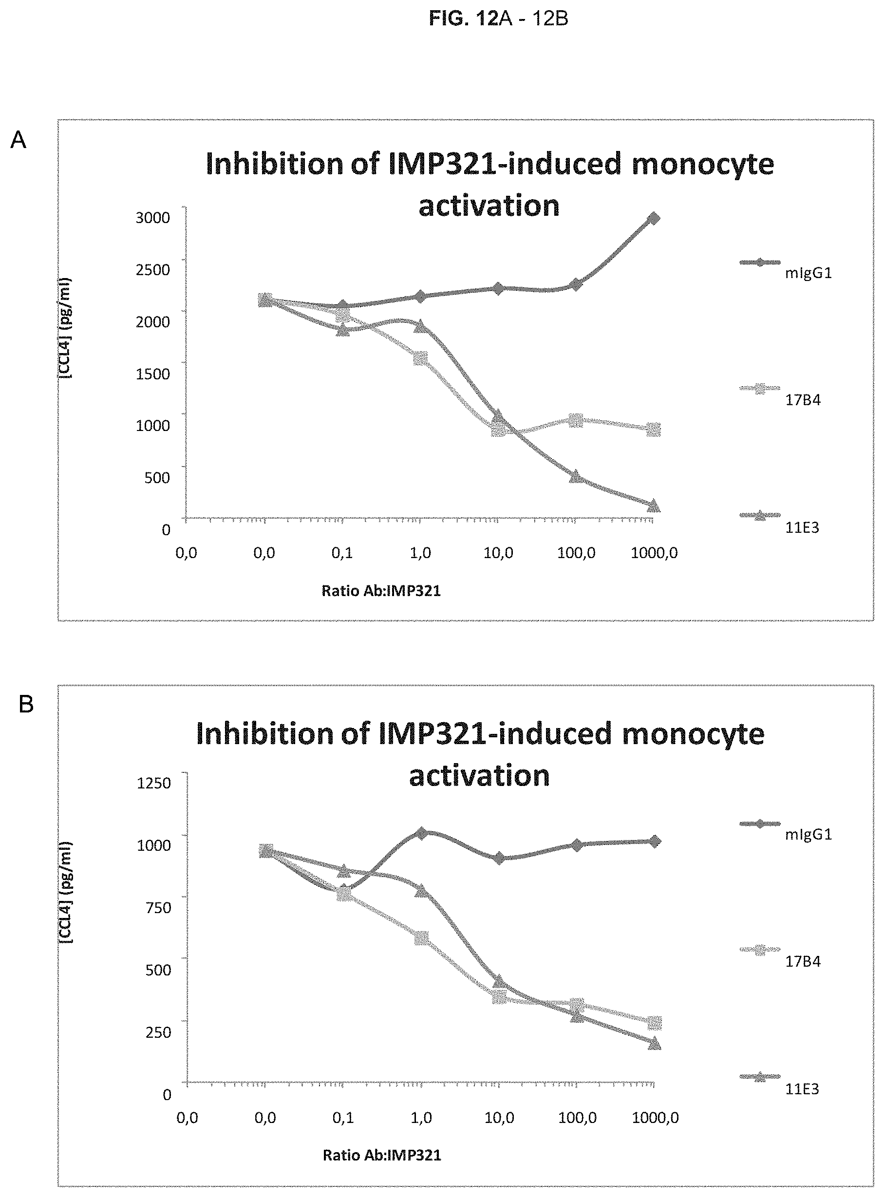
D00014

D00015
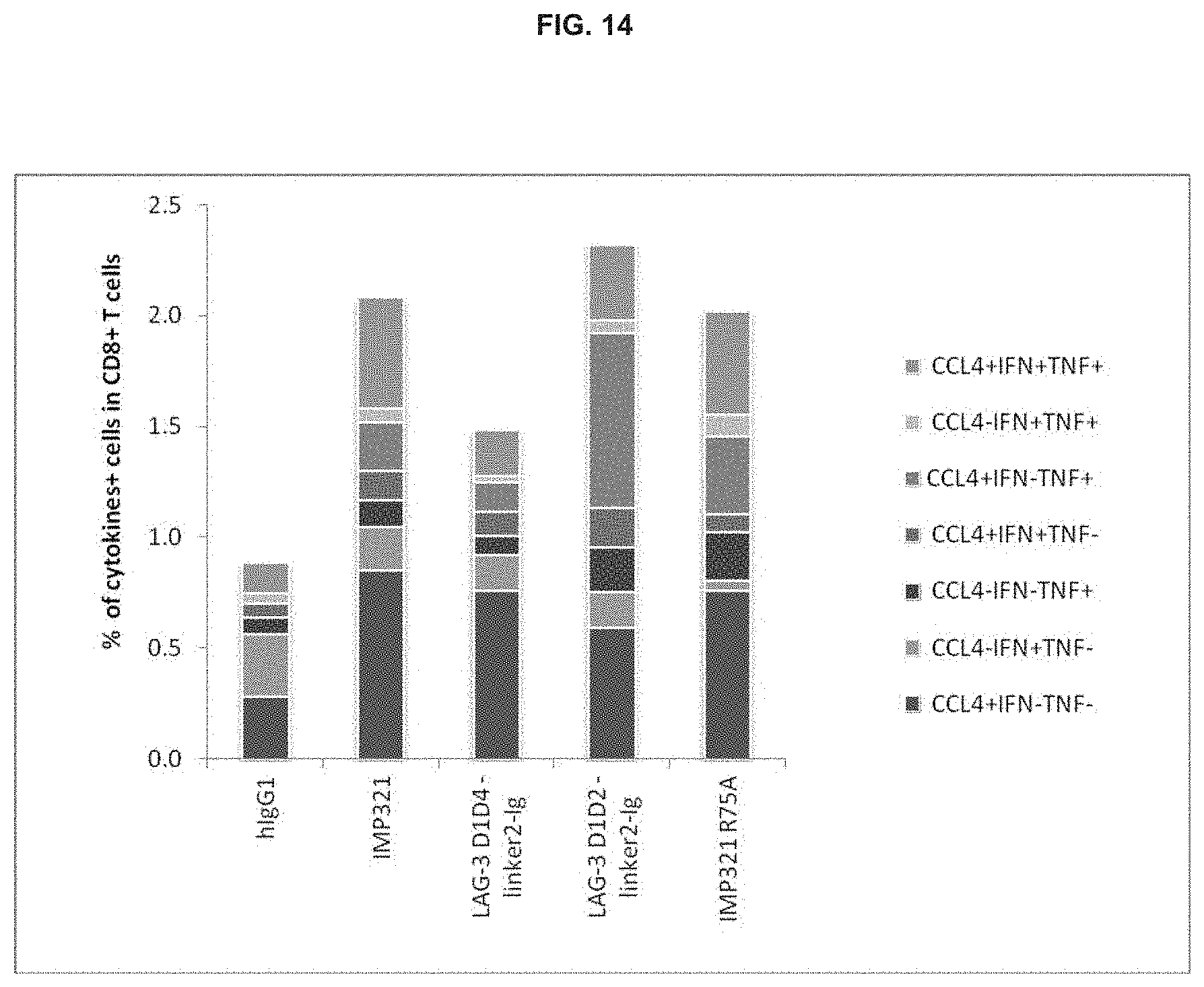
D00016

D00017
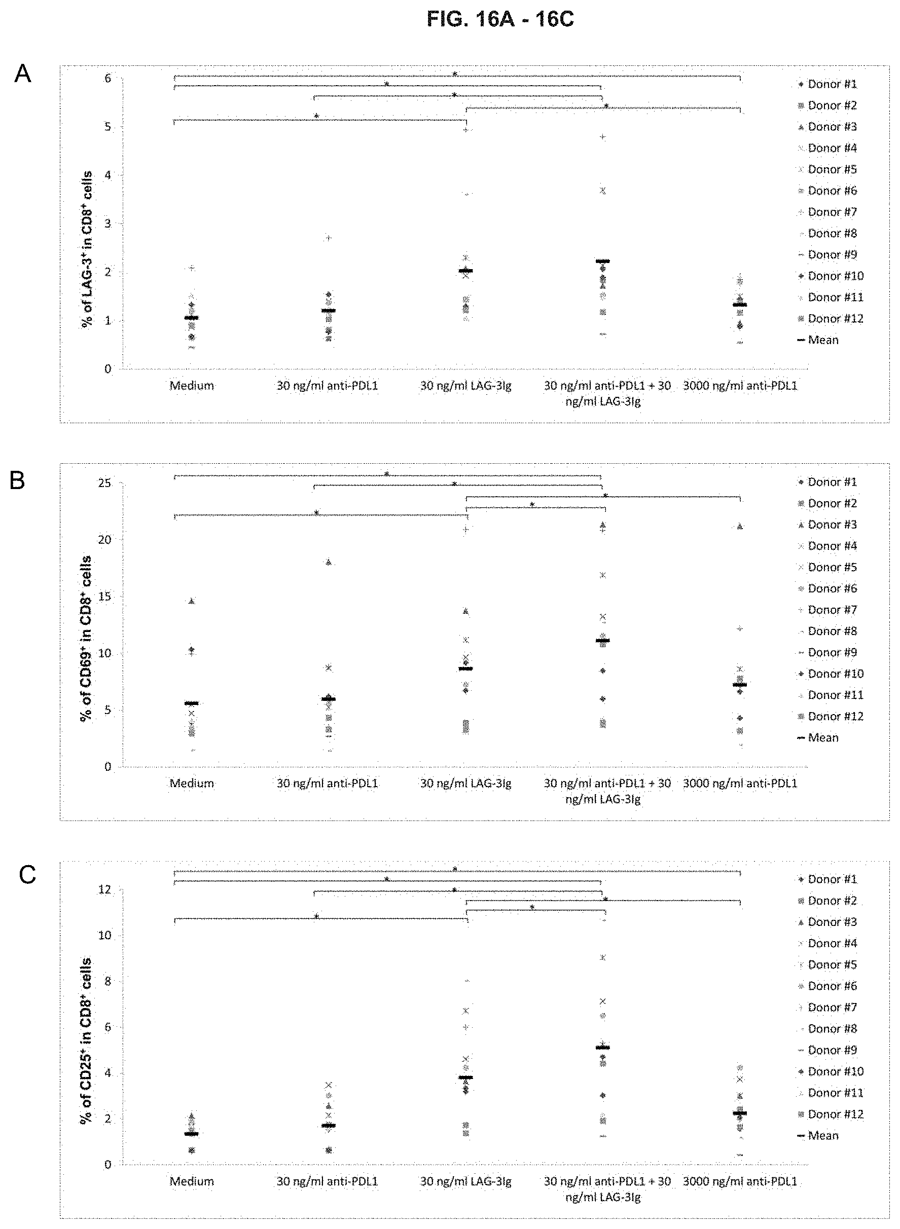
D00018

D00019
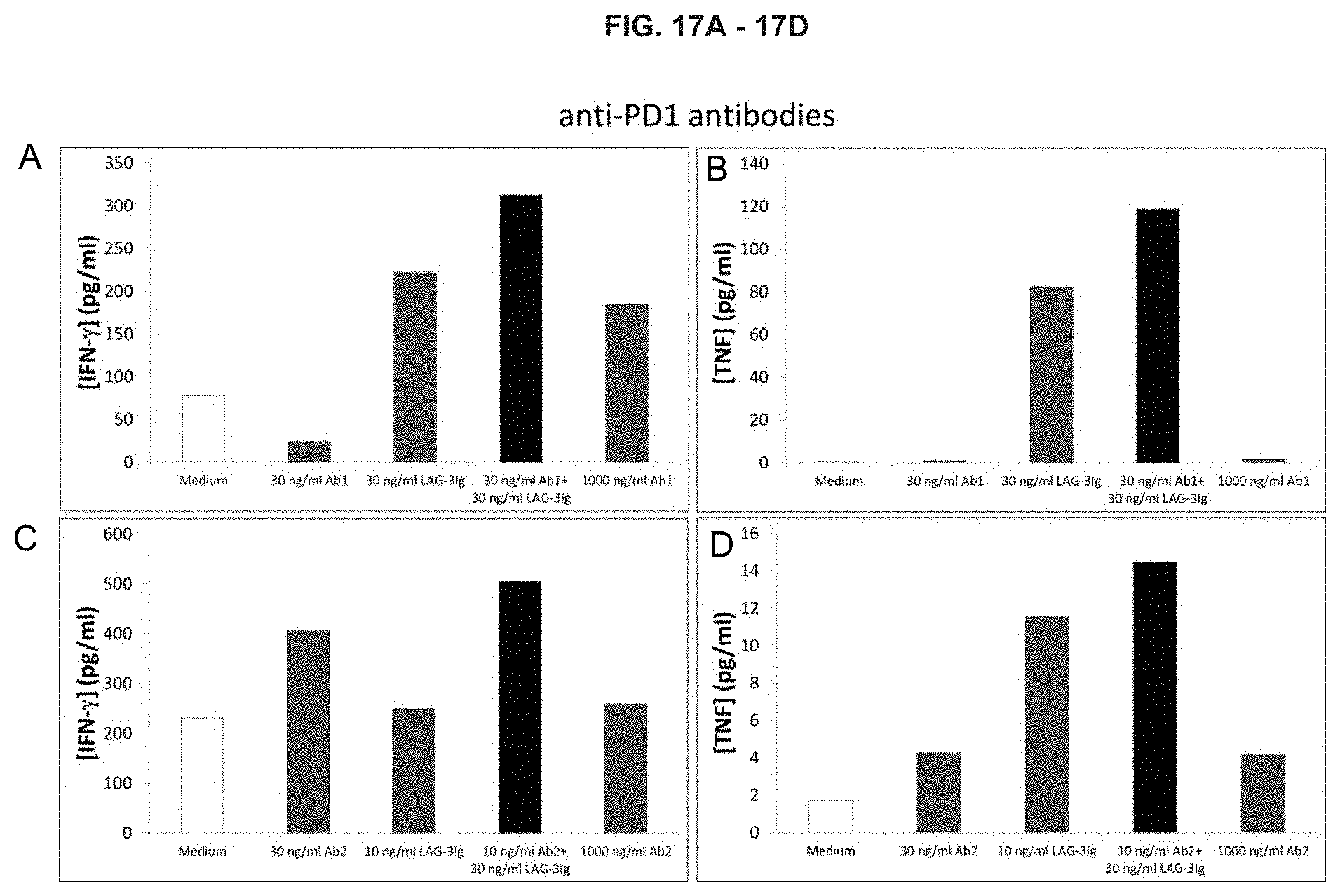
D00020
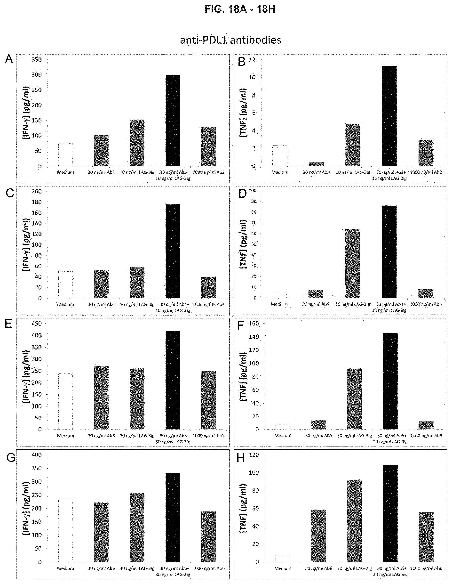
D00021
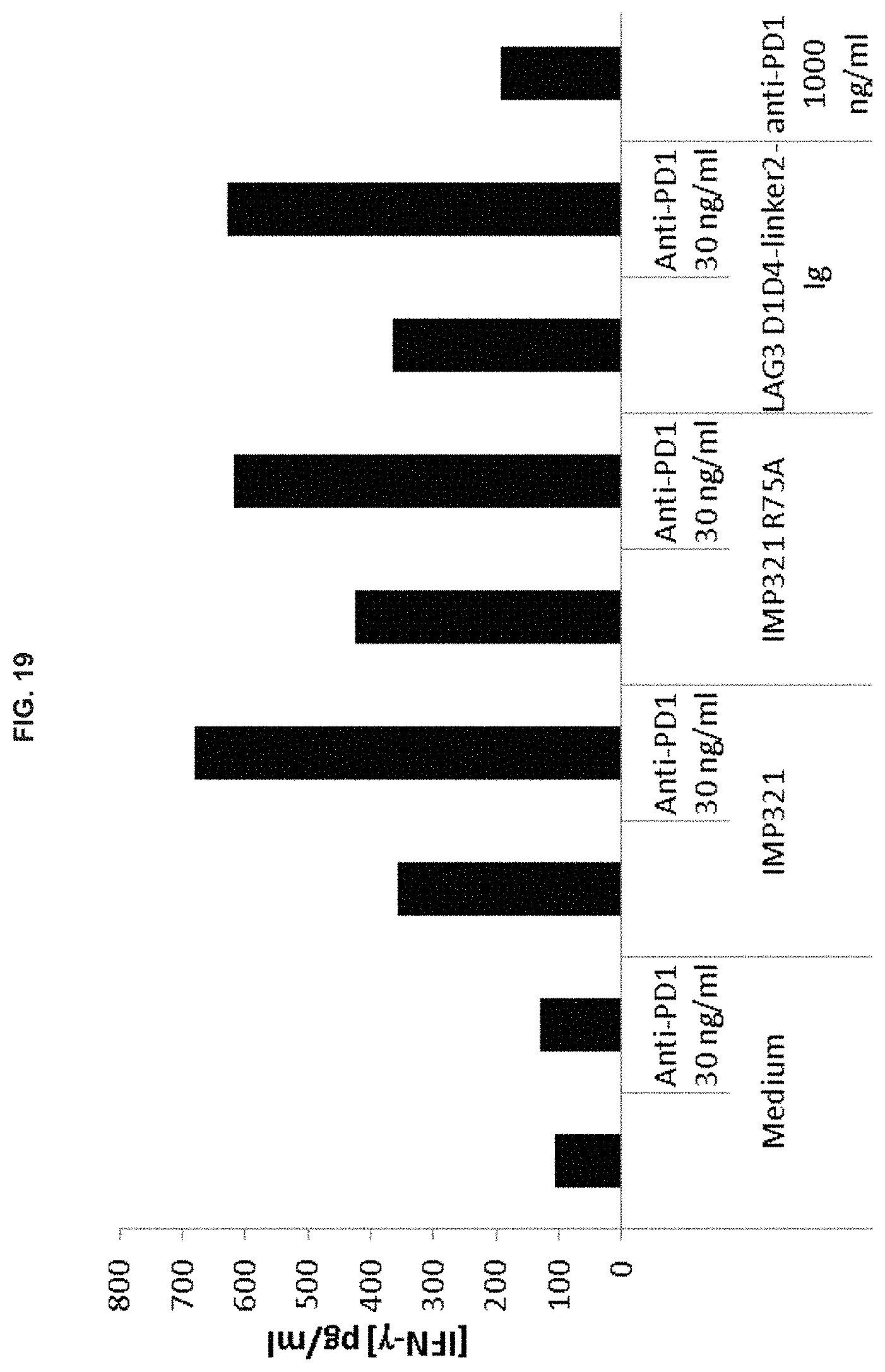
S00001
XML
uspto.report is an independent third-party trademark research tool that is not affiliated, endorsed, or sponsored by the United States Patent and Trademark Office (USPTO) or any other governmental organization. The information provided by uspto.report is based on publicly available data at the time of writing and is intended for informational purposes only.
While we strive to provide accurate and up-to-date information, we do not guarantee the accuracy, completeness, reliability, or suitability of the information displayed on this site. The use of this site is at your own risk. Any reliance you place on such information is therefore strictly at your own risk.
All official trademark data, including owner information, should be verified by visiting the official USPTO website at www.uspto.gov. This site is not intended to replace professional legal advice and should not be used as a substitute for consulting with a legal professional who is knowledgeable about trademark law.