Risk Assessment Of Disseminated Intravascular Coagulation
MARGARITO; JENNY ; et al.
U.S. patent application number 16/618426 was filed with the patent office on 2020-04-16 for risk assessment of disseminated intravascular coagulation. The applicant listed for this patent is KONINKLIJKE PHILIPS N.V.. Invention is credited to BART JACOB BAKKER, JENNY MARGARITO, RENE VAN DEN HAM.
| Application Number | 20200118687 16/618426 |
| Document ID | / |
| Family ID | 62748927 |
| Filed Date | 2020-04-16 |


View All Diagrams
| United States Patent Application | 20200118687 |
| Kind Code | A1 |
| MARGARITO; JENNY ; et al. | April 16, 2020 |
RISK ASSESSMENT OF DISSEMINATED INTRAVASCULAR COAGULATION
Abstract
A method and system for the assessment of the risk of development of disseminated intravascular coagulation (DIC), in patients showing systemic inflammatory response syndrome (SIRS) or sepsis is disclosed. Specifically, the invention provides a method for early DIC assessment and preventive treatment planning, which has the potential for significantly decreasing mortality rate as well as the rate of DIC related sequelae in the SIRS/sepsis patient population and thereby improving quality of life. The risk assessment method is based on features of vital signs and/or biomarker measurements, and provides solutions for assessing the risk of DIC development 24 hours in advance or within 72 hours after ICU admittance.
| Inventors: | MARGARITO; JENNY; (EINDHOVEN, NL) ; BAKKER; BART JACOB; (EINDHOVEN, NL) ; VAN DEN HAM; RENE; (MAARSBERGEN, NL) | ||||||||||
| Applicant: |
|
||||||||||
|---|---|---|---|---|---|---|---|---|---|---|---|
| Family ID: | 62748927 | ||||||||||
| Appl. No.: | 16/618426 | ||||||||||
| Filed: | June 12, 2018 | ||||||||||
| PCT Filed: | June 12, 2018 | ||||||||||
| PCT NO: | PCT/EP2018/065412 | ||||||||||
| 371 Date: | December 2, 2019 |
Related U.S. Patent Documents
| Application Number | Filing Date | Patent Number | ||
|---|---|---|---|---|
| 62518064 | Jun 12, 2017 | |||
| Current U.S. Class: | 1/1 |
| Current CPC Class: | A61B 5/7275 20130101; G16H 50/30 20180101; A61B 5/0002 20130101; G01N 33/6869 20130101; G01N 2800/26 20130101; G16H 50/50 20180101; G01N 33/728 20130101; G16H 40/67 20180101; G16H 50/20 20180101 |
| International Class: | G16H 50/30 20180101 G16H050/30; G01N 33/68 20060101 G01N033/68; G16H 50/20 20180101 G16H050/20; G16H 50/50 20180101 G16H050/50; A61B 5/00 20060101 A61B005/00; G16H 40/67 20180101 G16H040/67; G01N 33/72 20060101 G01N033/72 |
Claims
1. A computer-implemented method for assessing the risk of the development of DIC in a patient diagnosed with systematic inflammatory response syndrome (SIRS), the method comprising: a computing device with a graphical user interface, admitting a patient into an ICU unit, diagnosing said patient for SIRS and, if positive, inputting patient-specific diagnostic data onto a processor configured to receive said patient-specific data, and storing said data on a non-transitory computer readable storage medium, wherein the biomarker measurement data comprises total bilirubin and lactate measurement data; acquiring initial vital signs and biomarker measurement data from said patient and inputting and storing said vital signs and biomarker measurement data on said non-transitory computer readable storage medium; determining selection criteria based on the patient dataset; monitoring said vital signs of said patient and continuously inputting and storing said vital signs and biomarker measurement data on said non-transitory computer readable storage medium; pre-processing said vital sign data, including assessing the quality of said data and removing outliers from said data; windowing of said vital signs data; extracting specific features from said patient-specific data; calculation of statistical features from said vital signs windows; analyzing said statistical features in combination with the biomarker measurement data using a predictive model that is stored on said non-transitory computer readable storage medium; determining whether a value derived from said patient-specific data by the predictive model meets a DIC probability threshold, wherein said probability threshold is predictive of the likely development of DIC.
2. The method of claim 1, wherein said biomarker measurement data is analyzed with respect to said predictive model that is stored on said non-transitory computer readable storage medium.
3. The method of claim 1, wherein said vital sign data and said biomarker measurement data are both analyzed together with respect to said predictive model that is stored on said non-transitory computer readable storage medium.
4. A non-transitory computer readable storage medium tangibly encoded with computer-executable instructions, that when executed by a processor associated with computing device having a graphical user interface, cause the device to carry out the steps of the method as defined in claim 1.
5. A computer program product, comprising a computer-readable code to be executed by one or more processors when retrieved from a non-transitory computer-readable medium, the computer-readable program code including instructions to: input patient-specific diagnostic data onto a processor configured to receive said patient-specific data, and storing said data on a non-transitory computer readable storage medium; input and store patient-specific vital signs and biomarker measurement data on said non-transitory computer readable storage medium, wherein the biomarker measurement data comprises total bilirubin and lactate measurement data; determine selection criteria based on the patient dataset; input and store vital signs and biomarker measurement data obtained by continuously monitoring said patient, on said non-transitory computer readable storage medium; pre-process said vital sign data, including assessing the quality of said data and removing outliers from said data; windowing of said vital signs data; extract specific features from said patient-specific data; calculate of statistical features from said vital signs windows; analyze said statistical features in combination with the biomarker measurement data using a predictive model that is stored on said non-transitory computer readable storage medium; determine whether said patient-specific data meets a DIC probability threshold, wherein said probability threshold is predictive of the likely development of DIC.
Description
FIELD OF THE INVENTION
[0001] The present invention relates to a method and system for the assessment of the risk of the development of disseminated intravascular coagulation (DIC), in patients showing systemic inflammatory response syndrome (SIRS), or sepsis. Specifically, the invention provides a method for early DIC assessment and preventive treatment planning, which has the potential for significantly decreasing mortality rate as well as the rate of DIC related sequelae in the SIRS/sepsis patient population and thereby improving quality of life. The risk assessment method is based on features of vital signs and/or biomarker measurements, and provides solutions for assessing the risk of DIC development 24 hours in advance or within 72 hours after ICU admittance. Optimal sets of measurements for each model implementation are selected.
[0002] Developed models obtaining an area under the ROC (receiving operation curve) curve (AUC) equal to 0.85 and 0.83, for the risk assessment 24 hours in advance and within 72 hours from ICU admittance, are described.
[0003] The system provides an improved process of integrative analysis of a patient's characteristics and demographics, vital signs and in-vitro diagnostic data for effective treatment planning.
BACKGROUND OF THE INVENTION
[0004] DIC is a pathological process characterized by a systemic activation of the blood coagulation system, leading to subsequent clot formation, blood vessel obstruction and organ dysfunction. The large consumption of platelets and coagulation factors in this process may in turn cause bleeding, which further worsens the patient's condition and decreases the chances of survival (Di Nisio, M., et al., "Diagnosis and treatment of disseminated intravascular coagulation: guidelines of the Italian Society for Haemostasis and Thrombosis (SISET)," Thrombosis research, vol. 129, pp. e177-e184, 2012.)
[0005] DIC is usually secondary to an underlying condition such as systemic inflammatory response syndrome (SIRS), sepsis, trauma, malignancy, heat stroke and hyperthermia. SIRS and sepsis are among the most common causes of DIC, whose mortality ranges between 10-50%. Between 30% and 50% of sepsis patients develop DIC. Sepsis severity positively correlates with DIC incidence and therefore mortality (Levi. M., et al., "Disseminated intravascular coagulation," New England Journal of Medicine, vol. 341, pp. 586-592, 1999). DIC incidence ranges between 7% (mild sepsis) and 73% (septic shock) (see Kinasewitz, G. T., et al., "Prognostic value of a simple evolving disseminated intravascular coagulation score in patients with severe sepsis," Critical Care Medicine, vol. 33, pp. 2214-2221, 2005), and DIC mortality ranges between 10% and 50%.
[0006] Several diagnostic scores based on general coagulation tests such as prothrombin time (PT), fibrinogen, d-dimer and platelet counts have been proposed to objectify the subjective clinical diagnosis of DIC based on the clinical signs and symptoms of the patient. See, e.g., Kobayashi, N., et al., "Criteria for diagnosis of DIC based on the analysis of clinical and laboratory findings in 345 DIC patients collected by the Research Committee on DIC in Japan," Disseminated Intravascular Coagulation, ed: Karger Publishers, 1983, pp. 265-275. Although some of those scores seem to reflect the clinical diagnosis of DIC by experts quite well, this also means that these scores are unsuitable as a tool for the risk assessment and prevention of DIC.
[0007] The high mortality of SIRS/sepsis associated DIC has driven us to develop methods for assessing the risk of DIC development following the diagnosis of SIRS, using both vital signs and standardly available laboratory measurements. These methods allow for improving the prevention of DIC in SIRS patients, which in turn improves patient outcomes.
[0008] This invention assesses the risk of the development of DIC in SIRS patients. While available diagnostic DIC scores only allow for therapeutic treatment of DIC, the within invention allows for early and preventative intervention that should decrease DIC incidence and reduce mortality. Currently no risk assessment algorithms are available for DIC despite its high mortality rate.
[0009] Accordingly, the within invention provides a method based on biomarkers (lactate, bilirubin and creatinine) and/or statistical features (e.g., average, standard deviation, kurtosis, skewness and quantile values) extracted from vital signs (heart rate, respiration rate and oxygen saturation) that assesses the risk of developing DIC 24 hours in advance. In addition, the invention provides a method based on biomarkers (lactate, bilirubin and creatinine) and/or statistical features (e.g., average, standard deviation, kurtosis, skewness and quantile values) extracted from vital signs (heart rate, respiration rate and oxygen saturation), that assesses the risk of developing DIC within the first 72 hours after ICU admittance.
[0010] The optimal system requires all mentioned biomarker measurements on at least a daily basis and continuous monitoring of all mentioned vital signs. More practical, but still high performing implementations are based on continuous screening of sepsis/SIRS patients using one or more of the vital signs (high sensitivity, low specificity) and subsequent measurement of one or more of the biomarkers to increase the specificity of the risk assessment at the moment the vital signs indicate an increased risk of DIC development.
[0011] In general terms, our invention uses the combination of vital signs monitoring for the detection of clinical deterioration of sepsis/SIRS patients, with biomarker measurements that reflect organ damage likely caused by the DIC. One skilled in the art could imagine solutions that combine vital signs monitoring data with other and/or earlier markers of organ damage, and/or more direct biomarkers of systemic activation of the coagulation system.
SUMMARY OF THE INVENTION
[0012] It is an object of the present invention to provide a method and system for assessing the risk of development of DIC in patients showing SIRS or sepsis. In particular, it is an object of the present invention to provide a system and method that solves the above-mentioned problems of the prior art by providing a method and system for assessing the risk of DIC development. This invention proposes two different implementations of the system, the first one allows for predicting the onset of DIC 24 hours in advance, while the second one allows for predicting the risk of DIC at ICU admittance, more specifically in the first 72 hours from ICU admittance, before DIC is being diagnosed.
It is also an object of the present invention to provide a system and method for assessing the risk of developing DIC based on features of vital signs and/or biomarker measurements. It is a further object of the present invention to provide a method and system for decreasing the mortality rate for patients developing DIC by early assessment and preventative treatment planning for DIC. It is also an object of the present invention to provide an alternative to the prior art.
[0013] Thus, the above-described object and several other objects are intended to be obtained in a first aspect of the invention by providing a system and method for providing relevant patient-specific DIC risk information, such system and method comprising:
[0014] diagnosing said patient for SIRS and, if positive, inputting the patient-specific diagnostic data onto a processor configured to receive said patient-specific data, and storing said data on a non-transitory computer readable storage medium;
[0015] acquiring initial vital signs and biomarker measurement data from said patient and inputting and storing said vital signs and biomarker measurement data on said non-transitory computer readable storage medium;
[0016] monitoring said vital signs of said patient and continuously inputting and storing said vital signs and biomarker measurement data on said non-transitory computer readable storage medium;
[0017] pre-processing said vital sign data, including assessing the quality of said data and removing outliers from said data. The pre-processing consists of discarding samples which are outside the physiological range. Physiological range for both biomarkers and vital signs are shown in Table 1 and Table 2;
TABLE-US-00001 TABLE 1 Physiological range for selected biomarkers Name Unit Measure Range creatinine mg/dl 0 / 20 direct bilirubin mg/dl 0 / 10 lactate mmol/L 0 / 10 total bilirubin mg/dl 0 / 20
TABLE-US-00002 TABLE 2 Physiological range for selected vital signs Name Unit Measure Range Oxygen saturation % >=90 Heart Rate bpm 25 / 250 Respiration Cycle/min >0
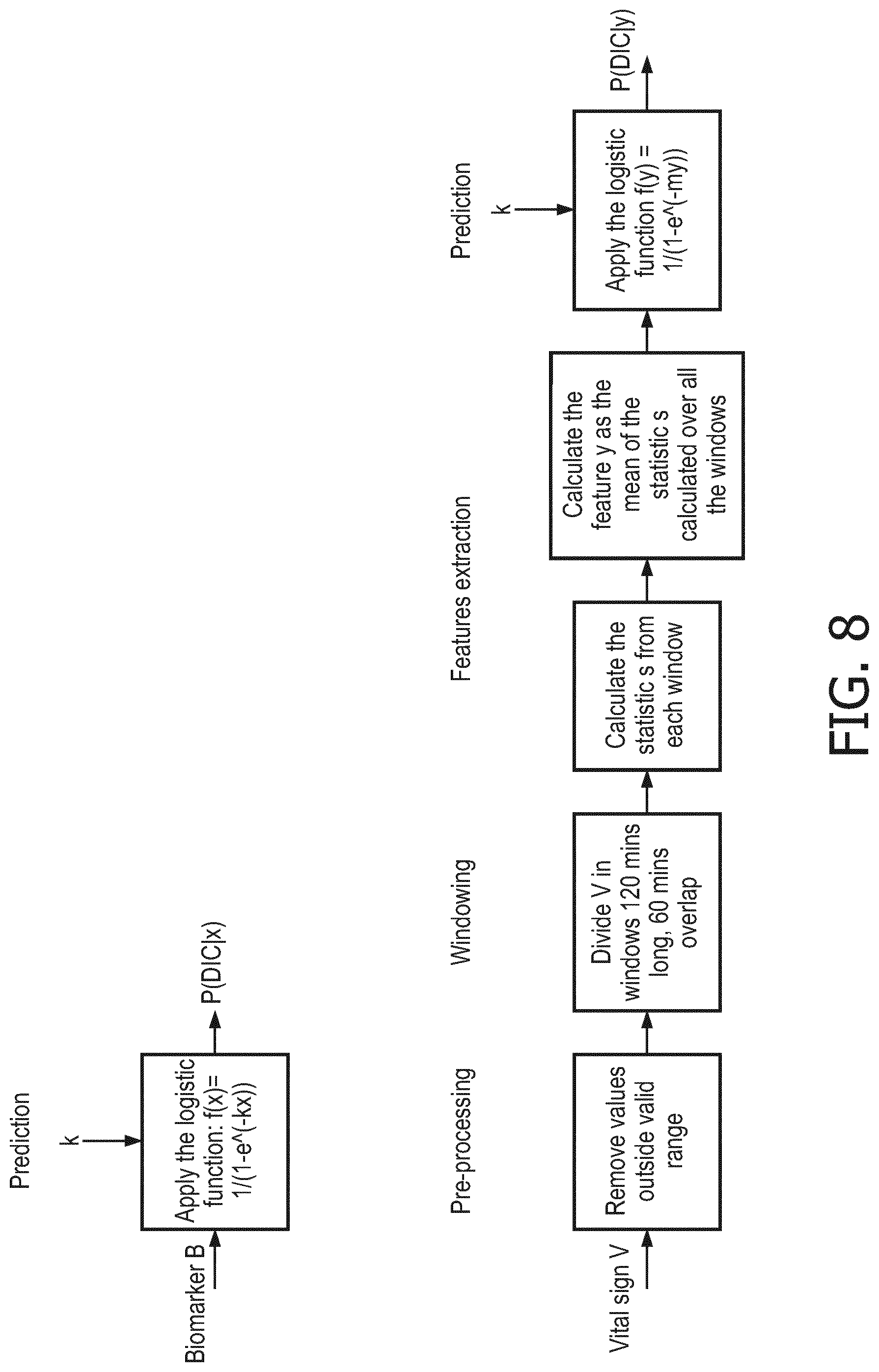
[0018] windowing of said vital signs data; vital signs are segmented in sliding windows 120 minutes long with an overlap of 60 minutes. The statistic of interest is extracted from each window and the final feature is obtained by averaging the statistics extracted over all the windows (see FIG. 9).
[0019] extracting specific features from said patient-specific data;
[0020] calculation of statistical features from said vital signs windows;
[0021] A description of the statistical features is provided in Table 3.
[0022] Table 3 Description of statistical features extracted from vital signs
TABLE-US-00003 Name of the features Description Mean average value of the signal point values Standard deviation root Mean squared deviation of the signal point values from their arrhythmic mean Kurtosis it quantifies the sharpness of the distribution curve peak Skewness it quantifies asymmetry of a distribution around its mean Quantile 0.25 x-value of the distribution which includes 0.25*N observations, with N being the number of point values Quantile 0.50 x-value of the distribution which includes 0.50*N observations, with N being the number of point values Quantile 0.75 x-value of the distribution which includes 0.75*N observations, with N being the number of point values Range Difference between the maximum and the minimum signal point values for a given Energy signal sum of the squared signal point values
[0023] analyzing said patient data with respect to a predictive model that is stored on said non-transitory computer readable storage medium; and determining whether a value derived from said patient-specific data by the predictive model meets a DIC probability threshold, wherein said probability threshold is predictive of the likely development of DIC.
BRIEF DESCRIPTION OF THE DRAWINGS
[0024] The methods and system according to the invention will now be described in more detail with regard to the accompanying figures. The figures show ways of implementing the present invention and are not to be construed as being limiting to other possible embodiments falling within the scope of the attached claims.
[0025] FIG. 1 illustrates biomarker distributions for patient populations who developed (Case) and did not develop DIC (Control);
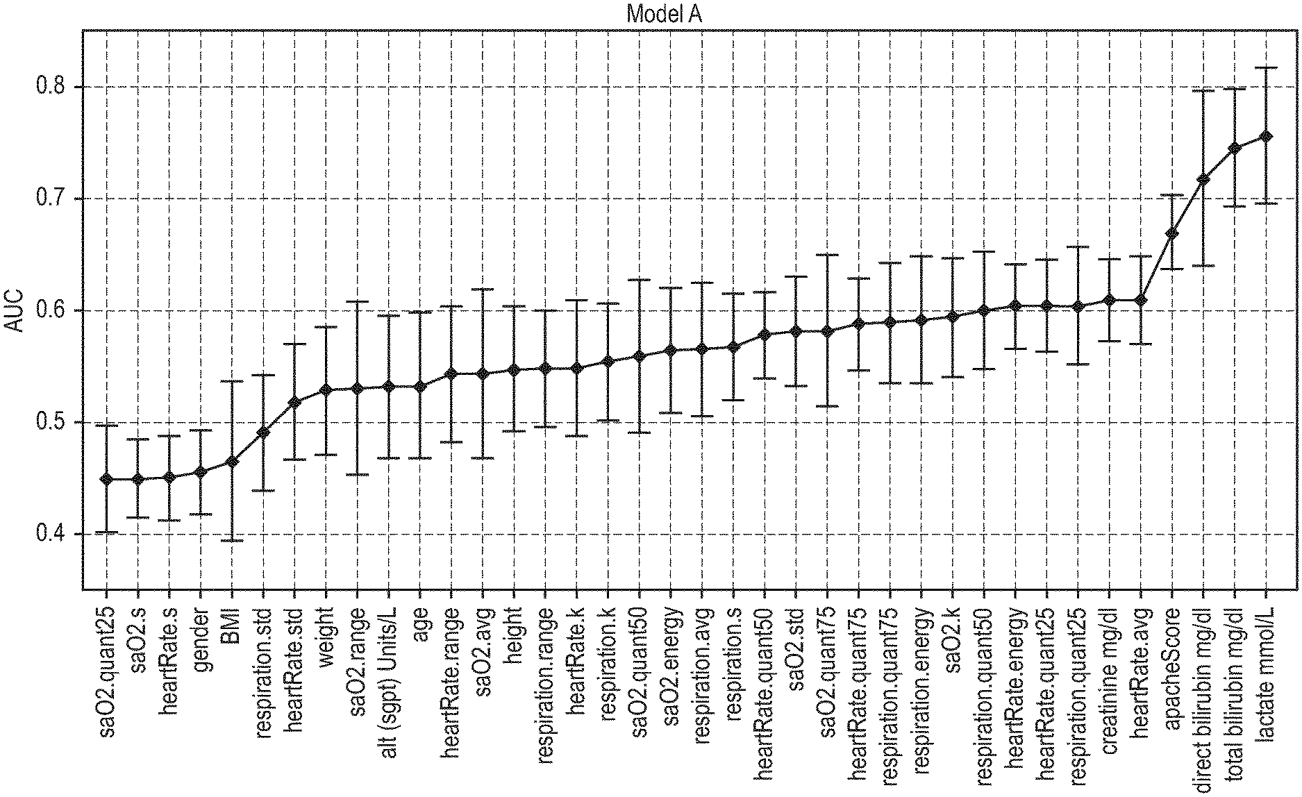
[0026] FIG. 2A is a plot showing the area under the curve (AUC) for single biomarkers, subject characteristics, the Apache score and statistic features extracted from vital signs for the 24 hour model;
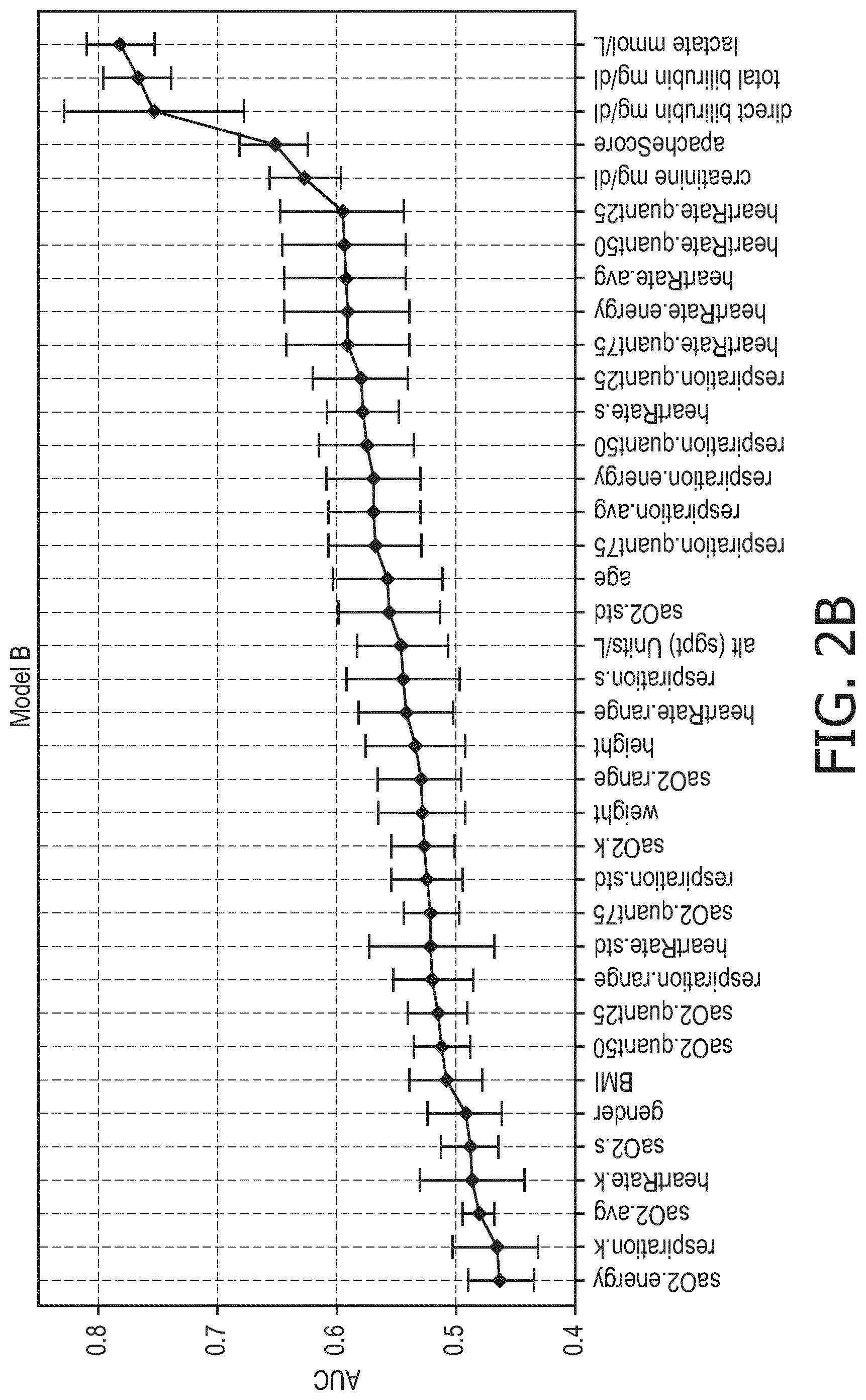
[0027] FIG. 2B is a plot showing the area under the curve (AUC) for single biomarkers, subject characteristics, the Apache score and statistic features extracted from vital signs for the 72 ICU admittance hours model;
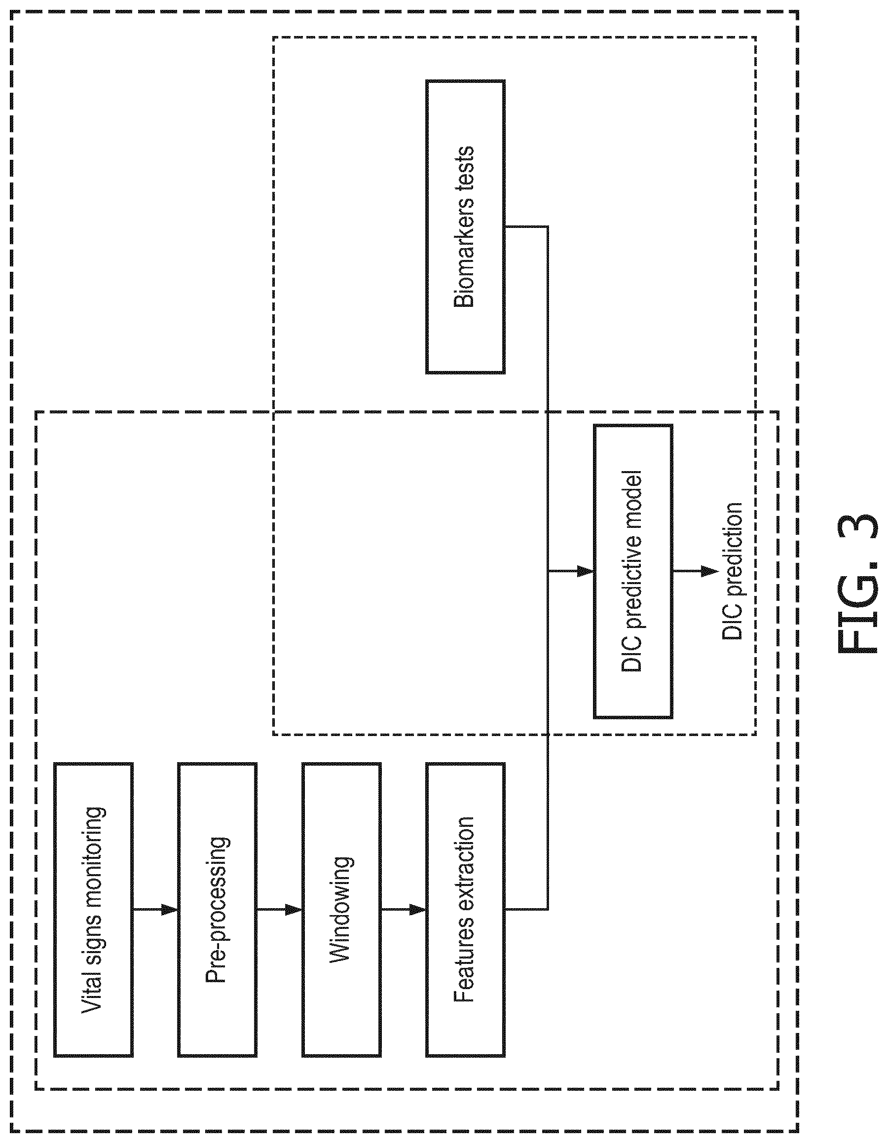
[0028] FIG. 3 is a diagram illustrating the three versions of the DIC risk assessment framework;
[0029] FIG. 4 is a block diagram showing an embodiment of the DIC risk assessment system of the within invention;
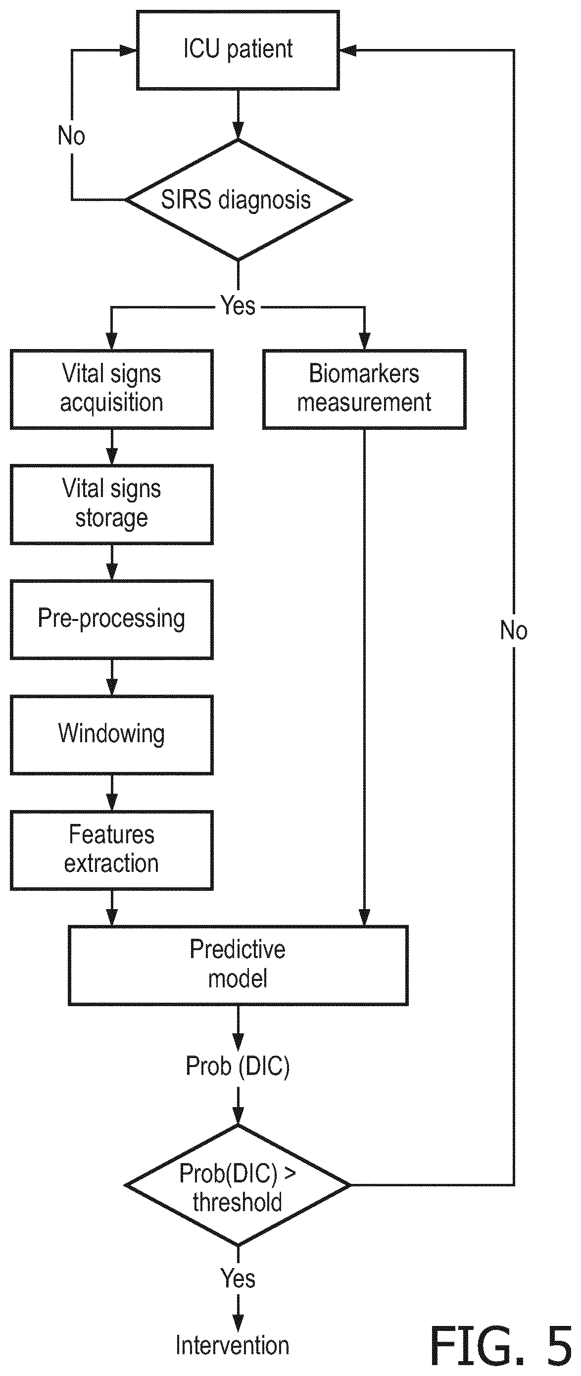
[0030] FIG. 5 is a flowchart of an embodiment of the invention showing a process from patient ICU admission and diagnosis leading to risk assessment for DIC;
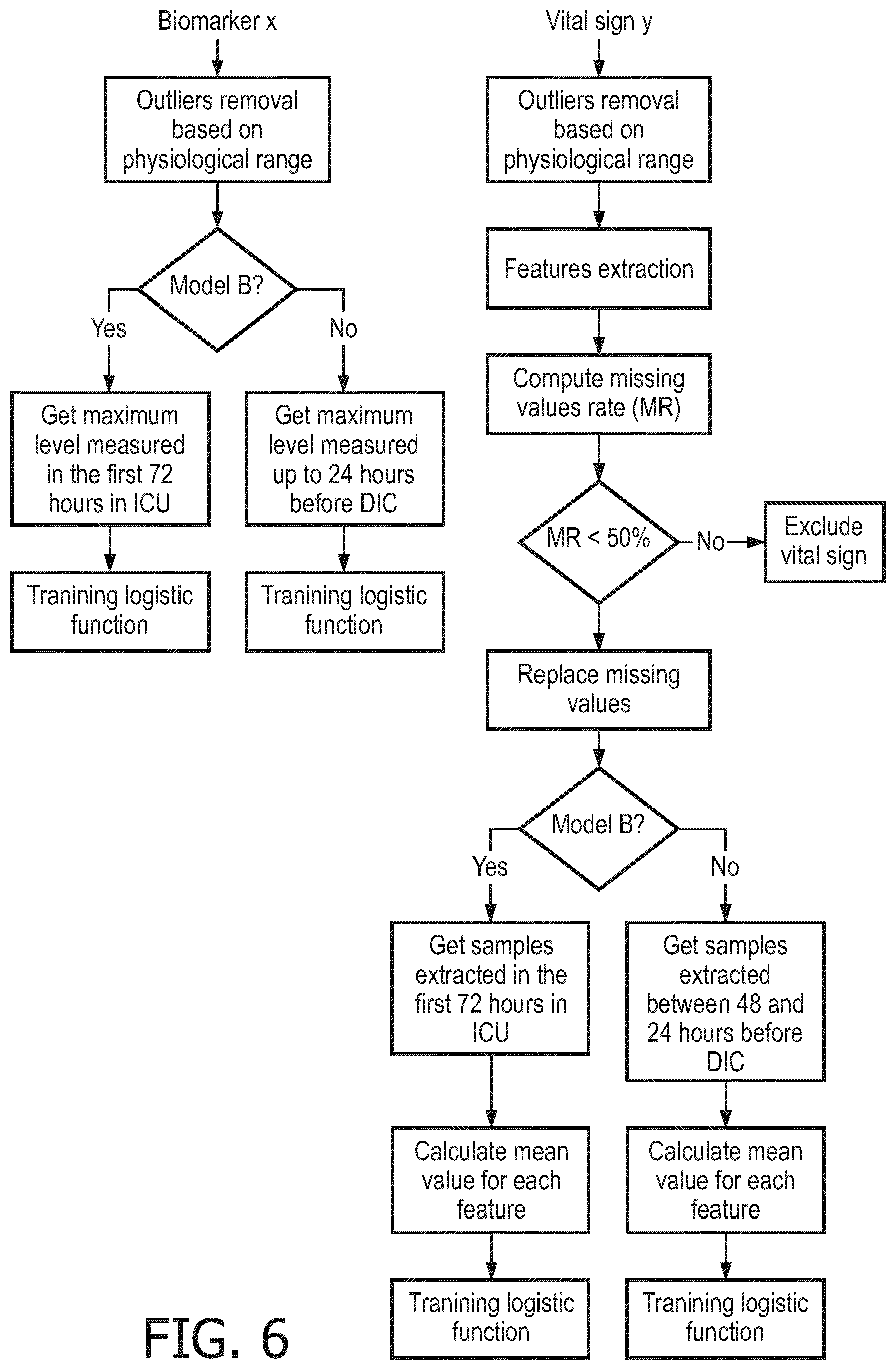
[0031] FIG. 6 is a flowchart describing the pre-processing steps that precede training of logistic functions which compose a final risk assessment model;
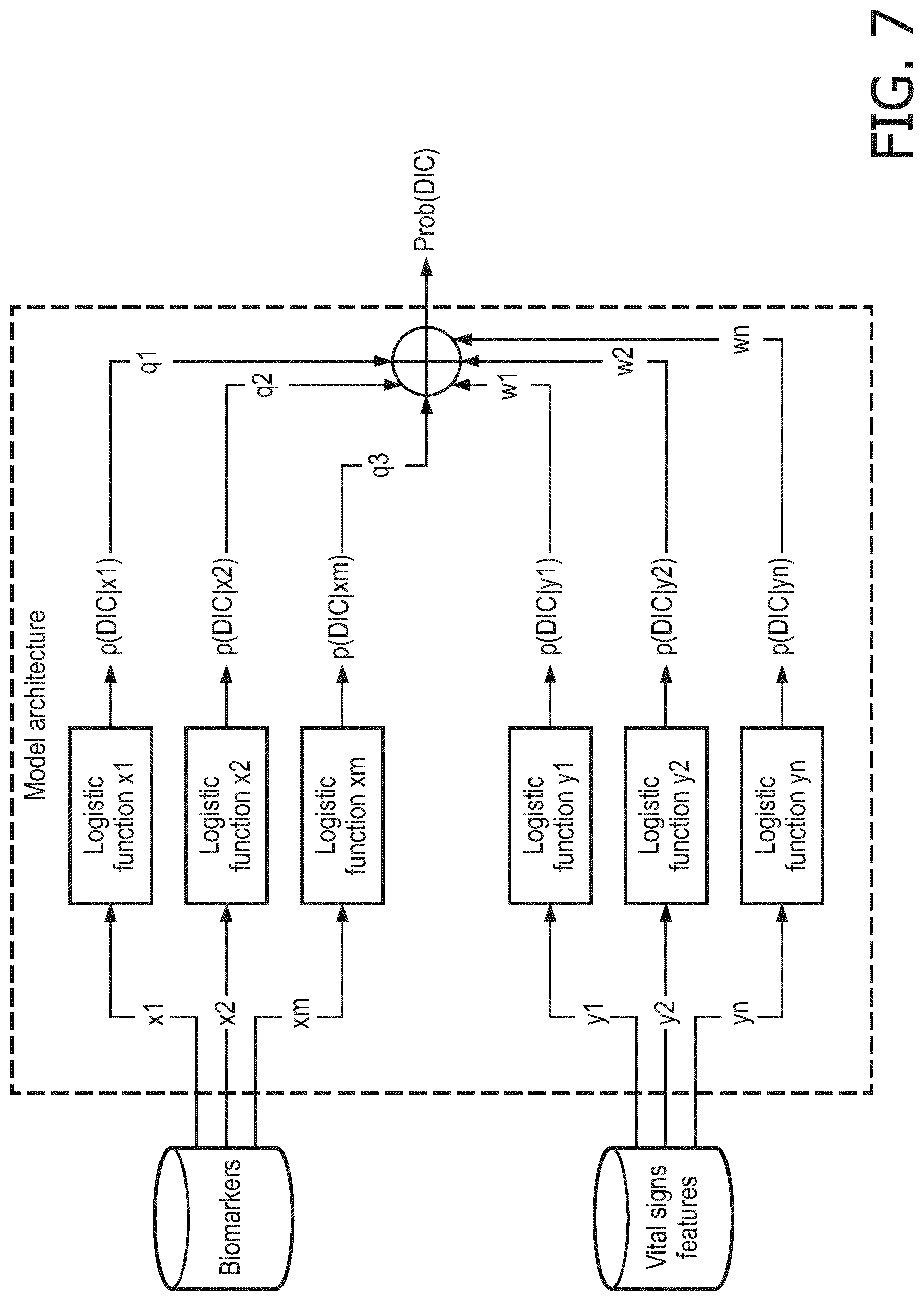
[0032] FIG. 7 is a diagram of an implemented model of the within invention; the parameter q and w were determined during the model training;
[0033] FIG. 8 provides two flowcharts describing steps for estimating the DIC probability giving a generic vital sign or a generic biomarker. The parameters k and m were determined with the training of the model;
[0034] FIG. 9A is the AUC calculated on the test set for the different configurations for the 24 hour model;
[0035] FIG. 9B is the AUC calculated on the test set for the different configurations for 72 ICU admittance hours model;
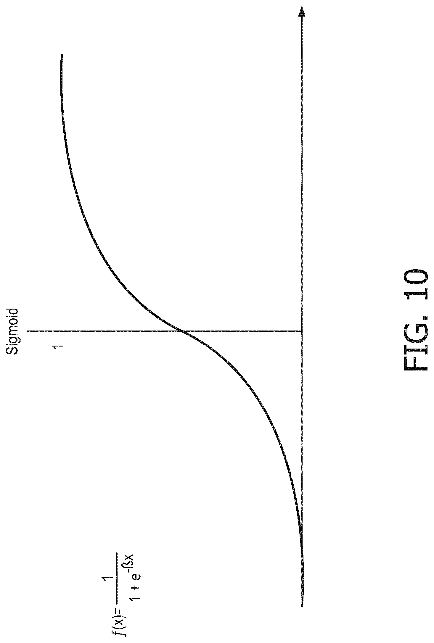
[0036] FIG. 10 is the representation of the logistic function applied for calculating the probability of DIC occurrence.
DETAILED DESCRIPTION OF THE INVENTION
[0037] The present invention provides a method and system for early risk assessment of DIC using biomarker measurements, vital signs monitoring and the combination of vital signs monitoring for the detection of clinical deterioration of sepsis/SIRS patients, with biomarker measurements that reflect organ damage likely caused by the DIC. The present invention is described in further detail below with reference made to FIGS. 1-10.
[0038] This invention arises from the analysis of intensive care unit (ICU) data collected from SIRS patients that did not receive anti-coagulant treatment before DIC diagnosis. The selected patients were divided into training and test sets, 70% and 30%, respectively. The training set has been used for determining the model parameters, whereas the test set to evaluate the model performance. Descriptions of the training and test sets are provided in Table 4. Data from patient subgroups have been used to develop the models of this invention since not all data was always available for all selected SIRS patients.
TABLE-US-00004 TABLE 4 Patients sets Model 24 hours Model 72 hours ICU Training Test Training Test set set set set Nr. Subjects 2277 947 2652 1120 Nr. Cases 1082 455 1557 670 Nr. Controls 1195 492 1095 450 Male [%] 54 51 54 52 Age (sd) 62 (16) 61 (16) 61 (16) 60 (16) [years] Weight (sd) 78.3 (21.0) 78.4 (21.5) 78.1 (21.0) 78.2 (21.0) [kg] Height (sd) 1.69 (0.12) 1.69 (0.11) 1.69 (0.12) 1.69 (0.11) [m] BMI (sd) 27.2 (6.1) 27.4 (6.4) 27.2 (6.2) 27.3 (6.3) [kg/m.sup.2]
[0039] The discriminative power of different standalone factors (features) and their combinations were evaluated in order to define the optimal combination of features for DIC prediction. Such factors include biomarkers, features extracted from vital signs, subjects' characteristics and Apache score. Referring now to the figures, FIG. 1 illustrates initial statistical analysis showing a significant difference in biomarker concentration for the biomarkers bilirubin, lactate and creatinine, between populations that developed and those that did not develop DIC. FIG. 1 demonstrates the discriminative power of such biomarkers for DIC prediction. Similarly, the discriminative power of each considered feature was evaluated with a logistic classifier and expressed in terms of area under (AUC) the receiving operating characteristic (ROC). See FIGS. 2A, 2B.
[0040] The ROC curve illustrates the performance of a binary classifier for different operating points. The curve is drawn by plotting the true positive rate (TPR) against the false positive rate (FPR) calculated for the different operating points. The `optimal cutoff` (see Table 5, Opt th) is defined in this document as the value which provides the highest achievable average or sum of the TPR (sensitivity) and specificity (1--false positive rate FPR) and aims to give an indication of the sensitivity and specificity that the models are capable of. Note that the cut-off that is optimal in a real world sense depends on the specific application, and this invention does not aim to propose an optimal cut-off. A further (widely used) parameter extracted from the ROC curve and used for models comparison is the Area Under the Curve (AUC), which can be interpreted as the probability that the classifier will assign a higher score to a randomly chosen positive example than to a randomly chosen negative example.
[0041] FIGS. 2A and 2B are plots which show the area under the curve (AUC) for single biomarkers, subjects characteristics, Apache score and statistic features extracted from vital signs, for the 24 hours model (FIG. 2A) and the 72 ICU admittance hours model (FIG. 2B). Mean AUC value and associated standard deviation have been evaluated with 10-Fold cross validation on the training set. Acronyms of the used statistics are: std: standard deviation, avg: mean, k: kurtosis, s: skewness, quant25, quant50, quant75: quantile 0.25, 0.50, 0.75.
[0042] FIGS. 2A and 2B demonstrate the predicting power of the selected biomarkers, each of which obtained an AUC above 0.60.
[0043] In order to select the optimal feature set, and therefore the best model architecture, a feature selection procedure was implemented. First, all features that had an AUC below 0.55 were excluded and n features were kept. Afterwards n single feature logistic regression models were trained (where feature is either vital sign extracted statistic or a biomarkers level, maximum biomarker level where multiple measurements are available). The DIC score was calculated by combining the output of the multiple univariate logistic models with a weighted average, with the weight being proportional to the univariate logistic model AUC calculated on the training set. The schema of this architecture is depicted in FIG. 7.
[0044] Considering the output of each logistic model P(Y|X.sub.i) as the likelihood of getting DIC given a predictor X.sub.i and AUC.sub.i, the area under the ROC curve achieved by X.sub.i calculated on the training set, the weight assigned to each predictor was calculated as follows:
.alpha. = 1 / ( i AUC i ) ##EQU00001## w i = AUC i .times. .alpha. ##EQU00001.2##
[0045] The risk of getting DIC (P(Y)) is finally calculated as:
P ( Y ) = i = 1 n P ( Y | X i ) .times. w i ##EQU00002##
[0046] In order to select the optimal set of models (X, features) included in the ensemble classifier described above, the performance of different model combinations was evaluated. To reduce the number of combinations we have constrained the maximum number of items for combination to three. We have computed the best configuration using only vital signs, only biomarkers and their combination.
[0047] The combinations which provided the highest AUC for only biomarkers, only vital signs features and their combination have been selected and tested on the independent test set (see Table 4).
[0048] The results of the selection process and therefore the best model configurations are shown in Table 5.
TABLE-US-00005 TABLE 5 Performance of DIC prediction models evaluated on a test set Opt th Se % Sp % AUC Nr. cases Nr. Ctrls Biomarkers creatinine, lactate, total bilirubin* 0.49 57.8 84.9 0.79 128 165 creatinine, lactate, total bilirubin** 0.52 75.0 79.3 0.82 180 159 Vital signs features Standard deviation of saO2, quantile 0.25 of heartRate, 0.57 99.7 4.6 0.66 288 132 Kurtosis of saO2* Quantile 0.25 of heartRate, quantile 0.25 of respiration, 0.51 91.0 18.2 0.62 631 439 Skewness of heartRate** Laboratory tests and vital signs lactate, total bilirubin, energy of respiration* 0.41 86.0 65.4 0.85 50 26 lactate, total bilirubin, skewness of heartRate** 0.57 70.5 83.9 0.83 166 155 Acronyms: standard deviation (std), kurtosis (k), quantile 0.25 (quant25), Oxygen saturation (saO2), optimal threshold calculated on the ROC for assigning DIC class (Opt th), sensitivity (Se), specificity (Sp), Nr. cases (number of patients included in test set that got DIC), Nr. Ctrls (number of patients included in test set that did not get DIC) *Feature set for predicting DIC 24 hours in advance **Feature set for predicting DIC within 72 hours from ICU admittance
[0049] The combination of bilirubin, lactate and creatinine was shown to be largely discriminative, achieving an AUC of 0.79 and 0.82 for the 24 hours model and 72 ICU admittance hours model, respectively. The lower accuracy obtained by the 24 hours model was probably related to the constraint of considering data collected at least 24 hours before DIC diagnosis. The assumption is that the closer to the diagnosis, the higher the increase and therefore the higher the predictability. According to literature the increase of such biomarkers is in most cases related to organ failure. The obtained results suggest that multiple organs are starting to fail, possibly due to lower perfusion caused by micro-vascular thromboses that could represent a first clinical sign of DIC occurrence. A further unexpected result of this work was the discovery that the coupling of vital signs with total bilirubin and lactate provides for a more accurate prediction. In fact, the largest accuracy was achieved by the combination of lactate, total bilirubin and respiration energy for the 24 hours model, which obtained an AUC curve of 0.85 and lactate, total bilirubin and heart rate skewness for 72 ICU admittance hours model, which obtained an AUC of 0.83.
[0050] Given the results obtained by the feature selection, three possible implementations are indicated: a model based on biomarkers, a model based on vital signs features and a model composed by the combination of biomarkers and vital signs features. The description of the general DIC risk assessment framework and the three implementation versions are show in FIG. 3. The implementation version based on vital signs features (orange block in FIG. 3) requires:
[0051] (a) Vital signs monitoring: continuous signals recording;
[0052] (b) Pre-processing: outliers removal based on physiological range;
[0053] (c) Windowing: 2 hours signal segmentation with 1 hour overlap; and
[0054] (d) Feature extraction: calculation of statistical features from vital signs windows;
The implementation version based on biomarkers requires at least a one-time, but preferably regular, e.g. daily biomarkers tests (green block, FIG. 3). And the implementation version based on biomarkers and vital signs requires the combination of the two systems (red block, FIG. 3).
[0055] In the example shown, a logistic function is used to estimate the probability of DIC given a selected single feature; feature based probabilities are then combined in a weighted average to obtain the final DIC prediction score expressed in terms of probability. The weights depend on the predictive accuracy of the separate estimator 8. Each of the algorithms (i.e. logistic function and weighted averaging) may be replaced by other methods known in the field (e.g., artificial neural networks, decision trees, etc.). Alternatively, the scheme below may be treated as a Bayesian network with probability tables estimated from the train data (naive Bayes) or from a priori knowledge (if available). Different blocks of the scheme could be combined in different ways and independently used to assess DIC risk probability.
[0056] The description of the general DIC risk assessment system is shown in FIG. 4. Such system includes: [0057] (a) A system for vital signs acquisition [0058] (b) A laboratory to measure biomarkers concentration [0059] (c) Data storage system such as a local drive [0060] (e) A computer which hosts the algorithm consisting of a block for data pre-processing (see FIG. 6), and a block with the predictive model (see FIG. 7). The output of the system of FIG. 4 could be used as input for a decision-making system responsible of deciding whether an intervention is needed and, in positive case, which kind of intervention would be more beneficial for the patient. A typical intervention could be a drug therapy (e.g. anti- coagulant, anti-arrhythmic).
[0061] FIG. 5 shows a flowchart of the process that leads from the patient diagnosis to the DIC prediction. Particularly, FIG. 5 is a description of the process from the admission of the patient in the ICU to the risk assessment for DIC. The pre-processing steps that precede the training of the logistic functions included in the final predictive model are explained in detail in FIG. 6, while FIG. 7 shows the architecture of the proposed predictive model.
[0062] In the current application example, the scheme of the implemented model is shown in FIG. 7 (for which the results are shown). A logistic function is used to estimate the probability of DIC given a selected single feature (see FIG. 10). Feature based probabilities are then combined with weighted average to obtain the final DIC prediction score.
[0063] The weights depend on the predictive accuracy of the separate estimators calculated over the training set. Note that each of the algorithms may be replaced by other machine learning methods known in the field (e.g., artificial neural networks, decision trees, etc.). Alternatively, the scheme below may be treated as a Bayesian network with probability tables estimated from the train data (naive Bayes) or from a priori knowledge (if available). Different blocks of the scheme could be combined in different ways and independently used to assess DIC risk probability.
[0064] FIGS. 9A and 9B illustrate receiver operating curves calculated for 24 hours model and 72 hours ICU admittance model based on: laboratory tests only, vital signs only, combination of laboratory tests and vital signs. The AUC calculated for each curve is reported in Table 5.
* * * * *
D00000
D00001
D00002
D00003
D00004
D00005
D00006
D00007
D00008
D00009
D00010
D00011


XML
uspto.report is an independent third-party trademark research tool that is not affiliated, endorsed, or sponsored by the United States Patent and Trademark Office (USPTO) or any other governmental organization. The information provided by uspto.report is based on publicly available data at the time of writing and is intended for informational purposes only.
While we strive to provide accurate and up-to-date information, we do not guarantee the accuracy, completeness, reliability, or suitability of the information displayed on this site. The use of this site is at your own risk. Any reliance you place on such information is therefore strictly at your own risk.
All official trademark data, including owner information, should be verified by visiting the official USPTO website at www.uspto.gov. This site is not intended to replace professional legal advice and should not be used as a substitute for consulting with a legal professional who is knowledgeable about trademark law.