Improvements in and Relating to Data Centres
Rogers; Paul ; et al.
U.S. patent application number 16/070392 was filed with the patent office on 2020-04-09 for improvements in and relating to data centres. The applicant listed for this patent is Timothy GEORGIA TECH RESEARCH CORPORATION CHANG. Invention is credited to Neil Crow, Aaron Favill, Samuel Hanks, LUCIAN Hicks, Paul Rogers, William Thornton, Richard Whiteley.
| Application Number | 20200113081 16/070392 |
| Document ID | / |
| Family ID | 70056294 |
| Filed Date | 2020-04-09 |


View All Diagrams
| United States Patent Application | 20200113081 |
| Kind Code | A1 |
| Rogers; Paul ; et al. | April 9, 2020 |
Improvements in and Relating to Data Centres
Abstract
Provided are nanoparticle-based vaccines that have an adjuvant coating attached to the external surface thereof to serve as an immunopotentiator. Most advantageously the nanoparticle vaccines have IgM antibodies attached to the external surface of an antigenic nanoparticle to serve as an immunopotentiator. The IgM may be attached by specifically binding to an epitope of the underlying nanoparticle including, but not limited to, the target vaccine immunogen. Since, however, the availability of specific IgMs is limited and less than the number of potential antigens that may be used as vaccines, attaching an IgM of the disclosure may require a hapten molecule that can be attached to the nanoparticle. By selecting a hapten that can be specifically recognized and bound by an IgM, it is possible to attach the hapten-specific IgM to a wide-variety of vaccine antigens.
| Inventors: | Rogers; Paul; (Stanley Pontlarge, GB) ; Crow; Neil; (Cheltenham, Gloucestershire, GB) ; Hicks; LUCIAN; (Cheltenham, Gloucestershire, GB) ; Whiteley; Richard; (Cheltenham, Gloucestershire, GB) ; Favill; Aaron; (Leamington Spa, Warwickshire, GB) ; Hanks; Samuel; (Cheltenham, Gloucestershire, GB) ; Thornton; William; (Cheltenham, Gloucestershire, GB) | ||||||||||
| Applicant: |
|
||||||||||
|---|---|---|---|---|---|---|---|---|---|---|---|
| Family ID: | 70056294 | ||||||||||
| Appl. No.: | 16/070392 | ||||||||||
| Filed: | January 17, 2017 | ||||||||||
| PCT Filed: | January 17, 2017 | ||||||||||
| PCT NO: | PCT/US2017/050906 | ||||||||||
| 371 Date: | July 16, 2018 |
Related U.S. Patent Documents
| Application Number | Filing Date | Patent Number | ||
|---|---|---|---|---|
| 62393126 | Sep 12, 2016 | |||
| Current U.S. Class: | 1/1 |
| Current CPC Class: | A61K 39/39 20130101; H04L 67/12 20130101; A61K 2039/55555 20130101; A61P 31/12 20180101; B23P 15/26 20130101; H05K 7/20745 20130101; A61K 2039/55516 20130101; H05K 7/1488 20130101 |
| International Class: | H05K 7/14 20060101 H05K007/14; H04L 29/08 20060101 H04L029/08; B23P 15/26 20060101 B23P015/26; H05K 7/20 20060101 H05K007/20 |
Goverment Interests
STATEMENT ON FUNDING PROVIDED BY THE U.S. GOVERNMENT
[0002] This invention was made with Government support under contract 1R01A1101047-01 awarded by the National Institutes of Health. The Government has certain rights in the invention.
Foreign Application Data
| Date | Code | Application Number |
|---|---|---|
| Jan 29, 2016 | GB | 1601721.2 |
Claims
1. A method of making a data centre in an existing building, the building having a floor, walls and a roof, an air inlet for supply of air into the building, and an air outlet for removal of air from the building, wherein the method includes the following steps: installing multiple prefabricated data centre elements by: (a) connecting to the air inlet an air handling module that is arranged to provide cooling capacity for the data centre; and (b) installing in the interior of the building multiple cold aisle services modules, each cold aisle services module having: a length and a width, one or more integrated blanking portions including at least one ceiling member, and one or more data centre services extending along the length of the cold aisle services module and terminating in a connector that is connected to a corresponding connector of an adjacent data centre services module; and installing in the interior of the building multiple racks of IT equipment, the racks being arranged in multiple parallel rows; the method being so performed that the floor, the racks, and the cold aisle services modules including the associated integrated blanking portions together define multiple parallel spaced apart cold aisles for entraining and encapsulating the flows of cooling air to the IT equipment in the racks.
2. A method according to claim 1, wherein at least some of the data centre services pre-installed on each cold aisle services module terminates in a connector facilitating connection to a corresponding connector on an adjacent data centre services module.
3. A method according to claim 1, wherein the step of installing multiple prefabricated data centre elements includes installing one or more services distribution modules, each services distribution module having one or more data centre services terminating in a connector that is connected to a corresponding connector of a cold aisle services module.
4. A method according to claim 3, wherein each services distribution module has one or more data centre services terminating in a connector that is connected to a corresponding connector on an adjacent services distribution module.
5. A method according to claim 3, wherein the method comprises connecting each services distribution module to at least one cold aisle services module and to at least one other services distribution module.
6. A method according to claim 1, wherein the step of installing the multiple cold aisle services modules comprises moving the integrated blanking portions from a first, transport, configuration to a second, deployed, configuration.
7. A method according to claim 1, wherein the method comprises installing one or more data centre services on the cold aisle services modules and/or the data centre services module prior to installation of the module in the building.
8. A method according to claim 7, wherein the method comprises installing on the cold aisle services modules and/or the data centre services module prior to installation of the module in the building at least one item of data centre service-providing equipment selected from the list consisting of: cable trays, electrical cables, earth cables, data-carrying/network cables, fire suppression system conduits, sensor cables, sensors, lighting system cables, and lighting systems.
9. A method according to claim 1, wherein the cold aisle services module comprises at least one integrated hot aisle services portion arranged to extend across and above at least part of at least one hot aisle adjacent to the cold aisle, the integrated hot aisle services portion comprises at least one data centre hot aisle service selected from the list consisting of: data carrying/network cables, electrical cables, earth cables and components of a hot aisle lighting system.
10. A method according to claim 9, wherein the method comprises moving the at least one integrated hot aisle services portion from a first, transport, configuration to a second, deployed, configuration.
11. A method according to claim 1, wherein the method comprises defining an air supply corridor for transporting cooling air above the floor from the air handling module to the cold aisles.
12. A method according to claim 11, wherein the step of installing multiple prefabricated data centre elements includes installing a vented door assembly for each cold aisle, the assembly comprising a frame and a door for providing personnel access to the cold aisle from the air supply corridor, the door comprising at least one controllable vent for regulating the flow of cooling air into the cold aisle from the air supply corridor.
13. A method according claim 1, wherein the method comprises suspending the multiple cold aisle service modules from the roof of the building.
14. A method according to claim 1, wherein the method comprises suspending the data centre services module from the roof of the building, optionally wherein the data centre services module is a services distribution module.
15. A method according to claim 1, wherein method includes steps ensuring that the building meets certain pre-defined criteria.
16. A method according to claim 15, wherein the pre-specified criteria for the building include specified fixing locations for fixing the prefabricated data centre elements to the building.
17. A method according to claim 16, wherein the method comprises using a template to check that fixing locations are provided on the building in accordance with the pre-specified criteria prior to installation of at least some of the multiple prefabricated data centre elements.
18. A method according to claim 15, wherein the pre-specified criteria for the building include providing a set of fixing points for suspending each data centre services module from the roof of the building, wherein the set of fixing points provided for each data centre services module has a load capacity of at least 150 Kg.
19. A method according to claim 15, wherein the data centre comprises a cold area, and wherein the pre-specified criteria for the building include requiring that the floor, walls and roof of the building are sufficiently insulated to prevent condensation of water on any surface in the cold area of the data centre when ambient air temperature outside of the data centre is at the typical average temperature for the coldest month at the data centre's location and when the relative humidity of the air in the cold area of the data centre is 40%.
20. A method according to claim 1, wherein the method includes a step of installing one or more prefabricated damper units in an aperture for forming an air inlet and/or a step of installing one or more prefabricated damper units in the aperture for forming an air outlet, optionally wherein the damper unit is a prefabricated damper unit comprising: a frame, a plurality of adjustable louvres mounted on the frame, at least one actuator connected to the adjustable louvres and arranged to adjust the position of the adjustable louvres in order to control the flow of air through the aperture, and at least one sensor selected from the list consisting of temperature/humidity sensors and smoke detection sensors.
21. A method according to claim 1, wherein the method comprises installing a first data centre services module in the building prior to the installation of any other data centre services modules, and subsequently installing at least one other data centre services module, wherein installing the first data centre services module comprises specifying a three-dimensional position for the first data centre services module in the building, and locating the first data centre services module at the specified position, and wherein installing the at least one other data centre services module comprises specifying a position for the at least one other data centre services module relative to the position of the first data centre services module, and locating the at least one other data centre services module at the specified position relative to the first data centre services module.
22. A method according to claim 21, wherein the method comprises connecting at least one laser level device to the first data centre services module and using the laser level device to compare the verify the position of the first data centre services module.
23. A method according to claim 1, wherein the method comprises arranging the prefabricated data centre elements in a transport configuration in which the elements occupy a first sum volume, transporting the prefabricated data centre elements so arranged, and installing the prefabricated data centre elements in the building, wherein when the elements are installed in the building, they collectively define a second sum volume that is larger than the first sum volume.
24. A method according to claim 1, wherein the method comprises providing a secondary supporting frame for supporting one or more of the prefabricated data centre elements during transportation and/or installation, optionally wherein the method additionally comprises using the secondary supporting frame as a jig to aid construction of the one or more prefabricated data centre elements.
Description
CROSS-REFERENCE TO RELATED APPLICATION
[0001] This application claims priority to and the benefit of U.S. Provisional Application 62/393,126 titled "ANTIBODY COATED NANOPARTICLE VACCINES" filed Sep. 12, 2016, the entire disclosure of which is incorporated herein by reference.
TECHNICAL FIELD
[0003] The present disclosure is generally related to vaccine nanoparticles having an IgM adjuvant bound thereto. The present disclosure is also generally related to methods of generating an enhanced immune response using the vaccine nanoparticles having an IgM adjuvant bound thereto.
BACKGROUND
[0004] Nanoparticle vaccine delivery systems have emerged as an attractive means of enhancing subunit vaccine adjuvancy. Particulate vaccine carriers can control release of soluble antigens to the immune system and protect them from degradation (Leleux et al., (2013) Adv. Health. Mater. 2: 72-94). However, nanoparticles have been found to be more than just passive antigen depots, and certain types of particles exhibit their own immunostimulatory effects on antigen presenting cells. The exact nature of this nanoparticulate-mediated adjuvancy is unknown, and many fundamental studies have examined the immunological effects of nanoparticle properties such as size (Brewer et al., (2004) J. Immunol. 173: 6143-6150), surface charge (Lundqvist et al., (2008) Proc. Natl. Acad. Sci. USA. 105: 14265-14270), shape (Kumar et al., (2015) J. Cont. Release 220(Pt A): 141-148), and material (Beningo & Wang (2002) J. Cell Sci. 115: 849-856). Generalized vaccine particle design principles are difficult to elucidate from these studies, however, due to our incomplete understanding of immunology of vaccination, and specifically the type of immune response needed to successfully vaccinate against a particular pathogen (Irvine et al., (2013) Nat. Mater. 12: 978-990 Irvine et al., (2013) Nat. Mater. 12: 978-990).
[0005] The molecular adjuvants are a more predictable class of immunostimulants. Pathogen-associated molecular patterns (PAMPs) are macro-molecules that interact with specific pattern recognition receptors (PRRs) on or inside antigen presenting cells (Leleux et al., (2013) Adv. Health. Mater. 2: 72-94; Murphy et al., (2012) Janeway's Immunobiology. New York: Garland Science). Receptors that bind bacterially-derived or virally-derived macromolecules are hypothesized to initiate adaptive immune responses geared toward those particular classes of pathogens (Murphy et al., (2012) Janeway's Immunobiology. New York: Garland Science; Fearon & Locksley (1996) Science 272: 50-54). Toll-like receptors (TLRs) are a class of membrane-bound PRRs that have been extensively studied for vaccine adjuvant use (Kasturi et al., (2011) Nature 470: 543-U136; Wang et al., (2014) Nanomed-Nanotechnol. 10: 473-482; Mizel & Bates (2010) J. Immunol. 185: 5677-5682). However, safety concerns over administration of pathogen-derived compounds require thorough investigation (Kwissa et al., (2012) Blood 119: 2044-2055). Currently, several pathogen-derived vaccine adjuvants are undergoing clinical trials, but only two have been approved for use in humans (Lee & Nguyen (2015) Immune Network 15: 51-57).
[0006] Flagellin (FliC) is a TLR-5 ligand shown to greatly enhance responses to influenza vaccination (Oh et al., (2014) Immunity 41: 478-492; Kim et al., (2015) J. Virol. 89: 7291-7303). Given the strength of FliC as an adjuvant, vaccines have been proposed with genetic fusion of antigenic peptides with the FliC protein (Mizel & Bates (2010) J. Immunol. 185: 5677-5682; Turley et al., (2011) Vaccine 29: 5145-5152), as well as nanoparticles decorated with FliC (Wang et al., (2008) J. Virol. 82: 11813-11823; Salman et al., (2009) Vaccine 27: 4784-4790). At least six clinical trials have been completed with FliC-fusion proteins (ClinicalTrials.gov (2016) [cited Aug. 29, 2016]). The propensity of certain FliC-fusion proteins to aggregate, even at 4.degree. C., may decrease their efficacy (Mizel & Bates (2010) J. Immunol. 185: 5677-5682), and the sequence-dependent nature of FliC-fusion protein stability reduces its attractiveness as a platform technology. Nanoparticles with a stable, native FliC coat, or with native FliC admixed can combine the immune-stimulatory properties of FliC with those of antigen-containing nanoparticles. The optimal location of antigen and adjuvant in nanoparticle vaccine formulations is still under active research (Kasturi et al., (2011) Nature 470: 543-U136; Zhang et al., (2014) Biomaterials 35: 6086-6097), and recent findings suggest that flagellated bacteria in the gut assist in TLR-5-mediated adjuvancy to subcutaneously administered influenza vaccines (Oh et al., (2014) Immunity 41: 478-492). Using TLR ligands as adjuvants, however, poses the risk of safety issues (Mizel & Bates (2010) J. Immunol. 185: 5677-5682) and immune responses against the adjuvant itself (Weimer et al., (2009) Vaccine 27: 6762-6769).
[0007] The use of host-derived proteins as vaccine adjuvants may be able to address some of the issues associated with pathogen-derived adjuvants. Antibodies, or immunoglobulins (Ig), coat pathogens during the immune response to an infection, and these proteins may be able to act as in situ adjuvants rendering nanoparticles more immunogenic in vivo. While antibodies immobilized by affinity interactions on the nanoparticles' surface should remain bound, any soluble Ig in the formulation should be recognized as host protein and consequently non-immunogenic, and would simply enter the host's circulating repertoire of antibodies. Additionally, the current, widespread good manufacturing practice production of humanized antibodies offers a pathway for largescale production of immunoglobulin-based adjuvants.
[0008] The idea of immunoglobulin-mediated adjuvancy has been explored through the use of antibody-bound antigen, or immune complexes, as vaccines (Roic et al., (2006) J. Vet. Med. B. 53: 17-23; Rafiq et al., (2002) J. Clin. Invest. 110: 71-79; Fossati et al., (2002) Ann. Rheum. Dis. 61: 13-19; Kim et al., (2015) Vaccine 33: 1830-1838). IgG2a complexed with soluble ovalbumin (OVA) was able to enhance specific anti-OVA antibody concentrations and CD41 T cell responses by over an order of magnitude in comparison to soluble OVA (Getahun et al., (2004) J Immunol. 172: 5269-5276). Although several sources state that immunoglobulins enhance responses against soluble antigen and suppress them when bound to particulates (Hjelm et al., (2006) Scand. J. Immunol. 64: 177-184), this assertion was based on evidence of anti-Rh factor IgG suppressing immune responses against fetal erythrocytes in pregnant women (Clarke et al., (1963) Brit. Med. J. 1(5336): 979-984). Immunosuppressive responses against IgG-opsonized nanoparticulates have not been definitively reported. Moreover, a study comparing the inflammatory properties of soluble and insoluble immune complexes from rheumatoid synovial fluid found that the larger, insoluble immune complexes were more immunostimulatory than the soluble ones (Fossati et al., (2002) Ann. Rheum. Dis. 61: 13-19), supporting the hypothesis that particle size and immunoglobulin opsonization may synergistically enhance immune responses.
[0009] The protein corona that forms on nanoparticles in serum in vivo consists of many protein types, and biomaterial-serum protein interactions are an active area of research (Gunawan et al., (2014) J. Mater. Chem. B. 2: 2060-2083). Engineering biomaterial surfaces to bind antibodies can enhance immunogenicity by targeting the antigen particles to macrophages and dendritic cells via Fc receptors on these antigen-presenting cell types (Cruz et al., (2011) Mol. Pharmaceut. 8: 104-116). Furthermore, antibody-opsonized nanoparticles and microparticles provide a unique platform for activating the complement system, an inflammatory extracellular signaling cascade designed to neutralize infection, trigger local inflammation, and assist in the adaptive immune response (Murphy et al., (2012) Janeway's Immunobiology. New York: Garland Science; Sorman et al., (2014) Mol. Immunol. 61: 79-88).
SUMMARY
[0010] Embodiments of the present disclosure provide for embodiments of an adjuvant-coated immunogenic nanoparticle comprising an antigenic nanoparticle core having a coating disposed thereon, wherein said coating comprises an immunoglobulin adjuvant protein.
[0011] In some embodiments of this aspect of the disclosure, the antigenic nanoparticle core can comprise an antigenic polypeptide, a polypeptide cross-linked to an antigen, a microbial nanoparticle or a fragment thereof, an antigen disposed on a polymer nanoparticle core, an antigen encapsulated by a polymer nanoparticle shell, or a liposomal nanoparticle.
[0012] In some embodiments of this aspect of the disclosure, the microbial nanoparticle can be a virus or a bacteria, and wherein the virus or bacteria is living, killed, or attenuated.
[0013] In some embodiments of this aspect of the disclosure, the immunoglobulin adjuvant protein can be an IgM antibody.
[0014] In some embodiments of this aspect of the disclosure, the IgM antibody can have specific binding affinity for a target antigen of the nanoparticle core.
[0015] In some embodiments of this aspect of the disclosure, the antigenic nanoparticle core can further comprise a hapten attached to the surface thereof, and wherein the IgM antibody has specific binding affinity for the hapten.
[0016] Another aspect of the disclosure encompasses embodiments of vaccine comprising an adjuvant-coated antigenic nanoparticle comprising an antigenic nanoparticle core having a coating disposed thereon, wherein said coating comprises an immunoglobulin adjuvant protein, and a pharmaceutically acceptable carrier.
[0017] In some embodiments of this aspect of the disclosure, the antigenic nanoparticle core can comprise an antigenic polypeptide, a polypeptide cross-linked to an antigen, a microbial nanoparticle or a fragment thereof, an antigen disposed on a polymer nanoparticle core, an antigen encapsulated by a polymer nanoparticle shell, or a liposomal nanoparticle.
[0018] In some embodiments of this aspect of the disclosure, the microbial nanoparticle can be a virus or a bacteria, and wherein the virus or bacteria is living, killed, or attenuated.
[0019] In some embodiments of this aspect of the disclosure, the immunoglobulin adjuvant protein can be an IgM antibody.
[0020] In some embodiments of this aspect of the disclosure, the IgM antibody can have specific binding affinity for a target antigen of the nanoparticle core.
[0021] In some embodiments of this aspect of the disclosure, the antigenic nanoparticle core can further comprise a hapten attached to the surface thereof, and wherein the IgM antibody has specific binding affinity for the hapten.
[0022] Still another aspect of the disclosure encompasses embodiments of a method of generating an immune response in a human or animal subject, said method comprising the step of administering to the subject a vaccine comprising an adjuvant-coated antigenic nanoparticle comprising an antigenic nanoparticle core having a coating disposed thereon, wherein said coating comprises an immunoglobulin adjuvant protein, and a pharmaceutically acceptable carrier.
[0023] In some embodiments of this aspect of the disclosure, the antigenic nanoparticle core can comprise an antigenic polypeptide, a polypeptide cross-linked to an antigen, a microbial nanoparticle or a fragment thereof, an antigen disposed on a polymer nanoparticle core, an antigen encapsulated by a polymer nanoparticle shell, or a liposomal nanoparticle.
[0024] In some embodiments of this aspect of the disclosure, the microbial nanoparticle can be a virus or a bacteria, and wherein the virus or bacteria is living, killed, or attenuated.
[0025] In some embodiments of this aspect of the disclosure, the immunoglobulin adjuvant protein can be an IgM antibody.
[0026] In some embodiments of this aspect of the disclosure, the IgM antibody can have specific binding affinity for a target antigen of the nanoparticle core.
[0027] In some embodiments of this aspect of the disclosure, the antigenic nanoparticle core can further comprise a hapten attached to the surface thereof, and wherein the IgM antibody has specific binding affinity for the hapten.
BRIEF DESCRIPTION OF THE DRAWINGS
[0028] Further aspects of the present disclosure will be more readily appreciated upon review of the detailed description of its various embodiments, described below, when taken in conjunction with the accompanying drawings.
[0029] FIG. 1 is a digital image illustrating a representative scanning electron micrograph of OVA-coated-OVA (OVA-OVA) nanoparticles as an example of an antigenic nanoparticle core. Outer scale bar, 200 nm. Inset scale bar, 30 nm.
[0030] FIGS. 2A and 2B illustrate coat activity confirmed by in vitro assays specific for each adjuvant.
[0031] FIG. 2A: OVA-FliC nanoparticles and OVA-OVA nanoparticles with soluble FliC (sFliC) admixed demonstrated similar levels of TLR-5-dependent NFKB activation in Hela cells as compared to OVA-OVA nanoparticles. Each bar is an average of two technical replicates (n=2).
[0032] FIG. 2B: Complement activation as determined by anti-TCC ELISA after mixing nanoparticles with human serum. IgM-coated OVA nanoparticles and uncoated OVA nanoparticles demonstrated similar levels of complement activation. Each average is composed of two technical replicates in each of two serum samples (n=4).
[0033] FIG. 3A illustrates anti-OVA IgG titers assessed 2 weeks after priming immunizations. Each data point represents the serum dilution factor past which antibody levels were indistinguishable from those in serum of PBS-immunized mice (G6). Each data point is the average titer of two technical replicates, and titers were assessed for each of the five mice per group. (*p<0.05). G1: OVA-coated OVA nanoparticles; G2: FliC-coated OVA nanoparticles; G3: IgM-coated OVA nanoparticles; G4: OVA nanoparticles admixed with soluble FliC; G5: mixture of FliC-coated OVA nanoparticles and IgM-coated OVA nanoparticles; G6: phosphate buffered saline (PBS).
[0034] FIG. 3B illustrates anti-OVA IgG titers assessed 2 weeks after boosting immunizations. Each data point represents the serum dilution factor past which antibody levels were indistinguishable from those in serum of PBS-immunized mice (G6). Each data point is the average titer of two technical replicates, and titers were assessed for each of the five mice per group. (*p<0.05). G1: OVA-coated OVA nanoparticles; G2: FliC-coated OVA nanoparticles; G3: IgM-coated OVA nanoparticles; G4: OVA nanoparticles admixed with soluble FliC; G5: mixture of FliC-coated OVA nanoparticles and IgM-coated OVA nanoparticles; G6: phosphate buffered saline (PBS).
[0035] FIGS. 4A-4D illustrate anti-OVA serum antibody concentrations of IgG1 and IgG2a, as assessed by ELISA. Each point represents the average concentration as determined by two technical replicates. (*p<0.05).
[0036] FIG. 5A illustrates IFN-.gamma.-secreting T cell counts in 2.5.times.10.sup.5 splenocytes post-stimulation with 50 .mu.g/ml OVA. Each data point is an average of two technical replicate counts. (*p<0.05).
[0037] FIG. 5B illustrates IL-4-secreting T cell counts in 2.5.times.10.sup.5 splenocytes post-stimulation with 50 .mu.g/ml OVA. Each data point is an average of two technical replicate counts. (*p<0.05).
[0038] FIG. 6A illustrates the fold change of CD441/CD62L1 splenocytes after stimulation with 50 .mu.g/ml OVA. Each data point is the ratio of the average number of positive cells in a stimulated versus an unstimulated sample of splenocytes. Each average was derived from two technical replicate samples of 10,000 cells each. Example gating is shown in FIG. 15. (*p<0.05).
[0039] FIG. 6B illustrates the fold change of CD441/CD62L2 splenocytes after stimulation with 50 .mu.g/ml OVA. Each data point is the ratio of the average number of positive cells in a stimulated versus an unstimulated sample of splenocytes. Each average was derived from two technical replicate samples of 10,000 cells each. Example gating is shown in FIG. 15. (*p<0.05).
[0040] FIG. 7 illustrates affinity maturation as assessed by biolayer interferometry. Each point consists of a K.sub.D value derived from a single association-dissociation run on the Octet RED96. Each column contains K.sub.D values obtained from sera from a particular group post-prime (G#P) or post-boost (G#B). Replication was assessed over three dilutions of four different serum samples (n=12). Group 5 was assessed at only two dilutions of two serum samples (n=4). (*p<0.05).
[0041] FIG. 8A. Physical characterization of protein nanoparticles. Size distributions were assessed by dynamic light scattering. Coated and uncoated particles showed negligible differences in size distributions.
[0042] FIG. 8B. Physical characterization of protein nanoparticles. Zeta potential in PBS is not a function of size or coating.
[0043] FIG. 9. Coating of ovalbumin nanoparticles was confirmed via flow cytometry. Briefly, green fluorescent ovalbumin nanoparticles were suspended in red fluorescent ovalbumin, and stirred at 4.degree. C. for varying amounts of time. Particles were separated from the coating solution by centrifugation and resuspended in PBS. Adsorbed protein was found to saturate the nanoparticle surface after 2 hours of coating.
[0044] FIG. 10 illustrates Small uncoated nanoparticles as seen by SEM. (Inset) Individual nanoparticles from the same sample.
[0045] FIG. 11. Ovalbumin nanoparticle uptake by dendritic cells as measured by flow cytometry. Nanoparticles enhance dendritic cell antigen uptake by 5-8 times.
[0046] FIG. 12. Endosomal trafficking of nanoparticles in dendritic cells. All nanoparticles undergo slight acidification following uptake, but soluble antigen is subjected to much more acidic conditions than antigen nanoparticles are. Nanoparticle size and coating have no influence on nanoparticle trafficking, and differences among particle types were not significant.
[0047] FIG. 13. SDS-PAGE and Western blots run after Ni-affinity purification of recombinant FliC. The elution fraction of the FliC purification shows a single band between 64 and 50 kDa (Left). A western blot was stained using a 488-Conjugated Penta-His antibody (Qiagen, Valencia, Calif.), and confirmed that FliC was present between the 64 kDa and 50 kDa ladder bands (Right).
[0048] FIG. 14. Preliminary coating of OVA nanoparticles (green) with anti-OVA IgM resulted in the formation of microparticles, suggesting crosslinking of OVA nanoparticles by the pentameric IgM. Quenching the coating process by addition of soluble OVA following IgM resulted in no change in nanoparticle size.
[0049] FIG. 15. Example gating used on splenocytes from immunized mice. FL1-A=CD44 signal, FL4-A=CD62L signal. Numbers of cells considered double-positive in calculations for FIG. 6 were counted from the upper right quadrant of all samples.
DETAILED DESCRIPTION
[0050] This disclosure is not limited to particular embodiments described, and as such may, of course, vary. The terminology used herein serves the purpose of describing particular embodiments only, and is not intended to be limiting, since the scope of the present disclosure will be limited only by the appended claims.
[0051] Where a range of values is provided, each intervening value, to the tenth of the unit of the lower limit unless the context clearly dictates otherwise, between the upper and lower limit of that range and any other stated or intervening value in that stated range, is encompassed within the disclosure. The upper and lower limits of these smaller ranges may independently be included in the smaller ranges and are also encompassed within the disclosure, subject to any specifically excluded limit in the stated range. Where the stated range includes one or both of the limits, ranges excluding either or both of those included limits are also included in the disclosure.
[0052] Embodiments of the present disclosure will employ, unless otherwise indicated, techniques of medicine, organic chemistry, biochemistry, molecular biology, pharmacology, and the like, which are within the skill of the art. Such techniques are explained fully in the literature.
[0053] The following examples are put forth so as to provide those of ordinary skill in the art with a complete disclosure and description of how to perform the methods and use the compositions and compounds disclosed and claimed herein. Efforts have been made to ensure accuracy with respect to numbers (e.g., amounts, temperature, etc.), but some errors and deviations should be accounted for. Unless indicated otherwise, parts are parts by weight, temperature is in .degree. C., and pressure is at or near atmospheric. Standard temperature and pressure are defined as 20.degree. C. and 1 atmosphere.
[0054] Before the embodiments of the present disclosure are described in detail, it is to be understood that, unless otherwise indicated, the present disclosure is not limited to particular materials, reagents, reaction materials, manufacturing processes, dimensions, frequency ranges, applications, or the like, as such can vary. It is also to be understood that the terminology used herein is for purposes of describing particular embodiments only, and is not intended to be limiting. It is also possible in the present disclosure that steps can be executed in different sequence, where this is logically possible. It is also possible that the embodiments of the present disclosure can be applied to additional embodiments involving measurements beyond the examples described herein, which are not intended to be limiting. It is furthermore possible that the embodiments of the present disclosure can be combined or integrated with other measurement techniques beyond the examples described herein, which are not intended to be limiting.
[0055] It should be noted that, as used in the specification and the appended claims, the singular forms "a," "an," and "the" include plural referents unless the context clearly dictates otherwise. Thus, for example, reference to "a support" includes a plurality of supports. In this specification and in the claims that follow, reference will be made to a number of terms that shall be defined to have the following meanings unless a contrary intention is apparent.
[0056] Each of the applications and patents cited in this text, as well as each document or reference cited in each of the applications and patents (including during the prosecution of each issued patent; "application cited documents"), and each of the PCT and foreign applications or patents corresponding to and/or claiming priority from any of these applications and patents, and each of the documents cited or referenced in each of the application cited documents, are hereby expressly incorporated herein by reference. Further, documents or references cited in this text, in a Reference List before the claims, or in the text itself; and each of these documents or references ("herein cited references"), as well as each document or reference cited in each of the herein-cited references (including any manufacturer's specifications, instructions, etc.) are hereby expressly incorporated herein by reference.
[0057] Prior to describing the various embodiments, the following definitions are provided and should be used unless otherwise indicated.
Abbreviations
[0058] PAMP, Pathogen-associated molecular pattern; PRR, pattern recognition receptor; TLR, Toll-like receptor; FliC, Flagellin; OVA, ovalbumin; Ig, immunoglobulin; HRP, horse radish peroxidase
Definitions
[0059] The terms "administering" and "administration" as used herein refer to introducing a composition (e.g., a vaccine, adjuvant, antigenic or immunogenic composition) of the present disclosure into a subject. Advantageous routes of administration include, but are not limited to, intramuscular, subcutaneous, intravenous, intraparental, and intranasal. A most advantageous route of adiminstartion of the vaccine compositions of the disclosure is intramuscular.
[0060] The term "antibody" as used herein refers to an immunoglobulin which specifically binds to and is thereby defined as complementary with a particular spatial and polar organization of another molecule. The antibody can be monoclonal, polyclonal, or a recombinant antibody, and can be prepared by techniques that are well known in the art such as immunization of a host and collection of sera (polyclonal) or by preparing continuous hybrid cell lines and collecting the secreted protein (monoclonal), or by cloning and expressing nucleotide sequences, or mutagenized versions thereof, coding at least for the amino acid sequences required for specific binding of natural antibodies. Antibodies may include a complete immunoglobulin or fragment thereof, which immunoglobulins include the various classes and isotypes, such as IgA, IgD, IgE, IgG1, IgG2a, IgG2b and IgG3, IgM, IgY, etc. Fragments thereof may include Fab, Fv and F(ab').sub.2, Fab', scFv, and the like. In addition, aggregates, polymers, and conjugates of immunoglobulins or their fragments can be used where appropriate so long as binding affinity for a particular molecule is maintained.
[0061] The terms "antigen" and "immunogen" as used herein refer to a molecule with one or more epitopes that stimulate a host's immune system to make a secretory, humoral and/or cellular antigen-specific response, or to a DNA molecule that is capable of producing such an antigen in a vertebrate. The term is also used interchangeably with "immunogen." For example, a specific antigen can be complete protein, portions of a protein, peptides, fusion proteins, glycosylated proteins and combinations thereof.
[0062] The term "antigen" as used herein also refers to any entity that binds to an antibody disposed on an antibody array and induces at least one shared conformational epitope on the antibody. Antigens could be proteins, peptides, antibodies, small molecules, lipid, carbohydrates, nucleic acid, and allergens. Normally, a B-cell epitope will include at least about 5 amino acids but can be as small as 3-4 amino acids. A T-cell epitope, such as a CTL epitope, will include at least about 7-9 amino acids, and a helper T-cell epitope at least about 12-20 amino acids. Normally, an epitope will include between about 7 and 15 amino acids, such as, 9, 10, 12 or 15 amino acids. The term includes polypeptides which include modifications, such as deletions, additions and substitutions (generally conservative in nature) as compared to a native sequence, so long as the protein maintains the ability to elicit an immunological response, as defined herein. These modifications may be deliberate, as through site-directed mutagenesis, or may be accidental, such as through mutations of hosts which produce the antigens. An antigen may be in its pure form or in a sample in which the antigen is mixed with other components.
[0063] The terms "antigen-binding site" or "binding portion" as used herein refer to the part of the immunoglobulin molecule that participates in antigen binding. The antigen binding site is formed by amino acid residues of the N-terminal variable (V) regions of the heavy ("H") and light ("L") chains. Three highly divergent stretches within the V regions of the heavy and light chains are referred to as "hypervariable regions" which are interposed between more conserved flanking stretches known as "framework regions," or "FRs". Thus the term "FR" refers to amino acid sequences which are naturally found between and adjacent to hypervariable regions in immunoglobulins. In an antibody molecule, the three hypervariable regions of a light chain and the three hypervariable regions of a heavy chain are disposed relative to each other in three dimensional space to form an antigen-binding surface. The antigen-binding surface is complementary to the three-dimensional surface of a bound antigen, and the three hypervariable regions of each of the heavy and light chains are referred to as "complementarity-determining regions," or "CDRs."
[0064] The term "composition" as used herein refers to a product comprising the specified ingredients in the specified amounts, as well as any product which results, directly or indirectly, from combination of the specified ingredients in the specified amounts. Such a term in relation to a pharmaceutical composition is intended to encompass a product comprising the active ingredient(s), and the inert ingredient(s) that make up the carrier, as well as any product which results, directly or indirectly, from combination, complexation, or aggregation of any two or more of the ingredients, or from dissociation of one or more of the ingredients, or from other types of reactions or interactions of one or more of the ingredients. Accordingly, the pharmaceutical compositions of the present disclosure encompass any composition made by admixing a compound of the present disclosure and a pharmaceutically acceptable carrier.
[0065] A composition of the disclosure may be sterilized by, for example, filtration through a bacteria retaining filter, addition of sterilizing agents to the composition, irradiation of the composition, or heating the composition. Alternatively, the compounds or compositions of the present disclosure may be provided as sterile solid preparations e.g. lyophilized powder, which are readily dissolved in sterile solvent immediately prior to use.
[0066] The terms "core" or "nanoparticle core" as used herein refers to the inner portion of nanoparticle.
[0067] The term "immunogenic composition" as used herein are those which result in specific antibody production or in cellular immunity when injected into a host.
[0068] The terms "immunological binding," and "immunological binding properties" as used herein refer to the non-covalent interactions of the type which occur between an immunoglobulin molecule and an antigen for which the immunoglobulin is specific. The strength, or affinity of immunological binding interactions can be expressed in terms of the dissociation constant (K.sub.d) of the interaction, wherein a smaller K.sub.d represents a greater affinity. Immunological binding properties of selected polypeptides can be quantified using methods well known in the art. One such method entails measuring the rates of antigen-binding site/antigen complex formation and dissociation, wherein those rates depend on the concentrations of the complex partners, the affinity of the interaction, and on geometric parameters that equally influence the rate in both directions. Thus, both the "on rate constant" (K.sub.on) and the "off rate constant" (K.sub.off) can be determined by calculation of the concentrations and the actual rates of association and dissociation. The ratio of K.sub.off/K.sub.on enables cancellation of all parameters not related to affinity, and is thus equal to the dissociation constant K.sub.d.
[0069] The immunogenic compositions and/or vaccines of the present disclosure may be formulated by any of the methods known in the art. The nanoparticles of the disclosure may be mixed with excipients or carriers, which are pharmaceutically acceptable and compatible with the active ingredient. Suitable excipients include but are not limited to water, saline, dextrose, glycerol, ethanol, or the like and combinations thereof.
[0070] In addition, if desired, the vaccines may contain minor amounts of auxiliary substances such as wetting or emulsifying agents, pH buffering agents, and/or other agents, which enhance the effectiveness of the vaccine. Examples of agents which may be effective include, but are not limited to, aluminum hydroxide; N-acetyl-muramyl-L-threonyl-D-isoglutamine (thr-MDP); N-acetyl-nor-muramyl-L-alanyl-D-isoglutamine (CGP 11637, referred to as nor-MDP); N-acetylmuramyl-L-alanyl-D-isoglutaminyl-L-alanine-2-(1'-2'-dip- almitoyl-sn-glycero-3-hydroxyphosphoryloxy)-ethylamine (CGP 19835A, referred to as MTP-PE); and RIBI, which contains three components extracted from bacteria: monophosphoryl lipid A, trehalose dimycolate and cell wall skeleton (MPL+TDM+CWS) in a 2% squalene/Tween 80 emulsion. The effectiveness of the auxiliary substances may be determined by measuring the amount of antibodies (especially IgG, IgM or IgA) directed against the antigen resulting from administration of the antigen in vaccines which comprise the adjuvant in question. Additional formulations and modes of administration may also be used.
[0071] The immunogenic compositions and/or vaccines of the present disclosure can be administered in a manner compatible with the dosage formulation and in such amount and manner as will be prophylactically and/or therapeutically effective, according to what is known to the art. Precise amounts of the active ingredient required to be administered may depend on the judgment of the physician or veterinarian and may be peculiar to each individual, but such a determination is within the skill of such a practitioner.
[0072] The vaccine or immunogenic composition may be given in a single dose; two-dose schedule, for example, two to eight weeks apart; or a multi-dose schedule. A multi-dose schedule is one in which a primary course of vaccination may include 1 to 10 or more separate doses, followed by other doses administered at subsequent time intervals as required to maintain and/or reinforce the immune response (e.g., at 1 to 4 months for a second dose, and if needed, a subsequent dose(s) after several months).
[0073] It should also be noted that the vaccine or immunogenic composition can be used to boost the immunization of a host having been previously treated with a different vaccine such as, but not limited to, DNA vaccine and a recombinant virus vaccine.
[0074] The term "immunogenic fragment" as used herein refers to a fragment of an immunogen that includes one or more epitopes and thus can modulate an immune response or can act as an adjuvant for a co-administered antigen. Such fragments can be identified using any number of epitope mapping techniques, well known in the art (see, e.g., Epitope Mapping Protocols in Methods in Molecular Biology, Vol. 66 (Morris, G. E., Ed., 1996) Humana Press, Totowa, N.J.). Immunogenic fragments can be at least about 2 amino acids in length, more preferably about 5 amino acids in length, and most preferably at least about 10 to about 15 amino acids in length. There is no critical upper limit to the length of the fragment, which can comprise nearly the full-length of the protein sequence or even a fusion protein comprising two or more epitopes.
[0075] The term "immunoglobulin" as used herein refers to a class of proteins that exhibit antibody activity and bind to other molecules (e.g., antigens and certain cell-surface receptors) with a high degree of specificity. Immunoglobulins can be divided into five classes: IgM, IgG, IgA, IgD, and IgE. IgG is the most abundant antibody class in the body and assumes a twisted "Y" shape configuration. With the exception of the IgMs and IgAs, immunoglobulins are composed of four peptide chains that are linked by intrachain and interchain disulfide bonds. IgGs are composed of two polypeptide heavy chains (H chains) and two polypeptide light chains (L chains) that are coupled by non-covalent disulfide bonds.
[0076] The light and heavy chains of immunoglobulin molecules are composed of constant regions and variable regions. For example, the light chains of an IgG1 molecule each contain a variable domain (V.sub.L) and a constant domain (C.sub.L). The heavy chains each have four domains: an amino terminal variable domain (V.sub.H), followed by three constant domains (C.sub.H1, C.sub.H2, and the carboxy terminal C.sub.H3). A hinge region corresponds to a flexible junction between the C.sub.H1 and C C.sub.H2 domains. Papain digestion of an intact IgG molecule results in proteolytic cleavage at the hinge and produces an Fc fragment that contains the C.sub.H2 and C.sub.H3 domains, as well as two identical Fab fragments that each contain a C.sub.H1 C.sub.L, V.sub.H, and V.sub.L domain. The Fc fragment has complement- and tissue-binding activity. The Fab fragments have antigen-binding activity
[0077] Immunoglobulin molecules can interact with other polypeptides through a cleft within the C.sub.H2-C.sub.H3 domain. This "C.sub.H2-C.sub.H3 cleft" typically includes the amino acids at positions 251-255 within the C.sub.H2 domain and the amino acids at positions 424-436 within the C.sub.H3 domain. As used herein, numbering is with respect to an intact IgG molecule as in Kabat et al. (Sequences of Proteins of Immunological Interest, 5.sup.th ed., Public Health Service, U.S. Department of Health and Human Services, Bethesda, Md.). The corresponding amino acids in other immunoglobulin classes can be readily determined by those of ordinary skill in the art.
[0078] The Fc region can bind to a number of effector molecules and other proteins, including the cellular Fc Receptor that provides a link between the humoral immune response and cell-mediated effector systems (Hamano et al., (2000) J. Immunol. 164: 6113-6119; Coxon et al., (2001) Immunity 14: 693-704; Fossati et al., (2001) Eur. J. Clin. Invest. 31: 821-831). The Fcy receptors are specific for IgG molecules, and include Fc.gamma.RI, Fc.gamma.RIIa, Fc.gamma.RIIb, and Fc.gamma.RIII. These isotypes bind with differing affinities to monomeric and immune-complexed IgG.
[0079] The term "immunological response" as used herein refers to the development in a subject of a humoral and/or a cellular immune response to an antigen present in the composition of interest. For purposes of the present disclosure, a "humoral immune response" refers to an immune response mediated by antibody molecules, while a "cellular immune response" is one mediated by T-lymphocytes and/or other white blood cells.
[0080] One aspect of cellular immunity involves an antigen-specific response by cytolytic T-cells ("CTL"s). CTLs have specificity for peptide antigens that are presented in association with proteins encoded by the major histocompatibility complex (MHC) and expressed on the surfaces of cells. CTLs help induce and promote the destruction of intracellular microbes or the lysis of cells infected with such microbes. Another aspect of cellular immunity involves an antigen-specific response by helper T-cells. Helper T-cells act to help stimulate the function, and focus the activity of, nonspecific effector cells against cells displaying peptide antigens in association with MHC molecules on their surface. A "cellular immune response" also refers to the production of cytokines, chemokines and other such molecules produced by activated T-cells and/or other white blood cells, including those derived from CD4.sup.+ and CD8.sup.+ T-cells. Hence, an immunological response may include one or more of the following effects: the production of antibodies by B-cells; and/or the activation of CD4.sup.+/CD8.sup.+ T-cells and/or .gamma..delta. T-cells directed specifically to an antigen or antigens present in the composition or vaccine of interest. These responses may serve to neutralize infectivity, and/or mediate antibody-complement, or antibody dependent cell cytotoxicity (ADCC) to provide protection to an immunized host. Such responses can be determined using standard immunoassays and neutralization assays, well known in the art.
[0081] The term "Fab'" as used herein refers to a polypeptide that is a heterodimer of the variable domain and the first constant domain of an antibody heavy chain, plus the variable domain and constant domain of an antibody light chain, plus at least one additional amino acid residue at the carboxy terminus of the heavy chain C.sub.H1 domain including one or more cysteine residues. F(ab').sub.2 antibody fragments are pairs of Fab' antibody fragments which are linked by a covalent bond(s). The Fab' heavy chain may include a hinge region. This may be any desired hinge amino acid sequence. Alternatively the hinge may be entirely omitted in favor of a single cysteine residue or, a short (about 1-10 residues) cysteine-containing polypeptide. In certain applications, a common naturally occurring antibody hinge sequence (cysteine followed by two prolines and then another cysteine) is used; this sequence is found in the hinge of human IgG1 molecules (E. A. Kabat, et al., Sequences of Proteins of Immunological Interest 3rd edition (National Institutes of Health, Bethesda, Md., 1987)). In other embodiments, the hinge region is selected from another desired antibody class or isotype.
[0082] The term "Fv" as used herein refers to a covalently or non-covalently-associated heavy and light chain heterodimer which does not contain constant domains. As used herein, the terms "Fv-SH" or "Fab'-SH" refers to an Fv or Fab' polypeptide having a cysteinyl free thiol. The free thiol is in the hinge region, with the light and heavy chain cysteine residues that ordinarily participate in inter-chain bonding being present in their native form. In the most preferred embodiments of this invention, the Fab'-SH polypeptide composition is free of heterogeneous proteolytic degradation fragments and is substantially (greater than about 90 mole percent) free of Fab' fragments wherein heavy and light chains have been reduced or otherwise derivatized so as not to be present in their native state, e.g. by the formation of aberrant disulfides or sulfhydryl addition products.
[0083] The term "immunization" as used herein refers to the process of inducing a continuing protective level of antibody and/or cellular immune response directed against an antigen.
[0084] The term "immunogenic amount" as used herein refers to an amount capable of eliciting the production of antibodies directed against an antigen in the host to which the vaccine has been administered.
[0085] The term "immunogenic carrier" as used herein refers to a composition enhancing immunogenicity. Such carriers include, but are not limited to, proteins and polysaccharides, and microspheres formulated using, for example, a biodegradable polymer such as DL-lactide-coglycolide, liposomes, and bacterial cells and membranes. Protein carriers may be joined to the proteins, or peptides derived therefrom, to form fusion proteins by recombinant or synthetic techniques or by chemical coupling. Useful carriers and ways of coupling such carriers to polypeptide antigens are known in the art.
[0086] The term "immunopotentiator," as used herein, is intended to mean a substance that, when mixed with an antigen, elicits a greater immune response than the antigen alone. For example, an immunopotentiator can enhance immunogenicity and provide a superior immune response. An immunopotentiator can act, for example, by enhancing the expression of co-stimulators on macrophages and other antigen-presenting cells. An immunopotentiator can be, for example, an IgM coating of a nanoparticle of the disclosure.
[0087] The terms "subject", "individual", or "patient" as used herein are used interchangeably and refer to an animal preferably a warm-blooded animal such as a mammal. Mammal includes without limitation any members of the Mammalia. A mammal, as a subject or patient in the present disclosure, can be from the family of Primates, Carnivora, Proboscidea, Perissodactyla, Artiodactyla, Rodentia, and Lagomorpha. In a particular embodiment, the mammal is a human. In other embodiments, animals can be treated; the animals can be vertebrates, including both birds and mammals. In aspects of the disclosure, the terms include domestic animals bred for food or as pets, including equines, bovines, sheep, poultry, fish, porcines, canines, felines, and zoo animals, goats, apes (e.g. gorilla or chimpanzee), and rodents such as rats and mice.
[0088] The term "isolated" as used herein refers to material that has been removed from its natural state or otherwise been subjected to human manipulation. Isolated material may be substantially or essentially free from components that normally accompany it in its natural state, or may be manipulated so as to be in an artificial state together with components that normally accompany it in its natural state. Isolated material includes material in native and recombinant form.
[0089] The term "pharmaceutically acceptable carrier" as used herein refers to a diluent, adjuvant, excipient, or vehicle with which an immunogenic nanoparticle of the disclosure is administered and which is approved by a regulatory agency of the Federal or a state government or listed in the U.S. Pharmacopeia or other generally recognized pharmacopeia for use in animals, and more particularly in humans. Such pharmaceutical carriers can be liquids, such as water and oils, including those of petroleum, animal, vegetable or synthetic origin, such as peanut oil, soybean oil, mineral oil, sesame oil and the like. The pharmaceutical carriers can be saline, gum acacia, gelatin, starch paste, talc, keratin, colloidal silica, urea, and the like. When administered to a patient, the immunogenic nanoparticles of the disclosure and the pharmaceutically acceptable carriers can be sterile. Water is a useful carrier when the vaccines of the disclosure are administered. However, more advantageously saline solutions and aqueous dextrose and glycerol solutions can be employed as liquid carriers, particularly for injectable solutions. Suitable pharmaceutical carriers also include excipients such as glucose, lactose, sucrose, glycerol monostearate, sodium chloride, glycerol, propylene, glycol, water, ethanol and the like. The present compositions, if desired, can also contain minor amounts of wetting or emulsifying agents, or pH buffering agents. The present compositions advantageously may take the form of solutions, emulsion, sustained-release formulations, or any other form suitable for use.
[0090] The term "pharmaceutically acceptable" as used herein refers to those compounds, materials, compositions, and/or dosage forms which are, within the scope of sound medical judgment, suitable for use in contact with the tissues of human beings and animals without excessive toxicity, irritation, allergic response, or other problem or complication, commensurate with a reasonable benefit/risk ratio.
[0091] The term "polymer" as used herein refers to molecules comprising two or more monomer subunits that may be identical repeating subunits or different repeating subunits. A monomer generally comprises a simple structure, low-molecular weight molecule containing carbon. Polymers may optionally be substituted. Polymers that can be used in the present disclosure include without limitation vinyl, acryl, styrene, carbohydrate derived polymers, polyethylene glycol (PEG), polyoxyethylene, polymethylene glycol, poly-trimethylene glycols, polyvinylpyrrolidone, polyoxyethylene, polyoxypropylene block polymers, and copolymers, salts, and derivatives thereof. In aspects of the disclosure, the polymer is poly(2-acrylamido-2-methyl-1-propanesulfonic acid); poly(2-acrylamido-2-methyl,-1-propanesulfonic acid-coacrylonitrile, poly(2-acrylamido-2-methyl,-1-propanesulfonic acid-co-styrene), poly(vinylsulfonic acid); poly(sodium 4-styrenesulfonic acid); and sulfates and sulfonates derived therefrom; poly(acrylic acid), poly(methylacrylate), poly(methyl methacrylate), and polyvinyl alcohol).
[0092] The term "polypeptide" includes proteins and fragments thereof. Polypeptides are disclosed herein as amino acid residue sequences. Those sequences are written left to right in the direction from the amino to the carboxy terminus. In accordance with standard nomenclature, amino acid residue sequences are denominated by either a three letter or a single letter code as indicated as follows: Alanine (Ala, A), Arginine (Arg, R), Asparagine (Asn, N), Aspartic Acid (Asp, D), Cysteine (Cys, C), Glutamine (Gln, Q), Glutamic Acid (Glu, E), Glycine (Gly, G), Histidine (His, H), Isoleucine (Ile, I), Leucine (Leu, L), Lysine (Lys, K), Methionine (Met, M), Phenylalanine (Phe, F), Proline (Pro, P), Serine (Ser, S), Threonine (Thr, T), Tryptophan (Trp, VV), Tyrosine (Tyr, Y), and Valine (Val, V).
[0093] The terms "vaccine composition" or "vaccine" as used herein refer to an antigen, adjuvant, excipient, and carrier that is used to administered to stimulate the immune system of a vertebrate, e.g., a bird, a fish, a mammal, or even a human, so that current harm is alleviated, or protection against future harm is provided by an adaptive immune response. An immune response may also be provided passively, by transferring immune protection (e.g., antibodies) from one "immunized" host to the recipient that has not been challenged by the antigen and/or is unable to generate an immune response to the antigen. An immune response may also carry from the host into the vector, wherein the antibodies that are ingested by the vector along with the parasites block parasite mating.
[0094] The term "specific binding" as used herein refers to the specific recognition of one molecule, of two different molecules, compared to substantially less recognition of other molecules. Generally, the molecules have areas on their surfaces or in cavities giving rise to specific recognition between the two molecules. Exemplary of specific binding are antibody-antigen interactions, enzyme-substrate interactions, polynucleotide interactions, and so forth.
[0095] The term "nanoparticle" As used herein refers to a particle having a diameter of between about 1 and about 1000 nm, preferably between about 100 nm and 1000 nm, and most preferably between about 50 nm and 700 nm. Similarly, by the term "nanoparticles" is meant a plurality of particles having an average diameter of between about 50 and about 1000 nm.
[0096] The terms "core" or "nanoparticle core" as used herein refers to the inner portion of nanoparticle. A core can substantially include a single homogeneous monoatomic or polyatomic material. A core can be crystalline, polycrystalline, or amorphous. A core may be "defect" free or contain a range of defect densities.
[0097] The term "hapten" as used herein refers to a low-molecular weight organic molecule that can combine specifically with an antibody, but typically is substantially incapable of being immunogenic except in combination with a carrier molecule. Examples of haptens include, but are not limited to, fluorescein, biotin, nitroaryls (for example, dinitrophenyl (DNP)), and (mono-,di-, and tri-) nitrobenzenesulfonic acid and digoxigenin, oxazole, pyrazole, thiazole, nitroaryl, benzofuran, triperpene, urea, thiourea, rotenoid, coumarin and cyclolignan haptens are disclosed in U.S. Patent Publication No. 2008/0268462.
[0098] The term "adjuvant" as used herein refers to non-specific stimulators of the immune response or substances that allow generation of a depot in the host which when combined with the vaccine and pharmaceutical composition, respectively, of the present invention may provide for an even more enhanced immune response.
[0099] The term "epitope" includes any polypeptide determinant that specifically binds to an immunoglobulin or T-cell receptor. In certain embodiments, epitope determinants include chemically active surface groupings of molecules such as amino acids, sugar side chains, phosphoryl, or sulfonyl, and, in certain embodiments, may have specific three dimensional structural characteristics, and/or specific charge characteristics. An epitope is a region of an antigen that is bound by an antibody. An epitope thus consists of the amino acid residues of a region of an antigen (or fragment thereof) known to bind to the complementary site on the specific binding partner. An antigenic fragment can contain more than one epitope. In certain embodiments, an antibody is said to specifically bind an antigen when it recognizes its target antigen in a complex mixture of proteins and/or macromolecules. Antibodies are said to "bind to the same epitope" if the antibodies cross-compete (one prevents the binding or modulating effect of the other). In addition structural definitions of epitopes (overlapping, similar, identical) are informative, but functional definitions are often more relevant as they encompass structural (binding) and functional (modulation, competition) parameters.
[0100] The term "cross-linker" as used herein refers to a molecule that can covalently attach to two individual molecules thereby linking said molecules as a single entity. In particular, for the embodiments of the disclosure an advantageous linker will attach to reactive side groups on two or more polypeptides. Advantageous linkers include, but are not limited to, DTSSP (3,3'-Dithiobis(sulfosuccinimidylpropionate), glutaraldehyde, DSP (dithiobis(succinimidyl propionate), DST (disuccinimidyl tartrate), DSC (Di(N-succinimidyl) carbonate), DSG (disuccinimidyl glutarate), DMA (dimethyl adipimidate), BS3 bis(sulfosuccinimidyl)suberate), DMS (dimethyl suberimidate), DTBP (dimethyl 3,3'-dithiobispropionimidate), which are examples of amine crosslinking reagents; DPDPB (1,4-Di-(3'-[2'-pyridyldithio]-propionamido)butane), DMH, BMOE ((bis-maleimidoethane)), BMB (1,4-bismaleimidobutan), BMH (bismaleimidohexane), TMEA (tris(2-maleimidoethyl)amine), DTME (dithiobismaleimidoethane), 3SH, which are examples of crosslinkers that could react with sulfhydryl groups on proteins; EDC (1-ethyl-3-(3-dimethylaminopropyl)carbodiimide hydrochloride) and NHS (N-hydroxysulfosuccinimide) can be used to conjugate an amine group to a carboxyl group on two different proteins, and photoreactive crosslinkers such as diazerines can be used to conjugate compounds to the protein backbone.
Discussion
[0101] Conserved pathogen protein subunits can provide broad protection in rationally-designed vaccine formulations, but these proteins tend to be poorly immunogenic by themselves. The nanoparticles of the disclosure advantageously provide compositions and methods to enhance pathogen protein immunogenicity. This approach uses protein nanoparticles, which are made entirely from crosslinked protein but may be applied to any nanoparticle vaccine bearing at least one antigenic target that is desired to generate antibodies. For example, but not intended to be limiting, the nanoparticles may be cross-linked polypeptides, viral or bacterial-derived nanoparticles including live, dead, or attenuated strains of the organisms or cells, or polymer based nanoparticles typically used as vaccines. Polymer nanoparticles may have target antigen polypeptides enclosed within a polymer shell or attached to the outer surface of a polymer nanoparticle core.
[0102] One example, but not intended to be limiting, of embodiments of the vaccines of the disclosure are immunogenic nanoparticle vaccines that have IgM antibodies attached to the external surface thereof so as to serve as an immunopotentiator). In some embodiments the IgM can be attached due it having a specific-binding affinity for an epitope of the underlying nanoparticle, the epitope including, but not limited to, the target vaccine antigen itself. Since, however, the range of IgMs specifically recognizing target vaccine antigens is limited and less than the number of potential antigenic immunogens desired to be used as vaccines, another mechanism of attaching an IgM is desirable. Accordingly, a hapten molecule may be attached to the nanoparticle vaccine, most advantageously by covalent bonding, to the nanoparticle. By selecting a hapten that can be specifically recognized and bound by an IgM, it is possible to attach the hapten-specific IgM to a wide-variety of vaccine antigens, therefore not limiting the compositions and methods of the disclosure solely to the availability of vaccine antigen-specific IgM antibody immunopotentiators (adjuvants).
[0103] A successful immune response requires mobilization of the innate and adaptive immune systems. The adaptive immune response's antigen-specificity is triggered by antigen presentation and the accompanying co-stimulatory signals of the innate immune response (Murphy et al., (2012) Janeway's Immunobiology. New York: Garland Science). Dendritic cells in particular are specialized antigen presenting cells essential for bridging the innate and adaptive immune systems through CD4+ and CD8+ T cell activation. Immature dendritic cells constantly sample antigens in their environment, primarily through phagocytosis and macropinocytosis. Perception of foreign or aberrant (i.e. tumor-derived) antigens trigger a process known as maturation, in which short peptide sequences from the offending protein are displayed on major histocompatibility complex (MHC) proteins on the dendritic cell surface, costimulatory molecules such as CD80 and CD86 are upregulated, endocytosis is diminished, and migration toward the lymph nodes is initiated.
[0104] Once in the lymph nodes or other lymphoid organs, mature dendritic cells seek out T cells expressing receptors (TCRs) that recognize the specific peptide displayed. Once this match is made between a dendritic cell and a T cell, the T cell undergoes proliferation and differentiation into various effector cell subsets.
[0105] Ligation of a CD8+ T cell receptor by peptide displayed within an MHC Class I protein triggers the proliferation of cytotoxic T lymphocytes (CTLs), which can kill compromised host cells expressing the offending antigen. CTLs are the primary effectors of the cell-mediated adaptive immune response. Ligation of CD4+ T cell receptors by peptide presented in the context of MHC Class II triggers the expansion of helper T cell phenotypes TH1 and TH2. TH1 cells assist the cell mediated response, and are characterized by their secretion of interferon gamma (IFN-.gamma.). TH2 cells secrete IL-4, IL-5, and IL-10, and activate B lymphocytes to produce antibody-secreting plasma cells, the effectors of the humoral immune response.
Challenges with Vaccine Preparation and Adjuvancy
[0106] Traditional vaccination with inactivated or attenuated virus fails to protect against rapidly mutating pathogens, such as influenza. The adaptive immune system's affinity maturation response is biased toward highly variable, exposed antigens over more cryptic yet conserved epitopes. Because of this bias inherent with whole-virus immunization, recombinant vaccines artificially enriched in conserved antigens is an area of rapidly expanding interest. While influenza virus for vaccination is traditionally grown in fertilized hen eggs, a recombinant, egg-free, insect cell culture-based vaccine was approved in 2013 by the FDA.
[0107] Unfortunately, immunization with soluble protein fails to elicit protective immune responses. One of the reasons inactivated and live attenuated vaccines are so successful is that the immune system is stimulated in a similar fashion to an actual infection. To recreate such an immune response to a pathogen-free formulation calls for the addition of adjuvants. Adjuvants are a broad class of chemicals and materials used to enhance an immune response to a co-administered antigen. The first adjuvant used with influenza vaccines was alum, a crystalline aluminum phosphate salt hypothesized to boost immunogenicity by increasing antigen persistence and triggering local inflammation. Today, alum is one of only a few adjuvants approved for use in humans. Because alum enhances vaccine immunogenicity by inducing general inflammation, it is not an ideal adjuvant for all vaccines. Furthermore, the adaptive immune responses generated by alum tend to be humoral in nature, while cell-mediated immune responses are weak.
[0108] Although next-generation adjuvants are expected to induce more specific immune responses, the two fundamental goals of these adjuvants are the same as those of alum: (1) to act as an antigen depot for controlled release of the vaccine, and (2) to provide an inflammatory signal to alert the immune system to the presence of pathogenic proteins. Given advances in molecular immunology and our understanding of the steps involved in mounting an immune response, rationally designed adjuvants and vaccines hold great potential for triggering optimal immune responses for specific pathogen classes.
[0109] Given the low persistence of soluble protein at a vaccination site, a particulate vaccine delivery vehicle offers a better method of antigen release (Leleux et al., (2013) Adv. Health. Mater. 2: 72-94). Combined with the fact that bacteria and viruses are of comparable size to nanoparticles, nanoparticles offer a unique platform for delivering antigen and triggering size-based immunogenic effects. Bioengineers are leveraging the tunable physical and chemical properties of these materials to effect immunogenic responses.
Nanoparticle Vaccine Technologies
[0110] Vaccine nanoparticle design generally follows two strategies: internal encapsulation of antigen and native antigen display on a particle's exterior. Biocompatible polymers such as alginate, chitosan, and poly lacto-co-glycolic acid (PLGA) have been studied extensively for nanoparticle vaccine formulation. Their use allows for encapsulation of properly-conformed antigen within the particles, and the ability to form a wide range of polymeric particle sizes allows for controlled studies of the effects of particle size and surface charge on cellular responses (more sources).
[0111] Virus-like particles (VLPs) consist of peptides containing viral antigens in fusion with a self-assembly motif. The self-assembly motif causes the peptides to form into nanoparticles, with natively conformed antigen on the surface. Since the particle is composed entirely of protein, the local concentration of antigen can be higher than that of a similarly-sized polymeric nanoparticle, and the lack of excipient materials decreases the chance of off-target effects.
Desolvated Protein Nanoparticles
[0112] Desolvation is an alternative approach to creating protein nanoparticles (Langer et al., (2008) Int. J. Pharm. 347: 109-117). Instead of forming nanoparticles via a self-assembly sequence on the protein, this solvent-directed self-assembly method introduces an unfavorable solvent to a protein solution to enhance the favorability of protein-protein interactions, causing proteins in solution to coalesce into nanoparticles. This method produces nanoparticles that are generally larger in size than VLPs, amorphous, and that contain roughly 100-1000 times more protein than VLPs do. Without an engineered self-assembly tag attached to the antigen, the chances of an off-target immune response to the tag are also decreased.
[0113] A general method for desolvation involves the slow addition of ethanol into a stirring solution of protein. Crosslinker is subsequently added to stabilize the resulting nanoparticles. Glutaraldehyde can react with primary amine groups on lysines and N-termini to irreversibly crosslink proteins together. Dithiobis(sulfosuccinimidylpropionate) (DTSSP) can be used to create a reducible, disulfide crosslink between two proteins, allowing for particle breakup upon cellular internalization. It is contemplated, however, that other crosslinking moieties known in the art may be used in place of or in addition to DTSSP.
[0114] Protein nanoparticles made by desolvation have traditionally been made of albumin, and used as carriers for small molecules. Recently, protein nanoparticles have been explored as a delivery vehicle for enzymes, cancer therapeutics, and anti-inflammatory effector proteins. Lyophilization and rehydration of enzyme-loaded particles showed retention in activity (Herrera-Estrada et al., (2014) J. Pharm. Sci. 103: 1836-1837). Protein nanoparticles' abiotic nature makes them amenable to cold-chain-independent storage: an attractive feature for dissemination of a vaccine formulation.
[0115] Protein nanoparticles made with the conserved influenza peptide M2e were found to induce protective immune responses in mice against two subtypes of influenza. A follow-up study using a trimerized H7 hemagglutinin protein found that while hemagglutinin nanoparticles did not trigger protective immunity, coating the nanoparticles with a layer of soluble protein did. A fundamental understanding of how protein nanoparticles confer protective immunity and the limits of size-based immunogenicity will lead to improved nanoparticle-directed immunomodulation.
[0116] The nanoparticle vaccine of the disclosure is novel in its use of only antigen and crosslinker to create the nanoparticles. Proposed modifications to the nanoparticles to increase adjuvancy via molecular adjuvant incorporation also examine the interplay between nanoparticles and host- or pathogen-derived adjuvants. Combination of immunostimulatory molecules and immunostimulatory particles can serve to increase immunogenicity of conserved pathogen proteins for vaccines.
[0117] Accordingly, examples of embodiments of the adjuvant nanoparticle coatings of the disclosure incorporate, but are intended not to be limited to, pathogen-derived flagellin (FliC) and the host-derived antibody immunoglobulin M (IgM) adjuvant coatings. IgM is the first antibody isotype made by antibody-producing B cells and is a stronger activator of the complement system than the more prevalent IgG (Rosse W F. (1971) J. Clin. Invest. 50: 727-733). While not wishing to be limitedto any one theory, it is likely that IgM enhances the adaptive immune response to the antigen to which it is bound. Given its lower affinity and different Fc structure than the more prevalent IgG, IgM likely serves an immunoregulatory function in addition to any neutralizing capabilities it may have. Although it has been proposed as a potential vaccine adjuvant due to its interactions with complement, B cells and T cells (Hag L L. (2011) Med. Hypoth. 77: 473-478), IgM has not been tested as part of any vaccine formulation. One embodiment of the vaccine nanoparticle core of the disclosure consists of model OVA protein nanoparticles (PNPs), which are nanoparticles composed entirely of cross-linked antigen protein (Wang et al., (2014) Nanomed-Nanotechnol. 10: 473-482; Chang et al., (2017) Biomater. Sci. UK. 5: 223-233). Immunization of mice with FliC and IgM-coated OVA PNPs examines (a) whether IgM could be used as a host-derived vaccine adjuvant, and (b) whether pathogen-derived adjuvants were more effective bound or unbound from antigen nanoparticles. Differences in host and pathogen-derived adjuvant responses.
[0118] Work with OVA nanoparticles highlighted the importance of protein nanoparticle coating in altering dendritic cell inflammatory responses (Chang et al., (2017) Biomater. Sci. UK. 5: 223-233). In addition to coating the nanoparticles of the disclosure with antigen, the in vivo immune responses to pathogen- and host-derived adjuvant coatings on to nanoparticles is now shown.
[0119] Flagellin-mediated adjuvancy: When OVA-FliC nanoparticles (G2) and OVA nanoparticles admixed with soluble flagellin (G4) were used to immunize mice, both groups developed similar levels of anti-OVA IgG titers (FIGS. 3A and 3B) and serum anti-OVA IgG1/IgG2a concentrations (FIGS. 4A-4D). The production of high IgG2a levels after the boost immunization is consistent with other literature showing FliC on nanoparticles generates a TH1-biased response (Wang et al., (2008) J. Virol. 82: 11813-11823). However, the most drastic difference between the two forms of FliC adjuvant presentation was that affinity maturation of anti-OVA serum antibodies was triggered by G2, but not by G4.
[0120] The phenomena of affinity maturation and class switching have classically been reported in the literature to occur in parallel upon immunization or infection (Murphy et al., (2012) Janeway's Immunobiology. New York: Garland Science). Only recently have the two phenomena been studied independently of one another (Gitlin et al., (2016) Immunity 44: 769-781). The novel observation that different modes of FliC presentation lead to differences in affinity maturation while not affecting class switching to IgG2a supports growing evidence that adjuvant presentation method can influence the resulting immune response (Manmohan S. (2007) Vaccine Adjuvants Delivery Systems. Hoboken, N.J.: Wiley-Interscience).
[0121] IgM as a host-derived adjuvant: Potential safety issues have been raised for TLR ligand-based adjuvants that may dissociate or diffuse away from the antigen (Irvine et al., (2013) Nat. Mater. 12: 978-990). Unlike FliC, host-derived IgM that may dissociate from the nanoparticles is probably not going to be seen as immunogenic as soluble FliC, and thus an OVA nanoparticle+soluble IgM group was not included in the study design of the present disclosure.
[0122] Antibodies have been proposed as host-derived adjuvants before (Hag L L. (2011) Med. Hypoth. 77: 473-478; Getahun & Heyman (2006) Immunol Lett. 104: 38-45). Most of these studies have been with soluble immune complexes consisting of soluble antigen bound to a cognate IgG antibody (Hioe et al., (2009) Vaccine 28: 352-360; Janczy et al., (2014) J. Immunol. 193: 5190-5198). This strategy targets the antigen to Fc receptor-bearing antigen presenting cells, yet does not exploit a second feature of antibody-mediated adjuvancy-the activation of complement.
[0123] Complement activation can be triggered by the proximity of two IgG Fc domains, or one IgM Fc domain exposed upon antigen binding (Rosse W F. (1971) J. Clin. Invest. 50: 727-733). Activation of complement is necessary for vaccination not only as an innate host defense mechanism (Murphy et al., (2012) Janeway's Immunobiology. New York: Garland Science), but also for bridging innate and adaptive immune responses (Ghannam et al., (2008) J. Immunol. 181: 5158-5166). Triggering complement activation may further enhance the potency of immunoglobulin-adjuvanted vaccines, and the nanoparticle antigen delivery platform is well-suited to mediate this effect.
[0124] The IgM-coated OVA particles (G3) did not trigger significant complement activation as compared to uncoated OVA nanoparticles (FIG. 2A). While strategies for enhancing IgM density on the particle surface may increase the likelihood of stronger complement activation, OVA-IgM nanoparticles still significantly enhanced antibody and T cell responses even in the absence of measurable complement activation.
[0125] Anti-OVA IgG endpoint titers significantly increased after one immunization with OVA-IgM nanoparticles (G3), as compared to non-adjuvanted OVA nanoparticles (G1). Following the boost immunization, OVA-IgM nanoparticles induced elevated levels of IgG2a, whereas non-adjuvanted OVA nanoparticles (G1) did not. Unexpectedly, non-adjuvanted OVA nanoparticles induced affinity maturation of antibodies, whereas OVA-IgM nanoparticles only triggered IgG2a antibody class switching and not affinity maturation. The inverse relationship between these phenomena has, to the best of our knowledge, never been reported before.
[0126] The strong IgG2a responses elicited by IgM were supported by the high levels of IFN-.gamma.-producing T cells, both indicators of a strong TH1 response. The TH1 and TH2 responses are mutually inhibitory (Mosmann & Sad (1996) Immunol. Today 17: 138-146), and during many infections, one response can be protective while the other can be fatal. The TH1 response is induced in response to viral and bacterial infections (Murphy et al., (2012) Janeway's Immunobiology. New York: Garland Science), and therefore priming a TH1-biased T cell response with antiviral and antibacterial vaccines is critical for successful immunization.
[0127] As successful vaccination requires immunological memory, the generation of memory T cell responses is crucial. CD44 and CD62L can be used to identify central memory T cells (TCM, CD441/CD62L1) and effector memory T cells (TEM, CD441/CD62L2) (Zhang et al., (2014) Biomaterials 35: 6086-6097). OVA-IgM nanoparticles of the disclosure stimulated the strongest TCM differentiation (FIG. 6A) of all the nanoparticle formulations, supporting the case for IgM as an adjuvant for promoting cell-mediated immunity. However, none of the nanoparticle formulations induced strong TEM responses (FIG. 6B), indicating that the nanoparticles and adjuvants used were unable to completely polarize the T cell response to a TH1 or TH2 response (Sallusto et al., (2004) Ann. Rev. Immunol. 22: 745-763).
[0128] The efficacy of a host-derived adjuvant, IgM has been shown, as well as the use of a pathogen-derived adjuvant both on nanoparticles and admixed with them. The results are summarized in Table 3.
TABLE-US-00001 TABLE 3 CD44.sup.+/ CD62L.sup.+ Affinity Nanoparticle type IgG1 IgG2a IFN-.gamma. IL-4 T cells maturation OVA-OVA Low Low No No No Yes OVA-FliC High High No No No Yes OVA-IgM High High Yes No Yes No OVA + soluble FliC High High No Yes No No OVA-FliC + OVA- High High Yes Yes No No IgM Note. T cell responses of "Yes" and "No" have been made with respect to the PBS (G6) control. Flic: flagellin; IgM: immunoglobulin M; IgG: immunoglobulin G; OVA: ovalbumin; IFN-.gamma.: interferon .gamma.; IL-4: interleukin 4.
[0129] The FliC-coated nanoparticles of the disclosure elicited comparable antibody titers to other FliC-adjuvanted nanovaccines (Wang et al., (2008) J. Virol. 82: 11813-11823; Corley et al., (2005) Scand. J. Immunol. 62: 55-61). In the group combining both OVA-FliC and OVA-IgM particles (G5), high IFN-g production characteristic of G3, low central memory T cell production characteristic of G2, and high IL-4 production were seen, which was uncharacteristic of either component nanoparticle alone. The benefits of combining these two types of adjuvanted nanoparticles provide a synergistic effect as evidenced by the IL-4 response. Other work has shown that delivery of two types of adjuvants in separate particles elicits greater effects compared to adjuvant co-delivery in the same particle (Kasturi et al., (2011) Nature 470: 543-U136).
[0130] A surprising finding was that antibody affinity maturation and IgG2a class switching did not correlate with one another. While the two processes are normally associated with each other in the development of an antibody response (Murphy et al., (2012) Janeway's Immunobiology. New York: Garland Science), it was now found that non-adjuvanted OVA nanoparticles and FliC-coated OVA nanoparticles triggered affinity maturation, while IgM- and soluble FliC-adjuvanted nanoparticles did not. These results stand in contrast to those by Corley et al., who showed that IgM-bound soluble antigen (IgM-ICs) accelerates affinity maturation responses to T-dependent antigens (Germain R N. (2010) Immunity 33: 441-450).
[0131] Affinity maturation is necessary for generating high affinity, neutralizing antibodies, which can be protective against highly conserved pathogens (Poland et al., (2011) Omics 15: 625-636). For pathogens that mutate or change yearly, such as influenza, however, the generation of high-affinity neutralizing antibodies results in a loss of antibody diversity, and can contribute to the phenomenon known as original antigenic sin, in which antibodies are only made to epitopes found on the first strain of virus the immune system encountered (Murphy et al., (2012) Janeway's Immunobiology. New York: Garland Science). If vaccine adjuvants can delay the affinity maturation process while promoting diversification of antibody effector functions via class switching, it is possible that the memory B cell repertoire generated from the immunization will be more effective at combatting rapidly mutating pathogens.
[0132] One aspect of the disclosure, therefore, encompasses embodiments of an adjuvant-coated immunogenic nanoparticle comprising an antigenic nanoparticle core having a coating disposed thereon, wherein said coating comprises an immunoglobulin adjuvant protein.
[0133] In some embodiments of this aspect of the disclosure, the antigenic nanoparticle core can comprise an antigenic polypeptide, a polypeptide cross-linked to an antigen, a microbial nanoparticle or a fragment thereof, an antigen disposed on a polymer nanoparticle core, an antigen encapsulated by a polymer nanoparticle shell, or a liposomal nanoparticle.
[0134] In some embodiments of this aspect of the disclosure, the microbial nanoparticle can be a virus or a bacteria, and wherein the virus or bacteria is living, killed, or attenuated.
[0135] In some embodiments of this aspect of the disclosure, the immunoglobulin adjuvant protein can be an IgM antibody.
[0136] In some embodiments of this aspect of the disclosure, the IgM antibody can have specific binding affinity for a target antigen of the nanoparticle core.
[0137] In some embodiments of this aspect of the disclosure, the antigenic nanoparticle core can further comprise a hapten attached to the surface thereof, and wherein the IgM antibody has specific binding affinity for the hapten.
[0138] Another aspect of the disclosure encompasses embodiments of vaccine comprising an adjuvant-coated immunogenic nanoparticle comprising an antigenic nanoparticle core having a coating disposed thereon, wherein said coating comprises an immunoglobulin adjuvant protein, and a pharmaceutically acceptable carrier.
[0139] In some embodiments of this aspect of the disclosure, the antigenic nanoparticle core can comprise an antigenic polypeptide, a polypeptide cross-linked to an antigen, a microbial nanoparticle or a fragment thereof, an antigen disposed on a polymer nanoparticle core, an antigen encapsulated by a polymer nanoparticle shell, or a liposomal nanoparticle.
[0140] In some embodiments of this aspect of the disclosure, the microbial nanoparticle can be a virus or a bacteria, and wherein the virus or bacteria is living, killed, or attenuated.
[0141] In some embodiments of this aspect of the disclosure, the immunoglobulin adjuvant protein can be an IgM antibody.
[0142] In some embodiments of this aspect of the disclosure, the IgM antibody can have specific binding affinity for a target antigen of the nanoparticle core.
[0143] In some embodiments of this aspect of the disclosure, the antigenic nanoparticle core can further comprise a hapten attached to the surface thereof, and wherein the IgM antibody has specific binding affinity for the hapten.
[0144] Still another aspect of the disclosure encompasses embodiments of a method of generating an immune response in a human or animal subject, said method comprising the step of administering to the subject a vaccine comprising an adjuvant-coated immunogenic nanoparticle comprising an antigenic nanoparticle core having a coating disposed thereon, wherein said coating comprises an immunoglobulin adjuvant protein, and a pharmaceutically acceptable carrier.
[0145] In some embodiments of this aspect of the disclosure, the antigenic nanoparticle core can comprise an antigenic polypeptide, a polypeptide cross-linked to an antigen, a microbial nanoparticle or a fragment thereof, an antigen disposed on a polymer nanoparticle core, an antigen encapsulated by a polymer nanoparticle shell, or a liposomal nanoparticle.
[0146] In some embodiments of this aspect of the disclosure, the microbial nanoparticle can be a virus or a bacteria, and wherein the virus or bacteria is living, killed, or attenuated.
[0147] In some embodiments of this aspect of the disclosure, the immunoglobulin adjuvant protein can be an IgM antibody.
[0148] In some embodiments of this aspect of the disclosure, the IgM antibody can have specific binding affinity for a target antigen of the nanoparticle core.
[0149] In some embodiments of this aspect of the disclosure, the antigenic nanoparticle core can further comprise a hapten attached to the surface thereof, and wherein the IgM antibody has specific binding affinity for the hapten.
[0150] As mentioned above, compounds of the present disclosure and pharmaceutical compositions can be used in combination of one or more other therapeutic agents for treating viral infection and other diseases. For example, compounds of the present disclosure and pharmaceutical compositions provided herein can be employed in combination with other anti-viral agents to treat viral infection.
[0151] While embodiments of the present disclosure are described in connection with the Examples and the corresponding text and figures, there is no intent to limit the disclosure to the embodiments in these descriptions. On the contrary, the intent is to cover all alternatives, modifications, and equivalents included within the spirit and scope of embodiments of the present disclosure.
EXAMPLES
Example 1
[0152] Materials: Endotoxin-free EndoFit.RTM OVA was dissolved in sterile phosphate buffered saline (PBS) for all nanoparticle formulations administered in vivo. OVA and endotoxin-free OVA were purchased from Invivogen (San Diego, Calif.). Antibodies were purchased from Thermo Fisher Scientific (Rockford, IL) unless stated otherwise.
Example 2
[0153] FliC expression and purification: The plasmid pET22b-flic was used to express recombinant FliC from Salmonella typhimurium (Chaung et al., (2012) Vet. Microbiol. 157: 69-77). The plasmid was transformed into E. coli BL21 for expression. Transformed E. coli were grown in 1-L Luria Bertani broth with 100 .mu.g/ml ampicillin from 10 ml overnight cultures. Expression was induced after approximately 2 h (OD.sub.600 0.6) with 0.25 mM isopropyl .beta.-D21-thiogalactopyranoside (IPTG). Recombinant FliC was expressed over 24 h and purified using native Ni-affinity purification according to the manufacturer's instructions (Ni-NTA agarose, Qiagen, Valencia, Calif.). Protein concentration was assessed with a bicinchoninic acid (BCA) assay according to the manufacturer's instructions (Thermo Fisher Scientific), and purity was assessed by SDS-PAGE and Western Blot (FIG. 13).
Example 3
[0154] Nanoparticle synthesis and characterization: The 270-nm OVA PNP cores were made as previously described (Chang et al., (2017) Biomater. Sci. UK. 5: 223-233). Briefly, 0.4 ml pure ethanol was added at a constant rate to 0.1 ml of 6.2 mg/ml OVA in PBS under constant stirring at 600 rpm. The amine-reactive crosslinker 3,3'-dithiobis[sulfosuccinimidylpropionate] (DTSSP) (ThermoFisher Scientific) was used to stabilize the resulting nanoparticles. The nanoparticles were cross-linked in 0.82 mM DTSSP while stirring at room temperature for 1 h, followed by centrifugation to collect the particles and resuspension in PBS by sonication.
[0155] OVA PNP cores were coated with FliC by resuspension in 0.9 mg/ml FliC in PBS, and stirred at 600 rpm overnight at 4.degree. C. Coated particles were collected by centrifugation, and resuspended in 5.26 .mu.M DTSSP to stabilize the adsorbed coat. After stirring at 600 rpm for 1 h at 4.degree. C., the cross-linking reaction was quenched with 50 mM Tris base, and the particles were resuspended by sonication in PBS.
[0156] OVA PNP cores were coated with IgM by affinity immobilization. One hundred microgram of OVA PNP cores were mixed with 17.5 .mu.g of anti-OVA mouse IgM (Chondrex, Redmond, Wash.) in 0.1 ml PBS, and stirred at 4.degree. C. for 30 min. Binding was quenched by the addition of 24 .mu.g soluble OVA, and the particles were collected by centrifugation and resuspended by sonication in PBS.
[0157] Nanoparticle size distribution and zeta potential were assessed by dynamic light scattering and electrophoretic light scattering, respectively, with a Malvern Zetasizer Nano ZS (Malvern Instruments, Westborough, Mass.). Nanoparticle concentration was assessed with a BCA assay according to the manufacturer's instructions (Thermo Scientific). Nanoparticles were resuspended in water, air-dried, and sputter-coated with palladium prior to visualization with a Zeiss Ultra60 FE (Carl Zeiss Microscopy, Cambridge, UK) scanning electron microscope at 5.0 kV.
Example 4
[0158] IgM coating characterization: IgM coating was confirmed by a standard enzyme-linked immunosorbent assay (ELISA) procedure. Briefly, 0.2 .mu.g/ml OVA-IgM PNPs in PBS were incubated on ELISA plates overnight at room temperature. IgM concentration was evaluated using a standard curve of anti-OVA IgM. Samples were blocked with 1% bovine serum albumin (BSA) in PBS, and probed with a horseradish peroxidase (HRP)-conjugated anti-mouse IgM antibody. Complement activation was assessed by the MicroVue CH50 enzyme immunoassay kit (Quidel, San Diego, Calif.). Human serum was obtained from two, healthy, consenting donors. Approximately 20 ml of blood was collected from each donor, and allowed to clot for 30 min at 4.degree. C. Blood was then centrifuged at 2,000.times.g for 10 min, and the serum decanted off into sterile centrifuge tubes. Serum was stored at 4.degree. C. for up to 2 weeks and at -80.degree. C. for extended storage. To activate complement, 15 .mu.g of nanoparticles were added to 14 .mu.l serum, and incubated for 1 h at 37.degree. C. Terminal complement complex (TCC) formation was assessed according to the kit manufacturer's instructions.
Example 5
[0159] FliC coating characterization: FliC activity was characterized by a TLR-5-dependent luciferase activation assay in vitro. Hela cells (ATCC, Manassas, Va.) were grown in Dulbecco's Modified Eagle's Medium (DMEM) supplemented with 10% fetal bovine serum (FBS), and cultured in humidified 5% CO.sub.2 at 37.degree. C. Cells were incubated overnight at a density of 2.times.10.sup.6 cells/well in a 6-well plate, and transfected the following day with 10 .mu.g pUNO1-hTLR5, 2 .mu.g pGL4.32-[Luc2/NF-.kappa.B/Hygro] (Invivogen, San Diego, Calif.) and 15 .mu.l Lipofectamine 2000 (Invitrogen, Grand Island, N.Y.) in DMEM with 1% FBS. Transfected cells were plated the following day at a density of 5.times.10.sup.4 cells/well in a 96-well plate in DMEM with 1% FBS. Nanoparticles were suspended in fresh DMEM+1% FBS at a concentration of 1 .mu.g/mL and used to stimulate transfected cells for 8 h. Bright-Glo Luciferase Assay reagent (Promega, Madison, Wis.) was diluted 1:1 with serum-free DMEM and used to assess luciferase activity according to the manufacturer's instructions.
Example 6
[0160] Immunization: Seven-week old female Balb/c mice (Jackson Laboratory, Bar Harbor, Me.) were given 50 .mu.l intramuscular (i. m.) injections into the right hind-leg of 0.2 mg/ml nanoparticle formulations as described in Table 1. Injections were repeated 21 days after priming for a boost administration.
TABLE-US-00002 TABLE 1 Immunization and sample collection schedule for the various groups tested Prime Bleed Boost Bleed Sacrifice day 0 day 14 day 21 day 36 day 39 G1: OVA-OVA NPs G2: OVA-FliC NPs G3: OVA-IgM NPs G4: OVA NPs1sFliC G5: OVA-FliC and OVA-IgM G6: PBS Note. Each group had five mice. FliC = flagellin; IgM = immunoglobulin M; OVA = ovalbumin; NP = Nanoparticles; PBS = phosphate-buffered saline; sFliC = soluble FliC
Example 7
[0161] Sample collection: Blood was collected from immunized mice by submandibular venipuncture 2 weeks after prime and boost immunizations. Blood was allowed to clot at 4.degree. C. for at least 30 min, and was centrifuged at 5,000 rpm for 5 min to collect serum. Serum samples were stored at -20.degree. C. for further analysis.
[0162] Following euthanasia on Day 39, splenocytes were prepared from mouse spleens. Briefly, spleens extracted from mice were homogenized manually with the plunger of a 1 ml syringe and cells collected by centrifugation at 300.times.g for 5 min. Cells collected were resuspended in red blood cell lysis buffer (150 mM NH.sub.4Cl, 10 mM NH.sub.4HCO.sub.3, 1 mM Na.sub.2EDTA, pH 7.4) for 5 min at room temperature, quenched with RPMI 1640 media (ATCC, Manassas, Va.) and centrifuged for 5 min at 2,300.times.g. Splenocytes collected were resuspended in RPMI 1640 at 4.degree. C. and counted by flow cytometry (BD Accuri c6, BD Biosciences, San Jose, Calif.).
Example 8
[0163] Serum antibody assessment: OVA-specific IgG antibody titers were assessed by ELISA, as previously described.10 Briefly, serial twofold dilutions of serum were analyzed using a standard ELISA procedure, with 1 .mu.g/ml OVA in PBS as the capture antigen, 1% BSA in PBS as the blocking solution, and 1 .mu.g/ml HRP-anti-mouse IgG in 1% BSA solution as the detection antibody. Chromogenic quantification was assessed by the oxidation of tetramethylbenzidine by hydrogen peroxide (R&D Systems, Minneapolis, Minn.) according to the manufacturer's instructions. Two times the absorbance of naive group's serum samples was considered the cutoff for measuring the endpoint titer.
[0164] OVA-specific IgG1 and IgG2a concentrations were also assessed by ELISA as described above, using HRP-conjugated anti-mouse IgG1 and IgG2a, and monoclonal mouse IgG1- or IgG2a-anti-OVA to create a standard curve (Chondrex, Redmond, Wash.).
Example 9
[0165] Cytokine ELISpot: Splenocytes were seeded at a density of 2.5.times.10.sup.6 cells/ml on interferon .gamma. (IFN-.gamma.) and interleukin 4 (IL-4) 96-well ELISpot membranes (R&D Systems, Minneapolis, Minn.). Splenocytes were stimulated with or without 50 .mu.g/ml endotoxin-free OVA, and incubated at 37.degree. C. in humidified air with 5% CO.sub.2 for 36 h. ELISpot membranes were developed according to the manufacturer's instructions. Wells were imaged using a dissection microscope (Olympus SZX16, Olympus Corporation, Tokyo, Japan), and spots were counted using ImageQuant TL's colony counting software (GE Healthcare, Pittsburgh, Pa.).
Example 10
[0166] Flow cytometry: Splenocytes were seeded at a density of 2.5.times.10.sup.6 cells/ml on 96-well plates, and stimulated with or without 50 .mu.g/ml endotoxin-free OVA, and incubated at 37.degree. C. in humidified air with 5% CO.sub.2 for 60 h. Cells were incubated with 1% BSA in PBS overnight at 4.degree. C., and blocked with TruStain FcX anti-CD16/CD32 (Biolegend, San Diego, Calif.) at a concentration of 1 .mu.g/10.sup.6 cells for 1 h on ice. Alexa-Fluor 488-conjugated anti-CD44 and Alexa-Fluor 647-conjugated anti-CD62L (Biolegend, San Diego, Calif.) were added to each well at a final concentration of 1 .mu.g/106 cells and 0.25 .mu.g/10.sup.6 cells, respectively, and incubated on ice for 1 h. Cells were collected by centrifugation, resuspended in PBS, and analyzed by flow cytometry.
Example 11
[0167] Affinity maturation: Affinity maturation of anti-OVA serum antibodies was measured using biolayer interferometry with the ForteBio Octet RED96 system (Pall Corporation, Port Washington, N.Y.). Streptavidin Dip-and-Read Biosensors were used to immobilize 50 .mu.g/ml biotinylated-OVA (Axxora Life Sciences, San Diego, Calif.). OVA-loaded biosensors were incubated with serum samples diluted 1:50, 1:100, and 1:200 in PBS for 5 min, followed by a 5-min incubation in PBS to measure k.sub.on and k.sub.off, respectively. The resulting binding curves were analyzed using the Octet Data Analysis software package Version 9.0.0.4 to determine K.sub.D values.
Example 12
[0168] Statistical analysis: Serum antibody titers were analyzed using the Mann-Whitney U test. Antibody concentrations and T cell counts were analyzed using one-way analysis of variance (ANOVA) followed by Sidak's multiple comparisons test. Comparisons between two groups were performed using Student's t-test. All statistical analyses were conducted using GraphPad Prism 6 (GraphPad, La Jolla, Calif.). The p values of p<0.05 were considered statistically significant (*p<0.05, **p<0.01). To test our hypotheses, statistical comparisons were assessed between G1 and G3, between G2 and G4, and for T cell counts, between G6 and all other groups. Comparisons between these groups that were significant are noted in the figures, while comparisons that were not significant are not shown.
Example 13
[0169] Coated PNP synthesis and characterization: Monodisperse, 270 nm OVA nanoparticles were made as previously described (Chang et al., (2017) Biomater. Sci. UK. 5: 223-233). Coating the nanoparticles did not significantly alter nanoparticle size (Table 2).
TABLE-US-00003 TABLE 2 Size (nm) Zeta Potential (mV) Other Information OVA-OVA 270 .+-. 88 nm -14.4 .+-. 1.5 Core yield: 92% NPs OVA-Flic 255 .+-. 92 nm -10.7 .+-. 0.8 nm Coating coverage: NPs 15% mass Flic/ mass OVA OVA-IgM 265 .+-. 119 nm -12.0 .+-. 0.7 Coating coverage: NPs 0.5% mass IgM/mass OVA
[0170] IgM-coating the nanoparticles without a soluble OVA quenching step resulted in large, 1,000 nm particles, suggesting IgM cross-linking of multiple nanoparticles (FIG. 14). Coating of FliC and IgM on OVA nanoparticles was assessed by FliC supernatant depletion and by anti-IgM ELISA, respectively. Coverage was reported as an approximate mass adjuvant per mass OVA.
Example 14
[0171] Coat activity: Coat activity was confirmed by testing FliC and IgM functionality. Since FliC is a TLR-5 agonist, FliC-coated nanoparticles were used to activate a TLR-5-dependent luciferase assay. FliC-coated OVA nanoparticles activated TLR-5 signaling, and did not significantly differ in activity compared to soluble FliC admixed with OVA nanoparticles (FIG. 2A). IgM's ability to activate complement was assessed by incubating IgM-coated nanoparticles with human serum and using ELISA to detect activated complement (Pacheco et al., (2013) Nano Life. 3: 1341001). Uncoated OVA nanoparticles were found to activate complement, and the IgM coating on these particles did not significantly enhance complement activation (FIG. 2B).
Example 15
[0172] Antibody production: Anti-OVA serum IgG titers were assessed 2 weeks after priming and boosting (Table 1). Following the priming immunization, OVA-IgM nanoparticles (G3) induced non-zero responses in all mice, and induced significantly greater responses than OVA-coated OVA nanoparticles (G1) (FIG. 3A). Following a boost immunization of the same formulations, the IgG titers were not significantly different among the groups (FIG. 3B). No significant differences in titer were observed between OVA-FliC nanoparticles (G2) and OVA nanoparticles admixed with soluble FliC (G4).
[0173] Anti-OVA IgG subtype concentrations were also assessed after priming and boosting. OVA-IgM nanoparticles induced significantly higher levels of IgG1 than OVA-OVA nanoparticles did after both priming and boosting (FIGS. 4A and 4B). Appreciable IgG2a responses only appeared after the boost immunization in all adjuvanted nanoparticle groups (FIGS. 4C and 4D). No significant differences were observed between OVA-FliC nanoparticles and OVA nanoparticles admixed with soluble FliC.
Example 16
[0174] T cell cytokines: ELISpot was used to examine the ability of OVA-stimulated splenocytes from immunized mice to produce IFN-.gamma. and IL-4. Both OVA-IgM (G3) and OVA-FliC1OVA-IgM (G5) immunized mice produced significant amounts of IFN-.gamma.-secreting splenocytes (FIG. 5A). OVA1sFliC (G4) and OVA-FliC1OVA-IgM (G5) immunized mice produced significant amounts of IL-4 (FIG. 5B).
Example 17
[0175] Memory T cells: OVA-stimulated and unstimulated splenocytes were stained for CD44 and CD62L and assessed by flow cytometry to profile memory T cell activation. CD441/CD62L1 double-positive T cells are indicative of central memory T cells, while CD441/CD62L2 single-positive cells are indicative of effector memory phenotypes (Baron et al., (2003) Immunity 18: 193-204). Normalizing the number of stimulated, positive cells by the number of unstimulated, positive cells allowed reporting of a fold change in the amount of positive cells. OVA-IgM nanoparticles (G3) induced a significant upregulation of central memory T cells (FIG. 6A), and no particle types induced appreciable upregulation of effector memory T cells (FIG. 6B).
Example 18
[0176] Affinity maturation: Anti-OVA antibody affinity was measured with the Octet RED system. Average log(K.sub.D) values for post-prime and post-boost sera were compared to test for affinity maturation. Significant affinity maturation was found in mice immunized with OVA-OVA nanoparticles (G1) and OVAFIiC (G2) nanoparticles (p<0.01) but not in mice immunized with OVA-IgM nanoparticles (G3), OVA-OVA1sFliC (G4), or OVA-IgM1 OVA-FliC (G5).
Example 19
[0177] Physical Characterization of Protein Nanoparticles: Nanoparticles of three different sizes have been made and characterized. Size was adjusted through the desolvation process, and all particles were crosslinked with DTSSP. Due to the risk of proteins unfolding at the nanoparticle-ethanol interface, an additional coating step was added to the synthesis process (FIG. 8). After desolvation and crosslinking, particles were resuspended and incubated in a solution of free ovalbumin protein. Particles and any adsorbed protein were collected, resuspended in PBS and crosslinked again with DTSSP. Each crosslinking step was quenched with Tris base, and the particles were centrifuged and resuspended in PBS for further characterization.
TABLE-US-00004 TABLE 4 Particle Type Average Diameter Zeta Potential Yield Small 286 nm -24.0 mV 81.7% Small Coated 269 nm -24.3 mV 97.5% Medium 331 nm -25.0 mV 92.0% Medium Coated 363 nm -22.5 mV 106.7% Large 581 nm -23.5 mV 82.1% Large Coated 564 nm -22.4 mV 101.5%
[0178] Nanoparticle size was characterized by dynamic light scattering (DLS). Zeta potential was measured with electrophoretic light scattering. Nanoparticle concentration and process yield was assessed by a BCA assay (FIG. 9B). Scanning electron microscopy (SEM) was used to image the nanoparticles (FIG. 10). For SEM imaging, nanoparticles were first centrifuged and resuspended in sterile water three times, and the final size was checked on DLS to ensure particle aggregation did not occur. Five microliters of the particle solution was then placed on an aluminum stub and allowed to air-dry overnight. The stub was then sputter-coated with carbon prior to imaging.
Example 20
[0179] Nanoparticle Uptake and Endosomal Trafficking in Dendritic Cells: Commercially-available fluorescent OVA was mixed with non-fluorescent OVA to make nanoparticles that could be tracked within cells (FIG. 11). For uptake studies, AlexaFluor488-conjugated ovalbumin was used, and adsorbed nanoparticle fluorescence was quenched by resuspending cells in trypan blue. For endosomal trafficking studies, endosomal pH was examined to assess whether nanoparticle size or coating influenced the trafficking of particles to acidifying or non-acidifying endosomes, which is known to influence antigen cross presentation. Endosomal pH was quantified by two-color ratiometric fluorescence measurements, using pH-sensitive FITC-conjugated OVA and pH-insensitive AlexaFluor647-conjugated OVA. Two-color ovalbumin nanoparticles were given to cells in a pulse-chase experiment to track the color change over the course of 6 hours (FIG. 12).
Example 21
[0180] Cytokine and Surface Biomarker Profiles in Dendritic Cells: Biological activity of dendritic cells in response to OVA nanoparticles is also being studied. The first markers to be studied were TNF-.alpha., to examine inflammatory responses triggered by the nanoparticles, and co-stimulatory cell surface marker CD86, an indicator of maturation and antigen-presenting capability.
[0181] With TNF-.alpha. secretion by dendritic cells 6 hours post-nanoparticle exposure, coating of particles enhances TNF-.alpha. secretion in response to medium and large particles, but not to small particles. Soluble protein triggers greater release of TNF-.alpha. than any particle type.
[0182] There was CD86 upregulation by dendritic cells 24 h post-nanoparticle exposure, normalized by the negative control. In addition to CD86 upregulation, there was non-specific upregulation of Fc receptors.
Example 22
[0183] Hapten addendum: The disclosure demonstrates the viability of immunoglobulins as host-derived adjuvants for coating vaccine nanoparticles. To apply this technology to vaccine antigens and to avoid generating human or humanized antibodies to every desired antigen an adapter molecule, or hapten, to link the nanoparticles to anti-hapten antibodies may be used. With this approach, an anti-hapten IgM antibody can be used to coat a wide variety of vaccine antigen nanoparticles by first attaching the hapten to the nanoparticles. Through this procedure, a single type of anti-hapten antibody can be used to boost the effectiveness of multiple types of vaccines.
[0184] For example, the compound 4-hydroxy-3-nitrophenylacetate (NPA) is a commercially available hapten. The carboxyl group allows the compound to be covalently conjugated to larger, protein antigens via a succinimide ester reaction (Sherr et al., (1981) J. Exp. Med. 153: 640-652). The haptenylation of protein antigens in a nanoparticle vaccine allows for anti-NP antibodies to bind using non-covalent affinity interactions, orienting the Fc region of the antibodies into an immunogenic conformation.
* * * * *
D00001
D00002
D00003
D00004
D00005
D00006
D00007
D00008
D00009
D00010
D00011
D00012
D00013
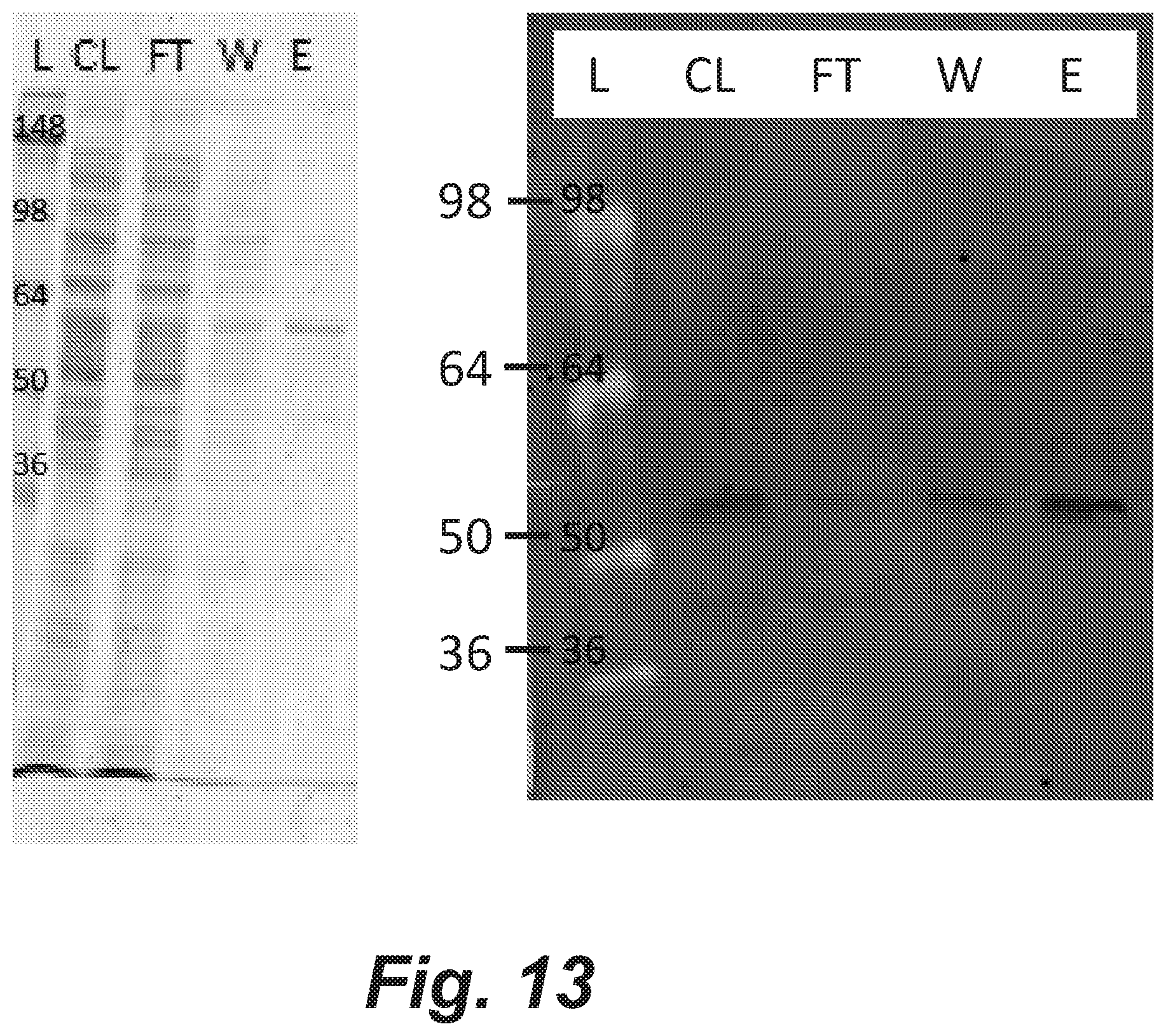
D00014

D00015

XML
uspto.report is an independent third-party trademark research tool that is not affiliated, endorsed, or sponsored by the United States Patent and Trademark Office (USPTO) or any other governmental organization. The information provided by uspto.report is based on publicly available data at the time of writing and is intended for informational purposes only.
While we strive to provide accurate and up-to-date information, we do not guarantee the accuracy, completeness, reliability, or suitability of the information displayed on this site. The use of this site is at your own risk. Any reliance you place on such information is therefore strictly at your own risk.
All official trademark data, including owner information, should be verified by visiting the official USPTO website at www.uspto.gov. This site is not intended to replace professional legal advice and should not be used as a substitute for consulting with a legal professional who is knowledgeable about trademark law.