Methods And Probiotic Compositions For The Treatment Of Bone Disorders
TRAJKOVSKI; Mirko ; et al.
U.S. patent application number 16/585726 was filed with the patent office on 2020-04-02 for methods and probiotic compositions for the treatment of bone disorders. The applicant listed for this patent is Research Development Foundation. Invention is credited to Claire CHEVALIER, Mirko TRAJKOVSKI.
| Application Number | 20200101121 16/585726 |
| Document ID | / |
| Family ID | 69945389 |
| Filed Date | 2020-04-02 |


| United States Patent Application | 20200101121 |
| Kind Code | A1 |
| TRAJKOVSKI; Mirko ; et al. | April 2, 2020 |
METHODS AND PROBIOTIC COMPOSITIONS FOR THE TREATMENT OF BONE DISORDERS
Abstract
In some aspects, methods and probiotic compositions are provided for the treatment of bone diseases such as, e.g., ostetoporosis. In some embodiments, one or more bacteria from the warm microbiota such as, e.g., Clostridialeace-assimilate, Lactobacillus, Bifidobacteriaceae, Akkermansia, and/or Parabacteroides may be administered to a subject, such as a human subject, to treat the bone disease. In some embodiments, heat may be applied to the subject to promote the development of warm microbiota to treat the bone disease.
| Inventors: | TRAJKOVSKI; Mirko; (Geneva, CH) ; CHEVALIER; Claire; (Geneva, CH) | ||||||||||
| Applicant: |
|
||||||||||
|---|---|---|---|---|---|---|---|---|---|---|---|
| Family ID: | 69945389 | ||||||||||
| Appl. No.: | 16/585726 | ||||||||||
| Filed: | September 27, 2019 |
Related U.S. Patent Documents
| Application Number | Filing Date | Patent Number | ||
|---|---|---|---|---|
| 62737661 | Sep 27, 2018 | |||
| Current U.S. Class: | 1/1 |
| Current CPC Class: | A61K 9/5036 20130101; A61K 35/741 20130101; A61K 35/745 20130101; A61P 19/10 20180101; A61K 9/2833 20130101; A61K 47/6903 20170801; A61K 47/36 20130101; A61K 35/747 20130101; A61K 9/06 20130101 |
| International Class: | A61K 35/747 20060101 A61K035/747; A61P 19/10 20060101 A61P019/10; A61K 47/36 20060101 A61K047/36; A61K 47/69 20060101 A61K047/69 |
Claims
1. A method for treating a bone disease or increasing bone strength in a mammalian subject, comprising administering a pharmaceutical or probiotic composition to the gastrointestinal system of the subject; wherein the composition comprises at least one warm microbiota bacteria or extracellular vesicles from at least one warm microbiota bacteria, and wherein if the warm microbiota contains Lactobacillus reuteri, Lactobacillus acidophilus, or Lactobacillus rhamnosus, then the warm microbiota contains at least one additional species of Clostridialeace-assimilate, Lactobacillus, Bifidobacteriaceae, or Parabacteroides.
2. The method of claim 1, wherein the composition comprises Lactobacillus gasseri, Lactobacillus reuteri, and/or Parabacteroides goldsteinii.
3. The method of claim 2, wherein the Parabacteroides goldsteinii is heat-inactivated.
4. The method of claim 1, wherein the composition comprises extracellular vesicles from Lactobacillus gasseri, Lactobacillus reuteri, and/or Parabacteroides goldsteinii.
5. The method of claim 4, wherein the Parabacteroides goldsteinii is heat-inactivated.
6. The method of claim 1, wherein the composition comprises from about 1.times.10.sup.8 to about 1.times.10.sup.13 cfu of the warm microbiota bacteria.
7. The method of claim 1, wherein the warm microbiota comprises Clostridialeace-assimilate spp., Lactobacillus spp., Bifidobacteriaceae spp., and/or Parabacteroides spp.
8. The method of claim 7, wherein the warm microbiota comprises (Lactobacillus spp. and Parabacteroides spp.) or (Lactobacillus spp. and Bifidobacteriaceae spp.).
9. The method of claim 8, wherein the warm microbiota comprises Clostridialeace-assimilate spp., Lactobacillus spp., and Parabacteroides spp.
10. The method of claim 9, wherein ratios of bacteria in the warm microbiota are about 10-40% Clostridialeace-assimilate spp., about 40-60% Lactobacillus spp., and about 10-40% Parabacteroides spp.
11. The method of claim 10, wherein ratios of bacteria in the warm microbiota are about 10-35% Clostridialeace-assimilate spp., about 40-60% Lactobacillus spp., and about 20-30% Parabacteroides spp.
12. The method of claim 1, wherein the warm microbiota comprises Bifidobacteriaceae or Akkermansia muciniphila.
13. The method of claim 12, wherein the Bifidobacteriaceae is Bifidobacterium longum.
14. The method of claim 1, wherein the warm microbiota comprises or consists essentially of live bacteria.
15. The method of claim 1, wherein the warm microbiota comprises or consists essentially of heat-inactivated bacteria.
16. The method of claim 1, wherein the warm microbiota comprises or consists of frozen or dried bacteria.
17. The method of claim 1, wherein the composition comprises extracellular vesicles from warm microbiota.
18. The method of claim 1, wherein the pharmaceutical or probiotic composition is administered orally, colonically, via enema, via an orogastric tube, or via a nasogastric tube.
19. The method of claim 1, wherein the warm microbiota is comprised in a pharmaceutical or probiotic composition that is resistant to degradation in the stomach but releases bacteria in the small intestine and/or large intestine of the subject.
20. The method of claim 1, wherein the pharmaceutical or probiotic composition comprises an enteric coating, chitosan-alginate beads, or a hydrogel.
21. The method of claim 20, wherein the enteric coating is a fatty acid, a wax, a shellac, a plastic such as a phthalate, CAP, CAT, PVAP, HPMCP, or a plant fiber.
22. The method of claim 1, wherein the pharmaceutical or probiotic composition does not comprise an enteric coating.
23. The method of claim 1, wherein the pharmaceutical or probiotic composition is a tablet or capsule.
24. The method of claim 1, wherein the subject is a human.
25. The method of claim 1, wherein the method comprises obtaining the warm microbiota from a super donor or a separate healthy subject.
26. The method of claim 25, wherein the warm microbiota is frozen after the obtaining and prior to administering to the subject.
27. The method of claim 1, wherein the method comprises obtaining the warm microbiota from the subject during the spring or summer and administering the warm microbiota to the subject during the winter.
28. The method of claim 27, wherein the warm microbiota is frozen after the obtaining and prior to administering to the subject.
29. The method of claim 1, wherein the subject has a bone disease.
30. The method of claim 29, wherein the bone disease is osteoporosis, osteomalacia, osteolysis, osteochondrodysplasias, periodontitis, rheumatoid arthritis, metabolic bone disease, a parathyroid disorder, steroid-induced osteoporosis, chemotherapy-induced bone loss, pre-menopausal bone loss, fragility and recurrent fractures, renal osteodystrophy, or Paget's disease.
31. The method of claim 30, wherein the bone disease is osteoporosis.
32. The method of claim 1, wherein the microbiota in the composition has been purified or cultured.
33. A pharmaceutical or probiotic composition for delivery to the gastrointestinal system comprising Clostridialeace-assimilate spp., Lactobacillus spp., Bifidobacteriaceae spp., and/or Parabacteroides spp., or combinations thereof; wherein if the warm microbiota contains Lactobacillus reuteri, Lactobacillus acidophilus, or Lactobacillus rhamnosus, then the warm microbiota contains at least one additional species of Clostridialeace-assimilate, Lactobacillus, Bifidobacteriaceae, or Parabacteroides.
34-58. (canceled)
59. A method of treating a bone disorder in a mammalian subject comprising administering heat to the torso of the subject.
60-67. (canceled)
Description
[0001] This application claims the benefit of U.S. Provisional Patent Application No. 62/737,661, filed Sep. 27, 2018, the entirety of which is incorporated herein by reference.
BACKGROUND OF THE INVENTION
1. Field of the Invention
[0002] The present invention relates generally to the fields of biology and medicine. More particularly, it concerns methods and compositions for the treatment of bone diseases.
2. Description of Related Art
[0003] Osteoporosis presents a significant clinical problem. Osteoporosis is the most prevalent metabolic bone disorders, characterized by low bone mass and microarchitectural deterioration (Sozen et al., 2017). Patients with osteoporosis have fragile bones and are vulnerable to fractures. The most common type of primary osteoporosis is due to the post-menopausal oestrogen deficiency, reflected in a higher incidence of osteoporosis in women (Reginster and Burlet, 2006). Although some therapies have been developed for the treatment of osteoporosis, some patients do not respond to these therapies and there is a need for new and improved therapies.
SUMMARY OF THE INVENTION
[0004] The present invention is based, in part, on the discovery that warm microbiota can improve bone density, strength, and other bone characteristics. In some embodiments, pharmaceutical compositions or probiotic compositions are provided that comprise one or more species of warm microbiota and may be used to treat a bone disease such as, e.g., osteoporosis.
[0005] An aspect of the present invention relates to a method for treating or preventing a bone disease or increasing bone strength in a mammalian subject, comprising administering a pharmaceutical or probiotic composition to the gastrointestinal system of the subject; wherein the composition comprises at least one warm microbiota bacteria or extracellular vesicles from at least one warm microbiota bacteria, and wherein if the warm microbiota contains Lactobacillus reuteri, Lactobacillus acidophilus, or Lactobacillus rhamnosus, then the warm microbiota contains at least one additional species of Clostridialeace-assimilate, Lactobacillus, Bifidobacteriaceae, or Parabacteroides genera. In some embodiments, the composition comprises Lactobacillus gasseri, Lactobacillus reuteri, and/or Parabacteroides goldsteinii (e.g., heat-inactivated Parabacteroides goldsteinii). In some embodiments, the composition comprises extracellular vesicles from Lactobacillus gasseri, Lactobacillus reuteri, and/or Parabacteroides goldsteinii (e.g., heat-inactivated Parabacteroides goldsteinii, or living Parabacteroides goldsteinii). Preferably, a therapeutically effective amount of the warm microbiota or the extracellular vesicles is administered to the subject. In some embodiments, the composition comprises from about 1.times.10.sup.8 to about 1.times.10.sup.13 cfu of the warm microbiota bacteria. In some embodiments, the warm microbiota comprises Clostridialeace-assimilate spp., Lactobacillus spp., Bifidobacteriaceae spp., and/or Parabacteroides spp. In some embodiments, the warm microbiota comprises (Lactobacillus spp. and Parabacteroides spp.) or (Lactobacillus spp. and Bifidobacteriaceae spp.). In some embodiments, the warm microbiota comprises Clostridialeace-assimilate spp., Lactobacillus spp., and Parabacteroides spp. The ratios of bacteria in the warm microbiota may be about 10-40% (e.g., 10, 15, 20, 25, 30, 35, 40%, or any range derivable therein) Clostridialeace-assimilate spp., about 40-60% (e.g., 40, 45, 50, 55, 60%, or any range derivable therein) Lactobacillus spp., and about 10-40% (e.g., 10, 15, 20, 25, 30, 35, 40%, or any range derivable therein) Parabacteroides spp. In some embodiments, the ratios of bacteria in the warm microbiota are about 10-35% Clostridialeace-assimilate spp., about 40-60% Lactobacillus spp., and about 20-30% Parabacteroides spp. The warm microbiota may comprise Bifidobacteriaceae such as (e.g., Bifidobacterium longum) or Akkermansia muciniphila. The warm microbiota may comprise or consist essentially of live bacteria. The warm microbiota may comprise or consist essentially of heat-inactivated bacteria. The warm microbiota may comprise or consist of frozen or dried bacteria. In some embodiments, the composition comprises extracellular vesicles from warm microbiota.
[0006] In some embodiments, the pharmaceutical or probiotic composition is administered orally, colonically, via enema, via an orogastric tube, or via a nasogastric tube. In some embodiments, the warm microbiota is comprised in a pharmaceutical or probiotic composition that is resistant to degradation in the stomach but releases bacteria in the small intestine and/or large intestine of the subject. The pharmaceutical or probiotic composition may comprise an enteric coating, chitosan-alginate beads, or a hydrogel. The enteric coating may be a fatty acid, a wax, a shellac, a plastic such as a phthalate, CAP, CAT, PVAP, HPMCP, or a plant fiber. In some embodiments, the pharmaceutical or probiotic composition does not comprise an enteric coating. In some embodiments, the pharmaceutical or probiotic composition is a tablet or capsule. The subject may be a human. In some embodiments, the method comprises obtaining the warm microbiota from a super donor or a separate healthy subject (e.g., a separate healthy subject who does not have a bone disease). Super-donors are individuals who have been identified as having particularly beneficial microbiota diversity and composition (e.g., Wilson et al., 2019). The method may comprise obtaining the warm microbiota from the subject during the spring or summer and administering the warm microbiota to the subject during the winter. The warm microbiota may be frozen after the obtaining and prior to administering to the subject. In some embodiments, the subject has a bone disease, e.g., osteoporosis, osteomalacia, osteolysis, osteochondrodysplasias, periodontitis, rheumatoid arthritis, metabolic bone disease, a parathyroid disorder, steroid-induced osteoporosis, chemotherapy-induced bone loss, pre-menopausal bone loss, fragility and recurrent fractures, renal osteodystrophy, or Paget's disease. In some embodiments, the bone disease is osteoporosis.
[0007] Another aspect of the present invention relates to a pharmaceutical or probiotic composition for delivery to the gastrointestinal system comprising Clostridialeace-assimilate spp., Lactobacillus spp., Bifidobacteriaceae spp., and/or Parabacteroides spp., or combinations thereof; wherein if the warm microbiota contains Lactobacillus reuteri, Lactobacillus acidophilus, or Lactobacillus rhamnosus, then the warm microbiota contains at least one additional species of Clostridialeace-assimilate, Lactobacillus, Bifidobacteriaceae, or Parabacteroides. The warm microbiota may comprise Clostridialeace-assimilate spp., Lactobacillus spp., Parabacteroides spp., and/or Akkermansia muciniphila. The warm microbiota may comprise Lactobacillus spp. and Parabacteroides spp. In some embodiments, the warm microbiota comprises Clostridialeace-assimilate spp., Lactobacillus spp., and Parabacteroides spp, optionally further comprising Akkermansia muciniphila. The ratios of bacteria in the warm microbiota may be about 10-40% (e.g., 10, 15, 20, 25, 30, 35, 40%, or any range derivable therein) Clostridialeace-assimilate spp., about 40-60% (e.g., 40, 45, 50, 55, 60%, or any range derivable therein) Lactobacillus spp., and about 10-40% (e.g., 10, 15, 20, 25, 30, 35, 40%, or any range derivable therein) Parabacteroides spp. The ratios of bacteria in the warm microbiota may be about 10-35% Clostridialeace-assimilate spp., about 40-60% Lactobacillus spp., and about 20-30% Parabacteroides spp. The warm microbiota may comprise Bifidobacteriaceae, e.g., Bifidobacterium longum. The warm microbiota may comprise or consist essentially of live bacteria, heat-inactivated bacteria, and/or frozen or dried bacteria. In some embodiments, the composition comprises extracellular vesicles from the warm microbiota. The pharmaceutical or probiotic composition may be formulated for oral, colonic, enema, orogastric, or nasogastric administration. The pharmaceutical or probiotic composition may be resistant to degradation in the stomach but releases bacteria in the small intestine and/or large intestine of the subject. The pharmaceutical or probiotic composition may comprise an enteric coating, chitosan-alginate beads, or a hydrogel. The enteric coating may be a fatty acid, a wax, a shellac, a plastic such as a phthalate, CAP, CAT, PVAP, HPMCP, or a plant fiber. In some embodiments, the pharmaceutical or probiotic composition does not comprise an enteric coating. In some embodiments, the pharmaceutical or probiotic composition is a tablet or capsule.
[0008] The composition may be used in treating a bone disorder in a mammalian subject, e.g., osteoporosis, osteomalacia, osteolysis, osteochondrodysplasias, periodontitis, rheumatoid arthritis, metabolic bone disease, a parathyroid disorder, steroid-induced osteoporosis, chemotherapy-induced bone loss, pre-menopausal bone loss, fragility and recurrent fractures, renal osteodystrophy, or Paget's disease. The subject may be a human.
[0009] Yet another aspect of the present invention relates to a method of treating or preventing a bone disorder in a mammalian subject comprising administering heat to the torso, whole body, and/or extremities of the subject. The method may comprise having the subject remain in a climate chamber with an ambient temperature of from about 65.degree. C. to about 95.degree. C. for about 3-20 minutes, about 3-15 minutes, about 3-10 minutes, or about 3-5 minutes. In some embodiments, the subject is repeatedly exposed to the climate chamber, with periods of time between each exposure (e.g., a period of about 10 minutes to 1, 2, 3, 4, 5 hours or about 1, 2, 3, 4, 5, 6 days or 1 about week between exposures). For example, the subject may be repeatedly exposed to ambient conditions of about 65-95.degree. C. or 70-95.degree. C. for about 3-15 minutes (e.g., about 5 minutes) during each exposure. The climate chamber may have an ambient temperature of from about 65.degree. C. to about 95.degree. C., and subject may remain in the climate chamber for about 3-30 minutes, about 3-15 minutes, about 3-10 minutes, or about 3-5 minutes. The method may comprise applying a heating pad or heating lamp (e.g., an infrared lamp) to the torso, stomach, and/or abdomen (or any targeted zone for treatment) of the subject, wherein the heating pad is from about 27.degree. C. to about 36.degree. C. (e.g., from about 27-50, 27-95, 65-95, or 35-50.degree. C.) or wherein the heating lamp is from about 60.degree. C. to about 95.degree. C. In some embodiments, a heating pad of about 27-36.degree. C. is applied to the body region for a duration of at least 10 minutes to several hours (e.g., about 1, 2, 3, 4, or 5 hours). In some embodiments, a warmer heating pad of about 36-95 or 35-50.degree. C. is applied to the body region for a shorter period of time such as, e.g., about 3-15 minutes. In some embodiments, the heating pad is used to raise the surface temperature of the particular body region to about 27-36 or 27-39.degree. C. The method may comprise placing the subject in a warm climatized environment, wherein the temperature is from about 30-85.degree. C., more preferably about 32-80.degree. C. The heat may be applied for a period of from about 3 minutes to about 9 hours, or 30 minutes to about 9 hours, or from 1-12 hours, or any range derivable therein (e.g., 3-10 minutes, 10-30 minutes, 10 minutes-3 hours, etc.). The heat may be applied at least 1, 2, 3, 4, 5, 6, or 7 days a week for 1, 2, 3, 4, 5, 6, 7, 8, 9, or more weeks. The bone disorder may be osteoporosis, osteomalacia, osteolysis, or osteochondrodysplasias. In some embodiments, the bone disorder is osteoporosis.
[0010] Lactobacillus gasseri is a species of bacteria that has been identified as part of the vaginal flora and has been found in the lower digestive systems of women. Particular strains of Lactobacillus gasseri that may be used to treat a bone disease or increase bone strength in a mammalian subject can include DSM 20077, DSM 107525, DSM 20243, DSM 20604, ATCC.RTM. 3332, ATCC.RTM. 2960, ATCC.RTM. BAA-2841, ATCC.RTM. PTA4483, ATCC.RTM. PTA4481, ATCC.RTM. PTA4484, ATCC.RTM. PTA4480, and/or ATCC.RTM. PTA4479. A variety of amounts of Lactobacillus gasseri may be administered to a mammalian subject (e.g., a human) to treat a bone disorder as described herein (e.g., osteoporosis, etc.). For example, in some embodiments from about 1.times.10.sup.8 to about 1.times.10.sup.13 cfu of Lactobacillus gasseri can be administered to a mammalian subject, such as a human, to treat the bone disorder or promote bone strengthening.
[0011] Lactobacillus reuteri is a species of bacteria that has been found in the intestinal tract of healthy mammals. Particular strains of Lactobacillus reuteri that may be used to treat a bone disease or increase bone strength in a mammalian subject include DSM 100191, DSM 100192, DSM 17509, DSM 20015, DSM 20016, DSM 20053, DSM 20056, DSM 28673, DSM 32035, ATCC.RTM. BAA-2837.TM., ATCC.RTM. 55148, ATCC.RTM. 53608, ATCC.RTM. 23272, ATCC.RTM. 23272D5, and/or ATCC.RTM. PTA6475. A variety of amounts of Lactobacillus reuteri may be administered to a mammalian subject (e.g., a human) to treat a bone disorder as described herein (e.g., osteoporosis, etc.). For example, in some embodiments from about 1.times.10.sup.8 to about 1.times.10.sup.13 cfu of Lactobacillus reuteri can be administered to a mammalian subject, such as a human, to treat the bone disorder or promote bone strengthening.
[0012] Parabacteroides goldsteinii is a gram-negative, obligately anaerobic non-spore-forming and non-motile bacterium that has been isolated from human blood. Particular strains of Parabacteroides goldsteinii that may be used to treat a bone disease or increase bone strength in a mammalian subject include DSM 19448 and/or DSM 29187. A variety of amounts of Parabacteroides goldsteinii may be administered to a mammalian subject (e.g., a human) to treat a bone disorder as described herein (e.g., osteoporosis, etc.). For example, in some embodiments from about 1.times.10.sup.8 to about 1.times.10.sup.13 cfu of Parabacteroides goldsteinii can be administered to a mammalian subject, such as a human, to treat the bone disorder. As shown in the below examples, heat-inactivated Parabacteroides goldsteinii can be administered to treat a bone disorder or promote bone strengthening. Methods of heat inactivation that may be used to prepare heat-inactivated Parabacteroides goldsteinii are well known and include heating up the bacteria to 100.degree. C. for 15 min (Wu et al., 2019).
[0013] As used herein the specification, "a" or "an" may mean one or more. As used herein in the claim(s), when used in conjunction with the word "comprising", the words "a" or "an" may mean one or more than one.
[0014] The use of the term "or" in the claims is used to mean "and/or" unless explicitly indicated to refer to alternatives only or the alternatives are mutually exclusive, although the disclosure supports a definition that refers to only alternatives and "and/or." As used herein "another" may mean at least a second or more.
[0015] Throughout this application, the term "about" is used to indicate that a value includes the inherent variation of error for the device, the method being employed to determine the value, or the variation that exists among the study subjects.
[0016] The terms "comprise," "have" and "include" are open-ended linking verbs. Any forms or tenses of one or more of these verbs, such as "comprises," "comprising," "has," "having," "includes" and "including," are also open-ended. For example, any method that "comprises," "has" or "includes" one or more steps is not limited to possessing only those one or more steps and also covers other unlisted steps.
[0017] Other objects, features and advantages of the present invention will become apparent from the following detailed description. It should be understood, however, that the detailed description and the specific examples, while indicating preferred embodiments of the invention, are given by way of illustration only, since various changes and modifications within the spirit and scope of the invention will become apparent to those skilled in the art from this detailed description.
BRIEF DESCRIPTION OF THE DRAWINGS
[0018] The following drawings form part of the present specification and are included to further demonstrate certain aspects of the present invention. The invention may be better understood by reference to one or more of these drawings in combination with the detailed description of specific embodiments presented herein. A petition for color drawings is provided herewith. The color in the drawings is important, e.g., for observation of the different groups of bacteria in FIGS. 1D-E.
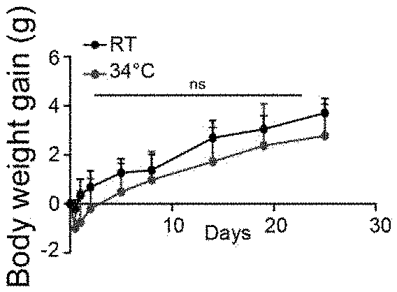
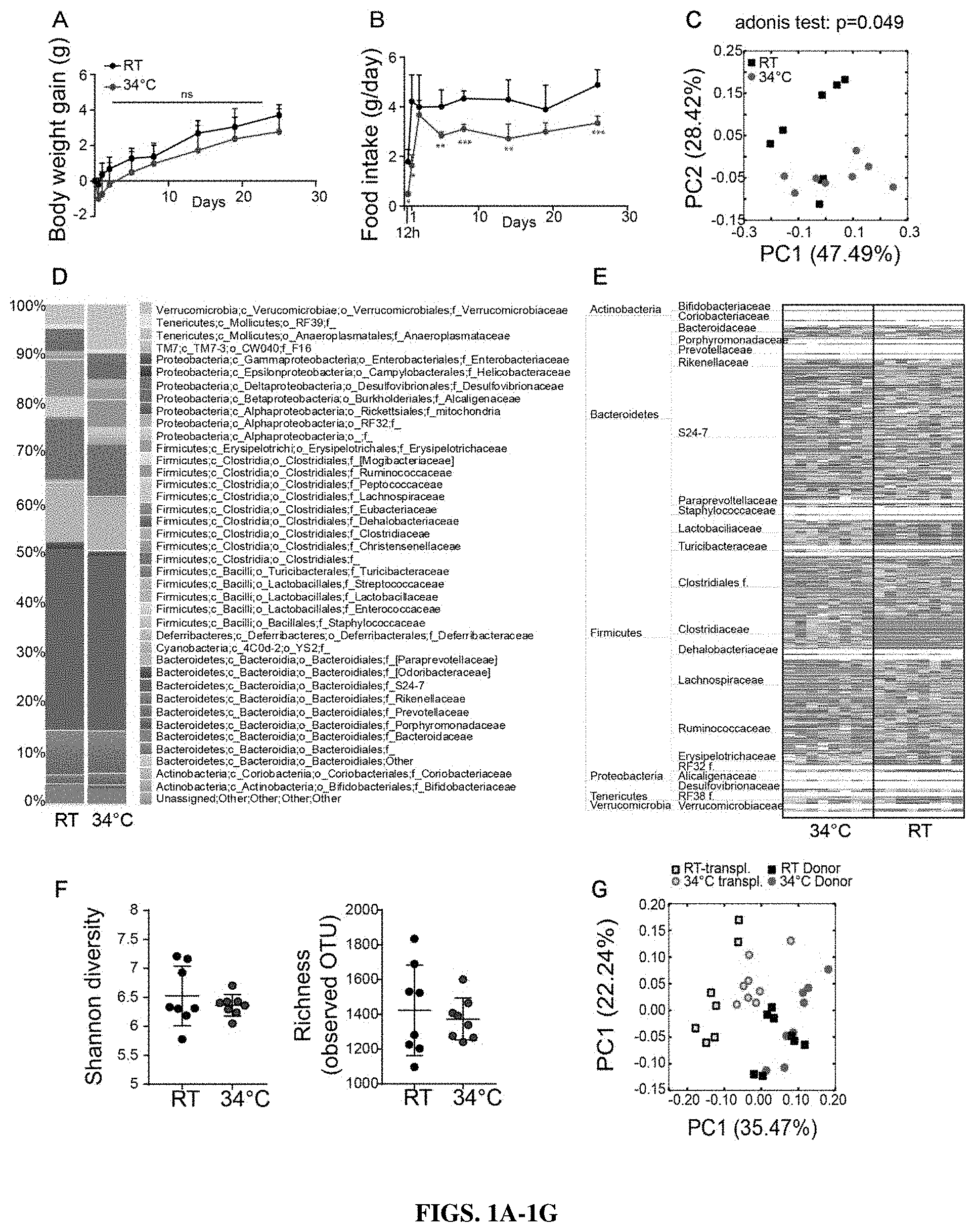
[0019] FIGS. 1A-1G show warm exposure changes in the gut microbiota population. (FIG. 1A) Body weight gain of mice exposed to 34.degree. C. for 1 month, and control mice kept at room temperature (RT). (n=8 males per groups). (FIG. 1B) Food intake of the mice as in A. (FIG. 1C) Principal coordinates analysis (PcoA) of 16SrDNA sequencing of fecal samples of mice exposed for 1 month to 34.degree. C. and their RT controls as in FIG. 1A. Each symbol represents a mouse's fecal microbiota. The analysis is based on Weighted UNIFRAC distance calculation. (FIG. 1D) Barchart representing the family proportional abundance of the samples as in FIG. 1C. (FIG. 1E) Heatmap showing OTUs associated with a p-value <0.05 after Kruskal-Walis test between 34.degree. C. exposed mice's and RT controls' fecal microbiota. Plotted is a z-score computed on the relative abundances of the selected OTUs. An idealized tree represents their taxonomic hierarchy. Final leaves are labelled with the corresponding family name. Each column represents one mouse's fecal microbiota. (FIG. 1F) Shannon diversity and estimated richness (observed OTUs) in the samples as in FIG. 1C. Data are displayed as mean.+-.SD. (FIG. 1G) Principal coordinate analysis of the fecal samples of the donor mice as in FIG. 1C. (1 month exposed at 34.degree. C. and their RT controls) and of the microbiota of corresponding recipient mice, kept at RT for 20 days. (n=10/group for the transplanted, n=8/group for the donors).
[0020] FIGS. 2A-2S show warm exposure leads to whitening of the adipose tissue, and this is partially mediated by the microbiota. (FIG. 2A) Eye temperature of mice 26 days after being exposed to 34.degree. C. and their RT controls (n=8 males per groups). (FIG. 2B) Tail temperature measurements of mice as in FIG. 2A. (FIG. 2C) Eye temperature of mice transplanted with the gut microbiota of mice as in FIG. 2A. and kept at RT (day 16 post-transplantation). (FIG. 2D) Tail temperature of mice as in FIG. 2C. (FIGS. 2E & 2F) mRNA relative expression of brown adipose tissue markers in BAT (FIG. 2E) and beige adipose tissue markers and SAT (FIG. 2F) of mice as in FIG. 2A normalized to RT controls. (FIG. 2G) H&E staining of brown adipose tissue and subcutaneous adipose tissue of mice as in FIG. 2A. (scale: 100 .mu.m). (FIG. 2H) Fat and lean mass percentage of total body weight, measured by EcoMRI of mice as in FIG. 2A. (FIGS. 2I & 2J) mRNA relative expression of brown adipose tissue markers in BAT (FIG. 2I) and beige adipose tissue markers and SAT (FIG. 2J) of mice as in FIG. 2A normalized to RT controls. (FIG. 2K) H&E staining of brown adipose tissue and subcutaneous adipose tissue of mice as in FIG. 2C (scale: 100 .mu.m). (FIG. 2L) Fat and lean mass percentage of total body weight, measured by EcoMRI of mice as in FIG. 2C. (FIGS. 2M-2S) Glucose uptake in fat tissue (FIG. 2M) BAT, (FIG. 2N) SAT and (FIG. 2O) VAT, and in muscles (FIG. 2P) Soleus, (FIG. 2Q) EDL, (FIG. 2R) Gastrocnemius, and (FIG. 2S) Quadriceps after an ipGTT spiked with radiolabeled glucose of mice as in FIG. 2A and FIG. 2C. Data are displayed as mean.+-.SD for all panels except for the qPCR data displayed as mean.+-.SEM; n=10/group for the transplanted, n=8/group for the donors. Statistic are calculated based on Mann-Whitney t-test *>0.05, **>0.01, ***>0.001.
[0021] FIGS. 3A-3K show warm exposure and warm microbiota transplantation are associated with increased bone strength. (FIGS. 3A & 3B) Tail (FIG. 3A) and femur (FIG. 3B) length of mice after 1 month at 34.degree. C. compared to their RT-kept controls (n=8 males per group). (FIG. 3C) Bone volume density (BV/TV), measured proximally in the trabecular bone of the femur (normalized to the body weight), and associated representative pictures (scale: 200 .mu.m), of mice as in A. (FIG. 3D) Cortical bone volume and width, measured in the midshaft of the femur (normalized to the body weight), and associated representative pictures (scale: 1 mm), of the mice as in FIG. 3A. (FIG. 3E) Biomechanical analysis of the femur using a 3-point bending test after 1 month of warm exposure, in male mice as in FIG. 3A. The parameters measured include the yield point, the ultimate stress, the elastic energy, the energy to fracture and the Young's modulus. (FIG. 3F) Same measures as in FIG. 3E on tibias of female mice (22 weeks old) exposed for 2 months at 34.degree. C. and their RT controls (n=8 male per groups). (FIG. 3G) Tail and femur length of mice transplanted with microbiota of mice as in FIG. 3A, 20 days after the transplantation (n=10 male per groups). (FIG. 3H) Bone volume density (BV/TV), measured proximally in trabecular bone of the femur, and associated representative pictures (scale: 200 .mu.m), of mice as in FIG. 3G. (FIG. 3I) Cortical bone volume and width, measured in the midshaft of the femur, and associated representative pictures (scale: 1 mm) of mice as in FIG. 3G. (FIG. 3J) Biomechanical analysis of the femur using a 3-point bending test after 1 month of warm exposure in male mice as in G. The parameters measured the yield point, the ultimate stress, the elastic energy, the energy to fracture and the Young's modulus. (FIG. 3K) Glucose uptake in the femur after an ipGTT spiked with radiolabeled glucose of mice as in FIG. 3A and FIG. 3G. Data are displayed as mean.+-.SD; n=10/group for the transplanted, n=8/group for the donors. Statistic are calculated based on Mann-Whitney t-test *>0.05, **>0.01, ***>0.001.
[0022] FIGS. 4A-4K show bone characteristics as a result of exposure to warm as well as bone characteristics as a result of transplantation of microbiota from warm-exposed mice. (FIG. A) Bone volume density (BV/TV), measured proximally in the trabecular bone of the tibias, in mice as in the FIG. 3C. (FIG. 4B) Cortical bone volume and width microarchitecture, measured in the midshaft of the tibias of mice as in FIG. 3C. (FIG. 4C) femur length of mice as in FIG. 3C associated with an extra control group of mice pair-fed like the 34.degree. C. exposed mice but maintained at RT. (FIG. 4D) Bone volume density (BV/TV), measured proximally in the trabecular bone of the femur, in mice as in FIG. 4C. (FIG. 4E) Cortical bone volume and width microarchitecture, measured in the midshaft of the of mice as in FIG. 4C. (FIG. 4F) Biomechanical analysis of the femur using a 3-point bending test after 1 month of warm exposure in male mice as in FIG. 4A. The parameters measured include the yield point, the ultimate stress, the elastic energy, the energy to fracture and the Young's modulus. (FIG. 4G) Bone volume density (BV/TV), measured proximally in the trabecular bone of the tibias, in transplanted mice as in FIG. 3G. (FIG. 4H) Cortical bone volume and width, measured in the midshaft of the tibias in transplanted mice as in FIG. 3G. (FIG. 4I) Glucose uptake in the femur and (FIG. 4J) in the caudal vertebra measured by micro-PET-CT with [18F]fluorodeoxyglucose after overnight fasting of mice as in the FIG. 3A. (FIG. 4K) Glucose uptake as in FIG. 4I and FIG. 4J, in mice transplanted as in FIG. 3G. Data are displayed as mean.+-.SD; n=8/group for all panels except for the transplanted n=10/group. Statistic are calculated based on Mann-Whitney t-test *>0.05, **>0.01, ***>0.001.
[0023] FIGS. 5A-5I show warm exposure prevents osteoporosis-induced bone loss in ovariectomized mice. (FIG. 5A) Body weight gain of mice exposed to 34.degree. C. for 2 months either ovariectomized or sham-operated, and their RT controls (n=8 females per groups). (FIG. 5B) food intake of mice as in FIG. 5A. (FIG. 5C) Bone volume density (BV/TV), measured proximally in the trabecular bone of the tibias (normalized to the body weight) of mice as in FIG. 5A. (FIG. 5D) Cortical bone volume and width, measured in the midshaft of the tibias (normalized to the body weight), of mice as in FIG. 5A. (FIG. 5E) Biomechanical analysis of the tibia using a 3-point bending in mice as in FIG. 5A. The parameters measured include the yield point, the ultimate stress, the elastic energy, the energy to fracture and the Young's modulus. (FIG. 5F) Tail length of mice as in FIG. 5A. (FIG. 5G) Bone volume density (BV/TV), measured in the caudal vertebra (CA2) (normalized to the body weight) of mice as in FIG. 5A. (FIG. 5H) Glucose uptake in the caudal vertebra and (FIG. 51) in the femur measured by micro-PET-CT with [18F]fluorodeoxyglucose injection after overnight fasting of mice as in FIG. 5A. Data are displayed as mean.+-.SD; n=8/group. Statistic are calculated based on Mann-Whitney t-test *>0.05, **>0.01, ***>0.001.
[0024] FIGS. 6A-6D show warm microbiota transplantation to ovariectomized mice prevents osteoporosis-associated bone fragility. (FIG. 6A) Schematic representation of the experimental plan. Mice previously exposed to 34.degree. C. for 1 month and their RT controls were used as donors of microbiota, transplanted every second day into ovariectomized recipient mice. (FIGS. 6B & 6C) Tibias of the mice were measured with a micro-CT just before ovariectomy and 1 month after transplantation, in order to measure the delta of the tibias microarchitecture. (FIG. 6B) BV/TV, BV and TV were measured proximally in the trabecular bone of tibias. (FIG. 6C) the cortical bone volume and width were measured in the midshaft of the tibias. (FIG. 6D) Biomechanical analysis of the tibias using a 3-point bending test on mice as in FIG. 6A. The parameters measured include the yield point, the ultimate stress, the elastic energy, the energy to fracture and the Young's modulus. Data are displayed as mean.+-.SD; n=8/group for the donors and n=10/group for the transplanted. Statistic are calculated based on Mann-Whitney t-test *>0.05, **>0.01, ***>0.001.
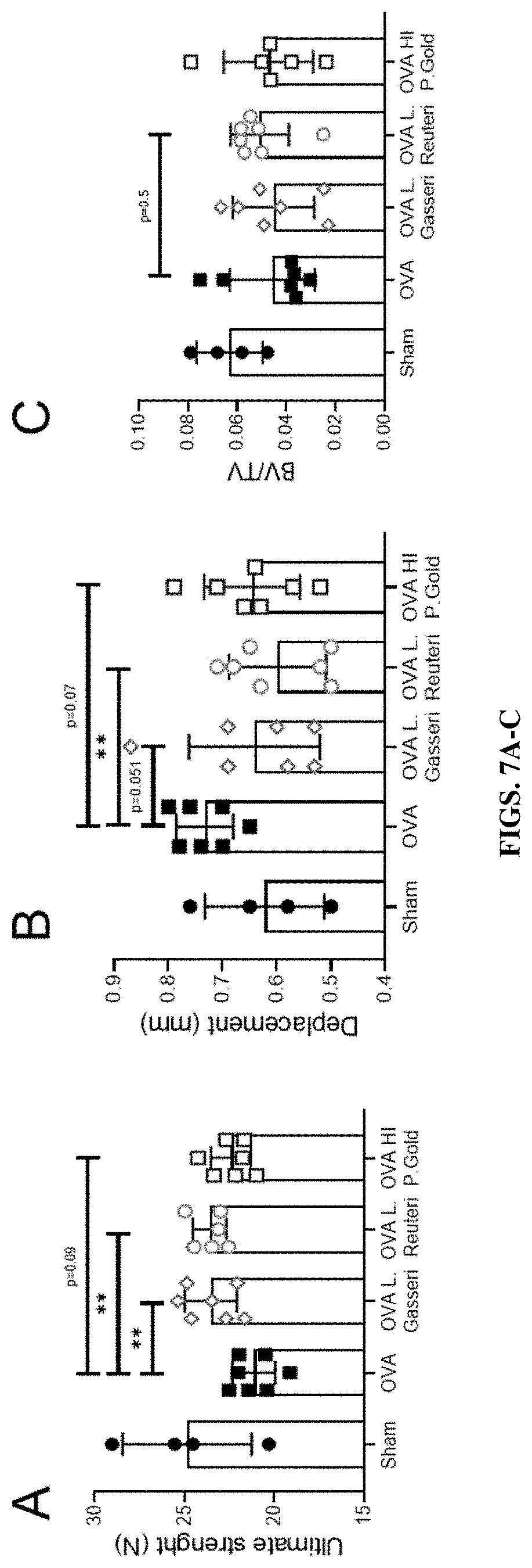
[0025] FIGS. 7A-C: L. gasseri, L. reuteri and heat-inactivaed (HI) P. goldsteinii can reduce or prevent bone fragility induce by ovariectomy. Ultimate strength (FIG. 7A) and post-yield displacement (FIG. 7B) measured by a 3-point bending biomechanical test on femur in ovariectomised mice supplemented with either L. gasseri, L. Reuteri, or HI P. goldsteinii. (FIG. 7C) Trabecular BV/TV measured by micro-CT in femur of the same mice.
DESCRIPTION OF ILLUSTRATIVE EMBODIMENTS
I. Definitions
[0026] A "bacterial composition" is a composition that comprises one of more types of bacteria (e.g., live, dried, or heat-inactivated) or extracellular vesicles (i.e., secreted extracellular vesicles) from bacteria. In some embodiments, the bacteria are from the Clostridiaceae, Lactobacillaceae, and/or Porphyromonadaceae families. Specific bacteria that are contemplated include Lactobacillus gasseri, Lactobacillus reuteri, and Parabacteroides goldsteinii (e.g., live or heat-inactivated P. goldsteinii).
[0027] The term "effective," as that term is used in the specification and/or claims, means adequate to accomplish a desired, expected, or intended result. "Effective amount," "Therapeutically effective amount" or "pharmaceutically effective amount" when used in the context of treating a patient or subject with a bacterial composition means that amount of the bacterial composition which, when administered to a subject or patient for treating or preventing a disease, is an amount sufficient to effect such treatment or prevention of the disease.
[0028] An "excipient" is a pharmaceutically acceptable substance formulated along with the active ingredient(s) of a medication, pharmaceutical composition, formulation, or drug delivery system. Excipients may be used, for example, to stabilize the composition, to bulk up the composition (thus often referred to as "bulking agents," "fillers," or "diluents" when used for this purpose), or to confer a therapeutic enhancement on the active ingredient in the final dosage form, such as facilitating drug absorption, reducing viscosity, or enhancing solubility. Excipients include pharmaceutically acceptable versions of antiadherents, binders, coatings, colors, disintegrants, flavors, glidants, lubricants, preservatives, sorbents, sweeteners, and vehicles. The main excipient that serves as a medium for conveying the active ingredient is usually called the vehicle. Excipients may also be used in the manufacturing process, for example, to aid in the handling of the active substance, such as by facilitating powder flowability or non-stick properties, in addition to aiding in vitro stability such as prevention of denaturation or aggregation over the expected shelf life. The suitability of an excipient will typically vary depending on the route of administration, the dosage form, the active ingredient, as well as other factors.
[0029] As used herein, the term "patient" or "subject" refers to a living mammalian organism, such as a human, monkey, cow, sheep, goat, dog, cat, mouse, rat, guinea pig, or transgenic non-human species thereof. In certain embodiments, the patient or subject is a primate. Non-limiting examples of human patients are adults, juveniles, infants, and fetuses.
[0030] A "pharmaceutically acceptable carrier," "drug carrier," or simply "carrier" is a pharmaceutically acceptable substance formulated along with the active ingredient that is involved in carrying, delivering and/or transporting a biological agent. Carriers may be used to improve the delivery and the effectiveness of the active ingredient, including for example, controlled-release technology to modulate drug bioavailability, decrease drug metabolism, and/or reduce drug toxicity. Some carriers may increase the effectiveness of delivery of the active ingredient to the specific target sites. Examples of carriers include: liposomes, microspheres (e.g., made of poly(lactic-co-glycolic) acid), albumin microspheres, synthetic polymers, nanofibers, protein-DNA complexes, protein conjugates, erythrocytes, virosomes, hydrogels, starches, and dendrimers. In some embodiments, the carrier comprises an enteric coating (e.g., a fatty acid, a wax, a shellac, a plastic such as a phthalate, CAP, CAT, PVAP, HPMCP, or a plant fiber) to reduce or slow degradation in the stomach, chitosan-alginate beads, or a hydrogel.
[0031] "Prevention" or "preventing" includes: (1) inhibiting the onset of a disease in a subject or patient which may be at risk and/or predisposed to the disease but does not yet experience or display any or all of the pathology or symptomatology of the disease, and/or (2) slowing the onset of the pathology or symptomatology of a disease in a subject or patient which may be at risk and/or predisposed to the disease but does not yet experience or display any or all of the pathology or symptomatology of the disease.
[0032] "Treatment" or "treating" includes (1) inhibiting a disease in a subject or patient experiencing or displaying the pathology or symptomatology of the disease (e.g., arresting further development of the pathology and/or symptomatology), (2) ameliorating a disease in a subject or patient that is experiencing or displaying the pathology or symptomatology of the disease (e.g., reversing the pathology and/or symptomatology), and/or (3) effecting any measurable decrease in a disease or symptom thereof in a subject or patient that is experiencing or displaying the pathology or symptomatology of the disease.
II. Warm Microbiota
[0033] As shown in the below examples, a variety of bacteria have been observed in warm microbiota, or the microbiota obtained from mammalian subjects living in warmer environments. These warm microbiota include Clostridialeace-assimilate spp., Lactobacillus spp. (e.g., Lactobacillus gasseri or Lactobacillus reuteri), Bifidobacteriaceae spp. (e.g., Bifidobacterium longum), Parabacteroides spp. (e.g., Parabacteroides goldsteinii) and Akkermansia spp. (e.g Akkermansia muciniphila). In some embodiments, it is anticipated that bacteria described in any one of Tables 1-5 may be included in a pharmaceutical composition or probiotic composition disclosed herein. In some embodiments, the pharmaceutical composition or probiotic composition may contain Lactobacillus reuteri, Lactobacillus acidophilus, and/or Lactobacillus rhamnosus. As shown in the examples, therapeutic responses may also be observed when using heat-inactivated Parabacteroides goldsteinii.
[0034] In various embodiments, it is anticipated that 1, 2, 3, 4, 5, 6, or more of the following types of bacteria may be included in a pharmaceutical composition or probiotic composition disclosed herein, e.g., as shown in any one of Tables 1-5. For example, the 1, 2, 3, 4, 5, 6, or more of Clostridialeace-assimilate spp., Lactobacillus spp. (e.g., Lactobacillus reuteri, Lactobacillus gasseri, Lactobacillus acidophilus, and/or Lactobacillus rhamnosus), Bifidobacteriaceae spp. (e.g., Bifidobacterium longum), Parabacteroides spp. (e.g., Parabacteroides goldsteinii) and Akkermansia spp. (e.g., Akkermansia muciniphila) may be included in a pharmaceutical or probiotic composition disclosed herein and/or administered to a mammalian subject, such as a human patient, to treat a bone disease. Various interactions between gut microbia and physiology may be used in combination with the present disclosure (e.g., as described in Ohlsson and Sjogren, 2015). Lactobacillus species such as Lactobacillus reuteri (Britton et al., 2014; also recently described in humans in Nilsson et al., 2018), Lactobacillus acidophilus (Dar et al., 2018), and/or Lactobacillus rhamnosus (Li et al., 2016) may be included in compositions for the treatment of a bone disease. In other embodiments, heat (e.g., from a heating chamber, heating pad, or a heating lamp) may be applied to the subject (e.g., to the whole body, or a specific zone, such as the torso, stomach, limbs, and/or abdomen) to treat a bone disease described herein, such as for example osteoporosis. In some embodiments, the heat applied may promote growth of warm microbiota.
III. Pharmaceutical Formulations and Routes of Administration
[0035] In another aspect, for administration to a patient in need of such treatment, pharmaceutical formulations (also referred to as bacterial formulations or pharmaceutical compositions) comprise a therapeutically effective amount of a bacterial composition disclosed herein formulated with one or more excipients and/or carriers appropriate to the indicated route of administration. In some embodiments, the bacteria disclosed herein are formulated in a manner amenable for the treatment of human and/or veterinary patients. In some embodiments, formulation comprises admixing or combining one or more of the bacteria disclosed herein with one or more of the following excipients: lactose, sucrose, starch powder, cellulose esters of alkanoic acids, cellulose alkyl esters, talc, stearic acid, magnesium stearate, magnesium oxide, sodium and calcium salts of phosphoric and sulfuric acids, gelatin, acacia, sodium alginate, polyvinylpyrrolidone, and/or polyvinyl alcohol. In some embodiments, e.g., for oral administration, the pharmaceutical formulation may be tableted or encapsulated. In some embodiments, the bacteria may be slurried in water, polyethylene glycol, propylene glycol, ethanol, corn oil, cottonseed oil, peanut oil, sesame oil, benzyl alcohol, sodium chloride, and/or various buffers. In some embodiments, the pharmaceutical formulations may be subjected to pharmaceutical operations, such as sterilization, and/or may contain carriers and/or excipients such as preservatives, stabilizers, wetting agents, emulsifiers, encapsulating agents such as lipids, dendrimers, polymers, proteins such as albumin, nucleic acids, and buffers.
[0036] Bacterial formulations may be administered by a variety of methods, e.g., orally, intracolonically, intranasally, intrarectally, via a catheter, via a lavage, via a nasogastric tube, via local delivery, or via a method for fecal microbiota transplantation (FMT). Depending on the route of administration, the bacterial compositions disclosed herein may be coated in a material to protect the bacterial compositions from the action of acids and other natural conditions which may inactivate the bacterial compositions. To administer the bacterial composition, it may be necessary to coat the bacterial composition with, or co-administer the bacterial composition with, a material to prevent its inactivation. In some embodiments, the bacterial composition may be administered to a patient in an appropriate carrier, for example, polymers, hydrogels, liposomes, starches, or a diluent. Pharmaceutically acceptable diluents include saline and aqueous buffer solutions. Liposomes include water-in-oil-in-water CGF emulsions as well as conventional liposomes.
[0037] Formulations may be employed to protect the bacterial compositions from the harsh gastric environment (Govander et al., 2014). Gastro-resistant polymers and coatings have been shown to supply protection against the harsh gastric environment. These coatings included enteric coated tablets and capsules that site-specifically deliver the administered probiotic bacteria to the intestinal system. These enteric coats are often pH selective and allow for protection against the harsh gastric conditions and subsequently dissolve in the alkali media of the intestinal system (Calinescu et al., 2005 and Yang et al., 2002). Non-limiting examples of excipients that may employed in the formulation of bacterial compositions are hydroxypropyl methylcellulose phthalate and carboxymethyl high amylose starch. Excipients may be combined to enhance delivery of the bacterial composition to the gastrointestinal tract. For example, carboxymethyl high amylose starch may be combined with chitosan for delivery of the bacterial composition to the colon. Formulations may include different polymers with different properties, or similar polymers with different properties, depending on the site of intended delivery to deliver the bacterial composition to different areas of the gastrointestinal tract (Yang et al., 2002).
[0038] The bacterial compositions disclosed herein may also be administered orally, intracolonically, intranasally, intrarectally, via a catheter, via a lavage, via a nasogastric tube, via local delivery, or via a method for fecal microbiota transplantation (FMT). The bacterial composition may be in the form of a dispersion. Dispersions can be prepared in glycerol, liquid polyethylene glycols, and mixtures thereof and in oils.
[0039] In some embodiments, the carrier comprises an enteric coating to reduce or slow degradation in the stomach. For example, the enteric coating may be a fatty acid, a wax, a shellac, a plastic such as a phthalate, CAP, CAT, PVAP, HPMCP, or a plant fiber (e.g., Hussan et al., 2012). In some embodiments, the pharmaceutical or probiotic composition may contain chitosan-alginate beads, or a hydrogel. Nonetheless, it is anticipated that in some embodiments,
[0040] The bacterial compositions disclosed herein can be administered orally, for example, with an inert diluent or an assimilable edible carrier. The bacterial compositions and other ingredients may also be enclosed in a hard or soft-shell gelatin capsule, compressed into tablets, or incorporated directly into the patient's diet. For oral therapeutic administration, the bacterial compositions disclosed herein may be incorporated with excipients and used in the form of ingestible tablets, buccal tablets, troches, capsules, elixirs, suspensions, syrups, wafers, and the like. The percentage of the therapeutic agent in the compositions and preparations may, of course, be varied. The amount of the therapeutic agent in such pharmaceutical formulations is such that a suitable dosage will be obtained.
[0041] In some embodiments, it may be advantageous to formulate compositions in dosage unit form for ease of administration and uniformity of dosage. Dosage unit form as used herein refers to physically discrete units suited as unitary dosages for the patients to be treated; each unit containing a predetermined quantity of therapeutic agent calculated to produce the desired therapeutic effect in association with the required pharmaceutical carrier. In some embodiments, the specification for the dosage unit forms of the invention are dictated by and directly dependent on (a) the unique characteristics of the therapeutic agent and the particular therapeutic effect to be achieved, and (b) the limitations inherent in the art of compounding such a therapeutic agent for the treatment of a selected condition in a patient. In some embodiments, the active agent(s) are administered at a therapeutically effective dosage sufficient to treat a condition associated with a condition in a patient. For example, the efficacy of a bacterial composition can be evaluated in an animal model system that may be predictive of efficacy in treating the disease in a human or another animal.
[0042] In some embodiments, the effective dose range for the therapeutic agent can be extrapolated from effective doses determined in animal studies for a variety of different animals. Precise amounts of the therapeutic composition depend on the judgment of the practitioner and are specific to each individual. Other factors affecting the dose include the physical and clinical state of the patient, the route of administration, the intended goal of treatment and the potency, stability, and toxicity of the particular therapeutic formulation.
[0043] The actual dosage amount of a bacterial composition of the present disclosure administered to a patient may be determined by physical and physiological factors such as type of animal treated, age, sex, body weight, severity of condition, the type of disease being treated, previous or concurrent therapeutic interventions, idiopathy of the patient and on the route of administration. These factors may be determined by a skilled artisan. The practitioner responsible for administration will typically determine the concentration of active ingredient(s) in a composition and appropriate dose(s) for the individual patient. The dosage may be adjusted by the individual physician in the event of any complication.
[0044] Single or multiple doses of the agents are contemplated. Desired time intervals for delivery of multiple doses can be determined by one of ordinary skill in the art employing no more than routine experimentation. As an example, patients may be administered two doses daily at approximately 12-hour intervals. In some embodiments, the agent is administered once a day.
[0045] The agent(s) may be administered on a routine schedule. As used herein a routine schedule refers to a predetermined designated period of time. The routine schedule may encompass periods of time which are identical, or which differ in length, as long as the schedule is predetermined. For instance, the routine schedule may involve administration twice a day, every day, every two days, every three days, every four days, every five days, every six days, a weekly basis, a monthly basis or any set number of days or weeks there-between. Alternatively, the predetermined routine schedule may involve administration on a twice daily basis for the first week, followed by a daily basis for several months, etc. In other embodiments, the invention provides that the agent(s) may be taken orally and that the timing of which is or is not dependent upon food intake. Thus, for example, the agent can be taken every morning and/or every evening, regardless of when the patient has eaten or will eat.
IV. Extracellular Vesicles of Bacteria
[0046] In some embodiments, extracellular vesicles from bacteria described herein may be administered to a subject to treat a bone disorder. For examples, extracellular vesicles (EVs) may be produced via methods as described, for example, in Chelakkot et al., 2018, or Choi et al., 2015.
[0047] Extracellular vesicles (EVs) are lipid bilayer structures secreted from the gut microbiota, including from both Gram-negative and -positive bacteria (Ellis and Kuehn, 2010 and Lee et al., 2009). A variety of bacteria constitutively produce EVs, defined as spherical lipid bilayers with an average diameter of 20-200 nm (Lee et al., 2007). EVs are composed of proteins, lipids, nucleic acids, lipopolysaccharides and other virulence factors associated with pathogenesis (Horstman and Kuehn, 2002, Hong et al., 2011, and Kim et al., 2013). EVs released by bacteria may have diverse roles in the microbial community, and some data suggests that they may transfer genetic material and proteins from the bacteria to the host (Kuehn and Nesty, 2005). EVs may directly interact with immune cells and epithelial cells to initiate several signaling pathways and may affect or mediate host-pathogen interactions.
[0048] For example, in some embodiments, EVs may be prepared via the following approach. Bacterial species or warm microbiota may be cultured under aerobic or anaerobic conditions (e.g., 95% N.sub.2 5% CO.sub.2 at 37.degree. C.) until desired (e.g., when the optical density at 600 nm reaches 1.5, as previously described; Derrien et al., 2004). Isolation of EVs may be performed as previously described in Kang et al., 2013. More specifically, bacterial cultures may be pelleted at 10 000 g for 20 min, and the supernatant may then be filtered through a 0.45-.mu.m vacuum filter. The filtrate can be enriched, e.g., using QuixStand (GE Healthcare, Little Chalfont, UK) and subsequently filtered through a 0.22-.mu.m bottle-top filter. The filtrate may then be pelleted by ultracentrifugation (e.g., in a 45 Ti rotor at 150 000 g for 2 h at 4.degree. C.). The final pellets may then be resuspended in phosphate-buffered saline (PBS) and stored at -80.degree. C. EVs may be analyzed, if desired, by transmission electron microscopy, dynamic light scattering, and/or sodium dodecyl sulfate-polyacrylamide gel electrophoresis (SDS-PAGE) followed by gel staining with Coomassie Brilliant Blue R-250.
V. Bone Diseases
[0049] It is anticipated that a variety of bone diseases or disorders may be treated with the methods and bacterial compositions described herein. For example, a bacterial composition as described herein (e.g., live bacteria, heat inactivated bacteria, lyophilized bacteria, bacteria in a pharmaceutical composition, or secreted extracellular vesicles of the bacteria) may be administered enterically or to the gastrointestinal tract of a subject to treat a bone disease or disorder or promote or increase bone density. In some embodiments, the bone disease is osteoporosis, osteomalacia, periodontitis, rheumatoid arthritis, metabolic bone disease, a parathyroid disorder, steroid-induced osteoporosis, chemotherapy-induced bone loss, pre-menopausal bone loss, fragility and recurrent fractures, renal osteodystrophy, or Paget's disease. Without wishing to be bound by any theory, it is anticipated that the methods and bacterial compositions provided herein may be used to reduce cortical and/or trabecular bone loss, reduce cortical and/or trabecular bone mineral content loss, improve the bone biomechanical resistance, increase bone formation, and/or reduce bone-resorption.
[0050] In some embodiments, the disease is osteoporosis. Osteoporosis is the most prevalent of metabolic bone disorders, characterized by low bone mass and microarchitectural deterioration (Sozen et al., 2017). Patients with osteoporosis have fragile bones and are vulnerable to fractures. The most common type of primary osteoporosis is due to the post-menopausal estrogen deficiency, reflected in a higher incidence of osteoporosis in women (Reginster and Burlet, 2006).
VI. Temperature and Gut Microbiology
[0051] As shown herein, exposure to warm environments can change the microbiota of a mammalian subject, and the resulting "warm microbiota" has been shown herein to produce effects including increases in bone density that may be particularly beneficial for treating bone diseases such as osteoporosis. Some living organisms adapt to the perpetual change of their surrounding environment. One such external parameter is temperature, which can vary from below -35.degree. C. to more than 40.degree. C. and depends on seasonal periodicity and on the time of the day. Homeotherm animals need to conserve a constant body temperature; as a result, they have developed different strategies to adapt to these external fluctuations. In rodents, a thermogenic program is engaged upon cold stimulation, including shivering thermogenesis from the muscles and non-shivering thermogenesis from the adipose tissue. During warm exposure, in contrast, the thermogenic program is blunted and the energy expenditure reduced accordingly (Kaiyala et al., 2012). Additionally, to dissipate the heat excess, rodents increase skin vasodilation at specific locations where the surface to body ratio is high in order to maximize the heat loss. This is the case in the ears and tail (Meyer et al., 2017). Interestingly, scarce reports have suggested that upon longer exposure to elevated temperature, rodents adapt to maximize their ability to dissipate heat through an increase in their tail and ear length/surface (Alhilli and Wright, 1983, Ashoub, 1958, and Harland, 1960). This is associated with bone growth and has been reported to happen in the limbs in general, particularly described in the femur (Romsos et al., 1985 and Serrat et al., 2008). Unilateral heating of the limb from weaning time is associated with bone elongation and lengthening of the extremities on the heat exposed side only (Serrat et al., 2015). It has been postulated that the warm induced-vasodilatation, associated with an increased supply of nutrient and hormones could be the mediator of the bone elongation (Alhilli and Wright, 1983 and Ashoub, 1958). However, metatarsal growth in higher temperature was increased when the incubation was performed in vitro, thus independently of any vascularization and specific nutrient supply (Serrat et al., 2008). To date, there is no clear understanding on how the bone elongates under warm exposure and the actual evidences describing this phenomenon, and the mechanical properties of the bone remain poorly investigated.
[0052] The intestinal flora has been shown to affect some aspects of host physiology. Adaptation to cold exposure was shown to be partially mediated by the gut microbiota (Chevalier et al., 2015). The present disclosure shows that warm exposure can benefit bone characteristics (including length, microstructure, and mechanical resistance), and gut flora alterations play a role in these changes to bone characteristics. Thus, these beneficial effects may be utilized in the treatment of pathologic bone conditions, such as for example osteoporosis.
VII. Examples
[0053] The following examples are included to demonstrate preferred embodiments of the invention. It should be appreciated by those of skill in the art that the techniques disclosed in the examples which follow represent techniques discovered by the inventor to function well in the practice of the invention, and thus can be considered to constitute preferred modes for its practice. However, those of skill in the art should, in light of the present disclosure, appreciate that many changes can be made in the specific embodiments which are disclosed and still obtain a like or similar result without departing from the spirit and scope of the invention.
EXAMPLE 1
Results
[0054] Warm Exposure Changes Gut Microbiota Composition
[0055] To evaluate the effect of elevated external temperature on metabolism, 8-week old C57BL/6J male mice were exposed to 34.degree. C. for one month and kept their controls at room temperature (RT, 22.degree. C.). Warm exposure was initially associated with a slight decrease in body weight that was rapidly compensated to reach similar levels than the controls (FIG. 1A). Warm exposed mice decreased their food intake by about 25% for the whole course of the experiment and already 12 h after the start of the warm exposure (FIG. 1B). This decrease in food intake, despite the maintenance a normal body weight gain, has been described to be associated with a reduced energy expenditure (Abreu-Vieira et al., 2015), allowing mice to maintain the energy balance at equilibrium. To investigate if the gut microbiota is altered during warm, the 16S rDNA from fecal and cecal samples of animals exposed for one month to 34.degree. C. and their RT controls were sequenced and the bacterial populations present in their gut microbiota were profiled. The relative distance between each microbiota was measured using principal coordinate analysis based on weighted Unifrac distance matrix (FIG. 1C). It was observed that the microbiota from 34.degree. C.-exposed animals clustered differently from the RT ones. This was associated with a significant Adonis test with a p-value of 0.049. Although the difference in the general observed population is mild, several bacterial families were pointed to with different abundancies in 34.degree. C.-exposed mice compared to RT (FIG. 1D and Table 1).
TABLE-US-00001 TABLE 1 Family abundance significant after Kruskal Wallis test (p < 0.05) in fecal sample of mice exposed 1 month at 34.degree. C. and their RT controls. Among 49 identified genera, 11 had p < 0.05. RT Warm median log2 P Family mean mean diff (medianFC) values Firmicutes; Clostridia; Clostridiales; Clostridiaceae 1.1E-03 1.7E-02 1.6E-02 4.1E+00 7.8E-04 Firmicutes; Bacilli; Lactobacillales; Lactobacillaceae 5.8E-03 2.0E-02 1.4E-02 1.9E+00 1.1E-03 Bacteroidetes; Bacteroidia; Bacteroidales; Porphyromonadaceae 3.3E-03 7.5E-03 4.0E-03 1.1E+00 1.6E-03 Actinobacteria; Actinobacteria; Bifidobacteriales; Bifidobacteriaceae 0.0E+00 1.5E-03 1.1E-04 2.0E+01 3.8E-03 Firmicutes; Bacilli; Bacillales; Staphylococcaceae 1.4E-06 2.5E-05 1.7E-05 1.7E+01 7.8E-03 Firmicutes; Bacilli; Turicibacterales; Turicibacteraceae 0.0E+00 3.0E-03 1.5E-03 2.4E+01 1.1E-02 Firmicutes; Clostridia; Clostridiales; Peptostreptococcaceae 0.0E+00 2.1E-05 2.3E-05 1.8E+01 1.1E-02 Firmicutes; Erysipelotrichi; Erysipelotrichales; Erysipelotrichaceae 9.5E-03 2.3E-02 1.5E-02 2.0E+00 1.6E-02 Proteobacteria; Alphaproteobacteria; RF32; Other 7.9E-03 1.8E-02 1.1E-02 1.9E+00 2.1E-02 Actinobacteria; Coriobacteriia; Coriobacteriales; Coriobacteriaceae 4.3E-04 1.9E-04 -2.4E-04 -1.3E+00 2.7E-02 Proteobacteria; Alphaproteobacteria; Other; Other 5.6E-04 1.1E-03 7.7E-04 1.9E+00 2.7E-02
[0056] Among 49 identified families, 11 were significantly differentially abundant in feces. This was the case of the Clostridiaceae, the Lactobacillaceae and the Porphyromonadaceae families that were the most significantly increased after warm exposure with an abundance higher than 0.5% on average. It was also noticed that Verrucomicrobiaceae were increased after warm exposure to range around 10% of mean abundance, despite not reaching statistical significance due to high variability (p=0.09). Within the significantly altered families (p>0.05), the associated heatmap representing OTUs (operational taxonomic units) belonging to each family showed that all the OTUs present in the three previously mentioned families were increased after warm exposure (FIG. 1E and Table 1). At the genera level, Lactobacillus, an unidentified genus of Clostridiaceae, and Parabacteroides were the three most significantly increased after warm exposure, thus confirming the previous observation since they are part of the families Clostridiaceae, Lactobacillaceae, and Porphyromonadaceae respectively (Table 2).
TABLE-US-00002 TABLE 2 OTUs filter for a p-value <0.05 (Kruskal-Wallis test), a median difference > 0.003 and a log.sup.2 median fold change > 1, in fecal samples of mice exposed 1 month at 34.degree. C. and their RT controls. Among 1698 identified OTU, 21 were corresponding to the criteria above, and 272 had p < 0.05. Taxonomy OTU ID Phylum Class Order Family 224155 Firmicutes Clostridia Clostridiales Clostridiaceae 392918 Firmicutes Clostridia Clostridiales Ruminococcaceae 780650 Firmicutes Clostridia Clostridiales Clostridiaceae 555945 Firmicutes Clostridia Clostridiales Clostridiaceae 460953 Bacteroidetes Bacteroidia Bacteroidales S24-7 216524 Bacteroidetes Bacteroidia Bacteroidales S24-7 268352 Firmicutes Erysipelotrichi Erysipelotrichales Erysipelotrichaceae 4426641 Firmicutes Erysipelotrichi Erysipelotrichales Erysipelotrichaceae 592160 Firmicutes Bacilli Lactobacillales Lactobacillaceae 588197 Firmicutes Bacilli Lactobacillales Lactobacillaceae 290338 Firmicutes Clostridia Clostridiales nd New.RefOTU10886 Proteobacteria Alphaproteobacteria RF32 nd 589071 Bacteroidetes Bacteroidia Bacteroidales Bacteroidaceae 276149 Bacteroidetes Bacteroidia Bacteroidales Porphyromonadaceae 339905 Bacteroidetes Bacteroidia Bacteroidales S24-7 331720 Bacteroidetes Bacteroidia Bacteroidales S24-7 228601 Bacteroidetes Bacteroidia Bacteroidales Bacteroidaceae 348038 Bacteroidetes Bacteroidia Bacteroidales S24-7 346870 Bacteroidetes Bacteroidia Bacteroidales S24-7 228730 Bacteroidetes Bacteroidia Bacteroidales S24-7 264657 Bacteroidetes Bacteroidia Bacteroidales S24-7 Log2 Taxonomy RT Warm median median OTU ID Genus Species mean mean FC diff. P values 224155 nd nd 1.4E-06 6.9E-03 3.0E+01 8.1E-03 4.5E-04 392918 nd nd 2.1E-06 9.9E-03 2.9E+01 7.0E-03 4.5E-04 780650 nd nd 2.6E-06 3.3E-03 2.9E+01 4.1E-03 4.5E-04 555945 nd nd 0.0E+00 3.5E-03 2.8E+01 3.6E-03 3.3E-04 460953 nd nd 4.0E-03 2.1E-02 3.6E+00 1.9E-02 7.8E-04 216524 nd nd 5.7E-03 5.5E-02 3.6E+00 5.1E-02 7.8E-04 268352 Allobaculum nd 2.1E-03 9.9E-03 3.5E+00 6.8E-03 1.6E-03 4426641 Allobaculum nd 1.7E-03 7.7E-03 3.2E+00 6.0E-03 1.1E-03 592160 Lactobacillus nd 2.9E-03 1.4E-02 2.5E+00 1.1E-02 1.1E-03 588197 Lactobacillus nd 1.4E-03 3.8E-03 2.4E+00 3.4E-03 3.3E-03 290338 nd nd 2.2E-03 6.2E-03 2.4E+00 3.3E-03 4.6E-02 New.RefOTU10886 nd nd 7.8E-03 1.7E-02 1.9E+00 1.1E-02 2.1E-02 589071 Bacteroides nd 2.4E-03 5.4E-03 1.5E+00 3.5E-03 2.7E-02 276149 Parabacteroides nd 3.2E-03 7.4E-03 1.2E+00 4.0E-03 1.6E-03 339905 nd nd 1.3E-02 6.7E-03 -1.1E+00 -7.4E-03 1.6E-02 331720 nd nd 2.7E-02 1.0E-02 -1.1E+00 -1.2E-02 7.8E-04 228601 Bacteroides nd 1.5E-02 7.2E-03 -1.2E+00 -7.9E-03 1.2E-02 348038 nd nd 7.3E-03 4.0E-03 -1.3E+00 -4.8E-03 8.7E-03 346870 nd nd 2.3E-02 9.3E-03 -1.5E+00 -1.5E-02 1.1E-03 228730 nd nd 1.4E-02 5.2E-03 -1.5E+00 -1.0E-02 2.3E-03 264657 nd nd 4.9E-03 1.6E-03 -1.5E+00 -3.1E-03 1.6E-03
[0057] Looking at the OTU level (Table 3), the same families and genera were highlighted with several OTUs from Clostridiaceae family, two OTUs from Lactobacillus genus and one OTU from Parabacteroides. Other OTUs from S24-7 family and Allobaculum genus were also part of the most differently abundant OTUs. Elevated temperature exposure had no effect on the richness or diversity of the gut microbiota (FIG. 1F).
TABLE-US-00003 TABLE 3 Genera abundance significant after Kruskal Wallis test (p < 0.05) in fecal samples of mice exposed 1 month at 34.degree. C. and their RT controls. Among 73 identified genera, 13 had p < 0.05. RT 34.degree. C. median log2 P Genera mean mean diff (medianFC) values Firmicutes; Clostridia; Clostridiales; Clostridiaceae; Other 7.5E-04 1.6E-02 1.5E-02 4.4E+00 7.8E-04 Firmicutes; Bacilli; Lactobacillales; Lactobacillaceae; Lactobacillus 5.8E-03 2.0E-02 1.4E-02 1.9E+00 1.1E-03 Bacteroidetes; Bacteroidia; Bacteroidales; Porphyromonadaceae; Parabacteroides 3.3E-03 7.5E-03 4.0E-03 1.1E+00 1.6E-03 Actinobacteria; Actinobacteria; Bifidobacteriales; Bifidobacteriaceae; Bifidobacterium 0.0E+00 1.5E-03 1.1E-04 2.0E+01 3.8E-03 Firmicutes; Bacilli; Bacillales; Staphylococcaceae; Staphylococcus 1.4E-06 2.5E-05 1.7E-05 1.7E+01 7.8E-03 Firmicutes; Erysipelotrichi; Erysipelotrichales; Erysipelotrichaceae; Allobaculum 6.2E-03 1.9E-02 1.3E-02 2.9E+00 8.7E-03 Firmicutes; Bacilli; Turicibacterales; Turicibacteraceae; Turicibacter 0.0E+00 3.0E-03 1.5E-03 2.4E+01 1.1E-02 Firmicutes; Clostridia; Clostridiales; Clostridiaceae; SMB53 0.0E+00 3.2E-05 1.7E-05 1.7E+01 1.1E-02 Firmicutes; Clostridia; Clostridiales; Peptostreptococcaceae; Other 0.0E+00 2.1E-05 2.3E-05 1.8E+01 1.1E-02 Proteobacteria; Alphaproteobacteria; RF32; Other; Other 7.9E-03 1.8E-02 1.1E-02 1.9E+00 2.1E-02 Firmicutes; Clostridia; Clostridiales; Lachnospiraceae; Other 3.4E-02 1.3E-02 -9.3E-03 -9.4E-01 2.7E-02 Proteobacteria; Alphaproteobacteria; Other; Other; Other 5.6E-04 1.1E-03 7.7E-04 1.9E+00 2.7E-02 Firmicutes; Clostridia; Clostridiales; Clostridiaceae; Candidatus Arthromitus 1.5E-04 3.9E-04 2.2E-04 2.1E+01 4.3E-02
[0058] While analysis of the cecal microbiota (Table 4) showed fewer differences between warm and RT mice compared to the fecal microbiota, increased Lactobacillus abundance was a similar hallmark in both microbiota specimens. Taken together, these results show that warm exposure in mice changes the gut microbiota composition with a particular increase in the genera Lactobacillus, Clostridiaceae-assimilate and Parabacteroides in feces.
TABLE-US-00004 TABLE 4 Significant differentially abundant genera of cecal samples of mice exposed 1 month at 34.degree. C. and their RT controls (p < 0.05 after Kruskal-Wallis test). Among 64 identified genera, 5 had p < 0.05. RT Warm median log2 P Genera cecum mean mean diff (medianFC) values Firmicutes; Bacilli; Lactobacillales; Lactobacillaceae; Lactobacillus 0.0036 0.0084 0.0036 0.9005 0.0087 Tenericutes; Mollicutes; Anaeroplasmatales; Anaeroplasmataceae; Anaeroplasma 0.0003 0.0085 0.0043 5.6683 0.0180 Firmicutes; Bacilli; Turicibacterales; Turicibacteraceae; Turicibacter 0.0000 0.0060 0.0005 22.1440 0.0273 Firmicutes; Clostridia; Clostridiales; Lachnospiraceae; Coprococcus 0.0057 0.0031 -0.0008 -0.2948 0.0274 Firmicutes; Clostridia; Clostridiales; Lachnospiraceae; Other 0.0681 0.0408 -0.0113 -0.3554 0.0460
[0059] Gut Microbiota Contributes to the Thermogenic Adaptation to Warm
[0060] To evaluate the effect of the gut microbiota on the warm ambient temperature adaptation, germ-free (GF) mice were transplanted with `warm` cecal microbiota (warm-transplanted) (FIG. 1C-1F and Tables 1-4) and were compared to RT microbiota-transplanted GF (RT-transplanted) control mice. All transplanted mice were maintained at RT. The transplantation efficiency was confirmed using principal coordinate analysis (FIG. 1G), where the transplanted recipients showed similar changes to their donors. The difference in the transplanted animals' microbiota compared with Adonis test was significant with a p-value of 0.01. In addition, similar bacterial hits to the donors were found in the transplanted microbiota as the most differentially abundant family, such as Clostridiaceae (here associated to Clostridium), Parabacteroides, and Akkermansia, Lactobacillus, however, was not increased in the warm transplanted animals, likely due to a disadvantageous competition post-transplantation.
TABLE-US-00005 TABLE 5 Differentially abundant genera in fecal microbiota of RT- and warm-transplanted mice (Kruskal Wallis test p < 0.05). 34.degree. C. RT transpl. transpl. genera P value mean mean Firmicutes; Clostridia; Clostridiales; Clostridiaceae; Clostridium 5.60E-04 5.30E-06 1.40E-04 Firmicutes; Clostridia; Clostridiales; Lachnospiraceae; Dorea 1.60E-03 3.90E-04 6.10E-05 Verrucomicrobia; Verrucomicrobiae; Verrucomicrobiales; Verrucomicrobiaceae; Akkermansia 1.60E-03 8.30E-02 1.20E-02 Bacteroidetes; Bacteroidia; Bacteroidales; Porphyromonadaceae; Parabacteroides 4.60E-03 5.80E-03 3.20E-03 Bacteroidetes; Bacteroidia; Bacteroidales; Rikenellaceae; other 4.60E-03 1.00E-01 1.70E-01 Bacteroidetes; Bacteroidia; Bacteroidales; [Odoribacteraceae]; Odoribacter 4.60E-03 2.10E-03 8.40E-03 Tenericutes; Mollicutes; Anaeroplasmatales; Anaeroplasmataceae; Anaeroplasma 8.60E-03 3.20E-04 3.20E-03 Bacteroidetes; Bacteroidia; Bacteroidales; other; other 8.70E-03 1.10E-02 2.40E-02 Firmicutes; Clostridia; Clostridiales; Lachnospiraceae; Other 8.70E-03 4.30E-04 2.20E-03 Firmicutes; Clostridia; Clostridiales; Lachnospiraceae; Blautia 1.50E-02 6.80E-05 1.70E-05 TM7; TM7-3; CW040; F16; other 2.00E-02 1.00E-05 4.70E-05 ActinoCoriobacteriia; Coriobacteriales; Coriobacteriaceae; other 4.30E-02 8.30E-05 2.00E-05 Firmicutes; Clostridia; Clostridiales; Other; Other 4.60E-02 4.60E-03 1.40E-03
[0061] Under warm temperature, mice need to maintain a constant body temperature around 37.degree. C. This is enabled by blunting the thermogenic program and increasing the vasodilatation of the tail (and ears) to dissipate the excessive heat (Kaiyala et al., 2012, Alhilli and Wright, 1983, Ashoub, 1958, and Harland, 1960). In the experimental setting, mice exposed one month at 34.degree. C. were able to maintain their body temperature constant (reflected by the eye temperature) (FIG. 2A), mediated by an increased temperature dissipation through their tail (FIG. 2B). To understand if the gut microbiota composition could participate in the adaptation to warm exposure, the same parameters were measured in the mice transplanted with the microbiota of warm exposed mice (warm-transplanted). Surprisingly, the mice receiving the warm microbiota had a slight decrease in their eye temperature, representative of their body temperature (FIG. 2C). This suggests that the warm microbiota alone is sufficient to decrease the core body temperature. This lower temperature was not mediated by an increased heat dissipation from the tail, as no differences in the tail temperature between the warm- and RT-transplanted mice was noticed (FIG. 2D). This suggests that other thermoregulatory mechanisms have been transplanted through the microbiota in these mice. Cold environment stimulates a thermogenic response to maintain body temperature at a constant level, it initiates shivering thermogenesis through muscular contraction and non-shivering thermogenesis with brown or beige adipose tissue activation. The main effector protein is the uncoupling protein 1 (UCP1) that uncouples the oxidative phosphorylation in the mitochondria from the ATP production and alleviates heat production. In the context of warm exposure, this phenomenon is blunted and the activity of brown and beige adipose tissue is reduced (Cui et al., 2016). This was confirmed in the experimental setting where Ucp1 and other thermogenic markers expression level were reduced both in the interscapular brown adipose tissue (BAT; FIG. 2E) and in subcutaneous adipose tissue (SAT; FIG. 2F). In addition, the morphology of this tissue changed to an increased lipid droplet size and disappearance of the multilocular cells (numerous small lipid droplets normally present in the active brown or beige adipocytes; FIG. 2G). This was associated with a maintenance of the fat mass despite the reduced food intake (FIG. 1B and FIG. 2H). It was suspected that the reduced basal body temperature of the warm transplanted mice was also due to an effect of the microbiota on the adipose tissue thermogenic activity. While changes in thermogenic genes expression in the BAT were not observed (FIG. 2I), Ucp1 and Cidea showed reduced expression in the SAT indicating a whitening of the adipose tissue (FIG. 2J). Neither morphological changes in the H&E stained sections of the BAT and SAT of the transplanted mice, nor any repercussion on fat mass distribution (FIGS. 2K-2L) were detected. In contrast, the decreased thermogenic genes expression was reflected in the glucose uptake of the tissues. Specifically, it was observed a reduced glucose uptake in BAT and SAT of the warm exposed animals that was also present in SAT of warm microbiota-transplanted mice (FIGS. 2M-2N). No changes were observed in visceral adipose tissue (VAT) and muscles glucose uptake (FIGS. 2O-2S), confirming the specific decreased glucose uptake mediated by decreased thermogenic activity. This suggests that mice adapt to warm temperature by altering their thermogenic activity, partially through a gut microbiota adaptation.
[0062] Bone Morphology Adaptation After Warm Exposure
[0063] Heat dissipation in rodents is mediated by vasodilatation of the extremities, mainly in the tail. This phenomenon is associated with an increase in the tail and ear length to maximise the surface ratio for heat dissipation (Serrat et al., 2008). In the experimental setting, this observation was confirmed as after one month of warm exposure the tail length increased by 8% relative to the controls (.about.0.7 cm longer) (FIG. 3A). It was further analysed whether warm exposure had an effect on long bone growth, and examined the structure of the femur. Serrat and coworkers showed that warm exposure from weaning to adulthood was leading to tail elongation, but also limbs like in the femur (Serrat et al., 2008). Although there was no difference in total femur length between mice exposed one month to warm and their RT controls (FIG. 3B), the structural analysis of the femur by micro-CT showed morphological changes. The trabecular bone proportion increased as reflected by the bone volume (BV)/total volume (TV) ratio (FIG. 3C), but no differences in the cortical bone were found (FIG. 3D). Similar structural changes as in the femur were observed in the tibia (FIGS. 4A & 4B). To investigate if these morphological changes were reflected in the biomechanical resistance of the bone, a three-point bending test was performed on the femur. Despite the trabecular increase in BV/TV ratio, no changes in the mechanical strength were observed (FIG. 3E). However, when the same test was performed on older female mice exposed longer to 34.degree. C. (two months), it was observed that both the yield point and the ultimate force were significantly increased with warm exposure (FIG. 3F). This suggests that long-term warm exposure has an impact on the bone morphology and associated strength, although age and sex dimorphism may interfere in the bone morphology in response to temperature. It has been previously shown that caloric restriction impaired bone mass both in rodent models and in humans (Devlin et al., 2010, Legroux-Gerot et al., 2007, Villareal et al., 2006, and Sanderson et al., 1997). As mentioned above, warm exposure is associated with a reduction in food intake of .about.25% (FIG. 1B). To control for this reduced food intake, the same experiment was performed with an additional group of mice that were pair-fed like the warm exposed mice (25% less food than the RT controls) but kept at RT. It was observed that despite their reduced food intake, the warm exposed mice were able to maintain the structure and the biomechanical strength of the femur, while the pair-fed mice displayed a reduced trabecular and cortical bone density as well as a mechanical fragility of the femur (FIGS. 4C-4F). This is in line with the beneficial effect of warm exposure on the bone strength, even if the comparison with pair-fed mice implies other regulatory mechanisms (especially regarding the energy balance of the two groups). It was then investigated if this adaptation could be mediated by the gut microbiota and measured the same parameter in the transplanted animals. Although differences in the total tail and femur lengths were not detected (FIG. 3G), the structural parameters of the femur were improved with warm microbiota transplantation. There was a tendency for higher trabecular BV/TV ratio (FIG. 3H), while at a cortical level, both the cortical bone volume and width in the femur (FIG. 3I) and in the tibias (FIGS. 4G & 4H) were increased with warm-microbiota transplantation. This suggests an impact of the gut microbiota to maintain bone mass at both the trabecular and cortical levels. To follow up on the structural changes of the bone, biomechanical analysis was performed and it was observed that all parameters measuring the strength of the bone were increased with warm microbiota transplantation. ANOVA analysis of all the biomechanical parameters revealed that the microbiota-induced changes were significant (p=0.05). Of note, these effects were measured only after 20 days of transplantation. Since the bone remodeling is very energy demanding, the glucose uptake in the femur and the caudal vertebra was measured and it was observed that glucose uptake was increased both in warm exposed animals and in warm-microbiota transplanted ones, in line with the structural and mechanical measurements (FIG. 3K and FIGS. 4I-4K). Taken together, this suggests that warm exposure has a positive effect on bone density and strength already after one month of warm exposure and the effect is amplified after two months. This is in part mediated by a shift in the gut microbiota population, which induces structural and mechanical changes already after 20 days of transplantation.
[0064] Effect of Warm Exposure on Osteoporosis
[0065] Next, it was investigated whether the positive effect of warm exposure on the bone morphology could lead to improvement in a pathological context. Osteoporosis is caused by an imbalance between the bone formation and the bone resorption and is a leading cause of fractures in aged individuals. One of the main factors associated with osteoporosis is oestrogen deficiency present during the post-menopausal period in women. To evaluate if warm exposure could have a positive effect on osteoporosis, aged ovariectomized mice were used that mimic the post-menopausal osteoporosis observed in humans. Four groups of mice were used: two groups were sham operated as controls (one kept two months at 34.degree. C., and the other at RT) and two groups were ovariectomized (similarly kept either at 34.degree. C. or at RT). Consistent with previous observations (Iwasa et al., 2018), the ovariectomized mice gained more weight than their sham-operated controls. Surprisingly, warm exposure was sufficient to prevent this body weight gain, and the mice were able to maintain a body weight similar to the sham-operated ones (FIG. 5A) coupled with 25% reduction in the food intake (FIG. 5B). Body weight and the associated mechanical load put on the bone has an effect on the bone density and strength (Iwaniec and Turner, 2016). To exclude this confounding factor, the next results were normalized to the body weight of the mice. As expected, the structure of the trabecular bone (BV/TV) and the cortical bone (volume and width) were decreased after ovariectomy. This was, however, prevented by warm exposure, where all the parameters were close to the sham-operated animals (FIGS. 5C & 5D). Differences observed in ovariectomized mice after warm exposure were more exacerbated than in sham-operated mice. This suggests that the mechanisms involved in bone deterioration due to oestrogen deficiency can be prevented by mechanisms deployed during warm exposure. Interestingly, the rescue observed at the structural level was also reflected at the mechanical level. While energy to fracture and elastic energy were not affected, the ultimate force the bone can endure before breaking was improved in ovariectomized mice after warm exposure (compared to the RT-kept mice). Parameters assessing the flexible part of the bone like the young modulus and the yield point were also improved, reaching similar levels as the control sham-operated mice (FIG. 5E). This suggests that warm exposure can prevent ovariectomy-induced osteoporosis. Additionally, the effect on the tail elongation normally present after warm exposure was conserved despite ovariectomy (FIG. 5F). The structure of the tail vertebra was also affected, with the same pattern as for the tibias (FIG. 5G), indicating a broad beneficial effect of the warmth on osteoporosis in long bones and in vertebras. It was also noticed that despite ovariectomy, warm exposure was accompanied by an increased glucose uptake in caudal vertebra (FIG. 5H), indicating an increase in their metabolic activity. However, the glucose uptake in the femur was similar in both warm-ovariectomized and RT-ovariectomized, probably due to the basal increased remodeling activity present in osteoporosis (FIG. 5I).
[0066] Effect of Warm Microbiota on Osteoporosis
[0067] It was wondered whether the prevention of osteoporosis observed with warm exposure can be also achieved by transplantation of the warm-adapted microbiota. To investigate this, an experiment was designed wherein donor mice previously exposed for one month at 34.degree. C. or RT were maintained to freshly transplant their fecal microbiota. Recipient mice were ovariectomized and transplanted with the donor microbiota every second day for one month (FIG. 6A). In this cohort, the tibia structure was measured before and one month after the ovariectomy. This allowed for the calculation of the change (delta) in the bone microarchitecture before and after the treatment to measure the effect of transplantation on the osteoporosis outcome. The trabecular BV/TV was slightly increased, but not significantly. This was the consequence of a double effect of the temperature, increasing both the bone volume (B V) and the total volume (TV), indicating an effect on the overall growth of the trabecular bone (FIG. 6B). The cortical bone, however, was unchanged (FIG. 6C). Interestingly, by looking at the biomechanics analysis, the ultimate force and the yield point were prevented from being reduced due to the ovariectomy. This was in line with the protective effect observed on the energy to fracture and the elastic energy, also not decreased by the ovariectomy (FIG. 6D). This suggests that warm microbiota transplantation could partially prevent the ovariectomy-associated bone fragility and protect against the related fracture risk.
EXAMPLE 2
Discussion
[0068] Mammals have been co-evolving in symbiosis with their gut microbiota. The external temperature is a major environmental factor to which an individual needs to constantly adapt. It has recently been described that the gut microbiota participates in the adaptation to cold external temperature (Chevalier et al., 2015, Worthmann et al., 2017, and Zietak et al., 2016). Cold exposure induces major changes in the gut microbial population. When transplanted into germ-free animals, this cold-microbiota was able to induce thermogenic activation of beige and brown adipose tissue. The present disclosure shows that warm exposure also induces changes in the bacterial composition measured in the feces, however, to a lesser extent than the one observed after cold. Transplantation of this warm microbiota into germ-free mice was sufficient to blunt the thermogenesis and decrease Ucp1 expression in subcutaneous adipose tissue. This is in line with additional interventions that promote white adipose tissue browning mediated by the gut microbiota composition, such as microbiota depletion (Suarez-Zamorano et al., 2015) or caloric restriction (Fabbiano et al., 2018). Trying to identify common bacteria decreased after cold or caloric restriction and increased after warm may help researchers to understand if a specific bacterial population could be responsible for the adipose tissue whitening. Alteration of these bacterial populations by phage therapy or by transplantation experiments in order to promote adipose tissue browning may be an appealing therapeutic target in the context of obesity.
[0069] During warm temperature exposure, mammals dissipate the heat through an increased vasodilatation. This is associated with an elongation of the tail and ear in rodents in order to maximize the surface area for heat dissipation (Alhilli and Wright, 1983 and Ashoub, 1958). Warm exposure during development is also causing elongation of the limbs such as the tibias and the femur (Serrat et al., 2008). The present disclosure illustrates that elevated temperature at a later stage of the development (starting at 8 weeks of age) improves the structural volume of bones of the limbs, especially in the trabecular bone. This phenomenon is already visible after one month of warm exposure in males. After two months of warm exposure in females, the increased trabecular proportion is reflected in the biomechanical resistance of the bone where the yield point and the ultimate stress are improved during a three-point bending test. Sexual dimorphism regarding bone remodeling is well documented (de Bakker et al., 2018). In line with this, the present data show that both sexes display bone remodeling upon warm exposure, but only female mice have improved biomechanical properties.
[0070] The effect of the warm temperature was mediated in part by the gut microbiota. Specifically, the "warm" microbiota transplantation displayed increased cortical bone structural volume and tendency towards enhanced trabecular volume. This led to a moderate improvement of the bone mechanical resistance. Interestingly, this phenomenon is relatively rapid since these effects are already observed as early as 20 days after the transplantation. This was associated with an increased glucose uptake in the femur in line with an increased need for fueling the bone remodeling process. Warm microbiota transplantation into germ-free mice leads to increased volume in both trabecular and cortical bone. However, the warm temperature exposure on conventional mice did not show any effect on the cortical bone. Germ-free mice were observed to have an increased trabecular and cortical bone volume compared to conventionally raised animals. When colonized with microbiota, the germ-free mice show a reduction of these parameters (Sjogren et al., 2012). Transplantation of warm microbiota in germ-free mice may prevent the decrease in cortical bone density compared with the conventional, RT-transplanted animals. If warm exposure may thus be used in treating or preventing the bone loss in a pathological condition such as osteoporosis. An ovariectomized mice model was used mimicking the post-menopausal osteoporosis in humans. Warm exposure was indeed able to prevent the bone loss induced by ovariectomy at both trabecular and cortical level. This was also transposed to the mechanical resistance of the bone, where osteoporosis induced fragility was prevented by warm exposure. Interestingly, these effects observed in the tibias were also present in the caudal vertebra where warm exposure prevented the trabecular bone loss in the ovariectomized mice. This indicates a protective effect of the warmth on long bones but also vertebra. Knowing that the associated risk fracture in human affected by osteoporosis is mainly related to the hip (femoral neck) and vertebrae (Bianchi et al., 2005), this supports the idea that warm exposure can be used as a therapeutic intervention to reduce the post-menopausal osteoporosis.
[0071] Other approaches may also be used to treat osteoporosis. For example, germ-free mice were protected against osteoporosis induced by GnRH agonist (Li et al., 2016). In the present disclosure, it is shown that recurrent transplantation of warm microbiota into ovariectomized mice was sufficient to prevent bone fragility and improve the trabecular bone structural parameters. The main changes in the bacterial population observed were an increase in the genera Clostridialeace-assimilate, Lactobacillus, and Parabacteroides. The beneficial effect observed with the warm microbiota might be mediated by the increased abundance of the Lactobacillus genera. However, the present disclosure shows that the increased Lactobacillus genus of the warm microbiota was not efficiently transplanted in the germ-free mice. Still, mice displayed an improvement in bone proportion after structural analysis and moderate improvement in bone mechanical resistance. These results support the idea that other bacterial population changes are responsible for this improvement and identifying them may be included in a probiotic mix for the treatment or prevention of osteoporosis.
[0072] The present disclosure illustrates that the gut microbiota is modulated after warm temperature exposure and that it is symbiotically participating in the regulation of the temperature adaptation by decreasing the thermogenic program in the adipose tissue. Additionally, it was uncovered that warm exposure has a positive effect on the bone volume and mechanical resistance in health and in the context of a post-menopausal osteoporosis model. Finally, it was shown that the beneficial effects observed on the bone after warm exposure were at least partially mediated by the gut microbiota changes. Osteoporosis is a highly prevalent bone metabolic disease in the older population and often diagnosed only after the first fracture. Treatments such as warm exposure or probiotic ingestion may be feasible therapeutic approaches in the prevention of this disease.
EXAMPLE 3
Materials and Methods
[0073] Animals: All C57BL/6J mice were purchased from Janvier Labs, and kept in a specific pathogen-free (SPF) facility in individually ventilated cages, or in a conventional facility with open cages covered with filter lids. All the mice were in a 12 h day/night cycle and fed a standard chow diet (16.2 MJ/kg Gross Energy; 9 kJ % Fat, 33 kJ % Protein, 58 kJ % Carbohydrates, V1534-727, S sniff, Germany). Germ-free mice of C57BL/6 background were bred and obtained from the germ-free facility of the University of Bern and transplanted with donor microbiota upon arrival in the SFP facility. All the mice were either male and entered the experiment at 8 weeks of age, or female starting at 16 weeks of age (for the ovariectomy experiment). Acclimatized animals were allocated into groups based on their body weight to ensure equal starting points. Warmth exposure was done at 34.degree. C. in a light and humidity-controlled climatic chamber (TSE, Germany) in SPF conditions using individually ventilated cages, or with a temperature-controlled chamber MEDI1300 from Froilabo for the conventional facility. The 34.degree. C. like pair-fed animals were kept at room temperature and fed an equal amount to the warm exposed animals. This is equivalent to .about.25% less than RT ad libitum fed, and the food was provided each day at 6 pm. All mice were sacrificed after 5 h fasting. 500 .mu.l of blood was taken from terminally anaesthetized mice in tubes with 10 .mu.l of 0.5 mM EDTA, 4 .mu.l of aprotinin (1.3%), and 4 .mu.l of DPP-IV (10 mM), and plasma stored at -80.degree. C. Samples for RNA isolation were stored in RNAlater solution (Invitrogen ref AM7020). Bone samples for CT-scan analysis were stored in a humid package at -20.degree. C., samples for histology in 4% PFA and all other samples snap-frozen in liquid nitrogen. The tail length was measured with a ruler from the tip of the tail to the limit between fur and skin. All animal experiments were approved by the Swiss federal and Geneva cantonal authorities for animal experimentation (Office Veterinaire Federal and Commission Cantonale pour les Experiences sur les animaux de Geneve).
[0074] Gut microbiota profiling: At the end of the experiment, fecal samples were collected in sterile tubes and immediately frozen and kept at -80.degree. C. Cecal samples were collected after sacrifice of the mice, snap frozen and conserved at -80.degree. C. Fecal and cecal bacterial DNA was then extracted using the PowerFecal DNA Kit (Qiagen, Ref. 12830-50) and the 16SrDNA library was built following the standardized protocol from the earth microbiome project (Caporaso et al., 2012 and Caporaso et al., 2011). Basically, DNA was amplified with QuantaBio 5Prime HotMasterMix using barcoded universal bacterial primers targeting variable region V4 of 16SrRNA gene (515F-806R barcoded primers, Illumina) 806 Reverse Primer GGACTACNVGGGTWTCTAAT (SEQ ID NO: 1)-515 Forward Primer GTGYCAGCMGCCGCGGTAA (SEQ ID NO: 2). 2 ng of template was used and the PCR conditions included an initial denaturation at 94.degree. C. for 3', followed by 35 cycles of denaturation at 94.degree. C. for 45'', annealing at 50.degree. C. for 1', and extension at 72.degree. C. for 90'', with a final extension at 72.degree. C. for 10'. Each PCR was performed in triplicate later combined and quality checked on an agarose gel. Each PCR amplification was then quantified with Quant-iT PicoGreen dsDNA Assay with SpectraMax Gemini XPS microplate reader and pooled to an equal amount of 200 ng per sample to form the library. The library was purified using QlAquick PCR purification Kit (Qiagen, Ref. 28104), and sequenced from both ends on Illumina MiSeq (kit v2) to generate 2.times.250 bp paired-end reads (Illumina, San Diego, Calif., USA). Demultiplexed FASTQ files were generated using the MiSeq reporter software. Qiime version 1.9.1 was used for follow-up analysis. Demultiplexed files were paired with a minimum overlap of 200 reads, and barcodes removed from the sequence. Sequences were quality filtered with Phred score of 33 and chimeric sequences removed using Usearch61. OTUs were identified using the pick_open_reference_otus command against the Greengenes 13.8 database clustered at 97%. For the downstream analysis, the number of reads per sample was rarefied to the lowest count in any sample (30000).
[0075] Temperature measurement: Body temperature was read with a FLIR E60 (FLIR, UK) infrared camera at 40 cm distance, perpendicular to the region of interest. The body temperature was measured at the eye and the tail temperature was measured as the highest temperature point of the tail. The data were analyzed with the FLIR Tools+software.
[0076] Glucose uptake under glucose stimulated condition: Glucose uptake in tissues during GTT was measured after intraperitoneal injection of 2 mg/kg of D-glucose spiked with 2-[14C] deoxyglucose, after a 6 h fast. 45 min later, mice were sacrificed and tissues rapidly harvested for radioactivity measurement.
[0077] Glucose uptake measured by positron emission tomography-computed tomography (PET-CT): Mice were anaesthetized with 2% isoflurane and injected in the venous sinus with 4-5 megabecquerel (MBq) [18F]FDG. 10 min prior to PET scan, mice were subjected to CT scans in a Triumph microPET/SPECT/CT system (TriFoil). Images were obtained at 80 peak kilovoltage (kVp) and 160 mA, and 1,024 projections were acquired during the 360.degree. rotation with a field of view of 71.3 mm (1.73 magnification). After 20 min of [18F]FDG uptake, PET scans were performed for a total duration of 10 min (Fabbiano et al., 2016). The software Osirix (Pixmeo) was used to quantitatively analyze the datasets and generate pictures.
[0078] Ovariectomy: Mice were anesthetised with Xylazin/Ketamin (injection of 120 .mu.l of a mixture of 120 mg/kg ketamine and 16 mg/kg xylazine) and shaved below the ribs on the back side. Betadine was applied to the area for appropriate disinfection. After a 1-2 cm incision through the skin and the muscle layer just below the ribs, the ovary was localized, the fallopian tube ligated with dissolving suture and the ovary removed. The muscle layer was sutured with dissolving suture, the wound closed with staples and disinfected. The same procedure was performed on the other side. A dose of Tamgesic was administered 4 hours after the surgery, and the staples were removed 7 days after the surgery under isoflurane anaesthesia. The sham-operated animals underwent the same procedure, without ligating the fallopian tube and the ovary excision.
[0079] Microbiota transplantation: Upon arrival, germ-free mice were manipulated in aseptic conditions and immediately colonized by gavaging them with cecal content of the appropriate donor. 500 .mu.L, of freshly collected cecal contents from donors (1 month exposed to 34.degree. C. or RT-controls) were pooled and suspended in 5 mL of anaerobic PBS, to make a gavage mixture for each group of colonized mice. Each mouse was orally gavaged with 100 .mu.L of the solution upon arrival and 2 days later. Animals were kept for 7 days in dirty cages from the respective donor groups. For microbiota transplantation of the ovariectomized mice experiment (with a conventional microbiota already present), fecal pellets of the donors were freshly collected every 2 days and immediately homogenized in 1 mL of anaerobic PBS. After a short centrifugation (300 g, 30 sec), the supernatant was then immediately gavaged to the respective recipient. In this condition, one cage of donors (1 pellet per mouse from both mice) was used to repopulate 1 cage of recipients. Each recipient was receiving 200 .mu.L of the donor mixture every 2 days.
[0080] Micro-CT analysis: For the better sensitivity of the scan, mice were also scanned with a micro-CT (VivaCT40/Scanco system; Zurich, Switzerland). The limbs were scanned in vivo before the ovariectomy to determine the basal state. After xylazine/ketamine anaesthesia, mice limbs were scanned for 18 min. Final scans were performed post-mortem on isolated bones. Subsequent analysis was performed using micro-CT software. For the femoral and tibial trabecular region, one hundred slices were analyzed starting from 50 slices below the distal growth plate. Femoral and tibial cortical structure was assessed through 60 continuous CT slides (600 .mu.m) from the bone midshaft. Images were segmented using an adaptative-iterative threshold approach, rather than a fixed threshold. Morphometric variables were computed from binarized images using direct, 3D techniques that do not rely on prior assumptions about the underlying structure. For trabecular bone regions, the bone volume/total volume (BV/TV) was assessed. For cortical bone at the femoral and tibial midshaft, the cortical bone volume (mm.sup.3) was measured and the average cortical thickness named cortical width (.mu.m).
[0081] Biomechanical analysis of the bones: A 3-points bending test was used to measure biomechanical parameters of the bone. Tibias were placed on two supports separated by a distance of 9.9 mm and the load was applied to the midpoint of the shaft (creating a 3-points bending). Mechanical resistance to failure (displacement and load applied) was measured using a servo-controlled electromechanical system (Instron 1114, Instron corp., High Wycombe, UK) with actuator displaced at 2 mm/minute. Ultimate force (maximal load, measured in Newtons, N), Yield point (N), stiffness (elastic energy, N/mm), and energy to fracture (surface under the curve of the plastic region, N*mm) were calculated. Young's modulus (MPa) was determined by the equation previously described (McMillan et al., 1989).
[0082] Histological analyses: Tissues were excised, fixed in 4% paraformaldehyde (Sigma-Aldrich), paraffin embedded, cut in 5 .mu.m-thick sections and stained with H&E using standard techniques.
[0083] RNA extraction, reverse transcription and real-time PCR: Upon collection, tissues were stored in 1 mL RNAlater and immediately processed for RNA extraction for the bone tissues or stored at -80.degree. C. For RNA extraction, tissues were placed in 2 mL Eppendorf tubes containing 1 mL Trizol (Thermo Fisher Scientific) and mechanically disaggregated using the bead-based TissueLyser equipment (Qiagen) by shaking for 40 seconds at 30 Hz in presence of a silicate bead for the bone and a metal bead for the other tissues. After brief centrifugation to remove tissue debris (3 minutes, 12000 g, 4.degree. C.), 200 .mu.L, chloroform was added, samples were shaken and centrifuged for 15 minutes at 12000 g at 4.degree. C. The chloroform phase was collected, mixed with 500 .mu.L isopropanol and centrifuged again as before. The pellet obtained was washed twice with 70% ethanol and ultimately resuspended in 50 .mu.L PCR-grade water. For retro-transcription, 1 .mu.g RNA was used per sample using the High-Capacity cDNA Reverse Transcription Kit (Applied Biosystems). qPCR were performed on a LightCycler 480 machine (Roche) with SYBR Green-based detection (Thermo fisher scientific). The primer sequences are the following.
TABLE-US-00006 Gene Forward primer Reverse primer Uncoupling protein 1 (Ucp1) GCATTCAGAGGCAAATCAGC GCCACACCTCCAGTCATTAAG (SEQ ID NO: 3) (SEQ ID NO: 4) Cell Death-Inducing TGCTCTTCTGTATCGCCCAGT GCCGTGTTAAGGAATCTGCTG DFFA-Like Effector A (SEQ ID NO: 5) (SEQ ID NO: 6) (CIDEA) Peroxisome proliferator- CCCTGCCATTGTTAAGACC TGCTGCTGTTCCTGTTTTC activated receptor (SEQ ID NO: 7) (SEQ ID NO: 8) gamma coactivator 1- alpha (PGC1a) Iodothyronine Deiodinase 2 (Dio2) CAGTGTGGTGCACGTCTCCAATC TGAACCAAAGTTGACCACCAG (SEQ ID NO: 9) (SEQ ID NO: 10) TATA Box binding Protein (TBP) GAAGCTGCGGTACAATTCCAG CCCCTTGTACCCTTCACCAAT (SEQ ID NO: 11) (SEQ ID NO: 12) Beta-2-Microglobulin (B2m) TTGTCTCACTGACCGGCCT TATGTTCGGCTTCCCATTCTCC (SEQ ID NO: 13) (SEQ ID NO: 14) glyceraldehyde-3-phosphate GGGTTCCTATAAATACGGACTGC CCATTTTGTCTACGGGACGA deshydrogenase (Gapdh) (SEQ ID NO: 15) (SEQ ID NO: 16) Peroxisome Proliferator Activated AGAGCCCCATCTGTCCTCTC ACTGGTAGTCTGCAAAACCAAA Receptor Alpha (PPARa) (SEQ ID NO: 17) (SEQ ID NO: 18) PR domain containing 16 (Prdm16) CAGCACGGTGAAGCCATT GCGTGCATCCGCTTGTG (SEQ ID NO: 19) (SEQ ID NO: 20) G-protein-coupled bile acid CCTGTCAGTCTTGGCCTATGAG3 GCCCAATGAGATGAGCGATA3 receptor (TGR5) (SEQ ID NO: 21) (SEQ ID NO: 22)
[0084] Echo-MRI: Body weight composition was determined using an EchoMRI-700 equipment.
EXAMPLE 4
Transplantation of Specific Warm-Adapted Microbiota Exerts a Protective Role Against Osteoporosis In Vivo
[0085] In the above examples, data is provided showing that that warm exposure improves bone strength and protects against ovariectomy-induced osteoporosis in a post-menopausal mouse model. This data also shows that warm exposure changes the microbiota composition, and that transplantation of this warm-adapted microbiota in ovariectomized mice exerts protective role against osteoporosis.
[0086] This work was extended by analyzing the microbiota composition associated with improved osteoporosis using a bioinformatics analysis, and several bacteria we selected for further in vivo analysis. Using recurrent transplantation of these single microbes (as specified in the Methods section below) into post-menopausal, ovariectomy-induced osteoporosis mice model, 3 bacteria were identified that can be used as probiotic treatment to prevent osteoporosis. Specifically, Lactobacillus gasseri, Lactobacillus reuteri (used as positive control) and heat-inactivated (HI) Parabacteroides goldsteinii were observed to strengthen bones in an in vivo mouse model, that is they were observed to reduce or prevent the reduction in ultimate force and increased displacement when biomechanical tests were performed on femurs (FIGS. 7A-B). These data indicate that transplantation of these single bacteria can lead to overall improvement in the bone strength associated with reduced ductility, thus preventing the ovariectomy-induced increase in displacement and bone fragility. In addition, data were obtained showing that these bacteria may help reduce or prevent trabecular bone BV/TV loss (FIG. 7C). It is anticipated that mixtures of these bacteria may similarly or further improve strengthening of bones and/or may further be used to treat osteoporosis.
[0087] Methods
[0088] Animals: All C57BL/6J mice were purchased from Janvier Labs, and kept in a specific pathogen-free (SPF) facility in individually ventilated cages. All the mice were in a 12 h day/night cycle and fed a standard chow diet (16.2 MJ/kg Gross Energy; 9 kJ % Fat, 33 kJ % Protein, 58 kJ % Carbohydrates, V1534-727, Ssniff, Germany). All the mice were either male and entered the experiment at 8 weeks of age, or female starting at 16 weeks of age (for the ovariectomy experiment). Acclimatized animals were allocated into groups based on their body weight to ensure equal starting points. Warmth exposure was done at 34.degree. C. in a light and humidity-controlled climatic chamber (TSE, Germany) in SPF conditions using individually ventilated cages All mice were sacrificed after 5 h fasting. Bone samples for CT-scan and biomecanics analysis were stored in a humid package at -20.degree. C. All animal experiments were approved by the Swiss federal and Geneva cantonal authorities for animal experimentation (Office Veterinaire Federal and Commission Cantonale pour les Experiences sur les animaux de Geneve).
[0089] Ovariectomy: Mice were anesthetised with Xylazin/Ketamin (injection of 120 .mu.l of a mixture of 120 mg/kg ketamine and 16 mg/kg xylazine) and shaved below the ribs on the back side. Betadine was applied to the area for appropriate disinfection. After a 1-2 cm incision through the skin and the muscle layer just below the ribs, the ovary was localized, the fallopian tube ligated with dissolving suture and the ovary removed. The muscle layer was sutured with dissolving suture, the wound closed with staples and disinfected. The same procedure was performed on the other side. A dose of Tamgesic was administered 4 hours after the surgery, and the staples were removed 7 days after the surgery under isoflurane anaesthesia. The sham-operated animals underwent the same procedure, without ligating the fallopian tube and the ovary excision.
[0090] Microbiota transplantation: For microbiota transplantation of the ovariectomized mice experiment (with a conventional microbiota already present), fecal pellets of the donors were freshly collected every 2 days and immediately homogenized in 1 ml of anaerobic PBS. After a short centrifugation (300 g, 30 sec), the supernatant was then immediately gavaged to the respective recipient. In this condition, one cage of donors (1 pellet per mouse from both mice) was used to repopulate 1 cage of recipients. Each recipient was receiving 200 .mu.l of the donor mixture every 2 days.
[0091] Single microbe transplantation: Lactobacillus gasseri (DSM 20604) and Parabacteroides goldsteinii (DSM 19948) were pushased from DSMZ. Lactobacillus reuteri (PTA-6475) and Akkermansia muciniphila (BAA835) was purchased from ATCC. Lactobacillus gasseri and reuteri were grown in MRS (deMan, Rogosa and Sharpe, USbiological Life Sciences, L1021-01) medium, P. goldsteinii in anaerobe basal broth (Thermo Scientific Oxoid Microbiology Products, CM0957), and A. muciniphila in SCHAEDLER Broth+Vitamin K3, (Biomerieux ref 42106) in an anaerobic incubator (Coy vinyl anaerobic chamber type C) set at 37.degree. C. with a gas mix of 5% CO2, 5% H and 90% N. Freshly prepared bacteria were diluted in anaerobic PBS to a final concentration equivalent to 10D at 600 nm. 300 ul of this suspension was gavaged every second day to the ovariectomized mice, starting 3 days post-surgery for 2 month until the sacrifice. P. goldsteinii preparation was heat inactivated with 100.degree. C. for 15 min before the gavage, and the inactivation was confirmed.
[0092] Micro-CT analysis: Mice were scanned with a micro-CT (VivaCT40/Scanco system; Zurich, Switzerland). The limbs were scanned in vivo before the ovariectomy to determine the basal state. After Xylazine/Ketamine anaesthesia, mice limbs were scanned for 18 min. Final scans were performed post-mortem on isolated bones. Subsequent analysis was done using micro-CT software. For the femoral and tibial trabecular region, one hundred scans (i.e., scan "slices") were analyzed, starting from 50 slices below the distal growth plate. Femoral and tibial cortical structure was assessed through 60 continuous CT slides (600 .mu.m) from the bone midshaft. Images were segmented using an adaptive-iterative threshold approach, rather than a fixed threshold. Morphometric variables were computed from binarized images using direct, 3D techniques that do not rely on prior assumptions about the underlying structure. For trabecular bone regions, we assessed the bone volume/total volume (BV/TV). For cortical bone at the femoral and tibial midshaft, we measured the cortical bone volume (mm.sup.3) and the average cortical thickness named cortical width (.mu.m).
[0093] Biomechanical analysis of the bones: We used a 3-points bending test to measure biomechanical parameters of the bone. Femurs were placed on two supports separated by a distance of 9.9 mm and the load was applied to the midpoint of the shaft (creating a 3-points bending). Mechanical resistance to failure (displacement and load applied) was measured using a servo-controlled electromechanical system (Instron 1114, Instron corp., High Wycombe, UK) with actuator displaced at 2 mm/minute. Ultimate force (maximal load, measured in Newtons [N]), Yield point (N), stiffness (elastic energy, N/mm), and energy to fracture (surface under the curve of the plastic region, N*mm) were calculated. Young's modulus (MPa) was determined.
[0094] All of the methods disclosed and claimed herein can be made and executed without undue experimentation in light of the present disclosure. While the compositions and methods of this invention have been described in terms of preferred embodiments, it will be apparent to those of skill in the art that variations may be applied to the methods and in the steps or in the sequence of steps of the method described herein without departing from the concept, spirit and scope of the invention. More specifically, it will be apparent that certain agents which are both chemically and physiologically related may be substituted for the agents described herein while the same or similar results would be achieved. All such similar substitutes and modifications apparent to those skilled in the art are deemed to be within the spirit, scope and concept of the invention as defined by the appended claims.
REFERENCES
[0095] The following references, to the extent that they provide exemplary procedural or other details supplementary to those set forth herein, are specifically incorporated herein by reference. [0096] Abreu-Vieira et al., Molecular Metabolism, 4(6): p. 461-470, 2015. [0097] Alhilli and Wright, British Journal of Experimental Pathology, 64(1): p. 34-42, 1983. [0098] Ashoub, M. A., Nature, 181(4604): p. 284, 1958. [0099] Bianchi et al., Health Qual Life Outcomes, 3: p. 78, 2005. [0100] Britton et al., Journal of Cellular Physiology, 2014. 229(11): p. 1822-1830, 2014. [0101] Calinescu et al., Eur J Pharm Biopharm., 60(1):53-60, 2005. [0102] Caporaso et al., Proceedings of the National Academy of Sciences of the United States of America, 108: p. 4516-4522, 2011. [0103] Caporaso et al., Isme Journal, 6(8): p. 1621-1624, 2012. [0104] Chelakkot et al., Experimental & Molecular Medicine volume 50, page e450, 2018. [0105] Chevalier et al., Cold. Cell, 163(6): p. 1360-74, 2015. [0106] Choi et al., Sci Rep, 5: 15878, 2015. [0107] Cui et al., Physiol Rep, 4(10), 2016. [0108] Dar et al., Bone Rep, 8: p. 46-56, 2018. [0109] de Bakker et al., J Biomech, 2018. [0110] Derrien et al., Int J Syst Evol Microbiol, 54: 1469-1476, 2004. [0111] Devlin et al., Journal of Bone and Mineral Research, 2010. 25(9): p. 2078-2088, 2010. [0112] Ellis and Kuehn, Microbiol Mol Biol Rev., 74: 81-94, 2010. [0113] Fabbiano et al., Cell metabolism, In press, 2018. [0114] Fabbiano et al., Cell Metab, 24(3): p. 434-46, 2016. [0115] Govander et al., AAPS PharmSciTech, 15(1):29-43, 2014. [0116] Harland, S. C., Nature, 186(4723): p. 446-446, 1960. [0117] Hong et al., Allergy, 66: 351-359, 2011. [0118] Horstman and Kuehn, J Biol Chem., 277: 32538-32545, 2002. [0119] Hussan et al., Journal of Pharmacy 2(6):5-11, 2012. [0120] Iwaniec and Turner, J Endocrinol, 2016. 230(3): p. R115-30. [0121] Iwasa et al., Horm Behav, 97: p. 25-30, 2018. [0122] Kaiyala et al., Plos One, 7(8), 2012. [0123] Kang et al., PLoS ONE, 8: e76520, 2013. [0124] Kim et al., Clin Exp Allergy, 43: 443-454, 2013. [0125] Kuehn and Kesty, Genes Dev., 19: 2645-2655, 2005. [0126] Lee et al. Proteomics, 7: 3143-3153, 2007. [0127] Lee et al., Proteomic, 9: 5425-5436, 2009. [0128] Legroux-Gerot et al., Calcified Tissue International, 81(3): p. 174-182, 2007. [0129] Li, et al., Journal of Clinical Investigation, 126(6): p. 2049-2063, 2016. [0130] Mcmillan et al., Calcified Tissue International, 44(6): p. 399-405, 1989. [0131] Meyer et al., Frontiers in Physiology, 8, 2017. [0132] Nilsson et al., J Intern Med, 2018. [0133] Ohlsson and Sjogren, Trends Endocrinol Metab, 2015. 26(2): p. 69-74. [0134] Reginster and Burlet, Bone, 38(2): p. 4-9, 2006. [0135] Romsos, et al., Metabolism-Clinical and Experimental, 34(10): p. 931-937, 1985. [0136] Sanderson et al., Journals of Gerontology Series a-Biological Sciences and Medical Sciences, 52(1): p. B20-B25, 1997. [0137] Serrat, et al., Proceedings of the National Academy of Sciences of the United States of America, 105(49): p. 19348-19353. 2008. [0138] Serrat, et al., Journal of Orthopaedic Research, 33(5): p. 692-698, 2015. [0139] Sjogren et al., J Bone Miner Res, 27(6): p. 1357-67, 2012. [0140] Sozen et al., European Journal of Rheumatology, 4(1): p. 46-56, 2017. [0141] Suarez-Zamorano et al., Nat Med, 2015. 21(12): p. 1497-1501, 2015. [0142] Villareal Arch Intern Med, 166(22): p. 2502-10, 2006. [0143] Wilson et al. Front Cell Infect Microbiol. 9: 2, 2019. [0144] Worthmann Nat Med, 23(7): p. 839-849, 2017. [0145] Wu et al., Hirsutella sinensis, Gut. 68(2):248-262, 2019. [0146] Wright, 1983. [0147] Yang et al., 235(1-2):1-15, 2002. [0148] Zietak Cell Metabolism, 23(6): p. 1216-1223, 2016.
Sequence CWU 1
1
22120DNAArtificial sequenceSynthetic primermisc_feature(8)..(8)n is
a, c, g, or t 1ggactacnvg ggtwtctaat 20219DNAArtificial
sequenceSynthetic primer 2gtgycagcmg ccgcggtaa 19320DNAArtificial
sequenceSynthetic primer 3gcattcagag gcaaatcagc 20421DNAArtificial
sequenceSynthetic primer 4gccacacctc cagtcattaa g
21521DNAArtificial sequenceSynthetic primer 5tgctcttctg tatcgcccag
t 21621DNAArtificial sequenceSynthetic primer 6gccgtgttaa
ggaatctgct g 21719DNAArtificial sequenceSynthetic primer
7ccctgccatt gttaagacc 19819DNAArtificial sequenceSynthetic primer
8tgctgctgtt cctgttttc 19923DNAArtificial sequenceSynthetic primer
9cagtgtggtg cacgtctcca atc 231021DNAArtificial sequenceSynthetic
primer 10tgaaccaaag ttgaccacca g 211121DNAArtificial
sequenceSynthetic primer 11gaagctgcgg tacaattcca g
211221DNAArtificial sequenceSynthetic primer 12ccccttgtac
ccttcaccaa t 211319DNAArtificial sequenceSynthetic primer
13ttgtctcact gaccggcct 191422DNAArtificial sequenceSynthetic primer
14tatgttcggc ttcccattct cc 221523DNAArtificial sequenceSynthetic
primer 15gggttcctat aaatacggac tgc 231620DNAArtificial
sequenceSynthetic primer 16ccattttgtc tacgggacga
201720DNAArtificial sequenceSynthetic primer 17agagccccat
ctgtcctctc 201822DNAArtificial sequenceSynthetic primer
18actggtagtc tgcaaaacca aa 221918DNAArtificial sequenceSynthetic
primer 19cagcacggtg aagccatt 182017DNAArtificial sequenceSynthetic
primer 20gcgtgcatcc gcttgtg 172122DNAArtificial sequenceSynthetic
primer 21cctgtcagtc ttggcctatg ag 222220DNAArtificial
sequenceSynthetic primer 22gcccaatgag atgagcgata 20
D00000
D00001
D00002
D00003
D00004
D00005
D00006
D00007
S00001
XML
uspto.report is an independent third-party trademark research tool that is not affiliated, endorsed, or sponsored by the United States Patent and Trademark Office (USPTO) or any other governmental organization. The information provided by uspto.report is based on publicly available data at the time of writing and is intended for informational purposes only.
While we strive to provide accurate and up-to-date information, we do not guarantee the accuracy, completeness, reliability, or suitability of the information displayed on this site. The use of this site is at your own risk. Any reliance you place on such information is therefore strictly at your own risk.
All official trademark data, including owner information, should be verified by visiting the official USPTO website at www.uspto.gov. This site is not intended to replace professional legal advice and should not be used as a substitute for consulting with a legal professional who is knowledgeable about trademark law.