Use Of Peptides As Biomarkers In The Diagnosis, Confirmation And Treatment Of A Neurological Disorder And Tcr And/or Hla Immunop
Sulzer; David ; et al.
U.S. patent application number 16/619286 was filed with the patent office on 2020-03-26 for use of peptides as biomarkers in the diagnosis, confirmation and treatment of a neurological disorder and tcr and/or hla immunop. This patent application is currently assigned to THE TRUSTEES OF COLUMBIA UNIVERSITY IN THE CITY OF NEW YORK. The applicant listed for this patent is LA JOLLA INSTITUTE FOR ALLERGY & IMMUNOLOGY, THE RESEARCH FOUNDATION FOR MENTAL HYGIENE, INC., THE TRUSTEES OF COLUMBIA UNIVERSITY IN THE CITY OF NEW YORK. Invention is credited to Cecilia Lindestam Arlehamn, Bjoern Peters, John Pham, Alessandro Sette, David Sulzer.
| Application Number | 20200095296 16/619286 |
| Document ID | / |
| Family ID | 64566057 |
| Filed Date | 2020-03-26 |
View All Diagrams
| United States Patent Application | 20200095296 |
| Kind Code | A1 |
| Sulzer; David ; et al. | March 26, 2020 |
USE OF PEPTIDES AS BIOMARKERS IN THE DIAGNOSIS, CONFIRMATION AND TREATMENT OF A NEUROLOGICAL DISORDER AND TCR AND/OR HLA IMMUNOPROFILING IN NEURODEGENERATIVE DISEASE
Abstract
The present invention provides methods for assessing whether a subject is at risk of developing a neurological disorder, diagnosing or confirming whether a subject is afflicted with a neurological disorder, assessing a neurological disorder is developing in a subject who has been identified as being at risk of developing the neurological disorder, assessing whether a subject afflicted with a neurological disorder is likely to benefit iron a therapy, assessing whether a subject afflicted with a neurological disorder has benefited from a therapy, treating a subject afflicted with a neurological disorder, and prophylactically treating a subject who has been identified as being at risk, of developing a neurological disorder. The present invention also provides epitopes, compounds and compositions relating to these methods.
| Inventors: | Sulzer; David; (New York, NY) ; Sette; Alessandro; (La Jolla, CA) ; Arlehamn; Cecilia Lindestam; (La Jolla, CA) ; Pham; John; (La Jolla, CA) ; Peters; Bjoern; (La Jolla, CA) | ||||||||||
| Applicant: |
|
||||||||||
|---|---|---|---|---|---|---|---|---|---|---|---|
| Assignee: | THE TRUSTEES OF COLUMBIA UNIVERSITY
IN THE CITY OF NEW YORK New York NY LA JOLLA INSTITUTE FOR ALLERGY & IMMUNOLOGY La Jolla CA THE RESEARCH FOUNDATION FOR MENTAL HYGIENE, INC. New York NY |
||||||||||
| Family ID: | 64566057 | ||||||||||
| Appl. No.: | 16/619286 | ||||||||||
| Filed: | June 4, 2018 | ||||||||||
| PCT Filed: | June 4, 2018 | ||||||||||
| PCT NO: | PCT/US2018/035870 | ||||||||||
| 371 Date: | December 4, 2019 |
Related U.S. Patent Documents
| Application Number | Filing Date | Patent Number | ||
|---|---|---|---|---|
| 62637303 | Mar 1, 2018 | |||
| 62586597 | Nov 15, 2017 | |||
| 62568099 | Oct 4, 2017 | |||
| 62522643 | Jun 20, 2017 | |||
| 62519558 | Jun 14, 2017 | |||
| 62518285 | Jun 12, 2017 | |||
| 62515429 | Jun 5, 2017 | |||
| Current U.S. Class: | 1/1 |
| Current CPC Class: | G01N 2800/2821 20130101; G01N 2800/285 20130101; C07K 14/47 20130101; G01N 2800/2835 20130101; G01N 33/6896 20130101; A61K 38/00 20130101; A61P 25/28 20180101; G01N 33/5047 20130101; C12N 5/0636 20130101; C12Q 1/6869 20130101 |
| International Class: | C07K 14/47 20060101 C07K014/47; G01N 33/50 20060101 G01N033/50; C12N 5/0783 20060101 C12N005/0783; G01N 33/68 20060101 G01N033/68; C12Q 1/6869 20060101 C12Q001/6869; A61P 25/28 20060101 A61P025/28 |
Claims
1. A method for assessing whether: A) a subject is at risk of developing, or for diagnosing or confirming whether a subject is afflicted with an .alpha.-synucleinopathy, a Tauopathy, Parkinson's disease (PD), amyotrophic lateral sclerosis (ALS), Lewy Body dementia (LBD), or Alzheimer's disease (AD); B) a subject afflicted with an .alpha.-synucleinopathy, a Tauopathy, Parkinson's disease (PD), amyotrophic lateral sclerosis (ALS), Lewy Body dementia (LBD), or Alzheimer's disease (AD) is likely to benefit from a therapy or has benefitted from a therapy, wherein the therapy is directed to leukocytes that are activated by an epitope peptide; C) leukocytes of a subject afflicted with an .alpha.-synucleinopathy, a Tauopathy, Parkinson's disease (PD), amyotrophic lateral sclerosis (ALS), Lewy Body dementia (LBD), or Alzheimer's disease (AD) are activated by an epitope peptide; or D) a test compound comprises an epitope peptide to which leukocytes of a subject suffering from a neurological disorder are responsive, comprising a) i) obtaining leukocytes from the subject; ii). contacting the leukocytes with an epitope peptide; iii) determining whether the leukocytes have increased activation after contact with the epitope peptide; and iv) A) if the method is for assessing whether a subject is at risk of developing, or for diagnosing or confirming whether a subject is afflicted with an .alpha.-synucleinopathy, a Tauopathy, Parkinson's disease (PD), amyotrophic lateral sclerosis (ALS), Lewy Body dementia (LBD), or Alzheimer's disease (AD), identifying the subject as at risk of developing, or as afflicted with the .alpha.-synucleinopathy, PD, ALS, LBD or AD if in step iii) the leukocytes are determined to have increased activation after contact with the epitope peptide, and identifying the subject as not at risk of developing, or as not afflicted. with the .alpha.-synucleinopathy, PD, ALS, LBD or AD if in step iii) the leukocytes are determined to not have increased activation after contact with the epitope peptide; B) if the method is assessing whether a subject afflicted with an .alpha.-synucleinopathy, a Tauopathy, Parkinson's disease (PD), amyotrophic lateral sclerosis (ALS), Lewy Body dementia (LBD), or Alzheimer's disease (AD) is likely to benefit from a therapy or has benefitted from a therapy, wherein the therapy is directed to leukocytes that are activated by an epitope peptide, identifying the subject as likely to benefit from the therapy if in step iii) the leukocytes are determined to have increased activation after contact with the epitope peptide, and identifying the subject as unlikely to benefit from the therapy if in step iii) the leukocytes are determined to not have increased activation after contact with the epitope peptide; C) if the method is assessing whether leukocytes of a subject afflicted with an .alpha.-synucleinopathy, a Tauopathy, Parkinson's disease (PD), amyotrophic lateral sclerosis (ALS), Lewy Body dementia (LBD), or Alzheimer's disease (AD) are activated by an epitope peptide, identifying the leukocytes of the subject as activated by the epitope peptide if in step iii) the leukocytes are determined to have increased activation after contact with the epitope peptide, and identifying the leukocytes of the subject as not activated by the epitope peptide if in step iii) the leukocytes are determined to not have increased activation after contact with the epitope peptide; or D) if the method is for assessing whether a test compound comprises an epitope peptide to which leukocytes of a subject suffering from a neurological disorder are responsive, identifying the test compound as comprising an epitope peptide to which the leukocytes are responsive if in step iii) the leukocytes are determined to have increased activation after contact with the test compound, and identifying the test compound as not comprising an epitope to which the leukocytes are responsive if in step iii) the leukocytes are determined to not have increased activation after contact with the test compound, or b) i) obtaining leukocytes from the subject; ii) separating the leukocytes into 2 or more pools of leukocytes and contacting each pool with an epitope peptide, wherein each pool is contacted with a different epitope; iii) determining whether each pool has increased activation after contact with the epitope peptide; and iv) A) if the method is for assessing whether a subject is at risk of developing, or for diagnosing or confirming whether a subject is afflicted with an .alpha.-synucleinopathy, a Tauopathy, Parkinson's disease (PD), amyotrophic lateral sclerosis (ALS), Lewy Body dementia (LBD), or Alzheimer's disease (AD), identifying the subject as at risk of developing, or as afflicted with the .alpha.-synucleinopathy, Tauopathy, PD, ALS, LBD, or AD if and only if in step iii) 1 or more pools is determined to have increased activation after contact with the epitope peptide; B) if the method is assessing whether a subject afflicted with an .alpha.-synucleinopathy, a Tauopathy, Parkinson's disease (PD), amyotrophic lateral sclerosis (ALS), Lewy Body dementia (LBD), or Alzheimer's disease (AD) is likely to benefit from a therapy or has benefitted from a therapy, identifying the subject as having benefited from therapy if in step iii) 1 or more pools is determined to have increased activation after contact with the epitope peptide, and identifying the subject as not having benefitted from the therapy if in step iii) the leukocytes are determined to not have increased activation after contact with the epitope peptide; C) if the method is assessing whether leukocytes of a subject afflicted with an .alpha.-synucleinopathy, a Tauopathy, Parkinson's disease (PD), amyotrophic lateral sclerosis (ALS), Lewy Body dementia (LBD), or Alzheimer's disease (AD) are activated by an epitope peptide, identifying the leukocytes of the subject as activated by the epitope peptide if in step iii) 1 or more pools of leukocytes are determined to have increased activation after contact with the epitope peptide, and identifying the leukocytes of the subject as not activated by the epitope peptide if in step iii) the leukocytes are determined to not have increased activation after contact with the epitope peptide; or D) if the method is for assessing whether a test compound comprises an epitope peptide to which leukocytes of a subject suffering from a neurological disorder are responsive, identifying the test compound as comprising an epitope peptide to which the leukocytes are responsive if in step iii) 1 or more pools of leukocytes are determined to have increased activation after contact with the test compound, and identifying the test compound as not comprising an epitope to which the leukocytes are responsive if in step iii) the leukocytes are determined to not have increased activation after contact with the test compound.
2. A method for assessing whether an .alpha.-synucleinopathy, a Tauopathy, Parkinson's disease (PD), amyotrophic lateral sclerosis (ALS), Lewy Body dementia (LBD), or Alzheimer's disease (AD) has progressed or is developing in a subject afflicted with or who has been identified as being at risk of developing the .alpha.-synucleinopathy, PD, ALS, LBD or AD comprising a) performing each of the following steps i) to iiiv): i) obtaining leukocytes from the subject; ii) contacting the leukocytes with an epitope peptide that was previously identified to increase activation of the leukocytes; and iii) determining the level of activation of the leukocytes after contact with the epitope peptide at a first and a second point in time, and then iv) concluding that the .alpha.-synucleinopathy, PD, ALS, LBD or AD has progressed or is developing in the subject if the leukocytes are determined to be more activated in step iii) performed at the second point in time compared to the level of activation in step iii) performed at the first point in time, or b) performing each of the following steps i) to iiiv): i) obtaining leukocytes from the subject; ii) separating the leukocytes into two or more pools of leukocytes and contacting each pool with an epitope peptide, wherein each pool is contacted with a different epitope; iii) determining whether each pool has increased activation after contact with the epitope peptide at a first and a second point in time, and then iv) concluding that the .alpha.-synucleinopathy, PD, ALS, LBD or AD has progressed or is developing in the subject if more pools of leukocytes are determined to be activated in step iii) performed at the second point in time compared to the number of pools that are determined to be activated in step iii) performed at the first point in time.
3. (canceled)
4. A method for assessing whether a subject afflicted with a disease or condition involving an inflammatory response or related to inflammation, or a neurodegenerative disease or disorder is likely to benefit or has benefitted from a therapy, wherein the therapy comprises administration of an effective amount of a T cell receptor for a particular antigen:MHC complex, the method comprising: a) (i) obtaining leukocytes from the subject; (ii) contacting the leukocytes with the antigen bound to an MHC molecule; (iii) determining whether the leukocytes have increased activation after contact with the antigen bound to an MHC molecule; and (iv) identifying the subject as likely to benefit from the therapy if in step (iii) the leukocytes are determined to have increased activation after contact with the antigen bound to an MHC molecule, and identifying the subject as unlikely to benefit from the therapy if in step (iii) the leukocytes are determined to not have increased activation after contact with the antigen bound to an MHC molecule; or b) (i) obtaining leukocytes from the subject; (ii) contacting the leukocytes with the antigen bound to an MHC molecule; (iii) determining whether the leukocytes have increased activation after contact with the antigen bound to an MHC molecule; and (iv) identifying the subject as having benefited from the therapy if in step (iii) the leukocytes are determined to have increased activation after contact with the antigen bound to an MHC molecule, and identifying the subject as not having benefitted from the therapy if in step (iii) the leukocytes are determined to not have increased activation after contact with the antigen bound to an MHC molecule.
5-6. (canceled)
7. The method of claim 1, wherein the subject a) is at least about 35, 40, 45, 50, 55, 60, 65, 70, 75 or 80 years of age; b) is less than about 35, 40, 45, 50, 55, 60, 65, 70, 75 or 80 years of age; c) has a symptom that has preceded the onset of the .alpha.-synucleinopathy, Tauopathy, PD, ALS, LBD or AD in subjects who have developed .alpha.-synucleinopathy, Tauopathy, PD, ALS, LBD or AD; d) has a symptom that has preceded the onset of the .alpha.-synucleinopathy, Tauopathy, PD, ALS, LBD or AD in subjects who have developed the .alpha.-synucleinopathy, Tauopathy, PD, ALS, LBD or AD, wherein the symptom has preceded the onset of the .alpha.-synucleinopathy, Tauopathy, PD, ALS, LBD or AD in the subjects by at least about 5, 10, 15, 20, 25, 30 or 5-30 years; e) is afflicted with cognitive decline, constipation or orthostatic hypotension f) is afflicted with cognitive decline, and the cognitive decline is reduced spatial reasoning ability and/or reduced memory ability. g) is afflicted with fasciculations or muscle twitches in the arm leg, shoulder, or tongue, muscle cramps, spasticity or tight and stiff muscles, muscle weakness affecting an arm, a leg, neck or diaphragm, slurred and nasal speech, and/or difficulty chewing or swallowing; or h) is afflicted with cognitive decline, and the cognitive decline is reduced language or decision-making.
8. The method of claim 1, further comprising directing the subject to a) be monitored more frequently for the .alpha.-synucleinopathy, Tauopathy, PD, ALS, LBD or AD; or b) receive additional diagnostic testing for the .alpha.-synucleinopathy, Tauopathy, PD, ALS, LBD or AD, if the subject is identified as at risk of developing the .alpha.-synucleinopathy, Tauopathy, PD, ALS, LBD or AD.
9. The method of claim 1, further comprising determining the presence of at least one human leukocyte antigen (HLA) allele, one T cell receptor (TCR) allele, or one MAPT allele in the subject.
10-13. (canceled)
14. A method for treating a subject afflicted with an .alpha.-synucleinopathy, a Tauopathy, Parkinson's disease (PD), amyotrophic lateral sclerosis (ALS), Lewy Body dementia (LBD), or Alzheimer's disease (AD)comprising a) administering to the subject a compound that is approved for use in treating subjects afflicted with the .alpha.-synucleinopathy, PD, ALS, LBD or AD, wherein the subject has been diagnosed or confirmed to be afflicted with .alpha.-synucleinopathy, PD, ALS, LBD or AD according to the method of claim 1; b) diagnosing or confirming the subject to be afflicted with the .alpha.-synucleinopathy, PD, ALS, LBD or AD according to the method of claim 1, and administering to the subject a compound that is approved for use in treating subjects afflicted with .alpha.-synucleinopathy, PD, ALS, LBD or AD; c) administering to the subject a therapy that is directed to leukocytes that are activated by an epitope peptide, wherein leukocytes of the subject have been determined to have increased activation after contact with the epitope peptide; d) administering an immunosuppressant therapy to the subject, wherein the subject has been identified as being likely to benefit therefrom by the method of claim 1; or e) administering an immunosuppressant therapy to the subject, wherein the subject has been identified as being likely to benefit from a therapy directed to leukocytes that are activated by an epitope peptide according to the method of claim 1.
15-19. (canceled)
20. The method of claim 1, wherein the epitope peptide: a) is or comprises part of a compound that is produced by neurons in subjects afflicted with the .alpha.-synucleinopathy, PD, ALS, LBD or AD; b) comprises consecutive amino acids that are identical to a stretch of consecutive amino acids in a protein that is produced by the neurons; c) comprises consecutive amino acids that are identical to a stretch of consecutive amino acids in a Tau mutant; d) comprises about 16, at least 15, 5-50, 8-11, or 8-14 amino acids; e) is phosphorylated, acetylated, nitrated, or dopamine modified; f) comprises a phosphorylated serine or a phosphorylated tyrosine; g) comprises a phosphorylated serine or a phosphorylated tyrosine, wherein the phosphorylated serine or phosphorylated tyrosine is within a stretch of consecutive amino acids that is identical to a stretch of consecutive amino acids comprising the serine at position 199, 202, 214, 262, 356, or 422 of Tau or the tyrosine at position 181, 205, 212, 231, or 262 of Tau. h) is or comprises part of a compound that is produced by neurons in subjects afflicted with the .alpha.-synucleinopathy, PD, ALS, LBD or AD, wherein the neurons are in the ventral midbrain, the substantia nigra, the locus coeruleus, or the ventral tegmental area; i) is or comprises part of a compound that is produced by neurons in subjects afflicted with the .alpha.-synucleinopathy, PD, ALS, LBD or AD, wherein the neurons are catecholamine neurons; j) comprises consecutive amino acids that are identical to a stretch of consecutive amino acids in an .alpha.-syn mutant; k) comprises consecutive amino acids that are identical to a stretch of consecutive amino acids in an .alpha.-syn mutant, wherein the .alpha.-syn mutant is an .alpha.-syn A53T or A30P mutant; l) comprises a phosphorylated serine or a phosphorylated tyrosine, wherein the phosphorylated serine or phosphorylated tyrosine is within a stretch of consecutive amino acids that is identical to a stretch of consecutive amino acids comprising the serine at position 129 of .alpha.-syn or the tyrosine at position 39 of .alpha.-syn; m) is or comprises part of a compound that is produced by neurons in `subjects afflicted with the ALS, wherein the neurons are in the motor area; n) is or comprises part of a compound that is produced by neurons in subjects afflicted with ALS, wherein the neurons are motor neurons; o) comprises consecutive amino acids that are identical to a stretch of consecutive amino acids in TDP43, FUS, or SOD-1; p) comprises consecutive amino acids that are identical to a stretch of consecutive amino acids in TDP43 mutant, FUS mutant, or SOD-1 mutant; q) comprises a deamidated asparagine, an oxidized threonine, or a phosphorylated tyrosine.
21. (canceled)
22. The method of claim 1, wherein in step iii) the leukocytes are determined to have increased activation after contact with the epitope peptide, a) if the leukocytes express or release more of at least one cytokine compared to corresponding leukocytes not contacted with the epitope peptide; b) if the leukocytes release at least one cytokine; c) if the leukocytes release at least one cytokine, wherein in step iii) the leukocytes are determined to have released the at least one cytokine if there are over 20 spot-forming cells (SFC) per million cells as measured by an ELISpot assay comprising the colorimetric detection of the at least one cytokine.
23-27. (canceled)
28. The method of claim 1, wherein the test compound is or comprises part of a compound that is produced by neurons in subjects afflicted with an .alpha.-synucleinopathy, a Tauopathy, Parkinson's disease (PD), amyotrophic lateral sclerosis (ALS), Lewy Body dementia (LBD), or Alzheimer's disease (AD).
29-30. (canceled)
31. A kit comprising an epitope peptide as in claim 20.
32. A compound for treating an .alpha.-synucleinopathy, a Tauopathy, Parkinson's disease (PD), amyotrophic lateral sclerosis (ALS), Lewy Body dementia (LBD), or Alzheimer's disease (AD), comprising i) a major histocompatibility complex (MHC) Tetramer having four MHC molecules, wherein each MHC molecule is associated with an epitope peptide, and ii) a toxin, wherein a. the epitope peptides is represented by an amino acid sequence selected from the group of Tau derived sequences represented by SEQ ID NO: 1-55 or 240-376, b. wherein the epitope peptide is represented by the amino acid sequence selected from the group of .alpha.-synuclein derived sequences GKTKEGVLYVGSKTK (SEQ ID NO: 487), KTKEGVLYVGSKTKE (SEQ ID NO: 488), MPVDPDNEAYEMPSE (SEQ ID NO: 489), DNEAYEMPSEEGYQD (SEQ ID NO: 490), EMPSEEGYQDYEPEA (SEQ ID NO: 491), SEEGYQDYEPEA (SEQ ID NO: 492), GVLYVGSKTK (SEQ ID NO: 493), VLYVGSKTK (SEQ ID NO: 494), or VLYVGSKTKK (SEQ ID NO: 495), or, c. wherein the epitope peptide is represented by the amino acid sequence selected from the group of TDP43 derived sequences represented by SEQ ID NO: 56-239.
33. In a process for assessing whether a subject is at risk of developing an .alpha.-synucleinopathy, a Tauopathy, Parkinson's disease (PD), amyotrophic lateral sclerosis (ALS), Lewy Body dementia (LBD), or Alzheimer's disease (AD), which involves an array of testing, the improvement comprising including in the array of testing the steps of: a) i) obtaining leukocytes from the subject; ii) contacting the leukocytes with an epitope peptide; iii) determining whether the leukocytes have increased activation after contact with the epitope peptide; and iv) identifying the subject as at risk of developing .alpha.-synucleinopathy, PD, ALS, LBD or AD if in step iii) the leukocytes are determined to have increased activation after contact with the epitope peptide, and identifying the subject as not at risk of developing the .alpha.-synucleinopathy, PD, ALS, LBD or AD if in step iii) the leukocytes are determined to not have increased activation after contact with the epitope peptide, wherein a. the epitope peptides is represented by an amino acid sequence selected from the group of Tau derived sequences represented by SEQ ID NO: 1-55 or 240-376, b. wherein the epitope peptide is represented by the amino acid sequence selected from the group of .alpha.-synuclein derived sequences GKTKEGVLYVGSKTK (SEQ ID NO: 487), KTKEGVLYVGSKTKE (SEQ ID NO: 488), MPVDPDNEAYEMPSE (SEQ ID NO: 489), DNEAYEMPSEEGYQD (SEQ ID NO: 490), EMPSEEGYQDYEPEA (SEQ ID NO: 491), SEEGYQDYEPEA (SEQ ID NO: 492), GVLYVGSKTK (SEQ ID NO: 493), VLYVGSKTK (SEQ ID NO: 494), or VLYVGSKTKK (SEQ ID NO: 495), or. c. wherein the epitope peptide is represented by the amino acid sequence selected from the group of TDP43 derived sequences represented by SEQ ID NO: 56-239 or b) i) obtaining leukocytes from the subject; ii) separating the leukocytes into 2 or more pools of leukocytes and contacting each pool with an epitope peptide, wherein each pool is contacted with a different epitope; iii) determining whether each pool has increased activation after contact with the epitope peptide; and iv) identifying the subject as at risk of developing the .alpha.-synucleinopathy, PD, ALS, LBD or AD if in step iii) 1 or more pools is determined to have increased activation after contact with the epitope peptide, wherein a. the epitope peptides is represented by an amino acid sequence selected from the group of Tau derived sequences represented by SEQ ID NO: 1-55 or 240-376, b. wherein the epitope peptide is represented by the amino acid sequence selected from the group of .alpha.-synuclein derived sequences GKTKEGVLYVGSKTK (SEQ ID NO: 487), KTKEGVLYVGSKTKE (SEQ ID NO: 488), MPVDPDNEAYEMPSE (SEQ ID NO: 489), DNEAYEMPSEEGYQD (SEQ ID NO: 490), EMPSEEGYQDYEPEA (SEQ ID NO: 491), SEEGYQDYEPEA (SEQ ID NO: 492), GVLYVGSKTK (SEQ ID NO: 493), VLYVGSKTK (SEQ ID NO: 494), or VLYVGSKTKK (SEQ ID NO: 495), or[[.]] c. wherein the epitope peptide is represented by the amino acid sequence selected from the group of TDP43 derived sequences represented by SEQ ID NO: 56-239.
34. In a process for diagnosing or confirming whether a subject is afflicted with an .alpha.-synucleinopathy, a Tauopathy, Parkinson's disease (PD), amyotrophic lateral sclerosis (ALS), Lewy Body dementia (LBD), or Alzheimer's disease (AD), which involves an array of testing, the improvement comprising including in the array of testing the steps of: a) i) obtaining leukocytes from the subject; ii) separating the leukocytes into 1 or more pools of leukocytes and contacting each pool with an epitope peptide, wherein each pool is contacted with a different epitope peptide; iii) determining whether each pool has increased activation after contact with the epitope peptide; and iv) identifying the subject as afflicted with the .alpha.-synucleinopathy, PD, ALS, LBD or AD if and only if in step iii) 1 or more pools is determined to have increased activation after contact with an epitope peptide, or b) i) obtaining leukocytes from the subject; ii) separating the leukocytes into 1 or more pools of leukocytes and contacting each pool with an epitope peptide, wherein each pool is contacted with a different epitope peptide; iii) determining whether each pool has increased activation after contact with the epitope peptide; and iv) identifying the subject as afflicted with the .alpha.-synucleinopathy, PD, ALS, LBD or AD if and only if in step iii) 1 or more pools is determined to have increased activation after contact with an epitope peptide, wherein a. the epitope peptides is represented by an amino acid sequence selected from the group of Tau derived sequences represented by SEQ ID NO: 1-55 or 240-376, b. wherein the epitope peptide is represented by the amino acid sequence selected from the group of .alpha.-synuclein derived sequences GKTKEGVLYVGSKTK (SEQ ID NO: 487), KTKEGVLYVGSKTKE (SEQ ID NO: 488), MPVDPDNEAYEMPSE (SEQ ID NO: 489), DNEAYEMPSEEGYQD (SEQ ID NO: 490), EMPSEEGYQDYEPEA (SEQ ID NO: 491), SEEGYQDYEPEA (SEQ ID NO: 492), GVLYVGSKTK (SEQ ID NO: 493), VLYVGSKTK (SEQ ID NO: 494), or VLYVGSKTKK (SEQ ID NO: 495), or c. wherein the epitope peptide is represented by the amino acid sequence selected from the group of TDP43 derived sequences represented by SEQ ID NO: 56-239.
35. (canceled)
36. A pharmaceutical composition for treating an .alpha.-synucleinopathy, a Tauopathy, Parkinson's disease (PD), amyotrophic lateral sclerosis (ALS), Lewy Body dementia (LBD), or Alzheimer's disease (AD), comprising i) a protein comprising an amino acid sequence selected from the group of a. the epitope peptides is represented by an amino acid sequence selected from the group of Tau derived sequences represented by SEQ ID NO: 1-55 or 240-376, b. wherein the epitope peptide is represented by the amino acid sequence selected from the group of .alpha.-synuclein derived sequences GKTKEGVLYVGSKTK (SEQ ID NO: 487), KTKEGVLYVGSKTKE (SEQ ID NO: 488), MPVDPDNEAYEMPSE (SEQ ID NO: 489), DNEAYEMPSEEGYQD (SEQ ID NO: 490), EMPSEEGYQDYEPEA (SEQ ID NO: 491), SEEGYQDYEPEA (SEQ ID NO: 492), GVLYVGSKTK (SEQ ID NO: 493), VLYVGSKTK (SEQ ID NO: 494), or VLYVGSKTKK (SEQ ID NO: 495), or. c. wherein the epitope peptide is represented by the amino acid sequence selected from the group of TDP43 derived sequences represented by SEQ ID NO: 56-239, and ii) a pharmaceutically acceptable carrier.
37. (canceled)
38. A method comprising: a. providing a biological sample from a subject; b. processing the biological sample to determine presence of a T cell receptor (TCR) specific to a peptide, wherein the peptide is a fragment from a protein associated with said neurodegenerative disease.
39. The method of claim 38, wherein the processing step includes: a) contacting T cells from said sample with said peptide, and detecting activation of a T cell having said TCR or b) performing gene sequencing on at least a cellular fraction of said biological sample to amplify a gene encoding the TCR specific to said peptide, and detecting presence of said gene encoding said TCR, preferably wherein said at least a cellular fraction of said biological sample includes peripheral blood mononuclear cells (PBMC), preferably leukocytes.
40-42. (canceled)
43. A method comprising: a) providing a biological sample from a subject; b) processing the biological sample to determine presence of a human leukocyte antigen (HLA) capable of presenting a peptide, wherein the peptide is a fragment from a protein associated with said neurodegenerative disease; and c) processing the biological sample to determine presence of a T cell receptor (TCR) specific to said peptide.
44. The method of claim 43, wherein the peptide is a fragment from a protein that forms aggregates in a patient having the neurodegenerative disease.
45. The method of claim 44, wherein: a) step c) includes contacting T cells present in said sample with said peptide, and detecting activation of a T cell having said TCR; b) step b) includes performing gene sequencing on at least a cellular fraction of said biological sample to amplify a gene encoding the HLA capable of presenting said peptide, and detecting presence of said gene encoding said HLA and c) includes performing gene sequencing on at least a cellular fraction of said biological sample to amplify a gene encoding the TCR specific to said peptide, and detecting presence of said gene encoding said TCR; c) step b) includes performing gene sequencing on at least a cellular fraction of said biological sample to amplify a gene encoding the HLA capable of presenting said peptide, and detecting presence of said gene encoding said HLA and c) includes performing gene sequencing on at least a cellular fraction of said biological sample to amplify a gene encoding the TCR specific to said peptide, and detecting presence of said gene encoding said TCR, wherein said at least a cellular fraction of said biological sample includes peripheral blood mononuclear cells (PBMC), preferably leukocytes; or d) the protein that forms aggregates in a patient having a neurodegenerative disease is tau, alpha-synuclein, or transactive response DNA binding protein 43 kDa (TDP-43).
46-54. (canceled)
Description
[0001] This application is a .sctn. 371 national stage of PCT International Application No. PCT/US2018/035870, filed Jun. 4, 2018, claiming the benefit of U.S. Provisional Application Numbers 62/637,303, filed Mar. 1, 2018, 62/586,597, filed Nov. 15, 2017, 62/568,099, filed Oct. 4, 2017, 62/522,643, filed Jun. 20, 2017, 62/519,558, filed Jun. 14, 2017, 62/518,285, filed Jun. 12, 2017, and 62/515,429, filed Jun. 5, 2017, the entire contents of each of which are hereby incorporated by reference herein.
[0002] This application incorporates-by-reference nucleotide and/or amino acid sequences which are present in the file named "191204_88451-A-PCT-US_Sequence_Listing_CAS.txt", which is 193 kilobytes in size, and which was created Dec. 4, 2019 in the IBM-PC machine format, having an operating system compatibility with MS-Windows, which is contained in the text file filed Dec. 4, 2019 as part of this application.
[0003] Throughout this application, various publications are referenced, including referenced in parenthesis. Full citations for publications referenced in parenthesis may be found listed at the end of the specification immediately preceding the claims. The disclosures of all referenced publications in their entireties are hereby incorporated by reference into this application in order to more fully describe the state of the art to which this invention pertains.
BACKGROUND OF INVENTION
[0004] Alzheimer's Disease
[0005] Alzheimer's disease (AD) affects about 11% of people aged 65 or older and about 32% of those aged 85 or older. Merck Manual, Parkinson's Disease, last full review/revision August 2007 by David Eidelberg and Michael Pourfar, available at merckmanuals.com/home/brain,-spinal-cord,-and-nerve-disorders/delirium-an- d-dementia/alzheimer-disease (hereinafter "Merck Manual").
[0006] What causes AD is unclear. According to one theory, several specific gene abnormalities may be involved (Merck Manual). One gene abnormality affects apolipoprotein E (apo E)--the protein part of certain lipoproteins, which transport cholesterol through the bloodstream (Merck Manual). There are three types of apo E: Epsilon-4, Epsilon-2, and Epsilon-3 (Merck Manual). Patients with the epsilon-4 type develop Alzheimer disease more commonly and at an earlier age than others, whereas patients with the epsilon-2 type seem to be protected against Alzheimer disease, and patients with the epsilon-3 type are neither protected nor more likely to develop the disease (Merck Manual). However, genetic testing for apo E type cannot determine whether a specific person will develop Alzheimer disease (Merck Manual).
[0007] Alzheimer disease may cause the following abnormalities to develop in brain tissue: (1) accumulation of beta-amyloid, an abnormal, insoluble protein, which accumulates because cells cannot process and remove it (beta-amyloid deposits); (2) clumps of dead nerve cells around a core of beta-amyloid (senile or neuritic plaques); (3) twisted strands of insoluble proteins in the nerve cell (neurofibrillary tangles); and/or (4) increased levels of Tau,. an abnormal protein that is a component of neurofibrillary tangles and beta-amyloid (Merck Manual). The abnormal proteins in Alzheimer disease (beta-amyloid and Tau) are misfolded and cause other proteins to misfold, and may cause the disease to progress (Merck Manual).
[0008] Improved and novel methods for diagnosing, confirming, providing biomarkers for, and treating AD are needed.
[0009] Parkinson's Disease Parkinson's disease (PD) affects about 1 of 250 people older than 40, about 1 of 100 people older than 65, and about 1 of 10 people older than 80. Merck Manual, Parkinson's Disease, last full review/revision August 2007 by David Eidelberg and Michael Pourfar, available at merckmanuals.com/home/brain_spinal_cord_and_nerve_disorders/movement_dis orders/parkinsons_disease.html (hereinafter "Merck Manual").
[0010] What causes PD is unclear. According to one theory, Parkinson's disease may result from abnormal deposits of synuclein (a protein in the brain that helps nerve cells communicate) (Merck Manual). These deposits, called Lewy bodies, can accumulate in several regions of the brain, particularly in the substantia nigra (deep within the cerebrum) and interfere with brain function (Merck Manual). Lewy bodies often accumulate in other parts of the brain and nervous system, suggesting that they may be involved in other disorders (Merck Manual). In Lewy body dementia, Lewy bodies form throughout the outer layer of the brain (cerebral cortex). Lewy bodies may also be involved in Alzheimer's disease (Merck Manual).
[0011] Improved and novel methods for diagnosing, confirming, providing biomarkers for, and treating PD are needed.
[0012] Tauopathy
[0013] Tauopathies are a group of neurodegenerative diseases characterized by the pathological accumulation of insoluble clusters of hyperphosphorylated Tau protein in neurons and glial cells (Tacik et al., 2015). Tauopathies are divided into primary Tauopathies and secondary Tauopathies.
[0014] In primary Tauopathies, Tau inclusions are the major neuropathological abnormality. In secondary Tauopathies, Tau pathology occurs in association with another, more specific, pathology. Tauopathies include Amyotrophic Lateral Sclerosis, Alzheimer's disease, Cerebrotendinous xanthomatosis, Agyrophilic Grain disease, Corticobasal Degeneration, Myotonic Dystrophy Type 1 and 2, Familial Creutzfeldt-Jacob disease, Fatal Familial Insomnia, Frontotemproal Lovar Degeneration, Frontotemporal Dementia, Gerstmann-Straussler-Scheinker syndrome, Niemann-Pick disease, Parkinson's disease, Progressive Supranuclear Palsy, X-linked parkinsonism with spasticity, Sialic acid storage disease, Hereditary cerebral amyloid angiopathy, Kufs disease, 18q deletion syndrome, Neurodegeneration with brain iron accumulation, and Christianson syndrome (Tacik et al., 2015).
[0015] Improved and novel methods for diagnosing, confirming, providing biomarkers for, and treating Tauopathies are needed.
[0016] Amyotrophic Lateral Sclerosis
[0017] Amyotrophic Lateral Sclerosis (ALS) is the most common form of motor neuron disease. Merck Manual, Amyotrophic Lateral Sclerosis and Other Motor Neuron Diseases, last full review/revision August 2007 by David Eidelberg and Michael Pourfar, available at www.merckmanuals.com/home/brain,-spinal-cord,-and-nerve-disorders/periphe- ral-nerve-disorders/amyotrophic-lateral-sclerosis-and-other-motor-neuron-d- iseases (hereinafter "Merck Manual").
[0018] What causes ALS is unclear. The majority of ALS cases (90 percent or more) are considered sporadic and about 5% to 7% of people who have a motor neuron disease have a hereditary type (Merck Manual). According to one theory, about 25 to 40 percent of all familial cases (and a small percentage of sporadic cases) are caused by a defect in chromosome 9 open reading frame 72, or C9ORF72. National Institute of Neurological Disorders and Stroke, Amyotrophic Lateral Sclerosis (ALS) Fact Sheet, available at www.ninds.nih.gov/Disorders/Patient-Caregiver-Education/Fact-Sheets/Amyot- rophic-Lateral-Sclerosis-ALS-Fact-Sheet, last updated Oct. 18, 2004 (hereinafter "NINDS Fact Sheet"). According to another theory, another 12 to 20 percent of familial cases result from mutations in the gene that provides instructions for the production of the enzyme copper-zinc superoxide dismutase 1 (SOD1) (NINDS Fact Sheet).
[0019] Amyotrophic Lateral Sclerosis may result in degeneratation or death of both the upper motor neurons and the lower motor neurons, which stop sending messages to the muscles (NINDS Fact Sheet). Eventually, the brain loses its ability to initiate and control voluntary movements (NINDS Fact Sheet).
[0020] Improved and novel methods for diagnosing, confirming, providing biomarkers for, and treating ALS are needed.
[0021] In various neurodegenerative diseases, it has been observed that aberrant protein expression and/or aberrant protein function and/or aberrant protein macrostructure (such as protein aggregates) can be found to be associated with the disease predisposition and/or presence and/or progress.
[0022] For example, the major pathological features of Parkinson's disease (PD), a neurodegenerative movement disorder, are the death of dopaminergic neurons of the substantia nigra (a basal ganglia structure located in the midbrain that plays an important role in reward and movement), and the presence of intraneuronal protein aggregates known as Lewy bodies that are composed of .alpha.-synuclein (.alpha.-syn) [Spillantini et al., Proc. Natl Acad. Sci. USA 95, 6469-6473 (1998)].
[0023] Alzheimer's disease (AD) is characterized clinically by a progressive and gradual decline in cognitive function and neuropathologically by the presence of neuropil threads, specific neuron loss, and synapse loss in addition to the hallmark protein aggregates in the form of an accumulation of extracellular beta amyloid (A.beta.) plaques and the flame-shaped neurofibrillary tangles of the microtubule binding protein tau [Cruts M, Van Broeckhoven C. (1998) Ann Med 30: 560-565; Ruis J. (2008) Rev. Infirm. 143: 14-15; Hsiao K, et al. (1996) Science 274:99-102].
[0024] Other diseases also have protein aggregates associated with the disease; in Creutzfeldt-Jakob disease (CJD) there are aggregates of prion protein [Sikorska et al., Subcell Biochem. 2012; 65: 457-96], in sporadic ALS patients there are aggregates of TDP-43 [Arai T, et al., Biochem. Biophys. Res. Commun. 2006;351:602-611], and in frontotemporal lobar degenerations (FTLD) there are aggregates of tau, TDP-43, fused in sarcoma/translocated in liposarcoma (Fus/TLS) and/or ubiquitin [Nonaka et al., Cell Rep. 2013 Jul 11; 4(1): 124-34; Neumann et al., Science. 2006;314:130-133].
[0025] Recent evidence has also suggested a role of the innate immune system in neurodegenerative diseases.
[0026] For example, recent evidence has suggested that cytokine profiles have implicated the activation of the innate immune system, suggesting a role for the acquired immune system in patients with PD [Cebrian et al., Curr. Top. Behay. Neurosci. 22, 237-270 (2015)], including T cell infiltration into the substantia nigra [Brochard, V. et al. J. Clin. Invest. 119, 182-192 (2009)]. Experimental, genetic and epidemiological data also indicate a crucial role for activation of the innate immune system as a disease-promoting factor in AD, where the sustained formation and deposition of A.beta. aggregates causes chronic activation of the immune system and disturbance of microglial clearance functions [Heneka et al., Nature Immunology 16, 229-236 (2015)].
[0027] Practical systems, processes and methods for diagnosing, confirming, and/or providing biomarkers for neurodegenerative diseases are still needed.
SUMMARY OF THE INVENTION
[0028] This Summary is provided to introduce a selection of concepts in a simplified form that are further described below in the Detailed Description. This Summary is not intended to identify key aspects or essential aspects of the claimed subject matter.
[0029] In the present disclosure, the invention proposed can be implemented in numerous ways, including as a process; an apparatus; a system; a composition of matter; a computer program product embodied on a computer readable storage medium; and/or a processor, such as a processor configured to execute instructions stored on and/or provided by a memory coupled to the processor. In this specification, these implementations, or any other form that the invention may take, may be referred to as techniques. In general, the order of the steps of disclosed processes may be altered within the scope of the invention. Unless stated otherwise, a component such as a processor or a memory described as being configured to perform a task may be implemented as a general component that is temporarily configured to perform the task at a given time or a specific component that is manufactured to perform the task. As used herein, the term "processor" refers to one or more devices, circuits, and/or processing cores configured to process data, such as computer program instructions.
[0030] The present inventors propose a working model where T-cell recognition of peptides derived from proteins associated with neurodegenerative diseases may be a potential element in the neurodegenerative disease predisposition or presence thereof, and/or responsiveness to therapeutic treatment of the disease. Such proteins may be, for example, proteins that have an aberrant protein expression and/or aberrant protein function and/or aberrant protein macrostructure (such as protein aggregates). The present disclosure relates to processes, methods and systems, which make use of this working model. Accordingly, it is proposed that protein antigens can act as autoantigens in neurodegenerative diseases such that such antigens can be the source of biomarkers and diagnostics.
[0031] The present invention provides methods for assessing whether a subject is at risk of developing, or for diagnosing or confirming whether a subject is afflicted with an .alpha.-synucleinopathy, a Tauopathy, Parkinson's disease (PD), amyotrophic lateral sclerosis (ALS), Lewy Body dementia
[0032] (LBD), or Alzheimer's disease (AD) comprising
[0033] a) [0034] i) obtaining leukocytes from the subject; [0035] ii) contacting the leukocytes with an epitope peptide; [0036] iii) determining whether the leukocytes have increased activation after contact with the epitope peptide; and [0037] iv) identifying the subject as at risk of developing, or as afflicted with the .alpha.-synucleinopathy, PD, ALS, LBD or AD if in step [0038] iii) the leukocytes are determined to have increased activation after contact with the epitope peptide, and identifying the subject as not at risk of developing, or as not afflicted with the .alpha.-synucleinopathy, PD, ALS, LBD or AD if in step iii) the leukocytes are determined to not have increased activation after contact with the epitope peptide, or
[0039] b) [0040] i) obtaining leukocytes from the subject; [0041] ii) separating the leukocytes into 2 or more pools of leukocytes and Contacting each pool with an epitope peptide, wherein each pool is contacted with a different epitope; [0042] iii) determining whether each pool has increased activation after contact with the epitope peptide; and [0043] iv) identifying the subject as at risk of developing, or as afflicted with the .alpha.-synucleinopathy, Tauopathy, PD, ALS, LBD, or AD if and only if in step iii) 1 or more pools is determined to have increased activation after contact with the epitope peptide.
[0044] The present invention also provides a method for assessing whether an .alpha.-synucleinopathy, a Tauopathy, Parkinson's disease (PD), amyotrophic lateral sclerosis (ALS), Lewy Body dementia (LBD), or Alzheimer's disease (AD) has progressed or is developing in a subject afflicted with or who has been identified as being at risk of developing the .alpha.-synucleinopathy, PD, ALS, LBD or AD comprising
[0045] a) performing each of the following steps i) to iii): [0046] i) obtaining leukocytes from the subject; [0047] ii) contacting the leukocytes with an epitope peptide that was previously identified to increase activation of the leukocytes; and [0048] iii) determining the level of activation of the leukocytes after contact with the epitope peptide at a first and a second point in time, and then [0049] iv) concluding that the .alpha.-synucleinopathy, PD, ALS, LBD or AD has progressed or is developing in the subject if the leukocytes are determined to be more activated in step iii) performed at the second point in time compared to the level of activation in step iii) performed at the first point in time, or
[0050] b) performing each of the following steps i) to iii): [0051] i) obtaining leukocytes from the subject; [0052] ii) separating the leukocytes into two or more pools of leukocytes and contacting each pool with an epitope peptide, wherein each pool is contacted with a different epitope; [0053] iii) determining whether each pool has increased activation after contact with the epitope peptide at a first and a second point in time, and then
[0054] concluding that the .alpha.-synucleinopathy, PD, ALS, LBD or AD has progressed or is developing in the subject if more pools of leukocytes are determined to be activated in step iii) performed at the second point in time compared to the number of pools that are determined to be activated in step iii) performed at the first point in time.
[0055] The present invention also provides methods for assessing whether a subject afflicted with an .alpha.-synucleinopathy, a Tauopathy, Parkinson's disease (PD), amyotrophic lateral sclerosis (ALS), Lewy Body dementia (LBD), or Alzheimer's disease (AD) is likely to benefit from a therapy, wherein the therapy is directed to leukocytes that are activated by an epitope peptide, the method comprising
[0056] a) [0057] i) obtaining leukocytes from the subject; [0058] ii) contacting the leukocytes with the epitope peptide; [0059] iii) determining whether the leukocytes have increased activation after contact with the epitope peptide; and [0060] iv) identifying the subject as likely to benefit from the therapy if in step iii) the leukocytes are determined to have increased activation after contact with the epitope peptide, and identifying the subject as unlikely to benefit from the therapy if in step iii) the leukocytes are determined to not have increased activation after contact with the epitope peptide, or
[0061] b) [0062] i) obtaining leukocytes from the subject; [0063] ii) contacting the leukocytes with the epitope peptide; [0064] iii) determining whether the leukocytes have increased activation after contact with the epitope peptide; and [0065] iv) identifying the subject as having benefited from the therapy if in step iii) if the leukocytes are determined to have increased activation after contact with the epitope peptide, and identifying the subject as not having benefitted from the therapy if in step iii) the leukocytes are determined to not have increased activation after contact with the epitope peptide.
[0066] The present invention also provides methods for assessing whether a subject afflicted with a disease or condition involving an inflammatory response or related to inflammation, or a neurodegenerative disease or disorder is likely to benefit or has benefitted from a therapy, wherein the therapy comprises administration of an effective amount of a T cell receptor for a particular antigen:MHC complex, the method comprising:
[0067] a) [0068] (i) obtaining leukocytes from the subject; [0069] (ii) contacting the leukocytes with the antigen bound to an MHC molecule; [0070] (iii) determining whether the leukocytes have increased activation after contact with the antigen bound to an MHC molecule; and [0071] (iv) identifying the subject as likely to benefit from the therapy if in step (iii) the leukocytes are determined to have increased activation after contact with the antigen bound to an MHC molecule, and identifying the subject as unlikely to benefit from the therapy if in step (iii) the leukocytes are determined to not have increased activation after contact with the antigen bound to an MHC molecule; or
[0072] b) [0073] (i) obtaining leukocytes from the subject; [0074] (ii) contacting the leukocytes with the antigen bound to an MHC molecule; [0075] (iii) determining whether the leukocytes have increased activation after contact with the antigen bound to an MHC molecule; and [0076] (iv) identifying the subject as having benefited from the therapy if in step (iii) the leukocytes are determined to have increased activation after contact with the antigen bound to an MHC molecule, and identifying the subject as not having benefitted from the therapy if in step (iii) the leukocytes are determined to not have increased activation after contact with the antigen bound to an MHC molecule.
[0077] The present invention also provides methods for treating a subject afflicted with an .alpha.-synucleinopathy, a Tauopathy, Parkinson's disease (PD), amyotrophic lateral sclerosis (ALS), Lewy Body dementia (LBD), or Alzheimer's disease (AD)comprising [0078] a) administering to the subject a compound that is approved for use in treating subjects afflicted with the .alpha.-synucleinopathy, PD, ALS, LBD or AD, wherein the subject has been diagnosed or confirmed to be afflicted with .alpha.-synucleinopathy, PD, ALS, LBD or AD; [0079] b) diagnosing or confirming the Subject to be afflicted with the .alpha.-synucleinopathy, PD, ALS, LBD or AD according to the method, and administering to the subject a compound that is approved for use in treating subjects afflicted with .alpha.-synucleinopathy, PD, ALS, LBD or AD; [0080] c) administering to the subject a therapy that is directed to leukocytes that are activated by an epitope peptide, wherein leukocytes of the subject have been determined to have increased activation after contact with the epitope peptide; [0081] d) administering an immunosuppressant therapy to the subject, wherein the subject has been identified as being likely to benefit therefrom by the methods; or [0082] e) administering an immunosuppressant therapy to the subject, wherein the subject has been identified as being likely to benefit from a therapy directed to leukocytes that are activated by an epitope peptide according to the methods.
[0083] The present invention also provides methods for assessing whether leukocytes of a subject afflicted with an .alpha.-synucleinopathy, a Tauopathy, Parkinson's disease (PD), amyotrophic lateral sclerosis (ALS), Lewy Body dementia (LBD), or Alzheimer's disease (AD) are activated by an epitope peptide, comprising [0084] i) obtaining leukocytes from the subject; [0085] ii) contacting the leukocytes with the epitope peptide; [0086] iii) determining whether the leukocytes have increased activation after contact with the epitope peptide; and [0087] iv) identifying the leukocytes of the subject as activated by the epitope peptide if in step iii) the leukocytes are determined to have increased activation after contact with the epitope peptide, and identifying the leukocytes of the subject as not activated by the epitope peptide if in step iii) the leukocytes are determined to not have increased activation after contact with the epitope peptide, wherein [0088] a. the epitope peptides is represented by an amino acid sequence selected from the group of Tau derived sequences represented by SEQ ID NO: 1-55 or 240-376, [0089] b. wherein the epitope peptide is represented by the amino acid sequence selected from the group of .alpha.-synuclein derived sequences GKTKEGVLYVGSKTK (SEQ ID NO: 487), KTKEGVLYVGSKTKE (SEQ ID NO: 488), MPVDPDNEAYEMPSE (SEQ ID NO: 489), DNEAYEMPSEEGYQD (SEQ ID NO: 490), EMPSEEGYQDYEPEA (SEQ ID NO: 491), SEEGYQDYEPEA (SEQ ID NO: 492), GVLYVGSKTK (SEQ ID NO: 493), VLYVGSKTK (SEQ ID NO: 494), or VLYVGSKTKK (SEQ ID NO: 495), or. [0090] c. wherein the epitope peptide is represented by the amino acid sequence selected from the group of TDP43 derived sequences represented by SEQ ID NO: 56-239.
[0091] The present invention, also provides methods for assessing whether a test compound comprises an epitope peptide to which leukocytes of a subject suffering from a neurological disorder are responsive comprising [0092] i) obtaining leukocytes from the subject; [0093] ii) contacting the leukocytes with the test compound; [0094] iii) determining whether the leukocytes has increased activation after contact with the test compound; and [0095] iv) identifying the test compound as comprising an epitope peptide to which the leukocytes are responsive if in step iii) the leukocytes are determined to have increased activation after contact with the test compound, and identifying the test compound as not comprising an epitope to which the leukocytes are responsive if in step iii) the leukocytes are determined to not have increased activation after contact with the test compound, wherein [0096] a. the epitope peptides is represented by an amino acid sequence selected from the group of Tau derived sequences represented by SEQ ID NO: 1-55 or 240-376, [0097] b. wherein the epitope peptide is represented by the amino acid sequence selected from the group of .alpha.-synuclein derived sequences GKTKEGVLYVGSKTK (SEQ ID NO: 487), KTKEGVLYVGSKTKE (SEQ ID NO: 488), MPVDPDNEAYEMPSE (SEQ ID NO: 489), DNEAYEMPSEEGYQD (SEQ ID NO: 490), EMPSEEGYQDYEPEA (SEQ ID NO: 491), SEEGYQDYEPEA (SEQ ID NO: 492), GVLYVGSKTK (SEQ ID NO: 493), VLYVGSKTK (SEQ ID NO: 494), or VLYVGSKTKK (SEQ ID NO: 495), or. [0098] c. wherein the epitope peptide is represented by the amino acid sequence selected from the group of TDP43 derived sequences represented by SEQ ID NO: 56-239.
[0099] The present invention also provides for compounds for treating an .alpha.-synucleinopathy, a Tauopathy, Parkinson's disease (PD), amyotrophic lateral sclerosis (ALS), Lewy Body dementia (LBD), or Alzheimer's disease (AD), comprising i) a major histocompatibility complex (MHC) Tetramer having four MHC molecules, wherein each MHC molecule is associated with an epitope peptide, and ii) a toxin, wherein [0100] a. the epitope peptides is represented by an amino acid sequence selected from the group of Tau derived sequences represented by SEQ ID NO: 1-55 or 240-376, [0101] b. wherein the epitope peptide is represented by the amino acid sequence selected from the group of .alpha.-synuclein derived sequences GKTKEGVLYVGSKTK (SEQ ID NO: 487), KTKEGVLYVGSKTKE (SEQ ID NO: 488), MPVDPDNEAYEMPSE (SEQ ID NO: 489), DNEAYEMPSEEGYQD (SEQ ID NO: 490), EMPSEEGYQDYEPEA (SEQ ID NO: 491), SEEGYQDYEPEA (SEQ ID NO: 492), GVLYVGSKTK (SEQ ID NO: 493), VLYVGSKTK (SEQ ID NO: 494), or VLYVGSKTKK (SEQ ID NO: 495), or. [0102] c. wherein the epitope peptide is represented by the amino acid sequence selected from the group of TDP43 derived sequences represented by SEQ ID NO: 56-239.
[0103] The present invention also provides processes for assessing whether a subject is at risk of developing an .alpha.-synucleinopathy, a Tauopathy, Parkinson's disease (PD), amyotrophic lateral sclerosis (ALS), Lewy Body dementia (LBD), or Alzheimer's disease (AD), which involves an array of testing, the improvement comprising including in the array of testing the steps of: [0104] a) [0105] i) obtaining leukocytes from the subject; [0106] ii) contacting the leukocytes with an epitope peptide; [0107] iii) determining whether the leukocytes have increased activation after contact with the epitope peptide; and [0108] iv) identifying the subject as at risk of developing .alpha.-synucleinopathy, PD, ALS, LBD or AD if in step iii) the leukocytes are determined to have increased activation after contact with the epitope peptide, and identifying the subject as not at risk of developing the .alpha.-synucleinopathy, PD, ALS, LBD or AD if in step iii) the leukocytes are determined to not have increased activation after contact with the epitope peptide, wherein [0109] a. the epitope peptides is represented by an amino acid sequence selected from the group of Tau derived sequences represented by SEQ ID NO: 1-55 or 240-376, [0110] b. wherein the epitope peptide is represented by the amino acid sequence selected from the group of .alpha.-synuclein derived sequences GKTKEGVLYVGSKTK (SEQ ID NO: 487), KTKEGVLYVGSKTKE (SEQ ID NO: 488), MPVDPDNEAYEMPSE (SEQ ID NO: 489), DNEAYEMPSEEGYQD (SEQ ID NO: 490), EMPSEEGYQDYEPEA (SEQ ID NO: 491), SEEGYQDYEPEA (SEQ ID NO: 492), GVLYVGSKTK (SEQ ID NO: 493), VLYVGSKTK (SEQ ID NO: 494), or VLYVGSKTKK (SEQ ID NO: 495), or. [0111] c. wherein the epitope peptide is represented by the amino acid sequence selected from the group of TDP43 derived sequences represented by SEQ ID NO: 56-239 [0112] or [0113] b) [0114] i) obtaining leukocytes from the subject; [0115] ii) separating the leukocytes into 2 or more pools of leukocytes and contacting each pool with an epitope peptide, wherein each pool is contacted with a different epitope; [0116] iii) determining whether each pool has increased activation after contact with the epitope peptide; and [0117] iv) identifying the subject as at risk of developing the .alpha.-synucleinopathy, PD, ALS, LBD or AD if in step iii) 1 or more pools is determined to have increased activation after contact with the epitope peptide, wherein [0118] a. the epitope peptides is represented by an amino acid sequence selected from the group of Tau derived sequences represented by SEQ ID NO: 1-55 or 240-376, [0119] b. wherein the epitope peptide is represented by the amino acid sequence selected from the group of .alpha.-synuclein derived sequences GKTKEGVLYVGSKTK (SEQ ID NO: 487), KTKEGVLYVGSKTKE (SEQ ID NO: 488), MPVDPDNEAYEMPSE (SEQ ID NO: 489), DNEAYEMPSEEGYQD (SEQ ID NO: 490), EMPSEEGYQDYEPEA (SEQ ID NO: 491), SEEGYQDYEPEA (SEQ ID NO: 492), GVLYVGSKTK (SEQ ID NO: 493), VLYVGSKTK (SEQ ID NO: 494), or VLYVGSKTKK (SEQ ID NO: 495), or [0120] c. wherein the epitope peptide is represented by the amino acid sequence selected from the group of TDP43 derived sequences represented by SEQ ID NO: 56-239.
[0121] The present invention also provides processes for diagnosing or confirming whether a subject is afflicted with an .alpha.-synucleinopathy, a Tauopathy, Parkinson's disease (PD), amyotrophic lateral sclerosis (ALS), Lewy Body dementia (LBD), or Alzheimer's disease (AD), which involves an array of testing, the improvement comprising including in the array of testing the steps of: [0122] a) [0123] i) obtaining leukocytes from the subject; [0124] ii) separating the leukocytes into 1 or more pools of leukocytes and contacting each pool with an epitope peptide, wherein each pool is contacted with a different epitope peptide; [0125] iii) determining whether each pool has increased activation after contact with the epitope peptide; and [0126] iv) identifying the subject as afflicted with the .alpha.-synucleinopathy, PD, ALS, LBD or AD if and only if in step iii) 1 or more pools is determined to have increased activation after contact with an epitope peptide, or [0127] b) [0128] i) obtaining leukocytes from the subject; [0129] ii) separating the leukocytes into 1 or more pools of leukocytes and contacting each pool with an epitope peptide, wherein each pool is contacted with a different epitope peptide; [0130] iii) determining whether each pool has increased activation after contact with the epitope peptide; and [0131] iv) identifying the subject as afflicted with the .alpha.-synucleinopathy, PD, ALS, LBD or AD if and only if in step iii) 1 or more pools is determined to have increased activation after contact with an epitope peptide, wherein [0132] a. the epitope peptides is represented by an amino acid sequence selected from the group of Tau derived sequences represented by SEQ ID NO: 1-55 or 240-376, [0133] b. wherein the epitope peptide is represented by the amino acid sequence selected from the group of .alpha.-synuclein derived sequences GKTKEGVLYVGSKTK (SEQ ID NO: 487), KTKEGVLYVGSKTKE (SEQ ID NO: 488), MPVDPDNEAYEMPSE (SEQ ID NO: 489), DNEAYEMPSEEGYQD (SEQ ID NO: 490), EMPSEEGYQDYEPEA (SEQ ID NO: 491), SEEGYQDYEPEA (SEQ ID NO: 492), GVLYVGSKTK (SEQ ID NO: 493), VLYVGSKTK (SEQ ID NO: 494), or VLYVGSKTKK (SEQ ID NO: 495), or. [0134] c. wherein the epitope peptide is represented by the amino acid sequence selected from the group of TDP43 derived sequences represented by SEQ ID NO: 56-239.
[0135] The present invention also provides for pharmaceutical compositions for treating an .alpha.-synucleinopathy, a Tauopathy, Parkinson's disease (PD), amyotrophic lateral sclerosis (ALS), Lewy Body dementia (LBD), or Alzheimer's disease (AD), comprising [0136] i) a protein comprising an amino acid sequence selected from the group of [0137] a. the epitope peptides is represented by an amino acid sequence selected from the group of Tau derived sequences represented by SEQ ID NO: 1-55 or 240-376, [0138] b. wherein the epitope peptide is represented by the amino acid sequence selected from the group of .alpha.-synuclein derived sequences GKTKEGVLYVGSKTK (SEQ ID NO: 487), KTKEGVLYVGSKTKE (SEQ ID NO: 488), MPVDPDNEAYEMPSE (SEQ ID NO: 489), DNEAYEMPSEEGYQD (SEQ ID NO: 490), EMPSEEGYQDYEPEA (SEQ ID NO: 491), SEEGYQDYEPEA (SEQ ID NO: 492), GVLYVGSKTK (SEQ ID NO: 493), VLYVGSKTK (SEQ ID NO: 494), or VLYVGSKTKK (SEQ ID NO: 495), or. [0139] c. wherein the epitope peptide is represented by the amino acid sequence selected from the group of TDP43 derived sequences represented by SEQ ID NO: 56-239, and [0140] ii) a pharmaceutically acceptable carrier.
[0141] The present invention also provides a method comprising: [0142] a. providing a biological sample from a subject; [0143] b. processing the biological sample to determine presence of a T cell receptor (TCR) specific to a peptide, wherein the peptide is a fragment from a protein associated with said neurodegenerative disease.
[0144] The present invention also provides a method comprising: [0145] a) providing a biological sample from a subject; [0146] b) processing the biological sample to determine presence of a human leukocyte antigen (HLA) capable of presenting a peptide, wherein the peptide is a fragment from a protein associated with said neurodegenerative disease; and [0147] c) processing the biological sample to determine presence of a T cell receptor (TCR) specific to said peptide.
[0148] As embodied and broadly described herein, the present disclosure relates to a method comprising: providing a biological sample from a subject; processing the biological sample to determine presence of a T cell receptor (TCR) specific to a peptide, wherein the peptide is a fragment from a protein having an aberrant protein expression and/or aberrant protein function and/or aberrant protein macrostructure in a patient having a neurodegenerative disease.
[0149] As embodied and broadly described herein, the present disclosure relates to a method comprising: providing a biological sample from a subject; processing the biological sample to determine presence of a human leukocyte antigen (HLA) capable of presenting a peptide, wherein the peptide is a fragment from a protein having an aberrant protein expression and/or aberrant protein function and/or aberrant protein macrostructure in a patient having a neurodegenerative disease; and processing the biological sample to determine presence of a T cell receptor (TCR) specific to said peptide.
[0150] In one embodiment, the peptide is a fragment from a protein that forms aggregates in a patient having the neurodegenerative disease.
[0151] As embodied and broadly described herein, the present disclosure relates to a system for processing biological data, comprising: one or more processors; and one or more memories coupled to the one or more processors. The one or more memories are configured to provide the one or more processors with instructions which when executed cause the one or more processors to receive first and second biological data elements for an individual from a biological data source, wherein the first biological data element comprises data pertaining to the individual's human leukocyte antigen (HLA) typing and the second biological data element comprises data pertaining to the individual's T cell receptor (TCR) repertoire. Further, the one or more memories are configured to provide the one or more processors with instructions which when executed cause the one or more processors to merge the first and second biological data elements from the biological data source to obtain a set of merged biological data associated with the individual, including to identify data in the first and second biological data elements that indicates a reciprocity, the identified data corresponding to a reciprocal presence of an HLA typing value in the first biological data element and of a TCR repertoire value in the second biological data element; compare the identified data with at least one of an element of HLA typing values and TCR repertoire values stored on the one or more memories, said values stored on the one or more memories being associated with reference individuals; and determine a likelihood or predisposition score based on at least the identified data and on the comparison. Further, the one or more memories are configured to provide the one or more processors with instructions which, when executed, cause the one or more processors to display the likelihood or predisposition score in a graphical user interface (GUI).
[0152] In one non-limiting embodiment, the neurodegenerative diseases may include at least one of alpha-synucleinopathy, Parkinson's disease (PD), Lewy Body dementia (LBD), and Alzheimer's disease (AD).
[0153] All features of exemplary embodiments which are described in this disclosure and are not mutually exclusive can be combined with one another. Elements of one embodiment can be utilized in the other embodiments without further mention. Other aspects and features of the present invention will become apparent to those ordinarily skilled in the art upon review of the following description of specific embodiments in conjunction with the accompanying Figures.
BRIEF DESCRIPTION OF THE DRAWINGS
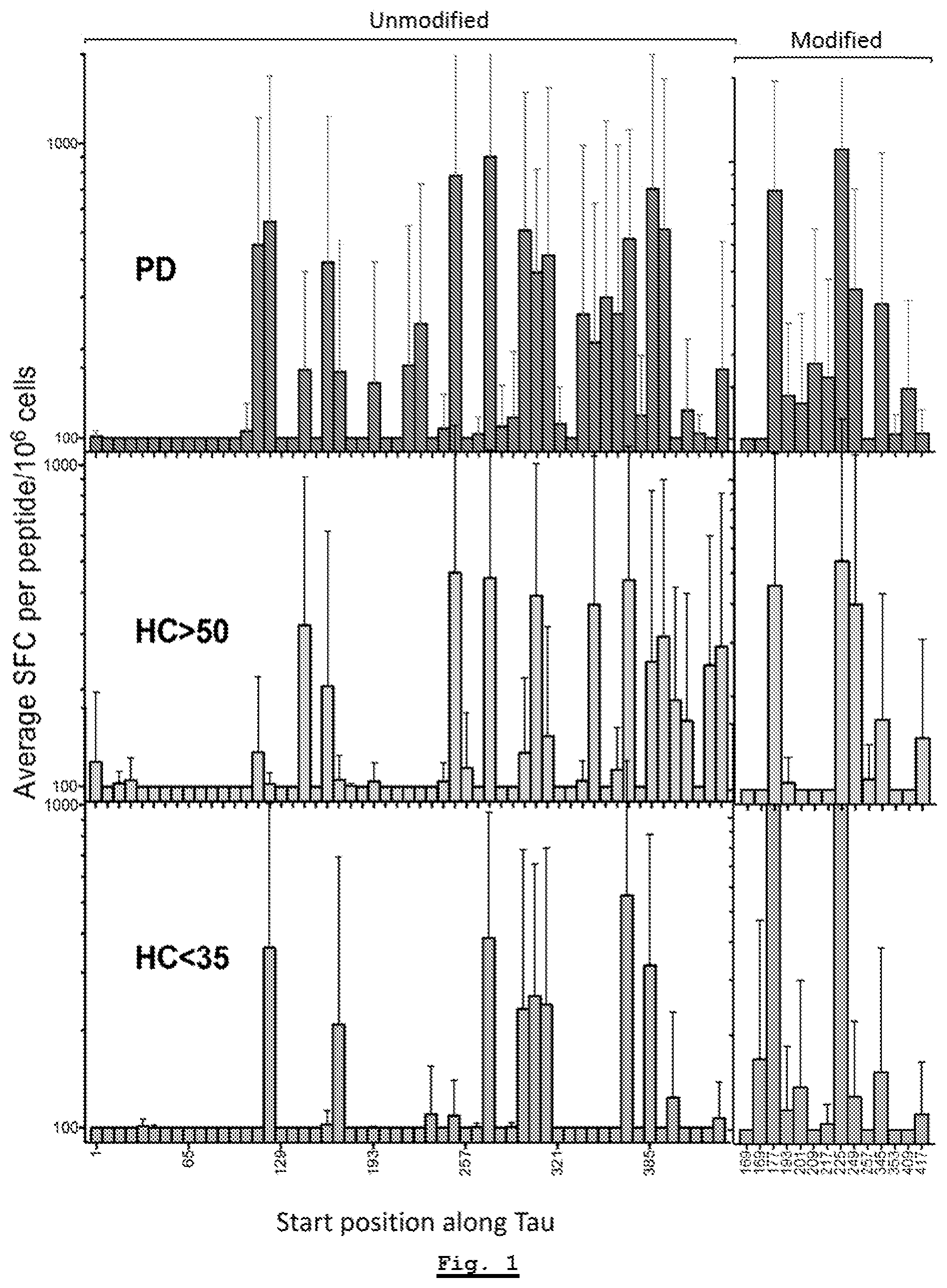
[0154] FIG. 1: Tau-specific responses for Parkinson's Disease donors as compared to healthy control donors for each of the HC Young (donors under 35 years old) and HC Age-Matched (donors above 50 years old) cohorts.
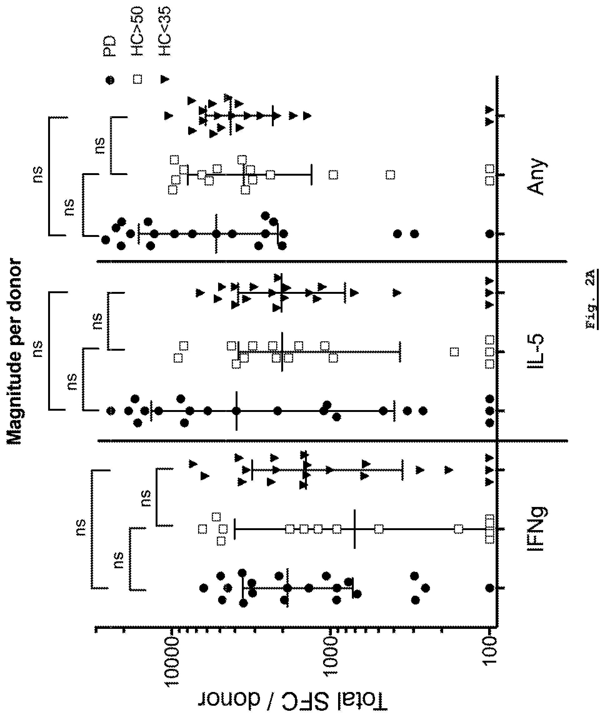
[0155] FIG. 2A: Analysis of response magnitude per donor for each of the Parkinson's Disease, HC Young (HC<35) and HC Age-Matched (HC>50) cohorts. Response magnitude for IFN-.gamma. (IFNg), IL-5 and the sum of both cytokines is shown.
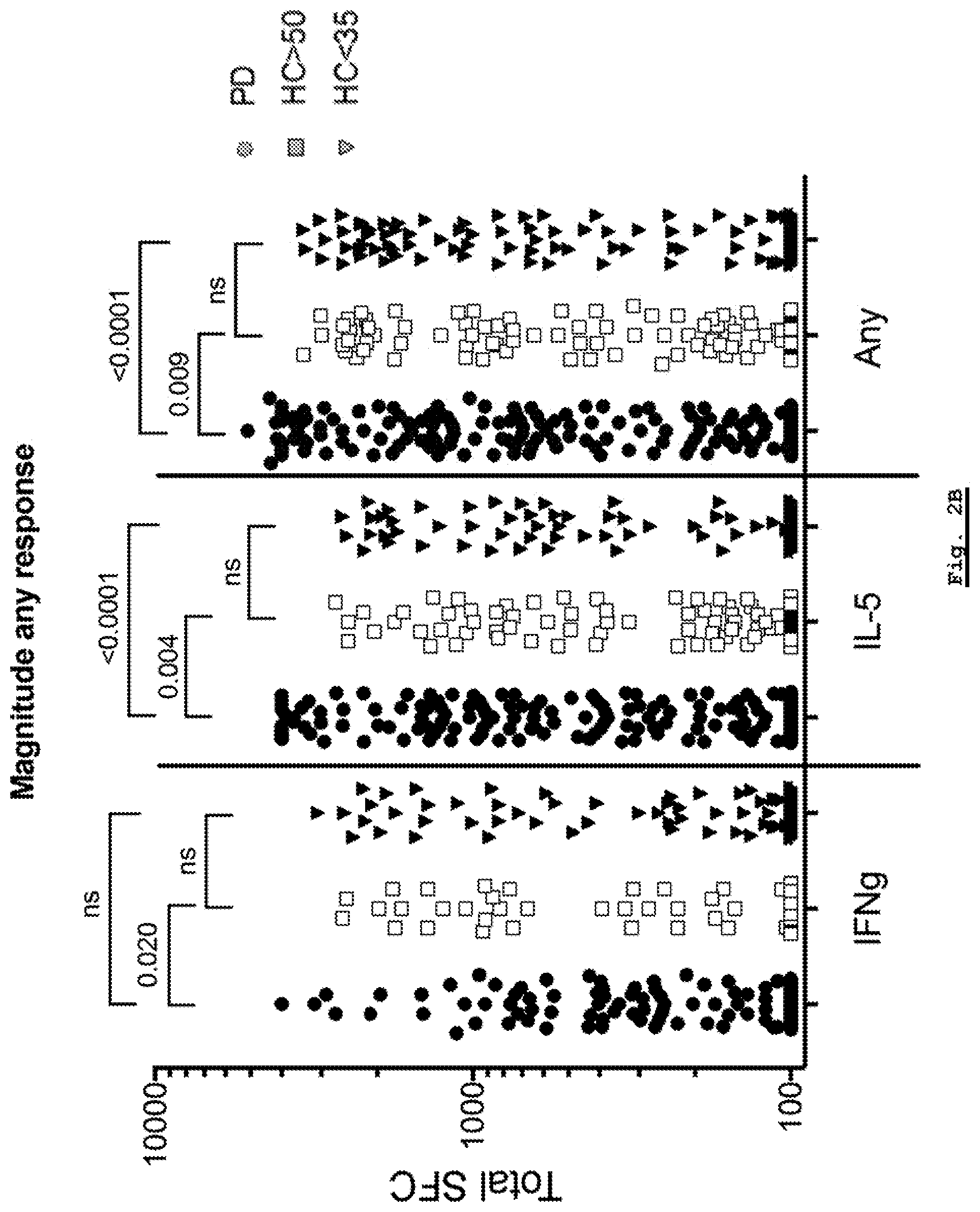
[0156] FIG. 2B: Analysis of response magnitude for each individual peptide for each of the Parkinson's Disease, HC Young (HC<35) and HC Age-Matched (HC>50) cohorts. The responses observed in each donor against each individual peptide are plotted for IFN-.gamma. (IFNg), IL-5 and the sum of both cytokines.
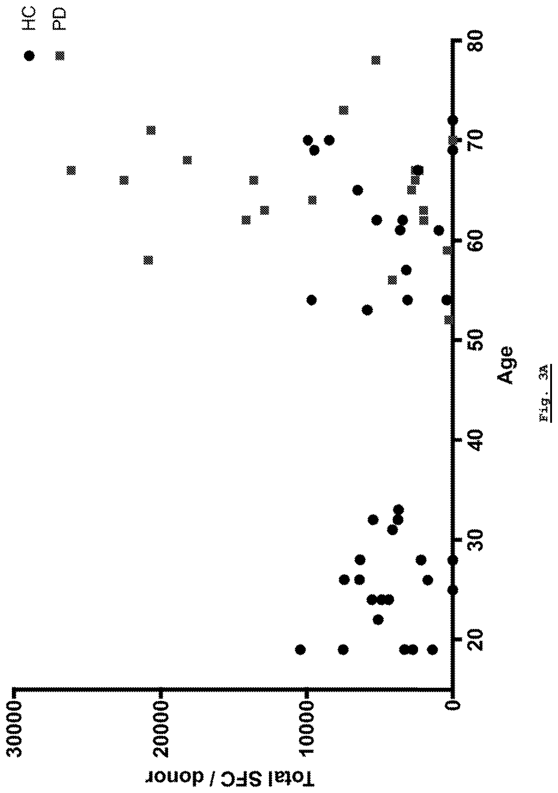
[0157] FIG. 3A: Overall response as plotted against age. Overall responses are not correlated with age in either controls or PD patients.
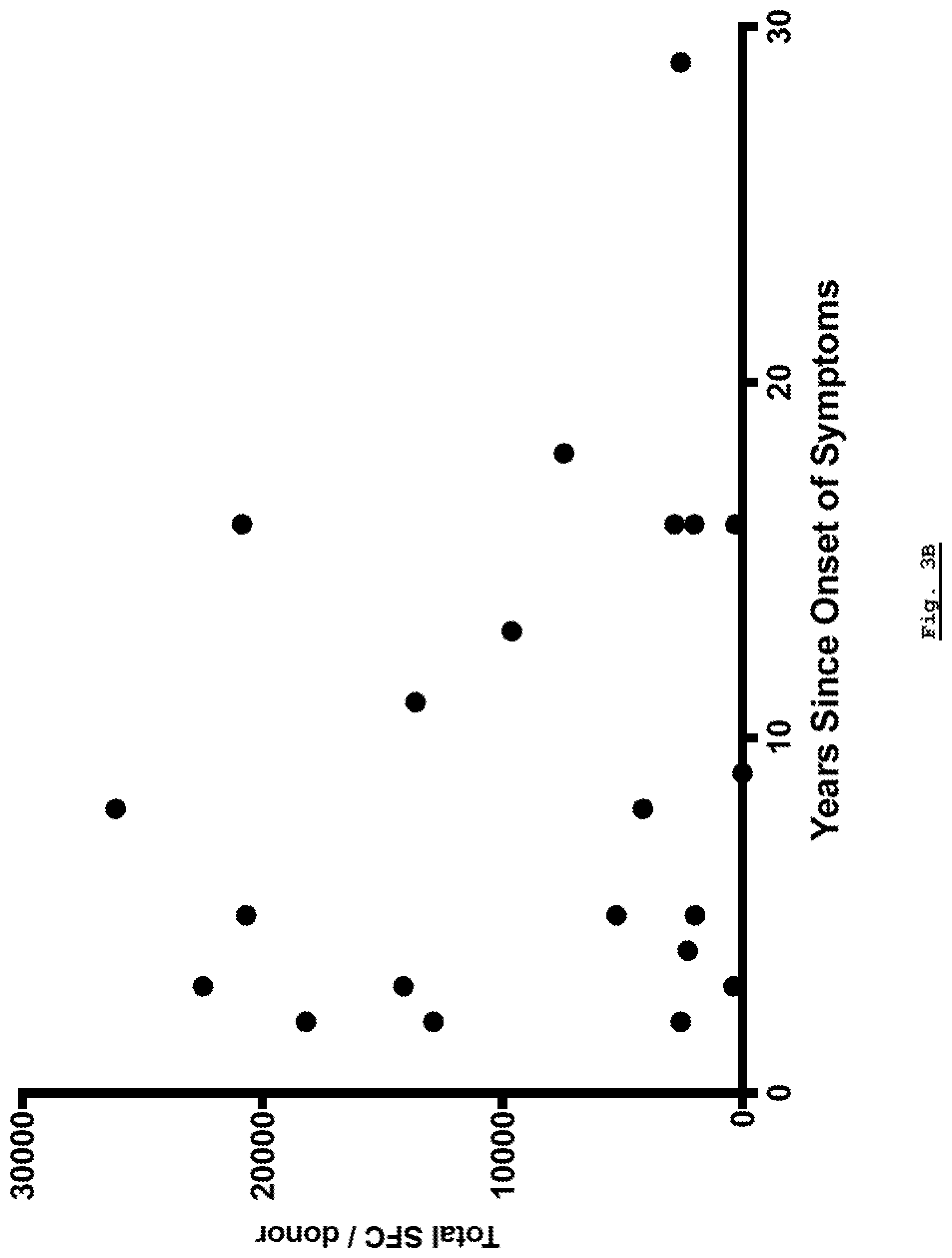
[0158] FIG. 3B: Total reactivity as a function of time since onset of symptoms.
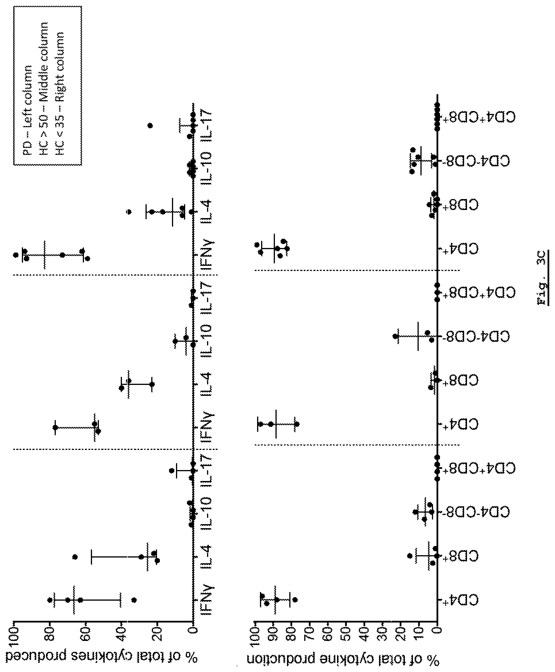
[0159] FIG. 3C: Intercellular Cytokine Staining analysis of cytokine response for each of the Parkinson's Disease, HC Young (HC<35) and HC Age-Matched (HC>50) cohorts.
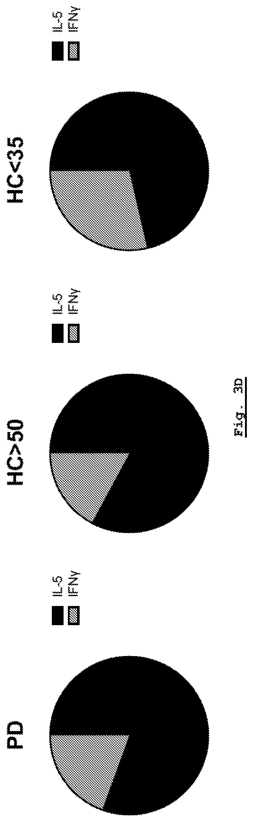
[0160] FIG. 3D: ELISPOT analysis of cytokine response for each of the Parkinson's Disease, HC Young (HC<35) and HC Age-Matched (HC>50) cohorts.
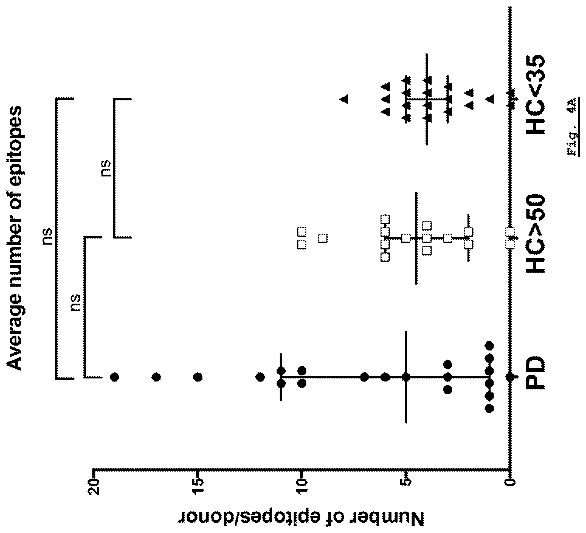
[0161] FIG. 4A: Breadth of response per donor. The number of epitopes responded to per donor is plotted for each of the Parkinson's Disease, HC Young (HC<35) and HC Age-Matched (HC>50) cohorts.
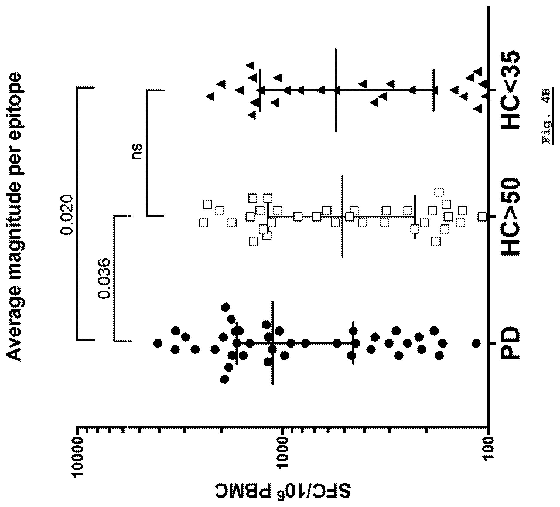
[0162] FIG. 4B: Magnitude of response per epitope per donor. The magnitude of response per epitope per donor is plotted for each of the Parkinson's Disease, HC Young (HC<35) and HC Age-Matched (HC>50) cohorts.
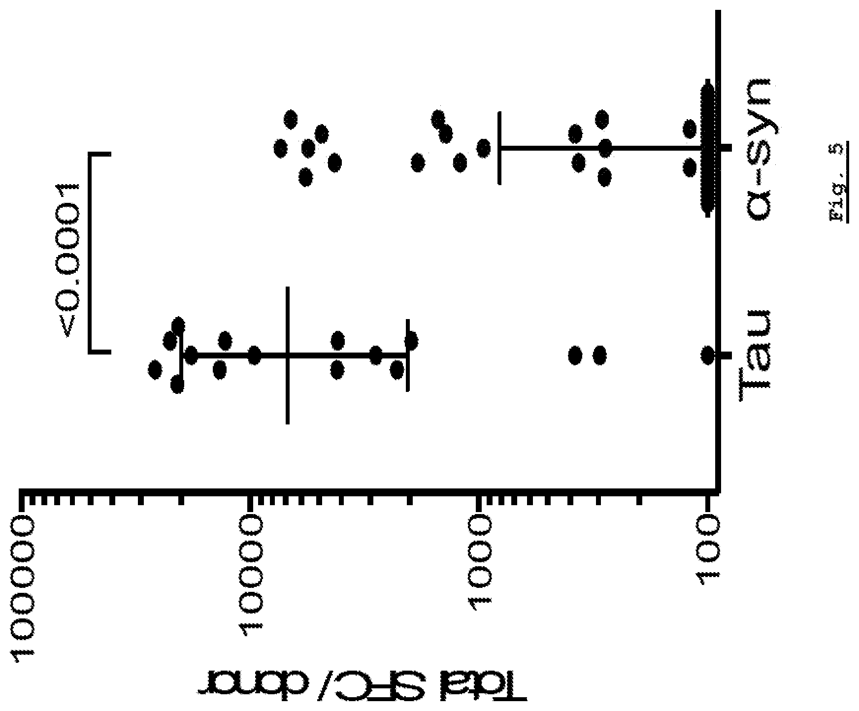
[0163] FIG. 5: Tau-specific response as compared to .alpha.-syn specific response. Tau-specific cytonkine response was plotted in comparison to .alpha.-syn specific response.
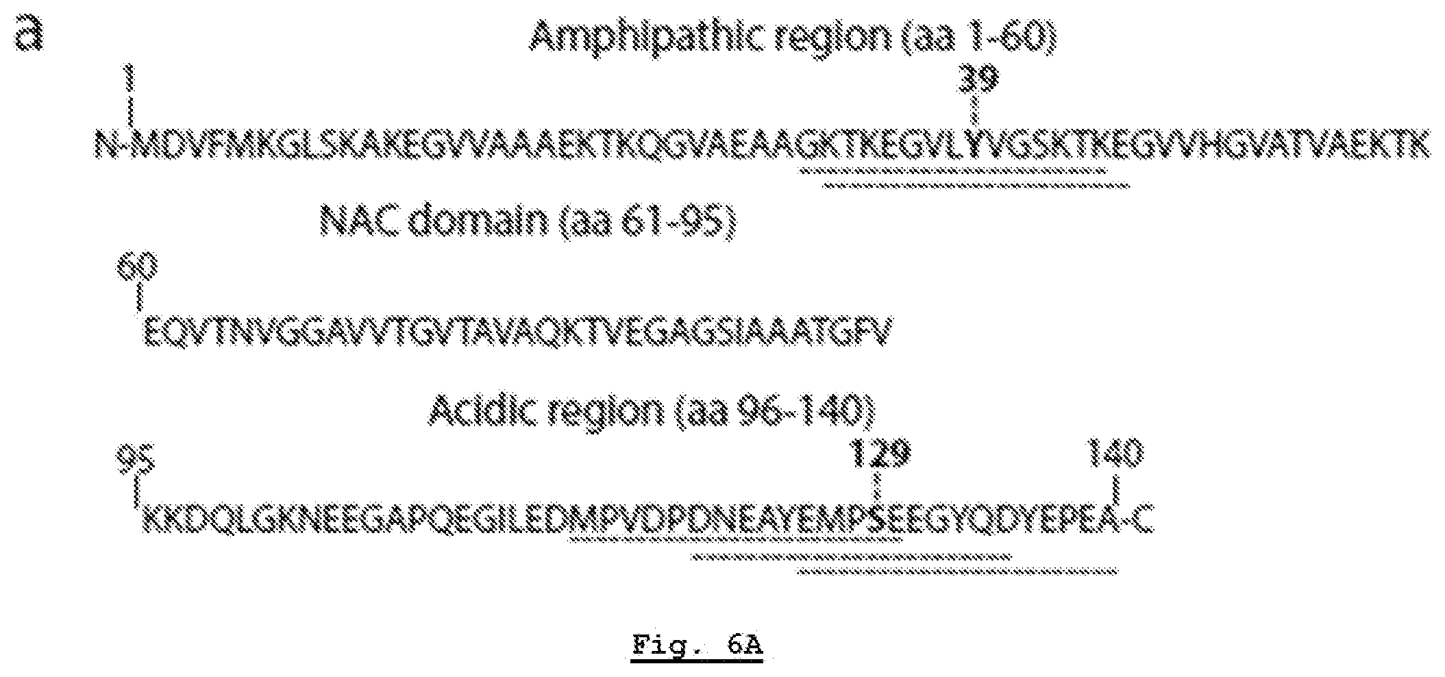
[0164] FIG. 6A: .alpha.-Syn autoimmune responses are directed against two regions. Sequence of .alpha.-syn. Antigenic regions are highlighted with dashed lines with amino acids Y39 and S129 in bold.
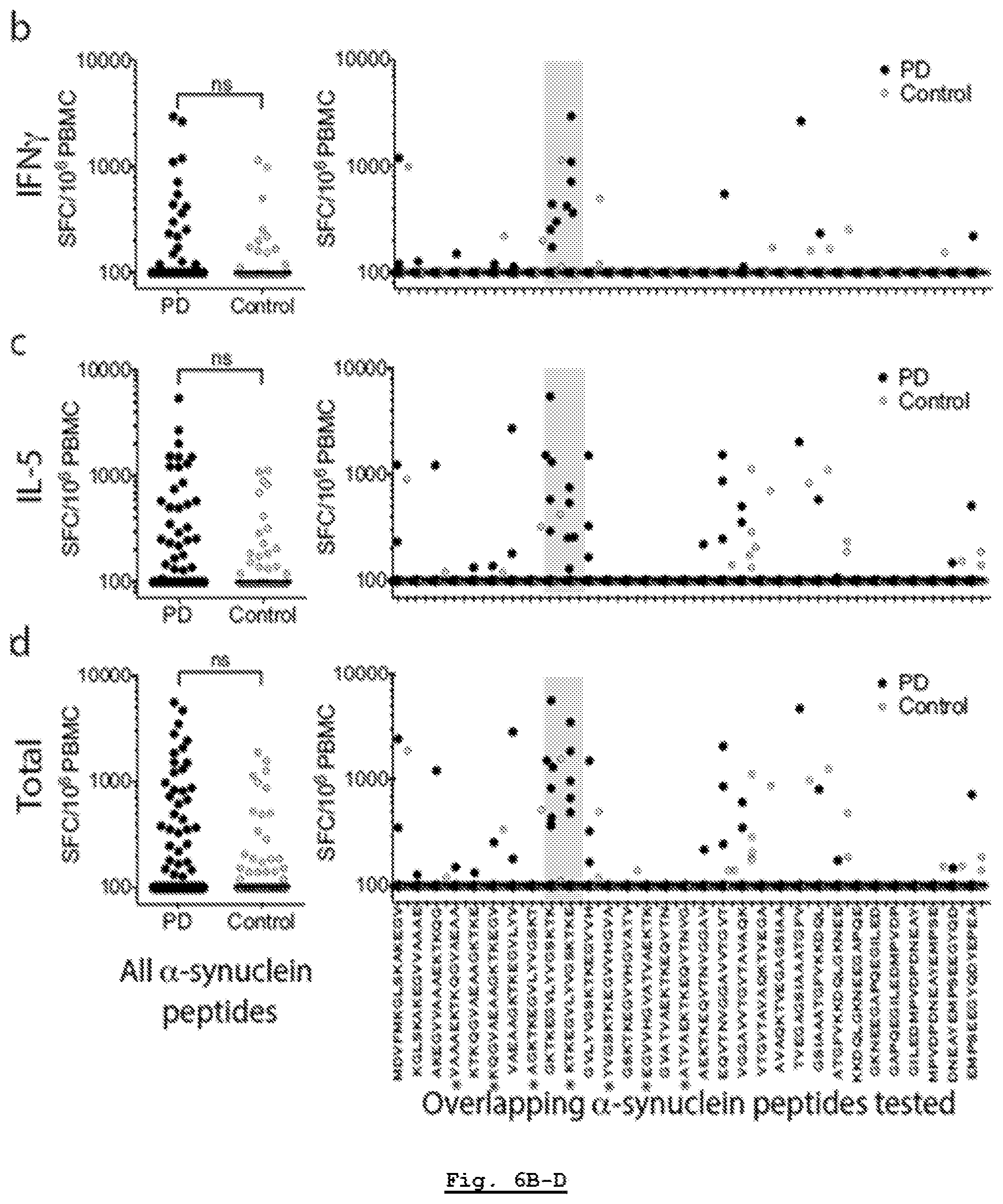
[0165] FIG. 6B: .alpha.-Syn autoimmune responses are directed against two regions. Magnitude of IFN.gamma. responses expressed as (SFC/10.sup.6 PBMC) per peptide/participant combination. Left panels; response to all overlapping native .alpha.-syn 15 mer peptides in PD (n=733) and Control (n=372). Right panels indicate responses against specific 15 mers. Grey shading indicates antigenic region containing Y39. As many participants showed no response, many points are at the limit of resolution (100 SFC).
[0166] FIG. 6C: .alpha.-Syn autoimmune responses are directed against two regions. Magnitude of IL-5 responses expressed as (SFC/10.sup.6 PBMC) per peptide/participant combination. Left panels; response to all overlapping native .alpha.-syn 15 mer peptides in PD (n=733) and Control (n=372). Right panels indicate responses against specific 15 mers. Grey shading indicates antigenic region containing Y39. As many participants showed no response, many points are at the limit of resolution (100 SFC).
[0167] FIG. 6D: .alpha.-Syn autoimmune responses are directed against two regions. Magnitude of total (IFN.gamma. & IL-5) response expressed as (SFC/10.sup.6 PBMC) per peptide/participant combination. Left panels; response to all overlapping native .alpha.-syn 15 mer peptides in PD (n=733) and Control (n=372). Right panels indicate responses against specific 15 mers. Grey shading indicates antigenic region containing Y39. As many participants showed no response, many points are at the limit of resolution (100 SFC).
[0168] FIG. 6E: .alpha.-Syn autoimmune responses are directed against two regions. Magnitude of IFN.gamma. responses. Left panels; responses to all native and phosphorylated S129 .alpha.-syn 15 mer peptides in PD (n=150) and Control (n=72). Right panels; responses against specific S129 peptides. Closed circles, PD (n=19, indicated by *, all other n=25); open circles, Control (n=12 participants). Two-tailed Mann Whitney, ns, not significant. As many participants showed no response, many points are at the limit of resolution (100 SFC).
[0169] FIG. 6F: .alpha.-Syn autoimmune responses are directed against two regions. Magnitude of IL-5 responses. Left panels; responses to all native and phosphorylated S129 .alpha.-syn 15 mer peptides in PD (n=150) and Control (n=72). Right panels; responses against specific S129 peptides. Closed circles, PD (n=19, indicated by *, all other n=25); open circles, Control (n=12 participants). Two-tailed Mann Whitney, ns, not significant. As many participants showed no response, many points are at the limit of resolution (100 SFC).
[0170] FIG. 6G: .alpha.-Syn autoimmune responses are directed against two regions. Magnitude of total (IFN.gamma. & IL-5) response. Left panels; responses to all native and phosphorylated S129 .alpha.-syn 15 mer peptides in PD (n=150) and Control (n=72). Right panels; responses against specific S129 peptides. Closed circles, PD (n=19, indicated by *, all other n=25); open circles, Control (n=12 participants). Two-tailed Mann Whitney, ns, not significant. As many participants showed no response, many points are at the limit of resolution (100 SFC).
[0171] FIG. 7A: T cell reactivity against .alpha.-syn peptides (wild type and posttranslationally modified). Magnitude of responses, expressed as the total magnitude (SFC/10.sup.6 PBMC) of IFN.gamma. response per peptide/participant combination. Responses against any .alpha.-syn 15 mer peptide spanning S129 and Y39, "any peptide", PD (n=209), Control (n=132), and responses against individual .alpha.-syn 15 mer peptides spanning S129 and Y39. Each dot represents a peptide/participant combination. Closed circles, PD (n=19); open circles, Control (n=12). Two-tailed Mann Whitney, *, p<0.05, **, p<0.01, ***, p<0.001.
[0172] FIG. 7B: T cell reactivity against .alpha.-syn peptides (wild type and posttranslationally modified). Magnitude of responses, expressed as the total magnitude (SFC/10.sup.6 PBMC) of IL-5 response per peptide/participant combination. Responses against any .alpha.-syn 15 mer peptide spanning S129 and Y39, "any peptide", PD (n=209), Control (n=132), and responses against individual .alpha.-syn 15 mer peptides spanning S129 and Y39. Each dot represents a peptide/participant combination. Closed circles, PD (n=19); open circles, Control (n=12). Two-tailed Mann Whitney, *, p<0.05, **, p<0.01, ***, p<0.001.
[0173] FIG. 7C: T cell reactivity against .alpha.-syn peptides (wild type and posttranslationally modified). Magnitude of responses, expressed as the total magnitude (SEC/10.sup.6 PBMC) of total (IFN.gamma. and IL-5 combined) response per peptide/participant combination. Responses against any .alpha.-syn 15 mer peptide spanning S129 and Y39, "any peptide", PD (n=209), Control (n=132), and responses against individual .alpha.-syn 15 mer peptides spanning S129 and Y39. Each dot represents a peptide/participant combination. Closed circles, PD (n=19); open circles, Control (n=12). Two-tailed Mann Whitney, *, p<0.05, **, p<0.01, ***, p<0.001.
[0174] FIG. 8A: Reactivity to native and modified .alpha.-syn peptides in PD patients. Magnitude of IFN.gamma. responses against native and modified .alpha.-syn 15 mer S129 and Y39 region peptides as (SEC/10.sup.6 PBMC). Each point represents a peptide/participant combination. Closed circles, PD (n=403 peptide/participant combinations "any peptide", KTKEGVLYVGSKTKE n=63 participants ({circumflex over ( )}), modified peptides marked with * are tested in 19 participants, unmodified peptides are tested in n=41); open circles, control (n=228 any peptide, {circumflex over ( )} n=36, *n=12 and unmodified peptides n=24).
[0175] FIG. 8B: Reactivity to native and modified .alpha.-syn peptides in PD patients. Magnitude of IL-5 responses against native and modified .alpha.-syn 15 mer S129 and Y39 region peptides as (SFC/10.sup.6 PBMC). Each point represents a peptide/participant combination. Closed circles, PD (n=403 peptide/participant combinations "any peptide", KTKEGVLYVGSKTKE n=63 participants ({circumflex over ( )}), mddified peptides marked with * are tested in 19 participants, unmodified peptides are tested in n=41); open circles, control (n=228 any peptide, {circumflex over ( )} n=36, *n=12 and unmodified peptides n=24).
[0176] FIG. 8C: Reactivity to native and modified .alpha.-syn peptides in PD patients. Magnitude of total (IFN.gamma. & IL-5 combined) responses against native and modified .alpha.-syn 15 mer S129 and Y39 region peptides as (SFC/10.sup.6 PBMC). Each point represents a peptide/participant combination. Closed circles, PD (n=403 peptide/participant combinations "any peptide", KTKEGVLYVGSKTKE n=63 participants ({circumflex over ( )}), modified peptides marked with * are tested in 19 participants, unmodified peptides are tested in n=41); open circles, control (n=228 any peptide, {circumflex over ( )} n=36, *n=12 and unmodified peptides n=24).
[0177] FIG. 8D: Reactivity to native and modified .alpha.-syn peptides in PD patients. Combined IL-5 and IFN.gamma. responses against individual native and modified .alpha.-syn peptides by PD. Black points, IFN.gamma. responses; red points, IL-5 responses. Two-tailed Mann Whitney, *, p<0.05, **, p<0.01, ***, p<0.001. As many participants showed no response, many points are at the limit of resolution (100 SFC).
[0178] FIG. 9A: Characterization of .alpha.-syn specific responses in PD. Gating strategy. T cells were gated based on CD3 expression. Boolean gating was used to define cytokine-producing cells expressing CD4 and/or CD8.
[0179] FIG. 9B: Characterization of .alpha.-syn specific responses in PD. Percent total cytokine detected from CD3+ T cells in response to .alpha.-syn peptides. Each point represents one participant (n=9); median.+-.interquartile range is indicated. Dotted line indicates 0.05% cut-off for specific cytokine production by CD3+ T cells.
[0180] FIG. 9C: Characterization of .alpha.-syn specific responses in PD. Percentage of total cytokines produced for IFN.gamma., IL-4, IL-10, and IL-17. Each point represents one participant that exceeded the cut-off (n=6), median.+-.interquartile range is indicated.
[0181] FIG. 9D: Characterization of .alpha.-syn specific responses in PD. Percentage of total cytokines produced by CD4+, CD8+, CD4-CD8-, or CD4+CD8+ T cells. Each point represents one participant (n=6), median.+-.interquartile range is indicated.
[0182] FIG. 10: Specific T cell reactivity against native or fibrilized .alpha.-syn. Magnitude of responses, expressed as the average spots per 10.sup.6 PBMC, of response per protein/PD participant or peptide/PD participant combination (n=12 PD participants, each represented by a different symbol). The lines connect discrete values from each individual participant and are present to provide a means to compare responses within and between individuals. The difference between response to unstimulated compared to peptides, the native .alpha.-syn and PFF groups is significant by the Wilcoxon one-tailed test (values are shown in the figure). No significant difference (Wilcoxon two-tailed test) in response to PFF and native protein was apparent in this relatively small sample.
[0183] FIG. 11A: HLA-DR surface expression across DRB1*15:01+ or DRB1*15:01- PD and HC participants. Gating strategy for FACS analysis. After eliminating non-lymphocytes and doublet cells by forward- and side-scatter, cells were gated based on HLA-DR expression.
[0184] FIG. 11B: HLA-DR surface expression across DRB1 15:01+ or DRB1*15:01- PD and HC participants. HLA-DR and CD3 expression of participant cells (black; HLA-DR antibody, red; isotype control) of PD patients that carry (n=5) DRB1*15:01 allele.
[0185] FIG. 11C: HLA-DR surface expression across DRB1*15:01+ or DRB1*15:01- PD and HC participants. HLA-DR and CD3 expression of participant cells (black; HLA-DR antibody, red; isotype control) of PD patients that do not carry (n=5) DRB1*15:01 allele.
[0186] FIG. 11D: HLA-DR surface expression across DRB1*15:01+ or DRB1*15:01- PD and HC participants. HLA-DR and CD3 expression of participant cells (black; HLA-DR antibody, red; isotype control) of HC that carry (n=3) DRB1*15:01 allele
[0187] FIG. 11E: HLA-DR surface expression across DRB1*15:01+ or DRB1*15:01- PD and HC participants. HLA-DR and CD3 expression of participant cells (black; HLA-DR antibody, red; isotype control) of HC that do not carry (n=5) DRB1*15:01 allele.
[0188] FIG. 11F: HLA-DR surface expression across DRB1*15:01+ or DRB1*15:01- PD and HC participants. 721.221 cells are used as controls that do not express HLA class II.
[0189] FIG. 11G: HLA-DR surface expression across DRB1*15:01+ or DRB1*15:01- PD and HC participants. RM3 cells are used as controls that do express HLA class II.
[0190] FIG. 11H: HLA-DR surface expression across DRB1*15:01+ or DRB1*15:01- PD and HC participants. Mean fluorescent intensities (MFI).+-.standard deviations of HLADR expression for each participant cohort.
[0191] FIG. 11I: HLA-DR surface expression across DRB1*15:01+ or DRB1*15:01- PD and HC participants. Percentage of living cells that express HLA-DR, mean.+-.SD.
[0192] FIG. 12A: HLA class I surface expression across DRB1*15:01+ or DRB1*15:01- PD and HC participants. Gating strategy for FACS analysis. After eliminating non-lymphocytes and doublet cells by forward- and side-scatter, cells were gated based on HLA-ABC expression.
[0193] FIG. 12B: HLA class I surface expression across DRB1*15:01+ or DRB1*15:01- PD and HC participants. HLA-ABC and CD3 expression of participant cells (black: HLA-ABC antibody, red: isotype control) of PD patients that carry (n=5) DRB1*15:01 allele.
[0194] FIG. 12C: HLA class I surface expression across DRB1*15:01+ or DRB1*15:01- PD and HC participants. HLA-ABC and CD3 expression of participant cells (black: HLA-ABC antibody, red: isotype control) of PD patients that do not carry (n=5) DRB1*15:01 allele.
[0195] FIG. 12D: HLA class I surface expression across DRB1*15:01+ or DRB1*15:01- PD and HC participants. HLA-ABC and CD3 expression of participant cells (black: HLA-ABC antibody, red: isotype control) of HC that carry (n=3) DRB1*15:01 allele.
[0196] FIG. 12E: HLA class I surface expression across DRB1*15:01+ or DRB1*15:01- PD and HC participants. HLA-ABC and CD3 expression of participant cells (black: HLA-ABC antibody, red: isotype control) of HC that do not carry (n=5) DRB1*15:01 allele.
[0197] FIG. 12F: HLA class I surface expression across DRB1*15:01+ or DRB1*15:01- PD and HC participants. 721.221 cells are used as controls that do not express HLA class I.
[0198] FIG. 12G: HLA class I surface expression across DRB1*15:01+ or DRB1*15:01- PD and HC participants. RM3 cells are used as controls that do express HLA class I.
[0199] FIG. 12H: HLA class I surface expression across DRB1*15:01+ or DRB1*15:01- PD and HC participants. Mean fluorescent intensities (MFI).+-.standard deviations of HLAABC expression for each participant cohort.
[0200] FIG. 13A: HLA association of Y39 epitope and identification of A*11:01 restricted 9-10 aa length Y39 epitopes. Overlapping but largely independent associations between DRB1*15:01, DQB1*03:04 and A*11:01 for PD (13 participants) responding to the Y39 epitope.
[0201] FIG. 13B: HLA association of Y39 epitope and identification of A*11:01 restricted 9-10aa length Y39 epitopes. Magnitude of responses by PD (n=19), as (SFC/10.sup.6 PBMC) of response per peptide/participant combination to .alpha.-syn 9-10 mer peptides spanning the protein. In some cases, response to overlapping peptides are combined, with additional residues of the longer peptide in parentheses. top panel, IFN.gamma.; middle, IL-5; bottom, total (IFN.gamma. & IL-5 combined) response. As many participants showed no T cell response, many points are at the limit of resolution (100 SFC).
[0202] FIG. 13C: HLA association of Y39 epitope and identification of A*11:01 restricted 9-10aa length Y39 epitopes. Magnitude of Total (IFN.gamma. & IL-5 combined) responses by control participants (n=12), as (SFC/10.sup.6 PBMC) of response per peptide/participant combination to .alpha.-syn 9-10 mer peptides spanning the protein. In some cases, response to overlapping peptides are combined, with additional residues of the longer peptide in parentheses. As many participants showed no T cell response, many points are at the limit of resolution (100 SFC).
[0203] FIG. 14A: Magnitude of IFN.gamma. responses expressed as (SFC/10.sup.6 PBMC) per peptide/participant combination. Response to selected TDP43 15 mer peptides by ALS patients and healthy controls.
[0204] FIG. 14B: Magnitude of IL-5 responses expressed as (SFC/10.sup.6 PBMC) per peptide/participant combination. Response to selected TDP43 15 mer peptides by ALS patients and healthy controls.
[0205] FIG. 14C: Magnitude of IL-10 responses expressed as (SFC/10.sup.6 PBMC) per peptide/participant combination. Response to selected TDP43 15 mer peptides by ALS patients and healthy controls.
[0206] FIG. 14D: Magnitude of any cytokine responses expressed as (SFC/10.sup.6 PBMC) per peptide/participant combination. Response to selected TDP43 15 mer peptides by ALS patients and healthy controls.
[0207] FIG. 15: High-level functional block diagram of a system for assessing a neurodegenerative disease patient in accordance with a specific example of implementation of the present invention;
[0208] FIG. 16: Functional block diagram of an apparatus for generating neurodegenerative disease patient information suitable for use in the system depicted in FIG. 15 in accordance with a first specific example of implementation of the present invention.
[0209] In the drawings, exemplary embodiments are illustrated by way of example. It is to be expressly understood that the description and drawings are only for the purpose of illustrating certain embodiments and are an aid for understanding. They are not intended to be a definition of the limits of the invention.
DETAILED DESCRIPTION OF THE INVENTION
[0210] Abbreviations
[0211] .alpha.-syn, alpha synuclein; AD, Alzheimer's disease; AIM, Activation Induced Marker; ALS, amyotrophic lateral sclerosis; .beta.2 m, beta 2 microglobulin; BF, brightfield; BrdU, 5-bromo-2-deoxyuridine; BSA, bovine seroalbumin; ConA, concananycin A; CMA, chaperone-mediated autophagy; CNS, central nervous system; CTLs, cytotoxic T cells CTRL, control; DA, dopamine/dopaminergic; DCs, dendritic cells; ELISA, enzyme-linked immunosorbent assay; ELISPOT, enzyme-linked immunospot assay; GSEA, Gene Set Enrichment Analysis; HC, healthy control(s); hES, human stem cells; HLA, human leukocyte antigen; IEDB, Immune Epitope Database; IFN-.gamma., interferon gamma; IL-1p, interleukin 1-beta; IL-6, interleukin 6; IPA, Integrated Pathway Analysis; KO, knocked out; LC, locus coeruleus; LGN, lateral geniculate nucleus; LPS, lipopolysaccharide; MAP-2, microtubule associated protein-2; MHC, major histocompatibility complex; MHC-I, major histocompatibility complex class I; MHC-II, major histocompatibility complex class II; MN, spinal motor neurons; Mut-.alpha.-syn, mutated (A53T) alpha synuclein; NE, norepinephrine/norepinephrinergic; Nit-.alpha.-syn, nitrated alpha synuclein; NM, neuromelanin; OVA, ovalbumin; PBMC, peripheral blood mononuclear cells; PD, Parkinson's disease; PET, positron emission tomography; PSP, progressive supranuclear palsy; PTM, post-translational modification; RATE, Restrictor Analysis Tool for Epitopes; SEM, standard error of the mean; SFC, spot-forming cells; SN, substantia nigra; TCR, T cell receptor; TH, tyrosine hydroxylase; UPDRS, Unified Parkinson's Disease Rating Scale; Veh, vehicle; VM, ventral midbrain; VTA, ventral tegmental area; WGCNA, Gene Co-expression Network Analysis.
[0212] A detailed description of one or more embodiments of the invention is provided below along with accompanying figures that illustrate the principles of the invention. The invention is described in connection with such embodiments, but the invention is not limited to any embodiment. The scope of the invention is limited only by the claims.
[0213] Numerous specific details are set forth in the following description in order to provide a thorough understanding of the invention. These details are provided for the purpose of non-limiting examples and the invention may be practiced according to the claims without some or all of these specific details. For the purpose of clarity, technical material that is known in the technical fields related to the invention has not been described in detail so that the invention is not unnecessarily obscured.
[0214] The present invention provides methods for assessing whether a subject is at risk of developing, or for diagnosing or confirming whether a subject is afflicted with an .alpha.-synucleinopathy, a Tauopathy, Parkinson's disease (PD), amyotrophic lateral sclerosis (ALS), Lewy Body dementia (LBD), or Alzheimer's disease (AD) comprising
[0215] a) [0216] i) obtaining leukocytes from the subject; [0217] ii) contacting the leukocytes with an epitope peptide; [0218] iii) determining whether the leukocytes have increased activation after contact with the epitope peptide; and [0219] iv) identifying the subject as at risk of developing, or as afflicted with the .alpha.-synucleinopathy, PD, ALS, LBD or AD if in step [0220] iii) the leukocytes are determined to have increased activation after contact with the epitope peptide, and identifying the subject as not at risk of developing, or as not afflicted with the .alpha.-synucleinopathy, PD, ALS, LBD or AD if in step iii) the leukocytes are determined to not have increased activation after contact with the epitope peptide, or
[0221] b) [0222] i) obtaining leukocytes from the subject; [0223] ii) separating the leukocytes into 2 or more pools of leukocytes and contacting each pool with an epitope peptide, wherein each pool is contacted with a different epitope; [0224] iii) determining whether each pool has increased activation after contact with the epitope peptide; and [0225] iv) identifying the subject as at risk of developing, or as afflicted with the .alpha.-synucleinopathy, Tauopathy, PD, ALS, LBD, or AD if and only if in step iii) 1 or more pools is determined to have increased activation after contact with the epitope peptide.
[0226] The present invention also provides a method for assessing whether an .alpha.-synucleinopathy, a Tauopathy, Parkinson's disease (PD), amyotrophic lateral sclerosis (ALS), Lewy Body dementia (LBD), or Alzheimer's disease (AD) has progressed or is developing in a subject afflicted with or who has been identified as being at risk of developing the .alpha.-synucleinopathy, PD, ALS, LBD or AD comprising
[0227] a) performing each of the following steps i) to iii): [0228] i) obtaining leukocytes from the subject; [0229] ii) contacting the leukocytes with an epitope peptide that was previously identified to increase activation of the leukocytes; and [0230] iii) determining the level of activation of the leukocytes after contact with the epitope peptide at a first and a second point in time, and then [0231] iv) concluding that the .alpha.-synucleinopathy, PD, ALS, LBD or AD has progressed or is developing in the subject if the leukocytes are determined to be more activated in step iii) performed at the second point in time compared to the level of activation in step iii) performed at the first point in time, or
[0232] b) performing each of the following steps i) to iii): [0233] i) obtaining leukocytes from the subject; [0234] ii) separating the leukocytes into two or more pools of leukocytes and contacting each pool with an epitope peptide, wherein'each pool is contacted with a different epitope; [0235] iii) determining whether each pool has increased activation after contact with the epitope peptide at a first and a second point in time, and then
[0236] concluding that the .alpha.-synucleinopathy, PD, ALS, LBD or AD has progressed or is developing in the subject if more pools of leukocytes are determined to be activated in step iii) performed at the second point in time compared to the number of pools that are determined to be activated in step iii) performed at the first point in time.
[0237] The present invention also provides methods for assessing whether a subject afflicted with an .alpha.-synucleinopathy, a Tauopathy, Parkinson's disease (PD), amyotrophic lateral sclerosis (ALS), Lewy Body dementia (LBD), or Alzheimer's disease (AD) is likely to benefit from a therapy, wherein the therapy is directed to leukocytes that are activated by an epitope peptide, the method comprising
[0238] a) [0239] i) obtaining leukocytes from the subject; [0240] ii) contacting the leukocytes with the epitope peptide; [0241] iii) determining whether the leukocytes have increased activation after contact with the epitope peptide; and [0242] iv) identifying the subject as likely to benefit from the therapy if in step iii) the leukocytes are determined to have increased activation after contact with the epitope peptide, and identifying the subject as unlikely to benefit from the therapy if in step iii) the leukocytes are determined to not have increased activation after contact with the epitope peptide, or
[0243] b) [0244] i) obtaining leukocytes from the subject; [0245] ii) contacting the leukocytes with the epitope peptide; [0246] iii) determining whether the leukocytes have increased activation after contact with the epitope peptide; and [0247] iv) identifying the subject as having benefited from the therapy if in step iii) if the leukocytes are determined to have increased activation after contact with the epitope peptide, and identifying the subject as not having benefitted from the therapy if in step iii) the leukocytes are determined to not have increased activation after contact with the epitope peptide.
[0248] The present invention also provides methods for assessing whether a subject afflicted with a disease or condition involving an inflammatory response or related to inflammation, or a neurodegenerative disease or disorder is likely to benefit or has benefitted from a therapy, wherein the therapy comprises administration of an effective amount of a T cell receptor for a particular antigen:MHC complex, the method comprising:
[0249] a) [0250] (i) obtaining leukocytes from the subject; [0251] (ii) contacting the leukocytes with the antigen bound to an MHC molecule; [0252] (iii) determining whether the leukocytes have increased activation after contact with the antigen bound to an MHC molecule; and [0253] (iv) identifying the subject as likely to benefit from the therapy if in step (iii) the leukocytes are determined to have increased activation after contact with the antigen bound to an MHC molecule, and identifying the subject as unlikely to benefit from the therapy if in step (iii) the leukocytes are determined to not have increased activation after contact with the antigen bound to an MHC molecule; or
[0254] b) [0255] (i) obtaining leukocytes from the subject; [0256] (ii) contacting the leukocytes with the antigen bound to an MHC molecule; [0257] (iii) determining whether the leukocytes have increased activation after contact with the antigen bound to an MHC molecule; and [0258] (iv) identifying the subject as having benefited from the therapy if in step (iii) the leukocytes are determined to have increased activation after contact with the antigen bound to an MHC molecule, and identifying the subject as not having benefitted from the therapy if in step (iii) the leukocytes are determined to not have increased activation after contact with the antigen bound to an MHC molecule.
[0259] In some embodiments, in step ii) the leukocytes are separated into 2, 3, 4, 5, 6, 7, 8, 9, 10, 11-50 or more pools, and in step iv) the subject is identified as at risk of developing or as afflicted with the .alpha.-synucleinopathy, Tauopathy, PD, ALS, LBD or AD if and only if in step iii) 1, 2, 3, 4, 5, 6, 7, 8, 9, 10, 11-50 or more pools is determined to have increased activation after contact with the epitope peptide.
[0260] In some embodiments, the subject the subject [0261] a) is at least about 35, 40, 45, 50, 55, 60, 65, 70, 75 or 80 years of age; [0262] b) is less than about 35, 40, 45, 50, 55, 60, 65, 70, 75 or 80 years of age; [0263] c) has a symptom that has preceded the onset of the .alpha.-synucleinopathy, Tauopathy, PD, ALS, LBD or AD in subjects who have developed .alpha.-synucleinopathy, Tauopathy, PD, ALS, LBD or AD; [0264] d) has a symptom that has preceded the onset of the .alpha.-synucleinopathy, Tauopathy, PD, ALS, LBD or AD in subjects who have developed the .alpha.-synucleinopathy, Tauopathy, PD, ALS, LBD or AD, wherein the symptom has preceded the onset of the .alpha.-synucleinopathy, Tauopathy, PD, ALS, LBD or AD in the subjects by at least about 5, 10, 15, 20, 25, 30 or 5-30 years; [0265] e) is afflicted with cognitive decline, constipation or orthostatic hypotension [0266] f) is afflicted with cognitive decline, and the cognitive decline is reduced spatial reasoning ability and/or reduced memory ability. [0267] g) is afflicted with fasciculations or muscle twitches in the arm leg, shoulder, or tongue, muscle cramps, spasticity or tight and stiff muscles, muscle weakness affecting an arm, a leg, neck or diaphragm, slurred and nasal speech, and/or difficulty chewing or swallowing; or [0268] h) is afflicted with cognitive decline, and the cognitive decline is reduced language or decision-making.
[0269] In some embodiments; the subject is the subject to [0270] a) be monitored more frequently for the .alpha.-synucleinopathy, Tauopathy, PD, ALS, LBD or AD; or [0271] b) receive additional diagnostic testing for the .alpha.-synucleinopathy, Tauopathy, PD, ALS, LBD or AD,
[0272] if the subject is identified as at risk of developing the .alpha.-synucleinopathy, Tauopathy, PD, ALS, LBD or AD.
[0273] In some embodiments the presence of at least one human leukocyte antigen (HLA) allele, one T cell receptor (TCR) allele, or one MAPT allele is determined in the subject.
[0274] In some embodiments, the subject is identified as at risk of developing the .alpha.-synucleinopathy, PD, ALS, LBD or AD or identified as afflicted with the .alpha.-synucleinopathy, PD, ALS, LBD or AD if [0275] a) the leukocytes are determined to have increased activation after contact with the epitope peptide, or 1 or more pools is determined to have increased activation after contact with the epitope peptide, and [0276] b) the subject has at least one HLA allele.
[0277] In some embodiments, the subject has the HLA allele DRB5*01:01, DRB1*15:01, DQB1*03:04, A*11:01, DRB1*09:01, DRB1*15, DRB1*04, DQB1*06, DRB1*01:01, DRB1*04:04, DRB1*07:01, DRB1*11:04, DRB3*02:02, DQA1*05:01, DQB1*03:01, DQB1*03:02, DQB1*03:03, DQB1*04:02, DRB1*15:01/DQB1*06:02 or DRB1*04:02/DQB1*03:02. In a further embodiment, the subject has the HLA alleles DRB5*01:01, DRB1*15:01, DQB1*03:04, and A*11:01.
[0278] In some embodiments, the subject has the HLA allele DRB1*11:04 and the amino acid sequence is SEQ ID NO: 19, the subject has the HLA allele DQB1*03:03 and the amino acid sequence is SEQ ID NO: 31, the subject has the HLA allele DQA1*05:01 and the amino acid sequence is SEQ ID NO: 32, the subject has the HLA allele DRB1*01:01 and the amino acid sequence is SEQ ID NO: 40, the subject has the HLA allele DRB1*04:04 and the amino acid sequence is SEQ ID NO: 49, the subject has the HLA allele DQB1*04:02 and the amino acid sequence is SEQ ID NO: 52, or the subject has the HLA allele DRB3*02:02 and the amino acid sequence is SEQ ID NO:
[0279] 29.
[0280] In some embodiments the method assesses whether AD, ALS or PD is developing in a subject who has been identified as being at risk of developing AD, ALS or PD, or assesses whether a subject afflicted with AD, ALS or PD is likely to benefit from a therapy.
[0281] The present invention also provides methods for treating a subject afflicted with an .alpha.-synucleinopathy, a Tauopathy, Parkinson's disease (PD), amyotrophic lateral sclerosis (ALS), Lewy Body dementia (LBD), or Alzheimer's disease (AD)comprising [0282] a) administering to the subject a compound that is approved for use in treating subjects afflicted with the .alpha.-synucleinopathy, PD, ALS, LBD or AD, wherein the subject has been diagnosed or confirmed to be afflicted with .alpha.-synucleinopathy, PD, ALS, LBD or AD according to the methods; [0283] b) diagnosing or confirming the subject to be afflicted with the .alpha.-synucleinopathy, PD, ALS, LBD or AD according to the methods, and administering to the subject a compound that is approved for use in treating subjects afflicted with .alpha.-synucleinopathy, PD, ALS, LBD or AD; [0284] c) administering to the subject a therapy that is directed to leukocytes that are activated by an epitope peptide, wherein leukocytes of the subject have been determined to have increased activation after contact with the epitope peptide; [0285] d) administering an immunosuppressant therapy to the subject, wherein the subject has been identified as being likely to benefit therefrom by the methods; or [0286] e) administering an immunosuppressant therapy to the subject, wherein the subject has been identified as being likely to benefit from a therapy directed to leukocytes that are activated by an epitope peptide according to the methods.
[0287] In some embodiments, the therapy is tolerization therapy, and the tolerization therapy is specific for leukocytes that are activated by the epitope, preferably wherein administering the tolerization therapy comprises administering to the subject the epitope peptide in an amount that is effective to reduce activation of leukocytes in the subject by the epitope peptide.
[0288] In some embodiments, the therapy comprises selectively killing the leukocytes that are activated by the epitope peptide in the subject, preferably wherein selectively killing the leukocytes that are activated by the epitope peptide in the subject comprises administering to the subject an effective amount of a compound comprising a major histocompatibility complex (MHC) Tetramer and a toxin to the subject, wherein the MHC Tetramer comprises the epitope peptide.
[0289] In some embodiments, the immunosuppressant therapy comprises tolerization therapy, selectively killing the leukocytes that are activated by an epitope peptide in the subject, or administering an effective amount of an immunosuppressive compound to the subject, preferably wherein the immunosuppressive compound is a calcineurin inhibitor, a compound that blocks a chemokine receptor that is expressed by a leukocyte, a glucocorticoid, a mTOR inhibitor, an anti-metabolic compound, a phosphodiesterase-5 inhibitor, an antibody, or a leukocyte function antigen-3 (LFA-3)/Fc fusion protein.
[0290] The present invention also provides methods for assessing whether leukocytes of a subject afflicted with an .alpha.-synucleinopathy, a Tauopathy, Parkinson's disease (PD), amyotrophic lateral sclerosis (ALS), Lewy Body dementia (LBD), or Alzheimer's disease (AD) are activated by an epitope peptide, comprising [0291] i) obtaining leukocytes from the subject; [0292] ii) contacting the leukocytes with the epitope peptide; [0293] iii) determining whether the leukocytes have increased activation after contact with the epitope peptide; and [0294] iv) identifying the leukocytes of the subject as activated by the epitope peptide if in step iii) the leukocytes are determined to have increased activation after contact with the epitope peptide, and identifying the leukocytes of the subject as not activated by the epitope peptide if in step iii) the leukocytes are determined to not have increased activation after contact with the epitope peptide, wherein [0295] a. the epitope peptides is represented by an amino acid sequence selected from the group of Tau derived sequences represented by SEQ ID NO: 1-55 or 240-376, [0296] b. wherein the epitope peptide is represented by the amino acid sequence selected from the group of .alpha.-synuclein derived sequences GKTKEGVLYVGSKTK (SEQ ID NO: 487), KTKEGVLYVGSKTKE (SEQ ID NO: 488), MPVDPDNEAYEMPSE (SEQ ID NO: 489), DNEAYEMPSEEGYQD (SEQ ID NO: 490), EMPSEEGYQDYEPEA (SEQ ID NO: 491), SEEGYQDYEPEA (SEQ ID NO: 492), GVLYVGSKTK (SEQ ID NO: 493), VLYVGSKTK (SEQ ID NO: 494), or VLYVGSKTKK (SEQ ID NO: 495), or [0297] c. wherein the epitope peptide is represented by the amino acid sequence selected from the group of TDP43 derived sequences represented by SEQ ID NO: 56-239.
[0298] In an embodiment, the assessment is made as to whether the leukocytes of a subject afflicted with AD, ALS or PD are activated by the epitope peptide.
[0299] In further embodiments, the epitope peptide: [0300] a) is or comprises part of a compound that is produced by neurons in subjects afflicted with the .alpha.-synucleinopathy, PD, ALS, LBD or AD; [0301] b) comprises consecutive amino acids that are identical to a stretch of consecutive amino acids in a protein that is produced by the neurons; [0302] c) comprises consecutive amino acids that are identical to a stretch of consecutive amino acids in a Tau mutant; [0303] d) comprises about 16, at least 15, 5-50, 8-11, or 8-14 amino acids; [0304] e) is phosphorylated, acetylated, nitrated, or dopamine modified; [0305] f) comprises a phosphorylated serine or a phosphorylated tyrosine; [0306] g) comprises a phosphorylated serine or a phosphorylated tyrosine, wherein the phosphorylated serine or phosphorylated tyrosine is within a stretch of consecutive amino acids that is identical to a stretch of consecutive amino acids comprising the serine at position 199, 202, 214, 262, 356, or 422 of Tau or the tyrosine at position 181, 205, 212, 231, or 262 of Tau. [0307] h) is or comprises part of a compound that is produced by neurons in subjects afflicted with the .alpha.-synucleinopathy, PD, ALS, LBD or AD, wherein the neurons are in the ventral midbrain, the substantia nigra, the locus coeruleus, or the ventral tegmental area; [0308] i) is or comprises part of a compound that is produced by neurons in subjects afflicted with the .alpha.-synucleinopathy, PD, ALS, LBD or AD, wherein the neurons are catecholamine neurons; [0309] j) comprises consecutive amino acids that are identical to a stretch of consecutive amino acids in an .alpha.-syn mutant; [0310] k) comprises consecutive amino acids that are identical to a stretch of consecutive amino acids in an .alpha.-syn mutant, wherein the .alpha.-syn mutant is an .alpha.-syn A53T or A30P mutant; [0311] l) comprises a phosphorylated serine or a phosphorylated tyrosine, wherein the phosphorylated serine or phosphorylated tyrosine is within a stretch of consecutive amino acids that is identical to a stretch of consecutive amino acids comprising the serine at position 129 of .alpha.-syn or the tyrosine at position 39 of .alpha.-syn; [0312] m) is or comprises part of a compound that is produced by neurons in subjects afflicted with the ALS, wherein the neurons are in the motor area; [0313] n) is or comprises part of a compound that is produced by neurons in subjects afflicted with ALS, wherein the neurons are motor neurons; [0314] o) comprises consecutive amino acids that are identical to a stretch of consecutive amino acids in TDP43, FUS, or SOD-1; [0315] p) comprises consecutive amino acids that are identical to a stretch of consecutive amino acids in TDP43 mutant, FUS mutant, or SOD-1 mutant; [0316] q) comprises a deamidated asparagine, an oxidized threonine, or a phosphorylated tyrosine.
[0317] In further embodiments, the epitope peptide comprises consecutive amino acids in the sequence set forth as MRGVRLVEGILHAPD (SEQ ID NO: 231), LVYVVNYPKDNKRKM (SEQ ID NO: 233), DMTEDELREFFSQYG (SEQ ID NO: 236), ELREFFSQYGDVMDV (SEQ ID NO: 237), EDLIIKGISVHISNA (SEQ ID NO: 74), EDDGTVLLSTVTAQF (SEQ ID NO: 229), AGWGNLVYVVNYPKD (SEQ ID NO: 232), DVMDVFIPKPFRAFA (SEQ ID NO: 238), or FIPKPFRAFAFVTFA (SEQ ID NO: 239).
[0318] In some embodiments, in step iii) the leukocytes are determined to have increased activation after contact with the epitope peptide, [0319] a) if the leukocytes express or release more of at least one cytokine compared to corresponding leukocytes not contacted with the epitope peptide; [0320] b) if the leukocytes release at least one cytokine; [0321] c) if the leukocytes release at least one cytokine, wherein in step iii) the leukocytes are determined to have released the at least one cytokine if there are over 20 spot-forming cells (SFC) per million cells as measured by an ELISpot assay comprising the colorimetric detection of the at least one cytokine.
[0322] In some embodiments, the leukocytes are T cells. In a further embodiment, the cytokine is interferon-gamma (IFN-.quadrature.) or IL-5. In a further embodiment, the cytokine is TNF.alpha., IL-4, IL-17, IL-10, or IL-21. In a further embodiment, the cytokine is two or more cytokines, wherein the two or more cytokines are at least IFN-.gamma. and IL-5. In a further embodiment, the leukocytes are T cells which are CD4+ T cells, CD8+ T cells, and/or CD4+CD8+ T cells. In a further embodiment, the leukocytes are IL-4-producing CD4+ T cells, IFN-.gamma.-producing CD4+ T cells, or IFN-.gamma.-producing CD8+ T cells.
[0323] In some embodiments, the at least one cytokine is at least interferon-gamma (IFN-.quadrature.), IL-4 or IL-5, wherein the at least one cytokine that is expressed or released from the leukocytes is assayed through a process comprising an enzyme-linked immunosorbent assay (ELISA), enzyme-lined immunospot (ELISPOT), intracellular cytokine staining (ICS), or quantitative RT-PCR.
[0324] In some embodiments, the leukocytes are CD4+ T cells.
[0325] In some embodiments, determining whether the leukocytes have increased activation comprises [0326] a) contacting the leukocytes with compound comprising a major histocompatibility complex (MHC) Tetramer having four MHC molecules, wherein each MHC molecule is associated with an epitope peptide; and [0327] b) identifying leukocytes that become bound to the compound as activated.
[0328] The present invention also provides methods for assessing whether a test compound comprises an epitope peptide to which leukocytes of a subject suffering from a neurological disorder are responsive comprising [0329] i) obtaining leukocytes from the subject; [0330] ii) contacting the leukocytes with the test compound; [0331] iii) determining whether the leukocytes has increased activation after contact with the test compound; and [0332] iv) identifying the test compound as comprising an epitope peptide to which the leukocytes are responsive if in step iii) the leukocytes are determined to have increased activation after contact with the test compound, and identifying the test compound as not comprising an epitope to which the leukocytes are responsive if in step iii) the leukocytes are determined to not have increased activation after contact with the test compound, wherein [0333] a. the epitope peptides is represented by an amino acid sequence selected from the group of Tau derived sequences represented by SEQ ID NO: 1-55 or 240-376, [0334] b. wherein the epitope peptide is represented by the amino acid sequence selected from the group of .alpha.-synuclein derived sequences GKTKEGVLYVGSKTK (SEQ ID NO: 487), KTKEGVLYVGSKTKE (SEQ ID NO: 488), MPVDPDNEAYEMPSE (SEQ ID NO: 489), DNEAYEMPSEEGYQD (SEQ ID NO: 490), EMPSEEGYQDYEPEA (SEQ ID NO: 491), SEEGYQDYEPEA (SEQ ID NO: 492), GVLYVGSKTK (SEQ ID NO: 493), VLYVGSKTK (SEQ ID NO: 494), or VLYVGSKTKK (SEQ ID NO: 495), or. [0335] c. wherein the epitope peptide is represented by the amino acid sequence selected from the group of TDP43 derived sequences represented by SEQ ID NO: 56-239.
[0336] In an embodiment the test compound is or comprises part of a compound that is produced by neurons in subjects afflicted with an .alpha.-synucleinopathy, a Tauopathy, Parkinson's disease (PD), amyotrophic lateral sclerosis (ALS), Lewy Body dementia (LBD), or Alzheimer's disease (AD).
[0337] In a specific embodiment the amino acid sequence is selected from the group of sequences consisting of SEQ ID NO: 136-165. In a further embodiment, the amino acid sequence is selected from the group of sequences consisting of SEQ ID NO: 136-138, SEQ ID NO: 140-143, SEQ ID NO: 145-146, SEQ ID NO: 148-152, SEQ ID NO: 154, and SEQ ID NO: 158-159.
[0338] The present invention also provides for a kit comprising an epitope peptide.
[0339] The present invention also provides for compounds for treating an .alpha.-synucleinopathy, a Tauopathy, Parkinson's disease (PD), amyotrophic lateral sclerosis (ALS), Lewy Body dementia (LBD), or Alzheimer's disease (AD), comprising i) a major histocompatibility complex (MHC) Tetramer having four MHC molecules, wherein each MHC molecule is associated with an epitope peptide, and ii) a toxin, wherein [0340] a. the epitope peptides is represented by an amino acid sequence selected from the group of Tau derived sequences represented by SEQ ID NO: 1-55 or 240-376, [0341] b. wherein the epitope peptide is represented by the amino acid sequence selected from the group of .alpha.-synuclein derived sequences GKTKEGVLYVGSKTK (SEQ ID NO: 487), KTKEGVLYVGSKTKE (SEQ ID NO: 488), MPVDPDNEAYEMPSE (SEQ ID NO: 489), DNEAYEMPSEEGYQD (SEQ ID NO: 490), EMPSEEGYQDYEPEA (SEQ ID NO: 491), SEEGYQDYEPEA (SEQ ID NO: 492), GVLYVGSKTK (SEQ ID NO: 493), VLYVGSKTK (SEQ ID NO: 494), or VLYVGSKTKK (SEQ ID NO: 495), or. [0342] c. wherein the epitope peptide is represented by the amino acid sequence selected from the group of TDP43 derived sequences represented by SEQ ID NO: 56-239.
[0343] The present invention also provides processes for assessing whether a subject is at risk of developing an .alpha.-synucleinopathy, a Tauopathy, Parkinson's disease (PD), amyotrophic lateral sclerosis (ALS), Lewy Body dementia (LBD), or Alzheimer's disease (AD), which involves an array of testing, the improvement compriing including in the array of testing the steps of: [0344] a) [0345] i) obtaining leukocytes from the subject; [0346] ii) contacting the leukocytes with an epitope peptide; [0347] iii) determining whether the leukocytes have increased activation after contact with the epitope peptide; and [0348] iv) identifying the subject as at risk of developing .alpha.-synucleinopathy, PD, ALS, LBD or AD if in step iii) the leukocytes are determined to have increased activation after contact with the epitope peptide, and identifying the subject as not at risk of developing the .alpha.-synucleinopathy, PD, ALS, LBD or AD if in step iii) the leukocytes are determined to not have increased activation after contact with the epitope peptide, wherein [0349] a. the epitope peptides is represented by an amino acid sequence selected from the group of Tau derived sequences represented by SEQ ID NO: 1-55 or 240-376, [0350] b. wherein the epitope peptide is represented by the amino acid sequence selected from the group of .alpha.-synuclein derived sequences GKTKEGVLYVGSKTK (SEQ ID NO: 487), KTKEGVLYVGSKTKE (SEQ ID NO: 488), MPVDPDNEAYEMPSE (SEQ ID NO: 489), DNEAYEMPSEEGYQD (SEQ ID NO: 490), EMPSEEGYQDYEPEA (SEQ ID NO: 491), SEEGYQDYEPEA (SEQ ID NO: 492), GVLYVGSKTK (SEQ ID NO: 493), VLYVGSKTK (SEQ ID NO: 494), or VLYVGSKTKK (SEQ ID NO: 495), or. [0351] c. wherein the epitope peptide is represented by the amino acid sequence selected from the group of TDP43 derived sequences represented by SEQ ID NO: 56-239 [0352] or [0353] b) [0354] i) obtaining leukocytes from the subject; [0355] ii) separating the leukocytes into 2 or more pools of leukocytes and contacting each pool with an epitope peptide, wherein each pool is contacted with a different epitope; [0356] iii) determining whether each pool has increased activation after contact with the epitope peptide; and [0357] iv) identifying the subject as at risk of developing the .alpha.-synucleinopathy, PD, ALS, LBD or AD if in step iii) 1 or more pools is determined to have increased activation after contact with the epitope peptide, wherein [0358] a. the epitope peptides is represented by an amino acid sequence selected from the group of Tau derived sequences represented by SEQ ID NO: 1-55 or 240-376, [0359] b. wherein the epitope peptide is represented by the amino acid sequence selected from the group of .alpha.-synuclein derived sequences GKTKEGVLYVGSKTK (SEQ ID NO: 487), KTKEGVLYVGSKTKE (SEQ ID NO: 488), MPVDPDNEAYEMPSE (SEQ ID NO: 489), DNEAYEMPSEEGYQD (SEQ ID NO: 490), EMPSEEGYQDYEPEA (SEQ ID NO: 491), SEEGYQDYEPEA (SEQ ID NO: 492), GVLYVGSKTK (SEQ ID NO: 493), VLYVGSKTK (SEQ ID NO: 494), or VLYVGSKTKK (SEQ ID NO: 495), or [0360] c. wherein the epitope peptide is represented by the amino acid sequence selected from the group of TDP43 derived sequences represented by SEQ ID NO: 56-239.
[0361] The present invention also provides processes for diagnosing or confirming whether a subject is afflicted with an .alpha.-synucleinopathy, a Tauopathy, Parkinson's disease (PD), amyotrophic lateral sclerosis (ALS), Lewy Body dementia (LBD), or Alzheimer's disease (AD), which involves an array of testing, the improvement comprising including in the array of testing the steps of: [0362] a) [0363] i) obtaining leukocytes from the subject; [0364] ii) separating the leukocytes into 1 or more pools of leukocytes and contacting each pool with an epitope peptide, wherein each pool is contacted with a different epitope peptide; [0365] iii) determining whether each pool has increased activation after contact with the epitope peptide; and [0366] iv) identifying the subject as afflicted with the .alpha.-synucleinopathy, PD, ALS, LBD or AD if and only if in step iii) 1 or more pools is determined to have increased activation after contact with an epitope peptide, or [0367] b) [0368] i) obtaining leukocytes from the subject; [0369] ii) separating the leukocytes into 1 or more pools of leukocytes and contacting each pool with an epitope peptide, wherein each pool is contacted with a different epitope peptide; [0370] iii) determining whether each pool has increased activation after contact with the epitope peptide; and [0371] iv) identifying the subject as afflicted with the .alpha.-synucleinopathy, PD, ALS, LBD or AD if and only if in step iii) 1 or more pools is determined to have increased activation after contact with an epitope peptide, wherein [0372] a. the epitope peptides is represented by an amino acid sequence selected from the group of Tau derived sequences represented by SEQ ID NO: 1-55 or 240-376, [0373] b. wherein the epitope peptide is represented by the amino acid sequence selected from the group of .alpha.-synuclein derived sequences GKTKEGVLYVGSKTK (SEQ ID NO: 487), KTKEGVLYVGSKTKE (SEQ ID NO: 488), MPVDPDNEAYEMPSE (SEQ ID NO: 489), DNEAYEMPSEEGYQD (SEQ ID NO: 490), EMPSEEGYQDYEPEA (SEQ ID NO: 491), SEEGYQDYEPEA (SEQ ID NO: 492), GVLYVGSKTK (SEQ ID NO: 493), VLYVGSKTK (SEQ ID NO: 494), or VLYVGSKTKK (SEQ ID NO: 495), or. [0374] c. wherein the epitope peptide is represented by the amino acid sequence selected from the group of TDP43 derived sequences represented by SEQ ID NO: 56-239.
[0375] In an embodiment, the leukocytes have increased activation after contact with native alpha-synuclein protein or fibrilized alpha-synuclein protein.
[0376] The present invention also provides for pharmaceutical compositions for treating an .alpha.-synucleinopathy, a Tauopathy, Parkinson's disease (PD), amyotrophic lateral sclerosis (ALS), Lewy Body dementia (LBD), or Alzheimer's disease (AD), comprising [0377] i) a protein comprising an amino acid sequence selected from the group of [0378] a. the epitope peptides is represented by an amino acid sequence selected from the group of Tau derived sequences represented by SEQ ID NO: 1-55 or 240-376, [0379] b. wherein the epitope peptide is represented by the amino acid sequence selected from the group of .alpha.-synuclein derived sequences GKTKEGVLYVGSKTK (SEQ ID NO: 487), KTKEGVLYVGSKTKE (SEQ ID NO: 488), MPVDPDNEAYEMPSE (SEQ ID NO: 489), DNEAYEMPSEEGYQD (SEQ ID NO: 490), EMPSEEGYQDYEPEA (SEQ ID NO: 491), SEEGYQDYEPEA (SEQ ID NO: 492), GVLYVGSKTK (SEQ ID NO: 493), VLYVGSKTK (SEQ ID NO: 494), or VLYVGSKTKK (SEQ ID NO: 495), or. [0380] c. wherein the epitope peptide is represented by the amino acid sequence selected from the group of TDP43 derived sequences represented by SEQ ID NO: 56-239, and [0381] ii) a pharmaceutically acceptable carrier.
[0382] In an embodiment, the the amino acid sequence is selected from the group of sequences SEQ ID NO: 74, 139, 144 and 230-239, or wherein the amino acid sequence is selected from the group of sequences SEQ ID NO: 231, 233, 236, and 237, or wherein the amino acid sequence is selected from the group of sequences SEQ ID NO: 74, 229, 231, 232, 239, or 239.
[0383] The present invention also provides a method comprising: [0384] a. providing a biological sample from a subject; [0385] b. processing the biological sample to determine presence of a T cell receptor (TCR) specific to a peptide, wherein the peptide is a fragment from a protein associated with said neurodegenerative disease.
[0386] In some embodiments, the processing step includes contacting T cells from said sample with said peptide, and detecting activation of a T cell having said TCR. In a further embodiment, the processing step includes performing gene sequencing on at least a cellular fraction of said biological sample to amplify a gene encoding the TCR specific to said peptide, and detecting presence of said gene encoding said TCR, preferably wherein said at least a cellular fraction of said biological sample includes peripheral blood mononuclear cells (PBMC), preferably leukocytes.
[0387] In some embodiments, the peptide associated with a neurodegenerative disease is tau, alpha-synuclein, or transactive response DNA binding protein 43 kDa (TDP-43). In a further embodiment, the peptide is selected from any one of tables 1 to 4.
[0388] The present invention also provides a method comprising: [0389] a) providing a biological sample from a subject; [0390] b) processing the biological sample to determine presence of a human leukocyte antigen (HLA) capable of presenting a peptide, wherein the peptide is a fragment from a protein associated with said neurodegenerative disease; and [0391] c) processing the biological sample to determine presence of a T cell receptor (TCR) specific to said peptide.
[0392] In some embodiments, the peptide is a fragment from a protein that forms aggregates in a patient having the neurodegenerative disease.
[0393] In some embodiments, the method of step c) for processing the biological sample includes contacting T cells present in said sample with said peptide, and detecting activation of a T cell having said TCR.
[0394] In some embodiments, the method of step b) for processing the biological sample includes performing gene sequencing on at least a cellular fraction of said biological sample to amplify a gene encoding the HLA capable of presenting said peptide, and detecting presence of said gene encoding said HLA and c) includes performing gene sequencing on at least a cellular fraction of said biological sample to amplify a gene encoding the TCR specific to said peptide, and detecting presence of said gene encoding said TCR. In some embodiments, at least a cellular fraction of said biological sample includes peripheral blood mononuclear cells (PBMC), preferably leukocytes. In some embodiments, the protein that forms aggregates in a patient having a neurodegenerative disease is tau, alpha-synuclein, or transactive response DNA binding protein 43 kDa (TDP-43).
[0395] In some embodiments, the protein is tau, preferably wherein the peptide derived from the tau protein includes a phosphorylated serine and/or tyrosine. In some embodiments, the protein is TDP-43, preferably wherein the peptide derived from the TDP-43 protein includes a phosphorylated serine and/or tyrosine. In further embodiments, the peptide is selected from any one of tables 1 to 4, preferably wherein the peptide is selected from any one of GKTKEGVLYVGSKTK (SEQ ID NO: 487), KTKEGVLYVGSKTKE (SEQ ID NO: 488), MPVDPDNEAYEMPSE (SEQ ID NO: 489), DNEAYEMPSEEGYQD (SEQ ID NO: 490), EMPSEEGYQDYEPEA (SEQ ID NO: 491), SEEGYQDYEPEA (SEQ ID NO: 492), GVLYVGSKTK (SEQ ID NO: 493), VLYVGSKTK (SEQ ID NO: 494), or VLYVGSKTKK (SEQ ID NO: 495).
[0396] In some embodiments, the HLA is DRB5*01:01, DRB1*15:01, DQB1*03:04, A*11:01, DRB1*07:01, DRB1*09:01, or DQBl*03:01.
[0397] In some embodiments, the method comprises detecting a peptide:MHC complex comprising any one of the peptides and any one of the HLAs listed in Table 5.
[0398] a) In some embodiments, the presence of the TCR and the HLA is indicative that the subject is predisposed to, at risk of, or has a neurodegenerative disease, preferably wherein the presence of the TCR is indicative that the subject is predisposed to, at risk of or has a neurodegenerative disease, preferably wherein the neurodegenerative disease or disorder is alpha-synucleinopathy, a Tauopathy, Parkinson's disease (PD), Lewy Body dementia (LBD), or Alzheimer's disease (AD).
[0399] Amyotrophic Lateral Sclerosis
[0400] ALS is often diagnosed by a neurologist who can evaluate symptoms and their severity. National Institute of Neurological Disorders and Stroke, Amyotrophic Lateral Sclerosis (ALS) Fact Sheet, available at www.ninds.nih.gov/Disorders/Patient-Caregiver-Education/Fact-Sheets/Amyot- rophic-Lateral-Sclerosis-ALS-Fact-Sheet, last updated Oct. 18, 2004 (hereinafter `NINDS Fact Sheet"). According to the National Institute of Neurological Disorders and Stroke, there is currently no test that can clearly identify the disease (NINDS Fact Sheet). The presence of upper and lower motor neuron symptoms strongly suggests the presence of the disease (NINDS Fact Sheet). Additionally, muscle and imaging tests, laboratory tests, and tests for other diseases and disorders can help doctors decide if a patient has true ALS or some other disorder that resembles it (NINDS Fact Sheet).
[0401] The present invention provides methods for identifying. subjects afflicted with ALS that would previously have remained undetected. Aspects of the present invention enable the detection of ALS in presymptomatic stages. Additionally, the present invention provides methods for identifying those who might eventually develop ALS. ALS has an increased prevalence with age. See, for example, the NINDS Fact Sheet, the entire content of each of which is hereby incorporated herein by reference. Without wishing to be bound by any scientific theory, the peripheral immune response of ALS may begin in a subject decades before ALS may be diagnosed by a neurologist, and subjects having the peripheral immune response may be identified for earlier therapy using methods of the invention. The present invention provides methods for diagnosing causes of these symptoms, and be used to identify subjects who may benefit from prophylactic treatment for ALS.
[0402] Aspects of the present invention provide an epitope useful as a test/biomarker for ALS, which could include identifying patients in preclinical stages or in danger of ALS, and to measure disease progression and/or response. As described herein, aspects of the invention provide means to detect these T cells in patient blood. Similar approaches for identifying tuberculosis patients will be known to those skilled in the art. For use of a similar test in TB diagnosis, see en.wikipedia.org/wiki/QuantiFERON, the entire contents of which are hereby incorporated by reference.
[0403] Additionally, aspects of the invention define which precise T cells and antigens individual patients have, and provide individualized therapy that spare other important immune functions. The test/biomarker test for ALS has already been conducted and is effective, as described in the Examples disclosed herein.
[0404] Without wishing to be bound by any scientific theory, the T cells identified in embodiments of the invention may be a step in the disease. The present invention provides means to treat ALS, as blocking these T cells arrest the disease progression. An example would be tolerization:
[0405] particular epitopes the T cells recognize are determined, and patients are exposed to the epitope in a form that alters the immune system to recognize it as self, and halt or reduce making killer T cells. In addition to TDP43, there are additional mutant proteins that are detected in ALS, including FUS and SOD-1. These markers are similarly useful for diagnosing, confirming, providing biomarkers for, and treating ALS.
[0406] Alzheimer's Disease
[0407] AD is often diagnosed by a neurologist who can evaluate symptoms and their severity. National Institute on Aging, Alzheimer's disease, available at www.nia.nih.gov/alzheimers (hereinafter "NIA"). According to the National Institute on Aging, there is currently no test that can clearly identify the disease (NIA). Tests of memory, problem solving, attention, counting, and language can help doctors decide if a patient having memory problems has "possible Alzheimer's disease" (dementia may be due to another cause), "probable Alzheimer's disease" (no other cause for dementia be found), or some other problem (NIA). Additionally, standard medical tests, such as blood and urine tests, and brain scans, such as computed tomography (CT), magnetic resonance imaging (MRI), or positron emission tomography (PET), may be used to identify other possible causes for symptoms (NIA). These tests may be repeated to give doctors information about how the person's memory and other cognitive functions are changing over time (NIA). However, the diagnosis of Alzheimer's disease can be confirmed when a sample of brain tissue is removed (after death, during an autopsy) and examined under a microscope (Merck Manual). The characteristic loss of nerve cells, neurofibrillary tangles, and senile plaques containing beta-amyloid can be seen throughout the brain, particularly in the area of the temporal lobe that is involved in forming new memories (Merck Manuel).
[0408] The present invention provides methods for identifying subjects afflicted with AD that would previously have remained undetected. Aspects of the present invention enable the detection of AD in presymptomatic stages. Additionally, the present invention provides methods for identifying those who might eventually develop AD. AD has an increased prevalence with age. See, for example, the NIA; and Mayeaux and Stern, (2012) Epidemiology of Alzheimer Disease, Cold Spring Harb Perspect Med. 2 (8): a006239, the entire content of each of which is hereby incorporated herein by reference. Without wishing to be bound by any scientific theory, the peripheral immune response of AD may begin in a subject decades before AD may be diagnosed by a neurologist, and subjects having the peripheral immune response may be identified for earlier therapy using methods of the invention. The present invention provides methods for diagnosing causes of these symptoms, and be used to identify subjects who may benefit from prophylactic treatment for AD.
[0409] Aspects of the present invention provide an epitope useful as a test/biomarker for AD, which could include identifying patients in preclinical stages or in danger of AD, and to measure disease progression and/or response. As described herein, aspects of the invention provide means to detect these T cells in patient blood. Similar approaches for identifying tuberculosis patients will be known to those skilled in the art. For use of a similar test in TB diagnosis, see en.wikipedia.org/wiki/QuantiFERON, the entire contents of which are hereby incorporated by reference.
[0410] Additionally, aspects of the invention define which precise T cells and antigens individual patients have, and provide individualized therapy that spare other important immune functions. The test/biomarker test for AD has already been conducted and is effective, as described in the Examples disclosed herein.
[0411] Without wishing to be bound by any scientific theory, the T cells identified in embodiments of the invention may be a step in the disease. The present invention provides means to treat AD, as blocking these T cells arrest the disease progression. An example would be tolerization: particular epitopes the T cells recognize are determined, and patients are exposed to the epitope in a form that alters the immune system to recognize it as self, and halt or reduce making killer T cells. In addition to Tau, there are additional mutant proteins that cause AD, including .beta.-amyloid. These markers are similarly useful for diagnosing, confirming, providing biomarkers for, and treating AD.
[0412] Parkinson' Disease
[0413] PD is often diagnosed by a neurologist who can evaluate symptoms and their severity. National Institute of Neurological Disorders and Stroke, Parkinson's Disease Backgrounder, available at www.ninds.nih.gov/disorders/parkinsons_disease/parkinsons_disease_back grounder.htm, last updated Oct. 18, 2004 (hereinafter "NINDS Backgrounder". According to the National Institute of Neurological disorders and Stroke, there is currently no test that can clearly identify the disease. (NINDS Backgrounder). Sometimes people with suspected PD are given anti-Parkinson's drugs to see if they respond (NINDS Backgrounder). Other tests, such as brain scans, can help doctors decide if a patient has true PD or some other disorder that resembles it (NINDS Backgrounder). Microscopic brain structures called Lewy bodies, which can be seen only during an autopsy, are regarded as a hallmark of classical PD (NINDS Backgrounder). Autopsies have uncovered Lewy bodies in a surprising number of older persons without diagnosed PD--8% of people over 50, almost 13% of people over 70, and almost 16% of those over 80, according to one study (NINDS Backgrounder). As a result, some experts believe PD is something of an "iceberg; phenomenon," lurking undetected in as many as 20 people for each known Parkinson's patient (NINDS Backgrounder). Without wishing to be bound by any scientific theory, a few researchers contend that almost everyone would develop Parkinson's eventually if they lived long enough (NINDS Backgrounder).
[0414] The present invention provides methods for identifying subjects afflicted with PD that would previously have remained undetected. Aspects of the present invention enable the detection of PD in presymptomatic stages. Additionally, the present invention provides methods for identifying those who might eventually develop PD. PD has an increased prevalence with age. See, for example, the NINDS Backgrounder; and Van Den Eeden et al., (2003) Incidence of Parkinson's Disease: Variation by Age, Gender, and Race/Ethnicity, Am. J. Epidemiol. 157 (11): 1015-1022, the entire content of each of which is hereby incorporated herein by reference. PD ranks among the most common late-life neurodegenerative diseases, affecting approximately 1.5% to 2.0% of people aged 60 years and older (Patrick Sweeney, Parkinson's Disease, Cleveland Clinic, available at clevelandclinicmeded.com/medicalpubs/diseasemanagement/neurology/parkinso- ns-disease/). Without wishing to be bound by any scientific theory, the peripheral immune response of PD may begin in a subject decades before PD may be diagnosed by a neurologist, and subjects having the peripheral immune response may be identified for earlier therapy using methods of the invention. There are a number of peripheral symptoms associated with PD including orthostatic hypotension and constipation. The present invention provides methods for diagnosing causes of these symptoms, and be used to identify subjects who may benefit from prophylactic treatment for PD.
[0415] Aspects of the present invention provide an epitope peptide useful as a test/biomarker for PD, which could include identifying patients in preclinical stages or in danger of PD, and to measure disease progression and/or response. As described herein, aspects of the invention provide means to detect these T cells in patient blood. Similar approaches for identifying tuberculosis patients will be known to those skilled in the art. For use of a similar test in TB diagnosis, see en.wikipedia.org/wiki/QuantiFERON, the entire contents of which are hereby incorporated by reference.
[0416] Additionally, aspects of the invention define which precise T cells and antigens individual patients have, and provide individualized therapy that spare other important immune functions. The test/biomarker test for PD has already been conducted and is effective, as described in the Examples disclosed herein.
[0417] Without wishing to be bound by any scientific theory, the T cells identified in embodiments of the invention may kill the neurons in PD, and thus be a step in the disease. The present invention provides means to treat PD, as blocking these T cells arrest the disease progression. An example would be tolerization: particular epitopes the T cells recognize are determined, and patients are exposed to the epitope in a form that alters the immune system to recognize it as self, and halt or reduce making killer T cells. In addition to .alpha.-synuclein, there are additional mutant proteins that cause PD, including LRRK2 and glucocerebrosidase. These markers are similarly useful for diagnosing, confirming, providing biomarkers for, and treating PD.
[0418] Each embodiment disclosed herein is contemplated as being applicable to each of the other disclosed embodiments. Thus, all combinations of the various elements described herein are within the scope of the invention.
[0419] It is understood that where a parameter range is provided, all integers within that range, and tenths thereof, are also provided by the invention.
[0420] For example, "0.2-5 mg/kg" is a disclosure of 0.2 mg/kg, 0.3 mg/kg, 0.4 mg/kg, 0.5 mg/kg, 0.6 mg/kg etc. up to 5.0 mg/kg.
[0421] Proteins
[0422] In the present disclosure, methods, systems and procedures are presented from which the person of skill can reasonably predict that various proteins that are associated with a neurodegenerative disease, for example where the protein forms aggregates associated with the disease, may contain epitopes that are recognized as autoantigens by T cells and such proteins or peptides derived therefrom may thus be useful in such methods, systems and procedures.
[0423] Examples of proteins that may illustrate the concept underlying the present disclosure are discussed below. For example, these proteins may include peptides that may be useful in the herein described processes, systems and methods that make use of assessing the presence of TCR specific to a given peptide to determine neurodegenerative disease predisposition or presence thereof, and/or to determine responsiveness to therapeutic treatment of the disease.
[0424] In one non-limiting embodiment, the TAR DNA-binding protein 43 (TDP-43, transactive response DNA binding protein 43 kDa) may represent an example of a protein that may contain epitopes that are recognized as autoantigens by T cells at least based on one or more of the following observations: [0425] a. hyper-phosphorylated, ubiquitinated and cleaved form of TDP-43 --known as pathologic TDP43 --is the major disease protein in ubiquitin-positive, tau-, and alpha-synuclein-negative frontotemporal dementia (FTLD-TDP) and in Amyotrophic lateral sclerosis (ALS). [Neumann et al., (2006). Science, 314 (5796): 130-3]. [0426] b.Elevated levels of the TDP-43 protein have been identified in individuals diagnosed with chronic traumatic encephalopathy, a condition that often mimics ALS and that has been associated with athletes who have experienced multiple concussions and other types of head injury. [0427] c. Abnormalities of TDP-43 occur in an important subset of Alzheimer's disease patients, correlating with clinical and neuropathologic features indexes. [Tremblay et al., (2011), J Neuropathol Exp Neurol. 70 (9): 788-98].
[0428] In one non-limiting embodiment, the tau protein may represent another example of a protein that may contain epitopes that are recognized as autoantigens by T cells at least based on one or more of the following observations: [0429] a. Tau is the protein product of the microtubule-associated protein tau (MAPT) gene. This protein, in highly phosphorylated aggregates, has long been associated with Alzheimer's disease (AD), progressive supranuclear palsy (PSP) and other dementias. [0430] b. MAPT has been identified as a risk factor for Parkinson's disease (PD) by genome-wide association study (GWAS) [Sharma et al., Neurology, 2012, Aug 14; 79(7):659-67], but not AD itself. [0431] c. Phospho-tau immunolabel can also be high in PD and particularly brain of LBD (Parkinson's disease dementia and dementia with Lewy bodies), while phospho-tau is higher in AD, and there is often significant overlap in patient brain pathology between the disorders [Arnold et al., J Comp Neurol, 2013, Dec 15; 521(18): 4339-55]. [0432] d. Tau and alpha-syn have many parallel features including association with PD by GWAS, phosphorylation under disease conditions, presence of both proteins [Hampel et al., Nat Rev Drug Discov., 2010 Jul; 9(7): 560-74; Foulds et al., Sci Rep., 2013; 3: 2540; Zetterberg et al., Alzheimers Res Ther., 2013, Mar 28; 5(2): 9], and autoantibodies in blood [Bartos et al., J Neuroimmunol., 2012, Nov 15; 252(1-2): 100-5; Koehler et al., PLoS One, 2013, May 31;8(5):e64649], and similar degradation by chaperone-mediated autophagy (CMA) that is disturbed by mutation [Wang et al., Hum Mol Genet., 2009, Nov 1; 18(21): 4153-70]. They may even form joint oligomers in some patients. [Sengupta et al., Biol Psychiatry, 2015, Nov 15; 78(10): 672-83]. [0433] e. Phosphorylated candidate epitopes are important for tau, which has about 40 potential phosphorylated sites (Sharma et al., 2012; Yin et al., 2013), of which 20 were identified in AD patients (Duka et al., 2013); 10 phosphorylated sites were identified in PD striata (S202, 235, 262, 356, 396/404, 409, 413, 422 and T205, 212); and seven sites in LBD (S214, 238, 396/404, 422 and T212, 217). [0434] f. In PD patients, there are 3 clusters of phospho-tau (202, 205, 212; 356, 396, 404; and 409, 413, 422).
[0435] Amino acid and nucleotide sequences of tau are accessible in public databases by the NCBI Gene ID: 4137.
[0436] In one non-limiting embodiment, the alpha-synuclein protein may represent another example of a protein that may contain epitopes that are recognized as autoantigens by T cells at least based on one or more of the following observations: [0437] a. T cells restricted by DRB5*01:01, DRB1*15:01, DQB1*03:04, A*11:01, DRB1*07:01, DRB1*09:01, or DQB1*03:01 have been shown to recognize specifically certain alpha-synuclein peptides in PD patients. [Sulzer et al., Nature, In press, doi:10.1038].
[0438] The reader will readily understand that the present disclosure is not limited in terms of practical implementation to the use of any particular protein from the above non-exhaustive list of proteins.
[0439] Peptides
[0440] In some embodiments, the epitope described herein comprises consecutive amino acids that are identical to a stretch of consecutive amino acids in the herein described protein. In other words, it is in the form of a peptide.
[0441] In some embodiments, the peptide is phosphorylated or nitrated. In some embodiments, the epitope comprises a phosphorylated serine and/or a phosphorylated tyrosine. In some embodiments, the epitope comprises a phosphorylated serine.
[0442] In some embodiments, the peptide comprises consecutive amino acids that are identical to a stretch of consecutive amino acids of the tau protein, where at least one phosphorylated serine and/or phosphorylated tyrosine is within the stretch of consecutive amino acids. In some embodiments, the stretch of consecutive amino acids of the tau protein comprises the serine at position 195, 198, 202, 214, 235, 237, 238, 262, 356, 396/404, 400, 409, 413, or 422, or the tyrosine at position 181, 184, 205, 212, 217, or 231.
[0443] In some embodiments, the peptide comprises a non-amino acid polymer that is produced by the neurons. In some embodiments, the peptide is neuromelanin or a portion thereof. Motor symptoms of PD are caused by cell death in the substantia nigra, which may be partly due to oxidative stress. This oxidation may be relieved by neuromelanin.
[0444] In some embodiments, the epitope comprises consecutive amino acids in the peptide sequences derived from tau, as set forth in Table 1.
TABLE-US-00001 TABLE 1 peptide sequences derived from tau SEQ ID NO Peptide Start End 1 MAEPRQEFEVMEDHAG 1 16 2 EVMEDHAGTYGLGDRK 9 24 3 TYGLGDRKDQGGYTMH 17 32 4 DQGGYTMHQDQEGDTD 25 40 5 QDQEGDTDAGLKESPL 33 48 6 AGLKESPLQTPTEDGS 41 56 7 QTPTEDGSEEPGSETS 49 64 8 EEPGSETSDAKSTPTA 57 72 9 DAKSTPTAEDVTAPLV 65 80 10 EDVTAPLVDEGAPGKQ 73 88 11 DEGAPGKQAAAQPHTE 81 96 12 AAAQPHTEIPEGTTAE 89 104 13 IPEGTTAEEAGIGDTP 97 112 14 EAGIGDTPSLEDEAAG 105 120 15 SLEDEAAGHVTQARMV 113 128 16 HVTQARMVSKSKDGTG 121 136 17 SKSKDGTGSDDKKAKG 129 144 18 SDDKKAKGADGKTKIA 137 152 19 ADGKTKIATPRGAAPP 145 160 20 TPRGAAPPGQKGQANA 153 168 21 GQKGQANATRIPAKTP 161 176 22 TRIPAKTPPAPKTPPS 169 184 23 PAPKTPPSSGEPPKSG 177 192 24 SGEPPKSGDRSGYSSP 185 200 25 DRSGYSSPGSPGTPGS 193 208 26 GSPGTPGSRSRTPSLP 201 216 27 RSRTPSLPTPPTREPK 209 224 28 TPPTREPKKVAVVRTP 217 232 29 KVAVVRTPPKSPSSAK 225 240 30 PKSPSSAKSRLQTAPV 233 248 31 SRLQTAPVPMPDLKNV 241 256 32 PMPDLKNVKSKIGSTE 249 264 33 KSKIGSTENLKHQPGG 257 272 34 NLKHQPGGGKVQIINK 265 280 35 GKVQIINKKLDLSNVQ 273 288 36 KLDLSNVQSKCGSKDN 281 296 37 SKCGSKDNIKHVPGGG 289 304 38 IKHVPGGGSVQIVYKP 297 312 39 SVQIVYKPVDLSKVTS 305 320 40 VDLSKVTSKCGSLGNI 313 328 41 KCGSLGNIHHKPGGGQ 321 336 42 HHKPGGGQVEVKSEKL 329 344 43 VEVKSEKLDFKDRVQS 337 352 44 DFKDRVQSKIGSLDNI 345 360 45 KIGSLDNITHVPGGGN 353 368 46 THVPGGGNKKIETHKL 361 376 47 KKIETHKLTFRENAKA 369 384 48 TFRENAKAKTDHGAEI 377 392 49 KTDHGAEIVYKSPVVS 385 400 50 VYKSPVVSGDTSPRHL 393 408 51 GDTSPRHLSNVSSTGS 401 416 52 SNVSSTGSIDMVDSPQ 409 424 53 IDMVDSPQLATLADEV 417 432 54 LATLADEVSASLAKQG 425 440 55 ATLADEVSASLAKQGL 426 441
[0445] In some embodiments, the epitope comprises consecutive amino acids in the peptide sequences derived from TDP-43, as set forth in Table 2.
TABLE-US-00002 TABLE 2 peptide sequences derived from TDP-43 SEQ ID NO Peptide 56 AFAFVTFADDQIAQS 57 FAFVTFADDQIAQSL 58 AFVTFADDQIAQSLC 59 FVTFADDQIAQSLCG 60 VTFADDQIAQSLCGE 61 TFADDQIAQSLCGED 62 FADDQIAQSLCGEDL 63 ADDQIAQSLCGEDLI 64 DDQIAQSLCGEDLII 65 DQIAQSLCGEDLIIK 66 QIAQSLCGEDLIIKG 67 IAQSLCGEDLIIKGI 68 AQSLCGEDLIIKGIS 69 QSLCGEDLIIKGISV 70 SLCGEDLIIKGISVH 71 LCGEDLIIKGISVHI 72 CGEDLIIKGISVHIS 73 GEDLIIKGISVHISN 74 EDLIIKGISVHISNA 75 DLIIKGISVHISNAE 76 LIIKGISVHISNAEP 77 IIKGISVHISNAEPK 78 IKGISVHISNAEPKH 79 KGISVHISNAEPKHN 80 GISVHISNAEPKHNS 81 ISVHISNAEPKHNSN 82 SVHISNAEPKHNSNR 83 VHISNAEPKHNSNRQ 84 HISNAEPKHNSNRQL 85 ISNAEPKHNSNRQLE 86 SNAEPKHNSNRQLER 87 NAEPKHNSNRQLERS 88 AEPKHNSNRQLERSG 89 EPKHNSNRQLERSGR 90 PKHNSNRQLERSGRF 91 KHNSNRQLERSGRFG 92 HNSNRQLERSGRFGG 93 NSNRQLERSGRFGGN 94 SNRQLERSGRFGGNP 95 NRQLERSGRFGGNPG 96 RQLERSGRFGGNPGG 97 QLERSGRFGGNPGGF 98 LERSGRFGGNPGGFG 99 ERSGRFGGNPGGFGN 100 RSGRFGGNPGGFGNQ 101 SGRFGGNPGGFGNQG 102 GRFGGNPGGFGNQGG 103 RFGGNPGGFGNQGGF 104 FGGNPGGFGNQGGFG 105 GGNPGGFGNQGGFGN 106 GNPGGFGNQGGFGNS 107 NPGGFGNQGGFGNSR 108 PGGFGNQGGFGNSRG 109 GGFGNQGGFGNSRGG 110 GFGNQGGFGNSRGGG 111 FGNQGGFGNSRGGGA 112 GNQGGFGNSRGGGAG 113 NQGGFGNSRGGGAGL 114 QGGFGNSRGGGAGLG 115 GGFGNSRGGGAGLGN 116 GGFGNSRGGGAGLGNN 117 FGNSRGGGAGLGNNQ 118 GNSRGGGAGLGNNQG 119 NSRGGGAGLGNNQGS 120 SRGGGAGLGNNQGSN 121 RGGGAGLGNNQGSNM 122 GGGAGLGNNQGSNMG 123 GGAGLGNNQGSNMGG 124 GAGLGNNQGSNMGGG 125 AGLGNNQGSNMGGGM 126 GLGNNQGSNMGGGMN 127 LGNNQGSNMGGGMNF 128 GNNQGSNMGGGMNFG 129 NNQGSNMGGGMNFGA 130 NQGSNMGGGMNFGAF 131 QGSNMGGGMNFGAFS 132 GSNMGGGMNFGAFSI 133 SNMGGGMNFGAFSIN 134 NMGGGMNFGAFSINP 135 MGGGMNFGAFSINPA 136 MGGGMNFGAFSINPA 137 GGMNFGAFSINPAMM 138 GMNFGAFSINPAMMA 139 MNFGAFSINPAMMAA 140 NFGAFSINPAMMAAA 141 FGAFSINPAMMAAAQ 142 GAFSINPAMMAAAQA 143 AFSINPAMMAAAQAA 144 FSINPAMMAAAQAAL 145 SINPAMMAAAQAALQ 146 INPAMMAAAQAALQS 147 NPAMMAAAQAALQSS 148 PAMMAAAQAALQSSW 149 AMMAAAQAALQSSWG 150 MMAAAQAALQSSWGM 151 MAAAQAALQSSWGMM 152 AAAQAALQSSWGMMG 153 AAQAALQSSWGMMGM 154 AQAALQSSWGMMGML 155 QAALQSSWGMMGMLA 156 AALQSSWGMMGMLAS 157 ALQSSWGMMGMLASQ 158 LQSSWGMMGMLASQQ 159 QSSWGMMGMLASQQN 160 SSWGMMGMLASQQNQ 161 SWGMMGMLASQQNQS 162 WGMMGMLASQQNQSG 163 GMMGMLASQQNQSGP 164 MMGMLASQQNQSGPS 165 MGMLASQQNQSGPSG 166 GMLASQQNQSGPSGN 167 MLASQQNQSGPSGNN 168 LASQQNQSGPSGNNQ 169 ASQQNQSGPSGNNQN 170 SQQNQSGPSGNNQNQ 171 QQNQSGPSGNNQNQG 172 QNQSGPSGNNQNQGN 173 NQSGPSGNNQNQGNM 174 QSGPSGNNQNQGNMQ 175 SGPSGNNQNQGNMQR 176 GPSGNNQNQGNMQRE 177 PSGNNQNQGNMQREP 178 SGNNQNQGNMQREPN
179 GNNQNQGNMQREPNQ 180 NNQNQGNMQREPNQA 181 NQNQGNMQREPNQAF 182 QNQGNMQREPNQAFG 183 NQGNMQREPNQAFGS 184 QGNMQREPNQAFGSG 185 GNMQREPNQAFGSGN 186 NMQREPNQAFGSGNN 187 MQREPNQAFGSGNNS 188 QREPNQAFGSGNNSY 189 REPNQAFGSGNNSYS 190 EPNQAFGSGNNSYSG 191 PNQAFGSGNNSYSGS 192 NQAFGSGNNSYSGSN 193 QAFGSGNNSYSGSNS 194 AFGSGNNSYSGSNSG 195 FGSGNNSYSGSNSGA 196 GSGNNSYSGSNSGAA 197 SGNNSYSGSNSGAAI 198 GNNSYSGSNSGAAIG 199 NNSYSGSNSGAAIGW 200 NSYSGSNSGAAIGWG 201 SYSGSNSGAAIGWGS 202 YSGSNSGAAIGWGSA 203 SGSNSGAAIGWGSAS 204 GSNSGAAIGWGSASN 205 SNSGAAIGWGSASNA 206 NSGAAIGWGSASNAG 207 SGAAIGWGSASNAGS 208 GAAIGWGSASNAGSG 209 AAIGWGSASNAGSGS 210 AIGWGSASNAGSGSG 211 IGWGSASNAGSGSGF 212 GWGSASNAGSGSGFN 213 WGSASNAGSGSGFNG 214 GSASNAGSGSGFNGG 215 SASNAGSGSGFNGGF 216 ASNAGSGSGFNGGFG 217 SNAGSGSGFNGGFGS 218 NAGSGSGFNGGFGSS 219 AGSGSGFNGGFGSSM 220 GSGSGFNGGFGSSMD 221 SGSGFNGGFGSSMDS 222 GSGFNGGFGSSMDSK 223 SGFNGGFGSSMDSKS 224 GFNGGFGSSMDSKSS 225 FNGGFGSSMDSKSSG 226 NGGFGSSMDSKSSGW 227 GGFGSSMDSKSSGWG 228 GFGSSMDSKSSGWGM 229 EDDGTVLLSTVTAQF 230 VLLSTVTAQFPGACG 231 MRGVRLVEGILHAPD 232 AGWGNINYVVNYPKD 233 LVYVVNYPKDNKRKM 234 TFGEVLMVQVKKDLK 235 ETQVKVMSQRHMIDG 236 DMTEDELREFFSQYG 237 ELREFFSQYGDVMDV 238 DVMDVFIPKPFRAFA 239 FIPKPFRAFAFVTFA
[0446] In some embodiments, the epitope comprises consecutive amino acids in the peptide sequences derived from tau, as set forth in Table 3.
TABLE-US-00003 TABLE 3 peptide sequences derived from tau SEQ ID NO Peptide 240 NATRIPAKTPPAPKT 241 ATRIPAKTPPAPKTP 242 TRIPAKTPPAPKTPP 243 RIPAKTPPAPKTPPS 244 IPAKTPPAPKTPPSS 245 PAKTPPAPKTPPSSG 246 AGIGDTPSLEDEAAG 247 AKTPPAPKTPPSSGE 248 KTPPAPKTPPSSGEP 249 TPPAPKTPPSSGEPP 250 PPAPKTPPSSGEPPK 251 PAPKTPPSSGEPPKS 252 APKTPPSSGEPPKSG 253 PKTPPSSGEPPKSGD 254 KTPPSSGEPPKSGDR 255 TPPSSGEPPKSGDRS 256 SSGEPPKSGDRSGYS 257 SGEPPKSGDRSGYSS 258 GEPPKSGDRSGYSSP 259 EPPKSGDRSGYSSPG 260 PPKSGDRSGYSSPGS 261 PKSGDRSGYSSPGSP 262 KSGDRSGYSSPGSPG 263 SGDRSGYSSPGSPGT 264 GDRSGYSSPGSPGTP 265 DRSGYSSPGSPGTPG 266 RSGYSSPGSPGTPGS 267 SGYSSPGSPGTPGSR 268 GYSSPGSPGTPGSRS 269 YSSPGSPGTPGSRSR 270 SSPGSPGTPGSRSRT 271 SPGSPGTPGSRSRTP 272 PGSPGTPGSRSRTPS 273 GSPGTPGSRSRTPSL 274 SPGTPGSRSRTPSLP 275 PGTPGSRSRTPSLPT 276 GTPGSRSRTPSLPTP 277 TPGSRSRTPSLPTPP 278 PGSRSRTPSLPTPPT 279 GSRSRTPSLPTPPTR 280 SRSRTPSLPTPPTRE 281 RSRTPSLPTPPTREP 282 SRTPSLPTPPTREPK 283 RTPSLPTPPTREPKK 284 TPSLPTPPTREPKKV 285 PSLPTPPTREPKKVA 286 SLPTPPTREPKKVAV 287 TPPTREPKKVAVVRT 288 PPTREPKKVAVVRTP 289 PTREPKKVAVVRTPP 290 TREPKKVAVVRTPPK 291 REPKKVAVVRTPPKS 292 EPKKVAVVRTPPKSP 293 PKKVAVVRTPPKSPS 294 KKVAVVRTPPKSPSS 295 KVAVVRTPPKSPSSA 296 VAVVRTPPKSPSSAK 297 AVVRTPPKSPSSAKS 298 VVRTPPKSPSSAKSR 299 VRTPPKSPSSAKSRL 300 RTPPKSPSSAKSRLQ 301 TPPKSPSSAKSRLQT 302 VPMPDLKNVKSKIGS 303 PMPDLKNVKSKIGST 304 MPDLKNVKSKIGSTE 305 PDLKNVKSKIGSTEN 306 DLKNVKSKIGSTENL 307 LKNVKSKIGSTENLK 308 KNVKSKIGSTENLKH 309 NVKSKIGSTENLKHQ 310 VKSKIGSTENLKHQP 311 KSKIGSTENLRHQPG 312 SKIGSTENLKHQPGG 313 KIGSTENLKHQPGGG 314 IGSTENLKHQPGGGK 315 GSTENLKHQPGGGKV 316 STENLKHQPGGGKVQ 317 EKLDFKDRVQSKIGS 318 KLDFKDRVQSKIGSL 319 DFKDRVQSKIGSLDN 320 FKDRVQSKIGSLDNI 321 KDRVQSKIGSLDNIT 322 DRVQSKIGSLDNITH 323 RVQSKIGSLDNITHV 324 VQSKIGSLDNITHVP 325 QSKIGSLDNITHVPG 326 SKIGSLDNITHVPGG 327 KIGSLDNITHVPGGG 328 IGSLDNITHVPGGGN 329 GSLDNITHVPGGGNK 330 SLDNITHVPGGGNKK 331 AKAKTDHGAEIVYKS 332 KAKTDHGAEIVYKSP 333 AKTDHGAEIVYKSPV 334 KTDHGAEIVYKSPVV 335 TDHGAEIVYKSPVVS 336 DHGAEIVYKSPVVSG 337 HGAEIVYKSPVVSGD 338 GAEIVYKSPVVSGDT 339 AEIVYKSPVVSGDTS 340 EIVYKSPVVSGDTSP 341 IVYKSPVVSGDTSPR 342 VYKSPVVSGDTSPRH 343 YKSPVVSGDTSPRHL 344 KSPVVSGDTSPRHLS 345 SPVVSGDTSPRHLSN 346 PVVSGDTSPRHLSNV 347 VVSGDTSPRELSNVS 348 VSGDTSPRHLSNVSS 349 SGDTSPRHLSNVSST 350 GDTSPRHLSNVSSTG 351 DTSPRHLSNVSSTGS 352 TSPRHLSNVSSTGST 353 SPRHLSNVSSTGSID 354 PRHLSNVSSTGSIDM 355 RHLSNVSSTGSIDMV 356 HLSNVSSTGSIDMVD 357 LSNVSSTGSIDMVDS 358 SNVSSTGSIDMVDSP 359 NVSSTGSIDMVDSPQ 360 VSSTGSIDMVDSPQL 361 SSTGSIDMVDSPQLA 362 STGSIDMVDSPQLAT
363 TGSIDMVDSPQLATL 364 GSIDMVDSPQLATLA 365 SIDMVDSPQLATLAD 366 IDMVDSPQLATLADE 367 DMVDSPQLATLADEV 368 MVDSPQLATLADEVS 369 VDSPQLATLADEVSA 370 DSPQLATLADEVSAS 371 SPQLATLADEVSASL 372 KVAVVRTPPKSPSSAK 373 PAPKTPPSSGEPPKSG 374 PMPDLKNVKSKIGSTE 375 DFKDRVQSKIGSLDNI 376 DRSGYSSPGSPGTPGS
[0447] In some embodiments, the epitope comprises consecutive amino acids in the peptide sequences derived from alpha-synuclein, as set forth in Table 4.
TABLE-US-00004 TABLE 4 peptide sequences derived from alpha-synuclein Seq ID NO Peptide 377 VFMKGLSKA 378 DVFMKGLSKA 379 GWAAAEKTK 380 VAAAEKTKQGVAEAP 381 VAAAEKTKQGVAEAA 382 AGKTKEGVL 383 PGKTKEGVL 384 AGKTKEGVLY 385 APGKTKEGVL 386 GVAEAAGKTK 387 KQGVAEAPGKTKEGV 388 PGKTKEGVLYVGSKT 389 KTKEGVLYVGSKTKK 390 KQGVAEAAGKTKEGV 391 AGKTKEGVINVGSKT 392 VLYVGSKTK 393 LYVGSKTKK 394 YVGSKTKEGV 395 VLYVGSKTK 396 GVLYVGSKTK 397 LYVGSKTKEG 398 KTKKGWHGV 399 KTKKGWHG 400 YVGSKTKKGWHGVA 401 KTKEGVLYVGSKTKE 402 VTNVGGAW 403 GWHGVTTV 404 EEGAPQEGI 405 GSIAAATGFV 406 SIAAATGFVK 407 AGSIAAATGF 408 IAAATGFVK 409 APQEGILEDM 410 EEGAPQEGIL 411 VFMGLSKAK 412 AEAAGKTKEG 413 YVGSKTKEGVVHGVT 414 IAAATGFVK 415 DNEAYEMPSEEGYQDY 416 PSEEGYQDY 417 YEMPSEEGY 418 MPSEEGYQD 419 AYEMPSEEGY 420 MPSEEGYQDY 421 EMPSEEGYQD 422 DNEAYEMPSE 423 YEMPSEEGYQ 424 SEEGYQDYEP 425 NTQTDRESLRNLRCYYNQS 426 GKTKEGVLYVGSKTK 427 YWDLQTRNVKAHSQTDRA 428 RNTQIFKTNTQTHRENLRIALRY 429 FLPTGGKGGSCSQAAS 430 QRMEPRAPWIEQERPAYW 431 VYSRITPAENGKSNFLNCYVSGFHPSDIE 432 PDGRLLRGYQQDAYDG 433 GHVRGVAPQIPGERE 434 HQAQVGGGPCGGAVESLPGGHVRG 435 FLPTGGKGGSCSQAAS 436 RKLEAAGVAEQLRAYLEGECV 437 DVGSDGRFLR 438 NTQTDRESLRNLRCYYNQS 439 SLPGGRVRGVAPQIPGE 440 KWEAARVAEQLRAYLEGLCVEWLRRH 441 DRETRDLTGNGKD 442 LSSWTAADTAAQITQRKLEAA 443 ILRWEPSSQPTIPIVGIIAGLVL 444 GHVRGVAPQIPGERE 445 HQAQVGGGPCGGAVESLPGGHVRG 446 FLPTGGKGGSCSQAAS 447 FLPTGGKGGSCSQAASSNSAQGSD 448 KTKQGVAEA 449 DVFMKGLSK 450 FMKGLSKAK 451 FMKGLSKAKE 452 KTKQGVAEAA 453 KAKEGVVAAA 454 KAKEGVVAA 455 KTKEGVLYV 456 APGKTKEGV 457 GVAEAPGKTK 458 KTKEGVVHG 459 YVGSKTKEGVVHGVA 460 AVVTGVTAV 461 QVTNVGGAV 462 KTKEQVTNV 463 NVGGAVVTGV 464 GAVVTGVTAV 465 GVTAVAQKTV 466 EQVTNVGGAV 467 VATVAEKTKE 468 GVVHGVATV 469 EGVVHGVTTVAEKTK 470 TTVAEKTKEQVTNVG 471 KGVVHGVATVAEKTK 472 EGVVHGVATVAEKTK 473 ATVAEKTKEQVTNVG 474 SIAAATGFV 475 AAATGFVKK 476 KTVEGAGSI 477 GSIAAATGF 478 NEEGAPQEGI 479 AATGFVKKDQ 480 FVKKDQLGK 481 PVDPDNEAY 482 MPVDPDNEA 483 DPDNEAYEM 484 EGILEDMPVD 485 MPVDPDNEAY 486 QEGILEDMPV 487 GKTKEGVLYVGSKTK 488 KTKEGVLYVGSKTKE 489 MPVDPDNEAYEMPSE 490 DNEAYEMPSEEGYQD 491 EMPSEEGYQDYEPEA 492 SEEGYQDYEPEA 493 GVLYVGSKTK 494 VLYVGSKTK 495 VLYVGSKTKK 496 MDVFMKGLSKAKEGV 497 KGLSKAKEGVVAAAE 498 AKEGVVAAAEKTKQG
499 VAAAEKTKQGVAEAA 500 KTKQGVAEAAGKTKE 501 KQGVAEAAGKTKEGV 502 VAEAAGKTKEGVLYV 503 AGKTKEGVLYVGSKT 504 GVLYVGSKTKEGVVH 505 YVGSKTKEGVVHGVA 506 GSKTKEGVVHGVATV 507 EGVVHGVATVAEKTK 508 GVATVAEKTKEQVTN 509 ATVAEKTKEQVTNVG 510 AEKTKEQVTNVGGAV 511 EQVTNVGGAVVTGVT 512 VGGAVVTGVTAVAQK 513 VTGVTAVAQKTVEGA 514 AVAQKTVEGAGSIAA 515 TVEGAGSIAAATGFV 516 GSIAAATGFVKKDQL 517 ATGFVKKDQLGKNEE 518 KKDQLGKNEEGAPQE 519 GKNEEGAPQEGILED 520 GAPQEGILEDMPVDP 521 GILEDMPVDPDNEAY
[0448] Biomarker Processing
[0449] In a practical implementation, a method is described whereby a biological sample is processed to determine the presence of a TCR specific to an epitope contained in a protein associated with a neurodegenerative disease, as previously discussed. For example, in one embodiment, the protein may form `aggregates in a subject, where the aggregates are associated with the neurodegenerative disease. The person of skill will recognize that there are various practical approaches to making such determination.
[0450] In a first variant, the person of skill can determine the presence of the TCR specific to such epitope by detecting an increase activation of leukocytes contained in the sample, after contacting the leukocytes with the epitope or test compound. For example, the epitope or compound may include at least one peptide derived from the protein associated with the neurodegenerative disease. In another example, the epitope or compound may include at least one peptide derived from the protein that forms aggregates in the subject, where the aggregates are associated with the neurodegenerative disease. In another example, the epitope may include at least one of the peptides listed in any one of Tables 1 to 4.
[0451] In some embodiments, the peptide may be linked or associated to a carrier, for example, a major histocompatibility complex (MHC) molecule or an inert carrier, such as streptavidin or avidin beads. General methods for linking a peptide to such carrier are readily available to the person of skill and will, thus, not be further discussed here.
[0452] General methods for assaying whether a leukocyte has increased activation are known to those of skill in the art, and may include techniques such as ELISpot assay Western Blot Analysis and ELISA for detecting/assessing cytokine release of activated leukocytes; cell counting and fluorescence-activated cell sorting (FACS) for assaying increased proliferation and differentiation of activated leukocytes; PCR, RT-PCR, Northern Blot Analysis, and microarray analysis for assaying differential gene expression of activated leukocytes; and the like. For sake of conciseness, and since such techniques are readily available to the person of skill, these techniques are not further discussed here.
[0453] In a second variant, the person of skill can determine the presence of the TCR specific to such epitope by detecting the presence of the particular TCR using a gene detection approach. For example, the person of skill may make use of techniques such as PCR, RT-PCR, Northern Blot Analysis, and the like. Such techniques are readily available to the person of skill. Once a particular TCR specific to a particular peptide is known, the person of skill can design and/or use particular primers or probe for detecting the presence of the TCR in a particular biological sample, a cell fraction thereof, or a cell culture derived therefrom. Again, for sake of conciseness, and since such techniques are readily available to the person of skill, these techniques are not further discussed here.
[0454] In other embodiments of the present invention, there is provided a method comprising: providing a biological sample from a subject; processing the biological sample to determine presence of a human leukocyte antigen (HLA) capable of presenting a peptide, wherein the peptide is a fragment from a protein having an aberrant protein expression and/or aberrant protein function and/or aberrant protein macrostructure in a patient having a neurodegenerative disease and the HLA allele is associated with a neurodegenerative disease. Identification of the HLAs expressed in a sample or by a patient, i.e. "HLA typing" can be done using methods known in the art, including gene detection approaches. In certain embodiments, the present method determines detecting the presence of one or more of the following HLAs: DRB5*01:01, DRB1*15:01, DQB1*03:04, A*11:01, DRB1*07:01, DRB1*09:01, or DQB1*03:01.
[0455] In a practical implementation, a method is described whereby the presence of the TCR specific to such epitope or the HLA capable of presenting such peptide is indicative of the patient being predisposed, at risk of or having a neurodegenerative disease or being a potential candidate for treatment of neurodegenerative disease. Non-limiting examples of therapies that are directed to leukocytes that are activated by an epitope include administration of a compound that selectively kills leukocytes that are capable of becoming activated when they are contacted with the epitope, and tolerization therapy. For example, tolerization therapy may be implemented by exposing the patient to the particular epitope in a form that alters the immune system to recognize it as self, and halt or reduce making killer T cells.
[0456] Such practical implementation may be based, without being limited to any particular theory, on the following scientific rational. The specificity of T cells towards their target antigens is determined by their heterodimeric, hyper-variable T cell receptor (TCR) molecules, which recognize antigenic peptides that are presented by MHC molecules. In immune defense situations, the MHC molecules are of "self"-origin, whereas the antigenic peptides are "non-self", i.e. they are derived from viral or microbial peptides. Typically, class-I MHC molecules present peptides of intracellular (viral) origin to CD8+ T cells, whereas class-II MHC molecules present phagocytosed (microbial) peptides to CD4+ T cells. In addition, "self" MHC molecules also present "self" peptides, but these are normally ignored because of T cell tolerance. It is assumed that in autoimmune diseases the tolerance is broken and recognition of "self" peptides results in chronic inflammation, disturbed organ function or tissue destruction.
[0457] In further embodiments, the present methods comprising detecting the presence of both (i) a human leukocyte antigen (HLA) capable of presenting a peptide, wherein the peptide is a fragment from a protein having an aberrant protein expression and/or aberrant protein function and/or aberrant protein macrostructure in a patient having a neurodegenerative disease and the HLA allele is associated with a neurodegenerative disease and (ii) a TCR specific to an epitope contained in the peptide associated with a neurodegenerative disease. In certain embodiments, the methods comprises (i) providing a biological sample from a subject; processing the biological sample to determine presence of a human leukocyte antigen (HLA) capable of presenting a peptide, wherein the peptide is a fragment from a protein having an aberrant protein expression and/or aberrant protein function and/or aberrant protein macrostructure in a patient having a neurodegenerative disease; (ii) if the HLA is detected in the biological sample, contacting cells in the sample with the peptide to expand the leukocytes and determine the presence of a TCR specific to the epitope. In certain embodiments, the detection in the patient sample of a HLA allele associated with neurodegenerative disease in combination with detection of a TCR specific to an epitope contained in a protein associated with a neurodegenerative disease indicative of the patient being predisposed, at risk of or having a neurodegenerative disease or being a potential candidate for treatment of neurodegenerative disease.
[0458] In specific embodiments of the present method, the method comprises determining in the biological patient sample the presence of an HLA allele listed in Table 5 and the presence of a TCR that binds an epitope listed in Table 5, wherein the presence of such an HLA allele and such a TCR is indicative of the patient being predisposed, at risk of or having a neurodegenerative disease or being a potential candidate for treatment of neurodegenerative disease.
TABLE-US-00005 TABLE 5 HLA Allele KTKEGVLYVGSKTKE Phospho-Y MPVDPDNEAYEMPSE Phospho-S DPB1*02:01 -- 620 -- -- DPB1*03:01 -- 480 -- -- DPB1*04:01 -- 937 -- -- DPB1*04:02 2285 1602 -- -- DPB1*05:01 9635 777 -- -- DPB1*14:01 4108 nd -- -- DQB1*02:01 -- 4429 1716 7401 DQB1*03:01 225 nd -- -- DQB1*03:02 -- 441 1419 6991 DQB1*04:02 -- -- 508 719 DQB1*05:01 -- 2019 179 258 DQB1*06:02 9642 2539 -- -- DRB1*01:01 7124 nd -- -- DRB1*03:01 5089 541 6962 -- DRB1*04:01 5140 nd 397 817 DRB1*04:05 2450 547 9136 3600 DRB1*07:01 177 nd -- 6384 DRB1*09:01 83 128 -- 6629 DRB1*11:01 -- nd 1568 -- DRB1*12:01 -- 2517 -- -- DRB1*13:02 -- 190 -- -- DRB1*15:01 3 49 5539 8977 DRB3*01:01 7181 1211 5671 411 DRB3*02:02 9245 879 -- -- DRB4*01:01 -- 198 -- -- DRB5*01:01 8 26 -- -- HLA Allele DNEAYEMPSEEGYQD Phospho-S EMPSEEGYQDYEPEA Phospho-S DPB1*02:01 -- -- -- -- DPB1*03:01 -- -- -- -- DPB1*04:01 -- -- -- -- DPB1*04:02 -- -- -- -- DPB1*05:01 -- -- -- -- DPB1*14:01 -- -- -- -- DQB1*02:01 1639 3306 2424 4164 DQB1*03:01 -- -- -- -- DQB1*03:02 349 1158 5801 -- DQB1*04:02 51 568 661 7869 DQB1*05:01 7 21 99 96 DQB1*06:02 -- -- -- -- DRB1*01:01 -- -- -- -- DRB1*03:01 -- -- -- -- DRB1*04:01 76 9129 7502 4955 DRB1*04:05 1941 -- -- -- DRB1*07:01 -- -- -- -- DRB1*09:01 4603 -- -- -- DRB1*11:01 -- -- -- -- DRB1*12:01 -- -- -- -- DRB1*13:02 -- -- -- -- DRB1*15:01 -- -- -- 1983 DRB3*01:01 787 9184 -- -- DRB3*02:02 -- -- -- -- DRB4*01:01 -- -- -- -- DRB5*01:01 -- -- -- -- --: no binding detected,; nd: not done.
[0459] Also provided is a method for assessing whether a subject afflicted with a disease or condition involving an inflammatory response or related to inflammation is likely to benefit or has benefitted from a therapy, wherein the therapy comprises administration of a T cell receptor for a particular antigen:MHC complex (e.g. as provided on a cell through adoptive T cell therapy), the method comprising, consisting, or alternatively consisting essentially of:(a) (i) obtaining leukocytes from the subject; (ii) contacting the leukocytes with the antigen bound to an MHC molecule; (iii) determining whether the leukocytes have increased activation after contact with the a antigen bound to an MHC molecule; and(iv) identifying the subject as likely to benefit from the therapy if in step (iii) the leukocytes are determined to have increased activation after contact with the antigen bound to an MHC molecule, and identifying the subject as unlikely to benefit from the therapy if in step (iii) the leukocytes are determined to not have increased activation after contact with the antigen bound to an MHC molecule; or (b) (i) obtaining leukocytes from the subject; (ii) contacting the leukocytes with the antigen bound to an MHC molecule; (iii) determining whether the leukocytes have increased activation after contact with the antigen bound to an MHC molecule; and (iv) identifying the subject as having benefited from the therapy if in step (iii) the leukocytes are determined to have increased activation after contact with the antigen bound to an MHC molecule, and identifying the subject as not having benefitted from the therapy if in step (iii) the leukocytes are determined to not have increased activation after contact with the antigen bound to an MHC molecule.
[0460] In some embodiments, provided herein is a method for assessing whether a subject afflicted with a neurodegenerative disease or disorder is likely to benefit or has benefitted from a therapy, wherein the therapy comprises administration of an effective amount of a T cell receptor for a particular antigen:MHC complex (e.g. as provided on a cell through adoptive T cell therapy), the method comprising, consisting, or alternatively consisting essentially of:(a) (i) obtaining leukocytes from the subject; (ii) contacting the leukocytes with the antigen bound to an MHC molecule; (iii) determining whether the leukocytes have increased activation after contact with the antigen bound to an MHC molecule; and (iv) identifying the subject as likely to benefit from the therapy if in step (iii) the leukocytes are determined to have increased activation after contact with the antigen bound to an MHC molecule, and identifying the subject as unlikely to benefit from the therapy if in step (iii) the leukocytes are determined to not have increased activation after contact with the antigen bound to an MHC molecule; or (b) (i) obtaining leukocytes from the subject; (ii) contacting the leukocytes with the antigen bound to an MHC molecule; (iii) determining whether the leukocytes have increased activation after contact with the antigen bound to an MHC molecule; and (iv) identifying the subject as having benefited from the therapy if in step (iii) the leukocytes are determined to have increased activation after contact with the antigen bound to an MHC molecule, and identifying the subject as not having benefitted from the therapy if in step (iii) the leukocytes are determined to not have increased activation after contact with the antigen bound to an MHC molecule. In certain embodiments, the neurodegenerative disease or disorder is .alpha.-synucleinopathy, Parkinson's disease, Lewy Body dementia, or Alzheimer's disease.
[0461] The following examples are illustrative of procedures which can be used in various instances in carrying the disclosure into effect.
[0462] In one non-limiting embodiment, the herein described methods, processes, and systems can be used alone or in combination with one another, for example as diagnostics.
[0463] Systems
[0464] The herein described methods, systems and procedures may be useful in practical applications for assisting a medical expert in assessing whether a subject is predisposed to a neurodegenerative disease or that the subject is afflicted with the disease. The person of skill in view of the teachings of the present text will readily understand how to design and perform a system for making such assessments.
[0465] With reference to FIG. 6, there is shown a configuration of a system 100 for assessing a neurodegenerative disease patient. The system 100 comprises a user interface 102, an apparatus 101 including a processing unit 104, and an output unit 106.
[0466] The user interface 102 includes any one or a combination of a keyboard, a pointing device, a touch sensitive surface, a speech recognition unit or any other suitable device allowing information to be entered by a user. Alternatively, the user interface 102 may be in the form of a data input device such as, but not limited to, a disk drive, CD-ROM, a port connected to a data stream and flash memory. The user interface 102 enables a user to provide a set of information data elements associated to a certain neurodegenerative disease patient.
[0467] The set of biological information data elements may include information pertaining to the presence of a human leukocyte antigen (HLA) capable of presenting a peptide, wherein the peptide is a fragment from a protein that forms aggregates in a patient having a neurodegenerative disease. Additionally or alternatively, the set of information data elements may include information pertaining to the presence of a T cell receptor (TCR) specific to such peptide.
[0468] Optionally, the set of information data elements may also include information derived from cognitive assessment test results associated with suspected AD; measurement of motor manifestations, assessment of ability to perform daily functional activities, and symptomatic response to medication with suspected PD; measurement or assessment of loss of function or gradual, slowly progressive, painless weakness in one or more regions of the body, without changes in the ability to feel, with suspected ALS; patient age; and the like.
[0469] Other suitable information data elements may also be provided through user interface 102 in alternative implementations.
[0470] The apparatus 101 is configured to receive the set of information data elements. The apparatus 101 processes the set of information data elements to generate information associated with the neurodegenerative disease patient. In the specific embodiment shown in FIG. 15, apparatus 101 includes a processing unit 104, an input 110 and an output 114. Input 110 is operative for receiving signals from the user interface 102 indicative of a set of information data elements associated to the patient. As shown in FIG. 15, the processing unit 104 is in communication with input 110 for receiving the signal or signals indicative of a set of information data elements associated to the patient. As will be described in more detail below, on the basis of the signal or signals received at input 110, the processing unit 104 is operative to generate information associated with the neurodegenerative disease patient. The information conveys the likelihood of the predisposition or presence of the neurodegenerative disease.
[0471] The apparatus 101 releases at output 114 a signal for causing output unit 106 to convey the information to a user. The output unit 106 may be in the form of any suitable device for conveying information to the physician or other health care professional. In a specific example of implementation, the output unit 106 can include a display screen, or in an alternative example of implementation, the output unit 106 can include a printing device for displaying the data in printed form.
[0472] As shown in FIG. 16, the processing unit 104, in accordance with a first specific embodiment, includes a neurodegenerative disease generation module 210, a memory unit 220 and an output control module 240.
[0473] Memory unit 220 stores a plurality of instructions and is configured to provide these instructions to the processing unit 104. When executed, these instructions cause the processing unit 104 to: [0474] (i) receive first and second biological data elements for an individual from a biological data source, wherein the first biological data element comprises data pertaining to the individual's human leukocyte antigen (HLA) typing and the second biological data element comprises data pertaining to the individual's T cell receptor (TCR) repertoire; [0475] (ii) merge the first and second biological data elements from the biological data source to obtain a set of merged biological data associated with the individual, including to: [0476] 1) identify data in the first and second biological data elements that indicates a reciprocity, the identified data corresponding to a reciprocal presence of an HLA typing value in the first biological data element and of a TCR repertoire value in the second biological data element; [0477] 2) compare the identified data with at least one of an element of HLA typing values and TCR repertoire values stored on the one or more memories, said values stored on the one or more memories being associated with reference individuals; and [0478] 3) determine a likelihood or predisposition score based on at least the identified data and on the comparison; and [0479] (iii) display the likelihood or predisposition score in a graphical user interface (GUI).
[0480] Those skilled in the art should appreciate that in some non-limiting embodiments, all or part of the functionality previously described herein with respect to the components of the system 100 for assessing a neurodegenerative disease patient to perform operations for providing the TCR and/or HLA immune-profiling functionality to a user as described throughout this specification, may be implemented as pre-programmed hardware or firmware elements (e.g., application specific integrated circuits (ASICs), electrically erasable programmable read-only memories (EEPROMs), etc.), or other related components.
[0481] In other non-limiting embodiments, all or part of the functionality previously described herein with respect to the system 100 for assessing a neurodegenerative disease patient to perform operations for providing TCR and/or HLA immune-profiling functionality to a user as described throughout this specification, may be implemented as software consisting of a series of program instructions for execution by one or more computing units. The series of program instructions can be tangibly stored on one or more tangible computer readable storage media (e.g., removable diskette, CD-ROM, ROM, PROM, EPROM or fixed disk), or the instructions can be tangibly stored remotely but transmittable to the one or more computing unit via a modem or other interface device (e.g., a communications adapter) connected to a network over a transmission medium. The transmission medium may be either a tangible medium (e.g., optical or analog communications lines) or a medium implemented using wireless techniques (e.g., microwave, infrared or other transmission schemes).
[0482] Those skilled in the art should further appreciate that the program instructions may be written in a number of programming languages for use with many computer architectures or operating systems.
[0483] Terms
[0484] Unless otherwise defined, all technical and scientific terms used herein have the same meaning as commonly understood by a person of ordinary skill in the art to which this invention belongs. As used herein, and unless stated otherwise or required otherwise by context, each of the following terms shall have the definition set forth below.
[0485] As used herein, "about" in the context of a numerical value or range means.+-.10% of the numerical value or range recited or claimed, unless the context requires a more limited range.
[0486] As used herein, a "subject afflicted with" a condition, e.g. PD, LBD, ALS or AD, means a subject who was been affirmatively diagnosed to have the condition.
[0487] Embodiments of the present invention relate to determining whether leukocytes have increase activation after contact with an epitope or test compound. It will be understood that the "increased activation" of the leukocytes is in response to contact with the epitope or the test compound. General methods for assaying whether a leukocyte has increased activation will be known to those of ordinary skill in the art. Additionally, assays for determining increased activation that are described for particular epitopes or test compounds in the Examples herein may be applied to other epitopes and test compounds of the invention.
[0488] In some embodiments, the leukocytes are determined to have increased activation after contact with the epitope if the leukocytes release a cytokine. In some embodiments, the leukocytes are determined to have increased activation after contact with an epitope or test compound if the leukocytes release a cytokine that is not released by corresponding leukocytes not contacted with the epitope or test compound. In some embodiments, a cytokine is determined to be released if there is a minimum of about 5, 10, 15, 20, 25, 30, 35, 40, 45, 50, 100, or about 10-50 spot-forming cells (SFC) per million cells are measured using an ELISpot assay comprising the colorimetric detection of the cytokine. In some embodiments, the leukocytes are determined to have released the at least one cytokine if there are over 20 spot-forming cells (SFC) per million cells as measured by an ELISpot assay comprising the colorimetric detection of the at least one cytokine. A person having ordinary skill in the art will readily be able to perform the ELISpot assay in embodiments of the invention using the disclosures herein. ELISpot assays are described in Czerkinsky et al. (1988) Reverse ELISpot Assay for Clonal Analysis of Cytokine Production. I. Enumeration of gamma-Interferon-Secreting Cells. J. Immunol. Methods, 110:29; Sedgwick and Holt, (1983) A Solid-Phase Immunoenzymatic technique for the Enumeration of Specific Antibody-Secreting Cells. J. Immunol. Methods, 57: 301; and Czerkinsky et al. (1983) A Solid-Phase Enzyme-Linked Immunospot (ELISPOT) Assay for Enumeration of Specific Antibody Secreting Cells. J. Immunol. Methods, 65:109, the entire content of each of which is incorporated herein by reference.
[0489] In some embodiments, the leukocytes are determined to have increased activation after contact with an epitope or test compound if the leukocytes release more of a cytokine than corresponding leukocytes not contacted with the epitope or test compound. In some embodiments, the leukocytes are determined to have increased activation after contact with the epitope or test compound if the leukocytes release about 1, 2, 3, 4, 5, 10, 15, 20, 25, 30, 35, 40, 45, 50, 55, 60, 65, 70, 80, 90, 100, 150, 200, 300, 400, 500, 750, 1,000, or 1-5,000% more of a cytokine than corresponding leukocytes not contacted with the epitope or test compound. Additional thresholds can be defined based on comparison with reactivity of, for example, non-PD donors (ie, healthy controls). Methods for assaying increased cytokine release include but are not limited to the ELISpot assay, Western Blot Analysis and ELISA, which will be well understood to those in the art.
[0490] In some embodiments, the leukocytes are determined to have increased activation after contact with the epitope or test compound if the leukocytes proliferate, and corresponding leukocytes not contacted with the epitope or test compound do not proliferate. In some embodiments, the leukocytes are determined to have increased activation after contact with the epitope or test compound if the leukocytes proliferate more than corresponding leukocytes not contacted with the epitope or test compound. In some embodiments, the leukocytes are determined to have increased activation after contact with the epitope or test compound if the leukocytes proliferate about 1, 2, 3, 4, 5, 10, 15, 20, 25, 30, 35, 40, 45, 50, 55, 60, 65, 70, 80, 90, 100, 150, or 200% more than corresponding leukocytes not contacted with the epitope or test compound. In some embodiments the leukocytes are determined to have increased activation after contact with the epitope or test compound if the leukocytes become differentiated after contact with the epitope or test compound, and corresponding leukocytes not contacted with the epitope or test compound do not become differentiated. In some embodiments the leukocytes are determined to have increased activation after contact with the epitope or test compound if the leukocytes are more differentiated than corresponding leukocytes not contacted with the epitope or test compound. Methods for assaying increased proliferation and differentiation are well known in the art, and include cell counting and fluorescence-activated cell sorting (PACS).
[0491] In some embodiments, the leukocytes are determined to have increased activation after contact with the epitope or test compound if the leukocytes express a gene at a higher or lower level than corresponding leukocytes not contacted with the epitope or test compound. In some embodiments, the expression is about 1, 2, 3, 4, 5, 10, 15, 20, 25, 30, 35, 40, 45, 50, 55, 60, 65, 70, 80, 90, 100, 150, 200, 300 400, 500, 750, 1,000, or 1-5,000% higher or lower than in the corresponding leukocytes. In some embodiments, the gene encodes a cytokine. Examples of genes which are differentially expressed in activated T cells are described in Teague et al. (1999) Activation changes the spectrum but not the diversity of genes expressed by T cells. PNAS Vol. 96, No. 22, 12691-12696, the entire content of which is hereby incorporated herein by reference. Methods for assaying gene expression are well known in the art, and include PCR, RT-PCR, Northern Blot Analysis, and microarray analysis.
[0492] The release, proliferation, differentiation or change in expression may be measured at, for example, about 0.5, 1, 2, 3, 4, 5, 6, 10, 12, 18, 24, 30, 36, 42, 48, or 72 hours after the leukocytes are contacted with the epitope or test compound.
[0493] As used herein, the term "T-cell receptor", or "TCR", is a molecule found on the surface of T cells, or T lymphocytes, which is responsible for recognizing fragments of antigen as peptides bound to major histocompatibility complex (MHC) molecules. When the TCR engages with antigenic peptide and MHC (peptide/MHC), the T lymphocyte is activated through signal transduction, that is, a series of biochemical events mediated by associated enzymes, co-receptors, specialized adaptor molecules, and activated or released transcription factors.
[0494] As used herein, the term "biological sample" includes a biological sample that contains immune cells. Such immune cells are generally isolated from peripheral blood, secondary lymphoid tissue and effector sites of activated immune cell populations (e.g. lung, gut, or intestine). The herein described sample can be obtained by any known technique, for example by drawing, by non-invasive techniques, or from sample collections or banks, etc. The sample may be processed so as to isolate a cellular fraction thereof. For example, in the case of blood, the cellular fraction can be fractionated from whole blood by centrifugation, using for instance gentle centrifugation at about 300-800.times. g for about five to about ten minutes, or fractionated by other standard methods.
[0495] As used herein, the term "peptide" describes a group of molecules consisting of up to 50 amino acids. Peptides may further form dimers, trimers and higher oligomers, i.e. consisting of more than one molecule which may be identical or non-identical. The corresponding higher order structures are, consequently, termed homo- or heterodimers, homo- or heterotrimers etc. The term "peptide" (wherein "polypeptide" is interchangeably used with "protein") also refers to naturally modified peptides wherein the modification is effected e.g. by glycosylation, acetylation, phosphorylation and the like. Such modifications are well-known in the art. Preferably, the peptides have a minimum length of at least 4 amino acids, such as for example at least 5, at least 6, at least 7, at least 8, at least 9 or at least 10 amino acids. Also preferred is that the peptides have a length of at the most 50 amino acids, such as for example at most 45, such as at most 40, at most 35, at most 30, at most 25, at most 20 amino acids. Any of the intermediate numbers not explicitly mentioned are also envisaged herein. More preferably, peptides represented by MHC class I molecules have a length of between 4 and 20 amino acids. Thus, said peptides may be 4, 5, 6, 7, 8, 9, 10, 11, 12, 13, 14, 15, 16, 17, 18, 19 or 20 amino acids in length. Also preferred is that peptides represented by MHC class II molecules have a length of between 4 and 50 amino acids. The class II peptides may in principle be infinitely long, because they may reach out from the MHC binding groove at both sides. The epitope itself is normally 8 to 10 amino acids long. Thus, said peptides may be 4, 5, 6, 7, 8, 9, 10, 11, 12, 13, 14, 15, 16, 17, 18, 19, 20, 21, 22, 23, 24, 25, 26, 27, 28, 29, 30, 31, 32, 33, 34, 35, 36, 37, 38, 39, 40, 41, 42, 43, 44, 45, 46, 47, 48, 49 or 50 amino acids in length.
[0496] As used herein, a stretch of "consecutive amino acids" means a plurality of amino acids arranged in a chain, each of which is joined to a preceding amino acid by a peptide bond, excepting that the first amino acid in the chain may optionally not be joined to a preceding amino acid. The amino acids of the chain may be naturally or non-naturally occurring, or may comprise a mixture thereof. The amino acids, unless otherwise indicated, may be genetically encoded, naturally-occurring but not genetically encoded, or non-naturally occurring, and any selection thereof. In some embodiments, the epitope peptide may be post-translationally modified and/or shortened and/or elongated on both sides
[0497] As used herein a therapy that is "directed to leukocytes that are activated by an epitope" is a therapy that selectively reduces or prevents the activation of the leukocytes by the epitope. It will be understood that selectively reducing or preventing the activation of leukocytes by an epitope includes killing the leukocytes or reducing the viability or proliferation of leukocytes that are capable of becoming activated when they are contacted with the epitope. Non-limiting examples of therapies that are directed to leukocytes that are activated by an epitope include administration of a compound that selectively kills leukocytes that are capable of becoming activated when they are contacted with the epitope, and tolerization therapy.
[0498] As used herein, "tolerization therapy" or "antigen-specific tolerization" comprises exposing a subject with an epitope in a way that alters the subject's immune system to have reduced activation by the epitope. Tolerization therapy results in a decrease in the activation of a leukocyte of the subject, such as a T cell, by the epitope. Tolerization therapy is discussed in Coppieters et al. (2013) Clinical Immunology, 149, 345-355; Billetta et al. (2012) Clin Immunol, 145(2):94-101; and Lutterotti and Martin (2014) Expert Opinion on Investigational Drugs, Vol. 23, No. 1, pages 9-20, the entire content of each of which is hereby incorporated herein by reference.
[0499] Aspects of the present invention relate to a compound comprising a major histocompatibility complex (MHC) Tetramer and a toxin. MHC Tetramers are complexes of four Major Histocompatibility Complex (MHC) molecules which are each associated with a specific molecule. The specific molecule may be an epitope of the invention. In some embodiments, the four MHC molecules are associated with each other via a tetramerization agent. In some embodiments, the MHC Tetramer comprises four MHC monomer fusion proteins, wherein each MHC monomer fusion protein comprises a MHC molecule and biotin. In some embodiments, the tetramerization agent is streptavidin or avidin. In the compound, a MHC Tetramer may be coupled to a toxin by, e.g., a covalent bond, a linker, a streptavidin-biotin interaction or a streptavidin-avidin interaction. In some embodiments, the toxin is covalently bound to the streptavidin or the avidin. There are two types of Tetramers, Class I and Class II. In some embodiments, the MHC molecules of a Class I Tetramer are mutated to minimize binding of the MHC molecule to CD8+ cell surfaces. These Class I Tetramers show diminished CD8- mediated binding to the general CD8+ lymphocyte population, but retain MHC peptide-specific binding to TCR thus facilitating targeting of specific T cells that are activated by the epitope. MHC Tetramers are described in U.S. Patent Application Publication No. 2004/0137642 A1, published July 15, 2004; Hess et al., (2007) Selective deletion of antigen-specific CD8+ T cells by MHC class I tetramers coupled to the type I ribosome-inactivating protein saporin. Blood, 106:3300-3307; and at www.beckmancoulter.com/wsrportal/wsr/research/-and-discovery/products-and- -services/flow-cytometry/class-I-itag-nhc-tetramers/index.htm, the entire content of each of which is hereby incorporated by reference.
[0500] It will be understood that by "treating" a subject there are multiple possible outcomes. For instance, treating a subject may comprise substantially reducing, slowing, stopping, preventing or reversing the progression of a disease, particularly a neurological disorder such as PD, LBD, or AD. Additionally, treating a subject may comprise substantially reducing, slowing, stopping, preventing or reversing a symptom of a disease. In the most favorable case, reduction is equivalent to prevention.
[0501] As used herein, a "symptom" associated with PD, LBD, ALS or AD includes any clinical or laboratory manifestation associated with PD, LBD, ALS or AD and is not limited to what the subject can feel or observe. Common symptoms of PD include but are not limited to tremors, muscle stiffness, difficulty maintaining balance, difficulty maintaining posture, bradykinesia, akinesia, rigid limbs, a shuffling gait, and a stooped posture. Other symptoms of PD include but are not limited to depression, personality changes, dementia, sleep disturbances, speech impairments, and sexual difficulties. Common symptoms of ALS include but are not limited to fasciculations (muscle twitches) in the arm, leg, shoulder, or tongue, muscle cramps, tight and stiff muscles (spasticity), muscle weakness affecting an arm, a leg, neck or diaphragm, slurred and nasal speech, difficulty chewing or swallowing, muscle atrophy. Other symptoms of ALS include but are not limited to depression, personality changes, dementia, sleep disturbances, speech impairments, and sexual difficulties.
[0502] The term "MHC" is used interchangeably with HLA herein. The genetic loci involved in the rejection of foreign organs are known as the major histocompatibility complex (MHC), and highly polymorphic cell surface molecules are encoded by the MHC. The human MHC is called the HLA (Human Leukocyte Antigen) system. When the specific HLA gene encoding the MHC recognized by the T cell receptor under investigation is not known, then a preceding experiment may be performed to screen the complete set of all MHC (HLA) molecules expressed from all alleles of the subject, e.g. of a patient, using methods well established in the art, such as for example as discussed in Robinson J, et al. (2003) Nucleic Acids Research, 31:311-314; Bettinotti et al. (2003) J. Immunol. Meth. 279:143-148; Marsh et al (2010) Tissue Antigens 75:291-455.
[0503] The term "genetic data" as used herein refers to information derived from a laboratory assay whereby a biological sample is processed in order to determine genetic data contained therein. For example, such genetic data may include data obtained from sequencing. Methods for sequencing comprise, without being limiting, approaches of sequence analysis by direct sequencing, fluorescent SSCP in an automated DNA sequencer and pyro-sequencing. These methods are well known in the art, see e.g. Adams et al. (Ed.), "Automated DNA Sequencing and Analysis", Academic Press, 1994; Alphey, "DNA Sequencing: From Experimental Methods to Bioinformatics", Springer Verlag Publishing, 1997; Ramon et al., J. Transl. Med. 1 (2003).sub.9; Meng et al., J. Clin. Endocrinol. Metab. 90 (2005) 3419-3422.
[0504] As used herein, the terms "individual," "subject," and "patient," generally refer to a human subject, unless indicated otherwise.
[0505] As used herein, the term "treating" a subject includes multiple possible outcomes. For instance, treating a subject may comprise substantially reducing, slowing, stopping, preventing or reversing the progression of a disease, particularly a neurological disorder such as PO, LBO, or AD. Additionally, treating a subject may comprise substantially reducing, slowing, stopping, preventing or reversing a symptom of a disease. In the most favorable case, reduction is equivalent to prevention.
[0506] The terms "determining," "measuring," "evaluating," "assessing," and "assaying," as used herein, generally refer to any form of measurement, and include determining if an element is present or not in a biological sample. These terms include both quantitative and/or qualitative determinations, which require sample processing and transformation steps of the biological sample. Assessing may be relative or absolute. The phrase "assessing the presence of" can include determining the amount of something present, as well as determining whether it is present or absent.
[0507] The term "stringent assay conditions" generally refers to conditions that are compatible to produce binding pairs of nucleic acids, e.g., probes and target TCR gene, of sufficient complementarity to provide for the desired level of specificity in the assay while being generally incompatible to the formation of binding pairs between binding members of insufficient complementarity to provide for the desired specificity. The term "stringent assay conditions" generally refers to the combination of hybridization and wash conditions.
[0508] A "label" or a "detectable moiety" in reference to a nucleic acid, generally refers to a composition that, when linked with a nucleic acid, renders the nucleic acid detectable, for example, by spectroscopic, photochemical, biochemical, immunochemical, or chemical means. Exemplary labels include but are not limited to radioactive isotopes, magnetic beads, metallic beads, colloidal particles, fluorescent dyes, enzymes, biotin, digoxigenin, haptens, and the like. A "labeled nucleic acid or oligonucleotide probe" is generally one that is bound, either covalently, through a linker or a chemical bond, or noncovalently, through ionic bonds, van der Waals forces, electrostatic attractions, hydrophobic interactions, or hydrogen bonds, to a label such that the presence of the nucleic acid or probe can be detected by detecting the presence of the label bound to the nucleic acid or probe.
[0509] In one non-limiting embodiment, the herein described detection agent comprises a nucleic acid primer (or probe) having a sequence of 6-50, or 10-30, or 15-30, or 20-30 contiguous nucleotides of the target TCR, including any length between the stated ranges. Such primer may be present, if desired, on a microarray.
[0510] Primers (or probes) are usually single-stranded for maximum efficiency in amplification/hybridization, but may alternatively be double-stranded. If double-stranded, the primers (or probes) are usually first treated to separate the strands before use; this denaturation step is typically done by heat, but may alternatively be carried .out using alkali, followed by neutralization.
[0511] By way of a non-limiting example, the primers (or probes) for detecting a circulating microRNA may be labeled, using labeling techniques that are known to one skilled in the art, to facilitate detection, including but not limited to radioisotope labels or fluorescent labels. The primers (or probes) can hybridize to nucleic acid molecules that are either or both strands of the double stranded nucleic acid molecule portion of the microRNA.
[0512] A "label"` or a "detectable moiety" in reference to a detecting agent, in particular in the case of primers (or probes), generally refers to a compound that, when linked with at least one detecting agent, renders it detectable, for example, by spectroscopic, photochemical, biochemical, immunochemical, or chemical means. An example of a "label" or a "detectable" moiety" includes but is not limited to radioactive isotopes, magnetic beads, metallic beads, colloidal particles, fluorescent dyes, enzymes, biotin, digoxigenin, haptens, and the like. In this context, "labeled" primers (or probe) includes primers (or probe) that are bound, either covalently, through a linker or a chemical bond, or noncovalently, through ionic bonds, van der Waals forces, electrostatic attractions, hydrophobic interactions, or hydrogen bonds, to a label such that the presence of the primers (or probe) can be detected by detecting the presence of the label bound to the primers (or probe).
[0513] A detectable label may be included in an amplification reaction. Suitable labels include fluorochromes, e.g., fluorescein isothiocyanate (FITC), rhodamine, Texas Red, phycoerythrin, allophycocyanin, 6-carboxyfluorescein (6-FAM), 2',7'-dimethoxy-4',5'-dichloro-6-carboxyfluorescein (JOE), 6-carboxy-X-rhodamine (ROX), 6-carboxy-2',4',7',4,7-hexachlorofluorescein (HEX), 5-carboxyfluorescein (5-FAM) or N,N,N',W-tetramethyl-6-carboxyrhodamine (TAMRA), radioactive labels, e.g. 32P, 35S, 3H; etc. The label may be a two stage system, where the amplified DNA is conjugated to biotin, haptens, etc. having a high affinity binding partner, e.g., avidin, specific antibodies, etc., where the binding partner is conjugated to a detectable label. The label may be conjugated to one or both of the primers. Alternatively, the pool of nucleotides used in the amplification may be labeled, so as to incorporate the label into the amplification product. All of these and other labels are well known in the art and one can select corresponding suitable means for detecting such labels without departing from the present invention.
[0514] Hybridization primers (or probes) may be coupled to labels for detection. As with amplification primers, several methods and compositions for derivitizing oligonucleotides with reactive functionalities that permit the addition of a label are known in the art. For example, several approaches are available for biotinylating probes so that radioactive, fluorescent, chemiluminescent, enzymatic, or electron dense labels can be attached via avidin. See, e.g., Broken et al., Nucl. Acids Res. (1978) 5:363-384 which discloses the use of ferritin-avidin-biotin labels; and Chollet et al. Nucl. Acids Res. (1985) 13:1529-1541 which discloses biotinylation of the 5' termini of oligonucleotides via an aminoalkylphosphoramide linker arm. Several methods are also available for synthesizing amino-derivatized oligonucleotides which are readily labeled by fluorescent or other types of compounds derivatized by amino-reactive groups, such as isothiocyanate, N-hydroxysuccinimide, or the like, see, e.g., Connolly (1987) Nucl. Acids Res. 15:3131-3139, Gibson et al. (1987) Nucl. Acids Res. 15:6455-6467 and U.S. Pat. No. 4,605,735 to Miyoshi et al. Methods are also available for synthesizing sulfhydryl-derivatized oligonucleotides which can be reacted with thiol-specific labels, see, e.g., U.S. Pat. No. 4,757,141, Connolly et al. (1985) Nuc. Acids Res. 13:4485-4502 and Spoat et al. (1987) Nucl. Acids Res. 15:4837-4848. A comprehensive review of methodologies for labeling DNA fragments is provided in Matthews et al., Anal. Biochem. (1988) 169:1-25.
[0515] For example, probes may be fluorescently labeled by linking a fluorescent molecule to the non-ligating terminus of the probe. Guidance for selecting appropriate fluorescent labels can be found in Smith et al., Meth. Enzymol. (1987) 155:260-301; Karger et al., Nucl. Acids Res. (1991) 19:4955-4962; Haugland (1989) Handbook of Fluorescent Probes and Research Chemicals (Molecular Probes, Inc., Eugene, Oreg.). In one embodiment, fluorescent labels include fluorescein and derivatives thereof, such as disclosed in U.S. Pat. No. 4,318,846 and Lee et al., Cytometry (1989) 10:151-164, and 6-FAM, JOE, TAMRA, ROX, HEX-1, HEX-2, ZOE, TET-1 or NAN-2, and the like.
[0516] Additionally, probes can be labeled with an acridinium ester (AE). Current technologies allow the AE label to be placed at any location within the probe. See, e.g., Nelson et al. (1995) "Detection of Acridinium Esters by Chemiluminescence" in Nonisotopic Probing, Blotting and Sequencing, Kricka L. J. (ed) Academic Press, San Diego, Calif.; Nelson et al. (1994) "Application of the Hybridization Protection Assay (HPA) to PCR" in The Polymerase Chain Reaction, Mullis et al. (eds.) Birkhauser, Boston, Mass.; Weeks et al., Clin. Chem. (1983) 29:1474-1479; Berry et al., Clin. Chem. (1988) 34:2087-2090. An AE molecule can be directly attached to the probe using non-nucleotide-based linker arm chemistry that allows placement of the label at any location within the probe. See, e.g., U.S. Pat. Nos. 5,585,481 and 5,185,439.
[0517] Hybridization (e.g., formation of a nucleic acid duplex) refers to the ability of a strand of nucleic acid to join with a complementary strand via base pairing. Hybridization occurs when complementary nucleic acid sequences in the two nucleic acid strands contact one another under appropriate conditions.
[0518] Nucleic acid hybridization is affected by such conditions as salt concentration, temperature, or organic solvents, in addition to the base composition, length of the complementary strands, and the number of nucleotide base mismatches between the hybridizing nucleic acids, as will be readily appreciated by those skilled in the art. Stringency conditions depend on the length and base composition of the nucleic acid, which can be determined by techniques well known in the art. Generally, stringency can be altered or controlled by, for example, manipulating temperature and salt concentration during hybridization and washing. For example, a combination of high temperature and low salt concentration increases stringency. Such conditions are known to those skilled in the art and can be found in, for example, Strauss, W. M. "Hybridization With Radioactive Probes," in Current Protocols in Molecular Biology 6.3.1-6.3.6, (John Wiley & Sons, N.Y. 2000). Both aqueous and non-aqueous conditions as described in the art can be used.
[0519] An example of stringent hybridization conditions is hybridization in 0.1.times.SSC (15 mM sodium chloride/1.5 mM sodium citrate) at 50 degree C. or higher. Another example of stringent hybridization conditions is hybridization overnight at 42 degree C. in 50% formamide, 1.times.SSC (150 mM NaCl, 15 mM sodium citrate), 50 mM sodium phosphate (pH 7.6), 5.times. Denhardt's solution, 10% (w/v) dextran sulfate, and 20 pg/ml denatured, sheared salmon sperm DNA, followed by washing in 0.1.times. SSC at about 65 degree C. Highly stringent conditions can include, for example, aqueous hybridization (e.g., free of formamide) in 6.times. SSC (where 20.times. SSC contains 3.0 M NaCl and 0.3 M sodium citrate), 1% (w/v) sodium dodecyl sulfate (SDS) at 65 degree C. for about 8 hours (or more), followed by one or more washes in 0.2.times. SSC, 0.1% SDS at 65 degree C.
[0520] Moderately stringent hybridization conditions permit a nucleic acid to bind a complementary nucleic acid that has at least about 60%, at least about 75%, at least about 85%, or greater than about 90% identity to the complementary nucleic acid. Stringency of hybridization is generally reduced by decreasing hybridization and washing temperatures, adding formamide to the hybridization buffer, or increasing salt concentration of the washing buffer, either individually or in combination. Moderately stringent conditions can include, for example, aqueous hybridization (e.g., free of formamide) in 6.times. SSC, 1% (w/v) SDS at 65 degree C. for about 8 hours (or more), followed by one or more washes in 2.times. SSC, 0.1% SDS at room temperature. Another exemplary hybridization under moderate stringency comprises hybridization in 6.times. SSC, 5.times. Denhardt's reagent, 0.5% (w/v) SDS, and optionally 100 pg/ml sonicated salmon or herring sperm DNA, at about 42 degree C., followed by washing in 2.times. SSC, 0.1% (w/v) SDS at 65 degree C. Other permutations and possibilities will be readily apparent to those of ordinary skill in the art, and are considered as equivalents within the scope of the instant invention.
[0521] As used herein, the terms "complementary" or "complementarity" are used in reference to "polynucleotides" and "oligonucleotides" (which are interchangeable terms that refer to a sequence of nucleotides) related by the base-pairing rules. For example, the sequence "5'-CAGT-3'," is complementary to the sequence "5'-ACTG-3'." Complementarity can be "partial" or "total." "Partial" complementarity is where one or more nucleic acid bases are not matched according to the base pairing rules. "Total" or "complete" complementarity between nucleic acids is where each and every nucleic acid base is matched with another base under the base pairing rules. The degree of complementarity between nucleic acid strands has significant effects on the efficiency and strength of hybridization between nucleic acid strands. This is of particular importance in amplification reactions, as well as detection methods which depend upon binding between nucleic acids.
[0522] In one non-limiting embodiment, substantially complementary nucleic acids have at least 50%, at least 60%, at least 70%, at least 80%, at least 90%, at least 95%, at least 98%, or at least 99% identical nucleotides.
[0523] As used herein, the term "about" for example with respect to a value relating to a particular parameter (e.g. concentration, such as "about 100 mM") relates to the variation, deviation or error (e.g. determined via statistical analysis) associated with a device or method used to measure the parameter. For example, in the case where the value of a parameter is based on a device or method which is capable of measuring the parameter with an error of .+-.10%, "about" would encompass the range from less than 10% of the value to more than 10% of the value.
[0524] As used herein, "effective" when referring to an amount of a compound administered to a subject for the treatment of a neurological disease refers to the quantity of the compound that is sufficient to yield a desired therapeutic response without undue adverse side effects (such as toxicity, irritation, or allergic response) commensurate with a reasonable benefit/risk ratio when used in the manner of this invention.
[0525] Amino acid sequence of Tau is accessible in public databases by the accession number P10636-1 and is set forth herein as SEQ ID NO: 522. Nucleotide sequences for tau is accessible in public databases by the accession number J03778.1, which is set forth herein as SEQ ID NO: 523. Amino acid and nucleotide sequences of Tau are also accessible in public databases by the NCBI Gene ID: 4137. The name of the Tau gene is microtubule-associated protein Tau (MAPT). The amino acid sequence of amyloid .beta. is accessible in public databases by the accession number P_05067-1 and is set forth herein as SEQ ID NO: 524.
[0526] The amino acid sequence of TDP43 is accessible in public databases by the accession number Q13148 and is set forth herein as SEQ ID NO: 525. The amino acid sequence of RNA-binding protein FUS is accessible in public databases by the accession number NP 004951.1 and is set forth herein as SEQ ID NO: 526. The amino acid sequence of superoxide dismutase is accessible in public databases by the accession number NP 000445.1 and is set forth herein as SEQ ID NO: 527.
[0527] Amino acid sequences of .alpha.-syn are accessible in public databases by the accession numbers NP_000336 and NP_009292, which are set forth herein as SEQ ID NOs: 528 and 529 respectively. Nucleotide sequences for .alpha.-syn is accessible in public databases by the accession numbers NM_000345 and NM_007308, which are set forth herein as SEQ ID NOs: 530 and 531 respectively. The amino acid sequence of leucine-rich repeat kinase 2 (LRRK2) is accessible in public databases by the accession number NP_940980 and is set forth herein as SEQ ID NO: 532. A nucleotide sequence for LRRK2 is accessible in public databases by the accession number NM_198578, which is set forth herein as SEQ ID NO: 533. The amino acid sequence of glucocerebrosidase is accessible in public databases by the accession number BAA02545 and is set forth herein as SEQ ID NO: 534. A nucleotide sequence for glucocerebrosidase is accessible in public databases by the accession number D13286, which is set forth herein as SEQ ID NO: 535. Amino acid and nucleotide sequences of tau are accessible in public databases by the NCBI Gene ID: 4137. The name of the tau gene is microtubule-associated protein tau (MAPT).
[0528] Aspects of the present invention relate to the HLA alleles. Additional information HLA alleles is available in Yokoyama et al. (2016) Association Between Genetic Traits for Immune-Mediated Diseases and Alzheimer Disease. JAMA Neurol, 73 (6):691-697, the entire content of which is incorporated herein by reference. Aspects of the present invention relate to the specific HLA alleles DRB5*01:01, DRB1*15:01, DQB1*03:04, A*11:01, DRB1*07:01, DRB1*09:01, or DQB1*03:01. The amino acid sequence for the DRB5*01:01 protein sequence is set forth herein as SEQ ID NO: 536 and the amino acid sequence for the DRB1*15:01 protein sequence is set forth herein as SEQ ID NO: 537. The amino acid sequence for the DQB1*03:04 protein sequence is set forth herein as SEQ ID NO: 538. The amino acid sequence for the A*11:01 protein sequence is set forth herein as SEQ ID NO: 539. The amino acid sequence for the DRB1*07:01 protein sequence is set forth herein as SEQ ID NO: 540. The amino acid sequence for the DRB1*09:01 protein sequence is set forth herein as SEQ ID NO: 541. The amino acid sequence for the DQB1*03:01 protein sequence is set forth herein as SEQ ID NO: 542. Additional information about these and other HLA alleles is available in Wissemann et al. (2013) Association of Parkinson disease with structural and regulatory variants in the HLA region. Am J Hum Genet, 93:984-993, PMC3824116, the entire content of which is incorporated herein by reference. Additional information, including sequence information, relating to these alleles and other alleles disclosed herein is available at www.ebi.ac.uk/ipd/imgt/hla/allele.html. It will be understood that persons skilled in the art are able to identify and obtain sequences and genomic locations for the HLA alleles disclosed herein using knowledge in the art.
[0529] Non-limiting examples of compounds which may be used in the treatment of AD in embodiments of the invention include holinesterase inhibitors (e.g., donepezil, rivastigmine, galantamine, and tacrine), N-methyl-d-aspartate receptor antagonist (e.g., ,memantine), high-dose vitamin E (1000 IU po once/day or bid), selegiline, NSAIDs, Ginkgo biloba extracts, and statins.
[0530] Non-limiting examples of compounds which may be used in the treatment of PD in embodiments of the invention include growth factors (e.g., GDNF), cell transplantation, deep brain stimulation, anti-inflammatory drugs.
[0531] Non-limiting examples of compounds which may be used in the treatment of PD in embodiments of the invention include dopamine precursors (e.g., levodopa and carbidopa), dopamine agonists (e.g., bromocriptine, pramipexole, ropinirole, apomorphine, and rotigotine), MAO-B inhibitors (e.g., rasagiline, selegiline, and pargyline), COMT inhibitors (e.g., entacapone and tolcapone), anticholinergic compounds (e.g., trihexyphenidyl, benztropine, amitiriptyline and diphenhydramine) antiviral compounds (e.g., amantadine), beta-blockers (e.g., propranolol), calcium channel blocker (e.g. isradipine and dihydropyridine), and antioxidants.
[0532] Non-limiting examples of compounds which may be used in the treatment of ALS in embodiments of the invention include riluzole (Rilutek) and edaravone (Radicava), baclofen, quinine or phenytoin, anticholinergic drug (eg, glycopyrrolate, amitriptyline, benztropine, trihexyphenidyl, transdermal hyoscine, atropine, amitriptyline, fluvoxamine, or a combination of dextromethorphan.
[0533] Non-limiting examples of compounds which may be used in the treatment of AD, PD or ALS in embodiments of the invention also include immunosuppressive compounds. In some embodiments, an immunosuppressive compound targets an autoimmune component in AD, PD or ALS, for example T cell activation or function. Non-limiting examples of approaches for suppressing the immune system, or a component thereof, in embodiments of the subject invention include: [0534] 1. Blocking receptors of chemokines such as CCR5 present on cytotoxic Tcells. This can be achieved by using antagonist drugs such as maraviroc. It will be understood that CCR5 is one of the HIV-1 receptors and such drugs have been in use for years to treat HIV patients. [0535] 2. Administering a glucocorticoid such as prednisone or prednisolone, which are effective immunosuppressive agents. They inhibit the activation of cytotoxic T cells. Additionally, they cross the blood brain barrier and are used to treat multiple sclerosis (MS). [0536] 3. Administering a calcineurin inhibitor such as cyclosporine or tacrolimus, which are potent immunosuppressive agents. They inhibit calcineurin, which blocks phosphatase activity, and thus T cell activation. They are used to inhibit transplant rejection. [0537] 4. Administering an inhibitor of mTOR such as rapamaycin, which blocks cell cycle at G1>S phase. Rapamaycin inhibits T cell activation and proliferation. It is used to treat transplant rejection. [0538] 5. Administering an anti-metabolic drug including azathioprine, micophenolate or mofetil to block killer T cells. [0539] 6. Administeration of antibodies for LFA-3Igl fusion protein, which interferes with T cell activation. This has been used in psoriasis. [0540] 7. Administering a phosphodiesterase-5 inhibitor such as sildenafil or paclitaxel, which have been used in melanoma to lead to cell-mediated T cell immunosupression.
[0541] General techniques and compositions for making dosage forms useful in the present invention are described in the following references: 7 Modern Pharmaceutics, Chapters 9 and 10 (Banker & Rhodes, Editors, 1979); Pharmaceutical Dosage Forms: Tablets (Lieberman et al., 1981); Ansel, Introduction to Pharmaceutical Dosage Forms 2nd Edition (1976); Remington's Pharmaceutical Sciences, 17th ed. (Mack Publishing Company, Easton, Pa., 1985); Advances in Pharmaceutical Sciences (David Ganderton, Trevor Jones, Eds., 1992); Advances in Pharmaceutical Sciences Vol 7. (David Ganderton, Trevor Jones, James McGinity, Eds., 1995); Aqueous Polymeric Coatings for Pharmaceutical Dosage Forms (Drugs and the Pharmaceutical Sciences, Series 36 (James McGinity, Ed., 1989); Pharmaceutical Particulate Carriers: Therapeutic Applications: Drugs and the Pharmaceutical Sciences, Vol 61 (Alain Rolland, Ed., 1993); Drug Delivery to the Gastrointestinal Tract (Ellis Horwood Books in the Biological Sciences. Series in Pharmaceutical Technology; J. G. Hardy, S. S. Davis, Clive G. Wilson, Eds.); Modern Pharmaceutics Drugs and the Pharmaceutical Sciences, Vol. 40 (Gilbert S. Banker, Christopher T. Rhodes, Eds.). These references in their entireties are hereby incorporated by reference into this application.
[0542] As used herein, the term "about" for example with respect to a value relating to a particular parameter (e.g. concentration, such as "about 100 mM") relates to the variation, deviation or error (e.g. determined via statistical analysis) associated with a device or method used to measure the parameter. For example, in the case where the value of a parameter is based on a device or method which is capable of measuring the parameter with an error of .+-.10%, "about" would encompass the range from less than 10% of the value to more than 10% of the value.
[0543] It must be noted that, as used herein and in the appended claims, the singular forms "a," "an," and "the" include plural referents and plural referents include singular forms unless the context clearly dictates otherwise. Thus, for example, reference to "a subject polypeptide" includes a plurality of such polypeptides, reference to the agent" includes reference to one or more agents and equivalents thereof known to those skilled in the art, reference to "nucleic acid molecules" includes reference to one or more nucleic acid molecules, and reference to "antibodies" includes reference to one or more antibodies and so forth.
[0544] With respect to ranges of values, it is contemplated that these encompass the upper and lower limits and each intervening value between the upper and lower limits of the range to at least a tenth of the upper and lower limit's unit, unless the context clearly indicates otherwise. Further, the invention encompasses any other stated intervening values.
[0545] The foregoing is considered as .illustrative only of the principles of the invention. Further, since numerous modifications and changes will readily occur to those skilled in the art, it is not desired to limit the invention to the exact examples and embodiments shown and described, and accordingly, all suitable modifications and equivalents may be resorted to, falling within the scope of the claims.
[0546] All publications and other references mentioned herein are incorporated by reference in their entirety, as if each individual publication or reference were specifically and individually indicated to be incorporated by reference. Publications and references cited herein are not admitted to be prior art.
[0547] This invention will be better understood by reference to the Experimental Details which follow, but those skilled in the art will readily appreciate that the specific experiments detailed are only illustrative of the invention as defined in the claims which follow thereafter.
EXPERIMENTAL DETAILS
[0548] Examples are provided below to facilitate a more complete understanding of the invention. The following examples illustrate the exemplary modes of making and practicing the invention. However, the scope of the invention is not limited to specific embodiments disclosed in these Examples, which are for purposes of illustration only.
Example 1
Phosphorylated Tau is Recognized as an Autoantigen
[0549] Identification of specific Tau antigens that act as autoantigens. This can be used as the source of biomarkers, diagnostics and therapeutics via tolerization and related approaches.
[0550] Tau, the protein product of the MAPT gene, in highly phosphorylated aggregates has long been associated with Alzheimer's disease (AD), progressive supranuclear palsy (PSP) and other dementias; in addition, MAPT has been identified as a risk factor for Parkinson's disease (PD) by GWAS (Sharma et al., 2012), but not AD itself. Phospho-Tau immunolabel can also be high in PD and particularly LBD brain, while phospho-Tau is higher in AD, and there is often significant overlap in patient brain pathology between the disorders (Arnold et al., 2013). Tau and .alpha.-syn have many parallel features including association with PD by GWAS, phosphorylation under disease conditions, presence of both proteins (Hampel et al., 2010; Foulds et al., 2013; Zetterberg et al., 2013), and autoantibodies in blood (Bartos et al., 2012; Koehler et al., 2013), and similar degradation by CMA that is disturbed by mutation (Wang et al., 2009). They may even form joint oligomers in some patients (Sengupta et al., 2015).
[0551] Phosphorylated Tau is recognized as an autoantigen by T cells in the blood in PD. Without wishing to be bound by any scientific theory, an autoimmune response in Tauopathies such as Alzheimer's and other dementias can be the basis of new means for diagnosis, biomarkers, and clinical therapies.
[0552] Phosphorylated candidate epitopes are important for Tau, which has .about.40 potential phosphorylated sites (Sharma et al., 2012; Yin et al., 2013), of which 20 were identified in AD patients (Duka et al., 2013); 10 phosphorylated sites were identified in PD striata (S202, 235, 262, 356, 396/404, 409, 413, 422 and T205, 212); and seven sites in LBD (S214, 238, 396/404, 422 and T212, 217). Interestingly, in PD there are 3 clusters of phospho-Tau (202,205,212; 356, 396, 404; 409, 413, 422). To conduct this assay, 15 mer epitopes were analyzed that contain the following phosphorylated residues of Tau, each of which are reported in dementias: T181, 5199, 5202, T205, T212, 5214, T231, 5262, 5356, 5422. These peptides were incubated in PBMCs obtained from 3 PD patients and one age-matched control and recorded T cell activation following the Sette lab's published protocols. Each individual including the control was found to have T cell responses to at least some set of these epitopes. The precise antigens are determined in assays as the pooled peptides are segregated. The precise T cell types and HLA alleles involved are also identified. Without wishing to be bound by any scientific theory, the observation that T cell response is quite common may underlie the very high population that exhibits AD and related dementias.
Example 2
Cytokine Release in Controls and PD Patients
[0553] Blood from age matched controls and PD patients were obtained and mononuclear cells were isolated by gradient centrifugation.
[0554] Release of the cytokine interferon-gamma (IFNg), which measures activation of CD4+ and/or CD8+ T cells, and the interleukin, IL-5, which measures activation of CD4+ T cells was measured by ELISPOT assay. Briefly, the isolated cells were plated in wells that have colorimetric detection of IFNg and IL-5, and were stimulated with pools of 95 epitopes of .alpha.-synuclein that the Sette lab determined would potentially be displayed by MHC-I or MHC-II antigen-presenting proteins in humans. After 22 hours of stimulation at 37 C, the cells were removed and release of cytokines was measured by colorimetric detection of spot-forming cells (SFC). Confirmed release of cytokine is determined by the presence of a minimum of 20 SFC per million cells.
[0555] Cytokine release was detected in only 15 of 160 stimulations to .alpha.-synuclein epitopes across controls (n=12), while 43 of 248 stimulations in PD patients had antigenic responses, yielding a probability of p=0.029 that PD patients are a different population (two-tailed Fisher exact test). Thus, the data shows that PD patients are more likely to have T cells in blood that recognize and are activated by .alpha.-synuclein than unaffected individuals.
Example 3
Tolerization Therapy Specific for Epitopes of Tau are Useful in Treating Subjects Afflicted with AD
[0556] Epitopes to which T cells are responsive in subjects afflicted with AD are identified by
[0557] i) obtaining T cells from each subject;
[0558] ii) contacting the T cells with a test compound;
[0559] iii) determining whether the T cells have increased activation after contact with the test compound; and
[0560] iv) identifying the test compound as an epitope to which the T cells are responsive if in step iii) the T cells are determined to have increased activation after contact with the test compound, and identifying the test compound as not an epitope to which the T cells are responsive if in step iii) the T cells are determined to not have increased activation after contact with the test compound. This method is repeated sequentially or in parallel for thousands of test compounds, each having an amino acid sequence identical to a stretch of consecutive amino acids in the Tau protein. Epitopes for Tau are identified in individual subjects.
[0561] The subjects afflicted with PD are then separated into one of two groups: 1) a test group that receives tolerization therapy, or 2) a control group that does not receive tolerization therapy.
[0562] Within the test group, an effective amount of a Tau epitope is administered orally, nasally, or subcutaneously to each subject (i.e., tolerization therapy specific for, the epitope). Within the control group, a polypeptide having a random sequence is administered to each subject.
[0563] Compared to the control group, subjects in the test group have a statistically significant reduction in symptoms of AD. Additionally, a statistically significant proportion of the subjects have little or no progression of AD.
[0564] Less or no activation of T cells by the epitope is observed in subjects who receive and respond to tolerization therapy, but not in subjects who do not receive or who do not respond to tolerization therapy.
Example 4
Autoimmune Features of Neurodegenerative Disorders
[0565] Without wishing to be bound by any scientific theory, at least some AD is in part an autoimmune disorder.
[0566] Without wishing to be bound by any scientific theory, the T cells recognize Tau.
Example 5
Parkinson's Disease is Associated with HLA Class II Restricted CD4 T Cell Responses Targeting the Tau Antigen
[0567] The Tau protein is known to accumulate with age and in a number of different disease conditions. Whether the aggregated proteins are recognized by T cell responses is currently unknown, and it is also unknown whether differential recognition occurs between healthy people and patients affected by several different neurodegenerative diseases, such as Parkinson's (PD), Alzheimer's, Dementia, ALS, Schizophrenia and others. This experiment investigated whether peptides derived from the Tau protein are preferentially recognized in PD patients.
[0568] A) Accrual of PD Patients and Control Donor Cohorts
[0569] Features, diagnosis, recruitment, age, gender are described in Table 6 below.
TABLE-US-00006 TABLE 6 Demographics of Study Participants Parkinson's Healthy controls Healthy controls cases (>50 years old) (<35 years old) (n = 22) (n = 21) (n = 22) Mean age in 65.5 (6.2) 62.3 (6.1) 25.0 (4.5) years, (SD) Male, % (n) 72.7 (16) 33.3 (7.0) 63.6 (14) Subjects with 9.1 (2) 14.3 (3) Unknown family history of Parkinson's disease in first- degree relative, % (n) Mean Parkinson's 56.6 (10.4) N/A N/A age-at-onset, (SD) Caucasian, % (n) 100 (22) 85.7 (18) 77.3 (17)
[0570] B) Tau-Specific T cells are Detected in Both PD and Control Donors, Throughout the Tau Antigen Sequence
[0571] It was investigated whether T cell responses against Tau were detectable in the PD and healthy control (HC) donors. To this end, a panel of overlapping peptides spanning through the entire sequence of the Tau protein was synthesized. Since it is known that the Tau protein is post-translationally modified (mainly by phosphorylation), in several instances phosphorylated peptides were also synthesized. In all, 55 non-modified 16-mers overlapping by 8 and 14 modified 16-mers were synthesized.
[0572] C) Results
[0573] PD: n=16, peptides tested 14-16 times; HC above 50 years old denominated as age-matched: n=11 (9-11 times) and HC below 35 years old (HC Young): n=13 (10-13 times).
[0574] These peptides were tested utilizing the following assay strategy. Briefly, PBMC from each donor were stimulated in vitro for 14 days as described (Hinz, D., 2015) with pools of 10 to 16 peptides each. After 14 days, the T cell cultures were assayed with IFNg/IL-5 dual ELISPOT assays (ref). Positive pools were deconvoluted at day 17 to identify the specific individual epitopes recognized.
[0575] The results shown in FIG. 1 for PD, HC Young and HC Age-matched donors show that Tau specific T cells were detected in both PD and control donors, throughout the Tau antigen sequence, both in the case of unmodified peptides, as well as the modified versions, also shown in separate panels.
[0576] D) Analysis of Overall Response Reveals Higher Responses in PD Versus Control Donors
[0577] Further analysis showed that while the reactivity of healthy young and age-matched controls was similar, the reactivity of PD donors was higher than the age-matched controls FIG. 2A and 2B. More specifically, in FIG. 2A responses detected for each donor are shown, reporting separately IFNg, IL5 or the sum of both cytokines. There was a trend for responses in the PD cohort being higher than for either the young or age-matched controls. A significant difference was actually detected in terms of IFNg responses between the PD and age-matched controls.
[0578] In FIG. 2B, the responses observed in each donor against each individual peptide are plotted. By this analysis, no significant difference was seen between the young and age-matched controls. However, significant differences were noted between PD and either group of controls, when the IFNg, IL5 or both cytokines combined was considered.
[0579] E) Phosphorylated Sequences are More Recognized Than Unmodified Ones
[0580] Next, the magnitude and frequency of responses to unmodified and phosphorylated peptides were compared. The results are shown in the following Table 7 for the seven cases where pairs of phosphorylated and non-phosphorylated peptides were tested and responses were detected in at least four donors against one of the peptide pairs. Strikingly, in seven out of seven cases phosphorylated sequences were recognized more frequently than unmodified ones, in higher frequencies and/or magnitude.
TABLE-US-00007 TABLE 7 Phosphorylated Sequences are More Recognized Than Unmodified Ones PD HC > 50 HC < 35 Total Total Total Total Total Total Total Total Position Sequencee Modification Responders SFC Responders SFC Responders SFC Responders SFC 177 PAPKTPPSSG 1 107 0 0 1 107 0 0 EPPKSG PAPKXPPSSG X = pT 26 42624 9 14672 5 6207 12 21745 EPPKSG 193 DRSGYSSPGS 2 1332 1 1175 1 157 0 0 PGTPGS DRSGYSSPGZ X = pT, 9 5499 5 1360 1 187 3 3952 PGXPGS Z = pS 217 TPPTREPKKV 1 1707 1 1707 0 0 0 0 AVVRTP TPPTREPKKV X = pT 7 2210 4 1813 0 0 3 397 AVVRXP 225 KVAVVRTPPK 5 4556 5 4556 0 0 0 0 SPSSAK KVAVVRXPPK X = pT 23 46324 12 22410 3 6987 8 16927 SPSSAK 249 PMPDLKNVKS 10 19815 5 13363 3 6084 2 368 KIGSTE PMPDLKNVKS Z = pS 11 11063 5 5175 4 5168 2 720 KIGZTE 345 DFKDRVQSKI 5 6727 2 2397 3 4330 0 0 GSLDNI DFKDRVQSKI Z = pS 7 6807 4 4490 2 1220 1 1097 GZLDNI 409 SNVSSTGSID 3 1767 2 710 1 1057 0 0 MVDSPQ SNVSSTGSID Z = pS 4 1484 4 1484 0 0 0 0 MVDZPQ 417 IDMVDSPQLA 1 173 1 173 0 0 0 0 TLADEV IDMVDZPQLA Z = pS 5 1703 1 197 2 943 2 563 TLADEV
[0581] F) Overall Responses are not Correlated with Age in Either Controls or PD Patients
[0582] The potential relationship between age and Tau reactivity by plotting reactivity for each donor as a function of age was addressed. In FIG. 3A, the PD donors are shown in red, while the controls (both age-matched and young) are shown with black symbols. While there is a mild trend toward correlation with age, the trend is not significant. We also show a plot, for the PD donors of total reactivity as a function of time since symptoms onset (FIG. 3B). Here again no significant correlation is detected.
[0583] G) Intercellular Cytokine Staining Indicates that Responses are Polarized Towards CD4, IFNg and IL-4
[0584] To further characterize responses, we also analyzed responses using
[0585] Intercellular Cytokine Staining (ICS) to ascertain whether the three different cohorts differed in the patterns of cytokine produced in response to antigenic stimulation. When using ICS, in all three cohorts IFN.gamma. and IL-4 were the predominant cytokines detected (FIG. 3C). Notably, no IL-10 was produced. Further, the Tau epitope response was produced almost entirely by CD4.sup.+ cells.
[0586] H) ELISPOT Indicates that Responses are Polarized Towards IL-5
[0587] To further characterize responses, we also analyzed responses using Enzyme-Linked ImmunoSport (ELISPOT) to ascertain whether the three different ,cohorts differed in the patterns of cytokine produced in response to antigenic stimulation. When using ELISPOT, in all three cohorts IL-5 was the predominant cytokine detected (FIG. 3D).
[0588] I) Differences Between the Donor Cohorts are to be Ascribed to Breadth, and not per Epitope Magnitude
[0589] In the next series of analyses, the basis for the higher responses detected in the PD cohort was investigated in more detail. Specifically, it was addressed separately whether the reason for the higher response was to be ascribed to a larger number of epitopes being recognized (breadth), a higher magnitude of positive responses (magnitude) or both.
[0590] It was found that both breadth (FIG. 4A) and magnitude of response/epitope (FIG. 4B) were increased in the PD donors with respect to controls. These results suggest that both factors contribute to the difference in response observed.
[0591] J) Tau-Specific Responses are Stronger than those Against .alpha.-syn
[0592] Next, the cytokine response as determined by ELISPOT assay to Tau epitopes was compared to the response to .alpha.-syn. It was found that the Tau-specific response was significantly stronger than the .alpha.-syn specific response (FIG. 5).
[0593] K) Definition of Dominant Epitopes, and PD Specific Ones
[0594] The most dominantly recognized epitopes were defined as those accounting for 90% of the total response; those epitopes are listed in the following Table 8. The most dominant epitopes were recognized in all three cohorts in most cases. In a few cases, the epitopes were selectively recognized in PD donors. The following Table 9 lists the selectivity of the dominantly recognized epitopes.
TABLE-US-00008 TABLE 8 Dominant Epitopes Across All Cohorts Start Total position SFC Average % of Frequency along per SFC per total of Sequence Tau Modification epitope responder response response KVAVVRXPPKSPS 225 X = pT 46324 12014 13.2 43.4 SAK PAPKXPPSSGEPP 177 X = pT 42624 1639 12.2 49.1 KSG GKVQIINKKLDLS 273 30211 1511 8.6 37.7 NVQ KTDHGAEIVYKSP 385 20142 1679 5.7 22.6 VVS PMPDLKNVKSKIG 249 19815 1982 5.7 18.9 STE KKIETHKLTFREN 369 18790 1174 5.4 30.2 AKA HVTQARMVSKSKD 121 14677 1468 4.2 18.9 GTG SVQIVYKPVDLSK 305 13888 731 4.0 35.8 VTS VYKSPVVSGDTSP 393 13408 1915 3.8 13.2 RHL IKHVPGGGSVQIV 297 11875 1319 3.4 17.0 YKP PMPDLKNVKSKIG 249 Z = pS 11063 1006 3.2 20.8 ZTE VDLSKVTSKCGSL 313 9861 1409 2.8 13.2 GNI GQKGQANATRIPA 161 8164 1361 2.3 11.3 KTP SLEDEAAGHVTQA 113 7698 1283 2.2 11.3 RMV DFKDRVQSKIGZL 345 Z = pS 6807 972 1.9 13.2 DNI DFKDRVQSKIGSL 345 6727 1345 1.9 9.4 DNI DRSGYSSPGZPGX 193 X = pTZ = pS 5499 611 1.6 17.0 PGS ADGKTKIATPRGA 145 5210 868 1.5 11.3 APP ATLADEVSASLAK 426 4997 999 1.4 9.4 QGL KVAVVRTPPKSPS 225 4556 911 1.3 9.4 SAK KIGSLDNITHVPG 353 4070 4070 1.2 1.9 GGN TRIPAKTPPAPKT 169 3897 1299 1.1 5.7 PPS GDTSPRHLSNVSS 401 3798 633 1.1 11.3 TGS LATLADEVSASLA 425 3793 948 1.1 7.5 KQG
TABLE-US-00009 TABLE 9 Selectivity in Recognition of Dominant Epitopes PD/HC > PD/HC > Start 50 50 position PD % of HC > 50 % of Ratio Ratio along Total Total Total Total of of % Sequence Tau Modification SFC Responders SFC Responders Response Responders KVAVVRTPPK 225 4556 100.0 0 0.0 inf inf SPSSAK KIGSLDNITH 353 4070 100.0 0 0.0 inf inf VPGGGN HVTQARMVSK 121 8916 50.0 133 10.0 66.9 5.0 SKDGTG IKHVPGGGSV 297 8565 44.4 737 33.3 11.6 1.3 QIVYKP SLEDEAAGHV 113 7058 66.7 640 33.3 11 2.0 TQARMV TRIPAKTPPA 169 145 33.3 180 33.3 8.1 1.0 PKTPPS VDLSKVTSKC 313 671 57.1 897 28.6 7.5 2.0 GSLGNI DRSGYSSPGZ 193 X = pT, 1360 55.6 187 11.1 7.3 5.0 PGXPGS Z = pS KTDHGAEIVY 385 13336 58.3 2437 8.3 5.5 7.0 KSPVVS DFKDRVQSKI 345 Z = pS 4490 57.1 1220 28.6 3.7 2.0 GZLDNI GQKGQANATR 161 6247 66.7 1770 16.7 3.5 4.0 IPAKTP GKVQIINKKL 273 18290 50.0 5560 20.0 3.3 2.5 DLSNVQ KVAVVRXPPK 225 X = pT 22410 52.2 6987 13.0 3.2 4.0 SPSSAK VYKSPVVSGD 393 8951 42.9 3397 42.9 2.6 1.0 TSPRHL PAPKXPPSSG 177 X = pT 14672 34.6 6207 19.2 2.4 1.8 EPPKSG PMPDLKNVKS 249 13363 50.0 6084 30.0 2.2 1.7 KIGSTE KKIETHKLTF 369 8190 43.8 5898 31.3 1.4 1.4 RENAKA SVQIVYKPVD 305 6203 36.8 5460 42.1 1.1 0.9 LSKVTS PMPDLKNVKS 249 Z = pS 5175 45.5 5168 36.4 1 1.3 KIGZTE ATLADEVSAS 426 2193 60.0 2803 40.0 0.8 1.5 LAKQGL DFKDRVQSKI 345 2397 40.0 4330 60.0 0.6 0.7 GSLDNI GDTSPRHLSN 401 997 16.7 1657 50.0 0.6 0.3 VSSTGS ADGKTKIATP 145 1633 50.0 3577 50.0 0.5 1.0 RGAAPP LATLADEVSA 425 1263 25.0 2530 75.0 0.5 0.3 SLAKQG
[0595] A genetic inference approach to attribute potential HLA restriction to specific epitopes was used (Paul et al., 2017). This approach was used to identify potential restriction of Tau epitopes. This analysis allowed assigning restriction for many of the most prominently recognized epitopes. The results are shown in the following Table 10.
TABLE-US-00010 TABLE 10 Inferred HLA Restrictions Relative Odds Peptide Sequence Allele A+R+ A-R+ A+R- A-R- Frequency Ratio P-value ADGKTKIATPRGAAPP DRB1*11:04 3 3 1 46 6.6 46.0 0.003 SRLQTAPVPMPDLKNV DQB1*03:03 2 0 3 49 10.8 inf 0.007 PMPDLKNVKSKIGSTE DQA1*05:01 8 2 11 31 2.2 11.3 0.003 VDLSKVTSKCGSLGNI DRB1*01:01 4 3 4 44 3.9 14.7 0.006 KTDHGAEIVYKSPVVS DRB1*04:04 3 9 0 44 4.7 inf 0.008 SNVSSTGSIDMVDSPQ DQB1*04:02 2 1 0 52 18.3 inf 0.002 KVAVVRXPPKSPSSAK DRB3*02:02 6 7 8 24 1.6 6.9 0.002
Example 6
Further Experiments With Tau Antigens
[0596] Experiments are conducted with an additional 95 non-modified 16-mer peptides based on Tau beyond the 55 16-mers used in Example 5, in accordance with the experimental methods used in Example 5. Further, additional modified Tau epitopes are tested, including a 16-mer Tau epitope phosphorylated at residue T18, four Tau epitopes nitrated at residues T18, T29, T197 or T394 and two Tau epitopes acetylated at residues K280 or K174. Finally, full-length wild-type Tau will be tested, as well as pre-formed fibrils.
Example 7
Determination of Human Leukocyte Antigen Restriction of Identified Epitopes
[0597] It is hypothesized that recognition of disease associated Tau-derived epitopes in PD patients is dependent on cofactors such as expression of specific HLA molecules that can bind the Tau-derived peptides, the presence of T cells expressing specific TCRs, and/or the presence of particular Tau haplotypes (H1 vs. H2)
[0598] HLA binding predictions are performed, along with assaying for binding with purified HLA molecules. Restriction is confirmed with HLA transfected cell lines in selected examples.
[0599] Cellular assays and validated genetic inference methods are used to determine the HLA restriction for each epitope. Proliferation of the specific T cells responding to selected epitope/HLA combination is induced by restimulation with specific epitopes, the T cell receptors that recognize those specific HLA-epitope combinations are determined.
[0600] Cellular assays and validated genetic inference methods are used to determine the HLA restriction for each epitope. First, it is determined whether responses are MHC class I or class II restricted. To accomplish this goal, epitope responsive T cells are studied for their CD4/CD8 phenotype, since a CD4 phenotype is indicative of class II presentation, while a CD8 is indicative of class I presentation. It is expected that most responses will be class II restricted CD4 T cells. If, however, CD8 class I restricted responses are detected, algorithmic protocols developed for peptides for class I are used (Paul et al., 2013; Kim et al., 2014). In this approach, each possible amino acid sequence that can be derived from the entirety of a particular protein, including pathogenic alleles, is predicted for the specific MHC alleles (Paul et al., 2013; Kim et al., 2014) expressed by a particular patient or HC. This approach has been shown to successfully predict MHC-peptide binding of>90% of the human population (Weiskopf et al., 2013). Each peptide with significant binding potential is synthesized and tested for recognition by the specific donor.
[0601] The HLA restriction of Tau epitopes is next confirmed. First, potential HLA restrictions are identified using the genetic inference Restrictor Analysis Tool for Epitope (RATE) approach, which matches likely HLA restriction for class I and class II alleles (Paul et al., 2015). Table 10 above demonstrates Tau alleles for which Tau epitope restriction has been identified. Such inferences are verified by determining which of the potential restricting alleles bind the epitopes in in vitro assays with purified HLA allelic variants (Sidney et al., 2013), and directly map restricting alleles with cell lines transfected with single HLA molecules (McKinney et al., 2013).
[0602] Methods
[0603] Donors are HLA typed by an American Society for Histocompatibility and Immunogenetics (ASHI)-accredited laboratory at the Institute for Immunology & Infectious Diseases (IIID), Murdoch University (Western Australia). HLA typing for class I (HLA A; B; C) and class II (DQA1; DQB1; DRB1 3,4,5; DPB1) is performed using locus-specific PCR amplification on genomic DNA. Primers used for amplification employ patient-specific barcoded primers. Amplified products are quantified and pooled by subject and up to 48 subjects are pooled. An unindexed (454 eight lane runs) or indexed (8 indexed MiSeq runs) library is quantified using Kappa universal QPCR library quantification kits. Sequencing is performed using either a Roche 454 FLX+ sequencer with titanium chemistry or an Illumina MiSeq using 2.times.300 paired end chemistry. Reads are quality-filtered and passed through a proprietary allele-calling algorithm and analysis pipeline using the latest IMGT HLA allele database as a reference. The algorithm was developed at IIID and relies on periodically updated versions of the freely available international immunogenetics information system and an ASHI-accredited HLA allele caller software pipeline, IIID HLA Analysis Suite.
[0604] Potential HLA-epitope restrictions are inferred using the RATE software program (Paul et al., 2015). Briefly, a computational method that infers HLA restriction of epitopes from T cell response data in HLA typed subjects is used. RATE infers HLA restrictions by considering the presence or absence of a response to a given epitope as the biological outcome, and calculating the relative frequency of the subjects responding to a given epitope and expressing a given allele as compared to the general test population and associated statistical significance. Binding predictions are performed using the consensus prediction method publicly available through the Immune Epitope Database (IEDB) Analysis Resource (Kim et al., 2012).
[0605] Next, classical competition assays are performed to quantitatively measure peptide-binding affinities for HLA class I and II MHC molecules, based on inhibition of binding of high affinity radiolabeled peptides to purified MHC molecules. In brief, 0.1-1 nM of radiolabeled peptide is co-incubated at room temperature or 37.degree. C. with purified MHC in the presence of a cocktail of protease inhibitors (and, for Class I, exogenous human .beta.2- microglobulin). Following a two to four day incubation, MHC-bound radioactivity (c.p.m.) is determined by capturing MHC-peptide complexes on plates coated with either HLA DR (L243), DQ (HB180), DP (B7/21) or Class I (W6/32) specific monoclonal antibodies. Bound c.p.m. is measured and the concentration of peptide yielding 50% inhibition of binding of the radiolabeled peptide is calculated. Under the conditions used, where [label]<[MHC] and IC50.gtoreq.[MHC], measured IC50 values are reasonable approximations of true Kd. Each competitor peptide is tested at six different concentrations covering a 100,000-fold range, and in three or more independent experiments. As a positive control, the unlabeled version of the radiolabeled probe is also tested in each experiment. A threshold of 1,000 nM binding affinity is associated with immunogenicity of HLA class II T cell epitopes, and most epitopes bind in the 1-100 nm range, with affinities in the 1-10 nM considered to be of high affinity.
[0606] Results
[0607] The HLA haplotype associated with the recognition of specific Tau epitopes is identified. Whether the HLA haplotype is a risk factor for PD and/or is associated with PD disease symptoms is determined.
Example 8
"Megapool" Experiments
[0608] These experiments focus on the generation of a "megapool" of PD peptides, and are tested in either re-stimulation mode, or also directly ex vivo, utilizing intracellular cytokine staining (ICS) or AIM assays (Dan et al., 2016). In these experiments FACS staining for various markers is incorporated. This establishes whether the responses are mediated as excepted by CD4 responses, and which cytokines in addition to IL5 and IFNg are released by the Tau-specific T cells, thus more precisely establishing their functionality. IL10 is tested, to assess whether T cells with potential regulatory activity can be detected.
[0609] Further, epitope, epitope pool or megapool stimulations are used to confirm the results, in an independent cohort, of the main findings of higher responses in PD versus control donors, CD4 phenotype, and patterns of cytokines secretion. In addition, whole Tau stimulation is used to demonstrate recognition of natural antigen.
Example 9
Characterize the T Cell Receptor Repertoire Associated with Recognition of Tau Epitopes
[0610] Recognition of disease associated autoimmune epitopes in PD is dependent on expression of specific HLA molecules that bind the autoimmune peptides, and presence of T cells expressing specific T cell receptors (TCRs). During development in the thymus, each T cell generates a unique TCR stochastically by recombining segments of V, D and J genes. This experiment investigates if the increased frequency of T cells responding to specific autoimmune epitopes in PD vs. HC participants is associated with the presence of shared, `public` TCRs in PD patients recognizing these epitopes.
[0611] PD patients and HC subjects that show T cell reactivity to Tau are selected. To identify TCRs recognizing Tau-derived epitopes, peripheral blood mononuclear cells (PBMC) are stimulated with peptide epitopes and culture for 14 days to expand epitope-specific cells. The T cell repertoire in the expanded culture is compared with the repertoire of input cells to determine which TCRs are specifically expanded. Peptides unrelated to PD, e.g., from common bacteria, are used as negative control stimuli.
[0612] Whether shared TCRs are present in the Tau-epitope specific T cells in the population is determined, and importantly, if these shared TCRs are present in PD but not HC samples is determined. This analysis of TCR sharing is performed both for specific TCR sequences as well as for motifs in the TCR CDR3 regions that are shared between related clonotypes with shared specificities.
[0613] Methods
[0614] During their maturation, T cells undergo a stochastic recombination process in which a unique receptor sequence is formed by recombination of genes to encode a mature TCR consisting of a TCR alpha and beta chain. The antigen-specificity of a T cell is determined by its receptor, but the phenotype of the mounted response can change over time as a T cell undergoes differentiation. The formation of memory cells and their proliferation gives rise to `clonotypes` of cells that can be tracked back to a common origin. The CDR3 region of the TCR-beta chain is the most polymorphic, and is in direct contact with the epitope. Thus, sequencing this region alone is sufficient to generate a useful marker of epitope specific T cells.
[0615] A protocol to determine the antigen-specificity of T cell receptors is used, similar to other published approaches (Klinger et al., 2015), but specifically targeted to reproducibly detect relatively rare antigen-specific CD4+ T cells. Briefly, PBMC are stimulated with individual epitopes or epitope pools, driving the proliferation of epitope-specific T cells. The TCR repertoire is sequenced from the DNA of cells extracted post-expansion, and cross-compared to the TCR repertoire in PBMC ex vivo, to identify TCRs from T cells that have undergone expansion, and to the TCR repertoire in cells stimulated with different epitopes (to remove potentially unspecific T cells). To reliably detect antigen-specific cells, it is important to run culture repeats, and cross compare proliferation with different antigens, as some T cells will proliferate unspecifically in a bystander fashion. When comparing the identified TCRs based on this proliferation approach to those obtained by isolating epitope-specific T cells directly ex vivo using tetramer sorting, it is shown that the vast majority of the identified cells are epitope specific (>95% specificity). This approach is utilized to identify if there are common TCRs that are found across multiple PD donors, which would suggest that presence of specific TCRs could serve as a diagnostic. If no identical TCR sequences are found, the publicly available Glyph package (Glanville et al., 2017) is used to search for conserved motifs in the epitope specific TCRs.
[0616] Results
[0617] The TCR haplotype associated with the recognition of specific Tau epitopes is identified. Whether the TCR haplotype is a risk factor for PD and/or is associated with PD disease symptoms is determined.
Example 10
Determination of Whether MAPT Haplotypes are Associated with Responses to Tau-derived Epitopes
[0618] Two major haplotypes (H1 vs. H2) of MAPT have been discovered and associated with different prevalence of neurodegenerative diseases including PD. In this experiment, both PD and HC patients are genotyped to determine their MAPT haplotype. Using these data, it is determined if there is evidence that the recognition of particular epitopes in the Tau-protein is associated with particular MAPT haplotypes. This could result from different MAPT having different levels of expression for the various Tau-splice isoforms and their PTMs.
[0619] Methods
[0620] To determine whether recognition of particular epitopes derived from Tau protein is associated with MAPT haplotype, eight intragenic polymorphic markers in exons 1, 7 and 9 and introns 2, 3 and 13 are used to infer MAPT haplotypes (Ghidoni et al., 2006). Polymorphisms of exons 1 (g.75859 g>a), 7 (g.104964 g>a; P176P), 9 (g.109929, A227A; g.110013 t>c, N255N; g.110058 g>a, P270P) and of introns 2 (g.85372 c>t), 3 (g.87889 a>g) and 13 (g.137615 t>c) are analyzed by PCR amplification followed by sequencing.
[0621] Results
[0622] The MAPT haplotype associated with the recognition of specific Tau epitopes is identified. Whether the MAPT haplotype is a risk factor for PD and/or is associated with PD disease symptoms is determined.
Example 11
Investigation of the Immunophenotypes of Tau-Epitope Responsive T cells in PD
[0623] In this experiment, T cells that are responsive to Tau epitopes are functionally characterized. To do so, epitope-specific T cells are analyzed by flow cytometry to establish their memory phenotype using CD45RA/CCR7 staining (naive, CD45RA+CCR7+; central memory, CD45RA-CCR7+; effector memory, CD45RA-CCR7-; effector memory re-expressing CD45RA, CD45RA+CCR7-). The functional characterization utilizes ICS assays to determine the specific pattern of cytokine secretion including IFN.gamma., TNF.alpha., IL-4, IL-17, IL-10, and IL-21. Additional staining for specific surface markers, such as CCR6, CCR4, CXCR3, CXCR5, PD-1, CD4OL, and CD69 determines their differentiation state and general phenotype. To increase sensitivity, the recently described Activation Induced Marker (AIM) assay (Dan et al., 2016), which was developed to allow capture of rare T cell subsets and specificities, is utilized.
[0624] As a parallel approach to characterize specific T cells, HLA multimers are designed, which use specific allele and epitope combinations to measure the presence of reactive T cells (Cecconi et al., 2010). For determined and dominant HLA allele-epitope combinations, multimers are produced, which stain and phenotype reactive cells. This technique could be used as a new means to identify preclinical PD, and by extension may by adapted for other neurodegenerative disorders that show autoimmune features.
[0625] Ex vivo analysis is used in cytokine capture assays for IL-5 and other cytokines with the AIM assay and multimer staining to characterize responses without in vitro manipulation, and to provide isolation of specific T cells for identification of micro mRNA-Seq. RNA-Seq (RNA sequencing) uses procedures that allow the study of rare antigen-specific T cells (Arlehamn et al., 2014). First, mRNA profiles of PD and HC are compared at the level of bulk memory subsets. This determines a baseline for comparison with isolated antigen-specific T cells and addresses whether a discriminatory signature is present in bulk subsets. mRNA signatures are then compared in bulk subsets with epitope-specific T cells. Distinct mRNA signatures are associated with epitope-specific responses from patient cohorts mRNA profiles from epitopespecific T cells are compared for donors to identify genes that are consistently up- or down-regulated, and associated molecular programs and functions are pinpointed by standard gene network analysis.
[0626] Methods
[0627] RNA-Seq is performed using the LJI Sequencing and Bioinformatics Core. Following collection of epitope-specific T cells during FACS sorting, RNA isolation and libraries are prepared for sequencing by a HiSeq2500 (Illumina Platform) sequencer. The sequencing data is passed to the Bioinformatics Core at LJI for analysis in their automated next generation sequencing (NGS) pipeline. Bioinformatics analysis defines genes significantly upregulated in the two subsets. Expression levels of housekeeping genes such as .beta.2 microglobulin (B2M) are compared to ensure consistency and highly reproducible data normalization between samples.
[0628] Genes differentially regulated are identified at a P.sub.adj<0.05. Pathway analysis of genes significantly upregulated in the various subsets and the associated gene modules is then performed. For this purpose, the web tool Gene Set Enrichment Analysis (GSEA) is used to determine which pathways are significantly represented (Mootha et al., 2003; Subramanian et al., 2005). GSEA is a computational method that determines whether an a priori defined set of genes shows statistically significant differences, in this case between differentially regulated gene set and the gene set represented by the full genome annotated genes. In addition, the `Integrated Pathway Analysis` (IPA) software is used to determine in more detail the directionality of the overrepresented functions and the common upstream regulators for the given set of genes (Kramer et al., 2014). A complementary approach follows a modular analysis that identifies clusters of genes that share a similar expression profiles. In particular the Gene Co-expression Network Analysis (WGCNA) algorithm (Langfelder and Horvath, 2008) is used.
[0629] Results
[0630] The analysis of Tau epitopes by cytokine, AIM assays and multimer technology determines the number and phenotypes of responsive T cells in PD and yields insight into their biological roles in neurodegenerative pathogenesis. The ability to isolate epitope specific T cells also allows for the identification of their mRNA signatures.
[0631] These experiments also provide a means to quantify specific autoimmune reactive T cells in individual patients; the numbers of specific T cells are correlated with UPDRS scores to determine if this assay might provide a progression biomarker. A battery of multimers provides general screening to identify "prodromal" individuals and could lead to individualized therapies.
Example 12
T-Cells of Parkinson's Disease Patients Recognize .alpha.-synuclein Peptides.
[0632] Abnormal processing of self-proteins can produce epitopes presented by major histocompatibility complex (MHC) proteins to be recognized by specific T cells that escaped tolerance during thymic selection (Marrack and Kappler, 2012). Such actions by the acquired immune system are implicated in autoimmune disorders including Type-1 diabetes (T1D). While not considered to possess autoimmune features, neurodegenerative diseases are characterized by altered protein processing. The major pathological features of Parkinson's disease (PD), the most common neurodegenerative movement disorder, are the death of substantia nigra (SN) dopaminergic neurons, and the presence of intraneuronal aggregates known as Lewy bodies composed of .alpha.-synuclein (.alpha.-syn) (Spillantini et al., 1998). Activated microglia have been reported in PD SN for nearly a century (Foix and Nicolesco, 1925) and cytokine profiles implicate activation of the innate immune system (Cebrian et al., 2015). More recent evidence suggests a role for the acquired immune system (Cebrian et al., 2015), including T cell infiltration to PD SN (Brochard et al., 2009). Genome wide association studies associate PD with an immune haplotype (Wissemann et al., 2013) present in .about.15% of the general population including the MHC class II gene alleles DRB5*01 and DRB1*15:01 (Greenbaum et al., 2011), and a polymorphism in a non-coding region that may increase MHC class II expression (Hamza et al., 2010; Kannarkat et al., 2015). Antigen presentation by MHC class I expression in SN dopamine neurons was reported in adult human brain of PD patients and age matched controls., It was further demonstrated that SN dopamine neurons express MHC class I upon activation by cytokines released from microglia activated by .alpha.-syn or neuromelanin, and that CD8+ T cells kill neurons that present the appropriate combination of MHC class I and peptide (Cebrain et al., 2014). Native (Mor et al., 2003; Theodore et al., 2008) and modified (nitrated) synuclein-derived peptides (Benner et al., 2008) elicit T cell responses in rats and mice, and Standaert and coworkers recently demonstrated that SN neuronal death in a .alpha.-syn overexpression model is absent in MHC II null mice (Harms et al., 2013).
[0633] To address if PD is associated with T cell recognition of epitopes derived from .alpha.-syn presented by specific MHC alleles, 67 PD participants and 36 age-matched non-PD healthy controls (HC) were recruited. Participants were 46-83 years of age (PD, median 66, range 46-83; HC, median 64, range 52-83) and 66% were male (PD 75%; HC 50%) (Tables 11a, 11b, and 12). While .about.15% of HC carried DRB1*15:01/DRB5*01:01 alleles, .about.1/3 of PD carried these alleles (difference between PD and HC, p=0.036 and 0.022 for DRB1*15:01/DRB5*01:01), indicating association of HLA DR allelic variants with PD in our cohort (Table 13).
TABLE-US-00011 TABLE 11a Demographics of study participants Parkinson's cases Controls (n = 67) (n = 36) p-value Mean age in years, 65.2 (8.6) 64.2 (7.4) 0.533 (SD) Male, % (n) 73.1 (49) 50.0 (18) 0.029 Recruitment site, % 86.6 (58) 63.9 (23) 0.011 Columbia University (n) Subjects with family 19.4 (13) 8.3 (3) 0.165 history of Parkinson's disease in first- degree relative, % (n) Mean Parkinson's age- 58.2 (10.1) N/A N/A at-onset, (SD) Caucasian, % (n) 89.6 (60) 86.1 (31) 0.599
TABLE-US-00012 TABLE 11b Demographics of study participants with Unified Parkinson's disease rating scale (UPDRS) scores Parkinson's cases Controls (n = 58) (n = 23) p-value Mean age in years, 65.5 (8.2) 64.9 (8.3) 0.754 (SD) Male, % (n) 75.9 (44) 47.8 (11) 0.5539 Recruitment site, % 100 (58) 100 (23) Columbia University (n) Subjects with family 0.21 (12) 0.043 (1) 0.3714 history of Parkinson's disease in first- degree relative, % (n) Mean Parkinson's age- 58 (9.44) N/A N/A at-onset, (SD) Mean UPDRS part III, 16.87 (9.49) 1.48 (2.06) <0.0001 (SD) (range 5-46) (range 0-6)
TABLE-US-00013 TABLE 12 Demographics of additional study participants in Supplemental FIG. 8a. Parkinson's cases (n = 8) Mean age in years, 65.5 (5.4) (SD) Male, % (n) 62.5% (5) Recruitment site, % 100% (8) Columbia University (n) Subjects with family 37.5% (3) history of Parkinson's disease in first- degree relative, % (n) Mean Parkinson's age- 55.4 (5.2) at-onset, (SD) Mean UPDRS part III, 17.5 (7) (SD) Median UPDRS part III 19 Range UPDRS part III Min: 7; Max: 28 **Note: total n = 12; demographics collected only for n = 8.
TABLE-US-00014 TABLE 13 HLA association of subjects. DRB1*15:01 DRB5*01:01 HLA individuals individuals individuals individuals Allele with allele without allele with allele without allele PD 23 44 24 43 HC 5 31 5 31 Fisher's 0.036 0.022 exact two- tailed p- value
[0634] To determine whether .alpha.-syn derived peptides were recognized by T cells, responses were assayed to pools that each contained .about.twenty 9-10aa peptides predicted to bind common HLA class I types 15, and 15aa peptides spanning the protein that could elicit HLA class II responses. PBMCs from PD and HC were stimulated for 14 days, and IFN.gamma. and IL-5 responses were measured by dual color ELISPOT, enabling quantification of responsive cells. Positive pools were deconvoluted to identify the peptides eliciting cytokine responses. IFN.gamma. was used as a representative cytokine to detect CD8+/HLA class I and CD4+ Th1/Class II T cells, and IL-5 as a representative cytokine secreted by CD4+ Th2/Class II T cells. Each pool was tested in an initial cohort in 19-25 randomly selected PD and 12 HC. The majority of PBMC responses to the 15aa peptides produced IL-5 (68% of total), indicating a prominent CD4+ Th2 phenotype, and the remainder of the responses were to IFN.gamma. (32%). No cells producing both IL-5 and IFN.gamma. were detected.
[0635] Two antigenic regions were identified in .alpha.-syn, the first near the N terminus, composed of aa31GKTKEGVLYVGSKTK aa45 and aa32KTKEGVLYVGSKTKE aa46 (referred to as the Y39 region) (FIG. 1a), which elicited an apparent Class II restricted IL-5 and IFN.gamma. response (FIG. 1b-d). Residue aa32 is a plasmin cleavage site 16 and chymotrypsin cleavage sites are at aa31/32 and aa45/46 17.
[0636] The second antigenic region was near the C terminus (aa116-140) (referred to as the S129 region) (FIG. 6a), and required phosphorylation of amino acid residue 5129. The three phosphorylated aaS129 epitopes (aall6MPVDPDNEAYEMPSEaa130, aa121DNEAYEMPSEEGYQDaa135, aa126EMPSEEGYQDYEPEAaa140) produced markedly higher IL-5 responses in PD than HC (p=0.02, Fisher's exact test, 300 SFC threshold) (FIG. 6e-g). Phosphorylated aaS129 residues are present at high levels in PD Lewy bodies (Fukiwara et al., 2002), and PD Lewy bodies contain .alpha.-syn fragments with cleavage sites at approximately aa115, 119, 133, and 135 (Anderson et al., 2006), and include the fragment aa129SEEGYQDYEPEAaa140, which is contained within one of the aaS129 epitopes. Caspase-1 (Wang et al., 2016) and neurosyn (Kasai et al., 2008) can cleave .alpha.-syn at aa121, chymotrypsin and cathepsin can cleave at aa116, aa125/126, and aa135/136 (Hossain et al., 2001), proteasome may cleave at aa119/120 (Li et al., 2005), and calpain can cleave at aa122, with resulting fragments identified in PD brain (Dufty et al., 2007).
[0637] The immune responses to aa39 and aa129 region epitopes, including a second cohort of 19 PD and 12 HC assayed for response to additional phosphorylated and nitrated modifications (FIG. 7), were different between PD and HC for secretion of both IFN.gamma. (two-tailed Mann-Whitney test, p<0.05) and IL-5 (two-tailed Mann-Whitney test, p<0.001), and combined responses (two-tailed Mann-Whitney test, p<0.001) (FIG. 8a-c). While residue aa39 is highly phosphorylated in PD patients 24, Y39 phosphorylation was not required for antigenic response. The response was primarily polarized towards IL-5 in PD (71% IL-5 and 29% IFN.gamma.; FIG. 8d). This polarization was PD specific, and the relatively rare HC responses were not similarly polarized (46% IL-5 and 54% IFN.gamma.). To identify specific sets of T cells that respond to .alpha.-syn epitopes, we measured response to a pool of the 11 .alpha.-syn antigenic peptides by 9 PD participants (FIG. 9). Approximately 0.2% of CD3+ T cells responded to the .alpha.-syn peptides. Of the responsive T cells, .about.50% produced IL-4 and 50% produced IFN.gamma., with no detectable IL-10 or IL-17 production. In most cases, responses were mediated by CD4+ T cells, but response by one PD was mostly mediated by IFN.gamma.-producing CD8+ T cells. Thus, T cell response to .alpha.-syn antigenic peptides was largely mediated by IL-4 or IFN.gamma.-producing CD4+ T cells, with potential contributions from CD8+/IFN.gamma. producing T cells.
[0638] To test if the .alpha.-syn epitopes arise from processing of native and/or fibrilized .alpha.-syn, PBMCs were stimulated with .alpha.-syn epitopes for 14 days. The cultures were then assayed with .alpha.-syn peptides, 25 .mu.g/ml fibrilized (PFF) .alpha.-syn, 25 .mu.g/ml native .alpha.-syn, or media alone. FIG. 10 shows that T cells lines specific for the .alpha.-syn epitopes were activated by antigen presenting cells pulsed with native or PFF protein in 7/12 and 11/12 cases. There was significantly higher response to native .alpha.-syn (p=0.004) and PFF .alpha.-syn (p=0.0005) than media alone. Thus, T cells can respond to .alpha.-syn epitopes arising from natural processing of extracellular native .alpha.-syn, which is present in blood, and the fibrilized .alpha.-syn associated with PD.
[0639] We then identified the HLA alleles that present .alpha.-syn peptides by in vitro binding to a panel of HLAs representing the common alleles expressed in worldwide populations 1. A threshold of 1,000 nM binding affinity is associated with immunogenicity of HLA class II T cell epitopes, and most epitopes bind in the 1-100 nm range, with affinities in the 1-10 nM considered to be of high affinity. Of 26 common HLA class II alleles tested, five bound to aa32KTKEGVLYVGSKTKEaa46 (Table 5). The HLA class II variants DRB1*15:01 and DRB5*01:01 bound the epitope with high affinity (2.8 nM and 8.1 nM, respectively), while DRB1*07:01, B1*09:01 and DQB1*03:01 bound in the 80-250 nM range. The aa32KTKEGVLYVGSKTKEaa46 epitope phosphorylated at Y39 also bound DRB1*15:01 and DRB5*01:01 with high affinity. Comparison of PD with and without DRB1*15:01 alleles found no difference in levels of HLA class I or class II protein expression (FIGS. 11 & 12). Thus, epitopes in the Y39 region of .alpha.-syn strongly bind two HLA class II .beta. chain alleles associated with PD.
[0640] In contrast, the C terminus peptides spanning 5129 and its post-translational forms bound HLA class II alleles weakly, with the exception of aa121DNEAYEMPSEEGYQDaa135, which in both native and phosphorylated S129 forms strongly bound DQB1*05:01. The aall6MPVDPDNEAYEMPSEaa130 epitope bound several alleles with lower affinity, and the aa126EMPSEEGYQDYEPEAaa140 epitope bound DQB1*04:02 and DQB1*05:01 with low affinity. Thus, antigenic peptides in the C terminus 5129 antigenic region demonstrated relatively little clear restriction, suggesting that they are recognized promiscuously.
[0641] DRB1*15:01 and DRB5*01:01 alleles are in linkage disequilibrium, and participants expressing one allele likely express both. Of PD participants, 8/13 responders to the aa32KTKEGVLYVGSKTKEaa46 epitope expressed both DRB1*15:01 and DRB5*01:01, while only 12/45 (DRB1*15:01) and 13/43 (DRB5*01:01) non-responders expressed the alleles, indicating association between the alleles and antigenic response (odd ratios of 4.4 and 3.7, p values of 0.04 and 0.05, respectively) (Table 14).
TABLE-US-00015 TABLE 14 HLA association of Y39 responses Individuals with allele Individuals lacking allele Neg. Neg. Pos. epitope epitope Pos. epitope epitope Rel. Odds HLA Allele response response response response freq. ratio p-value PD DRB1*15:01 8 12 5 33 1.8 4.4 0.04 DQB1*03:04 2 0 11 45 4.5 inf. 0.05 DRB5*01:01 8 13 5 30 1.6 3.7 0.05 DRB3*02:02 1 19 12 24 0.2 0.1 0.021 A*11:01 8 9 5 36 2.1 6.4 0.012 HC DRB1*15:01/DQB1*03:04/ 13 18 0 27 1.9 inf. 0.00007 DRB5*01:01/A*11:01 DRB1*15:01/DQB1*03:04/ 3 5 0 26 4.3 inf. 0.009 DRB5*01:01/A*11:01
[0642] This analysis detected additional associations, with 2/13 responders expressing DQB1*03:04 (p=0.05) compared to 0/45 non-responders, as well as the HLA class I allele A*11:01, with 8/13 responders expressing A*11:01 compared to 9/45 non-responders (p=0.012). While A*11:01 is in relatively mild linkage disequilibrium with DRB1*15:01 and DRB1*01:01, the associations were largely independent (FIG. 13a). In general, PD participants showed a trend towards higher expression of HLA molecules, particularly HLA class II. This is consistent with an inflammatory component of PD, and higher HLA class II expression and induction in PBMCs of PD vs. HC 3. Little or no difference in HLA class II expression was found between participants expressing DRB1*15:01 vs. other DRB1 alleles (FIG. 11). A similar but still less pronounced trend was noted for HLA class I (FIG. 12). This suggests that the association between DRB1*15:01 and PD is not based on differential expression of the protein. We detected negative association between recognition of aa32KTKEGVLYVGSKTKEaa46 and the DRB3*02:02 allele, suggesting this allele might be protective. The four alleles DRB1*15:01, DRB5*01:01, DQB1*03:04 and A*11:01 accounted for every single individual responding to the aa39 epitope (p=0.00007 for PD, Table 14). This association was far more significant in PD than HC (p=0.009). The combined association for the four alleles for PD vs. HC was significant (p=0.008 two-tailed Fisher's exact test compared to individual DRB1*15:01, p=0.05, and DRB5*01:01, p=0.03), with .about. half of the PD (31 with alleles and 27 without) carrying one of the four alleles, whereas only .about.20% of the HC (8 with alleles and 26 without) expressed one of the four (Table 14).
[0643] Following detection of association of response to the Y39 region with the MHC class I allele HLA A*11:01, PD responses to shorter .alpha.-syn derived peptide candidates were evaluated for class I presentation. It was found that 5/19 PD responded to these short peptides while 0/12 HC responded (FIG. 13B & 13C) (two-tailed Chi square=3.765, 1df, p=0.0523). Reactivity occurred mostly on peptides contained within the Y39 region, involving three peptides (aa36GVLYVGSKTKaa45, aa37VLYVGSKTKaa45, aa37VLYVGSKTKKaa46) predicted as potential A*11:01 binders 15. Each peptide was tested for binding to purified HLA A*11:01 molecules in vitro, and found that the 9 mer aa37VLYVGSKTKaa45, which is nested within the two 10 mers, bound with good affinity (IC50=161 nM), while the other two bound poorly, indicating that the 9 mer is responsible for T cell recognition. Reactivity to short peptides was mostly mediated by IFN.gamma. producing cells and most pronounced for the A11 binding peptides. Thus, immune responses to .alpha.-syn associated with PD have both MHC class I and II restricted components.
[0644] Discussion
[0645] Genetic studies associate Parkinson's disease with alleles of the major histocompatibility complex (Greenbaum et al., 2011; Hamza et al., 2010; Kannarkat et al., 2015). A defined set of peptides derived from .alpha.-synuclein, a protein aggregated in Parkinson's disease4, was found to act as antigenic epitopes displayed by these alleles and drive helper and cytotoxic T cell responses in Parkinson's disease patients. Without wishing to be bound by any scientific theory, these responses may explain the association of Parkinson's disease with alleles of the acquired immune system.
[0646] Alleles of over twenty genes are associated with familial PD (Hernandez et al., 2016), many of which encode proteins implicated in lysosomal degradation pathways including mitochondrial turnover. For example, mutations in .alpha.-syn or dopamine-modified .alpha.-syn (Martinex-Vincente et al., 2008; Cuervo et al., 2004), and LRRK2 (Orenstein et al.; 2013) interfere with protein degradation by chaperone-mediated autophagy, a process that becomes less efficient with age. Extracellular oligomeric .alpha.-syn may be acquired by brain cells during PD pathogenesis (Luk et al., 2012). These reports suggest that altered degradation of proteins including .alpha.-syn could produce antigenic epitopes that trigger immune reactions during aging and PD.
[0647] The results herein indicate that peptides derived from two regions of .alpha.-syn produce immune response in PD patients; their roles in additional synucleinopathies are untested. Epitopes derived from the Y39 region (.about.aa31/32 to 45/46) are specifically displayed by two MHC class II beta chain alleles, DRB5*01:01 and DRB1*15:01, associated with PD, as well as an additional MHC class II allele and an MHC class I allele not previously associated with PD. This response is enacted mostly by IL-5 secreting CD4+ T cells, as well as IFN.quadrature. CD8+ cytotoxic T cells. .alpha.-Syn is not to our knowledge endogenously expressed by cells that express MHC class II, but is in CSF (Atik et al., 2016), from where it can be acquired by MHC class II expressing cells. This situation is analogous to the experimental autoimmune encephalitis model of multiple sclerosis, as myelin proteins used to produce autoimmunity are not endogenous to MHC class II expressing cells, but are accumulated and processed for MHC class II display by antigen presenting cells and microglia. The Y39 antigenic region is strikingly close to the .alpha.-syn mutations that cause PD (A30P, E46K, H50Q, G51D, A53T) (Hernandez et al., 2016). The second antigenic region encompasses S129 and requires S129 phosphorylation, a form present in Lewy bodies (Fujiwara et al., 2002): antigenic epitopes from that region are not strongly restricted and can drive immune responses in PD patients who do not express HLA alleles that recognize the Y39 region.
[0648] Approximately 40% of the PD participants in our cohort exhibited immune responses to .alpha.-syn epitopes, and these responses may reflect variations in disease progression or environmental factors. The fraction of patients who display such responses in classic autoimmune disorders such as T1D, rheumatoid arthritis and multiple sclerosis is often .about.20-50% (Petrick de Marquesini et al., 2010). Without wishing to be bound by any scientific theory, as with T1D, which features epitopes derived from both preproinsulin and additional proteins, it may be that PD-related epitopes are derived from .alpha.-syn and additional proteins. In classic autoimmune disorders, MHC class II response may precede MHC class I (Marrack et al., 2012), and it is noted that exposing microglia to .alpha.-syn triggers MHC class I expression by dopamine neurons (Cebrain et al., 2014). Without wishing to be bound by any scientific theory, the PD-associated proteins parkin and PINK1 may regulate antigenic presentation of mitochondrial peptides (Matheoud et al., 2016), and it is possible that an autoimmune presentation of antigenic epitopes unites lysosomal and mitochondrial mechanisms of PD pathogenesis.
Example 13
Materials and Methods for Study of T-Cell Reactivity to Epitopes in Parkinson's Disease Patients
[0649] A) Study Subjects
[0650] All participants provided written informed consent for participation in the study. Ethical approval was obtained from the LJI and Columbia University institutional review boards. 67 participants with PD and 36 age-matched healthy controls (HCs) were recruited from the greater San Diego (PD, n=9; HC, n=13) and New York City (PD, n=58; HC, n=23) areas. The New York cohort was recruited from the Center for Parkinson's Disease at Columbia University Medical Center through the Spot study 34. PD was defined based on the United Kingdom Parkinson's Disease Brain Bank criteria, without excluding cases with a family history of PD 35. Demographics and disease characteristics were collected including age, age of onset, sex, medications, comorbidities and motor disease severity as measured by the Unified Parkinson's Disease Rating Scale (UPDRS) motor score (UPDRS-III). Also, family history of PD in first-degree relatives was collected. The data are reported in Tables 11a & 11b. In the San Diego cohort, demographic data was collected and PD was self-reported.
[0651] Samples used for additional assays in FIG. 13 and FIG. 10 were collected from consecutive individuals based on the schedule of their appointment: the demographics and PD characteristics of these participants are displayed in Tables 12 and 13. HCs were recruited through a convenience sample of consecutive non-blood related individuals, and were mostly spouses of PD participants. At Columbia University, PD and HC were recruited only if there was no history of immune modulatory medications (e.g., steroids) or overt autoimmune disorder (e.g., lupus). No significant difference was detected in response rates as a function of sex or geographical location. Three participants with PD had a history of Crohn's disease and one patient had a history of Hashimoto's thyroiditis. Two of the three participants with Crohn's disease showed antigenic response to .alpha.-syn and the participant with Hashimoto's thyroiditis did not. Experimental blinding was accomplished by labeling the blood samples in a coded fashion without information on age/gender or PD status. The cohort was predominantly Caucasian (88.3%) and no firm conclusions between Crohn's (Harms et al., 2013) disease and PD could be drawn because of the limited number of Crohn's disease patients studied.
[0652] B) Peptides
[0653] Peptides were synthesized as crude material on a small (1 mg) scale by A and A (San Diego, Calif.). Peptides were 40 15 mers overlapping by 10-14 residues and 70 9- or 10 mers predicted to bind common HLA-class I alleles. Briefly, each possible 9- and 10 mer from .alpha.-syn were scored for their capacity to bind a panel of 27 common HLA class I A and B molecules (Paul et al., 2013). For each allele 4 peptides were synthesized (two 9 mers and two 10 mers, n=61 after removing redundant sequences that were selected for 2 or more alleles). In addition, any peptide that scored at the 2 percentile level or better for predicted binding, but were not within the 4 selected per allele were synthesized (n=9). Posttranslationally modified peptides (n=7) were synthesized as purified material (>95% by reversed phase HPLC) by A and A (San Diego). Peptides were combined into pools of 14 peptides (range 11-16).
[0654] An alternative mode of stimulation would be to use whole .alpha.-syn, but it was opted for synthetic peptides due to their well-characterized and uniform chemical species, in contrast to .alpha.-syn preparations that contain varying amounts of different post-translational modifications, and as it is unclear which form(s) are processed by APCs during PD. In addition to a lower cost, synthetic peptides better provide mapping of specific epitopes and measurement of HLA binding.
[0655] C) PBMC Isolation and Culture
[0656] Venous blood was collected in heparin-containing blood bags or tubes. Peripheral blood mononuclear cells (PBMC) were purified from whole blood by density-gradient centrifugation, according to the manufacturer's instructions. Cells were cryopreserved in liquid nitrogen suspended in FBS containing 10% (vol/vol) DMSO. Culturing of PBMCs for in vitro expansion was performed by incubating in RPMI (Omega Scientific) supplemented with 5% human AB serum (Gemini Bioscience), 15 GlutaMAX (Gibco), and penicillin/streptomycin (Omega Scientific) at 2.times.106 per mL in the presence of individual peptide pools at 5 .mu.g/ml. Every 3 days, 10 U/ml IL-2 in media were added to the cultures.
[0657] D) ELISPOT Assays
[0658] After 14 days of culture with individual peptide pools (5 .mu.g/ml), the response to pools and individual peptides (5 .mu.g/ml) was measured by IFN.gamma. and IL-5 dual ELISPOT 37. ELISPOT antibodies, mouse anti-human IFN.gamma. (clone 1-D1K), mouse anti-human IL-5 (clone TRFK5), mouse anti-human IFN.gamma.-HRP (clone 7-B6-1), mouse anti-human IL-5 biotinylated (clone 5A10) were all from Mabtech. To be considered positive, a response had to match three criteria: 1) elicit at least 100 spot-forming cells (SFC) per 106 PBMC, 2) p.ltoreq.0.05 by Student' s t-test or by a Poisson distribution test, 3) stimulation index .gtoreq.2.
[0659] For the experiments with fibrilized or native .alpha.-syn, PBMCs were stimulated with epitopes derived from .alpha.-syn for 14 days. These cultures were then stimulated with .alpha.-syn peptides, 25 .mu.g/ml fibrilized .alpha.-syn or 25 .mu.g/ml native .alpha.-syn.
[0660] E) HLA Typing, Restriction, Binding Predictions and Assays
[0661] Participants were HLA typed at the La Jolla Institute or by an ASHI-accredited laboratory at Murdoch University (Western Australia). Typing at LJI was performed by next generation sequencing 38. Specifically, amplicons were generated from the appropriate class II locus for exons 2 through 4 by PCR amplification. From these amplicons, sequencing libraries were generated (Illumina Nextera XT) and sequenced with MiSeq Reagent Kit v3 as per manufacturer instructions (Illumina, San Diego, Calif.).
[0662] Sequence reads were matched to HLA alleles and participant genotyping assigned. HLA typing in Australia for Class I (HLA A; B; C) and Class II (DQAl; DQB1, DRB1 3,4,5; DPB1) was performed using locus-specific PCR amplification on genomic DNA. Primers used for amplification employed patientspecific barcoded primers. Amplified products were quantitated and pooled by subject and up to 48 subjects were pooled. An unindexed (454 8-lane runs) or indexed (8 indexed MiSeq runs) library was then quantitated using Kappa universal QPCR library quantification kits. Sequencing was performed using either a Roche 454 FLX+sequencer with titanium chemistry or an Illumina MiSeq using 2.times.300 paired-end chemistry. Reads were quality-filtered and passed through a proprietary allele calling algorithm and analysis pipeline using the latest IMGT HLA allele database as a reference.
[0663] The algorithm was developed by co-authors EP and SM and relies on periodically updated versions of the freely available international immunogenetics information system (http://www.imgt.org) and an ASHI-accredited HLA allele caller software pipeline, IIID HLA Analysis Suite (www.iiid.com.au/laboratory-testing/).
[0664] Potential HLA-epitope restrictions were inferred using the RATE program (Paul et al., 2015). HLA A*11:01 binding predictions were performed using the consensus prediction method publicly available through the IEDB Analysis Resource (available at www.iedb.org) (Vita et al., 2015).
[0665] Classical competition assays to quantitatively measure peptide binding affinities for HLA class I and II MHC molecules, based on inhibition of binding of high affinity radiolabeled peptides to purified MHC molecules, were performed as detailed elsewhere 40. Briefly, 0.1-1 nM of radiolabeled peptide was co-incubated at room temperature or 37.degree. C. with purified MHC in the presence of a cocktail of protease inhibitors (and, for class I, exogenous human .beta.2-microglobulin). Following a two to four day incubation, MHC bound radioactivity (cpm) was determined by capturing MHC/peptide complexes on Lumitrac 600 plates (Greiner Bio-one, Frickenhausen, Germany) coated with either HLA DR (L243), DQ (HB180), DP (B7/21) or class I (W6/32) specific monoclonal antibodies. Bound cpm was measured using the TopCount microscintillation counter (Packard Instrument Co., Meriden, CT). The concentration of peptide yielding 50% inhibition of binding of the radiolabeled peptide was calculated. Under the conditions utilized, where [label]<[MHC] and IC50.gtoreq.[MHC], measured IC50 values are reasonable approximations of true Kd (Cheng and Prusoff, 1973; Gulukota et al., 1997). Each competitor peptide was tested at six different concentrations covering a 100,000-fold range, and in three or more independent experiments. As a positive control, the unlabeled version of the radiolabeled probe was also tested in each experiment.
[0666] A threshold of 1,000 nM binding affinity is associated with immunogenicity of HLA class II T cell epitopes, and most epitopes bind in the 1-100 nm range, with affinities in the 1-10 nM considered to be of high affinity *Sidney et al., 2010).
[0667] F) Intracellular Cytokine Staining
[0668] After 14 days of culture PBMC were stimulated in the presence of 5 .mu.g/ml .alpha.-syn peptide pool for 2 h in complete RPMI medium at 37.degree. C. with 5% CO2. After 2 h, 2.5 .mu.g/ml each of BFA and monensin was added for an additional 4 h at 37.degree. C. Unstimulated PBMCs were used to assess nonspecific/background cytokine production and PHA stimulation at 5 .mu.g/ml was used as a positive control. After a total of 6 h, cells were harvested and stained for cell surface antigens CD4 (anti-CD4-APCEf780, RPA-T4, eBioscience), CD3 (anti-CD3-AF700, UCHT1, BD Pharmingen), CD8 (anti-CD8-BV650, RPA-T8, BioLegend), CD14 (anti-CD14-V500, M5E2, BD Pharmingen), CD19 (anti-CD19-V500, HIB19, BD Pharmingen), and fixable viability dye eFluor 506 (eBioscience). After washing, cells were fixed using 4% paraformaldehyde and permeabilized using saponin buffer. Cells were stained for IFN.gamma. (anti-IFN.gamma.-APC, 4S.B3, eBioscience), IL-17 (anti-IL-17-PECy7, eBio64DEC17, eBioscience), IL-4 (anti-IL-4-PE/Dazzle594, MP4-25D2, BioLegend), and IL-10 (anti-IL-10-AF488, JES3-9D7, eBioscience) in saponin buffer containing 10% FBS. Samples were acquired on a BD LSR II flow cytometer.
[0669] Frequencies of CD3+ T cells responding to .alpha.-syn peptide pool were quantified by determining the total number of gated CD3+ and cytokine+ cells and background values subtracted (as determined from the medium alone control) using FlowJo X Software (FlowJo, Ashland, Oreg.). Combinations of cytokine producing cells were determined using Boolean gating.
[0670] G) HLA-DR and -ABC Expression
[0671] PBMCs from DRB1*15:01+ or DRB1*15:01- PD (n=5 for both) and HC (n=3 DRB1*15:01+ and n=5 DRB1*15:01-) were assessed for HLA-DR and HLA-ABC (as a control) expression. 721.221 and RM3 cells (both sourced from ATCC, mycoplasma free) were used as controls for HLA-DR and HLAABC expression. 721.221 cells lack HLA-ABC and express HLA-DR, whereas RM3 cells lack HLA-DR and express HLA-ABC. All cells were stained for cell surface antigens CD14 (anti-CD14-APC, 61D3, Tonbo biosciences), CD3 (anti-CD3-AF700, UCHT1, BD Pharmingen), HLA-ABC (anti-HLA-ABC-AF488, W6/32; pan HLA class I, BioLegend), HLA-DR (anti-HLA-DR-PE, L243; pan HLA-DR, eBioscience), and fixable viability dye eFluor 506 (eBioscience) or isotype controls for HLA-ABC (AF488 Mouse IgG2a, .kappa., catalogue number 400233, BioLegend) or HLA-DR (PE Mouse IgG2a, .kappa., catalogue number 12-4724, eBioscience). After washing, cells were fixed using 4% paraformaldehyde. Samples were acquired on a BD LSR II flow cytometer. The fraction of living cells expressing HLA-ABC or HLA-DR was determined using FlowJo X Software.
[0672] H) .alpha.-Syn Purification and .alpha.-syn PFF Preparation
[0673] Recombinant .alpha.-syn monomer was purified as previously described 44. .alpha.-Syn pre-formed fibrils (PFF) were prepared by agitating .alpha.-syn monomer in a transparent glass vial with a magnetic stirrer (350 rpm at 37.degree. C.). After 5-7 days of agitation, the clear .alpha.-syn monomer solution became turbid, indicative that .alpha.-syn fibrils were generated. The .alpha.-syn fibrils were then sonicated for 30 seconds at 10% amplitude to generate .alpha.-syn PFF (Branson Digital Sonifier, Danbury, Conn., USA). .alpha.-Syn monomer and PFF were aliquoted and kept at -80.degree. C.
[0674] I) Statistics and Reproducibility
[0675] A power analysis was not conducted a priori as there was no means to estimate effect size. Future validation studies will test whether the Y39 antigenic region is recognized significantly higher in donors with PD compared to HC. The recognition frequency of this peptide was 17% in PD and 3% in HC, which achieves 61% power to detect a response difference between response rates of 14 percentage points. To achieve 80% power in a repeat study to detect a similar effect size, a total of 62 PD and 62 HC should be included. Additionally validation studies will test whether the overall recognition of the peptides is significantly higher in donors with PD compared to HC. Based on our combined cohort data the recognition frequency of a pool of peptides was 37% in PD and 8% in HC. To obtain 80% .power in a validation study a cohort size of 43 in both PD and HC will be required to detect the same effect.
[0676] The Fisher's exact (two-tailed) test was used to evaluate the contingency between carriers and non-carriers of the DRB1*15:01 and DRB5*01:01 alleles in the PD and HC donors (Supplemental Table 13), between the responses to phosphorylated aaS129 epitopes of PD and HC donors (FIG. 1e-g), and between DRB1*01/DRB5*01:01/DQB1*03:04/A*11:01 carriers and non-carriers in PD and HC donors (Table 15). A non-parametric test was used because the data is not normally distributed. Fisher's exact test that provides exact p values for the analysis of contingency tables and is available in most professional statistical analysis packages.
[0677] The Mann Whitney test (two-tailed) was used to assess whether the number of SFCs of HC donors would be less or greater than those of PD donors (FIG. 1b-g, FIG. 2, FIG. 3a-c). The Mann Whitney test (two-tailed) was used to determine if the number of IFN.gamma. SFC was different than IL-5 SFCs of PD donors (FIG. 3d). A non-parametric test was used because the data is not normally distributed. T-tests were used to analyze parametric differences in demographics between PD and HC donors (Table 11a, 11b, 12).
[0678] The Wilcoxon test was used to analyze differences in population means of the repeated measurements of number of SFCs induced by media and different isoforms of .alpha.-syn (FIG. 9). A non-parametric test was used because the data is not normally distributed. It was hypothesized that responses to proteins and peptides would be higher than media alone, therefore a one-tailed test was used for those comparisons. Comparison between PFF and native .alpha.-syn was two-tailed.
[0679] A power analysis could not be run for this prior to the experiments as there were no means to estimate effect size. Future validation studies will test whether the Y39 antigenic region is recognized significantly higher in donors with PD compared to HC. The recognition frequency of this peptide was 17% in PD and 3% in HC, which achieves 61% power to detect a response difference between the response rates of 14 percentage points. To achieve 80% power in a repeat study to detect a similar effect size, a total of 62 PD and 62 HC should be included. Additionally, validation studies will test whether the overall recognition of the 11 peptides is significantly higher in donors with PD compared to HC. Based on our combined cohort data the recognition frequency of a pool of peptides was 37% in PD and 8% in HC. To obtain 80% power in a validation study a cohort size of 43 in both PD and HC will be required to detect the same effect. The present invention is useful to diagnose, confirm, provide a biomarker for, and treat PD.
[0680] Aspects of the present invention relate to the surprising discovery that epitope peptides that activate leukocytes are expressed on the surface of neurons in subjects afflicted with PD. Surprisingly, these epitopes are useful in diagnostic and treatment methods for PD. The present invention provides improved and novel methods for diagnosing, confirming, providing biomarkers for, and treating PD are needed. Additionally, specific treatments tailored for individual patients are provided herein.
Example 14
TAR DNA Binding Protein 43 (TDP43) is Recognized as an Autoantigen
[0681] Identification of specific TAR DNA binding protein 43 (TDP43) antigens that act as autoantigens. This can be used as the source of biomarkers, diagnostics and therapeutics via tolerization and related approaches.
[0682] Amyotrophic lateral sclerosis (ALS) patients undergo an extraordinarily rapid death of neurons, prominently including motor neurons, leading to death at a mean duration of three years following diagnosis. While aggregates in the surviving neurons clearly point to a disturbance in normal protein handling during the disease process, despite much research and multiple theories, the field has not identified the means by which these neurons die. In this proposal we explore a novel hypothesis, that these neurons may be killed by autoimmune T cells that recognize particular epitopes from misprocessed disease-linked proteins.
[0683] The work herein characterizes autoimmune epitopes in ALS patients.
[0684] ALS is presently associated with mutations in over twenty genes and many more are likely to be involved. Mutations in the gene for transactivation response DNA-binding protein 43 (TDP43) are rare, but there appears to be a convergence during ALS pathogenesis, as 97% of patients diagnosed with ALS feature TDP43 intraneuronal cytosolic aggregates (Neumann et al., 2006; Arai et al., 2006; Ling et al., 2013). TDP43 is nominally a nuclear ribonucleoprotein implicated in RNA handling, and so the presence of cytosolic aggregates strongly indicates abnormal protein handling and degradation of the protein associated with the disease (Blokhuis et al., 2013). The TDP43 protein within the aggregates features phosphorylated, deamidated, and cleaved residues in the glycine-rich C terminal region (Kametani et al., 2016). Without wishing to be bound by any scientific theory, methods of the present invention focus on TDP43 due to its near ubiquity in ALS aggregates, its protein modifications associated with disease, and its likely role as a substrate for chaperone-mediated autophagy (CMA). Without wishing to be bound by any scientific theory, there are multiple additional proteins also found in ALS aggregates that may be important as discussed herein.
[0685] Summary
[0686] Specific autoimmune damage in ALS stems from two overall lines of recent findings:
[0687] A) Finding 1
[0688] Although neurodegenerative disorders of aging are not considered to be autoimmune disorders, recent findings in press at Nature from the collaboration between the Columbia and LJI teams (Sulzer et al., 2017) show that there are helper T cell and cytotoxic CD8+ T cell (CTL) autoimmune responses in Parkinson's disease (PD), and that this is due to specific epitopes, in this case derived from .alpha.-synuclein (.alpha.-syn) presented by specific MHC-I and II alleles. These epitopes include a phosphorylated residue of .alpha.-syn, 5129, which is the classic component of Lewy bodies, which are PD aggregates with multiple analogies to ALS aggregates. This PD autoimmune response may in turn stem from the decrease in CMA activity that occurs with aging (Cuervo et al., 2005), and the Sulzer group in collaboration with Ana Maria Cuervo (Einstein University) introduced CMA as the means by which .alpha.-syn is degraded and by which pathogenic .alpha.-syn blocks normal protein degradation (Cuervo et al., 2004; Martinez-Vincente et al., 2008), including for other PD-linked proteins (Orenstein et al., 2013).
[0689] Without wishing to be bound by any scientific theory, it is noted that TDP43 possesses a CMA consensus sequence, 134QVKKD138, that is extremely close to the .alpha.-syn CMA sequence, 95VKKDQ99, and that TDP43 is likely to be a CMA substrate as well. CMA has already been shown to be blocked by pathogenic mechanisms such as amino acid modification. Thus, TDP43 is a strong candidate as an autoantigen, in a manner similar to .alpha.-syn. Without wishing to be bound by any scientific theory, this response could engender both biomarkers and new treatments for ALS patients. It is noted that in a recent publication, the protein annexin A-11 was reported as a relatively rare cause of ALS, and this protein is already implicated in the classical autoimmune disorders, systemic lupus erythematosus and sarcoidosis (Smith et al., 2017).
[0690] B) Finding 2
[0691] Adult CNS neurons were long thought to not present antigen, but a recent study demonstrates in human pathological specimens that midbrain dopamine and norepinephrine neurons that die in PD express MHC-I (Cebrian et al., 2014). In rodent models, the MHC-I expression in the neurons is driven by cytokines, particularly interferon-y released from activated microglia, and the appropriate combination of T cells and antigens kill these neurons. In the specific case of motor neurons, multiple earlier studies demonstrate MHC-I presentation by mature and aged motor neurons in rodents 13-15, and a new study confirms MHC-I presentation by motor neurons in SOD1 ALS mouse models (Nardo et al., 2016). In ALS human pathology, multiple publications report activation of microglia and monocytes (Butovsky et al., 2012; Zhao et al., 2017), a feature also observed in ALS mouse models (Chiu et al., 2009; Chiu et al., 2013). In human ALS, there appear to be variable levels of T cell infiltration: Appel and collaborators first suggested autoimmune features of ALS, although with normal numbers of T cells in most patients 21, while early reports found a 90-fold increase in cytotoxic CD8+ T cells (CTLs) in ALS spinal cord over age-matched control patients, and 27-fold increase in helper T cells (McGreer et al., 1993; Kawamata et al., 1992). To our knowledge, there have been no reports of MHC-I or II on neurons or astrocytes in human ALS specimens. It is noted that studies of the periphery often assume that MHC-I provides a neuroprotective role, but that in ALS pathology (Chiu et al., 2008), as with dopamine neurons and other cells that express MHCI, recognition by CTLs may lead to cell death.
[0692] Without wishing to be bound by any scientific theory, it is hypothesized that aberrant degradation and processing of TDP43, and likely additional proteins, lead to the production of specific TDP43-derived autoimmune epitopes displayed by MHC-II that activates specific helper T cells and by MHC-I that activate specific CTLs. Future work would determine the role of autoimmune function in neuronal death, if autoimmune response provides ALS biomarkers, and how blockade of these steps could halt ALS pathogenesis.
[0693] Aim of Experiments
[0694] C) Aim 1
[0695] All possible 15-mer peptides of TDP43 overlapping by 10 amino acid residues covering the entire protein sequence will be produced, corresponding to approximately 40 peptides. Additionally, a smaller number of peptides will be manufactured that feature deamidated asparagine and oxidized methionine residues reported in ALS aggregates (Kametani et al., 2016). While initial focus is on TDP43 as discussed above, a smaller number (.about.5 each) of peptide candidates will be determined in FUS and SOD-1, which are less ubiquitous but relatively common additional proteins observed in ALS aggregates (Blokhuis et al., 2013), in contrast to annexin-A11, which is only observed in particular familial samples (Smith et al., 2017). All of these peptides (approximately 60) will be arranged in 6 pools of approximately 10-15 peptides each. Clinical analysis will be performed and 30 cc fresh blood samples obtained from 40 sporadic ALS and 40 age-matched controls (HC) that will be sent for analysis of specific T cell reactivity. For this purpose, PBMC will be separated by the use of standard magnetic bead protocols (Miltenyi Biotec), in subsets corresponding to CD4+ T cells including effector and regulatory subtypes, CD8+ T cells, and remaining CD3-cells (containing B cells, DC and macrophages to be used as APC). CD4+ T cells and CD8+ T cells will be separately stimulated in the presence of APC with the different peptide pools. After in vitro restimulation, T cell recognition will be assessed by triple FLUOROSPOT analysis for detection of IL-5 (indicating helper T cells mainly of the Th2 subset), IL-17 (for Th17 cells), IFN-.gamma. (for CTLs/helper Th1 cells) and IL-10 (for regulatory T regs) to pools of epitopes under conditions in which diagnosis is blinded. Responsive samples will be deconvoluted and the specific epitopes responsible identified. T cell lines that undergo activation in response to specific epitopes will be analyzed by flow cytometry assays to confirm their CD4/CD8 identity as CTLs or specific helper subtypes, and additional phenotypic characterization will be performed in terms of patterns of cytokines secreted. All data will be examined blind, will not be traceable to donors, and will be considered exempt (under Title 32, Federal Regulations, Part 219, Section 101(b) (32 CFR 219.101[b]).
[0696] D) Aim 2
[0697] The epitopes identified above derived from TDP43, FUS and SOD-1 will be characterized. Aim 2a). The correlation will be analyzed between T cell recognition and location of the immunogenic sequences. Without wishing to be bound by any scientific theory, it is expected that the epitopes will concentrate in particular regions near the carboxy terminus, based on reports of TDP43 fragments found in ALS aggregates (aa252-263, 276-293, 409-414) (Neumann et al., 2006), and recently reported TDP43 amino acid modifications in ALS patients (Kametani et al., 2016), including 18 phosphorylated disease-linked serine residues near the carboxyl terminus (from aa 242 to 409). Aim 2b) The HLA molecules that act as restriction elements will be determined. For this purpose, HLA typing will be determined by next-generation sequencing methods, and the association between responses to individual alleles and particular HLA molecules determined by genetic inference (Paul et al., 2015). The putative restrictions will be independently confirmed by performing quantitative HLA-peptide binding measurements with the HLA molecules expressed in the responding donors. Aim 2c) These confirmed restriction data will be utilized to produce tetrameric and dextrameric staining reagents, isolate specific responding T cells, and determine the patterns of TCR gene expression (TCR sequencing), of interest for the development of ALS biomarkers.
[0698] E) Aim 3
[0699] The ALS and HC samples will be genotyped for HLA alleles and for known causes of familial ALS (including and not limited to specific alleles in the TDP43 gene, TARDBP, HNRNPA1, PFN1, C9ORF72, UBQLN2, OPTN, VCP, ANXA11, and FUS). T cell receptors responsible for recognition of specific epitope/HLA combinations will be sequenced. After analysis, the results will be unblinded and analyzed for the relationship between specific autoimmune responses and disease status, duration, age, sex, and HLA genotype, as well as known ALS-linked genes.
[0700] Discussion
[0701] F) Innovation
[0702] There have been no analyses to address if ALS has autoimmune responses to epitopes from specific disease-linked proteins, nor characterization of the HLA alleles, T cells, or T cell receptors involved in such responses. Indeed, this approach has only become possible with the introduction of new technical approaches to identify specific epitope-allele interactions by the Sette group and collaborators. More broadly, the only publication describing such autoimmune response in neurodegenerative diseases of aging is the recent collaborative paper in PD patients (Sulzer et al., 2017), which relied on work on protein degradation, and the identification of candidate HLAs and epitopes. It is noted that multiple sclerosis is a classic autoimmune degenerative disorder of the nervous system, but that the principal cellular targets of the immune system are oligodendrocytes.)
[0703] The autoimmune responses to misfolded and/or aggregated signature proteins in ALS will provide clear consequences for the development of biomarkers and therapy.
[0704] G) Impact
[0705] Together, this experimental design provides a clear means to address if ALS has autoimmune responses to epitopes of specific disease-linked proteins. Once specific epitope-HLA restriction patterns are discerned, ALS patients can be characterized for their specific autoimmune responses using "multimer" technology, which provides a rapid and effective means to assay the number of specific reactive T cells. This would define specific subtypes of ALS, and contribute a valuable and independent means to genotyping. It is noted that many sporadic ALS patients have no known associated disease allele, and in analogy to our findings from responses to o syn-derived epitopes in PD, the autoimmune responses may show convergence from multiple causes. This may be particularly true for TDP43 epitopes, which would be present in the vast majority of aggregates of nearly all ALS patients.
[0706] The identification of specific autoimmune T cells may provide a predictive biomarker, and may provide a biomarker for the efficacy of treatments for particular individuals. It is acknowledged that the treatment for autoimmune disorders has been very challenging, and multiple immunosuppressive drugs (glucocorticoids, cyclophosphamide, azathioprine, cyclosporine and others) have been ineffective in ALS patients. It is further noted there are however recent developments that provide very effective immunomodulatory treatments for many relapsing-remitting multiple sclerosis patients that could be examined for ALS treatment (e.g., terifflunomide, dimethyl fumarate, natalizumab). Perhaps central to the development of effective treatment is that the immunosuppressive drugs that to our knowledge have been examined in ALS are not specific for T cell epitope combinations, and there are multiple new approaches under development to specifically modulate epitopespecific reactions. Additional approaches may include means to decrease MHC presentation by microglia, motor neurons, astrocytes, and epithelial cells. A "personalized" approach based on detection of the antigen-HLA conformation and T cell receptors of that patient may be required due to the diversity of HLAs, epitopes and T cell receptors.
[0707] This study would also contribute to the basic knowledge of how neurons die in ALS, a central issue that remains unclear and promises additional therapeutic development. Given the evidence above for recognition of MHC-II epitopes, and the expression by MHC-I by motor neurons, the results from this proposal may provide the means for follow-up studies to specifically test T cell-mediated responses in animal ALS models, as well as iPSC-derived neurons from particular ALS patients with defined HLA alleles and restricted epitope responses.
Example 15
Cytokine Release in Controls and ALS Patients
[0708] Blood from age matched controls and ALS patients were obtained and mononuclear cells were isolated by gradient centrifugation.
[0709] Release of the cytokines gamma-interferon, which measures activation of CD4+ and/or CD8+ T cells, the interleukin, IL-5, which measures activation of CD4+ T cells, and the interleukin IL-10 were measured by ELISpot assay. Briefly, the isolated cells were plated in wells that have colorimetric detection of gamma-interferon, IL-5, and IL-10, and were stimulated with pools of epitopes of TDP40 that the Sette lab determined would potentially be displayed by MHC-I or MHC-II antigen-presenting proteins in humans.
[0710] After two weeks of stimulation, the cells were harvested and release of cytokines was measured by colorimetric detection of spot-forming cells (SFC). Confirmed release of cytokine is determined by the presence of a minimum of 20 SFC per million cells.
[0711] Preliminary results indicated that ALS patients have a high reactivity to TDP43 peptides than the control groups (FIG. 14A-14D).
[0712] ALS patients are more likely to have T cells in blood that recognize and are activated by TDP43, FUS or SOD-1 than unaffected individuals.
Example 16
Tolerization Therapy Specific for Epitopes of TDP43, FUS or SOD-1 are Useful in Treating Subjects Afflicted with ALS
[0713] Epitopes to which T cells are responsive in subjects afflicted with ALS are identified by
[0714] i) obtaining T cells from each subject;
[0715] ii) contacting the T cells with a test compound;
[0716] iii) determining whether the T cells have increased activation after contact with the test compound; and
[0717] iv) identifying the test compound as an epitope to which the T cells are responsive if in step iii) the T cells are determined to have increased activation after contact with the test compound, and identifying the test compound as not an epitope to which the T cells are responsive if in step iii) the T cells are determined to not have increased activation after contact with the test compound. This method is repeated sequentially or in parallel for thousands of test compounds, each having an amino acid sequence identical to a stretch of consecutive amino acids in the TDP43 protein. Epitopes for TDP43 are identified in individual subjects.
[0718] Epitopes to which T cells are responsive in subjects afflicted with ALS are identified by
[0719] i) obtaining T cells from each subject;
[0720] ii) contacting the T cells with a test compound;
[0721] iii) determining whether the T cells have increased activation after contact with the test compound; and
[0722] iv) identifying the test compound as an epitope to which the T cells are responsive if in step iii) the T cells are determined to have increased activation after contact with the test compound, and identifying the test, compound as not an epitope to which the T cells are responsive if in step iii) the T cells are determined to not have increased activation after contact with the test compound. This method is repeated sequentially or in parallel for thousands of test compounds, each having an amino acid sequence identical to a stretch of consecutive amino acids in the FUS protein. Epitopes for FUS are identified in individual subjects.
[0723] Epitopes to which T cells are responsive in subjects afflicted with ALS are identified by
[0724] i) obtaining T cells from each subject;
[0725] ii) contacting the T cells with a test compound;
[0726] iii) determining whether the T cells have increased activation after contact with the test compound; and
[0727] iv) identifying the test compound as an epitope to which the T cells are responsive if in step iii) the T cells are determined to have increased activation after contact with the test compound, and identifying the test compound as not an epitope to which the T cells are responsive if in step iii) the T cells are determined to not have increased activation after contact with the test compound. This method is repeated sequentially or in parallel for thousands of test compounds, each having an amino acid sequence identical to a stretch of consecutive amino acids in the SOD-1 protein. Epitopes for SOD-1 are identified in individual subjects.
[0728] The subjects afflicted with ALS are then separated into one of two groups: 1) a test group that receives tolerization therapy, or 2) a control group that does not receive tolerization therapy.
[0729] Within the test group, an effective amount of an epitope to which T cells are responsive in subjects afflicted with ALS is administered orally, nasally, or subcutaneously to each subject (i.e., tolerization therapy specific for the epitope). Within the control group, a polypeptide having a random sequence is administered to each subject.
[0730] Compared to the control group, subjects in the test group have a statistically significant reduction in symptoms of ALS. Additionally, a statistically significant proportion of the subjects have little or no progression of ALS.
[0731] Less or no activation of T cells by the epitope is observed in subjects who receive and respond to tolerization therapy, but not in subjects who do not receive or who do not respond to tolerization therapy.
Example 17
Autoimmune Features of Neurodegenerative Disorders
[0732] Without wishing to be bound by any scientific theory, at least some ALS is in part an autoimmune disorder.
[0733] Without wishing to be bound by any scientific theory, the T cells recognize TDP43, PUS, or SOD-1.
[0734] Aspects of the present invention relate to the surprising discovery that epitopes that activate leukocytes are expressed on the surface of neurons in subjects afflicted with ALS. Surprisingly, these epitopes are useful in diagnostic and treatment methods for ALS. The present invention provides improved and novel methods for diagnosing, confirming, providing biomarkers for, and treating ALS are needed. Additionally, specific treatments tailored for individual patients are provided herein.
Example 18
T Cells from Patients with Parkinson's Disease Recognize .alpha.-synuclein Peptides
[0735] This example describes a specific epitope screen to identify disease-relevant antigens for Parkinson's disease. It should be understood that these methods can be broadly applied to other neurodegenerative diseases and disorders that involve an inflammatory response and/or inflammation, nonlimiting examples of such are disclosed herein.
[0736] Genetic studies have shown the association of Parkinson's disease with alleles of the major histocompatibility complex (Greenbaum, J. et al. Functional classification of class II human leukocyte antigen (HLA) molecules reveals seven different supertypes and a surprising degree of repertoire sharing across supertypes. Immunogenetics 63, 325-335 (2011); Hamza, T. H. et al. Common genetic variation in the HLA region is associated with late-onset sporadic Parkinson's disease. Nat. Genet. 42, 781-785 (2010); Kannarkat, G. T. et al. Common genetic variant association with altered HLA Expression, synergy with pyrethroid exposure, and risk for Parkinson's Disease: an observational and case-control study. NPJ Parkinson's Dis. 1, 15002 (2015)).
[0737] Described herein is a defined set of peptides that are derived from .alpha.-synuclein, a protein aggregated in Parkinson's disease (Spillantini, M. G., Crowther, R. A., Jakes, R., Hasegawa, M. & Goedert, M. .alpha.-synuclein in filamentous inclusions of Lewy bodies from Parkinson's disease and dementia with lewy bodies. Proc. Natl Acad. Sci. USA 95, 6469-6473 (1998)) act as antigenic epitopes displayed by these alleles and drive helper and cytotoxic T cell responses in patients with Parkinson's disease. Without being bound by theory, these responses may explain the association of Parkinson's disease with specific major histocompatibility complex (MHC) alleles.
[0738] Abnormal processing of self-proteins can produce epitopes, which are presented MHC proteins to be recognized by specific T cells that have escaped tolerance during thymic selection (Marrack, P. & Kappler, J. W. Do MHCII-presented neoantigens drive type 1 diabetes and other autoimmune diseases? Cold Spring Harb. Perspect. Med. 2, a007765 (2012)). Such actions by the acquired immune system have been implicated in autoimmune disorders, including type-1 diabetes. While not considered to possess autoimmune features, neurodegenerative diseases are characterized by altered protein processing. The major pathological features of Parkinson's disease, the most common neurodegenerative movement disorder, are the death of dopaminergic neurons of the substantia nigra, and the presence of intraneuronal aggregates known as Lewy bodies that are composed of .alpha.-synuclein (.alpha.-syn) (Spillantini, M. G., Crowther, R. A., Jakes, R., Hasegawa, M. & Goedert, M. .alpha.-synuclein in filamentous inclusions of Lewy bodies from Parkinson's disease and dementia with lewy bodies. Proc. Natl Acad. Sci. USA 95, 6469-6473 (1998)). Activated microglia have been reported in the substantia nigra of patients with Parkinson's disease for nearly a century and cytokine profiles have implicated the activation of the innate immune system (Cebrian, C., Loike, J. D. & Sulzer, D. Neuroinflammation in Parkinson's disease animal models: a cell stress response or a step in neurodegeneration? Curr. Top. Behay. Neurosci. 22, 237-270 (2015)).
[0739] More recent evidence has suggested a role for the acquired immune system (Cebrian, C., Loike, J. D. & Sulzer, D. Neuroinflammation in Parkinson's disease animal models: a cell stress response or a step in neurodegeneration? Curr. Top. Behay. Neurosci. 22, 237-270 (2015)). including T cell infiltration into the substantia nigra of patients with Parkinson's disease (Brochard, V. et al. Infiltration of CD4+ lymphocytes into the brain contributes to neurodegeneration in a mouse model of Parkinson disease. J. Clin. Invest. 119, 182-192 (2009)).
[0740] Genome-wide association studies have shown the association of Parkinson's disease with an immune haplotype (Wissemann, W. T. et al. Association of Parkinson disease with structural and regulatory variants in the HLA region. Am. J. Hum. Genet. 93, 984-993 (2013)) that is present in approximately 15% of the general population including the MHC class II gene alleles DRB501 and DRB1*15:01and a polymorphism in a non-coding region that may increase MHC class II expression. (Hamza, T. H. et al. Common genetic variation in the HLA region is associated with late-onset sporadic Parkinson's disease. Nat. Genet. 42, 781-785 (2010); Kannarkat, G. T. et al. Common genetic variant association with altered HLA Expression, synergy with pyrethroid exposure, and risk for Parkinson's Disease: an observational and case-control study. NPJ Parkinson's Dis. 1, 15002 (2015)). Antigen presentation by MHC class I expression in dopamine neurons of the substantia nigra in adult human brains of patients with Parkinson's disease and age-matched controls is reported. It has been further demonstrated that dopamine neurons of the substantia nigra express MHC class I upon activation by cytokines that are released from microglia, which are activated by .alpha.-syn or neuromelanin, and that CD8.sup.+ T cells kill neurons that present the appropriate combination of MHC class I and peptide. (Cebrian, C. et al. MHC-I expression renders catecholaminergic neurons susceptible to T-cell-mediated degeneration. Nat. Commun. 5, 3633 (2014)). Native and modified (nitrated) synuclein-derived peptides (Mor, F., Quintana, F., Mimran, A. & Cohen, I. R. Autoimmune encephalomyelitis and uveitis induced by T cell immunity to self .alpha.-synuclein. J. Immunol. 170, 628-634 (2003); Theodore, S., Cao, S., McLean, P. J. & Standaert, D. G. Targeted overexpression of human .alpha.-synuclein triggers microglial activation and an adaptive immune response in a mouse model of Parkinson disease. J. Neuropathol. Exp. Neurol. 67, 1149-1158 (2008); Benner, E. J. et al. Nitrated .alpha.-synuclein immunity accelerates degeneration of nigral dopaminergic neurons. PLoS ONE 3, e1376 (2008)) elicit T cell responses in rats and mice, and it was previously demonstrated that neuronal death in the substantia nigra in an .alpha.-syn overexpression model is absent in MHC II null mice (Harms, A. S. et al. MHCII is required for .alpha.-synuclein -induced activation of microglia, CD4 T cell proliferation, and dopaminergic neurodegeneration. J. Neurosci. 33, 9592-9600 (2013)).
[0741] To address whether Parkinson's disease is associated with T cell recognition of epitopes that are derived from .alpha.-syn presented by specific MHC alleles, 67 participants with Parkinson's disease and 36 age-matched non-Parkinson's disease healthy controls were recruited. Participants were 46-83 years of age (Parkinson's disease, median 66, range 46-83; healthy controls, median 64, range 52-83) and 66% were male (Parkinson's disease, 75%; healthy controls, 50%) (Tables 11A, 11B, 12). Whereas approximately 15% of healthy controls carried DRB1*15: DRB5*01:01 alleles, around one-third of patients with Parkinson's disease carried these alleles (difference between patients with Parkinson's disease and healthy controls, P=0.036 and 0.022 for DRB1*15:01 and DRB5*01:01, respectively), indicating association of HLA DR allelic variants with Parkinson's disease in our cohort (Table 13).
[0742] To determine whether .alpha.-syn-derived peptides were recognized by T cells, responses to pools that each contained approximately twenty peptides of 9-10 amino acids (a.a.) predicted to bind common HLA class I types, (Vita, R. et al. The immune epitope database (IEDB) 3.0. Nucleic Acids Res. 43, D405-D412 (2015)) and peptides of 15 amino acids spanning the protein that could elicit HLA class II responses were assayed. Peripheral blood mononuclear cells from patients with Parkinson's disease and healthy controls were stimulated for 14 days. Interferon-.gamma. (IFN.gamma.) and interleukin-5 (IL-5) responses were measured by dual-colour enzyme-linked immunospot (ELISPOT) assay, enabling quantification of responsive cells. Positive pools were deconvoluted to identify the peptides eliciting cytokine responses. IFNy was used as a representative cytokine to detect CD8.sup.+ HLA class I and CD4.sup.+ T helper 1 (T.sub.h1) class II T cells, and IL-5 as a representative cytokine secreted by CD4.sup.+ T.sub.h2 class II T cells. Each pool was tested in an initial cohort of 19-25 randomly selected patients with Parkinson's disease and 12 healthy controls. The majority of PBMC responses to the peptides of 15 amino acids produced IL-5 (68% of total responses), indicating a prominent CD4.sup.+ T.sub.j2 phenotype, and the remainder of the responses were to IFN.gamma. (32%). No cells producing both IL-5 and IFN.gamma. were detected.
[0743] Two antigenic regions in .alpha.-syn were identified, the first near the N terminus, composed of a.a.31GKEGVLYVGSKTKa.a.45 and a.a.32KEGVLYVGSKTKEa.a.46 (referred to as the Y39 region) (FIG. 6A), which elicited an apparent class II restricted IL-5 and IFNy response (FIGS. 6B-FIG. 6D). 32 is a plasmin-cleavage site (Kim, K. S. et al. Proteolytic cleavage of extracellular .alpha.-synuclein by plasmin: implications for Parkinson disease. J. Biol. Chem. 287, 24862-24872 (2012)) and chymotrypsin-cleavage digestion sites are at 32 and 45 (ref. 17). (Hossain, S. et al. Limited proteolysis of NACP .alpha.-synuclein. J. Alzheimers Dis. 3, 577-584 (2001)).
[0744] The second antigenic region was near the C terminus (a.a. 116-140) (referred to as the S129 region) (FIG. 6A) and required phosphorylation of amino acid residue S129. The three phosphorylated S129 epitopes (a.a.116MPVDPDNEAYEMPSEa.a.130, a.a.121DNEAYEMPSEEGYQDa.a.135, a.a.126EMPSEEGYQDYEPEAa.a.140) produced markedly higher IL-5 responses in patients with Parkinson's disease than in healthy controls (P=0.02, Fisher's exact test, threshold of at least 300 spot-forming cells (SFC) (FIG. 6E-FIG. 6G). Phosphorylated S129 residues are present at high levels in Lewy bodies of patients with Parkinson's disease, (Fujiwara, H. et al. .alpha.-synuclein is phosphorylated in synucleinopathy lesions. Nat. Cell Biol. 4, 160-164 (2002)) and Lewy bodies of patients with Parkinson's disease contain .alpha.-syn fragments with cleavage sites approximately at amino acids 115, 119, 133 and 135 (Anderson, J. P. et al. Phosphorylation of Ser-129 is the dominant pathological modification of .alpha.-synuclein in familial and sporadic Lewy body disease. J. Biol. Chem. 281, 29739-29752 (2006)) and include the fragment a.a.129SEEGYQDYEPEAa.a.140, which is contained within one of the S129 epitopes. Caspase-1 (Anderson, J. P. et al. Phosphorylation of Ser-129 is the dominant pathological modification of .alpha.-synuclein in familial and sporadic Lewy body disease. J. Biol. Chem. 281, 29739-29752 (2006)) and neurosyn (Kasai, T. et al. Cleavage of normal and pathological forms of .alpha.-synuclein by neurosin in vitro. Neurosci. Lett. 436, 52-56 (2008)) can cleave .alpha.-syn at a.a.121, chymotrypsin and cathepsin D digestion sites are at a.a.116, a.a.125 and a.a.136 (Hossain, S. et al. Limited proteolysis of NACP .alpha.-synuclein. J. Alzheimers Dis. 3, 577-584 (2001)) proteasome may cleave between a.a.119 and a.a.120 (Li, W. et al. Aggregation promoting C-terminal truncation of .alpha.-synuclein is a normal cellular process and is enhanced by the familial Parkinson's disease-linked mutations. Proc. Natl Acad. Sci. USA 102, 2162-2167 (2005)) and calpain can cleave at a.a.122, with resulting fragments that have been identified in brains of patients with Parkinson's disease (Dufty, B. M. et al. Calpain-cleavage of .alpha.-synuclein: connecting proteolytic processing to disease-linked aggregation. Am. J. Pathol. 170, 1725-1738 (2007)).
[0745] The immune responses to a.a.39 and a.a.129 region epitopes, which included analysis of a second cohort of 19 patients with Parkinson's disease and 12 healthy controls that were assayed for response to additional phosphorylated and nitrated modifications (FIG. 7) were different between patients with Parkinson's disease and healthy controls for secretion of IFN.gamma. (two-tailed Mann-Whitney U-test, P<0.05), IL-5 (two-tailed Mann-Whitney U-test, P<0.001) and both combined responses (two-tailed Mann-Whitney U-test, P<0.001) (FIG. 8A-8C). While residue is highly phosphorylated in patients with Parkinson's disease, (Brahmachari, S. et al. Activation of tyrosine kinase c-Ab1 contributes to .alpha.-synuclein-induced neurodegeneration. J. Clin. Invest. 126, 2970-2988 (2016)), Y39 phosphorylation was not required for antigenic response. The response was primarily polarized towards IL-5 in patients with Parkinson's disease (71% IL-5 and 29% IFN.gamma.; FIG. 8D). This polarization was specific to patients with Parkinson's disease, and the relatively rare responses in healthy controls were not similarly polarized (46% IL-5 and 54% IFN.gamma.).
[0746] To identify specific sets of T cells that respond to .alpha.-syn epitopes, the response to a pool of the 11 .alpha.-syn antigenic peptides by nine participants with Parkinson's disease was measured (FIG. 9). Approximately 0.2% of CD3.sup.+ T cells responded to the .alpha.-syn peptides. Of the responsive T cells, approximately 50% produced IL-4 and 50% produced IFN.gamma., with no detectable IL-10 or IL-17 production. In most cases, responses were mediated by CD4.sup.+ T cells, but response by one patient with Parkinson's disease was mostly mediated by IFN.gamma.-producing CD8.sup.+ T cells. Therefore, the T cell response to .alpha.-syn antigenic peptides was largely mediated by IL-4 or IFN.gamma.-producing CD4.sup.+ T cells, with potential contributions from IFN.gamma.-producing CD8.sup.+ T cells.
[0747] To test whether the .alpha.-syn epitopes arise from processing of native and/or fibrilized .alpha.-syn, PBMCs were stimulated with .alpha.-syn epitopes for 14 days. The cultures were then assayed following exposure to .alpha.-syn peptides, 25 .mu.g ml.sup.-1 fibrilized (pre-formed fibrils, PFF) .alpha.-syn, 25 .mu.g ml.sup.-1 native .alpha.-syn or medium alone. FIG. 10 shows that T cell lines specific for the .alpha.-syn epitopes were activated by antigen-presenting cells pulsed with native or PFF protein in 7 out of 12 or 11 out of 12 cases, respectively. There was a significantly higher response to native .alpha.-syn (P=0.004) and to PFF .alpha.-syn (P=0.0005) than to medium alone. Therefore, T cells can respond to .alpha.-syn epitopes arising from natural processing of extracellular native .alpha.-syn, which is present in blood, and the fibrilized .alpha.-syn associated with Parkinson's disease.
[0748] Next, the HLA alleles that present .alpha.-syn peptides by in vitro binding to a panel of HLAs representing the common alleles expressed in worldwide populations were identified. A threshold of 1,000 nM binding affinity is associated with immunogenicity of HLA class II T cell epitopes, and most epitopes bind in the 1-100 nM range, with affinities in the 1-10 nM considered to be of high affinity. Of 26 common HLA class II alleles tested, five bound to a.a.32KTKEGVLYVGSKTKEa.a.46 (Table 5). The HLA class II variants DRB1*15:01 and DRB5*01:01 bound to the epitope with high affinity (2.8 nM and 8.1 nM, respectively), while DRB1*07:01, DRB1*09:01 and DQB1*03:01 bound in the 80-250 nM range. The a.a.32KTKEGVLYVGSKTKEa.a.46 epitope phosphorylated at Y39 also bound DRB1*15:01 and DRB5*01:01 with high affinity. Comparison of patients with Parkinson's disease with and without DRB1*15:01 alleles showed that there was no difference in levels of HLA class I or class II protein expression (FIG. 7 & FIG. 9). Thus, epitopes in the Y39 region of .alpha.-syn strongly bind HLA heterodimers including two HLA class II .alpha. chain alleles associated with Parkinson's disease.
[0749] By contrast, the C terminus peptides spanning S129 and its post-translational forms bound HLA class II alleles weakly, with the exception of a.a.121DNEAYEMPSEEGYQDa.a.135, which in both native and phosphorylated S129 forms strongly bound to DQBl*05:01. The a.a.116MPVDPDNEAYEMPSEa.a.130 epitope bound to several alleles with lower affinity, and the a.a.126EMPSEEGYQDYEPEAa.a.140 epitope bound to DQB1*04:02 and DQB1*05:01 with low affinity. Thus, antigenic peptides in the C terminus S129 antigenic region demonstrated relatively little clear restriction, suggesting that they are recognized promiscuously. DRB1*15:01 and DRB5*01:01 alleles are in linkage disequilibrium, and participants expressing one allele are likely to express both. Of participants with Parkinson's disease, 8 out of 13 responders to the a.a.32KTKEGVLYVGSKTKEa.a.46 epitope expressed both 1*15:01 and DRB5*01:01, while only 12 out of 45 (DRB1*15:01) and 13 out of 43 (DRB5*01:01) non-responders expressed the alleles, indicating an association between the alleles and antigenic response (odd ratios of 4.4 and 3.7, P values of 0.04 and 0.05, respectively) (Table 14). This analysis detected additional associations, with 2 out of 13 responders expressing DQBl*03:04 (P=0.05) compared to 0 out of 45 non-responders, as well as the HLA class I allele A*11:01, with 8 out of 13 responders expressing A*11:01 compared to 9 out of 45 non-responders (P=0.012). While A*11:01 is in relatively mild linkage disequilibrium with DRB1*15:01 and DRB1*01:01, the associations were largely independent (FIG. 13A). In general, participants with Parkinson's disease showed a trend towards higher expression of HLA molecules, particularly HLA class II. This is consistent with an inflammatory component of Parkinson's disease, and higher HLA class II expression and induction in PBMCs of patients with Parkinson's disease compared to healthy controls (Kannarkat, G. T. et al. Common genetic variant association with altered HLA Expression, synergy with pyrethroid exposure, and risk for Parkinson's Disease: an observational and case-control study. NPJ Parkinson's Dis. 1, 15002 (2015)). Little or no difference in HLA class II expression was found between participants expressing DRB1*15:01 versus other DRB1 alleles (FIG. 4). A similar but still less pronounced trend was noted for HLA class I (FIG. 5). This suggests that the association between DRB1*15:01 and Parkinson's disease is not based on differential expression of the protein. A negative association between recognition of a.a.32KTKEGVLYVGSKTKEa.a.46 and the DRB3*02:02 allele was detected, suggesting this allele might be protective. The four alleles DRB1*15:01, DRB5*01:01, DQB1*03:04 and A*11:01 accounted for every single individual responding to the a.a.39 epitope (P=0.00007 for Parkinson's disease, Table 14). This association was far more significant in Parkinson's disease than healthy controls (P=0.009). The combined association of the four alleles for Parkinson's disease versus healthy controls was significant (P=0.008 two-tailed Fisher's exact test compared to individual DRB1*15:01, P=0.05; and DRB5*01:01, P=0.03), with around half of the patients with Parkinson's disease (31 with alleles and 27 without) carrying one of the four alleles, whereas only around 20% of the healthy controls (8 with alleles and 26 without) expressed one of the four (Table 14).
[0750] Following detection of association of response to the Y39 region with the MHC class I allele HLA A*11:01, Parkinson's disease responses to shorter .alpha.-syn-derived peptide candidates for class I presentation were evaluated. 5 out of 19 Parkinson's disease responded to these short peptides, whereas 0 out of 12 healthy controls responded (FIG. 13B-13C) (two-tailed .chi..sub.1.sup.2=3.765,P=0.0523). Reactivity occurred mostly on peptides contained within the Y39 region, involving three peptides (a.a.36GVLYVGSKTKa.a.45, a.a.37VLYVGSKTKa.a.45, a.a.37VLYVGSKTKKa.a.46) predicted as potential A*11:01 binders (Vita, R. et al. The immune epitope database (IEDB) 3.0. Nucleic Acids Res. 43, D405-D412 (2015)). Each peptide was tested for binding to purified HLA A*11:01 molecules in vitro, and found that the 9-mer a.a.37VLYVGSKTKa.a.45, which is nested within the two 10-mers, bound with good 50% inhibitory concentration (IC.sub.50)=161 nM), while the other two bound poorly, indicating that the 9-mer is responsible for T cell recognition. Reactivity to short peptides was mostly mediated by IFNy-producing cells and most pronounced for the All binding peptides. Therefore, immune responses to .alpha.-syn associated with Parkinson's disease have both MHC class I and II restricted components.
[0751] Alleles of over twenty genes are associated with familial Parkinson's disease, (Hernandez, D. G., Reed, X. & Singleton, A. B. Genetics in Parkinson disease: Mendelian versus non-Mendelian inheritance. J. Neurochem. 139, 59-74 (2016)) many of which encode proteins implicated in lysosomal degradation pathways including mitochondrial turnover. For example, mutations in .alpha.-syn or dopamine-modified .alpha.-syn, (Martinez-Vicente, M. et al. Dopamine-modified .alpha.-synuclein blocks chaperone-mediated autophagy. J. Clin. Invest. 118, 777-788 (2008); Cuervo, A. M., Stefanis, L., Fredenburg, R., Lansbury, P. T. & Sulzer, D. Impaired degradation of mutant .alpha.-synuclein by chaperone-mediated autophagy. Science 305, 1292-1295 (2004)), and LRRK2 (Orenstein, S. J. et al. Interplay of LRRK2 with chaperone-mediated autophagy. Nat. Neurosci. 16, 394-406 (2013)) interfere with protein degradation by chaperone-mediated autophagy, a process that becomes less efficient with age. Extracellular oligomeric .alpha.-syn may be acquired by brain cells during Parkinson's disease pathogenesis (Luk, K. C. et al. Pathological .alpha.-synuclein transmission initiates Parkinson-like neurodegeneration in nontransgenic mice. Science 338, 949-953 (2012)). Without being bound by theory, these reports suggest that altered degradation of proteins including .alpha.-syn could produce antigenic epitopes that trigger immune reactions during ageing and Parkinson's disease.
[0752] These results indicate that peptides derived from two regions of .alpha.-syn produce immune responses in patients with Parkinson's disease; their roles in additional synucleinopathies are untested. Epitopes derived from the Y39 region (approximately from a.a.31/32 to a.a.45/46) are specifically displayed by two MHC class II a-chain alleles, DRB5*01:01 and DRB1*15:01, associated with Parkinson's disease, as well as an additional MHC class II allele and an MHC class I allele not previously associated with Parkinson's disease. The response is enacted mostly by IL-5-secreting CD4.sup.+ T cells, as well as IFN.gamma.-secreting CD8.sup.+ cytotoxic T cells. .alpha.-syn is, to our knowledge, not endogenously expressed by cells that express MHC class II, but is found from where it can be acquired by MHC class II-expressing cells. This situation is analogous to the experimental autoimmune encephalitis model of multiple sclerosis, as myelin proteins used to produce autoimmunity are not endogenous to MHC class II-expressing cells, but are accumulated and processed for MHC class II display by antigen-presenting cells and microglia. The Y39 antigenic region is strikingly close to the .alpha.-syn mutations that cause Parkinson's disease (A30P, E46K, H50Q, G51D, A53T). (Hernandez, D. G., Reed, X. & Singleton, A. B. Genetics in Parkinson disease: Mendelian versus non-Mendelian inheritance. J. Neurochem. 139, 59-74 (2016)). The second antigenic region encompasses S129 and requires 5129 phosphorylation, a form present in Lewy bodies (Fujiwara, H. et al. .alpha.-synuclein is phosphorylated in synucleinopathy lesions. Nat. Cell Biol. 4, 160-164 (2002)); antigenic epitopes from that region are not strongly restricted and can drive immune responses in patients who do not express HLA alleles that recognize the Y39 region.
[0753] Approximately 40% of the participants with Parkinson's disease in our cohort exhibited immune responses to .alpha.-syn epitopes, and responses may reflect variations in disease progression or environmental factors. The fraction of patients who display these responses in classic autoimmune disorders such as type-1 diabetes, rheumatoid arthritis and multiple sclerosis is often around 20-50% (Petrich de Marquesini, L. G. et al. IFN-.gamma. and IL-10 islet-antigen-specific T cell responses in autoantibody-negative first-degree relatives of patients with type 1 diabetes. Diabetologia 53, 1451-1460 (2010); Arif, S. et al. Peripheral and islet interleukin-17 pathway activation characterizes human autoimmune diabetes and promotes cytokine-mediated)-cell death. Diabetes 60, 2112-2119 (2011)). As with type-1 diabetes, which features epitopes that are derived from both preproinsulin and additional proteins, it may be that epitopes related to Parkinson's disease are derived from .alpha.-syn and additional proteins. In classic autoimmune disorders, the MHC class II response may precede MHC class I, (Marrack, P. & Kappler, J. W. Do MHCII-presented neoantigens drive type 1 diabetes and other autoimmune diseases? Cold Spring Harb. Perspect. Med. 2, a007765 (2012)) and that exposing microglia to .alpha.-syn triggers MHC class I expression by dopamine neurons (Cebrian, C. et al. MHC-I expression renders catecholaminergic neurons susceptible to T-cell-mediated degeneration. Nat. Commun. 5, 3633 (2014)). The Parkinson's disease-associated proteins parkin and PINK1 may regulate antigenic presentation of mitochondrial peptides (Matheoud, D. et al. Parkinson's disease-related proteins PINK1 and Parkin repress mitochondrial antigen presentation. Cell 166, 314-327 (2016)) and it is possible that an autoimmune presentation of antigenic epitopes unites lysosomal and mitochondrial mechanisms of Parkinson's disease pathogenesis.
[0754] Methods
[0755] A) Study Subjects
[0756] All participants provided written informed consent for participation in the study. Ethical approval was obtained from the LJI and Columbia University. 67 participants with Parkinson's disease and 36 age-matched healthy controls from the greater San Diego (Parkinson's disease, n=9; healthy controls, n=13) and New York City (Parkinson's disease, n=58; healthy controls, n=23) areas were recruited. The New York cohort was recruited from the Center for Parkinson's Disease at Columbia University Medical Center through the Spot study (Alcalay, R. N. et al. Glucocerebrosidase activity in Parkinson's disease with and without GBA mutations. Brain 138, 2648-2658 (2015). Blood samples were collected by Dr. Sean Campbell and Suxiao Yang of the Columbia Center for Translational Immunology (CCTI) Human Studies Core and approved by the CUMC Institutional Review Board. Parkinson's disease was defined based on the UK Parkinson's Disease Brain Bank criteria, without excluding cases with a family history of Parkinson's disease (Hughes, A. J., Ben-Shlomo, Y., Daniel, S. E. & Lees, A. J. What features improve the accuracy of clinical diagnosis in Parkinson's disease: a clinicopathologic study. 1992. Neurology 57, S34-S38 (2001)). We collected demographics and disease characteristics including age, age of onset, sex, medications, comorbidities and motor disease severity as measured by the Unified Parkinson's Disease Rating Scale (UPDRS) motor score (UPDRS-III). We also collected family history of Parkinson's disease in first-degree relatives. The data are reported in Tables 11A and 11B. In the San Diego cohort, demographic data was recorded and Parkinson's disease was self-reported. Samples used for additional assays in FIG. 13 and FIG. 10 were collected from consecutive individuals based on the schedule of their appointment; the demographics and Parkinson's disease characteristics of these participants are shown in Tables 13 and 5. Healthy controls were recruited through a convenience sample of consecutive non-blood related individuals, and were mostly spouses of participants with Parkinson's disease. At Columbia University, Parkinson's disease and healthy controls were recruited only if there was no history of immune modulatory medications (for example, steroids) or overt autoimmune disorder (for example, lupus). No significant difference was detected in response rates as a function of sex or geographical location. Three participants with Parkinson's disease had a history of Crohn's disease and one patient had a history of Hashimoto's thyroiditis. Two of the three participants with Crohn's disease showed antigenic response to .alpha.-syn and the participant with Hashimoto's thyroiditis did not. Experimental blinding was accomplished by labelling the blood samples in a coded fashion without information on age/gender or Parkinson's disease status. The cohort was predominantly Caucasian (88.3%) and no firm conclusions between Crohn's disease and Parkinson's disease could be drawn because of the limited number of Crohn's disease patients studied.
[0757] B) Peptides
[0758] Peptides were synthesized as crude material on a small (1.mg) scale by A and A, LLC (San Diego). Peptides were forty 15-mers overlapping by 10-14 residues and seventy 9- or 10-mers predicted to bind common HLA class I alleles. In brief, each possible 9- and 10-mer from .alpha.-syn was scored for their capacity to bind a panel of 27 common HLA class I A and B molecules (Paul, S. et al. HLA class I alleles are associated with peptide-binding repertoires of different size, affinity, and immunogenicity. J. Immunol. 191, 5831-5839 (2013)). For each allele four peptides were synthesized (two 9-mers and two 10-mers, n=61 after removing redundant sequences that were selected for 2 or more alleles). In addition, any peptide that scored at the 2 percentile level or better for predicted binding, but were not within the four selected per allele were synthesized (n=9). Post-translationally modified peptides (n=7) were synthesized as purified material (>95% by reversed phase HPLC) by A and A, LLC (San Diego). Peptides were combined into pools of 14 peptides (range 11-16).
[0759] An alternative mode of stimulation is to use whole .alpha.-syn protein. However, synthetic peptides are preferred owing to their well-characterized and uniform chemical species, in contrast to .alpha.-syn preparations that contain varying amounts of different post-translational modifications, and as it is unclear which form(s) are processed by antigen presenting cells Parkinson's disease. In addition to a lower cost, synthetic peptides provide better mapping of specific epitopes and measurement of HLA binding.
[0760] C) PBMC Isolation and Culture
[0761] Venous blood was collected in heparin-containing blood bags or tubes. PBMCs were purified from whole blood by density-gradient centrifugation, according to the manufacturer's instructions. Cells were cryopreserved in liquid nitrogen suspended in FBS containing 10% (vol/vol) DMSO. Culturing of PBMCs for in vitro expansion was performed by incubating in RPMI (Omega Scientific) supplemented with 5% human AB serum (Gemini Bioscience), GlutaMAX (Gibco), and penicillin and streptomycin (Omega Scientific) at 2.times.10.sup.6 per ml in the presence of individual peptide pools at 5 .mu.g ml.sup.-1. Every three days, 10 U ml.sup.-1 IL-2 in medium was added to the cultures.
[0762] D) ELISPOT Assays
[0763] After 14 days of culture with individual peptide pools (5 .mu.g ml.sup.-1), the response to pools and individual peptides (5 .mu.g ml.sup.-1) was measured by IFN.gamma. and IL-5 dual ELISPOT (Oseroff, C. et al. Molecular determinants of T cell epitope recognition to the common Timothy grass allergen. J. Immunol. 185, 943-955 (2010)). ELISPOT antibodies, mouse anti-human IFN.gamma. (clone 1-D1K), mouse anti-human IL-5 (clone TRFK5), mouse anti-human IFN.gamma.-HRP (clone 7-B6-1), mouse anti-human IL-5 biotinylated (clone 5A10) were all from Mabtech. To be considered positive, a response had to match three criteria: (1) elicit at least 100 spot-forming cells (SFC) per 10.sup.6 PBMC; (2) P.ltoreq.0.05 by Student's t-test or by a Poisson distribution test; (3) stimulation index.gtoreq.2.
[0764] For the experiments with fibrilized or native .alpha.-syn, PBMCs were stimulated with epitopes derived from .alpha.-syn for 14 days. These cultures were then stimulated with .alpha.-syn peptides, 25 .mu.g ml.sup.-1 fibrilized .alpha.-syn or 25 .mu.g ml.sup.-1 native .alpha.-syn.
[0765] E) HLA Typing, Restriction, Binding Predictions and Assays
[0766] Participants were HLA-typed at the La Jolla Institute or by an American Society for Histocompatibility and Immunogenetics (ASHI)-accredited laboratory at Murdoch University (Western Australia). Typing at LJI was performed by next-generation sequencing (McKinney, D. M. et al. Development and validation of a sample sparing strategy for HLA typing utilizing next generation sequencing. Hum. Immunol. 76, 917-922 (2015)). Specifically, amplicons were generated from the appropriate class II locus for exons 2 through 4 by PCR amplification. From these amplicons, sequencing libraries were generated (Illumina Nextera XT) and sequenced with MiSeq Reagent Kit v3 as per the manufacturer's instructions (Illumina). Sequence reads were matched to HLA alleles and participant genotypes were assigned. HLA typing in Australia for class I (HLA A; B; C) and class II (DQA1; DQB1, DRB1 3,4,5; DPB1) was performed using locus-specific PCR amplification on genomic DNA. Primers used for amplification employed patient-specific barcoded primers. Amplified products were quantified and pooled by subject and up to 48 subjects were pooled. An unindexed (454 eight-lane runs) or indexed (8 indexed MiSeq runs) library was then quantified using Kappa universal QPCR library quantification kits. Sequencing was performed using either a Roche 454 FLX+ sequencer with titanium chemistry or an Illumina MiSeq using 2.times.300 paired-end chemistry. Reads were quality-filtered and passed through a proprietary allele-calling algorithm and analysis pipeline using the latest IMGT HLA allele database as a reference. The algorithm was developed by E. P. and S. M. and relies on periodically updated versions of the freely available international immunogenetics information system (http://www.imgt.org) and an ASHI-accredited HLA allele caller software pipeline, IIID HLA Analysis Suite (http://www.iiid.com.au/laboratory-testing/).
[0767] Potential HLA-epitope restrictions were inferred using the RATE program (Paul, S. et al. A population response analysis approach to assign class II HLA-epitope restrictions. J. Immunol. 194, 6164-6176 (2015)). HLA A*11:01 binding predictions were performed using the consensus prediction method publicly available through the Immune Epitope Database (IEDB) Analysis Resource (available at http://www.iedb.org) (Vita, R. et al. The immune epitope database (IEDB) 3.0. Nucleic Acids Res. 43, D405-D412 (2015)).
[0768] Classical competition assays to quantitatively measure peptide-binding affinities for HLA class I and II MHC molecules, based on inhibition of binding of high affinity radiolabelled peptides to purified MHC molecules, were performed as detailed elsewhere (Sidney, J. et al. Measurement of MHC/peptide interactions by gel filtration or monoclonal antibody capture. Current Protoc. Immunol. 18, 18.13 (2013)). In brief, 0.1-1 nM of radiolabelled peptide was co-incubated at room temperature or 37.degree. C. with purified MHC in the presence of a cocktail of protease inhibitors (and, for class I, exogenous human .alpha.2-microglobulin). Following a two to four day incubation, MHC-bound radioactivity (c.p.m.) was determined by capturing MHC-peptide complexes on Lumitrac 600 plates (Greiner Bio-one, Frickenhausen, Germany) coated with either HLA DR (L243), DQ (HB180), DP (B7/21) or class I (W6/32) specific monoclonal antibodies. Bound c.p.m. was measured using the TopCount microscintillation counter (Packard Instrument Co.). The concentration of peptide yielding 50% inhibition of binding of the radiolabelled peptide was calculated. Under the conditions used, where [label]<[MHC] and IC.sub.50.gtoreq.[MHC], measured IC.sub.50 values are reasonable approximations of true K.sub.d (Cheng, Y. & Prusoff, W. H. Relationship between the inhibition constant (K1) and the concentration of inhibitor which causes 50 per cent inhibition (I50) of an enzymatic reaction. Biochem. Pharmacol. 22, 3099-3108 (1973); Gulukota, K., Sidney, J., Sette, A. & DeLisi, C. Two complementary methods for predicting peptides binding major histocompatibility complex molecules. J. Mol. Biol. 267, 1258-1267 (1997)). Each competitor peptide was tested at six different concentrations covering a 100,000-fold range, and in three or more independent experiments. As a positive control, the unlabelled version of the radiolabelled probe was also tested in each experiment. A threshold of 1,000 nM binding affinity is associated with immunogenicity of HLA class II T cell epitopes, and most epitopes bind in the 1-100 nm range, with affinities in the 1-10 nM considered to be of high affinity (Sidney, J. et al. Divergent motifs but overlapping binding repertoires of six HLA-DQ molecules frequently expressed in the worldwide human population. J. Immunol. 185, 4189-4198 (2010)).
[0769] F) Intracellular Cytokine Staining
[0770] After 14 days of culture, PBMCs were stimulated in the presence of 5 .mu.g ml.sup.-1 .alpha.-syn peptide pool for 2 h in complete RPMI medium at 37.degree. C. with 5% CO.sub.2. After 2 h, 2.5 .mu.g ml.sup.-1 each of BFA and monensin was added for an additional 4 h at 37.degree. C. Unstimulated PBMCs were used to assess nonspecific/background cytokine production and PHA stimulation at 5 .mu.g ml.sup.-1 was used as a positive control. After a total of 6 h, cells were collected and stained for cell surface antigens CD4 (anti-CD4-APCeF780, RPA-T4, eBioscience), CD3 (anti-CD3-AF700, UCHT1, BD Pharmingen), CD8 (anti-CD8-BV650, RPA-T8, BioLegend), CD14 (anti-CD14-V500, M5E2, BD Pharmingen), CD19 (anti-CD19-V500, HIB19, BD Pharmingen), and fixable viability dye eFluor 506 (eBioscience). After washing, cells were fixed using 4% paraformaldehyde and permeabilized using saponin buffer. Cells were stained for IFN.gamma. (anti-IFN.gamma.-APC, 4S.B3, eBioscience), IL-17 (anti-IL-17-PECy7, eBio64DEC17, eBioscience), IL-4 (anti-IL-4-PE/Dazzle594, MP4-25D2, BioLegend), and IL-10 (anti-IL-10-AF488, JES3-907, eBioscience) in saponin buffer containing 10% FBS. Samples were acquired on a BD LSR II flow cytometer. Frequencies of CD3.sup.+ T cells responding to .alpha.-syn peptide pool were quantified by determining the total number of gated CD3.sup.+ and cytokine.sup.+ cells and background values were subtracted (as determined from the medium alone control) using FlowJo X Software (FlowJo). Combinations of cytokine producing cells were determined using Boolean gating.
[0771] G) HLA-DR and HLA-ABC Epression
[0772] PBMCs from DRB1*15:01.sup.+ or DRB1*15:01.sup.- patients with Parkinson's disease (n=5 for both) and healthy controls (n=3 DRB1*15:01.sup.+ and n=5 DRB1*15:01.sup.-) were assessed for HLA-DR and HLA-ABC (as a control) expression. 721.221 and RM3 cells (both sourced from ATCC, mycoplasma free) were used as controls for HLA-DR and HLA-ABC expression. 721.221 cells lack HLA-ABC and express HLA-DR, whereas RM3 cells lack HLA-DR and express HLA-ABC. All cells were stained for cell-surface antigens CD14 (anti-CD14-APC, 61D3, Tonbo biosciences), CD3 (anti-CD3-AF700, UCHT1, BD Pharmingen), HLA-ABC (anti-HLA-ABC-AF488, W6/32; pan-HLA class I, BioLegend), HLA-DR (anti-HLA-DR-PE, L243; pan-HLA-DR, eBioscience), and fixable viability dye eFluor 506 (eBioscience) or isotype controls for HLA-ABC (AF488 mouse IgG2a, .kappa., catalogue number 400233, BioLegend) or HLA-DR (PE mouse IgG2a, .kappa., catalogue number 12-4724, eBioscience). After washing, cells were fixed using 4% paraformaldehyde. Samples were acquired on a BD LSR II flow cytometer. The fraction of living cells expressing HLA-ABC or HLA-DR was determined using FlowJo X Software.
[0773] H) .alpha.-syn Purification and .alpha.-syn PFF Preparation
[0774] The recombinant .alpha.-syn monomer was purified as previously described (Mao, X. et al. Pathological .alpha.-synuclein transmission initiated by binding lymphocyte-activation gene 3. Science 353, aah3374 (2016)). .alpha.-syn pre-formed fibrils (PFF) were prepared by agitating .alpha.-syn monomer in a transparent glass vial with a magnetic stirrer (350 r.p.m. at 37.degree. C.). After 5-7 days of agitation, the clear .alpha.-syn monomer solution became turbid, indicative that .alpha.-syn fibrils were generated. The .alpha.-syn fibrils were then sonicated for 30 s at 10% amplitude to generate .alpha.-syn PFF (Branson Digital Sonifier). .alpha.-syn monomer and PFF were aliquoted and kept at -80.degree. C.
[0775] Statistics and Reproducibility
[0776] A power analysis was not conducted a priori as there was no means to estimate effect size. Future validation studies will test whether the Y39 antigenic region is recognized significantly higher in patients with Parkinson's disease compared to healthy controls. The recognition frequency of this peptide was 17% in patients with Parkinson's disease and 3% in healthy controls, which achieves 61% power to detect a response difference between response rates of 14 percentage points. To achieve 80% power in a repeat study to detect a similar effect size, a total of 62 patients with Parkinson's disease and 62 healthy controls should be included. Additionally validation studies will test whether the overall recognition of the 11 peptides is significantly higher in patients with Parkinson's disease compared to healthy controls. On the basis of the combined cohort data, the recognition frequency of a pool of peptides was 37% in patients with Parkinson's disease and 8% in healthy controls. To obtain 80% power in a validation study a cohort size of 43 in both patients with Parkinson's disease and healthy controls will be required to detect the same effect.
[0777] The Fisher's exact (two-tailed) test was used to evaluate the contingency between carriers and non-carriers of the DRB1*15:01 and DRB5*01:01 alleles in the patients with Parkinson's disease and healthy control donors (Table 5), between the responses to phosphorylated S129 epitopes of patients with Parkinson's disease and healthy control donors (FIG. 6E-6G), and between DRB1*01/DRB5*01:01/DQB1*03:04/A*11:01 carriers and non-carriers in patients with Parkinson's disease and healthy controls (Table 14). A non-parametric test was used because the data are not normally distributed. A Fisher's exact test that provides exact P values for the analysis of contingency tables is available in most professional statistical analysis packages.
[0778] The Mann-Whitney test (two-tailed) was used to assess whether the number of SFC of healthy control donors would be less or greater than those of donors with Parkinson's disease (FIGS. 6B-6G, and FIG. 7). The Mann-Whitney U-test (two-tailed) was used to determine whether the number of IFN.gamma. SFC was different from the number of IL-5 SFC in patients with Parkinson's disease (FIG. 8D). A non-parametric test was used because the data are not normally distributed. Student's t-tests were used to analyse parametric differences in demographics between patients with Parkinson's disease and healthy control donors (Tables 12A, 12B, and 13). The Wilcoxon signed-rank test was used to analyse differences in population means of the repeated measurements of number of SFC induced by medium and different isoforms of .alpha.-syn (FIG. 10). A non-parametric test was used because the data are not normally distributed. We hypothesized that responses to proteins and peptides would be higher than medium alone, therefore a one-tailed test was used for those comparisons. Comparison between PFF and native .alpha.-syn was two-tailed.
Example 19
Determining HLA and TCRs as Biomarkers
[0779] To develop diagnostic biomarkers for neurodegenerative disease, the following is performed: [0780] a) Patients are HLA typed to identify those with an HLA capable of presenting certain disease associated epitopes (e.g. those HLA and epitopes identified using methods as described in Example 18). [0781] b) in parallel cells from these patients expressing those HLA are expanded with the epitope in vitro, the and TCR is determined
[0782] Thus individuals are screened based on HLA plus TCR presence and identified as persons with the disease or at risk for it. In addition, this method can be used to determine TCRs that should be used in therapeutics.
[0783] Other examples of implementations will become apparent to the reader in view of the teachings of the present description and as such, will not be further described here.
[0784] Note that titles or subtitles may be used throughout the present disclosure for convenience of a reader, but in no way these should limit the scope of the invention. Moreover, certain theories may be proposed and disclosed herein; however, in no way they, whether they are right or wrong, should limit the scope of the invention so long as the invention is practiced according to the present disclosure without regard for any particular theory or scheme of action.
[0785] All references cited throughout the specification are hereby incorporated by reference in their entirety for all purposes.
[0786] It will be understood by those of skill in the art that throughout the present specification, the term "a" used before a term encompasses embodiments containing one or more to what the term refers. It will also be understood by those of skill in the art that throughout the present specification, the term "comprising", which is synonymous with "including," "containing," or "characterized by," is inclusive or open-ended and does not exclude additional, un-recited elements or method steps.
[0787] Unless otherwise defined, all technical and scientific terms used herein have the same meaning as commonly understood by one of ordinary skill in the art to which this invention pertains. In the case of conflict, the present document, including definitions will control.
[0788] As used in the present disclosure, the terms "around", "about" or "approximately" shall generally mean within the error margin generally accepted in the art. Hence, numerical quantities given herein generally include such error margin such that the terms "around", "about" or "approximately" can be inferred if not expressly stated.
[0789] Although various embodiments of the disclosure have been described and illustrated, it will be apparent to those skilled in the art in light of the present description that numerous modifications and variations can be made. The scope of the invention is defined more particularly in the appended claims
REFERENCES
[0790] Alcalay, R. N. et al. Glucocerebrosidase activity in Parkinson's disease with and without GBA mutations. Brain 138, 2648-2658 (2015). [0791] Anderson, J. P. et al. Phosphorylation of Ser-129 is the dominant pathological modification of alpha-synuclein in familial and sporadic Lewy body disease. J Biol Chem 281, 29739-29752 (2006). [0792] Appel, S. H., Stockton-Appel, V., Stewart, S. S. & Kerman, R. H. Amyotrophic lateral sclerosis. Associated clinical disorders and immunological evaluations. Arch Neurol 43, 234-238 (1986). [0793] Arai, T., et al. TDP-43 is a component of ubiquitin-positive tau-negative inclusions in frontotemporal lobar degeneration and amyotrophic lateral sclerosis. Biochem Biophys Res Commun 351, 602-611 (2006). [0794] Arif, S. et al. Peripheral and islet interleukin-17 pathway activation characterizes human autoimmune diabetes and promotes cytokine-mediated beta-cell death. Diabetes 60, 2112-2119 (2011). [0795] Arlehamn et al. (2014) Transcriptional profile of tuberculosis antigen-specific T cells reveals novel multifunctional features. Journal of immunology (Baltimore, Md : 1950) 193:2931-2940. [0796] Arnold et al. (2013) Comparative survey of the topographical distribution of signature molecular lesions in major neurodegenerative diseases. J Comp Neurol 521:4339-4355.Pmc3872132. [0797] Atik, A., Stewart, T. & Zhang, J. Alph.alpha.-Synuclein as a Biomarker for Parkinson's Disease. Brain Pathol 26, 410-418 (2016). [0798] Bartos et al. (2012) Patients with Alzheimer disease have elevated intrathecal synthesis of antibodies against Tau protein and heavy neurofilament. J Neuroimmunol 252:100-105. [0799] Benner, E. J. et al. Nitrated alpha-synuclein immunity accelerates degeneration of nigral dopaminergic neurons. PLoS ONE 3, e1376 (2008). [0800] Blokhuis, A. M., Groen, E. J., Koppers, M., van den Berg, L. H. & Pasterkamp, R. J. Protein aggregation in amyotrophic lateral sclerosis. Acta Neuropathol 125, 777-794 (2013). [0801] Brahmachari, S. et al. Activation of tyrosine kinase c-Ab1 contributes to alpha-synuclein-induced neurodegeneration. J Clin Invest 126, 2970-2988 (2016). [0802] Brochard, V. et al. Infiltration of CD4+ lymphocytes into the brain contributes to neurodegeneration in a mouse model of Parkinson disease. J Clin Invest 119, 182-192 (2009). [0803] Butovsky, O., et al. Modulating inflammatory monocytes with a unique microRNA gene signature ameliorates murine ALS. J Clin Invest 122, 3063-3087 (2012). [0804] Cebrian, C. et al. MHC-I expression renders catecholaminergic neurons susceptible to T-cellmediated degeneration. Nat Commun 5, 3633 (2014). [0805] Cebrian, C., Loike, J. D. & Sulzer, D. Neuroinflammation in Parkinson's disease animal models: a cell stress response or a step in neurodegeneration? Curr Top Behav Neurosci 22, 237-270 (2015). [0806] Cecconi et al. (2010) The CD4+ T-cell epitope-binding register is a critical parameter when generating functional HLA-DR tetramers with promiscuous peptides. European Journal of Immunology 40:1603-1616. [0807] Cheng, Y. & Prusoff, W. H. Relationship between the inhibition constant (K1) and the concentration of inhibitor which causes 50 per cent inhibition (I50) of an enzymatic reaction. Biochem Pharmacol 22, 3099-3108 (1973). [0808] Chiu, I. M., et al. A neurodegeneration-specific gene-expression signature of acutely isolated microglia from an amyotrophic lateral sclerosis mouse model. Cell reports 4, 385-401 (2013). [0809] Chiu, I. M., et al. Activation of innate and humoral immunity in the peripheral nervous system of ALS transgenic mice. Proc Natl Acad Sci U S A 106, 20960-20965 (2009). [0810] Chiu, I. M., et al. T lymphocytes potentiate endogenous neuroprotective inflammation in a mouse model of ALS. Proc Natl Acad Sci U S A 105, 17913-17918 (2008). [0811] Cuervo, A. M., et al. Autophagy and aging: the importance of maintaining "clean" cells. Autophagy 1, 131-140 (2005). [0812] Cuervo, A. M., Stefanis, L., Fredenburg, R., Lansbury, P. T. & Sulzer, D. Impaired degradation of mutant alpha-synuclein by chaperone-mediated autophagy. Science 305, 1292-1295 (2004). [0813] Dan et al. (2016) A Cytokine-Independent Approach To Identify Antigen-Specific Human Germinal Center T Follicular Helper Cells and Rare Antigen-Specific CD4+ T Cells in Blood. Journal of Immunology, 197:983-993. [0814] Dufty, B. M. et al. Calpain-cleavage of alpha-synuclein: connecting proteolytic processing to disease-linked aggregation. Am J Pathol 170, 1725-1738 (2007). [0815] Duka et al. (2013) Identification of the sites of Tau hyperphosphorylation and activation of Tau kinases in synucleinopathies and Alzheimer's diseases. PLoS One 8:e75025.Pmc3779212. [0816] Edstrom, E., Kullberg, S., Ming, Y., Zheng, H. & Ulfhake, B. MHC class I, beta2 microglobulin, and the INF-gamma receptor are upregulated in aged motoneurons. J Neurosci Res 78, 892-900 (2004). [0817] Foix, C. & Nicolesco, J. Cerebrale: Les Noyauz Gris Centraux Et La Region Mesencephalo-Soue-Optique. SuiviD'Un Appendice Sur L'Anatomic Pathologique De La Maladie De Parkinson. (Masson et Cie., 1925). [0818] Foulds et al. (2013) A longitudinal study on alpha-synuclein in blood plasma as a biomarker for Parkinson's disease. Scientific reports 3:2540.Pmc3756331. [0819] Fujiwara, H. et al. alph.alpha.-Synuclein is phosphorylated in synucleinopathy lesions. Nature cell biology 4, 160-164 (2002). [0820] Ghidoni et al. (2006) The H2 MAPT haplotype is associated with familial frontotemporal dementia. Neurobiology of Disease 22:357-362. [0821] Glanville et al. (2017) Identifying specificity groups in the T cell receptor repertoire. Nature 547:94-98. [0822] Greenbaum, J. et al. Functional classification of class II human leukocyte antigen (HLA) molecules reveals seven different supertypes and a surprising degree of repertoire sharing across supertypes. Immunogenetics 63, 325-335 (2011). [0823] Gulukota, K., Sidney, J., Sette, A. & DeLisi, C. Two complementary methods for predicting peptides binding major histocompatibility complex molecules. J Mol Biol 267, 1258-1267 (1997). [0824] Hampel et al. (2010) Total and phosphorylated Tau protein as biological markers of Alzheimer's disease. Experimental gerontology 45:30-40.Pmc2815003. [0825] Hamza, T. H. et al. Common genetic variation in the HLA region is associated with late-onset sporadic Parkinson's disease. Nat Genet 42, 781-785 (2010). [0826] Harms, A. S. et al. MHCII is required for alpha-synuclein-induced activation of microglia, CD4 T cell proliferation, and dopaminergic neurodegeneration. J Neurosci 33, 9592-9600 (2013). [0827] Hernandez, D. G., Reed, X. & Singleton, A. B. Genetics in Parkinson disease: Mendelian versus non-Mendelian inheritance. J Neurochem (2016). [0828] Hossain, S. et al. Limited proteolysis of NACP/alpha-synuclein. Alzheimers Dis 3, 577-584 (2001). [0829] Hughes, A. J., Ben-Shlomo, Y., Daniel, S. E. & Lees, A. J. What features improve the accuracy of clinical diagnosis in Parkinson's disease: a clinicopathologic study. 1992. Neurology 57, S34-38 (2001). [0830] Kametani, F., et al. Mass spectrometric analysis of accumulated TDP-43 in amyotrophic lateral sclerosis brains. Scientific reports 6, 23281 (2016). [0831] Kannarkat, G. T. et al. Common Genetic Variant Association with Altered HLA Expression, Synergy with Pyrethroid Exposure, and Risk for Parkinson's Disease: An Observational and Case-Control Study. NPJ Parkinson's disease 1 (2015). [0832] Kawamata, T., Akiyama, H., Yamada, T. & McGeer, P. L. Immunologic reactions in amyotrophic lateral sclerosis brain and spinal cord tissue. Am J Pathol 140, 691-707 (1992). [0833] Kasai, T. et al. Cleavage of normal and pathological forms of alpha-synuclein by neurosin in vitro. Neurosci Lett 436, 52-56 (2008). [0834] Kim et al. (2014) Dataset size and composition impact the reliability of performance benchmarks for peptide-MHC binding predictions. BMC bioinformatics 15:241. [0835] Kim, K. S. et al. Proteolytic cleavage of extracellular alpha-synuclein by plasmin: implications for Parkinson disease. J Biol Chem 287, 24862-24872 (2012). [0836] Kim et al. (2012) Immune epitope database analysis resource. Nucleic Acids Research 40:W525-530. [0837] Klinger et al. (2015) Multiplex Identification of Antigen-Specific T Cell Receptors Using a Combination of Immune Assays and Immune Receptor Sequencing. PloS one 10:e0141561. [0838] Koehler et al. (2013) Altered serum IgG levels to alpha-synuclein in dementia with Lewy bodies and Alzheimer's disease. PLoS One 8:e64649.Pmc3669378. [0839] Kramer et al. (2014) Causal analysis approaches in Ingenuity Pathway Analysis. Bioinformatics 30:523-530.Langfelder and Horvath (2008) WGCNA: an R package for weighted correlation network analysis. BMC Bioinformatics 9:559. [0840] Li, W. et al. Aggregation promoting C-terminal truncation of alpha-synuclein is a normal cellular process and is enhanced by the familial Parkinson's disease-linked mutations. Proc Natl Acad Sci U S A 102, 2162-2167 (2005). [0841] Linda, H., Hammarberg, H., Piehl, F., Khademi, M. & Olsson, T. Expression of MHC class I heavy chain and beta2-microglobulin in rat brainstem motoneurons and nigral dopaminergic neurons. J Neuroimmunol 101, 76-86 (1999). [0842] Ling, S. C., Polymenidou, M. & Cleveland, D. W. Converging mechanisms in ALS and FTD: disrupted RNA and protein homeostasis. Neuron 79, 416-438 (2013). [0843] Luk, K. C. et al. Pathological alpha-synuclein transmission initiates Parkinson-like neurodegeneration in nontransgenic mice. Science 338, 949-953 (2012): [0844] Mao, X. et al. Pathological alpha-synuclein transmission initiated by binding lymphocyteactivation gene 3. Science 353 (2016). [0845] Marrack, P. & Kappler, J. W. Do MHCII-presented neoantigens drive type 1 diabetes and other autoimmune diseases? Cold Spring Harbor perspectives in medicine 2, a007765 (2012). [0846] Martinez-Vicente, M. et al. Dopamine-modified alpha-synuclein blocks chaperone-mediated autophagy. J Clin Invest 118, 777-788 (2008). [0847] Matheoud, D. et al. Parkinson's Disease-Related Proteins PINK1 and Parkin Repress Mitochondrial Antigen Presentation. Cell 166, 314-327 (2016). [0848] McGeer, P. L., et al. Microglia in degenerative neurological disease. Glia 7, 84-92 (1993).
[0849] McKinney, D. M. et al. Development and validation of a sample sparing strategy for HLA typing utilizing next generation sequencing. Hum Immunol (2015). [0850] McKinney et al. (2013) A strategy to determine HLA class II restriction broadly covering the DR, DP, and DQ allelic variants most commonly expressed in the general population. Immunogenetics 65:357-370. [0851] Mootha VK et al. (2003) PGC-lalpha-responsive genes involved in oxidative phosphorylation are coordinately downregulated in human diabetes. Nature Genetics 34:267-273. [0852] Mor, F., Quintana, F., Mimran, A. & Cohen, I. R. Autoimmune encephalomyelitis and uveitis induced by T cell immunity to self bet.alpha.-synuclein. J Immunol 170, 628-634 (2003). [0853] Nardo, G., Trolese, M. C. & Bendotti, C. Major Histocompatibility Complex I Expression by Motor Neurons and Its Implication in Amyotrophic Lateral Sclerosis. Frontiers in neurology 7, 89 (2016). [0854] Neumann, M., et al. Ubiquitinated TDP-43 in frontotemporal lobar degeneration and amyotrophic lateral sclerosis. Science 314, 130-133 (2006). [0855] Orenstein, S. J. et al. Interplay of LRRK2 with chaperone-mediated autophagy. Nat Neurosci 16, 394-406 (2013). [0856] Oseroff, C. et al. Molecular determinants of T cell epitope recognition to the common Timothy grass allergen. J Immunol 185, 943-955 (2010). [0857] Paul et al. (2013) HLA class I alleles are associated with peptide-binding repertoires of different size, affinity, and immunogenicity. Journal of Immunology 191:5831-5839. [0858] Paul et al. (2015) A population response analysis approach to assign class II HLA-epitope restrictions. Journal of Immunology 194:6164-176. [0859] Petrich de Marquesini, L. G. et al. IFN-gamma and IL-10 islet-antigen-specific T cell responses in autoantibody-negative first-degree relatives of patients with type 1 diabetes. Diabetologia 53, 1451-1460 (2010). [0860] Sharma M et al. (2012) Large-scale replication and heterogeneity in Parkinson disease genetic loci. Neurology 79:659-667.Pmc3414661. [0861] Sidney, J. et al. Measurement of MHC/peptide interactions by gel filtration or monoclonal antibody capture. Current protocols in immunology Chapter 18, Unit 18.13, (2013). [0862] Sidney, J. et al. Divergent motifs but overlapping binding repertoires of six HLA-DQ molecules frequently expressed in the worldwide human population. J Immunol 185, 4189-4198 (2010). [0863] Spillantini, M. G., Crowther, R. A., Jakes, R., Hasegawa, M. & Goedert, M. Alph.alpha.-synuclein in filamentous inclusions of Lewy bodies from Parkinson's disease and dementia with Lewy bodies. Proc. Natl. Acad. Sci. USA 95, 6469-6473 (1998). [0864] Smith, B. N., et al. Mutations in the vesicular trafficking protein annexin All are associated with amyotrophic lateral sclerosis. Science translational medicine 9(2017). [0865] Subramanian et al. (2005) Gene set enrichment analysis: a knowledge-based approach for interpreting genome-wide expression profiles. Proceedings of the National Academy of Sciences of the United States of America 102:15545-15550. [0866] Sulzer, D., et al. T cells of Parkinson's disease patients recognize alpha-synuclein peptides. Nature in press(2017). [0867] Tacik et al. (2015) Genetic Disorders with Tau Pathology: A Review of the Literature and Report of Two Patients with Tauopathy and Positive Family Histories. Neuro-degenerative Diseases 16:12-21. [0868] Thams, S., et al. Classical major histocompatibility complex class I molecules in motoneurons: new actors at the neuromuscular junction. J Neurosci 29, 13503-13515 (2009). [0869] Theodore, S., Cao, S., McLean, P. J. & Standaert, D. G. Targeted overexpression of human alphasynuclein triggers microglial activation and an adaptive immune response in a mouse model of Parkinson disease. J Neuropathol Exp Neurol 67, 1149-1158 (2008). [0870] Vita, R. et al. The immune epitope database (IEDB) 3.0. Nucleic Acids Res 43, D405-412 (2015). [0871] Wang, W. et al. Caspase-1 causes truncation and aggregation of the Parkinson's diseaseassociated protein alpha-synuclein. Proc Nati Acad Sci U S A 113, 9587-9592 (2016). [0872] Wang et al. (2009) Tau fragmentation, aggregation and clearance: the dual role of lysosomal processing. Hum Mol Genet 18:4153-4170.2758146. [0873] Weiskopf et al. (2013) Comprehensive analysis of dengue virus- specific responses supports an HLA-linked protective role for CD8+ T, cells. Proceedings of the National Academy of Sciences of the United States of America 110:E2046-2053. [0874] Wissemann, W. T. et al. Association of Parkinson disease with structural and regulatory variants in the HLA region. Am J Hum Genet 93, 984-993 (2013). [0875] Yin et al. (2013) Recognition of self and altered self by T cells in autoimmunity and allergy. Protein Cell 4:8-16.Pmc3951410. [0876] Zetterberg et al. (2013) Plasma Tau levels in Alzheimer's disease. Alzheimer's research & therapy 5:9.Pmc3707015. [0877] Zhao, W., et al. Characterization of Gene Expression Phenotype in Amyotrophic Lateral Sclerosis Monocytes. JAMA neurology (2017).
Sequence CWU 1
1
542116PRTHomo sapiens 1Met Ala Glu Pro Arg Gln Glu Phe Glu Val Met
Glu Asp His Ala Gly1 5 10 15216PRTHomo sapiens 2Glu Val Met Glu Asp
His Ala Gly Thr Tyr Gly Leu Gly Asp Arg Lys1 5 10 15316PRTHomo
sapiens 3Thr Tyr Gly Leu Gly Asp Arg Lys Asp Gln Gly Gly Tyr Thr
Met His1 5 10 15416PRTHomo sapiens 4Asp Gln Gly Gly Tyr Thr Met His
Gln Asp Gln Glu Gly Asp Thr Asp1 5 10 15516PRTHomo sapiens 5Gln Asp
Gln Glu Gly Asp Thr Asp Ala Gly Leu Lys Glu Ser Pro Leu1 5 10
15616PRTHomo sapiens 6Ala Gly Leu Lys Glu Ser Pro Leu Gln Thr Pro
Thr Glu Asp Gly Ser1 5 10 15716PRTHomo sapiens 7Gln Thr Pro Thr Glu
Asp Gly Ser Glu Glu Pro Gly Ser Glu Thr Ser1 5 10 15816PRTHomo
sapiens 8Glu Glu Pro Gly Ser Glu Thr Ser Asp Ala Lys Ser Thr Pro
Thr Ala1 5 10 15916PRTHomo sapiens 9Asp Ala Lys Ser Thr Pro Thr Ala
Glu Asp Val Thr Ala Pro Leu Val1 5 10 151016PRTHomo sapiens 10Glu
Asp Val Thr Ala Pro Leu Val Asp Glu Gly Ala Pro Gly Lys Gln1 5 10
151116PRTHomo sapiens 11Asp Glu Gly Ala Pro Gly Lys Gln Ala Ala Ala
Gln Pro His Thr Glu1 5 10 151216PRTHomo sapiens 12Ala Ala Ala Gln
Pro His Thr Glu Ile Pro Glu Gly Thr Thr Ala Glu1 5 10 151316PRTHomo
sapiens 13Ile Pro Glu Gly Thr Thr Ala Glu Glu Ala Gly Ile Gly Asp
Thr Pro1 5 10 151416PRTHomo sapiens 14Glu Ala Gly Ile Gly Asp Thr
Pro Ser Leu Glu Asp Glu Ala Ala Gly1 5 10 151516PRTHomo sapiens
15Ser Leu Glu Asp Glu Ala Ala Gly His Val Thr Gln Ala Arg Met Val1
5 10 151616PRTHomo sapiens 16His Val Thr Gln Ala Arg Met Val Ser
Lys Ser Lys Asp Gly Thr Gly1 5 10 151716PRTHomo sapiens 17Ser Lys
Ser Lys Asp Gly Thr Gly Ser Asp Asp Lys Lys Ala Lys Gly1 5 10
151816PRTHomo sapiens 18Ser Asp Asp Lys Lys Ala Lys Gly Ala Asp Gly
Lys Thr Lys Ile Ala1 5 10 151916PRTHomo sapiens 19Ala Asp Gly Lys
Thr Lys Ile Ala Thr Pro Arg Gly Ala Ala Pro Pro1 5 10 152016PRTHomo
sapiens 20Thr Pro Arg Gly Ala Ala Pro Pro Gly Gln Lys Gly Gln Ala
Asn Ala1 5 10 152116PRTHomo sapiens 21Gly Gln Lys Gly Gln Ala Asn
Ala Thr Arg Ile Pro Ala Lys Thr Pro1 5 10 152216PRTHomo sapiens
22Thr Arg Ile Pro Ala Lys Thr Pro Pro Ala Pro Lys Thr Pro Pro Ser1
5 10 152316PRTHomo sapiensMOD_RES(5)..(5)PHOSPHORYLATION 23Pro Ala
Pro Lys Thr Pro Pro Ser Ser Gly Glu Pro Pro Lys Ser Gly1 5 10
152416PRTHomo sapiens 24Ser Gly Glu Pro Pro Lys Ser Gly Asp Arg Ser
Gly Tyr Ser Ser Pro1 5 10 152516PRTHomo
sapiensMOD_RES(10)..(10)PHOSPHORYLATIONMOD_RES(13)..(13)PHOSPHORYLATION
25Asp Arg Ser Gly Tyr Ser Ser Pro Gly Ser Pro Gly Thr Pro Gly Ser1
5 10 152616PRTHomo sapiens 26Gly Ser Pro Gly Thr Pro Gly Ser Arg
Ser Arg Thr Pro Ser Leu Pro1 5 10 152716PRTHomo sapiens 27Arg Ser
Arg Thr Pro Ser Leu Pro Thr Pro Pro Thr Arg Glu Pro Lys1 5 10
152816PRTHomo sapiens 28Thr Pro Pro Thr Arg Glu Pro Lys Lys Val Ala
Val Val Arg Thr Pro1 5 10 152916PRTHomo
sapiensMOD_RES(7)..(7)PHOSPHORYLATION 29Lys Val Ala Val Val Arg Thr
Pro Pro Lys Ser Pro Ser Ser Ala Lys1 5 10 153016PRTHomo sapiens
30Pro Lys Ser Pro Ser Ser Ala Lys Ser Arg Leu Gln Thr Ala Pro Val1
5 10 153116PRTHomo sapiens 31Ser Arg Leu Gln Thr Ala Pro Val Pro
Met Pro Asp Leu Lys Asn Val1 5 10 153216PRTHomo
sapiensMOD_RES(14)..(14)PHOSPHORYLATION 32Pro Met Pro Asp Leu Lys
Asn Val Lys Ser Lys Ile Gly Ser Thr Glu1 5 10 153316PRTHomo sapiens
33Lys Ser Lys Ile Gly Ser Thr Glu Asn Leu Lys His Gln Pro Gly Gly1
5 10 153416PRTHomo sapiens 34Asn Leu Lys His Gln Pro Gly Gly Gly
Lys Val Gln Ile Ile Asn Lys1 5 10 153516PRTHomo sapiens 35Gly Lys
Val Gln Ile Ile Asn Lys Lys Leu Asp Leu Ser Asn Val Gln1 5 10
153616PRTHomo sapiens 36Lys Leu Asp Leu Ser Asn Val Gln Ser Lys Cys
Gly Ser Lys Asp Asn1 5 10 153716PRTHomo sapiens 37Ser Lys Cys Gly
Ser Lys Asp Asn Ile Lys His Val Pro Gly Gly Gly1 5 10 153816PRTHomo
sapiens 38Ile Lys His Val Pro Gly Gly Gly Ser Val Gln Ile Val Tyr
Lys Pro1 5 10 153916PRTHomo sapiens 39Ser Val Gln Ile Val Tyr Lys
Pro Val Asp Leu Ser Lys Val Thr Ser1 5 10 154016PRTHomo sapiens
40Val Asp Leu Ser Lys Val Thr Ser Lys Cys Gly Ser Leu Gly Asn Ile1
5 10 154116PRTHomo sapiens 41Lys Cys Gly Ser Leu Gly Asn Ile His
His Lys Pro Gly Gly Gly Gln1 5 10 154216PRTHomo sapiens 42His His
Lys Pro Gly Gly Gly Gln Val Glu Val Lys Ser Glu Lys Leu1 5 10
154316PRTHomo sapiens 43Val Glu Val Lys Ser Glu Lys Leu Asp Phe Lys
Asp Arg Val Gln Ser1 5 10 154416PRTHomo
sapiensMOD_RES(12)..(12)PHOSPHORYLATION 44Asp Phe Lys Asp Arg Val
Gln Ser Lys Ile Gly Ser Leu Asp Asn Ile1 5 10 154516PRTHomo sapiens
45Lys Ile Gly Ser Leu Asp Asn Ile Thr His Val Pro Gly Gly Gly Asn1
5 10 154616PRTHomo sapiens 46Thr His Val Pro Gly Gly Gly Asn Lys
Lys Ile Glu Thr His Lys Leu1 5 10 154716PRTHomo sapiens 47Lys Lys
Ile Glu Thr His Lys Leu Thr Phe Arg Glu Asn Ala Lys Ala1 5 10
154816PRTHomo sapiens 48Thr Phe Arg Glu Asn Ala Lys Ala Lys Thr Asp
His Gly Ala Glu Ile1 5 10 154916PRTHomo sapiens 49Lys Thr Asp His
Gly Ala Glu Ile Val Tyr Lys Ser Pro Val Val Ser1 5 10 155016PRTHomo
sapiens 50Val Tyr Lys Ser Pro Val Val Ser Gly Asp Thr Ser Pro Arg
His Leu1 5 10 155116PRTHomo sapiens 51Gly Asp Thr Ser Pro Arg His
Leu Ser Asn Val Ser Ser Thr Gly Ser1 5 10 155216PRTHomo sapiens
52Ser Asn Val Ser Ser Thr Gly Ser Ile Asp Met Val Asp Ser Pro Gln1
5 10 155316PRTHomo sapiens 53Ile Asp Met Val Asp Ser Pro Gln Leu
Ala Thr Leu Ala Asp Glu Val1 5 10 155416PRTHomo sapiens 54Leu Ala
Thr Leu Ala Asp Glu Val Ser Ala Ser Leu Ala Lys Gln Gly1 5 10
155516PRTHomo sapiens 55Ala Thr Leu Ala Asp Glu Val Ser Ala Ser Leu
Ala Lys Gln Gly Leu1 5 10 155615PRTHomo sapiens 56Ala Phe Ala Phe
Val Thr Phe Ala Asp Asp Gln Ile Ala Gln Ser1 5 10 155715PRTHomo
sapiens 57Phe Ala Phe Val Thr Phe Ala Asp Asp Gln Ile Ala Gln Ser
Leu1 5 10 155815PRTHomo sapiens 58Ala Phe Val Thr Phe Ala Asp Asp
Gln Ile Ala Gln Ser Leu Cys1 5 10 155915PRTHomo sapiens 59Phe Val
Thr Phe Ala Asp Asp Gln Ile Ala Gln Ser Leu Cys Gly1 5 10
156015PRTHomo sapiens 60Val Thr Phe Ala Asp Asp Gln Ile Ala Gln Ser
Leu Cys Gly Glu1 5 10 156115PRTHomo sapiens 61Thr Phe Ala Asp Asp
Gln Ile Ala Gln Ser Leu Cys Gly Glu Asp1 5 10 156215PRTHomo sapiens
62Phe Ala Asp Asp Gln Ile Ala Gln Ser Leu Cys Gly Glu Asp Leu1 5 10
156315PRTHomo sapiens 63Ala Asp Asp Gln Ile Ala Gln Ser Leu Cys Gly
Glu Asp Leu Ile1 5 10 156415PRTHomo sapiens 64Asp Asp Gln Ile Ala
Gln Ser Leu Cys Gly Glu Asp Leu Ile Ile1 5 10 156515PRTHomo sapiens
65Asp Gln Ile Ala Gln Ser Leu Cys Gly Glu Asp Leu Ile Ile Lys1 5 10
156615PRTHomo sapiens 66Gln Ile Ala Gln Ser Leu Cys Gly Glu Asp Leu
Ile Ile Lys Gly1 5 10 156715PRTHomo sapiens 67Ile Ala Gln Ser Leu
Cys Gly Glu Asp Leu Ile Ile Lys Gly Ile1 5 10 156815PRTHomo sapiens
68Ala Gln Ser Leu Cys Gly Glu Asp Leu Ile Ile Lys Gly Ile Ser1 5 10
156915PRTHomo sapiens 69Gln Ser Leu Cys Gly Glu Asp Leu Ile Ile Lys
Gly Ile Ser Val1 5 10 157015PRTHomo sapiens 70Ser Leu Cys Gly Glu
Asp Leu Ile Ile Lys Gly Ile Ser Val His1 5 10 157115PRTHomo sapiens
71Leu Cys Gly Glu Asp Leu Ile Ile Lys Gly Ile Ser Val His Ile1 5 10
157215PRTHomo sapiens 72Cys Gly Glu Asp Leu Ile Ile Lys Gly Ile Ser
Val His Ile Ser1 5 10 157315PRTHomo sapiens 73Gly Glu Asp Leu Ile
Ile Lys Gly Ile Ser Val His Ile Ser Asn1 5 10 157415PRTHomo sapiens
74Glu Asp Leu Ile Ile Lys Gly Ile Ser Val His Ile Ser Asn Ala1 5 10
157515PRTHomo sapiens 75Asp Leu Ile Ile Lys Gly Ile Ser Val His Ile
Ser Asn Ala Glu1 5 10 157615PRTHomo sapiens 76Leu Ile Ile Lys Gly
Ile Ser Val His Ile Ser Asn Ala Glu Pro1 5 10 157715PRTHomo sapiens
77Ile Ile Lys Gly Ile Ser Val His Ile Ser Asn Ala Glu Pro Lys1 5 10
157815PRTHomo sapiens 78Ile Lys Gly Ile Ser Val His Ile Ser Asn Ala
Glu Pro Lys His1 5 10 157915PRTHomo sapiens 79Lys Gly Ile Ser Val
His Ile Ser Asn Ala Glu Pro Lys His Asn1 5 10 158015PRTHomo sapiens
80Gly Ile Ser Val His Ile Ser Asn Ala Glu Pro Lys His Asn Ser1 5 10
158115PRTHomo sapiens 81Ile Ser Val His Ile Ser Asn Ala Glu Pro Lys
His Asn Ser Asn1 5 10 158215PRTHomo sapiens 82Ser Val His Ile Ser
Asn Ala Glu Pro Lys His Asn Ser Asn Arg1 5 10 158315PRTHomo sapiens
83Val His Ile Ser Asn Ala Glu Pro Lys His Asn Ser Asn Arg Gln1 5 10
158415PRTHomo sapiens 84His Ile Ser Asn Ala Glu Pro Lys His Asn Ser
Asn Arg Gln Leu1 5 10 158515PRTHomo sapiens 85Ile Ser Asn Ala Glu
Pro Lys His Asn Ser Asn Arg Gln Leu Glu1 5 10 158615PRTHomo sapiens
86Ser Asn Ala Glu Pro Lys His Asn Ser Asn Arg Gln Leu Glu Arg1 5 10
158715PRTHomo sapiens 87Asn Ala Glu Pro Lys His Asn Ser Asn Arg Gln
Leu Glu Arg Ser1 5 10 158815PRTHomo sapiens 88Ala Glu Pro Lys His
Asn Ser Asn Arg Gln Leu Glu Arg Ser Gly1 5 10 158915PRTHomo sapiens
89Glu Pro Lys His Asn Ser Asn Arg Gln Leu Glu Arg Ser Gly Arg1 5 10
159015PRTHomo sapiens 90Pro Lys His Asn Ser Asn Arg Gln Leu Glu Arg
Ser Gly Arg Phe1 5 10 159115PRTHomo sapiens 91Lys His Asn Ser Asn
Arg Gln Leu Glu Arg Ser Gly Arg Phe Gly1 5 10 159215PRTHomo sapiens
92His Asn Ser Asn Arg Gln Leu Glu Arg Ser Gly Arg Phe Gly Gly1 5 10
159315PRTHomo sapiens 93Asn Ser Asn Arg Gln Leu Glu Arg Ser Gly Arg
Phe Gly Gly Asn1 5 10 159415PRTHomo sapiens 94Ser Asn Arg Gln Leu
Glu Arg Ser Gly Arg Phe Gly Gly Asn Pro1 5 10 159515PRTHomo sapiens
95Asn Arg Gln Leu Glu Arg Ser Gly Arg Phe Gly Gly Asn Pro Gly1 5 10
159615PRTHomo sapiens 96Arg Gln Leu Glu Arg Ser Gly Arg Phe Gly Gly
Asn Pro Gly Gly1 5 10 159715PRTHomo sapiens 97Gln Leu Glu Arg Ser
Gly Arg Phe Gly Gly Asn Pro Gly Gly Phe1 5 10 159815PRTHomo sapiens
98Leu Glu Arg Ser Gly Arg Phe Gly Gly Asn Pro Gly Gly Phe Gly1 5 10
159915PRTHomo sapiens 99Glu Arg Ser Gly Arg Phe Gly Gly Asn Pro Gly
Gly Phe Gly Asn1 5 10 1510015PRTHomo sapiens 100Arg Ser Gly Arg Phe
Gly Gly Asn Pro Gly Gly Phe Gly Asn Gln1 5 10 1510115PRTHomo
sapiens 101Ser Gly Arg Phe Gly Gly Asn Pro Gly Gly Phe Gly Asn Gln
Gly1 5 10 1510215PRTHomo sapiens 102Gly Arg Phe Gly Gly Asn Pro Gly
Gly Phe Gly Asn Gln Gly Gly1 5 10 1510315PRTHomo sapiens 103Arg Phe
Gly Gly Asn Pro Gly Gly Phe Gly Asn Gln Gly Gly Phe1 5 10
1510415PRTHomo sapiens 104Phe Gly Gly Asn Pro Gly Gly Phe Gly Asn
Gln Gly Gly Phe Gly1 5 10 1510515PRTHomo sapiens 105Gly Gly Asn Pro
Gly Gly Phe Gly Asn Gln Gly Gly Phe Gly Asn1 5 10 1510615PRTHomo
sapiens 106Gly Asn Pro Gly Gly Phe Gly Asn Gln Gly Gly Phe Gly Asn
Ser1 5 10 1510715PRTHomo sapiens 107Asn Pro Gly Gly Phe Gly Asn Gln
Gly Gly Phe Gly Asn Ser Arg1 5 10 1510815PRTHomo sapiens 108Pro Gly
Gly Phe Gly Asn Gln Gly Gly Phe Gly Asn Ser Arg Gly1 5 10
1510915PRTHomo sapiens 109Gly Gly Phe Gly Asn Gln Gly Gly Phe Gly
Asn Ser Arg Gly Gly1 5 10 1511015PRTHomo sapiens 110Gly Phe Gly Asn
Gln Gly Gly Phe Gly Asn Ser Arg Gly Gly Gly1 5 10 1511115PRTHomo
sapiens 111Phe Gly Asn Gln Gly Gly Phe Gly Asn Ser Arg Gly Gly Gly
Ala1 5 10 1511215PRTHomo sapiens 112Gly Asn Gln Gly Gly Phe Gly Asn
Ser Arg Gly Gly Gly Ala Gly1 5 10 1511315PRTHomo sapiens 113Asn Gln
Gly Gly Phe Gly Asn Ser Arg Gly Gly Gly Ala Gly Leu1 5 10
1511415PRTHomo sapiens 114Gln Gly Gly Phe Gly Asn Ser Arg Gly Gly
Gly Ala Gly Leu Gly1 5 10 1511515PRTHomo sapiens 115Gly Gly Phe Gly
Asn Ser Arg Gly Gly Gly Ala Gly Leu Gly Asn1 5 10 1511616PRTHomo
sapiens 116Gly Gly Phe Gly Asn Ser Arg Gly Gly Gly Ala Gly Leu Gly
Asn Asn1 5 10 1511715PRTHomo sapiens 117Phe Gly Asn Ser Arg Gly Gly
Gly Ala Gly Leu Gly Asn Asn Gln1 5 10 1511815PRTHomo sapiens 118Gly
Asn Ser Arg Gly Gly Gly Ala Gly Leu Gly Asn Asn Gln Gly1 5 10
1511915PRTHomo sapiens 119Asn Ser Arg Gly Gly Gly Ala Gly Leu Gly
Asn Asn Gln Gly Ser1 5 10 1512015PRTHomo sapiens 120Ser Arg Gly Gly
Gly Ala Gly Leu Gly Asn Asn Gln Gly Ser Asn1 5 10 1512115PRTHomo
sapiens 121Arg Gly Gly Gly Ala Gly Leu Gly Asn Asn Gln Gly Ser Asn
Met1 5 10 1512215PRTHomo sapiens 122Gly Gly Gly Ala Gly Leu Gly Asn
Asn Gln Gly Ser Asn Met Gly1 5 10 1512315PRTHomo sapiens 123Gly Gly
Ala Gly Leu Gly Asn Asn Gln Gly Ser Asn Met Gly Gly1 5 10
1512415PRTHomo sapiens 124Gly Ala Gly Leu Gly Asn Asn Gln Gly Ser
Asn Met Gly Gly Gly1 5 10 1512515PRTHomo sapiens 125Ala Gly Leu Gly
Asn Asn Gln Gly Ser Asn Met Gly
Gly Gly Met1 5 10 1512615PRTHomo sapiens 126Gly Leu Gly Asn Asn Gln
Gly Ser Asn Met Gly Gly Gly Met Asn1 5 10 1512715PRTHomo sapiens
127Leu Gly Asn Asn Gln Gly Ser Asn Met Gly Gly Gly Met Asn Phe1 5
10 1512815PRTHomo sapiens 128Gly Asn Asn Gln Gly Ser Asn Met Gly
Gly Gly Met Asn Phe Gly1 5 10 1512915PRTHomo sapiens 129Asn Asn Gln
Gly Ser Asn Met Gly Gly Gly Met Asn Phe Gly Ala1 5 10
1513015PRTHomo sapiens 130Asn Gln Gly Ser Asn Met Gly Gly Gly Met
Asn Phe Gly Ala Phe1 5 10 1513115PRTHomo sapiens 131Gln Gly Ser Asn
Met Gly Gly Gly Met Asn Phe Gly Ala Phe Ser1 5 10 1513215PRTHomo
sapiens 132Gly Ser Asn Met Gly Gly Gly Met Asn Phe Gly Ala Phe Ser
Ile1 5 10 1513315PRTHomo sapiens 133Ser Asn Met Gly Gly Gly Met Asn
Phe Gly Ala Phe Ser Ile Asn1 5 10 1513415PRTHomo sapiens 134Asn Met
Gly Gly Gly Met Asn Phe Gly Ala Phe Ser Ile Asn Pro1 5 10
1513515PRTHomo sapiens 135Met Gly Gly Gly Met Asn Phe Gly Ala Phe
Ser Ile Asn Pro Ala1 5 10 1513615PRTHomo sapiens 136Gly Gly Gly Met
Asn Phe Gly Ala Phe Ser Ile Asn Pro Ala Met1 5 10 1513715PRTHomo
sapiens 137Gly Gly Met Asn Phe Gly Ala Phe Ser Ile Asn Pro Ala Met
Met1 5 10 1513815PRTHomo sapiens 138Gly Met Asn Phe Gly Ala Phe Ser
Ile Asn Pro Ala Met Met Ala1 5 10 1513915PRTHomo sapiens 139Met Asn
Phe Gly Ala Phe Ser Ile Asn Pro Ala Met Met Ala Ala1 5 10
1514015PRTHomo sapiens 140Asn Phe Gly Ala Phe Ser Ile Asn Pro Ala
Met Met Ala Ala Ala1 5 10 1514115PRTHomo sapiens 141Phe Gly Ala Phe
Ser Ile Asn Pro Ala Met Met Ala Ala Ala Gln1 5 10 1514215PRTHomo
sapiens 142Gly Ala Phe Ser Ile Asn Pro Ala Met Met Ala Ala Ala Gln
Ala1 5 10 1514315PRTHomo sapiens 143Ala Phe Ser Ile Asn Pro Ala Met
Met Ala Ala Ala Gln Ala Ala1 5 10 1514415PRTHomo sapiens 144Phe Ser
Ile Asn Pro Ala Met Met Ala Ala Ala Gln Ala Ala Leu1 5 10
1514515PRTHomo sapiens 145Ser Ile Asn Pro Ala Met Met Ala Ala Ala
Gln Ala Ala Leu Gln1 5 10 1514615PRTHomo sapiens 146Ile Asn Pro Ala
Met Met Ala Ala Ala Gln Ala Ala Leu Gln Ser1 5 10 1514715PRTHomo
sapiens 147Asn Pro Ala Met Met Ala Ala Ala Gln Ala Ala Leu Gln Ser
Ser1 5 10 1514815PRTHomo sapiens 148Pro Ala Met Met Ala Ala Ala Gln
Ala Ala Leu Gln Ser Ser Trp1 5 10 1514915PRTHomo sapiens 149Ala Met
Met Ala Ala Ala Gln Ala Ala Leu Gln Ser Ser Trp Gly1 5 10
1515015PRTHomo sapiens 150Met Met Ala Ala Ala Gln Ala Ala Leu Gln
Ser Ser Trp Gly Met1 5 10 1515115PRTHomo sapiens 151Met Ala Ala Ala
Gln Ala Ala Leu Gln Ser Ser Trp Gly Met Met1 5 10 1515215PRTHomo
sapiens 152Ala Ala Ala Gln Ala Ala Leu Gln Ser Ser Trp Gly Met Met
Gly1 5 10 1515315PRTHomo sapiens 153Ala Ala Gln Ala Ala Leu Gln Ser
Ser Trp Gly Met Met Gly Met1 5 10 1515415PRTHomo sapiens 154Ala Gln
Ala Ala Leu Gln Ser Ser Trp Gly Met Met Gly Met Leu1 5 10
1515515PRTHomo sapiens 155Gln Ala Ala Leu Gln Ser Ser Trp Gly Met
Met Gly Met Leu Ala1 5 10 1515615PRTHomo sapiens 156Ala Ala Leu Gln
Ser Ser Trp Gly Met Met Gly Met Leu Ala Ser1 5 10 1515715PRTHomo
sapiens 157Ala Leu Gln Ser Ser Trp Gly Met Met Gly Met Leu Ala Ser
Gln1 5 10 1515815PRTHomo sapiens 158Leu Gln Ser Ser Trp Gly Met Met
Gly Met Leu Ala Ser Gln Gln1 5 10 1515915PRTHomo sapiens 159Gln Ser
Ser Trp Gly Met Met Gly Met Leu Ala Ser Gln Gln Asn1 5 10
1516015PRTHomo sapiens 160Ser Ser Trp Gly Met Met Gly Met Leu Ala
Ser Gln Gln Asn Gln1 5 10 1516115PRTHomo sapiens 161Ser Trp Gly Met
Met Gly Met Leu Ala Ser Gln Gln Asn Gln Ser1 5 10 1516215PRTHomo
sapiens 162Trp Gly Met Met Gly Met Leu Ala Ser Gln Gln Asn Gln Ser
Gly1 5 10 1516315PRTHomo sapiens 163Gly Met Met Gly Met Leu Ala Ser
Gln Gln Asn Gln Ser Gly Pro1 5 10 1516415PRTHomo sapiens 164Met Met
Gly Met Leu Ala Ser Gln Gln Asn Gln Ser Gly Pro Ser1 5 10
1516515PRTHomo sapiens 165Met Gly Met Leu Ala Ser Gln Gln Asn Gln
Ser Gly Pro Ser Gly1 5 10 1516615PRTHomo sapiens 166Gly Met Leu Ala
Ser Gln Gln Asn Gln Ser Gly Pro Ser Gly Asn1 5 10 1516715PRTHomo
sapiens 167Met Leu Ala Ser Gln Gln Asn Gln Ser Gly Pro Ser Gly Asn
Asn1 5 10 1516815PRTHomo sapiens 168Leu Ala Ser Gln Gln Asn Gln Ser
Gly Pro Ser Gly Asn Asn Gln1 5 10 1516915PRTHomo sapiens 169Ala Ser
Gln Gln Asn Gln Ser Gly Pro Ser Gly Asn Asn Gln Asn1 5 10
1517015PRTHomo sapiens 170Ser Gln Gln Asn Gln Ser Gly Pro Ser Gly
Asn Asn Gln Asn Gln1 5 10 1517115PRTHomo sapiens 171Gln Gln Asn Gln
Ser Gly Pro Ser Gly Asn Asn Gln Asn Gln Gly1 5 10 1517215PRTHomo
sapiens 172Gln Asn Gln Ser Gly Pro Ser Gly Asn Asn Gln Asn Gln Gly
Asn1 5 10 1517315PRTHomo sapiens 173Asn Gln Ser Gly Pro Ser Gly Asn
Asn Gln Asn Gln Gly Asn Met1 5 10 1517415PRTHomo sapiens 174Gln Ser
Gly Pro Ser Gly Asn Asn Gln Asn Gln Gly Asn Met Gln1 5 10
1517515PRTHomo sapiens 175Ser Gly Pro Ser Gly Asn Asn Gln Asn Gln
Gly Asn Met Gln Arg1 5 10 1517615PRTHomo sapiens 176Gly Pro Ser Gly
Asn Asn Gln Asn Gln Gly Asn Met Gln Arg Glu1 5 10 1517715PRTHomo
sapiens 177Pro Ser Gly Asn Asn Gln Asn Gln Gly Asn Met Gln Arg Glu
Pro1 5 10 1517815PRTHomo sapiens 178Ser Gly Asn Asn Gln Asn Gln Gly
Asn Met Gln Arg Glu Pro Asn1 5 10 1517915PRTHomo sapiens 179Gly Asn
Asn Gln Asn Gln Gly Asn Met Gln Arg Glu Pro Asn Gln1 5 10
1518015PRTHomo sapiens 180Asn Asn Gln Asn Gln Gly Asn Met Gln Arg
Glu Pro Asn Gln Ala1 5 10 1518115PRTHomo sapiens 181Asn Gln Asn Gln
Gly Asn Met Gln Arg Glu Pro Asn Gln Ala Phe1 5 10 1518215PRTHomo
sapiens 182Gln Asn Gln Gly Asn Met Gln Arg Glu Pro Asn Gln Ala Phe
Gly1 5 10 1518315PRTHomo sapiens 183Asn Gln Gly Asn Met Gln Arg Glu
Pro Asn Gln Ala Phe Gly Ser1 5 10 1518415PRTHomo sapiens 184Gln Gly
Asn Met Gln Arg Glu Pro Asn Gln Ala Phe Gly Ser Gly1 5 10
1518515PRTHomo sapiens 185Gly Asn Met Gln Arg Glu Pro Asn Gln Ala
Phe Gly Ser Gly Asn1 5 10 1518615PRTHomo sapiens 186Asn Met Gln Arg
Glu Pro Asn Gln Ala Phe Gly Ser Gly Asn Asn1 5 10 1518715PRTHomo
sapiens 187Met Gln Arg Glu Pro Asn Gln Ala Phe Gly Ser Gly Asn Asn
Ser1 5 10 1518815PRTHomo sapiens 188Gln Arg Glu Pro Asn Gln Ala Phe
Gly Ser Gly Asn Asn Ser Tyr1 5 10 1518915PRTHomo sapiens 189Arg Glu
Pro Asn Gln Ala Phe Gly Ser Gly Asn Asn Ser Tyr Ser1 5 10
1519015PRTHomo sapiens 190Glu Pro Asn Gln Ala Phe Gly Ser Gly Asn
Asn Ser Tyr Ser Gly1 5 10 1519115PRTHomo sapiens 191Pro Asn Gln Ala
Phe Gly Ser Gly Asn Asn Ser Tyr Ser Gly Ser1 5 10 1519215PRTHomo
sapiens 192Asn Gln Ala Phe Gly Ser Gly Asn Asn Ser Tyr Ser Gly Ser
Asn1 5 10 1519315PRTHomo sapiens 193Gln Ala Phe Gly Ser Gly Asn Asn
Ser Tyr Ser Gly Ser Asn Ser1 5 10 1519415PRTHomo sapiens 194Ala Phe
Gly Ser Gly Asn Asn Ser Tyr Ser Gly Ser Asn Ser Gly1 5 10
1519515PRTHomo sapiens 195Phe Gly Ser Gly Asn Asn Ser Tyr Ser Gly
Ser Asn Ser Gly Ala1 5 10 1519615PRTHomo sapiens 196Gly Ser Gly Asn
Asn Ser Tyr Ser Gly Ser Asn Ser Gly Ala Ala1 5 10 1519715PRTHomo
sapiens 197Ser Gly Asn Asn Ser Tyr Ser Gly Ser Asn Ser Gly Ala Ala
Ile1 5 10 1519815PRTHomo sapiens 198Gly Asn Asn Ser Tyr Ser Gly Ser
Asn Ser Gly Ala Ala Ile Gly1 5 10 1519915PRTHomo sapiens 199Asn Asn
Ser Tyr Ser Gly Ser Asn Ser Gly Ala Ala Ile Gly Trp1 5 10
1520015PRTHomo sapiens 200Asn Ser Tyr Ser Gly Ser Asn Ser Gly Ala
Ala Ile Gly Trp Gly1 5 10 1520115PRTHomo sapiens 201Ser Tyr Ser Gly
Ser Asn Ser Gly Ala Ala Ile Gly Trp Gly Ser1 5 10 1520215PRTHomo
sapiens 202Tyr Ser Gly Ser Asn Ser Gly Ala Ala Ile Gly Trp Gly Ser
Ala1 5 10 1520315PRTHomo sapiens 203Ser Gly Ser Asn Ser Gly Ala Ala
Ile Gly Trp Gly Ser Ala Ser1 5 10 1520415PRTHomo sapiens 204Gly Ser
Asn Ser Gly Ala Ala Ile Gly Trp Gly Ser Ala Ser Asn1 5 10
1520515PRTHomo sapiens 205Ser Asn Ser Gly Ala Ala Ile Gly Trp Gly
Ser Ala Ser Asn Ala1 5 10 1520615PRTHomo sapiens 206Asn Ser Gly Ala
Ala Ile Gly Trp Gly Ser Ala Ser Asn Ala Gly1 5 10 1520715PRTHomo
sapiens 207Ser Gly Ala Ala Ile Gly Trp Gly Ser Ala Ser Asn Ala Gly
Ser1 5 10 1520815PRTHomo sapiens 208Gly Ala Ala Ile Gly Trp Gly Ser
Ala Ser Asn Ala Gly Ser Gly1 5 10 1520915PRTHomo sapiens 209Ala Ala
Ile Gly Trp Gly Ser Ala Ser Asn Ala Gly Ser Gly Ser1 5 10
1521015PRTHomo sapiens 210Ala Ile Gly Trp Gly Ser Ala Ser Asn Ala
Gly Ser Gly Ser Gly1 5 10 1521115PRTHomo sapiens 211Ile Gly Trp Gly
Ser Ala Ser Asn Ala Gly Ser Gly Ser Gly Phe1 5 10 1521215PRTHomo
sapiens 212Gly Trp Gly Ser Ala Ser Asn Ala Gly Ser Gly Ser Gly Phe
Asn1 5 10 1521315PRTHomo sapiens 213Trp Gly Ser Ala Ser Asn Ala Gly
Ser Gly Ser Gly Phe Asn Gly1 5 10 1521415PRTHomo sapiens 214Gly Ser
Ala Ser Asn Ala Gly Ser Gly Ser Gly Phe Asn Gly Gly1 5 10
1521515PRTHomo sapiens 215Ser Ala Ser Asn Ala Gly Ser Gly Ser Gly
Phe Asn Gly Gly Phe1 5 10 1521615PRTHomo sapiens 216Ala Ser Asn Ala
Gly Ser Gly Ser Gly Phe Asn Gly Gly Phe Gly1 5 10 1521715PRTHomo
sapiens 217Ser Asn Ala Gly Ser Gly Ser Gly Phe Asn Gly Gly Phe Gly
Ser1 5 10 1521815PRTHomo sapiens 218Asn Ala Gly Ser Gly Ser Gly Phe
Asn Gly Gly Phe Gly Ser Ser1 5 10 1521915PRTHomo sapiens 219Ala Gly
Ser Gly Ser Gly Phe Asn Gly Gly Phe Gly Ser Ser Met1 5 10
1522015PRTHomo sapiens 220Gly Ser Gly Ser Gly Phe Asn Gly Gly Phe
Gly Ser Ser Met Asp1 5 10 1522115PRTHomo sapiens 221Ser Gly Ser Gly
Phe Asn Gly Gly Phe Gly Ser Ser Met Asp Ser1 5 10 1522215PRTHomo
sapiens 222Gly Ser Gly Phe Asn Gly Gly Phe Gly Ser Ser Met Asp Ser
Lys1 5 10 1522315PRTHomo sapiens 223Ser Gly Phe Asn Gly Gly Phe Gly
Ser Ser Met Asp Ser Lys Ser1 5 10 1522415PRTHomo sapiens 224Gly Phe
Asn Gly Gly Phe Gly Ser Ser Met Asp Ser Lys Ser Ser1 5 10
1522515PRTHomo sapiens 225Phe Asn Gly Gly Phe Gly Ser Ser Met Asp
Ser Lys Ser Ser Gly1 5 10 1522615PRTHomo sapiens 226Asn Gly Gly Phe
Gly Ser Ser Met Asp Ser Lys Ser Ser Gly Trp1 5 10 1522715PRTHomo
sapiens 227Gly Gly Phe Gly Ser Ser Met Asp Ser Lys Ser Ser Gly Trp
Gly1 5 10 1522815PRTHomo sapiens 228Gly Phe Gly Ser Ser Met Asp Ser
Lys Ser Ser Gly Trp Gly Met1 5 10 1522915PRTHomo sapiens 229Glu Asp
Asp Gly Thr Val Leu Leu Ser Thr Val Thr Ala Gln Phe1 5 10
1523015PRTHomo sapiens 230Val Leu Leu Ser Thr Val Thr Ala Gln Phe
Pro Gly Ala Cys Gly1 5 10 1523115PRTHomo sapiens 231Met Arg Gly Val
Arg Leu Val Glu Gly Ile Leu His Ala Pro Asp1 5 10 1523215PRTHomo
sapiens 232Ala Gly Trp Gly Asn Leu Val Tyr Val Val Asn Tyr Pro Lys
Asp1 5 10 1523315PRTHomo sapiens 233Leu Val Tyr Val Val Asn Tyr Pro
Lys Asp Asn Lys Arg Lys Met1 5 10 1523415PRTHomo sapiens 234Thr Phe
Gly Glu Val Leu Met Val Gln Val Lys Lys Asp Leu Lys1 5 10
1523515PRTHomo sapiens 235Glu Thr Gln Val Lys Val Met Ser Gln Arg
His Met Ile Asp Gly1 5 10 1523615PRTHomo sapiens 236Asp Met Thr Glu
Asp Glu Leu Arg Glu Phe Phe Ser Gln Tyr Gly1 5 10 1523715PRTHomo
sapiens 237Glu Leu Arg Glu Phe Phe Ser Gln Tyr Gly Asp Val Met Asp
Val1 5 10 1523815PRTHomo sapiens 238Asp Val Met Asp Val Phe Ile Pro
Lys Pro Phe Arg Ala Phe Ala1 5 10 1523915PRTHomo sapiens 239Phe Ile
Pro Lys Pro Phe Arg Ala Phe Ala Phe Val Thr Phe Ala1 5 10
1524015PRTHomo sapiens 240Asn Ala Thr Arg Ile Pro Ala Lys Thr Pro
Pro Ala Pro Lys Thr1 5 10 1524115PRTHomo sapiens 241Ala Thr Arg Ile
Pro Ala Lys Thr Pro Pro Ala Pro Lys Thr Pro1 5 10 1524215PRTHomo
sapiens 242Thr Arg Ile Pro Ala Lys Thr Pro Pro Ala Pro Lys Thr Pro
Pro1 5 10 1524315PRTHomo sapiens 243Arg Ile Pro Ala Lys Thr Pro Pro
Ala Pro Lys Thr Pro Pro Ser1 5 10 1524415PRTHomo sapiens 244Ile Pro
Ala Lys Thr Pro Pro Ala Pro Lys Thr Pro Pro Ser Ser1 5 10
1524515PRTHomo sapiens 245Pro Ala Lys Thr Pro Pro Ala Pro Lys Thr
Pro Pro Ser Ser Gly1 5 10 1524615PRTHomo sapiens 246Ala Gly Ile Gly
Asp Thr Pro Ser Leu Glu Asp Glu Ala Ala Gly1 5 10 1524715PRTHomo
sapiens 247Ala Lys Thr Pro Pro Ala Pro Lys Thr Pro Pro Ser Ser Gly
Glu1 5 10 1524815PRTHomo sapiens 248Lys Thr Pro Pro Ala Pro Lys Thr
Pro Pro Ser Ser Gly Glu Pro1 5 10 1524915PRTHomo sapiens 249Thr Pro
Pro Ala Pro Lys Thr Pro Pro Ser Ser Gly Glu Pro Pro1 5 10
1525015PRTHomo sapiens 250Pro Pro Ala Pro Lys Thr Pro Pro Ser Ser
Gly Glu Pro Pro Lys1 5 10
1525115PRTHomo sapiens 251Pro Ala Pro Lys Thr Pro Pro Ser Ser Gly
Glu Pro Pro Lys Ser1 5 10 1525215PRTHomo sapiens 252Ala Pro Lys Thr
Pro Pro Ser Ser Gly Glu Pro Pro Lys Ser Gly1 5 10 1525315PRTHomo
sapiens 253Pro Lys Thr Pro Pro Ser Ser Gly Glu Pro Pro Lys Ser Gly
Asp1 5 10 1525415PRTHomo sapiens 254Lys Thr Pro Pro Ser Ser Gly Glu
Pro Pro Lys Ser Gly Asp Arg1 5 10 1525515PRTHomo sapiens 255Thr Pro
Pro Ser Ser Gly Glu Pro Pro Lys Ser Gly Asp Arg Ser1 5 10
1525615PRTHomo sapiens 256Ser Ser Gly Glu Pro Pro Lys Ser Gly Asp
Arg Ser Gly Tyr Ser1 5 10 1525715PRTHomo sapiens 257Ser Gly Glu Pro
Pro Lys Ser Gly Asp Arg Ser Gly Tyr Ser Ser1 5 10 1525815PRTHomo
sapiens 258Gly Glu Pro Pro Lys Ser Gly Asp Arg Ser Gly Tyr Ser Ser
Pro1 5 10 1525915PRTHomo sapiens 259Glu Pro Pro Lys Ser Gly Asp Arg
Ser Gly Tyr Ser Ser Pro Gly1 5 10 1526015PRTHomo sapiens 260Pro Pro
Lys Ser Gly Asp Arg Ser Gly Tyr Ser Ser Pro Gly Ser1 5 10
1526115PRTHomo sapiens 261Pro Lys Ser Gly Asp Arg Ser Gly Tyr Ser
Ser Pro Gly Ser Pro1 5 10 1526215PRTHomo sapiens 262Lys Ser Gly Asp
Arg Ser Gly Tyr Ser Ser Pro Gly Ser Pro Gly1 5 10 1526315PRTHomo
sapiens 263Ser Gly Asp Arg Ser Gly Tyr Ser Ser Pro Gly Ser Pro Gly
Thr1 5 10 1526415PRTHomo sapiens 264Gly Asp Arg Ser Gly Tyr Ser Ser
Pro Gly Ser Pro Gly Thr Pro1 5 10 1526515PRTHomo sapiens 265Asp Arg
Ser Gly Tyr Ser Ser Pro Gly Ser Pro Gly Thr Pro Gly1 5 10
1526615PRTHomo sapiens 266Arg Ser Gly Tyr Ser Ser Pro Gly Ser Pro
Gly Thr Pro Gly Ser1 5 10 1526715PRTHomo sapiens 267Ser Gly Tyr Ser
Ser Pro Gly Ser Pro Gly Thr Pro Gly Ser Arg1 5 10 1526815PRTHomo
sapiens 268Gly Tyr Ser Ser Pro Gly Ser Pro Gly Thr Pro Gly Ser Arg
Ser1 5 10 1526915PRTHomo sapiens 269Tyr Ser Ser Pro Gly Ser Pro Gly
Thr Pro Gly Ser Arg Ser Arg1 5 10 1527015PRTHomo sapiens 270Ser Ser
Pro Gly Ser Pro Gly Thr Pro Gly Ser Arg Ser Arg Thr1 5 10
1527115PRTHomo sapiens 271Ser Pro Gly Ser Pro Gly Thr Pro Gly Ser
Arg Ser Arg Thr Pro1 5 10 1527215PRTHomo sapiens 272Pro Gly Ser Pro
Gly Thr Pro Gly Ser Arg Ser Arg Thr Pro Ser1 5 10 1527315PRTHomo
sapiens 273Gly Ser Pro Gly Thr Pro Gly Ser Arg Ser Arg Thr Pro Ser
Leu1 5 10 1527415PRTHomo sapiens 274Ser Pro Gly Thr Pro Gly Ser Arg
Ser Arg Thr Pro Ser Leu Pro1 5 10 1527515PRTHomo sapiens 275Pro Gly
Thr Pro Gly Ser Arg Ser Arg Thr Pro Ser Leu Pro Thr1 5 10
1527615PRTHomo sapiens 276Gly Thr Pro Gly Ser Arg Ser Arg Thr Pro
Ser Leu Pro Thr Pro1 5 10 1527715PRTHomo sapiens 277Thr Pro Gly Ser
Arg Ser Arg Thr Pro Ser Leu Pro Thr Pro Pro1 5 10 1527815PRTHomo
sapiens 278Pro Gly Ser Arg Ser Arg Thr Pro Ser Leu Pro Thr Pro Pro
Thr1 5 10 1527915PRTHomo sapiens 279Gly Ser Arg Ser Arg Thr Pro Ser
Leu Pro Thr Pro Pro Thr Arg1 5 10 1528015PRTHomo sapiens 280Ser Arg
Ser Arg Thr Pro Ser Leu Pro Thr Pro Pro Thr Arg Glu1 5 10
1528115PRTHomo sapiens 281Arg Ser Arg Thr Pro Ser Leu Pro Thr Pro
Pro Thr Arg Glu Pro1 5 10 1528215PRTHomo sapiens 282Ser Arg Thr Pro
Ser Leu Pro Thr Pro Pro Thr Arg Glu Pro Lys1 5 10 1528315PRTHomo
sapiens 283Arg Thr Pro Ser Leu Pro Thr Pro Pro Thr Arg Glu Pro Lys
Lys1 5 10 1528415PRTHomo sapiens 284Thr Pro Ser Leu Pro Thr Pro Pro
Thr Arg Glu Pro Lys Lys Val1 5 10 1528515PRTHomo sapiens 285Pro Ser
Leu Pro Thr Pro Pro Thr Arg Glu Pro Lys Lys Val Ala1 5 10
1528615PRTHomo sapiens 286Ser Leu Pro Thr Pro Pro Thr Arg Glu Pro
Lys Lys Val Ala Val1 5 10 1528715PRTHomo sapiens 287Thr Pro Pro Thr
Arg Glu Pro Lys Lys Val Ala Val Val Arg Thr1 5 10 1528815PRTHomo
sapiens 288Pro Pro Thr Arg Glu Pro Lys Lys Val Ala Val Val Arg Thr
Pro1 5 10 1528915PRTHomo sapiens 289Pro Thr Arg Glu Pro Lys Lys Val
Ala Val Val Arg Thr Pro Pro1 5 10 1529015PRTHomo sapiens 290Thr Arg
Glu Pro Lys Lys Val Ala Val Val Arg Thr Pro Pro Lys1 5 10
1529115PRTHomo sapiens 291Arg Glu Pro Lys Lys Val Ala Val Val Arg
Thr Pro Pro Lys Ser1 5 10 1529215PRTHomo sapiens 292Glu Pro Lys Lys
Val Ala Val Val Arg Thr Pro Pro Lys Ser Pro1 5 10 1529315PRTHomo
sapiens 293Pro Lys Lys Val Ala Val Val Arg Thr Pro Pro Lys Ser Pro
Ser1 5 10 1529415PRTHomo sapiens 294Lys Lys Val Ala Val Val Arg Thr
Pro Pro Lys Ser Pro Ser Ser1 5 10 1529515PRTHomo sapiens 295Lys Val
Ala Val Val Arg Thr Pro Pro Lys Ser Pro Ser Ser Ala1 5 10
1529615PRTHomo sapiens 296Val Ala Val Val Arg Thr Pro Pro Lys Ser
Pro Ser Ser Ala Lys1 5 10 1529715PRTHomo sapiens 297Ala Val Val Arg
Thr Pro Pro Lys Ser Pro Ser Ser Ala Lys Ser1 5 10 1529815PRTHomo
sapiens 298Val Val Arg Thr Pro Pro Lys Ser Pro Ser Ser Ala Lys Ser
Arg1 5 10 1529915PRTHomo sapiens 299Val Arg Thr Pro Pro Lys Ser Pro
Ser Ser Ala Lys Ser Arg Leu1 5 10 1530015PRTHomo sapiens 300Arg Thr
Pro Pro Lys Ser Pro Ser Ser Ala Lys Ser Arg Leu Gln1 5 10
1530115PRTHomo sapiens 301Thr Pro Pro Lys Ser Pro Ser Ser Ala Lys
Ser Arg Leu Gln Thr1 5 10 1530215PRTHomo sapiens 302Val Pro Met Pro
Asp Leu Lys Asn Val Lys Ser Lys Ile Gly Ser1 5 10 1530315PRTHomo
sapiens 303Pro Met Pro Asp Leu Lys Asn Val Lys Ser Lys Ile Gly Ser
Thr1 5 10 1530415PRTHomo sapiens 304Met Pro Asp Leu Lys Asn Val Lys
Ser Lys Ile Gly Ser Thr Glu1 5 10 1530515PRTHomo sapiens 305Pro Asp
Leu Lys Asn Val Lys Ser Lys Ile Gly Ser Thr Glu Asn1 5 10
1530615PRTHomo sapiens 306Asp Leu Lys Asn Val Lys Ser Lys Ile Gly
Ser Thr Glu Asn Leu1 5 10 1530715PRTHomo sapiens 307Leu Lys Asn Val
Lys Ser Lys Ile Gly Ser Thr Glu Asn Leu Lys1 5 10 1530815PRTHomo
sapiens 308Lys Asn Val Lys Ser Lys Ile Gly Ser Thr Glu Asn Leu Lys
His1 5 10 1530915PRTHomo sapiens 309Asn Val Lys Ser Lys Ile Gly Ser
Thr Glu Asn Leu Lys His Gln1 5 10 1531015PRTHomo sapiens 310Val Lys
Ser Lys Ile Gly Ser Thr Glu Asn Leu Lys His Gln Pro1 5 10
1531115PRTHomo sapiens 311Lys Ser Lys Ile Gly Ser Thr Glu Asn Leu
Lys His Gln Pro Gly1 5 10 1531215PRTHomo sapiens 312Ser Lys Ile Gly
Ser Thr Glu Asn Leu Lys His Gln Pro Gly Gly1 5 10 1531315PRTHomo
sapiens 313Lys Ile Gly Ser Thr Glu Asn Leu Lys His Gln Pro Gly Gly
Gly1 5 10 1531415PRTHomo sapiens 314Ile Gly Ser Thr Glu Asn Leu Lys
His Gln Pro Gly Gly Gly Lys1 5 10 1531515PRTHomo sapiens 315Gly Ser
Thr Glu Asn Leu Lys His Gln Pro Gly Gly Gly Lys Val1 5 10
1531615PRTHomo sapiens 316Ser Thr Glu Asn Leu Lys His Gln Pro Gly
Gly Gly Lys Val Gln1 5 10 1531715PRTHomo sapiens 317Glu Lys Leu Asp
Phe Lys Asp Arg Val Gln Ser Lys Ile Gly Ser1 5 10 1531815PRTHomo
sapiens 318Lys Leu Asp Phe Lys Asp Arg Val Gln Ser Lys Ile Gly Ser
Leu1 5 10 1531915PRTHomo sapiens 319Asp Phe Lys Asp Arg Val Gln Ser
Lys Ile Gly Ser Leu Asp Asn1 5 10 1532015PRTHomo sapiens 320Phe Lys
Asp Arg Val Gln Ser Lys Ile Gly Ser Leu Asp Asn Ile1 5 10
1532115PRTHomo sapiens 321Lys Asp Arg Val Gln Ser Lys Ile Gly Ser
Leu Asp Asn Ile Thr1 5 10 1532215PRTHomo sapiens 322Asp Arg Val Gln
Ser Lys Ile Gly Ser Leu Asp Asn Ile Thr His1 5 10 1532315PRTHomo
sapiens 323Arg Val Gln Ser Lys Ile Gly Ser Leu Asp Asn Ile Thr His
Val1 5 10 1532415PRTHomo sapiens 324Val Gln Ser Lys Ile Gly Ser Leu
Asp Asn Ile Thr His Val Pro1 5 10 1532515PRTHomo sapiens 325Gln Ser
Lys Ile Gly Ser Leu Asp Asn Ile Thr His Val Pro Gly1 5 10
1532615PRTHomo sapiens 326Ser Lys Ile Gly Ser Leu Asp Asn Ile Thr
His Val Pro Gly Gly1 5 10 1532715PRTHomo sapiens 327Lys Ile Gly Ser
Leu Asp Asn Ile Thr His Val Pro Gly Gly Gly1 5 10 1532815PRTHomo
sapiens 328Ile Gly Ser Leu Asp Asn Ile Thr His Val Pro Gly Gly Gly
Asn1 5 10 1532915PRTHomo sapiens 329Gly Ser Leu Asp Asn Ile Thr His
Val Pro Gly Gly Gly Asn Lys1 5 10 1533015PRTHomo sapiens 330Ser Leu
Asp Asn Ile Thr His Val Pro Gly Gly Gly Asn Lys Lys1 5 10
1533115PRTHomo sapiens 331Ala Lys Ala Lys Thr Asp His Gly Ala Glu
Ile Val Tyr Lys Ser1 5 10 1533215PRTHomo sapiens 332Lys Ala Lys Thr
Asp His Gly Ala Glu Ile Val Tyr Lys Ser Pro1 5 10 1533315PRTHomo
sapiens 333Ala Lys Thr Asp His Gly Ala Glu Ile Val Tyr Lys Ser Pro
Val1 5 10 1533415PRTHomo sapiens 334Lys Thr Asp His Gly Ala Glu Ile
Val Tyr Lys Ser Pro Val Val1 5 10 1533515PRTHomo sapiens 335Thr Asp
His Gly Ala Glu Ile Val Tyr Lys Ser Pro Val Val Ser1 5 10
1533615PRTHomo sapiens 336Asp His Gly Ala Glu Ile Val Tyr Lys Ser
Pro Val Val Ser Gly1 5 10 1533715PRTHomo sapiens 337His Gly Ala Glu
Ile Val Tyr Lys Ser Pro Val Val Ser Gly Asp1 5 10 1533815PRTHomo
sapiens 338Gly Ala Glu Ile Val Tyr Lys Ser Pro Val Val Ser Gly Asp
Thr1 5 10 1533915PRTHomo sapiens 339Ala Glu Ile Val Tyr Lys Ser Pro
Val Val Ser Gly Asp Thr Ser1 5 10 1534015PRTHomo sapiens 340Glu Ile
Val Tyr Lys Ser Pro Val Val Ser Gly Asp Thr Ser Pro1 5 10
1534115PRTHomo sapiens 341Ile Val Tyr Lys Ser Pro Val Val Ser Gly
Asp Thr Ser Pro Arg1 5 10 1534215PRTHomo sapiens 342Val Tyr Lys Ser
Pro Val Val Ser Gly Asp Thr Ser Pro Arg His1 5 10 1534315PRTHomo
sapiens 343Tyr Lys Ser Pro Val Val Ser Gly Asp Thr Ser Pro Arg His
Leu1 5 10 1534415PRTHomo sapiens 344Lys Ser Pro Val Val Ser Gly Asp
Thr Ser Pro Arg His Leu Ser1 5 10 1534515PRTHomo sapiens 345Ser Pro
Val Val Ser Gly Asp Thr Ser Pro Arg His Leu Ser Asn1 5 10
1534615PRTHomo sapiens 346Pro Val Val Ser Gly Asp Thr Ser Pro Arg
His Leu Ser Asn Val1 5 10 1534715PRTHomo sapiens 347Val Val Ser Gly
Asp Thr Ser Pro Arg His Leu Ser Asn Val Ser1 5 10 1534815PRTHomo
sapiens 348Val Ser Gly Asp Thr Ser Pro Arg His Leu Ser Asn Val Ser
Ser1 5 10 1534915PRTHomo sapiens 349Ser Gly Asp Thr Ser Pro Arg His
Leu Ser Asn Val Ser Ser Thr1 5 10 1535015PRTHomo sapiens 350Gly Asp
Thr Ser Pro Arg His Leu Ser Asn Val Ser Ser Thr Gly1 5 10
1535115PRTHomo sapiens 351Asp Thr Ser Pro Arg His Leu Ser Asn Val
Ser Ser Thr Gly Ser1 5 10 1535215PRTHomo sapiens 352Thr Ser Pro Arg
His Leu Ser Asn Val Ser Ser Thr Gly Ser Ile1 5 10 1535315PRTHomo
sapiens 353Ser Pro Arg His Leu Ser Asn Val Ser Ser Thr Gly Ser Ile
Asp1 5 10 1535415PRTHomo sapiens 354Pro Arg His Leu Ser Asn Val Ser
Ser Thr Gly Ser Ile Asp Met1 5 10 1535515PRTHomo sapiens 355Arg His
Leu Ser Asn Val Ser Ser Thr Gly Ser Ile Asp Met Val1 5 10
1535615PRTHomo sapiens 356His Leu Ser Asn Val Ser Ser Thr Gly Ser
Ile Asp Met Val Asp1 5 10 1535715PRTHomo sapiens 357Leu Ser Asn Val
Ser Ser Thr Gly Ser Ile Asp Met Val Asp Ser1 5 10 1535815PRTHomo
sapiens 358Ser Asn Val Ser Ser Thr Gly Ser Ile Asp Met Val Asp Ser
Pro1 5 10 1535915PRTHomo sapiens 359Asn Val Ser Ser Thr Gly Ser Ile
Asp Met Val Asp Ser Pro Gln1 5 10 1536015PRTHomo sapiens 360Val Ser
Ser Thr Gly Ser Ile Asp Met Val Asp Ser Pro Gln Leu1 5 10
1536115PRTHomo sapiens 361Ser Ser Thr Gly Ser Ile Asp Met Val Asp
Ser Pro Gln Leu Ala1 5 10 1536215PRTHomo sapiens 362Ser Thr Gly Ser
Ile Asp Met Val Asp Ser Pro Gln Leu Ala Thr1 5 10 1536315PRTHomo
sapiens 363Thr Gly Ser Ile Asp Met Val Asp Ser Pro Gln Leu Ala Thr
Leu1 5 10 1536415PRTHomo sapiens 364Gly Ser Ile Asp Met Val Asp Ser
Pro Gln Leu Ala Thr Leu Ala1 5 10 1536515PRTHomo sapiens 365Ser Ile
Asp Met Val Asp Ser Pro Gln Leu Ala Thr Leu Ala Asp1 5 10
1536615PRTHomo sapiens 366Ile Asp Met Val Asp Ser Pro Gln Leu Ala
Thr Leu Ala Asp Glu1 5 10 1536715PRTHomo sapiens 367Asp Met Val Asp
Ser Pro Gln Leu Ala Thr Leu Ala Asp Glu Val1 5 10 1536815PRTHomo
sapiens 368Met Val Asp Ser Pro Gln Leu Ala Thr Leu Ala Asp Glu Val
Ser1 5 10 1536915PRTHomo sapiens 369Val Asp Ser Pro Gln Leu Ala Thr
Leu Ala Asp Glu Val Ser Ala1 5 10 1537015PRTHomo sapiens 370Asp Ser
Pro Gln Leu Ala Thr Leu Ala Asp Glu Val Ser Ala Ser1 5 10
1537115PRTHomo sapiens 371Ser Pro Gln Leu Ala Thr Leu Ala Asp Glu
Val Ser Ala Ser Leu1 5 10 1537216PRTHomo sapiens 372Lys Val Ala Val
Val Arg Thr Pro Pro Lys Ser Pro Ser Ser Ala Lys1 5 10
1537316PRTHomo sapiens 373Pro Ala Pro Lys Thr Pro Pro Ser Ser Gly
Glu Pro Pro Lys Ser Gly1 5 10 1537416PRTHomo sapiens 374Pro Met Pro
Asp Leu Lys Asn Val Lys Ser Lys Ile Gly Ser Thr Glu1 5 10
1537516PRTHomo sapiens 375Asp Phe Lys Asp Arg Val Gln Ser Lys Ile
Gly Ser Leu Asp Asn Ile1 5 10 1537616PRTHomo sapiens 376Asp Arg Ser
Gly Tyr Ser Ser Pro Gly Ser Pro Gly
Thr Pro Gly Ser1 5 10 153779PRTHomo sapiens 377Val Phe Met Lys Gly
Leu Ser Lys Ala1 537810PRTHomo sapiens 378Asp Val Phe Met Lys Gly
Leu Ser Lys Ala1 5 103799PRTHomo sapiens 379Gly Trp Ala Ala Ala Glu
Lys Thr Lys1 538015PRTHomo sapiens 380Val Ala Ala Ala Glu Lys Thr
Lys Gln Gly Val Ala Glu Ala Pro1 5 10 1538115PRTHomo sapiens 381Val
Ala Ala Ala Glu Lys Thr Lys Gln Gly Val Ala Glu Ala Ala1 5 10
153829PRTHomo sapiens 382Ala Gly Lys Thr Lys Glu Gly Val Leu1
53839PRTHomo sapiens 383Pro Gly Lys Thr Lys Glu Gly Val Leu1
538410PRTHomo sapiens 384Ala Gly Lys Thr Lys Glu Gly Val Leu Tyr1 5
1038510PRTHomo sapiens 385Ala Pro Gly Lys Thr Lys Glu Gly Val Leu1
5 1038610PRTHomo sapiens 386Gly Val Ala Glu Ala Ala Gly Lys Thr
Lys1 5 1038715PRTHomo sapiens 387Lys Gln Gly Val Ala Glu Ala Pro
Gly Lys Thr Lys Glu Gly Val1 5 10 1538815PRTHomo sapiens 388Pro Gly
Lys Thr Lys Glu Gly Val Leu Tyr Val Gly Ser Lys Thr1 5 10
1538915PRTHomo sapiens 389Lys Thr Lys Glu Gly Val Leu Tyr Val Gly
Ser Lys Thr Lys Lys1 5 10 1539015PRTHomo sapiens 390Lys Gln Gly Val
Ala Glu Ala Ala Gly Lys Thr Lys Glu Gly Val1 5 10 1539115PRTHomo
sapiens 391Ala Gly Lys Thr Lys Glu Gly Val Leu Tyr Val Gly Ser Lys
Thr1 5 10 153929PRTHomo sapiens 392Val Leu Tyr Val Gly Ser Lys Thr
Lys1 53939PRTHomo sapiens 393Leu Tyr Val Gly Ser Lys Thr Lys Lys1
539410PRTHomo sapiens 394Tyr Val Gly Ser Lys Thr Lys Glu Gly Val1 5
103959PRTHomo sapiens 395Val Leu Tyr Val Gly Ser Lys Thr Lys1
539610PRTHomo sapiens 396Gly Val Leu Tyr Val Gly Ser Lys Thr Lys1 5
1039710PRTHomo sapiens 397Leu Tyr Val Gly Ser Lys Thr Lys Glu Gly1
5 103989PRTHomo sapiens 398Lys Thr Lys Lys Gly Trp His Gly Val1
53998PRTHomo sapiens 399Lys Thr Lys Lys Gly Trp His Gly1
540014PRTHomo sapiens 400Tyr Val Gly Ser Lys Thr Lys Lys Gly Trp
His Gly Val Ala1 5 1040115PRTHomo sapiens 401Lys Thr Lys Glu Gly
Val Leu Tyr Val Gly Ser Lys Thr Lys Glu1 5 10 154028PRTHomo sapiens
402Val Thr Asn Val Gly Gly Ala Trp1 54038PRTHomo sapiens 403Gly Trp
His Gly Val Thr Thr Val1 54049PRTHomo sapiens 404Glu Glu Gly Ala
Pro Gln Glu Gly Ile1 540510PRTHomo sapiens 405Gly Ser Ile Ala Ala
Ala Thr Gly Phe Val1 5 1040610PRTHomo sapiens 406Ser Ile Ala Ala
Ala Thr Gly Phe Val Lys1 5 1040710PRTHomo sapiens 407Ala Gly Ser
Ile Ala Ala Ala Thr Gly Phe1 5 104089PRTHomo sapiens 408Ile Ala Ala
Ala Thr Gly Phe Val Lys1 540910PRTHomo sapiens 409Ala Pro Gln Glu
Gly Ile Leu Glu Asp Met1 5 1041010PRTHomo sapiens 410Glu Glu Gly
Ala Pro Gln Glu Gly Ile Leu1 5 104119PRTHomo sapiens 411Val Phe Met
Gly Leu Ser Lys Ala Lys1 541210PRTHomo sapiens 412Ala Glu Ala Ala
Gly Lys Thr Lys Glu Gly1 5 1041315PRTHomo sapiens 413Tyr Val Gly
Ser Lys Thr Lys Glu Gly Val Val His Gly Val Thr1 5 10 154149PRTHomo
sapiens 414Ile Ala Ala Ala Thr Gly Phe Val Lys1 541517PRTHomo
sapiens 415Asp Asn Glu Ala Tyr Glu Met Pro Ser Glu Glu Gly Tyr Gln
Asp Tyr1 5 10 15His4169PRTHomo sapiens 416Pro Ser Glu Glu Gly Tyr
Gln Asp Tyr1 54179PRTHomo sapiens 417Tyr Glu Met Pro Ser Glu Glu
Gly Tyr1 54189PRTHomo sapiens 418Met Pro Ser Glu Glu Gly Tyr Gln
Asp1 541910PRTHomo sapiens 419Ala Tyr Glu Met Pro Ser Glu Glu Gly
Tyr1 5 1042010PRTHomo sapiens 420Met Pro Ser Glu Glu Gly Tyr Gln
Asp Tyr1 5 1042110PRTHomo sapiens 421Glu Met Pro Ser Glu Glu Gly
Tyr Gln Asp1 5 1042210PRTHomo sapiens 422Asp Asn Glu Ala Tyr Glu
Met Pro Ser Glu1 5 1042310PRTHomo sapiens 423Tyr Glu Met Pro Ser
Glu Glu Gly Tyr Gln1 5 1042410PRTHomo sapiens 424Ser Glu Glu Gly
Tyr Gln Asp Tyr Glu Pro1 5 1042519PRTHomo sapiens 425Asn Thr Gln
Thr Asp Arg Glu Ser Leu Arg Asn Leu Arg Cys Tyr Tyr1 5 10 15Asn Gln
Ser42615PRTHomo sapiens 426Gly Lys Thr Lys Glu Gly Val Leu Tyr Val
Gly Ser Lys Thr Lys1 5 10 1542718PRTHomo sapiens 427Tyr Trp Asp Leu
Gln Thr Arg Asn Val Lys Ala His Ser Gln Thr Asp1 5 10 15Arg
Ala42823PRTHomo sapiens 428Arg Asn Thr Gln Ile Phe Lys Thr Asn Thr
Gln Thr His Arg Glu Asn1 5 10 15Leu Arg Ile Ala Leu Arg Tyr
2042916PRTHomo sapiens 429Phe Leu Pro Thr Gly Gly Lys Gly Gly Ser
Cys Ser Gln Ala Ala Ser1 5 10 1543018PRTHomo sapiens 430Gln Arg Met
Glu Pro Arg Ala Pro Trp Ile Glu Gln Glu Arg Pro Ala1 5 10 15Tyr
Trp43128PRTHomo sapiens 431Val Tyr Ser Arg His Pro Ala Glu Asn Gly
Lys Ser Asn Phe Leu Asn1 5 10 15Cys Tyr Val Ser Gly Phe His Pro Ser
Asp Ile Glu 20 2543216PRTHomo sapiens 432Pro Asp Gly Arg Leu Leu
Arg Gly Tyr Gln Gln Asp Ala Tyr Asp Gly1 5 10 1543315PRTHomo
sapiens 433Gly His Val Arg Gly Val Ala Pro Gln Ile Pro Gly Glu Arg
Glu1 5 10 1543424PRTHomo sapiens 434His Gln Ala Gln Val Gly Gly Gly
Pro Cys Gly Gly Ala Val Glu Ser1 5 10 15Leu Pro Gly Gly His Val Arg
Gly 2043516PRTHomo sapiens 435Phe Leu Pro Thr Gly Gly Lys Gly Gly
Ser Cys Ser Gln Ala Ala Ser1 5 10 1543621PRTHomo sapiens 436Arg Lys
Leu Glu Ala Ala Gly Val Ala Glu Gln Leu Arg Ala Tyr Leu1 5 10 15Glu
Gly Glu Cys Val 2043710PRTHomo sapiens 437Asp Val Gly Ser Asp Gly
Arg Phe Leu Arg1 5 1043819PRTHomo sapiens 438Asn Thr Gln Thr Asp
Arg Glu Ser Leu Arg Asn Leu Arg Cys Tyr Tyr1 5 10 15Asn Gln
Ser43917PRTHomo sapiens 439Ser Leu Pro Gly Gly Arg Val Arg Gly Val
Ala Pro Gln Ile Pro Gly1 5 10 15Glu44026PRTHomo sapiens 440Lys Trp
Glu Ala Ala Arg Val Ala Glu Gln Leu Arg Ala Tyr Leu Glu1 5 10 15Gly
Leu Cys Val Glu Trp Leu Arg Arg His 20 2544113PRTHomo sapiens
441Asp Arg Glu Thr Arg Asp Leu Thr Gly Asn Gly Lys Asp1 5
1044221PRTHomo sapiens 442Leu Ser Ser Trp Thr Ala Ala Asp Thr Ala
Ala Gln Ile Thr Gln Arg1 5 10 15Lys Leu Glu Ala Ala 2044323PRTHomo
sapiens 443Ile Leu Arg Trp Glu Pro Ser Ser Gln Pro Thr Ile Pro Ile
Val Gly1 5 10 15Ile Ile Ala Gly Leu Val Leu 2044415PRTHomo sapiens
444Gly His Val Arg Gly Val Ala Pro Gln Ile Pro Gly Glu Arg Glu1 5
10 1544524PRTHomo sapiens 445His Gln Ala Gln Val Gly Gly Gly Pro
Cys Gly Gly Ala Val Glu Ser1 5 10 15Leu Pro Gly Gly His Val Arg Gly
2044616PRTHomo sapiens 446Phe Leu Pro Thr Gly Gly Lys Gly Gly Ser
Cys Ser Gln Ala Ala Ser1 5 10 1544724PRTHomo sapiens 447Phe Leu Pro
Thr Gly Gly Lys Gly Gly Ser Cys Ser Gln Ala Ala Ser1 5 10 15Ser Asn
Ser Ala Gln Gly Ser Asp 204489PRTHomo sapiens 448Lys Thr Lys Gln
Gly Val Ala Glu Ala1 54499PRTHomo sapiens 449Asp Val Phe Met Lys
Gly Leu Ser Lys1 54509PRTHomo sapiens 450Phe Met Lys Gly Leu Ser
Lys Ala Lys1 545110PRTHomo sapiens 451Phe Met Lys Gly Leu Ser Lys
Ala Lys Glu1 5 1045210PRTHomo sapiens 452Lys Thr Lys Gln Gly Val
Ala Glu Ala Ala1 5 1045310PRTHomo sapiens 453Lys Ala Lys Glu Gly
Val Val Ala Ala Ala1 5 104549PRTHomo sapiens 454Lys Ala Lys Glu Gly
Val Val Ala Ala1 54559PRTHomo sapiens 455Lys Thr Lys Glu Gly Val
Leu Tyr Val1 54569PRTHomo sapiens 456Ala Pro Gly Lys Thr Lys Glu
Gly Val1 545710PRTHomo sapiens 457Gly Val Ala Glu Ala Pro Gly Lys
Thr Lys1 5 104589PRTHomo sapiens 458Lys Thr Lys Glu Gly Val Val His
Gly1 545915PRTHomo sapiens 459Tyr Val Gly Ser Lys Thr Lys Glu Gly
Val Val His Gly Val Ala1 5 10 154609PRTHomo sapiens 460Ala Val Val
Thr Gly Val Thr Ala Val1 54619PRTHomo sapiens 461Gln Val Thr Asn
Val Gly Gly Ala Val1 54629PRTHomo sapiens 462Lys Thr Lys Glu Gln
Val Thr Asn Val1 546310PRTHomo sapiens 463Asn Val Gly Gly Ala Val
Val Thr Gly Val1 5 1046410PRTHomo sapiens 464Gly Ala Val Val Thr
Gly Val Thr Ala Val1 5 1046510PRTHomo sapiens 465Gly Val Thr Ala
Val Ala Gln Lys Thr Val1 5 1046610PRTHomo sapiens 466Glu Gln Val
Thr Asn Val Gly Gly Ala Val1 5 1046710PRTHomo sapiens 467Val Ala
Thr Val Ala Glu Lys Thr Lys Glu1 5 104689PRTHomo sapiens 468Gly Val
Val His Gly Val Ala Thr Val1 546915PRTHomo sapiens 469Glu Gly Val
Val His Gly Val Thr Thr Val Ala Glu Lys Thr Lys1 5 10
1547015PRTHomo sapiens 470Thr Thr Val Ala Glu Lys Thr Lys Glu Gln
Val Thr Asn Val Gly1 5 10 1547115PRTHomo sapiens 471Lys Gly Val Val
His Gly Val Ala Thr Val Ala Glu Lys Thr Lys1 5 10 1547215PRTHomo
sapiens 472Glu Gly Val Val His Gly Val Ala Thr Val Ala Glu Lys Thr
Lys1 5 10 1547315PRTHomo sapiens 473Ala Thr Val Ala Glu Lys Thr Lys
Glu Gln Val Thr Asn Val Gly1 5 10 154749PRTHomo sapiens 474Ser Ile
Ala Ala Ala Thr Gly Phe Val1 54759PRTHomo sapiens 475Ala Ala Ala
Thr Gly Phe Val Lys Lys1 54769PRTHomo sapiens 476Lys Thr Val Glu
Gly Ala Gly Ser Ile1 54779PRTHomo sapiens 477Gly Ser Ile Ala Ala
Ala Thr Gly Phe1 547810PRTHomo sapiens 478Asn Glu Glu Gly Ala Pro
Gln Glu Gly Ile1 5 1047910PRTHomo sapiens 479Ala Ala Thr Gly Phe
Val Lys Lys Asp Gln1 5 104809PRTHomo sapiens 480Phe Val Lys Lys Asp
Gln Leu Gly Lys1 54819PRTHomo sapiens 481Pro Val Asp Pro Asp Asn
Glu Ala Tyr1 54829PRTHomo sapiens 482Met Pro Val Asp Pro Asp Asn
Glu Ala1 54839PRTHomo sapiens 483Asp Pro Asp Asn Glu Ala Tyr Glu
Met1 548410PRTHomo sapiens 484Glu Gly Ile Leu Glu Asp Met Pro Val
Asp1 5 1048510PRTHomo sapiens 485Met Pro Val Asp Pro Asp Asn Glu
Ala Tyr1 5 1048610PRTHomo sapiens 486Gln Glu Gly Ile Leu Glu Asp
Met Pro Val1 5 1048715PRTHomo sapiens 487Gly Lys Thr Lys Glu Gly
Val Leu Tyr Val Gly Ser Lys Thr Lys1 5 10 1548815PRTHomo sapiens
488Lys Thr Lys Glu Gly Val Leu Tyr Val Gly Ser Lys Thr Lys Glu1 5
10 1548915PRTHomo sapiens 489Met Pro Val Asp Pro Asp Asn Glu Ala
Tyr Glu Met Pro Ser Glu1 5 10 1549015PRTHomo sapiens 490Asp Asn Glu
Ala Tyr Glu Met Pro Ser Glu Glu Gly Tyr Gln Asp1 5 10
1549115PRTHomo sapiens 491Glu Met Pro Ser Glu Glu Gly Tyr Gln Asp
Tyr Glu Pro Glu Ala1 5 10 1549212PRTHomo sapiens 492Ser Glu Glu Gly
Tyr Gln Asp Tyr Glu Pro Glu Ala1 5 1049310PRTHomo sapiens 493Gly
Val Leu Tyr Val Gly Ser Lys Thr Lys1 5 104949PRTHomo sapiens 494Val
Leu Tyr Val Gly Ser Lys Thr Lys1 549510PRTHomo sapiens 495Val Leu
Tyr Val Gly Ser Lys Thr Lys Lys1 5 1049615PRTHomo sapiens 496Met
Asp Val Phe Met Lys Gly Leu Ser Lys Ala Lys Glu Gly Val1 5 10
1549715PRTHomo sapiens 497Lys Gly Leu Ser Lys Ala Lys Glu Gly Val
Val Ala Ala Ala Glu1 5 10 1549815PRTHomo sapiens 498Ala Lys Glu Gly
Val Val Ala Ala Ala Glu Lys Thr Lys Gln Gly1 5 10 1549915PRTHomo
sapiens 499Val Ala Ala Ala Glu Lys Thr Lys Gln Gly Val Ala Glu Ala
Ala1 5 10 1550015PRTHomo sapiens 500Lys Thr Lys Gln Gly Val Ala Glu
Ala Ala Gly Lys Thr Lys Glu1 5 10 1550115PRTHomo sapiens 501Lys Gln
Gly Val Ala Glu Ala Ala Gly Lys Thr Lys Glu Gly Val1 5 10
1550215PRTHomo sapiens 502Val Ala Glu Ala Ala Gly Lys Thr Lys Glu
Gly Val Leu Tyr Val1 5 10 1550315PRTHomo sapiens 503Ala Gly Lys Thr
Lys Glu Gly Val Leu Tyr Val Gly Ser Lys Thr1 5 10 1550415PRTHomo
sapiens 504Gly Val Leu Tyr Val Gly Ser Lys Thr Lys Glu Gly Val Val
His1 5 10 1550515PRTHomo sapiens 505Tyr Val Gly Ser Lys Thr Lys Glu
Gly Val Val His Gly Val Ala1 5 10 1550615PRTHomo sapiens 506Gly Ser
Lys Thr Lys Glu Gly Val Val His Gly Val Ala Thr Val1 5 10
1550715PRTHomo sapiens 507Glu Gly Val Val His Gly Val Ala Thr Val
Ala Glu Lys Thr Lys1 5 10 1550815PRTHomo sapiens 508Gly Val Ala Thr
Val Ala Glu Lys Thr Lys Glu Gln Val Thr Asn1 5 10 1550915PRTHomo
sapiens 509Ala Thr Val Ala Glu Lys Thr Lys Glu Gln Val Thr Asn Val
Gly1 5 10 1551015PRTHomo sapiens 510Ala Glu Lys Thr Lys Glu Gln Val
Thr Asn Val Gly Gly Ala Val1 5 10 1551115PRTHomo sapiens 511Glu Gln
Val Thr Asn Val Gly Gly Ala Val Val Thr Gly Val Thr1 5 10
1551215PRTHomo sapiens 512Val Gly Gly Ala Val Val Thr Gly Val Thr
Ala Val Ala Gln Lys1 5 10 1551315PRTHomo sapiens 513Val Thr Gly Val
Thr Ala Val Ala Gln Lys Thr Val Glu Gly Ala1 5 10 1551415PRTHomo
sapiens 514Ala Val Ala Gln Lys Thr Val Glu Gly Ala Gly Ser Ile Ala
Ala1 5 10 1551515PRTHomo sapiens 515Thr Val Glu Gly Ala Gly Ser Ile
Ala Ala Ala Thr Gly Phe Val1 5 10 1551615PRTHomo sapiens 516Gly Ser
Ile Ala Ala Ala Thr Gly Phe Val Lys Lys Asp Gln Leu1 5 10
1551715PRTHomo sapiens 517Ala Thr Gly Phe Val Lys Lys Asp Gln Leu
Gly Lys Asn Glu Glu1 5 10 1551815PRTHomo sapiens 518Lys Lys Asp Gln
Leu Gly Lys Asn Glu Glu Gly Ala Pro Gln Glu1 5 10 1551915PRTHomo
sapiens 519Gly Lys Asn Glu Glu Gly Ala Pro Gln Glu Gly Ile Leu Glu
Asp1 5 10 1552015PRTHomo sapiens 520Gly Ala Pro Gln Glu Gly Ile Leu
Glu Asp Met Pro Val Asp Pro1 5 10 1552115PRTHomo sapiens 521Gly Ile
Leu Glu Asp Met Pro Val Asp Pro Asp Asn Glu Ala Tyr1 5 10
15522758PRTHomo sapiens 522Met Ala Glu Pro Arg Gln Glu Phe Glu Val
Met Glu Asp His Ala Gly1 5 10 15Thr Tyr Gly Leu Gly Asp Arg Lys Asp
Gln Gly Gly Tyr Thr Met His 20 25 30Gln Asp Gln Glu Gly Asp Thr Asp
Ala Gly Leu Lys Glu Ser Pro Leu 35 40 45Gln Thr Pro Thr Glu Asp Gly
Ser Glu Glu Pro Gly Ser Glu Thr Ser 50 55 60Asp Ala Lys Ser Thr Pro
Thr Ala Glu Asp Val Thr Ala Pro Leu Val65 70 75 80Asp Glu Gly Ala
Pro Gly Lys Gln Ala Ala Ala Gln Pro His Thr Glu 85 90 95Ile Pro Glu
Gly Thr Thr Ala Glu Glu Ala Gly Ile Gly Asp Thr Pro 100 105 110Ser
Leu Glu Asp Glu Ala Ala Gly His Val Thr Gln Glu Pro Glu Ser 115 120
125Gly Lys Val Val Gln Glu Gly Phe Leu Arg Glu Pro Gly Pro Pro Gly
130 135 140Leu Ser His Gln Leu
Met Ser Gly Met Pro Gly Ala Pro Leu Leu Pro145 150 155 160Glu Gly
Pro Arg Glu Ala Thr Arg Gln Pro Ser Gly Thr Gly Pro Glu 165 170
175Asp Thr Glu Gly Gly Arg His Ala Pro Glu Leu Leu Lys His Gln Leu
180 185 190Leu Gly Asp Leu His Gln Glu Gly Pro Pro Leu Lys Gly Ala
Gly Gly 195 200 205Lys Glu Arg Pro Gly Ser Lys Glu Glu Val Asp Glu
Asp Arg Asp Val 210 215 220Asp Glu Ser Ser Pro Gln Asp Ser Pro Pro
Ser Lys Ala Ser Pro Ala225 230 235 240Gln Asp Gly Arg Pro Pro Gln
Thr Ala Ala Arg Glu Ala Thr Ser Ile 245 250 255Pro Gly Phe Pro Ala
Glu Gly Ala Ile Pro Leu Pro Val Asp Phe Leu 260 265 270Ser Lys Val
Ser Thr Glu Ile Pro Ala Ser Glu Pro Asp Gly Pro Ser 275 280 285Val
Gly Arg Ala Lys Gly Gln Asp Ala Pro Leu Glu Phe Thr Phe His 290 295
300Val Glu Ile Thr Pro Asn Val Gln Lys Glu Gln Ala His Ser Glu
Glu305 310 315 320His Leu Gly Arg Ala Ala Phe Pro Gly Ala Pro Gly
Glu Gly Pro Glu 325 330 335Ala Arg Gly Pro Ser Leu Gly Glu Asp Thr
Lys Glu Ala Asp Leu Pro 340 345 350Glu Pro Ser Glu Lys Gln Pro Ala
Ala Ala Pro Arg Gly Lys Pro Val 355 360 365Ser Arg Val Pro Gln Leu
Lys Ala Arg Met Val Ser Lys Ser Lys Asp 370 375 380Gly Thr Gly Ser
Asp Asp Lys Lys Ala Lys Thr Ser Thr Arg Ser Ser385 390 395 400Ala
Lys Thr Leu Lys Asn Arg Pro Cys Leu Ser Pro Lys His Pro Thr 405 410
415Pro Gly Ser Ser Asp Pro Leu Ile Gln Pro Ser Ser Pro Ala Val Cys
420 425 430Pro Glu Pro Pro Ser Ser Pro Lys Tyr Val Ser Ser Val Thr
Ser Arg 435 440 445Thr Gly Ser Ser Gly Ala Lys Glu Met Lys Leu Lys
Gly Ala Asp Gly 450 455 460Lys Thr Lys Ile Ala Thr Pro Arg Gly Ala
Ala Pro Pro Gly Gln Lys465 470 475 480Gly Gln Ala Asn Ala Thr Arg
Ile Pro Ala Lys Thr Pro Pro Ala Pro 485 490 495Lys Thr Pro Pro Ser
Ser Gly Glu Pro Pro Lys Ser Gly Asp Arg Ser 500 505 510Gly Tyr Ser
Ser Pro Gly Ser Pro Gly Thr Pro Gly Ser Arg Ser Arg 515 520 525Thr
Pro Ser Leu Pro Thr Pro Pro Thr Arg Glu Pro Lys Lys Val Ala 530 535
540Val Val Arg Thr Pro Pro Lys Ser Pro Ser Ser Ala Lys Ser Arg
Leu545 550 555 560Gln Thr Ala Pro Val Pro Met Pro Asp Leu Lys Asn
Val Lys Ser Lys 565 570 575Ile Gly Ser Thr Glu Asn Leu Lys His Gln
Pro Gly Gly Gly Lys Val 580 585 590Gln Ile Ile Asn Lys Lys Leu Asp
Leu Ser Asn Val Gln Ser Lys Cys 595 600 605Gly Ser Lys Asp Asn Ile
Lys His Val Pro Gly Gly Gly Ser Val Gln 610 615 620Ile Val Tyr Lys
Pro Val Asp Leu Ser Lys Val Thr Ser Lys Cys Gly625 630 635 640Ser
Leu Gly Asn Ile His His Lys Pro Gly Gly Gly Gln Val Glu Val 645 650
655Lys Ser Glu Lys Leu Asp Phe Lys Asp Arg Val Gln Ser Lys Ile Gly
660 665 670Ser Leu Asp Asn Ile Thr His Val Pro Gly Gly Gly Asn Lys
Lys Ile 675 680 685Glu Thr His Lys Leu Thr Phe Arg Glu Asn Ala Lys
Ala Lys Thr Asp 690 695 700His Gly Ala Glu Ile Val Tyr Lys Ser Pro
Val Val Ser Gly Asp Thr705 710 715 720Ser Pro Arg His Leu Ser Asn
Val Ser Ser Thr Gly Ser Ile Asp Met 725 730 735Val Asp Ser Pro Gln
Leu Ala Thr Leu Ala Asp Glu Val Ser Ala Ser 740 745 750Leu Ala Lys
Gln Gly Leu 7555231108DNAHomo sapiens 523ccgcctctgt cgactatcag
gtgaactttg aaccaggatg gctgagcccc gccaggagtt 60cgaagtgatg gaagatcacg
ctgggacgta cgggttgggg gacaggaaag atcagggggg 120ctacaccatg
caccaagacc aagagggtga cacggacgct ggcctgaaag ctgaagaagc
180aggcattgga gacaccccca gcctggaaga cgaagctgct ggtcacgtga
cccaagctcg 240catggtcagt aaaagcaaag acgggactgg aagcgatgac
aaaaaagcca agggggctga 300tggtaaaacg aagatcgcca caccgcgggg
agcagcccct ccaggccaga agggccaggc 360caacgccacc aggattccag
caaaaacccc gcccgctcca aagacaccac ccagctctgg 420tgaacctcca
aaatcagggg atcgcagcgg ctacagcagc cccggctccc caggcactcc
480cggcagccgc tcccgcaccc cgtcccttcc aaccccaccc acccgggagc
ccaagaaggt 540ggcagtggtc cgtactccac ccaagtcgcc gtcttccgcc
aagagccgcc tgcagacagc 600ccccgtgccc atgccagacc tgaagaatgt
caagtccaag atcggctcca ctgagaacct 660gaagcaccag ccgggaggcg
ggaaggtgca aatagtctac aaaccagttg acctgagcaa 720ggtgacctcc
aagtgtggct cattaggcaa catccatcat aaaccaggag gtggccaggt
780ggaagtaaaa tctgagaagc ttgacttcaa ggacagagtc cagtcgaaga
ttgggtccct 840ggacaatatc acccacgtcc ctggcggagg aaataaaaag
attgaaaccc acaagctgac 900cttccgcgag aacgccaaag ccaagacaga
ccacggggcg gagatcgtgt acaagtcgcc 960agtggtgtct ggggacacgt
ctccacggca tctcagcaat gtctcctcca ccggcagcat 1020cgacatggta
gactcgcccc agctcgccac gctagctgac gaggtgtctg cctccctggc
1080caagcagggt ttgtgatcag gcccctgg 1108524770PRTHomo sapiens 524Met
Leu Pro Gly Leu Ala Leu Leu Leu Leu Ala Ala Trp Thr Ala Arg1 5 10
15Ala Leu Glu Val Pro Thr Asp Gly Asn Ala Gly Leu Leu Ala Glu Pro
20 25 30Gln Ile Ala Met Phe Cys Gly Arg Leu Asn Met His Met Asn Val
Gln 35 40 45Asn Gly Lys Trp Asp Ser Asp Pro Ser Gly Thr Lys Thr Cys
Ile Asp 50 55 60Thr Lys Glu Gly Ile Leu Gln Tyr Cys Gln Glu Val Tyr
Pro Glu Leu65 70 75 80Gln Ile Thr Asn Val Val Glu Ala Asn Gln Pro
Val Thr Ile Gln Asn 85 90 95Trp Cys Lys Arg Gly Arg Lys Gln Cys Lys
Thr His Pro His Phe Val 100 105 110Ile Pro Tyr Arg Cys Leu Val Gly
Glu Phe Val Ser Asp Ala Leu Leu 115 120 125Val Pro Asp Lys Cys Lys
Phe Leu His Gln Glu Arg Met Asp Val Cys 130 135 140Glu Thr His Leu
His Trp His Thr Val Ala Lys Glu Thr Cys Ser Glu145 150 155 160Lys
Ser Thr Asn Leu His Asp Tyr Gly Met Leu Leu Pro Cys Gly Ile 165 170
175Asp Lys Phe Arg Gly Val Glu Phe Val Cys Cys Pro Leu Ala Glu Glu
180 185 190Ser Asp Asn Val Asp Ser Ala Asp Ala Glu Glu Asp Asp Ser
Asp Val 195 200 205Trp Trp Gly Gly Ala Asp Thr Asp Tyr Ala Asp Gly
Ser Glu Asp Lys 210 215 220Val Val Glu Val Ala Glu Glu Glu Glu Val
Ala Glu Val Glu Glu Glu225 230 235 240Glu Ala Asp Asp Asp Glu Asp
Asp Glu Asp Gly Asp Glu Val Glu Glu 245 250 255Glu Ala Glu Glu Pro
Tyr Glu Glu Ala Thr Glu Arg Thr Thr Ser Ile 260 265 270Ala Thr Thr
Thr Thr Thr Thr Thr Glu Ser Val Glu Glu Val Val Arg 275 280 285Glu
Val Cys Ser Glu Gln Ala Glu Thr Gly Pro Cys Arg Ala Met Ile 290 295
300Ser Arg Trp Tyr Phe Asp Val Thr Glu Gly Lys Cys Ala Pro Phe
Phe305 310 315 320Tyr Gly Gly Cys Gly Gly Asn Arg Asn Asn Phe Asp
Thr Glu Glu Tyr 325 330 335Cys Met Ala Val Cys Gly Ser Ala Met Ser
Gln Ser Leu Leu Lys Thr 340 345 350Thr Gln Glu Pro Leu Ala Arg Asp
Pro Val Lys Leu Pro Thr Thr Ala 355 360 365Ala Ser Thr Pro Asp Ala
Val Asp Lys Tyr Leu Glu Thr Pro Gly Asp 370 375 380Glu Asn Glu His
Ala His Phe Gln Lys Ala Lys Glu Arg Leu Glu Ala385 390 395 400Lys
His Arg Glu Arg Met Ser Gln Val Met Arg Glu Trp Glu Glu Ala 405 410
415Glu Arg Gln Ala Lys Asn Leu Pro Lys Ala Asp Lys Lys Ala Val Ile
420 425 430Gln His Phe Gln Glu Lys Val Glu Ser Leu Glu Gln Glu Ala
Ala Asn 435 440 445Glu Arg Gln Gln Leu Val Glu Thr His Met Ala Arg
Val Glu Ala Met 450 455 460Leu Asn Asp Arg Arg Arg Leu Ala Leu Glu
Asn Tyr Ile Thr Ala Leu465 470 475 480Gln Ala Val Pro Pro Arg Pro
Arg His Val Phe Asn Met Leu Lys Lys 485 490 495Tyr Val Arg Ala Glu
Gln Lys Asp Arg Gln His Thr Leu Lys His Phe 500 505 510Glu His Val
Arg Met Val Asp Pro Lys Lys Ala Ala Gln Ile Arg Ser 515 520 525Gln
Val Met Thr His Leu Arg Val Ile Tyr Glu Arg Met Asn Gln Ser 530 535
540Leu Ser Leu Leu Tyr Asn Val Pro Ala Val Ala Glu Glu Ile Gln
Asp545 550 555 560Glu Val Asp Glu Leu Leu Gln Lys Glu Gln Asn Tyr
Ser Asp Asp Val 565 570 575Leu Ala Asn Met Ile Ser Glu Pro Arg Ile
Ser Tyr Gly Asn Asp Ala 580 585 590Leu Met Pro Ser Leu Thr Glu Thr
Lys Thr Thr Val Glu Leu Leu Pro 595 600 605Val Asn Gly Glu Phe Ser
Leu Asp Asp Leu Gln Pro Trp His Ser Phe 610 615 620Gly Ala Asp Ser
Val Pro Ala Asn Thr Glu Asn Glu Val Glu Pro Val625 630 635 640Asp
Ala Arg Pro Ala Ala Asp Arg Gly Leu Thr Thr Arg Pro Gly Ser 645 650
655Gly Leu Thr Asn Ile Lys Thr Glu Glu Ile Ser Glu Val Lys Met Asp
660 665 670Ala Glu Phe Arg His Asp Ser Gly Tyr Glu Val His His Gln
Lys Leu 675 680 685Val Phe Phe Ala Glu Asp Val Gly Ser Asn Lys Gly
Ala Ile Ile Gly 690 695 700Leu Met Val Gly Gly Val Val Ile Ala Thr
Val Ile Val Ile Thr Leu705 710 715 720Val Met Leu Lys Lys Lys Gln
Tyr Thr Ser Ile His His Gly Val Val 725 730 735Glu Val Asp Ala Ala
Val Thr Pro Glu Glu Arg His Leu Ser Lys Met 740 745 750Gln Gln Asn
Gly Tyr Glu Asn Pro Thr Tyr Lys Phe Phe Glu Gln Met 755 760 765Gln
Asn 770525414PRTHomo sapiens 525Met Ser Glu Tyr Ile Arg Val Thr Glu
Asp Glu Asn Asp Glu Pro Ile1 5 10 15Glu Ile Pro Ser Glu Asp Asp Gly
Thr Val Leu Leu Ser Thr Val Thr 20 25 30Ala Gln Phe Pro Gly Ala Cys
Gly Leu Arg Tyr Arg Asn Pro Val Ser 35 40 45Gln Cys Met Arg Gly Val
Arg Leu Val Glu Gly Ile Leu His Ala Pro 50 55 60Asp Ala Gly Trp Gly
Asn Leu Val Tyr Val Val Asn Tyr Pro Lys Asp65 70 75 80Asn Lys Arg
Lys Met Asp Glu Thr Asp Ala Ser Ser Ala Val Lys Val 85 90 95Lys Arg
Ala Val Gln Lys Thr Ser Asp Leu Ile Val Leu Gly Leu Pro 100 105
110Trp Lys Thr Thr Glu Gln Asp Leu Lys Glu Tyr Phe Ser Thr Phe Gly
115 120 125Glu Val Leu Met Val Gln Val Lys Lys Asp Leu Lys Thr Gly
His Ser 130 135 140Lys Gly Phe Gly Phe Val Arg Phe Thr Glu Tyr Glu
Thr Gln Val Lys145 150 155 160Val Met Ser Gln Arg His Met Ile Asp
Gly Arg Trp Cys Asp Cys Lys 165 170 175Leu Pro Asn Ser Lys Gln Ser
Gln Asp Glu Pro Leu Arg Ser Arg Lys 180 185 190Val Phe Val Gly Arg
Cys Thr Glu Asp Met Thr Glu Asp Glu Leu Arg 195 200 205Glu Phe Phe
Ser Gln Tyr Gly Asp Val Met Asp Val Phe Ile Pro Lys 210 215 220Pro
Phe Arg Ala Phe Ala Phe Val Thr Phe Ala Asp Asp Gln Ile Ala225 230
235 240Gln Ser Leu Cys Gly Glu Asp Leu Ile Ile Lys Gly Ile Ser Val
His 245 250 255Ile Ser Asn Ala Glu Pro Lys His Asn Ser Asn Arg Gln
Leu Glu Arg 260 265 270Ser Gly Arg Phe Gly Gly Asn Pro Gly Gly Phe
Gly Asn Gln Gly Gly 275 280 285Phe Gly Asn Ser Arg Gly Gly Gly Ala
Gly Leu Gly Asn Asn Gln Gly 290 295 300Ser Asn Met Gly Gly Gly Met
Asn Phe Gly Ala Phe Ser Ile Asn Pro305 310 315 320Ala Met Met Ala
Ala Ala Gln Ala Ala Leu Gln Ser Ser Trp Gly Met 325 330 335Met Gly
Met Leu Ala Ser Gln Gln Asn Gln Ser Gly Pro Ser Gly Asn 340 345
350Asn Gln Asn Gln Gly Asn Met Gln Arg Glu Pro Asn Gln Ala Phe Gly
355 360 365Ser Gly Asn Asn Ser Tyr Ser Gly Ser Asn Ser Gly Ala Ala
Ile Gly 370 375 380Trp Gly Ser Ala Ser Asn Ala Gly Ser Gly Ser Gly
Phe Asn Gly Gly385 390 395 400Phe Gly Ser Ser Met Asp Ser Lys Ser
Ser Gly Trp Gly Met 405 410526526PRTHomo sapiens 526Met Ala Ser Asn
Asp Tyr Thr Gln Gln Ala Thr Gln Ser Tyr Gly Ala1 5 10 15Tyr Pro Thr
Gln Pro Gly Gln Gly Tyr Ser Gln Gln Ser Ser Gln Pro 20 25 30Tyr Gly
Gln Gln Ser Tyr Ser Gly Tyr Ser Gln Ser Thr Asp Thr Ser 35 40 45Gly
Tyr Gly Gln Ser Ser Tyr Ser Ser Tyr Gly Gln Ser Gln Asn Thr 50 55
60Gly Tyr Gly Thr Gln Ser Thr Pro Gln Gly Tyr Gly Ser Thr Gly Gly65
70 75 80Tyr Gly Ser Ser Gln Ser Ser Gln Ser Ser Tyr Gly Gln Gln Ser
Ser 85 90 95Tyr Pro Gly Tyr Gly Gln Gln Pro Ala Pro Ser Ser Thr Ser
Gly Ser 100 105 110Tyr Gly Ser Ser Ser Gln Ser Ser Ser Tyr Gly Gln
Pro Gln Ser Gly 115 120 125Ser Tyr Ser Gln Gln Pro Ser Tyr Gly Gly
Gln Gln Gln Ser Tyr Gly 130 135 140Gln Gln Gln Ser Tyr Asn Pro Pro
Gln Gly Tyr Gly Gln Gln Asn Gln145 150 155 160Tyr Asn Ser Ser Ser
Gly Gly Gly Gly Gly Gly Gly Gly Gly Gly Asn 165 170 175Tyr Gly Gln
Asp Gln Ser Ser Met Ser Ser Gly Gly Gly Ser Gly Gly 180 185 190Gly
Tyr Gly Asn Gln Asp Gln Ser Gly Gly Gly Gly Ser Gly Gly Tyr 195 200
205Gly Gln Gln Asp Arg Gly Gly Arg Gly Arg Gly Gly Ser Gly Gly Gly
210 215 220Gly Gly Gly Gly Gly Gly Gly Tyr Asn Arg Ser Ser Gly Gly
Tyr Glu225 230 235 240Pro Arg Gly Arg Gly Gly Gly Arg Gly Gly Arg
Gly Gly Met Gly Gly 245 250 255Ser Asp Arg Gly Gly Phe Asn Lys Phe
Gly Gly Pro Arg Asp Gln Gly 260 265 270Ser Arg His Asp Ser Glu Gln
Asp Asn Ser Asp Asn Asn Thr Ile Phe 275 280 285Val Gln Gly Leu Gly
Glu Asn Val Thr Ile Glu Ser Val Ala Asp Tyr 290 295 300Phe Lys Gln
Ile Gly Ile Ile Lys Thr Asn Lys Lys Thr Gly Gln Pro305 310 315
320Met Ile Asn Leu Tyr Thr Asp Arg Glu Thr Gly Lys Leu Lys Gly Glu
325 330 335Ala Thr Val Ser Phe Asp Asp Pro Pro Ser Ala Lys Ala Ala
Ile Asp 340 345 350Trp Phe Asp Gly Lys Glu Phe Ser Gly Asn Pro Ile
Lys Val Ser Phe 355 360 365Ala Thr Arg Arg Ala Asp Phe Asn Arg Gly
Gly Gly Asn Gly Arg Gly 370 375 380Gly Arg Gly Arg Gly Gly Pro Met
Gly Arg Gly Gly Tyr Gly Gly Gly385 390 395 400Gly Ser Gly Gly Gly
Gly Arg Gly Gly Phe Pro Ser Gly Gly Gly Gly 405 410 415Gly Gly Gly
Gln Gln Arg Ala Gly Asp Trp Lys Cys Pro Asn Pro Thr 420 425 430Cys
Glu Asn Met Asn Phe Ser Trp Arg Asn Glu Cys Asn Gln Cys Lys 435 440
445Ala Pro Lys Pro Asp Gly Pro Gly Gly Gly Pro Gly Gly Ser His Met
450 455 460Gly Gly Asn Tyr Gly Asp Asp Arg Arg Gly Gly Arg Gly Gly
Tyr Asp465 470 475
480Arg Gly Gly Tyr Arg Gly Arg Gly Gly Asp Arg Gly Gly Phe Arg Gly
485 490 495Gly Arg Gly Gly Gly Asp Arg Gly Gly Phe Gly Pro Gly Lys
Met Asp 500 505 510Ser Arg Gly Glu His Arg Gln Asp Arg Arg Glu Arg
Pro Tyr 515 520 525527154PRTHomo sapiens 527Met Ala Thr Lys Ala Val
Cys Val Leu Lys Gly Asp Gly Pro Val Gln1 5 10 15Gly Ile Ile Asn Phe
Glu Gln Lys Glu Ser Asn Gly Pro Val Lys Val 20 25 30Trp Gly Ser Ile
Lys Gly Leu Thr Glu Gly Leu His Gly Phe His Val 35 40 45His Glu Phe
Gly Asp Asn Thr Ala Gly Cys Thr Ser Ala Gly Pro His 50 55 60Phe Asn
Pro Leu Ser Arg Lys His Gly Gly Pro Lys Asp Glu Glu Arg65 70 75
80His Val Gly Asp Leu Gly Asn Val Thr Ala Asp Lys Asp Gly Val Ala
85 90 95Asp Val Ser Ile Glu Asp Ser Val Ile Ser Leu Ser Gly Asp His
Cys 100 105 110Ile Ile Gly Arg Thr Leu Val Val His Glu Lys Ala Asp
Asp Leu Gly 115 120 125Lys Gly Gly Asn Glu Glu Ser Thr Lys Thr Gly
Asn Ala Gly Ser Arg 130 135 140Leu Ala Cys Gly Val Ile Gly Ile Ala
Gln145 150528140PRTHomo sapiens 528Met Asp Val Phe Met Lys Gly Leu
Ser Lys Ala Lys Glu Gly Val Val1 5 10 15Ala Ala Ala Glu Lys Thr Lys
Gln Gly Val Ala Glu Ala Ala Gly Lys 20 25 30Thr Lys Glu Gly Val Leu
Tyr Val Gly Ser Lys Thr Lys Glu Gly Val 35 40 45Val His Gly Val Ala
Thr Val Ala Glu Lys Thr Lys Glu Gln Val Thr 50 55 60Asn Val Gly Gly
Ala Val Val Thr Gly Val Thr Ala Val Ala Gln Lys65 70 75 80Thr Val
Glu Gly Ala Gly Ser Ile Ala Ala Ala Thr Gly Phe Val Lys 85 90 95Lys
Asp Gln Leu Gly Lys Asn Glu Glu Gly Ala Pro Gln Glu Gly Ile 100 105
110Leu Glu Asp Met Pro Val Asp Pro Asp Asn Glu Ala Tyr Glu Met Pro
115 120 125Ser Glu Glu Gly Tyr Gln Asp Tyr Glu Pro Glu Ala 130 135
140529112PRTHomo sapiens 529Met Asp Val Phe Met Lys Gly Leu Ser Lys
Ala Lys Glu Gly Val Val1 5 10 15Ala Ala Ala Glu Lys Thr Lys Gln Gly
Val Ala Glu Ala Ala Gly Lys 20 25 30Thr Lys Glu Gly Val Leu Tyr Val
Gly Ser Lys Thr Lys Glu Gly Val 35 40 45Val His Gly Val Ala Thr Val
Ala Glu Lys Thr Lys Glu Gln Val Thr 50 55 60Asn Val Gly Gly Ala Val
Val Thr Gly Val Thr Ala Val Ala Gln Lys65 70 75 80Thr Val Glu Gly
Ala Gly Ser Ile Ala Ala Ala Thr Gly Phe Val Lys 85 90 95Lys Asp Gln
Leu Gly Lys Glu Gly Tyr Gln Asp Tyr Glu Pro Glu Ala 100 105
1105303215DNAHomo sapiens 530aggagaagga gaaggaggag gactaggagg
aggaggacgg cgacgaccag aaggggccca 60agagaggggg cgagcgaccg agcgccgcga
cgcggaagtg aggtgcgtgc gggctgcagc 120gcagaccccg gcccggcccc
tccgagagcg tcctgggcgc tccctcacgc cttgccttca 180agccttctgc
ctttccaccc tcgtgagcgg agaactggga gtggccattc gacgacagtg
240tggtgtaaag gaattcatta gccatggatg tattcatgaa aggactttca
aaggccaagg 300agggagttgt ggctgctgct gagaaaacca aacagggtgt
ggcagaagca gcaggaaaga 360caaaagaggg tgttctctat gtaggctcca
aaaccaagga gggagtggtg catggtgtgg 420caacagtggc tgagaagacc
aaagagcaag tgacaaatgt tggaggagca gtggtgacgg 480gtgtgacagc
agtagcccag aagacagtgg agggagcagg gagcattgca gcagccactg
540gctttgtcaa aaaggaccag ttgggcaaga atgaagaagg agccccacag
gaaggaattc 600tggaagatat gcctgtggat cctgacaatg aggcttatga
aatgccttct gaggaagggt 660atcaagacta cgaacctgaa gcctaagaaa
tatctttgct cccagtttct tgagatctgc 720tgacagatgt tccatcctgt
acaagtgctc agttccaatg tgcccagtca tgacatttct 780caaagttttt
acagtgtatc tcgaagtctt ccatcagcag tgattgaagt atctgtacct
840gcccccactc agcatttcgg tgcttccctt tcactgaagt gaatacatgg
tagcagggtc 900tttgtgtgct gtggattttg tggcttcaat ctacgatgtt
aaaacaaatt aaaaacacct 960aagtgactac cacttatttc taaatcctca
ctattttttt gttgctgttg ttcagaagtt 1020gttagtgatt tgctatcata
tattataaga tttttaggtg tcttttaatg atactgtcta 1080agaataatga
cgtattgtga aatttgttaa tatatataat acttaaaaat atgtgagcat
1140gaaactatgc acctataaat actaaatatg aaattttacc attttgcgat
gtgttttatt 1200cacttgtgtt tgtatataaa tggtgagaat taaaataaaa
cgttatctca ttgcaaaaat 1260attttatttt tatcccatct cactttaata
ataaaaatca tgcttataag caacatgaat 1320taagaactga cacaaaggac
aaaaatataa agttattaat agccatttga agaaggagga 1380attttagaag
aggtagagaa aatggaacat taaccctaca ctcggaattc cctgaagcaa
1440cactgccaga agtgtgtttt ggtatgcact ggttccttaa gtggctgtga
ttaattattg 1500aaagtggggt gttgaagacc ccaactacta ttgtagagtg
gtctatttct cccttcaatc 1560ctgtcaatgt ttgctttacg tattttgggg
aactgttgtt tgatgtgtat gtgtttataa 1620ttgttataca tttttaattg
agccttttat taacatatat tgttattttt gtctcgaaat 1680aattttttag
ttaaaatcta ttttgtctga tattggtgtg aatgctgtac ctttctgaca
1740ataaataata ttcgaccatg aataaaaaaa aaaaaaaagt gggttcccgg
gaactaagca 1800gtgtagaaga tgattttgac tacaccctcc ttagagagcc
ataagacaca ttagcacata 1860ttagcacatt caaggctctg agagaatgtg
gttaactttg tttaactcag cattcctcac 1920tttttttttt taatcatcag
aaattctctc tctctctctc tctttttctc tcgctctctt 1980tttttttttt
tttttacagg aaatgccttt aaacatcgtt ggaactacca gagtcacctt
2040aaaggagatc aattctctag actgataaaa atttcatggc ctcctttaaa
tgttgccaaa 2100tatatgaatt ctaggatttt tccttaggaa aggtttttct
ctttcaggga agatctatta 2160actccccatg ggtgctgaaa ataaacttga
tggtgaaaaa ctctgtataa attaatttaa 2220aaattatttg gtttctcttt
ttaattattc tggggcatag tcatttctaa aagtcactag 2280tagaaagtat
aatttcaaga cagaatattc tagacatgct agcagtttat atgtattcat
2340gagtaatgtg atatatattg ggcgctggtg aggaaggaag gaggaatgag
tgactataag 2400gatggttacc atagaaactt ccttttttac ctaattgaag
agagactact acagagtgct 2460aagctgcatg tgtcatctta cactagagag
aaatggtaag tttcttgttt tatttaagtt 2520atgtttaagc aaggaaagga
tttgttattg aacagtatat ttcaggaagg ttagaaagtg 2580gcggttagga
tatattttaa atctacctaa agcagcatat tttaaaaatt taaaagtatt
2640ggtattaaat taagaaatag aggacagaac tagactgata gcagtgacct
agaacaattt 2700gagattagga aagttgtgac catgaattta aggatttatg
tggatacaaa ttctccttta 2760aagtgtttct tcccttaata tttatctgac
ggtaattttt gagcagtgaa ttactttata 2820tatcttaata gtttatttgg
gaccaaacac ttaaacaaaa agttctttaa gtcatataag 2880ccttttcagg
aagcttgtct catattcact cccgagacat tcacctgcca agtggcctga
2940ggatcaatcc agtcctaggt ttattttgca gacttacatt ctcccaagtt
attcagcctc 3000atatgactcc acggtcggct ttaccaaaac agttcagagt
gcactttggc acacaattgg 3060gaacagaaca atctaatgtg tggtttggta
ttccaagtgg ggtctttttc agaatctctg 3120cactagtgtg agatgcaaac
atgtttcctc atctttctgg cttatccagt atgtagctat 3180ttgtgacata
ataaatatat acatatatga aaata 32155313127DNAHomo sapiens
531gccattcgac gacaggttag cgggtttgcc tcccactccc ccagcctcgc
gtcgccggct 60cacagcggcc tcctctgggg acagtccccc ccgggtgccg cctccgccct
tcctgtgcgc 120tccttttcct tcttctttcc tattaaatat tatttgggaa
ttgtttaaat ttttttttta 180aaaaaagaga gaggcgggga ggagtcggag
ttgtggagaa gcagagggac tcagtgtggt 240gtaaaggaat tcattagcca
tggatgtatt catgaaagga ctttcaaagg ccaaggaggg 300agttgtggct
gctgctgaga aaaccaaaca gggtgtggca gaagcagcag gaaagacaaa
360agagggtgtt ctctatgtag gctccaaaac caaggaggga gtggtgcatg
gtgtggcaac 420agtggctgag aagaccaaag agcaagtgac aaatgttgga
ggagcagtgg tgacgggtgt 480gacagcagta gcccagaaga cagtggaggg
agcagggagc attgcagcag ccactggctt 540tgtcaaaaag gaccagttgg
gcaaggaagg gtatcaagac tacgaacctg aagcctaaga 600aatatctttg
ctcccagttt cttgagatct gctgacagat gttccatcct gtacaagtgc
660tcagttccaa tgtgcccagt catgacattt ctcaaagttt ttacagtgta
tctcgaagtc 720ttccatcagc agtgattgaa gtatctgtac ctgcccccac
tcagcatttc ggtgcttccc 780tttcactgaa gtgaatacat ggtagcaggg
tctttgtgtg ctgtggattt tgtggcttca 840atctacgatg ttaaaacaaa
ttaaaaacac ctaagtgact accacttatt tctaaatcct 900cactattttt
ttgttgctgt tgttcagaag ttgttagtga tttgctatca tatattataa
960gatttttagg tgtcttttaa tgatactgtc taagaataat gacgtattgt
gaaatttgtt 1020aatatatata atacttaaaa atatgtgagc atgaaactat
gcacctataa atactaaata 1080tgaaatttta ccattttgcg atgtgtttta
ttcacttgtg tttgtatata aatggtgaga 1140attaaaataa aacgttatct
cattgcaaaa atattttatt tttatcccat ctcactttaa 1200taataaaaat
catgcttata agcaacatga attaagaact gacacaaagg acaaaaatat
1260aaagttatta atagccattt gaagaaggag gaattttaga agaggtagag
aaaatggaac 1320attaacccta cactcggaat tccctgaagc aacactgcca
gaagtgtgtt ttggtatgca 1380ctggttcctt aagtggctgt gattaattat
tgaaagtggg gtgttgaaga ccccaactac 1440tattgtagag tggtctattt
ctcccttcaa tcctgtcaat gtttgcttta cgtattttgg 1500ggaactgttg
tttgatgtgt atgtgtttat aattgttata catttttaat tgagcctttt
1560attaacatat attgttattt ttgtctcgaa ataatttttt agttaaaatc
tattttgtct 1620gatattggtg tgaatgctgt acctttctga caataaataa
tattcgacca tgaataaaaa 1680aaaaaaaaaa gtgggttccc gggaactaag
cagtgtagaa gatgattttg actacaccct 1740ccttagagag ccataagaca
cattagcaca tattagcaca ttcaaggctc tgagagaatg 1800tggttaactt
tgtttaactc agcattcctc actttttttt tttaatcatc agaaattctc
1860tctctctctc tctctttttc tctcgctctc tttttttttt tttttttaca
ggaaatgcct 1920ttaaacatcg ttggaactac cagagtcacc ttaaaggaga
tcaattctct agactgataa 1980aaatttcatg gcctccttta aatgttgcca
aatatatgaa ttctaggatt tttccttagg 2040aaaggttttt ctctttcagg
gaagatctat taactcccca tgggtgctga aaataaactt 2100gatggtgaaa
aactctgtat aaattaattt aaaaattatt tggtttctct ttttaattat
2160tctggggcat agtcatttct aaaagtcact agtagaaagt ataatttcaa
gacagaatat 2220tctagacatg ctagcagttt atatgtattc atgagtaatg
tgatatatat tgggcgctgg 2280tgaggaagga aggaggaatg agtgactata
aggatggtta ccatagaaac ttcctttttt 2340acctaattga agagagacta
ctacagagtg ctaagctgca tgtgtcatct tacactagag 2400agaaatggta
agtttcttgt tttatttaag ttatgtttaa gcaaggaaag gatttgttat
2460tgaacagtat atttcaggaa ggttagaaag tggcggttag gatatatttt
aaatctacct 2520aaagcagcat attttaaaaa tttaaaagta ttggtattaa
attaagaaat agaggacaga 2580actagactga tagcagtgac ctagaacaat
ttgagattag gaaagttgtg accatgaatt 2640taaggattta tgtggataca
aattctcctt taaagtgttt cttcccttaa tatttatctg 2700acggtaattt
ttgagcagtg aattacttta tatatcttaa tagtttattt gggaccaaac
2760acttaaacaa aaagttcttt aagtcatata agccttttca ggaagcttgt
ctcatattca 2820ctcccgagac attcacctgc caagtggcct gaggatcaat
ccagtcctag gtttattttg 2880cagacttaca ttctcccaag ttattcagcc
tcatatgact ccacggtcgg ctttaccaaa 2940acagttcaga gtgcactttg
gcacacaatt gggaacagaa caatctaatg tgtggtttgg 3000tattccaagt
ggggtctttt tcagaatctc tgcactagtg tgagatgcaa acatgtttcc
3060tcatctttct ggcttatcca gtatgtagct atttgtgaca taataaatat
atacatatat 3120gaaaata 31275322527PRTHomo sapiens 532Met Ala Ser
Gly Ser Cys Gln Gly Cys Glu Glu Asp Glu Glu Thr Leu1 5 10 15Lys Lys
Leu Ile Val Arg Leu Asn Asn Val Gln Glu Gly Lys Gln Ile 20 25 30Glu
Thr Leu Val Gln Ile Leu Glu Asp Leu Leu Val Phe Thr Tyr Ser 35 40
45Glu His Ala Ser Lys Leu Phe Gln Gly Lys Asn Ile His Val Pro Leu
50 55 60Leu Ile Val Leu Asp Ser Tyr Met Arg Val Ala Ser Val Gln Gln
Val65 70 75 80Gly Trp Ser Leu Leu Cys Lys Leu Ile Glu Val Cys Pro
Gly Thr Met 85 90 95Gln Ser Leu Met Gly Pro Gln Asp Val Gly Asn Asp
Trp Glu Val Leu 100 105 110Gly Val His Gln Leu Ile Leu Lys Met Leu
Thr Val His Asn Ala Ser 115 120 125Val Asn Leu Ser Val Ile Gly Leu
Lys Thr Leu Asp Leu Leu Leu Thr 130 135 140Ser Gly Lys Ile Thr Leu
Leu Ile Leu Asp Glu Glu Ser Asp Ile Phe145 150 155 160Met Leu Ile
Phe Asp Ala Met His Ser Phe Pro Ala Asn Asp Glu Val 165 170 175Gln
Lys Leu Gly Cys Lys Ala Leu His Val Leu Phe Glu Arg Val Ser 180 185
190Glu Glu Gln Leu Thr Glu Phe Val Glu Asn Lys Asp Tyr Met Ile Leu
195 200 205Leu Ser Ala Leu Thr Asn Phe Lys Asp Glu Glu Glu Ile Val
Leu His 210 215 220Val Leu His Cys Leu His Ser Leu Ala Ile Pro Cys
Asn Asn Val Glu225 230 235 240Val Leu Met Ser Gly Asn Val Arg Cys
Tyr Asn Ile Val Val Glu Ala 245 250 255Met Lys Ala Phe Pro Met Ser
Glu Arg Ile Gln Glu Val Ser Cys Cys 260 265 270Leu Leu His Arg Leu
Thr Leu Gly Asn Phe Phe Asn Ile Leu Val Leu 275 280 285Asn Glu Val
His Glu Phe Val Val Lys Ala Val Gln Gln Tyr Pro Glu 290 295 300Asn
Ala Ala Leu Gln Ile Ser Ala Leu Ser Cys Leu Ala Leu Leu Thr305 310
315 320Glu Thr Ile Phe Leu Asn Gln Asp Leu Glu Glu Lys Asn Glu Asn
Gln 325 330 335Glu Asn Asp Asp Glu Gly Glu Glu Asp Lys Leu Phe Trp
Leu Glu Ala 340 345 350Cys Tyr Lys Ala Leu Thr Trp His Arg Lys Asn
Lys His Val Gln Glu 355 360 365Ala Ala Cys Trp Ala Leu Asn Asn Leu
Leu Met Tyr Gln Asn Ser Leu 370 375 380His Glu Lys Ile Gly Asp Glu
Asp Gly His Phe Pro Ala His Arg Glu385 390 395 400Val Met Leu Ser
Met Leu Met His Ser Ser Ser Lys Glu Val Phe Gln 405 410 415Ala Ser
Ala Asn Ala Leu Ser Thr Leu Leu Glu Gln Asn Val Asn Phe 420 425
430Arg Lys Ile Leu Leu Ser Lys Gly Ile His Leu Asn Val Leu Glu Leu
435 440 445Met Gln Lys His Ile His Ser Pro Glu Val Ala Glu Ser Gly
Cys Lys 450 455 460Met Leu Asn His Leu Phe Glu Gly Ser Asn Thr Ser
Leu Asp Ile Met465 470 475 480Ala Ala Val Val Pro Lys Ile Leu Thr
Val Met Lys Arg His Glu Thr 485 490 495Ser Leu Pro Val Gln Leu Glu
Ala Leu Arg Ala Ile Leu His Phe Ile 500 505 510Val Pro Gly Met Pro
Glu Glu Ser Arg Glu Asp Thr Glu Phe His His 515 520 525Lys Leu Asn
Met Val Lys Lys Gln Cys Phe Lys Asn Asp Ile His Lys 530 535 540Leu
Val Leu Ala Ala Leu Asn Arg Phe Ile Gly Asn Pro Gly Ile Gln545 550
555 560Lys Cys Gly Leu Lys Val Ile Ser Ser Ile Val His Phe Pro Asp
Ala 565 570 575Leu Glu Met Leu Ser Leu Glu Gly Ala Met Asp Ser Val
Leu His Thr 580 585 590Leu Gln Met Tyr Pro Asp Asp Gln Glu Ile Gln
Cys Leu Gly Leu Ser 595 600 605Leu Ile Gly Tyr Leu Ile Thr Lys Lys
Asn Val Phe Ile Gly Thr Gly 610 615 620His Leu Leu Ala Lys Ile Leu
Val Ser Ser Leu Tyr Arg Phe Lys Asp625 630 635 640Val Ala Glu Ile
Gln Thr Lys Gly Phe Gln Thr Ile Leu Ala Ile Leu 645 650 655Lys Leu
Ser Ala Ser Phe Ser Lys Leu Leu Val His His Ser Phe Asp 660 665
670Leu Val Ile Phe His Gln Met Ser Ser Asn Ile Met Glu Gln Lys Asp
675 680 685Gln Gln Phe Leu Asn Leu Cys Cys Lys Cys Phe Ala Lys Val
Ala Met 690 695 700Asp Asp Tyr Leu Lys Asn Val Met Leu Glu Arg Ala
Cys Asp Gln Asn705 710 715 720Asn Ser Ile Met Val Glu Cys Leu Leu
Leu Leu Gly Ala Asp Ala Asn 725 730 735Gln Ala Lys Glu Gly Ser Ser
Leu Ile Cys Gln Val Cys Glu Lys Glu 740 745 750Ser Ser Pro Lys Leu
Val Glu Leu Leu Leu Asn Ser Gly Ser Arg Glu 755 760 765Gln Asp Val
Arg Lys Ala Leu Thr Ile Ser Ile Gly Lys Gly Asp Ser 770 775 780Gln
Ile Ile Ser Leu Leu Leu Arg Arg Leu Ala Leu Asp Val Ala Asn785 790
795 800Asn Ser Ile Cys Leu Gly Gly Phe Cys Ile Gly Lys Val Glu Pro
Ser 805 810 815Trp Leu Gly Pro Leu Phe Pro Asp Lys Thr Ser Asn Leu
Arg Lys Gln 820 825 830Thr Asn Ile Ala Ser Thr Leu Ala Arg Met Val
Ile Arg Tyr Gln Met 835 840 845Lys Ser Ala Val Glu Glu Gly Thr Ala
Ser Gly Ser Asp Gly Asn Phe 850 855 860Ser Glu Asp Val Leu Ser Lys
Phe Asp Glu Trp Thr Phe Ile Pro Asp865 870 875 880Ser Ser Met Asp
Ser Val Phe Ala Gln Ser Asp Asp Leu Asp Ser Glu 885 890 895Gly Ser
Glu Gly Ser Phe Leu Val Lys Lys Lys Ser Asn Ser Ile Ser 900 905
910Val Gly Glu Phe Tyr Arg Asp Ala Val Leu Gln Arg Cys Ser Pro Asn
915 920 925Leu Gln Arg His Ser Asn Ser Leu Gly Pro Ile Phe Asp His
Glu Asp 930 935 940Leu Leu Lys Arg Lys Arg Lys Ile Leu Ser Ser
Asp Asp Ser Leu Arg945 950 955 960Ser Ser Lys Leu Gln Ser His Met
Arg His Ser Asp Ser Ile Ser Ser 965 970 975Leu Ala Ser Glu Arg Glu
Tyr Ile Thr Ser Leu Asp Leu Ser Ala Asn 980 985 990Glu Leu Arg Asp
Ile Asp Ala Leu Ser Gln Lys Cys Cys Ile Ser Val 995 1000 1005His
Leu Glu His Leu Glu Lys Leu Glu Leu His Gln Asn Ala Leu 1010 1015
1020Thr Ser Phe Pro Gln Gln Leu Cys Glu Thr Leu Lys Ser Leu Thr
1025 1030 1035His Leu Asp Leu His Ser Asn Lys Phe Thr Ser Phe Pro
Ser Tyr 1040 1045 1050Leu Leu Lys Met Ser Cys Ile Ala Asn Leu Asp
Val Ser Arg Asn 1055 1060 1065Asp Ile Gly Pro Ser Val Val Leu Asp
Pro Thr Val Lys Cys Pro 1070 1075 1080Thr Leu Lys Gln Phe Asn Leu
Ser Tyr Asn Gln Leu Ser Phe Val 1085 1090 1095Pro Glu Asn Leu Thr
Asp Val Val Glu Lys Leu Glu Gln Leu Ile 1100 1105 1110Leu Glu Gly
Asn Lys Ile Ser Gly Ile Cys Ser Pro Leu Arg Leu 1115 1120 1125Lys
Glu Leu Lys Ile Leu Asn Leu Ser Lys Asn His Ile Ser Ser 1130 1135
1140Leu Ser Glu Asn Phe Leu Glu Ala Cys Pro Lys Val Glu Ser Phe
1145 1150 1155Ser Ala Arg Met Asn Phe Leu Ala Ala Met Pro Phe Leu
Pro Pro 1160 1165 1170Ser Met Thr Ile Leu Lys Leu Ser Gln Asn Lys
Phe Ser Cys Ile 1175 1180 1185Pro Glu Ala Ile Leu Asn Leu Pro His
Leu Arg Ser Leu Asp Met 1190 1195 1200Ser Ser Asn Asp Ile Gln Tyr
Leu Pro Gly Pro Ala His Trp Lys 1205 1210 1215Ser Leu Asn Leu Arg
Glu Leu Leu Phe Ser His Asn Gln Ile Ser 1220 1225 1230Ile Leu Asp
Leu Ser Glu Lys Ala Tyr Leu Trp Ser Arg Val Glu 1235 1240 1245Lys
Leu His Leu Ser His Asn Lys Leu Lys Glu Ile Pro Pro Glu 1250 1255
1260Ile Gly Cys Leu Glu Asn Leu Thr Ser Leu Asp Val Ser Tyr Asn
1265 1270 1275Leu Glu Leu Arg Ser Phe Pro Asn Glu Met Gly Lys Leu
Ser Lys 1280 1285 1290Ile Trp Asp Leu Pro Leu Asp Glu Leu His Leu
Asn Phe Asp Phe 1295 1300 1305Lys His Ile Gly Cys Lys Ala Lys Asp
Ile Ile Arg Phe Leu Gln 1310 1315 1320Gln Arg Leu Lys Lys Ala Val
Pro Tyr Asn Arg Met Lys Leu Met 1325 1330 1335Ile Val Gly Asn Thr
Gly Ser Gly Lys Thr Thr Leu Leu Gln Gln 1340 1345 1350Leu Met Lys
Thr Lys Lys Ser Asp Leu Gly Met Gln Ser Ala Thr 1355 1360 1365Val
Gly Ile Asp Val Lys Asp Trp Pro Ile Gln Ile Arg Asp Lys 1370 1375
1380Arg Lys Arg Asp Leu Val Leu Asn Val Trp Asp Phe Ala Gly Arg
1385 1390 1395Glu Glu Phe Tyr Ser Thr His Pro His Phe Met Thr Gln
Arg Ala 1400 1405 1410Leu Tyr Leu Ala Val Tyr Asp Leu Ser Lys Gly
Gln Ala Glu Val 1415 1420 1425Asp Ala Met Lys Pro Trp Leu Phe Asn
Ile Lys Ala Arg Ala Ser 1430 1435 1440Ser Ser Pro Val Ile Leu Val
Gly Thr His Leu Asp Val Ser Asp 1445 1450 1455Glu Lys Gln Arg Lys
Ala Cys Met Ser Lys Ile Thr Lys Glu Leu 1460 1465 1470Leu Asn Lys
Arg Gly Phe Pro Ala Ile Arg Asp Tyr His Phe Val 1475 1480 1485Asn
Ala Thr Glu Glu Ser Asp Ala Leu Ala Lys Leu Arg Lys Thr 1490 1495
1500Ile Ile Asn Glu Ser Leu Asn Phe Lys Ile Arg Asp Gln Leu Val
1505 1510 1515Val Gly Gln Leu Ile Pro Asp Cys Tyr Val Glu Leu Glu
Lys Ile 1520 1525 1530Ile Leu Ser Glu Arg Lys Asn Val Pro Ile Glu
Phe Pro Val Ile 1535 1540 1545Asp Arg Lys Arg Leu Leu Gln Leu Val
Arg Glu Asn Gln Leu Gln 1550 1555 1560Leu Asp Glu Asn Glu Leu Pro
His Ala Val His Phe Leu Asn Glu 1565 1570 1575Ser Gly Val Leu Leu
His Phe Gln Asp Pro Ala Leu Gln Leu Ser 1580 1585 1590Asp Leu Tyr
Phe Val Glu Pro Lys Trp Leu Cys Lys Ile Met Ala 1595 1600 1605Gln
Ile Leu Thr Val Lys Val Glu Gly Cys Pro Lys His Pro Lys 1610 1615
1620Gly Ile Ile Ser Arg Arg Asp Val Glu Lys Phe Leu Ser Lys Lys
1625 1630 1635Arg Lys Phe Pro Lys Asn Tyr Met Ser Gln Tyr Phe Lys
Leu Leu 1640 1645 1650Glu Lys Phe Gln Ile Ala Leu Pro Ile Gly Glu
Glu Tyr Leu Leu 1655 1660 1665Val Pro Ser Ser Leu Ser Asp His Arg
Pro Val Ile Glu Leu Pro 1670 1675 1680His Cys Glu Asn Ser Glu Ile
Ile Ile Arg Leu Tyr Glu Met Pro 1685 1690 1695Tyr Phe Pro Met Gly
Phe Trp Ser Arg Leu Ile Asn Arg Leu Leu 1700 1705 1710Glu Ile Ser
Pro Tyr Met Leu Ser Gly Arg Glu Arg Ala Leu Arg 1715 1720 1725Pro
Asn Arg Met Tyr Trp Arg Gln Gly Ile Tyr Leu Asn Trp Ser 1730 1735
1740Pro Glu Ala Tyr Cys Leu Val Gly Ser Glu Val Leu Asp Asn His
1745 1750 1755Pro Glu Ser Phe Leu Lys Ile Thr Val Pro Ser Cys Arg
Lys Gly 1760 1765 1770Cys Ile Leu Leu Gly Gln Val Val Asp His Ile
Asp Ser Leu Met 1775 1780 1785Glu Glu Trp Phe Pro Gly Leu Leu Glu
Ile Asp Ile Cys Gly Glu 1790 1795 1800Gly Glu Thr Leu Leu Lys Lys
Trp Ala Leu Tyr Ser Phe Asn Asp 1805 1810 1815Gly Glu Glu His Gln
Lys Ile Leu Leu Asp Asp Leu Met Lys Lys 1820 1825 1830Ala Glu Glu
Gly Asp Leu Leu Val Asn Pro Asp Gln Pro Arg Leu 1835 1840 1845Thr
Ile Pro Ile Ser Gln Ile Ala Pro Asp Leu Ile Leu Ala Asp 1850 1855
1860Leu Pro Arg Asn Ile Met Leu Asn Asn Asp Glu Leu Glu Phe Glu
1865 1870 1875Gln Ala Pro Glu Phe Leu Leu Gly Asp Gly Ser Phe Gly
Ser Val 1880 1885 1890Tyr Arg Ala Ala Tyr Glu Gly Glu Glu Val Ala
Val Lys Ile Phe 1895 1900 1905Asn Lys His Thr Ser Leu Arg Leu Leu
Arg Gln Glu Leu Val Val 1910 1915 1920Leu Cys His Leu His His Pro
Ser Leu Ile Ser Leu Leu Ala Ala 1925 1930 1935Gly Ile Arg Pro Arg
Met Leu Val Met Glu Leu Ala Ser Lys Gly 1940 1945 1950Ser Leu Asp
Arg Leu Leu Gln Gln Asp Lys Ala Ser Leu Thr Arg 1955 1960 1965Thr
Leu Gln His Arg Ile Ala Leu His Val Ala Asp Gly Leu Arg 1970 1975
1980Tyr Leu His Ser Ala Met Ile Ile Tyr Arg Asp Leu Lys Pro His
1985 1990 1995Asn Val Leu Leu Phe Thr Leu Tyr Pro Asn Ala Ala Ile
Ile Ala 2000 2005 2010Lys Ile Ala Asp Tyr Gly Ile Ala Gln Tyr Cys
Cys Arg Met Gly 2015 2020 2025Ile Lys Thr Ser Glu Gly Thr Pro Gly
Phe Arg Ala Pro Glu Val 2030 2035 2040Ala Arg Gly Asn Val Ile Tyr
Asn Gln Gln Ala Asp Val Tyr Ser 2045 2050 2055Phe Gly Leu Leu Leu
Tyr Asp Ile Leu Thr Thr Gly Gly Arg Ile 2060 2065 2070Val Glu Gly
Leu Lys Phe Pro Asn Glu Phe Asp Glu Leu Glu Ile 2075 2080 2085Gln
Gly Lys Leu Pro Asp Pro Val Lys Glu Tyr Gly Cys Ala Pro 2090 2095
2100Trp Pro Met Val Glu Lys Leu Ile Lys Gln Cys Leu Lys Glu Asn
2105 2110 2115Pro Gln Glu Arg Pro Thr Ser Ala Gln Val Phe Asp Ile
Leu Asn 2120 2125 2130Ser Ala Glu Leu Val Cys Leu Thr Arg Arg Ile
Leu Leu Pro Lys 2135 2140 2145Asn Val Ile Val Glu Cys Met Val Ala
Thr His His Asn Ser Arg 2150 2155 2160Asn Ala Ser Ile Trp Leu Gly
Cys Gly His Thr Asp Arg Gly Gln 2165 2170 2175Leu Ser Phe Leu Asp
Leu Asn Thr Glu Gly Tyr Thr Ser Glu Glu 2180 2185 2190Val Ala Asp
Ser Arg Ile Leu Cys Leu Ala Leu Val His Leu Pro 2195 2200 2205Val
Glu Lys Glu Ser Trp Ile Val Ser Gly Thr Gln Ser Gly Thr 2210 2215
2220Leu Leu Val Ile Asn Thr Glu Asp Gly Lys Lys Arg His Thr Leu
2225 2230 2235Glu Lys Met Thr Asp Ser Val Thr Cys Leu Tyr Cys Asn
Ser Phe 2240 2245 2250Ser Lys Gln Ser Lys Gln Lys Asn Phe Leu Leu
Val Gly Thr Ala 2255 2260 2265Asp Gly Lys Leu Ala Ile Phe Glu Asp
Lys Thr Val Lys Leu Lys 2270 2275 2280Gly Ala Ala Pro Leu Lys Ile
Leu Asn Ile Gly Asn Val Ser Thr 2285 2290 2295Pro Leu Met Cys Leu
Ser Glu Ser Thr Asn Ser Thr Glu Arg Asn 2300 2305 2310Val Met Trp
Gly Gly Cys Gly Thr Lys Ile Phe Ser Phe Ser Asn 2315 2320 2325Asp
Phe Thr Ile Gln Lys Leu Ile Glu Thr Arg Thr Ser Gln Leu 2330 2335
2340Phe Ser Tyr Ala Ala Phe Ser Asp Ser Asn Ile Ile Thr Val Val
2345 2350 2355Val Asp Thr Ala Leu Tyr Ile Ala Lys Gln Asn Ser Pro
Val Val 2360 2365 2370Glu Val Trp Asp Lys Lys Thr Glu Lys Leu Cys
Gly Leu Ile Asp 2375 2380 2385Cys Val His Phe Leu Arg Glu Val Met
Val Lys Glu Asn Lys Glu 2390 2395 2400Ser Lys His Lys Met Ser Tyr
Ser Gly Arg Val Lys Thr Leu Cys 2405 2410 2415Leu Gln Lys Asn Thr
Ala Leu Trp Ile Gly Thr Gly Gly Gly His 2420 2425 2430Ile Leu Leu
Leu Asp Leu Ser Thr Arg Arg Leu Ile Arg Val Ile 2435 2440 2445Tyr
Asn Phe Cys Asn Ser Val Arg Val Met Met Thr Ala Gln Leu 2450 2455
2460Gly Ser Leu Lys Asn Val Met Leu Val Leu Gly Tyr Asn Arg Lys
2465 2470 2475Asn Thr Glu Gly Thr Gln Lys Gln Lys Glu Ile Gln Ser
Cys Leu 2480 2485 2490Thr Val Trp Asp Ile Asn Leu Pro His Glu Val
Gln Asn Leu Glu 2495 2500 2505Lys His Ile Glu Val Arg Lys Glu Leu
Ala Glu Lys Met Arg Arg 2510 2515 2520Thr Ser Val Glu
25255339239DNAHomo sapiens 533gcgctggctg cgggcggtga gctgagctcg
cccccgggga gctgtggccg gcgcccctgc 60cggttccctg agcagcggac gttcatgctg
ggagggcggc gggttggaag caggtgccac 120catggctagt ggcagctgtc
aggggtgcga agaggacgag gaaactctga agaagttgat 180agtcaggctg
aacaatgtcc aggaaggaaa acagatagaa acgctggtcc aaatcctgga
240ggatctgctg gtgttcacgt actccgagca cgcctccaag ttatttcaag
gcaaaaatat 300ccatgtgcct ctgttgatcg tcttggactc ctatatgaga
gtcgcgagtg tgcagcaggt 360gggttggtca cttctgtgca aattaataga
agtctgtcca ggtacaatgc aaagcttaat 420gggaccccag gatgttggaa
atgattggga agtccttggt gttcaccaat tgattcttaa 480aatgctaaca
gttcataatg ccagtgtaaa cttgtcagtg attggactga agaccttaga
540tctcctccta acttcaggta aaatcacctt gctgatattg gatgaagaaa
gtgatatttt 600catgttaatt tttgatgcca tgcactcatt tccagccaat
gatgaagtcc agaaacttgg 660atgcaaagct ttacatgtgc tgtttgagag
agtctcagag gagcaactga ctgaatttgt 720tgagaacaaa gattatatga
tattgttaag tgcgttaaca aattttaaag atgaagagga 780aattgtgctt
catgtgctgc attgtttaca ttccctagcg attccttgca ataatgtgga
840agtcctcatg agtggcaatg tcaggtgtta taatattgtg gtggaagcta
tgaaagcatt 900ccctatgagt gaaagaattc aagaagtgag ttgctgtttg
ctccataggc ttacattagg 960taattttttc aatatcctgg tattaaacga
agtccatgag tttgtggtga aagctgtgca 1020gcagtaccca gagaatgcag
cattgcagat ctcagcgctc agctgtttgg ccctcctcac 1080tgagactatt
ttcttaaatc aagatttaga ggaaaagaat gagaatcaag agaatgatga
1140tgagggggaa gaagataaat tgttttggct ggaagcctgt tacaaagcat
taacgtggca 1200tagaaagaac aagcacgtgc aggaggccgc atgctgggca
ctaaataatc tccttatgta 1260ccaaaacagt ttacatgaga agattggaga
tgaagatggc catttcccag ctcataggga 1320agtgatgctc tccatgctga
tgcattcttc atcaaaggaa gttttccagg catctgcgaa 1380tgcattgtca
actctcttag aacaaaatgt taatttcaga aaaatactgt tatcaaaagg
1440aatacacctg aatgttttgg agttaatgca gaagcatata cattctcctg
aagtggctga 1500aagtggctgt aaaatgctaa atcatctttt tgaaggaagc
aacacttccc tggatataat 1560ggcagcagtg gtccccaaaa tactaacagt
tatgaaacgt catgagacat cattaccagt 1620gcagctggag gcgcttcgag
ctattttaca ttttatagtg cctggcatgc cagaagaatc 1680cagggaggat
acagaatttc atcataagct aaatatggtt aaaaaacagt gtttcaagaa
1740tgatattcac aaactggtcc tagcagcttt gaacaggttc attggaaatc
ctgggattca 1800gaaatgtgga ttaaaagtaa tttcttctat tgtacatttt
cctgatgcat tagagatgtt 1860atccctggaa ggtgctatgg attcagtgct
tcacacactg cagatgtatc cagatgacca 1920agaaattcag tgtctgggtt
taagtcttat aggatacttg attacaaaga agaatgtgtt 1980cataggaact
ggacatctgc tggcaaaaat tctggtttcc agcttatacc gatttaagga
2040tgttgctgaa atacagacta aaggatttca gacaatctta gcaatcctca
aattgtcagc 2100atctttttct aagctgctgg tgcatcattc atttgactta
gtaatattcc atcaaatgtc 2160ttccaatatc atggaacaaa aggatcaaca
gtttctaaac ctctgttgca agtgttttgc 2220aaaagtagct atggatgatt
acttaaaaaa tgtgatgcta gagagagcgt gtgatcagaa 2280taacagcatc
atggttgaat gcttgcttct attgggagca gatgccaatc aagcaaagga
2340gggatcttct ttaatttgtc aggtatgtga gaaagagagc agtcccaaat
tggtggaact 2400cttactgaat agtggatctc gtgaacaaga tgtacgaaaa
gcgttgacga taagcattgg 2460gaaaggtgac agccagatca tcagcttgct
cttaaggagg ctggccctgg atgtggccaa 2520caatagcatt tgccttggag
gattttgtat aggaaaagtt gaaccttctt ggcttggtcc 2580tttatttcca
gataagactt ctaatttaag gaaacaaaca aatatagcat ctacactagc
2640aagaatggtg atcagatatc agatgaaaag tgctgtggaa gaaggaacag
cctcaggcag 2700cgatggaaat ttttctgaag atgtgctgtc taaatttgat
gaatggacct ttattcctga 2760ctcttctatg gacagtgtgt ttgctcaaag
tgatgacctg gatagtgaag gaagtgaagg 2820ctcatttctt gtgaaaaaga
aatctaattc aattagtgta ggagaatttt accgagatgc 2880cgtattacag
cgttgctcac caaatttgca aagacattcc aattccttgg ggcccatttt
2940tgatcatgaa gatttactga agcgaaaaag aaaaatatta tcttcagatg
attcactcag 3000gtcatcaaaa cttcaatccc atatgaggca ttcagacagc
atttcttctc tggcttctga 3060gagagaatat attacatcac tagacctttc
agcaaatgaa ctaagagata ttgatgccct 3120aagccagaaa tgctgtataa
gtgttcattt ggagcatctt gaaaagctgg agcttcacca 3180gaatgcactc
acgagctttc cacaacagct atgtgaaact ctgaagagtt tgacacattt
3240ggacttgcac agtaataaat ttacatcatt tccttcttat ttgttgaaaa
tgagttgtat 3300tgctaatctt gatgtctctc gaaatgacat tggaccctca
gtggttttag atcctacagt 3360gaaatgtcca actctgaaac agtttaacct
gtcatataac cagctgtctt ttgtacctga 3420gaacctcact gatgtggtag
agaaactgga gcagctcatt ttagaaggaa ataaaatatc 3480agggatatgc
tcccccttga gactgaagga actgaagatt ttaaacctta gtaagaacca
3540catttcatcc ctatcagaga actttcttga ggcttgtcct aaagtggaga
gtttcagtgc 3600cagaatgaat tttcttgctg ctatgccttt cttgcctcct
tctatgacaa tcctaaaatt 3660atctcagaac aaattttcct gtattccaga
agcaatttta aatcttccac acttgcggtc 3720tttagatatg agcagcaatg
atattcagta cctaccaggt cccgcacact ggaaatcttt 3780gaacttaagg
gaactcttat ttagccataa tcagatcagc atcttggact tgagtgaaaa
3840agcatattta tggtctagag tagagaaact gcatctttct cacaataaac
tgaaagagat 3900tcctcctgag attggctgtc ttgaaaatct gacatctctg
gatgtcagtt acaacttgga 3960actaagatcc tttcccaatg aaatggggaa
attaagcaaa atatgggatc ttcctttgga 4020tgaactgcat cttaactttg
attttaaaca tataggatgt aaagccaaag acatcataag 4080gtttcttcaa
cagcgattaa aaaaggctgt gccttataac cgaatgaaac ttatgattgt
4140gggaaatact gggagtggta aaaccacctt attgcagcaa ttaatgaaaa
ccaagaaatc 4200agatcttgga atgcaaagtg ccacagttgg catagatgtg
aaagactggc ctatccaaat 4260aagagacaaa agaaagagag atctcgtcct
aaatgtgtgg gattttgcag gtcgtgagga 4320attctatagt actcatcccc
attttatgac gcagcgagca ttgtaccttg ctgtctatga 4380cctcagcaag
ggacaggctg aagttgatgc catgaagcct tggctcttca atataaaggc
4440tcgcgcttct tcttcccctg tgattctcgt tggcacacat ttggatgttt
ctgatgagaa 4500gcaacgcaaa gcctgcatga gtaaaatcac caaggaactc
ctgaataagc gagggttccc 4560tgccatacga gattaccact ttgtgaatgc
caccgaggaa tctgatgctt tggcaaaact 4620tcggaaaacc atcataaacg
agagccttaa tttcaagatc cgagatcagc ttgttgttgg 4680acagctgatt
ccagactgct atgtagaact tgaaaaaatc attttatcgg agcgtaaaaa
4740tgtgccaatt gaatttcccg taattgaccg gaaacgatta ttacaactag
tgagagaaaa 4800tcagctgcag ttagatgaaa atgagcttcc tcacgcagtt
cactttctaa atgaatcagg 4860agtccttctt cattttcaag acccagcact
gcagttaagt gacttgtact ttgtggaacc 4920caagtggctt tgtaaaatca
tggcacagat tttgacagtg aaagtggaag gttgtccaaa 4980acaccctaag
ggcattattt cgcgtagaga tgtggaaaaa tttctttcaa aaaaaaggaa
5040atttccaaag aactacatgt cacagtattt taagctccta gaaaaattcc
agattgcttt 5100gccaatagga gaagaatatt tgctggttcc aagcagtttg
tctgaccaca ggcctgtgat 5160agagcttccc cattgtgaga actctgaaat
tatcatccga ctatatgaaa tgccttattt 5220tccaatggga ttttggtcaa
gattaatcaa tcgattactt gagatttcac cttacatgct 5280ttcagggaga
gaacgagcac ttcgcccaaa cagaatgtat tggcgacaag gcatttactt
5340aaattggtct cctgaagctt attgtctggt aggatctgaa gtcttagaca
atcatccaga 5400gagtttctta aaaattacag ttccttcttg tagaaaaggc
tgtattcttt tgggccaagt 5460tgtggaccac attgattctc tcatggaaga
atggtttcct gggttgctgg agattgatat 5520ttgtggtgaa ggagaaactc
tgttgaagaa atgggcatta tatagtttta atgatggtga 5580agaacatcaa
aaaatcttac ttgatgactt gatgaagaaa gcagaggaag gagatctctt
5640agtaaatcca gatcaaccaa ggctcaccat tccaatatct cagattgccc
ctgacttgat 5700tttggctgac ctgcctagaa atattatgtt gaataatgat
gagttggaat ttgaacaagc 5760tccagagttt ctcctaggtg atggcagttt
tggatcagtt taccgagcag cctatgaagg 5820agaagaagtg gctgtgaaga
tttttaataa acatacatca ctcaggctgt taagacaaga 5880gcttgtggtg
ctttgccacc tccaccaccc cagtttgata tctttgctgg cagctgggat
5940tcgtccccgg atgttggtga tggagttagc ctccaagggt tccttggatc
gcctgcttca 6000gcaggacaaa gccagcctca ctagaaccct acagcacagg
attgcactcc acgtagctga 6060tggtttgaga tacctccact cagccatgat
tatataccga gacctgaaac cccacaatgt 6120gctgcttttc acactgtatc
ccaatgctgc catcattgca aagattgctg actacggcat 6180tgctcagtac
tgctgtagaa tggggataaa aacatcagag ggcacaccag ggtttcgtgc
6240acctgaagtt gccagaggaa atgtcattta taaccaacag gctgatgttt
attcatttgg 6300tttactactc tatgacattt tgacaactgg aggtagaata
gtagagggtt tgaagtttcc 6360aaatgagttt gatgaattag aaatacaagg
aaaattacct gatccagtta aagaatatgg 6420ttgtgcccca tggcctatgg
ttgagaaatt aattaaacag tgtttgaaag aaaatcctca 6480agaaaggcct
acttctgccc aggtctttga cattttgaat tcagctgaat tagtctgtct
6540gacgagacgc attttattac ctaaaaacgt aattgttgaa tgcatggttg
ctacacatca 6600caacagcagg aatgcaagca tttggctggg ctgtgggcac
accgacagag gacagctctc 6660atttcttgac ttaaatactg aaggatacac
ttctgaggaa gttgctgata gtagaatatt 6720gtgcttagcc ttggtgcatc
ttcctgttga aaaggaaagc tggattgtgt ctgggacaca 6780gtctggtact
ctcctggtca tcaataccga agatgggaaa aagagacata ccctagaaaa
6840gatgactgat tctgtcactt gtttgtattg caattccttt tccaagcaaa
gcaaacaaaa 6900aaattttctt ttggttggaa ccgctgatgg caagttagca
atttttgaag ataagactgt 6960taagcttaaa ggagctgctc ctttgaagat
actaaatata ggaaatgtca gtactccatt 7020gatgtgtttg agtgaatcca
caaattcaac ggaaagaaat gtaatgtggg gaggatgtgg 7080cacaaagatt
ttctcctttt ctaatgattt caccattcag aaactcattg agacaagaac
7140aagccaactg ttttcttatg cagctttcag tgattccaac atcataacag
tggtggtaga 7200cactgctctc tatattgcta agcaaaatag ccctgttgtg
gaagtgtggg ataagaaaac 7260tgaaaaactc tgtggactaa tagactgcgt
gcacttttta agggaggtaa tggtaaaaga 7320aaacaaggaa tcaaaacaca
aaatgtctta ttctgggaga gtgaaaaccc tctgccttca 7380gaagaacact
gctctttgga taggaactgg aggaggccat attttactcc tggatctttc
7440aactcgtcga cttatacgtg taatttacaa cttttgtaat tcggtcagag
tcatgatgac 7500agcacagcta ggaagcctta aaaatgtcat gctggtattg
ggctacaacc ggaaaaatac 7560tgaaggtaca caaaagcaga aagagataca
atcttgcttg accgtttggg acatcaatct 7620tccacatgaa gtgcaaaatt
tagaaaaaca cattgaagtg agaaaagaat tagctgaaaa 7680aatgagacga
acatctgttg agtaagagag aaataggaat tgtctttgga taggaaaatt
7740attctctcct cttgtaaata tttattttaa aaatgttcac atggaaaggg
tactcacatt 7800ttttgaaata gctcgtgtgt atgaaggaat gttattattt
ttaatttaaa tatatgtaaa 7860aatacttacc agtaaatgtg tattttaaag
aactatttaa aacacaatgt tatatttctt 7920ataaatacca gttactttcg
ttcattaatt aatgaaaata aatctgtgaa gtacctaatt 7980taagtactca
tactaaaatt tataaggccg ataatttttt gttttcttgt ctgtaatgga
8040ggtaaacttt attttaaatt ctgtgcttaa gacaggacta ttgcttgtcg
atttttctag 8100aaatctgcac ggtataatga aaatattaag acagtttccc
atgtaatgta ttccttctta 8160gattgcatcg aaatgcacta tcatatatgc
ttgtaaatat tcaaatgaat ttgcactaat 8220aaagtccttt gttggtatgt
gaattctctt tgttgctgtt gcaaacagtg catcttacac 8280aacttcactc
aattcaaaag aaaactccat taaaagtact aatgaaaaaa catgacatac
8340tgtcaaagtc ctcatatcta ggaaagacac agaaactctc tttgtcacag
aaactctctg 8400tgtctttcct agacataata gagttgtttt tcaactctat
gtttgaatgt ggataccctg 8460aattttgtat aattagtgta aatacagtgt
tcagtccttc aagtgatatt tttatttttt 8520tattcatacc actagctact
tgttttctaa tctgcttcat tctaatgctt atattcatct 8580tttccctaaa
tttgtgatgc tgcagatcct acatcattca gatagaaacc tttttttttt
8640tcagaattat agaattccac agctcctacc aagaccatga ggataaatat
ctaacacttt 8700tcagttgctg aaggagaaag gagctttagt tatgatggat
aaaaatatct gccaccctag 8760gcttccaaat tatacttaaa ttgtttacat
agcttaccac aataggagta tcagggccaa 8820atacctatgt aataatttga
ggtcatttct gctttaggaa aagtactttc ggtaaattct 8880ttggccctga
ccagtattca ttatttcaga taattccctg tgataggaca actagtacat
8940ttaatattct cagaacttat ggcattttac tatgtgaaaa ctttaaattt
atttatatta 9000agggtaatca aattcttaaa gatgaaagat tttctgtatt
ttaaaggaag ctatgcttta 9060acttgttatg taattaacaa aaaaatcata
tataatagag ctctttgttc cagtgttatc 9120tctttcattg ttactttgta
tttgcaattt tttttaccaa agacaaatta aaaaaatgaa 9180taccatattt
aaatggaata ataaaggttt tttaaaaact ttaaaaaaaa aaaaaaaaa
9239534536PRTHomo sapiens 534Met Glu Phe Ser Ser Pro Ser Arg Glu
Glu Cys Pro Lys Pro Leu Ser1 5 10 15Arg Val Ser Ile Met Ala Gly Ser
Leu Thr Gly Leu Leu Leu Leu Gln 20 25 30Ala Val Ser Trp Ala Ser Gly
Ala Arg Pro Cys Ile Pro Lys Ser Phe 35 40 45Gly Tyr Ser Ser Val Val
Cys Val Cys Asn Ala Thr Tyr Cys Asp Ser 50 55 60Phe Asp Pro Pro Thr
Phe Pro Ala Leu Gly Thr Phe Ser Arg Tyr Glu65 70 75 80Ser Thr Arg
Ser Gly Arg Arg Met Glu Leu Ser Met Gly Pro Ile Gln 85 90 95Ala Asn
His Thr Gly Thr Gly Leu Leu Leu Thr Leu Gln Pro Glu Gln 100 105
110Lys Phe Gln Lys Val Lys Gly Phe Gly Gly Ala Met Thr Asp Ala Ala
115 120 125Ala Leu Asn Ile Leu Ala Leu Ser Pro Pro Ala Gln Asn Leu
Leu Leu 130 135 140Lys Ser Tyr Phe Ser Glu Glu Gly Ile Gly Tyr Asn
Ile Ile Arg Val145 150 155 160Pro Met Ala Ser Cys Asp Phe Ser Ile
Arg Thr Tyr Thr Tyr Ala Asp 165 170 175Thr Pro Asp Asp Phe Gln Leu
His Asn Phe Ser Leu Pro Glu Glu Asp 180 185 190Thr Lys Leu Lys Ile
Pro Leu Ile His Arg Ala Leu Gln Leu Ala Gln 195 200 205Arg Pro Val
Ser Leu Leu Ala Ser Pro Trp Thr Ser Pro Thr Trp Leu 210 215 220Lys
Thr Asn Gly Ala Val Asn Gly Lys Gly Ser Leu Lys Gly Gln Pro225 230
235 240Gly Asp Ile Tyr His Gln Thr Trp Ala Arg Tyr Phe Val Lys Phe
Leu 245 250 255Asp Ala Tyr Ala Glu His Lys Leu Gln Phe Trp Ala Val
Thr Ala Glu 260 265 270Asn Glu Pro Ser Ala Gly Leu Leu Ser Gly Tyr
Pro Phe Gln Cys Leu 275 280 285Gly Phe Thr Pro Glu His Gln Arg Asp
Phe Ile Ala Arg Asp Leu Gly 290 295 300Pro Thr Leu Ala Asn Ser Thr
His His Asn Val Arg Leu Leu Met Leu305 310 315 320Asp Asp Gln Arg
Leu Leu Leu Pro His Trp Ala Lys Val Val Leu Thr 325 330 335Asp Pro
Glu Ala Ala Lys Tyr Val His Gly Ile Ala Val His Trp Tyr 340 345
350Leu Asp Phe Leu Ala Pro Ala Lys Ala Thr Leu Gly Glu Thr His Arg
355 360 365Leu Phe Pro Asn Thr Met Leu Phe Ala Ser Glu Ala Cys Val
Gly Ser 370 375 380Lys Phe Trp Glu Gln Ser Val Arg Leu Gly Ser Trp
Asp Arg Gly Met385 390 395 400Gln Tyr Ser His Ser Ile Ile Thr Asn
Leu Leu Tyr His Val Val Gly 405 410 415Trp Thr Asp Trp Asn Leu Ala
Leu Asn Pro Glu Gly Gly Pro Asn Trp 420 425 430Val Arg Asn Phe Val
Asp Ser Pro Ile Ile Val Asp Ile Thr Lys Asp 435 440 445Thr Phe Tyr
Lys Gln Pro Met Phe Tyr His Leu Gly His Phe Ser Lys 450 455 460Phe
Ile Pro Glu Gly Ser Gln Arg Val Gly Leu Val Ala Ser Gln Lys465 470
475 480Asn Asp Leu Asp Ala Val Ala Leu Met His Pro Asp Gly Ser Ala
Val 485 490 495Val Val Val Leu Asn Arg Ser Ser Lys Asp Val Pro Leu
Thr Ile Lys 500 505 510Asp Pro Ala Val Gly Phe Leu Glu Thr Ile Ser
Pro Gly Tyr Ser Ile 515 520 525His Thr Tyr Leu Trp Arg Arg Gln 530
5355352259DNAHomo sapiens 535agctaaggca ggtacctgca tccttgtttt
tgtttagtgg atcctctatc cttcagagac 60tctggaaccc ctgtggtctt ctcttcatct
aatgaccctg aggggatgga gttttcaagt 120ccttccagag aggaatgtcc
caagcctttg agtagggtaa gcatcatggc tggcagcctc 180acaggattgc
ttctacttca ggcagtgtcg tgggcatcag gtgcccgccc ctgcatccct
240aaaagcttcg gctacagctc ggtggtgtgt gtctgcaatg ccacatactg
tgactccttt 300gaccccccga cctttcctgc ccttggtacc ttcagccgct
atgagagtac acgcagtggg 360cgacggatgg agctgagtat ggggcccatc
caggctaatc acacgggcac aggcctgcta 420ctgaccctgc agccagaaca
gaagttccag aaagtgaagg gatttggagg ggccatgaca 480gatgctgctg
ctctcaacat ccttgccctg tcaccccctg cccaaaattt gctacttaaa
540tcgtacttct ctgaagaagg aatcggatat aacatcatcc gggtacccat
ggccagctgt 600gacttctcca tccgcaccta cacctatgca gacacccctg
atgatttcca gttgcacaac 660ttcagcctcc cagaggaaga taccaagctc
aagatacccc tgattcaccg agccctgcag 720ttggcccagc gtcccgtttc
actccttgcc agcccctgga catcacccac ttggctcaag 780accaatggag
cggtgaatgg gaaggggtca ctcaagggac agcccggaga catctaccac
840cagacctggg ccagatactt tgtgaagttc ctggatgcct atgctgagca
caagttacag 900ttctgggcag tgacagctga aaatgagcct tctgctgggc
tgttgagtgg ataccccttc 960cagtgcctgg gcttcacccc tgaacatcag
cgagacttca ttgcccgtga cctaggtcct 1020accctcgcca acagtactca
ccacaatgtc cgcctactca tgctggatga ccaacgcttg 1080ctgctgcccc
actgggcaaa ggtggtactg acagacccag aagcagctaa atatgttcat
1140ggcattgctg tacattggta cctggacttt ctggctccag ccaaagccac
cctaggggag 1200acacaccgcc tgttccccaa caccatgctc tttgcctcag
aggcctgtgt gggctccaag 1260ttctgggagc agagtgtgcg gctaggctcc
tgggatcgag ggatgcagta cagccacagc 1320atcatcacga acctcctgta
ccatgtggtc ggctggaccg actggaacct tgccctgaac 1380cccgaaggag
gacccaattg ggtgcgtaac tttgtcgaca gtcccatcat tgtagacatc
1440accaaggaca cgttttacaa acagcccatg ttctaccacc ttggccactt
cagcaagttc 1500attcctgagg gctcccagag agtggggctg gttgccagtc
agaagaacga cctggacgca 1560gtggcactga tgcatcccga tggctctgct
gttgtggtcg tgctaaaccg ctcctctaag 1620gatgtgcctc ttaccatcaa
ggatcctgct gtgggcttcc tggagacaat ctcacctggc 1680tactccattc
acacctacct gtggcgtcgc cagtgatgga gcagatactc aaggaggcac
1740tgggctcagc ctgggcatta aagggacaga gtcagctcac acgctgtctg
tgactaaaga 1800gggcacagca gggccagtgt gagcttacag cgacgtaagc
ccaggggcaa tggtttgggt 1860gactcacttt cccctctagg cggtgcccag
gggctggagg cccctagaaa aagatcagta 1920agccccagtg tccccccagc
ccccatgctt atgtgaacat gcgctgtgtg ctgcttgctt 1980tggaaactgg
gcctgggtcc aggcctaggg tgagctcact gtccgtacaa acacaagatc
2040agggctgagg gtaaggaaaa gaagagacta ggaaagctgg gcccaaaact
ggagactgtt 2100tgtctttcct ggagatgcag aactgggccc gtggagcagc
agtgtcagca tcagggcgga 2160agccttaaag cagcagcggg tgtgcccagg
cacccagatg attcctatgg caccagccag 2220gaaaaatggc agctcttaaa
ggagaaaatg tttgagccc 2259536266PRTHomo sapiens 536Met Val Cys Leu
Lys Leu Pro Gly Gly Ser Tyr Met Ala Lys Leu Thr1 5 10 15Val Thr Leu
Met Val Leu Ser Ser Pro Leu Ala Leu Ala Gly Asp Thr 20 25 30Arg Pro
Arg Phe Leu Gln Gln Asp Lys Tyr Glu Cys His Phe Phe Asn 35 40 45Gly
Thr Glu Arg Val Arg Phe Leu His Arg Asp Ile Tyr Asn Gln Glu 50 55
60Glu Asp Leu Arg Phe Asp Ser Asp Val Gly Glu Tyr Arg Ala Val Thr65
70 75 80Glu Leu Gly Arg Pro Asp Ala Glu Tyr Trp Asn Ser Gln Lys Asp
Phe 85 90 95Leu Glu Asp Arg Arg Ala Ala Val Asp Thr Tyr Cys Arg His
Asn Tyr 100 105 110Gly Val Gly Glu Ser Phe Thr Val Gln Arg Arg Val
Glu Pro Lys Val 115 120 125Thr Val Tyr Pro Ala Arg Thr Gln Thr Leu
Gln His His Asn Leu Leu 130 135 140Val Cys Ser Val Asn Gly Phe Tyr
Pro Gly Ser Ile Glu Val Arg Trp145 150 155 160Phe Arg Asn Ser Gln
Glu Glu Lys Ala Gly Val Val Ser Thr Gly Leu 165 170 175Ile Gln Asn
Gly Asp Trp Thr Phe Gln Thr Leu Val Met Leu Glu Thr 180 185 190Val
Pro Arg Ser Gly Glu Val Tyr Thr Cys Gln Val Glu His Pro Ser 195 200
205Val Thr Ser Pro Leu Thr Val Glu Trp Arg Ala Gln Ser Glu Ser Ala
210 215 220Gln Ser Lys Met Leu Ser Gly Val Gly Gly Phe Val Leu Gly
Leu Leu225 230 235 240Phe Leu Gly Ala Gly Leu Phe Ile Tyr Phe Lys
Asn Gln Lys Gly His 245 250 255Ser Gly Leu His Pro Thr Gly Leu Val
Ser 260 265537266PRTHomo sapiens 537Met Val Cys Leu Lys Leu Pro Gly
Gly Ser Cys Met Thr Ala Leu Thr1 5 10 15Val Thr Leu Met Val Leu Ser
Ser Pro Leu Ala Leu Ser Gly Asp Thr 20 25 30Arg Pro Arg Phe Leu Trp
Gln Pro Lys Arg Glu Cys His Phe Phe Asn 35 40 45Gly Thr Glu Arg Val
Arg Phe Leu Asp Arg Tyr Phe Tyr Asn Gln Glu 50 55 60Glu Ser Val Arg
Phe Asp Ser Asp Val Gly Glu Phe Arg Ala Val Thr65 70 75 80Glu Leu
Gly Arg Pro Asp Ala Glu Tyr Trp Asn Ser Gln Lys Asp Ile 85 90 95Leu
Glu Gln Ala Arg Ala Ala Val Asp Thr Tyr Cys Arg His Asn Tyr 100 105
110Gly Val Val Glu Ser Phe Thr Val Gln Arg Arg Val Gln Pro Lys Val
115 120 125Thr Val Tyr Pro Ser Lys Thr Gln Pro Leu Gln His His Asn
Leu Leu 130 135 140Val Cys Ser Val Ser Gly Phe Tyr Pro Gly Ser Ile
Glu Val Arg Trp145 150 155 160Phe Leu Asn Gly Gln Glu Glu Lys Ala
Gly Met Val Ser Thr Gly Leu 165 170 175Ile Gln Asn Gly Asp Trp Thr
Phe Gln Thr Leu Val Met Leu Glu Thr 180 185 190Val Pro Arg Ser Gly
Glu Val Tyr Thr Cys Gln Val Glu His Pro Ser 195 200 205Val Thr Ser
Pro Leu Thr Val Glu Trp Arg Ala Arg Ser Glu Ser Ala 210 215 220Gln
Ser Lys Met Leu Ser Gly Val Gly Gly Phe Val Leu Gly Leu Leu225 230
235 240Phe Leu Gly Ala Gly Leu Phe Ile Tyr Phe Arg Asn Gln Lys Gly
His 245 250 255Ser Gly Leu Gln Pro Thr Gly Phe Leu Ser 260
265538261PRTHomo sapiens 538Met Ser Trp Lys Lys Ala Leu Arg Ile Pro
Gly Gly Leu Arg Ala Ala1 5 10 15Thr Val Thr Leu Met Leu Ala Met Leu
Ser Thr Pro Val Ala Glu Gly 20 25 30Arg Asp Ser Pro Glu Asp Phe Val
Tyr Gln Phe Lys Ala Met Cys Tyr 35 40 45Phe Thr Asn Gly Thr Glu Arg
Val Arg Tyr Val Thr Arg Tyr Ile Tyr 50 55 60Asn Arg Glu Glu Tyr Ala
Arg Phe Asp Ser Asp Val Glu Val Tyr Arg65 70 75 80Ala Val Thr Pro
Leu Gly Pro Pro Asp Ala Glu Tyr Trp Asn Ser Gln 85 90 95Lys Glu Val
Leu Glu Arg Thr Arg Ala Glu Leu Asp Thr Val Cys Arg 100 105 110His
Asn Tyr Gln Leu Glu Leu Arg Thr Thr Leu Gln Arg Arg Val Glu 115 120
125Pro Thr Val Thr Ile Ser Pro Ser Arg Thr Glu Ala Leu Asn His His
130 135 140Asn Leu Leu Val Cys Ser Val Thr Asp Phe Tyr Pro Ala Gln
Ile Lys145 150 155 160Val Arg Trp Phe Arg Asn Asp Gln Glu Glu Thr
Thr Gly Val Val Ser 165 170 175Thr Pro Leu Ile Arg Asn Gly Asp Trp
Thr Phe Gln Ile Leu Val Met 180 185 190Leu Glu Met Thr Pro Gln His
Gly Asp Val Tyr Thr Cys His Val Glu 195 200 205His Pro Ser Leu Gln
Asn Pro Ile Thr Val Glu Trp Arg Ala Gln Ser 210 215 220Glu Ser Ala
Gln Ser Lys Met Leu Ser Gly Ile Gly Gly Phe Val Leu225 230 235
240Gly Leu Ile Phe Leu Gly Leu Gly Leu Ile Ile His His Arg Ser Gln
245 250 255Lys Gly Leu Leu His 260539365PRTHomo sapiens 539Met Ala
Val Met Ala Pro Arg Thr Leu Leu Leu Leu Leu Ser Gly Ala1 5 10 15Leu
Ala Leu Thr Gln Thr Trp Ala Gly Ser His Ser Met Arg Tyr Phe 20 25
30Tyr Thr Ser Val Ser Arg Pro Gly Arg Gly Glu Pro Arg Phe Ile Ala
35 40 45Val Gly Tyr Val Asp Asp Thr Gln Phe Val Arg Phe Asp Ser Asp
Ala 50 55 60Ala Ser Gln Arg Met Glu Pro Arg Ala Pro Trp Ile Glu Gln
Glu Gly65 70 75 80Pro Glu Tyr Trp Asp Gln Glu Thr Arg Asn Val Lys
Ala Gln Ser Gln 85 90 95Thr Asp Arg Val Asp Leu
Gly Thr Leu Arg Gly Tyr Tyr Asn Gln Ser 100 105 110Glu Asp Gly Ser
His Thr Ile Gln Ile Met Tyr Gly Cys Asp Val Gly 115 120 125Pro Asp
Gly Arg Phe Leu Arg Gly Tyr Arg Gln Asp Ala Tyr Asp Gly 130 135
140Lys Asp Tyr Ile Ala Leu Asn Glu Asp Leu Arg Ser Trp Thr Ala
Ala145 150 155 160Asp Met Ala Ala Gln Ile Thr Lys Arg Lys Trp Glu
Ala Ala His Ala 165 170 175Ala Glu Gln Gln Arg Ala Tyr Leu Glu Gly
Arg Cys Val Glu Trp Leu 180 185 190Arg Arg Tyr Leu Glu Asn Gly Lys
Glu Thr Leu Gln Arg Thr Asp Pro 195 200 205Pro Lys Thr His Met Thr
His His Pro Ile Ser Asp His Glu Ala Thr 210 215 220Leu Arg Cys Trp
Ala Leu Gly Phe Tyr Pro Ala Glu Ile Thr Leu Thr225 230 235 240Trp
Gln Arg Asp Gly Glu Asp Gln Thr Gln Asp Thr Glu Leu Val Glu 245 250
255Thr Arg Pro Ala Gly Asp Gly Thr Phe Gln Lys Trp Ala Ala Val Val
260 265 270Val Pro Ser Gly Glu Glu Gln Arg Tyr Thr Cys His Val Gln
His Glu 275 280 285Gly Leu Pro Lys Pro Leu Thr Leu Arg Trp Glu Leu
Ser Ser Gln Pro 290 295 300Thr Ile Pro Ile Val Gly Ile Ile Ala Gly
Leu Val Leu Leu Gly Ala305 310 315 320Val Ile Thr Gly Ala Val Val
Ala Ala Val Met Trp Arg Arg Lys Ser 325 330 335Ser Asp Arg Lys Gly
Gly Ser Tyr Thr Gln Ala Ala Ser Ser Asp Ser 340 345 350Ala Gln Gly
Ser Asp Val Ser Leu Thr Ala Cys Lys Val 355 360 365540266PRTHomo
sapiens 540Met Val Cys Leu Lys Leu Pro Gly Gly Ser Cys Met Ala Ala
Leu Thr1 5 10 15Val Thr Leu Met Val Leu Ser Ser Pro Leu Ala Leu Ala
Gly Asp Thr 20 25 30Gln Pro Arg Phe Leu Trp Gln Gly Lys Tyr Lys Cys
His Phe Phe Asn 35 40 45Gly Thr Glu Arg Val Gln Phe Leu Glu Arg Leu
Phe Tyr Asn Gln Glu 50 55 60Glu Phe Val Arg Phe Asp Ser Asp Val Gly
Glu Tyr Arg Ala Val Thr65 70 75 80Glu Leu Gly Arg Pro Val Ala Glu
Ser Trp Asn Ser Gln Lys Asp Ile 85 90 95Leu Glu Asp Arg Arg Gly Gln
Val Asp Thr Val Cys Arg His Asn Tyr 100 105 110Gly Val Gly Glu Ser
Phe Thr Val Gln Arg Arg Val His Pro Glu Val 115 120 125Thr Val Tyr
Pro Ala Lys Thr Gln Pro Leu Gln His His Asn Leu Leu 130 135 140Val
Cys Ser Val Ser Gly Phe Tyr Pro Gly Ser Ile Glu Val Arg Trp145 150
155 160Phe Arg Asn Gly Gln Glu Glu Lys Ala Gly Val Val Ser Thr Gly
Leu 165 170 175Ile Gln Asn Gly Asp Trp Thr Phe Gln Thr Leu Val Met
Leu Glu Thr 180 185 190Val Pro Arg Ser Gly Glu Val Tyr Thr Cys Gln
Val Glu His Pro Ser 195 200 205Val Met Ser Pro Leu Thr Val Glu Trp
Arg Ala Arg Ser Glu Ser Ala 210 215 220Gln Ser Lys Met Leu Ser Gly
Val Gly Gly Phe Val Leu Gly Leu Leu225 230 235 240Phe Leu Gly Ala
Gly Leu Phe Ile Tyr Phe Arg Asn Gln Lys Gly His 245 250 255Ser Gly
Leu Gln Pro Thr Gly Phe Leu Ser 260 265541266PRTHomo sapiens 541Met
Val Cys Leu Lys Leu Pro Gly Gly Ser Cys Met Ala Ala Leu Thr1 5 10
15Val Thr Leu Met Val Leu Ser Ser Pro Leu Ala Leu Ala Gly Asp Thr
20 25 30Gln Pro Arg Phe Leu Lys Gln Asp Lys Phe Glu Cys His Phe Phe
Asn 35 40 45Gly Thr Glu Arg Val Arg Tyr Leu His Arg Gly Ile Tyr Asn
Gln Glu 50 55 60Glu Asn Val Arg Phe Asp Ser Asp Val Gly Glu Tyr Arg
Ala Val Thr65 70 75 80Glu Leu Gly Arg Pro Val Ala Glu Ser Trp Asn
Ser Gln Lys Asp Phe 85 90 95Leu Glu Arg Arg Arg Ala Glu Val Asp Thr
Val Cys Arg His Asn Tyr 100 105 110Gly Val Gly Glu Ser Phe Thr Val
Gln Arg Arg Val His Pro Glu Val 115 120 125Thr Val Tyr Pro Ala Lys
Thr Gln Pro Leu Gln His His Asn Leu Leu 130 135 140Val Cys Ser Val
Ser Gly Phe Tyr Pro Gly Ser Ile Glu Val Arg Trp145 150 155 160Phe
Arg Asn Gly Gln Glu Glu Lys Ala Gly Val Val Ser Thr Gly Leu 165 170
175Ile Gln Asn Gly Asp Trp Thr Phe Gln Thr Leu Val Met Leu Glu Thr
180 185 190Val Pro Arg Ser Gly Glu Val Tyr Thr Cys Gln Val Glu His
Pro Ser 195 200 205Val Met Ser Pro Leu Thr Val Glu Trp Arg Ala Arg
Ser Glu Ser Ala 210 215 220Gln Ser Lys Met Leu Ser Gly Val Gly Gly
Phe Val Leu Gly Leu Leu225 230 235 240Phe Leu Gly Ala Gly Leu Phe
Ile Tyr Phe Arg Asn Gln Lys Gly His 245 250 255Ser Gly Leu Gln Pro
Thr Gly Phe Leu Ser 260 265542261PRTHomo sapiens 542Met Ser Trp Lys
Lys Ala Leu Arg Ile Pro Gly Gly Leu Arg Ala Ala1 5 10 15Thr Val Thr
Leu Met Leu Ala Met Leu Ser Thr Pro Val Ala Glu Gly 20 25 30Arg Asp
Ser Pro Glu Asp Phe Val Tyr Gln Phe Lys Ala Met Cys Tyr 35 40 45Phe
Thr Asn Gly Thr Glu Arg Val Arg Tyr Val Thr Arg Tyr Ile Tyr 50 55
60Asn Arg Glu Glu Tyr Ala Arg Phe Asp Ser Asp Val Glu Val Tyr Arg65
70 75 80Ala Val Thr Pro Leu Gly Pro Pro Asp Ala Glu Tyr Trp Asn Ser
Gln 85 90 95Lys Glu Val Leu Glu Arg Thr Arg Ala Glu Leu Asp Thr Val
Cys Arg 100 105 110His Asn Tyr Gln Leu Glu Leu Arg Thr Thr Leu Gln
Arg Arg Val Glu 115 120 125Pro Thr Val Thr Ile Ser Pro Ser Arg Thr
Glu Ala Leu Asn His His 130 135 140Asn Leu Leu Val Cys Ser Val Thr
Asp Phe Tyr Pro Ala Gln Ile Lys145 150 155 160Val Arg Trp Phe Arg
Asn Asp Gln Glu Glu Thr Thr Gly Val Val Ser 165 170 175Thr Pro Leu
Ile Arg Asn Gly Asp Trp Thr Phe Gln Ile Leu Val Met 180 185 190Leu
Glu Met Thr Pro Gln His Gly Asp Val Tyr Thr Cys His Val Glu 195 200
205His Pro Ser Leu Gln Asn Pro Ile Thr Val Glu Trp Arg Ala Gln Ser
210 215 220Glu Ser Ala Gln Ser Lys Met Leu Ser Gly Ile Gly Gly Phe
Val Leu225 230 235 240Gly Leu Ile Phe Leu Gly Leu Gly Leu Ile Ile
His His Arg Ser Gln 245 250 255Lys Gly Leu Leu His 260
References
-
merckmanuals.com/home/brain
-
ninds.nih.gov/Disorders/Patient-Caregiver-Education/Fact-Sheets/Amyotrophic-Lateral-Sclerosis-ALS-Fact-Sheet
-
nia.nih.gov/alzheimers
-
-
beckmancoulter.com/wsrportal/wsr/research/-and-discovery/products-and-services/flow-cytometry/class-I-itag-nhc-tetramers/index.htm
-
ebi.ac.uk/ipd/imgt/hla/allele.html
-
imgt.org
-
iedb.org
-
iiid.com.au/laboratory-testing
D00001
D00002
D00003
D00004
D00005
D00006
D00007
D00008
D00009
D00010
D00011
D00012
D00013
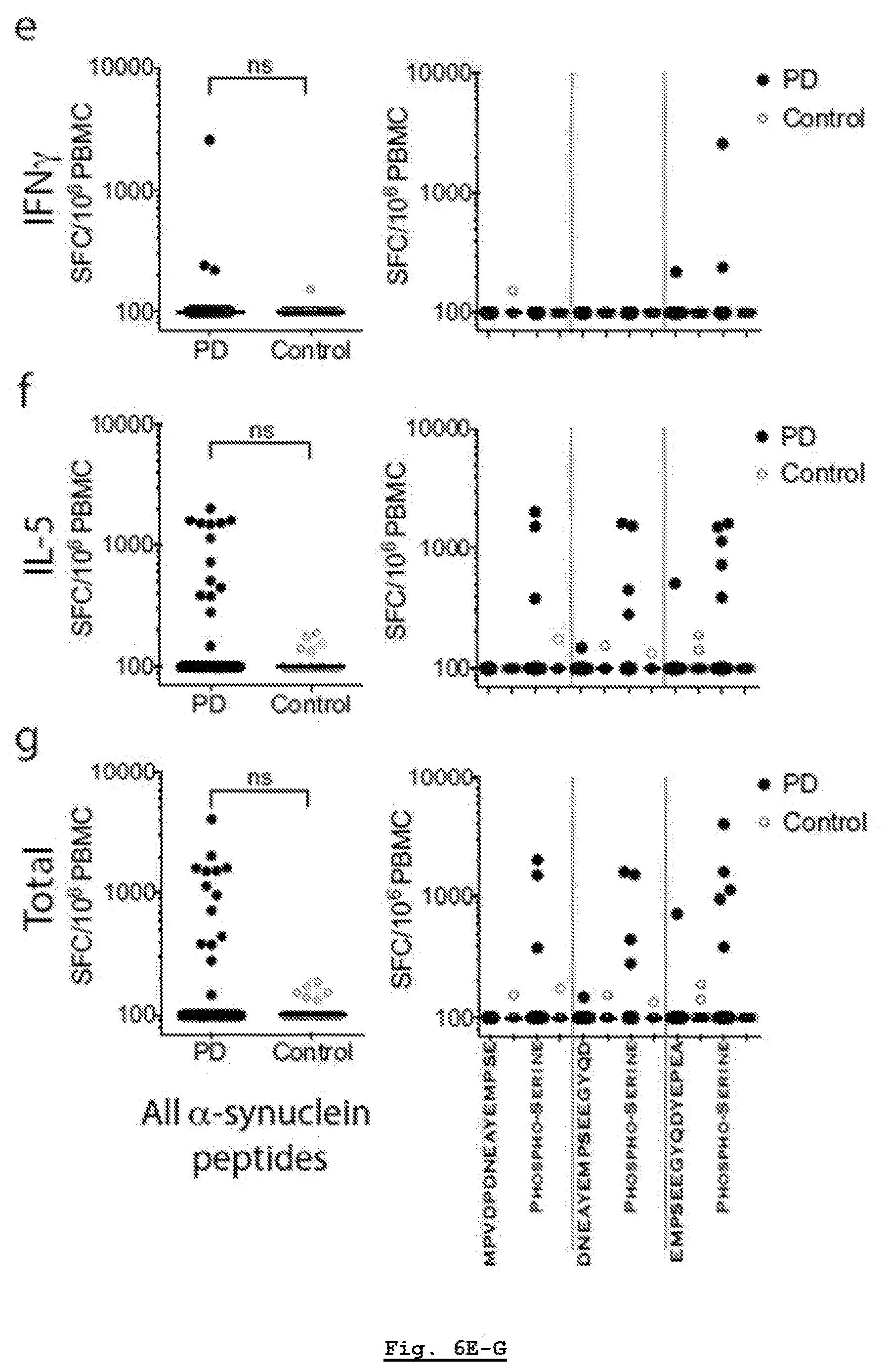
D00014
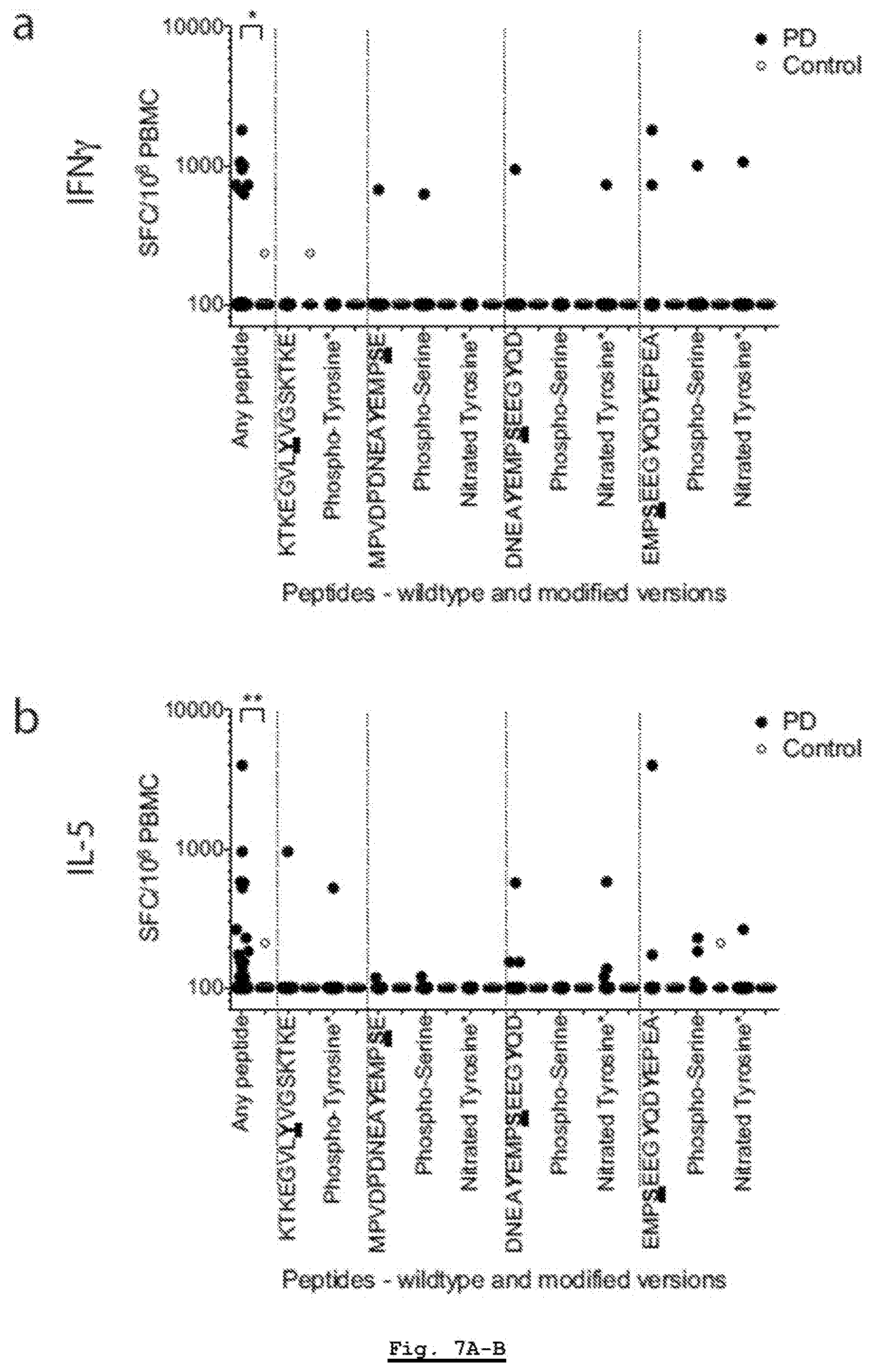
D00015

D00016
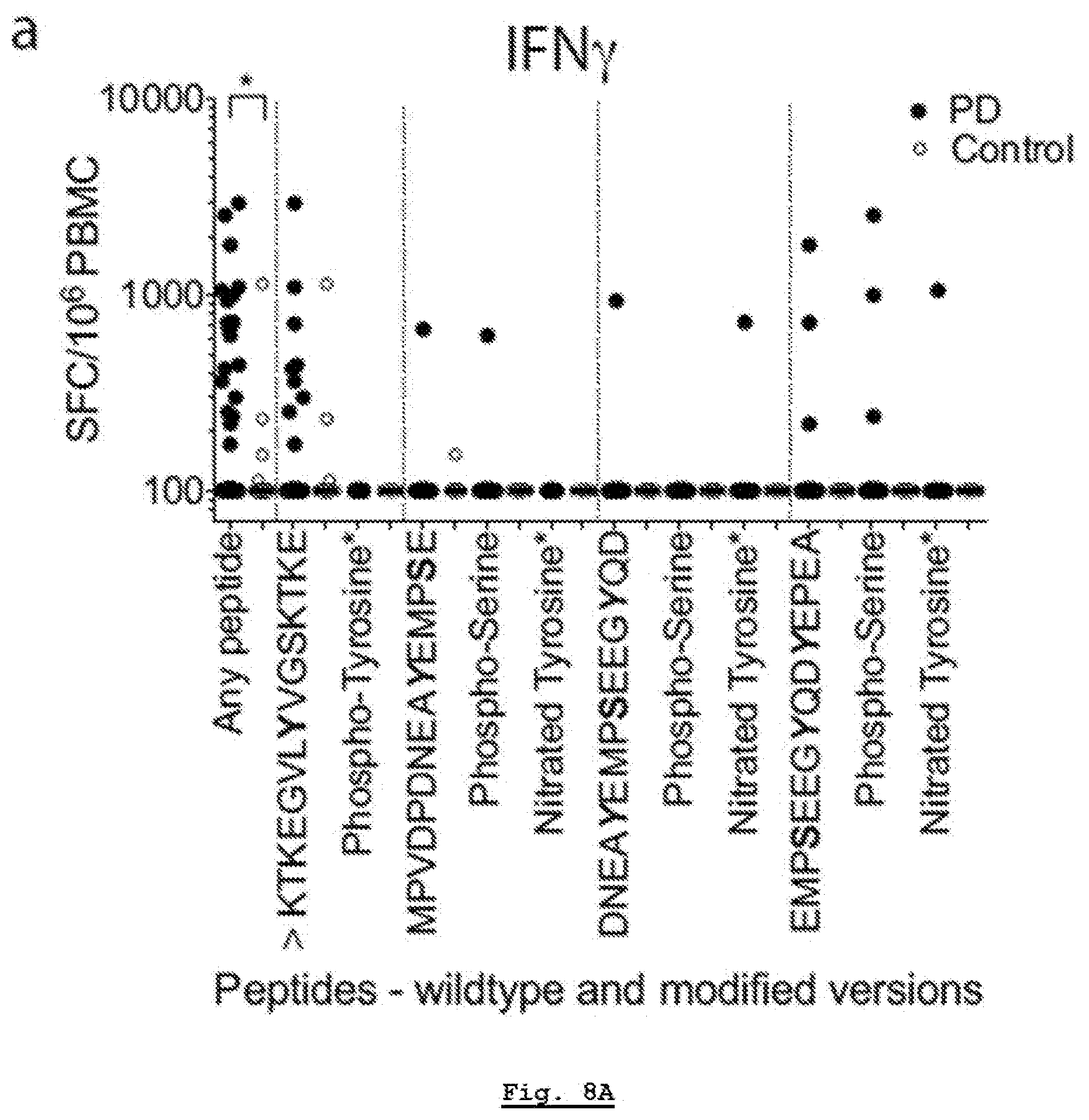
D00017
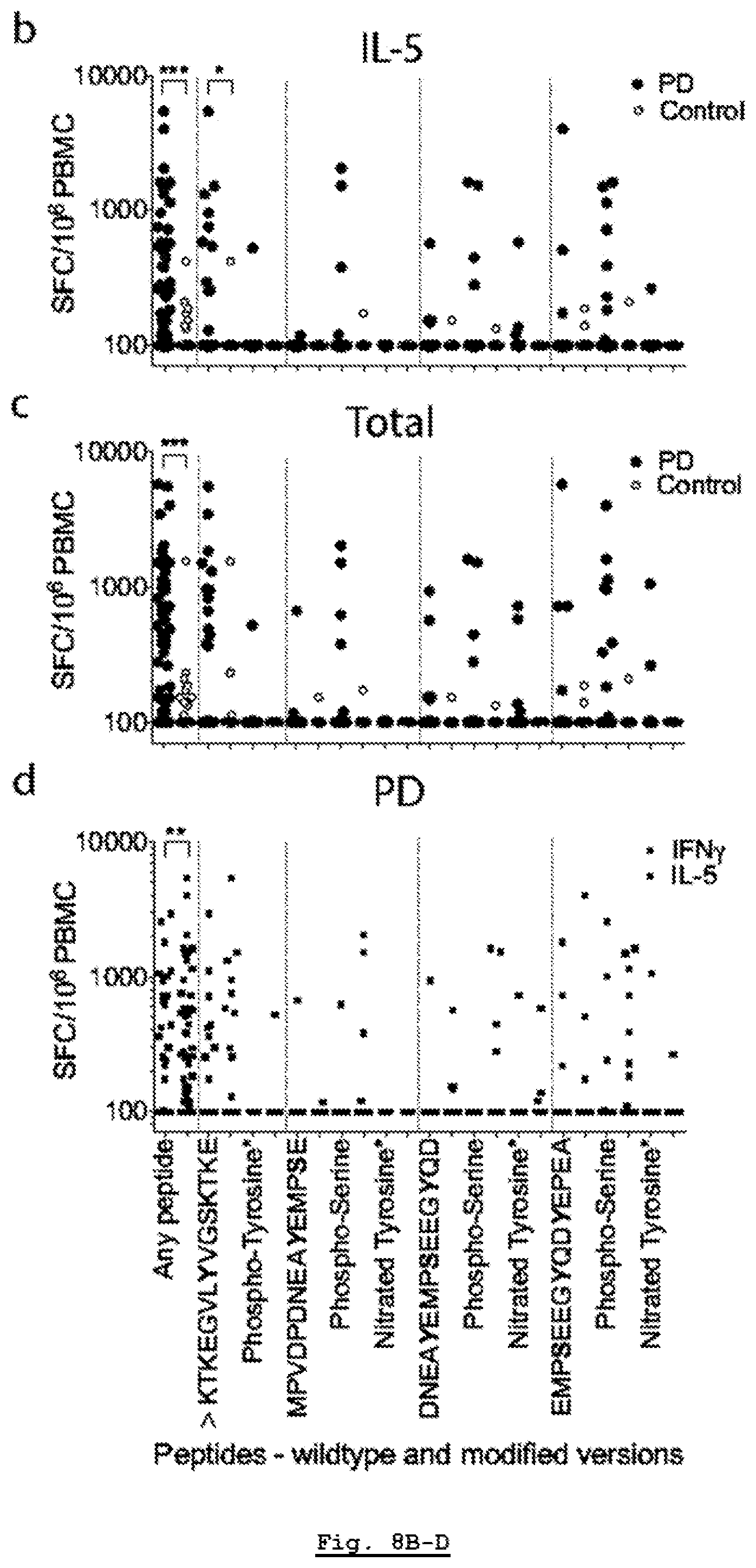
D00018
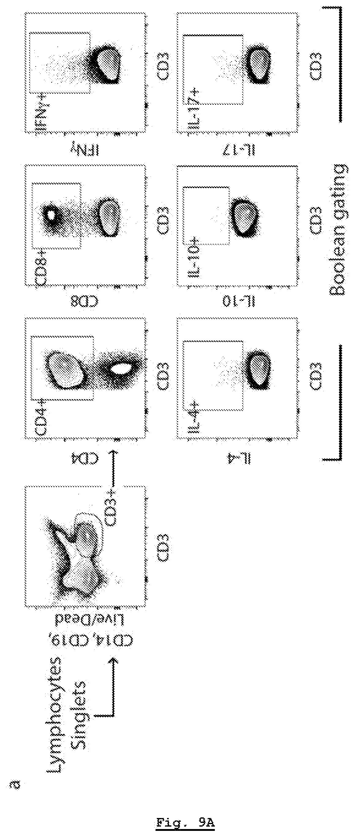
D00019

D00020
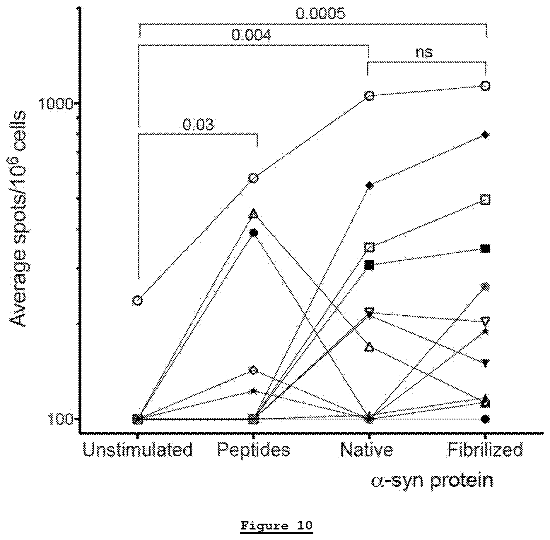
D00021
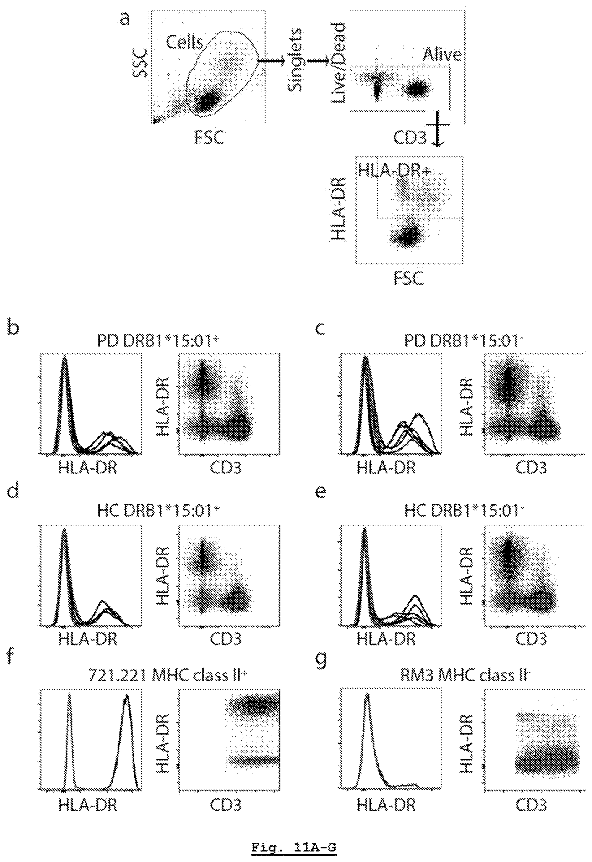
D00022
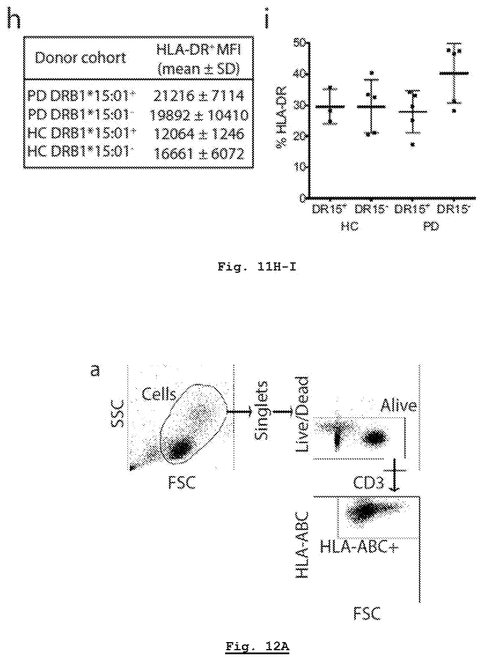
D00023
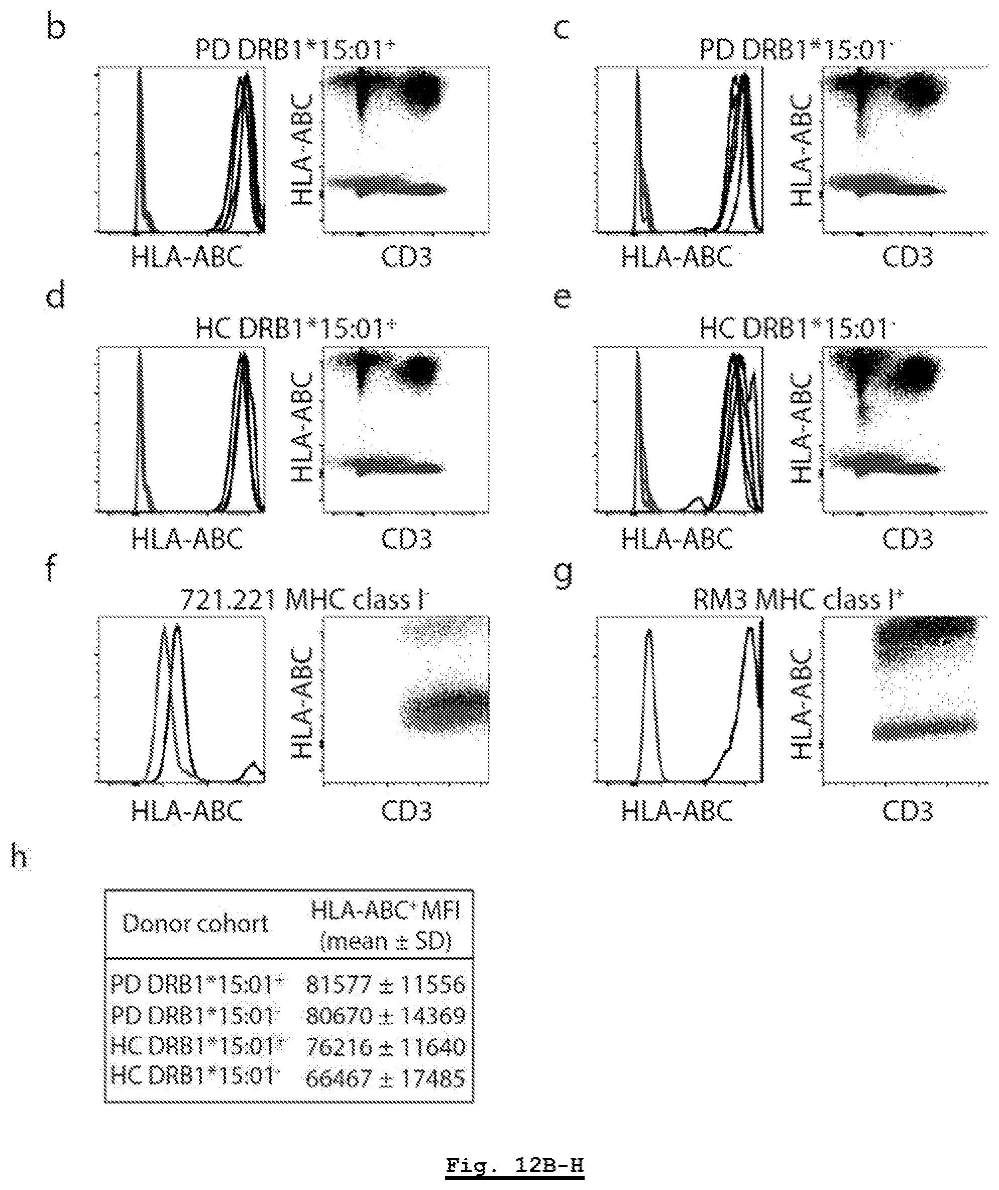
D00024
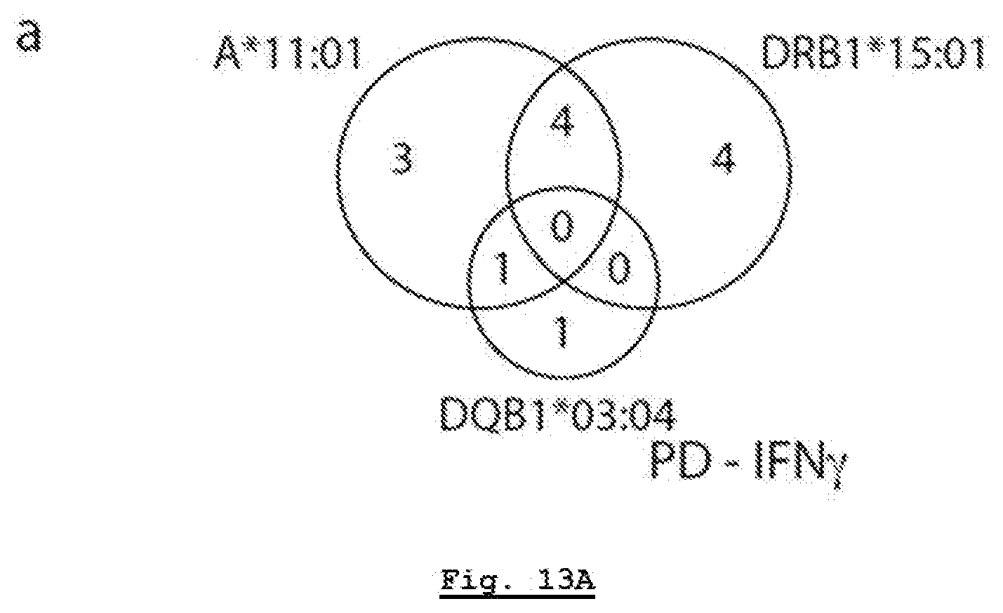
D00025
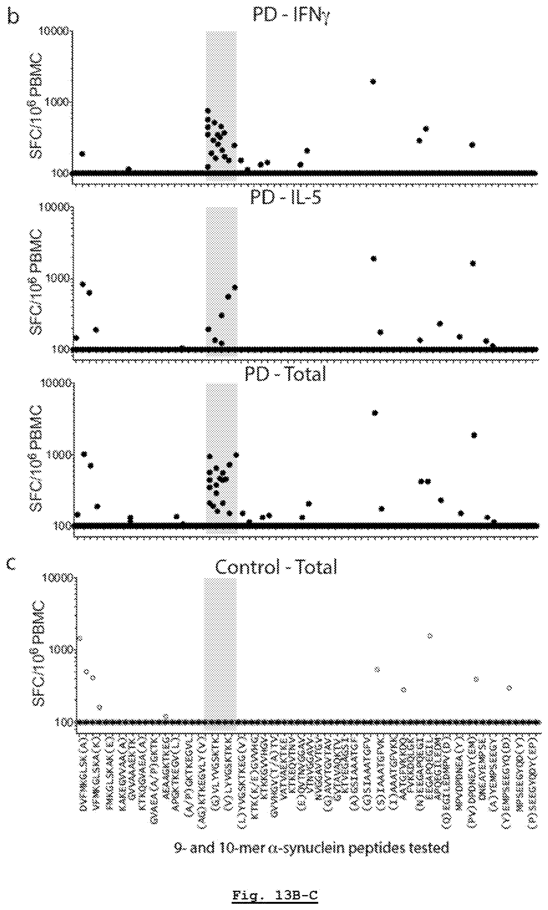
D00026
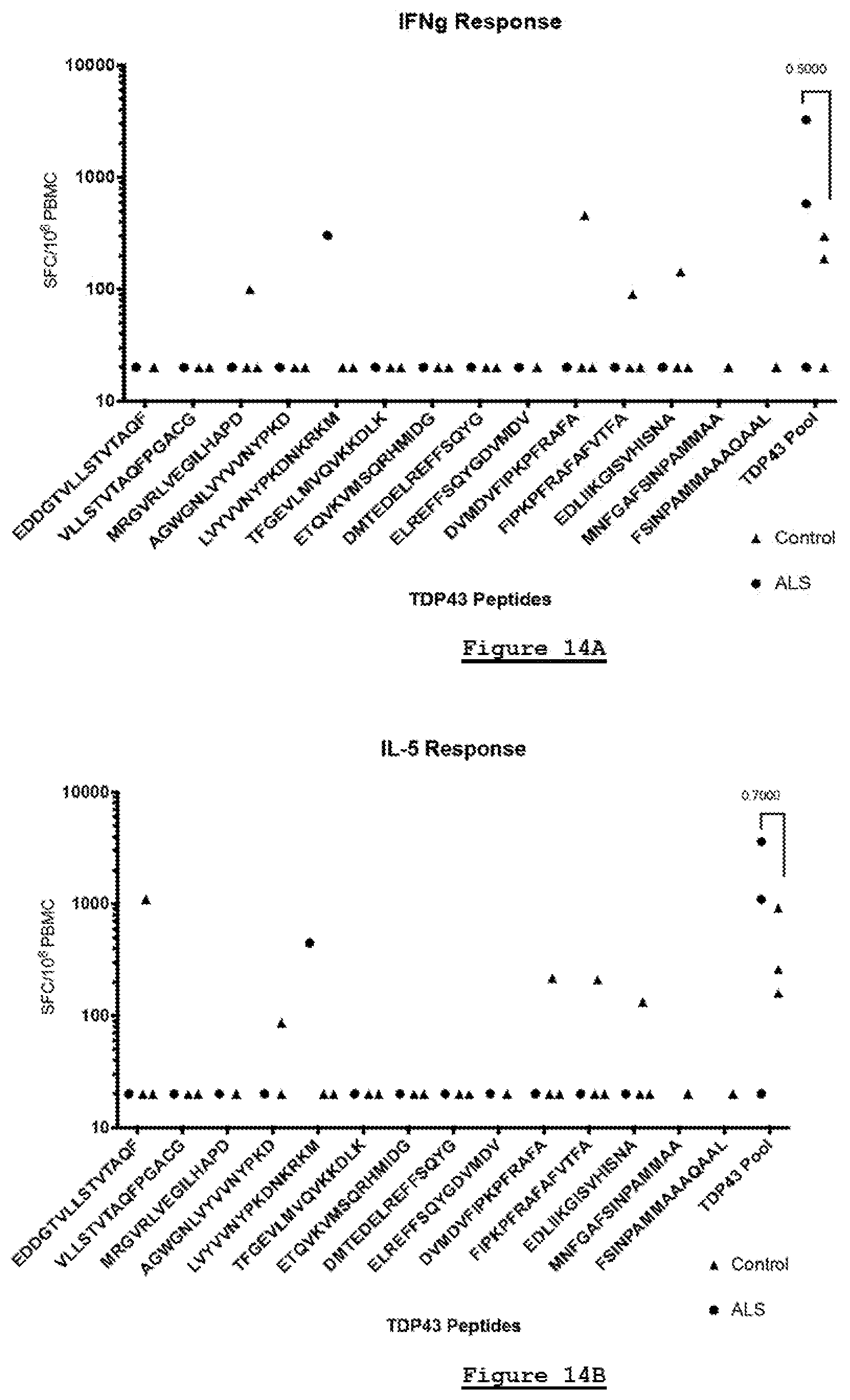
D00027
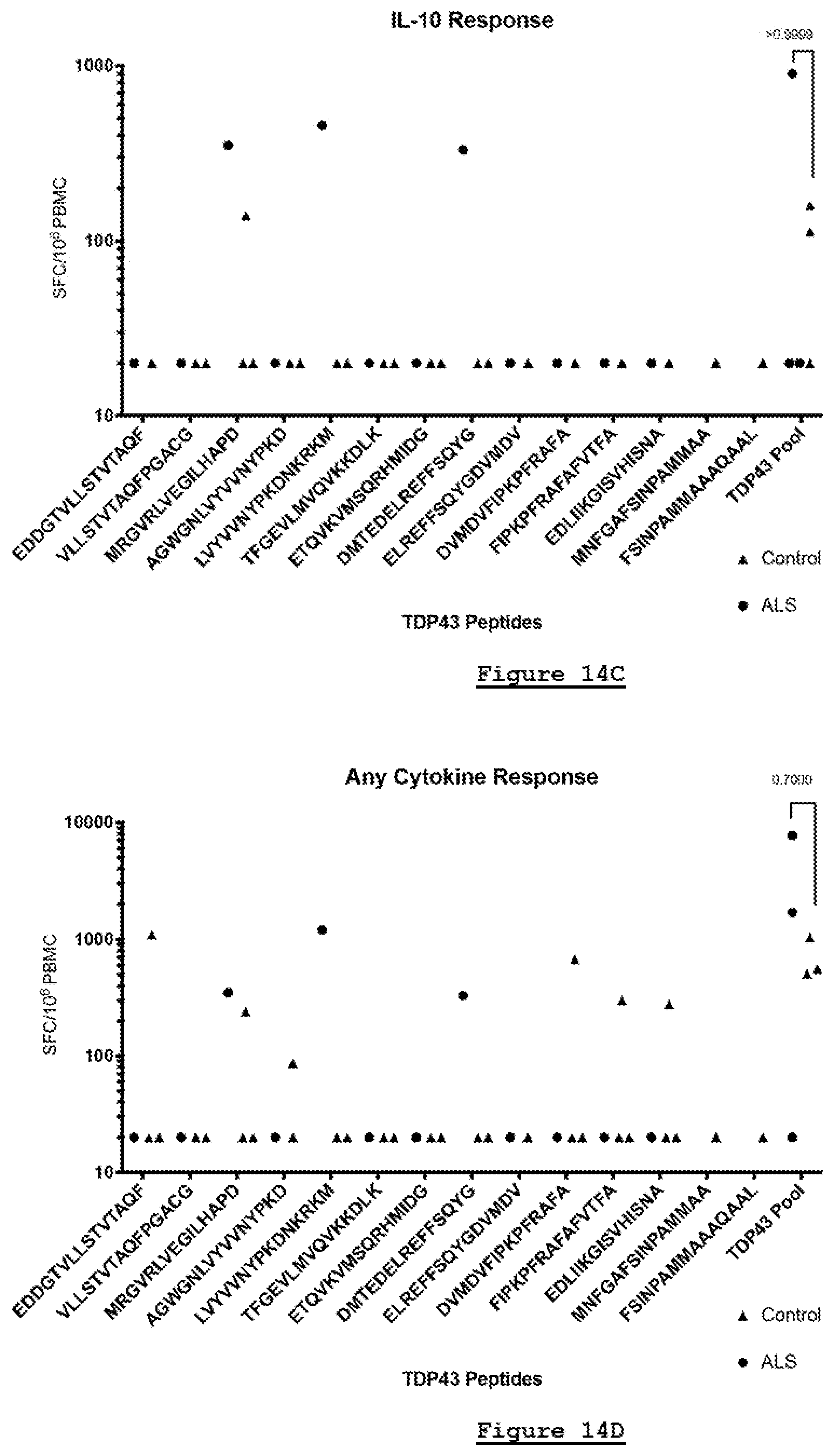
D00028
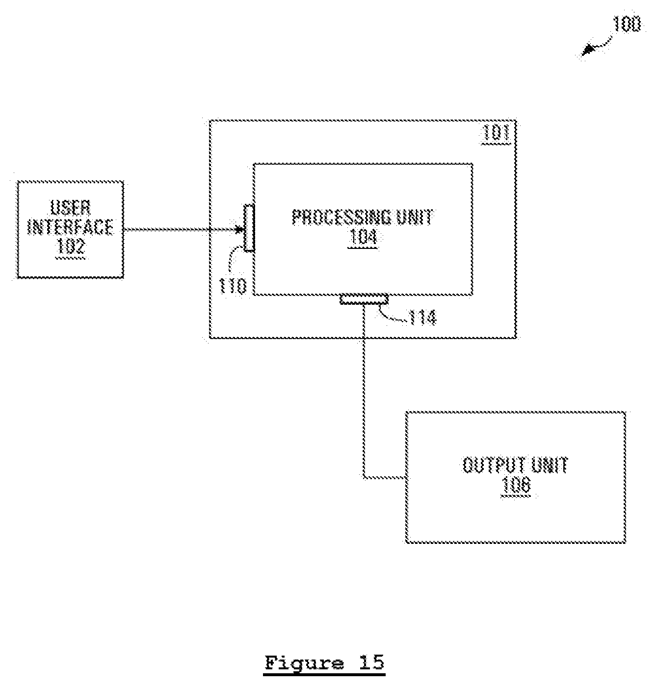
D00029
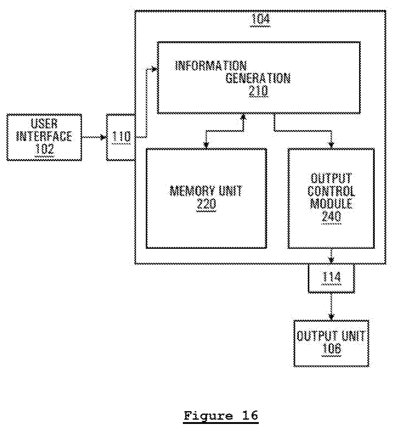
S00001
XML
uspto.report is an independent third-party trademark research tool that is not affiliated, endorsed, or sponsored by the United States Patent and Trademark Office (USPTO) or any other governmental organization. The information provided by uspto.report is based on publicly available data at the time of writing and is intended for informational purposes only.
While we strive to provide accurate and up-to-date information, we do not guarantee the accuracy, completeness, reliability, or suitability of the information displayed on this site. The use of this site is at your own risk. Any reliance you place on such information is therefore strictly at your own risk.
All official trademark data, including owner information, should be verified by visiting the official USPTO website at www.uspto.gov. This site is not intended to replace professional legal advice and should not be used as a substitute for consulting with a legal professional who is knowledgeable about trademark law.