A Method For Regenerating The Interverterbral Disc With Notochordal Cells
Gazit; Zulma ; et al.
U.S. patent application number 15/739555 was filed with the patent office on 2020-03-26 for a method for regenerating the interverterbral disc with notochordal cells. This patent application is currently assigned to Cedars-Sinai Medical Center. The applicant listed for this patent is Cedars-Sinai Medical Center. Invention is credited to Dan GAZIT, Zulma Gazit, Gadi PELLED, Dmitriy SHEYN.
| Application Number | 20200093961 15/739555 |
| Document ID | / |
| Family ID | 57585644 |
| Filed Date | 2020-03-26 |


View All Diagrams
| United States Patent Application | 20200093961 |
| Kind Code | A1 |
| Gazit; Zulma ; et al. | March 26, 2020 |
A METHOD FOR REGENERATING THE INTERVERTERBRAL DISC WITH NOTOCHORDAL CELLS
Abstract
Described herein are compositions and methods related to derivation of human notochordal cells differentiated from induced pluripotent stem cells (iPSCs). The inventors have developed a two-step process for generating these iPSC-derived notochordal cells (iNCs), which can provide a renewable source of therapeutic material for use in degenerative disc disease (DDD). As iNCs are capable of reversing DDD and supporting regeneration of invertebral disc (IVD) tissue based on the understanding that NC cells maintain homeostasis and repair of other IVD cell types such as nuclear pulposus (NP).
| Inventors: | Gazit; Zulma; (Los Angeles, CA) ; SHEYN; Dmitriy; (Los Angeles, CA) ; PELLED; Gadi; (Los Angeles, CA) ; GAZIT; Dan; (Los Angeles, CA) | ||||||||||
| Applicant: |
|
||||||||||
|---|---|---|---|---|---|---|---|---|---|---|---|
| Assignee: | Cedars-Sinai Medical Center Los Angeles CA |
||||||||||
| Family ID: | 57585644 | ||||||||||
| Appl. No.: | 15/739555 | ||||||||||
| Filed: | June 22, 2016 | ||||||||||
| PCT Filed: | June 22, 2016 | ||||||||||
| PCT NO: | PCT/US16/38799 | ||||||||||
| 371 Date: | December 22, 2017 |
Related U.S. Patent Documents
| Application Number | Filing Date | Patent Number | ||
|---|---|---|---|---|
| 62182816 | Jun 22, 2015 | |||
| Current U.S. Class: | 1/1 |
| Current CPC Class: | A61L 27/225 20130101; A61L 27/52 20130101; A61L 2430/38 20130101; C12N 5/0619 20130101; A61P 19/08 20180101; A61L 27/3895 20130101; A61L 2300/64 20130101; C12N 5/0668 20130101; C12N 2501/727 20130101; C12N 5/0655 20130101; A61L 27/3878 20130101; A61L 27/3817 20130101; C12N 2506/45 20130101; A61L 2300/62 20130101; C12N 2501/999 20130101 |
| International Class: | A61L 27/38 20060101 A61L027/38; A61L 27/52 20060101 A61L027/52; C12N 5/077 20060101 C12N005/077; A61L 27/22 20060101 A61L027/22 |
Claims
1. A method for modulating intervertebral disc degeneration, comprising: selecting a subject; and administering a quantity of induced notochordal cells (iNCs), wherein administration of iNCs modulates intervertebral disc degeneration in the subject.
2. The method of claim 1, wherein the iNCs express one or more markers selected from the group consisting of: Galectin 3, chondroitin sulfate epitopes (3B3, 7D4, 4C3), Vimentin, Noggin, Integrins (a1, b1, a5, a6), and Brachyury.
3. The method of claim 1, wherein the iNCs express one or more markers selected from the group consisting of: homeobox MIXL1, Brachyury, Noggin, Keratin 8 and Keratin 19.
4. The method of claim 1, wherein the iNCs are encapsulated in a hydrogel.
5. The method of claim 4, wherein the hydrogel comprises PuraMatrix peptide hydrogel.
6. The method of claim 4, wherein the hydrogel comprises Fibrinogen-Tetronic-1307 1 KPa hydrogel.
7. The method of claim 1, wherein at least about 1.times.10.sup.6, 2.times.10.sup.6, or 3'10.sup.6 iNCs are administered to the subject.
8. The method of claim 1, wherein modulating intervertebral disc degeneration comprises an increase in water content and/or disc height.
9. The method of claim 1, wherein modulating intervertebral disc degeneration comprises an increase in proteoglycan-matrix in nuclear pulposus tissue.
10. A method of generating induced notochordal cells (iNCs) comprising: providing a quantity of induced pluripotent stem cells (iPSCs); culturing the iPSCs in the presence of a GSK3 inhibitor (GSK3i) to form primitive streak (PS) cells; contacting the PS cells with a vector encoding Brachyury; and expressing Brachyury in the PS cells, wherein expressing Brachyury in the PS cells induces formation of induced notochordal cells (iNCs).
11. The method of claim 10, wherein the iPSCs are cultured in the presence of 1, 2, 3, 4, 5, 6, 7, 8, 9, or 10 .mu.M of GSK3i.
12. The method of claim 10, wherein the iPSCs are cultured in the presence of GSK3i for 1, 2, 3, 4, 5, 6, 7, or 8 days.
13. The method of claim 10, wherein contacting the PS cells with a vector comprises nucleofection.
14. The method of claim 10, wherein contacting the PS cells with a vector comprises transfection.
15. The method of claim 10, wherein expressing Brachyury in the PS cells comprises culturing the PS cells in A-RPMI media.
16. The method of claim 15, wherein culturing the PS cells in A-RPMI media comprises about 5, 6, or 7 or more days.
17. The method of claim 10, further comprising exogenous addition of one or more of FGF, Noggin, and dickkopf 1 (DKK1) to the Brachyury expressing PS cells.
18. A composition of iNCs made by the method of claim 10.
19. A composition of induced notochordal cells (iNCs).
20. The composition of claim 19, wherein the iNCs express one or more markers selected from the group consisting of: Galectin 3, chondroitin sulfate epitopes (3B3, 7D4, 4C3), Vimentin, Noggin, Integrins (a1, b1, a5, a6), and Brachyury.
21. The composition of claim 19, wherein the iNCs express one or more markers selected from the group consisting of: homeobox MIXL1, Brachyury, Noggin, Keratin 8 and Keratin 19.
Description
FIELD OF THE INVENTION
[0001] Described are methods and compositions for bone and bone-related tissue degeneration and related conditions. Notochordal cells derived from pluripotent stem cells can establish new therapies related to regeneration and repair of tissue.
BACKGROUND
[0002] Low back pain (LBP) is a crippling physical and socioeconomic burden with costs in the United States alone approaching $200 billion annually. This condition affects up to 80% of adults at least once during their lifetime. Degeneration of the intervertebral disc is associated with, and has been proposed to be a cause of, a large percentage of these cases of LBP. Degeneration of the disc is a chronic, progressive disease, and current clinical strategies, including both surgical as well as non-surgical approaches aimed at symptomatic relief, have done very little to improve mental, physical, or social health of those patients affected. As a result, the research community continues to focus on understanding the molecular nature of the healthy intervertebral disc and its degeneration, with the hope that novel therapeutic and regenerative strategies will eventually be developed to more adequately treat patients with the disabling condition of excessive and pathological disc degeneration.
[0003] The intervertebral disc contains three distinct tissue compartments. The outer fibrocartilaginous annulus fibrosus (AF) and the superior and inferior cartilaginous endplates completely enclose the gel-like nucleus pulposus (NP), an avascular, aggrecan-rich tissue. It is generally understood that notochord cells (NCs) do not exist in adult humans, instead they are a transient embryonic population disappearing during early human development. The NP is frequently implicated in disc degeneration and, as such, has been the focus of much research in the basic science and tissue engineering fields. In spite of this, a consensus is lacking in defining the young healthy, NP cell phenotype, and furthermore, there may be limits for repair and regeneration processes conferred by NP cells, or NP-like cells. Interestingly, animals in which NCs remain throughout the majority of their lifespan, including commonly used experimental animals such as rabbits, rats, and mice, do not show signs of degeneration and maintain a more hydrated, proteoglycan-rich matrix than that found in adult human NP tissue. Supporting this theory, NCs were found to be more metabolically active and to produce more proteoglycans (PGs) than smaller NP cells. Yet, some have argued that NP cells, or NP-like cells are essentially the same as notochord (NC) cells, and it should be said that definitive characterization of both populations (e.g., NP and NC) is lacking. Compounding such technical challenges, human NCs are in short supply, due to their disappearance during childhood, and cannot be harvested as an autologous or allogeneic graft. An alternative source of NCs is thus needed, and a potential strategy would be to mimic the differentiation process that occurs during embryogenesis and obtain NCs from pluripotent stem cells.
[0004] Current treatments for DDD rely on conservative therapies (pain management and exercise) and surgical treatments such as spinal fusion and disc replacement, which have complications and poor long-term clinical outcomes. Despite decades of research, robust clinical therapies targeting underlying causes rather than symptoms are still in the earliest stages of development. One can attempt to define the NP cell phenotype as a collection of "markers"--genes, proteins, and metabolic characteristics that are representative and distinguishing of NP cells. Nevertheless, it is important to focus on defining a phenotype, rather than merely a genotype, since phenotypic characteristics likely have physiologic consequences and dictate NP cell function vital for identifying suitable therapeutic material. With such a functional phenotypic definition of therapeutic material, one can more easily distinguish NP cells from AF cells and other related cell types, better diagnose degenerate conditions and gauge therapies while comparing healthy and degenerate samples, and better guide stem cell and tissue engineering strategies that attempt to replace lost NP cells and tissue. Perhaps most important, a clear identification of phenotype will allow us to better understand the physiology and function of NP cells, ultimately driving development of novel cell-based therapeutic modalities. Phenotypic characterization of NP cells at all stages of development, growth, maturation, and disease warrant further characterization and identification, including confirmation of the properties of bona fide human NCs.
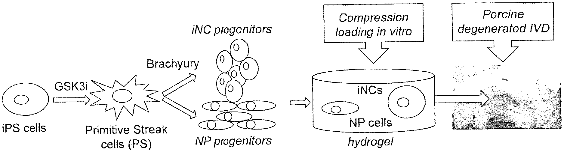
[0005] Notochordal cells are attractive candidates for cell therapy for intervertebral discs (IVDs), since the cells have a critical function in disc generation and their disappearance is associated with disc degeneration. Based on the Inventors' previous work, it is established that stem cells residing in the degenerate disc are impaired in their potential to regenerate the disc. Described herein is a reproducible and inexhaustible source of iPSC-derived NCs (iNCs). By characterizing these cells to ensure that they have acquired NC functionality and the potential to rejuvenate the IVD, the results described herein show that iNCs can be generated from iPSCs in a two-step process, in which the cells are first transformed into primitive streak cells using GSK3i and then reprogrammed into NCs using overexpression of the Brachyury gene. By developing a method of IVD regeneration using human iNCs, it is established that: i) iPSCs can be directed to differentiate into NCs in a two-step process that includes Brachyury transcription factor overexpression; ii) iNCs will function appropriately to stimulate cell viability, gene expression of cell differentiation, further including matrix protein secretion in nucleus pulposus cell (NPC)/iNC co-cultures exposed to mechanical loading conditions that simulate IVD degeneration in vitro; and iii) iNCs will induce regeneration of the degenerate IVD in vivo.
SUMMARY OF THE INVENTION
[0006] Described herein is a method for modulating intervertebral disc degeneration, comprising selecting a subject, and administering a quantity of induced notochordal cells (iNCs), wherein administering the iNCs modulates intervertebral disc degeneration in the subject. In other embodiments, the iNCs express one or more markers Galectin 3, chondroitin sulfate epitopes (3B3, 7D4, 4C3), Vimentin, Noggin, Integrins (a1, b1, a5, a6), and Brachyury. In other embodiments, the iNCs express one or more markers homeobox MIXL1, Brachyury, Noggin, Keratin 8 and Keratin 19. In other embodiments, the iNCs are encapsulated in a hydrogel. In other embodiments, the hydrogel includes PuraMatrix peptide hydrogel. In other embodiments, the hydrogel includes Fibrinogen-Tetronic-1307 1 KPa hydrogel. In other embodiments, at least about 1.times.10.sup.6, 2.times.10.sup.6, or 3.times.10.sup.6 or more iNCs are administered to the subject. In other embodiments, modulating intervertebral disc degeneration includes an increase in water content and/or disc height. In other embodiments, modulating intervertebral disc degeneration includes an increase in proteoglycan-matrix in nuclear pulposus tissue.
[0007] Also described herein is a method of generating induced notochordal cells (iNCs) including providing a quantity of induced pluripotent stem cells (iPSCs), culturing the iPSCs in the presence of a GSK3 inhibitor (GSK3i) to form primitive streak (PS) cells, contacting the PS cells with a vector encoding Brachyury, and expressing Brachyury in the PS cells, wherein expressing Brachyury in the PS cells induces formation of induced notochordal cells (iNCs). In other embodiments, the iPSCs are cultured in the presence of about 1, 2, 3, 4, 5, 6, 7, 8, 9, or 10 .mu.M of GSK3i. In other embodiments, the iPSCs are cultured in the presence of GSK3i for 1, 2, 3, 4, 5, 6, 7, or 8 days. In other embodiments, contacting the PS cells with a vector includes nucleofection. In other embodiments, contacting the PS cells with a vector includes transfection. In other embodiments, expressing Brachyury in the PS cells includes culturing the PS cells in A-RPMI media. In other embodiments, culturing the PS cells in A-RPMI media is for about 5, 6, or 7 or more days. In other embodiments, the methods includes exogenous addition of one or more of FGF, Noggin, and dickkopf 1 (DKK1) to the Brachyury expressing PS cells.
[0008] Further described herein is composition of iNCs made by the method of generating induced notochordal cells (iNCs) including providing a quantity of induced pluripotent stem cells (iPSCs), including the iPSCs in the presence of a GSK3 inhibitor (GSK3i) to form primitive streak (PS) cells, contacting the PS cells with a vector encoding Brachyury, and expressing Brachyury in the PS cells, wherein expressing Brachyury in the PS cells induces formation of induced notochordal cells (iNCs). In other embodiments, the iPSCs are cultured with about 1, 2, 3, 4, 5, 6, 7, 8, 9, or 10 .mu.M of GSK3i. In other embodiments, the culturing in the presence of GSK3i is for 1, 2, 3, 4, 5, 6, 7, or 8 days. In other embodiments, contacting the PS cells with a vector comprises nucleofection. In other embodiments, contacting the PS cells with a vector comprises transfection. In other embodiments, expressing Brachyury in the PS cells comprises culturing the PS cells in A-RPMI media. In other embodiments, culturing the PS cells in A-RPMI media is for about 5, 6, or 7 or more days. In other embodiments, the methods includes exogenous addition of one or more of FGF, Noggin, and dickkopf 1 (DKK1) to the Brachyury expressing PS cells.
[0009] Also described herein is a composition of induced notochordal cells (iNCs). In other embodiments, the iNCs express one or more markers Galectin 3, chondroitin sulfate epitopes (3B3, 7D4, 4C3), Vimentin, Noggin, Integrins (a1, b1, a5, a6), and Brachyury. In other embodiments, the iNCs express one or more markers homeobox MIXL1, Brachyury, Noggin, Keratin 8 and Keratin 19.
BRIEF DESCRIPTION OF FIGURES
[0010] FIG. 1. Notochordal cells can be separated from nuclear pulposus (NP) cells based on size. (FIG. 1A) Porcine IVDs were harvested from healthy pigs. (FIG. 1B) Porcine NPs were enzymatially digested with 0.25% collagenase and 1% hyaluronidase overnight. The cells were washed, and notochordal cell clusters were separated from the small NP cells using a 70-.mu.m cell strainer. (FIG. 1C) The RNA of each population was extracted, analyzed and plotted with RQ values comparing to total NP cells. Gene expression analysis of genes typically expressed in NP cells (blue) showed higher levels of expression in small cells than in larger cell clusters. Genes that were previously observed to be expressed in notochordal cells (red) were highly expressed in large cell clusters compared with that in small cells. These findings are consistent with those of previous studies and show that when separated by size, notochordal cells can be used as a positive control.
[0011] FIG. 2. Step 1--Differentiation of iPSCs into primitive streak (PS) cells in vitro. iPSCs were cultured with addition of 5 .mu.M GSK3i for up to 6 days. (FIG. 2A). Morphological changes during treatment were depicted with light microscopy. (FIG. 2B). On Days 2 and 4 mesenchymal stem cell (MSC) markers CD44 and CD90 were expressed at high levels, similar to what occurs in bone marrow--derived human MSCs (BM-hMSCs). The NC/NP marker CD24 was expressed in PS cells but not in BM-MSCs. (FIG. 2C). Gene expression analysis of the PS cells over time showed peak expression of mesodermal markers MIXL1 and Brachyury on Day 2 and a gradual increase in NC markers Keratins 8 and 19.
[0012] FIG. 3. Step 2--Differentiation of PS cells into iNC progenitor cells. (FIG. 3A) PS cells were nucleofected with either Brachyury (Br) or GFP-encoding plasmids and cultured in A-RPMI in vitro. Light microscopy showed the formation of two layers of cells differing in morphological characteristics: fibroblast-like cells attached to the plate (iNPs) and clustered, loosely attached, round cells (iNCs) (FIG. 3B). Bars indicate SEs, *p<0.05; **p<0.01; ***p<0.001; ****P<0.0001,
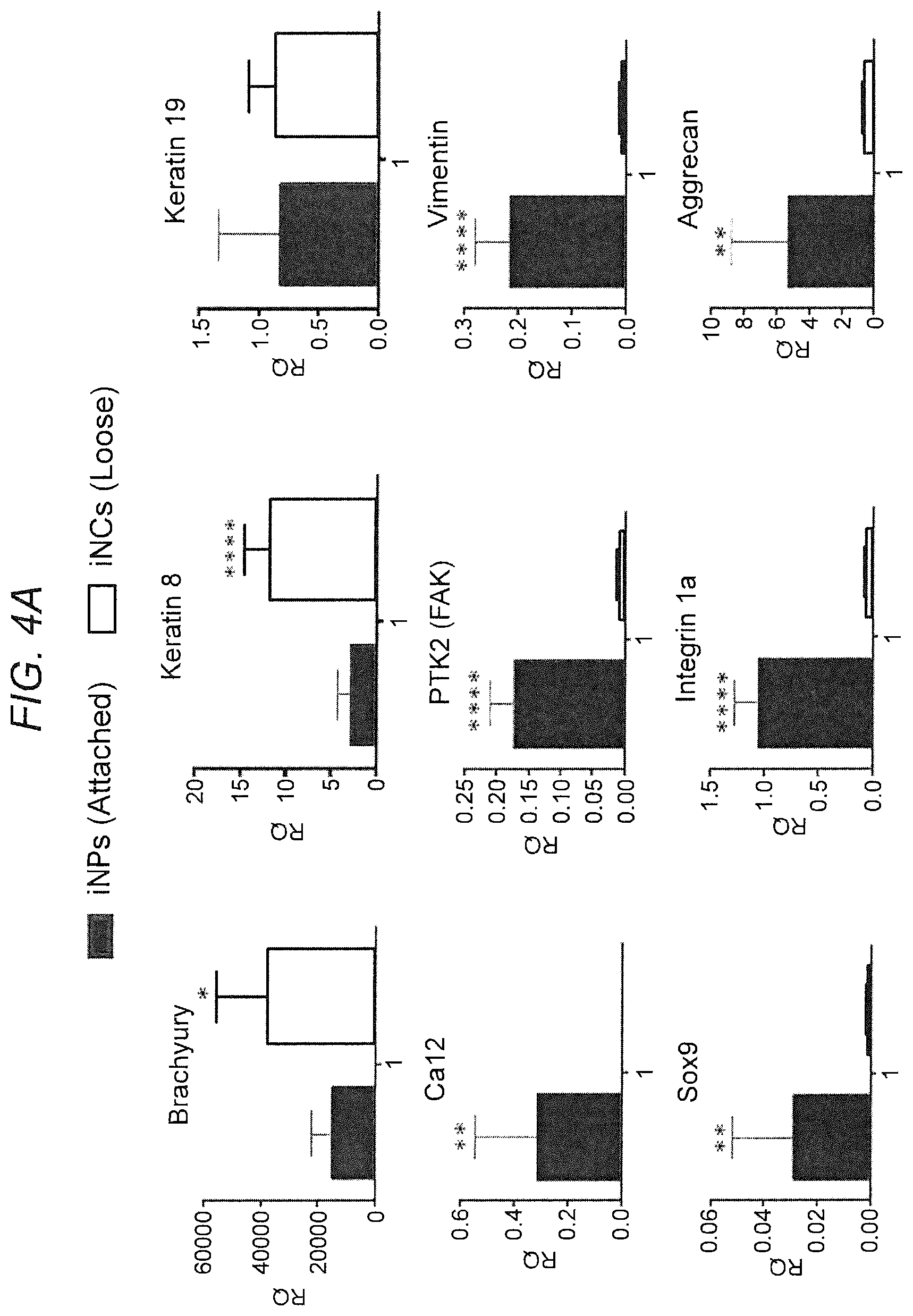
[0013] FIG. 4. Differential gene expression of iNPs and iNCs. (FIG. 4A) The upper layer of cells could be easily separated from the lower level of cells, and therefore we could analyze them separately. Gene expression analysis on Day 6 after Brachyury nucleofection showed a higher expression of notochordal marker genes (Brachyury, Keratins 8 and 19) in iNCs than in iNPs. In contrast, the iNP population displayed a higher expression of chondrogenic markers (Aggrecan, Sox9), NP markers (PTK2, CA12), and MSCs markers that are not only expressed in MSCs, but also in notochordal cells (Vimentin, Integrin 1a). Bars indicate SEs, *p<0.05; **p<0.01; ***p<0.001; ****p<0.0001. (FIG. 4A B) PS-Br cells were fixed 4 days after nucleofection and stained against Brachyury and Glut4 epitopes using immunofluorescence (IF). The IF indicates high expression of Brachyury (upper right) inside the cell nucleus mostly, but not only, in iNCs, and expression of the Glut4 NP marker mostly in iNPs (upper left).
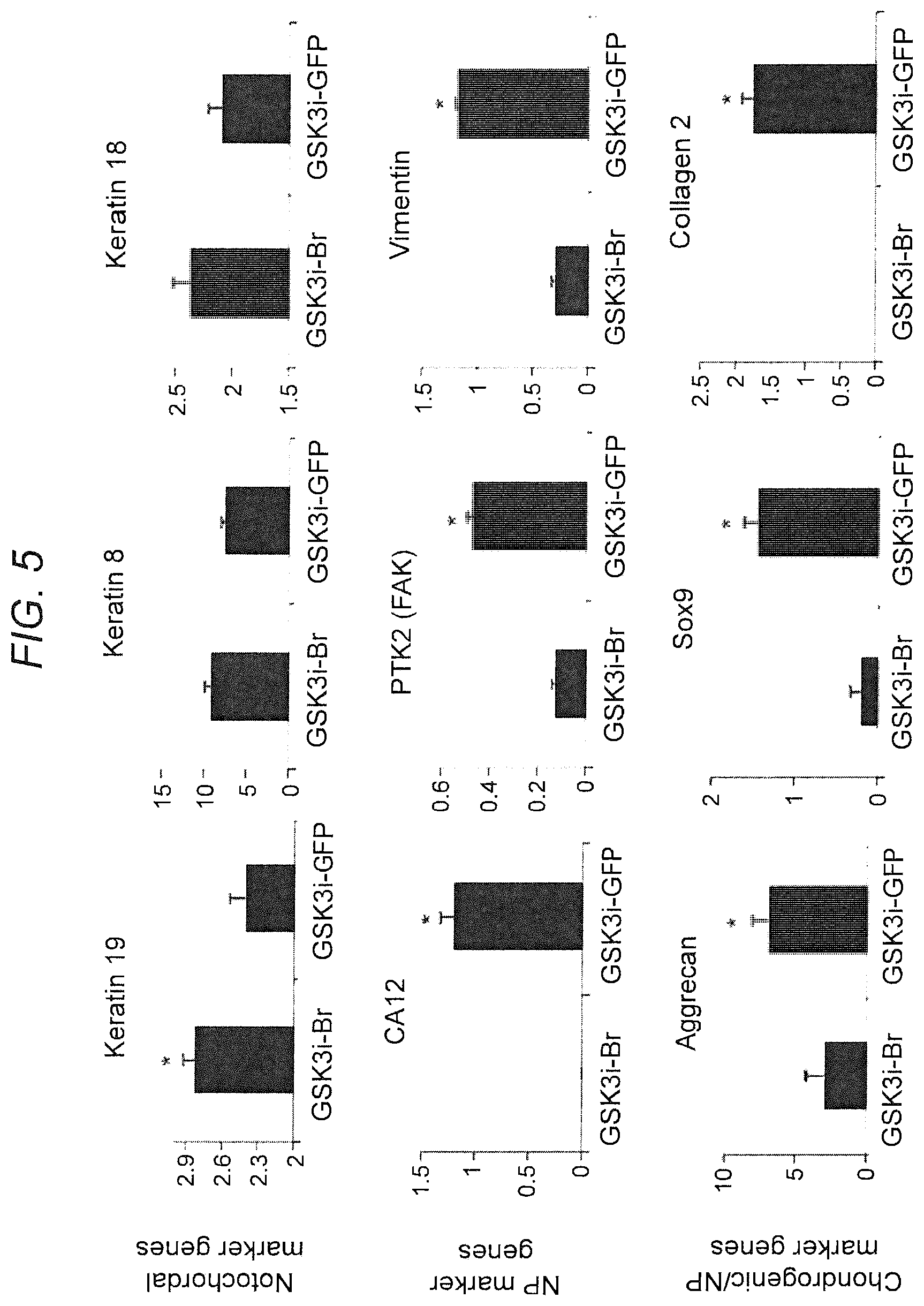
[0014] FIG. 5. iNCs sustain notochordal phenotype in NP differentiation conditions in vitro. PS cells were nucleofected either with Brachyury-encoding plasmid or GFP reporter gene, encapsulated in hydrogel, and cultured in NP-differentiation conditions (NP media and hypoxia) for 21 days. RNA was extracted, and the gene expression profile was evaluated using qRT-PCR for NC and NP markers. Gene expression analysis showed that the NP markers CA12, PTK2, and Vimentin as well as the chondrogenic markers Aggrecan, Sox9 and Co12, were downregulated, and that the NC markers Keratins 8, 18, and 19 were upregulated or did not change in Brachyury-overexpressing cells, compared to GFP-expressing cells. This indicates that NP differentiation conditions affected PS-GFP cells but not PS-Br cells, which sustained their NC phenotype. Bars indicate SEs, *p<0.05.
[0015] FIG. 6. Effect of dynamic overloading on NP marker gene expression. PS cells were nucleofected with either Brachyury- or GFP-encoding plasmids and on Day 6 of culture were encapsulated in fibrin gel. The constructs were placed in the FlexCell system and exposed to dynamic compression--20% compression at 1 Hz for 2 hours, twice per day for 48 hours. Afterward, the constructs were harvested and RNA was extracted. Gene expression analysis showed that the loading conditions resulted in downregulation of NP markers in both types of cells, but not downregulation of notochordal markers. Bars indicate SEs, *p<0.05; **p<0.01; ***p<0.001; ****p<0.0001 (comparison of loaded and unloaded groups).
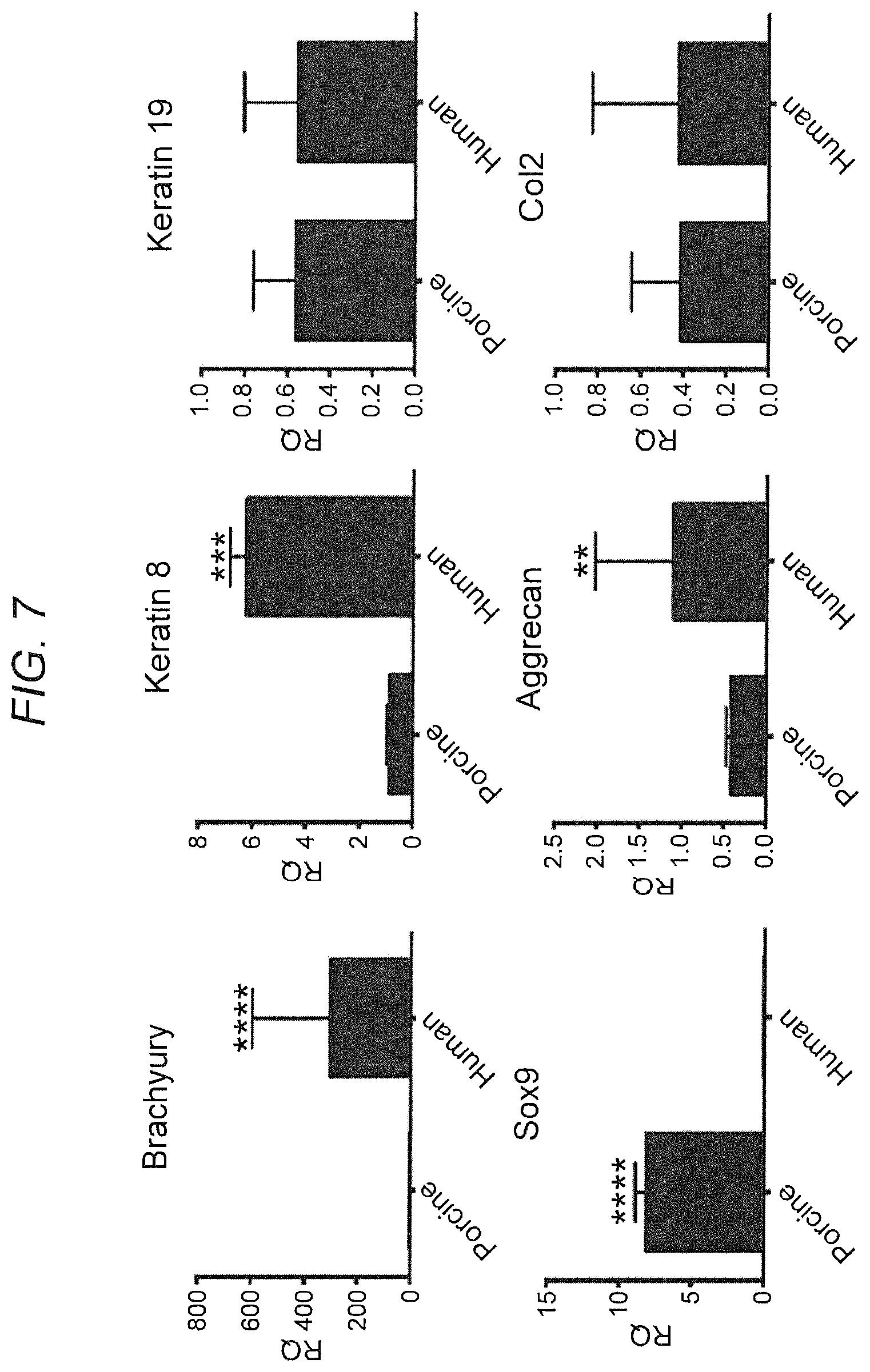
[0016] FIG. 7. Co-culture of iNCs with HNPCs: gene expression. iNCs were generated using the protocol described above. NPCs were isolated from porcine healthy NPs. Both cell types were encapsulated in PuraMatrix in a 1:1 ratio and cultured in NP differentiation conditions. On Day 21 the constructs were harvested and RNA was extracted. Gene expression analysis was performed using human and porcine specific primers for NC/NP marker genes. The results show that human cells expressed higher levels of Brachyury, Noggin and Aggrecan, whereas porcine cells expressed higher levels of Sox9 and similar levels of Keratin 8 and Co12, indicating that there were more notochordal cells of human origin and and more NP-like cells of porcine origin. Bars indicate SEs, *p<0.05; **p<0.01; ***p<0.001; ****p<0.0001.
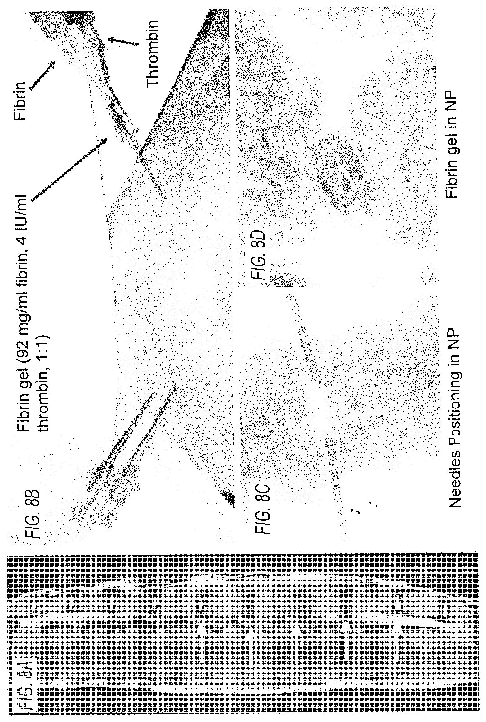
[0017] FIG. 8. Porcine IVD model. (FIG. 8A) Three levels of degenerate IVDs following annular injury (yellow arrows) and healthy IVDs (white arrows) imaged by MRI. (FIG. 8B) Injection into the porcine IVD under fluoroscopic guidance. (FIG. 8C) An uncompromised IVD was washed out using warm saline solution and (FIG. 8D) green solution--labeled fibrin gel was injected into the void.
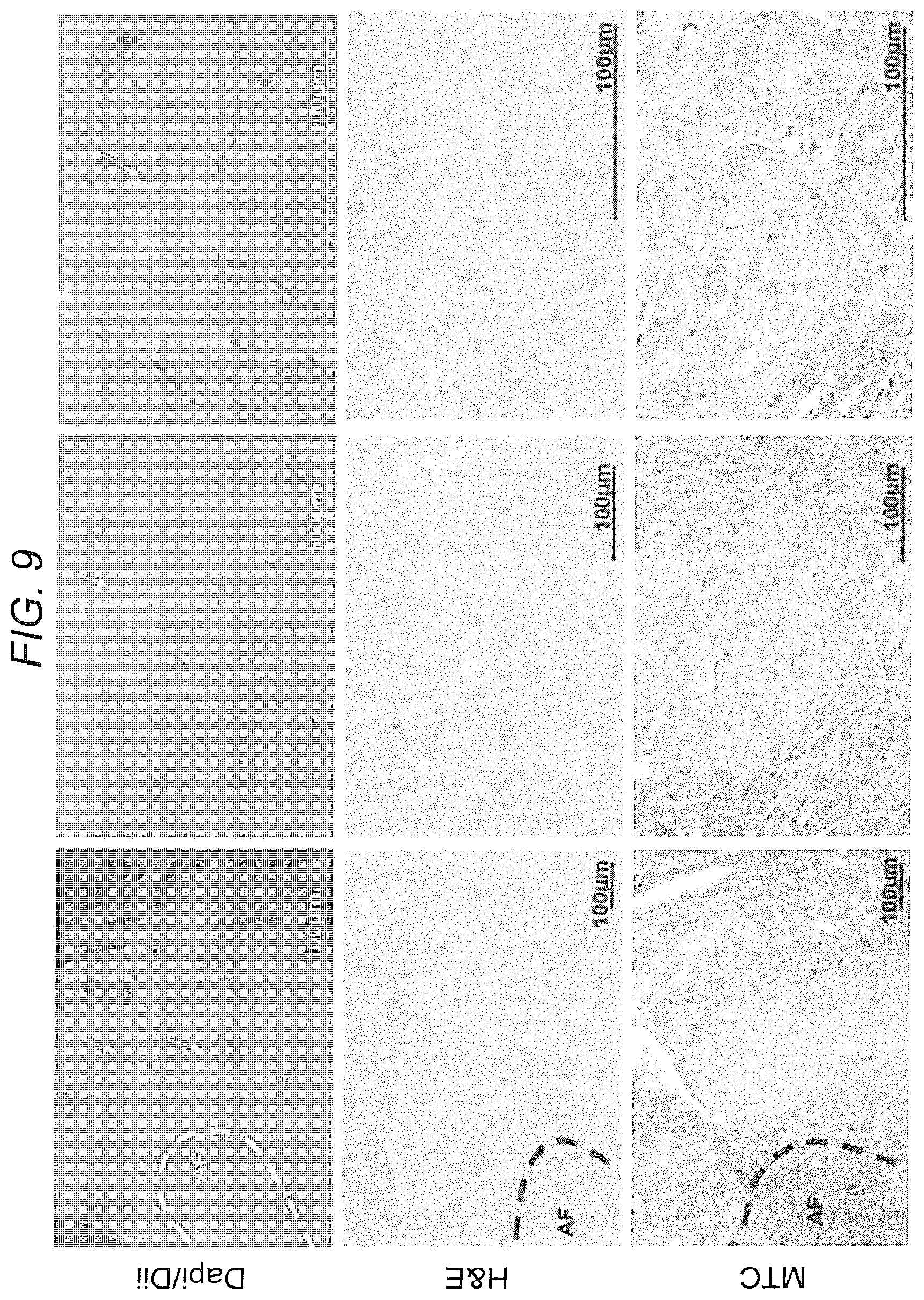
[0018] FIG. 9. DiI-labeled MSCs detected in rodent IVD--engraftment and differentiation. Human DiI-labeled MSCs were injected into rat IVDs. The discs were harvested, sectioned, and stained with H&E and MTC. Engrafted MSCs were detected inside the disc using light and fluorescence microscopy. Chondrogenic lineage differentiation was observed and is depicted by MTC staining.
[0019] FIG. 10. Process for differentiating iPSCs into functional notochordal cells that can be used for IVD cell therapy. iPSCs can be directed to differentiate into NCs in a two-step process that includes Brachyury transcription factor overexpression. Thereafter, iNCs will function appropriately to stimulate cell viability, gene expression of cell differentiation, and matrix protein secretion in NPC/iNC co-cultures exposed to mechanical loading conditions that simulate IVD degeneration in vitro. Finally, iNCs will induce repair/regeneration of the degenerate IVD in vivo
[0020] FIG. 11. Experimental design and various aims.
[0021] FIG. 12. Experimental design for evaluating of the regenerative potential of iNCs in a large animal model of IVD.
[0022] FIG. 13. Step 1--Differentiation of iPSCs into primitive streak (PS) cells in vitro. (FIG. 13A) Morphological changes during treatment were depicted with light microscopy. (FIG. 13B) Gene expression analysis of PS cells shows a rapid and significant decline in expression of pluripotency markers in GSK3i-treated cells compared to cells treated with vehicle only (DMSO). The mesodermal markers Brachyury and MIXL1 were upregulated 24 hours after addition of GSK3i to the media and their expression was significantly higher up to Days 2 and 3, respectively. An additional mesodermal marker, FoxF1, was upregulated after 48 hours of treatment and its expression remained higher than that in the DMSO group through Day 6. Results represent mean RQs calibrated relatively to untreated iPSCs (Day 0), n=6, bars indicate SEs, 2-way ANOVA with Bonferroni correction, *p<0.05; **p<0.01; ***p<0.001; ****p<0.0001.
[0023] FIG. 14. Step 2--Differentiation of PS cells into iNC progenitor cells. (FIG. 14A) PS cells were nucleofected with either Brachyury (Br) or GFP-encoding plasmids and cultured in A-RPMI in vitro for 3 days. Afterward, they were either grown in vitro for 7 days (2D) or in 3D alginate beads (ABs). Differentiation towards notochordal cells was evaluated using gene expression for NC marker genes. (FIG. 14B) Phenotype stability: iNCs in ABs were grown in hypoxic conditions and NC marker gene expression was analyzed once a week. Bars indicate SEs, ****p<0.0001.
[0024] FIG. 15. Step 2--Optimization with additional factors. The cells were encapsulated in ABs, grown in hypoxic conditions for 7 days, and gene expression of the notochordal markers was evaluated. No combination was found to be significantly different from Brachyury alone. Thus we concluded that overexpression of Brachyury in PS cells and a 3D hypoxic environment is sufficient to induce iNC differentiation. Bars indicate SEs, # p<0.01 when comparing to a sample not transfected with the same gene that is being tested, *p<0.05; **p<0.01; ***p<0.001; ****p<0.0001.
[0025] FIG. 16. iNC function: paracrine effect on BM-hMSCs. Notochordal conditioned medium (NCM) was collected from iNCs and porcine NCs grown in alginate beads, as reported. This medium was applied to BM-hMSCs encapsulated in alginate beads. Gene expression was evaluated on Day 7 and normalized to BM-hMSCs grown in control medium. iNC-NCM was found to induce significantly higher expression of NC and NP marker genes in BM-hMSCs than porcine NC-NCM. Bars indicate SEs, *p<0.05
[0026] FIG. 17. Porcine IVD model. (FIG. 17A) Three levels of IVDs were subjected to annular puncture under fluoroscopic guidance. (FIG. 17B) The degeneration process was observed using MRI. Degenerated discs are indicated with yellow arrows and healthy IVDs with white arrows.
[0027] FIG. 18: MR imaging of the IVD can detect and quantify matrix composition. (FIG. 18A) A GAG phantom study demonstrating a linear relationship between the --OH CEST signal and GAG concentration. (FIG. 18B) Representative MRIs of healthy and degenerate porcine IVDs. (FIG. 18C) Quantitation of MR imaging of IVDs after degeneration induction. There are significant decreases in T1, T2, Trho, and CEST values of the disc as early as 2 weeks post injury, indicating reductions in GAGs, collagens, and water content in the disc. Bars indicate SEs, *p<0.05; **p<0.01; ****p<0.0001.
[0028] FIG. 19. iNC injection into degenerate porcine IVD: pilot study. 6 weeks after degeneration induction caused by annular puncture, DiI-labeled iNCs were injected into the NP. After 4 weeks, the IVDs were harvested, stained with H&E, and analyzed (FIG. 19A & FIG. 19B). FIG. 19B shows magnification of the NP area. The injected iNCs iNCs were detected using IF staining against Brachyury and Keratin 18, and confocal microscopy (FIG. 19C). The right panel shows that the DiI-labeled iNCs expressed both genes.
DETAILED DESCRIPTION
[0029] All references cited herein are incorporated by reference in their entirety as though fully set forth. Unless defined otherwise, technical and scientific terms used herein have the same meaning as commonly understood by one of ordinary skill in the art to which this invention belongs. Allen et al., Remington: The Science and Practice of Pharmacy 22.sup.nd ed., Pharmaceutical Press (Sep. 15, 2012); Hornyak et al., Introduction to Nanoscience and Nanotechnology, CRC Press (2008); Singleton and Sainsbury, Dictionary of Microbiology and Molecular Biology 3.sup.rd ed., revised ed., J. Wiley & Sons (New York, N.Y. 2006); Smith, March's Advanced Organic Chemistry Reactions, Mechanisms and Structure 7.sup.th ed., J. Wiley & Sons (New York, N.Y. 2013); Singleton, Dictionary of DNA and Genome Technology 3.sup.rd ed., Wiley-Blackwell (Nov. 28, 2012); and Green and Sambrook, Molecular Cloning: A Laboratory Manual 4th ed., Cold Spring Harbor Laboratory Press (Cold Spring Harbor, N.Y. 2012), provide one skilled in the art with a general guide to many of the terms used in the present application. For references on how to prepare antibodies, see Greenfield, Antibodies A Laboratory Manual 2.sup.nd ed., Cold Spring Harbor Press (Cold Spring Harbor N.Y., 2013); Kohler and Milstein, Derivation of specific antibody-producing tissue culture and tumor lines by cell fusion, Eur. J. Immunol. 1976 July, 6(7):511-9; Queen and Selick, Humanized immunoglobulins, U.S. Pat. No. 5,585,089 (1996 December); and Riechmann et al., Reshaping human antibodies for therapy, Nature 1988 Mar. 24, 332(6162):323-7.
[0030] As described, low back pain (LBP) is a crippling physical and socioeconomic burden with costs in the United States alone approaching $200 billion annually and affecting up to 80% of adults at least once during their lifetime. Degeneration of the intervertebral disc is involved with a large percentage of these cases of LBP with current clinical strategies of surgical as well as non-surgical approaches aimed at symptomatic relief. Despite these advances there is a need for novel treatment approaches.
[0031] Imaging studies have indicated a link between LBP and intervertebral disc (IVD) degeneration in 40% of patients. IVD cells occupy only 1% of IVD volume, but they are responsible for extracellular matrix synthesis and degradation. In degeneration there is an alteration in nucleus pulposus (NP) cell biology that leads to diminished cell numbers and altered cell function, resulting in an imbalance between matrix synthesis and degradation. Proteoglycan synthesis decreases, and there is a transition from type II collagen to type I collagen. This results in a more fibrous, dehydrated matrix less able to withstand the mechanical forces experienced in the spine. These changes have been linked to the initiation of pain responses, either through direct ingrowth of nerves and blood vessels into the IVD17 or by surrounding anatomical structures (e.g., spinal facets, spinal cord), which further impair IVD function leading to pain.
[0032] Current treatments for degenerative disc disease (DDD) rely on conservative therapies such as pain management and exercise, and, when these fail, surgery. Surgical treatments such as spinal fusion and disc replacement provide satisfactory results in alleviating pain, but are not devoid of complications and poor long-term clinical outcomes. Despite decades of research, robust clinical therapies targeting underlying causes rather than symptoms are still in the earliest stages of development. Thus, there is an urgent need for alternative treatments, such as stem cell therapies, focused on correcting the underlying pathogenesis and aberrant cell biology of DDD. An appropriate cell source still needs to be identified.
TABLE-US-00001 TABLE 1 Therapeutic Approaches IVD therapy Strength Weaknesses Small Relatively safe - used in Short half-life of proteins in molecules, clinical trials solution anti- Efficient to reduce acute Limited effect of a single inflammatory inflammation factor on a complex process factors, such as DDD proteins Gene therapy: Prolonged effect Limited safety (high doses, retroviruses misplaced injections, lack of control over vector expression) Cell therapies: NPC: can differentiate NPC: low availability, low NPC, MSC, towards chondrocytes, proliferative ability iPSC reduce inflammation MSC: the mechanism of MSC: can be autologous, immunomodulation is not reduce inflammation, pain clear yet. In human clinical when injected into IVD trials no regeneration of the iPSC: have the potential to disc was found. Cell become notochordal cells, manipulation is required for safe if fully differentiated expansion. Low availability of autologous cells in elderly patients iPSC: if not differentiated can induce teratomas
[0033] Of great interest is understanding the role of notochordal cells in the IVD. Nuclear pulposus (NP) is formed from the embryonic notochord as it segments during fetal development; the surrounding annulus fibrosus (AF) is formed from the sclerotome/mesoderm. At birth, the NP is populated by morphologically distinct, large vacuolated notochordal cells (NCs). It is important to emphasize that in some vertebrates these NCs persist throughout most of adult life, whereas in other species, including humans, these NCs gradually disappear during maturation. Such transient embryonic NCs eventually becoming undetectable and replaced by a population of smaller round cells--NP cells--believed to differentiate from NCs.
[0034] The change in cell population correlates with the initiation of degenerative changes within the disc, suggesting that the loss of NCs may be responsible. Interestingly, animals in which NCs remain throughout the majority of their lifespan, including commonly used experimental animals such as rabbits, rats, and mice, do not show signs of degeneration and maintain a more hydrated, proteoglycan-rich matrix than that found in adult human NP tissue. Supporting this theory, NCs were found to be more metabolically active and to produce more proteoglycans (PGs) than smaller NP cells. Additionally, in vitro experiments with human and bovine NP cells encapsulated in 3D hydrogels suggest that NC cells could also act as stimulators, controlling the synthesis of proteoglycans by the NP cells. These findings imply that the development of stem cell-based therapies focusing on differentiation toward a notochordal cell phenotype capable of synthesizing a proteoglycan-rich matrix and playing a protective role in a catabolic environment may be more desirable than therapies focusing on differentiation into NP cells.
[0035] The presence and effect of mechanical loading on degenerative processes in the IVD is significant considering that the main function of the IVD is to transfer mechanical axial forces. Therefore, physiological loading of the IVD is a natural stimulus and known to be essential for maintenance of cell viability and matrix biology. In contrast, mechanical overload is widely assumed to promote degeneration. For example, dynamic overloading of caprine lumbar IVD explants for 21 days at 1 Hz, altering in magnitude every 30 min (0.1 MPa and 0.4-0.8 MPa for 16 hours/day), has been shown to have catabolic effects and to result in reduced cell viability in the nucleus pulposus. Moderate cyclic compression of an IVD and stem cells co-cultured in a biomimetic matrix at 10% compression, 1 Hz for 1 hour per day, resulted in upregulation of cartilage-related markers. Therefore in vitro mechanical overloading can simulate disc degeneration as well as environmental conditions affecting a healthy disc.
[0036] For use of iPSCs as a cell source for IVD cell therapy, studies have shown the therapeutic effect of mesenchymal stem cell (MSC) injection into the IVD. And while MSCs have been found either to obtain the phenotype of NP cells, they potentially only provide a short-term solution, or actually induce mineralization and bone tissue formation in the injured IVD, which impairs the function of the IVD. Therefore, given the evidence above, NCs appear to be the ideal cell to regenerate the NP.
[0037] Unfortunately, human NCs are in short supply, due to their disappearance during childhood, and cannot be harvested as an autologous or allogeneic graft. An alternative source of NCs is thus needed, and a potential strategy would be to mimic the differentiation process that occurs during embryogenesis and obtain NCs from pluripotent stem cells. Induced pluripotent stem cells (iPSCs), first discovered in 2006, can be generated from almost any kind of somatic cell by using an integration-free method. The unlimited proliferation capacity of iPSCs, combined with their pluripotent differentiation potential, places them among the most promising stem cells for IVD therapy. Indeed, the feasibility of iPSC differentiation toward NC-like cells (hereafter referred as iNCs) was recently shown using a non-defined NP tissue matrix.
[0038] Described herein is a two-step, well-defined, and highly controlled method of reprogramming iPSCs into iNCs. The described approach of providing iPSC-derived NCs to the degenerate disc may reverse the course of IVD degeneration and thus rejuvenate the disc in a more permanent manner. By developing a method of regenerating degenerate IVDs by using human iPSC-derived notochordal cells it is shown that: i) iPSCs can be directed to differentiate into NCs in a two-step process that includes Brachyury transcription factor overexpression; ii) iNCs will function appropriately to stimulate cell viability, gene expression of cell differentiation, and matrix protein secretion in NPC/iNC co-cultures exposed to mechanical loading conditions that simulate IVD degeneration in vitro; and iii) iNCs will induce repair/regeneration of the degenerate IVD in vivo (FIG. 10). As iPSCs are shown as capable for differentiation into functional notochordal cells that can be used for IVD cell therapy, a reliable step-by-step method to generate iNCs that resembles human NCs in their phenotype and functional characteristics, will provide new therapeutic avenues for regeneration and repair. This includes demonstrated potential for survival and regenerative potential in in vitro and in vivo models of IVD degeneration (FIG. 11).
[0039] The Inventors are the first to tie these pieces together: deriving and comprehensively describing the NC phenotype as it has never been done before, even with NP cells, and then testing NCs' therapeutic value in simulated disc degeneration models. The Inventors' study will potentially enable the utilization of these potent cells to rejuvenate one of the most commonly degenerating tissues of the body, the nucleus pulposus. If successful, the proposed therapy could reverse the degeneration process and thereby significantly reduce the more than 650,000 spinal surgeries performed each year as well as alleviate the $50-$200 billion in annual hospital costs 14 and questionable outcomes these surgeries represent. To date, the feasibility of transforming iPSCs toward the notochordal cell-like phenotype has been reported, but in such studies iPSCs were cultured with non-defined porcine NP tissue matrix as the differentiation method. By contrast, described herein is a two-step approach that will enable a controllable differentiation process using a well-defined gene and small molecule that if developed will be more acceptable for cGMP production and FDA approval, thus having more potential to reach clinical application.
[0040] A critical feature of the described approach is molecular and functional characterization of human iPSC-derived notochordal cells (iNCs). Specifically, given the evanescent nature of NCs in humans, the goal is to compare iNCs to human NCs (obtained from young cadaveric spines) in a nucleus pulposus (NP)-mimicking environment in vitro. Therefore, characterization of the iNC molecular imprint and comparison with human NCs confirm their status as bona fide NCs. As described, while some have asserted NP cells to be equivalent to NC cells, the apparently limited capability of NP cells to regenerate and repair IVD suggests that isolation of bona fide NCs as yet unachieved. Moreover a variety of purported NC candidate cells may actually turn out to be NP-like cells, not bona fide NC cells. Confirmation of the status of bona fide NC cells would be aided greater by a extensive comparison of iNC and NC cells from young juvenile tissue, and subsequent confirmation for regeneration and repair capability in a large animal model. The iNCs will be derived from human iPSCs. Both transcriptomic and proteomic analyses will be performed on iNCs and human NCs to fully define the similarities and differences between these two phenotypes.
[0041] It is of further importance to elucidate the influence of NP-simulating mechanical loading conditions on iNCs and the cell response. Along with maturation human NCs naturally disappear from the disc before degeneration occurs; thus their therapeutic potential to treat disc degeneration is intriguing and should be investigated. Mechanical loading has been shown to be critically involved in IVD degeneration, and therefore the regenerative potential of iNCs will first be tested in vitro under defined mechanical loading conditions. Human NCs or iNCs will be encapsulated in a 3D synthetic hydrogel and stimulated by high dynamic compression to mimic the degenerated disc or by low dynamic compression, which is known to occur in healthy discs. Cell responses will be tested for viability, gene expression, and NP matrix protein secretion.
[0042] Confirming the beneficial effect of iNCs on NP cells, derived from healthy and degenerate IVDs, will rely on establishing conditions that mimic IVD degeneration in vitro. The iNCs will be compared to native human NCs in their ability to stimulate NPCs isolated from healthy or degenerated discs. NC/NPC co-cultures will be tested in 3D synthetic hydrogels under mechanical loading. Similarly treated NPCs will be used as controls. The level of NPC stimulation is evaluated by measuring the synthesis of a proteoglycan-rich matrix.
[0043] Finally, by evaluating of the regenerative potential of iNCs in a large animal model of IVD degeneration, one can assess the engraftment, survival, and differentiation potential of human iNCs in a porcine model of disc degeneration in vivo. IVD degeneration is induced using a previously published annular injury method on three spine levels (L2-5). Cells are suspended in synthetic hydrogel and injected into the nucleus pulposus. The regeneration process is monitored using MRI. Cell engraftment and differentiation is assessed after harvesting by performing an immunohistochemical analysis.
[0044] Described herein is a method for modulating intervertebral disc degeneration, comprising selecting a subject, and administering a quantity of induced notochordal cells (iNCs), wherein administering the iNCs modulates intervertebral disc degeneration in the subject. In other embodiments, the iNCs express one or more markers Galectin 3, chondroitin sulfate epitopes (3B3, 7D4, 4C3), Vimentin, Noggin, Integrins (a1, b1, a5, a6), and Brachyury. In other embodiments, the iNCs express one or more markers homeobox MIXL1, Brachyury, Noggin, Keratin 8 and Keratin 19. In other embodiments, the iNCs are encapsulated in a hydrogel. In other embodiments, the hydrogel includes PuraMatrix peptide hydrogel. In other embodiments, the hydrogel includes Fibrinogen-Tetronic-1307 1 KPa hydrogel. In other embodiments, at least about 1.times.10.sup.6, 2.times.10.sup.6, 3.times.10.sup.6, 4.times.10.sup.6, 5.times.10.sup.6, 6.times.10.sup.6 or more iNCs are administered to the subject. In other embodiments, modulating intervertebral disc degeneration includes an increase in water content and/or disc height. In other embodiments, modulating intervertebral disc degeneration includes an increase in proteoglycan-matrix in nuclear pulposus (NP) tissue. In various embodiments, the iNCs are greater than about .mu.m in size.
[0045] Also described herein is a method of generating induced notochordal cells (iNCs) including providing a quantity of induced pluripotent stem cells (iPSCs), culturing the iPSCs in the presence of a GSK3 inhibitor (GSK3i) to form primitive streak (PS) cells, contacting the PS cells with a vector encoding Brachyury, and expressing Brachyury in the PS cells, wherein expressing Brachyury in the PS cells induces formation of induced notochordal cells (iNCs). In other embodiments, the iPSCs are cultured in the presence of about 1, 2, 3, 4, 5, 6, 7, 8, 9, or 10 .mu.M of GSK3i. In other embodiments, the iPSCs are cultured in the presence of about 4, 5, or 6.mu.M of GSK3i In other embodiments, the iPSCs are cultured in the presence of GSK3i for 1, 2, 3, 4, 5, 6, 7, or 8 days. In other embodiments, the iPSCs are cultured in the presence of GSK3i for 3, 4, or 5 days. In other embodiments, the iPSCs are cultured in the presence of GSK3i for about 3 days In other embodiments, the vector is pCMV6-AC-GFP vector. In other embodiments, contacting the PS cells with a vector includes nucleofection. In other embodiments, contacting the PS cells with a vector includes transfection. In other embodiments, expressing Brachyury in the PS cells includes culturing the PS cells in A-RPMI media. In other embodiments, culturing the PS cells in A-RPMI media is for about 5, 6, or 7 or more days. In other embodiments, the methods includes exogenous addition of one or more of FGF, Noggin, and dickkopf 1 (DKK1) to the Brachyury expressing PS cells.
[0046] Further described herein is composition of iNCs made by the method of generating induced notochordal cells (iNCs) including providing a quantity of induced pluripotent stem cells (iPSCs), including the iPSCs in the presence of a GSK3 inhibitor (GSK3i) to form primitive streak (PS) cells, contacting the PS cells with a vector encoding Brachyury, and expressing Brachyury in the PS cells, wherein expressing Brachyury in the PS cells induces formation of induced notochordal cells (iNCs). In other embodiments, the iPSCs are cultured in the presence of about 1, 2, 3, 4, 5, 6, 7, 8, 9, or 10 .mu.M of GSK3i. In other embodiments, the iPSCs are cultured in the presence of about 4, 5, or 6 .mu.M of GSK3i In other embodiments, the iPSCs are cultured in the presence of GSK3i for 1, 2, 3, 4, 5, 6, 7, or 8 days. In other embodiments, the iPSCs are cultured in the presence of GSK3i for 3, 4, or 5 days. In other embodiments, the iPSCs are cultured in the presence of GSK3i for about 3 days. In other embodiments, the vector is pCMV6-AC-GFP vector. In other embodiments, contacting the PS cells with a vector comprises nucleofection. In other embodiments, contacting the PS cells with a vector comprises transfection. In other embodiments, expressing Brachyury in the PS cells comprises culturing the PS cells in A-RPMI media. In other embodiments, culturing the PS cells in A-RPMI media is for about 5, 6, or 7 or more days. In other embodiments, the methods includes exogenous addition of one or more of FGF, Noggin, and dickkopf 1 (DKK1) to the Brachyury expressing PS cells. In various embodiments, the iNCs are greater than about .mu.m in size.
[0047] Also described herein is a composition of induced notochordal cells (iNCs). In other embodiments, the iNCs express one or more markers Galectin 3, chondroitin sulfate epitopes (3B3, 7D4, 4C3), Vimentin, Noggin, Integrins (a1, b1, a5, a6), and Brachyury. In other embodiments, the iNCs express one or more markers homeobox MIXL1, Brachyury, Noggin, Keratin 8 and Keratin 19. In various embodiments, the iNCs are greater than about .mu.m in size.
EXAMPLE 1
Characterization of the iNC Molecular Imprint and Comparison with Human NCs
[0048] iNCs are derived from human iPSCs using the new method (FIG. 10). Transcriptomic and proteomic analyses are performed on iNCs and human NCs to fully define the similarities and differences between these two phenotypes.
[0049] Although extensively studied previously, NCs have no consensus marker genes that can fully identify the new iPSC-derived entity as canonical notochordal cells. To ensure correct cell differentiation, it is necessary to know the phenotype of NCs, the target cell population. Although there are few known markers of NCs, a full phenotypic profile of human NCs, a prerequisite for characterizing the phenotype of iPSC-derived NCs, has not been undertaken. For comparison the Inventors have access to young fresh cadaver spines through the National Disease Research Interchange (NDRI) protocol.
[0050] The isolation of notochordal cell candidates from the nucleus pulposus has been described in the literature (FIG. 1A,B). Successful RNA extraction from both cell types (small NP and large NC cells). Furthermore, both phenotypes could be distinguished using a limited range of known NC markers, including Keratin 8, Brachyury, and Noggin (FIG. 1C).
[0051] Previously published studies and data shown herein (FIG. 2) establish the feasibility of differentiation of iPSCs into primitive streak (PS) cells using short-term exposure to GSK3i. Once iPSCs are exposed to GSK3i, they rapidly change their morphological structure and start expressing PS markers (MIXL1 and Brachyury) and notochordal markers (Keratins 8, 19; FIG. 2C). Among the PS markers, Brachyury (T-box) transcription factor expression is the highest and has been identified as a major factor involved in notochord development during embryogenesis. The Inventors' preliminary data indicate that overexpression of Brachyury using non-viral transfection of PS cells leads to an NC cell--like phenotype (as shown by expression of several NC markers in FIG. 3).
EXAMPLE 2
Human NC Isolation and Culture
[0052] Human NC isolation: In the proposed study, one can implement an NC cell isolation procedure based on cell size (FIG. 1). Young human cadaver spines are acquired (n=5, age at time of death 0-18 years). NP tissue is dissected in aseptic conditions, and the NPs are enzymatically digested overnight with 0.25% collagenase and 1% hyaluronidase. Cells are washed with PBS, and clusters of NCs are separated from the small NP cells using a 70-.mu.m cell strainer (see FIG. 1), as previously reported by others.
EXAMPLE 3
Reprogramming and Culturing iPSCs
[0053] Human iPSCs were prepared by reprogramming healthy human fibroblasts, nucleofected with episomal plasmid vectors as previously reported. The iPSC lines are expanded on Matrigel-coated plates (BD Biosciences, San Jose, Calif.) and chemically defined mTeSR.TM.1 media (StemCell Technologies Inc., Vancouver, BC, Canada).
EXAMPLE 4
Derivation of iNCs from iPSCs
[0054] Derivation of iNCs from iPSCs is performed using a previously established two-step protocol. During Step 1, the iPSCs are differentiated into PS cells via a 4-day exposure to 5 .mu.M GSK3 inhibitor (Millipore, Billerica, Mass.) (FIG. 2). During Step 2, after GSK3i treatment, the cells are nucleofected with human Brachyury-encoding pCMV6-AC-GFP vector plasmid (OriGene, Rockville, Md.). Cells are cultured for 6 days in A-RPMI media; then we can distinguish two cell types (as shown in FIG. 4) and harvest cells for transcriptomic and proteomic analyses.
[0055] A transcriptomic analysis of iNCs and human NCs is performed to examine similarities and differences between the two phenotypes (n=5). Transcriptomic profiling is performed using Illumina RNA-Seq technology on the MiSeq platform.
[0056] In addition to the whole transcriptome, small RNAs (miRNAs, snoRNAs) are analyzed to identify transcript isoforms unique to NCs. Downstream validation of the identified target genes/small RNAs are performed using quantitative real-time PCR (qPCR) via ABI7500 Prism (Applied Biosystems, Foster City, Calif.) and Taqman expression assays. Further validation of the few identified genes are done at the protein level by performing immunohistochemistry (IHC), immunofluorescence (IF), and/or flow cytometry, as appropriate.
EXAMPLE 5
Proteomic Analysis
[0057] In addition to transcriptome profiling, one can compare protein expression profiles of iNCs and hNCs (n=5). To compare the proteomes of both cell types, isobaric tagging of each sample (iTRAQ) is followed by pooling of the tagged samples and analysis by 2-dimensional liquid chromatography and tandem mass spectrometry (2D-LC-MS/MS). iTRAQ has significant advantages over other protein profiling methods, since the isobaric nature of the tag ensures that signals from all samples are summed as part of the analysis, resulting in an increase in signal for each peptide and allowing deeper penetration into the proteome.
[0058] Processed transcriptome and proteome data can be further studied by performing an Ingenuity pathway analysis to define key processes that differ between the two cell populations. By applying Expression2kinases software, one can predict key regulatory transcription factors and signaling events that are specific to iNCs.
EXAMPLE 6
Elucidating the Influence of NP-Simulating Mechanical Loading Conditions on iNCs and the Cell Response
[0059] The primary role of NCs is to support the nucleus pulposus matrix, and thus these cells are the main effectors in restoring the environment, NP cells, and a proteoglycan-rich matrix in cases of degeneration. So far the functionality of NCs has not been tested in conditions that closely resemble the unique environments of degenerate and healthy IVDs. The 3D structure of the nucleus pulposus, biomechanical forces (mostly compression), low oxygen levels, and slow nutrient exchange due to a lack of blood supply are the most important factors that compose this environment.
[0060] By studying iNC gene expression and functionality in an in vitro microenvironment one can establish a platform that resembles in vivo conditions. Therefore, cells are embedded in 3D PuraMatrix peptide hydrogel, which has been successfully used in several cartilage repair and IVD cell therapy models, following which the cells are stimulated by mechanical compression using the FlexCell FX-5000 system (FlexCell Corp, Burlington, N.C.).
[0061] In the Inventors' preliminary study, iNCs embedded in PuraMatrix sustained their viability and phenotype under NP differentiation conditions (FIG. 5). Furthermore, cell responses to mechanical loading in this setup have been intensively analyzed. In preliminary studies, iNCs embedded in PuraMatrix displayed downregulation of NP markers in response to compression strain (FIG. 6), indicating the feasibility of a cell response to overloading in vitro.
EXAMPLE 7
iNCs in IVD-Mimicking Environmental Conditions
[0062] A functional phenotypic characterization of iNCs and a comparison with hNCs are conducted in an IVD-like environment in vitro. One may compare two cell populations--iNCs and hNCs (10.sup.6 cells per construct, n=3)--each encapsulated in PuraMatrix peptide hydrogel (Corning, Tewksbury, Mass.). The cell constructs are placed in 6.5-mm-diameter Costar Transwell inserts (Corning) and cultured under hypoxic conditions (2% O.sub.2) for up to 28 days in NP differentiation medium (DMEM/F-12 with 15 mM HEPES, L-glutamine, and pyridoxine hydrochloride (1:1, v/v; Life Technologies) with additional L-ascorbic acid-2-phosphate, non-essential amino acids, insulin-transferrin-selenium (ITS), and penicillin-streptomycin (all from Life Technologies), as was done in preliminary studies (FIG. 6) and previously described. The media is changed every 3-4 days. To simulate a low mechanical load on IVDs, the iNC-PuraMatrix constructs are stimulated by intermittent loading--1 h (30 min twice per day) at 10% compression and 1 Hz--since these parameters have been shown to enhance bioengineering of an IVD ex vivo and found to be ideal for the investigation of 3D chondrocyte-seeded constructs in a compression bioreactor. To simulate mechanical overloading, iNC constructs are stimulated by a different intermittent loading--1 h (30 min twice per day) at 30% compression and 1 Hz--since an amplitude of 30% strain has been demonstrated to have detrimental effects on cell viability and function in bovine cartilage explants.
EXAMPLE 8
Cell Survival and Proliferation
[0063] To assess cell viability in PuraMatrix hydrogels (n=5 per time point, per study group), constructs are stained on Days 2, 7, and 14 by using a LIVE/DEAD staining kit (Molecular Probes) according to the manufacturer's protocol. Controls for the LIVE/DEAD assay can be prepared by the addition of 70% ethanol for 30 min to cell-seeded hydrogels yielding 100% red staining, which is indicative of cell death. LIVE/DEAD images are captured using a Zeiss 780 confocal microscope. Five representative 40.times. magnification images for each time point in each study group is evaluated. The number of live cells within each image is counted. To assess proliferation, one can evaluate cell numbers based on ATP content by performing a CellTiter-Glo.TM. (Cat. no. G7572; Promega, Madison, Wis.) luminescent cell viability assay, as described previously. Cell numbers on Days 1, 3, 7, and 14 post-seeding and calculate relative light units are plotted against a standard curve of known cell numbers and proliferation rate.
EXAMPLE 9
Gene Expression Analysis
[0064] RNA is be extracted from the cell constructs using an RNeasy Mini kit (Qiagen, Valencia, Calif.) and reverse transcribed using random primers and reverse transcriptase (Promega Corp., Madison, Wis.). Responses of cells to two compression conditions are tested by detecting gene expression of the NC markers Keratins 8 and 18, Galectin 3, and Integrins a1 and Noggin, and the NP markers CA12, PTK2, Glut1, Versican, Aggrecan, Sox9, and Pax1.10. One can also determine gene expression of markers reported to be expressed in both cell types, namely, Keratin 19, Vimentin, CD24, Shh, and Co12. Additionally, one can examine the expression of genes found to be specific to NC cells. Quantitative PCR is performed using an ABI 7300 Prism and Taqman expression assays.
EXAMPLE 10
Matrix Composition
[0065] Matrix composition is evaluated in response to loading conditions by performing a DMMB assay to determine the amount of secreted glycosaminoglycans (GAGs), as published earlier. For this purpose, hydrogels are harvested after 21 days. In brief, implants are digested using Proteinase K. DMMB dye is added and read at 525 nm with the aid of a spectrophotometer. Results are obtained by using a standard curve of chondroitin sulfate.
[0066] To quantify collagens, samples are hydrolyzed in 0.5M acetic acid for 18 h at 4.degree. C. The acid extracts are reacted with Sircol.RTM. reagent, and collagen-bound dye is quantitated according to the manufacturer's protocol. Collagen is estimated by measuring the amount of hydroxyproline, assuming 13.4% (w/w) hydroxyproline content in collagen, as reported elsewhere. The GAG/hydroxyproline ratio indicates resemblances of differentiated constructs to NP or cartilage. Additionally, the constructs are fixed in formalin and embedded in paraffin to proceed and validate the formation of Aggrecan and Collagen II. Histological sections are subjected to immunofluorescence staining with specific antibodies against the NP matrix--abundant proteins Aggrecan and Collagen II and the NC markers Keratin 8 and Keratin 18, as reported earlier.
EXAMPLE 11
Expected Results and Alternative Approach
[0067] Preliminary data (FIGS. 1-5) suggests that the iPS-derived notochordal cells resemble human NCs in phenotype. Of interest is confirming both a phenotype and molecular imprint of iNCs. By establishing a protocol to differentiate iPSC into iNCs, the generated iNCs allow for a comparison with native human NCs for comparison of their status as bona fide NCs. One can identify specific marker genes that can be incorporated into the list of genes commonly identified with notochordal cells by working with human native NCs. One does not anticipate a high donor-to-donor variation, because the cadaveric donors are young (0-18 years). In case such variability does exist, one can separate donors into 0-9 and 10-18 year age groups. Importantly, using the described methods, one can develop iNC phenotype closer towards that of hNCs, by altering the differentiation process following Brachyury overexpression. This includes, for example, optimized short-term treatment with additional factors such as FGF, Noggin, and DKK1. PuraMatrix, previously used in several cartilage and IVD tissue engineering applications, has advantages for in vivo application because it is an injectable, synthetic hydrogel. However, if this additives does not provide the optimal elasticity needed to promote NP matrix formation in the in vitro studies, other materials such as Fibrinogen-Tetronic-1307 1 KPa (TF) hydrogel can be used, as reported to promote chondrogenic and NP differentiation.
EXAMPLE 12
Elucidating the Beneficial Effect of iNCs on NP Cells, Derived from Healthy and Degenerate IVDs, in Conditions that Mimic IVD Degeneration In Vitro
[0068] It has been shown that NCs play a fundamental role in disc integrity via protection of NPCs and promotion of matrix deposition by NPCs. It was also shown that NPCs have a limited regeneration capacity at onset of degeneration. Therefore, it is suggested that that iNCs are able to activate NPCs in degenerate discs. The beneficial regulatory effect of iNCs on NPCs in normal and degenerative states should demonstrate iNCs' ability to activate NPCs to secrete matrix proteoglycans and rebuild NP tissue. iNCs are compared with native human NCs in their ability to induce activation of NPCs, resulting in extended proliferation, and in synthesis of a proteoglycan-rich matrix. This effect is tested using NPCs isolated from both healthy and degenerate discs (HNPCs and DNPCs, respectively), under compression mechanical loading conditions, since HNPCs and DNPCs are expected to respond differently to differentiation stimuli.
EXAMPLE 13
Porcine IVD Degeneration
[0069] Porcine IVD degeneration is induced using a previously published annular injury method at three spine levels (L2-5). Pigs are used to obtain the desirable amount of cells from healthy and degenerate NP. Following the annular injury the disc degeneration process is monitored using MRI, and once degeneration occurs (6-8 weeks postsurgery) the pigs are euthanized and 3 degenerated discs along with 3 healthy discs are harvested. DNPCs and HNPCs are isolated and expanded in vitro. NCs are excluded using 70-.mu.m cell strainers. Native human NCs are derived from human young cadaveric IVDs.
EXAMPLE 14
Beneficial Regulatory Effect of iNCs
[0070] Beneficial regulatory effect of iNCs is assessed by using co-culture or co-encapsulation of iNCs or hNCs with NPCs from healthy or degenerated discs in hydrogels and by applying two biomechanical conditions: 1) low dynamic compression (simulating the healthy disc environment) using the FlexCell system with 10% compression at 1 Hz twice daily for 30 min; and 2) high dynamic overloading (simulating the degenerated disc environment) using the FlexCell system with 30% compression at 1 Hz twice daily for 30 min. The co-culture system includes 6 experimental groups (n=5): 2 groups with homogeneous HNPC and DNPC populations as negative controls (106 cells each) and 4 groups of different combinations of iNCs or hNCs and DNPs or HNPs (5.times.105 cells each). The human notochordal components of the co-culture (iNC and hNC) are labeled with CM-DiI Cell-Labeling Solution (Molecular Probes, Eugene, Oreg.) and the porcine NPCs are labeled with CM-DiO Cell-Labeling Solution, so that the cells are distinguished in paraffin sections and IF analysis. Cell proliferation and viability is determined similarly to what was described using LIVE/DEAD staining and the CellTiter-Glo.TM. assay.
TABLE-US-00002 TABLE 2 Cell Counting Cell Type Group I Group II Group III Group IV Group V Group VI Hydrogel + + + + + + hNC-DiI - - 50% 50% - - iNC-DiI - - - - 50% 50% HNPC-iO 100% - 50% - 50% - DNPC-iO - 100% - 50% - 50%
EXAMPLE 15
Beneficial Regulatory Effect of iNCs
[0071] The contribution of iNCs to IVD regeneration is tested using qPCR specific primers for human and porcine marker genes. The construct is harvested after 21 days in culture in IVD-mimicking conditions in vitro (2% O2, 3 different mechanical loading conditions, NP media). The RNA is extracted and reverse transcribed. Quantitative PCR is performed using an ABI 7300 Prism (Applied Biosystems, Foster City, Calif.) and Taqman expression assays for the NC marker genes Keratins 8 and 18, Galectin 3, and Noggin, and the NP markers CA12, PTK2, Glut1, Versican, Aggrecan, Sox9, and Pax1, according to the manufacturer's protocol. One can also check gene expression of markers that are expressed in both cell types: Keratin 19, Vimentin, CD24, Shh, and Co12.
EXAMPLE 16
Histological and Immunofluorescence Analyses
[0072] Three constructs from each group are subjected to fixation, paraffin sectioning, and histological analysis. Paraffin slides are stained for H&E and Alcian blue standard stains for morphological evaluation. Then the slides will receive immunofluorescent staining against the human NC markers Galectin 3, chondroitin sulfate epitopes (3B3, 7D4, 4C3), Vimentin, Noggin, Integrins (a1, b1, a5, a6), and Brachyury, as well as the describe panel of NC markers. In parallel, on the same slides, one can use primary antibodies against the porcine NP markers Aggrecan, CA12, Versican, PTK2, Glut1, Co12, Sox9, and Pax1 in a paired manner. This way one can identify cells labeled with fluorescent dyes (CM-DiI and CM-DiO) and determine which cells express particular NC or NP markers.
[0073] For matrix composition evaluation, the properties of the matrix secreted by each group and cell combination is evaluated according to total GAGs (DMMB assay) and the GAG/collagen ratio (hydroxyproline quantitative assay). Additionally, one can measure the quantity of HIF1a protein in the cell construct by using Western blot analysis and will thus define the resemblance of the constructs to NP tissue.
[0074] It is suggested that notochordal cells will have a significant beneficiary effect on NP cells that can be recapitulated with iNCs on NP cells. Here, it is anticipated that a co-culture of iNC with NPCs will have a synergistic effect due to the regulatory role NC cells play in disc formation and young spines. Preliminary results showed high survival of cells and expression of NC markers (FIG. 6). Mechanical loading of cell constructs may be challenging in fine-tuning between low and high loading conditions. If mechanical loading of the cells does not achieve the effect expected, one can further optimize the mechanical loading conditions and the ratio of co-cultured cells. If the NPCs do not respond to iNCs signals, one can use human MSCs for the co-culture studies.
EXAMPLE 17
Evaluation of the Regenerative Potential of iNCs in a Large Animal Model of IVD Degeneration
[0075] Ultimately, a key goal for therapeutic development is to assess the engraftment, survival, and differentiation potential of human iNCs in a porcine model of disc degeneration in vivo. The Inventors previously established a large animal model for disc degeneration that resembles in size the human IVD. Not only are there implications of scaling with small versus large disc volume, but also some animals may be inappropriate as the procedure proposed is physically challenging in small species. In small quadrupeds, such as rabbit, mouse, or rat, much lower forces are applied than in humans. Biochemical composition of the disc differs between species, and IVDs in large quadrupeds more closely resemble discs in humans than those in small animals.
[0076] Degeneration and regeneration processes can be monitored using MRI. The Inventors have previously shown the feasibility of MSC injection and survival inside IVDs (FIGS. 8, 9). The Inventors' previous study showed that annular injury-induced degeneration affects the phenotype of NP-residing cells, and thus it is suggested that addition of cells with the NC phenotype would rejuvenate the disc and activate resident cells for regeneration and repopulation of the disc. The IVD is an immuno-privileged organ that can be treated with xenogeneic human cells without risk of rejection. Here, validation of in vitro studies can be translated into a clinically relevant in vivo model of disc degeneration.
EXAMPLE 18
Methodology
[0077] For IVD degeneration model, twenty-four healthy female Yucatan miniature pigs with an average age of 10 months and weights ranging from .about.40 to 60 kg are included in large animal study. NP degeneration is induced in conditions of general anesthesia via a posterolateral approach at targeted levels (L2-3, L3-4 and L4-5) by a superficial 4-mm-deep stab incision made with a surgical scalpel (#10 blade) through the AF into the center of the NP and parallel to the endplate, as previously reported. Seven to eight weeks later, the animals are imaged with MRI (Siemens Medical Solutions USA, Inc., PA) and disc heights measured to verify degeneration.
[0078] For IVD regeneration in vivo, each degenerated disc is treated differently (see Diagram 3). One will serve as a control and be injected with hydrogel only (1 ml). The second one is injected with 2.times.10.sup.6 iNCs pre-labeled with CM-DiI fluorescent dye and encapsulated in hydrogel. The third disc is injected with 2.times.106 hNCs pre-labeled with CM-DiI as a positive control.
[0079] For IVD imaging, the regeneration process is monitored by MRI every 4 weeks postinjection up to Week 12 (up to 3 times), similar to monitoring in previous studies involving the porcine IVD degeneration model. Disc height and water content are quantitatively evaluated. At Week 12 or earlier, if regeneration of the disc is evident, the pigs are euthanized and the discs harvested. Imaging data and cell engraftment, proliferation, and differentiation is assessed to evaluate the efficiency of the regeneration.
EXAMPLE 19
Additional Study Parameters
[0080] Cell engraftment, survival, and differentiation. After harvesting, NPs are digested enzymatically (n=6) and the cellular components of the NP is evaluated using flow cytometry. Host (porcine) NPCs as opposed to donor iNCs, or hNCs are differentiated using fluorescence labeling (CM-DiI). Additionally, mesenchymal (CD44, CD29, and CD90) and NP (CD24, Glut1) surface marker expression is assessed by flow cytometry.
[0081] For, matrix deposition, GAGs are quantified in each NP group by using the DMMB assay (n=6) and collagens by using the hydroxyproline assay (n=6). The GAG/collagen ratio is calculated as described.
[0082] For gene expression analysis, another group of NPs (n=6) is used for gene expression analyses of notochordal and NP markers. The discs are harvested and homogenized. RNA is extracted, and NC and NP marker panels assessed using qPCR.
[0083] For histological and immunofluorescence analyses, another group of animals are used to qualitatively evaluate the morphological structure, matrix composition, and iNC contribution to IVD regeneration (n=4). The disc is harvested, fixed in formalin, and subjected to histological analysis. Then immunofluorescent stains are applied to elucidate factors secreted by the host porcine cells and co-localization of matrix proteins (Aggrecan, Col2) with DiI-labeled donor (hNC or iNC) cells.
[0084] The Inventors anticipate seeing significant improvements in IVD water content and disc height (outcomes of regeneration) within 12 weeks after cell injection. If leakage of cells from the IVD is observed after injection, one can use tissue glue to seal the injection site. In case no improvement is evident, the animals are kept for up to 6 months, as previously reported and additional minimally invasive injections of cells are considered. NPCs from degenerate IVDs are less active; if there is no response to the iNC's signals, hMSCs are injected into the degenerated disc alone (as a control) or with the iNCs.
[0085] This design study should establish a molecular imprint of NCs generated from human iPSCs. The development of iNCs could provide a reproducible and inexhaustible source of human notochordal cells to treat DDD, which can be delivered in a minimally invasive manner to treat one cause of DDD, namely, exhaustion of the original NC population.
EXAMPLE 20
Further Studies
[0086] As described, the Inventors developed a 2-step approach supporting a controllable differentiation process using a well-defined gene and a small molecule. This approach will be more acceptable for cGMP production and FDA approval, and thus having more potential to reach clinical application. iNCs will induce regeneration of a degenerate IVD. In developing a reliable step-by-step method to generate iNCs that will resemble human NCs in their phenotype and functional characteristics, the Inventors also examined their survival and regenerative potential in in vitro and in vivo models of IVD degeneration.
[0087] The Inventors isolated porcine cells residing in the nucleus pulposus, consisting of two populations: NP cells and NCs. There were two observable populations, "small" and "big" cells, expressing typical genes for each cell type, as seen in FIG. 1. These findings are consistent with those of previous studies and show that when separated by size, notochordal cells can be used as a positive control. Preliminary data (FIG. 13) demonstrated the feasibility of differentiation of iPSCs into primitive streak (PS) cells using short-term exposure to GSK3i (FIG. 13A). Once iPSCs are exposed to GSK3i, they rapidly change their morphology (FIG. 13B). GSK3i exposure decreased the expression of pluripotent markers, but induced expression of PS markers (MIXL1 and Brachyury), which declined in 2-3 days (FIG. 13D). FoxF1, another mesodermal marker, was upregulated after 2 days of GSK3i treatment and its expression was higher than that found in the control group until Day 6. Based on this experiment, the best timing to promote PS cell differentiation toward notochordal progenitors appears to be Day 3, when the pluripotent phenotype is lost, but the mesodermal phenotype is still detectable.
EXAMPLE 21
Brachyury Supports Fate Specification Towards iNCs
[0088] Among the PS markers, Brachyury (T-box) transcription factor has been identified as a major factor involved in notochord development during embryogenesis. As the second step of differentiation Brachyury was overexpressed in PS cells using nonviral transfection, leading to an NC progenitor phenotype (as shown by expression of several NC markers in FIG. 14).
[0089] In an attempt to improve the differentiation process by increasing specificity and/or efficiency, the step involving differentiation from PSs to iNCs was repeated with the addition of different combinations of plasmids encoding for Brachyury, Noto, and GDF6. By analysis of expression levels of characteristic notochordal genes, overexpression of Brachyury in PS cells and a 3D hypoxic environment appears to be sufficient to induce iNC differentiation (FIG. 15).
EXAMPLE 22
iNC Gene Expression
[0090] The Inventors then studied iNC gene expression and functionality in an in vitro microenvironment that resembles in vivo conditions. The cells were embedded in 3D PuraMatrix peptide hydrogel, (which has been successfully used in several cartilage repair and IVD cell therapy models), following which the cells were stimulated by mechanical compression using the FlexCell FX-5000 system (FlexCell Corp, Burlington, N.C.). The iNCs embedded in PuraMatrix sustained their viability and phenotype under NP differentiation conditions (FIG. 16). Furthermore, cell responses to mechanical loading in this setup have been intensively analyzed by the Inventors previous studies. Here, iNCs embedded in PuraMatrix displayed downregulation of NP markers in response to compression strain, indicating the feasibility of a cell response to overloading in vitro.
EXAMPLE 23
Co-Culture Studies
[0091] Following our protocol, iNCs were generated and tested in a co-culture system to assess the effect of these cells on NP cells, isolated from healthy porcine NPs. Both cell types were encapsulated in PuraMatrix in a 1:1 ratio and cultured in NP differentiation conditions. Gene expression analysis showed that human cells expressed higher levels of Brachyury, Noggin, and Aggrecan, whereas porcine cells expressed higher levels of Sox9 and similar levels of Keratin 8 and Co12, indicating that there were more notochordal cells of human origin and more NP-like cells of porcine origin.
EXAMPLE 24
Evaluation of the Regenerative Potential of iNCs in a Large Animal Model of IVD Degeneration
[0092] This phase in the research is essential to assess to assess the engraftment, survival, and differentiation potential of human iNCs in a porcine model of disc degeneration in vivo. The Inventors have previously established a large animal model for disc degeneration that resembles in size the human IVD (FIGS. 17, 18). Not only are there implications of scaling with small versus large disc volume, but also some animals may be inappropriate just because the procedure proposed is physically challenging in small species. In small quadrupeds, such as rabbit, mouse, or rat, much lower forces are applied than in humans.
[0093] Biochemical composition of the disc differs between species, and IVDs in large quadrupeds more closely resemble discs in humans than those in small animals. Our previous study showed that annular injury-induced degeneration affects the phenotype of NP-residing cells, and thus we hypothesize that the addition of cells with the NC phenotype would rejuvenate the disc and activate resident cells for regeneration and repopulation of the disc. The IVD is an immunoprivileged organ that can be treated with xenogeneic human cells without risk of rejection. The Inventors previously showed the feasibility of iNC injection and survival of these cells in a degenerated IVD (FIG. 19). This pilot study showed that the cells could survive inside the degenerated IVD and keep their phenotype for at least 4 weeks (FIG. 19C). No cell infiltration or inflammation was observed in discs injected with human iNCs (FIG. 19A & B).
[0094] Development of iNCs will provide a reproducible and inexhaustible source of human notochordal cells to treat DDD, which can be delivered in a minimally invasive manner to treat one cause of DDD, namely, exhaustion of the original NC population. If indeed we fully succeed, this proposed therapy could reverse the degeneration process and thereby significantly reduce the more than 650,000 spinal surgeries performed each year as well as alleviate the $50-$200 billion in annual hospital costs and questionable outcomes these surgeries represent.
[0095] The various methods and techniques described above provide a number of ways to carry out the invention. Of course, it is to be understood that not necessarily all objectives or advantages described may be achieved in accordance with any particular embodiment described herein. Thus, for example, those skilled in the art will recognize that the methods can be performed in a manner that achieves or optimizes one advantage or group of advantages as taught herein without necessarily achieving other objectives or advantages as may be taught or suggested herein. A variety of advantageous and disadvantageous alternatives are mentioned herein. It is to be understood that some preferred embodiments specifically include one, another, or several advantageous features, while others specifically exclude one, another, or several disadvantageous features, while still others specifically mitigate a present disadvantageous feature by inclusion of one, another, or several advantageous features.
[0096] Furthermore, the skilled artisan will recognize the applicability of various features from different embodiments. Similarly, the various elements, features and steps discussed above, as well as other known equivalents for each such element, feature or step, can be mixed and matched by one of ordinary skill in this art to perform methods in accordance with principles described herein. Among the various elements, features, and steps some will be specifically included and others specifically excluded in diverse embodiments.
[0097] Although the invention has been disclosed in the context of certain embodiments and examples, it will be understood by those skilled in the art that the embodiments of the invention extend beyond the specifically disclosed embodiments to other alternative embodiments and/or uses and modifications and equivalents thereof.
[0098] Many variations and alternative elements have been disclosed in embodiments of the present invention. Still further variations and alternate elements will be apparent to one of skill in the art. Among these variations, without limitation, are the methods for preventing or treating intervertebral disease and related conditions, such as degenerative disc disease, methods of isolating or modifying notochordal cells used in the described techniques, compositions of therapeutic agents and/or noctochordal cells generated by the aforementioned techniques, techniques and composition and use of solutions used therein, and the particular use of the products created through the teachings of the invention. Various embodiments of the invention can specifically include or exclude any of these variations or elements.
[0099] In some embodiments, the numbers expressing quantities of ingredients, properties such as concentration, reaction conditions, and so forth, used to describe and claim certain embodiments of the invention are to be understood as being modified in some instances by the term "about." Accordingly, in some embodiments, the numerical parameters set forth in the written description and attached claims are approximations that can vary depending upon the desired properties sought to be obtained by a particular embodiment. In some embodiments, the numerical parameters should be construed in light of the number of reported significant digits and by applying ordinary rounding techniques. Notwithstanding that the numerical ranges and parameters setting forth the broad scope of some embodiments of the invention are approximations, the numerical values set forth in the specific examples are reported as precisely as practicable. The numerical values presented in some embodiments of the invention may contain certain errors necessarily resulting from the standard deviation found in their respective testing measurements.
[0100] In some embodiments, the terms "a" and "an" and "the" and similar references used in the context of describing a particular embodiment of the invention (especially in the context of certain of the following claims) can be construed to cover both the singular and the plural. The recitation of ranges of values herein is merely intended to serve as a shorthand method of referring individually to each separate value falling within the range. Unless otherwise indicated herein, each individual value is incorporated into the specification as if it were individually recited herein. All methods described herein can be performed in any suitable order unless otherwise indicated herein or otherwise clearly contradicted by context. The use of any and all examples, or exemplary language (e.g. "such as") provided with respect to certain embodiments herein is intended merely to better illuminate the invention and does not pose a limitation on the scope of the invention otherwise claimed. No language in the specification should be construed as indicating any non-claimed element essential to the practice of the invention.
[0101] Groupings of alternative elements or embodiments of the invention disclosed herein are not to be construed as limitations. Each group member can be referred to and claimed individually or in any combination with other members of the group or other elements found herein. One or more members of a group can be included in, or deleted from, a group for reasons of convenience and/or patentability. When any such inclusion or deletion occurs, the specification is herein deemed to contain the group as modified thus fulfilling the written description of all Markush groups used in the appended claims.
[0102] Preferred embodiments of this invention are described herein, including the best mode known to the inventor for carrying out the invention. Variations on those preferred embodiments will become apparent to those of ordinary skill in the art upon reading the foregoing description. It is contemplated that skilled artisans can employ such variations as appropriate, and the invention can be practiced otherwise than specifically described herein. Accordingly, many embodiments of this invention include all modifications and equivalents of the subject matter recited in the claims appended hereto as permitted by applicable law. Moreover, any combination of the above-described elements in all possible variations thereof is encompassed by the invention unless otherwise indicated herein or otherwise clearly contradicted by context.
[0103] Furthermore, numerous references have been made to patents and printed publications throughout this specification. Each of the above cited references and printed publications are herein individually incorporated by reference in their entirety.
[0104] In closing, it is to be understood that the embodiments of the invention disclosed herein are illustrative of the principles of the present invention. Other modifications that can be employed can be within the scope of the invention. Thus, by way of example, but not of limitation, alternative configurations of the present invention can be utilized in accordance with the teachings herein. Accordingly, embodiments of the present invention are not limited to that precisely as shown and described.
Sequence CWU 1
1
312500DNAHomo sapiens 1ggaggacact tctcagaagg ggttgttttg cttttgctta
tttccgtcca tttccctctc 60tgcgcgcgga ccttcctttt ccagatggtg agagccgcgg
ggacacccga cgccggggca 120ggctgatcca cgatcctggg tgtgcgtaac
gccgcctggg gctccgtggg cgagggacgt 180gtggggacag gtgcaccgga
aactgccaga ctggagagtt gaggcatcgg aggcgcgaga 240acagcactac
tactgcggcg agacgagcgc ggcgcatccc aaagcccggc caaatgcgct
300cgtccctggg aggggaggga ggcgcgcctg gagcggggac agtcttggtc
cgcgccctcc 360tcccgggtct gtgccgggac ccgggacccg ggagccgtcg
caggtctcgg tccaaggggc 420cccttttctc ggaagggcgg cggccaagag
cagggaaggt ggatctcagg tagcgagtct 480gggcttcggg gacggcgggg
aggggagccg gacgggagga tgagctcccc tggcaccgag 540agcgcgggaa
agagcctgca gtaccgagtg gaccacctgc tgagcgccgt ggagaatgag
600ctgcaggcgg gcagcgagaa gggcgacccc acagagcgcg aactgcgcgt
gggcctggag 660gagagcgagc tgtggctgcg cttcaaggag ctcaccaatg
agatgatcgt gaccaagaac 720ggcaggagga tgtttccggt gctgaaggtg
aacgtgtctg gcctggaccc caacgccatg 780tactccttcc tgctggactt
cgtggcggcg gacaaccacc gctggaagta cgtgaacggg 840gaatgggtgc
cggggggcaa gccggagccg caggcgccca gctgcgtcta catccacccc
900gactcgccca acttcggggc ccactggatg aaggctcccg tctccttcag
caaagtcaag 960ctcaccaaca agctcaacgg agggggccag atcatgctga
actccttgca taagtatgag 1020cctcgaatcc acatagtgag agttgggggt
ccacagcgca tgatcaccag ccactgcttc 1080cctgagaccc agttcatagc
ggtgactgct tatcagaacg aggagatcac agctcttaaa 1140attaagtaca
atccatttgc aaaagctttc cttgatgcaa aggaaagaag tgatcacaaa
1200gagatgatgg aggaacccgg agacagccag caacctgggt actcccaatg
ggggtggctt 1260cttcctggaa ccagcaccct gtgtccacct gcaaatcctc
atcctcagtt tggaggtgcc 1320ctctccctcc cctccacgca cagctgtgac
aggtacccaa ccctgaggag ccaccggtcc 1380tcaccctacc ccagccccta
tgctcatcgg aacaattctc caacctattc tgacaactca 1440cctgcatgtt
tatccatgct gcaatcccat gacaattggt ccagccttgg aatgcctgcc
1500catcccagca tgctccccgt gagccacaat gccagcccac ctaccagctc
cagtcagtac 1560cccagcctgt ggtctgtgag caacggcgcc gtcaccccgg
gctcccaggc agcagccgtg 1620tccaacgggc tgggggccca gttcttccgg
ggctcccccg cgcactacac acccctcacc 1680catccggtct cggcgccctc
ttcctcggga tccccactgt acgaaggggc ggccgcggcc 1740acagacatcg
tggacagcca gtacgacgcc gcagcccaag gccgcctcat agcctcatgg
1800acacctgtgt cgccaccttc catgtgaagc agcaaggccc aggtcccgaa
agatgcagtg 1860actttttgtc gtggcagcca gtggtgactg gattgaccta
ctaggtaccc agtggcagtc 1920tcaggttaag aaggaaatgc agcctcagta
acttcctttt caaagcagtg gaggagcaca 1980cggcaccttt ccccagagcc
ccagcatccc ttgctcacac ctgcagtagc ggtgctgtcc 2040caggtggctt
acagatgaac ccaactgtgg agatgatgca gttggcccaa cctcactgac
2100ggtgaaaaaa tgtttgccag ggtccagaaa ctttttttgg tttatttctc
atacagtgta 2160ttggcaactt tggcacacca gaatttgtaa actccaccag
tcctacttta gtgagataaa 2220aagcacactc ttaatcttct tccttgttgc
tttcaagtag ttagagttga gctgttaagg 2280acagaataaa atcatagttg
aggacagcag gttttagttg aattgaaaat ttgactgctc 2340tgccccctag
aatgtgtgta ttttaagcat atgtagctaa tctcttgtgt tgttaaacta
2400taactgtttc atatttttct tttgacaaag tagccaaaga caatcagcag
aaagcatttt 2460ctgcaaaata aacgcaatat gcaaaaaaaa aaaaaaaaaa
250022233DNAHomo sapiens 2ggaggacact tctcagaagg ggttgttttg
cttttgctta tttccgtcca tttccctctc 60tgcgcgcgga ccttcctttt ccagatggtg
agagccgcgg ggacacccga cgccggggca 120ggctgatcca cgatcctggg
tgtgcgtaac gccgcctggg gctccgtggg cgagggacgt 180gtggggacag
gtgcaccgga aactgccaga ctggagagtt gaggcatcgg aggcgcgaga
240acagcactac tactgcggcg agacgagcgc ggcgcatccc aaagcccggc
caaatgcgct 300cgtccctggg aggggaggga ggcgcgcctg gagcggggac
agggcggcgg ccaagagcag 360ggaaggtgga tctcaggtag cgagtctggg
cttcggggac ggcggggagg ggagccggac 420gggaggatga gctcccctgg
caccgagagc gcgggaaaga gcctgcagta ccgagtggac 480cacctgctga
gcgccgtgga gaatgagctg caggcgggca gcgagaaggg cgaccccaca
540gagcgcgaac tgcgcgtggg cctggaggag agcgagctgt ggctgcgctt
caaggagctc 600accaatgaga tgatcgtgac caagaacggc aggaggatgt
ttccggtgct gaaggtgaac 660gtgtctggcc tggaccccaa cgccatgtac
tccttcctgc tggacttcgt ggcggcggac 720aaccaccgct ggaagtacgt
gaacggggaa tgggtgccgg ggggcaagcc ggagccgcag 780gcgcccagct
gcgtctacat ccaccccgac tcgcccaact tcggggccca ctggatgaag
840gctcccgtct ccttcagcaa agtcaagctc accaacaagc tcaacggagg
gggccagatc 900atgctgaact ccttgcataa gtatgagcct cgaatccaca
tagtgagagt tgggggtcca 960cagcgcatga tcaccagcca ctgcttccct
gagacccagt tcatagcggt gactgcttat 1020cagaacgagg agatcacagc
tcttaaaatt aagtacaatc catttgcaaa agctttcctt 1080gatgcaaagg
aaagaagtga tcacaaagag atgatggagg aacccggaga cagccagcaa
1140cctgggtact cccaatccta ttctgacaac tcacctgcat gtttatccat
gctgcaatcc 1200catgacaatt ggtccagcct tggaatgcct gcccatccca
gcatgctccc cgtgagccac 1260aatgccagcc cacctaccag ctccagtcag
taccccagcc tgtggtctgt gagcaacggc 1320gccgtcaccc cgggctccca
ggcagcagcc gtgtccaacg ggctgggggc ccagttcttc 1380cggggctccc
ccgcgcacta cacacccctc acccatccgg tctcggcgcc ctcttcctcg
1440ggatccccac tgtacgaagg ggcggccgcg gccacagaca tcgtggacag
ccagtacgac 1500gccgcagccc aaggccgcct catagcctca tggacacctg
tgtcgccacc ttccatgtga 1560agcagcaagg cccaggtccc gaaagatgca
gtgacttttt gtcgtggcag ccagtggtga 1620ctggattgac ctactaggta
cccagtggca gtctcaggtt aagaaggaaa tgcagcctca 1680gtaacttcct
tttcaaagca gtggaggagc acacggcacc tttccccaga gccccagcat
1740cccttgctca cacctgcagt agcggtgctg tcccaggtgg cttacagatg
aacccaactg 1800tggagatgat gcagttggcc caacctcact gacggtgaaa
aaatgtttgc cagggtccag 1860aaactttttt tggtttattt ctcatacagt
gtattggcaa ctttggcaca ccagaatttg 1920taaactccac cagtcctact
ttagtgagat aaaaagcaca ctcttaatct tcttccttgt 1980tgctttcaag
tagttagagt tgagctgtta aggacagaat aaaatcatag ttgaggacag
2040caggttttag ttgaattgaa aatttgactg ctctgccccc tagaatgtgt
gtattttaag 2100catatgtagc taatctcttg tgttgttaaa ctataactgt
ttcatatttt tcttttgaca 2160aagtagccaa agacaatcag cagaaagcat
tttctgcaaa ataaacgcaa tatgcaaaaa 2220aaaaaaaaaa aaa
223336598DNAArtificial SequencepCMV6-AC-GFP 3aacaaaatat taacgcttac
aatttccatt cgccattcag gctgcgcaac tgttgggaag 60ggcgatcggt gcgggcctct
tcgctattac gccagctggc gaaaggggga tgtgctgcaa 120ggcgattaag
ttgggtaacg ccagggtttt cccagtcacg acgttgtaaa acgacggcca
180gtgccaagct gatctataca ttgaatcaat attggcaatt agccatatta
gtcattggtt 240atatagcata aatcaatatt ggctattggc cattgcatac
gttgtatcta tatcataata 300tgtacattta tattggctca tgtccaatat
gaccgccatg ttgacattga ttattgacta 360gttattaata gtaatcaatt
acggggtcat tagttcatag cccatatatg gagttccgcg 420ttacataact
tacggtaaat ggcccgcctg gctgaccgcc caacgacccc cgcccattga
480cgtcaataat gacgtatgtt cccatagtaa cgccaatagg gactttccat
tgacgtcaat 540gggtggagta tttacggtaa actgcccact tggcagtaca
tcaagtgtat catatgccaa 600gtccgccccc tattgacgtc aatgacggta
aatggcccgc ctggcattat gcccagtaca 660tgaccttacg ggactttcct
acttggcagt acatctacgt attagtcatc gctattacca 720tggtgatgcg
gttttggcag tacaccaatg ggcgtggata gcggtttgac tcacggggat
780ttccaagtct ccaccccatt gacgtcaatg ggagtttgtt ttggcaccaa
aatcaacggg 840actttccaaa atgtcgtaat aaccccgccc cgttgacgca
aatgggcggt aggcgtgtac 900ggtgggaggt ctatataagc agagctcgtt
tagtgaaccg tcagaatttt gtaatacgac 960tcactatagg gcggccggga
attcgtcgac tggatccggt accgaggaga tctgccgccg 1020cgatcgccgg
cgcgccagat ctcaagctta actagttagc ggaccgacgc gtacgcggcc
1080gctcgagatg gagagcgacg agagcggcct gcccgccatg gagatcgagt
gccgcatcac 1140cggcaccctg aacggcgtgg agttcgagct ggtgggcggc
ggagagggca cccccgagca 1200gggccgcatg accaacaaga tgaagagcac
caaaggcgcc ctgaccttca gcccctacct 1260gctgagccac gtgatgggct
acggcttcta ccacttcggc acctacccca gcggctacga 1320gaaccccttc
ctgcacgcca tcaacaacgg cggctacacc aacacccgca tcgagaagta
1380cgaggacggc ggcgtgctgc acgtgagctt cagctaccgc tacgaggccg
gccgcgtgat 1440cggcgacttc aaggtgatgg gcaccggctt ccccgaggac
agcgtgatct tcaccgacaa 1500gatcatccgc agcaacgcca ccgtggagca
cctgcacccc atgggcgata acgatctgga 1560tggcagcttc acccgcacct
tcagcctgcg cgacggcggc tactacagct ccgtggtgga 1620cagccacatg
cacttcaaga gcgccatcca ccccagcatc ctgcagaacg ggggccccat
1680gttcgccttc cgccgcgtgg aggaggatca cagcaacacc gagctgggca
tcgtggagta 1740ccagcacgcc ttcaagaccc cggatgcaga tgccggtgaa
gaaagagttt aaacggccgg 1800ccgcggtcat agctgtttcc tgaacagatc
ccgggtggca tccctgtgac ccctccccag 1860tgcctctcct ggccctggaa
gttgccactc cagtgcccac cagccttgtc ctaataaaat 1920taagttgcat
cattttgtct gactaggtgt ccttctataa tattatgggg tggagggggg
1980tggtatggag caaggggcaa gttgggaaga caacctgtag ggcctgcggg
gtctattggg 2040aaccaagctg gagtgcagtg gcacaatctt ggctcactgc
aatctccgcc tcctgggttc 2100aagcgattct cctgcctcag cctcccgagt
tgttgggatt ccaggcatgc atgaccaggc 2160tcagctaatt tttgtttttt
tggtagagac ggggtttcac catattggcc aggctggtct 2220ccaactccta
atctcaggtg atctacccac cttggcctcc caaattgctg ggattacagg
2280cgtgaaccac tgctcccttc cctgtccttc tgattttaaa ataactatac
cagcaggagg 2340acgtccagac acagcatagg ctacctggcc atgcccaacc
ggtgggacat ttgagttgct 2400tgcttggcac tgtcctctca tgcgttgggt
ccactcagta gatgcctgtt gaattgggta 2460cgcggccagc ttggctgtgg
aatgtgtgtc agttagggtg tggaaagtcc ccaggctccc 2520cagcaggcag
aagtatgcaa agcatgcatc tcaattagtc agcaaccagg tgtggaaagt
2580ccccaggctc cccagcaggc agaagtatgc aaagcatgca tctcaattag
tcagcaacca 2640tagtcccgcc cctaactccg cccatcccgc ccctaactcc
gcccagttcc gcccattctc 2700cgccccatgg ctgactaatt ttttttattt
atgcagaggc cgaggccgcc tcggcctctg 2760agctattcca gaagtagtga
ggaggctttt ttggaggcct aggcttttgc aaaaagctcc 2820cgggagcttg
tatatccatt ttcggatctg atcaagagac aggatgagga tcgtttcgca
2880tgattgaaca agatggattg cacgcaggtt ctccggccgc ttgggtggag
aggctattcg 2940gctatgactg ggcacaacag acaatcggct gctctgatgc
cgccgtgttc cggctgtcag 3000cgcaggggcg cccggttctt tttgtcaaga
ccgacctgtc cggtgccctg aatgaactgc 3060aggacgaggc agcgcggcta
tcgtggctgg ccacgacggg cgttccttgc gcagctgtgc 3120tcgacgttgt
cactgaagcg ggaagggact ggctgctatt gggcgaagtg ccggggcagg
3180atctcctgtc atctcacctt gctcctgccg agaaagtatc catcatggct
gatgcaatgc 3240ggcggctgca tacgcttgat ccggctacct gcccattcga
ccaccaagcg aaacatcgca 3300tcgagcgagc acgtactcgg atggaagccg
gtcttgtcga tcaggatgat ctggacgaag 3360agcatcaggg gctcgcgcca
gccgaactgt tcgccaggct caaggcgcgc atgcccgacg 3420gcgaggatct
cgtcgtgacc catggcgatg cctgcttgcc gaatatcatg gtggaaaatg
3480gccgcttttc tggattcatc gactgtggcc ggctgggtgt ggccgaccgc
tatcaggaca 3540tagcgttggc tacccgtgat attgctgaag agcttggcgg
cgaatgggct gaccgcttcc 3600tcgtgcttta cggtatcgcc gctcccgatt
cgcagcgcat cgccttctat cgccttcttg 3660acgagttctt ctgagcggga
ctctggggtt cgaaatgacc gaccaagcga cgcccaacct 3720gccatcacga
gatttcgatt ccaccgccgc cttctatgaa aggttgggct tcggaatcgt
3780tttccgggac gccggctgga tgatcctcca gcgcggggat ctcatgctgg
agttcttcgc 3840ccaccccaac ttgtttattg cagcttataa tggttacaaa
taaagcaata gcatcacaaa 3900tttcacaaat aaagcatttt tttcactgca
ttctagttgt ggtttgtcca aactcatcaa 3960tgtatcttat catgtctgta
taccgtcgac ctctagctag agcttggcgt aatcatggtc 4020atagctgttt
cctgtgtgaa attgttatcc gctcacaatt ccacacaaca tacgagccgg
4080aagcataaag tgtaaagcct ggggtgccta atgagtgagc taactcacat
taattgcgtt 4140gcgctcactg cccgctttcc agtcgggaaa cctgtcgtgc
cagctgcatt aatgaatcgg 4200ccaacgcgcg gggagaggcg gtttgcgtat
tgggcgctct tccgcttcct cgctcactga 4260ctcgctgcgc tcggtcgttc
ggctgcggcg agcggtatca gctcactcaa aggcggtaat 4320acggttatcc
acagaatcag gggataacgc aggaaagaac atgtgagcaa aaggccagca
4380aaaggccagg aaccgtaaaa aggccgcgtt gctggcgttt ttccataggc
tccgcccccc 4440tgacgagcat cacaaaaatc gacgctcaag tcagaggtgg
cgaaacccga caggactata 4500aagataccag gcgtttcccc ctggaagctc
cctcgtgcgc tctcctgttc cgaccctgcc 4560gcttaccgga tacctgtccg
cctttctccc ttcgggaagc gtggcgcttt ctcatagctc 4620acgctgtagg
tatctcagtt cggtgtaggt cgttcgctcc aagctgggct gtgtgcacga
4680accccccgtt cagcccgacc gctgcgcctt atccggtaac tatcgtcttg
agtccaaccc 4740ggtaagacac gacttatcgc cactggcagc agccactggt
aacaggatta gcagagcgag 4800gtatgtaggc ggtgctacag agttcttgaa
gtggtggcct aactacggct acactagaag 4860aacagtattt ggtatctgcg
ctctgctgaa gccagttacc ttcggaaaaa gagttggtag 4920ctcttgatcc
ggcaaacaaa ccaccgctgg tagcggtttt tttgtttgca agcagcagat
4980tacgcgcaga aaaaaaggat ctcaagaaga tcctttgatc ttttctacgg
ggtctgacgc 5040tcagtggaac gaaaactcac gttaagggat tttggtcatg
agattatcaa aaaggatctt 5100cacctagatc cttttaaatt aaaaatgaag
ttttaaatca atctaaagta tatatgagta 5160aacttggtct gacagttacc
aatgcttaat cagtgaggca cctatctcag cgatctgtct 5220atttcgttca
tccatagttg cctgactccc cgtcgtgtag ataactacga tacgggaggg
5280cttaccatct ggccccagtg ctgcaatgat accgcgagac ccacgctcac
cggctccaga 5340tttatcagca ataaaccagc cagccggaag ggccgagcgc
agaagtggtc ctgcaacttt 5400atccgcctcc atccagtcta ttaattgttg
ccgggaagct agagtaagta gttcgccagt 5460taatagtttg cgcaacgttg
ttgccattgc tacaggcatc gtggtgtcac gctcgtcgtt 5520tggtatggct
tcattcagct ccggttccca acgatcaagg cgagttacat gatcccccat
5580gttgtgcaaa aaagcggtta gctccttcgg tcctccgatc gttgtcagaa
gtaagttggc 5640cgcagtgtta tcactcatgg ttatggcagc actgcataat
tctcttactg tcatgccatc 5700cgtaagatgc ttttctgtga ctggtgagta
ctcaaccaag tcattctgag aatagtgtat 5760gcggcgaccg agttgctctt
gcccggcgtc aatacgggat aataccgcgc cacatagcag 5820aactttaaaa
gtgctcatca ttggaaaacg ttcttcgggg cgaaaactct caaggatctt
5880accgctgttg agatccagtt cgatgtaacc cactcgtgca cccaactgat
cttcagcatc 5940ttttactttc accagcgttt ctgggtgagc aaaaacagga
aggcaaaatg ccgcaaaaaa 6000gggaataagg gcgacacgga aatgttgaat
actcatactc ttcctttttc aatattattg 6060aagcatttat cagggttatt
gtctcatgag cggatacata tttgaatgta tttagaaaaa 6120taaacaaata
ggggttccgc gcacatttcc ccgaaaagtg ccacctgacg cgccctgtag
6180cggcgcatta agcgcggcgg gtgtggtggt tacgcgcagc gtgaccgcta
cacttgccag 6240cgccctagcg cccgctcctt tcgctttctt cccttccttt
ctcgccacgt tcgccggctt 6300tccccgtcaa gctctaaatc gggggctccc
tttagggttc cgatttagtg ctttacggca 6360cctcgacccc aaaaaacttg
attagggtga tggttcacgt agtgggccat cgccctgata 6420gacggttttt
cgccctttga cgttggagtc cacgttcttt aatagtggac tcttgttcca
6480aactggaaca acactcaacc ctatctcggt ctattctttt gatttataag
ggattttgcc 6540gatttcggcc tattggttaa aaaatgagct gatttaacaa
aaatttaacg cgaatttt 6598
D00000
D00001
D00002
D00003
D00004
D00005
D00006
D00007
D00008
D00009
D00010
D00011
D00012

D00013
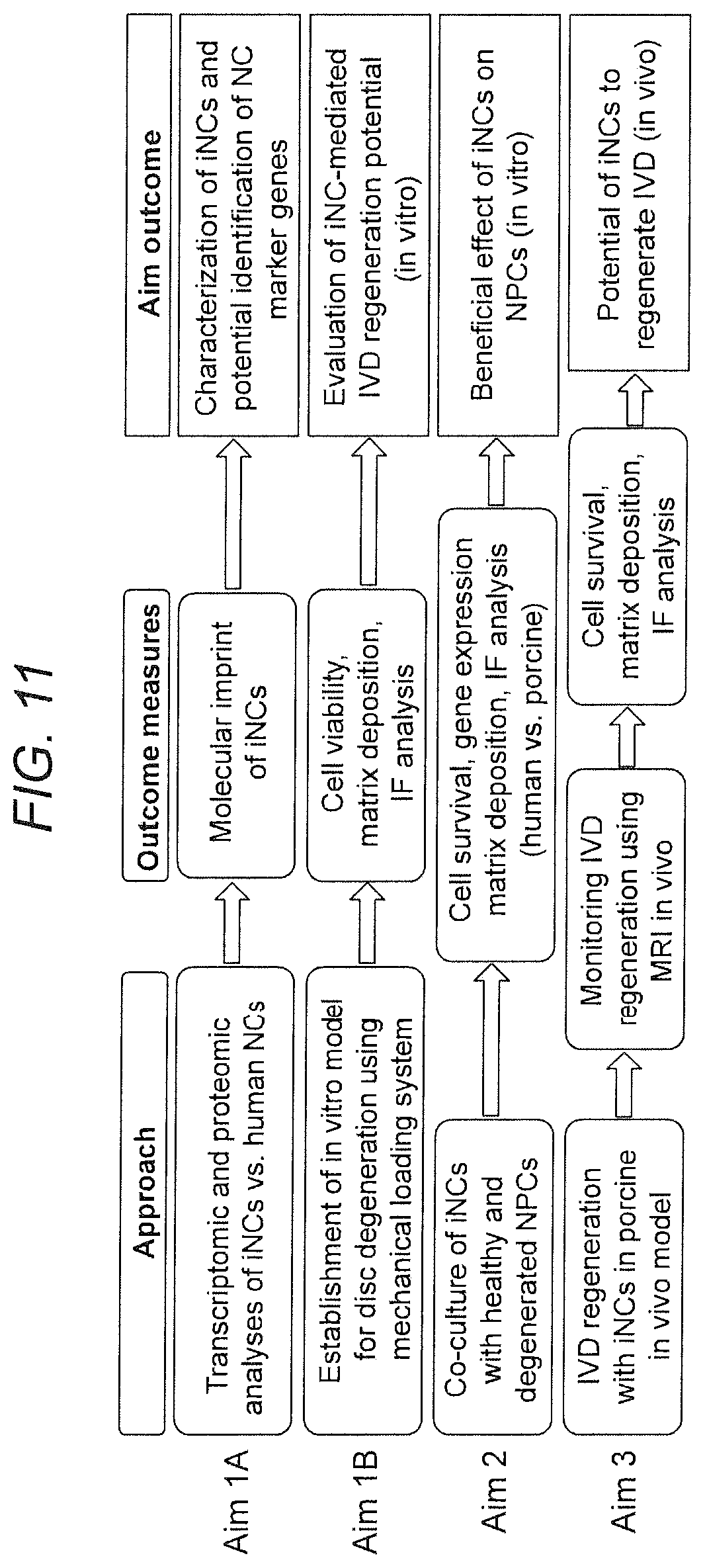
D00014
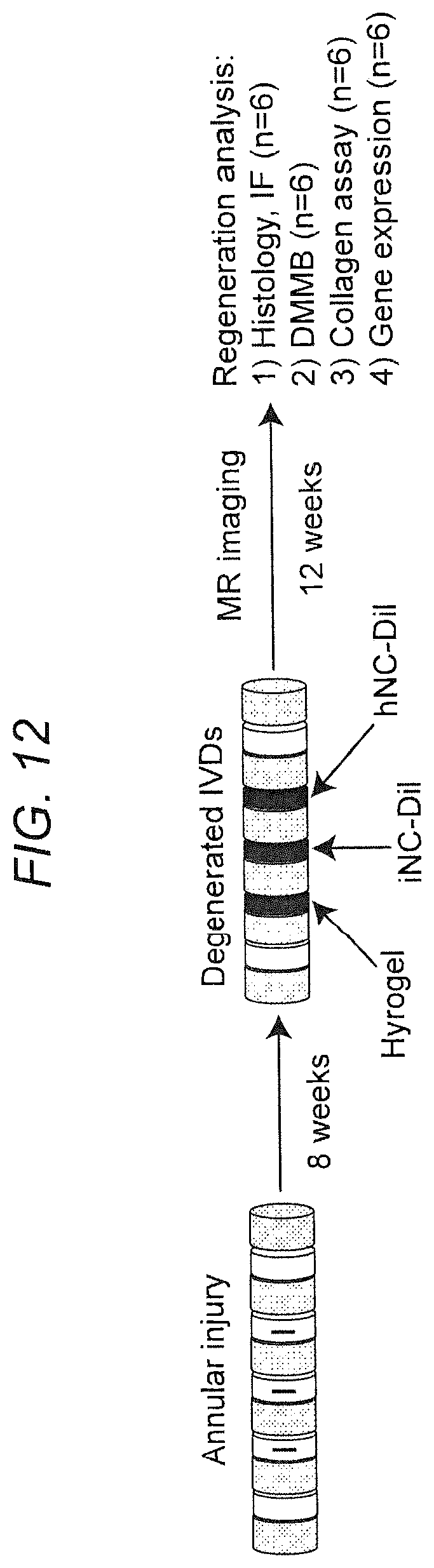
D00015

D00016

D00017

D00018

D00019
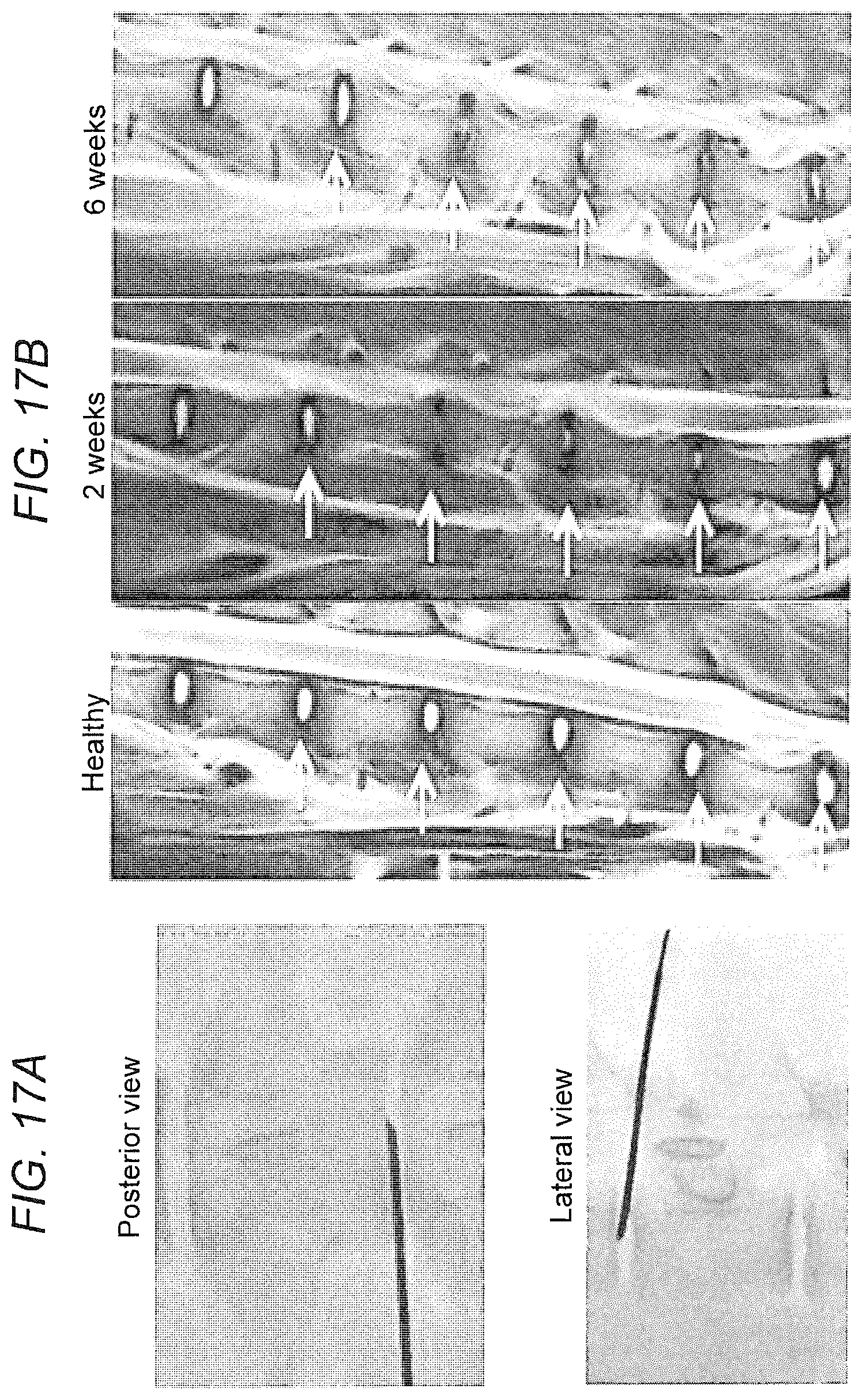
D00020
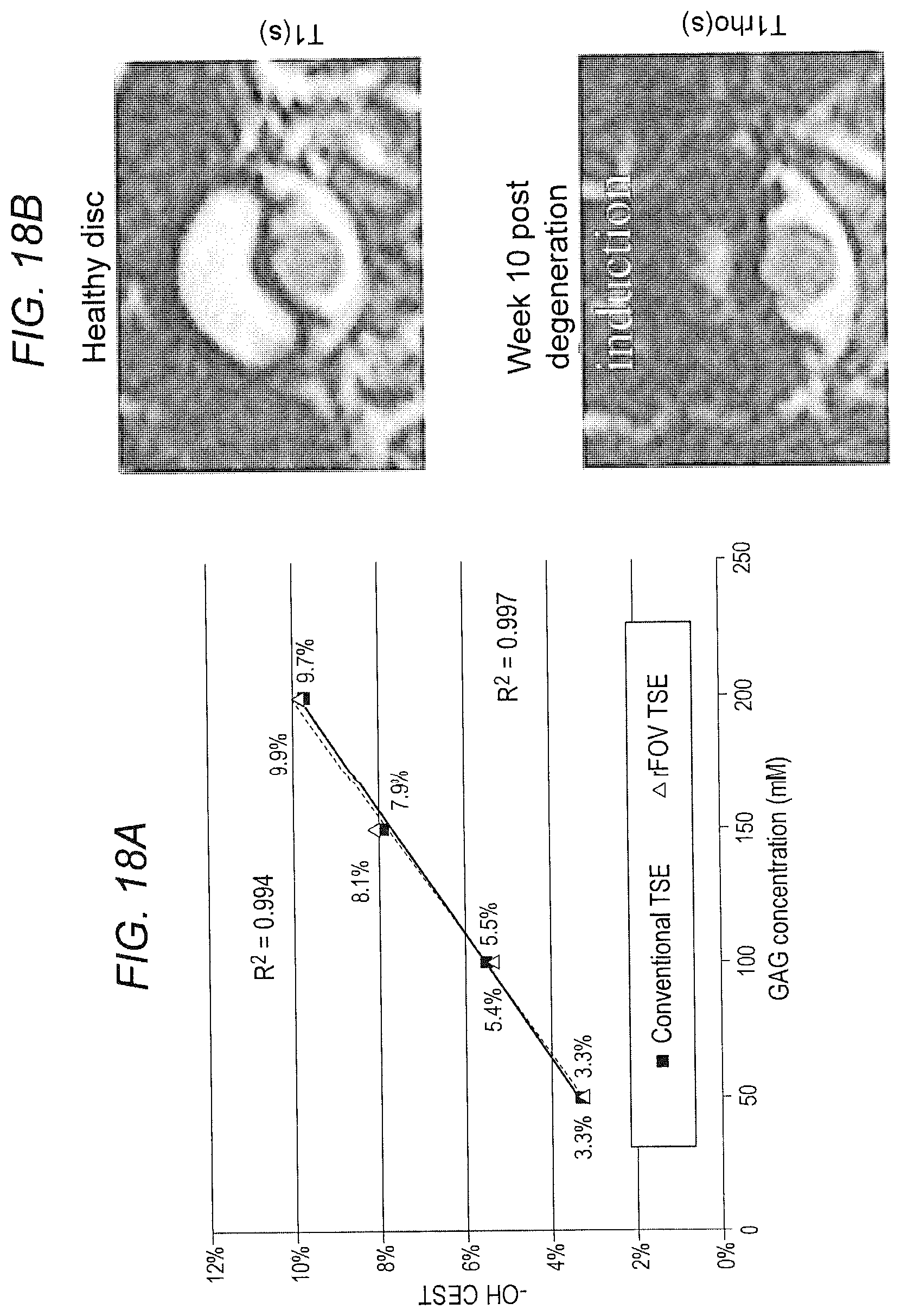
D00021

D00022
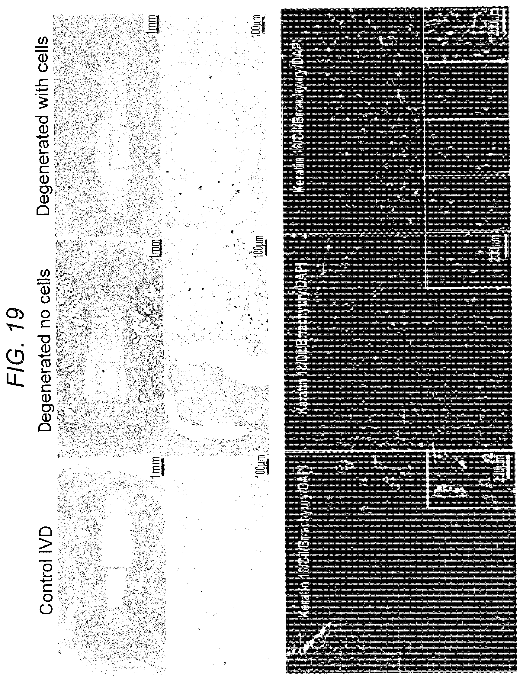
S00001
XML
uspto.report is an independent third-party trademark research tool that is not affiliated, endorsed, or sponsored by the United States Patent and Trademark Office (USPTO) or any other governmental organization. The information provided by uspto.report is based on publicly available data at the time of writing and is intended for informational purposes only.
While we strive to provide accurate and up-to-date information, we do not guarantee the accuracy, completeness, reliability, or suitability of the information displayed on this site. The use of this site is at your own risk. Any reliance you place on such information is therefore strictly at your own risk.
All official trademark data, including owner information, should be verified by visiting the official USPTO website at www.uspto.gov. This site is not intended to replace professional legal advice and should not be used as a substitute for consulting with a legal professional who is knowledgeable about trademark law.