Antibodies Targeting A Ligand From An Immune Checkpoint, With An Fc Fragment Having An Improved Affinity For Cd16a
MONNET; Celine
U.S. patent application number 16/495427 was filed with the patent office on 2020-03-19 for antibodies targeting a ligand from an immune checkpoint, with an fc fragment having an improved affinity for cd16a. The applicant listed for this patent is LABORATOIRE FRAN AIS DU FRACTIONNEMENT ET DES BIOTECHNOLOGIES. Invention is credited to Celine MONNET.
| Application Number | 20200087394 16/495427 |
| Document ID | / |
| Family ID | 59699746 |
| Filed Date | 2020-03-19 |

| United States Patent Application | 20200087394 |
| Kind Code | A1 |
| MONNET; Celine | March 19, 2020 |
ANTIBODIES TARGETING A LIGAND FROM AN IMMUNE CHECKPOINT, WITH AN FC FRAGMENT HAVING AN IMPROVED AFFINITY FOR CD16A
Abstract
Disclosed is an antibody targeting at least one ligand from an immune checkpoint, having a region Fc that is mutated in relation to that of a parent antibody and has an improved affinity for the receptor FcgRIIIa (CD16a) and/or a higher ADCC activity than the parent antibody.
| Inventors: | MONNET; Celine; (LAMBERSART, FR) | ||||||||||
| Applicant: |
|
||||||||||
|---|---|---|---|---|---|---|---|---|---|---|---|
| Family ID: | 59699746 | ||||||||||
| Appl. No.: | 16/495427 | ||||||||||
| Filed: | March 19, 2018 | ||||||||||
| PCT Filed: | March 19, 2018 | ||||||||||
| PCT NO: | PCT/EP2018/056891 | ||||||||||
| 371 Date: | September 19, 2019 |
| Current U.S. Class: | 1/1 |
| Current CPC Class: | C07K 2317/41 20130101; C07K 2317/72 20130101; C07K 2317/732 20130101; C07K 16/2827 20130101; C07K 16/283 20130101; A61K 38/00 20130101; C07K 16/2818 20130101; A61P 35/00 20180101; C07K 2317/52 20130101 |
| International Class: | C07K 16/28 20060101 C07K016/28; A61P 35/00 20060101 A61P035/00 |
Foreign Application Data
| Date | Code | Application Number |
|---|---|---|
| Mar 20, 2017 | FR | 1752285 |
Claims
1-19. (canceled)
20. Antibody directed against a ligand of an immune checkpoint, having a modified Fc fragment compared with that of a parent antibody, and having improved affinity for the FcgRIIIa (CD16a) receptor and/or increased ADCC activity compared with the parent antibody.
21. The antibody according to claim 20, wherein the modified Fc fragment comprises at least one combination of 2 following mutations: i) a mutation selected from among 307N, 326E, 326T, 334N, 334R, 352L, 378V, 378T, 394P, 396L, 397M, 421T, 434Y and 434S; and ii) at least one mutation selected from among 226G, 226Y, 227S, 228L, 228R, 230S, 230T, 230L, 231V, 234P, 241L, 243I, 243L, 246R, 246E, 247T, 248E, 253F, 254F, 255W, 259A, 261R, 262A, 263A, 264E, 266M, 267N, 267G, 274E, 274R, 276S, 278H, 282A, 283G, 284L, 286I, 286Y, 287T, 288E, 288R, 290E, 298N, 302A, 305A, 307P, 308A, 308I, 308G, 309P, 312G, 315D, 316D, 319H, 320T, 320R, 320M, 322E, 323I, 325S, 330V, 333G, 334N, 334R, 336T, 339T, 340E, 343S, 345G, 349S, 349H, 350A 352S, 359A, 361H, 362R, 363I, 366A, 373D, 375R, 377T, 378V, 378T, 379A, 380G, 383R, 385R, 389S, 389T, 389K, 392R, 393A, 393I, 394P, 396L, 397I, 397M, 398P, 405V, 405L, 410R, 412M, 414R, 421T, 421S, 423L, 423Y, 423S, 423P, 428T, 431V, 431T, 434K, 434Y, 434S, 435R, 436H, 439R, 440G, 440N, 442F, 442P and 447N, the numbering being that of the EU Index or Kabat equivalent, and provided that mutation (i) does not take place on the same amino acid as mutation (ii).
22. The antibody according to claim 20, wherein the modified Fc fragment comprises at least one combination of 2 following mutations: i) a mutation selected from among 307N, 326E, 326T, 334N, 334R, 352L, 378V, 378T, 394P, 396L, 397M and 421T; and ii) at least one mutation selected from among 226Y, 227S, 230S, 231V, 234P, 243I, 243L, 246R, 246E, 247T, 248E, 253F, 254F, 255W, 259A, 261R, 262A, 263A, 266M, 267N, 267G, 274E, 274R, 276S, 278H, 282A, 283G, 284L, 286I, 286Y, 287T, 288E, 288R, 290E, 298N, 302A, 305A, 307P, 308A, 308I, 308G, 309P, 312G, 315D, 316D, 319H, 320T, 320R, 320M, 322E, 323I, 325S, 333G, 334N, 334R, 336T, 339T, 340E, 343S, 345G, 349S, 349H, 350A 352S, 359A, 361H, 362R, 363I, 366A, 373D, 375R, 377T, 378V, 378T, 379A, 380G, 383R, 385R, 389S, 389T, 392R, 393A, 393I, 394P, 396L, 397I, 397M, 398P, 405V, 405L, 410R, 412M, 414R, 421T, 421S, 423L, 423Y, 423S, 423P, 428T, 431V, 431T, 434K, 434S, 435R, 436H, 439R, 440G, 440N, 442F, 442P and 447N, the numbering being that of the EU Index or Kabat equivalent, and provided that mutation (i) does not take place on the same amino acid as mutation (ii).
23. The antibody according to claim 21, wherein the modified Fc fragment comprises at least one combination of 2 following mutations: i) a mutation selected from among 378V, 378T, 434Y and 434S; and ii) at least one mutation selected from among 226G, 228L, 228R, 230S, 230T, 230L, 241L, 264E, 307P, 315D, 330V, 362R, 378V, 378T, 389T, 389K, 434Y and 434S, the numbering being that of the EU Index or Kabat equivalent, and provided that mutation (i) does not take place on the same amino acid as mutation (ii).
24. The antibody according to claim 20, wherein the modified Fc fragment comprises at least one combination of 2 following mutations: i) a mutation selected from among 378V, 326E, 397M, 334N and 396L; and ii) at least one mutation selected from among 316D, 397M, 334N, 248E, 231V, 246R, 336T, 421T, 361H, 366A, 439R, 290E, 394P, 307P, 378V, 378T, 286I, 286Y and 298N, the numbering being that of the EU Index or Kabat equivalent, and provided that mutation (i) does not take place on the same amino acid as mutation (ii).
25. The antibody according to claim 20, wherein the modified Fc fragment comprises: either a combination of mutations selected from among N315D/A330V/N361D/A378V/N434Y, N315D/N361D/A378V/N434Y, P230S/N315D/M428L/N434Y, T307A/N315D/A330V/E382V/N389T/N434Y, V259I/N315D/N434Y and T256N/A378V/S383N/N434Y; or the combination of mutations K334N/P352S/V397M/A378V; or a combination of mutations selected from among N315D/A330V/N361D/A378V/N434Y, V259I/N315D/N434Y, K334N/P352S/V397M/A378V and N315D/N361D/A378V/N434Y, and at least one of the following mutations: K290G, Y296W or N434Y.
26. The antibody according to claim 20, wherein the antibody has improved affinity compared with that of the parent antibody, by a ratio of at least 2.
27. Composition comprising antibodies according to claim 20, said modified Fc fragments having N-glycans on their glycosylation site, wherein said N-glycans have a fucosylation level of less than 65%.
28. The composition according to claim 27, said Fc fragments having N-glycans on their Asn 297 glycosylation site, wherein said N-glycans have a glycan structure of biantennary type with short chains, low sialylation, and non-intercalated terminal N-acetylglucosamines.
29. The composition according to claim 27, said Fc fragments having N-glycans on their Asn 297 glycosylation site, wherein said N-glycans have a content higher than 60% for the forms G0+G1+G0F+G1F, the content of forms G0F+G1F being lower than 50%.
30. The composition according to claim 27, said Fc fragments having N-glycans on their Asn 297 glycosylation site, wherein said N-glycans have a content higher than 60% for the forms G0+G1+G0F+G1F, the fucose content being lower than 65%.
31. The composition according to claim 29, said Fc fragments having N-glycans on their Asn 297 glycosylation site, wherein said N-glycans have a content lower than 40% for the forms G1F+G0F.
32. The antibody according to claim 20, wherein the ligand of the immune checkpoint is selected from among PDL1, OX40L, PDL2, CD80, CD86, galectine-9, MHC II, MHC I, HVEM and adenosine.
33. The antibody according to claim 20, wherein the antibody is an anti-PDL1 comprising a light chain variable sequence (VL) and a heavy chain variable sequence (VH) corresponding to sequences VL and VH of the atezolizumab antibody, durvalumab antibody, or avelumab antibody respectively.
34. Products containing: a) an antibody according to claim 20, and b) an antibody directed against an immune checkpoint, having a modified Fc fragment compared with that of a parent antibody, having improved affinity for the FcRn receptor, said immune checkpoint being selected from among PD1, CTLA4, TIM3, LAG3, KIR, BTLA1 and a2AR, as combination products for simultaneous, separate or time-staggered administration, for use thereof in the prevention or treatment of cancers.
35. The products according to claim 34, wherein the ligand of the immune checkpoint according to a) is PDL1 and wherein the immune checkpoint according to b) is PD1.
36. The antibody according to claim 20, wherein the parent antibodies comprise a parent Fc fragment which is a human Fc fragment.
37. Pharmaceutical composition comprising (i) at least one antibody according to claim 20, and (ii) at least one pharmaceutically acceptable excipient.
38. The antibody according to claim 20, for use thereof in the treatment of cancers.
39. The antibody according to claim 20, wherein the antibody has improved affinity compared with that of the parent antibody, by a ratio higher than 5.
Description
[0001] The present invention concerns cancer immunotherapy.
TECHNOLOGICAL BACKGROUND OF THE INVENTION
[0002] Immunotherapy, which entails administering exogenous antibodies to patients, is currently widely used to treat various pathologies and cancers in particular.
[0003] In recent years, knowledge of the biology and immunology of cancers has constantly progressed. It is henceforth recognised that the immune system is involved in antitumor response, particularly through recognition of cancer cells by the immune cells. This recognition can lead to the controlling and even elimination of tumours. However, immune effector cells have receptors on their surface known as immune checkpoints. The purpose of these receptors is to modulate (inhibit or activate) immune response and in particular to maintain self-tolerance. It is now known that cancer cells borrow this escape mechanism to resist immune response, in particular through expression on their surface of ligands of the receptors of said immune checkpoints, which will lead to inhibition of the response of the immune effector cell when it recognises the latter (Pardoll et al., Nat. Rev. Cancer. 12:252-264 (2012)). A new approach to cancer immunotherapy has therefore emerged to counter-attack this phenomenon of immune escape by restoring immune response and hence tumour rejection. In the last few years, antibodies directed against these immune checkpoints have been developed. The chief antibodies that are marketed or under development target: [0004] Cytotoxic T-Lymphocyte Associated antigen 4 (CTLA4): this is the case for Ipilimumab (Yervoy.RTM., Bristol Myers Squibb). This monoclonal antibody causes blockading of the CTLA4 receptor, an inhibitory immune checkpoint present on T lymphocytes, and consequently leads to activation of the immune response of said T lymphocyte. In other words, Ipilimumab blockades the immune escape route of cancer cells by suppressing their inhibitory action on T lymphocytes via their stimulation of the CTLA4 receptor. A clinical study allowed the evidencing, for the first time, that patients suffering from metastatic melanomas treated with an anti-CTLA4 antibody (Ipilimumab), have their lifetime increased (Hodi et al., N Engl. J. Med., 363: 711-723 (2010) and 363:1290 (2010) (erratum); Robert et al., N Engl. J. Med., 364: 2517-2526 (2011)); [0005] Programmed Cell Death Protein 1 (PD1): this is the case for Nivolumab (Opdivo.RTM., Bristol Myers Squibb) and Pembrolizumab (Keytruda.RTM., Merck). This monoclonal antibody causes blockading of the PD1 receptor, an inhibitory immune checkpoint present on T lymphocytes, leading to activation of the immune response of said T lymphocyte. In other words, Nivolumab blockades the immune escape route of cancer cells by suppressing their inhibitory action on T lymphocytes T via their stimulation of the PD1 receptor. In particular, Nivolumab appears to have antitumor activity in patients suffering from metastatic renal cell carcinoma (Motzer et al., American Society of Clinical Oncology 33 (13): 1430-1437 (2014)); or [0006] Lymphocyte Activation Gene 3 (LAG3) (e.g. BMS-986016). LAG3 is an inhibitory immune checkpoint present on T lymphocytes T. It is currently the target of inhibitor development and in particular of monoclonal antibodies. Similar to CTLA4 and PD1, the blockading of this receptor would allow the blockading of its inhibiting effect on the response of effector cells, thereby activating immune response.
[0007] More recently, antibodies directed against ligands of immune checkpoints have been developed. The chief antibodies marketed or under development target the ligand of PD1 (Programmed Cell Death Protein 1) contained on antigen presenting cells, and tumour cells. For example, Atezolizumab/Tecentriq (Genentech/Roche) is an anti-PDL1 antibody and was granted approval in May 2016 for the treatment of patients with bladder cancer. This antibody is an IgG1 modified to be aglycosylated, without effector activity. Nevertheless, to date, most patients show little or no response to such treatment via anti-checkpoint ligand antibodies. In addition, some patients face toxicity reactions in the body after receiving treatment. For example, it was observed in some patients that anti-PDL1 antibodies induce autoantibodies in patients, related to the onset of skin and liver problems for example. Finally, it was observed that some patients become resistant to the treatment.
[0008] At the present time there is a need to optimise the immunotherapy approaches used to blockade these immune checkpoints, notably with a view to obtaining a more efficient and/or less toxic clinical response. There is a particular need to obtain antibodies directed against an immune checkpoint ligand, having improved effector properties and advantageously having an improved half-life in the body allowing extended antitumor effect, whilst being well tolerated by the body. In particular, said antibodies with improved half-life can advantageously be administered in lower dosage with the same or greater efficacy, which would allow limiting of the side effects observed in some patients.
SUMMARY OF THE INVENTION
[0009] The present invention therefore relates to an antibody directed against at least one ligand of an immune checkpoint, having a modified Fc region compared with that of a parent antibody, having improved affinity for the FcgRIIIa (CD16a) receptor and/or increased ADCC activity compared with a parent antibody. Said antibody is also called an anti-ligand antibody . It is adapted for use in cancer treatment.
[0010] The invention also relates to an anti-ligand antibody composition, and to a pharmaceutical composition comprising at least one antibody of the invention (anti-immune checkpoint or anti-ligand). Said composition can be suitable for use in the treatment of cancers.
[0011] The present invention also relates to products containing:
[0012] a) an antibody directed against a ligand of an immune checkpoint, or a composition of antibodies directed against a ligand of an immune checkpoint, and
[0013] b) an antibody directed against an immune checkpoint, having a modified Fc fragment compared with that of a parent antibody, having improved affinity for the FcRn receptor and optionally reduced functional activity mediated by the Fc region, said immune checkpoint being selected from among PD1, CTLA4, TIM3, LAG3, KIR, BTLA1 and a2AR, as combination products for simultaneous, separate or time-staggered administration, for use thereof in the prevention or treatment of cancers. [0014] The invention also concerns: [0015] an anti-immune checkpoint antibody for use thereof in the prevention or treatment of cancers, in combination with an anti-ligand antibody or anti-ligand antibody composition; and [0016] an anti-ligand antibody or anti-ligand antibody composition for use thereof in the prevention or treatment of cancers in combination with an anti-immune checkpoint antibody.
KEY TO FIGURES
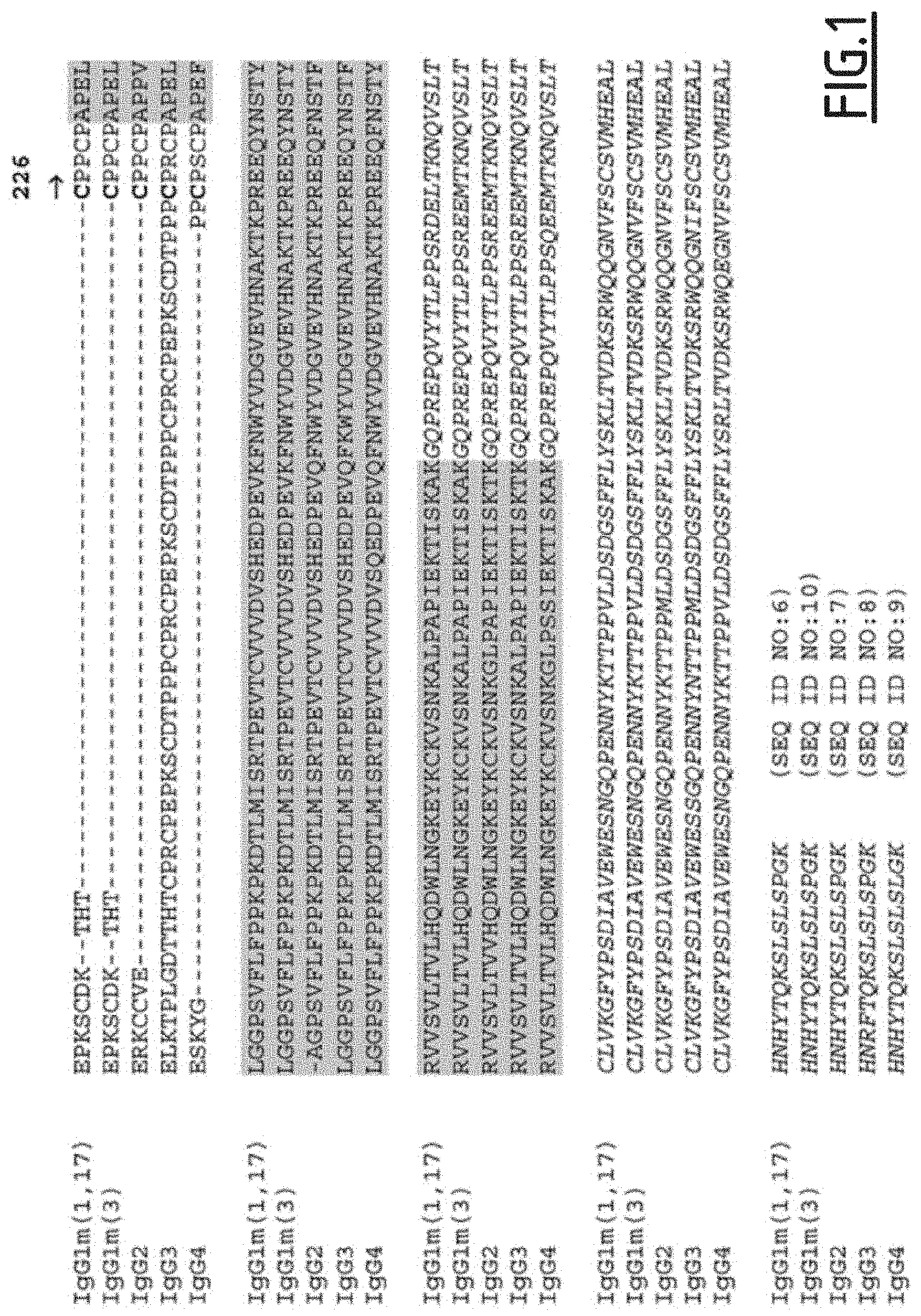
[0017] FIG. 1 gives the alignments of native human IgG1 sequences referring to positions 216 to 447 (as per EU index) with the corresponding human IgG2 (SEQ ID NO: 7), human IgG3 (SEQ ID NO: 8) and human IgG4 (SEQ ID NO: 9) sequences. The IgG1 sequences refer to the G1m1,17 allotype (SEQ ID NO: 6) and G1m3 allotype (SEQ ID NO: 10). The "CH2-CH3 lower hinge" domain of IgG1 starts at cysteine 226 (see arrow). The CH2 domain is highlighted in grey and the CH3 domain is in italics.
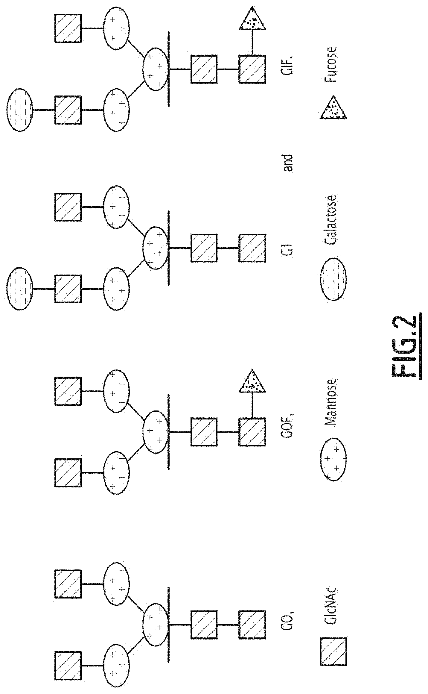
[0018] FIG. 2 shows the glycan structure of the G0, G0F, G1 and G1F forms.
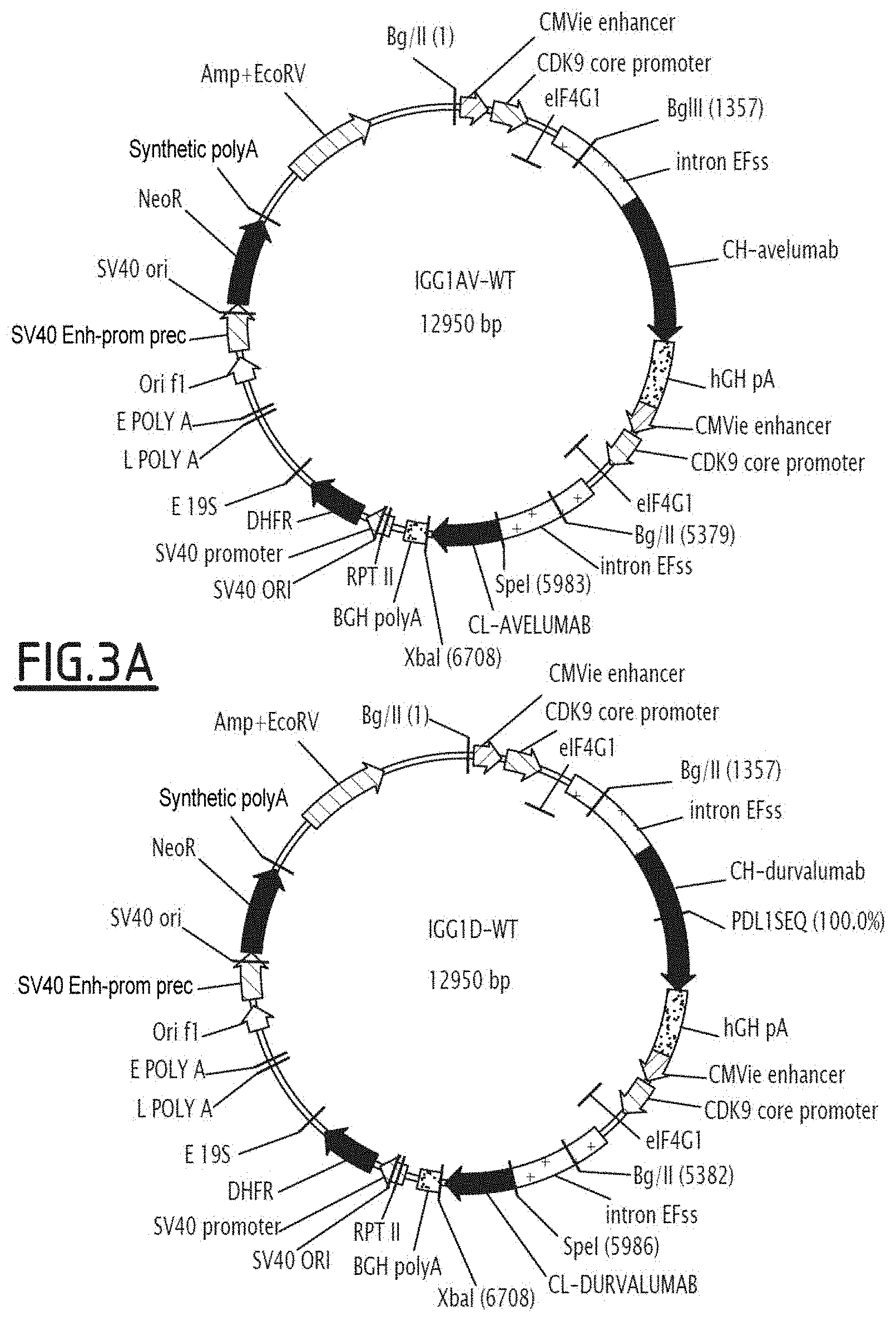
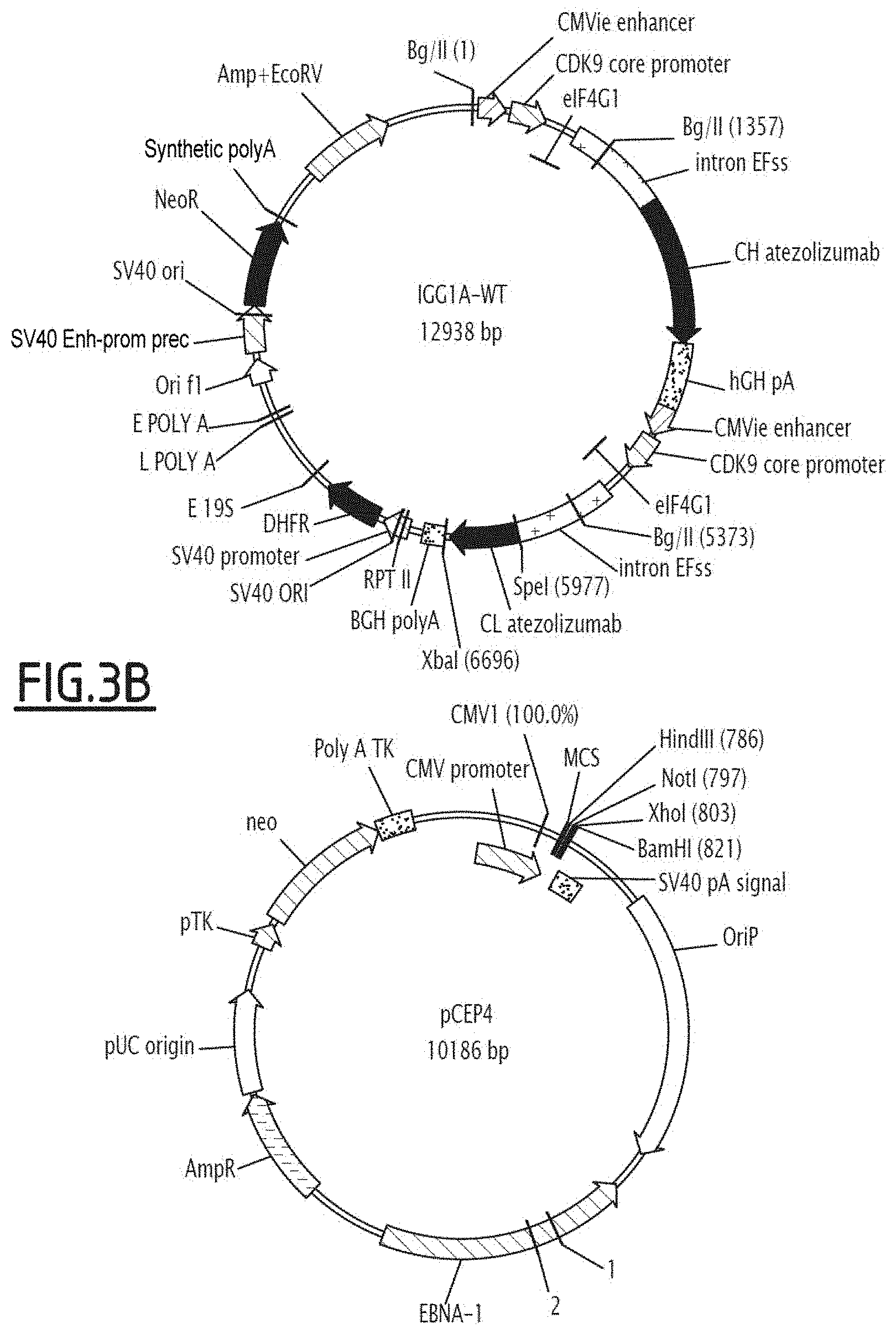
[0019] FIG. 3 shows the expression vectors containing the heavy chain (HC) of anti-PDL1 mutated on the Fc fragment, and the non-modified light chain (LC) of the anti-PDL1 antibody under consideration. FIG. 3A illustrates the IGG1AV-WT and IGG1D-WT vectors, whilst FIG. 3B illustrates the IGG1A-WT and pCEP4 vectors.
[0020] IGG1AV-WT corresponds to the expression vector encoding avelumab; IGG1D-WT corresponds to the expression vector encoding durvalumab; and IGG1A-WT corresponds to the expression vector encoding atezolizumab.
DETAILED DESCRIPTION
Characteristic of the Antibodies
[0021] The present invention relates to an antibody directed against a ligand of an immune checkpoint (anti-ligand antibody), having a modified Fc region compared with that of a parent antibody, having improved affinity for the FcgRIIIa receptor (CD16a) and/or increased ADCC activity compared with the parent antibody.
[0022] The anti-immune checkpoint ligand antibody allows binding with the target tumour cell. For example, by binding to the ligand present on tumour cells (e.g. PD-L1), it allows the recruiting of effector immune cells via the mutated Fc region (having an effector function). This leads to direct cytotoxicity on the tumour cells.
[0023] The present invention preferably relates to an antibody directed against a ligand of an immune checkpoint, said antibody having a mutated Fc region compared with that of a parent antibody, having improved affinity for the FcgRIIIa receptor (CD16a) and/or increased ADCC activity compared with the parent antibody, said mutated Fc region comprising at least one combination of 2 following mutations: [0024] i) a mutation selected from among 307N, 326E, 326T, 334N, 334R, 352L, 378V, 378T, 394P, 396L, 397M, 421T, 434Y and 434S; and [0025] ii) at least one mutation selected from among 226G, 226Y, 227S, 228L, 228R, 230S, 230T, 230L, 231V, 234P, 241L, 243I, 243L, 246R, 246E, 247T, 248E, 253F, 254F, 255W, 259A, 261R, 262A, 263A, 264E, 266M, 267N, 267G, 274E, 274R, 276S, 278H, 282A, 283G, 284L, 286I, 286Y, 287T, 288E, 288R, 290E, 298N, 302A, 305A, 307P, 308A, 308I, 308G, 309P, 312G, 315D, 316D, 319H, 320T, 320R, 320M, 322E, 323I, 325S, 330V, 333G, 334N, 334R, 336T, 339T, 340E, 343S, 345G, 349S, 349H, 350A 352S, 359A, 361H, 362R, 363I, 366A, 373D, 375R, 377T, 378V, 378T, 379A, 380G, 383R, 385R, 389S, 389T, 389K, 392R, 393A, 393I, 394P, 396L, 397I, 397M, 398P, 405V, 405L, 410R, 412M, 414R, 421T, 421S, 423L, 423Y, 423S, 423P, 428T, 431V, 431T, 434K, 434Y, 434S, 435R, 436H, 439R, 440G, 440N, 442F, 442P and 447N, the numbering being that of the EU index or Kabat equivalent, and provided that mutation (i) does not take place on the same amino acid as mutation (ii).
[0026] In a first embodiment, the mutated Fc region of the antibody of the invention comprises at least one combination of 2 following mutations: [0027] i) a mutation selected from among 378V, 378T, 434Y and 434S; and [0028] ii) at least one mutation selected from among 226G, 228L, 228R, 230S, 230T, 230L, 241L, 264E, 307P, 315D, 330V, 362R, 378V, 378T, 389T, 389K, 434Y and 434S, [0029] the numbering being that of the EU index or Kabat equivalent, and provided that mutation (i) does not take place on the same amino acid as mutation (ii).
[0030] According to the preceding paragraph, the mutated Fc region of the antibody of the invention may also comprise an additional mutation selected from among 361D, 428L, 307A, 382V, 259I, 256N and 383N.
[0031] More preferably, the mutated Fc region of the antibody of the invention comprises the combinations of mutations selected from among N315D/A330V/N361 D/A378V/N434Y, N315D/N361 D/A378V/N434Y, P230S/N315D/M428L/N434Y, T307A/N315 D/A330V/E382V/N389T/N434Y, V259I/N315D/N434Y and T256N/A378V/S383N/N434Y.
[0032] In a second embodiment, the present invention relates to an antibody directed against a ligand of an immune checkpoint, said antibody having a mutated Fc region compared with that of a parent antibody, having improved affinity for the FcgRIIIa receptor (CD16a) and/or increased ADCC activity compared with the parent antibody, said mutated Fc region comprising at least one combination of 2 following mutations: [0033] i) a mutation selected from among 307N, 326E, 326T, 334N, 334R, 352L, 378V, 378T, 394P, 396L, 397M and 421T; and [0034] ii) at least one mutation selected from among 226Y, 227S, 230S, 231V, 234P, 243I, 243L, 246R, 246E, 247T, 248E, 253F, 254F, 255W, 259A, 261R, 262A, 263A, 266M, 267N, 267G, 274E, 274R, 276S, 278H, 282A, 283G, 284L, 286I, 286Y, 287T, 288E, 288R, 290E, 298N, 302A, 305A, 307P, 308A, 308I, 308G, 309P, 312G, 315D, 316D, 319H, 320T, 320R, 320M, 322E, 323I, 325S, 333G, 334N, 334R, 336T, 339T, 340E, 343S, 345G, 349S, 349H, 350A 352S, 359A, 361H, 362R, 363I, 366A, 373D, 375R, 377T, 378V, 378T, 379A, 380G, 383R, 385R, 389S, 389T, 392R, 393A, 393I, 394P, 396L, 397I, 397M, 398P, 405V, 405L, 410R, 412M, 414R, 421T, 421S, 423L, 423Y, 423S, 423P, 428T, 431V, 431T, 434K, 434S, 435R, 436H, 439R, 440G, 440N, 442F, 442P and 447N, the numbering being that of the EU index or Kabat equivalent, and provided that mutation (i) does not take place on the same amino acid as mutation (ii).
[0035] More preferably, in this second embodiment, the mutated Fc region of the antibody of the invention comprises at least one combination of 2 following mutations: [0036] i) a mutation selected from among 378V, 378T, 326E, 397M, 334N, 396L, 434Y and 434S; and [0037] ii) at least one mutation selected from among 226G, 228L, 228R, 230S, 230T, 230L, 241L, 264E, 307P, 315D, 316D, 330V, 362V, 397M, 334N, 248E, 231V, 246R, 336T, 421T, 361H, 366A, 439R, 290E, 394P, 307P, 378V, 378T, 286I, 286Y, 298N, 389T, 389K, 434Y and 434S, the numbering being that of the EU index or Kabat equivalent, and provided that mutation (i) does not take place on the same amino acid as mutation (ii).
[0038] More preferably, the mutated Fc region of the antibody of the invention comprises at least one combination of 2 following mutations: [0039] i) a mutation selected from among 378V, 326E, 397M, 334N and 396L; and [0040] ii) at least one mutation selected from among 316D, 397M, 334N, 248E, 231V, 246R, 336T, 421T, 361H, 366A, 439R, 290E, 394P, 307P, 378V, 378T, 286I, 286Y and 298N, the numbering being that of the EU index or Kabat equivalent, and provided that mutation (i) does not take placed on the same amino acid as mutation (ii).
[0041] According to the preceding paragraph, the mutated Fc region of the antibody of the invention may also comprise an additional mutation selected from among 333G, 352S, 423Y, 315D, 412M and 366A.
[0042] More preferably, the mutated Fc region of the antibody of the invention comprises the combinations of mutations selected from among 248E/378V, 333G/378T/397M, 396L/421 T/378V, 396L/421 T, 316D/326E/378V, 298N/378V, 336T/378V, 334N/352S/397M/378V, 286I/378V/423Y, 315D/361H/396L, 231V/378V, 378T/397M/412M, 286Y/352S/378V, 290E/366A/378V, 286I/396L/421T and 334N/352S/397M. More preferably, the mutated Fc region of the antibody of the invention comprises the combination of mutations 334N/352S/397M/378V.
[0043] Preferably, the mutated Fc region of the antibody of the invention comprises at least one combination of 2 mutations according to the first embodiment, and at least one combination according to the second embodiment.
[0044] Therefore, preferably, the mutated Fc region of the antibody of the invention comprises a combination of mutations selected from among N315D/A330V/N361 D/A378V/N434Y, V259I/N315D/N434Y and N315D/N361 D/A378V/N434Y, and a combination of mutations selected from among 248E/378V, 333G/378T/397M, 396L/421T/378V, 396L/421T, 316D/326E/378V, 298N/378V, 336T/378V, 334N/352S/397M/378V, 286I/378V/423Y, 315D/361H/396L, 231V/378V, 378T/397M/412M, 286Y/352S/378V, 290E/366A/378V, 286I/396L/421T and 334N/352S/397M.
[0045] Alternatively, preferably, the mutated Fc region of the antibody of the invention comprises a combination of mutations selected from among N315D/A330V/N361 D/A378V/N434Y, V259I/N315D/N434Y, K334N/P352S/V397M/A378V et N315D/N361 D/A378V/N434Y, as well as one of the following mutations: V240M, L242K, L242G, L242F, F243L, E258R, T260A, V262A, K290G, Y296W, S298R or V302R.
[0046] Preferably, the mutated Fc region of the antibody of the invention comprises a combination of mutations selected from among DN315D/A330V/N361 D/A378V/N434Y, V259I/N315D/N434Y, K334N/P352S/V397M/A378V and N315D/N361 D/A378V/N434Y, as well as at least one of the following mutations: K290G, Y296W or N434Y.
[0047] Preferably, the mutated Fc region of the antibody of the invention comprises a combination of mutations selected from among K334N/P352S/V397M/A378V and N315D/N361D/A378V/N434Y, as well as at least one of the following mutations Y296W, N434Y or Y296W/N434Y.
[0048] Preferably, the mutated Fc region of the antibody of the invention comprises a combination of mutations selected from among Y296W/K334N/P352S/V397M/A378V, Y296W/N315D/N361 D/A378V/N434Y, K334N/P352S/V397M/A378V/N434Y and Y296W/K334N/P352S/V397M/A378V/N434Y.
[0049] By antibody it is meant a tetramer composed of two identical heavy chains each of 50-70 kDa (called H chains) and of two identical light chains each of 25 kDa (called L chains) bonded together by intra- and inter-chain disulfide bridges. This tetramer comprises at least two variable regions at the N-terminal end of each chain (called VL for the light chains and VH for the heavy chains) and a constant region at the C-terminal end called Fc formed of a single domain called CL for the light chain and of three or four domains for the heavy chain called CH1, CH2, CH3 and possibly CH4.
[0050] Each domain comprises about 110 amino acids and is comparably structured. The 2 heavy chains are bonded by disulfide bridges at CH2 and each heavy chain is bonded to a light chain via a disulfide bridge between CH1 and CL. The region which determines the specificity of the antibody for the antigen is carried by the variable parts and it is these parts which are responsible for recognition of the antigen. In each variable region, three loops are joined together to form a binding site to the antigen. Each of the loops is called a Complement Determining Region (CDR). As for the constant regions, these preferably bind to the Fc receptors Fc (FcR) of the effector cells. The assembling of the chains composing an antibody allows a characteristic Y-shaped three-dimensional structure to be defined where: [0051] the base of the Y corresponds to the constant region Fc, or Fc fragment: it is recognised by the Fc receptors to mediate the effector functions of the antibody; and [0052] the ends of the Y arms correspond to the respective assembling of the variable region of a light chain and variable region of a heavy chain, said ends forming the Fab region and determining the specificity of the antibody for the antigen.
[0053] There are five types of heavy chains (alpha, gamma, delta, epsilon, mu), which determine the classes of immunoglobulins (IgA, IgG, IgD, IgE, IgM). The light chain group comprises two sub-types, lambda and kappa. The kappa and lambda light chains are shared by all classes and sub-classes. In man, the proportion of kappa and lambda produced lies in a ratio of 2 to 1.
[0054] IgGs are the most abundant immunoglobulins in serum (75-80% of circulating antibodies). They are present in the form of monomers and have a half-life of 21 days on average.
[0055] There are four types of gamma heavy chains, which determines four IgG sub-classes (IgG1 for gamma1, IgG2 for gamma2, IgG3 for gamma3 and IgG4 for gamma4). These four sub-classes differ in the number and variable positions of the disulfide bridges (Basic and Clinical Immunology, 8.sup.th Edition, Daniel P. Stites, Abba I. Terr and Tristram G. Parslow (Eds.), Appleton & Lange, Norwalk, Conn., 1994, page 71 and Chapter 6).
[0056] The four sub-classes of human IgGs also differ in biological activity, despite highly homologous structures (more than 95% sequence homology for the Fc regions).
[0057] Antibodies notably comprise full-length immunoglobulins, monoclonal antibodies, multi-specific antibodies, chimeric antibodies, humanized antibodies and fully human antibodies.
[0058] The term "Fc" or "Fc region" or "Fc fragment" designates the constant region of an antibody of total length with the exclusion of the first domain of immunoglobulin constant region (CH1-CL). Therefore, Fc refers to the two last domains (CH2 and CH3) of an IgG constant region, and to the flexible N-terminal hinge of these domains. For human IgG1, the Fc region comprises the domains CH2 and CH3 as well as the lower hinge region between CH1 and CH2. Therefore, the Fc region corresponds to the C226 residue as far as its carboxy-terminal end i.e. the residues at position 226 to 447, as per EU Index numbering or Kabat equivalent. The analogue domains for other IgG sub-classes can be determined from the alignment of amino acid sequences of the heavy chains or heavy chain fragments of IgG sub-classes with human IgG1 (see FIG. 1). The Fc region used may additionally comprise part of the upper hinge region located between positions 216 to 226 as per the EU Index or Kabat equivalent; in this case, the Fc region used corresponds to the residues at position 216 to 447, 217 to 447, 218 to 447, 219 to 447, 220 to 447, 221 to 447, 222 to 447, 223 to 447, 224 to 447 or 225 to 447, where the numbering is that of the EU Index or Kabat equivalent. Preferably in this case, the Fc region used corresponds to the residues at position 216 to 447 as per the numbering of the EU Index or Kabat equivalent.
[0059] Preferably, the Fc region used is selected from among the sequences SEQ ID NO: 1, 2, 3, 4 and 5. Preferably, the Fc region of the parent antibody has the sequence SEQ ID NO: 1. The sequences represented in SEQ ID NO: 1, 2, 3, 4 and 5 are devoid of an N-terminal hinge region.
[0060] The sequences represented in SEQ ID NO: 6, 7, 8, 9 and 10 correspond to the sequences represented in SEQ ID NO: 1, 2, 3, 4 and 5 respectively with their N-terminal hinge regions. Therefore, in one particular embodiment, the Fc region of the parent antibody is selected from among sequences SEQ ID NO: 6, 7, 8, 9 and 10.
[0061] Preferably, the Fc region of the parent antibody has a sequence corresponding to positions 1-232, 2-232, 3-232, 4-232, 5-232, 6-232, 7-232, 8-232, 9-232, 10-232 or 11-232 of sequence SEQ ID NO: 6.
[0062] Fv fragment designates the smallest fragment maintaining the binding properties of the antibody. It is solely composed of light chain VL and heavy chain VH variable regions, it therefore fixes the antigen with the same affinity as the whole antibody.
[0063] By position it is meant a position in the sequence of amino acids. For the Fc region, the positions are numbered in accordance with the EU Index or Kabat equivalent.
[0064] By amino acid or residue it is meant one of the 20 natural amino acids or natural analogues.
[0065] The term immune checkpoints refers to receptors located on the surface of immune effector cells capable of inhibiting (inhibitory immune checkpoints) or activating immune response (stimulatory immune checkpoints) after engaging with their ligands.
[0066] The immune checkpoint is preferably selected from among GITR, OX40, PD1, CTLA4, TIM3, LAG3, KIR, BTLA1 and a2AR.
[0067] By activation receptor in the present invention it is meant a surface receptor which, after interaction with its ligand, causes triggering of a signalling pathway leading to activation of immune response. The stimulatory immune checkpoint is preferably selected from among GITR and OX40.
[0068] GITR, also called tumour necrosis factor receptor superfamily member 18 (TNFRSF18) or activation-inducible TNFR family receptor (AITR), is a protein having its expression increased when the T cells are activated.
[0069] OX40, also called CD134 or Tumour necrosis factor receptor superfamily, member 4 (TNFRSF4), is a protein which is not constitutively expressed on naive T cells. It is expressed when the latter are activated. Its ligand OX40L is also expressed on activated antigen presenting cells.
[0070] By inhibitory receptor in the present invention it is meant a surface receptor which, after interaction with its ligand, causes triggering of a signalling pathway leading to inactivation of immune response.
[0071] Preferably, the immune checkpoint is inhibitory. More preferably, it is selected from among PD1, CTLA4, TIM3, LAG3, KIR, BTLA1 and a2AR.
[0072] PD1 (Programmed cell Death factor 1) is an inhibitory receptor of the CD28 family expressed on the surface of activated T and B lymphocytes and Natural Killers. Its role is to limit the activity of the effector cells in secondary lymphoid tissues or tumours, thereby imparting it with a major tumour resistance mechanism. PD1 inhibits lymphocyte functions when it is engaged with one of its ligands, PDL1 (or B7-H1 or CD274) or PDL2. PDL1 is a molecule expressed on the surface of tumour cells. In the event of chronic exposure to the PDL1 ligand (e.g. the case with cancer), the expression of PD1 on the surface of effector cells is increased, leading to a phenomenon of anergy. Anti-PD1 antibodies are used in the treatment of cancers such as lung cancer, non-small cell lung cancer (NSCLC), mesotheliomas, bladder cancer, colorectal cancer, metastatic colorectal cancer, bladder cancer, breast cancer, head and neck cancers, testicular cancer, endometrial cancer, oesophageal cancer, thymus cancer, haematological cancer, advanced haematological cancer such as non-Hodgkin's lymphoma, Hodgkin's lymphoma, chronic lymphoid leukaemia, multiple melanomas, acute myeloid leukaemia, brain tumours, glioblastomas, solid tumours, gastric adenocarcinomas, germ cell tumours, hepatocellular carcinoma, melanomas, metastatic melanomas, lymphomas, diffuse large B-cell lymphomas (DLBCL), follicular lymphomas, non-resectable or metastatic melanomas, or advanced renal cell carcinoma.
[0073] CTLA4 (Cytotoxic T-lymphocyte-associated antigen 4) is an inhibitory receptor solely expressed on the surface of T lymphocytes. Its role is to regulate the first activation steps of T lymphocytes. 48 hours after activation of the T lymphocytes via their receptor (T Cell Receptor--TCR), CTLA4 engages with its ligands (CD80 or CD86) expressed on the surface of the antigen presenting cells (APCs) at the lymph nodes and sometimes at tumours. This causes a signalling cascade leading to inhibition of the T lymphocytes. Anti-CTLA4 antibodies are used in the treatment of cancers such as lung cancer, non-small cell lung cancer (NSCLC), small cell lung carcinoma, breast cancer, pancreatic cancer, prostate cancer, gastric cancer, renal cancer, head and neck cancer, liver cancer, metastatic or non-resectable melanomas, skin melanoma with lymph node involvement, renal carcinoma, myelomas, lymphomas, hepatocellular carcinoma, brain metastases, solid tumours, mesotheliomas, lymphomas or melanomas.
[0074] TIM3 (T-cell immunoglobulin and mucin-domain containing-3) is a receptor expressed on the surface of IFN .gamma.-secreting T lymphocytes. One of its ligands is galectine-9, a protein overexpressed in tumour cells. The engaging of TIM3 with galectine-9 leads to inhibition of immune response.
[0075] LAG3 (lymphocyte activation gene 3) also called CD223 is a molecule expressed on the surface of T lymphocytes. Its only known ligand is the class II Major Histocompatibility Complex (MHC II) which can be overexpressed in some cancers but also by antigen presenting cells (APC) (macrophages and dendritic cells) infiltrated at tumours. The engaging of LAG3 with its receptor causes an anergy phenomenon.
[0076] KIR (Killer-cell immunoglobulin-like receptor) is a molecule expressed on the surface of Natural Killers, T lymphocytes and APCs. When KIR binds to its ligand, the class I MHC (MHC I), the effector response of the Natural Killers is attenuated at tumour sites.
[0077] BTLA1 (B and T lymphocyte attenuator), also called CD272, is a molecule expressed on the surface of lymphocytes. Its ligand, the HVEM molecule (herpesvirus entry mediator), is expressed in some types of tumours (notably the case in melanomas).
[0078] a2ARs are expressed in different types of immune effector cells, in particular in T lymphocytes and in endothelial cells. When a2AR binds to its ligand adenosine (which accumulates in tumours), the CD4+ cells express FOXP3 and differentiate into regulatory T cells, consequently leading to inhibition of immune response.
[0079] Preferably, the ligand of the immune checkpoint is selected from among OX40L, PDL1, PDL2, CD80, CD86, galectine-9, MHC II, MHC I, HVEM and adenosine.
[0080] More preferably, the immune checkpoint is selected from among PD1 and CTLA4.
[0081] More preferably, the immune checkpoint is PD1. Therefore, preferably, the anti-immune checkpoint antibody of the invention is an anti-PD1 or anti-CTLA4 antibody. More preferably, the anti-immune checkpoint antibody is an anti-PD1 antibody.
[0082] Preferably, the ligand is selected from among PDL1, PDL2, CD80 and CD86. Preferably, the ligand is PDL1 or PDL2, preferably PDL1. Therefore, preferably, the anti-ligand antibody of the invention is an anti-PDL1, anti-PDL2, anti-CD80 or anti-CD86 antibody. Preferably, the anti-ligand antibody of the invention is an anti-PDL1 antibody.
[0083] The anti-PDL1 antibody of the invention may comprise a variable region corresponding to the sequence of an Fv fragment of a known anti-PDL1 antibody e.g. the atezolizumab antibody, durvalumab antibody or avelumab antibody. Therefore, the anti-PDL1 antibody of the invention may comprise a light chain variable sequence (VL) and a heavy chain variable sequence (VH) corresponding to the VL and VH sequences of the atezolizumab antibody, durvalumab antibody or avelumab antibody respectively.
[0084] The sequences are the following: [0085] the VH and VL sequences of the atezolizumab antibody are the sequences SEQ ID NO:11 and 12 respectively; [0086] the VH and VL sequences of the durvalumab antibody are the sequences SEQ ID NO:13 and 14 respectively; and [0087] the VH and VL sequences of the avelumab antibody are the sequences SEQ ID NO:15 and 16 respectively.
[0088] Therefore, preferably, the anti-PDL1 antibody of the invention comprises a VH of sequence SEQ ID NO:11 and a VL of sequence SEQ ID NO:12.
[0089] Alternatively, preferably, the anti-PDL1 antibody of the invention comprises a VH of sequence SEQ ID NO:13 and a VL of sequence SEQ ID NO:14.
[0090] Alternatively, preferably, the anti-PDL1 antibody of the invention comprises a VH of sequence SEQ ID NO:15 and a VL of sequence SEQ ID NO:16.
[0091] The term parent antibody is used to define the reference antibody which can be of natural or synthetic origin. In the context of the present invention, the parent antibody comprises an Fc region called parent Fc region . Said parent Fc region is selected from the group of wild-type Fc regions and fragments thereof. By wild-type (WT), it is meant here a sequence of amino acids or nucleotide sequence found in nature i.e. which is of natural origin, including allelic variations, and which has not been intentionally modified via molecular biology techniques such as mutagenesis. For example, the Fc regions of wild-type particularly refer to the Fc region of IgG1 having the sequence SEQ ID NO:1 (allotype G1m1,17), the Fc region of IgG2 having the sequence SEQ ID NO: 3, the Fc region of IgG3 having the sequence SEQ ID NO: 4, the Fc region of IgG4 having the sequence SEQ ID NO: 5, and the Fc region of IgG1 having the sequence SEQ ID NO: 1 (allotype G1m3). The Fc regions of wild-type also refer to the Fc regions corresponding to sequences SEQ ID NO: 6 to SEQ ID NO: 10. Preferably, the parent antibody comprises a parent Fc region which is a human Fc region, preferably an Fc region of human IgG1 or human IgG2. The parent antibody may also comprise modifications of amino acids pre-existing in the Fc region (e.g. an Fc mutant) compared with the Fc regions of wild-type.
[0092] By immune effector cells , it is meant cells which carry out the immune mechanism and express an Fc receptor (FcR). Those cells notably considered as effector cells are lymphocytes including Natural Killer cells (NK), macrophages, monocytes, neutrophils, eosinophils, basophils, mastocytes, dendritic cells including Langerhans cells and platelets.
[0093] By mutation it is meant a change of at least one amino acid in the sequence of a polypeptide, in particular a change of at least one amino acid in the Fc region of the parent antibody. The antibody obtained then comprises a mutated Fc region compared with that of the parent antibody. Preferably, mutation is a substitution, insertion or deletion of at least one amino acid at a particular position. The mutated Fc regions can have several mutations, affecting several amino acids, preferably two to ten.
[0094] By substitution it is meant the replacement of an amino acid by another amino acid at a particular position in a sequence of the parent antibody. For example, substitution 434S refers to a variant antibody (or mutant), here a variant for which an amino acid at position 434 is replaced by serine. Preferably, the following mutation wording is used: 434S or N434S , and means that the parent antibody comprises asparagine at position 434, which is replaced by serine in the variant. In the event of a combination of substitutions, the preferred format is the following: 259I/315D/434Y or V259I/N315D/N434Y . This means that there are three substitutions in the variant, at positions 259, 315 and 434, and that the amino acid at position 259 of the parent antibody, namely valine, is replaced by isoleucine, that the amino acid at position 315 of the parent antibody, namely asparagine, is replaced by aspartic acid, and that the amino acid at position 434 of the parent antibody, namely asparagine, is replaced by tyrosine.
[0095] By deletion of amino acids or deletion , it is meant the suppression of an amino acid at a particular position in a sequence of the parent antibody. For example, E294del or 294del designates the suppression of glutamic acid at position 294.
[0096] By insertion of amino acid or insertion , it is meant the addition of an amino acid at a particular position in a sequence of the parent antibody. For example, the insertion G>235-236 designates an insertion of glycine between positions 235 and 236.
[0097] A mutated Fc variant of the invention can be generated by any well-known mutagenesis method. For example, via overlap extension PCR using two sets of primers adapted to integrate the targeted mutation(s) with the codon(s) encoding the desired amino acid. Alternatively, de novo synthesis of genes containing the nucleotide sequence comprising the mutations of interest, can be used.
[0098] Throughout the present application, the numbering of the residues in the Fc region is that of the immunoglobulin heavy chain in accordance with the EU Index or Kabat equivalent in Kabat et al. (Sequences of Proteins of Immunological Interest, 5.sup.th Ed. Public Health Service, National Institutes of Health, Bethesda, Md., 1991). The expression EU Index or Kabat equivalent refers to the EU numbering of the residues of the human IgG1, IgG2, IgG3 or IgG4 antibody as given on the IMGT website: http://www.imgt.org/IMGTScientificChart/Numbering/Hu_IGHGnber.html).
[0099] The affinity of the mutated Fc region of the antibody of the invention for the FcgRIIIa receptor (CD16a) is increased compared with that of the parent antibody.
[0100] Preferably, this affinity is improved compared with that of the parent antibody, by a ratio of at least 2, preferably higher than 5, preferably higher than 10, preferably higher than 15, preferably higher than 20, preferably higher than 25, preferably higher than 30.
[0101] By improved affinity for the FcgRIIIa receptor (CD16a) , it is meant an increase in the in vivo or in vitro binding affinity of the mutated Fc of the invention for the FcgRIIIa receptor (CD16a) compared with the parent antibody. The FcgRIIIa receptor (CD16a) is involved in ADCC and has V/F polymorphism at position 158.
[0102] Preferably, the mutated Fc region of the antibody of the invention also has modified affinity, compared with that of the parent antibody, for at least one of the following receptors: the C1q complement component, FcgRIIa (CD32a) and FcgRIIb (CD32b). The C1q complement component is involved in complement-dependent cytotoxicity (CDC). The FcgRIIa receptor (CD32a) is involved in platelet activation and phagocytosis; it has H/R polymorphism at position 131.
[0103] Preferably, the mutated Fc region of the antibody of the invention has increased affinity, compared with that of the parent antibody, for at least one of the following receptors: C1q complement component, FcgRIIa (CD32a) and FcgRIIb (CD32b). By increased affinity for a receptor, it is meant an increase in the binding affinity, in vivo or in vitro, of the mutated Fc region of the invention for said receptor compared with the parent antibody. In this case, this affinity is improved, compared with that of the parent antibody, by a ratio of at least 2, preferably higher than 5, preferably higher than 10, preferably higher than 15, preferably higher than 20, preferably higher than 25, preferably higher than 30.
[0104] Preferably, the mutated Fc region of the antibody of the invention also has affinity for the C1q complement that is increased compared with that of the parent antibody. Therefore, preferably, the antibody of the invention has increased CDC activity compared with that of the parent antibody.
[0105] The affinity of an antibody for an FcR can be assessed using methods well-known in the prior art. For example, persons skilled in the art can determine affinity (Kd) using surface plasmon resonance (SPR). Alternatively, skilled persons can conduct an appropriate ELISA assay. An appropriate ELISA assay allows comparison between the binding forces of the parent Fc and mutated Fc. The detected signals specific to the mutated Fc and parent Fc are compared.
[0106] The present invention also relates to a composition comprising antibodies directed against a ligand of an immune checkpoint, having a modified Fc fragment compared with that of a parent antibody, and having improved affinity for CD16a and/or increased ADCC activity compared with the parent antibody, wherein said modified Fc fragments have N-glycans on their glycosylation site, said N-glycans having a fucosylation level of less than than 65%, preferably less than 60%, preferably less than 55%, preferably less than 50%, further preferably less than 45%, preferably less than 40%, preferably less than 35%, preferably less than 30%, preferably less than 25%, preferably less than 20%. This composition is called composition of the invention .
[0107] By level of fucosylation, it is meant the ratio of N-glycans present on the Fc fragments having a fucose residue, compared with the total quantity of N-glycans of the Fc fragments in an antibody composition.
[0108] Preferably, said antibody composition comprises a single type of antibody comprising a mutated Fc region. In other words, the composition comprises antibody molecules of same sequence.
[0109] Preferably, the antibody composition of the invention is scarcely fucosylated. By scarcely fucosylated it is meant a composition which comprises antibodies in which the Fc fragments have N-glycans on their glycosylation site (Asn 297) which have a fucosylation level of less than 65%, preferably less than 60%, preferably less than 55%, preferably less than 50%, further preferably less than 45%, preferably less than 40%, preferably less than 35%, preferably less than 30%, preferably less than 25%, preferably less than 20%. Said N-glycans preferably have glycan structures of biantennary type with short chains and low sialylation. Preferably, the glycan structure has non-intercalated terminal GlcNAcs (N-Acetylglucosamine). Preferably, the glycan structure is selected from among the forms, G0F, G1 and G1F such as shown in FIG. 2.
[0110] Therefore, preferably, said N-glycans have glycan structures of biantennary type, with short chains, low sialylation, non-intercalated terminal GlcNAcs. More particularly, the antibody composition has a sialic acid content of less than 25%, 20%, 15%, or 10%, preferably 5%, 4% 3% or 2%. By sialylation percentage, it is meant the ratio of N-glycans present on the Fc fragments having a sialic acid residue, compared with the total quantity of N-glycans of the Fc fragments in an antibody composition.
[0111] One preferred antibody composition of the invention has a content higher than 60%, preferably higher than 80% for the forms G0+G1+G0F+G1F, on the understanding that the content of the forms G0F+G1F is less than 50%, preferably less than 40%, preferably less than 30%. Preferably, the N-glycan content is higher than 60% for the forms G0+G1+G0F+G1F, the fucose content being less than 65%.
[0112] The antibodies of the invention directed against a ligand of an immune checkpoint can be prepared using any method well-known in the prior art. Once the encoding nucleic acids thereof have been obtained, the antibodies of the invention can be prepared using any method known in the art.
[0113] In one embodiment, the nucleic sequences can be cloned in host cells and then expressed. The nucleic sequences can also be incorporated in an expression vector. A wide variety of suitable host cell lines can be used including, but not limited thereto, mammalian cells, bacteria, insect cells and yeasts.
[0114] The host cells, as nonlimiting examples, can be YB2/0 (ATCC, CRL-1662), SP2/0, YE2/0, PERC6 cells, CHO cell lines, in particular CHO-K-1, CHOS, CHO-LeclO, CHO-Lecl, CHO-Lecl3, CHO Pro-5, CHO dhfr-, Wil-2, Jurkat, Vero, COS-7, HEK particularly 293-HEK, BHK, KGH6, NSO, SP2/0-Ag 14, P3X63Ag8.653, C127, JC, LA7, ZR-45-30, hTERT, NM2C5 or UACC-812. In one preferred embodiment of the invention, the antibody is expressed in the YB2/0 cell.
[0115] Alternatively, the host cells can be cells of a non-human transgenic organism, in particular cells of transgenic animals modified to produce the antibody in their milk, or else cells of transgenic plants modified to produce the antibody. With respect to cells of transgenic animals modified to produce the antibody in their mik, the expression of DNA sequences encoding the antibody of the invention, directed against a ligand of an immune checkpoint, is controlled by a mammalian casein promoter or mammalian whey promoter, said promoter not naturally controlling transcription of said gene, and the DNA sequences also containing a secretion sequence of the protein. The secretion sequence comprises a secretion signal interpositioned between the coding sequence and the promoter. The animal can be selected for example from among sheep, goats, does, ewes or cows.
[0116] In this embodiment, preferably whole anti-PDL1 IgG1s mutated according to the invention can be generated by production in the milk of a transgenic animal e.g. a transgenic goat, and purified by extracting the milk. For this purpose, the sequence encoding the heavy chain and the sequence encoding the light chain are prepared in an expression vector under the control of a promoter specific to the mammary glands e.g. a mammalian casein promoter allowing the production and secretion of the antibody to be directed into the milk of the mammary glands. Said method is particularly described in application EP0741515.
[0117] The present invention also relates to products (hereafter products of the invention ) containing:
[0118] a) an antibody of the invention directed against a ligand of an immune checkpoint, or a composition of the invention, and
[0119] b) an antibody directed against an immune checkpoint, having a modified Fc fragment compared with that of a parent antibody, having improved affinity for the FcRn receptor and optionally reduced functional activity mediated by the Fc region, said immune checkpoint being selected from among PD1, CTLA4, TIM3, LAG3, KIR, BTLA1 and a2AR, as combination products for simultaneous, separate or time-staggered administration, for use thereof in the prevention or treatment of cancers.
[0120] This anti-immune checkpoint antibody b) particularly allows binding to the target immune cell. For example, by binding to the receptor present on the T lymphocytes infiltrating tumours (e.g. PD1), it prevents binding between PD1 (present on T lymphocytes) and PDL1 (present on tumour cells). The antibody is then termed a neutralising antibody.
[0121] The anti-immune checkpoint antibody b) has a modified Fc fragment imparting thereto greater affinity for the FcRn receptor.
[0122] The FcRn receptor corresponding to the neonatal Fc receptor is a protein composed of a heavy chain encoded by the FcRn gene (called FCGRT in Man) and of a light chain, the molecule of .beta.2-microglobulin. It can bind the Fc region of IgGs and has the characteristic of increasing the half-life of IgGs which attach thereto. FcRn can be found in different organisms including, but not limited thereto, humans, mice, rats, rabbits and monkeys.
[0123] By greater affinity for FcRn it is meant increased binding affinity, in vivo or in vitro, of the mutated Fc region of the invention for FcRn, compared with that of the parent antibody. The capability of the mutated Fc region of the invention to bind with a FcRn receptor can be assessed in vitro by ELISA assay, as described for example in patent application WO2010/106180. Increased binding to FcRn translates as an improvement in serum retention in vivo et, and hence an increase in half-life.
[0124] Preferably, the anti-immune checkpoint antibody b) (having greater affinity for the FcRn receptor) comprises at least two mutations, said mutations being selected from among: [0125] (i) a modification selected from among 378V, 378T, 434Y and 434S; and [0126] (ii) at least one modification selected from among 226G, P228L, P228R, 230S, 230T, 230L, 241 L, 264E, 307P, 315D, 330V, 362R, 378V, 378T, 389T, 389K, 434Y and 434S, the numbering being that of the EU Index or Kabat equivalent, and provided that mutation (i) does not take place on the same amino acid as mutation (ii).
[0127] Preferably, the anti-immune checkpoint antibody b) has an Fc region comprising at least one combination of mutations selected from among 226G/315D/434Y, 230S/315D/434Y, 230T/315D/434Y, 230T/264E/434S, 230T/389T/434S, 241 L/264E/378V, 241L/264E/434S, 250A/389K/434Y, 259I/315D/434Y, 284E/378T/396L, 264E/378V/434Y, 345D/330V/434Y, 315D/382V/434Y and 378V/383N/434Y compared with the Fc region of said parent antibody, the numbering being that of the EU Index or Kabat equivalent.
[0128] Preferably, the anti-immune checkpoint antibody b) has an Fc region comprising at least one mutation selected from among 226G, 227L, 230S, 230T, 230L, 231T, 241L, 243L, 250A, 256N, 259I, 264E, 265G, 267R, 290E, 294del, 303A, 305A, 307P, 307A, 308I, 315D, 322R, 325S, 327V, 330V, 342R, 347R, 352S, 361D, 362R, 362E, 370R, 378V, 378T, 382V, 383N, 386R, 386K, 387T, 389T, 389K, 392R, 395A, 396L, 397M, 403T, 404L, 415N, 416K, 421T, 426T, 428L, 433R, 434Y, 434S and 439R compared with the Fc region of said parent antibody, the numbering being that of the EU Index or Kabat equivalent.
[0129] Preferably, the anti-immune checkpoint antibody b) has an Fc region comprising a combination of mutations selected from among 307A/315D/330V/382V/389T/434Y, 256N/378V/383N/434Y, 345D/330V/361 D/378V/434Y, 259I/315D/434Y, 230S/315D/428L/434Y, 241 L/264E/307P/378V/433R, 250N389K/434Y, 305A/315D/330V/395N343Y, 264E/386R/396L/434S/439R, 315D/330V/362R/434Y, 294del/307P/434Y, 305N315D/330V/389K/434Y, 315 D/327V/330V/397M/434Y, 230T/241 L/264E/265G/378V/421 T, 264E/396U415N/434S, 227L/264E/378V/434S, 264E/378T/396L, 230T/315D/362R/426T/434Y, 226G/315D/330V/434Y, 230L/241 L/243L/264E/307P/378V, 250A/315D/325S/330V/434Y, 290E/315D/342R/382V/434Y, 241 L/315D/330V/392R/434Y, 241 L/264E/307P/378W/434S, 230T/264E/403T/434S, 264E/378V/416K, 230T/315D/362E/434Y, 226G/315D/434Y, 226G/315D/362R/434Y, 226G/264E/347R/370R/378V/434S, 308I/315D/330V/382V/434Y, 230T/264E/378V/434S, 231T/241 L/264E/378T/397M/434S, 230L/264E/378W/434S, 230T/315D/330V/386K/434Y, 226G/315D/330V/389T/434Y, 267R/307P/378V/421 T/434Y, 230S/315D/387T/434Y, 230S/264E/352S/378V/434S and 230T/303A/322R/389T/404U434S compared with said parent antibody, the numbering being that of the EU Index or Kabat equivalent.
[0130] Preferably, the anti-immune checkpoint antibody b) has an Fc region comprising the combinations of mutations selected from among N315D/A330V/N361 D/A378V/N434Y, P230S/N315D/M428L/N434Y, E294del/T307P/N434Y, T307A/N315 D/A330V/E382V/N389T/N434Y, V259I/N315D/N434Y, V259I/E294Del/N315D/N434Y and T256N/A378V/S383N/N434Y.
[0131] The anti-immune checkpoint antibody b) can have reduced functional activity mediated by the Fc region, compared with that of the parent antibody.
[0132] By functional activity mediated by the Fc region it is meant the effector functions mediated by the Fc region. Included in said functional activities mediated by the Fc region are antibody-dependent cell-mediated cytotoxicity (ADCC), complement-dependent cytotoxicity (CDC), antibody-dependent cell phagocytosis (ADCP), endocytosis activity, cytokine secretion or a combination of at least two of these activities. Preferably, the functional activity mediated by the Fc region under consideration in the invention is ADCC. This functional activity can be evaluated with methods well known in the prior art.
[0133] In particular, the functional activity mediated by the Fc region is reduced compared with that of the parent antibody, by a ratio of at least 2, preferably higher than 5, preferably higher than 10, preferably higher than 15, preferably higher than 20, preferably higher than 25, preferably higher than 30.
[0134] The mutated Fc region of the antibody b) of the invention preferably has reduced affinity for at least one of the receptors of the Fc region (FcR) selected from among C1q complement and the receptors FcgRIIIa (CD16a), FcgRIIa (CD32a) and FcgRIIb (CD32b). The receptors of the Fc region that are involved are: [0135] C1q involved in CDC activity, [0136] FcgRIIIa receptor (CD16a) involved in ADCC and has V/F polymorphism at position 158, [0137] FcgRIIa receptor (CD32a) involved in platelet activation and phagocytosis, and has H/R polymorphism at position 131, and [0138] FcgRIIb receptor (CD32b) involved in inhibition of cell activity.
[0139] The affinity of an antibody for a FcR can be evaluated with methods well known in the prior art. For example, persons skilled in the art can determine affinity (Kd) using surface plasmon resonance (SPR). Alternatively, skilled persons can conduct an appropriate ELISA assay. An appropriate ELISA assay allows comparison between the binding forces of the parent Fc and the mutated Fc. The detected signals specific to the mutated Fc and parent Fc are compared.
[0140] When the anti-immune checkpoint antibody b) has reduced functional activity mediated by the Fc region, it allows neutralisation of the binding between the immune checkpoint and its ligand (e.g. PD1 and PDL1), without having effector activity. It thereby allows blockading of the immune checkpoint.
[0141] Preferably, the anti-immune checkpoint antibody b) has an Fc region at least having the mutation del294.
[0142] Preferably, in another embodiment, the anti-immune checkpoint antibody b) is aglycosylated. For example, it can be mutated on asparagine 297 by an amino acid such as alanine preventing glycosylation. Therefore, preferably, the anti-immune checkpoint antibody b) has a mutated Fc region having mutation N297A compared with the parent antibody.
[0143] Preferably, the products of the invention contain:
[0144] a) an antibody of the invention directed against PDL1, or a composition of the invention comprising antibodies directed against PDL1, and
[0145] b) an antibody directed against PD1, having a modified Fc fragment compared with that of a parent antibody, having improved affinity for the FcRn receptor, and optionally reduced functional activity mediated by the Fc region,
[0146] as combination products for simultaneous, separate or time-staggered administration, for use thereof in the prevention or treatment of cancers.
[0147] The antibody directed against PD1 (antibody b) is such as described in the foregoing and preferably has at least the mutation del294.
[0148] Preferably, when the anti-immune checkpoint antibody b) is an anti-PD1 antibody, it comprises a variable region corresponding to the sequence of an Fv fragment of a known anti-PD1 antibody, for example the nivolumab antibody or pembrolizumab antibody. Therefore, the anti-PD1 antibody of the invention may comprise a light chain variable sequence (VL) and a heavy chain variable sequence (VH) corresponding to the VL and VH sequences of the nivolumab antibody or pembrolizumab antibody respectively.
[0149] The sequences are the following: [0150] the VH and VL sequences of the nivolumab antibody are the sequences SEQ ID NO: 17 and 18 respectively; and [0151] the VH and VL sequences of the pembrolizumab antibody are the sequences SEQ ID NO: 19 and 20 respectively.
[0152] Therefore, preferably, the anti-PD1 antibody of the invention comprises a VH of sequence SEQ ID NO: 17 and a VL of sequence SEQ ID NO: 18.
[0153] Alternatively, preferably, the anti-PD1 antibody of the invention comprises a VH of sequence SEQ ID NO: 19 and a VL of sequence SEQ ID NO: 20.
[0154] The invention also concerns a method for treating cancers, which comprises the administering to a patient of an anti-ligand antibody (preferably anti-PDL1) of the invention, or a composition of the invention (preferably a composition of anti-PDL1 antibodies).
[0155] Any route of administration can be envisaged, in particular parenteral routes such as the intravenous, intramuscular, sub-cutaneous, intradermal, topical routes, or via mucosal route e.g. by inhalation. The enteral (oral, rectal) and intrathecal routes are also possible. Preferably, the intravenous route is used.
[0156] The antibodies of the invention are generally formulated in pharmaceutical compositions comprising pharmaceutically acceptable excipients.
[0157] The invention also relates to a pharmaceutical composition comprising (i) at least one anti-immune checkpoint antibody of the invention, or a composition of the invention, or products of the invention and (ii) at least one pharmaceutically acceptable excipient.
[0158] By pharmaceutical composition it is meant a composition having curative or preventive properties with regard to human or animal diseases.
[0159] A further subject of the invention is the use of an anti-ligand antibody of the invention (preferably anti-PDL1), or of a composition of the invention (preferably of anti-PDL1 antibodies), or of products of the invention or of a pharmaceutical composition such as described in the preceding paragraph, to treat cancers.
[0160] The pharmaceutical compositions can be in any galenic form adapted to the chosen route of administration.
[0161] Preferably, they contain a pharmaceutically acceptable excipient for a formulation that can be injected. In particular this may concern isotonic, sterile formulas, saline solutions or freeze-dried compositions which, when sterilised water or physiological saline is added thereto accordingly, allow the forming of injectable solutes.
[0162] The pharmaceutical forms suitable for injection comprise sterile aqueous solutions or dispersions, oily formulations, and sterile powders for extemporaneous preparation of sterile injectable solutions or dispersions. In all cases, the form must be sterile, and must be fluid insofar as it is to be injected via syringe. It must be stable under production and storage conditions and must be protected against contaminating action of microorganisms such as bacteria and fungi.
[0163] The dispersions of the invention can be prepared in glycerol, liquid polyethylene glycols or mixtures thereof, or in oils. Under normal conditions of storage and use, these preparations contain a preserving agent to prevent the growth of microorganisms.
[0164] The pharmaceutically acceptable excipient can be a solvent or dispersion medium. Suitable fluidity can be maintained for example through the use of a surfactant. The prevention of action by microorganisms can be obtained via various antibacterial and antifungal agents. In many cases it will be preferable to include isotonic agents. Extended absorption of the injectable compositions can be obtained through the use of absorption-delaying agents in the compositions.
[0165] The sterile injectable solutions are prepared by incorporating the active substances in required amount in the suitable solvent with several of the other ingredients listed above, optionally followed by filter sterilisation. As a general rule, dispersions are prepared by incorporating the various sterilised active ingredients in a sterile excipient which contains the basic dispersion medium and other required ingredients among those listed above. Regarding sterile powders for the preparation of sterile injectable solutions, the preferred preparation methods are vacuum drying and freeze drying. When formulated, the solutions are to be administered in a manner compatible with the dosage formulation and in a therapeutically effective amount. The formulations are easily administered in a variety of galenic forms such as the injectable solutions described above, but drug release capsules and similar can also be used. For parenteral administration in an aqueous solution for example, the solution must be suitably buffered and the liquid diluent made isotonic with sufficient saline solution or glucose. These particular aqueous solutions are especially suitable for intravenous, intramuscular, subcutaneous and intraperitoneal administration. In this respect, the sterile aqueous media able to be used are known to skilled persons.
[0166] The therapeutically effective dose specific to a particular patient will depend on a variety of factors, including the disorder being treated and seriousness of the disease, the activity of the specific compound employed, the specific composition used, the age, body weight, general health, gender and food diet of the patient, the time of administration, route of administration, excretion rate of the specific compound used, length of treatment, or parallel medication.
Indications
[0167] The immune system normally recognises tumour cells as foreign elements, which consequently leads to an antitumor immune response. However, it happens that cancer cells set up escape strategies from the immune system; they are therefore no longer recognised or eliminated by the immune system. The immune checkpoints expressed on the surface of immune effector cells form a major pathway for tumour escape. These molecules, when they are inhibitory (the case for example with PD1, CTLA4, LAG3, TIM3, KIR, BTLA1 and a2AR), have the function of attenuating immune response when they engage with their ligands which are often expressed by tumour cells.
[0168] By cancer it is meant any physiological condition characterised by abnormal cell proliferation.
[0169] The antibodies of the invention, the compositions of the invention, the products and the pharmaceutical composition of the invention are therefore used to treat different types of cancers. Examples of cancers notably include non-small cell lung cancer (NSCLC), non-resectable or metastatic melanomas, advanced renal cell carcinomas, bladder cancer, kidney cancer, melanomas, lung cancers, lymphomas, mesotheliomas, colorectal cancer, metastatic colorectal cancer, breast cancer, gastric cancer, head and neck cancers, brain tumours, glioblastomas, solid tumours, endometrial cancers, oesophageal cancers, gastric adenocarcinomas, germ cell tumours, testicular cancer, hepatocellular carcinoma, thymus cancer, diffuse large B-cell lymphomas (DLBCL), haematoiogical cancers, advanced haematological cancers (such as non-Hodgkin's lymphoma, Hodgkin's lymphoma, chronic lymphoid leukaemia, multiple melanomas, acute myeloid leukaemia), astrocytoma, uveal melanoma, solid sarcomas, cancers of ovarian epithelium, primary peritoneal cancers, cancers of the Fallopian tubes, cervical cancer, anal cancer, ovarian cancer, urogenital cancer, urothelial cancer, genitourinary cancer, urogenital neoplasms, thoracic tumours, adrenocortical carcinomas, biliary cancer, follicular lymphomas, pancreatic cancer, prostate cancer, cerebral metastases, liver cancer, cervical adenocarcinomas, gastro-intestinal stromal tumours, metastatic brain cancers, Merkel cell carcinomas, synovial sarcoma, fibrosarcoma, this list not being exhaustive.
[0170] The list of sequences described in the present application is the following:
TABLE-US-00001 SEQ ID NO: Definition Sequence 1 Fc of IgG1 CPPCPAPELLGGPSVFLFPP G1m1, 17 KPKDTLMISRTPEVTCVVVD VSHEDPEVKFNWYVDGVEVH NAKTKPREEQYNSTYRVVSV LTVLHQDWLNGKEYKCKVSN KALPAPIEKTISKAKGQPRE PQVYTLPPSRDELTKNQVSL TCLVKGFYPSDIAVEWESNG QPENNYKTTPPVLDSDGSFF LYSKLTVDKSRWQQGNVFSC SVMHEALHNHYTQKSLSLSPGK 2 Fc of IgG2 CPPCPAPPVAGPSVFLFPPK PKDTLMISRTPEVTCVVVDV SHEDPEVQFNWYVDGVEVHN AKTKPREEQFNSTFRVVSVL TVVHQDWLNGKEYKCKVSNK GLPAPIEKTISKTKGQPREP QVYTLPPSREEMTKNQVSLT CLVKGFYPSDIAVEWESNGQ PENNYKTTPPMLDSDGSFFL YSKLTVDKSRWQQGNVFSCS VMHEALHNHYTQKSLSLSPGK 3 Fc of IgG3 CPRCPAPELLGGPSVFLFPP KPKDTLMISRTPEVTCVVVD VSHEDPEVQFKWYVDGVEVH NAKTKPREEQYNSTFRVVSV LTVLHQDWLNGKEYKCKVSN KALPAPIEKTISKTKGQPRE PQVYTLPPSREEMTKNQVSL TCLVKGFYPSDIAVEWESSG QPENNYNTTPPMLDSDGSFF LYSKLTVDKSRWQQGNIFSC SVMHEALHNRFTQKSLSLSPGK 4 Fc of IgG4 CPSCPAPEFLGGPSVFLFPP KPKDTLMISRTPEVTCVVVD VSQEDPEVQFNWYVDGVEVH NAKTKPREEQFNSTYRVVSV LTVLHQDWLNGKEYKCKVSN KGLPSSIEKTISKAKGQPRE PQVYTLPPSQEEMTKNQVSL TCLVKGFYPSDIAVEWESNG QPENNYKTTPPVLDSDGSFF LYSRLTVDKSRWQEGNVFSC SVMHEALHNHYTQKSLSLSLGK 5 Fc of IgG1 G1m3 CPPCPAPELLGGPSVFLFPP KPKDTLMISRTPEVTCVVVD VSHEDPEVKFNWYVDGVEVH NAKTKPREEQYNSTYRVVSV LTVLHQDWLNGKEYKCKVSN KALPAPIEKTISKAKGQPRE PQVYTLPPSREEMTKNQVSL TCLVKGFYPSDIAVEWESNG QPENNYKTTPPVLDSDGSFF LYSKLTVDKSRWQQGNVFSC SVMHEALHNHYTQKSLSLSPGK 6 Fc of IgG1 EPKSCDKTHTCPPCPAPELL G1m1, 17 GGPSVFLFPPKPKDTLMISR (with hinge TPEVTCVVVDVSHEDPEVKF region) NWYVDGVEVHNAKTKPREEQ YNSTYRVVSVLTVLHQDWLN GKEYKCKVSNKALPAPIEKT ISKAKGQPREPQVYTLPPSR DELTKNQVSLTCLVKGFYPS DIAVEWESNGQPENNYKTTP PVLDSDGSFFLYSKLTVDKS RWQQGNVFSCSVMHEALHNH YTQKSLSLSPGK 7 Fc of IgG2 ERKCCVECPPCPAPPVAGPS (with hinge VFLFPPKPKDTLMISRTPEV region) TCVVVDVSHEDPEVQFNWYV DGVEVHNAKTKPREEQFNST FRVVSVLTVVHQDWLNGKEY KCKVSNKGLPAPIEKTISKT KGQPREPQVYTLPPSREEMT KNQVSLTCLVKGFYPSDIAV EWESNGQPENNYKTTPPMLD SDGSFFLYSKLTVDKSRWQQ GNVFSCSVMHEALHNHYTQK SLSLSPGK 8 Fc of IgG3 ELKTPLGDTTHTCPRCPEPK (with hinge SCDTPPPCPRCPEPKSCDTP region) PPCPRCPEPKSCDTPPPCPR CPAPELLGGPSVFLFPPKPK DTLMISRTPEVTCVVVDVSH EDPEVQFKWYVDGVEVHNAK TKPREEQYNSTFRVVSVLTV LHQDWLNGKEYKCKVSNKAL PAPIEKTISKTKGQPREPQV YTLPPSREEMTKNQVSLTCL VKGFYPSDIAVEWESSGQPE NNYNTTPPMLDSDGSFFLYS KLTVDKSRWQQGNIFSCSVM HEALHNRFTQKSLSLSPGK 9 Fc of IgG4 ESKYGPPCPSCPAPEFLGGP (with hinge SVFLFPPKPKDTLMISRTPE region) VTCVVVDVSQEDPEVQFNWY VDGVEVHNAKTKPREEQFNS TYRVVSVLTVLHQDWLNGKE YKCKVSNKGLPSSIEKTISK AKGQPREPQVYTLPPSQEEM TKNQVSLTCLVKGFYPSDIA VEWESNGQPENNYKTTPPVL DSDGSFFLYSRLTVDKSRWQ EGNVFSCSVMHEALHNHYTQ KSLSLSLGK 10 Fc of IgG1 G1m3 EPKSCDKTHTCPPCPAPELL (with hinge GGPSVFLFPPKPKDTLMISR region) TPEVTCVVVDVSHEDPEVKF NWYVDGVEVHNAKTKPREEQ YNSTYRVVSVLTVLHQDWLN GKEYKCKVSNKALPAPIEKT ISKAKGQPREPQVYTLPPSR EEMTKNQVSLTCLVKGFYPS DIAVEWESNGQPENNYKTTP PVLDSDGSFFLYSKLTVDKS RWQQGNVFSCSVMHEALHNH YTQKSLSLSPGK 11 VH of EVQLVESGGGLVQPGGSLRL atezolizumab SCAASGFTFSDSWIHWVRQA PGKGLEWVAWISPYGGSTYY ADSVKGRFTISADTSKNTAY LQMNSLRAEDTAVYYCARRH WPGGFDYWGQGTLVTVSS 12 VL of DIQMTQSPSSLSASVGDRVT atezolizumab ITCRASQDVSTAVAWYQQKP GKAPKLLIYSASFLYSGVPS RFSGSGSGTDFTLTISSLQP EDFATYYCQQYLYHPATFGQ GTKVEIK 13 VH of EVQLVESGGGLVQPGGSLRL durvalumab SCAASGFTFSRYWMSWVRQA PGKGLEWVANIKQDGSEKYY VDSVKGRFTISRDNAKNSLY LQMNSLRAEDTAVYYCAREG GWFGELAFDYWGQGTLVTVSS 14 VL of EIVLTQSPGTLSLSPGERAT durvalumab LSCRASQRVSSSYLAWYQQK PGQAPRLLIYDASSRATGIP DRFSGSGSGTDFTLTISRLE PEDFAVYYCQQYGSLPWTFG QGTKVEIK 15 VH of avelumab EVQLLESGGGLVQPGGSLRL SCAASGFTFSSYIMMWVRQA PGKGLEWVSSIYPSGGITFY ADTVKGRFTISRDNSKNTLY LQMNSLRAEDTAVYYCARIK LGTVTTVDYWGQGTLVTVSS 16 VL of avelumab QSALTQPASVSGSPGQSITI SCTGTSSDVGGYNYVSWYQQ HPGKAPKLMIYDVSNRPSGV SNRFSGSKSGNTASLTISGL QAEDEADYYCSSYTSSSTRV FGTGTKVTVLG 17 VH of nivolumab QVQLVESGGGVVQPGRSLRL DCKASGITFSNSGMHWVRQA PGKGLEWVAVIWYDGSKRYY ADSVKGRFTISRDNSKNTLF LQMNSLRAEDTAVYYCATND DYWGQGTLVTVSS 18 VL of nivolumab EIVLTQSPATLSLSPGERAT LSCRASQSVSSYLAWYQQKP GQAPRLLIYDASNRATGIPA RFSGSGSGTDFTLTISSLEP EDFAVYYCQQSSNWPRTFGQ GTKVEIK 19 VH of QVQLVQSGVEVKKPGASVKV pembrolizumab SCKASGYTFTNYYMYWVRQA PGQGLEWMGGINPSNGGTNF NEKFKNRVTLTTDSSTTTAY MELKSLQFDDTAVYYCARRD YRFDMGFDYWGQGTTVTVSS 20 VL of EIVLTQSPATLSLSPGERAT pembrolizumab LSCRASKGVSTSGYSYLHWY QQKPGQAPRLLIYLASYLES GVPARFSGSGSGTDFTLTIS SLEPEDFAVYYCQHSRDLPL TFGGGTKVEIK
[0171] The invention is illustrated below with the following examples that are in no way limiting.
Example 1: Production of Anti-PDL1 IgG1 Variants of the Invention in YB2/0 Cells
A/ Construction of Fc Variants:
[0172] Each mutation of interest in the Fc fragment was independently inserted in an expression vector containing the anti-PDL1 heavy chain (containing the variable part of the anti-PDL1 atezolizumab, durvalumab or avelumab antibody, and the constant part of wild-type Fc region) via overlap extension PCR using two sets of primers adapted to integrate the targeted mutation(s) with the codon(s) encoding the desired amino acid. Advantageously, when the mutations to be inserted are close on the Fc sequence, they are added via one same oligonucleotide. The fragments thus obtained by PCR were associated and the resulting fragment amplified by PCR following standard protocols. The PCR product, containing the whole heavy chain of the anti-PDL1 mutated on the Fc fragment, was purified over 1% agarose gel (w/v), digested with appropriate restriction enzymes and cloned in the eukaryote expression vector (HK-Gen EFSS, see FIG. 3) which also contained the non-modified light chain of the anti-PDL1 antibody under consideration.
B/ Production of Variants in the Form of Whole Ig in YB2/0:
[0173] The Fc variants can be prepared in whole IgG1 format in the YB2/0 cell line (ATCC, CRL-1662) with anti-PDL1 specificity. The production steps via cell culture and purification of the antibodies can be conducted following the techniques described in Example 1 of application WO2001/77181, to produce and select antibodies characterized by a low level of fucosylation at their Asn 297 glycosylation site on the Fc.
[0174] The following combinations of mutations are preferably selected to produce the anti-PDL1s in YB2/0:
TABLE-US-00002 TABLE 1 Anti-PDL1 mutants of IgG1 selected in particular with the method described in WO2010/106180 Variant Mutations C6A_69 T307A/N315D/A330V/E382V/N389T/N434Y C6A_78 T256N/A378V/S383N/N434Y T5A_74 N315D/A330V/N361D/A378V/N434Y C6A_74 V259I/N315D/N434Y C6A_60 P230S/N315D/M428L/N434Y T5A_74A N315D/N361D/A378V/N434Y
Example 2: Production of Anti-PDL1 IgG1 Variants According to the Invention in CHOS or HEK Cells, or in the Milk of Transgenic Animals
[0175] The following combinations of mutations are preferably selected:
TABLE-US-00003 TABLE 2 Anti-PDL1 mutants of IgG1 selected with the method described in application WO2016/177984 Variant Mutations G3A-103 K248E, A378V J3A-28 E333G, A378T, V397M J3B-118A P396L, N421T, A378V J3B-118 P396L, N421T A3A-105D G316D, K326E, A378V A3A-14 S298N, A378V G3A-95 I336T, A378V A3A-184A K334N, P352S, V397M, A378V J3B-23 N286I, A378V, F423Y K3B-01 N315D, N361H, P396L G3A-43 A231V, A378V J3A-06 A3781, V397M, V412M J3A-16 N286Y, P352S, A378V O3A-05 K290E, T366A, A378V Q3A-39 N286I, P396L, N421T A3A-184 K334N, P352S, V397M
[0176] Advantageously, the mutants can contain the mutations of the variant T5A-74, C6A-74 or T5A-74A such as given in Table 1, and the mutations of a variant listed in Table 2.
[0177] Alternatively, the mutants may contain the mutations of variant T5A-74, C6A-74 or T5A-74A such as given in Table 1, and one of the following mutations: V240M, L242K, L242G, L242F, F243L, E258R, T260A, V262A, K290G, Y296W, S298R or V302R.
2-1/ Production of Variants in Whole Ig Form in CHO or HEK
[0178] The Fc variants, produced by directed mutagenesis according to the method of Example 1-A, can be prepared in whole IgG1 format in a CHOS cell line (HK-Gen EFSS bicistronic vector) or HEK (using the monocistronic vector pCEP4, a vector containing the light chain and the other similar vector containing the mutated heavy chain, see FIG. 3), with anti-PDL1 specificity. The production steps via cell culture and purification of the antibodies can be conducted following well known techniques and described for example in application WO2016/177984 (Example 2.6 in HEK).
2-2/ Production of Variants in Whole Ig Form in Transgenic Animal Milk
[0179] The mutated anti-PDL1 whole IgG1s can also be generated by production in the milk of a transgenic animal, e.g. a transgenic goat, and purification via milk extraction. For this purpose, the sequence encoding the heavy chain and the sequence encoding the light chain are prepared in an expression vector under the control of a promoter specific to the mammary glands e.g. a mammalian casein promoter, allowing the production and secretion of the antibody to be directed into the milk of the mammary glands. Said method is particularly described in application EP0741515.
Example 3A: Methods Allowing Characterization of Binding to FcgRIIIa, of Antigen Binding, and of Binding to FcRn of the Anti-PDL1 IgG1 Variants of the Invention
[0180] The binding assays to FcgRIIIa, to the PDL1 ligand and to FcRn were conducted following the methods described in application WO2010/106180. In brief, the binding of the IgG1s to FcgRIIIa, to the PDL1 ligand and to FcRn were measured using a conventional ELISA assay.
[0181] Maxisorp immunoplates were coated with PDL1 antigens (pH=7.4) or FcRns (pH=6.0). The solutions of parent anti-PDL1 IgG1 or of each anti-PDL1 IgG1 variant were added to each well to a final concentration of 1.25 .mu.g of IgG/mL for FcRn, or 0.25 .mu.g of IgG/mL for the other receptors, for 1 h at 37.degree. C., then contacted with HRP goat F(ab')2 anti-human IgG for 1h at 37.degree. C. The bonded IgG1s were detected via TMB visualisation by measuring absorbance at 450 nm.
[0182] For binding to FcgRIIIa (CD16a), FcgRIIaH/R (CD32aH/R), FcgRIIb (CD32b) or FcgRI (CD64), Maxisorp or NiNTA immunoplates were coated with FcgRIIIaV/F, FcgRIIaH/R, FcgRIIb or FcgRI, and saturated with 4% PBS-BSA. The solutions of parent anti-PDL1 IgG1 or of each anti-PDL1 Ig G1 variant in a final concentration of 0.5 .mu.g of IgG/mL were contacted with HRP goat F(ab')2 anti-human IgG at the same concentration for 2 hours at ambient temperature, under gentle agitation. The IgGs aggregated to F(ab')2 were incubated under gentle agitation for 1 hour at 30.degree. C. on ELISA plates saturated for FcgR. The plates were then visualised with TMB (Pierce) and absorbances read at 450 nm.
[0183] The variants of Fc C6A_74 and T5A_74A, comprising the variable part of durvalumab or atezolizumab (i.e. the VH and VL sequences of the durvalumab antibody are the sequences SEQ ID NO: 13 and 14 respectively, and the VH and VL sequences of the atezolizumab antibody are the sequences SEQ ID NO: 11 et 12 respectively), were produced in YB2/0 or in CHO as in Example 1:
TABLE-US-00004 Variant Mutations C6A_74 V259I/N315D/N434Y T5A_74A N315D/N361D/A378V/N434Y
[0184] The reference controls were: [0185] Fc-WT produced in YB2/0, [0186] Fc-WT produced in CHO, but also [0187] aglycosylated Fc comprising the mutation N297A (called N297A ) produced in CHO, and [0188] Fc-FES (i.e. comprising the 3 mutations L234F/L235E/P331S).
[0189] The binding thereof to human FcgRIIIa (hCD16aV and hCD16aF) and to human FcRn (hFcRn) was measured.
[0190] The results are as follows:
TABLE-US-00005 Atezolizumab hCD16aV hCD16aF hFcRn hCD64 hCD32aR hCD32b hCD32aH (0.25 .mu.g/ml) (0.25 .mu.g/ml) (1.25 .mu.g/ml) (0.25 .mu.g/ml) (0.25 .mu.g/ml) (0.25 .mu.g/ml) (0.25 .mu.g/ml) ELISA Ratio ELISA Ratio ELISA Ratio ELISA Ratio ELISA Ratio ELISA Ratio ELISA Ratio Ratio/Fc-WT (YB2/0) Anti-PDL1|atezolizumab|Fc-WT (CHOs) 0.21 0.20 2.95 0.90 1.04 0.96 0.99 Anti-PDL1|atezolizumab|C6A-74 (YB2/0) 0.78 0.88 13.72 1.17 1.05 0.94 0.99 Anti-PDL1|atezolizumab|T5A-74A (YB2/0) 1.03 1.57 14.60 1.51 1.04 0.97 1.00 Anti-PDL1|atezolizumab|N297A (CHOs) 0.02 0.22 2.10 0.01 0.11 0.09 0.10 Ratio/Fc-WT (CHOs) Anti-PDL1|atezolizumab|Fc-WT(YB2/0) 4.77 5.07 0.34 1.11 0.97 1.05 1.01 Anti-PDL1|atezolizumab|C6A-74 (YB2/0) 3.72 4.45 4.65 1.30 1.01 0.98 1.00 Anti-PDL1|atezolizumab|T5A-74A (YB2/0) 4.90 7.99 4.95 1.68 1.00 1.02 1.01 Anti-PDL1|atezolizumab|N297A (CHOs) 0.10 1.10 0.71 0.01 0.11 0.10 0.10 Ratio/N297A (CHOs) Anti-PDL1|atezolizumab|Fc-WT (CHOs) 10.1948 0.9062 1.4045 92.5333 9.5110 10.4443 9.9453 Anti-PDL1|atezolizumab|Fc-WT(YB2/0) 48.5989 4.5972 0.4765 103.0556 9.1887 10.9256 9.9969 Anti-PDL1|atezolizumab|C6A-74 (YB2/0) 37.9226 4.0364 6.5353 120.7000 9.6156 10.2278 9.9005 Anti-PDL1|atezolizumab|T5A-74A (YB2/0) 49.9885 7.2382 6.9571 155.1111 9.5505 10.6143 10.0455
TABLE-US-00006 Durvalumab hCD16aV hCD16aF hFcRn hCD64 hCD32aR hCD32b hCD32aH (0.25 .mu.g/ml) (0.25 .mu.g/ml) (1.25 .mu.g/ml) (0.25 .mu.g/ml) (0.25 .mu.g/ml) (0.25 .mu.g/ml) (0.25 .mu.g/ml) ELISA Ratio ELISA Ratio ELISA Ratio ELISA Ratio ELISA Ratio ELISA Ratio ELISA Ratio Ratio/Fc-WT (YB2/0) Anti-PDL1|durvalumab|Fc-WT (CHOs) 0.23 0.17 1.83 0.81 0.99 0.96 1.08 Anti-PDL1|durvalumab|C6A-74 (YB2/0) 0.90 0.86 24.11 1.15 0.98 0.93 0.90 Anti-PDL1|durvalumab|T5A-74A (YB2/0) 1.15 1.70 24.81 1.63 0.99 1.01 1.72 Anti-PDL1|durvalumab|Fc-FES (CHOs) 0.07 0.09 1.61 0.05 0.61 0.48 0.11 Ratio/Fc-WT (CHOs) Anti-PDL1|durvalumab|Fc-WT (YB2/0) 4.27 5.77 0.55 1.23 1.01 1.04 0.93 Anti-PDL1|durvalumab|C6A-74 (YB2/0) 3.83 4.98 13.19 1.42 0.98 0.97 0.83 Anti-PDL1|durvalumab|T5A-74A (YB2/0) 4.91 9.82 13.57 2.01 1.00 1.05 1.60 Anti-PDL1|durvalumab|Fc-FES (CHOs) 0.30 0.52 0.88 0.06 0.61 0.50 0.10 Ratio/Fc-FES (CHOs) Anti-PDL1|durvalumab|Fc-WT (CHOs) 3.3447 1.9144 1.1391 17.6622 1.6330 2.0023 9.8652 Anti-PDL1|durvalumab|Fc-WT (YB2/0) 14.2780 11.0367 0.6230 21.7763 1.6425 2.0883 9.1583 Anti-PDL1|durvalumab|C6A-74 (YB2/0) 12.8024 9.5245 15.0214 25.0716 1.6019 1.9371 8.2253 Anti-PDL1|durvalumab|T5A-74A (YB2/0) 16.4106 18.7997 15.4601 35.4228 1.6305 2.1121 15.7423
[0191] The results show that the variants C6A_74 and T5A_74A both have equivalent binding to hFcRn, which is strongly increased (more than 12 times), compared with their respective WT parent.
[0192] It emerges that all the variants produced in YB2/0 cells have increased binding to hCD16aV and to hCD16aF compared with their respective WT parent produced in CHO cells.
[0193] For the variants produced in YB2/0 cells: [0194] C6A_74 has equivalent or slightly reduced binding to hCD16aV and hCD16aF compared with the WT parent, whilst having strongly increased binding to FcRn; [0195] T5A_74A has equivalent binding to hCD16aV, and slightly increased binding to hCD16aF, whilst having strongly increased binding to FcRn.
[0196] Similar results were obtained with durvalumab and atezolizumab; they show increased binding to hFcRn and to hCD16aV/F.
Example 3B: Methods Allowing Characterization of Binding to FcgRIIIa, of Antigen Binding, and of Binding to FcRn of Other Anti-PDL1 IgG1 Variants of the Invention
[0197] The same assays as those described in Example 3A were conducted for the following variants, comprising the variable part of durvalumab (i.e. with VH and VL sequences of SEQ ID NO: 13 and 14 respectively) and produced in CHO cells:
TABLE-US-00007 Variant Mutations C6A_74 V259I/N315D/N434Y (cited in Ex. 3A) T5A_74A N315D/N361D/A378V/N434Y (cited in Ex. 3A) A3A_184A K334N/P352S/V397M/A378V C6A_74W V259I/Y296W/N315D/N434Y A3A_184E Y296W/K334N/P352S/V397M/A378V T5A_74MA Y296W/N315D/N361D/A378V/N434Y C6A_74G V259I/K290G/N315D/N434Y A3A_184AG K290G/K334N/P352S/V397M/A378V T5A_74AG K290G/N315D/N361D/A378V/N434Y A3A_184AY K334N/P352S/V397M/A378V/N434Y A3A_184EY Y296W/K334N/P352S/V397M/A378V/N434Y
[0198] The parent durvalumab antibody Fc-WT was produced in CHO cells.
[0199] The binding thereof to human and murine PD-L1, to human FcgRIIIa (hCD16aV and hCD16aF), to hFcRn, to human CD64 (hCD64), and to hCD32aH/aR/b, was measured.
[0200] The results are as follows:
[0201] All the anti-PD-L1 antibodies showed strong binding to human PD-L1, and no binding to murine PD-L1.
TABLE-US-00008 ELISA ELISA ELISA Ratio Ratio Ratio hCD16aV hCD16aF hFcRn (0.25 (0.125 (1.25 .mu.g/ml) .mu.g/ml) .mu.g/ml) Ratio/Fc-WT (CHOs) mean mean mean Anti-PDL1|durvalunnab|C6A-74 0.83 0.87 19.07 Anti-PDL1|durvalunnab|C6A-74W 1.69 1.28 18.55 Anti-PDL1|durvalunnab|C6A-74G 1.32 1.10 18.78 Anti-PDL1|durvalunnab|T5A-74A 1.39 1.17 20.05 Anti-PDL1|durvalunnab|T5A-74MA 2.50 2.52 20.07 Anti-PDL1|durvalunnab|T5A-74AG 2.21 2.24 19.53 Anti-PDL1|durvalunnab|A3A-184EY 3.02 5.20 22.70 Anti-PDL1|durvalunnab|A3A-184A 2.52 1.99 21.55 Anti-PDL1|durvalunnab|A3A-184AY 2.38 1.97 26.19 Anti-PDL1|durvalunnab|A3A-184AG 1.46 0.80 17.82 Anti-PDL1|durvalunnab|A3A-184E 2.75 4.26 19.78
TABLE-US-00009 ELISA ELISA ELISA ELISA Ratio Ratio Ratio Ratio hCD64 hCD32aR hCD32b hCD32aH (0.25 (0.25 (0.25 (0.25 .mu.g/ml) .mu.g/ml) .mu.g/ml) .mu.g/ml) Ratio/Fc-WT (CHOs) mean mean mean mean Anti-PDL1|durvalunnab|C6A- 1.17 0.92 0.92 0.87 74 Anti-PDL1|durvalunnab|C6A- 1.26 0.98 0.97 0.94 74W Anti-PDL1|durvalunnab|C6A- 1.69 1.04 0.97 0.99 74G Anti-PDL1|durvalunnab|T5A- 2.09 1.01 0.99 1.03 74A Anti-PDL1|durvalunnab|T5A- 2.16 1.06 1.04 1.11 74MA Anti-PDL1|durvalunnab|T5A- 2.41 1.12 0.99 1.14 74AG Anti-PDL1|durvalunnab|A3A- 5.11 1.10 1.14 1.00 184EY Anti-PDL1|durvalunnab|A3A- 3.91 1.04 1.08 1.25 184A Anti-PDL1|durvalunnab|A3A- 4.25 0.99 0.99 1.30 184AY Anti-PDL1|durvalunnab|A3A- 3.32 0.91 0.88 0.53 184AG Anti-PDL1|durvalunnab|A3A- 4.03 1.08 1.15 1.27 184E
[0202] All the Durvalumab variants (i.e. the variants of format C6A-74, T5A-74 and A3A-184) have equivalent increased binding to FcRn compared with Fc-WT.
[0203] The Durvalumab variants of format A3A-184 show the best affinity for hCD64, followed by the variants of format T5A-74, and finally to a lesser extent by the variants of format C6A-74.
[0204] The Durvalumab variants A3A-184E and A3A-184EY show equivalent increased binding to hCD16aF. They represent the best variants. The Durvalumab variants T5A-74MA, T5A-74AG, A3A-184A, A3A-184AY show increased binding to hCD16aF compared with Fc-WT produced in CHO cells.
[0205] The Durvalumab variants C6A-74, C6A-74W, C6A-74G and T5A-74A compared with Fc-WT produced in CHO cells show equivalent binding to hCD16aF.
[0206] The Durvalumab variants A3A-184E, A3A-184EY, A3A-184A, A3A-184AY and T5A-74MA show equivalent increased binding to hCD16aV compared with Fc-WT.
[0207] The Durvalumab variants C6A-74W, C6A-74G, T5A-74A and T5A-74AG show increased binding to hCD16aV compared with Fc-WT.
[0208] The Durvalumab variants A3A-184AG and C6A-74 compared with Fc-WT produced in CHO cells have equivalent binding to hCD16aV.
[0209] The Durvalumab variants A3A-184A, A3A-184AY, A3A-184E, T5A-74AG and A3A-184EY show slightly increased binding to hCD32aH compared with Fc-WT.
[0210] The Durvalumab variants C6A-74, T5A-74MA, C6A-74W, C6A-74G and T5A-74A compared with Fc-WT show equivalent binding to hCD32aH.
[0211] All the Durvalumab variants advantageously show maintained binding to hCD32b compared with Fc-WT.
[0212] All the Durvalumab variants advantageously show maintained binding to hCD32aR compared with Fc-WT.
[0213] To conclude, the variants A3A-184E and A3A-184EY appear to be the best candidates with the strongest binding to hFcRn and hCD16aFN. The variants A3A-184AY and T5A-74MA also appear to be very good candidates with good binding to hFcRn and hCD16aFN. These four variants show strong binding to hCD64 and good binding to hCD32aH. Therefore, these four variants can advantageously be used in the context of treating cancer with an anti-PDL1.
Example 3C: Method Allowing Characterization of Half-Life
[0214] Pharmacokinetic experiments were also conducted in hFcRn mice KO homozygous for an allele of murine FcRn and heterozygous for a transgene of human FcRn (mFcRn'-hFcRnTg), to evaluate the half-life of the atezolizumab variants (i.e. with respective VH and VL sequences SEQ ID NO: 11 and 12) with Fc C6A_74 or T5A_74A according to the invention.
[0215] For these pharmacokinetic studies, each animal received a single intravenous IgG injection of 5 mg/kg at the retro-orbital sinus, following a similar protocol to the one previously described (Petkova S B, et al. Enhanced half-life of genetically engineered human IgG1 antibodies in a humanized FcRn mouse model: potential application in humorally mediated autoimmune disease. Int Immunol 2006).
[0216] Half-life is generally calculated from plasma concentrations measured during the elimination phase.
[0217] Half-life time can therefore be obtained: [0218] by solving the equation:
[0218] T1/2=(Ln 2.times.Vd)/CL, where:
[0219] Vd=distribution volume=Dose/initial plasma concentration
[0220] CL=Clearance=Dose/AUC (area under curve) [0221] by graphical analysis by determining on the Y-axis (concentration in .mu.g/ml) the time lapse between concentration C1 and concentration C2. It is essential to plot this curve on semi-logarithmic scale to ensure aligning of the experimental points in this last so-called elimination phase. The investigation time length of this curve must be sufficiently long to allow accurate estimation of half-life.
[0222] Once the slope of the elimination phase has been measured (k.sub.e or elimination rate constant), the half-life can be calculated as follows:
T1/2=Ln 2/k.sub.e=0.693/k.sub.e
[0223] The mean residence time, MRT, can also be measured This translates the time the variant is present in the body.
[0224] MRT can be obtained as follows:
[0225] MRT=AUC/AUMC, where:
[0226] AUC=area under the zero moment curve of plasma concentrations as a function of time;
[0227] AUMC=area under the first moment curve of plasma concentrations as a function of time.
[0228] Blood samples were taken from the retro-orbital sinus at multiple time intervals and the IgGs assayed by ELISA.
[0229] The results are the following:
TABLE-US-00010 T1/2 Ratio Cmax AUCinf Vd CL MRTinf Ratio Molecule (h) T1/2/WT (.mu.g/ml) (h * .mu.g/ml) (ml/kg) (ml/h/kg) (h) MRTinf/WT Atezolizumab_Fc- 14.6 1.0 102.0 2007 33.4 2.51 13.5 1.0 WT Atezoliumab_C6A- 38.4 2.6 79.8 2564 63.4 2.06 30.5 2.3 74 Atezolizumab_T5A- 41.0 2.8 92.4 3036 74.5 1.68 45.0 3.3 74A
[0230] The analysed parameters are defined below:
[0231] T1/2: half-life
[0232] Cmax: maximum concentration obtained at a given time, corresponding to the maximum plasma concentration time (Tmax)
[0233] AUCinf: Area under time curve/plasma concentration from T0 to infinity
[0234] Vd: Distribution volume
[0235] Cl: Clearance
[0236] MRT: Mean residence time
[0237] They show that the atezolizumab variants C6A_74 and T5A_74A have an increase of more than two- to threefold in their half-life compared with the WT parent, respectively. In addition, the atezolizumab variants C6A_74 and T5A_74A show an increase in mean residence time of more than two- to threefold compared with the WT parent, respectively.
Example 4: Method Allowing Testing of the ADCC Activity of the Anti-PDL1 IgG1 Variants of the Invention
Example 4A: Methods Using Human Tumour Eukaryote Cells as Target
[0238] NK cells are incubated with human tumour eukaryote cells (such as A431 and A549 lines) expressing PDL1, in the presence of different concentrations (0.005 to 5000 ng/ml) of parent anti-PDL1 IgG1 or of each anti-PDL1 IgG1 variant. The level of intracellular lactate dehydrogenase (LDH) released by the lysed target cells is measured.
[0239] Human NK cells are purified from the peripheral blood of healthy voluntary donors using the negative depletion technique developed by Miltenyi. The ADCC assay comprises incubation of NK cells with target eukaryote cells expressing the PDL1 antigen, in the presence of different concentrations of anti-PDL1 antibodies. After an incubation time of 16 hours, the cytotoxicity induced by the anti-PDL1 antibodies is measured by quantifying in the cell supernatants the intracellular LDH released by the lysed target cells.
[0240] This method can be used efficiently to evaluate the increase in ADCC activity of the antibodies selected in the present application.
Example 4B: Activation Assay of Jurkat Cells with Stable Surface Expression of the CD16 Receptor (Jurkat-CD16), with Anti-PDL1 Antibodies in the Presence of Target Cells Expressing PDL1
[0241] The secretion of IL-2 by Jurkat-CD16 cells in the presence of an antibody composition is known to be correlated with ADCC activity via Fc.gamma.RIII (CD16) (see WO2004024768, page 3 lines 12-18, page 4 line 29 to page 5 line 2, page 8 lines 25-29, and especially Example 2 pages 14-15 and FIG. 10).
[0242] The same antibodies and reference controls as those in Example 3A were characterized for their activation of Jurkat-CD16 cells, by measuring IL-2 secretion.
[0243] Target K1 PD-L1 aAPC/CHO cells (25 000 cells/well in 50 .mu.l) were incubated with 50 .mu.l of increasing concentrations of anti-PDL1 antibody (0 to 1250 ng/ml final) in the presence of 50 .mu.l of Jurkat-hCD16aF cells (250 000 cells/well) and 50 .mu.l of PMA (Phorbol-Myristate Acetate) in a final concentration of 10 ng/ml.
[0244] After an incubation time of 16 hours at 37.degree. C., the quantity of IL-2 released by Jurkat-CD16 was measured by colorimetry (e.g. RD System DuoSet Kit IL-2: Ref DY202-05). The data are expressed in IL-2 concentration in pg/ml.
[0245] The results are the following (expressed in quantity of antibody required to induce 1000 .mu.g of IL-2 (about 50% of maximum level obtained in this assay)):
TABLE-US-00011 In ng per 1000 pg of IL-2 Atezolizumab Fc-WT CHO >1250 Atezolizumab Fc-WT YB2/0 2.1 Atezolizumab C6A-74 YB2/0 3.4 Atezolizumab T5A-74A YB2/0 2.7 Atezolizumab N297A CHO >1250 Durvalumab FcWT CHO >1250 Durvalumab Fc-WT YB2/0 2.7 Durvalumab C6A-74A YB2/0 2 Durvalumab T5A-74A YB2/0 2.7 Durvalumab Fc-FES CHO >1250
[0246] Results show that the atezolizumab and durvalumab variants C6A_74 and T5A_74A allow IL-2 induction and ADCC activity via CD16 at least equivalent to that of corresponding Fc-WT produced in YB2/0, and much increased compared with that of Fc-WT produced in CHO. Also, the atezolizumab and durvalumab variants produced in YB2/0 have much higher activity than the reference variant atezolizumab N297A CHO and durvalumab Fc-FES CHO respectively.
Example 5: Antitumor Effect of the Anti-PDL1 IgG1 Variants of the Invention on In Vivo Models
[0247] An in vivo tumour model can be used to analyse the effect of the anti-PDL1 variants on animal survival. At Day 0, C57BL/6 nude mice were given intravenous injections of C4198-GFP leukaemia cells. The mice were then separated into 3 groups. At Days 1, 4 and 7, the mice were treated with PBS (vehicle) or with 10 mg/kg of parent anti-PDL1 IgG1 (reference) or with anti-PDL1 variant in a proportion of 200 .mu.L by intraperitoneal injection. The mice were observed twice weekly throughout the entire length of the study (76 days) and on completion the survival rate of the mice was measured. This protocol can allow confirmation in vivo, of the advantage of using an anti-PDL1 antibody that is modified to give improved binding to CD16a and/or improved ADCC activity, compared with a non-modified parent antibody.
Example 6: Antitumor Effect of the Combination of Anti-PDL1 and Anti-PD1 IgG1 Variants of the Invention on In Vivo Models
1-Generation of a Study Model Reproducing a Pathological Situation in Man
[0248] A humanized tumour mouse model (HTM)) can be used to analyse the effect on animal survival of combining anti-PDL1 variants (such as described in Examples 1 or 2) and anti-PD1 variants (i.e. variant del294 or N315D/A330V/N361 D/A378V/N434Y or E294deVT307P/N434Y or V259I/N315D/N434Y or V259I/E294Del/N315D/N434Y or N297A, comprising in particular the VH and VL of nivolumab or pembrolizumab such as described in the foregoing description).
[0249] This model is characterized by the development of a mature human immune system and the growth of human cancer cells previously co-transplanted with human hematopoietic stem cells.
[0250] In brief, NOD-scid IL2R.gamma.null mice (NSG) can be obtained from Laboratoires Jackson for example and housed in a specialised pathogen-free unit. The new-born mice are irradiated (1 Gy) during their first 48 hours of life and are transplanted 3 hours later via intra-hepatic injection with 2.5.times.10.sup.5 human CD34+ cells isolated from umbilical cord blood (UC) in the presence of 3.times.10.sup.6 C4198-Luc tumour cells (expressing luciferase for bioluminescence monitoring).
2--Method Able to be Used to Test the Activity of the Antibody Combination
[0251] As soon as the tumour is visible via bioluminescence (IVIS), the HTM mice are treated with 20 mg/kg of a combination of anti-PD1 and anti-PDL1 variants every 3 days via intravenous route.
[0252] Monitoring of treatment efficacy is conducted under bioluminescence; survival of the animals and survival in the absence of tumours are monitored. Blood samples are taken throughout the study to ensure the efficacy of the antibody combination on survival.
CONCLUSION
[0253] The HTM murine model can advantageously be used for in vivo evaluation of the advantage of combining: [0254] the cytotoxic effects of an anti-PDL1 antibody modified to give improved binding to CD16a and/or improved ADCC activity, [0255] the neutralising effects of an anti-PD1 antibody as part of an antitumor treatment.
Sequence CWU 1
1
201222PRTArtificial SequenceFc of IgG1 G1m1,17 1Cys Pro Pro Cys Pro
Ala Pro Glu Leu Leu Gly Gly Pro Ser Val Phe1 5 10 15Leu Phe Pro Pro
Lys Pro Lys Asp Thr Leu Met Ile Ser Arg Thr Pro 20 25 30Glu Val Thr
Cys Val Val Val Asp Val Ser His Glu Asp Pro Glu Val 35 40 45Lys Phe
Asn Trp Tyr Val Asp Gly Val Glu Val His Asn Ala Lys Thr 50 55 60Lys
Pro Arg Glu Glu Gln Tyr Asn Ser Thr Tyr Arg Val Val Ser Val65 70 75
80Leu Thr Val Leu His Gln Asp Trp Leu Asn Gly Lys Glu Tyr Lys Cys
85 90 95Lys Val Ser Asn Lys Ala Leu Pro Ala Pro Ile Glu Lys Thr Ile
Ser 100 105 110Lys Ala Lys Gly Gln Pro Arg Glu Pro Gln Val Tyr Thr
Leu Pro Pro 115 120 125Ser Arg Asp Glu Leu Thr Lys Asn Gln Val Ser
Leu Thr Cys Leu Val 130 135 140Lys Gly Phe Tyr Pro Ser Asp Ile Ala
Val Glu Trp Glu Ser Asn Gly145 150 155 160Gln Pro Glu Asn Asn Tyr
Lys Thr Thr Pro Pro Val Leu Asp Ser Asp 165 170 175Gly Ser Phe Phe
Leu Tyr Ser Lys Leu Thr Val Asp Lys Ser Arg Trp 180 185 190Gln Gln
Gly Asn Val Phe Ser Cys Ser Val Met His Glu Ala Leu His 195 200
205Asn His Tyr Thr Gln Lys Ser Leu Ser Leu Ser Pro Gly Lys 210 215
2202221PRTArtificial SequenceFc of IgG2 2Cys Pro Pro Cys Pro Ala
Pro Pro Val Ala Gly Pro Ser Val Phe Leu1 5 10 15Phe Pro Pro Lys Pro
Lys Asp Thr Leu Met Ile Ser Arg Thr Pro Glu 20 25 30Val Thr Cys Val
Val Val Asp Val Ser His Glu Asp Pro Glu Val Gln 35 40 45Phe Asn Trp
Tyr Val Asp Gly Val Glu Val His Asn Ala Lys Thr Lys 50 55 60Pro Arg
Glu Glu Gln Phe Asn Ser Thr Phe Arg Val Val Ser Val Leu65 70 75
80Thr Val Val His Gln Asp Trp Leu Asn Gly Lys Glu Tyr Lys Cys Lys
85 90 95Val Ser Asn Lys Gly Leu Pro Ala Pro Ile Glu Lys Thr Ile Ser
Lys 100 105 110Thr Lys Gly Gln Pro Arg Glu Pro Gln Val Tyr Thr Leu
Pro Pro Ser 115 120 125Arg Glu Glu Met Thr Lys Asn Gln Val Ser Leu
Thr Cys Leu Val Lys 130 135 140Gly Phe Tyr Pro Ser Asp Ile Ala Val
Glu Trp Glu Ser Asn Gly Gln145 150 155 160Pro Glu Asn Asn Tyr Lys
Thr Thr Pro Pro Met Leu Asp Ser Asp Gly 165 170 175Ser Phe Phe Leu
Tyr Ser Lys Leu Thr Val Asp Lys Ser Arg Trp Gln 180 185 190Gln Gly
Asn Val Phe Ser Cys Ser Val Met His Glu Ala Leu His Asn 195 200
205His Tyr Thr Gln Lys Ser Leu Ser Leu Ser Pro Gly Lys 210 215
2203222PRTArtificial SequenceFc of IgG3 3Cys Pro Arg Cys Pro Ala
Pro Glu Leu Leu Gly Gly Pro Ser Val Phe1 5 10 15Leu Phe Pro Pro Lys
Pro Lys Asp Thr Leu Met Ile Ser Arg Thr Pro 20 25 30Glu Val Thr Cys
Val Val Val Asp Val Ser His Glu Asp Pro Glu Val 35 40 45Gln Phe Lys
Trp Tyr Val Asp Gly Val Glu Val His Asn Ala Lys Thr 50 55 60Lys Pro
Arg Glu Glu Gln Tyr Asn Ser Thr Phe Arg Val Val Ser Val65 70 75
80Leu Thr Val Leu His Gln Asp Trp Leu Asn Gly Lys Glu Tyr Lys Cys
85 90 95Lys Val Ser Asn Lys Ala Leu Pro Ala Pro Ile Glu Lys Thr Ile
Ser 100 105 110Lys Thr Lys Gly Gln Pro Arg Glu Pro Gln Val Tyr Thr
Leu Pro Pro 115 120 125Ser Arg Glu Glu Met Thr Lys Asn Gln Val Ser
Leu Thr Cys Leu Val 130 135 140Lys Gly Phe Tyr Pro Ser Asp Ile Ala
Val Glu Trp Glu Ser Ser Gly145 150 155 160Gln Pro Glu Asn Asn Tyr
Asn Thr Thr Pro Pro Met Leu Asp Ser Asp 165 170 175Gly Ser Phe Phe
Leu Tyr Ser Lys Leu Thr Val Asp Lys Ser Arg Trp 180 185 190Gln Gln
Gly Asn Ile Phe Ser Cys Ser Val Met His Glu Ala Leu His 195 200
205Asn Arg Phe Thr Gln Lys Ser Leu Ser Leu Ser Pro Gly Lys 210 215
2204222PRTArtificial SequenceFc of IgG4 4Cys Pro Ser Cys Pro Ala
Pro Glu Phe Leu Gly Gly Pro Ser Val Phe1 5 10 15Leu Phe Pro Pro Lys
Pro Lys Asp Thr Leu Met Ile Ser Arg Thr Pro 20 25 30Glu Val Thr Cys
Val Val Val Asp Val Ser Gln Glu Asp Pro Glu Val 35 40 45Gln Phe Asn
Trp Tyr Val Asp Gly Val Glu Val His Asn Ala Lys Thr 50 55 60Lys Pro
Arg Glu Glu Gln Phe Asn Ser Thr Tyr Arg Val Val Ser Val65 70 75
80Leu Thr Val Leu His Gln Asp Trp Leu Asn Gly Lys Glu Tyr Lys Cys
85 90 95Lys Val Ser Asn Lys Gly Leu Pro Ser Ser Ile Glu Lys Thr Ile
Ser 100 105 110Lys Ala Lys Gly Gln Pro Arg Glu Pro Gln Val Tyr Thr
Leu Pro Pro 115 120 125Ser Gln Glu Glu Met Thr Lys Asn Gln Val Ser
Leu Thr Cys Leu Val 130 135 140Lys Gly Phe Tyr Pro Ser Asp Ile Ala
Val Glu Trp Glu Ser Asn Gly145 150 155 160Gln Pro Glu Asn Asn Tyr
Lys Thr Thr Pro Pro Val Leu Asp Ser Asp 165 170 175Gly Ser Phe Phe
Leu Tyr Ser Arg Leu Thr Val Asp Lys Ser Arg Trp 180 185 190Gln Glu
Gly Asn Val Phe Ser Cys Ser Val Met His Glu Ala Leu His 195 200
205Asn His Tyr Thr Gln Lys Ser Leu Ser Leu Ser Leu Gly Lys 210 215
2205222PRTArtificial SequenceFc of IgG1 G1m3 5Cys Pro Pro Cys Pro
Ala Pro Glu Leu Leu Gly Gly Pro Ser Val Phe1 5 10 15Leu Phe Pro Pro
Lys Pro Lys Asp Thr Leu Met Ile Ser Arg Thr Pro 20 25 30Glu Val Thr
Cys Val Val Val Asp Val Ser His Glu Asp Pro Glu Val 35 40 45Lys Phe
Asn Trp Tyr Val Asp Gly Val Glu Val His Asn Ala Lys Thr 50 55 60Lys
Pro Arg Glu Glu Gln Tyr Asn Ser Thr Tyr Arg Val Val Ser Val65 70 75
80Leu Thr Val Leu His Gln Asp Trp Leu Asn Gly Lys Glu Tyr Lys Cys
85 90 95Lys Val Ser Asn Lys Ala Leu Pro Ala Pro Ile Glu Lys Thr Ile
Ser 100 105 110Lys Ala Lys Gly Gln Pro Arg Glu Pro Gln Val Tyr Thr
Leu Pro Pro 115 120 125Ser Arg Glu Glu Met Thr Lys Asn Gln Val Ser
Leu Thr Cys Leu Val 130 135 140Lys Gly Phe Tyr Pro Ser Asp Ile Ala
Val Glu Trp Glu Ser Asn Gly145 150 155 160Gln Pro Glu Asn Asn Tyr
Lys Thr Thr Pro Pro Val Leu Asp Ser Asp 165 170 175Gly Ser Phe Phe
Leu Tyr Ser Lys Leu Thr Val Asp Lys Ser Arg Trp 180 185 190Gln Gln
Gly Asn Val Phe Ser Cys Ser Val Met His Glu Ala Leu His 195 200
205Asn His Tyr Thr Gln Lys Ser Leu Ser Leu Ser Pro Gly Lys 210 215
2206232PRTArtificial SequenceFc of IgG1 G1m1,17 6Glu Pro Lys Ser
Cys Asp Lys Thr His Thr Cys Pro Pro Cys Pro Ala1 5 10 15Pro Glu Leu
Leu Gly Gly Pro Ser Val Phe Leu Phe Pro Pro Lys Pro 20 25 30Lys Asp
Thr Leu Met Ile Ser Arg Thr Pro Glu Val Thr Cys Val Val 35 40 45Val
Asp Val Ser His Glu Asp Pro Glu Val Lys Phe Asn Trp Tyr Val 50 55
60Asp Gly Val Glu Val His Asn Ala Lys Thr Lys Pro Arg Glu Glu Gln65
70 75 80Tyr Asn Ser Thr Tyr Arg Val Val Ser Val Leu Thr Val Leu His
Gln 85 90 95Asp Trp Leu Asn Gly Lys Glu Tyr Lys Cys Lys Val Ser Asn
Lys Ala 100 105 110Leu Pro Ala Pro Ile Glu Lys Thr Ile Ser Lys Ala
Lys Gly Gln Pro 115 120 125Arg Glu Pro Gln Val Tyr Thr Leu Pro Pro
Ser Arg Asp Glu Leu Thr 130 135 140Lys Asn Gln Val Ser Leu Thr Cys
Leu Val Lys Gly Phe Tyr Pro Ser145 150 155 160Asp Ile Ala Val Glu
Trp Glu Ser Asn Gly Gln Pro Glu Asn Asn Tyr 165 170 175Lys Thr Thr
Pro Pro Val Leu Asp Ser Asp Gly Ser Phe Phe Leu Tyr 180 185 190Ser
Lys Leu Thr Val Asp Lys Ser Arg Trp Gln Gln Gly Asn Val Phe 195 200
205Ser Cys Ser Val Met His Glu Ala Leu His Asn His Tyr Thr Gln Lys
210 215 220Ser Leu Ser Leu Ser Pro Gly Lys225 2307228PRTArtificial
SequenceFc of IgG2 7Glu Arg Lys Cys Cys Val Glu Cys Pro Pro Cys Pro
Ala Pro Pro Val1 5 10 15Ala Gly Pro Ser Val Phe Leu Phe Pro Pro Lys
Pro Lys Asp Thr Leu 20 25 30Met Ile Ser Arg Thr Pro Glu Val Thr Cys
Val Val Val Asp Val Ser 35 40 45His Glu Asp Pro Glu Val Gln Phe Asn
Trp Tyr Val Asp Gly Val Glu 50 55 60Val His Asn Ala Lys Thr Lys Pro
Arg Glu Glu Gln Phe Asn Ser Thr65 70 75 80Phe Arg Val Val Ser Val
Leu Thr Val Val His Gln Asp Trp Leu Asn 85 90 95Gly Lys Glu Tyr Lys
Cys Lys Val Ser Asn Lys Gly Leu Pro Ala Pro 100 105 110Ile Glu Lys
Thr Ile Ser Lys Thr Lys Gly Gln Pro Arg Glu Pro Gln 115 120 125Val
Tyr Thr Leu Pro Pro Ser Arg Glu Glu Met Thr Lys Asn Gln Val 130 135
140Ser Leu Thr Cys Leu Val Lys Gly Phe Tyr Pro Ser Asp Ile Ala
Val145 150 155 160Glu Trp Glu Ser Asn Gly Gln Pro Glu Asn Asn Tyr
Lys Thr Thr Pro 165 170 175Pro Met Leu Asp Ser Asp Gly Ser Phe Phe
Leu Tyr Ser Lys Leu Thr 180 185 190Val Asp Lys Ser Arg Trp Gln Gln
Gly Asn Val Phe Ser Cys Ser Val 195 200 205Met His Glu Ala Leu His
Asn His Tyr Thr Gln Lys Ser Leu Ser Leu 210 215 220Ser Pro Gly
Lys2258279PRTArtificial SequenceFc of IgG3 8Glu Leu Lys Thr Pro Leu
Gly Asp Thr Thr His Thr Cys Pro Arg Cys1 5 10 15Pro Glu Pro Lys Ser
Cys Asp Thr Pro Pro Pro Cys Pro Arg Cys Pro 20 25 30Glu Pro Lys Ser
Cys Asp Thr Pro Pro Pro Cys Pro Arg Cys Pro Glu 35 40 45Pro Lys Ser
Cys Asp Thr Pro Pro Pro Cys Pro Arg Cys Pro Ala Pro 50 55 60Glu Leu
Leu Gly Gly Pro Ser Val Phe Leu Phe Pro Pro Lys Pro Lys65 70 75
80Asp Thr Leu Met Ile Ser Arg Thr Pro Glu Val Thr Cys Val Val Val
85 90 95Asp Val Ser His Glu Asp Pro Glu Val Gln Phe Lys Trp Tyr Val
Asp 100 105 110Gly Val Glu Val His Asn Ala Lys Thr Lys Pro Arg Glu
Glu Gln Tyr 115 120 125Asn Ser Thr Phe Arg Val Val Ser Val Leu Thr
Val Leu His Gln Asp 130 135 140Trp Leu Asn Gly Lys Glu Tyr Lys Cys
Lys Val Ser Asn Lys Ala Leu145 150 155 160Pro Ala Pro Ile Glu Lys
Thr Ile Ser Lys Thr Lys Gly Gln Pro Arg 165 170 175Glu Pro Gln Val
Tyr Thr Leu Pro Pro Ser Arg Glu Glu Met Thr Lys 180 185 190Asn Gln
Val Ser Leu Thr Cys Leu Val Lys Gly Phe Tyr Pro Ser Asp 195 200
205Ile Ala Val Glu Trp Glu Ser Ser Gly Gln Pro Glu Asn Asn Tyr Asn
210 215 220Thr Thr Pro Pro Met Leu Asp Ser Asp Gly Ser Phe Phe Leu
Tyr Ser225 230 235 240Lys Leu Thr Val Asp Lys Ser Arg Trp Gln Gln
Gly Asn Ile Phe Ser 245 250 255Cys Ser Val Met His Glu Ala Leu His
Asn Arg Phe Thr Gln Lys Ser 260 265 270Leu Ser Leu Ser Pro Gly Lys
2759229PRTArtificial SequenceFc of IgG4 9Glu Ser Lys Tyr Gly Pro
Pro Cys Pro Ser Cys Pro Ala Pro Glu Phe1 5 10 15Leu Gly Gly Pro Ser
Val Phe Leu Phe Pro Pro Lys Pro Lys Asp Thr 20 25 30Leu Met Ile Ser
Arg Thr Pro Glu Val Thr Cys Val Val Val Asp Val 35 40 45Ser Gln Glu
Asp Pro Glu Val Gln Phe Asn Trp Tyr Val Asp Gly Val 50 55 60Glu Val
His Asn Ala Lys Thr Lys Pro Arg Glu Glu Gln Phe Asn Ser65 70 75
80Thr Tyr Arg Val Val Ser Val Leu Thr Val Leu His Gln Asp Trp Leu
85 90 95Asn Gly Lys Glu Tyr Lys Cys Lys Val Ser Asn Lys Gly Leu Pro
Ser 100 105 110Ser Ile Glu Lys Thr Ile Ser Lys Ala Lys Gly Gln Pro
Arg Glu Pro 115 120 125Gln Val Tyr Thr Leu Pro Pro Ser Gln Glu Glu
Met Thr Lys Asn Gln 130 135 140Val Ser Leu Thr Cys Leu Val Lys Gly
Phe Tyr Pro Ser Asp Ile Ala145 150 155 160Val Glu Trp Glu Ser Asn
Gly Gln Pro Glu Asn Asn Tyr Lys Thr Thr 165 170 175Pro Pro Val Leu
Asp Ser Asp Gly Ser Phe Phe Leu Tyr Ser Arg Leu 180 185 190Thr Val
Asp Lys Ser Arg Trp Gln Glu Gly Asn Val Phe Ser Cys Ser 195 200
205Val Met His Glu Ala Leu His Asn His Tyr Thr Gln Lys Ser Leu Ser
210 215 220Leu Ser Leu Gly Lys22510232PRTArtificial SequenceFc of
IgG1 G1m3 10Glu Pro Lys Ser Cys Asp Lys Thr His Thr Cys Pro Pro Cys
Pro Ala1 5 10 15Pro Glu Leu Leu Gly Gly Pro Ser Val Phe Leu Phe Pro
Pro Lys Pro 20 25 30Lys Asp Thr Leu Met Ile Ser Arg Thr Pro Glu Val
Thr Cys Val Val 35 40 45Val Asp Val Ser His Glu Asp Pro Glu Val Lys
Phe Asn Trp Tyr Val 50 55 60Asp Gly Val Glu Val His Asn Ala Lys Thr
Lys Pro Arg Glu Glu Gln65 70 75 80Tyr Asn Ser Thr Tyr Arg Val Val
Ser Val Leu Thr Val Leu His Gln 85 90 95Asp Trp Leu Asn Gly Lys Glu
Tyr Lys Cys Lys Val Ser Asn Lys Ala 100 105 110Leu Pro Ala Pro Ile
Glu Lys Thr Ile Ser Lys Ala Lys Gly Gln Pro 115 120 125Arg Glu Pro
Gln Val Tyr Thr Leu Pro Pro Ser Arg Glu Glu Met Thr 130 135 140Lys
Asn Gln Val Ser Leu Thr Cys Leu Val Lys Gly Phe Tyr Pro Ser145 150
155 160Asp Ile Ala Val Glu Trp Glu Ser Asn Gly Gln Pro Glu Asn Asn
Tyr 165 170 175Lys Thr Thr Pro Pro Val Leu Asp Ser Asp Gly Ser Phe
Phe Leu Tyr 180 185 190Ser Lys Leu Thr Val Asp Lys Ser Arg Trp Gln
Gln Gly Asn Val Phe 195 200 205Ser Cys Ser Val Met His Glu Ala Leu
His Asn His Tyr Thr Gln Lys 210 215 220Ser Leu Ser Leu Ser Pro Gly
Lys225 23011118PRTArtificial SequenceVH of atezolizumab 11Glu Val
Gln Leu Val Glu Ser Gly Gly Gly Leu Val Gln Pro Gly Gly1 5 10 15Ser
Leu Arg Leu Ser Cys Ala Ala Ser Gly Phe Thr Phe Ser Asp Ser 20 25
30Trp Ile His Trp Val Arg Gln Ala Pro Gly Lys Gly Leu Glu Trp Val
35 40 45Ala Trp Ile Ser Pro Tyr Gly Gly Ser Thr Tyr Tyr Ala Asp Ser
Val 50 55 60Lys Gly Arg Phe Thr Ile Ser Ala Asp Thr Ser Lys Asn Thr
Ala Tyr65 70 75 80Leu Gln Met Asn Ser Leu Arg Ala Glu Asp Thr Ala
Val Tyr Tyr Cys 85 90 95Ala Arg Arg His Trp Pro Gly Gly Phe Asp Tyr
Trp Gly Gln Gly Thr 100 105 110Leu Val Thr Val Ser Ser
11512107PRTArtificial SequenceVL of
atezolizumab 12Asp Ile Gln Met Thr Gln Ser Pro Ser Ser Leu Ser Ala
Ser Val Gly1 5 10 15Asp Arg Val Thr Ile Thr Cys Arg Ala Ser Gln Asp
Val Ser Thr Ala 20 25 30Val Ala Trp Tyr Gln Gln Lys Pro Gly Lys Ala
Pro Lys Leu Leu Ile 35 40 45Tyr Ser Ala Ser Phe Leu Tyr Ser Gly Val
Pro Ser Arg Phe Ser Gly 50 55 60Ser Gly Ser Gly Thr Asp Phe Thr Leu
Thr Ile Ser Ser Leu Gln Pro65 70 75 80Glu Asp Phe Ala Thr Tyr Tyr
Cys Gln Gln Tyr Leu Tyr His Pro Ala 85 90 95Thr Phe Gly Gln Gly Thr
Lys Val Glu Ile Lys 100 10513121PRTArtificial SequenceVH of
durvalumab 13Glu Val Gln Leu Val Glu Ser Gly Gly Gly Leu Val Gln
Pro Gly Gly1 5 10 15Ser Leu Arg Leu Ser Cys Ala Ala Ser Gly Phe Thr
Phe Ser Arg Tyr 20 25 30Trp Met Ser Trp Val Arg Gln Ala Pro Gly Lys
Gly Leu Glu Trp Val 35 40 45Ala Asn Ile Lys Gln Asp Gly Ser Glu Lys
Tyr Tyr Val Asp Ser Val 50 55 60Lys Gly Arg Phe Thr Ile Ser Arg Asp
Asn Ala Lys Asn Ser Leu Tyr65 70 75 80Leu Gln Met Asn Ser Leu Arg
Ala Glu Asp Thr Ala Val Tyr Tyr Cys 85 90 95Ala Arg Glu Gly Gly Trp
Phe Gly Glu Leu Ala Phe Asp Tyr Trp Gly 100 105 110Gln Gly Thr Leu
Val Thr Val Ser Ser 115 12014108PRTArtificial SequenceVL of
durvalumab 14Glu Ile Val Leu Thr Gln Ser Pro Gly Thr Leu Ser Leu
Ser Pro Gly1 5 10 15Glu Arg Ala Thr Leu Ser Cys Arg Ala Ser Gln Arg
Val Ser Ser Ser 20 25 30Tyr Leu Ala Trp Tyr Gln Gln Lys Pro Gly Gln
Ala Pro Arg Leu Leu 35 40 45Ile Tyr Asp Ala Ser Ser Arg Ala Thr Gly
Ile Pro Asp Arg Phe Ser 50 55 60Gly Ser Gly Ser Gly Thr Asp Phe Thr
Leu Thr Ile Ser Arg Leu Glu65 70 75 80Pro Glu Asp Phe Ala Val Tyr
Tyr Cys Gln Gln Tyr Gly Ser Leu Pro 85 90 95Trp Thr Phe Gly Gln Gly
Thr Lys Val Glu Ile Lys 100 10515120PRTArtificial SequenceVH of
avelumab 15Glu Val Gln Leu Leu Glu Ser Gly Gly Gly Leu Val Gln Pro
Gly Gly1 5 10 15Ser Leu Arg Leu Ser Cys Ala Ala Ser Gly Phe Thr Phe
Ser Ser Tyr 20 25 30Ile Met Met Trp Val Arg Gln Ala Pro Gly Lys Gly
Leu Glu Trp Val 35 40 45Ser Ser Ile Tyr Pro Ser Gly Gly Ile Thr Phe
Tyr Ala Asp Thr Val 50 55 60Lys Gly Arg Phe Thr Ile Ser Arg Asp Asn
Ser Lys Asn Thr Leu Tyr65 70 75 80Leu Gln Met Asn Ser Leu Arg Ala
Glu Asp Thr Ala Val Tyr Tyr Cys 85 90 95Ala Arg Ile Lys Leu Gly Thr
Val Thr Thr Val Asp Tyr Trp Gly Gln 100 105 110Gly Thr Leu Val Thr
Val Ser Ser 115 12016111PRTArtificial SequenceVL of avelumab 16Gln
Ser Ala Leu Thr Gln Pro Ala Ser Val Ser Gly Ser Pro Gly Gln1 5 10
15Ser Ile Thr Ile Ser Cys Thr Gly Thr Ser Ser Asp Val Gly Gly Tyr
20 25 30Asn Tyr Val Ser Trp Tyr Gln Gln His Pro Gly Lys Ala Pro Lys
Leu 35 40 45Met Ile Tyr Asp Val Ser Asn Arg Pro Ser Gly Val Ser Asn
Arg Phe 50 55 60Ser Gly Ser Lys Ser Gly Asn Thr Ala Ser Leu Thr Ile
Ser Gly Leu65 70 75 80Gln Ala Glu Asp Glu Ala Asp Tyr Tyr Cys Ser
Ser Tyr Thr Ser Ser 85 90 95Ser Thr Arg Val Phe Gly Thr Gly Thr Lys
Val Thr Val Leu Gly 100 105 11017113PRTArtificial SequenceVH of
nivolumab 17Gln Val Gln Leu Val Glu Ser Gly Gly Gly Val Val Gln Pro
Gly Arg1 5 10 15Ser Leu Arg Leu Asp Cys Lys Ala Ser Gly Ile Thr Phe
Ser Asn Ser 20 25 30Gly Met His Trp Val Arg Gln Ala Pro Gly Lys Gly
Leu Glu Trp Val 35 40 45Ala Val Ile Trp Tyr Asp Gly Ser Lys Arg Tyr
Tyr Ala Asp Ser Val 50 55 60Lys Gly Arg Phe Thr Ile Ser Arg Asp Asn
Ser Lys Asn Thr Leu Phe65 70 75 80Leu Gln Met Asn Ser Leu Arg Ala
Glu Asp Thr Ala Val Tyr Tyr Cys 85 90 95Ala Thr Asn Asp Asp Tyr Trp
Gly Gln Gly Thr Leu Val Thr Val Ser 100 105
110Ser18107PRTArtificial SequenceVL of nivolumab 18Glu Ile Val Leu
Thr Gln Ser Pro Ala Thr Leu Ser Leu Ser Pro Gly1 5 10 15Glu Arg Ala
Thr Leu Ser Cys Arg Ala Ser Gln Ser Val Ser Ser Tyr 20 25 30Leu Ala
Trp Tyr Gln Gln Lys Pro Gly Gln Ala Pro Arg Leu Leu Ile 35 40 45Tyr
Asp Ala Ser Asn Arg Ala Thr Gly Ile Pro Ala Arg Phe Ser Gly 50 55
60Ser Gly Ser Gly Thr Asp Phe Thr Leu Thr Ile Ser Ser Leu Glu Pro65
70 75 80Glu Asp Phe Ala Val Tyr Tyr Cys Gln Gln Ser Ser Asn Trp Pro
Arg 85 90 95Thr Phe Gly Gln Gly Thr Lys Val Glu Ile Lys 100
10519120PRTArtificial SequenceVH of pembrolizumab 19Gln Val Gln Leu
Val Gln Ser Gly Val Glu Val Lys Lys Pro Gly Ala1 5 10 15Ser Val Lys
Val Ser Cys Lys Ala Ser Gly Tyr Thr Phe Thr Asn Tyr 20 25 30Tyr Met
Tyr Trp Val Arg Gln Ala Pro Gly Gln Gly Leu Glu Trp Met 35 40 45Gly
Gly Ile Asn Pro Ser Asn Gly Gly Thr Asn Phe Asn Glu Lys Phe 50 55
60Lys Asn Arg Val Thr Leu Thr Thr Asp Ser Ser Thr Thr Thr Ala Tyr65
70 75 80Met Glu Leu Lys Ser Leu Gln Phe Asp Asp Thr Ala Val Tyr Tyr
Cys 85 90 95Ala Arg Arg Asp Tyr Arg Phe Asp Met Gly Phe Asp Tyr Trp
Gly Gln 100 105 110Gly Thr Thr Val Thr Val Ser Ser 115
12020111PRTArtificial SequenceVL of pembrolizumab 20Glu Ile Val Leu
Thr Gln Ser Pro Ala Thr Leu Ser Leu Ser Pro Gly1 5 10 15Glu Arg Ala
Thr Leu Ser Cys Arg Ala Ser Lys Gly Val Ser Thr Ser 20 25 30Gly Tyr
Ser Tyr Leu His Trp Tyr Gln Gln Lys Pro Gly Gln Ala Pro 35 40 45Arg
Leu Leu Ile Tyr Leu Ala Ser Tyr Leu Glu Ser Gly Val Pro Ala 50 55
60Arg Phe Ser Gly Ser Gly Ser Gly Thr Asp Phe Thr Leu Thr Ile Ser65
70 75 80Ser Leu Glu Pro Glu Asp Phe Ala Val Tyr Tyr Cys Gln His Ser
Arg 85 90 95Asp Leu Pro Leu Thr Phe Gly Gly Gly Thr Lys Val Glu Ile
Lys 100 105 110
References
D00000
D00001
D00002
D00003
D00004
S00001
XML
uspto.report is an independent third-party trademark research tool that is not affiliated, endorsed, or sponsored by the United States Patent and Trademark Office (USPTO) or any other governmental organization. The information provided by uspto.report is based on publicly available data at the time of writing and is intended for informational purposes only.
While we strive to provide accurate and up-to-date information, we do not guarantee the accuracy, completeness, reliability, or suitability of the information displayed on this site. The use of this site is at your own risk. Any reliance you place on such information is therefore strictly at your own risk.
All official trademark data, including owner information, should be verified by visiting the official USPTO website at www.uspto.gov. This site is not intended to replace professional legal advice and should not be used as a substitute for consulting with a legal professional who is knowledgeable about trademark law.