CRISPR-Cas9 Knock-out of SHP-1/2 to Reduce T cell Exhaustion in Adoptive Cell Therapy
RUELLA; Marco ; et al.
U.S. patent application number 16/560067 was filed with the patent office on 2020-03-12 for crispr-cas9 knock-out of shp-1/2 to reduce t cell exhaustion in adoptive cell therapy. The applicant listed for this patent is THE TRUSTEES OF THE UNIVERSITY OF PENNSYLVANIA. Invention is credited to Sangya AGARWAL, Seokjae Albert HONG, Marco RUELLA.
| Application Number | 20200080056 16/560067 |
| Document ID | / |
| Family ID | 69719489 |
| Filed Date | 2020-03-12 |

View All Diagrams
| United States Patent Application | 20200080056 |
| Kind Code | A1 |
| RUELLA; Marco ; et al. | March 12, 2020 |
CRISPR-Cas9 Knock-out of SHP-1/2 to Reduce T cell Exhaustion in Adoptive Cell Therapy
Abstract
The present invention includes compositions and methods comprising CART cells with SHP-1 and/or SHP-2 genes knocked out.
| Inventors: | RUELLA; Marco; (Ardmore, PA) ; AGARWAL; Sangya; (Philadelphia, PA) ; HONG; Seokjae Albert; (Philadelphia, PA) | ||||||||||
| Applicant: |
|
||||||||||
|---|---|---|---|---|---|---|---|---|---|---|---|
| Family ID: | 69719489 | ||||||||||
| Appl. No.: | 16/560067 | ||||||||||
| Filed: | September 4, 2019 |
Related U.S. Patent Documents
| Application Number | Filing Date | Patent Number | ||
|---|---|---|---|---|
| 62727706 | Sep 6, 2018 | |||
| Current U.S. Class: | 1/1 |
| Current CPC Class: | C12N 2310/20 20170501; C07K 14/7051 20130101; C07K 16/30 20130101; C07K 2319/02 20130101; A61K 39/0011 20130101; A61K 39/001112 20180801; C07K 2319/03 20130101; C12N 15/11 20130101; C12N 2510/00 20130101; C12Y 301/03048 20130101; C07K 14/70575 20130101; C07K 16/2803 20130101; C12N 15/1138 20130101; A61K 35/17 20130101; C07K 2317/622 20130101; C07K 2319/33 20130101; A61K 2039/5156 20130101; C12N 5/0636 20130101; C12N 2800/80 20130101; C12N 9/22 20130101; A61K 2039/5158 20130101; A61P 35/00 20180101; C12N 15/1137 20130101; C12N 9/16 20130101 |
| International Class: | C12N 5/0783 20060101 C12N005/0783; C12N 15/11 20060101 C12N015/11; C12N 9/22 20060101 C12N009/22; A61K 35/17 20060101 A61K035/17; A61P 35/00 20060101 A61P035/00; C12N 9/16 20060101 C12N009/16; C07K 16/28 20060101 C07K016/28; C07K 14/725 20060101 C07K014/725; C07K 14/705 20060101 C07K014/705 |
Goverment Interests
STATEMENT REGARDING FEDERALLY SPONSORED RESEARCH OR DEVELOPMENT
[0001] This invention was made with government support under CA212302 awarded by the National Institutes of Health. The government has certain rights in the invention. sp
CROSS-REFERENCE TO RELATED APPLICATIONS
[0002] The present application claims priority under 35 U.S.C. .sctn. 119(e) to U.S. Provisional Patent Application No. 62/727,706 filed Sep. 6, 2018, which is incorporated herein by reference in its entirety.
Claims
1. A genetically modified cell comprising a chimeric antigen receptor (CAR), wherein the CAR comprises an antigen binding domain, a transmembrane domain, and an intracellular domain, and wherein at least one gene selected from the group consisting SHP-1 and SHP-2, has been modified in the cell, wherein the SHP-1 and/or SHP-2 modification is carried out by a CRISPR-Cas9 system comprising at least one guide RNA (gRNA) that targets SHP-1 and/or SHP-2.
2. The genetically modified cell of claim 1, wherein the gRNA that targets SHP-1 comprises the nucleotide sequence selected from the group consisting of SEQ ID NO: 1, 15, 17, 19, 21, 23, or 27.
3. The genetically modified cell of claim 1, wherein the gRNA that targets SHP-2 comprises the nucleotide sequence selected from the group consisting of SEQ ID NO: 2, 30, 32, 34, 36, 38, 40, 42, or 44.
4. The genetically modified cell of claim 1, wherein the gRNA that targets SHP-1 comprises the nucleotide sequence of SEQ ID NO: 1 and/or the gRNA that targets SHP-2 comprises the nucleotide sequence of SEQ ID NO: 2.
5. The genetically modified cell of claim 1, wherein the antigen binding domain is an antibody or an antigen-binding fragment thereof, wherein the antigen-binding fragment is a Fab or a scFv.
6. The genetically modified cell of claim 1, wherein the antigen binding domain is capable of binding CD19 or mesothelin.
7. The genetically modified cell of claim 1, wherein the intracellular domain comprises an intracellular domain of a costimulatory molecule selected from the group consisting of CD27, CD28, 4-1BB, OX40, CD30, CD40, PD-1, ICOS, lymphocyte function-associated antigen-1 (LFA-1), CD2, CD7, LIGHT, NKG2C, B7-H3, a ligand that specifically binds with CD83, and any combination thereof.
8. The genetically modified cell claim 1, wherein the intracellular domain comprises a CD3 zeta signaling domain.
9. The genetically modified cell claim 1, wherein the intracellular domain comprises a 4-1BB domain and a CD3 zeta signaling domain.
10. The genetically modified cell of claim 8 or 9, wherein the a CD3 zeta signaling domain comprises the amino acid sequence of SEQ ID NO: 13 and/or is encoded by the nucleic acid sequence of SEQ ID NO: 11.
11. The genetically modified cell of claim 1, further comprising wherein the TRAC locus is disrupted.
12. The genetically modified cell of claim 11, wherein the TRAC locus is disrupted by a CRISPR-Cas9 system comprising at least one gRNA.
13. The genetically modified cell of claim 12, wherein the gRNA comprises the nucleotide sequence of SEQ ID NO: 46.
14. The genetically modified cell of claim 1, wherein the cell is a T cell.
15. A method of treating cancer in a subject in need thereof, the method comprising administering to the subject a T cell genetically engineered to express a CAR, wherein the CAR comprises an antigen binding domain, a transmembrane domain, and an intracellular domain, and wherein at least one gene selected from the group consisting SHP-1 and SHP-2, has been modified in the cell, wherein the SHP-1 and/or SHP-2 modification is carried out by a CRISPR-Cas9 system comprising at least one guide RNA (gRNA) that targets SHP-1 and/or SHP-2.
16. The method of claim 15, wherein the gRNA that targets SHP-1 comprises the nucleotide sequence selected from the group consisting of SEQ ID NO: 1, 15, 17, 19, 21, 23, or 27.
17. The method of claim 15, wherein the gRNA that targets SHP-2 comprises the nucleotide sequence selected from the group consisting of SEQ ID NO: 2, 30, 32, 34, 36, 38, 40, 42, or 44.
18. The method of claim 15, wherein the gRNA that targets SHP-1 comprises the nucleotide sequence of SEQ ID NO: 1 and/or the gRNA that targets SHP-2 comprises the nucleotide sequence of SEQ ID NO: 2.
19. The method of claim 15, wherein the human is resistant to at least one chemotherapeutic agent.
20. The method of claim 15, wherein the cancer is chronic lymphocytic leukemia.
21. The method of claim 20, wherein the chronic lymphocytic leukemia is refractory CD19+leukemia and lymphoma.
22. The method of claim 15, wherein the antigen binding domain is an antibody or an antigen-binding fragment thereof, wherein the antigen-binding fragment is a Fab or a scFv.
23. The method of claim 15, wherein the antigen binding domain is capable of binding CD19 or mesothelin.
24. The method of claim 15, wherein the intracellular domain comprises an intracellular domain of a costimulatory molecule selected from the group consisting of CD27, CD28, 4-1BB, OX40, CD30, CD40, PD-1, ICOS, lymphocyte function-associated antigen-1 (LFA-1), CD2, CD7, LIGHT, NKG2C, B7-H3, a ligand that specifically binds with CD83, and any combination thereof
25. The method of claim 15, wherein the intracellular domain comprises a CD3 zeta signaling domain.
26. The method of claim 15, wherein the intracellular domain comprises a 4-1BB domain and a CD3 zeta signaling domain.
27. The method of claim 25 or 26, wherein the a CD3 zeta signaling domain comprises the amino acid sequence of SEQ ID NO: 13 and/or is encoded by the nucleic acid sequence of SEQ ID NO: 11.
28. The method of claim 15, further comprising wherein the TRAC locus is disrupted.
29. The method of claim 28, wherein the TRAC locus is disrupted by a CRISPR-Cas9 system comprising at least one gRNA.
30. The method of claim 29, wherein the gRNA comprises the nucleotide sequence of SEQ ID NO: 46.
Description
BACKGROUND OF THE INVENTION
[0003] The two members of the Src-homology 2 domain (SH2)-containing protein tyrosine phosphatases, SHP-1 and SHP-2, are involved in the regulation of T cell activation. SHP-1 is a negative regulator of antigen-dependent activation and proliferation. Both SHP-1 and SHP-2 are considered major players in T cell exhaustion.
[0004] Chimeric antigen receptor T (CART) cells have rapidly become an emerging technology in treating cancers. However, many challenges still remain in CART therapy, including combating the immunosuppressive tumor microenvironment, and T cell exhaustion.
[0005] A need exists for compositions and methods for reducing T cell exhaustion in CART cell therapy. The present application satisfies this need.
BRIEF SUMMARY OF THE INVENTION
[0006] As described herein, the invention relates to CRISPR-Cas9 knock-out of SHP-1/2 in order to reduce T cell exhaustion in adoptive cell therapy.
[0007] In one aspect, the invention includes a genetically modified cell comprising a chimeric antigen receptor (CAR), wherein the CAR comprises an antigen binding domain, a transmembrane domain, and an intracellular domain. At least one gene selected from the group consisting SHP-1 and SHP-2, has been modified in the cell. The SHP-1 and/or SHP-2 modification is carried out using a CRISPR-Cas9 system comprising at least one guide RNA (gRNA) that targets SHP-1 and/or SHP-2.
[0008] In another aspect, the present disclosure provides a method of treating cancer in a subject in need thereof. The method comprises administering to the subject a T cell genetically engineered to express a CAR, wherein the CAR comprises an antigen binding domain, a transmembrane domain, and an intracellular domain. At least one gene selected from the group consisting SHP-1 and SHP-2, has been modified in the cell. The SHP-1 and/or SHP-2 modification is carried out using a CRISPR-Cas9 system comprising at least one guide RNA (gRNA).
[0009] In various embodiments of the above aspects or any other aspect of the invention delineated herein, the gRNA that targets SHP-1 comprises the nucleotide sequence selected from the group consisting of SEQ ID NO: 1, 15, 17, 19, 21, 23, or 27. In certain embodiments, the gRNA that targets SHP-2 comprises the nucleotide sequence selected from the group consisting of SEQ ID NO: 2, 30, 32, 34, 36, 38, 40, 42, or 44. In certain embodiments, the gRNA that targets SHP-1 comprises SEQ ID NO: 1 and/or the gRNA that targets SHP-2 comprises SEQ ID NO: 2.
[0010] In certain embodiments, the antigen binding domain is an antibody or an antigen-binding fragment thereof, wherein the antigen-binding fragment is a Fab or a scFv. In certain embodiments, the antigen binding domain is capable of binding CD19 or mesothelin.
[0011] In certain embodiments, the intracellular domain comprises an intracellular domain of a costimulatory molecule selected from the group consisting of CD27, CD28, 4-1BB, OX40, CD30, CD40, PD-1, ICOS, lymphocyte function-associated antigen-1 (LFA-1), CD2, CD7, LIGHT, NKG2C, B7-H3, a ligand that specifically binds with CD83, and any combination thereof.
[0012] In certain embodiments, the intracellular domain comprises a CD3 zeta signaling domain. In certain embodiments, the intracellular domain comprises a 4-1BB domain and a CD3 zeta signaling domain. In certain embodiments, the CD3 zeta signaling domain is encoded by the nucleic acid sequence of SEQ ID NO: 11. In certain embodiments, the CD3 zeta signaling domain comprises the amino acid sequence of SEQ ID NO: 13.
[0013] In certain embodiments, the cell or method further comprises disruption of the TRAC locus. In certain embodiments, the TRAC locus is disrupted by a CRISPR-Cas9 system comprising at least one gRNA. In certain embodiments, the gRNA that disrupts the TRAC locus comprises the nucleotide sequence of SEQ ID NO: 46.
[0014] In certain embodiments the cell is a T cell.
[0015] In certain embodiments, the human is resistant to at least one chemotherapeutic agent. In certain embodiments, the cancer is chronic lymphocytic leukemia. In certain embodiments, the chronic lymphocytic leukemia is refractory CD19+leukemia and lymphoma.
BRIEF DESCRIPTION OF THE DRAWINGS
[0016] The following detailed description of specific embodiments of the invention will be better understood when read in conjunction with the appended drawings. For the purpose of illustrating the invention, there are shown in the drawings exemplary embodiments. It should be understood, however, that the invention is not limited to the precise arrangements and instrumentalities of the embodiments shown in the drawings.
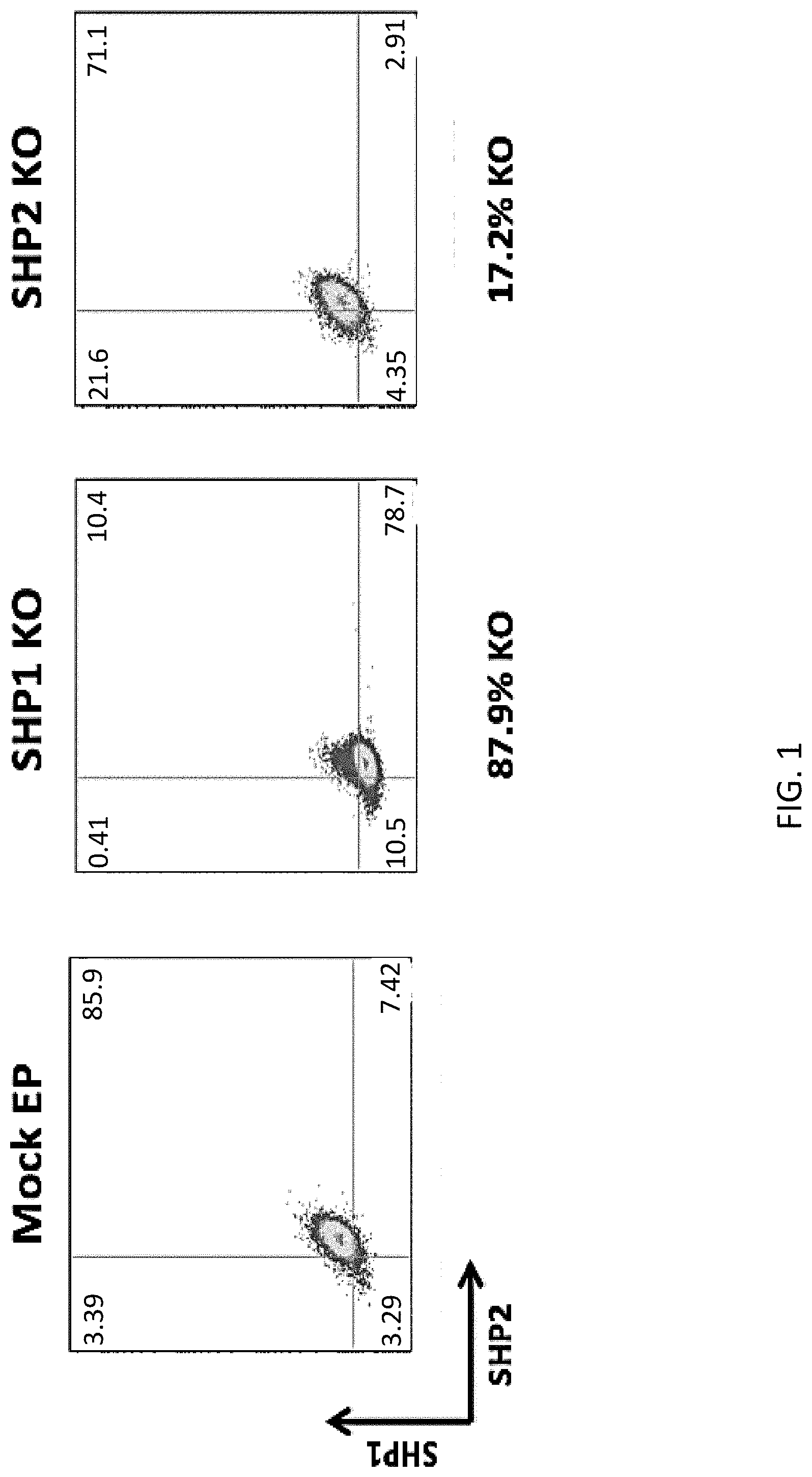
[0017] FIG. 1 illustrates knockout efficiency of SHP-1 and SHP-2 using CRISPR. All cells were electroporated using the Lonza 4D-Nucleofector Core/X Unit. The ribonucleoprotein (RNP) complex was first formed by incubating 10 ug of TrueCut Cas9 Protein V2, 5 ug of sgRNA, and 4 uL of 100 uM IDT Electroporation Enhancer for at least 10 minutes, no longer than 30 minutes, at room temperature. Pulse code EO-115 was used for primary T cells. Knockout efficiency was measured by fixing and permeabilizing cells prior to intracellular staining for the indicated markers. Cells were then analyzed by flow cytometry.
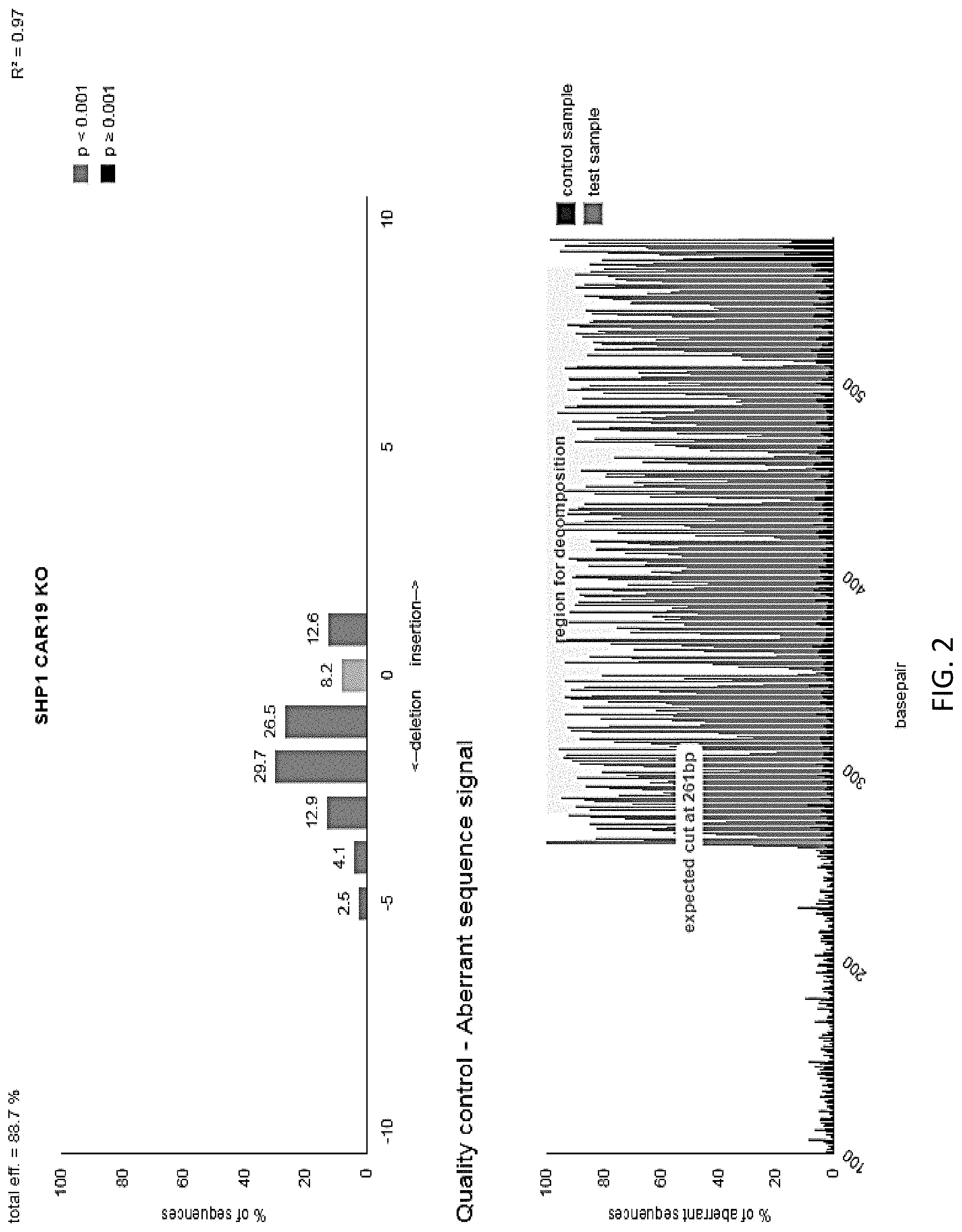
[0018] FIG. 2 illustrates knockout efficiency of SHP-1 in cells generated in FIG. 1 as determined by TIDE analysis. Sequencing trace files for the gene region of interest were analyzed by software integrated into tide.deskgen.com and used to determine knockout efficiency.
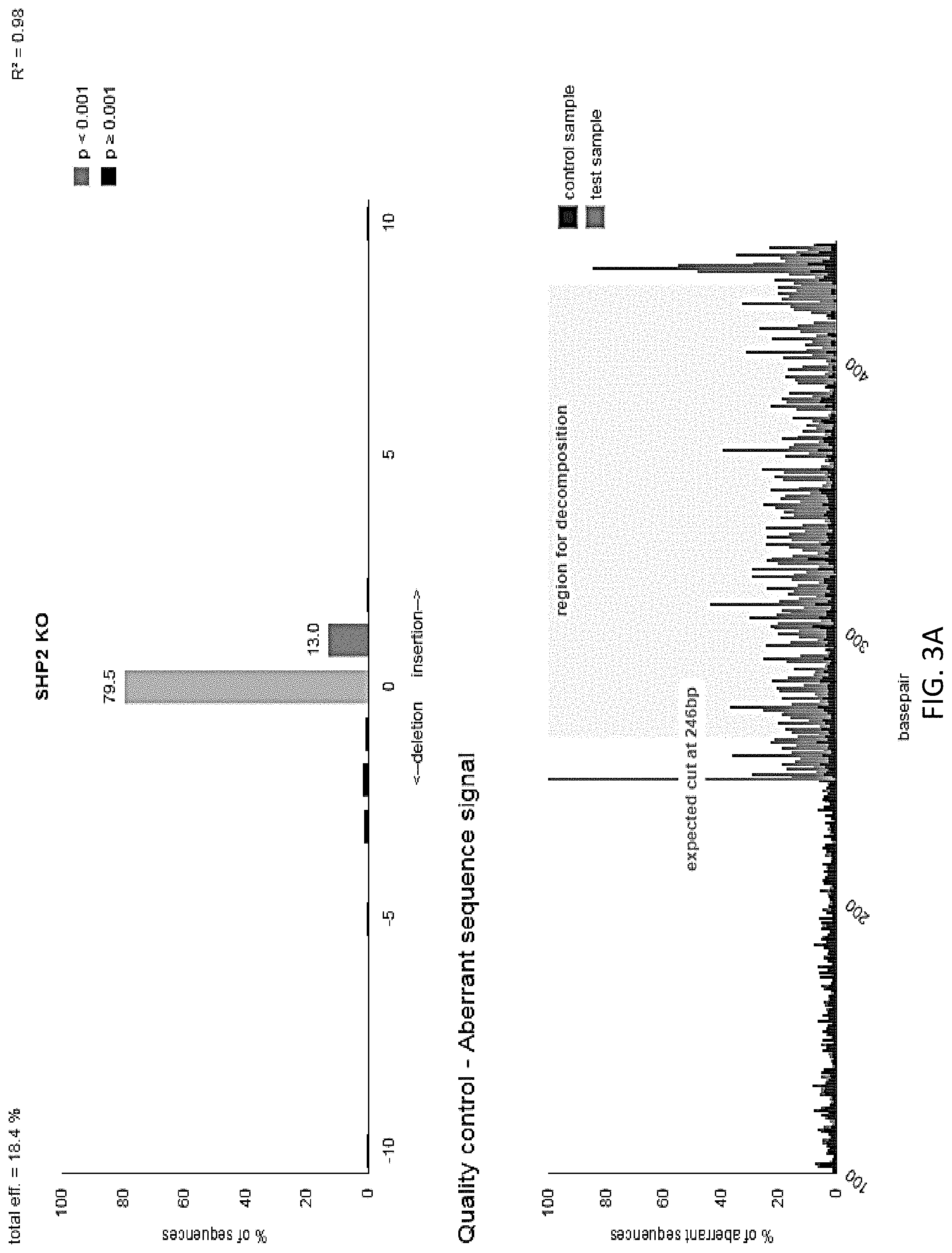
[0019] FIGS. 3A-3B are a series of graphs illustrating the knockout efficiency of SHP-2 as determined by TIDE analysis. Cells were electroporated using the Lonza 4D-Nucleofector Core/X Unit. The ribonucleoprotein (RNP) complex was first formed by incubating 10 ug of TrueCut Cas9 Protein V2, 5 ug of sgRNA, and 4 uL of 100 uM IDT Electroporation Enhancer for at least 10 minutes, no longer than 30 minutes, at room temperature. Pulse code EO-115 was used for primary T cells. Knockout efficiency was measured using TIDE analysis. Sequencing trace files for the gene region of interest were analyzed by software integrated into tide.deskgen.com to determine KO efficiency.
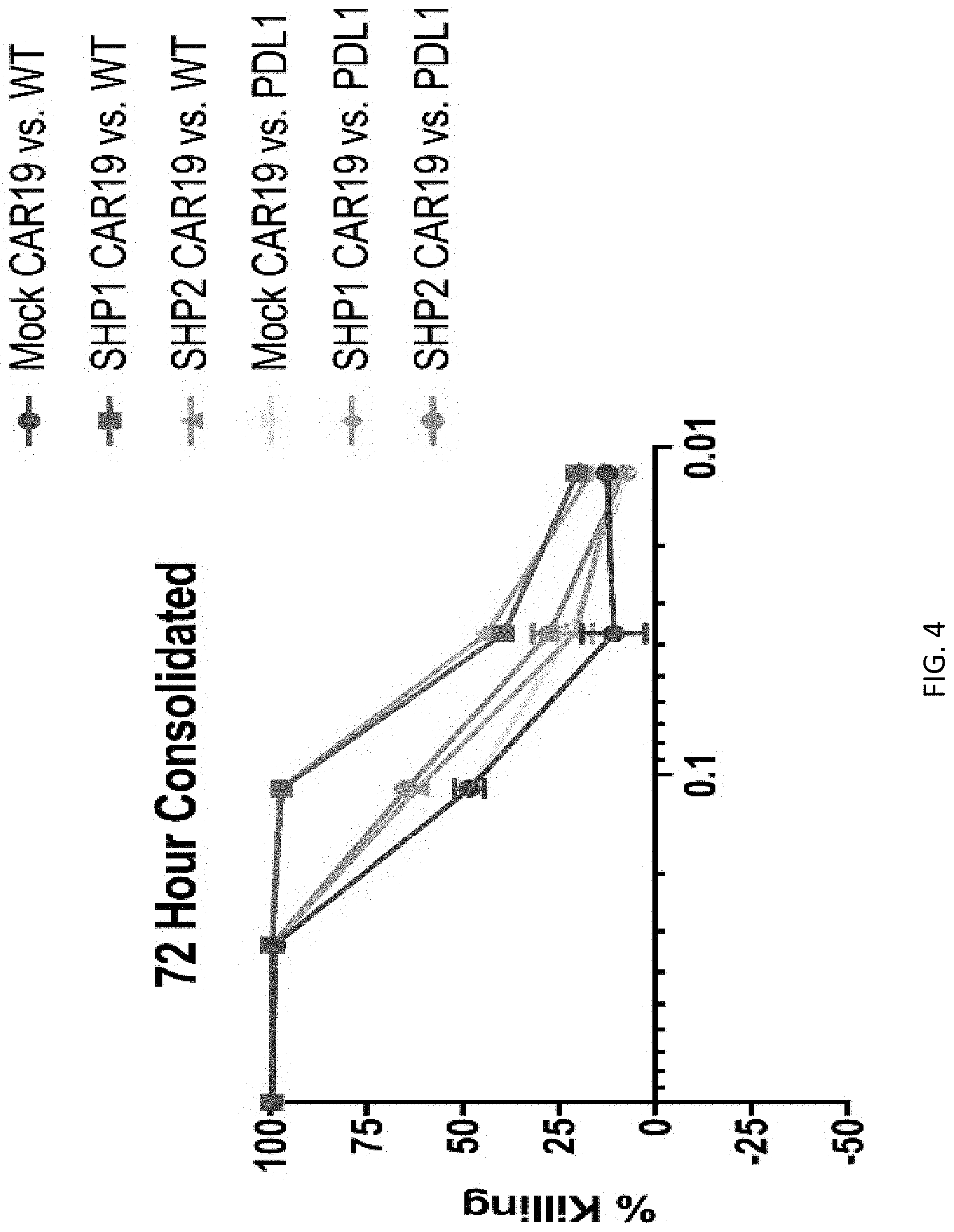
[0020] FIG. 4 illustrates results from an experiment wherein mock CAR19, SHP-1 KO CAR19, and SHP-2 KO CAR19 cells were co-cultured with either Nalm6 wild-type (WT) and Nalm6-PDL1 for 72 hours. Cytotoxicity was measured by luciferase based bioluminescence imaging.
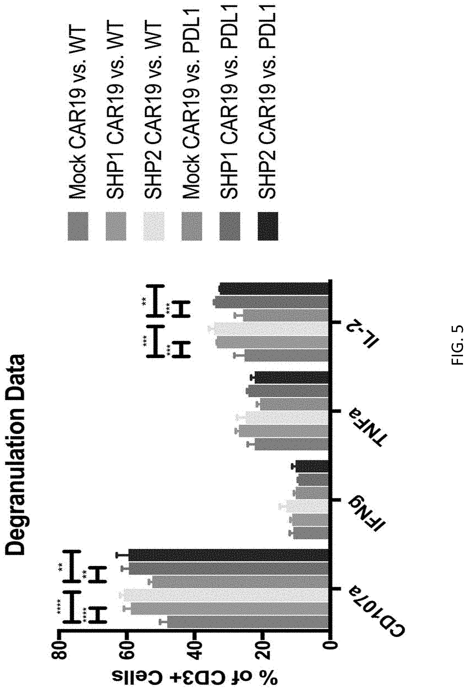
[0021] FIG. 5 is a graph that illustrates results from an experiment wherein mock CAR19, SHP-1 KO CAR19, and SHP-2 KO CAR19 cells were co-cultured with either Nalm6 wild-type (WT) and Nalm6-PDL1 for 6 hours. Protein levels were measured by flow cytometry after staining with fluorophore-conjugated antibodies. ** P.ltoreq.0.01 *** P.ltoreq.0.001 **** P.ltoreq.0.0001.
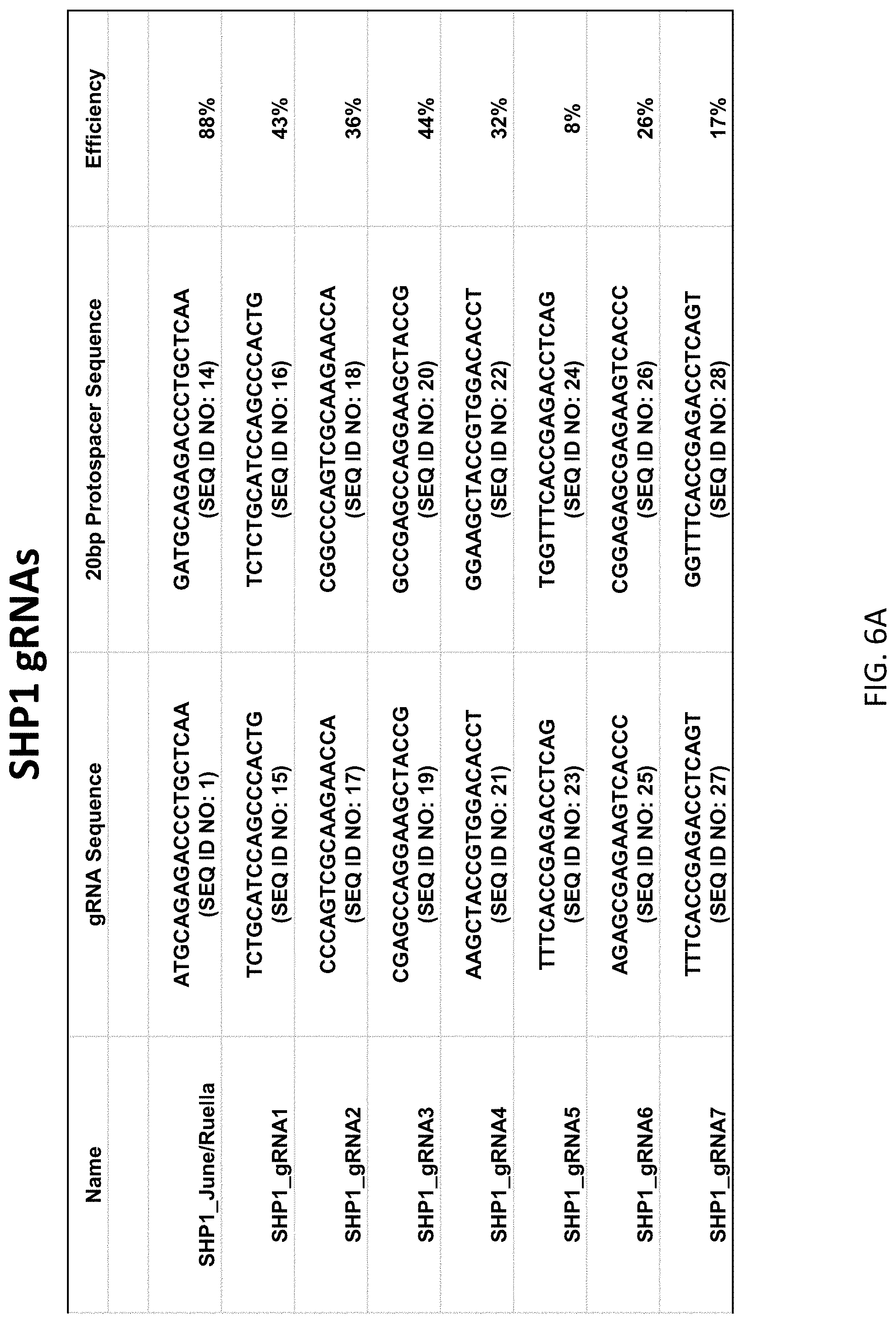
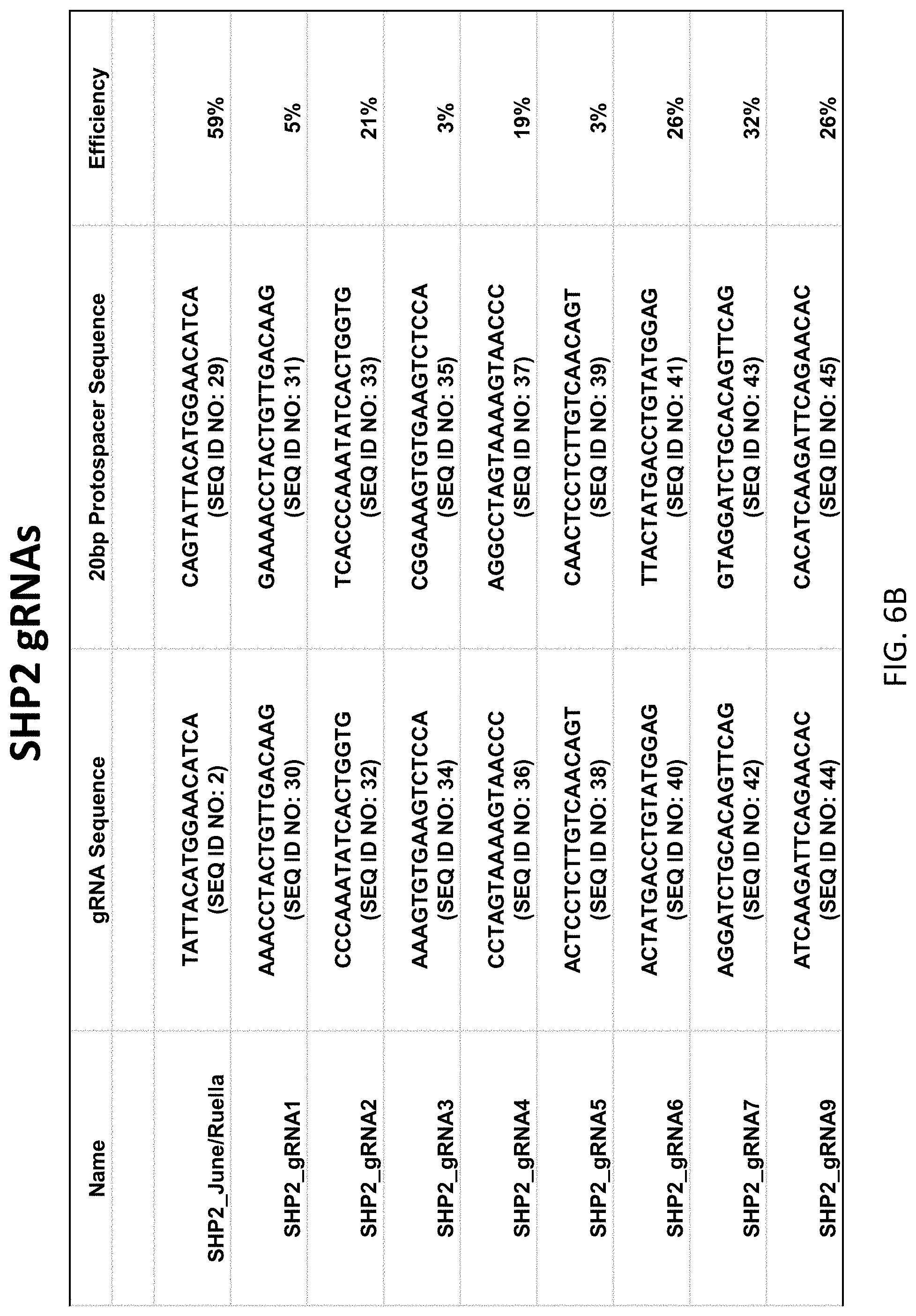
[0022] FIGS. 6A-6B are tables listing the sequences of the guide RNAs and corresponding protospacer sequences used to generate the SHP1 and SHP2 knockout cells. The guides were designed to target translated regions in earlier exons of the genes. sgRNAs with the highest predicted on-target score according to Doensch, et al. (2016) Nature Biotechnology were chosen for screening, and the lead guides displayed were determined through TIDE analysis. The knockout efficiency of SHP1_June/Ruella (FIG. 6A) was further verified through flow cytometry.
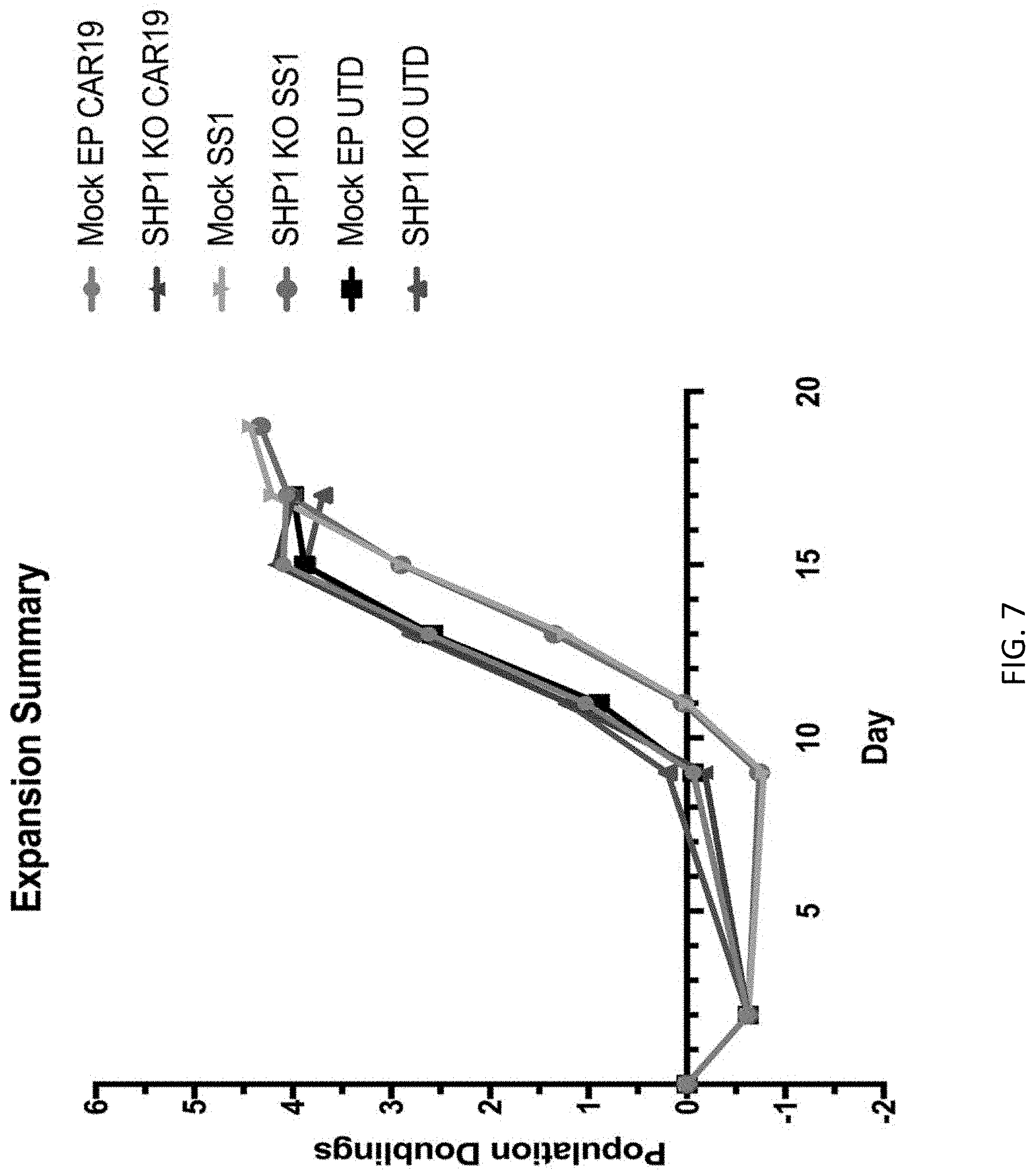
[0023] FIG. 7 is a graph showing the manufacturing of anti-CD19 and anti-mesothelin (SS1) CART with or without SHP-1 KO. EP=electroporation; Mock=no gRNA. T cells were electroporated with Cas9 protein and gRNA at day 0 then kept at 30.degree. C. for 2 days. At day 2 the T cells were activated using anti-CD3/CD28 magnetic beads (Dynabeads). After 24 hours lentivirus for CAR19 and CAR-meso were added. Magnetic beads were removed after 7 days. T cells were expanded until their size returned below 300 fl.
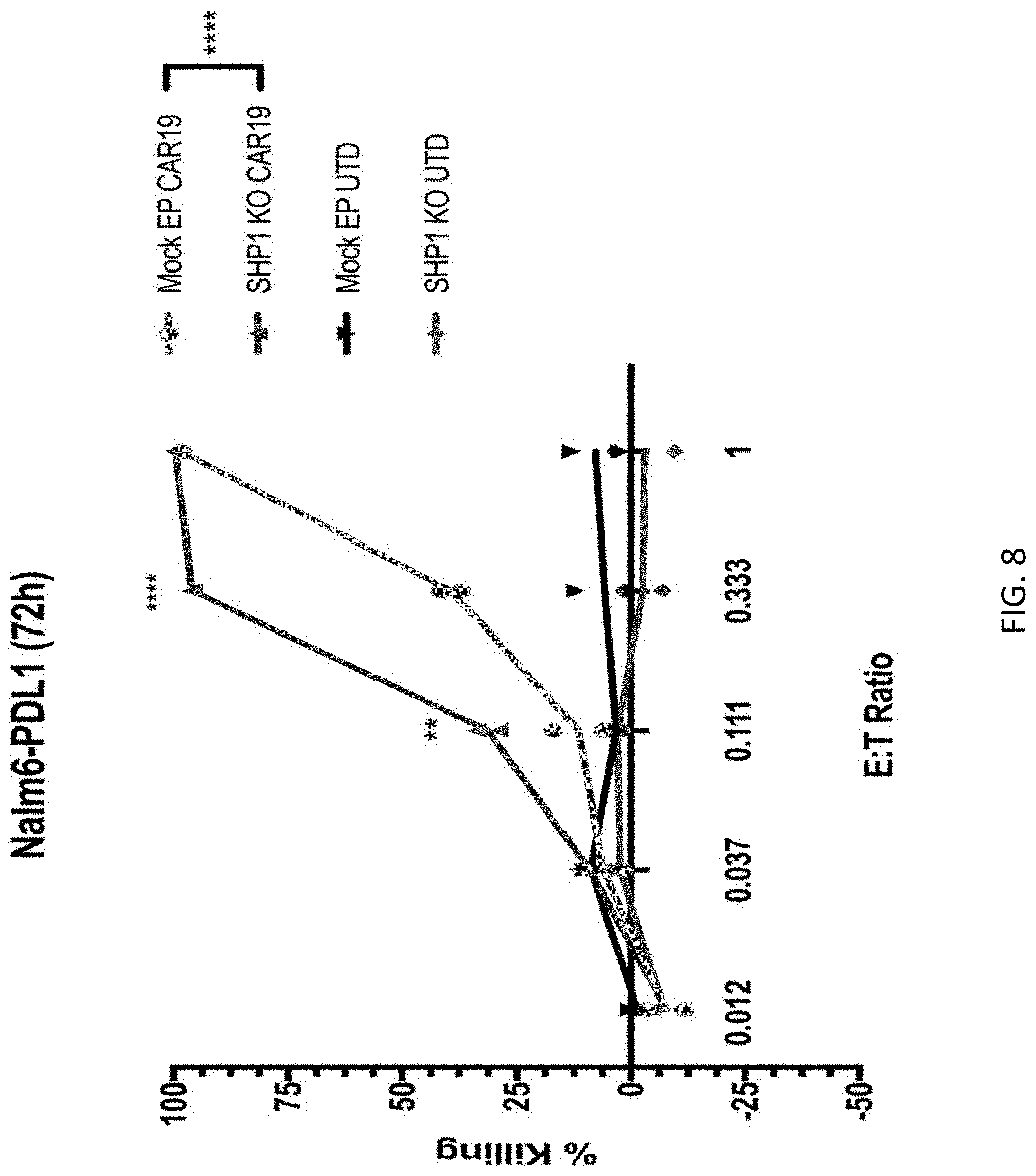
[0024] FIG. 8 is a graph demonstrating the in vitro cytotoxicity of CART19 expressing T cells with or without accompanying knock-down of SHP1. Engineered cells were incubated with luciferase-labeled B-ALL cells PDL-1+NALM6 as targets. CART19 or control, unmodified T cells (UTD) were co-cultured with target cells at different effector to target (E:T) ratios. NALM6 killing was calculated measuring luminescence at 72 hours. **** p.ltoreq.0.0001 and **p.ltoreq.0.01 for SHP1 knockout cells vs. SHP1 wt (Mock EP) at the indicated time points.
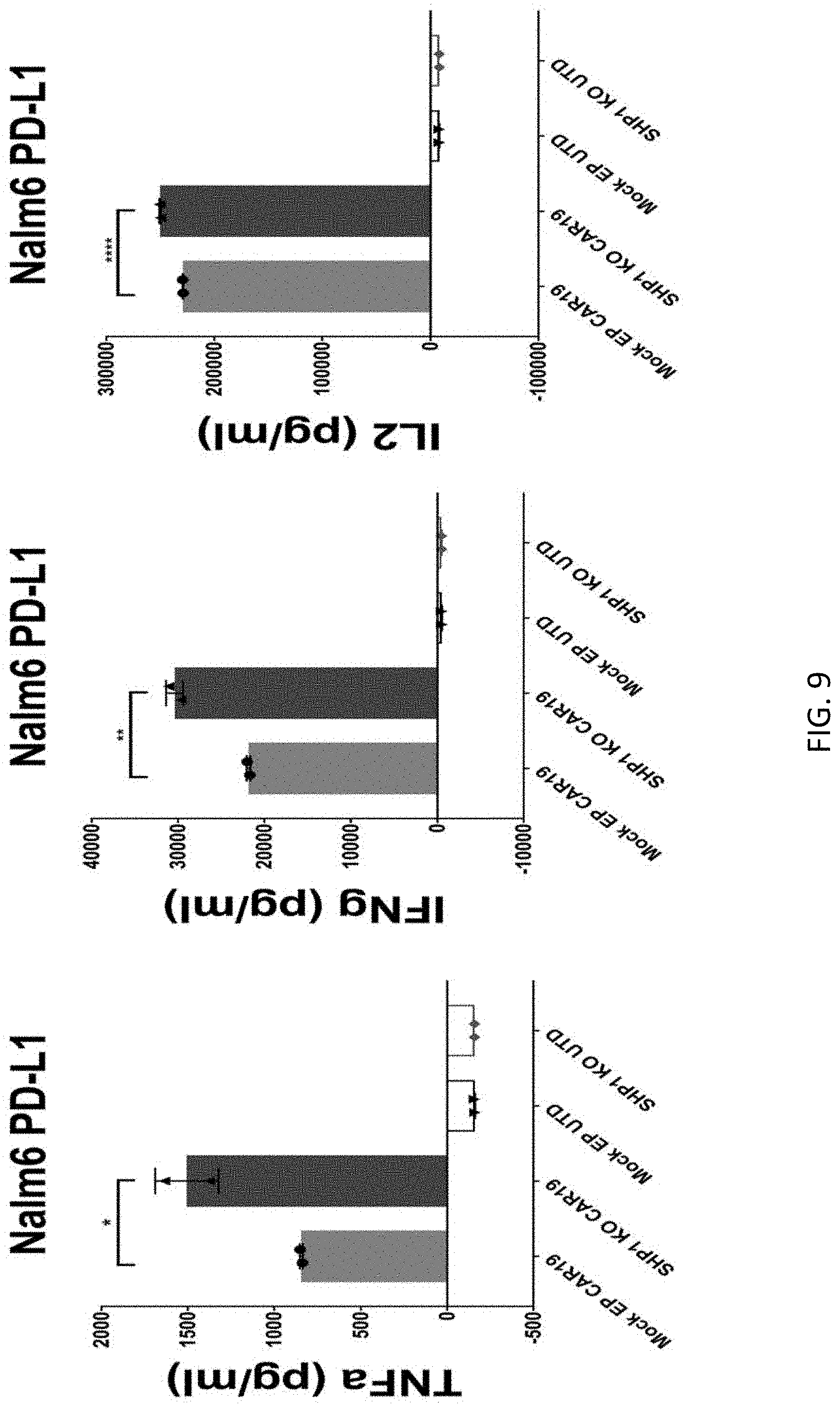
[0025] FIG. 9 is a series of graphs quantifying in vitro cytotoxicity using SHP1 KO or control cells expressing wildtype levels of SHP1(Mock EP). CART19 or control T cells (UTD) were co-cultured with NALM6 cells at 1:2 E:T ratio for 24 hours. Supernatants were then harvested and cytokine release of tumor necrosis factor-alpha (TNF.alpha.) (left), interferon-gamma (IFN.gamma.) (center), and interleukin-2 (IL-2) (right) were determined by ELISA. Error bars indicated standard deviation. *p.ltoreq.0.05, **p.ltoreq.0.01, and ****p.ltoreq.0.0001 for the indicated comparisons between SHP1 KO and control cells.
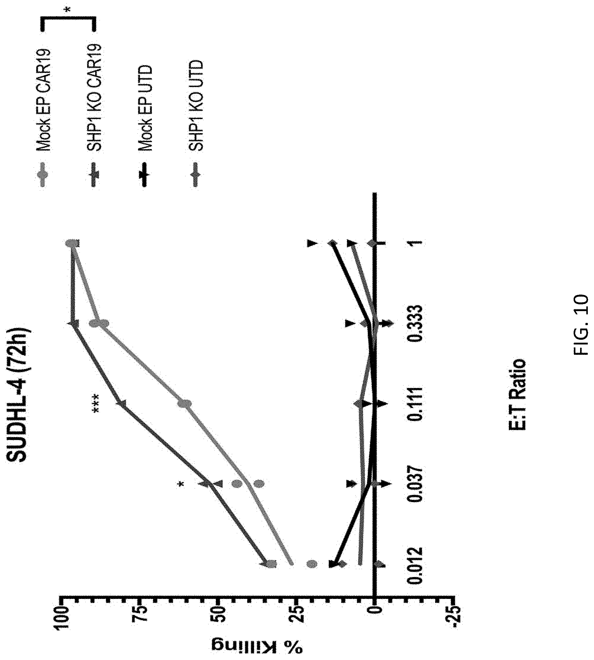
[0026] FIG. 10 is a graph showing the in vitro cytotoxicity of CART19 T cells against the diffuse large B-cell lymphoma cell line SUDHL4. SHP1 knock-out CART19 T cells (SHP1 KO CAR19), SHP1 wildtype CART19 T cells (MockEP CAR19), SHP1 knock-out control T cells (SHP1 KO UTD), or unmodified T cells (UTD) were co-cultured with luciferase-labeled SUDHL4 cells at different effector to target (E:T) ratios for 72 hours. At the conclusion of the study, SUDHL4 viability was calculated by mearing luminescence and percent killing was calculated by comparison to SUDHL4-alone controls. *p.ltoreq.0.05 and ***p.ltoreq.0.001 for SHP1 knock-out CART19 T cells vs SHP1 wildtype CART19T cells at the indicated effector:target ratios.
[0027] FIG. 11 is a series of graphs showing the in vitro cytotoxicity of SHP1 knock-out CART19 T cells. Knock-out CART19 T cells, or controls (Mock EP CAR19), or SHP1 knockout non-CAR expressing T cells (SHP1 KO UTD) or untouched T cells (UTD) were co-cultured with SUDLH4 cells at a 1:2 effector: target ratio for 24 hours. Supernatants were then harvested and production of TNF.alpha. (left), IFN.gamma. (center), and IL-2 (right) were determined by ELISA. Error bars indicated standard deviation. *p.ltoreq.0.05 and **p.ltoreq.0.01 for SHP1 knockout vs control CAR19 cells.
[0028] FIG. 12 is a graph showing the in vitro cytotoxicity of CART19 T cells against the diffuse large B-cell lymphoma cell line Ocy-L18. SHP1 knock-out CART19 T cells (SHP1 KO CAR19), SHP1 wildtype CART19 T cells (MockEP CAR19), SHP1 knock-out control T cells (SHP1 KO UTD), or unmodified T cells (UTD) were co-cultured with luciferase-labeled Ocy-L18 cells at different effector to target (E:T) ratios for 72 hours. At the conclusion of the study, target cell viability was calculated by measuring luminescence and percent killing was calculated by comparison to SUDHL4-alone controls. *p.ltoreq.0.05, ***p.ltoreq.0.001, and ****p.ltoreq.0.0001 for SHP1 knock-out CART19 T cells vs SHP1 wildtype CART19T cells at the indicated effector:target ratios.
[0029] FIG. 13 is a graph showing the in vitro cytotoxicity of CART19 T cells against the diffuse large B-cell lymphoma cell line SUDHL2. SHP1 knock-out CART19 T cells (SHP1 KO CAR19), SHP1 wildtype CART19 T cells (Mock EP CAR19), SHP1 knock-out control T cells (SHP1 KO UTD), or unmodified T cells (UTD) were co-cultured with luciferase-labeled SUDHL2 cells at different effector to target (E:T) ratios for 72 hours. At the conclusion of the study, SUDHL2 viability was calculated by mearing luminescence and percent killing was calculated by comparison to SUDHL4-alone controls. *p.ltoreq.0.05 and ****p.ltoreq.0.0001 for SHP1 knock-out CART19 T cells vs SHP1 wildtype CART19T cells at the indicated effector:target ratios.
[0030] FIG. 14A-14B are a series of graphs showing in vitro proliferation of SHP1 knock-out T cells. Wildtype CART19, SHP1 KO CART19, SHP1 KO non-CAR expressing, and untouched control T cells were labeled with CellTrace Violet before use in co-incubation assays. Labeled cells were then co-cultured with NALM6-PD1 (FIG. 14A, top), Ocy-L18 (FIG. 14A, bottom), SUDHL2 (FIG. 14B, top), or SUDHL4 (FIG. 14B, bottom) target cells for 5 days. Absolute number of T cells (left) and percentage of proliferating T cells (right) was then measured by flow cytometry. Error bars indicated standard deviation between replicates in each group. *p.ltoreq.0.05, **p.ltoreq.0.01, and ***p.ltoreq.0.001 for SHP1 knock-out CART19 T cells vs SHP1 wildtype CART19 T cells. NS =no statistical relevance.
[0031] FIG. 15 is a set of graphs demonstrating the in vitro cytotoxicity of SHP1 knock-out or control cells (Mock EP). Target cells for these assays were the mesothelioma cell line EMMEO (PD-L1+) and T cells were engineered to express the SS1 anti-mesothelin CART construct. Modified and control T cells were incubated with target cells at either 1.11:1 or 0.37:1 effector: target ratios for 6 days, after which cell killing was determined by the xCELLigence cytotoxicity assay.
[0032] FIG. 16 is a schematic drawing illustrating the setup of an in vivo experiment in which SHP1 knock-out CD19 CART T cells were used against NALM6-PD1 cells injected into NSG mice.
[0033] FIG. 17 is a graph illustrating the growth of xenografted tumor cells in mice as part of the in vivo study described in FIG. 16. NALM6-PD1 tumor cells were engineered to express luciferase prior to injection, and animals were subjected to intravital imaging in order to determine tumor burden at the indicated timepoints.
[0034] FIG. 18 is a graph demonstrating engraftment and expansion of SHP1 KO or control CAR19-expressing T cells in NSG mice. Twelve days after injection, peripheral blood from mice was harvested and the total number of transferred cells per 100 .mu.l of blood was determined by flow cytometry. Error bars indicated standard deviation between replicate animals in each group. *p.ltoreq.0.05, **p.ltoreq.0.01, and ****p.ltoreq.0.0001 for the indicated comparisons.
[0035] FIG. 19 is a series of graphs showing the combined knock-out of SHP-1 and TRAC (TCR) in human primary T cells by a CRISPR knock-out system. All cells were electroporated using the Lonza 4D-Nucleofector Core/X Unit. The ribonucleoprotein (RNP) complex was first formed by incubating 10 ug of TrueCut Cas9 Protein V2, 5 ug of sgRNA, and 4 uL of 100 uM IDT Electroporation Enhancer for at least 10 minutes, no longer than 30 minutes, at room temperature. Pulse code EO-115 was used for primary T cells. Knockout efficiency was measured by fixing and permeabilizing cells prior to intracellular staining for the indicated markers. Cells were then analyzed by flow cytometry.
DETAILED DESCRIPTION
Definitions
[0036] Unless defined otherwise, all technical and scientific terms used herein have the same meaning as commonly understood by one of ordinary skill in the art to which the invention pertains. Although any methods and materials similar or equivalent to those described herein can be used in the practice for testing of the present invention, the preferred materials and methods are described herein. In describing and claiming the present invention, the following terminology will be used.
[0037] It is also to be understood that the terminology used herein is for the purpose of describing particular embodiments only, and is not intended to be limiting.
[0038] The articles "a" and "an" are used herein to refer to one or to more than one (i.e., to at least one) of the grammatical object of the article. By way of example, "an element" means one element or more than one element.
[0039] "About" as used herein when referring to a measurable value such as an amount, a temporal duration, and the like, is meant to encompass variations of .+-.20% or .+-.10%, more preferably .+-.5%, even more preferably .+-.1%, and still more preferably .+-.0.1% from the specified value, as such variations are appropriate to perform the disclosed methods.
[0040] "Activation," as used herein, refers to the state of a T cell that has been sufficiently stimulated to induce detectable cellular proliferation. Activation can also be associated with induced cytokine production, and detectable effector functions. The term "activated T cells" refers to, among other things, T cells that are undergoing cell division.
[0041] The term "antibody," as used herein, refers to an immunoglobulin molecule which specifically binds with an antigen. Antibodies can be intact immunoglobulins derived from natural sources or from recombinant sources and can be immunoreactive portions of intact immunoglobulins. Antibodies are typically tetramers of immunoglobulin molecules. The antibodies in the present invention may exist in a variety of forms including, for example, polyclonal antibodies, monoclonal antibodies, Fv, Fab and F(ab).sub.2, as well as single chain antibodies (scFv) and humanized antibodies (Harlow et al., 1999, In: Using Antibodies: A Laboratory Manual, Cold Spring Harbor Laboratory Press, NY; Harlow et al., 1989, In: Antibodies: A Laboratory Manual, Cold Spring Harbor, New York; Houston et al., 1988, Proc. Natl. Acad. Sci. USA 85:5879-5883; Bird et al., 1988, Science 242:423-426).
[0042] The term "antibody fragment" refers to a portion of an intact antibody and refers to the antigenic determining variable regions of an intact antibody. Examples of antibody fragments include, but are not limited to, Fab, Fab', F(ab')2, and Fv fragments, linear antibodies, scFv antibodies, and multispecific antibodies formed from antibody fragments.
[0043] An "antibody heavy chain," as used herein, refers to the larger of the two types of polypeptide chains present in all antibody molecules in their naturally occurring conformations.
[0044] An "antibody light chain," as used herein, refers to the smaller of the two types of polypeptide chains present in all antibody molecules in their naturally occurring conformations. Kappa and lambda light chains refer to the two major antibody light chain isotypes.
[0045] By the term "synthetic antibody" as used herein, is meant an antibody which is generated using recombinant DNA technology, such as, for example, an antibody expressed by a bacteriophage as described herein. The term should also be construed to mean an antibody which has been generated by the synthesis of a DNA molecule encoding the antibody and which DNA molecule expresses an antibody protein, or an amino acid sequence specifying the antibody, wherein the DNA or amino acid sequence has been obtained using synthetic DNA or amino acid sequence technology which is available and well known in the art.
[0046] The term "antigen" or "Ag" as used herein is defined as a molecule that provokes an immune response. This immune response may involve either antibody production, or the activation of specific immunologically-competent cells, or both. The skilled artisan will understand that any macromolecule, including virtually all proteins or peptides, can serve as an antigen. Furthermore, antigens can be derived from recombinant or genomic DNA. A skilled artisan will understand that any DNA, which comprises a nucleotide sequences or a partial nucleotide sequence encoding a protein that elicits an immune response therefore encodes an "antigen" as that term is used herein. Furthermore, one skilled in the art will understand that an antigen need not be encoded solely by a full length nucleotide sequence of a gene. It is readily apparent that the present invention includes, but is not limited to, the use of partial nucleotide sequences of more than one gene and that these nucleotide sequences are arranged in various combinations to elicit the desired immune response. Moreover, a skilled artisan will understand that an antigen need not be encoded by a "gene" at all. It is readily apparent that an antigen can be generated synthesized or can be derived from a biological sample. Such a biological sample can include, but is not limited to a tissue sample, a tumor sample, a cell or a biological fluid.
[0047] As used herein, the term "autologous" is meant to refer to any material derived from the same individual to which it is later to be re-introduced into the individual.
[0048] "Allogeneic" refers to any material derived from a different animal of the same species.
[0049] "Xenogeneic" refers to any material derived from an animal of a different species.
[0050] The term "chimeric antigen receptor" or "CAR," as used herein, refers to an artificial T cell receptor that is engineered to be expressed on an immune effector cell and specifically bind an antigen. CARs may be used as a therapy with adoptive cell transfer. T cells are removed from a patient and modified so that they express the receptors specific to a particular form of antigen. In some embodiments, the CARs has specificity to a selected target, for example a B cell surface receptor. CARs may also comprise an intracellular activation domain, a transmembrane domain and an extracellular domain comprising a tumor associated antigen binding region. In some aspects, CARs comprise an extracellular domain comprising an anti-B cell binding domain fused to CD3-zeta transmembrane and intracellular domain
[0051] The term "cleavage" refers to the breakage of covalent bonds, such as in the backbone of a nucleic acid molecule or the hydrolysis of peptide bonds. Cleavage can be initiated by a variety of methods, including, but not limited to, enzymatic or chemical hydrolysis of a phosphodiester bond. Both single-stranded cleavage and double-stranded cleavage are possible. Double-stranded cleavage can occur as a result of two distinct single-stranded cleavage events. DNA cleavage can result in the production of either blunt ends or staggered ends. In certain embodiments, fusion polypeptides may be used for targeting cleaved double-stranded DNA.
[0052] As used herein, the term "conservative sequence modifications" is intended to refer to amino acid modifications that do not significantly affect or alter the binding characteristics of the antibody containing the amino acid sequence. Such conservative modifications include amino acid substitutions, additions and deletions. Modifications can be introduced into an antibody of the invention by standard techniques known in the art, such as site-directed mutagenesis and PCR-mediated mutagenesis. Conservative amino acid substitutions are ones in which the amino acid residue is replaced with an amino acid residue having a similar side chain. Families of amino acid residues having similar side chains have been defined in the art. These families include amino acids with basic side chains (e.g., lysine, arginine, histidine), acidic side chains (e.g., aspartic acid, glutamic acid), uncharged polar side chains (e.g., glycine, asparagine, glutamine, serine, threonine, tyrosine, cysteine, tryptophan), nonpolar side chains (e.g., alanine, valine, leucine, isoleucine, proline, phenylalanine, methionine), beta-branched side chains (e.g., threonine, valine, isoleucine) and aromatic side chains (e.g., tyrosine, phenylalanine, tryptophan, histidine). Thus, one or more amino acid residues within the CDR regions of an antibody can be replaced with other amino acid residues from the same side chain family and the altered antibody can be tested for the ability to bind antigens using the functional assays described herein.
[0053] "Co-stimulatory ligand," as the term is used herein, includes a molecule on an antigen presenting cell (e.g., an aAPC, dendritic cell, B cell, and the like) that specifically binds a cognate co-stimulatory molecule on a T cell, thereby providing a signal which, in addition to the primary signal provided by, for instance, binding of a TCR/CD3 complex with an MHC molecule loaded with peptide, mediates a T cell response, including, but not limited to, proliferation, activation, differentiation, and the like. A co-stimulatory ligand can include, but is not limited to, CD7, B7-1 (CD80), B7-2 (CD86), PD-L1, PD-L2, 4-1BBL, OX40L, inducible costimulatory ligand (ICOS-L), intercellular adhesion molecule (ICAM), CD30L, CD40, CD70, CD83, HLA-G, MICA, MICB, HVEM, lymphotoxin beta receptor, 3/TR6, ILT3, ILT4, HVEM, an agonist or antibody that binds Toll ligand receptor and a ligand that specifically binds with B7-H3. A co-stimulatory ligand also encompasses, inter alia, an antibody that specifically binds with a co-stimulatory molecule present on a T cell, such as, but not limited to, CD27, CD28, 4-1BB, OX40, CD30, CD40, PD-1, ICOS, lymphocyte function-associated antigen-1 (LFA-1), CD2, CD7, LIGHT, NKG2C, B7-H3, and a ligand that specifically binds with CD83.
[0054] A "co-stimulatory molecule" refers to the cognate binding partner on a T cell that specifically binds with a co-stimulatory ligand, thereby mediating a co-stimulatory response by the T cell, such as, but not limited to, proliferation. Co-stimulatory molecules include, but are not limited to an MHC class I molecule, BTLA and a Toll ligand receptor.
[0055] A "co-stimulatory signal", as used herein, refers to a signal, which in combination with a primary signal, such as TCR/CD3 ligation, leads to T cell proliferation and/or upregulation or downregulation of key molecules.
[0056] A "disease" is a state of health of an animal wherein the animal cannot maintain homeostasis, and wherein if the disease is not ameliorated then the animal's health continues to deteriorate. In contrast, a "disorder" in an animal is a state of health in which the animal is able to maintain homeostasis, but in which the animal's state of health is less favorable than it would be in the absence of the disorder. Left untreated, a disorder does not necessarily cause a further decrease in the animal's state of health.
[0057] The term "downregulation" as used herein refers to the decrease or elimination of gene expression of one or more genes.
[0058] "Effective amount" or "therapeutically effective amount" are used interchangeably herein, and refer to an amount of a compound, formulation, material, or composition, as described herein effective to achieve a particular biological result or provides a therapeutic or prophylactic benefit. Such results may include, but are not limited to, anti-tumor activity as determined by any means suitable in the art.
[0059] "Encoding" refers to the inherent property of specific sequences of nucleotides in a polynucleotide, such as a gene, a cDNA, or an mRNA, to serve as templates for synthesis of other polymers and macromolecules in biological processes having either a defined sequence of nucleotides (i.e., rRNA, tRNA and mRNA) or a defined sequence of amino acids and the biological properties resulting therefrom. Thus, a gene encodes a protein if transcription and translation of mRNA corresponding to that gene produces the protein in a cell or other biological system. Both the coding strand, the nucleotide sequence of which is identical to the mRNA sequence and is usually provided in sequence listings, and the non-coding strand, used as the template for transcription of a gene or cDNA, can be referred to as encoding the protein or other product of that gene or cDNA.
[0060] As used herein "endogenous" refers to any material from or produced inside an organism, cell, tissue or system.
[0061] As used herein, the term "exogenous" refers to any material introduced from or produced outside an organism, cell, tissue or system.
[0062] The term "expand" as used herein refers to increasing in number, as in an increase in the number of T cells. In one embodiment, the T cells that are expanded ex vivo increase in number relative to the number originally present in the culture. In another embodiment, the T cells that are expanded ex vivo increase in number relative to other cell types in the culture. The term "ex vivo," as used herein, refers to cells that have been removed from a living organism, (e.g., a human) and propagated outside the organism (e.g., in a culture dish, test tube, or bioreactor).
[0063] The term "expression" as used herein is defined as the transcription and/or translation of a particular nucleotide sequence driven by its promoter.
[0064] "Expression vector" refers to a vector comprising a recombinant polynucleotide comprising expression control sequences operatively linked to a nucleotide sequence to be expressed. An expression vector comprises sufficient cis-acting elements for expression; other elements for expression can be supplied by the host cell or in an in vitro expression system. Expression vectors include all those known in the art, such as cosmids, plasmids (e.g., naked or contained in liposomes) and viruses (e.g., Sendai viruses, lentiviruses, retroviruses, adenoviruses, and adeno-associated viruses) that incorporate the recombinant polynucleotide.
[0065] "Homologous" as used herein, refers to the subunit sequence identity between two polymeric molecules, e.g., between two nucleic acid molecules, such as, two DNA molecules or two RNA molecules, or between two polypeptide molecules. When a subunit position in both of the two molecules is occupied by the same monomeric subunit; e.g., if a position in each of two DNA molecules is occupied by adenine, then they are homologous at that position. The homology between two sequences is a direct function of the number of matching or homologous positions; e.g., if half (e.g., five positions in a polymer ten subunits in length) of the positions in two sequences are homologous, the two sequences are 50% homologous; if 90% of the positions (e.g., 9 of 10), are matched or homologous, the two sequences are 90% homologous.
[0066] "Humanized" forms of non-human (e.g., murine) antibodies are chimeric immunoglobulins, immunoglobulin chains or fragments thereof (such as Fv, Fab, Fab', F(ab')2 or other antigen-binding subsequences of antibodies) which contain minimal sequence derived from non-human immunoglobulin. For the most part, humanized antibodies are human immunoglobulins (recipient antibody) in which residues from a complementary-determining region (CDR) of the recipient are replaced by residues from a CDR of a non-human species (donor antibody) such as mouse, rat or rabbit having the desired specificity, affinity, and capacity. In some instances, Fv framework region (FR) residues of the human immunoglobulin are replaced by corresponding non-human residues. Furthermore, humanized antibodies can comprise residues which are found neither in the recipient antibody nor in the imported CDR or framework sequences. These modifications are made to further refine and optimize antibody performance. In general, the humanized antibody will comprise substantially all of at least one, and typically two, variable domains, in which all or substantially all of the CDR regions correspond to those of a non-human immunoglobulin and all or substantially all of the FR regions are those of a human immunoglobulin sequence. The humanized antibody optimally also will comprise at least a portion of an immunoglobulin constant region (Fc), typically that of a human immunoglobulin. For further details, see Jones et al., Nature, 321: 522-525, 1986; Reichmann et al., Nature, 332: 323-329, 1988; Presta, Curr. Op. Struct. Biol., 2: 593-596, 1992.
[0067] "Fully human" refers to an immunoglobulin, such as an antibody, where the whole molecule is of human origin or consists of an amino acid sequence identical to a human form of the antibody.
[0068] "Identity" as used herein refers to the subunit sequence identity between two polymeric molecules particularly between two amino acid molecules, such as, between two polypeptide molecules. When two amino acid sequences have the same residues at the same positions; e.g., if a position in each of two polypeptide molecules is occupied by an Arginine, then they are identical at that position. The identity or extent to which two amino acid sequences have the same residues at the same positions in an alignment is often expressed as a percentage. The identity between two amino acid sequences is a direct function of the number of matching or identical positions; e.g., if half (e.g., five positions in a polymer ten amino acids in length) of the positions in two sequences are identical, the two sequences are 50% identical; if 90% of the positions (e.g., 9 of 10), are matched or identical, the two amino acids sequences are 90% identical.
[0069] The term "immunoglobulin" or "Ig," as used herein is defined as a class of proteins, which function as antibodies. Antibodies expressed by B cells are sometimes referred to as the BCR (B cell receptor) or antigen receptor. The five members included in this class of proteins are IgA, IgG, IgM, IgD, and IgE. IgA is the primary antibody that is present in body secretions, such as saliva, tears, breast milk, gastrointestinal secretions and mucus secretions of the respiratory and genitourinary tracts. IgG is the most common circulating antibody. IgM is the main immunoglobulin produced in the primary immune response in most subjects. It is the most efficient immunoglobulin in agglutination, complement fixation, and other antibody responses, and is important in defense against bacteria and viruses. IgD is the immunoglobulin that has no known antibody function, but may serve as an antigen receptor. IgE is the immunoglobulin that mediates immediate hypersensitivity by causing release of mediators from mast cells and basophils upon exposure to allergen.
[0070] The term "immune response" as used herein is defined as a cellular response to an antigen that occurs when lymphocytes identify antigenic molecules as foreign and induce the formation of antibodies and/or activate lymphocytes to remove the antigen.
[0071] When "an immunologically effective amount," "an autoimmune disease-inhibiting effective amount," or "therapeutic amount" is indicated, the precise amount of the compositions of the present invention to be administered can be determined by a physician or researcher with consideration of individual differences in age, weight, tumor size, extent of infection or metastasis, and condition of the patient (subject).
[0072] As used herein, an "instructional material" includes a publication, a recording, a diagram, or any other medium of expression which can be used to communicate the usefulness of the compositions and methods of the invention. The instructional material of the kit of the invention may, for example, be affixed to a container which contains the nucleic acid, peptide, and/or composition of the invention or be shipped together with a container which contains the nucleic acid, peptide, and/or composition. Alternatively, the instructional material may be shipped separately from the container with the intention that the instructional material and the compound be used cooperatively by the recipient.
[0073] "Isolated" means altered or removed from the natural state. For example, a nucleic acid or a peptide naturally present in a living animal is not "isolated," but the same nucleic acid or peptide partially or completely separated from the coexisting materials of its natural state is "isolated." An isolated nucleic acid or protein can exist in substantially purified form, or can exist in a non-native environment such as, for example, a host cell.
[0074] The term "knockdown" as used herein refers to a decrease in gene expression of one or more genes.
[0075] The term "knockout" as used herein refers to the ablation of gene expression of one or more genes.
[0076] A "lentivirus" as used herein refers to a genus of the Retroviridae family. Lentiviruses are unique among the retroviruses in being able to infect non-dividing cells; they can deliver a significant amount of genetic information into the DNA of the host cell, so they are one of the most efficient methods of a gene delivery vector. HIV, SIV, and FIV are all examples of lentiviruses. Vectors derived from lentiviruses offer the means to achieve significant levels of gene transfer in vivo.
[0077] The term "limited toxicity" as used herein, refers to the peptides, polynucleotides, cells and/or antibodies of the invention manifesting a lack of substantially negative biological effects, anti-tumor effects, or substantially negative physiological symptoms toward a healthy cell, non-tumor cell, non-diseased cell, non-target cell or population of such cells either in vitro or in vivo.
[0078] By the term "modified" as used herein, is meant a changed state or structure of a molecule or cell of the invention. Molecules may be modified in many ways, including chemically, structurally, and functionally. Cells may be modified through the introduction of nucleic acids.
[0079] By the term "modulating," as used herein, is meant mediating a detectable increase or decrease in the level of a response in a subject compared with the level of a response in the subject in the absence of a treatment or compound, and/or compared with the level of a response in an otherwise identical but untreated subject. The term encompasses perturbing and/or affecting a native signal or response thereby mediating a beneficial therapeutic response in a subject, preferably, a human.
[0080] In the context of the present invention, the following abbreviations for the commonly occurring nucleic acid bases are used. "A" refers to adenosine, "C" refers to cytosine, "G" refers to guanosine, "T" refers to thymidine, and "U" refers to uridine.
[0081] Unless otherwise specified, a "nucleotide sequence encoding an amino acid sequence" includes all nucleotide sequences that are degenerate versions of each other and that encode the same amino acid sequence. The phrase nucleotide sequence that encodes a protein or an RNA may also include introns to the extent that the nucleotide sequence encoding the protein may in some version contain an intron(s).
[0082] The term "operably linked" refers to functional linkage between a regulatory sequence and a heterologous nucleic acid sequence resulting in expression of the latter. For example, a first nucleic acid sequence is operably linked with a second nucleic acid sequence when the first nucleic acid sequence is placed in a functional relationship with the second nucleic acid sequence. For instance, a promoter is operably linked to a coding sequence if the promoter affects the transcription or expression of the coding sequence. Generally, operably linked DNA sequences are contiguous and, where necessary to join two protein coding regions, in the same reading frame.
[0083] The term "overexpressed" tumor antigen or "overexpression" of a tumor antigen is intended to indicate an abnormal level of expression of a tumor antigen in a cell from a disease area like a solid tumor within a specific tissue or organ of the patient relative to the level of expression in a normal cell from that tissue or organ. Patients having solid tumors or a hematological malignancy characterized by overexpression of the tumor antigen can be determined by standard assays known in the art.
[0084] "Parenteral" administration of an immunogenic composition includes, e.g., subcutaneous (s.c.), intravenous (i.v.), intramuscular (i.m.), or intrasternal injection, or infusion techniques.
[0085] The term "polynucleotide" as used herein is defined as a chain of nucleotides. Furthermore, nucleic acids are polymers of nucleotides. Thus, nucleic acids and polynucleotides as used herein are interchangeable. One skilled in the art has the general knowledge that nucleic acids are polynucleotides, which can be hydrolyzed into the monomeric "nucleotides." The monomeric nucleotides can be hydrolyzed into nucleosides. As used herein polynucleotides include, but are not limited to, all nucleic acid sequences which are obtained by any means available in the art, including, without limitation, recombinant means, i.e., the cloning of nucleic acid sequences from a recombinant library or a cell genome, using ordinary cloning technology and PCRTM, and the like, and by synthetic means.
[0086] As used herein, the terms "peptide," "polypeptide," and "protein" are used interchangeably, and refer to a compound comprised of amino acid residues covalently linked by peptide bonds. A protein or peptide must contain at least two amino acids, and no limitation is placed on the maximum number of amino acids that can comprise a protein's or peptide's sequence. Polypeptides include any peptide or protein comprising two or more amino acids joined to each other by peptide bonds. As used herein, the term refers to both short chains, which also commonly are referred to in the art as peptides, oligopeptides and oligomers, for example, and to longer chains, which generally are referred to in the art as proteins, of which there are many types. "Polypeptides" include, for example, biologically active fragments, substantially homologous polypeptides, oligopeptides, homodimers, heterodimers, variants of polypeptides, modified polypeptides, derivatives, analogs, fusion proteins, among others. The polypeptides include natural peptides, recombinant peptides, synthetic peptides, or a combination thereof.
[0087] The term "promoter" as used herein is defined as a DNA sequence recognized by the synthetic machinery of the cell, or introduced synthetic machinery, required to initiate the specific transcription of a polynucleotide sequence.
[0088] As used herein, the term "promoter/regulatory sequence" means a nucleic acid sequence which is required for expression of a gene product operably linked to the promoter/regulatory sequence. In some instances, this sequence may be the core promoter sequence and in other instances, this sequence may also include an enhancer sequence and other regulatory elements which are required for expression of the gene product. The promoter/regulatory sequence may, for example, be one which expresses the gene product in a tissue specific manner.
[0089] A "constitutive" promoter is a nucleotide sequence which, when operably linked with a polynucleotide which encodes or specifies a gene product, causes the gene product to be produced in a cell under most or all physiological conditions of the cell.
[0090] An "inducible" promoter is a nucleotide sequence which, when operably linked with a polynucleotide which encodes or specifies a gene product, causes the gene product to be produced in a cell substantially only when an inducer which corresponds to the promoter is present in the cell.
[0091] A "tissue-specific" promoter is a nucleotide sequence which, when operably linked with a polynucleotide encodes or specified by a gene, causes the gene product to be produced in a cell substantially only if the cell is a cell of the tissue type corresponding to the promoter.
[0092] A "Sendai virus" refers to a genus of the Paramyxoviridae family. Sendai viruses are negative, single stranded RNA viruses that do not integrate into the host genome or alter the genetic information of the host cell. Sendai vinises have an exceptionally broad host range and are not pathogenic to humans. Used as a recombinant viral vector, Sendai viruses are capable of transient but strong gene expression
[0093] A "signal transduction pathway" refers to the biochemical relationship between a variety of signal transduction molecules that play a role in the transmission of a signal from one portion of a cell to another portion of a cell. The phrase "cell surface receptor" includes molecules and complexes of molecules capable of receiving a signal and transmitting signal across the plasma membrane of a cell.
[0094] By the term "specifically binds," as used herein with respect to an antibody, is meant an antibody which recognizes a specific antigen, but does not substantially recognize or bind other molecules in a sample. For example, an antibody that specifically binds to an antigen from one species may also bind to that antigen from one or more species. But, such cross-species reactivity does not itself alter the classification of an antibody as specific. In another example, an antibody that specifically binds to an antigen may also bind to different allelic forms of the antigen. However, such cross reactivity does not itself alter the classification of an antibody as specific. In some instances, the terms "specific binding" or "specifically binding," can be used in reference to the interaction of an antibody, a protein, or a peptide with a second chemical species, to mean that the interaction is dependent upon the presence of a particular structure (e.g., an antigenic determinant or epitope) on the chemical species; for example, an antibody recognizes and binds to a specific protein structure rather than to proteins generally. If an antibody is specific for epitope "A", the presence of a molecule containing epitope A (or free, unlabeled A), in a reaction containing labeled "A" and the antibody, will reduce the amount of labeled A bound to the antibody.
[0095] By the term "stimulation," is meant a primary response induced by binding of a stimulatory molecule (e.g., a TCR/CD3 complex) with its cognate ligand thereby mediating a signal transduction event, such as, but not limited to, signal transduction via the TCR/CD3 complex. Stimulation can mediate altered expression of certain molecules, such as downregulation of TGF-beta, and/or reorganization of cytoskeletal structures, and the like.
[0096] A "stimulatory molecule," as the term is used herein, means a molecule on a T cell that specifically binds with a cognate stimulatory ligand present on an antigen presenting cell.
[0097] A "stimulatory ligand," as used herein, means a ligand that when present on an antigen presenting cell (e.g., an aAPC, a dendritic cell, a B-cell, and the like) can specifically bind with a cognate binding partner (referred to herein as a "stimulatory molecule") on a T cell, thereby mediating a primary response by the T cell, including, but not limited to, activation, initiation of an immune response, proliferation, and the like. Stimulatory ligands are well-known in the art and encompass, inter alia, an MHC Class I molecule loaded with a peptide, an anti-CD3 antibody, a superagonist anti-CD28 antibody, and a superagonist anti-CD2 antibody.
[0098] The term "subject" is intended to include living organisms in which an immune response can be elicited (e.g., mammals). A "subject" or "patient," as used therein, may be a human or non-human mammal. Non-human mammals include, for example, livestock and pets, such as ovine, bovine, porcine, canine, feline and murine mammals. Preferably, the subject is human.
[0099] As used herein, a "substantially purified" cell is a cell that is essentially free of other cell types. A substantially purified cell also refers to a cell which has been separated from other cell types with which it is normally associated in its naturally occurring state. In some instances, a population of substantially purified cells refers to a homogenous population of cells. In other instances, this term refers simply to cell that have been separated from the cells with which they are naturally associated in their natural state. In some embodiments, the cells are cultured in vitro. In other embodiments, the cells are not cultured in vitro.
[0100] A "target site" or "target sequence" refers to a genomic nucleic acid sequence that defines a portion of a nucleic acid to which a binding molecule may specifically bind under conditions sufficient for binding to occur.
[0101] As used herein, the term "T cell receptor" or "TCR" refers to a complex of membrane proteins that participate in the activation of T cells in response to the presentation of antigen. The TCR is responsible for recognizing antigens bound to major histocompatibility complex molecules. TCR is composed of a heterodimer of an alpha (a) and beta (.beta.) chain, although in some cells the TCR consists of gamma and delta (.gamma./.delta.) chains. TCRs may exist in alpha/beta and gamma/delta forms, which are structurally similar but have distinct anatomical locations and functions. Each chain is composed of two extracellular domains, a variable and constant domain. In some embodiments, the TCR may be modified on any cell comprising a TCR, including, for example, a helper T cell, a cytotoxic T cell, a memory cell, regulatory T cell, natural killer cell, and gamma delta. T cell.
[0102] The term "therapeutic" as used herein means a treatment and/or prophylaxis. A therapeutic effect is obtained by suppression, remission, or eradication of a disease state.
[0103] The term "transfected" or "transformed" or "transduced" as used herein refers to a process by which exogenous nucleic acid is transferred or introduced into the host cell. A "transfected" or "transformed" or "transduced" cell is one which has been transfected, transformed or transduced with exogenous nucleic acid. The cell includes the primary subject cell and its progeny.
[0104] To "treat" a disease as the term is used herein, means to reduce the frequency or severity of at least one sign or symptom of a disease or disorder experienced by a subject.
[0105] The phrase "under transcriptional control" or "operatively linked" as used herein means that the promoter is in the correct location and orientation in relation to a polynucleotide to control the initiation of transcription by RNA polymerase and expression of the polynucleotide.
[0106] A "vector" is a composition of matter which comprises an isolated nucleic acid and which can be used to deliver the isolated nucleic acid to the interior of a cell. Numerous vectors are known in the art including, but not limited to, linear polynucleotides, polynucleotides associated with ionic or amphiphilic compounds, plasmids, and viruses. Thus, the term "vector" includes an autonomously replicating plasmid or a virus. The term should also be construed to include non-plasmid and non-viral compounds which facilitate transfer of nucleic acid into cells, such as, for example, polylysine compounds, liposomes, and the like. Examples of viral vectors include, but are not limited to, Sendai viral vectors, adenoviral vectors, adeno-associated virus vectors, retroviral vectors, lentiviral vectors, and the like.
[0107] Ranges: throughout this disclosure, various aspects of the invention can be presented in a range format. It should be understood that the description in range format is merely for convenience and brevity and should not be construed as an inflexible limitation on the scope of the invention. Accordingly, the description of a range should be considered to have specifically disclosed all the possible subranges as well as individual numerical values within that range. For example, description of a range such as from 1 to 6 should be considered to have specifically disclosed subranges such as from 1 to 3, from 1 to 4, from 1 to 5, from 2 to 4, from 2 to 6, from 3 to 6 etc., as well as individual numbers within that range, for example, 1, 2, 2.7, 3, 4, 5, 5.3, and 6. This applies regardless of the breadth of the range.
Description
[0108] The present invention provides compositions and methods comprising a CD19 chimeric antigen receptor T (CART) cell wherein SHP-1 and/or SHP-2 has been knocked out.
[0109] As demonstrated herein, SHP-1 and/or SHP-2 were knocked out in CART cells using the CRISPR-Cas9 technology with the goal of increasing their anti-tumor activity in an immunosuppressive microenvironment.
Compositions
[0110] In one aspect, the invention includes a genetically modified cell comprising a chimeric antigen receptor (CAR). The CAR comprises an antigen binding domain that targets CD19, a transmembrane domain, and an intracellular domain. Within the cell, at least one gene selected from the group consisting of one SHP-1 and SHP-2, has been modified. The SHP-1 and/or SHP-2 modification is carried out by a CRISPR-Cas9 system comprising at least one guide RNA (gRNA). The SHP-1 gRNA can comprise any one of the nucleotide sequences selected from the group consisting of SEQ ID NO: 1, 15, 17, 19, 21, 23, or 27. The SHP-2 gRNA can comprise any one of the nucleotide sequences selected from the group consisting of SEQ ID NO: 2, 30, 32, 34, 36, 38, 40, 42, or 44. In certain embodiments, the gRNA that targets SHP-1 comprises the nucleotide sequence of SEQ ID NO: 1 and/or the gRNA that targets SHP-2 comprises the nucleotide sequence of SEQ ID NO: 2.
[0111] In certain embodiments, the cell further comprises a disruption in the TRAC locus. In certain embodiments, the TRAC locus is disrupted by a CRISPR-Cas9 system comprising at least one gRNA. In certain embodiments, the gRNA that disrupts the TRAC locus comprises the nucleotide sequence of SEQ ID NO: 46.
[0112] In certain embodiments, the SHP-1 and/or SHP-2 gene is knocked out in the cell.
[0113] In certain embodiments, the cell is a T cell.
Chimeric Antigen Receptor (CAR)
[0114] The present invention provides a chimeric antigen receptor (CAR) comprising an extracellular domain, a transmembrane domain, and an intracellular domain. The extracellular domain comprises a target-specific binding element otherwise referred to as an antigen binding domain. The intracellular domain or otherwise the cytoplasmic domain comprises, a costimulatory signaling region and a zeta chain portion. The costimulatory signaling region refers to a portion of the CAR comprising the intracellular domain of a costimulatory molecule. Costimulatory molecules are cell surface molecules other than antigen receptors or their ligands that are required for an efficient response of lymphocytes to antigen.
[0115] Between the extracellular domain and the transmembrane domain of the CAR, or between the cytoplasmic domain and the transmembrane domain of the CAR, there may be incorporated a spacer domain. As used herein, the term "spacer domain" generally means any oligo- or polypeptide that functions to link the transmembrane domain to, either the extracellular domain or, the cytoplasmic domain in the polypeptide chain. A spacer domain may comprise up to 300 amino acids, preferably 10 to 100 amino acids and most preferably 25 to 50 amino acids.
[0116] Antigen Binding Domain
[0117] In one embodiment, the CAR of the invention comprises a target-specific binding element otherwise referred to as an antigen binding domain. The choice of antigen binding domain depends upon the type and number of ligands that define the surface of a target cell. For example, the antigen binding domain may be chosen to recognize a ligand that acts as a cell surface marker on target cells associated with a particular disease state. Thus examples of cell surface markers that may act as ligands for the antigen moiety domain in the CAR of the invention include those associated with viral, bacterial and parasitic infections, autoimmune disease and cancer cells.
[0118] In one embodiment, the CAR of the invention can be engineered to target a tumor antigen of interest by way of engineering a desired antigen binding domain that specifically binds to an antigen on a tumor cell. In the context of the present invention, "tumor antigen" or "hyperporoliferative disorder antigen" or "antigen associated with a hyperproliferative disorder," refers to antigens that are common to specific hyperproliferative disorders such as cancer. The antigens discussed herein are merely included by way of example. The list is not intended to be exclusive and further examples will be readily apparent to those of skill in the art.
[0119] Tumor antigens are proteins that are produced by tumor cells that elicit an immune response, particularly T-cell mediated immune responses. The selection of the antigen binding domain of the invention will depend on the particular type of cancer to be treated. Tumor antigens are well known in the art and include, for example, a glioma-associated antigen, carcinoembryonic antigen (CEA), .beta.-human chorionic gonadotropin, alphafetoprotein (AFP), lectin-reactive AFP, thyroglobulin, RAGE-1, MN-CA IX, human telomerase reverse transcriptase, RUL RU2 (AS), intestinal carboxyl esterase, mut hsp70-2, M-CSF, prostase, prostate-specific antigen (PSA), PAP, NY-ESO-1, LAGE-1a, p53, prostein, PSMA, Her2/neu, survivin and telomerase, prostate-carcinoma tumor antigen-1 (PCTA-1), MAGE, ELF2M, neutrophil elastase, ephrinB2, CD22, insulin growth factor (IGF)-I, IGF-II, IGF-I receptor and mesothelin.
[0120] In one embodiment, the tumor antigen comprises one or more antigenic cancer epitopes associated with a malignant tumor. Malignant tumors express a number of proteins that can serve as target antigens for an immune attack. These molecules include but are not limited to tissue-specific antigens such as MART-1, tyrosinase and GP 100 in melanoma and prostatic acid phosphatase (PAP) and prostate-specific antigen (PSA) in prostate cancer. Other target molecules belong to the group of transformation-related molecules such as the oncogene HER-2/Neu/ErbB-2. Yet another group of target antigens are onco-fetal antigens such as carcinoembryonic antigen (CEA). In B-cell lymphoma the tumor-specific idiotype immunoglobulin constitutes a truly tumor-specific immunoglobulin antigen that is unique to the individual tumor. B-cell differentiation antigens such as CD19, CD20 and CD37 are other candidates for target antigens in B-cell lymphoma.
[0121] The type of tumor antigen referred to in the invention may also be a tumor-specific antigen (TSA) or a tumor-associated antigen (TAA). A TSA is unique to tumor cells and does not occur on other cells in the body. A TAA associated antigen is not unique to a tumor cell and instead is also expressed on a normal cell under conditions that fail to induce a state of immunologic tolerance to the antigen. The expression of the antigen on the tumor may occur under conditions that enable the immune system to respond to the antigen. TAAs may be antigens that are expressed on normal cells during fetal development when the immune system is immature and unable to respond or they may be antigens that are normally present at extremely low levels on normal cells but which are expressed at much higher levels on tumor cells.
[0122] Non-limiting examples of TSA or TAA antigens include the following: Differentiation antigens such as MART-1/MelanA (MART-I), gp100 (Pmel 17), tyrosinase, TRP-1, TRP-2 and tumor-specific multilineage antigens such as MAGE-1, MAGE-3, BAGE, GAGE-1, GAGE-2, p15; overexpressed embryonic antigens such as CEA; overexpressed oncogenes and mutated tumor-suppressor genes such as p53, Ras, HER-2/neu; unique tumor antigens resulting from chromosomal translocations; such as BCR-ABL, E2A-PRL, H4-RET, IGH-IGK, MYL-RAR; and viral antigens, such as the Epstein Barr virus antigens EBVA and the human papillomavirus (HPV) antigens E6 and E7. Other large, protein-based antigens include TSP-180, MAGE-4, MAGE-5, MAGE-6, RAGE, NY-ESO, p185erbB2, p180erbB-3, c-met, nm-23H1, PSA, TAG-72, CA 19-9, CA 72-4, CAM 17.1, NuMa, K-ras, beta-Catenin, CDK4, Mum-1, p 15, p 16, 43-9F, 5T4, 791Tgp72, alpha-fetoprotein, beta-HCG, BCA225, BTAA, CA 125, CA 15-3\CA 27.29\BCAA, CA 195, CA 242, CA-50, CAM43, CD68\P1, CO-029, FGF-5, G250, Ga733\EpCAM, HTgp-175, M344, MA-50, MG7-Ag, MOV18, NB/70K, NY-CO-1, RCAS1, SDCCAG16, TA-90\Mac-2 binding protein\cyclophilin C-associated protein, TAAL6, TAG72, TLP, and TPS.
[0123] In a preferred embodiment, the antigen binding domain of the CAR targets CD19. In certain embodiments, the antigen binding domain of the CAR targets an antigen that includes but is not limited to, CD20, CD22, ROR1, Mesothelin, CD33/IL3Ra, c-Met, PSMA, Glycolipid F77, EGFRvIII, GD-2, MY-ESO-1 TCR, MAGE A3 TCR, and the like.
[0124] Depending on the desired antigen to be targeted, the CAR of the invention can be engineered to include the appropriate antigen bind domain that is specific to the desired antigen target. For example, if CD19 is the desired antigen that is to be targeted, an antibody for CD19 can be used as the antigen binding domain for incorporation into the CAR of the invention.
[0125] In one embodiment, the antigen binding domain of the CAR of the invention targets CD19. In some embodiments, the antigen binding domain in the CAR of the invention is anti-CD19 scFV, wherein the nucleic acid sequence of the anti-CD19 scFV comprises the sequence set forth in SEQ ID NO: 3. In other embodiments, the anti-CD19 scFV comprises a nucleic acid sequence that encodes the amino acid sequence of SEQ ID NO: 4. In yet other embodiments, the anti-CD19 scFV portion of the CAR of the invention comprises the amino acid sequence set forth in SEQ ID NO: 4. In some embodiments, the antigen binding domain is an anti-CD19 antibody. In some embodiments, the nucleic acid sequence of the anti-CD19 antibody comprises the nucleic acid sequence set forth in SEQ ID NO: 5.
[0126] Transmembrane Domain
[0127] With respect to the transmembrane domain, the CAR can be designed to comprise a transmembrane domain that is fused to the extracellular domain of the CAR. In one embodiment, the transmembrane domain that naturally is associated with one of the domains in the CAR is used. In some instances, the transmembrane domain can be selected or modified by amino acid substitution to avoid binding of such domains to the transmembrane domains of the same or different surface membrane proteins to minimize interactions with other members of the receptor complex.
[0128] The transmembrane domain may be derived either from a natural or from a synthetic source. Where the source is natural, the domain may be derived from any membrane-bound or transmembrane protein. Transmembrane regions of particular use in this invention may be derived from (i.e. comprise at least the transmembrane region(s) of) the alpha, beta or zeta chain of the T-cell receptor, CD28, CD3 epsilon, CD45, CD4, CD5, CD8, CD9, CD16, CD22, CD33, CD37, CD64, CD80, CD86, CD134, CD137, CD154. Alternatively the transmembrane domain may be synthetic, in which case it will comprise predominantly hydrophobic residues such as leucine and valine. Preferably a triplet of phenylalanine, tryptophan and valine will be found at each end of a synthetic transmembrane domain. Optionally, a short oligo- or polypeptide linker, preferably between 2 and 10 amino acids in length may form the linkage between the transmembrane domain and the cytoplasmic signaling domain of the CAR. A glycine-serine doublet provides a particularly suitable linker.
[0129] Preferably, the transmembrane domain in the CAR of the invention is the CD8 transmembrane domain. In one embodiment, the CD8 transmembrane domain comprises the nucleic acid sequence of SEQ ID NO: 6. In one embodiment, the CD8 transmembrane domain comprises the nucleic acid sequence that encodes the amino acid sequence of SEQ ID NO: 7. In another embodiment, the CD8 transmembrane domain comprises the amino acid sequence of SEQ ID NO: 7.
[0130] In some instances, the transmembrane domain of the CAR of the invention comprises the CD8.alpha. hinge domain. In one embodiment, the CD8 hinge domain comprises the nucleic acid sequence of SEQ ID NO: 8. In one embodiment, the CD8 hinge domain comprises a nucleic acid sequence that encodes the amino acid sequence of SEQ ID NO: 9. In another embodiment, the CD8 hinge domain comprises the amino acid sequence of SEQ ID NO: 9.
[0131] Intracellular Domain
[0132] The intracellular domain or otherwise the cytoplasmic domain of the CAR of the invention is responsible for activation of at least one of the normal effector functions of the immune cell in which the CAR has been placed in. The term "effector function" refers to a specialized function of a cell. Effector function of a T cell, for example, may be cytolytic activity or helper activity including the secretion of cytokines. Thus the term "intracellular domain" refers to the portion of a protein which transduces the effector function signal and directs the cell to perform a specialized function. While usually the entire intracellular domain can be employed, in many cases it is not necessary to use the entire chain. To the extent that a truncated portion of the intracellular domain is used, such truncated portion may be used in place of the intact chain as long as it transduces the effector function signal. The term intracellular domain is thus meant to include any truncated portion of the intracellular domain sufficient to transduce the effector function signal.
[0133] Preferred examples of intracellular domains for use in the CAR of the invention include the cytoplasmic sequences of the T cell receptor (TCR) and co-receptors that act in concert to initiate signal transduction following antigen receptor engagement, as well as any derivative or variant of these sequences and any synthetic sequence that has the same functional capability.
[0134] It is known that signals generated through the TCR alone are insufficient for full activation of the T cell and that a secondary or co-stimulatory signal is also required. Thus, T cell activation can be said to be mediated by two distinct classes of cytoplasmic signaling sequence: those that initiate antigen-dependent primary activation through the TCR (primary cytoplasmic signaling sequences) and those that act in an antigen-independent manner to provide a secondary or co-stimulatory signal (secondary cytoplasmic signaling sequences).
[0135] Primary cytoplasmic signaling sequences regulate primary activation of the TCR complex either in a stimulatory way, or in an inhibitory way. Primary cytoplasmic signaling sequences that act in a stimulatory manner may contain signaling motifs which are known as immunoreceptor tyrosine-based activation motifs or ITAMs.
[0136] Examples of ITAM containing primary cytoplasmic signaling sequences that are of particular use in the invention include those derived from TCR zeta, FcR gamma, FcR beta, CD3 gamma , CD3 delta , CD3 epsilon, CD5, CD22, CD79a, CD79b, and CD66d. It is particularly preferred that cytoplasmic signaling molecule in the CAR of the invention comprises a cytoplasmic signaling sequence derived from CD3 zeta.
[0137] In a preferred embodiment, the intracellular domain of the CAR can be designed to comprise the CD3-zeta signaling domain by itself or combined with any other desired intracellular domain(s) useful in the context of the CAR of the invention. For example, the intracellular domain of the CAR can comprise a CD3 zeta chain portion and a costimulatory signaling region. The costimulatory signaling region refers to a portion of the CAR comprising the intracellular domain of a costimulatory molecule. A costimulatory molecule is a cell surface molecule other than an antigen receptor or their ligands that is required for an efficient response of lymphocytes to an antigen. Examples of such molecules include, but are not limited to, CD27, CD28, 4-1BB (CD137), OX40, CD30, CD40, PD-1, ICOS, lymphocyte function-associated antigen-1 (LFA-1), CD2, CD7, LIGHT, NKG2C, B7-H3, and a ligand that specifically binds with CD83, and the like. Thus, while the invention is exemplified primarily with 4-1BB as the co-stimulatory signaling element, other costimulatory elements are within the scope of the invention.
[0138] The cytoplasmic signaling sequences within the cytoplasmic signaling portion of the CAR of the invention may be linked to each other in a random or specified order. Optionally, a short oligo- or polypeptide linker, preferably between 2 and 10 amino acids in length may form the linkage. A glycine-serine doublet provides a particularly suitable linker.
[0139] In one embodiment, the intracellular domain is designed to comprise the signaling domain of CD3-zeta and the signaling domain of CD28. In another embodiment, the intracellular domain is designed to comprise the signaling domain of CD3-zeta and the signaling domain of 4-1BB. In yet another embodiment, the intracellular domain is designed to comprise the signaling domain of CD3-zeta and the signaling domain of CD28 and 4-1BB.
[0140] In one embodiment, the intracellular domain in the CAR of the invention is designed to comprise the signaling domain of 4-1BB and the signaling domain of CD3-zeta, wherein the signaling domain of 4-1BB comprises the nucleic acid sequence set forth in SEQ ID NO: 10 and the signaling domain of CD3-zeta comprises the nucleic acid sequence set forth in SEQ ID NO: 11.
[0141] In one embodiment, the intracellular domain in the CAR of the invention is designed to comprise the signaling domain of 4-1BB and the signaling domain of CD3-zeta, wherein the signaling domain of 4-1BB comprises a nucleic acid sequence that encodes the amino acid sequence of SEQ ID NO: 12 and the signaling domain of CD3-zeta comprises a nucleic acid sequence that encodes the amino acid sequence of SEQ ID NO: 13.
[0142] In one embodiment, the cytoplasmic domain in the CAR of the invention is designed to comprise the signaling domain of 4-1BB and the signaling domain of CD3-zeta, wherein the signaling domain of 4-1BB comprises the amino acid sequence set forth in SEQ ID NO: 12 and the signaling domain of CD3-zeta comprises the amino acid sequence set forth in SEQ ID NO: 13.
[0143] The invention should be construed to include any one of: a CAR, a nucleic acid encoding a CAR, a vector comprising a nucleic acid encoding a CAR, a cell comprising a CAR, a cell comprising a nucleic acid encoding a CAR, and a cell comprising a vector comprising a nucleic acid encoding a CAR.
TABLE-US-00001 Anti-CD19scFv nucleic acid sequence(SEQ ID NO: 3): gacatccaga tgacacagac tacatcctcc ctgtctgcct ctctgggaga cagagtcacc 60 atcagttgca gggcaagtca ggacattagt aaatatttaa attggtatca gcagaaacca 120 gatggaactg ttaaactcct gatctaccat acatcaagat tacactcagg agtcccatca 180 aggttcagtg gcagtgggtc tggaacagat tattctctca ccattagcaa cctggagcaa 240 gaagatattg ccacttactt ttgccaacag ggtaatacgc ttccgtacac gttcggaggg 300 gggaccaagc tggagatcac aggtggcggt ggctcgggcg gtggtgggtc gggtggcggc 360 ggatctgagg tgaaactgca ggagtcagga cctggcctgg tggcgccctc acagagcctg 420 tccgtcacat gcactgtctc aggggtctca ttacccgact atggtgtaag ctggattcgc 480 cagcctccac gaaagggtct ggagtggctg ggagtaatat ggggtagtga aaccacatac 540 tataattcag ctctcaaatc cagactgacc atcatcaagg acaactccaa gagccaagtt 600 ttcttaaaaa tgaacagtct gcaaactgat gacacagcca tttactactg tgccaaacat 660 tattactacg gtggtagcta tgctatggac tactggggcc aaggaacctc agtcaccgtc 720 tcctca 726 Anti-CD19scFv amino acid sequence (SEQ ID NO: 4): Asp Ile Gln Met Thr Gln Thr Thr Ser Ser Leu Ser Ala Ser Leu Gly 1 5 10 15 Asp Arg Val Thr Ile Ser Cys Arg Ala Ser Gln Asp Ile Ser Lys Tyr 20 25 30 Leu Asn Trp Tyr Gln Gln Lys Pro Asp Gly Thr Val Lys Leu Leu Ile 35 40 45 Tyr His Thr Ser Arg Leu His Ser Gly Val Pro Ser Arg Phe Ser Gly 50 55 60 Ser Gly Ser Gly Thr Asp Tyr Ser Leu Thr Ile Ser Asn Leu Glu Gln 65 70 75 80 Glu Asp Ile Ala Thr Tyr Phe Cys Gln Gln Gly Asn Thr Leu Pro Tyr 85 90 95 Thr Phe Gly Gly Gly Thr Lys Leu Glu Ile Thr Gly Gly Gly Gly Ser 100 105 110 Gly Gly Gly Gly Ser Gly Gly Gly Gly Ser Glu Val Lys Leu Gln Glu 115 120 125 Ser Gly Pro Gly Leu Val Ala Pro Ser Gln Ser Leu Ser Val Thr Cys 130 135 140 Thr Val Ser Gly Val Ser Leu Pro Asp Tyr Gly Val Ser Trp Ile Arg 145 150 155 160 Gln Pro Pro Arg Lys Gly Leu Glu Trp Leu Gly Val Ile Trp Gly Ser 165 170 175 Glu Thr Thr Tyr Tyr Asn Ser Ala Leu Lys Ser Arg Leu Thr Ile Ile 180 185 190 Lys Asp Asn Ser Lys Ser Gln Val Phe Leu Lys Met Asn Ser Leu Gln 195 200 205 Thr Asp Asp Thr Ala Ile Tyr Tyr Cys Ala Lys His Tyr Tyr Tyr Gly 210 215 220 Gly Ser Tyr Ala Met Asp Tyr Trp Gly Gln Gly Thr Ser Val Thr Val 225 230 235 240 Ser Ser CD19 antibody sequence (SEQ ID NO: 5) atggccctccctgtcaccgccctgctgcttccgctggctcttctgctccacgccgctcggcccgaaattgtgat- gacccagtcacc cgccactcttagcctttcacccggtgagcgcgcaaccctgtcttgcagagcctcccaagacatctcaaaatacc- ttaattggtatca acagaagcccggacaggctcctcgccttctgatctaccacaccagccggctccattctggaatccctgccaggt- tcagcggtag cggatctgggaccgactacaccctcactatcagctcactgcagccagaggacttcgctgtctatttctgtcagc- aagggaacacc ctgccctacacctttggacagggcaccaagctcgagattaaaggtggaggtggcagcggaggaggtgggtccgg- cggtgga ggaagccaggtccaactccaagaaagcggaccgggtcttgtgaagccatcagaaactctttcactgacttgtac- tgtgagcgga gtgtctctccccgattacggggtgtcttggatcagacagccaccggggaagggtctggaatggattggagtgat- ttggggctctg agactacttactaccaatcatccctcaagtcacgcgtcaccatctcaaaggacaactctaagaatcaggtgtca- ctgaaactgtcat ctgtgaccgcagccgacaccgccgtgtactattgcgctaagcattactattatggcgggagctacgcaatggat- tactggggaca gggtactctggtcaccgtgtccagcaccactaccccagcaccgaggccacccaccccggctcctaccatcgcct- cccagcctct gtccctgcgtccggaggcatgtagacccgcagctggtggggccgtgcatacccggggtcttgacttcgcctgcg- atatctacatt tgggcccctctggctggtacttgcggggtcctgctgctttcactcgtgatcactctttactgtaagcgcggtcg- gaagaagctgctg tacatctttaagcaacccttcatgaggcctgtgcagactactcaagaggaggacggctgttcatgccggttccc- agaggaggag gaaggcggctgcgaactgcgcgtgaaattcagccgcagcgcagatgctccagcctacaagcaggggcagaacca- gctctac aacgaactcaatcttggtcggagagaggagtacgacgtgctggacaagcggagaggacgggacccagaaatggg- cgggaa gccgcgcagaaagaatccccaagagggcctgtacaacgagctccaaaaggataagatggcagaagcctatagcg- agattggt atgaaaggggaacgcagaagaggcaaaggccacgacggactgtaccagggactcagcaccgccaccaaggacac- ctatga cgctcttcacatgcaggccctgccgcctcgg CD8 Transmembrane domain nucleic acid sequence (SEQ ID NO: 6): atctacatct gggcgccctt ggccgggact tgtggggtcc ttctcctgtc actggttatc 60 accctttact gc 72 CD8 Transmembrane domain amino acid sequence (SEQ ID NO: 7): Ile Trp Ala Pro Leu Ala Gly Thr Cys Gly Val Leu Leu Leu Ser Leu 1 5 10 15 Val Ile Thr Leu Tyr Cys 20 CD8 hinge domain nucleic acid sequence (SEQ ID NO: 8): accacgacgc cagcgccgcg accaccaaca ccggcgccca ccatcgcgtc gcagcccctg 60 tccctgcgcc cagaggcgtg ccggccagcg gcggggggcg cagtgcacac gagggggctg 120 gacttcgcct gtgat 135 CD8 hinge domain amino acid sequence (SEQ ID NO: 9): Ile Trp Ala Pro Leu Ala Gly Thr Cys Gly Val Leu Leu Leu Ser Leu 1 5 10 15 Val Ile Thr Leu Tyr Cys 20 4-1BB nucleic acid sequence (SEQ ID NO: 10) aaacggggca gaaagaaact cctgtatata ttcaaacaac catttatgag accagtacaa 60 actactcaag aggaagatgg ctgtagctgc cgatttccag aagaagaaga aggaggatgt 120 gaactg 126 CD3-zeta nucleic acid sequence (SEQ ID NO: 11) agagtgaagt tcagcaggag cgcagacgcc cccgcgtaca agcagggcca gaaccagctc 60 tataacgagc tcaatctagg acgaagagag gagtacgatg ttttggacaa gagacgtggc 120 cgggaccctg agatgggggg aaagccgaga aggaagaacc ctcaggaagg cctgtacaat 180 gaactgcaga aagataagat ggcggaggcc tacagtgaga ttgggatgaa aggcgagcgc 240 cggaggggca aggggcacga tggcctttac cagggtctca gtacagccac caaggacacc 300 tacgacgccc ttcacatgca ggccctgccc cctcgc 336 4-1BB amino acid sequence (SEQ ID NO: 12): Lys Arg Gly Arg Lys Lys Leu Leu Tyr Ile Phe Lys Gln Pro Phe Met 1 5 10 15 Arg Pro Val Gln Thr Thr Gln Glu Glu Asp Gly Cys Ser Cys Arg Phe 20 25 30 Pro Glu Glu Glu Glu Gly Gly Cys Glu Leu 35 40 CD3-zeta amino acid sequence (SEQ ID NO: 13): Arg Val Lys Phe Ser Arg Ser Ala Asp Ala Pro Ala Tyr Lys Gln Gly 1 5 10 15 Gln Asn Gln Leu Tyr Asn Glu Leu Asn Leu Gly Arg Arg Glu Glu Tyr 20 25 30 Asp Val Leu Asp Lys Arg Arg Gly Arg Asp Pro Glu Met Gly Gly Lys 35 40 45 Pro Arg Arg Lys Asn Pro Gln Glu Gly Leu Tyr Asn Glu Leu Gln Lys 50 55 60 Asp Lys Met Ala Glu Ala Tyr Ser Glu Ile Gly Met Lys Gly Glu Arg 65 70 75 80 Arg Arg Gly Lys Gly His Asp Gly Leu Tyr Gln Gly Leu Ser Thr Ala 85 90 95 Thr Lys Asp Thr Tyr Asp Ala Leu His Met Gln Ala Leu Pro Pro Arg 100 105 110
Methods of Treatment
[0144] The present invention includes methods for treating cancer using the CART cells of the present invention that are deficient in SHP-1 and/or SHP-2. As disclosed herein, SHP inhibition reduces immunosuppression by checkpoint inhibitors to improve CART function. Since SHP phosphatases act downstream of multiple receptors, their inhibition eliminates the need for multiple checkpoint inhibitors.
[0145] In one embodiment, the present invention includes a type of cellular therapy where T cells are genetically modified to express a CAR and the CAR T cell is infused to a recipient in need thereof. The infused cell is able to kill tumor cells in the recipient. Unlike antibody therapies, CAR T cells are able to replicate in vivo resulting in long-term persistence that can lead to sustained tumor control.
[0146] In one embodiment, the CAR T cells of the invention can undergo robust in vivo T cell expansion and can persist for an extended amount of time. In another embodiment, the CAR T cells of the invention evolve into specific memory T cells that can be reactivated to inhibit any additional tumor formation or growth. For example, the CART19 cells of the invention can undergo robust in vivo T cell expansion and persist at high levels for an extended amount of time in blood and bone marrow and form specific memory T cells. Without wishing to be bound by any particular theory, CAR T cells may differentiate in vivo into a central memory-like state upon encounter and subsequent elimination of target cells expressing the surrogate antigen.
[0147] Without wishing to be bound by any particular theory, the anti-tumor immunity response elicited by the CAR-modified T cells may be an active or a passive immune response. In addition, the CAR mediated immune response may be part of an adoptive immunotherapy approach in which CAR-modified T cells induce an immune response specific to the antigen binding moiety in the CAR. For example, a CART19 cells elicits an immune response specific against cells expressing CD19.
[0148] While the data disclosed herein specifically disclose an anti-CD19 scFv derived from FMC63 murine monoclonal antibody, human CD8.alpha. hinge and transmembrane domain, and human 4-1BB and CD3zeta signaling domains, the invention should be construed to include any number of variations for each of the components of the construct as described elsewhere herein. That is, the invention includes the use of any antigen binding moiety in the CAR to generate a CAR-mediated T-cell response specific to the antigen binding moiety. For example, the antigen binding moiety in the CAR of the invention can target a tumor antigen for the purpose of treating cancer.
[0149] Cancers that may be treated include tumors that are not vascularized, or not yet substantially vascularized, as well as vascularized tumors. The cancers may comprise non-solid tumors (such as hematological tumors, for example, leukemias and lymphomas) or may comprise solid tumors. Types of cancers to be treated with the CARs of the invention include, but are not limited to, carcinoma, blastoma, and sarcoma, and certain leukemia or lymphoid malignancies, benign and malignant tumors, and malignancies e.g., sarcomas, carcinomas, and melanomas. Adult tumors/cancers and pediatric tumors/cancers are also included.
[0150] Hematologic cancers are cancers of the blood or bone marrow. Examples of hematological (or hematogenous) cancers include leukemias, including acute leukemias (such as acute lymphocytic leukemia, acute myelocytic leukemia, acute myelogenous leukemia and myeloblastic, promyelocytic, myelomonocytic, monocytic and erythroleukemia), chronic leukemias (such as chronic myelocytic (granulocytic) leukemia, chronic myelogenous leukemia, and chronic lymphocytic leukemia), polycythemia vera, lymphoma, Hodgkin's disease, non-Hodgkin's lymphoma (indolent and high grade forms), multiple myeloma, Waldenstrom's macroglobulinemia, heavy chain disease, myelodysplastic syndrome, hairy cell leukemia and myelodysplasia.
[0151] Solid tumors are abnormal masses of tissue that usually do not contain cysts or liquid areas. Solid tumors can be benign or malignant. Different types of solid tumors are named for the type of cells that form them (such as sarcomas, carcinomas, and lymphomas). Examples of solid tumors, such as sarcomas and carcinomas, include fibrosarcoma, myxosarcoma, liposarcoma, chondrosarcoma, osteosarcoma, and other sarcomas, synovioma, mesothelioma, Ewing's tumor, leiomyosarcoma, rhabdomyosarcoma, colon carcinoma, lymphoid malignancy, pancreatic cancer, breast cancer, lung cancers, ovarian cancer, prostate cancer, hepatocellular carcinoma, squamous cell carcinoma, basal cell carcinoma, adenocarcinoma, sweat gland carcinoma, medullary thyroid carcinoma, papillary thyroid carcinoma, pheochromocytomas sebaceous gland carcinoma, papillary carcinoma, papillary adenocarcinomas, medullary carcinoma, bronchogenic carcinoma, renal cell carcinoma, hepatoma, bile duct carcinoma, choriocarcinoma, Wilms' tumor, cervical cancer, testicular tumor, seminoma, bladder carcinoma, melanoma, and CNS tumors (such as a glioma (such as brainstem glioma and mixed gliomas), glioblastoma (also known as glioblastoma multiforme) astrocytoma, CNS lymphoma, germinoma, medulloblastoma, Schwannoma craniopharyogioma, ependymoma, pinealoma, hemangioblastoma, acoustic neuroma, oligodendroglioma, menangioma, neuroblastoma, retinoblastoma and brain metastases).
[0152] In one embodiment, the antigen binding moiety portion of the CAR of the invention is designed to treat a particular cancer. For example, the CAR designed to target CD19 can be used to treat cancers and disorders including but not limited to pre-B ALL (pediatric indication), adult ALL, mantle cell lymphoma, diffuse large B-cell lymphoma, salvage post allogenic bone marrow transplantation, and the like.
[0153] In one embodiment, cancers and disorders include but are not limited to pre-B ALL (pediatric indication), adult ALL, mantle cell lymphoma, diffuse large B-cell lymphoma, salvage post allogenic bone marrow transplantation, and the like can be treated using a combination of CARs that target CD19, CD20, CD22, and ROR1.
[0154] In one embodiment, the CAR can be designed to target mesothelin to treat mesothelioma, pancreatic cancer, ovarian cancer, and the like.
[0155] In certain embodiments, the human is resistant to at least one chemotherapeutic agent.
[0156] In certain embodiments, the cancer is chronic lymphocytic leukemia. In certain embodiments, the chronic lymphocytic leukemia is refractory CD19+leukemia and lymphoma.
[0157] In certain embodiments, it may be desired to administer activated T cells to a subject and then subsequently redraw blood (or have an apheresis performed), activate T cells therefrom according to the present invention, and reinfuse the patient with these activated and expanded T cells. This process can be carried out multiple times every few weeks. In certain embodiments, T cells can be activated from blood draws of from 10 cc to 400 cc. In certain embodiments, T cells are activated from blood draws of 20 cc, 30 cc, 40 cc, 50 cc, 60 cc, 70 cc, 80 cc, 90 cc, or 100 cc. Not to be bound by theory, using this multiple blood draw/multiple reinfusion protocol may serve to select out certain populations of T cells.
[0158] The administration of the subject compositions may be carried out in any convenient manner, including by aerosol inhalation, injection, ingestion, transfusion, implantation or transplantation. The compositions described herein may be administered to a patient subcutaneously, intradermally, intratumorally, intranodally, intramedullary, intramuscularly, by intravenous (i.v.) injection, or intraperitoneally. In one embodiment, the T cell compositions of the present invention are administered to a patient by intradermal or subcutaneous injection. In another embodiment, the T cell compositions of the present invention are preferably administered by i.v. injection. The compositions of T cells may be injected directly into a tumor, lymph node, or site of infection.
[0159] In certain embodiments of the present invention, cells activated and expanded using the methods described herein, or other methods known in the art where T cells are expanded to therapeutic levels, are administered to a patient in conjunction with (e.g., before, simultaneously or following) any number of relevant treatment modalities, including but not limited to treatment with agents such as antiviral therapy, cidofovir and interleukin-2, Cytarabine (also known as ARA-C) or natalizumab treatment for MS patients or efalizumab treatment for psoriasis patients or other treatments for PML patients. In further embodiments, the T cells of the invention may be used in combination with chemotherapy, radiation, immunosuppressive agents, such as cyclosporin, azathioprine, methotrexate, mycophenolate, and FK506, antibodies, or other immunoablative agents such as CAMPATH, anti-CD3 antibodies or other antibody therapies, cytoxin, fludaribine, cyclosporin, FK506, rapamycin, mycophenolic acid, steroids, FR901228, cytokines, and irradiation. These drugs inhibit either the calcium dependent phosphatase calcineurin (cyclosporine and FK506) or inhibit the p70S6 kinase that is important for growth factor induced signaling (rapamycin) (Liu et al., Cell 66:807-815, 1991; Henderson et al., Immun. 73:316-321, 1991; Bierer et al., Curr. Opin. Immun. 5:763-773, 1993). In a further embodiment, the cell compositions of the present invention are administered to a patient in conjunction with (e.g., before, simultaneously or following) bone marrow transplantation, T cell ablative therapy using either chemotherapy agents such as, fludarabine, external-beam radiation therapy (XRT), cyclophosphamide, or antibodies such as OKT3 or CAMPATH. In another embodiment, the cell compositions of the present invention are administered following B-cell ablative therapy such as agents that react with CD20, e.g., Rituxan. For example, in one embodiment, subjects may undergo standard treatment with high dose chemotherapy followed by peripheral blood stem cell transplantation. In certain embodiments, following the transplant, subjects receive an infusion of the expanded immune cells of the present invention. In an additional embodiment, expanded cells are administered before or following surgery.
[0160] The dosage of the above treatments to be administered to a patient will vary with the precise nature of the condition being treated and the recipient of the treatment. The scaling of dosages for human administration can be performed according to art-accepted practices. The dose for CAMPATH, for example, will generally be in the range 1 to about 100 mg for an adult patient, usually administered daily for a period between 1 and 30 days. The preferred daily dose is 1 to 10 mg per day although in some instances larger doses of up to 40 mg per day may be used (described in U.S. Pat. No. 6,120,766). In one aspect, the invention includes a method of treating cancer in a subject in need thereof, the method comprising administering to the subject a T cell genetically engineered to express a CAR. The CAR comprises an antigen binding domain that targets CD19, a transmembrane domain, and an intracellular domain. At least one gene selected from the group consisting SHP-1 and SHP-2, has been modified in the cell. The SHP-1 and/or SHP-2 modification is carried out by a CRISPR-Cas9 system comprising at least one guide RNA (gRNA). The SHP-1 gRNA can comprise any one of the nucleotide sequences selected from the group consisting of SEQ ID NO: 1, 15, 17, 19, 21, 23, or 27. The SHP-2 gRNA can comprise any one of the nucleotide sequences selected from the group consisting of SEQ ID NO: 2, 30, 32, 34, 36, 38, 40, 42, or 44. In certain embodiments, the gRNA comprises the nucleotide sequence of SEQ ID NO: 1 and/or SEQ ID NO: 2.
[0161] In certain embodiments, the method further comprises disruption of the TRAC locus. In certain embodiments, the TRAC locus is disrupted by a CRISPR-Cas9 system comprising at least one gRNA. In certain embodiments, the gRNA that disrupts the TRAC locus comprises the nucleotide sequence of SEQ ID NO: 46.
CRISPR/Cas
[0162] The CRISPR''Cas9 system is a facile and efficient system for inducing targeted genetic alterations. Target recognition by the Cas9 protein requires a `seed` sequence within the guide RNA (gRNA) and a conserved tri-nucleotide containing protospacer adjacent motif (PAM) sequence upstream of the gRNA-binding region. The CRISP,/CAS system can thereby be engineered to cleave virtually any DNA sequence by redesigning the gRNA for use in cell lines (such as 293T cells), primary cells, and CART cells. The CRISPR/Cas system can simultaneously target multiple genomic loci by co-expressing a single Cas9 protein with two or more .sub.LI.sup.-RN.sup.-As, making this system uniquely suited for multiple gene editing or synergistic activation of target genes.
[0163] One example of a CRISPR/Cas system used to inhibit gene expression, CRISPRi, is described in U.S. Publication No. US2014/0068797, which is incorporated herein by reference in its entirety. CRISPRi induces permanent gene disruption that utilizes the RNA-guided Cas9 endonuclease to introduce DNA double stranded breaks which trigger error-prone repair pathways to result in frame shift mutations. A catalytically dead Cas9 lacks endonuclease activity. When coexpressed with a guide RNA, a DNA recognition complex is generated that specifically interferes with transcriptional elongation, RNA polymerase binding, or transcription factor binding. This CRISPRi system efficiently represses expression of targeted genes.
[0164] CRISPR/Cas gene disruption occurs when a guide nucleic acid sequence specific for a target gene and a Cas endonuclease are introduced into a cell and form a complex that enables the Cas endonuclease to introduce a double strand break at the target gene. In certain embodiments, the CRISPR system comprises an expression vector, such as, but not limited to, an pAd5F35-CRISPR vector. In other embodiments, the Cas expression vector induces expression of Cas9 endonuclease. Other endonucleases may also be used, including but not limited to Cpf1, T7, Cas3, Cas8a, Cas8b, Cas10d, Cse1, Csy1, Csn2, Cas4, Cas10, Csm2, Cmr5, Fok1, other nucleases known in the art, and any combination thereof.
[0165] In certain embodiments, inducing the Cas expression vector comprises exposing the cell to an agent that activates an inducible promoter in the Cas expression vector. In such embodiments, the Cas expression vector includes an inducible promoter, such as one that is inducible by exposure to an antibiotic (e.g., by tetracycline or a derivative of tetracycline, for example doxycycline). However, it should be appreciated that other inducible promoters can be used. The inducing agent can be a selective condition (e.g., exposure to an agent, for example an antibiotic) that results in induction of the inducible promoter. This results in expression of the Cas expression vector.
[0166] The guide nucleic acid sequence is specific for a gene and targets that gene for Cas endonuclease-induced double strand breaks. The sequence of the guide nucleic acid sequence may be within a loci of the gene. In one embodiment, the guide nucleic acid sequence is at least 10, 11, 12, 13, 14, 15, 16, 17, 18, 19, 20, 21, 22, 23, 24, 25, 26, 27, 30, 31, 32, 33, 34, 35, 36, 37, 38, 39, 40 or more nucleotides in length.
[0167] The guide nucleic acid sequence may be specific for any gene, such as a gene that would reduce immunogenicity or reduce sensitivity to an immunosuppressive microenvironment. The guide nucleic acid sequence includes a RNA sequence, a DNA sequence, a combination thereof (a RNA-DNA combination sequence), or a sequence with synthetic nucleotides. The guide nucleic acid sequence can be a single molecule or a double molecule. In one embodiment, the guide nucleic acid sequence comprises a single guide RNA.
[0168] In the context of formation of a CRISPR complex, "target sequence" refers to a sequence to which a guide sequence is designed to have some complementarity, where hybridization between a target sequence and a guide sequence promotes the formation of a CRISPR complex. Full complementarity is not necessarily required, provided there is sufficient complementarity to cause hybridization and promote formation of a CRISPR complex. A target sequence may comprise any polynucleotide, such as DNA or RNA polynucleotides. In certain embodiments, a target sequence is located in the nucleus or cytoplasm of a cell. In other embodiments, the target sequence may be within an organelle of a eukaryotic cell, for example, mitochondrion or nucleus. Typically, in the context of an endogenous CRISPR system, formation of a CRISPR complex (comprising a guide sequence hybridized to a target sequence and complexed with one or more Cas proteins) results in cleavage of one or both strands in or near (e.g., within about 1, 2, 3, 4, 5, 6, 7, 8, 9, 10, 20, 50 or more base pairs) the target sequence. As with the target sequence, it is believed that complete complementarity is not needed, provided this is sufficient to be functional. In certain embodiments, the tracr sequence has at least 50%, 60%, 70%, 80%, 90%, 95% or 99% of sequence complementarity along the length of the tracr mate sequence when optimally aligned. In other embodiments, one or more vectors driving expression of one or more elements of a CRISPR system are introduced into a host cell, such that expression of the elements of the CRISPR system direct formation of a CRISPR complex at one or more target sites. For example, a Cas enzyme, a guide sequence linked to a tracr-mate sequence, and a tracr sequence could each be operably linked to separate regulatory elements on separate vectors. Alternatively, two or more of the elements expressed from the same or different regulatory elements may be combined in a single vector, with one or more additional vectors providing any components of the CRISPR system not included in the first vector. CRISPR system elements that are combined in a single vector may be arranged in any suitable orientation, such as one element located 5' with respect to ("upstream" of) or 3' with respect to ("downstream" of) a second element. The coding sequence of one element may be located on the same or opposite strand of the coding sequence of a second element, and oriented in the same or opposite direction. In certain embodiments, a single promoter drives expression of a transcript encoding a CRISPR enzyme and one or more of the guide sequence, tracr mate sequence (optionally operably linked to the guide sequence), and a tracr sequence embedded within one or more intron sequences (e.g., each in a different intron, two or more in at least one intron, or all in a single intron).
[0169] In certain embodiments, the CRISPR enzyme is part of a fusion protein comprising one or more heterologous protein domains (e.g. about or more than about 1, 2, 3, 4, 5, 6, 7, 8, 9, 10, or more domains in addition to the CRISPR enzyme). A CRISPR enzyme fusion protein may comprise any additional protein sequence, and optionally a linker sequence between any two domains. Examples of protein domains that may be fused to a CRISPR enzyme include, without limitation, epitope tags, reporter gene sequences, and protein domains having one or more of the following activities: methylase activity, demethylase activity, transcription activation activity, transcription repression activity, transcription release factor activity, histone modification activity, RNA cleavage activity and nucleic acid binding activity. Additional domains that may form part of a fusion protein comprising a CRISPR enzyme are described in US20110059502, incorporated herein by reference. In certain embodiments, a tagged CRISPR enzyme is used to identify the location of a target sequence.
[0170] Conventional viral and non-viral based gene transfer methods can be used to introduce nucleic acids in mammalian cells or target tissues. Such methods can be used to administer nucleic acids encoding components of a CRISPR system to cells in culture, or in a host organism. Non-viral vector delivery systems include DNA plasmids, RNA (e.g. a transcript of a vector described herein), naked nucleic acid, and nucleic acid complexed with a delivery vehicle, such as a liposome. Another delivery mode for the CRISPR/Cas9 comprises a combination of RNA and purified Cas9 protein in the form of a Cas9-guide RNA ribonucleoprotein (RNP) complex. (Lin et al., 2014, ELife 3:e04766). Viral vector delivery systems include DNA and RNA viruses, which have either episomal or integrated genomes after delivery to the cell (Anderson, 1992, Science 256:808-813; and Yu et al., 1994, Gene Therapy 1:13-26).
[0171] In certain embodiments, the CRISPR/Cas is derived from a type II CRISPR/Cas system. In other embodiments, the CRISPR/Cas system is derived from a Cas9 protein. The Cas9 protein can be from Streptococcus pyogenes, Streptococcus thermophilus, or other species. In certain embodiments, Cas9 can include: spCas9, Cpf1, CasY, CasX, or saCas9.
[0172] In general, CRISPR/Cas proteins comprise at least one RNA recognition and/or RNA binding domain. RNA recognition and/or RNA binding domains interact with the guiding RNA. CRISPR/Cas proteins can also comprise nuclease domains (i.e., DNase or RNase domains), DNA binding domains, helicase domains, RNAse domains, protein-protein interaction domains, dimerization domains, as well as other domains. The CRISPR/Cas proteins can be modified to increase nucleic acid binding affinity and/or specificity, alter an enzymatic activity, and/or change another property of the protein. In certain embodiments, the CRISPR/Cas-like protein of the fusion protein can be derived from a wild type Cas9 protein or fragment thereof. In other embodiments, the CRISPR/Cas can be derived from modified Cas9 protein. For example, the amino acid sequence of the Cas9 protein can be modified to alter one or more properties (e.g., nuclease activity, affinity, stability, and so forth) of the protein. Alternatively, domains of the Cas9 protein not involved in RNA-guided cleavage can be eliminated from the protein such that the modified Cas9 protein is smaller than the wild type Cas9 protein. In general, a Cas9 protein comprises at least two nuclease (i.e., DNase) domains. For example, a Cas9 protein can comprise a RuvC-like nuclease domain and a HNH-like nuclease domain. The RuvC and HNH domains work together to cut single strands to make a double-stranded break in DNA. (Jinek et al., 2012, Science, 337:816-821). In certain embodiments, the Cas9-derived protein can be modified to contain only one functional nuclease domain (either a RuvC-like or a HNH-like nuclease domain). For example, the Cas9-derived protein can be modified such that one of the nuclease domains is deleted or mutated such that it is no longer functional (i.e., the nuclease activity is absent). In some embodiments in which one of the nuclease domains is inactive, the Cas9-derived protein is able to introduce a nick into a double-stranded nucleic acid (such protein is termed a "nickase"), but not cleave the double-stranded DNA. In any of the above-described embodiments, any or all of the nuclease domains can be inactivated by one or more deletion mutations, insertion mutations, and/or substitution mutations using well-known methods, such as site-directed mutagenesis, PCR-mediated mutagenesis, and total gene synthesis, as well as other methods known in the art.
[0173] In one non-limiting embodiment, a vector drives the expression of the CRISPR system. The art is replete with suitable vectors that are useful in the present invention. The vectors to be used are suitable for replication and, optionally, integration in eukaryotic cells. Typical vectors contain transcription and translation terminators, initiation sequences, and promoters useful for regulation of the expression of the desired nucleic acid sequence. The vectors of the present invention may also be used for nucleic acid standard gene delivery protocols. Methods for gene delivery are known in the art (U.S. Pat. Nos. 5,399,346, 5,580,859 & 5,589,466, incorporated by reference herein in their entireties).
[0174] Further, the vector may be provided to a cell in the form of a viral vector. Viral vector technology is well known in the art and is described, for example, in Sambrook et al. (4.sup.th Edition, Molecular Cloning: A Laboratory Manual, Cold Spring Harbor Laboratory, New York, 2012), and in other virology and molecular biology manuals. Viruses, which are useful as vectors include, but are not limited to, retroviruses, adenoviruses, adeno-associated viruses, herpes viruses, Sindbis virus, gammaretrovirus and lentiviruses. In general, a suitable vector contains an origin of replication functional in at least one organism, a promoter sequence, convenient restriction endonuclease sites, and one or more selectable markers (e.g., WO 01/96584; WO 01/29058; and U.S. Pat. No. 6,326,193).
Introduction of Nucleic Acids
[0175] Methods of introducing nucleic acids into a cell include physical, biological and chemical methods. Physical methods for introducing a polynucleotide, such as RNA, into a host cell include calcium phosphate precipitation, lipofection, particle bombardment, microinjection, electroporation, and the like. RNA can be introduced into target cells using commercially available methods which include electroporation (Lonza 4D-Nucleofector, Amaxa Nucleofector-II, (Amaxa Biosystems, Cologne, Germany), ECM 830 (BTX) (Harvard Instruments, Boston, Mass.) or the Gene Pulser II (BioRad, Denver, Colo.), Multiporator (Eppendorf, Hamburg Germany)). RNA can also be introduced into cells using cationic liposome mediated transfection using lipofection, using polymer encapsulation, using peptide mediated transfection, or using biolistic particle delivery systems such as "gene guns" (see, for example, Nishikawa, et al. Hum Gene Ther., 12(8):861-70 (2001).
[0176] Biological methods for introducing a polynucleotide of interest into a host cell include the use of DNA and RNA vectors. Viral vectors, and especially retroviral vectors, have become the most widely used method for inserting genes into mammalian, e.g., human cells. Other viral vectors can be derived from lentivirus, poxviruses, herpes simplex virus I, adenoviruses and adeno-associated viruses, and the like. See, for example, U.S. Pat. Nos. 5,350,674 and 5,585,362.
[0177] Chemical means for introducing a polynucleotide into a host cell include colloidal dispersion systems, such as macromolecule complexes, nanocapsules, microspheres, beads, and lipid-based systems including oil-in-water emulsions, micelles, mixed micelles, and liposomes. An exemplary colloidal system for use as a delivery vehicle in vitro and in vivo is a liposome (e.g., an artificial membrane vesicle).
[0178] Lipids suitable for use can be obtained from commercial sources. For example, dimyristyl phosphatidylcholine ("DMPC") can be obtained from Sigma, St. Louis, Mo.; dicetyl phosphate ("DCP") can be obtained from K & K Laboratories (Plainview, N.Y.); cholesterol ("Choi") can be obtained from Calbiochem-Behring; dimyristyl phosphatidylglycerol ("DMPG") and other lipids may be obtained from Avanti Polar Lipids, Inc. (Birmingham, Ala.). Stock solutions of lipids in chloroform or chloroform/methanol can be stored at about -20.degree. C. Chloroform is used as the only solvent since it is more readily evaporated than methanol. "Liposome" is a generic term encompassing a variety of single and multilamellar lipid vehicles formed by the generation of enclosed lipid bilayers or aggregates. Liposomes can be characterized as having vesicular structures with a phospholipid bilayer membrane and an inner aqueous medium. Multilamellar liposomes have multiple lipid layers separated by aqueous medium. They form spontaneously when phospholipids are suspended in an excess of aqueous solution. The lipid components undergo self-rearrangement before the formation of closed structures and entrap water and dissolved solutes between the lipid bilayers (Ghosh et al., 1991 Glycobiology 5: 505-10). However, compositions that have different structures in solution than the normal vesicular structure are also encompassed. For example, the lipids may assume a micellar structure or merely exist as nonuniform aggregates of lipid molecules. Also contemplated are lipofectamine-nucleic acid complexes.
[0179] Regardless of the method used to introduce exogenous nucleic acids into a host cell or otherwise expose a cell to the inhibitor of the present invention, in order to confirm the presence of the nucleic acids in the host cell, a variety of assays may be performed. Such assays include, for example, "molecular biological" assays well known to those of skill in the art, such as Southern and Northern blotting, RT-PCR and PCR; "biochemical" assays, such as detecting the presence or absence of a particular peptide, e.g., by immunological means (ELISAs and Western blots) or by assays described herein to identify agents falling within the scope of the invention.
[0180] Moreover, the nucleic acids may be introduced by any means, such as transducing the expanded T cells, transfecting the expanded T cells, and electroporating the expanded T cells. One nucleic acid may be introduced by one method and another nucleic acid may be introduced into the T cell by a different method.
[0181] RNA
[0182] In one embodiment, the nucleic acids introduced into the T cell are RNA. In another embodiment, the RNA is mRNA that comprises in vitro transcribed RNA or synthetic RNA. The RNA is produced by in vitro transcription using a polymerase chain reaction (PCR)-generated template. DNA of interest from any source can be directly converted by PCR into a template for in vitro mRNA synthesis using appropriate primers and RNA polymerase. The source of the DNA can be, for example, genomic DNA, plasmid DNA, phage DNA, cDNA, synthetic DNA sequence or any other appropriate source of DNA. The desired template for in vitro transcription is a chimeric membrane protein. By way of example, the template encodes an antibody, a fragment of an antibody or a portion of an antibody. By way of another example, the template comprises an extracellular domain comprising a single chain variable domain of an antibody, such as anti-CD3, and an intracellular domain of a co-stimulatory molecule. In one embodiment, the template for the RNA chimeric membrane protein encodes a chimeric membrane protein comprising an extracellular domain comprising an antigen binding domain derived from an antibody to a co-stimulatory molecule, and an intracellular domain derived from a portion of an intracellular domain of CD28 and 4-1BB.
[0183] PCR can be used to generate a template for in vitro transcription of mRNA which is then introduced into cells. Methods for performing PCR are well known in the art. Primers for use in PCR are designed to have regions that are substantially complementary to regions of the DNA to be used as a template for the PCR. "Substantially complementary", as used herein, refers to sequences of nucleotides where a majority or all of the bases in the primer sequence are complementary, or one or more bases are non-complementary, or mismatched. Substantially complementary sequences are able to anneal or hybridize with the intended DNA target under annealing conditions used for PCR. The primers can be designed to be substantially complementary to any portion of the DNA template. For example, the primers can be designed to amplify the portion of a gene that is normally transcribed in cells (the open reading frame), including 5' and 3' UTRs. The primers can also be designed to amplify a portion of a gene that encodes a particular domain of interest. In one embodiment, the primers are designed to amplify the coding region of a human cDNA, including all or portions of the 5' and 3' UTRs. Primers useful for PCR are generated by synthetic methods that are well known in the art. "Forward primers" are primers that contain a region of nucleotides that are substantially complementary to nucleotides on the DNA template that are upstream of the DNA sequence that is to be amplified. "Upstream" is used herein to refer to a location 5, to the DNA sequence to be amplified relative to the coding strand. "Reverse primers" are primers that contain a region of nucleotides that are substantially complementary to a double-stranded DNA template that are downstream of the DNA sequence that is to be amplified. "Downstream" is used herein to refer to a location 3' to the DNA sequence to be amplified relative to the coding strand.
[0184] Chemical structures that have the ability to promote stability and/or translation efficiency of the RNA may also be used. The RNA preferably has 5' and 3' UTRs. In one embodiment, the 5' UTR is between zero and 3000 nucleotides in length. The length of 5' and 3' UTR sequences to be added to the coding region can be altered by different methods, including, but not limited to, designing primers for PCR that anneal to different regions of the UTRs. Using this approach, one of ordinary skill in the art can modify the 5' and 3' UTR lengths required to achieve optimal translation efficiency following transfection of the transcribed RNA.
[0185] The 5' and 3' UTRs can be the naturally occurring, endogenous 5' and 3' UTRs for the gene of interest. Alternatively, UTR sequences that are not endogenous to the gene of interest can be added by incorporating the UTR sequences into the forward and reverse primers or by any other modifications of the template. The use of UTR sequences that are not endogenous to the gene of interest can be useful for modifying the stability and/or translation efficiency of the RNA. For example, it is known that AU-rich elements in 3' UTR sequences can decrease the stability of mRNA. Therefore, 3' UTRs can be selected or designed to increase the stability of the transcribed RNA based on properties of UTRs that are well known in the art.
[0186] In one embodiment, the 5' UTR can contain the Kozak sequence of the endogenous gene. Alternatively, when a 5' UTR that is not endogenous to the gene of interest is being added by PCR as described above, a consensus Kozak sequence can be redesigned by adding the 5' UTR sequence. Kozak sequences can increase the efficiency of translation of some RNA transcripts, but does not appear to be required for all RNAs to enable efficient translation. The requirement for Kozak sequences for many mRNAs is known in the art. In other embodiments the 5' UTR can be derived from an RNA virus whose RNA genome is stable in cells. In other embodiments various nucleotide analogues can be used in the 3' or 5' UTR to impede exonuclease degradation of the mRNA.
[0187] To enable synthesis of RNA from a DNA template without the need for gene cloning, a promoter of transcription should be attached to the DNA template upstream of the sequence to be transcribed. When a sequence that functions as a promoter for an RNA polymerase is added to the 5' end of the forward primer, the RNA polymerase promoter becomes incorporated into the PCR product upstream of the open reading frame that is to be transcribed. In one embodiment, the promoter is a T7 polymerase promoter, as described elsewhere herein. Other useful promoters include, but are not limited to, T3 and SP6 RNA polymerase promoters. Consensus nucleotide sequences for T7, T3 and SP6 promoters are known in the art.
[0188] In one embodiment, the mRNA has both a cap on the 5' end and a 3' poly(A) tail which determine ribosome binding, initiation of translation and stability mRNA in the cell. On a circular DNA template, for instance, plasmid DNA, RNA polymerase produces a long concatameric product which is not suitable for expression in eukaryotic cells. The transcription of plasmid DNA linearized at the end of the 3' UTR results in normal sized mRNA which is not effective in eukaryotic transfection even if it is polyadenylated after transcription.
[0189] On a linear DNA template, phage T7 RNA polymerase can extend the 3' end of the transcript beyond the last base of the template (Schenborn and Mierendorf, Nuc Acids Res., 13:6223-36 (1985); Nacheva and Berzal-Herranz, Eur. J. Biochem., 270:1485-65 (2003).
[0190] The conventional method of integration of polyA/T stretches into a DNA template is molecular cloning. However polyA/T sequence integrated into plasmid DNA can cause plasmid instability, which is why plasmid DNA templates obtained from bacterial cells are often highly contaminated with deletions and other aberrations. This makes cloning procedures not only laborious and time consuming but often not reliable. That is why a method which allows construction of DNA templates with polyA/T 3' stretch without cloning highly desirable.
[0191] The polyA/T segment of the transcriptional DNA template can be produced during PCR by using a reverse primer containing a polyT tail, such as 100T tail (size can be 50-5000 T), or after PCR by any other method, including, but not limited to, DNA ligation or in vitro recombination. Poly(A) tails also provide stability to RNAs and reduce their degradation. Generally, the length of a poly(A) tail positively correlates with the stability of the transcribed RNA. In one embodiment, the poly(A) tail is between 100 and 5000 adenosines.
[0192] Poly(A) tails of RNAs can be further extended following in vitro transcription with the use of a poly(A) polymerase, such as E. coli polyA polymerase (E-PAP). In one embodiment, increasing the length of a poly(A) tail from 100 nucleotides to between 300 and 400 nucleotides results in about a two-fold increase in the translation efficiency of the RNA. Additionally, the attachment of different chemical groups to the 3' end can increase mRNA stability. Such attachment can contain modified/artificial nucleotides, aptamers and other compounds. For example, ATP analogs can be incorporated into the poly(A) tail using poly(A) polymerase. ATP analogs can further increase the stability of the RNA.
[0193] 5' caps also provide stability to RNA molecules. In a preferred embodiment, RNAs produced by the methods disclosed herein include a 5' cap. The 5' cap is provided using techniques known in the art and described herein (Cougot, et al., Trends in Biochem. Sci., 29:436-444 (2001); Stepinski, et al., RNA, 7:1468-95 (2001); Elango, et al., Biochim. Biophys. Res. Commun., 330:958-966 (2005)).
[0194] The RNAs produced by the methods disclosed herein can also contain an internal ribosome entry site (IRES) sequence. The IRES sequence may be any viral, chromosomal or artificially designed sequence which initiates cap-independent ribosome binding to mRNA and facilitates the initiation of translation. Any solutes suitable for cell electroporation, which can contain factors facilitating cellular permeability and viability such as sugars, peptides, lipids, proteins, antioxidants, and surfactants can be included.
[0195] In some embodiments, the RNA is electroporated into the cells, such as in vitro transcribed RNA.
[0196] The disclosed methods can be applied to the modulation of T cell activity in basic research and therapy, in the fields of cancer, stem cells, acute and chronic infections, and autoimmune diseases, including the assessment of the ability of the genetically modified T cell to kill a target cancer cell.
[0197] The methods also provide the ability to control the level of expression over a wide range by changing, for example, the promoter or the amount of input RNA, making it possible to individually regulate the expression level. Furthermore, the PCR-based technique of mRNA production greatly facilitates the design of the mRNAs with different structures and combination of their domains.
[0198] One advantage of RNA transfection methods of the invention is that RNA transfection is essentially transient and a vector-free. A RNA transgene can be delivered to a lymphocyte and expressed therein following a brief in vitro cell activation, as a minimal expressing cassette without the need for any additional viral sequences. Under these conditions, integration of the transgene into the host cell genome is unlikely. Cloning of cells is not necessary because of the efficiency of transfection of the RNA and its ability to uniformly modify the entire lymphocyte population.
[0199] Genetic modification of T cells with in vitro-transcribed RNA (IVT-RNA) makes use of two different strategies both of which have been successively tested in various animal models. Cells are transfected with in vitro-transcribed RNA by means of lipofection or electroporation. It is desirable to stabilize IVT-RNA using various modifications in order to achieve prolonged expression of transferred IVT-RNA.
[0200] Some IVT vectors are known in the literature which are utilized in a standardized manner as template for in vitro transcription and which have been genetically modified in such a way that stabilized RNA transcripts are produced. Currently protocols used in the art are based on a plasmid vector with the following structure: a 5' RNA polymerase promoter enabling RNA transcription, followed by a gene of interest which is flanked either 3' and/or 5' by untranslated regions (UTR), and a 3' polyadenyl cassette containing 50-70 A nucleotides. Prior to in vitro transcription, the circular plasmid is linearized downstream of the polyadenyl cassette by type II restriction enzymes (recognition sequence corresponds to cleavage site). The polyadenyl cassette thus corresponds to the later poly(A) sequence in the transcript. As a result of this procedure, some nucleotides remain as part of the enzyme cleavage site after linearization and extend or mask the poly(A) sequence at the 3' end. It is not clear, whether this nonphysiological overhang affects the amount of protein produced intracellularly from such a construct.
[0201] RNA has several advantages over more traditional plasmid or viral approaches. Gene expression from an RNA source does not require transcription and the protein product is produced rapidly after the transfection. Further, since the RNA has to only gain access to the cytoplasm, rather than the nucleus, and therefore typical transfection methods result in an extremely high rate of transfection. In addition, plasmid based approaches require that the promoter driving the expression of the gene of interest be active in the cells under study.
[0202] In another aspect, the RNA construct is delivered into the cells by electroporation. See, e.g., the formulations and methodology of electroporation of nucleic acid constructs into mammalian cells as taught in US 2004/0014645, US 2005/0052630A1, US 2005/0070841A1, US 2004/0059285A1, US 2004/0092907A1. The various parameters including electric field strength required for electroporation of any known cell type are generally known in the relevant research literature as well as numerous patents and applications in the field. See e.g., U.S. Pat. Nos. 6,678,556, 7,171,264, and 7,173,116. Apparatus for therapeutic application of electroporation are available commercially, e.g., the MedPulser.TM. DNA Electroporation Therapy System (Inovio/Genetronics, San Diego, Calif.), and are described in patents such as U.S. Pat. Nos. 6,567,694; 6,516,223, 5,993,434, 6,181,964, 6,241,701, and 6,233,482; electroporation may also be used for transfection of cells in vitro as described e.g. in US20070128708A1. Electroporation may also be utilized to deliver nucleic acids into cells in vitro. Accordingly, electroporation-mediated administration into cells of nucleic acids including expression constructs utilizing any of the many available devices and electroporation systems known to those of skill in the art presents an exciting new means for delivering an RNA of interest to a target cell.
Sources of T Cells
[0203] In certain embodiments, a source of T cells is obtained from a subject. Non-limiting examples of subjects include humans, dogs, cats, mice, rats, and transgenic species thereof. Preferably, the subject is a human. T cells can be obtained from a number of sources, including peripheral blood mononuclear cells, bone marrow, lymph node tissue, spleen tissue, umbilical cord, and tumors. In certain embodiments, any number of T cell lines available in the art, may be used. In certain embodiments, T cells can be obtained from a unit of blood collected from a subject using any number of techniques known to the skilled artisan, such as Ficoll separation. In one embodiment, cells from the circulating blood of an individual are obtained by apheresis or leukapheresis. The apheresis product typically contains lymphocytes, including T cells, monocytes, granulocytes, B cells, other nucleated white blood cells, red blood cells, and platelets. The cells collected by apheresis may be washed to remove the plasma fraction and to place the cells in an appropriate buffer or media, such as phosphate buffered saline (PBS) or wash solution lacks calcium and may lack magnesium or may lack many if not all divalent cations, for subsequent processing steps. After washing, the cells may be resuspended in a variety of biocompatible buffers, such as, for example, Ca-free, Mg-free PBS. Alternatively, the undesirable components of the apheresis sample may be removed and the cells directly resuspended in culture media.
[0204] In another embodiment, T cells are isolated from peripheral blood by lysing the red blood cells and depleting the monocytes, for example, by centrifugation through a PERCOLL.TM. gradient. Alternatively, T cells can be isolated from umbilical cord. In any event, a specific subpopulation of T cells can be further isolated by positive or negative selection techniques.
[0205] The cord blood mononuclear cells so isolated can be depleted of cells expressing certain antigens, including, but not limited to, CD34, CD8, CD14, CD19 and CD56. Depletion of these cells can be accomplished using an isolated antibody, a biological sample comprising an antibody, such as ascites, an antibody bound to a physical support, and a cell bound antibody.
[0206] Enrichment of a T cell population by negative selection can be accomplished using a combination of antibodies directed to surface markers unique to the negatively selected cells. A preferred method is cell sorting and/or selection via negative magnetic immunoadherence or flow cytometry that uses a cocktail of monoclonal antibodies directed to cell surface markers present on the cells negatively selected. For example, to enrich for CD4+cells by negative selection, a monoclonal antibody cocktail typically includes antibodies to CD14, CD20, CD11b, CD16, HLA-DR, and CD8.
[0207] For isolation of a desired population of cells by positive or negative selection, the concentration of cells and surface (e.g., particles such as beads) can be varied. In certain embodiments, it may be desirable to significantly decrease the volume in which beads and cells are mixed together (i.e., increase the concentration of cells), to ensure maximum contact of cells and beads. For example, in one embodiment, a concentration of 2 billion cells/ml is used. In one embodiment, a concentration of 1 billion cells/ml is used. In a further embodiment, greater than 100 million cells/ml is used. In a further embodiment, a concentration of cells of 10, 15, 20, 25, 30, 35, 40, 45, or 50 million cells/ml is used. In yet another embodiment, a concentration of cells from 75, 80, 85, 90, 95, or 100 million cells/ml is used. In further embodiments, concentrations of 125 or 150 million cells/ml can be used. Using high concentrations can result in increased cell yield, cell activation, and cell expansion.
[0208] T cells can also be frozen after the washing step, which does not require the monocyte-removal step. While not wishing to be bound by theory, the freeze and subsequent thaw step provides a more uniform product by removing granulocytes and to some extent monocytes in the cell population. After the washing step that removes plasma and platelets, the cells may be suspended in a freezing solution. While many freezing solutions and parameters are known in the art and will be useful in this context, in a non-limiting example, one method involves using PBS containing 20% DMSO and 8% human serum albumin, or other suitable cell freezing media. The cells are then frozen to -80.degree. C. at a rate of 1.degree. per minute and stored in the vapor phase of a liquid nitrogen storage tank. Other methods of controlled freezing may be used as well as uncontrolled freezing immediately at -20.degree. C. or in liquid nitrogen.
[0209] In one embodiment, the population of T cells is comprised within cells such as peripheral blood mononuclear cells, cord blood cells, a purified population of T cells, and a T cell line. In another embodiment, peripheral blood mononuclear cells comprise the population of T cells. In yet another embodiment, purified T cells comprise the population of T cells.
Expansion of T Cells
[0210] Following culturing, the T cells can be incubated in cell medium in a culture apparatus for a period of time or until the cells reach confluency or high cell density for optimal passage before passing the cells to another culture apparatus. The culturing apparatus can be of any culture apparatus commonly used for culturing cells in vitro. Preferably, the level of confluence is 70% or greater before passing the cells to another culture apparatus. More preferably, the level of confluence is 90% or greater. A period of time can be any time suitable for the culture of cells in vitro. The T cell medium may be replaced during the culture of the T cells at any time. Preferably, the T cell medium is replaced about every 2 to 3 days. The T cells are then harvested from the culture apparatus whereupon the T cells can be used immediately or cryopreserved to be stored for use at a later time. In one embodiment, the invention includes cryopreserving the expanded T cells. The cryopreserved T cells are thawed prior to introducing nucleic acids into the T cell.
[0211] In another embodiment, the method comprises isolating T cells and expanding the T cells. In another embodiment, the invention further comprises cryopreserving the T cells prior to expansion. In yet another embodiment, the cryopreserved T cells are thawed for electroporation with the RNA encoding the chimeric membrane protein.
[0212] Another procedure for ex vivo expansion cells is described in U.S. Pat. No. 5,199,942 (incorporated herein by reference). Expansion, such as described in U.S. Pat. No. 5,199,942 can be an alternative or in addition to other methods of expansion described herein. Briefly, ex vivo culture and expansion of T cells comprises the addition to the cellular growth factors, such as those described in U.S. Pat. No. 5,199,942, or other factors, such as flt3-L, IL-1, IL-3 and c-kit ligand. In one embodiment, expanding the T cells comprises culturing the T cells with a factor selected from the group consisting of flt3-L, IL-1, IL-3 and c-kit ligand.
[0213] The culturing step as described herein (contact with agents as described herein or after electroporation) can be very short, for example less than 24 hours such as 1, 2, 3, 4, 5, 6, 7, 8, 9, 10, 11, 12, 13, 14, 15, 16, 17, 18, 19, 20, 21, 22, or 23 hours. The culturing step as described further herein (contact with agents as described herein) can be longer, for example 1, 2, 3, 4, 5, 6, 7, 8, 9, 10, 11, 12, 13, 14, or more days.
[0214] Various terms are used to describe cells in culture. Cell culture refers generally to cells taken from a living organism and grown under controlled condition. A primary cell culture is a culture of cells, tissues or organs taken directly from an organism and before the first subculture. Cells are expanded in culture when they are placed in a growth medium under conditions that facilitate cell growth and/or division, resulting in a larger population of the cells. When cells are expanded in culture, the rate of cell proliferation is typically measured by the amount of time required for the cells to double in number, otherwise known as the doubling time.
[0215] Each round of subculturing is referred to as a passage. When cells are subcultured, they are referred to as having been passaged. A specific population of cells, or a cell line, is sometimes referred to or characterized by the number of times it has been passaged. For example, a cultured cell population that has been passaged ten times may be referred to as a P10 culture. The primary culture, i.e., the first culture following the isolation of cells from tissue, is designated P0. Following the first subculture, the cells are described as a secondary culture (P1 or passage 1). After the second subculture, the cells become a tertiary culture (P2 or passage 2), and so on. It will be understood by those of skill in the art that there may be many population doublings during the period of passaging; therefore the number of population doublings of a culture is greater than the passage number. The expansion of cells (i.e., the number of population doublings) during the period between passaging depends on many factors, including but is not limited to the seeding density, substrate, medium, and time between passaging.
[0216] In one embodiment, the cells may be cultured for several hours (about 3 hours) to about 14 days or any hourly integer value in between. Conditions appropriate for T cell culture include an appropriate media (e.g., Minimal Essential Media or RPMI Media 1640 or, X-vivo 15, (Lonza)) that may contain factors necessary for proliferation and viability, including serum (e.g., fetal bovine or human serum), interleukin-2 (IL-2), insulin, IFN-gamma, IL-4, IL-7, GM-CSF, IL-10, IL-12, IL-15, TGF-beta, and TNF-.alpha.. or any other additives for the growth of cells known to the skilled artisan. Other additives for the growth of cells include, but are not limited to, surfactant, plasmanate, and reducing agents such as N-acetyl-cysteine and 2-mercaptoethanol. Media can include RPMI 1640, AIM-V, DMEM, MEM, a-MEM, F-12, X-Vivo 15, and X-Vivo 20, Optimizer, with added amino acids, sodium pyruvate, and vitamins, either serum-free or supplemented with an appropriate amount of serum (or plasma) or a defined set of hormones, and/or an amount of cytokine(s) sufficient for the growth and expansion of T cells. Antibiotics, e.g., penicillin and streptomycin, are included only in experimental cultures, not in cultures of cells that are to be infused into a subject. The target cells are maintained under conditions necessary to support growth, for example, an appropriate temperature (e.g., 37.degree. C.) and atmosphere (e.g., air plus 5% CO.sub.2).
[0217] The medium used to culture the T cells may include an agent that can co-stimulate the T cells. For example, an agent that can stimulate CD3 is an antibody to CD3, and an agent that can stimulate CD28 is an antibody to CD28. A cell isolated by the methods disclosed herein can be expanded approximately 10 fold, 20 fold, 30 fold, 40 fold, 50 fold, 60 fold, 70 fold, 80 fold, 90 fold, 100 fold, 200 fold, 300 fold, 400 fold, 500 fold, 600 fold, 700 fold, 800 fold, 900 fold, 1000 fold, 2000 fold, 3000 fold, 4000 fold, 5000 fold, 6000 fold, 7000 fold, 8000 fold, 9000 fold, 10,000 fold, 100,000 fold, 1,000,000 fold, 10,000,000 fold, or greater. In one embodiment, the T cells expand in the range of about 20 fold to about 50 fold, or more by culturing the electroporated population.
[0218] In one embodiment, the method of expanding the T cells can further comprise isolating the expanded T cells for further applications. In another embodiment, the method of expanding can further comprise a subsequent electroporation of the expanded T cells followed by culturing. The subsequent electroporation may include introducing a nucleic acid encoding an agent, such as a transducing the expanded T cells, transfecting the expanded T cells, or electroporating the expanded T cells with a nucleic acid, into the expanded population of T cells, wherein the agent further stimulates the T cell. The agent may stimulate the T cells, such as by stimulating further expansion, effector function, or another T cell function.
Pharmaceutical Compositions
[0219] Pharmaceutical compositions of the present invention may comprise the modified T cell as described herein, in combination with one or more pharmaceutically or physiologically acceptable carriers, diluents or excipients. Such compositions may comprise buffers such as neutral buffered saline, phosphate buffered saline and the like; carbohydrates such as glucose, mannose, sucrose or dextrans, mannitol; proteins; polypeptides or amino acids such as glycine; antioxidants; chelating agents such as EDTA or glutathione; adjuvants (e.g., aluminum hydroxide); and preservatives. Compositions of the present invention are preferably formulated for intravenous administration.
[0220] Pharmaceutical compositions of the present invention may be administered in a manner appropriate to the disease to be treated (or prevented). The quantity and frequency of administration will be determined by such factors as the condition of the patient, and the type and severity of the patient's disease, although appropriate dosages may be determined by clinical trials.
[0221] The cells of the invention to be administered may be autologous, allogeneic or xenogeneic with respect to the subject undergoing therapy.
[0222] Cells of the invention can be administered in dosages and routes and at times to be determined in appropriate pre-clinical and clinical experimentation and trials. Cell compositions may be administered multiple times at dosages within these ranges. Administration of the cells of the invention may be combined with other methods useful to treat the desired disease or condition as determined by those of skill in the art.
[0223] It can generally be stated that a pharmaceutical composition comprising the modified T cells described herein may be administered at a dosage of 10.sup.4 to 10.sup.9 cells/kg body weight, in some instances 10.sup.5 to 10.sup.6 cells/kg body weight, including all integer values within those ranges. T cell compositions may also be administered multiple times at these dosages. The cells can be administered by using infusion techniques that are commonly known in immunotherapy (see, e.g., Rosenberg et al., New Eng. J. of Med. 319:1676, 1988). The optimal dosage and treatment regime for a particular patient can readily be determined by one skilled in the art of medicine by monitoring the patient for signs of disease and adjusting the treatment accordingly.
[0224] The administration of the modified T cells of the invention may be carried out in any convenient manner known to those of skill in the art. The cells of the present invention may be administered to a subject by aerosol inhalation, injection, ingestion, transfusion, implantation or transplantation. The compositions described herein may be administered to a patient transarterially, subcutaneously, intradermally, intratumorally, intranodally, intramedullary, intramuscularly, by intravenous (i. v.) injection, or intraperitoneally. In other instances, the cells of the invention are injected directly into a site of inflammation in the subject, a local disease site in the subject, a lymph node, an organ, a tumor, and the like.
[0225] It should be understood that the method and compositions that would be useful in the present invention are not limited to the particular formulations set forth in the examples. The following examples are put forth so as to provide those of ordinary skill in the art with a complete disclosure and description of how to make and use the cells, expansion and culture methods, and therapeutic methods of the invention, and are not intended to limit the scope of what the inventors regard as their invention.
[0226] The practice of the present invention employs, unless otherwise indicated, conventional techniques of molecular biology (including recombinant techniques), microbiology, cell biology, biochemistry and immunology, which are well within the purview of the skilled artisan. Such techniques are explained fully in the literature, such as, "Molecular Cloning: A Laboratory Manual", fourth edition (Sambrook, 2012); "Oligonucleotide Synthesis" (Gait, 1984); "Culture of Animal Cells" (Freshney, 2010); "Methods in Enzymology" "Handbook of Experimental Immunology" (Weir, 1997); "Gene Transfer Vectors for Mammalian Cells" (Miller and Calos, 1987); "Short Protocols in Molecular Biology" (Ausubel, 2002); "Polymerase Chain Reaction: Principles, Applications and Troubleshooting", (Babar, 2011); "Current Protocols in Immunology" (Coligan, 2002). These techniques are applicable to the production of the polynucleotides and polypeptides of the invention, and, as such, may be considered in making and practicing the invention. Particularly useful techniques for particular embodiments will be discussed in the sections that follow.
EXPERIMENTAL EXAMPLES
[0227] The invention is now described with reference to the following Examples. These Examples are provided for the purpose of illustration only, and the invention is not limited to these Examples, but rather encompasses all variations that are evident as a result of the teachings provided herein.
[0228] The materials and methods employed in these experiments are now described.
[0229] Knock-out cell generation: All cells were electroporated using the Lonza 4D-Nucleofector Core/X Unit. The ribonucleoprotein (RNP) complex was first formed by incubating 10 ug of TrueCut Cas9 Protein V2, 5 ug of sgRNA, and 4 uL of 100 uM IDT Electroporation Enhancer for at least 10 minutes, no longer than 30 minutes, at room temperature. Pulse code EO-115 was used for primary T cells. Knockout efficiency was measured using flow cytometry and TIDE analysis. Sequencing trace files for the gene region of interest were analyzed by software integrated into tide.deskgen.com to determine KO efficiency.
[0230] Guide RNA design: The guides were designed to target translated regions in earlier exons of the genes. sgRNAs with the highest predicted on-target score according to Doensch et al. (2016) Nature Biotechnology, 34: 184-191 were chosen for screening, and the lead guides were determined through TIDE analysis. The knockout efficiency of SHP1_June/Ruella was further verified through flow cytometry.
Table 1 lists the gRNAs used to create CAR19 and SS1CAR cells with either SHP1 or SHP2 knocked out:
TABLE-US-00002 TABLE 1 Target gRNA sequence SEQ ID NO: SHP-1 ATGCAGAGACCCTGCTCAA 1 SHP-2 TATTACATGGAACATCA 2
[0231] Degranulation, proliferation, and cytokine production assays: Modified or unmodified CART19 cells were incubated with NALM6 tumor cells, a human B-cell precursor leukemia, SUDHL2, SUDHL4 (both are diffuse large B-cell lymphoma lines), Ocy-L18 cells (a diffuse large B-cell lymphoma), or EMMESO, a mesothelioma cell line. PD-L1 positive subtypes of NALM6 and EMMESO lines were used. Co-incubation occurred for 6 hours, followed by washing and staining effector cells. CD107a degranulation and IFN.gamma., TNF.alpha., and IL-2 production were measured by intracellular flow cytometry. For ELISA-based detection of cytokine production, supernatant was collected after 24 hours. For proliferation assays, CART19 or control cells were labeled with CellTrace Violet prior to co-culture with target cells for 5 days. Flow cytometry was then used to determine absolute number and degree of proliferation of effector T cells.
[0232] Cytotoxicity assays: Modified or control CART19 T cells or control T cells (UTD) were co-cultured with target cells expressing luciferase at different effector to target (E:T) ratios. Target killing was calculated by measuring luminescence at 72 hours. For studies using the xCELLigence in vitro killing assay, co-incubation occurred for 6-days prior to readout.
[0233] In vivo studies: For in vivo tumor growth and cytotoxicity studies, .about.3.times.10.sup.6 luciferase-expressing NALM6-PDL1 cells were injected into non-obese diabetic severe-combined immunodefficient IL-2Ry knock-out (NSG) mice. Twenty-four hours later, animals received an injection of varying numbers of CART19 or control T cells. Tumor progression was followed via intravital IVIS imaging starting on day 7 and occurring every 4th day for the duration of the experiment. Peripheral blood was collected at days 12, 20, and 31 for further analysis by flow cytometry.
[0234] The results of the experiments are now described.
Example 1
[0235] CAR19 and SS1CAR cells were generated and either knocked out for SHP-1 or SHP-2. The CRISPR-Cas9 system was used to mutate the SHP-1 and/or SHP-2 gene using the guide RNAs shown in FIG. 6A-6B. Corresponding protospacer sequences are also shown. The guides were designed to target translated regions in earlier exons of the genes. sgRNAs with the highest predicted on-target score according to Doensch, Fusi et al. (2016) were chosen for screening, and the lead guides were determined through TIDE analysis. Using both flow cytometry (FIG. 1) and TIDE (Tracking of Indels by Decomposition) analysis, knockout (KO) efficiencies were verified in CART cells (FIGS. 2, 3A-3B). The KO efficiency of SHP-1 using the gRNA corresponding to SEQ ID NO: 1 was around 88% and the KO efficiency of SHP-2 using the gRNA corresponding to SEQ ID NO: 2 was around 59%.
[0236] With these SHP-1 and SHP-2 KO CAR19 cells and their respective controls, several in vitro assays were performed. The first was a killing assay in which Mock EP CAR19 cells (electroporated with no gRNAs), SHP1 KO CAR19 cells, and SHP2 KO CAR19 cells were co-cultured with the B-cell leukemia cell line Nalm6, either wild type (WT) or transduced to express the PDL1 ligand. The effector to target ratios were 1:1, 0.33:1, 0.11:1, 0.0367:1, and 0.0122:1 (FIG. 4). Increased killing was achieved with SHP-1 and SHP-2 KO CAR19 cells compared to controls.
[0237] Next, a CD107a degranulation assay was performed and intracellular cytokines levels of these various CAR19 cells was measured after 6 hours of co-culture with either WT or PDL1+Nalm6 cells (FIG. 5). The effector to target ratio was 0.1:1. Degranulation and IL-2 levels were increased in SHP-1 and SHP-2 KO CAR19 cells compared to controls.
[0238] Taken together, results demonstrated herein that CART cells lacking SHP1 and SHP2 have increased cytotoxicity and cytokine production.
Example 2
[0239] To determine the effects of SHP1 and SHP2 knockout on the function of CART cells specific for other tumors, similar studies were performed using the anti-mesothelin CAR SS1 (Carpenito et al. Proc Natl Acad Sci USA 2009;106:3360-5) in addition to the aCD19 CAR19 and untransfected T cells (FIG. 7). T cells were electroporated with Cas9 protein and gRNA at day 0 then kept at 30.degree. C. for 2 days. At day 2 the T cells were activated using aCD3/CD28 magnetic beads (Dynabeads). After 24 hours lentivirus for CAR19 and CAR-meso were added. Magnetic beads were removed after 7 days, and T cells were expanded until their size returned below 300 fl. Cell growth was followed by flow cytometry. Results showed that SHP1 KO and control cells expanded at the same rate regardless of SHP1 expression status.
Example 3
[0240] A series of in vitro cytoxicity studies were then undertaken in order to determine the effect of SHP1 knock-out on CART19 cells co-cultured with several CD19-expressing leukemia cell lines. Engineered CART or control T cells were incubated with luciferase-labeled NALM6-PDL1 (FIG. 8), SUDHL-4 (FIG. 10), Oci-Ly18 (FIG. 12), and SUDHL2 cells (FIG. 13) as targets at different effector to target (E:T) ratios and target killing was measured by luciferase. In each case, CAR19 expressing cells which were SHP1 knock-outs were significantly better at killing target cells as compared to both Mock-transfected CAR19 cells and SHP1 knockout or control cells which did not express CAR19. Cytokine production was further assessed in studies using NALM6 and SUDHL2 cells. Similar to cytotoxicity assays, SHP1 knock-out, CAR19 expressing T cells produced significantly greater amounts of TNF.alpha., IFN.gamma., and IL-2 as compared to mock-transfected, control CAR19 expressing cells (FIGS. 9 and 11).
Example 4
[0241] Having demonstrated that SHP1 knock-down could improve the cytotoxicity and cytokine productive ability of CAR19-expressing cells, the effect of SHP1 expression modulation on T cell proliferation in response to target cell engagement was then assessed. Mock-transfected control CART19 cells, SHP1 knock-out CART19 cells, SHP1 knock-out non-CAR expressing, and untouched control T cells were labeled with the proliferation dye CellTrace Violet before use in co-incubation assays with the four lymphoma cell lines. In each case, SHP1 knock-out CAR19 cells demonstrated significantly better proliferation than mock-transfected cells, while the lack of SHP1 expression had no effect on control T cells which did not express the CAR and thus could not proliferate. The improvement in proliferation was evident when measuring both absolute cell numbers, as well as the number of divisions. Together these data indicate that the down-regulation of SHP1 expression can improve the proliferation and expansion of CAR-expressing T cells.
Example 5
[0242] Having demonstrated the beneficial effects of SHP1 knock-down on the function of CART cells specific for CD19 expressed on various lymphoma cells, the ability of SHP1 knock-out to improve the function of CAR T cells specific for other tumor antigens was then assessed. T cells were first engineered to express the SS1 anti-mesothelin CART construct, followed by knock-down of SHP1 by CRISPR. Modified and control T cells were then co-cultured with the EMMESO cell line in an in vitro cytotoxicity assay using xCELLigence as a readout. Similar to previous studies, SHP1 knock-down CART cells were more cytotoxic at both 1.11:1 and 0.37:1 effector: target ratios. Thus SHP1 modulation can be beneficial to CAR function against a number of tumors in addition to CD19-expressing lymphomas.
Example 6
[0243] In order to demonstrate the beneficial effects of SHP1 knock-out on CART function in vivo, animal studies using human lymphoma lines xenografted into NSG mice were setup (see FIG. 16 for schematic). Luciferase-expressing NALM6-PD1 tumor cells were engrafted into mice followed by injection of CAR19-expressing T cells that were also SHP1 knock-outs or mock-transfected controls (FIG. 17). Both groups of CAR19-expressing T cells were able to dramatically control the growth of NALM6-PDL1 cells as compared to a vehicle control. SHP1 knock-out CAR19 cells, however, were most successful at controlling tumor growth; tumors in mice treated with mock-transfected CAR19 cells exhibited outgrowth near the conclusion of the experiment, while the tumors of SHP1 knock-out CAR19 treated mice were largely unchanged over the duration of the study. When engraftment of CAR19 T cells was assessed in the mice, animals receiving SHP1 knockout CAR19 cells had significantly greater numbers of CART cells per volume of peripheral blood as compared to mock-transfected controls. These data, together with previous in vitro studies, suggest that the modulation of SHP1 can dramatically improve the efficacy of CART cells specific for both CD19 expressing lymphomas and mesothelin-expressing tumors. The beneficial effects of modulating SHP1 expression are evident in both in vitro and in vivo cytotoxicity, cytokine release, and tumor growth assays.
Example 7
[0244] A double knockout was generated wherein the T-cell receptor a constant (TRAC) locus and SHP1 were knocked-out of CAR T cells (CAR19). The TRAC locus was knocked-out using CRISPR-Cas9 with the following gRNA sequence: CGTCATGAGCAGATTAAACC (SEQ ID NO: 46). Flow cytometry analysis of the resulting cells demonstrated that this method could efficiently generate populations of T cells which were 97.1% SHP1 and CD3 negative (the insertion of the CAR into the TRAC locus resulted in the loss of CD3 expression). These data demonstrated that disrupting the TRAC locus in combination with SHP1 disruption can be a successful strategy for the efficient production of CART cells.
Other Embodiments
[0245] The recitation of a listing of elements in any definition of a variable herein includes definitions of that variable as any single element or combination (or subcombination) of listed elements. The recitation of an embodiment herein includes that embodiment as any single embodiment or in combination with any other embodiments or portions thereof
[0246] The disclosures of each and every patent, patent application, and publication cited herein are hereby incorporated herein by reference in their entirety. While this invention has been disclosed with reference to specific embodiments, it is apparent that other embodiments and variations of this invention may be devised by others skilled in the art without departing from the true spirit and scope of the invention. The appended claims are intended to be construed to include all such embodiments and equivalent variations.
Sequence CWU 1
1
46119DNAArtificial SequenceSHP1 gRNA 1atgcagagac cctgctcaa
19217DNAArtificial SequenceSHP2 gRNA 2tattacatgg aacatca
173726DNAArtificial SequenceAnti-CD19scFv 3gacatccaga tgacacagac
tacatcctcc ctgtctgcct ctctgggaga cagagtcacc 60atcagttgca gggcaagtca
ggacattagt aaatatttaa attggtatca gcagaaacca 120gatggaactg
ttaaactcct gatctaccat acatcaagat tacactcagg agtcccatca
180aggttcagtg gcagtgggtc tggaacagat tattctctca ccattagcaa
cctggagcaa 240gaagatattg ccacttactt ttgccaacag ggtaatacgc
ttccgtacac gttcggaggg 300gggaccaagc tggagatcac aggtggcggt
ggctcgggcg gtggtgggtc gggtggcggc 360ggatctgagg tgaaactgca
ggagtcagga cctggcctgg tggcgccctc acagagcctg 420tccgtcacat
gcactgtctc aggggtctca ttacccgact atggtgtaag ctggattcgc
480cagcctccac gaaagggtct ggagtggctg ggagtaatat ggggtagtga
aaccacatac 540tataattcag ctctcaaatc cagactgacc atcatcaagg
acaactccaa gagccaagtt 600ttcttaaaaa tgaacagtct gcaaactgat
gacacagcca tttactactg tgccaaacat 660tattactacg gtggtagcta
tgctatggac tactggggcc aaggaacctc agtcaccgtc 720tcctca
7264242PRTArtificial SequenceAnti-CD19scFv 4Asp Ile Gln Met Thr Gln
Thr Thr Ser Ser Leu Ser Ala Ser Leu Gly1 5 10 15Asp Arg Val Thr Ile
Ser Cys Arg Ala Ser Gln Asp Ile Ser Lys Tyr 20 25 30Leu Asn Trp Tyr
Gln Gln Lys Pro Asp Gly Thr Val Lys Leu Leu Ile 35 40 45Tyr His Thr
Ser Arg Leu His Ser Gly Val Pro Ser Arg Phe Ser Gly 50 55 60Ser Gly
Ser Gly Thr Asp Tyr Ser Leu Thr Ile Ser Asn Leu Glu Gln65 70 75
80Glu Asp Ile Ala Thr Tyr Phe Cys Gln Gln Gly Asn Thr Leu Pro Tyr
85 90 95Thr Phe Gly Gly Gly Thr Lys Leu Glu Ile Thr Gly Gly Gly Gly
Ser 100 105 110Gly Gly Gly Gly Ser Gly Gly Gly Gly Ser Glu Val Lys
Leu Gln Glu 115 120 125Ser Gly Pro Gly Leu Val Ala Pro Ser Gln Ser
Leu Ser Val Thr Cys 130 135 140Thr Val Ser Gly Val Ser Leu Pro Asp
Tyr Gly Val Ser Trp Ile Arg145 150 155 160Gln Pro Pro Arg Lys Gly
Leu Glu Trp Leu Gly Val Ile Trp Gly Ser 165 170 175Glu Thr Thr Tyr
Tyr Asn Ser Ala Leu Lys Ser Arg Leu Thr Ile Ile 180 185 190Lys Asp
Asn Ser Lys Ser Gln Val Phe Leu Lys Met Asn Ser Leu Gln 195 200
205Thr Asp Asp Thr Ala Ile Tyr Tyr Cys Ala Lys His Tyr Tyr Tyr Gly
210 215 220Gly Ser Tyr Ala Met Asp Tyr Trp Gly Gln Gly Thr Ser Val
Thr Val225 230 235 240Ser Ser51458DNAArtificial SequenceCD19
Antibody Sequence 5atggccctcc ctgtcaccgc cctgctgctt ccgctggctc
ttctgctcca cgccgctcgg 60cccgaaattg tgatgaccca gtcacccgcc actcttagcc
tttcacccgg tgagcgcgca 120accctgtctt gcagagcctc ccaagacatc
tcaaaatacc ttaattggta tcaacagaag 180cccggacagg ctcctcgcct
tctgatctac cacaccagcc ggctccattc tggaatccct 240gccaggttca
gcggtagcgg atctgggacc gactacaccc tcactatcag ctcactgcag
300ccagaggact tcgctgtcta tttctgtcag caagggaaca ccctgcccta
cacctttgga 360cagggcacca agctcgagat taaaggtgga ggtggcagcg
gaggaggtgg gtccggcggt 420ggaggaagcc aggtccaact ccaagaaagc
ggaccgggtc ttgtgaagcc atcagaaact 480ctttcactga cttgtactgt
gagcggagtg tctctccccg attacggggt gtcttggatc 540agacagccac
cggggaaggg tctggaatgg attggagtga tttggggctc tgagactact
600tactaccaat catccctcaa gtcacgcgtc accatctcaa aggacaactc
taagaatcag 660gtgtcactga aactgtcatc tgtgaccgca gccgacaccg
ccgtgtacta ttgcgctaag 720cattactatt atggcgggag ctacgcaatg
gattactggg gacagggtac tctggtcacc 780gtgtccagca ccactacccc
agcaccgagg ccacccaccc cggctcctac catcgcctcc 840cagcctctgt
ccctgcgtcc ggaggcatgt agacccgcag ctggtggggc cgtgcatacc
900cggggtcttg acttcgcctg cgatatctac atttgggccc ctctggctgg
tacttgcggg 960gtcctgctgc tttcactcgt gatcactctt tactgtaagc
gcggtcggaa gaagctgctg 1020tacatcttta agcaaccctt catgaggcct
gtgcagacta ctcaagagga ggacggctgt 1080tcatgccggt tcccagagga
ggaggaaggc ggctgcgaac tgcgcgtgaa attcagccgc 1140agcgcagatg
ctccagccta caagcagggg cagaaccagc tctacaacga actcaatctt
1200ggtcggagag aggagtacga cgtgctggac aagcggagag gacgggaccc
agaaatgggc 1260gggaagccgc gcagaaagaa tccccaagag ggcctgtaca
acgagctcca aaaggataag 1320atggcagaag cctatagcga gattggtatg
aaaggggaac gcagaagagg caaaggccac 1380gacggactgt accagggact
cagcaccgcc accaaggaca cctatgacgc tcttcacatg 1440caggccctgc cgcctcgg
1458672DNAArtificial SequenceCD8 Transmembrane Domain 6atctacatct
gggcgccctt ggccgggact tgtggggtcc ttctcctgtc actggttatc 60accctttact
gc 72722PRTArtificial SequenceCD8 Transmembrane Domain 7Ile Trp Ala
Pro Leu Ala Gly Thr Cys Gly Val Leu Leu Leu Ser Leu1 5 10 15Val Ile
Thr Leu Tyr Cys 208135DNAArtificial SequenceCD8 Hinge Domain
8accacgacgc cagcgccgcg accaccaaca ccggcgccca ccatcgcgtc gcagcccctg
60tccctgcgcc cagaggcgtg ccggccagcg gcggggggcg cagtgcacac gagggggctg
120gacttcgcct gtgat 135922PRTArtificial SequenceCD8 Hinge Domain
9Ile Trp Ala Pro Leu Ala Gly Thr Cys Gly Val Leu Leu Leu Ser Leu1 5
10 15Val Ile Thr Leu Tyr Cys 2010126DNAArtificial Sequence4-1BB
Signaling Domain 10aaacggggca gaaagaaact cctgtatata ttcaaacaac
catttatgag accagtacaa 60actactcaag aggaagatgg ctgtagctgc cgatttccag
aagaagaaga aggaggatgt 120gaactg 12611336DNAArtificial
SequenceCD3zeta Signaling Domain 11agagtgaagt tcagcaggag cgcagacgcc
cccgcgtaca agcagggcca gaaccagctc 60tataacgagc tcaatctagg acgaagagag
gagtacgatg ttttggacaa gagacgtggc 120cgggaccctg agatgggggg
aaagccgaga aggaagaacc ctcaggaagg cctgtacaat 180gaactgcaga
aagataagat ggcggaggcc tacagtgaga ttgggatgaa aggcgagcgc
240cggaggggca aggggcacga tggcctttac cagggtctca gtacagccac
caaggacacc 300tacgacgccc ttcacatgca ggccctgccc cctcgc
3361242PRTArtificial Sequence4-1BB Signaling Domain 12Lys Arg Gly
Arg Lys Lys Leu Leu Tyr Ile Phe Lys Gln Pro Phe Met1 5 10 15Arg Pro
Val Gln Thr Thr Gln Glu Glu Asp Gly Cys Ser Cys Arg Phe 20 25 30Pro
Glu Glu Glu Glu Gly Gly Cys Glu Leu 35 4013112PRTArtificial
SequenceCD3zeta Signaling Domain 13Arg Val Lys Phe Ser Arg Ser Ala
Asp Ala Pro Ala Tyr Lys Gln Gly1 5 10 15Gln Asn Gln Leu Tyr Asn Glu
Leu Asn Leu Gly Arg Arg Glu Glu Tyr 20 25 30Asp Val Leu Asp Lys Arg
Arg Gly Arg Asp Pro Glu Met Gly Gly Lys 35 40 45Pro Arg Arg Lys Asn
Pro Gln Glu Gly Leu Tyr Asn Glu Leu Gln Lys 50 55 60Asp Lys Met Ala
Glu Ala Tyr Ser Glu Ile Gly Met Lys Gly Glu Arg65 70 75 80Arg Arg
Gly Lys Gly His Asp Gly Leu Tyr Gln Gly Leu Ser Thr Ala 85 90 95Thr
Lys Asp Thr Tyr Asp Ala Leu His Met Gln Ala Leu Pro Pro Arg 100 105
1101420DNAArtificial SequenceSHP1 Protospacer 14gatgcagaga
ccctgctcaa 201518DNAArtificial SequenceSHP1_gRNA1 15tctgcatcca
gcccactg 181620DNAArtificial SequenceSHP1_gRNA1 Protospacer
16tctctgcatc cagcccactg 201717DNAArtificial SequenceSHP1_gRNA2
17cccagtcgca agaacca 171820DNAArtificial SequenceSHP1_gRNA2
Protospacer 18cggcccagtc gcaagaacca 201918DNAArtificial
SequenceSHP1_gRNA3 19cgagccagga agctaccg 182020DNAArtificial
SequenceSHP1_gRNA3 Protospacer 20gccgagccag gaagctaccg
202118DNAArtificial SequenceSHP1_gRNA4 21aagctaccgt ggacacct
182220DNAArtificial SequenceSHP1_gRNA4 Protospacer 22ggaagctacc
gtggacacct 202317DNAArtificial SequenceSHP1_gRNA5 23tttcaccgag
acctcag 172420DNAArtificial SequenceSHP1_gRNA5 Protospacer
24tggtttcacc gagacctcag 202517DNAArtificial SequenceSHP1_gRNA6
25agagcgagaa gtcaccc 172620DNAArtificial SequenceSHP1_gRNA6
Protospacer 26cggagagcga gaagtcaccc 202718DNAArtificial
SequenceSHP1_gRNA7 27tttcaccgag acctcagt 182820DNAArtificial
SequenceSHP1_gRNA7 Protospacer 28ggtttcaccg agacctcagt
202920DNAArtificial SequenceSHP2 Protospacer 29cagtattaca
tggaacatca 203018DNAArtificial SequenceSHP2_gRNA1 30aaacctactg
ttgacaag 183120DNAArtificial SequenceSHP2_gRNA1 Protospacer
31gaaaacctac tgttgacaag 203217DNAArtificial SequenceSHP2_gRNA2
32cccaaatatc actggtg 173320DNAArtificial SequenceSHP2_gRNA2
Protospacer 33tcacccaaat atcactggtg 203417DNAArtificial
SequenceSHP2_gRNA3 34aaagtgtgaa gtctcca 173520DNAArtificial
SequenceSHP2_gRNA3 Protospacer 35cggaaagtgt gaagtctcca
203617DNAArtificial SequenceSHP2_gRNA4 36cctagtaaaa gtaaccc
173720DNAArtificial SequenceSHP2_gRNA4 Protospacer 37aggcctagta
aaagtaaccc 203818DNAArtificial SequenceSHP2_gRNA5 38actcctcttg
tcaacagt 183920DNAArtificial SequenceSHP2_gRNA5 Protospacer
39caactcctct tgtcaacagt 204018DNAArtificial SequenceSHP2_gRNA6
40actatgacct gtatggag 184120DNAArtificial SequenceSHP2_gRNA6
Protospacer 41ttactatgac ctgtatggag 204218DNAArtificial
SequenceSHP2_gRNA7 42aggatctgca cagttcag 184320DNAArtificial
SequenceSHP2_gRNA7 Protospacer 43gtaggatctg cacagttcag
204417DNAArtificial SequenceSHP2_gRNA9 44atcaagattc agaacac
174520DNAArtificial SequenceSHP2_gRNA9 Protospacer 45cacatcaaga
ttcagaacac 204620DNAArtificial SequenceTRAC gRNA 46cgtcatgagc
agattaaacc 20
D00001
D00002
D00003
D00004
D00005
D00006
D00007
D00008
D00009
D00010
D00011
D00012
D00013

D00014

D00015
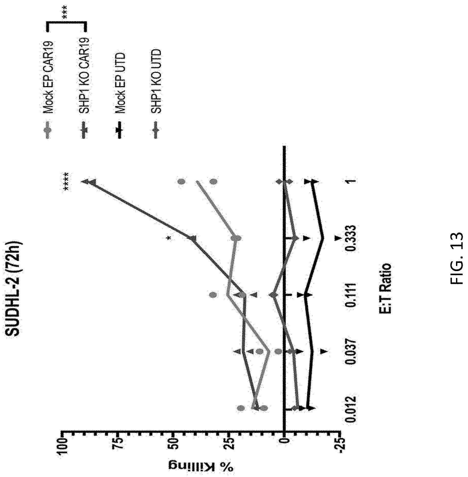
D00016
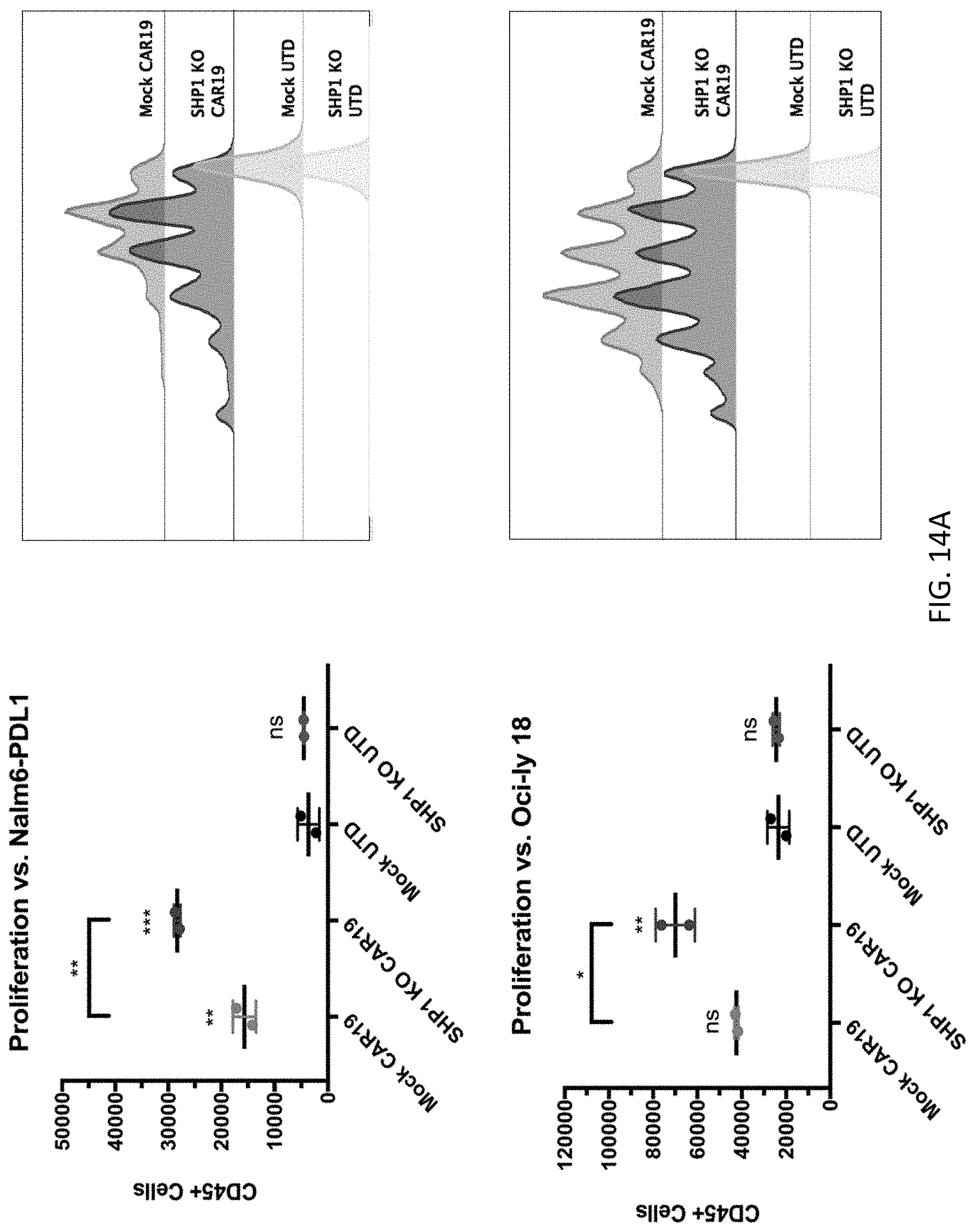
D00017
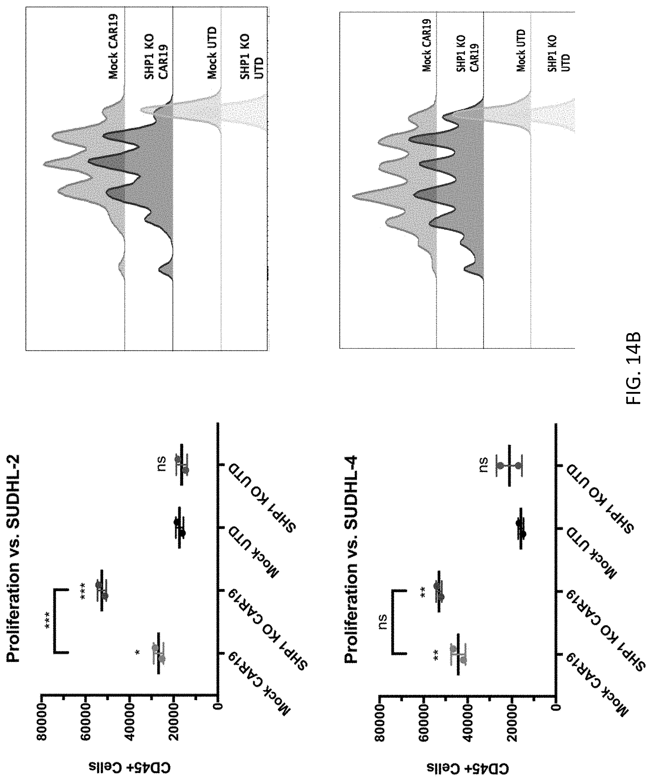
D00018

D00019
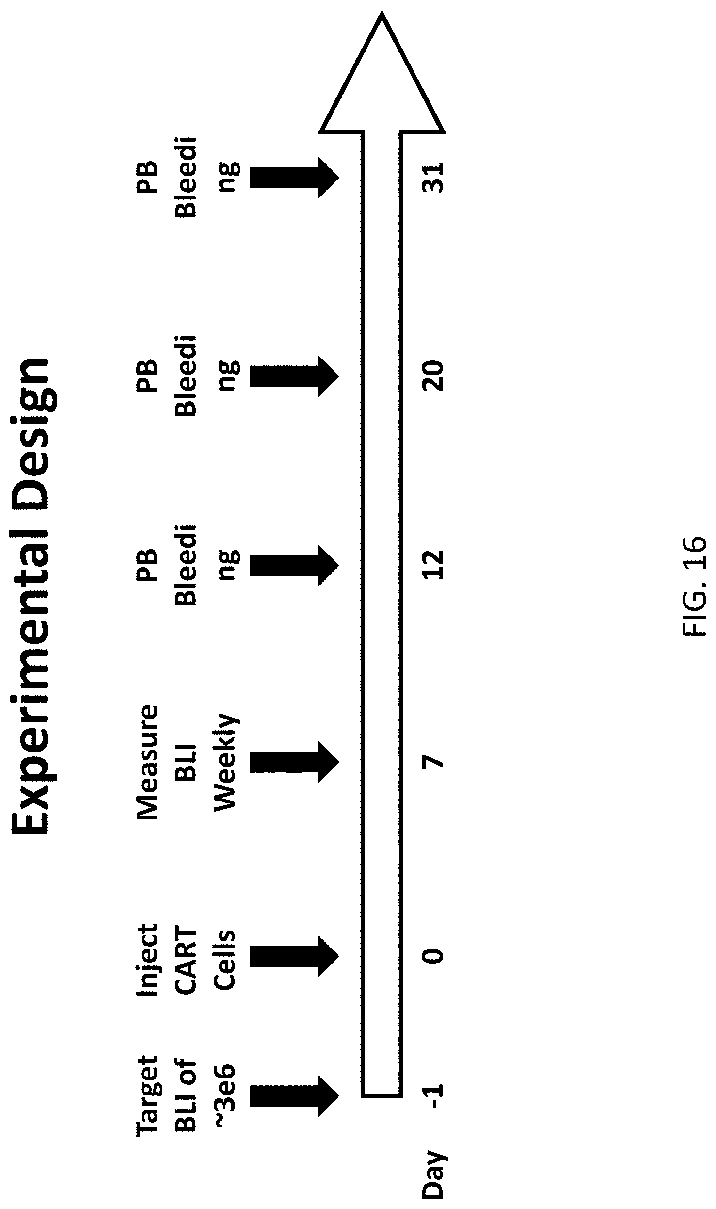
D00020
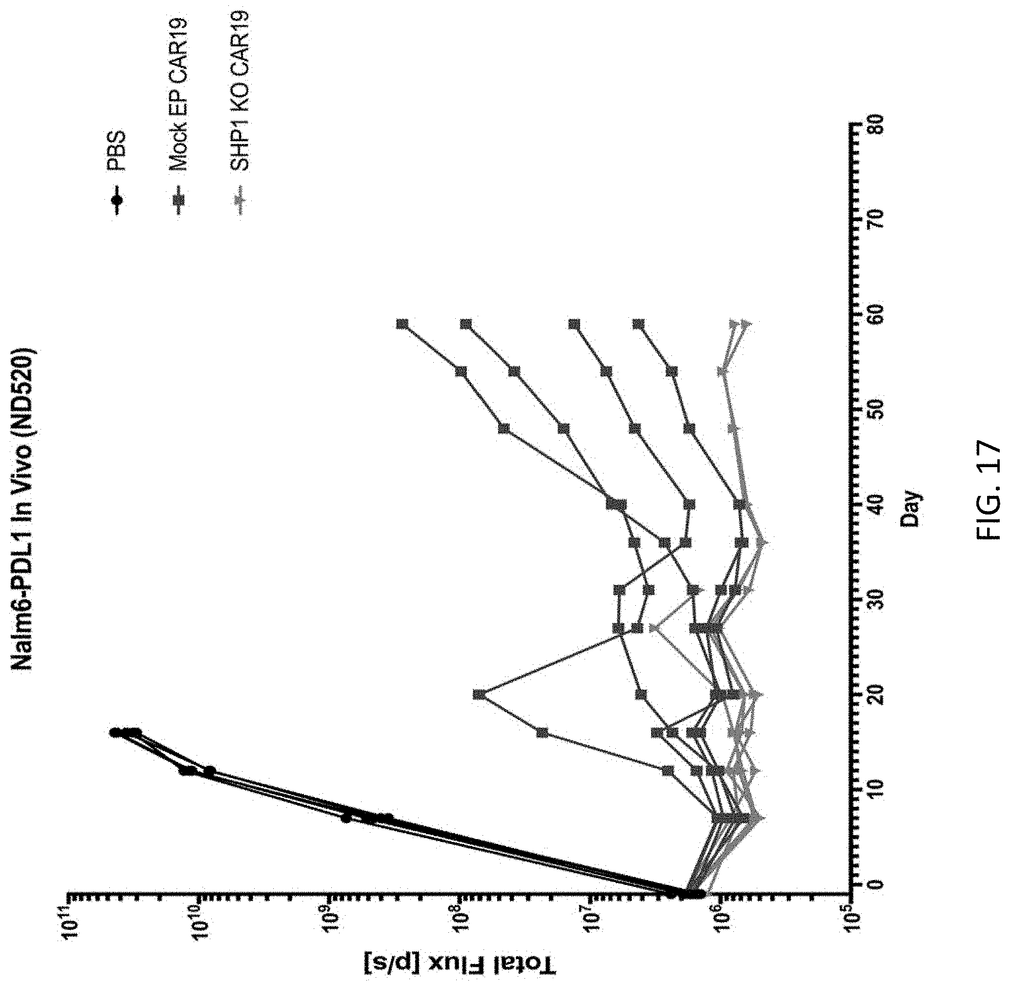
D00021
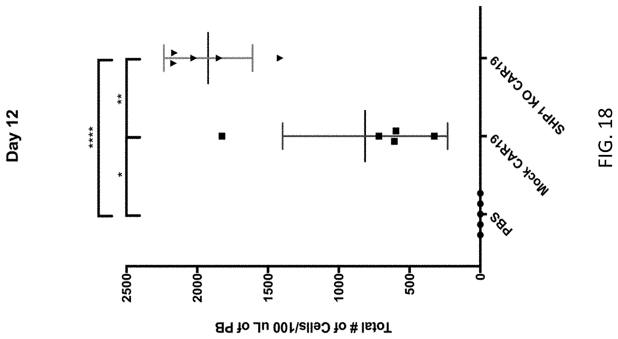
D00022
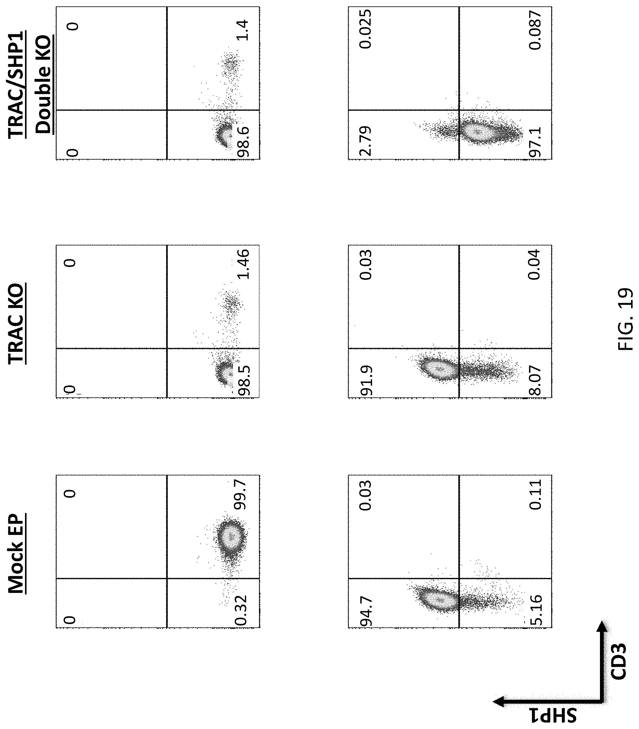
S00001
XML
uspto.report is an independent third-party trademark research tool that is not affiliated, endorsed, or sponsored by the United States Patent and Trademark Office (USPTO) or any other governmental organization. The information provided by uspto.report is based on publicly available data at the time of writing and is intended for informational purposes only.
While we strive to provide accurate and up-to-date information, we do not guarantee the accuracy, completeness, reliability, or suitability of the information displayed on this site. The use of this site is at your own risk. Any reliance you place on such information is therefore strictly at your own risk.
All official trademark data, including owner information, should be verified by visiting the official USPTO website at www.uspto.gov. This site is not intended to replace professional legal advice and should not be used as a substitute for consulting with a legal professional who is knowledgeable about trademark law.