Chimeric Antigen Receptors and Methods of Use
Orentas; Rimas ; et al.
U.S. patent application number 16/579082 was filed with the patent office on 2020-03-12 for chimeric antigen receptors and methods of use. The applicant listed for this patent is LENTIGEN TECHNOLOGY, INC.. Invention is credited to Boro Dropulic, Rimas Orentas, Dina Schneider.
| Application Number | 20200079849 16/579082 |
| Document ID | / |
| Family ID | 57145073 |
| Filed Date | 2020-03-12 |


View All Diagrams
| United States Patent Application | 20200079849 |
| Kind Code | A1 |
| Orentas; Rimas ; et al. | March 12, 2020 |
Chimeric Antigen Receptors and Methods of Use
Abstract
Chimeric antigen receptors containing tumor necrosis factor receptor superfamily member transmembrane domains are disclosed. Nucleic acids, recombinant expression vectors, host cells, antigen binding fragments, and pharmaceutical compositions, relating to the chimeric antigen receptors are also disclosed. Methods of treating or preventing cancer in a subject, and methods of making chimeric antigen receptor T cells are also disclosed.
| Inventors: | Orentas; Rimas; (Washington, DC) ; Schneider; Dina; (Potomac, MD) ; Dropulic; Boro; (Ellicott City, MD) | ||||||||||
| Applicant: |
|
||||||||||
|---|---|---|---|---|---|---|---|---|---|---|---|
| Family ID: | 57145073 | ||||||||||
| Appl. No.: | 16/579082 | ||||||||||
| Filed: | September 23, 2019 |
Related U.S. Patent Documents
| Application Number | Filing Date | Patent Number | ||
|---|---|---|---|---|
| 15767076 | Apr 9, 2018 | 10421810 | ||
| PCT/US2016/056073 | Oct 7, 2016 | |||
| 16579082 | ||||
| 62239509 | Oct 9, 2015 | |||
| Current U.S. Class: | 1/1 |
| Current CPC Class: | C07K 16/2803 20130101; C07K 2319/00 20130101; C07K 14/705 20130101; A61P 35/00 20180101; A61K 2039/505 20130101; C07K 2319/02 20130101; C07K 14/7051 20130101; C07K 14/70578 20130101; A61K 35/17 20130101; C07K 2319/03 20130101; C07K 19/00 20130101; C07K 2317/622 20130101; A61P 35/02 20180101; A61P 43/00 20180101; C12N 15/87 20130101; C07K 2319/74 20130101 |
| International Class: | C07K 16/28 20060101 C07K016/28; C12N 15/87 20060101 C12N015/87; C07K 14/705 20060101 C07K014/705; A61K 35/17 20060101 A61K035/17; C07K 19/00 20060101 C07K019/00; C07K 14/725 20060101 C07K014/725 |
Claims
1. An isolated nucleic acid molecule encoding a chimeric antigen receptor (CAR) comprising at least one extracellular antigen binding domain, at least one transmembrane tumor necrosis factor receptor superfamily (TNFRSF) domain, and at least one intracellular signaling domain, wherein the encoded CAR comprises the amino acid sequence of SEQ ID NO: 8, SEQ ID NO: 10, or SEQ ID NO: 36.
2.-22. (canceled)
23. A chimeric antigen receptor (CAR) encoded by the isolated nucleic acid molecule of claim 1.
24.-37. (canceled)
38. A vector comprising a nucleic acid molecule of claim 1.
39. The vector of claim 38, wherein the vector is selected from the group consisting of a DNA vector, an RNA vector, a plasmid vector, a cosmid vector, a herpes virus vector, a measles virus vector, a lentivirus vector, adenoviral vector, or a retrovirus vector, or a combination thereof.
40. The vector of claim 38, further comprising a promoter.
41. The vector of claim 40, wherein the promoter is an inducible promoter, a constitutive promoter, a tissue specific promoter, a suicide promoter or any combination thereof.
42. A cell comprising the vector of claim 38.
43. The cell of claim 42, wherein the cell is a T cell.
44. The cell of claim 43, wherein the T cell is a CD8+ T cell.
45. The cell of claim 42, wherein the cell is a human cell.
46. A method of making a cell comprising transducing a T cell with a vector of claim 38.
47. A method of generating a population of RNA-engineered cells comprising introducing an in vitro transcribed RNA or synthetic RNA into a cell, where the RNA comprises a nucleic acid molecule of claim 1.
48. A method of providing an anti-tumor immunity in a mammal comprising administering to the mammal an effective amount of a cell of claim 42.
49. A method of treating or preventing cancer in a mammal, comprising administering to the mammal the CAR of claim 23, in an amount effective to treat or prevent cancer in the mammal.
50. A pharmaceutical composition comprising an anti-tumor effective amount of a population of human T cells, wherein the T cells comprise the nucleic acid molecule of claim 1, wherein the T cells are T cells of a human having a cancer.
51. (canceled)
52. The pharmaceutical composition of claim 50, wherein the T cells are T cells of a human having a hematological cancer.
53. The pharmaceutical composition of claim 52, wherein the hematological cancer is leukemia or lymphoma.
54. The pharmaceutical composition of claim 53, wherein the leukemia is chronic lymphocytic leukemia (CLL), acute lymphocytic leukemia (ALL), or chronic myelogenous leukemia (CML).
55. The pharmaceutical composition of claim 53, wherein the lymphoma is mantle cell lymphoma, non-Hodgkin's lymphoma or Hodgkin's lymphoma.
56. The pharmaceutical composition of claim 52, wherein the hematological cancer is multiple myeloma.
57. The pharmaceutical composition of claim 50, wherein the human cancer includes an adult carcinoma comprising coral and pharynx cancer (tongue, mouth, pharynx, head and neck), digestive system cancers (esophagus, stomach, small intestine, colon, rectum, anus, liver, interhepatic bile duct, gallbladder, pancreas), respiratory system cancers (larynx, lung and bronchus), bones and joint cancers, soft tissue cancers, skin cancers (melanoma, basal and squamous cell carcinoma), pediatric tumors (neuroblastoma, rhabdomyosarcoma, osteosarcoma, Ewing's sarcoma), tumors of the central nervous system (brain, astrocytoma, glioblastoma, glioma), and cancers of the breast, the genital system (uterine cervix, uterine corpus, ovary, vulva, vagina, prostate, testis, penis, endometrium), the urinary system (urinary bladder, kidney and renal pelvis, ureter), the eye and orbit, the endocrine system (thyroid), and the brain and other nervous system, or any combination thereof.
58. A method of treating a mammal having a disease, disorder or condition associated with an elevated expression of a tumor antigen, the method comprising administering to the subject a pharmaceutical composition comprising an anti-tumor effective amount of a population of T cells, wherein the T cells comprise the nucleic acid molecule of claim 1, wherein the T cells are T cells of the subject having cancer.
59.-60. (canceled)
61. A process for producing a chimeric antigen receptor-expressing cell, the process comprising introducing the isolated nucleic acid of claim 1 into a cell.
62. The process for producing a chimeric antigen receptor-expressing cell according to claim 61, wherein the cell is a T cell or a cell population containing a T cell.
63. The isolated nucleic acid molecule of claim 1, wherein the nucleic acid sequence that encodes the CAR comprises the nucleic acid sequence of SEQ ID NO: 7, SEQ ID NO: 9, or SEQ ID NO: 35, or a sequence with 85%, 90%, 95%, 96%, 97%, 98% or 99% identity thereof.
Description
CROSS-REFERENCE TO RELATED APPLICATIONS
[0001] This application claims the benefit of priority under 35 U.S.C. Section 119(e) to U.S. Provisional Patent Application No. 62/239,509, filed on Oct. 9, 2015, the entire contents of which are incorporated herein by reference.
FIELD OF THE DISCLOSURE
[0002] This application relates to the field of cancer, particularly to chimeric antigen receptors (CARs) containing tumor necrosis factor receptor superfamily member transmembrane domains and methods of use.
BACKGROUND
[0003] Cancer is one of the deadliest threats to human health. In the U.S. alone, cancer affects nearly 1.3 million new patients each year, and is the second leading cause of death after cardiovascular disease, accounting for approximately 1 in 4 deaths. Solid tumors are responsible for most of those deaths. Although there have been significant advances in the medical treatment of certain cancers, the overall 5-year survival rate for all cancers has improved only by about 10% in the past 20 years. Cancers, or malignant tumors, metastasize and grow rapidly in an uncontrolled manner, making treatment extremely difficult.
[0004] Chimeric Antigen Receptors (CARs) are hybrid molecules comprising three essential units: (1) an extracellular antigen-binding motif, (2) linking/transmembrane motifs, and (3) intracellular T-cell signaling motifs (Long A H, Haso W M, Orentas R J. Lessons learned from a highly-active CD22-specific chimeric antigen receptor. Oncoimmunology. 2013; 2 (4):e23621). The antigen-binding motif of a CAR is commonly fashioned after a single chain Fragment variable (scFv), the minimal binding domain of an immunoglobulin (Ig) molecule. Alternate antigen-binding motifs, such as receptor ligands (i.e., IL-13 has been engineered to bind tumor expressed IL-13 receptor), intact immune receptors, library-derived peptides, and innate immune system effector molecules (such as NKG2D) also have been engineered. Alternate cell targets for CAR expression (such as NK or gamma-delta T cells) are also under development (Brown C E et al Clin Cancer Res. 2012; 18(8):2199-209; Lehner M et al. PLoS One. 2012; 7 (2):e31210). There remains significant work with regard to defining the most active T-cell population to transduce with CAR vectors, determining the optimal culture and expansion techniques, and defining the molecular details of the CAR protein structure itself.
[0005] The linking motifs of a CAR can be a relatively stable structural domain, such as the constant domain of IgG, or designed to be an extended flexible linker. Structural motifs, such as those derived from IgG constant domains, can be used to extend the scFv binding domain away from the T-cell plasma membrane surface. This may be important for some tumor targets where the binding domain is particularly close to the tumor cell surface membrane (such as for the disialoganglioside GD2; Orentas et al., unpublished observations). To date, the signaling motifs used in CARs always include the CD3-.zeta. chain because this core motif is the key signal for T cell activation. The first reported second-generation CARs featured CD28 signaling domains and the CD28 transmembrane sequence. This motif was used in third-generation CARs containing CD137 (4-1BB) signaling motifs as well (Zhao Y et al J Immunol. 2009; 183 (9): 5563-74). With the advent of new technology, the activation of T cells with beads linked to anti-CD3 and anti-CD28 antibody, and the presence of the canonical "signal 2" from CD28 was no longer required to be encoded by the CAR itself. Using bead activation, third-generation vectors were found to be not superior to second-generation vectors in in vitro assays, and they provided no clear benefit over second-generation vectors in mouse models of leukemia (Haso W, Lee D W, Shah N N, Stetler-Stevenson M, Yuan C M, Pastan I H, Dimitrov D S, Morgan R A, FitzGerald D J, Barrett D M, Wayne A S, Mackall C L, Orentas R J. Anti-CD22-chimeric antigen receptors targeting B cell precursor acute lymphoblastic leukemia, Blood. 2013; 121 (7):1165-74; Kochenderfer J N et al. Blood. 2012; 119 (12):2709-20). This is borne out by the clinical success of CD19-specific CARs that are in a second generation CD28/CD3-.zeta. (Lee D W et al. American Society of Hematology Annual Meeting. New Orleans, La.; Dec. 7-10, 2013) and a CD137/CD3-.zeta. signaling format (Porter D L et al. N Engl J Med. 2011; 365 (8): 725-33). In addition to CD137, other tumor necrosis factor receptor superfamily members such as OX40 also are able to provide important persistence signals in CAR-transduced T cells (Yvon E et al. Clin Cancer Res. 2009; 15(18):5852-60). Equally important are the culture conditions under which the CAR T-cell populations were cultured.
[0006] Current challenges in the more widespread and effective adaptation of CAR therapy for cancer relate to a paucity of compelling targets. Creating binders to cell surface antigens is now readily achievable, but discovering a cell surface antigen that is specific for tumor while sparing normal tissues remains a formidable challenge. One potential way to imbue greater target cell specificity to CAR-expressing T cells is to use combinatorial CAR approaches. In one system, the CD3-.zeta. and CD28 signal units are split between two different CAR constructs expressed in the same cell; in another, two CARs are expressed in the same T cell, but one has a lower affinity and thus requires the alternate CAR to be engaged first for full activity of the second (Lanitis E et al. Cancer Immunol Res. 2013; 1(1):43-53; Kloss C C et al. Nat Biotechnol. 2013; 31(1):71-5). A second challenge for the generation of a single scFv-based CAR as an immunotherapeutic agent is tumor cell heterogeneity. At least one group has developed a CAR strategy for glioblastoma whereby the effector cell population targets multiple antigens (HER2, IL-13Ra, EphA2) at the same time in the hope of avoiding the outgrowth of target antigen-negative populations (Hegde M et al. Mol Ther. 2013; 21(11):2087-101).
[0007] T-cell-based immunotherapy has become a new frontier in synthetic biology; multiple promoters and gene products are envisioned to steer these highly potent cells to the tumor microenvironment, where T cells can both evade negative regulatory signals and mediate effective tumor killing. The elimination of unwanted T cells through the drug-induced dimerization of inducible caspase 9 constructs with AP1903 demonstrates one way in which a powerful switch that can control T-cell populations can be initiated pharmacologically (Di Stasi A et al. N Engl J Med. 2011; 365(18):1673-83). The creation of effector T-cell populations that are immune to the negative regulatory effects of transforming growth factor-.beta. by the expression of a decoy receptor further demonstrates that degree to which effector T cells can be engineered for optimal antitumor activity (Foster A E et al. J Immunother. 2008; 31(5):500-5).
[0008] Thus, while it appears that CARs can trigger T-cell activation in a manner similar to an endogenous T-cell receptor, a major impediment to the clinical application of this technology to date has been limited in vivo expansion of CAR+ T cells, rapid disappearance of the cells after infusion, and disappointing clinical activity. Accordingly, there is an urgent and long felt need in the art for discovering compositions and methods for treatment of cancer using CARs that can exhibit intended therapeutic attributes without the aforementioned short comings.
[0009] The present invention addresses these needs by providing CAR compositions and therapeutic methods that can be used to treat cancers and other diseases and/or conditions. In particular, the present invention as disclosed and described herein provides CARs that exhibit a high surface expression on transduced T cells, exhibit a high degree of cytolysis and transduced T cell in vivo expansion and persistence.
SUMMARY
[0010] Novel chimeric antigen receptors (CARs) that contain tumor necrosis factor receptor superfamily (TNFRSF) member transmembrane domains are provided herein, as well as host cells (e.g., T cells) expressing the receptors, and nucleic acid molecules encoding the receptors. The CARs exhibit a high surface expression on transduced T cells, with a high degree of cytolysis and transduced T cell expansion and persistence in vivo. Methods of using the disclosed CARs, host cells, and nucleic acid molecules are also provided, for example, to treat a cancer in a subject.
[0011] In one aspect, an isolated nucleic acid molecule encoding a chimeric antigen receptor (CAR) is provided comprising, from N-terminus to C-terminus, at least one extracellular antigen binding domain, at least one transmembrane TNFRSF domain, and at least one intracellular signaling domain.
[0012] In one embodiment, an isolated nucleic acid molecule encoding the CAR is provided wherein the encoded extracellular antigen binding domain comprises at least one single chain variable fragment of an antibody that binds to the antigen.
[0013] In another embodiment, an isolated nucleic acid molecule encoding the CAR is provided wherein the encoded extracellular antigen binding domain comprises at least one heavy chain variable region of an antibody that binds to the antigen.
[0014] In yet another embodiment, an isolated nucleic acid molecule encoding the CAR is provided wherein the encoded CAR extracellular antigen binding domain comprises at least one lipocalin-based antigen binding antigen (anticalins) that binds to the antigen.
[0015] In one embodiment, an isolated nucleic acid molecule is provided wherein the encoded extracellular antigen binding domain is connected to the TNFRSF transmembrane domain by a linker domain.
[0016] In another embodiment, an isolated nucleic acid molecule encoding the CAR is provided wherein the encoded extracellular antigen binding domain is preceded by a sequence encoding a leader or signal peptide.
[0017] In yet another embodiment, an isolated nucleic acid molecule encoding the CAR is provided wherein the encoded extracellular antigen binding domain targets an antigen that includes, but is not limited to, CD19, CD20, CD22, ROR1, mesothelin, CD33, CD38, CD123 (IL3RA), CD138, BCMA (CD269), GPC2, GPC3, FGFR4, c-Met, PSMA, Glycolipid F77, EGFRvIII, GD-2, NY-ESO-1 TCR, MAGE A3 TCR, or any combination thereof.
[0018] In certain embodiments, an isolated nucleic acid molecule encoding the CAR is provided wherein the encoded extracellular antigen binding domain comprises an anti-CD19 scFV antigen binding domain, an anti-CD20 scFV antigen binding domain, an anti-CD22 scFV antigen binding domain, an anti-ROR1 scFV antigen binding domain, an anti-mesothelin scFV antigen binding domain, an anti-CD33 scFV antigen binding domain, an anti-CD38 scFV antigen binding domain, an anti-CD123 (IL3RA) scFV antigen binding domain, an anti-CD138 scFV antigen binding domain, an anti-BCMA (CD269) scFV antigen binding domain, an anti-GPC2 scFV antigen binding domain, an anti-GPC3 scFV antigen binding domain, an anti-FGFR4 scFV antigen binding domain, an anti-c-Met scFV antigen binding domain, an anti-PMSA scFV antigen binding domain, an anti-glycolipid F77 scFV antigen binding domain, an anti-EGFRvIII scFV antigen binding domain, an anti-GD-2 scFV antigen binding domain, an anti-NY-ESo-1 TCR scFV antigen binding domain, an anti-MAGE A3 TCR scFV antigen binding domain, or an amino acid sequence with 85%, 90%, 95%, 96%, 97%, 98% or 99% identity thereof, or any combination thereof.
[0019] In one aspect, the CARs provided herein further comprise a linker or spacer domain.
[0020] In one embodiment, an isolated nucleic acid molecule encoding the CAR is provided wherein the extracellular antigen binding domain, the intracellular signaling domain, or both are connected to the transmembrane domain by a linker or spacer domain.
[0021] In one embodiment, an isolated nucleic acid molecule encoding the CAR is provided wherein the encoded linker domain is derived from the extracellular domain of CD8, and is linked to the transmembrane TNFRSF16 domain, the transmembrane TNFRSF19 domain, or a combination thereof.
[0022] In another embodiment, an isolated nucleic acid molecule encoding the CAR is provided wherein the encoded linker domain is derived from the extracellular domain of TNFRSF 16 or TNFRSF 19, and is linked to the transmembrane TNFRSF16 domain, the transmembrane TNFRSF19 domain, or a combination thereof.
[0023] In a further embodiment, an isolated nucleic acid molecule encoding the CAR is provided wherein the encoded transmembrane TNFRSF domain comprises a transmembrane TNFRSF16 domain, a transmembrane TNFRSF 19 domain, or a combination thereof.
[0024] In yet another embodiment, an isolated nucleic acid molecule encoding the CAR is provided wherein the nucleic acid sequence encoding the TNFRSF16 transmembrane domain comprises a sequence of SEQ ID NO: 1, or a sequence with 85%, 90%, 95%, 96%, 97%, 98% or 99% identity thereof.
[0025] In one embodiment, an isolated nucleic acid molecule encoding the CAR is provided wherein the encoded TNFRSF16 transmembrane domain comprises a sequence of SEQ ID NO: 2, or an amino acid sequence comprising at least one but not more 10 modifications of the amino acid sequence of SEQ ID NO: 2, or a sequence with 85%, 90%, 95%, 96%, 97%, 98% or 99% identity to an amino acid sequence of SEQ ID NO: 2.
[0026] In yet another embodiment, an isolated nucleic acid molecule encoding the CAR is provided wherein the nucleic acid sequence encoding the TNFRSF19 transmembrane domain comprises a sequence of SEQ ID NO: 3, or a sequence with 85%, 90%, 95%, 96%, 97%, 98% or 99% identity thereof.
[0027] In one embodiment, an isolated nucleic acid molecule encoding the CAR is provided wherein the encoded TNFRSF19 transmembrane domain comprises a sequence of SEQ ID NO: 4, or an amino acid sequence comprising at least one but not more than 10 modifications of the amino acid sequence of SEQ ID NO: 4, or a sequence with 85%, 90%, 95%, 96%, 97%, 98% or 99% identity to an amino acid sequence of SEQ ID NO: 4.
[0028] In another embodiment, an isolated nucleic acid molecule encoding the CAR is provided wherein the encoded CAR further comprises a transmembrane domain that comprises a transmembrane domain of a protein selected from the group consisting of the alpha, beta or zeta chain of the T-cell receptor, CD28, CD3 epsilon, CD45, CD4, CD5, CD8, CD9, CD16, CD22, CD33, CD37, CD64, CD80, CD86, CD134, CD137 and CD154, or a combination thereof.
[0029] In yet another embodiment, an isolated nucleic acid molecule encoding the CAR is provided wherein the encoded intracellular signaling domain further comprises a CD3 zeta intracellular domain.
[0030] In one embodiment, an isolated nucleic acid molecule encoding the CAR is provided wherein the encoded intracellular signaling domain is arranged on a C-terminal side relative to the CD3 zeta intracellular domain.
[0031] In another embodiment, an isolated nucleic acid molecule encoding the CAR is provided wherein the encoded at least one intracellular signaling domain comprises a costimulatory domain, a primary signaling domain, or a combination thereof.
[0032] In further embodiments, an isolated nucleic acid molecule encoding the CAR is provided wherein the encoded at least one costimulatory domain comprises a functional signaling domain of OX40, CD70, CD27, CD28, CD5, ICAM-1, LFA-1 (CD11a/CD18), ICOS (CD278), DAP10, DAP12, and 4-1BB (CD137), or a combination thereof.
[0033] In one embodiment, an isolated nucleic acid molecule encoding the CAR is provided that further contains a leader sequence or signal peptide wherein the leader or signal peptide nucleotide sequence comprises the nucleotide sequence of SEQ ID NO: 5.
[0034] In yet another embodiment, an isolated nucleic acid molecule encoding the CAR is provided wherein the encoded leader sequence comprises the amino acid sequence of SEQ ID NO: 6.
[0035] In one aspect, a chimeric antigen receptor (CAR) is provided herein comprising, from N-terminus to C-terminus, at least one extracellular antigen binding domain, at least one transmembrane TNFRSF domain, and at least one intracellular signaling domain.
[0036] In one embodiment, a CAR is provided wherein the extracellular antigen binding domain comprises at least one single chain variable fragment of an antibody that binds to the antigen, or at least one heavy chain variable region of an antibody that binds to the antigen, or a combination thereof.
[0037] In another embodiment, a CAR is provided wherein the at least one transmembrane TNFRSF domain comprises a transmembrane TNFRSF19 domain, a transmembrane TNFRSF16 domain, or a combination thereof.
[0038] In some embodiments, the CAR is provided wherein the extracellular antigen binding domain comprises CD19, CD20, CD22, ROR1, mesothelin, CD33, CD38, CD123 (IL3RA), CD138, BCMA (CD269), GPC2, GPC3, FGFR4, c-Met, PSMA, Glycolipid F77, EGFRvIII, GD-2, NY-ESO-1 TCR, MAGE A3 TCR, or an amino acid sequence with 85%, 90%, 95%, 96%, 97%, 98% or 99% identity thereof, or any combination thereof.
[0039] In one embodiment, the CAR is provided wherein the extracellular antigen binding domain comprises an anti-CD19 scFV antigen binding domain, an anti-CD20 scFV antigen binding domain, an anti-CD22 scFV antigen binding domain, an anti-ROR1 scFV antigen binding domain, an anti-mesothelin scFV antigen binding domain, an anti-CD33 scFV antigen binding domain, an anti-CD38 scFV antigen binding domain, an anti-CD123 (IL3RA) scFV antigen binding domain, an anti-CD138 scFV antigen binding domain, an anti-BCMA (CD269) scFV antigen binding domain, an anti-GPC2 scFV antigen binding domain, an anti-GPC3 scFV antigen binding domain, an anti-FGFR4 scFV antigen binding domain, an anti-c-Met scFV antigen binding domain, an anti-PMSA scFV antigen binding domain, an anti-glycolipid F77 scFV antigen binding domain, an anti-EGFRvIII scFV antigen binding domain, an anti-GD-2 scFV antigen binding domain, an anti-NY-ESo-1 TCR scFV antigen binding domain, an anti-MAGE A3 TCR scFV antigen binding domain, or an amino acid sequence with 85%, 90%, 95%, 96%, 97%, 98% or 99% identity thereof, or any combination thereof.
[0040] In another embodiment, a CAR is provided wherein the at least one intracellular signaling domain comprises a costimulatory domain and a primary signaling domain.
[0041] In yet another embodiment, a CAR is provided wherein the at least one intracellular signaling domain comprises a costimulatory domain comprising a functional signaling domain of a protein selected from the group consisting of OX40, CD70, CD27, CD28, CD5, ICAM-1, LFA-1 (CD11a/CD18), ICOS (CD278), DAP10, DAP12, and 4-1BB (CD137), or a combination thereof.
[0042] In one embodiment, the nucleic acid sequence encoding a CAR comprises the nucleic acid sequence of SEQ ID NO: 7. In one embodiment, the nucleic acid sequence encodes a CAR comprising the amino acid sequence of SEQ ID NO: 8.
[0043] In another embodiment, the nucleic acid sequence encoding a CAR comprises the nucleic acid sequence of SEQ ID NO: 9. In one embodiment, the nucleic acid sequence encodes a CAR comprising the amino acid sequence of SEQ ID NO: 10.
[0044] In another embodiment, the nucleic acid sequence encoding a CAR comprises the nucleic acid sequence of SEQ ID NO: 21. In one embodiment, the nucleic acid sequence encodes a CAR comprising the amino acid sequence of SEQ ID NO: 22.
[0045] In one aspect, the CARs disclosed herein are modified to express or contain a detectable marker for use in diagnosis, monitoring, and/or predicting the treatment outcome such as progression free survival of cancer patients or for monitoring the progress of such treatment.
[0046] In one embodiment, the nucleic acid molecule encoding the disclosed CARs can be contained in a vector, such as a viral vector. The vector is a DNA vector, an RNA vector, a plasmid vector, a cosmid vector, a herpes virus vector, a measles virus vector, a lentivirus vector, adenoviral vector, or a retrovirus vector, or a combination thereof.
[0047] In certain embodiments, the vector further comprises a promoter wherein the promoter is an inducible promoter, a tissue specific promoter, a constitutive promoter, a suicide promoter or any combination thereof.
[0048] In yet another embodiment, the vector expressing the CAR can be further modified to include one or more operative elements to control the expression of CAR T cells, or to eliminate CAR-T cells by virtue of a suicide switch. The suicide switch can include, for example, an apoptosis inducing signaling cascade or a drug that induces cell death. In a preferred embodiment, the vector expressing the CAR can be further modified to express an enzyme such thymidine kinase (TK) or cytosine deaminase (CD).
[0049] In another aspect, host cells including the nucleic acid molecule encoding the CAR are also provided. In some embodiments, the host cell is a T cell, such as a primary T cell obtained from a subject. In one embodiment, the host cell is a CD8+ T cell.
[0050] In yet another aspect, a pharmaceutical composition is provided comprising an anti-tumor effective amount of a population of human T cells, wherein the T cells comprise a nucleic acid sequence that encodes a chimeric antigen receptor (CAR), wherein the CAR comprises at least one extracellular antigen binding domain, at least one linker domain, at least one transmembrane TNFRSF domain, and at least one intracellular signaling domain, wherein the T cells are T cells of a human having a cancer. The cancer includes, inter alia, a hematological cancer such as leukemia (e.g., chronic lymphocytic leukemia (CLL), acute lymphocytic leukemia (ALL), or chronic myelogenous leukemia (CML), lymphoma (e.g., mantle cell lymphoma, non-Hodgkin's lymphoma or Hodgkin's lymphoma) or multiple myeloma, or a combination thereof.
[0051] In one embodiment, a pharmaceutical composition is provided wherein the at least one transmembrane TNFRSF domain of the CAR contains a transmembrane TNFRSF16 domain, a transmembrane TNFRSF19 domain or a combination thereof.
[0052] In another embodiment, a pharmaceutical composition is provided wherein the human cancer includes an adult carcinoma comprising coral and pharynx cancer (tongue, mouth, pharynx, head and neck), digestive system cancers (esophagus, stomach, small intestine, colon, rectum, anus, liver, interhepatic bile duct, gallbladder, pancreas), respiratory system cancers (larynx, lung and bronchus), bones and joint cancers, soft tissue cancers, skin cancers (melanoma, basal and squamous cell carcinoma), pediatric tumors (neuroblastoma, rhabdomyosarcoma, osteosarcoma, Ewing's sarcoma), tumors of the central nervous system (brain, astrocytoma, glioblastoma, glioma), and cancers of the breast, the genital system (uterine cervix, uterine corpus, ovary, vulva, vagina, prostate, testis, penis, endometrium), the urinary system (urinary bladder, kidney and renal pelvis, ureter), the eye and orbit, the endocrine system (thyroid), and the brain and other nervous system, or any combination thereof.
[0053] In yet another embodiment, a pharmaceutical composition is provided comprising an anti-tumor effective amount of a population of human T cells of a human having a cancer wherein the cancer is a refractory cancer non-responsive to one or more chemotherapeutic agents. The cancer includes hematopoietic cancer, myelodysplastic syndrome pancreatic cancer, head and neck cancer, cutaneous tumors, minimal residual disease (MRD) in acute lymphoblastic leukemia (ALL), acute myeloid leukemia (AML), adult B cell malignancies including, CLL (Chronic lymphocytic leukemia), CML (chronic myelogenous leukemia), non-Hodgkin's lymphoma (NHL), pediatric B cell malignancies (including B lineage ALL (acute lymphocytic leukemia)), multiple myeloma lung cancer, breast cancer, ovarian cancer, prostate cancer, colon cancer, melanoma or other hematological cancer and solid tumors, or any combination thereof.
[0054] In another aspect, methods of making CAR-containing T cells (hereinafter "CAR-T cells") are provided. The methods include transducing a T cell with a vector or nucleic acid molecule encoding a disclosed CAR that specifically binds an antigen, thereby making the CAR-T cell.
[0055] In yet another aspect, a method of generating a population of RNA-engineered cells is provided that comprises introducing an in vitro transcribed RNA or synthetic RNA of a nucleic acid molecule encoding a disclosed CAR into a cell of a subject, thereby generating a CAR cell.
[0056] In another embodiment, a method is provided for inducing an anti-tumor immunity in a mammal comprising administering to the mammal a therapeutically effective amount of a T cell transduced with vector or nucleic acid molecule encoding a disclosed CAR.
[0057] In another embodiment, a method of treating or preventing cancer in a mammal is provided comprising administering to the mammal one or more of the disclosed CARs, in an amount effective to treat or prevent cancer in the mammal. The method includes administering to the subject a therapeutically effective amount of host cells expressing a disclosed CAR that specifically binds one or more of the aforementioned antigens, under conditions sufficient to form an immune complex of the antigen binding domain on the CAR and the extracellular domain of one or more of the aforementioned antigens in the subject.
[0058] In yet another embodiment, a method is provided for treating a mammal having a disease, disorder or condition associated with an elevated expression of a tumor antigen, the method comprising administering to the subject a pharmaceutical composition comprising an anti-tumor effective amount of a population of T cells, wherein the T cells comprise a nucleic acid sequence that encodes a chimeric antigen receptor (CAR), wherein the CAR includes at least one extracellular antigen binding domain, at least one linker or spacer domain, at least one transmembrane TNFRSF domain, at least one intracellular signaling domain, and wherein the T cells are T cells of the subject having cancer.
[0059] In yet another embodiment, a method is provided for treating cancer in a subject in need thereof comprising administering to the subject a pharmaceutical composition comprising an anti-tumor effective amount of a population of T cells, wherein the T cells comprise a nucleic acid sequence that encodes a chimeric antigen receptor (CAR), wherein the CAR comprises at least one extracellular antigen binding domain, at least one linker or spacer domain, at least one transmembrane TNFRSF domain, at least one intracellular signaling domain, wherein the T cells are T cells of the subject having cancer. In some embodiments of the aforementioned methods, the at least one transmembrane TNFRSF domain comprises a transmembrane TNFRSF16 domain, a transmembrane TNFRSF19 domain or a combination thereof.
[0060] In yet another embodiment, a method is provided for generating a persisting population of genetically engineered T cells in a human diagnosed with cancer. In one embodiment, the method comprises administering to a human a T cell genetically engineered to express a CAR wherein the CAR comprises at least one extracellular antigen binding domain, at least one transmembrane TNFRSF domain, and at least one intracellular signaling domain wherein the persisting population of genetically engineered T cells, or the population of progeny of the T cells, persists in the human for at least one month, two months, three months, four months, five months, six months, seven months, eight months, nine months, ten months, eleven months, twelve months, two years, or three years after administration.
[0061] In one embodiment, the progeny T cells in the human comprise a memory T cell. In another embodiment, the T cell is an autologous T cell.
[0062] In all of the aspects and embodiments of methods described herein, any of the aforementioned cancers, diseases, disorders or conditions associated with an elevated expression of a tumor antigen that may be treated or prevented or ameliorated using one or more of the CARs disclosed herein,
[0063] In yet another aspect, a kit is provided for making a chimeric antigen receptor T-cell as described supra or for preventing, treating, or ameliorating any of the cancers, diseases, disorders or conditions associated with an elevated expression of a tumor antigen in a subject as described supra, comprising a container comprising any one of the nucleic acid molecules, vectors, host cells, or compositions disclosed supra or any combination thereof, and instructions for using the kit.
[0064] It will be understood that the CARs, host cells, nucleic acids, and methods are useful beyond the specific aspects and embodiments that are described in detail herein. The foregoing features and advantages of the disclosure will become more apparent from the following detailed description, which proceeds with reference to the accompanying figures.
BRIEF DESCRIPTION OF THE FIGURES
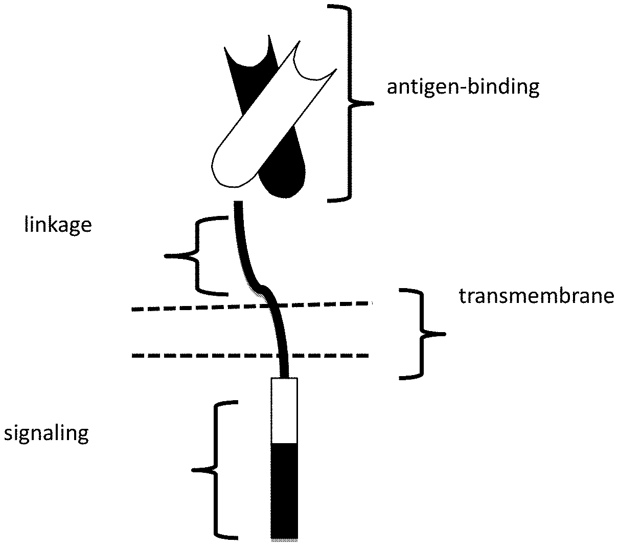
[0065] FIG. 1 depicts a schematic of a chimeric antigen receptor (CAR). The CAR contains an antigen binding domain, a linkage domain, a transmembrane domain and an intracellular signaling domain.
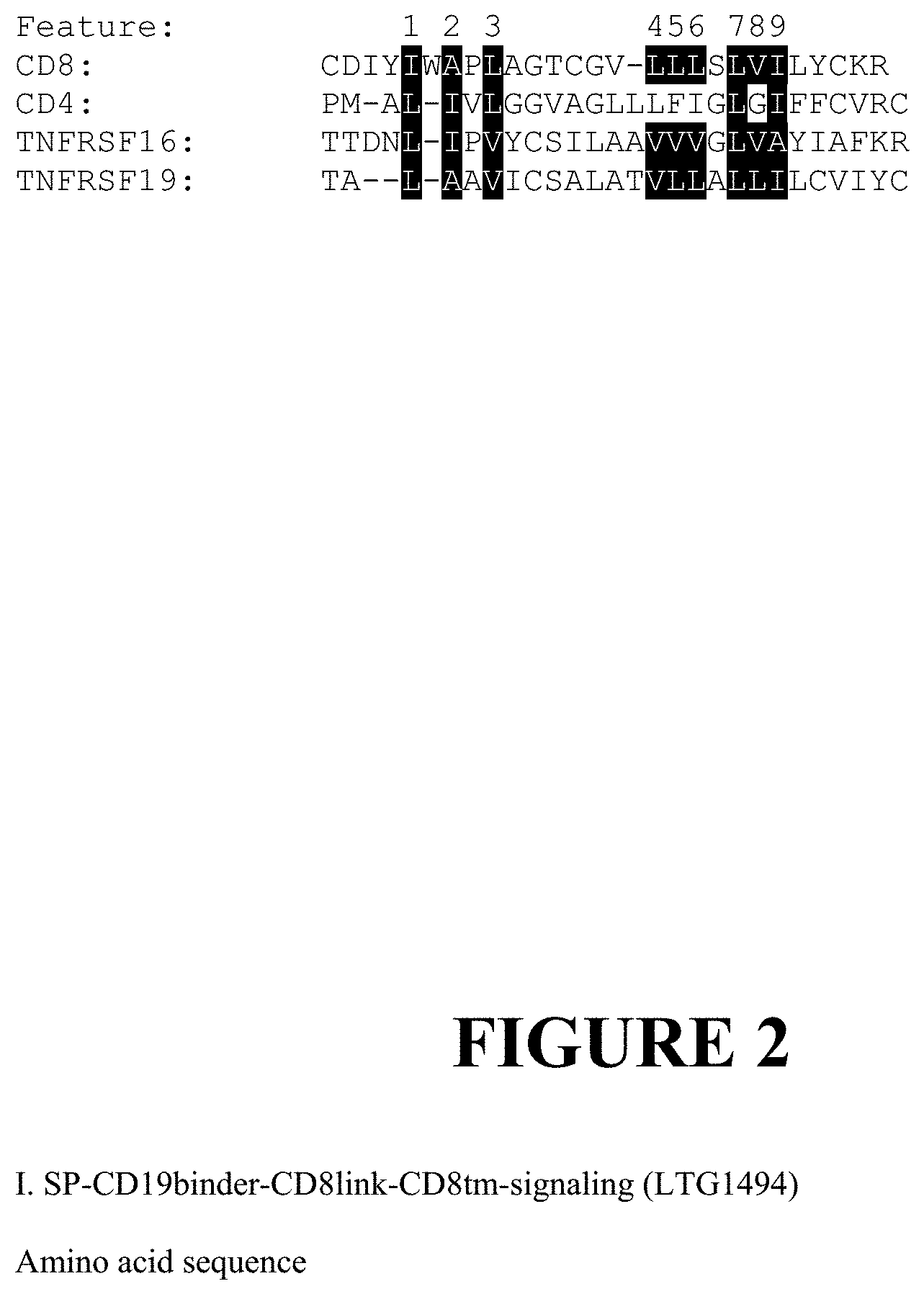
[0066] FIG. 2 depicts certain transmembrane amino acid motifs in TNFRSF16 and TNFRSF19. The transmembrane amino acid sequences of CD8, CD4, TNFRSF16 and TNFRSF19 are also shown. Nine distinct features are highlighted in the header of FIG. 2. Each is an amino acid encoded by the human genome in the context of a transmembrane plasma membrane protein. The Amino acid abbreviations used herein are according to the IUPAC-IUB Joint Commission on Biochemical Nomenclature (JCBN).
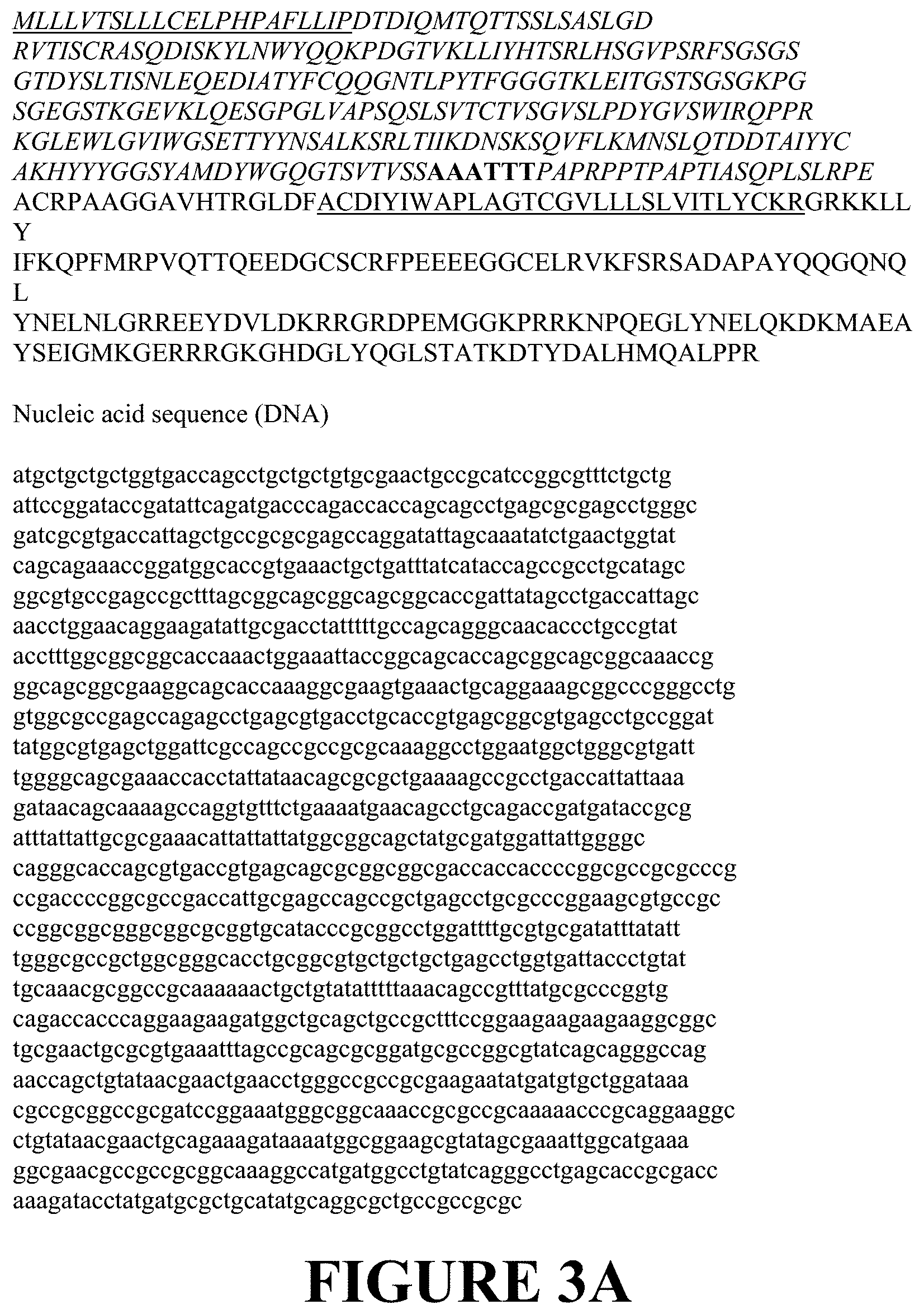
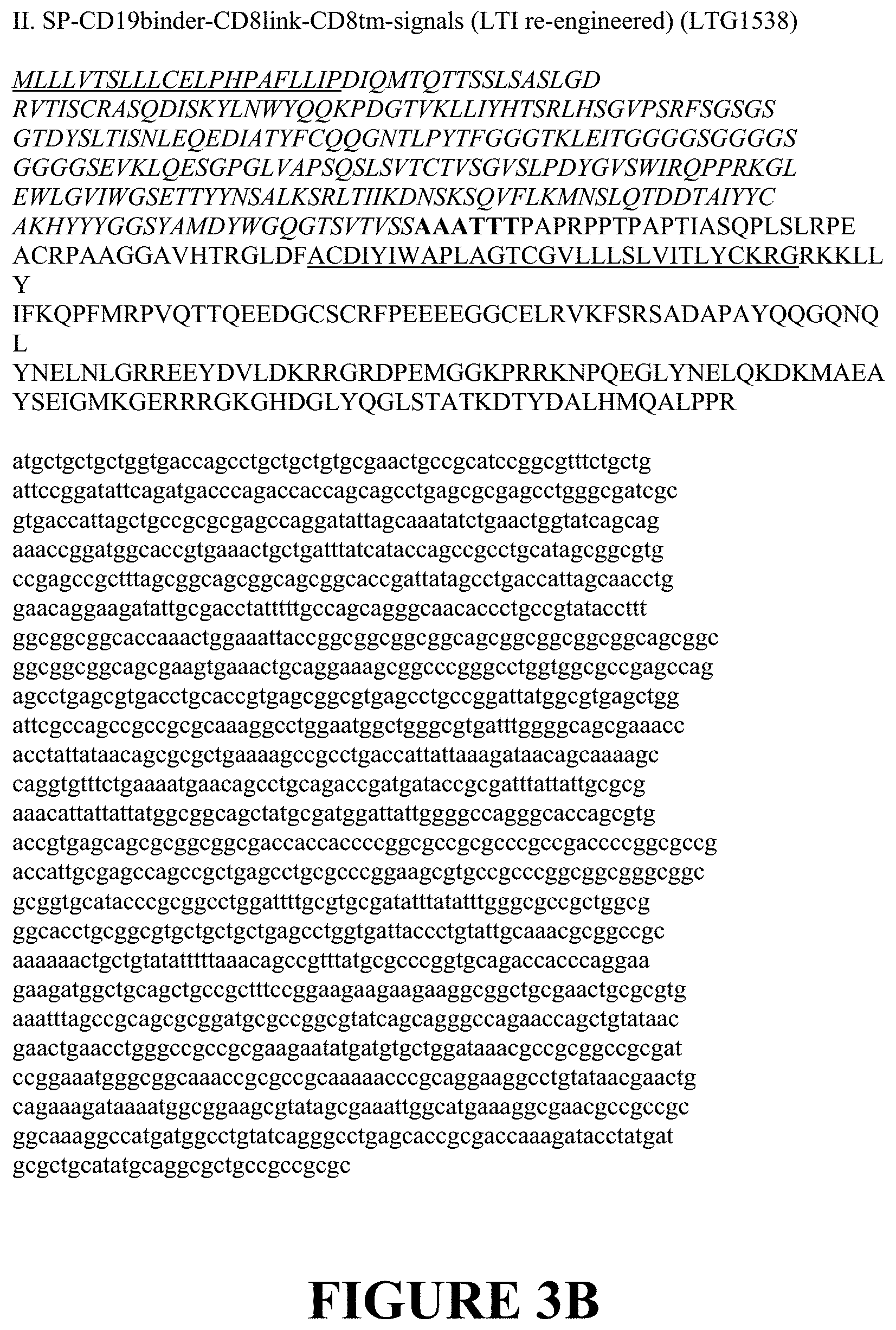
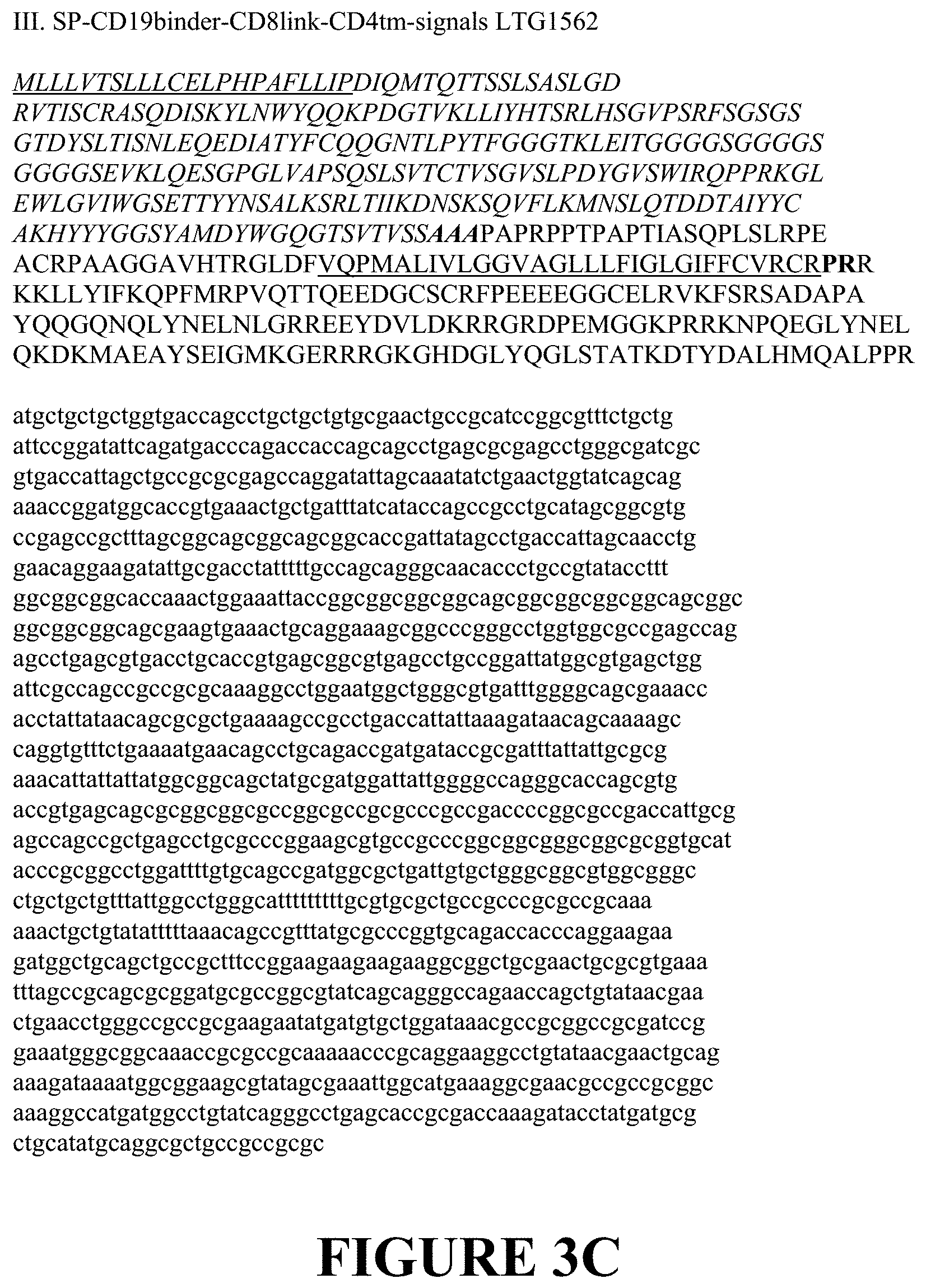
[0067] FIG. 3 depicts several chimeric antigen receptors (CARs). The general scheme for the CARs includes, from the N terminus to the C terminus, a Signal peptide, Anti-CD19 scFv/FMC63, extracellular linker, transmembrane, 4-1BB, CD3 zeta wherein the bolded text represents the cloning sites for linking domains. FIG. 3A depicts a lentiviral vector expressing the CAR containing SP-CD19binder-CD8link-CD8tm-signaling (LTG1494). FIG. 3B depicts a lentiviral vector expressing the CAR containing SP-CD19binder-CD8link-CD8tm-signals (LTI re-engineered) (LTG1538). FIG. 3C depicts a lentiviral vector expressing the CAR containing SP-CD19binder-CD8link-CD4tm-signals (LTG1562). FIG. 3D depicts a lentiviral vector expressing the CAR containing SP-CD19binder-CD8link-TNFRSF19tm-signals (LTG1563). FIG. 3E depicts a lentiviral vector expressing the CAR containing SP-CD19binder-TNFRSF191ink-TNFRSF19tm-signals (LTG1564).
[0068] FIG. 4 depicts a schematic of the general domain structure of CARs with novel transmembrane sequences. Depicted is a series of unique CARs utilizing an anti-CD19 antibody FMC63 (binding domain A). Binding domains were linked to CD8 linkers and CD8 transmembrane domains, CAR format (A), CD4 (B), TNFRSF19 (C) transmembrane domains, or to a TNFRSF19 linker and transmembrane domain (D).
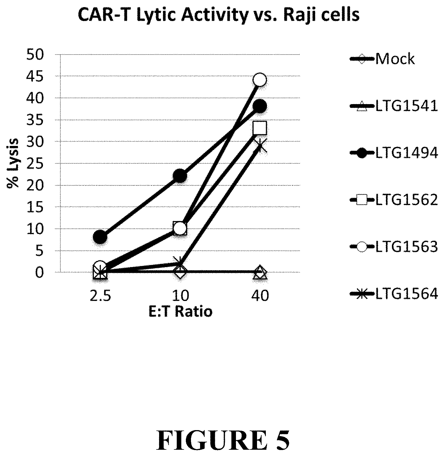
[0069] FIG. 5 depicts the anti-tumor activity of CARs containing the FMC63-derived anti-CD19 binding motif, the 4-1BB/CD3-zeta chain signaling motif and the TNFRSF transmembrane domains. Non-transduced expanded T cells (mock, open diamond) or T cells transduced with a LV expressing a control protein (LNGFR, LTG, open triangle) served as controls. The CAR-T featuring a CD8-derived linker and CD8-transmembrane domain (LTG1494, solid circles) showed strong lytic activity at the effector to target (E:T) ratios listed on the x-axis. The CAR-T expressing the CD8 linker and CD4 transmembrane (LTG1562, open square) or TNFRSF19 linker and transmembrane domains both showed appreciable lytic activity as well (LTG1564, star). The CAR-T expressed CD8 linker and TNFRSF19 transmembrane regions were tested (LTG1563, open circle) exhibited very strong lytic activity.
[0070] FIG. 6 depicts a diagram of novel linker and transmembrane domain CAR19 constructs. Six CAR19 constructs termed 1562, 1563, 1564 and 1712, 1713 and 1714, incorporating novel linker and transmembrane domain elements, were designed in order to determine whether altering CAR T linker and transmembrane domain composition alters CAR-T function. Construct 1494 denotes a CAR19 configuration comprised of FMC63 scFv binding domain, CD8 linker, CD8 transmembrane domain, CD137/4-1BB and CD3 zeta signaling domains, and serves as a positive control. "transM" refers to the transmembrane domain. "TruncT19" refers to a truncated TNFRSF19 linker in comparison to the extended version present in construct 1564.
[0071] FIG. 7 depicts CAR19 surface expression in human T cells as determined by A) Protein L or B) CD19 Fc staining. Six novel CAR19 constructs and the construct 1494, were stably expressed in human primary T cells by LV transduction. Percentage CAR on T cell surface was determined by flow cytometry using either staining with protein L conjugated to biotin, followed by incubation with streptavidin-PE (left column), or staining with CD19 Fc peptide followed by F(ab)2 anti-Fc reagent conjugated to AF647 (detected in the APC channel). N.T., non-transduced T cells, were used as a negative control.
[0072] FIG. 8 depicts novel transmembrane CAR19s that demonstrate in vitro leukemia cell killing activity. In vitro killing activity of CAR T cells was evaluated by co-culturing CAR T cells with CD19-positive (Raji) and CD19-negative (293T, K562) tumor cell lines stably expressing firefly luciferase at the noted E:T ratios (x-axis). After overnight incubation, cells were lysed and live tumor luminescence from surviving cells quantified. Bars represent mean+SD.
[0073] FIG. 9 depicts novel transmembrane CAR19 constructs that demonstrate tumor-specific cytokine response in vitro. Cytokine secretion analysis was performed on supernatants from CAR T challenged overnight with Raji tumor cells at E:T ratio 10:1. Cytokines in supernatants were measured by MACSPlex array (Miltenyi Biotec). Bars represent average of two replicates+SD. Statistical analysis was carried out on GraphPad Prizm software Sample means were compared by 1-Way ANOVA with Dunnett's post-hoc test, *p<0.05, **p<0.01, ***p<0.001, ****p<0.0001.
[0074] FIG. 10 depicts novel transmembrane CAR construct 1563 that is efficient in eliminating established disseminated Burkitt's lymphoma tumors in NSG mice. A. NSG mice were inoculated with half million Raji cells stably expressing firefly luciferase on day 0, and tumor engraftment was verified on day 6. Then, mice were distributed into groups based on bioluminescence and were dosed with 10 million CAR T cells via tail vein on day 7. Tumor burden was evaluated by bioluminescence up to day 32. Construct 1563 was more efficient than 1494 in Raji tumor elimination in this model.
[0075] FIGS. 11A and 11B depict an in vivo study repeated with CAR T cells generated from a different T cell donor. Study was performed as in FIG. 10 above. FIG. 11A. NSG mice were inoculated with half million Raji cells stably expressing firefly luciferase on day 0, and tumor engraftment was verified on day 6. Then, mice were distributed into groups based on bioluminescence and were dosed with 10 million CAR T cells via tailvein on day 7. Tumor burden was evaluated by bioluminescence up to day 32. Construct 1563 was more efficient than 1494 in Raji tumor elimination in this model. FIG. 11B. Kaplan Meier plot demonstrating survival analysis of mice treated with CAR T cells. Statistical analysis was performed using GraphPad Prism. The surviving group is significantly different from non-surviving by Log-rank (Mantel-Cox) test, *p<0.05. Median survival: GFP-19 days, No Treatment (N.T.)--20 days, 1494-24 days.
DETAILED DESCRIPTION
Definitions
[0076] As used herein, the singular forms "a," "an," and "the," refer to both the singular as well as plural, unless the context clearly indicates otherwise. For example, the term "an antigen" includes single or plural antigens and can be considered equivalent to the phrase "at least one antigen." As used herein, the term "comprises" means "includes." Thus, "comprising an antigen" means "including an antigen" without excluding other elements. The phrase "and/or" means "and" or "or." It is further to be understood that any and all base sizes or amino acid sizes, and all molecular weight or molecular mass values, given for nucleic acids or polypeptides are approximate, and are provided for descriptive purposes, unless otherwise indicated. Although many methods and materials similar or equivalent to those described herein can be used, particular suitable methods and materials are described below. In case of conflict, the present specification, including explanations of terms, will control. In addition, the materials, methods, and examples are illustrative only and not intended to be limiting. To facilitate review of the various embodiments, the following explanations of terms are provided: The term "about" when referring to a measurable value such as an amount, a temporal duration, and the like, is meant to encompass variations of .+-0.20% or in some instances .+-0.10%, or in some instances .+-0.5%, or in some instances .+-0.1%, or in some instances .+-0.0.1% from the specified value, as such variations are appropriate to perform the disclosed methods.
[0077] Unless otherwise noted, the technical terms herein are used according to conventional usage. Definitions of common terms in molecular biology can be found in Benjamin Lewin, Genes VII, published by Oxford University Press, 1999; Kendrew et al. (eds.), The Encyclopedia of Molecular Biology, published by Blackwell Science Ltd., 1994; and Robert A. Meyers (ed.), Molecular Biology and Biotechnology: a Comprehensive Desk Reference, published by VCH Publishers, Inc., 1995; and other similar references.
[0078] The present disclosure provides for chimeric antigen receptors (CARs) having tumor necrosis factor receptor superfamily (TNFRSF) member transmembrane domains and/or a modified linkage domain for linking the transmembrane domain to the extracellular antigen binding domain. The enhancement of the functional activity of the CAR directly relates to the enhancement of functional activity of the CAR-expressing T cell. As a result of one or more of these modifications, the CARs exhibit both a high degree of cytokine-induced cytolysis and cell surface expression on transduced T cells, along with an increased level of in vivo T cell expansion and persistence of the transduced CAR-expressing T cell.
[0079] The unique ability to combine functional moieties derived from different protein domains has been a key innovative feature of Chimeric Antigen Receptors (CARs). The choice of each of these protein domains is a key design feature, as is the way in which they are specifically combined. Each design domain is an essential component that can be used across different CAR platforms to engineer the function of lymphocytes. For example, the choice of the extracellular binding domain can make an otherwise ineffective CAR be effective.
[0080] The invariable framework components of the immunoglobulin-derived protein sequences used to create the extracellular antigen binding domain of a CAR can either be entirely neutral, or they can self-associate and drive the T cell to a state of metabolic exhaustion, thus making the therapeutic T cell expressing that CAR far less effective. This occurs independently of the antigen binding function of this CAR domain. Furthermore, the choice of the intracellular signaling domain(s) also can govern the activity and the durability of the therapeutic lymphocyte population used for immunotherapy. This indicates that structural and sequence components of the protein domains used to create CARs, thought to be biologically inert, can have completely unexpected functional effects and thereby affect the therapeutic potential of a CAR.
[0081] While the ability to bind target antigen and the ability to transmit an activation signal to the T cell through these extracellular and intracellular domains, respectively, are important CAR design aspects, what has also become apparent is that other structural domains in the composition of a CAR also have a defining role for the function and clinical utility of the CAR.
[0082] Surprisingly and unexpectedly, it has now been discovered by the inventors that the transmembrane domain itself, and the way in which it links to the extracellular antigen binding domain and/or intracellular signaling domain of a CAR, also determine the functional activity of a CAR-expressing T cell. The CARs disclosed herein are expressed at a high level in a cell. A cell expressing the CAR has a high in vivo proliferation rate, produces large amounts of cytokines, and has a high cytotoxic activity against a cell having, on its surface, an antigen to which a CAR binds.
[0083] What follows is a detailed description of the inventive CARs including a description of their extracellular domain, the transmembrane domain and the intracellular domain, along with additional description of CARs, antibodies and antigen binding fragments thereof, conjugates, nucleotides, expression, vectors, and host cells, methods of treatment, compositions, and kits employing the disclosed CARs.
[0084] A. Chimeric Antigen Receptors (CARs)
[0085] The CARs disclosed herein comprise at least one extracellular domain capable of binding to an antigen, at least one tumor necrosis factor receptor superfamily (TNFRSF) transmembrane domain, and at least one intracellular domain.
[0086] A chimeric antigen receptor (CAR) is an artificially constructed hybrid protein or polypeptide containing the antigen binding domains of an antibody (e.g., single chain variable fragment (scFv)) linked to T-cell signaling domains via the novel tumor necrosis factor receptor superfamily (TNFRSF) member transmembrane domain. Characteristics of CARs include their ability to redirect T-cell specificity and reactivity toward a selected target in a non-MHC-restricted manner, and exploiting the antigen-binding properties of monoclonal antibodies. The non-MHC-restricted antigen recognition gives T cells expressing CARs the ability to recognize antigen independent of antigen processing, thus bypassing a major mechanism of tumor escape. Moreover, when expressed in T-cells, CARs advantageously do not dimerize with endogenous T cell receptor (TCR) alpha and beta chains.
[0087] As disclosed herein, the intracellular T cell signaling domains of the CARs can include, for example, a T cell receptor signaling domain, a T cell costimulatory signaling domain, or both. The T cell receptor signaling domain refers to a portion of the CAR comprising the intracellular domain of a T cell receptor, such as, for example, and not by way of limitation, the intracellular portion of the CD3 zeta protein. The costimulatory signaling domain refers to a portion of the CAR comprising the intracellular domain of a costimulatory molecule, which is a cell surface molecule other than an antigen receptor or their ligands that are required for an efficient response of lymphocytes to antigen.
[0088] 1. Extracellular Domain
[0089] In one embodiment, the CAR comprises a target-specific binding element otherwise referred to as an antigen binding domain or moiety. The choice of domain depends upon the type and number of ligands that define the surface of a target cell. For example, the antigen binding domain may be chosen to recognize a ligand that acts as a cell surface marker on target cells associated with a particular disease state. Thus examples of cell surface markers that may act as ligands for the antigen binding domain in the CAR include those associated with viral, bacterial and parasitic infections, autoimmune disease and cancer cells.
[0090] In one embodiment, the CAR can be engineered to target a tumor antigen of interest by way of engineering a desired antigen binding domain that specifically binds to an antigen on a tumor cell. Tumor antigens are proteins that are produced by tumor cells that elicit an immune response, particularly T-cell mediated immune responses. The selection of the antigen binding domain will depend on the particular type of cancer to be treated. Tumor antigens are well known in the art and include, for example, a glioma-associated antigen, carcinoembryonic antigen (CEA), .beta.-human chorionic gonadotropin, alphafetoprotein (AFP), lectin-reactive AFP, thyroglobulin, RAGE-1, MN-CA IX, human telomerase reverse transcriptase, RU1, RU2 (AS), intestinal carboxyl esterase, mut hsp70-2, M-CSF, prostase, prostate-specific antigen (PSA), PAP, NY-ESO-1, LAGE-1a, p53, prostein, PSMA, Her2/neu, survivin and telomerase, prostate-carcinoma tumor antigen-1 (PCTA-1), MAGE, ELF2M, neutrophil elastase, ephrinB2, CD22, insulin growth factor (IGF)-I, IGF-II, IGF-I receptor and mesothelin. The tumor antigens disclosed herein are merely included by way of example. The list is not intended to be exclusive and further examples will be readily apparent to those of skill in the art.
[0091] In one embodiment, the tumor antigen comprises one or more antigenic cancer epitopes associated with a malignant tumor. Malignant tumors express a number of proteins that can serve as target antigens for an immune attack. These molecules include, but are not limited to, tissue-specific antigens such as MART-1, tyrosinase and GP 100 in melanoma and prostatic acid phosphatase (PAP) and prostate-specific antigen (PSA) in prostate cancer. Other target molecules belong to the group of transformation-related molecules such as the oncogene HER-2/Neu/ErbB-2. Yet another group of target antigens are onco-fetal antigens such as carcinoembryonic antigen (CEA). In B-cell lymphoma the tumor-specific idiotype immunoglobulin constitutes a truly tumor-specific immunoglobulin antigen that is unique to the individual tumor. B-cell differentiation antigens such as CD19, CD20 and CD37 are other candidates for target antigens in B-cell lymphoma. Some of these antigens (CEA, HER-2, CD19, CD20, idiotype) have been used as targets for passive immunotherapy with monoclonal antibodies with limited success.
[0092] The type of tumor antigen may also be a tumor-specific antigen (TSA) or a tumor-associated antigen (TAA). A TSA is unique to tumor cells and does not occur on other cells in the body. A TAA is not unique to a tumor cell and instead is also expressed on a normal cell under conditions that fail to induce a state of immunologic tolerance to the antigen. The expression of the antigen on the tumor may occur under conditions that enable the immune system to respond to the antigen. TAAs may be antigens that are expressed on normal cells during fetal development when the immune system is immature and unable to respond or they may be antigens that are normally present at extremely low levels on normal cells but which are expressed at much higher levels on tumor cells.
[0093] Non-limiting examples of TSAs or TAAs include the following: Differentiation antigens such as MART-1/MelanA (MART-I), gp100 (Pmel 17), tyrosinase, TRP-1, TRP-2 and tumor-specific multi-lineage antigens such as MAGE-1, MAGE-3, BAGE, GAGE-1, GAGE-2, p15; overexpressed embryonic antigens such as CEA; overexpressed oncogenes and mutated tumor-suppressor genes such as p53, Ras, HER-2/neu; unique tumor antigens resulting from chromosomal translocations; such as BCR-ABL, E2A-PRL, H4-RET, IGH-IGK, MYL-RAR; and viral antigens, such as the Epstein Barr virus antigens EBVA and the human papillomavirus (HPV) antigens E6 and E7. Other large, protein-based antigens include TSP-180, MAGE-4, MAGE-5, MAGE-6, RAGE, NY-ESO, p185erbB2, p180erbB-3, c-met, nm-23H1, PSA, TAG-72, CA 19-9, CA 72-4, CAM 17.1, NuMa, K-ras, beta-Catenin, CDK4, Mum-1, p 15, p 16, 43-9F, 5T4, 791Tgp72, alpha-fetoprotein, beta-HCG, BCA225, BTAA, CA 125, CA 15-3CA 27.29BCAA, CA 195, CA 242, CA-50, CAM43, CD68P1, CO-029, FGF-5, G250, Ga733EpCAM, HTgp-175, M344, MA-50, MG7-Ag, MOV18, NB/70K, NY-CO-1, RCAS1, SDCCAG16, TA-90Mac-2 binding protein\cyclophilin C-associated protein, TAAL6, TAG72, TLP, and TPS.
[0094] In a preferred embodiment, the antigen binding domain portion of the CAR targets an antigen that includes but is not limited to CD19, CD20, CD22, ROR1, Mesothelin, CD33, c-Met, PSMA, Glycolipid F77, EGFRvIII, GD-2, MY-ESO-1 TCR, MAGE A3 TCR, and the like.
[0095] Depending on the desired antigen to be targeted, the CAR can be engineered to include the appropriate antigen bind domain that is specific to the desired antigen target. For example, if CD19 is the desired antigen that is to be targeted, an antibody for CD19 can be used as the antigen bind domain incorporation into the CAR.
[0096] In one exemplary embodiment, the antigen binding domain portion of the CAR targets CD19. Preferably, the antigen binding domain in the CAR is anti-CD19 scFV, wherein the nucleic acid sequence of the anti-CD19 scFV comprises the sequence set forth in SEQ ID NO: 27 In one embodiment, the anti-CD19 scFV comprises the nucleic acid sequence that encodes the amino acid sequence of SEQ ID NO: 28. In another embodiment, the anti-CD19 scFV portion of the CAR comprises the amino acid sequence set forth in SEQ ID NO: 28.
[0097] In one aspect of the present invention, there is provided a CAR capable of binding to a non-TSA or non-TAA including, for example and not by way of limitation, an antigen derived from Retroviridae (e.g. human immunodeficiency viruses such as HIV-1 and HIV-LP), Picomaviridae (e.g. poliovirus, hepatitis A virus, enterovirus, human coxsackievirus, rhinovirus, and echovirus), rubella virus, coronavirus, vesicular stomatitis virus, rabies virus, ebola virus, parainfluenza virus, mumps virus, measles virus, respiratory syncytial virus, influenza virus, hepatitis B virus, parvovirus, Adenoviridae, Herpesviridae [e.g. type 1 and type 2 herpes simplex virus (HSV), varicella-zoster virus, cytomegalovirus (CMV), and herpes virus], Poxviridae (e.g. smallpox virus, vaccinia virus, and pox virus), or hepatitis C virus, or any combination thereof.
[0098] In another aspect of the present invention, there is provided a CAR capable of binding to an antigen derived from a bacterial strain of Staphylococci, Streptococcus, Escherichia coli, Pseudomonas, or Salmonella. Particularly, there is provided a CAR capable of binding to an antigen derived from an infectious bacterium, for example, Helicobacter pyloris, Legionella pneumophilia, a bacterial strain of Mycobacteria sps. (e.g. M. tuberculosis, M. avium, M. intracellulare, M. kansaii, or M. gordonea), Staphylococcus aureus, Neisseria gonorrhoeae, Neisseria meningitides, Listeria monocytogenes, Streptococcus pyogenes, Group A Streptococcus, Group B Streptococcus (Streptococcus agalactiae), Streptococcus pneumoniae, or Clostridium tetani, or a combination thereof.
[0099] 2. Transmembrane Domain
[0100] With respect to the transmembrane domain, the CAR comprises one or more TNFRSF transmembrane domains fused to the extracellular domain of the CAR.
[0101] In one embodiment, the TNFRSF transmembrane domain comprises at least one TNFRSF16 transmembrane domain, at least one TNFRSF19 transmembrane domain, or a combination thereof. In one embodiment, an isolated nucleic acid molecule is provided wherein the encoded TNFRSF transmembrane domain comprises a TNFRSF16 transmembrane domain, a TNFRSF19 transmembrane domain, or a combination thereof.
[0102] In one embodiment, the isolated nucleic acid molecule encoding the TNFRSF16 transmembrane domain comprises a nucleotide sequence of SEQ ID NO: 1, or a sequence with 85%, 90%, 95%, 96%, 97%, 98% or 99% identity thereof. In one embodiment, an isolated nucleic acid molecule is provided wherein the encoded TNFRSF16 transmembrane domain comprises an amino acid sequence of SEQ ID NO: 2, or an amino acid sequence with 85%, 90%, 95%, 96%, 97%, 98% or 99% identity to an amino acid sequence of SEQ ID NO: 2.
[0103] In another embodiment, the isolated nucleic acid molecule encoding the TNFRSF19 transmembrane domain comprises a nucleotide sequence of SEQ ID NO: 3, or a sequence with 85%, 90%, 95%, 96%, 97%, 98% or 99% identity thereof. In one embodiment, an isolated nucleic acid molecule is provided wherein the encoded TNFRSF19 transmembrane domain comprises an amino acid sequence of SEQ ID NO: 4, or an amino acid sequence with 85%, 90%, 95%, 96%, 97%, 98% or 99% identity to an amino acid sequence of SEQ ID NO: 4.
[0104] As shown in Example 1 herein, certain important transmembrane amino acid motifs in TNFRSF16 and TNFRSF19 are depicted in FIG. 2 which shows the transmembrane amino acid sequences of CD8, CD4, TNFRSF16 and TNFRSF19. Nine distinct features are highlighted in the header. Each is an amino acid encoded by the human genome in the context of a transmembrane plasma membrane protein. The Amino acid abbreviations used herein are according to the IUPAC-IUB Joint Commission on Biochemical Nomenclature (JCBN) The features are expressed in two groupings. In feature 1, 2, and 3, the amino acid is composed of I, A, or L within the first 10 transmembrane residues. Feature 2 is adjacent or one amino acid removed from feature 1 and is either an A or I. Feature three is either an L or a V and is spaced one amino acid away from feature 2. The second group of features begins at least 5 but not more than 10 amino acids away from feature 3. Feature 4, 5, and 6 are a consecutive string of 3 amino acids composed of L or V. Feature 7 is one amino acid away from feature 6 and is composed solely of L. Feature 8 is adjacent to feature 7 and is composed of a V or L. This feature is lacking in CD4. Feature 9 is adjacent to feature 8 and is composed of an I or an A. The significance of these features is that they form a unique secondary structure within the plasma membrane of the cell, allowing for optimal signal transduction and subsequent T cell activation by the chimeric antigen receptor (CAR).
[0105] In the various embodiments of the CARs disclosed herein, the general scheme is set forth in FIG. 3 and includes, from the N-terminus to the C-terminus, a signal or leader peptide, anti-CD19 scFv/FMC63, extracellular linker, transmembrane, 4-1BB, CD3 zeta, wherein the bolded text represents the cloning sites for linking domains.
[0106] In one embodiment, the nucleic acid sequence encoding a CAR comprises the nucleic acid sequence of SEQ ID NO: 29, and encodes the CAR comprising the amino acid sequence as set forth in SEQ ID NO: 30 [SP-CD19binder-CD8link-CD8tm-signaling (LTG1494)(as depicted in FIG. 3A)].
[0107] In one embodiment, the nucleic acid sequence encoding a CAR comprises the nucleic acid sequence of SEQ ID NO: 31, and encodes the CAR comprising the amino acid sequence as set forth in SEQ ID NO: 32 [SP-CD19binder-CD8link-CD8tm-signals (LTI re-engineered) (LTG1538) (as depicted in FIG. 3B)].
[0108] In another embodiment, the nucleic acid sequence encoding a CAR comprises the nucleic acid sequence of SEQ ID NO: 21, and encodes the CAR comprising the amino acid sequence as set forth in SEQ ID NO: 22 [SP-CD19binder-CD8link-CD4tm-signals (LTG1562) (as depicted in FIG. 3C)]. In another embodiment, the nucleic acid sequence encoding a CAR comprises the nucleic acid sequence of SEQ ID NO: 39, and encodes the CAR comprising the amino acid sequence as set forth in SEQ ID NO: 22 [SP-CD19binder-CD8link-CD4tm-signals (LTG1562) (as depicted in FIG. 3C)].
[0109] In yet another embodiment, the nucleic acid sequence encoding a CAR comprises the nucleic acid sequence of SEQ ID NO: 7, and encodes the CAR comprising the amino acid sequence as set forth in SEQ ID NO: 8 [SP-CD19binder-CD8link-TNFRSF19tm-signals (LTG1563)(as depicted in FIG. 3D)]. In yet another embodiment, the nucleic acid sequence encoding a CAR comprises the nucleic acid sequence of SEQ ID NO: 40, and encodes the CAR comprising the amino acid sequence as set forth in SEQ ID NO: 8 [SP-CD19binder-CD8link-TNFRSF19tm-signals (LTG1563)(as depicted in FIG. 3D)].
[0110] In yet another embodiment, the nucleic acid sequence encoding a CAR comprises the nucleic acid sequence of SEQ ID NO: 9, and encodes the CAR comprising the amino acid sequence as set forth in SEQ ID NO: 10 [SP-SP-CD19binder-TNFRSF191ink-TNFRSF19tm-signals LTG1564 (LTG1564) (as depicted in FIG. 3E)]. In yet another embodiment, the nucleic acid sequence encoding a CAR comprises the nucleic acid sequence of SEQ ID NO: 41, and encodes the CAR comprising the amino acid sequence as set forth in SEQ ID NO: 10 [SP-SP-CD 19binder-TNFRSF 91ink-TNFRSF 19tm-signals LTG1564 (LTG1564) (as depicted in FIG. 3E)].
[0111] As shown in Example 2 and FIG. 5, respectively, the unexpected high cytolytic activity of the TNFRSF-containing CARs was demonstrated when lentiviral vectors (LV) expressing the following CARs were created and tested for anti-leukemia activity. Each experimental CAR contains the 4-1BB/CD3-zeta chain signaling motif and the FMC63-derived anti-CD19 binding motif. The Raji leukemia cell line was used as a target in cytolysis assays. The CAR-T featuring a CD8-derived linker and CD8-transmembrane domain (c.f., FIG. 5, LTG1494, solid circles) showed strong lytic activity at the effector to target (E:T) ratios listed on the x-axis. CAR-T expressing the CD8 linker and CD4 transmembrane (c.f., FIG. 5, LTG1562, open square) or TNFRSF19 linker and transmembrane domains both showed appreciable lytic activity as well (c.f., FIG. 5, LTG1564, star). Surprisingly, it was found that very strong lytic activity was seen when the CAR-T containing an expressed CD8 linker and TNFRSF19 transmembrane regions were tested (c.f., FIG. 5, LTG1563, open circle).
[0112] It is expected that due to the high amino acid conservation between the transmembrane regions of TNFRSF16 and TNFRSF19 in the conserved region depicted in FIG. 2 infra, a CAR-T-containing expressed CD8 linker and TNFRSF16 transmembrane regions would also exhibit very strong cytolytic activity when the CAR-T expressed CD8 linker and TNFRSF16 transmembrane regions are tested.
[0113] The surface expression of the TNFRSF transmembrane domain-containing CARs is shown in Example 2 and summarized in Table 2. The expression level for each CAR domain was determined by flow cytometric analysis of LV-transduced T cells using biotinylated protein L and streptavidin-conjugated phycoerythrin (PE). The TNFRSF19 transmembrane domain-containing CAR (LTG1564) exhibited high surface expression compared to an LNGFR-mCherry CAR (LTG1541) which expresses a control protein (LNGFR-mCherry) and has no surface expression or cytolytic activity (c.f., Example 1, FIG. 5, and Table 2).
[0114] It is also expected that due to the high amino acid conservation between the transmembrane domain regions of TNFRSF16 and TNFRSF19 in the conserved region depicted in FIG. 2 infra, a CAR-T-containing expressed CD8 linker and TNFRSF16 transmembrane regions would also exhibit high surface expression when the CAR-T expressed CD8 linker and TNFRSF16 transmembrane regions are tested compared to the LNGFR-mCherry CAR (LTG1541) control.
[0115] Without being intended to limit to any particular mechanism of action, it is believed that possible reasons for the enhanced therapeutic function associated with the exemplary CARs of the invention include, for example, and not by way of limitation, a) improved lateral movement within the plasma membrane allowing for more efficient signal transduction, b) superior location within plasma membrane microdomains, such as lipid rafts, and greater ability to interact with transmembrane signaling cascades associated with T cell activation, c) superior location within the plasma membrane by preferential movement away from dampening or down-modulatory interactions, such as less proximity to or interaction with phosphatases such as CD45, and d) superior assembly into T cell receptor signaling complexes (i.e. the immune synapse), or any combination thereof.
[0116] While the disclosure has been illustrated with TNFRSF16 and TNFRSF19 as exemplary transmembrane domains, other members within the tumor necrosis factor receptor superfamily may be used to derive the TNFRSF transmembrane domains and/or linker or spacer domains for use in the CARs described herein. Table 1 infra depicts the members within the tumor necrosis factor receptor superfamily.
TABLE-US-00001 TABLE 1 Tumor Necrosis Factor Receptor Superfamily (TNFRSF) Approved Approved Previous Symbol Name Symbols Synonyms Chromosome EDAR ectodysplasin ED3, DL ED5, EDA3, Edar, 2q13 A receptor ED1R, EDA1R TNFRSF1A tumor TNFR1 TNF-R, TNFAR, 12p13.2 necrosis TNFR60, TNF-R-I, factor CD120a, TNF-R55 receptor superfamily, member 1A TNFRSF1B tumor TNFR2 TNFBR, TNFR80, 1p36.22 necrosis TNF-R75, TNF-R- factor II, p75, CD120b receptor superfamily, member 1B LTBR lymphotoxin D12S370 TNF-R-III, 12p13 beta receptor TNFCR, TNFRSF3, (TNFR TNFR2-RP, superfamily, TNFR-RP member 3) TNFRSF4 tumor TXGP1L ACT35, OX40, 1p36 necrosis CD134 factor receptor superfamily, member 4 CD40 CD40 TNFRSF5 p50, Bp50 20q12-q13.2 molecule, TNF receptor superfamily member 5 FAS Fas cell APT1, CD95, APO-1 10q24.1 surface death FAS1, TNFRSF6 receptor TNFRSF6B tumor DcR3, DCR3, TR6, 20q13.33 necrosis M68 factor receptor superfamily, member 6b, decoy CD27 CD27 TNFRSF7 S152, Tp55 12p13 molecule TNFRSF8 tumor CD30, KI-1 1p36 necrosis D1S166E factor receptor superfamily, member 8 TNFRSF9 tumor ILA CD137, 4-1BB 1p36 necrosis factor receptor superfamily, member 9 TNFRSF10A tumor DR4, Apo2, 8p21 necrosis TRAILR-1, CD261 factor receptor superfamily, member 10a TNFRSF10B tumor DR5, KILLER, 8p22-p21 necrosis TRICK2A, TRAIL- factor R2, TRICKB, receptor CD262 superfamily, member 10b TNFRSF10C tumor DcR1, TRAILR3, 8p22-p21 necrosis LIT, TRID, CD263 factor receptor superfamily, member 10c, decoy without an intracellular domain TNFRSF10D tumor DcR2, TRUNDD, 8p21 necrosis TRAILR4, CD264 factor receptor superfamily, member 10d, decoy with truncated death domain TNFRSF11A tumor PDB2, RANK, CD265, 18q22.1 necrosis LOH18CR1 FEO factor receptor superfamily, member 11a, NFKB activator TNFRSF11B tumor OPG OCIF, TR1 8q24 necrosis factor receptor superfamily, member 11b TNFRSF12A tumor FN14, TweakR, 16p13.3 necrosis CD266 factor receptor superfamily, member 12A TNFRSF13B tumor TACI, CD267, 17p11.2 necrosis IGAD2 factor receptor superfamily, member 13B TNFRSF13C tumor BAFFR, CD268 22q13.1-q13.3 necrosis factor receptor superfamily, member 13C TNFRSF14 tumor HVEM, ATAR, 1p36.32 necrosis TR2, LIGHTR, factor HVEA, CD270 receptor superfamily, member 14 NGFR nerve growth TNFRSF16, 17q21-q22 factor p75NTR, CD271 receptor TNFRSF17 tumor BCMA BCM, CD269, 16p13.1 necrosis TNFRSF13A factor receptor superfamily, member 17 TNFRSF18 tumor AITR, GITR, 1p36.3 necrosis CD357 factor receptor superfamily, member 18 TNFRSF19 tumor TAJ-alpha, TROY, 13q12.11-q12.3 necrosis TAJ, TRADE factor receptor superfamily, member 19 RELT RELT tumor TNFRSF19L FLJ14993 11q13.2 necrosis factor receptor TNFRSF21 tumor DR6, CD358 6p21.1 necrosis factor receptor superfamily, member 21 TNFRSF25 tumor TNFRSF12 DR3, TRAMP, 1p36.2 necrosis WSL-1, LARD, factor WSL-LR, DDR3, receptor TR3, APO-3 superfamily, member 25 EDA2R ectodysplasin XEDAR, Xq11.1 A2 EDAA2R, EDA- receptor A2R, TNFRSF27 EDAR ectodysplasin ED3, DL ED5, EDA3, Edar, 2q13 A receptor ED1R, EDA1R
[0117] The data depicted in Table 1 supra was used with permission of the HGNC Database, HUGO Gene Nomenclature Committee (HGNC), EMBL Outstation--Hinxton, European Bioinformatics Institute, Wellcome Trust Genome Campus, Hinxton, Cambridgeshire, CB10 1SD, UK www.genenames.org. [Gray K A, Yates B, Seal R L, Wright M W, Bruford E A. genenames.org: the HGNC resources in 2015. Nucleic Acids Res. 2015 January; 43 (Database issue): D1079-85. doi: 10.1093/nar/gku1071. PMID: 25361968].
[0118] In one embodiment, an isolated nucleic acid molecule is provided wherein the encoded linker domain is derived from the extracellular domain of CD8, and is linked to the transmembrane TNFRSF16 domain, the transmembrane TNFRSF19 domain, or a combination thereof.
[0119] In one embodiment, an isolated nucleic acid molecule is provided wherein the encoded linker domain is derived from the extracellular domain of TNFRSF 16 or TNFRSF 19, and is linked to the TNFRSF16 transmembrane domain, the TNFRSF19 transmembrane domain, or a combination thereof.
[0120] In one embodiment, the transmembrane domain that naturally is associated with one of the domains in the CAR is used in addition to the novel TNFRSF transmembrane domains described supra.
[0121] In some instances, the transmembrane domain can be selected or by amino acid substitution to avoid binding of such domains to the transmembrane domains of the same or different surface membrane proteins to minimize interactions with other members of the receptor complex.
[0122] The transmembrane domain may be derived either from a natural or from a synthetic source. Where the source is natural, the domain may be derived from any membrane-bound or transmembrane protein. Transmembrane regions of particular use in this invention may be derived from (i.e. comprise at least the transmembrane region(s) of) the alpha, beta or zeta chain of the T-cell receptor, CD28, CD3 epsilon, CD45, CD4, CD5, CD8, CD9, CD16, CD22, CD33, CD37, CD64, CD80, CD86, CD134, CD137, CD154. Alternatively, the transmembrane domain may be synthetic, in which case it will comprise predominantly hydrophobic residues such as leucine and valine. Preferably a triplet of phenylalanine, tryptophan and valine will be found at each end of a synthetic transmembrane domain. Optionally, a short oligo- or polypeptide linker, preferably between 2 and 10 amino acids in length may form the linkage between the transmembrane domain and the cytoplasmic signaling domain of the CAR. A glycine-serine doublet provides a particularly suitable linker.
[0123] In one embodiment, the transmembrane domain in the CAR of the invention is the CD8 transmembrane domain. In one embodiment, the CD8 transmembrane domain comprises the nucleic acid sequence of SEQ ID NO: 11. In one embodiment, the CD8 transmembrane domain comprises the nucleic acid sequence that encodes the amino acid sequence of SEQ ID NO: 12. In another embodiment, the CD8 transmembrane domain comprises the amino acid sequence of SEQ ID NO: 12.
[0124] In some instances, the transmembrane domain of the CAR comprises the CD8.alpha.hinge domain. In one embodiment, the CD8 hinge domain comprises the nucleic acid sequence of SEQ ID NO: 13. In one embodiment, the CD8 hinge domain comprises the nucleic acid sequence that encodes the amino acid sequence of SEQ ID NO: 14. In another embodiment, the CD8 hinge domain comprises the amino acid sequence of SEQ ID NO: 14.
[0125] 3. Spacer Domain
[0126] In the CAR, a spacer domain can be arranged between the extracellular domain and the TNFRSF transmembrane domain, or between the intracellular domain and the TNFRSF transmembrane domain. The spacer domain means any oligopeptide or polypeptide that serves to link the TNFRSF transmembrane domain with the extracellular domain and/or the TNFRSF transmembrane domain with the intracellular domain. The spacer domain comprises up to 300 amino acids, preferably 10 to 100 amino acids, and most preferably 25 to 50 amino acids.
[0127] In several embodiments, the linker can include a spacer element, which, when present, increases the size of the linker such that the distance between the effector molecule or the detectable marker and the antibody or antigen binding fragment is increased. Exemplary spacers are known to the person of ordinary skill, and include those listed in U.S. Pat. Nos. 7,964,5667, 498,298, 6,884,869, 6,323,315, 6,239,104, 6,034,065, 5,780,588, 5,665,860, 5,663,149, 5,635,483, 5,599,902, 5,554,725, 5,530,097, 5,521,284, 5,504,191, 5,410,024, 5,138,036, 5,076,973, 4,986,988, 4,978,744, 4,879,278, 4,816,444, and 4,486,414, as well as U.S. Pat. Pub. Nos. 20110212088 and 20110070248, each of which is incorporated by reference herein in its entirety.
[0128] The spacer domain preferably has a sequence that promotes binding of a CAR with an antigen and enhances signaling into a cell. Examples of an amino acid that is expected to promote the binding include cysteine, a charged amino acid, and serine and threonine in a potential glycosylation site, and these amino acids can be used as an amino acid constituting the spacer domain.
[0129] As the spacer domain, the entire or a part of amino acid numbers 118 to 178 (SEQ ID NO: 15) which is a hinge region of CD8.alpha. (NCBI RefSeq: NP.sub.--001759.3), amino acid numbers 135 to 195 of CD8.beta. (GenBank: AAA35664.1), amino acid numbers 315 to 396 of CD4 (NCBI RefSeq: NP.sub.--000607.1), or amino acid numbers 137 to 152 of CD28 (NCBI RefSeq: NP.sub.--006130.1) can be used. Also, as the spacer domain, a part of a constant region of an antibody H chain or L chain (CH1 region or CL region, for example, a peptide having an amino acid sequence shown in SEQ ID NO.: 16) can be used. Further, the spacer domain may be an artificially synthesized sequence.
[0130] Further, in the CAR, a signal peptide sequence can be linked to the N-terminus. The signal peptide sequence exists at the N-terminus of many secretory proteins and membrane proteins, and has a length of 15 to 30 amino acids. Since many of the protein molecules mentioned above as the intracellular domain have signal peptide sequences, the signal peptides can be used as a signal peptide for the CAR. In one embodiment, the signal peptide comprises the amino acid sequence shown in SEQ ID NO: 6).
[0131] 4. Intracellular Domain
[0132] The cytoplasmic domain or otherwise the intracellular signaling domain of the CAR is responsible for activation of at least one of the normal effector functions of the immune cell in which the CAR has been placed in. The term "effector function" refers to a specialized function of a cell. Effector function of a T cell, for example, may be cytolytic activity or helper activity including the secretion of cytokines. Thus the term "intracellular signaling domain" refers to the portion of a protein which transduces the effector function signal and directs the cell to perform a specialized function. While usually the entire intracellular signaling domain can be employed, in many cases it is not necessary to use the entire chain. To the extent that a truncated portion of the intracellular signaling domain is used, such truncated portion may be used in place of the intact chain as long as it transduces the effector function signal. The term intracellular signaling domain is thus meant to include any truncated portion of the intracellular signaling domain sufficient to transduce the effector function signal.
[0133] Preferred examples of intracellular signaling domains for use in the CAR include the cytoplasmic sequences of the T cell receptor (TCR) and co-receptors that act in concert to initiate signal transduction following antigen receptor engagement, as well as any derivative or variant of these sequences and any synthetic sequence that has the same functional capability.
[0134] It is known that signals generated through the TCR alone are insufficient for full activation of the T cell and that a secondary or co-stimulatory signal is also required. Thus, T cell activation can be said to be mediated by two distinct classes of cytoplasmic signaling sequence: those that initiate antigen-dependent primary activation through the TCR (primary cytoplasmic signaling sequences) and those that act in an antigen-independent manner to provide a secondary or co-stimulatory signal (secondary cytoplasmic signaling sequences).
[0135] Primary cytoplasmic signaling sequences regulate primary activation of the TCR complex either in a stimulatory way, or in an inhibitory way. Primary cytoplasmic signaling sequences that act in a stimulatory manner may contain signaling motifs which are known as immunoreceptor tyrosine-based activation motifs or ITAMs.
[0136] Examples of ITAM containing primary cytoplasmic signaling sequences that are of particular use in the CARS disclosed herein include those derived from TCR zeta (CD3 Zeta), FcR gamma, FcR beta, CD3 gamma, CD3 delta, CD3 epsilon, CD5, CD22, CD79a, CD79b, and CD66d. Specific, non-limiting examples, of the ITAM include peptides having sequences of amino acid numbers 51 to 164 of CD3.zeta. (NCBI RefSeq: NP.sub.--932170.1), amino acid numbers 45 to 86 of Fc.epsilon.RI.gamma. (NCBI RefSeq: NP.sub.--004097.1), amino acid numbers 201 to 244 of Fc.epsilon.RI.beta. (NCBI RefSeq: NP.sub.--000130.1), amino acid numbers 139 to 182 of CD3.gamma. (NCBI RefSeq: NP.sub.--000064.1), amino acid numbers 128 to 171 of CD3.delta. (NCBI RefSeq: NP.sub.--000723.1), amino acid numbers 153 to 207 of CD3.epsilon. (NCBI RefSeq: NP.sub.--000724.1), amino acid numbers 402 to 495 of CD5 (NCBI RefSeq: NP.sub.--055022.2), amino acid numbers 707 to 847 of 0022 (NCBI RefSeq: NP.sub.--001762.2), amino acid numbers 166 to 226 of CD79a (NCBI RefSeq: NP.sub.--001774.1), amino acid numbers 182 to 229 of CD79b (NCBI RefSeq: NP.sub.--000617.1), and amino acid numbers 177 to 252 of CD66d (NCBI RefSeq: NP.sub.--001806.2), and their variants having the same function as these peptides have. The amino acid number based on amino acid sequence information of NCBI RefSeq ID or GenBank described herein is numbered based on the full length of the precursor (comprising a signal peptide sequence etc.) of each protein. In one embodiment, the cytoplasmic signaling molecule in the CAR comprises a cytoplasmic signaling sequence derived from CD3 zeta.
[0137] In a preferred embodiment, the intracellular domain of the CAR can be designed to comprise the CD3-zeta signaling domain by itself or combined with any other desired cytoplasmic domain(s) useful in the context of the CAR. For example, the intracellular domain of the CAR can comprise a CD3 zeta chain portion and a costimulatory signaling region. The costimulatory signaling region refers to a portion of the CAR comprising the intracellular domain of a costimulatory molecule. A costimulatory molecule is a cell surface molecule other than an antigen receptor or their ligands that is required for an efficient response of lymphocytes to an antigen. Examples of such costimulatory molecules include CD27, CD28, 4-1BB (CD137), OX40, CD30, CD40, PD-1, ICOS, lymphocyte function-associated antigen-1 (LFA-1), CD2, CD7, LIGHT, NKG2C, B7-H3, and a ligand that specifically binds with CD83, and the like. Specific, non-limiting examples, of such costimulatory molecules include peptides having sequences of amino acid numbers 236 to 351 of CD2 (NCBI RefSeq: NP.sub.--001758.2), amino acid numbers 421 to 458 of CD4 (NCBI RefSeq: NP.sub.--000607.1), amino acid numbers 402 to 495 of CD5 (NCBI RefSeq: NP.sub.--055022.2), amino acid numbers 207 to 235 of CD8.alpha. (NCBI RefSeq: NP.sub.--001759.3), amino acid numbers 196 to 210 of CD83 (GenBank: AAA35664.1), amino acid numbers 181 to 220 of CD28 (NCBI RefSeq: NP.sub.--006130.1), amino acid numbers 214 to 255 of CD137 (4-1BB, NCBI RefSeq: NP.sub.--001552.2), amino acid numbers 241 to 277 of CD134 (OX40, NCBI RefSeq: NP.sub.--003318.1), and amino acid numbers 166 to 199 of ICOS (NCBI RefSeq: NP.sub.--036224.1), and their variants having the same function as these peptides have. Thus, while the disclosure herein is exemplified primarily with 4-1BB as the co-stimulatory signaling element, other costimulatory elements are within the scope of the disclosure.
[0138] The cytoplasmic signaling sequences within the cytoplasmic signaling portion of the CAR may be linked to each other in a random or specified order. Optionally, a short oligo- or polypeptide linker, preferably between 2 and 10 amino acids in length may form the linkage. A glycine-serine doublet provides a particularly suitable linker.
[0139] In one embodiment, the intracellular domain is designed to comprise the signaling domain of CD3-zeta and the signaling domain of CD28. In another embodiment, the intracellular domain is designed to comprise the signaling domain of CD3-zeta and the signaling domain of 4-1BB. In yet another embodiment, the intracellular domain is designed to comprise the signaling domain of CD3-zeta and the signaling domain of CD28 and 4-1BB.
[0140] In one embodiment, the intracellular domain in the CAR is designed to comprise the signaling domain of 4-1BB and the signaling domain of CD3-zeta, wherein the signaling domain of 4-1BB comprises the nucleic acid sequence set forth in SEQ ID NO: 17 and the signaling domain of CD3-zeta comprises the nucleic acid sequence set forth in SEQ ID NO: 19.
[0141] In one embodiment, the intracellular domain in the CAR is designed to comprise the signaling domain of 4-1BB and the signaling domain of CD3-zeta, wherein the signaling domain of 4-1BB comprises the nucleic acid sequence that encodes the amino acid sequence of SEQ ID NO: 18 and the signaling domain of CD3-zeta comprises the nucleic acid sequence that encodes the amino acid sequence of SEQ ID NO: 20.
[0142] In one embodiment, the intracellular domain in the CAR is designed to comprise the signaling domain of 4-1BB and the signaling domain of CD3-zeta, wherein the signaling domain of 4-1BB comprises the amino acid sequence set forth in SEQ ID NO: 18 and the signaling domain of CD3-zeta comprises the amino acid sequence set forth in SEQ ID NO: 20.
[0143] 5. Additional Description of CARs
[0144] Also expressly included within the scope of the invention are functional portions of the CARs disclosed herein. The term "functional portion" when used in reference to a CAR refers to any part or fragment of one or more of the CARs disclosed herein, which part or fragment retains the biological activity of the CAR of which it is a part (the parent CAR). Functional portions encompass, for example, those parts of a CAR that retain the ability to recognize target cells, or detect, treat, or prevent a disease, to a similar extent, the same extent, or to a higher extent, as the parent CAR. In reference to the parent CAR, the functional portion can comprise, for instance, about 10%, 25%, 30%, 50%, 68%, 80%, 90%, 95%, or more, of the parent CAR.
[0145] The functional portion can comprise additional amino acids at the amino or carboxy terminus of the portion, or at both termini, which additional amino acids are not found in the amino acid sequence of the parent CAR. Desirably, the additional amino acids do not interfere with the biological function of the functional portion, e.g., recognize target cells, detect cancer, treat or prevent cancer, etc. More desirably, the additional amino acids enhance the biological activity, as compared to the biological activity of the parent CAR.
[0146] Included in the scope of the disclosure are functional variants of the CARs disclosed herein. The term "functional variant" as used herein refers to a CAR, polypeptide, or protein having substantial or significant sequence identity or similarity to a parent CAR, which functional variant retains the biological activity of the CAR of which it is a variant. Functional variants encompass, for example, those variants of the CAR described herein (the parent CAR) that retain the ability to recognize target cells to a similar extent, the same extent, or to a higher extent, as the parent CAR. In reference to the parent CAR, the functional variant can, for instance, be at least about 30%, 50%, 75%, 80%, 90%, 98% or more identical in amino acid sequence to the parent CAR.
[0147] A functional variant can, for example, comprise the amino acid sequence of the parent CAR with at least one conservative amino acid substitution. Alternatively or additionally, the functional variants can comprise the amino acid sequence of the parent CAR with at least one non-conservative amino acid substitution. In this case, it is preferable for the non-conservative amino acid substitution to not interfere with or inhibit the biological activity of the functional variant. The non-conservative amino acid substitution may enhance the biological activity of the functional variant, such that the biological activity of the functional variant is increased as compared to the parent CAR.
[0148] Amino acid substitutions of the CARs are preferably conservative amino acid substitutions. Conservative amino acid substitutions are known in the art, and include amino acid substitutions in which one amino acid having certain physical and/or chemical properties is exchanged for another amino acid that has the same or similar chemical or physical properties. For instance, the conservative amino acid substitution can be an acidic/negatively charged polar amino acid substituted for another acidic/negatively charged polar amino acid (e.g., Asp or Glu), an amino acid with a nonpolar side chain substituted for another amino acid with a nonpolar side chain (e.g., Ala, Gly, Val, He, Leu, Met, Phe, Pro, Trp, Cys, Val, etc.), a basic/positively charged polar amino acid substituted for another basic/positively charged polar amino acid (e.g. Lys, His, Arg, etc.), an uncharged amino acid with a polar side chain substituted for another uncharged amino acid with a polar side chain (e.g., Asn, Gin, Ser, Thr, Tyr, etc.), an amino acid with a beta-branched side-chain substituted for another amino acid with a beta-branched side-chain (e.g., He, Thr, and Val), an amino acid with an aromatic side-chain substituted for another amino acid with an aromatic side chain (e.g., His, Phe, Trp, and Tyr), etc.
[0149] The CAR can consist essentially of the specified amino acid sequence or sequences described herein, such that other components, e.g., other amino acids, do not materially change the biological activity of the functional variant.
[0150] The CARs (including functional portions and functional variants) can be of any length, i.e., can comprise any number of amino acids, provided that the CARs (or functional portions or functional variants thereof) retain their biological activity, e.g., the ability to specifically bind to antigen, detect diseased cells in a mammal, or treat or prevent disease in a mammal, etc. For example, the CAR can be about 50 to about 5000 amino acids long, such as 50, 70, 75, 100, 125, 150, 175, 200, 300, 400, 500, 600, 700, 800, 900, 1000 or more amino acids in length.
[0151] The CARs (including functional portions and functional variants of the invention) can comprise synthetic amino acids in place of one or more naturally-occurring amino acids. Such synthetic amino acids are known in the art, and include, for example, aminocyclohexane carboxylic acid, norleucine, -amino n-decanoic acid, homoserine, S-acetylaminomethyl-cysteine, trans-3- and trans-4-hydroxyproline, 4-aminophenylalanine, 4-nitrophenylalanine, 4-chlorophenylalanine, 4-carboxyphenylalanine, 0-phenylserine 01-hydroxyphenylalanine, phenylglycine, a-naphthylalanine, cyclohexylalanine, cyclohexylglycine, indoline-2-carboxylic acid, 1,2,3,4-tetrahydroisoquinoline-3-carboxylic acid, aminomalonic acid, aminomalonic acid monoamide, N'-benzyl-N'-methyl-lysine, N',N'-dibenzyl-lysine, 6-hydroxylysine, ornithine, -aminocyclopentane carboxylic acid, a-aminocyclohexane carboxylic acid, a-aminocycloheptane carboxylic acid, a-(2-amino-2-norbornane)-carboxylic acid, .gamma.-diaminobutyric acid, .beta.-diaminopropionic acid, homophenylalanine, and a-tert-butylglycine.
[0152] The CARs (including functional portions and functional variants) can be glycosylated, amidated, carboxylated, phosphorylated, esterified, N-acylated, cyclized via, e.g., a disulfide bridge, or converted into an acid addition salt and/or optionally dimerized or polymerized, or conjugated.
[0153] The CARs (including functional portions and functional variants thereof) can be obtained by methods known in the art. The CARs may be made by any suitable method of making polypeptides or proteins. Suitable methods of de novo synthesizing polypeptides and proteins are described in references, such as Chan et al., Fmoc Solid Phase Peptide Synthesis, Oxford University Press, Oxford, United Kingdom, 2000; Peptide and Protein Drug Analysis, ed. Reid, R., Marcel Dekker, Inc., 2000; Epitope Mapping, ed. Westwood et al., Oxford University Press, Oxford, United Kingdom, 2001; and U.S. Pat. No. 5,449,752. Also, polypeptides and proteins can be recombinantly produced using the nucleic acids described herein using standard recombinant methods. See, for instance, Sambrook et al., Molecular Cloning: A Laboratory Manual, 3rd ed., Cold Spring Harbor Press, Cold Spring Harbor, N.Y. 2001; and Ausubel et al., Current Protocols in Molecular Biology, Greene Publishing Associates and John Wiley & Sons, N Y, 1994. Further, some of the CARs (including functional portions and functional variants thereof) can be isolated and/or purified from a source, such as a plant, a bacterium, an insect, a mammal, e.g., a rat, a human, etc. Methods of isolation and purification are well-known in the art. Alternatively, the CARs described herein (including functional portions and functional variants thereof) can be commercially synthesized by companies. In this respect, the CARs can be synthetic, recombinant, isolated, and/or purified.
[0154] b. Antibodies and Antigen Binding Fragments
[0155] One embodiment further provides a CAR, a T cell expressing a CAR, an antibody, or antigen binding domain or portion thereof, which specifically binds to one or more of the antigens disclosed herein. As used herein, a "T cell expressing a CAR," or a "CAR T cell" means a T cell expressing a CAR, and has antigen specificity determined by, for example, the antibody-derived targeting domain of the CAR.
[0156] As used herein, and "antigen binding domain" can include an antibody and antigen binding fragments thereof. The term "antibody" is used herein in the broadest sense and encompasses various antibody structures, including but not limited to monoclonal antibodies, polyclonal antibodies, multi-specific antibodies (e.g., bispecific antibodies), and antigen binding fragments thereof, so long as they exhibit the desired antigen-binding activity. Non-limiting examples of antibodies include, for example, intact immunoglobulins and variants and fragments thereof known in the art that retain binding affinity for the antigen.
[0157] A "monoclonal antibody" is an antibody obtained from a population of substantially homogeneous antibodies, i.e., the individual antibodies comprising the population are identical except for possible naturally occurring mutations that may be present in minor amounts. Monoclonal antibodies are highly specific, being directed against a single antigenic epitope. The modifier "monoclonal" indicates the character of the antibody as being obtained from a substantially homogeneous population of antibodies, and is not to be construed as requiring production of the antibody by any particular method. In some examples, a monoclonal antibody is an antibody produced by a single clone of B lymphocytes or by a cell into which nucleic acid encoding the light and heavy variable regions of the antibody of a single antibody (or an antigen binding fragment thereof) have been transfected, or a progeny thereof. In some examples monoclonal antibodies are isolated from a subject. Monoclonal antibodies can have conservative amino acid substitutions which have substantially no effect on antigen binding or other immunoglobulin functions. Exemplary methods of production of monoclonal antibodies are known, for example, see Harlow & Lane, Antibodies, A Laboratory Manual, 2nd ed. Cold Spring Harbor Publications, New York (2013).
[0158] Typically, an immunoglobulin has heavy (H) chains and light (L) chains interconnected by disulfide bonds. Immunoglobulin genes include the kappa, lambda, alpha, gamma, delta, epsilon and mu constant region genes, as well as the myriad immunoglobulin variable domain genes. There are two types of light chain, lambda (.lamda.) and kappa (.kappa.). There are five main heavy chain classes (or isotypes) which determine the functional activity of an antibody molecule: IgM, IgD, IgG, IgA and IgE.
[0159] Each heavy and light chain contains a constant region (or constant domain) and a variable region (or variable domain; see, e.g., Kindt et al. Kuby Immunology, 6.sup.th ed., W.H. Freeman and Co., page 91 (2007).) In several embodiments, the heavy and the light chain variable regions combine to specifically bind the antigen. In additional embodiments, only the heavy chain variable region is required. For example, naturally occurring camelid antibodies consisting of a heavy chain only are functional and stable in the absence of light chain (see, e.g., Hamers-Casterman et al., Nature, 363:446-448, 1993; Sheriff et al., Nat. Struct. Biol., 3:733-736, 1996). References to "VH" or "VH" refer to the variable region of an antibody heavy chain, including that of an antigen binding fragment, such as Fv, scFv, dsFv or Fab. References to "VL" or "VL" refer to the variable domain of an antibody light chain, including that of an Fv, scFv, dsFv or Fab.
[0160] Light and heavy chain variable regions contain a "framework" region interrupted by three hypervariable regions, also called "complementarity-determining regions" or "CDRs" (see, e.g., Kabat et al., Sequences of Proteins of Immunological Interest, U.S. Department of Health and Human Services, 1991). The sequences of the framework regions of different light or heavy chains are relatively conserved within a species. The framework region of an antibody, that is the combined framework regions of the constituent light and heavy chains, serves to position and align the CDRs in three-dimensional space.
[0161] The CDRs are primarily responsible for binding to an epitope of an antigen. The amino acid sequence boundaries of a given CDR can be readily determined using any of a number of well-known schemes, including those described by Kabat et al. ("Sequences of Proteins of Immunological Interest," 5th Ed. Public Health Service, National Institutes of Health, Bethesda, Md., 1991; "Kabat" numbering scheme), Al-Lazikani et al., (JMB 273,927-948, 1997; "Chothia" numbering scheme), and Lefranc et al. ("IMGT unique numbering for immunoglobulin and T cell receptor variable domains and Ig superfamily V-like domains," Dev. Comp. Immunol., 27:55-77, 2003; "IMGT" numbering scheme). The CDRs of each chain are typically referred to as CDR1, CDR2, and CDR3 (from the N-terminus to C-terminus), and are also typically identified by the chain in which the particular CDR is located. Thus, a VH CDR3 is the CDR3 from the variable domain of the heavy chain of the antibody in which it is found, whereas a VL CDR1 is the CDR1 from the variable domain of the light chain of the antibody in which it is found. Light chain CDRs are sometimes referred to as LCDR1, LCDR2, and LCDR3. Heavy chain CDRs are sometimes referred to as LCDR1, LCDR2, and LCDR3.
[0162] An "antigen binding fragment" is a portion of a full length antibody that retains the ability to specifically recognize the cognate antigen, as well as various combinations of such portions. Non-limiting examples of antigen binding fragments include Fv, Fab, Fab', Fab'-SH, F(ab')2; diabodies; linear antibodies; single-chain antibody molecules (e.g. scFv); and multi-specific antibodies formed from antibody fragments. Antibody fragments include antigen binding fragments either produced by the modification of whole antibodies or those synthesized de novo using recombinant DNA methodologies (see, e.g., Kontermann and Dubel (Ed), Antibody Engineering, Vols. 1-2, 2nd Ed., Springer Press, 2010).
[0163] A single-chain antibody (scFv) is a genetically engineered molecule containing the VH and VL domains of one or more antibody(ies) linked by a suitable polypeptide linker as a genetically fused single chain molecule (see, for example, Bird et al., Science, 242:423 426, 1988; Huston et al., Proc. Natl. Acad. Sci., 85:5879 5883, 1988; Ahmad et al., Clin. Dev. Immunol., 2012, doi:10.1155/2012/980250; Marbry, IDrugs, 13:543-549, 2010). The intramolecular orientation of the VH-domain and the VL-domain in a scFv, is typically not decisive for scFvs. Thus, scFvs with both possible arrangements (VH-domain-linker domain-VL-domain; VL-domain-linker domain-VH-domain) may be used.
[0164] In a dsFv the heavy and light chain variable chains have been mutated to introduce a disulfide bond to stabilize the association of the chains. Diabodies also are included, which are bivalent, bispecific antibodies in which VH and VL domains are expressed on a single polypeptide chain, but using a linker that is too short to allow for pairing between the two domains on the same chain, thereby forcing the domains to pair with complementary domains of another chain and creating two antigen binding sites (see, for example, Holliger et al., Proc. Natl. Acad. Sci., 90:6444 6448, 1993; Poljak et al., Structure, 2:1121 1123, 1994).
[0165] Antibodies also include genetically engineered forms such as chimeric antibodies (such as humanized murine antibodies) and heteroconjugate antibodies (such as bispecific antibodies). See also, Pierce Catalog and Handbook, 1994-1995 (Pierce Chemical Co., Rockford, Ill.); Kuby, J., Immunology, 3rd Ed., W.H. Freeman & Co., New York, 1997.
[0166] Non-naturally occurring antibodies can be constructed using solid phase peptide synthesis, can be produced recombinantly, or can be obtained, for example, by screening combinatorial libraries consisting of variable heavy chains and variable light chains as described by Huse et al., Science 246:1275-1281 (1989), which is incorporated herein by reference. These and other methods of making, for example, chimeric, humanized, CDR-grafted, single chain, and bifunctional antibodies, are well known to those skilled in the art (Winter and Harris, Immunol. Today 14:243-246 (1993); Ward et al., Nature 341:544-546 (1989); Harlow and Lane, supra, 1988; Hilyard et al., Protein Engineering: A practical approach (IRL Press 1992); Borrabeck, Antibody Engineering, 2d ed. (Oxford University Press 1995); each of which is incorporated herein by reference).
[0167] An "antibody that binds to the same epitope" as a reference antibody refers to an antibody that blocks binding of the reference antibody to its antigen in a competition assay by 50% or more, and conversely, the reference antibody blocks binding of the antibody to its antigen in a competition assay by 50% or more. Antibody competition assays are known, and an exemplary competition assay is provided herein.
[0168] A "humanized" antibody or antigen binding fragment includes a human framework region and one or more CDRs from a non-human (such as a mouse, rat, or synthetic) antibody or antigen binding fragment. The non-human antibody or antigen binding fragment providing the CDRs is termed a "donor," and the human antibody or antigen binding fragment providing the framework is termed an "acceptor." In one embodiment, all the CDRs are from the donor immunoglobulin in a humanized immunoglobulin. Constant regions need not be present, but if they are, they can be substantially identical to human immunoglobulin constant regions, such as at least about 85-90%, such as about 95% or more identical. Hence, all parts of a humanized antibody or antigen binding fragment, except possibly the CDRs, are substantially identical to corresponding parts of natural human antibody sequences.
[0169] A "chimeric antibody" is an antibody which includes sequences derived from two different antibodies, which typically are of different species. In some examples, a chimeric antibody includes one or more CDRs and/or framework regions from one human antibody and CDRs and/or framework regions from another human antibody.
[0170] A "fully human antibody" or "human antibody" is an antibody which includes sequences from (or derived from) the human genome, and does not include sequence from another species. In some embodiments, a human antibody includes CDRs, framework regions, and (if present) an Fc region from (or derived from) the human genome. Human antibodies can be identified and isolated using technologies for creating antibodies based on sequences derived from the human genome, for example by phage display or using transgenic animals (see, e.g., Barbas et al. Phage display: A Laboratory Manuel. 1st Ed. New York: Cold Spring Harbor Laboratory Press, 2004. Print.; Lonberg, Nat. Biotech., 23: 1117-1125, 2005; Lonenberg, Curr. Opin. Immunol., 20:450-459, 2008).
[0171] An antibody may have one or more binding sites. If there is more than one binding site, the binding sites may be identical to one another or may be different. For instance, a naturally-occurring immunoglobulin has two identical binding sites, a single-chain antibody or Fab fragment has one binding site, while a bispecific or bifunctional antibody has two different binding sites.
[0172] Methods of testing antibodies for the ability to bind to any functional portion of the CAR are known in the art and include any antibody-antigen binding assay, such as, for example, radioimmunoassay (RIA), ELISA, Western blot, immunoprecipitation, and competitive inhibition assays (see, e.g., Janeway et al., infra, U.S. Patent Application Publication No. 2002/0197266 A1, and U.S. Pat. No. 7,338,929).
[0173] Also, a CAR, a T cell expressing a CAR, an antibody, or antigen binding portion thereof, can be to comprise a detectable label, such as, for instance, a radioisotope, a fluorophore (e.g., fluorescein isothiocyanate (FITC), phycoerythrin (PE)), an enzyme (e.g., alkaline phosphatase, horseradish peroxidase), and element particles (e.g., gold particles).
[0174] C. Conjugates
[0175] A CAR, a T cell expressing a CAR, or monoclonal antibodies, or antigen binding fragments thereof, specific for one or more of the antigens disclosed herein, can be conjugated to an agent, such as an effector molecule or detectable marker, using any number of means known to those of skill in the art. Both covalent and noncovalent attachment means may be used.
[0176] Conjugates include, but are not limited to, molecules in which there is a covalent linkage of an effector molecule or a detectable marker to an antibody or antigen binding fragment that specifically binds one or more of the antigens disclosed herein. One of skill in the art will appreciate that various effector molecules and detectable markers can be used, including (but not limited to) chemotherapeutic agents, anti-angiogenic agents, toxins, radioactive agents such as .sup.125I, .sup.32P, .sup.14C, .sup.3H and .sup.35S and other labels, target moieties and ligands, etc.
[0177] The choice of a particular effector molecule or detectable marker depends on the particular target molecule or cell, and the desired biological effect. Thus, for example, the effector molecule can be a cytotoxin that is used to bring about the death of a particular target cell (such as a tumor cell).
[0178] The procedure for attaching an effector molecule or detectable marker to an antibody or antigen binding fragment varies according to the chemical structure of the effector. Polypeptides typically contain a variety of functional groups; such as carboxylic acid (COOH), free amine (--NH.sub.2) or sulfhydryl (--SH) groups, which are available for reaction with a suitable functional group on an antibody to result in the binding of the effector molecule or detectable marker. Alternatively, the antibody or antigen binding fragment is derivatized to expose or attach additional reactive functional groups. The derivatization may involve attachment of any of a number of known linker molecules such as those available from Pierce Chemical Company, Rockford, Ill. The linker can be any molecule used to join the antibody or antigen binding fragment to the effector molecule or detectable marker. The linker is capable of forming covalent bonds to both the antibody or antigen binding fragment and to the effector molecule or detectable marker. Suitable linkers are well known to those of skill in the art and include, but are not limited to, straight or branched-chain carbon linkers, heterocyclic carbon linkers, or peptide linkers. Where the antibody or antigen binding fragment and the effector molecule or detectable marker are polypeptides, the linkers may be joined to the constituent amino acids through their side groups (such as through a disulfide linkage to cysteine) or to the alpha carbon amino and carboxyl groups of the terminal amino acids.
[0179] In several embodiments, the linker can include a spacer element, which, when present, increases the size of the linker such that the distance between the effector molecule or the detectable marker and the antibody or antigen binding fragment is increased. Exemplary spacers are known to the person of ordinary skill, and include those listed in U.S. Pat. Nos. 7,964,5667, 498,298, 6,884,869, 6,323,315, 6,239,104, 6,034,065, 5,780,588, 5,665,860, 5,663,149, 5,635,483, 5,599,902, 5,554,725, 5,530,097, 5,521,284, 5,504,191, 5,410,024, 5,138,036, 5,076,973, 4,986,988, 4,978,744, 4,879,278, 4,816,444, and 4,486,414, as well as U.S. Pat. Pub. Nos. 20110212088 and 20110070248, each of which is incorporated by reference herein in its entirety.
[0180] In some embodiments, the linker is cleavable under intracellular conditions, such that cleavage of the linker releases the effector molecule or detectable marker from the antibody or antigen binding fragment in the intracellular environment. In yet other embodiments, the linker is not cleavable and the effector molecule or detectable marker is released, for example, by antibody degradation. In some embodiments, the linker is cleavable by a cleaving agent that is present in the intracellular environment (for example, within a lysosome or endosome or caveolea). The linker can be, for example, a peptide linker that is cleaved by an intracellular peptidase or protease enzyme, including, but not limited to, a lysosomal or endosomal protease. In some embodiments, the peptide linker is at least two amino acids long or at least three amino acids long. However, the linker can be 4, 5, 6, 7, 8, 9, 10, 11, 12, 13, 14 or 15 amino acids long, such as 1-2, 1-3, 2-5, 3-10, 3-15, 1-5, 1-10, 1-15 amino acids long. Proteases can include cathepsins B and D and plasmin, all of which are known to hydrolyze dipeptide drug derivatives resulting in the release of active drug inside target cells (see, for example, Dubowchik and Walker, 1999, Pharm. Therapeutics 83:67-123). For example, a peptide linker that is cleavable by the thiol-dependent protease cathepsin-B, can be used (for example, a Phenylalanine-Leucine or a Glycine-Phenylalanine-Leucine-Glycine linker). Other examples of such linkers are described, for example, in U.S. Pat. No. 6,214,345, incorporated herein by reference. In a specific embodiment, the peptide linker cleavable by an intracellular protease is a Valine-Citruline linker or a Phenylalanine-Lysine linker (see, for example, U.S. Pat. No. 6,214,345, which describes the synthesis of doxorubicin with the Valine-Citruline linker).
[0181] In other embodiments, the cleavable linker is pH-sensitive, i.e., sensitive to hydrolysis at certain pH values. Typically, the pH-sensitive linker is hydrolyzable under acidic conditions. For example, an acid-labile linker that is hydrolyzable in the lysosome (for example, a hydrazone, semicarbazone, thiosemicarbazone, cis-aconitic amide, orthoester, acetal, ketal, or the like) can be used. (See, for example, U.S. Pat. Nos. 5,122,368; 5,824,805; 5,622,929; Dubowchik and Walker, 1999, Pharm. Therapeutics 83:67-123; Neville et al., 1989, Biol. Chem. 264:14653-14661.) Such linkers are relatively stable under neutral pH conditions, such as those in the blood, but are unstable at below pH 5.5 or 5.0, the approximate pH of the lysosome. In certain embodiments, the hydrolyzable linker is a thioether linker (such as, for example, a thioether attached to the therapeutic agent via an acylhydrazone bond (see, for example, U.S. Pat. No. 5,622,929).
[0182] In other embodiments, the linker is cleavable under reducing conditions (for example, a disulfide linker). A variety of disulfide linkers are known in the art, including, for example, those that can be formed using SATA (N-succinimidyl-S-acetylthioacetate), SPDP (N-succinimidyl-3-(2-pyridyldithio)propionate), SPDB (N-succinimidyl-3-(2-pyridyldithio)butyrate) and SMPT (N-succinimidyl-oxycarbonyl-alpha-methyl-alpha-(2-pyridyl-dithio)toluene)- -, SPDB and SMPT. (See, for example, Thorpe et al., 1987, Cancer Res. 47:5924-5931; Wawrzynczak et al., In Immunoconjugates: Antibody Conjugates in Radioimagery and Therapy of Cancer (C. W. Vogel ed., Oxford U. Press, 1987); Phillips et al., Cancer Res. 68:92809290, 2008). See also U.S. Pat. No. 4,880,935.)
[0183] In yet other specific embodiments, the linker is a malonate linker (Johnson et al., 1995, Anticancer Res. 15:1387-93), a maleimidobenzoyl linker (Lau et al., 1995, Bioorg-Med-Chem. 3(10):1299-1304), or a 3'-N-amide analog (Lau et al., 1995, Bioorg-Med-Chem. 3(10):1305-12).
[0184] In yet other embodiments, the linker is not cleavable and the effector molecule or detectable marker is released by antibody degradation. (See U.S. Publication No. 2005/0238649 incorporated by reference herein in its entirety).
[0185] In several embodiments, the linker is resistant to cleavage in an extracellular environment. For example, no more than about 20%, no more than about 15%, no more than about 10%, no more than about 5%, no more than about 3%, or no more than about 1% of the linkers, in a sample of conjugate, are cleaved when the conjugate is present in an extracellular environment (for example, in plasma). Whether or not a linker is resistant to cleavage in an extracellular environment can be determined, for example, by incubating the conjugate containing the linker of interest with plasma for a predetermined time period (for example, 2, 4, 8, 16, or 24 hours) and then quantitating the amount of free effector molecule or detectable marker present in the plasma. A variety of exemplary linkers that can be used in conjugates are described in WO 2004-010957, U.S. Publication No. 2006/0074008, U.S. Publication No. 20050238649, and U.S. Publication No. 2006/0024317, each of which is incorporated by reference herein in its entirety.
[0186] In several embodiments, conjugates of a CAR, a T cell expressing a CAR, an antibody, or antigen binding portion thereof, and one or more small molecule toxins, such as a calicheamicin, maytansinoids, dolastatins, auristatins, a trichothecene, and CC1065, and the derivatives of these toxins that have toxin activity, are provided.
[0187] Maytansine compounds suitable for use as maytansinoid toxin moieties are well known in the art, and can be isolated from natural sources according to known methods, produced using genetic engineering techniques (see Yu et al (2002) PNAS 99:7968-7973), or maytansinol and maytansinol analogues prepared synthetically according to known methods. Maytansinoids are mitototic inhibitors which act by inhibiting tubulin polymerization. Maytansine was first isolated from the east African shrub Maytenus serrata (U.S. Pat. No. 3,896,111). Subsequently, it was discovered that certain microbes also produce maytansinoids, such as maytansinol and C-3 maytansinol esters (U.S. Pat. No. 4,151,042). Synthetic maytansinol and derivatives and analogues thereof are disclosed, for example, in U.S. Pat. Nos. 4,137,230; 4,248,870; 4,256,746; 4,260,608; 4,265,814; 4,294,757; 4,307,016; 4,308,268; 4,308,269; 4,309,428; 4,313,946; 4,315,929; 4,317,821; 4,322,348; 4,331,598; 4,361,650; 4,364,866; 4,424,219; 4,450,254; 4,362,663; and 4,371,533, each of which is incorporated herein by reference. Conjugates containing maytansinoids, methods of making same, and their therapeutic use are disclosed, for example, in U.S. Pat. Nos. 5,208,020; 5,416,064; 6,441,163 and European Patent EP 0 425 235 B1, the disclosures of which are hereby expressly incorporated by reference.
[0188] Additional toxins can be employed with a CAR, a T cell expressing a CAR, an antibody, or antigen binding portion thereof. Exemplary toxins include Pseudomonas exotoxin (PE), ricin, abrin, diphtheria toxin and subunits thereof, ribotoxin, ribonuclease, saporin, and calicheamicin, as well as botulinum toxins A through F. These toxins are well known in the art and many are readily available from commercial sources (for example, Sigma Chemical Company, St. Louis, Mo.). Contemplated toxins also include variants of the toxins (see, for example, see, U.S. Pat. Nos. 5,079,163 and 4,689,401).
[0189] Saporin is a toxin derived from Saponaria officinalis that disrupts protein synthesis by inactivating the 60S portion of the ribosomal complex (Stirpe et al., Bio/Technology, 10:405-412, 1992). However, the toxin has no mechanism for specific entry into cells, and therefore requires conjugation to an antibody or antigen binding fragment that recognizes a cell-surface protein that is internalized in order to be efficiently taken up by cells.
[0190] Diphtheria toxin is isolated from Corynebacterium diphtheriae. Typically, diphtheria toxin for use in immunotoxins is mutated to reduce or to eliminate non-specific toxicity. A mutant known as CRM107, which has full enzymatic activity but markedly reduced non-specific toxicity, has been known since the 1970's (Laird and Groman, J. Virol. 19:220, 1976), and has been used in human clinical trials. See, U.S. Pat. Nos. 5,792,458 and 5,208,021.
[0191] Ricin is the lectin RCA60 from Ricinus communis (Castor bean). For examples of ricin, see, U.S. Pat. Nos. 5,079,163 and 4,689,401. Ricinus communis agglutinin (RCA) occurs in two forms designated RCA.sub.60 and RCA.sub.120 according to their molecular weights of approximately 65 and 120 kD, respectively (Nicholson & Blaustein, J. Biochim. Biophys. Acta 266:543, 1972). The A chain is responsible for inactivating protein synthesis and killing cells. The B chain binds ricin to cell-surface galactose residues and facilitates transport of the A chain into the cytosol (Olsnes et al., Nature 249:627-631, 1974 and U.S. Pat. No. 3,060,165).
[0192] Ribonucleases have also been conjugated to targeting molecules for use as immunotoxins (see Suzuki et al., Nat. Biotech. 17:265-70, 1999). Exemplary ribotoxins such as .alpha.-sarcin and restrictocin are discussed in, for example Rathore et al., Gene 190:31-5, 1997; and Goyal and Batra, Biochem. 345 Pt 2:247-54, 2000. Calicheamicins were first isolated from Micromonospora echinospora and are members of the enediyne antitumor antibiotic family that cause double strand breaks in DNA that lead to apoptosis (see, for example Lee et al., J. Antibiot. 42:1070-87, 1989). The drug is the toxic moiety of an immunotoxin in clinical trials (see, for example, Gillespie et al., Ann. Oncol. 11:735-41, 2000).
[0193] Abrin includes toxic lectins from Abrus precatorius. The toxic principles, abrin a, b, c, and d, have a molecular weight of from about 63 and 67 kD and are composed of two disulfide-linked polypeptide chains A and B. The A chain inhibits protein synthesis; the B chain (abrin-b) binds to D-galactose residues (see, Funatsu et al., Agr. Biol. Chem. 52:1095, 1988; and Olsnes, Methods Enzymol. 50:330-335, 1978).
[0194] A CAR, a T cell expressing a CAR, monoclonal antibodies, antigen binding fragments thereof, specific for one or more of the antigens disclosed herein, can also be conjugated with a detectable marker; for example, a detectable marker capable of detection by ELISA, spectrophotometry, flow cytometry, microscopy or diagnostic imaging techniques (such as computed tomography (CT), computed axial tomography (CAT) scans, magnetic resonance imaging (MRI), nuclear magnetic resonance imaging NMRI), magnetic resonance tomography (MTR), ultrasound, fiberoptic examination, and laparoscopic examination). Specific, non-limiting examples of detectable markers include fluorophores, chemiluminescent agents, enzymatic linkages, radioactive isotopes and heavy metals or compounds (for example super paramagnetic iron oxide nanocrystals for detection by MRI). For example, useful detectable markers include fluorescent compounds, including fluorescein, fluorescein isothiocyanate, rhodamine, 5-dimethylamine-1-napthalenesulfonyl chloride, phycoerythrin, lanthanide phosphors and the like. Bioluminescent markers are also of use, such as luciferase, Green fluorescent protein (GFP), Yellow fluorescent protein (YFP). A CAR, a T cell expressing a CAR, an antibody, or antigen binding portion thereof, can also be conjugated with enzymes that are useful for detection, such as horseradish peroxidase, .beta.-galactosidase, luciferase, alkaline phosphatase, glucose oxidase and the like. When a CAR, a T cell expressing a CAR, an antibody, or antigen binding portion thereof, is conjugated with a detectable enzyme, it can be detected by adding additional reagents that the enzyme uses to produce a reaction product that can be discerned. For example, when the agent horseradish peroxidase is present the addition of hydrogen peroxide and diaminobenzidine leads to a colored reaction product, which is visually detectable. A CAR, a T cell expressing a CAR, an antibody, or antigen binding portion thereof, may also be conjugated with biotin, and detected through indirect measurement of avidin or streptavidin binding. It should be noted that the avidin itself can be conjugated with an enzyme or a fluorescent label.
[0195] A CAR, a T cell expressing a CAR, an antibody, or antigen binding portion thereof, may be conjugated with a paramagnetic agent, such as gadolinium. Paramagnetic agents such as superparamagnetic iron oxide are also of use as labels. Antibodies can also be conjugated with lanthanides (such as europium and dysprosium), and manganese. An antibody or antigen binding fragment may also be labeled with a predetermined polypeptide epitopes recognized by a secondary reporter (such as leucine zipper pair sequences, binding sites for secondary antibodies, metal binding domains, epitope tags).
[0196] A CAR, a T cell expressing a CAR, an antibody, or antigen binding portion thereof, can also be conjugated with a radiolabeled amino acid. The radiolabel may be used for both diagnostic and therapeutic purposes. For instance, the radiolabel may be used to detect one or more of the antigens disclosed herein and antigen expressing cells by x-ray, emission spectra, or other diagnostic techniques. Further, the radiolabel may be used therapeutically as a toxin for treatment of tumors in a subject, for example for treatment of a neuroblastoma. Examples of labels for polypeptides include, but are not limited to, the following radioisotopes or radionucleotides: .sup.3H, .sup.14C, .sup.15N, .sup.35S, .sup.90Y, .sup.99Tc, .sup.111In, .sup.125I, .sup.131I.
[0197] Means of detecting such detectable markers are well known to those of skill in the art. Thus, for example, radiolabels may be detected using photographic film or scintillation counters, fluorescent markers may be detected using a photodetector to detect emitted illumination. Enzymatic labels are typically detected by providing the enzyme with a substrate and detecting the reaction product produced by the action of the enzyme on the substrate, and colorimetric labels are detected by simply visualizing the colored label.
[0198] D. Nucleotides, Expression, Vectors, and Host Cells
[0199] Further provided by an embodiment of the invention is a nucleic acid comprising a nucleotide sequence encoding any of the CARs, an antibody, or antigen binding portion thereof, described herein (including functional portions and functional variants thereof). The nucleic acids of the invention may comprise a nucleotide sequence encoding any of the leader sequences, antigen binding domains, transmembrane domains, and/or intracellular T cell signaling domains described herein.
[0200] In some embodiments, the nucleotide sequence may be codon-modified. Without being bound to a particular theory, it is believed that codon optimization of the nucleotide sequence increases the translation efficiency of the mRNA transcripts. Codon optimization of the nucleotide sequence may involve substituting a native codon for another codon that encodes the same amino acid, but can be translated by tRNA that is more readily available within a cell, thus increasing translation efficiency. Optimization of the nucleotide sequence may also reduce secondary mRNA structures that would interfere with translation, thus increasing translation efficiency.
[0201] In an embodiment of the invention, the nucleic acid may comprise a codon-modified nucleotide sequence that encodes the antigen binding domain of the inventive CAR. In another embodiment of the invention, the nucleic acid may comprise a codon-modified nucleotide sequence that encodes any of the CARs described herein (including functional portions and functional variants thereof).
[0202] "Nucleic acid" as used herein includes "polynucleotide," "oligonucleotide," and "nucleic acid molecule," and generally means a polymer of DNA or RNA, which can be single-stranded or double-stranded, synthesized or obtained (e.g., isolated and/or purified) from natural sources, which can contain natural, non-natural or altered nucleotides, and which can contain a natural, non-natural or altered internucleotide linkage, such as a phosphoroamidate linkage or a phosphorothioate linkage, instead of the phosphodiester found between the nucleotides of an unmodified oligonucleotide. In some embodiments, the nucleic acid does not comprise any insertions, deletions, inversions, and/or substitutions. However, it may be suitable in some instances, as discussed herein, for the nucleic acid to comprise one or more insertions, deletions, inversions, and/or substitutions.
[0203] A recombinant nucleic acid may be one that has a sequence that is not naturally occurring or has a sequence that is made by an artificial combination of two otherwise separated segments of sequence. This artificial combination is often accomplished by chemical synthesis or, more commonly, by the artificial manipulation of isolated segments of nucleic acids, e.g., by genetic engineering techniques, such as those described in Sambrook et al., supra. The nucleic acids can be constructed based on chemical synthesis and/or enzymatic ligation reactions using procedures known in the art. See, for example, Sambrook et al., supra, and Ausubel et al., supra. For example, a nucleic acid can be chemically synthesized using naturally occurring nucleotides or variously modified nucleotides designed to increase the biological stability of the molecules or to increase the physical stability of the duplex formed upon hybridization (e.g., phosphorothioate derivatives and acridine substituted nucleotides). Examples of modified nucleotides that can be used to generate the nucleic acids include, but are not limited to, 5-fluorouracil, 5-bromouracil, 5-chlorouracil, 5-iodouracil, hypoxanthine, xanthine, 4-acetylcytosine, 5-(carboxyhydroxymethyl) uracil, 5-carboxymethylaminomethyl-2-thiouridine, 5-carboxymethylaminomethyluracil, dihydrouracil, beta-D-galactosylqueosine, inosine, N6-isopentenyladenine, 1-methylguanine, 1-methylinosine, 2,2-dimethylguanine, 2-methyladenine, 2-methylguanine, 3-methylcytosine, 5-methylcytosine, N6-substituted adenine, 7-methylguanine, 5-methylaminomethyluracil, 5-methoxyaminomethyl-2-thiouracil, beta-D-mannosylqueosine, 5'-methoxycarboxymethyluracil, 5-methoxyuracil, 2-methylthio-N6-isopentenyladenine, uracil-5-oxyacetic acid (v), wybutoxosine, pseudouracil, queosine, 2-thiocytosine, 5-methyl-2-thiouracil, 2-thiouracil, 4-thiouracil, 5-methyluracil, uracil-5-oxyacetic acid methylester, 3-(3-amino-3-N-2-carboxypropyl) uracil, and 2,6-diaminopurine. Alternatively, one or more of the nucleic acids of the invention can be purchased from companies, such as Integrated DNA Technologies (Coralville, Iowa, USA).
[0204] The nucleic acid can comprise any isolated or purified nucleotide sequence which encodes any of the CARs or functional portions or functional variants thereof. Alternatively, the nucleotide sequence can comprise a nucleotide sequence which is degenerate to any of the sequences or a combination of degenerate sequences.
[0205] An embodiment also provides an isolated or purified nucleic acid comprising a nucleotide sequence which is complementary to the nucleotide sequence of any of the nucleic acids described herein or a nucleotide sequence which hybridizes under stringent conditions to the nucleotide sequence of any of the nucleic acids described herein.
[0206] The nucleotide sequence which hybridizes under stringent conditions may hybridize under high stringency conditions. By "high stringency conditions" is meant that the nucleotide sequence specifically hybridizes to a target sequence (the nucleotide sequence of any of the nucleic acids described herein) in an amount that is detectably stronger than non-specific hybridization. High stringency conditions include conditions which would distinguish a polynucleotide with an exact complementary sequence, or one containing only a few scattered mismatches from a random sequence that happened to have a few small regions (e.g., 3-10 bases) that matched the nucleotide sequence. Such small regions of complementarity are more easily melted than a full-length complement of 14-17 or more bases, and high stringency hybridization makes them easily distinguishable. Relatively high stringency conditions would include, for example, low salt and/or high temperature conditions, such as provided by about 0.02-0.1 M NaCl or the equivalent, at temperatures of about 50-70.degree. C. Such high stringency conditions tolerate little, if any, mismatch between the nucleotide sequence and the template or target strand, and are particularly suitable for detecting expression of any of the inventive CARs. It is generally appreciated that conditions can be rendered more stringent by the addition of increasing amounts of formamide.
[0207] Also provided is a nucleic acid comprising a nucleotide sequence that is at least about 70% or more, e.g., about 80%, about 90%, about 91%, about 92%, about 93%, about 94%, about 95%, about 96%, about 97%, about 98%, or about 99% identical to any of the nucleic acids described herein.
[0208] In an embodiment, the nucleic acids can be incorporated into a recombinant expression vector. In this regard, an embodiment provides recombinant expression vectors comprising any of the nucleic acids. For purposes herein, the term "recombinant expression vector" means a genetically-modified oligonucleotide or polynucleotide construct that permits the expression of an mRNA, protein, polypeptide, or peptide by a host cell, when the construct comprises a nucleotide sequence encoding the mRNA, protein, polypeptide, or peptide, and the vector is contacted with the cell under conditions sufficient to have the mRNA, protein, polypeptide, or peptide expressed within the cell. The vectors are not naturally-occurring as a whole.
[0209] However, parts of the vectors can be naturally-occurring. The recombinant expression vectors can comprise any type of nucleotides, including, but not limited to DNA and RNA, which can be single-stranded or double-stranded, synthesized or obtained in part from natural sources, and which can contain natural, non-natural or altered nucleotides. The recombinant expression vectors can comprise naturally-occurring or non-naturally-occurring intemucleotide linkages, or both types of linkages. Preferably, the non-naturally occurring or altered nucleotides or internucleotide linkages do not hinder the transcription or replication of the vector.
[0210] In an embodiment, the recombinant expression vector can be any suitable recombinant expression vector, and can be used to transform or transfect any suitable host cell. Suitable vectors include those designed for propagation and expansion or for expression or both, such as plasmids and viruses. The vector can be selected from the group consisting of the pUC series (Fermentas Life Sciences, Glen Burnie, Md.), the pBluescript series (Stratagene, LaJolla, Calif.), the pET series (Novagen, Madison, Wis.), the pGEX series (Pharmacia Biotech, Uppsala, Sweden), and the pEX series (Clontech, Palo Alto, Calif.).
[0211] Bacteriophage vectors, such as .lamda.{acute over ({umlaut over (.upsilon.)})}TIO, .lamda.{umlaut over (.upsilon.)}TI 1, .lamda.ZapII (Stratagene), EMBL4, and .lamda.NMI 149, also can be used. Examples of plant expression vectors include pBIO1, pBI101.2, pBHOl 0.3, pBI121 and pBIN19 (Clontech). Examples of animal expression vectors include pEUK-C1, pMAM, and pMAMneo (Clontech). The recombinant expression vector may be a viral vector, e.g., a retroviral vector or a lentiviral vector. A lentiviral vector is a vector derived from at least a portion of a lentivirus genome, including especially a self-inactivating lentiviral vector as provided in Milone et al., Mol. Ther. 17(8): 1453-1464 (2009). Other examples of lentivirus vectors that may be used in the clinic, include, for example, and not by way of limitation, the LENTIVECTOR.RTM. gene delivery technology from Oxford BioMedica plc, the LENTIMAX.TM. vector system from Lentigen and the like. Nonclinical types of lentiviral vectors are also available and would be known to one skilled in the art.
[0212] A number of transfection techniques are generally known in the art (see, e.g., Graham et al., Virology, 52: 456-467 (1973); Sambrook et al., supra; Davis et al., Basic Methods in Molecular Biology, Elsevier (1986); and Chu et al, Gene, 13: 97 (1981).
[0213] Transfection methods include calcium phosphate co-precipitation (see, e.g., Graham et al., supra), direct micro injection into cultured cells (see, e.g., Capecchi, Cell, 22: 479-488 (1980)), electroporation (see, e.g., Shigekawa et al., BioTechniques, 6: 742-751 (1988)), liposome mediated gene transfer (see, e.g., Mannino et al., BioTechniques, 6: 682-690 (1988)), lipid mediated transduction (see, e.g., Feigner et al., Proc. Natl. Acad. Sci. USA, 84: 7413-7417 (1987)), and nucleic acid delivery using high velocity microprojectiles (see, e.g., Klein et al, Nature, 327: 70-73 (1987)).
[0214] In an embodiment, the recombinant expression vectors can be prepared using standard recombinant DNA techniques described in, for example, Sambrook et al., supra, and Ausubel et al., supra. Constructs of expression vectors, which are circular or linear, can be prepared to contain a replication system functional in a prokaryotic or eukaryotic host cell. Replication systems can be derived, e.g., from ColE1, 2.mu. plasmid, .lamda., SV40, bovine papilloma virus, and the like.
[0215] The recombinant expression vector may comprise regulatory sequences, such as transcription and translation initiation and termination codons, which are specific to the type of host cell (e.g., bacterium, fungus, plant, or animal) into which the vector is to be introduced, as appropriate, and taking into consideration whether the vector is DNA- or RNA-based. The recombinant expression vector may comprise restriction sites to facilitate cloning.
[0216] The recombinant expression vector can include one or more marker genes, which allow for selection of transformed or transfected host cells. Marker genes include biocide resistance, e.g., resistance to antibiotics, heavy metals, etc., complementation in an auxotrophic host to provide prototrophy, and the like. Suitable marker genes for the inventive expression vectors include, for instance, neomycin/G418 resistance genes, hygromycin resistance genes, histidinol resistance genes, tetracycline resistance genes, and ampicillin resistance genes.
[0217] The recombinant expression vector can comprise a native or nonnative promoter operably linked to the nucleotide sequence encoding the CAR (including functional portions and functional variants thereof), or to the nucleotide sequence which is complementary to or which hybridizes to the nucleotide sequence encoding the CAR. The selection of promoters, e.g., strong, weak, inducible, tissue-specific and developmental-specific, is within the ordinary skill of the artisan. Similarly, the combining of a nucleotide sequence with a promoter is also within the skill of the artisan. The promoter can be a non-viral promoter or a viral promoter, e.g., a cytomegalovirus (CMV) promoter, an SV40 promoter, an RSV promoter, or a promoter found in the long-terminal repeat of the murine stem cell virus.
[0218] The recombinant expression vectors can be designed for either transient expression, for stable expression, or for both. Also, the recombinant expression vectors can be made for constitutive expression or for inducible expression.
[0219] Further, the recombinant expression vectors can be made to include a suicide gene. As used herein, the term "suicide gene" refers to a gene that causes the cell expressing the suicide gene to die. The suicide gene can be a gene that confers sensitivity to an agent, e.g., a drug, upon the cell in which the gene is expressed, and causes the cell to die when the cell is contacted with or exposed to the agent. Suicide genes are known in the art (see, for example, Suicide Gene Therapy: Methods and Reviews, Springer, Caroline J. (Cancer Research UK Centre for Cancer Therapeutics at the Institute of Cancer Research, Sutton, Surrey, UK), Humana Press, 2004) and include, for example, the Herpes Simplex Virus (HSV) thymidine kinase (TK) gene, cytosine daminase, purine nucleoside phosphorylase, and nitroreductase.
[0220] An embodiment further provides a host cell comprising any of the recombinant expression vectors described herein. As used herein, the term "host cell" refers to any type of cell that can contain the inventive recombinant expression vector. The host cell can be a eukaryotic cell, e.g., plant, animal, fungi, or algae, or can be a prokaryotic cell, e.g., bacteria or protozoa. The host cell can be a cultured cell or a primary cell, i.e., isolated directly from an organism, e.g., a human. The host cell can be an adherent cell or a suspended cell, i.e., a cell that grows in suspension. Suitable host cells are known in the art and include, for instance, DH5a E. coli cells, Chinese hamster ovarian cells, monkey VERO cells, COS cells, HEK293 cells, and the like. For purposes of amplifying or replicating the recombinant expression vector, the host cell may be a prokaryotic cell, e.g., a DH5a cell. For purposes of producing a recombinant CAR, the host cell may be a mammalian cell. The host cell may be a human cell. While the host cell can be of any cell type, can originate from any type of tissue, and can be of any developmental stage, the host cell may be a peripheral blood lymphocyte (PBL) or a peripheral blood mononuclear cell (PBMC). The host cell may be a T cell.
[0221] For purposes herein, the T cell can be any T cell, such as a cultured T cell, e.g., a primary T cell, or a T cell from a cultured T cell line, e.g., Jurkat, SupT1, etc., or a T cell obtained from a mammal. If obtained from a mammal, the T cell can be obtained from numerous sources, including but not limited to blood, bone marrow, lymph node, the thymus, or other tissues or fluids. T cells can also be enriched for or purified. The T cell may be a human T cell. The T cell may be a T cell isolated from a human. The T cell can be any type of T cell and can be of any developmental stage, including but not limited to, CD4+/CD8+ double positive T cells, CD4+ helper T cells, e.g., Thi and Th2 cells, CD8+ T cells (e.g., cytotoxic T cells), tumor infiltrating cells, memory T cells, na'ive T cells, and the like. The T cell may be a CD8+ T cell or a CD4+ T cell.
[0222] In an embodiment, the CARs as described herein can be used in suitable non-T cells. Such cells are those with an immune-effector function, such as, for example, NK cells, and T-like cells generated from pluripotent stem cells.
[0223] Also provided by an embodiment is a population of cells comprising at least one host cell described herein. The population of cells can be a heterogeneous population comprising the host cell comprising any of the recombinant expression vectors described, in addition to at least one other cell, e.g., a host cell (e.g., a T cell), which does not comprise any of the recombinant expression vectors, or a cell other than a T cell, e.g., a B cell, a macrophage, a neutrophil, an erythrocyte, a hepatocyte, an endothelial cell, an epithelial cell, a muscle cell, a brain cell, etc. Alternatively, the population of cells can be a substantially homogeneous population, in which the population comprises mainly host cells (e.g., consisting essentially of) comprising the recombinant expression vector. The population also can be a clonal population of cells, in which all cells of the population are clones of a single host cell comprising a recombinant expression vector, such that all cells of the population comprise the recombinant expression vector. In one embodiment of the invention, the population of cells is a clonal population comprising host cells comprising a recombinant expression vector as described herein.
[0224] CARs (including functional portions and variants thereof), nucleic acids, recombinant expression vectors, host cells (including populations thereof), and antibodies (including antigen binding portions thereof), can be isolated and/or purified. For example, a purified (or isolated) host cell preparation is one in which the host cell is more pure than cells in their natural environment within the body. Such host cells may be produced, for example, by standard purification techniques. In some embodiments, a preparation of a host cell is purified such that the host cell represents at least about 50%, for example at least about 70%, of the total cell content of the preparation. For example, the purity can be at least about 50%, can be greater than about 60%, about 70% or about 80%, or can be about 100%.
[0225] E. Methods of Treatment
[0226] It is contemplated that the CARs disclosed herein can be used in methods of treating or preventing a disease in a mammal. In this regard, an embodiment provides a method of treating or preventing cancer in a mammal, comprising administering to the mammal the CARs, the nucleic acids, the recombinant expression vectors, the host cells, the population of cells, the antibodies and/or the antigen binding portions thereof, and/or the pharmaceutical compositions in an amount effective to treat or prevent cancer in the mammal.
[0227] An embodiment further comprises lymphodepleting the mammal prior to administering the CARs disclosed herein. Examples of lymphodepletion include, but may not be limited to, nonmyeloablative lymphodepleting chemotherapy, myeloablative lymphodepleting chemotherapy, total body irradiation, etc.
[0228] For purposes of the methods, wherein host cells or populations of cells are administered, the cells can be cells that are allogeneic or autologous to the mammal. Preferably, the cells are autologous to the mammal. As used herein, allogeneic means any material derived from a different animal of the same species as the individual to whom the material is introduced. Two or more individuals are said to be allogeneic to one another when the genes at one or more loci are not identical. In some aspects, allogeneic material from individuals of the same species may be sufficiently unlike genetically to interact antigenically. As used herein, "autologous" means any material derived from the same individual to whom it is later to be re-introduced into the individual.
[0229] The mammal referred to herein can be any mammal. As used herein, the term "mammal" refers to any mammal, including, but not limited to, mammals of the order Rodentia, such as mice and hamsters, and mammals of the order Logomorpha, such as rabbits. The mammals may be from the order Carnivora, including Felines (cats) and Canines (dogs). The mammals may be from the order Artiodactyla, including Bovines (cows) and Swines (pigs) or of the order Perssodactyla, including Equines (horses). The mammals may be of the order Primates, Ceboids, or Simoids (monkeys) or of the order Anthropoids (humans and apes). Preferably, the mammal is a human.
[0230] With respect to the methods, the cancer can be any cancer, including any of acute lymphocytic cancer, acute myeloid leukemia, alveolar rhabdomyosarcoma, bladder cancer (e.g., bladder carcinoma), bone cancer, brain cancer (e.g., meduUoblastoma), breast cancer, cancer of the anus, anal canal, or anorectum, cancer of the eye, cancer of the intrahepatic bile duct, cancer of the joints, cancer of the neck, gallbladder, or pleura, cancer of the nose, nasal cavity, or middle ear, cancer of the oral cavity, cancer of the vulva, chronic lymphocytic leukemia, chronic myeloid cancer, colon cancer, esophageal cancer, cervical cancer, fibrosarcoma, gastrointestinal carcinoid tumor, head and neck cancer (e.g., head and neck squamous cell carcinoma), Hodgkin lymphoma, hypopharynx cancer, kidney cancer, larynx cancer, leukemia, liquid tumors, liver cancer, lung cancer (e.g., non-small cell lung carcinoma and lung adenocarcinoma), lymphoma, mesothelioma, mastocytoma, melanoma, multiple myeloma, nasopharynx cancer, non-Hodgkin lymphoma, B-chronic lymphocytic leukemia, hairy cell leukemia, acute lymphocytic leukemia (ALL), and Burkitt's lymphoma, ovarian cancer, pancreatic cancer, peritoneum, omentum, and mesentery cancer, pharynx cancer, prostate cancer, rectal cancer, renal cancer, skin cancer, small intestine cancer, soft tissue cancer, solid tumors, synovial sarcoma, gastric cancer, testicular cancer, thyroid cancer, and ureter cancer.
[0231] The terms "treat," and "prevent" as well as words stemming therefrom, as used herein, do not necessarily imply 100% or complete treatment or prevention. Rather, there are varying degrees of treatment or prevention of which one of ordinary skill in the art recognizes as having a potential benefit or therapeutic effect. In this respect, the methods can provide any amount or any level of treatment or prevention of cancer in a mammal.
[0232] Furthermore, the treatment or prevention provided by the method can include treatment or prevention of one or more conditions or symptoms of the disease, e.g., cancer, being treated or prevented. Also, for purposes herein, "prevention" can encompass delaying the onset of the disease, or a symptom or condition thereof.
[0233] Another embodiment provides a method of detecting the presence of cancer in a mammal, comprising: (a) contacting a sample comprising one or more cells from the mammal with the CARs, the nucleic acids, the recombinant expression vectors, the host cells, the population of cells, the antibodies, and/or the antigen binding portions thereof, or the pharmaceutical compositions, thereby forming a complex, (b) and detecting the complex, wherein detection of the complex is indicative of the presence of cancer in the mammal.
[0234] The sample may be obtained by any suitable method, e.g., biopsy or necropsy. A biopsy is the removal of tissue and/or cells from an individual. Such removal may be to collect tissue and/or cells from the individual in order to perform experimentation on the removed tissue and/or cells. This experimentation may include experiments to determine if the individual has and/or is suffering from a certain condition or disease-state. The condition or disease may be, e.g., cancer.
[0235] With respect to an embodiment of the method of detecting the presence of a proliferative disorder, e.g., cancer, in a mammal, the sample comprising cells of the mammal can be a sample comprising whole cells, lysates thereof, or a fraction of the whole cell lysates, e.g., a nuclear or cytoplasmic fraction, a whole protein fraction, or a nucleic acid fraction. If the sample comprises whole cells, the cells can be any cells of the mammal, e.g., the cells of any organ or tissue, including blood cells or endothelial cells.
[0236] The contacting can take place in vitro or in vivo with respect to the mammal. Preferably, the contacting is in vitro.
[0237] Also, detection of the complex can occur through any number of ways known in the art. For instance, the CARs disclosed herein, polypeptides, proteins, nucleic acids, recombinant expression vectors, host cells, populations of cells, or antibodies, or antigen binding portions thereof, described herein, can be labeled with a detectable label such as, for instance, a radioisotope, a fluorophore (e.g., fluorescein isothiocyanate (FITC), phycoerythrin (PE)), an enzyme (e.g., alkaline phosphatase, horseradish peroxidase), and element particles (e.g., gold particles) as disclosed supra.
[0238] Methods of testing a CAR for the ability to recognize target cells and for antigen specificity are known in the art. For instance, Clay et al., J. Immunol, 163: 507-513 (1999), teaches methods of measuring the release of cytokines (e.g., interferon-.gamma., granulocyte/monocyte colony stimulating factor (GM-CSF), tumor necrosis factor a (TNF-a) or interleukin 2 (IL-2)). In addition, CAR function can be evaluated by measurement of cellular cytotoxicity, as described in Zhao et al, J. Immunol. 174: 4415-4423 (2005).
[0239] Another embodiment provides for the use of the CARs, nucleic acids, recombinant expression vectors, host cells, populations of cells, antibodies, or antigen binding portions thereof, and/or pharmaceutical compositions of the invention, for the treatment or prevention of a proliferative disorder, e.g., cancer, in a mammal. The cancer may be any of the cancers described herein.
[0240] Any method of administration can be used for the disclosed therapeutic agents, including local and systemic administration. For example, topical, oral, intravascular such as intravenous, intramuscular, intraperitoneal, intranasal, intradermal, intrathecal and subcutaneous administration can be used. The particular mode of administration and the dosage regimen will be selected by the attending clinician, taking into account the particulars of the case (for example the subject, the disease, the disease state involved, and whether the treatment is prophylactic). In cases in which more than one agent or composition is being administered, one or more routes of administration may be used; for example, a chemotherapeutic agent may be administered orally and an antibody or antigen binding fragment or conjugate or composition may be administered intravenously. Methods of administration include injection for which the CAR, CAR T Cell, conjugates, antibodies, antigen binding fragments, or compositions are provided in a nontoxic pharmaceutically acceptable carrier such as water, saline, Ringer's solution, dextrose solution, 5% human serum albumin, fixed oils, ethyl oleate, or liposomes. In some embodiments, local administration of the disclosed compounds can be used, for instance by applying the antibody or antigen binding fragment to a region of tissue from which a tumor has been removed, or a region suspected of being prone to tumor development. In some embodiments, sustained intra-tumoral (or near-tumoral) release of the pharmaceutical preparation that includes a therapeutically effective amount of the antibody or antigen binding fragment may be beneficial. In other examples, the conjugate is applied as an eye drop topically to the cornea, or intravitreally into the eye.
[0241] The disclosed therapeutic agents can be formulated in unit dosage form suitable for individual administration of precise dosages. In addition, the disclosed therapeutic agents may be administered in a single dose or in a multiple dose schedule. A multiple dose schedule is one in which a primary course of treatment may be with more than one separate dose, for instance 1-10 doses, followed by other doses given at subsequent time intervals as needed to maintain or reinforce the action of the compositions. Treatment can involve daily or multi-daily doses of compound(s) over a period of a few days to months, or even years. Thus, the dosage regime will also, at least in part, be determined based on the particular needs of the subject to be treated and will be dependent upon the judgment of the administering practitioner.
[0242] Typical dosages of the antibodies or conjugates can range from about 0.01 to about 30 mg/kg, such as from about 0.1 to about 10 mg/kg.
[0243] In particular examples, the subject is administered a therapeutic composition that includes one or more of the conjugates, antibodies, compositions, CARs, CAR T cells or additional agents, on a multiple daily dosing schedule, such as at least two consecutive days, 10 consecutive days, and so forth, for example for a period of weeks, months, or years. In one example, the subject is administered the conjugates, antibodies, compositions or additional agents for a period of at least 30 days, such as at least 2 months, at least 4 months, at least 6 months, at least 12 months, at least 24 months, or at least 36 months.
[0244] In some embodiments, the disclosed methods include providing surgery, radiation therapy, and/or chemotherapeutics to the subject in combination with a disclosed antibody, antigen binding fragment, conjugate, CAR or T cell expressing a CAR (for example, sequentially, substantially simultaneously, or simultaneously). Methods and therapeutic dosages of such agents and treatments are known to those skilled in the art, and can be determined by a skilled clinician. Preparation and dosing schedules for the additional agent may be used according to manufacturer's instructions or as determined empirically by the skilled practitioner. Preparation and dosing schedules for such chemotherapy are also described in Chemotherapy Service, (1992) Ed., M. C. Perry, Williams & Wilkins, Baltimore, Md.
[0245] In some embodiments, the combination therapy can include administration of a therapeutically effective amount of an additional cancer inhibitor to a subject. Non-limiting examples of additional therapeutic agents that can be used with the combination therapy include microtubule binding agents, DNA intercalators or cross-linkers, DNA synthesis inhibitors, DNA and RNA transcription inhibitors, antibodies, enzymes, enzyme inhibitors, gene regulators, and angiogenesis inhibitors. These agents (which are administered at a therapeutically effective amount) and treatments can be used alone or in combination. For example, any suitable anti-cancer or anti-angiogenic agent can be administered in combination with the CARS, CAR-T cells, antibodies, antigen binding fragment, or conjugates disclosed herein. Methods and therapeutic dosages of such agents are known to those skilled in the art, and can be determined by a skilled clinician.
[0246] Additional chemotherapeutic agents include, but are not limited to alkylating agents, such as nitrogen mustards (for example, chlorambucil, chlormethine, cyclophosphamide, ifosfamide, and melphalan), nitrosoureas (for example, carmustine, fotemustine, lomustine, and streptozocin), platinum compounds (for example, carboplatin, cisplatin, oxaliplatin, and BBR3464), busulfan, dacarbazine, mechlorethamine, procarbazine, temozolomide, thiotepa, and uramustine; antimetabolites, such as folic acid (for example, methotrexate, pemetrexed, and raltitrexed), purine (for example, cladribine, clofarabine, fludarabine, mercaptopurine, and tioguanine), pyrimidine (for example, capecitabine), cytarabine, fluorouracil, and gemcitabine; plant alkaloids, such as podophyllum (for example, etoposide, and teniposide), taxane (for example, docetaxel and paclitaxel), vinca (for example, vinblastine, vincristine, vindesine, and vinorelbine); cytotoxic/antitumor antibiotics, such as anthracycline family members (for example, daunorubicin, doxorubicin, epirubicin, idarubicin, mitoxantrone, and valrubicin), bleomycin, rifampicin, hydroxyurea, and mitomycin; topoisomerase inhibitors, such as topotecan and irinotecan; monoclonal antibodies, such as alemtuzumab, bevacizumab, cetuximab, gemtuzumab, rituximab, panitumumab, pertuzumab, and trastuzumab; photosensitizers, such as aminolevulinic acid, methyl aminolevulinate, porfimer sodium, and verteporfin; and other agents, such as alitretinoin, altretamine, amsacrine, anagrelide, arsenic trioxide, asparaginase, axitinib, bexarotene, bevacizumab, bortezomib, celecoxib, denileukin diftitox, erlotinib, estramustine, gefitinib, hydroxycarbamide, imatinib, lapatinib, pazopanib, pentostatin, masoprocol, mitotane, pegaspargase, tamoxifen, sorafenib, sunitinib, vemurafinib, vandetanib, and tretinoin. Selection and therapeutic dosages of such agents are known to those skilled in the art, and can be determined by a skilled clinician.
[0247] The combination therapy may provide synergy and prove synergistic, that is, the effect achieved when the active ingredients used together is greater than the sum of the effects that results from using the compounds separately. A synergistic effect may be attained when the active ingredients are: (1) co-formulated and administered or delivered simultaneously in a combined, unit dosage formulation; (2) delivered by alternation or in parallel as separate formulations; or (3) by some other regimen. When delivered in alternation, a synergistic effect may be attained when the compounds are administered or delivered sequentially, for example by different injections in separate syringes. In general, during alternation, an effective dosage of each active ingredient is administered sequentially, i.e. serially, whereas in combination therapy, effective dosages of two or more active ingredients are administered together.
[0248] In one embodiment, an effective amount of an antibody or antigen binding fragment that specifically binds to one or more of the antigens disclosed herein or a conjugate thereof is administered to a subject having a tumor following anti-cancer treatment. After a sufficient amount of time has elapsed to allow for the administered antibody or antigen binding fragment or conjugate to form an immune complex with the antigen expressed on the respective cancer cell, the immune complex is detected. The presence (or absence) of the immune complex indicates the effectiveness of the treatment. For example, an increase in the immune complex compared to a control taken prior to the treatment indicates that the treatment is not effective, whereas a decrease in the immune complex compared to a control taken prior to the treatment indicates that the treatment is effective.
[0249] F. Biopharmaceutical Compositions
[0250] Biopharmaceutical or biologics compositions (hereinafter, "compositions") are provided herein for use in gene therapy, immunotherapy and/or cell therapy that include one or more of the disclosed CARs, or T cells expressing a CAR, antibodies, antigen binding fragments, conjugates, CARs, or T cells expressing a CAR that specifically bind to one or more antigens disclosed herein, in a carrier (such as a pharmaceutically acceptable carrier). The compositions can be prepared in unit dosage forms for administration to a subject. The amount and timing of administration are at the discretion of the treating clinician to achieve the desired outcome. The compositions can be formulated for systemic (such as intravenus) or local (such as intra-tumor) administration. In one example, a disclosed CARs, or T cells expressing a CAR, antibody, antigen binding fragment, conjugate, is formulated for parenteral administration, such as intravenous administration. Compositions including a CAR, or T cell expressing a CAR, a conjugate, antibody or antigen binding fragment as disclosed herein are of use, for example, for the treatment and detection of a tumor, for example, and not by way of limitation, a neuroblastoma. In some examples, the compositions are useful for the treatment or detection of a carcinoma. The compositions including a CAR, or T cell expressing a CAR, a conjugate, antibody or antigen binding fragment as disclosed herein are also of use, for example, for the detection of pathological angiogenesis.
[0251] The compositions for administration can include a solution of the CAR, or T cell expressing a CAR, conjugate, antibody or antigen binding fragment dissolved in a pharmaceutically acceptable carrier, such as an aqueous carrier. A variety of aqueous carriers can be used, for example, buffered saline and the like. These solutions are sterile and generally free of undesirable matter. These compositions may be sterilized by conventional, well known sterilization techniques. The compositions may contain pharmaceutically acceptable auxiliary substances as required to approximate physiological conditions such as pH adjusting and buffering agents, toxicity adjusting agents, adjuvant agents, and the like, for example, sodium acetate, sodium chloride, potassium chloride, calcium chloride, sodium lactate and the like. The concentration of a CAR, or T cell expressing a CAR, antibody or antigen binding fragment or conjugate in these formulations can vary widely, and will be selected primarily based on fluid volumes, viscosities, body weight and the like in accordance with the particular mode of administration selected and the subject's needs. Actual methods of preparing such dosage forms for use in gene therapy, immunotherapy and/or cell therapy are known, or will be apparent, to those skilled in the art.
[0252] A typical composition for intravenous administration includes about 0.01 to about 30 mg/kg of antibody or antigen binding fragment or conjugate per subject per day (or the corresponding dose of a CAR, or T cell expressing a CAR, conjugate including the antibody or antigen binding fragment). Actual methods for preparing administrable compositions will be known or apparent to those skilled in the art and are described in more detail in such publications as Remington's Pharmaceutical Science, 19th ed., Mack Publishing Company, Easton, Pa. (1995).
[0253] A CAR, or T cell expressing a CAR, antibodies, antigen binding fragments, or conjugates may be provided in lyophilized form and rehydrated with sterile water before administration, although they are also provided in sterile solutions of known concentration. The CARs, or T cells expressing a CAR, antibody or antigen binding fragment or conjugate solution is then added to an infusion bag containing 0.9% sodium chloride, USP, and in some cases administered at a dosage of from 0.5 to 15 mg/kg of body weight. Considerable experience is available in the art in the administration of antibody or antigen binding fragment and conjugate drugs; for example, antibody drugs have been marketed in the U.S. since the approval of RITUXAN.RTM. in 1997. A CAR, or T cell expressing a CAR, antibodies, antigen binding fragments and conjugates thereof can be administered by slow infusion, rather than in an intravenous push or bolus. In one example, a higher loading dose is administered, with subsequent, maintenance doses being administered at a lower level. For example, an initial loading dose of 4 mg/kg antibody or antigen binding fragment (or the corresponding dose of a conjugate including the antibody or antigen binding fragment) may be infused over a period of some 90 minutes, followed by weekly maintenance doses for 4-8 weeks of 2 mg/kg infused over a 30 minute period if the previous dose was well tolerated.
[0254] Controlled release parenteral formulations can be made as implants, oily injections, or as particulate systems. For a broad overview of protein delivery systems see, Banga, A. J., Therapeutic Peptides and Proteins: Formulation, Processing, and Delivery Systems, Technomic Publishing Company, Inc., Lancaster, Pa., (1995). Particulate systems include microspheres, microparticles, microcapsules, nanocapsules, nanospheres, and nanoparticles. Microcapsules contain the therapeutic protein, such as a cytotoxin or a drug, as a central core. In microspheres, the therapeutic is dispersed throughout the particle. Particles, microspheres, and microcapsules smaller than about 1 .mu.m are generally referred to as nanoparticles, nanospheres, and nanocapsules, respectively. Capillaries have a diameter of approximately 5 .mu.m so that only nanoparticles are administered intravenously. Microparticles are typically around 100 .mu.m in diameter and are administered subcutaneously or intramuscularly. See, for example, Kreuter, J., Colloidal Drug Delivery Systems, J. Kreuter, ed., Marcel Dekker, Inc., New York, N.Y., pp. 219-342 (1994); and Tice & Tabibi, Treatise on Controlled Drug Delivery, A. Kydonieus, ed., Marcel Dekker, Inc. New York, N.Y., pp. 315-339, (1992).
[0255] Polymers can be used for ion-controlled release of the CARs, or T cells expressing a CAR, antibody or antigen binding fragment or conjugate compositions disclosed herein. Various degradable and nondegradable polymeric matrices for use in controlled drug delivery are known in the art (Langer, Accounts Chem. Res. 26:537-542, 1993). For example, the block copolymer, polaxamer 407, exists as a viscous yet mobile liquid at low temperatures but forms a semisolid gel at body temperature. It has been shown to be an effective vehicle for formulation and sustained delivery of recombinant interleukin-2 and urease (Johnston et al., Pharm. Res. 9:425-434, 1992; and Pec et al., J. Parent. Sci. Tech. 44(2):58-65, 1990). Alternatively, hydroxyapatite has been used as a microcarrier for controlled release of proteins (Ijntema et al., Int. J. Pharm. 112:215-224, 1994). In yet another aspect, liposomes are used for controlled release as well as drug targeting of the lipid-capsulated drug (Betageri et al., Liposome Drug Delivery Systems, Technomic Publishing Co., Inc., Lancaster, Pa. (1993)). Numerous additional systems for controlled delivery of therapeutic proteins are known (see U.S. Pat. Nos. 5,055,303; 5,188,837; 4,235,871; 4,501,728; 4,837,028; 4,957,735; 5,019,369; 5,055,303; 5,514,670; 5,413,797; 5,268,164; 5,004,697; 4,902,505; 5,506,206; 5,271,961; 5,254,342 and 5,534,496).
[0256] G. Kits
[0257] In one aspect, Kits employing the CARs disclosed herein are also provided. For example, kits for treating a tumor in a subject, or making a CAR T cell that expresses one or more of the CARs disclosed herein. The kits will typically include a disclosed antibody, antigen binding fragment, conjugate, nucleic acid molecule, CAR or T cell expressing a CAR as disclosed herein. More than one of the disclosed antibodies, antigen binding fragments, conjugates, nucleic acid molecules, CARs or T cells expressing a CAR can be included in the kit.
[0258] The kit can include a container and a label or package insert on or associated with the container. Suitable containers include, for example, bottles, vials, syringes, etc. The containers may be formed from a variety of materials such as glass or plastic. The container typically holds a composition including one or more of the disclosed antibodies, antigen binding fragments, conjugates, nucleic acid molecules, CARs or T cells expressing a CAR. In several embodiments the container may have a sterile access port (for example the container may be an intravenous solution bag or a vial having a stopper pierceable by a hypodermic injection needle). A label or package insert indicates that the composition is used for treating the particular condition.
[0259] The label or package insert typically will further include instructions for use of a disclosed antibodies, antigen binding fragments, conjugates, nucleic acid molecules, CARs or T cells expressing a CAR, for example, in a method of treating or preventing a tumor or of making a CAR T cell. The package insert typically includes instructions customarily included in commercial packages of therapeutic products that contain information about the indications, usage, dosage, administration, contraindications and/or warnings concerning the use of such therapeutic products. The instructional materials may be written, in an electronic form (such as a computer diskette or compact disk) or may be visual (such as video files). The kits may also include additional components to facilitate the particular application for which the kit is designed. Thus, for example, the kit may additionally contain means of detecting a label (such as enzyme substrates for enzymatic labels, filter sets to detect fluorescent labels, appropriate secondary labels such as a secondary antibody, or the like). The kits may additionally include buffers and other reagents routinely used for the practice of a particular method. Such kits and appropriate contents are well known to those of skill in the art.
EXAMPLES
[0260] This invention is further illustrated by the following examples, which are not to be construed in any way as imposing limitations upon the scope thereof. On the contrary, it is to be clearly understood that resort may be had to various other embodiments, modifications, and equivalents thereof which, after reading the description herein, may suggest themselves to those skilled in the art without departing from the spirit of the present invention and/or the scope of the appended claims.
Example 1
Chimeric Antigen Receptors Containing TNFRSF Transmembrane Domains
[0261] Generation of CARs with novel transmembrane sequences was achieved according to the procedure described herein.
[0262] The transgene expression cassette for the CAR (vector genome) was created by in vitro synthesis of the entire open reading frame encoding the antigen binding, linker, transmembrane and T cell signaling domains. Lentivirus expression vectors (LV) expressing the CARs were created by co-transfection of envelope, helper, and vector genome-encoding plasmids into a producer cell line and vector containing supematants harvested and titrated on an indicator cell line using standard methods for mammalian cell culture and DNA transfection. Vector containing supematants were then used to transduce primary human lymphocytes isolated from peripheral blood and expression of the CAR-encoded transgene demonstrated by flow cytometric analysis of the transduced T cell population. Lymphocyte populations expressing CAR were then tested in functional assays. For cytokine production, CAR-T cells were co-cultured with tumor cell line expressing the cognate antigen. Supematants were collected and analyzed by ELISA assay at 24 hours. For cytotoxicity, tumor cell lines were co-cultured with CAR-T cells and the degree of cell death monitored by flow cytometric analysis of the loss of membrane integrity in the target cell population or biochemical activation of cell death pathways.
[0263] For proof of principle, each CAR utilized the 4-1BB/CD3-zeta chain signaling motif and the FMC63-derived anti-CD19 binding motif and featured the CD8-derived linker and CD8-transmembrane domain (c.f., FIG. 2 and FIG. 4 (a)), the CD8 linker and CD4 transmembrane (c.f., FIG. 2 and FIG. 4 (b)), TNFRSF19 linker and transmembrane domains (c.f., FIG. 2 and FIG. 4 (c)), or an expressed CD8 linker and TNFRSF19 transmembrane regions (c.f., FIG. 2 and FIG. 4 (d)).
[0264] The core residues defining the transmembrane motif of TNFRSF16 and TNFRSF19 in comparison to the transmembrane motifs present in the human CD4 and CD8a proteins reveal the presence of a core pattern of sequence homology that the inventors claim to be associated with signal transducing activity in CARs.
[0265] A schematic of a chimeric antigen receptor showing the relative position of the transmembrane domain to the extracellular antigen binding domain and the intracellular signaling domain has been shown in FIG. 1.
[0266] The transmembrane amino acid sequences of CD8, CD4, TNFRSF16 and TNFRSF19 and certain important transmembrane amino acid motifs in TNFRSF16 and TNFRSF19 have been shown in FIG. 2. Nine distinct features are highlighted in the header of FIG. 2. Each is an amino acid encoded by the human genome in the context of a transmembrane plasma membrane protein. The Amino acid abbreviations used herein are according to the IUPAC-IUB Joint Commission on Biochemical Nomenclature (JCBN). The features were expressed in two groupings. In feature 1, 2, and 3, the amino acid is composed of I, A, or L within the first 10 transmembrane residues. Feature 2 is adjacent or one amino acid removed from feature 1 and is either an A or I. Feature three is either an L or a V and is spaced one amino acid away from feature 2. The second group of features begins at least 5 but not more than 10 amino acids away from feature 3. Feature 4, 5, and 6 are a consecutive string of 3 amino acids composed of L or V. Feature 7 is one amino acid away from feature 6 and is composed solely of L. Feature 8 is adjacent to feature 7 and is composed of a V or L. This feature is lacking in CD4. Feature 9 is adjacent to feature 8 and is composed of an I or an A. The significance of these features is that they form a unique secondary structure within the plasma membrane of the cell, allowing for optimal signal transduction and subsequent T cell activation by the chimeric antigen receptor (CAR).
Example 2
[0267] T Cells Expressing Chimeric Receptors Exhibit High Surface Expression and Cytolytic Activity
[0268] The CARs disclosed herein were expressed at a high level in the Raji cell line. The cells expressing the CAR had a high proliferation rate, produced large amounts of cytokines, and had a high cytotoxic activity against a cell having, on its surface, an antigen to which a CAR binds.
[0269] To demonstrate the cytolytic activity of the identified transmembrane motifs, chimeric antigen receptors were created that include a portion of TNFRSF19 transmembrane or transmembrane and linker domains as depicted in FIG. 3.
[0270] Lentivirus expression vectors (LV) expressing the CARs utilizing the 4-1BB/CD3-zeta chain signaling motif and the FMC63-derived anti-CD19 binding motif featured either the CD8-derived linker and CD8-transmembrane domain (c.f., FIG. 2 and FIG. 4 (a), LTG1494, solid circles depicted in FIG. 5), the CD8 linker and CD4 transmembrane (c.f., FIG. 2 and FIG. 4 (b), LTG1562, open square depicted in FIG. 5), TNFRSF19 linker and transmembrane domains (c.f., FIG. 2 and FIG. 4 (c), LTG1564, star depicted in FIG. 5), or an expressed CD8 linker and TNFRSF19 transmembrane regions (c.f., FIG. 2 and FIG. 4 (d), LTG1563, open circle depicted in FIG. 5) were constructed by the methods described in Example 1 supra.
[0271] a) Cytolysis Assay of CARs
[0272] The CARs were then tested in a cytolysis assay which involved a Raji cell line (CCL-86 from ATCC) which was transduced with a LV encoding luciferase (Raji-luc). Raji-luc culture and the cytolysis assay itself are carried out in RPMI-1640 medium containing 10% fetal bovine serum under standard tissue culture conditions. The addition of a luciferase substrate allows the number of viable Raji cells to be accurately measured in each well of a 96-well tissue culture plate containing a constant number of target cells. CAR T cells were generated by stimulation with anti-CD3/CD28 beads (TransACT, Miltenyi Bitoec) under serum free conditions in TexMACS medium (Miltenyi Biotec) containing IL-2. Cells were activated for 3 days, exposed to LV, and cultured for up to 10 additional days. Determination of cytolytic activity of CAR-expressing T cells was carried out by co-incubation of Raji-luc with activated T cells (control), activated T cell expressing a LV-encoded control protein (LNGFR control), or a LV encoding a CAR specific for a cell surface antigen present on Raji-luc (in this case CD19). Increasing the number of effector CAR-T cells per well allowed different effector to target ratios to be tested. Viable Raji-luc at the end of a 24 hour co-incubation period was determined by adding luciferase substrate and measuring light emission in a plate reader. Cytotoxicity was quantified as a percentage of the luciferase signal from viable Raji-luc per well with no T cells in comparison to Raji-luc co-incubated with CAR-T cells.
[0273] b) Cell Surface Expression Assay of CARs
[0274] The CAR-T cells were then tested in a flow cytometry based cell surface expression assay which involved exposure to biotinylated protein L, which binds to the light chain-derived domain of the extracellular aspect of CARs. The amount of biotinylated protein L binding to the CAR domain was determined by secondary staining with avidin conjugated to phycoerythrin (PE). The percentage of single cells positively staining for protein L-binding as determined by the PE signal on a MACS-Quant flow cytometer was used to calculate CAR expression.
[0275] c) Cytolysis Assay of CARs
[0276] An unexpected high lytic activity of the TNFRSF-containing the CARs was demonstrated when lentivirus expression vectors (LV) expressing the following CARs were created and tested for anti-leukemia activity (See FIG. 5).
[0277] Each experimental CAR contains the 4-1BB/CD3-zeta chain signaling motif and the FMC63-derived anti-CD19 binding motif. The CAR-T featuring a CD8-derived linker and CD8-transmembrane domain (c.f., FIG. 4, LTG1494, solid circles) showed strong lytic activity at the effector to target (E:T) ratios listed on the x-axis. CAR-T expressing the CD8 linker and CD4 transmembrane (c.f., FIG. 4, LTG1562, open square) or TNFRSF19 linker and transmembrane domains both showed appreciable lytic activity as well (c.f., FIG. 4, LTG1564, star). Surprisingly, it was found that very strong lytic activity was seen when the CAR-T containing an expressed CD8 linker and TNFRSF19 transmembrane regions were tested (c.f., FIG. 4, LTG1563, open circle).
[0278] In this analysis, while high cytolytic activity was seen by the TNFRSF19 transmembrane domain containing CARs, there was also a marked difference between the different formats, with LTG1563 (containing CD8 linker and TNFRSF19 transmembrane) showing both strong lytic activity and high surface expression of LV-transduced T cells.
[0279] As there was high amino acid conservation between the transmembrane regions of TNFRSF16 and TNFRSF19 in the conserved region as depicted in FIG. 2, a CAR-T-containing expressed CD8 linker and TNFRSF16 transmembrane regions also exhibited very strong cytolytic activity when the CAR-T expressed CD8 linker and TNFRSF16 transmembrane regions were tested.
[0280] d) Cell Surface Expression Assay of CARs
[0281] It has been demonstrated that the cell surface expression of the CAR on the T cell plasma membrane was also an important parameter required for robust activity of therapeutic cellular populations expressing CARs. The cell surface expression of the CAR on the T cell plasma membrane was detected by flow cytometric analysis.
[0282] The analysis of cell surface expression (as determined by flow cytometric analysis) used indirect fluorescent staining of protein L bound to the Ig-derived binding domain of the CAR of transduced T cells with the Lentivirus expression vectors (LV). The results showed expression of the CARs utilizing the 4-1BB/CD3-zeta chain signaling motif and the FMC63-derived anti-CD19 binding motif featured either the CD8-derived linker and CD8-transmembrane domain (c.f., FIG. 4 (a)), the CD8 linker and CD4 transmembrane (c.f., FIG. 4 (b)), TNFRSF19 linker and transmembrane domains (c.f., FIG. 2 and FIG. 4 (c)), or an expressed CD8 linker and TNFRSF19 transmembrane regions (c.f., FIG. 4 (d)) constructed by the methods described in Example 1 supra is shown in Table 2 below.
[0283] The results of the cell surface expression of the TNFRSF transmembrane domain-containing CARs are depicted in the Table 2. The cell surface expression level for each CAR domain was determined by flow cytometric analysis of LV-transduced T cells using biotinylated protein L and streptavidin-conjugated phycoerythrin (PE). The TNFRSF19 transmembrane domain-containing CAR (LTG1564) exhibited high surface expression compared to an LNGFR-mCherry CAR (LTG1541) which expresses a control protein (LNGFR-mCherry) and has no surface expression or cytolytic activity (c.f., Example 1, FIG. 5 and Table 2).
[0284] Table 2 infra depicts the summary of the results of the cytolytic and surface expression of TNFRSF domain containing CARs. Activated human T cells received no LV (Mock) or were transduced with LV encoding the following combinations of linker and transmembrane (Linker_TM) domains: CD8-transmembrane and linker domain (LTG1494), CD8 linker and CD4 transmembrane (LTG1562), CD8 linker and TNFRSF19 transmembrane region (LTG1563), TNFRSF19 linker and TNFRSF19 transmembrane domains (LTG1564). LTG1541 expresses a control protein (LNGFR-mCherry) and has no cytolytic activity. Expression of cytolytic activity was determined using the Raji cell line, FIG. 4. Expression level for each CAR domain was determined by flow cytometric analysis of LV-transduced T cells using biotinylated protein L and streptavidin-conjugated phycoerythrin (PE).
TABLE-US-00002 TABLE 2 Summary of cytolytic and surface expression of TNFRSF domain containing CARs. Linker_TM Expression Cytolysis Mock None n/a none LTG1541 LNGFR Hi none LTG1494 CD8_CD8 Hi Hi LTG1562 8_4 Med Hi LTG1563 8_TNFRSF Hi Hi LTG1564 TNFRSF_TNFRSF Low Hi
[0285] The CAR-T-containing expressed CD8 linker and TNFRSF16 transmembrane regions exhibited high cell surface expression as compared to the LNGFR-mCherry CAR (LTG1541) control.
Example 3
[0286] Utilization of Linker and/or Transmembrane Domains Derived from TNFRSF Members Enables Fine-Tuning of CAR T Anti-Tumor Function.
[0287] In this Example, the unexpected superiority of TNFRSF19 and TNFRSF9 to the CD8-encoded transmembrane domain is demonstrated. In CAR19-based tumor killing assays, and in the induction of IFN gamma, TNF alpha, IL-2, and GM-CSF cytokines in vitro upon co-culture with tumor target cells, both proved superior. Moreover, the TNFRSF19-derived CAR19 is demonstrated to be superior to the CD8 transmembrane CAR19 in elimination of an established Raji Burkitt lymphoma in a mouse model of human leukemia. Importantly, the incorporation of the transmembrane domain derived from TNFRSF16 (also known as LNGFR, low affinity neurotrophin receptor) into CAR19 blocked all in vitro and in vivo activity, despite high level of surface CAR expression in T cells. Thus indicating that not all transmembrane regions, even from proteins in the same superfamily such as the TNFRSF, function as components of a CAR.
[0288] The novel findings demonstrate that the transmembrane domain of CAR plays a role in CAR T function beyond a simple connection between the extracellular and the intracellular domains. Furthermore, the magnitude of a CAR T response can be enhanced or inhibited by the use of novel transmembrane domains.
Materials and Methods
(a) Cell Lines (PBMC and Targets)
[0289] All cell lines and reagents were purchased from American Tissue Culture Collection (ATCC, Manassas, Va.), unless otherwise noted. The Raji and K562 cell lines were cultured in RPMI-1640 medium supplemented with 10% heat-inactivated fetal bovine serum and 2 mM glutamine. The human embryonic kidney cell line 293T was propagated in Dulbecco's modified Eagle medium (DMEM) supplemented with 10% heat-inactivated FBS.
[0290] Single-cell clones of luciferase-expressing cell lines were generated by stably transducing wild-type tumor lines with lentiviral vector encoding firefly luciferase (Lentigen Technology, Inc., Gaithersburg, Md.), followed by cloning and selection of luciferase-positive clones. The mouse-adapted Raji-luc line was generated by engrafting a Raji clone stably expressing firefly luciferase into NSG mice (NOD.Cg-Prkdc.sup.scid Il2rg.sup.tm1Wj1/SzJ), The Jackson Laboratory Sacramento, Calif.), isolating the engrafted Raji-luc tumor cells from mouse spleens by either positive (CD19 microBeads, human, Miltenyi Biotec) or negative selection (mouse cell depletion kit, Miltenyi Biotec), expanding in culture, and re-cloning to facilitate the selection of clones with high expression of firefly luciferase.
[0291] Whole blood was collected from healthy volunteers at Oklahoma Blood Institute (OBI) with donors' written consent. CD4-positive and CD8-positive human T cells were purified from buffy coats via positive selection using a 1:1 mixture of CD4- and CD8-MicroBeads (Miltenyi Biotec, Bergisch Gladbach, FRG) according to manufacturer's protocol.
(b) Creation of Chimeric Antigen Receptor (CAR)--Expression Vectors
[0292] CAR antigen-binding domains, scFv, sequences were derived from the mouse hybridoma FMC-63 for CD19 (FMC-63: aa 1-267, GenBank ID: HM852952.1). CAR T constructs were generated by linking FMC63 scFv in frame to the designated linking and transmembrane domains, and then to 4-1BB (CD137, aa 214-255, UniProt sequence ID Q07011) signaling domain and CD3 zeta signaling domain (CD247, aa 52-163, Ref sequence ID: NP_000725.1). Construct 1494 was designed to approximate the published CD19 CAR configuration (Chimeric receptors with 4-1BB signaling capacity provoke potent cytotoxicity against acute lymphoblastic leukemia, 2004, C Imai, K Mihara, M Andreansky, I C Nicholson, C-H Pui, T L Geiger, D Campana. Leukemia, 18:676-84) (referred to in the specification as "1494"). The 1494 construct incorporates the linker and transmembrane domains derived from the human CD8 alpha protein (UniProt sequence ID P01732, aa 138-206). CAR constructs sequences were cloned into a third generation lentiviral plasmid backbone (Lentigen Technology Inc., Gaithersburg, Md.). Lentiviral vector (LV) containing supernatants were generated by transient transfection of HEK 293T cells and vector pelleted by centrifugation of lentiviral vector-containing supernatants, and stored at -80.degree. C.
(c) Primary T Cell Transduction
[0293] Human primary T cells from normal donors were purified from buffy coats following immunomagnetic bead selection of CD4+ and CD8+ cells, cultivated in TexMACS medium supplemented with 40 IU/ml IL-2 at a density of 0.3 and 2.times.10.sup.6 cells/ml, activated with CD3/CD28 MACS.RTM. GMP TransAct reagent (Miltenyi Biotec) and transduced on day 3 with lentiviral vectors encoding CAR constructs in the presence of 10 .mu.g/ml protamine sulfate (Sigma-Aldrich, St. Louis, Mo.) overnight, and media exchanged on day 4. On day 5, cultures were transferred to TexMACS medium supplemented with 200 IU/ml IL-2, and propagated until harvest on day 10-13.
(d) Immune Effector Assays (CTL and Cytokine)
[0294] To determine cell-mediated cytotoxicity (CTL assay), 5,000 target cells stably transduced with firefly luciferase were combined with CAR T cells at various effector to target ratios and incubated overnight. SteadyGlo reagent (Promega, Madison Wis.) was added to each well and the resulting luminescence quantified as counts per second (sample CPS). Target only wells (max CPS) and target only wells plus 1% Tween-20 (min CPS) were used to determine assay range. Percent specific lysis was calculated as: (1-(sample CPS-min CPS)/(max CPS-min CPS)). For cytokine release assays, effector and target cells were combined at ratio 10:1 and incubated overnight. Harvested supernatants were analyzed for secreted cytokines using or MACSplex human cytokine bead array kit (Miltenyi Biotec).
(e) Western Blot
[0295] Two million CAR T cells were washed twice in cold PBS (Lonza, Walkersville, Md.), lysed in cold RIPA buffer containing a protease and phosphatase inhibitor cocktail. The lysate was incubated at 4.degree. C. for 20 minutes, pelleted at 13000 RPM in a table top centrifuge at 4.degree. C. for 10 min, and supernatants collected and frozen at -20.degree. C. Samples were resolved on 4%-12% gradient SDS-PAGE gel in MOPS buffer (Thermo-Fisher Scientific, Grand Island, N.Y.) according to manufacturer's protocol. Proteins were transferred to 0.45 micron nitrocellulose transfer membrane and probed with antibody against CD3 zeta (Clone ab40804, Abcam, Cambridge, Mass.). Bands were visualized using Vectastain ABC-AMP reagent kit (Vector Laboratories, Burlingame, Calif.) and images captured on an Odyssey imaging system (LI-COR, Lincoln, Nebr.).
(f) Flow Cytometric Analysis.
[0296] For cell staining one million CAR T transduced cells were harvested from culture, washed two times in cold staining buffer and CAR surface expression detected by staining with protein L-biotin conjugate (stock 1 mg/ml, 1:1000 dilution, GenScript, Piscataway, N.J.) for 30 minutes at 4.degree. C., followed by two washes and staining with streptavidin-PE conjugate for 30 minutes at 4.degree. C. (stock: 1.0 ml, 1:200 dilution, Jackson ImmunoResearch Laboratories, West Grove, Pa.). Non-transduced cells were used as negative controls. Dead cells in all studies were excluded by 7AAD staining (BD Biosciences, San Jose, Calif.). Cells were washed twice and resuspended in 200 ul Staining Buffer before quantitative analysis by flow cytometry.
[0297] Specific CAR T staining was carried out by incubating cells with 1 .mu.g/ml Fc-tagged-CD19 peptide for 15 minutes at 4.degree. C., followed by incubation with anti-Fc-AF647 F(ab').sub.2 fragment. Flow cytometric analysis was performed on a MACSQuant.RTM.10 Analyzer (Miltenyi Biotec), and high resolution plots were generated using FlowJo software (Ashland, Oreg.).
(g) In Vivo Analysis of CAR-T Activity
[0298] All animal studies were approved by Jackson Laboratory Animal Care and Use Committee (Sacramento, Calif.). A half million mouse-adapted Raji-luc cells were injected into the tail vein of NSG (NOD.Cg-Prkdc.sup.scid Il2rg.sup.tm1Wj1/SzJ) mice. On day 6 following Raji-luc injection, tumor engraftment was measured by i.p. injection of 150 mg/kg luciferin and imaged 10 min later for 40 seconds on a Xenogen IVIS-200 instrument (Caliper Biosciences). Images were analyzed using Living Image, version 4.1, software (Perkin Elmer) and the bioluminescent signal flux for each mouse was expressed as average radiance (photons per second per cm.sup.2 per steradian). CAR T cells were administered to mice via tail vein injection on Day 7. Imaging was performed on the indicated days following injection to establish the kinetics of tumor growth and eradication by CAR T cells.
(h) Statistical Analysis
[0299] Statistical analysis was performed using GraphPad Prism 7.01 statistical software (La Jolla, Calif.) as indicated in figure legends.
Results
[0300] To investigate the role of transmembrane and linker domains in the function of CAR T cells transmembrane and linker elements derived from protein members of tumor necrosis factor receptor superfamily (TNFRSF) were incorporated in CAR 19 design (FIG. 6 and Table 3).
TABLE-US-00003 TABLE 3 Composition of CAR constructs Construct CD19 scFv Linker Trans - membrane Signaling number domain domain domain domains 1562 FMC63 CD8 CD4 4-1BB/CD3z 1563 FMC63 CD8 TNFRSF19 4-1BB/CD3z 1564 FMC63 TNFRSF19 TNFRSF19 4-1BB/CD3z 1712 FMC63 TNFRSF16 TNFRSF16 4-1BB/CD3z 1713 FMC63 Truncated TNFRSF19 4-1BB/CD3z TNFRSF19 1714 FMC63 *TNFRSF9 *TNFRSF9 4-1BB/CD3z 1494 FMC63 CD8 CD8 4-1BB/CD3z (positive control) GFP N/A N/A N/A N/A (negative control) *Please note that TNFRSF9 is an alternative name for CD137 (4-1BB)
[0301] Construct 1494 was based on published sequences and used as a positive control and comparator. Construct 1494 is comprised of the murine anti-CD19 scFv sequence, derived from the FMC63 antibody, linked in frame to the linker and transmembrane domain of human CD8-alpha (CD8-a), and the intracellular CD137 (4-1BB) activation domain, followed by the CD247 (CD3-zeta) signaling domain as in Materials and Methods. For each test construct, either the CD8 linker, the CD8 transmembrane domain, or both were substituted with the alternative sequences as described in Table 3. To generate constructs with substituted transmembrane domains, the CD8 transmembrane domain of construct 1494 were replaced with sequences forming the transmembrane helix in CD4, TNFRSF19, TNFRSF16, or TNFRSF9. The role of disulfide bond formation in CART function remains to be fully elucidated. For each novel linker design, structural complexity was reduced by incorporating short fragments of the native ectodomains. This approach gave rise to constructs 1564, 1712, 1714, whose linkers contain 4, 0 and 1 cysteine residues, respectively. To further minimize the potential for disulfide bond formation in construct 1564, construct 1713 was designed with a truncated TNFRSF19 linker to include only one cysteine residue.
[0302] To evaluate expression of CAR19 constructs, each construct was produced as a lentiviral vector (LV), and human primary T cells were transduced with LV as described in Materials and Methods. On day 10 after transduction, CAR T cells were analyzed for surface CAR19 expression by flow cytometry (FIG. 6). Two distinct staining methods were evaluated in parallel in order to identify the most sensitive and specific method for CAR19 surface detection. Protein L staining exploits the affinity of bacterial protein L to all immunoglobulin kappa light chains comprising the scFv. Protein L thus cannot differentiate CAR target antigen specificity, and often displays varying affinity to different CARTs. On the other hand, CD19 Fc peptide-binding is based on the binding of the CAR anti-CD19 scFv domain to the target CD19 protein at the T cell surface. Thus, staining with this reagent is both target-specific, and requires the proper conformation of scFv for binding. Protein L staining demonstrated varying degree of CAR T expression in the constructs presented here, ranging from 90.4% for construct 1494 to 0.3% for construct 1713 (FIG. 6). By contrast, staining with CD19Fc peptide yielded better sensitivity, and CART expression ranged from 94.2%-6.86%. Therefore, the CD19 Fc staining is more sensitive than protein L staining, is more uniform, and avoids construct-specific bias. Flow cytometry analysis demonstrates that all CAR19 constructs are expressed on the surface of human T cells transduced with CAR-encoding LV.
[0303] Flow cytometry CAR expression data was validated by an alternative method, Western blot, to also demonstrate that the expressed constructs correspond to the predicted molecular weight. Western blot analysis demonstrated strong bands for each tested construct when probed with an anti-CD3 zeta antibody, which also revealed a second strong band for the lower molecular weight native CD3-zeta chain (not illustrated). Lysates from CAR transduced T cells were resolved on denaturing 4-12% SDS-PAGE gel and probed with an antibody for CD3 zeta. GFP-transduced T cells and non-transduced T cells (N.T.) were used as negative controls. Native CD3 zeta was detected in all samples and had an approximate molecular weight of 16 kDa, as predicted. All CAR T constructs produced immunoreactive bands of the predicted molecular weight of 58 kDa. Additional bands seen in some of the lanes may represent post-translational modifications of the CAR T proteins, or residual non-denatured dimers of native CD3 zeta (notable in constructs 1712, 1714). Importantly, all CAR T constructs yielded a band of the predicted size of 58 kDa, thus corroborating the finding that all CARs were expressed in the human primary T cells.
[0304] To determine the lytic potential of the CAR T constructs, Raji CD19-positive lymphoma, which stably expressed firefly luciferase, were employed in an in vitro killing assay (FIG. 8). CAR T cells were combined with Raji-Luc cells at effector to target ratios of 5:1, 10:1 and 20:1 and incubated overnight in 96-wells, then cultures were harvested and analyzed for the residual luciferase activity, indicative of the surviving live Raji tumor cells. Two CD19-negative lines stably expressing luciferase, K562 and 293T were used as negative controls. The results demonstrate a clear ranking of lytic potency within the CAR19 LV tested. Construct 1713 was the most potent, followed by 1562 and 1564. Constructs 1563, 1714 and the positive control 1494 showing similar killing capacity. Unexpectedly, construct 1712 had no specific lytic activity against Raji cells, despite high expression levels (86.4% and 86.9% by Protein L and CD19 Fc staining respectively). Similarly, the negative control GFP showed no specific lysis of the tumor line.
[0305] The magnitude of the induction of type I cytokines IFN gamma, IL-2, TNF alpha and GM-CSF in CAR19 cells co incubated with the CD19-positive Raji lymphoma was then measured. CAR T cells and Raji cells were combined at 10:1 effector to target ratio overnight, and culture supernatants were analyzed by human MACSPlex bead array (FIG. 9). As with the in vitro killing assay, cytokine induction followed a clear rank order, with construct 1713 yielding the highest concentrations of induced cytokines, and the novel transmembrane domain constructs producing more cytokines than the control 1494 construct, the non-lytic construct 1712, and the negative control GFP.
[0306] The antitumor activity of novel transmembrane domain CAR19 constructs in vivo was then measured (FIG. 10). NSG mice were implanted with Raji-luc tumor cells on day 0. Then, on study day 6 tumor engraftment verified by bioluminescence imaging. Mice were randomized to obtain groups with similar tumor burden, and CAR T cells bearing novel transmembrane domains, CAR 1563, or CAR 1712, or positive control CAR1494, or negative control GFP-transduced T cells were injected into mice on study day 7. The tumor alone group (No T cells) served as an additional negative control. Kinetic measurements of tumor burden were performed weekly up to study day 32. As averaged radiance results in FIG. 10 demonstrate, CAR construct 1712, which showed no tumor lysis or cytokine induction in in vitro assays, also failed to alleviate Raji tumor burden in NSG mice, and resulted in tumor average radiance similar to the negative control groups GFP and no T cells over the time course of measurement. Furthermore, the positive control CAR19 construct 1494 was able to reduce, but not completely alleviate tumor growth in this aggressive lymphoma model. Importantly and unexpectedly, only the novel transmembrane domain construct 1563 was able to fully control Raji tumor growth starting from study day 18 and onwards.
[0307] To confirm the in vivo findings in FIG. 10, and to rule out possible donor-to donor variability, the NSG model experiment was repeated with CAR T cells generated from a different human donor, using same experimental protocol (FIG. 11A). Here again, mice bearing bioluminescent Raji tumors were treated with either construct 1494, 1563, GFP, or no T cells. As before, construct 1563 was only able to control Raji tumor progression. It is noteworthy that these experiments were carried out with the more resistant Raji cell line, as opposed to the NALM-6 cell line commonly used in other reports. Thus, the assay employed is far more stringent and may have greater predictive power for relapsed or treatment-resistant human disease. Unexpectedly, in this experiment the positive control construct 1494 was not able to reduce Raji tumor burden below the level of the negative controls, GFP or the "No T cells" treatment (FIG. 11A). Furthermore, Kaplan-Meier survival analysis of mice in this experiment demonstrates a significant difference in survival between construct 1563, and either 1494, or any of the two negative controls, as determined by Log-rank Mantel-Cox test (FIG. 11B). The median survival was: for construct 1563--undetermined (i.e., all mice in this group survived for the duration of the study), for the GFP negative control--19 days, for "No Treatment" group--20 days, and for construct 1494-24 days from study day 0.
[0308] In conclusion, these results have demonstrated here that the novel transmembrane domain CAR19 constructs can be successfully expressed in human primary T cells, as evidenced by flow cytometry and Western blotting. Furthermore, the novel transmembrane domain CAR19-bearing T cells are functional in vitro and exhibit a superior functional profile as compared to the control construct 1494. Consequently, the novel transmembrane construct 1563, performed consistently better than the positive control (construct 1494) in controlling established disseminated Raji lymphoma tumors in NSG mouse model in two independent in vivo studies, employing CAR T cells originating from two separate human donors. These findings demonstrate the superiority of a number of CAR19 constructs incorporating transmembrane and/or linker domains derived from the TNFRSF notably, 19 and 9, over the previously described CAR19 construct based on CD8-a transmembrane and linker domains (i.e., CAR construct-1494). Unexpectedly, construct 1712 in which transmembrane and linker domains from TNFRSF16 were used instead of CD8a sequences, demonstrated neither cytolytic activity nor cytokine induction in vitro, and was inefficient in controlling tumors in vivo, despite being highly expressed on T cells. Therefore, the improvement of CAR19 function by substitution of linker or transmembrane domains was an unexpected finding indicating that not all transmembrane regions behave consistently across all type I and type II transmembrane glycoproteins, even within in a single superfamily sub-type such as the TNFRSF, to produce a functional CAR exhibiting in vitro and in vivo activity.
[0309] Each of the applications and patents cited in this text, as well as each document or reference cited in each of the applications and patents (including during the prosecution of each issued patent; "application cited documents"), and each of the PCT and foreign applications or patents corresponding to and/or claiming priority from any of these applications and patents, and each of the documents cited or referenced in each of the application cited documents, are hereby expressly incorporated herein by reference, and may be employed in the practice of the invention. More generally, documents or references are cited in this text, either in a Reference List before the claims, or in the text itself; and, each of these documents or references ("herein cited references"), as well as each document or reference cited in each of the herein cited references (including any manufacturer's specifications, instructions, etc.), is hereby expressly incorporated herein by reference.
[0310] The foregoing description of some specific embodiments provides sufficient information that others can, by applying current knowledge, readily modify or adapt for various applications such specific embodiments without departing from the generic concept, and, therefore, such adaptations and modifications should and are intended to be comprehended within the meaning and range of equivalents of the disclosed embodiments. It is to be understood that the phraseology or terminology employed herein is for the purpose of description and not of limitation. In the drawings and the description, there have been disclosed exemplary embodiments and, although specific terms may have been employed, they are unless otherwise stated used in a generic and descriptive sense only and not for purposes of limitation, the scope of the claims therefore not being so limited. Moreover, one skilled in the art will appreciate that certain steps of the methods discussed herein may be sequenced in alternative order or steps may be combined. Therefore, it is intended that the appended claims not be limited to the particular embodiment disclosed herein. Those skilled in the art will recognize, or be able to ascertain using no more than routine experimentation, many equivalents to the embodiments of the invention described herein. Such equivalents are encompassed by the following claims.
Sequence CWU 1
1
40184DNAArtificial Sequencesynthetic oligonucleotide 1accgataacc
tgattccggt gtattgcagc attctggcgg cggtggtggt gggcctggtg 60gcgtatattg
cgtttaaacg ctgg 84228PRTArtificial Sequencesynthetic peptide 2Thr
Asp Asn Leu Ile Pro Val Tyr Cys Ser Ile Leu Ala Ala Val Val1 5 10
15Val Gly Leu Val Ala Tyr Ile Ala Phe Lys Arg Trp 20
25390DNAArtificial Sequencesynthetic oligonucleotide 3gataccgcgc
tggcggcggt gatttgcagc gcgctggcga ccgtgctgct ggcgctgctg 60attctgtgcg
tgatttattg caaacgccag 90430PRTArtificial Sequencesynthetic peptide
4Asp Thr Ala Leu Ala Ala Val Ile Cys Ser Ala Leu Ala Thr Val Leu1 5
10 15Leu Ala Leu Leu Ile Leu Cys Val Ile Tyr Cys Lys Arg Gln 20 25
30566DNAArtificial Sequencesynthetic oligonucleotide 5atgctgctgc
tggtgaccag cctgctgctg tgcgaactgc cgcatccggc gtttctgctg 60attccg
66622PRTArtificial Sequencesynthetic peptide 6Met Leu Leu Leu Val
Thr Ser Leu Leu Leu Cys Glu Leu Pro His Pro1 5 10 15Ala Phe Leu Leu
Ile Pro 2071467DNAArtificial Sequencesynthetic oligonucleotide
7atgctgctgc tggtgaccag cctgctgctg tgcgaactgc cgcatccggc gtttctgctg
60attccggata ttcagatgac ccagaccacc agcagcctga gcgcgagcct gggcgatcgc
120gtgaccatta gctgccgcgc gagccaggat attagcaaat atctgaactg
gtatcagcag 180aaaccggatg gcaccgtgaa actgctgatt tatcatacca
gccgcctgca tagcggcgtg 240ccgagccgct ttagcggcag cggcagcggc
accgattata gcctgaccat tagcaacctg 300gaacaggaag atattgcgac
ctatttttgc cagcagggca acaccctgcc gtataccttt 360ggcggcggca
ccaaactgga aattaccggc ggcggcggca gcggcggcgg cggcagcggc
420ggcggcggca gcgaagtgaa actgcaggaa agcggcccgg gcctggtggc
gccgagccag 480agcctgagcg tgacctgcac cgtgagcggc gtgagcctgc
cggattatgg cgtgagctgg 540attcgccagc cgccgcgcaa aggcctggaa
tggctgggcg tgatttgggg cagcgaaacc 600acctattata acagcgcgct
gaaaagccgc ctgaccatta ttaaagataa cagcaaaagc 660caggtgtttc
tgaaaatgaa cagcctgcag accgatgata ccgcgattta ttattgcgcg
720aaacattatt attatggcgg cagctatgcg atggattatt ggggccaggg
caccagcgtg 780accgtgagca gcgcggcggc gccggcgccg cgcccgccga
ccccggcgcc gaccattgcg 840agccagccgc tgagcctgcg cccggaagcg
tgccgcccgg cggcgggcgg cgcggtgcat 900acccgcggcc tggattttga
taccgcgctg gcggcggtga tttgcagcgc gctggcgacc 960gtgctgctgg
cgctgctgat tctgtgcgtg atttattgca aacgccagcc gcgccgcaaa
1020aaactgctgt atatttttaa acagccgttt atgcgcccgg tgcagaccac
ccaggaagaa 1080gatggctgca gctgccgctt tccggaagaa gaagaaggcg
gctgcgaact gcgcgtgaaa 1140tttagccgca gcgcggatgc gccggcgtat
cagcagggcc agaaccagct gtataacgaa 1200ctgaacctgg gccgccgcga
agaatatgat gtgctggata aacgccgcgg ccgcgatccg 1260gaaatgggcg
gcaaaccgcg ccgcaaaaac ccgcaggaag gcctgtataa cgaactgcag
1320aaagataaaa tggcggaagc gtatagcgaa attggcatga aaggcgaacg
ccgccgcggc 1380aaaggccatg atggcctgta tcagggcctg agcaccgcga
ccaaagatac ctatgatgcg 1440ctgcatatgc aggcgctgcc gccgcgc
14678489PRTArtificial Sequencesynthetic peptide 8Met Leu Leu Leu
Val Thr Ser Leu Leu Leu Cys Glu Leu Pro His Pro1 5 10 15Ala Phe Leu
Leu Ile Pro Asp Ile Gln Met Thr Gln Thr Thr Ser Ser 20 25 30Leu Ser
Ala Ser Leu Gly Asp Arg Val Thr Ile Ser Cys Arg Ala Ser 35 40 45Gln
Asp Ile Ser Lys Tyr Leu Asn Trp Tyr Gln Gln Lys Pro Asp Gly 50 55
60Thr Val Lys Leu Leu Ile Tyr His Thr Ser Arg Leu His Ser Gly Val65
70 75 80Pro Ser Arg Phe Ser Gly Ser Gly Ser Gly Thr Asp Tyr Ser Leu
Thr 85 90 95Ile Ser Asn Leu Glu Gln Glu Asp Ile Ala Thr Tyr Phe Cys
Gln Gln 100 105 110Gly Asn Thr Leu Pro Tyr Thr Phe Gly Gly Gly Thr
Lys Leu Glu Ile 115 120 125Thr Gly Gly Gly Gly Ser Gly Gly Gly Gly
Ser Gly Gly Gly Gly Ser 130 135 140Glu Val Lys Leu Gln Glu Ser Gly
Pro Gly Leu Val Ala Pro Ser Gln145 150 155 160Ser Leu Ser Val Thr
Cys Thr Val Ser Gly Val Ser Leu Pro Asp Tyr 165 170 175Gly Val Ser
Trp Ile Arg Gln Pro Pro Arg Lys Gly Leu Glu Trp Leu 180 185 190Gly
Val Ile Trp Gly Ser Glu Thr Thr Tyr Tyr Asn Ser Ala Leu Lys 195 200
205Ser Arg Leu Thr Ile Ile Lys Asp Asn Ser Lys Ser Gln Val Phe Leu
210 215 220Lys Met Asn Ser Leu Gln Thr Asp Asp Thr Ala Ile Tyr Tyr
Cys Ala225 230 235 240Lys His Tyr Tyr Tyr Gly Gly Ser Tyr Ala Met
Asp Tyr Trp Gly Gln 245 250 255Gly Thr Ser Val Thr Val Ser Ser Ala
Ala Ala Pro Ala Pro Arg Pro 260 265 270Pro Thr Pro Ala Pro Thr Ile
Ala Ser Gln Pro Leu Ser Leu Arg Pro 275 280 285Glu Ala Cys Arg Pro
Ala Ala Gly Gly Ala Val His Thr Arg Gly Leu 290 295 300Asp Phe Asp
Thr Ala Leu Ala Ala Val Ile Cys Ser Ala Leu Ala Thr305 310 315
320Val Leu Leu Ala Leu Leu Ile Leu Cys Val Ile Tyr Cys Lys Arg Gln
325 330 335Pro Arg Arg Lys Lys Leu Leu Tyr Ile Phe Lys Gln Pro Phe
Met Arg 340 345 350Pro Val Gln Thr Thr Gln Glu Glu Asp Gly Cys Ser
Cys Arg Phe Pro 355 360 365Glu Glu Glu Glu Gly Gly Cys Glu Leu Arg
Val Lys Phe Ser Arg Ser 370 375 380Ala Asp Ala Pro Ala Tyr Gln Gln
Gly Gln Asn Gln Leu Tyr Asn Glu385 390 395 400Leu Asn Leu Gly Arg
Arg Glu Glu Tyr Asp Val Leu Asp Lys Arg Arg 405 410 415Gly Arg Asp
Pro Glu Met Gly Gly Lys Pro Arg Arg Lys Asn Pro Gln 420 425 430Glu
Gly Leu Tyr Asn Glu Leu Gln Lys Asp Lys Met Ala Glu Ala Tyr 435 440
445Ser Glu Ile Gly Met Lys Gly Glu Arg Arg Arg Gly Lys Gly His Asp
450 455 460Gly Leu Tyr Gln Gly Leu Ser Thr Ala Thr Lys Asp Thr Tyr
Asp Ala465 470 475 480Leu His Met Gln Ala Leu Pro Pro Arg
48591467DNAArtificial Sequencesynthetic oligonucleotide 9atgctgctgc
tggtgaccag cctgctgctg tgcgaactgc cgcatccggc gtttctgctg 60attccggata
ttcagatgac ccagaccacc agcagcctga gcgcgagcct gggcgatcgc
120gtgaccatta gctgccgcgc gagccaggat attagcaaat atctgaactg
gtatcagcag 180aaaccggatg gcaccgtgaa actgctgatt tatcatacca
gccgcctgca tagcggcgtg 240ccgagccgct ttagcggcag cggcagcggc
accgattata gcctgaccat tagcaacctg 300gaacaggaag atattgcgac
ctatttttgc cagcagggca acaccctgcc gtataccttt 360ggcggcggca
ccaaactgga aattaccggc ggcggcggca gcggcggcgg cggcagcggc
420ggcggcggca gcgaagtgaa actgcaggaa agcggcccgg gcctggtggc
gccgagccag 480agcctgagcg tgacctgcac cgtgagcggc gtgagcctgc
cggattatgg cgtgagctgg 540attcgccagc cgccgcgcaa aggcctggaa
tggctgggcg tgatttgggg cagcgaaacc 600acctattata acagcgcgct
gaaaagccgc ctgaccatta ttaaagataa cagcaaaagc 660caggtgtttc
tgaaaatgaa cagcctgcag accgatgata ccgcgattta ttattgcgcg
720aaacattatt attatggcgg cagctatgcg atggattatt ggggccaggg
caccagcgtg 780accgtgagca gcgcggcggc ggtgggcttt caggatatgg
aatgcgtgcc gtgcggcgat 840ccgccgccgc cgtatgaacc gcattgcgcg
agcaaagtga acctggtgaa aattgcgagc 900accgcgagca gcccgcgcga
taccgcgctg gcggcggtga tttgcagcgc gctggcgacc 960gtgctgctgg
cgctgctgat tctgtgcgtg atttattgca aacgccagcc gcgccgcaaa
1020aaactgctgt atatttttaa acagccgttt atgcgcccgg tgcagaccac
ccaggaagaa 1080gatggctgca gctgccgctt tccggaagaa gaagaaggcg
gctgcgaact gcgcgtgaaa 1140tttagccgca gcgcggatgc gccggcgtat
cagcagggcc agaaccagct gtataacgaa 1200ctgaacctgg gccgccgcga
agaatatgat gtgctggata aacgccgcgg ccgcgatccg 1260gaaatgggcg
gcaaaccgcg ccgcaaaaac ccgcaggaag gcctgtataa cgaactgcag
1320aaagataaaa tggcggaagc gtatagcgaa attggcatga aaggcgaacg
ccgccgcggc 1380aaaggccatg atggcctgta tcagggcctg agcaccgcga
ccaaagatac ctatgatgcg 1440ctgcatatgc aggcgctgcc gccgcgc
146710489PRTArtificial Sequencesynthetic peptide 10Met Leu Leu Leu
Val Thr Ser Leu Leu Leu Cys Glu Leu Pro His Pro1 5 10 15Ala Phe Leu
Leu Ile Pro Asp Ile Gln Met Thr Gln Thr Thr Ser Ser 20 25 30Leu Ser
Ala Ser Leu Gly Asp Arg Val Thr Ile Ser Cys Arg Ala Ser 35 40 45Gln
Asp Ile Ser Lys Tyr Leu Asn Trp Tyr Gln Gln Lys Pro Asp Gly 50 55
60Thr Val Lys Leu Leu Ile Tyr His Thr Ser Arg Leu His Ser Gly Val65
70 75 80Pro Ser Arg Phe Ser Gly Ser Gly Ser Gly Thr Asp Tyr Ser Leu
Thr 85 90 95Ile Ser Asn Leu Glu Gln Glu Asp Ile Ala Thr Tyr Phe Cys
Gln Gln 100 105 110Gly Asn Thr Leu Pro Tyr Thr Phe Gly Gly Gly Thr
Lys Leu Glu Ile 115 120 125Thr Gly Gly Gly Gly Ser Gly Gly Gly Gly
Ser Gly Gly Gly Gly Ser 130 135 140Glu Val Lys Leu Gln Glu Ser Gly
Pro Gly Leu Val Ala Pro Ser Gln145 150 155 160Ser Leu Ser Val Thr
Cys Thr Val Ser Gly Val Ser Leu Pro Asp Tyr 165 170 175Gly Val Ser
Trp Ile Arg Gln Pro Pro Arg Lys Gly Leu Glu Trp Leu 180 185 190Gly
Val Ile Trp Gly Ser Glu Thr Thr Tyr Tyr Asn Ser Ala Leu Lys 195 200
205Ser Arg Leu Thr Ile Ile Lys Asp Asn Ser Lys Ser Gln Val Phe Leu
210 215 220Lys Met Asn Ser Leu Gln Thr Asp Asp Thr Ala Ile Tyr Tyr
Cys Ala225 230 235 240Lys His Tyr Tyr Tyr Gly Gly Ser Tyr Ala Met
Asp Tyr Trp Gly Gln 245 250 255Gly Thr Ser Val Thr Val Ser Ser Ala
Ala Ala Val Gly Phe Gln Asp 260 265 270Met Glu Cys Val Pro Cys Gly
Asp Pro Pro Pro Pro Tyr Glu Pro His 275 280 285Cys Ala Ser Lys Val
Asn Leu Val Lys Ile Ala Ser Thr Ala Ser Ser 290 295 300Pro Arg Asp
Thr Ala Leu Ala Ala Val Ile Cys Ser Ala Leu Ala Thr305 310 315
320Val Leu Leu Ala Leu Leu Ile Leu Cys Val Ile Tyr Cys Lys Arg Gln
325 330 335Pro Arg Arg Lys Lys Leu Leu Tyr Ile Phe Lys Gln Pro Phe
Met Arg 340 345 350Pro Val Gln Thr Thr Gln Glu Glu Asp Gly Cys Ser
Cys Arg Phe Pro 355 360 365Glu Glu Glu Glu Gly Gly Cys Glu Leu Arg
Val Lys Phe Ser Arg Ser 370 375 380Ala Asp Ala Pro Ala Tyr Gln Gln
Gly Gln Asn Gln Leu Tyr Asn Glu385 390 395 400Leu Asn Leu Gly Arg
Arg Glu Glu Tyr Asp Val Leu Asp Lys Arg Arg 405 410 415Gly Arg Asp
Pro Glu Met Gly Gly Lys Pro Arg Arg Lys Asn Pro Gln 420 425 430Glu
Gly Leu Tyr Asn Glu Leu Gln Lys Asp Lys Met Ala Glu Ala Tyr 435 440
445Ser Glu Ile Gly Met Lys Gly Glu Arg Arg Arg Gly Lys Gly His Asp
450 455 460Gly Leu Tyr Gln Gly Leu Ser Thr Ala Thr Lys Asp Thr Tyr
Asp Ala465 470 475 480Leu His Met Gln Ala Leu Pro Pro Arg
4851172DNAArtificial Sequencesynthetic oligonucleotide 11atctacatct
gggcgccctt ggccgggact tgtggggtcc ttctcctgtc actggttatc 60accctttact
gc 721222PRTArtificial Sequencesynthetic peptide 12Ile Trp Ala Pro
Leu Ala Glx Thr Cys Gly Val Leu Leu Leu Ser Leu1 5 10 15Val Ile Thr
Leu Tyr Cys 2013135DNAArtificial Sequencesynthetic oligonucleotide
13accacgacgc cagcgccgcg accaccaaca ccggcgccca ccatcgcgtc gcagcccctg
60tccctgcgcc cagaggcgtg ccggccagcg gcggggggcg cagtgcacac gagggggctg
120gacttcgcct gtgat 1351447PRTArtificial Sequencesynthetic peptide
14Thr Thr Thr Pro Ala Pro Arg Pro Pro Thr Pro Ala Pro Thr Ile Ala1
5 10 15Ser Gln Pro Leu Ser Leu Arg Pro Glu Ala Cys Arg Pro Ala Ala
Gly 20 25 30Gly Ala Val His Thr Arg Gly Leu Asp Phe Ala Cys Asp Ile
Tyr 35 40 451542PRTArtificial Sequencesynthetic peptide 15Lys Arg
Gly Arg Lys Lys Leu Leu Tyr Ile Phe Lys Gln Pro Phe Met1 5 10 15Arg
Pro Val Gln Thr Thr Gln Glu Glu Asp Gly Cys Ser Cys Arg Phe 20 25
30Pro Glu Glu Glu Glu Gly Gly Cys Glu Leu 35 4016106PRTArtificial
Sequencesynthetic peptide 16Gly Gln Pro Lys Ala Ala Pro Ser Val Thr
Leu Phe Pro Pro Ser Ser1 5 10 15Glu Glu Leu Gln Ala Asn Lys Ala Thr
Leu Val Cys Leu Ile Ser Asp 20 25 30Phe Tyr Pro Gly Ala Val Thr Val
Ala Trp Lys Ala Asp Ser Ser Pro 35 40 45Val Lys Ala Gly Val Glu Thr
Thr Thr Pro Ser Lys Gln Ser Asn Asn 50 55 60Lys Tyr Ala Ala Ser Ser
Tyr Leu Ser Leu Thr Pro Glu Gln Trp Lys65 70 75 80Ser His Arg Ser
Tyr Ser Cys Gln Val Thr His Glu Gly Ser Thr Val 85 90 95Glu Lys Thr
Val Ala Pro Thr Glu Cys Ser 100 10517126DNAArtificial
Sequencesynthetic oligonucleotide 17aaacggggca gaaagaaact
cctgtatata ttcaaacaac catttatgag accagtacaa 60actactcaag aggaagatgg
ctgtagctgc cgatttccag aagaagaaga aggaggatgt 120gaactg
1261842PRTArtificial Sequencesynthetic peptide 18Lys Arg Gly Arg
Lys Lys Leu Leu Tyr Ile Phe Lys Gln Pro Phe Met1 5 10 15Arg Pro Val
Gln Thr Thr Gln Glu Glu Asp Gly Cys Ser Cys Arg Phe 20 25 30Pro Glu
Glu Glu Glu Gly Gly Cys Glu Leu 35 4019336DNAArtificial
Sequencesynthetic oligonucleotide 19agagtgaagt tcagcaggag
cgcagacgcc cccgcgtaca agcagggcca gaaccagctc 60tataacgagc tcaatctagg
acgaagagag gagtacgatg ttttggacaa gagacgtggc 120cgggaccctg
agatgggggg aaagccgaga aggaagaacc ctcaggaagg cctgtacaat
180gaactgcaga aagataagat ggcggaggcc tacagtgaga ttgggatgaa
aggcgagcgc 240cggaggggca aggggcacga tggcctttac cagggtctca
gtacagccac caaggacacc 300tacgacgccc ttcacatgca ggccctgccc cctcgc
33620112PRTArtificial Sequencesynthetic peptide 20Arg Val Lys Phe
Ser Arg Ser Ala Asp Ala Pro Ala Tyr Lys Gln Gly1 5 10 15Gln Asn Gln
Leu Tyr Asn Glu Leu Asn Leu Gly Arg Arg Glu Glu Tyr 20 25 30Asp Val
Leu Asp Lys Arg Arg Gly Arg Asp Pro Glu Met Gly Gly Lys 35 40 45Pro
Arg Arg Lys Asn Pro Gln Glu Gly Leu Tyr Asn Glu Leu Gln Lys 50 55
60Asp Lys Met Ala Glu Ala Tyr Ser Glu Ile Gly Met Lys Gly Glu Arg65
70 75 80Arg Arg Gly Lys Gly His Asp Gly Leu Tyr Gln Gly Leu Ser Thr
Ala 85 90 95Thr Lys Asp Thr Tyr Asp Ala Leu His Met Gln Ala Leu Pro
Pro Arg 100 105 110211467DNAArtificial Sequencesynthetic
oligonucleotide 21atgctgctgc tggtgaccag cctgctgctg tgcgaactgc
cgcatccggc gtttctgctg 60attccggata ttcagatgac ccagaccacc agcagcctga
gcgcgagcct gggcgatcgc 120gtgaccatta gctgccgcgc gagccaggat
attagcaaat atctgaactg gtatcagcag 180aaaccggatg gcaccgtgaa
actgctgatt tatcatacca gccgcctgca tagcggcgtg 240ccgagccgct
ttagcggcag cggcagcggc accgattata gcctgaccat tagcaacctg
300gaacaggaag atattgcgac ctatttttgc cagcagggca acaccctgcc
gtataccttt 360ggcggcggca ccaaactgga aattaccggc ggcggcggca
gcggcggcgg cggcagcggc 420ggcggcggca gcgaagtgaa actgcaggaa
agcggcccgg gcctggtggc gccgagccag 480agcctgagcg tgacctgcac
cgtgagcggc gtgagcctgc cggattatgg cgtgagctgg 540attcgccagc
cgccgcgcaa aggcctggaa tggctgggcg tgatttgggg cagcgaaacc
600acctattata acagcgcgct gaaaagccgc ctgaccatta ttaaagataa
cagcaaaagc 660caggtgtttc tgaaaatgaa cagcctgcag accgatgata
ccgcgattta ttattgcgcg 720aaacattatt attatggcgg cagctatgcg
atggattatt ggggccaggg caccagcgtg 780accgtgagca gcgcggcggc
gccggcgccg cgcccgccga ccccggcgcc gaccattgcg 840agccagccgc
tgagcctgcg cccggaagcg tgccgcccgg cggcgggcgg cgcggtgcat
900acccgcggcc tggattttgt gcagccgatg gcgctgattg tgctgggcgg
cgtggcgggc 960ctgctgctgt ttattggcct gggcattttt ttttgcgtgc
gctgccgccc gcgccgcaaa 1020aaactgctgt atatttttaa acagccgttt
atgcgcccgg tgcagaccac ccaggaagaa 1080gatggctgca gctgccgctt
tccggaagaa gaagaaggcg gctgcgaact gcgcgtgaaa 1140tttagccgca
gcgcggatgc gccggcgtat cagcagggcc agaaccagct gtataacgaa
1200ctgaacctgg
gccgccgcga agaatatgat gtgctggata aacgccgcgg ccgcgatccg
1260gaaatgggcg gcaaaccgcg ccgcaaaaac ccgcaggaag gcctgtataa
cgaactgcag 1320aaagataaaa tggcggaagc gtatagcgaa attggcatga
aaggcgaacg ccgccgcggc 1380aaaggccatg atggcctgta tcagggcctg
agcaccgcga ccaaagatac ctatgatgcg 1440ctgcatatgc aggcgctgcc gccgcgc
146722489PRTArtificial Sequencesynthetic peptide 22Met Leu Leu Leu
Val Thr Ser Leu Leu Leu Cys Glu Leu Pro His Pro1 5 10 15Ala Phe Leu
Leu Ile Pro Asp Ile Gln Met Thr Gln Thr Thr Ser Ser 20 25 30Leu Ser
Ala Ser Leu Gly Asp Arg Val Thr Ile Ser Cys Arg Ala Ser 35 40 45Gln
Asp Ile Ser Lys Tyr Leu Asn Trp Tyr Gln Gln Lys Pro Asp Gly 50 55
60Thr Val Lys Leu Leu Ile Tyr His Thr Ser Arg Leu His Ser Gly Val65
70 75 80Pro Ser Arg Phe Ser Gly Ser Gly Ser Gly Thr Asp Tyr Ser Leu
Thr 85 90 95Ile Ser Asn Leu Glu Gln Glu Asp Ile Ala Thr Tyr Phe Cys
Gln Gln 100 105 110Gly Asn Thr Leu Pro Tyr Thr Phe Gly Gly Gly Thr
Lys Leu Glu Ile 115 120 125Thr Gly Gly Gly Gly Ser Gly Gly Gly Gly
Ser Gly Gly Gly Gly Ser 130 135 140Glu Val Lys Leu Gln Glu Ser Gly
Pro Gly Leu Val Ala Pro Ser Gln145 150 155 160Ser Leu Ser Val Thr
Cys Thr Val Ser Gly Val Ser Leu Pro Asp Tyr 165 170 175Gly Val Ser
Trp Ile Arg Gln Pro Pro Arg Lys Gly Leu Glu Trp Leu 180 185 190Gly
Val Ile Trp Gly Ser Glu Thr Thr Tyr Tyr Asn Ser Ala Leu Lys 195 200
205Ser Arg Leu Thr Ile Ile Lys Asp Asn Ser Lys Ser Gln Val Phe Leu
210 215 220Lys Met Asn Ser Leu Gln Thr Asp Asp Thr Ala Ile Tyr Tyr
Cys Ala225 230 235 240Lys His Tyr Tyr Tyr Gly Gly Ser Tyr Ala Met
Asp Tyr Trp Gly Gln 245 250 255Gly Thr Ser Val Thr Val Ser Ser Ala
Ala Ala Pro Ala Pro Arg Pro 260 265 270Pro Thr Pro Ala Pro Thr Ile
Ala Ser Gln Pro Leu Ser Leu Arg Pro 275 280 285Glu Ala Cys Arg Pro
Ala Ala Gly Gly Ala Val His Thr Arg Gly Leu 290 295 300Asp Phe Val
Gln Pro Met Ala Leu Ile Val Leu Gly Gly Val Ala Gly305 310 315
320Leu Leu Leu Phe Ile Gly Leu Gly Ile Phe Phe Cys Val Arg Cys Arg
325 330 335Pro Arg Arg Lys Lys Leu Leu Tyr Ile Phe Lys Gln Pro Phe
Met Arg 340 345 350Pro Val Gln Thr Thr Gln Glu Glu Asp Gly Cys Ser
Cys Arg Phe Pro 355 360 365Glu Glu Glu Glu Gly Gly Cys Glu Leu Arg
Val Lys Phe Ser Arg Ser 370 375 380Ala Asp Ala Pro Ala Tyr Gln Gln
Gly Gln Asn Gln Leu Tyr Asn Glu385 390 395 400Leu Asn Leu Gly Arg
Arg Glu Glu Tyr Asp Val Leu Asp Lys Arg Arg 405 410 415Gly Arg Asp
Pro Glu Met Gly Gly Lys Pro Arg Arg Lys Asn Pro Gln 420 425 430Glu
Gly Leu Tyr Asn Glu Leu Gln Lys Asp Lys Met Ala Glu Ala Tyr 435 440
445Ser Glu Ile Gly Met Lys Gly Glu Arg Arg Arg Gly Lys Gly His Asp
450 455 460Gly Leu Tyr Gln Gly Leu Ser Thr Ala Thr Lys Asp Thr Tyr
Asp Ala465 470 475 480Leu His Met Gln Ala Leu Pro Pro Arg
48523159DNAArtificial Sequencesynthetic oligonucleotide
23ggcgattgcc tgccgggctt ttatcgcaaa accaaactgg tgggctttca ggatatggaa
60tgcgtgccgt gcggcgatcc gccgccgccg tatgaaccgc attgcgcgag caaagtgaac
120ctggtgaaaa ttgcgagcac cgcgagcagc ccgcgcgat 1592453PRTArtificial
Sequencesynthetic peptide 24Gly Asp Cys Leu Pro Gly Phe Tyr Arg Lys
Thr Lys Leu Val Gly Phe1 5 10 15Gln Asp Met Glu Cys Val Pro Cys Gly
Asp Pro Pro Pro Pro Tyr Glu 20 25 30Pro His Cys Ala Ser Lys Val Asn
Leu Val Lys Ile Ala Ser Thr Ala 35 40 45Ser Ser Pro Arg Asp
5025186DNAArtificial Sequencesynthetic oligonucleotide 25gaagaaattc
cgggccgctg gattacccgc agcaccccgc cggaaggcag cgatagcacc 60gcgccgagca
cccaggaacc ggaagcgccg ccggaacagg atctgattgc gagcaccgtg
120gcgggcgtgg tgaccaccgt gatgggcagc agccagccgg tggtgacccg
cggcaccacc 180gataac 1862662PRTArtificial Sequencesynthetic peptide
26Glu Glu Ile Pro Gly Arg Trp Ile Thr Arg Ser Thr Pro Pro Glu Gly1
5 10 15Ser Asp Ser Thr Ala Pro Ser Thr Gln Glu Pro Glu Ala Pro Pro
Glu 20 25 30Gln Asp Leu Ile Ala Ser Thr Val Ala Gly Val Val Thr Thr
Val Met 35 40 45Gly Ser Ser Gln Pro Val Val Thr Arg Gly Thr Thr Asp
Asn 50 55 6027726DNAArtificial Sequencesynthetic oligonucleotide
27gacatccaga tgacacagac tacatcctcc ctgtctgcct ctctgggaga cagagtcacc
60atcagttgca gggcaagtca ggacattagt aaatatttaa attggtatca gcagaaacca
120gatggaactg ttaaactcct gatctaccat acatcaagat tacactcagg
agtcccatca 180aggttcagtg gcagtgggtc tggaacagat tattctctca
ccattagcaa cctggagcaa 240gaagatattg ccacttactt ttgccaacag
ggtaatacgc ttccgtacac gttcggaggg 300gggaccaagc tggagatcac
aggtggcggt ggctcgggcg gtggtgggtc gggtggcggc 360ggatctgagg
tgaaactgca ggagtcagga cctggcctgg tggcgccctc acagagcctg
420tccgtcacat gcactgtctc aggggtctca ttacccgact atggtgtaag
ctggattcgc 480cagcctccac gaaagggtct ggagtggctg ggagtaatat
ggggtagtga aaccacatac 540tataattcag ctctcaaatc cagactgacc
atcatcaagg acaactccaa gagccaagtt 600ttcttaaaaa tgaacagtct
gcaaactgat gacacagcca tttactactg tgccaaacat 660tattactacg
gtggtagcta tgctatggac tactggggcc aaggaacctc agtcaccgtc 720tcctca
72628242PRTArtificial Sequencesynthetic peptide 28Asp Ile Gln Met
Thr Gln Thr Thr Ser Ser Leu Ser Ala Ser Leu Gly1 5 10 15Asp Arg Val
Thr Ile Ser Cys Arg Ala Ser Gln Asp Ile Ser Lys Tyr 20 25 30Leu Asn
Trp Tyr Gln Gln Lys Pro Asp Gly Thr Val Lys Leu Leu Ile 35 40 45Tyr
His Thr Ser Arg Leu His Ser Gly Val Pro Ser Arg Phe Ser Gly 50 55
60Ser Gly Ser Gly Thr Asp Tyr Ser Leu Thr Ile Ser Asn Leu Glu Gln65
70 75 80Glu Asp Ile Ala Thr Tyr Phe Cys Gln Gln Gly Asn Thr Leu Pro
Tyr 85 90 95Thr Phe Gly Gly Gly Thr Lys Leu Glu Ile Thr Gly Gly Gly
Gly Ser 100 105 110Gly Gly Gly Gly Ser Gly Gly Gly Gly Ser Glu Val
Lys Leu Gln Glu 115 120 125Ser Gly Pro Gly Leu Val Ala Pro Ser Gln
Ser Leu Ser Val Thr Cys 130 135 140Thr Val Ser Gly Val Ser Leu Pro
Asp Tyr Gly Val Ser Trp Ile Arg145 150 155 160Gln Pro Pro Arg Lys
Gly Leu Glu Trp Leu Gly Val Ile Trp Gly Ser 165 170 175Glu Thr Thr
Tyr Tyr Asn Ser Ala Leu Lys Ser Arg Leu Thr Ile Ile 180 185 190Lys
Asp Asn Ser Lys Ser Gln Val Phe Leu Lys Met Asn Ser Leu Gln 195 200
205Thr Asp Asp Thr Ala Ile Tyr Tyr Cys Ala Lys His Tyr Tyr Tyr Gly
210 215 220Gly Ser Tyr Ala Met Asp Tyr Trp Gly Gln Gly Thr Ser Val
Thr Val225 230 235 240Ser Ser291485DNAArtificial Sequencesynthetic
oligonucleotide 29atgctgctgc tggtgaccag cctgctgctg tgcgaactgc
cgcatccggc gtttctgctg 60attccggata ccgatattca gatgacccag accaccagca
gcctgagcgc gagcctgggc 120gatcgcgtga ccattagctg ccgcgcgagc
caggatatta gcaaatatct gaactggtat 180cagcagaaac cggatggcac
cgtgaaactg ctgatttatc ataccagccg cctgcatagc 240ggcgtgccga
gccgctttag cggcagcggc agcggcaccg attatagcct gaccattagc
300aacctggaac aggaagatat tgcgacctat ttttgccagc agggcaacac
cctgccgtat 360acctttggcg gcggcaccaa actggaaatt accggcagca
ccagcggcag cggcaaaccg 420ggcagcggcg aaggcagcac caaaggcgaa
gtgaaactgc aggaaagcgg cccgggcctg 480gtggcgccga gccagagcct
gagcgtgacc tgcaccgtga gcggcgtgag cctgccggat 540tatggcgtga
gctggattcg ccagccgccg cgcaaaggcc tggaatggct gggcgtgatt
600tggggcagcg aaaccaccta ttataacagc gcgctgaaaa gccgcctgac
cattattaaa 660gataacagca aaagccaggt gtttctgaaa atgaacagcc
tgcagaccga tgataccgcg 720atttattatt gcgcgaaaca ttattattat
ggcggcagct atgcgatgga ttattggggc 780cagggcacca gcgtgaccgt
gagcagcgcg gcggcgacca ccaccccggc gccgcgcccg 840ccgaccccgg
cgccgaccat tgcgagccag ccgctgagcc tgcgcccgga agcgtgccgc
900ccggcggcgg gcggcgcggt gcatacccgc ggcctggatt ttgcgtgcga
tatttatatt 960tgggcgccgc tggcgggcac ctgcggcgtg ctgctgctga
gcctggtgat taccctgtat 1020tgcaaacgcg gccgcaaaaa actgctgtat
atttttaaac agccgtttat gcgcccggtg 1080cagaccaccc aggaagaaga
tggctgcagc tgccgctttc cggaagaaga agaaggcggc 1140tgcgaactgc
gcgtgaaatt tagccgcagc gcggatgcgc cggcgtatca gcagggccag
1200aaccagctgt ataacgaact gaacctgggc cgccgcgaag aatatgatgt
gctggataaa 1260cgccgcggcc gcgatccgga aatgggcggc aaaccgcgcc
gcaaaaaccc gcaggaaggc 1320ctgtataacg aactgcagaa agataaaatg
gcggaagcgt atagcgaaat tggcatgaaa 1380ggcgaacgcc gccgcggcaa
aggccatgat ggcctgtatc agggcctgag caccgcgacc 1440aaagatacct
atgatgcgct gcatatgcag gcgctgccgc cgcgc 148530495PRTArtificial
Sequencesynthetic peptide 30Met Leu Leu Leu Val Thr Ser Leu Leu Leu
Cys Glu Leu Pro His Pro1 5 10 15Ala Phe Leu Leu Ile Pro Asp Thr Asp
Ile Gln Met Thr Gln Thr Thr 20 25 30Ser Ser Leu Ser Ala Ser Leu Gly
Asp Arg Val Thr Ile Ser Cys Arg 35 40 45Ala Ser Gln Asp Ile Ser Lys
Tyr Leu Asn Trp Tyr Gln Gln Lys Pro 50 55 60Asp Gly Thr Val Lys Leu
Leu Ile Tyr His Thr Ser Arg Leu His Ser65 70 75 80Gly Val Pro Ser
Arg Phe Ser Gly Ser Gly Ser Gly Thr Asp Tyr Ser 85 90 95Leu Thr Ile
Ser Asn Leu Glu Gln Glu Asp Ile Ala Thr Tyr Phe Cys 100 105 110Gln
Gln Gly Asn Thr Leu Pro Tyr Thr Phe Gly Gly Gly Thr Lys Leu 115 120
125Glu Ile Thr Gly Ser Thr Ser Gly Ser Gly Lys Pro Gly Ser Gly Glu
130 135 140Gly Ser Thr Lys Gly Glu Val Lys Leu Gln Glu Ser Gly Pro
Gly Leu145 150 155 160Val Ala Pro Ser Gln Ser Leu Ser Val Thr Cys
Thr Val Ser Gly Val 165 170 175Ser Leu Pro Asp Tyr Gly Val Ser Trp
Ile Arg Gln Pro Pro Arg Lys 180 185 190Gly Leu Glu Trp Leu Gly Val
Ile Trp Gly Ser Glu Thr Thr Tyr Tyr 195 200 205Asn Ser Ala Leu Lys
Ser Arg Leu Thr Ile Ile Lys Asp Asn Ser Lys 210 215 220Ser Gln Val
Phe Leu Lys Met Asn Ser Leu Gln Thr Asp Asp Thr Ala225 230 235
240Ile Tyr Tyr Cys Ala Lys His Tyr Tyr Tyr Gly Gly Ser Tyr Ala Met
245 250 255Asp Tyr Trp Gly Gln Gly Thr Ser Val Thr Val Ser Ser Ala
Ala Ala 260 265 270Thr Thr Thr Pro Ala Pro Arg Pro Pro Thr Pro Ala
Pro Thr Ile Ala 275 280 285Ser Gln Pro Leu Ser Leu Arg Pro Glu Ala
Cys Arg Pro Ala Ala Gly 290 295 300Gly Ala Val His Thr Arg Gly Leu
Asp Phe Ala Cys Asp Ile Tyr Ile305 310 315 320Trp Ala Pro Leu Ala
Gly Thr Cys Gly Val Leu Leu Leu Ser Leu Val 325 330 335Ile Thr Leu
Tyr Cys Lys Arg Gly Arg Lys Lys Leu Leu Tyr Ile Phe 340 345 350Lys
Gln Pro Phe Met Arg Pro Val Gln Thr Thr Gln Glu Glu Asp Gly 355 360
365Cys Ser Cys Arg Phe Pro Glu Glu Glu Glu Gly Gly Cys Glu Leu Arg
370 375 380Val Lys Phe Ser Arg Ser Ala Asp Ala Pro Ala Tyr Gln Gln
Gly Gln385 390 395 400Asn Gln Leu Tyr Asn Glu Leu Asn Leu Gly Arg
Arg Glu Glu Tyr Asp 405 410 415Val Leu Asp Lys Arg Arg Gly Arg Asp
Pro Glu Met Gly Gly Lys Pro 420 425 430Arg Arg Lys Asn Pro Gln Glu
Gly Leu Tyr Asn Glu Leu Gln Lys Asp 435 440 445Lys Met Ala Glu Ala
Tyr Ser Glu Ile Gly Met Lys Gly Glu Arg Arg 450 455 460Arg Gly Lys
Gly His Asp Gly Leu Tyr Gln Gly Leu Ser Thr Ala Thr465 470 475
480Lys Asp Thr Tyr Asp Ala Leu His Met Gln Ala Leu Pro Pro Arg 485
490 495311470DNAArtificial Sequencesynthetic oligonucleotide
31atgctgctgc tggtgaccag cctgctgctg tgcgaactgc cgcatccggc gtttctgctg
60attccggata ttcagatgac ccagaccacc agcagcctga gcgcgagcct gggcgatcgc
120gtgaccatta gctgccgcgc gagccaggat attagcaaat atctgaactg
gtatcagcag 180aaaccggatg gcaccgtgaa actgctgatt tatcatacca
gccgcctgca tagcggcgtg 240ccgagccgct ttagcggcag cggcagcggc
accgattata gcctgaccat tagcaacctg 300gaacaggaag atattgcgac
ctatttttgc cagcagggca acaccctgcc gtataccttt 360ggcggcggca
ccaaactgga aattaccggc ggcggcggca gcggcggcgg cggcagcggc
420ggcggcggca gcgaagtgaa actgcaggaa agcggcccgg gcctggtggc
gccgagccag 480agcctgagcg tgacctgcac cgtgagcggc gtgagcctgc
cggattatgg cgtgagctgg 540attcgccagc cgccgcgcaa aggcctggaa
tggctgggcg tgatttgggg cagcgaaacc 600acctattata acagcgcgct
gaaaagccgc ctgaccatta ttaaagataa cagcaaaagc 660caggtgtttc
tgaaaatgaa cagcctgcag accgatgata ccgcgattta ttattgcgcg
720aaacattatt attatggcgg cagctatgcg atggattatt ggggccaggg
caccagcgtg 780accgtgagca gcgcggcggc gaccaccacc ccggcgccgc
gcccgccgac cccggcgccg 840accattgcga gccagccgct gagcctgcgc
ccggaagcgt gccgcccggc ggcgggcggc 900gcggtgcata cccgcggcct
ggattttgcg tgcgatattt atatttgggc gccgctggcg 960ggcacctgcg
gcgtgctgct gctgagcctg gtgattaccc tgtattgcaa acgcggccgc
1020aaaaaactgc tgtatatttt taaacagccg tttatgcgcc cggtgcagac
cacccaggaa 1080gaagatggct gcagctgccg ctttccggaa gaagaagaag
gcggctgcga actgcgcgtg 1140aaatttagcc gcagcgcgga tgcgccggcg
tatcagcagg gccagaacca gctgtataac 1200gaactgaacc tgggccgccg
cgaagaatat gatgtgctgg ataaacgccg cggccgcgat 1260ccggaaatgg
gcggcaaacc gcgccgcaaa aacccgcagg aaggcctgta taacgaactg
1320cagaaagata aaatggcgga agcgtatagc gaaattggca tgaaaggcga
acgccgccgc 1380ggcaaaggcc atgatggcct gtatcagggc ctgagcaccg
cgaccaaaga tacctatgat 1440gcgctgcata tgcaggcgct gccgccgcgc
147032490PRTArtificial Sequencesynthetic peptide 32Met Leu Leu Leu
Val Thr Ser Leu Leu Leu Cys Glu Leu Pro His Pro1 5 10 15Ala Phe Leu
Leu Ile Pro Asp Ile Gln Met Thr Gln Thr Thr Ser Ser 20 25 30Leu Ser
Ala Ser Leu Gly Asp Arg Val Thr Ile Ser Cys Arg Ala Ser 35 40 45Gln
Asp Ile Ser Lys Tyr Leu Asn Trp Tyr Gln Gln Lys Pro Asp Gly 50 55
60Thr Val Lys Leu Leu Ile Tyr His Thr Ser Arg Leu His Ser Gly Val65
70 75 80Pro Ser Arg Phe Ser Gly Ser Gly Ser Gly Thr Asp Tyr Ser Leu
Thr 85 90 95Ile Ser Asn Leu Glu Gln Glu Asp Ile Ala Thr Tyr Phe Cys
Gln Gln 100 105 110Gly Asn Thr Leu Pro Tyr Thr Phe Gly Gly Gly Thr
Lys Leu Glu Ile 115 120 125Thr Gly Gly Gly Gly Ser Gly Gly Gly Gly
Ser Gly Gly Gly Gly Ser 130 135 140Glu Val Lys Leu Gln Glu Ser Gly
Pro Gly Leu Val Ala Pro Ser Gln145 150 155 160Ser Leu Ser Val Thr
Cys Thr Val Ser Gly Val Ser Leu Pro Asp Tyr 165 170 175Gly Val Ser
Trp Ile Arg Gln Pro Pro Arg Lys Gly Leu Glu Trp Leu 180 185 190Gly
Val Ile Trp Gly Ser Glu Thr Thr Tyr Tyr Asn Ser Ala Leu Lys 195 200
205Ser Arg Leu Thr Ile Ile Lys Asp Asn Ser Lys Ser Gln Val Phe Leu
210 215 220Lys Met Asn Ser Leu Gln Thr Asp Asp Thr Ala Ile Tyr Tyr
Cys Ala225 230 235 240Lys His Tyr Tyr Tyr Gly Gly Ser Tyr Ala Met
Asp Tyr Trp Gly Gln 245 250 255Gly Thr Ser Val Thr Val Ser Ser Ala
Ala Ala Thr Thr Thr Pro Ala 260 265 270Pro Arg Pro Pro Thr Pro Ala
Pro Thr Ile Ala Ser Gln Pro Leu Ser 275 280 285Leu Arg Pro Glu Ala
Cys Arg Pro Ala Ala Gly Gly Ala Val His Thr 290 295 300Arg Gly Leu
Asp Phe Ala Cys Asp Ile Tyr Ile
Trp Ala Pro Leu Ala305 310 315 320Gly Thr Cys Gly Val Leu Leu Leu
Ser Leu Val Ile Thr Leu Tyr Cys 325 330 335Lys Arg Gly Arg Lys Lys
Leu Leu Tyr Ile Phe Lys Gln Pro Phe Met 340 345 350Arg Pro Val Gln
Thr Thr Gln Glu Glu Asp Gly Cys Ser Cys Arg Phe 355 360 365Pro Glu
Glu Glu Glu Gly Gly Cys Glu Leu Arg Val Lys Phe Ser Arg 370 375
380Ser Ala Asp Ala Pro Ala Tyr Gln Gln Gly Gln Asn Gln Leu Tyr
Asn385 390 395 400Glu Leu Asn Leu Gly Arg Arg Glu Glu Tyr Asp Val
Leu Asp Lys Arg 405 410 415Arg Gly Arg Asp Pro Glu Met Gly Gly Lys
Pro Arg Arg Lys Asn Pro 420 425 430Gln Glu Gly Leu Tyr Asn Glu Leu
Gln Lys Asp Lys Met Ala Glu Ala 435 440 445Tyr Ser Glu Ile Gly Met
Lys Gly Glu Arg Arg Arg Gly Lys Gly His 450 455 460Asp Gly Leu Tyr
Gln Gly Leu Ser Thr Ala Thr Lys Asp Thr Tyr Asp465 470 475 480Ala
Leu His Met Gln Ala Leu Pro Pro Arg 485 490331524DNAArtificial
Sequencesynthetic oligonucleotide 33atgggagccg gagctactgg
acgcgcaatg gacggaccga gactgctgct tctccttctg 60ctgggcgtgt cccttggcgg
tgccgatatc cagatgaccc agacaacctc gtccctgtcg 120gccagcctgg
gtgatcgcgt gaccatctcg tgccgcgcaa gccaggacat ctcaaagtat
180ctgaactggt accaacaaaa gcccgacggg actgtgaagc tgctgatcta
ccacacctcc 240cgactccata gcggagtgcc aagccgattc tccggttccg
gctctggaac cgattactcc 300ctgaccatct ccaacctgga gcaggaggat
attgccacct acttttgcca gcaggggaac 360accctgccgt acactttcgg
gggtggtact aagctggaaa tcaccggagg cggaggaagc 420ggaggaggcg
gttccggagg aggaggctca gaagtgaagc tgcaagaatc cggaccaggc
480ttggtcgccc cctcccaaag cctgtcagtg acttgtaccg tgtcgggagt
gtcgctgccc 540gactacggcg tgtcctggat tagacagccg ccgagaaagg
gcctggagtg gctgggtgtc 600atttggggct ccgaaaccac ctactacaac
agcgccctca agtcacggct taccatcatt 660aaggacaact ccaagagcca
agtgtttctc aagatgaaca gcctccagac cgacgacacc 720gccatctact
actgtgctaa gcactactac tacgggggct cctacgcaat ggactactgg
780ggtcagggca ccagcgtgac cgtgtcctcc gaagagatcc ctggcagatg
gattacacgg 840tctacgcctc ctgaaggctc ggactccact gccccgagca
ctcaggaacc ggaagcccct 900ccagaacagg acttgatcgc gagcactgtg
gccggggtgg tcactactgt catgggatcg 960tcccaacctg tggtgacccg
gggtaccact gacaacctga tccccgtcta ctgctcgatt 1020ctcgcagccg
tcgtggtcgg cctggtggca tatatcgcat tcaagagggg ccggaagaag
1080ctgctctaca tcttcaaaca accattcatg cggccggtgc agactaccca
ggaggaggac 1140ggatgttcgt gccgcttccc ggaagaggaa gagggcggat
gcgaactgag agtcaagttc 1200tcgagatcag cggatgcccc cgcttaccag
cagggacaga atcagctcta taacgagctg 1260aaccttggcc ggcgggaaga
gtacgatgtg ctggataaga gaagaggacg cgatcccgag 1320atgggaggaa
agcctcgccg gaagaacccg caggaaggcc tgtacaacga gcttcagaag
1380gacaagatgg ccgaggccta ctccgaaatc ggaatgaagg gagaacgcag
gcgggggaaa 1440ggccacgatg ggctctacca gggtcttagc accgcgacca
aggacaccta cgacgccctg 1500catatgcaag cgcttcctcc acgc
152434508PRTArtificial Sequencesynthetic peptide 34Met Gly Ala Gly
Ala Thr Gly Arg Ala Met Asp Gly Pro Arg Leu Leu1 5 10 15Leu Leu Leu
Leu Leu Gly Val Ser Leu Gly Gly Ala Asp Ile Gln Met 20 25 30Thr Gln
Thr Thr Ser Ser Leu Ser Ala Ser Leu Gly Asp Arg Val Thr 35 40 45Ile
Ser Cys Arg Ala Ser Gln Asp Ile Ser Lys Tyr Leu Asn Trp Tyr 50 55
60Gln Gln Lys Pro Asp Gly Thr Val Lys Leu Leu Ile Tyr His Thr Ser65
70 75 80Arg Leu His Ser Gly Val Pro Ser Arg Phe Ser Gly Ser Gly Ser
Gly 85 90 95Thr Asp Tyr Ser Leu Thr Ile Ser Asn Leu Glu Gln Glu Asp
Ile Ala 100 105 110Thr Tyr Phe Cys Gln Gln Gly Asn Thr Leu Pro Tyr
Thr Phe Gly Gly 115 120 125Gly Thr Lys Leu Glu Ile Thr Gly Gly Gly
Gly Ser Gly Gly Gly Gly 130 135 140Ser Gly Gly Gly Gly Ser Glu Val
Lys Leu Gln Glu Ser Gly Pro Gly145 150 155 160Leu Val Ala Pro Ser
Gln Ser Leu Ser Val Thr Cys Thr Val Ser Gly 165 170 175Val Ser Leu
Pro Asp Tyr Gly Val Ser Trp Ile Arg Gln Pro Pro Arg 180 185 190Lys
Gly Leu Glu Trp Leu Gly Val Ile Trp Gly Ser Glu Thr Thr Tyr 195 200
205Tyr Asn Ser Ala Leu Lys Ser Arg Leu Thr Ile Ile Lys Asp Asn Ser
210 215 220Lys Ser Gln Val Phe Leu Lys Met Asn Ser Leu Gln Thr Asp
Asp Thr225 230 235 240Ala Ile Tyr Tyr Cys Ala Lys His Tyr Tyr Tyr
Gly Gly Ser Tyr Ala 245 250 255Met Asp Tyr Trp Gly Gln Gly Thr Ser
Val Thr Val Ser Ser Glu Glu 260 265 270Ile Pro Gly Arg Trp Ile Thr
Arg Ser Thr Pro Pro Glu Gly Ser Asp 275 280 285Ser Thr Ala Pro Ser
Thr Gln Glu Pro Glu Ala Pro Pro Glu Gln Asp 290 295 300Leu Ile Ala
Ser Thr Val Ala Gly Val Val Thr Thr Val Met Gly Ser305 310 315
320Ser Gln Pro Val Val Thr Arg Gly Thr Thr Asp Asn Leu Ile Pro Val
325 330 335Tyr Cys Ser Ile Leu Ala Ala Val Val Val Gly Leu Val Ala
Tyr Ile 340 345 350Ala Phe Lys Arg Gly Arg Lys Lys Leu Leu Tyr Ile
Phe Lys Gln Pro 355 360 365Phe Met Arg Pro Val Gln Thr Thr Gln Glu
Glu Asp Gly Cys Ser Cys 370 375 380Arg Phe Pro Glu Glu Glu Glu Gly
Gly Cys Glu Leu Arg Val Lys Phe385 390 395 400Ser Arg Ser Ala Asp
Ala Pro Ala Tyr Gln Gln Gly Gln Asn Gln Leu 405 410 415Tyr Asn Glu
Leu Asn Leu Gly Arg Arg Glu Glu Tyr Asp Val Leu Asp 420 425 430Lys
Arg Arg Gly Arg Asp Pro Glu Met Gly Gly Lys Pro Arg Arg Lys 435 440
445Asn Pro Gln Glu Gly Leu Tyr Asn Glu Leu Gln Lys Asp Lys Met Ala
450 455 460Glu Ala Tyr Ser Glu Ile Gly Met Lys Gly Glu Arg Arg Arg
Gly Lys465 470 475 480Gly His Asp Gly Leu Tyr Gln Gly Leu Ser Thr
Ala Thr Lys Asp Thr 485 490 495Tyr Asp Ala Leu His Met Gln Ala Leu
Pro Pro Arg 500 505351422DNAArtificial Sequencesynthetic
oligonucleotide 35atggctctga aagtgctgtt ggaacaagaa aagaccttct
tcaccttgct cgtgttgctg 60gggtacctgt cctgcaaagt cacctgtgat atccagatga
cccagacaac ctcgtccctg 120tcggccagcc tgggtgatcg cgtgaccatc
tcgtgccgcg caagccagga catctcaaag 180tatctgaact ggtaccaaca
aaagcccgac gggactgtga agctgctgat ctaccacacc 240tcccgactcc
atagcggagt gccaagccga ttctccggtt ccggctctgg aaccgattac
300tccctgacca tctccaacct ggagcaggag gatattgcca cctacttttg
ccagcagggg 360aacaccctgc cgtacacttt cgggggtggt actaagctgg
aaatcaccgg aggcggagga 420agcggaggag gcggttccgg aggaggaggc
tcagaagtga agctgcaaga atccggacca 480ggcttggtcg ccccctccca
aagcctgtca gtgacttgta ccgtgtcggg agtgtcgctg 540cccgactacg
gcgtgtcctg gattagacag ccgccgagaa agggcctgga gtggctgggt
600gtcatttggg gctccgaaac cacctactac aacagcgccc tcaagtcacg
gcttaccatc 660attaaggaca actccaagag ccaagtgttt ctcaagatga
acagcctcca gaccgacgac 720accgccatct actactgtgc taagcactac
tactacgggg gctcctacgc aatggactac 780tggggtcagg gcaccagcgt
gaccgtgtcc tcctacgagc ctcactgcgc cagcaaagtc 840aacttggtga
agatcgcgag cactgcctcg tcccctcggg acactgctct ggccgccgtc
900atttgctcgg cgctcgcaac ggtcctgctg gccctgctca ttctgtgcgt
gatctactgc 960aagaggggcc ggaagaagct gctctacatc ttcaaacaac
cattcatgcg gccggtgcag 1020actacccagg aggaggacgg atgttcgtgc
cgcttcccgg aagaggaaga gggcggatgc 1080gaactgagag tcaagttctc
gagatcagcg gatgcccccg cttaccagca gggacagaat 1140cagctctata
acgagctgaa ccttggccgg cgggaagagt acgatgtgct ggataagaga
1200agaggacgcg atcccgagat gggaggaaag cctcgccgga agaacccgca
ggaaggcctg 1260tacaacgagc ttcagaagga caagatggcc gaggcctact
ccgaaatcgg aatgaaggga 1320gaacgcaggc gggggaaagg ccacgatggg
ctctaccagg gtcttagcac cgcgaccaag 1380gacacctacg acgccctgca
tatgcaagcg cttcctccac gc 142236474PRTArtificial Sequencesynthetic
peptide 36Met Ala Leu Lys Val Leu Leu Glu Gln Glu Lys Thr Phe Phe
Thr Leu1 5 10 15Leu Val Leu Leu Gly Tyr Leu Ser Cys Lys Val Thr Cys
Asp Ile Gln 20 25 30Met Thr Gln Thr Thr Ser Ser Leu Ser Ala Ser Leu
Gly Asp Arg Val 35 40 45Thr Ile Ser Cys Arg Ala Ser Gln Asp Ile Ser
Lys Tyr Leu Asn Trp 50 55 60Tyr Gln Gln Lys Pro Asp Gly Thr Val Lys
Leu Leu Ile Tyr His Thr65 70 75 80Ser Arg Leu His Ser Gly Val Pro
Ser Arg Phe Ser Gly Ser Gly Ser 85 90 95Gly Thr Asp Tyr Ser Leu Thr
Ile Ser Asn Leu Glu Gln Glu Asp Ile 100 105 110Ala Thr Tyr Phe Cys
Gln Gln Gly Asn Thr Leu Pro Tyr Thr Phe Gly 115 120 125Gly Gly Thr
Lys Leu Glu Ile Thr Gly Gly Gly Gly Ser Gly Gly Gly 130 135 140Gly
Ser Gly Gly Gly Gly Ser Glu Val Lys Leu Gln Glu Ser Gly Pro145 150
155 160Gly Leu Val Ala Pro Ser Gln Ser Leu Ser Val Thr Cys Thr Val
Ser 165 170 175Gly Val Ser Leu Pro Asp Tyr Gly Val Ser Trp Ile Arg
Gln Pro Pro 180 185 190Arg Lys Gly Leu Glu Trp Leu Gly Val Ile Trp
Gly Ser Glu Thr Thr 195 200 205Tyr Tyr Asn Ser Ala Leu Lys Ser Arg
Leu Thr Ile Ile Lys Asp Asn 210 215 220Ser Lys Ser Gln Val Phe Leu
Lys Met Asn Ser Leu Gln Thr Asp Asp225 230 235 240Thr Ala Ile Tyr
Tyr Cys Ala Lys His Tyr Tyr Tyr Gly Gly Ser Tyr 245 250 255Ala Met
Asp Tyr Trp Gly Gln Gly Thr Ser Val Thr Val Ser Ser Tyr 260 265
270Glu Pro His Cys Ala Ser Lys Val Asn Leu Val Lys Ile Ala Ser Thr
275 280 285Ala Ser Ser Pro Arg Asp Thr Ala Leu Ala Ala Val Ile Cys
Ser Ala 290 295 300Leu Ala Thr Val Leu Leu Ala Leu Leu Ile Leu Cys
Val Ile Tyr Cys305 310 315 320Lys Arg Gly Arg Lys Lys Leu Leu Tyr
Ile Phe Lys Gln Pro Phe Met 325 330 335Arg Pro Val Gln Thr Thr Gln
Glu Glu Asp Gly Cys Ser Cys Arg Phe 340 345 350Pro Glu Glu Glu Glu
Gly Gly Cys Glu Leu Arg Val Lys Phe Ser Arg 355 360 365Ser Ala Asp
Ala Pro Ala Tyr Gln Gln Gly Gln Asn Gln Leu Tyr Asn 370 375 380Glu
Leu Asn Leu Gly Arg Arg Glu Glu Tyr Asp Val Leu Asp Lys Arg385 390
395 400Arg Gly Arg Asp Pro Glu Met Gly Gly Lys Pro Arg Arg Lys Asn
Pro 405 410 415Gln Glu Gly Leu Tyr Asn Glu Leu Gln Lys Asp Lys Met
Ala Glu Ala 420 425 430Tyr Ser Glu Ile Gly Met Lys Gly Glu Arg Arg
Arg Gly Lys Gly His 435 440 445Asp Gly Leu Tyr Gln Gly Leu Ser Thr
Ala Thr Lys Asp Thr Tyr Asp 450 455 460Ala Leu His Met Gln Ala Leu
Pro Pro Arg465 470371476DNAArtificial Sequencesynthetic
oligonucleotide 37atgggaaatt cctgctacaa catcgtcgcc actctgctcc
tcgtgctgaa cttcgagaga 60actcgcagcg atatccagat gacccagaca acctcgtccc
tgtcggccag cctgggtgat 120cgcgtgacca tctcgtgccg cgcaagccag
gacatctcaa agtatctgaa ctggtaccaa 180caaaagcccg acgggactgt
gaagctgctg atctaccaca cctcccgact ccatagcgga 240gtgccaagcc
gattctccgg ttccggctct ggaaccgatt actccctgac catctccaac
300ctggagcagg aggatattgc cacctacttt tgccagcagg ggaacaccct
gccgtacact 360ttcgggggtg gtactaagct ggaaatcacc ggaggcggag
gaagcggagg aggcggttcc 420ggaggaggag gctcagaagt gaagctgcaa
gaatccggac caggcttggt cgccccctcc 480caaagcctgt cagtgacttg
taccgtgtcg ggagtgtcgc tgcccgacta cggcgtgtcc 540tggattagac
agccgccgag aaagggcctg gagtggctgg gtgtcatttg gggctccgaa
600accacctact acaacagcgc cctcaagtca cggcttacca tcattaagga
caactccaag 660agccaagtgt ttctcaagat gaacagcctc cagaccgacg
acaccgccat ctactactgt 720gctaagcact actactacgg gggctcctac
gcaatggact actggggtca gggcaccagc 780gtgaccgtgt cctccctgga
tggcaaatcc gtgcttgtga acgggacgaa ggaaagggac 840gtggtctgtg
gaccgagccc ggccgacttg agcccaggcg cctcatccgt cactccccct
900gctcctgcac gggagcctgg tcacagcccc cagatcattt cgttcttcct
cgcattgacc 960tccactgccc tgctgttcct gctgttcttc ctgaccctgc
ggttctcggt ggtcaagagg 1020ggccggaaga agctgctcta catcttcaaa
caaccattca tgcggccggt gcagactacc 1080caggaggagg acggatgttc
gtgccgcttc ccggaagagg aagagggcgg atgcgaactg 1140agagtcaagt
tctcgagatc agcggatgcc cccgcttacc agcagggaca gaatcagctc
1200tataacgagc tgaaccttgg ccggcgggaa gagtacgatg tgctggataa
gagaagagga 1260cgcgatcccg agatgggagg aaagcctcgc cggaagaacc
cgcaggaagg cctgtacaac 1320gagcttcaga aggacaagat ggccgaggcc
tactccgaaa tcggaatgaa gggagaacgc 1380aggcggggga aaggccacga
tgggctctac cagggtctta gcaccgcgac caaggacacc 1440tacgacgccc
tgcatatgca agcgcttcct ccacgc 147638492PRTArtificial
Sequencesynthetic peptide 38Met Gly Asn Ser Cys Tyr Asn Ile Val Ala
Thr Leu Leu Leu Val Leu1 5 10 15Asn Phe Glu Arg Thr Arg Ser Asp Ile
Gln Met Thr Gln Thr Thr Ser 20 25 30Ser Leu Ser Ala Ser Leu Gly Asp
Arg Val Thr Ile Ser Cys Arg Ala 35 40 45Ser Gln Asp Ile Ser Lys Tyr
Leu Asn Trp Tyr Gln Gln Lys Pro Asp 50 55 60Gly Thr Val Lys Leu Leu
Ile Tyr His Thr Ser Arg Leu His Ser Gly65 70 75 80Val Pro Ser Arg
Phe Ser Gly Ser Gly Ser Gly Thr Asp Tyr Ser Leu 85 90 95Thr Ile Ser
Asn Leu Glu Gln Glu Asp Ile Ala Thr Tyr Phe Cys Gln 100 105 110Gln
Gly Asn Thr Leu Pro Tyr Thr Phe Gly Gly Gly Thr Lys Leu Glu 115 120
125Ile Thr Gly Gly Gly Gly Ser Gly Gly Gly Gly Ser Gly Gly Gly Gly
130 135 140Ser Glu Val Lys Leu Gln Glu Ser Gly Pro Gly Leu Val Ala
Pro Ser145 150 155 160Gln Ser Leu Ser Val Thr Cys Thr Val Ser Gly
Val Ser Leu Pro Asp 165 170 175Tyr Gly Val Ser Trp Ile Arg Gln Pro
Pro Arg Lys Gly Leu Glu Trp 180 185 190Leu Gly Val Ile Trp Gly Ser
Glu Thr Thr Tyr Tyr Asn Ser Ala Leu 195 200 205Lys Ser Arg Leu Thr
Ile Ile Lys Asp Asn Ser Lys Ser Gln Val Phe 210 215 220Leu Lys Met
Asn Ser Leu Gln Thr Asp Asp Thr Ala Ile Tyr Tyr Cys225 230 235
240Ala Lys His Tyr Tyr Tyr Gly Gly Ser Tyr Ala Met Asp Tyr Trp Gly
245 250 255Gln Gly Thr Ser Val Thr Val Ser Ser Leu Asp Gly Lys Ser
Val Leu 260 265 270Val Asn Gly Thr Lys Glu Arg Asp Val Val Cys Gly
Pro Ser Pro Ala 275 280 285Asp Leu Ser Pro Gly Ala Ser Ser Val Thr
Pro Pro Ala Pro Ala Arg 290 295 300Glu Pro Gly His Ser Pro Gln Ile
Ile Ser Phe Phe Leu Ala Leu Thr305 310 315 320Ser Thr Ala Leu Leu
Phe Leu Leu Phe Phe Leu Thr Leu Arg Phe Ser 325 330 335Val Val Lys
Arg Gly Arg Lys Lys Leu Leu Tyr Ile Phe Lys Gln Pro 340 345 350Phe
Met Arg Pro Val Gln Thr Thr Gln Glu Glu Asp Gly Cys Ser Cys 355 360
365Arg Phe Pro Glu Glu Glu Glu Gly Gly Cys Glu Leu Arg Val Lys Phe
370 375 380Ser Arg Ser Ala Asp Ala Pro Ala Tyr Gln Gln Gly Gln Asn
Gln Leu385 390 395 400Tyr Asn Glu Leu Asn Leu Gly Arg Arg Glu Glu
Tyr Asp Val Leu Asp 405 410 415Lys Arg Arg Gly Arg Asp Pro Glu Met
Gly Gly Lys Pro Arg Arg Lys 420 425 430Asn Pro Gln Glu Gly Leu Tyr
Asn Glu Leu Gln Lys Asp Lys Met Ala 435 440 445Glu Ala Tyr Ser Glu
Ile Gly Met Lys Gly Glu Arg Arg Arg Gly Lys 450 455 460Gly His Asp
Gly Leu Tyr Gln Gly Leu Ser Thr Ala Thr Lys Asp Thr465 470 475
480Tyr Asp Ala Leu His Met Gln Ala Leu Pro Pro Arg 485
490391467DNAArtificial Sequencesynthetic oligonucleotide
39atgctgctgc tggtcaccag cctgctgctg tgcgagctcc ctcaccccgc ctttctgctt
60atcccggaca ttcagatgac acagaccacc tcgagcttgt ccgcgtcgct gggcgatcgc
120gtgaccatct cctgccgggc ctcccaagac atttcaaagt atctcaactg
gtaccagcag 180aagccggacg gaaccgtgaa actgctgatc taccatacca
gccgcctgca
ctccggcgtg 240ccgtcccgct tctccggatc gggttccgga actgactact
cactgactat ctccaacttg 300gaacaagagg acatcgccac ttacttctgt
caacaaggaa atacccttcc ctacaccttc 360ggggggggta ccaagctgga
gatcactggg ggcggaggct ccggtggagg cggatccggc 420ggtggaggga
gcgaagtcaa gctgcaggaa tcaggaccag gactcgtggc gccatcccag
480tccctgtcgg tgacctgtac tgtctccgga gtcagcctcc ccgattacgg
agtgtcatgg 540attaggcaac ccccaagaaa agggctggaa tggctcggag
tgatctgggg ctccgaaacc 600acctactaca actcggcgct gaagtcccgg
ctgaccatca tcaaggacaa ctccaagagc 660caagtgttct tgaagatgaa
cagcttgcag accgacgata ccgcaatcta ctactgtgcc 720aagcactatt
actacggggg gtcttacgcc atggactact ggggacaggg cacctccgtg
780actgtgtcgt ccgcggccgc gcccgcccct cggcccccga ctcctgcccc
gacgatcgct 840tcccaacctc tctcgctgcg cccggaagca tgccggcccg
ccgccggtgg cgctgtccac 900actcgcggac tggactttgt gcagcctatg
gcactgatcg tcctgggagg agtggccgga 960ctgctgctgt tcattgggct
cggaatcttc ttctgcgtgc ggtgccggcc taggcgaaag 1020aagctcctct
acattttcaa gcaacccttc atgcgccccg tgcaaaccac ccaggaggag
1080gatggatgct catgccggtt ccctgaggaa gaagagggcg gttgcgagct
cagagtgaaa 1140ttcagccggt cggctgacgc cccggcgtac cagcagggcc
agaaccagct gtacaatgag 1200ctcaacctgg ggcgccgcga agagtacgac
gtgctggaca agaggagagg cagagatccg 1260gaaatgggcg gaaagccaag
gcggaagaac ccgcaggaag gtctttacaa cgaactgcag 1320aaggacaaga
tggccgaggc ctactccgag attgggatga agggagaaag acggagggga
1380aagggacatg acggacttta ccagggcctg agcactgcca cgaaggacac
ctatgatgcc 1440ctgcacatgc aggcgctgcc gcctcgg
1467401260DNAArtificial Sequencesynthetic oligonucleotide
40atgctgctgc tggtcaccag cctgctgctg tgcgagctcc ctcaccccgc ctttctgctt
60atcccggaca ttcagatgac acagaccacc tcgagcttgt ccgcgtcgct gggcgatcgc
120gtgaccatct cctgccgggc ctcccaagac atttcaaagt atctcaactg
gtaccagcag 180aagccggacg gaaccgtgaa actgctgatc taccatacca
gccgcctgca ctccggcgtg 240ccgtcccgct tctccggatc gggttccgga
actgactact cactgactat ctccaacttg 300gaacaagagg acatcgccac
ttacttctgt caacaaggaa atacccttcc ctacaccttc 360ggggggggta
ccaagctgga gatcactggg ggcggaggct ccggtggagg cggatccggc
420ggtggaggga gcgaagtcaa gctgcaggaa tcaggaccag gactcgtggc
gccatcccag 480tccctgtcgg tgacctgtac tgtctccgga gtcagcctcc
ccgattacgg agtgtcatgg 540attaggcaac ccccaagaaa agggctggaa
tggctcggag tgatctgggg ctccgaaacc 600acctactaca actcggcgct
gaagtcccgg ctgaccatca tcaaggacaa ctccaagagc 660caagtgttct
tgaagatgaa cagcttgcag accgacgata ccgcaatcta ctactgtgcc
720aagcactatt actacggggg gtcttacgcc atggactact ggggacaggg
cacctccgtg 780actgtgtcgt ccgcggccgc gcccgcccct cggcccccga
ctcctgcccc gacgatcgct 840tcccaacctc tctcgctgcg cccggaagca
tgccggcccg ccgccggtgg cgctgtccac 900actcgcggac tggactttga
taccgcactg gcggccgtga tctgtagcgc cctggccacc 960gtgctgctgg
cgctgctcat cctttgcgtg atctactgca agcggcagcc taggcgaaag
1020aagctcctct acattttcaa gcaacccttc atgcgccccg tgcaaaccac
ccaggaggag 1080gatggatgct catgccggtt ccctgaggaa gaagagggcg
gttgcgagct cagagtgaaa 1140ttcagccggt cggctgacgc cccggcgtac
cagcagggcc agaaccagct gtacaatgag 1200ctcaacctgg ggcgccgcga
agagtacgac gtgctggaca agaggagagg cagagatccg 1260
References
D00000
D00001
D00002
D00003
D00004
D00005
D00006
D00007
D00008
D00009
D00010
D00011
D00012
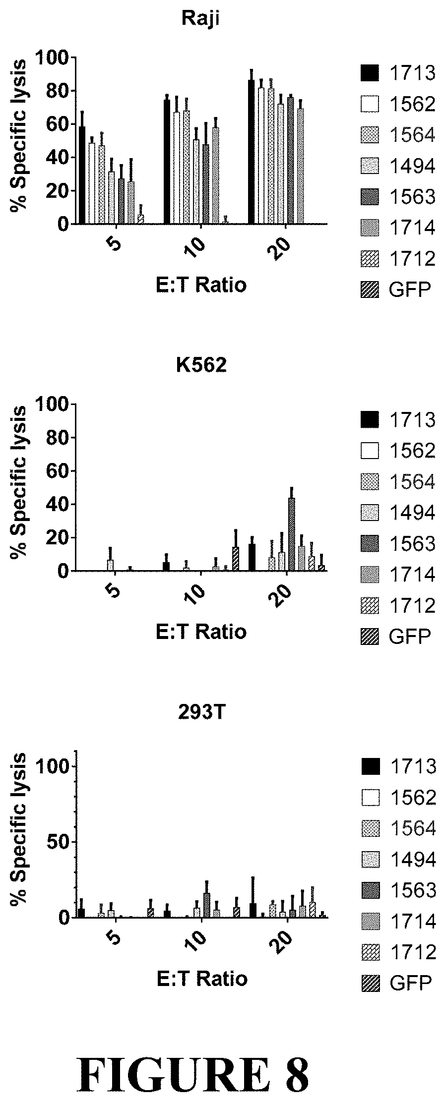
D00013
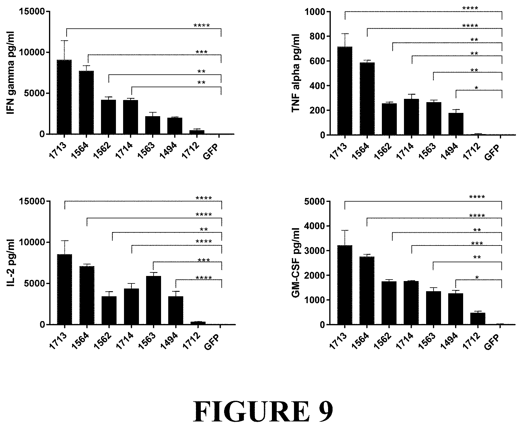
D00014

D00015

D00016
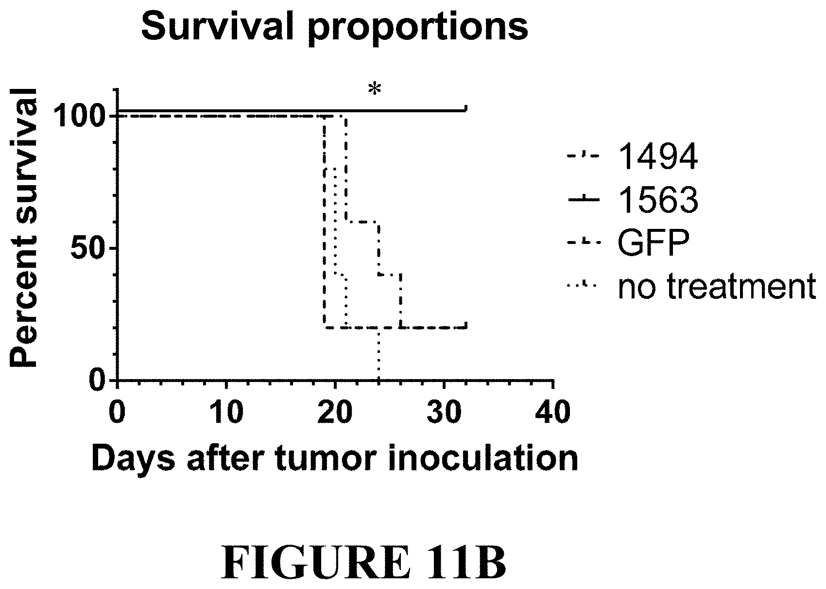
S00001
XML
uspto.report is an independent third-party trademark research tool that is not affiliated, endorsed, or sponsored by the United States Patent and Trademark Office (USPTO) or any other governmental organization. The information provided by uspto.report is based on publicly available data at the time of writing and is intended for informational purposes only.
While we strive to provide accurate and up-to-date information, we do not guarantee the accuracy, completeness, reliability, or suitability of the information displayed on this site. The use of this site is at your own risk. Any reliance you place on such information is therefore strictly at your own risk.
All official trademark data, including owner information, should be verified by visiting the official USPTO website at www.uspto.gov. This site is not intended to replace professional legal advice and should not be used as a substitute for consulting with a legal professional who is knowledgeable about trademark law.