Synthetic Hydrogel Carriers For Cellular Structures, Generation Of Organoids, And Treatment Of Tissue Injury
CRUZ-ACUNA; Ricardo ; et al.
U.S. patent application number 16/492263 was filed with the patent office on 2020-03-12 for synthetic hydrogel carriers for cellular structures, generation of organoids, and treatment of tissue injury. The applicant listed for this patent is GEORGIA TECH RESEARCH CORPORATION, THE REGENTS OF THE UNIVERSITY OF MICHIGAN. Invention is credited to Ricardo CRUZ-ACUNA, Andres J. GARCIA, Asma NUSRAT, Miguel QUIROS, Jason R. SPENCE.
| Application Number | 20200078493 16/492263 |
| Document ID | / |
| Family ID | 63448052 |
| Filed Date | 2020-03-12 |


View All Diagrams
| United States Patent Application | 20200078493 |
| Kind Code | A1 |
| CRUZ-ACUNA; Ricardo ; et al. | March 12, 2020 |
SYNTHETIC HYDROGEL CARRIERS FOR CELLULAR STRUCTURES, GENERATION OF ORGANOIDS, AND TREATMENT OF TISSUE INJURY
Abstract
Disclosed herein are synthetic hydrogel useful for the generation, storage and administration of cellular structures such as spheroids and organoids.
| Inventors: | CRUZ-ACUNA; Ricardo; (Atlanta, GA) ; GARCIA; Andres J.; (Atlanta, GA) ; NUSRAT; Asma; (Ann Arbor, MI) ; SPENCE; Jason R.; (Ann Arbor, MI) ; QUIROS; Miguel; (Ann Arbor, MI) | ||||||||||
| Applicant: |
|
||||||||||
|---|---|---|---|---|---|---|---|---|---|---|---|
| Family ID: | 63448052 | ||||||||||
| Appl. No.: | 16/492263 | ||||||||||
| Filed: | March 9, 2018 | ||||||||||
| PCT Filed: | March 9, 2018 | ||||||||||
| PCT NO: | PCT/US18/21771 | ||||||||||
| 371 Date: | September 9, 2019 |
Related U.S. Patent Documents
| Application Number | Filing Date | Patent Number | ||
|---|---|---|---|---|
| 62469299 | Mar 9, 2017 | |||
| Current U.S. Class: | 1/1 |
| Current CPC Class: | A61K 35/50 20130101; A61L 2300/252 20130101; A61L 2400/06 20130101; A61K 35/28 20130101; A61L 27/3834 20130101; A61L 2300/25 20130101; A61K 35/38 20130101; A61L 27/50 20130101; A61L 27/54 20130101; A61L 27/3886 20130101; A61K 35/545 20130101; A61L 27/52 20130101; A61K 35/42 20130101 |
| International Class: | A61L 27/52 20060101 A61L027/52; A61K 35/50 20060101 A61K035/50; A61K 35/545 20060101 A61K035/545; A61K 35/28 20060101 A61K035/28; A61K 35/38 20060101 A61K035/38; A61L 27/38 20060101 A61L027/38 |
Goverment Interests
STATEMENT OF GOVERNMENT SUPPORT
[0002] This invention was made with government support under R01 AR062368, R01 AR062920, R01 DK055679, R01 DK059888, DK055679, DK059888, and DK089763, awarded by the National Institutes of Health. The government has certain rights in the invention.
Claims
1. A composition comprising a cellular structure dispersed in a hydrogel matrix, wherein the hydrogel matrix comprises a crosslinked hydrophilic polymer network covalently bonded to a plurality of adhesion peptides.
2. The composition according to claim 1, wherein the cellular structure comprises an organoid, cellular spheroid, stem cell, or combination thereof.
3. The composition of claim 2, wherein the cellular structure comprises an organoid comprising an intestinal organoid, gastric organoid, or lingual organoid.
4-6. (canceled)
7. The composition according to claim 2, wherein the cellular structure comprises a stem cell comprising multipotent stem cells, totipotent stem cells, pluripotent stem cells, embryonic stem cell, induced pluripotent stem cells, extraembryonic fetal stem cells, amniotic stem cells, or adult stem cells.
8. The composition according to claim 1, wherein the crosslinked hydrophilic polymer network has the general formula: ##STR00007## wherein `polymer` represents any hydrophilic polymer, C represents a core, Q represents a linker, Z represents an adhesion peptide, X represents a crosslinker, a is greater than 0, and b is greater than 1.
9. The composition according 8, wherein the hydrophilic polymer comprises a multi-armed poly(ethylene glycol).
10. The composition according to claim 8, wherein the crosslinked hydrophilic polymer network has the general formula: ##STR00008## wherein C represents a core, n is an integer from 20-2,000, Q is a linking moiety, Z is an adhesion peptide, X is a crosslinker, a is greater than 0 and b is greater than 1, provided the sum of a+b does not exceed 7.
11. The composition according to claim 8, wherein C represents a group having the formula: ##STR00009## wherein q is 1, 2, 3, 4, 5, or 6.
12. The composition according to claim 8, wherein Q represents a group having the formula: ##STR00010## wherein A is independent selected from O or NH, a is independently selected from 0 or 1, B is selected from hydrogen or methyl, and Z/X in each case independently represents either an adhesion peptide or crosslinker.
13. The composition according to claim 1, wherein the adhesion peptide comprises the amino acid sequence RGD.
14. (canceled)
15. The composition according to claim 13, wherein the adhesion peptide comprises the amino acid sequence GRGDSPC
16. The composition according to claim 1, wherein the crosslinked hydrophilic polymer is crosslinked with a crosslinker comprising a MMP- or cathepsin- or other protease-cleavable or non-cleavable peptide comprising at least a cysteine residue at each end of the sequence.
17. (canceled)
18. The composition according to claim 1, wherein the crosslinker comprises 1,4-dithiothreitol poly(ethylene glycol) dithiol.
19. The composition according to claim 1, wherein the crosslinked hydrophilic polymer network is present, on a dry basis, in an amount from 1-8% polymer weight by total volume of the composition.
20. (canceled)
21. The composition according to claim 1, wherein the crosslinked hydrophilic polymer is present in an amount of no greater than 15%, by weight relative to the total volume of the composition.
22. A composition according to claim 1, obtained by dispersing an organoid in a hydrogel matrix, wherein the hydrogel matrix is obtained by crosslinking a hydrophilic polymer conjugated to a plurality of adhesion peptides.
23-47. (canceled)
48. A method of generating an organoid, comprising contacting the composition of claim 1 with a growth medium.
49-66. (canceled)
67. A method of repairing a tissue injury, comprising contacting the injured tissue with the composition according to claim 1.
68. A method of repairing a tissue injury comprising combining a solution comprising an organoid and a hydrogel precursor having the formula: ##STR00011## wherein C represents a core, n is an integer from 20-2,000, Q is a linking moiety, Z is an adhesion peptide, X is a crosslinker, a is greater than 0 and b is greater than 1 Q.sup.1 is an electrophilic group capable of reacting with a thiol group; with a solution comprising a crosslinker; and contacting injured tissue with the resulting mixture.
69. An organoid delivery device comprising: a) a syringe comprising a hydrogel precursor and organoid; b) a mixing vessel comprising a crosslinking; and c) an injection needle; wherein the syringe is in fluid communication with the mixing vessel and injection needle, in which the mixing vessel is disposed between the needle and syringe.
Description
CROSS REFERENCE TO RELATED APPLICATION
[0001] This application claims the benefit of U.S. Provisional Application 62/469,299, filed on Mar. 9, 2017, the contents of which are hereby incorporated in its entirety.
FIELD OF THE INVENTION
[0003] The invention is directed to synthetic hydrogels useful as storage and growth matrices for cellular structures, and as delivery vehicles in compositions useful for tissue and wound repair.
BACKGROUND
[0004] Human pluripotent stem cells (hPSCs), such as embryonic stem cells (ESCs) and induced pluripotent stem cells (iPSCs), are important cell sources for regenerative therapies and modeling of human diseases. In vitro generation of human organoids from hPSCs offers unparalleled strategies for generating multi-cellular 3D structures recapitulating important features of epithelial and mesenchymal tissues. For example, human intestinal organoid (HIO) technology provides a powerful platform for functional modeling and repair of genetic defects in human intestinal development and the establishment of chronic disease models, such as inflammatory bowel disease.
[0005] In order to generate HIOs, hPSCs have cultured and differentiated using growth factors in a Matrigel.TM.-coated substrate, giving rise to 3D intestinal spheroids which are collected and encapsulated within Matrigel.TM. for expansion into HIOs. Matrigel.TM. is a heterogeneous, complex mixture of ECM proteins, proteoglycans, and growth factors secreted by Engelbreth-Holm-Swarm mouse sarcoma cells, which is required for 3D growth and expansion of HIOs. There are substantial limitations associated with Matrigel.TM., as it suffers from lot-to-lot compositional and structural variability. Importantly, this tumor-derived matrix has limited potential for use in a clinical context.
[0006] There remains a need for improved carriers for organoids and other cellular structures. There remains a need for improved methods to generate human organoids, including intestinal organoids. The remains a need for methods to generate intestinal organoids spheroids that do not require the use of Matrigel.TM.. There also remains a need for improved methods of treating injuries, including mucosal wounds.
SUMMARY
[0007] Disclosed herein is a completely synthetic hydrogel that supports in vitro generation of organoids from spheroids without Matrigel.TM. encapsulation. The synthetic hydrogels are also useful as a storage matrix for spheroids and organoids, and as vehicles for the administration of such organoids and spheroids to a subject. The synthetic hydrogel promotes organoid engraftment and healing of injuries, including mucosal wounds. The synthetic hydrogel can also be used as an injectable vehicle to deliver organoids and other therapeutics to injuries.
[0008] The details of one or more embodiments are set forth in the descriptions below. Other features, objects, and advantages will be apparent from the description and from the claims.
BRIEF DESCRIPTION OF THE FIGURES
[0009] FIG. 1A depicts transmitted light and fluorescence microscopy images of HIOs cultured in PEG-4MAL hydrogels of different polymer density or Matrigel.TM.. HIO viability was assessed by Calcein-AM (live)/TOTO-3 iodide (dead) labeling at 7 d after encapsulation. Bar, 500 .mu.m. FIG. 1B depicts percentage of total organoid area stained for live or dead (mean.+-.SEM) after 7 d of encapsulation (n=5 or 6 organoids analyzed per condition). One-way ANOVA with Tukey's multiple comparisons test showed significant differences between 4.0% PEG-4MAL or Matrigel.TM. and 5.0 or 6.0% PEG-4MAL (*P<0.05, **P<0.01, ***P<0.001, ****P<0.0001). Graphs and images are representative of one and three independent experiments, respectively, performed with 6 PEG-4MAL/Matrigel.TM. per condition.
[0010] FIG. 2A depicts transmitted light and fluorescence microscopy images of HIOs cultured in 4.0% PEG-4MAL hydrogels functionalized with different adhesive peptides or Matrigel.TM.. HIO viability was assessed at 7 d after encapsulation. Bar, 500 .mu.m. FIG. 2B depicts percentage of total organoid area stained for live or dead (mean.+-.SEM) after 7 d of encapsulation (n=5 or 6 organoids analyzed per condition). One-way ANOVA with Tukey's multiple comparisons test showed significant differences between PEG-4MAL-RGD or Matrigel.TM. and PEG-4MAL-GFOGER or -IKVAV (*P<0.05, **P<0.01). Graphs and images are representative of one and three independent experiments, respectively, performed with 6 PEG-4MAL/Matrigel.TM. per condition.
[0011] FIG. 3A and FIG. 3B depict fluorescence microscopy images of a HIO at 7 d after encapsulation in 4.0% PEG-4MAL-RGD hydrogel or Matrigel.TM., and labeled for FIG. 3A, .beta.-CATENIN, proliferative cells (KI67), and FIG. 3B, epithelial apical polarity (EZRIN) and tight junctions (ZO-1). DAPI, counterstain. "L" indicates HIO lumen. Bars, 100 .mu.m. Images are representative of three independent experiments performed with 6 PEG-4MAL/Matrigel.TM. per condition (a,b).
[0012] FIG. 4 depicts transmitted light and fluorescence microscopy images of mCherry-spheroids cultured in PEG-4MAL hydrogels of different polymer density or Matrigel.TM.. Spheroids viability was assessed by Calcein-AM labeling at 2 hr after encapsulation (day 0) and at day 5 for PEG-4MAL conditions, and at day 3 for Matrigel.TM.. Bar, 100 .mu.m. Viability is quantified as percentage of total spheroid or organoid area stained for live or dead (mean.+-.SEM; n=5 organoids analyzed per condition/time-point). One-way ANOVA with Tukey's multiple comparisons test showed significant differences between 3.5% or 4.0% PEG-4MAL and 8.0% or 12.0% PEG-4MAL at Day 0 (****P<0.0001). Graphs and images are representative of one and three independent experiments, respectively, performed with 6 PEG-4MAL/Matrigel.TM. per condition.
[0013] FIG. 5A depicts HIO projected area and FIG. 5B depicts feret diameter normalized to Day 0 values at different time-points after encapsulation in 4.0% PEG-4MAL-RGD hydrogel (.circle-solid.) or Matrigel.TM. (.box-solid.) (n=6 or 4 organoids analyzed per condition/time-point). Repeated measures two-way ANOVA showed no significant difference between matrix types (P-value>0.05). Graph line represents the mean of the individual data points at each time-point. FIG. 5C and FIG. 5D Fluorescence microscopy images of a HIO at 21 d after encapsulation in 4.0% PEG-4MAL-RGD hydrogel or Matrigel.TM. and labeled for FIG. 5C .beta.-CATENIN, proliferative cells (KI67), and FIG. 5D epithelial apical polarity (EZRIN) and tight junctions (ZO-1). DAPI, counterstain. "L" indicates HIO lumen. Bars, 100 .mu.m. Graphs and images are representative of one and three independent experiments, respectively, performed with 6 PEG-4MAL/Matrigel.TM. per condition.
[0014] FIG. 6A depicts micrographs of dissected kidneys containing HIOs generated within PEG-4MAL-RGD or Matrigel.TM.. FIG. 6B depicts transmitted light and fluorescence microscopy (mCherry) images of harvested organoids. Bar, 0.5 cm. FIG. 6C depicts H&E staining demonstrates mature human intestinal crypt-villus structure, and FIG. 6D depicts Alcian blue and trichrome staining reveal presence of differentiated goblet cells and organized collagen fibers. Bar, 100 .mu.m. FIG. 6E depicts fluorescence microscopy images of HIOs labeled for (.beta.-CATENIN, proliferative cells (KI67), epithelial apical polarity (EZRIN) and junctions (ZO-1 and ECAD), intestinal epithelial protein CDX2, enteroendocrine cells (CHGA), goblet cells (MUC2), tuft cells (DCLK1) and small intestinal marker (duodenum; PDX1). DAPI, counterstain. "L" indicates HIO lumen. White arrows show enteroendocrine cells or tuft cells. Bars, 50 .mu.m. Images are representative of one experiment performed with 3 mice per condition.
[0015] FIG. 7A depicts PEG-4MAL-generated HIOs mixed with engineered hydrogel precursor solutions were injected underneath mechanically-induced mucosal wounds, as seen through the colonoscope camera. FIG. 7B depicts mechanically-induced mucosal wound and fluorescence imaging (mCherry) at the wound site at 5 d post-injection. Bar, 500 .mu.m. FIG. 7C depicts fluorescence microscopy images of murine colonic tissue at the wound site labeled for human cell nuclei (NUMA) at 4 weeks post-delivery. Left: Images from wound edge showing insets from i) adjacent host tissue and ii) wound. Right: Images from wound center showing insets at wound site. DAPI, counterstain. Bars, 100 .mu.m. (d) In situ hybridization, stained for human OLFM4+ cells. Bar, 50 .mu.m. Images are representative of two experiment performed with 4 mice per condition (five colonic wounds/injections per mouse; a-d).
[0016] FIG. 8 depicts images of mucosal wounds at 1 d (prior to injection) or 5 d post-injury in murine colon as seen through the colonoscope camera. Mucosal wound area at 5 d post-injury was normalized to day 1 (prior to injection) values (mean.+-.SEM). Five colonic wounds per mouse were analyzed and averaged (n=4 mice per condition). One-way ANOVA with Tukey's multiple comparisons test showed significant difference between Hydrogel-grown HIOs in hydrogel (.circle-solid.) or Matrigel-grown HIOs in hydrogel (.box-solid.) and Saline injected HIOs (.tangle-solidup.) Only hydrogel (.diamond-solid.) or No injection group (X) (**P<0.01, ***P<0.001, ****P<0.0001). Bars, 500 .mu.m. Images and graph are representative of one experiment performed with 4 mice per condition (five colonic wounds/injections per mouse; a-e). Source data are available in Supplementary Table 1.
[0017] FIG. 9A depicts PEG-4MAL macromers are conjugated with thiol-containing adhesive peptide to produce a functionalized PEG-4MAL macromer, which is then crosslinked in the presence of HIOs using protease-cleavable peptides containing terminal cysteines to form a FIG. 9B hydrogel network. FIG. 9C and FIG. 9D depict the relationship between polymer density (wt %) and storage modulus (FIG. 9C) or loss modulus (FIG. 9D) (mean.+-.SEM; n=10 independently prepared hydrogels per condition). FIG. 9E depicts schematic of spheroid development into HIOs within Matrigel.TM. and further growth within PEG-4MAL hydrogel. FIG. 9F depicts schematic of spheroid development into HIOs within hydrogel. (c,d) Graphs are representative of one experiment. Source data are available in Supplementary Table 1.
[0018] FIG. 10 depicts transmitted light and fluorescence microscopy images of HIOs cultured in 4.0% PEG-4MAL hydrogels functionalized with RGD (FIG. 10A), inactive scrambled RDG peptide (FIG. 10B), or non-degradable crosslinker (DTT) (FIG. 10C). HIO viability was assessed at 7 d after encapsulation. FIG. 10D and FIG. 10E depict transmitted light microscopy images of Matrigel.TM.-generated HIOs cultured within 4.0% PEG-4MAL-RGD hydrogel (FIG. 10D) or Matrigel.TM. over time (FIG. 10E). Bars, 500 .mu.m. Images are representative of three different experiments performed with (a-c) 4 or (d,e) 12 PEG-4MAL/Matrigel.TM. per condition.
[0019] FIG. 11 depicts transmitted light and fluorescence microscopy images of hiPSC-derived HIO generation within 4.0% PEG-4MAL-RGD hydrogels (FIG. 11A), Matrigel.TM. (FIG. 11B), or 8.0% PEG-4MAL-RGD hydrogels (FIG. 11C), hiPSC-derived spheroid and HIO viability was assessed at different time-points after encapsulation. FIG. 11D depicts transmitted light microscopy images of hESC-derived HIO generation within 4.0% PEG-4MAL-RGD hydrogels. These organoids were never encapsulated within Matrigel.TM.. Black arrows show epithelial budding. Bars, 500 .mu.m. Images are representative of three different experiments performed with 12 PEG-4MAL/Matrigel.TM. per condition (a-c).
[0020] FIG. 12A depicts transmitted light and fluorescence microscopy images of HIO generation within 20 kDa (4.0%) or 40 kDa (8.0%) PEG-4MAL-RGD hydrogels. HIO viability was assessed at 5 d after encapsulation. FIG. 12B depicts percentage of total organoid area stained for live or dead (mean.+-.SEM) after 5 d of encapsulation (n=6 organoids analyzed per condition). FIG. 12C depicts HIO projected area and Feret diameter normalized to Day 0 values (mean.+-.SEM) after 5 d of encapsulation (n=5 organoids analyzed per condition). Unpaired two-tailed t-test with Welch's correction showed no significant differences between HIO viability or HIO size parameters within 20 kDa (4.0%) and 40 kDa (8.0%) PEG-4MAL-RGD (P-value>0.05). FIG. 12D and FIG. 12E depict transmitted light and fluorescence microscopy images of spheroids cultured within 4.0% PEG-4MAL-RGD hydrogels supplemented with 10 .mu.M (FIG. 12D) or 30 .mu.M of verteporfin, Y-27632 or blebbistatin FIG. 12E), or DMSO (vehicle control) (FIG. 12F). Spheroids death was assessed by annexin-V (apoptosis) and propidium iodide (dead) labeling at 1 d after encapsulation. Bars, 100 .mu.m. Data is representative of one experiment performed with 12 PEG-4MAL hydrogels per condition.
[0021] FIG. 13 depicts RNA levels of pluripotency (OCT4), endoderm (FOXA2), and epithelial junction (ZO1, ECAD and CLDN2) genes, as quantified by RT-qPCR (mean.+-.SEM; n=6 samples per condition). Unpaired two-tailed t-test was used to identify statistical differences between matrix types (**P<0.01; ns, not significant). The results are representative of one experiment.
[0022] FIG. 14A depicts transmitted light and fluorescence microscopy images of HIOs cultured in 4.0% PEG-4MAL-RGD hydrogels or Matrigel.TM.. HIO viability was assessed at 7 d after encapsulation. Bar, 500 .mu.m. FIG. 14B depicts fluorescence microscopy images of HLO at 7 d after encapsulation in 4.0% PEG-4MAL-RGD hydrogel or Matrigel.TM. and labeled for e-cadherin (ECAD), lung epithelia (NKX2.1), and basal cells (P63). DAPI, counterstain. "L" indicates HLO lumen. Bars, 25 .mu.m. The results are representative of one experiment performed with 6 PEG-4MAL/Matrigel.TM. per condition.
[0023] FIG. 15A depicts micrographs of dissected kidney containing HIOs. FIG. 15B depicts transmitted light and fluorescence microscopy (mCherry) images of harvested HIOs. FIG. 15C depicts H&E staining demonstrates mature human intestinal crypt-villus structure, and FIG. 15D depicts Alcian blue and trichrome staining show the presence of differentiated goblet cells and organized collagen fibers. Bar, 100 .mu.m. FIG. 15E depicts fluorescence microscopy images of harvested HIOs labeled for .beta.-CATENIN, proliferative cells (KI67), epithelial apical polarity (EZRIN) and junctions (ZO-1 and ECAD), intestinal epithelial protein CDX2, enteroendocrine cells (CHGA), goblet cells (MUC2), tuft cells (DCLK1) and small intestinal marker (duodenum; PDX1). DAPI, counterstain. White arrows show tuft cells. Bar, 50 .mu.m.
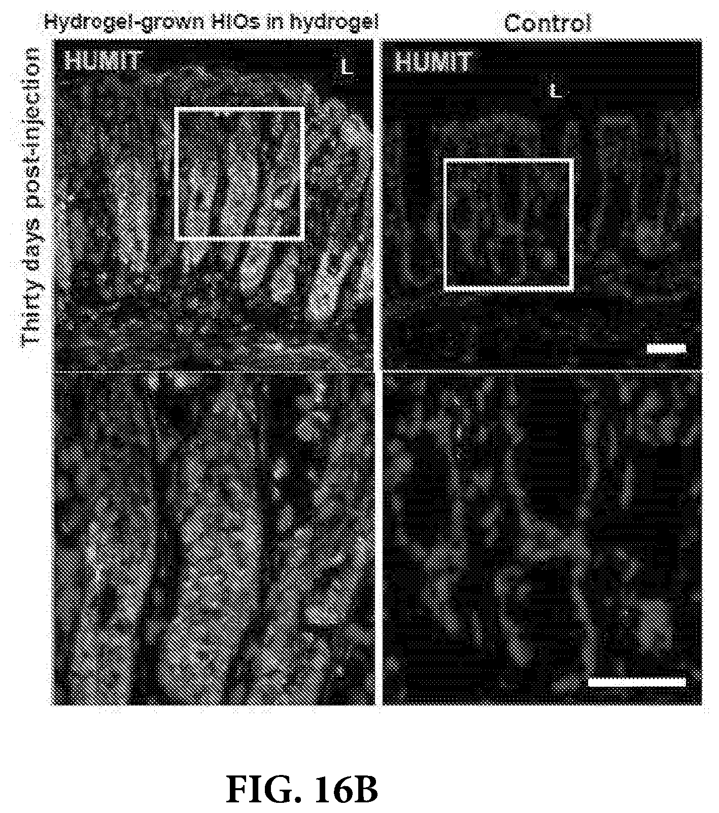
[0024] FIG. 16A depicts mechanically-induced submucosal wounds were performed in the distal colon of mice using a mechanical probe through a mouse colonoscope. One day post-wounding HIOs generated in engineered 4% PEG-4MAL-RGD hydrogels or Matrigel.TM. were recovered from the matrix, mixed with the engineered hydrogel precursor solutions, and injected underneath the submucosal wounds. A group with no injections, HIOs injected in saline, or injection of HIO-free hydrogel precursor solutions were used as control groups. Distal colon tissue harvest, immunostaining and imaging was performed 4 weeks post-wounding. FIG. 16B depicts fluorescent microscopy images labeled for human mitochondria (HUMIT) of murine colonic tissue at the wound site at 4 weeks post-injection or control tissue. DAPI, counterstain. "L" indicates HIO lumen. Bars, 100 .mu.m. (c,d) In situ hybridization images of control adult human colon or sections taken at the mouse colonic wound site stained for human OLFM4+ cells (FIG. 16C) or mouse Lgr5+ intestinal stem cells ((FIG. 16D). Bars, 50 .mu.m. The results are representative of two different experiments performed with 4 mice per condition (five colonic wounds/injections per mouse).
DETAILED DESCRIPTION
[0025] Before the present methods and systems are disclosed and described, it is to be understood that the methods and systems are not limited to specific synthetic methods, specific components, or to particular compositions. It is also to be understood that the terminology used herein is for the purpose of describing particular embodiments only and is not intended to be limiting.
[0026] As used in the specification and the appended claims, the singular forms "a," "an" and "the" include plural referents unless the context clearly dictates otherwise. Ranges may be expressed herein as from "about" one particular value, and/or to "about" another particular value. When such a range is expressed, another embodiment includes from the one particular value and/or to the other particular value. Similarly, when values are expressed as approximations, by use of the antecedent "about," it will be understood that the particular value forms another embodiment. It will be further understood that the endpoints of each of the ranges are significant both in relation to the other endpoint, and independently of the other endpoint.
[0027] "Optional" or "optionally" means that the subsequently described event or circumstance may or may not occur, and that the description includes instances where said event or circumstance occurs and instances where it does not.
[0028] Throughout the description and claims of this specification, the word "comprise" and variations of the word, such as "comprising" and "comprises," means "including but not limited to," and is not intended to exclude, for example, other additives, components, integers or steps. "Exemplary" means "an example of" and is not intended to convey an indication of a preferred or ideal embodiment. "Such as" is not used in a restrictive sense, but for explanatory purposes.
[0029] Disclosed are components that can be used to perform the disclosed methods and systems. These and other components are disclosed herein, and it is understood that when combinations, subsets, interactions, groups, etc. of these components are disclosed that while specific reference of each various individual and collective combinations and permutation of these may not be explicitly disclosed, each is specifically contemplated and described herein, for all methods and systems. This applies to all aspects of this application including, but not limited to, steps in disclosed methods. Thus, if there are a variety of additional steps that can be performed it is understood that each of these additional steps can be performed with any specific embodiment or combination of embodiments of the disclosed methods.
[0030] Disclosed herein are synthetic hydrogels capable of supporting in vitro generation organoids from cellular spheroids, for instance, stem cell derived spheroids. The synthetic hydrogel are also useful as a storage matrix for spheroids and organoids, and can be used as a vehicle for the administration of such organoids and spheroids to a subject.
[0031] As used herein, the term spheroid (and cellular spheroid) refers to a plurality of cells grown in a 3-dimensional matrix. In contrast to cell cultures obtained from conventional two-dimensional techniques, spheroids can growth in all directions, and the resulting tissue more closely resembles the morphology of cells grown in vivo. As used herein, the term organoid (and cellular organoid) refers to a three-dimensional cell cultures that incorporate some of the key features of the represented organ. Unless specified to the contrary, an organoid may be obtained from adult stem cell-containing tissue samples, single adult stem cells, or from the directed differentiation of pluripotent stem cells, multipotent stem cells, or totipotent stem cells.
[0032] The synthetic hydrogels include a network of crosslinked hydrophilic polymer conjugated to adhesion peptides. Suitable hydrophilic polymers include polyalkylene glycol polymers, polyalkylene oxide homopolymers such as polypropylene glycols, polyoxyethylenated polyols, copolymers thereof and block copolymers thereof, as well as poly(oxyethylated polyol), poly(olefinic alcohol), poly(vinylpyrrolidone), poly(hydroxypropylmethacrylamide), poly(.alpha.-hydroxy acid), poly(vinyl alcohol), polyphosphazene, polyoxazoline, poly(N-acryloylmorpholine) and copolymers, terpolymers, and mixtures thereof. The molecular weight of the hydrophilic polymer can be from 1,000-1,000,000, from 1,000-500,000, from 1,000-250,000, from 1,000-150,000, from 1,000-100,000, from 1,000-50,000, from 5,000-100,000, from 5,000-50,000, from 10,000-100,000, from 10,000-50,000, from 20,000-100,000, from 20,000-80,000, from 20,000-60,000, from 20,000-40,000, or from 40,000-60,000.
[0033] In certain embodiments, the crosslinked hydrophilic polymer is a polyethylene glycol, i.e., PEG. The PEG can have a molecular weight from 1,000-1,000,000, from 1,000-500,000, from 1,000-250,000, from 1,000-150,000, from 1,000-100,000, from 1,000-50,000, from 5,000-100,000, from 5,000-50,000, from 10,000-100,000, from 10,000-50,000, from 20,000-100,000, from 20,000-80,000, from 20,000-60,000, from 20,000-40,000, or from 40,000-60,000.
[0034] In preferred embodiments, the crosslinked hydrophilic polymer is a branched or multi-arm polymer. As used herein, a multi-arm polymer describes a polymer having a central core with at least two polymers covalently attached thereto. Multi-arm polymers can have 2, 3, 4, 5, 6, 7, 8 or more polymer arms. Preferred multi-arm polymers, as defined above, include those with 4 arms. Generally, all of the polymers attached to the core are the same, but in some instances different hydrophilic polymers, as defined above, can be used. Suitable cores include those derived from polyols, including glycerol (3-arm), pentaerythritol (4-arm), tetraglycerol (6-arm), and hexaglycerol (8-arm). A particularly preferred polymer is a 4-arm PEG, having a total molecular weight from 1,000-1,000,000, from 1,000-500,000, from 1,000-250,000, from 1,000-150,000, from 1,000-100,000, from 1,000-50,000, from 5,000-100,000, from 5,000-50,000, from 10,000-100,000, from 10,000-50,000, from 20,000-100,000, from 20,000-80,000, from 20,000-60,000, from 20,000-40,000, or from 40,000-60,000.
[0035] In certain embodiments, the crosslinked hydrophilic polymer network can have the general formula:
##STR00001##
wherein `polymer` in each case independently represents any hydrophilic polymer, including those defined above, C represents a core, Q represents a linker, Z represents an adhesion peptide, X represents a crosslinker, a is greater than 0, and b is greater than 1. In some embodiments, the sum a+b is no greater than 3, no greater than 4, no greater than 5, no greater than 6, no greater than 7, no greater than 8, no greater than 9, or no greater than 10. In other embodiments, the sum a+b is at least 3, at least 4, at least 5, no at least 6, at least 7, at least 8, at least 9, or at least 10.
[0036] In some embodiments, the hydrophilic polymer can be a poly(ethylene glycol), i.e., networks having the formula:
##STR00002##
wherein C represents a core, n is an integer from 20-2,000, Q is a linking moiety, Z is an adhesion peptide, X is a crosslinker, a is greater than 0 and b is greater than 1. In some embodiments, the sum a+b is no greater than 3, no greater than 4, no greater than 5, no greater than 6, no greater than 7, no greater than 8, no greater than 9, or no greater than 10. In other embodiments, the sum a+b is at least 3, at least 4, at least 5, no at least 6, at least 7, at least 8, at least 9, or at least 10.
[0037] Suitable C groups can be derived from a polyol such as glycerol, pentaerythritol, sorbitol, mannitol, tetraglycerol, and hexaglycerol. In some instances, the core can have the general structure:
##STR00003##
wherein q is any integer, for instance 1, 2, 3, 4, 5, 6, 7, 8, 9, or 10 and .quadrature. represents a link to a hydrophilic polymer, as described above. Other suitable polyols include carbohydrates, including monosaccharides and di-saccharides, such as glucose, xylose, mannose, galactose, sucrose, maltose, trehalose and fructose, and cyclic polyols like cyclopropane-1,2,3-triol, cyclobutane-1,2,3,4-tetraol, cyclopentane-1,2,3,4-tetraol, cyclopentane-1,2,3,4,5-pentaol, cyclohexane-1,2,4,5-tetraol, cyclohexane-1,2,3,4,5,6-hexaol, and the like.
[0038] Suitable Q group include those formed via Michael addition between a nucleophilic group on the adhesion peptide or crosslinker, and a Michael acceptor bonded to the hydrophilic polymer. For instance, in some embodiments, Q represents a group having the formula:
##STR00004##
wherein A is independently selected from O or NH, a is independently selected from 0 or 1, B is selected from hydrogen or methyl, Z/X in each case independently represents either an adhesion peptide or crosslinker, and .quadrature. represents a link to a hydrophilic polymer, as described above.
[0039] In some embodiments, the adhesion peptide can include the sequence RGD. In some embodiments, the adhesion peptide can include GRGDSPC (SEQ. ID 1), CRGDS(SEQ. ID 2), CRGDSP (SEQ. ID 3), CPHSRN (SEQ. ID 4), CGWGGRGDSP (SEQ. ID 5), CGGSIDQVEPYSSTAQ (SEQ. ID 6), CGGRNIAEIIKDI (SEQ. ID 7), CGGDITYVRLKF (SEQ. ID 8), CGGDITVTLNRL (SEQ. ID 9), CGGRYVVLPR (SEQ. ID 10), CGGKAFDITYVRLKF (SEQ. ID 11), CGGEGYGEGYIGSR (SEQ. ID 12), CGGATLQLQEGRLHFXFDLGKGR, wherein X=Nle (SEQ. ID 13), CGGSYWYRIEASRTG (SEQ. ID 14), CGGGEFYFDLRLKGDKY (SEQ. ID 15), CKGGNGEPRGDTYRAY (SEQ. ID 16), CKGGPQVTRGDVFTMP (SEQ. ID 17), CGGNRWHSIYITRFG (SEQ. ID 18), CGGASIKVAVSADR (SEQ. ID 19), CGGTTVKYIFR (SEQ. ID 20), CGGSIKIRGTYS (SEQ. ID 21), CGGSINNNR (SEQ. ID 22), CGGSDPGYIGSR (SEQ. ID 23), CYIGSR (SEQ. ID 24), CGGTPGPQGIAGQGVV (SEQ. ID 25), CGGTPGPQGIAGQRVV (SEQ. ID 26), CGGMNYYSNS (SEQ. ID 27), CGGKKQRFRHRNRKG (SEQ. ID 28), CRGDGGGGGGGGGGGGGPHSRN (SEQ. ID 29), CPHSRNSGSGSGSGSGRGD (SEQ. ID 30), Acetylated-GCYGRGDSPG (SEQ. ID 31), ((GPP)5GPC) (SEQ. ID 32), CRDGS (SEQ. ID 33), cyclic RGD{Fd}C (SEQ. ID 34), CGGRKRLQVQLSIRT (SEQ. ID 35), CIKVAV (SEQ. ID 36), CGGAASIKVAVSADR (SEQ. ID 37), CGGKRTGQYKL (SEQ. ID 38), CGGTYRSRKY (SEQ. ID 39), CGGYGGGP(GPP)5GFOGERPP(GPP)4GPC (SEQ. ID 40), CGGKRTGQYKLGSKTGPGQK (SEQ. ID 41), QAKHKQRKRLKSSC (SEQ. ID 42), SPKHHSQRARKKKNKNC (SEQ. ID 43), CGGXBBXBX, wherein B=basic residue and X=hydropathic residue (SEQ. ID 44), and CGGXBBBXXBX, wherein B=basic residue and X=hydropathic residue (SEQ. ID 45). In some preferred embodiments, the adhesion peptide includes the sequence GRGDSPC.
[0040] Suitable crosslinkers include enzymatically cleavable and non-cleavable peptide sequences. The peptide sequences will generally include a cysteine residue at each end of the sequence. Exemplary cleavable peptides include those that are cleavable by MMP, cathepsin, or other protease. Although the cysteine may be the final amino acid residue at each end of sequence, it is more preferable that the crosslinking peptides are terminated with a glycine or other inert residue. Suitable crosslinking peptides include the sequences GCRDGPQG.dwnarw.IWGQDRCG (SEQ. ID 46), GCRDGPQG.dwnarw.IAGQDRCG (SEQ. ID 47), GCRDVPMS.dwnarw.MRGGDRCG (SEQ. ID 48), GCRDIPVS.dwnarw.LRSGDRCG (SEQ. ID 49), GCRDRPFS.dwnarw.MIMGDRCG (SEQ. ID 50), GCRDVPLS.dwnarw.LTMGDRCG (SEQ. ID 51), GCRDVPLS.dwnarw.LYSGDRCG (SEQ. ID 52), GCRDIPES.dwnarw.LRAGDRCG (SEQ. ID 53), GCRDSGESPAY.dwnarw.YTADRCG (SEQ. ID 54), GCRDGGYAE.dwnarw.LRMGGDRCG (SEQ. ID 55), GCRDGGPLG.dwnarw.LYAGGDRCG (SEQ. ID 56), GCRDGPLG.dwnarw.LWARDRCG (SEQ. ID 57), wherein .dwnarw. represents a cleavable amide bond. In some embodiments, the crosslinker is a not a peptide, for instance a di-mercapto compound such as a 1,4-dithiothreitol (1,4-dimercapto-2,3-butanediol) or poly(ethylene glycol) dithiol.
[0041] Aqueous compositions including the networks described above can be used as a support matrix or carrier for a variety of cellular structures. As used herein, cellular structures include stem cells, cell-derived spheroids, and organoids. For instance, the networks can be combined with organoids, for instance a lung organoid, cerebral organoid, thyroid organoid, thymic organoid, testicular organoid, hepatic organoid, pancreatic organoid, gut organoid (i.e., intestinal organoid, gastric organoid, or lingual organoid), epithelial organoid, lung organoid, renal organoid, embryonic organoid, or cardiac organoid. The hydrophilic networks can also be combined with cell-derived spheroids, e.g., pluripotent stem cells like embryonic stem cells and induced pluripotent stem cells. In some cases, stem cells such as multipotent stem cells, totipotent stem cells, pluripotent stem cells, embryonic stem cell, induced pluripotent stem cells, extraembryonic fetal stem cells, amniotic stem cells (including cells stem cells obtained from the cord blood), and adult stem cells (including, but not limited to hematopoietic stem cells, endothelial stem cells, intestinal stem cells, mammary stem cells, neural stem cells, and mesenchymal stem cells) can be incorporated into the hydrogel.
[0042] The compositions can include water in an amount of at least 70% by weight relative to the total weight of the composition. In some embodiments, the water can be present in an amount of at least 75%, at least 80%, at least 85%, at least 87.5%, at least 90%, at least 92.5%, or at least 95% by weight relative to the total weight of the composition. In some embodiments, the compositions will include the hydrophilic crosslinked polymer network in an amount no greater than 30%, no greater than 25%, no greater than 20%, no greater than 15%, no greater than 12.5%, no greater than 10%, no greater than 7.5%, or no greater than 5%, by weight relative to the total volume of the hydrogel. In certain embodiments, the crosslinked hydrophilic polymer network is present in an amount from 1-8%, from 2-7%, from 2-6%, from 3-6%, from 3-5%, or from 3.5-4.5% polymer weight by total volume of the composition. In certain embodiments, the crosslinked hydrophilic polymer network is present in an amount of about 4% polymer weight by total volume of the composition.
[0043] The crosslinked networks disclosed herein may be prepared by first conjugating an adhesion peptide to a hydrophilic polymer having the formula:
C-[polymer-Q.sup.1].sub.c,
wherein C and "polymer" are as defined above, c is an integer greater than or equal to 3, and Q.sup.1 is an electrophilic group capable of reacting with a thiol group. In some embodiments, the hydrophilic polymer is PEG, i.e., a compound of formula:
C--[(CH.sub.2CH.sub.2O).sub.n-Q.sup.1].sub.c,
wherein C, n, c, and Q.sup.1 are as defined above. In some embodiments, Q.sup.1 represents a group having the formula:
##STR00005##
wherein A is independently selected from O or NH, a is independently selected from 0 or 1, B is selected from hydrogen or methyl, and .quadrature. represents a link to a hydrophilic polymer, as described above.
[0044] The adhesion peptide can contain a single cysteine residue or thiol group, and will be combined such that there is a molar excess of Q.sup.1 groups relative to cysteine/thiol groups. For instance, molar ratio of Q.sup.1 groups to cysteine/thiol residues is from 10:1 to 1.5:1, from 8:1 to 1.5:1, from 6:1 to 1.5:1, from 4:1 to 1.5:1, from 3:1 to 1.5:1, from 2.5:1 to 1.5:1, from 5:1 to 2:1, from 5:1 to 3:1, or from 5:1 to 4:1. The molar ratio of nucleophilic groups in the crosslinker to unreacted Q.sup.1 groups (assuming complete reaction with adhesion peptide) can be 1:1, greater than 1:1, e.g., 1.1:1, 1.2:1, or 1.5:1, less than 1:1, e.g., 0.9:1, 0.8:1, or 0.5:1, from 0.5:1 to 1.5:1, from 0.75:1 to 1.25:1, from 0.5:1 to less than 1:1, or from 1.5:1 to greater than 1:1.
[0045] Each of the hydrophilic polymer, adhesion peptide, crosslinker, and cellular component can be separately combined with an appropriate aqueous solution, generally buffered to a pH from 7.0-8.0, from 7.2-7.6, or 7.3-7.5. Any physiologically compatible buffer may be used, such as 4-(2-hydroxyethyl)piperazine-1-ethanesulfonic acid (HEPES), phosphate buffers, carbonate buffers, tromethamine (tris) buffers, including those formed with EDTA and an acid such as acetic acid, boric acid, and the like.
[0046] The relative ratios of the components may be as follows:
TABLE-US-00001 Volume fraction of Concentration factor of Hydrogel component hydrogel component hydrogel component Hydrophilic polymer 0.3-0.5 2.5X Adhesion peptide 0.15-0.25 5X Crosslinker 0.15-0.25 5X Cellular structure 0.15-0.25 5X
[0047] The hydrophilic polymer may be combined with a solution of adhesive peptide such that the final adhesive peptide concentration is from 0.1-100 mM, from 0.5-75 mM, from 1-50 mM, from 5-25 mM, or from 7.5-15 mM, based on the total volume of the hydrogel. The mixture can be incubated at a temperature from 23-50.degree. C., from 28-45.degree. C., from 32-40.degree. C., or at 37.degree. C. for at least 5 minutes, at least 10 minutes, at least 15 minutes, or at least 20 minutes. The resulting product is designated the hydrogel precursor, which has the following structure:
##STR00006##
wherein C, `polymer,` n, Q, Q.sup.1, Z, a, and b have the meanings given above. The hydrogel precursor may be directly converted to a hydrogel by combination with the crosslinker, or may first be combined with a cellular structure and subsequently combined with the crosslinker to form the hydrogel.
[0048] Stem cells can be cultured and differentiated into tissue spheroids, and organoids can be obtained from iPSCs or ESCs and Matrigel using established methods. The spheroids and organoids can be suspended in a growth medium appropriate for the tissue type. The suspension can then be combined with the hydrogel precursor solution and combined with a solution of crosslinker. Generally, crosslinker solution is placed in the center of a dish, and then combined with the hydrogel precursor/cell suspension. It is important to mix the components thoroughly. The mixture can be incubated at a temperature from 23-50.degree. C., from 28-45.degree. C., from 32-40.degree. C., or at 37.degree. C. for no more than 5 minutes, no more than 10 minutes, no more than 20 minutes, no more than 30 minutes, or no more than 60 minutes to generate the hydrogel, which is then overlaid with additional growth medium. The growth medium should be replaced every 3-5 days. Spheroids and organoids stored in the hydrogel matrix are stable over prolonged periods of time. Suitable spheroids that can be employed with the hydrogel matrix include round spheroids, mass spheroids, grape-like spheroids, and stellate spheroids. The round spheroids usually express tight cell junction proteins such as ZO-1. Colonies of round spheroids sometimes undergo lumen formation in the center, which usually occurs in a time-dependent manner. The mass-type spheroids are characterized by round colony outlines, disorganized nuclei, filled colony centers, and strong cellular communication. The mass spheroids are usually larger in diameter than round spheroids and overexpress luminal keratin 8 (KRT8) and keratin 18 (KRT18), yet lack a lumen. The grape-like spheroids display a distinguished grape-like appearance and usually have poor cell-cell interactions. The stellate-type spheroids are characterized by their invasive phenotype with stellate projections that often bridge multiple colonies and/or invade the matrixes
[0049] Organoids grown in the hydrogel matrix may be passaged using conventional techniques. Generally, the organoids are mechanically dislodged from the hydrogel by vigorously pipetting. Any remaining hydrogel still adhering to the organoid can be removed with forceps. The organoid is then cut in half, and each half resuspended in growth medium. Each half may be combined with hydrogel precursor solution and cast into hydrogels as described above. In this fashion, larger organoids may be obtained.
[0050] The compositions disclosed herein may be used to promote wound healing and tissue repair. In addition to the organoid, the hydrogel composition may further include additional therapeutics to facilitate healing and prevent infection. Suitable additional therapeutics include growth factors, antibiotics, antivirals, analgesics, cytokines, enzymes, aptamers, nucleic acids, and combinations thereof.
[0051] In some instances, a preformed hydrogel/organoid composition may be directly contacted with the site of tissue injury. In other instances, the composition may be administered subcutaneously adjacent or proximate to the injury. In some embodiment the hydrogel precursor is combined with the crosslinker at the site of injury at the time of administration. A preferred method includes an in vivo hydrogel formation. The hydrogel precursor/organoid is loaded into a dispensing means, for instance a syringe, which is in fluid communication with a needle by way of a tube. Inside the tube is loaded the crosslinker, such that when the hydrogel precursor/organoid is expelled from the syringe, it combines with the crosslinker solution and is ejected from the needle into the directed site.
Examples
[0052] The following examples are for the purpose of illustration of the invention only and are not intended to limit the scope of the present invention in any manner whatsoever.
Immunofluorescence Analysis
[0053] For immunofluorescence labeling of frozen sections from colon, HIOs, kidney capsule implanted-HIOs or HLOs, these were fixed with 3.7% (w/v) paraformaldehyde at room temperature for 15 min, followed by 0.5% (w/v) Triton X-100 for 5 min. Primary antibody incubation was performed overnight at a 1:100 dilution, unless stated otherwise. Secondary antibody incubation was performed for 1 h at a 1:2000 dilution. Detailed information on the antibodies used including their resources (company names, catalogue numbers) and dilutions are provided in the Reporting Summary.
Differentiation of hPSCs into Intestinal Spheroids or HLOs
[0054] All work using human pluripotent stem cells was approved by the University of Michigan Human Pluripotent Stem Cell Oversight Committee (HPSCRO). Stem cell lines are routinely monitored for chromosomal karyotype, pluripotency (using a panel of antibody and RT-qPCR markers), and for the ability to undergo multi-lineage differentiation. For intestinal spheroid generation, mycoplasma-free human ES cells (H9, NIH registry #0062) and iPS cells were cultured on Matrigel.TM.-coated plates and differentiated into intestinal tissue. Floating spheroids present in the cultures on day 4 and day 5 of mid/hindgut induction were harvested for use in subsequent experiments. In some experiments, hESCs expressing a constitutively active H2BmCherry fluorescent reporter were used. This line was generated by infecting hESCs with a lentivirus containing PGK-H2BmCherry. For HLO generation, human ES cells (UM63-1, NIH registry #0277) were maintained, differentiated and expanded into HLOs.
[0055] Hydrogel Formation and In Vitro Intestinal Spheroid/HIO
[0056] To prepare PEG hydrogels, PEG-4MAL macromer (MW 22,000 or 44,000; Laysan Bio) was dissolved in 4-(2-hydroxyethyl)piperazine-1-ethanesulfonic acid (HEPES) buffer (20 mM in DPBS, pH 7.4). Adhesive and GPQ-W crosslinking peptides were custom synthesized by AAPPTec. Adhesive peptides RGD (GRGDSPC), AG73 (CGGRKRLQVQLSIRT), GFOGER (GYGGGP(GPP).sub.5GFOGER (GPP).sub.5GPC), IKVAV (CGGAASIKVAVSADR) and RDG (GRDGSPC) were dissolved in HEPES at 10.0 mM (5.times. final ligand density) and mixed with PEG-4MAL at a 2:1 PEG-4MAL/ligand ratio to generate functionalized PEG-4MAL precursor. Bis-cysteine crosslinking peptide GPQ-W (GCRDGPQG.dwnarw.IWGQDRCG; .dwnarw. denotes enzymatic cleavage site) or non-degradable crosslinking agent DTT (1,4-dithiothreitol; 3483-12-3, Sigma) was dissolved in HEPES at a density corresponding to 1:1 maleimide/cysteine ratio after accounting for maleimide groups reacted with adhesive peptide. For HIOs encapsulation, spheroids were embedded and expanded in Matrigel.TM. for up to 30 d. Resulting HIOs were dislodged from the Matrigel.TM. and resuspended at 5.times. final density (final density: 2-4 HIOs/hydrogel) in intestine growth medium.sup.38 and kept on ice. For human intestinal spheroid encapsulation, spheroids were harvested immediately after differentiation and were resuspended at 5.times. final density (final density: 20-30 spheroids/hydrogel) in intestine growth medium and kept on ice. For HLOs encapsulation, these were dislodged from the Matrigel.TM. and resuspended at 5.times. final density (final density: 2-4 HLOs/hydrogel) in foregut growth medium and kept on ice. To form hydrogels, adhesive peptide-functionalized PEG-4MAL macromer, cells, and crosslinking peptide were polymerized for 20 min before addition of intestine growth medium. Matrigel.TM.-generated hPSC-derived HIOs were generated and cultured. Passaging of HIOs cultured in PEG-4MAL hydrogels was performed similarly to tissue embedded in Matrigel.TM. Briefly, HIOs were dislodged from the PEG-4MAL hydrogel, transferred to a sterile Petri dish, and manually cut into halves using a scalpel. HIO halves were resuspended at 5.times. final density (final density: 2-4 HIOs/hydrogel) in intestine growth medium and mixed with hydrogel precursor solutions to form PEG-4MAL hydrogels. HIOs were passaged up to 3 times over the course of 3 weeks.
[0057] Hydrogel Characterization
[0058] The storage and loss moduli of hydrogels were assessed by dynamic oscillatory strain and frequency sweeps performed on a MCR 302 stress-controlled rheometer (Anton Paar) with a 10-mm diameter, 2.degree. cone, and plate geometry (CP 10-2, Anton Paar). Oscillatory frequency sweeps were used to examine the storage and loss moduli (.omega.=0.5-100 rad s.sup.-1) at a strain of 2.31%.
[0059] Viability Assay and Quantification
[0060] PEG-4MAL gels were incubated in 2 .mu.M calcein-AM (live; Life Technologies), and 1 .mu.m TOTO-3 iodide (dead; Life Technologies) in growth medium for 1 hr. Samples were imaged using an Axiovert 35, Zeiss microscope. Quantification of viability was performed by calculating the percentage of the total projected area of a spheroid/organoid that stained positive for the live or dead stain using ImageJ (National Institute of Health, USA).
[0061] Inhibition of Mediators of Mechanotransduction
[0062] Inhibition of YAP, myosin II or Rho-associated kinase was performed using verteporfin (SML0534, Sigma), blebbistatin (203389, Calbiochem) and Y-27632 (688002, Calbiochem), respectively, by adding 10 or 30 .mu.M to the intestine growth medium 20 min after spheroid encapsulation in hydrogel. Cell apoptosis/death was assessed 1 d after encapsulation using Annexin V/Dead Cell Apoptosis Kit (A13201, ThermoFisher). Samples were imaged using an Axiovert 35, Zeiss microscope.
[0063] RT-qPCR
[0064] Total RNA from hESC day 0 spheroids or HIOs grown in PEG-4MAL hydrogels or Matrigel.TM. was extracted using the MagMax RNA isolation system and MagMax-96 total RNA isolation Kit (AM1830, ThermoFisher Scientific). cDNA was synthesized using the SuperScript VILO cDNA Synthesis Kit (11754-250, ThermoFisher Scientific). RT-qPCR was carried out using the QuantiTect SYBR Green PCR Kit (204145, Qiagen). Relative gene expression was plotted as Arbitrary Units using the formula: [2{circumflex over ( )}(housekeeping gene Ct-gene of interest Ct)].times.10,000.
TABLE-US-00002 Primer sequences for RT-qPCR Human gene Forward primer Reverse primer 4-Oct gtggaggaagctgacaacaa ggttctcgatactggttcgc FOXA2 cgactggagcagctactatgc tacgtgttcatgccgttcat CDX2 gggctctctgagaggcaggt ggtgacggtggggtttagca ECAD ttgacgccgagagctacac gaccggtgcaatcttcaaa CLDN2 aaggctctgcaaagaactgc ctgccaggctgacttctctc ZO1 gggaacaacatagagtgacgc ccccactctgaaaatgagga
[0065] Animal Models
[0066] All animal studies were conducted following approved protocols established by University of Michigan's Institutional Animal Care and Use Committee (IACUC) in accordance with the U.S. Department of Agriculture (USDA) Animal and Plant Health Inspection Service (APHIS) regulations and the National Institutes of Health (NIH) Office of Laboratory Animal Welfare (OLAW) regulations governing the use of vertebrate animals. Male (8 weeks old) NOD-scid IL2Rg-null (NSG) mice (Jackson Laboratory) were used for all our experiments.
[0067] Kidney Capsule Implantation
[0068] Organoids were implanted under the kidney capsule of male NOD-scid IL2Rg-null (NSG) mice (Jackson Laboratory). Briefly, mice were anesthetized using 2% isofluorane. The left flank was shaved and sterilized using chlorhexidine and isopropyl alcohol. A left flank incision was used to expose the kidney. HIOs were manually placed in a subcapsular pocket of the kidney using forceps. An intraperitoneal flush of Zosyn (100 mg/kg; Pfizer) was administered prior to closure in two layers. The mice were sacrificed and transplant retrieved after 12 weeks. The results are representative of one experiment performed with 3 mice per condition (one organoid implanted per kidney capsule).
[0069] Colonic Mucosal Wound and HIO Injections
[0070] NSG mice were anesthetized by intraperitoneal injection of a ketamine (100 mg/kg)/xylazine (10 mg/kg) solution. A high-resolution miniaturized colonoscope system equipped with biopsy forceps (Coloview Veterinary Endoscope, Karl Stortz) was used to biopsy-injure the colonic mucosa at 3-5 sites along the dorsal artery. Wound size averaged approximately 1 mm.sup.2. HIO injection was performed on day 1 after wounding with the aid of a custom-made device comprising a 27-gauge needle (OD: 0.41 mm) connected to a small tube. Endoscopic procedures were viewed with high-resolution (1,024.times.768 pixels) live video on a flat-panel color monitor.
[0071] Wound Closure Quantification
[0072] Mice were anesthetized by intraperitoneal injection of a ketamine (10 g/1) xylazine (8 g/1) solution (10 .mu.l/g body weight). To create mucosal injuries in the mouse colon and to monitor their regeneration, a high-resolution colonoscopy system was used. Each wound region was digitally photographed at day 1 and day 5, and wound areas were calculated using ImageJ (National Institute of Health, USA).
[0073] In Situ Hybridization (ISH)
[0074] ISH for mouse Lgr5 expression was performed on frozen sections fixed with 4% paraformaldehyde (PFA). Slides were permeabilized with proteinase K (3115887001, Sigma-Aldrich) for 30 min at 37.degree. C., washed with Saline-Sodium Citrate buffer and then acetylated at room temperature for 10 min. Pre-hybridization step was performed for 1 h at 37.degree. C. in a humidified chamber. A DIG-labeled riboprobe diluted in hybridization buffer was incubated overnight at 68.degree. C. The slides were then washed and blocked for 1 h at room temperature followed by incubation with DIG antibody (11093274910, Sigma-Aldrich) overnight at 4.degree. C. The developer solution (11681451001, Roche) was incubated for 72 h until the Lgr5 cells became evident. ISH for OLFM4 was performed using the RNAscope 2.5 HD manual assay with brown chromogenic detection (Advanced Cell Diagnostics, Inc.) per manufacturer's instructions. The human 20 base pair OLFM4 probe was generated by Advanced Cell Diagnostics targeting 20 base pairs within 1111-2222 of OLFM4 (gene accession NM_006418.4) and is commercially available.
[0075] The compositions and methods of the appended claims are not limited in scope by the specific compositions and methods described herein, which are intended as illustrations of a few aspects of the claims and any compositions and methods that are functionally equivalent are intended to fall within the scope of the claims. Various modifications of the compositions and methods in addition to those shown and described herein are intended to fall within the scope of the appended claims. Further, while only certain representative compositions and method steps disclosed herein are specifically described, other combinations of the compositions and method steps also are intended to fall within the scope of the appended claims, even if not specifically recited. Thus, a combination of steps, elements, components, or constituents may be explicitly mentioned herein or less, however, other combinations of steps, elements, components, and constituents are included, even though not explicitly stated. The term "comprising" and variations thereof as used herein is used synonymously with the term "including" and variations thereof and are open, non-limiting terms. Although the terms "comprising" and "including" have been used herein to describe various embodiments, the terms "consisting essentially of" and "consisting of" can be used in place of "comprising" and "including" to provide for more specific embodiments of the invention and are also disclosed. Other than in the examples, or where otherwise noted, all numbers expressing quantities of ingredients, reaction conditions, and so forth used in the specification and claims are to be understood at the very least, and not as an attempt to limit the application of the doctrine of equivalents to the scope of the claims, to be construed in light of the number of significant digits and ordinary rounding approaches.
Sequence CWU 1
1
5717PRTArtificial SequenceSynthetic construct 1Gly Arg Gly Asp Ser
Pro Cys1 525PRTArtificial SequenceSynthetic construct 2Cys Arg Gly
Asp Ser1 536PRTArtificial SequenceSynthetic construct 3Cys Arg Gly
Asp Ser Pro1 546PRTArtificial SequenceSynthetic construct 4Cys Pro
His Ser Arg Asn1 5510PRTArtificial SequenceSynthetic construct 5Cys
Gly Trp Gly Gly Arg Gly Asp Ser Pro1 5 10616PRTArtificial
SequenceSynthetic construct 6Cys Gly Gly Ser Ile Asp Gln Val Glu
Pro Tyr Ser Ser Thr Ala Gln1 5 10 15713PRTArtificial
SequenceSynthetic construct 7Cys Gly Gly Arg Asn Ile Ala Glu Ile
Ile Lys Asp Ile1 5 10812PRTArtificial SequenceSynthetic construct
8Cys Gly Gly Asp Ile Thr Tyr Val Arg Leu Lys Phe1 5
10912PRTArtificial SequenceSynthetic construct 9Cys Gly Gly Asp Ile
Thr Val Thr Leu Asn Arg Leu1 5 101010PRTArtificial
SequenceSynthetic construct 10Cys Gly Gly Arg Tyr Val Val Leu Pro
Arg1 5 101115PRTArtificial SequenceSynthetic construct 11Cys Gly
Gly Lys Ala Phe Asp Ile Thr Tyr Val Arg Leu Lys Phe1 5 10
151214PRTArtificial SequenceSynthetic construct 12Cys Gly Gly Glu
Gly Tyr Gly Glu Gly Tyr Ile Gly Ser Arg1 5 101323PRTArtificial
SequenceSynthetic constructmisc_feature(16)..(16)Xaa can be any
naturally occurring amino acid 13Cys Gly Gly Ala Thr Leu Gln Leu
Gln Glu Gly Arg Leu His Phe Xaa1 5 10 15Phe Asp Leu Gly Lys Gly Arg
201415PRTArtificial SequenceSynthetic construct 14Cys Gly Gly Ser
Tyr Trp Tyr Arg Ile Glu Ala Ser Arg Thr Gly1 5 10
151517PRTArtificial SequenceSynthetic construct 15Cys Gly Gly Gly
Glu Phe Tyr Phe Asp Leu Arg Leu Lys Gly Asp Lys1 5 10
15Tyr1616PRTArtificial SequenceSynthetic construct 16Cys Lys Gly
Gly Asn Gly Glu Pro Arg Gly Asp Thr Tyr Arg Ala Tyr1 5 10
151716PRTArtificial SequenceSynthetic construct 17Cys Lys Gly Gly
Pro Gln Val Thr Arg Gly Asp Val Phe Thr Met Pro1 5 10
151815PRTArtificial SequenceSynthetic construct 18Cys Gly Gly Asn
Arg Trp His Ser Ile Tyr Ile Thr Arg Phe Gly1 5 10
151914PRTArtificial SequenceSynthetic construct 19Cys Gly Gly Ala
Ser Ile Lys Val Ala Val Ser Ala Asp Arg1 5 102011PRTArtificial
SequenceSynthetic construct 20Cys Gly Gly Thr Thr Val Lys Tyr Ile
Phe Arg1 5 102112PRTArtificial SequenceSynthetic construct 21Cys
Gly Gly Ser Ile Lys Ile Arg Gly Thr Tyr Ser1 5 10229PRTArtificial
SequenceSynthetic construct 22Cys Gly Gly Ser Ile Asn Asn Asn Arg1
52312PRTArtificial SequenceSynthetic construct 23Cys Gly Gly Ser
Asp Pro Gly Tyr Ile Gly Ser Arg1 5 10246PRTArtificial
SequenceSynthetic construct 24Cys Tyr Ile Gly Ser Arg1
52516PRTArtificial SequenceSynthetic construct 25Cys Gly Gly Thr
Pro Gly Pro Gln Gly Ile Ala Gly Gln Gly Val Val1 5 10
152616PRTArtificial SequenceSynthetic construct 26Cys Gly Gly Thr
Pro Gly Pro Gln Gly Ile Ala Gly Gln Arg Val Val1 5 10
152710PRTArtificial SequenceSynthetic construct 27Cys Gly Gly Met
Asn Tyr Tyr Ser Asn Ser1 5 102815PRTArtificial SequenceSynthetic
construct 28Cys Gly Gly Lys Lys Gln Arg Phe Arg His Arg Asn Arg Lys
Gly1 5 10 152922PRTArtificial SequenceSynthetic construct 29Cys Arg
Gly Asp Gly Gly Gly Gly Gly Gly Gly Gly Gly Gly Gly Gly1 5 10 15Gly
Pro His Ser Arg Asn 203019PRTArtificial SequenceSynthetic construct
30Cys Pro His Ser Arg Asn Ser Gly Ser Gly Ser Gly Ser Gly Ser Gly1
5 10 15Arg Gly Asp3120PRTArtificial SequenceSynthetic construct
31Ala Cys Glu Thr Tyr Leu Ala Thr Glu Asp Gly Cys Tyr Gly Arg Gly1
5 10 15Asp Ser Pro Gly 20326PRTArtificial SequenceSynthetic
construct 32Gly Pro Pro Gly Pro Cys1 5335PRTArtificial
SequenceSynthetic construct 33Cys Arg Asp Gly Ser1
53412PRTArtificial SequenceSynthetic construct 34Cys Tyr Cys Leu
Ile Cys Arg Gly Asp Phe Asp Cys1 5 103515PRTArtificial
SequenceSynthetic construct 35Cys Gly Gly Arg Lys Arg Leu Gln Val
Gln Leu Ser Ile Arg Thr1 5 10 15366PRTArtificial SequenceSynthetic
construct 36Cys Ile Lys Val Ala Val1 53715PRTArtificial
SequenceSynthetic construct 37Cys Gly Gly Ala Ala Ser Ile Lys Val
Ala Val Ser Ala Asp Arg1 5 10 153811PRTArtificial SequenceSynthetic
construct 38Cys Gly Gly Lys Arg Thr Gly Gln Tyr Lys Leu1 5
103910PRTArtificial SequenceSynthetic construct 39Cys Gly Gly Thr
Tyr Arg Ser Arg Lys Tyr1 5 104025PRTArtificial SequenceSynthetic
construct; X at position 14 is
hydroxyprolinemisc_feature(14)..(14)Xaa can be any naturally
occurring amino acid 40Cys Gly Gly Tyr Gly Gly Gly Pro Gly Pro Pro
Gly Phe Xaa Gly Glu1 5 10 15Arg Pro Pro Gly Pro Pro Gly Pro Cys 20
254120PRTArtificial SequenceSynthetic construct 41Cys Gly Gly Lys
Arg Thr Gly Gln Tyr Lys Leu Gly Ser Lys Thr Gly1 5 10 15Pro Gly Gln
Lys 204214PRTArtificial SequenceSynthetic construct 42Gln Ala Lys
His Lys Gln Arg Lys Arg Leu Lys Ser Ser Cys1 5 104317PRTArtificial
SequenceSynthetic construct 43Ser Pro Lys His His Ser Gln Arg Ala
Arg Lys Lys Lys Asn Lys Asn1 5 10 15Cys449PRTArtificial
SequenceSynthetic constructmisc_feature(4)..(4)Xaa can be any
naturally occurring amino acidmisc_feature(7)..(7)Xaa can be any
naturally occurring amino acidmisc_feature(9)..(9)Xaa can be any
naturally occurring amino acid 44Cys Gly Gly Xaa Asx Asx Xaa Asx
Xaa1 54511PRTArtificial SequenceSynthetic
constructmisc_feature(4)..(4)Xaa can be any naturally occurring
amino acidmisc_feature(8)..(9)Xaa can be any naturally occurring
amino acidmisc_feature(11)..(11)Xaa can be any naturally occurring
amino acid 45Cys Gly Gly Xaa Asx Asx Asx Xaa Xaa Asx Xaa1 5
104616PRTArtificial SequenceSynthetic construct 46Gly Cys Arg Asp
Gly Pro Gln Gly Ile Trp Gly Gln Asp Arg Cys Gly1 5 10
154716PRTArtificial SequenceSynthetic construct 47Gly Cys Arg Asp
Gly Pro Gln Gly Ile Ala Gly Gln Asp Arg Cys Gly1 5 10
154816PRTArtificial SequenceSynthetic construct 48Gly Cys Arg Asp
Val Pro Met Ser Met Arg Gly Gly Asp Arg Cys Gly1 5 10
154916PRTArtificial SequenceSynthetic construct 49Gly Cys Arg Asp
Ile Pro Val Ser Leu Arg Ser Gly Asp Arg Cys Gly1 5 10
155016PRTArtificial SequenceSynthetic construct 50Gly Cys Arg Asp
Arg Pro Phe Ser Met Ile Met Gly Asp Arg Cys Gly1 5 10
155116PRTArtificial SequenceSynthetic construct 51Gly Cys Arg Asp
Val Pro Leu Ser Leu Thr Met Gly Asp Arg Cys Gly1 5 10
155216PRTArtificial SequenceSynthetic construct 52Gly Cys Arg Asp
Val Pro Leu Ser Leu Tyr Ser Gly Asp Arg Cys Gly1 5 10
155316PRTArtificial SequenceSynthetic construct 53Gly Cys Arg Asp
Ile Pro Glu Ser Leu Arg Ala Gly Asp Arg Cys Gly1 5 10
155418PRTArtificial SequenceSynthetic construct 54Gly Cys Arg Asp
Ser Gly Glu Ser Pro Ala Tyr Tyr Thr Ala Asp Arg1 5 10 15Cys
Gly5518PRTArtificial SequenceSynthetic construct 55Gly Cys Arg Asp
Gly Gly Tyr Ala Glu Leu Arg Met Gly Gly Asp Arg1 5 10 15Cys
Gly5618PRTArtificial SequenceSynthetic construct 56Gly Cys Arg Asp
Gly Gly Pro Leu Gly Leu Tyr Ala Gly Gly Asp Arg1 5 10 15Cys
Gly5716PRTArtificial SequenceSynthetic construct 57Gly Cys Arg Asp
Gly Pro Leu Gly Leu Trp Ala Arg Asp Arg Cys Gly1 5 10 15
D00000
D00001

D00002

D00003

D00004

D00005

D00006

D00007

D00008

D00009

D00010

D00011

D00012

D00013

D00014

D00015

D00016

D00017

D00018

D00019

D00020

D00021

D00022

D00023

D00024

D00025

D00026

D00027

D00028

D00029
D00030

S00001
XML
uspto.report is an independent third-party trademark research tool that is not affiliated, endorsed, or sponsored by the United States Patent and Trademark Office (USPTO) or any other governmental organization. The information provided by uspto.report is based on publicly available data at the time of writing and is intended for informational purposes only.
While we strive to provide accurate and up-to-date information, we do not guarantee the accuracy, completeness, reliability, or suitability of the information displayed on this site. The use of this site is at your own risk. Any reliance you place on such information is therefore strictly at your own risk.
All official trademark data, including owner information, should be verified by visiting the official USPTO website at www.uspto.gov. This site is not intended to replace professional legal advice and should not be used as a substitute for consulting with a legal professional who is knowledgeable about trademark law.