Echocardiogram Context Measurement Tool
SEVENSTER; Merlijn ; et al.
U.S. patent application number 16/489367 was filed with the patent office on 2020-03-05 for echocardiogram context measurement tool. The applicant listed for this patent is KONINKLIJKE PHILIPS N.V.. Invention is credited to Thomas Andre FORSBERG, Amit PATEL, Merlijn SEVENSTER.
| Application Number | 20200075175 16/489367 |
| Document ID | / |
| Family ID | 61563374 |
| Filed Date | 2020-03-05 |


| United States Patent Application | 20200075175 |
| Kind Code | A1 |
| SEVENSTER; Merlijn ; et al. | March 5, 2020 |
ECHOCARDIOGRAM CONTEXT MEASUREMENT TOOL
Abstract
A system (100) includes an echocardiogram measurement tool (150) that determines a subset (152) of measurements from a list of echocardiogram measurements according to a view of an ultrasound imaging sequence (110) and a mapping (154) between the view and the subset of measurements.
| Inventors: | SEVENSTER; Merlijn; (HAARLEM, NL) ; FORSBERG; Thomas Andre; (HAYWARD, CA) ; PATEL; Amit; (CHICAGO, IL) | ||||||||||
| Applicant: |
|
||||||||||
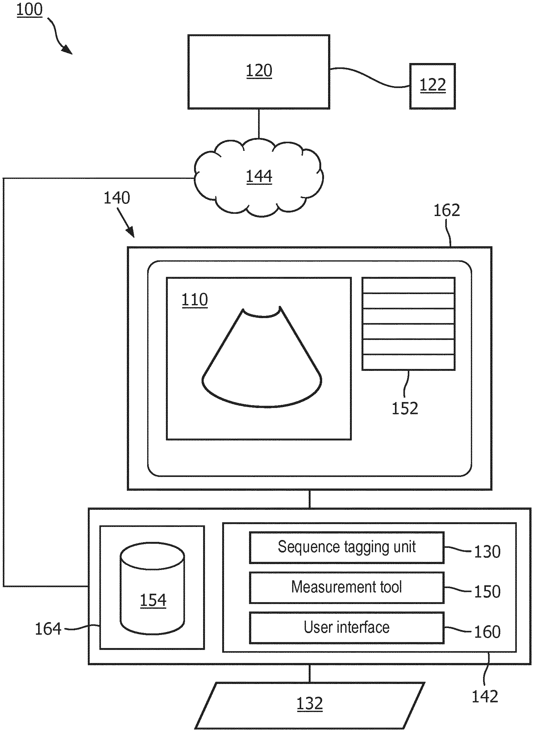
|---|---|---|---|---|---|---|---|---|---|---|---|
| Family ID: | 61563374 | ||||||||||
| Appl. No.: | 16/489367 | ||||||||||
| Filed: | February 28, 2018 | ||||||||||
| PCT Filed: | February 28, 2018 | ||||||||||
| PCT NO: | PCT/EP2018/054854 | ||||||||||
| 371 Date: | August 28, 2019 |
Related U.S. Patent Documents
| Application Number | Filing Date | Patent Number | ||
|---|---|---|---|---|
| 62465177 | Mar 1, 2017 | |||
| Current U.S. Class: | 1/1 |
| Current CPC Class: | A61B 8/14 20130101; G16H 30/40 20180101; A61B 8/465 20130101; A61B 8/5223 20130101; A61B 8/469 20130101; A61B 8/0883 20130101; A61B 8/5292 20130101; G16H 70/20 20180101 |
| International Class: | G16H 70/20 20060101 G16H070/20; G16H 30/40 20060101 G16H030/40; A61B 8/08 20060101 A61B008/08; A61B 8/14 20060101 A61B008/14; A61B 8/00 20060101 A61B008/00 |
Claims
1. A system, comprising: an echocardiogram measurement tool configured to determine a subset of measurements from a list of echocardiogram measurements according to a view of an ultrasound imaging sequence and a mapping between the view and the subset of measurements.
2. The system according to claim 1, wherein the mapping includes a first sub-mapping from the view to portions of heart anatomy within the view, and a second sub-mapping from the portions of heart anatomy within the view to the subset of measurements.
3. The system according to claim 1, further including: a user interface configured to display the determined subset of measurements in a selectable list and the view of the ultrasound imaging sequence on a display device; and wherein the measurement tool, in response to an input, measures an aspect of the heart in the view according to a selected measurement in the displayed subset of measurements indicated by the input.
4. The system according to claim 1, further including: a sequence tagging unit configured to determine the view from an analysis of the ultrasound imaging sequence.
5. The system according to claim 1, wherein the list of echocardiogram measurements is stored as a lookup table that comprises elements of a view and a measurement.
6. The system according to claim 1, wherein the subset of measurements are ordered according to usability statistics.
7. The system according to claim 2, wherein the first sub-mapping includes sub-views.
8. The system according to claim 1, wherein the measurement tool selects and applies at least one measurement of the subset of measurements to the view of the ultrasound imaging sequence autonomously.
9. A method, comprising: determining a subset of measurements from a list of echocardiogram measurements according to a view of an ultrasound imaging sequence and a mapping between the view and the subset of measurement; displaying the determined subset of measurements in a selectable list and the view of the ultrasound imaging sequence on a display device; and in response to an input, measuring an aspect of the heart in the view according to a selected measurement in the displayed subset of measurements indicated by the input.
10. The method according to claim 9, wherein the mapping includes a first sub-mapping from the view to portions of heart anatomy within the view, and a second sub-mapping from the portions of heart anatomy within the view to the subset of measurements.
11. The method according to claim 9 wherein the displaying comprises displaying only the determined subset of measurements in the selectable list and not displaying a remainder of the list of measurements in the selectable list.
12. The method according to claim 9, further including: determining the view from an analysis of the ultrasound imaging sequence.
13. The method according to claim 9, wherein the list of echocardiogram measurements is stored as a lookup table that comprises elements of a view and a measurement.
14. The method according to claim 11, wherein the displayed subset of measurements are ordered according to usability statistics.
15. The method according to claim 9, further comprising: selecting and applying at least one measurement of the subset of measurements to the view of the ultrasound imaging sequence autonomously.
16-20. (canceled)
Description
FIELD OF THE INVENTION
[0001] The following generally relates to ultrasound echocardiograms, and more specifically to echocardiogram measurements.
BACKGROUND OF THE INVENTION
[0002] Echocardiograms are performed to monitor and diagnosis heart disease and/or heart condition. A system that performs echocardiograms typically uses ultrasound (US) waves to create imaging sequences of a functioning heart with a probe that generates the ultrasound waves and detects reflected ultrasound waves.
[0003] US imaging sequences can include two dimensional (2-D) images, three-dimensional (3-D) images, Doppler imaging techniques, combinations and the like. An echocardiogram can include a multitude of sequences. Each sequence is typically an image of a portion of the heart over a cycle or at a phase.
[0004] The orientation of the probe relative to the heart determines the content of the image or view. The orientation of the probe relative to the heart includes a position and an angle of the probe on the skin of a patient, and thus, a direction of the waves relative to the heart and the portion of the heart included in the view. Views can include sub-views, which are minor adjustments in position and/or angle of the probe that focus on particular anatomical portions of the heart.
[0005] Measurements are made using the imaging sequence to quantify an aspect of an anatomical portion of the heart. The measurements are quantified, such as functions of a length, a width, a diameter, an area, a volume, combinations and the like. The measurements can be made at a specific phase of a cardiac cycle represented in the sequence. The anatomical portion measured can include aspects of an atrium, a ventricle, a vessel, a valve, and the like.
[0006] In quantifying aspects of the echocardiogram, a healthcare practitioner selects a measurement from a list of measurements that pertain to echocardiograms. The list of measurements, typically ordered alphabetically, can include over two hundred different measurements. The list can be very large even using abbreviations, such as LV for left ventricle, MV for mitral value, and the like. For example, in a display screen of an echocardiogram system, the abbreviated list can fill half the screen, temporarily obscuring half of the information concerning the echocardiogram.
SUMMARY OF THE INVENTION
[0007] Aspects described herein address the above-referenced problems and others.
[0008] The following describes embodiments of a system and method for a contextual based list of echocardiogram measurements. The context is determined from a view or sub-view of an echocardiogram imaging sequence and a mapping between the view or sub-view, anatomy displayed within the view or sub-view, and an echocardiogram measurement corresponding to the displayed anatomy. In some embodiments, the mapping includes further context according to usability statistics. In some embodiments, autonomous measurements are applied according to the context. In some embodiments, the view or sub-view is determined from analysis of the echocardiogram imaging sequence.
[0009] In one aspect, a system includes an echocardiogram measurement tool that determines a subset of measurements from a list of echocardiogram measurements according to a view of an ultrasound imaging sequence and a mapping between the view and the subset of measurements.
[0010] In another aspect, a method includes determining a subset of measurements from a list of echocardiogram measurements according to a view of an ultrasound imaging sequence and a mapping between the view and the subset of measurements.
[0011] In another aspect, a non-transitory computer-readable storage medium carrying instructions controls one or more processors to determine a subset of measurements from a list of echocardiogram measurements according to a view of an ultrasound imaging sequence and a mapping between the view and the subset of measurements
[0012] These and other aspects of the invention will be apparent from and elucidated with reference to the embodiment(s) described hereinafter.
BRIEF DESCRIPTION OF THE DRAWINGS
[0013] The invention may take form in various components and arrangements of components, and in various steps and arrangements of steps. The drawings are only for purposes of illustrating the preferred embodiments and are not to be construed as limiting the invention.
[0014] FIG. 1 schematically illustrates an embodiment of a system for context selected measurements of echocardiogram sequences.
[0015] FIG. 3 illustrates an example of a displayed echocardiogram sequence with a context based view of measurements.
[0016] FIG. 3 flowcharts an embodiment of a method of context based view of echocardiogram measurements.
DETAILED DESCRIPTION OF EMBODIMENTS
[0017] With reference to FIG. 1, an embodiment of a system 100 for context selected measurement of an echocardiogram ultrasound sequence 110 is illustrated. An ultrasound (US) imaging scanner 120 scans a subject using a probe 122 and generates the sequence 110. The sequence 110 is an ultrasound image generated from a view determined by the operation of the probe 122 relative to the heart of the subject. The view determines anatomical portions of the heart present in the sequence 110.
[0018] The view can be represented as a single data element or include a sub-view and be represented as a combined data element. The sub-view can be nested at different levels N>1. For example, a first sub-view 1 of view PLAX includes sub-views A and B, which identify the views of PLAX, PLAX1, PLAX1A, and PLAX1B. PLAX1, as an intermediate nested, level, can be omitted. Views can be represented by textual explanations, numerical references, or combinations thereof. For example views can represented by textual explanations or abbreviations, such as Parasternal long axis (PLAX), Parasternal short axis (PSAX), Apical four chamber (AP4), Apical five chamber (AP5), Apical two chamber (AP2), Apical three chamber (AP3), Sub costal (SC), and Supra sternal notch (SSN). In another example, PLAX3 represents a combination of textual and numerical representation that includes a sub-view.
[0019] A sequence tagging unit 130 determines the view from an analysis of the sequence 110. The analysis can include pattern matching of the pixel data with reference images. The analysis can include segmentation of anatomical features and comparison with an anatomical model. In some embodiments, tagging can include, via a console or computing device 140, an entry of a view, a selection of a view, a display of the view and a modification of the view, combinations thereof and the like.
[0020] The computing device 140 or console can be part of, combined with, or separated from the US imaging scanner 120. The computing device 140 includes one or more processors 142, such as a digital processor, a microprocessor, an electronic processor, an optical processor, a multi-processor, a distribution of processors including peer-to-peer or cooperatively operating processors, client-server arrangement of processors, and the like. The arrangement can include a network 144, which can include a bus structure or other internal or local communication structure, wireless or wired communications, public or private network communications, cellular or data communications, combinations thereof and the like. The computing device 140 can be configured as a laptop computer, a desktop computer, a tablet, a smartphone, a body worn device, and the like.
[0021] An echocardiogram measurement tool 150 determines a subset 152 of measurements from a list of echocardiogram measurements according to the view of the sequence 110 and a mapping 154 between the view and the subset 152 of measurements. For example in a PLAX3 view, measurements, such as left ventricular ejection fraction (LVEF), are included in the subset 152, while measurements, such as a diameter of the inferior vena cava (IVC Diam), are excluded from the subset 152. That is, the PLAX3 view is mapped to LVEF and is not mapped to IVC Diam. The PLAX3 view includes the anatomical portions of the left ventricle sufficient to compute the LVEF. The PLAX3 excludes the anatomical portions of the inferior vena cava sufficient to compute the IVC Diam and thus, the IVC Diam is not mapped according to the PLAX3 view. The mappings are determined according to the anatomy present in a view, and the anatomy with which the measurements are performed.
[0022] The mapping of echocardiogram measurements 154 can be represented and stored as a lookup table with data elements of a view and a measurement. For example, a table can include rows, each row representing a measurement according to a view. A view can include multiple measurements. Data elements or columns include the view and the corresponding measurement. The table can include other elements of the mapping, such as the anatomical portion according to the view to which the measurement applies, a link to the specific measurement for programmatic access, and the like.
[0023] A user interface 160 displays the sequence 110 and the determined subset 152 of measurements in a selectable list, such as in a menu, in a drop-down box, in a pick list, and the like on a display device 162. In some embodiments, the subset 152 is displayed at the top of the entire list of echocardiogram measurements showing the subset 152 on top of all possible measurements. In some embodiments, the remainder of measurements in the entire list of echocardiogram measurements (excluding the subset 152) are shown in a sub-menu or expansion box. That is, the entire list can be accessed through a sub-menu or expansion box. The sub-menu or expansion box does not display the remainder of the measurements until invoked, which, in some instances, does not obscure portions of the display. In some embodiments, individual measurements in the subset 152 are shown as active and those not included in the subset 152 are shown inactive, such as grayed out and non-selectable. In some instances, the displayed subset of measurements significantly shortens the number of displayed measurements, which covers less of the display 162 and reduces the number of measurements a healthcare practitioner searches to select a measurement. The display device 162 is suitably embodied by a computer display, smartphone display, projector, body worn display, and the like.
[0024] An input device 132, such as a keyboard, mouse, microphone, and the like, sends an input indicative of a selected measurement, such as an input indicative of a measurement in the displayed subset 152. The measurement tool 150, in response to the input, measures an aspect of the heart in the view according to the selected measurement. For example, in response to a mouse click selecting a left ventricle ejection fraction, the measurement tool measures the left ventricle ejection fraction according to the sequence 110.
[0025] In some embodiments, the measurement tool 150 autonomously selects and applies a measurement from the subset 152 of measurements to the tagged view prior to display by the user interface 160. The measurement from the subset 152 can be autonomously selected according to site policies or usability statistics, such as frequency of use by a healthcare practitioner, frequency of use by a group of practitioners, percentage of echocardiogram usage, percentage of sequence usage, threshold number of uses, and the like. For example, prior to display of the sequence 110, a measurement applied with a high frequency can be selected and applied by the measurement tool 150. The measured value can be included in the initial display of the sequence 110 or added to the display without the input for selecting the measurement from the list. In some embodiments, the autonomously selected and applied measurements are indicated with an icon or other symbol in the displayed subset.
[0026] The computer device 140 includes the processor 142 and a memory 164. The mapping 154 is suitably embodied by a configured electronic storage medium, such as local disk, cloud storage, server storage, remote storage and the like, accessed by the configured processor 142. The configured electronic storage medium can include system file structures, relational and/or object oriented database system structures, data structures, and the like.
[0027] The sequence tagging unit 130, the measurement tool 150 and the user interface 160 are suitably embodied by the processor 142, configured to receive the sequence 110, determine the view, generate the subset 152, display the sequence 110 and the subset 152, receive a selection and perform the measurement according to the selection.
[0028] The configured processor 142 executes at least one computer readable instruction stored in the computer readable storage medium 164, such as an optical disk, a magnetic disk, semiconductor memory of a computing device with the configured processor, which excludes transitory medium and includes physical memory and/or other non-transitory medium to perform the disclosed techniques. The configured processor may also execute one or more computer readable instructions carried by a carrier wave, a signal or other transitory medium. The lines between components represented in the diagram represent communications paths.
[0029] With reference to FIG. 2, an example of a displayed echocardiogram sequence 110 with a context based view of measurements that is a subset 152 of the list of echocardiogram measurements is illustrated. The context is based on the determined view of the sequence 110, such as PLAX. The determined view can include subviews. That is, the determined view can be specified at different levels of granularity. For example, a view can be PLAX, PLAX1, or PLAX1A, where PLAX1 is a subview of PLAX, and PLAX1A is a subview of PLAX1. PLAX1 further refines or delineates PLAX and PLAX1A further refines or delineates PLAX1. Each refinement or delineation can correspondingly refine or delineate anatomy present in the view.
[0030] Values of data elements, such as of the view 200, the anatomy 210, the usability statistics 230 or combinations thereof can be included in the displayed subset 152. In some embodiments, the values of the view 200, the anatomy 210 and the usability statistics 230 are omitted from the display. The values of the measurement data element 220 or representations thereof are included in the display, such as LVEF, IVC Diam, and the like. Representations of the measurement data element 220 can include abbreviations, icons, or other indicators.
[0031] The values or instances of the measurement data element 220 for the subset 152 are displayed in the selectable list. The data elements illustrate the relationship between values of the view/subview 200 that are mapped to values of the anatomy 210, and values of the anatomy 210 are mapped to measurements included in the subset 152. The values of the anatomy are portions of the heart anatomy, such as left ventricle, left atrium, aorta, mitral value, aortic valve, sub anatomical portions or each, combinations of each, and the like. In some embodiments, the heart anatomy can include a phase, such as left ventricle end-diastolic.
[0032] In some embodiments, the subset 152 is ordered according to usability statistics 230 or combinations of usability statistics 230 and anatomy 210. For example, a measurement within a highest frequency of use is displayed first. In another example, measurements are ordered within anatomy 210. For example, measurements mapped from the anatomy of the left ventricle are ordered by frequency and grouped together, and measurements of the right ventricle are ordered by frequency and grouped together. In some instances, the context is based on the view and in some instances the context is based on combinations of the view and usability statistics.
[0033] With reference to FIG. 3, an embodiment of a method of context based view of echocardiogram measurements is flowcharted.
[0034] At 300, an echocardiogram sequence 110 is received. The echocardiogram sequence 110 can be received directly from the US imaging scanner 120 or from computer memory or storage.
[0035] At 310, a view of the sequence 110 is determined. The determined view can include sub-views. In some embodiments, the view is determined by analysis of the ultrasound image data. The analysis can include pattern matching of the pixel data with reference images. The analysis can include segmentation of anatomical features and comparison with an anatomical model. In some embodiments, the view is determined by manual review and/or input.
[0036] At 320, a subset 152 of measurements is determined from the list of echocardiogram measurements according to the view and a mapping 154 between the view and the subset 152. The mapping 154 includes a first sub-mapping from the view to portions of heart anatomy within the view, and a second sub-mapping from portions of heart anatomy within the view to the subset 152 of measurements. A lookup table can store the mappings between the views and the measurements. The lookup table includes data elements of the view and the mapped measurement. The lookup table can include usability statistics 230. The usabilities statistics can be according to system, organization, site, user or combinations thereof. For example, in a PLAX view, use can be tracked, such as a total number or a percentage frequency of the LVEF being measured in the PLAX view, a total number or a percentage frequency of the LVEF being measured by a current user in the PLAX view, and the like.
[0037] At 330 the determined subset 152 of measurements in a selectable list and the view of the ultrasound imaging sequence are displayed on the display device 162. The displayed subset 152 can be ordered according to the usability statistics. For example, the determined subset 152 of measurements can be ordered to a total number or a percentage frequency of use in the displayed subset 152
[0038] At 340, in response to an input from the input device 132, an aspect of the heart in the view is measured according to a selected measurement in the displayed subset of measurements. For example, a user selects the LVEF with a mouse click, the LVEF is measured according to the received sequence 110.
[0039] In some embodiments, steps 330 and 340 are omitted, and at least one measurement of the subset of measurements is selected and applied to the view of the ultrasound imaging sequence autonomously. For example, a measurement is designated as autonomous within a view, such as LVEF within PLAX according to a site policy or the useability statistics. Autonomous measurements can be indicated in the lookup table, such as with a separate data element with a binary value. The autonomous measurement is applied to the received sequence 110 prior to displaying the received sequence 110, such as upon being received.
[0040] The above may be implemented by way of computer readable instructions, encoded or embedded on a computer readable storage medium, which, when executed by a computer processor(s), cause the processor(s) to carry out the described acts. Additionally or alternatively, at least one of the computer readable instructions is carried by a signal, carrier wave or other transitory medium.
[0041] The invention has been described with reference to the preferred embodiments. Modifications and alterations may occur to others upon reading and understanding the preceding detailed description. It is intended that the invention is constructed as including all such modifications and alterations insofar as they come within the scope of the appended claims or the equivalents thereof. The word "comprising" does not exclude other elements or steps, and the indefinite article "a" or "an" does not exclude a plurality.
* * * * *
D00000
D00001
D00002
D00003
XML
uspto.report is an independent third-party trademark research tool that is not affiliated, endorsed, or sponsored by the United States Patent and Trademark Office (USPTO) or any other governmental organization. The information provided by uspto.report is based on publicly available data at the time of writing and is intended for informational purposes only.
While we strive to provide accurate and up-to-date information, we do not guarantee the accuracy, completeness, reliability, or suitability of the information displayed on this site. The use of this site is at your own risk. Any reliance you place on such information is therefore strictly at your own risk.
All official trademark data, including owner information, should be verified by visiting the official USPTO website at www.uspto.gov. This site is not intended to replace professional legal advice and should not be used as a substitute for consulting with a legal professional who is knowledgeable about trademark law.