Tumor Signature For Metastasis, Compositions Of Matter Methods Of Use Thereof
Puram; Sidharth ; et al.
U.S. patent application number 16/604651 was filed with the patent office on 2020-03-05 for tumor signature for metastasis, compositions of matter methods of use thereof. The applicant listed for this patent is The Broad Institute, Inc., The General Hospital Corporation, Massachusetts Eye and Ear Infirmary, Massachusetts Institute of Technology. Invention is credited to Bradley Bernstein, Derrick Lin, Anuraag Parikh, Sidharth Puram, Aviv Regev, Itay Tirosh.
| Application Number | 20200071773 16/604651 |
| Document ID | / |
| Family ID | 63792877 |
| Filed Date | 2020-03-05 |


View All Diagrams
| United States Patent Application | 20200071773 |
| Kind Code | A1 |
| Puram; Sidharth ; et al. | March 5, 2020 |
TUMOR SIGNATURE FOR METASTASIS, COMPOSITIONS OF MATTER METHODS OF USE THEREOF
Abstract
The present invention advantageously provides for novel gene signatures, tools and methods for the treatment and prognosis of epithelial tumors. Applicants have used single cell RNA-seq to reveal novel expression programs of malignant, stromal and immune cells in the HNSCC tumor ecosystem. Malignant cells varied in expression of programs related to stress, hypoxia and epithelial differentiation. A partial EMT-like program (p-EMT) was discovered that was expressed in cells residing at the leading edge of tumors. Applicants unexpectedly linked the p-EMT state to metastasis and adverse clinical features that may be used to direct treatment of epithelial cancers (e.g., HNSCC). Applicants also show that metastases are dynamically regulated by the tumor microenvironment (TME). Finally, a computational modeling approach was developed that allows analysis of malignant cells in bulk sequencing samples.
| Inventors: | Puram; Sidharth; (Boston, MA) ; Tirosh; Itay; (Cambridge, MA) ; Parikh; Anuraag; (Boston, MA) ; Lin; Derrick; (Boston, MA) ; Regev; Aviv; (Cambridge, MA) ; Bernstein; Bradley; (Boston, MA) | ||||||||||
| Applicant: |
|
||||||||||
|---|---|---|---|---|---|---|---|---|---|---|---|
| Family ID: | 63792877 | ||||||||||
| Appl. No.: | 16/604651 | ||||||||||
| Filed: | April 12, 2018 | ||||||||||
| PCT Filed: | April 12, 2018 | ||||||||||
| PCT NO: | PCT/US2018/027383 | ||||||||||
| 371 Date: | October 11, 2019 |
Related U.S. Patent Documents
| Application Number | Filing Date | Patent Number | ||
|---|---|---|---|---|
| 62484709 | Apr 12, 2017 | |||
| 62586126 | Nov 14, 2017 | |||
| Current U.S. Class: | 1/1 |
| Current CPC Class: | C12Q 2600/106 20130101; C12Q 1/6837 20130101; C12Q 1/6886 20130101; A61P 35/00 20180101; G16B 25/10 20190201; G16B 40/00 20190201; C12Q 2600/158 20130101; G01N 33/574 20130101; A61P 35/04 20180101 |
| International Class: | C12Q 1/6886 20060101 C12Q001/6886; A61P 35/04 20060101 A61P035/04; C12Q 1/6837 20060101 C12Q001/6837; G16B 25/10 20060101 G16B025/10; G16B 40/00 20060101 G16B040/00 |
Goverment Interests
STATEMENT REGARDING FEDERALLY SPONSORED RESEARCH
[0002] This invention was made with government support under grant numbers CA216873, CA180922, CA202820 and CA14051 awarded by the National Institutes of Health. The government has certain rights in the invention.
Claims
1. A method of detecting an EMT-like (p-EMT) gene signature in epithelial tumors comprising, detecting in tumor cells obtained from a subject suffering from an epithelial tumor, the expression or activity of a EMT-like (p-EMT) gene signature, said signature comprising one or more genes or polypeptides selected from the group consisting of SERPINE1, TGFBI, MMP10, LAMC2, P4HA2, PDPN, ITGA5, LAMA3, CDH13, TNC, MMP2, EMP3, INHBA, LAMB3, SNAIL2 and VIM, preferably, wherein said signature does not comprise ZEB1/2, TWIST1/2, or SNAIL1.
2. (canceled)
3. The method according to claim 1, wherein detecting a p-EMT gene signature indicates that the subject is less likely to respond to therapy, and/or wherein detecting a p-EMT gene signature indicates that the subject requires more aggressive treatment.
4. (canceled)
5. The method according to claim 1, further comprising treating the subject with one or more of lymph node dissection, adjuvant chemotherapy, adjuvant radiation, neoadjuvant therapy, chemoradiation and an agent that inhibits TGF beta signaling upon detecting the p-EMT gene signature.
6. The method according to claim 1, wherein the epithelial tumor is head and neck squamous cell carcinoma (HNSCC).
7. The method of claim 1, further comprising treating the method of treatment for a subject in need thereof suffering from an epithelial tumor, said method comprising: a) detecting expression or activity of a p-EMT gene signature for a tumor sample obtained from the subject, wherein the p-EMT signature comprises one or more genes or polypeptides selected from the group consisting of SERPINE1, TGFBI, MMP10, LAMC2, P4HA2, PDPN, ITGA5, LAMA3, CDH13, TNC, MMP2, EMP3, INHBA, LAMB3, SNAIL2 and VIM; and b) treating the subject, wherein if a p-EMT signature is detected above a p-EMT high reference level the treatment comprises: i) lymph node dissection of the subject; ii) adjuvant chemotherapy; iii) adjuvant radiation or postoperative radiation treatment (PORT); iv) neoadjuvant therapy; v) chemoradiation; or vi) administering an agent that inhibits TGF beta signaling, wherein if a p-EMT signature is not detected the treatment comprises delaying lymph node dissection.
8. The method according to claim 7, further comprising: c) detecting expression or activity of an epithelial gene signature for a tumor sample obtained from the subject, wherein the epithelial signature comprises: one or more genes or polypeptides selected from the group consisting of IL1RN, SLPI, CLDN4, CLDN7, S100A9, SPRR1B, PVRL4, RHCG, SDCBP2, S100A8, APOBEC3A, LY6D, KRT16, KRT6B, KRT6A, LYPD3, KRT6C, KLK10, KLK11, TYMP, FABP5, SCO2, FGFBP1 and JUP, or one or more genes or polypeptides selected from the group consisting of SPRR1B, KRT16, KRT6B, KRT6C, KRT6A, KLK10, KLK11 and CLDN7, and d) treating the subject as in (b) if a p-EMT signature is detected above a p-EMT high reference level and the epithelial signature is detected below an epithelial low reference.
9. The method according to claim 7, wherein chemoradiation comprises cisplatin.
10. The method according to claim 7, wherein treatment comprises administering an agent that inhibits TGF beta signaling.
11. The method according to claim 7, wherein the epithelial tumor is head and neck squamous cell carcinoma (HNSCC).
12. A method of treating an epithelial tumor, comprising administering to a subject in need thereof a therapeutically effective amount of an agent: a) capable of reducing the expression or inhibiting the activity of one or more p-EMT signature genes or polypeptides; or b) capable of targeting or binding to one or more cell surface exposed p-EMT signature genes or polypeptides, wherein the p-EMT signature comprises one or more genes or polypeptides selected from the group consisting of SERPINE1, TGFBI, MMP10, LAMC2, P4HA2, PDPN, ITGA5, LAMA3, CDH13, TNC, MMP2, EMP3, INHBA, LAMB3, SNAIL2 and VIM.
13. The method according to claim 12, wherein the epithelial tumor comprises HNSCC.
14. The method according to claim 12, wherein said agent capable of reducing the expression or inhibiting the activity of one or more p-EMT signature genes or polypeptides comprises a therapeutic antibody, antibody fragment, antibody-like protein scaffold, aptamer, genetic modifying agent or small molecule; or wherein said agent capable of targeting or binding to one or more cell surface exposed EMT-like signature polypeptides comprises a CAR T cell capable of targeting or binding to one or more cell surface exposed p-EMT signature genes or polypeptides.
15. (canceled)
16. A method of deconvoluting bulk gene expression data obtained from an epithelial tumor, wherein the tumor comprises both malignant and non-malignant cells, said method comprising: a) defining, by a processor, the relative frequency of a set of cell types in the tumor from the bulk gene expression data, wherein the frequency of the cell types is determined by cell type specific gene expression, and wherein the set of cell types comprises one or more cell types selected from the group consisting of T cells, fibroblasts, macrophages, mast cells, B/plasma cells, endothelial cells, myocytes and dendritic cells; and b) defining, by a processor, a linear relationship between the frequency of the non-malignant cell types and the expression of a set of genes, wherein the set of genes comprises genes highly expressed by malignant cells and at most two non-malignant cell types, wherein the set of genes are derived from gene expression analysis of single cells in at least one epithelial tumor, and wherein the residual of the linear relationship defines the malignant cell-specific (MCS) expression profile.
17. The method according to claim 16, wherein the epithelial tumor is HNSCC.
18. The method according to claim 16, further comprising assigning genes to a specific malignant cell sub-type, preferably, wherein the malignant cell sub-type is a EMT-like subtype; and/or wherein the method further comprises determining a p-EMT score, wherein said score is based on expression of a p-EMT signature for the malignant cell-specific (MCS) expression profile, wherein said p-EMT signature comprises one or more genes or polypeptides selected from the group consisting of SERPINE1, TGFBI, MMP10, LAMC2, P4HA2, PDPN, ITGA5, LAMA3, CDH13, TNC, MMP2, EMP3, INHBA, LAMB3, SNAIL2 and VIM, and wherein a high p-EMT score has higher expression of the p-EMT signature as compared to expression in a reference data set obtained from a subject with a non-invasive epithelial tumor.
19. (canceled)
20. (canceled)
21. The method of claim 18, wherein the method further comprises treating a subject in need thereof suffering from an epithelial tumor, said method comprising: a) determining a p-EMT score for a tumor sample obtained from the subject; and b) treating the subject, wherein if a high p-EMT score is determined the treatment comprises: i) lymph node dissection of the subject; ii) adjuvant chemotherapy; iii) adjuvant radiation or postoperative radiation treatment (PORT); iv) neoadjuvant therapy; v) chemoradiation; or vi) administering an agent that inhibits TGF beta signaling, wherein if the subject does not have a high p-EMT score the treatment comprises delaying lymph node dissection.
22. The method according to claim 21, wherein chemoradiation comprises cisplatin.
23. The method according to claim 21, wherein treatment comprises administering an agent that inhibits TGF beta signaling.
24. (canceled)
25. (canceled)
26. (canceled)
27. (canceled)
28. (canceled)
29. (canceled)
30. A method of detecting an epithelial gene signature in epithelial tumors comprising detecting in tumor cells obtained from a subject suffering from an epithelial tumor, the expression or activity of an epithelial gene signature, said signature comprising: a) one or more genes or polypeptides selected from the group consisting of IL1RN, SLPI, CLDN4, CLDN7, S100A9, SPRR1B, PVRL4, RHCG, SDCBP2, S100A8, APOBEC3A, LY6D, KRT16, KRT6B, KRT6A, LYPD3, KRT6C, KLK10, KLK11, TYMP, FABP5, SCO2, FGFBP1 and JUP; or b) one or more genes or polypeptides selected from the group consisting of SPRR1B, KRT16, KRT6B, KRT6C, KRT6A, KLK10, KLK11 and CLDN7, preferably, wherein detecting an epithelial gene signature indicates that the subject is more likely to respond to therapy; and/or wherein detecting an epithelial gene signature indicates that the subject does not require more aggressive treatment.
31. (canceled)
32. (canceled)
33. The method according to claim 30, wherein the epithelial tumor is head and neck squamous cell carcinoma (HNSCC).
34. A method for characterizing epithelial tumor composition comprising: detecting the presence of one or more expression programs in a sample, wherein each expression program comprises a set of biomarkers as defined in Table S7.
35. A kit comprising reagents to detect at least one gene or gene expression program as defined in claim 34, preferably, wherein the gene expression program is a p-EMT program, wherein the p-EMT program comprises one or more genes or polypeptides selected from the group consisting of SERPINE1, TGFBI, MMP10, LAMC2, P4HA2, PDPN, ITGA5, LAMA3, CDH13, TNC, MMP2, EMP3, INHBA, LAMB3, SNAIL2 and VIM; and/or wherein the kit comprises antibodies and reagents for immunohistochemistry, preferably, an HNSCC specific antibody; and/or wherein the kit comprises primers and/or probes for quantitative RT-PCR, PCR, and/or sequencing; and/or wherein the kit comprises fluorescently bar-coded oligonucleotide probes for hybridization to RNA.
Description
CROSS-REFERENCE TO RELATED APPLICATIONS
[0001] This application claims the benefit of U.S. Provisional Application Nos. 62/484,709, filed Apr. 12, 2017 and 62/586,126, filed Nov. 14, 2017. The entire contents of the above-identified applications are hereby fully incorporated herein by reference.
TECHNICAL FIELD
[0003] The subject matter disclosed herein is generally directed to methods of using gene expression profiles representative of cell sub-types present in head and neck squamous cell carcinoma (HNSCC). Specifically, the gene signatures may be used for diagnosing, pro gnosing and/or staging of tumors and designing and selecting appropriate treatment regimens. Furthermore, novel signatures determined by single cell analysis of HNSCC are leveraged to provide for methods and systems for deconvolution of bulk sequencing data from tumors.
BACKGROUND
[0004] Genomic and transcriptomic studies have revealed driver mutations, identified aberrant regulatory programs, and redefined disease subtypes for major human tumors (Stratton et al., 2009; Weinberg, 2014). However, these studies relied on profiling technologies that measure the entire tumor in bulk, limiting their ability to capture intra-tumoral heterogeneity, including malignant cells in distinct genetic, epigenetic, and functional states, as well as diverse non-malignant cells such as immune cells, fibroblasts, and endothelial cells. Substantial evidence indicates that intra-tumoral heterogeneity among malignant and non-malignant cells, and their interactions within the tumor microenvironment (TME) are critical to many aspects of tumor biology, including self-renewal, immune surveillance, drug resistance and metastasis (Meacham and Morrison, 2013; Weinberg, 2014).
[0005] Recent advances in single-cell genomics provide an avenue to explore genetic and functional heterogeneity at a cellular resolution (Navin, 2015; Tanay and Regev, 2017; Wagner et al., 2016). In particular, single-cell RNA-seq (scRNA-seq) studies of human tumors, circulating tumor cells and patient-derived xenografts have revealed new insights into tumor composition, cancer stem cells, and drug resistance.
[0006] Despite these promising results, scRNA-seq studies have not extensively characterized epithelial tumors, in spite of their predominance. In these tumors, metastasis to nearby draining lymph nodes (locoregional metastasis) and to other organs (distant metastasis) represents a major cause of morbidity and mortality. However, lymph node (LN) and distant metastases are often treated based on molecular and pathologic features of the primary tumor, raising the question of whether metastases share the same genetics, epigenetics, and vulnerabilities (Lambert et al., 2017). The potentially different composition of primary tumors and metastases hinders the straightforward comparison of bulk tumor profiles. Single-cell expression profiling studies would, in principle, offer a compelling alternative.
[0007] Epithelial-to-mesenchymal transition (EMT) has been suggested as a driver of local and distant spread of epithelial tumors (Gupta and Massague, 2006; Lambert et al., 2017). The process of EMT is fundamental to embryonic development and other physiologic processes and may be co-opted by malignant epithelial cells to facilitate invasion and dissemination (Thiery et al., 2009; Ye and Weinberg, 2015). EMT markers have been detected on circulating tumor cells (CTCs) associated with metastatic disease (Ting et al., 2014; Yu et al., 2013). However, since most EMT studies have focused on laboratory models, the nature, extent, and significance of EMT in primary human tumors and metastases remains controversial (Lambert et al., 2017; Nieto et al., 2016). For example, although mesenchymal subtypes have been identified in multiple tumor types (Cancer Genome Atlas, 2015; Cancer Genome Atlas Research, 2011; Verhaak et al., 2010), it remains unclear whether they reflect mesenchymal cancer cells or, alternatively, contributions of non-malignant, mesenchymal cell types in the TME.
[0008] Head and neck squamous cell carcinoma (HNSCC) is an epithelial tumor with strong associations to chronic alcohol and tobacco exposure (Puram and Rocco, 2015). Like many epithelial cancers, HNSCC tumors are highly heterogeneous within and between patients. Metastatic disease remains a central challenge, with patients often presenting at an advanced stage with LN metastases. Thus, there is a need for biomarkers and therapeutic targets capable of guiding treatment and predicting disease progression (e.g., metastasis) in epithelial tumors.
SUMMARY
[0009] The diverse malignant, stromal, and immune cells in tumors affect growth, metastasis and response to therapy. It is an objective of the present invention to understand intra-tumoral heterogeneity, invasion and metastasis in an epithelial human cancer. It is another objective of the present to provide for novel tools and methods for diagnosing, prognosing and treating tumors. Applicants investigated primary HNSCC tumors and matched lymph nodes. Specifically, Applicants profiled transcriptomes of .about.6,000 single cells from 18 head and neck squamous cell carcinoma (HNSCC) patients, including five matched pairs of primary tumors and lymph node metastases. Stromal and immune cells had consistent expression programs across patients. Conversely, malignant cells varied within and between tumors in their expression of signatures related to cell cycle, stress, hypoxia, epithelial differentiation, and partial epithelial-to-mesenchymal transition (p-EMT). Cells expressing the p-EMT program spatially localized to the leading edge of primary tumors. By integrating single-cell transcriptomes with bulk expression profiles for hundreds of tumors, Applicants refined HNSCC subtypes by their malignant and stromal composition, and established p-EMT as an independent predictor of nodal metastasis, tumor grade, and adverse pathologic features (e.g., extracapsular extension). The results provide insight into the HNSCC ecosystem, define stromal interactions and define a p-EMT program associated with metastasis.
[0010] In one aspect, the present invention provides for a method of detecting an EMT-like (p-EMT) gene signature in epithelial tumors comprising, detecting in tumor cells obtained from a subject suffering from an epithelial tumor, the expression or activity of a EMT-like (p-EMT) gene signature, said signature comprising one or more genes or polypeptides selected from the group consisting of SERPINE1, TGFBI, MMP10, LAMC2, P4HA2, PDPN, ITGA5, LAMA3, CDH13, TNC, MMP2, EMP3, INHBA, LAMB3, SNAIL2 and VIM; or one or more genes or polypeptides selected from the group consisting of SERPINE1, TGFBI, MMP10, LAMC2, P4HA2, PDPN, ITGA5, LAMA3, CDH13, TNC, MMP2, EMP3, INHBA, LAMB3, VIM, SEMA3C, PRKCDBP, ANXA5, DHRS7, ITGB1, ACTN1, CXCR7, ITGB6, IGFBP7, THBS1, PTHLH, TNFRSF6B, PDLIM7, CAV1, DKK3, COL17A1, LTBP1, COL5A2, COL1A1, FHL2, TIMP3, PLAU, LGALS1, PSMD2, CD63, HERPUD1, TPM1, SLC39A14, C1S, MMP1, EXT2, COL4A2, PRSS23, SLC7A8, SLC31A2, ARPC1B, APP, MFAP2, MPZL1, DFNA5, MT2A, MAGED2, ITGA6, FSTL1, TNFRSF12A, IL32, COPB2, PTK7, OCIAD2, TAX1BP3, SEC13, SERPINH1, TPM4, MYH9, ANXA8L1, PLOD2, GALNT2, LEPREL1, MAGED1, SLC38A5, FSTL3, CD99, F3, PSAP, NMRK1, FKBP9, DSG2, ECM1, HTRA1, SERINC1, CALU, TPST1, PLOD3, IGFBP3, FRMD6, CXCL14, SERPINE2, RABAC1, TMED9, NAGK, BMP1, ESYT1, STON2, TAGLN and GJA1. The signature may not comprise ZEB1/2, TWIST1/2, or SNAIL1. Thus, the signature unexpectedly does not include most classical EMT transcription factors.
[0011] In one embodiment, detecting a p-EMT gene signature may indicate that the subject is less likely to respond to therapy. In certain embodiments, the therapy is a therapy consistent with the standard of care for the epithelial tumor. In certain embodiments, the therapy is an immunotherapy, such as checkpoint blockade therapy. Detecting a p-EMT gene signature may indicate that the subject requires more aggressive treatment. The method may further comprise treating the subject with one or more of lymph node dissection, adjuvant chemotherapy, adjuvant radiation, neoadjuvant therapy, chemoradiation, and an agent that inhibits TGF beta signaling upon detecting the p-EMT gene signature. The epithelial tumor may be head and neck squamous cell carcinoma (HNSCC). In certain example embodiments, "less likely to respond" indicates the likelihood of response is less than the likelihood of an individual without a p-EMT gene signature of p-EMT.sup.lo signature as measured using standard statistical analysis, such as those used and described in the examples section below.
[0012] In another embodiment, not detecting a p-EMT gene signature may indicate that the subject is more likely to respond to therapy. Not detecting a p-EMT gene signature may indicate that the subject should avoid aggressive treatment. Not being bound by a theory, an unnecessary aggressive treatment may lead to increased mortality and morbidity. In certain embodiments, if a p-EMT signature is not detected a subject may be treated according to a less aggressive standard of care as described herein.
[0013] In another aspect, the present invention provides for a method of treatment for a subject in need thereof suffering from an epithelial tumor comprising: a) detecting expression or activity of a p-EMT gene signature for a tumor sample obtained from the subject, wherein the p-EMT signature comprises one or more genes or polypeptides selected from the group consisting of SERPINE1, TGFBI, MMP10, LAMC2, P4HA2, PDPN, ITGA5, LAMA3, CDH13, TNC, MMP2, EMP3, INHBA, LAMB3, SNAIL2 and VIM; and b) treating the subject, wherein if a p-EMT signature is detected the treatment comprises: i) lymph node dissection of the subject; ii) adjuvant chemotherapy; iii) adjuvant radiation or postoperative radiation treatment (PORT); iv) neoadjuvant therapy; v) chemoradiation; or vi) administering an agent that inhibits TGF beta signaling, wherein if a p-EMT signature is not detected the treatment comprises delaying lymph node dissection.
[0014] In certain embodiments, the method may further comprise: detecting expression or activity of an epithelial gene signature for a tumor sample obtained from the subject, wherein the epithelial signature comprises: one or more genes or polypeptides selected from the group consisting of IL1RN, SLPI, CLDN4, CLDN7, S100A9, SPRR1B, PVRL4, RHCG, SDCBP2, S100A8, APOBEC3A, LY6D, KRT16, KRT6B, KRT6A, LYPD3, KRT6C, KLK10, KLK11, TYMP, FABP5, SCO2, FGFBP1 and JUP; or one or more genes or polypeptides selected from the group consisting of SPRR1B, KRT16, KRT6B, KRT6C, KRT6A, KLK10, KLK11 and CLDN7; or one or more genes or polypeptides selected from the group consisting of IL1RN, SLPI, CLDN4, S100A9, SPRR1B, PVRL4, RHCG, SDCBP2, S100A8, APOBEC3A, GRHL1, SULT2B1, ELF3, KRT16, PRSS8, MXD1, S100A7, KRT6B, LYPD3, TACSTD2, CDKN1A, KLK11, GPRC5A, KLK10, TMBIM1, PLAUR, CLDN7, DUOXA1, PDZK1IP1, NCCRP1, IDS, PPL, ZNF750, EMP1, CLDN1, CRB3, CYB5R1, DSC2, S100P, GRHL3, SPINT1, SDR16C5, SPRR1A, WBP2, GRB7, KLK7, TMEM79, SBSN, PIM1, CLIC3, MALAT1, TRIP10, CAST, TMPRSS4, TOM1, A2ML1, MBOAT2, LGALS3, ERO1L, EHF, LCN2, YPEL5, ALDH3B2, DMKN, PIK3IP1, CEACAM6, OVOL1, TMPRSS11E, CD55, KLK6, SPRR2D, NDRG2, CD24, HIST1H1C, LY6D, CLIP1, HIST1H2AC, BNIPL, QSOX1, ECM1, DHRS3, PPP1R15A, TRIM16, AQP3, IRF6, CSTA, RAB25, HOPX, GIPC1, RAB11FIP1, CSTB, KRT6C, PKP1, JUP, MAFF, DSG3, AKTIP, KLF3, HSPB8 and H1F0; or one or more genes or polypeptides selected from the group consisting of LY6D, KRT16, KRT6B, LYPD3, KRT6C, TYMP, FABP5, SCO2, FGFBP1, JUP, IMP4, DSC2, TMBIM1, KRT14, C1QBP, SFN, S100A14, RAB38, GJB5, MRPL14, TRIM29, ANXA8L2, KRT6A, PDHB, AKR1B10, LAD1, DSG3, MRPL21, NDUFS7, PSMD6, AHCY, GBP2, TXN2, PSMD13, NOP16, EIF4EBP1, MRPL12, HSD17B10, LGALS7B, THBD, EXOSC4, APRT, ANXA8L1, ATP5G1, S100A2, TBRG4, MAL2, NHP2L1, DDX39A, ZNF750, UBE2L6, WDR74, PPIF, PRMT5, VSNL1, VPS25, SNRNP40, ADRM1, NDUFS8, TUBA1C, TMEM79, UQCRFS1, EIF3K, NME2, PKP3, SERPINB1, RPL26L1, EIF6, DSP, PHLDA2, S100A16, LGALS7, MT1X, UQCRC2, EIF3I, MRPL24, CCT7, RHOV, ECE2, SSBP1, POLDIP2, FIS1, CKMT1A, GJB3, NME1, MRPS12, GPS1, ALG3, MRPL20, EMC6, SRD5A1, PA2G4, ECSIT, MRPL23, NAA20, HMOX2, COA4, DCXR, PSMD8 and WBSCR22; and treating the subject as above if a p-EMT signature is detected above a p-EMT high reference level and the epithelial signature is detected below an epithelial low reference. Chemoradiation may comprise cisplatin. The treatment may comprise administering an agent that inhibits TGF beta signaling. Applicants describe herein data showing that the p-EMT signature is regulated by TGF beta signaling. The epithelial tumor may be head and neck squamous cell carcinoma (HNSCC).
[0015] In another aspect, the present invention provides for a method of treating an epithelial tumor, comprising administering to a subject in need thereof suffering from an epithelial tumor a therapeutically effective amount of an agent: a) capable of reducing the expression or inhibiting the activity of one or more p-EMT signature genes or polypeptides; or b) capable of targeting or binding to one or more cell surface exposed p-EMT signature genes or polypeptides, wherein the p-EMT signature comprises one or more genes or polypeptides selected from the group consisting of SERPINE1, TGFBI, MMP10, LAMC2, P4HA2, PDPN, ITGA5, LAMA3, CDH13, TNC, MMP2, EMP3, INHBA, LAMB3, SNAIL2 and VIM. The epithelial tumor may comprise HNSCC. The agent capable of reducing the expression or inhibiting the activity of one or more p-EMT signature genes or polypeptides may comprise a therapeutic antibody, antibody fragment, antibody-like protein scaffold, aptamer, genetic modifying agent or small molecule. The agent capable of targeting or binding to one or more cell surface exposed EMT-like signature polypeptides may comprise a CAR T cell capable of targeting or binding to one or more cell surface exposed p-EMT signature genes or polypeptides.
[0016] In another aspect, the present invention provides for a method of deconvoluting bulk gene expression data obtained from an epithelial tumor, wherein the tumor comprises both malignant and non-malignant cells, said method comprising: a) defining, by a processor, the relative frequency of a set of cell types in the tumor from the bulk gene expression data, wherein the frequency of the cell types is determined by cell type specific gene expression, and wherein the set of cell types comprises one or more cell types selected from the group consisting of T cells, fibroblasts, macrophages, mast cells, B/plasma cells, endothelial cells, myocytes and dendritic cells; and b) defining, by a processor, a linear relationship between the frequency of the non-malignant cell types and the expression of a set of genes, wherein the set of genes comprises genes highly expressed by malignant cells and at most two non-malignant cell types, wherein the set of genes are derived from gene expression analysis of single cells in at least one epithelial tumor, and wherein the residual of the linear relationship defines the malignant cell-specific (MCS) expression profile. The epithelial tumor may be HNSCC. The method may further comprise assigning genes to a specific malignant cell sub-type. In other words, a tumor sample is analyzed for types of nonmalignant cells within the tumor based on known cell type markers. This is followed by assigning the detected gene expression to the nonmalignant cells. The residual gene expression data is then assigned to the malignant cell specific sub-population (MCS) in the tumor sample. The malignant cell sub-type may be an EMT-like subtype. Not being bound by a theory, the MCS expression comprising a p-EMT signature can only have been derived from the EMT-like sub-type. In certain embodiments, a p-EMT high tumor has a larger fraction of p-EMT cells than cells of an epithelial differentiation sub-type.
[0017] The method may further comprise determining a p-EMT score, wherein said score is based on expression of a p-EMT signature for the malignant cell-specific (MCS) expression profile, wherein said p-EMT signature comprises one or more genes or polypeptides selected from the group consisting of SERPINE1, TGFBI, MMP10, LAMC2, P4HA2, PDPN, ITGA5, LAMA3, CDH13, TNC, MMP2, EMP3, INHBA, LAMB3, SNAIL2 and VIM, and wherein a high p-EMT score has higher expression of the p-EMT signature as compared to expression in a reference data set obtained from a subject with a non-invasive epithelial tumor (see, e.g., FIG. 15). A reference sample may be any known sample where the subject the sample was obtained from did not have lymph node metastasis. A reference sample may be obtained from a database comprising gene expression data and patient histories, such as, but not limited to The Cancer Genome Atlas (TCGA). The reference sample subject may have had a neck dissection and upon analysis of the dissected tissue no tumor cells were observed. Not being bound by a theory, this subject had an unnecessary neck dissection and the present invention would have prevented the unnecessary procedure. The reference data set preferably includes more than one sample from more than one subject. In certain embodiments, a p-EMT low sample will not express a detectable p-EMT signature.
[0018] In another aspect, the present invention provides for a method of treatment for a subject in need thereof suffering from an epithelial tumor comprising: a) determining a p-EMT score according to any method described herein for a tumor sample obtained from the subject; and b) treating the subject, wherein if a high p-EMT score is determined the treatment comprises: i) lymph node dissection of the subject; ii) adjuvant chemotherapy; iii) adjuvant radiation or postoperative radiation treatment (PORT); iv) neoadjuvant therapy; v) chemoradiation; or vi) administering an agent that inhibits TGF beta signaling, wherein if the subject does not have a high p-EMT score the treatment comprises delaying lymph node dissection. The chemoradiation may comprise cisplatin. The treatment may comprise administering an agent that inhibits TGF beta signaling.
[0019] In another aspect, the present invention provides for a kit comprising reagents to detect at least one gene or gene expression program defined in Table S7. The gene expression program may be a p-EMT program, wherein the p-EMT program comprises one or more genes or polypeptides selected from the group consisting of SERPINE1, TGFBI, MMP10, LAMC2, P4HA2, PDPN, ITGA5, LAMA3, CDH13, TNC, MMP2, EMP3, INHBA, LAMB3, SNAIL2 and VIM. The kit may comprise antibodies and reagents for immunohistochemistry. The kit may further comprise an HNSCC specific antibody. The HNSCC specific antibody may be a p63 antibody. The kit may comprise primers and/or probes for quantitative RT-PCR, PCR, and/or sequencing. The kit may comprise fluorescently bar-coded oligonucleotide probes for hybridization to RNA (see e.g., Geiss G K, et al., Direct multiplexed measurement of gene expression with color-coded probe pairs. Nat Biotechnol. 2008 March; 26(3):317-25). In certain example embodiments, the kits may further comprise reagents needed to carry out the assays described herein.
[0020] In another aspect, the present invention provides for a method of detecting an epithelial gene signature in epithelial tumors comprising detecting in tumor cells obtained from a subject suffering from an epithelial tumor, the expression or activity of an epithelial gene signature, said signature comprising: one or more genes or polypeptides selected from the group consisting of IL1RN, SLPI, CLDN4, CLDN7, S100A9, SPRR1B, PVRL4, RHCG, SDCBP2, S100A8, APOBEC3A, LY6D, KRT16, KRT6B, KRT6A, LYPD3, KRT6C, KLK10, KLK11, TYMP, FABP5, SCO2, FGFBP1 and JUP; or one or more genes or polypeptides selected from the group consisting of SPRR1B, KRT16, KRT6B, KRT6C, KRT6A, KLK10, KLK11 and CLDN7; or one or more genes or polypeptides selected from the group consisting of IL1RN, SLPI, CLDN4, S100A9, SPRR1B, PVRL4, RHCG, SDCBP2, S100A8, APOBEC3A, GRHL1, SULT2B1, ELF3, KRT16, PRSS8, MXD1, S100A7, KRT6B, LYPD3, TACSTD2, CDKN1A, KLK11, GPRC5A, KLK10, TMBIM1, PLAUR, CLDN7, DUOXA1, PDZK1IP1, NCCRP1, IDS, PPL, ZNF750, EMP1, CLDN1, CRB3, CYB5R1, DSC2, S100P, GRHL3, SPINT1, SDR16C5, SPRR1A, WBP2, GRB7, KLK7, TMEM79, SBSN, PIM1, CLIC3, MALAT1, TRIP10, CAST, TMPRSS4, TOM1, A2ML1, MBOAT2, LGALS3, ERO1L, EHF, LCN2, YPEL5, ALDH3B2, DMKN, PIK3IP1, CEACAM6, OVOL1, TMPRSS11E, CD55, KLK6, SPRR2D, NDRG2, CD24, HIST1H1C, LY6D, CLIP1, HIST1H2AC, BNIPL, QSOX1, ECM1, DHRS3, PPP1R15A, TRIM16, AQP3, IRF6, CSTA, RAB25, HOPX, GIPC1, RAB11FIP1, CSTB, KRT6C, PKP1, JUP, MAFF, DSG3, AKTIP, KLF3, HSPB8 and H1F0; or one or more genes or polypeptides selected from the group consisting of LY6D, KRT16, KRT6B, LYPD3, KRT6C, TYMP, FABP5, SCO2, FGFBP1, JUP, IMP4, DSC2, TMBIM1, KRT14, C1QBP, SFN, S100A14, RAB38, GJB5, MRPL14, TRIM29, ANXA8L2, KRT6A, PDHB, AKR1B10, LAD1, DSG3, MRPL21, NDUFS7, PSMD6, AHCY, GBP2, TXN2, PSMD13, NOP16, EIF4EBP1, MRPL12, HSD17B10, LGALS7B, THBD, EXOSC4, APRT, ANXA8L1, ATP5G1, S100A2, TBRG4, MAL2, NHP2L1, DDX39A, ZNF750, UBE2L6, WDR74, PPIF, PRMT5, VSNL1, VPS25, SNRNP40, ADRM1, NDUFS8, TUBA1C, TMEM79, UQCRFS1, EIF3K, NME2, PKP3, SERPINB1, RPL26L1, EIF6, DSP, PHLDA2, S100A16, LGALS7, MT1X, UQCRC2, EIF3I, MRPL24, CCT7, RHOV, ECE2, SSBP1, POLDIP2, FIS1, CKMT1A, GJB3, NME1, MRPS12, GPS1, ALG3, MRPL20, EMC6, SRD5A1, PA2G4, ECSIT, MRPL23, NAA20, HMOX2, COA4, DCXR, PSMD8 and WBSCR22. Detecting an epithelial gene signature may indicate that the subject is more likely to respond to therapy. In certain embodiments, the therapy is a therapy consistent with the standard of care for the epithelial tumor. In certain embodiments, the therapy is an immunotherapy, such as checkpoint blockade therapy. Detecting an epithelial gene signature may indicate that the subject does not require more aggressive treatment. The epithelial tumor may be head and neck squamous cell carcinoma (HNSCC).
[0021] In another aspect, the present invention provides for a method for characterizing epithelial tumor composition comprising: detecting the presence of one or more expression programs in a sample, wherein each expression program comprises a set of biomarkers as defined in Table S7. The programs may comprise cell cycle, stress, epithelial differentiation, hypoxia or p-EMT programs.
[0022] These and other aspects, objects, features, and advantages of the example embodiments will become apparent to those having ordinary skill in the art upon consideration of the following detailed description of illustrated example embodiments.
BRIEF DESCRIPTION OF THE DRAWINGS
[0023] An understanding of the features and advantages of the present invention will be obtained by reference to the following detailed description that sets forth illustrative embodiments, in which the principles of the invention may be utilized, and the accompanying drawings of which:
[0024] FIG. 1--Characterizing intra-tumoral expression heterogeneity in HNSCC by single-cell RNA-seq. (A) Workflow shows collection and processing of fresh biopsy samples of primary oral cavity HNSCC tumors and matched metastatic LNs for scRNA-seq. (B) Heat map shows large-scale CNVs for individual cells (rows) from a representative tumor (MEEI5), inferred based on the average expression of 100 genes surrounding each chromosomal position (columns). Red: amplifications; Blue: deletions. (C) Heatmap shows expression of epithelial marker genes across 5,902 single cells (columns), sorted by the average expression of these genes. (D) Violin plot shows distributions of epithelial scores (average expression of epithelial marker genes) for cells categorized as malignant or non-malignant based on CNVs. See also FIG. 8 and Tables S1-S4.
[0025] FIG. 2--Expression heterogeneity of malignant and non-malignant cells in the HNSCC ecosystem. (A) t-distributed stochastic neighbor embedding (t-SNE) plot of non-malignant cells from 10 patients reveals consistent clusters of stromal and immune cells across tumors. Clusters are assigned to indicated cell types by differentially expressed genes (see also FIG. 9B). (B) (Left) Zoomed in t-SNE plot of T-cells with distinct naive-like, regulatory, cytotoxic, and exhausted populations as identified by DBscan clustering. (Right) Zoomed in t-SNE plot of fibroblasts with myofibroblasts, non-activated resting fibroblasts, and activated CAFs (cancer associated fibroblasts), which can be seen to further divide into two sub-clusters. Differentially expressed genes are listed for key subsets (see also FIG. 9). (C) t-SNE plot of malignant cells from 10 patients (indicated by colors) reveals tumor-specific clusters. Clustering patterns for malignant and non-malignant cells are not driven by transcriptome complexity (see also FIG. 9J). (D) Heatmap shows genes (rows) that are differentially expressed across 10 individual primary tumors (columns). For five tumors, expression is also shown for matched LNs. Red: high expression; Blue: low expression. Selected genes are highlighted. Two classical subtype tumors (MEEI6 and MEEI20; see also FIG. 6A) preferentially expressed genes associated with detoxification and drug metabolism (e.g. GPX2, GSTMs, CYPs, ABCC1). See also FIG. 9 and Table S5.
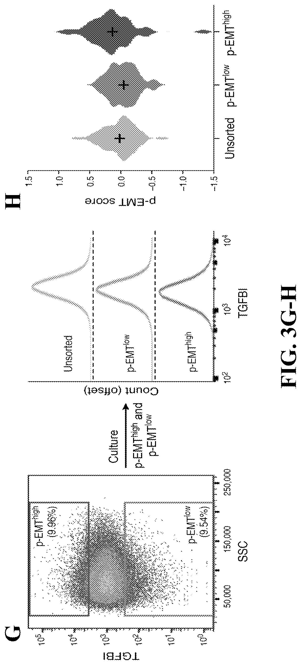
[0026] FIG. 3--Unbiased clustering reveals a common program of partial EMT (p-EMT) in HNSCC tumors. (A) Heatmap shows differentially-expressed genes (rows) identified by non-negative matrix factorization (NNMF) clustered by their expression across single cells (columns) from a representative tumor (MEEI25). The gene clusters reveal intra-tumoral programs that are differentially expressed in MEEI25. The corresponding gene signatures are numbered and selected genes indicated (right). (B) Heatmap depicts pairwise correlations of 60 intra-tumoral programs derived from 10 tumors, as in (A). Clustering identifies seven coherent expression programs across tumors. Rows in the heatmap that correspond to programs derived from MEEI25 are indicated by arrows and numbered as in (A). (C) Heatmap shows NNMF gene scores (rows) for common (top) and tumor-specific (bottom) genes within the p-EMT program by tumor (columns). (D) Representative images of SCC9 HNSCC cells sorted by p-EMT marker TGFBI into p-EMT.sup.high and p-EMT.sup.low populations and analyzed by matrigel invasion assay. (E) Bar plot depicts relative invasiveness of p-EMT.sup.high and p-EMT.sup.low SCC9 cells sorted and analyzed as in (D) (representative experiment; error bars reflect SEM; ANOVA, p<0.005, n=3). (F) Bar plot depicts relative proliferation of p-EMT.sup.high and p-EMT.sup.low SCC9 cells sorted as in (D) (representative experiment; error bars reflect SEM; ANOVA, p<0.0001, n=4). (G) (Left) Fluorescence-activated cell sorting plot identifies p-EMT.sup.high and p-EMT.sup.low SCC9 cells isolated based on TGFBI expression. (Right) Histogram (offset) reveals the distribution (x-axis) of TGFBI expression across cells from the respective isolates (p-EMT.sup.high, p and unsorted; separated by dashed lines). After 7 days in culture, p-EMT.sup.high, p-EMT.sup.low, and unsorted cells have similar distributions of p-EMT marker expression. Additional experiments with the p-EMT marker CXADR demonstrate similar findings (data not shown). (H) Violin plot depicts p-EMT scores for unsorted, p-EMT.sup.low, and p-EMT.sup.high SCC9 cell sorted and cultured as in (G). Respective isolates largely recapitulate the initial distribution of p-EMT scores. See also FIGS. 10 and 11 and Tables S6 and S7.
[0027] FIG. 4--p-EMT cells at the leading edge engage in cross-talk with CAFs. (A-C) IHC images of representative HNSCC tumors (MEEI5, MEEI16, MEEI17, MEEI25, MEEI28) stained for p-EMT markers (PDPN, LAMB3, LAMC2) and the malignant cell-specific marker p63 (A and B) or the epithelial program marker SPRR1B (C). Scale bar=100 .mu.M. (D) Scatter plot shows the Pearson correlation between the p-EMT program and other expression programs underlying HNSCC intra-tumoral heterogeneity (FIG. 3). Blue circles depict the correlations within individual tumors; black circles and error-bars represent the average and standard error, respectively, across the different tumors. (E) Bar plot depicts numbers of putative receptor-ligand interactions between malignant HNSCC cells and indicated cell types. Interaction numbers were calculated based on expression of receptors and corresponding ligands in scRNA-seq data. Outgoing interactions refer to the sum of ligands from malignant cells that interact with receptors on the indicated cell type. Incoming interactions refer to the opposite. CAFs express a significantly greater number of ligands whose receptors are expressed by malignant cells (hypergeometric test, p<0.05). (F) Heatmap depicts expression of ligands expressed by in vivo and in vitro CAFs. Relative expression is shown for all in vivo CAFs, MEEI18 in vivo CAFs, and in vitro CAFs derived from MEEI18. (G) Heatmap depicts relative expression of genes that were differentially regulated when SCC9 cells were treated with TGF4.beta.3 or TGF.beta. pathway inhibitors. Panel includes all genes with significantly higher expression upon TGF4.beta.3 treatment and lower expression upon TGF.beta. inhibition, relative to vehicle (t-test, p<0.05). Heat intensity reflects relative expression of indicated genes in bulk RNA-seq profiles for nine samples in each group, corresponding to distinct dosage or time points (see Materials and Methods). Selected genes are labeled and overlap with the in vivo p-EMT program (bold). (H) Violin plot depicts distributions of the p-EMT gene expression score across SCC9 cells treated as in (G) and profiled by scRNA-seq. p-EMT scores were increased with TGF4.beta.3 treatment and decreased upon TGF.beta. inhibition, relative to vehicle (t-test, p<10.sup.-16) (I) Bar plot shows relative invasiveness of SCC9 cells treated as in (G) (representative experiment; error bars reflect SEM; ANOVA, p<0.0001, n=3). In vitro treatment of HNSCC cells with the CAF-related ligand TGF.beta. causes coherent induction of the p-EMT program and increases invasiveness, while TGF.beta. inhibition has the opposite effect. See also FIG. 12.
[0028] FIG. 5--Intra-tumoral HNSCC heterogeneity recapitulated in nodal metastases. (A) t-SNE plot of malignant cells (as in FIG. 2) from five primary tumors (black) and their matched LNs (red). Malignant cells cluster by tumor rather than by site. (B) t-SNE plot of non-malignant cells (as in FIG. 2) from five primary tumors (black) and their matched LNs (red). Non-malignant cells are consistent across tumors but their representation and expression states vary between sites (see also FIG. 9). See also FIG. 13.
[0029] FIG. 6--HNSCC subtypes revised by deconvolution of expression profiles from hundreds of tumors. (A) t-SNE plot of malignant cells from ten tumors (as in FIG. 2). Each cluster of cells corresponds to a different tumor. Cells are colored according to the TCGA expression subtype that they match. Black indicates no match. Each tumor can be clearly assigned to one of three subtypes: basal, atypical, or classical. (B) t-SNE plot of non-malignant cells from ten tumors (as in FIG. 2). Each cluster of cells corresponds to a different cell type. Cells are colored according to the TCGA expression subtype that they match. Black indicates no match. Fibroblasts and myocytes highly express signature genes of the mesenchymal subtype, which likely reflects tumor profiles with high stromal representation. (C) For each TCGA subtype (columns), heatmap shows relative expression of gene signatures for non-malignant cell types (rows), which were used as estimates of cell type abundances. Tumors classified as mesenchymal highly expressed genes specific to CAFs and myocytes, while atypical tumors were enriched for T- and B-cells. (D) Heatmap depicts pairwise correlations between TCGA expression profiles ordered by their subtype annotations. This analysis included all genes and recovered all four subtypes. (E) Schematic of linear regression used to subtract the influence of non-malignant cell frequency from bulk TCGA expression profiles, and thereby infer malignant cell-specific expression profiles. (F) Heatmap depicts pairwise correlations between TCGA expression profiles ordered by their subtype annotations. This analysis was based on the inferred malignant cell-specific expression profiles in (E). Classical and atypical subtypes are maintained. However, basal and mesenchymal subtypes collapse to a single subtype, which Applicants term `malignant-basal.` See also FIG. 14.
[0030] FIG. 7--p-EMT predicts nodal metastasis and adverse pathologic features. (A) PC1 and PC2 gene scores based on PCA of inferred malignant cell-specific profiles from all malignant-basal TCGA tumors (n=225). p-EMT genes (red) and epithelial differentiation genes (green) underlie variance among malignant-basal tumors. (B) PC1 and PC2 gene scores based on PCA of inferred malignant cell-specific profiles from all classical and atypical TCGA tumors (n=156). p-EMT (red) and epithelial differentiation (green) genes are weakly associated with variance in these tumors. (C) Plot depicts percentage of p-EMT high and p-EMT low malignant-basal tumors associated with each clinical feature. Higher p-EMT scores were associated with positive LNs, advanced nodal stage, high grade, extracapsular extension (ECE), and lymphovascular invasion (LVI) (hypergeometric test, p<0.05). Advanced local disease (T3/T4) as determined by T-stage did not correlate with p-EMT score. (D) Volcano plot depicts gene expression differences between malignant-basal TCGA tumors with multiple LNs versus those without positive LNs. p-EMT genes (red) have increased expression, while epithelial differentiation genes (green) have decreased expression in metastatic tumors. (E) Model of the in vivo p-EMT program associated with invasion and metastasis in malignant-basal HNSCC tumors. See also FIG. 14.
[0031] FIG. 8--Cells are classified as malignant and non-malignant based on CNVs and epithelial marker expression, Related to FIG. 1. (A) Histograms show distribution of cells ordered by numbers of reads (Left; median 1.34 million reads), percent of reads mapped to the transcriptome (Middle; median 52.2%), and number of unique genes detected (Right; median 3,880 detected genes). (B) Heatmap shows large-scale CNVs for individual cells (rows) from 18 tumors, inferred based on the average expression of 100 genes surrounding each chromosomal position (columns). Red: Amplifications; Blue: Deletions. (C) Large-scale CNVs of seven samples (rows) from three patients as defined by whole exome sequencing analysis. (D) Stacked bar plots of 27 clusters show percent of malignant (blue) and non-malignant (red) cells, as classified by one (light color) or two (dark color) independent methods: epithelial marker scoring and CNVs. 22 of 27 clusters contain >95% malignant or non-malignant cells; cells in the remaining five clusters were excluded from further analysis.
[0032] FIG. 9--Expression heterogeneity of stromal and immune cells in the HNSCC ecosystem, Related to FIG. 2. (A) t-SNE plot of non-malignant cells (as shown in FIG. 2A) colored by their assignment to 14 clusters by SC3 (Bacher et al., 2017) with default parameters, demonstrating high consistency between SC3 clusters and tSNE coordinates. (B) t-SNE plot of non-malignant cells from 10 tumors (same as FIG. 2A) with cells colored based on the average expression of sets of marker genes for particular cell types (marker genes and associated cell types are indicated next to each plot). Zero expression level (for all markers of a given cell type) is indicated with small circles, and positive expression is indicated by larger circles, with higher levels indicated by shades of red. (C) (Top) Zoomed in t-SNE plot of T-cells with four distinct clusters identified. (Bottom) Heat map of differentially expressed genes (rows) facilitates annotation of the four clusters (columns) as naive-like, regulatory, cytotoxic, and exhausted. (D) Bar plot shows percent of exhausted CD8+ T-cells in six tumors. Asterisks indicate a significant deviation from the mean (hypergeometric test, p<0.01). (E) (Top) Zoomed in t-SNE plot of fibroblasts with two distinct clusters and a set of intermediates identified. (Bottom) Heat map of differentially expressed genes (rows) facilitates annotation of the clusters (columns) as myofibroblasts, activated CAFs, and intermediate (resting) fibroblasts lacking coherent expression of genes consistent with either myofibroblasts or CAFs. (F) PC1 and PC2 from a principal component analysis of all fibroblasts, colored based on their assignments to the three clusters as in (D), demonstrates that PC2 further separates the CAF cluster into two subpopulations (CAF1 and CAF2, defined as CAFs with PC2>0, and PC2<0, respectively). (G) Heatmap of differentially expressed genes (rows) between the CAF1 and CAF2 subpopulations. Selected genes are indicated by name. (H) Heatmap shows distribution of relative CNVs (columns) for upregulated genes from 10 tumors (rows). Relative CNVs are calculated as the CNV value in the respective tumor minus the average CNVs of all other tumors. (I) Bar plot shows percentage of upregulated genes (blue) and other genes (red) with relative CNV>0.15 in each tumor, demonstrating a significant enrichment of upregulated genes with high CNVs in all cases (hypergeometric test with Bonferroni correction, p<0.05). (J) t-SNE plots of malignant (Left; same as FIG. 2C) and non-malignant (Right; same as FIG. 2A) cells colored by number of unique genes detected. These plots show that clustering is not driven by the detected number of genes. Additional analyses with clusters annotated by batch demonstrate clusters are not determined by batch effects (data not shown). (K) (Top) Heatmap shows absolute expression of housekeeping (positive) genes (top rows) and immune marker (negative) genes (bottom rows) in single cells (columns) from MEEI25 (same as FIG. 3A). (Middle) Heatmap shows absolute expression of genes defining distinct meta-programs (rows) identified by NNMF in single cells (columns) from MEEI25. (Bottom) Bar plot shows number of detected genes in single cells (columns) from MEEI25, with cells ordered as in top and middle panels. Variability in the number of genes detected is not linked to the expression programs identified.
[0033] FIG. 10--Defining the p-EMT program in HNSCC tumors and cell lines, Related to FIG. 3. (A) Each panel (from top to bottom) shows the meta-signature scores (top section of panel) and a heat map with expression of the top 10 genes for that meta-signature (bottom section of panel) for each of the six coherent expression programs in malignant cells. Cells from ten HNSCC tumors are included and sorted (left to right) first by tumor, within a tumor by sample (primary followed by LN, when applicable), and within a sample by the corresponding meta-signature score (black line). (B) Each panel (from top to bottom) shows violin plots that depict scores for one of the six meta-signatures in (A) for malignant cells from ten tumors. Violin plots in the second panel depict p-EMT scores, revealing distinct cohorts of p-EMT low (blue) and p-EMT high (red) tumors. Tumors in all panels are ordered identically. (C-F) Line graphs show smoothed expression (moving average with a window of 100 cells) for selected genes (as labeled); cells from ten HNSCC tumors were included and rank ordered by p-EMT program expression. The selected genes include six of the top p-EMT genes (C), eight epithelial genes negatively correlated with p-EMT scores (D), six epithelial genes not correlated with p-EMT scores (E), and canonical EMT transcription factors (TFs) (F). (G) Heatmap depicts pairwise Pearson correlations of global expression profiles of malignant cells from ten tumors and five oral cavity HNSCC cell lines. Correlations were calculated across all genes with average expression (E.sub.a) above four in at least one of the tumors or cell lines and after centering the expression levels of genes across all samples included. Clustering indicates that cell lines are more similar to one another than to primary tumor samples and also illustrates the distinction between tumor samples of different subtypes. (H) Heatmaps show pairwise correlations of expression profiles from individual cells in five oral cavity HNSCC cell lines, ordered by hierarchical clustering. SCC9 includes a subpopulation of cells with an expression profile reminiscent of the p-EMT program, while SCC25 has a subpopulation with an expression profile similar to the stress program. Selected genes preferentially expressed within these subpopulations are highlighted, with markers used for sorting experiments (TGFBI, CXADR) in bold.
[0034] FIG. 11--Distinguishing the p-EMT program in HNSCC tumors from previously described EMT programs and modeling p-EMT in vitro, Related to FIG. 3. (A) Correlation plot demonstrates pairwise Pearson correlations between EMT and p-EMT programs, including signatures from previous work, as well as this work. Previously described TCGA-Mesenchymal genes ("Mes"), EMT signatures from tumors ("Tumor"), and cell lines ("Culture") strongly correlate with the expression program of CAFs. These programs weakly correlate with the p-EMT program ("Orig.") described in this study. Focusing on malignant-specific p-EMT genes ("Malig.") and p-EMT genes identified after deconvolution ("Decon.") reveals a more limited correlation of p-EMT with TCGA-Mes and previous EMT signatures, indicating this program is distinct from prior EMT descriptions. (B) Scatter plot demonstrates three cohorts of TCGA tumors, with (1) high TCGA-mes/intermediate p-EMT, (2) high p-EMT, and (3) low p-EMT scores. (C) Heatmap demonstrates relative expression of TCGA-Mes, CAF, and p-EMT genes (rows) in TCGA tumors (columns) from the cohorts described in (B), with the eight malignant-specific p-EMT genes ("Malig.") shown at the bottom. (D) Bar plots show average expression of each of the gene sets described in (C) in CAFs, malignant cells, and all other immune and stromal cell types detected in this cohort. The p-EMT signature is highly specific to malignant cells, while the TCGA-mes signature is associated with CAFs. (E) Line graphs show percentage of cycling malignant cells within a sliding window of 20 cells, rank ordered by p-EMT scores. Seven p-EMT high tumors are included; in each tumor, a p-value is shown (permutation test), corresponding to the enrichment of cycling cells among the 30% of cells with lowest p-EMT scores in that tumor. Low p-EMT is significantly enriched with cycling cells among the three tumors with the highest p-EMT scores (MEEI16, MEEI17, and MEEI25). (F) Bar plot depicts relative invasiveness of SCC9 cells transfected with TGFBI or vector in matrigel invasion assays (error bars reflect SEM; t-test, p<0.005, n=3). (G) Bar plot shows relative proliferation of SCC9 treated as in (F) (error bars reflect SEM; ANOVA, p<0.0001, n=4). (H) (Top left) Fluorescence-activated cell sorting plot identifies p-EMT.sup.high and p-EMT.sup.low SCC9 cells isolated based on TGFBI expression. (Top right) Histogram (offset) reveals the distribution of TGFBI expression across cells from the respective isolates (p-EMT.sup.high and p-EMT.sup.low; separated by dashed line) immediately after sorting. (Bottom) Histograms (offset) reveal the distribution of TGFBI expression across cells from the respective isolates (p-EMT.sup.high and p-EMT.sup.low; separated by dashed line) after 4 hours, 24 hours, 4 days, and 7 days in culture. The p-EMT.sup.high and p-EMT.sup.low populations remained distinct 4 hours and 24 hours after sorting (representative experiment; t-test, p<0.0001, n=3).
[0035] FIG. 12--p-EMT program is localized at the leading edge, distinct from the epithelial differentiation program at the core, Related to FIG. 4. (A-C) Immunohistochemical staining of representative tumors (MEEI5, MEEI16, MEEI17, MEEI25, MEEI28) for p-EMT (LAMC2, MMP10, TGFBI) with the malignant cell-specific marker p63. Scale bar=100 .mu.M. The leading edges of tumors co-stain with p63 and p-EMT markers. Additional staining with the marker p-EMT marker ITGA5 further validated localization of p-EMT at the leading edge (data not shown). (D) Immunohistochemical staining of representative tumors (MEEI17, MEEI28) for multiple p-EMT markers (LAMC2, TGFBI). p-EMT markers co-localize at the leading edge. (E-G) Immunohistochemical staining of representative p-EMT low tumors (MEEI20, MEEI26) for p-EMT (PDPN, LAMB3, LAMC2) with the malignant cell-specific marker p63. p-EMT low tumors show minimal staining for p-EMT markers at the leading edge. Additional staining with the marker ITGA5 confirmed minimal staining for the p-EMT program in these tumors (data not shown). (H and I) Immunohistochemical staining of representative tumors (MEEI16, MEEI17) for epithelial differentiation (SPRR1B, CLDN4) and the malignant cell-specific marker p63. (J and K) Immunohistochemical staining of representative tumor (MEEI17) for p-EMT (LAMC2, PDPN) and epithelial differentiation (CLDN4). Markers demonstrate distinct spatial localization of p-EMT and epithelial differentiation programs, at the leading edge and core, respectively. (L) Bar plot shows statistical significance (minus log 10 of p-value defined by hypergeometric test) of number of observed outgoing interactions between ten listed cell types and malignant cells. Bars above the x-axis indicate a greater number of interactions than expected, while bars below the x-axis indicate fewer interactions than expected. (M) Immunohistochemical staining of representative tumors (MEEI16, MEEI18) for p-EMT and CAFs (FAP) with the malignant cell-specific marker p63. FAP staining is present both at the leading edge of tumors nests and in the stroma, highlighting activated CAFs. (N) Bar plot depicts relative proliferation of SCC9 cells treated with vehicle, TGF.beta., or TGF.beta. pathway inhibitors (error bars reflect SEM; ANOVA, p<0.0001, n=4). (0) Histograms show percent of sequencing reads with insertions or deletions (indels) of specified size in mock infected SCC9 cells (Top left) and SCC9 TGFBI CRISPR knockout cells (other panels). Each of the TGFBI-targeting sgRNAs resulted in >98.8% of reads containing indels, indicating efficient knockout of TGFBI. (P) Bar plot depicts relative invasiveness of mock infected SCC9 cells or SCC9 TGFBI CRISPR knockout cells after treatment with vehicle or TGF.beta. in matrigel invasion assays (error bars reflect SEM; ANOVA, p<0.0001, n=3). (Q) Violin plot depicts hypoxia program scoring of SCC9 cells grown in normoxic or hypoxic conditions. Hypoxic conditions are associated with significantly increased hypoxia score (t-test, p<0.05). (R) Violin plot depicts scoring of SCC9 cells for p-EMT scores after growth in standard conditions (control), hypoxic conditions, or in co-culture with CAFs derived from MEEI18. p-EMT expression is not significantly changed across these conditions.
[0036] FIG. 13--Variability in the p-EMT program and cancer-associated fibroblasts across tumor subsites (primary and lymph node), Related to FIG. 5. (A) Comparison of point mutations between primary and LN samples in three individual tumors (MEEI26, MEEI20, and MEEI25 from top to bottom) as detected by whole exome sequencing. In each tumor, Applicants examined all mutations identified in at least one of the samples (primary or LN) and assigned it one of three values in each sample: "detected" (black), "not detected" (white), or unresolved due to "low coverage." A single mutant read was sufficient to define a mutation as "detected," but zero mutant reads were defined as "not detected" only if the probability of detecting zero mutant reads in that sample was below 0.05 (as defined by binomial test, given the number of reads covering that base and assuming the same frequency of the mutant reads as in the sample(s) where it is detected). Mutations were then ordered by their identification across the samples and assigned to four classes: shared among primary and LN, specific to primary, specific to LN, and unresolved. Note that for MEEI26 two LN samples are included corresponding to the left (ipsilateral) and right (contralateral) LNs, denoted as LN.sub.L and LN.sub.R, respectively. (B) Heatmap of differentially expressed genes between primary and LN samples across multiple patients. For each of the five patients with matched primary and LN samples, Applicants identified significant differentially expressed genes (defined by p<0.001 and fold-change>2). All genes defined as upregulated in at least two patients (left panel) or downregulated in at least two patients (right panel) are shown. Red: upregulated; Blue: downregulated. Darker shades indicate significant differential expression, while lighter shades denote borderline differential expression (p<0.05 and fold-change>1.5). (C) Violin plot depicts p-EMT score of malignant cells from five primary tumors and matched LN. (D) Scatter plot shows the average (x-axis) and the variability (y-axis) of p-EMT scores across individual malignant cells within each sample; five primary tumors (black) and matched LNs (red) are included and matched samples are connected with lines. p-EMT high tumors display both higher average and higher variability of p-EMT scores. (E) Fibroblasts from primary (black) and LN (red) samples, scored by the relative expression of gene-sets distinguishing CAFs from myofibroblasts (x-axis) and those distinguishing the CAF1 and CAF2 subsets (y-axis), demonstrating that LN CAFs are biased towards the CAF1 subset (hypergeometric test, p<0.05). (F and G) Immunohistochemical staining of representative LN metastases (MEEI25, MEEI28) for p-EMT (PDPN, LAMB3) with the malignant-cell specific marker p63.
[0037] FIG. 14--p-EMT program is negatively correlated with epithelial differentiation and may predict nodal metastasis, Related to FIGS. 6 and 7. (A) Hematoxylin-eosin (H&E) stained sections from representative mesenchymal (Left) and basal (Right) TCGA tumors demonstrate substantially more stromal infiltrate in mesenchymal than basal tumors. Scale bar=400 .mu.M. (B) (Left) Bar plot shows significantly higher percent of stromal infiltrate in mesenchymal tumors compared to basal tumors (t-test, p<0.0001; n=203 tumors). (Right) Bar plot shows number of tumors with H&E stromal scores ranging from 0 (lowest) to 4 (highest) for mesenchymal and basal subtype TCGA tumors. (C and D) Scatter plots demonstrate a correlation between H&E stromal score (indicated by dot color) with CAF and TCGA mesenchymal scores (C), but not p-EMT scores (D). (E) Line graph shows distribution of p-EMT scores across TCGA tumors of each subtype. (F) Scatter plot shows scoring of TCGA basal and mesenchymal tumors for epithelial differentiation and p-EMT which are significantly negatively correlated in this subset of tumors (Pearson correlation, p<0.05); black lines indicate linear regression. (G) Scatter plot shows scoring of TCGA classical and atypical tumors for epithelial differentiation and p-EMT, which are not significantly correlated in this subset of tumors; black lines indicate linear regression. (H) Bar plot shows direction and statistical significance (p-value based on a t-test) of the association between each of six coherent meta-signatures and the presence of multiple versus no metastatic LNs in TCGA malignant-basal tumors. The p-EMT and epithelial differentiation programs, which were inversely correlated in expression studies, had opposite associations with metastasis. The other programs show no significant association with LN metastases. (I) (Top) Bar plot shows the percent of patients with adverse clinical features (positive LNs, multiple LNs, advanced N stage, grade III, extranodal extension, lymphovascular invasion, and advanced local disease) in cohorts with high and low p-EMT scores stratified by high and low CAF scores. (Bottom) Heatmap shows the statistical significance of p-EMT and CAF effects on adverse clinical features based on a binomial logistic regression with two predictive variables (p-EMT and variable scores) and an interaction effect. Only the p-EMT effect is predictive of clinical features associated with metastasis and invasion (positive LNs, multiple LNs, advanced nodal stage, extracapsular extension, and lymphovascular invasion) (Bottom, first row). In contrast, the CAF effect has no significant predictive value for features associated with metastasis, but instead, predicts high grade disease and advanced local disease (T3/T4) (Bottom, second row). The p-EMT and CAF effects did act cooperatively to influence the risk of nodal metastasis (Bottom, third row), consistent with a putative ligand-receptor interaction between CAFs and p-EMT cells. (J) Percent of patients from TCGA for which neck dissection was justified using varying thresholds of p-EMT scores and stratified by tumor (T) stage. Justified neck dissection refers to patients with initial clinical diagnosis of lymph node-negative (cN0) for which neck dissection revealed a positive metastatic lymph node (pN1-N3); the percentage of justified neck dissections was calculated out of all patients with clinical node-negative disease that underwent neck dissection. A higher p-EMT threshold is associated with a higher rate of justified neck dissection, regardless of T-stage (permutation test, p<0.05). (K) Correlations of genes with the p-EMT program within (x-axis) and across (y-axis) tumors in the cohort of ten patients. Within-tumor correlations were calculated separately in each tumor and averaged; across-tumor correlations were calculated between the average levels of genes and those of the p-EMT program across all malignant cells in each tumor. Selected genes are indicated. (L) Scatter plot shows the correlations of genes with p-EMT (x-axis) and epithelial differentiation (y-axis) programs based on inferred malignant cell-specific profiles from TCGA malignant-basal tumors. Genes of the p-EMT (red) and epithelial differentiation (green) programs as well as EMT TFs (black) are indicated, demonstrating a high p-EMT correlation with SNAIL2 but not of other EMT TFs.
[0038] FIG. 15--block diagram depicting a method for generating a p-EMT score in a tumor using bulk RNA-seq data obtained from a sample of the tumor.
[0039] FIG. 16--p-EMT predicts adverse pathologic features in an independent MEEI cohort of patients by IHC. Higher p-EMT scores were associated with positive LNs, advanced nodal stage, perineural invasion, lymphovascular invasion (LVI) and high grade. Advanced local disease (T2/T4) as determined by T-stage did not correlate with high p-EMT score.
[0040] FIG. 17--quantification of marker staining.
[0041] FIG. 18--classification of tumors as basal subtype. Tumors were classified as non-basal subtype and eliminated from analysis (20%) if staining was 1+ for multiple markers. p-EMT quantification in malignant-basal subtype tumors correlated with pathologic features.
[0042] The figures herein are for illustrative purposes only and are not necessarily drawn to scale. The following manuscript contains complete color versions of the figures described above and is hereby fully incorporated herein by reference: Puram et al., Single-Cell Transcriptomic Analysis of Primary and Metastatic Tumor Ecosystems in Head and Neck Cancer, Cell. 2017 Dec. 14; 171(7):1611-1624.e24. doi: 10.1016/j.cell.2017.10.044. Epub 2017 Nov. 30.
DETAILED DESCRIPTION OF THE EXAMPLE EMBODIMENTS
General Definitions
[0043] Unless defined otherwise, technical and scientific terms used herein have the same meaning as commonly understood by one of ordinary skill in the art to which this disclosure pertains. Definitions of common terms and techniques in molecular biology may be found in Molecular Cloning: A Laboratory Manual, 2.sup.nd edition (1989) (Sambrook, Fritsch, and Maniatis); Molecular Cloning: A Laboratory Manual, 4th edition (2012) (Green and Sambrook); Current Protocols in Molecular Biology (1987) (F. M. Ausubel et al. eds.); the series Methods in Enzymology (Academic Press, Inc.): PCR 2: A Practical Approach (1995) (M. J. MacPherson, B. D. Hames, and G. R. Taylor eds.): Antibodies, A Laboratory Manual (1988) (Harlow and Lane, eds.): Antibodies A Laboratory Manual, 2nd edition 2013 (E. A. Greenfield ed.); Animal Cell Culture (1987) (R. I. Freshney, ed.); Benjamin Lewin, Genes IX, published by Jones and Bartlet, 2008 (ISBN 0763752223); Kendrew et al. (eds.), The Encyclopedia of Molecular Biology, published by Blackwell Science Ltd., 1994 (ISBN 0632021829); Robert A. Meyers (ed.), Molecular Biology and Biotechnology: a Comprehensive Desk Reference, published by VCH Publishers, Inc., 1995 (ISBN 9780471185710); Singleton et al., Dictionary of Microbiology and Molecular Biology 2nd ed., J. Wiley & Sons (New York, N.Y. 1994), March, Advanced Organic Chemistry Reactions, Mechanisms and Structure 4th ed., John Wiley & Sons (New York, N.Y. 1992); and Marten H. Hofker and Jan van Deursen, Transgenic Mouse Methods and Protocols, 2nd edition (2011).
[0044] As used herein, the singular forms "a", "an", and "the" include both singular and plural referents unless the context clearly dictates otherwise.
[0045] The term "optional" or "optionally" means that the subsequent described event, circumstance or substituent may or may not occur, and that the description includes instances where the event or circumstance occurs and instances where it does not.
[0046] The recitation of numerical ranges by endpoints includes all numbers and fractions subsumed within the respective ranges, as well as the recited endpoints.
[0047] The terms "about" or "approximately" as used herein when referring to a measurable value such as a parameter, an amount, a temporal duration, and the like, are meant to encompass variations of and from the specified value, such as variations of +/-10% or less, +/-5% or less, +/-1% or less, and +1-0.1% or less of and from the specified value, insofar such variations are appropriate to perform in the disclosed invention. It is to be understood that the value to which the modifier "about" or "approximately" refers is itself also specifically, and preferably, disclosed.
[0048] Various embodiments are described hereinafter. It should be noted that the specific embodiments are not intended as an exhaustive description or as a limitation to the broader aspects discussed herein. One aspect described in conjunction with a particular embodiment is not necessarily limited to that embodiment and can be practiced with any other embodiment(s). Reference throughout this specification to "one embodiment", "an embodiment," "an example embodiment," means that a particular feature, structure or characteristic described in connection with the embodiment is included in at least one embodiment of the present invention. Thus, appearances of the phrases "in one embodiment," "in an embodiment," or "an example embodiment" in various places throughout this specification are not necessarily all referring to the same embodiment, but may. Furthermore, the particular features, structures or characteristics may be combined in any suitable manner, as would be apparent to a person skilled in the art from this disclosure, in one or more embodiments. Furthermore, while some embodiments described herein include some but not other features included in other embodiments, combinations of features of different embodiments are meant to be within the scope of the invention. For example, in the appended claims, any of the claimed embodiments can be used in any combination.
[0049] All publications, published patent documents, and patent applications cited herein are hereby incorporated by reference to the same extent as though each individual publication, published patent document, or patent application was specifically and individually indicated as being incorporated by reference.
Overview
[0050] Human tumors are composed of diverse malignant, stromal and immune cell states, which are masked when bulk samples are profiled. Applicants investigated primary HNSCC tumors and matched LNs in order to better understand intra-tumoral heterogeneity, invasion, and metastasis in an epithelial human cancer. By analyzing 18 tumors, including five matched pairs of primary tumors and LN metastases, Applicants profiled .about.6,000 individual tumor cells, revealing expression programs that distinguish diverse malignant, stromal, and immune cells. Malignant cells vary in their expression of programs related to cell cycle, stress, hypoxia and epithelial differentiation. A subset also express a partial EMT (p-EMT) program with extracellular matrix proteins, but lacking classical EMT transcription factors (TFs). p-EMT cells localized to the leading edge of primary tumors in close proximity to cancer-associated fibroblasts. A similar tumor-stromal interaction was evident in matched lymph nodes in structured tumor nests. Knowledge of HNSCC expression cell states allowed Applicants to deconvolve bulk RNA-seq data from The Cancer Genome Atlas (TCGA), and thereby redefine HNSCC subtypes by their malignant and stromal components. Notably, the p-EMT program is largely specific to the most prevalent HNSCC subtype, where it is associated with adverse clinical and pathologic features such as metastasis, tumor grade, and extracapsular extension. These data define inter-tumoral and intra-tumoral heterogeneity in HNSCC, and provide insight into in vivo EMT-like changes and stromal interactions relevant to tumor invasion and metastasis.
[0051] Embodiments disclosed herein provide for a p-EMT signature in epithelial tumors capable of guiding treatment of the tumors. Embodiments disclosed herein provide tools and methods for prognosing and stratifying epithelial tumors. The methods leverage a novel gene signature program detectable in HNSCC tumors. Applicants have discovered several malignant cell gene expression programs and have defined the tumor microenvironment in HNSCC using single cell RNA-seq. The discovery enables the deconvolution of bulk sequencing gene expression data of a HNSCC sample to identify the malignant gene expression programs and determine the gene expression attributed to the tumor microenvironment (TME). Deconvolution utilizes a novel algorithm constructed based on the insight obtained from the single cell sequencing, such as malignant cell sub-types and non-malignant cell types. Specifically, applicants identified an EMT-like meta-signature (p-EMT) that correlates with lymph node metastasis. Thus, applicants have developed methods and systems for analyzing bulk sequencing data from a subject and classifying it based on a p-EMT high signature score. The EMT-signature score can then be used to predict lymph node (LN) metastasis and direct treatment decisions. The p-EMT signature genes or polypeptides may also be therapeutically targeted in order to prevent unfavorable clinical outcomes (e.g., metastasis). In one embodiment, a tumor biopsy is obtained from a subject in need thereof and the sample is analyzed by RNA-seq. The expression data can then be denconvoluted to determine a p-EMT score. The subject may then be treated according to the pEMT score.
Cancer
[0052] In certain embodiments, the systems and methods may be used for any epithelial cancer. Studies have suggested that EMT is a process that occurs in all epithelial tumors. Not being bound by a theory, epithelial tumors all express similar p-EMT programs as described herein. HNSCC is one of many common epithelial tumors. Not being bound by a theory, detection of the p-EMT signature described herein in any epithelial tumor predicts 1) risk of having lymph node or distant metastasis, 2) tumor stage, 3) adverse pathologic features, 4) need for adjuvant (radiation/chemotherapy) treatment, 5) treatment response, and 6) overall survival. The examples described herein show that the p-EMT signature is a strong genetic predictor of having lymph node (LN) involvement and that the signature predicts the need for a neck dissection (removal of LN).
[0053] Cancers may include, but are not limited to, breast cancer, colon cancer, lung cancer, prostate cancer, testicular cancer, brain cancer, skin cancer, rectal cancer, gastric cancer, esophageal cancer, tracheal cancer, head and neck cancer, pancreatic cancer, liver cancer, ovarian cancer, lymphoid cancer, cervical cancer, vulvar cancer, melanoma, mesothelioma, renal cancer, bladder cancer, thyroid cancer, bone cancers, cutaneous squamous cell carcinoma, carcinomas, sarcomas, and soft tissue cancers. Thus, the disclosure is generally applicable to any type of cancer in which expression of an EMT program occurs. In certain embodiments, the signature is useful for all epithelial tumors, including but not limited to lung, breast, prostate, colon, cutaneous squamous cell carcinoma and esophageal carcinoma.
Use of Signature Genes
[0054] As used herein a "signature" or "gene signature" may encompass any gene or genes, protein or proteins, or epigenetic element(s) whose expression profile or whose occurrence is associated with a specific cell type, subtype, or cell state of a specific cell type or subtype within a population of cells. For ease of discussion, when discussing gene expression, any of gene or genes, protein or proteins, or epigenetic element(s) may be substituted. As used herein, the terms "signature", "expression profile", or "expression program" may be used interchangeably. It is to be understood that also when referring to proteins (e.g. differentially expressed proteins), such may fall within the definition of "gene" signature. Levels of expression or activity or prevalence may be compared between different cells in order to characterize or identify for instance signatures specific for cell (sub)populations. Increased or decreased expression or activity or prevalence of signature genes may be compared between different cells in order to characterize or identify for instance specific cell (sub)populations. The detection of a signature in single cells may be used to identify and quantitate for instance specific cell (sub)populations. A signature may include a gene or genes, protein or proteins, or epigenetic element(s) whose expression or occurrence is specific to a cell (sub)population, such that expression or occurrence is exclusive to the cell (sub)population. A gene signature as used herein, may thus refer to any set of up- and down-regulated genes that are representative of a cell type or subtype. A gene signature as used herein, may also refer to any set of up- and down-regulated genes between different cells or cell (sub)populations derived from a gene-expression profile. For example, a gene signature may comprise a list of genes differentially expressed in a distinction of interest.
[0055] The signature as defined herein (being it a gene signature, protein signature or other genetic or epigenetic signature) can be used to indicate the presence of a cell type, a subtype of the cell type, the state of the microenvironment of a population of cells, a particular cell type population or subpopulation, and/or the overall status of the entire cell (sub)population. Furthermore, the signature may be indicative of cells within a population of cells in vivo. The signature may also be used to suggest for instance particular therapies, or to follow up treatment, or to suggest ways to modulate immune systems. The signatures of the present invention may be discovered by analysis of expression profiles of single-cells within a population of cells from isolated samples (e.g. tumor samples), thus allowing the discovery of novel cell subtypes or cell states that were previously invisible or unrecognized. The presence of subtypes or cell states may be determined by subtype specific or cell state specific signatures. The presence of these specific cell (sub)types or cell states may be determined by applying the signature genes to bulk sequencing data in a sample. Not being bound by a theory the signatures of the present invention may be microenvironment specific, such as their expression in a particular spatio-temporal context. Not being bound by a theory, signatures as discussed herein are specific to a particular pathological context. Not being bound by a theory, a combination of cell subtypes having a particular signature may indicate an outcome. Not being bound by a theory, the signatures can be used to deconvolute the network of cells present in a particular pathological condition. Not being bound by a theory the presence of specific cells and cell subtypes are indicative of a particular response to treatment, such as including increased or decreased susceptibility to treatment. The signature may indicate the presence of one particular cell type. In one embodiment, the novel signatures are used to detect multiple cell states or hierarchies that occur in subpopulations of cancer cells that are linked to particular pathological condition (e.g. cancer grade), or linked to a particular outcome or progression of the disease (e.g. metastasis), or linked to a particular response to treatment of the disease.
[0056] The signature according to certain embodiments of the present invention may comprise or consist of one or more genes, proteins and/or epigenetic elements, such as for instance 1, 2, 3, 4, 5, 6, 7, 8, 9, 10 or more. In certain embodiments, the signature may comprise or consist of two or more genes, proteins and/or epigenetic elements, such as for instance 2, 3, 4, 5, 6, 7, 8, 9, 10 or more. In certain embodiments, the signature may comprise or consist of three or more genes, proteins and/or epigenetic elements, such as for instance 3, 4, 5, 6, 7, 8, 9, 10 or more. In certain embodiments, the signature may comprise or consist of four or more genes, proteins and/or epigenetic elements, such as for instance 4, 5, 6, 7, 8, 9, 10 or more. In certain embodiments, the signature may comprise or consist of five or more genes, proteins and/or epigenetic elements, such as for instance 5, 6, 7, 8, 9, 10 or more. In certain embodiments, the signature may comprise or consist of six or more genes, proteins and/or epigenetic elements, such as for instance 6, 7, 8, 9, 10 or more. In certain embodiments, the signature may comprise or consist of seven or more genes, proteins and/or epigenetic elements, such as for instance 7, 8, 9, 10 or more. In certain embodiments, the signature may comprise or consist of eight or more genes, proteins and/or epigenetic elements, such as for instance 8, 9, 10 or more. In certain embodiments, the signature may comprise or consist of nine or more genes, proteins and/or epigenetic elements, such as for instance 9, 10 or more. In certain embodiments, the signature may comprise or consist of ten or more genes, proteins and/or epigenetic elements, such as for instance 10, 11, 12, 13, 14, 15, or more. It is to be understood that a signature according to the invention may for instance also include genes or proteins as well as epigenetic elements combined.
[0057] In certain embodiments, a signature is characterized as being specific for a particular tumor cell or tumor cell (sub)population if it is upregulated or only present, detected or detectable in that particular tumor cell or tumor cell (sub)population, or alternatively is downregulated or only absent, or undetectable in that particular tumor cell or tumor cell (sub)population. In this context, a signature consists of one or more differentially expressed genes/proteins or differential epigenetic elements when comparing different cells or cell (sub)populations, including comparing different tumor cells or tumor cell (sub)populations, as well as comparing tumor cells or tumor cell (sub)populations with non-tumor cells or non-tumor cell (sub)populations. It is to be understood that "differentially expressed" genes/proteins include genes/proteins which are up- or down-regulated as well as genes/proteins which are turned on or off. When referring to up- or down-regulation, in certain embodiments, such up- or down-regulation is preferably at least two-fold, such as two-fold, three-fold, four-fold, five-fold, or more, such as for instance at least ten-fold, at least 20-fold, at least 30-fold, at least 40-fold, at least 50-fold, or more. Alternatively, or in addition, differential expression may be determined based on common statistical tests, as is known in the art.
[0058] As discussed herein, differentially expressed genes/proteins, or differential epigenetic elements may be differentially expressed on a single cell level, or may be differentially expressed on a cell population level. Preferably, the differentially expressed genes/proteins or epigenetic elements as discussed herein, such as constituting the gene signatures as discussed herein, when as to the cell population level, refer to genes that are differentially expressed in all or substantially all cells of the population (such as at least 80%, preferably at least 90%, such as at least 95% of the individual cells). This allows one to define a particular subpopulation of tumor cells. As referred to herein, a "subpopulation" of cells preferably refers to a particular subset of cells of a particular cell type which can be distinguished or are uniquely identifiable and set apart from other cells of this cell type. The cell subpopulation may be phenotypically characterized, and is preferably characterized by the signature as discussed herein. A cell (sub)population as referred to herein may constitute a (sub)population of cells of a particular cell type characterized by a specific cell state.
[0059] When referring to induction, or alternatively suppression of a particular signature, preferable is meant induction or alternatively suppression (or upregulation or downregulation) of at least one gene/protein and/or epigenetic element of the signature, such as for instance at least to, at least three, at least four, at least five, at least six, or all genes/proteins and/or epigenetic elements of the signature.
[0060] Various aspects and embodiments of the invention may involve analyzing gene signatures, protein signature, and/or other genetic or epigenetic signature based on single cell analyses (e.g. single cell RNA sequencing) or alternatively based on cell population or bulk analyses, as is defined herein elsewhere.
[0061] In further aspects, the invention relates to gene signatures, protein signature, and/or other genetic or epigenetic signature of particular tumor cell subpopulations, as defined herein elsewhere. The invention hereto also further relates to particular tumor cell subpopulations, which may be identified based on the methods according to the invention as discussed herein; as well as methods to obtain such cell (sub)populations and screening methods to identify agents capable of inducing or suppressing particular tumor cell (sub)populations.
[0062] The invention further relates to various uses of the gene signatures, protein signature, and/or other genetic or epigenetic signature as defined herein, as well as various uses of the tumor cells or tumor cell (sub)populations as defined herein. Particular advantageous uses include methods for identifying agents capable of inducing or suppressing particular tumor cell (sub)populations based on the gene signatures, protein signature, and/or other genetic or epigenetic signature as defined herein. The invention further relates to agents capable of inducing or suppressing particular tumor cell (sub)populations based on the gene signatures, protein signature, and/or other genetic or epigenetic signature as defined herein, as well as their use for modulating, such as inducing or repressing, a particular gene signature, protein signature, and/or other genetic or epigenetic signature. In one embodiment, genes in one population of cells may be activated or suppressed in order to affect the cells of another population. In related aspects, modulating, such as inducing or repressing, a particular gene signature, protein signature, and/or other genetic or epigenetic signature may modify overall tumor composition, such as tumor cell composition, such as tumor cell subpopulation composition or distribution, or functionality.
[0063] The signature genes of the present invention were discovered by analysis of expression profiles of single-cells within a population of cells from freshly isolated tumors, thus allowing the discovery of novel cell subtypes that were previously invisible in a population of cells within a tumor. The presence of subtypes may be determined by subtype specific signature genes. The presence of these specific cell types may be determined by applying the signature genes to bulk sequencing data in a patient tumor. Not being bound by a theory, a tumor is a conglomeration of many cells that make up a tumor microenvironment, whereby the cells communicate and affect each other in specific ways. As such, specific cell types within this microenvironment may express signature genes specific for this microenvironment. Not being bound by a theory the signature genes of the present invention may be microenvironment specific, such as their expression in a tumor. Not being bound by a theory, signature genes determined in single cells that originated in a tumor are specific to other tumors. Not being bound by a theory, a combination of cell subtypes in a tumor may indicate an outcome. Not being bound by a theory, the signature genes can be used to deconvolute the network of cells present in a tumor based on comparing them to data from bulk analysis of a tumor sample. Not being bound by a theory the presence of specific cells and cell subtypes may be indicative of tumor growth, invasiveness and resistance to treatment. The signature gene may indicate the presence of one particular cell type. The presence of cell types within a tumor may indicate that the tumor will be resistant to a treatment. In one embodiment, the signature genes of the present invention are applied to bulk sequencing data from a tumor sample obtained from a subject, such that information relating to disease outcome and personalized treatments is determined. In one embodiment, the novel signature genes are used to detect multiple cell states that occur in a subpopulation of tumor cells that are linked to resistance to targeted therapies, progressive tumor growth and metastasis.
[0064] The gene signatures described herein are useful in methods of monitoring a cancer in a subject by detecting a level of expression, activity and/or function of one or more signature genes or one or more products of one or more signature genes at a first time point, detecting a level of expression, activity and/or function of one or more signature genes or one or more products of one or more signature genes at a second time point, and comparing the first detected level of expression, activity and/or function with the second detected level of expression, activity and/or function, wherein a change in the first and second detected levels indicates a change in the cancer in the subject.
[0065] One unique aspect of the invention is the ability to relate expression of one gene or a gene signature in one cell type to that of another gene or signature in another cell type in the same tumor. In one embodiment, the methods and signatures of the invention are useful in patients with complex cancers, heterogeneous cancers or more than one cancer.
[0066] In an embodiment of the invention, these signatures are useful in monitoring subjects undergoing treatments and therapies for cancer to determine efficaciousness of the treatment or therapy. In an embodiment of the invention, these signatures are useful in monitoring subjects undergoing treatments and therapies for cancer to determine whether the patient is responsive to the treatment or therapy. In an embodiment of the invention, these signatures are also useful for selecting or modifying therapies and treatments that would be efficacious in treating, delaying the progression of or otherwise ameliorating a symptom of cancer. In an embodiment of the invention, the signatures provided herein are used for selecting a group of patients at a specific state of a disease with accuracy that facilitates selection of treatments.
[0067] In one embodiment, the signature genes are detected by immunofluorescence, immunohistochemistry, fluorescence activated cell sorting (FACS), mass cytometry (CyTOF), RNA-seq, scRNA-seq, Drop-seq, InDrop, single cell qPCR, MERFISH (multiplex (in situ) RNA FISH) and/or by in situ hybridization. Other methods including absorbance assays and colorimetric assays are known in the art and may be used herein.
[0068] In one embodiment, tumor cells are stained for one or more cell subtype specific signature genes. In one embodiment, the cells are fixed. In another embodiment, the cells are formalin fixed and paraffin embedded. Not being bound by a theory, the presence of the cell subtypes in a tumor indicate outcome and personalized treatments. Not being bound by a theory, the cell subtypes may be quantitated in a section of a tumor and the number of cells indicates an outcome and personalized treatment. In preferred embodiments, EMT high cells according to the present invention are detected.
[0069] In certain embodiments, the invention involves targeted nucleic acid profiling (e.g., sequencing, quantitative reverse transcription polymerase chain reaction, and the like). In certain embodiments, a target nucleic acid molecule (e.g., RNA molecule), may be sequenced by any method known in the art, for example, methods of high-throughput sequencing, also known as next generation sequencing or deep sequencing. A nucleic acid target molecule labeled with a barcode (for example, an origin-specific barcode) can be sequenced with the barcode to produce a single read and/or contig containing the sequence, or portions thereof, of both the target molecule and the barcode. Exemplary next generation sequencing technologies include, for example, Illumina sequencing, Ion Torrent sequencing, 454 sequencing, SOLiD sequencing, and nanopore sequencing amongst others.
[0070] In certain embodiments, the invention involves high-throughput single-cell RNA-sequencing where the RNAs from different cells are tagged individually, allowing a single library to be created while retaining the cell identity of each read. In this regard reference is made to Picelli, S. et al., 2014, "Full-length RNA-seq from single cells using Smart-seq2" Nature protocols 9, 171-181, doi:10.1038/nprot.2014.006; Macosko et al., 2015, "Highly Parallel Genome-wide Expression Profiling of Individual Cells Using Nanoliter Droplets" Cell 161, 1202-1214; International patent application number PCT/US2015/049178, published as WO2016/040476 on Mar. 17, 2016; Klein et al., 2015, "Droplet Barcoding for Single-Cell Transcriptomics Applied to Embryonic Stem Cells" Cell 161, 1187-1201; International patent application number PCT/US2016/027734, published as WO2016168584A1 on Oct. 20, 2016; Zheng, et al., 2016, "Haplotyping germline and cancer genomes with high-throughput linked-read sequencing" Nature Biotechnology 34, 303-311; Zheng, et al., 2017, "Massively parallel digital transcriptional profiling of single cells" Nat. Commun. 8, 14049 doi: 10.1038/ncomms14049; International patent publication number WO 2014210353 A2; Zilionis, et al., 2017, "Single-cell barcoding and sequencing using droplet microfluidics" Nat Protoc. Jan; 12(1):44-73; Cao et al., 2017, "Comprehensive single cell transcriptional profiling of a multicellular organism by combinatorial indexing" bioRxiv preprint first posted online Feb. 2, 2017, doi: dx.doi.org/10.1101/104844; and Rosenberg et al., 2017, "Scaling single cell transcriptomics through split pool barcoding" bioRxiv preprint first posted online Feb. 2, 2017, doi: dx.doi.org/10.1101/105163, all the contents and disclosure of each of which are herein incorporated by reference in their entirety.
[0071] In certain embodiments, the invention involves single nucleus RNA sequencing. In this regard reference is made to Swiech et al., 2014, "In vivo interrogation of gene function in the mammalian brain using CRISPR-Cas9" Nature Biotechnology Vol. 33, pp. 102-106; and Habib et al., 2016, "Div-Seq: Single-nucleus RNA-Seq reveals dynamics of rare adult newborn neurons" Science, Vol. 353, Issue 6302, pp. 925-928, both of which are herein incorporated by reference in their entirety.
[0072] In certain embodiments, single cells of a subject are sequenced to determine cell types and gene signatures present in a tumor. In one embodiment, sequencing is targeted for gene signatures of a specific cell type. Cells may be quantitated based on the sequencing of a cell specific gene signature. In certain embodiments, the depth of sequencing may be adjusted, such that cells having a particular gene signature can be detected. The term "depth (coverage)" as used herein refers to the number of times a nucleotide is read during the sequencing process.
Treatment
[0073] It will be understood by the skilled person that treating as referred to herein encompasses enhancing treatment, or improving treatment efficacy. Treatment may include tumor regression as well as inhibition of tumor growth, metastasis or tumor cell proliferation, or inhibition or reduction of otherwise deleterious effects associated with the tumor.
[0074] Efficaciousness of treatment is determined in association with any known method for diagnosing or treating the particular cancer. The invention comprehends a treatment method comprising any one of the methods or uses herein discussed.
[0075] The phrase "therapeutically effective amount" as used herein refers to a nontoxic but sufficient amount of a drug, agent, or compound to provide a desired therapeutic effect.
[0076] As used herein "patient" refers to any human being receiving or who may receive medical treatment.
[0077] Therapy or treatment according to the invention may be performed alone or in conjunction with another therapy, and may be provided at home, the doctor's office, a clinic, a hospital's outpatient department, or a hospital. Treatment generally begins at a hospital so that the doctor can observe the therapy's effects closely and make any adjustments that are needed. The duration of the therapy depends on the age and condition of the patient, the stage of the cancer, and how the patient responds to the treatment. Additionally, a person having a greater risk of developing a cancer (e.g., a person who is genetically predisposed) may receive prophylactic treatment to inhibit or delay symptoms of the disease.
[0078] As described herein, the p-EMT signature may be regulated by TGF.beta. signaling. Not being bound by a theory, detection of a p-EMT signature indicates that a therapy targeting the TGF.beta. pathway should be used in treating cancer. Therapies targeting TGF.beta. signaling have been described (see e.g., Neuzilleta, et al., Targeting the TGF.beta. pathway for cancer therapy, Pharmacology & Therapeutics, Volume 147, March 2015, Pages 22-31). In certain embodiments, an epithelial tumor with a high p-EMT score is treated with a known therapy targeting TGF.beta. signaling. Exemplary inhibitors are provided in Table 1. Not being bound by a theory, a high p-EMT score may indicate a patient population is more responsive to a therapy targeting TGF.beta. signaling.
TABLE-US-00001 TABLE 1 TGF.beta. pathway inhibitors in development in cancer. Name Targets Trial identifier Current status TGF.beta. ligand inhibitors Lerdelimumab TGF.beta.2 Development stopped (CAT-152) Genzyme .RTM. Metelimumab TGF.beta.1 Development stopped Genzyme .RTM. Fresolimumab TGF.beta.1, NCT00356460 Results in RCC, melanoma, mesothelioma and (GC1008) -.beta.2, -.beta.3 NCT00923169 glioma; combination phase I/II in progress in Genzyme .RTM./Aventis .RTM. NCT01472731 breast cancer NCT01112293 NCT01401062 LY2382770 TGF.beta.1 In progress outside oncology Eli Lilly .RTM. Trabedersen TGF.beta.2 NCT00844064 Results in glioma, PDAC, CRC, melanoma and (AP12009) NCT00431561 glioblastoma Antisens Pharma .RTM. NCT00761280 Lucanix TGF.beta.2 NCT01058785 Results in glioma and NSCLC; combination (Belagenpumatucel-L) NCT00676507 phase I in progress NovaRx Corporation .RTM. FANG .TM. Vaccine TGF.beta.1, NCT01061840 In progress in melanoma, CRC and ovarian (rhGMCSF/shRNAfurin) -.beta.2 NCT01309230 cancer Gradalis .RTM. NCT01505166 NCT01453361 Disitertide TGF.beta.1 In progress outside oncology (P144) Digna Biotech .RTM. TGF.beta. receptor inhibitors Galunisertib TGF.beta.RI NCT01246986 Phase II in progress in PDAC, HCC, glioma (LY2157299) NCT01373164 and glioblastoma Eli Lilly .RTM. NCT01220271 NCT02178358 NCT01582269 TEW-7197 TGF.beta.RI NCT02160106 Phase I in progress MedPacto .RTM. PF-03446962 ALK-1 NCT00557856 Results of phase I; phase II results pending in Pfizer .RTM. (TGF.beta.RI) NCT01337050 HCC and in progress in malignant pleural NCT01911273 mesothelioma and refractory urothelial NCT01486368 carcinoma; combination phase I in progress NCT01620970 with regorafenib in CRC NCT02116894 IMC-TR1 TGF.beta.RII NCT01646203 Phase I in progress (LY3022859) Eli Lilly .RTM. CRC: colorectal carcinoma; HCC: hepatocellular carcinoma; NSCLC: non-small cell lung carcinoma; PDAC: pancreatic ductal adenocarcinoma; RCC: Renal cell carcinoma.
Standard of Care
[0079] Aspects of the invention involve modifying the therapy within a standard of care based on the detection of a p-EMT signature as described herein. In one embodiment, therapy comprising an agent is administered within a standard of care where addition of the agent is synergistic within the steps of the standard of care. In one embodiment, the agent targets TGF.beta. signaling. In one embodiment, the agent inhibits expression or activity of a gene or polypeptide selected from the p-EMT signature. In one embodiment, the agent targets tumor cells expressing a gene or polypeptide selected from the p-EMT signature. The term "standard of care" as used herein refers to the current treatment that is accepted by medical experts as a proper treatment for a certain type of disease and that is widely used by healthcare professionals. Standard of care is also called best practice, standard medical care, and standard therapy. Standards of care for cancer generally include surgery, lymph node removal, radiation, chemotherapy, targeted therapies, antibodies targeting the tumor, and immunotherapy. Immunotherapy can include checkpoint blockers (CBP), chimeric antigen receptors (CARs), and adoptive T-cell therapy. The standards of care for the most common cancers can be found on the website of National Cancer Institute (www.cancer.gov/cancertopics). A treatment clinical trial is a research study meant to help improve current treatments or obtain information on new treatments for patients with cancer. When clinical trials show that a new treatment is better than the standard treatment, the new treatment may be considered the new standard treatment.
[0080] The term "Adjuvant therapy" as used herein refers to any treatment given after primary therapy to increase the chance of long-term disease-free survival. The term "Neoadjuvant therapy" as used herein refers to any treatment given before primary therapy. The term "Primary therapy" as used herein refers to the main treatment used to reduce or eliminate the cancer.
[0081] In exemplary embodiments, two types of standard treatment are used to treat HNSCC. In certain embodiments, the standard treatment is surgery or radiation therapy.
[0082] Surgery may include neck dissection. Not being bound by a theory, the current standard of care cannot predict whether a tumor has spread to the lymph nodes and unnecessary neck dissections may be performed (see, e.g., FIG. 14J). Not being bound by a theory, only after performing a neck dissection and examination of the dissected tissue can it be determined that the dissection was necessary. In preferred embodiments, neck dissection is used when a p-EMT signature, preferably a p-EMT high signature, as described herein is detected in a sample obtained from a subject in need thereof. The sample is preferably from a primary tumor. Neck dissection may be delayed when a p-EMT signature is not detected. Not being bound by a theory, unnecessary neck dissections may be avoided by incorporating the methods and gene signatures described herein into the standard of care. It will be appreciated by one of ordinary skill in the art that avoiding unnecessary aggressive interventions such as neck dissection also avoids the related potential co-morbidities and mortality associated with such procedures. The invention thus provides a substantial improvement in care of such patients.
[0083] There are different types of neck dissection based on the amount of tissue that is removed. Radical neck dissection may comprise surgery to remove tissues in one or both sides of the neck between the jawbone and the collarbone, including the following: 1) all lymph nodes, 2) the jugular vein, and 3) the muscles and nerves that are used for face, neck, and shoulder movement, speech, and swallowing. In most cases, radical neck dissection is used when cancer has spread widely in the neck. However, detection of cancer in the lymph nodes and detection of a p-EMT high signature may indicate that radical neck dissection is required. Modified radical neck dissection may comprise surgery to remove all the lymph nodes in one or both sides of the neck without removing the neck muscles. The nerves and/or the jugular vein may be removed. Partial neck dissection may comprise surgery to remove some of the lymph nodes in the neck. This is also called selective neck dissection. In certain embodiments, radical neck dissection, modified radical neck dissection, or partial neck dissection is used when a p-EMT signature as described herein is detected in a sample obtained from a subject in need thereof. In preferred embodiments, the sample is obtained from a primary tumor. Not being bound by a theory, detection of a p-EMT signature indicates that a partial neck dissection should be performed due to the high correlation to negative outcomes (e.g., metastasis) and absence of a p-EMT signature indicates that surgery may be delayed. In preferred embodiments, partial neck dissection is used when a p-EMT signature as described herein is detected in a sample obtained from a subject in need thereof. In other preferred embodiments, radical neck dissection or modified radical neck dissection is used instead of partial neck dissection when a p-EMT signature as described herein is detected in a sample obtained from a subject in need thereof. Not being bound by a theory, detection of a p-EMT signature indicates that the more aggressive choice of surgery should be selected. In certain embodiments, the type of neck dissection is performed based on the detection of a p-EMT signature. Not being bound by a theory, if the standard of care indicates a choice between an aggressive surgery and a less aggressive surgery, detection or lack of detection of a p-EMT signature may inform the choice between two options.
[0084] In certain embodiments, if a physician removes all of the cancer from a patient that can be seen at the time of surgery, some patients may be given radiation therapy after surgery to destroy any remaining cancer cells. Treatment given after surgery, to lower the risk that the cancer will come back, is called adjuvant therapy. Adjuvant therapy may comprise radiation or chemotherapy. Not being bound by a theory, detection of a p-EMT signature indicates that adjuvant therapy should be given and absence of a p-EMT signature indicates that further treatment may be delayed or reduced.
[0085] As used herein the term "radiation therapy" refers to a cancer treatment that uses high-energy x-rays or other types of radiation to kill cancer cells or keep them from growing. There are two types of radiation therapy. External radiation therapy uses a machine outside the body to send radiation toward the cancer. Certain ways of giving external radiation therapy can help keep radiation from damaging nearby healthy tissue. Intensity-modulated radiation therapy (IMRT) is a type of 3-dimensional (3-D) radiation therapy that uses a computer to make pictures of the size and shape of the tumor. Thin beams of radiation of different intensities (strengths) are aimed at the tumor from many angles. This type of radiation therapy is less likely to cause dry mouth, trouble swallowing, and damage to the skin. Intensity-modulated radiation therapy (IMRT) has become a standard technique for head and neck radiation therapy. IMRT allows a dose-painting technique also known as a simultaneous-integrated-boost (SIB) technique with a dose per fraction slightly higher than 2 Gy, which allows slight shortening of overall treatment time and increases the biologically equivalent dose to the tumor. Internal radiation therapy uses a radioactive substance sealed in needles, seeds, wires, or catheters that are placed directly into or near the cancer. In certain embodiments, an aggressive radiation therapy is used to treat HNSCC where a p-EMT signature is detected.
[0086] In certain embodiments, detection of a p-EMT signature is used to determine whether hyperfractionated radiation therapy is used. Hyperfractionated radiation therapy is a type of external radiation treatment in which a smaller than usual total daily dose of radiation is divided into two doses and the treatments are given twice a day. Hyperfractionated radiation therapy is given over the same period of time (days or weeks) as standard radiation therapy.
[0087] In addition to surgery and radiation, in certain embodiments detection of a p-EMT signature is used to determine whether chemotherapy should be administered. Chemotherapy is a cancer treatment that uses drugs to stop the growth of cancer cells, either by killing the cells or by stopping them from dividing. When chemotherapy is taken by mouth or injected into a vein or muscle, the drugs enter the bloodstream and can reach cancer cells throughout the body (systemic chemotherapy). When chemotherapy is placed directly into, e.g., the cerebrospinal fluid, an organ, or a body cavity such as the abdomen, the drugs mainly affect cancer cells in those areas (regional chemotherapy).
[0088] Treatment of HNSCC may include radiation therapy, surgery, radiation therapy followed by surgery, chemotherapy followed by radiation therapy, or chemotherapy given at the same time as hyperfractionated radiation therapy. Not being bound by a theory, radiation alone is the least aggressive treatment option, followed by surgery, radiation therapy followed by surgery, chemotherapy followed by radiation therapy, or chemotherapy given at the same time as hyperfractionated radiation therapy. Not being bound by a theory, detection of a p-EMT signature can guide the aggressiveness of a treatment to be administered to a subject in need thereof. In certain embodiments, combined-modality treatment is considered more aggressive treatment. When used in conjunction with surgery, radiation therapy is typically administered postoperatively, postoperative radiation treatment (PORT). Alternative strategies using neoadjuvant chemotherapy and radiation therapy may increase the chance for local control in selected advanced presentations to a level approaching that of resection and PORT. Neoadjuvant chemotherapy as given in clinical trials has been used to shrink tumors and render them more definitively treatable with either surgery or radiation. Chemotherapy is given before the other modalities, hence the designation, neoadjuvant, to distinguish it from standard adjuvant therapy, which is given after or during definitive therapy with radiation or after surgery. Many drug combinations have been used in neoadjuvant chemotherapy. Neoadjuvant chemotherapy is commonly used to treat patients who present with advanced disease to improve locoregional control or survival.
[0089] For locally advanced disease, concurrent chemoradiation approaches are superior to radiation therapy alone (Denis, et al., Final results of the 94-01 French Head and Neck Oncology and Radiotherapy Group randomized trial comparing radiotherapy alone with concomitant radiochemotherapy in advanced-stage oropharynx carcinoma. J Clin Oncol 22 (1): 69-76, 2004). This treatment approach emphasizes organ preservation and functionality.
[0090] Depending on pathological findings after primary surgery, PORT or postoperative chemoradiation is used in the adjuvant setting for the following histological findings including: T4 disease, Perineural invasion, Lymphovascular invasion, Positive margins or margins less than 5 mm, Extracapsular extension of a lymph node, Two or more involved lymph nodes. In certain embodiments, pathological findings may be combined with detection of a p-EMT signature to a treat a patient in need thereof with postoperative chemoradiation.
[0091] The benefit for overall survival has been demonstrated with postoperative chemoradiation therapy using cisplatin; an overall survival benefit has also been found for positive margins and extracapsular extension (Bernier J, et al.: Defining risk levels in locally advanced head and neck cancers: a comparative analysis of concurrent postoperative radiation plus chemotherapy trials of the EORTC (#22931) and RTOG (#9501). Head Neck 27 (10): 843-50, 2005; Cooper J S, et al.: Long-term follow-up of the RTOG 9501/intergroup phase III trial: postoperative concurrent radiation therapy and chemotherapy in high-risk squamous cell carcinoma of the head and neck. Int J Radiat Oncol Biol Phys 84 (5): 1198-205, 2012; Cooper J S, et al.: Postoperative concurrent radiotherapy and chemotherapy for high-risk squamous-cell carcinoma of the head and neck. N Engl J Med 350 (19): 1937-44, 2004; and Bernier J, et al.: Postoperative irradiation with or without concomitant chemotherapy for locally advanced head and neck cancer. N Engl J Med 350 (19): 1945-52, 2004). Not being bound by a theory, detection of a p-EMT signature may be used to select candidates for postoperative chemoradiation therapy.
[0092] The present invention, advantageously provides a p-EMT signature that positively correlates with the histological features of HNSCC and can be used to predict negative pathological features (e.g., extracapsular extension and lymphovascular invasion) (see, e.g., FIG. 14 H-J), which are clear indications for administering chemoradiation to a surgical intervention. Thus, the signature can predict which patients need chemotherapy and radiation and in some cases this may affect the decision to perform surgery in the first place. In one embodiment, surgery may not be performed and a patient may be first treated with a chemoradiation regimen.
[0093] In a randomized trial of locally advanced head and neck cancer patients, curative-intent radiation therapy alone (213 patients) was compared with radiation therapy plus weekly cetuximab (211 patients) (Bonner J A, Harari P M, Giralt J, et al.: Radiotherapy plus cetuximab for squamous-cell carcinoma of the head and neck. N Engl J Med 354 (6): 567-78, 2006). Cetuximab is an epidermal growth factor receptor (EGFR) inhibitor used for the treatment of metastatic colorectal cancer, metastatic non-small cell lung cancer and head and neck cancer. Cetuximab is a chimeric (mouse/human) monoclonal antibody given by intravenous infusion. The initial dose was 400 mg per square meter of body-surface area 1 week before starting radiation therapy followed by 250 mg per square meter weekly for the duration of the radiation therapy. At a median follow up of 54 months, patients treated with cetuximab and radiation therapy demonstrated significantly higher progression-free survival (hazard ratio for disease progression or death, 0.70; P=0.006). Patients in the cetuximab arm experienced higher rates of acneiform rash and infusion reactions, although the incidence of other grade 3 or higher toxicities, including mucositis, did not differ significantly between the two groups. In certain embodiments, radiation therapy plus weekly cetuximab may be administered before metastasis or locally advanced cancer is detected in patients positive for a p-EMT signature.
[0094] Aspects of the invention involve targeting proliferating cell types. In certain embodiments, targeting reduces the viability or reduces the invasiveness of p-EMT high cells comprised by the epithelial tumor. In one embodiment, the cells are killed or removed by targeting. In another embodiment, the cells no longer express a p-EMT signature. Not being bound by a theory, reducing the activity or inhibiting the expression of a p-EMT signature gene may cause loss of the p-EMT signature and improve prognosis. Targeting may be by use of small molecules, antibodies, antibody fragments, antibody like platforms and antibody drug conjugates. Targeting agents may include, but are not limited to single-chain immunotoxins reactive with human epithelial tumor cells. Antibody drug conjugates are well known in the art.
Adoptive Cell Therapy
[0095] In certain embodiments, cells are targeted by using Adoptive cell therapy or Adoptive cell transfer (ACT). In certain embodiments, pathological features and detection of a p-EMT signature indicate that adoptive cell transfer may be used as a treatment. Adoptive cell therapy can refer to the transfer of cells, most commonly immune-derived cells, back into the same patient or into a new recipient host with the goal of transferring the immunologic functionality and characteristics into the new host. If possible, use of autologous cells helps the recipient by minimizing GVHD issues. The adoptive transfer of autologous tumor infiltrating lymphocytes (TIL) (Besser et al., (2010) Clin. Cancer Res 16 (9) 2646-55; Dudley et al., (2002) Science 298 (5594): 850-4; and Dudley et al., (2005) Journal of Clinical Oncology 23 (10): 2346-57.) or genetically re-directed peripheral blood mononuclear cells (Johnson et al., (2009) Blood 114 (3): 535-46; and Morgan et al., (2006) Science 314 (5796) 126-9) has been used to successfully treat patients with advanced solid tumors, including melanoma and colorectal carcinoma, as well as patients with CD19-expressing hematologic malignancies (Kalos et al., (2011) Science Translational Medicine 3 (95): 95ra73). In one embodiment, ACT is performed before surgery or radiation therapy to shrink a tumor before primary treatment. In another embodiment ACT is performed after surgery or radiation to remove any remaining metastatic cancer cells. In one embodiment, transferred cells may be tumor infiltrating cells reactive to an epithelial tumor. In one embodiment, transferred cells may specifically target p-EMT high cells. Not being bound by a theory, ACT may eliminate or reduce cells having a p-EMT signature.
[0096] Aspects of the invention involve the adoptive transfer of immune system cells, such as T cells. In certain embodiments, immune cells are specific for cell surface markers present on cells having a p-EMT signature as described herein. The immune cells may be modified to express a chimeric antigen receptor specific for a marker. In other embodiments, cells specific for tumor cells having a p-EMT signature as described herein are activated and transferred to the patient. Immune cells may also be specific for selected antigens, such as tumor associated antigens or tumor specific neoantigens (see Maus et al., 2014, Adoptive Immunotherapy for Cancer or Viruses, Annual Review of Immunology, Vol. 32: 189-225; Rosenberg and Restifo, 2015, Adoptive cell transfer as personalized immunotherapy for human cancer, Science Vol. 348 no. 6230 pp. 62-68; Restifo et al., 2015, Adoptive immunotherapy for cancer: harnessing the T cell response. Nat. Rev. Immunol. 12(4): 269-281; and Jenson and Riddell, 2014, Design and implementation of adoptive therapy with chimeric antigen receptor-modified T cells. Immunol Rev. 257(1): 127-144; and Rajasagi et al., 2014, Systematic identification of personal tumor-specific neoantigens in chronic lymphocytic leukemia. Blood. 2014 Jul. 17; 124(3):453-62).
[0097] In certain embodiments, an antigen (such as a tumor antigen) to be targeted in adoptive cell therapy (such as particularly CAR or TCR T-cell therapy) of a disease (such as particularly of tumor or cancer) may be selected from a group consisting of: B cell maturation antigen (BCMA); PSA (prostate-specific antigen); prostate-specific membrane antigen (PSMA); PSCA (Prostate stem cell antigen); Tyrosine-protein kinase transmembrane receptor ROR1; fibroblast activation protein (FAP); Tumor-associated glycoprotein 72 (TAG72); Carcinoembryonic antigen (CEA); Epithelial cell adhesion molecule (EPCAM); Mesothelin; Human Epidermal growth factor Receptor 2 (ERBB2 (Her2/neu)); Prostate; Prostatic acid phosphatase (PAP); elongation factor 2 mutant (ELF2M); Insulin-like growth factor 1 receptor (IGF-1R); gp1OO; BCR-ABL (breakpoint cluster region-Abelson); tyrosinase; New York esophageal squamous cell carcinoma 1 (NY-ESO-1); .kappa.-light chain, LAGE (L antigen); MAGE (melanoma antigen); Melanoma-associated antigen 1 (MAGE-A1); MAGE A3; MAGE A6; legumain; Human papillomavirus (HPV) E6; HPV E7; prostein; survivin; PCTA1 (Galectin 8); Melan-A/MART-1; Ras mutant; TRP-1 (tyrosinase related protein 1, or gp75); Tyrosinase-related Protein 2 (TRP2); TRP-2/INT2 (TRP-2/intron 2); RAGE (renal antigen); receptor for advanced glycation end products 1 (RAGE1); Renal ubiquitous 1, 2 (RU1, RU2); intestinal carboxyl esterase (iCE); Heat shock protein 70-2 (HSP70-2) mutant; thyroid stimulating hormone receptor (TSHR); CD123; CD171; CD19; CD20; CD22; CD26; CD30; CD33; CD44v7/8 (cluster of differentiation 44, exons 7/8); CD53; CD92; CD100; CD148; CD150; CD200; CD261; CD262; CD362; CS-1 (CD2 subset 1, CRACC, SLAMF7, CD319, and 19A24); C-type lectin-like molecule-1 (CLL-1); ganglioside GD3 (aNeu5Ac(2-8)aNeu5Ac(2-3)bDGalp(1-4)bDG1cp(1-1)Cer); Tn antigen (Tn Ag); Fms-Like Tyrosine Kinase 3 (FLT3); CD38; CD138; CD44v6; B7H3 (CD276); KIT (CD117); Interleukin-13 receptor subunit alpha-2 (IL-13Ra2); Interleukin 11 receptor alpha (IL-11Ra); prostate stem cell antigen (PSCA); Protease Serine 21 (PRSS21); vascular endothelial growth factor receptor 2 (VEGFR2); Lewis(Y) antigen; CD24; Platelet-derived growth factor receptor beta (PDGFR-beta); stage-specific embryonic antigen-4 (SSEA-4); Mucin 1, cell surface associated (MUC1); mucin 16 (MUC16); epidermal growth factor receptor (EGFR); epidermal growth factor receptor variant III (EGFRvIII); neural cell adhesion molecule (NCAM); carbonic anhydrase IX (CAIX); Proteasome (Prosome, Macropain) Subunit, Beta Type, 9 (LMP2); ephrin type-A receptor 2 (EphA2); Ephrin B2; Fucosyl GM1; sialyl Lewis adhesion molecule (sLe); ganglioside GM3 (aNeu5Ac(2-3)bDGalp(1-4)bDG1cp(1-1)Cer); TGS5; high molecular weight-melanoma-associated antigen (HMWMAA); o-acetyl-GD2 ganglioside (OAcGD2); Folate receptor alpha; Folate receptor beta; tumor endothelial marker 1 (TEM1/CD248); tumor endothelial marker 7-related (TEM7R); claudin 6 (CLDN6); G protein-coupled receptor class C group 5, member D (GPRC5D); chromosome X open reading frame 61 (CXORF61); CD97; CD179a; anaplastic lymphoma kinase (ALK); Polysialic acid; placenta-specific 1 (PLAC1); hexasaccharide portion of globoH glycoceramide (GloboH); mammary gland differentiation antigen (NY-BR-1); uroplakin 2 (UPK2); Hepatitis A virus cellular receptor 1 (HAVCR1); adrenoceptor beta 3 (ADRB3); pannexin 3 (PANX3); G protein-coupled receptor 20 (GPR20); lymphocyte antigen 6 complex, locus K 9 (LY6K); Olfactory receptor 51E2 (OR51E2); TCR Gamma Alternate Reading Frame Protein (TARP); Wilms tumor protein (WT1); ETS translocation-variant gene 6, located on chromosome 12p (ETV6-AML); sperm protein 17 (SPA17); X Antigen Family, Member 1A (XAGE1); angiopoietin-binding cell surface receptor 2 (Tie 2); CT (cancer/testis (antigen)); melanoma cancer testis antigen-1 (MAD-CT-1); melanoma cancer testis antigen-2 (MAD-CT-2); Fos-related antigen 1; p53; p53 mutant; human Telomerase reverse transcriptase (hTERT); sarcoma translocation breakpoints; melanoma inhibitor of apoptosis (ML-IAP); ERG (transmembrane protease, serine 2 (TMPRSS2) ETS fusion gene); N-Acetyl glucosaminyl-transferase V (NA17); paired box protein Pax-3 (PAX3); Androgen receptor; Cyclin B1; Cyclin D1; v-myc avian myelocytomatosis viral oncogene neuroblastoma derived homolog (MYCN); Ras Homolog Family Member C (RhoC); Cytochrome P450 1B1 (CYP1B1); CCCTC-Binding Factor (Zinc Finger Protein)-Like (BORIS); Squamous Cell Carcinoma Antigen Recognized By T Cells-1 or 3 (SART1, SART3); Paired box protein Pax-5 (PAX5); proacrosin binding protein sp32 (OY-TES1); lymphocyte-specific protein tyrosine kinase (LCK); A kinase anchor protein 4 (AKAP-4); synovial sarcoma, X breakpoint-1, -2, -3 or -4 (SSX1, SSX2, SSX3, SSX4); CD79a; CD79b; CD72; Leukocyte-associated immunoglobulin-like receptor 1 (LAIR1); Fc fragment of IgA receptor (FCAR); Leukocyte immunoglobulin-like receptor subfamily A member 2 (LILRA2); CD300 molecule-like family member f (CD300LF); C-type lectin domain family 12 member A (CLEC12A); bone marrow stromal cell antigen 2 (BST2); EGF-like module-containing mucin-like hormone receptor-like 2 (EMR2); lymphocyte antigen 75 (LY75); Glypican-3 (GPC3); Fc receptor-like 5 (FCRLS); mouse double minute 2 homolog (MDM2); livin; alphafetoprotein (AFP); transmembrane activator and CAML Interactor (TACI); B-cell activating factor receptor (BAFF-R); V-Ki-ras2 Kirsten rat sarcoma viral oncogene homolog (KRAS); immunoglobulin lambda-like polypeptide 1 (IGLL1); 707-AP (707 alanine proline); ART-4 (adenocarcinoma antigen recognized by T4 cells); BAGE (B antigen; b-catenin/m, b-catenin/mutated); CAMEL (CTL-recognized antigen on melanoma); CAP1 (carcinoembryonic antigen peptide 1); CASP-8 (caspase-8); CDC27m (cell-division cycle 27 mutated); CDK4/m (cycline-dependent kinase 4 mutated); Cyp-B (cyclophilin B); DAM (differentiation antigen melanoma); EGP-2 (epithelial glycoprotein 2); EGP-40 (epithelial glycoprotein 40); Erbb2, 3, 4 (erythroblastic leukemia viral oncogene homolog-2, -3, 4); FBP (folate binding protein); fAchR (Fetal acetylcholine receptor); G250 (glycoprotein 250); GAGE (G antigen); GnT-V (N-acetylglucosaminyltransferase V); HAGE (helicose antigen); ULA-A (human leukocyte antigen-A); HST2 (human signet ring tumor 2); KIAA0205; KDR (kinase insert domain receptor); LDLR/FUT (low density lipid receptor/GDP L-fucose: b-D-galactosidase 2-a-L fucosyltransferase); L1CAM (L1 cell adhesion molecule); MC1R (melanocortin 1 receptor); Myosin/m (myosin mutated); MUM-1, -2, -3 (melanoma ubiquitous mutated 1, 2, 3); NA88-A (NA cDNA clone of patient M88); KG2D (Natural killer group 2, member D) ligands; oncofetal antigen (h5T4); p190 minor bcr-abl (protein of 190KD bcr-abl); Pml/RARa (promyelocytic leukaemia/retinoic acid receptor a); PRAME (preferentially expressed antigen of melanoma); SAGE (sarcoma antigen); TEL/AML1 (translocation Ets-family leukemia/acute myeloid leukemia 1); TPI/m (triosephosphate isomerase mutated); and any combination thereof.
[0098] In certain embodiments, an antigen to be targeted in adoptive cell therapy (such as particularly CAR or TCR T-cell therapy) of a disease (such as particularly of tumor or cancer) is a tumor-specific antigen (TSA).
[0099] In certain embodiments, an antigen to be targeted in adoptive cell therapy (such as particularly CAR or TCR T-cell therapy) of a disease (such as particularly of tumor or cancer) is a neoantigen.
[0100] In certain embodiments, an antigen to be targeted in adoptive cell therapy (such as particularly CAR or TCR T-cell therapy) of a disease (such as particularly of tumor or cancer) is a tumor-associated antigen (TAA).
[0101] In certain embodiments, an antigen to be targeted in adoptive cell therapy (such as particularly CAR or TCR T-cell therapy) of a disease (such as particularly of tumor or cancer) is a universal tumor antigen. In certain preferred embodiments, the universal tumor antigen is selected from the group consisting of: a human telomerase reverse transcriptase (hTERT), survivin, mouse double minute 2 homolog (MDM2), cytochrome P450 1B 1 (CYP1B), HER2/neu, Wilms' tumor gene 1 (WT1), livin, alphafetoprotein (AFP), carcinoembryonic antigen (CEA), mucin 16 (MUC16), MUC1, prostate-specific membrane antigen (PSMA), p53, cyclin (Dl), and any combinations thereof.
[0102] In certain embodiments, an antigen (such as a tumor antigen) to be targeted in adoptive cell therapy (such as particularly CAR or TCR T-cell therapy) of a disease (such as particularly of tumor or cancer) may be selected from a group consisting of: CD19, BCMA, CLL-1, MAGE A3, MAGE A6, HPV E6, HPV E7, WT1, CD22, CD171, ROR1, MUC16, and SSX2. In certain preferred embodiments, the antigen may be CD19. For example, CD19 may be targeted in hematologic malignancies, such as in lymphomas, more particularly in B-cell lymphomas, such as without limitation in diffuse large B-cell lymphoma, primary mediastinal b-cell lymphoma, transformed follicular lymphoma, marginal zone lymphoma, mantle cell lymphoma, acute lymphoblastic leukemia including adult and pediatric ALL, non-Hodgkin lymphoma, indolent non-Hodgkin lymphoma, or chronic lymphocytic leukemia. For example, BCMA may be targeted in multiple myeloma or plasma cell leukemia. For example, CLL1 may be targeted in acute myeloid leukemia. For example, MAGE A3, MAGE A6, SSX2, and/or KRAS may be targeted in solid tumors. For example, HPV E6 and/or HPV E7 may be targeted in cervical cancer or head and neck cancer. For example, WT1 may be targeted in acute myeloid leukemia (AML), myelodysplastic syndromes (MDS), chronic myeloid leukemia (CIVIL), non-small cell lung cancer, breast, pancreatic, ovarian or colorectal cancers, or mesothelioma. For example, CD22 may be targeted in B cell malignancies, including non-Hodgkin lymphoma, diffuse large B-cell lymphoma, or acute lymphoblastic leukemia. For example, CD171 may be targeted in neuroblastoma, glioblastoma, or lung, pancreatic, or ovarian cancers. For example, ROR1 may be targeted in ROR1.sup.+ malignancies, including non-small cell lung cancer, triple negative breast cancer, pancreatic cancer, prostate cancer, ALL, chronic lymphocytic leukemia, or mantle cell lymphoma. For example, MUC16 may be targeted in MUC16ecto.sup.+ epithelial ovarian, fallopian tube or primary peritoneal cancer.
[0103] Various strategies may for example be employed to genetically modify T cells by altering the specificity of the T cell receptor (TCR) for example by introducing new TCR a and .beta. chains with selected peptide specificity (see U.S. Pat. No. 8,697,854; PCT Patent Publications: WO2003020763, WO2004033685, WO2004044004, WO2005114215, WO2006000830, WO2008038002, WO2008039818, WO2004074322, WO2005113595, WO2006125962, WO2013166321, WO2013039889, WO2014018863, WO2014083173; U.S. Pat. No. 8,088,379).
[0104] As an alternative to, or addition to, TCR modifications, chimeric antigen receptors (CARs) may be used in order to generate immunoresponsive cells, such as T cells, specific for selected targets, such as malignant cells, with a wide variety of receptor chimera constructs having been described (see U.S. Pat. Nos. 5,843,728; 5,851,828; 5,912,170; 6,004,811; 6,284,240; 6,392,013; 6,410,014; 6,753,162; 8,211,422; and, PCT Publication WO9215322).
[0105] In general, CARs are comprised of an extracellular domain, a transmembrane domain, and an intracellular domain, wherein the extracellular domain comprises an antigen-binding domain that is specific for a predetermined target. While the antigen-binding domain of a CAR is often an antibody or antibody fragment (e.g., a single chain variable fragment, scFv), the binding domain is not particularly limited so long as it results in specific recognition of a target. For example, in some embodiments, the antigen-binding domain may comprise a receptor, such that the CAR is capable of binding to the ligand of the receptor. Alternatively, the antigen-binding domain may comprise a ligand, such that the CAR is capable of binding the endogenous receptor of that ligand.
[0106] The antigen-binding domain of a CAR is generally separated from the transmembrane domain by a hinge or spacer. The spacer is also not particularly limited, and it is designed to provide the CAR with flexibility. For example, a spacer domain may comprise a portion of a human Fc domain, including a portion of the CH3 domain, or the hinge region of any immunoglobulin, such as IgA, IgD, IgE, IgG, or IgM, or variants thereof. Furthermore, the hinge region may be modified so as to prevent off-target binding by FcRs or other potential interfering objects. For example, the hinge may comprise an IgG4 Fc domain with or without a S228P, L235E, and/or N297Q mutation (according to Kabat numbering) in order to decrease binding to FcRs. Additional spacers/hinges include, but are not limited to, CD4, CD8, and CD28 hinge regions.
[0107] The transmembrane domain of a CAR may be derived either from a natural or from a synthetic source. Where the source is natural, the domain may be derived from any membrane bound or transmembrane protein. Transmembrane regions of particular use in this disclosure may be derived from CD8, CD28, CD3, CD45, CD4, CD5, CDS, CD9, CD 16, CD22, CD33, CD37, CD64, CD80, CD86, CD 134, CD137, CD 154, TCR. Alternatively, the transmembrane domain may be synthetic, in which case it will comprise predominantly hydrophobic residues such as leucine and valine. Preferably a triplet of phenylalanine, tryptophan and valine will be found at each end of a synthetic transmembrane domain. Optionally, a short oligo- or polypeptide linker, preferably between 2 and 10 amino acids in length may form the linkage between the transmembrane domain and the cytoplasmic signaling domain of the CAR. A glycine-serine doublet provides a particularly suitable linker.
[0108] Alternative CAR constructs may be characterized as belonging to successive generations. First-generation CARs typically consist of a single-chain variable fragment of an antibody specific for an antigen, for example comprising a V.sub.L linked to a V.sub.H of a specific antibody, linked by a flexible linker, for example by a CD8.alpha. hinge domain and a CD8.alpha. transmembrane domain, to the transmembrane and intracellular signaling domains of either CD3.zeta. or FcR.gamma. (scFv-CD3.zeta. or scFv-FcR.gamma.; see U.S. Pat. Nos. 7,741,465; 5,912,172; 5,906,936). Second-generation CARs incorporate the intracellular domains of one or more costimulatory molecules, such as CD28, OX40 (CD134), or 4-1BB (CD137) within the endodomain (for example scFv-CD28/OX40/4-1BB-CD3.zeta.; see U.S. Pat. Nos. 8,911,993; 8,916,381; 8,975,071; 9,101,584; 9,102,760; 9,102,761). Third-generation CARs include a combination of costimulatory endodomains, such a CD3.zeta.-chain, CD97, GDI 1a-CD18, CD2, ICOS, CD27, CD154, CDS, OX40, 4-1BB, CD2, CD7, LIGHT, LFA-1, NKG2C, B7-H3, CD30, CD40, or CD28 signaling domains (for example scFv-CD28-4-1BB-CD3.zeta. or scFv-CD28-OX40-CD3.zeta.; see U.S. Pat. Nos. 8,906,682; 8,399,645; 5,686,281; PCT Publication No. WO2014134165; PCT Publication No. WO2012079000). In certain embodiments, the primary signaling domain comprises a functional signaling domain of a protein selected from the group consisting of CD3 zeta, CD3 gamma, CD3 delta, CD3 epsilon, common FcR gamma (FCERIG), FcR beta (Fc Epsilon R1b), CD79a, CD79b, Fc gamma RIIa, DAP10, and DAP12. In certain preferred embodiments, the primary signaling domain comprises a functional signaling domain of CD3.zeta. or FcR.gamma.. In certain embodiments, the one or more costimulatory signaling domains comprise a functional signaling domain of a protein selected, each independently, from the group consisting of: CD27, CD28, 4-1BB (CD137), OX40, CD30, CD40, ICOS, lymphocyte function-associated antigen-1 (LFA-1), CD2, CD7, LIGHT, NKG2C, B7-H3, a ligand that specifically binds with CD83, CDS, ICAM-1, GITR, BAFFR, HVEM (LIGHTR), SLAMF7, NKp80 (KLRF1), CD160, CD19, CD4, CD8 alpha, CD8 beta, IL2R beta, IL2R gamma, IL7R alpha, ITGA4, VLA1, CD49a, ITGA4, IA4, CD49D, ITGA6, VLA-6, CD49f, ITGAD, CD11d, ITGAE, CD103, ITGAL, CD11a, LFA-1, ITGAM, CD11b, ITGAX, CD11c, ITGB1, CD29, ITGB2, CD18, ITGB7, TNFR2, TRANCE/RANKL, DNAM1 (CD226), SLAMF4 (CD244, 2B4), CD84, CD96 (Tactile), CEACAM1, CRTAM, Ly9 (CD229), CD160 (BY55), PSGL1, CD100 (SEMA4D), CD69, SLAMF6 (NTB-A, Ly108), SLAM (SLAMF1, CD150, IPO-3), BLAME (SLAMF8), SELPLG (CD162), LTBR, LAT, GADS, SLP-76, PAG/Cbp, NKp44, NKp30, NKp46, and NKG2D. In certain embodiments, the one or more costimulatory signaling domains comprise a functional signaling domain of a protein selected, each independently, from the group consisting of: 4-1BB, CD27, and CD28. In certain embodiments, a chimeric antigen receptor may have the design as described in U.S. Pat. No. 7,446,190, comprising an intracellular domain of CD3.zeta. chain (such as amino acid residues 52-163 of the human CD3 zeta chain, as shown in SEQ ID NO: 14 of U.S. Pat. No. 7,446,190), a signaling region from CD28 and an antigen-binding element (or portion or domain; such as scFv). The CD28 portion, when between the zeta chain portion and the antigen-binding element, may suitably include the transmembrane and signaling domains of CD28 (such as amino acid residues 114-220 of SEQ ID NO: 10, full sequence shown in SEQ ID NO: 6 of U.S. Pat. No. 7,446,190; these can include the following portion of CD28 as set forth in Genbank identifier NM_006139 (sequence version 1, 2 or 3): IEVMYPPPYLDNEKSNGTIIHVKGKHLCPSPLFPGPSKPFWVLVVVGGVLACYSLLVT VAFIIFWVRSKRSRLLHSDYMNMTPRRPGPTRKHYQPYAPPRDFAAYRS) (SEQ. I.D. No. 1). Alternatively, when the zeta sequence lies between the CD28 sequence and the antigen-binding element, intracellular domain of CD28 can be used alone (such as amino sequence set forth in SEQ ID NO: 9 of U.S. Pat. No. 7,446,190). Hence, certain embodiments employ a CAR comprising (a) a zeta chain portion comprising the intracellular domain of human CD3.zeta. chain, (b) a costimulatory signaling region, and (c) an antigen-binding element (or portion or domain), wherein the costimulatory signaling region comprises the amino acid sequence encoded by SEQ ID NO: 6 of U.S. Pat. No. 7,446,190.
[0109] Alternatively, costimulation may be orchestrated by expressing CARs in antigen-specific T cells, chosen so as to be activated and expanded following engagement of their native .alpha..beta.TCR, for example by antigen on professional antigen-presenting cells, with attendant costimulation. In addition, additional engineered receptors may be provided on the immunoresponsive cells, for example to improve targeting of a T-cell attack and/or minimize side effects.
[0110] By means of an example and without limitation, Kochenderfer et al., (2009) J Immunother. 32 (7): 689-702 described anti-CD19 chimeric antigen receptors (CAR). FMC63-28Z CAR contained a single chain variable region moiety (scFv) recognizing CD19 derived from the FMC63 mouse hybridoma (described in Nicholson et al., (1997) Molecular Immunology 34: 1157-1165), a portion of the human CD28 molecule, and the intracellular component of the human TCR-.zeta. molecule. FMC63-CD828BBZ CAR contained the FMC63 scFv, the hinge and transmembrane regions of the CD8 molecule, the cytoplasmic portions of CD28 and 4-1BB, and the cytoplasmic component of the TCR-.zeta. molecule. The exact sequence of the CD28 molecule included in the FMC63-28Z CAR corresponded to Genbank identifier NM_006139; the sequence included all amino acids starting with the amino acid sequence IEVMYPPPY (SEQ. I.D. No. 2) and continuing all the way to the carboxy-terminus of the protein. To encode the anti-CD19 scFv component of the vector, the authors designed a DNA sequence which was based on a portion of a previously published CAR (Cooper et al., (2003) Blood 101: 1637-1644). This sequence encoded the following components in frame from the 5' end to the 3' end: an XhoI site, the human granulocyte-macrophage colony-stimulating factor (GM-CSF) receptor a-chain signal sequence, the FMC63 light chain variable region (as in Nicholson et al., supra), a linker peptide (as in Cooper et al., supra), the FMC63 heavy chain variable region (as in Nicholson et al., supra), and a NotI site. A plasmid encoding this sequence was digested with XhoI and NotI. To form the MSGV-FMC63-28Z retroviral vector, the XhoI and NotI-digested fragment encoding the FMC63 scFv was ligated into a second XhoI and NotI-digested fragment that encoded the MSGV retroviral backbone (as in Hughes et al., (2005) Human Gene Therapy 16: 457-472) as well as part of the extracellular portion of human CD28, the entire transmembrane and cytoplasmic portion of human CD28, and the cytoplasmic portion of the human TCR-.zeta. molecule (as in Maher et al., 2002) Nature Biotechnology 20: 70-75). The FMC63-28Z CAR is included in the KTE-C19 (axicabtagene ciloleucel) anti-CD19 CAR-T therapy product in development by Kite Pharma, Inc. for the treatment of inter alia patients with relapsed/refractory aggressive B-cell non-Hodgkin lymphoma (NHL). Accordingly, in certain embodiments, cells intended for adoptive cell therapies, more particularly immunoresponsive cells such as T cells, may express the FMC63-28Z CAR as described by Kochenderfer et al. (supra). Hence, in certain embodiments, cells intended for adoptive cell therapies, more particularly immunoresponsive cells such as T cells, may comprise a CAR comprising an extracellular antigen-binding element (or portion or domain; such as scFv) that specifically binds to an antigen, an intracellular signaling domain comprising an intracellular domain of a CD3.zeta. chain, and a costimulatory signaling region comprising a signaling domain of CD28. Preferably, the CD28 amino acid sequence is as set forth in Genbank identifier NM_006139 (sequence version 1, 2 or 3) starting with the amino acid sequence IEVMYPPPY and continuing all the way to the carboxy-terminus of the protein. The sequence is reproduced herein: IEVMYPPPYLDNEKSNGTIIHVKGKHLCPSPLFPGPSKPFWVLVVVGGVLACYSLLVT VAFIIFWVRSKRSRLLHSDYMNMTPRRPGPTRKHYQPYAPPRDFAAYRS. Preferably, the antigen is CD19, more preferably the antigen-binding element is an anti-CD19 scFv, even more preferably the anti-CD19 scFv as described by Kochenderfer et al. (supra).
[0111] Additional anti-CD19 CARs are further described in WO2015187528. More particularly Example 1 and Table 1 of WO2015187528, incorporated by reference herein, demonstrate the generation of anti-CD19 CARs based on a fully human anti-CD19 monoclonal antibody (47G4, as described in US20100104509) and murine anti-CD19 monoclonal antibody (as described in Nicholson et al. and explained above). Various combinations of a signal sequence (human CD8-alpha or GM-CSF receptor), extracellular and transmembrane regions (human CD8-alpha) and intracellular T-cell signalling domains (CD28-CD3.zeta.; 4-1BB-CD3.zeta.; CD27-CD3.zeta.; CD28-CD27-CD3.zeta., 4-1BB-CD27-CD3.zeta.; CD27-4-1BB-CD3.zeta.; CD28-CD27-Fc.epsilon.RT gamma chain; or CD28-Fc.epsilon.RT gamma chain) were disclosed. Hence, in certain embodiments, cells intended for adoptive cell therapies, more particularly immunoresponsive cells such as T cells, may comprise a CAR comprising an extracellular antigen-binding element that specifically binds to an antigen, an extracellular and transmembrane region as set forth in Table 1 of WO2015187528 and an intracellular T-cell signalling domain as set forth in Table 1 of WO2015187528. Preferably, the antigen is CD19, more preferably the antigen-binding element is an anti-CD19 scFv, even more preferably the mouse or human anti-CD19 scFv as described in Example 1 of WO2015187528. In certain embodiments, the CAR comprises, consists essentially of or consists of an amino acid sequence of SEQ ID NO: 1, SEQ ID NO: 2, SEQ ID NO: 3, SEQ ID NO: 4, SEQ ID NO: 5, SEQ ID NO: 6, SEQ ID NO: 7, SEQ ID NO: 8, SEQ ID NO: 9, SEQ ID NO: 10, SEQ ID NO: 11, SEQ ID NO: 12, or SEQ ID NO: 13 as set forth in Table 1 of WO2015187528.
[0112] In certain embodiments, the immune cell may, in addition to a CAR or exogenous TCR as described herein, further comprise a chimeric inhibitory receptor (inhibitory CAR) that specifically binds to a second target antigen and is capable of inducing an inhibitory or immunosuppressive or repressive signal to the cell upon recognition of the second target antigen. In certain embodiments, the chimeric inhibitory receptor comprises an extracellular antigen-binding element (or portion or domain) configured to specifically bind to a target antigen, a transmembrane domain, and an intracellular immunosuppressive or repressive signaling domain. In certain embodiments, the second target antigen is an antigen that is not expressed on the surface of a cancer cell or infected cell or the expression of which is downregulated on a cancer cell or an infected cell. In certain embodiments, the second target antigen is an MHC-class I molecule. In certain embodiments, the intracellular signaling domain comprises a functional signaling portion of an immune checkpoint molecule, such as for example PD-1 or CTLA4. Advantageously, the inclusion of such inhibitory CAR reduces the chance of the engineered immune cells attacking non-target (e.g., non-cancer) tissues.
[0113] Alternatively, T-cells expressing CARs may be further modified to reduce or eliminate expression of endogenous TCRs in order to reduce off-target effects. Reduction or elimination of endogenous TCRs can reduce off-target effects and increase the effectiveness of the T cells (U.S. Pat. No. 9,181,527). T cells stably lacking expression of a functional TCR may be produced using a variety of approaches. T cells internalize, sort, and degrade the entire T cell receptor as a complex, with a half-life of about 10 hours in resting T cells and 3 hours in stimulated T cells (von Essen, M. et al. 2004. J. Immunol. 173:384-393). Proper functioning of the TCR complex requires the proper stoichiometric ratio of the proteins that compose the TCR complex. TCR function also requires two functioning TCR zeta proteins with ITAM motifs. The activation of the TCR upon engagement of its MHC-peptide ligand requires the engagement of several TCRs on the same T cell, which all must signal properly. Thus, if a TCR complex is destabilized with proteins that do not associate properly or cannot signal optimally, the T cell will not become activated sufficiently to begin a cellular response.
[0114] Accordingly, in some embodiments, TCR expression may eliminated using RNA interference (e.g., shRNA, siRNA, miRNA, etc.), CRISPR, or other methods that target the nucleic acids encoding specific TCRs (e.g., TCR-.alpha. and TCR-.beta.) and/or CD3 chains in primary T cells. By blocking expression of one or more of these proteins, the T cell will no longer produce one or more of the key components of the TCR complex, thereby destabilizing the TCR complex and preventing cell surface expression of a functional TCR.
[0115] In some instances, CAR may also comprise a switch mechanism for controlling expression and/or activation of the CAR. For example, a CAR may comprise an extracellular, transmembrane, and intracellular domain, in which the extracellular domain comprises a target-specific binding element that comprises a label, binding domain, or tag that is specific for a molecule other than the target antigen that is expressed on or by a target cell. In such embodiments, the specificity of the CAR is provided by a second construct that comprises a target antigen binding domain (e.g., an scFv or a bispecific antibody that is specific for both the target antigen and the label or tag on the CAR) and a domain that is recognized by or binds to the label, binding domain, or tag on the CAR. See, e.g., WO 2013/044225, WO 2016/000304, WO 2015/057834, WO 2015/057852, WO 2016/070061, U.S. Pat. No. 9,233,125, US 2016/0129109. In this way, a T-cell that expresses the CAR can be administered to a subject, but the CAR cannot bind its target antigen until the second composition comprising an antigen-specific binding domain is administered.
[0116] Alternative switch mechanisms include CARs that require multimerization in order to activate their signaling function (see, e.g., US 2015/0368342, US 2016/0175359, US 2015/0368360) and/or an exogenous signal, such as a small molecule drug (US 2016/0166613, Yung et al., Science, 2015), in order to elicit a T-cell response. Some CARs may also comprise a "suicide switch" to induce cell death of the CAR T-cells following treatment (Buddee et al., PLoS One, 2013) or to downregulate expression of the CAR following binding to the target antigen (WO 2016/011210).
[0117] Alternative techniques may be used to transform target immunoresponsive cells, such as protoplast fusion, lipofection, transfection or electroporation. A wide variety of vectors may be used, such as retroviral vectors, lentiviral vectors, adenoviral vectors, adeno-associated viral vectors, plasmids or transposons, such as a Sleeping Beauty transposon (see U.S. Pat. Nos. 6,489,458; 7,148,203; 7,160,682; 7,985,739; 8,227,432), may be used to introduce CARs, for example using 2nd generation antigen-specific CARs signaling through CD3.zeta. and either CD28 or CD137. Viral vectors may for example include vectors based on HIV, SV40, EBV, HSV or BPV.
[0118] Cells that are targeted for transformation may for example include T cells, Natural Killer (NK) cells, cytotoxic T lymphocytes (CTL), regulatory T cells, human embryonic stem cells, tumor-infiltrating lymphocytes (TIL) or a pluripotent stem cell from which lymphoid cells may be differentiated. T cells expressing a desired CAR may for example be selected through co-culture with y-irradiated activating and propagating cells (AaPC), which co-express the cancer antigen and co-stimulatory molecules. The engineered CAR T-cells may be expanded, for example by co-culture on AaPC in presence of soluble factors, such as IL-2 and IL-21. This expansion may for example be carried out so as to provide memory CAR.sup.+ T cells (which may for example be assayed by non-enzymatic digital array and/or multi-panel flow cytometry). In this way, CAR T cells may be provided that have specific cytotoxic activity against antigen-bearing tumors (optionally in conjunction with production of desired chemokines such as interferon-.gamma.). CAR T cells of this kind may for example be used in animal models, for example to treat tumor xenografts.
[0119] In certain embodiments, ACT includes co-transferring CD4+ Th1 cells and CD8+ CTLs to induce a synergistic antitumour response (see, e.g., Li et al., Adoptive cell therapy with CD4+ T helper 1 cells and CD8+ cytotoxic T cells enhances complete rejection of an established tumour, leading to generation of endogenous memory responses to non-targeted tumour epitopes. Clin Transl Immunology. 2017 October; 6(10): e160).
[0120] In certain embodiments, Th17 cells are transferred to a subject in need thereof. Th17 cells have been reported to directly eradicate melanoma tumors in mice to a greater extent than Th1 cells (Muranski P, et al., Tumor-specific Th17-polarized cells eradicate large established melanoma. Blood. 2008 Jul. 15; 112(2):362-73; and Martin-Orozco N, et al., T helper 17 cells promote cytotoxic T cell activation in tumor immunity. Immunity. 2009 Nov. 20; 31(5):787-98). Those studies involved an adoptive T cell transfer (ACT) therapy approach, which takes advantage of CD4.sup.+ T cells that express a TCR recognizing tyrosinase tumor antigen. Exploitation of the TCR leads to rapid expansion of Th17 populations to large numbers ex vivo for reinfusion into the autologous tumor-bearing hosts.
[0121] In certain embodiments, ACT may include autologous iPSC-based vaccines, such as irradiated iPSCs in autologous anti-tumor vaccines (see e.g., Kooreman, Nigel G. et al., Autologous iPSC-Based Vaccines Elicit Anti-tumor Responses In Vivo, Cell Stem Cell 22, 1-13,2018, doi.org/10.1016/j.stem.2018.01.016).
[0122] Unlike T-cell receptors (TCRs) that are MHC restricted, CARs can potentially bind any cell surface-expressed antigen and can thus be more universally used to treat patients (see Irving et al., Engineering Chimeric Antigen Receptor T-Cells for Racing in Solid Tumors: Don't Forget the Fuel, Front. Immunol., 3 Apr. 2017, doi.org/10.3389/fimmu.2017.00267). In certain embodiments, in the absence of endogenous T-cell infiltrate (e.g., due to aberrant antigen processing and presentation), which precludes the use of TIL therapy and immune checkpoint blockade, the transfer of CAR T-cells may be used to treat patients (see, e.g., Hinrichs C S, Rosenberg S A. Exploiting the curative potential of adoptive T-cell therapy for cancer. Immunol Rev (2014) 257(1):56-71. doi:10.1111/imr.12132).
[0123] Approaches such as the foregoing may be adapted to provide methods of treating and/or increasing survival of a subject having a disease, such as a neoplasia, for example by administering an effective amount of an immunoresponsive cell comprising an antigen recognizing receptor that binds a selected antigen, wherein the binding activates the immunoresponsive cell, thereby treating or preventing the disease (such as a neoplasia, a pathogen infection, an autoimmune disorder, or an allogeneic transplant reaction).
[0124] In certain embodiments, the treatment can be administered after lymphodepleting pretreatment in the form of chemotherapy (typically a combination of cyclophosphamide and fludarabine) or radiation therapy. Initial studies in ACT had short lived responses and the transferred cells did not persist in vivo for very long (Houot et al., T-cell-based immunotherapy: adoptive cell transfer and checkpoint inhibition. Cancer Immunol Res (2015) 3(10):1115-22; and Kamta et al., Advancing Cancer Therapy with Present and Emerging Immuno-Oncology Approaches. Front. Oncol. (2017) 7:64). Immune suppressor cells like Tregs and MDSCs may attenuate the activity of transferred cells by outcompeting them for the necessary cytokines. Not being bound by a theory lymphodepleting pretreatment may eliminate the suppressor cells allowing the TILs to persist.
[0125] In one embodiment, the treatment can be administrated into patients undergoing an immunosuppressive treatment. The cells or population of cells, may be made resistant to at least one immunosuppressive agent due to the inactivation of a gene encoding a receptor for such immunosuppressive agent. Not being bound by a theory, the immunosuppressive treatment should help the selection and expansion of the immunoresponsive or T cells according to the invention within the patient.
[0126] In certain embodiments, the treatment can be administered before primary treatment (e.g., surgery or radiation therapy) to shrink a tumor before the primary treatment. In another embodiment, the treatment can be administered after primary treatment to remove any remaining cancer cells.
[0127] In certain embodiments, immunometabolic barriers can be targeted therapeutically prior to and/or during ACT to enhance responses to ACT or CAR T-cell therapy and to support endogenous immunity (see, e.g., Irving et al., Engineering Chimeric Antigen Receptor T-Cells for Racing in Solid Tumors: Don't Forget the Fuel, Front. Immunol., 3 Apr. 2017, doi.org/10.3389/fimmu.2017.00267).
[0128] The administration of cells or population of cells, such as immune system cells or cell populations, such as more particularly immunoresponsive cells or cell populations, as disclosed herein may be carried out in any convenient manner, including by aerosol inhalation, injection, ingestion, transfusion, implantation or transplantation. The cells or population of cells may be administered to a patient subcutaneously, intradermally, intratumorally, intranodally, intramedullary, intramuscularly, intrathecally, by intravenous or intralymphatic injection, or intraperitoneally. In some embodiments, the disclosed CARs may be delivered or administered into a cavity formed by the resection of tumor tissue (i.e. intracavity delivery) or directly into a tumor prior to resection (i.e. intratumoral delivery). In one embodiment, the cell compositions of the present invention are preferably administered by intravenous injection.
[0129] The administration of the cells or population of cells can consist of the administration of 10.sup.4-10.sup.9 cells per kg body weight, preferably 10.sup.5 to 10.sup.6 cells/kg body weight including all integer values of cell numbers within those ranges. Dosing in CAR T cell therapies may for example involve administration of from 10.sup.6 to 10.sup.9 cells/kg, with or without a course of lymphodepletion, for example with cyclophosphamide. The cells or population of cells can be administrated in one or more doses. In another embodiment, the effective amount of cells are administrated as a single dose. In another embodiment, the effective amount of cells are administrated as more than one dose over a period time. Timing of administration is within the judgment of managing physician and depends on the clinical condition of the patient. The cells or population of cells may be obtained from any source, such as a blood bank or a donor. While individual needs vary, determination of optimal ranges of effective amounts of a given cell type for a particular disease or conditions are within the skill of one in the art. An effective amount means an amount which provides a therapeutic or prophylactic benefit. The dosage administrated will be dependent upon the age, health and weight of the recipient, kind of concurrent treatment, if any, frequency of treatment and the nature of the effect desired.
[0130] In another embodiment, the effective amount of cells or composition comprising those cells are administrated parenterally. The administration can be an intravenous administration. The administration can be directly done by injection within a tumor.
[0131] To guard against possible adverse reactions, engineered immunoresponsive cells may be equipped with a transgenic safety switch, in the form of a transgene that renders the cells vulnerable to exposure to a specific signal. For example, the herpes simplex viral thymidine kinase (TK) gene may be used in this way, for example by introduction into allogeneic T lymphocytes used as donor lymphocyte infusions following stem cell transplantation (Greco, et al., Improving the safety of cell therapy with the TK-suicide gene. Front. Pharmacol. 2015; 6: 95). In such cells, administration of a nucleoside prodrug such as ganciclovir or acyclovir causes cell death. Alternative safety switch constructs include inducible caspase 9, for example triggered by administration of a small-molecule dimerizer that brings together two nonfunctional icasp9 molecules to form the active enzyme. A wide variety of alternative approaches to implementing cellular proliferation controls have been described (see U.S. Patent Publication No. 20130071414; PCT Patent Publication WO2011146862; PCT Patent Publication WO2014011987; PCT Patent Publication WO2013040371; Zhou et al. BLOOD, 2014, 123/25:3895-3905; Di Stasi et al., The New England Journal of Medicine 2011; 365:1673-1683; Sadelain M, The New England Journal of Medicine 2011; 365:1735-173; Ramos et al., Stem Cells 28(6):1107-15 (2010)).
[0132] In a further refinement of adoptive therapies, genome editing may be used to tailor immunoresponsive cells to alternative implementations, for example providing edited CAR T cells (see Poirot et al., 2015, Multiplex genome edited T-cell manufacturing platform for "off-the-shelf" adoptive T-cell immunotherapies, Cancer Res 75 (18): 3853; Ren et al., 2016, Multiplex genome editing to generate universal CAR T cells resistant to PD1 inhibition, Clin Cancer Res. 2016 Nov. 4; and Qasim et al., 2017, Molecular remission of infant B-ALL after infusion of universal TALEN gene-edited CAR T cells, Sci Transl Med. 2017 Jan. 25; 9(374)). Cells may be edited using any CRISPR system and method of use thereof as described herein. CRISPR systems may be delivered to an immune cell by any method described herein. In preferred embodiments, cells are edited ex vivo and transferred to a subject in need thereof. Immunoresponsive cells, CAR T cells or any cells used for adoptive cell transfer may be edited. Editing may be performed for example to insert or knock-in an exogenous gene, such as an exogenous gene encoding a CAR or a TCR, at a preselected locus in a cell; to eliminate potential alloreactive T-cell receptors (TCR) or to prevent inappropriate pairing between endogenous and exogenous TCR chains, such as to knock-out or knock-down expression of an endogenous TCR in a cell; to disrupt the target of a chemotherapeutic agent in a cell; to block an immune checkpoint, such as to knock-out or knock-down expression of an immune checkpoint protein or receptor in a cell; to knock-out or knock-down expression of other gene or genes in a cell, the reduced expression or lack of expression of which can enhance the efficacy of adoptive therapies using the cell; to knock-out or knock-down expression of an endogenous gene in a cell, said endogenous gene encoding an antigen targeted by an exogenous CAR or TCR; to knock-out or knock-down expression of one or more MHC constituent proteins in a cell; to activate a T cell; to modulate cells such that the cells are resistant to exhaustion or dysfunction; and/or increase the differentiation and/or proliferation of functionally exhausted or dysfunctional CD8.sup.+ T-cells (see PCT Patent Publications: WO2013176915, WO2014059173, WO2014172606, WO2014184744, and WO2014191128). Editing may result in inactivation of a gene.
[0133] By inactivating a gene it is intended that the gene of interest is not expressed in a functional protein form. In a particular embodiment, the CRISPR system specifically catalyzes cleavage in one targeted gene thereby inactivating said targeted gene. The nucleic acid strand breaks caused are commonly repaired through the distinct mechanisms of homologous recombination or non-homologous end joining (NHEJ). However, NHEJ is an imperfect repair process that often results in changes to the DNA sequence at the site of the cleavage. Repair via non-homologous end joining (NHEJ) often results in small insertions or deletions (Indel) and can be used for the creation of specific gene knockouts. Cells in which a cleavage induced mutagenesis event has occurred can be identified and/or selected by well-known methods in the art.
[0134] Hence, in certain embodiments, editing of cells (such as by CRISPR/Cas), particularly cells intended for adoptive cell therapies, more particularly immunoresponsive cells such as T cells, may be performed to insert or knock-in an exogenous gene, such as an exogenous gene encoding a CAR or a TCR, at a preselected locus in a cell. Conventionally, nucleic acid molecules encoding CARs or TCRs are transfected or transduced to cells using randomly integrating vectors, which, depending on the site of integration, may lead to clonal expansion, oncogenic transformation, variegated transgene expression and/or transcriptional silencing of the transgene. Directing of transgene(s) to a specific locus in a cell can minimize or avoid such risks and advantageously provide for uniform expression of the transgene(s) by the cells. Without limitation, suitable `safe harbor` loci for directed transgene integration include CCR5 or AAVS1. Homology-directed repair (HDR) strategies are known and described elsewhere in this specification allowing to insert transgenes into desired loci.
[0135] Further suitable loci for insertion of transgenes, in particular CAR or exogenous TCR transgenes, include without limitation loci comprising genes coding for constituents of endogenous T-cell receptor, such as T-cell receptor alpha locus (TRA) or T-cell receptor beta locus (TRB), for example T-cell receptor alpha constant (TRAC) locus, T-cell receptor beta constant 1 (TRBC1) locus or T-cell receptor beta constant 2 (TRBC1) locus. Advantageously, insertion of a transgene into such locus can simultaneously achieve expression of the transgene, potentially controlled by the endogenous promoter, and knock-out expression of the endogenous TCR. This approach has been exemplified in Eyquem et al., (2017) Nature 543: 113-117, wherein the authors used CRISPR/Cas9 gene editing to knock-in a DNA molecule encoding a CD19-specific CAR into the TRAC locus downstream of the endogenous promoter; the CAR-T cells obtained by CRISPR were significantly superior in terms of reduced tonic CAR signaling and exhaustion.
[0136] T cell receptors (TCR) are cell surface receptors that participate in the activation of T cells in response to the presentation of antigen. The TCR is generally made from two chains, .alpha. and .beta., which assemble to form a heterodimer and associates with the CD3-transducing subunits to form the T cell receptor complex present on the cell surface. Each .alpha. and .beta. chain of the TCR consists of an immunoglobulin-like N-terminal variable (V) and constant (C) region, a hydrophobic transmembrane domain, and a short cytoplasmic region. As for immunoglobulin molecules, the variable region of the .alpha. and .beta. chains are generated by V(D)J recombination, creating a large diversity of antigen specificities within the population of T cells. However, in contrast to immunoglobulins that recognize intact antigen, T cells are activated by processed peptide fragments in association with an MHC molecule, introducing an extra dimension to antigen recognition by T cells, known as MHC restriction. Recognition of MHC disparities between the donor and recipient through the T cell receptor leads to T cell proliferation and the potential development of graft versus host disease (GVHD). The inactivation of TCR.alpha. or TCR.beta. can result in the elimination of the TCR from the surface of T cells preventing recognition of alloantigen and thus GVHD. However, TCR disruption generally results in the elimination of the CD3 signaling component and alters the means of further T cell expansion.
[0137] Hence, in certain embodiments, editing of cells (such as by CRISPR/Cas), particularly cells intended for adoptive cell therapies, more particularly immunoresponsive cells such as T cells, may be performed to knock-out or knock-down expression of an endogenous TCR in a cell. For example, NHEJ-based or HDR-based gene editing approaches can be employed to disrupt the endogenous TCR alpha and/or beta chain genes. For example, gene editing system or systems, such as CRISPR/Cas system or systems, can be designed to target a sequence found within the TCR beta chain conserved between the beta 1 and beta 2 constant region genes (TRBC1 and TRBC2) and/or to target the constant region of the TCR alpha chain (TRAC) gene.
[0138] Allogeneic cells are rapidly rejected by the host immune system. It has been demonstrated that, allogeneic leukocytes present in non-irradiated blood products will persist for no more than 5 to 6 days (Boni, Muranski et al. 2008 Blood 1; 112(12):4746-54). Thus, to prevent rejection of allogeneic cells, the host's immune system usually has to be suppressed to some extent. However, in the case of adoptive cell transfer the use of immunosuppressive drugs also have a detrimental effect on the introduced therapeutic T cells. Therefore, to effectively use an adoptive immunotherapy approach in these conditions, the introduced cells would need to be resistant to the immunosuppressive treatment. Thus, in a particular embodiment, the present invention further comprises a step of modifying T cells to make them resistant to an immunosuppressive agent, preferably by inactivating at least one gene encoding a target for an immunosuppressive agent. An immunosuppressive agent is an agent that suppresses immune function by one of several mechanisms of action. An immunosuppressive agent can be, but is not limited to a calcineurin inhibitor, a target of rapamycin, an interleukin-2 receptor .alpha.-chain blocker, an inhibitor of inosine monophosphate dehydrogenase, an inhibitor of dihydrofolic acid reductase, a corticosteroid or an immunosuppressive antimetabolite. The present invention allows conferring immunosuppressive resistance to T cells for immunotherapy by inactivating the target of the immunosuppressive agent in T cells. As non-limiting examples, targets for an immunosuppressive agent can be a receptor for an immunosuppressive agent such as: CD52, glucocorticoid receptor (GR), a FKBP family gene member and a cyclophilin family gene member.
[0139] In certain embodiments, editing of cells (such as by CRISPR/Cas), particularly cells intended for adoptive cell therapies, more particularly immunoresponsive cells such as T cells, may be performed to block an immune checkpoint, such as to knock-out or knock-down expression of an immune checkpoint protein or receptor in a cell. Immune checkpoints are inhibitory pathways that slow down or stop immune reactions and prevent excessive tissue damage from uncontrolled activity of immune cells. In certain embodiments, the immune checkpoint targeted is the programmed death-1 (PD-1 or CD279) gene (PDCD1). In other embodiments, the immune checkpoint targeted is cytotoxic T-lymphocyte-associated antigen (CTLA-4). In additional embodiments, the immune checkpoint targeted is another member of the CD28 and CTLA4 Ig superfamily such as TIM-3, BTLA, LAG3, ICOS, PDL1 or KIR.
[0140] Additional immune checkpoints include Src homology 2 domain-containing protein tyrosine phosphatase 1 (SHP-1) (Watson H A, et al., SHP-1: the next checkpoint target for cancer immunotherapy? Biochem Soc Trans. 2016 Apr. 15; 44(2):356-62). SHP-1 is a widely expressed inhibitory protein tyrosine phosphatase (PTP). In T-cells, it is a negative regulator of antigen-dependent activation and proliferation. It is a cytosolic protein, and therefore not amenable to antibody-mediated therapies, but its role in activation and proliferation makes it an attractive target for genetic manipulation in adoptive transfer strategies, such as chimeric antigen receptor (CAR) T cells. Immune checkpoints may also include T cell immunoreceptor with Ig and ITIM domains (TIGIT/Vstm3/WUCAM/VSIG9) and VISTA (Le Mercier I, et al., (2015) Beyond CTLA-4 and PD-1, the generation Z of negative checkpoint regulators. Front. Immunol. 6:418).
[0141] WO2014172606 relates to the use of MT1 and/or MT2 inhibitors to increase proliferation and/or activity of exhausted CD8.sup.+ T-cells and to decrease CD8.sup.+ T-cell exhaustion (e.g., decrease functionally exhausted or unresponsive CD8.sup.+ immune cells). In certain embodiments, metallothioneins are targeted by gene editing in adoptively transferred T cells.
[0142] In certain embodiments, editing of cells (such as by CRISPR/Cas), particularly cells intended for adoptive cell therapies, more particularly immunoresponsive cells such as T cells, may be performed to enhance or maintain expression of co-stimulatory receptors (co-stimulatory immune checkpoint molecule), such as a member of the TNFR superfamily including, but not limited to CD40, OX40, CD137 (4-1BB), GITR or CD27.
[0143] In certain embodiments, targets of gene editing may be at least one targeted locus involved in the expression of an immune checkpoint protein. Such targets may include, but are not limited to CTLA4, PPP2CA, PPP2CB, PTPN6, PTPN22, PDCD1, ICOS (CD278), PDL1, KIR, LAG3, HAVCR2, BTLA, CD160, TIGIT, CD96, CRTAM, LAIR1, SIGLEC7, SIGLEC9, CD244 (2B4), TNFRSF10B, TNFRSF10A, CASP8, CASP10, CASP3, CASP6, CASP7, FADD, FAS, TGFBRII, TGFRBRI, SMAD2, SMAD3, SMAD4, SMAD10, SKI, SKIL, TGIF1, IL10RA, IL10RB, HMOX2, IL6R, IL6ST, EIF2AK4, CSK, PAG1, SIT1, FOXP3, PRDM1, BATF, VISTA, GUCY1A2, GUCY1A3, GUCY1B2, GUCY1B3, MT1, MT2, CD40, OX40, CD137, GITR, CD27, SHP-1, TIM-3, CEACAM-1, CEACAM-3, or CEACAM-5. In preferred embodiments, the gene locus involved in the expression of PD-1 or CTLA-4 genes is targeted. In other preferred embodiments, combinations of genes are targeted, such as but not limited to PD-1 and TIGIT. In other preferred embodiments, HNSCC specific T-cell exhaustion markers are targeted (see, e.g., FIG. 9C).
[0144] By means of an example and without limitation, WO2016196388 concerns an engineered T cell comprising (a) a genetically engineered antigen receptor that specifically binds to an antigen, which receptor may be a CAR; and (b) a disrupted gene encoding a PD-L1, an agent for disruption of a gene encoding a PD-L1, and/or disruption of a gene encoding PD-L1, wherein the disruption of the gene may be mediated by a gene editing nuclease, a zinc finger nuclease (ZFN), CRISPR/Cas9 and/or TALEN. WO2015142675 relates to immune effector cells comprising a CAR in combination with an agent (such as CRISPR, TALEN or ZFN) that increases the efficacy of the immune effector cells in the treatment of cancer, wherein the agent may inhibit an immune inhibitory molecule, such as PD1, PD-L1, CTLA-4, TIM-3, LAG-3, VISTA, BTLA, TIGIT, LAIR1, CD160, 2B4, TGFR beta, CEACAM-1, CEACAM-3, or CEACAM-5. Ren et al., (2017) Clin Cancer Res 23 (9) 2255-2266 performed lentiviral delivery of CAR and electro-transfer of Cas9 mRNA and gRNAs targeting endogenous TCR, .beta.-2 microglobulin (B2M) and PD1 simultaneously, to generate gene-disrupted allogeneic CAR T cells deficient of TCR, HLA class I molecule and PD1.
[0145] In certain embodiments, cells may be engineered to express a CAR, wherein expression and/or function of methylcytosine dioxygenase genes (TET1, TET2 and/or TET3) in the cells has been reduced or eliminated, such as by CRISPR, ZNF or TALEN (for example, as described in WO201704916).
[0146] In certain embodiments, editing of cells (such as by CRISPR/Cas), particularly cells intended for adoptive cell therapies, more particularly immunoresponsive cells such as T cells, may be performed to knock-out or knock-down expression of an endogenous gene in a cell, said endogenous gene encoding an antigen targeted by an exogenous CAR or TCR, thereby reducing the likelihood of targeting of the engineered cells. In certain embodiments, the targeted antigen may be one or more antigen selected from the group consisting of CD38, CD138, CS-1, CD33, CD26, CD30, CD53, CD92, CD100, CD148, CD150, CD200, CD261, CD262, CD362, human telomerase reverse transcriptase (hTERT), survivin, mouse double minute 2 homolog (MDM2), cytochrome P450 1B1 (CYP1B), HER2/neu, Wilms' tumor gene 1 (WT1), livin, alphafetoprotein (AFP), carcinoembryonic antigen (CEA), mucin 16 (MUC16), MUC1, prostate-specific membrane antigen (PSMA), p53, cyclin (D1), B cell maturation antigen (BCMA), transmembrane activator and CAML Interactor (TACI), and B-cell activating factor receptor (BAFF-R) (for example, as described in WO2016011210 and WO2017011804).
[0147] In certain embodiments, editing of cells (such as by CRISPR/Cas), particularly cells intended for adoptive cell therapies, more particularly immunoresponsive cells such as T cells, may be performed to knock-out or knock-down expression of one or more MEW constituent proteins, such as one or more HLA proteins and/or beta-2 microglobulin (B2M), in a cell, whereby rejection of non-autologous (e.g., allogeneic) cells by the recipient's immune system can be reduced or avoided. In preferred embodiments, one or more HLA class I proteins, such as HLA-A, B and/or C, and/or B2M may be knocked-out or knocked-down. Preferably, B2M may be knocked-out or knocked-down. By means of an example, Ren et al., (2017) Clin Cancer Res 23 (9) 2255-2266 performed lentiviral delivery of CAR and electro-transfer of Cas9 mRNA and gRNAs targeting endogenous TCR, .beta.-2 microglobulin (B2M) and PD1 simultaneously, to generate gene-disrupted allogeneic CAR T cells deficient of TCR, HLA class I molecule and PD1.
[0148] In other embodiments, at least two genes are edited. Pairs of genes may include, but are not limited to PD1 and TCR.alpha., PD1 and TCR.beta., CTLA-4 and TCR.alpha., CTLA-4 and TCR.beta., LAG3 and TCR.alpha., LAG3 and TCR.beta., Tim3 and TCR.alpha., Tim3 and TCR.beta., BTLA and TCR.alpha., BTLA and TCR.beta., BY55 and TCR.alpha., BY55 and TCR.beta., TIGIT and TCR.alpha., TIGIT and TCR.beta., B7H5 and TCR.alpha., B7H5 and TCR.beta., LAIR1 and TCR.alpha., LAIR1 and TCR.beta., SIGLEC10 and TCR.alpha., SIGLEC10 and TCR.beta., 2B4 and TCR.alpha., 2B4 and TCR.beta..
[0149] In certain embodiments, a cell may be multiply edited (multiplex genome editing) as taught herein to (1) knock-out or knock-down expression of an endogenous TCR (for example, TRBC1, TRBC2 and/or TRAC), (2) knock-out or knock-down expression of an immune checkpoint protein or receptor (for example PD1, PD-L1 and/or CTLA4); and (3) knock-out or knock-down expression of one or more MHC constituent proteins (for example, HLA-A, B and/or C, and/or B2M, preferably B2M).
[0150] Whether prior to or after genetic modification of the T cells, the T cells can be activated and expanded generally using methods as described, for example, in U.S. Pat. Nos. 6,352,694; 6,534,055; 6,905,680; 5,858,358; 6,887,466; 6,905,681; 7,144,575; 7,232,566; 7,175,843; 5,883,223; 6,905,874; 6,797,514; 6,867,041; and 7,572,631. T cells can be expanded in vitro or in vivo.
[0151] Immune cells may be obtained using any method known in the art. In one embodiment T cells that have infiltrated a tumor are isolated. T cells may be removed during surgery. T cells may be isolated after removal of tumor tissue by biopsy. T cells may be isolated by any means known in the art. In one embodiment, the method may comprise obtaining a bulk population of T cells from a tumor sample by any suitable method known in the art. For example, a bulk population of T cells can be obtained from a tumor sample by dissociating the tumor sample into a cell suspension from which specific cell populations can be selected. Suitable methods of obtaining a bulk population of T cells may include, but are not limited to, any one or more of mechanically dissociating (e.g., mincing) the tumor, enzymatically dissociating (e.g., digesting) the tumor, and aspiration (e.g., as with a needle).
[0152] The bulk population of T cells obtained from a tumor sample may comprise any suitable type of T cell. Preferably, the bulk population of T cells obtained from a tumor sample comprises tumor infiltrating lymphocytes (TILs).
[0153] The tumor sample may be obtained from any mammal. Unless stated otherwise, as used herein, the term "mammal" refers to any mammal including, but not limited to, mammals of the order Logomorpha, such as rabbits; the order Carnivora, including Felines (cats) and Canines (dogs); the order Artiodactyla, including Bovines (cows) and Swines (pigs); or of the order Perssodactyla, including Equines (horses). The mammals may be non-human primates, e.g., of the order Primates, Ceboids, or Simoids (monkeys) or of the order Anthropoids (humans and apes). In some embodiments, the mammal may be a mammal of the order Rodentia, such as mice and hamsters. Preferably, the mammal is a non-human primate or a human. An especially preferred mammal is the human.
[0154] T cells can be obtained from a number of sources, including peripheral blood mononuclear cells, bone marrow, lymph node tissue, spleen tissue, and tumors. In certain embodiments of the present invention, T cells can be obtained from a unit of blood collected from a subject using any number of techniques known to the skilled artisan, such as Ficoll separation. In one preferred embodiment, cells from the circulating blood of an individual are obtained by apheresis or leukapheresis. The apheresis product typically contains lymphocytes, including T cells, monocytes, granulocytes, B cells, other nucleated white blood cells, red blood cells, and platelets. In one embodiment, the cells collected by apheresis may be washed to remove the plasma fraction and to place the cells in an appropriate buffer or media for subsequent processing steps. In one embodiment of the invention, the cells are washed with phosphate buffered saline (PBS). In an alternative embodiment, the wash solution lacks calcium and may lack magnesium or may lack many if not all divalent cations. Initial activation steps in the absence of calcium lead to magnified activation. As those of ordinary skill in the art would readily appreciate a washing step may be accomplished by methods known to those in the art, such as by using a semi-automated "flow-through" centrifuge (for example, the Cobe 2991 cell processor) according to the manufacturer's instructions. After washing, the cells may be resuspended in a variety of biocompatible buffers, such as, for example, Ca-free, Mg-free PBS. Alternatively, the undesirable components of the apheresis sample may be removed and the cells directly resuspended in culture media.
[0155] In another embodiment, T cells are isolated from peripheral blood lymphocytes by lysing the red blood cells and depleting the monocytes, for example, by centrifugation through a PERCOLL.TM. gradient.
[0156] A specific subpopulation of T cells can be further isolated by positive or negative selection techniques. For example, in one preferred embodiment, T cells are isolated by incubation with antibody-conjugated beads (e.g., specific for any marker described herein), such as DYNABEADS.RTM. for a time period sufficient for positive selection of the desired T cells. In one embodiment, the time period is about 30 minutes. In a further embodiment, the time period ranges from 30 minutes to 36 hours or longer and all integer values there between. In a further embodiment, the time period is at least 1, 2, 3, 4, 5, or 6 hours. In yet another preferred embodiment, the time period is 10 to 24 hours. In one preferred embodiment, the incubation time period is 24 hours. For isolation of T cells from patients with leukemia, use of longer incubation times, such as 24 hours, can increase cell yield. Longer incubation times may be used to isolate T cells in any situation where there are few T cells as compared to other cell types, such in isolating tumor infiltrating lymphocytes (TIL) from tumor tissue or from immunocompromised individuals. Further, use of longer incubation times can increase the efficiency of capture of CD8.sup.+ T cells.
[0157] Enrichment of a T cell population by negative selection can be accomplished with a combination of antibodies directed to surface markers unique to the negatively selected cells. A preferred method is cell sorting and/or selection via negative magnetic immunoadherence or flow cytometry that uses a cocktail of monoclonal antibodies directed to cell surface markers present on the cells negatively selected.
[0158] Further, monocyte populations (i.e., CD14.sup.+ cells) may be depleted from blood preparations by a variety of methodologies, including anti-CD14 coated beads or columns, or utilization of the phagocytotic activity of these cells to facilitate removal. Accordingly, in one embodiment, the invention uses paramagnetic particles of a size sufficient to be engulfed by phagocytotic monocytes. In certain embodiments, the paramagnetic particles are commercially available beads, for example, those produced by Life Technologies under the trade name Dynabeads.TM.. In one embodiment, other non-specific cells are removed by coating the paramagnetic particles with "irrelevant" proteins (e.g., serum proteins or antibodies). Irrelevant proteins and antibodies include those proteins and antibodies or fragments thereof that do not specifically target the T cells to be isolated. In certain embodiments the irrelevant beads include beads coated with sheep anti-mouse antibodies, goat anti-mouse antibodies, and human serum albumin.
[0159] In brief, such depletion of monocytes is performed by preincubating T cells isolated from whole blood, apheresed peripheral blood, or tumors with one or more varieties of irrelevant or non-antibody coupled paramagnetic particles at any amount that allows for removal of monocytes (approximately a 20:1 bead:cell ratio) for about 30 minutes to 2 hours at 22 to 37 degrees C., followed by magnetic removal of cells which have attached to or engulfed the paramagnetic particles. Such separation can be performed using standard methods available in the art. For example, any magnetic separation methodology may be used including a variety of which are commercially available, (e.g., DYNAL.RTM. Magnetic Particle Concentrator (DYNAL MPC.RTM.)). Assurance of requisite depletion can be monitored by a variety of methodologies known to those of ordinary skill in the art, including flow cytometric analysis of CD14 positive cells, before and after depletion.
[0160] For isolation of a desired population of cells by positive or negative selection, the concentration of cells and surface (e.g., particles such as beads) can be varied. In certain embodiments, it may be desirable to significantly decrease the volume in which beads and cells are mixed together (i.e., increase the concentration of cells), to ensure maximum contact of cells and beads. For example, in one embodiment, a concentration of 2 billion cells/ml is used. In one embodiment, a concentration of 1 billion cells/ml is used. In a further embodiment, greater than 100 million cells/ml is used. In a further embodiment, a concentration of cells of 10, 15, 20, 25, 30, 35, 40, 45, or 50 million cells/ml is used. In yet another embodiment, a concentration of cells from 75, 80, 85, 90, 95, or 100 million cells/ml is used. In further embodiments, concentrations of 125 or 150 million cells/ml can be used. Using high concentrations can result in increased cell yield, cell activation, and cell expansion. Further, use of high cell concentrations allows more efficient capture of cells that may weakly express target antigens of interest or from samples where there are many tumor cells present (i.e., leukemic blood, tumor tissue, etc). Such populations of cells may have therapeutic value and would be desirable to obtain.
[0161] In a related embodiment, it may be desirable to use lower concentrations of cells. By significantly diluting the mixture of T cells and surface (e.g., particles such as beads), interactions between the particles and cells is minimized. This selects for cells that express high amounts of desired antigens to be bound to the particles. In one embodiment, the concentration of cells used is 5.times.10.sup.6/ml. In other embodiments, the concentration used can be from about 1.times.10.sup.5/ml to 1.times.10.sup.6/ml, and any integer value in between.
[0162] In certain embodiments, T cells can also be frozen. Wishing not to be bound by theory, the freeze and subsequent thaw step provides a more uniform product by removing granulocytes and to some extent monocytes in the cell population. After a washing step to remove plasma and platelets, the cells may be suspended in a freezing solution. While many freezing solutions and parameters are known in the art and will be useful in this context, one method involves using PBS containing 20% DMSO and 8% human serum albumin, or other suitable cell freezing media, the cells then are frozen to -80.degree. C. at a rate of 1.degree. per minute and stored in the vapor phase of a liquid nitrogen storage tank. Other methods of controlled freezing may be used as well as uncontrolled freezing immediately at -20.degree. C. or in liquid nitrogen.
[0163] T cells for use in the present invention may also be antigen-specific T cells. For example, tumor-specific T cells can be used. In certain embodiments, antigen-specific T cells can be isolated from a patient of interest, such as a patient afflicted with a cancer or an infectious disease. In one embodiment neoepitopes are determined for a subject and T cells specific to these antigens are isolated. Antigen-specific cells for use in expansion may also be generated in vitro using any number of methods known in the art, for example, as described in U.S. Patent Publication No. US 20040224402 entitled, Generation and Isolation of Antigen-Specific T Cells, or in U.S. Pat. No. 6,040,177. Antigen-specific cells for use in the present invention may also be generated using any number of methods known in the art, for example, as described in Current Protocols in Immunology, or Current Protocols in Cell Biology, both published by John Wiley & Sons, Inc., Boston, Mass.
[0164] In a related embodiment, it may be desirable to sort or otherwise positively select (e.g. via magnetic selection) the antigen specific cells prior to or following one or two rounds of expansion. Sorting or positively selecting antigen-specific cells can be carried out using peptide-MHC tetramers (Altman, et al., Science. 1996 Oct. 4; 274(5284):94-6). In another embodiment the adaptable tetramer technology approach is used (Andersen et al., 2012 Nat Protoc. 7:891-902). Tetramers are limited by the need to utilize predicted binding peptides based on prior hypotheses, and the restriction to specific HLAs. Peptide-MHC tetramers can be generated using techniques known in the art and can be made with any MEW molecule of interest and any antigen of interest as described herein. Specific epitopes to be used in this context can be identified using numerous assays known in the art. For example, the ability of a polypeptide to bind to MEW class I may be evaluated indirectly by monitoring the ability to promote incorporation of .sup.125I labeled .beta.2-microglobulin (.beta.2m) into MEW class I/.beta.2m/peptide heterotrimeric complexes (see Parker et al., J. Immunol. 152:163, 1994).
[0165] In one embodiment cells are directly labeled with an epitope-specific reagent for isolation by flow cytometry followed by characterization of phenotype and TCRs. In one T cells are isolated by contacting the T cell specific antibodies. Sorting of antigen-specific T cells, or generally any cells of the present invention, can be carried out using any of a variety of commercially available cell sorters, including, but not limited to, MoFlo sorter (DakoCytomation, Fort Collins, Colo.), FACSAria.TM., FACSArray.TM., FACSVantage.TM., BD.TM. LSR II, and FACSCalibur.TM. (BD Biosciences, San Jose, Calif.).
[0166] In a preferred embodiment, the method comprises selecting cells that also express CD3. The method may comprise specifically selecting the cells in any suitable manner. Preferably, the selecting is carried out using flow cytometry. The flow cytometry may be carried out using any suitable method known in the art. The flow cytometry may employ any suitable antibodies and stains. Preferably, the antibody is chosen such that it specifically recognizes and binds to the particular biomarker being selected. For example, the specific selection of CD3, CD8, TIM-3, LAG-3, 4-1BB, or PD-1 may be carried out using anti-CD3, anti-CD8, anti-TIM-3, anti-LAG-3, anti-4-1BB, or anti-PD-1 antibodies, respectively. The antibody or antibodies may be conjugated to a bead (e.g., a magnetic bead) or to a fluorochrome. Preferably, the flow cytometry is fluorescence-activated cell sorting (FACS). TCRs expressed on T cells can be selected based on reactivity to autologous tumors. Additionally, T cells that are reactive to tumors can be selected for based on markers using the methods described in patent publication Nos. WO2014133567 and WO2014133568, herein incorporated by reference in their entirety. Additionally, activated T cells can be selected for based on surface expression of CD107a.
[0167] In one embodiment of the invention, the method further comprises expanding the numbers of T cells in the enriched cell population. Such methods are described in U.S. Pat. No. 8,637,307 and is herein incorporated by reference in its entirety. The numbers of T cells may be increased at least about 3-fold (or 4-, 5-, 6-, 7-, 8-, or 9-fold), more preferably at least about 10-fold (or 20-, 30-, 40-, 50-, 60-, 70-, 80-, or 90-fold), more preferably at least about 100-fold, more preferably at least about 1,000 fold, or most preferably at least about 100,000-fold. The numbers of T cells may be expanded using any suitable method known in the art. Exemplary methods of expanding the numbers of cells are described in patent publication No. WO 2003057171, U.S. Pat. No. 8,034,334, and U.S. Patent Application Publication No. 2012/0244133, each of which is incorporated herein by reference.
[0168] In one embodiment, ex vivo T cell expansion can be performed by isolation of T cells and subsequent stimulation or activation followed by further expansion. In one embodiment of the invention, the T cells may be stimulated or activated by a single agent. In another embodiment, T cells are stimulated or activated with two agents, one that induces a primary signal and a second that is a co-stimulatory signal. Ligands useful for stimulating a single signal or stimulating a primary signal and an accessory molecule that stimulates a second signal may be used in soluble form. Ligands may be attached to the surface of a cell, to an Engineered Multivalent Signaling Platform (EMSP), or immobilized on a surface. In a preferred embodiment both primary and secondary agents are co-immobilized on a surface, for example a bead or a cell. In one embodiment, the molecule providing the primary activation signal may be a CD3 ligand, and the co-stimulatory molecule may be a CD28 ligand or 4-1BB ligand.
[0169] In certain embodiments, T cells comprising a CAR or an exogenous TCR, may be manufactured as described in WO2015120096, by a method comprising: enriching a population of lymphocytes obtained from a donor subject; stimulating the population of lymphocytes with one or more T-cell stimulating agents to produce a population of activated T cells, wherein the stimulation is performed in a closed system using serum-free culture medium; transducing the population of activated T cells with a viral vector comprising a nucleic acid molecule which encodes the CAR or TCR, using a single cycle transduction to produce a population of transduced T cells, wherein the transduction is performed in a closed system using serum-free culture medium; and expanding the population of transduced T cells for a predetermined time to produce a population of engineered T cells, wherein the expansion is performed in a closed system using serum-free culture medium. In certain embodiments, T cells comprising a CAR or an exogenous TCR, may be manufactured as described in WO2015120096, by a method comprising: obtaining a population of lymphocytes; stimulating the population of lymphocytes with one or more stimulating agents to produce a population of activated T cells, wherein the stimulation is performed in a closed system using serum-free culture medium; transducing the population of activated T cells with a viral vector comprising a nucleic acid molecule which encodes the CAR or TCR, using at least one cycle transduction to produce a population of transduced T cells, wherein the transduction is performed in a closed system using serum-free culture medium; and expanding the population of transduced T cells to produce a population of engineered T cells, wherein the expansion is performed in a closed system using serum-free culture medium. The predetermined time for expanding the population of transduced T cells may be 3 days. The time from enriching the population of lymphocytes to producing the engineered T cells may be 6 days. The closed system may be a closed bag system. Further provided is population of T cells comprising a CAR or an exogenous TCR obtainable or obtained by said method, and a pharmaceutical composition comprising such cells.
[0170] In certain embodiments, T cell maturation or differentiation in vitro may be delayed or inhibited by the method as described in WO2017070395, comprising contacting one or more T cells from a subject in need of a T cell therapy with an AKT inhibitor (such as, e.g., one or a combination of two or more AKT inhibitors disclosed in claim 8 of WO2017070395) and at least one of exogenous Interleukin-7 (IL-7) and exogenous Interleukin-15 (IL-15), wherein the resulting T cells exhibit delayed maturation or differentiation, and/or wherein the resulting T cells exhibit improved T cell function (such as, e.g., increased T cell proliferation; increased cytokine production; and/or increased cytolytic activity) relative to a T cell function of a T cell cultured in the absence of an AKT inhibitor.
[0171] In certain embodiments, a patient in need of a T cell therapy may be conditioned by a method as described in WO2016191756 comprising administering to the patient a dose of cyclophosphamide between 200 mg/m.sup.2/day and 2000 mg/m.sup.2/day and a dose of fludarabine between 20 mg/m.sup.2/day and 900 mg/m.sup.2/day.
Therapeutic Agents and Formulations
[0172] Therapeutic formulations of the invention, which includes an agent that is capable of reducing the expression or inhibiting the activity of one or more p-EMT signature genes or polypeptides, a T cell modulating agent, targeted therapies and checkpoint inhibitors, are used to treat or alleviate a symptom associated with a cancer. An agent that is capable of reducing the expression or inhibiting the activity of one or more p-EMT signature genes may include, but is not limited to antisense oligonucleotides, shRNAs, RNAi, microRNAs, a CRISPR system, a therapeutic protein, therapeutic antibody, or small molecule. The present invention also provides methods of treating or alleviating a symptom associated with cancer. A therapeutic regimen is carried out by identifying a subject, e.g., a human patient suffering from an epithelial cancer, using standard methods in combination with the methods of using the p-EMT signature as described herein.
[0173] In certain embodiments, agents capable of modulating expression of the p-EMT signature are identified by signature screening. The concept of signature screening was introduced by Stegmaier et al. (Gene expression-based high-throughput screening (GE-HTS) and application to leukemia differentiation. Nature Genet. 36, 257-263 (2004)), who realized that if a gene-expression signature really was the proxy for a phenotype of interest, it could be used to find small molecules that effect that phenotype without knowledge of a validated drug target. The p-EMT signature of the present invention may be used to screen for drugs that reduce the signature in cancer cells or cell lines.
[0174] The Connectivity Map (cmap) is a collection of genome-wide transcriptional expression data from cultured human cells treated with bioactive small molecules and simple pattern-matching algorithms that together enable the discovery of functional connections between drugs, genes and diseases through the transitory feature of common gene-expression changes (see, Lamb et al., The Connectivity Map: Using Gene-Expression Signatures to Connect Small Molecules, Genes, and Disease. Science 29 Sep. 2006: Vol. 313, Issue 5795, pp. 1929-1935, DOI: 10.1126/science.1132939; and Lamb, J., The Connectivity Map: a new tool for biomedical research. Nature Reviews Cancer January 2007: Vol. 7, pp. 54-60). In certain embodiments, cmap can be used to screen for agents capable of modulating the p-EMT signature in silico.
[0175] It will be appreciated that administration of therapeutic entities in accordance with the invention will be administered with suitable carriers, excipients, and other agents that are incorporated into formulations to provide improved transfer, delivery, tolerance, and the like. A multitude of appropriate formulations can be found in the formulary known to all pharmaceutical chemists: Remington's Pharmaceutical Sciences (15th ed, Mack Publishing Company, Easton, Pa. (1975)), particularly Chapter 87 by Blaug, Seymour, therein. These formulations include, for example, powders, pastes, ointments, jellies, waxes, oils, lipids, lipid (cationic or anionic) containing vesicles (such as Lipofectin.TM.), DNA conjugates, anhydrous absorption pastes, oil-in-water and water-in-oil emulsions, emulsions carbowax (polyethylene glycols of various molecular weights), semi-solid gels, and semi-solid mixtures containing carbowax. Any of the foregoing mixtures may be appropriate in treatments and therapies in accordance with the present invention, provided that the active ingredient in the formulation is not inactivated by the formulation and the formulation is physiologically compatible and tolerable with the route of administration. See also Baldrick P. "Pharmaceutical excipient development: the need for preclinical guidance." Regul. Toxicol Pharmacol. 32(2):210-8 (2000), Wang W. "Lyophilization and development of solid protein pharmaceuticals." Int. J. Pharm. 203(1-2):1-60 (2000), Charman W N "Lipids, lipophilic drugs, and oral drug delivery-some emerging concepts." J Pharm Sci. 89(8):967-78 (2000), Powell et al. "Compendium of excipients for parenteral formulations" PDA J Pharm Sci Technol. 52:238-311 (1998) and the citations therein for additional information related to formulations, excipients and carriers well known to pharmaceutical chemists.
[0176] The medicaments of the invention are prepared in a manner known to those skilled in the art, for example, by means of conventional dissolving, lyophilizing, mixing, granulating or confectioning processes. Methods well known in the art for making formulations are found, for example, in Remington: The Science and Practice of Pharmacy, 20th ed., ed. A. R. Gennaro, 2000, Lippincott Williams & Wilkins, Philadelphia, and Encyclopedia of Pharmaceutical Technology, eds. J. Swarbrick and J. C. Boylan, 1988-1999, Marcel Dekker, New York.
[0177] Administration of medicaments of the invention may be by any suitable means that results in a compound concentration that is effective for treating or inhibiting (e.g., by delaying) the development of a disease (e.g., metastatic disease). The compound is admixed with a suitable carrier substance, e.g., a pharmaceutically acceptable excipient that preserves the therapeutic properties of the compound with which it is administered. One exemplary pharmaceutically acceptable excipient is physiological saline. The suitable carrier substance is generally present in an amount of 1-95% by weight of the total weight of the medicament. The medicament may be provided in a dosage form that is suitable for administration. Thus, the medicament may be in form of, e.g., tablets, capsules, pills, powders, granulates, suspensions, emulsions, solutions, gels including hydrogels, pastes, ointments, creams, plasters, drenches, delivery devices, injectables, implants, sprays, or aerosols.
Perturb-seq
[0178] Previously developed methods and tools for genome-scale screening of perturbations in single cells using CRISPR-Cas9, herein referred to as Perturb-seq, may be used to determine networks regulating or disrupted in cells expressing a p-EMT signature (see e.g., Dixit et al., "Perturb-Seq: Dissecting Molecular Circuits with Scalable Single-Cell RNA Profiling of Pooled Genetic Screens" 2016, Cell 167, 1853-1866; Adamson et al., "A Multiplexed Single-Cell CRISPR Screening Platform Enables Systematic Dissection of the Unfolded Protein Response" 2016, Cell 167, 1867-1882; and International publication serial number WO/2017/075294). The present invention is compatible with Perturb-seq, such that signature genes may be perturbed and the perturbation may be identified and assigned to the gene expression readouts of single cells.
[0179] The perturbation methods and tools allow reconstructing of a cellular network or circuit. In one embodiment, the method comprises (1) introducing single-order or combinatorial perturbations to a population of cells, (2) measuring genomic, genetic, proteomic, epigenetic and/or phenotypic differences in single cells and (3) assigning a perturbation(s) to the single cells. Not being bound by a theory, a perturbation may be linked to a phenotypic change, preferably changes in gene or protein expression. In preferred embodiments, measured differences that are relevant to the perturbations are determined by applying a model accounting for co-variates to the measured differences. The model may include the capture rate of measured signals, whether the perturbation actually perturbed the cell (phenotypic impact), the presence of subpopulations of either different cells or cell states, and/or analysis of matched cells without any perturbation. In certain embodiments, the measuring of phenotypic differences and assigning a perturbation to a single cell is determined by performing single cell RNA sequencing (RNA-seq). In preferred embodiments, the single cell RNA-seq is performed as described herein. In certain embodiments, unique barcodes are used to perform Perturb-seq. In certain embodiments, a guide RNA is detected by RNA-seq using a transcript expressed from a vector encoding the guide RNA. The transcript may include a unique barcode specific to the guide RNA. Not being bound by a theory, a guide RNA and guide RNA barcode is expressed from the same vector and the barcode may be detected by RNA-seq. Not being bound by a theory, detection of a guide RNA barcode is more reliable than detecting a guide RNA sequence and reduces the chance of false guide RNA assignment. Thus, a perturbation may be assigned to a single cell by detection of a guide RNA barcode in the cell. In certain embodiments, a cell barcode is added to the RNA in single cells, such that the RNA may be assigned to a single cell. Generating cell barcodes is described herein. In certain embodiments, a Unique Molecular Identifier (UMI) is added to each individual transcript and protein capture oligonucleotide. Not being bound by a theory, the UMI allows for determining the capture rate of measured signals, or preferably the binding events or the number of transcripts captured. Not being bound by a theory, the data is more significant if the signal observed is derived from more than one protein binding event or transcript. In preferred embodiments, Perturb-seq is performed using a guide RNA barcode expressed as a polyadenylated transcript, a cell barcode, and a UMI.
[0180] Perturb-seq combines emerging technologies in the field of genome engineering, and single-cell analysis, in particular the CRISPR-Cas9 system and droplet single-cell sequencing analysis. In certain embodiments, a CRISPR system is used to create an INDEL at a target gene. In other embodiments, epigenetic screening is performed by applying CRISPRa/i/x technology (see, e.g., Konermann et al. "Genome-scale transcriptional activation by an engineered CRISPR-Cas9 complex" Nature. 2014 Dec. 10. doi: 10.1038/nature14136; Qi, L. S., et al. (2013). "Repurposing CRISPR as an RNA-guided platform for sequence-specific control of gene expression". Cell. 152 (5): 1173-83; Gilbert, L. A., et al., (2013). "CRISPR-mediated modular RNA-guided regulation of transcription in eukaryotes". Cell. 154 (2): 442-51; Komor et al., 2016, Programmable editing of a target base in genomic DNA without double-stranded DNA cleavage, Nature 533, 420-424; Nishida et al., 2016, Targeted nucleotide editing using hybrid prokaryotic and vertebrate adaptive immune systems, Science 353(6305); Yang et al., 2016, Engineering and optimising deaminase fusions for genome editing, Nat Commun. 7:13330; Hess et al., 2016, Directed evolution using dCas9-targeted somatic hypermutation in mammalian cells, Nature Methods 13, 1036-1042; and Ma et al., 2016, Targeted AID-mediated mutagenesis (TAM) enables efficient genomic diversification in mammalian cells, Nature Methods 13, 1029-1035). Numerous genetic variants associated with disease phenotypes are found to be in non-coding region of the genome, and frequently coincide with transcription factor (TF) binding sites and non-coding RNA genes. Not being bound by a theory, CRISPRa/i/x approaches may be used to achieve a more thorough and precise understanding of the implication of epigenetic regulation.
[0181] In certain embodiments, whole genome screens can be used for understanding the phenotypic readout of perturbing potential target genes. In preferred embodiments, perturbations target expressed genes as defined by RNA-seq or the signature described herein using a focused sgRNA library. Libraries may be focused on expressed genes in specific networks or pathways (e.g. p-EMT signature). Not being bound by a theory, this approach will accelerate the development of therapeutics for human disorders, in particular cancer.
Genetic Modifying Agents
[0182] In certain embodiments, the one or more modulating agents may be a genetic modifying agent. The genetic modifying agent may comprise a CRISPR system, a zinc finger nuclease system, a TALEN, or a meganuclease.
[0183] In general, a CRISPR-Cas or CRISPR system as used in herein and in documents, such as WO 2014/093622 (PCT/US2013/074667), refers collectively to transcripts and other elements involved in the expression of or directing the activity of CRISPR-associated ("Cas") genes, including sequences encoding a Cas gene, a tracr (trans-activating CRISPR) sequence (e.g. tracrRNA or an active partial tracrRNA), a tracr-mate sequence (encompassing a "direct repeat" and a tracrRNA-processed partial direct repeat in the context of an endogenous CRISPR system), a guide sequence (also referred to as a "spacer" in the context of an endogenous CRISPR system), or "RNA(s)" as that term is herein used (e.g., RNA(s) to guide Cas, such as Cas9, e.g. CRISPR RNA and transactivating (tracr) RNA or a single guide RNA (sgRNA) (chimeric RNA)) or other sequences and transcripts from a CRISPR locus. In general, a CRISPR system is characterized by elements that promote the formation of a CRISPR complex at the site of a target sequence (also referred to as a protospacer in the context of an endogenous CRISPR system). See, e.g, Shmakov et al. (2015) "Discovery and Functional Characterization of Diverse Class 2 CRISPR-Cas Systems", Molecular Cell, DOI: dx.doi.org/10.1016/j.molcel.2015.10.008.
[0184] In certain embodiments, a protospacer adjacent motif (PAM) or PAM-like motif directs binding of the effector protein complex as disclosed herein to the target locus of interest. In some embodiments, the PAM may be a 5' PAM (i.e., located upstream of the 5' end of the protospacer). In other embodiments, the PAM may be a 3' PAM (i.e., located downstream of the 5' end of the protospacer). The term "PAM" may be used interchangeably with the term "PFS" or "protospacer flanking site" or "protospacer flanking sequence".
[0185] In a preferred embodiment, the CRISPR effector protein may recognize a 3' PAM. In certain embodiments, the CRISPR effector protein may recognize a 3' PAM which is 5'H, wherein H is A, C or U.
[0186] In the context of formation of a CRISPR complex, "target sequence" refers to a sequence to which a guide sequence is designed to have complementarity, where hybridization between a target sequence and a guide sequence promotes the formation of a CRISPR complex. A target sequence may comprise RNA polynucleotides. The term "target RNA" refers to a RNA polynucleotide being or comprising the target sequence. In other words, the target RNA may be a RNA polynucleotide or a part of a RNA polynucleotide to which a part of the gRNA, i.e. the guide sequence, is designed to have complementarity and to which the effector function mediated by the complex comprising CRISPR effector protein and a gRNA is to be directed. In some embodiments, a target sequence is located in the nucleus or cytoplasm of a cell.
[0187] In certain example embodiments, the CRISPR effector protein may be delivered using a nucleic acid molecule encoding the CRISPR effector protein. The nucleic acid molecule encoding a CRISPR effector protein, may advantageously be a codon optimized CRISPR effector protein. An example of a codon optimized sequence, is in this instance a sequence optimized for expression in eukaryote, e.g., humans (i.e. being optimized for expression in humans), or for another eukaryote, animal or mammal as herein discussed; see, e.g., SaCas9 human codon optimized sequence in WO 2014/093622 (PCT/US2013/074667). Whilst this is preferred, it will be appreciated that other examples are possible and codon optimization for a host species other than human, or for codon optimization for specific organs is known. In some embodiments, an enzyme coding sequence encoding a CRISPR effector protein is a codon optimized for expression in particular cells, such as eukaryotic cells. The eukaryotic cells may be those of or derived from a particular organism, such as a plant or a mammal, including but not limited to human, or non-human eukaryote or animal or mammal as herein discussed, e.g., mouse, rat, rabbit, dog, livestock, or non-human mammal or primate. In some embodiments, processes for modifying the germ line genetic identity of human beings and/or processes for modifying the genetic identity of animals which are likely to cause them suffering without any substantial medical benefit to man or animal, and also animals resulting from such processes, may be excluded. In general, codon optimization refers to a process of modifying a nucleic acid sequence for enhanced expression in the host cells of interest by replacing at least one codon (e.g. about or more than about 1, 2, 3, 4, 5, 10, 15, 20, 25, 50, or more codons) of the native sequence with codons that are more frequently or most frequently used in the genes of that host cell while maintaining the native amino acid sequence. Various species exhibit particular bias for certain codons of a particular amino acid. Codon bias (differences in codon usage between organisms) often correlates with the efficiency of translation of messenger RNA (mRNA), which is in turn believed to be dependent on, among other things, the properties of the codons being translated and the availability of particular transfer RNA (tRNA) molecules. The predominance of selected tRNAs in a cell is generally a reflection of the codons used most frequently in peptide synthesis. Accordingly, genes can be tailored for optimal gene expression in a given organism based on codon optimization. Codon usage tables are readily available, for example, at the "Codon Usage Database" available at kazusa.orjp/codon/ and these tables can be adapted in a number of ways. See Nakamura, Y., et al. "Codon usage tabulated from the international DNA sequence databases: status for the year 2000" Nucl. Acids Res. 28:292 (2000). Computer algorithms for codon optimizing a particular sequence for expression in a particular host cell are also available, such as Gene Forge (Aptagen; Jacobus, Pa.), are also available. In some embodiments, one or more codons (e.g. 1, 2, 3, 4, 5, 10, 15, 20, 25, 50, or more, or all codons) in a sequence encoding a Cas correspond to the most frequently used codon for a particular amino acid.
[0188] In certain embodiments, the methods as described herein may comprise providing a Cas transgenic cell in which one or more nucleic acids encoding one or more guide RNAs are provided or introduced operably connected in the cell with a regulatory element comprising a promoter of one or more gene of interest. As used herein, the term "Cas transgenic cell" refers to a cell, such as a eukaryotic cell, in which a Cas gene has been genomically integrated. The nature, type, or origin of the cell are not particularly limiting according to the present invention. Also the way the Cas transgene is introduced in the cell may vary and can be any method as is known in the art. In certain embodiments, the Cas transgenic cell is obtained by introducing the Cas transgene in an isolated cell. In certain other embodiments, the Cas transgenic cell is obtained by isolating cells from a Cas transgenic organism. By means of example, and without limitation, the Cas transgenic cell as referred to herein may be derived from a Cas transgenic eukaryote, such as a Cas knock-in eukaryote. Reference is made to WO 2014/093622 (PCT/US13/74667), incorporated herein by reference. Methods of US Patent Publication Nos. 20120017290 and 20110265198 assigned to Sangamo BioSciences, Inc. directed to targeting the Rosa locus may be modified to utilize the CRISPR Cas system of the present invention. Methods of US Patent Publication No. 20130236946 assigned to Cellectis directed to targeting the Rosa locus may also be modified to utilize the CRISPR Cas system of the present invention. By means of further example reference is made to Platt et. al. (Cell; 159(2):440-455 (2014)), describing a Cas9 knock-in mouse, which is incorporated herein by reference. The Cas transgene can further comprise a Lox-Stop-polyA-Lox(LSL) cassette thereby rendering Cas expression inducible by Cre recombinase. Alternatively, the Cas transgenic cell may be obtained by introducing the Cas transgene in an isolated cell. Delivery systems for transgenes are well known in the art. By means of example, the Cas transgene may be delivered in for instance eukaryotic cell by means of vector (e.g., AAV, adenovirus, lentivirus) and/or particle and/or nanoparticle delivery, as also described herein elsewhere.
[0189] It will be understood by the skilled person that the cell, such as the Cas transgenic cell, as referred to herein may comprise further genomic alterations besides having an integrated Cas gene or the mutations arising from the sequence specific action of Cas when complexed with RNA capable of guiding Cas to a target locus.
[0190] In certain aspects the invention involves vectors, e.g. for delivering or introducing in a cell Cas and/or RNA capable of guiding Cas to a target locus (i.e. guide RNA), but also for propagating these components (e.g. in prokaryotic cells). A used herein, a "vector" is a tool that allows or facilitates the transfer of an entity from one environment to another. It is a replicon, such as a plasmid, phage, or cosmid, into which another DNA segment may be inserted so as to bring about the replication of the inserted segment. Generally, a vector is capable of replication when associated with the proper control elements. In general, the term "vector" refers to a nucleic acid molecule capable of transporting another nucleic acid to which it has been linked. Vectors include, but are not limited to, nucleic acid molecules that are single-stranded, double-stranded, or partially double-stranded; nucleic acid molecules that comprise one or more free ends, no free ends (e.g. circular); nucleic acid molecules that comprise DNA, RNA, or both; and other varieties of polynucleotides known in the art. One type of vector is a "plasmid," which refers to a circular double stranded DNA loop into which additional DNA segments can be inserted, such as by standard molecular cloning techniques. Another type of vector is a viral vector, wherein virally-derived DNA or RNA sequences are present in the vector for packaging into a virus (e.g. retroviruses, replication defective retroviruses, adenoviruses, replication defective adenoviruses, and adeno-associated viruses (AAVs)). Viral vectors also include polynucleotides carried by a virus for transfection into a host cell. Certain vectors are capable of autonomous replication in a host cell into which they are introduced (e.g. bacterial vectors having a bacterial origin of replication and episomal mammalian vectors). Other vectors (e.g., non-episomal mammalian vectors) are integrated into the genome of a host cell upon introduction into the host cell, and thereby are replicated along with the host genome. Moreover, certain vectors are capable of directing the expression of genes to which they are operatively-linked. Such vectors are referred to herein as "expression vectors." Common expression vectors of utility in recombinant DNA techniques are often in the form of plasmids.
[0191] Recombinant expression vectors can comprise a nucleic acid of the invention in a form suitable for expression of the nucleic acid in a host cell, which means that the recombinant expression vectors include one or more regulatory elements, which may be selected on the basis of the host cells to be used for expression, that is operatively-linked to the nucleic acid sequence to be expressed. Within a recombinant expression vector, "operably linked" is intended to mean that the nucleotide sequence of interest is linked to the regulatory element(s) in a manner that allows for expression of the nucleotide sequence (e.g. in an in vitro transcription/translation system or in a host cell when the vector is introduced into the host cell). With regards to recombination and cloning methods, mention is made of U.S. patent application Ser. No. 10/815,730, published Sep. 2, 2004 as US 2004-0171156 A1, the contents of which are herein incorporated by reference in their entirety. Thus, the embodiments disclosed herein may also comprise transgenic cells comprising the CRISPR effector system. In certain example embodiments, the transgenic cell may function as an individual discrete volume. In other words samples comprising a masking construct may be delivered to a cell, for example in a suitable delivery vesicle and if the target is present in the delivery vesicle the CRISPR effector is activated and a detectable signal generated.
[0192] The vector(s) can include the regulatory element(s), e.g., promoter(s). The vector(s) can comprise Cas encoding sequences, and/or a single, but possibly also can comprise at least 3 or 8 or 16 or 32 or 48 or 50 guide RNA(s) (e.g., sgRNAs) encoding sequences, such as 1-2, 1-3, 1-4 1-5, 3-6, 3-7, 3-8, 3-9, 3-10, 3-8, 3-16, 3-30, 3-32, 3-48, 3-50 RNA(s) (e.g., sgRNAs). In a single vector there can be a promoter for each RNA (e.g., sgRNA), advantageously when there are up to about 16 RNA(s); and, when a single vector provides for more than 16 RNA(s), one or more promoter(s) can drive expression of more than one of the RNA(s), e.g., when there are 32 RNA(s), each promoter can drive expression of two RNA(s), and when there are 48 RNA(s), each promoter can drive expression of three RNA(s). By simple arithmetic and well established cloning protocols and the teachings in this disclosure one skilled in the art can readily practice the invention as to the RNA(s) for a suitable exemplary vector such as AAV, and a suitable promoter such as the U6 promoter. For example, the packaging limit of AAV is .about.4.7 kb. The length of a single U6-gRNA (plus restriction sites for cloning) is 361 bp. Therefore, the skilled person can readily fit about 12-16, e.g., 13 U6-gRNA cassettes in a single vector. This can be assembled by any suitable means, such as a golden gate strategy used for TALE assembly (genome-engineering.org/taleffectors/). The skilled person can also use a tandem guide strategy to increase the number of U6-gRNAs by approximately 1.5 times, e.g., to increase from 12-16, e.g., 13 to approximately 18-24, e.g., about 19 U6-gRNAs. Therefore, one skilled in the art can readily reach approximately 18-24, e.g., about 19 promoter-RNAs, e.g., U6-gRNAs in a single vector, e.g., an AAV vector. A further means for increasing the number of promoters and RNAs in a vector is to use a single promoter (e.g., U6) to express an array of RNAs separated by cleavable sequences. And an even further means for increasing the number of promoter-RNAs in a vector, is to express an array of promoter-RNAs separated by cleavable sequences in the intron of a coding sequence or gene; and, in this instance it is advantageous to use a polymerase II promoter, which can have increased expression and enable the transcription of long RNA in a tissue specific manner. (see, e.g., nar.oxfordjournals.org/content/34/7/e53. short and nature.com/mt/journal/v16/n9/abs/mt2008144a.html). In an advantageous embodiment, AAV may package U6 tandem gRNA targeting up to about 50 genes. Accordingly, from the knowledge in the art and the teachings in this disclosure the skilled person can readily make and use vector(s), e.g., a single vector, expressing multiple RNAs or guides under the control or operatively or functionally linked to one or more promoters-especially as to the numbers of RNAs or guides discussed herein, without any undue experimentation.
[0193] The guide RNA(s) encoding sequences and/or Cas encoding sequences, can be functionally or operatively linked to regulatory element(s) and hence the regulatory element(s) drive expression. The promoter(s) can be constitutive promoter(s) and/or conditional promoter(s) and/or inducible promoter(s) and/or tissue specific promoter(s). The promoter can be selected from the group consisting of RNA polymerases, pol I, pol II, pol III, T7, U6, H1, retroviral Rous sarcoma virus (RSV) LTR promoter, the cytomegalovirus (CMV) promoter, the SV40 promoter, the dihydrofolate reductase promoter, the .beta.-actin promoter, the phosphoglycerol kinase (PGK) promoter, and the EF1.alpha. promoter. An advantageous promoter is the promoter is U6.
[0194] Additional effectors for use according to the invention can be identified by their proximity to cas1 genes, for example, though not limited to, within the region 20 kb from the start of the cas1 gene and 20 kb from the end of the cas1 gene. In certain embodiments, the effector protein comprises at least one HEPN domain and at least 500 amino acids, and wherein the C2c2 effector protein is naturally present in a prokaryotic genome within 20 kb upstream or downstream of a Cas gene or a CRISPR array. Non-limiting examples of Cas proteins include Cas1, Cas1B, Cas2, Cas3, Cas4, Cas5, Cash, Cas7, Cas8, Cas9 (also known as Csn1 and Csx12), Cas10, Csy1, Csy2, Csy3, Cse1, Cse2, Csc1, Csc2, Csa5, Csn2, Csm2, Csm3, Csm4, Csm5, Csm6, Cmr1, Cmr3, Cmr4, Cmr5, Cmr6, Csb1, Csb2, Csb3, Csx17, Csx14, Csx10, Csx16, CsaX, Csx3, Csx1, Csx15, Csf1, Csf2, Csf3, Csf4, homologues thereof, or modified versions thereof. In certain example embodiments, the C2c2 effector protein is naturally present in a prokaryotic genome within 20kb upstream or downstream of a Cas 1 gene. The terms "orthologue" (also referred to as "ortholog" herein) and "homologue" (also referred to as "homolog" herein) are well known in the art. By means of further guidance, a "homologue" of a protein as used herein is a protein of the same species which performs the same or a similar function as the protein it is a homologue of. Homologous proteins may but need not be structurally related, or are only partially structurally related. An "orthologue" of a protein as used herein is a protein of a different species which performs the same or a similar function as the protein it is an orthologue of. Orthologous proteins may but need not be structurally related, or are only partially structurally related.
Guide Molecules
[0195] The methods described herein may be used to screen inhibition of CRISPR systems employing different types of guide molecules. As used herein, the term "guide sequence" and "guide molecule" in the context of a CRISPR-Cas system, comprises any polynucleotide sequence having sufficient complementarity with a target nucleic acid sequence to hybridize with the target nucleic acid sequence and direct sequence-specific binding of a nucleic acid-targeting complex to the target nucleic acid sequence. The guide sequences made using the methods disclosed herein may be a full-length guide sequence, a truncated guide sequence, a full-length sgRNA sequence, a truncated sgRNA sequence, or an E+F sgRNA sequence. In some embodiments, the degree of complementarity of the guide sequence to a given target sequence, when optimally aligned using a suitable alignment algorithm, is about or more than about 50%, 60%, 75%, 80%, 85%, 90%, 95%, 97.5%, 99%, or more. In certain example embodiments, the guide molecule comprises a guide sequence that may be designed to have at least one mismatch with the target sequence, such that a RNA duplex formed between the guide sequence and the target sequence. Accordingly, the degree of complementarity is preferably less than 99%. For instance, where the guide sequence consists of 24 nucleotides, the degree of complementarity is more particularly about 96% or less. In particular embodiments, the guide sequence is designed to have a stretch of two or more adjacent mismatching nucleotides, such that the degree of complementarity over the entire guide sequence is further reduced. For instance, where the guide sequence consists of 24 nucleotides, the degree of complementarity is more particularly about 96% or less, more particularly, about 92% or less, more particularly about 88% or less, more particularly about 84% or less, more particularly about 80% or less, more particularly about 76% or less, more particularly about 72% or less, depending on whether the stretch of two or more mismatching nucleotides encompasses 2, 3, 4, 5, 6 or 7 nucleotides, etc. In some embodiments, aside from the stretch of one or more mismatching nucleotides, the degree of complementarity, when optimally aligned using a suitable alignment algorithm, is about or more than about 50%, 60%, 75%, 80%, 85%, 90%, 95%, 97.5%, 99%, or more. Optimal alignment may be determined with the use of any suitable algorithm for aligning sequences, non-limiting example of which include the Smith-Waterman algorithm, the Needleman-Wunsch algorithm, algorithms based on the Burrows-Wheeler Transform (e.g., the Burrows Wheeler Aligner), ClustalW, Clustal X, BLAT, Novoalign (Novocraft Technologies; available at www.novocraft.com), ELAND (Illumina, San Diego, Calif.), SOAP (available at soap.genomics.org.cn), and Maq (available at maq.sourceforge.net). The ability of a guide sequence (within a nucleic acid-targeting guide RNA) to direct sequence-specific binding of a nucleic acid-targeting complex to a target nucleic acid sequence may be assessed by any suitable assay. For example, the components of a nucleic acid-targeting CRISPR system sufficient to form a nucleic acid-targeting complex, including the guide sequence to be tested, may be provided to a host cell having the corresponding target nucleic acid sequence, such as by transfection with vectors encoding the components of the nucleic acid-targeting complex, followed by an assessment of preferential targeting (e.g., cleavage) within the target nucleic acid sequence, such as by Surveyor assay as described herein. Similarly, cleavage of a target nucleic acid sequence (or a sequence in the vicinity thereof) may be evaluated in a test tube by providing the target nucleic acid sequence, components of a nucleic acid-targeting complex, including the guide sequence to be tested and a control guide sequence different from the test guide sequence, and comparing binding or rate of cleavage at or in the vicinity of the target sequence between the test and control guide sequence reactions. Other assays are possible, and will occur to those skilled in the art. A guide sequence, and hence a nucleic acid-targeting guide RNA may be selected to target any target nucleic acid sequence.
[0196] In certain embodiments, the guide sequence or spacer length of the guide molecules is from 15 to 50 nt. In certain embodiments, the spacer length of the guide RNA is at least 15 nucleotides. In certain embodiments, the spacer length is from 15 to 17 nt, e.g., 15, 16, or 17 nt, from 17 to 20 nt, e.g., 17, 18, 19, or 20 nt, from 20 to 24 nt, e.g., 20, 21, 22, 23, or 24 nt, from 23 to 25 nt, e.g., 23, 24, or 25 nt, from 24 to 27 nt, e.g., 24, 25, 26, or 27 nt, from 27-30 nt, e.g., 27, 28, 29, or 30 nt, from 30-35 nt, e.g., 30, 31, 32, 33, 34, or 35 nt, or 35 nt or longer. In certain example embodiment, the guide sequence is 15, 16, 17, 18, 19, 20, 21, 22, 23, 24, 25, 26, 27, 28, 29, 30, 31, 32, 33, 34, 35, 36, 37, 38, 39 40, 41, 42, 43, 44, 45, 46, 47 48, 49, 50, 51, 52, 53, 54, 55, 56, 57, 58, 59, 60, 61, 62, 63, 64, 65, 66, 67, 68, 69, 70, 71, 72, 73, 74, 75, 76, 77, 78, 79, 80, 81, 82, 83, 84, 85, 86, 87, 88, 89, 90, 91, 92, 93, 94, 95, 96, 97, 98, 99, or 100 nt.
[0197] In some embodiments, the guide sequence is an RNA sequence of between 10 to 50 nt in length, but more particularly of about 20-30 nt advantageously about 20 nt, 23-25 nt or 24 nt. The guide sequence is selected so as to ensure that it hybridizes to the target sequence. This is described more in detail below. Selection can encompass further steps which increase efficacy and specificity.
[0198] In some embodiments, the guide sequence has a canonical length (e.g., about 15-30 nt) is used to hybridize with the target RNA or DNA. In some embodiments, a guide molecule is longer than the canonical length (e.g., >30 nt) is used to hybridize with the target RNA or DNA, such that a region of the guide sequence hybridizes with a region of the RNA or DNA strand outside of the Cas-guide target complex. This can be of interest where additional modifications, such deamination of nucleotides is of interest. In alternative embodiments, it is of interest to maintain the limitation of the canonical guide sequence length.
[0199] In some embodiments, the sequence of the guide molecule (direct repeat and/or spacer) is selected to reduce the degree secondary structure within the guide molecule. In some embodiments, about or less than about 75%, 50%, 40%, 30%, 25%, 20%, 15%, 10%, 5%, 1%, or fewer of the nucleotides of the nucleic acid-targeting guide RNA participate in self-complementary base pairing when optimally folded. Optimal folding may be determined by any suitable polynucleotide folding algorithm. Some programs are based on calculating the minimal Gibbs free energy. An example of one such algorithm is mFold, as described by Zuker and Stiegler (Nucleic Acids Res. 9 (1981), 133-148). Another example folding algorithm is the online webserver RNAfold, developed at Institute for Theoretical Chemistry at the University of Vienna, using the centroid structure prediction algorithm (see e.g., A. R. Gruber et al., 2008, Cell 106(1): 23-24; and P A Carr and G M Church, 2009, Nature Biotechnology 27(12): 1151-62).
[0200] In some embodiments, it is of interest to reduce the susceptibility of the guide molecule to RNA cleavage, such as to cleavage by Cas13. Accordingly, in particular embodiments, the guide molecule is adjusted to avoide cleavage by Cas13 or other RNA-cleaving enzymes.
[0201] In certain embodiments, the guide molecule comprises non-naturally occurring nucleic acids and/or non-naturally occurring nucleotides and/or nucleotide analogs, and/or chemically modifications. Preferably, these non-naturally occurring nucleic acids and non-naturally occurring nucleotides are located outside the guide sequence. Non-naturally occurring nucleic acids can include, for example, mixtures of naturally and non-naturally occurring nucleotides. Non-naturally occurring nucleotides and/or nucleotide analogs may be modified at the ribose, phosphate, and/or base moiety. In an embodiment of the invention, a guide nucleic acid comprises ribonucleotides and non-ribonucleotides. In one such embodiment, a guide comprises one or more ribonucleotides and one or more deoxyribonucleotides. In an embodiment of the invention, the guide comprises one or more non-naturally occurring nucleotide or nucleotide analog such as a nucleotide with phosphorothioate linkage, a locked nucleic acid (LNA) nucleotides comprising a methylene bridge between the 2' and 4' carbons of the ribose ring, or bridged nucleic acids (BNA). Other examples of modified nucleotides include 2'-O-methyl analogs, 2'-deoxy analogs, or 2'-fluoro analogs. Further examples of modified bases include, but are not limited to, 2-aminopurine, 5-bromo-uridine, pseudouridine, inosine, 7-methylguanosine. Examples of guide RNA chemical modifications include, without limitation, incorporation of 2'-O-methyl (M), 2'-O-methyl 3' phosphorothioate (MS), S-constrained ethyl(cEt), or 2'-O-methyl 3' thioPACE (MSP) at one or more terminal nucleotides. Such chemically modified guides can comprise increased stability and increased activity as compared to unmodified guides, though on-target vs. off-target specificity is not predictable. (See, Hendel, 2015, Nat Biotechnol. 33(9):985-9, doi: 10.1038/nbt.3290, published online 29 Jun. 2015 Ragdarm et al., 0215, PNAS, E7110-E7111; Allerson et al., J Med. Chem. 2005, 48:901-904; Bramsen et al., Front. Genet., 2012, 3:154; Deng et al., PNAS, 2015, 112:11870-11875; Sharma et al., MedChemComm., 2014, 5:1454-1471; Hendel et al., Nat. Biotechnol. (2015) 33(9): 985-989; Li et al., Nature Biomedical Engineering, 2017, 1, 0066 DOI:10.1038/s41551-017-0066). In some embodiments, the 5' and/or 3' end of a guide RNA is modified by a variety of functional moieties including fluorescent dyes, polyethylene glycol, cholesterol, proteins, or detection tags. (See Kelly et al., 2016, J. Biotech. 233:74-83). In certain embodiments, a guide comprises ribonucleotides in a region that binds to a target RNA and one or more deoxyribonucletides and/or nucleotide analogs in a region that binds to Cas13. In an embodiment of the invention, deoxyribonucleotides and/or nucleotide analogs are incorporated in engineered guide structures, such as, without limitation, stem-loop regions, and the seed region. For Cas13 guide, in certain embodiments, the modification is not in the 5'-handle of the stem-loop regions. Chemical modification in the 5'-handle of the stem-loop region of a guide may abolish its function (see Li, et al., Nature Biomedical Engineering, 2017, 1:0066). In certain embodiments, at least 1, 2, 3, 4, 5, 6, 7, 8, 9, 10, 11, 12, 13, 14, 15, 16, 17, 18, 19, 20, 21, 22, 23, 24, 25, 26, 27, 28, 29, 30, 35, 40, 45, 50, or 75 nucleotides of a guide is chemically modified. In some embodiments, 3-5 nucleotides at either the 3' or the 5' end of a guide is chemically modified. In some embodiments, only minor modifications are introduced in the seed region, such as 2'-F modifications. In some embodiments, 2'-F modification is introduced at the 3' end of a guide. In certain embodiments, three to five nucleotides at the 5' and/or the 3' end of the guide are chemically modified with 2'-O-methyl (M), 2'-O-methyl 3' phosphorothioate (MS), S-constrained ethyl(cEt), or 2'-O-methyl 3' thioPACE (MSP). Such modification can enhance genome editing efficiency (see Hendel et al., Nat. Biotechnol. (2015) 33(9): 985-989). In certain embodiments, all of the phosphodiester bonds of a guide are substituted with phosphorothioates (PS) for enhancing levels of gene disruption. In certain embodiments, more than five nucleotides at the 5' and/or the 3' end of the guide are chemically modified with 2'-O-Me, 2'-F or S-constrained ethyl(cEt). Such chemically modified guide can mediate enhanced levels of gene disruption (see Ragdarm et al., 0215, PNAS, E7110-E7111). In an embodiment of the invention, a guide is modified to comprise a chemical moiety at its 3' and/or 5' end. Such moieties include, but are not limited to amine, azide, alkyne, thio, dibenzocyclooctyne (DBCO), or Rhodamine. In certain embodiment, the chemical moiety is conjugated to the guide by a linker, such as an alkyl chain. In certain embodiments, the chemical moiety of the modified guide can be used to attach the guide to another molecule, such as DNA, RNA, protein, or nanoparticles. Such chemically modified guide can be used to identify or enrich cells generically edited by a CRISPR system (see Lee et al., eLife, 2017, 6:e25312, DOI:10.7554).
[0202] In some embodiments, the modification to the guide is a chemical modification, an insertion, a deletion or a split. In some embodiments, the chemical modification includes, but is not limited to, incorporation of 2'-O-methyl (M) analogs, 2'-deoxy analogs, 2-thiouridine analogs, N6-methyladenosine analogs, 2'-fluoro analogs, 2-aminopurine, 5-bromo-uridine, pseudouridine (.PSI.), N1-methylpseudouridine (me1.PSI.), 5-methoxyuridine(5moU), inosine, 7-methylguanosine, 2'-O-methyl 3'phosphorothioate (MS), S-constrained ethyl(cEt), phosphorothioate (PS), or 2'-O-methyl 3'thioPACE (MSP). In some embodiments, the guide comprises one or more of phosphorothioate modifications. In certain embodiments, at least 1, 2, 3, 4, 5, 6, 7, 8, 9, 10, 11, 12, 13, 14, 15, 16, 17, 18, 19, 20, or 25 nucleotides of the guide are chemically modified. In certain embodiments, one or more nucleotides in the seed region are chemically modified. In certain embodiments, one or more nucleotides in the 3'-terminus are chemically modified. In certain embodiments, none of the nucleotides in the 5'-handle is chemically modified. In some embodiments, the chemical modification in the seed region is a minor modification, such as incorporation of a 2'-fluoro analog. In a specific embodiment, one nucleotide of the seed region is replaced with a 2'-fluoro analog. In some embodiments, 5 to 10 nucleotides in the 3'-terminus are chemically modified. Such chemical modifications at the 3'-terminus of the Cas13 CrRNA may improve Cas13 activity. In a specific embodiment, 1, 2, 3, 4, 5, 6, 7, 8, 9 or 10 nucleotides in the 3'-terminus are replaced with 2'-fluoro analogues. In a specific embodiment, 1, 2, 3, 4, 5, 6, 7, 8, 9 or 10 nucleotides in the 3'-terminus are replaced with 2'-O-methyl (M) analogs.
[0203] In some embodiments, the loop of the 5'-handle of the guide is modified. In some embodiments, the loop of the 5'-handle of the guide is modified to have a deletion, an insertion, a split, or chemical modifications. In certain embodiments, the modified loop comprises 3, 4, or 5 nucleotides. In certain embodiments, the loop comprises the sequence of UCUU, UUUU, UAUU, or UGUU.
[0204] In some embodiments, the guide molecule forms a stemloop with a separate non-covalently linked sequence, which can be DNA or RNA. In particular embodiments, the sequences forming the guide are first synthesized using the standard phosphoramidite synthetic protocol (Herdewijn, P., ed., Methods in Molecular Biology Col 288, Oligonucleotide Synthesis: Methods and Applications, Humana Press, New Jersey (2012)). In some embodiments, these sequences can be functionalized to contain an appropriate functional group for ligation using the standard protocol known in the art (Hermanson, G. T., Bioconjugate Techniques, Academic Press (2013)). Examples of functional groups include, but are not limited to, hydroxyl, amine, carboxylic acid, carboxylic acid halide, carboxylic acid active ester, aldehyde, carbonyl, chlorocarbonyl, imidazolylcarbonyl, hydrozide, semicarbazide, thio semicarbazide, thiol, maleimide, haloalkyl, sufonyl, ally, propargyl, diene, alkyne, and azide. Once this sequence is functionalized, a covalent chemical bond or linkage can be formed between this sequence and the direct repeat sequence. Examples of chemical bonds include, but are not limited to, those based on carbamates, ethers, esters, amides, imines, amidines, aminotrizines, hydrozone, disulfides, thioethers, thioesters, phosphorothioates, phosphorodithioates, sulfonamides, sulfonates, fulfones, sulfoxides, ureas, thioureas, hydrazide, oxime, triazole, photolabile linkages, C--C bond forming groups such as Diels-Alder cyclo-addition pairs or ring-closing metathesis pairs, and Michael reaction pairs.
[0205] In some embodiments, these stem-loop forming sequences can be chemically synthesized. In some embodiments, the chemical synthesis uses automated, solid-phase oligonucleotide synthesis machines with 2'-acetoxyethyl orthoester (2'-ACE) (Scaringe et al., J. Am. Chem. Soc. (1998) 120: 11820-11821; Scaringe, Methods Enzymol. (2000) 317: 3-18) or 2'-thionocarbamate (2'-TC) chemistry (Dellinger et al., J. Am. Chem. Soc. (2011) 133: 11540-11546; Hendel et al., Nat. Biotechnol. (2015) 33:985-989).
[0206] In certain embodiments, the guide molecule comprises (1) a guide sequence capable of hybridizing to a target locus and (2) a tracr mate or direct repeat sequence whereby the direct repeat sequence is located upstream (i.e., 5') from the guide sequence. In a particular embodiment the seed sequence (i.e. the sequence essential critical for recognition and/or hybridization to the sequence at the target locus) of th guide sequence is approximately within the first 10 nucleotides of the guide sequence.
[0207] In a particular embodiment the guide molecule comprises a guide sequence linked to a direct repeat sequence, wherein the direct repeat sequence comprises one or more stem loops or optimized secondary structures. In particular embodiments, the direct repeat has a minimum length of 16 nts and a single stem loop. In further embodiments the direct repeat has a length longer than 16 nts, preferably more than 17 nts, and has more than one stem loops or optimized secondary structures. In particular embodiments the guide molecule comprises or consists of the guide sequence linked to all or part of the natural direct repeat sequence. A typical Type V or Type VI CRISPR-cas guide molecule comprises (in 3' to 5' direction or in 5' to 3' direction): a guide sequence a first complimentary stretch (the "repeat"), a loop (which is typically 4 or 5 nucleotides long), a second complimentary stretch (the "anti-repeat" being complimentary to the repeat), and a poly A (often poly U in RNA) tail (terminator). In certain embodiments, the direct repeat sequence retains its natural architecture and forms a single stem loop. In particular embodiments, certain aspects of the guide architecture can be modified, for example by addition, subtraction, or substitution of features, whereas certain other aspects of guide architecture are maintained. Preferred locations for engineered guide molecule modifications, including but not limited to insertions, deletions, and substitutions include guide termini and regions of the guide molecule that are exposed when complexed with the CRISPR-Cas protein and/or target, for example the stemloop of the direct repeat sequence.
[0208] In particular embodiments, the stem comprises at least about 4 bp comprising complementary X and Y sequences, although stems of more, e.g., 5, 6, 7, 8, 9, 10, 11 or 12 or fewer, e.g., 3, 2, base pairs are also contemplated. Thus, for example X2-10 and Y2-10 (wherein X and Y represent any complementary set of nucleotides) may be contemplated. In one aspect, the stem made of the X and Y nucleotides, together with the loop will form a complete hairpin in the overall secondary structure; and, this may be advantageous and the amount of base pairs can be any amount that forms a complete hairpin. In one aspect, any complementary X:Y basepairing sequence (e.g., as to length) is tolerated, so long as the secondary structure of the entire guide molecule is preserved. In one aspect, the loop that connects the stem made of X:Y basepairs can be any sequence of the same length (e.g., 4 or 5 nucleotides) or longer that does not interrupt the overall secondary structure of the guide molecule. In one aspect, the stemloop can further comprise, e.g. an MS2 aptamer. In one aspect, the stem comprises about 5-7 bp comprising complementary X and Y sequences, although stems of more or fewer basepairs are also contemplated. In one aspect, non-Watson Crick basepairing is contemplated, where such pairing otherwise generally preserves the architecture of the stemloop at that position.
[0209] In particular embodiments the natural hairpin or stemloop structure of the guide molecule is extended or replaced by an extended stemloop. It has been demonstrated that extension of the stem can enhance the assembly of the guide molecule with the CRISPR-Cas proten (Chen et al. Cell. (2013); 155(7): 1479-1491). In particular embodiments the stem of the stemloop is extended by at least 1, 2, 3, 4, 5 or more complementary basepairs (i.e. corresponding to the addition of 2, 4, 6, 8, 10 or more nucleotides in the guide molecule). In particular embodiments these are located at the end of the stem, adjacent to the loop of the stemloop.
[0210] In particular embodiments, the susceptibility of the guide molecule to RNAses or to decreased expression can be reduced by slight modifications of the sequence of the guide molecule which do not affect its function. For instance, in particular embodiments, premature termination of transcription, such as premature transcription of U6 Pol-III, can be removed by modifying a putative Pol-III terminator (4 consecutive U's) in the guide molecules sequence. Where such sequence modification is required in the stemloop of the guide molecule, it is preferably ensured by a basepair flip.
[0211] In a particular embodiment the direct repeat may be modified to comprise one or more protein-binding RNA aptamers. In a particular embodiment, one or more aptamers may be included such as part of optimized secondary structure. Such aptamers may be capable of binding a bacteriophage coat protein as detailed further herein.
[0212] In some embodiments, the guide molecule forms a duplex with a target RNA comprising at least one target cytosine residue to be edited. Upon hybridization of the guide RNA molecule to the target RNA, the cytidine deaminase binds to the single strand RNA in the duplex made accessible by the mismatch in the guide sequence and catalyzes deamination of one or more target cytosine residues comprised within the stretch of mismatching nucleotides.
[0213] A guide sequence, and hence a nucleic acid-targeting guide RNA may be selected to target any target nucleic acid sequence. The target sequence may be mRNA.
[0214] In certain embodiments, the target sequence should be associated with a PAM (protospacer adjacent motif) or PFS (protospacer flanking sequence or site); that is, a short sequence recognized by the CRISPR complex. Depending on the nature of the CRISPR-Cas protein, the target sequence should be selected such that its complementary sequence in the DNA duplex (also referred to herein as the non-target sequence) is upstream or downstream of the PAM. In the embodiments of the present invention where the CRISPR-Cas protein is a Cas13 protein, the complementary sequence of the target sequence is downstream or 3' of the PAM or upstream or 5' of the PAM. The precise sequence and length requirements for the PAM differ depending on the Cas13 protein used, but PAMs are typically 2-5 base pair sequences adjacent the protospacer (that is, the target sequence). Examples of the natural PAM sequences for different Cas13 orthologues are provided herein below and the skilled person will be able to identify further PAM sequences for use with a given Cas13 protein.
[0215] Further, engineering of the PAM Interacting (PI) domain may allow programing of PAM specificity, improve target site recognition fidelity, and increase the versatility of the CRISPR-Cas protein, for example as described for Cas9 in Kleinstiver B P et al. Engineered CRISPR-Cas9 nucleases with altered PAM specificities. Nature. 2015 Jul. 23; 523(7561):481-5. doi: 10.1038/nature14592. As further detailed herein, the skilled person will understand that Cas13 proteins may be modified analogously.
[0216] In particular embodiment, the guide is an escorted guide. By "escorted" is meant that the CRISPR-Cas system or complex or guide is delivered to a selected time or place within a cell, so that activity of the CRISPR-Cas system or complex or guide is spatially or temporally controlled. For example, the activity and destination of the 3 CRISPR-Cas system or complex or guide may be controlled by an escort RNA aptamer sequence that has binding affinity for an aptamer ligand, such as a cell surface protein or other localized cellular component. Alternatively, the escort aptamer may for example be responsive to an aptamer effector on or in the cell, such as a transient effector, such as an external energy source that is applied to the cell at a particular time.
[0217] The escorted CRISPR-Cas systems or complexes have a guide molecule with a functional structure designed to improve guide molecule structure, architecture, stability, genetic expression, or any combination thereof. Such a structure can include an aptamer.
[0218] Aptamers are biomolecules that can be designed or selected to bind tightly to other ligands, for example using a technique called systematic evolution of ligands by exponential enrichment (SELEX; Tuerk C, Gold L: "Systematic evolution of ligands by exponential enrichment: RNA ligands to bacteriophage T4 DNA polymerase." Science 1990, 249:505-510). Nucleic acid aptamers can for example be selected from pools of random-sequence oligonucleotides, with high binding affinities and specificities for a wide range of biomedically relevant targets, suggesting a wide range of therapeutic utilities for aptamers (Keefe, Anthony D., Supriya Pai, and Andrew Ellington. "Aptamers as therapeutics." Nature Reviews Drug Discovery 9.7 (2010): 537-550). These characteristics also suggest a wide range of uses for aptamers as drug delivery vehicles (Levy-Nissenbaum, Etgar, et al. "Nanotechnology and aptamers: applications in drug delivery." Trends in biotechnology 26.8 (2008): 442-449; and, Hicke B J, Stephens A W. "Escort aptamers: a delivery service for diagnosis and therapy." J Clin Invest 2000, 106:923-928.). Aptamers may also be constructed that function as molecular switches, responding to a que by changing properties, such as RNA aptamers that bind fluorophores to mimic the activity of green flourescent protein (Paige, Jeremy S., Karen Y. Wu, and Samie R. Jaffrey. "RNA mimics of green fluorescent protein." Science 333.6042 (2011): 642-646). It has also been suggested that aptamers may be used as components of targeted siRNA therapeutic delivery systems, for example targeting cell surface proteins (Zhou, Jiehua, and John J. Rossi. "Aptamer-targeted cell-specific RNA interference." Silence 1.1 (2010): 4).
[0219] Accordingly, in particular embodiments, the guide molecule is modified, e.g., by one or more aptamer(s) designed to improve guide molecule delivery, including delivery across the cellular membrane, to intracellular compartments, or into the nucleus. Such a structure can include, either in addition to the one or more aptamer(s) or without such one or more aptamer(s), moiety(ies) so as to render the guide molecule deliverable, inducible or responsive to a selected effector. The invention accordingly comprehends an guide molecule that responds to normal or pathological physiological conditions, including without limitation pH, hypoxia, O.sub.2 concentration, temperature, protein concentration, enzymatic concentration, lipid structure, light exposure, mechanical disruption (e.g. ultrasound waves), magnetic fields, electric fields, or electromagnetic radiation.
[0220] Light responsiveness of an inducible system may be achieved via the activation and binding of cryptochrome-2 and CIB1. Blue light stimulation induces an activating conformational change in cryptochrome-2, resulting in recruitment of its binding partner CIB1. This binding is fast and reversible, achieving saturation in <15 sec following pulsed stimulation and returning to baseline <15 min after the end of stimulation. These rapid binding kinetics result in a system temporally bound only by the speed of transcription/translation and transcript/protein degradation, rather than uptake and clearance of inducing agents. Crytochrome-2 activation is also highly sensitive, allowing for the use of low light intensity stimulation and mitigating the risks of phototoxicity. Further, in a context such as the intact mammalian brain, variable light intensity may be used to control the size of a stimulated region, allowing for greater precision than vector delivery alone may offer.
[0221] The invention contemplates energy sources such as electromagnetic radiation, sound energy or thermal energy to induce the guide. Advantageously, the electromagnetic radiation is a component of visible light. In a preferred embodiment, the light is a blue light with a wavelength of about 450 to about 495 nm. In an especially preferred embodiment, the wavelength is about 488 nm. In another preferred embodiment, the light stimulation is via pulses. The light power may range from about 0-9 mW/cm.sup.2. In a preferred embodiment, a stimulation paradigm of as low as 0.25 sec every 15 sec should result in maximal activation.
[0222] The chemical or energy sensitive guide may undergo a conformational change upon induction by the binding of a chemical source or by the energy allowing it act as a guide and have the Cas13 CRISPR-Cas system or complex function. The invention can involve applying the chemical source or energy so as to have the guide function and the Cas13 CRISPR-Cas system or complex function; and optionally further determining that the expression of the genomic locus is altered.
[0223] There are several different designs of this chemical inducible system: 1. ABI-PYL based system inducible by Abscisic Acid (ABA) (see, e.g., stke.sciencemag.org/cgi/content/abstract/sigtrans; 4/164/r52), 2. FKBP-FRB based system inducible by rapamycin (or related chemicals based on rapamycin) (see, e.g., www.nature.com/nmeth/journal/v2/n6/full/nmeth763.html), 3. GID1-GAI based system inducible by Gibberellin (GA) (see, e.g., www.nature.com/nchembio/journal/v8/n5/full/nchembio.922.html).
[0224] A chemical inducible system can be an estrogen receptor (ER) based system inducible by 4-hydroxytamoxifen (4OHT) (see, e.g., www.pnas.org/content/104/3/1027.abstract). A mutated ligand-binding domain of the estrogen receptor called ERT2 translocates into the nucleus of cells upon binding of 4-hydroxytamoxifen. In further embodiments of the invention any naturally occurring or engineered derivative of any nuclear receptor, thyroid hormone receptor, retinoic acid receptor, estrogren receptor, estrogen-related receptor, glucocorticoid receptor, progesterone receptor, androgen receptor may be used in inducible systems analogous to the ER based inducible system.
[0225] Another inducible system is based on the design using Transient receptor potential (TRP) ion channel based system inducible by energy, heat or radio-wave (see, e.g., www.sciencemag.org/content/336/6081/604). These TRP family proteins respond to different stimuli, including light and heat. When this protein is activated by light or heat, the ion channel will open and allow the entering of ions such as calcium into the plasma membrane. This influx of ions will bind to intracellular ion interacting partners linked to a polypeptide including the guide and the other components of the Cas13 CRISPR-Cas complex or system, and the binding will induce the change of sub-cellular localization of the polypeptide, leading to the entire polypeptide entering the nucleus of cells. Once inside the nucleus, the guide protein and the other components of the Cas13 CRISPR-Cas complex will be active and modulating target gene expression in cells.
[0226] While light activation may be an advantageous embodiment, sometimes it may be disadvantageous especially for in vivo applications in which the light may not penetrate the skin or other organs. In this instance, other methods of energy activation are contemplated, in particular, electric field energy and/or ultrasound which have a similar effect.
[0227] Electric field energy is preferably administered substantially as described in the art, using one or more electric pulses of from about 1 Volt/cm to about 10 kVolts/cm under in vivo conditions. Instead of or in addition to the pulses, the electric field may be delivered in a continuous manner. The electric pulse may be applied for between 1 .mu.s and 500 milliseconds, preferably between 1 .mu.s and 100 milliseconds. The electric field may be applied continuously or in a pulsed manner for 5 about minutes.
[0228] As used herein, `electric field energy` is the electrical energy to which a cell is exposed. Preferably the electric field has a strength of from about 1 Volt/cm to about 10 kVolts/cm or more under in vivo conditions (see WO97/49450).
[0229] As used herein, the term "electric field" includes one or more pulses at variable capacitance and voltage and including exponential and/or square wave and/or modulated wave and/or modulated square wave forms. References to electric fields and electricity should be taken to include reference the presence of an electric potential difference in the environment of a cell. Such an environment may be set up by way of static electricity, alternating current (AC), direct current (DC), etc, as known in the art. The electric field may be uniform, non-uniform or otherwise, and may vary in strength and/or direction in a time dependent manner.
[0230] Single or multiple applications of electric field, as well as single or multiple applications of ultrasound are also possible, in any order and in any combination. The ultrasound and/or the electric field may be delivered as single or multiple continuous applications, or as pulses (pulsatile delivery).
[0231] Electroporation has been used in both in vitro and in vivo procedures to introduce foreign material into living cells. With in vitro applications, a sample of live cells is first mixed with the agent of interest and placed between electrodes such as parallel plates. Then, the electrodes apply an electrical field to the cell/implant mixture. Examples of systems that perform in vitro electroporation include the Electro Cell Manipulator ECM600 product, and the Electro Square Porator T820, both made by the BTX Division of Genetronics, Inc (see U.S. Pat. No. 5,869,326).
[0232] The known electroporation techniques (both in vitro and in vivo) function by applying a brief high voltage pulse to electrodes positioned around the treatment region. The electric field generated between the electrodes causes the cell membranes to temporarily become porous, whereupon molecules of the agent of interest enter the cells. In known electroporation applications, this electric field comprises a single square wave pulse on the order of 1000 V/cm, of about 100mus duration. Such a pulse may be generated, for example, in known applications of the Electro Square Porator T820.
[0233] Preferably, the electric field has a strength of from about 1 V/cm to about 10 kV/cm under in vitro conditions. Thus, the electric field may have a strength of 1 V/cm, 2 V/cm, 3 V/cm, 4 V/cm, 5 V/cm, 6 V/cm, 7 V/cm, 8 V/cm, 9 V/cm, 10 V/cm, 20 V/cm, 50 V/cm, 100 V/cm, 200 V/cm, 300 V/cm, 400 V/cm, 500 V/cm, 600 V/cm, 700 V/cm, 800 V/cm, 900 V/cm, 1 kV/cm, 2 kV/cm, 5 kV/cm, 10 kV/cm, 20 kV/cm, 50 kV/cm or more. More preferably from about 0.5 kV/cm to about 4.0 kV/cm under in vitro conditions. Preferably the electric field has a strength of from about 1 V/cm to about 10 kV/cm under in vivo conditions. However, the electric field strengths may be lowered where the number of pulses delivered to the target site are increased. Thus, pulsatile delivery of electric fields at lower field strengths is envisaged.
[0234] Preferably the application of the electric field is in the form of multiple pulses such as double pulses of the same strength and capacitance or sequential pulses of varying strength and/or capacitance. As used herein, the term "pulse" includes one or more electric pulses at variable capacitance and voltage and including exponential and/or square wave and/or modulated wave/square wave forms.
[0235] Preferably the electric pulse is delivered as a waveform selected from an exponential wave form, a square wave form, a modulated wave form and a modulated square wave form.
[0236] A preferred embodiment employs direct current at low voltage. Thus, Applicants disclose the use of an electric field which is applied to the cell, tissue or tissue mass at a field strength of between 1V/cm and 20V/cm, for a period of 100 milliseconds or more, preferably 15 minutes or more.
[0237] Ultrasound is advantageously administered at a power level of from about 0.05 W/cm2 to about 100 W/cm2. Diagnostic or therapeutic ultrasound may be used, or combinations thereof.
[0238] As used herein, the term "ultrasound" refers to a form of energy which consists of mechanical vibrations the frequencies of which are so high they are above the range of human hearing. Lower frequency limit of the ultrasonic spectrum may generally be taken as about 20 kHz. Most diagnostic applications of ultrasound employ frequencies in the range 1 and 15 MHz' (From Ultrasonics in Clinical Diagnosis, P. N. T. Wells, ed., 2nd. Edition, Publ. Churchill Livingstone [Edinburgh, London & NY, 1977]).
[0239] Ultrasound has been used in both diagnostic and therapeutic applications. When used as a diagnostic tool ("diagnostic ultrasound"), ultrasound is typically used in an energy density range of up to about 100 mW/cm2 (FDA recommendation), although energy densities of up to 750 mW/cm2 have been used. In physiotherapy, ultrasound is typically used as an energy source in a range up to about 3 to 4 W/cm2 (WHO recommendation). In other therapeutic applications, higher intensities of ultrasound may be employed, for example, HIFU at 100 W/cm up to 1 kW/cm2 (or even higher) for short periods of time. The term "ultrasound" as used in this specification is intended to encompass diagnostic, therapeutic and focused ultrasound.
[0240] Focused ultrasound (FUS) allows thermal energy to be delivered without an invasive probe (see Morocz et al 1998 Journal of Magnetic Resonance Imaging Vol. 8, No. 1, pp. 136-142. Another form of focused ultrasound is high intensity focused ultrasound (HIFU) which is reviewed by Moussatov et al in Ultrasonics (1998) Vol. 36, No. 8, pp. 893-900 and TranHuuHue et al in Acustica (1997) Vol. 83, No. 6, pp. 1103-1106.
[0241] Preferably, a combination of diagnostic ultrasound and a therapeutic ultrasound is employed. This combination is not intended to be limiting, however, and the skilled reader will appreciate that any variety of combinations of ultrasound may be used. Additionally, the energy density, frequency of ultrasound, and period of exposure may be varied.
[0242] Preferably the exposure to an ultrasound energy source is at a power density of from about 0.05 to about 100 Wcm-2. Even more preferably, the exposure to an ultrasound energy source is at a power density of from about 1 to about 15 Wcm-2.
[0243] Preferably the exposure to an ultrasound energy source is at a frequency of from about 0.015 to about 10.0 MHz. More preferably the exposure to an ultrasound energy source is at a frequency of from about 0.02 to about 5.0 MHz or about 6.0 MHz. Most preferably, the ultrasound is applied at a frequency of 3 MHz.
[0244] Preferably the exposure is for periods of from about 10 milliseconds to about 60 minutes. Preferably the exposure is for periods of from about 1 second to about 5 minutes. More preferably, the ultrasound is applied for about 2 minutes. Depending on the particular target cell to be disrupted, however, the exposure may be for a longer duration, for example, for 15 minutes.
[0245] Advantageously, the target tissue is exposed to an ultrasound energy source at an acoustic power density of from about 0.05 Wcm-2 to about 10 Wcm-2 with a frequency ranging from about 0.015 to about 10 MHz (see WO 98/52609). However, alternatives are also possible, for example, exposure to an ultrasound energy source at an acoustic power density of above 100 Wcm-2, but for reduced periods of time, for example, 1000 Wcm-2 for periods in the millisecond range or less.
[0246] Preferably the application of the ultrasound is in the form of multiple pulses; thus, both continuous wave and pulsed wave (pulsatile delivery of ultrasound) may be employed in any combination. For example, continuous wave ultrasound may be applied, followed by pulsed wave ultrasound, or vice versa. This may be repeated any number of times, in any order and combination. The pulsed wave ultrasound may be applied against a background of continuous wave ultrasound, and any number of pulses may be used in any number of groups.
[0247] Preferably, the ultrasound may comprise pulsed wave ultrasound. In a highly preferred embodiment, the ultrasound is applied at a power density of 0.7 Wcm-2 or 1.25 Wcm-2 as a continuous wave. Higher power densities may be employed if pulsed wave ultrasound is used.
[0248] Use of ultrasound is advantageous as, like light, it may be focused accurately on a target. Moreover, ultrasound is advantageous as it may be focused more deeply into tissues unlike light. It is therefore better suited to whole-tissue penetration (such as but not limited to a lobe of the liver) or whole organ (such as but not limited to the entire liver or an entire muscle, such as the heart) therapy. Another important advantage is that ultrasound is a non-invasive stimulus which is used in a wide variety of diagnostic and therapeutic applications. By way of example, ultrasound is well known in medical imaging techniques and, additionally, in orthopedic therapy. Furthermore, instruments suitable for the application of ultrasound to a subject vertebrate are widely available and their use is well known in the art.
[0249] In particular embodiments, the guide molecule is modified by a secondary structure to increase the specificity of the CRISPR-Cas system and the secondary structure can protect against exonuclease activity and allow for 5' additions to the guide sequence also referred to herein as a protected guide molecule.
[0250] In one aspect, the invention provides for hybridizing a "protector RNA" to a sequence of the guide molecule, wherein the "protector RNA" is an RNA strand complementary to the 3' end of the guide molecule to thereby generate a partially double-stranded guide RNA. In an embodiment of the invention, protecting mismatched bases (i.e. the bases of the guide molecule which do not form part of the guide sequence) with a perfectly complementary protector sequence decreases the likelihood of target RNA binding to the mismatched basepairs at the 3' end. In particular embodiments of the invention, additional sequences comprising an extended length may also be present within the guide molecule such that the guide comprises a protector sequence within the guide molecule. This "protector sequence" ensures that the guide molecule comprises a "protected sequence" in addition to an "exposed sequence" (comprising the part of the guide sequence hybridizing to the target sequence). In particular embodiments, the guide molecule is modified by the presence of the protector guide to comprise a secondary structure such as a hairpin. Advantageously there are three or four to thirty or more, e.g., about 10 or more, contiguous base pairs having complementarity to the protected sequence, the guide sequence or both. It is advantageous that the protected portion does not impede thermodynamics of the CRISPR-Cas system interacting with its target. By providing such an extension including a partially double stranded guide molecule, the guide molecule is considered protected and results in improved specific binding of the CRISPR-Cas complex, while maintaining specific activity.
[0251] In particular embodiments, use is made of a truncated guide (tru-guide), i.e. a guide molecule which comprises a guide sequence which is truncated in length with respect to the canonical guide sequence length. As described by Nowak et al. (Nucleic Acids Res (2016) 44 (20): 9555-9564), such guides may allow catalytically active CRISPR-Cas enzyme to bind its target without cleaving the target RNA. In particular embodiments, a truncated guide is used which allows the binding of the target but retains only nickase activity of the CRISPR-Cas enzyme.
CRISPR RNA-Targeting Effector Proteins
[0252] In one example embodiment, the CRISPR system effector protein is an RNA-targeting effector protein. In certain embodiments, the CRISPR system effector protein is a Type VI CRISPR system targeting RNA (e.g., Cas13a, Cas13b, Cas13c or Cas13d). Example RNA-targeting effector proteins include Cas13b and C2c2 (now known as Cas13a). It will be understood that the term "C2c2" herein is used interchangeably with "Cas13a". "C2c2" is now referred to as "Cas13a", and the terms are used interchangeably herein unless indicated otherwise. As used herein, the term "Cas13" refers to any Type VI CRISPR system targeting RNA (e.g., Cas13a, Cas13b, Cas13c or Cas13d). When the CRISPR protein is a C2c2 protein, a tracrRNA is not required. C2c2 has been described in Abudayyeh et al. (2016) "C2c2 is a single-component programmable RNA-guided RNA-targeting CRISPR effector"; Science; DOI: 10.1126/science.aaf5573; and Shmakov et al. (2015) "Discovery and Functional Characterization of Diverse Class 2 CRISPR-Cas Systems", Molecular Cell, DOI: dx.doi.org/10. 1016/j.molcel.2015.10.008; which are incorporated herein in their entirety by reference. Cas13b has been described in Smargon et al. (2017) "Cas13b Is a Type VI-B CRISPR-Associated RNA-Guided RNases Differentially Regulated by Accessory Proteins Csx27 and Csx28," Molecular Cell. 65, 1-13; dx.doi.org/10.1016/j.molcel.2016.12.023., which is incorporated herein in its entirety by reference.
[0253] In some embodiments, one or more elements of a nucleic acid-targeting system is derived from a particular organism comprising an endogenous CRISPR RNA-targeting system. In certain example embodiments, the effector protein CRISPR RNA-targeting system comprises at least one HEPN domain, including but not limited to the HEPN domains described herein, HEPN domains known in the art, and domains recognized to be HEPN domains by comparison to consensus sequence motifs. Several such domains are provided herein. In one non-limiting example, a consensus sequence can be derived from the sequences of C2c2 or Cas13b orthologs provided herein. In certain example embodiments, the effector protein comprises a single HEPN domain. In certain other example embodiments, the effector protein comprises two HEPN domains.
[0254] In one example embodiment, the effector protein comprise one or more HEPN domains comprising a RxxxxH motif sequence. The RxxxxH motif sequence can be, without limitation, from a HEPN domain described herein or a HEPN domain known in the art. RxxxxH motif sequences further include motif sequences created by combining portions of two or more HEPN domains. As noted, consensus sequences can be derived from the sequences of the orthologs disclosed in U.S. Provisional Patent Application 62/432,240 entitled "Novel CRISPR Enzymes and Systems," U.S. Provisional Patent Application 62/471,710 entitled "Novel Type VI CRISPR Orthologs and Systems" filed on Mar. 15, 2017, and U.S. Provisional Patent Application entitled "Novel Type VI CRISPR Orthologs and Systems," labeled as attorney docket number 47627-05-2133 and filed on Apr. 12, 2017.
[0255] In certain other example embodiments, the CRISPR system effector protein is a C2c2 nuclease. The activity of C2c2 may depend on the presence of two HEPN domains. These have been shown to be RNase domains, i.e. nuclease (in particular an endonuclease) cutting RNA. C2c2 HEPN may also target DNA, or potentially DNA and/or RNA. On the basis that the HEPN domains of C2c2 are at least capable of binding to and, in their wild-type form, cutting RNA, then it is preferred that the C2c2 effector protein has RNase function. Regarding C2c2 CRISPR systems, reference is made to U.S. Provisional 62/351,662 filed on Jun. 17, 2016 and U.S. Provisional 62/376,377 filed on Aug. 17, 2016. Reference is also made to U.S. Provisional 62/351,803 filed on Jun. 17, 2016. Reference is also made to U.S. Provisional entitled "Novel Crispr Enzymes and Systems" filed Dec. 8, 2016 bearing Broad Institute No. 10035.PA4 and Attorney Docket No. 47627.03.2133. Reference is further made to East-Seletsky et al. "Two distinct RNase activities of CRISPR-C2c2 enable guide-RNA processing and RNA detection" Nature doi:10/1038/nature19802 and Abudayyeh et al. "C2c2 is a single-component programmable RNA-guided RNA targeting CRISPR effector" bioRxiv doi:10.1101/054742.
[0256] In certain embodiments, the C2c2 effector protein is from an organism of a genus selected from the group consisting of: Leptotrichia, Listeria, Corynebacter, Sutterella, Legionella, Treponema, Filifactor, Eubacterium, Streptococcus, Lactobacillus, Mycoplasma, Bacteroides, Flaviivola, Flavobacterium, Sphaerochaeta, Azospirillum, Gluconacetobacter, Neisseria, Roseburia, Parvibaculum, Staphylococcus, Nitratifractor, Mycoplasma, Campylobacter, and Lachnospira, or the C2c2 effector protein is an organism selected from the group consisting of: Leptotrichia shahii, Leptotrichia. wadei, Listeria seeligeri, Clostridium aminophilum, Carnobacterium gallinarum, Paludibacter propionicigenes, Listeria weihenstephanensis, or the C2c2 effector protein is a L. wadei F0279 or L. wadei F0279 (Lw2) C2C2 effector protein. In another embodiment, the one or more guide RNAs are designed to detect a single nucleotide polymorphism, splice variant of a transcript, or a frameshift mutation in a target RNA or DNA.
[0257] In certain example embodiments, the RNA-targeting effector protein is a Type VI-B effector protein, such as Cas13b and Group 29 or Group 30 proteins. In certain example embodiments, the RNA-targeting effector protein comprises one or more HEPN domains. In certain example embodiments, the RNA-targeting effector protein comprises a C-terminal HEPN domain, a N-terminal HEPN domain, or both. Regarding example Type VI-B effector proteins that may be used in the context of this invention, reference is made to U.S. application Ser. No. 15/331,792 entitled "Novel CRISPR Enzymes and Systems" and filed Oct. 21, 2016, International Patent Application No. PCT/US2016/058302 entitled "Novel CRISPR Enzymes and Systems", and filed Oct. 21, 2016, and Smargon et al. "Cas13b is a Type VI-B CRISPR-associated RNA-Guided RNase differentially regulated by accessory proteins Csx27 and Csx28" Molecular Cell, 65, 1-13 (2017); dx.doi.org/10.1016/j.molcel.2016.12.023, and U.S. Provisional Application No. to be assigned, entitled "Novel Cas13b Orthologues CRISPR Enzymes and System" filed Mar. 15, 2017. In particular embodiments, the Cas13b enzyme is derived from Bergeyella zoohelcum.
[0258] In certain example embodiments, the RNA-targeting effector protein is a Cas13c effector protein as disclosed in U.S. Provisional Patent Application No. 62/525,165 filed Jun. 26, 2017, and PCT Application No. US 2017/047193 filed Aug. 16, 2017.
[0259] In some embodiments, one or more elements of a nucleic acid-targeting system is derived from a particular organism comprising an endogenous CRISPR RNA-targeting system. In certain embodiments, the CRISPR RNA-targeting system is found in Eubacterium and Ruminococcus. In certain embodiments, the effector protein comprises targeted and collateral ssRNA cleavage activity. In certain embodiments, the effector protein comprises dual HEPN domains. In certain embodiments, the effector protein lacks a counterpart to the Helical-1 domain of Cas13a. In certain embodiments, the effector protein is smaller than previously characterized class 2 CRISPR effectors, with a median size of 928 aa. This median size is 190 aa (17%) less than that of Cas13c, more than 200 aa (18%) less than that of Cas13b, and more than 300 aa (26%) less than that of Cas13a. In certain embodiments, the effector protein has no requirement for a flanking sequence (e.g., PFS, PAM).
[0260] In certain embodiments, the effector protein locus structures include a WYL domain containing accessory protein (so denoted after three amino acids that were conserved in the originally identified group of these domains; see, e.g., WYL domain IPR026881). In certain embodiments, the WYL domain accessory protein comprises at least one helix-turn-helix (HTH) or ribbon-helix-helix (RHH) DNA-binding domain. In certain embodiments, the WYL domain containing accessory protein increases both the targeted and the collateral ssRNA cleavage activity of the RNA-targeting effector protein. In certain embodiments, the WYL domain containing accessory protein comprises an N-terminal RHH domain, as well as a pattern of primarily hydrophobic conserved residues, including an invariant tyrosine-leucine doublet corresponding to the original WYL motif. In certain embodiments, the WYL domain containing accessory protein is WYL1. WYL1 is a single WYL-domain protein associated primarily with Ruminococcus.
[0261] In other example embodiments, the Type VI RNA-targeting Cas enzyme is Cas13d. In certain embodiments, Cas13d is Eubacterium siraeum DSM 15702 (EsCas13d) or Ruminococcus sp. N15.MGS-57 (RspCas13d) (see, e.g., Yan et al., Cas13d Is a Compact RNA-Targeting Type VI CRISPR Effector Positively Modulated by a WYL-Domain-Containing Accessory Protein, Molecular Cell (2018), doi.org/10.1016/j.molcel.2018.02.028). RspCas13d and EsCas13d have no flanking sequence requirements (e.g., PFS, PAM).
Cas13 RNA Editing
[0262] In one aspect, the invention provides a method of modifying or editing a target transcript in a eukaryotic cell. In some embodiments, the method comprises allowing a CRISPR-Cas effector module complex to bind to the target polynucleotide to effect RNA base editing, wherein the CRISPR-Cas effector module complex comprises a Cas effector module complexed with a guide sequence hybridized to a target sequence within said target polynucleotide, wherein said guide sequence is linked to a direct repeat sequence. In some embodiments, the Cas effector module comprises a catalytically inactive CRISPR-Cas protein. In some embodiments, the guide sequence is designed to introduce one or more mismatches to the RNA/RNA duplex formed between the target sequence and the guide sequence. In particular embodiments, the mismatch is an A-C mismatch. In some embodiments, the Cas effector may associate with one or more functional domains (e.g. via fusion protein or suitable linkers). In some embodiments, the effector domain comprises one or more cytindine or adenosine deaminases that mediate endogenous editing of via hydrolytic deamination. In particular embodiments, the effector domain comprises the adenosine deaminase acting on RNA (ADAR) family of enzymes. In particular embodiments, the adenosine deaminase protein or catalytic domain thereof capable of deaminating adenosine or cytidine in RNA or is an RNA specific adenosine deaminase and/or is a bacterial, human, cephalopod, or Drosophila adenosine deaminase protein or catalytic domain thereof, preferably TadA, more preferably ADAR, optionally huADAR, optionally (hu)ADAR1 or (hu)ADAR2, preferably huADAR2 or catalytic domain thereof.
[0263] The present application relates to modifying a target RNA sequence of interest (see, e.g, Cox et al., Science. 2017 Nov. 24; 358(6366):1019-1027). Using RNA-targeting rather than DNA targeting offers several advantages relevant for therapeutic development. First, there are substantial safety benefits to targeting RNA: there will be fewer off-target events because the available sequence space in the transcriptome is significantly smaller than the genome, and if an off-target event does occur, it will be transient and less likely to induce negative side effects. Second, RNA-targeting therapeutics will be more efficient because they are cell-type independent and not have to enter the nucleus, making them easier to deliver.
[0264] A further aspect of the invention relates to the method and composition as envisaged herein for use in prophylactic or therapeutic treatment, preferably wherein said target locus of interest is within a human or animal and to methods of modifying an Adenine or Cytidine in a target RNA sequence of interest, comprising delivering to said target RNA, the composition as described herein. In particular embodiments, the CRISPR system and the adenonsine deaminase, or catalytic domain thereof, are delivered as one or more polynucleotide molecules, as a ribonucleoprotein complex, optionally via particles, vesicles, or one or more viral vectors. In particular embodiments, the invention thus comprises compositions for use in therapy. This implies that the methods can be performed in vivo, ex vivo or in vitro. In particular embodiments, when the target is a human or animal target, the method is carried out ex vivo or in vitro.
[0265] A further aspect of the invention relates to the method as envisaged herein for use in prophylactic or therapeutic treatment, preferably wherein said target of interest is within a human or animal and to methods of modifying an Adenine or Cytidine in a target RNA sequence of interest, comprising delivering to said target RNA, the composition as described herein. In particular embodiments, the CRISPR system and the adenonsine deaminase, or catalytic domain thereof, are delivered as one or more polynucleotide molecules, as a ribonucleoprotein complex, optionally via particles, vesicles, or one or more viral vectors.
[0266] In one aspect, the invention provides a method of generating a eukaryotic cell comprising a modified or edited gene. In some embodiments, the method comprises (a) introducing one or more vectors into a eukaryotic cell, wherein the one or more vectors drive expression of one or more of: Cas effector module, and a guide sequence linked to a direct repeat sequence, wherein the Cas effector module associate one or more effector domains that mediate base editing, and (b) allowing a CRISPR-Cas effector module complex to bind to a target polynucleotide to effect base editing of the target polynucleotide within said disease gene, wherein the CRISPR-Cas effector module complex comprises a Cas effector module complexed with the guide sequence that is hybridized to the target sequence within the target polynucleotide, wherein the guide sequence may be designed to introduce one or more mismatches between the RNA/RNA duplex formed between the guide sequence and the target sequence. In particular embodiments, the mismatch is an A-C mismatch. In some embodiments, the Cas effector may associate with one or more functional domains (e.g. via fusion protein or suitable linkers). In some embodiments, the effector domain comprises one or more cytidine or adenosine deaminases that mediate endogenous editing of via hydrolytic deamination. In particular embodiments, the effector domain comprises the adenosine deaminase acting on RNA (ADAR) family of enzymes. In particular embodiments, the adenosine deaminase protein or catalytic domain thereof capable of deaminating adenosine or cytidine in RNA or is an RNA specific adenosine deaminase and/or is a bacterial, human, cephalopod, or Drosophila adenosine deaminase protein or catalytic domain thereof, preferably TadA, more preferably ADAR, optionally huADAR, optionally (hu)ADAR1 or (hu)ADAR2, preferably huADAR2 or catalytic domain thereof.
[0267] A further aspect relates to an isolated cell obtained or obtainable from the methods described herein comprising the composition described herein or progeny of said modified cell, preferably wherein said cell comprises a hypoxanthine or a guanine in replace of said Adenine in said target RNA of interest compared to a corresponding cell not subjected to the method. In particular embodiments, the cell is a eukaryotic cell, preferably a human or non-human animal cell, optionally a therapeutic T cell or an antibody-producing B-cell.
[0268] In some embodiments, the modified cell is a therapeutic T cell, such as a T cell suitable for adoptive cell transfer therapies (e.g., CAR-T therapies). The modification may result in one or more desirable traits in the therapeutic T cell, as described further herein.
[0269] The invention further relates to a method for cell therapy, comprising administering to a patient in need thereof the modified cell described herein, wherein the presence of the modified cell remedies a disease in the patient. In one embodiment, the modified cell for cell therapy is a CAR-T cell capable of recognizing and/or attacking a tumor cell.
[0270] The present invention may be further illustrated and extended based on aspects of CRISPR-Cas development and use as set forth in the following articles and particularly as relates to delivery of a CRISPR protein complex and uses of an RNA guided endonuclease in cells and organisms: [0271] Multiplex genome engineering using CRISPR-Cas systems. Cong, L., Ran, F. A., Cox, D., Lin, S., Barretto, R., Habib, N., Hsu, P. D., Wu, X., Jiang, W., Marraffini, L. A., & Zhang, F. Science February 15; 339(6121):819-23 (2013); [0272] RNA-guided editing of bacterial genomes using CRISPR-Cas systems. Jiang W., Bikard D., Cox D., Zhang F, Marraffini L A. Nat Biotechnol March; 31(3):233-9 (2013); [0273] One-Step Generation of Mice Carrying Mutations in Multiple Genes by CRISPR-Cas-Mediated Genome Engineering. Wang H., Yang H., Shivalila C S., Dawlaty M M., Cheng A W., Zhang F., Jaenisch R. Cell May 9; 153(4):910-8 (2013); [0274] Optical control of mammalian endogenous transcription and epigenetic states. Konermann S, Brigham M D, Trevino A E, Hsu P D, Heidenreich M, Cong L, Platt R J, Scott D A, Church G M, Zhang F. Nature. August 22; 500(7463):472-6. doi: 10.1038/Nature12466. Epub 2013 Aug. 23 (2013); [0275] Double Nicking by RNA-Guided CRISPR Cas9 for Enhanced Genome Editing Specificity. Ran, F A., Hsu, P D., Lin, C Y., Gootenberg, J S., Konermann, S., Trevino, A E., Scott, D A., Inoue, A., Matoba, S., Zhang, Y., & Zhang, F. Cell August 28. pii: S0092-8674(13) 01015-5 (2013-A); [0276] DNA targeting specificity of RNA-guided Cas9 nucleases. Hsu, P., Scott, D., Weinstein, J., Ran, F A., Konermann, S., Agarwala, V., Li, Y., Fine, E., Wu, X., Shalem, O., Cradick, T J., Marraffini, L A., Bao, G., & Zhang, F. Nat Biotechnol doi:10.1038/nbt.2647 (2013); [0277] Genome engineering using the CRISPR-Cas9 system. Ran, F A., Hsu, P D., Wright, J., Agarwala, V., Scott, D A., Zhang, F. Nature Protocols November; 8(11):2281-308 (2013-B); [0278] Genome-Scale CRISPR-Cas9 Knockout Screening in Human Cells. Shalem, O., Sanjana, N E., Hartenian, E., Shi, X., Scott, D A., Mikkelson, T., Heckl, D., Ebert, B L., Root, D E., Doench, J G., Zhang, F. Science December 12. (2013); [0279] Crystal structure of cas9 in complex with guide RNA and target DNA. Nishimasu, H., Ran, F A., Hsu, P D., Konermann, S., Shehata, S I., Dohmae, N., Ishitani, R., Zhang, F., Nureki, O. Cell February 27, 156(5):935-49 (2014); [0280] Genome-wide binding of the CRISPR endonuclease Cas9 in mammalian cells. Wu X., Scott D A., Kriz A J., Chiu A C., Hsu P D., Dadon D B., Cheng A W., Trevino A E., Konermann S., Chen S., Jaenisch R., Zhang F., Sharp P A. Nat Biotechnol. April 20. doi: 10.1038/nbt.2889 (2014); [0281] CRISPR-Cas9 Knockin Mice for Genome Editing and Cancer Modeling. Platt R J, Chen S, Zhou Y, Yim M J, Swiech L, Kempton H R, Dahlman J E, Parnas O, Eisenhaure T M, Jovanovic M, Graham D B, Jhunjhunwala S, Heidenreich M, Xavier R J, Langer R, Anderson D G, Hacohen N, Regev A, Feng G, Sharp P A, Zhang F. Cell 159(2): 440-455 DOI: 10.1016/j.cell.2014.09.014(2014); [0282] Development and Applications of CRISPR-Cas9 for Genome Engineering, Hsu P D, Lander E S, Zhang F., Cell. June 5; 157(6):1262-78 (2014). [0283] Genetic screens in human cells using the CRISPR-Cas9 system, Wang T, Wei J J, Sabatini D M, Lander E S., Science. January 3; 343(6166): 80-84. doi:10.1126/science.1246981 (2014); [0284] Rational design of highly active sgRNAs for CRISPR-Cas9-mediated gene inactivation, Doench J G, Hartenian E, Graham D B, Tothova Z, Hegde M, Smith I, Sullender M, Ebert B L, Xavier R J, Root D E., (published online 3 Sep. 2014) Nat Biotechnol. December; 32(12):1262-7 (2014); [0285] In vivo interrogation of gene function in the mammalian brain using CRISPR-Cas9, Swiech L, Heidenreich M, Banerjee A, Habib N, Li Y, Trombetta J, Sur M, Zhang F., (published online 19 Oct. 2014) Nat Biotechnol. January; 33(1):102-6 (2015); [0286] Genome-scale transcriptional activation by an engineered CRISPR-Cas9 complex, Konermann S, Brigham M D, Trevino A E, Joung J, Abudayyeh O O, Barcena C, Hsu P D, Habib N, Gootenberg J S, Nishimasu H, Nureki O, Zhang F., Nature. January 29; 517(7536):583-8 (2015). [0287] A split-Cas9 architecture for inducible genome editing and transcription modulation, Zetsche B, Volz S E, Zhang F., (published online 2 Feb. 2015) Nat Biotechnol. February; 33(2):139-42 (2015); [0288] Genome-wide CRISPR Screen in a Mouse Model of Tumor Growth and Metastasis, Chen S, Sanjana N E, Zheng K, Shalem O, Lee K, Shi X, Scott D A, Song J, Pan J Q, Weissleder R, Lee H, Zhang F, Sharp P A. Cell 160, 1246-1260, Mar. 12, 2015 (multiplex screen in mouse), and [0289] In vivo genome editing using Staphylococcus aureus Cas9, Ran F A, Cong L, Yan W X, Scott D A, Gootenberg J S, Kriz A J, Zetsche B, Shalem O, Wu X, Makarova K S, Koonin E V, Sharp P A, Zhang F., (published online 1 Apr. 2015), Nature. April 9; 520(7546): 186-91 (2015). [0290] Shalem et al., "High-throughput functional genomics using CRISPR-Cas9," Nature Reviews Genetics 16, 299-311 (May 2015). [0291] Xu et al., "Sequence determinants of improved CRISPR sgRNA design," Genome Research 25, 1147-1157 (August 2015). [0292] Parnas et al., "A Genome-wide CRISPR Screen in Primary Immune Cells to Dissect Regulatory Networks," Cell 162, 675-686 (Jul. 30, 2015). [0293] Ramanan et al., CRISPR-Cas9 cleavage of viral DNA efficiently suppresses hepatitis B virus," Scientific Reports 5:10833. doi: 10.1038/srep10833 (Jun. 2, 2015) [0294] Nishimasu et al., Crystal Structure of Staphylococcus aureus Cas9," Cell 162, 1113-1126 (Aug. 27, 2015) [0295] BCL11A enhancer dissection by Cas9-mediated in situ saturating mutagenesis, Canver et al., Nature 527(7577):192-7 (Nov. 12, 2015) doi: 10.1038/nature15521. Epub 2015 Sep. 16. [0296] Cpf1 Is a Single RNA-Guided Endonuclease of a Class 2 CRISPR-Cas System, Zetsche et al., Cell 163, 759-71 (Sep. 25, 2015). [0297] Discovery and Functional Characterization of Diverse Class 2 CRISPR-Cas Systems, Shmakov et al., Molecular Cell, 60(3), 385-397 doi: 10.1016/j.molcel.2015.10.008 Epub Oct. 22, 2015. [0298] Rationally engineered Cas9 nucleases with improved specificity, Slaymaker et al., Science 2016 Jan. 1 351(6268): 84-88 doi: 10.1126/science.aad5227. Epub 2015 Dec. 1. [0299] Gao et al, "Engineered Cpf1 Enzymes with Altered PAM Specificities," bioRxiv 091611; doi: http://dx.doi.org/10.1101/091611 (Dec. 4, 2016). [0300] Cox et al., "RNA editing with CRISPR-Cas13," Science. 2017 Nov. 24; 358(6366):1019-1027. doi: 10.1126/science.aaq0180. Epub 2017 Oct. 25.
[0301] each of which is incorporated herein by reference, may be considered in the practice of the instant invention, and discussed briefly below: [0302] Cong et al. engineered type II CRISPR-Cas systems for use in eukaryotic cells based on both Streptococcus thermophilus Cas9 and also Streptococcus pyogenes Cas9 and demonstrated that Cas9 nucleases can be directed by short RNAs to induce precise cleavage of DNA in human and mouse cells. Their study further showed that Cas9 as converted into a nicking enzyme can be used to facilitate homology-directed repair in eukaryotic cells with minimal mutagenic activity. Additionally, their study demonstrated that multiple guide sequences can be encoded into a single CRISPR array to enable simultaneous editing of several at endogenous genomic loci sites within the mammalian genome, demonstrating easy programmability and wide applicability of the RNA-guided nuclease technology. This ability to use RNA to program sequence specific DNA cleavage in cells defined a new class of genome engineering tools. These studies further showed that other CRISPR loci are likely to be transplantable into mammalian cells and can also mediate mammalian genome cleavage. Importantly, it can be envisaged that several aspects of the CRISPR-Cas system can be further improved to increase its efficiency and versatility. [0303] Jiang et al. used the clustered, regularly interspaced, short palindromic repeats (CRISPR)-associated Cas9 endonuclease complexed with dual-RNAs to introduce precise mutations in the genomes of Streptococcus pneumoniae and Escherichia coli. The approach relied on dual-RNA:Cas9-directed cleavage at the targeted genomic site to kill unmutated cells and circumvents the need for selectable markers or counter-selection systems. The study reported reprogramming dual-RNA:Cas9 specificity by changing the sequence of short CRISPR RNA (crRNA) to make single- and multinucleotide changes carried on editing templates. The study showed that simultaneous use of two crRNAs enabled multiplex mutagenesis. Furthermore, when the approach was used in combination with recombineering, in S. pneumoniae, nearly 100% of cells that were recovered using the described approach contained the desired mutation, and in E. coli, 65% that were recovered contained the mutation. [0304] Wang et al. (2013) used the CRISPR-Cas system for the one-step generation of mice carrying mutations in multiple genes which were traditionally generated in multiple steps by sequential recombination in embryonic stem cells and/or time-consuming intercrossing of mice with a single mutation. The CRISPR-Cas system will greatly accelerate the in vivo study of functionally redundant genes and of epistatic gene interactions. [0305] Konermann et al. (2013) addressed the need in the art for versatile and robust technologies that enable optical and chemical modulation of DNA-binding domains based CRISPR Cas9 enzyme and also Transcriptional Activator Like Effectors [0306] Ran et al. (2013-A) described an approach that combined a Cas9 nickase mutant with paired guide RNAs to introduce targeted double-strand breaks. This addresses the issue of the Cas9 nuclease from the microbial CRISPR-Cas system being targeted to specific genomic loci by a guide sequence, which can tolerate certain mismatches to the DNA target and thereby promote undesired off-target mutagenesis. Because individual nicks in the genome are repaired with high fidelity, simultaneous nicking via appropriately offset guide RNAs is required for double-stranded breaks and extends the number of specifically recognized bases for target cleavage. The authors demonstrated that using paired nicking can reduce off-target activity by 50- to 1,500-fold in cell lines and to facilitate gene knockout in mouse zygotes without sacrificing on-target cleavage efficiency. This versatile strategy enables a wide variety of genome editing applications that require high specificity. [0307] Hsu et al. (2013) characterized SpCas9 targeting specificity in human cells to inform the selection of target sites and avoid off-target effects. The study evaluated >700 guide RNA variants and SpCas9-induced indel mutation levels at >100 predicted genomic off-target loci in 293T and 293FT cells. The authors that SpCas9 tolerates mismatches between guide RNA and target DNA at different positions in a sequence-dependent manner, sensitive to the number, position and distribution of mismatches. The authors further showed that SpCas9-mediated cleavage is unaffected by DNA methylation and that the dosage of SpCas9 and guide RNA can be titrated to minimize off-target modification. Additionally, to facilitate mammalian genome engineering applications, the authors reported providing a web-based software tool to guide the selection and validation of target sequences as well as off-target analyses. [0308] Ran et al. (2013-B) described a set of tools for Cas9-mediated genome editing via non-homologous end joining (NHEJ) or homology-directed repair (HDR) in mammalian cells, as well as generation of modified cell lines for downstream functional studies. To minimize off-target cleavage, the authors further described a double-nicking strategy using the Cas9 nickase mutant with paired guide RNAs. The protocol provided by the authors experimentally derived guidelines for the selection of target sites, evaluation of cleavage efficiency and analysis of off-target activity. The studies showed that beginning with target design, gene modifications can be achieved within as little as 1-2 weeks, and modified clonal cell lines can be derived within 2-3 weeks. [0309] Shalem et al. described a new way to interrogate gene function on a genome-wide scale. Their studies showed that delivery of a genome-scale CRISPR-Cas9 knockout (GeCKO) library targeted 18,080 genes with 64,751 unique guide sequences enabled both negative and positive selection screening in human cells. First, the authors showed use of the GeCKO library to identify genes essential for cell viability in cancer and pluripotent stem cells. Next, in a melanoma model, the authors screened for genes whose loss is involved in resistance to vemurafenib, a therapeutic that inhibits mutant protein kinase BRAF. Their studies showed that the highest-ranking candidates included previously validated genes NF1 and MED12 as well as novel hits NF2, CUL3, TADA2B, and TADA1. The authors observed a high level of consistency between independent guide RNAs targeting the same gene and a high rate of hit confirmation, and thus demonstrated the promise of genome-scale screening with Cas9. [0310] Nishimasu et al. reported the crystal structure of Streptococcus pyogenes Cas9 in complex with sgRNA and its target DNA at 2.5 A.degree. resolution. The structure revealed a bilobed architecture composed of target recognition and nuclease lobes, accommodating the sgRNA:DNA heteroduplex in a positively charged groove at their interface. Whereas the recognition lobe is essential for binding sgRNA and DNA, the nuclease lobe contains the HNH and RuvC nuclease domains, which are properly positioned for cleavage of the complementary and non-complementary strands of the target DNA, respectively. The nuclease lobe also contains a carboxyl-terminal domain responsible for the interaction with the protospacer adjacent motif (PAM). This high-resolution structure and accompanying functional analyses have revealed the molecular mechanism of RNA-guided DNA targeting by Cas9, thus paving the way for the rational design of new, versatile genome-editing technologies. [0311] Wu et al. mapped genome-wide binding sites of a catalytically inactive Cas9 (dCas9) from Streptococcus pyogenes loaded with single guide RNAs (sgRNAs) in mouse embryonic stem cells (mESCs). The authors showed that each of the four sgRNAs tested targets dCas9 to between tens and thousands of genomic sites, frequently characterized by a 5-nucleotide seed region in the sgRNA and an NGG protospacer adjacent motif (PAM). Chromatin inaccessibility decreases dCas9 binding to other sites with matching seed sequences; thus 70% of off-target sites are associated with genes. The authors showed that targeted sequencing of 295 dCas9 binding sites in mESCs transfected with catalytically active Cas9 identified only one site mutated above background levels. The authors proposed a two-state model for Cas9 binding and cleavage, in which a seed match triggers binding but extensive pairing with target DNA is required for cleavage. [0312] Platt et al. established a Cre-dependent Cas9 knockin mouse. The authors demonstrated in vivo as well as ex vivo genome editing using adeno-associated virus (AAV)-, lentivirus-, or particle-mediated delivery of guide RNA in neurons, immune cells, and endothelial cells. [0313] Hsu et al. (2014) is a review article that discusses generally CRISPR-Cas9 history from yogurt to genome editing, including genetic screening of cells. [0314] Wang et al. (2014) relates to a pooled, loss-of-function genetic screening approach suitable for both positive and negative selection that uses a genome-scale lentiviral single guide RNA (sgRNA) library. [0315] Doench et al. created a pool of sgRNAs, tiling across all possible target sites of a panel of six endogenous mouse and three endogenous human genes and quantitatively assessed their ability to produce null alleles of their target gene by antibody staining and flow cytometry. The authors showed that optimization of the PAM improved activity and also provided an on-line tool for designing sgRNAs. [0316] Swiech et al. demonstrate that AAV-mediated SpCas9 genome editing can enable reverse genetic studies of gene function in the brain. [0317] Konermann et al. (2015) discusses the ability to attach multiple effector domains, e.g., transcriptional activator, functional and epigenomic regulators at appropriate positions on the guide such as stem or tetraloop with and without linkers. [0318] Zetsche et al. demonstrates that the Cas9 enzyme can be split into two and hence the assembly of Cas9 for activation can be controlled. [0319] Chen et al. relates to multiplex screening by demonstrating that a genome-wide in vivo CRISPR-Cas9 screen in mice reveals genes regulating lung metastasis. [0320] Ran et al. (2015) relates to SaCas9 and its ability to edit genomes and demonstrates that one cannot extrapolate from biochemical assays. [0321] Shalem et al. (2015) described ways in which catalytically inactive Cas9 (dCas9) fusions are used to synthetically repress (CRISPRi) or activate (CRISPRa) expression, showing. advances using Cas9 for genome-scale screens, including arrayed and pooled screens, knockout approaches that inactivate genomic loci and strategies that modulate transcriptional activity. [0322] Xu et al. (2015) assessed the DNA sequence features that contribute to single guide RNA (sgRNA) efficiency in CRISPR-based screens. The authors explored efficiency of CRISPR-Cas9 knockout and nucleotide preference at the cleavage site. The authors also found that the sequence preference for CRISPRi/a is substantially different from that for CRISPR-Cas9 knockout.
[0323] Parnas et al. (2015) introduced genome-wide pooled CRISPR-Cas9 libraries into dendritic cells (DCs) to identify genes that control the induction of tumor necrosis factor (Tnf) by bacterial lipopolysaccharide (LPS). Known regulators of Tlr4 signaling and previously unknown candidates were identified and classified into three functional modules with distinct effects on the canonical responses to LPS. [0324] Ramanan et al (2015) demonstrated cleavage of viral episomal DNA (cccDNA) in infected cells. The HBV genome exists in the nuclei of infected hepatocytes as a 3.2kb double-stranded episomal DNA species called covalently closed circular DNA (cccDNA), which is a key component in the HBV life cycle whose replication is not inhibited by current therapies. The authors showed that sgRNAs specifically targeting highly conserved regions of HBV robustly suppresses viral replication and depleted cccDNA. [0325] Nishimasu et al. (2015) reported the crystal structures of SaCas9 in complex with a single guide RNA (sgRNA) and its double-stranded DNA targets, containing the 5'-TTGAAT-3' PAM and the 5'-TTGGGT-3' PAM. A structural comparison of SaCas9 with SpCas9 highlighted both structural conservation and divergence, explaining their distinct PAM specificities and orthologous sgRNA recognition. [0326] Canver et al. (2015) demonstrated a CRISPR-Cas9-based functional investigation of non-coding genomic elements. The authors we developed pooled CRISPR-Cas9 guide RNA libraries to perform in situ saturating mutagenesis of the human and mouse BCL11A enhancers which revealed critical features of the enhancers. [0327] Zetsche et al. (2015) reported characterization of Cpf1, a class 2 CRISPR nuclease from Francisella novicida U112 having features distinct from Cas9. Cpf1 is a single RNA-guided endonuclease lacking tracrRNA, utilizes a T-rich protospacer-adjacent motif, and cleaves DNA via a staggered DNA double-stranded break. [0328] Shmakov et al. (2015) reported three distinct Class 2 CRISPR-Cas systems. Two system CRISPR enzymes (C2c1 and C2c3) contain RuvC-like endonuclease domains distantly related to Cpf1. Unlike Cpf1, C2c1 depends on both crRNA and tracrRNA for DNA cleavage. The third enzyme (C2c2) contains two predicted HEPN RNase domains and is tracrRNA independent. [0329] Slaymaker et al (2016) reported the use of structure-guided protein engineering to improve the specificity of Streptococcus pyogenes Cas9 (SpCas9). The authors developed "enhanced specificity" SpCas9 (eSpCas9) variants which maintained robust on-target cleavage with reduced off-target effects. [0330] Cox et al., (2017) reported the use of catalytically inactive Cas13 (dCas13) to direct adenosine-to-inosine deaminase activity by ADAR2 (adenosine deaminase acting on RNA type 2) to transcripts in mammalian cells. The system, referred to as RNA Editing for Programmable A to I Replacement (REPAIR), has no strict sequence constraints and can be used to edit full-length transcripts. The authors further engineered the system to create a high-specificity variant and minimized the system to facilitate viral delivery.
[0331] The methods and tools provided herein are may be designed for use with or Cas13, a type II nuclease that does not make use of tracrRNA. Orthologs of Cas13 have been identified in different bacterial species as described herein. Further type II nucleases with similar properties can be identified using methods described in the art (Shmakov et al. 2015, 60:385-397; Abudayeh et al. 2016, Science, 5; 353(6299)). In particular embodiments, such methods for identifying novel CRISPR effector proteins may comprise the steps of selecting sequences from the database encoding a seed which identifies the presence of a CRISPR Cas locus, identifying loci located within 10 kb of the seed comprising Open Reading Frames (ORFs) in the selected sequences, selecting therefrom loci comprising ORFs of which only a single ORF encodes a novel CRISPR effector having greater than 700 amino acids and no more than 90% homology to a known CRISPR effector. In particular embodiments, the seed is a protein that is common to the CRISPR-Cas system, such as Cas1. In further embodiments, the CRISPR array is used as a seed to identify new effector proteins.
[0332] Also, "Dimeric CRISPR RNA-guided FokI nucleases for highly specific genome editing", Shengdar Q. Tsai, Nicolas Wyvekens, Cyd Khayter, Jennifer A. Foden, Vishal Thapar, Deepak Reyon, Mathew J. Goodwin, Martin J. Aryee, J. Keith Joung Nature Biotechnology 32(6): 569-77 (2014), relates to dimeric RNA-guided FokI Nucleases that recognize extended sequences and can edit endogenous genes with high efficiencies in human cells.
[0333] With respect to general information on CRISPR/Cas Systems, components thereof, and delivery of such components, including methods, materials, delivery vehicles, vectors, particles, and making and using thereof, including as to amounts and formulations, as well as CRISPR-Cas-expressing eukaryotic cells, CRISPR-Cas expressing eukaryotes, such as a mouse, reference is made to: U.S. Pat. Nos. 8,999,641, 8,993,233, 8,697,359, 8,771,945, 8,795,965, 8,865,406, 8,871,445, 8,889,356, 8,889,418, 8,895,308, 8,906,616, 8,932,814, and 8,945,839; US Patent Publications US 2014-0310830 (U.S. application Ser. No. 14/105,031), US 2014-0287938 A1 (U.S. application Ser. No. 14/213,991), US 2014-0273234 A1 (U.S. application Ser. No. 14/293,674), US2014-0273232 A1 (U.S. application Ser. No. 14/290,575), US 2014-0273231 (U.S. application Ser. No. 14/259,420), US 2014-0256046 A1 (U.S. application Ser. No. 14/226,274), US 2014-0248702 A1 (U.S. application Ser. No. 14/258,458), US 2014-0242700 A1 (U.S. application Ser. No. 14/222,930), US 2014-0242699 A1 (U.S. application Ser. No. 14/183,512), US 2014-0242664 A1 (U.S. application Ser. No. 14/104,990), US 2014-0234972 A1 (U.S. application Ser. No. 14/183,471), US 2014-0227787 A1 (U.S. application Ser. No. 14/256,912), US 2014-0189896 A1 (U.S. application Ser. No. 14/105,035), US 2014-0186958 (U.S. application Ser. No. 14/105,017), US 2014-0186919 A1 (U.S. application Ser. No. 14/104,977), US 2014-0186843 A1 (U.S. application Ser. No. 14/104,900), US 2014-0179770 A1 (U.S. application Ser. No. 14/104,837) and US 2014-0179006 A1 (U.S. application Ser. No. 14/183,486), US 2014-0170753 (U.S. application Ser. No. 14/183,429); US 2015-0184139 (U.S. application Ser. No. 14/324,960); Ser. No. 14/054,414 European Patent Applications EP 2 771 468 (EP13818570.7), EP 2 764 103 (EP13824232.6), and EP 2 784 162 (EP14170383.5); and PCT Patent Publications WO2014/093661 (PCT/US2013/074743), WO2014/093694 (PCT/US2013/074790), WO2014/093595 (PCT/US2013/074611), WO2014/093718 (PCT/US2013/074825), WO2014/093709 (PCT/US2013/074812), WO2014/093622 (PCT/US2013/074667), WO2014/093635 (PCT/US2013/074691), WO2014/093655 (PCT/US2013/074736), WO2014/093712 (PCT/US2013/074819), WO2014/093701 (PCT/US2013/074800), WO2014/018423 (PCT/US2013/051418), WO2014/204723 (PCT/US2014/041790), WO2014/204724 (PCT/US2014/041800), WO2014/204725 (PCT/US2014/041803), WO2014/204726 (PCT/US2014/041804), WO2014/204727 (PCT/US2014/041806), WO2014/204728 (PCT/US2014/041808), WO2014/204729 (PCT/US2014/041809), WO2015/089351 (PCT/US2014/069897), WO2015/089354 (PCT/US2014/069902), WO2015/089364 (PCT/US2014/069925), WO2015/089427 (PCT/US2014/070068), WO2015/089462 (PCT/US2014/070127), WO2015/089419 (PCT/US2014/070057), WO2015/089465 (PCT/US2014/070135), WO2015/089486 (PCT/US2014/070175), WO2015/058052 (PCT/US2014/061077), WO2015/070083 (PCT/US2014/064663), WO2015/089354 (PCT/US2014/069902), WO2015/089351 (PCT/US2014/069897), WO2015/089364 (PCT/US2014/069925), WO2015/089427 (PCT/US2014/070068), WO2015/089473 (PCT/US2014/070152), WO2015/089486 (PCT/US2014/070175), WO2016/049258 (PCT/US2015/051830), WO2016/094867 (PCT/US2015/065385), WO2016/094872 (PCT/US2015/065393), WO2016/094874 (PCT/US2015/065396), WO2016/106244 (PCT/US2015/067177).
[0334] Mention is also made of U.S. application 62/180,709, 17 Jun. 2015, PROTECTED GUIDE RNAS (PGRNAS); U.S. application 62/091,455, filed, 12 Dec. 2014, PROTECTED GUIDE RNAS (PGRNAS); U.S. application 62/096,708, 24 Dec. 2014, PROTECTED GUIDE RNAS (PGRNAS); U.S. applications 62/091,462, 12 Dec. 2014, 62/096,324, 23 Dec. 2014, 62/180,681, 17 Jun. 2015, and 62/237,496, 5 Oct. 2015, DEAD GUIDES FOR CRISPR TRANSCRIPTION FACTORS; U.S. application 62/091,456, 12 Dec. 2014 and 62/180,692, 17 Jun. 2015, ESCORTED AND FUNCTIONALIZED GUIDES FOR CRISPR-CAS SYSTEMS; U.S. application 62/091,461, 12 Dec. 2014, DELIVERY, USE AND THERAPEUTIC APPLICATIONS OF THE CRISPR-CAS SYSTEMS AND COMPOSITIONS FOR GENOME EDITING AS TO HEMATOPOETIC STEM CELLS (HSCs); U.S. application 62/094,903, 19 Dec. 2014, UNBIASED IDENTIFICATION OF DOUBLE-STRAND BREAKS AND GENOMIC REARRANGEMENT BY GENOME-WISE INSERT CAPTURE SEQUENCING; U.S. application 62/096,761, 24 Dec. 2014, ENGINEERING OF SYSTEMS, METHODS AND OPTIMIZED ENZYME AND GUIDE SCAFFOLDS FOR SEQUENCE MANIPULATION; U.S. application 62/098,059, 30 Dec. 2014, 62/181,641, 18 Jun. 2015, and 62/181,667, 18 Jun. 2015, RNA-TARGETING SYSTEM; U.S. application 62/096,656, 24 Dec. 2014 and 62/181,151, 17 Jun. 2015, CRISPR HAVING OR ASSOCIATED WITH DESTABILIZATION DOMAINS; U.S. application 62/096,697, 24 Dec. 2014, CRISPR HAVING OR ASSOCIATED WITH AAV; U.S. application 62/098,158, 30 Dec. 2014, ENGINEERED CRISPR COMPLEX INSERTIONAL TARGETING SYSTEMS; U.S. application 62/151,052, 22 Apr. 2015, CELLULAR TARGETING FOR EXTRACELLULAR EXOSOMAL REPORTING; U.S. application 62/054,490, 24 Sep. 2014, DELIVERY, USE AND THERAPEUTIC APPLICATIONS OF THE CRISPR-CAS SYSTEMS AND COMPOSITIONS FOR TARGETING DISORDERS AND DISEASES USING PARTICLE DELIVERY COMPONENTS; U.S. application 61/939,154, 12-F
[0335] EB-14, SYSTEMS, METHODS AND COMPOSITIONS FOR SEQUENCE MANIPULATION WITH OPTIMIZED FUNCTIONAL CRISPR-CAS SYSTEMS; U.S. application 62/055,484, 25 Sep. 2014, SYSTEMS, METHODS AND COMPOSITIONS FOR SEQUENCE MANIPULATION WITH OPTIMIZED FUNCTIONAL CRISPR-CAS SYSTEMS; U.S. application 62/087,537, 4 Dec. 2014, SYSTEMS, METHODS AND COMPOSITIONS FOR SEQUENCE MANIPULATION WITH OPTIMIZED FUNCTIONAL CRISPR-CAS SYSTEMS; U.S. application 62/054,651, 24 Sep. 2014, DELIVERY, USE AND THERAPEUTIC APPLICATIONS OF THE CRISPR-CAS SYSTEMS AND COMPOSITIONS FOR MODELING COMPETITION OF MULTIPLE CANCER MUTATIONS IN VIVO; U.S. application 62/067,886, 23 Oct. 2014, DELIVERY, USE AND THERAPEUTIC APPLICATIONS OF THE CRISPR-CAS SYSTEMS AND COMPOSITIONS FOR MODELING COMPETITION OF MULTIPLE CANCER MUTATIONS IN VIVO; U.S. applications 62/054,675, 24 Sep. 2014 and 62/181,002, 17 Jun. 2015, DELIVERY, USE AND THERAPEUTIC APPLICATIONS OF THE CRISPR-CAS SYSTEMS AND COMPOSITIONS IN NEURONAL CELLS/TISSUES; U.S. application 62/054,528, 24 Sep. 2014, DELIVERY, USE AND THERAPEUTIC APPLICATIONS OF THE CRISPR-CAS SYSTEMS AND COMPOSITIONS IN IMMUNE DISEASES OR DISORDERS; U.S. application 62/055,454, 25 Sep. 2014, DELIVERY, USE AND THERAPEUTIC APPLICATIONS OF THE CRISPR-CAS SYSTEMS AND COMPOSITIONS FOR TARGETING DISORDERS AND DISEASES USING CELL PENETRATION PEPTIDES (CPP); U.S. application 62/055,460, 25 Sep. 2014, MULTIFUNCTIONAL-CRISPR COMPLEXES AND/OR OPTIMIZED ENZYME LINKED FUNCTIONAL-CRISPR COMPLEXES; U.S. application 62/087,475, 4 Dec. 2014 and 62/181,690, 18 Jun. 2015, FUNCTIONAL SCREENING WITH OPTIMIZED FUNCTIONAL CRISPR-CAS SYSTEMS; U.S. application 62/055,487, 25 Sep. 2014, FUNCTIONAL SCREENING WITH OPTIMIZED FUNCTIONAL CRISPR-CAS SYSTEMS; U.S. application 62/087,546, 4 Dec. 2014 and 62/181,687, 18 Jun. 2015, MULTIFUNCTIONAL CRISPR COMPLEXES AND/OR OPTIMIZED ENZYME LINKED FUNCTIONAL-CRISPR COMPLEXES; and U.S. application 62/098,285, 30 Dec. 2014, CRISPR MEDIATED IN VIVO MODELING AND GENETIC SCREENING OF TUMOR GROWTH AND METASTASIS.
[0336] Mention is made of U.S. applications 62/181,659, 18 Jun. 2015 and 62/207,318, 19 Aug. 2015, ENGINEERING AND OPTIMIZATION OF SYSTEMS, METHODS, ENZYME AND GUIDE SCAFFOLDS OF CAS9 ORTHOLOGS AND VARIANTS FOR SEQUENCE MANIPULATION. Mention is made of U.S. applications 62/181,663, 18 Jun. 2015 and 62/245,264, 22 Oct. 2015, NOVEL CRISPR ENZYMES AND SYSTEMS, U.S. applications 62/181,675, 18 Jun. 2015, 62/285,349, 22 Oct. 2015, 62/296,522, 17 Feb. 2016, and 62/320,231, 8 Apr. 2016, NOVEL CRISPR ENZYMES AND SYSTEMS, U.S. application 62/232,067, 24 Sep. 2015, U.S. application Ser. No. 14/975,085, 18 Dec. 2015, European application No. 16150428.7, U.S. application 62/205,733, 16 Aug. 2015, U.S. application 62/201,542, 5 Aug. 2015, U.S. application 62/193,507, 16 Jul. 2015, and U.S. application 62/181,739, 18 Jun. 2015, each entitled NOVEL CRISPR ENZYMES AND SYSTEMS and of U.S. application 62/245,270, 22 Oct. 2015, NOVEL CRISPR ENZYMES AND SYSTEMS. Mention is also made of U.S. application 61/939,256, 12 Feb. 2014, and WO 2015/089473 (PCT/US2014/070152), 12 Dec. 2014, each entitled ENGINEERING OF SYSTEMS, METHODS AND OPTIMIZED GUIDE COMPOSITIONS WITH NEW ARCHITECTURES FOR SEQUENCE MANIPULATION. Mention is also made of PCT/US2015/045504, 15 Aug. 2015, U.S. application 62/180,699, 17 Jun. 2015, and U.S. application 62/038,358, 17 Aug. 2014, each entitled GENOME EDITING USING CAS9 NICKASES.
[0337] Each of these patents, patent publications, and applications, and all documents cited therein or during their prosecution ("appin cited documents") and all documents cited or referenced in the appin cited documents, together with any instructions, descriptions, product specifications, and product sheets for any products mentioned therein or in any document therein and incorporated by reference herein, are hereby incorporated herein by reference, and may be employed in the practice of the invention. All documents (e.g., these patents, patent publications and applications and the appin cited documents) are incorporated herein by reference to the same extent as if each individual document was specifically and individually indicated to be incorporated by reference.
[0338] In particular embodiments, pre-complexed guide RNA and CRISPR effector protein, (optionally, adenosine deaminase fused to a CRISPR protein or an adaptor) are delivered as a ribonucleoprotein (RNP). RNPs have the advantage that they lead to rapid editing effects even more so than the RNA method because this process avoids the need for transcription. An important advantage is that both RNP delivery is transient, reducing off-target effects and toxicity issues. Efficient genome editing in different cell types has been observed by Kim et al. (2014, Genome Res. 24(6):1012-9), Paix et al. (2015, Genetics 204(1):47-54), Chu et al. (2016, BMC Biotechnol. 16:4), and Wang et al. (2013, Cell. 9; 153 (4): 910-8).
[0339] In particular embodiments, the ribonucleoprotein is delivered by way of a polypeptide-based shuttle agent as described in WO2016161516. WO2016161516 describes efficient transduction of polypeptide cargos using synthetic peptides comprising an endosome leakage domain (ELD) operably linked to a cell penetrating domain (CPD), to a histidine-rich domain and a CPD. Similarly these polypeptides can be used for the delivery of CRISPR-effector based RNPs in eukaryotic cells.
Tale Systems
[0340] As disclosed herein editing can be made by way of the transcription activator-like effector nucleases (TALENs) system. Transcription activator-like effectors (TALEs) can be engineered to bind practically any desired DNA sequence. Exemplary methods of genome editing using the TALEN system can be found for example in Cermak T. Doyle EL. Christian M. Wang L. Zhang Y. Schmidt C, et al. Efficient design and assembly of custom TALEN and other TAL effector-based constructs for DNA targeting. Nucleic Acids Res. 2011; 39:e82; Zhang F. Cong L. Lodato S. Kosuri S. Church G M. Arlotta P Efficient construction of sequence-specific TAL effectors for modulating mammalian transcription. Nat Biotechnol. 2011; 29:149-153 and U.S. Pat. Nos. 8,450,471, 8,440,431 and 8,440,432, all of which are specifically incorporated by reference.
[0341] In advantageous embodiments of the invention, the methods provided herein use isolated, non-naturally occurring, recombinant or engineered DNA binding proteins that comprise TALE monomers as a part of their organizational structure that enable the targeting of nucleic acid sequences with improved efficiency and expanded specificity.
[0342] Naturally occurring TALEs or "wild type TALEs" are nucleic acid binding proteins secreted by numerous species of proteobacteria. TALE polypeptides contain a nucleic acid binding domain composed of tandem repeats of highly conserved monomer polypeptides that are predominantly 33, 34 or 35 amino acids in length and that differ from each other mainly in amino acid positions 12 and 13. In advantageous embodiments the nucleic acid is DNA. As used herein, the term "polypeptide monomers", or "TALE monomers" will be used to refer to the highly conserved repetitive polypeptide sequences within the TALE nucleic acid binding domain and the term "repeat variable di-residues" or "RVD" will be used to refer to the highly variable amino acids at positions 12 and 13 of the polypeptide monomers. As provided throughout the disclosure, the amino acid residues of the RVD are depicted using the IUPAC single letter code for amino acids. A general representation of a TALE monomer which is comprised within the DNA binding domain is X1-11-(X12X13)-X14-33 or 34 or 35, where the subscript indicates the amino acid position and X represents any amino acid. X12X13 indicate the RVDs. In some polypeptide monomers, the variable amino acid at position 13 is missing or absent and in such polypeptide monomers, the RVD consists of a single amino acid. In such cases the RVD may be alternatively represented as X*, where X represents X12 and (*) indicates that X13 is absent. The DNA binding domain comprises several repeats of TALE monomers and this may be represented as (X1-11-(X12X13)-X14-33 or 34 or 35)z, where in an advantageous embodiment, z is at least 5 to 40. In a further advantageous embodiment, z is at least 10 to 26.
[0343] The TALE monomers have a nucleotide binding affinity that is determined by the identity of the amino acids in its RVD. For example, polypeptide monomers with an RVD of NI preferentially bind to adenine (A), polypeptide monomers with an RVD of NG preferentially bind to thymine (T), polypeptide monomers with an RVD of HD preferentially bind to cytosine (C) and polypeptide monomers with an RVD of NN preferentially bind to both adenine (A) and guanine (G). In yet another embodiment of the invention, polypeptide monomers with an RVD of IG preferentially bind to T. Thus, the number and order of the polypeptide monomer repeats in the nucleic acid binding domain of a TALE determines its nucleic acid target specificity. In still further embodiments of the invention, polypeptide monomers with an RVD of NS recognize all four base pairs and may bind to A, T, G or C. The structure and function of TALEs is further described in, for example, Moscou et al., Science 326:1501 (2009); Boch et al., Science 326:1509-1512 (2009); and Zhang et al., Nature Biotechnology 29:149-153 (2011), each of which is incorporated by reference in its entirety.
[0344] The TALE polypeptides used in methods of the invention are isolated, non-naturally occurring, recombinant or engineered nucleic acid-binding proteins that have nucleic acid or DNA binding regions containing polypeptide monomer repeats that are designed to target specific nucleic acid sequences.
[0345] As described herein, polypeptide monomers having an RVD of HN or NH preferentially bind to guanine and thereby allow the generation of TALE polypeptides with high binding specificity for guanine containing target nucleic acid sequences. In a preferred embodiment of the invention, polypeptide monomers having RVDs RN, NN, NK, SN, NH, KN, HN, NQ, HH, RG, KH, RH and SS preferentially bind to guanine. In a much more advantageous embodiment of the invention, polypeptide monomers having RVDs RN, NK, NQ, HH, KH, RH, SS and SN preferentially bind to guanine and thereby allow the generation of TALE polypeptides with high binding specificity for guanine containing target nucleic acid sequences. In an even more advantageous embodiment of the invention, polypeptide monomers having RVDs HH, KH, NH, NK, NQ, RH, RN and SS preferentially bind to guanine and thereby allow the generation of TALE polypeptides with high binding specificity for guanine containing target nucleic acid sequences. In a further advantageous embodiment, the RVDs that have high binding specificity for guanine are RN, NH RH and KH. Furthermore, polypeptide monomers having an RVD of NV preferentially bind to adenine and guanine. In more preferred embodiments of the invention, polypeptide monomers having RVDs of H*, HA, KA, N*, NA, NC, NS, RA, and S* bind to adenine, guanine, cytosine and thymine with comparable affinity.
[0346] The predetermined N-terminal to C-terminal order of the one or more polypeptide monomers of the nucleic acid or DNA binding domain determines the corresponding predetermined target nucleic acid sequence to which the TALE polypeptides will bind. As used herein the polypeptide monomers and at least one or more half polypeptide monomers are "specifically ordered to target" the genomic locus or gene of interest. In plant genomes, the natural TALE-binding sites always begin with a thymine (T), which may be specified by a cryptic signal within the non-repetitive N-terminus of the TALE polypeptide; in some cases this region may be referred to as repeat 0. In animal genomes, TALE binding sites do not necessarily have to begin with a thymine (T) and TALE polypeptides may target DNA sequences that begin with T, A, G or C. The tandem repeat of TALE monomers always ends with a half-length repeat or a stretch of sequence that may share identity with only the first 20 amino acids of a repetitive full length TALE monomer and this half repeat may be referred to as a half-monomer (FIG. 8), which is included in the term "TALE monomer". Therefore, it follows that the length of the nucleic acid or DNA being targeted is equal to the number of full polypeptide monomers plus two.
[0347] As described in Zhang et al., Nature Biotechnology 29:149-153 (2011), TALE polypeptide binding efficiency may be increased by including amino acid sequences from the "capping regions" that are directly N-terminal or C-terminal of the DNA binding region of naturally occurring TALEs into the engineered TALEs at positions N-terminal or C-terminal of the engineered TALE DNA binding region. Thus, in certain embodiments, the TALE polypeptides described herein further comprise an N-terminal capping region and/or a C-terminal capping region.
An exemplary amino acid sequence of a N-terminal capping region is:
TABLE-US-00002 (SEQ. I.D. No. 20) M D P I R S R T P S P A R E L L S G P Q P D G V Q P T A D R G V S P P A G G P L D G L P A R R T M S R T R L P S P P A P S P A F S A D S F S D L L R Q F D P S L F N T S L F D S L P P F G A H H T E A A T G E W D E V Q S G L R A A D A P P P T M R V A V T A A R P P R A K P A P R R R A A Q P S D A S P A A Q V D L R T L G Y S Q Q Q Q E K I K P K V R S T V A Q H H E A L V G H G F T H A H I V A L S Q H P A A L G T V A V K Y Q D M I A A L P E A T H E A I V G V G K Q W S G A R A L E A L L T V A G E L R G P P L Q L D T G Q L L K I A K R G G V T A V E A V H A W R N A L T G A P L N
An exemplary amino acid sequence of a C-terminal capping region is:
TABLE-US-00003 (SEQ. I.D. No. 21) G R P A L D A V K K G L P H A P A L I K R T N R R I P E R T S H R V A D H A Q V V R V L G F F Q C H S H P A Q A F D D A M T Q F G M S R H G L L Q L F R R V G V T E L E A R S G T L P P A S Q R W D R I L Q A S G M K R A K P S P T S T Q T P D Q A S L H A F A D S L E R D L D A P S P M H E G D Q T R A S
[0348] As used herein the predetermined "N-terminus" to "C terminus" orientation of the N-terminal capping region, the DNA binding domain comprising the repeat TALE monomers and the C-terminal capping region provide structural basis for the organization of different domains in the d-TALEs or polypeptides of the invention.
[0349] The entire N-terminal and/or C-terminal capping regions are not necessary to enhance the binding activity of the DNA binding region. Therefore, in certain embodiments, fragments of the N-terminal and/or C-terminal capping regions are included in the TALE polypeptides described herein.
[0350] In certain embodiments, the TALE polypeptides described herein contain a N-terminal capping region fragment that included at least 10, 20, 30, 40, 50, 54, 60, 70, 80, 87, 90, 94, 100, 102, 110, 117, 120, 130, 140, 147, 150, 160, 170, 180, 190, 200, 210, 220, 230, 240, 250, 260 or 270 amino acids of an N-terminal capping region. In certain embodiments, the N-terminal capping region fragment amino acids are of the C-terminus (the DNA-binding region proximal end) of an N-terminal capping region. As described in Zhang et al., Nature Biotechnology 29:149-153 (2011), N-terminal capping region fragments that include the C-terminal 240 amino acids enhance binding activity equal to the full length capping region, while fragments that include the C-terminal 147 amino acids retain greater than 80% of the efficacy of the full length capping region, and fragments that include the C-terminal 117 amino acids retain greater than 50% of the activity of the full-length capping region.
[0351] In some embodiments, the TALE polypeptides described herein contain a C-terminal capping region fragment that included at least 6, 10, 20, 30, 37, 40, 50, 60, 68, 70, 80, 90, 100, 110, 120, 127, 130, 140, 150, 155, 160, 170, 180 amino acids of a C-terminal capping region. In certain embodiments, the C-terminal capping region fragment amino acids are of the N-terminus (the DNA-binding region proximal end) of a C-terminal capping region. As described in Zhang et al., Nature Biotechnology 29:149-153 (2011), C-terminal capping region fragments that include the C-terminal 68 amino acids enhance binding activity equal to the full length capping region, while fragments that include the C-terminal 20 amino acids retain greater than 50% of the efficacy of the full length capping region.
[0352] In certain embodiments, the capping regions of the TALE polypeptides described herein do not need to have identical sequences to the capping region sequences provided herein. Thus, in some embodiments, the capping region of the TALE polypeptides described herein have sequences that are at least 50%, 60%, 70%, 80%, 85%, 90%, 91%, 92%, 93%, 94%, 95%, 96%, 97%, 98% or 99% identical or share identity to the capping region amino acid sequences provided herein. Sequence identity is related to sequence homology. Homology comparisons may be conducted by eye, or more usually, with the aid of readily available sequence comparison programs. These commercially available computer programs may calculate percent (%) homology between two or more sequences and may also calculate the sequence identity shared by two or more amino acid or nucleic acid sequences. In some preferred embodiments, the capping region of the TALE polypeptides described herein have sequences that are at least 95% identical or share identity to the capping region amino acid sequences provided herein.
[0353] Sequence homologies may be generated by any of a number of computer programs known in the art, which include but are not limited to BLAST or FASTA. Suitable computer program for carrying out alignments like the GCG Wisconsin Bestfit package may also be used. Once the software has produced an optimal alignment, it is possible to calculate % homology, preferably % sequence identity. The software typically does this as part of the sequence comparison and generates a numerical result.
[0354] In advantageous embodiments described herein, the TALE polypeptides of the invention include a nucleic acid binding domain linked to the one or more effector domains. The terms "effector domain" or "regulatory and functional domain" refer to a polypeptide sequence that has an activity other than binding to the nucleic acid sequence recognized by the nucleic acid binding domain. By combining a nucleic acid binding domain with one or more effector domains, the polypeptides of the invention may be used to target the one or more functions or activities mediated by the effector domain to a particular target DNA sequence to which the nucleic acid binding domain specifically binds.
[0355] In some embodiments of the TALE polypeptides described herein, the activity mediated by the effector domain is a biological activity. For example, in some embodiments the effector domain is a transcriptional inhibitor (i.e., a repressor domain), such as an mSin interaction domain (SID). SID4X domain or a Kruppel-associated box (KRAB) or fragments of the KRAB domain. In some embodiments the effector domain is an enhancer of transcription (i.e. an activation domain), such as the VP16, VP64 or p65 activation domain. In some embodiments, the nucleic acid binding is linked, for example, with an effector domain that includes but is not limited to a transposase, integrase, recombinase, resolvase, invertase, protease, DNA methyltransferase, DNA demethylase, histone acetylase, histone deacetylase, nuclease, transcriptional repressor, transcriptional activator, transcription factor recruiting, protein nuclear-localization signal or cellular uptake signal.
[0356] In some embodiments, the effector domain is a protein domain which exhibits activities which include but are not limited to transposase activity, integrase activity, recombinase activity, resolvase activity, invertase activity, protease activity, DNA methyltransferase activity, DNA demethylase activity, histone acetylase activity, histone deacetylase activity, nuclease activity, nuclear-localization signaling activity, transcriptional repressor activity, transcriptional activator activity, transcription factor recruiting activity, or cellular uptake signaling activity. Other preferred embodiments of the invention may include any combination the activities described herein.
ZN-Finger Nucleases
[0357] Other preferred tools for genome editing for use in the context of this invention include zinc finger systems and TALE systems. One type of programmable DNA-binding domain is provided by artificial zinc-finger (ZF) technology, which involves arrays of ZF modules to target new DNA-binding sites in the genome. Each finger module in a ZF array targets three DNA bases. A customized array of individual zinc finger domains is assembled into a ZF protein (ZFP).
[0358] ZFPs can comprise a functional domain. The first synthetic zinc finger nucleases (ZFNs) were developed by fusing a ZF protein to the catalytic domain of the Type IIS restriction enzyme FokI. (Kim, Y. G. et al., 1994, Chimeric restriction endonuclease, Proc. Natl. Acad. Sci. U.S.A. 91, 883-887; Kim, Y. G. et al., 1996, Hybrid restriction enzymes: zinc finger fusions to Fok I cleavage domain. Proc. Natl. Acad. Sci. U.S.A. 93, 1156-1160). Increased cleavage specificity can be attained with decreased off target activity by use of paired ZFN heterodimers, each targeting different nucleotide sequences separated by a short spacer. (Doyon, Y. et al., 2011, Enhancing zinc-finger-nuclease activity with improved obligate heterodimeric architectures. Nat. Methods 8, 74-79). ZFPs can also be designed as transcription activators and repressors and have been used to target many genes in a wide variety of organisms. Exemplary methods of genome editing using ZFNs can be found for example in U.S. Pat. Nos. 6,534,261, 6,607,882, 6,746,838, 6,794,136, 6,824,978, 6,866,997, 6,933,113, 6,979,539, 7,013,219, 7,030,215, 7,220,719, 7,241,573, 7,241,574, 7,585,849, 7,595,376, 6,903,185, and 6,479,626, all of which are specifically incorporated by reference.
Meganucleases
[0359] As disclosed herein editing can be made by way of meganucleases, which are endodeoxyribonucleases characterized by a large recognition site (double-stranded DNA sequences of 12 to 40 base pairs). Exemplary method for using meganucleases can be found in U.S. Pat. Nos. 8,163,514; 8,133,697; 8,021,867; 8,119,361; 8,119,381; 8,124,369; and 8,129,134, which are specifically incorporated by reference.
[0360] In certain other aspects, the invention is directed to kits incorporating the disclosed herein. The kits may further comprise the reagents necessary to carry out the various enzymatic reactions and assays that may be used in conjunction with the methods disclosed herein.
[0361] The present invention advantageously provides for novel tools and methods for the treatment and prognosis of epithelial tumors. Applicants have used single cell RNA-seq to reveal novel expression programs of malignant, stromal and immune cells in the HNSCC tumor ecosystem. Malignant cells varied in expression of programs related to stress, hypoxia and epithelial differentiation. A partial EMT-like program (p-EMT) was discovered and shown to correlate highly with negative pathologies in HNSCC. Applicants also discovered that cells comprising the p-EMT signature resided at the leading edge of tumors and that metastases are dynamically regulated by the TME. Applicants also developed a computational modeling approach to refine TCGA subtypes that allows analysis of malignant cells in bulk sequencing samples. Finally, Applicants unexpectedly linked the p-EMT state to metastasis and adverse clinical features that may be used to direct treatment of epithelial cancers (e.g., HNSCC).
[0362] The invention is further described in the following examples, which do not limit the scope of the invention described in the claims.
EXAMPLES
Example 1--a Single-Cell Expression Atlas of HNSCC Primary Tumors and Metastases
[0363] To explore the cellular diversity within and across HNSCC tumors, Applicants focused on oral cavity tumors, which represent the most common subsite of HNSCC. Resection of advanced oral cavity tumors is often accompanied by removal of locoregional LNs, providing an opportunity to obtain primary tumors with matched LN metastases. Applicants profiled single cells from 18 treatment-naive patients with oral cavity cancer, five of whom had one or more matching LN metastasis analyzed (FIG. 1; Tables S1 and S2). Applicants dissociated freshly resected specimens and generated full-length scRNA-seq profiles (FIG. 1A; Materials and Methods. Whole exome sequencing (WES) and targeted genotyping (SNaPshot) of these tumors demonstrated a range of putative driver mutations and chromosomal aberrations (FIG. 8B; Tables S3 and S4), consistent with established HNSCC genetics (Agrawal et al., 2011; Cancer Genome Atlas, 2015; Stransky et al., 2011).
[0364] Applicants retained single-cell transcriptomes for 5,902 cells from 18 patients after initial quality controls (FIG. 8A). Applicants confidently distinguished 2,215 malignant and 3,363 non-malignant cells by three complementary approaches. First, Applicants inferred patterns of large-scale chromosomal copy-number variations (CNVs) in each single cell based on averaged expression profiles across chromosomal intervals (100 genes per interval) (Muller et al., 2016; Patel et al., 2014; Tirosh et al., 2016b). These inferred CNVs, which were consistent with WES (FIGS. 1B, 8B, and 8C), allowed us to distinguish malignant cells from non-malignant cells with normal karyotypes. Second, Applicants independently distinguished malignant cells by their epithelial origin, which differs from stromal and immune cells in the TME (FIG. 1C). Applicants found remarkable concordance between cells with epithelial marker expression and those with aberrant karyotypes (FIG. 1D). Finally, Applicants partitioned the cells to preliminary clusters by their global gene expression patterns. The vast majority of cells were part of clusters with concordant malignant or non-malignant classification, based on CNV and epithelial marker analyses (FIG. 8D; Materials and Methods). The remaining 324 cells were associated with lower data quality and were excluded from further analyses (FIG. 8D).
Example 2--Landscape of Expression Heterogeneity in Head and Neck Cancer
[0365] The single-cell profiles of non-malignant cells highlighted the composition of the TME. Applicants partitioned the 3,363 non-malignant cells to eight main clusters by their expression states (FIGS. 2A, 9A, 9B, and 9J). Applicants annotated clusters by the expression of known marker genes as T-cells, B/plasma cells, macrophages, dendritic cells, mast cells, endothelial cells, fibroblasts, and myocytes (FIG. 9B). Notably, each of the clusters contained cells from different patients, indicating that cell types and expression states in the TME are relatively consistent across HNSCC tumors and do not represent patient-specific subpopulations or batch effects, though they do vary in their proportions across patients.
[0366] Applicants found additional diversity within both T-cells and fibroblasts through finer clustering, powered by their relatively large numbers in the dataset (FIG. 2B). The main T-cell cluster (.about.1,000 T-cells) can be further partitioned into four smaller sub-clusters (FIGS. 2B and 9C; Materials and Methods). Applicants annotated these sub-clusters by the expression of marker genes as regulatory T-cells (T.sub.regs), conventional CD4+ T-helper cells (CD4+ T.sub.conv), and two cytotoxic CD8+ T-cell populations (CD8+ T and CD8+ T.sub.exhausted). The cytotoxic subsets differed in their expression of co-inhibitory receptors (e.g. PD1 and CTLA4) and other genes associated with T-cell dysfunction and exhaustion (Tirosh et al., 2016a), allowing us to define a putative HNSCC-specific program of T-cell exhaustion (FIGS. 2B and 9C). The proportions of exhausted CD8+ T-cells varied significantly among patients in this cohort (FIG. 9D). These T-cell expression states may inform future efforts to understand and predict responses to checkpoint immunotherapies (Mellman et al., 2011), which were recently approved for HNSCC.
[0367] Applicants also found substantial diversity among fibroblasts. Despite significant interest, the regulatory states and diversity of fibroblasts in human tumors remain obscure. The .about.1,500 fibroblasts in this dataset partitioned into two main subsets (FIG. 2B, black and blue), and a third minor subset (FIGS. 2B, brown, 9E and 9F). One subset expressed classical markers of myofibroblasts, including alpha smooth muscle actin (ACTA2) and myosin light chain proteins (MYLK, MYL9). Such myofibroblasts are an established component of the TME and have been linked to wound healing and contracture (Rockey et al., 2013). A second subset expressed many receptors, ligands, and extracellular matrix (ECM) genes, including fibroblast activation protein (FAP), podoplanin (PDPN), and connective tissue growth factor (CTGF), that have been associated with classical CAFs (Madar et al., 2013). The third subset was depleted for markers of myofibroblasts and CAFs and may represent resting fibroblasts. These diverse fibroblast expression states were reproducibly detected across primary tumors, suggesting they represent common features of the HNSCC TME.
[0368] Although the cellular identity and origin of CAFs has been ascribed to various lineages (Madar et al., 2013), the subpopulations that Applicants detect in HNSCC are highly consistent with a fibroblast identity. Further analysis partitioned these CAFs into two subsets (CAF1 and CAF2) with differential expression of immediate early response genes (e.g. JUN, FOS), mesenchymal markers (e.g. VIM, THY1), ligands and receptors (e.g. FGF7, TGFBR2/3), and ECM proteins (e.g. MMP11, CAV1) (FIGS. 9F and 9G; Table S5). This intra-tumoral fibroblast heterogeneity is consistent with current views that CAFs are involved in complex structural and paracrine interactions within the TME, a feature that Applicants examine in the following sections.
[0369] In stark contrast to non-malignant cells, the 2,215 malignant cells in this dataset clustered according to the tumor from which they were derived (FIGS. 2C and 9J). Over 2,000 genes were preferentially expressed in individual tumors (FIG. 2D). Differentially-expressed genes are enriched within CNVs that vary between tumors (FIGS. 9H and 9I), accounting for .about.25% of inter-tumoral heterogeneity. Other differences relate to tumor subtypes (see FIG. 6A and `HNSCC subtypes . . . ` below). For example, genes associated with detoxification and drug metabolism (e.g. GPX2, GSTMs, CYPs, ABCC1) are preferentially expressed by the two classical subtype tumors in this cohort (MEEI6 and MEEI20; FIG. 2D). Finally, other, differentially expressed genes related to stress (e.g. JUNB, FOSL1) or immune activation (e.g. IDO1, STAT1, TNF), potentially in response to varied TMEs. Thus, inter-tumoral malignant cell expression heterogeneity likely reflects differences in genetics, expression subtypes, and TME between tumors in this cohort.
Example 3--Intra-Tumoral Expression Heterogeneity of the Malignant Compartment
[0370] Applicants next explored how expression states varied among different malignant cells within the same tumor, focusing on the 10 tumors from which the largest numbers of malignant cell transcriptomes were acquired (Materials and Methods). Applicants used non-negative matrix factorization to uncover coherent sets of genes ("gene signatures") that were preferentially co-expressed by subsets of malignant cells in a tumor (Materials and Methods). For example, Applicants defined six gene signatures that vary among malignant cells of MEEI25 (FIGS. 3A and 9K; Table S6). Applying the approach to each of the 10 HNSCC tumors defined a total of 60 gene signatures that coherently vary across individual cells in at least one tumor (Table S6). Next, Applicants used hierarchical clustering to distill these 60 signatures into meta-signatures that reflect common expression programs that vary within multiple tumors (FIGS. 3B, 10A, and 10B; Table S6 and S7; Materials and Methods). The high concordance between signatures from different tumors suggests that they reflect common patterns of intra-tumoral expression heterogeneity in HNSCC.
[0371] Seven expression programs were preferentially expressed by subsets of malignant cells in at least two tumors. Two programs (clusters 1, 2 in FIG. 3A and corresponding rows in FIG. 3B) reflected the G1/S and G2/M phases of the cell cycle and allowed us to identify cells in each tumor that were presumed to be cycling (14-40% of cells in the different tumors) (FIG. 10A; Table S7). A third program (cluster 6 in FIG. 3A and corresponding rows in FIG. 3B) consisted of JUN, FOS, and other immediate early genes implicated in cellular activation and stress responses (FIG. 10A; Table S7). A fourth program was enriched for hypoxia-related genes and increased in HNSCC cells cultured in hypoxic conditions (FIGS. 3B, 10A, and 12Q; Table S7).
[0372] Two additional programs (clusters 4, 5 in FIG. 3A and corresponding rows in FIG. 3B) consisted primarily of epithelial genes, such as EPCAM, cytokeratins (e.g. KRT6, 16, 17 and 75), and kallikreins (KLK5-11) (FIG. 10A; Table S7). While all malignant HNSCC cells expressed epithelial markers, many of which were largely uniform across malignant cells (FIGS. 1C, 1D, and 10E), the expression levels of these particular epithelial genes varied coherently across malignant cells (FIG. 10D; Materials and Methods) and may reflect the pattern and degree of epithelial differentiation. A final expression program (cluster 3 in FIG. 3A and corresponding rows in FIG. 3B) contained genes associated with the ECM and had features of EMT (FIG. 10A; Table S7). This program was evident in subsets of the cells in seven of the ten tumors examined (FIG. 10B).
Example 4--A Partial EMT Program in HNSCC
[0373] Although EMT programs have been widely considered as potential drivers of drug resistance, invasion, and metastasis, their patterns and significance in human epithelial tumors in vivo remains unclear (Nieto et al., 2016; Thiery et al., 2009; Ye and Weinberg, 2015). Applicants therefore closely examined the ECM program for features of EMT. In addition to ECM genes such as matrix metalloproteinases, laminins and integrins, this program included the EMT markers vimentin (VIM) and integrin .alpha.-5 (ITGA5) (FIGS. 3A, 3C, 10A, and 10C; Table S7). Moreover, one of the top scoring genes in this program was TGF.beta.-induced (TGFBI), thus implicating the classic EMT regulator TGF.beta. (FIG. 10C).
[0374] While the program had key features of classical EMT, it lacked other hallmarks, suggesting it may be a partial EMT program. First, although the EMT signature was accompanied by reduced expression of certain epithelial genes, the overall expression of epithelial markers was clearly maintained (FIGS. 10D and 10E). Second, Applicants did not detect expression of the classical EMT TFs, ZEB1/2, TWIST1/2 and SNAIL1. Only SNAIL2 was detected (in 70% of HNSCC cells), and while its expression correlated with the program across tumors, it did not correlate with the program across individual cells within a tumor (FIG. 10F). Recent work suggests that SNAIL2 peaks earlier than other EMT TFs as cells undergo EMT (van Dijk et al., Pre-print, 2017); SNAIL2 is also implicated in controlling a partial EMT response in the context of wound healing (Savagner et al., 2005). Applicants note that EMT is recognized to be a continuous and variable process (Hong et al., 2015; Lambert et al., 2017; Lundgren et al., 2009; Nieto et al., 2016), and moreover, remains poorly defined in vivo. Applicants therefore suggest that the in vivo program identified here reflects a partial EMT-like state or `p-EMT`. Several additional analyses demonstrate that that this p-EMT program is distinct from full EMT programs derived from cell lines and tumor models, as well as from "Mesenchymal" signatures derived from bulk tumor expression profiles (Figures S4A-D) (Cancer Genome Atlas, 2015; Tan et al., 2014). Example 5--In vitro p-EMT cells are highly dynamic and invasive
[0375] Applicants investigated the functional significance of the p-EMT program across five commonly studied HNSCC cell lines. Expression profiles of 501 cells from these five lines were largely distinct from human tumors (FIG. 10G). However, a subset of cells in SCC9, an oral cavity-derived cell line, partially recapitulated the in vivo p-EMT program (FIG. 1011). These p-EMT.sup.high cells were isolated by flow cytometry using two distinct p-EMT markers (TGFBI and CXADR) and demonstrated increased invasiveness in a matrigel transwell assay (FIGS. 3D and 3E). p-EMT.sup.high cells also had a decreased proliferation rate (FIG. 3F), consistent with the scRNA-seq analysis of patient samples (FIG. 11E) and prior EMT studies (Nieto et al., 2016; Ye and Weinberg, 2015).
[0376] Prior studies have suggested that early stages of EMT may be transitional or metastable (Hong et al., 2015; Lambert et al., 2017; Lundgren et al., 2009; Nieto et al., 2016). Applicants therefore considered whether the p-EMT state might reflect a transient state in dynamic equilibrium with more epithelial HNSCC subpopulations. To test this, Applicants sorted p-EMT.sup.high and p-EMT.sup.low cells from SCC9, cultured them in vitro, and re-assessed marker expression. The p-EMT.sup.high and p-EMT.sup.low populations remained distinct 4 hours and 24 hours after sorting (t-test, p<0.0001; FIG. 11H) but became largely indistinguishable after 4 days of culture, with both cultures recapitulating the distribution of marker expression across unsorted SCC9 cells (FIGS. 3G, 3H, and 11H). The dynamic nature of the p-EMT-like program in vitro raises the possibility that the in vivo p-EMT program may also represent a transient state (see Discussion).
Example 6--p-EMT Cells Localize to the Leading Edge in Proximity to CAFs
[0377] Taken together, the in vivo profiles and in vitro functional data suggest that the p-EMT program is dynamic, invasive, and potentially responsive to TME cues. This led us to investigate the in situ spatial localization of cells expressing this program within HNSCC tumors. Applicants used immunohistochemistry (IHC) to stain a collection of tumors for six of the top genes in the p-EMT program (PDPN, LAMC2, LAMB3, MMP10, TGFBI and ITGA5), along with the HNSCC marker p63 (FIGS. 4A, 4B, and 12A-D).
[0378] These experiments revealed a population of malignant cells that co-stain for the p-EMT markers and localize to the leading edge of tumors in close apposition to surrounding stroma. Tumors without cells expressing the p-EMT program in the scRNA-seq data did not stain for these markers (FIGS. 12E-G). In contrast to the p-EMT markers, epithelial differentiation markers (SPRR1B, CLDN4) stained a distinct set of cells at the core of the tumors (FIGS. 4C and 12H-K), consistent with the negative correlation between these programs across individual cells in the scRNA-seq data (FIG. 4D).
[0379] The localization of the p-EMT program to the leading edge prompted us to consider interactions with the TME, such as ligand-receptor signaling. Applicants inferred putative tumor-stromal interactions based on high expression of a ligand by one cell type and a corresponding receptor by another cell type (Ramilowski et al., 2015). This analysis predicted "outgoing" signals from malignant cells to the various TME cell types in similar proportions (FIG. 4E). Conversely, when Applicants considered "incoming" signals to malignant cells, Applicants found that CAFs expressed significantly higher numbers of ligands, compared to other cell types, that correspond to receptors expressed by the malignant cells of the corresponding tumor (hypergeometric test, p<0.05; FIGS. 4E and 12L). These included several interactions that may promote EMT, such as TGFB3-TGFBR2, FGF7-FGFR2 and CXCL12-CXCR7 (FIG. 4F) (Moustakas and Heldin, 2016; Ranieri et al., 2016; Yao et al., 2016). Accordingly, when Applicants stained HNSCC tumors for CAF markers (FAP, PDPN), Applicants found that CAF-like cells were present near the p-EMT malignant cells at the leading edge (FIGS. 4C and 12M).
[0380] To evaluate the functional significance of the ligand-receptor interactions, Applicants treated SCC9 cells with TGF.beta.. Four hours of exposure was sufficient to induce a p-EMT-like program, which was repressed upon inhibition of TGF.beta. (t-test, p<10.sup.16; FIGS. 4G and 411). TGF.beta. exposure also increased invasiveness and reduced proliferation, while inhibition had opposite effects (ANOVA, p<0.0001; FIGS. 4I and 12N). In addition, overexpression of TGFBI, a known target of TGF.beta. and the top p-EMT gene, led to similar effects on invasiveness and proliferation (t-test, p<0.005 and ANOVA, p<0.0001, respectively; FIGS. 11F and 11G). Conversely, genetic inactivation of TGFBI abrogated the TGF.beta. response (ANOVA, p<0.0001; FIGS. 12O and 12P). Although Applicants sought to test CAFs from primary HNSCC tumors in co-culture, Applicants found that cultured fibroblasts lost expression of characteristic activation markers and ligands (FIG. 4F) and failed to induce a p-EMT response in co-cultured cancer cells (FIG. 12R). Taken together, these data suggest that paracrine interactions between CAFs and malignant cells promote a p-EMT program at the leading edge of HNSCC tumors with potential roles in tumor invasion and spread.
Example 7--Intra-Tumoral HNSCC Heterogeneity Recapitulated in Locoregional Metastases
[0381] To gain further insight into potential determinants of HNSCC spread, Applicants compared data for five LNs against corresponding primary tumors. Applicants first examined genetic differences between tumor sites. Although inferred CNVs and whole exome sequencing revealed some differences between primary and matched LN samples, they did not identify any distinctions that were consistent across individuals, possibly due to the small number of individuals studied (FIGS. 8B, 8C, and 13A).
[0382] The expression profiles of malignant cells in LNs also largely matched the corresponding primary tumors (FIG. 5A). Few differentially expressed genes were evident for each matched pair, yet they were largely patient-specific and Applicants did not detect any consistent genes that may reflect a signature of LN metastasis (FIG. 13B). The existence of p-EMT high and low subpopulations was consistent between primary tumors and LNs of all patients, but the prevalence of these subpopulations differed between sites (FIGS. 13C and 13D), consistent with the possibility of p-EMT dynamics. While the sample sizes are limited, these findings raise the possibility that programs required for LN metastasis are dynamic and hence undetected in comparisons of primary tumors and LNs. Accordingly, prior studies have also failed to detect consistent genetic or transcriptional distinctions between tumors and locoregional metastases (Colella et al., 2008; Roepman et al., 2006).
[0383] Applicants also observed an overall concordance in the identity and representation of stromal and immune cell states in LNs and matched primary tumors, albeit with some important distinctions. Multiple clusters (macrophages, endothelial cells, mast cells, and dendritic cells) contained cells from both sites (FIG. 5B). However, myocytes were observed only in primary tumors, while B/plasma cells were found only in LNs (FIG. 5B). Fibroblast subsets were also differentially represented: LN fibroblasts were enriched for myofibroblasts and the CAF1 subtype (hypergeometric test; p<0.05), and preferentially expressed certain receptors and ligands (e.g. IL1R1, MMP11, SPARC) (FIGS. 5B, 9G, and 13E; Table S8). These differences support an altered signaling environment in the LN, but suggest that the TME remains largely stable upon locoregional metastasis.
[0384] These findings prompted Applicants to examine the histology of LN specimens by IHC, using the markers described above. Applicants found largely intact epithelial structures or `nests` of malignant cells (FIGS. 13F and 13G) with p-EMT markers at their periphery, surrounded by CAFs and other TME components. These observations are consistent with a `collective migration` model (Clark and Vignjevic, 2015; Lambert et al., 2017), wherein malignant and stromal cells move in clusters to spread lymphatogenously and form locoregional metastases. Alternatively, individual cells may disseminate and engraft at the same site (`single-cell dissemination`), thereby recapitulating primary tumor compositional heterogeneity within LN metastases.
Example 8--HNSCC Subtypes Refined by Deconvolution of Bulk Expression Data
[0385] Applicants next considered the generality and prognostic significance of the malignant and stromal expression programs identified from the scRNA-seq data. A recent TCGA study analyzed expression profiles for hundreds of HNSCC tumors, and classified them into four subtypes: basal, mesenchymal, classical, and atypical (Cancer Genome Atlas, 2015). Although the TCGA profiles were acquired from bulk tumors, Applicants reasoned that expression programs of the individual cellular components might enable us to extract additional insights from these data (Tirosh et al., 2016a). In particular, Applicants asked whether molecular subtypes defined from these bulk data reflect differences in malignant programs, malignant cell composition, and/or TME composition.
[0386] To address these questions, Applicants first determined the TCGA expression subtypes of the ten HNSCC tumors. Applicants scored malignant cells from each tumor for their correspondence to the TCGA subtype expression signatures. Strikingly, each tumor clearly mapped to just one of three subtypes: basal (n=7), classical (n=2), or atypical (n=1) (FIG. 6A). None of the malignant cells mapped to the mesenchymal subtype, even though it is the second most frequent subtype among oral cavity tumors (Cancer Genome Atlas, 2015). However, when Applicants expanded the analysis to include stromal and immune cells, Applicants found that hundreds of CAFs, myofibroblasts, and myocytes mapped to the mesenchymal subtype (FIG. 6B). This finding raised the possibility that the mesenchymal TCGA subtype reflects high stromal representation in the bulk samples, rather than a distinct malignant cell program. Indeed, analysis of TCGA samples confirmed that mesenchymal subtype tumors highly expressed genes specific to CAFs and myocytes (FIG. 6C). Furthermore, when Applicants directly examined histology sections for HNSCC tumors from the TCGA (Cancer Genome Atlas, 2015), Applicants confirmed that mesenchymal tumors had roughly 2.7-fold more fibroblasts than basal tumors (t-test, p<0.0001; FIGS. 14A-D).
[0387] To investigate the influence of TME composition on TCGA classifications further, Applicants devised a computational approach to subtract the effect of non-malignant cells from the TCGA profiles (Materials and Methods). Applicants first restricted the analysis to genes expressed by malignant cells. Since most of these genes were also expressed by non-malignant cells, Applicants then normalized the expression of these genes to remove the expected contribution of non-malignant cells. To this end, Applicants used cell type-specific gene signatures to estimate the relative abundance of each cell type in each tumor and then, for each gene, Applicants inferred a linear relationship between its bulk expression across tumors and the relative abundance of each cell type using multiple linear regression (FIG. 6E). By using the residual of this regression model, Applicants removed the influence of cell type frequencies, including malignant cell frequency (i.e. purity), and inferred a malignant cell-specific intrinsic expression profile for each TCGA tumor (Materials and Methods).
[0388] Remarkably, while standard analysis of TCGA tumors recovered all four subtypes (FIG. 6D), analysis of inferred malignant cell-specific expression completely eliminated the mesenchymal subtype, while maintaining the other three subtypes (FIG. 6F). Tumors previously classified as mesenchymal were found to be part of the previously described basal subtype (now referred to as `malignant-base`). Importantly, Applicants validated that TCGA mesenchymal scores reflect genes primarily expressed by CAFs and do not correlate with the malignant cell-specific p-EMT program (FIG. 11B-D). Applicants therefore suggest that HNSCC tumors may be refined into three subtypes of malignant cells (malignant-basal, classical, and atypical), with the previously described mesenchymal subtype reflecting malignant-basal tumors with a large stromal component. The combined malignant-basal subtype would be particularly prevalent, comprising >70% of oral cavity tumors in TCGA, consistent with the classification of seven out of ten tumors in the cohort.
Example 9--p-EMT Predicts Nodal Metastasis and Adverse Pathological Features
[0389] Incorporation of TCGA data gave Applicants an opportunity to examine the prevalence and significance of the p-EMT program across a larger cohort. In the smaller cohort, the p-EMT program was evident in cells from seven of ten tumors (FIG. 10B), which exactly correspond to the seven tumors that mapped to the malignant-basal subtype (FIG. 6A). Consistent with the smaller cohort, p-EMT levels were highest in malignant-basal tumors in TCGA (originally classified as basal or mesenchymal; FIG. 14E). Furthermore, principal component analysis (PCA) of malignant-basal TCGA tumors, but not of atypical and classical tumors, revealed that the first two components (PC1 and PC2) were associated with expression of p-EMT genes, and were inversely correlated with expression of epithelial differentiation genes (FIGS. 7A, 7B, 14F, and 14G). Remarkably, the p-EMT programs defined from these unbiased analyses of bulk expression data were highly consistent with those defined by the scRNA-seq analyses (FIG. 7A). They independently confirmed the absence of expression of classical EMT TFs, except for SNAIL2 (FIG. 14L), and therefore further support an in vivo p-EMT state in human tumors. Thus, by controlling for confounding effects of TME composition, Applicants demonstrate that differences in the expression of the p-EMT program represent a predominant source of inter-tumoral variability in HNSCC tumors.
[0390] Lymphatogenous spread of HNSCC tumors to form LN metastases is a major source of disease burden and mortality. Accordingly, resection of advanced oral cavity tumors is typically accompanied by neck dissection (lymphadenectomy) to remove the first echelon of draining LNs, a procedure associated with patient morbidity. In addition, tumors with poor prognostic features such as extracapsular extension or lymphovascular invasion receive adjuvant therapy (radiation with or without chemotherapy). Applicants therefore tested whether the in vivo p-EMT signature might predict unfavorable pathological features or disease outcome. Applicants partitioned malignant-basal tumors into high and low p-EMT subsets, which Applicants evaluated for major pathological and clinical features.
[0391] Applicants found that high p-EMT scores were associated with the existence and number of LN metastases and with higher pathological nodal (N) stage (hypergeometric test; p<0.05; FIG. 7C). Applicants also found an association with higher tumor grade, offering a potential explanation for the aggressiveness of poorly differentiated tumors. High p-EMT scores were similarly associated with adverse pathological characteristics, including extracapsular extension and lymphovascular invasion (FIG. 7C), for which no reliable biomarkers are currently known. Interestingly, p-EMT was not associated with primary tumor size (FIG. 7C), suggesting a direct association with invasion and metastasis but not with tumor growth. Overall, p-EMT genes were among the top correlated genes with these clinical features, while other programs such as cell cycle or hypoxia did not correlate nearly as strongly with any of these measures (FIGS. 7D and 14H). In contrast, the epithelial differentiation program was negatively associated with metastasis (FIG. 14H), consistent with the prior observation of an inverse correlation between p-EMT and epithelial differentiation. Importantly, the p-EMT program is a stronger predictor of nodal metastasis and local invasion (FIG. 14I) than either the TCGA mesenchymal program or conventional EMT signatures collated from literature, both of which primarily reflect CAF frequency (FIGS. 11A and 14I) (Cancer Genome Atlas, 2015; Tan et al., 2014).Current clinical practice relies on imperfect predictors of nodal metastasis, such as tumor thickness and size, resulting in a high rate (.about.80%) of unnecessary neck dissections (Monroe and Gross, 2012). The p-EMT score could help predict nodal metastasis and thus spare patient morbidity associated with unnecessary neck dissections (FIG. 14J). Applicants further validated the association of p-EMT with adverse pathologic features in an independent MEET cohort of patients by IHC (FIG. 16-18).
Example 10--Discussion
[0392] Intra-tumoral heterogeneity represents a major challenge in oncology. Among various emerging technologies, scRNA-seq has facilitated the identification of developmental hierarchies, drug resistance programs, and patterns of immune infiltration relevant to tumor biology, diagnosis, and therapy (Giustacchini et al., 2017; Kim et al., 2016; Li et al., 2017; Patel et al., 2014; Tirosh et al., 2016a; Tirosh et al., 2016b; Venteicher et al., 2017). Here, Applicants applied the approach to characterize primary HNSCC tumors and matched LN metastases. This analysis highlights a complex cellular ecosystem with active cross-talk between malignant and non-malignant cells, and an in vivo p-EMT program associated with metastasis. This study represents an important step towards understanding intra-tumoral expression heterogeneity in epithelial tumors, which encompass most solid malignancies, and identifies cell states and programs relevant to invasion and metastasis (FIG. 7E).
[0393] Among the key findings is the identification of a p-EMT program in malignant cells in vivo. This program involves upregulation of certain mesenchymal genes and moderation of epithelial programs. Although reminiscent of an EMT-like process, the program lacks classical TFs thought to drive EMT (ZEB1/2, TWIST1/2, SNAIL1) (Nieto et al., 2016; Thiery et al., 2009; Ye and Weinberg, 2015). The tumors do, however, express SNAIL2, another implicated TF. SNAIL2 levels do not correlate with the p-EMT program across individual cells in a tumor, but do correlate with the p-EMT program across tumors, both in the small cohort and in TCGA tumors (FIGS. 14K and 14L), hinting at post-transcriptional regulation. Prior studies have linked SNAIL2 to EMT-like changes required for wound healing (Savagner et al., 2005), raising the possibility that such physiologic responses are co-opted by malignant epithelial cells, especially at the invasive edge.
[0394] Given the absence of some classical regulatory programs, the retention of epithelial markers, and the likely transience of this expression state, Applicants speculate that the p-EMT program reflects a `metastable` state that recapitulates certain aspects of EMT, but may be fundamentally different from those defined previously in vitro (Hong et al., 2015; Lambert et al., 2017; Lundgren et al., 2009; Nieto et al., 2016). Indeed, although Applicants describe an isolated EMT-like program, the molecular description of EMT is currently being re-evaluated with increasing evidence for a continuum of states. It has also been hypothesized that a dynamic, partial EMT state confers invasive properties without losing tumor initiation capacity (Lambert et al., 2017). It remains unclear whether a full EMT state exists in HNSCC, or if the spectrum extends only to p-EMT. Regardless, the unbiased definition of an in vivo partial EMT-like program in patients can guide future studies of this process as it relates to human cancers and metastases.
[0395] Several observations suggest that the p-EMT program may promote local invasion and LN metastasis. First, IHC analyses clearly showed that the program localizes to the leading edge of primary tumors, potentially enabling the collective migration of cohorts of cells and their locoregional or distant dissemination (FIG. 7E) (Clark and Vignjevic, 2015; Lambert et al., 2017). Interestingly, p-EMT cells are in close proximity to CAFs in the surrounding TME, consistent with ligand-receptor analyses supporting regulatory cross-talk between these populations. Second, p-EMT.sup.high HNSCC cells have increased invasive potential in vitro. Third, deconvolution of bulk expression profiles for hundreds of HNSCC tumors identified the p-EMT program as a leading source of variability between patients that is strongly predictive of nodal metastases, lymphovascular invasion, and extranodal extension. Importantly, although CAF abundance did not independently predict nodal metastasis and invasion, tumors with both high CAF scores and high p-EMT scores had a particularly high propensity for metastasis, consistent with a cooperative effect (FIG. 14I). This could potentially reflect a role for paracrine signaling between CAFs and malignant cells in promoting nodal disease.
[0396] At the same time, other observations temper the conclusions. First, an important caveat is the limited size of this study--only 10 tumors were deeply characterized. Analysis of more tumors may reveal additional stromal, immune and malignant cell states, potentially including malignant cells that have further progressed towards a mesenchymal state. Second, the p-EMT program is largely absent from classical and atypical HNSCC tumors, which nonetheless metastasize at similar rates (Cancer Genome Atlas, 2015). Thus, p-EMT may be relevant in some subtypes but not others, potentially explaining discordance in prior studies regarding the importance of EMT in tumor biology (Nieto et al., 2016; Thiery et al., 2009; Ye and Weinberg, 2015). Third, although the data implies that the p-EMT state may be responsive to CAF signals, the program might simply be a function of increased TME interactions due to disrupted tumor borders and hence increased capacity for metastasis rather than a cause. Thus, although the study establishes robust associations, it does not define the precise mechanisms by which p-EMT and/or corresponding stromal interactions drive HNSCC metastasis.
[0397] Subtype classification schemes have been applied to several tumor types based on `bulk` samples, which cannot effectively distinguish true malignant classes from population mixtures (Patel et al., 2014) or differences in stromal cell abundance. Here, knowledge of the expression states of the malignant, stromal, and immune cell types in HNSCC tumors enabled us to deconvolve bulk TCGA data and infer malignant cell-specific expression profiles. This analysis suggested that the mesenchymal subtype reflects the TME, namely the fraction of CAFs and myocytes within a tumor. Indeed, no malignant cells mapped to the mesenchymal subtype described by TCGA. Thus, the mesenchymal subtype may reflect stromal composition and should be re-evaluated in future studies. In contrast, Applicants find strong support for the other three HNSCC subtypes (classical, atypical, and basal) in that malignant cells from each tumor map exclusively to one of those subtypes and these subtypes remain stable when controlling for TME. Nonetheless, the potential of stromal components to offer orthogonal prognostic insight (FIG. 14I) suggests that future classification systems may ultimately need to integrate detailed information on both malignant states and non-malignant components in a tumor.
[0398] In summary, this work provides important insights into HNSCC tumor biology and an atlas of diverse malignant, stromal, and immune cells that should prove relevant to other epithelial malignancies (i.e. carcinomas). The computational approach for inferring malignant cell-specific profiles from bulk expression data refines malignant subtypes in HNSCC, and offers a powerful strategy to extract information from the large universe of existing expression profiles. Finally, the definition of a p-EMT program helps relate a large body of EMT data to the in vivo biology of a human tumor. Although further studies are can be performed, the association of this p-EMT program to unfavorable clinical features can guide diagnostic strategies and treatment algorithms.
Example 11--Tumor Resistance Programs
[0399] Applicants additionally found that CAFs in cold tumors overexpressed genes upregulated by TGFB1 (P=1.70*10.sup.-7, hypergeometric test) and that these CAFs were associated with T cell exclusion. The genes included BHLHE40, CRYAB, ELL2, ETS2, NTF3, PDGFA, RHOB, RRAD, SMTN, TAGLN and C3. As used herein "cold tumors" refer to tumors that do not respond to immunotherapy (e.g., checkpoint blockade therapy). Therefore, CAFs that over-express TGF.beta. genes are also more likely to reside in "cold" HNSCC tumors. These results are consistent with the hypothesis that in HNSCC, CAFs secrete TGF.beta. and induce the p-EMT response. Indeed, TGFB1 and TGFB signaling has been recently shown to be highly associated with lack of response to anti-PD-L1 treatment in urothelial cancer patients (Mariathasan et al., 2018 TGF.beta. attenuates tumour response to PD-L1 blockade by contributing to exclusion of T cells. Nature 554, 544-548). Moreover, co-administration of TGF.beta.-blocking and anti-PD-L1 has been shown to modulate the tumor CAFs, which in turn facilitated T cell infiltration and tumor regression in mouse models (Mariathasan et al., 2018). Thus, TGF.beta. inhibition can block CAFs from inducing the p-EMT signature resulting in increased responsiveness to immunotherapy and induction of T cell infiltration.
Example 12--Materials and Methods
Experimental Model and Subject Details
[0400] Human Tumor Specimens. Patients at the Massachusetts Eye and Ear Infirmary (MEEI) (Table 51) were consented preoperatively to take part in the study following Institutional Review Board approval (Protocol #11-024H). Fresh biopsies of oral cavity head and neck squamous cell carcinoma (HNSCC) were collected at the time of surgical resection, either from the primary tumor or lymph node (LN) dissection. A small fragment was snap frozen for bulk whole exome sequencing and the remainder of the provided tissue was processed for single-cell RNA-seq (scRNA-seq).
[0401] The MGH Cancer Registry was used to select an independent MEEI cohort of MEEI patients for p-EMT Markers (FIGS. 16-18). The MGH cancer registry provides well documented TNM staging, type of surgery, margin status, adjuvant therapy, recurrence, and survival. Clinical and pathologic information was available for 99 patients treated surgically for primary oral cavity HNSCC between 1995-2015 (47 T2 tumors, 52 T4 tumors, and .about.50% node positive in each condition). Tissue microarrays (TMAs) were created from paraffin blocks. H&E slides were reviewed for each patient and areas of tumor were marked. Five 2 mm cores from at least 3 paraffin blocks for each primary tumor and up to four 2 mm cores for each lymph node were collected. Double IHC staining was performed for the tumor marker p63 and each marker in the p-EMT marker panel. Quantification of marker staining was performed as 1+, 2+, or 3+.
[0402] Cell Lines. Oral cavity HNSCC cell lines (Cal-27, SCC9, SCC4, SCC25, and JHU-006; all derived from male patients) were generously provided by Dr. James Rocco and colleagues after confirmation by short tandem repeat (STR) analysis (data not shown). They were cultured as follows: JHU-006 cells were grown in RPMI 1640 media (ThermoFisher Scientific), while others cells were grown in 3:1 Ham's F12 (ThermoFisher Scientific):DMEM (ThermoFisher Scientific). 10% fetal bovine serum (FBS; Peak Serum, Fort Collins, Colo.) and 1.times. penicillin-streptomycin-glutamine (PSG; ThermoFisher Scientific) were added to all growth media.
Method Details
[0403] Tumor Dissociation. Fresh biopsy samples of oral cavity HNSCC were minced, washed with phosphate buffered saline (PBS; ThermoFisher Scientific, Waltham, Mass.), and dissociated using a Human Tumor Dissociation Kit (Miltenyi Biotec, Bergisch Gladbach, Germany) per manufacturer guidelines. Viability was confirmed to be >90% in all samples using trypan blue (ThermoFisher Scientific) exclusion. Cell suspensions were filtered using a 70 .mu.m filter (ThermoFisher Scientific), and dissociated cells were pelleted and re-suspended in PBS with 1% bovine serum albumin (BSA; Sigma-Aldrich, St. Louis, Mo.). Cells were stained with CD45-vioblue (Miltenyi Biotec), along with either the combination of CD9O-PE (BD Biosciences, Franklin Lakes, N.J.) and CD31-PE-cy7 (BD Biosciences) or CD3-PE-cy7 (ThermoFisher Scientific), then washed with cold PBS, and re-suspended for flow cytometry analyses.
[0404] Sorting of Patient Samples. Cells were stained for viability with 1 .mu.M calcein AM (ThermoFisher Scientific) and 0.33 .mu.M TO-PRO-3 iodide (ThermoFisher Scientific) immediately prior to sorting. Fluorescence-activated cell sorting (FACS) was performed on FACSAria Fusion Special Order System (BD Biosciences) using 488 nm (calcein AM, 530/30 filter), 640 nm (TO-PRO-3, 670/14 filter), 405 nm (Vioblue, 450/50 filter), 561 nm (PE, 586/15 filter; PE-Cy7, 780/60 filter) lasers. Standard forward scatter height versus area criteria were used to discard doublets and capture singlets. Viable cells were identified as calcein.sup.high and TO-PRO.sup.low and additional gates were used to enrich or deplete specific cell types in each plate. For each tumor, plates were sorted containing CD45- cells (to deplete immune cells), CD45-/CD90-/CD31- cells (to further deplete fibroblasts and endothelium and enrich for malignant cells), CD45+ cells (to enrich for immune cells), and CD45+/CD3+ cells (to enrich specifically for T-cells). Single cells were sorted into 96-well plates containing TCL buffer (Qiagen, Hilden, Germany) with 1% .beta.-mercaptoethanol. Plates were briefly centrifuged, snap frozen, and stored at -80.degree. C. before cDNA synthesis and library construction. For each tumor sample, at least one CD45- and one CD45+ plate was sequenced.
[0405] cDNA Synthesis and Library Construction. Libraries for isolated single cells were generated based on the SMART-Seq2 protocol (Picelli et al., 2014) with the following modifications: RNA was purified using Agencourt RNAClean XP beads (Beckman Coulter, Brea, Calif.), prior to reverse transcription with Superscript II (ThermoFisher Scientific) or Maxima (ThermoFisher Scientific) reverse transcriptase and whole transcriptome amplification using KAPA HiFi HotStart ReadyMix (KAPA Biosystems, Wilmington, Mass.). Full length cDNA libraries were tagmented using the Nextera XT Library Prep Kit (Illumina, San Diego, Calif.). 384 samples were pooled and sequenced as paired-end 38 base reads on a NextSeq 500 instrument (Illumina).
[0406] Whole Exome and Targeted Sequencing. Snap frozen fresh biopsy and matched whole blood samples were processed by the Genomics Platform at the Broad Institute. Whole exome sequencing was performed per standard protocols using Illumina technology (Illumina). Briefly, library construction was performed as previously described (Fisher et al., 2011). Subsequently, hybridization and capture were performed using the Rapid Capture Exome Kit (Illumina) per manufacturer protocol. After post-capture enrichment, library pools were quantified using an automated qPCR assay on the Agilent Bravo (Agilent Technologies, Santa Clara, Calif.). Cluster amplification of denatured templates was performed per manufacturer's protocol using HiSeq 4000 cluster chemistry and HiSeq 4000 flowcells (Illumina). Flowcells were sequenced using v1 Sequencing-by-Synthesis chemistry for HiSeq 4000 flowcells. The flowcells were then analyzed using RTA v.1.18.64 or later (Illumina). In addition, SnAPShot next generation sequencing v2 assay was performed on FFPE samples at the MGH Center for Integrated Diagnostics per standard protocols as previously described (Zheng et al., 2014). Sequencing was performed on an Illumina NextSeq (Illumina). Novoalign (Novocraft Technologies, Selangor, Malaysia) was used to align reads to the hg19 human genome reference. Single nucleotide and indel variants were detected using MuTectl (Cibulskis et al., 2013), LoFreq (Wilm et al., 2012), and GATK (DePristo et al., 2011; McKenna et al., 2010; Van der Auwera et al., 2013). Exons from 91 gene targets were sequenced.
[0407] RNA-seq of Cell Lines. For scRNA-seq, cells were harvested, stained for viability, and sorted into 96-well plates, as described above. cDNA synthesis, library construction, and sequencing were also performed as described. For bulk RNA, RNA was isolated from 1,000 pooled cells using RNEasy Micro Kit (Qiagen).
[0408] Flow Cytometry and Sorting of Cell Lines. Sorting of SCC9 cells was performed using TGFBI antibody (LifeSpan Biosciences, Seattle, Wash.) conjugated to PE using the R-PE IgG labeling kit (ThermoFisher Scientific) per manufacturer specifications. Cells were sorted as described above. For stained samples, cells were considered marker-positive if marker signal was at least as high as the top .about.2% of cells in the unstained control. For repopulation experiments, 10.sup.5 TGFBI.sup.high TGFBI.sup.low, and bulk sorted cells were plated and propagated. Cells were harvested after 4 hours, 24 hours, 4 days, and 7 days, stained with TGFBI-PE as described, and re-analyzed by FACS. Cells harvested at 4 hours were not re-stained prior to FACS analysis. Final analysis was performed in FlowJo version 10.2 (TreeStar, Ashland, Oreg.). In addition, single cells in each condition at the 7 day time point were sorted into 96-well plates for scRNA-seq.
[0409] Modification of Culture Conditions. For hypoxia cultures, SCC9 cells were grown for seven days in a Galaxy 48R CO.sub.2 incubator (Eppendorf, Hamburg, Germany), with 2% 02, 5% CO.sub.2. Cells were then harvested and FACS sorted for scRNA-seq. For co-culture experiments, a tumor biopsy from MEEI18 was used to derive CAFs by the Broad Institute Cancer Cell Line Factory. Briefly, the tissue was washed with PBS (ThermoFisher Scientific) and minced using a scalpel. It was digested in 5 mL media with 1 mL 10X collagenase-hyaluronidase (StemCell Technologies, Vancouver, Canada) and 1 mL dispase (StemCell Technologies) for one hour at 37.degree. C. Cells were then centrifuged at 1000 rpm for 5 minutes, followed by RBC lysis with a 5 minute incubation in ACK lysis buffer (ThermoFisher Scientific), followed by 3 minutes in 1 mL media with 1:6 DNase I (StemCell Technologies). Cells were then washed and plated for propagation in ACL4 media (RPMI with L-glutamine (ThermoFisher Scientific) with 5% FBS (Sigma-Aldrich), 0.5% BSA (Rockland Immunochemicals, Limerick, Pa.), 10 mM HEPES (Sigma-Aldrich), 0.5 mM sodium pyruvate (Sigma-Aldrich), 0.02 mg/mL insulin (Sigma-Aldrich), 0.01 mg/mL transferrin (Sigma-Aldrich), 25 nM sodium selenite (Sigma-Aldrich), 50 nM hydrocortisone (Sigma-Aldrich), and 1 ng/mL epidermal growth factor (Sigma-Aldrich)). Growth of a pure population of fibroblasts was confirmed by a PCR-based targeted sequencing assay using the TruSeq Custom Amplicon platform (Illumina). These tumor-derived fibroblasts were initially plated at a 1:3 ratio with SCC9 cells, and cells were harvested after 48 hours when the ratio of tumor-derived fibroblasts to SCC9 cells was approximately 1:1.
[0410] TGF.beta. Treatment and TGFBI Overexpression. For drug treatment experiments, SCC9 cells were grown in vehicle (4 .mu.M HCl with 1 .mu.g/mL BSA), TGF.beta., or TGF.beta.-inhibitor. For TGF.beta.-treated cells, 10 ng/mL recombinant TGF.beta.1 (R&D Systems, Minneapolis, Minn.) or TGF.beta.3 (R&D systems) was applied. Cells in the TGF.beta.-inhibitor condition were either grown in 3:1 F12:DMEM (ThermoFisher Scientific) with 1 .mu.M A-83-01 (Tocris Bioscience, Bristol, UK) or small airway basal medium (Lonza, Basel, Switzerland) with four inhibitors of the TGF.beta. pathway: 1 .mu.M DMH-1, 1 .mu.M A-83-01, 1 .mu.M CHIR99021 (Tocris Bioscience), and 10 .mu.M Y-27632 (Selleck Chemicals, Houston, Tex.). For scRNA-seq, cells in each condition were harvested 4 hours after treatment. For bulk RNA-seq, cells were harvested 2, 4, or 6 days after treatment and titrated for analysis. For matrigel invasion assay and cell proliferation assays, cells were maintained in the given conditions for the duration of the experiment.
[0411] For TGFBI overexpression, TGFBI was PCR-amplified from pDNR-Dual-TGFBI (Harvard Plasmid Consortium, Cambridge, Mass.) using the following primers (Integrated DNA Technologies, Coralville, Iowa): For: 5'-CAC CAT GGC GCT CTT CGT GCG G-3' (SEQ. I.D. No. 3) and Rev: 5'-CTA ATG CTT CAT CCT CTC-3' (SEQ. I.D. No. 4). The PCR product was then cloned into pMAL (van Galen et al., 2014) using the pENTR/D-TOPO Cloning Kit (ThermoFisher Scientific) and the Gateway LR Clonase protocol (ThermoFisher Scientific). SCC9 cells at 50-70% confluence were transfected with pMAL-TGFBI or pMAL-Luc (van Galen et al., 2014) using the FuGENE HD transfection reagent (Promega, Madison, Wis.) per manufacturer protocol. Transfection with pMAX-GFP (van Galen et al., 2014) in parallel conditions confirmed adequate transfection efficiency. Cells were harvested 24 hours after transfection.
[0412] TGFBI Knockout Using CRISPR-Cas9. CRISPR sgRNAs were subcloned into lentiCRISPRv2 (Addgene, Cambridge, Mass.) using primers listed in the Key Resources Table. The target sequences were: sgRNA1 (exon 1 CDS, antisense): 5'-AGC TGG TAG GGC GAC TTG GC-3' (SEQ. I.D. No. 5); sgRNA2 (exon 1 CDS, antisense): 5'-CGA CTT GGC GGG ACC CGC CA-3' (SEQ. I.D. No. 6); and sgRNA3 (exon 8 CDS, sense): 5'-CAT GCT CAC TAT CAA CGG GA-3' (SEQ. I.D. No. 7). A non-targeting control ("mock") plasmid (BRDN0001478216, Broad Genetic Perturbation Platform, Broad Institute, Cambridge Mass.) was used for comparison. CRISPR plasmids were co-transfected into 293T cells with GAG/POL and VSVG plasmids, per the Addgene third generation lentiviral system, using the FuGENE HD transfection reagent (Promega) per manufacturer's protocol. At 36 hours post-transfection, the supernatant was collected and concentrated using Lenti-X Concentrator (Clontech), per manufacturer's protocol. SCC9 cells at 70% confluence (approximately 2.5.times.10.sup.4 cells) in 24-well plates were infected with concentrated virus for 36 hours, allowed to recover for multiple passages, and selected with 1 .mu.g/mL puromycin (Life Technologies) for 48 hours, prior to harvesting for matrigel and sequencing assays. Genomic DNA was isolated from 3.times.10.sup.6 cells using QIAamp DNA Blood Mini Kit (Qiagen). A .about.200 bp fragment surrounding the CRISPR cut site of each sample was PCR amplified (PCR Supermix, ThermoFisher Scientific) using TGFBI NGS primers listed in the Key Resources Table. Efficient genome editing was confirmed with next generation sequencing of PCR products at the Massachusetts General Hospital (MGH) Center for Computational & Integrative Biology (CCIB) DNA Core per standard core protocols. Briefly, this entailed Illumina adapter ligation, low-cycle PCR amplification, and sequencing on the Illumina MiSeq (Illumina). Results were analyzed using the CRISPResso software pipeline (Pinello et al., 2016).
[0413] Matrigel Invasion Assay. Matrigel invasion assay was performed as previously described (Puram et al., 2012). Preformed matrigel invasion chambers (Corning, Corning, N.Y.) were prepared per manufacturer protocol. Serum-containing media was placed below the invasion chambers and 2.5.times.10.sup.4 cells suspended in 500 .mu.L serum-free media were placed above the invasion chambers and incubated for 24 hours. Cells on the lower surface of the membrane were fixed with methanol, stained with crystal violet, and counted in a blinded manner. Cells in serum-containing media were used as a negative control.
[0414] Cell Proliferation Assay. CellTiter-Glo (CTG) proliferation assay were performed per manufacturer protocol. Cells were plated in 96-well plates in 6-9 replicates per condition at 1,000 cells per well. Cells were lysed on days 2, 4, and 6 by adding CTG reagent (Promega), and point luminescence was measured via the BioTek Synergy HTX Platereader (BioTek, Winooski, Vt.). For all experiments, a proportional sampling of cells were also lysed at 1 hour after initial plating to ensure that equal numbers were plated across conditions. For cells lysed on day 6, fresh media was added on day 3. CTG luminescence values for individual wells were normalized by subtracting background luminescence (mean luminescence values for wells containing PBS, with CTG reagent added), adjusting for 2 .mu.M adenosine triphosphate (ATP) luminescence measured on the same 96-well plate, and normalizing by numbers of plated cells in each condition (as measured by T.sub.0 luminescence).
[0415] Staining of Tissue Sections. Sectioning and immunohistochemical (IHC) staining of formalin fixed, paraffin-embedded (FFPE) HNSCC specimens was performed by the MGH Histopathology Core per standard protocols. All sections were 5 .mu.m thick. Briefly, antigen retrieval was performed in a decloaker (Biocare Medical) using citrate buffer at pH 6.0. Sections were deparaffinized through xylenes and graded ethanol. Primary antibodies were visualized with HRP- or AP-linked secondary antibodies, followed by diaminobenzidine (DAB; Dako, Glostrup, Denmark) or AP-red (Dako) chromogens, respectively. Sections were counterstained with hematoxylin (ThermoFisher Scientific). Human papillomavirus (HPV) in situ hybridization (ISH) was performed per Advanced Cell Diagnostics RNAscope DAB ISH protocol (Advanced Cell Diagnostics, Newark, Calif.), with dewaxing followed by a 95-minute target retrieval step, incubation with the RNAscope enzyme, and a 6-hour hybridization. Stained sections were visualized using a Nikon Eclipse 90i microscope with a Nikon DS-Fi1 high definition color camera and NIS-Elements Advanced Research version 3.10 software (Nikon, Melville, N.Y.). Images were captured with a 20.times. objective and were reviewed by a dedicated head and neck pathologist.
[0416] TCGA Stromal Quantification. Digital hematoxylin and eosin stained slides for TCGA tumors were downloaded and entire sections were examined in a blinded manner. Working with a dedicated head and neck pathologist (W.C.F.), the stromal content of each basal and mesenchymal tumor was quantified by percent and scored as 0 (<10% stromal content), 1+ (10% to <20%), 2+ (20% to <30%), 3+ (30% to <50%), or 4+ (.gtoreq.50%).
Quantification and Statistical Analysis
[0417] Statistical analyses were performed with GraphPad Prism version 7. (GraphPad Software, La Jolla, Calif.) or MatLab version 2014b (MathWorks, Natick, Mass.). Parameters such as sample size, the number of replicates, the number of independent experiments, measures of center, dispersion, and precision (mean.+-.SD or SEM), and statistical significance are reported in Figures and Figure Legends. Results were considered statistically significant when p<0.05, or a lower threshold when indicated, by the appropriate test (ANOVA, t-test, Pearson correlation). The Student's t-test, permutation test, and hypergeometric test were utilized for comparisons in experiments with two sample groups. In experiments with more than two sample groups, analysis of variance (ANOVA) was performed followed by Bonferroni's post-hoc test.
[0418] Single-Cell RNA-seq Data Processing. Expression levels were quantified as E.sub.i,j=log.sub.2(TPM.sub.i,j/10+1), where TPM.sub.i,j refers to transcript-per-million for gene i in sample j, as calculated by RSEM (Li and Dewey, 2011). TPM values are then divided by 10 since Applicants estimate the complexity of single-cell libraries to be on the order of 100,000 transcripts and would like to avoid counting each transcript .about.10 times, as would be the case with TPM, which may inflate the difference between the expression level of a gene in cells in which the gene is detected and those in which it is not detected. This modification has a minimal influence on the expression values (Spearman correlation of 1, Pearson correlation of 0.98), but decreases the difference between the expression values of undetected genes (i.e. zero) and that of detected genes (data not shown), thereby reducing the impact of dropouts on downstream analysis. Applicants note that the SMART-Seq2 protocol cannot incorporate unique molecular identifiers (UMI) and therefore Applicants cannot directly identify duplicate reads.
[0419] For each cell, Applicants quantified two quality measures: (i) the number of genes for which at least one read was mapped, which is indicative of library complexity and (ii) the average expression level (E) of a curated list of housekeeping genes (Tirosh et al., 2016a), which is meant to verify that genes which are expected to be expressed highly, regardless of cell type, are indeed detected as highly expressed. Scatter plot analyses of all profiled cells separated low and high quality cells based on these two measures (data not shown), and Applicants therefore conservatively excluded all cells with either fewer than 2,000 detected genes or an average housekeeping expression level (E) below 2.5, as done in previous studies (Patel et al., 2014; Tirosh et al., 2016a). For cells passing these quality controls, the median number of reads were 1.34 million per cell, with a 52.2% transcriptome mapping rate and 3,880 detected genes.
[0420] Applicants used the remaining cells (k=5,902) to identify genes that are expressed at high or intermediate levels by calculating the aggregate expression of each gene i across the k cells, as E.sub.a(i)=log.sub.2(average(TPM(i).sub.1 . . . k)+1), and excluded genes with E.sub.a<4. For the remaining cells and genes, Applicants defined relative expression by centering the expression levels, Er.sub.i,j=E.sub.i,j-average[E.sub.i, 1 . . . k]. The relative expression levels, across the remaining subset of cells and genes, were used for downstream analysis. Although normalization approaches can potentially introduce bias into initial clustering, relative expression levels, as defined above and as defined with an alternative normalization method (Bacher et al., 2017) were highly similar. The use of alternative normalization had a limited influence on downstream results such as the distribution of p-EMT scores.
[0421] To test for batch effects, Applicants performed preliminary clustering of all cells using t-SNE with perplexity of 30 followed by density clustering (DBscan with parameters epsilon=5 and MinPoints=15). The resulting clusters showed limited impact of sequencing batches but an apparent batch effect linked to the enzyme used for reverse transcription (Superscript II or Maxima; data not shown). Since these batch effects have a different impact on the transcriptomes of distinct cell types, Applicants corrected the effect in two steps. First, of the 27 clusters identified in the preliminary clustering described below (see Classification to Malignant and Non-malignant Cells and FIG. 8D), Applicants identified seven pairs of clusters that differed by the enzyme used but otherwise were highly similar (as defined by an average Pearson correlation above 0.9); each of these pairs of clusters were then merged, thereby reducing the impact of enzyme usage on cluster assignment. Applicants then normalized the data within each cluster to correct for within-cluster differences that may be linked to enzyme usage. In each cluster, Applicants calculated, for each gene, the average expression among cells processed with Superscript II, the average expression among cells processed with Maxima, and the difference between those. Applicants then subtracted the difference from all cells processed with Maxima in order to correct for the average differences between the two subsets of cells, and make all data comparable to that generated by Superscript II.
[0422] Annotation of t-SNE clusters (as in FIGS. 2A and 2C) by the reverse transcription enzyme revealed that all non-malignant clusters and most malignant clusters contained cells processed with both enzymes (data not shown), suggesting that the choice of enzymes has a minimal effect on the final clustering pattern. Five malignant clusters (each corresponding to all malignant cells from a specific tumor) included cells processed only with Superscript II or only with Maxima. Four of these clusters included only cells processed by Superscript II; since the normalization was done to make all data comparable to Superscript II (by only correcting the Maxima-generated data) these clusters should remain comparable to all other clusters. One malignant cluster contained only cells processed by Maxima, corresponding to all malignant cells of MEEI28, which could theoretically introduce variability between MEEI28 and other malignant clusters; however, this tumor had few differentially expressed genes compared to other tumors (FIG. 2D), indicating that batch effects are unlikely to explain the differences between tumors. Importantly, variability of the p-EMT and epithelial differentiation programs was not influenced by the enzyme used for reverse transcription (data not shown).
[0423] Epithelial Classification. Applicants defined a set of potential epithelial markers consisting of all cytokeratins, EPCAM, and SFN. Applicants excluded potential markers that were lowly expressed (E.sub.a<4) or not co-regulated with the other markers across all single cells (Pearson R<0.4 with the average of all other markers). The average expression (E) of the 14 remaining genes was used to quantify an epithelial score, which was bimodally distributed (FIG. 1C). Epithelial and non-epithelial cells were defined as those with epithelial scores above 3 and below 1.5, respectively, and the remaining cells (with intermediate scores) were unresolved.
[0424] CNV Estimation. Initial CNVs (CNV.sub.0) were estimated by sorting the analyzed genes by their chromosomal location and applying a moving average to the relative expression values, with a sliding window of 100 genes within each chromosome, as previously described (Patel et al., 2014; Tirosh et al., 2016a). To avoid considerable impact of any particular gene on the moving average, Applicants limited the relative expression values to [-3,3] by replacing all values above 3 by a ceiling of 3, and replacing values below -3 by a floor of -3. This was performed only in the context of CNV estimation. Applicants scored each cell for the extent of CNV signal, defined as the mean of squares of CNV.sub.0 values across the genome, and for the correlation between the CNV.sub.0 profile of each cell with the average CNV.sub.0 profile of all cells from the corresponding tumor. Putative malignant cells were then defined as those with CNV signal above 0.05 and CNV correlation above 0.5, putative non-malignant cells as those below the two cutoffs, and unresolved cells as those above only one of the thresholds. This initial analysis was based on the average CNV.sub.0 of all cells as a reference, which is biased due to the inclusion of many malignant cells. Applicants thus redefined CNV estimations, the CNV signal, and CNV correlations values using the average patterns of non-malignant cells as a reference. Non-malignant cells were separated into distinct clusters based on t-SNE as described below. For each cluster Applicants defined a baseline reflecting the average CNV.sub.0 estimates of all cells in that cluster, and based on these distinct baselines Applicants defined the maximal (BaseMax) and minimal (BaseMin) baseline at each window. The final CNV estimate of cell i at position j was defined as:
CNV f ( i , j ) = { CNV 0 ( i , j ) = BaseMax ( j ) , if CNV 0 ( i , j ) > BaseMax ( j ) + 0.2 CNV 0 ( i , j ) = BaseMin ( j ) , if CNV 0 ( i , j ) < BaseMin ( j ) - 0.2 0 , if BaseMin ( j ) - 0.2 < CNV 0 ( i , j ) < BaseMax ( j ) + 0.2 ##EQU00001##
[0425] Classification to Malignant and Non-malignant Cells. Epithelial and CNV-based classifications were highly concordant and enabled robust assignment of single cells as malignant or non-malignant. To further support these classifications, Applicants reasoned that global similarity of gene expression programs should also distinguish between malignant and non-malignant cells. Applicants examined 27 clusters as defined by the preliminary clustering described above. Most clusters contained exclusively malignant or non-malignant cells by the above two criteria. Five clusters of smaller sizes were associated primarily with cells that had unresolved or inconsistent assignments by the above two criteria. These clusters were also associated with low complexity (number of genes detected in each cell) and low expression of housekeeping genes, leading us to suspect that they reflect low-quality data. Exclusion of these cells was therefore useful both in order to maintain confidence in malignant classifications and to remove cells of low quality for which the global expression profile and associated clustering may be highly affected by their low data quality.
[0426] Identification of Differentially Expressed Genes. To identify differentially expressed genes between different clusters, including comparisons of non-malignant clusters and of malignant clusters, Applicants combined three criteria: (i) an average fold-change of 2, (ii) a t-test p-value below 10.sup.-10, and (iii) a permutation test p-value below 0.001. The latter criterion was defined by shuffling the assignments of cells to clusters 10,000 times and counting the fraction of times where an equal or larger difference was obtained between the average expression of each cluster and that of the remaining clusters. The cutoff in the second criterion ensures the control for multiple testing (a stringent Bonferroni correction would result in a corrected p-value of 6.5.times.10.sup.-6, as there are at most 10.times.6,465 tests in the family of hypotheses for differential expression).
[0427] Classifying Non-malignant Cells. t-SNE analysis of all non-malignant cells using perplexity of 30 was followed by DBscan clustering (with parameters 5 and 15) to identify eight major clusters. Clustering using this approach was highly consistent with an alternative approach (FIG. 9A) (Bacher et al., 2017). Furthermore, additional t-SNE analyses with multiple perplexity parameters (15, 20, 25, 30 and 35) and six instances for each perplexity parameter confirmed the robustness of the clustering patterns (data not shown). For each original cluster, Applicants quantified its robustness in each alternative t-SNE instance by the fraction of cells for which the five nearest neighbors (in the alternative t-SNE) are all assigned to the same cluster as the cells being examined. This analysis demonstrated an average rate (across the 30 alternative t-SNE analyses) of consistent clustering larger than 99.6% for each of the clusters. Inspection of the top differentially expressed genes revealed classical cell type markers; for each cluster, Applicants thus defined a set of marker genes, which were both identified as differentially expressed and previously associated with a specific cell type. The average expression profiles of those gene-sets were indeed highly specific to the corresponding clusters (FIG. 9), supporting the cell type classifications.
[0428] To further identify subtypes Applicants focused on the two cell types with the largest numbers of cells: T-cells and fibroblasts. Applicants used refined DBscan clustering of the t-SNE analysis (with parameters Epsilon=3, and MinPoints=5) to separate each of those clusters to sub-clusters, and further examined the results with multiple t-SNE analyses to evaluate the robustness of cluster assignments.
[0429] The T-cell cluster was subdivided into four subtypes, which were annotated based on the differential expression of T cell markers (FIG. 9C). This clustering was not strict as variability among T cells was continuous, yet the four clusters were used to represent the main patterns of variability that Applicants observed among T cells (exhausted, CD4, CD8, Tregs).
[0430] For fibroblasts, Applicants first observed two robust sub-clusters (myofibroblasts and CAFs, each with more than 98% consistent clustering as defined above) and a third intermediate sub-cluster which was less robust (89% consistent clustering, data not shown). In subsequent analysis, Applicants explored further the diversity of fibroblasts using a focused PCA (FIG. 9F). This analysis was restricted to fibroblasts and to genes that are preferentially expressed by fibroblasts (defined as E.sub.a of fibroblast higher than E.sub.a of all other non-malignant cells combined). It recapitulated the three sub-clusters defined above, but also demonstrated that CAFs may be further separated into two subtypes (CAF1 and CAF2) that differ in the expression of many ligands, receptors, and other fibroblast-related genes (FIG. 9G).
[0431] Expression Programs of Intra-tumoral Heterogeneity. For each of the 10 tumors, non-negative matrix factorization (as implemented by the Matlab nnmf function, with the number of factors set to 10) was used to identify variable expression programs. NNMF was applied to the relative expression values (Er), by transforming all negative values to zero. Notably, undetected genes include many drop-out events (genes that are expressed but are not detected in particular cells due to the incomplete transcriptome coverage), which introduce challenges for normalization of single-cell RNA-seq; since NNMF avoids the exact normalized values of undetected genes (as they are all zero), it may be beneficial in analysis of single-cell RNA-seq (data not shown). Applicants retained only programs for which the standard deviation in cell scores within the respective tumor was larger than 0.8, which resulted in a total of 60 programs across the 10 tumors. The 60 programs were compared by hierarchical clustering (data not shown), using one minus the Pearson correlation coefficient over all gene scores as a distance metric. Six clusters of programs were identified manually (FIG. 3B) and used to define meta-signatures. For each cluster, NNMF gene scores were log.sub.2-transformed and then averaged across the programs in the cluster, and genes were ranked by their average scores (see Table S6 for the top 50 genes in each cluster). The top 30 genes for each cluster were defined as the meta-signature that was used to define cell scores (see Table S7); each of those genes had average scores above 1 and a t-test p-value below 0.05, based on their scores across the individual programs in the cluster. Since the number of programs in a cluster was small this analysis was not powered to correct for multiple testing and thus Applicants refer to an uncorrected p-value and selected the top ranked genes. However, while confidence is difficult to establish for individual genes in each meta-program, each gene-set defined as a meta-program is highly significant in its co-variation in tumors. For each of the meta-programs, and within each of the tumors included in those meta-programs (2-8 tumors for each meta-program), the average Pearson correlation between all pairs of genes included in the gene-set (calculated across single malignant cells from the respective tumor) was higher than that obtained for 10,000 control gene-sets, which were selected to reproduce the overall distribution of expression levels of the meta-program genes (see also Defining Cell and Sample Scores).
[0432] To show the robustness of the NNMF-derived programs with regards to the number of NNMF factors in the dataset, Applicants repeated the NNMF analysis with the number of factors between 5 and 15 (data not shown). Applicants then compared the resulting NNMF programs to the meta-programs defined in the original analysis, with a threshold of global Pearson correlation (across all genes) of 0.2. This threshold is highly significant as it was never observed among 10,000 permutation analyses, in which Applicants permuted the centered expression data of each cell and repeated the analysis. Each of the six meta-programs was identified with each of the NNMF parameters.
[0433] Defining Cell and Sample Scores. Applicants used cell scores in order to evaluate the degree to which individual cells express a certain pre-defined expression program. These are initially based on the average expression of the genes from the pre-defined program in the respective cell: Given an input set of genes (G.sub.j), Applicants define a score, SC.sub.j(i), for each cell i, as the average relative expression (Er) of the genes in G.sub.j. However, such initial scores may be confounded by cell complexity, as cells with higher complexity have more genes detected (i.e. less zeros) and consequently would be expected to have higher cell scores for any gene-set. To control for this effect Applicants also add a control gene-set (G.sub.j.sup.cont); Applicants calculate a similar cell score with the control gene-set and subtract it from the initial cell scores: SC.sub.j(i)=average[Er(G.sub.j,i)]-average[Er(G.sub.j.sup.cont,i)- ]. The control gene-set is selected in a way that ensures similar properties (distribution of expression levels) to that of the input gene-set to properly control for the effect of complexity. First, all analyzed genes are binned into 25 bins of equal size based on their aggregate expression levels (Ea). Next, for each gene in the given gene-set, Applicants randomly select 100 genes from the same expression bin. In this way, the control gene-set has a comparable distribution of expression levels to that of the considered gene-set, and is 100-fold larger, such that its average expression is analogous to averaging over 100 randomly-selected gene-sets of the same size as the considered gene-set. A similar approach was used to define bulk sample scores from TCGA.
[0434] Flow Cytometry and Sorting of Cell Lines. Applicants performed n=3 independent experiments for TGFBI staining. For stained samples, cells were considered marker-positive if marker signal was at least as high as the top .about.2% of cells in the unstained control.
[0435] Matrigel Invasion Assay. Applicants performed n=3 independent experiments per condition, and n=4-6 replicates per independent experiment. Invaded cells in each well were counted in a blinded manner across four distinct high powered fields and averaged. Error was calculated as SEM for a representative experiment.
[0436] Cell Proliferation Assay. Applicants performed n=3-4 independent experiments per condition, and n=6-9 replicates per independent experiment. CTG luminescence values for individual wells were normalized by subtracting background luminescence (mean luminescence values for wells containing PBS, with CTG reagent added), adjusting for 2 .mu.M adenosine triphosphate (ATP) luminescence measured on the same 96-well plate, and normalizing by numbers of plated cells in each condition (as measured by T.sub.0 luminescence). Error was calculated as SEM for a representative experiment.
[0437] Putative Interactions Between Cell Types. Applicants identified putative interactions between any pair of cell types based on expression of a receptor by one cell type and expression of an interacting ligand by the other cell type: whenever a ligand transcript is "expressed" by cell type A and the interacting receptor transcript is "expressed" by cell type B, Applicants define it as a potential interaction between A and B. If the malignant cells express the receptor or the ligand, then the corresponding interaction was defined as incoming or outgoing, respectively. This analysis required two additional definitions. First, the set of potential receptor-ligand interactions were obtained from Ramilowski et al. (Nature Communications, 2015). Second, a ligand or receptor transcript was defined as "expressed" by a given cell type if its average expression in that cell type was above the threshold of 4 (in values of log.sub.2(TPM+1)).
[0438] TCGA Subtype Analysis. Bulk RNA-seq data of HNSCC tumors (rnaseqv2-RSEM_genes_normalized) was downloaded from the Broad Firehose website (gdac.broadinstitute.org/), along with additional tumor and clinical annotations. Expression data was log.sub.2-transformed, filtered to include only the top 10,000 genes (based on average expression), centered for each gene, and compared between subtypes. Applicants identified all genes preferentially expressed in each of the four subtypes (fold-change >2 and p<0.01 by t-test, when comparing a given subtype to each of the other three subtypes) and scored single cells by the four subtype gene-sets (FIGS. 6A and 6B). To further examine the classification of TCGA samples, Applicants first calculated the average Pearson correlation of each sample with all samples classified by TCGA into a given subtype; samples with an average correlation above 0.1 to one (and only one) subtype were retained for further analysis (FIGS. 6C-F), while samples with lower correlations for all four subtypes or higher correlation to more than one subtype were excluded.
[0439] Inferring Cancer-cell Specific Expression. Applicants first excluded all genes that are not expressed by the malignant cells (i.e., are only expressed by the TME) based on the single-cell data. Applicants retained cells with E.sub.a above 3 (as calculated only over the malignant cells). While this step reduces the influence of TME on bulk expression profiles, it is not sufficient to control for the effect of TME because most genes expressed by malignant cells are also expressed at comparable levels by additional cell types in the TME. Applicants thus aimed to remove this influence using regression analysis. For each of the cell types (t) (both TME and malignant cells) Applicants used the average expression of cell type-specific genes to estimate the relative abundance of the cell type (Fr.sub.t) across all bulk tumors. These estimates were then used for a multiple linear regression seeking to approximate Ex(i,g), the (log-transformed and centered) expression level of gene g in bulk tumor i, by the sum of Fr.sub.t(i), the estimated relative cell type frequencies of tumor i, multiplied by gene-specific and cell type-specific scaling factors X.sub.t(g):
Ex(i,g)=.SIGMA..sub.t.di-elect cons.T.sub.g(Fr.sub.t(i)*X.sub.t(g))+R(i,g)
[0440] T.sub.g includes all the cell types for which the average expression of gene g is lower than that of the malignant cells by at most 2-fold; note that this definition includes also the malignant cell as a cell type, which enables the regression to account for purity. This regression defines the scaling factors X.sub.t(g) that minimize the sum of squares of the residuals, R(i,g), which reflect the component of expression level that is not accounted by the expression of cell types T.sub.g based on the assumption of linear relationship between cell type abundances and total expression level; Applicants define the residuals as the inferred cancer-cell specific expression.
[0441] p-EMT Stratification of TCGA samples. Since p-EMT and epithelial differentiation scores were a prominent source of variability in malignant-basal tumors, but not in classical and atypical, Applicants classified only those tumors into p-EMT high and p-EMT low. Applicants defined sample scores (see Defining Cell and Sample Scores) for all malignant-basal tumors based on the inferred cancer-cell specific expression of the p-EMT and epithelial differentiation (Epi. Diff. 2) signatures; only the subset of genes from these signatures which were included in the inferred cancer-cell specific expression were used for these scores. Applicants then ranked the tumors based on their p-EMT score minus the epithelial differentiation, and defined the highest 40% as p-EMT high and the lowest 40% as p-EMT low, while excluding the remaining 20% of tumors with intermediate scores.
[0442] Prognostic analysis of p-EMT and CAF scores. To evaluate the effect of p-EMT on seven clinical features (FIG. 7C), Applicants compared the fractions of patients with that feature between p-EMT high and p-EMT low tumors, and evaluated the significance of enrichments with a hypergeometric test. To further evaluate the effect of p-EMT while also taking CAF frequency (which is highly consistent with TCGA mesenchymal scores) into account, Applicants used a binomial logistic regression model as implemented by MATLAB fitglm function, with binomial distribution and included interactions. These models fit a logistic regression of two effects (p-EMT scores and CAF frequency scores) and their interactions, in order to predict the clinical features, with a separate model for each feature. The p-values from these models are shown in the bottom panel of FIG. 14I.
Data and Software Availability
[0443] Raw expression and WES data is available through dbGAP (study ID 26106). Processed expression data is available through the Gene Expression Omnibus (www.ncbi.nlm.nih.gov/geo/) with accession number GSE103322. Matlab scripts for analyses are available through the Trinity Cancer Transcriptome Analysis Toolkit (github.com/NCIP/Trinity_CTAT/wiki).
TABLE-US-00004 TABLE S1 Patients and samples included in dataset, Related to FIG.1. Lymph Node(s) Pathologic Designation Age/Sex Primary Site Collected Stage Grade PNI LVI ECE MEEI5 69/F Left lateral tongue Left level 2 T2N1 2 Present Present Absent MEEI6 88/F Right floor of mouth Left level 2 T4aN2c 1 Present Present Present MEEI7 71/F Right floor of mouth -- T1N2b 3 Absent Present Present MEEI8 82/F Right hard palate -- T4aN0 1 Absent Absent -- MEEI9 77/F Right lateral tongue -- T1N0 2 Absent Absent -- MEEI10 76/M Right retromolar trigone -- T4aN2b 2 Absent Absent Present MEEI12 80/M Left retromolar trigone -- T4aN0 2 Present Present -- MEEI13 52/F Left lateral tongue -- T3N1 2 Present Present Absent MEEI16 63/F Left lateral tongue -- T2N0 1 Absent Absent -- MEEI17 59/M Right alveolar ridge -- T4aN0 2 Present Absent -- MEEI19 41/M Left lateral tongue -- T3N1 2 Present Present Absent MEEI20 53/M Right floor of mouth Right level 3 T4aN2c 2 Present Present Present MEEI22 77/M Left buccal mucosa -- T1N0 2 Absent Absent -- MEEI23 56/M Right retromolar trigone -- T3N1 2 Absent Present Absent MEEI24 78/F Right alveolar ridge -- T4aN2c 2 Absent Absent Absent MEEI25 76/F Left lateral tongue Left level 2 T3N1 2 Present Present Present MEEI26 51/M Left floor of mouth Right and left level 1 T4aN2c 3 Present Present Absent MEEI28 58/M Right lateral tongue Left level 2 T2N2c 1 Present Present Absent PNI = perineural invasion; LVI = lymphovascular invasion; ECE = extracapsular extension
TABLE-US-00005 TABLE S2 Clinical and pathologic features of deeply sequenced samples, Related to FIG. 1. p16 immunohistochemistry and HPV in situ hybridization were negative for all samples. Desig- Size P16 HPV nation Primary Site (cm) Description LN Involved Stage Grade PNI LVI ECE IHC PCR MEEI 5 L lateral tongue 3.6 invasive SCC, keratinizing 1/21. L level 2. <1.0 cm T2N1 2 Present Present Absent (-) (-) MEEI 6 R floor of mouth 3.9 invasiveSCC, keratinizing, 5/34. bilateral, T4aN2c 1 Present Present Present (-) (-) involving bone largest 1.8 cm MEEI 16 L lateral tongue 2.2 invasive SCC, keratinizing 0/23 T2N0 1 Absent Absent -- (-) (-) MEEI 17 R alveolar ridge 3.8 invasive SCC, keratinizing, 0/30 T4aN0 2 Present Absent -- (-) (-) involving bone MEEI 18 L lateral tongue 5.5 invasive SCC, keratinizing 1/23. L level 1, 0.4 cm T3N1 2 Present Present Absent (-) (-) MEEI 20 R floor of mouth 5.2 invasive SCC, 5/50. bilateral, T4aN2c 2 Present Present Present (-) (-) non-keratinizing, largest 3.0 cm basaloid, necrotic MEEI 22 L buccal mucesa 2.0 invasive SCC, keratinizing Not examined T1N0 2 Absent Absent -- (-) (-) MEEI 25 L floor of mouth 6.2 invasive SCC, keratinzing 1/9. L level 2, 2.5 cm T3N1 2 Present Present Present (-) (-) MEEI 28 L floor of mouth 3.6 invasive SCC, involving 2/72. bilateral, T4aN2c 3 Present Present Absent (-) (-) bone and muscle largest 1.6 cm MEEI 28 R lateral tongue 3.0 invasive SCC, keratinzing 4/20. bilateral, T2N2c 1 Present Present Absent (-) (-) largest 2.1 cm L = left; R = right; SCC = squamous cell carcinoma; PNI = perineural invasion; LVI = lymphovascular invasion; ECE = extracapsular extension; IHC = immunohistochemistry; HPV = human papillomavirus; PCR = polymerase chain reaction
TABLE-US-00006 TABLE S3 Mutations and copy number variations detected in profiled primary tumors, Related to FIG. 1. Common mutations evaluated by whole exome sequencing of a subset of samples and SNaPshot next generation sequencing assay of all samples include the top 5 mutations in TCGA HNSCC tumors, as well as mutations in TERT promoter. CNVs evaluated include top 4 abnormalities noted in TCGA HNSCC tumors. Reported MEEI5 MEEI6 MEEI16 MEEI17 MEEI18 MEEI20 MEEI22 MEEI25 MEEI26 MEEI28 Common TP53 72%.sup.a Mut.sup.1,2 Mut.sup.2 ND ND Mut.sup.2 Mut.sup.1,2 Mut.sup.2 Mut.sup.1,2 Mut.sup.1,2 ND Mutations TERT promoter 30%.sup.b Mut.sup.2 ND Mut.sup.2 Mut.sup.2 ND ND Mut.sup.2 Mut.sup.2 ND Mut2 FAT1 23%.sup.a ND ND ND ND ND ND ND Mut.sup.1 ND ND CDKN2A 22%.sup.a ND ND ND ND ND Mut.sup.1,2 Mut.sup.2 Mut.sup.1 ND ND PIK-3CA 21%.sup.a Mut.sup.1,2 ND ND ND Mut.sup.2 ND ND ND ND ND NOTCH1 19%.sup.a ND ND ND ND ND Mut.sup.1 ND ND ND Mut.sup.1 Other -- -- TSC2.sup.2 FSXW7.sup.2 -- AJUBA.sup.1, MET.sup.2, NSD1.sup.1 -- -- NFE2L2.sup.1 AJUBA.sup.1, FBXW7.sup.2 BRCA2.sup.2 TGFER2.sup.1, SMARCB1.sup.2, CIC.sup.2 CRVs 3p loss 57% ++ + ND ND ++ ++ ++ ND ND ND 3q gain 44% ++ + ++ ND ++ ++ ++ ND ++ ND 8p loss 29% ++ ND ND ND ++ + + ++ ND + 8q gain 56% ++ + ND ND ++ ++ ++ ++ ND ND .sup.aReported in TCGA; .sup.breported in Morris et al. (2016); Mut = mutation detected; ND = mutation not detected; .sup.1mutation detected by whole exome sequencing; .sup.2mutation detected by SNaPshot; CNV = copy number variation
TABLE-US-00007 TABLE S4 Mutations detected by whole exome sequencing, Related to FIG. 1. Mutations are sorted by patient number, within patient by primary tumor followed by lymph node, and within sample by location within the genome. Alleles Protein Count Sample Gene Genome Position Mutation Type Ref Alt Change Ref Alt MEEI5 Primary LRRC47 chr1:3700584 Missense C T R429Q 42 5 MEEI5 Primary HDAC1 chr1:32796249 Missense C G S267C 64 6 MEEI5 Primary COL24A1 chr1:86252048 Nonsense G A Q1350* 53 5 MEEI5 Primary GSTM5 chr1:110255922 5'UTR C G 26 3 MEEI5 Primary ST7L chr1:113153504 Missense G C S137W 39 7 MEEI5 Primary AQP10 chr1:154293718 Silent G A L29L 33 4 MEEI5 Primary GEN1 chr2:17962979 Missense G C E834Q 70 7 MEEI5 Primary GCKR chr2:27730108 Missense G C R358P 115 6 MEEI5 Primary CLIP4 chr2:29355024 Missense A C K94Q 42 7 MEEI5 Primary RALB chr2:121047157 Splice Site G A E109_splice 42 4 MEEI5 Primary RALB chr2:121047174 Silent G A V114V 45 4 MEEI5 Primary MAP2 chr2:210559379 Missense G T A829S 55 8 MEEI5 Primary PIK3CA chr3:178952085 Missense A G H1047R 79 5 MEEI5 Primary AHSG chr3:186338633 Missense C T R340C 107 17 MEEI5 Primary MB21D2 chr3:192516720 Missense G C Q311E 59 8 MEEI5 Primary TACC3 chr4:1746508 Silent G A L800L 98 9 MEEI5 Primary CPZ chr4:8608565 Silent G A A199A 73 5 MEEI5 Primary WDR36 chr5:110461326 Missense C A L847M 110 10 MEEI5 Primary PCDHA12 chr5:140255349 Missense C T R98W 222 27 MEEI5 Primary PCDHB4 chr5:140503140 Silent C T Y520Y 244 13 MEEI5 Primary FAM71B chr5:156589623 Silent G A V551V 97 6 MEEI5 Primary SOX30 chr5:157073806 Missense C T R409H 49 6 MEEI5 Primary ID4 chr6:19838204 Silent C T S73S 87 5 MEEI5 Primary HIST1H2AH chr6:27115181 Missense G A E92K 143 12 MEEI5 Primary HIST1H2AK chr6:27805934 Nonsense C A E62* 89 8 MEEI5 Primary PLAGL1 chr6:144262931 Missense G A 5341L 103 5 MEEI5 Primary SYNE1 chr6:152665335 Nonsense G A Q4036* 131 12 MEEI5 Primary DGKB chr7:14216421 3'UTR C A 18 5 MEEI5 Primary MPDZ chr9:13247678 Missense G C Q47E 76 5 MEEI5 Primary SFMBT2 chr10:7239547 Missense G A S554L 64 6 MEEI5 Primary SLC29A3 chr10:73115934 Missense C T T236M 113 8 MEEI5 Primary OR52N1 chr11:5809606 Silent C G G147G 174 7 MEEI5 Primary LDHA chr11:18428720 Missense G C Q297H 60 5 MEEI5 Primary HEPHL1 chr11:93779040 Silent C T Y124Y 72 9 MEEI5 Primary BCO2 chr11:112086986 Missense T C V520A 77 8 MEEI5 Primary ZBTB16 chr11:114112938 Silent G A A501A 142 8 MEEI5 Primary SLC38A1 chr12:46594880 Splice Site C T 64 6 MEEI5 Primary MARS chr12:57892344 Missense C G I343M 172 11 MEEI5 Primary MARS chr12:57905609 Silent C G L499L 113 5 MEEI5 Primary NAV3 chr12:78574810 Missense G A E1893K 41 5 MEEI5 Primary NAV3 chr12:78574816 Missense G A V1895I 36 6 MEEI5 Primary MYBPC1 chr12:102053525 Missense G A E603K 237 18 MEEI5 Primary TMEM132C chr12:128899409 Missense G A R73Q 69 6 MEEI5 Primary TMEM132D chr12:129694156 Missense G A P451L 54 6 MEEI5 Primary COL4A2 chr13:111164485 Missense G A G16965 41 5 MEEI5 Primary MYH7 chr14:23884311 Missense G A R1818W 183 15 MEEI5 Primary SIX4 chr14:61187006 Missense T C I34W 72 7 MEEI5 Primary AHNAK2 chr14:105416973 Silent C G L1605L 171 5 MEEI5 Primary CHST14 chr15:40764072 Silent C G L220L 251 15 MEEI5 Primary MAP1A chr15:43814174 Missense G A R406H 53 11 MEEI5 Primary GRIN2A chr16:9858247 Missense G T L1052I 185 10 MEEI5 Primary CACNG3 chr16:24268008 5'UTR G T 20 3 MEEI5 Primary SRCAP chr16:30740437 Missense G A G1937S 43 6 MEEI5 Primary TAF1C chr16:84216925 Silent G C L111L 77 4 MEEI5 Primary ANKRD11 chr16:89353559 Intron G C 30 3 MEEI5 Primary ELP5 chr17:7163048 3'UTR G A 22 4 MEEI5 Primary TP53 chr17:7577509 Missense C T E258K 66 5 MEEI5 Primary TVP23B chr17:18692727 Missense G C R25T 65 5 MEEI5 Primary VTN chr17:26694761 Missense G C I433M 47 4 MEEI5 Primary LINC00482 chr17:79278543 lincRNA C T 123 9 MEEI5 Primary LAMA3 chr18:21426363 Silent C T T1274T 115 7 MEEI5 Primary DSG2 chr18:29126415 Silent C T F1022F 63 5 MEEI5 Primary PGPEP1 chr19:18474211 Missense G A D150N 34 4 MEEI5 Primary CRTC1 chr19:18879591 Silent C T I452I 20 3 MEEI5 Primary ATP1A3 chr19:42479866 Silent G A I739I 138 7 MEEI5 Primary ERCC2 chr19:45860963 Intron G C 109 9 MEEI5 Primary ZNF331 chr19:54074873 Missense G A A9T 28 3 MEEI5 Primary PHEX chrX:22239861 Splice Site G A 103 10 MEEI5 Primary FAM47C chrX:37027654 Missense C T R391C 155 16 MEEI5 Primary SYP chrX:49055471 Missense G A R20W 139 6 MEEI5 Primary SASH3 chrX:128927086 Missense C T T308M 85 5 MEEI5 Primary UTP14A chrX:129055570 3'UTR T C 31 6 MEEI5 Primary Unknown chrGL000205.1:117244 IGR G A 50 11 MEEI17 Primary PEX14 chr1:10683298 Intron G A 85 12 MEEI17 Primary NBPF1 chr1:16892261 Silent G C V977V 3107 35 MEEI17 Primary KIAA0754 chr1:39876883 Missense G A E180K 161 16 MEEI17 Primary PABPC4 chr1:40038188 Missense C G Q88H 233 18 MEEI17 Primary PPIE chr1:40205848 Missense G A E15K 91 17 MEEI17 Primary PTPRF chr1:44063417 Splice Site A C 85 15 MEEI17 Primary HPDL chr1:45793070 Missense G A D84N 171 17 MEEI17 Primary ZYG11B chr1:53255680 Silent G A Q425Q 56 7 MEEI17 Primary C1orf177 chr1:55277792 Missense C T T231I 436 38 MEEI17 Primary TMEM61 chr1:55457693 Missense C G L184V 341 24 MEEI17 Primary ELTD1 chr1:79392609 Missense C G E349Q 139 14 MEEI17 Primary NTNG1 chr1:107691206 5'UTR G C 98 13 MEEI17 Primary POGZ chr1:151377986 Silent G A F1175F 221 26 MEEI17 Primary GATAD2B chr1:153800799 Missense G C L9V 107 18 MEEI17 Primary TPM3 chr1:154155674 5'UTR C T 142 19 MEEI17 Primary ASH1L chr1:155308026 Missense G C A2891G 318 41 MEEI17 Primary NES chr1:156641146 Missense G C 5945C 285 14 MEEI17 Primary SH2D2A chr1:156777079 Missense A G L364P 99 14 MEEI17 Primary AIM2 chr1:159035839 Missense C T R226H 276 5 MEEI17 Primary KIAA0040 chr1:175130027 Silent G T I41I 257 25 MEEI17 Primary PTPN7 chr1:202119391 5'UTR G C 141 4 MEEI17 Primary RPS6KC1 chr1:213445971 Missense G A M1065I 196 21 MEEI17 Primary RAB3GAP2 chr1:220363471 Missense C T R550Q 182 41 MEEI17 Primary RYR2 chr1:237608761 Missense G A E411K 205 37 MEEI17 Primary ATAD2B chr2:24055197 5'UTR G C 16 3 MEEI17 Primary CAD chr2:27460930 Missense G A E1579K 145 16 MEEI17 Primary ElF2B4 chr2:27587233 3'UTR G T 252 22 MEEI17 Primary FEZ2 chr2:36805813 Missense G T A277E 136 11 MEEI17 Primary PRKD3 chr2:37516575 Missense T A N214I 144 12 MEEI17 Primary EML4 chr2:42544613 Silent C G L701L 73 4 MEEI17 Primary THADA chr2:43455169 Intron A G 22 4 MEEI17 Primary AC096579.13 chr2:89160209 RNA T A 34 5 MEEI17 Primary IGKV1-5 chr2:89246970 RNA T C 539 6 MEEI17 Primary MAP4K4 chr2:102460728 Missense G C Q396H 260 27 MEEI17 Primary MARCO chr2:119752005 Missense C T T413M 170 8 MEEI17 Primary TTN chr2:179669354 Missense G A P6S 334 5 MEEI17 Primary GULP1 chr2:189393848 Missense G A E63K 93 10 MEEI17 Primary STK11IP chr2:220474016 Intron G A 91 15 MEEI17 Primary NYAP2 chr2:226447625 Missense G T A498S 459 51 MEEI17 Primary BHLHE40 chr3:5022064 Missense G C D77H 175 23 MEEI17 Primary TRIM71 chr3:32932472 Silent G A P592P 111 8 MEEI17 Primary LAMB2 chr3:49159238 Missense G A A1660V 483 40 MEEI17 Primary ABHD6 chr3:58253021 Silent C T L75L 178 13 MEEI17 Primary FRMD4B chr3:69230594 Missense C G Q715H 193 13 MEEI17 Primary CCDC14 chr3:123633715 Missense C T E725K 83 10 MEEI17 Primary COL6A6 chr3:130293368 Splice Site C A S1182_splice 74 9 MEEI17 Primary GFM1 chr3:158402347 Missense C T S600F 275 6 MEEI17 Primary SI chr3:164712194 Splice Site C T 171 17 MEEI17 Primary PARL chr3:183585696 Missense T C Y93C 204 16 MEEI17 Primary PIGG chr4:502620 Silent G A E254E 118 9 MEEI17 Primary CRIPAK chr4:1388985 Missense C T P229L 840 14 MEEI17 Primary UGT2B15 chr4:69513036 Missense C T R460Q 293 32 MEEI17 Primary MTHFD2L chr4:75107460 3'UTR G C 15 3 MEEI17 Primary FRAS1 chr4:79385249 Missense G C E2280Q 70 4 MEEI17 Primary AGPAT9 chr4:84457805 Silent C T I10I 63 8 MEEI17 Primary SLC9B2 chr4:103949943 Missense T C I451M 77 15 MEEI17 Primary FBXW7 chr4:153253852 Nonsense G C S294* 185 21 MEEI17 Primary SLC6A3 chr5:1406366 Silent G T T512T 279 6 MEEI17 Primary NSUN2 chr5:6623348 Silent G C L172L 154 12 MEEI17 Primary TARS chr5:33441163 5'UTR C A 173 17 MEEI17 Primary ZSWIM6 chr5:60831313 Missense G A G750R 116 12 MEEI17 Primary KIAA0825 chr5:93820533 Missense C T G358D 88 9 MEEI17 Primary APC chr5:112090679 Missense C T S31F 278 25 MEEI17 Primary CD14 chr5:140012193 Missense G T L126I 249 20 MEEI17 Primary PCDHA3 chr5:140180885 Missense G A V35I 364 40 MEEI17 Primary PCDHB18 chr5:140615818 RNA G A 938 116 MEEI17 Primary DOCK2 chr5:169472889 Missense G C E1316Q 299 7 MEEI17 Primary TLX3 chr5:170736340 5'UTR A C 164 27 MEEI17 Primary TLX3 chr5:170736345 5'UTR A C 174 26 MEEI17 Primary FGF18 chr5:170847102 5'UTR G A 81 5 MEEI17 Primary DBN1 chr5:176885263 Silent G A G526G 267 34 MEEI17 Primary ADAMTS2 chr5:178770992 Missense C T E104K 230 19 MEEI17 Primary TFAP2A chr6:10404765 Missense G A S249L 148 40 MEEI17 Primary ERVFRD-1 chr6:11105298 Silent C G L82L 235 33 MEEI17 Primary E2F3 chr6:20402610 Silent C T A49A 419 7 MEEI17 Primary HIST1H2BE chr6:26184237 Missense G C E72Q 399 61 MEEI17 Primary POM121L2 chr6:27279625 Missense G C R109G 298 24 MEEI17 Primary DDAH2 chr6:31696886 Missense C G G18A 153 22 MEEI17 Primary COL11A2 chr6:33156150 Missense C T E199K 677 60 MEEI17 Primary ANKS1A chr6:34985604 Missense G A G593D 346 40 MEEI17 Primary DNAH8 chr6:38905891 Missense C A A3685D 299 24 MEEI17 Primary TFAP2B chr6:50796332 Splice Site T A S190_splice 184 16 MEEI17 Primary BAI3 chr6:70070810 Silent A G T1215T 67 9 MEEI17 Primary ME1 chr6:83933629 Silent G A G433G 130 12 MEEI17 Primary RARS2 chr6:88234357 Missense C T V298I 361 6 MEEI17 Primary HSF2 chr6:122734769 Missense G A E144K 152 12 MEEI17 Primary THEMIS chr6:128150822 Missense C G E91Q 114 14 MEEI17 Primary SMLR1 chr6:131156091 Missense C T T107M 208 17 MEEI17 Primary SYNE1 chr6:152671854 Missense C G E3878Q 275 31 MEEI17 Primary RPA3 chr7:7676696 Missense C T E101K 151 16 MEEI17 Primary DNAH11 chr7:21778423 Missense G A D2591N 84 8 MEEI17 Primary HOXA3 chr7:27150260 5'UTR C T 247 32 MEEI17 Primary HOXA11 chr7:27224189 Missense G A S192L 508 59 MEEI17 Primary FKBP9 chr7:33042311 Missense G A V466I 246 6 MEEI17 Primary ABCA13 chr7:48312377 Missense C G N1038K 210 19 MEEI17 Primary BAZ1B chr7:72912953 Missense C T E149K 253 28 MEEI17 Primary SLC25A13 chr7:95951304 5'UTR T C 256 24 MEEI17 Primary ZKSCAN5 chr7:99130932 3'UTR A C 30 5 MEEI17 Primary ZNF655 chr7:99161523 Missense T G S129R 118 8 MEEI17 Primary KCP chr7:128525206 RNA G A 381 38 MEEI17 Primary CTAGE6 chr7:143453626 Missense C T E376K 1050 27 MEEI17 Primary Unknown chr7:143533805 IGR G A 484 17 MEEI17 Primary CUL1 chr7:148451132 Missense C T P69S 139 12 MEEI17 Primary ZNF467 chr7:149463038 Missense C T E185K 259 25 MEEI17 Primary CSMD1 chr8:2832046 Missense C G E2890D 180 17 MEEI17 Primary FAM66D chr8:11986534 RNA C G 1785 73 MEEI17 Primary PSD3 chr8:18413851 Silent C T L934L 73 8 MEEI17 Primary RAB11FIP1 chr8:37732638 Missense G T S339R 261 30 MEEI17 Primary ANK1 chr8:41525881 Silent G A P1766P 292 5 MEEI17 Primary KAT6A chr8:41791645 Missense C G D1365H 117 13 MEEI17 Primary KAT6A chr8:41791897 Missense C T E1281K 298 31 MEEI17 Primary XKR4 chr8:56015555 Silent C T H169H 104 7 MEEI17 Primary CSMD3 chr8:113326163 Missense G T N2556K 178 9 MEEI17 Primary GPIHBP1 chr8:144296937 Nonsense C A C77* 272 33 MEEI17 Primary TOP1MT chr8:144406198 Missense G A R213W 166 16 MEEI17 Primary KIAA2026 chr9:5922709 Missense C G G1096A 210 24 MEEI17 Primary KIAA2026 chr9:5922996 Silent C T Q1000Q 210 19 MEEI17 Primary GLDC chr9:6550845 Nonsense G A R843* 432 43 MEEI17 Primary Unknown chr9:68414845 IGR C T 45 8 MEEI17 Primary KIF27 chr9:86514596 Missense G T Q528K 115 7 MEEI17 Primary FBP2 chr9:97333806 Missense C T G169S 86 10 MEEI17 Primary ZNF189 chr9:104171650 Missense C T H520Y 161 13 MEEI17 Primary MAPKAP1 chr9:128268606 Missense C A C350F 369 32 MEEI17 Primary C9orf50 chr9:132375404 Silent C T P390P 356 30 MEEI17 Primary RXRA chr9:137314322 Intron A C 36 8 MEEI17 Primary NOTCH1 chr9:139409069 Silent G A F700F 766 84 MEEI17 Primary ID12 chr10:1065637 Silent C T T168T 350 28 MEEI17 Primary ARMC3 chr10:23244738 Missense G A E57K 124 14 MEEI17 Primary GPR158 chr10:25883146 3'UTR G C 115 8 MEEI17 Primary PDE6C chr10:95425205 3'UTR T A 149 14 MEEI17 Primary PNLIPRP2 chr10:118385470 RNA C T 176 28 MEEI17 Primary STK33 chr11:8486305 Missense T C D135G 152 10 MEEI17 Primary CDC42BPG chr11:64600406 Silent G A T919T 126 12 MEEI17 Primary PCNXL3 chr11:65404235 Splice Site A C 71 16 MEEI17 Primary PACS1 chr11:65985029 Intron C T 114 18 MEEI17 Primary PACS1 chr11:65985165 Intron C G 28 4 MEEI17 Primary PACS1 chr11:65985172 Intron C G 29 4 MEEI17 Primary LRTOMT chr11:71799427 Splice Site G A 101 6 MEEI17 Primary ALG8 chr11:77838434 Silent G A H48H 268 25 MEEI17 Primary FAT3 chr11:92577460 Missense G A E3643K 205 23 MEEI17 Primary BIRC2 chr11:102221135 Missense G A E184K 245 27 MEEI17 Primary DDX25 chr11:125781365 Missense A G H261R 70 8 MEEI17 Primary IGSF9B chr11:133790271 Missense G A P1117S 73 4 MEEI17 Primary KCNA6 chr12:4919528 Silent G A R107R 219 23 MEEI17 Primary CD163L1 chr12:7559220 Missense G A S332F 107 11 MEEI17 Primary LRRK2 chr12:40629475 Missense C T S132L 215 26 MEEI17 Primary PDZRN4 chr12:41966269 Missense G A R303Q 376 35 MEEI17 Primary AQP2 chr12:50347966 Missense C T A130V 382 47 MEEI17 Primary SLC11A2 chr12:51386092 Missense C A A439S 175 27 MEEI17 Primary HOXC5 chr12:54428086 Missense C A T160N 392 44 MEEI17 Primary NFE2 chr12:54686625 Missense G A R219W 150 26 MEEI17 Primary BAZ2A chr12:56997411 Missense G A R1008W 185 21 MEEI17 Primary EID3 chr12:104698639 Missense G C L309F 86 13 MEEI17 Primary DDX54 chr12:113605757 Intron C T 81 5 MEEI17 Primary SIRT4 chr12:120741514 Silent C T F50F 258 31 MEEI17 Primary PUS1 chr12:132425971 Missense G A E174K 288 25 MEEI17 Primary NOC4L chr12:132631927 Silent C T L149L 84 7 MEEI17 Primary KLHL1 chr13:70293649 Missense G A H623Y 310 16 MEEI17 Primary ABCC4 chr13:95858804 Silent G A V381V 164 19 MEEI17 Primary TUBGCP3 chr13:113200091 Silent G C L419L 261 19 MEEI17 Primary GRTP1 chr13:114018221 Missense C G D13H 184 29 MEEI17 Primary CHD8 chr14:21870222 Missense T C D1319G 97 12 MEEI17 Primary AJUBA chr14:23444289 Missense A T Y422N 55 4 MEEI17 Primary DICER1 chr14:95572381 Missense G A S995L 248 19 MEEI17 Primary BDKRB2 chr14:96707400 Missense G A M218I 106 14 MEEI17 Primary BDKRB2 chr14:96707584 Missense G A E280K 100 11 MEEI17 Primary DYNC1H1 chr14:102466705 Missense A C E1348A 393 35 MEEI17 Primary Unknown chr14:103576752 IGR C T 211 21
MEEI17 Primary IGHV4-4 chr14:106478158 RNA C G 620 9 MEEI17 Primary B2M chr15:45003745 Start Codon A T M1L 486 53 MEEI17 Primary LDHAL6B chr15:59499247 Silent C T I36I 251 32 MEEI17 Primary HERC1 chr15:63972295 Missense C A S2177I 129 11 MEEI17 Primary RHOT2 chr16:723520 Missense C T L591F 92 13 MEEI17 Primary CHTF18 chr16:845741 Missense C G I953M 101 13 MEEI17 Primary SPSB3 chr16:1827212 Silent G A S318S 345 7 MEEI17 Primary SEC14L5 chr16:5038249 Missense C T R105C 194 25 MEEI17 Primary SMG1 chr16:18937327 Missense C T G135 276 6 MEEI17 Primary ZNF768 chr16:30536461 Missense G A R334C 287 33 MEEI17 Primary RP11-17M15.2 chr16:32321755 RNA A G 25 3 MEEI17 Primary Unknown chr16:34404240 IGR A G 138 15 MEEI17 Primary ZNF23 chr16:71487240 Silent G C L16L 156 21 MEEI17 Primary ZNF276 chr16:89805829 3'UTR G A 218 30 MEEI17 Primary MC1R chr16:89986035 Silent C T I123I 211 25 MEEI17 Primary ARHGEF15 chr17:8215998 Intron C T 316 6 MEEI17 Primary GPR179 chr17:36487329 Missense C G R708P 91 5 MEEI17 Primary AXIN2 chr17:63545769 Missense C G K275N 397 7 MEEI17 Primary FBF1 chr17:73910089 Missense C T E997K 122 16 MEEI17 Primary UBE2O chr17:74398194 Missense G A T234M 102 10 MEEI17 Primary TMC6 chr17:76117146 Missense C T A495T 384 52 MEEI17 Primary RNF213 chr17:78324190 Missense C T S3393L 441 8 MEEI17 Primary CDH20 chr18:59221757 Silent C T L745L 444 56 MEEI17 Primary CDH20 chr18:59221904 Silent G A S794S 109 11 MEEI17 Primary ZNF407 chr18:72343597 Missense C T H208Y 164 23 MEEI17 Primary DAZAP1 chr19:1432548 Nonsense C T Q303* 285 37 MEEI17 Primary PLIN5 chr19:4523796 Missense G A A379V 115 10 MEEI17 Primary SAFB chr19:5667147 Nonsense G T E809* 179 15 MEEI17 Primary KHSRP chr19:6413540 3'UTR C G 59 15 MEEI17 Primary SLC25A41 chr19:6433594 Silent A G P37P 192 22 MEEI17 Primary MUC16 chr19:9088981 Missense G A T945M 312 6 MEEI17 Primary NOTCH3 chr19:15278185 Missense T C D1746G 242 23 MEEI17 Primary ZNF181 chr19:35232275 Missense T C F374S 185 4 MEEI17 Primary ZNF780B chr19:40541132 Missense G C S545C 232 18 MEEI17 Primary CNTD2 chr19:40732369 Silent C T A60A 252 25 MEEI17 Primary SNRPA chr19:41268873 Missense C T P165L 206 22 MEEI17 Primary IRGC chr19:44223234 Missense C A A175E 53 4 MEEI17 Primary ZC3H4 chr19:47584789 Missense G A T474M 268 31 MEEI17 Primary GRIN2D chr19:48908274 Missense G A R250Q 151 18 MEEI17 Primary IRF3 chr19:50166455 Missense G C S133C 146 18 MEEI17 Primary ADM5 chr19:50193112 Silent C T L6L 249 24 MEEI17 Primary ZNF761 chr19:53958345 RNA A G 329 30 MEEI17 Primary CNOT3 chr19:54646887 Missense G A E20K 288 22 MEEI17 Primary LILRB5 chr19:54754876 Missense C G E587Q 281 20 MEEI17 Primary EPN1 chr19:56200721 Missense G A R221Q 129 10 MEEI17 Primary ZNF304 chr19:57867695 Missense C T S153L 192 14 MEEI17 Primary ZNF550 chr19:58058874 Silent G A L214L 227 23 MEEI17 Primary CDH4 chr20:60448935 Silent G T V343V 98 15 MEEI17 Primary ANKRD30BP2 chr21:14414914 RNA G A 146 7 MEEI17 Primary OLIG2 chr21:34399428 Silent G A T86T 370 38 MEEI17 Primary OLIG1 chr21:34443201 Missense G A A217T 112 19 MEEI17 Primary U2AF1 chr21:44513125 3'UTR G T 68 10 MEEI17 Primary RRP1B chr21:45106735 Missense G A D354N 236 9 MEEI17 Primary TRPM2 chr21:45789172 Silent C T I239I 149 12 MEEI17 Primary COL18A1 chr21:46897813 Silent C T V800V 138 20 MEEI17 Primary AC008103.5 chr22:18843018 RNA C G 845 35 MEEI17 Primary SCARF2 chr22:20791909 Missense C G E45Q 446 45 MEEI17 Primary NF2 chr22:30090901 3'UTR G A 136 12 MEEI17 Primary MYH9 chr22:36689445 Missense C G R1342P 340 43 MEEI17 Primary PVALB chr22:37213063 5'UTR G C 135 6 MEEI17 Primary SH3BP1 chr22:38051320 Missense C G Q579E 85 9 MEEI17 Primary ENTHD1 chr22:40217051 Missense G C S260C 230 19 MEEI17 Primary ACO2 chr22:41919943 Missense G C E519Q 258 33 MEEI17 Primary PKDREJ chr22:46654106 Missense C T R1705K 128 13 MEEI17 Primary PKDREJ chr22:46654777 Missense C G L1481F 126 14 MEEI17 Primary PKDREJ chr22:46656431 Missense C T G930E 184 18 MEEI17 Primary PKDREJ chr22:46656903 Missense C T A773T 127 13 MEEI17 Primary CTA-299D3.8 chr22:48940680 Silent G A F43F 214 18 MEEI17 Primary ZNF674 chrX:46360510 Missense C T D172N 80 23 MEEI17 Primary CXorf57 chrX:105875923 Missense G C E350Q 34 11 MEEI17 Primary ATP2B3 chrX:152818528 Missense G A R606Q 102 7 MEEI17 Primary Unknown chrGL000205.1:118335 IGR G A 153 16 MEEI17 Primary Unknown chrGL000195.1:138011 IGR C T 9 3 MEEI17 Primary Unknown chrGL000194.1:58626 IGR T C 64 6 MEEI20 Primary NOC2L chr1:894240 Intron G A 67 40 MEEI20 Primary TARDBP chr1:11073956 Missense G C E58Q 177 102 MEEI20 Primary MACF1 chr1:39797507 Silent C T F1749F 167 56 MEEI20 Primary ZZZ3 chr1:78097765 Missense T A Q425H 26 18 MEEI20 Primary KIAA1107 chr1:92647732 Missense G A E1115K 61 31 MEEI20 Primary GSTM5 chr1:110255418 5'UTR G C 67 44 MEEI20 Primary GABPB2 chr1:151062895 Missense C T P41L 71 16 MEEI20 Primary S100A16 chr1:153579813 3'UTR G C 6 3 MEEI20 Primary FCRL5 chr1:157494259 Silent C T L683L 184 44 MEEI20 Primary RCSD1 chr1:167666670 Missense A G E270G 255 55 MEEI20 Primary RABGAP1L chr1:174769546 Missense G A D26N 132 33 MEEI20 Primary COLGALT2 chr1:183899415 Nonsense G C S538* 253 60 MEEI20 Primary COLGALT2 chr1:183933253 5'UTR G C 19 4 MEEI20 Primary ASPM chr1:197070960 Missense T C K2474R 784 110 MEEI20 Primary IL20 chr1:207039175 5'UTR G C 204 28 MEEI20 Primary RD3 chr1:211652607 Missense G A S120L 111 34 MEEI20 Primary PTPN14 chr1:214625204 Missense G T F96L 125 25 MEEI20 Primary MIA3 chr1:222824217 Missense G C R1294T 115 40 MEEI20 Primary SUSD4 chr1:223400943 Missense G A P352S 67 97 MEEI20 Primary DNAH14 chr1:225506368 Silent C T V3015V 115 14 MEEI20 Primary DNAH14 chr1:225562549 Missense G A E4067K 223 53 MEEI20 Primary OBSCN chr1:228459730 Missense G C E1932Q 598 108 MEEI20 Primary OBSCN chr1:228505340 Silent C T P5536P 570 288 MEEI20 Primary TRIM67 chr1:231299118 Missense C G P135A 107 34 MEEI20 Primary TBCE chr1:235612150 3'UTR G A 122 32 MEEI20 Primary FMN2 chr1:240458138 Silent T C D1390D 204 41 MEEI20 Primary AHCTF1 chr1:247014172 Silent T C K1747K 178 195 MEEI20 Primary OR2C3 chr1:247695215 Missense A G M200T 206 42 MEEI20 Primary NBAS chr2:15307376 Silent G C V2304V 121 65 MEEI20 Primary SDC1 chr2:20403986 Missense G A T72M 93 21 MEEI20 Primary APOB chr2:21225265 Nonsense A T Y4343* 55 18 MEEI20 Primary ATAD2B chr2:24009067 Missense G A L935F 66 45 MEEI20 Primary SPAST chr2:32340842 Missense G C L314F 217 44 MEEI20 Primary CCT4 chr2:62099620 Missense C G C410S 243 29 MEEI20 Primary TET3 chr2:74274322 Silent A T P291P 343 438 MEEI20 Primary INO80B chr2:74683244 Missense C G L129V 45 26 MEEI20 Primary POLR1A chr2:86332979 5'UTR C T 24 9 MEEI20 Primary POLR1A chr2:86333207 5'UTR G A 41 8 MEEI20 Primary RNF103 chr2:86831783 Missense G C S414C 157 45 MEEI20 Primary ANKRD36C chr2:96557426 Missense T G E948D 148 48 MEEI20 Primary POLR1B chr2:113326347 Missense G A E648K 58 16 MEEI20 Primary TMEM177 chr2:120439062 Silent A G A211A 164 36 MEEI20 Primary CNTNAP5 chr2:125232387 Silent C T I330I 75 17 MEEI20 Primary PLEKHB2 chr2:131890577 Missense C G L146V 113 37 MEEI20 Primary MGAT5 chr2:135028001 Missense C A R965 87 41 MEEI20 Primary KIF5C chr2:149847540 Missense G C G578A 72 34 MEEI20 Primary NEB chr2:152359330 Missense G A R7969C 124 36 MEEI20 Primary UBR3 chr2:170850928 Missense G A D1294N 343 64 MEEI20 Primary GORASP2 chr2:171822706 3'UTR C G 102 23 MEEI20 Primary TTN chr2:179497043 Silent T C R14526R 284 35 MEEI20 Primary CCDC141 chr2:179770213 Missense C G G370R 179 21 MEEI20 Primary PGAP1 chr2:197755602 Missense C G E375Q 241 28 MEEI20 Primary INO80D chr2:206869892 Missense C T V762I 371 66 MEEI20 Primary PTH2R chr2:209345831 Silent C T L340L 95 45 MEEI20 Primary MAP2 chr2:210595014 Nonsense C T R1793* 462 61 MEEI20 Primary COL4A3 chr2:228128601 Missense C A S419Y 102 36 MEEI20 Primary SLC16A14 chr2:230910914 Missense G C L310V 44 43 MEEI20 Primary COL6A3 chr2:238263762 Intron A G 16 8 MEEI20 Primary TRAF3IP1 chr2:239256102 Missense C A S423Y 133 46 MEEI20 Primary EOMES chr3:27759160 Missense G A R488C 53 113 MEEI20 Primary XIRP1 chr3:39227759 Missense T A M1060L 23 62 MEEI20 Primary ZNF852 chr3:44541164 Missense G C Q369E 95 37 MEEI20 Primary CYB561D2 chr3:50388837 5'UTR C T 32 8 MEEI20 Primary KIAA2018 chr3:113379729 Missense T C Q267R 104 32 MEEI20 Primary CASR chr3:121980379 Missense G A S166N 327 72 MEEI20 Primary MUC13 chr3:124631993 Silent G A P392P 133 22 MEEI20 Primary CHST13 chr3:126260591 Silent C T L66L 63 11 MEEI20 Primary ATP2C1 chr3:130682805 Intron T A 321 68 MEEI20 Primary EPHB1 chr3:134825342 Silent C T H286H 475 63 MEEI20 Primary SLC35G2 chr3:136574442 Silent C T I380I 131 20 MEEI20 Primary TERC chr3:169482512 lincRNA C T 140 35 MEEI20 Primary LRRC31 chr3:169557996 Missense C T R478Q 303 73 MEEI20 Primary OPA1 chr3:193355029 Missense G T D332Y 144 97 MEEI20 Primary PPARGC1A chr4:23830201 Silent C G A193A 234 33 MEEI20 Primary SMR3B chr4:71255799 3'UTR C A 113 45 MEEI20 Primary NPFFR2 chr4:72897735 Silent C T R39R 74 11 MEEI20 Primary MTTP chr4:100522763 Splice Site G C 224 68 MEEI20 Primary SLC9B1 chr4:103870473 Missense C T G108E 43 10 MEEI20 Primary TBCK chr4:107154194 Nonsense G A Q514* 129 30 MEEI20 Primary PHF17 chr4:129793144 Silent G C R752R 215 50 MEEI20 Primary LRBA chr4:151509256 Missense C T E2092K 54 24 MEEI20 Primary KLHL2 chr4:166199071 Intron A T 111 77 MEEI20 Primary IRX1 chr5:3599530 Silent C T T156T 732 143 MEEI20 Primary DNAH5 chr5:13919404 Missense C T E286K 292 67 MEEI20 Primary FAM105A chr5:14581990 5'UTR G T 156 34 MEEI20 Primary FAM105A chr5:14581991 5'UTR C T 154 35 MEEI20 Primary RAD1 chr5:34914899 Silent A G A33A 354 67 MEEI20 Primary PPWD1 chr5:64859267 Missense G A E44K 87 44 MEEI20 Primary ZNF366 chr5:71739493 3'UTR G A 3 8 MEEI20 Primary PCDHA13 chr5:140263972 Missense G A V707M 178 82 MEEI20 Primary PCDHB3 chr5:140481075 Missense T G F281C 68 31 MEEI20 Primary PCDHI2 chr5:141336I58 Missense C T R420K 109 68 MEEI20 Primary KIF4B chr5:154394844 Silent G A Q475Q 163 69 MEEI20 Primary FAM196B chr5:169310735 Silent G C V56V 53 20 MEEI20 Primary NSD1 chr5:1766386I4 Nonsense C T R1072* 81 42 MEEI20 Primary RGS14 chr5:176795865 Missense G A E333K 123 68 MEEI20 Primary GMDS chr6:1961111 Silent G A I145I 75 28 MEEI20 Primary LRRC16A chr6:25495409 Missense C A L431I 80 25 MEEI20 Primary SLC17A3 chr6:25862664 Missense A T L34I 221 68 MEEI20 Primary HIST1H2AG chr6:27100950 Silent C T L34L 58 177 MEEI20 Primary ZNRD1-AS1 chr6:29976003 RNA G A 63 27 MEEI20 Primary AGPAT1 chr6:32I36868 3'UTR G T 1 6 MEEI20 Primary BRD2 chr6:32947795 Missense G C D713H 238 89 MEEI20 Primary DNAH8 chr6:38821105 Silent C T L1688L 58 17 MEEI20 Primary UBR2 chr6:42629988 Missense A G H1170R 78 20 MEEI20 Primary MAD2LIBP chr6:43597287 5'UTR A C 36 17 MEEI20 Primary SLC25A27 chr6:46623757 Missense T C I95T 65 20 MEEI20 Primary GPR110 chr6:46979830 Silent C G G343G 134 43 MEEI20 Primary EYS chr6:65767552 Missense C A D698Y 135 43 MEEI20 Primary LAMA4 chr6:112575392 5'UTR G T 82 49 MEEI20 Primary CH5T12 chr7:2473426 Silent C A I384I 99 88 MEEI20 Primary TNRC18 chr7:5428096 Silent G A S453S 40 14 MEEI20 Primary TNRC18 chr7:5428097 Missense G A S453F 39 15 MEEI20 Primary USP42 chr7:6I93522 Silent C T R779R 144 32 MEEI20 Primary ZNF853 chr7:6661136 Missense C A QI72K 196 75 MEEI20 Primary GARS chr7:30662062 Silent C T L533L 239 89 MEEI20 Primary BBS9 chr7:33427715 Missense C G L692V 207 76 MEEI20 Primary AMPH chr7:38469134 Intron T G 69 16 MEEI20 Primary AEBPI chr7:44152354 Silent G A E805E 120 63 MEEI20 Primary NPCIL1 chr7:44571753 Missense C G E825Q 195 86 MEEI20 Primary NPCIL1 chr7:44578713 Missense C T G428E 220 67 MEEI20 Primary OGDH chr7:44714050 Missense C T R277C 115 44 MEEI20 Primary ZNF736 chr7:63808578 Missense T A LI13I 230 63 MEEI20 Primary AUTS2 chr7:70252291 Missense C T S802L 180 71 MEEI20 Primary MDH2 chr7:75687389 Missense C T A14IV 68 77 MEEI20 Primary GNAI1 chr7:79840299 Missense G T G202V 67 68 MEEI20 Primary RNF133 chr7:122338664 Silent T C A103A 197 59 MEEI20 Primary LMOD2 chr7:123296256 Missense T G L80R 120 45 MEEI20 Primary GRM8 chr7:126882896 Missense C A Q121H 209 57 MEEI20 Primary KIAA1549 chr7:138522791 Missense C G GI889R 85 107 MEEI20 Primary PRSS3P2 chr7:14248I377 RNA G C 315 135 MEEI20 Primary RP11-61L23.2 chr7:143510092 RNA G A 440 137 MEEI20 Primary CUL1 chr7:148454131 Silent A G E124E 63 22 MEEI20 Primary ZNF282 chr7:148921263 Missense C T R514C 33 41 MEEI20 Primary ANGPT2 chr8:6366411 Missense T G M457L 51 22 MEEI20 Primary BLK chr8:114I2293 Missense G C E172Q 215 124 MEEI20 Primary PENK chr8:57354065 Missense C T M190I 169 60 MEEI20 Primary CHD7 chr8:61769121 Nonsense C T R2428* 249 67 MEEI20 Primary ZFHX4 chr8:77775863 Missense C G Q3305E 121 43 MEEI20 Primary DECR1 chr8:91033274 Silent A G KI76K 28 11 MEEI20 Primary OXR1 chr8:107705027 Silent A T VI99V 109 62 MEEI20 Primary PKHD1L1 chr8:110439314 Missense G C E977Q 218 51 MEEI20 Primary ENPP2 chr8:120629779 Missense G C I164M 87 21 MEEI20 Primary CDKN2A chr9:21971000 Nonsense C A E120* 103 401 MEEI20 Primary MELK chr9:36599467 Nonsense C G S152* 61 23 MEEI20 Primary GNA14 chr9:80262831 5'UTR G A 16 6 MEEI20 Primary ECM2 chr9:95263289 Missense G A P551S 171 61 MEEI20 Primary PHF2 chr9:96429442 Missense G T E756D 42 156 MEEI20 Primary DBC1 chr9:122004494 Splice Site C G G137_splice 53 46 MEEI20 Primary SPTAN1 chr9:131374009 Missense A T H1597L 429 142 MEEI20 Primary NOTCH1 chr9:139409769 Nonsense C A E663* 89 321 MEEI20 Primary PTPLA chr10:17632310 3'UTR G C 14 8 MEEI20 Primary LYZL1 chr10:29577994 5'UTR G C 28 20 MEEI20 Primary Unknown chr10:38737632 IGR C A 266 120 MEEI20 Primary RSU1P2 chr10:45602410 RNA A C 18 5 MEEI20 Primary EGR2 chr10:64573084 Silent G C P438P 14 11 MEEI20 Primary LRRTM3 chr10:68686197 5'UTR C T 12 8 MEEI20 Primary CDH23 chr10:73565590 Missense G A E2639K 57 28 MEEI20 Primary TNKS2 chr10:93619394 Silent C T Y1090Y 34 21 MEEI20 Primary C10orf120 chr10:124458877 Nonsense G C Y76* 62 29 MEEI20 Primary HMX2 chr10:124909443 Missense C T S209L 93 62 MEEI20 Primary DHX32 chr10:127527692 Missense T A I587F 67 36 MEEI20 Primary DHX32 chr10:127527693 Silent A T V586V 68 36 MEEI20 Primary KCNQ1 chr11:2869097 Missense G MEEI R632K 69 42 20 Primary MEEI20 Primary OR51M1 chr11:5411257 Missense A MEEI Y210F 79 30 20 Primary MEEI20 Primary PAX6 chr11:31811444 3'UTR C T 46 30 MEEI20 Primary OR5L1 chr11:55579155 Missense C G F71L 128 55 MEEI20 Primary SLC15A3 chr11:60718567 Missense G T P153T 83 26 MEEI20 Primary C11orf48 chr11:62435057 Missense G C S185C 227 87 MEEI20 Primary CCDC88B chr11:64124704 Missense C T S628F 172 55 MEEI20 Primary MAP3K11 chr11:65367093 Missense C T D660N 141 57 MEEI20 Primary CATSPER1 chr11:65793245 Silent G C L202L 68 32 MEEI20 Primary CCDC87 chr11:66360235 Missense G C I84M 58 26
MEEI20 Primary LRFN4 chr11:66625234 Missense C G L7V 78 30 MEEI20 Primary RELT chr11:73103469 Missense C T T194M 292 94 MEEI20 Primary P4HA3 chr11:74013514 Missense C T R156Q 213 79 MEEI20 Primary CACNA1C chr12:2690884 Missense G A G675E 139 76 MEEI20 Primary CACNA1C chr12:2786826 Intron C T 46 9 MEEI20 Primary CACNA1C chr12:2797785 Missense G C S1986T 408 100 MEEI20 Primary PARP11 chr12:3921231 3'UTR C A 21 7 MEEI20 Primary VAMP1 chr12:6575058 Missense C T E80K 171 40 MEEI20 Primary KLRG1 chr12:9144826 Missense C T S36F 132 33 MEEI20 Primary CLEC12A chr12:10133239 Silent C T F146F 81 58 MEEI20 Primary PRICKLE1 chr12:42854296 Missense G T P604Q 116 36 MEEI20 Primary TENC1 chr12:53452958 Silent C T S511S 36 12 MEEI20 Primary AAAS chr12:53701437 Missense G A R493C 101 57 MEEI20 Primary DCD chr12:55039031 Missense C G G72A 197 82 MEEI20 Primary PA2G4 chr12:56498234 5'UTR T A 94 37 MEEI20 Primary BTBD11 chr12:107914298 Missense C G H390Q 180 54 MEEI20 Primary CUX2 chr12:111786131 3'UTR G A 36 7 MEEI20 Primary PRKAB1 chr12:120118120 Missense A T K268M 127 40 MEEI20 Primary HIP1R chr12:123341234 Missense G C E527D 135 31 MEEI20 Primary SBNO1 chr12:123795625 Missense G A S1091L 82 48 MEEI20 Primary CHFR chr12:133423633 Silent C A L588L 127 104 MEEI20 Primary OLFM4 chr13:53624619 Missense C A Q416K 99 22 MEEI20 Primary PCDH17 chr13:58207963 Missense G A R428H 16 70 MEEI20 Primary HS6ST3 chr13:97484833 Missense C A T266N 23 116 MEEI20 Primary TRAV9-1 chr14:22279905 RNA C T 111 40 MEEI20 Primary PSMB5 chr14:23495401 Missense C G G230A 226 91 MEEI20 Primary PSME1 chr14:24605422 5'UTR C T 29 45 MEEI20 Primary FKBP3 chr14:45590794 Silent C T L116L 143 27 MEEI20 Primary TBPL2 chr14:55890788 Intron C T 20 30 MEEI20 Primary OTX2 chr14:57268852 Silent G A I165I 186 47 MEEI20 Primary PCNX chr14:71413729 Missense C G A84G 114 141 MEEI20 Primary IGHD2-21 chr14:106354434 RNA G C 109 20 MEEI20 Primary NPAP1 chr15:24923689 Missense A G N8925 180 53 MEEI20 Primary ATP10A chr15:25940172 Missense G T S961Y 188 184 MEEI20 Primary OTUD7A chr15:31941891 Intron C T 36 18 MEEI20 Primary SLCI2A1 chr15:48524954 Missense A T I336F 43 192 MEEI20 Primary THAP10 chr15:71174825 Missense T C M248V 26 76 MEEI20 Primary TTC23 chr15:99696417 Missense C T R360Q 86 222 MEEI20 Primary CRAMP1L chr16:1723984 Missense C T R1250C 197 69 MEEI20 Primary CREBBP chr16:3781810 Missense C G Q1619H 313 102 MEEI20 Primary PDXDC1 chr16:15068872 5'UTR C T 162 34 MEEI20 Primary TMC5 chr16:19451967 Missense G A G203R 197 62 MEEI20 Primary GP2 chr16:20322603 Splice Site C T G516_splice 141 38 MEEI20 Primary SLC5A11 chr16:24909423 Missense C A L201I 55 21 MEEI20 Primary PRRT2 chr16:29825108 Missense C T R245C 15 4 MEEI20 Primary ZNF688 chr16:30583569 Silent G A L10L 26 12 MEEI20 Primary CDH5 chr16:66426087 Missense A T T340S 297 168 MEEI20 Primary CDH5 chr16:66432000 Silent C A V492V 320 59 MEEI20 Primary CCDC79 chr16:66803918 Missense G C Q523E 108 56 MEEI20 Primary CCDC79 chr16:66822087 Missense G C LI29V 26 22 MEEI20 Primary RLTPR chr16:67690171 Silent G C SI261S 176 30 MEEI20 Primary ACD chr16:67693674 Silent G T RI72R 112 29 MEEI20 Primary ENKD1 chr16:67700094 Missense G T P54T 171 31 MEEI20 Primary DPEP3 chr16:68011617 Nonsense G C S3I6* 180 25 MEEI20 Primary ZNF821 chr16:71898890 Missense C G E76D 96 80 MEEI20 Primary CNTNAP4 chr16:76501302 Missense C G Q512E 375 60 MEEI20 Primary KIAA0513 chr16:85100869 Silent G A P64P 143 104 MEEI20 Primary DPH1 chr17:1946416 3'UTR C G 48 9 MEEI20 Primary KIF1C chr17:4925757 Missense A G D794G 31 52 MEEI20 Primary TP53 chr17:7578493 Nonsense C T W146* 42 154 MEEI20 Primary PIK3R5 chr17:8789859 Missense C T D657N 42 17 MEEI20 Primary ARHGAP44 chr17:12855898 Splice Site G C R379_splice 73 30 MEEI20 Primary RAI1 chr17:17714169 Missense C A S1633Y 7 58 MEEI20 Primary TOP3A chr17:18181234 Missense G A A86IV 278 32 MEEI20 Primary SMCR8 chr17:18226425 3'UTR G A 28 13 MEEI20 Primary VTN chr17:26697213 Silent C G L4L 73 34 MEEI20 Primary PEX12 chr17:33904475 Missense C T G88S 405 55 MEEI20 Primary CUEDC1 chr17:55962593 Silent C T P111P 17 5 MEEI20 Primary KCNH6 chr17:61620952 Missense C T H722Y 113 34 MEEI20 Primary SMIM5 chr17:73636395 Silent C T I38I 82 28 MEEI20 Primary SMIM5 chr17:73636398 Silent C T I39I 79 28 MEEI20 Primary SMIM5 chr17:73636953 Silent C G V70V 206 50 MEEI20 Primary TNRC6C chr17:76089705 Silent C T F1386F 25 114 MEEI20 Primary YES1 chr18:724504 Missense T G T518P 241 73 MEEI20 Primary ASXL3 chr18:31325043 Missense A T E1744V 43 31 MEEI20 Primary NFATC1 chr18:77227465 Missense G C E659Q 33 19 MEEI20 Primary ELANE chr19:852350 Missense G C A8P 122 62 MEEI20 Primary EIF3G chr19:10230155 Intron G C 50 29 MEEI20 Primary ASF1B chr19:14247259 Missense C G V4L 47 36 MEEI20 Primary DNAJB1 chr19:14629164 5'UTR C A 33 7 MEEI20 Primary NOTCH3 chr19:15292590 Silent C T S863S 54 33 MEEI20 Primary ZNF100 chr19:21910494 Missense C A C207F 75 14 MEEI20 Primary ZNF676 chr19:22362665 3'UTR C G 48 20 MEEI20 Primary ZNF676 chr19:22363123 Missense T C K466E 173 25 MEEI20 Primary TSHZ3 chr19:31769178 Silent C T L507L 239 74 MEEI20 Primary GAPDHS chr19:36027753 Missense G A V36I 94 34 MEEI20 Primary RYR1 chr19:39051859 Missense G A R4125H 132 41 MEEI20 Primary CYP2A7 chr19:41386441 Missense C G E146Q 134 29 MEEI20 Primary CEACAM5 chr19:42212617 5'UTR T G 216 65 MEEI20 Primary MEGF8 chr19:42853692 Silent G A T713T 124 28 MEEI20 Primary PLAUR chr19:44159686 Missense T C Y171C 43 121 MEEI20 Primary PVR chr19:45153109 Silent G A Q152Q 132 35 MEEI20 Primary BCAM chr19:45323063 3'UTR C G 5 14 MEEI20 Primary EHD2 chr19:48239756 Missense C G S349C 31 12 MEEI20 Primary MIR520E chr19:54179008 RNA G C 90 30 MEEI20 Primary LILRB5 chr19:54756147 Intron G T 19 11 MEEI20 Primary LILRB5 chr19:54756783 Silent G A L466L 51 160 MEEI20 Primary NLRP7 chr19:55452842 Missense G C L108V 262 101 MEEI20 Primary ZNF787 chr19:56614225 Missense C T G121D 25 45 MEEI20 Primary ADAM33 chr20:3653920 5'UTR G C 262 59 MEEI20 Primary GPCPD1 chr20:5559164 Silent G A S189S 272 62 MEEI20 Primary RALGAPA2 chr20:20586024 Silent G C L611L 233 63 MEEI20 Primary FAM182B chr20:25755811 Nonsense G A Q49* 701 119 MEEI20 Primary SYS1 chr20:43992331 Missense G C E54Q 63 32 MEEI20 Primary ZMYND8 chr20:45938888 Missense G A S29F 166 45 MEEI20 Primary ZNF831 chr20:57768943 Missense C T P957S 62 25 MEEI20 Primary COL20A1 chr20:61936807 Missense C T P78S 20 10 MEEI20 Primary LSS chr21:47642593 Missense T A I127F 65 36 MEEI20 Primary USP18 chr22:18644562 Missense C T T87M 68 21 MEEI20 Primary HIRA chr22:19349315 Missense C G E639Q 286 53 MEEI20 Primary SMTN chr22:31478961 Intron G T 124 38 MEEI20 Primary PANX2 chr22:50615649 Missense G A E170K 172 53 MEEI20 Primary STS chrX:7243411 Silent C T I376I 48 97 MEEI20 Primary CNKSR2 chrX:21670494 Missense A T Q957L 17 60 MEEI20 Primary ZNF674 chrX:46384812 Intron C T 7 19 MEEI20 Primary Unknown chrX:47695443 IGR C T 2 7 MEEI20 Primary MAGIX chrX:49022757 3'UTR G T 2 9 MEEI20 Primary AR chrX:66909477 3'UTR T C 16 24 MEEI20 Primary KLHL4 chrX:86924344 Silent C T L705L 126 101 MEEI20 LN NOC2L chr1:894240 Intron G A 100 31 MEEI20 LN TARDBP chr1:11073956 Missense G C E58Q 280 83 MEEI20 LN MACF1 chr1:39797507 Silent C T F1749F 153 49 MEEI20 LN ZZZ3 chr1:78097765 Missense T A Q425H 48 16 MEEI20 LN KIAA1107 chr1:92647732 Missense G A E1115K 67 25 MEEI20 LN GSTM5 chr1:110255418 5'UTR G C 74 30 MEEI20 LN GABPB2 chr1:151062895 Missense C T P41L 73 12 MEEI20 LN S100A16 chr1:153579813 3'UTR G C 7 3 MEEI20 LN RCSD1 chr1:167666670 Missense A G E270G 228 46 MEEI20 LN RABGAP1L chr1:174769546 Missense G A D26N 136 25 MEEI20 LN TNR chr1:175299356 Missense T C H1216R 94 19 MEEI20 LN COLGALT2 chr1:183899415 Nonsense G C S538* 237 39 MEEI20 LN COLGALT2 chr1:183933253 5'UTR G C 24 5 MEEI20 LN ASPM chr1:197070960 Missense T C K2474R 710 97 MEEI20 LN IL20 chr1:207039175 5'UTR G C 181 28 MEEI20 LN RD3 chr1:211652607 Missense G A S120L 107 15 MEEI20 LN PTPN14 chr1:214625204 Missense G T F96L 118 17 MEEI20 LN MIA3 chr1:222824217 Missense G C R1294T 120 41 MEEI20 LN SUSD4 chr1:223400943 Missense G A P352S 76 55 MEEI20 LN DNAH14 chr1:225562549 Missense G A E4067K 199 34 MEEI20 LN OBSCN chr1:228459730 Missense G C E1932Q 575 92 MEEI20 LN OBSCN chr1:228505340 Silent C T P5536P 602 200 MEEI20 LN TRIM67 chr1:231299118 Missense C G P135A 137 23 MEEI20 LN FMN2 chr1:240256314 Missense C T S302F 40 7 MEEI20 LN FMN2 chr1:240458138 Silent T C D1390D 196 34 MEEI20 LN AHCTF1 chr1:247014172 Silent T C K1747K 186 163 MEEI20 LN OR2C3 chr1:247695215 Missense A G M200T 176 26 MEEI20 LN NBAS chr2:15307376 Silent G C V2304V 171 55 MEEI20 LN SDC1 chr2:20403986 Missense G A T72M 85 18 MEEI20 LN APOB chr2:21225265 Nonsense A T Y4343* 49 9 MEEI20 LN ATAD2B chr2:24009067 Missense G A L935F 60 34 MEEI20 LN SPAST chr2:32340842 Missense G C L314F 214 28 MEEI20 LN CCT4 chr2:62099620 Missense C G C4105 233 31 MEEI20 LN TET3 chr2:74274322 Silent A T P291P 373 341 MEEI20 LN INO80B chr2:74683244 Missense C G L129V 40 21 MEEI20 LN POLR1A chr2:86332979 5'UTR C T 20 13 MEEI20 LN POLR1A chr2:86333207 5'UTR G A 37 9 MEEI20 LN RNF103 chr2:86831783 Missense G C S414C 153 17 MEEI20 LN ANKRD36C chr2:96557426 Missense T G E948D 144 33 MEEI20 LN POLR1B chr2:113326347 Missense G A E648K 89 17 MEEI20 LN TMEM177 chr2:120439062 Silent A G A211A 136 29 MEEI20 LN MGAT5 chr2:135028001 Missense C A R965 111 23 MEEI20 LN KIF5C chr2:149847540 Missense G C G578A 71 18 MEEI20 LN NEB chr2:152359330 Missense G A R7969C 117 20 MEEI20 LN UBR3 chr2:170850928 Missense G A D1294N 289 58 MEEI20 LN GORASP2 chr2:171822706 3'UTR C G 98 23 MEEI20 LN TTN chr2:179497043 Silent T C R14526R 247 27 MEEI20 LN CCDC141 chr2:179770213 Missense C G G370R 160 15 MEEI20 LN INO80D chr2:206869892 Missense C T V762I 379 70 MEEI20 LN PTH2R chr2:209345831 Silent C T L340L 90 38 MEEI20 LN MAP2 chr2:210595014 Nonsense C T R1793* 409 47 MEEI20 LN SLC16A14 chr2:230910914 Missense G C L310V 68 23 MEEI20 LN TRAF3IP1 chr2:239256102 Missense C A S423Y 150 33 MEEI20 LN MRPS25 chr3:15091199 3'UTR C T 4 3 MEEI20 LN EOMES chr3:27759160 Missense G A R488C 97 74 MEEI20 LN XIRP1 chr3:39227759 Missense T A M1060L 42 47 MEEI20 LN ZNF852 chr3:44541164 Missense G C Q369E 119 39 MEEI20 LN CYB561D2 chr3:50388837 5'UTR C T 29 12 MEEI20 LN KIAA2018 chr3:113379729 Missense T C Q267R 119 29 MEEI20 LN CASR chr3:121980379 Missense G A S166N 335 66 MEEI20 LN MUC13 chr3:124631993 Silent G A P392P 110 13 MEEI20 LN ATP2C1 chr3:130682805 Intron T A 249 41 MEEI20 LN AMOTL2 chr3:134077549 Missense G A P763L 92 28 MEEI20 LN SLC35G2 chr3:136574442 Silent C T I380I 122 27 MEEI20 LN TERC chr3:169482512 lincRNA C T 144 33 MEEI20 LN LRRC31 chr3:169557996 Missense C T R478Q 305 44 MEEI20 LN OPA1 chr3:193355029 Missense G T D332Y 131 52 MEEI20 LN DLG1 chr3:196792240 Missense T A K793N 67 15 MEEI20 LN PPARGC1A chr4:23830201 Silent C G A193A 223 79 MEEI20 LN SMR3B chr4:71255799 3'UTR C A 182 34 MEEI20 LN NPFFR2 chr4:72897735 Silent C T R39R 68 22 MEEI20 LN AGPAT9 chr4:84502751 Missense G T G82V 69 46 MEEI20 LN PTPN13 chr4:87622858 Missense G A E367K 139 54 MEEI20 LN MTTP chr4:100522763 Splice Site G C 207 35 MEEI20 LN SLC9B1 chr4:103870473 Missense C T G108E 36 6 MEEI20 LN TBCK chr4:107154194 Nonsense G A Q514* 99 20 MEEI20 LN PHF17 chr4:129793144 Silent G C R752R 177 40 MEEI20 LN LRBA chr4:151509256 Missense C T E2092K 36 16 MEEI20 LN KLHL2 chr4:166199071 Intron A T 131 41 MEEI20 LN IRX1 chr5:3599530 Silent C T T156T 756 62 MEEI20 LN DNAH5 chr5:13919404 Missense C T E286K 269 33 MEEI20 LN FAM105A chr5:14581990 5'UTR G T 161 24 MEEI20 LN FAM105A chr5:14581991 5'UTR C T 163 24 MEEI20 LN PRDM9 chr5:23527712 Silent C A R839R 948 94 MEEI20 LN RAD1 chr5:34914899 Silent A G A33A 364 41 MEEI20 LN PPWD1 chr5:64859267 Missense G A E44K 97 18 MEEI20 LN PCDHA13 chr5:140263972 Missense G A V707M 230 61 MEEI20 LN PCDHB3 chr5:140481075 Missense T G F281C 83 23 MEEI20 LN PCDH12 chr5:141336158 Missense C T R420K 158 64 MEEI20 LN KIF4B chr5:154394844 Silent G A Q475Q 203 54 MEEI20 LN NSD1 chr5:176638614 Nonsense C T R1072* 117 28 MEEI20 LN RGS14 chr5:176795865 Missense G A E333K 166 51 MEEI20 LN GMDS chr6:1961111 Silent G A I145I 119 24 MEEI20 LN LRRC16A chr6:25495409 Missense C A L431I 102 15 MEEI20 LN SLC17A3 chr6:25862664 Missense A T L34I 209 50 MEEI20 LN HIST1H2AG chr6:27100950 Silent C T L34L 103 113 MEEI20 LN ZNRD1-AS1 chr6:29976003 RNA G A 101 13 MEEI20 LN BRD2 chr6:32947795 Missense G C D713H 263 64 MEEI20 LN DNAH8 chr6:38821105 Silent C T L1688L 73 11 MEEI20 LN UBR2 chr6:42629988 Missense A G H1170R 78 23 MEEI20 LN MAD2L1BP chr6:43597287 5'UTR A C 59 8 MEEI20 LN SLC25A27 chr6:46623757 Missense T C I95T 58 16 MEEI20 LN GPR110 chr6:46979830 Silent C G G343G 172 38 MEEI20 LN EYS chr6:65767552 Missense C A D698Y 140 29 MEEI20 LN MDN1 chr6:90458976 Missense C T C1243Y 84 26 MEEI20 LN LAMA4 chr6:112575392 5'UTR G T 109 32 MEEI20 LN CHST12 chr7:2473426 Silent C A I384I 105 53 MEEI20 LN TNRC18 chr7:5428096 Silent G A S453S 56 16 MEEI20 LN TNRC18 chr7:5428097 Missense G A S453F 57 16 MEEI20 LN USP42 chr7:6193522 Silent C T R779R 141 22 MEEI20 LN ZNF853 chr7:6661136 Missense C A Q172K 219 46 MEEI20 LN GARS chr7:30662062 Silent C T L533L 264 72 MEEI20 LN BBS9 chr7:33427715 Missense C G L692V 223 48 MEEI20 LN AMPH chr7:38469134 Intron T G 62 10 MEEI20 LN AEBP1 chr7:44152354 Silent G A E805E 135 38 MEEI20 LN NPC1L1 chr7:44571753 Missense C G E825Q 213 49 MEEI20 LN NPC1L1 chr7:44578713 Missense C T G428E 240 60 MEEI20 LN OGDH chr7:44714050 Missense C T R277C 147 25 MEEI20 LN ZNF736 chr7:63808578 Missense T A L113I 177 35 MEEI20 LN AUTS2 chr7:70252291 Missense C T S802L 154 53 MEEI20 LN MDH2 chr7:75687389 Missense C T A141V 53 60 MEEI20 LN GNAI1 chr7:79840299 Missense G T G202V 62 60 MEEI20 LN RNF133 chr7:122338664 Silent T C A103A 215 50 MEEI20 LN LMOD2 chr7:123296256 Missense T G L80R 104 40 MEEI20 LN KIAA1549 chr7:138522791 Missense C G G1889R 95 66 MEEI20 LN PRSS3P2 chr7:142481377 RNA G C 247 89 MEEI20 LN ZNF282 chr7:148921263 Missense C T R514C 35 30 MEEI20 LN ANGPT2 chr8:6366411 Missense T G M457L 67 22 MEEI20 LN BLK chr8:11412293 Missense G C E172Q 271 84 MEEI20 LN PENK chr8:57354065 Missense C T M190I 199 51 MEEI20 LN CHD7 chr8:61769121 Nonsense C T R2428* 248 46 MEEI20 LN ZFHX4 chr8:77775863 Missense C G Q3305E 126 33
MEEI20 LN DECR1 chr8:91033274 Silent A G K176K 37 7 MEEI20 LN OXR1 chr8:107705027 Silent A T V199V 120 38 MEEI20 LN PKHD1L1 chr8:110439314 Missense G C E977Q 226 31 MEEI20 LN ENPP2 chr8:120629779 Missense G C I164M 109 18 MEEI20 LN CDKN2A chr9:21971000 Nonsense C A E120* 186 340 MEEI20 LN MELK chr9:36599467 Nonsense C G S152* 102 21 MEEI20 LN ECM2 chr9:95263289 Missense G A P551S 197 51 MEEI20 LN PHF2 chr9:96429442 Missense G T E756D 97 117 MEEI20 LN DBC1 chr9:122004494 Splice Site C G G137_splice 62 32 MEEI20 LN TRAF1 chr9:123675635 Missense G A R226C 91 17 MEEI20 LN SPTAN1 chr9:131374009 Missense A T H1597L 513 115 MEEI20 LN NOTCH1 chr9:139409769 Nonsense C A E663* 147 237 MEEI20 LN LYZL1 chr10:29577994 5'UTR G C 44 10 MEEI20 LN Unknown chr10:38737632 IGR C A 364 76 MEEI20 LN RSU1P2 chr10:45602410 RNA A C 20 11 MEEI20 LN EGR2 chr10:64573084 Silent G C P438P 22 7 MEEI20 LN CDH23 chr10:73565590 Missense G A E2639K 72 26 MEEI20 LN TNKS2 chr10:93619394 Silent C T Y1090Y 43 8 MEEI20 LN C10orf120 chr10:124458877 Nonsense G C Y76* 90 22 MEEI20 LN HMX2 chr10:124909443 Missense C T S209L 149 32 MEEI20 LN DHX32 chr10:127527692 Missense T A I587F 89 28 MEEI20 LN DHX32 chr10:127527693 Silent A T V586V 89 28 MEEI20 LN TTC40 chr10:134622392 Missense G C L2561V 27 8 MEEI20 LN KCNQ1 chr11:2869097 Missense G A R632K 94 50 MEEI20 LN PAX6 chr11:31811444 3'UTR C T 89 29 MEEI20 LN SPI1 chr11:47379894 3'UTR C T 0 2 MEEI20 LN OR5L1 chr11:55579155 Missense C G F71L 158 38 MEEI20 LN SLC15A3 chr11:60718567 Missense G T P153T 101 16 MEEI20 LN C11orf48 chr11:62435057 Missense G C S185C 290 71 MEEI20 LN CCDC88B chr11:64124704 Missense C T S628F 194 49 MEEI20 LN MAP3K11 chr11:65367093 Missense C T D660N 138 38 MEEI20 LN CATSPER1 chr11:65793245 Silent G C L202L 74 22 MEEI20 LN CCDC87 chr11:66360235 Missense G C I84M 84 14 MEEI20 LN LRFN4 chr11:66625234 Missense C G L7V 86 19 MEEI20 LN RELT chr11:73103469 Missense C T T194M 338 86 MEEI20 LN P4HA3 chr11:74013514 Missense C T R156Q 219 56 MEEI20 LN KMT2A chr11:118376301 Nonsense C T R3232* 169 46 MEEI20 LN CACNA1C chr12:2690884 Missense G A G675E 161 83 MEEI20 LN CACNA1C chr12:2786826 Intron C T 69 10 MEEI20 LN CACNA1C chr12:2797785 Missense G C S1986T 425 67 MEEI20 LN VAMP1 chr12:6575058 Missense C T E80K 237 32 MEEI20 LN KLRG1 chr12:9144826 Missense C T S36F 181 23 MEEI20 LN CLEC12A chr12:10133239 Silent C T F146F 111 64 MEEI20 LN PRICKLE1 chr12:42854296 Missense G T P604Q 132 29 MEEI20 LN TENC1 chr12:53452958 Silent C T S511S 35 14 MEEI20 LN AAAS chr12:53701437 Missense G A R493C 120 38 MEEI20 LN DCD chr12:55039031 Missense C G G72A 247 49 MEEI20 LN PA2G4 chr12:56498234 5'UTR T A 119 21 MEEI20 LN BTBD11 chr12:107914298 Missense C G H390Q 224 36 MEEI20 LN CUX2 chr12:111786131 3'UTR G A 47 10 MEEI20 LN PRKAB1 chr12:120118120 Missense A T K268M 129 21 MEEI20 LN HIP1R chr12:123341234 Missense G C E527D 179 22 MEEI20 LN SBNO1 chr12:123795625 Missense G A S1091L 97 43 MEEI20 LN CHFR chr12:133423633 Silent C A L588L 146 70 MEEI20 LN OLFM4 chr13:53624619 Missense C A Q416K 100 13 MEEI20 LN PCDH17 chr13:58207963 Missense G A R428H 45 51 MEEI20 LN HS6ST3 chr13:97484833 Missense C A T266N 67 83 MEEI20 LN TRAV9-1 chr14:22279905 RNA C T 125 38 MEEI20 LN PSMB5 chr14:23495401 Missense C G G230A 304 65 MEEI20 LN PSME1 chr14:24605422 5'UTR C T 35 24 MEEI20 LN FKBP3 chr14:45590794 Silent C T L116L 147 24 MEEI20 LN TBPL2 chr14:55890788 Intron C T 15 10 MEEI20 LN OTX2 chr14:57268852 Silent G A I165I 198 41 MEEI20 LN PCNX chr14:71413729 Missense C G A84G 178 102 MEEI20 LN IGHD2-21 chr14:106354434 RNA G C 95 19 MEEI20 LN NPAPI chr15:24923689 Missense A G N892S 206 39 MEEI20 LN ATP10A chr15:25940172 Missense G T S961Y 253 146 MEEI20 LN SLCI2A1 chr15:48524954 Missense A T I336F 98 113 MEEI20 LN THAP10 chr15:71174825 Missense T C M248V 43 71 MEEI20 LN TTC23 chr15:99696417 Missense C T R360Q 158 173 MEEI20 LN CRAMP1L chr16:1723984 Missense C T R1250C 184 56 MEEI20 LN CREBBP chr16:3781810 Missense C G QI619H 395 74 MEEI20 LN PDXDC1 chr16:15068872 5'UTR C T 163 39 MEEI20 LN TMC5 chr16:19451967 Missense G A G203R 243 57 MEEI20 LN GP2 chr16:20322603 Splice Site C T G516_splice 193 28 MEEI20 LN SLC5A11 chr16:24909423 Missense C A L201I 51 15 MEEI20 LN ZNF688 chr16:30583569 Silent G A L10L 34 13 MEEI20 LN CDH5 chr16:66426087 Missense A T T340S 411 144 MEEI20 LN CDH5 chr16:66432000 Silent C A V492V 297 53 MEEI20 LN CCDC79 chr16:66803918 Missense G C Q523E 112 77 MEEI20 LN CCDC79 chr16:66822087 Missense G C LI29V 39 26 MEEI20 LN RLTPR chr16:67690171 Silent G C SI261S 203 20 MEEI20 LN ACD chr16:67693674 Silent G T RI72R 119 20 MEEI20 LN ENKD1 chr16:67700094 Missense G T P54T 233 31 MEEI20 LN DPEP3 chr16:68011617 Nonsense G C S3I6* 192 24 MEEI20 LN ZNF821 chr16:71898890 Missense C G E76D 113 75 MEEI20 LN WDR59 chr16:74926451 Missense A T N67IK 321 25 MEEI20 LN CNTNAP4 chr16:76501302 Missense C G Q5I2E 378 48 MEEI20 LN KIAA0513 chr16:85100869 Silent G A P64P 144 86 MEEI20 LN DPHI chr17:1946416 3'UTR C G 36 13 MEEI20 LN KIFIC chr17:4925757 Missense A G D794G 69 38 MEEI20 LN TP53 chr17:7578493 Nonsense C T WI46* 103 120 MEEI20 LN PIK3R5 chr17:8789859 Missense C T D657N 62 11 MEEI20 LN ARHGAP44 chr17:12855898 Splice Site G C R379_splice 82 23 MEEI20 LN RAI1 chr17:17714169 Missense C A S1633Y 33 67 MEEI20 LN SMCR8 chr17:18226425 3'UTR G A 42 10 MEEI20 LN VTN chr17:26697213 Silent C G L4L 81 30 MEEI20 LN PEXI2 chr17:33904475 Missense C T G88S 423 54 MEEI20 LN STAT5A chr17:40456393 Silent C T C401C 291 24 MEEI20 LN CUEDC1 chr17:55962593 Silent C T P111P 22 7 MEEI20 LN KCNH6 chr17:61620952 Missense C T H722Y 131 28 MEEI20 LN ABCA9 chr17:67022525 Missense G A H712Y 62 19 MEEI20 LN RECQL5 chr17:73625440 Missense C A G688V 8 3 MEEI20 LN SMIM5 chr17:73636395 Silent C T I38I 73 16 MEEI20 LN SMIM5 chr17:73636398 Silent C T I39I 72 17 MEEI20 LN SMIM5 chr17:73636953 Silent C G V70V 208 47 MEEI20 LN TNRC6C chr17:76089705 Silent C T FI386F 50 83 MEEI20 LN YES1 chr18:724504 Missense T G T5I8P 254 70 MEEI20 LN NFATC1 chr18:77227465 Missense G C E659Q 35 12 MEEI20 LN ELANE chr19:852350 Missense G C A8P 146 49 MEEI20 LN EIF3G chr19:10230155 Intron G C 66 21 MEEI20 LN ASF1B chr19:14247259 Missense C G V4L 67 27 MEEI20 LN NOTCH3 chr19:15292590 Silent C T S863S 91 27 MEEI20 LN ZNF676 chr19:22362665 3'UTR C G 39 7 MEEI20 LN TSHZ3 chr19:31769178 Silent C T L507L 230 62 MEEI20 LN GAPDHS chr19:36027753 Missense G A V36I 123 26 MEEI20 LN RYR1 chr19:39051859 Missense G A R4I25H 133 33 MEEI20 LN CYP2A7 chr19:41386441 Missense C G E146Q 132 33 MEEI20 LN CEACAM5 chr19:42212617 5'UTR T G 226 56 MEEI20 LN PLAUR chr19:44159686 Missense T C Y171C 64 95 MEEI20 LN PVR chr19:45153109 Silent G A Q152Q 161 33 MEEI20 LN MIR520E chr19:54179008 RNA G C 112 20 MEEI20 LN LILRB5 chr19:54756783 Silent G A L466L 95 95 MEEI20 LN NLRP7 chr19:55452842 Missense G C L108V 338 77 MEEI20 LN ZNF787 chr19:56614225 Missense C T G121D 33 15 MEEI20 LN ADAM33 chr20:3653920 5'UTR G C 286 54 MEEI20 LN GPCPD1 chr20:5559164 Silent G A S189S 287 51 MEEI20 LN RALGAPA2 chr20:20586024 Silent G C L611L 247 39 MEEI20 LN FAM182B chr20:25755811 Nonsense G A Q49* 730 81 MEEI20 LN SYS1 chr20:43992331 Missense G C E54Q 97 34 MEEI20 LN ZMYND8 chr20:45938888 Missense G A S29F 155 45 MEEI20 LN ZNF831 chr20:57768943 Missense C T P957S 75 16 MEEI20 LN COL20A1 chr20:61936807 Missense C T P78S 35 6 MEEI20 LN LSS chr21:47642593 Missense T A I127F 85 33 MEEI20 LN USP18 chr22:18644562 Missense C T T87M 74 12 MEEI20 LN HIRA chr22:19349315 Missense C G E639Q 280 44 MEEI20 LN OSBP2 chr22:31289541 Silent C T L694L 218 19 MEEI20 LN SMTN chr22:31478961 Intron G T 117 37 MEEI20 LN PANX2 chr22:50615649 Missense G A E170K 178 37 MEEI20 LN STS chrX:7243411 Silent C T I376I 68 92 MEEI20 LN CNKSR2 chrX:21670494 Missense A T Q957L 37 35 MEEI20 LN ZNF674 chrX:46384812 Intron C T 18 16 MEEI20 LN Unknown chrX:47695443 IGR C T 5 5 MEEI20 LN MAGIX chrX:49022757 3'UTR G T 3 6 MEEI20 LN AR chrX:66909477 3'UTR T C 22 12 MEEI20 LN KLHL4 chrX:86924344 Silent C T L705L 122 34 MEEI20 LN CSTF2 chrX:100077337 Missense G A G79R 121 37 MEEI25 Primary ACAP3 chr1:1229477 Missense G C R748G 52 19 MEEI25 Primary MYOM3 chr1:24432519 Missense C T G1525 44 22 MEEI25 Primary KCNQ4 chr1:41285897 Missense G A E336K 180 54 MEEI25 Primary PLK3 chr1:45271337 Missense G A R643H 66 9 MEEI25 Primary CYP4B1 chr1:47279982 Missense G T A292S 36 15 MEEI25 Primary DMRTA2 chr1:50886810 Silent G A Y133Y 64 19 MEEI25 Primary BARHL2 chr1:91182528 Silent C T P75P 191 4 MEEI25 Primary SPRR3 chr1:152975978 Missense C T P161L 60 25 MEEI25 Primary PVRL4 chr1:161047432 Missense C T V181M 79 44 MEEI25 Primary DESI2 chr1:244868941 Silent C T L145L 140 14 MEEI25 Primary SNTG2 chr2:1241770 Missense G A R277K 61 5 MEEI25 Primary TPO chr2:1481327 Missense C T A430V 91 21 MEEI25 Primary MERTK chr2:112786057 Silent C T Y872Y 119 32 MEEI25 Primary SCN1A chr2:166901591 Silent G T R542R 97 29 MEEI25 Primary HOXD3 chr2:177036714 Missense T A H337Q 83 13 MEEI25 Primary TTN chr2:179452446 Missense A G I21197T 40 11 MEEI25 Primary ZNF804A chr2:185801932 Silent T C C603C 39 4 MEEI25 Primary DCLK3 chr3:36756703 3'UTR C G 19 6 MEEI25 Primary DOCK3 chr3:51392331 Missense C T R1376C 27 5 MEEI25 Primary ALAS1 chr3:52245399 Silent G A L477L 169 28 MEEI25 Primary FBXO40 chr3:121345790 3'UTR G T 33 6 MEEI25 Primary DNAJC13 chr3:132224254 Nonsense A T K1665* 50 6 MEEI25 Primary XRN1 chr3:142075870 Missense T C T1186A 152 26 MEEI25 Primary NAALADL2 chr3:174951824 Missense T C Y217H 86 28 MEEI25 Primary GABRA4 chr4:46979448 Missense C G G158A 47 19 MEEI25 Primary THAP9 chr4:83825939 Missense G A R44H 109 32 MEEI25 Primary PITX2 chr4:111539425 Silent C T P270P 200 7 MEEI25 Primary C4orf46 chr4:159590626 3'UTR G A 5 3 MEEI25 Primary TRIM60 chr4:165961938 Silent G A E238E 79 16 MEEI25 Primary FAT1 chr4:187584759 Nonsense C A E1092* 45 14 MEEI25 Primary CTD-2031P19.3 chr5:55297590 RNA G A 54 16 MEEI25 Primary COL4A3BP chr5:74754984 Missense C A R213L 47 11 MEEI25 Primary PCDHGB7 chr5:140799533 Missense T A F703I 81 11 MEEI25 Primary MAML1 chr5:179193271 Silent G A P420P 48 8 MEEI25 Primary MEP1A chr6:46803277 Missense C T A692V 138 50 MEEI25 Primary GFRAL chr6:55216206 Nonsense C T Q176* 99 11 MEEI25 Primary OOEP chr6:74079365 Missense C T V51M 105 30 MEEI25 Primary C7orf10 chr7:40488949 Missense G A V301I 58 15 MEEI25 Primary FZD9 chr7:72849451 Missense G A V372I 45 20 MEEI25 Primary PTCD1 chr7:99021422 Silent G A A632A 104 14 MEEI25 Primary TMEM213 chr7:138487647 Missense G A V53M 28 13 MEEI25 Primary DPP6 chr7:154681234 Silent C T S751S 42 10 MEEI25 Primary NCAPG2 chr7:158448079 Silent C T P819P 106 38 MEEI25 Primary ADRA1A chr8:26722037 Silent G A V150V 83 15 MEEI25 Primary HTRA4 chr8:38831702 5'UTR C T 56 24 MEEI25 Primary PKHD1L1 chr8:110476761 Missense C T P2567L 109 27 MEEI25 Primary COL22A1 chr8:139890558 Splice Site A G G31_splice 16 15 MEEI25 Primary MAFA chr8:144511604 Missense T A S325C 90 51 MEEI25 Primary CDKN2A chr9:21971028 Nonsense C T W110* 120 49 MEEI25 Primary MUSK chr9:113547110 Missense C T T459M 42 6 MEEI25 Primary FAM178A chr10:102685854 Nonsense T A L707* 49 21 MEEI25 Primary POLL chr10:103347271 5'UTR C G 40 11 MEEI25 Primary INSC chr11:15243032 Missense G A V324I 14 4 MEEI25 Primary OR5M11 chr11:56310097 Missense C T V213I 66 21 MEEI25 Primary ZBTB16 chr11:113934180 Missense C T A53V 176 24 MEEI25 Primary LRP1 chr12:57592404 Silent C T R3209R 107 49 MEEI25 Primary TCTN1 chr12:111064196 Missense C A A124E 89 44 MEEI25 Primary P2RX7 chr12:121615201 Silent C T N210N 126 19 MEEI25 Primary COG3 chr13:46093117 Missense A G H681R 50 8 MEEI25 Primary MIR381HG chr14:101513704 lincRNA G A 172 44 MEEI25 Primary JAG2 chr14:105617241 Silent G A N463N 218 107 MEEI25 Primary MKRN3 chr15:23855804 3'UTR C T 47 11 MEEI25 Primary NDNL2 chr15:29561099 Missense G T H271N 115 28 MEEI25 Primary CATSPER2 chr15:43927945 Silent G A F367F 278 56 MEEI25 Primary TRPM7 chr15:50870862 Splice Site C T 62 16 MEEI25 Primary CYP1A1 chr15:75015124 Silent G T G105G 93 9 MEEI25 Primary CAPN15 chr16:602155 Missense C T A817V 42 14 MEEI25 Primary GNPTG chr16:1412218 Silent G A A141A 154 39 MEEI25 Primary IGFALS chr16:1842429 Missense G A P35L 9 5 MEEI25 Primary PHKB chr16:47495232 5'UTR G A 53 9 MEEI25 Primary CDH11 chr16:65026854 Missense C T E203K 102 22 MEEI25 Primary NFATC3 chr16:68156527 Silent C T S247S 81 17 MEEI25 Primary DLG4 chr17:7120905 Intron C A 4 8 MEEI25 Primary TP53 chr17:7578268 Missense A C L194R 177 156 MEEI25 Primary MYO15A chr17:18025358 Nonsense G T G1082* 131 14 MEEI25 Primary CUEDC1 chr17:55962701 Silent G A G75G 112 21 MEEI25 Primary ACE chr17:61557729 Silent C T S229S 194 52 MEEI25 Primary GAREM chr18:29867120 Silent C T Q480Q 77 15 MEEI25 Primary DAPK3 chr19:3959459 Silent G A A335A 48 14 MEEI25 Primary DKFZP761J1410 chr19:11472074 Nonsense C A C19I* 344 18 MEEI25 Primary NPHS1 chr19:36335333 Silent C T V653V 55 18 MEEI25 Primary LILRB5 chr19:54757928 Missense C A G428V 48 15 MEEI25 Primary PCSK2 chr20:17207904 5'UTR C T 22 8 MEEI25 Primary PYGB chr20:25273152 Missense G T D694Y 167 44 MEEI25 Primary KCNQ2 chr20:62073807 Silent C T G256G 275 71 MEEI25 Primary NCAM2 chr21:22658612 Missense G A V121I 52 15 MEEI25 Primary RUNX1 chr21:36164220 3'UTR G A 16 6 MEEI25 Primary Unknown chr21:43720714 IGR T C 66 13 MEEI25 Primary CCT8L2 chr22:17072961 Missense A T D160E 89 17 MEEI25 Primary MN1 chr22:28194843 Silent C T S563S 107 29 MEEI25 Primary GRAP2 chr22:40356136 Missense G A R83Q 77 21 MEEI25 Primary EFCAB6 chr22:44107464 Missense C T A308T 95 16 MEEI25 Primary WWC3 chrX:10106972 Missense G A R1027Q 29 5 MEEI25 Primary TLR8 chrX:12938470 Silent C T T437T 55 18 MEEI25 Primary CXorf30 chrX:36254193 5'UTR C T 18 6 MEEI25 Primary SYN1 chrX:47479178 5'UTR G A 20 12 MEEI25 Primary CACNA1F chrX:49081329 Missense A G C602R 170 52 MEEI25 Primary SATL1 chrX:84362518 Missense G C P486R 146 10 MEEI25 Primary ZNF711 chrX:84502438 5'UTR G A 15 4 MEEI25 Primary DRP2 chrX:100513469 Silent G A Q854Q 178 6 MEEI25 Primary ZCCHC12 chrX:117959874 Missense G T D223Y 234 65 MEEI25 Primary SMARCA1 chrX:128602861 Nonsense G A R863* 48 5 MEEI25 Primary GABRE chrX:151123498 Missense C T R399H 30 11 MEEI25 Primary Unknown chrGL000237.1:734 IGR C T 84 4
MEEI25 LN ACAP3 chr1:1229477 Missense G C R748G 38 16 MEEI25 LN NPHP4 chr1:5934954 Missense C A E1008D 56 7 MEEI25 LN MYOM3 chr1:24432519 Missense C T G152S 33 20 MEEI25 LN NIPAL3 chr1:24795615 Silent C T H305H 69 26 MEEI25 LN KCNQ4 chr1:41285897 Missense G A E336K 141 60 MEEI25 LN PLK3 chr1:45271337 Missense G A R643H 54 11 MEEI25 LN CYP4B1 chr1:47279982 Missense G T A2925 29 12 MEEI25 LN DMRTA2 chr1:50886810 Silent G A Y133Y 43 22 MEEI25 LN SPRR3 chr1:152975978 Missense C T P161L 37 26 MEEI25 LN PVRL4 chr1:161047432 Missense C T V181M 77 42 MEEI25 LN DESI2 chr1:244868941 Silent C T L145L 115 19 MEEI25 LN SNTG2 chr2:1241770 Missense G A R277K 64 13 MEEI25 LN TPO chr2:1481327 Missense C T A430V 74 30 MEEI25 LN MERTK chr2:112786057 Silent C T Y872Y 85 23 MEEI25 LN SCN1A chr2:166901591 Silent G T R542R 60 40 MEEI25 LN HOXD3 chr2:177036714 Missense T A H337Q 45 23 MEEI25 LN TTN chr2:179452446 Missense A G I21197T 40 19 MEEI25 LN ZNF804A chr2:185801932 Silent T C C603C 27 20 MEEI25 LN DCLK3 chr3:36756703 3'UTR C G 20 3 MEEI25 LN DOCK3 chr3:51392331 Missense C T R1376C 22 4 MEEI25 LN IQCF6 chr3:51812868 Missense C A R32L 82 14 MEEI25 LN ALAS1 chr3:52245399 Silent G A L477L 113 22 MEEI25 LN FBX040 chr3:121345790 3'UTR G T 46 9 MEEI25 LN DNAJC13 chr3:132224254 Nonsense A T K1665* 37 15 MEEI25 LN XRN1 chr3:142075870 Missense T C T1186A 117 46 MEEI25 LN NAALADL2 chr3:174951824 Missense T C Y217H 64 28 MEEI25 LN GABRA4 chr4:46979448 Missense C G G158A 36 20 MEEI25 LN THAP9 chr4:83825939 Missense G A R44H 73 32 MEEI25 LN TRIM60 chr4:165961938 Silent G A E238E 69 14 MEEI25 LN FAT1 chr4:187584759 Nonsense C A E1092* 32 16 MEEI25 LN CTD-2031P19.3 chr5:55297590 RNA G A 39 15 MEEI25 LN COL4A3BP chr5:74754984 Missense C A R213L 44 18 MEEI25 LN PCDHGB7 chr5:140799533 Missense T A F703I 68 13 MEEI25 LN MAML1 chr5:179193271 Silent G A P420P 40 12 MEEI25 LN MEP1A chr6:46803277 Missense C T A692V 115 58 MEEI25 LN GFRAL chr6:55216206 Nonsense C T Q176* 103 20 MEEI25 LN OOEP chr6:74079365 Missense C T V51M 57 37 MEEI25 LN C7orf10 chr7:40488949 Missense G A V301I 48 9 MEEI25 LN FZD9 chr7:72849451 Missense G A V372I 38 23 MEEI25 LN PTCD1 chr7:99021422 Silent G A A632A 77 17 MEEI25 LN TMEM213 chr7:138487647 Missense G A V53M 24 7 MEEI25 LN DPP6 chr7:154681234 Silent C T S751S 41 15 MEEI25 LN NCAPG2 chr7:158448079 Silent C T P819P 107 43 MEEI25 LN ADRA1A chr8:26722037 Silent G A V150V 64 18 MEEI25 LN HTRA4 chr8:38831702 5'UTR C T 45 30 MEEI25 LN PKHD1L1 chr8:110476761 Missense C T P2567L 93 23 MEEI25 LN COL22A1 chr8:139890558 Splice Site A G G31_splice 10 10 MEEI25 LN MAFA chr8:144511604 Missense T A S325C 67 62 MEEI25 LN CDKN2A chr9:21971028 Nonsense C T W110* 64 50 MEEI25 LN MUSK chr9:113547110 Missense C T T459M 31 4 MEEI25 LN FAM178A chr10:102685854 Nonsense T A L707* 45 21 MEEI25 LN INSC chr11:15243032 Missense G A V324I 11 4 MEEI25 LN OR5M11 chr11:56310097 Missense C T V213I 38 18 MEEI25 LN ZBTB16 chr11:113934180 Missense C T A53V 139 29 MEEI25 LN LRP1 chr12:57592404 Silent C T R3209R 89 31 MEEI25 LN TCTN1 chr12:111064196 Missense C A A124E 103 32 MEEI25 LN P2RX7 chr12:121615201 Silent C T N210N 127 25 MEEI25 LN COG3 chr13:46093117 Missense A G H681R 55 14 MEEI25 LN NOVA1 chr14:27064643 Silent G A L85L 35 13 MEEI25 LN MIR381HG chr14:101513704 lincRNA G A 124 42 MEEI25 LN JAG2 chr14:105617241 Silent G A N463N 198 79 MEEI25 LN MKRN3 chr15:23855804 3'UTR C T 47 15 MEEI25 LN NDNL2 chr15:29561099 Missense G T H271N 96 33 MEEI25 LN CATSPER2 chr15:43927945 Silent G A F367F 241 47 MEEI25 LN TRPM7 chr15:50870862 Splice Site C T 49 15 MEEI25 LN CYP1A1 chr15:75015124 Silent G T G105G 113 17 MEEI25 LN CAPN15 chr16:602155 Missense C T A817V 40 20 MEEI25 LN GNPTG chr16:1412218 Silent G A A141A 107 37 MEEI25 LN PHKB chr16:47495232 5'UTR G A 38 17 MEEI25 LN CDH11 chr16:65026854 Missense C T E203K 63 22 MEEI25 LN NFATC3 chr16:68156527 Silent C T S247S 67 28 MEEI25 LN DLG4 chr17:7120905 Intron C A 7 4 MEEI25 LN TP53 chr17:7578268 Missense A C L194R 96 182 MEEI25 LN MYO15A chr17:18025358 Nonsense G T G1082* 112 21 MEEI25 LN GOSR1 chr17:28850935 3'UTR C T 5 12 MEEI25 LN CUEDC1 chr17:55962701 Silent G A G75G 68 16 MEEI25 LN ACE chr17:61557729 Silent C T S229S 110 55 MEEI25 LN DAPK3 chr19:3959459 Silent G A A335A 59 17 MEEI25 LN AKAP8L chr19:15511228 Intron A C 25 8 MEEI25 LN NPHS1 chr19:36335333 Silent C T V653V 58 19 MEEI25 LN SYMPK chr19:46318775 3'UTR A C 8 7 MEEI25 LN LILRB5 chr19:54757928 Missense C A G428V 44 14 MEEI25 LN PCSK2 chr20:17207904 5'UTR C T 21 5 MEEI25 LN PYGB chr20:25273152 Missense G T D694Y 110 40 MEEI25 LN KCNQ2 chr20:62073807 Silent C T G256G 206 80 MEEI25 LN NCAM2 chr21:22658612 Missense G A V121I 48 24 MEEI25 LN RUNX1 chr21:36164220 3'UTR G A 8 4 MEEI25 LN Unknown chr21:43720714 IGR T C 37 13 MEEI25 LN CCT8L2 chr22:17072961 Missense A T D160E 66 13 MEEI25 LN MN1 chr22:28194843 Silent C T S563S 65 35 MEEI25 LN GRAP2 chr22:40356136 Missense G A R83Q 67 32 MEEI25 LN EFCAB6 chr22:44107464 Missense C T A308T 71 19 MEEI25 LN WWC3 chrX:10106972 Missense G A R1027Q 30 7 MEEI25 LN TLR8 chrX:12938470 Silent C T T437T 31 14 MEEI25 LN MAGEB2 chrX:30236816 Missense G T C40F 81 14 MEEI25 LN CXorf30 chrX:36254193 5'UTR C T 13 8 MEEI25 LN SYN1 chrX:47479178 5'UTR G A 14 12 MEEI25 LN CACNA1F chrX:49081329 Missense A G C602R 133 55 MEEI25 LN SATL1 chrX:84362518 Missense G C P486R 157 29 MEEI25 LN ZNF711 chrX:84502438 5'UTR G A 7 8 MEEI25 LN ZCCHC12 chrX:117959874 Missense G T D223Y 207 72 MEEI25 LN SMARCA1 chrX:128602861 Nonsense G A R863* 41 5 MEEI25 LN GABRE chrX:151123498 Missense C T R399H 18 20 MEEI26 Primary FBXO44 chr1:11718910 MEEI26 Primary CROCC chr1:17257001 Missense G C S254T 225 18 MEEI26 Primary LDLRAD2 chr1:22150218 3'UTR G A 195 36 MEEI26 Primary ASAP3 chr1:23756197 3'UTR C T 2 3 MEEI26 Primary SFN chr1:27189974 Missense G A E91K 72 20 MEEI26 Primary AHDC1 chr1:27874924 Missense G A R1235W 39 31 MEEI26 Primary SESN2 chr1:28599209 Missense G C E219Q 105 31 MEEI26 Primary KIF2C chr1:45226006 Missense C G F420L 481 44 MEEI26 Primary PLK3 chr1:45271335 Missense C G D642E 203 20 MEEI26 Primary PTCH2 chr1:45293992 Missense A T L562H 96 488 MEEI26 Primary CMPK1 chr1:47799713 Silent C T L32L 175 23 MEEI26 Primary PTGER3 chr1:71513167 Missense C T E32K 64 17 MEEI26 Primary MSH4 chr1:76333241 Missense A G K425E 42 43 MEEI26 Primary TRMT13 chr1:100613532 Missense G C K300N 67 21 MEEI26 Primary MOV10 chr1:113231481 5'UTR G A 25 20 MEEI26 Primary PTPN22 chr1:114380455 Missense G C L523V 162 42 MEEI26 Primary RNF115 chr1:145688126 Missense G C G274A 110 14 MEEI26 Primary LCE1E chr1:152760033 Silent C T H86H 120 55 MEEI26 Primary ARHGAP30 chr1:161021288 Missense G C I412M 108 18 MEEI26 Primary GPA33 chr1:167042740 Missense G A P27L 47 22 MEEI26 Primary NME7 chr1:169293653 Missense G A P30L 89 22 MEEI26 Primary FAM5C chr1:190067408 Missense G A R681W 129 25 MEEI26 Primary IGFN1 chr1:201186533 Missense G C E3238D 115 10 MEEI26 Primary C4BPB chr1:207265155 Silent C A I133I 75 17 MEEI26 Primary INTS7 chr1:212156145 Silent C G V335V 75 21 MEEI26 Primary TGFB2 chr1:218536680 Silent C G V117V 108 27 MEEI26 Primary CEP170 chr1:243354534 Silent C T V298V 118 38 MEEI26 Primary ZNF124 chr1:247320212 Missense C G E176Q 98 14 MEEI26 Primary OR2T6 chr1:248551248 Missense C A F113L 32 12 MEEI26 Primary MATN3 chr2:20192865 3'UTR G C 164 21 MEEI26 Primary TTC27 chr2:32889484 Missense A G D252G 139 18 MEEI26 Primary SLC8A1 chr2:40342502 Missense C T G902D 203 34 MEEI26 Primary ACYP2 chr2:54342872 Missense G A E41K 61 20 MEEI26 Primary DNAH6 chr2:85014299 Missense G T K3704N 42 51 MEEI26 Primary CHMP3 chr2:86734629 Missense C T D166N 73 11 MEEI26 Primary IGKV1-12 chr2:89339971 RNA G A 452 98 MEEI26 Primary TSGA10 chr2:99685371 Nonsense T A K400* 36 8 MEEI26 Primary ZC3H8 chr2:113012612 5'UTR G C 41 7 MEEI26 Primary EPC2 chr2:149542400 Silent G C L727L 71 19 MEEI26 Primary WDSUB1 chr2:160132150 Splice Site C T 49 7 MEEI26 Primary GRB14 chr2:165349590 Missense G C L527V 66 40 MEEI26 Primary DLX2 chr2:172965392 Missense G C S289C 46 11 MEEI26 Primary DLX2 chr2:172965576 Missense G A P228S 276 57 MEEI26 Primary NFE2L2 chr2:178098864 Missense G C Q61E 231 49 MEEI26 Primary TTN chr2:179476137 Missense G C S16940C 301 71 MEEI26 Primary TTN chr2:179590563 Missense C G G6829A 45 28 MEEI26 Primary CCDC141 chr2:179701747 Missense C A S1400I 53 114 MEEI26 Primary PLCL1 chr2:198950305 Silent C T N688N 58 128 MEEI26 Primary ABCA12 chr2:215855472 Missense A G L1193P 65 122 MEEI26 Primary PTPRN chr2:220155600 Silent C G G914G 104 14 MEEI26 Primary NGEF chr2:233839670 5'UTR G C 30 5 MEEI26 Primary ZCWPW2 chr3:28476708 Nonsense C G S147* 103 26 MEEI26 Primary CCDC71 chr3:49200688 Silent G C V318V 272 66 MEEI26 Primary CCDC71 chr3:49201377 Missense G C R89G 151 51 MEEI26 Primary ST3GAL6 chr3:98475259 5'UTR C G 180 13 MEEI26 Primary STAG1 chr3:136323193 Silent G C V85V 108 17 MEEI26 Primary IL20RB chr3:136701125 Silent C G V113V 83 10 MEEI26 Primary ZBBX chr3:167023514 Missense G C Q548E 129 13 MEEI26 Primary NAALADL2 chr3:175455162 Silent T C A655A 272 47 MEEI26 Primary PSMD2 chr3:184019386 Missense T G L140R 253 39 MEEI26 Primary ACO24560.3 chr3:197354702 RNA G A 16 11 MEEI26 Primary FAM184B chr4:17636663 Missense G C P953R 74 9 MEEI26 Primary NCAPG chr4:17812743 Missense C T R15W 37 14 MEEI26 Primary KIAA1239 chr4:37446024 Missense C T S805F 91 110 MEEI26 Primary CNGA1 chr4:47942814 Silent G C V279V 95 32 MEEI26 Primary ENAM chr4:71508072 Missense G A R310K 211 27 MEEI26 Primary NPFFR2 chr4:73013177 Missense C T P406L 234 31 MEEI26 Primary COPS4 chr4:83989658 Missense G A G357E 96 23 MEEI26 Primary FAM13A chr4:89670923 Splice Site C G R693_splice 31 10 MEEI26 Primary CCSER1 chr4:91389411 Missense G T V544F 75 30 MEEI26 Primary ANK2 chr4:114279576 Missense G A D3268N 104 22 MEEI26 Primary NDNF chr4:121961107 Silent C T E97E 101 35 MEEI26 Primary SH3RF1 chr4:170038681 Silent C T V590V 115 14 MEEI26 Primary NUP155 chr5:37364401 Silent G T I81I 472 45 MEEI26 Primary TMEM174 chr5:72469365 Missense C A Q99K 222 82 MEEI26 Primary ANKRD34B chr5:79854712 Nonsense G C S376* 119 44 MEEI26 Primary VCAN chr5:82816437 Missense C G T771R 190 61 MEEI26 Primary VCAN chr5:82835979 Missense C G S2386C 173 42 MEEI26 Primary MEF2C chr5:88057020 Silent G T I128I 84 70 MEEI26 Primary MEF2C chr5:88057021 Missense A G I128T 83 69 MEEI26 Primary PCDHA2 chr5:140175104 Missense T G N185K 144 32 MEEI26 Primary PCDHA5 chr5:140203362 Silent C T L668L 98 132 MEEI26 Primary PCDHA11 chr5:140249252 Missense G C Q188H 106 30 MEEI26 Primary PCDHB13 chr5:140595303 Silent C T H536H 493 129 MEEI26 Primary FAT2 chr5:150945456 Missense G A L1013F 99 29 MEEI26 Primary SLIT3 chr5:168098289 Silent C T E1347E 176 53 MEEI26 Primary ERGIC1 chr5:172342676 3'UTR G C 48 19 MEEI26 Primary MBOAT1 chr6:20115562 Missense A G Y196H 108 20 MEEI26 Primary PPP1R18 chr6:30653375 Missense C G E141Q 92 19 MEEI26 Primary MDC1 chr6:30675692 Silent C T E888E 267 34 MEEI26 Primary MED20 chr6:41874850 Missense A G I200T 121 20 MEEI26 Primary B3GAT2 chr6:71571595 Missense C G D275H 242 44 MEEI26 Primary FILIP1 chr6:76023690 Missense C G E620Q 11 7 MEEI26 Primary PREP chr6:105726245 Missense A G L636P 149 86 MEEI26 Primary REV3L chr6:111696409 Nonsense G C S972* 78 8 MEEI26 Primary MED23 chr6:131948597 Missense T A E33V 156 71 MEEI26 Primary GRM1 chr6:146351152 Missense G T V167L 244 41 MEEI26 Primary STXBP5 chr6:147636860 Missense G A E538K 85 39 MEEI26 Primary QKI chr6:163991728 Missense G C A338P 98 22 MEEI26 Primary INTS1 chr7:1519275 Missense G A R1573C 34 10 MEEI26 Primary NEUROD6 chr7:31378079 Missense A C N268K 81 78 MEEI26 Primary NEUROD6 chr7:31378245 Missense C G S213T 79 71 MEEI26 Primary DDX56 chr7:44613461 Missense T C M12V 112 51 MEEI26 Primary POM121 chr7:72419917 3'UTR G C 135 29 MEEI26 Primary LRRN3 chr7:110762939 Missense C G I37M 128 25 MEEI26 Primary CAPZA2 chr7:116546356 Missense G T A156S 96 19 MEEI26 Primary KCND2 chr7:120385840 Missense G A E492K 82 67 MEEI26 Primary FLNC chr7:128496664 Silent G T S2448S 178 24 MEEI26 Primary CREB3L2 chr7:137597805 Missense G C T1725 78 21 MEEI26 Primary ZNF777 chr7:149128876 Silent C T T829T 43 36 MEEI26 Primary ZNF467 chr7:149462099 Missense G A R498C 29 37 MEEI26 Primary KAT6A chr8:41800387 Nonsense G C S787* 545 67 MEEI26 Primary Unknown chr8:74171716 IGR G C 50 11 MEEI26 Primary ZNF572 chr8:125990012 Missense G C G501A 73 20 MEEI26 Primary FAM135B chr8:139379643 Intron C G 98 16 MEEI26 Primary SPATA6L chr9:4626364 Intron G A 17 5 MEEI26 Primary TLN1 chr9:35704047 Missense G A L2058F 21 30 MEEI26 Primary GOLM1 chr9:88661396 Missense G C F152L 82 33 MEEI26 Primary SECISBP2 chr9:91943769 Missense G C E257Q 97 18 MEEI26 Primary ECM2 chr9:95263009 Splice Site C T R644_splice 29 9 MEEI26 Primary ZNF782 chr9:99581281 Missense G A P342S 28 29 MEEI26 Primary ZNF483 chr9:114289875 Missense G A R67K 54 64 MEEI26 Primary NUP214 chr9:134106098 Missense G C Q2052H 144 32 MEEI26 Primary SFMBT2 chr10:7214545 Missense G T P688H 71 30 MEEI26 Primary UNC5B chr10:73045106 Missense G C E158Q 145 54 MEEI26 Primary CDH23 chr10:73538055 Missense C G S1731C 51 21 MEEI26 Primary ECD chr10:74912177 Silent G C V262V 89 16 MEEI26 Primary DNAJC9 chr10:75007269 5'UTR G C 99 39 MEEI26 Primary Unknown chr10:75181975 IGR C T 17 6 MEEI26 Primary VCL chr10:75757905 5'UTR C T 95 14 MEEI26 Primary 42799 chr10:94070940 Silent A G E28E 132 51 MEEI26 Primary FBXW4 chr10:103433327 Missense G A R154W 131 37 MEEI26 Primary TACC2 chr10:123976245 Missense G A C2483Y 218 78 MEEI26 Primary PLEKHA1 chr10:124187945 Intron A G 30 5 MEEI26 Primary HMX3 chr10:124896826 Missense G A S218N 100 33 MEEI26 Primary TUBGCP2 chr10:135116312 Missense G A S45F 87 14 MEEI26 Primary NUP98 chill :3784133 Splice Site G A S362_splice 70 15 MEEI26 Primary OR52A4 chr11:5142484 RNA G A 62 37 MEEI26 Primary HPX chr11:6458607 Intron G C 51 17 MEEI26 Primary ZNF214 chr11:7022705 Missense C A W70L 93 23 MEEI26 Primary PPFIBP2 chr11:7654148 Silent G A Q373Q 132 21 MEEI26 Primary BDNF chr11:27679676 Missense C T E146K 319 59 MEEI26 Primary MAPK8IP1 chr11:45921932 Missense G A G1415 120 43 MEEI26 Primary FNBP4 chr11:47739036 Missense C T E998K 162 21 MEEI26 Primary LGALS12 chr11:63283143 Silent G A L213L 105 23 MEEI26 Primary SLC22A12 chr11:64367916 Missense T A Y455N 78 18 MEEI26 Primary TBC1D10C chr11:67177099 Silent G A P405P 188 26 MEEI26 Primary UVRAG chr11:75563030 Missense G A E74K 64 12
MEEI26 Primary RAB30 chr11:82693313 Missense C T R169Q 213 43 MEEI26 Primary MMP27 chr11:102562489 3'UTR C G 23 7 MEEI26 Primary USP2-AS1 chr11:119369466 RNA T A 54 86 MEEI26 Primary Unknown chr11:124135484 IGR C A 83 83 MEEI26 Primary DDX25 chr11:125780292 Missense C G L181V 81 26 MEEI26 Primary CACNA2D4 chr12:2024097 Missense T G K78Q 219 15 MEEI26 Primary TULP3 chr12:3029941 Missense G A E36K 281 50 MEEI26 Primary ATN1 chr12:7045469 Missense G C E347Q 124 85 MEEI26 Primary Unknown chr12:7052373 IGR G A 93 9 MEEI26 Primary Unknown chr12:9462490 IGR C G 54 12 MEEI26 Primary CLEC2D chr12:9847456 Missense G C D188H 168 131 MEEI26 Primary PPFIBP1 chr12:27832883 Nonsense C G S601* 246 20 MEEI26 Primary CNTN1 chr12:41327586 Silent C T I297I 234 29 MEEI26 Primary RAPGEF3 chr12:48133006 Missense G A S752L 188 40 MEEI26 Primary PRKAG1 chr12:49412516 Splice Site G T T3_splice 92 33 MEEI26 Primary SLC4A8 chr12:51844497 Intron C G 29 4 MEEI26 Primary SLC4A8 chr12:51845970 Missense C T H114Y 227 31 MEEI26 Primary TESPA1 chr12:55357536 Silent G C L77L 145 12 MEEI26 Primary GLI1 chr12:57864433 Missense G A R637Q 63 12 MEEI26 Primary LRIG3 chr12:59308053 Silent G T R101R 98 17 MEEI26 Primary PWP1 chr12:108079654 5'UTR G C 86 9 MEEI26 Primary SDSL chr12:113865895 Silent C G L36L 238 51 MEEI26 Primary CLIP1 chr12:122861975 Silent C T K206K 128 14 MEEI26 Primary CCDC92 chr12:124428789 5'UTR C G 114 13 MEEI26 Primary ULK1 chr12:132399728 Missense A G R492G 22 16 MEEI26 Primary Unknown chr13:19425963 IGR G C 193 28 MEEI26 Primary SACS chr13:23914642 Silent G A L1125L 43 11 MEEI26 Primary SACS chr13:23928706 De_novo_Start_OutOfFrame G C 71 37 MEEI26 Primary PABPC3 chr13:25671891 Missense C T R519C 159 44 MEEI26 Primary RNF6 chr13:26789563 Silent T C E152E 31 61 MEEI26 Primary FLT1 chr13:29004202 Missense G A S364L 155 28 MEEI26 Primary MTUS2 chr13:29599440 Missense C T S212F 59 52 MEEI26 Primary MTUS2 chr13:29599441 Silent C T S212S 59 53 MEEI26 Primary BRCA2 chr13:32914045 Silent C T I1851I 19 13 MEEI26 Primary NBEA chr13:35745535 Missense G C E1457Q 70 24 MEEI26 Primary SLITRK5 chr13:88329176 Missense C G F511L 122 35 MEEI26 Primary ARHGEF40 chr14:21553067 Silent C T Y1315Y 59 36 MEEI26 Primary KIAA0391 chr14:35592848 Nonsense G T E133* 120 27 MEEI26 Primary FBX033 chr14:39870794 Missense C T D328N 138 20 MEEI26 Primary SIX4 chr14:61190475 Silent C T L106L 46 13 MEEI26 Primary VASH1 chr14:77229338 Silent C T V58V 31 34 MEEI26 Primary MARK3 chr14:103933461 Missense C G S348C 22 22 MEEI26 Primary ADS5L1 chr14:105211189 Missense G A E415K 117 29 MEEI26 Primary IGHV1-46 chr14:106967503 RNA C T 73 25 MEEI26 Primary OR4N4 chr15:22383022 Missense G C V184L 369 52 MEEI26 Primary MAPKBP1 chr15:42114509 Missense G C E1040Q 102 21 MEEI26 Primary SPG11 chr15:44941065 Splice Site T A E534_splice 129 43 MEEI26 Primary SPATA5L1 chr15:45709498 Missense G C M623I 69 24 MEEI26 Primary MY05C chr15:52571749 Silent G C L87L 81 33 MEEI26 Primary BBS4 chr15:73023750 Silent C T L272L 294 77 MEEI26 Primary SH3GL3 chr15:84286981 Missense G A G337E 120 42 MEEI26 Primary Unknown chr15:84946969 IGR C A 124 41 MEEI26 Primary TICRR chr15:90167446 Missense C T S1302F 139 28 MEEI26 Primary RGS11 chr16:318695 3'UTR G A 43 11 MEEI26 Primary CCDC78 chr16:774678 Intron C G 99 17 MEEI26 Primary PRR25 chr16:855561 Missense G A R40Q 115 15 MEEI26 Primary PDXDC1 chr16:15083846 Intron C T 45 78 MEEI26 Primary KIAA0430 chr16:15690543 3'UTR A G 31 5 MEEI26 Primary AMFR chr16:56443377 Missense G T L158M 45 10 MEEI26 Primary AMFR chr16:56443450 Silent G A I133I 59 12 MEEI26 Primary AMFR chr16:56443453 Silent G A F132F 60 12 MEEI26 Primary CBFA2T3 chr16:88964489 Missense G A L126F 162 26 MEEI26 Primary SPG7 chr16:89623334 Missense G C E741Q 315 59 MEEI26 Primary CRK chr17:1359482 5'UTR C A 14 11 MEEI26 Primary WDR81 chr17:1633734 Missense G A R1243H 47 22 MEEI26 Primary DHX33 chr17:5372104 Missense A C F26V 48 18 MEEI26 Primary TP53 chr17:7577106 Missense G A P278S 181 277 MEEI26 Primary KDM6B chr17:7755300 Silent C T V1399V 381 92 MEEI26 Primary KRT12 chr17:39020047 Missense C G D293H 109 21 MEEI26 Primary LINC00671 chr17:41031777 lincRNA C G 54 19 MEEI26 Primary EFTUD2 chr17:42931994 Missense C T R730H 70 16 MEEI26 Primary PLEKHM1P chr17:62825358 RNA C G 214 42 MEEI26 Primary MY015B chr17:73612814 3'UTR G A 216 47 MEEI26 Primary TBCD chr17:80866292 Intron C G 74 25 MEEI26 Primary PPP4R1 chr18:9559507 Silent T A A646A 98 19 MEEI26 Primary PIGN chr18:59781831 Missense G C A405G 109 30 MEEI26 Primary MKNK2 chr19:2043138 Missense C G E160Q 41 7 MEEI26 Primary MAP2K2 chr19:4095428 Missense T C N335S 45 29 MEEI26 Primary GTF2F1 chr19:6393087 5'UTR G A 52 17 MEEI26 Primary FBN3 chr19:8193958 Silent G A S750S 65 15 MEEI26 Primary CERS4 chr19:8320565 Silent G C R71R 59 11 MEEI26 Primary MUC16 chr19:9074580 Missense G A S4289L 87 19 MEEI26 Primary ZNF878 chr19:12155729 Missense T C R210G 176 48 MEEI26 Primary C19orf60 chr19:18699953 Intron C T 19 9 MEEI26 Primary ZNF567 chr19:37185723 5'UTR G C 38 11 MEEI26 Primary ACTN4 chr19:39216475 Missense G C E708Q 186 28 MEEI26 Primary HIPK4 chr19:40889765 Missense G C F249L 67 24 MEEI26 Primary ZNF284 chr19:44590583 Missense G A D318N 47 77 MEEI26 Primary ZNF808 chr19:53056414 Missense G C R82T 58 8 MEEI26 Primary ZNF808 chr19:53056639 Missense G C G157A 187 30 MEEI26 Primary ZNF808 chr19:53056710 Missense G C E181Q 171 26 MEEI26 Primary ZNF331 chr19:54080516 Missense G C Q234H 118 21 MEEI26 Primary NLRP11 chr19:56320236 Silent G C V580V 86 24 MEEI26 Primary ZSCAN18 chr19:58600079 Nonsense C A E177* 72 18 MEEI26 Primary ZNF497 chr19:58870272 Intron G C 46 12 MEEI26 Primary ZNF497 chr19:58870844 Intron G C 116 16 MEEI26 Primary N0P56 chr20:2633801 Intron G A 21 6 MEEI26 Primary PANK2 chr20:3870037 Missense C G S97C 101 16 MEEI26 Primary NINL chr20:25507072 Missense G A T51M 83 9 MEEI26 Primary MMP9 chr20:44642100 Missense G A D513N 158 67 MEEI26 Primary VAPB chr20:57024667 3'UTR G C 147 12 MEEI26 Primary LAMA5 chr20:60906095 Missense G A P1215S 44 29 MEEI26 Primary LAMA5 chr20:60922001 Missense G A P347L 108 39 MEEI26 Primary STMN3 chr20:62275172 Silent C T K76K 78 17 MEEI26 Primary ITSN1 chr21:35186316 Missense G C Q889H 75 23 MEEI26 Primary COL6A1 chr21:47404298 Missense A C S115R 163 216 MEEI26 Primary RTN4R chr22:20229380 Missense G C R426G 200 26 MEEI26 Primary TOP3B chr22:22318356 Silent G T G381G 31 19 MEEI26 Primary BCR chr22:23523874 Missense G A D243N 92 23 MEEI26 Primary Unknown chr22:25041592 IGR C T 725 59 MEEI26 Primary APOL2 chr22:36623531 Missense C G K311N 64 16 MEEI26 Primary RRP7A chr22:42910266 Missense C A E201D 87 25 MEEI26 Primary FLJ27365 chr22:46501575 Missense G A G165E 182 26 MEEI26 Primary TRABD chr22:50631520 Missense G A E11K 98 17 MEEI26 Primary TYMP chr22:50968008 Missense C G R44P 242 41 MEEI26 Primary ATP7A chrX:77245113 Missense G C R332T 78 39 MEEI26 L LN FBX044 chr1:11718910 Nonsense C T R161* 65 21 MEEI26 L LN AHDC1 chr1:27874924 Missense G A R1235W 46 21 MEEI26 L LN KIF2C chr1:45226006 Missense C G F420L 587 35 MEEI26 L LN PLK3 chr1:45271335 Missense C G D642E 236 22 MEEI26 L LN PTCH2 chr1:45293992 Missense A T L562H 151 855 MEEI26 L LN CMPK1 chr1:47799713 Silent C T L32L 164 20 MEEI26 L LN PTGER3 chr1:71513167 Missense C T E32K 77 16 MEEI26 L LN MSH4 chr1:76333241 Missense A G K425E 47 39 MEEI26 L LN DPYD chr1:97771782 Silent C T L710L 225 43 MEEI26 L LN MOV10 chr1:113231481 5'UTR G A 45 22 MEEI26 L LN LCE1E chr1:152760033 Silent C T H86H 180 33 MEEI26 L LN NUP210L chr1:153973447 Silent G C V1757V 164 34 MEEI26 L LN UHMK1 chr1:162492298 Silent G A L406L 136 20 MEEI26 L LN UAP1 chr1:162557310 Missense C G R294G 102 11 MEEI26 L LN GPA33 chr1:167042740 Missense G A P27L 57 13 MEEI26 L LN GORAB chr1:170508677 Missense G A D155N 61 23 MEEI26 L LN ZC3H11A chr1:203821384 Missense A C K764Q 322 47 MEEI26 L LN DNAH14 chr1:225458485 Missense G A D2524N 112 16 MEEI26 L LN CEP170 chr1:243354534 Silent C T V298V 138 32 MEEI26 L LN OR2AK2 chr1:248129574 Missense G A G314E 116 14 MEEI26 L LN ACYP2 chr2:54342872 Missense G A E41K 89 9 MEEI26 L LN DNAH6 chr2:85014299 Missense G T K3704N 56 55 MEEI26 L LN IGKV1-12 chr2:89339971 RNA G A 437 88 MEEI26 L LN TSGA10 chr2:99685371 Nonsense T A K400* 18 5 MEEI26 L LN TMEM182 chr2:103414430 Missense A C K147T 132 26 MEEI26 L LN EPC2 chr2:149542400 Silent G C L727L 79 13 MEEI26 L LN BAZ2B chr2:160239253 Silent C T L1274L 217 42 MEEI26 L LN GRB14 chr2:165349590 Missense G C L527V 72 45 MEEI26 L LN NFE2L2 chr2:178098864 Missense G C Q61E 272 47 MEEI26 L LN TTN chr2:179476137 Missense G C S16940C 324 54 MEEI26 L LN TTN chr2:179590563 Missense C G G6829A 43 25 MEEI26 L LN CCDC141 chr2:179701747 Missense C A S1400I 87 86 MEEI26 L LN PLCL1 chr2:198950305 Silent C T N688N 106 107 MEEI26 L LN ABCA12 chr2:215855472 Missense A G L1193P 121 90 MEEI26 L LN PTPRN chr2:220155600 Silent C G G914G 119 20 MEEI26 L LN SNED1 chr2:242011668 Intron A C 99 14 MEEI26 L LN CNTN4 chr3:3072590 Missense G C G572R 277 50 MEEI26 L LN ZCWPW2 chr3:28476708 Nonsense C G S147* 141 34 MEEI26 L LN SHOX2 chr3:157823807 Missense C G E3Q 478 20 MEEI26 L LN ZBBX chr3:167023514 Missense G C Q548E 104 30 MEEI26 L LN NAALADL2 chr3:175455162 Silent T C A655A 245 64 MEEI26 L LN PSMD2 chr3:184019386 Missense T G L140R 236 20 MEEI26 L LN ACO24560.3 chr3:197354702 RNA G A 16 5 MEEI26 L LN FAM184B chr4:17636663 Missense G C P953R 83 9 MEEI26 L LN NCAPG chr4:17812743 Missense C T R15W 44 12 MEEI26 L LN KIAA1239 chr4:37446024 Missense C T S805F 149 90 MEEI26 L LN CNGA1 chr4:47942814 Silent G C V279V 68 20 MEEI26 L LN FAM13A chr4:89670923 Splice Site C G R693_splice 57 11 MEEI26 L LN CCSER1 chr4:91389411 Missense G T V544F 118 25 MEEI26 L LN NDNF chr4:121961107 Silent C T E97E 142 29 MEEI26 L LN SH3RF1 chr4:170038681 Silent C T V590V 108 31 MEEI26 L LN NUP155 chr5:37364401 Silent G T I81I 475 49 MEEI26 L LN MROH2B chr5:41033158 Silent C T L782L 252 19 MEEI26 L LN TMEM174 chr5:72469365 Missense C A Q99K 268 71 MEEI26 L LN ANKRD34B chr5:79854712 Nonsense G C S376* 173 48 MEEI26 L LN VCAN chr5:82816437 Missense C G T771R 279 56 MEEI26 L LN MEF2C chr5:88057020 Silent G T I128I 112 45 MEEI26 L LN MEF2C chr5:88057021 Missense A G I128T 111 45 MEEI26 L LN PCDHA2 chr5:140175104 Missense T G N185K 146 46 MEEI26 L LN PCDHA5 chr5:140203362 Silent C T L668L 143 96 MEEI26 L LN PCDHB13 chr5:140595303 Silent C T H536H 553 90 MEEI26 L LN RARS chr5:167944899 Missense G A E569K 151 28 MEEI26 L LN SLIT3 chr5:168098289 Silent C T E1347E 193 39 MEEI26 L LN ERGIC1 chr5:172342676 3'UTR G C 61 20 MEEI26 L LN KIF13A chr6:17873656 Missense C G D58H 75 12 MEEI26 L LN PPP1R18 chr6:30653375 Missense C G E141Q 98 20 MEEI26 L LN MED20 chr6:41874850 Missense A G I200T 152 15 MEEI26 L LN DST chr6:56350020 Intron G A 35 9 MEEI26 L LN B3GAT2 chr6:71571595 Missense C G D275H 239 38 MEEI26 L LN FILIP1 chr6:76023690 Missense C G E620Q 21 11 MEEI26 L LN PREP chr6:105726245 Missense A G L636P 166 84 MEEI26 L LN MED23 chr6:131948597 Missense T A E33V 169 56 MEEI26 L LN BCLAF1 chr6:136589490 Intron G A 69 5 MEEI26 L LN KIAA1244 chr6:138584700 Missense G C E694Q 112 7 MEEI26 L LN GRM1 chr6:146351152 Missense G T V167L 307 37 MEEI26 L LN STXBP5 chr6:147636860 Missense G A E538K 103 42 MEEI26 L LN GRID2IP chr7:6548741 Missense T G T659P 22 8 MEEI26 L LN NEUROD6 chr7:31378079 Missense A C N268K 102 65 MEEI26 L LN NEUROD6 chr7:31378245 Missense C G S213T 114 69 MEEI26 L LN DDX56 chr7:44613461 Missense T C M12V 149 35 MEEI26 L LN PKD1L1 chr7:47835746 Silent C T L2732L 75 19 MEEI26 L LN AP1S1 chr7:100799961 Missense G C K30N 82 5 MEEI26 L LN KCND2 chr7:120385840 Missense G A E492K 130 70 MEEI26 L LN CREB3L2 chr7:137597805 Missense G C T172S 106 20 MEEI26 L LN ZNF777 chr7:149128876 Silent C T T829T 52 35 MEEI26 L LN ZNF467 chr7:149462099 Missense G A R498C 46 23 MEEI26 L LN KAT6A chr8:41800387 Nonsense G C S787* 616 92 MEEI26 L LN POLB chr8:42206560 Missense A C E71D 179 33 MEEI26 L LN RP1 chr8:55541307 Missense A G E1622G 159 29 MEEI26 L LN Unknown chr8:74171716 IGR G C 42 9 MEEI26 L LN TRHR chr8:110099987 Missense C G N82K 244 51 MEEI26 L LN ZNF572 chr8:125990012 Missense G C G501A 70 20 MEEI26 L LN FAM135B chr8:139379643 Intron C G 116 29 MEEI26 L LN SPATA6L chr9:4626364 Intron G A 24 7 MEEI26 L LN TLN1 chr9:35704047 Missense G A L2058F 59 37 MEEI26 L LN GOLM1 chr9:88661396 Missense G C F152L 88 29 MEEI26 L LN CENPP chr9:95094452 Splice Site A C Q36_splice 25 4 MEEI26 L LN ECM2 chr9:95263009 Splice Site C T R644_splice 49 13 MEEI26 L LN ZNF782 chr9:99581281 Missense G A P342S 33 24 MEEI26 L LN ZNF483 chr9:114289875 Missense G A R67K 77 45 MEEI26 L LN ARRDC1 chr9:140500156 5'UTR G T 24 5 MEEI26 L LN UNC5B chr10:73045106 Missense G C E158Q 178 33 MEEI26 L LN CDH23 chr10:73538055 Missense C G S1731C 69 14 MEEI26 L LN DNAJC9 chr10:75007269 5'UTR G C 92 24 MEEI26 L LN 42799 chr10:94070940 Silent A G E28E 179 42 MEEI26 L LN SLIT1 chr10:98763835 Silent G A L1285L 29 12 MEEI26 L LN FBXW4 chr10:103433327 Missense G A R154W 123 29 MEEI26 L LN TACC2 chr10:123976245 Missense G A C2483Y 259 60 MEEI26 L LN PLEKHA1 chr10:124187945 Intron A G 19 6 MEEI26 L LN HMX3 chr10:124896826 Missense G A S218N 118 20 MEEI26 L LN NUP98 chr11:3784133 Splice Site G A S362_splice 72 22 MEEI26 L LN OR52A4 chr11:5142484 RNA G A 60 27 MEEI26 L LN TRIM6 chr11:5624790 Missense A T E111V 228 52 MEEI26 L LN ZNF214 chr11:7022705 Missense C A W70L 129 25 MEEI26 L LN KIF18A chr11:28058202 Nonsense G C S653* 88 16 MEEI26 L LN MAPK8IP1 chr11:45921932 Missense G A G141S 147 27 MEEI26 L LN LGALS12 chr11:63283143 Silent G A L213L 125 17 MEEI26 L LN SLC22A12 chr11:64367916 Missense T A Y455N 79 14 MEEI26 L LN GRIA4 chr11:105795133 Missense G C E495D 32 5 MEEI26 L LN USP2-AS1 chr11:119369466 RNA T A 98 63 MEEI26 L LN Unknown chr11:124135484 IGR C A 121 63 MEEI26 L LN TULP3 chr12:3029941 Missense G A E36K 163 36 MEEI26 L LN ATN1 chr12:7045469 Missense G C E347Q 122 61 MEEI26 L LN Unknown chr12:9462490 IGR C G 34 6 MEEI26 L LN CLEC2D chr12:9847456 Missense G C D188H 155 60 MEEI26 L LN RP11-967K21.1 chr12:28336700 RNA C T 237 20 MEEI26 L LN RAPGEF3 chr12:48133006 Missense G A S752L 180 27 MEEI26 L LN PRKAG1 chr12:49412516 Splice Site G T T3_splice 113 28 MEEI26 L LN TESPA1 chr12:55357536 Silent G C L77L 143 15 MEEI26 L LN PWP1 chr12:108079654 5'UTR G C 92 14 MEEI26 L LN SDSL chr12:113865895 Silent C G L36L 307 30 MEEI26 L LN IL31 chr12:122658717 Start_Codon_SNP C T M1I 173 24 MEEI26 L LN CLIP1 chr12:122861975 Silent C T K206K 145 19 MEEI26 L LN HCAR3 chr12:123200711 Missense T C M192V 271 45 MEEI26 L LN ULK1 chr12:132399728 Missense A G R492G 41 6
MEEI26 L LN SACS chr13:23928706 De_novo_Start_OutOfFrame G C 101 21 MEEI26 L LN PABPC3 chr13:25671891 Missense C T R519C 171 43 MEEI26 L LN RNF6 chr13:26789563 Silent T C E152E 85 50 MEEI26 L LN MTUS2 chr13:29599440 Missense C T S212F 84 48 MEEI26 L LN MTUS2 chr13:29599441 Silent C T S212S 84 49 MEEI26 L LN BRCA2 chr13:32914045 Silent C T I1851I 35 11 MEEI26 L LN SLITRK5 chr13:88329176 Missense C G F511L 201 37 MEEI26 L LN ARHGEF40 chr14:21553067 Silent C T Y1315Y 85 19 MEEI26 L LN HECTD1 chr14:31602821 Silent C G V1213V 96 21 MEEI26 L LN VASH1 chr14:77229338 Silent C T V58V 55 27 MEEI26 L LN MARK3 chr14:103933461 Missense C G S348C 15 14 MEEI26 L LN OR4N4 chr15:22383022 Missense G C V184L 456 39 MEEI26 L LN EXD1 chr15:41483741 Missense G A L197F 78 19 MEEI26 L LN SPG11 chr15:44941065 Splice Site T A E534_splice 177 36 MEEI26 L LN SPATA5L1 chr15:45709498 Missense G C M623I 63 18 MEEI26 L LN MYO5C chr15:52571749 Silent G C L87L 87 29 MEEI26 L LN SH3GL3 chr15:84286981 Missense G A G337E 157 32 MEEI26 L LN Unknown chr15:84946969 IGR C A 139 30 MEEI26 L LN PRSS41 chr16:2848552 RNA G A 42 17 MEEI26 L LN PDXDC1 chr16:15083846 Intron C T 63 48 MEEI26 L LN KIAA0430 chr16:15690543 3'UTR A G 24 13 MEEI26 L LN CRK chr17:1359482 5'UTR C A 12 7 MEEI26 L LN WDR81 chr17:1633734 Missense G A R1243H 74 17 MEEI26 L LN RABEP1 chr17:5264669 Missense C T A421V 337 57 MEEI26 L LN DHX33 chr17:5372104 Missense A C F26V 69 12 MEEI26 L LN TP53 chr17:7577106 Missense G A P278S 302 231 MEEI26 L LN MPRIP chr17:17069775 Intron G C 114 6 MEEI26 L LN DUSP14 chr17:35872411 Missense C T L13F 64 8 MEEI26 L LN DUSP14 chr17:35872479 Missense C G F35L 176 20 MEEI26 L LN LINC00671 chr17:41031777 lincRNA C G 70 11 MEEI26 L LN EFTUD2 chr17:42931994 Missense C T R730H 78 26 MEEI26 L LN DLX3 chr17:48072208 Missense G C S52W 274 42 MEEI26 L LN TRIM25 chr17:54981779 Nonsense G C S25S* 236 37 MEEI26 L LN CA4 chr17:58235448 Missense C G F180L 211 36 MEEI26 L LN CA4 chr17:58235471 Missense C G S188C 219 30 MEEI26 L LN PLEKHM1P chr17:62825358 RNA C G 272 27 MEEI26 L LN CYTH1 chr17:76698687 Splice Site C T 88 8 MEEI26 L LN TBCD chr17:80866292 Intron C G 86 8 MEEI26 L LN LRRC30 chr18:7231142 Silent G A G2G 35 5 MEEI26 L LN PPP4R1 chr18:9559507 Silent T A A646A 100 20 MEEI26 L LN PIGN chr18:59781831 Missense G C A405G 90 19 MEEI26 L LN MUM1 chr19:1360306 Missense C T S61L 36 11 MEEI26 L LN MUM1 chr19:1360786 Missense C T S221L 16 5 MEEI26 L LN KLF16 chr19:1863108 Missense G A P130L 67 20 MEEI26 L LN MAP2K2 chr19:4095428 Missense T C N335S 56 13 MEEI26 L LN GTF2F1 chr19:6393087 5'UTR G A 83 18 MEEI26 L LN MUC16 chr19:8974070 Missense C G E14201Q 106 6 MEEI26 L LN ZNF878 chr19:12155729 Missense T C R210G 210 53 MEEI26 L LN ZNF383 chr19:37726901 Missense C G Q53E 78 13 MEEI26 L LN HIPK4 chr19:40889765 Missense G C F249L 66 11 MEEI26 L LN ZNF284 chr19:44590583 Missense G A D318N 69 41 MEEI26 L LN DNAAF3 chr19:55678004 Missense G C LSV 50 16 MEEI26 L LN DEFB127 chr20:139420 Missense G A E19K 63 15 MEEI26 L LN PYGB chr20:25252062 Silent C T Y156Y 135 20 MEEI26 L LN Unknown chr20:43883206 IGR G A 99 8 MEEI26 L LN MMP9 chr20:44642100 Missense G A D513N 194 26 MEEI26 L LN VAPB chr20:57024667 3'UTR G C 118 16 MEEI26 L LN LAMA5 chr20:60906095 Missense G A P1215S 56 10 MEEI26 L LN LAMA5 chr20:60922001 Missense G A P347L 107 39 MEEI26 L LN AP000251.2 chr21:32932394 lincRNA G T 174 8 MEEI26 L LN ITSN1 chr21:35186316 Missense G C Q889H 89 12 MEEI26 L LN COL6A1 chr21:47404298 Missense A C S115R 268 157 MEEI26 L LN Unknown chr22:25041592 IGR C T 614 31 MEEI26 L LN RRP7A chr22:42910266 Missense C A E201D 95 20 MEEI26 L LN ASMTL chrX:1537892 Missense G C S396C 106 7 MEEI26 L LN TSPAN7 chrX:38420755 5'UTR G T 29 31 MEEI26 L LN ATP7A chrX:77245113 Missense G C R332T 47 25 MEEI26 L LN COL4A5 chrX:107865969 Missense G A G944E 54 40 MEEI26 R LN FBXO44 chr1:11718910 Nonsense C T R161* 55 24 MEEI26 R LN C1orf167 chr1:11842274 Missense G C Q987H 25 6 MEEI26 R LN ASAP3 chr1:23756197 3'UTR C T 7 4 MEEI26 R LN AHDC1 chr1:27874924 Missense G A R1235W 27 33 MEEI26 R LN OSCP1 chr1:36883811 Missense C G E367Q 53 10 MEEI26 R LN PTCH2 chr1:45293992 Missense A T L562H 92 553 MEEI26 R LN MMACHC chr1:45973965 Missense G T A120S 77 26 MEEI26 R LN CMPK1 chr1:47799713 Silent C T L32L 137 18 MEEI26 R LN PTGER3 chr1:71513167 Missense C T E32K 47 20 MEEI26 R LN MSH4 chr1:76333241 Missense A G K425E 28 22 MEEI26 R LN BCAR3 chr1:94048170 Silent G A L458L 159 57 MEEI26 R LN MOV10 chr1:113231481 5'UTR G A 17 26 MEEI26 R LN RNF115 chr1:145688126 Missense G C G274A 95 26 MEEI26 R LN LCE1E chr1:152760033 Silent C T H86H 144 37 MEEI26 R LN NUP210L chr1:153973447 Silent G C V1757V 136 27 MEEI26 R LN UHMK1 chr1:162492298 Silent G A L406L 96 21 MEEI26 R LN UAP1 chr1:162557310 Missense C G R294G 52 19 MEEI26 R LN GPA33 chr1:167042740 Missense G A P27L 24 25 MEEI26 R LN GORAB chr1:170508677 Missense G A D155N 52 12 MEEI26 R LN FAM5C chr1:190067408 Missense G A R681W 100 26 MEEI26 R LN IGFN1 chr1:201186533 Missense G C E3238D 110 19 MEEI26 R LN ZC3H11A chr1:203821384 Missense A C K764Q 255 77 MEEI26 R LN DNAH14 chr1:225458485 Missense G A D2524N 112 24 MEEI26 R LN CEP170 chr1:243354534 Silent C T V298V 114 34 MEEI26 R LN OR2AK2 chr1:248129574 Missense G A G314E 72 21 MEEI26 R LN OR2T6 chr1:248551248 Missense C A F113L 19 11 MEEI26 R LN RMDN2 chr2:38294125 Splice Site G C E572_splice 97 24 MEEI26 R LN ACYP2 chr2:54342872 Missense G A E41K 61 20 MEEI26 R LN DNAH6 chr2:85014299 Missense G T K3704N 46 50 MEEI26 R LN IGKV1-12 chr2:89339971 RNA G A 371 76 MEEI26 R LN KIAA1211L chr2:99438567 Silent C T P723P 69 30 MEEI26 R LN EPC2 chr2:149542400 Silent G C L727L 69 11 MEEI26 R LN GRB14 chr2:165349590 Missense G C L527V 62 34 MEEI26 R LN NFE2L2 chr2:178098864 Missense G C Q61E 174 42 MEEI26 R LN TTN chr2:179476137 Missense G C S16940C 237 70 MEEI26 R LN TTN chr2:179590563 Missense C G G6829A 25 27 MEEI26 R LN CCDC141 chr2:179701747 Missense C A S1400I 56 96 MEEI26 R LN PLCL1 chr2:198950305 Silent C T N688N 63 117 MEEI26 R LN ABCA12 chr2:215855472 Missense A G Li193P 64 90 MEEI26 R LN CNTN4 chr3:3072590 Missense G C G572R 188 71 MEEI26 R LN ZCWPW2 chr3:28476708 Nonsense C G S147* 71 30 MEEI26 R LN PAQR9 chr3:142681435 Silent G A V248V 229 31 MEEI26 R LN ZBBX chr3:167023514 Missense G C Q548E 72 22 MEEI26 R LN NAALADL2 chr3:175455162 Silent T C A655A 215 48 MEEI26 R LN GRK4 chr4:3039175 Silent C G T494T 70 18 MEEI26 R LN FAM184B chr4:17636663 Missense G C P953R 42 8 MEEI26 R LN NCAPG chr4:17812743 Missense C T R15W 38 12 MEEI26 R LN KIAA1239 chr4:37446024 Missense C T S805F 94 117 MEEI26 R LN CNGA1 chr4:47942814 Silent G C V279V 47 14 MEEI26 R LN COPS4 chr4:83989658 Missense G A G357E 40 11 MEEI26 R LN FAM13A chr4:89670923 Splice Site C G R693_splice 21 5 MEEI26 R LN CCSER1 chr4:91389411 Missense G T V544F 63 17 MEEI26 R LN NDNF chr4:121961107 Silent C T E97E 68 28 MEEI26 R LN SEMA5A chr5:9380048 Missense G A T4I 134 21 MEEI26 R LN NUP155 chr5:37364401 Silent G T I81I 429 47 MEEI26 R LN MROH2B chr5:41033158 Silent C T L782L 229 30 MEEI26 R LN FCHO2 chr5:72286666 Missense G A G157E 37 15 MEEI26 R LN TMEM174 chr5:72469365 Missense C A Q99K 230 75 MEEI26 R LN ANKRD34B chr5:79854712 Nonsense G C S376* 114 50 MEEI26 R LN VCAN chr5:82816437 Missense C G T771R 198 41 MEEI26 R LN MEF2C chr5:88057020 Silent G T I128I 68 71 MEEI26 R LN MEF2C chr5:88057021 Missense A G I128T 67 70 MEEI26 R LN PCDHA2 chr5:140175104 Missense T G N185K 96 35 MEEI26 R LN PCDHA5 chr5:140203362 Silent C T L668L 74 92 MEEI26 R LN PCDHB13 chr5:140595303 Silent C T H536H 398 109 MEEI26 R LN RARS chr5:167944899 Missense G A E569K 114 25 MEEI26 R LN SLIT3 chr5:168098289 Silent C T E1347E 130 51 MEEI26 R LN ERGIC1 chr5:172342676 3'UTR G C 47 11 MEEI26 R LN HIST1H2BL chr6:27775601 Silent C T K28K 306 54 MEEI26 R LN PPP1R18 chr6:30653375 Missense C G E141Q 92 23 MEEI26 R LN MNF1 chr6:33665325 3'UTR G C 15 5 MEEI26 R LN MED20 chr6:41874850 Missense A G 1200T 131 21 MEEI26 R LN DST chr6:56350020 Intron G A 23 12 MEEI26 R LN LMBRD1 chr6:70386155 Silent C T L506L 145 27 MEEI26 R LN B3GAT2 chr6:71571595 Missense C G D275H 186 37 MEEI26 R LN FILIP1 chr6:76023690 Missense C G E620Q 16 9 MEEI26 R LN PREP chr6:105726245 Missense A G L636P 153 66 MEEI26 R LN MED23 chr6:131948597 Missense T A E33V 131 75 MEEI26 R LN EPM2A chr6:146056515 Silent C G L40L 14 4 MEEI26 R LN GRM1 chr6:146351152 Missense G T V167L 220 53 MEEI26 R LN STXBP5 chr6:147636860 Missense G A E538K 75 43 MEEI26 R LN QKI chr6:163991728 Missense G C A338P 91 22 MEEI26 R LN NEUROD6 chr7:31378079 Missense A C N268K 64 78 MEEI26 R LN NEUROD6 chr7:31378245 Missense C G S213T 62 76 MEEI26 R LN DDX56 chr7:44613461 Missense T C M12V 108 34 MEEI26 R LN PKD1L1 chr7:47835746 Silent C T L2732L 61 17 MEEI26 R LN WNT2 chr7:116955138 Missense C G R192T 66 28 MEEI26 R LN KCND2 chr7:120385840 Missense G A E492K 65 62 MEEI26 R LN CREB3L2 chr7:137597805 Missense G C T172S 55 25 MEEI26 R LN ZNF777 chr7:149128876 Silent C T T829T 28 43 MEEI26 R LN ZNF467 chr7:149462099 Missense G A R498C 28 25 MEEI26 R LN KAT6A chr8:41800387 Nonsense G C S787* 609 120 MEEI26 R LN RP1 chr8:55541307 Missense A G E1622G 112 31 MEEI26 R LN Unknown chr8:74171716 IGR G C 34 8 MEEI26 R LN TRHR chr8:110099987 Missense C G N82K 160 47 MEEI26 R LN ZNF572 chr8:125990012 Missense G C G501A 51 11 MEEI26 R LN FAM135B chr8:139379643 Intron C G 84 19 MEEI26 R LN SPATA6L chr9:4626364 Intron G A 20 10 MEEI26 R LN BNC2 chr9:16738420 Missense C T E23K 172 32 MEEI26 R LN TLN1 chr9:35704047 Missense G A L2058F 19 39 MEEI26 R LN GOLM1 chr9:88661396 Missense G C F152L 87 21 MEEI26 R LN SPTLC1 chr9:94809941 Missense G C S313C 72 38 MEEI26 R LN ECM2 chr9:95263009 Splice Site C T R644_splice 25 14 MEEI26 R LN ZNF782 chr9:99581281 Missense G A P342S 27 28 MEEI26 R LN ZNF483 chr9:114289875 Missense G A R67K 56 59 MEEI26 R LN ARRDC1 chr9:140500156 5'UTR G T 17 8 MEEI26 R LN SFMBT2 chr10:7214545 Missense G T P688H 66 23 MEEI26 R LN UNC5B chr10:73045106 Missense G C E158Q 128 46 MEEI26 R LN CDH23 chr10:73538055 Missense C G S1731C 47 25 MEEI26 R LN DNAJC9 chr10:75007269 5'UTR G C 85 34 MEEI26 R LN 42799 chr10:94070940 Silent A G E28E 109 41 MEEI26 R LN FBXW4 chr10:103433327 Missense G A R154W 108 31 MEEI26 R LN TACC2 chr10:123976245 Missense G A C2483Y 175 67 MEEI26 R LN PLEKHA1 chr10:124187945 Intron A G 18 9 MEEI26 R LN HMX3 chr10:124896826 Missense G A S218N 102 32 MEEI26 R LN NUP98 chr11:3784133 Splice Site G A S362_splice 64 18 MEEI26 R LN OR52A4 chr11:5142484 RNA G A 50 27 MEEI26 R LN ZNF214 chr11:7022705 Missense C A W70L 107 16 MEEI26 R LN LGALS12 chr11:63283143 Silent G A L213L 93 22 MEEI26 R LN SLC22A12 chr11:64367916 Missense T A Y455N 63 14 MEEI26 R LN GRIA4 chr11:105795133 Missense G C E495D 14 5 MEEI26 R LN USP2-AS1 chr11:119369466 RNA T A 60 68 MEEI26 R LN Unknown chr11:124135484 IGR C A 60 49 MEEI26 R LN TULP3 chr12:3029941 Missense G A E36K 155 52 MEEI26 R LN ATN1 chr12:7045469 Missense G C E347Q 116 53 MEEI26 R LN CLEC2D chr12:9847456 Missense G C D188H 120 63 MEEI26 R LN RAPGEF3 chr12:48133006 Missense G A 5752L 183 31 MEEI26 R LN PRKAG1 chr12:49412516 Splice Site G T T3_splice 83 36 MEEI26 R LN TESPA1 chr12:55357536 Silent G C L77L 127 33 MEEI26 R LN PWP1 chr12:108079654 5'UTR G C 74 13 MEEI26 R LN SDSL chr12:113865895 Silent C G L36L 230 38 MEEI26 R LN IL31 chr12:122658717 Start_Codon_SNP C T M1I 136 25 MEEI26 R LN CLIP1 chr12:122861975 Silent C T K206K 99 16 MEEI26 R LN HCAR3 chr12:123200711 Missense T C M192V 237 48 MEEI26 R LN GTF2H3 chr12:124118390 5'UTR G A 77 13 MEEI26 R LN ULK1 chr12:132399728 Missense A G R492G 30 15 MEEI26 R LN SACS chr13:23928706 De_novo_Start_OutOfFrame G C 68 21 MEEI26 R LN PABPC3 chr13:25671891 Missense C T R519C 102 35 MEEI26 R LN RNF6 chr13:26789563 Silent T C E152E 31 46 MEEI26 R LN MTUS2 chr13:29599440 Missense C T S212F 46 48 MEEI26 R LN MTUS2 chr13:29599441 Silent C T S212S 45 49 MEEI26 R LN DLEU1 chr13:50678935 Missense G C D73H 64 25 MEEI26 R LN SLITRK5 chr13:88329176 Missense C G F511L 103 38 MEEI26 R LN ARHGEF40 chr14:21553067 Silent C T Y1315Y 47 13 MEEI26 R LN VASH1 chr14:77229338 Silent C T V58V 34 29 MEEI26 R LN MARK3 chr14:103933461 Missense C G S348C 7 16 MEEI26 R LN OR4N4 chr15:22383022 Missense G C V184L 267 42 MEEI26 R LN EXD1 chr15:41483741 Missense G A L197F 55 15 MEEI26 R LN PLA2G4E chr15:42281673 Missense C T E555K 67 15 MEEI26 R LN SPG11 chr15:44941065 Splice Site T A E534_splice 116 45 MEEI26 R LN SPATA5L1 chr15:45709498 Missense G C M623I 42 22 MEEI26 R LN MYO5C chr15:52571749 Silent G C L87L 68 24 MEEI26 R LN SH3GL3 chr15:84286981 Missense G A G337E 101 46 MEEI26 R LN Unknown chr15:84946969 IGR C A 79 38 MEEI26 R LN C16orf59 chr16:2510244 5'UTR G A 46 27 MEEI26 R LN PRSS41 chr16:2848552 RNA G A 51 15 MEEI26 R LN PDXDC1 chr16:15083846 Intron C T 42 74 MEEI26 R LN KIAA0430 chr16:15690543 3'UTR A G 24 11 MEEI26 R LN WDR81 chr17:1633734 Missense G A R1243H 61 12 MEEI26 R LN DHX33 chr17:5372104 Missense A C F26V 70 18 MEEI26 R LN TP53 chr17:7577106 Missense G A P278S 182 281 MEEI26 R LN NF1 chr17:29528456 Missense A C T405P 90 26 MEEI26 R LN DUSP14 chr17:35872411 Missense C T L13F 44 9 MEEI26 R LN DUSP14 chr17:35872479 Missense C G F35L 132 29 MEEI26 R LN EFTUD2 chr17:42931994 Missense C T R730H 61 37 MEEI26 R LN DLX3 chr17:48072208 Missense G C S52W 172 48 MEEI26 R LN TRIM25 chr17:54981779 Nonsense G C S255* 190 31 MEEI26 R LN CA4 chr17:58235448 Missense C G F180L 168 31 MEEI26 R LN CA4 chr17:58235471 Missense C G S188C 159 29 MEEI26 R LN INTS2 chr17:59945304 Missense C G R1112P 35 13 MEEI26 R LN PLEKHM1P chr17:62825358 RNA C G 210 31 MEEI26 R LN SLC39A11 chr17:70645009 Missense G C L295V 123 32 MEEI26 R LN CYTH1 chr17:76698687 Splice Site C T 58 15 MEEI26 R LN SLC38A10 chr17:79226448 Missense C T E498K 43 15 MEEI26 R LN TBCD chr17:80866292 Intron C G 77 15 MEEI26 R LN LRRC30 chr18:7231142 Silent G A G2G 27 9 MEEI26 R LN PPP4R1 chr18:9559507 Silent T A A646A 99 23 MEEI26 R LN PIGN chr18:59781831 Missense G C A405G 76 16 MEEI26 R LN MUM1 chr19:1360306 Missense C T S61L 26 13 MEEI26 R LN KLF16 chr19:1863108 Missense G A P130L 41 15 MEEI26 R LN MAP2K2 chr19:4095428 Missense T C N335S 29 31 MEEI26 R LN GTF2F1 chr19:6393087 5'UTR G A 49 18 MEEI26 R LN ZNF878 chr19:12155729 Missense T C R210G 139 55
MEEI26 R LN ZNF383 chr19:37726901 Missense C G Q53E 51 24 MEEI26 R LN HIPK4 chr19:40889765 Missense G C F249L 71 20 MEEI26 R LN ZNF284 chr19:44590583 Missense G A D318N 37 66 MEEI26 R LN DEFB127 chr20:139420 Missense G A E19K 37 19 MEEI26 R LN MMP9 chr20:44642100 Missense G A D513N 130 36 MEEI26 R LN VAPB chr20:57024667 3'UTR G C 75 16 MEEI26 R LN LAMAS chr20:60906095 Missense G A P1215S 48 9 MEEI26 R LN LAMAS chr20:60922001 Missense G A P347L 61 49 MEEI26 R LN ITSN1 chr21:35186316 Missense G C Q889H 75 19 MEEI26 R LN COL6A1 chr21:47404298 Missense A C S115R 154 199 MEEI26 R LN TOP3B chr22:22318356 Silent G T G381G 34 10 MEEI26 R LN RRP7A chr22:42910266 Missense C A E201D 73 19 MEEI26 R LN TSPAN7 chrX:38420755 5'UTR G T 10 37 MEEI26 R LN ATP7A chrX:77245113 Missense G C R332T 31 36 MEEI26 R LN COL4A5 chrX:107865969 Missense G A G944E 27 35 MEEI28 LN ZCCHC17 chr1:31836995 Missense G C K227N 111 5 MEEI28 LN Unknown chr1:92109150 IGR C T 192 8 MEEI28 LN DBT chr1:100681693 Silent G C L206L 235 6 MEEI28 LN REG4 chr1:120342468 Silent G A Y61Y 114 4 MEEI28 LN TMOD4 chr1:151146081 Intron A G 92 4 MEEI28 LN NCSTN chr1:160313303 Intron C T 116 4 MEEI28 LN KDM5B chr1:202743794 Missense G C L118V 153 9 MEEI28 LN SLC41A1 chr1:205770130 Missense C T G144E 213 10 MEEI28 LN HHIPL2 chr1:222721212 Missense C G E59Q 144 6 MEEI28 LN SMYD3 chr1:246021855 Missense T C N2815 152 6 MEEI28 LN MSH6 chr2:48018193 Missense C G H130D 237 9 MEEI28 LN GAD1 chr2:171678601 Silent C T Y29Y 197 8 MEEI28 LN SP110 chr2:231077706 Missense G A P118L 156 5 MEEI28 LN GPR35 chr2:241569804 Silent C T I145I 92 6 MEEI28 LN ATG7 chr3:11399971 Missense G A R455H 65 4 MEEI28 LN TOP2B chr3:25674016 Missense G A S391F 110 4 MEEI28 LN TGFBR2 chr3:30732963 Missense G A E526K 145 7 MEEI28 LN ARPP21 chr3:35835368 Missense C T S786F 198 6 MEEI28 LN KLHL40 chr3:42727776 Missense G C E222D 38 3 MEEI28 LN KIF15 chr3:44894204 Missense G C E1382Q 84 4 MEEI28 LN AMT chr3:49454524 3'UTR C T 353 14 MEEI28 LN DUSP7 chr3:52084849 Silent A G N414N 62 5 MEEI28 LN ABI3BP chr3:100535460 3'UTR T A 199 9 MEEI28 LN SERPINI2 chr3:167167104 Splice Site C T G351_splice 272 10 MEEI28 LN PHC3 chr3:169846740 Missense G A S495F 112 5 MEEI28 LN UGDH chr4:39512364 Missense C G E128Q 297 9 MEEI28 LN CHRNA9 chr4:40356174 Silent C T L359L 108 6 MEEI28 LN UGT2B28 chr4:70160320 Silent C G V461V 190 5 MEEI28 LN ENAM chr4:71510351 Missense A G T1070A 67 4 MEEI28 LN GSTCD chr4:106640384 Missense G C Q198H 153 8 MEEI28 LN PLK4 chr4:128819607 Missense G A E942K 125 5 MEEI28 LN DCHS2 chr4:155157203 Missense C A W2412C 217 6 MEEI28 LN TLR3 chr4:187004123 Missense C G S428C 81 4 MEEI28 LN TXNDC15 chr5:134229202 Silent C T N204N 117 6 MEEI28 LN PCDHGA2 chr5:140718711 Missense C T A58V 128 4 MEEI28 LN STK32A chr5:146752801 Missense G C D283H 190 7 MEEI28 LN FAM71B chr5:156589725 Silent C T K517K 247 5 MEEI28 LN RANBP9 chr6:13622583 3'UTR G C 36 4 MEEI28 LN TPMT chr6:18143952 Missense C T D81N 232 8 MEEI28 LN HIST1H1E chr6:26156961 Nonsense G T E115* 265 9 MEEI28 LN ZNF391 chr6:27369053 Missense G A E302K 111 6 MEEI28 LN LHFPL5 chr6:35773543 Silent C T L32L 329 12 MEEI28 LN ETV7 chr6:36336811 Silent G A L179L 158 6 MEEI28 LN FOXP4 chr6:41562611 Missense G A A514T 100 7 MEEI28 LN UBR2 chr6:42641505 Intron G T 37 7 MEEI28 LN COL21A1 chr6:55922480 Missense G A P950L 267 11 MEEI28 LN FAXC chr6:99771386 Missense G A R253C 244 5 MEEI28 LN SOBP chr6:107827452 Missense C A S81Y 156 5 MEEI28 LN T chr6:166572027 Missense A T S362T 33 4 MEEI28 LN T chr6:166572040 Silent G T A357A 25 3 MEEI28 LN GHRHR chr7:31014627 Missense T A I221N 243 13 MEEI28 LN HECW1 chr7:43531744 Missense T C I1102T 96 7 MEEI28 LN CACNA2D1 chr7:81601167 Missense C G L689F 174 7 MEEI28 LN PEX1 chr7:92148333 Silent G C L111L 157 4 MEEI28 LN ZAN chr7:100350009 RNA A C 206 10 MEEI28 LN KCP chr7:128533485 RNA C G 99 5 MEEI28 LN CUL1 chr7:148487456 Nonsense C T R577* 192 4 MEEI28 LN DCAF4L2 chr8:88885230 Missense C T E324K 140 6 MEEI28 LN PKHD1L1 chr8:110534461 Silent A G G4026G 169 6 MEEI28 LN ZC3H3 chr8:144621326 Missense G A R71C 144 5 MEEI28 LN CDKN2A chr9:21994190 Silent C T L47L 166 9 MEEI28 LN APTX chr9:33001382 5'UTR G C 87 5 MEEI28 LN SPATA31E1 chr9:90498010 Silent C T F68F 212 6 MEEI28 LN CYLC2 chr9:105767302 Missense C T S130L 32 5 MEEI28 LN ABCA1 chr9:107576413 Missense G C S1296C 243 8 MEEI28 LN NOTCH1 chr9:139412239 Missense T C D469G 201 16 MEEI28 LN CUBN chr10:17130165 Missense C G E649Q 156 7 MEEI28 LN ARMC3 chr10:23297828 Silent C T L671L 92 8 MEEI28 LN AGAP11 chr10:88768826 RNA G A 331 14 MEEI28 LN AC129929.5 chr11:2356907 RNA C T 192 10 MEEI28 LN OR10A3 chr11:7960941 Missense C A A43S 104 5 MEEI28 LN RIC3 chr11:8161626 Missense T C K80R 267 11 MEEI28 LN ABCC8 chr11:17428327 Silent G A T1058T 249 8 MEEI28 LN KIAA1549L chr11:33566517 Missense C T P696L 178 10 MEEI28 LN OR4C46 chr11:51516009 Missense C T T243M 108 8 MEEI28 LN CTNND1 chr11:57577589 Nonsense C G S809* 90 4 MEEI28 LN M54A2 chr11:59856269 Missense C A L11I 165 4 MEEI28 LN SLC22A6 chr11:62751890 Silent G C L91L 265 13 MEEI28 LN SSH3 chr11:67070974 5'UTR G A 40 5 MEEI28 LN OR8A1 chr11:124440042 Silent G A V26V 98 7 MEEI28 LN FGF23 chr12:4479940 Missense C T D109N 325 7 MEEI28 LN CHD4 chr12:6697040 Missense C G E1181Q 161 6 MEEI28 LN GUCY2C chr12:14775001 Missense C T D847N 164 7 MEEI28 LN MYO1A chr12:57424929 Silent G A N793N 168 7 MEEI28 LN DPY19L2 chr12:64062086 Missense C T E30K 167 5 MEEI28 LN MYF6 chr12:81101703 Missense C A Q69K 239 12 MEEI28 LN SLC17A8 chr12:100774702 Missense G A V109I 160 5 MEEI28 LN Unknown chr13:19419905 IGR C T 133 7 MEEI28 LN TRAV21 chr14:22521115 RNA G T 157 7 MEEI28 LN AJUBA chr14:23443288 Missense G A R487W 106 5 MEEI28 LN CTAGE5 chr14:39734530 5'UTR C G 110 4 MEEI28 LN FUT8 chr14:66082718 Nonsense C T Q76* 86 5 MEEI28 LN MIR494 chr14:101495978 RNA G C 214 8 MEEI28 LN FBN1 chr15:48717990 Missense G C H2426D 174 11 MEEI28 LN RPL3L chr16:2000933 Missense C T R138Q 108 5 MEEI28 LN HS3ST4 chr16:26147153 Missense C T R319W 149 9 MEEI28 LN IRX5 chr16:54967119 Silent C T D262D 127 7 MEEI28 LN DOK4 chr16:57513429 5'UTR A G 108 5 MEEI28 LN ESRP2 chr16:68269602 Missense C G E88Q 102 10 MEEI28 LN FAM157C chr16:90233558 RNA C A 564 11 MEEI28 LN KRTAP4-8 chr17:39253822 Missense G C S172C 55 4 MEEI28 LN CCR10 chr17:40832003 Silent C T P219P 127 4 MEEI28 LN NFE2L1 chr17:46128488 Missense C G S3C 136 6 MEEI28 LN NFE2L1 chr17:46128846 Silent C T L122L 165 5 MEEI28 LN B4GALNT2 chr17:47230259 Missense C T P211S 182 9 MEEI28 LN ITGA3 chr17:48158748 Silent C T I965I 100 5 MEEI28 LN MIR21 chr17:57918654 RNA G A 117 10 MEEI28 LN BRIP1 chr17:59938862 Silent C A V13V 211 7 MEEI28 LN 42804 chr17:60813593 Missense C G D545H 240 11 MEEI28 LN 42804 chr17:60814545 Silent C T Q227Q 243 11 MEEI28 LN RNF213 chr17:78327779 Intron C T 201 11 MEEI28 LN USP14 chr18:163416 Missense C T A42V 92 5 MEEI28 LN MAPK4 chr18:48255680 Missense G A R407H 92 4 MEEI28 LN POLR2E chr19:1095360 5'UTR G C 70 4 MEEI28 LN ELAVL1 chr19:8038758 Missense G A S94L 71 5 MEEI28 LN UBL5 chr19:9939283 Silent C T I24I 138 7 MEEI28 LN RFX1 chr19:14083755 Missense T C T372A 224 9 MEEI28 LN WDR62 chr19:36556891 Missense G A D122N 172 4 MEEI28 LN ZNF383 chr19:37721322 5'UTR C G 74 6 MEEI28 LN ZNF780B chr19:40541403 Nonsense G A R455* 211 11 MEEI28 LN TEX101 chr19:43920564 Missense C T P101L 72 5 MEEI28 LN NPAS1 chr19:47548631 Missense C T R499W 160 10 MEEI28 LN PRPF31 chr19:54621832 Missense G C K58N 176 5 MEEI28 LN DZANK1 chr20:18371050 Silent G C V507V 127 4 MEEI28 LN RAB22A chr20:56918779 Missense C G S41C 76 4 MEEI28 LN EEF1A2 chr20:62126268 Missense C T V171I 232 11 MEEI28 LN IL10RB chr21:34655420 Missense G C D174H 240 9 MEEI28 LN UFD1L chr22:19442165 Intron G C 58 4 MEEI28 LN SMARCB1 chr22:24176339 Missense G A R386H 249 9 MEEI28 LN NEFH chr22:29876293 Silent G A P14P 62 6 MEEI28 LN MICALL1 chr22:38321666 Splice Site A C 23 8 MEEI28 LN CSNK1E chr22:38690390 Missense C T E346K 82 6 MEEI28 LN PLXNB2 chr22:50716562 Missense C T R1624Q 46 4 MEEI28 LN PLXNB2 chr22:50721833 Missense C A G871V 151 8 MEEI28 LN PLXNB2 chr22:50728293 Missense G A R241W 138 8 MEEI28 LN IL1RAPL1 chrX:29973610 Silent G A S588S 53 11 MEEI28 LN ZMYM3 chrX:70464238 Missense G A S1053L 78 6 MEEI28 LN HNRNPH2 chrX:100667593 Missense G T R206L 87 9 MEEI28 LN IRS4 chrX:107977583 Silent C T T664T 148 11 MEEI28 LN AFF2 chrX:148037612 Silent C T A679A 49 4 MEEI28 LN F8 chrX:154157570 Missense G A L1499F 107 10 MEEI28 LN Unknown chrGL000237.1:2589 IGR C T 75 5 MEEI28 LN Unknown chrGL000205.1:117350 IGR C A 30 5 LN = lymph node; L = left; R = right; chr = chromosome
TABLE-US-00008 TABLE S5 Differentially expressed genes between CAF subsets, Related to FIG. 2. Genes are sorted from most to least significant. CAF1 genes CAF2 genes Genes Genes Genes Genes Genes Genes Genes Genes Genes Genes 1-50 51-100 101-150 151-200 201-208 1-50 51-100 101-150 151-200 201-241 CTHRC1 COL5A2 CPXM1 LOXL3 CXCL6 CFD ANGPTL1 CLU FBLN5 TMEM176A COL1A1 GBP1 RGS3 COL6A2 FBXO32 APOD JUNB LHFP SCARA5 ADH5 POSTN ITGB1 MYO1B GEM FKBP10 CXCL12 RPL13A NDRG2 PROS1 IL11RA TPM4 IL24 WBP5 TGFB1 PTS GPC3 CILP SOD3 FOXO3 C5orf4 MFAP2 PLAU MYH9 SGCB SMIM3 SEPP1 MEG3 TFPI RECK CYP27A1 SPARC AEBP1 ITGB5 PDLIM3 TRIB2 G5N JUN NFIB SLPI CBLB WNT5A COL1A2 SEC23A FKBP3 HEPH CXCL14 CD34 MTUS1 RPL22 PID1 COL3A1 TPM2 HSPB1 EDNRA RRBP1 MFAP4 FOSB TENC1 ANKRD36BP1 EGFR LOC541471 CLEC11A FRMD6 ITM2C GPX3 MTRNR2L1 E8F1 EBF2 TMEM159 TNFRSF12A AXL PDLIM7 CRISPLD2 MGP VIM STOM ECHOC2 MT1M INHBA ADAMTSZ PRKCDBP F2RL2 SPARCL1 CHRDL1 ZFA51 AGT PPP1R10 THY1 IGFBP3 ILK RCN1 LTBP4 SERPINA3 SAMHD1 NPDC1 ARHGAP10 SERPINH1 GREM1 WIPI1 IL8 AOH1B TIMP3 IGF8P5 DDIT3 NID1 LOXL2 MMP1 CDH11 RAB2A Z8TB18 TGFBR3 KLF4 TPPP3 SLIT3 RAB31 DUSP14 PLAT F2R ABCA8 NTRK2 CDO1 PL5CR4 CRTAP GPM6B TAGLN SGIPI SEC31A C3 MYOC ADAM33 NKF1 CA839L ACTN1 PPIC STEAP1 C12orf75 IGF1 THBS4 PCOHGC3 F10 COL42EP4 IFI27 REEP3 PON2 STARD13 PODN CST3 PRNP AKR1C3 SLC19A2 LGALS1 RAP18 HA52 TWSG1 PRELP PLAC9 EPB41L2 PDGFQ SLC4DA1 SERPINE1 TNFRSF21 PTEN MRPL32 PDK4 PI15 FBLN2 CTSF SPG20 PLAUR CTSK FMOD RSU1 FHL1 IGFBP6 GADD45B FCGRT CALCOCO1 HIF1A SPON2 CALD1 FBN1 AB13BP IER2 VAT1 RARRES1 SOC2 CHN1 RARRES2 MMP3 B4GALT1 RNASE4 FGL2 PLAGL1 MATN2 BCL6 THBS2 LEPRE1 PHLDA2 VOL PLA2G2A RPL14 CFH ZNF460 DDX39B ASPN RHOC P4HA2 TDO2 BTG2 COL14A1 BTF3 CYR81 GPMMB TNC PRSSZ3 ECM1 CCL2 TNX5 RAMP2 KIAA1683 CDKN1A MT1A ARF4 HIF0 SNAI2 MARCKS CYBRD1 DHRS3 ESD INMT BDHZ CALU PLOD2 FNDC3B CNIH4 FBLN1 OLFML3 EDNRB EIF1B ENPP2 LUM XIAA1217 UBD GOLM1 HSPB6 ABLIM1 MYC SOCS3 SYNPO2 TWIST1 C1OTNF8 BMP1 CNN3 MGST1 CD302 LEPR APP IL8ST MMP11 CPE SDC4 STEAP2 ZFP36 CCDC60 GAS6 ITIH5 SERINC1 SPHK1 IFITM1 FAF DKK3 WT RBMS3 DDAH2 GABARAPL1 TRIP10 PRDM1 MYL6 NPTN ANXA5 SFRP1 MT1X FIBIN GSTM2 PLBD1 NREP PHLDA1 GNAIT TMEM30A ADAMTS1 FOS C7 RASD1 PHF17 LYBE LTBP1 CLIC4 MRPS24 MAMDC2 CAPN6 SPTBN1 NR4A1 MT1E TNFAIP8 ANTXR1 AQP1 MMP19 PLTP LAMA2 RPL10A NT5E NFKBIZ BPGM YIF1A ITGA1 EPAS1 SRPX HAPA1B ITM2B MAP1LC3C MOB36 COL5A1 CRABP2 LAMP5 XCTD10 PDGFRL ABCA8 BRD2 EPB41L4A-AS1 SELENBP1 CAV1 ITGA5 FADS1 IERXP1 ALDH1A1 MFAP5 CPQ ACVR2A TCEA3 TMEM45A COL6A3 MYL8 TNFRSF8B EFEMP1 FXVD8 HSD17B11 RPS4Y1 AKAP12 COL12A1 TWIST2 SELM CD59 A2M OGN CEBPD TNFAIP2 PPAP2A PXON SULF1 FN1 ID1 MTRNR2L2 LRP1 GSTM5 FAM13C PDPN C1orf198 ANGPTL2 DDAH1 SMOC2 PPP1R15A SESN1 CPED1 RIN3 SRPX2 HAPLN3 STC1 MTRNR2L8 COL15A1 PDGFRA TGFBR2 S100A18 LMCO1 SULF2 SLC39A6 PTGDS ADIRF ANG SCPEP1 IL1R1 XDELR3 FAM114A1 PGM3 NFIA ALDH2 FMO2 RND3 PTXZ PKIG ID3 PLOD1 DPT PPAP28 CTGF HSPB8 TPM1 IFIT3 TSKU C12orf23 FGF7 METTL7A BMP4 SGCE RCN3 VMP1 ACTA2 CTTN ITM2A WISP2 ZFAND6 ASS1 COL6A1 RABAC1 C5orf15 GGT5 FIGF LGALS3 GALNT16 LAMB2
TABLE-US-00009 TABLE S6 Expression programs detected by NNMF in each of 10 patients, Related to FIG. 3. Clusters are ordered as in FIG. 3B, and within each cluster the genes are ordered from most to least significant. For each cluster, headers also indicate the patient from which it was derived and an inferred annotation. See also online tables. Inferred Patient Cluster Annotation (MEEI) Genes 1-10 1 Cell 17 CCNB1 CDC20 CCNB2 CDKN3 KIF20A BIRC5 CENPW TPX2 PLK1 PTTG1 cycle(G2/m) 2 Cell 20 CDC20 CCNB2 CCNB1 CDKN3 PLK1 PTTG1 BIRC5 NUSAP1 TOP2A HMGB2 cycle(G2/m) 3 Cell 26 CDC20 CDCA8 CCNB1 BIRC5 CCNB2 PTTG1 HMGB2 PLK1 TPX2 TROAP cycle(G2/m) 4 Cell 25 CDC20 CCNB1 CCNB2 CDKN3 NUSAP1 HMGB2 BIRC5 TPX2 TK1 PTTG1 cycle(G2/m) 5 Cell 28 CCNB1 CDC20 CCNB2 CDKN3 PTTG1 PLK1 TPX2 KIF22 BIRC5 CENPW cycle(G2/m) 6 Cell 18 CDC20 TK1 MMP9 CCNB2 TUBA1B BIRC5 CCNB1 CDKN3 CENPW PTTG1 cycle(G2/m) 7 Cell 22 BIRC5 CDKN3 CCNB1 CDC20 CCNB2 TGFBI KPNA2 PLK1 LAMC2 RNASEH2A cycle(G2/m) 8 Cell 22 CCNB1 CDC20 PLK1 FABP4 HMGB2 BIRC5 CCNB2 TROAP CDKN3 PTTG1 cycle(G2/m) 9 Cell cycle (G1/S) 25 MCM5 GINS2 UNG MCM3 MCM4 C19orf48 CDC6 MCM2 CD74 WDR34 10 Cell cycle (G1/S) 22 GINS2 MCM3 MCM5 UNG CDC6 MCM2 UHRF1 MCM6 PSMC3IP RFC2 11 Cell cycle (G1/S) 28 CDC45 CXCL10 MCM5 MCM6 MCM2 MCM3 PCNA MCM7 FEN1 RFC4 12 Cell cycle (G1/S) 5 UBE2C ZWINT HMGB2 NUSAP1 MAD2L1 RFC4 CCNB1 CDC20 MCM7 TPX2 13 Cell cycle (G1/S) 17 MCM5 GINS2 MCM2 UHRF1 FEN1 MCM7 MCM3 PCNA ZWINT MCM6 14 Cell cycle (G1/S) 20 GINS2 TK1 PCNA MCM5 MCM3 TUBA1B FEN1 MCM2 CDC45 UBE2T 15 Cell cycle (G1/S) 26 TK1 MCM5 H2AFZ MCM4 MCM3 PCNA ZWINT RFC4 UBE2T TUBA1B 16 Cell 6 TK1 BIRC5 RRM2 HMGB2 MAD2L1 CCNB1 HIST1H4C CDK1 ZWINT PBK cycle(G1/S + G2/ M) 17 Cell 18 UBE2C CDC6 TK1 HIST1H4C C19orf48 RRM2 CENPW ANLN TUBA1B MCM5 cycle(G1/S + G2/ M) 18 Cell 16 CDC20 CENPW TK1 HMGB2 CCNB1 MAD2L1 CCNB2 BIRC5 ZWINT CDKN3 cycle(G1/S + G2/ M) 19 EMT-like 25 TGFBI IGFBP3 MMP1 ITGB1 LAMC2 PDPN TNC LAMB3 VIM SERPINE1 20 EMT-like 5 CXCL14 LAMC2 COL17A1 DKK3 TNC SERPINE2 SERPINHI PRKCDBP LAMA3 BGN 21 EMT-like 16 FN1 CLU TGFBI COL1A1 TAGLN AXL LAMC2 KRT8 CTHRC1 MMP2 22 EMT-like 17 AMTN MMP3 MMP10 TNC ITGB6 TGFBI RBP1 FSTL3 IGFL1 COL5A2 23 EMT-like 22 LAMC2 MMP10 INHBA LAMA3 KRT6B MMP9 SERPINE1 MMP1 G0S2 CDH1 24 EMT-like 18 MMP10 NDUFA4L2 MFAP2 GJB6 TGFBI GJB2 APP LOX DKK3 CXCL14 25 EMT-like 6 LGALS1 NNMT SERPING1 IGFBP7 C1S VIM CD74 SERPINF1 SPARC MEG3 26 EMT-like 18 LAMC2 CTNNAL1 LTBP1 SLC20A1 ECH1 INHBA PTHLH PLIN2 BECN1 THBS1 27 MHC-II 17 HLA- CD74 HLA- CXCL14 NUPR1 HLA- DLK2 HLA- PTHLH HLA- DRA DRB1 DMA DRB5 DPA1 28 Hypoxia 18 IGFBP3 NDRG1 PTHLH NDUFA4L2 EGLN3 SERPINE1 SLC2A1 DHRS3 GPNMB TIMP3 29 Hypoxia 20 ERO1L FAM162A BNIP3 SLC2A3 ENO2 NDRG1 PGK1 PDK1 LDHA P4HA1 30 Epi-dif 25 KRT6B SBSN KLK7 KRTDAP SPRR1B CRYAB KRT17 CLIC3 SULT2B1 CALML5 31 Epi-dif 17 SPRR1B SLPI S100A7 PI3 C10orf99 FABP5 SPRR2D KRT16 CLDN7 IL1RN 32 Epi-dif 25 SLPI KRT17 IL1R2 APOBEC3A SDCBP2 ERO1L CEACAM6 SDR16C5 IL1RN KRT6B 33 Epi-dif 26 LCN2 S100A9 IL1RN PRSS8 S100P GPRC5A CLIC3 KRT23 SLPI SDCBP2 34 Epi-dif 18 LCN2 SPRR3 CEACAM5 SPRR1A SPRR2A KRT23 GCNT3 PSCA TMPRSS11E S100P 35 Epi-dif 22 S100A9 CLDN4 APOBEC3A S100A8 SPRR1B PLAUR IL1RN SPRR2A LCN2 KRT19 36 Epi-dif 5 S100A7 S100A8 S100A9 CRABP2 SERPINB3 PDZK1IP1 SERPINB4 PROS1 SPRR1B TXNIP 37 Epi-dif 6 GPNMB LYPD3 DMKN KRT16 ANXA8L2 GJB2 FABP5 DAPL1 LY6D ANXA8L1 38 Epi-dif 18 SBSN SPRR1B S100A7 LYPD3 SULT2B1 MFSD5 SPRR1A KLK6 KRTDAP KLK7 39 Epi-dif 25 LY6D FABP5 KRT16 FGFBP1 IGFL1 KRT6C KRT6B THBD AKR1B10 LYPD3 40 Epi-dif 22 LY6D KRT6C KRT16 KRT13 KRT6B CSRP2 FABP5 S100A9 SERPINB3 FABP4 41 Epi-dif 5 S100A9 S100A8 SERPINB4 KRT6C SERPINB3 LY6D FABP4 CA2 AKR1B10 MRPL23 42 Epi-dif + MHC-II 26 OLFM4 KRT15 HCAR3 HLA- MOXD1 HCAR2 UBD HLA- HLA- NCK1 DRA DPA1 DQA1 43 Epi-dif 6 AKR1B10 LYPD3 ID1 ANXA1 S100A16 KLC3 RAB38 S100A2 KRT16 KRT17 44 Epi-dif 20 S100A2 KRT6B KRT14 ANXA8L1 ANXA3 TYMP ANXA8L2 TUBA4A LAMC2 LYPD3 45 stress 25 CXCL2 G0S2 CXCL1 CXCL3 TNFAIP3 NEDD9 IL8 ATF3 NFKBIA SOD2 46 stress 17 ATF3 CXCL2 TNFAIP3 NR4A1 PPP1R15A IER2 FOS NFKBIZ CYR61 NFKBIA 47 stress 20 ATF3 FOS EGR1 DUSP1 EGR2 PPP1R15A FOSB KRT15 KRT19 IER2 48 stress 28 KRT16 C10orf99 AKR1B10 AQP3 ATF3 NR4A1 LUM SERTAD1 LCN2 ZC3H12A 49 stress 26 DCN C1S LUM KRT14 ICAM1 DHRS3 LOC284454 LAMB3 SOD2 CCL20 50 stress 18 S100A7 HLA- HLA- HLA- CD74 HLA- PDZK1IP1 DUSP2 ENTPD6 HLA- DMA DRA DMB DPA1 DRB1 51 stress 26 CXCL10 MT2A HLA- FDCSP HSPA1B DDIT4 TYMP UBD CSN3 C8orf4 DRA 52 stress 16 MMP1 LDLR KEAP1 HIST1H2AM PILRB SERPINB4 PEPD ZFP36 FLII SERPINB13 53 5 CTHRC1 GABRP SERPINA3 KRT19 SOSTDC1 CAPNS2 KRT15 RTP4 CALML5 HEBP1 54 5 GABRP MMP7 SOSTDC1 KRT19 CALML5 CAPNS2 KRT15 NUPR1 SERPINA3 CLDN7 55 detoxification 20 SUSD4 CREG1 CALML3 PRODH CYP4F3 GSTA4 CCND3 GCLM UCP2 ALDH3B2 56 6 GPC3 ALDH1A1 PLA2G16 KRT19 FAM83D VPS25 EPCAM KRT8 UNG FAM136A 57 20 KLK5 PLD3 S100A2 KLK6 SPP1 UPP1 KLK9 LGALS1 SLC7A5 ASNS 58 cell cycle + EMT 16 FN1 KRT8 VIM TAGLN COL1A1 NNMT CDK5RAP1 TUBA1A TGFBI MCM7 59 28 TAGLN THBS1 IL1R2 KRT8 ANXA3 F3 CD24 LAMC2 MFAP5 SPINK6 60 cell cycle + EMT 28 CDC20 CCNB1 BIRC5 ANXA3 CDKN3 PLAUR RNF6 KLK5 LAMC2 HAS2 Genes 11-20 1 Cell 17 DLGAP5 KIF22 PRC1 NUSAP1 HMGB2 CKS1B PBK KNSTRN STMN1 MAD2L1 cycle(G2/m) 2 Cell 20 UBE2C TPX2 ARL6IP1 SPAG5 NCAPD2 KIF2C PRC1 CKS2 AURKB KPNA2 cycle(G2/m) 3 Cell 26 KIF2C CDCA3 NCAPD2 CDKN3 TOP2A KIF20A DLGAP5 CCNA2 NUSAP1 ECT2 cycle(G2/m) 4 Cell 25 UBE2C CENPW KIF22 MELK CCNA2 ZWINT PLK1 AURKB MAD2L1 CKS1B cycle(G2/m) 5 Cell 28 HMGB2 STMN1 CKS1B CCNA2 TACC3 CKS2 TK1 DTYMK AURKB KIF20A cycle(G2/m) 6 Cell 18 DTYMK COTL1 RNF26 FSTL1 PLAU STMN1 PTTG1IP LAMC2 RNASEH2A CIRH1A cycle(G2/m) 7 Cell 22 RRM2 SMS TMEM106C TPX2 NCAPD2 TK1 TUBA1B PTTG1 DDX39A STMN1 cycle(G2/m) 8 Cell 22 TPX2 ECT2 PRC1 DDIT3 HLA- CHTF18 UBE2C IFIT1 KAT5 GPN1 cycle(G2/m) DRA 9 Cell cycle (G1/S) 25 SLC43A3 FEN1 MTHFD1 MCM7 RANBP1 SLC29A1 DCTPP1 GALE CLNS1A FBXW9 10 Cell cycle (G1/S) 22 MTHFD1 PCNA MCM4 DSN1 MCM7 TYMS CDC45 RFC4 FEN1 UBE2C 11 Cell cycle (G1/S) 28 WARS ASF1B GINS2 GMNN PKMYT1 TYMS GALE DNAJC9 CDC6 MCM4 12 Cell cycle (G1/S) 5 ANLN KIF22 MLF1IP RNASEH2A TUBA1B KPNA2 TK1 PCNA TUBB4B MCM3 13 Cell cycle (G1/S) 17 DNAJC9 TYMS TK1 RFC2 CDC45 RFC4 RRM1 CKS1B MELK GMNN 14 Cell cycle (G1/S) 20 CDC6 TYMS RFC4 DTL MCM4 CENPM ZWINT MCM7 STRA13 GMNN 15 Cell cycle (G1/S) 26 TYMS MCM7 KIAA0101 CDCA5 RRM1 RPL39L CDC6 CENPW RANBP1 GINS2 16 Cell 6 UBE2C MELK AURKB NUSAP1 ASF1B CCNA2 FEN1 CKS1B PTTG1 TOP2A cycle(G1/S + G2/ M) 17 Cell 18 KPNA2 CDC20 ZWINT MCM7 MAD2L1 STMN1 ASF1B GGCT HMGB2 GGH cycle(G1/S + G2/ M) 18 Cell 16 RRM2 APOBEC3B PRC1 CKS1B PTTG1 TUBA1B STMN1 PBK H2AFZ KIF20A cycle(G1/S + G2/ M) 19 EMT-like 25 LAMA3 ITGA6 COL17A1 CD99 PTHLH GJA1 LTBP1 ITGB6 LIMA1 CA9 20 EMT-like 5 MMP10 SLC7A8 LAMB3 PRSS23 PDPN IGFBP7 TGFBI SLC38A5 SERPINE1 EFEMP1 21 EMT-like 16 TMEM45A LEPRE1 TPM1 SERPINE1 MUL1 FRMD6 MAGED1 CTSL1 GADD45B ITGB6 22 EMT-like 17 SERPINE1 SEMA3C LAMC2 SLC38A5 IL32 PDLIM7 PIK3IP1 PDPN PRKCDBP LAMB3 23 EMT-like 22 TGFBI SPRR1B ECM1 CD68 IL1R2 ODC1 HTRA1 SQRDL P4HA2 F3 24 EMT-like 18 MT1X SLC16A3 WFDC2 BNIP3 TNFRSF6B SULF2 FCGRT FHL2 RNF25 PLD3 25 EMT-like 6 CNN3 IFITM2 C1R S100A4 ANGPTL4 COL1A1 GPX3 TAGLN TGFBI GSTA1 26 EMT-like 18 PSMD13 SMARCA1 PDCL3 TNFRSF6B LEMD1 LAMA3 ACTR3 VAMP3 TSR2 PLEK2 27 MHC-II 17 SERPING1 EGLN3 HLA- LUM NDUFA4L2 CYBRD1 SPINK6 IGFBP3 IFI6 CALCOCO1 DQB1 28 Hypoxia 18 PLAU GJB6 P4HA1 PLD3 PGF BNIP3 NUPR1 MFAP2 ACP5 GORASP1 29 Hypoxia 20 HK2 EGLN3 PTHLH C4orf3 IGFBP3 NDUFA4L2 SLC2A1 PGF CA9 GPI 30 Epi-dif 25 KRT6C A2ML1 KLK10 KLK6 APOBEC3A CNFN S100P KLK11 TGM1 S100A7 31 Epi-dif 17 SPRR2A IL36G CLDN4 DMKN LY6D S100A8 KLK10 GPRC5A MXD1 SDCBP2 32 Epi-dif 25 ELF3 ALDH1A3 KLK7 EMP1 GRB7 PIK3IP1 S100A9 KRT16 SERPINB1 PVRL4 33 Epi-dif 26 PIM1 PVRL4 MALL ALDH1A3 ELF3 S100A8 SPRR1B EMP1 ISG15 RHCG 34 Epi-dif 18 ISG15 NCCRP1 MUC4 PRSS8 SAA2 KLK6 CEACAM6 KLK13 SPRR1B CXCL1 35 Epi-dif 22 SLPI FDCSP PDZK1IP1 CLDN7 GPRC5A ELF3 TMPRSS4 ANXA1 CD55 MXD1 36 Epi-dif 5 IL1RN TMEM79 SLPI RHCG C10orf99 DMKN DHRS3 PVRL4 PPAP2A NUPR1 37 Epi-dif 6 CALML3 KRT6B KRTDAP C10orf99 GJB6 THBD DSG3 RHOV CLDN1 CAPG 38 Epi-dif 18 HIST1H2AC KRT6B HIST1H4H CALML5 GAST SPRR2D CLIC3 IL1RN KLK5 TMEM79 39 Epi-dif 25 SBSN AQP3 ZNF750 MAL2 UPK3BL CRABP2 CLDN4 VSNL1 CA2 DEGS1 40 Epi-dif 22 THBD SFXN4 ADH7 KLK8 MAL2 LYPD3 S100A14 CYB561 ACAT2 KRT6A 41 Epi-dif 5 WARS NELL2 ACOT7 GADD45GIP1 RHOV C20orf24 ADPRHL2 MRPS17 CFI RPL21 42 Epi-dif + MHC-II 26 CD74 HLA- DUSP1 GABRP GBP2 HLA- FABP7 SNX17 CLDN1 MYCL1 DRB1 DMA 43 Epi-dif 6 SDCBP2 SERPINB5 TYMP CSTA TMBIM1 THBD KRT6B ALDH3B2 FABP5 GLTP 44 Epi-dif 20 KRT16 CLDN5 SFN ANXA1 RHCG TNFRSF12A S100A14 SPRR1B SQRDL GJB3 45 stress 25 ZC3H12A HCAR2 PMAIP1 IER3 DUSP1 GEM FOS INHBA NFKBIZ RND3 46 stress 17 SOD2 EGR1 ZFP36 HCAR2 IL8 CCL20 RND3 HBEGF CDKN1A DUSP1 47 stress 20 OSR2 CYR61 DDIT3 JUNB MYH11 MYC KLF6 EMP1 OSGIN1 HERPUD1 48 stress 28 ID1 HOPX DUSP1 ZFP36 IER3 FABP5 ALDH3A1 AKR1C2 NFKBIA S100A7 49 stress 26 TNFAIP3 IDO1 C1R NFKBIA SAA1 GPNMB KRT13 CYP24A1 IL32 ZNF267 50 stress 18 SOD2 SAA2 SLC1A3 SAA1 SEPP1 ATP1A1 HLA- TNFSF10 HLA- ADRB2 DRB5 DPB1 51 stress 26 IFI6 CXCL13 ISG15 FOS RASD1 JUN DNAJB1 PSMB10 CD74 PHLDA2 52 stress 16 FOS SERGEF STAT1 JUN ERRFI1 RPF2 LAMA3 HSPA2 SGK1 CDH1 53 5 HLA- NUPR1 CLDN4 C11orf1 CYB5R2 SDR39U1 HOPX ERV3-1 UNC119 TXNIP DRA 54 5 CLDN1 TXNIP CTHRC1 FBXO32 LY6D EFNA5 CLDN4 CCNA1 GPX2 DHRS3 55 detoxification 20 SULT2B1 CAPN1 OSGIN1 TM7SF2 GSTA1 KLK11 SLC9A9 PTDSS1 ALDH1A1 UGT1A7 56 6 CCDC58 ZNF57 IMPDH2 MYH11 ZNF766 MCM7 DDX1 HDAC3 C2orf47 NDUFB5 57 20 ARPC1A CXCR7 MTHFD2 GLA SLC1A5 LAMB3 CTSL2 RHBDD2 DHCR7 DPH2 58 cell cycle + EMT 16 ANXA3 BIRC5 CCNB1 DLEU1 CDKN3 FAM69A IGFBP7 DBN1 GLIPR1 SLC37A3 59 28 KRT17 KLK5 LGALS1 TUBB3 NNMT CRIP2 CDA IL32 COTL1 FSTL3 60 cell cycle + EMT 28 CD24 TGFBI IARS2 ERLEC1 ANLN BUB1 IGFL2 VAMP7 PSMC6 KIF20A Genes 21-30 1 Cell 17 SPAG5 TK1 ANLN TOP2A ZWINT CKS2 KPNA2 KIF2C CDCA3 CCNA2 cycle(G2/m) 2 Cell 20 KNSTRN TUBA1B NUF2 CKS1B MAD2L1 TK1 DLGAP5 DTYMK ECT2 CENPF cycle(G2/m) 3 Cell 26 FAM64A AURKB NUF2 TK1 CKS2 NEK2 MAD2L1 STMN1 H2AFZ TUBA1B cycle(G2/m) 4 Cell 25 KPNA2 STMN1 TYMS TACC3 RRM2 KIF2C PRC1 TUBA1B CDCA3 PKMYT1 cycle(G2/m) 5 Cell 28 RRM2 TOP2A PBK KPNA2 NUSAP1 NCAPD2 CDCA3 TRIP13 CDCA8 H2AFZ cycle(G2/m) 6 Cell 18 FSTL3 LAMB3 C1S DDX41 SEPHS2 ARL6IP1 NMU UNC50 VIM MAD2L1 cycle(G2/m) 7 Cell 22 LTBP1 VIM LGALS1 VKORC1 KIF20A RCC1 MAD2L1 EIF2B1 NUSAP1 CDCA3 cycle(G2/m) 8 Cell 22 KIF2C HIST1H4H ARL6IP1 KPNA2 DKC1 TUBA1B TOP2A CKS1B MMP13 MX1 cycle(G2/m) 9 Cell 25 PCNA GMNN MCM6 SOD2 FGFBP1 RFC5 IMPDH2 CDK4 RFC2 KIAA0101 cycle(G1/S) 10 Cell 22 SLBP C19orf48 KIAA0101 CDK2 CDCA7L DNAJC9 DUT FN3KRP RFC5 ATAD2
cycle(G1/S) 11 Cell 28 RPA2 SERPINB3 MAGEA1 TK1 C19orf48 RFC2 GBP5 SERPINB4 UNG RAD51 cycle(G1/S) 12 Cell 5 TYMS CCNB2 PTTG1 KIAA0101 GINS2 ASF1B CDC45 C19orf48 OIP5 CDCA5 cycle(G1/S) 13 Cell 17 MCM4 ASF1B MTHFD1 RNASEH2A RPA2 RAD51 UNG KIAA0101 LIG1 C19orf48 cycle(G1/S) 14 Cell 20 MCM6 CDCA7 RFC5 SLC29A1 KIAA0101 SNRNP25 TMEM106C UNG MAD2L1 MLF1IP cycle(G1/S) 15 Cell 26 UNG FEN1 HMGB2 UBE2C GMNN MAD2L1 CTPS1 RRM2 MLF1IP CDK2 cycle(G1/S) 16 Cell 6 CDC20 SPAG5 STMN1 CCNB2 TUBA1B CDKN3 TPX2 APOBEC3B PLK1 CMSS1 cycle(G1/S + G2/ M) 17 Cell 18 TYMS KIF22 PTTG1 NUDT1 CENPM PCNA STRA13 CKS1B BIRC5 RFC2 cycle(G1/S + G2/ M) 18 Cell 16 RUVBL1 TRIP13 KPNA2 KIAA0101 UBE2T NUSAP1 ZCCHC17 NUDCD2 DDX39A CKS2 cycle(G1/S + G2/ M) 19 EMT-like 25 ITGA5 ODC1 SERPINE2 AREG BNIP3 MMP3 P4HA1 SLC2A1 FHL2 NDRG1 20 EMT-like 5 KLK5 THBS2 CAV1 CXCR7 LGALS1 ADM ANXA5 PTHLH FSTL3 F3 21 EMT-like 16 CTGF KDELR3 ITGA5 CDH11 SLC31A2 BPGM COL5A2 CXCL13 HTRA1 AMTN 22 EMT-like 17 TLR2 C1S TNFSF10 PLAU MMP2 INHBA GSN LAMA3 CXCR7 SLC7A8 23 EMT-like 22 TMEM154 CYB5R1 LOC100862671 IL1RN FEZ1 TRIM16 KYNU GJB5 DHCR7 MBOAT2 24 EMT-like 18 DNPH1 NDRG1 MMP28 TCIRG1 CTSH MMP13 IGFL1 CCDC115 SERPINE1 KRT8 25 EMT-like 6 CTHRC1 SAT1 SPP1 CCL2 SOD2 S100A16 MT2A SERPINE1 TIMP3 TPPP3 26 EMT-like 18 HSPA5 GALNT3 SERPINB5 DDX47 ITGB1 NANS TVP23B ADAM9 TM9SF2 PAFAH1B2 27 MHC-II 17 TPPP3 SLC04A1 AVPI1 CRIP1 RARRES3 ADM LAMB1 APOL1 BNIP3 MX1 28 Hypoxia 18 LPIN3 MIR205HG ADM CLCA2 SNAI2 ERO1L ITGA5 HIST1H1C DDIT4 INHBA 29 Hypoxia 20 BIK SLCO1B3 MT1X PFKFB3 WDR45B BNIP3L IGFBP2 BHLHE40 P4HA2 ELF3 30 Epi-dif 25 PRSS3 IGFL2 KLK9 CLDN4 KRT16 S100A9 KLK5 CSTA CTSL2 NCCRP1 31 Epi-dif 17 SBSN KRT6C GLTP PVRL4 TMEM79 NDUFA4L2 CDA TGM1 RHCG KRT6B 32 Epi-dif 25 KLK11 GPRC5A NPEPPS EHF FXYD5 TGFA DSC2 PLAUR KLK10 NCCRP1 33 Epi-dif 26 GRHL1 FXYD5 LGALS3 TMPRSS11E PPL NDRG2 KRT6A SCNN1A ECM1 GRB7 34 Epi-dif 18 IL1RN NDRG2 CFB PDZKIIP1 MX1 ELF3 SLPI TMPRSS11D ERO1L IFI6 35 Epi-dif 22 SDCBP2 TACSTD2 SPRR3 SAA1 IL8 ZFP36 TMPRSS11D SPRR1A PSCA TMPRSS11A 36 Epi-dif 5 TMEM45A IGFL2 FABP4 HIST2H2AA3 HIST2H2AA4 BNIPL SDR16C5 SERPING1 KRT16 ERV3-1 37 Epi-dif 6 LGALS7B TACSTD2 ALDH3B2 DSC2 CLDN4 CDKN1A GLTP FAM57A S100A16 IL20RB 38 Epi-dif 18 GRHL3 KRT16 CEACAM6 KRT23 HIST2H2AA3 HIST2H2AA4 PI3 GRHL1 OVOL1 S100A8 39 Epi-dif 25 ALDH2 RAB38 NSG1 DSC2 GBP2 IDH2 FAM213A EPCAM CALML5 KRT6A 40 Epi-dif 22 TMEM109 SEPHS2 COQ9 TRAP1 S100A8 LSM10 CLTB WBSCR22 IMPDH2 DBNDD2 41 Epi-dif 5 SLC25A1 IFI30 TOMM40 TYMP MRPS24 PDHB FOS CCDC109B CA9 IFITM1 42 Epi-dif + MHC-II 26 PHGDH FOS TNFSF10 STAT1 C1S FKBP5 KRT5 GPNMB CXCL14 SGK1 43 Epi-dif 6 FIS1 CYP26A1 ISG15 GBP6 DSG3 SFN FDXR PCIF1 JOSD2 DAXX 44 Epi-dif 20 LGALS7 LGALS7B LEMD1 HMOX2 FGFBP1 PLAUR EMP3 TUBB6 ISG15 CAP1 45 stress 25 OVOL1 ADAMTS1 PPP1R15A ZFP36 TRIB1 TGIF1 ICAM1 IRF1 DNAJA1 HAS2 46 stress 17 HCAR3 DUSP2 CTGF FOSB JUNB ZC3H12A NEDD9 NCOA7 SERTAD1 SGK1 47 stress 20 ID2 JUN RASD1 PMAIP1 TNC TUBA1A BTG2 CDKN1A SLC7A8 ZFP36 48 stress 28 SAT1 IER2 SGK1 HBEGF NXF1 FOSB FOS CDKN1A FOSL1 SLPI 49 stress 26 UBD MOXD1 VRK2 TACSTD2 RND3 IL1R2 SERTAD3 PPP1R15A TSC22D1 IFNGR1 50 stress 18 IFITM1 S100A8 CLDN1 NUPR1 NR4A1 LCN2 TRAPPC6A NCOA7 KLF10 DUSP23 51 stress 26 IFIT3 GADD45B HLA- ZC3H12A MT1X HLA- IDO1 IFI35 IFITM1 HLA- DPA1 DRB1 DMB 52 stress 16 CXCL10 RUSC1 MOV10 IMP3 NR1H2 C10orf54 MST1R CTNNBL1 EGLN2 PLK2 53 5 DGCR6L EFNA4 CRIP2 MMP7 MDP1 TP53TG1 ALKBH3 ABHD11 ISOC2 POP7 54 5 DMKN LYPD3 NDRG2 LGALS7 FZD6 TM4SF1 LGALS7B PYGB MYLK GSN 55 detoxification 20 ALDH3A1 SCIN CBR1 ALDOC CHP2 NDRG4 ENTPD3 RAB25 EPAS1 ZNF750 56 6 PHB HRASLS2 PPT1 CLDN7 TMEM230 GCSH ENTPD3 VSNL1 NELFCD PIN1 57 20 SCPEP1 MEST ARPC1B ODC1 SND1 DDC EBNA1BP2 FGFBP2 PPT1 MYADM 58 cell cycle + EMT 16 PHF5A HIST1H4C MARCKSL1 SMS VKORC1 TCTA AGPAT2 TPM1 PTTG1 COA6 59 28 TNC SERPINB2 KRT16 KLK9 FST TUBB2A TUBA1B PDLIM4 EMP3 AXL 60 cell cycle + EMT 28 PLAU TNC CXCL11 TAGLN PPID CCNB2 ATP5SL ERLIN1 TAF9 RCN2 Genes 31-40 1 Cell 17 TACC3 NCAPD2 BUB1 DTYMK TRIP13 ARL6IP1 TROAP CENPF UBE2C RNF26 cycle(G2/m) 2 Cell 20 CCNA2 BUB1 NEK2 KIF20A TROAP TACC3 CKAP2 DDX39A UBE2T PBK cycle(G2/m) 3 Cell 26 CENPF NMU KPNA2 DTYMK CKAP2 KIF23 BUB1 PRC1 CENPW HMGB3 cycle(G2/m) 4 Cell 25 CDK1 RNASEH2A NMU ANLN CKS2 TROAP NCAPD2 TUBB6 HIST1H4C UBE2T cycle(G2/m) 5 Cell 28 NTAN1 DLGAP5 KIF2C TYMS CENPF PKMYT1 C16orf91 KIAA0101 SNHG3 RNF26 cycle(G2/m) 6 Cell 18 CRELD2 AGR2 KIF20A TUBB6 KPNA2 LGALS1 CTSC TPX2 EBP NUP37 cycle(G2/m) 7 Cell 22 DTYMK THBS1 CENPM CKS1B ZWINT TNFRSF12A TRIP13 APOBEC3B VRK1 RNF26 cycle(G2/m) 8 Cell 22 GGH KIF22 ODF2 HAX1 RPA2 RNF26 MBTPS1 NUSAP1 SPAG5 YTHDF3 cycle(G2/m) 9 Cell 25 TK1 HLA- RNASEH2A SLBP GMPS CDCA4 TALDO1 LDHB PHGDH TRAP1 cycle(G1/S) DRA 10 Cell 22 ZWINT CCND3 RRM1 TUBG1 MGME1 FANCI RFC3 GGCT POLA2 USP18 cycle(G1/S) 11 Cell 28 POLD1 LIG1 CD14 CRELD2 KIF22 WDR34 GYG1 UHRF1 FDPS SDF2L1 cycle(G1/S) 12 Cell 5 H2AFZ STMN1 HIST1H4C AURKB UBE2T CENPW DSN1 DTL CKS1B CDCA8 cycle(G1/S) 13 Cell 17 STMN1 CDCA7 RFC5 DTL PSMC3IP GGCT CDK4 CDC6 SLBP NUSAP1 cycle(G1/S) 14 Cell 20 RRM2 RFC2 MSH6 MSH2 CKS1B VRK1 KNTC1 UHRF1 HMGB2 ORC6 cycle(G1/S) 15 Cell 26 PKMYT1 DNAJC9 RFC2 POLD1 MCM2 RECQL CKS1B DTL MCM6 DUT cycle(G1/S) 16 Cell 6 RFC4 PRC1 RFC5 CKS2 UBE2T VRK1 KIF23 SIVA1 POLR2H HAT1 cycle(G1/S + G2/ M) 17 Cell 18 CCNB1 MCM2 CDK1 LDHB TRAPPC2L MCM4 RFC4 CKS2 H2AFZ NUSAP1 cycle(G1/S + G2/ M) 18 Cell 16 KNSTRN DTYMK PLK1 DNAJC9 CENPM VRK1 TUBB4B TPX2 PCNA CDCA3 cycle(G1/S + G2/ M) 19 EMT-like 25 SDC1 PRSS23 NPNT RAMP1 CDH13 DST MMP2 ITGB4 PTK7 GLB1 20 EMT-like 5 VIM PLEK2 TNFRSF12A DFNA5 MT2A MMP2 IGFBP6 SLC3A2 PFN2 LEPREL1 21 EMT-like 16 TCF25 IL32 LINC00152 GLIPR1 MMP10 TMEM40 HIST1H2BG EDN1 SPATA20 HERPUD1 22 EMT-like 17 WDR91 ALDH2 EXT2 SPHK1 PRSS8 NINJ1 P4HA2 TNFRSF6B SLC39A14 FTSJ1 23 EMT-like 22 ITGA5 LAMB3 ITGB1 LEPREL1 MEG3 EMP3 DSC2 SDC4 UAP1 RBP1 24 EMT-like 18 MT2A GAMT TGM2 HTRA1 PLEKHA1 ECM1 SGK1 GJA1 COL17A1 NMRK1 25 EMT-like 6 MYADM PLIN2 SPARCL1 LSP1 TSPAN4 IF130 SMIM3 P4HA2 SERPINA3 CYR61 26 EMT-like 18 MAPRE1 EFNA5 KLF7 DKK3 TP63 SF3A3 HERPUD1 CCL20 CMTM6 WDR18 27 MHC-II 17 HLA- PLA2R1 GSTM3 CAPNS2 C1R TXNIP BCKDHA C3 MFAP5 GBP1 DPB1 28 Hypoxia 18 PVRL4 ENO2 HK2 HEXA ANGPTL4 C1orf43 PFKP SLC39A13 HIST1H2BD PLOD2 29 Hypoxia 20 NTS ALDOA SEMA4B DDIT4 CXADR IFNGR1 F3 PVRL4 ACVR2A KDM3A 30 Epi-dif 25 RHCG KRT75 S100A8 ACSL1 SLPI APOBEC3A_B IL1RN S100A1 4 ACOT7 GRHL1 31 Epi-dif 17 CSTB KLK11 PRSS8 PLAUR TMEM40 CRYAB CA2 FAM83A SULT2B1 LYPD3 32 Epi-dif 25 ADIRF DSG3 CLDN7 SERPINB2 ECM1 CLDN4 KRT7 NDRG4 CA2 KIFC3 33 Epi-dif 26 KRT16 HSPB8 CSTA CSTB RAB11FIP1 CLDN7 SPINT1 CLTB TSPAN1 TUBA4A 34 Epi-dif 18 NR4A1 HIST1H1C CLDN7 SLC31A2 CNFN TXNRD1 SAT1 NEU1 FTH1 GBA 35 Epi-dif 22 BHLHE40 NR4A1 CEACAM5 S100P TNFAIP3 BIK GLUL LYPD3 KYNU HIST1H2AC 36 Epi-dif 5 BCL6 LYPD3 CDKN1A ID1 NEAT1 GRHL1 MMP13 AQP3 LGALS7B KRT6C 37 Epi-dif 6 ID1 RHCG CXCL14 SMIM14 OAS1 IL1RN TMEM79 CDC42EP4 GRHL3 NDRG1 38 Epi-dif 18 RHCG PVRL4 CLTB ZNF750 DSC2 DUOXA1 CLDN4 PLA2G4B RHOD SPSB3 39 Epi-dif 25 GALK1 CALML3 GRHL3 OVOL1 MPZL2 DIMT1 CDKN1A PKP1 GSTA4 MT1X 40 Epi-dif 22 PGD AQP3 IMP4 AFG3L2 SPNS1 MT1X CECR5 TMBIM1 RWDD2B TLCD1 41 Epi-dif 5 TMEM54 FBXO6 IMPDH2 SEPHS2 DRG1 WDR4 C12orf75 DDX49 TK1 VBP1 42 Epi-dif + MHC-II 26 ANAPC15 CCT7 PYCARD ZFP36 C22orf28 INPP1 NDUFV2 AAMP GSTA4 FBXW5 43 Epi-dif 6 GJB5 JUP FAM57A TMEM179B GPNMB FRMD8 GPR89A YDJC DMKN HIST2H2AA3 44 Epi-dif 20 CD82 KLK10 SERPINB5 GPR87 DMKN GJB2 C19orf33 PRKCDBP MYL12A CAPN2 45 stress 25 BHLHE40 DUSP2 EFNA1 NR4A1 BDKRB1 RIPK2 SGK1 ZFAND2A HBEGF SERTAD1 46 stress 17 SERPINB2 EDN2 PLK2 DNAJA1 IRF1 BTG2 MAFF ADRB2 SLC20A1 TNF 47 stress 20 NR4A1 ID3 GPC3 TSC22D1 TGIF1 ARL4D TOB1 SNA12 ELF3 SGK1 48 stress 28 IGFBP3 RND3 JUNB DNAJB1 LOC284454 HSPB3 OVOL1 SPRR2D ADRB2 EMP1 49 stress 26 DST NCOA7 SERTAD1 LGALS1 BIK GJB2 SLC38A2 KEAP1 IRF1 PRNP 50 stress 18 GLUL TNFRSF18 CFB GADD45B CCL20 ZC3H12A HLA- CYR61 PARP9 ID1 DQB1 51 stress 26 BST2 RARRES3 CLK1 IFI44 IFITM3 DDIT3 ID1 PILRB TAP1 HSPA6 52 stress 16 BCL6 DHCR24 FASTK GANAB RARG CTSD FLOT1 GALNT18 DDX41 C16orf62 53 5 TRIM21 ECHDC1 PCIF1 ELAC2 NAGK MKS1 DPP7 TMPRSS4 CIDEB RARRES3 54 5 TFCP2L1 ALDH1A1 CKMT1A SPON2 CD14 TMSB4X FXYD3 GPRC5B KRTDAP TMPRSS4 55 detoxification 20 ABCB6 TDP2 TP53I3 PVRL4 SLC16A5 MUC4 CLDN8 NUDT7 ACSL1 THBD 56 6 CHCHD3 NDUFAB1 HBEGF RARRES3 MED10 SORD TFRC TOMM34 MCM5 SDF2L1 57 20 SRPRB POR ITGB4 PYGB KDELR2 IFRD2 PDIA4 YARS COPG1 MSN 58 cell cycle + EMT 16 C6orf226 TMEM205 TMX4 MANF CTSH KRT18 PLOD1 NSMCE4A CDC20 CKLF 59 28 PTHLH DKK3 TSPAN4 CCL5 KPNA2 GLIPR1 RANGRF RHOD TNFRSF12A LINC00152 60 cell cycle + EMT 28 YES1 DENR PRSS23 KLK9 PTTG1 KRT8 ACTR10 C1GALT1C1 C3orf37 FSTL1 Genes 41-50 1 Cell 17 NDC80 H2AFZ KIF23 TUBB4B TUBA1B AURKB ANP32E NUF2 RRM2 DEPDC1B cycle(G2/m) 2 Cell 20 CDCA3 TUBB4B KIF22 FAM64A AURKA CENPN KIF23 H2AFZ CKAP5 HSPA8 cycle(G2/m) 3 Cell 26 TACC3 KNSTRN ARL6IP1 CKAP5 OIP5 AURKA SPAG5 TUBB4B CNIH4 UBE2C cycle(G2/m) 4 Cell 25 NDC80 KIF23 DLGAP5 KIF20A DNAJC9 TUBB MLF1IP KNSTRN CDCA8 TUBG1 cycle(G2/m) 5 Cell 28 NDC80 BRD8 UBD WDR54 CENPM TROAP CXCL10 SPAG5 REEP4 SIVA1 cycle(G2/m) 6 Cell 18 RNASEH1 KARS CDCA3 MRPS12 CD276 AREG RAC2 SLC35A2 PSENEN C22orf28 cycle(G2/m) 7 Cell 22 PRC1 LEPREL1 DLGAP5 INHBA PLAU ARL6IP1 AURKA NOB1 ANXA1 AREG cycle(G2/m) 8 Cell 22 RPL13AP5 ALDH4A1 MMP1 COPS8 RFC3 TUBB4B ANP32E CDCA3 YEATS4 NUF2 cycle(G2/m) 9 Cell cycle(G1/S) 25 SHMT1 WLS XRN2 POLD2 IDH2 METTL1 PYCR1 CXCL14 IARS NUBP2 10 Cell cycle(G1/S) 22 CYB5A KIF22 CDCA7 STAT1 RAD51C FANCA HLA-DRB5 TEX30 ALDH9A1 EARS2 11 Cell cycle(G1/S) 28 ZWINT SGSM3 NASP DNMT1 MSRB1 NXT1 S100A8 STMN1 THEM6 GBP2 12 Cell cycle(G1/S) 5 BIRC5 ORC6 HAT1 SPAG5 WDR34 TMEM106C ATAD2 NDC80 MCM5 RRM2 13 Cell cycle(G1/S) 17 TMEM106C STRA13 MAD2L1 H2AFZ WDR34 DUT CENPM POLE3 SIVA1 SAE1 14 Cell cycle(G1/S) 20 FANCI C19orf48 TUBG1 TRIP13 ATAD2 RNASEH2A UBE2C FANCA RAD51 LIG1 15 Cell cycle(G1/S) 26 CENPM SLBP RAD51 PSMG1 ACOT7 VRK1 AP2S1 CCND1 CDCA7 TRIP13 16 Cell 6 GMNN CENPW TMEM106C PARP2 NUDT1 TUBG1 AC0T7 PCNA MCM7 CENPM cycle(G1/S + G2/ M) 17 Cell 18 GINS2 DNAJC9 RANBP1 RPA2 UBE2T TPX2 GMNN POLE3 VRK1 TUBB cycle(G1/S + G2/ M) 18 Cell 16 TUBB6 PPIH RRM1 VDAC3 NUDT1 CCNA2 LSM4 APTX TUBG1 TYMS cycle(G1/S + G2/ M) 19 EMT-like 25 AKR1C1 DLK2 PXN LEPREL1 PSMD2 PFN2 CSRP2 SLC16A1 PFKP PLS3 20 EMT-like 5 COL5A2 ITGB1 SERINC2 MMP1 DST INHBA PDLIM1 CD24 IVNS1ABP FTH1 21 EMT-like 16 IGFL2 GALNT2 RALA FSTL1 NIPSNAP1 SEMA3C CDKN1A DSG2 CTSA MFSD1 22 EMT-like 17 ATP1B1 NNMT COL7A1 BMP1 SELM TNFRSF12A RTKN SERINC2 KYNU PTK2 23 EMT-like 22 DHRS7 OPTN COL4A2 ANXA3 AHNAK2 ANXA8 HERPUD1 CD40 SERINC1 TOR1A 24 EMT-like 18 CRABP2 ANXA4 TPST1 SLC2A1 GLTSCR2 KRT14 SDC2 TMEM14C GALNT2
TIMP3 25 EMT-like 6 ADAMTS1 SELM MLKL NDUFAF3 HERPUD1 LUM NMRK1 SLC39A1 TIMP1 TMEM179B 26 EMT-like 18 USP10 CLIC4 ERGIC2 GMPPA RPL21P28 DNAJC3 C14orf1 LGALS1 RAP1B RAB8A 27 MHC-II 17 IFI35 BST2 KLK5 EDN2 MOV10 ARRDC1 ETS2 EHD2 CFB DDIT4 28 Hypoxia 18 SPSB3 TSC22D2 HDAC3 TGFBI AP1G2 SEMA3C LDHA MMP9 FAM213A DAAM1 29 Hypoxia 20 WSB1 IVNS1ABP PELI1 DARS KCNK1 GAPDH TPD52 CLEC2B ALDOC DHRS3 30 Epi-dif 25 NDRG2 LCN2 KLK8 SDCBP2 LYPD3 GRHL3 HSPB8 CLDN1 CD24 ATL2 31 Epi-dif 17 ELF3 ANGPTL4 RAET1G GRHL3 DSC2 CD24 ALDH1A3 THBD KLK13 RAB25 32 Epi-dif 25 KLK5 GABRP RAP2B PRSS8 CNFN LBH ELK3 CD68 A2ML1 S100A8 33 Epi-dif 26 PDZK1IP1 ALDH3B2 SMAGP LYPD3 A2ML1 GJB3 PLK3 ZFAND6 RAB9A MAFF 34 Epi-dif 18 APOBEC3A ERV3-1 ECM1 SDCBP2 PTGES TNFAIP3 SMAGP SPRR2E CTSA OAS1 35 Epi-dif 22 EMP1 SPRR2D CD46 DUSP10 SAA2 PRSS8 DHRS3 PFKFB3 SERPINB1 EHF 36 Epi-dif 5 THBD PIK3IP1 LY6D MALAT1 IRF6 CYB5A TMEM91 GBP2 PIGC GIPC1 37 Epi-dif 6 DSC3 DSP PERP NOL3 TUBA4A SULT2B1 GAA SLC39A6 MPZL2 EHF 38 Epi-dif 18 TBCC AQP3 RABGEF1 LAD1 DSG3 H1F0 NFKBIL1 TRAPPC5 GPS2 HSPH1 39 Epi-dif 25 NRP1 DBI CTSC PHB2 S100A14 GTPBP4 XPNPEP1 RAB3D DSP MRPL16 40 Epi-dif 22 EIF4EBP1 PSMC5 NMU SNRNP40 DSC2 KLF5 IAH1 C12orf10 UCKL1 MRPL54 41 Epi-dif 5 GJB3 TOMM6 TNFRSF18 DCXR MPG MAF1 MT2A VSNL1 AAMP PLTP 42 Epi-dif + MHC-II 26 RARRES3 IFNGR1 TOB1 EIF3I RAMP1 COPS7A SRD5A1 GPN1 IFI30 DUSP2 43 Epi-dif 6 HIST2H2AA4 CLDN7 SCO2 MMADHC PPME1 C1GALT1C1 CHMP2A TUBA4A CTNNBL1 CHCHD1 44 Epi-dif 20 DSG3 B4GALT4 FOSL1 HAS3 CDKN1A C1orf116 A2ML1 PHLDA2 KANK1 KRT6A 45 stress 25 PLK3 FOSL1 DNAJB1 BRD2 PLAUR TNFAIP8 CTGF PLK2 EGR1 JUN 46 stress 17 CCNL1 EPHA2 KBTBD7 JUN LOC284454 PMAIP1 EDN1 DUSP6 GADD45B FOSL1 47 stress 20 PER2 DUSP6 ADM MYADM CCNA1 DNAJB14 DLX5 KLF10 TUBB2A GLUL 48 stress 28 EGR1 S100A9 PPP1R15A NUPR1 SPRR1B LY6D S100A8 GADD45B DNAJA1 PLK2 49 stress 26 SGK1 GSTA1 CCND1 DUSP1 SNW1 C8orf4 LOC100190986 CLEC7A TAPBPL ZFP36 50 stress 18 SCPEP1 IFI30 S100A9 CCNL1 MAFF CD14 MFSD2A ITFG1 EPHA2 SERPINB4 51 stress 26 MT1E ADM TRIM22 HLA- MT1G GBP1 HLA-DQB1 EGR1 NLRC5 IER3 DQA1 52 stress 16 JUNB SLC1A5 RIC8A CXCL11 MID1 TAP1 ATXN7L3B ZNF207 NRBP1 SSH3 53 5 FAM32A PRPF18 IFI44L SMAGP R3HCC1 UCKL1 AIG1 ETFDH TMEM205 RIC8A 54 5 GLTP MAGEA4 C19orf33 SDC4 HOPX SOX4 DSTN GABARAP CMTM6 PVRL4 55 detoxification 20 MAGED1 JUP TRIM29 TRAPPC6A SULT1A1 LGALS3 PTPN6 SDC1 PEPD ABHD4 56 6 SLC29A1 RPN1 PRDX4 IQCB1 TXNRD1 GSN CDK4 DHCR24 GTPBP4 ASH2L 57 20 ALDH2 MRPS24 GALNT11 PLTP PNO1 GAST PLA2G4A UQCRH AP2B1 PLOD3 58 cell cycle + EMT 16 ORC5 MYADM YIF1A ZNF7 MSRB1 EMP3 ISOC2 GCHFR LGALS1 GANC 59 28 SPHK1 CDKN3 KRT75 LAMA3 FGFBP1 P4HA2 TGFBI ACLY GOT2 LAMB3 60 cell cycle + EMT 28 NRAS ELP2 PPP2R5C CCNA2 PJA2 PTHLH UBE2V2 AXL STMN1 OAT
TABLE-US-00010 TABLE S7 Six meta-signatures, each derived from multiple related NNMF programs, Related to FIG. 3. Genes in each program are ordered from most to least significant. Cell Cycle p-EMT Epi dif. 1 Genes Genes Genes Genes Genes Genes 1-50 51-100 1-50 51-100 1-50 51-100 TK1 MCM5 SERPINE1 ARPC1B IL1RN MALAT1 HMGB2 PLK1 TGFBI APP SLPI TRIP10 ZWINT GGH MMP10 MFAP2 CLDN4 CAST MAD2L1 MCM4 LAMC2 MPZL1 S100A9 TMPRSS4 TUBA1B CENPN P4HA2 DFNA5 SPRR16 TOM1 STMN1 TMPO PDPN MT2A PVRL4 A2ML1 KIF22 CDCA3 ITGA5 MAGED2 RHCG MBOAT2 CKS1B DEK LAMA3 ITGA6 SDCBP2 LGAL53 H2AFZ RPA2 CDH13 FSTL1 S100A8 ERO1L CENPW KIF2C TNC TNFRSF12A APOBEC3A EHF CDC20 CDK1 MMP2 IL32 GRHL1 LCN2 DTYMK CDCA5 EMP3 COPB2 SULT2B1 YPEL5 UBE2C LSM4 INHBA PTK7 ELF3 ALDH3B2 UBE2T KNSTRN LAMB3 OCIAD2 KRT18 DMKN NUSAP1 TUBG1 VM TAX1BP3 PRSS8 PIK3P1 RRM2 SMC4 SEMA3C SEC13 MXD1 CEACAM6 BIRC5 CSE1L PRKCDBP SERPINH1 S100A7 OVCL1 RNASEH2A UHRF1 ANXA5 TPM4 KRT6B TMPRSS11E PCNA RANBP1 DHRS7 MYH9 LYPD3 CD55 TUB8 CDCA8 ITGB1 ANXASL1 TACSTD2 KLK8 KPNA2 MCM2 ACTN1 PLOD2 CDKN1A SPRR2D ASF1B RFC2 CXCR7 GALNT2 KLK11 NDRG2 TRIP13 HMGN2 ITG86 LEPREL1 GPRC5A CD24 CCNB1 ATAD2 IGFBP7 MAGED1 KLK10 HIST1H1C TPX2 HAT1 THBS1 SLC38A5 TMBIM1 LY6D CCNB2 PKMYT1 PTHLH FSTL3 PLAUR CLIP1 TYMS SIVA1 TNFRSF6B CD99 CLDN7 HIST1H2AC PTTG1 FANC1 PDLIM7 F3 DUOXA1 BNIPL KIAA0101 ECT2 CAV1 PSAP PDZK1IP1 QSOX1 GMNN POLE3 DKK3 NMRK1 NCCRP1 ECM1 DNAJC9 WDR34 COLI7A1 FKBP3 IDS DHRS3 CCNA2 MCM3 LTBP1 DSG2 PPL PPP1R15A CKS2 NCAPG2 COL5A2 ECN1 ZNF750 TRIM16 MLF1P TUBB6 COL1A1 HTRA1 EMP1 AQP3 VRK1 NCAPD2 FHL2 SERINC1 CLDN1 IRF6 CENPM GINS2 TIMP3 CALU CRB3 CSTA PRC1 TIMELESS PLAU TPST1 CYB5R1 RAB25 SFAG5 RAD51 LGALS1 PLOD3 DSC2 HOPX TOP2A CMC2 PSMD2 IGFBP3 S100P GIPC1 AURKB OIP5 CD63 FRMD6 GRHL3 RAB11FIP1 FEN1 TUB84B HERPUD1 CXCL14 SPINT1 CSTB TMEM106C APOBEC3B TPM1 SERPINE2 SDR18C5 KRT6C RRM1 ORC6 SLC38A14 RABAC1 SPRR1A PKP1 RFC4 C19orf48 C1S TMED9 WBP2 JUP MCM7 SNRNP25 MMP1 NAGK GRB7 MAFF CDKN3 RFC3 EXT2 BMP1 KLK7 DSG3 NUDT1 TROAP COL4A2 ESYT1 TMEM79 AKTIP PBK EBP PRSS23 STON2 SBSN KLF3 MELK DKC1 SLC7A6 TAGLN PIN1 HSPB8 ANLN H2AFV SLC31A2 GJA1 CLIC3 H1FD Epi dif. 2 Stress Hypoxia Genes Genes Genes Genes Genes Genes 1-50 51-100 1-50 51-100 1-50 51-100 LY6D UBE2L5 FOS C1R NDRG1 ZFP36L1 KRT16 WDR74 ATF3 PHLDA2 IGFBP3 HLA-E KRT6B PPIF NR4A1 DNAJB14 PTHLH PIK3IF1 LYPD3 PRMT6 DUSP1 MCL1 EGLN3 CLK3 KRT6C VSNL1 ZFP36 HERPUD1 BNIP3 POLR1D TYMP VPS25 PPP1R15A ADRB2 NDUFA4L2 BTG1 FABP5 SNRNP40 SGK1 EIF4A3 ERO1L NPC2 SCO2 ADRM1 EGR1 TAC5TD2 P4HA1 LAMP2 FGFBP1 NDUFS6 ZC3H12A ID1 SLC2A1 DSG2 JUP TUBA1C JUNB ETS2 ENOQ SAT1 IMP4 TMEM79 FOSB CD74 HK2 AK4 DSC2 UOCRF51 IER2 TRIB1 PGF SMS TMBIM1 EIF3K NFKBIA SLC20A1 LDHA FRMD6 KRT14 NME2 NFKBIZ LOC284454 PGK1 CLDND1 C1QBP PKP3 HBEGF EIF1 PDX1 ACP6 SFN SERPINB1 BTG2 CXCL2 DHRS3 AP1G2 S100A14 RPL26L1 SOD2 BRD2 DDIT4 TPI1 RAB38 EIF6 CDKN1A RASD1 PVRL4 PLAUR GJB5 DSP NCOA7 LDLR GPNMB BCL10 MRPL14 PHLDA2 JUN EGR2 BIK TMEM59 TRIM29 S100A16 NYC TFRC GJB6 HA53 ANXA8L2 LGAL57 SERTAD1 ADM C4ORF3 SERINC1 KRT6A MT1X CCNL1 TGIF1 IGFBP2 C1orf43 PDHB UQCRC2 RND3 HLA-DRB1 FAM162A END1 AKR1B10 EIF3I PLK2 OSR2 GPI CSDA LAD1 MRPL24 SOCS3 SAA1 LPIM3 PFKP DSG3 CCT7 DNAJB1 ELF3 PLAL1 KLHL24 MRPL21 RHOV DUSP2 CLK1 ADM HIST1H1C NDUFS7 ECE2 TSC22D1 PER2 ANGPTL4 RBP4 PSMD6 SSBP1 KLF10 KLF1 DARS BHLHE40 AHCY PCLDIP2 GADD45B GPNMB NUPR1 GAPDH GBF2 FIS1 PMAIP1 MXD1 SERPINE1 UPK3BL TXN2 CKMT1A MAFF UBC FGAN1 LTBP1 PSMD13 GJB3 ERRFI1 HLA-DRA ALDOA P4HA2 NOP16 NME1 SLC38A2 SLC3A2 DAAM1 HBP1 EIF4EBP1 MRFS12 IRF1 OVOL1 CXADR GRHL1 WRPL12 GPS1 TOB1 HIST1H2BK SEMA4B DDIT3 HSD17B10 ALG3 ID2 DDX3X CA9 ANXA1 LGALS7B MRPL20 KLF6 LAMB3 CIB1 ITGA5 THBD EMC6 DNAJA1 ZNF622 SPRR1B LOC100862671 EXOSC4 SRD5A1 TNFAIP3 TUBB2A PLIN2 PLS3 APRT PA2G4 BHLHE40 ZFAND5 WSB1 TSC22D2 ANXABL1 ECSIT NXF1 IRF6 HILPDA GLTP ATP5G1 MRPL23 FOSL1 TNF NOL3 PLCD2 S10DA2 NAA20 IER3 BTG1 PFKFB3 PERF TBRG4 HMOX2 DUSP6 LMNA IFNGR1 MALL MAL2 COA4 HCAR2 MAP1LC3B H1F0 CTNND1 MHP2L1 DCXR IL6 TSC22D3 KDM3A KDM5B DDX3BA FSMD6 CYR61 PLK3 BCL6 AHNAK2 ZNF750 WBSCR22 EFNA1 KLHL21 BNIP3L PNRC1
TABLE-US-00011 TABLE S8 Frequencies of fibroblast subpopulations in distinct patients (top) and matched primary and LN samples (bottom). Myofib. Resting CAFs CAF1 CAF2 MEEI28 212 47 145 34 111 MEEI25 172 25 65 26 39 MEEI16 99 7 40 35 5 MEEI26 86 12 31 19 12 MEEI6 7 13 68 16 52 MEEI5 27 2 39 34 5 MEEI17 5 7 31 31 0 MEEI18 14 9 25 8 17 MEEI22 46 3 5 4 1 MEEI10 5 2 23 23 0 MEEI8 2 0 24 17 7 MEEI24 24 4 8 8 0 MEEI20 10 0 4 4 0 MEEI13 5 0 5 4 1 MEEI12 1 1 1 1 0 MEEI7 0 0 1 1 0 MEEI9 0 0 0 0 0 MEEI23 0 0 0 0 0 Myofib. Resting CAFs CAF1 CAF2 MEEI28 Pri 139 47 111 4 107 LN 73 0 34 30 4 MEEI25 Pri 82 22 54 17 37 LN 90 3 11 9 2 MEEI26 Pri 42 4 18 10 8 LN 44 8 13 9 4 MEEI5 Pri 4 2 35 30 5 LN 23 0 4 4 0 MEEI10 Pri 1 2 23 23 0 LN 4 0 0 0 0 MEEI20 Pri 10 0 3 3 0 LN 0 0 1 1 0 Total Pri 278 77 244 87 157 LN 234 11 63 53 10 Myofib = myofibroblasts; CAF = cancer-associated fibroblast
TABLE-US-00012 Key Resources Table REAGENT or RESOURCE SOURCE IDENTIFIER Antibodiess Monoclonal mouse CD45-vioblue, clone 5B1 Miltenyi Blotec Cat#130-092-880, RRID:AB_1103220 Monoclonal mouse CD90-PE, clone 5E10, lot BD Biosciences Cat#555596, #4343763 RRID:AB_395970 Monoclonal mouse CD31-PE-cy7, clone WM59, lot BD Biosciences Cat#563651 #4357750 Monoclonal mouse CD3-PE-cy7, clone UCHT1, lot ThermoFisher Cat#25-0038-42 #E09903-1631 Calcein AM ThermoFisher Cat#C3100MP TO-PRO-3 iodide ThermoFisher Cat#T3605 Monoclonal mouse p63, clone 4A4, lot #031915, Biocare Medical Cat#CM 163 A/B, 040416 RRID:AB_10582730 Monoclonal mouse LAMC2, clone CL2980, lot Novus Biologicals Cat#NBP2-42388 #CL2980 Polyclonal rabbit Beta Ig-h3/TGFBI, lot Novus Biologicals Cat#NBP1-60049, #QC14319-41943 RRID:AB_11005227 Polyclonal rabbit CLDN4, lot #AA43131 Novus Biologicals Cat#NB100-91712, RRID:AB_1216500 Monoclonal mouse MMP-10, clone 110304, lot R&D Systems Cat#MAB910, #DRA0215031 RRID:AB_2144566 Polyclonal goat p63, lot #KFX0115111 R&D Systems Cat#AF1916, RRID:AB_2207174 Polyclonal sheep PDPN, lot #XXO0115071 R&D Systems Cat#AF3670, RRID:AB_2162070 Polyclonal rabbit LAMB3, lot #A74251 Sigma-Aldrich Cat#HPA008069, RRID:AB_1079228 Polyclonal rabbit ITGA5, lot #B74062 Sigma-Aldrich Cat#HPA002642, RRID:AB_1078469 Polyclonal rabbit SPRR1B, lot #SA100223AI Sigma-Aldrich Cat#SAB1301567 Polyclonal rabbit FAP, lot #R84355 Sigma-Aldrich Cat#HPA059739 Monoclonal mouse CXADR-PE clone RmcB, lot EMD Millipore Cat#FCMAB418PE, #2766468 RRID:AB_10807695 Polyclonal rabbit TGFBI, lot #75709 LifeSpan Biosciences Cat#LS-C325695 Monoclonal mouse p16, clone E6H2 Roche Tissue Diagnostics Cat#725-4713 RNAscope Probe HPV-HR18 Advanced Cell Diagnostics Cat#312591 R-PE Rabbit IgG Labeling Kit ThermoFisher Cat#Z25355 Bacterial and Virus Strains Biological Samples See Table S1 for a list of patients included in the study. Chemicals, Peptides, and Recombinant Proteins A-83-01 Tocris Bioscience Cat#2939 DMH-1 Tocris Bioscience Cat#4126 CHIR99021 Tocris Bioscience Cat#4423 Y-27632 Selleck Chemicals Cat#S1049 Recombinant TGF.beta.1 R&D Systems Cat#240-B-010 Recombinant TGF.beta.3 R&D Systems Cat#243-B3-010 Critical Commercial Assays Human Tumor Dissociation Kit Miltenyi Biotec Cat#130-095-929 CellTiter-Glo Promega Cat#G7572 BioCoat Matrigel Invasion Chambers Corning Cat#354480 RNeasy Micro Kit Qiagen Cat#74004 QIAamp DNA Blood Mini Kit Qiagen Cat#51106 pENTR/D-TOPO Cloning Kt ThermoFisher Cat#K240020 Gateway LR clonase Enzyme Mix ThermoFsher Cat#11791019 FuGENE HD Transfection Reagent Promega Cat#E2312 PCR Supermix ThemoFisher Cat#10572014 Deposited Data Raw and analyzed data This paper Experimental Models: Cell Lines Cal27 Ohio State University, RRID:CVCL_1107 James Rocco Lab SCC9 Ohio State University, RRID:CVCL_1685 James Rocco Lab SCC4 Ohio State University, RRID:CVCL_1684 James Rocco Lab SCC25 Ohio State University, RRID:CVCL_1682 James Rocco Lab JHU-006 Ohio State University, RRID:CVCL_5985 James Rocco Lab HEK293T MGH, Bradley Bernstein RRID:CVCL_0063 Lab Experimental Models: Organisms/Strains Oligonucleotides TGFBI forward: 5'-CAC CAT GGC GCT CTT CGT GCG IDT Ref#150615285 G-3' TGFBI reverse: 5'-CTA ATG CTT CAT CCT CTC-3' IDT Ref#150615286 TGFBI sgRNA1 forward: 5'-CAC CGA GCT GGT AGG IDT Ref#150619894 GCG ACT TGG C-3' TGFBI sgRNA1 reverse: 5'-AAA CGC CAA GTC GCC IDT Ref#150619895 CTA CCA GCT C-3' TGFBI sgRNA2 forward: 5'-CAC CGC GAC TTG GCG IDT Ref#150619896 GGA CCC GCC A-3' TGFB1 sgRNA2 reverse: 5'-AAA CTG GCG GGT CCC IDT Ref#150619897 GCC AAG TCG C-3' TGIBI sgRNA3 forward: 5'-CAC CGC ATG CTC ACT IDT Ref#150619898 ATC AAC GGG A-3' TGFB1 sgRNA3 reverse: 5'-AAA CTC CCG TTG ATA IDT Ref#150619899 GTG AGC ATG C-3' TGFBI NG5 forward (sgRNA 1 and 2): 5-TCC ATG IDT Ref#160658478 GCG CTC TTC GTG-3' TGFBI NGS reverse (sgRNA 1 and 2): 5'-GAC TAC IDT Ref#160658479 CTG ACC TTC CGC AG-3' TGFBI NGS forward (sgRNA3): 5'-GTG GAC CCT GAC IDT Ref#160658480 TTG ACC TG-3' TGFBI NGS reverse (sgRNA3): 5'-GTA GTG GAT CAC IDT Ref#160658481 CCC GTT GG-3' Recombinant DNA pDNR-Dual-TGFBI Harvard Plasmid Cat#HsCD00003120 Consortium pMAL MGH, Bradley Bernstein van Galen et al. Lab (2014) pMAL-Luc MGH, Bradley Bernstein van Galen et al. Lab (2014) pMAX-GFP MGH, BradLey Bernstein van Galen et al. Lab (2014) lentiCRISPRv2 Addgene 52961 Non-targeting control plasmid Broad Institute BRDN0001478216 Software and Algorithms Flowjo version 10.2 TreeStar NIS-Elements Advanced Research version 3.10 Nikon GraphPad Prism version 4.0 GraphPad Software MatLab version 2014b MathWorks Other TGFBI oligonucleotides above are identified as SEQ. I.D. Nos. 8-19.
REFERENCES
[0444] Agrawal, N., Frederick, M. J., Pickering, C. R., Bettegowda, C., Chang, K., Li, R. J., Fakhry, C., Xie, T. X., Zhang, J., Wang, J., et al. (2011). Exome sequencing of head and neck squamous cell carcinoma reveals inactivating mutations in NOTCH1. Science 333, 1154-1157. [0445] Bacher, R., Chu, L. F., Leng, N., Gasch, A. P., Thomson, J. A., Stewart, R. M., Newton, M., and Kendziorski, C. (2017). SCnorm: robust normalization of single-cell RNA-seq data. Nat Methods 14, 584-586. [0446] Cancer Genome Atlas, N. (2015). Comprehensive genomic characterization of head and neck squamous cell carcinomas. Nature 517, 576-582. [0447] Cancer Genome Atlas Research, N. (2011). Integrated genomic analyses of ovarian carcinoma. Nature 474, 609-615. [0448] Cibulskis, K., Lawrence, M. S., Carter, S. L., Sivachenko, A., Jaffe, D., Sougnez, C., Gabriel, S., Meyerson, M., Lander, E. S., and Getz, G. (2013). Sensitive detection of somatic point mutations in impure and heterogeneous cancer samples. Nat Biotechnol 31, 213-219. [0449] Clark, A. G., and Vignjevic, D. M. (2015). Modes of cancer cell invasion and the role of the microenvironment. Curr Opin Cell Biol 36, 13-22. [0450] Colella, S., Richards, K. L., Bachinski, L. L., Baggerly, K. A., Tsavachidis, S., Lang, J. C., Schuller, D. E., and Krahe, R. (2008). Molecular signatures of metastasis in head and neck cancer. Head Neck 30, 1273-1283. [0451] DePristo, M. A., Banks, E., Poplin, R., Garimella, K. V., Maguire, J. R., Hartl, C., Philippakis, A. A., del Angel, G., Rivas, M. A., Hanna, M., et al. (2011). A framework for variation discovery and genotyping using next-generation DNA sequencing data. Nat Genet 43, 491-498. [0452] Fisher, S., Barry, A., Abreu, J., Minie, B., Nolan, J., Delorey, T. M., Young, G., Fennell, T. J., Allen, A., Ambrogio, L., et al. (2011). A scalable, fully automated process for construction of sequence-ready human exome targeted capture libraries. Genome Biol 12, R1. [0453] Giustacchini, A., Thongjuea, S., Barkas, N., Woll, P. S., Povinelli, B. J., Booth, C. A. G., Sopp, P., Norfo, R., Rodriguez-Meira, A., Ashley, N., et al. (2017). Single-cell transcriptomics uncovers distinct molecular signatures of stem cells in chronic myeloid leukemia. Nat Med 23, 692-702. [0454] Gupta, G. P., and Massague, J. (2006). Cancer metastasis: building a framework. Cell 127, 679-695. [0455] Hong, T., Watanabe, K., Ta, C. H., Villarreal-Ponce, A., Nie, Q., and Dai, X. (2015). An Ovol2-Zeb1 Mutual Inhibitory Circuit Governs Bidirectional and Multi-step Transition between Epithelial and Mesenchymal States. PLoS Comput Biol 11, e1004569. [0456] Kim, K. T., Lee, H. W., Lee, H. O., Song, H. J., Jeong da, E., Shin, S., Kim, H., Shin, Y., Nam, D. H., Jeong, B. C., et al. (2016). Application of single-cell RNA sequencing in optimizing a combinatorial therapeutic strategy in metastatic renal cell carcinoma. Genome Biol 17, 80. [0457] Lambert, A. W., Pattabiraman, D. R., and Weinberg, R. A. (2017). Emerging Biological Principles of Metastasis. Cell 168, 670-691. [0458] Li, B., and Dewey, C. N. (2011). RSEM: accurate transcript quantification from RNA-Seq data with or without a reference genome. In BMC Bioinformatics (England), p. 323. [0459] Li, H., Courtois, E. T., Sengupta, D., Tan, Y., Chen, K. H., Goh, J. J., Kong, S. L., Chua, C., Hon, L. K., Tan, W. S., et al. (2017). Reference component analysis of single-cell transcriptomes elucidates cellular heterogeneity in human colorectal tumors. Nat Genet. [0460] Lundgren, K., Nordenskj old, B., and Landberg, G. (2009). Hypoxia, Snail and incomplete epithelial-mesenchymal transition in breast cancer. Br J Cancer 101, 1769-1781. [0461] Madar, S., Goldstein, I., and Rotter, V. (2013). `Cancer associated fibroblasts`--more than meets the eye. Trends Mol Med 19, 447-453. [0462] McKenna, A., Hanna, M., Banks, E., Sivachenko, A., Cibulskis, K., Kernytsky, A., Garimella, K., Altshuler, D., Gabriel, S., Daly, M., et al. (2010). The Genome Analysis Toolkit: a MapReduce framework for analyzing next-generation DNA sequencing data. Genome Res 20, 1297-1303. [0463] Meacham, C. E., and Morrison, S. J. (2013). Tumour heterogeneity and cancer cell plasticity. Nature 501, 328-337. [0464] Mellman, I., Coukos, G., and Dranoff, G. (2011). Cancer immunotherapy comes of age. Nature 480, 480-489. [0465] Monroe, M. M., and Gross, N. D. (2012). Evidence-based practice: management of the clinical node-negative neck in early-stage oral cavity squamous cell carcinoma. Otolaryngol Clin North Am 45, 1181-1193. [0466] Moustakas, A., and Heldin, C. H. (2016). Mechanisms of TGFbeta-Induced Epithelial-Mesenchymal Transition. J Clin Med 5. [0467] Muller, S., Liu, S. J., Di Lullo, E., Malatesta, M., Pollen, A. A., Nowakowski, T. J., Kohanbash, G., Aghi, M., Kriegstein, A. R., Lim, D. A., et al. (2016). Single-cell sequencing maps gene expression to mutational phylogenies in PDGF- and EGF-driven gliomas. Mol Syst Biol 12, 889. [0468] Navin, N. E. (2015). The first five years of single-cell cancer genomics and beyond. Genome Res 25, 1499-1507. [0469] Nieto, M. A., Huang, R. Y., Jackson, R. A., and Thiery, J. P. (2016). Emt: 2016. Cell 166, 21-45. [0470] Patel, A. P., Tirosh, I., Trombetta, J. J., Shalek, A. K., Gillespie, S. M., Wakimoto, H., Cahill, D. P., Nahed, B. V., Curry, W. T., Martuza, R. L., et al. (2014). Single-cell RNA-seq highlights intratumoral heterogeneity in primary glioblastoma. Science 344, 1396-1401. [0471] Picelli, S., Faridani, O. R., Bjorklund, A. K., Winberg, G., Sagasser, S., and Sandberg, R. (2014). Full-length RNA-seq from single cells using Smart-seq2. Nat Protoc 9, 171-181. [0472] Pinello, L., Canver, M. C., Hoban, M. D., Orkin, S. H., Kohn, D. B., Bauer, D. E., and Yuan, G. C. (2016). Analyzing CRISPR genome-editing experiments with CRISPResso. Nat Biotechnol 34, 695-697. [0473] Puram, S. V., and Rocco, J. W. (2015). Molecular Aspects of Head and Neck Cancer Therapy. Hematol Oncol Clin North Am 29, 971-992. [0474] Puram, S. V., Yeung, C. M., Jahani-Asl, A., Lin, C., de la Iglesia, N., Konopka, G., Jackson-Grusby, L., and Bonni, A. (2012). STAT3-iNOS Signaling Mediates EGFRvIII-Induced Glial Proliferation and Transformation. J Neurosci 32, 7806-7818. [0475] Ramilowski, J. A., Goldberg, T., Harshbarger, J., Kloppmann, E., Lizio, M., Satagopam, V. P., Itoh, M., Kawaji, H., Carninci, P., Rost, B., et al. (2015). A draft network of ligand-receptor-mediated multicellular signalling in human. Nat Commun 6, 7866. [0476] Ranieri, D., Rosato, B., Nanni, M., Magenta, A., Belleudi, F., and Torrisi, M. R. (2016). Expression of the FGFR2 mesenchymal splicing variant in epithelial cells drives epithelial-mesenchymal transition. Oncotarget 7, 5440-5460. [0477] Rockey, D. C., Weymouth, N., and Shi, Z. (2013). Smooth muscle alpha actin (Acta2) and myofibroblast function during hepatic wound healing. PLoS One 8, e77166. [0478] Roepman, P., de Jager, A., Groot Koerkamp, M. J., Kummer, J. A., Slootweg, P. J., and Holstege, F. C. (2006). Maintenance of head and neck tumor gene expression profiles upon lymph node metastasis. Cancer Res 66, 11110-11114. [0479] Savagner, P., Kusewitt, D. F., Carver, E. A., Magnino, F., Choi, C., Gridley, T., and Hudson, L. G. (2005). Developmental transcription factor slug is required for effective re-epithelialization by adult keratinocytes. J Cell Physiol 202, 858-866. [0480] Stransky, N., Egloff, A. M., Tward, A. D., Kostic, A. D., Cibulskis, K., Sivachenko, A., Kryukov, G. V., Lawrence, M. S., Sougnez, C., McKenna, A., et al. (2011). The mutational landscape of head and neck squamous cell carcinoma. Science 333, 1157-1160. [0481] Stratton, M. R., Campbell, P. J., and Futreal, P. A. (2009). The cancer genome. Nature 458, 719-724. [0482] Tan, T. Z., Miow, Q. H., Miki, Y., Noda, T., Mori, S., Huang, R. Y., and Thiery, J. P. (2014). Epithelial-mesenchymal transition spectrum quantification and its efficacy in deciphering survival and drug responses of cancer patients. EMBO Mol Med 6, 1279-1293. [0483] Tanay, A., and Regev, A. (2017). Scaling single-cell genomics from phenomenology to mechanism. Nature 541, 331-338. [0484] Thiery, J. P., Acloque, H., Huang, R. Y., and Nieto, M. A. (2009). Epithelial-mesenchymal transitions in development and disease. Cell 139, 871-890. [0485] Ting, D. T., Wittner, B. S., Ligorio, M., Vincent Jordan, N., Shah, A. M., Miyamoto, D. T., Aceto, N., Bersani, F., Brannigan, B. W., Xega, K., et al. (2014). Single-cell RNA sequencing identifies extracellular matrix gene expression by pancreatic circulating tumor cells. Cell Rep 8, 1905-1918. [0486] Tirosh, I., Izar, B., Prakadan, S. M., Wadsworth, M. H., 2nd, Treacy, D., Trombetta, J. J., Rotem, A., Rodman, C., Lian, C., Murphy, G., et al. (2016a). Dissecting the multicellular ecosystem of metastatic melanoma by single-cell RNA-seq. Science 352, 189-196. [0487] Tirosh, I., Venteicher, A. S., Hebert, C., Escalante, L. E., Patel, A. P., Yizhak, K., Fisher, J. M., Rodman, C., Mount, C., Filbin, M. G., et al. (2016b). Single-cell RNA-seq supports a developmental hierarchy in human oligodendroglioma. Nature 539, 309-313. [0488] Van der Auwera, G. A., Carneiro, M. O., Hartl, C., Poplin, R., Del Angel, G., Levy-Moonshine, A., Jordan, T., Shakir, K., Roazen, D., Thibault, J., et al. (2013). From FastQ data to high confidence variant calls: the Genome Analysis Toolkit best practices pipeline. Curr Protoc Bioinformatics 43, 11 10 11-33. [0489] van Dijk, D., Nainys, J., Sharma, R., Kathail, P., Carr, A. J., Moon, K. R., Mazutis, L., Wolf, G., Krishnaswamy, S., and Pe'er, D. (Pre-print, 2017). MAGIC: A diffusion-based imputation method reveals gene-gene interactions in single-cell RNA-sequencing data. BioRxiv. [0490] van Galen, P., Kreso, A., Mbong, N., Kent, D. G., Fitzmaurice, T., Chambers, J. E., Xie, S., Laurenti, E., Hermans, K., Eppert, K., et al. (2014). The unfolded protein response governs integrity of the haematopoietic stem-cell pool during stress. Nature 510, 268-272. [0491] Venteicher, A. S., Tirosh, I., Hebert, C., Yizhak, K., C., N., Filbin, M. G., Hoverstadt, V., Escalante, L. E., Saw, M. L., Rodman, C., et al. (2017). Decoupling genetics, lineages and tumor micro-environment in IDH-mutant gliomas by single-cell RNA-seq. Science 355. [0492] Verhaak, R. G., Hoadley, K. A., Purdom, E., Wang, V., Qi, Y., Wilkerson, M. D., Miller, C. R., Ding, L., Golub, T., Mesirov, J. P., et al. (2010). Integrated genomic analysis identifies clinically relevant subtypes of glioblastoma characterized by abnormalities in PDGFRA, IDH1, EGFR, and NF1. Cancer Cell 17, 98-110. [0493] Wagner, A., Regev, A., and Yosef, N. (2016). Revealing the vectors of cellular identity with single-cell genomics. Nat Biotechnol 34, 1145-1160. [0494] Weinberg, R. A. (2014). Coming full circle-from endless complexity to simplicity and back again. Cell 157, 267-271. [0495] Wilm, A., Aw, P. P., Bertrand, D., Yeo, G. H., Ong, S. H., Wong, C. H., Khor, C. C., Petric, R., Hibberd, M. L., and Nagarajan, N. (2012). LoFreq: a sequence-quality aware, ultra-sensitive variant caller for uncovering cell-population heterogeneity from high-throughput sequencing datasets. Nucleic Acids Res 40, 11189-11201. [0496] Yao, C., Li, P., Song, H., Song, F., Qu, Y., Ma, X., Shi, R., and Wu, J. (2016). CXCL12/CXCR4 Axis Upregulates Twist to Induce EMT in Human Glioblastoma. Mol Neurobiol 53, 3948-3953. [0497] Ye, X., and Weinberg, R. A. (2015). Epithelial-Mesenchymal Plasticity: A Central Regulator of Cancer Progression. Trends Cell Biol 25, 675-686. [0498] Yu, M., Bardia, A., Wittner, B. S., Stott, S. L., Smas, M. E., Ting, D. T., Isakoff, S. J., Ciciliano, J. C., Wells, M. N., Shah, A. M., et al. (2013). Circulating breast tumor cells exhibit dynamic changes in epithelial and mesenchymal composition. Science 339, 580-584. [0499] Zheng, Z., Liebers, M., Zhelyazkova, B., Cao, Y., Panditi, D., Lynch, K. D., Chen, J., Robinson, H. E., Shim, H. S., Chmielecki, J., et al. (2014). Anchored multiplex PCR for targeted next-generation sequencing. Nat Med 20, 1479-1484.
[0500] Various modifications and variations of the described methods, pharmaceutical compositions, and kits of the invention will be apparent to those skilled in the art without departing from the scope and spirit of the invention. Although the invention has been described in connection with specific embodiments, it will be understood that it is capable of further modifications and that the invention as claimed should not be unduly limited to such specific embodiments. Indeed, various modifications of the described modes for carrying out the invention that are obvious to those skilled in the art are intended to be within the scope of the invention. This application is intended to cover any variations, uses, or adaptations of the invention following, in general, the principles of the invention and including such departures from the present disclosure come within known customary practice within the art to which the invention pertains and may be applied to the essential features herein before set forth.
Sequence CWU 1
1
211107PRTHomo sapiens 1Ile Glu Val Met Tyr Pro Pro Pro Tyr Leu Asp
Asn Glu Lys Ser Asn1 5 10 15Gly Thr Ile Ile His Val Lys Gly Lys His
Leu Cys Pro Ser Pro Leu 20 25 30Phe Pro Gly Pro Ser Lys Pro Phe Trp
Val Leu Val Val Val Gly Gly 35 40 45Val Leu Ala Cys Tyr Ser Leu Leu
Val Thr Val Ala Phe Ile Ile Phe 50 55 60Trp Val Arg Ser Lys Arg Ser
Arg Leu Leu His Ser Asp Tyr Met Asn65 70 75 80Met Thr Pro Arg Arg
Pro Gly Pro Thr Arg Lys His Tyr Gln Pro Tyr 85 90 95Ala Pro Pro Arg
Asp Phe Ala Ala Tyr Arg Ser 100 10529PRTHomo sapiens 2Ile Glu Val
Met Tyr Pro Pro Pro Tyr1 5322DNAArtificial SequenceSynthetic primer
3caccatggcg ctcttcgtgc gg 22418DNAArtificial SequenceSynthetic
primer 4ctaatgcttc atcctctc 18520DNAArtificial SequeneSynthtic
5agctggtagg gcgacttggc 20620DNAArtificial SequenceSynthetic
6cgacttggcg ggacccgcca 20720DNAArtificial SequenceSynthetic
7catgctcact atcaacggga 20822DNAArtificial SequenceSynthetic
Oligonucleotide 8caccatggcg ctcttcgtgc gg 22918DNAArtificial
SequenceSynthetic Oligonucleotide 9ctaatgcttc atcctctc
181025DNAArtificial SequenceSynthetic Oligonucleotide 10caccgagctg
gtagggcgac ttggc 251125DNAArtificial SequenceSynthetic
Oligonucleotide 11aaacgccaag tcgccctacc agctc 251225DNAArtificial
SequenceSynthetic Oligonucleotide 12caccgcgact tggcgggacc cgcca
251325DNAArtificial SequenceSynthetic Oligonucleotide 13aaactggcgg
gtcccgccaa gtcgc 251425DNAArtificial SequenceSynthetic
Oligonucleotide 14caccgcatgc tcactatcaa cggga 251525DNAArtificial
SequenceSynthetic Oligonucleotide 15aaactcccgt tgatagtgag catgc
251618DNAArtificial SequenceSynthetic Oligonucleotide 16tccatggcgc
tcttcgtg 181720DNAArtificial SequenceSynthetic Oligonucleotide
17gactacctga ccttccgcag 201820DNAArtificial SequenceSynthetic
Oligonucleotide 18gtggaccctg acttgacctg 201920DNAArtificial
SequenceSynthetic Oligonucleotide 19gtagtggatc accccgttgg
2020288PRTArtificial SequenceSynthetic 20Met Asp Pro Ile Arg Ser
Arg Thr Pro Ser Pro Ala Arg Glu Leu Leu1 5 10 15Ser Gly Pro Gln Pro
Asp Gly Val Gln Pro Thr Ala Asp Arg Gly Val 20 25 30Ser Pro Pro Ala
Gly Gly Pro Leu Asp Gly Leu Pro Ala Arg Arg Thr 35 40 45Met Ser Arg
Thr Arg Leu Pro Ser Pro Pro Ala Pro Ser Pro Ala Phe 50 55 60Ser Ala
Asp Ser Phe Ser Asp Leu Leu Arg Gln Phe Asp Pro Ser Leu65 70 75
80Phe Asn Thr Ser Leu Phe Asp Ser Leu Pro Pro Phe Gly Ala His His
85 90 95Thr Glu Ala Ala Thr Gly Glu Trp Asp Glu Val Gln Ser Gly Leu
Arg 100 105 110Ala Ala Asp Ala Pro Pro Pro Thr Met Arg Val Ala Val
Thr Ala Ala 115 120 125Arg Pro Pro Arg Ala Lys Pro Ala Pro Arg Arg
Arg Ala Ala Gln Pro 130 135 140Ser Asp Ala Ser Pro Ala Ala Gln Val
Asp Leu Arg Thr Leu Gly Tyr145 150 155 160Ser Gln Gln Gln Gln Glu
Lys Ile Lys Pro Lys Val Arg Ser Thr Val 165 170 175Ala Gln His His
Glu Ala Leu Val Gly His Gly Phe Thr His Ala His 180 185 190Ile Val
Ala Leu Ser Gln His Pro Ala Ala Leu Gly Thr Val Ala Val 195 200
205Lys Tyr Gln Asp Met Ile Ala Ala Leu Pro Glu Ala Thr His Glu Ala
210 215 220Ile Val Gly Val Gly Lys Gln Trp Ser Gly Ala Arg Ala Leu
Glu Ala225 230 235 240Leu Leu Thr Val Ala Gly Glu Leu Arg Gly Pro
Pro Leu Gln Leu Asp 245 250 255Thr Gly Gln Leu Leu Lys Ile Ala Lys
Arg Gly Gly Val Thr Ala Val 260 265 270Glu Ala Val His Ala Trp Arg
Asn Ala Leu Thr Gly Ala Pro Leu Asn 275 280 28521183PRTArtificial
SequenceSynthetic 21Arg Pro Ala Leu Glu Ser Ile Val Ala Gln Leu Ser
Arg Pro Asp Pro1 5 10 15Ala Leu Ala Ala Leu Thr Asn Asp His Leu Val
Ala Leu Ala Cys Leu 20 25 30Gly Gly Arg Pro Ala Leu Asp Ala Val Lys
Lys Gly Leu Pro His Ala 35 40 45Pro Ala Leu Ile Lys Arg Thr Asn Arg
Arg Ile Pro Glu Arg Thr Ser 50 55 60His Arg Val Ala Asp His Ala Gln
Val Val Arg Val Leu Gly Phe Phe65 70 75 80Gln Cys His Ser His Pro
Ala Gln Ala Phe Asp Asp Ala Met Thr Gln 85 90 95Phe Gly Met Ser Arg
His Gly Leu Leu Gln Leu Phe Arg Arg Val Gly 100 105 110Val Thr Glu
Leu Glu Ala Arg Ser Gly Thr Leu Pro Pro Ala Ser Gln 115 120 125Arg
Trp Asp Arg Ile Leu Gln Ala Ser Gly Met Lys Arg Ala Lys Pro 130 135
140Ser Pro Thr Ser Thr Gln Thr Pro Asp Gln Ala Ser Leu His Ala
Phe145 150 155 160Ala Asp Ser Leu Glu Arg Asp Leu Asp Ala Pro Ser
Pro Met His Glu 165 170 175Gly Asp Gln Thr Arg Ala Ser 180
References
D00000
D00001
D00002
D00003
D00004
D00005
D00006
D00007
D00008
D00009
D00010
D00011
D00012

D00013

D00014

D00015

D00016
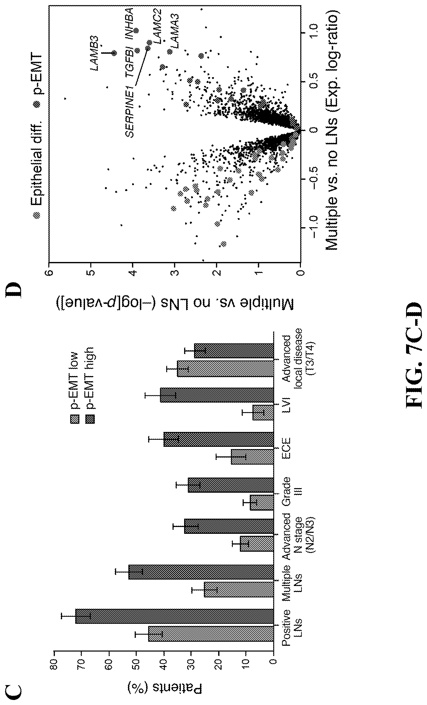
D00017

D00018

D00019

D00020

D00021

D00022

D00023

D00024

D00025

D00026

D00027

D00028

D00029

D00030

D00031

D00032

D00033

D00034

D00035

D00036

D00037

D00038

D00039

D00040

D00041


S00001
XML
uspto.report is an independent third-party trademark research tool that is not affiliated, endorsed, or sponsored by the United States Patent and Trademark Office (USPTO) or any other governmental organization. The information provided by uspto.report is based on publicly available data at the time of writing and is intended for informational purposes only.
While we strive to provide accurate and up-to-date information, we do not guarantee the accuracy, completeness, reliability, or suitability of the information displayed on this site. The use of this site is at your own risk. Any reliance you place on such information is therefore strictly at your own risk.
All official trademark data, including owner information, should be verified by visiting the official USPTO website at www.uspto.gov. This site is not intended to replace professional legal advice and should not be used as a substitute for consulting with a legal professional who is knowledgeable about trademark law.