Quantitative Dna-based Imaging And Super-resolution Imaging
Jungmann; Ralf ; et al.
U.S. patent application number 16/559490 was filed with the patent office on 2020-02-27 for quantitative dna-based imaging and super-resolution imaging. This patent application is currently assigned to President and Fellows of Harvard College. The applicant listed for this patent is President and Fellows of Harvard College. Invention is credited to Maier S. Avendano Amado, Mingjie Dai, Ralf Jungmann, Johannes B. Woehrstein, Peng Yin.
| Application Number | 20200064340 16/559490 |
| Document ID | / |
| Family ID | 52432405 |
| Filed Date | 2020-02-27 |


View All Diagrams
| United States Patent Application | 20200064340 |
| Kind Code | A1 |
| Jungmann; Ralf ; et al. | February 27, 2020 |
QUANTITATIVE DNA-BASED IMAGING AND SUPER-RESOLUTION IMAGING
Abstract
The present disclosure provides, inter alia, methods and compositions (e.g., conjugates) for imaging, at high spatial resolution, targets of interest.
| Inventors: | Jungmann; Ralf; (Munich, DE) ; Yin; Peng; (Brookline, MA) ; Dai; Mingjie; (Brookline, MA) ; Avendano Amado; Maier S.; (Brookline, MA) ; Woehrstein; Johannes B.; (Munich, DE) | ||||||||||
| Applicant: |
|
||||||||||
|---|---|---|---|---|---|---|---|---|---|---|---|
| Assignee: | President and Fellows of Harvard
College Cambridge MA |
||||||||||
| Family ID: | 52432405 | ||||||||||
| Appl. No.: | 16/559490 | ||||||||||
| Filed: | September 3, 2019 |
Related U.S. Patent Documents
| Application Number | Filing Date | Patent Number | ||
|---|---|---|---|---|
| 14908333 | Jan 28, 2016 | |||
| PCT/US14/48977 | Jul 30, 2014 | |||
| 16559490 | ||||
| 61934759 | Feb 1, 2014 | |||
| 61884126 | Sep 29, 2013 | |||
| 61859891 | Jul 30, 2013 | |||
| Current U.S. Class: | 1/1 |
| Current CPC Class: | G06K 9/6206 20130101; C07K 16/18 20130101; G06K 9/6212 20130101; G16B 45/00 20190201; G01N 2458/10 20130101; G06K 9/4642 20130101; C12Q 1/6816 20130101; G01N 33/5308 20130101; C12Q 1/6841 20130101; G06K 9/38 20130101; G06T 7/248 20170101; G16B 40/00 20190201; G06K 9/6298 20130101; G01N 2333/36 20130101; C12Q 1/6804 20130101; C07K 2317/76 20130101; C12Q 1/6804 20130101; C12Q 2525/204 20130101; C12Q 2527/107 20130101; C12Q 2537/143 20130101; C12Q 2543/10 20130101; C12Q 2563/107 20130101; C12Q 2563/131 20130101; C12Q 2563/179 20130101; C12Q 2565/102 20130101; C12Q 1/6841 20130101; C12Q 2525/204 20130101; C12Q 2527/107 20130101; C12Q 2537/143 20130101; C12Q 2543/10 20130101; C12Q 2563/107 20130101; C12Q 2563/131 20130101; C12Q 2563/179 20130101; C12Q 2565/102 20130101; C12Q 1/6816 20130101; C12Q 2525/204 20130101; C12Q 2527/107 20130101; C12Q 2537/143 20130101; C12Q 2543/10 20130101; C12Q 2563/107 20130101; C12Q 2563/131 20130101; C12Q 2563/179 20130101; C12Q 2565/102 20130101 |
| International Class: | G01N 33/53 20060101 G01N033/53; C12Q 1/6841 20060101 C12Q001/6841; G06T 7/246 20060101 G06T007/246; C12Q 1/6804 20060101 C12Q001/6804; C12Q 1/6816 20060101 C12Q001/6816; G06K 9/62 20060101 G06K009/62; G06K 9/46 20060101 G06K009/46; G06K 9/38 20060101 G06K009/38; C07K 16/18 20060101 C07K016/18; G16B 45/00 20060101 G16B045/00; G16B 40/00 20060101 G16B040/00 |
Claims
1. A protein-nucleic acid conjugate, comprising a protein linked to a docking strand that is capable of transiently binding to a complementary labeled imager strand.
2.-34. (canceled)
35. A method of detecting a target in a sample, the method comprising: contacting the sample with (a) at least one antibody-DNA conjugate that comprises a biotinylated antibody linked to a biotinylated docking strand through a biotin-streptavidin linker, and (b) at least one fluorescently-labeled imager strand that is complementary to the docking strand of the at least one antibody-DNA conjugate; and determining whether the at least one antibody-DNA conjugate binds to the target in the sample.
36. The method of claim 35, wherein the determining step comprises imaging binding of the at least one fluorescently-labeled imager strand to the docking strand of the at least one antibody-DNA conjugate.
37. (canceled)
38. The method of claim 35, wherein the antibody is a monoclonal antibody.
39.-46. (canceled)
47. The method of claim 35, wherein the docking strand comprises at least two domains, wherein each domain is complementary to a respectively labeled imager strand.
48.-49. (canceled)
50. The method of claim 1, comprising: contacting the sample with (a) at least two different antibody-DNA conjugates, each comprising a biotinylated antibody linked to a biotinylated docking strand through an intermediate biotin-streptavidin linker, and (b) at least two labeled imager strands that are complementary to respective docking strands of the at least two different antibody-DNA conjugates; and determining whether the at least two antibody-DNA conjugates bind to at least two targets in the sample.
51.-68. (canceled)
69. The method of claim 50, wherein the sample is contacted sequentially with the at least two labeled imager strands of (b).
70. The method of claim 69, comprising, in the following ordered steps: contacting the sample with a first antibody-DNA conjugate and at least one other antibody-DNA conjugate; contacting the sample with a first labeled imager strand that is complementary to the docking strand of the first antibody-DNA conjugate; imaging the sample to obtain a first image; removing the first labeled imager strand; contacting the sample with at least one other labeled imager strand that is complementary to the docking strand of the at least one other antibody-DNA conjugate; and imaging the sample to obtain at least one other image.
71. The method of claim 69, comprising, in the following ordered steps: contacting the sample with a first antibody-DNA conjugate; contacting the sample with a first labeled imager strand that is complementary to the docking strand of the first antibody-DNA conjugate; imaging the sample to obtain a first image; removing the first labeled imager strand; contacting the sample with at least one other antibody-DNA conjugate; contacting the sample with at least one other labeled imager strand that is complementary to the docking strand of the at least one other antibody-DNA conjugate; and imaging the sample to obtain at least one other image.
72.-90. (canceled)
91. A method of detecting a single-stranded target strand in a sample, the method comprising contacting the sample with (a) at least one single-stranded DNA probe comprising a target binding domain of 20 nucleotides in length linked to a docking domain comprising at least one subdomain, wherein the target binding domain is complementary to a respective domain of a single-stranded target strand, (b) at least one labeled imager strand of 4 to 30 nucleotides in length that is complementary to the at least one subdomain of the docking domain; and determining whether the at least one single-stranded DNA probe binds to the single-stranded target strand in the sample.
92. The method of claim 91, wherein the determining step comprises imaging binding of the at least one labeled imager strand to the at least one subdomain of the docking domain of the at least one single-stranded DNA probe.
93.-110. (canceled)
111. The method of claim 91, comprising contacting the sample with (a) at least two different single-stranded DNA probes, each comprising a target binding domain of 20 nucleotides in length linked to a docking domain comprising at least one subdomain, wherein the target binding domain is complementary to a respective domain of a single-stranded target strand, and (b) at least two labeled imager strands that are complementary to respective subdomains of the docking domain of the at least two different single-stranded DNA probes; and determining whether the at least two single-stranded DNA probes bind to at least two targets in the sample.
112.-230. (canceled)
231. The method of claim 111, wherein the sample is contacted sequentially with the at least two labeled imager strands of (b).
232. The method of claim 231, comprising, in the following ordered steps: contacting the sample with a first single-stranded DNA probe and at least one other single-stranded DNA probe; contacting the sample with a first labeled imager strand that is complementary to the at least one subdomain of the docking domain of the first single-stranded DNA probe; imaging the sample to obtain a first image; removing the first labeled imager strand; contacting the sample with at least one other labeled imager strand that is complementary to the at least one subdomain of the docking domain of the at least one other antibody-DNA conjugate; and imaging the sample to obtain at least one other image.
233. The method of claim 231, comprising, in the following ordered steps: contacting the sample with a first single-stranded DNA probe; contacting the sample with a first labeled imager strand that is complementary to the at least one subdomain of the docking domain of the first antibody-DNA conjugate; imaging the sample to obtain a first image; removing the first labeled imager strand; contacting the sample with at least one other single-stranded DNA probe; contacting the sample with at least one other labeled imager strand that is complementary to the at least one subdomain of the docking domain of the at least one other antibody-DNA conjugate; and imaging the sample to obtain at least one other image.
234. The method of claim 91, wherein the single-stranded target strand is a single-stranded mRNA target strand.
235. The method of claim 91, wherein the at least one labeled imager strand is fluorescently labeled.
236. The method of claim 91, wherein the docking domain comprises at least two subdomains, optionally at least three subdomains, wherein the at least two subdomains are respectively complementary to at least two labeled imager strands of about 4 to 30 nucleotides in length.
237. The method of claim 236, wherein the at least two subdomains are respectively complementary to at least two labeled imager strands of about 8 to 10 nucleotides in length.
238. The method of claim 236, wherein the respectively complementary labeled imager strands are distinctly labeled imager strands.
239. The method of claim 91, wherein the target binding domain of about 20 nucleotides in length is linked at its 3' end or its 5' end to a docking domain.
Description
RELATED APPLICATIONS
[0001] This application is a divisional of U.S. application Ser. No. 14/908,333, filed Jan. 28, 2016, which is a national stage filing under 35 U.S.C. .sctn. 371 of international application number PCT/US2014/048977, filed Jul. 30, 2014, which was published under PCT Article 21(2) in English and claims the benefit under 35 U.S.C. .sctn. 119(e) of U.S. provisional application No. 61/934,759, filed Feb. 1, 2014, U.S. provisional application No. 61/884,126, filed Sep. 29, 2013, and U.S. provisional application No. 61/859,891, filed Jul. 30, 2013, each of which is incorporated by reference herein in its entirety.
FIELD OF THE INVENTION
[0002] The present disclosure relates generally to the field of detection and quantification of targets.
BACKGROUND OF THE INVENTION
[0003] Far-field fluorescence microscopy has seen major advances since the advent of methods that circumvent the classical diffraction limit, e.g., super-resolution microscopy (refs. 1, 2). Most implementations switch molecules between fluorescent ON- and OFF-states to allow consecutive localization of individual molecules. Switching is traditionally obtained in one of two ways: "targeted" switching actively confines the fluorescence excitation to an area smaller than the diffraction of light (e.g., stimulated emission depletion microscopy, or STED (ref. 3)), whereas "stochastic" switching uses photoswitchable proteins (photoactivated localization microscopy, or PALM (ref. 4)) or photoswitchable organic dyes (stochastic optical reconstruction microscopy, or STORM (ref. 1)). Although these methods offer enhanced spatial resolution, they tend to require either expensive instrumentation or highly specialized experimental conditions, and thus have not yet been developed into common biological laboratory techniques.
SUMMARY OF THE INVENTION
[0004] The present disclosure provides, inter alia, methods, compositions (e.g., conjugates) and kits for imaging, at high or low spatial resolution, targets (e.g., biomolecules) of interest in, for example, a cellular environment. The methods, compositions and kits of the present disclosure take advantage of repetitive, transient binding of short, labeled (e.g., fluorescently labeled) oligonucleotides (e.g., DNA oligonucleotides), or "imager" strands, to complementary "docking" strands, which are attached to targets of interest, in some embodiments, through an intermediate molecule such as an antibody such as a primary or a secondary antibody, to obtain stochastic switching between fluorescent ON- and OFF-states (FIGS. 1A and 1B). In the unbound state, only background fluorescence from partially quenched (ref. 8) imager strands is observed (depicted by dimmer fluorescence of unbound imager strands in FIG. 1A). This is considered an "OFF" state. Upon binding and immobilization of an imager strand, fluorescence emission is detected using, for example, total internal reflection (TIR) or highly inclined and laminated optical sheet (HILO) microscopy (ref. 9). This is considered an "ON" state. In general, the methods, compositions and kits as provided herein increase the imaging resolution and thus the sensitivity of detection. In some aspects, they also increase the specificity as well as the number of utilizable fluorophores available for detecting targets of interest including but not limited to, e.g., naturally-occurring biomolecules.
[0005] By linking a short docking strand to a binding partner (e.g., a protein-binding moiety or a nucleic acid-binding moiety, whether primary or secondary), such as an antibody including a primary and a secondary antibody, different species of targets (e.g., biomolecules, optionally in a cellular environment) can be labeled and subsequently detected by introducing fluorescently-labeled imager strands that are complementary to and bind to the docking strands through transient Watson-Crick interactions. Unlike existing detection methods, the methods of the present disclosure are not limited by the number of spectrally distinct fluorophores available for detecting distinct targets (e.g., biomolecules). Rather, the programmability of nucleic acid (e.g., DNA and/or RNA) molecules and sequential time-lapsed imaging are used herein to provide images of up to hundreds of distinct species of targets using, in some embodiments, only a single optimized fluorophore. Further, these different species of targets (e.g., biomolecules) can be quantified using predictable kinetics of binding of single fluorescently-labeled imager strands to their complementary target docking strands.
[0006] In some instances, the methods can be used to generate super-resolution images, significantly even without the need for a super-resolution microscope. It should be understood that while the methods, compositions and kits as provided herein may be described for use in super-resolution imaging, they may also be used, in some embodiments, for imaging that does not require super-resolution. Thus, in some embodiments, the methods, compositions and kits of the present disclosure may be used for imaging, generally.
[0007] In some aspects, provided herein is a protein-nucleic acid conjugate, comprising a protein linked to a docking strand that is capable of transiently binding to a complementary labeled imager strand. In some aspects, provided herein is a protein-nucleic acid conjugate, comprising a protein linked to a docking strand that is transiently bound to a complementary labeled imager strand. Imager strands, in some embodiments, are labeled with a detectable label. The detectable label may be, for example, a fluorescent label or other detectable label, e.g., gold nanoparticle. While various aspects and embodiments herein refer to fluorescently-labeled imager strands, it should be understood that such fluorescent labels, in many instances, can be interchanged with other detectable labels. Thus, in some embodiments, a fluorescently-labeled imager strand (which may be detected by, for example, fluorescent microscopy) may be interchanged with an imager strand labeled with, for example, gold nanoparticles (which may be detected by, for example, dark field microscopy). It is also to be understood that the docking strands may be capable of transiently binding a plurality of complementary labeled strands (e.g., the docking strand may comprise a plurality of binding sites for complementary labeled strands).
[0008] In some embodiments, a method may be carried out involving a plurality of docking strand and imager strand pairs. Such a method can be used to detect a plurality of targets (e.g., with each docking strand-imager strand pair corresponding to one target). The docking strand-imager strand pairs in the plurality must share an approximately equal probability of hybridizing under a single environment or condition (as defined for example by temperature, salt concentration, strand molarity, etc.), such that if there is an observed difference between the level of binding (and thus the detection) of a population of imager strands, an end user can conclude that such difference is a function of the amount of docking strand and thus ultimately the amount of target. In some embodiments, the docking and imager strands are typically selected such that their bound states have a thermal stability in the range of about +/-0.5 kcal/mol. With this range of thermal stability, it is possible to select at least 200 orthogonal (e.g., different) sequences to be used in these multiplexing methods.
[0009] In some embodiments, a protein is an antibody such as a primary antibody or a secondary antibody, an antigen-binding antibody fragment, or a peptide aptamer.
[0010] In some embodiments, a protein is linked to the docking strand through an intermediate linker. In some embodiments, the intermediate linker comprises biotin and streptavidin.
[0011] In some embodiments, an antibody is a monoclonal antibody.
[0012] In some embodiments, a complementary fluorescently-labeled imager strand comprises at least one fluorophore.
[0013] In some embodiments, a complementary labeled, optionally fluorescently labeled, imager strand is about 4 to about 30 nucleotides, or about 8 to about 10 nucleotides, in length. In some embodiments, a complementary labeled imager strand is longer than 30 nucleotides.
[0014] In this and other aspects and embodiments described herein, the docking strand may comprise a plurality of domains, each complementary to a labeled imager strand. The domains may be identical in sequence (and thus will bind to the identical imager strands) or they may be of different sequence (and thus may bind to imager strands that are not identically labeled). Such domains may also be referred to herein as binding sites for imager strands.
[0015] In some embodiments, a docking strand includes at least two or at least three domains, each respectively complementary to a labeled imager strand.
[0016] In some aspects, provided herein is a target bound to at least one protein-nucleic acid conjugate.
[0017] In some embodiments, the target is a protein. In some embodiments, the target is a nucleic acid (e.g., DNA or RNA).
[0018] In some aspects, provided herein is a plurality of protein-nucleic acid conjugates. In some embodiments, the plurality comprises at least two subsets of the protein-nucleic acid conjugates, and the protein-nucleic acid conjugates of each subset bind to different targets.
[0019] In some aspects, provided herein is a composition or kit comprising a plurality of protein-nucleic acid conjugates, optionally wherein at least one of the protein-nucleic acid conjugates is bound to at least one target.
[0020] In some aspects, provided herein is a composition or kit comprising at least one protein-nucleic acid conjugate that comprises a protein linked to a docking strand, optionally wherein the at least one protein-nucleic acid conjugate is bound to a target, and at least one complementary labeled, optionally fluorescently labeled, imager strand that is transiently bound to (or is capable of transiently binding to) the at least one protein-nucleic acid conjugate.
[0021] In some embodiments, a composition or kit comprises at least two complementary labeled, optionally fluorescently labeled, imager strands, wherein the at least two complementary labeled imager strands are identical. In some embodiments, the composition or kit comprises at least two complementary labeled imager strands, wherein the at least two complementary labeled imager strands are different.
[0022] In some embodiments, the number of complementary labeled, optionally fluorescently labeled, imager strands is less than, greater than or equal to the number of protein-nucleic acid conjugates.
[0023] In some embodiments, a composition or kit comprises at least 2, 3, 4, 5, 6, 7, 9 or 10 different complementary labeled, optionally fluorescently labeled, imager strands. In some embodiments, the composition or kit comprises at least 50 or at least 100 different complementary fluorescently-labeled imager strands.
[0024] In some aspects, provided herein is a composition or a kit comprising a (e.g., one or more) docking strand and an (e.g., one or more) imager strand. The docking strand may be modified to include an affinity label, thereby facilitating its subsequent attachment to one or more binding partners, such as an antibodies. For example, the docking strand may be biotinylated or it may be attached to avidin or streptavidin. Other affinity labels can be used instead. The imager strands may be labeled, such as fluorescently labeled. The imager strands may be a plurality of identical imager strands (e.g., with respect to sequence and label) or they may be a plurality of different imager strands (e.g., with respect to sequence and label). The composition or kit may further comprise a target-specific binding partner, such as an antibody. It is to be understood that the components may be bound to each other or they may be unbound, including physically separated from each other, in such compositions and kits. These and other compositions and kits may further comprise one or more buffers including oxygen scavengers.
[0025] In some aspects, provided herein is a composition or kit comprising an antibody-nucleic acid conjugate, wherein the antibody is a "secondary antibody" having specificity for an antibody, typically specificity for a particular isotype or an Fc domain of an antibody from a particular species (e.g., a mouse antibody that is specific for a human IgG1 antibody). The nucleic acid in the conjugate is a docking strand, as described herein. The composition or kit may further comprise one or more imager strands (or one or more subsets or populations of imager strands), as described herein. These and other compositions and kits may further comprise one or more buffers including oxygen scavengers.
[0026] In some aspects, the present disclosure provides an antibody-DNA conjugate, comprising a monoclonal antibody linked to a docking strand that is bound to a complementary labeled, optionally fluorescently labeled, imager strand, wherein the antibody and the docking strand are each biotinylated and linked to each other through an avidin or streptavidin linker or a biotin-streptavidin linker.
[0027] In some aspects, provided herein is an aptamer-nucleic acid conjugate, comprising a nucleic acid aptamer linked to a docking strand that is transiently bound to a complementary labeled, optionally fluorescently labeled, imager strand.
[0028] In some aspects, provided herein is a method of detecting a target in a sample, the method comprising contacting a sample with (a) at least one protein-nucleic acid conjugate that comprises a protein linked to a docking strand and (b) at least one fluorescently-labeled imager strand that is complementary to and transiently binds to the docking strand of the at least one protein-nucleic acid conjugate, and determining whether the at least one protein-nucleic acid conjugate binds to the target in the sample. In some embodiments, the determining step comprises imaging transient binding of the at least one fluorescently-labeled imager strand to the docking strand of the at least one protein-nucleic acid conjugate.
[0029] In some aspects, provided herein is a method of detecting a target in a sample, the method comprising contacting a sample with (a) at least one protein-nucleic acid conjugate that comprises a protein linked to a docking strand and (b) at least one fluorescently-labeled imager strand that is complementary to and transiently binds to the docking strand of the at least one protein-nucleic acid conjugate, and imaging transient binding, optionally using time-lapsed imaging, of the at least one fluorescently-labeled imager strand to the docking strand of the at least one protein-nucleic acid conjugate.
[0030] In some embodiments, a protein of the protein-nucleic acid conjugate is an antibody, an antigen-binding antibody fragment, or a peptide aptamer. In some embodiments, an antibody is a monoclonal antibody.
[0031] In some embodiments, a protein of the protein-nucleic acid conjugate is linked to the docking strand through an intermediate linker. In some embodiments, an intermediate linker comprises biotin and/or streptavidin.
[0032] In some embodiments, a complementary fluorescently-labeled imager strand comprises at least one fluorophore.
[0033] In some embodiments, a complementary labeled, optionally fluorescently labeled, imager strand is about 4 to about 10 nucleotides, or about 8 to about 10 nucleotides in length.
[0034] In some embodiments, a sample is a cell or cell lysate.
[0035] In some embodiments, a target is a protein. In some embodiments, a target is a nucleic acid (e.g., DNA or RNA).
[0036] In some embodiments, a target is obtained from a cell or cell lysate.
[0037] In some aspects, provided herein is a method of detecting at least one or at least two targets in a sample, the method comprising contacting a sample with (a) at least two protein-nucleic acid conjugates, each comprising a protein linked to a docking strand, and (b) at least two labeled (optionally spectrally distinct, or fluorescently labeled, or spectrally distinct and fluorescently labeled) imager strands that are complementary to and transiently bind to respective docking strands of the at least one, or at least, two different protein-nucleic acid conjugates and determining whether the at least two protein-nucleic acid conjugates bind to at least two targets in the sample. In some embodiments, the determining step comprises, in the following order, imaging transient binding of one of the at least two labeled imager strands to a docking strand of one of the at least two protein-nucleic acid conjugates to produce a first image (e.g., of a fluorescent signal), and imaging transient binding of another of the at least two labeled imager strands to a docking strand of another of the at least two protein-nucleic acid conjugates to produce at least one other image (e.g., of a fluorescent signal). In some embodiments, the method further comprises combining the first image and the at least one other image to produce a composite image of signal (e.g., fluorescent signal), wherein the signal of the composite image is representative of the at least two targets.
[0038] In some embodiments, a protein of the protein-nucleic acid conjugate is an antibody, an antigen-binding antibody fragment, or a peptide aptamer. In some embodiments, an antibody is a monoclonal antibody.
[0039] In some embodiments, a protein of the protein-nucleic acid conjugate is linked to the docking strand through an intermediate linker. In some embodiments, an intermediate linker comprises biotin and streptavidin.
[0040] In some embodiments, each of the at least two spectrally distinct, fluorescently-labeled imager strands comprises at least one fluorophore.
[0041] In some embodiments, each of the at least two labeled, optionally spectrally distinct, fluorescently labeled, imager strands is about 4 to about 10 nucleotides, or about 8 to about 10 nucleotides in length.
[0042] In some embodiments, a sample is a cell or cell lysate.
[0043] In some embodiments, at least two targets are proteins. In some embodiments, at least two targets are nucleic acids (e.g., DNA or RNA).
[0044] In some embodiments, at least two targets are obtained from a cell or cell lysate.
[0045] In some aspects, provided herein is a method of detecting at least one, or at least two, protein targets in a sample, comprising (a) contacting a sample with at least two protein-nucleic acid conjugates, each comprising a protein linked to a docking strand and (b) sequentially contacting the sample with at least two labeled (e.g., optionally spectrally indistinct, or fluorescently labeled, or spectrally distinct and fluorescently labeled) imager strands that are complementary to and transiently bind to respective docking strands of the at least two protein-nucleic acid conjugates, and determining whether the at least one or at least two protein-nucleic acid conjugates bind to at least two targets in the sample. In some embodiments, a method comprises, in the following ordered steps, contacting the sample with a first protein-nucleic acid conjugate and at least one other protein-nucleic acid conjugate, contacting the sample with a first labeled, optionally fluorescently labeled, imager strand that is complementary to and transiently binds to the docking strand of the first protein-nucleic acid conjugate, imaging the sample to obtain a first image, optionally using time-lapsed imaging, removing the first labeled imager strand, contacting the sample with at least one other labeled imager strand that is complementary to and transiently binds to the docking strand of the at least one other protein-nucleic acid conjugate, and imaging the sample to obtain at least one other image, optionally using time-lapsed imaging.
[0046] In some embodiments, a method comprises, in the following ordered steps, contacting a sample with a first protein-nucleic acid conjugate, contacting the sample with a first labeled, optionally fluorescently labeled, imager strand that is complementary to and transiently binds to the docking strand of the first protein-nucleic acid conjugate, imaging the sample to obtain a first image, optionally using time-lapsed imaging, removing the first labeled imager strand, contacting the sample with at least one other protein-nucleic acid conjugate, contacting the sample with at least one other labeled, optionally fluorescently labeled, imager strand that is complementary to and transiently binds to the docking strand of the at least one other protein-nucleic acid conjugate, and imaging the sample to obtain at least one other image, optionally using time-lapsed imaging.
[0047] In some embodiments, a method further comprises determining whether the first protein-DNA conjugate binds to a first target and/or whether the at least one other protein-DNA conjugate binds to at least one other target.
[0048] In some embodiments, a method further comprises assigning a pseudo-color to the signal (e.g., fluorescent signal) in a first image, and assigning at least one other pseudo-color to the fluorescent signal in the at least one other image.
[0049] In some embodiments, a method further comprises combining a first image and the at least one other image to produce a composite image of the pseudo-colored signals, wherein the pseudo-colored signals of the composite image are representative of the at least two targets.
[0050] In some embodiments, the protein of the protein-nucleic acid conjugate(s) is an antibody, an antigen-binding antibody fragment, or a peptide aptamer. In some embodiments, the antibody is a monoclonal antibody.
[0051] In some embodiments, the protein of the protein-nucleic acid conjugate(s) is linked to the docking strand through an intermediate linker. In some embodiments, the intermediate linker comprises biotin and/or streptavidin.
[0052] In some embodiments, each of the fluorescently-labeled imager strands comprises at least one fluorophore.
[0053] In some embodiments, each of the fluorescently-labeled imager strands is about 4 to about 30 nucleotides, or about 8 to about 10 nucleotides in length.
[0054] In some embodiments, a sample is a cell or cell lysate.
[0055] In some embodiments, a target(s) is a protein. In some embodiments, a target(s) is a nucleic acid (e.g., DNA or RNA).
[0056] In some embodiments, a target(s) is obtained from a cell or cell lysate.
[0057] In some aspects, provided herein is a method of detecting a target, optionally a naturally-occurring biomolecule, comprising contacting a sample containing at least one target, optionally a naturally-occurring biomolecule, with (a) at least one BP-NA conjugate, optionally each BP-NA conjugate comprising a protein or nucleic acid linked to a docking strand, and (b) at least one labeled, optionally fluorescently labeled, imager strand that is complementary to and transiently binds the docking strand of the at least one BP-NA conjugate, and determining whether the at least one BP-NA conjugate binds to at least one target, optionally a naturally-occurring biomolecule, in the sample. In this and other aspects or embodiments described herein, it is to be understood that the method may be carried out using a sample that is suspected of containing at least one target or a sample that an end-user desires to analyze for the presence of the at least one target without any prior knowledge of the sample respecting its likelihood of containing the target.
[0058] In some embodiments, the determining step comprises imaging transient binding of the at least one labeled, optionally fluorescently labeled, imager strand to the docking strand of the at least one BP-NA conjugate.
[0059] In some embodiments, a sample is a cell or cell lysate.
[0060] In some embodiments, an at least one target, optionally a naturally-occurring biomolecule, is obtained from a cell or cell lysate.
[0061] In some embodiments, a protein is an antibody, an antigen-binding antibody fragment, or a peptide aptamer. In some embodiments, an antibody is a monoclonal antibody.
[0062] In some embodiments, a protein is linked to the docking strand through an intermediate linker. In some embodiments, an intermediate linker comprises biotin and/or streptavidin.
[0063] In some embodiments, a nucleic acid is a nucleic acid aptamer.
[0064] In some embodiments, a fluorescently-labeled imager strand comprises at least one fluorophore.
[0065] In some embodiments, an imager strand, optionally a fluorescently-labeled imager strand, is about 4 to about 30, or about 8 to about 10 nucleotides in length.
[0066] In some aspects, provided herein is a method of detecting a target, optionally a naturally-occurring biomolecule, comprising contacting a sample containing at least two targets, optionally naturally-occurring biomolecules, with (a) at least two different BP-NA conjugates, optionally each BP-NA conjugate comprising a protein or nucleic acid linked to a DNA docking strand, and (b) at least two labeled (optionally spectrally indistinct, or fluorescently labeled, or spectrally distinct and fluorescently labeled) imager strands that are complementary to and transiently bind to respective docking strands of the at least two BP-NA conjugates, and determining whether the at least two BP-NA conjugates bind to at least one or at least two naturally-occurring biomolecules in the sample.
[0067] In some embodiments, a method comprises, in the following ordered steps, contacting the sample with a first BP-NA conjugate and at least one other BP-NA conjugate, contacting the sample with a first labeled, optionally fluorescently labeled, imager strand that is complementary to and transiently binds to the docking strand of the first BP-NA conjugate, imaging the sample to obtain a first image, optionally using time-lapsed imaging, removing the first labeled imager strand, contacting the sample with at least one other labeled, optionally fluorescently labeled, imager strand that is complementary to and transiently binds to the docking strand of the at least one other BP-NA conjugate, and imaging the sample to obtain at least one other image, optionally using time-lapsed imaging.
[0068] In some embodiments, a method comprises, in the following ordered steps, contacting the sample with a first BP-NA conjugate, contacting the sample with a first labeled, optionally fluorescently labeled, imager strand that is complementary to and transiently binds to the docking strand of the first BP-NA conjugate, imaging the sample to obtain a first image, optionally using time-lapsed imaging, removing the first labeled imager strand, contacting the sample with at least one other BP-NA conjugate, contacting the sample with at least one other labeled, optionally fluorescently labeled, imager strand that is complementary to and transiently binds to the docking strand of the at least one other BP-NA conjugate, and imaging the sample to obtain a at least one other image, optionally using time-lapsed imaging.
[0069] In some embodiments, a method further comprises determining whether the first protein DNA conjugate binds to a first target, optionally a naturally-occurring biomolecule, and/or whether the at least one other protein-DNA conjugate binds to at least one other target, optionally a naturally-occurring biomolecule.
[0070] In some embodiments, a method further comprises assigning a pseudo-color to the signal (e.g., fluorescent signal) in a first image, and assigning at least one other pseudo-color to the signal (e.g., fluorescent signal) in at least one other image.
[0071] In some embodiments, a method further comprises combining a first image and at least one other image to produce a composite image of pseudo-colored signals, wherein the pseudo-colored signals of the composite image are representative of at least one, or at least two, targets (e.g., naturally-occurring biomolecules).
[0072] In some aspects, provided herein is a method of determining the number of targets in a test sample, comprising obtaining a sample that comprises targets transiently bound directly or indirectly to labeled, optionally fluorescently labeled, imager strands, obtaining a time-lapsed image, optionally a time-lapsed diffraction-limited fluorescence image, of the sample, performing spot detection (e.g., fluorescence spot detection) and localization (e.g., through the use of Gaussian fitting) on the diffraction-limited image to obtain a high-resolution image of the sample, calibrating k.sub.onc.sub.imager, optionally using a control sample with a known number of targets, wherein k.sub.on is a second order association constant, and c.sub.imager is concentration of labeled (e.g., fluorescently labeled) imager strands in the test sample, determining variable .sigma..sub.d, optionally by fitting the fluorescence OFF-time distribution to a cumulative distribution function, and determining the number of test targets in the sample based on the equation, number of test targets=(k.sub.onc.sub.imager.tau..sub.d).sup.-1.
[0073] In some aspects, provided herein is a method of determining a relative amount of targets in a test sample, comprising obtaining a sample that comprises targets transiently bound directly or indirectly to labeled imager strands, obtaining a time-lapsed image of the sample, performing spot detection and localization on the image to obtain a high-resolution image of the sample, determining variable .tau..sub.d, and determining the relative amount of two or more test targets in the sample based on .tau..sub.d.
[0074] In some embodiments, test targets are protein targets.
[0075] In some embodiments, protein targets are bound to protein-nucleic acid conjugates that comprise a protein linked to a docking strand, and the labeled (e.g., fluorescently labeled) imager strands are complementary to and transiently bind to respective docking strands of the protein-nucleic acid conjugates.
[0076] In some embodiments, the protein of the protein-nucleic acid conjugate is an antibody, an antigen-binding antibody fragment, or a peptide aptamer.
[0077] In some embodiments, test targets are single-stranded nucleic acids.
[0078] In some embodiments, single-stranded nucleic acids are DNA or RNA.
[0079] In some embodiments, each of the fluorescently-labeled imager strands comprises at least one fluorophore.
[0080] In some embodiments, each of the labeled, optionally fluorescently labeled, imager strands is about 4 to about 30 nucleotides, or about 8 to about 10 nucleotides in length.
[0081] In some embodiments, a time-lapsed fluorescence image is obtained over a period of about 25 minutes.
[0082] In some embodiments, the number of test targets is determined with an accuracy of greater than 90%.
[0083] In some aspects, provided herein is a single-stranded DNA probe comprising a target binding domain of about 20 nucleotides in length linked, optionally at its 3' end, to a docking domain of at least one, at least two, or at least three subdomains, wherein the at at least one, least two, or at least three subdomains are respectively complementary to at least one, at least two, or at least three labeled, optionally fluorescently labeled, imager strands of about 4 to about 30, or about 8 to 10 nucleotides in length, and wherein the target binding domain is bound to a complementary domain of a single-stranded mRNA target strand.
[0084] In some embodiments, at least one of the at least one, at least two, or at least three subdomains is transiently bound to at least one labeled, optionally fluorescently labeled, imager strand.
[0085] In some aspects, provided herein is a method of performing drift correction for a plurality of images, wherein each of the plurality of images comprises a frame of a time sequence of images, wherein the time sequence of images captures a plurality of transient events, the method comprising determining a time trace for each of a plurality of drift markers identified in the plurality of images, wherein a time trace for each drift marker corresponds to movement of an object in the image over the time sequence of images, determining, with at least one computer processor, a first drift correction from at least one of the plurality of drift markers based, at least in part, on the time traces for the plurality of drift markers, determining a time trace for each of a plurality of geometrically-addressable marker sites from a plurality of drift templates identified from the plurality of images, wherein each drift template in the plurality of drift templates describes a geometrical relationship between the plurality of geometrically-addressable marker sites of transient events in the drift template, determining a second drift correction based, at least in part, on the time traces for the plurality of geometrically-addressable marker sites from the plurality of drift templates, correcting the plurality of images based, at least in part, on the first drift correction and the second drift correction, and outputting a final image based on the corrected plurality of images.
[0086] In some embodiments, a method further comprises identifying a plurality of localizations in each of the plurality of images, creating a two-dimensional histogram of the plurality of localizations, and identifying locations of the plurality of drift markers based, at least in part, on the two-dimensional histogram, wherein determining the time traces for each of the plurality of drift markers comprises determining the time traces based, at least in part, on the locations of the plurality of drift markers.
[0087] In some embodiments, identifying a plurality of localizations comprises identifying a plurality of spots on each of the plurality of images, and determining a fitted center position of each of the plurality of spots using a local Gaussian fitting algorithm, wherein each of the plurality of localizations comprises the spot identified on an image and its associated fitted center position.
[0088] In some embodiments, each of the plurality of localizations further comprises a detected photon count corresponding to the localization.
[0089] In some embodiments, creating the two-dimensional histogram of the plurality of localizations comprises binning all localizations into a two-dimensional grid and using a total number of localizations in each bin as a histogram count.
[0090] In some embodiments, creating the two-dimensional histogram of the plurality of localizations comprises binning all localizations into a two-dimensional grid and using a total number of photon count of the plurality of localizations in each bin as a histogram count.
[0091] In some embodiments, identifying locations of the plurality of drift markers based, at least in part, on the two-dimensional histogram comprises at least one of the following: binarizing the two-dimensional histogram using one or more selection criteria, wherein the one or more selection criteria include a lower-bound threshold of a histogram value or a upper-bound threshold of a histogram value; partitioning the binarized image into partitions and filtering the partitions based on one or more selection criteria, wherein the one or more selection criteria include one or more of a lower-bound threshold of an area of a partition area, an upper-bound threshold of the area, a lower-bound or an upper-bound of a longest or shortest linear dimension of a partition longest, and a lower-bound or an upper-bound of an eccentricity of a partition; and expanding and shrinking the binarized image using one or more binary image operations, wherein the one or more binary image operations include one or more of the following: dilate, erode, bridge, close, open, fill, clean, top-hat, bottom-hat, thicken, thin, and more.
[0092] In some embodiments, determining a first drift correction based, at least in part, on the time traces for the plurality of drift markers comprises: determining a relative time trace for each of the plurality of drift markers, wherein the relative time trace is determined by comparing the time trace for the drift marker with the average position of the same trace; and determining a combined time trace based on the relative time traces for each of the plurality of drift markers, wherein determining the first drift correction based, at least in part, on the time traces for the plurality of drift markers comprises determining the first drift correction based, at least in part, on the relative time traces for each of the plurality of drift markers.
[0093] In some embodiments, determining the first drift correction based, at least in part, on the relative time traces for each of the plurality of drift markers comprises performing a weighted average of the relative time traces for each of the plurality of drift markers.
[0094] In some embodiments, performing the weighted average comprises: determining a quality score for each of the relative time traces, wherein the quality score is determined based, at least in part, on a measure of variability over time associated with the time trace and/or a measure of localization uncertainty of individual localizations within the time trace.
[0095] In some embodiments, the measure of variability over time comprises a standard deviation of the time trace over time.
[0096] In some embodiments, the measure of localization uncertainty of individual localizations comprises, at least in part, an estimate of uncertainty from a Gaussian fitting or a comparison with other simultaneous localizations, wherein the other simultaneous localizations are from within a same image and from other time traces from the plurality of drift markers, wherein the comparison comprises a mean and standard deviation of all simultaneous localizations.
[0097] In some embodiments, the method further comprises determining that a first drift marker of the plurality of drift markers is not present in at least one frame of the time sequence of images, and linearly interpolating the time trace for the first drift marker for the at least one frame to produce a smoothed time trace for the first drift marker.
[0098] In some embodiments, determining a time trace for each of a plurality of geometrically-addressable marker sites from a plurality of drift templates identified from the plurality of images comprises: identifying a plurality of localizations in each of the plurality of images; creating a two-dimensional histogram of the plurality of localizations; and identifying the plurality of drift templates based, at least in part, on the two-dimensional histogram, wherein identifying the plurality of drift templates comprises evaluating the two-dimensional histogram using an lower-bound and/or an upper-bound threshold in a histogram count.
[0099] In some embodiments, determining a time trace for each of a plurality of geometrically-addressable marker sites from a plurality of drift templates identified from the plurality of images comprises determining a time trace for each of a plurality of geometrically-addressable marker sites within each of the plurality of drift templates, and wherein determining the second drift correction comprises determining the second drift correction based, at least in part, on the time traces for each of the plurality of marker sites within each of the plurality of drift templates.
[0100] In some embodiments, determining the second drift correction based, at least in part, on the time traces for each of the plurality of geometrically-addressable marker sites from each of the plurality of drift templates comprises: identifying a plurality of geometrically-addressable marker sites within each of the plurality of drift templates; and determining a relative time trace for each of a plurality of geometrically-addressable drift markers for each of the plurality of drift templates, wherein determining the second drift correction based, at least in part, on the time traces for the plurality of drift templates comprises determining the second drift correction based, at least in part, on the relative time traces for each of the plurality of drift markers within each of the plurality of drift templates.
[0101] In some embodiments, identifying a plurality of geometrically addressable marker sites from each of the plurality of drift templates comprises determining a plurality of marker sites based on, at least in part, a two-dimensional histogram of the plurality of localizations in the corresponding drift template, and/or one or more selection criteria, wherein the one or more selection criteria include one or more of a total number of localizations, a surface density of localizations, and standard deviation of localizations.
[0102] In some embodiments, determining the second drift correction based, at least in part, on the relative time traces for each of the plurality of drift markers within each of the plurality of drift templates comprises performing a weighted average of the relative time traces for each of the plurality of drift markers within each of the drift templates.
[0103] In some embodiments, performing the weighted average comprises:
[0104] determining a quality score for each of the relative time traces, wherein the quality score is determined based, at least in part, on a measure of variability over time associated with the time trace and/or a localization uncertainty within the time trace.
[0105] In some embodiments, the measure of variability over time comprises a standard deviation of the time trace over time.
[0106] In some embodiments, the measure of localization uncertainty of individual localizations comprises an estimate of uncertainty from a Gaussian fitting or a comparison with other simultaneous localizations, wherein the other simultaneous localizations are from within a same image and from the other time traces from the plurality of marker sites from the plurality of drift templates, wherein the comparison comprises a mean and standard deviation of all simultaneous localizations.
[0107] In some embodiments, correcting the plurality of images based, at least in part, on the first drift correction and the second drift correction comprises correcting the plurality images using the first drift correction to produce a first corrected plurality of images, and wherein determining a time trace for each of a plurality of drift templates identified from the plurality of images comprises determining a time trace for each of the plurality of drift templates identified from the first corrected plurality of images.
[0108] In some embodiments, a method further comprises smoothing the first drift correction prior to correcting the plurality of images using the first drift correction.
[0109] In some embodiments, smoothing the first drift correction comprises processing the first drift correction using local regression with a window determined by a characteristic drift time scale of the first drift correction.
[0110] In some embodiments, a method further comprises smoothing the second drift correction prior to correcting the plurality of images using the second drift correction.
[0111] In some embodiments, smoothing the second drift correction comprises processing the second drift correction using local regression with a window determined by a characteristic drift time scale of the second drift correction.
[0112] In some embodiments, a method further comprises selecting a single drift marker of the plurality of drift markers; and determining a third drift correction based, at least in part, on the selected single drift marker, wherein correcting the plurality of images comprises correcting the plurality of images based, at least in part, on the third drift correction.
[0113] In some embodiments, correcting the plurality of images based, at least in part, on the third drift correction is performed prior to correcting the plurality of images based, at least in part on the first drift correction and the second drift correction.
[0114] In some embodiments, a method further comprises identifying locations of a first plurality of points in a first image of the plurality of frames; identifying locations of a second plurality of points in a second image of the plurality of images, wherein the second image corresponds to a neighboring frame of the first image in the time sequence of images; and determining a fourth drift correction based, at least in part, on differences between the locations of the first plurality of points and the second plurality of points, wherein correcting the plurality of images comprises correcting the plurality of images based, at least in part, on the fourth drift correction.
[0115] In some embodiments, the second image corresponds to a frame immediately following the frame corresponding to the first image in the time sequence of images.
[0116] In some embodiments, determining the fourth drift correction based, at least in part, on differences between the locations of the first plurality of points and the second plurality of points comprises: creating a histogram of distances between the locations of the first plurality of points and the second plurality of points; determining based, at least in part, on the histogram, pairs of points between the first image and the second image that correspond to the same transient event; and determining a location offset between each of the determined pairs of points, wherein determining the fourth drift correction is based on a vector average of the location offsets for each of the determined pairs of points.
[0117] In some embodiments, the plurality of images correspond to DNA-based images and wherein the plurality of transient events are binding events between an imaging strand and a DNA docking strand.
[0118] In some embodiments, the imaging strand is a fluorescent imaging probe configured to fluoresce when associated with the DNA docking strand.
[0119] In some embodiments, at least one of the drift markers is a DNA based nanostructure.
[0120] In some embodiments, the DNA based nanostructure is a DNA origami nanostructure with docking strands.
[0121] In some embodiments, at least one of the drift templates is a DNA based nanostructure.
[0122] In some embodiments, the DNA based nanostructure is a DNA origami nanostructure with docking strands.
[0123] In some embodiments, at least one of the drift templates is a three-dimensional drift template.
[0124] In some embodiments, the three-dimensional drift template is a tetrahedron.
[0125] In some embodiments, at least one of the drift templates includes multiple colors corresponding to different types of transient events.
[0126] In some embodiments, the different types of transient events include a first binding event of a first imaging strand with a first type of DNA docking strand and a second binding event of a second imaging strand with a second type of DNA docking strand.
[0127] In some embodiments, outputting the final image comprises displaying the final image on a display.
[0128] In some embodiments, outputting the final image comprises sending the final image to a computer via at least one network.
[0129] In some embodiments, outputting the final image comprises storing the final image on at least one storage device.
[0130] In some aspects, provided herein is a non-transitory computer readable medium encoded with a plurality of instructions that, when executed by at least one computer processor, performs a method of performing drift correction for a plurality of images, wherein each of the plurality of images comprises a frame of a time sequence of images, wherein the time sequence of images captures a plurality of transient events, the method comprising: determining a time trace for each of a plurality of drift markers identified in the plurality of images, wherein a time trace for each drift marker corresponds to movement of an object in the image over the time sequence of images; determining a first drift correction from at least one of the plurality of drift markers based, at least in part, on the time traces for the plurality of drift markers; determining a time trace for each of a plurality of geometrically-addressable marker sites from a plurality of drift templates identified from the plurality of images, wherein each drift template in the plurality of drift templates describes a geometrical relationship between the plurality of geometrically-addressable marker sites of transient events in the drift template; determining a second drift correction based, at least in part, on the time traces for the plurality of geometrically-addressable marker sites from the plurality of drift templates; correcting the plurality of images based, at least in part, on the first drift correction and the second drift correction; and outputting a final image based on the corrected plurality of images.
[0131] In some aspects, provided herein is a computer, comprising: an input interface configured to receive a plurality of images, wherein each of the plurality of images comprises a frame of a time sequence of images, wherein the time sequence of images captures a plurality of transient events; at least one processor programmed to: determine a time trace for each of a plurality of drift markers identified in the plurality of images, wherein a time trace for each drift marker corresponds to movement of an object in the image over the time sequence of images; determine a first drift correction from at least one of the plurality of drift markers based, at least in part, on the time traces for the plurality of drift markers; determine a time trace for each of a plurality of geometrically-addressable marker sites from a plurality of drift templates identified from the plurality of images, wherein each drift template in the plurality of drift templates describes a geometrical relationship between the plurality of geometrically-addressable marker sites of transient events in the drift template; determine a second drift correction based, at least in part, on the time traces for the plurality of geometrically-addressable marker sites from the plurality of drift templates; correct the plurality of images based, at least in part, on the first drift correction and the second drift correction; and determine a final image based on the corrected plurality of images; and output interface configured to output the final image.
BRIEF DESCRIPTION OF THE DRAWINGS
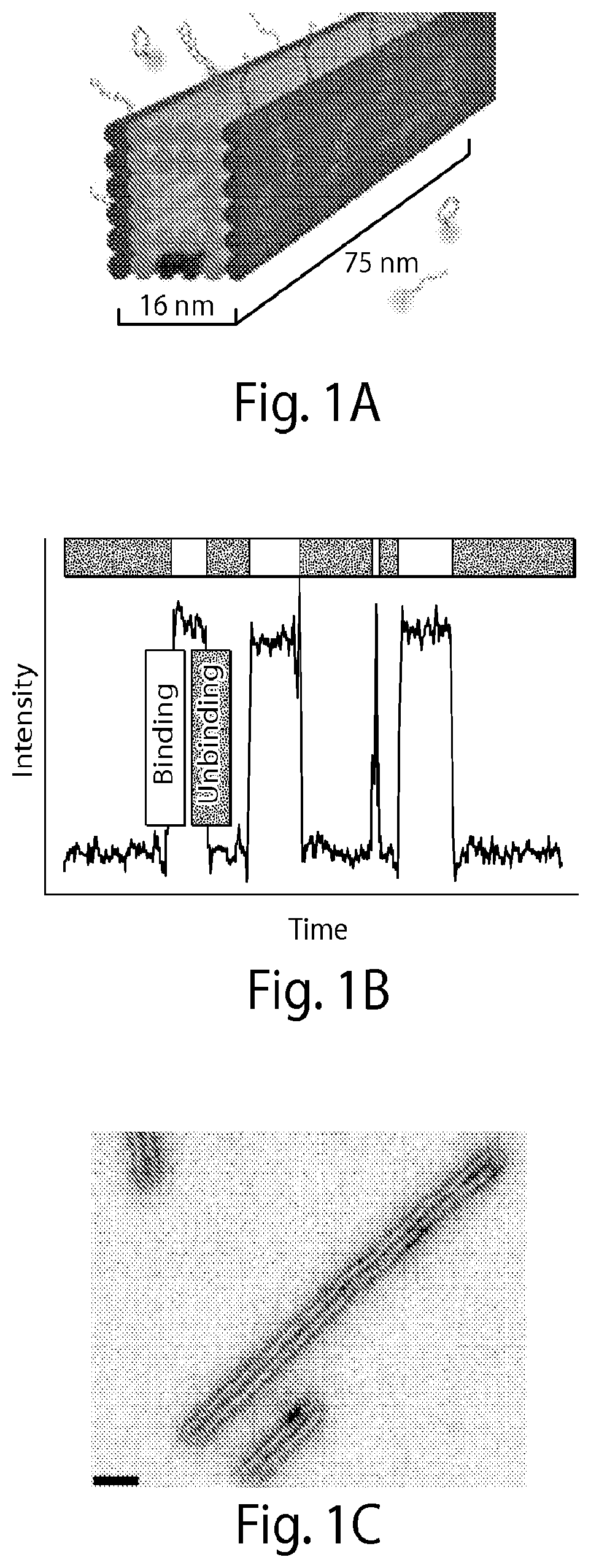
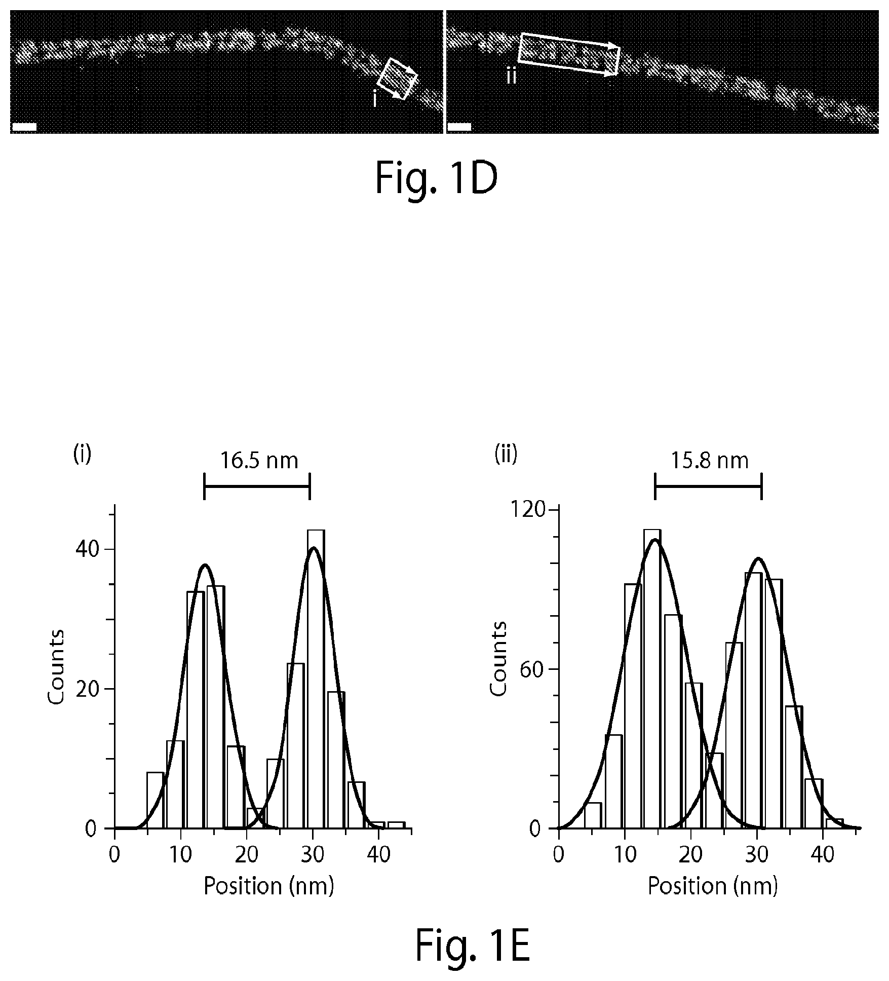
[0132] FIG. 1A shows microtubule-like DNA origami polymers "labeled" with single-stranded DNA docking strands on a pair of opposite faces (colored in dark gray) spaced approximately 16 nanometers (nm) apart. Complementary fluorescently-labeled imager strands transiently bind from solution to the docking strands. Biotinylated DNA strands (present on the bottom two center helices) are used to bind the microtubule-like DNA structures to glass surfaces for fluorescence imaging. FIG. 1B shows a graph demonstrating that transient binding of fluorescently-labeled imager strands to the docking strands produces fluorescence "blinking" (fluorescence intensity vs. time trace). This blinking is used to consecutively localize points below the diffraction limit. FIG. 1C shows a transmission electron microscope (TEM) image of DNA origami polymers with a measured width of 16.+-.1 nm (mean.+-.stdv) [scale bar: 40 nm]. FIG. 1D shows super-resolution fluorescence images obtained using Cy3b-labeled imager strands (15,000 frames, 5 Hz frame rate). Two distinct lines are visible [scale bars: 40 nm]. FIG. 1E shows a cross-sectional histogram of highlighted areas <i> and <ii> in FIG. 1D (arrows denote histogram direction), which show that the designed distance of approximately 16 nm is clearly resolved (full width at half maximum (FWHM) of each distribution is observed to be approximately 9 nm).
[0133] FIG. 2 shows an example of a biomolecule labeling scheme of the present disclosure where a protein (e.g., protein target) is labeled with antibody-DNA conjugates of the present disclosure and complementary fluorescently-labeled imager strands. The antibodies are linked to the docking strands through a linker that contains biotin and streptavidin (e.g., biotin-streptavidin-biotin linker).
[0134] FIG. 3A shows a super-resolution image of a microtubule network inside a fixed HeLa cell using an antibody-DNA conjugate and Atto655-labeled imager strands (10,000 frames, 10 Hz frame rate) [scale bar: 5 .mu.m]. FIG. 3B shows a high magnification image of the highlighted area in FIG. 3A [scale bar: 1 .mu.m]. FIG. 3C shows a diffraction-limited representation of the same area in FIG. 3B. Arrows highlight positions where the increase in resolution of the image is clearly visible. Adjacent microtubules with an apparent width of approximately 46 nm at position <i> in FIG. 3B are separated by approximately 79 nm [scale bar: 1 .mu.m]. FIG. 3D shows a dual-color super-resolution image (15,000 frames, 10 Hz frame rate) of microtubules and mitochondria inside a fixed HeLa cell obtained using antibody-DNA conjugates, Cy3b-labeled imager strands for microtubules (linear-like structures) and Atto655-labeled imager strands for mitochondria (patch-like structures) [scale bar: 5 .mu.m]. FIG. 3E shows a high magnification image of the highlighted area in FIG. 3D [scale bar: 1 .mu.m]. FIG. 3F shows a diffraction-limited image of the same area shown in FIG. 3E [scale bar: 1 .mu.m].
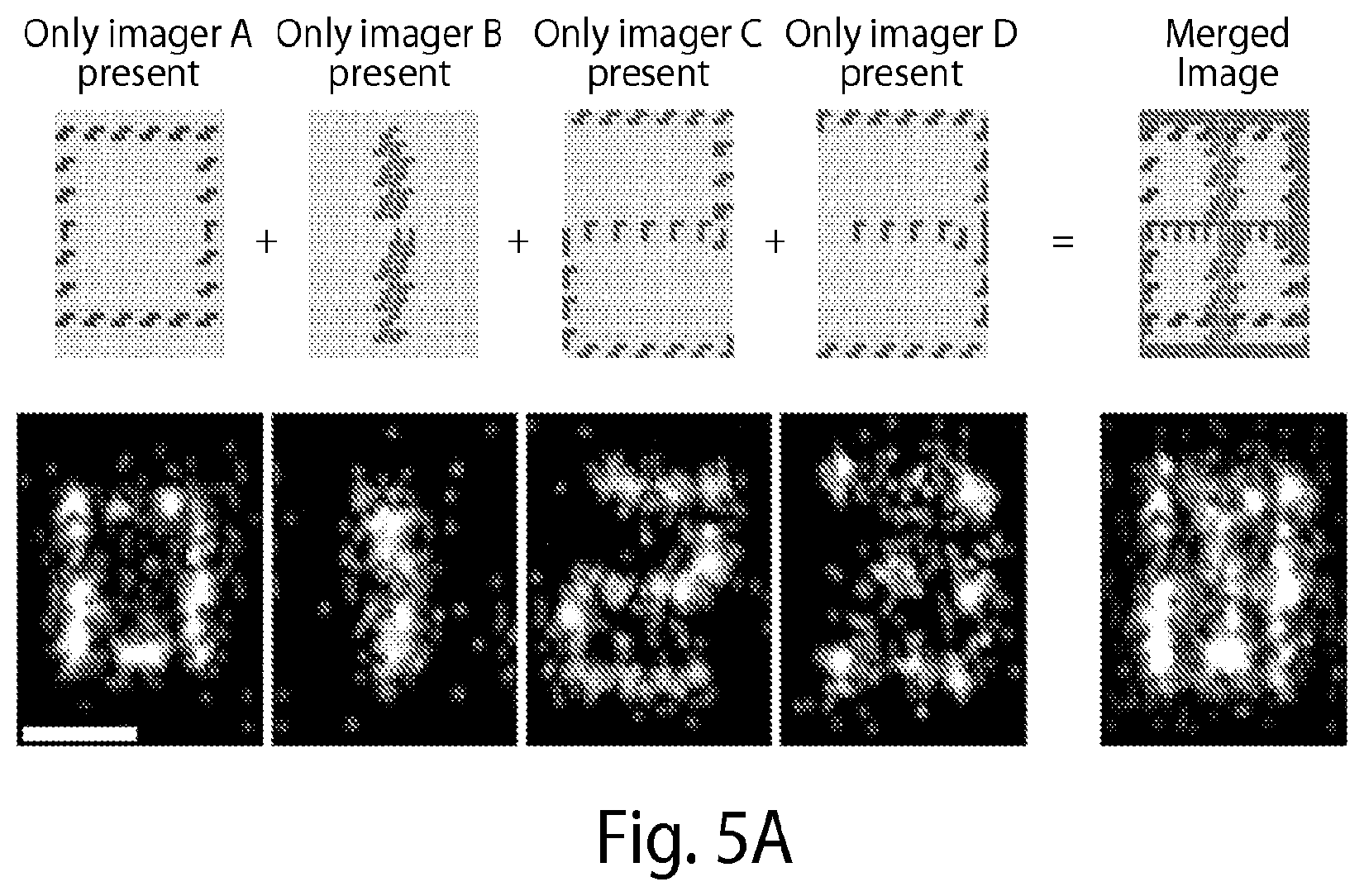
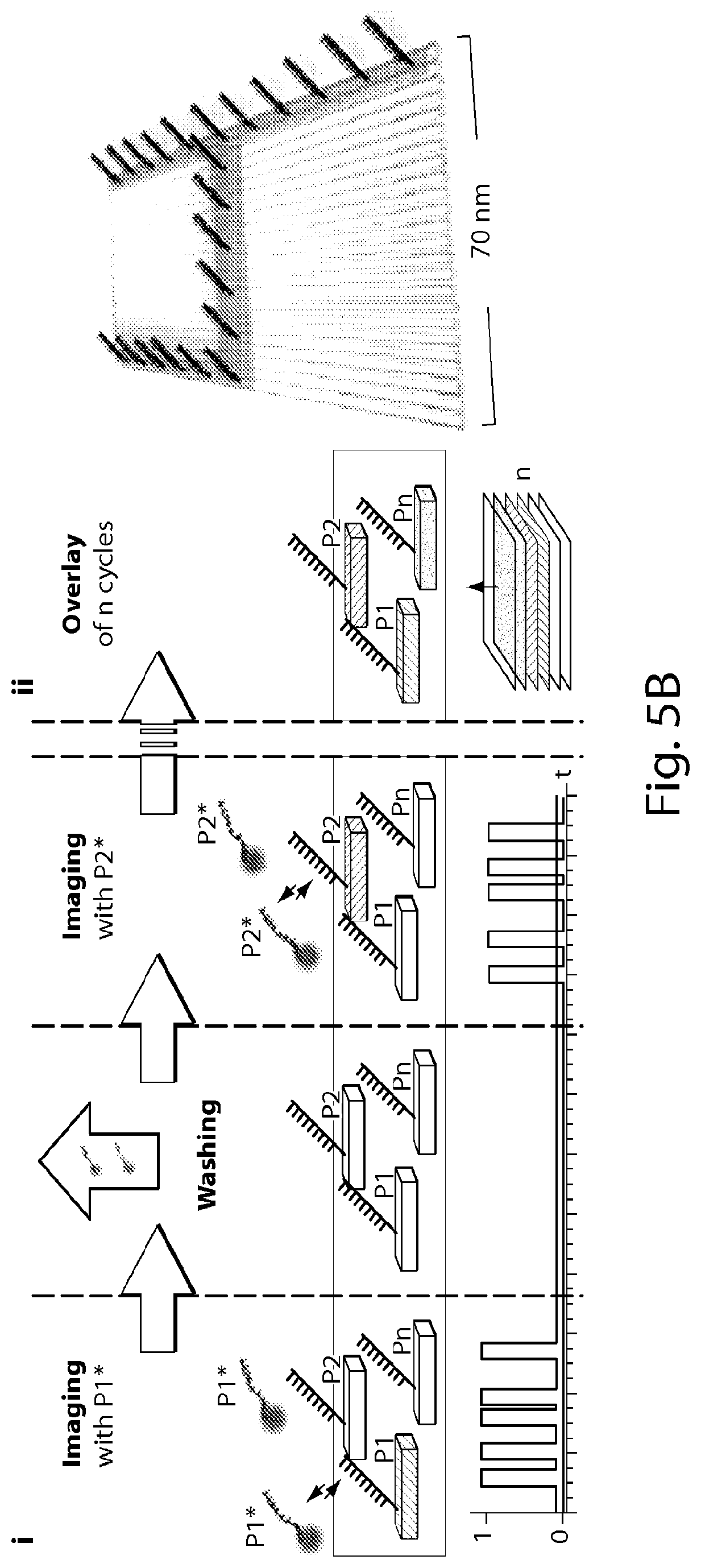
[0135] FIG. 4A shows one embodiment of the present disclosure using spectrally indistinct imager strands (e.g., each labeled with the same color fluorophore). In step [1], three different species of docking strands (a,b,c) label a surface. Such labeling may occur using the docking strand alone or linked to a protein-binding (e.g., antibody) or a nucleic acid-binding molecule that binds to the surface/biomolecule of interest. In step [2], multiple copies of the imager strand a* are introduced (where a* has a sequence complementary to a), and points labeled with docking strands a are imaged. In step [3], copies of the imager strand a* are flushed away, and imager strand b* is introduced to image the b labeled points. Images are obtained, and imager strands b* are washed away. In step [4], c labeled points are imaged in the same manner. In step [5], images from [2-4] are assigned pseudo-colors and combined to create the final image. Although pseudo-colors may be used in the final rendering of the image, all imager strands are actually labeled with the same color dye (e.g., fluorophore). FIGS. 4B(1)-4B(3) show that reducing the image density simultaneously increases the achievable resolution by up to a factor of 2 {square root over (2)} ln 2.apprxeq.2.35. Here, the resolution, previously defined as the FWHM of the reconstructed localizations, can be understood as the standard deviation of the localization as in localization microscopy with sparse points. FIG. 4B(1) shows seven points in a linear geometry spaced at 10 nm (top). Simulated super-resolution data with approximately 14 nm resolution (center). The points cannot be resolved. Cross-sectional histogram data shows a broad peak (bottom). FIGS. 4B(2) and 4B(3) show that imaging every other site allows the localization of individual spots. These localizations can then be combined to form the final image of the full pattern. FIG. 4C shows an image of a DNA origami structure that displays docking strands spaced at 10 nm intervals.
[0136] FIG. 5A shows one embodiment of the present disclosure using DNA origami structures with different species of docking strands at designated positions resembling numbers 0-3 (0, 1, 2 and 3). For each round, the respective imager strand sequence is added to an imaging chamber, image acquisition is carried out and the imager strands are washed out. In each imaging round the designed number is imaged, showing a very sequence-specific interaction with no crosstalk between different rounds [Scale bar: 50 nm]. Note that all imager strands are labeled with the same color dye, though each structure (e.g., 0-3 (0, 1, 2 and 3)) is rendered a distinct color (e.g., purple, yellow, blue, or red; color rendering not shown). FIGS. 5B(i)-(v) show another embodiment of the present disclosure using DNA origami structures with different species of docking strands at designated positions resembling numbers 0-9 (0, 1, 2, 3, 4, 5, 6, 7, 8 and 9). FIG. 5B(i) shows an exchange-PAINT schematic showing sequential imaging of multiple targets using imager strands labeled with the same fluorophore. FIG. 5B(ii) shows a schematic of a DNA origami (70 100 nm) displaying docking strands that resemble digit 4. FIG. 5B(iii) shows a combined overview image of all ten Exchange-PAINT cycles, demonstrating specific interaction with the respective target with no crosstalk between imaging cycles. Scale bar: 250 nm. FIG. 5B(iv) shows a four-"color" image of digits 0 to 3 that are all present on the same DNA origami (10,000 frames each, 5 Hz frame rate; schematic at the bottom). Scale bar: 25 nm. FIG. 5B(v) shows pseudocolor images of ten different origami structures, each rendered a distinct color (e.g., orange, green, blue, purple, pink, etc.; color rendering not shown), displaying digits 0 to 9 in one sample with high resolution (FWHM of bar-like features <10 nm) and specificity. Image obtained using only one fluorophore (Cy3b) through ten imaging-washing cycles (imaging: 7,500 frames per cycle, 5 Hz frame rate; washing: 1-2 minutes per cycle). Scale bar: 25 nm.
[0137] FIG. 6A shows an experimental schematic for one embodiment of the present disclosure using fixed HeLa cells, where in one round, docking strands are bound to a target, labeled imager strands are then added, an image is acquired, and the imager strands are washed away. Each round is repeated with docking strands specific for different targets along with different labeled imager strands. The docking strands may be used alone or linked to a protein-binding (e.g., antibody) or a nucleic acid-binding molecule that binds to the target of interest. FIG. 6B shows two rounds of a method of the present disclosure using Cy3b-labeled imager strands in fixed HeLa cells. Here, microtubules (green pseudo-color; color rendering not shown) were labeled with docking sequence a and mitochondria (magenta pseudo-color; color rendering not shown) with orthogonal docking sequence b. FIG. 6C shows two rounds of a method of the present disclosure using ATT0655-labeled imager strands performed in fixed HeLa cells similar to FIG. 6B. [Scale bars: 5 .mu.m]. Note that all imager strands are labeled with the same color dye.
[0138] FIG. 7A shows that fluorescently-labeled imager strands transiently bind from solution to complementary docking strands on a structure or molecule of interest. The transient binding produces an apparent blinking as shown in the binding vs. time trace with characteristic fluorescence ON- and OFF-times (.tau..sub.b and .tau..sub.d, respectively). The detected binding frequency of imager strands from solution is linearly dependent on the number of available docking strands in a given image area (i.e., the more docking strands, the higher the binding frequency). The time in-between binding events, i.e., the fluorescence OFF-time (rd), is inversely proportional to the number of docking strands. FIG. 7B shows that the average fluorescence OFF-time (.tau..sub.d) can be determined by calculating a cumulative distribution function (CDF) for the OFF-time distribution. Given a known association constant k.sub.on and imager strand concentration c, the number of binding sites can be calculated by: number of binding sites=(.tau..sub.dck.sub.on).sup.-1. FIG. 7C shows a super-resolution image of DNA origami structures designed to display 13 binding sites as a proof-of-concept platform. The incorporation efficiency for docking sites is not 100% leading to a distribution of actually incorporated sites (FIG. 7D(1)). The structures serve as an ideal test system, as the number of displayed docking sites can be determined visually by counting the number of spots (direct counting) and comparing it with the corresponding number of sites calculated using the proposed binding kinetic analysis. FIG. 7D(1) shows the binding site distribution for 377 origami structures obtained by direct visual counting. FIG. 7D(2) shows the binding site distribution for the same structures obtained by binding kinetic analysis (kinetics) of the present disclosure. FIG. 7D(3) shows the "offset" between direct and kinetic counting: the counting "error" or uncertainty for the method of the present disclosure is less than 7% (determined by the coefficient of variation of the Gaussian distribution).
[0139] FIG. 8A shows mRNA molecules of interest in fixed Escherichia coli cells tagged using docking strands in a FISH-like hybridization scheme. FIG. 8B shows a readout scheme used to determine the binding frequency for each imaging color. The intensity vs. time profile of each single mRNA location yields a specific transient binding pattern (blinking) per color. The frequency of binding events depends on the number of binding sites allowing the use of the binding frequency to distinguish between different integer numbers of binding sites. FIG. 8C shows an in vitro proof-of-principle experiment on DNA origami structures displaying 3, 9, 22 and 44 binding sites for each of the red, green, and blue imager strands, respectively (color rendering not shown). The different binding levels are clearly distinguishable for each color, suggesting 4 possible "frequency levels" per color, yielding up to 124 different possible combinations for barcoding, e.g., mRNA molecules inside cells. The barcoding space can be increased by using the fluorescence ON-time as an additional coding entity.
[0140] FIG. 9 shows a graph demonstrating that the fluorescence ON-time (related to the dissociation rate k.sub.off) can be tuned independently of the fluorescence OFF-time (related to the association rate k.sub.on). Extending the imaging/docking duplex from 9 to 10 nucleotides (nt) by adding a single CG base pair, the kinetic OFF-rate is reduced by almost one order of magnitude (8).
[0141] FIG. 10A shows a barcode probe that is roughly 50 nucleotide (nt) in length and can tag a biomolecule using a 21 nt target detection domain t* followed by an approximately 30 nt long "barcode" region with a combination of 8, 9, or 10 nt long binding domain for red, green, or blue imager strands. Here, 8, 9 or 10 nt long docking strands are displayed for three colors with a k.sub.off of 10, 1 and 0.1 per second, respectively (color rendering not shown). FIG. 10B shows characteristic intensity vs. time traces with increased fluorescence ON-times r for the 9 nt interaction domain compared to the 8 nt interaction domain. FIG. 10C shows stochastic simulations demonstrating that k.sub.off values of 10, 1 and 0.1 per second can be distinguished.
[0142] FIG. 11A shows that in the traditional method of detection, where a single fluorophore is stably attached to the imaging surface (see FIG. 11B(1)), a limited number of photons per "switching" event is emitted (top panel), that extraction of all photons from "replenishable" imager strands (see FIG. 11B(2)) leads to higher localization accuracy per switching event (middle panel), and that a DNA metafluorophore (see FIG. 11B(3) and FIG. 11B(4)) yields a significantly larger number of photons per switching event than the single fluorophore in FIG. 11B(2) (bottom). FIGS. 11B(1)-11B(3) show schematics of current imaging methods and methods of the present disclosure. FIG. 11B(1) shows a traditional detection method (e.g., in STORM), which uses a fluorophore stably attached to the imaging surface. FIG. 11B(2) shows one embodiment of a detection method of the present disclosure with fluorophores transiently binding to the imaging surface. FIG. 11B(3) shows a bright metafluorophore with 8 fluorophores in a compact DNA nanostructure. FIG. 11B(4) shows a conditional metafluorophore decorated with both quenchers (dark dots) and fluorophores (stars) that only fluoresces when transiently bound to the surface. FIG. 11C shows a DNA origami structure with docking sites arranged in a 4.times.3 grid, spacing 20 nm. Single sites are optically localized with an accuracy of approximately 3 nm, currently the highest demonstrated resolution [scale bar: 50 nm]. FIG. 11D shows a 280 nm.times.240 nm DNA nano-rectangle (single-stranded tile structure (15), 10.times. larger area than origami) displaying 2000 single-stranded docking strands (dots) with 7 or 5 nm spacing used as a test platform for ultra-resolution imaging [scale bar:100 nm].
[0143] FIG. 12A(i) illustrates schematics showing the principle of each stage of drift correction. In each image, black markers and lines indicate source data, and gray values and curves indicate the calculated drift correction. FIG. 12A(ii) shows a schematic drawing of the major type of drift markers (e.g., DNA drift markers) used in each stage. FIG. 12B(i) illustrates an example structure showing the imaging quality after each stage or correction, and FIG. 12B(ii) shows a zoomed image of the corresponding green rectangle in FIG. 12B(i) at each stage. The scale bars shown in FIGS. 12B(i) and 12B(ii) correspond to 50 nm. FIG. 12C(i) illustrates an example drift trace after each stage of correction, and FIG. 12C(ii) shows a zoomed image of the corresponding rectangle in FIG. 12C(i) at each stage. The scale bars in FIG. 12C(i) correspond to x: 500 nm, t: 500 s, and the scale bars in FIG. 12C(ii) correspond to x: 10 nm, t: 10 s.
[0144] FIG. 13 illustrates a process for performing drift correction in accordance with some embodiments.
[0145] FIG. 14 illustrates a process for performing drift correction corresponding to stage 230 of FIG. 13.
[0146] FIG. 15 illustrates a process for performing drift correction corresponding to stage 240 of FIG. 13.
[0147] FIGS. 16A-16B illustrate 3D tetrahedrons used as templates for 3D drift correction. The four corners are labeled with docking sites. FIG. 16A shows that the four corners are clearly resolved. FIG. 16B illustrates the X-Z projection of the structures with a height of .about.85 nm.
[0148] FIG. 17 shows an illustrative implementation of computer system 600 that may be used in connection with any of the embodiments of the present disclosure described herein.
[0149] FIGS. 18A-18C illustrate an alternative representation of stages in a drift correction process in accordance with some embodiments of the present disclosure. A super-resolved image of a 10 nm-spaced regular grid on a single-molecule DNA origami nanostructure is shown. The DNA origami structure was designed to be a 5.times.8 square lattice of 10 nm spacing both vertically and horizontally. FIG. 18A shows a scatter plot of collected and filtered localizations, represented by crosses. FIG. 18B shows a binned 2-D histogram view of the above structure. FIG. 18C shows a 1-D histogram by projecting all localizations in the rectangle in FIGS. 18A and 18B onto the x-axis, and least square fitting with 8 Gaussian components. The fitted Gaussian peaks all have standard deviation in the range of 1.5-2.4 nm, allowing for 3.5-5.6 nm resolution in principle; and spacing between neighboring peaks in the range of 9.8-11.0 nm, consistent with the DNA origami design. A few (5 in this case) spots are missing in the structure because of imperfect incorporation of staples in the assembly reaction, but not missed during the super-resolution imaging.
[0150] FIGS. 19A and 19B show that RNA aptamers modulate the fluorescence of GFP-like fluorophores. FIG. 19A shows structures of HBI (green), in the context of GFP, and DMHBI. FIG. 19B shows that the 13-2 RNA aptamer enhances the fluorescence of DMHBI by stabilizing a particular molecular arrangement favorable for fluorescence emission. Solutions containing DMHBI, 13-2 RNA, DMHBI with 13-2 RNA, or DMHBI with total HeLa cell RNA were photographed under illumination with 365 nm of light. The image is a montage obtained under identical image-acquisition conditions. (Image from Paige et al., ref. 19)
[0151] FIGS. 20A and 20B show single-molecule fluorescence characterization of the DFHBI binding kinetics. FIG. 20A shows a 5'-extended Spinach (green) is immobilized on a BSA/Biotin-coated glass substrate using a biotinylated DNA capture sequence (labeled with a red dye, e.g. Alexa647; color rendering not shown). FIG. 20B shows a bulk fluorescence measurement of the Spinach-DFHBI before (bottom line) and after (top line) addition of the aptamer shows that the DFHBI binding activity is well maintained after the addition an extension to Spinach required for immobilizing to the glass surface in FIG. 20A.
[0152] FIGS. 21A and 21B show benchmarking Spinach-PAINT performance. FIG. 20A shows a six-helix DNA origami structure used for placing two Spinach molecules in a defined distance. FIG. 20B shows a simulated representation of a super-resolved reconstruction using DNA-PAINT to localize the DNA structure (P) and Spinach-PAINT to localize the Spinach molecules in three different distances.
[0153] FIG. 22 shows Spinach-based sensors. The allosteric variant of the Spinach-based sensor (left) comprises the Spinach domain (black), the transducer module (medium gray), and the recognition module (light gray). In the absence of the target molecule, the transducer module is in a primarily unstructured state, which prevents the stabilization of the Spinach structure needed for activation of DFHBI. Upon binding of the target molecule, the transducer module forms a duplex, leading to structural rigidification of the Spinach module, and activation of DFHBI fluorescence (ref. 24).
[0154] FIGS. 23A and 23B show examples of in vitro and in situ Exchange-PAINT chambers.
DESCRIPTION OF THE INVENTION
[0155] The present disclosure provides, inter alia, methods, compositions and kits for multiplexed imaging, for example, in a cellular environment using nucleic acid-based imaging probes (e.g., DNA-based imaging probes). The methods, compositions and kits for multiplexed fluorescence imaging are not limited by the degree of resolution attained. Thus, the methods, compositions and kits as provided herein may be used for imaging, generally.
[0156] In some aspects, the present disclosure further provides, inter alia, methods, compositions and kits for multiplexed super-resolution fluorescence imaging, for example, in a cellular environment using nucleic acid-based imaging probes (e.g., DNA-based imaging probes). As used herein, "super-resolution" imaging refers to the process of combining a set of low resolution images of the same area to obtain a single image of higher resolution. Many aspects of the present disclosure may be used to "switch" targets (e.g., biomolecules) of interest between fluorescent ON- and OFF-states to permit consecutive, or in some instances simultaneous, localization of individual targets. A fluorescent "ON" state is a state in which fluorescence is emitted. A fluorescent "OFF" state is a state in which fluorescence is not emitted. Switching between the two states is achieved, in some embodiments, with diffusing molecules that are labeled with a detectable label (e.g., fluorescent molecules) that interact transiently with the targets using an intermediate moiety that comprises the detectable label (e.g., fluorescent molecule(s)) and binds to the target. The methods, compositions and kits of the present disclosure are useful, in some aspects, for detecting, identifying and quantifying target targets of interest.
[0157] Binding partner-nucleic acid conjugates ("BP-NA conjugates") of the present disclosure transiently bind to complementary labeled, optionally fluorescently labeled, imager strands. As used herein, "binding partner-nucleic acid conjugate," or "BP-NA conjugate," refers to a molecule linked (e.g., through an N-Hydroxysuccinimide (NHS) linker) to a single-stranded nucleic acid (e.g., DNA) docking strand. The binding partner of the conjugate may be any moiety (e.g., antibody or aptamer) that has an affinity for (e.g., binds to) a target, such as a biomolecule (e.g., protein or nucleic acid), of interest. In some embodiments, the binding partner is a protein. BP-NA-conjugates that comprise a protein (or peptide) linked to a docking strand may be referred to herein as "protein-nucleic acid conjugates," or "protein-NA conjugates." Examples of proteins for use in the conjugates of the present disclosure include, without limitation, antibodies (e.g., monoclonal monobodies), antigen-binding antibody fragments (e.g., Fab fragments), receptors, peptides and peptide aptamers. Other binding partners may be used in accordance with the present disclosure. For example, binding partners that bind to targets through electrostatic (e.g., electrostatic particles), hydrophobic or magnetic (e.g., magnetic particles) interactions are contemplated herein.
[0158] As used herein, "antibody" includes full-length antibodies and any antigen binding fragment (e.g., "antigen-binding portion") or single chain thereof. The term "antibody" includes, without limitation, a glycoprotein comprising at least two heavy (H) chains and two light (L) chains inter-connected by disulfide bonds, or an antigen binding portion thereof. Antibodies may be polyclonal or monoclonal; xenogeneic, allogeneic, or syngeneic; or modified forms thereof (e.g., humanized, chimeric).
[0159] As used herein, "antigen-binding portion" of an antibody, refers to one or more fragments of an antibody that retain the ability to specifically bind to an antigen. The antigen-binding function of an antibody can be performed by fragments of a full-length antibody. Examples of binding fragments encompassed within the term "antigen-binding portion" of an antibody include (i) a Fab fragment, a monovalent fragment consisting of the V.sub.H, V.sub.L, C.sub.L and C.sub.H1 domains; (ii) a F(ab')2 fragment, a bivalent fragment comprising two Fab fragments linked by a disulfide bridge at the hinge region; (iii) a Fd fragment consisting of the V.sub.H and C.sub.H1 domains; (iv) a Fv fragment consisting of the V.sub.H and V.sub.L domains of a single arm of an antibody, (v) a dAb fragment (Ward et al., Nature 341:544 546, 1989), which consists of a V.sub.H domain; and (vi) an isolated complementarity determining region (CDR) or (vii) a combination of two or more isolated CDRs, which may optionally be joined by a synthetic linker. Furthermore, although the two domains of the Fv fragment, V.sub.H and V.sub.L, are coded for by separate genes, they can be joined, using recombinant methods, by a synthetic linker that enables them to be made as a single protein chain in which the V.sub.H and V.sub.L regions pair to form monovalent molecules (known as single chain Fv (scFv); see, e.g., Bird et al. Science 242:423 426, 1988; and Huston et al. Proc. Natl. Acad. Sci. USA 85:5879-5883, 1988). Such single chain antibodies are also encompassed within the term "antigen-binding portion" of an antibody. These antibody fragments are obtained using conventional techniques known to those with skill in the art, and the fragments are screened for utility in the same manner as are intact antibodies.
[0160] As used herein, "receptors" refer to cellular-derived molecules (e.g., proteins) that bind to ligands such as, for example, peptides or small molecules (e.g., low molecular weight (<900 Daltons) organic or inorganic compounds).
[0161] As used herein, "peptide aptamer" refers to a molecule with a variable peptide sequence inserted into a constant scaffold protein (see, e.g., Baines I C, et al. Drug Discov. Today 11:334-341, 2006).
[0162] In some embodiments, the molecule of the BP-NA conjugate is a nucleic acid such as, for example, a nucleic acid aptamer. As used herein, "nucleic acid aptamer" refers to a small RNA or DNA molecules that can form secondary and tertiary structures capable of specifically binding proteins or other cellular targets (see, e.g., Ni X, et al. Curr Med Chem. 18(27): 4206-4214, 2011). Thus, in some embodiments, the BP-NA conjugate may be an aptamer-nucleic acid conjugate.
[0163] As used herein a "docking strand" refers to a single-stranded nucleic acid (e.g., DNA) that is about 5 nucleotides to about 50 nucleotides in length (or is 5 nucleotides to 50 nucleotides in length). In some embodiments, a docking strand is about 4 to about 60, about 6 nucleotides to about 40 nucleotides, about 7 nucleotides to about 30 nucleotides, about 8 to about 20 nucleotides, or about 9 nucleotides to about 15 nucleotides in length. In some embodiments, a docking strand is (or is about) 4, 5, 6, 7, 8, 9, 10, 11, 12, 13, 14, 15, 16, 17, 18, 19, 20 21, 22, 23, 24, 25, 26, 27, 28, 29, 30, 31, 32, 33, 34, 35, 36, 37, 38, 39, 40, 41, 42, 43, 44, 45, 46, 47, 48, 49, 50, 51, 52, 53, 54, 55, 56, 57, 58, 59, 60, or more nucleotides in length.
[0164] A docking strand may have one domain or more than one domain (i.e., a plurality of domains), each domain complementary to a respective imager strand. As used herein, a "docking strand domain" refers to a nucleotide sequence of the docking strand that is complementary to a nucleotide sequence of an imager strand. A docking strand, for example, may contain one, two three, or more domains, each domain complementary to an imager strand. Each complementary imager strand can contain a distinct label (e.g., a red fluorophore, a blue fluorophore, or a green fluorophore), or all complementary imager strands can contain the same label (e.g., red fluorophores). For example, for a three-domain docking strand, the strand may contain a first domain complementary to an imager strand labeled with a red fluorophore, a second domain complementary to an imager strand labeled with a blue fluorophore, and a third domain complementary to an imager strand labeled with a green fluorophore. Alternatively, each of three docking domains may be complementary to imager strands labeled with a red fluorophore. In some embodiments, a docking strand has at least 2, at least 3, at least 4, at least 5, or more domains, each respectively complementary to an imager strand. In some embodiments, a docking strand has 1 to 5, 1 to 10, 1 to 15, 1 to 20, 1 to 25, 1 to 50, or 1 to 100 domains, each respectively complementary to an imager strand.
[0165] As used herein, an "imager strand" is a single-stranded nucleic acid (e.g., DNA) that is about 4 to about 30 nucleotides, about 5 to about 18 nucleotides, about 6 to about 15 nucleotides, about 7 to about 12 nucleotides, or about 8 to 10 nucleotides in length and is fluorescently-labeled. In some embodiments, the imager strand may be (or may be about) 4, 5, 6, 7, 8, 9, 10, 11, 12, 13, 14, 15, 16, 17, 18, 19, 20, 21, 22, 23, 24, 25, 26, 27, 28, 29 or 30 nucleotides in length. An imager strand of the present disclosure is complementary to and transiently binds to a docking strand. Two nucleic acids or nucleic acid domains are "complementary" to one another if they base-pair, or bind, with each other to form a double-stranded nucleic acid molecule via Watson-Crick interactions. As used herein, "binding" refers to an association between at least two molecules due to, for example, electrostatic, hydrophobic, ionic and/or hydrogen-bond interactions under physiological conditions. An imager strand is considered to "transiently bind" to a docking strand if it binds to a complementary region of a docking strand and then disassociates (unbinds) from the docking strand within a short period of time, for example, at room temperature. In some embodiments, an imager strand remains bound to a docking strand for about 0.1 to about 10, or about 0.1 to about 5 seconds. For example, an imager strand may remain bound to a docking strand for about 0.1, about 1, about 5 or about 10 seconds.
[0166] Imager strands of the present disclosure may be labeled with a detectable label (e.g., a fluorescent label, and thus are considered "fluorescently labeled"). For example, in some embodiments, an imager strand may comprise at least one (i.e., one or more) fluorophore. Examples of fluorophores for use in accordance with the present disclosure include, without limitation, xanthene derivatives (e.g., fluorescein, rhodamine, Oregon green, eosin and Texas red), cyanine derivatives (e.g., cyanine, indocarbocyanine, oxacarbocyanine, thiacarbocyanine and merocyanine), naphthalene derivatives (e.g., dansyl and prodan derivatives), coumarin derivatives, oxadiazole derivatives (e.g., pyridyloxazole, nitrobenzoxadiazole and benzoxadiazole), pyrene derivatives (e.g., cascade blue), oxazine derivatives (e.g., Nile red, Nile blue, cresyl violet and oxazine 170), acridine derivatives (e.g., proflavin, acridine orange and acridine yellow), arylmethine derivatives (e.g., auramine, crystal violet and malachite green), and tetrapyrrole derivatives (e.g., porphin, phthalocyanine and bilirubin). Other detectable labels may be used in accordance with the present disclosure, such us, for example, gold nanoparticles or other detectable particles or moieties.
[0167] As used herein, "spectrally distinct" molecules of the present disclosure (e.g., conjugates and/or imager strands) refer to molecules with labels (e.g., fluorophores) of different spectral signal or wavelength. For example, an imager strand labeled with a Cy2 fluorophore emits a signal at a wavelength of light of about 510 nm, while an imager strand labeled with a Cy5 fluorophore emits a signal at a wavelength of light of about 670 nm. Thus, the Cy2-labeled imager strand is considered herein to be spectrally distinct from the Cy5-labeled imager strand. Conversely, "spectrally indistinct" molecules of the present disclosure herein refer to molecules with labels having the same spectral signal or wavelength--that is, the emission wavelength of the labels cannot be used to distinguish between two spectrally indistinct fluorescently labeled molecules (e.g., because the wavelengths are the same or close together).
[0168] The BP-NA conjugates (e.g., protein-nucleic acid conjugates) of the present disclosure may, in some embodiments, comprise an intermediate linker that links (e.g., covalently or non-covalently) the molecule to a docking strand. The intermediate linker may comprise biotin and/or streptavidin. For example, in some embodiments, an antibody and a docking strand may each be biotinylated (i.e., linked to at least one biotin molecule) and linked to each other through biotin binding to an intermediate streptavidin molecule, as shown in FIG. 2. Other intermediate linkers may be used in accordance with the present disclosure. In some embodiments, such as those where the molecule is a nucleic acid, an intermediate linker may not be required. For example, the docking strand of a BP-NA conjugate may be an extension (e.g., 5' or 3' extension) of a nucleic acid molecule such as, for example, a nucleic acid aptamer.
[0169] Pluralities of BP-NA conjugates (e.g., protein-nucleic acid conjugates) and imager strands are provided herein. A plurality may be a population of the same species or distinct species. A plurality of BP-NA conjugates of the same species may comprise conjugates that all bind to the same target (e.g., biomolecule) (e.g., the same epitope or region/domain). Conversely, a plurality of BP-NA conjugates of distinct species may comprise conjugates, or subsets of conjugates, each conjugate or subset of conjugates binding to a distinct epitope on the same target or to a distinct target. A plurality of imager strands of the same species may comprise imager strands with the same nucleotide sequence and the same fluorescent label (e.g., Cy2, Cy3 or Cy4). Conversely, a plurality of imager strands of distinct species may comprise imager strands with distinct nucleotide sequences (e.g., DNA sequences) and distinct fluorescent labels (e.g., Cy2, Cy3 or Cy4) or with distinct nucleotide sequences and the same fluorescent (e.g., all Cy2). The number of distinct species in a given plurality of BP-NA conjugates is limited by the number of binding partners (e.g., antibodies) and the number of docking strands of different nucleotide sequence (and thus complementary imager strands). In some embodiments, a plurality of BP-NA conjugates (e.g., protein-nucleic acid conjugates) comprises at least 10, 50, 100, 500, 1000, 2000, 3000, 4000, 5000, 10.sup.4, 50000, 10.sup.5, 10.sup.5, 10.sup.6, 10.sup.7, 10.sup.8, 10.sup.9, 10.sup.10, 10.sup.11 BP-NA conjugates. Likewise, in some embodiments, a plurality of fluorescently-labeled imager strands comprises at least 10, 50, 100, 500, 1000, 2000, 3000, 4000, 5000, 10.sup.4, 50000, 10.sup.5, 10.sup.5, 10.sup.6, 10.sup.7, 10.sup.8, 10.sup.9, 10.sup.10, 10.sup.11 fluorescently-labeled imager strands. In some embodiments, a plurality may contain 1 to about 200 or more distinct species of BP-NA conjugates and/or imager strands. For example, a plurality may contain at least 1, 2, 3, 4, 5, 6, 7, 8, 9, 10, 15, 20, 25, 30, 35, 40, 45, 50, 55, 60, 65, 70, 75, 80, 85, 90, 95, 100, 125, 150, 175, 200 or more distinct species. In some embodiments, a plurality may contain less than about 5 to about 200 distinct species of BP-NA conjugates and/or imager strands. For example, a plurality may contain less than 5, 6, 7, 8, 9, 10, 15, 20, 25, 30, 35, 40, 45, 50, 55, 60, 65, 70, 75, 80, 85, 90, 95, 100, 125, 150, 175 or 200 distinct species.
[0170] The present disclosure also contemplates docking strands that can bind directly to a target. For example, as shown in FIG. 8A, a docking strand may contain, in additional to imager-binding domain(s) (e.g., one, two, three, or more, with the same or distinct fluorophores), a target domain that is complementary to and binds to a target, such as, for example, mRNA or other nucleic acid.
Methods
[0171] Methods provided herein are based, in part, on the programmability of nucleic acid docking strands and imager strands. That is, for example, docking strands and imager strands can be designed such that they bind to each other under certain conditions for a certain period of time. This programmability permits transient binding of imager strands to docking strands, as provided herein. Generally, the methods provided herein are directed to identifying one or more target(s) (e.g., biomolecule(s) such as a protein or nucleic acid) in a particular sample (e.g., biological sample). In some instances, whether or not one or more target(s) is present in sample is unknown. Thus, methods of the present disclosure may be used to determine the presence or absence of one or more target(s) in a sample suspected of containing the target(s). In any one of the aspects and embodiments provided herein, a sample may contain or may be suspected of containing one or more target(s).
[0172] Methods provided herein can also be used to identify the absolute quantity of a single target (e.g., such as, for example, a particular protein), or the quantity of a single target relative to one or more other targets.
[0173] Further, methods provided herein may be used to identify the location of a target within a sample or relative to other targets in the sample.
[0174] Methods provided herein may comprise, in some embodiments, contacting a sample with (a) at least one BP-NA conjugate (e.g., protein-nucleic acid conjugate) that comprises a binding partner linked to a docking strand and (b) at least one labeled, optionally fluorescently labeled, imager strand that is complementary to and transiently binds to the docking strand of the at least one BP-NA conjugate, and then determining whether the at least one BP-NA conjugate binds to at least one target (such as a biomolecule target) in the sample. In some embodiments, the determining step comprises imaging (e.g., with time-lapsed fluorescent microscopy techniques) transient binding of the at least one labeled, optionally fluorescently labeled, imager strand to the docking strand of the at least one BP-NA conjugate.
[0175] Other methods provided herein may comprise, in some embodiments, contacting a sample with (a) at least two BP-NA conjugates, each comprising a binding partner linked to a docking strand, and (b) at least two labeled, optionally spectrally distinct, fluorescently labeled, imager strands that are complementary to and transiently bind to respective docking strands of the at least two different BP-NA conjugates, and then determining whether the at least two BP-NA conjugates bind to at least one, or at least two, targets (such as biomolecule targets) in the sample. Binding of the BP-NA conjugates to respective targets can be determined by imaging transient binding of one of the at least two labeled, optionally spectrally distinct, fluorescently labeled, imager strands to a docking strand of one of the at least two BP-NA conjugates to produce a first image, and then imaging transient binding of another of the at least two labeled, optionally spectrally distinct, fluorescently labeled, imager strands to a docking strand of another of the at least two BP-NA conjugates to produce a second image. In some embodiments, the methods further comprise combining the first image and the second image to produce a composite image of signals (e.g., fluorescent signals), wherein the signals (e.g., fluorescent signals) of the composite image are representative of the at least two targets. As used herein, a "composite image" refers to a single image produced by combining (e.g., overlaying) multiple images of the same (or substantially similar) area. A composite image may also be referred to as a super-resolution image, as described elsewhere herein.
[0176] FIG. 3 demonstrates one embodiment of the present disclosure in which two distinct species of BP-NA conjugates (e.g., antibody-nucleic acid conjugates) are used to label biomolecules in a fixed HeLa cell sample. One species of antibody-nucleic acid conjugate comprises an antibody that recognizes and binds to an epitope on mitochondria. The mitochondrial specific antibody is linked to a docking strand with a sequence complementary to a Cy3b-labeled imager strand. The other species of antibody-nucleic acid conjugate comprises an antibody that recognizes and binds to an epitope on microtubules. The microtubule specific antibody is linked to a docking strand with a sequence complementary to an ATT0655-labeled imager strand. Two spectrally distinct species of imager strands are then introduced at the same time: one species is labeled with Cy3b and is complementary to the docking strand that is linked to the mitochondrial specific antibody, and the other species is labeled with ATT0655 and is complementary to the docking strand that is linked to the microtubule specific antibody. While both the Cy3b-labeled imager strand and the ATT0655-labeled imager strand are present at the same time in solution with the sample, imaging is carried our sequentially in Cy3b and ATT0655 channels.
[0177] Yet other methods provided herein may comprise, in some embodiments, contacting a sample with (a) at least two BP-NA conjugates, each comprising a protein linked to a docking strand and (b) at least two spectrally indistinct (e.g., labeled with the same fluorophore) fluorescently-labeled imager strands that are complementary to and transiently bind to respective docking strands of the at least two BP-NA conjugates, and then determining whether the at last two BP-NA conjugates bind to at least two targets (e.g., biomolecule targets) in the sample. In some embodiments, the methods comprise, in the following ordered steps, contacting the sample with a first BP-NA conjugate and at least one other BP-NA conjugate, contacting the sample with a first fluorescently-labeled imager strand that is complementary to and transiently binds to the docking strand of the first BP-NA conjugate, determining whether the first BP-NA conjugate binds to a first target, removing the first fluorescently-labeled imager strand, contacting the sample with at least one other fluorescently-labeled imager strand that is complementary to and transiently binds to the docking strand of the at least one other BP-NA conjugate, and determining whether the at least one other BP-NA conjugate binds to at least one other target.
[0178] Alternatively, in other embodiments, methods comprise, in the following ordered steps, contacting the sample with a first BP-NA conjugate, contacting the sample with a first fluorescently-labeled imager strand that is complementary to and transiently binds to the docking strand of the first BP-NA conjugate, determining whether the first BP-NA conjugate binds to a first target (e.g., biomolecule), removing the first fluorescently-labeled imager strand, contacting the sample with at least one other BP-NA conjugate, contacting the sample with at least one other fluorescently-labeled imager strand that is complementary to and transiently binds to the docking strand of the at least one other BP-NA conjugate, and determining whether the at least one other BP-NA conjugate binds to at least one other target.
[0179] In some embodiments, the first determining step comprises imaging transient binding of the first fluorescently-labeled imager strand to the docking strand of the first BP-NA conjugate to produce a first image, and the second determining step comprises imaging transient binding of the at least one other fluorescently-labeled imager strand to the docking strand of the at least one other BP-NA conjugate to produce a second image. In some embodiments, the methods further comprise assigning a pseudo-color to the fluorescent signal in the first image, and assigning at least one other pseudo-color to the fluorescent signal in the second image. Further still, in some embodiments, the methods comprise combining the first image and the second image to produce a composite image of the pseudo-colored signals, wherein the pseudo-colored signals of the composite image are representative of the at least two targets (e.g., biomolecule targets). As illustrated in FIG. 4A, step [1], three distinct species of docking strands (a,b,c) label the surface of a grid (chosen for illustrative purposes). In step [2], multiple copies of the imager strand a* are introduced, and points labeled with docking strands a are imaged. In step [3], copies of the imager strand a* are flushed away, and imager strand b* is introduced to image the b labeled points. In step [4], c labeled points are imaged in the same manner. In step [5], images from steps [2-4] are assigned artificial pseudo-colors (e.g., using a software program) and combined to create the final composite image. All imager strands may be labeled with the same fluorophore--that is, the imager strands are spectrally indistinct. In some embodiments, the docking strands are linked to binding partners (e.g., proteins such as antibodies, or nucleic acids such as DNA or nucleic acid aptamers).
[0180] An advantage of the methods of the present disclosure is that partitioning and sequential imaging can be used to obtain multiplexed super-resolved images of up to hundreds of different species using only a single optimized fluorescent dye. Using these methods, the number of distinct nucleotide sequences (e.g., DNA sequences), as opposed to the number of spectrally distinct dyes, limits the multiplexing capability. In some methods of the present disclosure, for example, those that use an imager strand with a length of 9 nucleotides, there are several hundred species within tight bounds for binding kinetics that may be used for a single sample, representing a tremendous increase in multiplexing compared to direct "traditional" imaging approaches.
[0181] FIG. 5A illustrates another embodiment of the present disclosure using spectrally indistinct imager strands. A single DNA nanostructure displays four distinct species of docking strands (optionally linked to protein binding partners or nucleic acid binding partners) designed to resemble the digits from 0 to 3, respectively. Imaging is performed sequentially using a simple flow chamber setup, first flushing in fluorescently-labeled imager strands complementary to the docking strands of the number 0, and then exchanging the solution for fluorescently-labeled imager strands with a sequence complementary to docking strands of the number 1, and so forth. The resulting images have been pseudo-colored to represent the respective imaging cycles. As used herein, an "imaging cycle," or "imaging round" refers to the process of introducing fluorescently-labeled imager strands complementary to docking strands under conditions that allow the imager strand to bind to the docking strand, even if such binding is transient, and obtaining an image (or imaging an area).
[0182] Aspects of the present disclosure contemplate multiplex detection using multi-domain docking strands (e.g., docking strands with more than one domain), as described above. For illustrative purposes, the following embodiments are described in terms of a docking strand binding to a target, e.g., without an intermediate binding partner. It should be understood, however, that multi-domain docking strands may be linked to a binding partner (e.g., of a BP-NA conjugate, as provided herein.
[0183] In some embodiments, methods comprise contacting one or more target(s) with one or more docking strands, each containing two or more binding domains. In other embodiments, methods comprise contacting one or more target(s) with two or more docking strands, each containing one binding domain. The docking strand domains may have orthogonal sequences. In the examples that follow, all three targets (protein #1-#3) are present in the sample.
[0184] Detection Based on Spectral Resolution.
[0185] An exemplary multiplexed target detection method follows. A sample contains, or is suspected of containing, three target species--protein #1, protein #2, and protein #3. Three docking strands are designed such that: the first, containing imager binding domain A, binds to protein #1; the second, containing imager binding domain B, binds to protein #2; and the third, containing imager binding domains A and B, binds to protein #3. Complementary imager strand A' binds to imager binding domain A of a docking strand and is labeled with a blue fluorophore, and imager strand B' binds to imager binding domain B of a docking strand and is labeled with a red fluorophore. The sample is first contacted with the docking strands, and subsequently contacted with the imager strands A' and B'. The sample is then imaged. The sample containing the docking strands and the imager strands is first imaged under conditions that detect the blue fluorophore. Imaging the blue fluorophore detects protein #1 and protein #3, each bound by an imager strand labeled with the blue fluorophore. Imaging the red fluorophore detects protein #2 and protein #3, each bound by an imager strand labeled with the red fluorophore. Overlapping images of the red and blue fluorophores detects protein #3 only, the only protein bound by an imager strand labeled with a red fluorophore and an imager strand labeled with a blue fluorophore. Thus, the identification and location of proteins #1-#3 are identified by the overlay of images respectively detecting the red and blue fluorophores.
[0186] Detection Based on Exchange of Imager Strands.
[0187] Another exemplary multiplexed target detection method follows: A sample contains, or is suspected of containing, three target species--protein #1, protein #2, and protein #3. Three docking strands are designed such that: the first, containing imager binding domain A, binds to protein #1; the second, containing imager binding domain B, binds to protein #2; and the third, containing imager binding domains A and B, binds to protein #3. Complementary imager strand A' binds to imager binding domain A of a docking strand and is labeled with a blue fluorophore, and imager strand B' binds to imager binding domain B of a docking strand and is also labeled with a blue fluorophore. The sample is first contacted with the docking strands, and subsequently contacted with imager strands A'. The sample is then imaged under conditions that detect the blue fluorophore. Imaging the blue fluorophore detects protein #1 and protein #3, each bound by imager strand A' labeled with the blue fluorophore. The sample is then washed to remove imager strands A'. Next, the sample is contacted with imager strands B'. The sample is then imaged again under conditions that detect the blue fluorophore. Imaging the blue fluorophore now detects protein #2 and protein #3, each bound by imager strand B' labeled with the blue fluorophore. Overlapping images of the blue fluorophores (e.g., resulting in a stronger signal relative to non-overlapping fluorophores) detects protein #3 only, the only protein bound by two imager strands labeled with a blue fluorophore. Thus, the identification and location of proteins #1-#3 are identified by the overlay of images detecting the blue fluorophores and is based on signal intensity.
[0188] Detection Based on a Combination of Spectral and Exchange Detection.
[0189] Yet another exemplary multiplexed target detection method follows: A sample contains, or is suspected of containing, fifteen target species. The docking strands are designed such that each target species binds to a docking strand, each docking strand containing a single distinct domain or a distinct combination of domains A-D (e.g., A, or A and B (i.e., A/B), or A/C, or A/D, or A/B/C, or A/B/D, or A/C/D, or A/B/C/D, or B, or B/C, or B/D, or B/C/D, or C, or C/D, or D). The imager strands are divided into two sets: the first set (set #1) contains imager strand A' labeled by a red fluorophore and imager strand B' labeled by a blue fluorophore; the second set contains imager strand C' labeled with a red fluorophore and imager strand D' labeled with a blue fluorophore. The sample is first contacted with the docking strands, and subsequently contacted with imager strand set #1. The sample is then imaged under conditions that detect blue and red fluorophores. Targets bound by imager strands A' will be detected red and targets bound by imager strands B' will be detected blue. Thus, all target species with docking domains A and B will be detected in a first image or first set of images. The sample is then washed to remove imager strand set #1. Next, the sample is contacted with imager strand set #2. The sample is then imaged again under conditions that detect blue and red fluorophores. Targets bound by imager strands C' will be detected red and targets bound by imager strands D' will be detected blue. Thus, all target species with docking domains C and D will be detected in a second image or second set of images. By combining all images collected, each of the 15 target species can be identified using only four imager strands and two fluorophores. It should be understood that more than four imager strands can be used as well as more than two fluorophores, depending on, for example, the number of targets.
[0190] Detection Based on Duration of Transient Binding.
[0191] In some embodiments, the disclosure contemplates contacting target species with different docking strands domain sequence and different lengths of those sequences. The length of a docking strand imager binding domain affects the duration of transient binding to an imager strand. Docking strands with longer binding domains bind to respectively complementary imager strands for longer durations relative to shorter binding domains. In the following exemplary embodiment, a sample contains, or is suspected of containing, four target species. Four docking strands are designed such that: the first, containing binding domain A of 10 nucleotides in length (A10), binds to protein #1; the second, containing imager binding domain A10 and imager binding domain B of 8 nucleotides in length (B8), binds to protein #2; the third, containing imager binding domain A of 8 nucleotides in length (A8) and imager binding domain B of 10 nucleotides in length (B10), binds to protein #3; and four, containing imager strand binding domain B10. Imager strand A' is 10 nucleotides in length, binds to both A8 and A10, and is labeled with a blue fluorophore. Imager strand B' is 10 nucleotides in length, binds to both B8 and B10, and is labeled with a red fluorophore. The sample is first contacted with the docking strands, and subsequently contacted with imager strands A' and B'. The sample is then imaged under conditions that detect the blue fluorophore. Imaging the blue fluorophore detects protein #3 and protein #4 with a longer bound time (i.e., time of binding between imager strand and docking strand) and protein #2 with a shorter bound time. Imaging the red fluorophore detects protein #1 and protein #2 with a longer bound time and protein #3 with a shorter bound time. Overlapping images of the blue and red fluorophores detects each of the four protein targets.
[0192] The present disclosure also contemplates combining multiplexed detection based on spectral resolution and duration, exchange and duration, and spectral resolution, exchange and duration.
[0193] A "sample" may comprise cells (or a cell), tissue, or bodily fluid such as blood (serum and/or plasma), urine, semen, lymphatic fluid, cerebrospinal fluid or amniotic fluid. A sample may be obtained from (or derived from) any source including, without limitation, humans, animals, bacteria, viruses, microbes and plants. In some embodiments, a sample is a cell lysate or a tissue lysate. A sample may also contain mixtures of material from one source or different sources. A sample may be a spatial area or volume (e.g., a grid on an array, or a well in a plate or dish). A sample, in some embodiments, includes target(s), BP-NA conjugate(s) and imager strand(s).
[0194] A "target" is any moiety that one wishes to observe or quantitate and for which a binding partner exists. A target, in some embodiments, may be non-naturally occurring. The target, in some embodiments, may be a biomolecule. As used herein, a "biomolecule" is any molecule that is produced by a living organism, including large macromolecules such as proteins, polysaccharides, lipids and nucleic acids (e.g., DNA and RNA such as mRNA), as well as small molecules such as primary metabolites, secondary metabolites, and natural products. Examples of biomolecules include, without limitation, DNA, RNA, cDNA, or the DNA product of RNA subjected to reverse transcription, A23187 (Calcimycin, Calcium Ionophore), Abamectine, Abietic acid, Acetic acid, Acetylcholine, Actin, Actinomycin D, Adenosine, Adenosine diphosphate (ADP), Adenosine monophosphate (AMP), Adenosine triphosphate (ATP), Adenylate cyclase, Adonitol, Adrenaline, epinephrine, Adrenocorticotropic hormone (ACTH), Aequorin, Aflatoxin, Agar, Alamethicin, Alanine, Albumins, Aldosterone, Aleurone, Alpha-amanitin, Allantoin, Allethrin, .alpha.-Amanatin, Amino acid, Amylase, Anabolic steroid, Anethole, Angiotensinogen, Anisomycin, Antidiuretic hormone (ADH), Arabinose, Arginine, Ascomycin, Ascorbic acid (vitamin C), Asparagine, Aspartic acid, Asymmetric dimethylarginine, Atrial-natriuretic peptide (ANP), Auxin, Avidin, Azadirachtin A--C35H44016, Bacteriocin, Beauvericin, Bicuculline, Bilirubin, Biopolymer, Biotin (Vitamin H), Brefeldin A, Brassinolide, Brucine, Cadaverine, Caffeine, Calciferol (Vitamin D), Calcitonin, Calmodulin, Calmodulin, Calreticulin, Camphor--(C10H16O), Cannabinol, Capsaicin, Carbohydrase, Carbohydrate, Carnitine, Carrageenan, Casein, Caspase, Cellulase, Cellulose--(C6H10O5), Cerulenin, Cetrimonium bromide (Cetrimide)--C19H42BrN, Chelerythrine, Chromomycin A3, Chaparonin, Chitin, .alpha.-Chloralose, Chlorophyll, Cholecystokinin (CCK), Cholesterol, Choline, Chondroitin sulfate, Cinnamaldehyde, Citral, Citric acid, Citrinin, Citronellal, Citronellol, Citrulline, Cobalamin (vitamin B12), Coenzyme, Coenzyme Q, Colchicine, Collagen, Coniine, Corticosteroid, Corticosterone, Corticotropin-releasing hormone (CRH), Cortisol, Creatine, Creatine kinase, Crystallin, .alpha.-Cyclodextrin, Cyclodextrin glycosyltransferase, Cyclopamine, Cyclopiazonic acid, Cysteine, Cystine, Cytidine, Cytochalasin, Cytochalasin E, Cytochrome, Cytochrome C, Cytochrome c oxidase, Cytochrome c peroxidase, Cytokine, Cytosine--C4H5N3O, Deoxycholic acid, DON (DeoxyNivalenol), Deoxyribofuranose, Deoxyribose, Deoxyribose nucleic acid (DNA), Dextran, Dextrin, DNA, Dopamine, Enzyme, Ephedrine, Epinephrine--C9H13NO3, Erucic acid--CH3(CH2)7CH.dbd.CH(CH2)11COOH, Erythritol, Erythropoietin (EPO), Estradiol, Eugenol, Fatty acid, Fibrin, Fibronectin, Folic acid (Vitamin M), Follicle stimulating hormone (FSH), Formaldehyde, Formic acid, Formnoci, Fructose, Fumonisin B1, Gamma globulin, Galactose, Gamma globulin, Gamma-aminobutyric acid, Gamma-butyrolactone, Gamma-hydroxybutyrate (GHB), Gastrin, Gelatin, Geraniol, Globulin, Glucagon, Glucosamine, Glucose--C6H12O6, Glucose oxidase, Gluten, Glutamic acid, Glutamine, Glutathione, Gluten, Glycerin (glycerol), Glycine, Glycogen, Glycolic acid, Glycoprotein, Gonadotropin-releasing hormone (GnRH), Granzyme, Green fluorescent protein, Growth hormone, Growth hormone-releasing hormone (GHRH), GTPase, Guanine, Guanosine, Guanosine triphosphate (+GTP), Haptoglobin, Hematoxylin, Heme, Hemerythrin, Hemocyanin, Hemoglobin, Hemoprotein, Heparan sulfate, High density lipoprotein, HDL, Histamine, Histidine, Histone, Histone methyltransferase, HLA antigen, Homocysteine, Hormone, human chorionic gonadotropin (hCG), Human growth hormone, Hyaluronate, Hyaluronidase, Hydrogen peroxide, 5-Hydroxymethylcytosine, Hydroxyproline, 5-Hydroxytryptamine, Indigo dye, Indole, Inosine, Inositol, Insulin, Insulin-like growth factor, Integral membrane protein, Integrase, Integrin, Intein, Interferon, Inulin, Ionomycin, Ionone, Isoleucine, Iron-sulfur cluster, K252a, K252b, KT5720, KT5823, Keratin, Kinase, Lactase, Lactic acid, Lactose, Lanolin, Lauric acid, Leptin, Leptomycin B, Leucine, Lignin, Limonene, Linalool, Linoleic acid, Linolenic acid, Lipase, Lipid, Lipid anchored protein, Lipoamide, Lipoprotein, Low density lipoprotein, LDL, Luteinizing hormone (LH), Lycopene, Lysine, Lysozyme, Malic acid, Maltose, Melatonin, Membrane protein, Metalloprotein, Metallothionein, Methionine, Mimosine, Mithramycin A, Mitomycin C, Monomer, Mycophenolic acid, Myoglobin, Myosin, Natural phenols, Nucleic Acid, Ochratoxin A, Oestrogens, Oligopeptide, Oligomycin, Orcin, Orexin, Ornithine, Oxalic acid, Oxidase, Oxytocin, p53, PABA, Paclitaxel, Palmitic acid, Pantothenic acid (vitamin B5), parathyroid hormone (PTH), Paraprotein, Pardaxin, Parthenolide, Patulin, Paxilline, Penicillic acid, Penicillin, Penitrem A, Peptidase, Pepsin, Peptide, Perimycin, Peripheral membrane protein, Perosamine, Phenethylamine, Phenylalanine, Phosphagen, phosphatase, Phospholipid, Phenylalanine, Phytic acid, Plant hormones, Polypeptide, Polyphenols, Polysaccharides, Porphyrin, Prion, Progesterone, Prolactin (PRL), Proline, Propionic acid, Protamine, Protease, Protein, Proteinoid, Putrescine, Pyrethrin, Pyridoxine or pyridoxamine (Vitamin B6), Pyrrolysine, Pyruvic acid, Quinone, Radicicol, Raffinose, Renin, Retinene, Retinol (Vitamin A), Rhodopsin (visual purple), Riboflavin (vitamin B2), Ribofuranose, Ribose, Ribozyme, Ricin, RNA--Ribonucleic acid, RuBisCO, Safrole, Salicylaldehyde, Salicylic acid, Salvinorin-A--C23H28O8, Saponin, Secretin, Selenocysteine, Selenomethionine, Selenoprotein, Serine, Serine kinase, Serotonin, Skatole, Signal recognition particle, Somatostatin, Sorbic acid, Squalene, Staurosporin, Stearic acid, Sterigmatocystin, Sterol, Strychnine, Sucrose (sugar), Sugars (in general), superoxide, T2 Toxin, Tannic acid, Tannin, Tartaric acid, Taurine, Tetrodotoxin, Thaumatin, Topoisomerase, Tyrosine kinase, Taurine, Testosterone, Tetrahydrocannabinol (THC), Tetrodotoxin, Thapsigargin, Thaumatin, Thiamine (vitamin B1)--C12H17C1N4OS.HCl, Threonine, Thrombopoietin, Thymidine, Thymine, Triacsin C, Thyroid-stimulating hormone (TSH), Thyrotropin-releasing hormone (TRH), Thyroxine (T4), Tocopherol (Vitamin E), Topoisomerase, Triiodothyronine (T3), Transmembrane receptor, Trichostatin A, Trophic hormone, Trypsin, Tryptophan, Tubulin, Tunicamycin, Tyrosine, Ubiquitin, Uracil, Urea, Urease, Uric acid--C5H4N4O3, Uridine, Valine, Valinomycin, Vanabins, Vasopressin, Verruculogen, Vitamins (in general), Vitamin A (retinol), Vitamin B, Vitamin B1 (thiamine), Vitamin B2 (riboflavin), Vitamin B3 (niacin or nicotinic acid), Vitamin B4 (adenine), Vitamin B5 (pantothenic acid), Vitamin B6 (pyridoxine or pyridoxamine), Vitamin B12 (cobalamin), Vitamin C (ascorbic acid), Vitamin D (calciferol), Vitamin E (tocopherol), Vitamin F, Vitamin H (biotin), Vitamin K (naphthoquinone), Vitamin M (folic acid), Wortmannin and Xylose.
[0195] In some embodiments, a target may be a protein target such as, for example, proteins of a cellular environment (e.g., intracellular or membrane proteins). Examples of proteins include, without limitation, fibrous proteins such as cytoskeletal proteins (e.g., actin, arp2/3, coronin, dystrophin, FtsZ, keratin, myosin, nebulin, spectrin, tau, titin, tropomyosin, tubulin and collagen) and extracellular matrix proteins (e.g., collagen, elastin, f-spondin, pikachurin, and fibronectin); globular proteins such as plasma proteins (e.g., serum amyloid P component and serum albumin), coagulation factors (e.g., complement proteins, C1-inhibitor and C3-convertase, Factor VIII, Factor XIII, fibrin, Protein C, Protein S, Protein Z, Protein Z-related protease inhibitor, thrombin, Von Willebrand Factor) and acute phase proteins such as C-reactive protein; hemoproteins; cell adhesion proteins (e.g., cadherin, ependymin, integrin, Ncam and selectin); transmembrane transport proteins (e.g., CFTR, glycophorin D and scramblase) such as ion channels (e.g., ligand-gated ion channels such nicotinic acetylcholine receptors and GABAa receptors, and voltage-gated ion channels such as potassium, calcium and sodium channels), synport/antiport proteins (e.g., glucose transporter); hormones and growth factors (e.g., epidermal growth factor (EGF), fibroblast growth factor (FGF), vascular endothelial growth factor (VEGF), peptide hormones such as insulin, insulin-like growth factor and oxytocin, and steroid hormones such as androgens, estrogens and progesterones); receptors such as transmembrane receptors (e.g., G-protein-coupled receptor, rhodopsin) and intracellular receptors (e.g., estrogen receptor); DNA-binding proteins (e.g., histones, protamines, CI protein); transcription regulators (e.g., c-myc, FOXP2, FOXP3, MyoD and P53); immune system proteins (e.g., immunoglobulins, major histocompatibility antigens and T cell receptors); nutrient storage/transport proteins (e.g., ferritin); chaperone proteins; and enzymes.
[0196] In some embodiments, a target may be a nucleic acid target such as, for example, nucleic acids of a cellular environment. As used herein with respect to targets, docking strands, and imager strands, a "nucleic acid" refers to a polymeric form of nucleotides of any length, such as deoxyribonucleotides or ribonucleotides, or analogs thereof. For example, a nucleic acid may be a DNA, RNA or the DNA product of RNA subjected to reverse transcription. Non-limiting examples of nucleic acids include coding or non-coding regions of a gene or gene fragment, loci (locus) defined from linkage analysis, exons, introns, messenger RNA (mRNA), transfer RNA, ribosomal RNA, ribozymes, cDNA, recombinant nucleic acids, branched nucleic acids, plasmids, vectors, isolated DNA of any sequence, isolated RNA of any sequence, nucleic acid probes, and primers. Other examples of nucleic acids include, without limitation, cDNA, aptamers, peptide nucleic acids ("PNA"), 2'-5' DNA (a synthetic material with a shortened backbone that has a base-spacing that matches the A conformation of DNA; 2'-5' DNA will not normally hybridize with DNA in the B form, but it will hybridize readily with RNA), locked nucleic acids ("LNA"), and nucleic acids with modified backbones (e.g., base- or sugar-modified forms of naturally-occurring nucleic acids). A nucleic acid may comprise modified nucleotides, such as methylated nucleotides and nucleotide analogs ("analogous" forms of purines and pyrimidines are well known in the art). If present, modifications to the nucleotide structure may be imparted before or after assembly of the polymer. A nucleic acid may be a single-stranded, double-stranded, partially single-stranded, or partially double-stranded DNA or RNA.
[0197] In some embodiments, a nucleic acid (e.g., a nucleic acid target) is naturally-occurring. As used herein, a "naturally occurring" refers to a nucleic acid that is present in organisms or viruses that exist in nature in the absence of human intervention. In some embodiments, a nucleic acid naturally occurs in an organism or virus. In some embodiments, a nucleic acid is genomic DNA, messenger RNA, ribosomal RNA, micro-RNA, pre-micro-RNA, pro-micro-RNA, viral DNA, viral RNA or piwi-RNA. In some embodiments, a nucleic acid target is not a synthetic DNA nanostructure (e.g., two-dimensional (2-D) or three-dimensional (3-D) DNA nanostructure that comprises two or more nucleic acids hybridized to each other by Watson-Crick interactions to form the 2-D or 3-D nanostructure).
[0198] The nucleic acid docking strands and imager strands described herein can be any one of the nucleic acids described above (e.g., DNA, RNA, modified nucleic acids, nucleic acid analogues, naturally-occurring nucleic acids, synthetic nucleic acids).
Quantitative Imaging
[0199] The present disclosure also provides methods for quantitating fluorescent moieties or emitters in a dense cluster that cannot be spatially resolved using prior art imaging techniques. Prior to the invention, no systematic model existed that describes the kinetics of photoswitching of fluorescent signals.
[0200] Stochastic super-resolution imaging using transient binding of short oligonucleotides (e.g., imager strands) to their targets offers a unique possibility to quantitatively count integer numbers of labeled molecules in a diffraction-limited area. "Switching" molecules from a fluorescent OFF- to an ON-state in the method of the present disclosure is facilitated by single-molecule nucleic acid (e.g., DNA) hybridization events, which are governed by a very predictable kinetic model with a second order association rate k.sub.on and a first order dissociation rate k.sub.off:
S imager + S docking .fwdarw. k on S imager : S docking ##EQU00001## S imager : S docking .fwdarw. k off S imager + S docking ##EQU00001.2##
The kinetic parameters k.sub.on and k.sub.off are now directly linked to fluorescent ON- and OFF-times (.tau..sub.b and .tau..sub.d, respectively) depicted in FIG. 7A. The fluorescence ON-time .tau..sub.b is determined by the dissociation rate k.sub.off: .tau..sub.b=1/k.sub.off, and the fluorescence OFF-time .tau..sub.d is determined by the association rate k.sub.on, the concentration of imager strands in solution C.sub.imager, and the number of observed binding sites bs:
.tau. d = 1 k on c imager bs ##EQU00002##
After calibrating k.sub.onc.sub.imager using a sample with a known number of binding sites bs (which can be easily done using, e.g., a DNA nanostructure), the number of binding sites for an unknown molecule or area can be obtained according to the equation:
bs = 1 k on c imager .tau. d ##EQU00003##
[0201] Accordingly, the quantification of a fluorescence image may be done automatically using binding kinetics analysis software. In brief, a typical image is recorded in a time-lapsed fashion (e.g., 15000 frames with a frame rate of 10 Hz). Fluorescence spot detection and fitting (e.g., Gaussian fitting, Centroid fitting, or Bessel fitting) is performed on the diffraction-limited image, and thus a super-resolved image is obtained. In the next step, a calibration marker is selected (e.g., a DNA origami structure with a defined number of spots as in FIG. 7C). The software automatically calculates the fluorescence dark time Td by fitting the OFF-time distribution to a cumulative distribution function. Using the equations described above, the product of k.sub.onc.sub.imager can be calculated. This product is used to calculate the number of docking sites, and thus targets in the imaged area.
[0202] In some embodiments, the selection of areas of interest in the resolved (e.g., super-resolved) imaged can be performed automatically by applying a second spot detection step, e.g., to calculate the number of targets in a cluster.
[0203] Thus, in some embodiments, the methods of the present disclosure comprise providing a sample that comprises targets transiently bound directly or indirectly to fluorescently-labeled imager strands, obtaining a time-lapsed diffraction-limited fluorescence image of the sample, performing fluorescence spot detection and fitting (e.g., Gaussian fitting, Centroid fitting, or Bessel fitting) on the diffraction-limited image to obtain a high-resolution image of the sample, calibrating k.sub.onc.sub.imager using a sample with a known number of targets, wherein k.sub.on is a second order association constant, and c.sub.imager is the concentration of fluorescently-labeled imager strands in the sample, including unbound imager strands, determining variable .tau..sub.d by fitting the fluorescence OFF-time distribution to a cumulative distribution function, and determining the number of targets in the sample based on the equation, number of targets=(k.sub.onc.sub.imager.tau..sub.d).sup.-1.
[0204] Some aspects of the present disclosure relate to fitting functions. A "fitting function," as used herein, refers to a mathematical function used to fit the intensity profile of molecules. Examples of fitting functions for use as provided herein include, without limitation, Gaussian fitting, Centroid fitting, and Bessel fitting. It should be understood that while many aspects and embodiments of the present disclosure refer to Gaussian fitting, other fitting functions may be used instead of, or in addition to, Gaussian fitting.
Compositions
[0205] Provided herein are compositions that comprise at least one or at least two (e.g., a plurality) BP-NA conjugate(s) (e.g., protein-nucleic acid conjugate(s)) of the present disclosure. The BP-NA conjugates may be bound to a target of interest (e.g., biomolecule) and/or transiently bound to a complementary fluorescently-labeled imager strand. A composition may comprise a plurality of the same species or distinct species of BP-NA conjugates. In some embodiments, a composition may comprise at least 10, 50, 100, 500, 1000, 2000, 3000, 4000, 5000, 10.sup.4, 50000, 10.sup.5, 10.sup.5, 10.sup.6, 10.sup.7, 10.sup.8, 10.sup.9, 10.sup.10, 10.sup.11 BP-NA conjugates. In some embodiments, a composition may comprise at least 10, 50, 100, 500, 1000, 2000, 3000, 4000, 5000, 10.sup.4, 50000, 10.sup.5, 10.sup.5, 10.sup.6, 10.sup.7, 10.sup.8, 10.sup.9, 10.sup.10, 10.sup.11 complementary fluorescently-labeled imager strands. In some embodiments, a composition may contain 1 to about 200 or more distinct species of BP-NA conjugates and/or imager strands. For example, a composition may contain at least 1, 2, 3, 4, 5, 6, 7, 8, 9, 10, 15, 20, 25, 30, 35, 40, 45, 50, 55, 60, 65, 70, 75, 80, 85, 90, 95, 100, 125, 150, 175, 200 or more distinct species. In some embodiments, a composition may contain less than about 5 to about 200 distinct species of BP-NA conjugates and/or imager strands. For example, a composition may contain less than 5, 6, 7, 8, 9, 10, 15, 20, 25, 30, 35, 40, 45, 50, 55, 60, 65, 70, 75, 80, 85, 90, 95, 100, 125, 150, 175 or 200 distinct species.
[0206] It should be understood that the number of complementary fluorescently-labeled imager strands imager stands in a composition may be less than, equal to or greater than the number of BP-NA conjugates in the composition.
Kits
[0207] The present disclosure further provides kits comprising one or more components as provided herein. The kits may comprise, for example, a BP-NA conjugate and/or a fluorescently-labeled imager strands. The kits may also comprise components for producing a BP-NA conjugate or for labeling an imager strand. For example, the kits may comprise a binding partner (e.g., antibody), docking strands and intermediate linkers such as, for example, biotin and streptavidin molecules, and/or imager strands. The kits can be used for any purpose apparent to those of skill in the art, including, those described above.
[0208] The kits may include other reagents as well, for example, buffers for performing hybridization reactions. The kit may also include instructions for using the components of the kit, and/or for making and/or using the BP-NA conjugates and/or labeled imager strands.
[0209] In some embodiments, a kit comprises at least one docking strand and at least one labeled imager strand that is capable of transiently binding to a docking strand. The docking strands may or may not be conjugated to a binding partner. In some embodiments, the docking strands are conjugated to "generic" non-target-specific affinity molecule (e.g., biotin or streptavidin), which may be used to link a docking strand to binding partner chosen by an end user. In some embodiments, the affinity molecule is a secondary antibody. Thus, in some embodiments, a kit comprises at least one docking strand, at least one affinity molecule such as a secondary antibody, and at least one imager strand.
[0210] In some embodiments, a kit comprises (a) at least one docking strand linked to a binding partner such as a protein (e.g., a protein that binds to a target) and (b) at least one (e.g., at least 2, at least 3, at least 4, at least 5, at least 10, at least 100) labeled imager strand that is capable of transiently binding (e.g., transiently binds) to a docking strand. A docking strand may comprise, for example, at least two domains or at least three domain, wherein each domain binds to a respective complementary labeled imager strand. The number of labeled imager strands may be, for example, less than, greater than or equal to the number of docking strands. The binding partner may be a protein such as, for example, an antibody (e.g., monoclonal antibody), an antigen-binding antibody fragment, or a peptide aptamer. In some embodiments, a kit comprises at least two different binding partners (e.g., proteins), each specific for a different target. A binding partner (e.g., protein), in some embodiments, is linked to a docking strand through an intermediate linker such as, for example, a linker that includes biotin and streptavidin (e.g., a biotin-streptavidin-biotin linker). In some embodiments, a docking strand is modified to contain an affinity molecule that can be used to link the docking strand to a binding partner. In some embodiments, the affinity molecule is a secondary antibody. An imager strand, in some embodiments, is labeled with at least one fluorescent label (e.g., at least one fluorophore). In some embodiments, the length of an imager strand is 4 to 30 nucleotides, or longer. For example, the length of an imager strand may be 4, 5, 6, 7, 8, 9, 10, 11, 12, 13, 14, 15, 16, 17, 18, 19, 20, 21, 22, 23, 24, 25, 26, 27, 28, 29 or 30 nucleotides. In some embodiments, the length of an imager strand is 8 to 10 nucleotides. In some embodiments, a kit comprises at least two imager strands, each different from one another. In some embodiments, the thermal stability of a docking strand transiently bound to its complementary labeled imager strand is within 0.5 kcal/mol of the thermal stability of other docking strands transiently bound to their respective labeled imager strands.
[0211] In some embodiments, a kit comprises (a) at least one docking strand linked to a monoclonal antibody or an antigen binding fragment thereof (e.g., a monoclonal antibody or an antigen binding fragment thereof that binds to a target) and (b) at least one (e.g., at least 2, at least 3, at least 4, at least 5, at least 10, at least 100) labeled imager strand that is capable of transiently binding (e.g., transiently binds) to a docking strand. A docking strand may comprise, for example, at least two domains, wherein each domain binds to a respectively complementary labeled imager strand. The number of labeled imager strands may be, for example, less than, greater than or equal to the number of docking strands. In some embodiments, a kit comprises at least two different monoclonal antibodies or antigen binding fragments thereof, each specific for a different target. A monoclonal antibody or an antigen binding fragment thereof, in some embodiments, is linked to a docking strand through an intermediate linker that includes biotin and streptavidin (e.g., a biotin-streptavidin-biotin linker). An imager strand, in some embodiments, is labeled with at least one fluorescent label (e.g., at least one fluorophore). In some embodiments, the length of an imager strand is 4 to 30 nucleotides, or longer. For example, the length of an imager strand may be 4, 5, 6, 7, 8, 9, 10, 11, 12, 13, 14, 15, 16, 17, 18, 19, 20, 21, 22, 23, 24, 25, 26, 27, 28, 29 or 30 nucleotides. In some embodiments, the length of an imager strand is 8 to 10 nucleotides. In some embodiments, a kit comprises at least two imager strands, each different from one another. In some embodiments, the thermal stability of a docking strand transiently bound to a complementary labeled imager strand is within 0.5 kcal/mol of the thermal stability of other docking strands transiently bound to their respective labeled imager strands.
Applications
[0212] The BP-NA conjugates (e.g., protein-nucleic acid conjugates or antibody-nucleic acid conjugates) of the present disclosure can be used, inter alia, in any assay in which existing target detection technologies are used.
[0213] Typically assays include detection assays including diagnostic assays, prognostic assays, patient monitoring assays, screening assays, biowarfare assays, forensic analysis assays, prenatal genomic diagnostic assays and the like. The assay may be an in vitro assay or an in vivo assay. The present disclosure provides the advantage that many different targets can be analyzed at one time from a single sample using the methods of the present disclosure, even where such targets are spatially not resolvable (and thus spatially indistinct) using prior art imaging methods. This allows, for example, for several diagnostic tests to be performed on one sample.
[0214] The BP-NA conjugates can also be used to simply observe an area or region.
[0215] The methods of the present disclosure may be applied to the analysis of samples obtained or derived from a patient so as to determine whether a diseased cell type is present in the sample and/or to stage the disease. For example, a blood sample can be assayed according to any of the methods described herein to determine the presence and/or quantity of markers of a cancerous cell type in the sample, thereby diagnosing or staging the cancer.
[0216] Alternatively, the methods described herein can be used to diagnose pathogen infections, for example infections by intracellular bacteria and viruses, by determining the presence and/or quantity of markers of bacterium or virus, respectively, in the sample. Thus, the targets detected using the methods, compositions and kits of the present disclosure may be either patient markers (such as a cancer marker) or markers of infection with a foreign agent, such as bacterial or viral markers.
[0217] The quantitative imaging methods of the present disclosure may be used, for example, to quantify targets (e.g., target biomolecules) whose abundance is indicative of a biological state or disease condition (e.g., blood markers that are upregulated or downregulated as a result of a disease state).
[0218] Further, the methods, compositions and kits of the present disclosure may be used to provide prognostic information that assists in determining a course of treatment for a patient. For example, the amount of a particular marker for a tumor can be accurately quantified from even a small sample from a patient. For certain diseases like breast cancer, overexpression of certain proteins, such as Her2-neu, indicate a more aggressive course of treatment will be needed.
[0219] The methods of the present disclosure may also be used for determining the effect of a perturbation, including chemical compounds, mutations, temperature changes, growth hormones, growth factors, disease, or a change in culture conditions, on various targes, thereby identifying targets whose presence, absence or levels are indicative of a particular biological states. In some embodiments, the present disclosure is used to elucidate and discover components and pathways of disease states. For example, the comparison of quantities of targets present in a disease tissue with "normal" tissue allows the elucidation of important targets involved in the disease, thereby identifying targets for the discovery/screening of new drug candidates that can be used to treat disease.
[0220] The sample being analyzed may be a biological sample, such as blood, sputum, lymph, mucous, stool, urine and the like. The sample may be an environmental sample such as a water sample, an air sample, a food sample and the like. The assay may be carried out with one or more components of the binding reaction immobilized. Thus, the targets or the BP-NA conjugates may be immobilized. The assay may be carried out with one or more components of the binding reaction non-immobilized. The assays may involve detection of a number of targets in a sample, essentially at the same time, in view of the multiplexing potential offered by the BP-NA conjugates and fluorescently-labeled imager strands of the present disclosure. As an example, an assay may be used to detect a particular cell type (e.g., based on a specific cell surface receptor) and a particular genetic mutation in that particular cell type. In this way, an end user may be able to determine how many cells of a particular type carry the mutation of interest, as an example.
Devices
[0221] Also provided herein are fluidic chamber devices for liquid handling, as shown in FIGS. 23A and 23B. In some embodiments, the device is a polymer-based (e.g., polydimethylsiloxane (PDMS)) device comprising first and second channels, each connected at one end to a sample chamber. This configuration permits one or more fluid(s) to be sequentially administered to the sample at a controlled rate. For example, a syringe may be used to administer a first fluid to the sample through a first channel of the device. The syringe may then be used to administer a second fluid, which passes through the first channel of the device into the sample chamber, thereby forcing the first fluid out of the sample chamber, passing through a second channel and into, for example, a reservoir connected to the second channel (FIG. 23A). In some embodiments, the device is positioned on a glass slide to permit viewing from a microscope objective positioned below the device.
Super Resolution Imaging
[0222] Super-resolution imaging with increased spatial resolution (referred to herein as "ultra-resolution" imaging) may be achieved, in some embodiments, by increasing the number of photons per localization event using one of two strategies, depicted in FIG. 11A. In the first strategy, the maximum number of photons from single, replenishable fluorophores is extracted. High laser excitation power, in combination with fluorophore stabilization buffers (16,17) may be used to "bleach" transiently bound fluorophores at docking sites (or sites of docking strands), thus extracting the maximal number of photons per binding event and dye (FIG. 11A). The repetitive binding of imager strands permits "photobleaching" of every bound strand, thus making maximal use of emitted photons and resulting in a significant increase in localization accuracy over traditional imaging techniques. In the second strategy, bright metafluorophores are used. A fluorescent DNA nanostructure, or "metafluorophore," may be constructed by decorating a compact DNA nanostructure with many fluorophores (FIG. 11B3). The sum of the individual dye emissions from a metafluorophore are interpreted as originating from the same point source, and thus, using the metafluorophore in place of a standard fluorophore (e.g., Cy3) further improves the localization precision. A more advanced version of the metafluorophore with active background suppression is depicted in FIG. 11B4. Here, the clam-shell-like structure acts as a conditional fluorophore that only fluoresces when it is bound to the docking strand.
[0223] The present disclosure also provides algorithms used for spot detection, fitting and drift correction, as described below.
Software Algorithm for Drift Correction
[0224] Some embodiments are directed to methods and apparatus for correcting drift in images recorded in a time sequence. A non-limiting application of the techniques for performing drift correction, discussed in further detail below, is to correct for drift in molecular scale DNA-based imaging described herein involving transiently binding between docking strands and imaging strands. However, it should be appreciated that the techniques described herein may alternatively be used to correct for drift in other imaging applications where one or more transient imaging events are recorded during a time sequence of images, and embodiments related to drift correction are not limited to molecular scale DNA-based imaging.
[0225] In some embodiments, a DNA nanostructure can be used as a drift marker. Any suitable DNA nanostructure (see, e.g., (Rothemund US-2007/0117109 A1), single-stranded tiles (Yin et al., "Programming DNA Tube Circumferences," Science (2008): 321: 824-826), DNA hairpins (Yin et al. US-2009/0011956 A1; Yin et al., "Programming biomolecular self-assembly pathways," Nature (2008) 451:318-323) may be used, and may be made using, e.g., DNA origami techniques. Drift correction using DNA nanostructure-based drift markers in combination with advanced analysis and post-processing techniques has the advantage of high precision correction, compatibility with long time imaging and simplicity of implementation. Conventional nucleic acid-based imaging techniques incorporating drift markers based on fluorescent beads suffer from the limited length of imaging time before the beads are bleached; whereas bright field imaging requires specialized equipment, e.g. dual-field camera view.
[0226] Drift correction techniques in accordance with some embodiments described herein may include a plurality of stages, where each of the stages uses a different technique to perform drift correction. In some embodiments, the output from one stage is provided as input to a subsequent stage for additional drift correction processing. In a first stage, a coarse drift correction is performed by comparing localizations from neighboring frames. In a second stage, a single drift marker is selected and its time trace is used as a different coarse correction. In a third stage, a group of drift markers is selected, either automatically or with user input, and their time traces are then combined to compute a more precise drift correction. In a fourth stage, localizations are pooled from template-based drift markers displaying spots in a defined and spatially resolvable geometry (e.g., 4.times.3 grid points). In a fifth stage, a smoothing of the drift correction is performed to further reduce noise and enabling the resolution of the final image to approach molecular-scale resolution.
[0227] Any number and/or combination of these five stages may be performed in accordance with the techniques described herein. For example, in some embodiments, an amount of drift in the time sequence of images may be characterized using a quality measure, and based, at least in part, on the quality measure, one or more of the stages may be eliminated. In other embodiments, all five stages may be performed, as embodiments are not limited in this respect. In yet other embodiments, additional drift correction stages used in combination with at least one of the stages described herein may also be used.
[0228] In the following description of techniques for performing drift correction, the term FWHM (Full Width at Half Maximum) is used as the mathematical surrogate for "resolution." The approximation that FWHM.about.=sigma*2.35 for a Gaussian distribution, where sigma is the standard deviation, is also used. The techniques described herein for performing drift correction relate to processing a time sequence of images. The recorded image stack is referred to herein as a "movie" and each of the individual images as a "frame." Each frame of the movie is operated on with a spot finding algorithm, and then a local Gaussian fitting algorithm may be used for each identified spot; the spot and its fitted center position localization are interchangeably referred to herein as a "localization." The frames of the movie capture one or more transient events that are present in some frames but not others. In the illustrative application of the techniques where the frames of the movie related to nucleic acid-based imaging as discussed above, the hybridization of an imager strand to a docking strand until their disassociation is referred to herein as a binding "event"; thus an event could have, and typically will consist of, several localizations in a series of neighboring frames. Although binding events are discussed in further detail below as one illustrative transient event that may be analyzed using the techniques described herein for performing drift correction, it should be appreciated that other types of transient events imaged in a time sequence may alternatively be used. Within a certain area of the field of view, the collection of all localizations throughout the entire movie is collectively referred to as the "time trace," which reflects the movement of an observed structure, and is used for drift correction in several different ways, as described in more detail below.
Overview of Drift Correction Techniques
[0229] FIG. 12 illustrates a schematic overview of five stages of a drift correction procedure that may be performed in accordance with the techniques described herein. The stages are illustrated as being performed consecutively in an order from an unprocessed image to a final image that has been processed using the techniques of each of the five stages. Each of these stages will be discussed in more detail below. Briefly, FIG. 12A(i) illustrates schematics showing the principle of each stage of drift correction. In each image, black markers and lines indicate source data, and red values and curves indicate the calculated drift correction. FIG. 12A(ii) shows a schematic drawing of the major type of drift markers (e.g., DNA drift markers) used in each stage. FIG. 12B(i) illustrates an example structure showing the imaging quality after each stage or correction, and FIG. 12B(ii) shows a zoomed image of the corresponding green rectangle in FIG. 12B(i) at each stage. The scale bars shown in FIGS. 12B(i) and 12B(ii) correspond to 50 nm. FIG. 12C(i) illustrates an example drift trace after each stage of correction, and FIG. 12C(ii) shows a zoomed image of the corresponding green rectangle in FIG. 12C(i) at each stage. The scale bars in FIG. 12C(i) correspond to x: 500 nm, t: 500 s, and the scale bars in FIG. 12C(ii) correspond to x: 10 nm, t: 10 s. FIG. 18 illustrates an alternate representation of stages in a drift correction process in accordance with some embodiments.
[0230] In some embodiments, an image resolution output from the first stage may be on the order of 1 .mu.m.
[0231] In some embodiments, an image resolution output from the second stage may be on the order of 200 nm.
[0232] In some embodiments, an image resolution output from the third stage may be on the order of 20 nm.
[0233] In some embodiments, an image resolution output from the fourth stage may be on the order of 5 nm.
[0234] In some embodiments, an image resolution output from the fourth stage may be on the order of less than 5 nm.
Imaging Quality and Limit of Achievable Resolution
[0235] The finest possible quality of a drift-corrected image is limited by the quality of individual localizations, which is determined by the various conditions used during an imaging session (e.g., a microscopy imaging session). To quantitatively assess and effectively compare between the quality of different imaging conditions, a quantity called Distance between Neighboring Frame Localizations (DNFL) is defined as the mean separation between localizations detected from consecutive image frames, which originated from the same transient event (e.g., a binding event).
[0236] The procedure for calculating the DNFL for an image is outlined as follows. For each pair of NF (Neighboring Frames, e.g. frame #1 and #2), all localizations from both frames are pooled and the distance between every pair of localizations from different frames (e.g. one localization from frame #1 versus another from frame #2) is calculated, assuming no drift between the frames. The resulting distances from all NF pairs are pooled to provide a bimodal distribution. The first mode of the bimodal distribution is broad, high in amplitude, and spans the width of the field of view. The second mode of the bimodal distribution is sharp, low in amplitude, and close to zero, and corresponds to localizations from the same binding event. The maximum of the second mode may be determined and a local Gaussian fitting algorithm may be performed around the maximum to determine the center of the peak. This value may be considered the DNFL of a certain image. Without combining localizations from consecutive frames, the DNFL value sets the limit of the finest possible resolution that can be achieved from a certain image, with a mathematical relation between the best achievable resolution and DNFL being: best achievable resolution=DNFL/sqrt(2)*2.35.
Drift Correction Quality and Supported Resolution
[0237] The quality of a final drift-corrected image may assessed by characterizing the Point Spread Function (PSF) of a single binding site. A statistical overlay of images of more than thousands of single docking sites may be produced as the reference for the PSF distribution. A 2-D Gaussian fitting may be performed on the statistical overlay to determine the standard deviation (sigma) of the PSF, which in turn determines the best supported resolution of the produced image, given by a similar formula as above: image supported resolution=sigma*2.35. The isolation and overlay of single isolated docking sites may be performed with the help of an auxiliary DNA nanostructure. This structure has a known pattern of well-separated docking sites (e.g., a lattice grid pattern), and may be the same structure used for the template-based drift correction stage, discussed in more detail below. Because of variation of laser intensity, unevenness of optical surface deposition and other systematic factors, as well as the possible stochastic nature of the imaging process, the above determined image quality of the whole image may not reflect the true imaging quality for each single sample object in the imaging field. Typically, structures closer to the center of the imaging field and better fixed to the optical surface, are better illuminated, and show better resolution than those that are on the periphery and are less well fixed. The image quality and resolution of a single molecule may be determined in a procedure similar to the one above. A projection of a single molecule of an auxiliary DNA nanostructure (same as above) may be taken along a direction that best separates the docking sites (in the case of lattice grid structures, this will be along any of the lattice directions), and a multi-Gaussian fit may be performed on the projected 1-D distribution. The standard deviation of the fitted Gaussian peaks may then be determined and similarly used to infer the resolution of a single-molecule image.
Drift Assessment and Choice of Drift Correction Stages
[0238] In some embodiments, the five stages incorporating techniques for performing drift correction, discussed in further detail below, operate in series to reduce the drift of an unprocessed image, where each consecutive stage reduces the drift further, and low enough for the successful operation of the next stage. Depending on the amount of drift in the captured images, less than all five stages may be used. For example, if it is determined that there is low drift in the original image, the localizations in each frame may be separable, and processing may begin from the second stage without requiring processing by first stage. If it is determined that there is even lower drift in the original image, processing may be begin from the third stage without significant loss of final image quality. Due to the complex origin of drift, which may involve, among other things, thermal fluctuation and expansion, microscope stage movement due to electric motor activation, vibration from the building and optical table complex, controlling drift in the original image tends to be difficult, and including the first two stages is often useful in producing a final image with desired resolution. For example, including all five stages described herein provides a robust strategy for drift correction that is applicable to images taken in most biology labs, without the requirement of specialized hardware or building requirements.
[0239] In some embodiments, the amount of drift and overall image quality of the original unprocessed image may be determined using any suitable technique, and the determined image quality may be used to select a choice of drift correction stages to use in performing drift correction. For example, a technique for determining image quality may compare different temporal segments of the same image. In this illustrative technique, the original image may be divided into two halves by separately pooling localizations from the first half and the second half of the movie, respectively. The cross-correlation between the two images may be calculated and a best offset may be estimated to provide an indication of the overall drift. The indication of overall drift may be compared to one or more threshold values to determine whether one or more of the drift correction stages may be skipped in performing drift correction in accordance with the techniques described herein.
[0240] FIG. 13 illustrates a process for performing drift correction in accordance with some embodiments. In act 210, drift correction is performed by considering differences in localizations across neighboring frames of a movie. The process then proceeds to act 220, where a single drift marker is selected, a time trace describing the movement of the drift marker over time during the movie is determined, and the time trace for the single drift marker is used to perform drift correction of the image. The process then proceeds to act 230, where time traces are determined for each of a plurality of drift markers identified in the image. As discussed in more detail below, differences between the time traces may be used to provide a further drift correction in the final image. The process then proceeds to act 240, where drift correction using geometrically-constrained templates is performed. The process then proceeds to act 250, where the image is further drift corrected by smoothing the drift trace using suitable smoothing techniques, as discussed in more detail below. Each of the five stages for performing drift correction in accordance with the techniques described herein are described in further detail below. As should be appreciated from the foregoing discussion, not all embodiments require the use of all five stages for drift correction processing. For example, in some embodiments, only acts 230 and 240 may be performed. In other embodiments, acts 230, 240, and 250 may be performed. In yet other embodiments, acts 220, 230, 240 and 250 may be performed without including act 210.
First Stage Drift Correction
[0241] A first stage of drift correction (e.g., act 210 of FIG. 13) operates by comparing localizations from neighboring frames in a movie. A procedure similar to the DNFL calculation described above (or any other suitable technique) may be used to identify pairs of localizations originating from the same transient event (e.g., a single binding event). All pairs of localizations originating from the same transient event may be pooled to create a bimodal distribution. After creating a bimodal distribution of the localizations from neighboring images, a cutoff value may be automatically determined to separate those pairs of localizations from the same event (close localizations), from those that are different. Next, all pairs of close localizations for the same neighboring frame (NF) pair are pooled, and the offset between each pair is computed. The vector average of all offsets are output as the drift correction. For NF pairs with no qualifying close localizations being identified, a zero drift may be output. The first stage of drift correction typically corrects for global drift with high amplitude (farther than 1 .mu.m in offset), which effectively removes interference between different drift markers and allows for incorporation of the next stage. As discussed above, in some embodiments where different drift markers may already be separable from each other, the first stage of processing may be omitted. A determination of whether the first stage of processing may be omitted may be made using an image quality factor analysis, as discussed above, or using any other suitable technique (e.g., manual inspection).
Second Stage Drift Correction
[0242] A second stage of drift correction (e.g., act 220 of FIG. 13) operates on a single drift marker or sample object. The single drift marker for use in this stage of drift correction processing may be selected in any suitable way. For example, in some embodiments, the single drift marker may be randomly selected from the set of all identified drift markers. In other embodiments, a particular drift marker associated with desirable qualities (e.g., an average amount of drift over the entire movie) may be selected as the single drift marker to use for this stage. After selecting the single drift marker, its time trace is automatically determined, smoothed, and output as the drift correction. Alternatively, a drift trace may be manually drawn in cases where separation between drift markers is hard to identify automatically.
[0243] The second stage of drift correction further reduces global drift in the movie (typically <200 nm), and allows automatic batch identification of drift markers in the following stages.
Third Stage of Drift Correction
[0244] A third stage of drift correction (e.g., stage 230 of FIG. 2) combines the time traces of a plurality of drift markers to compute drift correction with a finer resolution than the second stage of drift correction. Each drift marker has a large number of docking sites to allow a high temporal coverage; and a large number of these drift markers are deposited onto the imaging surface together with the samples. The number of binding sites on each drift marker and the concentration of drift markers on the surface may be selected appropriately to ensure a high quality drift correction, as the improvement in drift correction in performing this stage is primarily determined by the spread of each drift marker in the image and the effective number of drift markers per frame.
[0245] FIG. 14 illustrates a process for performing drift correction corresponding to stage 230 of FIG. 2. In act 310, locations of a plurality of drift markers are identified. Identifying locations of the plurality of drift markers may include pooling localizations from all frames of the movie to calculate a two-dimensional (2D) histogram. Then, the locations of drift markers may be identified by appropriately tuning the histogram binning size and a combination of other selection criteria. For example, the binning size of the histogram may be tuned to reflect the feature size of the drift marker, e.g., a fourth or third of their overall size. The range of selection criteria includes, but it is not limited to, a lower-bound threshold of the histogram value and filtering based on geometrical properties (e.g., area, dimensions). Adequate separation between nearby drift markers is often necessary to exclude false localizations, which, for example, arise from spurious localizations of double-binding events. After the identification of a pool (e.g., thousands) of drift markers, the process to act 320, where the time trace for each drift marker is determined and the relative time trace determined as the offset of each time trace from the center of the combined trace is computed. Because the images capture transient events (e.g., nucleic acid-binding events), not all time traces for a drift marker may cover all time points in the movie. In such cases, the time traces may be linearly interpolated at the "missing points" to achieve a finer and smoother result.
[0246] After determining the time trace for each of the plurality of drift markers, the process proceeds to act 330, where the drift correction for the image is determined based on the time traces determined for each drift marker. Any suitable combination of the time traces may be used to determine the drift correction, and embodiments are not limited in this respect. In some embodiments, a weighted average of the time traces is used to determine the drift correction output from this stage. For example, a weighted average of the pool of relative time traces may be computed as the result of drift correction, where the correction is weighted by the quality of drift marker traces. The quality of drift marker traces may be determined in any suitable way including, but not limited to, determining the quality by assessing drift marker quality or individual localization quality. In some embodiments, the quality of each drift marker trace is computed by taking the standard deviation (sigma) of the trace over time. The inverse of this measure (e.g., 1/sigma) for each trace may be used as the weight factor. Alternatively, the quality of each individual localization within the time traces may be computed as the localization uncertainty given by the formula in Thomson, 2002. The inverse of the standard deviation of this calculation may be used as the weight factor for each time trace. After determining the drift correction, the process proceeds to act 340 where the image is corrected using the determined drift correction.
[0247] This stage of drift correction may be performed any number of times. In some embodiments, this stage of drift correction is iteratively performed with different parameters used for each iteration. As drift correction proceeds, the remaining drift amplitude is decreased further and further, the spatial spread-out of drift markers becomes smaller and smaller, and selection of drift markers may be performed more and more stringently. Consequently, in initial iterations, the threshold histogram count may be set to a lower value, and this value may be adjusted during later iterations to higher values. Additionally, in some embodiments, the valid area and dimensions of drift markers may be set to larger values in initial iterations, and later adjusted to smaller values during subsequent iterations. Yet further, in some embodiments, separation between drift markers may be shifted from larger values to smaller values in later iterations. In some embodiments, an interactive quality check (either manually or automatically performed) may be determined between iterations to facilitate a determination of further operations.
[0248] Depending on the imaging quality, this stage of drift correction typically brings the obtained image resolution to within a factor of two from the best allowed resolution (i.e. if the precision of each individual localization supports resolution of .about.5 nm, then this stage usually yields .about.10 nm resolution). Typically, with good imaging conditions, a resolution <10 nm may be obtained following processing with this stage.
Fourth Stage of Drift Correction
[0249] A fourth stage of drift correction (e.g., act 240 in FIG. 13) uses drift marker "templates," and thus this stage is termed "templated drift correction." One or more drift marker templates (e.g., DNA nanostructures with docking sites in a known and well-separated geometric arrangement) are deposited onto the imaging surface together with "ordinary" drift markers, discussed above in connection with stage three. The separation between these docking sites is preferably chosen not to be not smaller than twice the resulting resolution from the previous stage (e.g., stage three), allowing easy separation between localizations from different docking sites. The number of docking sites on these templates as well as the concentration of the docking sites on the surface, is preferably chosen to achieve effective template correction, similar that described for the third stage.
[0250] FIG. 15 illustrates a process for performing drift correction corresponding to stage 240 of FIG. 2. In act 410, a plurality of drift correction templates are identified from a 2-D histogram of localizations pooled across all frames of the movie. To distinguish the drift templates from the drift markers used in the third stage, an extra upper-bound threshold in histogram count may be incorporated in addition to the range of selection criteria as mentioned above. After the drift templates have been identified, the process proceeds to act 420, where the time trace of each drift template is determined. Because the docking sites are designed to be well separated in the templates, several non-overlapping time traces from individual docking sites may be isolated from each time trace of a full drift template. This identification and separation step may be carried out in a similar manner as the identification of drift markers from the entire image. For example, a combination of local histogram thresholding and filtering on standard deviation of the individual time traces for each drift template may be used.
[0251] After the time trace for each drift template has been determined, the process proceeds to act 430, where the combination of time traces is used to determine a drift correction for this stage. In some embodiments, this is accomplished by computing relative time traces for each time trace, and the relative time traces are used, at least in part, to determine the drift correction. The relative time traces may be used in any suitable way to determine the drift correction. For example, in some embodiments, a weighted average of all the time traces of individual docking sites may be averaged to produce the final drift correction. The weight factors may be determined in any suitable way. For example, the weight factors may be based on the quality of each individual site, or the quality of each individual localization in the time trace. After determining the drift correction, the process proceeds to act 440 where the image is corrected using the determined drift correction.
[0252] Depending on the imaging quality, this stage of drift correction may result in a resolution of the final image being close to the best possible resolution. That is, if the precision of each individual localization supports a resolution of .about.5 nm, performing template drift correction in accordance with the techniques described herein may achieve a resolution close to .about.6 nm. In some embodiments, for the <10 nm resolution achieved after the third stage, a DNA nanostructure with 12 docking sites arranged in a 4.times.3 grid of lattice spacing 20 nm may be used as the drift correction template. Template-based drift correction using the techniques described in this section may enable the achievement of 6-7 nm resolution after this stage.
Fifth Stage Drift Correction
[0253] A fifth stage of drift correction (e.g., act 250 in FIG. 13) performs smoothing of the drift correction trace, and the smoothed drift correction trace may be used to perform drift correction. For example, after determining a drift correction for one or more of the second, third and fourth stages discussed above, the resultant drift correction may be smoothed using any suitable technique to produce the final drift correction result. Smoothing effectively increases the number of drift markers or drift templates in each frame, by taking the localizations in neighboring frames into account. Smoothing may be performed in any suitable manner using any suitable window period. For example, in some embodiments, smoothing is performed with a robust local regression method that operates over a window period determined by the characteristic drift time scale. A non-limiting example of a smoothing window period may be 10-30 s.
Extension to 3D Imaging
[0254] All stages described above can be directly applied to correct a 3D image (e.g., super-resolution image) as well. In 3D super-resolution imaging, for example, an astigmatism lens may be introduced in the imaging path and the ellipticity of the resulting Gaussian emission profile may be used to determine a z-position of a molecule. The above-described origami drift marker structures (e.g., geometric-constrained templates) may be used in a one-to-one fashion to perform the stages of drift correction. For the template-based drift correction stage, a DNA origami structure with a defined 3D shape (e.g., a tetrahedron) may be used.
[0255] FIGS. 16A-16B illustrate 3D tetrahedrons used as templates for 3D drift correction. The four corners are labeled with docking sites. FIG. 16A shows that the four corners are clearly resolved. FIG. 16B illustrates the X-Z projection of the structures with a height of .about.85 nm.
Extension to Multicolor Imaging
[0256] The above-described techniques for correcting drift in a plurality of time sequence images is described with respect to imaging a transient event identified using a single color in the images. However, it should be appreciated that these techniques may be extended to multicolor imaging in which different transient events (e.g., binding of different nucleic acid drift markers with docking sites) are labeled with different colors that can be identified in the same image. When multicolor imaging is used, the geometric templates discussed above for template-based drift correction may include information describing a particular known geometry for the different transient events that correspond to the different colors. For example, rather than just a single binding event occurring at a single docking site in a 3.times.4 grid, multiple binding events color-coded using different colors in the same geometric template may be represented, and the drift correction may be performed using information from the multiple binding events. Other processes for extending the techniques described herein to multicolor imaging are also contemplated, and embodiments are not limited in this respect.
Exemplary Computer System
[0257] An illustrative implementation of a computer system 600 that may be used in connection with any of the embodiments of the present disclosure described herein is shown in FIG. 17. The computer system 600 may include one or more processors 610 and one or more computer-readable non-transitory storage media (e.g., memory 620 and one or more non-volatile storage media 630). The processor 610 may control writing data to and reading data from the memory 620 and the non-volatile storage device 630 in any suitable manner, as the aspects of the present disclosure described herein are not limited in this respect. To perform any of the functionality described herein, the processor 610 may execute one or more instructions stored in one or more computer-readable storage media (e.g., the memory 620), which may serve as non-transitory computer-readable storage media storing instructions for execution by the processor 610.
[0258] The above-described embodiments of the present disclosure can be implemented in any of numerous ways. For example, the embodiments may be implemented using hardware, software or a combination thereof. When implemented in software, the software code can be executed on any suitable processor or collection of processors, whether provided in a single computer or distributed among multiple computers. It should be appreciated that any component or collection of components that perform the functions described above can be generically considered as one or more controllers that control the above-discussed functions. The one or more controllers can be implemented in numerous ways, such as with dedicated hardware, or with general purpose hardware (e.g., one or more processors) that is programmed using microcode or software to perform the functions recited above.
[0259] In this respect, it should be appreciated that one implementation of the embodiments of the present disclosure comprises at least one non-transitory computer-readable storage medium (e.g., a computer memory, a floppy disk, a compact disk, a tape, etc.) encoded with a computer program (i.e., a plurality of instructions), which, when executed on a processor, performs the above-discussed functions of the embodiments of the present disclosure. The computer-readable storage medium can be transportable such that the program stored thereon can be loaded onto any computer resource to implement the aspects of the present disclosure discussed herein. In addition, it should be appreciated that the reference to a computer program which, when executed, performs the above-discussed functions, is not limited to an application program running on a host computer. Rather, the term computer program is used herein in a generic sense to reference any type of computer code (e.g., software or microcode) that can be employed to program a processor to implement the above-discussed aspects of the present disclosure.
EQUIVALENTS
[0260] While several inventive embodiments have been described and illustrated herein, those of ordinary skill in the art will readily envision a variety of other means and/or structures for performing the function and/or obtaining the results and/or one or more of the advantages described herein, and each of such variations and/or modifications is deemed to be within the scope of the inventive embodiments described herein. More generally, those skilled in the art will readily appreciate that all parameters, dimensions, materials, and configurations described herein are meant to be exemplary and that the actual parameters, dimensions, materials, and/or configurations will depend upon the specific application or applications for which the inventive teachings is/are used. Those skilled in the art will recognize, or be able to ascertain using no more than routine experimentation, many equivalents to the specific inventive embodiments described herein. It is, therefore, to be understood that the foregoing embodiments are presented by way of example only and that, within the scope of the appended claims and equivalents thereto, inventive embodiments may be practiced otherwise than as specifically described and claimed. Inventive embodiments of the present disclosure are directed to each individual feature, system, article, material, kit, and/or method described herein. In addition, any combination of two or more such features, systems, articles, materials, kits, and/or methods, if such features, systems, articles, materials, kits, and/or methods are not mutually inconsistent, is included within the inventive scope of the present disclosure.
[0261] All definitions, as defined and used herein, should be understood to control over dictionary definitions, definitions in documents incorporated by reference, and/or ordinary meanings of the defined terms.
[0262] All references, patents and patent applications disclosed herein are incorporated by reference with respect to the subject matter for which each is cited, which in some cases may encompass the entirety of the document.
[0263] The indefinite articles "a" and "an," as used herein in the specification and in the claims, unless clearly indicated to the contrary, should be understood to mean "at least one."
[0264] The phrase "and/or," as used herein in the specification and in the claims, should be understood to mean "either or both" of the elements so conjoined, i.e., elements that are conjunctively present in some cases and disjunctively present in other cases. Multiple elements listed with "and/or" should be construed in the same fashion, i.e., "one or more" of the elements so conjoined. Other elements may optionally be present other than the elements specifically identified by the "and/or" clause, whether related or unrelated to those elements specifically identified. Thus, as a non-limiting example, a reference to "A and/or B", when used in conjunction with open-ended language such as "comprising" can refer, in one embodiment, to A only (optionally including elements other than B); in another embodiment, to B only (optionally including elements other than A); in yet another embodiment, to both A and B (optionally including other elements); etc.
[0265] As used herein in the specification and in the claims, "or" should be understood to have the same meaning as "and/or" as defined above. For example, when separating items in a list, "or" or "and/or" shall be interpreted as being inclusive, i.e., the inclusion of at least one, but also including more than one, of a number or list of elements, and, optionally, additional unlisted items. Only terms clearly indicated to the contrary, such as "only one of" or "exactly one of," or, when used in the claims, "consisting of," will refer to the inclusion of exactly one element of a number or list of elements. In general, the term "or" as used herein shall only be interpreted as indicating exclusive alternatives (i.e. "one or the other but not both") when preceded by terms of exclusivity, such as "either," "one of," "only one of," or "exactly one of." "Consisting essentially of," when used in the claims, shall have its ordinary meaning as used in the field of patent law.
[0266] As used herein in the specification and in the claims, the phrase "at least one," in reference to a list of one or more elements, should be understood to mean at least one element selected from any one or more of the elements in the list of elements, but not necessarily including at least one of each and every element specifically listed within the list of elements and not excluding any combinations of elements in the list of elements. This definition also allows that elements may optionally be present other than the elements specifically identified within the list of elements to which the phrase "at least one" refers, whether related or unrelated to those elements specifically identified. Thus, as a non-limiting example, "at least one of A and B" (or, equivalently, "at least one of A or B," or, equivalently "at least one of A and/or B") can refer, in one embodiment, to at least one, optionally including more than one, A, with no B present (and optionally including elements other than B); in another embodiment, to at least one, optionally including more than one, B, with no A present (and optionally including elements other than A); in yet another embodiment, to at least one, optionally including more than one, A, and at least one, optionally including more than one, B (and optionally including other elements); etc.
[0267] It should also be understood that, unless clearly indicated to the contrary, in any methods claimed herein that include more than one step or act, the order of the steps or acts of the method is not necessarily limited to the order in which the steps or acts of the method are recited.
[0268] In the claims, as well as in the specification above, all transitional phrases such as "comprising," "including," "carrying," "having," "containing," "involving," "holding," "composed of," and the like are to be understood to be open-ended, i.e., to mean including but not limited to. Only the transitional phrases "consisting of" and "consisting essentially of" shall be closed or semi-closed transitional phrases, respectively, as set forth in the United States Patent Office Manual of Patent Examining Procedures, Section 2111.03.
EXAMPLES
Example 1: Cellular Imaging
[0269] Multiplexed super-resolution imaging of intra-cellular components in fixed cells was achieved by linking docking strands to antibodies (FIG. 2). These antibody-DNA conjugates were formed by first reacting biotinylated docking strands with streptavidin, and then incubating with a biotinylated antibody against the protein of interest. Fixed HeLa cells were then immunostained using a preassembled antibody-DNA conjugate against beta-tubulin. Prior to imaging, ATT0655-labeled imager strands were introduced to the sample in hybridization buffer (1.times.PBS supplemented with 500 mM NaCl), and single-molecule imaging was carried out using oblique illumination (9). The resulting super-resolution images show a clear increase in spatial resolution in contrast to the diffraction-limited representation (FIGS. 3a-3c). A cross-sectional profile taken at position <i> in FIG. 3B yields a distance of .apprxeq.79 nm between two adjacent microtubules with an apparent width of .apprxeq.47 and .apprxeq.44 nm for each of the microtubules, which is in agreement with earlier reports for immunostained microtubules (13). The antibody-DNA conjugation approach of the present disclosure yields a high labeling density, and little to no non-specific binding of imager strands to non-labeled cellular components occurs.
[0270] To demonstrate the multicolor extension of the labeling scheme of the present disclosure, where orthogonal imager strand sequences are coupled to spectrally distinct dyes, the microtubule network in a fixed HeLa cell was labeled with a preassembled antibody-DNA conjugate carrying a docking sequence for Cy3b-labeled strands, and mitochondria were stained using a second antibody linked to an orthogonal sequence for ATT0655 imager sequences. While both Cy3b- and ATT0655-labeled imager strands were present in solution at the same time, imaging was carried out sequentially in the Cy3b and ATT0655 channels and the resulting super-resolution images showed a clear increase in spatial resolution as compared to the diffraction-limited representation (FIGS. 3d-3f). As in the single-color case, little-to-no non-specific binding of the imager strands to non-labeled components in the cellular environment was observed. In addition, similar to the in vitro case, no crosstalk between the two colors was observed, indicating a sequence-specific interaction of the imager strands.
Example 2: Multiplexing
[0271] Assuming fixed localization accuracy, one can trivially obtain a direct two-fold increase in imaging resolution. This can be realized by spacing imaging spots with the same docking strand sequence (e.g., docking sites a in FIG. 4) farther apart than the actual current resolution limit, thus clearly identifying these sites as single spots in the super-resolved image. As all obtained localizations can now be assigned to a specific site, the obtainable imaging resolution is no longer the full width at half maximum (FWHM) of the reconstructed spots, but rather the standard deviation (.apprxeq.2.35-times smaller than the FWHM). This is depicted in FIG. 4B1, wherein a set of seven points with 10 nm spacing was imaged with .apprxeq.14 nm resolution, leaving individual points unresolvable. Cross-sectional histogram data showed a broad peak (bottom). In FIGS. 4B2 and 4B3, imaging every other site at a time permitted the localization of individual spots. These localizations were then combined to form the final composite image with increased resolution.
[0272] FIG. 5A shows a single DNA nanostructure, displaying four distinct sets of DNA sequences, designed to resemble the digits from 0 to 3, respectively. Imaging was performed sequentially using a simple flow chamber setup, first flushing in imager strands complementary to the docking strands of the number 0, and then exchanging the solution for imager strands with a sequence complementary to docking strands of the number 1 and so forth. The resulting images were pseudo-colored to represent the respective imaging rounds. The demonstration using DNA origami structures showed the high imaging efficiency as well as no crosstalk between consecutive imaging runs.
[0273] To demonstrate ten-"color" super-resolution imaging of DNA structures using Exchange-PAINT, ten unique rectangular DNA origami structures were designed, each displaying a distinct pattern of orthogonal docking strands that resembles a digit between 0 and 9 (see FIG. 5B(ii) for pattern "4"). After surface immobilization of all ten structures, sequential imaging was performed using a custom made fluidic chamber (FIG. 23A) for easy liquid handling. Ten orthogonal imager strands (P1* to P10*), all labeled with Cy3b, were used to perform Exchange-PAINT. The resulting digits from all ten imaging rounds are shown in FIG. 5B(v). Each target is resolved with high spatial resolution. Cross-sectional histograms along the bars of the digits show sub-10 nm FWHM of the distributions (data not shown). Note that high resolution is maintained for all digits, as the same optimized dye (Cy3b) and imaging conditions are used in each cycle.
[0274] FIG. 5B(iii) shows a combined image of all ten rounds, demonstrating specific interaction of imager strands with respective targets with no observable crosstalk between cycles. Digits 8 and 9 are not present in the selected area. An apparent "green" digit 5 instead of 2 was observed (<i> in FIG. 5B(iii)). This is likely not a falsely imaged digit 5 from crosstalk, but rather a "mirrored" digit 2. A mirrored image likely results from an origami immobilized upside-down, with docking strands trapped underneath, yet still accessible to imager strands.
[0275] The fluidic setup is designed to minimize sample movement by "decoupling" the fluid reservoir and syringe from the actual flow chamber via flexible tubing. To avoid sample distortion, special care was taken to ensure gentle fluid flow during washing steps. To verify that the sample indeed exhibited little movement and little-to-no distortion, a ten-round Exchange-PAINT experiment was performed. The DNA origami was imaged for digit 4 in the first round and reimaged after ten rounds of buffer exchange. The total sample movement (physical movement of the fluidic chamber with respect to the objective) was less than 2 .mu.m, which could easily be corrected using fiducial markers. Normalized cross-correlation analysis for select structures produced a correlation coefficient 0.92, demonstrating almost no sample distortion (also see the discussion in the cellular imaging section).
[0276] Finally, using Exchange-PAINT, four different digit patterns were successfully imaged on the same DNA origami structure (FIG. 5B(iv)). Thus Exchange-PAINT is not limited to spatially separate species and can resolve sub-diffraction patterns on the same structure with no observable crosstalk or sample distortion. Aligning images from different Exchange-PAINT rounds is straightforward using DNA origami-based drift markers. Additionally, because imaging is performed using the same dye, no chromatic aberration needs to be corrected between imaging rounds.
[0277] The applicability of the methods of the present disclosure in a cellular environment was shown by targeting microtubules and mitochondria in fixed HeLa cells, similar to FIG. 3, but only using a single color fluorophore, or spectrally indistinct imager strands. Imaging was performed sequentially using imager strands labeled with the same dye (two rounds, FIG. 6).
Example 3: Quantitative Imaging
[0278] To demonstrate the feasibility of the quantitative methods of the present disclosure, a DNA origami nanostructure with 13 binding sites in a grid-like arrangement was used (FIG. 7C). The incorporation efficiency for docking sites was not 100%, leading to a distribution of actually incorporated sites (FIGS. 7C and 7D1). Nonetheless, the structures were an ideal test system because the number of available sites could be determined visually by counting the number of spots ("direct") and comparing it with the corresponding number of sites calculated using the proposed binding kinetic analysis ("kinetics"). FIG. 7D1 shows the binding site distribution for 377 origami structures obtained by direct counting. The binding site distribution for the same structures obtained by binding kinetic analysis is shown in FIG. 7D2. Finally, as a benchmark, the "offset" between direct and kinetic counting was calculated for each structure. The counting "error" or uncertainty for the method was less than 7% (determined by the coefficient of variation of the Gaussian distribution) with an imaging time of .about.25 min.
Example 4: Kinetic Barcoding
[0279] Highly multiplexed super-resolution barcoding was obtained by binding frequency analysis rather than geometrical or spectral encoding. Binding frequencies of BP-NA conjugates, or docking strands, to a molecule of interest is linearly dependent on the number of binding sites on this molecule. Given a certain concentration of fluorescently-labeled imager strands and association rate, binding frequency scales linearly with the number of binding sites, and can thus be used for identification. For example, 124 distinct dynamic "blinking signatures" were created using 3 colors and 4 levels of binding frequency per color (FIG. 8). Compared to geometrical encoding, this approach features much more compact, unstructured probes. Compared to spectral encoding (14) the method of the present disclosure is more cost effective, scalable, and easier to implement. Only 3 fluorescently-labeled imager strands are required in this frequency encoding method, making it very cost-efficient for high-throughput screening experiments. In vitro tests on a DNA origami test structure are shown in FIG. 8C.
[0280] In addition to using the inter-event lifetime rd or binding frequency to determine the number of available binding sites and to barcode molecules, the fluorescence ON-time or .tau..sub.b and thus the dissociation constant k.sub.off can also be used to encode information. k.sub.off can be precisely tuned by the base-composition and/or length of the duplex of docking and imager strand. The feasibility of this approach is illustrated in FIG. 9: 1/.tau..sub.d and 1/.tau..sub.b are plotted vs. the length of the docking/imaging duplex. 1/.tau..sub.d, and thus k.sub.on, is independent of the duplex stability. However, extending the imaging/docking duplex from 9 to 10 nt by adding a single CG base pair reduces the kinetic OFF-rate by almost one order of magnitude.
[0281] Finally, a "microbarcode," a minimal barcode that uses our ability to detect differences in thermodynamic stability of the imager/docking duplex was produced to identify a large number of molecules using a short, economical and unstructured probe. The barcode contains only of a single DNA molecule, roughly 50 nt in length, which is used to tag a molecule of interest using a 21 nt target detection domain t* (FIG. 10A) followed by a .about.30 nt long "barcode" region with a combination of 8, 9, or 10 nt long binding domain for red, green, or blue imager strands. Despite being only 30 nt in length, it can be used to present 3.sup.3=27 different barcodes with only three spectrally distinct colors and three thermodynamically distinct sequence lengths. FIG. 10 illustrates a 8, 9, or 10 nt long docking strands for three colors with a k.sub.off of 10, 1 and 0.1 per second, respectively. FIG. 10A shows an example barcode consisting of an 8 nt long binding domain for a red imager strand, two 9 nt long binding domains for the green, and blue imager strand, respectively. This produces characteristic intensity vs. time traces with increased fluorescence ON-times .tau..sub.b for the 9 nt interaction domain compared to the 8 nt interaction domain (FIG. 10B). Stochastic simulations show that it is clearly possible to distinguish between k.sub.off values of 10, 1 and 0.1 per second, respectively (FIG. 10C).
Example 5: Genetically Encoded Live-Cell Super-Resolution Imaging
[0282] A significant advantage of fluorescence imaging lies in its potential to visualize biomolecular processes in living cells. There are however challenges in demonstrating live cell super-resolution imaging (ref. 22-22). One such challenge is in the delivery of a sufficient concentration of synthetic imaging probes to living cells in a biocompatible manner while also allowing for proper imaging conditions. Another challenge is the extent of background fluorescence for unbound probes in the context of a live cell environment where macromolecular crowding may be more of a significant factor compared to fixed cell environments, and "live" helicases may influence binding kinetics.
[0283] This disclosure addresses these and other challenges by utilizing a genetically encoded RNA probe that can specifically bind to a target molecule in a living cell, and only then start to fluoresce and blink to enable super-resolution imaging of the specific target.
[0284] This conditional "blinking" probe is a small single-stranded RNA (<100 nt) with a target binding domain (TBD) and a conditional blinking domain (CBD). The CBD is under mechanical control of the TBD and is dark when the TBD is not bound to a target T. The binding of TBD with target "T" results in a reconfiguration of the CBD and enables it to blink with an intensity and frequency suitable for the super-resolution imaging of T.
[0285] The blinking RNA probe is based on the Spinach aptamer system (ref. 19), a fluorescent RNA mimic of GFP (FIG. 19). Spinach can turn on the fluorescence of an initially dark small molecule DFHBI (similar to DMHBI), which is non-toxic and cell-permeable. By tagging Spinach to a target RNA, the expression of the target RNA in bacteria and mammalian cells can be imaged (ref. 19).
[0286] As the small molecule DFHBI binds and unbinds Spinach, it will alternate between a fluorescence ON- and OFF-state. The resulting blinking behavior is used to perform super-resolution fluorescence microscopy, which is referred to herein as "Spinach-PAINT" (FIG. 20). Unlike most live cell super-resolution imaging techniques, Spinach-PAINT needs no special imaging conditions such as special buffer systems or external photoswitching or activation. More importantly, DFHBI is a small, cell-permeable molecule and has been shown to provide non-toxic imaging for living cells. The necessary "blinking" behavior for super-resolution imaging can be obtained by altering the fluorescence ON- and OFF-times by adjusting the DFHBI concentration in solution and modifying/mutating Spinach to enhance/weaken the binding of DFHBI to it in the same spirit as one would tune the DNA-DNA binding interaction of an imaging strand with its partner. It is possible to characterize and optimize the binding kinetics of DFHBI to surface-immobilized Spinach based on single-molecule measurements (FIG. 20). The binding kinetics are checked for compatibility with super-resolution microscopy based on transient binding.
[0287] Generate Spinach Variants with Optimized Blinking Properties for Super-Resolution Imaging:
[0288] Based on DNA-PAINT, a starting point is to obtain Spinach variants that show ON-times of at least 50 ms, occurring at a rate of .apprxeq.2 Hz. It has been found that Spinach exhibits a k.sub.off of 0.02 s.sup.-1, resulting in a residence time of .apprxeq.50 s. This is markedly longer than needed for super-resolution imaging. To identify Spinach variants with a k.sub.off between 1-20 s.sup.-1, a "doped" library of Spinach variants will be prepared, using a dNTP mixture containing the dNTP that is found in Spinach for each position and the other three dNTPs in a 2:1:1:1 ratio. This approach is typically used to optimize aptamer sequences (SELEX) (ref. 23). The Spinach variants will then be screened for variants with shorter DFHBI residence times. After 5-10 rounds of SELEX, clones will be individually characterized. A first step is to confirm that all the Spinach variants still exhibit Spinach-like fluorescence with comparable quantum yield and extinction coefficient upon binding DFHBI. It is generally easy to weaken rather than to strengthen the binding interaction between an aptamer and a small molecule.
[0289] A second step involves measuring the binding and unbinding rate constants of these Spinach variants by single-molecule imaging. Spinach variants that exhibit binding durations that provide sufficient photon counts for obtaining precise super-resolution imaging will be chosen.
[0290] To optimize the blinking frequency, we will titrate with DFHBI, as the rate of RNA-fluorophore complex formation is determined by both k.sub.on and the DFHBI concentration, and will identify the concentration that produces a blinking rate of 5-10 Hz. Finally, we will have identified a set of Spinach variants that exhibit optimized blinking needed for super-resolution imaging.
[0291] As a testing platform, we will optimize the super-resolution ability of Spinach-PAINT by in vitro "imaging" of a DNA-based nanostructure (e.g., a nanostructure made by DNA origami). The DNA origami substrate has the advantage of providing a programmable environment for placing multiple Spinach molecules in a defined distance and geometry. This nanoscopic ruler system will enable us to precisely quantify the obtainable resolution of this super-resolution imaging technique (FIG. 21).
[0292] RNAs that Exhibit Blinking Upon Binding Tubulin for Super-Resolution Imaging of Cytoskeleton Proteins:
[0293] Spinach-based sensors that are activated by metabolites (ref. 24) and by proteins (ref. 25) have been generated. The approach of this disclosure will be used to generate Spinach-based sensors that are activated by binding tubulin, using tubulin-binding aptamers and the blinking Spinach variants described herein.
[0294] The allosteric form of Spinach is compromised of the modified Spinach aptamer domain fused to a "control" or "sensing" module consisting of an aptamer that binds a target of interest (ref. 24). Binding of the target results in the allosteric folding of the modified Spinach aptamer into an "active" conformation, enabling DFHBI binding and fluorescence (FIG. 22). Several tubulin-binding DNA aptamers have been described (ref. 26). This disclosure provides for the evolution of control modules for Spinach capable of sensing tubulin using previously established protocols. Briefly, candidate aptamers will be fused to Spinach to obtain tubulin-dependent Spinach fluorescence, and Spinach activation only in the presence of tubulin (which will be Cy5 labeled and polymerized in vitro) will be verified.
[0295] Using the conditional Spinach probe generated above and based on the super-resolution conditions tested for in vitro imaging, Spinach-PAINT can be used for, for example, super-resolution imaging of microtubules in live mammalian cells.
Example 6: Flow Chamber
[0296] FIG. 23A shows an example of an experimental setup used for in vitro DNA origami experiments, as provided herein. The sample is immobilized on a glass coverslip in a PDMS channel. Imaging and washing buffers are added to a reservoir and pulled through the channel by a syringe. Reservoirs and syringes are connected to the PDMS channel via flexible tubing and are, thus, mechanically decoupled. FIG. 23B shows an experimental setup used for in situ cell imaging. Cells are imaged in a Lab-Tek II chamber. One syringe supplies new buffer solution, the second one removes the previous buffer.
[0297] An exemplary protocol for Exchange-PAINT imaging using a flow chamber, for example, as depicted in FIGS. 23A and 23B, is as follow:
[0298] PDMS flow chamber volume: 40 .mu.l [0299] Rinse flow chamber with 100 .mu.l 1 M KOH [0300] Rinse flow chamber with 100 .mu.l buffer A twice [0301] Incubate for 5 min [0302] Rinse flow chamber with 100 .mu.l buffer A [0303] Rinse flow chamber with 50 .mu.l 1 mg/ml BSA-Biotin in buffer A [0304] Incubate for 2 min [0305] Rinse flow chamber with 50 .mu.l 1 mg/ml BSA-Biotin in buffer A [0306] Incubate for 2 min [0307] Rinse flow chamber with 100 .mu.l buffer A twice [0308] Rinse flow chamber with 50 .mu.l 0.5 mg/ml Streptavidin in buffer A [0309] Incubate for 2 min [0310] Rinse flow chamber with 50 .mu.l 0.5 mg/ml Streptavidin in buffer A [0311] Incubate for 2 min [0312] Rinse flow chamber with 100 .mu.l buffer A twice [0313] Rinse flow chamber with 100 .mu.l buffer B twice [0314] Incubate for 30 min [0315] Rinse flow chamber with 100 .mu.l buffer B twice [0316] Rinse flow chamber with 50 .mu.l 1 nM origami in buffer B [0317] Incubate for 10 min [0318] Rinse flow chamber with 100 .mu.l buffer B twice [0319] Attach tubing [0320] Operate in buffer B.
Additional Materials and Methods
[0321] Materials.
[0322] Unmodified DNA oligonucleotides were purchased from Integrated DNA Technologies. Fluorescently modified DNA oligonucleotides were purchased from Biosynthesis. Biotinylated monoclonal antibodies against .beta.-tubulin (9F3; Catalog number: 6181) and COX IV (3E11; Catalog number: 6014) were purchased from Cell Signaling. Anti-PMP70 (Catalog number: ab28499) was purchased from Abcam. Anti-TGN46 (Catalog number: NBP1-49643B) was purchased from VWR. Streptavidin was purchased from Invitrogen (Catalog number: S-888). Bovine serum albumin (BSA), and BSA-biotin was obtained from Sigma Aldrich (Catalog Number: A8549). Glass slides and coverslips were purchased from VWR. Lab-Tek II chambered cover glass were purchased from Thermo Fisher Scientific. M13mp18 scaffold was obtained from New England Biolabs. p8064 scaffold for microtubule-like DNA origami structures was prepared as described 19. `Freeze N Squeeze` columns were ordered from Bio-Rad. TetraSpeck Beads were purchased from Life Technologies. Paraformaldehyde, glutaraldehyde and TEM grids (FORMVAR 400 Mesh Copper Grids) were obtained from Electron Microscopy Sciences. Three buffers were used for sample preparation and imaging: Buffer A (10 mM Tris-HCl, 100 mM NaCl, 0.05% Tween-20, pH 7.5), buffer B (5 mM Tris-HCl, 10 mM MgCl2, 1 mM EDTA, 0.05% Tween-20, pH 8), and buffer C (1.times.PBS, 500 mM NaCl, pH 8).
Optical Setup.
[0323] Fluorescence imaging was carried out on an inverted Nikon Eclipse Ti microscope (Nikon Instruments) with the Perfect Focus System, applying an objective-type TIRF configuration using a Nikon TIRF illuminator with an oil-immersion objective (CFI Apo TIRF 100.times., NA 1.49, Oil). For 2D imaging an additional 1.5 magnification was used to obtain a final magnification of .apprxeq.150-fold, corresponding to a pixel size of 107 nm. Three lasers were used for excitation: 488 nm (200 mW nominal, Coherent Sapphire), 561 nm (200 mW nominal, Coherent Sapphire) and 647 nm (300 mW nominal, MBP Communications). The laser beam was passed through cleanup filters (ZT488/10, ZET561/10, and ZET640/20, Chroma Technology) and coupled into the microscope objective using a multi-band beam splitter (ZT488rdc/ZT561rdc/ZT640rdc, Chroma Technology). Fluorescence light was spectrally filtered with emission filters (ET525/50m, ET600/50m, and ET700/75m, Chroma Technology) and imaged on an EMCCD camera (iXon X3 DU-897, Andor Technologies).
DNA Origami Self-Assembly.
[0324] The microtubule-like DNA origami structures were formed in a one-pot reaction with 40 .mu.l total volume containing 10 nM scaffold strand (p8064), 500 nM folding staples and biotin handles, 750 nM biotin anti-handles and 1.1 .mu.M DNA-PAINT docking strands in folding buffer (1.times.TAE Buffer with 20 mM MgCl.sub.2). The solution was annealed using a thermal ramp13 cooling from 80.degree. C. to 14.degree. C. over the course of 15 h. After self-assembly, monomeric structures were purified by agarose gel electrophoresis (1.5% agarose, 0.5.times.TBE, 10 mM MgCl.sub.2, 1.times.SybrSafe) at 4.5 V/cm for 1.5 h. Gel bands were cut, crushed and filled into a `Freeze 'N Squeeze` column and spun for 5 min at 1000.times.g at 4.degree. C. Polymerization was carried out at 30.degree. C. for 48 h with a 5-fold excess of polymerization staples in folding buffer. Polymerized structures were used for imaging without further purification. DNA origami drift markers were self-assembled in a one-pot reaction (40 .mu.l total volume, 20 nM M13mp18 scaffold, 100 nM biotinylated staples, 530 nM staples with DNA-PAINT docking sites, 1.times.TAE with 12.5 mM MgCl2). Self-assembled structures were purified as described before. DNA origami structures for the 4-"color" in vitro Exchange-PAINT demonstration were self-assembled in a one-pot reaction (40 .mu.l total volume, 30 nM M13mp18 scaffold, 470 nM biotinylated staples, 400 nM staples with docking sites for number imaging, 370 nM core structure staples, 1.times.TAE with 12.5 mM MgCl2). Self-assembled structures were purified as described before. DNA origami structures for the 10-"color" in vitro Exchange-PAINT demonstration were self-assembled in a one-pot reaction (40 .mu.l total volume, 30 nM M13mp18 scaffold, 36 nM biotinylated staples, 750 nM staples with docking sites for number imaging, 300 nM core structure staples, 1.times.TAE with 12.5 mM MgCl2). Structures were not purified. Excessive staples are washed out of the sample after immobilization of the structure on the surface. DNA strand sequences for the microtubule-like DNA origami structures can be found in Table 1. DNA strand sequences for DNA origami drift markers can be found in Table 2. DNA strand sequences for DNA origami structures for 10-"color" in vitro Exchange-PAINT demonstration can be found in Tables 3 and 4 for odd and even digits, respectively. DNA strand sequences for DNA origami structures for in vitro Exchange-PAINT demonstration (digits 0 to 3) can be found in Table 5. The scaffold sequence for p8064 and M13mp18 correspond to SEQ ID NO: 882 and 883, respectively. DNA-PAINT imager and docking sequences as well as sequences for surface attachment via Biotin are listed in Table 6.
[0325] Antibody-DNA conjugates. Antibody-DNA conjugates used to specifically label proteins of interest with DNA-PAINT docking sites were preassembled in two steps: First, 3.2 .mu.l of 1 mg/ml streptavidin (dissolved in buffer A) was reacted with 0.5 .mu.l biotinylated DNA-PAINT docking strands at 100 .mu.M and an additional 5.3 .mu.l of buffer A for 30 min at room temperature (RT) while gently shaking. The solution was then incubated in a second step with 1 .mu.l of monoclonal biotinylated antibodies at 1 mg/ml against the protein of interest for 30 min at RT. Filter columns (Amicon 100 kDa, Millipore) were used to purify the preassembled conjugates from unreacted streptavidin-oligo conjugates.
Cell Immunostaining.
[0326] HeLa and DLD1 cells were cultured with Eagle's minimum essential medium fortified with 10% FBS with penicillin and streptomycin and were incubated at 37.degree. C. with 5% CO2. Approximately 30% confluence cells per well were seeded into Lab-Tek II chambered cover glass 24 h before fixation. Microtubules, mitochondria, Golgi complex, and peroxisomes were immunostained using the following procedure: washing in PBS; fixation in a mixture of 3% paraformaldehyde and 0.1% glutaraldehyde in PBS for 10 min; 3-times washing with PBS; reduction with .apprxeq.1 mg/ml NaBH4 for 7 min; 3-times washing with PBS; permeabilization with 0.25% (v/v) Triton X-100 in PBS for 10 min; 3-times washing with PBS; blocking with 3% (w/v) bovine serum albumin for 30 min and staining over night with the preassembled antibody-DNA conjugates against .beta.-tubulin, COX IV, PMP70, or TGN46 (conjugates were diluted to 10m/ml in 5% BSA); 3-times washing with PBS; post-fixation in a mixture of 3% paraformaldehyde and 0.1% glutaraldehyde in PBS for 10 min; and 3-times washing with PBS.
Super-Resolution DNA-PAINT Imaging of Microtubule-Like DNA Origami Structures.
[0327] For sample preparation, a piece of coverslip (No. 1.5, 18.times.18 mm2, .apprxeq.0.17 mm thick) and a glass slide (3.times.1 inch2, 1 mm thick) were sandwiched together by two strips of double-sided tape to form a flow chamber with inner volume of .apprxeq.20 .mu.l. First, 20 .mu.l of biotin-labeled bovine albumin (1 mg/ml, dissolved in buffer A) was flown into the chamber and incubated for 2 min. The chamber was then washed using 40 .mu.l of buffer A. 20 .mu.l of streptavidin (0.5 mg/ml, dissolved in buffer A) was then flown through the chamber and allowed to bind for 2 min. After washing with 40 .mu.l of buffer A and subsequently with 40 .mu.l of buffer B, 20 .mu.l of biotin-labeled microtubule-like DNA structures (.apprxeq.300 pM monomer concentration) and DNA origami drift markers (.apprxeq.100 pM) in buffer B were finally flown into the chamber and incubated for 5 min. The chamber was washed using 40 .mu.l of buffer B. The final imaging buffer solution contained 1.5 nM Cy3b-labeled imager strands in buffer B. The chamber was sealed with epoxy before subsequent imaging. The CCD readout bandwidth was set to 1 MHz at 16 bit and 5.1 pre-amp gain. No EM gain was used. Imaging was performed using TIR illumination with an excitation intensity of 294 W/cm2 at 561 nm.
Super-Resolution Exchange-PAINT Imaging of DNA Nanostructures.
[0328] For fluid exchange, a custom flow chamber was constructed. A detailed preparation protocol can be found in Example 6. Prior to functionalizing the imaging channel with BSA-biotin, it was rinsed with 1 M KOH for cleaning. Binding of the origami structures to the surface of the flow chamber was performed as described before. Each image acquisition step was followed with a brief .about.1-2 min washing step consisting of at least three washes using 200 .mu.l of buffer B each. Then the next imager strand solution was introduced. The surface was monitored throughout the washing procedure to ensure complete exchange of imager solutions. Acquisition and washing steps were repeated until all ten targets were imaged. The CCD readout bandwidth was set to 3 MHz at 14 bit and 5.1 pre-amp gain. No EM gain was used. Imaging was performed using TIR illumination with an excitation intensity of 166 W/cm2 at 561 nm (Ten-"color" Exchange-PAINT with 3 nM Cy3b-labeled imager strands in buffer B, FIG. 5B(iii) and 5B(v)) and 600 W/cm2 at 647 nm (Four-"color" Exchange-PAINT with 3 nM ATT0655-labeled imager strands in buffer B, FIG. 5B(iv)).
Super-Resolution DNA-PAINT Imaging of Cells.
[0329] All data was acquired with an EMCCD readout bandwidth of 5 MHz at 14 bit, 5.1 pre-amp gain and 255 electron-multiplying gain. Imaging was performed using HILO illumination11. The laser power densities were 283 W/cm2 at 647 nm in FIG. 3A, 142 W/cm2 at 647 nm and 19 W/cm2 at 561 nm in FIG. 3D. Imaging conditions: FIG. 3A: 700 pM ATT0655-labeled imager strands in buffer C. FIG. 3D: 600 pM Cy3b-labeled imager strands and 1.5 nM ATT0655-labeled imager strands in buffer C.
Super-resolution Exchange-PAINT imaging of cells. A Lab-Tek II chamber was adapted for fluid exchange. 2D images (FIGS. 6B and 6C) were acquired with an EMCCD readout bandwidth of 5 MHz at 14 bit, 5.1 preamp gain and 255 EM gain. 3D images (FIG. 6) were acquired with a CCD readout bandwidth of 3 MHz at 154 bit, 5.1 pre-amp gain and no EM gain. Imaging was performed using HILO illumination in both cases. Sequential imaging was done as described for the 2D origami nanostructures, but the washing steps were performed using buffer C.
3D DNA-Paint Imaging.
[0330] 3D images were acquired with a cylindrical lens in the detection path (Nikon). The N-STORM analysis package for NIS Elements (Nikon) was used for data processing. Imaging was performed without additional magnification in the detection path, yielding 160 nm pixel size. 3D calibration was carried out according to the manufacturer's instructions.
Imager Strand Concentration Determination.
[0331] Optimal imager concentrations are determined empirically according to the labeling density. Generally, a high enough fluorescence OFF/ON-ratio has to be ensured in order to guarantee binding of only a single imager strand per diffraction-limited area. Additionally, a sufficient imager strand concentration (and thus sufficiently low fluorescence OFF-time) is necessary to ensure sufficient binding events and thereby robust detection of every docking strand during image acquisition.
Super-Resolution Data Processing.
[0332] Super-resolution DNA-PAINT images were reconstructed using spot-finding and 2D-Gaussian fitting algorithms programmed in LabVIEW10. A simplified version of this software is available for download at the DNA-Paint website (org suffix).
Normalized Cross-Correlation Analysis.
[0333] Normalized cross-correlation coefficients were obtained by first normalizing the respective reconstructed gray-scale super-resolution images and subsequently performing a cross-correlation analysis in MATLAB R2013b (MathWorks, Natick, Mass., USA).
Drift Correction and Channel Alignment.
[0334] DNA origami structures are used for drift correction and as alignment markers in in vitro DNA-PAINT and Exchange-PAINT imaging. Drift correction was performed by tracking the position of each origami drift marker throughout the duration of each movie. The trajectories of all detected drift markers were then averaged and used to globally correct the drift in the final super-resolution reconstruction. For channel alignment between different imaging cycles in Exchange-PAINT, these structures are used as alignment points by matching their positions in each Exchange-PAINT image. For cellular imaging, 100 nm gold nanoparticles (Sigma Aldrich; 10 nM in buffer C, added before imaging) were used as drift and alignment markers. The gold nanoparticles adsorb non-specifically to the glass bottom of the imaging chambers. Drift correction and alignment is performed in a similar fashion as for the origami drift markers. Again, the apparent movement of all gold nanoparticles in a field of view is tracked throughout the movie. The obtained trajectories are then averaged and used for global drift correction of the final super-resolution image. For the dual-color image of mitochondria and microtubules in FIG. 3D-3F, the gold particles are visible in both color channels. The same gold nanoparticles are also used for drift-correction and re-alignment of the different imaging rounds in the in situ Exchange-PAINT experiments (FIG. 6).
Transmission Electron Microscopy Imaging.
[0335] For imaging, 3.5 .mu.l of undiluted microtubules-like DNA structures were adsorbed for 2 minutes onto glow-discharged, carbon-coated TEM grids. The grids were then stained for 10 seconds using a 2% aqueous ultra-filtrated (0.2 .mu.m filter) uranyl formate solution containing 25 mM NaOH. Imaging was performed using a JEOL JEM-1400 operated at 80 kV.
Atomic Force Microscopy Imaging.
[0336] Imaging was performed using tapping mode on a Multimode VIII atomic force microscope (AFM) with an E-scanner (Bruker). Imaging was performed in TAE/Mg2+ buffer solution with DNP-S oxide-sharpened silicon nitride cantilevers and SNL sharp nitride levers (Bruker Probes) using resonance frequencies between 7-9 kHz of the narrow 100 .mu.m, 0.38 N/m force constant cantilever. After self-assembly of the origami structure .apprxeq.20 .mu.l of TAE/Mg2+ buffer solution was deposited onto a freshly cleaved mica surface (Ted Pella) glued to a metal puck (Ted Pella). After 30 s the mica surface was dried using a gentle stream of N2 and 5 .mu.l of the origami solution was deposited onto the mica surface. After another 30 s, 30 .mu.l of additional buffer solution was added to the sample. Imaging parameters were optimized for best image quality while maintaining the highest possible setpoint to minimize damage to the samples. Images were post-processed by subtracting a 1st order polynomial from each scan line. Drive amplitudes were approximately 0.11 V, integral gains .apprxeq.2, and proportional gains .apprxeq.4.
[0337] In Tables 1-5 below, each oligonucleotide position in a described structure is set forth. Each oligonucleotide position (e.g., n[n]n[n]) in the first column is separated by a comma and corresponds respectively to a sequence identification number in the second column within the same row. Each sequence identification number identifies a corresponding oligonucleotide sequence in the attached sequence listing, incorporated by reference herein. For example, in Table 1, position 0[39]21[39] corresponds to the oligonucleotide represented by SEQ ID NO: 1; position 0[79]1[79] corresponds to the oligonucleotide represented by SEQ ID NO: 2, and so forth.
TABLE-US-00001 TABLE 1 Staple sequences for microtubule analog DNA structure. Sequence Position* Identifiers Description 0[39]21[39], 0[79]1[79], 0[167]22[168], 0[199]2[200], 0[239]21[231], SEQ ID NO: 1- Structure 1[24]18[24], 1[96]17[95], 1[120]1[151], 1 [152]19[167], 2[39]23[55], SEQ ID NO: 178 Strand 2[79]23[71], 2[103]3[119], 2[127]31[143], 2[199]23[207], 2[231]5[231], 3[16]31[31], 3[56]19[55], 3[80]2[80], 3[120]24[128], 3[168]5[175], 4[71]5[87], 4[135]22[120], 4[207]6[184], 5[16]22[16], 5[32]25[31], 5[52]3[55], 5[88]23[103], 5[152]4[136], 5[176]22[184], 6[95]4[72], 6[127]8[120], 6[151]22[144], 6[159]26[160], 6[183]2[184], 6[207]4[208], 6[231]24[208], 7[16]4[16], 7[48]5[51], 7[80]25[95], 7[112]6[96], 7[176]25[191], 8[39]11[31], 8[95]12[80], 8[159]10[160], 8[191]8[160], 9[48]25[63], 9[104]24[112], 9[120]10[136], 9[136]24[144], 9[176]11[191], 9[208]25[223], 10[31]27[23], 10[55]8[40], 10[95]27[95], 10[135]26[112], 10[159]27[159], 10[183]27[191], 10[215]10[184], 11[192]26[208], 12[31]29[23], 12[55]28[32], 12[79]11[63], 12[119]16[120], 12[183]29[191], 12[215]27[215], 13[40]27[55], 13[72]30[80], 13[104]9[103], 13[144]17[159], 13[168]25[183], 13[216]14[224], 14[55]16[40], 14[87]28[72], 14[119]15[135], 14[223]30[208], 15[32]29[55], 15[80]18[80], 15[96]28[104], 15[160]28[168], 16[119]31[111], 16[143]30[120], 16[191]14[160], 16[207]19[207] 16[239]29[231], 17[24]14[24], 17[40]18[56], 17[64]14[56], 17[160]31[175], 17[224]31[239], 18[55]2[40], 18[79]21[79], 18[111]0[96], 18[183]30[160], 18[239]2[232], 19[56]30[64], 19[96]30[96], 19[168]3[167], 19[192]30[184], 19[208]30[224], 20[159]21[135], 20[223]21[199], 21[16]1[23], 21[40]4[32], 21[80]22[96], 21[136]2[128], 21[200]1[215], 21[232]6[232], 22[95]3[79], 22[119]2[104], 22[143]21[159], 22[167]7[175], 22[183]18[184], 22[207]21[223], 23[56]1[55], 23[72]24[56], 23[104]5[103], 23[208]22[208], 24[79]7[79], 24[127]9[135], 24[143]5[151], 24[231]26[216], 25[64]24[80], 25[224]10[216], 26[159]12[144], 26[207]6[208], 26[239]7[247], 27[96]10[96] 27[112]14[120], 27[136]7[151], 28[191]15[183], 29[56]15[79], 29[136]27[135], 29[192]13[215], 29[232]28[216], 30[95]14[88], 30[119]29[135], 30[183]16[192], 30[207]17[223], 30[223]15[239], 31[112]1[119], 31[144]19[143], 1[56]0[40], 1[80]19[95], 1[216]0[200], 2[183]19[191], 5[104]6[128], 8[63]10[56], 9[80]8[64], 11[64]9[79], 11[216]9[239], 14[159]15[159], 15[184]16[208], 17[96]15[95], 19[128]18[112], 19[144]19[127], 24[55]7[47], 24[111]7[111], 24[183]6[160], 24[207]8[192], 25[32]9[47], 25[96]8[96], 25[192]9[207], 27[32]10[32], 27[160]9[175], 30[63]17[63], 30[79]0[80], 30[159]16[144], 31[32]15[31], 31[176]0[168], 13[136]12[120], 15[136]13[135] 27[24]13[39], 27[56]12[56], 27[216]12[216], 28[71]13[71], 28[103]13[103], 28[167]13[167], 28[215]12[184] 1[56]0[40], 1[80]19[95], 1[216]0[200], 2[183]19[191], 5[104]6[128], SEQ ID NO: 179- DNA-PAINT, 8[63]10[56], 9[80]8[64], 11[64]9[79], 11[216]9[239], 14[159]15[159], SEQ ID NO: 206 docking site 15[184]16[208], 17[96]15[95], 19[128]18[112], 19[144]19[127], 24[55]7[47], 24[111]7[111], 24[183]6[160], 24[207]8[192], 25[32]9[47], 25[96]8[96], 25[192]9[207], 27[32]10[32], 27[160]9[175], 30[63]17[63], 30[79]0[80], 30[159]16[144], 31[32]15[31], 31[176]0[168] 2[9]2[10], 4[15]6[248], 5[232]5[15], 6[247]3[15], 7[248]24[232], 9[240]11[7], SEQ ID NO: 207- Connector 11[8]25[23], 13[8]26[240], 14[23]16[240], 15[240]28[8], 17[8]18[240], SEQ ID NO: 225 strand 19[3]0[248], 22[15]21[15], 24[23]7[15], 27[237]13[7], 28[7]27[236], 30[23]17[7], 31[16]31[15], 31[240]0[240] 13[136]12[120], 15[136]13[135], 27[24]13[39], 27[56]12[56], 27[216]12[216], SEQ ID NO: 226- 3'-Biotin, 28[71]13[71], 28[103]13[103], 28[167]13[167], 28[215]12[184] SEQ ID NO: 234 docking site
TABLE-US-00002 TABLE 2 Staple sequences for drift markers. Sequence Position* Identifiers Description 0[111]1[95], 0[143]1[127], 0[175]0[144], 0[207]1[191], 0[239]1[223], SEQ ID NO: 235- DNA-PAINT, 1[32]3[31], 1[96]3[95], 1[224]3[223], 2[79]0[80], 2[111]0[112], 2[143]1[159], SEQ ID NO: 402 docking site 2[175]0[176], 2[207]0[208], 3[32]5[31], 3[96]5[95], 3[160]4[144], 3[224]5[223], 4[143]3[159], 5[32]7[31], 5[96]7[95], 5[224]7[223], 6[47]4[48], 6[79]4[80], 6[111]4[112], 6[143]5[159], 6[175]4[176], 6[207]4[208], 6[239]4[240], 6[271]4[272], 7[32]9[31], 7[96]9[95], 7[160]8[144], 7[224]9[223], 8[143]7[159], 9[32]11[31], 9[64]11[63], 9[96]11[95], 9[128]11[127], 9[192]11[191], 9[224]11 [223], 9[256]11[255], 10[47]8 [48], 10[79]8[80], 10[111]8[112], 10[143]9[159], 10[175]8[176], 10[207]8[208], 10[239]8[240], 10[271]8[272], 12[143]11[159], 13[32] 15[31], 13[64]15[63], 13[96]15[95], 13[128]15[127], 13[192]15[191], 13[224]15[223], 13[256]15[255], 14[271]12[272], 15[32]17[31], 15[96]17[95], 15[160]16[144], 15[224]17[223], 16[143]15[159], 17[32]19[31], 17[96]19[95], 17[224]19[223], 18[47]16[48], 18[79]16[80], 18[111]16[112], 18[143]17[159], 18[175]16[176], 18[207]16[208], 18[239]16[240], 18[271]16[272], 19[32]21[31], 19[96]21[95], 19[160]20[144], 19[224]21[223], 20[143]19[159], 21[96]23[95], 21[160]22[144], 21[224]23[223], 22[47]20[48], 22[79]20[80], 22[111]20[112], 22[143]21[159], 22[175]20[176], 22[207]20[208], 22[239]20[240], 22[271]20[272], 23[64]22[80], 23[96]22[112], 23[128]23[159], 23[160]22[176], 23[192]22[208], 7[56]9[63], 7[120]9[127], 7[184]9[191], 7[248]9[255], 11[32]13[31], 11[64]13[63], 11[96]13[95], 11[128]13[127], 11[160]12[144], 11[192]13[191], 11[224]13[223], 11[256]13[255], 14[47]12[48], 14[79]12[80], 14[111]12[112], 14[143]13[159], 14[175]12[176], 14[207]12[208], 14[239]12[240], 21[120]23[127], 21[184]23[191], 1[160]2[144], 4[47]2[48], 4[79]2[80], 4[111]2[112], 4[175]2[176], 4[207]2[208], 4[239]2[240], 4[271]2[272], 5[160]6[144], 8[47]6[48], 8[79]6[80], 8[111]6[112], 8[175]6[176], 8[207]6[208], 8[239]6[240], 8[271]6[272], 9[160]10[144], 12[47]10[48], 12[79]10[80], 12[111]10[112], 12[175]10[176], 12[207]10[208], 12[239]10[240], 12[271]10[272], 13[160]14[144], 16[47]14[48], 16[79]14[80], 16[111]14[112], 16[175]14[176], 16[207]14[208], 16[239]14[240], 16[271]14[272], 17[160]18[144], 20[47]18[48], 20[79]18[80], 20[111]18[112], 20[175]18[176], 20[207]18[208], 20[239]18[240], 20[271]18[272], 0[47]1[31], 0[79]1[63], 0[271]1[255], 2[47]0[48], 2[239]0[240], 2[271]0[272], 21[32]23[31], 21[56]23[63], 21[248]23[255], 23[32]22[48], 23[224]22[240], 23[256]22[272] 4[63]6[56], 4[127]6[120], 4[191]6[184], 4[255]6[248], 18[63]20[56], SEQ ID NO: 403- 5'-Biotin 18[127]20[120], 18[191]20[184], 18 [255]20[248] SEQ ID NO: 410 modification 1[64]4[64], 1[128]4[128], 1[192]4[192], 1[256]4[256], 15[64]18[64], SEQ ID NO: 411- Structure 15[128]18[128], 15[192]18[192], 15[256]18[256], SEQ ID NO: 418 strand
TABLE-US-00003 TABLE 3 Staple sequences for DNA origami structures for 10-"color" in vitro Exchange-PAINT demonstration (odd digits). Sequence Description Position* Identifiers (number) 0[111]1[95], 0[143]1[127], 0[175]0[144], 0[79]1[63], 1[160]2[144], 2[47]0[48], SEQ ID NO: 419- 5, 9 3[160]4[144], 5[160]6[144], 7[160]8[144] SEQ ID NO: 427 10[271]8[272], 11[160]12[144], 12[271]10[272], 13[160]14[144], SEQ ID NO: 428- 3, 5, 9 14[271]12[272], 15[160]16[144], 16[271]14[272], 17[160]18[144], SEQ ID NO: 445 18[271]16[272], 19[160]20[144], 2[271]0[272], 20[271]18[272], 21[160]22[144], 22[271]20[272], 4[271]2[272], 6[271]4[272], 8[271]6[272], 9[160]10[144] 0[47]1[31], 1[32]3[31], 11[32]13[31], 13[32]15[31], 15[32]17[31], SEQ ID NO: 446- 3, 5, 7, 9 17[32]19[31], 19[32]21[31], 3[32]5[31], 5[32]7[31], 7[32]9[31], 9[32]11[31] SEQ ID NO: 456 21[120]23[127], 21[56]23[63], 21[96]23[95], 23[32]22[48], 23[64]22[80], SEQ ID NO: 457- 1, 3, 7, 9 23[96]22[112] SEQ ID NO: 462 21[184]23[191], 21[224]23[223], 21[248]23[255], 21[32]23[31], SEQ ID NO: 463- 1, 3, 5, 7, 9 23[128]23[159], 23[160]22[176], 23[192]22[208], 23[224]22[240], SEQ ID NO: 471 23[256]22[272] 2[111]0[112], 2[79]0[80] SEQ ID NO: 472- 9 SEQ ID NO: 473 0[207]1[191], 0[239]1[223], 0[271]1[255], 1[128]4[128], 1[192]4[192], SEQ ID NO: 474- Structure 1[224]3[223], 1[256]4[256], 1[64]4[64], 1[96]3[95], 10[111]8[112], SEQ ID NO: 594 staple 10[143]9[159], 10[175]8[176], 10[207]8[208], 10[239]8[240], 10[47]8[48], 10[79]8[80], 11[128]13[127], 11[192]13[191], 11[224]13[223], 11[256]13[255], 11[64]13[63], 11[96]13[95], 12[111]10[112], 12[143]11[159], 12[175]10[176], 12[207]10[208], 12[239]10[240], 12[47]10[48], 12[79]10[80], 13[128]15[127], 13[192]15[191], 13[224]15[223], 13[256]15[255], 13[64]15[63], 13[96]15[95], 14[111]12[112], 14[143]13[159], 14[175]12[176], 14[207]12[208], 14[239]12[240], 14[47]12[48], 14[79]12[80], 15[128]18[128], 15[192]18[192], 15[224]17[223], 15[256]18[256], 15[64]18[64], 15[96]17[95], 16[111]14[112], 16[143]15[159], 16[175]14[176], 16[207]14[208], 16[239]14[240], 16[47]14[48], 16[79]14[80], 17[224]19[223], 17[96]19[95], 18[111]16[112], 18[143]17[159], 18[175]16[176], 18[207]16[208], 18[239]16[240], 18[47]16[48], 18[79]16[80], 19[224]21[223], 19[96]21[95], 2[143]1[159], 2[175]0[176], 2[207]0[208], 2[239]0[240], 20[111]18[112], 20[143]19[159], 20[175]18[176], 20[207]18[208], 20[239]18[240], 20[47]18[48], 20[79]18[80], 22[111]20[112], 22[143]21[159], 22[175]20[176], 22[207]20[208], 22[239]20[240], 22[47]20[48], 22[79]20[80], 3[224]5[223], 3[96]5[95], 4[111]2[112], 4[143]3[159], 4[175]2[176], 4[207]2[208], 4[239]2[240], 4[47]2[48], 4[79]2[80], 5[224]7[223], 5[96]7[95], 6[111]4[112], 6[143]5[159], 6[175]4[176], 6[207]4[208], 6[239]4[240], 6[47]4[48], 6[79]4[80], 7[120]9[127], 7[184]9[191], 7[224]9[223], 7[248]9[255], 7[56]9[63], 7[96]9[95], 8[111]6[112], 8[143]7[159], 8[175]6[176], 8[207]6[208], 8[239]6[240], 8[47]6[48], 8[79]6[80], 9[128]11[127], 9[192]11[191], 9[224]11[223], 9[256]11[255], 9[64]11[63], 9[96]11[95] 4[63]6[56], 4[127]6[120], 4[191]6[184], 4[255]6[248], 18[63]20[56], SEQ ID NO: 595- 5'-Biotin 18[127]20[120], 18[191]20[184], 18[255]20[248] SEQ ID NO: 602
TABLE-US-00004 TABLE 4 Staple sequences for DNA origami structure for 10-"color" in vitro Exchange-PAINT demonstrate (even digits). Sequence Description Position* Identifiers (number) 1[160]2[144], 11[160]12[144], 13[160]14[144], 15[160]16[144], SEQ ID NO: 603- 2, 4, 6, 8 17[160]18[144], 19[160]20[144], 21[160]22[144], 3[160]4[144], 5[160]6[144], SEQ ID NO: 613 7[160]8[144], 9[160]10[144] 21[224]23[223], 21[248]23[255] SEQ ID NO: 614- 0, 4, 8 SEQ ID NO: 615 0[111]1[95], 0[143]1[127], 0[79]1[63], 2[111]0[112], 2[47]0[48], 2[79]0[80], SEQ ID NO: 606- 0, 4, 6, 8 21[184]23[191], 23[160]22[176], 23[192]22[208], 23[224]22[240] SEQ ID NO: 625 1[32]3[31], 11[32]13[31], 13[32]15[31], 15[32]17[31], 17[32]19[31], SEQ ID NO: 626- 0, 2, 8 19[32]21[31], 3[32]5[31], 5[32]7[31], 7[32]9[31], 9[32]11[31] SEQ ID NO: 635 0[207]1[191], 0[239]1[223], 0[271]1[255], 10[271]8[272], 12[271]10[272], SEQ ID NO: 636- 0, 2, 6, 8 14[271]12[272], 16[271]14[272], 18[271]16[272], 2[175]0[176], 2[207]0[208], SEQ ID NO: 652 2[239]0[240], 2[271]0[272], 20[271]18[272], 22[271]20[272], 4[271]2[272], 6[271]4[272], 8[271]6[272] 21[32]23[31], 21[56]23[63], 21[96]23[95], 23[32]22[48], 23[64]22[80], SEQ ID NO: 653- 0, 2, 4, 8 23[96]22[112] SEQ ID NO: 658 0[175]0[144], 0[47]1[31], 23[128]23[159], 23[256]22[272] SEQ ID NO: 659- 0, 2, 4, 6, 8 SEQ ID NO: 662 21[120]23[127] SEQ ID NO: 663 0, 2, 4 1[128]4[128], 1[192]4[192], 1[224]3[223], 1[256]4[256], 1[64]4[64], SEQ ID NO: 664- Structure 1[96]3[95], 10[111]8[112], 10[143]9[159], 10[175]8[176], 10[207]8[208], SEQ ID NO: 778 Staple 10[239]8[240], 10[47]8[48], 10[79]8[80], 11[128]13[127], 11[192]13[191], 11[224]13[223], 11[256]13[255], 11[64]13[63], 11[96]13[95], 12[111]10[112], 12[143]11[159], 12[175]10[176], 12[207]10[208], 12[239]10[240], 12[47]10[48], 12[79]10[80], 13[128]15[127], 13[192]15[191], 13[224]15[223], 13[256]15[255], 13[64]15[63], 13[96]15[95], 14[111]12[112], 14[143]13[159], 14[175]12[176], 14[207]12[208], 14[239]12[240], 14[47]12[48], 14[79]12[80], 15[128]18[128], 15[192]18[192], 15[224]17[223], 15[256]18[256], 15[64]18[64], 15[96]17[95], 16[111]14[112], 16[143]15[159], 16[175]14[176], 16[207]14[208], 16[239]14[240], 16[47]14[48], 16[79]14[80], 17[224]19[223], 17[96]19[95], 18[111]16[112], 18[143]17[159], 18[175]16[176], 18[207]16[208], 18[239]16[240], 18[47]16[48], 18[79]16[80], 19[224]21[223], 19[96]21[95], 2[143]1[159], 20[111]18[112], 20[143]19[159], 20[175]18[176], 20[207]18[208], 20[239]18[240], 20[47]18[48], 20[79]18[80], 22[111]20[112], 22[143]21[159], 22[175]20[176], 22[207]20[208], 22[239]20[240], 22[47]20[48], 22[79]20[80], 3[224]5[223], 3[96]5[95], 4[111]2[112], 4[143]3[159], 4[175]2[176], 4[207]2[208], 4[239]2[240], 4[47]2[48], 4[79]2[80], 5[224]7[223], 5[96]7[95], 6[111]4[112], 6[143]5[159], 6[175]4[176], 6[207]4[208], 6[239]4[240], 6[47]4[48], 6[79]4[80], 7[120]9[127], 7[184]9[191], 7[224]9[223], 7[248]9[255], 7[56]9[63], 7[96]9[95], 8[111]6[112], 8[143]7[159], 8[175]6[176], 8[207]6[208], 8[239]6[240], 8[47]6[48], 8[79]6[80], 9[128]11[127], 9[192]11[191], 9[224]11[223], 9[256]11[255], 9[64]11[63], 9[96]11[95] 4[63]6[56], 4[255]6[248], 4[191]6[184], 4[127]6[120], 18[63]20[56], SEQ ID NO: 779- 5'-Biotin 18[255]20[248], 18[191]20[184], 18[127]20[120], SEQ ID NO: 786
TABLE-US-00005 TABLE 5 Staple sequences for DNA origami structures for in vitro Exchange-PAINT demonstration (digits 0 to 3) Sequence Description Position* Identifiers (number) 2[47]0[48], 2[79]0[80], 2[111]0[112], 2[143]1[159], 2[175]0[176], SEQ ID NO: 787- 0 2[207]0[208], 2[239]0[240], 6[47]4[48], 6[239]4[240], 10[47]8[48], SEQ ID NO: 808 10[239]8[240], 14[47]12[48], 14[239]12[240], 18[47]16[48], 18[239]16[240], 22[47]20[48], 22[79]20[80], 22[111]20[112], 22[143]21[159], 22[175]20[176], 22[207]20[208], 22[239]20[240] 9[64]11[63], 9[96]11[95], 9[128]11[127], 9[192]11[191], 9[224]11[223], SEQ ID NO: 809- 1 9[256]11[255], 11[64]13[63], 11[96]13[95], 11[128]13[127], 11[160]12[144], SEQ ID NO: 832 11[192]13[191], 11[224]13[223], 11[256]13[255], 12[47]10[48], 12[79]10[80], 12[111]10[112], 12[175]10[176], 12[207]10[208], 12[239]10[240], 13[160]14[144], 14[79]12[80], 14[111] 12[112], 14[175]12[176], 14[207]12[208] 0[175]0[144], 0[207]1[191], 0[239]1[223], 0[271]1[255], 1[32]3[31], SEQ ID NO: 833- 2 4[143]3[159], 4[271]2[272], 5[32]7[31], 8[143]7[159], 8[271]6[272], SEQ ID NO: 857 9[32]11[31], 12[143]11[159], 12[271]10[272], 13[32]15[31], 16[143]15[159], 16[271]14[272], 17[32]19[31], 20[143]19[159], 20[271]18[272], 21[32]23[31], 21[56]23[63], 21[96]23[95], 21[120]23[127], 21[160]22[144], 23[256]22[272] 0[47]1[31], 2[271]0[272], 3[32]5[31], 6[143]5[159], 6[271]4[272], 7[32]9[31], SEQ ID NO: 858- 3 10[143]9[159], 10[271]8[272], 11[32]13[31], 14[143]13[159], 14[271]12[272], SEQ ID NO: 881 15[32]17[31], 18[143]17[159], 18[271]16[272], 19[32]21[31], 19[160]20[144], 22[271]20[272], 23[32]22[48], 23[64]22[80], 23[96]22[112], 23[128]23[159], 23[160]22[176], 23[192]22[208], 23[224]22[240]
TABLE-US-00006 TABLE 6 DNA-PAINT docking and imager sequences and biotin docking sequence SEQ ID Description NO: Sequence Imager P1* 884 5'-CTAGATGTAT-dye Imager P2* 885 5'-TATGTAGATC-dye Imager P3* 886 5'-GTAATGAAGA-dye Imager P4* 887 5'-GTAGATTCAT-dye Imager P5* 888 5'-CTTTACCTAA-dye Imager P6* 889 5'-GTACTCAATT-dye Imager P7* 890 5'-CATCCTAATT-dye Imager P8* 891 5'-GATCCATTAT-dye Imager P9* 892 5'-CACCTTATTA-dye Imager P10* 893 5'-CCTTCTCTAT-dye Imager P11* 894 5'-GTATCATCAA-dye Imager P12* 895 5'-GAATCACTAT-dye 9 nt P1 docking site 896 Strand-TTATACATCTA-3' 9 nt P2 docking site 897 Strand-TTATCTACATA-3' 10 nt P2 docking site 898 Strand-TTGATCTACATA-3' 9 nt P3 docking site 899 Strand-TTTCTTCATTA-3' 9 nt P4 docking site 900 Strand-TTATGAATCTA-3' 9 nt P5 docking site 901 Strand-TTTTAGGTAAA-3' 9 nt P6 docking site 902 Strand-TTAATTGAGTA-3' 9 nt P7 docking site 903 Strand-TTAATTAGGAT-3' 9 nt P8 docking site 904 Strand-TTATAATGGAT-3' 9 nt P9 docking site 905 Strand-TTTAATAAGGT-3' 9 nt P10 docking site 906 Strand-TTATAGAGAAG-3' 9 nt P11 docking site 907 Strand-TTTTGATGATA-3' 9 nt P12 docking site 908 Strand-TTATAGTGATT-3' Biotinylated P1 docking site for antibody coupling 909 Biotin-TTATACATCTA-3' Biotinylated P2 docking site for antibody coupling 910 Biotin-TTATCTACATA-3' Biotinylated P3 docking site for antibody coupling 911 Biotin-TTTCTTCATTA-3' Biotinylated P4 docking site for antibody coupling 912 Biotin-TTATGAATCTA-3' Biotinylated docking site for microtubule-like structure 913 Biotin-GAATCGGTCACAGTACAACCG-3'
REFERENCES
[0338] 1. Rust, M. J., Bates, M. & Zhuang, X. Sub-diffraction-limit imaging by stochastic optical reconstruction microscopy (STORM). Nat Methods 3, 793-5 (2006). [0339] 2. Hell, S. W. Microscopy and its focal switch. Nature methods 6, 24-32 (2009). [0340] 3. Hell, S. W. & Wichmann, J. Breaking the diffraction resolution limit by stimulated emission: stimulated-emission-depletion fluorescence microscopy. Opt Lett 19, 780-2 (1994). [0341] 4. Betzig, E., Patterson, G. H., Sougrat, R., Lindwasser, O. W., Olenych, S., Bonifacino, J. S., Davidson, M. W., Lippincott-Schwartz, J. & Hess, H. F. Imaging intracellular fluorescent proteins at nanometer resolution. Science 313, 1642-5 (2006). [0342] 5. Sharonov, A. & Hochstrasser, R. M. Wide-field subdiffraction imaging by accumulated binding of diffusing probes. Proceedings of the National Academy of Sciences of the United States of America 103, 18911-18916 (2006). [0343] 6. Giannone, G., Hosy, E., Levet, F., Constals, A., Schulze, K., Sobolevsky, A. I., Rosconi, M. P., Gouaux, E., Tampe, R., Choquet, D. & Cognet, L. Dynamic superresolution imaging of endogenous proteins on living cells at ultra-high density. Biophys J 99, 1303-10 (2010). [0344] 7. Lew, M. D., Lee, S. F., Ptacin, J. L., Lee, M. K., Twieg, R. J., Shapiro, L. & Moerner, W. E. Three-dimensional superresolution colocalization of intracellular protein superstructures and the cell surface in live Caulobacter crescentus. Proc Natl Acad Sci USA 108, E1102-10 (2011). [0345] 8. Jungmann, R., Steinhauer, C., Scheible, M., Kuzyk, A., Tinnefeld, P. & Simmel, F. C. Single-Molecule Kinetics and Super-Resolution Microscopy by Fluorescence Imaging of Transient Binding on DNA Origami. Nano Letters 10, 4756-4761 (2010). [0346] 9. Tokunaga, M., Imamoto, N. & Sakata-Sogawa, K. Highly inclined thin illumination enables clear single-molecule imaging in cells. Nature Methods 5, 159-161 (2008). [0347] 10. Lin, C., Jungmann, R., Leifer, A. M., Li, C., Levner, D., Church, G. M., Shih, W. M. & Yin, P. Submicrometre geometrically encoded fluorescent barcodes self-assembled from DNA. Nat Chem 4, 832-9 (2012). [0348] 11. Derr, N. D., Goodman, B. S., Jungmann, R., Leschziner, A. E., Shih, W. M. & Reck-Peterson, S. L. Tug-of-war in motor protein ensembles revealed with a programmable DNA origami scaffold. Science 338, 662-5 (2012). [0349] 12. Johnson-Buck, A., Nangreave, J., Kim, D. N., Bathe, M., Yan, H. & Walter, N. G. Super-resolution fingerprinting detects chemical reactions and idiosyncrasies of single DNA pegboards. Nano Lett 13, 728-33 (2013). [0350] 13. Ries, J., Kaplan, C., Platonova, E., Eghlidi, H. & Ewers, H. A simple, versatile method for GFP-based super-resolution microscopy via nanobodies. Nat Methods 9, 582-4 (2012). [0351] 14. Lubeck, E. & Cai, L. Single-cell systems biology by super-resolution imaging and combinatorial labeling. Nat Methods 9, 743-8 (2012). [0352] 15. Wei, B., Dai, M. & Yin, P. Complex shapes self-assembled from single-stranded DNA tiles. Nature 485, 623-6 (2012). [0353] 16. Aitken, C. E., Marshall, R. A. & Puglisi, J. D. An oxygen scavenging system for improvement of dye stability in single-molecule fluorescence experiments. Biophys J 94, 1826-35 (2008). [0354] 17. Rasnik, I., McKinney, S. A. & Ha, T. Nonblinking and long-lasting single-molecule fluorescence imaging. Nat Methods 3, 891-3 (2006). [0355] 18. Huang, B., Wang, W., Bates, M. & Zhuang, X. Three-dimensional super-resolution imaging by stochastic optical reconstruction microscopy. Science 319, 810-3 (2008). [0356] 19. Paige, J. S., Wu, K. Y. & Jaffrey, S. R. RNA mimics of green fluorescent protein. Science 333, 642-6 (2011). [0357] 20. Hein, B., Willig, K. I. & Hell, S. W. Stimulated emission depletion (STED) nanoscopy of a fluorescent protein-labeled organelle inside a living cell. Proc Natl Acad Sci USA 105, 14271-6 (2008). [0358] 21. Jones, S. A., Shim, S. H., He, J. & Zhuang, X. Fast, three-dimensional super-resolution imaging of live cells. Nature methods 8, 499-508 (2011). [0359] 22. Willig, K. I. et al. Nanoscale resolution in GFP-based microscopy. Nat Methods 3, 721-3 (2006). [0360] 23. Stoltenburg, R., Reinemann, C. & Strehlitz, B. SELEX--a (r)evolutionary method to generate high-affinity nucleic acid ligands. Biomol Eng 24, 381-403 (2007). [0361] 24. Paige, J. S., Nguyen-Duc, T., Song, W. & Jaffrey, S. R. Fluorescence imaging of cellular metabolites with RNA. Science 335, 1194 (2012). [0362] 25. Jaffrey, S. R. Personal Communication. Personal Communication (2013). [0363] 26. Fukusaki, E. et al. SELEX for tubulin affords specific T-rich DNA aptamers. Systematic evolution of ligands by exponential enrichment. Bioorg Med Chem Lett 11, 2927-30 (2001).
Sequence CWU 1
1
913132DNAArtificialSynthetic Polynucleotide 1aggccacctc accagttcaa
cagtggcgtt tt 32232DNAArtificialSynthetic Polynucleotide
2cgtcaaaggg cgaaaacatt ctggccatcc ac 32348DNAArtificialSynthetic
Polynucleotide 3agaatgcgca gcgcagtact tatagctcac acattcaact
tcataacc 48432DNAArtificialSynthetic Polynucleotide 4ccttacacag
caaatcgttt gggtggtaaa ac 32540DNAArtificialSynthetic Polynucleotide
5aattcgcgtc tggcctagct ttcacaggtc agtaccttta
40648DNAArtificialSynthetic Polynucleotide 6tggcagatga gtaaaaaatc
gccatattta actgtaattt aggacaac 48748DNAArtificialSynthetic
Polynucleotide 7cacgctggtt aaacgggtaa acaatttggg aaggcttgca
catcggaa 48832DNAArtificialSynthetic Polynucleotide 8acgggcaaca
gctgggtttc tgccagcact ca 32929DNAArtificialSynthetic Polynucleotide
9gacgatcgcg ggcctgggaa gaaaaatct 291048DNAArtificialSynthetic
Polynucleotide 10gtctgaaaaa caggaagaag gcttcgggta ggaatcatta
ccgcgccc 481140DNAArtificialSynthetic Polynucleotide 11aagagacaga
gatagagacc tgaaaaatca agctattttg 401231DNAArtificialSynthetic
Polynucleotide 12gtttgatgaa tcggcaaaat ttgcgtattg g
311348DNAArtificialSynthetic Polynucleotide 13ttttcacccc taaaacaaag
aataagcacc attacagcgt cagactgt 481440DNAArtificialSynthetic
Polynucleotide 14ggcatcaggg aggtgtcgag gcatatagcg agaggcttat
401548DNAArtificialSynthetic Polynucleotide 15ttaaatgtca acccgtcggc
gcataaatta ttctccgtgg cggattga 481648DNAArtificialSynthetic
Polynucleotide 16atcacccagc cattgctgga ttatgaacgc gaagggctta
gaacaaag 481744DNAArtificialSynthetic Polynucleotide 17accttctacc
ctactgcggg atcttaccag tataaagaaa aagc 441832DNAArtificialSynthetic
Polynucleotide 18gagtgttgtt ctccgagtgg tcagtttgga ac
321940DNAArtificialSynthetic Polynucleotide 19gcgccagggt ggtcgtgagg
cgaagaatta tgttcaacag 402040DNAArtificialSynthetic Polynucleotide
20atcccacggc agcaaccgca agaaatgact tgtagaacgt
402132DNAArtificialSynthetic Polynucleotide 21agaatacgag cgtaaatcgt
cgctattaat ta 322232DNAArtificialSynthetic Polynucleotide
22ccctcggcca acgcgcaact aaagtaataa tt 322340DNAArtificialSynthetic
Polynucleotide 23tatgagcatc agcggggcgc tttctaaccg tgcatctgcc
402448DNAArtificialSynthetic Polynucleotide 24atcggccttg ctggtacaat
attatcaata atatccggta ttctccca 482548DNAArtificialSynthetic
Polynucleotide 25atatctatta tctggtcagt tggcttatct aatctttcct
taccgcac 482634DNAArtificialSynthetic Polynucleotide 26cgcgaactga
ttggcacaga caatatatgg aaat 342732DNAArtificialSynthetic
Polynucleotide 27attttccctc aaaagaaaag gctccaaaag ga
322830DNAArtificialSynthetic Polynucleotide 28cgtaatctgc caacggccac
agttgaggat 302940DNAArtificialSynthetic Polynucleotide 29cagcgtggtg
ctgcaggtca ttggaaacca aaagtaagag 403040DNAArtificialSynthetic
Polynucleotide 30gctatatgtg agtgaaaatt tcttatagcc cgagatagta
403140DNAArtificialSynthetic Polynucleotide 31tttccagtaa tgagtgagct
aactgagccg gaagcataaa 403238DNAArtificialSynthetic Polynucleotide
32tttcccgttc aactttaatc attttatgcg attgtaaa
383344DNAArtificialSynthetic Polynucleotide 33atagctggaa attggaggtt
tccctcagaa cagtatatat acgc 443448DNAArtificialSynthetic
Polynucleotide 34agttttaaga cgataatctg gtcacaacca gcttacggct
atgccggg 483546DNAArtificialSynthetic Polynucleotide 35gcgcatcggc
actcaatccg ccgggcaacg ggaacagcgg ttgcgg
463639DNAArtificialSynthetic Polynucleotide 36ataggtcacg ttggtgggag
caaagagcgg aatcgtcat 393732DNAArtificialSynthetic Polynucleotide
37gaggaaggaa atcaacgaaa ccaaccgttg cc 323836DNAArtificialSynthetic
Polynucleotide 38aaattaaacg ccaccctcaa tcaatagtct ttaatg
363932DNAArtificialSynthetic Polynucleotide 39tacataaatc aattagttat
cagcatcaat ag 324031DNAArtificialSynthetic Polynucleotide
40gggtgcctcg ggaaacctaa acatagcgat a 314132DNAArtificialSynthetic
Polynucleotide 41tcagagggga cgacgatttt gccatagtaa aa
324240DNAArtificialSynthetic Polynucleotide 42tgctgatctt taggagtaga
taatcagagg gttttgaacc 404348DNAArtificialSynthetic Polynucleotide
43tgacctttaa ttaattcata tggtcggctt agataactat atggaatt
484430DNAArtificialSynthetic Polynucleotide 44gctcacaaaa cgcggtccgt
ttaagggtaa 304532DNAArtificialSynthetic Polynucleotide 45cggcctcagg
aagcgctggc agcctccggt cc 324631DNAArtificialSynthetic
Polynucleotide 46ctagtcagag gcgaagagcc caacgctaac g
314739DNAArtificialSynthetic Polynucleotide 47tcatatgctc atttggtgta
aagagacgtt agagtgaga 394848DNAArtificialSynthetic Polynucleotide
48ccgccagcac cctcatgaaa cagcaaaaaa atcccgtaaa atttgtac
484940DNAArtificialSynthetic Polynucleotide 49ggttggccgt tccggcattc
cacatttcgc caagtacgct 405032DNAArtificialSynthetic Polynucleotide
50cgtatcgcac tccagcggat aagtagctca aa 325129DNAArtificialSynthetic
Polynucleotide 51gagggtaaga gatccgtcca atactgaat
295240DNAArtificialSynthetic Polynucleotide 52gaggatttag aagtatttaa
atccaattga gctgagttaa 405348DNAArtificialSynthetic Polynucleotide
53gtgccacgtt tgcacgagcc taatttgccc tgaacaagca catcacct
485448DNAArtificialSynthetic Polynucleotide 54acctttttaa ccaagaaaca
tctcttaaaa cgaaaagcca gcgccaaa 485540DNAArtificialSynthetic
Polynucleotide 55atcgacatgg atcaaactta aattgagacg catttgtaac
405646DNAArtificialSynthetic Polynucleotide 56agttaaacga tgctgaaaag
ccgagaaccg catgtaccgt aacgga 465740DNAArtificialSynthetic
Polynucleotide 57cgcttctgcc aggcaagccg tcgagaaccg cctccctcag
405831DNAArtificialSynthetic Polynucleotide 58tgcaaccgtt ctagctgata
ctttccggca c 315930DNAArtificialSynthetic Polynucleotide
59tcaccatcaa tatgatattc gggtcaggtt 306040DNAArtificialSynthetic
Polynucleotide 60ggttagcccg aacgttattt tgcgtaataa gattagagag
406140DNAArtificialSynthetic Polynucleotide 61agaaataata gattttatat
tatttatcca gcgcattaga 406246DNAArtificialSynthetic Polynucleotide
62attcatttca acatatcaaa gacaccacgg tctttccagt aacaaa
466348DNAArtificialSynthetic Polynucleotide 63tggtgaagag acggtctgta
gcatgacaac gtcaccaatg gtacaacg 486440DNAArtificialSynthetic
Polynucleotide 64attcgccagc aactgtcgcc accccaccct cagagcccat
406530DNAArtificialSynthetic Polynucleotide 65aaaaaagggt gagaatagga
ttagcgggtg 306632DNAArtificialSynthetic Polynucleotide 66ctgttgcgag
aaaaatacca gttacaaaat aa 326740DNAArtificialSynthetic
Polynucleotide 67atcgcgcaga ggctaaaaca tgttgcagtc gatcaccgtc
406844DNAArtificialSynthetic Polynucleotide 68gaggcgcgga tagcctcata
ggatctaaag tttatttatc aaaa 446945DNAArtificialSynthetic
Polynucleotide 69aactttgatg agtttccacc gtaacagaat accggatatt cacgg
457048DNAArtificialSynthetic Polynucleotide 70gtagctgctt cagcagcacc
accggagggt tgagcccgga ataggtaa 487150DNAArtificialSynthetic
Polynucleotide 71ttttagaacc ctcgcaaaat taagcaatag caaggcaaag
aattaatata 507232DNAArtificialSynthetic Polynucleotide 72cagtaacaca
tcgggataaa taaggcgcca gt 327332DNAArtificialSynthetic
Polynucleotide 73caattgaata ccaagtctta ttacagcaaa cg
327432DNAArtificialSynthetic Polynucleotide 74tccatgttta tttgtatcat
cgcctgataa at 327545DNAArtificialSynthetic Polynucleotide
75ttttaaatgc aatgcctgag taataagagg ctgagtaact atttc
457640DNAArtificialSynthetic Polynucleotide 76gcaattcatc attttagtac
cataggacaa tccaaataag 407748DNAArtificialSynthetic Polynucleotide
77ttgctagaaa tttaatggtt tgaacagcag cgaaagacag gggagtta
487836DNAArtificialSynthetic Polynucleotide 78aaactttttc aaataactta
gcaaatattt ccacag 367940DNAArtificialSynthetic Polynucleotide
79cgggaacgga tcagcttacg caactttgcc actcagacat
408036DNAArtificialSynthetic Polynucleotide 80gagattttag ttaatgcaac
ggaattatta gcaaaa 368140DNAArtificialSynthetic Polynucleotide
81cttcatcatg acaagacaag tttgcctttc attagcaagg
408248DNAArtificialSynthetic Polynucleotide 82caatgtgctg caaggcgatt
tcagaggtgg agtgccatct ctcaccgg 488346DNAArtificialSynthetic
Polynucleotide 83acatttcgat tcccaattct gcggtacggt gtctggaaat tctgta
468424DNAArtificialSynthetic Polynucleotide 84aacatccaag gtgttagtct
ctga 248548DNAArtificialSynthetic Polynucleotide 85aataacagag
gcatttaata agagaaaaac agtaatcctg attgtcaa
488647DNAArtificialSynthetic Polynucleotide 86tcgagttacg tcaaaaagga
aaccgaggaa acgcaataag gaaccgg 478737DNAArtificialSynthetic
Polynucleotide 87tcaccctata ccgacaagac tctaccagat gaatata
378832DNAArtificialSynthetic Polynucleotide 88ccagtgccaa gcttaaccat
agccggtcac ca 328932DNAArtificialSynthetic Polynucleotide
89gatgcatcaa ttctactaaa gccaattcac aa 329048DNAArtificialSynthetic
Polynucleotide 90aatcataatt acaacaaacg cctagccaac gccacacgac
gctcaatc 489148DNAArtificialSynthetic Polynucleotide 91aaggccgctg
ccgcatgccc agttatacaa atggttttga agcgttgc
489245DNAArtificialSynthetic Polynucleotide 92aggctttgac tttttcatgt
aacgcctggc cctgagagag ttgca 459340DNAArtificialSynthetic
Polynucleotide 93tttcccagtc acgacgttgt aaaagcattt tccccttatt
409440DNAArtificialSynthetic Polynucleotide 94aaccagacct ctttaacgcg
tcaatcatta acattttaca 409537DNAArtificialSynthetic Polynucleotide
95ctgtttagta tcatattttg cgtaacggaa aactggc
379632DNAArtificialSynthetic Polynucleotide 96cgcctaaaga ggatgattag
agcccattaa ag 329748DNAArtificialSynthetic Polynucleotide
97acgttaattt taggaatgtc actgagccag cggtgccggt gtggtgcc
489840DNAArtificialSynthetic Polynucleotide 98acaacattgt ttcatttgac
aggattattc tgaaagccac 409947DNAArtificialSynthetic Polynucleotide
99gctcaacatg ttttaaatgc aaacggaaat ggccttgatg aatggaa
4710039DNAArtificialSynthetic Polynucleotide 100ccagtcagga
gcttgccctg acgagaaggc agaaagaac 3910139DNAArtificialSynthetic
Polynucleotide 101gattattgct gaatataatg acaggtagaa agccaaaag
3910224DNAArtificialSynthetic Polynucleotide 102taatataatt
acaatatgta gcat 2410340DNAArtificialSynthetic Polynucleotide
103agcgaaccca gatataaaac gctcttttga atggccagaa
4010446DNAArtificialSynthetic Polynucleotide 104gccgacaatg
aatacgtaat gccactacga attgaaaatc tccaaa
4610540DNAArtificialSynthetic Polynucleotide 105accagaacga
gtattagcag cgtgcctgtt cttcgttttc 4010629DNAArtificialSynthetic
Polynucleotide 106gaatggctta gagcttgcgg ctaaaggtt
2910748DNAArtificialSynthetic Polynucleotide 107attagaggga
ttagctcctt ttgacaaatg ctttaaagaa ttaatggg
4810832DNAArtificialSynthetic Polynucleotide 108aaaaacagct
tgataccgat acttagcggg tt 3210931DNAArtificialSynthetic
Polynucleotide 109ttttcacggg caccaaagtg gcgaaaatcc t
3111045DNAArtificialSynthetic Polynucleotide 110ttgggcttcc
gtgagcctcc tagcaccgtc ggccccctag aactg
4511140DNAArtificialSynthetic Polynucleotide 111ctcttaccgt
gaagttgtac tcagttacca gagcacatcc 4011248DNAArtificialSynthetic
Polynucleotide 112caacactaaa tgcagataca taacgattca tcgccagcat
ccaagggt 4811347DNAArtificialSynthetic Polynucleotide 113gcggattacc
agccgggtca ctgttgagta agagcgccct aagagag
4711446DNAArtificialSynthetic Polynucleotide 114aatgttgatt
aagcaagcaa attcccgact attttgacca gtaata
4611545DNAArtificialSynthetic Polynucleotide 115cacccgtgta
accttgcttc tcctaaaaca taataccgtc ctgaa
4511646DNAArtificialSynthetic Polynucleotide 116gccttagaaa
ggaacgggga gaggcggtcc cttataaatt agaatc
4611730DNAArtificialSynthetic Polynucleotide 117tcattgaatc
ccccttaaga ggtcattttt 3011832DNAArtificialSynthetic Polynucleotide
118gagagctaca attttaaacg aaccaccagc ag
3211940DNAArtificialSynthetic Polynucleotide 119tttcagcggt
aaatgaattt tctggagcca ccagttgggc 4012040DNAArtificialSynthetic
Polynucleotide 120aaacaactgt ttaatttgcg ctcagtaccg agctcgaatt
4012148DNAArtificialSynthetic Polynucleotide 121cagaaaacca
agagaagtaa tcgtaaattt tggctatact taacgggg
4812232DNAArtificialSynthetic Polynucleotide 122agcgaataag
tttattttgt cacattgctt tc 3212340DNAArtificialSynthetic
Polynucleotide 123gaccataaat aagtttagca tgtcgaccct gtaatacttt
4012429DNAArtificialSynthetic Polynucleotide 124caccctccac
aggcttacca gtcccggaa 2912546DNAArtificialSynthetic Polynucleotide
125ttgctcagct ggatagtaca aaggattgcc tgagagtctt agatgg
4612640DNAArtificialSynthetic Polynucleotide 126tactggtaat
caaaaacccg aacgtcgata aaaacaggaa
4012746DNAArtificialSynthetic Polynucleotide 127gacaagcctc
tgttatgttg gcacggaaaa atcggtctga gagact
4612840DNAArtificialSynthetic Polynucleotide 128ttagccaggg
atagcaacaa cgccaatcat aacgacctgc 4012948DNAArtificialSynthetic
Polynucleotide 129aggaacccca ccctcatatg ggatcaacat accacattaa
ttgtgtgt 4813040DNAArtificialSynthetic Polynucleotide 130ctcagaactg
ggaaggcggg cctcttcgct atggcgaaag 4013138DNAArtificialSynthetic
Polynucleotide 131aaacgattcg tgtgagaaac aataacggat tcgcctga
3813232DNAArtificialSynthetic Polynucleotide 132gatagcaggt
caccagtaca aactagccca at 3213339DNAArtificialSynthetic
Polynucleotide 133gaaagtattg tcggtggcga tgtaggtaaa gattcaaat
3913432DNAArtificialSynthetic Polynucleotide 134atttaccgcg
tcatacatgt gcccgtataa ac 3213540DNAArtificialSynthetic
Polynucleotide 135gtgaattatt gagggaggga aggtcggtcc aatcgcaaga
4013648DNAArtificialSynthetic Polynucleotide 136ccggaggact
aataccaagc gcgaaacaat ctagagtaaa aaaccatc
4813740DNAArtificialSynthetic Polynucleotide 137cagaaccagt
tgggtaacgc ctataacagt tgcaaatggt 4013847DNAArtificialSynthetic
Polynucleotide 138ggaacctagg ttgaggcagg tcagacgatt gcaactaaaa
acgagta 4713948DNAArtificialSynthetic Polynucleotide 139agccctgctg
cccgcagttt gaccggggcg cgagctgaaa ataaatca
4814038DNAArtificialSynthetic Polynucleotide 140tcaccagtca
caggaagttt ccatttgccc cagcagag 3814146DNAArtificialSynthetic
Polynucleotide 141agcgcgtttt catcgcgatt acccaaatca acgtaacatt
cagtga 4614230DNAArtificialSynthetic Polynucleotide 142aaagggaacc
gtctatcatt ataatcagtg 3014332DNAArtificialSynthetic Polynucleotide
143tattaaagaa ccggtcgcaa ggtgtattcg gt
3214431DNAArtificialSynthetic Polynucleotide 144tcttagcctc
ctgttgctcg tcataaacat c 3114540DNAArtificialSynthetic
Polynucleotide 145ttacctgccg cgcctgtgct gttctggtga ctctaacgga
4014639DNAArtificialSynthetic Polynucleotide 146cttgagtcgt
gccagctgca ttaatgaacg gctgcccgc 3914737DNAArtificialSynthetic
Polynucleotide 147agaggtgtat taacacctac atttaatgcc tgcaaca
3714832DNAArtificialSynthetic Polynucleotide 148ttcatttgtt
taatggaaac agaagataaa ac 3214932DNAArtificialSynthetic
Polynucleotide 149agaagatgat gaaacaaaac aaaattaaca at
3215040DNAArtificialSynthetic Polynucleotide 150agaagccttt
attagctaaa tcggaacatt ataatcatat 4015130DNAArtificialSynthetic
Polynucleotide 151aaacaggaca gatgagacca ggcgcatcca
3015238DNAArtificialSynthetic Polynucleotide 152ggggataacc
tgtttagcta tattttcatt tattagat 3815332DNAArtificialSynthetic
Polynucleotide 153cgagggtatt catcttctga cctaacgcga ga
3215432DNAArtificialSynthetic Polynucleotide 154tctgctcaaa
gctttgaccc ccagcgattc ag 3215548DNAArtificialSynthetic
Polynucleotide 155ataagcgtgt tttcacggtc ataccgggat tgcccttcac
aacactca 4815640DNAArtificialSynthetic Polynucleotide 156tcttacgttt
ttattttcat cctgaataac ctcaaatatc 4015730DNAArtificialSynthetic
Polynucleotide 157attaattgta tcggtattaa gacgctgatg
3015840DNAArtificialSynthetic Polynucleotide 158gagggggtgt
atcacctacc agaccggaac gtgccgggtc 4015932DNAArtificialSynthetic
Polynucleotide 159aaatttgcaa aagaagcaga ggtcctatct at
3216032DNAArtificialSynthetic Polynucleotide 160tcatcgagaa
caaagtacag caaatgaaaa at 3216130DNAArtificialSynthetic
Polynucleotide 161aaatgtcgtc tttccccgaa gagtcaatag
3016232DNAArtificialSynthetic Polynucleotide 162tgtttagata
ccaggccaga attaatgccg ga 3216348DNAArtificialSynthetic
Polynucleotide 163aactgaacaa tggaagtacc atatcaaaat tactgagagc
cagcattt 4816448DNAArtificialSynthetic Polynucleotide 164accagagccg
ccatgtggcc tttagtggga aagtgccatg tttcgtct
4816532DNAArtificialSynthetic Polynucleotide 165atgatttttt
gtttaaaata agaataaact cg 3216648DNAArtificialSynthetic
Polynucleotide 166accaaaagta cccgacttga gccacaacca tcaaccgata
gactccaa 4816731DNAArtificialSynthetic Polynucleotide 167agctagcgat
caggttccga ggctggctga c 3116848DNAArtificialSynthetic
Polynucleotide 168ttaccagaat gaaaatagca gcctaataac ataatataaa
agatgatg 4816940DNAArtificialSynthetic Polynucleotide 169gagccgccag
ttgagaaaaa cgaactgtgg tgctgcggcc 4017032DNAArtificialSynthetic
Polynucleotide 170aactgacctt tgtgagagat agactttctc cg
3217132DNAArtificialSynthetic Polynucleotide 171tgtgtacaac
ggtgtcgaaa tccggggaac cg 3217232DNAArtificialSynthetic
Polynucleotide 172gccgaattcg ggacaagaat tggattatac tt
3217330DNAArtificialSynthetic Polynucleotide 173acaacataaa
ggtggcaatt acctgagaac 3017432DNAArtificialSynthetic Polynucleotide
174ccttgagtaa caggcttaat caacgcaagg at
3217530DNAArtificialSynthetic Polynucleotide 175tagaaaatac
atgcccaggt ttaacgtaaa 3017632DNAArtificialSynthetic Polynucleotide
176acaaagggcg acattcatgc tgatgcaaaa ac
3217730DNAArtificialSynthetic Polynucleotide 177aatcaaaatc
accacaagaa tcggcgaaac 3017847DNAArtificialSynthetic Polynucleotide
178agtactcctc aagagaagcc accagccgga taggccggag acaggcc
4717930DNAArtificialSynthetic Polynucleotide 179aaagggaacc
gtctatcatt ataatcagtg 3018032DNAArtificialSynthetic Polynucleotide
180tattaaagaa ccggtcgcaa ggtgtattcg gt
3218131DNAArtificialSynthetic Polynucleotide 181tcttagcctc
ctgttgctcg tcataaacat c 3118240DNAArtificialSynthetic
Polynucleotide 182ttacctgccg cgcctgtgct gttctggtga ctctaacgga
4018339DNAArtificialSynthetic Polynucleotide 183cttgagtcgt
gccagctgca ttaatgaacg gctgcccgc 3918437DNAArtificialSynthetic
Polynucleotide 184agaggtgtat taacacctac atttaatgcc tgcaaca
3718532DNAArtificialSynthetic Polynucleotide 185ttcatttgtt
taatggaaac agaagataaa ac 3218632DNAArtificialSynthetic
Polynucleotide 186agaagatgat gaaacaaaac aaaattaaca at
3218740DNAArtificialSynthetic Polynucleotide 187agaagccttt
attagctaaa tcggaacatt ataatcatat 4018830DNAArtificialSynthetic
Polynucleotide 188aaacaggaca gatgagacca ggcgcatcca
3018938DNAArtificialSynthetic Polynucleotide 189ggggataacc
tgtttagcta tattttcatt tattagat 3819032DNAArtificialSynthetic
Polynucleotide 190cgagggtatt catcttctga cctaacgcga ga
3219132DNAArtificialSynthetic Polynucleotide 191tctgctcaaa
gctttgaccc ccagcgattc ag 3219248DNAArtificialSynthetic
Polynucleotide 192ataagcgtgt tttcacggtc ataccgggat tgcccttcac
aacactca 4819340DNAArtificialSynthetic Polynucleotide 193tcttacgttt
ttattttcat cctgaataac ctcaaatatc 4019430DNAArtificialSynthetic
Polynucleotide 194attaattgta tcggtattaa gacgctgatg
3019540DNAArtificialSynthetic Polynucleotide 195gagggggtgt
atcacctacc agaccggaac gtgccgggtc 4019632DNAArtificialSynthetic
Polynucleotide 196aaatttgcaa aagaagcaga ggtcctatct at
3219732DNAArtificialSynthetic Polynucleotide 197tcatcgagaa
caaagtacag caaatgaaaa at 3219830DNAArtificialSynthetic
Polynucleotide 198aaatgtcgtc tttccccgaa gagtcaatag
3019932DNAArtificialSynthetic Polynucleotide 199tgtttagata
ccaggccaga attaatgccg ga 3220048DNAArtificialSynthetic
Polynucleotide 200aactgaacaa tggaagtacc atatcaaaat tactgagagc
cagcattt 4820148DNAArtificialSynthetic Polynucleotide 201accagagccg
ccatgtggcc tttagtggga aagtgccatg tttcgtct
4820232DNAArtificialSynthetic Polynucleotide 202atgatttttt
gtttaaaata agaataaact cg 3220348DNAArtificialSynthetic
Polynucleotide 203accaaaagta cccgacttga gccacaacca tcaaccgata
gactccaa 4820431DNAArtificialSynthetic Polynucleotide 204agctagcgat
caggttccga ggctggctga c 3120548DNAArtificialSynthetic
Polynucleotide 205ttaccagaat gaaaatagca gcctaataac ataatataaa
agatgatg 4820640DNAArtificialSynthetic Polynucleotide 206gagccgccag
ttgagaaaaa cgaactgtgg tgctgcggcc 4020742DNAArtificialSynthetic
Polynucleotide 207ttgattaaat cagctccaat aggaacgaat taaccgtctt ct
4220838DNAArtificialSynthetic Polynucleotide 208tgagtagaag
atagatcgag catgtaagtt gaatataa 3820929DNAArtificialSynthetic
Polynucleotide 209ccgtaaattg taaacgttaa actcaaact
2921045DNAArtificialSynthetic Polynucleotide 210gcaaatatgc
aaagcgtttt gttataaatt tttgttagta ataac
4521142DNAArtificialSynthetic Polynucleotide 211gattgtaatc
agaaaagctc aggtcttatt atagtcacag tt 4221239DNAArtificialSynthetic
Polynucleotide 212gtaccccggt tgatacaaac accaaattgt aattcgaca
3921345DNAArtificialSynthetic Polynucleotide 213actcgtatag
acttgattag agccgtcaac actaacaaaa ccaag
4521441DNAArtificialSynthetic Polynucleotide 214cattatcatt
aattcaagaa tgctaatatc agagaggagt g 4121544DNAArtificialSynthetic
Polynucleotide 215agaaaccatg attatcgtac cgacaaaagg taaagtgtag catt
4421641DNAArtificialSynthetic Polynucleotide 216tacagttatc
atcatattcc ccagaagagc tatcgcaaga a 4121743DNAArtificialSynthetic
Polynucleotide 217tgtccagaag ccctttttaa tcctcattaa tagtatcaaa gcg
4321841DNAArtificialSynthetic Polynucleotide 218gcgcctgttt
atcaacagag gagtctgtcc atcacgcacc a 4121942DNAArtificialSynthetic
Polynucleotide 219tcctaattta tgcatcaaaa aaagcccgaa agacaagaaa aa
4222046DNAArtificialSynthetic Polynucleotide 220tatcattcca
agaacctgac ttaccgggta ttactaataa ggaatt
4622142DNAArtificialSynthetic Polynucleotide 221tgatacaggt
aagttccaat agcaatgagc ggaattgagt aa 4222242DNAArtificialSynthetic
Polynucleotide 222acaatgaaat aacccattaa aagtaagcct cagagcattt ga
4222341DNAArtificialSynthetic Polynucleotide 223ttacaccgac
gacgacatgt tcagctaatg cagaacaatt c 4122441DNAArtificialSynthetic
Polynucleotide 224agatagccat tatagataag tcctgaacta gaaaagtaag c
4122532DNAArtificialSynthetic Polynucleotide 225acaaataatc
aaatatttcg agcttcaaaa at 3222653DNAArtificialSynthetic
Polynucleotide 226aactgacctt tgtgagagat agactttctc cgcggttgta
ctgtgaccga ttc 5322753DNAArtificialSynthetic Polynucleotide
227tgtgtacaac ggtgtcgaaa tccggggaac cgcggttgta ctgtgaccga ttc
5322853DNAArtificialSynthetic Polynucleotide 228gccgaattcg
ggacaagaat tggattatac ttcggttgta ctgtgaccga ttc
5322951DNAArtificialSynthetic Polynucleotide 229acaacataaa
ggtggcaatt acctgagaac cggttgtact gtgaccgatt c
5123053DNAArtificialSynthetic Polynucleotide 230ccttgagtaa
caggcttaat caacgcaagg atcggttgta ctgtgaccga ttc
5323151DNAArtificialSynthetic Polynucleotide 231tagaaaatac
atgcccaggt ttaacgtaaa cggttgtact gtgaccgatt c
5123253DNAArtificialSynthetic Polynucleotide 232acaaagggcg
acattcatgc tgatgcaaaa accggttgta ctgtgaccga ttc
5323351DNAArtificialSynthetic Polynucleotide 233aatcaaaatc
accacaagaa tcggcgaaac cggttgtact gtgaccgatt c
5123468DNAArtificialSynthetic Polynucleotide 234agtactcctc
aagagaagcc accagccgga taggccggag acaggcccgg ttgtactgtg 60accgattc
6823544DNAArtificialSynthetic Polynucleotide 235taaatgaatt
ttctgtatgg gattaatttc ttttgatcta cata 4423643DNAArtificialSynthetic
Polynucleotide 236tctaaagttt tgtcgtcttt ccagccgaca attgatctac ata
4323744DNAArtificialSynthetic Polynucleotide 237tccacagaca
gccctcatag ttagcgtaac gattgatcta cata 4423844DNAArtificialSynthetic
Polynucleotide 238tcaccagtac aaactacaac gcctagtacc agttgatcta cata
4423943DNAArtificialSynthetic Polynucleotide 239aggaacccat
gtaccgtaac acttgatata attgatctac ata 4324042DNAArtificialSynthetic
Polynucleotide 240aggctccaga ggctttgagg acacgggtaa ttgatctaca ta
4224144DNAArtificialSynthetic Polynucleotide 241aaacagcttt
ttgcgggatc gtcaacacta aattgatcta cata 4424242DNAArtificialSynthetic
Polynucleotide 242gtatagcaaa cagttaatgc ccaatcctca ttgatctaca ta
4224342DNAArtificialSynthetic Polynucleotide 243cagcgaaact
tgctttcgag gtgttgctaa ttgatctaca ta 4224444DNAArtificialSynthetic
Polynucleotide 244aaggccgctg ataccgatag ttgcgacgtt agttgatcta cata
4424544DNAArtificialSynthetic Polynucleotide 245atattcggaa
ccatcgccca cgcagagaag gattgatcta cata 4424642DNAArtificialSynthetic
Polynucleotide 246tattaagaag cggggttttg ctcgtagcat ttgatctaca ta
4224744DNAArtificialSynthetic Polynucleotide 247tttcggaagt
gccgtcgaga gggtgagttt cgttgatcta cata 4424842DNAArtificialSynthetic
Polynucleotide 248aatacgtttg aaagaggaca gactgacctt ttgatctaca ta
4224944DNAArtificialSynthetic Polynucleotide 249acactcatcc
atgttactta gccgaaagct gcttgatcta cata 4425044DNAArtificialSynthetic
Polynucleotide 250ttgacaggcc accaccagag ccgcgatttg tattgatcta cata
4425142DNAArtificialSynthetic Polynucleotide 251ttaaagccag
agccgccacc ctcgacagaa ttgatctaca ta
4225244DNAArtificialSynthetic Polynucleotide 252tcatcgccaa
caaagtacaa cggacgccag cattgatcta cata 4425342DNAArtificialSynthetic
Polynucleotide 253catcaagtaa aacgaactaa cgagttgaga ttgatctaca ta
4225444DNAArtificialSynthetic Polynucleotide 254tcattcagat
gcgattttaa gaacaggcat agttgatcta cata 4425542DNAArtificialSynthetic
Polynucleotide 255tcaagtttca ttaaaggtga atataaaaga ttgatctaca ta
4225644DNAArtificialSynthetic Polynucleotide 256tacgttaaag
taatcttgac aagaaccgaa ctttgatcta cata 4425742DNAArtificialSynthetic
Polynucleotide 257ttataccacc aaatcaacgt aacgaacgag ttgatctaca ta
4225844DNAArtificialSynthetic Polynucleotide 258attacctttg
aataaggctt gcccaaatcc gcttgatcta cata 4425944DNAArtificialSynthetic
Polynucleotide 259gatggtttga acgagtagta aatttaccat tattgatcta cata
4426042DNAArtificialSynthetic Polynucleotide 260cagcaaaagg
aaacgtcacc aatgagccgc ttgatctaca ta 4226144DNAArtificialSynthetic
Polynucleotide 261tcaccgacgc accgtaatca gtagcagaac cgttgatcta cata
4426244DNAArtificialSynthetic Polynucleotide 262gaaattattg
cctttagcgt cagaccggaa ccttgatcta cata 4426342DNAArtificialSynthetic
Polynucleotide 263accgattgtc ggcattttcg gtcataatca ttgatctaca ta
4226442DNAArtificialSynthetic Polynucleotide 264tttaggacaa
atgctttaaa caatcaggtc ttgatctaca ta 4226544DNAArtificialSynthetic
Polynucleotide 265taagagcaaa tgtttagact ggataggaag ccttgatcta cata
4426644DNAArtificialSynthetic Polynucleotide 266ttattacgaa
gaactggcat gattgcgaga ggttgatcta cata 4426742DNAArtificialSynthetic
Polynucleotide 267aacgcaaaga tagccgaaca aaccctgaac ttgatctaca ta
4226844DNAArtificialSynthetic Polynucleotide 268cttttgcaga
taaaaaccaa aataaagact ccttgatcta cata 4426942DNAArtificialSynthetic
Polynucleotide 269tttaccccaa catgttttaa atttccatat ttgatctaca ta
4227044DNAArtificialSynthetic Polynucleotide 270cggattgcag
agcttaattg ctgaaacgag tattgatcta cata 4427144DNAArtificialSynthetic
Polynucleotide 271cgaaagactt tgataagagg tcatatttcg cattgatcta cata
4427242DNAArtificialSynthetic Polynucleotide 272gcttcaatca
ggattagaga gttattttca ttgatctaca ta 4227344DNAArtificialSynthetic
Polynucleotide 273ttagacggcc aaataagaaa cgatagaagg ctttgatcta cata
4427442DNAArtificialSynthetic Polynucleotide 274aaagtcacaa
aataaacagc cagcgtttta ttgatctaca ta 4227544DNAArtificialSynthetic
Polynucleotide 275gagagataga gcgtctttcc agaggttttg aattgatcta cata
4427644DNAArtificialSynthetic Polynucleotide 276ctgtagcttg
actattatag tcagttcatt gattgatcta cata 4427742DNAArtificialSynthetic
Polynucleotide 277gatggcttat caaaaagatt aagagcgtcc ttgatctaca ta
4227844DNAArtificialSynthetic Polynucleotide 278ttgctccttt
caaatatcgc gtttgagggg gtttgatcta cata 4427944DNAArtificialSynthetic
Polynucleotide 279ccaacaggag cgaaccagac cggagccttt acttgatcta cata
4428042DNAArtificialSynthetic Polynucleotide 280ttaacgtcta
acataaaaac aggtaacgga ttgatctaca ta 4228144DNAArtificialSynthetic
Polynucleotide 281atcccaatga gaattaactg aacagttacc agttgatcta cata
4428244DNAArtificialSynthetic Polynucleotide 282gccagttaga
gggtaattga gcgctttaag aattgatcta cata 4428342DNAArtificialSynthetic
Polynucleotide 283acgctaacac ccacaagaat tgaaaatagc ttgatctaca ta
4228444DNAArtificialSynthetic Polynucleotide 284ttctactacg
cgagctgaaa aggttaccgc gcttgatcta cata 4428542DNAArtificialSynthetic
Polynucleotide 285aacgcaaaat cgatgaacgg taccggttga ttgatctaca ta
4228644DNAArtificialSynthetic Polynucleotide 286tatattttgt
cattgcctga gagtggaaga ttttgatcta cata 4428744DNAArtificialSynthetic
Polynucleotide 287taggtaaact atttttgaga gatcaaacgt tattgatcta cata
4428842DNAArtificialSynthetic Polynucleotide 288gagacagcta
gctgataaat taatttttgt ttgatctaca ta 4228944DNAArtificialSynthetic
Polynucleotide 289gtaaagtaat cgccatattt aacaaaactt ttttgatcta cata
4429042DNAArtificialSynthetic Polynucleotide 290acaacatgcc
aacgctcaac agtcttctga ttgatctaca ta 4229144DNAArtificialSynthetic
Polynucleotide 291gtttatcaat atgcgttata caaaccgacc gtttgatcta cata
4429242DNAArtificialSynthetic Polynucleotide 292ttagtatcac
aatagataag tccacgagca ttgatctaca ta 4229342DNAArtificialSynthetic
Polynucleotide 293taatcagcgg attgaccgta atcgtaaccg ttgatctaca ta
4229444DNAArtificialSynthetic Polynucleotide 294atattttggc
tttcatcaac attatccagc cattgatcta cata 4429544DNAArtificialSynthetic
Polynucleotide 295atcgcaagta tgtaaatgct gatgatagga acttgatcta cata
4429642DNAArtificialSynthetic Polynucleotide 296cctaaatcaa
aatcataggt ctaaacagta ttgatctaca ta 4229744DNAArtificialSynthetic
Polynucleotide 297gccatcaagc tcatttttta accacaaatc cattgatcta cata
4429842DNAArtificialSynthetic Polynucleotide 298tgcatctttc
ccagtcacga cggcctgcag ttgatctaca ta 4229944DNAArtificialSynthetic
Polynucleotide 299gctttccgat tacgccagct ggcggctgtt tcttgatcta cata
4430042DNAArtificialSynthetic Polynucleotide 300cataaatctt
tgaataccaa gtgttagaac ttgatctaca ta 4230144DNAArtificialSynthetic
Polynucleotide 301ccagggttgc cagtttgagg ggacccgtgg gattgatcta cata
4430242DNAArtificialSynthetic Polynucleotide 302gatgtgcttc
aggaagatcg cacaatgtga ttgatctaca ta 4230344DNAArtificialSynthetic
Polynucleotide 303tcttcgctgc accgcttctg gtgcggcctt ccttgatcta cata
4430444DNAArtificialSynthetic Polynucleotide 304caactgttgc
gccattcgcc attcaaacat cattgatcta cata 4430542DNAArtificialSynthetic
Polynucleotide 305ctgagcaaaa attaattaca ttttgggtta ttgatctaca ta
4230644DNAArtificialSynthetic Polynucleotide 306cgcgcagatt
acctttttta atgggagaga ctttgatcta cata 4430744DNAArtificialSynthetic
Polynucleotide 307cctgattgca atatatgtga gtgatcaata gtttgatcta cata
4430842DNAArtificialSynthetic Polynucleotide 308cttttacaaa
atcgtcgcta ttagcgatag ttgatctaca ta 4230942DNAArtificialSynthetic
Polynucleotide 309gtcgacttcg gccaacgcgc ggggtttttc ttgatctaca ta
4231044DNAArtificialSynthetic Polynucleotide 310ctgtgtgatt
gcgttgcgct cactagagtt gcttgatcta cata 4431144DNAArtificialSynthetic
Polynucleotide 311gcaattcaca tattcctgat tatcaaagtg tattgatcta cata
4431242DNAArtificialSynthetic Polynucleotide 312ctaccatagt
ttgagtaaca tttaaaatat ttgatctaca ta 4231344DNAArtificialSynthetic
Polynucleotide 313aagcctggta cgagccggaa gcatagatga tgttgatcta cata
4431444DNAArtificialSynthetic Polynucleotide 314agcaagcgta
gggttgagtg ttgtagggag ccttgatcta cata 4431544DNAArtificialSynthetic
Polynucleotide 315tcaatatcga acctcaaata tcaattccga aattgatcta cata
4431642DNAArtificialSynthetic Polynucleotide 316ctttagggcc
tgcaacagtg ccaatacgtg ttgatctaca ta 4231744DNAArtificialSynthetic
Polynucleotide 317ctccaacgca gtgagacggg caaccagctg cattgatcta cata
4431842DNAArtificialSynthetic Polynucleotide 318tggaacaacc
gcctggccct gaggcccgct ttgatctaca ta 4231944DNAArtificialSynthetic
Polynucleotide 319gcccgagagt ccacgctggt ttgcagctaa ctttgatcta cata
4432044DNAArtificialSynthetic Polynucleotide 320tcggcaaatc
ctgtttgatg gtggaccctc aattgatcta cata 4432142DNAArtificialSynthetic
Polynucleotide 321accttgcttg gtcagttggc aaagagcgga ttgatctaca ta
4232244DNAArtificialSynthetic Polynucleotide 322agccagcaat
tgaggaaggt tatcatcatt ttttgatcta cata 4432344DNAArtificialSynthetic
Polynucleotide 323ttaacaccag cactaacaac taatcgttat tattgatcta cata
4432442DNAArtificialSynthetic Polynucleotide 324cagaagatta
gataatacat ttgtcgacaa ttgatctaca ta 4232542DNAArtificialSynthetic
Polynucleotide 325aaagcactaa atcggaaccc taatccagtt ttgatctaca ta
4232644DNAArtificialSynthetic Polynucleotide 326cccgatttag
agcttgacgg ggaaaaagaa tattgatcta cata 4432743DNAArtificialSynthetic
Polynucleotide 327aacgtggcga gaaaggaagg gaaaccagta attgatctac ata
4332842DNAArtificialSynthetic Polynucleotide 328taaaagggac
attctggcca acaaagcatc ttgatctaca ta 4232944DNAArtificialSynthetic
Polynucleotide 329acccttctga cctgaaagcg taagacgctg agttgatcta cata
4433052DNAArtificialSynthetic Polynucleotide 330atgcagatac
ataacgggaa tcgtcataaa taaagcaaag ttgatctaca ta
5233150DNAArtificialSynthetic Polynucleotide 331cgtttaccag
acgacaaaga agttttgcca taattcgatt gatctacata
5033252DNAArtificialSynthetic Polynucleotide 332cgtagaaaat
acataccgag gaaacgcaat aagaagcgca ttgatctaca ta
5233352DNAArtificialSynthetic Polynucleotide 333gtttattttg
tcacaatctt accgaagccc tttaatatca ttgatctaca ta
5233442DNAArtificialSynthetic Polynucleotide 334aacagttttg
taccaaaaac attttatttc ttgatctaca ta 4233544DNAArtificialSynthetic
Polynucleotide 335gatttagtca ataaagcctc agagaaccct cattgatcta cata
4433644DNAArtificialSynthetic Polynucleotide 336aatggtcaac
aggcaaggca aagagtaatg tgttgatcta cata 4433742DNAArtificialSynthetic
Polynucleotide 337tttggggata gtagtagcat taaaaggccg ttgatctaca ta
4233844DNAArtificialSynthetic Polynucleotide 338ccaatagctc
atcgtaggaa tcatggcatc aattgatcta cata 4433944DNAArtificialSynthetic
Polynucleotide 339tatccggtct catcgagaac aagcgacaaa agttgatcta cata
4434042DNAArtificialSynthetic Polynucleotide 340gcgaacctcc
aagaacgggt atgacaataa ttgatctaca ta 4234144DNAArtificialSynthetic
Polynucleotide 341gccttaaacc aatcaataat cggcacgcgc ctttgatcta cata
4434244DNAArtificialSynthetic Polynucleotide 342aacaagaggg
ataaaaattt ttagcataaa gcttgatcta cata 4434342DNAArtificialSynthetic
Polynucleotide 343gctatcagaa atgcaatgcc tgaattagca ttgatctaca ta
4234444DNAArtificialSynthetic Polynucleotide 344gagggtagga
ttcaaaaggg tgagacatcc aattgatcta cata 4434544DNAArtificialSynthetic
Polynucleotide 345caaccgtttc aaatcaccat caattcgagc cattgatcta cata
4434642DNAArtificialSynthetic Polynucleotide 346catgtaatag
aatataaagt accaagccgt ttgatctaca ta 4234744DNAArtificialSynthetic
Polynucleotide 347aattgagaat tctgtccaga cgactaaacc aattgatcta cata
4434844DNAArtificialSynthetic Polynucleotide 348agtataaagt
tcagctaatg cagatgtctt tcttgatcta cata 4434950DNAArtificialSynthetic
Polynucleotide 349cccagcaggc gaaaaatccc ttataaatca agccggcgtt
gatctacata 5035052DNAArtificialSynthetic Polynucleotide
350tcaacagttg aaaggagcaa atgaaaaatc tagagataga ttgatctaca ta
5235144DNAArtificialSynthetic Polynucleotide 351ttaggattgg
ctgagactcc tcaataaccg atttgatcta cata 4435244DNAArtificialSynthetic
Polynucleotide 352gaccaactaa tgccactacg aagggggtag cattgatcta cata
4435342DNAArtificialSynthetic Polynucleotide 353gcgcagacaa
gaggcaaaag aatccctcag ttgatctaca ta 4235444DNAArtificialSynthetic
Polynucleotide 354gacctgctct ttgaccccca gcgagggagt tattgatcta cata
4435542DNAArtificialSynthetic Polynucleotide 355caccagaaag
gttgaggcag gtcatgaaag ttgatctaca ta 4235644DNAArtificialSynthetic
Polynucleotide 356ccaccctcta ttcacaaaca aatacctgcc tattgatcta cata
4435744DNAArtificialSynthetic Polynucleotide 357gcctccctca
gaatggaaag cgcagtaaca gtttgatcta cata 4435842DNAArtificialSynthetic
Polynucleotide 358aaatcacctt ccagtaagcg tcagtaataa ttgatctaca ta
4235944DNAArtificialSynthetic Polynucleotide 359gcaaggcctc
accagtagca ccatgggctt gattgatcta cata 4436044DNAArtificialSynthetic
Polynucleotide 360atccccctat accacattca actagaaaaa tcttgatcta cata
4436142DNAArtificialSynthetic Polynucleotide 361aatactgccc
aaaaggaatt acgtggctca ttgatctaca ta 4236244DNAArtificialSynthetic
Polynucleotide 362aatagtaaac actatcataa ccctcattgt gattgatcta cata
4436342DNAArtificialSynthetic Polynucleotide 363atacccaaca
gtatgttagc aaattagagc ttgatctaca ta 4236444DNAArtificialSynthetic
Polynucleotide 364aaggaaacat aaaggtggca acattatcac cgttgatcta cata
4436544DNAArtificialSynthetic Polynucleotide 365aagtaagcag
acaccacgga ataatattga cgttgatcta cata 4436642DNAArtificialSynthetic
Polynucleotide 366aatagctatc aatagaaaat tcaacattca ttgatctaca ta
4236744DNAArtificialSynthetic Polynucleotide 367agagagaaaa
aaatgaaaat agcaagcaaa ctttgatcta cata 4436844DNAArtificialSynthetic
Polynucleotide 368taaatcggga ttcccaattc tgcgatataa tgttgatcta cata
4436942DNAArtificialSynthetic Polynucleotide 369aaattaagtt
gaccattaga tacttttgcg ttgatctaca ta 4237044DNAArtificialSynthetic
Polynucleotide 370taaatcatat aacctgttta gctaaccttt aattgatcta cata
4437142DNAArtificialSynthetic Polynucleotide 371ttttatttaa
gcaaatcaga tattttttgt ttgatctaca ta 4237244DNAArtificialSynthetic
Polynucleotide 372gtaccgcaat tctaagaacg cgagtattat ttttgatcta cata
4437344DNAArtificialSynthetic Polynucleotide 373cttatcattc
ccgacttgcg ggagcctaat ttttgatcta cata 4437442DNAArtificialSynthetic
Polynucleotide 374tgtagaaatc aagattagtt gctcttacca ttgatctaca ta
4237544DNAArtificialSynthetic Polynucleotide 375gtaataagtt
aggcagaggc atttatgata ttttgatcta cata 4437644DNAArtificialSynthetic
Polynucleotide 376acaaacggaa aagccccaaa aacactggag cattgatcta cata
4437742DNAArtificialSynthetic Polynucleotide 377gcgagtaaaa
atatttaaat
tgttacaaag ttgatctaca ta 4237844DNAArtificialSynthetic
Polynucleotide 378tgtagccatt aaaattcgca ttaaatgccg gattgatcta cata
4437942DNAArtificialSynthetic Polynucleotide 379tataactaac
aaagaacgcg agaacgccaa ttgatctaca ta 4238044DNAArtificialSynthetic
Polynucleotide 380acctttttat tttagttaat ttcatagggc ttttgatcta cata
4438144DNAArtificialSynthetic Polynucleotide 381gaatttattt
aatggtttga aatattctta ccttgatcta cata 4438242DNAArtificialSynthetic
Polynucleotide 382cttagattta aggcgttaaa taaagcctgt ttgatctaca ta
4238344DNAArtificialSynthetic Polynucleotide 383agaaaacaaa
gaagatgatg aaacaggctg cgttgatcta cata 4438444DNAArtificialSynthetic
Polynucleotide 384ttaatgaact agaggatccc cggggggtaa cgttgatcta cata
4438542DNAArtificialSynthetic Polynucleotide 385ttccagtcgt
aatcatggtc ataaaagggg ttgatctaca ta 4238644DNAArtificialSynthetic
Polynucleotide 386cacattaaaa ttgttatccg ctcatgcggg ccttgatcta cata
4438742DNAArtificialSynthetic Polynucleotide 387attatcattc
aatataatcc tgacaattac ttgatctaca ta 4238844DNAArtificialSynthetic
Polynucleotide 388gcggaacatc tgaataatgg aaggtacaaa atttgatcta cata
4438944DNAArtificialSynthetic Polynucleotide 389attttaaaat
caaaattatt tgcacggatt cgttgatcta cata 4439042DNAArtificialSynthetic
Polynucleotide 390ctcgtattag aaattgcgta gatacagtac ttgatctaca ta
4239143DNAArtificialSynthetic Polynucleotide 391agaaaggaac
aactaaagga attcaaaaaa attgatctac ata 4339244DNAArtificialSynthetic
Polynucleotide 392acaactttca acagtttcag cggatgtatc ggttgatcta cata
4439344DNAArtificialSynthetic Polynucleotide 393ccaccctcat
tttcagggat agcaaccgta ctttgatcta cata 4439444DNAArtificialSynthetic
Polynucleotide 394acggctacaa aaggagcctt taatgtgaga atttgatcta cata
4439544DNAArtificialSynthetic Polynucleotide 395gcccgtatcc
ggaataggtg tatcagccca atttgatcta cata 4439642DNAArtificialSynthetic
Polynucleotide 396gttttaactt agtaccgcca cccagagcca ttgatctaca ta
4239742DNAArtificialSynthetic Polynucleotide 397ttttcactca
aagggcgaaa aaccatcacc ttgatctaca ta 4239852DNAArtificialSynthetic
Polynucleotide 398agctgattgc ccttcagagt ccactattaa agggtgccgt
ttgatctaca ta 5239952DNAArtificialSynthetic Polynucleotide
399agattagagc cgtcaaaaaa cagaggtgag gcctattagt ttgatctaca ta
5240043DNAArtificialSynthetic Polynucleotide 400caaatcaagt
tttttggggt cgaaacgtgg attgatctac ata 4340143DNAArtificialSynthetic
Polynucleotide 401gcacagacaa tatttttgaa tggggtcagt attgatctac ata
4340242DNAArtificialSynthetic Polynucleotide 402ctttaatgcg
cgaactgata gccccaccag ttgatctaca ta 4240340DNAArtificialSynthetic
Polynucleotide 403ataagggaac cggatattca ttacgtcagg acgttgggaa
4040438DNAArtificialSynthetic Polynucleotide 404ttgtgtcgtg
acgagaaaca ccaaatttca actttaat 3840540DNAArtificialSynthetic
Polynucleotide 405caccctcaga aaccatcgat agcattgagc catttgggaa
4040640DNAArtificialSynthetic Polynucleotide 406agccaccact
gtagcgcgtt ttcaagggag ggaaggtaaa 4040740DNAArtificialSynthetic
Polynucleotide 407attaagttta ccgagctcga attcgggaaa cctgtcgtgc
4040838DNAArtificialSynthetic Polynucleotide 408gcgatcggca
attccacaca acaggtgcct aatgagtg 3840940DNAArtificialSynthetic
Polynucleotide 409attcattttt gtttggatta tactaagaaa ccaccagaag
4041040DNAArtificialSynthetic Polynucleotide 410aacaataacg
taaaacagaa ataaaaatcc tttgcccgaa 4041148DNAArtificialSynthetic
Polynucleotide 411tttatcagga cagcatcgga acgacaccaa cctaaaacga
ggtcaatc 4841244DNAArtificialSynthetic Polynucleotide 412tgacaactcg
ctgaggcttg cattatacca agcgcgatga taaa 4441348DNAArtificialSynthetic
Polynucleotide 413gcggataacc tattattctg aaacagacga ttggccttga
agagccac 4841448DNAArtificialSynthetic Polynucleotide 414caggaggtgg
ggtcagtgcc ttgagtctct gaatttaccg ggaaccag
4841548DNAArtificialSynthetic Polynucleotide 415gtataagcca
acccgtcgga ttctgacgac agtatcggcc gcaaggcg
4841644DNAArtificialSynthetic Polynucleotide 416taaatcaaaa
taattcgcgt ctcggaaacc aggcaaaggg aagg 4441748DNAArtificialSynthetic
Polynucleotide 417tcaaatataa cctccggctt aggtaacaat ttcatttgaa
ggcgaatt 4841848DNAArtificialSynthetic Polynucleotide 418gtgataaaaa
gacgctgaga agagataacc ttgcttctgt tcgggaga
4841932DNAArtificialSynthetic Polynucleotide 419aaggaattgc
gaataataat ttttgtcgct ga 3242031DNAArtificialSynthetic
Polynucleotide 420tcagcggagt gagaatagaa aggttttgcg g
3142132DNAArtificialSynthetic Polynucleotide 421tatgggattt
tgctaaacaa ctttcaacag tt 3242232DNAArtificialSynthetic
Polynucleotide 422gaaaatctcc aaaaaaaagg ctccaaccat cg
3242332DNAArtificialSynthetic Polynucleotide 423ggtgtatctt
gatataagta tagcgacagc at 3242432DNAArtificialSynthetic
Polynucleotide 424tacgaaggcg ccgacaatga caacaaaagg ag
3242532DNAArtificialSynthetic Polynucleotide 425aaagcgcaaa
atcctcatta aagcggtcaa tc 3242632DNAArtificialSynthetic
Polynucleotide 426gcgtcagagc gacagaatca agttgtcagg ac
3242732DNAArtificialSynthetic Polynucleotide 427cggaataata
taaaagaaac gcaaggaatc gt 3242830DNAArtificialSynthetic
Polynucleotide 428agaaacgatt taagaaaagt aagcgaggaa
3042932DNAArtificialSynthetic Polynucleotide 429tgcgggaggg
cgttttagcg aacccaataa ag 3243030DNAArtificialSynthetic
Polynucleotide 430agaacaagcc taatttgcca gttccaaata
3043132DNAArtificialSynthetic Polynucleotide 431aatgcagacg
acaataaaca acatgtcatt gc 3243230DNAArtificialSynthetic
Polynucleotide 432tatttaactg tctttcctta tcactcatcg
3043332DNAArtificialSynthetic Polynucleotide 433ttgaaataat
cttctgacct aaatcaaccc gt 3243430DNAArtificialSynthetic
Polynucleotide 434ggcttaggtt cttaccagta taaatcgcca
3043532DNAArtificialSynthetic Polynucleotide 435gtgagtgaga
aacagtacat aaatgcaagg cg 3243630DNAArtificialSynthetic
Polynucleotide 436tattcatttc aatagtgaat ttaaacctcc
3043732DNAArtificialSynthetic Polynucleotide 437tatttgcagg
ttagaaccta ccatgggaaa cc 3243830DNAArtificialSynthetic
Polynucleotide 438ttctgaaaag cccaatagga accacaaact
3043930DNAArtificialSynthetic Polynucleotide 439caccagaacg
gattcgcctg attggcgaat 3044032DNAArtificialSynthetic Polynucleotide
440caactaatct aaaatatctt tagggagtcc ac
3244130DNAArtificialSynthetic Polynucleotide 441aaaaatctcg
ttattaattt taaaagaaac 3044230DNAArtificialSynthetic Polynucleotide
442ccaccctcgt aacagtgccc gtacctatta 3044330DNAArtificialSynthetic
Polynucleotide 443atttgggacc ggaaccgcct cccagagcca
3044430DNAArtificialSynthetic Polynucleotide 444acgcaatata
ttgacggaaa ttattgagcc 3044532DNAArtificialSynthetic Polynucleotide
445ttgagcgcac cctgaacaaa gtcaagagct ta
3244631DNAArtificialSynthetic Polynucleotide 446cctttaattg
tatcggttta tcatgatacc g 3144730DNAArtificialSynthetic
Polynucleotide 447atagttgcac caacctaaaa cgctttgacc
3044830DNAArtificialSynthetic Polynucleotide 448tttagctaac
cctcatatat ttgattcaaa 3044930DNAArtificialSynthetic Polynucleotide
449agggtgagga agattgtata agttaaaatt 3045030DNAArtificialSynthetic
Polynucleotide 450cgcattagac gacagtatcg gcgcaccgct
3045130DNAArtificialSynthetic Polynucleotide 451tctggtgtac
cgagctcgaa ttaattgtta 3045230DNAArtificialSynthetic Polynucleotide
452tccgctcagc tgattgccct tcgtccacgc 3045330DNAArtificialSynthetic
Polynucleotide 453cccagcgacc ggatattcat tatgaataag
3045430DNAArtificialSynthetic Polynucleotide 454gcttgccatg
cagatacata acacactatc 3045530DNAArtificialSynthetic Polynucleotide
455ataacccaag caaagcggat tgttcaaata 3045630DNAArtificialSynthetic
Polynucleotide 456tcgcgttaac gagtagattt agataacctg
3045738DNAArtificialSynthetic Polynucleotide 457tagggttgag
tgttgaacgt ggactccaac gacctgaa 3845840DNAArtificialSynthetic
Polynucleotide 458tcctgtttga tggtggacca tcacccaaat cacgagaaag
4045932DNAArtificialSynthetic Polynucleotide 459ataaatcacc
gtctatcagg gcgagacatt ct 3246031DNAArtificialSynthetic
Polynucleotide 460acggggaaag ccggcgaacg tggagttttt t
3146130DNAArtificialSynthetic Polynucleotide 461gaagggaaac
cagtaataaa aggtggccca 3046232DNAArtificialSynthetic Polynucleotide
462ggccaacaga gatagaaccc ttctgtcaaa gg
3246340DNAArtificialSynthetic Polynucleotide 463atagataata
catttgtcaa cagttgaaag gagcgcgaac 4046430DNAArtificialSynthetic
Polynucleotide 464caaacaaacc ctcaatcaat ataaaaatac
3046540DNAArtificialSynthetic Polynucleotide 465aaatcctttg
cccgaaaaag catcaccttg ctaaaacaga 4046630DNAArtificialSynthetic
Polynucleotide 466tggtttgggt gccgtaaagc acagagcttg
3046731DNAArtificialSynthetic Polynucleotide 467agcgtaagaa
tacgtggcac agacaatatt t 3146830DNAArtificialSynthetic
Polynucleotide 468ttgaatggct attagtcttt aatattgagg
3046932DNAArtificialSynthetic Polynucleotide 469tgatagccct
aaaacatcgc cattctggtc ag 3247031DNAArtificialSynthetic
Polynucleotide 470cgaacgaacc accagcagaa gatgaacctc a
3147130DNAArtificialSynthetic Polynucleotide 471ggtgaggcgg
tcagtattaa cacgcaaatg 3047232DNAArtificialSynthetic Polynucleotide
472ttgaggacgg gagttaaagg ccgcaacaac ta
3247330DNAArtificialSynthetic Polynucleotide 473ttccattata
accgatatat tcgtcacgtt 3047432DNAArtificialSynthetic Polynucleotide
474tctttccaga cgttagtaaa tgaatttagt ac
3247531DNAArtificialSynthetic Polynucleotide 475atagttagcg
taacgatcta aagcagaacc g 3147632DNAArtificialSynthetic
Polynucleotide 476acaacgcctg tagcattcca cagaattttc ag
3247744DNAArtificialSynthetic Polynucleotide 477gatcgtcggg
tagcaacggc taccatgtta cttagccacc gaac 4447848DNAArtificialSynthetic
Polynucleotide 478cgccacccag taccaggcgg ataagttcca gtaagcgtca
agacgatt 4847930DNAArtificialSynthetic Polynucleotide 479ccaccctgag
aaggattagg atatacagga 3048048DNAArtificialSynthetic Polynucleotide
480ggatagcaca tgaaagtatt aagaggggtc agtgccttga agagccgc
4848148DNAArtificialSynthetic Polynucleotide 481cccacgcaaa
cgggtaaaat acgtaacaaa gtacaacgga gctgacct
4848232DNAArtificialSynthetic Polynucleotide 482ggcttgcata
aagacttttt catgctgata aa 3248332DNAArtificialSynthetic
Polynucleotide 483ttttaaatac ctttaattgc tccttcaaat gc
3248432DNAArtificialSynthetic Polynucleotide 484attgctgatt
tttgcggatg gcttgagggt aa 3248530DNAArtificialSynthetic
Polynucleotide 485aactgaacta atatcagaga gatataaagg
3048632DNAArtificialSynthetic Polynucleotide 486aaaacagggt
taagcccaat aatagcagta tg 3248732DNAArtificialSynthetic
Polynucleotide 487aaatagcaga aatagcaata gctaaagaac tg
3248832DNAArtificialSynthetic Polynucleotide 488attctgcgtt
aattcgagct tcaatgacta tt 3248930DNAArtificialSynthetic
Polynucleotide 489gaagtttcag caaactccaa cagtgaccat
3049030DNAArtificialSynthetic Polynucleotide 490ggcaaagcat
aaagctaaat cgctattttt 3049132DNAArtificialSynthetic Polynucleotide
491tagttgctag aaggcttatc cggtaacaat ag
3249230DNAArtificialSynthetic Polynucleotide 492tcctgaatac
cgcgcccaat agtatcccat 3049332DNAArtificialSynthetic Polynucleotide
493ttccagagca agccgttttt atttccaatc aa
3249432DNAArtificialSynthetic Polynucleotide 494gaaaaggtct
ttatttcaac gcaatcaaat ca 3249532DNAArtificialSynthetic
Polynucleotide 495tagcattata tgaccctgta atactctagc tg
3249632DNAArtificialSynthetic Polynucleotide 496aaaaacatac
atccaataaa tcattcaaca tg 3249732DNAArtificialSynthetic
Polynucleotide 497cctcagagaa ttagcaaaat taagtcccga ct
3249830DNAArtificialSynthetic Polynucleotide 498gaacgcgagt
tttgaagcct taagagaatt 3049932DNAArtificialSynthetic Polynucleotide
499tcagatatat tttgcaccca gctaataaca ta
3250032DNAArtificialSynthetic Polynucleotide 500ggaatcattc
ttaccaacgc taacaaaaat ga 3250132DNAArtificialSynthetic
Polynucleotide 501atttttagat attttcattt gggggattcc ca
3250230DNAArtificialSynthetic Polynucleotide 502ggagaagcgg
catcaattct actgtgtctg
3050330DNAArtificialSynthetic Polynucleotide 503gagagatctg
gagcaaacaa gagctttcat 3050432DNAArtificialSynthetic Polynucleotide
504ataagtccga caaaaggtaa agtaataagg cg
3250530DNAArtificialSynthetic Polynucleotide 505cctaatttcg
agccagtaat aacataatta 3050632DNAArtificialSynthetic Polynucleotide
506taatcggcaa cgccaacatg taatatatgc gt
3250732DNAArtificialSynthetic Polynucleotide 507ccatcaatcc
cggttgataa tcaggctcat tt 3250832DNAArtificialSynthetic
Polynucleotide 508ataaattaat cgtaaaacta gcataaaata at
3250932DNAArtificialSynthetic Polynucleotide 509gaacggtaat
gccggagagg gtaggttgta cc 3251032DNAArtificialSynthetic
Polynucleotide 510ctgagagtct acaaaggcta tcaggttcag ct
3251130DNAArtificialSynthetic Polynucleotide 511ccagacgaac
gcgcctgttt atcattctaa 3051232DNAArtificialSynthetic Polynucleotide
512aaagtacctg aacaagaaaa ataacaagca aa
3251332DNAArtificialSynthetic Polynucleotide 513aggcatttta
cgagcatgta gaaatcatcg ta 3251432DNAArtificialSynthetic
Polynucleotide 514caaaaacaga aaggccggag acagggataa aa
3251530DNAArtificialSynthetic Polynucleotide 515atatgtacat
gatattcaac cgtttttgcg 3051644DNAArtificialSynthetic Polynucleotide
516caacattccg tgggaacaaa cgattacgcc agctggcggg taac
4451748DNAArtificialSynthetic Polynucleotide 517ttaaataaaa
actttttcaa atattaaatc gtcgctatta aacaattt
4851830DNAArtificialSynthetic Polynucleotide 518ctagaaacaa
atccaatcgc aaaatccttg 3051948DNAArtificialSynthetic Polynucleotide
519tatacaaatt gggttatata actaaagacg ctgagaagag tcaattac
4852048DNAArtificialSynthetic Polynucleotide 520tttaaccatc
gtaaccgtgc atctgcgcca ttcgccattc tgcctgca
4852132DNAArtificialSynthetic Polynucleotide 521tcgcgtctgg
gataggtcac gttgtgggaa gg 3252232DNAArtificialSynthetic
Polynucleotide 522accgtaatgg ccttcctgta gccagaatcg at
3252332DNAArtificialSynthetic Polynucleotide 523cggattctaa
atgtgagcga gtaattaatg gt 3252430DNAArtificialSynthetic
Polynucleotide 524ttaatttccc gaccgtgtga taaattctgt
3052532DNAArtificialSynthetic Polynucleotide 525acgcgagaga
ataaacaccg gaatgagaat at 3252632DNAArtificialSynthetic
Polynucleotide 526tgctgatgaa gcctgtttag tatcttaggc ag
3252732DNAArtificialSynthetic Polynucleotide 527gaggggacaa
tttttgttaa atcaaaaagc cc 3252830DNAArtificialSynthetic
Polynucleotide 528tgggcgcaat aggaacgcca tcagtcaatc
3052930DNAArtificialSynthetic Polynucleotide 529aaaacataaa
catcaagaaa acagatgaat 3053032DNAArtificialSynthetic Polynucleotide
530gcgatcggtt gtaaaacgac ggccgggtgc ct
3253132DNAArtificialSynthetic Polynucleotide 531tcacgacgtg
cgggcctctt cgctgcggat tg 3253232DNAArtificialSynthetic
Polynucleotide 532attaagttga aagggggatg tgctcaatat at
3253330DNAArtificialSynthetic Polynucleotide 533ttttaatgat
aaccttgctt ctgattttag 3053432DNAArtificialSynthetic Polynucleotide
534ttacatttat taattttccc ttaggacaaa ga
3253532DNAArtificialSynthetic Polynucleotide 535gatgaaacag
cgatagctta gatttatgta aa 3253632DNAArtificialSynthetic
Polynucleotide 536tccccgggcc ggaaaccagg caaagccagt tt
3253730DNAArtificialSynthetic Polynucleotide 537agcttgcaag
gctgcgcaac tgtgtgtaga 3053830DNAArtificialSynthetic Polynucleotide
538atacagtaga tgatggcaat tcagacttta 3053932DNAArtificialSynthetic
Polynucleotide 539aatgagtggg agaggcggtt tgcgaatccc tt
3254032DNAArtificialSynthetic Polynucleotide 540cggaacgaac
cctcagcagc gaaaccggaa ta 3254130DNAArtificialSynthetic
Polynucleotide 541cgagagggac cgtactcagg aggttttctg
3054232DNAArtificialSynthetic Polynucleotide 542ttttgctctc
agaaccgcca ccctttttgt cg 3254332DNAArtificialSynthetic
Polynucleotide 543ctcctcaaca gagccaccac cctccagccc tc
3254432DNAArtificialSynthetic Polynucleotide 544acgcgcggag
ctaactcaca ttaatttccc ag 3254532DNAArtificialSynthetic
Polynucleotide 545tgtcgtgctg cccgctttcc agtcatcaaa at
3254630DNAArtificialSynthetic Polynucleotide 546aatggaagcg
taaaacagaa atattacctt 3054732DNAArtificialSynthetic Polynucleotide
547aatcctgatt tcaggtttaa cgtcaaaatt aa
3254832DNAArtificialSynthetic Polynucleotide 548tgattatcaa
cagtaccttt tacaaagaag at 3254932DNAArtificialSynthetic
Polynucleotide 549cgggcaacac aattccacac aacactagag ga
3255030DNAArtificialSynthetic Polynucleotide 550cgccagggaa
agtgtaaagc ctgagtgcca 3055132DNAArtificialSynthetic Polynucleotide
551gcgaaaaaaa agaatagccc gagaatcggc ca
3255232DNAArtificialSynthetic Polynucleotide 552tattaaagtt
ccagtttgga acaaagcact aa 3255330DNAArtificialSynthetic
Polynucleotide 553aaggttatag attagagccg tcatctgaat
3055432DNAArtificialSynthetic Polynucleotide 554ttggcaaaag
gatttagaag tattatcaat at 3255532DNAArtificialSynthetic
Polynucleotide 555aatatcaatt cgacaactcg tattcatatt cc
3255632DNAArtificialSynthetic Polynucleotide 556ggggtcgacc
ccagcaggcg aaaacagtga ga 3255730DNAArtificialSynthetic
Polynucleotide 557ctacgtgatt ccgaaatcgg caatattggg
3055830DNAArtificialSynthetic Polynucleotide 558gtgtactcgc
cagcattgac agcgtttgcc 3055932DNAArtificialSynthetic Polynucleotide
559ttgtgtcgtg aacggtgtac agactaattt ca
3256032DNAArtificialSynthetic Polynucleotide 560aggacagaaa
atccgcgacc tgctcagagg ct 3256132DNAArtificialSynthetic
Polynucleotide 561ataagggagg aacgaggcgc agaccagaat gg
3256230DNAArtificialSynthetic Polynucleotide 562aaacaaatgt
ctctgaattt accgtgccgt 3056332DNAArtificialSynthetic Polynucleotide
563ggcaggtcta catggctttt gatgtagcgg gg
3256432DNAArtificialSynthetic Polynucleotide 564agagccgcgg
taataagttt taacggctga ga 3256532DNAArtificialSynthetic
Polynucleotide 565tgacaagaat tataccaagc gcgaaatgcc ac
3256630DNAArtificialSynthetic Polynucleotide 566ataggctgga
tttgtatcat cgcaggaagt 3056730DNAArtificialSynthetic Polynucleotide
567atctttttac cattagcaag gccaaagaca 3056832DNAArtificialSynthetic
Polynucleotide 568actttaatga acaacattat tacaaaaaga ag
3256932DNAArtificialSynthetic Polynucleotide 569aactaacgca
ttgtgaatta cctttttgaa ag 3257032DNAArtificialSynthetic
Polynucleotide 570gttgggaact ggctcattat accatgcctt ta
3257130DNAArtificialSynthetic Polynucleotide 571aatcagtact
gtagcgcgtt ttctattcac 3057232DNAArtificialSynthetic Polynucleotide
572tcaccaatat agccccctta ttaggaggtt ga
3257332DNAArtificialSynthetic Polynucleotide 573agcaccatca
taatcaaaat caccccacca cc 3257432DNAArtificialSynthetic
Polynucleotide 574ttcaactact gacgagaaac accaagtaat ct
3257530DNAArtificialSynthetic Polynucleotide 575agattcatgg
gcttgagatg gttcaggcgc 3057638DNAArtificialSynthetic Polynucleotide
576aatgtttaga ctggattcat tgaatccccc tttgataa
3857740DNAArtificialSynthetic Polynucleotide 577atcaatagaa
aattcacgta gaaaatacat acaacccaca 4057830DNAArtificialSynthetic
Polynucleotide 578aaagggcaag actccttatt acagagcaag
3057940DNAArtificialSynthetic Polynucleotide 579agggagggaa
ggtaaaataa cggaataccc aatcttaccg 4058040DNAArtificialSynthetic
Polynucleotide 580gataaaaacc aaaataaatc aggtctttac ccagcgaacc
4058132DNAArtificialSynthetic Polynucleotide 581ttttgccagt
tcagaaaacg agaagtcagg at 3258232DNAArtificialSynthetic
Polynucleotide 582tttaaacaga gggggtaata gtaaataaaa cg
3258332DNAArtificialSynthetic Polynucleotide 583cataaatata
gcgtccaata ctgcagacac ca 3258430DNAArtificialSynthetic
Polynucleotide 584tggcaacagt ttattttgtc acagcaccgt
3058532DNAArtificialSynthetic Polynucleotide 585ttagcaaata
tggtttacca gcgccggaaa cg 3258632DNAArtificialSynthetic
Polynucleotide 586gcatgattga cattcaaccg attgtcacca gt
3258732DNAArtificialSynthetic Polynucleotide 587atagtcagtc
gtttaccaga cgacatacca ca 3258830DNAArtificialSynthetic
Polynucleotide 588aaatcaaagc gagaggcttt tgcggtagaa
3058930DNAArtificialSynthetic Polynucleotide 589gaggtcaata
taatgctgta gcacaggcaa 3059032DNAArtificialSynthetic Polynucleotide
590agaattgaga agcgcattag acggatcaag at
3259130DNAArtificialSynthetic Polynucleotide 591aaacaatgcc
tttacagaga gacaatttta 3059232DNAArtificialSynthetic Polynucleotide
592aagccctttt ttttgtttaa cgtcgagcgt ct
3259332DNAArtificialSynthetic Polynucleotide 593agaccggaat
tccatataac agttcgcgag ct 3259432DNAArtificialSynthetic
Polynucleotide 594tagagagtat gcaactaaag tacgaatagt ag
3259540DNAArtificialSynthetic Polynucleotide 595ataagggaac
cggatattca ttacgtcagg acgttgggaa 4059638DNAArtificialSynthetic
Polynucleotide 596ttgtgtcgtg acgagaaaca ccaaatttca actttaat
3859740DNAArtificialSynthetic Polynucleotide 597caccctcaga
aaccatcgat agcattgagc catttgggaa 4059840DNAArtificialSynthetic
Polynucleotide 598agccaccact gtagcgcgtt ttcaagggag ggaaggtaaa
4059940DNAArtificialSynthetic Polynucleotide 599attaagttta
ccgagctcga attcgggaaa cctgtcgtgc 4060038DNAArtificialSynthetic
Polynucleotide 600gcgatcggca attccacaca acaggtgcct aatgagtg
3860140DNAArtificialSynthetic Polynucleotide 601attcattttt
gtttggatta tactaagaaa ccaccagaag 4060240DNAArtificialSynthetic
Polynucleotide 602aacaataacg taaaacagaa ataaaaatcc tttgcccgaa
4060332DNAArtificialSynthetic Polynucleotide 603ggtgtatctt
gatataagta tagcgacagc at 3260432DNAArtificialSynthetic
Polynucleotide 604tgcgggaggg cgttttagcg aacccaataa ag
3260532DNAArtificialSynthetic Polynucleotide 605aatgcagacg
acaataaaca acatgtcatt gc 3260632DNAArtificialSynthetic
Polynucleotide 606ttgaaataat cttctgacct aaatcaaccc gt
3260732DNAArtificialSynthetic Polynucleotide 607gtgagtgaga
aacagtacat aaatgcaagg cg 3260832DNAArtificialSynthetic
Polynucleotide 608tatttgcagg ttagaaccta ccatgggaaa cc
3260932DNAArtificialSynthetic Polynucleotide 609caactaatct
aaaatatctt tagggagtcc ac 3261032DNAArtificialSynthetic
Polynucleotide 610aaagcgcaaa atcctcatta aagcggtcaa tc
3261132DNAArtificialSynthetic Polynucleotide 611gcgtcagagc
gacagaatca agttgtcagg ac 3261232DNAArtificialSynthetic
Polynucleotide 612cggaataata taaaagaaac gcaaggaatc gt
3261332DNAArtificialSynthetic Polynucleotide 613ttgagcgcac
cctgaacaaa gtcaagagct ta 3261430DNAArtificialSynthetic
Polynucleotide 614caaacaaacc ctcaatcaat ataaaaatac
3061540DNAArtificialSynthetic Polynucleotide 615aaatcctttg
cccgaaaaag catcaccttg ctaaaacaga 4061632DNAArtificialSynthetic
Polynucleotide 616aaggaattgc gaataataat ttttgtcgct ga
3261731DNAArtificialSynthetic Polynucleotide 617tcagcggagt
gagaatagaa aggttttgcg g 3161832DNAArtificialSynthetic
Polynucleotide 618gaaaatctcc aaaaaaaagg ctccaaccat cg
3261932DNAArtificialSynthetic Polynucleotide 619ttgaggacgg
gagttaaagg ccgcaacaac ta 3262032DNAArtificialSynthetic
Polynucleotide 620tacgaaggcg ccgacaatga caacaaaagg ag
3262130DNAArtificialSynthetic Polynucleotide 621ttccattata
accgatatat tcgtcacgtt 3062240DNAArtificialSynthetic Polynucleotide
622atagataata catttgtcaa cagttgaaag gagcgcgaac
4062330DNAArtificialSynthetic Polynucleotide 623ttgaatggct
attagtcttt aatattgagg 3062432DNAArtificialSynthetic Polynucleotide
624tgatagccct aaaacatcgc cattctggtc ag
3262531DNAArtificialSynthetic Polynucleotide 625cgaacgaacc
accagcagaa gatgaacctc a 3162630DNAArtificialSynthetic
Polynucleotide 626atagttgcac caacctaaaa cgctttgacc
3062730DNAArtificialSynthetic Polynucleotide 627tttagctaac
cctcatatat ttgattcaaa 3062830DNAArtificialSynthetic Polynucleotide
628agggtgagga agattgtata
agttaaaatt 3062930DNAArtificialSynthetic Polynucleotide
629cgcattagac gacagtatcg gcgcaccgct 3063030DNAArtificialSynthetic
Polynucleotide 630tctggtgtac cgagctcgaa ttaattgtta
3063130DNAArtificialSynthetic Polynucleotide 631tccgctcagc
tgattgccct tcgtccacgc 3063230DNAArtificialSynthetic Polynucleotide
632cccagcgacc ggatattcat tatgaataag 3063330DNAArtificialSynthetic
Polynucleotide 633gcttgccatg cagatacata acacactatc
3063430DNAArtificialSynthetic Polynucleotide 634ataacccaag
caaagcggat tgttcaaata 3063530DNAArtificialSynthetic Polynucleotide
635tcgcgttaac gagtagattt agataacctg 3063632DNAArtificialSynthetic
Polynucleotide 636tctttccaga cgttagtaaa tgaatttagt ac
3263731DNAArtificialSynthetic Polynucleotide 637atagttagcg
taacgatcta aagcagaacc g 3163832DNAArtificialSynthetic
Polynucleotide 638acaacgcctg tagcattcca cagaattttc ag
3263930DNAArtificialSynthetic Polynucleotide 639agaaacgatt
taagaaaagt aagcgaggaa 3064030DNAArtificialSynthetic Polynucleotide
640agaacaagcc taatttgcca gttccaaata 3064130DNAArtificialSynthetic
Polynucleotide 641tatttaactg tctttcctta tcactcatcg
3064230DNAArtificialSynthetic Polynucleotide 642ggcttaggtt
cttaccagta taaatcgcca 3064330DNAArtificialSynthetic Polynucleotide
643tattcatttc aatagtgaat ttaaacctcc 3064430DNAArtificialSynthetic
Polynucleotide 644cgagagggac cgtactcagg aggttttctg
3064532DNAArtificialSynthetic Polynucleotide 645ttttgctctc
agaaccgcca ccctttttgt cg 3264632DNAArtificialSynthetic
Polynucleotide 646ctcctcaaca gagccaccac cctccagccc tc
3264730DNAArtificialSynthetic Polynucleotide 647ttctgaaaag
cccaatagga accacaaact 3064830DNAArtificialSynthetic Polynucleotide
648caccagaacg gattcgcctg attggcgaat 3064930DNAArtificialSynthetic
Polynucleotide 649aaaaatctcg ttattaattt taaaagaaac
3065030DNAArtificialSynthetic Polynucleotide 650ccaccctcgt
aacagtgccc gtacctatta 3065130DNAArtificialSynthetic Polynucleotide
651atttgggacc ggaaccgcct cccagagcca 3065230DNAArtificialSynthetic
Polynucleotide 652acgcaatata ttgacggaaa ttattgagcc
3065330DNAArtificialSynthetic Polynucleotide 653tggtttgggt
gccgtaaagc acagagcttg 3065440DNAArtificialSynthetic Polynucleotide
654tcctgtttga tggtggacca tcacccaaat cacgagaaag
4065532DNAArtificialSynthetic Polynucleotide 655ataaatcacc
gtctatcagg gcgagacatt ct 3265631DNAArtificialSynthetic
Polynucleotide 656acggggaaag ccggcgaacg tggagttttt t
3165730DNAArtificialSynthetic Polynucleotide 657gaagggaaac
cagtaataaa aggtggccca 3065832DNAArtificialSynthetic Polynucleotide
658ggccaacaga gatagaaccc ttctgtcaaa gg
3265932DNAArtificialSynthetic Polynucleotide 659tatgggattt
tgctaaacaa ctttcaacag tt 3266031DNAArtificialSynthetic
Polynucleotide 660cctttaattg tatcggttta tcatgatacc g
3166131DNAArtificialSynthetic Polynucleotide 661agcgtaagaa
tacgtggcac agacaatatt t 3166230DNAArtificialSynthetic
Polynucleotide 662ggtgaggcgg tcagtattaa cacgcaaatg
3066338DNAArtificialSynthetic Polynucleotide 663tagggttgag
tgttgaacgt ggactccaac gacctgaa 3866444DNAArtificialSynthetic
Polynucleotide 664gatcgtcggg tagcaacggc taccatgtta cttagccacc gaac
4466548DNAArtificialSynthetic Polynucleotide 665cgccacccag
taccaggcgg ataagttcca gtaagcgtca agacgatt
4866630DNAArtificialSynthetic Polynucleotide 666ccaccctgag
aaggattagg atatacagga 3066748DNAArtificialSynthetic Polynucleotide
667ggatagcaca tgaaagtatt aagaggggtc agtgccttga agagccgc
4866848DNAArtificialSynthetic Polynucleotide 668cccacgcaaa
cgggtaaaat acgtaacaaa gtacaacgga gctgacct
4866932DNAArtificialSynthetic Polynucleotide 669ggcttgcata
aagacttttt catgctgata aa 3267032DNAArtificialSynthetic
Polynucleotide 670ttttaaatac ctttaattgc tccttcaaat gc
3267132DNAArtificialSynthetic Polynucleotide 671attgctgatt
tttgcggatg gcttgagggt aa 3267230DNAArtificialSynthetic
Polynucleotide 672aactgaacta atatcagaga gatataaagg
3067332DNAArtificialSynthetic Polynucleotide 673aaaacagggt
taagcccaat aatagcagta tg 3267432DNAArtificialSynthetic
Polynucleotide 674aaatagcaga aatagcaata gctaaagaac tg
3267532DNAArtificialSynthetic Polynucleotide 675attctgcgtt
aattcgagct tcaatgacta tt 3267630DNAArtificialSynthetic
Polynucleotide 676gaagtttcag caaactccaa cagtgaccat
3067730DNAArtificialSynthetic Polynucleotide 677ggcaaagcat
aaagctaaat cgctattttt 3067832DNAArtificialSynthetic Polynucleotide
678tagttgctag aaggcttatc cggtaacaat ag
3267930DNAArtificialSynthetic Polynucleotide 679tcctgaatac
cgcgcccaat agtatcccat 3068032DNAArtificialSynthetic Polynucleotide
680ttccagagca agccgttttt atttccaatc aa
3268132DNAArtificialSynthetic Polynucleotide 681gaaaaggtct
ttatttcaac gcaatcaaat ca 3268232DNAArtificialSynthetic
Polynucleotide 682tagcattata tgaccctgta atactctagc tg
3268332DNAArtificialSynthetic Polynucleotide 683aaaaacatac
atccaataaa tcattcaaca tg 3268432DNAArtificialSynthetic
Polynucleotide 684cctcagagaa ttagcaaaat taagtcccga ct
3268530DNAArtificialSynthetic Polynucleotide 685gaacgcgagt
tttgaagcct taagagaatt 3068632DNAArtificialSynthetic Polynucleotide
686tcagatatat tttgcaccca gctaataaca ta
3268732DNAArtificialSynthetic Polynucleotide 687ggaatcattc
ttaccaacgc taacaaaaat ga 3268832DNAArtificialSynthetic
Polynucleotide 688atttttagat attttcattt gggggattcc ca
3268930DNAArtificialSynthetic Polynucleotide 689ggagaagcgg
catcaattct actgtgtctg 3069030DNAArtificialSynthetic Polynucleotide
690gagagatctg gagcaaacaa gagctttcat 3069132DNAArtificialSynthetic
Polynucleotide 691ataagtccga caaaaggtaa agtaataagg cg
3269230DNAArtificialSynthetic Polynucleotide 692cctaatttcg
agccagtaat aacataatta 3069332DNAArtificialSynthetic Polynucleotide
693taatcggcaa cgccaacatg taatatatgc gt
3269432DNAArtificialSynthetic Polynucleotide 694ccatcaatcc
cggttgataa tcaggctcat tt 3269532DNAArtificialSynthetic
Polynucleotide 695ataaattaat cgtaaaacta gcataaaata at
3269632DNAArtificialSynthetic Polynucleotide 696gaacggtaat
gccggagagg gtaggttgta cc 3269732DNAArtificialSynthetic
Polynucleotide 697ctgagagtct acaaaggcta tcaggttcag ct
3269830DNAArtificialSynthetic Polynucleotide 698ccagacgaac
gcgcctgttt atcattctaa 3069932DNAArtificialSynthetic Polynucleotide
699aaagtacctg aacaagaaaa ataacaagca aa
3270032DNAArtificialSynthetic Polynucleotide 700aggcatttta
cgagcatgta gaaatcatcg ta 3270132DNAArtificialSynthetic
Polynucleotide 701caaaaacaga aaggccggag acagggataa aa
3270230DNAArtificialSynthetic Polynucleotide 702atatgtacat
gatattcaac cgtttttgcg 3070344DNAArtificialSynthetic Polynucleotide
703caacattccg tgggaacaaa cgattacgcc agctggcggg taac
4470448DNAArtificialSynthetic Polynucleotide 704ttaaataaaa
actttttcaa atattaaatc gtcgctatta aacaattt
4870530DNAArtificialSynthetic Polynucleotide 705ctagaaacaa
atccaatcgc aaaatccttg 3070648DNAArtificialSynthetic Polynucleotide
706tatacaaatt gggttatata actaaagacg ctgagaagag tcaattac
4870748DNAArtificialSynthetic Polynucleotide 707tttaaccatc
gtaaccgtgc atctgcgcca ttcgccattc tgcctgca
4870832DNAArtificialSynthetic Polynucleotide 708tcgcgtctgg
gataggtcac gttgtgggaa gg 3270932DNAArtificialSynthetic
Polynucleotide 709accgtaatgg ccttcctgta gccagaatcg at
3271032DNAArtificialSynthetic Polynucleotide 710cggattctaa
atgtgagcga gtaattaatg gt 3271130DNAArtificialSynthetic
Polynucleotide 711ttaatttccc gaccgtgtga taaattctgt
3071232DNAArtificialSynthetic Polynucleotide 712acgcgagaga
ataaacaccg gaatgagaat at 3271332DNAArtificialSynthetic
Polynucleotide 713tgctgatgaa gcctgtttag tatcttaggc ag
3271432DNAArtificialSynthetic Polynucleotide 714gaggggacaa
tttttgttaa atcaaaaagc cc 3271530DNAArtificialSynthetic
Polynucleotide 715tgggcgcaat aggaacgcca tcagtcaatc
3071630DNAArtificialSynthetic Polynucleotide 716aaaacataaa
catcaagaaa acagatgaat 3071732DNAArtificialSynthetic Polynucleotide
717gcgatcggtt gtaaaacgac ggccgggtgc ct
3271832DNAArtificialSynthetic Polynucleotide 718tcacgacgtg
cgggcctctt cgctgcggat tg 3271932DNAArtificialSynthetic
Polynucleotide 719attaagttga aagggggatg tgctcaatat at
3272030DNAArtificialSynthetic Polynucleotide 720ttttaatgat
aaccttgctt ctgattttag 3072132DNAArtificialSynthetic Polynucleotide
721ttacatttat taattttccc ttaggacaaa ga
3272232DNAArtificialSynthetic Polynucleotide 722gatgaaacag
cgatagctta gatttatgta aa 3272332DNAArtificialSynthetic
Polynucleotide 723tccccgggcc ggaaaccagg caaagccagt tt
3272430DNAArtificialSynthetic Polynucleotide 724agcttgcaag
gctgcgcaac tgtgtgtaga 3072530DNAArtificialSynthetic Polynucleotide
725atacagtaga tgatggcaat tcagacttta 3072632DNAArtificialSynthetic
Polynucleotide 726aatgagtggg agaggcggtt tgcgaatccc tt
3272732DNAArtificialSynthetic Polynucleotide 727cggaacgaac
cctcagcagc gaaaccggaa ta 3272832DNAArtificialSynthetic
Polynucleotide 728acgcgcggag ctaactcaca ttaatttccc ag
3272932DNAArtificialSynthetic Polynucleotide 729tgtcgtgctg
cccgctttcc agtcatcaaa at 3273030DNAArtificialSynthetic
Polynucleotide 730aatggaagcg taaaacagaa atattacctt
3073132DNAArtificialSynthetic Polynucleotide 731aatcctgatt
tcaggtttaa cgtcaaaatt aa 3273232DNAArtificialSynthetic
Polynucleotide 732tgattatcaa cagtaccttt tacaaagaag at
3273332DNAArtificialSynthetic Polynucleotide 733cgggcaacac
aattccacac aacactagag ga 3273430DNAArtificialSynthetic
Polynucleotide 734cgccagggaa agtgtaaagc ctgagtgcca
3073532DNAArtificialSynthetic Polynucleotide 735gcgaaaaaaa
agaatagccc gagaatcggc ca 3273632DNAArtificialSynthetic
Polynucleotide 736tattaaagtt ccagtttgga acaaagcact aa
3273730DNAArtificialSynthetic Polynucleotide 737aaggttatag
attagagccg tcatctgaat 3073832DNAArtificialSynthetic Polynucleotide
738ttggcaaaag gatttagaag tattatcaat at
3273932DNAArtificialSynthetic Polynucleotide 739aatatcaatt
cgacaactcg tattcatatt cc 3274032DNAArtificialSynthetic
Polynucleotide 740ggggtcgacc ccagcaggcg aaaacagtga ga
3274130DNAArtificialSynthetic Polynucleotide 741ctacgtgatt
ccgaaatcgg caatattggg 3074230DNAArtificialSynthetic Polynucleotide
742gtgtactcgc cagcattgac agcgtttgcc 3074332DNAArtificialSynthetic
Polynucleotide 743ttgtgtcgtg aacggtgtac agactaattt ca
3274432DNAArtificialSynthetic Polynucleotide 744aggacagaaa
atccgcgacc tgctcagagg ct 3274532DNAArtificialSynthetic
Polynucleotide 745ataagggagg aacgaggcgc agaccagaat gg
3274630DNAArtificialSynthetic Polynucleotide 746aaacaaatgt
ctctgaattt accgtgccgt 3074732DNAArtificialSynthetic Polynucleotide
747ggcaggtcta catggctttt gatgtagcgg gg
3274832DNAArtificialSynthetic Polynucleotide 748agagccgcgg
taataagttt taacggctga ga 3274932DNAArtificialSynthetic
Polynucleotide 749tgacaagaat tataccaagc gcgaaatgcc ac
3275030DNAArtificialSynthetic Polynucleotide 750ataggctgga
tttgtatcat cgcaggaagt 3075130DNAArtificialSynthetic Polynucleotide
751atctttttac cattagcaag gccaaagaca 3075232DNAArtificialSynthetic
Polynucleotide 752actttaatga acaacattat tacaaaaaga ag
3275332DNAArtificialSynthetic Polynucleotide 753aactaacgca
ttgtgaatta cctttttgaa ag
3275432DNAArtificialSynthetic Polynucleotide 754gttgggaact
ggctcattat accatgcctt ta 3275530DNAArtificialSynthetic
Polynucleotide 755aatcagtact gtagcgcgtt ttctattcac
3075632DNAArtificialSynthetic Polynucleotide 756tcaccaatat
agccccctta ttaggaggtt ga 3275732DNAArtificialSynthetic
Polynucleotide 757agcaccatca taatcaaaat caccccacca cc
3275832DNAArtificialSynthetic Polynucleotide 758ttcaactact
gacgagaaac accaagtaat ct 3275930DNAArtificialSynthetic
Polynucleotide 759agattcatgg gcttgagatg gttcaggcgc
3076038DNAArtificialSynthetic Polynucleotide 760aatgtttaga
ctggattcat tgaatccccc tttgataa 3876140DNAArtificialSynthetic
Polynucleotide 761atcaatagaa aattcacgta gaaaatacat acaacccaca
4076230DNAArtificialSynthetic Polynucleotide 762aaagggcaag
actccttatt acagagcaag 3076340DNAArtificialSynthetic Polynucleotide
763agggagggaa ggtaaaataa cggaataccc aatcttaccg
4076440DNAArtificialSynthetic Polynucleotide 764gataaaaacc
aaaataaatc aggtctttac ccagcgaacc 4076532DNAArtificialSynthetic
Polynucleotide 765ttttgccagt tcagaaaacg agaagtcagg at
3276632DNAArtificialSynthetic Polynucleotide 766tttaaacaga
gggggtaata gtaaataaaa cg 3276732DNAArtificialSynthetic
Polynucleotide 767cataaatata gcgtccaata ctgcagacac ca
3276830DNAArtificialSynthetic Polynucleotide 768tggcaacagt
ttattttgtc acagcaccgt 3076932DNAArtificialSynthetic Polynucleotide
769ttagcaaata tggtttacca gcgccggaaa cg
3277032DNAArtificialSynthetic Polynucleotide 770gcatgattga
cattcaaccg attgtcacca gt 3277132DNAArtificialSynthetic
Polynucleotide 771atagtcagtc gtttaccaga cgacatacca ca
3277230DNAArtificialSynthetic Polynucleotide 772aaatcaaagc
gagaggcttt tgcggtagaa 3077330DNAArtificialSynthetic Polynucleotide
773gaggtcaata taatgctgta gcacaggcaa 3077432DNAArtificialSynthetic
Polynucleotide 774agaattgaga agcgcattag acggatcaag at
3277530DNAArtificialSynthetic Polynucleotide 775aaacaatgcc
tttacagaga gacaatttta 3077632DNAArtificialSynthetic Polynucleotide
776aagccctttt ttttgtttaa cgtcgagcgt ct
3277732DNAArtificialSynthetic Polynucleotide 777agaccggaat
tccatataac agttcgcgag ct 3277832DNAArtificialSynthetic
Polynucleotide 778tagagagtat gcaactaaag tacgaatagt ag
3277940DNAArtificialSynthetic Polynucleotide 779tcatcaagga
acgagtagta aattcagttg agatttagga 4078040DNAArtificialSynthetic
Polynucleotide 780caccagaagg aaccagagcc accaattaga gccagcaaaa
4078140DNAArtificialSynthetic Polynucleotide 781ggccttgaat
cggcattttc ggtcgaaacc atcgatagca 4078238DNAArtificialSynthetic
Polynucleotide 782tgaccaacat gcgattttaa gaagaaaaat ctacgtta
3878340DNAArtificialSynthetic Polynucleotide 783ggtcgactta
cgagccggaa gcattggttt ttcttttcac 4078440DNAArtificialSynthetic
Polynucleotide 784ctgagcaatc gggagaaaca ataaggagcg gaattatcat
4078540DNAArtificialSynthetic Polynucleotide 785catttgaaaa
gaaattgcgt agatttgttt ggattatact 4078638DNAArtificialSynthetic
Polynucleotide 786gccagggttt gcgttgcgct caccagctgc attaatga
3878743DNAArtificialSynthetic Polynucleotide 787acggctacaa
aaggagcctt taatgtgaga atttatacat cta 4378841DNAArtificialSynthetic
Polynucleotide 788cagcgaaact tgctttcgag gtgttgctaa ttatacatct a
4178943DNAArtificialSynthetic Polynucleotide 789aaggccgctg
ataccgatag ttgcgacgtt agttatacat cta 4379043DNAArtificialSynthetic
Polynucleotide 790atattcggaa ccatcgccca cgcagagaag gattatacat cta
4379141DNAArtificialSynthetic Polynucleotide 791tattaagaag
cggggttttg ctcgtagcat ttatacatct a 4179243DNAArtificialSynthetic
Polynucleotide 792tttcggaagt gccgtcgaga gggtgagttt cgttatacat cta
4379343DNAArtificialSynthetic Polynucleotide 793gcccgtatcc
ggaataggtg tatcagccca atttatacat cta 4379443DNAArtificialSynthetic
Polynucleotide 794tacgttaaag taatcttgac aagaaccgaa ctttatacat cta
4379543DNAArtificialSynthetic Polynucleotide 795gaaattattg
cctttagcgt cagaccggaa ccttatacat cta 4379643DNAArtificialSynthetic
Polynucleotide 796ctgtagcttg actattatag tcagttcatt gattatacat cta
4379743DNAArtificialSynthetic Polynucleotide 797gccagttaga
gggtaattga gcgctttaag aattatacat cta 4379843DNAArtificialSynthetic
Polynucleotide 798aacaagaggg ataaaaattt ttagcataaa gcttatacat cta
4379943DNAArtificialSynthetic Polynucleotide 799agtataaagt
tcagctaatg cagatgtctt tcttatacat cta 4380043DNAArtificialSynthetic
Polynucleotide 800ccagggttgc cagtttgagg ggacccgtgg gattatacat cta
4380143DNAArtificialSynthetic Polynucleotide 801cctgattgca
atatatgtga gtgatcaata gtttatacat cta 4380243DNAArtificialSynthetic
Polynucleotide 802ctccaacgca gtgagacggg caaccagctg cattatacat cta
4380341DNAArtificialSynthetic Polynucleotide 803tggaacaacc
gcctggccct gaggcccgct ttatacatct a 4180443DNAArtificialSynthetic
Polynucleotide 804gcccgagagt ccacgctggt ttgcagctaa ctttatacat cta
4380543DNAArtificialSynthetic Polynucleotide 805tcggcaaatc
ctgtttgatg gtggaccctc aattatacat cta 4380641DNAArtificialSynthetic
Polynucleotide 806accttgcttg gtcagttggc aaagagcgga ttatacatct a
4180743DNAArtificialSynthetic Polynucleotide 807agccagcaat
tgaggaaggt tatcatcatt ttttatacat cta 4380843DNAArtificialSynthetic
Polynucleotide 808ttaacaccag cactaacaac taatcgttat tattatacat cta
4380943DNAArtificialSynthetic Polynucleotide 809cggattgcag
agcttaattg ctgaaacgag tattatctac ata 4381043DNAArtificialSynthetic
Polynucleotide 810cgaaagactt tgataagagg tcatatttcg cattatctac ata
4381141DNAArtificialSynthetic Polynucleotide 811gcttcaatca
ggattagaga gttattttca ttatctacat a 4181243DNAArtificialSynthetic
Polynucleotide 812ttagacggcc aaataagaaa cgatagaagg ctttatctac ata
4381341DNAArtificialSynthetic Polynucleotide 813aaagtcacaa
aataaacagc cagcgtttta ttatctacat a 4181443DNAArtificialSynthetic
Polynucleotide 814gagagataga gcgtctttcc agaggttttg aattatctac ata
4381543DNAArtificialSynthetic Polynucleotide 815gatttagtca
ataaagcctc agagaaccct cattatctac ata 4381643DNAArtificialSynthetic
Polynucleotide 816aatggtcaac aggcaaggca aagagtaatg tgttatctac ata
4381741DNAArtificialSynthetic Polynucleotide 817tttggggata
gtagtagcat taaaaggccg ttatctacat a 4181843DNAArtificialSynthetic
Polynucleotide 818ccaatagctc atcgtaggaa tcatggcatc aattatctac ata
4381943DNAArtificialSynthetic Polynucleotide 819tatccggtct
catcgagaac aagcgacaaa agttatctac ata 4382041DNAArtificialSynthetic
Polynucleotide 820gcgaacctcc aagaacgggt atgacaataa ttatctacat a
4182143DNAArtificialSynthetic Polynucleotide 821gccttaaacc
aatcaataat cggcacgcgc ctttatctac ata 4382243DNAArtificialSynthetic
Polynucleotide 822taaatcggga ttcccaattc tgcgatataa tgttatctac ata
4382341DNAArtificialSynthetic Polynucleotide 823aaattaagtt
gaccattaga tacttttgcg ttatctacat a 4182443DNAArtificialSynthetic
Polynucleotide 824taaatcatat aacctgttta gctaaccttt aattatctac ata
4382541DNAArtificialSynthetic Polynucleotide 825ttttatttaa
gcaaatcaga tattttttgt ttatctacat a 4182643DNAArtificialSynthetic
Polynucleotide 826gtaccgcaat tctaagaacg cgagtattat ttttatctac ata
4382743DNAArtificialSynthetic Polynucleotide 827cttatcattc
ccgacttgcg ggagcctaat ttttatctac ata 4382843DNAArtificialSynthetic
Polynucleotide 828gtaataagtt aggcagaggc atttatgata ttttatctac ata
4382941DNAArtificialSynthetic Polynucleotide 829gctatcagaa
atgcaatgcc tgaattagca ttatctacat a 4183043DNAArtificialSynthetic
Polynucleotide 830gagggtagga ttcaaaaggg tgagacatcc aattatctac ata
4383141DNAArtificialSynthetic Polynucleotide 831catgtaatag
aatataaagt accaagccgt ttatctacat a 4183243DNAArtificialSynthetic
Polynucleotide 832aattgagaat tctgtccaga cgactaaacc aattatctac ata
4383343DNAArtificialSynthetic Polynucleotide 833tccacagaca
gccctcatag ttagcgtaac gatttcttca tta 4383443DNAArtificialSynthetic
Polynucleotide 834tcaccagtac aaactacaac gcctagtacc agtttcttca tta
4383542DNAArtificialSynthetic Polynucleotide 835aggaacccat
gtaccgtaac acttgatata atttcttcat ta 4283643DNAArtificialSynthetic
Polynucleotide 836ccaccctcat tttcagggat agcaaccgta cttttcttca tta
4383741DNAArtificialSynthetic Polynucleotide 837aggctccaga
ggctttgagg acacgggtaa tttcttcatt a 4183843DNAArtificialSynthetic
Polynucleotide 838tcatcgccaa caaagtacaa cggacgccag catttcttca tta
4383941DNAArtificialSynthetic Polynucleotide 839aaatcacctt
ccagtaagcg tcagtaataa tttcttcatt a 4184041DNAArtificialSynthetic
Polynucleotide 840catcaagtaa aacgaactaa cgagttgaga tttcttcatt a
4184143DNAArtificialSynthetic Polynucleotide 841cttttgcaga
taaaaaccaa aataaagact cctttcttca tta 4384241DNAArtificialSynthetic
Polynucleotide 842aatagctatc aatagaaaat tcaacattca tttcttcatt a
4184341DNAArtificialSynthetic Polynucleotide 843tttaccccaa
catgttttaa atttccatat tttcttcatt a 4184443DNAArtificialSynthetic
Polynucleotide 844ttctactacg cgagctgaaa aggttaccgc gctttcttca tta
4384541DNAArtificialSynthetic Polynucleotide 845tgtagaaatc
aagattagtt gctcttacca tttcttcatt a 4184641DNAArtificialSynthetic
Polynucleotide 846aacgcaaaat cgatgaacgg taccggttga tttcttcatt a
4184743DNAArtificialSynthetic Polynucleotide 847gccatcaagc
tcatttttta accacaaatc catttcttca tta 4384841DNAArtificialSynthetic
Polynucleotide 848cttagattta aggcgttaaa taaagcctgt tttcttcatt a
4184941DNAArtificialSynthetic Polynucleotide 849tgcatctttc
ccagtcacga cggcctgcag tttcttcatt a 4185043DNAArtificialSynthetic
Polynucleotide 850aagcctggta cgagccggaa gcatagatga tgtttcttca tta
4385141DNAArtificialSynthetic Polynucleotide 851ctcgtattag
aaattgcgta gatacagtac tttcttcatt a 4185241DNAArtificialSynthetic
Polynucleotide 852ttttcactca aagggcgaaa aaccatcacc tttcttcatt a
4185351DNAArtificialSynthetic Polynucleotide 853agctgattgc
ccttcagagt ccactattaa agggtgccgt tttcttcatt a
5185443DNAArtificialSynthetic Polynucleotide 854agcaagcgta
gggttgagtg ttgtagggag cctttcttca tta 4385549DNAArtificialSynthetic
Polynucleotide 855cccagcaggc gaaaaatccc ttataaatca agccggcgtt
tcttcatta 4985643DNAArtificialSynthetic Polynucleotide
856tcaatatcga acctcaaata tcaattccga aatttcttca tta
4385741DNAArtificialSynthetic Polynucleotide 857ctttaatgcg
cgaactgata gccccaccag tttcttcatt a 4185842DNAArtificialSynthetic
Polynucleotide 858agaaaggaac aactaaagga attcaaaaaa attatgaatc ta
4285941DNAArtificialSynthetic Polynucleotide 859gttttaactt
agtaccgcca cccagagcca ttatgaatct a 4186041DNAArtificialSynthetic
Polynucleotide 860aatacgtttg aaagaggaca gactgacctt ttatgaatct a
4186143DNAArtificialSynthetic Polynucleotide 861gatggtttga
acgagtagta aatttaccat tattatgaat cta 4386241DNAArtificialSynthetic
Polynucleotide 862accgattgtc ggcattttcg gtcataatca ttatgaatct a
4186341DNAArtificialSynthetic Polynucleotide 863tttaggacaa
atgctttaaa caatcaggtc ttatgaatct a 4186443DNAArtificialSynthetic
Polynucleotide 864ccaacaggag cgaaccagac cggagccttt acttatgaat cta
4386541DNAArtificialSynthetic Polynucleotide 865acgctaacac
ccacaagaat tgaaaatagc ttatgaatct a 4186641DNAArtificialSynthetic
Polynucleotide 866aacagttttg taccaaaaac attttatttc ttatgaatct a
4186743DNAArtificialSynthetic Polynucleotide 867caaccgtttc
aaatcaccat caattcgagc cattatgaat cta 4386841DNAArtificialSynthetic
Polynucleotide 868ttagtatcac aatagataag tccacgagca ttatgaatct a
4186941DNAArtificialSynthetic Polynucleotide 869taatcagcgg
attgaccgta atcgtaaccg ttatgaatct a 4187043DNAArtificialSynthetic
Polynucleotide 870caactgttgc gccattcgcc attcaaacat cattatgaat cta
4387141DNAArtificialSynthetic Polynucleotide 871cttttacaaa
atcgtcgcta ttagcgatag ttatgaatct a 4187241DNAArtificialSynthetic
Polynucleotide 872gtcgacttcg gccaacgcgc ggggtttttc ttatgaatct a
4187343DNAArtificialSynthetic Polynucleotide 873gcaattcaca
tattcctgat tatcaaagtg tattatgaat cta 4387441DNAArtificialSynthetic
Polynucleotide 874cagaagatta gataatacat ttgtcgacaa ttatgaatct a
4187542DNAArtificialSynthetic Polynucleotide 875caaatcaagt
tttttggggt cgaaacgtgg attatgaatc ta 4287641DNAArtificialSynthetic
Polynucleotide 876aaagcactaa atcggaaccc taatccagtt ttatgaatct a
4187743DNAArtificialSynthetic Polynucleotide 877cccgatttag
agcttgacgg ggaaaaagaa tattatgaat cta 4387842DNAArtificialSynthetic
Polynucleotide 878aacgtggcga gaaaggaagg gaaaccagta attatgaatc ta
4287941DNAArtificialSynthetic Polynucleotide 879taaaagggac
attctggcca
acaaagcatc ttatgaatct a 4188043DNAArtificialSynthetic
Polynucleotide 880acccttctga cctgaaagcg taagacgctg agttatgaat cta
4388142DNAArtificialSynthetic Polynucleotide 881gcacagacaa
tatttttgaa tggggtcagt attatgaatc ta 428828064DNAArtificialp8064
scaffold sequence for microtubule-like DNA origami structure
882tgatagacgg tttttcgccc tttgacgttg gagtccacgt tctttaatag
tggactcttg 60ttccaaactg gaacaacact caaccctatc tcgggctatt cttttgattt
ataagggatt 120ttgccgattt cggaaccacc atcaaacagg attttcgcct
gctggggcaa accagcgtgg 180accgcttgct gcaactctct cagggccagg
cggtgaaggg caatcagctg ttgcccgtct 240cactggtgaa aagaaaaacc
accctggcgc ccaatacgca aaccgcctct ccccgcgcgt 300tggccgattc
attaatgcag ctggcacgac aggtttcccg actggaaagc gggcagtgag
360cgcaacgcaa ttaatgtgag ttagctcact cattaggcac cccaggcttt
acactttatg 420cttccggctc gtatgttgtg tggaattgtg agcggataac
aatttcacac aggaaacagc 480tatgaccatg attacgaatt cgagctcggt
acccggggat cctcaactgt gaggaggctc 540acggacgcga agaacaggca
cgcgtgctgg cagaaacccc cggtatgacc gtgaaaacgg 600cccgccgcat
tctggccgca gcaccacaga gtgcacaggc gcgcagtgac actgcgctgg
660atcgtctgat gcagggggca ccggcaccgc tggctgcagg taacccggca
tctgatgccg 720ttaacgattt gctgaacaca ccagtgtaag ggatgtttat
gacgagcaaa gaaaccttta 780cccattacca gccgcagggc aacagtgacc
cggctcatac cgcaaccgcg cccggcggat 840tgagtgcgaa agcgcctgca
atgaccccgc tgatgctgga cacctccagc cgtaagctgg 900ttgcgtggga
tggcaccacc gacggtgctg ccgttggcat tcttgcggtt gctgctgacc
960agaccagcac cacgctgacg ttctacaagt ccggcacgtt ccgttatgag
gatgtgctct 1020ggccggaggc tgccagcgac gagacgaaaa aacggaccgc
gtttgccgga acggcaatca 1080gcatcgttta actttaccct tcatcactaa
aggccgcctg tgcggctttt tttacgggat 1140ttttttatgt cgatgtacac
aaccgcccaa ctgctggcgg caaatgagca gaaatttaag 1200tttgatccgc
tgtttctgcg tctctttttc cgtgagagct atcccttcac cacggagaaa
1260gtctatctct cacaaattcc gggactggta aacatggcgc tgtacgtttc
gccgattgtt 1320tccggtgagg ttatccgttc ccgtggcggc tccacctctg
aaagcttggc actggccgtc 1380gttttacaac gtcgtgactg ggaaaaccct
ggcgttaccc aacttaatcg ccttgcagca 1440catccccctt tcgccagctg
gcgtaatagc gaagaggccc gcaccgatcg cccttcccaa 1500cagttgcgca
gcctgaatgg cgaatggcgc tttgcctggt ttccggcacc agaagcggtg
1560ccggaaagct ggctggagtg cgatcttcct gaggccgata ctgtcgtcgt
cccctcaaac 1620tggcagatgc acggttacga tgcgcccatc tacaccaacg
tgacctatcc cattacggtc 1680aatccgccgt ttgttcccac ggagaatccg
acgggttgtt actcgctcac atttaatgtt 1740gatgaaagct ggctacagga
aggccagacg cgaattattt ttgatggcgt tcctattggt 1800taaaaaatga
gctgatttaa caaaaattta atgcgaattt taacaaaata ttaacgttta
1860caatttaaat atttgcttat acaatcttcc tgtttttggg gcttttctga
ttatcaaccg 1920gggtacatat gattgacatg ctagttttac gattaccgtt
catcgattct cttgtttgct 1980ccagactctc aggcaatgac ctgatagcct
ttgtagatct ctcaaaaata gctaccctct 2040ccggcattaa tttatcagct
agaacggttg aatatcatat tgatggtgat ttgactgtct 2100ccggcctttc
tcaccctttt gaatctttac ctacacatta ctcaggcatt gcatttaaaa
2160tatatgaggg ttctaaaaat ttttatcctt gcgttgaaat aaaggcttct
cccgcaaaag 2220tattacaggg tcataatgtt tttggtacaa ccgatttagc
tttatgctct gaggctttat 2280tgcttaattt tgctaattct ttgccttgcc
tgtatgattt attggatgtt aatgctacta 2340ctattagtag aattgatgcc
accttttcag ctcgcgcccc aaatgaaaat atagctaaac 2400aggttattga
ccatttgcga aatgtatcta atggtcaaac taaatctact cgttcgcaga
2460attgggaatc aactgttata tggaatgaaa cttccagaca ccgtacttta
gttgcatatt 2520taaaacatgt tgagctacag cattatattc agcaattaag
ctctaagcca tccgcaaaaa 2580tgacctctta tcaaaaggag caattaaagg
tactctctaa tcctgacctg ttggagtttg 2640cttccggtct ggttcgcttt
gaagctcgaa ttaaaacgcg atatttgaag tctttcgggc 2700ttcctcttaa
tctttttgat gcaatccgct ttgcttctga ctataatagt cagggtaaag
2760acctgatttt tgatttatgg tcattctcgt tttctgaact gtttaaagca
tttgaggggg 2820attcaatgaa tatttatgac gattccgcag tattggacgc
tatccagtct aaacatttta 2880ctattacccc ctctggcaaa acttcttttg
caaaagcctc tcgctatttt ggtttttatc 2940gtcgtctggt aaacgagggt
tatgatagtg ttgctcttac tatgcctcgt aattcctttt 3000ggcgttatgt
atctgcatta gttgaatgtg gtattcctaa atctcaactg atgaatcttt
3060ctacctgtaa taatgttgtt ccgttagttc gttttattaa cgtagatttt
tcttcccaac 3120gtcctgactg gtataatgag ccagttctta aaatcgcata
aggtaattca caatgattaa 3180agttgaaatt aaaccatctc aagcccaatt
tactactcgt tctggtgttt ctcgtcaggg 3240caagccttat tcactgaatg
agcagctttg ttacgttgat ttgggtaatg aatatccggt 3300tcttgtcaag
attactcttg atgaaggtca gccagcctat gcgcctggtc tgtacaccgt
3360tcatctgtcc tctttcaaag ttggtcagtt cggttccctt atgattgacc
gtctgcgcct 3420cgttccggct aagtaacatg gagcaggtcg cggatttcga
cacaatttat caggcgatga 3480tacaaatctc cgttgtactt tgtttcgcgc
ttggtataat cgctgggggt caaagatgag 3540tgttttagtg tattcttttg
cctctttcgt tttaggttgg tgccttcgta gtggcattac 3600gtattttacc
cgtttaatgg aaacttcctc atgaaaaagt ctttagtcct caaagcctct
3660gtagccgttg ctaccctcgt tccgatgctg tctttcgctg ctgagggtga
cgatcccgca 3720aaagcggcct ttaactccct gcaagcctca gcgaccgaat
atatcggtta tgcgtgggcg 3780atggttgttg tcattgtcgg cgcaactatc
ggtatcaagc tgtttaagaa attcacctcg 3840aaagcaagct gataaaccga
tacaattaaa ggctcctttt ggagcctttt ttttggagat 3900tttcaacgtg
aaaaaattat tattcgcaat tcctttagtt gttcctttct attctcactc
3960cgctgaaact gttgaaagtt gtttagcaaa atcccataca gaaaattcat
ttactaacgt 4020ctggaaagac gacaaaactt tagatcgtta cgctaactat
gagggctgtc tgtggaatgc 4080tacaggcgtt gtagtttgta ctggtgacga
aactcagtgt tacggtacat gggttcctat 4140tgggcttgct atccctgaaa
atgagggtgg tggctctgag ggtggcggtt ctgagggtgg 4200cggttctgag
ggtggcggta ctaaacctcc tgagtacggt gatacaccta ttccgggcta
4260tacttatatc aaccctctcg acggcactta tccgcctggt actgagcaaa
accccgctaa 4320tcctaatcct tctcttgagg agtctcagcc tcttaatact
ttcatgtttc agaataatag 4380gttccgaaat aggcaggggg cattaactgt
ttatacgggc actgttactc aaggcactga 4440ccccgttaaa acttattacc
agtacactcc tgtatcatca aaagccatgt atgacgctta 4500ctggaacggt
aaattcagag actgcgcttt ccattctggc tttaatgagg atttatttgt
4560ttgtgaatat caaggccaat cgtctgacct gcctcaacct cctgtcaatg
ctggcggcgg 4620ctctggtggt ggttctggtg gcggctctga gggtggtggc
tctgagggtg gcggttctga 4680gggtggcggc tctgagggag gcggttccgg
tggtggctct ggttccggtg attttgatta 4740tgaaaagatg gcaaacgcta
ataagggggc tatgaccgaa aatgccgatg aaaacgcgct 4800acagtctgac
gctaaaggca aacttgattc tgtcgctact gattacggtg ctgctatcga
4860tggtttcatt ggtgacgttt ccggccttgc taatggtaat ggtgctactg
gtgattttgc 4920tggctctaat tcccaaatgg ctcaagtcgg tgacggtgat
aattcacctt taatgaataa 4980tttccgtcaa tatttacctt ccctccctca
atcggttgaa tgtcgccctt ttgtctttgg 5040cgctggtaaa ccatatgaat
tttctattga ttgtgacaaa ataaacttat tccgtggtgt 5100ctttgcgttt
cttttatatg ttgccacctt tatgtatgta ttttctacgt ttgctaacat
5160actgcgtaat aaggagtctt aatcatgcca gttcttttgg gtattccgtt
attattgcgt 5220ttcctcggtt tccttctggt aactttgttc ggctatctgc
ttacttttct taaaaagggc 5280ttcggtaaga tagctattgc tatttcattg
tttcttgctc ttattattgg gcttaactca 5340attcttgtgg gttatctctc
tgatattagc gctcaattac cctctgactt tgttcagggt 5400gttcagttaa
ttctcccgtc taatgcgctt ccctgttttt atgttattct ctctgtaaag
5460gctgctattt tcatttttga cgttaaacaa aaaatcgttt cttatttgga
ttgggataaa 5520taatatggct gtttattttg taactggcaa attaggctct
ggaaagacgc tcgttagcgt 5580tggtaagatt caggataaaa ttgtagctgg
gtgcaaaata gcaactaatc ttgatttaag 5640gcttcaaaac ctcccgcaag
tcgggaggtt cgctaaaacg cctcgcgttc ttagaatacc 5700ggataagcct
tctatatctg atttgcttgc tattgggcgc ggtaatgatt cctacgatga
5760aaataaaaac ggcttgcttg ttctcgatga gtgcggtact tggtttaata
cccgttcttg 5820gaatgataag gaaagacagc cgattattga ttggtttcta
catgctcgta aattaggatg 5880ggatattatt tttcttgttc aggacttatc
tattgttgat aaacaggcgc gttctgcatt 5940agctgaacat gttgtttatt
gtcgtcgtct ggacagaatt actttacctt ttgtcggtac 6000tttatattct
cttattactg gctcgaaaat gcctctgcct aaattacatg ttggcgttgt
6060taaatatggc gattctcaat taagccctac tgttgagcgt tggctttata
ctggtaagaa 6120tttgtataac gcatatgata ctaaacaggc tttttctagt
aattatgatt ccggtgttta 6180ttcttattta acgccttatt tatcacacgg
tcggtatttc aaaccattaa atttaggtca 6240gaagatgaaa ttaactaaaa
tatatttgaa aaagttttct cgcgttcttt gtcttgcgat 6300tggatttgca
tcagcattta catatagtta tataacccaa cctaagccgg aggttaaaaa
6360ggtagtctct cagacctatg attttgataa attcactatt gactcttctc
agcgtcttaa 6420tctaagctat cgctatgttt tcaaggattc taagggaaaa
ttaattaata gcgacgattt 6480acagaagcaa ggttattcac tcacatatat
tgatttatgt actgtttcca ttaaaaaagg 6540taattcaaat gaaattgtta
aatgtaatta attttgtttt cttgatgttt gtttcatcat 6600cttcttttgc
tcaggtaatt gaaatgaata attcgcctct gcgcgatttt gtaacttggt
6660attcaaagca atcaggcgaa tccgttattg tttctcccga tgtaaaaggt
actgttactg 6720tatattcatc tgacgttaaa cctgaaaatc tacgcaattt
ctttatttct gttttacgtg 6780caaataattt tgatatggta ggttctaacc
cttccattat tcagaagtat aatccaaaca 6840atcaggatta tattgatgaa
ttgccatcat ctgataatca ggaatatgat gataattccg 6900ctccttctgg
tggtttcttt gttccgcaaa atgataatgt tactcaaact tttaaaatta
6960ataacgttcg ggcaaaggat ttaatacgag ttgtcgaatt gtttgtaaag
tctaatactt 7020ctaaatcctc aaatgtatta tctattgacg gctctaatct
attagttgtt agtgctccta 7080aagatatttt agataacctt cctcaattcc
tttcaactgt tgatttgcca actgaccaga 7140tattgattga gggtttgata
tttgaggttc agcaaggtga tgctttagat ttttcatttg 7200ctgctggctc
tcagcgtggc actgttgcag gcggtgttaa tactgaccgc ctcacctctg
7260ttttatcttc tgctggtggt tcgttcggta tttttaatgg cgatgtttta
gggctatcag 7320ttcgcgcatt aaagactaat agccattcaa aaatattgtc
tgtgccacgt attcttacgc 7380tttcaggtca gaagggttct atctctgttg
gccagaatgt cccttttatt actggtcgtg 7440tgactggtga atctgccaat
gtaaataatc catttcagac gattgagcgt caaaatgtag 7500gtatttccat
gagcgttttt cctgttgcaa tggctggcgg taatattgtt ctggatatta
7560ccagcaaggc cgatagtttg agttcttcta ctcaggcaag tgatgttatt
actaatcaaa 7620gaagtattgc tacaacggtt aatttgcgtg atggacagac
tcttttactc ggtggcctca 7680ctgattataa aaacacttct caggattctg
gcgtaccgtt cctgtctaaa atccctttaa 7740tcggcctcct gtttagctcc
cgctctgatt ctaacgagga aagcacgtta tacgtgctcg 7800tcaaagcaac
catagtacgc gccctgtagc ggcgcattaa gcgcggcggg tgtggtggtt
7860acgcgcagcg tgaccgctac acttgccagc gccctagcgc ccgctccttt
cgctttcttc 7920ccttcctttc tcgccacgtt cgccggcttt ccccgtcaag
ctctaaatcg ggggctccct 7980ttagggttcc gatttagtgc tttacggcac
ctcgacccca aaaaacttga tttgggtgat 8040ggttcacgta gtgggccatc gccc
80648836912DNAArtificialM13mp18 scaffold sequence for drift markers
and Exchange-PAINT DNAorigami structures 883ttcccttcct ttctcgccac
gttcgccggc tttccccgtc aagctctaaa tcgggggctc 60cctttagggt tccgatttag
tgctttacgg cacctcgacc ccaaaaaact tgatttgggt 120gatggttcac
gtagtgggcc atcgccctga tagacggttt ttcgcccttt gacgttggag
180tccacgttct ttaatagtgg actcttgttc caaactggaa caacactcaa
ccctatctcg 240ggctattctt ttgatttata agggattttg ccgatttcgg
aaccaccatc aaacaggatt 300ttcgcctgct ggggcaaacc agcgtggacc
gcttgctgca actctctcag ggccaggcgg 360tgaagggcaa tcagctgttg
cccgtctcac tggtgaaaag aaaaaccacc ctggcgccca 420atacgcaaac
cgcctctccc cgcgcgttgg ccgattcatt aatgcagctg gcacgacagg
480tttcccgact ggaaagcggg cagtgagcgc aacgcaatta atgtgagtta
gctcactcat 540taggcacccc aggctttaca ctttatgctt ccggctcgta
tgttgtgtgg aattgtgagc 600ggataacaat ttcacacagg aaacagctat
gaccatgatt acgaattcga gctcggtacc 660cggggatcct ctagagtcga
cctgcaggca tgcaagcttg gcactggccg tcgttttaca 720acgtcgtgac
tgggaaaacc ctggcgttac ccaacttaat cgccttgcag cacatccccc
780tttcgccagc tggcgtaata gcgaagaggc ccgcaccgat cgcccttccc
aacagttgcg 840cagcctgaat ggcgaatggc gctttgcctg gtttccggca
ccagaagcgg tgccggaaag 900ctggctggag tgcgatcttc ctgaggccga
tactgtcgtc gtcccctcaa actggcagat 960gcacggttac gatgcgccca
tctacaccaa cgtgacctat cccattacgg tcaatccgcc 1020gtttgttccc
acggagaatc cgacgggttg ttactcgctc acatttaatg ttgatgaaag
1080ctggctacag gaaggccaga cgcgaattat ttttgatggc gttcctattg
gttaaaaaat 1140gagctgattt aacaaaaatt taatgcgaat tttaacaaaa
tattaacgtt tacaatttaa 1200atatttgctt atacaatctt cctgtttttg
gggcttttct gattatcaac cggggtacat 1260atgattgaca tgctagtttt
acgattaccg ttcatcgatt ctcttgtttg ctccagactc 1320tcaggcaatg
acctgatagc ctttgtagat ctctcaaaaa tagctaccct ctccggcatt
1380aatttatcag ctagaacggt tgaatatcat attgatggtg atttgactgt
ctccggcctt 1440tctcaccctt ttgaatcttt acctacacat tactcaggca
ttgcatttaa aatatatgag 1500ggttctaaaa atttttatcc ttgcgttgaa
ataaaggctt ctcccgcaaa agtattacag 1560ggtcataatg tttttggtac
aaccgattta gctttatgct ctgaggcttt attgcttaat 1620tttgctaatt
ctttgccttg cctgtatgat ttattggatg ttaatgctac tactattagt
1680agaattgatg ccaccttttc agctcgcgcc ccaaatgaaa atatagctaa
acaggttatt 1740gaccatttgc gaaatgtatc taatggtcaa actaaatcta
ctcgttcgca gaattgggaa 1800tcaactgtta tatggaatga aacttccaga
caccgtactt tagttgcata tttaaaacat 1860gttgagctac agcattatat
tcagcaatta agctctaagc catccgcaaa aatgacctct 1920tatcaaaagg
agcaattaaa ggtactctct aatcctgacc tgttggagtt tgcttccggt
1980ctggttcgct ttgaagctcg aattaaaacg cgatatttga agtctttcgg
gcttcctctt 2040aatctttttg atgcaatccg ctttgcttct gactataata
gtcagggtaa agacctgatt 2100tttgatttat ggtcattctc gttttctgaa
ctgtttaaag catttgaggg ggattcaatg 2160aatatttatg acgattccgc
agtattggac gctatccagt ctaaacattt tactattacc 2220ccctctggca
aaacttcttt tgcaaaagcc tctcgctatt ttggttttta tcgtcgtctg
2280gtaaacgagg gttatgatag tgttgctctt actatgcctc gtaattcctt
ttggcgttat 2340gtatctgcat tagttgaatg tggtattcct aaatctcaac
tgatgaatct ttctacctgt 2400aataatgttg ttccgttagt tcgttttatt
aacgtagatt tttcttccca acgtcctgac 2460tggtataatg agccagttct
taaaatcgca taaggtaatt cacaatgatt aaagttgaaa 2520ttaaaccatc
tcaagcccaa tttactactc gttctggtgt ttctcgtcag ggcaagcctt
2580attcactgaa tgagcagctt tgttacgttg atttgggtaa tgaatatccg
gttcttgtca 2640agattactct tgatgaaggt cagccagcct atgcgcctgg
tctgtacacc gttcatctgt 2700cctctttcaa agttggtcag ttcggttccc
ttatgattga ccgtctgcgc ctcgttccgg 2760ctaagtaaca tggagcaggt
cgcggatttc gacacaattt atcaggcgat gatacaaatc 2820tccgttgtac
tttgtttcgc gcttggtata atcgctgggg gtcaaagatg agtgttttag
2880tgtattcttt tgcctctttc gttttaggtt ggtgccttcg tagtggcatt
acgtatttta 2940cccgtttaat ggaaacttcc tcatgaaaaa gtctttagtc
ctcaaagcct ctgtagccgt 3000tgctaccctc gttccgatgc tgtctttcgc
tgctgagggt gacgatcccg caaaagcggc 3060ctttaactcc ctgcaagcct
cagcgaccga atatatcggt tatgcgtggg cgatggttgt 3120tgtcattgtc
ggcgcaacta tcggtatcaa gctgtttaag aaattcacct cgaaagcaag
3180ctgataaacc gatacaatta aaggctcctt ttggagcctt ttttttggag
attttcaacg 3240tgaaaaaatt attattcgca attcctttag ttgttccttt
ctattctcac tccgctgaaa 3300ctgttgaaag ttgtttagca aaatcccata
cagaaaattc atttactaac gtctggaaag 3360acgacaaaac tttagatcgt
tacgctaact atgagggctg tctgtggaat gctacaggcg 3420ttgtagtttg
tactggtgac gaaactcagt gttacggtac atgggttcct attgggcttg
3480ctatccctga aaatgagggt ggtggctctg agggtggcgg ttctgagggt
ggcggttctg 3540agggtggcgg tactaaacct cctgagtacg gtgatacacc
tattccgggc tatacttata 3600tcaaccctct cgacggcact tatccgcctg
gtactgagca aaaccccgct aatcctaatc 3660cttctcttga ggagtctcag
cctcttaata ctttcatgtt tcagaataat aggttccgaa 3720ataggcaggg
ggcattaact gtttatacgg gcactgttac tcaaggcact gaccccgtta
3780aaacttatta ccagtacact cctgtatcat caaaagccat gtatgacgct
tactggaacg 3840gtaaattcag agactgcgct ttccattctg gctttaatga
ggatttattt gtttgtgaat 3900atcaaggcca atcgtctgac ctgcctcaac
ctcctgtcaa tgctggcggc ggctctggtg 3960gtggttctgg tggcggctct
gagggtggtg gctctgaggg tggcggttct gagggtggcg 4020gctctgaggg
aggcggttcc ggtggtggct ctggttccgg tgattttgat tatgaaaaga
4080tggcaaacgc taataagggg gctatgaccg aaaatgccga tgaaaacgcg
ctacagtctg 4140acgctaaagg caaacttgat tctgtcgcta ctgattacgg
tgctgctatc gatggtttca 4200ttggtgacgt ttccggcctt gctaatggta
atggtgctac tggtgatttt gctggctcta 4260attcccaaat ggctcaagtc
ggtgacggtg ataattcacc tttaatgaat aatttccgtc 4320aatatttacc
ttccctccct caatcggttg aatgtcgccc ttttgtcttt ggcgctggta
4380aaccatatga attttctatt gattgtgaca aaataaactt attccgtggt
gtctttgcgt 4440ttcttttata tgttgccacc tttatgtatg tattttctac
gtttgctaac atactgcgta 4500ataaggagtc ttaatcatgc cagttctttt
gggtattccg ttattattgc gtttcctcgg 4560tttccttctg gtaactttgt
tcggctatct gcttactttt cttaaaaagg gcttcggtaa 4620gatagctatt
gctatttcat tgtttcttgc tcttattatt gggcttaact caattcttgt
4680gggttatctc tctgatatta gcgctcaatt accctctgac tttgttcagg
gtgttcagtt 4740aattctcccg tctaatgcgc ttccctgttt ttatgttatt
ctctctgtaa aggctgctat 4800tttcattttt gacgttaaac aaaaaatcgt
ttcttatttg gattgggata aataatatgg 4860ctgtttattt tgtaactggc
aaattaggct ctggaaagac gctcgttagc gttggtaaga 4920ttcaggataa
aattgtagct gggtgcaaaa tagcaactaa tcttgattta aggcttcaaa
4980acctcccgca agtcgggagg ttcgctaaaa cgcctcgcgt tcttagaata
ccggataa
D00001
D00002
D00003
D00004
D00005
D00006
D00007
D00008
D00009
D00010
D00011
D00012
D00013

D00014

D00015

D00016

D00017

D00018
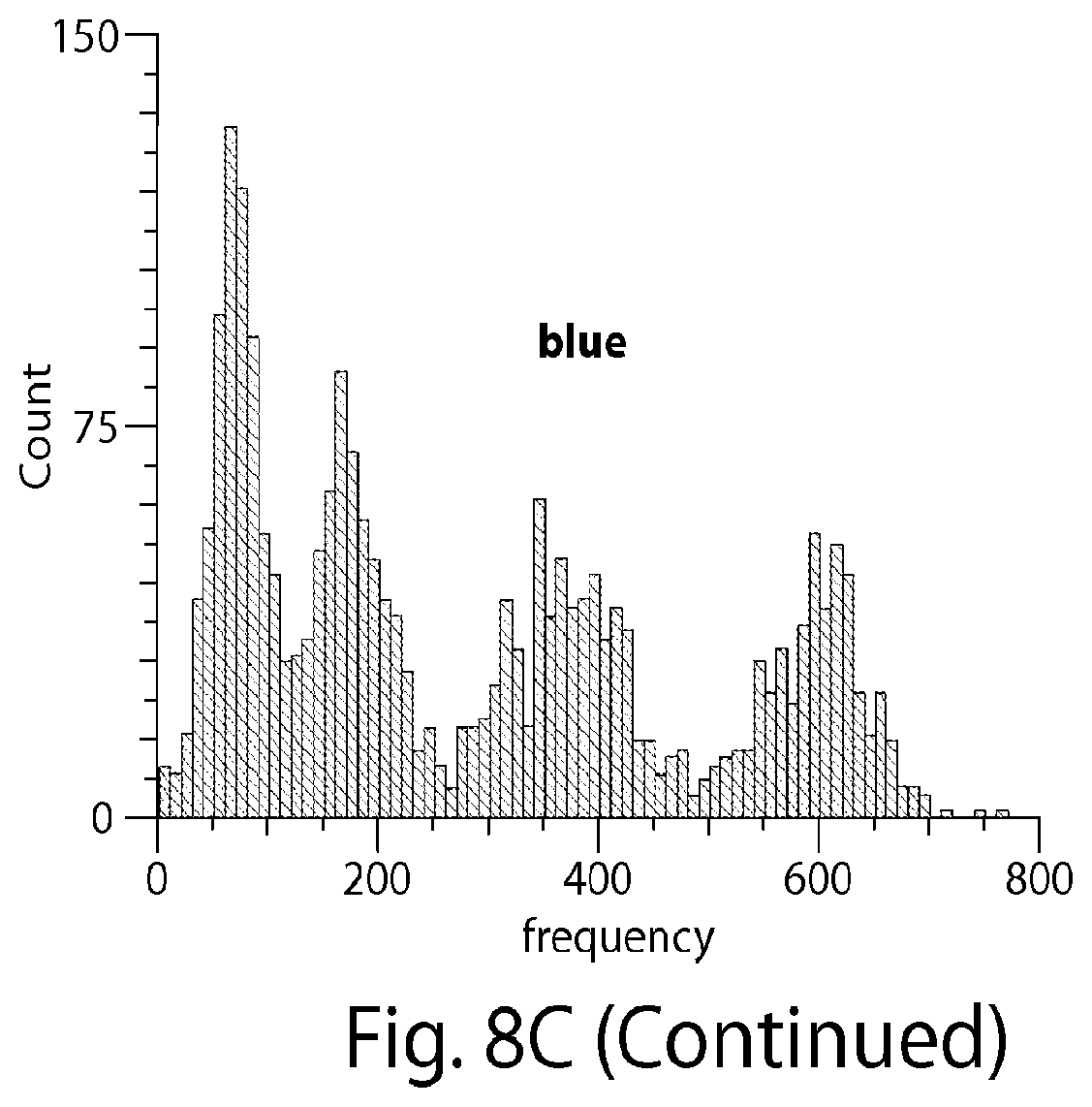
D00019

D00020

D00021

D00022

D00023

D00024

D00025

D00026

D00027

D00028

D00029

D00030

D00031

D00032

D00033

D00034

D00035

D00036

D00037

D00038


S00001
XML
uspto.report is an independent third-party trademark research tool that is not affiliated, endorsed, or sponsored by the United States Patent and Trademark Office (USPTO) or any other governmental organization. The information provided by uspto.report is based on publicly available data at the time of writing and is intended for informational purposes only.
While we strive to provide accurate and up-to-date information, we do not guarantee the accuracy, completeness, reliability, or suitability of the information displayed on this site. The use of this site is at your own risk. Any reliance you place on such information is therefore strictly at your own risk.
All official trademark data, including owner information, should be verified by visiting the official USPTO website at www.uspto.gov. This site is not intended to replace professional legal advice and should not be used as a substitute for consulting with a legal professional who is knowledgeable about trademark law.