Formulations Of Anti-lag3 Antibodies And Co-formulations Of Anti-lag3 Antibodies And Anti-pd-1 Antibodies
Desai; Preeti G. ; et al.
U.S. patent application number 16/609961 was filed with the patent office on 2020-02-20 for formulations of anti-lag3 antibodies and co-formulations of anti-lag3 antibodies and anti-pd-1 antibodies. This patent application is currently assigned to Merck Sharp & Dohme Corp.. The applicant listed for this patent is Merck Sharp & Dohme Corp.. Invention is credited to Valentyn Antochshuk, Rubi Burlage, Preeti G. Desai, Smita Raghava, Shuai Shi.
| Application Number | 20200055938 16/609961 |
| Document ID | / |
| Family ID | 64016970 |
| Filed Date | 2020-02-20 |


View All Diagrams
| United States Patent Application | 20200055938 |
| Kind Code | A1 |
| Desai; Preeti G. ; et al. | February 20, 2020 |
FORMULATIONS OF ANTI-LAG3 ANTIBODIES AND CO-FORMULATIONS OF ANTI-LAG3 ANTIBODIES AND ANTI-PD-1 ANTIBODIES
Abstract
The present invention provides formulations of anti-LAG3 antibodies, and co-formulations of anti-PD-1 antibodies and anti-LAG3 antibodies, and their use in treating various disorders.
| Inventors: | Desai; Preeti G.; (Westfield, NJ) ; Shi; Shuai; (Whippany, NJ) ; Antochshuk; Valentyn; (Cranford, NJ) ; Burlage; Rubi; (Florham Park, NJ) ; Raghava; Smita; (Belle Meade, NJ) | ||||||||||
| Applicant: |
|
||||||||||
|---|---|---|---|---|---|---|---|---|---|---|---|
| Assignee: | Merck Sharp & Dohme
Corp. Rahway NJ |
||||||||||
| Family ID: | 64016970 | ||||||||||
| Appl. No.: | 16/609961 | ||||||||||
| Filed: | May 1, 2018 | ||||||||||
| PCT Filed: | May 1, 2018 | ||||||||||
| PCT NO: | PCT/US18/30468 | ||||||||||
| 371 Date: | October 31, 2019 |
Related U.S. Patent Documents
| Application Number | Filing Date | Patent Number | ||
|---|---|---|---|---|
| 62500330 | May 2, 2017 | |||
| Current U.S. Class: | 1/1 |
| Current CPC Class: | A61K 9/0019 20130101; C07K 16/2818 20130101; A61K 47/02 20130101; A61P 31/00 20180101; C07K 16/2803 20130101; C07K 19/00 20130101; A61K 2039/507 20130101; A61P 35/00 20180101; A61K 47/183 20130101; A61K 39/39591 20130101 |
| International Class: | C07K 16/28 20060101 C07K016/28; A61K 47/02 20060101 A61K047/02; A61K 9/00 20060101 A61K009/00 |
Claims
1. A formulation comprising: about 5-300 mg/mL of an anti-LAG3 antibody or antigen-binding fragment thereof, one or more of an excipient selected from the group consisting of histidine, aspartate, glutamine, glycine, proline, methionine, arginine or pharmaceutically acceptable salt thereof, NaCl, KCl, LiCl, CaCl.sub.2), MgCl.sub.2, ZnCl.sub.2, and FeCl.sub.2, at a total concentration of 10-1000 mM, and a buffer at pH about 5-8.
2. The formulation of claim 1 that comprises the anti-LAG3 antibody or antigen-binding fragment thereof at a concentration of 10-250 mg/ml and comprises a variable light chain region comprising CDRL1 of SEQ ID NO: 39, CDRL2 of SEQ ID NO: 40, CDRL3 of SEQ ID NO: 41 and a variable heavy chain region comprising CDRH1 of SEQ ID NO: 42, CDRH2 of SEQ ID NO: 59, CDRH3 of SEQ ID NO: 44, one or more of an excipient selected from the group consisting of histidine, aspartate, glutamine, glycine, proline, methionine, arginine or pharmaceutically acceptable salt thereof, NaCl, KCl, and LiCl at a total concentration of 15-300 mM, and a buffer at pH about 5.0-6.5.
3. The formulation of claim 2, wherein the excipient is L-arginine or a pharmaceutically acceptable salt thereof at a concentration of 15-250 mM.
4. The formulation of claim 2, wherein the excipient is L-arginine or pharmaceutically acceptable salt thereof at a concentration of 40-100 mM.
5. The formulation of claim 2, wherein the excipients are of NaCl and L-arginine or a pharmaceutically acceptable salt thereof with total concentration of 20-250 mM.
6. The formulation of claim 2, wherein the excipient is NaCl, KCl or LiCl at about 40-150 mM.
7. The formulation of claim 2, wherein the excipient is L-histidine, L-aspartate, L-glutamine, or L-glycine at about 15-200 mM.
8. The formulation of claim 2, wherein the excipient is L-histidine at about 40-100 mM.
9. The formulation of claim 2 wherein the buffer is a histidine buffer, an acetate buffer or a citrate buffer.
10. The formulation of claim 9 wherein the buffer has a concentration of about 1-300 mM.
11. The formulation of claim 2 further comprising a sugar or polyol, and a non-ionic surfactant, or a combination thereof.
12. The formulation of claim 11, wherein the sugar is a non-reducing disaccharide.
13. The formulation of claim 12, wherein the sugar is trehalose or sucrose, or a combination thereof.
14. The formulation of claim 11, wherein the polyol is selected from the group consisting of mannitol, sorbitol, glycerol and polyethylene glycol.
15. The formulation of claim 11, wherein the sugar or polyol is at a concentration of about 10-200 mg/ml.
16. The formulation of claim 11, wherein the non-ionic surfactant is a polysorbate.
17. The formulation of claim 10, further comprising a surfactant selected from polysorbate 20 and polysorbate 80, and a sugar selected from sucrose and trehalose, or a combination thereof.
18. The formulation of claim 2 further comprising about 10-250 mg/mL sucrose, trehalose, mannitol, sorbitol, polyethylene glycol or glycerol; about 0.005-2.0 mg/mL polysorbate 80 or 20; and about 3-300 mM L-histidine, acetate or citrate buffer at pH about 5.0-6.5.
19. The formulation of claim 2 further comprising about 30-120 mg/mL sucrose or trehalose; about 0.05-1.5 mg/mL polysorbate 80 or 20; and about 3-150 mM L-histidine, acetate or citrate buffer at pH about 5.0-6.5.
20. The formulation of claim 2 further comprising about 50-90 mg/mL sucrose or trehalose; about 0.05-1.0 mg/mL polysorbate 80; and about 5-30 mM L-histidine, acetate or citrate buffer at pH about 5.0-6.5.
21. The formulation of claim 11 comprising about 20-220 mg/mL of the anti-LAG3 antibody; about 50-90 mg/mL sucrose or trehalose; about 0.05-1.0 mg/mL polysorbate 80 or 20; about 5-20 mM L-histidine, acetate or citrate buffer at pH about 5.0-6.5; and about 40-150 mM L-arginine or a pharmaceutically acceptable salt thereof.
22. The formulation of claim 11 comprising about 20-220 mg/mL of the anti-LAG3 antibody; about 20-200 mg/mL glycerol, sorbitol or PEG400; about 0.05-1.0 mg/mL polysorbate 80 or 20; about 3-150 mM L-histidine, acetate or citrate buffer at pH about 5.0-6.5; and about 40-150 mM L-arginine or a pharmaceutically acceptable salt thereof.
23. The formulation of claim 2 comprising about 20-220 mg/mL of the anti-LAG3 antibody; about 20-150 mM L-glutamine, L-glycine, L-proline or L-methionine; about 0.05-1.0 mg/mL polysorbate 80 or 20; about 3-150 mM L-histidine, acetate or citrate buffer at pH about 5.0-6.5; and about 40-150 mM L-arginine or a pharmaceutically acceptable salt thereof.
24. The formulation of claim 2 comprising about 20-220 mg/mL of the anti-LAG3 antibody; about 0.05-1.0 mg/mL polysorbate 80 or 20; about 3-150 mM L-histidine, acetate or citrate buffer at pH about 5.0-6.5; and about 40-150 mM NaCl or a pharmaceutically acceptable salt thereof.
25. The formulation of claim 24, further comprising 3-150 mM L-methionine.
26. The formulation of claim 21, further comprising 5-70 mM L-methionine.
27. The formulation of claim 2 comprising about 25 mg/mL of the anti-LAG3 antibody; about 50 mg/mL sucrose; about 0.2 mg/mL polysorbate 80; about 10 mM L-histidine buffer at pH about 5.8; about 70 mM L-arginine or a pharmaceutically acceptable salt thereof; and about 10 mM L-methionine.
28. The formulation of claim 2 comprising about 25 mg/mL of the anti-LAG3 antibody; about 50 mg/mL sucrose; about 0.2 mg/mL polysorbate 80; about 10 mM L-histidine buffer at pH about 5.8-6.0; about 70 mM L-arginine or L-arginine-HCl.
29. The formulation of claim 21, that is a liquid formulation.
30. The formulation of claim 21 that is frozen to at least below -70.degree. C.
31. The formulation of claim 21 that is a reconstituted solution from a lyophilized formulation.
32. The formulation of claim 29, wherein at 5.degree. C., the % monomer of the anti-LAG3 antibody is .gtoreq.95% after 3 months as measured by size exclusion chromatography.
33. The formulation of claim 29, wherein at 5.degree. C., the % acidic variant of the anti-LAG3 antibody is less than 15% after 3 months as measured by ion exchange chromatography.
34. The formulation of claim 21, wherein the anti-LAG3 antibody or antigen binding fragment comprises: a light chain variable region sequence of SEQ ID NO: 37 and a heavy chain variable region sequence of SEQ ID NO: 58.
35. The formulation of claim 21, wherein the anti-LAG3 antibody comprises a light chain sequence of SEQ ID NO: 35 and a heavy chain sequence of SEQ ID NO: 57.
36. The formulation of claim 21, further comprising an anti-PD-1 antibody or antigen-binding fragment thereof.
37. The formulation of claim 36, wherein the molar ratio of anti-LAG3 antibody and anti-PD-1 antibody is 1:1.
38. The formulation of claim 36, wherein the molar ratio of anti-LAG3 antibody and anti-PD-1 antibody is 1:1, 2:1, 3:1 or 3.5:1.
39. The formulation of claim 37, wherein the anti-PD-1 antibody or antigen binding fragment thereof comprises a variable light region comprising CDRL1 of SEQ ID NO: 1, CDRL2 of SEQ ID NO: 2, and CDRL3 of SEQ ID NO: 3, and a variable heavy chain region comprising CDRH1 of SEQ ID NO: 6, CDRH2 of SEQ ID NO: 7, and CDRH3 of SEQ ID NO: 8.
40. The formulation of claim 39, wherein the anti-PD-1 antibody or antigen binding fragment thereof comprises a heavy chain variable region of SEQ ID NO: 9 and a light chain variable region of SEQ ID NO: 4.
41. The formulation of claim 39, wherein the anti-PD-1 antibody comprises a heavy chain sequence of SEQ ID NO: 10 and a light chain sequence of SEQ ID NO: 5.
42. The formulation of claim 39 comprising: about 10-120 mg/mL of the anti-LAG3 antibody or antigen-binding fragment thereof and about 10-120 mg/mL of the anti-PD-1 antibody or antigen-binding fragment thereof.
43. A formulation comprising: an anti-LAG3 antibody or antigen-binding fragment thereof comprising a variable light chain region comprising CDRL1 of SEQ ID NO: 39, CDRL2 of SEQ ID NO: 40, CDRL3 of SEQ ID NO: 41, and a variable heavy chain region comprising CDRH1 of SEQ ID NO: 42, CDRH2 of SEQ ID NO: 59, CDRH3 of SEQ ID NO: 44, an anti-PD-1 antibody or antigen-binding fragment thereof comprising a variable light chain region comprising CDRL1 of SEQ ID NO: 1, CDRL2 of SEQ ID NO: 2, CDRL3 of SEQ ID NO: 3, and a variable heavy chain region comprising CDRH1 of SEQ ID NO: 6, CDRH2 of SEQ ID NO: 7, and CDRH3 of SEQ ID NO: 8, L-arginine or a pharmaceutically acceptable salt thereof at a concentration of 25-250 mM, and a buffer at pH about 5-8.
44. The formulation of claim 43, wherein the anti-LAG3 antibody or antigen binding fragment comprises: a light chain variable region sequence of SEQ ID NO: 37 and a heavy chain variable region sequence of SEQ ID NO: 58, and the anti-PD-1 antibody or antigen binding fragment thereof comprises a heavy chain variable region of SEQ ID NO: 9 and a light chain variable region of SEQ ID NO: 4.
45. The formulation of claim 43, wherein the anti-LAG3 antibody comprises a light chain sequence of SEQ ID NO: 35 and a heavy chain sequence of SEQ ID NO: 57, and the anti-PD-1 antibody comprises a heavy chain sequence of SEQ ID NO: 10 and a light chain sequence of SEQ ID NO: 5.
46. The formulation of claim 43 comprising about 10-120 mg/mL of the anti-LAG3 antibody; about 10-120 mg/mL of the anti-PD-1 antibody; about 30-120 mg/mL of a non-reducing disaccharide; about 0.05-2.0 mg/mL polysorbate 80 or 20; a buffer at pH about 5.0-6.5; and about 40-150 mM L-arginine or a pharmaceutically acceptable salt thereof.
47. The formulation of claim 43 comprising about 10-30 mg/mL of the anti-LAG3 antibody; about 10-30 mg/mL of the anti-PD-1 antibody; about 50-90 mg/mL sucrose or trehalose; about 0.05-1.0 mg/mL polysorbate 80 or 20; about 3-30 mM histidine buffer at pH about 5.0-6.5; and about 40-100 mM L-arginine or a pharmaceutically acceptable salt thereof.
48. The formulation of claim 46, further 3-100 mM L-methionine.
49. The formulation of claim 47, further comprising about 5-15 mM L-methionine.
50. The formulation of claim 43 comprising about 10 mg/mL of the anti-LAG3 antibody; about 10 mg/mL of the anti-PD-1 antibody; about 50 mg/mL sucrose; about 0.2 mg/mL polysorbate 80; about 10 mM histidine buffer at pH about 5.8; about 70 mM L-arginine or a pharmaceutically acceptable salt thereof; and about 10 mM L-methionine.
51. The formulation of claim 47, wherein at 5.degree. C., the % monomer of the anti-LAG3 antibody is .gtoreq.95% after 3 months as measured by size exclusion chromatography.
52. The formulation of claim 47, wherein at 5.degree. C., the % acidic variant of the anti-LAG3 antibody is less than 15% after 3 months as measured by ion exchange chromatography.
53. A vessel or injection device comprising the formulation of claim 2.
54. A method for the treatment of cancer or infection in a patient comprising administering to the patient the formulation of claim 2.
55. The formulation of claim 43 comprising about 10-30 mg/mL of the anti-LAG3 antibody; about 10-30 mg/mL of the anti-PD-1 antibody; about 50 mg/mL sucrose; about 0.2 mg/mL polysorbate 80; about 10 mM histidine buffer at pH about 5.8; about 70 mM L-arginine or a pharmaceutically acceptable salt thereof; and about 10 mM L-methionine.
Description
FIELD OF THE INVENTION
[0001] The present invention relates generally to formulations of therapeutic antibodies, and their use in treating various disorders.
BACKGROUND OF THE INVENTION
[0002] Antibodies may differ somewhat in the amino acid sequence of their constant domains, or in their framework sequences within the variable domains, but they typically differ most dramatically in the CDR sequences. Even antibodies binding to the same protein, the same polypeptide, or even potentially the same epitope may comprise entirely different CDR sequences. Therapeutic antibodies for use in human beings can also be obtained from human germline antibody sequence or from non-human (e.g. rodent) germline antibody sequences, such as in humanized antibodies, leading to yet further diversity in potential sequences. These sequence differences may result in potentially different stabilities in solution and different responsiveness to solution parameters. In addition, small changes in the arrangement of amino acids or changes in one or a few amino acid residues can result in dramatically different antibody stability and susceptibility to sequence-specific degradation pathways. As a consequence, it is not possible at present to predict the solution conditions necessary to optimize antibody stability. Each antibody must be studied individually to determine the optimum solution formulation. Bhambhani et al. (2012) J. Pharm. Sci. 101:1120.
[0003] Antibodies are also fairly large proteins (.about.150,000 Da), for example as compared with other therapeutic proteins such as hormones and cytokines. Antibody drugs must be stable during storage to ensure efficacy and consistent dosing, so it is critical that whatever formulation is chosen supports desirable properties, such as high concentration, clarity and acceptable viscosity, and that also maintains these properties and drug efficacy over an acceptably long shelf-life under typical storage conditions.
[0004] LAG3 (CD223) is a cell surface molecule expressed on activated T cells (Huard et al. Immunogenetics 39:213-217, 1994), NK cells (Triebel et al. J Exp Med 171:1393-1405, 1990), B cells (Kisielow et al. Eur J Immunol 35:2081-2088, 2005), and plasmacytoid dendritic cells (Workman et al. J Immunol 182:1885-1891, 2009) that plays an important role in the function of these lymphocyte subsets. In addition, the interaction between LAG3 and its major ligand, Class II MHC, is thought to play a role in modulating dendritic cell function (Andreae et al. J Immunol 168:3874-3880, 2002). Recent preclinical studies have documented a role for LAG-3 in CD8 T-cell exhaustion (Blackburn et al. Nat Immunol 10:29-37, 2009).
[0005] As with chronic viral infection, tumor antigen-specific CD4.sup.+ and CD8.sup.+ T cells display impaired effector function and an exhausted phenotype characterized by decreased production of pro-inflammatory cytokines and hyporesponsiveness to antigenic re-stimulation. This is mediated by cell extrinsic mechanisms, such as regulatory T-cells (Treg), and cell intrinsic mechanisms, such as inhibitory molecules that are upregulated on exhausted, tumor-infiltrating lymphocytes (TIL). These inhibitory mechanisms represent a formidable barrier to effective antitumor immunity.
[0006] LAG--is expressed on tolerized TILs suggesting that they contribute to tumor-mediated immune suppression. Inhibition of LAG3 may lead to enhanced activation of antigen-specific T cells from which a therapeutic benefit may be gained.
[0007] PD-1 is recognized as an important molecule in immune regulation and the maintenance of peripheral tolerance. PD-1 is moderately expressed on naive T, B and NKT cells and up-regulated by TB cell receptor signaling on lymphocytes, monocytes and myeloid cells. Two known ligands for PD-1, PD-L1 (B7-H1) and PD-L2 (B7-DC), are expressed in human cancers arising in various tissues. In large sample sets of e.g. ovarian, renal, colorectal, pancreatic, liver cancers and melanoma, it was shown that PD-L1 expression correlated with poor prognosis and reduced overall survival irrespective of subsequent treatment. Similarly, PD-1 expression on tumor infiltrating lymphocytes was found to mark dysfunctional T cells in breast cancer and melanoma and to correlate with poor prognosis in renal cancer. Thus, it has been proposed that PD-L1 expressing tumor cells interact with PD-1 expressing T cells to attenuate T cell activation and evasion of immune surveillance, thereby contributing to an impaired immune response against the tumor.
[0008] Several monoclonal antibodies that inhibit the interaction between PD-1 and one or both of its ligands PD-L1 and PD-L2 are in clinical development for treating cancer. It has been proposed that the efficacy of such antibodies might be enhanced if administered in combination with other approved or experimental cancer therapies, e.g., radiation, surgery, chemotherapeutic agents, targeted therapies, agents that inhibit other signaling pathways that are disregulated in tumors, and other immune enhancing agents.
[0009] As a consequence, the need exists for stable formulations of therapeutic antibodies, such as antibodies that bind to human LAG-3, as well as stable co-formulations of an anti-LAG3 antibody and an anti-PD-1 antibody. Such stable formulations will preferably exhibit stability over months to years under conditions typical for storage of drugs for self-administration, i.e. at refrigerator temperature in a syringe, resulting in a long shelf-life for the corresponding drug product.
SUMMARY OF THE INVENTION
[0010] The present invention provides formulations of anti-LAG3 antibodies or antigen binding fragments. Applicants discovered certain excipients that mitigate the phase separation of anti-LAG3 in solution. In one aspect, the invention provides one or more of an excipient selected from the group consisting of histidine, aspartate, glutamine, glycine, proline, methionine, arginine or a pharmaceutically acceptable salt thereof, NaCl, KCl, LiCl, CaCl.sub.2, MgCl.sub.2, ZnCl.sub.2, and FeCl.sub.2, at a total concentration of 10-1000 mM, and a buffer at pH about 5-8. In one aspect, the present invention provides a formulation comprising an anti-LAG3 antibody or antigen binding fragment thereof and a buffer at pH about 5-8, and one or more of arginine, histidine or a pharmaceutically acceptable salt thereof, or NaCl at a total concentration of 15-250 mM. In one embodiment, the formulation comprises an anti-LAG3 antibody or antigen-binding fragment thereof, a sugar or polyol; a non-ionic surfactant, a buffer at pH about 5-8, 25-200 mM arginine or a pharmaceutically acceptable salt thereof. In a further embodiment, the formulation comprises about 25 mg/mL anti-LAG3 antibody; about 50 mg/mL sucrose; about 0.2 mg/mL polysorbate 80; about 10 mM L-histidine buffer at about pH 5.8-6.0; about 70 mM L-Arginine-HCl thereof; and optionally about 10 mM L-methionine. The formulation optionally comprises an anti-PD-1 antibody.
[0011] In other aspects, the invention provides a co-formulation of anti-LAG3 antibodies or antigen binding fragments and anti-PD-1 antibodies or antigen binding fragments with arginine or a pharmaceutically acceptable salt thereof at a total concentration of 10-1000 mM, and a buffer at pH about 5-8, and optionally 3-100 mM of methionine. In one embodiment, the formulation comprises about 25 mg/mL anti-LAG3 antibody and about 25 mg/ml anti-PD-1 antibody; about 50 mg/mL sucrose; about 0.2 mg/mL polysorbate 80; about 10 mM L-histidine buffer at pH about 5.8-6.0; about 70 mM L-Arginine-HCl thereof; and about 10 mM L-methionine. Surprisingly, the anti-LAG.sup.3/anti-PD-1 co-formulations shows better stability than the individual antibody formulations. The formulations can be lyophilized for reconstitution or in liquid form.
[0012] The present invention also provides a method of treating cancer or infection, comprising administering the reconstituted or liquid formulation (solution formulation) to a subject in need thereof. In further embodiments the formulation is used in treating chronic infection. Also contemplated is the use of the solution or lyophilized formulation in the manufacture of a medicament for treating cancer or infection.
BRIEF DESCRIPTION OF THE DRAWINGS
[0013] FIG. 1: Number of particles per container .gtoreq.10 .mu.m as measured by mHIAC for anti-LAG3 drug product samples that were stored at -80.degree. C., -20.degree. C., 5.degree. C., 25.degree. C., and 40.degree. C.
[0014] FIG. 2: Number of particles per container .gtoreq.25 .mu.m as measured by mHIAC for anti-LAG3 drug product samples that were stored at -80.degree. C., -20.degree. C., 5.degree. C., 25.degree. C., and 40.degree. C.
[0015] FIG. 3: Potency as measured by ELISA for anti-LAG3 drug product samples that were stored at -80.degree. C., -20.degree. C., 5.degree. C., 25.degree. C., and 40.degree. C.
[0016] FIG. 4: Monomer (%) by UP-SEC for anti-LAG3 drug product samples that were stored at -80.degree. C., -20.degree. C., 5.degree. C., 25.degree. C., and 40.degree. C.
[0017] FIG. 5: High molecular weight species (%) by UP-SEC for anti-LAG3 drug product samples that were stored at -80.degree. C., -20.degree. C., 5.degree. C., 25.degree. C., and 40.degree. C.
[0018] FIG. 6: Acidic Variants (%) by HP-IEX for anti-LAG3 drug product samples that were stored at -80.degree. C., -20.degree. C., 5.degree. C., 25.degree. C., and 40.degree. C.
[0019] FIG. 7: Total Main Peak (%) by HP-IEX for anti-LAG3 drug product samples that were stored at -80.degree. C., -20.degree. C., 5.degree. C., 25.degree. C., and 40.degree. C.
[0020] FIG. 8: Basic Variants (%) by HP-IEX for anti-LAG3 drug product samples that were stored at -80.degree. C., -20.degree. C., 5.degree. C., 25.degree. C., and 40.degree. C.
[0021] FIG. 9: Purity Heavy Chain+Light Chain (%) by CE-SDS Reducing for anti-LAG3 drug product samples that were stored at -80.degree. C., -20.degree. C., 5.degree. C., 25.degree. C., and 40.degree. C.
[0022] FIG. 10: Purity Intact IgG (%) by CE-SDS Non-Reducing for anti-LAG3 drug product samples that were stored at -80.degree. C., -20.degree. C., 5.degree. C., 25.degree. C., and 40.degree. C.
[0023] FIG. 11: Homology model of anti-LAG3 antibody Ab6 showing Tryptophan surface exposure. Trp102 is surface exposed as measured by accessible surface area (85.25 .ANG..sup.2) calculated using a homology model.
[0024] FIG. 12: Reduction of self-interaction (KD) and improvement of colloidal stability (OD350) and relative solubility (% PEGmid-point) of anti-LAG3 antibody in the presence of salt (50 mM NaCl) and in the presence of L-arginine hydrochloride (40 mM) in 10 mM histidine buffer pH 5.6.
[0025] FIG. 13: Effect of L-arginine hydrochloride on the diffusion interaction parameter (KD) and turbidity (OD350 at 50 mg/mL) of the anti-LAG3 antibody in 10 mM histidine buffer at pH 5.8 and at pH 6.0.
[0026] FIG. 14: Anti-LAG3 antibody pH Ranging Studies (5.3 to 6.4).
[0027] FIG. 15: Diffusion interaction parameter (kD) of anti-LAG3 (25 mg/mL in 10 mM L histidine pH 5.8) in the presence of L-arginine, L-histidine or sodium chloride.
[0028] FIG. 16: Relative solubility of anti-LAG3 (25 mg/mL in 10 mM L-histidine pH 5.8) in the presence of L-arginine, L-histidine or sodium chloride.
[0029] FIG. 17: Percent change in charged species of anti-LAG3 (25 mg/mL in 10 mM L-histidine pH 5.8) in the presence of L-arginine, L-histidine or sodium chloride.
[0030] FIG. 18: Optimization of 25 mg/mL anti-LAG3 formulation in 10 mM L-histidine pH 5.8 buffer with L-arginine, sodium chloride or its mixture using second virial coefficient (B.sub.22) measurement.
[0031] FIG. 19: Colloidal stability (OD350) of 25 mg/mL anti-LAG3 formulation in 10 mM L-histidine pH 5.8 buffer in presence of L-arginine, sodium chloride or its mixture.
[0032] FIG. 20: Viscosity of anti-LAG3 (60 mg/mL) in 10 mM L-histidine pH 5.8 buffer in presence of either L-arginine, sodium chloride or its mixture.
[0033] FIG. 21: Osmolality of anti-LAG3 (25 mg/mL) in 10 mM L-histidine pH 5.8 buffer in presence of either L-arginine, sodium chloride or its mixture.
[0034] FIG. 22: Turbidity analysis of formulations (F1-F6) over time at 40.degree. C. and 25.degree. C. storage conditions.
[0035] FIG. 23: Mixed-mode chromatography analysis of formulations (F1-F6) over time at 40.degree. C. storage condition. Change in monomer percentage for each mAb (anti-LAG3 and anti-PD-1) is plotted over time for formulations F1-F6.
[0036] FIG. 24: Percent change in high molecular weight (HMW) species, monomer and low molecular weight (LMW) species of 25 mg/mL anti-LAG3 formulation (10 mM L-histidine pH 5.8 buffer) in presence of 70 mM L-arginine hydrochloride with 2.5% to 9% stabilizers or 70 mM sodium chloride with 2.5% to 9.0% stabilizers.
[0037] FIG. 25: Percent change in charged species of 25 mg/mL anti-LAG3 formulation (10 mM L-histidine pH 5.8 buffer) in presence of 70 mM L-arginine hydrochloride with 2.5% to 9% stabilizers or 70 mM sodium chloride with 2.5% to 9.0% stabilizers.
[0038] FIG. 26: Tm1, Tm2 and Tonset of 25 mg/mL anti-LAG3 formulation (10 mM L-histidine pH 5.8) in presence of 70 mM L-arginine with 2.5% to 9% stabilizers or 70 mM sodium chloride with 2.5% to 9.0% stabilizers.
[0039] FIG. 27: Colloidal stability (OD350) of 25 mg/mL anti-LAG3 formulation (10 mM L-histidine, 70 mM L-arginine, 5% w/v sucrose, pH 5.8) in presence of different concentrations of polysorbate 80 upon agitation stress.
[0040] FIG. 28: Percent change in high molecular weight (HMW) species, monomer and low molecular weight (LMW) species of 25 mg/mL anti-LAG3 formulation (10 mM L-histidine pH 5.8 buffer, 70 mM L-arginine hydrochloride, 5% w/v sucrose) in the presence of different concentrations of polysorbate 80 upon agitation stress.
[0041] FIG. 29: Percent change in charged species of 25 mg/mL anti-LAG3 formulation (10 mM L-histidine, 70 mM L-arginine, 5% w/v sucrose, pH 5.8) in presence of different concentrations of polysorbate 80 upon agitation stress.
[0042] FIG. 30: Colloidal stability (OD350) of 25 mg/mL anti-LAG3 formulation (10 mM L-histidine, 70 mM L-arginine, 5% w/v sucrose, pH 5.8) alone and in presence of increasing concentrations of L-methionine.
[0043] FIG. 31: Percent change in high molecular weight (HMW) species, and monomer of 25 mg/mL anti-LAG3 formulation (10 mM L-histidine, 70 mM L-arginine, 5% w/v sucrose, pH 5.8) alone and in presence of increasing concentrations of L-methionine.
[0044] FIG. 32: Percent change in charged species of 25 mg/mL anti-LAG3 formulation (10 mM L-histidine, 70 mM L-arginine, 5% w/v sucrose, pH 5.8) alone and in presence of increasing concentrations of L-methionine.
[0045] FIG. 33: Percent change in oxidation of 25 mg/mL anti-LAG3 formulation (10 mM L-histidine, 70 mM L-arginine, 5% w/v sucrose, pH 5.8) alone and in presence of increasing concentrations of L-methionine.
[0046] FIG. 34: Percent change in turbidity (OD350) of 200 mg/mL anti-LAG3 in the presence of 10 mM buffer with 70 mM L-arginine hydrochloride, pH 5.8 alone and in the presence of different stabilizers.
[0047] FIG. 35: Percent change in high molecular weight (HMW) species, monomer and low molecular weight (LMW) species of 200 mg/mL anti-LAG3 in the presence of 10 mM buffer. with 70 mM L-arginine hydrochloride, pH 5.8 alone and in the presence of different stabilizers.
[0048] FIG. 36: Percent change in charged species of 200 mg/mL anti-LAG3 in the presence of 10 mM buffer with 70 mM L-arginine hydrochloride, pH 5.8 alone and in the presence of different stabilizers.
[0049] FIG. 37: Percent change in turbidity (OD.sub.350) of 25 mg/mL anti-LAG3 in the presence of 40 to 70 mM salt (sodium chloride) and 20 to 70 mM amino acids alone and some combinations.
[0050] FIG. 38: Percent change in high molecular weight (HMW) species, monomer and low molecular weight (LMW) species of 25 mg/mL anti-LAG3 in the presence of 40 to 70 mM salt (sodium chloride) and 20 to 70 mM amino acids alone and some combinations.
[0051] FIG. 39: Percent change in charged species of 25 mg/mL anti-LAG3 in the presence of 40 to 70 mM salt (sodium chloride) and 20 to 70 mM amino acids alone and some combinations.
[0052] FIG. 40: Percent change in hydrodynamic diameter of 25 mg/mL anti-LAG3 in the presence of 40 to 70 mM salt (sodium chloride) and 20 to 70 mM amino acids alone and some combinations.
DETAILED DESCRIPTION
[0053] As used herein, including the appended claims, the singular forms of words such as "a," "an," and "the," include their corresponding plural references unless the context clearly dictates otherwise. Unless otherwise indicated, the proteins and subjects referred to herein are human proteins and human subjects, rather than another species.
Definitions
[0054] As used herein, unless otherwise indicated, "antigen binding fragment" refers to antigen binding fragments of antibodies, i.e. antibody fragments that retain the ability to bind specifically to the antigen bound by the full-length antibody, e.g. fragments that retain one or more CDR regions. Examples of antibody binding fragments include, but are not limited to, Fab, Fab', F(ab')2, and Fv fragments.
[0055] A "Fab fragment" is comprised of one light chain and the CH1 and variable regions of one heavy chain. The heavy chain of a Fab molecule cannot form a disulfide bond with another heavy chain molecule. An "Fab fragment" can be the product of papain cleavage of an antibody.
[0056] An "Fc" region contains two heavy chain fragments comprising the CH1 and CH2 domains of an antibody. The two heavy chain fragments are held together by two or more disulfide bonds and by hydrophobic interactions of the CH3 domains.
[0057] A "Fab' fragment" contains one light chain and a portion or fragment of one heavy chain that contains the VH domain and the C H1 domain and also the region between the CH1 and C H2 domains, such that an interchain disulfide bond can be formed between the two heavy chains of two Fab' fragments to form a F(ab') 2 molecule.
[0058] A "F(ab')2 fragment" contains two light chains and two heavy chains containing a portion of the constant region between the CH1 and CH2 domains, such that an interchain disulfide bond is formed between the two heavy chains. A F(ab') 2 fragment thus is composed of two Fab' fragments that are held together by a disulfide bond between the two heavy chains. An "F(ab')2 fragment" can be the product of pepsin cleavage of an antibody.
[0059] The "Fv region" comprises the variable regions from both the heavy and light chains, but lacks the constant regions.
[0060] As used herein, the term "hypervariable region" refers to the amino acid residues of an antibody that are responsible for antigen-binding. The hypervariable region comprises amino acid residues from a "complementarity determining region" or "CDR" (e.g. residues 24-34 (CDRL1), 50-56 (CDRL2) and 89-97 (CDRL3) in the light chain variable domain and residues 31-35 (CDRH1), 50-65 (CDRH2) and 95-102 (CDRH3) in the heavy chain variable domain (Kabat et al. (1991) Sequences of Proteins of Immunological Interest, 5th Ed. Public Health Service, National Institutes of Health, Bethesda, Md.) and/or those residues from a "hypervariable loop" (i.e. residues 26-32 (L1), 50-52 (L2) and 91-96 (L3) in the light chain variable domain and 26-32 (H1), 53-55 (H2) and 96-101 (H3) in the heavy chain variable domain (Chothia and Lesk (1987) J. Mol. Biol. 196: 901-917). As used herein, the term "framework" or "FR" residues refers to those variable domain residues other than the hypervariable region residues defined herein as CDR residues. The residue numbering above relates to the Kabat numbering system and does not necessarily correspond in detail to the sequence numbering in the accompanying Sequence Listing.
[0061] "Proliferative activity" encompasses an activity that promotes, that is necessary for, or that is specifically associated with, e.g., normal cell division, as well as cancer, tumors, dysplasia, cell transformation, metastasis, and angiogenesis.
[0062] The terms "cancer", "tumor", "cancerous", and "malignant" refer to or describe the physiological condition in mammals that is typically characterized by unregulated cell growth. Examples of cancer include but are not limited to, carcinoma including adenocarcinoma, lymphoma, blastoma, melanoma, sarcoma, and leukemia. More particular examples of such cancers include squamous cell cancer, small-cell lung cancer, non-small cell lung cancer, gastrointestinal cancer, Hodgkin's and non-Hodgkin's lymphoma, pancreatic cancer, glioblastoma, glioma, cervical cancer, ovarian cancer, liver cancer such as hepatic carcinoma and hepatoma, bladder cancer, breast cancer, colon cancer, colorectal cancer, endometrial carcinoma, myeloma (such as multiple myeloma), salivary gland carcinoma, kidney cancer such as renal cell carcinoma and Wilms' tumors, basal cell carcinoma, melanoma, prostate cancer, vulval cancer, thyroid cancer, testicular cancer, esophageal cancer, and various types of head and neck cancer.
[0063] As cancerous cells grow and multiply, they form a mass of cancerous tissue, that is a tumor, which invades and destroys normal adjacent tissues. Malignant tumors are cancer. Malignant tumors usually can be removed, but they may grow back. Cells from malignant tumors can invade and damage nearby tissues and organs. Also, cancer cells can break away from a malignant tumor and enter the bloodstream or lymphatic system, which is the way cancer cells spread from the primary tumor (i.e., the original cancer) to form new tumors in other organs. The spread of cancer in the body is called metastasis (What You Need to Know About Cancer--an Overview, NIH Publication No. 00-1566; posted Sep. 26, 2000, updated Sep. 16, 2002 (2002)).
[0064] As used herein, the term "solid tumor" refers to an abnormal growth or mass of tissue that usually does not contain cysts or liquid areas. Solid tumors may be benign (not cancerous) or malignant (cancerous). Different types of solid tumors are named for the type of cells that form them. Examples of solid tumors are sarcomas, carcinomas, and lymphomas. Leukemias (cancers of the blood) generally do not form solid tumors (National Cancer Institute, Dictionary of Cancer Terms).
[0065] As used herein, the term "carcinomas" refers to cancers of epithelial cells, which are cells that cover the surface of the body, produce hormones, and make up glands. Examples of carcinomas are cancers of the skin, lung, colon, stomach, breast, prostate and thyroid gland.
Pharmaceutical Composition Definitions
[0066] As used herein, an "aqueous" pharmaceutical composition is a composition suitable for pharmaceutical use, wherein the aqueous carrier is sterile water for injection. A composition suitable for pharmaceutical use may be sterile, homogeneous and/or isotonic. In certain embodiments, the aqueous pharmaceutical compositions of the invention are suitable for parenteral administration to a human subject. In a specific embodiment, the aqueous pharmaceutical compositions of the invention are suitable for intravenous and/or subcutaneous administration.
[0067] The term "about", when modifying the quantity (e.g., mM, or M) of a substance or composition, the percentage (v/v or w/v) of a formulation component, the pH of a solution/formulation, or the value of a parameter characterizing a step in a method, or the like refers to variation in the numerical quantity that can occur, for example, through typical measuring, handling and sampling procedures involved in the preparation, characterization and/or use of the substance or composition; through instrumental error in these procedures; through differences in the manufacture, source, or purity of the ingredients employed to make or use the compositions or carry out the procedures; and the like. In certain embodiments, "about" can mean a variation of .+-.0.1%, 0.5%, 1%, 2%, 3%, 4%, 5%, or 10%.
[0068] As used herein, "x % (w/v)" is equivalent to x g/100 ml (for example 5% w/v equals 50 mg/ml).
[0069] The term "buffer" encompasses those agents which maintain the solution pH in an acceptable range in the liquid formulation, prior to lyophilization and/or after reconstitution and may include but not limited to succinate (sodium or potassium), histidine, acetate, phosphate (sodium or potassium), Tris (tris (hydroxymethyl) aminomethane), diethanolamine, citrate (sodium) and the like.
[0070] "Co-formulated" or "co-formulation" or "coformulation" or "coformulated" as used herein refers to at least two different antibodies or antigen binding fragments thereof which are formulated together and stored as a combined product in a single vial or vessel (for example an injection device) rather than being formulated and stored individually and then mixed before administration or separately administered. In one embodiment, the co-formulation contains two different antibodies or antigen binding fragments thereof.
[0071] "Glycol" refers to an alkyl with two hydroxyl groups.
[0072] "Sugar alcohol" refers to polyols derived from a sugar and have the general formula HOCH.sub.2(CHOH).sub.nCH.sub.2OH, n=1, 2, 3, 4, 5, 6, 7, 8, 9 or 10. Examples include but are not limited to mannitol, sorbitol, erythritol, xylitol and glycerol.
[0073] As used herein "polyol" includes a glycol and a sugar alcohol.
[0074] The terms "lyophilization," "lyophilized," and "freeze-dried" refer to a process by which the material to be dried is first frozen and then the ice or frozen solvent is removed by sublimation in a vacuum environment. An excipient may be included in pre-lyophilized formulations to enhance stability of the lyophilized product upon storage.
[0075] "Non-reducing sugar" is a sugar not capable of acting as a reducing agent because it does not contain or cannot be converted to contain a free aldehyde group or a free ketone group. Examples of non-reducing sugars include but are not limited to dissacharrides such as sucrose and trehalose.
[0076] The term "pharmaceutical formulation" refers to preparations which are in such form as to permit the active ingredients to be effective, and which contains no additional components which are toxic to the subjects to which the formulation would be administered.
[0077] "Pharmaceutically acceptable" excipients (vehicles, additives) are those which can reasonably be administered to a subject mammal to provide an effective dose of the active ingredient employed.
[0078] "Reconstitution time" is the time that is required to rehydrate a lyophilized formulation with a solution to a particle-free clarified solution.
[0079] A "stable" formulation is one in which the protein therein essentially retains its physical stability and/or chemical stability and/or biological activity upon storage. Various analytical techniques for measuring protein stability are available in the art and are reviewed in Peptide and Protein Drug Delivery, 247-301, Vincent Lee Ed., Marcel Dekker, Inc., New York, N.Y., Pubs. (1991) and Jones, A. Adv. Drug Delivery Rev. 10:29-90 (1993). Stability can be measured at a selected temperature for a selected time period. For example, in one embodiment, a stable formulation is a formulation with no significant changes observed at a refrigerated temperature (2-8.degree. C.) for at least 12 months. In another embodiment, a stable formulation is a formulation with no significant changes observed at a refrigerated temperature (2-8.degree. C.) for at least 18 months. In another embodiment, stable formulation is a formulation with no significant changes observed at room temperature (23-27.degree. C.) for at least 3 months. In another embodiment, stable formulation is a formulation with no significant changes observed at room temperature (23-27.degree. C.) for at least 6 months. In another embodiment, stable formulation is a formulation with no significant changes observed at room temperature (23-27.degree. C.) for at least 12 months. In another embodiment, stable formulation is a formulation with no significant changes observed at room temperature (23-27.degree. C.) for at least 18 months. The criteria for stability for an antibody formulation are as follows. Typically, no more than 10%, preferably 5%, of antibody monomer is degraded as measured by SEC-HPLC. Typically, the formulation is colorless, or clear to slightly opalescent by visual analysis. Typically, the concentration, pH and osmolality of the formulation have no more than +1-10% change. Potency is typically within 60-140%, preferably 80-120% of the control or reference. Typically, no more than 10%, preferably 5% of clipping of the antibody is observed, i.e., % low molecular weight species as determined, for example, by HP-SEC. Typically, or no more than 10%, preferably 5% of aggregation of the antibody is formed, i.e. % high molecular weight speciies as determined, for example, by HP-SEC.
[0080] "Surfactant" is a surface active agent that is amphipathic in nature.
[0081] An antibody "retains its physical stability" in a pharmaceutical formulation if it shows no significant increase of aggregation, precipitation and/or denaturation upon visual examination of color and/or clarity, or as measured by UV light scattering, size exclusion chromatography (SEC) and dynamic light scattering. The changes of protein conformation can be evaluated by fluorescence spectroscopy, which determines the protein tertiary structure, and by FTIR spectroscopy, which determines the protein secondary structure.
[0082] An antibody "retains its chemical stability" in a pharmaceutical formulation, if it shows no significant chemical alteration. Chemical stability can be assessed by detecting and quantifying chemically altered forms of the protein. Degradation processes that often alter the protein chemical structure include hydrolysis or clipping (evaluated by methods such as size exclusion chromatography and SDS-PAGE), oxidation (evaluated by methods such as by peptide mapping in conjunction with mass spectroscopy or MALDI/TOF/MS), deamidation (evaluated by methods such as ion-exchange chromatography, capillary isoelectric focusing, peptide mapping, isoaspartic acid measurement), and isomerization (evaluated by measuring the isoaspartic acid content, peptide mapping, etc.).
[0083] An antibody "retains its biological activity" in a pharmaceutical formulation, if the biological activity of the antibody at a given time frame is withing a predetermined range of biological activity exhibited at the time the formulation was prepared. The biological activity of an antibody can be determined, for example, by an antigen binding assay. In one embodiment, the biological activity of stable antibody formulation within 12 months is within 60-140% of the reference.
[0084] The term "isotonic" means that the formulation of interest has essentially the same osmotic pressure as human blood. Isotonic formulations will generally have an osmotic pressure about 270-328 mOsm. Slightly hypotonic pressure is 250-269 and slightly hypertonic pressure is 328-350 mOsm. Osmotic pressure can be measured, for example, using a vapor pressure or ice-freezing type osmometer.
[0085] A "reconstituted" formulation is one that has been prepared by dissolving a lyophilized protein formulation in a diluent such that the protein is dispersed in the reconstituted formulation. The reconstituted formulation is suitable for administration, (e.g. parenteral administration), and may optionally be suitable for subcutaneous administration.
[0086] As used herein, concentrations are to be construed as approximate within the ranges normally associated with such concentrations in the manufacture of pharmaceutical formulations. Specifically, concentrations need not be exact, but may differ from the stated concentrations within the tolerances typically expected for drugs manufactured under GMP conditions. Similarly, pH values are approximate within the tolerances typically expected for drugs manufactured under GMP conditions and stored under typical storage conditions.
[0087] When a range of pH values is recited, such as "a pH between pH 5.0 and 6.0," the range is intended to be inclusive of the recited values. The pH is typically measured at 25.degree. C. using standard glass bulb pH meter. As used herein, a solution comprising "histidine buffer at pH X" refers to a solution at pH X and comprising the histidine buffer, i.e. the pH is intended to refer to the pH of the solution.
Analytical Methods
[0088] Analytical methods suitable for evaluating the product stability include size exclusion chromatography (SEC), dynamic light scattering test (DLS), differential scanning calorimetery (DSC), iso-asp quantification, potency, UV at 350 nm, UV spectroscopy, and FTIR. SEC (J. Pharm. Scien., 83:1645-1650, (1994); Pharm. Res., 11:485 (1994); J. Pharm. Bio. Anal., 15:1928 (1997); J. Pharm. Bio. Anal., 14:1133-1140 (1986)) measures percent monomer in the product and gives information of the amount of soluble aggregates. DSC (Pharm. Res., 15:200 (1998); Pharm. Res., 9:109 (1982)) gives information of protein denaturation temperature and glass transition temperature. DLS (American Lab., November (1991)) measures mean diffusion coefficient, and gives information of the amount of soluble and insoluble aggregates. UV at 340 nm measures scattered light intensity at 340 nm and gives information about the amounts of soluble and insoluble aggregates. UV spectroscopy measures absorbance at 278 nm and gives information of protein concentration. FTIR (Eur. J. Pharm. Biopharm., 45:231 (1998); Pharm. Res., 12:1250 (1995); J. Pharm. Scien., 85:1290 (1996); J. Pharm. Scien., 87:1069 (1998)) measures IR spectrum in the amide one region, and gives information of protein secondary structure.
[0089] The iso-asp content in the samples is measured using the Isoquant Isoaspartate Detection System (Promega). The kit uses the enzyme Protein Isoaspartyl Methyltransferase (PIMT) to specifically detect the presence of isoaspartic acid residues in a target protein. PIMT catalyzes the transfer of a methyl group from S-adenosyl-L-methionine to isoaspartic acid at the .alpha.-carboxyl position, generating S-adenosyl-L-homocysteine (SAH) in the process. This is a relatively small molecule, and can usually be isolated and quantitated by reverse phase HPLC using the SAH HPLC standards provided in the kit.
[0090] The potency or bioidentity of an antibody can be measured by its ability to bind to its antigen. The specific binding of an antibody to its antigen can be quantitated by any method known to those skilled in the art, for example, an immunoassay, such as ELISA (enzyme-linked immunosorbant assay).
Anti-LAG3 Antibodies
[0091] The CDR residues are highly variable between different antibodies, and may originate from human germline sequences (in the case of fully human antibodies), or from non-human (e.g. rodent) germline sequences. The framework regions can also differ significantly from antibody to antibody. The constant regions will differ depending on whether the selected antibody has a lambda (.lamda.) or kappa (.kappa.) light chain, and depending on the class (or isotype) of the antibody (IgA, IgD, IgE, IgG, or IgM) and subclass (e.g. IgG1, IgG2, IgG3, IgG4).
[0092] The LAG3 antibodies exemplified below have CDR sequences derived from non-human (in this case mouse) germline sequences, or human germline sequences. The germline sequences comprise the sequence repertoire from which an antibody's CDR sequences are derived, aside from somatic hypermutation derived changes, and as a consequence it would be expected that CDRs obtained starting with a mouse germline would systematically differ from those starting from a human germline. Use of human germline sequences is often justified on the basis that CDR sequences from human germlines will be less immunogenic in humans than those derived from other species, reflecting the underlying belief that CDRs will systematically differ depending on their species of origin. Although the increase in CDR diversity increases the likelihood of finding antibodies with desired properties, such as high affinity, it further magnifies the difficulties in developing a stable solution formulation of the resulting antibody.
[0093] Even antibodies that bind to the same antigen can differ dramatically in sequence, and are not necessarily any more closely related in sequence than antibodies to entirely separate antigens. Based on the low sequence similarity, the chemical properties of the antibodies, and thus their susceptibility to degradation, cannot be presumed to be similar despite their shared target.
[0094] As discussed above, antibodies are large, highly complex polypeptide complexes subject to various forms of degradation and instability in solution. The diversity of sequence, and thus structure, of antibodies gives rise to wide range of chemical properties. Aside from the obvious sequence-specific differences in antigen binding specificity, antibodies exhibit varying susceptibility to various degradative pathways, aggregation, and precipitation. Amino acid side chains differ in the presence or absence of reactive groups, such as carboxy-(D,E), amino-(K), amide-(N,Q), hydroxyl-(S,T,Y), sulfhydryl-(C), thioether-(M) groups, as well as potentially chemically reactive sites on histidine, phenylalanine and proline residues. Amino acid side chains directly involved in antigen binding interactions are obvious candidates for inactivation by side chain modification, but degradation at other positions can also affect such factors as steric orientation of the CDRs (e.g. changes in framework residues), effector function (e.g. changes in Fc region--see, e.g., Liu et al. (2008) Biochemistry 47:5088), or self-association/aggregation.
[0095] Antibodies are subject to any number of potential degradation pathways. Oxidation of methionione residues in antibodies, particularly in CDRs, can be a problem if it disrupts antigen binding. Presta (2005) J. Allergy Clin. Immunol. 116: 731; Lam et al. (1997) J. Pharm. Sci. 86:1250. Other potential degradative pathways include asparagine deamidation (Harris et al. (2001) Chromatogr., B 752:233; Vlasak et al. (2009) Anal. Biochem. 392:145) tryptophan oxidation (Wei et al. (2007) Anal. Chem. 79:2797), cysteinylation (Banks et al. (2008) J. Pharm. Sci. 97:775), glycation (Brady et al. (2007) Anal. Chem. 79:9403), pyroglutamate formation (Yu et al. (2006) J. Pharm. Biomed. Anal. 42:455), disulfide shuffling (Liu et al. (2008) J. Biol. Chem. 283:29266), and hydrolysis (Davagnino et al. (1995) J. Immunol. Methods 185:177). Discussed in Ionescu & Vlasak (2010) Anal. Chem. 82:3198. See also Liu et al. (2008) J. Pharm. Sci. 97:2426. Some potential degradation pathways depend not only on the presence of a specific amino acid residue, but also the surrounding sequence. Deamidation and isoaspartate formation can arise from a spontaneous intramolecular rearrangement of the peptide bond following (C-terminal to) N or D residues, with N-G and D-G sequences being particularly susceptible. Reissner & Aswad (2003) CMLS Cell. Mol. Life Sci. 60:1281.
[0096] Antibodies are also subject to sequence-dependent non-enzymatic fragmentation during storage. Vlasak & Ionescu (2011) mAbs 3:253. The presence of reactive side chains, such as D, G, S, T, C or N can result in intramolecular cleavage reactions that sever the polypeptide backbone. Such sequence specific hydrolysis reactions are typically dependent on pH. Id. Antibodies may also undergo sequence-dependent aggregation, for example when CDRs include high numbers of hydrophobic residues. Perchiacca et al. (2012) Prot. Eng. Des. Selection 25:591. Aggregation is particularly problematic for antibodies that need to be formulated at high concentrations for subcutaneous administration, and has even led some to modify the antibody sequence by adding charged residues to increase solubility. Id.
[0097] Mirroring the diversity of potential sequence-specific stability issues with antibodies, potential antibody formulations are also diverse. The sequence variability of the antibody leads to chemical heterogeneity of the resulting antibodies, which results in a wide range of potential degradation pathways. Formulations may vary, for example, in antibody concentration, buffer, pH, presence or absence of surfactant, presence or absence of tonicifying agents (ionic or nonionic), presence or absence of molecular crowding agent. Commercially available therapeutic antibodies are marketed in a wide range of solution formulations, in phosphate buffer (e.g. adalimumab), phosphate/glycine buffer (e.g. basilixumab), Tris buffer (e.g. ipilimumab), histidine (e.g. ustekinumab), sodium citrate (e.g. rituximab); and from pH 4.7 (e.g. certolizumab) and pH 5.2 (e.g. adalimumab) to pH 7.0-7.4 (e.g. cetuximab). They are also available in formulations optionally containing disodium edetate (e.g. alemtuzumab), mannitol (e.g. ipilimumab), sorbitol (e.g. golimumab), sucrose (e.g. ustekinumab), sodium chloride (e.g. rituximab), potassium chloride (e.g. alemtuzumab), and trehalose (e.g. ranibizumab); all with and without polysorbate-80, ranging from 0.001% (e.g. abcixmab) to 0.1% (e.g. adalimumab).
Biological Activity of Humanized Anti-LAG3 and Anti-PD-1 Antibodies
[0098] Formulations of the present invention include anti-LAG3 antibodies and fragments thereof and optionally anti-PD1 antibodies and fragments thereof that are biologically active when reconstituted or in liquid formulation.
Exemplary anti-LAG3 antibodies are provided below (disclosed in WO 2016/028672, incorporated herein by reference in its entirety):
TABLE-US-00001 Ab1: a light chain immunoglobulin comprising the amino acid sequence: (SEQ ID NO: 35) DIVMTQTPLSLSVTPGQPASISCKASQSLDYEGDSDMNWYLQKPGQPPQLLIYGASNLESGVPDRFSGSGSGTD- FTL KISRVEAEDVGVYYCQQSTEDPRTFGGGTKVEIKRTVAAPSVFIFPPSDEQLKSGTASVVCLLNNFYPREAKVQ- WKV DNALQSGNSQESVTEQDSKDSTYSLSSTLTLSKADYEKHKVYACEVTHQGLSSPVTKSFNRGEC; and a heavy chain immunoglobulin comprising the amino acid sequence: (SEQ ID NO: 36) QMQLVQSGPEVKKPGTSVKVSCKASGYTFTDYNVDWVRQARGQRLEWIGDINPNNGGTIYAQKFQERVTITVDK- STS TAYMELSSLRSEDTAVYYCARNYRWFGAMDHWGQGTTVTVSSASTKGPSVFPLAPSSKSTSGGTAALGCLVKDY- FPE PVTVSWNSGALTSGVHTFPAVLQSSGLYSLSSVVTVPSSSLGTQTYICNVNHKPSNTKVDKKVEPKSCDKTHTC- PPC PAPELLGGPSVFLFPPKPKDTLMISRTPEVTCVVVDVSHEDPEVKFNWYVDGVEVHNAKTKPREEQYNSTYRVV- SVL TVLHQDWLNGKEYKCKVSNKALPAPIEKTISKAKGQPREPQVYTLPPSRDELTKNQVSLTCLVKGFYPSDIAVE- WES NGQPENNYKTTPPVLDSDGSFFLYSKLTVDKSRWQQGNVFSCSVMHEALHNHYTQKSLSLSPGK; or a light chain immunoglobulin variable domain comprising the amino acid sequence: (SEQ ID NO: 37 (CDRs underscored)) DIVMTQTPLSLSVTPGQPASISCKASQSLDYEGDSDMNWYLQKPGQPPQLLIYGASNLESGVPDRFSGSGSGTD- FTL KISRVEAEDVGVYYCQQSTEDPRTFGGGTKVEIK; and a heavy chain immunoglobulin variable domain comprising the amino acid sequence: (SEQ ID NO: 38 (CDRs underscored)) QMQLVQSGPEVKKPGTSVKVSCKASGYTFTDYNVDWVRQARGQRLEWIGDINPNNGGTIYAQKFQERVTITVDK- STS TAYMELSSLRSEDTAVYYCARNYRWFGAMDHWGQGTTVTVSS; or comprising the CDRs: CDR-L1: (SEQ ID NO: 39) KASQSLDYEGDSDMN; CDR-L2: (SEQ ID NO: 40) GASNLES; CDR-L3: (SEQ ID NO: 41) QQSTEDPRT; CDR-H1: (SEQ ID NO: 42) DYNVD; CDR-H2: (SEQ ID NO: 43) DINPNNGGTIYAQKFQE; and CDR-H3: (SEQ ID NO: 44) NYRWFGAMDH Ab2: a light chain immunoglobulin comprising the amino acid sequence: (SEQ ID NO: 35) DIVMTQTPLSLSVTPGQPASISCKASQSLDYEGDSDMNWYLQKPGQPPQLLIYGASNLESGVPDRFSGSGSGTD- FTL KISRVEAEDVGVYYCQQSTEDPRTFGGGTKVEIKRTVAAPSVFIFPPSDEQLKSGTASVVCLLNNFYPREAKVQ- WKV DNALQSGNSQESVTEQDSKDSTYSLSSTLTLSKADYEKHKVYACEVTHQGLSSPVTKSFNRGEC; and a heavy chain immunoglobulin comprising the amino acid sequence: (SEQ ID NO: 45) QMQLVQSGPEVKKPGTSVKVSCKASGYTFTDYNVDWVRQARGQRLEWIGDINPNSGGTIYAQKFQERVTITVDK- STS TAYMELSSLRSEDTAVYYCARNYRWFGAMDHWGQGTTVTVSSASTKGPSVFPLAPSSKSTSGGTAALGCLVKDY- FPE PVTVSWNSGALTSGVHTFPAVLQSSGLYSLSSVVTVPSSSLGTQTYICNVNHKPSNTKVDKKVEPKSCDKTHTC- PPC PAPELLGGPSVFLFPPKPKDTLMISRTPEVTCVVVDVSHEDPEVKFNWYVDGVEVHNAKTKPREEQYNSTYRVV- SVL TVLHQDWLNGKEYKCKVSNKALPAPIEKTISKAKGQPREPQVYTLPPSRDELTKNQVSLTCLVKGFYPSDIAVE- WES NGQPENNYKTTPPVLDSDGSFFLYSKLTVDKSRWQQGNVFSCSVMHEALHNHYTQKSLSLSPGK; or a light chain immunoglobulin variable domain comprising the amino acid sequence: (SEQ ID NO: 37 (CDRs underscored)) DIVMTQTPLSLSVTPGQPASISCKASQSLDYEGDSDMNWYLQKPGQPPQLLIYGASNLESGVPDRFSGSGSGTD- FTL KISRVEAEDVGVYYCQQSTEDPRTFGGGTKVEIK; and a heavy chain immunoglobulin variable domain comprising the amino acid sequence: (SEQ ID NO: 46 (CDRs underscored)) QMQLVQSGPEVKKPGTSVKVSCKASGYTFTDYNVDWVRQARGQRLEWIGDINPNSGGTIYAQKFQERVTITVDK- STS TAYMELSSLRSEDTAVYYCARNYRWFGAMDHWGQGTTVTVSS; or comprising the CDRs: CDR-L1: (SEQ ID NO: 39) KASQSLDYEGDSDMN; CDR-L2: (SEQ ID NO: 40) GASNLES; CDR-L3: (SEQ ID NO: 41) QQSTEDPRT; CDR-H1: (SEQ ID NO: 42) DYNVD; CDR-H2: (SEQ ID NO: 47) DINPNSGGTIYAQKFQE; and CDR-H3: (SEQ ID NO: 44) NYRWFGAMDH Ab3: a light chain immunoglobulin comprising the amino acid sequence: (SEQ ID NO: 35) DIVMTQTPLSLSVTPGQPASISCKASQSLDYEGDSDMNWYLQKPGQPPQLLIYGASNLESGVPDRFSGSGSGTD- FTL KISRVEAEDVGVYYCQQSTEDPRTFGGGTKVEIKRTVAAPSVFIFPPSDEQLKSGTASVVCLLNNFYPREAKVQ- WKV DNALQSGNSQESVTEQDSKDSTYSLSSTLTLSKADYEKHKVYACEVTHQGLSSPVTKSFNRGEC a heavy chain immunoglobulin comprising the amino acid sequence: (SEQ ID NO: 48) QMQLVQSGPEVKKPGTSVKVSCKASGYTFTDYNVDWVRQARGQRLEWIGDINPNDGGTIYAQKFQERVTITVDK- STS TAYMELSSLRSEDTAVYYCARNYRWFGAMDHWGQGTTVTVSSASTKGPSVFPLAPSSKSTSGGTAALGCLVKDY- FPE PVTVSWNSGALTSGVHTFPAVLQSSGLYSLSSVVTVPSSSLGTQTYICNVNHKPSNTKVDKKVEPKSCDKTHTC- PPC PAPELLGGPSVFLFPPKPKDTLMISRTPEVTCVVVDVSHEDPEVKFNWYVDGVEVHNAKTKPREEQYNSTYRVV- SVL TVLHQDWLNGKEYKCKVSNKALPAPIEKTISKAKGQPREPQVYTLPPSRDELTKNQVSLTCLVKGFYPSDIAVE- WES NGQPENNYKTTPPVLDSDGSFFLYSKLTVDKSRWQQGNVFSCSVMHEALHNHYTQKSLSLSPGK; or a light chain immunoglobulin variable domain comprising the amino acid sequence: (SEQ ID NO: 37 (CDRs underscored)) DIVMTQTPLSLSVTPGQPASISCKASQSLDYEGDSDMNWYLQKPGQPPQLLIYGASNLESGVPDRFSGSGSGTD- FTL KISRVEAEDVGVYYCQQSTEDPRTFGGGTKVEIK; and a heavy chain immunoglobulin variable domain comprising the amino acid sequence: (SEQ ID NO: 49 (CDRs underscored)) QMQLVQSGPEVKKPGTSVKVSCKASGYTFTDYNVDWVRQARGQRLEWIGDINPNDGGTIYAQKFQERVTITVDK- STS TAYMELSSLRSEDTAVYYCARNYRWFGAMDHWGQGTTVTVSS; or comprising the CDRs: CDR-L1: (SEQ ID NO: 39) KASQSLDYEGDSDMN; CDR-L2: (SEQ ID NO: 40) GASNLES; CDR-L3: (SEQ ID NO: 41) QQSTEDPRT; CDR-H1: (SEQ ID NO: 42) DYNVD; CDR-H2: (SEQ ID NO: 50) DINPNDGGTIYAQKFQE; and CDR-H3: (SEQ ID NO: 44) NYRWFGAMDH Ab4: a light chain immunoglobulin comprising the amino acid sequence: (SEQ ID NO: 35) DIVMTQTPLSLSVTPGQPASISCKASQSLDYEGDSDMNWYLQKPGQPPQLLIYGASNLESGVPDRFSGSGSGTD- FTL KISRVEAEDVGVYYCQQSTEDPRTFGGGTKVEIKRTVAAPSVFIFPPSDEQLKSGTASVVCLLNNFYPREAKVQ- WKV DNALQSGNSQESVTEQDSKDSTYSLSSTLTLSKADYEKHKVYACEVTHQGLSSPVTKSFNRGEC; and a heavy chain immunoglobulin comprising the amino acid sequence: (SEQ ID NO: 51) QMQLVQSGPEVKKPGTSVKVSCKASGYTFTDYNVDWVRQARGQRLEWIGDINPNQGGTIYAQKFQERVTITVDK- STS TAYMELSSLRSEDTAVYYCARNYRWFGAMDHWGQGTTVTVSSASTKGPSVFPLAPSSKSTSGGTAALGCLVKDY- FPE PVTVSWNSGALTSGVHTFPAVLQSSGLYSLSSVVTVPSSSLGTQTYICNVNHKPSNTKVDKKVEPKSCDKTHTC- PPC PAPELLGGPSVFLFPPKPKDTLMISRTPEVTCVVVDVSHEDPEVKFNWYVDGVEVHNAKTKPREEQYNSTYRVV- SVL TVLHQDWLNGKEYKCKVSNKALPAPIEKTISKAKGQPREPQVYTLPPSRDELTKNQVSLTCLVKGFYPSDIAVE-
WES NGQPENNYKTTPPVLDSDGSFFLYSKLTVDKSRWQQGNVFSCSVMHEALHNHYTQKSLSLSPGK; or a light chain immunoglobulin variable domain comprising the amino acid sequence: (SEQ ID NO: 37 (CDRs underscored)) DIVMTQTPLSLSVTPGQPASISCKASQSLDYEGDSDMNWYLQKPGQPPQLLIYGASNLESGVPDRFSGSGSGTD- FTL KISRVEAEDVGVYYCQQSTEDPRTFGGGTKVEIK; and a heavy chain immunoglobulin variable domain comprising the amino acid sequence: (SEQ ID NO: 52 (CDRs underscored)) QMQLVQSGPEVKKPGTSVKVSCKASGYTFTDYNVDWVRQARGQRLEWIGDINPNQGGTIYAQKFQERVTITVDK- STS TAYMELSSLRSEDTAVYYCARNYRWFGAMDHWGQGTTVTVSS; or comprising the CDRs: CDR-L1: (SEQ ID NO: 39) KASQSLDYEGDSDMN; CDR-L2: (SEQ ID NO: 40) GASNLES; CDR-L3: (SEQ ID NO: 41) QQSTEDPRT; CDR-H1: (SEQ ID NO: 42) DYNVD; CDR-H2: (SEQ ID NO: 53) DINPNQGGTIYAQKFQE; and CDR-H3: (SEQ ID NO: 44) NYRWFGAMDH Ab5: a light chain immunoglobulin comprising the amino acid sequence: (SEQ ID NO: 35) DIVMTQTPLSLSVTPGQPASISCKASQSLDYEGDSDMNWYLQKPGQPPQLLIYGASNLESGVPDRFSGSGSGTD- FTL KISRVEAEDVGVYYCQQSTEDPRTFGGGTKVEIKRTVAAPSVFIFPPSDEQLKSGTASVVCLLNNFYPREAKVQ- WKV DNALQSGNSQESVTEQDSKDSTYSLSSTLTLSKADYEKHKVYACEVTHQGLSSPVTKSFNRGEC; and a heavy chain immunoglobulin comprising the amino acid sequence: (SEQ ID NO: 54) QMQLVQSGPEVKKPGTSVKVSCKASGYTFTDYNVDWVRQARGQRLEWIGDINPNNGGTIYAQKFQERVTITVDK- STS TAYMELSSLRSEDTAVYYCARNYRWFGAMDHWGQGTTVTVSSASTKGPSVFPLAPCSRSTSESTAALGCLVKDY- FPE PVTVSWNSGALTSGVHTFPAVLQSSGLYSLSSVVTVPSSSLGTKTYTCNVDHKPSNTKVDKRVESKYGPPCPPC- PAP EFLGGPSVFLFPPKPKDTLMISRTPEVTCVVVDVSQEDPEVQFNWYVDGVEVHNAKTKPREEQFNSTYRVVSVL- TVL HQDWLNGKEYKCKVSNKGLPSSIEKTISKAKGQPREPQVYTLPPSQEEMTKNQVSLTCLVKGFYPSDIAVEWES- NGQ PENNYKTTPPVLDSDGSFFLYSRLTVDKSRWQEGNVFSCSVMHEALHNHYTQKSLSLSLGK; or a light chain immunoglobulin variable domain comprising the amino acid sequence: (SEQ ID NO: 37 (CDRs underscored)) DIVMTQTPLSLSVTPGQPASISCKASQSLDYEGDSDMNWYLQKPGQPPQLLIYGASNLESGVPDRFSGSGSGTD- FTL KISRVEAEDVGVYYCQQSTEDPRTFGGGTKVEIK; and a heavy chain immunoglobulin variable domain comprising the amino acid sequence: (SEQ ID NO: 55 (CDRs underscored)) QMQLVQSGPEVKKPGTSVKVSCKASGYTFTDYNVDWVRQARGQRLEWIGDINPNNGGTIYAQKFQERVTITVDK- STS TAYMELSSLRSEDTAVYYCARNYRWFGAMDHWGQGTTVTVSS; or comprising the CDRs: CDR-L1: (SEQ ID NO: 39) KASQSLDYEGDSDMN; CDR-L2: (SEQ ID NO: 40) GASNLES; CDR-L3: (SEQ ID NO: 41) QQSTEDPRT; CDR-H1: (SEQ ID NO: 42) DYNVD; CDR-H2: (SEQ ID NO: 56) DINPNNGGTIYAQKFQE; and CDR-H3: (SEQ ID NO: 44) NYRWFGAMDH Ab6: a light chain immunoglobulin comprising the amino acid sequence: (SEQ ID NO: 35) DIVMTQTPLSLSVTPGQPASISCKASQSLDYEGDSDMNWYLQKPGQPPQLLIYGASNLESGVPDRFSGSGSGTD- FTL KISRVEAEDVGVYYCQQSTEDPRTFGGGTKVEIKRTVAAPSVFIFPPSDEQLKSGTASVVCLLNNFYPREAKVQ- WKV DNALQSGNSQESVTEQDSKDSTYSLSSTLTLSKADYEKHKVYACEVTHQGLSSPVTKSFNRGEC; and a heavy chain immunoglobulin comprising the amino acid sequence: (SEQ ID NO: 57) QMQLVQSGPEVKKPGTSVKVSCKASGYTFTDYNVDWVRQARGQRLEWIGDINPNDGGTIYAQKFQERVTITVDK- STS TAYMELSSLRSEDTAVYYCARNYRWFGAMDHWGQGTTVTVSSASTKGPSVFPLAPCSRSTSESTAALGCLVKDY- FPE PVTVSWNSGALTSGVHTFPAVLQSSGLYSLSSVVTVPSSSLGTKTYTCNVDHKPSNTKVDKRVESKYGPPCPPC- PAP EFLGGPSVFLFPPKPKDTLMISRTPEVTCVVVDVSQEDPEVQFNWYVDGVEVHNAKTKPREEQFNSTYRVVSVL- TVL HQDWLNGKEYKCKVSNKGLPSSIEKTISKAKGQPREPQVYTLPPSQEEMTKNQVSLTCLVKGFYPSDIAVEWES- NGQ PENNYKTTPPVLDSDGSFFLYSRLTVDKSRWQEGNVFSCSVMHEALHNHYTQKSLSLSLGK; or a light chain immunoglobulin variable domain comprising the amino acid sequence: (SEQ ID NO: 37 (CDRs underscored)) DIVMTQTPLSLSVTPGQPASISCKASQSLDYEGDSDMNWYLQKPGQPPQLLIYGASNLESGVPDRFSGSGSGTD- FTL KISRVEAEDVGVYYCQQSTEDPRTFGGGTKVEIK; and a heavy chain immunoglobulin variable domain comprising the amino acid sequence: (SEQ ID NO: 58 (CDRs underscored)) QMQLVQSGPEVKKPGTSVKVSCKASGYTFTDYNVDWVRQARGQRLEWIGDINPNDGGTIYAQKFQERVTITVDK- STS TAYMELSSLRSEDTAVYYCARNYRWFGAMDHWGQGTTVTVSS; or comprising the CDRs: CDR-L1: (SEQ ID NO: 39) KASQSLDYEGDSDMN; CDR-L2: (SEQ ID NO: 40) GASNLES; CDR-L3: (SEQ ID NO: 41) QQSTEDPRT; CDR-H1: (SEQ ID NO: 42) DYNVD; CDR-H2: (SEQ ID NO: 59) DINPNDGGTIYAQKFQE; and CDR-H3: (SEQ ID NO: 44) NYRWFGAMDH Ab7: a light chain immunoglobulin comprising the amino acid sequence: (SEQ ID NO: 35) DIVMTQTPLSLSVTPGQPASISCKASQSLDYEGDSDMNWYLQKPGQPPQLLIYGASNLESGVPDRFSGSGSGTD- FTL KISRVEAEDVGVYYCQQSTEDPRTFGGGTKVEIKRTVAAPSVFIFPPSDEQLKSGTASVVCLLNNFYPREAKVQ- WKV DNALQSGNSQESVTEQDSKDSTYSLSSTLTLSKADYEKHKVYACEVTHQGLSSPVTKSFNRGEC; and a heavy chain immunoglobulin comprising the amino acid sequence: (SEQ ID NO: 60) QMQLVQSGPEVKKPGTSVKVSCKASGYTFTDYNVDWVRQARGQRLEWIGDINPNSGGTIYAQKFQERVTITVDK- STS TAYMELSSLRSEDTAVYYCARNYRWFGAMDHWGQGTTVTVSSASTKGPSVFPLAPCSRSTSESTAALGCLVKDY- FPE PVTVSWNSGALTSGVHTFPAVLQSSGLYSLSSVVTVPSSSLGTKTYTCNVDHKPSNTKVDKRVESKYGPPCPPC- PAP EFLGGPSVFLFPPKPKDTLMISRTPEVTCVVVDVSQEDPEVQFNWYVDGVEVHNAKTKPREEQFNSTYRVVSVL- TVL HQDWLNGKEYKCKVSNKGLPSSIEKTISKAKGQPREPQVYTLPPSQEEMTKNQVSLTCLVKGFYPSDIAVEWES- NGQ PENNYKTTPPVLDSDGSFFLYSRLTVDKSRWQEGNVFSCSVMHEALHNHYTQKSLSLSLGK; or a light chain immunoglobulin variable domain comprising the amino acid sequence: (SEQ ID NO: 37 (CDRs underscored)) DIVMTQTPLSLSVTPGQPASISCKASQSLDYEGDSDMNWYLQKPGQPPQLLIYGASNLESGVPDRFSGSGSGTD- FTL KISRVEAEDVGVYYCQQSTEDPRTFGGGTKVEIK; and a heavy chain immunoglobulin variable domain comprising the amino acid sequence: (SEQ ID NO: 61 (CDRs underscored)) QMQLVQSGPEVKKPGTSVKVSCKASGYTFTDYNVDWVRQARGQRLEWIGDINPNSGGTIYAQKFQERVTITVDK- STS TAYMELSSLRSEDTAVYYCARNYRWFGAMDHWGQGTTVTVSS; or comprising the CDRs: CDR-L1: (SEQ ID NO: 39)
KASQSLDYEGDSDMN; CDR-L2: (SEQ ID NO: 40) GASNLES; CDR-L3: (SEQ ID NO: 41) QQSTEDPRT; CDR-H1: (SEQ ID NO: 42) DYNVD; CDR-H2: (SEQ ID NO: 62) DINPNSGGTIYAQKFQE; and CDR-H3: (SEQ ID NO: 44) NYRWFGAMDH Ab8: a light chain immunoglobulin comprising the amino acid sequence: (SEQ ID NO: 35) DIVMTQTPLSLSVTPGQPASISCKASQSLDYEGDSDMNWYLQKPGQPPQLLIYGASNLESGVPDRFSGSGSGTD- FTL KISRVEAEDVGVYYCQQSTEDPRTFGGGTKVEIKRTVAAPSVFIFPPSDEQLKSGTASVVCLLNNFYPREAKVQ- WKV DNALQSGNSQESVTEQDSKDSTYSLSSTLTLSKADYEKHKVYACEVTHQGLSSPVTKSFNRGEC; and a heavy chain immunoglobulin comprising the amino acid sequence: (SEQ ID NO: 63) QMQLVQSGPEVKKPGTSVKVSCKASGYTFTDYNVDWVRQARGQRLEWIGDINPNQGGTIYAQKFQERVTITVDK- STS TAYMELSSLRSEDTAVYYCARNYRWFGAMDHWGQGTTVTVSSASTKGPSVFPLAPCSRSTSESTAALGCLVKDY- FPE PVTVSWNSGALTSGVHTFPAVLQSSGLYSLSSVVTVPSSSLGTKTYTCNVDHKPSNTKVDKRVESKYGPPCPPC- PAP EFLGGPSVFLFPPKPKDTLMISRTPEVTCVVVDVSQEDPEVQFNWYVDGVEVHNAKTKPREEQFNSTYRVVSVL- TVL HQDWLNGKEYKCKVSNKGLPSSIEKTISKAKGQPREPQVYTLPPSQEEMTKNQVSLTCLVKGFYPSDIAVEWES- NGQ PENNYKTTPPVLDSDGSFFLYSRLTVDKSRWQEGNVFSCSVMHEALHNHYTQKSLSLSLGK; or a light chain immunoglobulin variable domain comprising the amino acid sequence: (SEQ ID NO: 37 (CDRs underscored)) DIVMTQTPLSLSVTPGQPASISCKASQSLDYEGDSDMNWYLQKPGQPPQLLIYGASNLESGVPDRFSGSGSGTD- FTL KISRVEAEDVGVYYCQQSTEDPRTFGGGTKVEIK; and a heavy chain immunoglobulin variable domain comprising the amino acid sequence: (SEQ ID NO: 64 (CDRs underscored)) QMQLVQSGPEVKKPGTSVKVSCKASGYTFTDYNVDWVRQARGQRLEWIGDINPNQGGTIYAQKFQERVTITVDK- STS TAYMELSSLRSEDTAVYYCARNYRWFGAMDHWGQGTTVTVSS; or comprising the CDRs: CDR-L1: (SEQ ID NO: 39) KASQSLDYEGDSDMN; CDR-L2: (SEQ ID NO: 40) GASNLES; CDR-L3: (SEQ ID NO: 41) QQSTEDPRT; CDR-H1: (SEQ ID NO: 42) DYNVD; CDR-H2: (SEQ ID NO: 65) DINPNQGGTIYAQKFQE; and CDR-H3: (SEQ ID NO: 44) NYRWFGAMDH Ab9: a light chain immunoglobulin comprising the amino acid sequence: (SEQ ID NO: 35) DIVMTQTPLSLSVTPGQPASISCKASQSLDYEGDSDMNWYLQKPGQPPQLLIYGASNLESGVPDRFSGSGSGTD- FTL KISRVEAEDVGVYYCQQSTEDPRTFGGGTKVEIKRTVAAPSVFIFPPSDEQLKSGTASVVCLLNNFYPREAKVQ- WKV DNALQSGNSQESVTEQDSKDSTYSLSSTLTLSKADYEKHKVYACEVTHQGLSSPVTKSFNRGEC; and a heavy chain immunoglobulin comprising the amino acid sequence: (SEQ ID NO: 66) QMQLVQSGPEVKKPGTSVKVSCKASGYTFTDYNVDWVRQARGQRLEWIGDINPNGGGTIYAQKFQERVTITVDK- STS TAYMELSSLRSEDTAVYYCARNYRWFGAMDHWGQGTTVTVSSASTKGPSVFPLAPCSRSTSESTAALGCLVKDY- FPE PVTVSWNSGALTSGVHTFPAVLQSSGLYSLSSVVTVPSSSLGTKTYTCNVDHKPSNTKVDKRVESKYGPPCPPC- PAP EFLGGPSVFLFPPKPKDTLMISRTPEVTCVVVDVSQEDPEVQFNWYVDGVEVHNAKTKPREEQFNSTYRVVSVL- TVL HQDWLNGKEYKCKVSNKGLPSSIEKTISKAKGQPREPQVYTLPPSQEEMTKNQVSLTCLVKGFYPSDIAVEWES- NGQ PENNYKTTPPVLDSDGSFFLYSRLTVDKSRWQEGNVFSCSVMHEALHNHYTQKSLSLSLGK; or a light chain immunoglobulin variable domain comprising the amino acid sequence: (SEQ ID NO: 37 (CDRs underscored)) DIVMTQTPLSLSVTPGQPASISCKASQSLDYEGDSDMNWYLQKPGQPPQLLIYGASNLESGVPDRFSGSGSGTD- FTL KISRVEAEDVGVYYCQQSTEDPRTFGGGTKVEIK; and a heavy chain immunoglobulin variable domain comprising the amino acid sequence: (SEQ ID NO: 67 (CDRs underscored)) QMQLVQSGPEVKKPGTSVKVSCKASGYTFTDYNVDWVRQARGQRLEWIGDINPNGGGTIYAQKFQERVTITVDK- STS TAYMELSSLRSEDTAVYYCARNYRWFGAMDHWGQGTTVTVSS; or comprising the CDRs: CDR-L1: (SEQ ID NO: 39) KASQSLDYEGDSDMN; CDR-L2: (SEQ ID NO: 40) GASNLES; CDR-L3: (SEQ ID NO: 41) QQSTEDPRT; CDR-H1: (SEQ ID NO: 42) DYNVD; CDR-H2: (SEQ ID NO: 68) DINPNGGGTIYAQKFQE; and CDR-H3: (SEQ ID NO: 44) NYRWFGAMDH
[0099] The present invention provides formulations of anti-LAG3 antibodies, which comprises two identical light chains with the sequence of SEQ ID NO: 35 and two identical heavy chains with the sequence of SEQ ID NO: 36, 45, 48, 51, 54, 57, 60, 63 or 66. The present invention also provides formulations of anti-LAG3 antibodies, which comprises two identical light chains with the sequence of SEQ ID NO: 35 and two identical heavy chains with the sequence of SEQ ID NO: 57.
[0100] The present invention provides formulations of an anti-LAG3 antibody or antigen binding fragment that comprises a light chain variable region sequence of SEQ ID NO: 37 and a heavy chain variable region sequence of SEQ ID NO: 38, 46, 49, 52, 55, 58, 61, 64 or 67. The present invention also provides formulations of an anti-LAG3 antibody or antigen binding fragment that comprises a light chain variable region sequence of SEQ ID NO: 37 and a heavy chain variable region sequence of SEQ ID NO: 58. The present invention also provides formulations of an anti-LAG3 antibody or antigen binding fragment comprising a light chain variable region CDRL1 sequence of SEQ ID NO: 39, CDRL2 sequence of SEQ ID NO: 40, CDRL3 sequence of SEQ ID NO: 41 and a heavy chain variable region CDRH1 sequence of SEQ ID NO: 42, CDRH2 sequence of SEQ ID NO: 43, 47, 50, 53, 56, 59, 62, 65 or 68, and CDRH3 sequence of SEQ ID NO: 44. The present invention also provides formulations of an anti-LAG3 antibody or antigen binding fragment comprising a light chain variable region CDRL1 sequence of SEQ ID NO: 39, CDRL2 sequence of SEQ ID NO: 40, CDRL3 sequence of SEQ ID NO: 41 and a heavy chain variable region CDRH1 sequence of SEQ ID NO: 42, CDRH2 sequence of SEQ ID NO: 59, and CDRH3 sequence of SEQ ID NO: 44.
[0101] Other anti-LAG3 antibodies that could be included in the formulation include BMS-986016 disclosed in WO2014008218; IMP731, and IMP701. Therefore, the present invention provides formulations of an anti-LAG3 antibody or antigen binding fragment that comprises a light chain variable region sequence of SEQ ID NO: 69 and a heavy chain variable region sequence of SEQ ID NO: 70. The present invention also provides formulations of an anti-LAG3 antibody or antigen binding fragment comprising a light chain variable region CDRL1 sequence of SEQ ID NO: 71, CDRL2 sequence of SEQ ID NO: 72, CDRL3 sequence of SEQ ID NO: 73 and a heavy chain variable region CDRH1 sequence of SEQ ID NO: 74, CDRH2 sequence of SEQ ID NO: 75, and CDRH3 sequence of SEQ ID NO: 76.
[0102] The formulation may further comprise an anti-PD-1 antibody or antigen binding fragment as exemplified below.
TABLE-US-00002 Exemplary PD-1 Antibody Sequences Antibody SEQ ID Feature Amino Acid Sequence NO. Pembrolizumab Light Chain CDR1 RASKGVSTSGYSYLH 1 CDR2 LASYLES 2 CDR3 QHSRDLPLT 3 Variable EIVLTQSPATLSLSPGERATLSCRASKGVSTSGYSYLHWY 4 Region QQKPGQAPRLLIYLASYLESGVPARFSGSGSGTDFTLTISS LEPEDFAVYYCQHSRDLPLTFGGGTKVEIK Light Chain EIVLTQSPATLSLSPGERATLSCRASKGVSTSGYSYLHWY 5 QQKPGQAPRLLIYLASYLESGVPARFSGSGSGTDFTLTISS LEPEDFAVYYCQHSRDLPLTFGGGTKVEIKRTVAAPSVFI FPPSDEQLKSGTASVVCLLNNFYPREAKVQWKVDNALQS GNSQESVTEQDSKDSTYSLSSTLTLSKADYEKHKVYACE VTHQGLSSPVTKSFNRGEC Pembrolizumab Heavy Chain CDR1 NYYMY 6 CDR2 GINPSNGGTNFNEKFKN 7 CDR3 RDYRFDMGFDY 8 Variable QVQLVQSGVEVKKPGASVKVSCKASGYTFTNYYMYWV 9 Region RQAPGQGLEWMGGINPSNGGTNFNEKFKNRVTLTTDSST TTAYMELKSLQFDDTAVYYCARRDYRFDMGFDYWGQG TTVTVSS Heavy QVQLVQSGVEVKKPGASVKVSCKASGYTFTNYYMYWV 10 Chain RQAPGQGLEWMGGINPSNGGTNFNEKFKNRVTLTTDSST TTAYMELKSLQFDDTAVYYCARRDYRFDMGFDYWGQG TTVTVSSASTKGPSVFPLAPCSRSTSESTAALGCLVKDYFP EPVTVSWNSGALTSGVHTFPAVLQSSGLYSLSSVVTVPSS SLGTKTYTCNVDHKPSNTKVDKRVESKYGPPCPPCPAPE FLGGPSVFLFPPKPKDTLMISRTPEVTCVVVDVSQEDPEV QFNWYVDGVEVHNAKTKPREEQFNSTYRVVSVLTVLHQ DWLNGKEYKCKVSNKGLPSSIEKTISKAKGQPREPQVYT LPPSQEEMTKNQVSLTCLVKGFYPSDIAVEWESNGQPEN NYKTTPPVLDSDGSFFLYSRLTVDKSRWQEGNVFSCSVM HEALHNHYTQKSLSLSLGK Nivolumab Light Chain CDR1 RASQSVSSYLA 11 CDR2 DASNRAT 12 CDR3 QQSSNWPRT 13 Variable EIVLTQSPATLSLSPGERATLSCRASQSVSSYLAWYQQKP 14 Region GQAPRLLIYDASNRATGIPARFSGSGSGTDFTLTISSLEPED FAVYYCQQSSNWPRTFGQGTKVEIK Light Chain EIVLTQSPATLSLSPGERATLSCRASQSVSSYLAWYQQKP 15 GQAPRLLIYDASNRATGIPARFSGSGSGTDFTLTISSLEPED FAVYYCQQSSNWPRTFGQGTKVEIKRTVAAPSVFIFPPSD EQLKSGTASVVCLLNNFYPREAKVQWKVDNALQSGNSQ ESVTEQDSKDSTYSLSSTLTLSKADYEKHKVYACEVTHQ GLSSPVTKSFNRGEC Nivolumab Heavy Chain CDR1 NSGMH 16 CDR2 VIWYDGSKRYYADSVKG 17 CDR3 NDDY 18 Variable QVQLVESGGGVVQPGRSLRLDCKASGITFSNSGMHWVR 19 Region QAPGKGLEWVAVIWYDGSKRYYADSVKGRFTISRDNSK NTLFLQMNSLRAEDTAVYYCATNDDYWGQGTLVTVSS Heavy QVQLVESGGGVVQPGRSLRLDCKASGITFSNSGMHWVR 20 Chain QAPGKGLEWVAVIWYDGSKRYYADSVKGRFTISRDNSK NTLFLQMNSLRAEDTAVYYCATNDDYWGQGTLVTVSSA STKGPSVFPLAPCSRSTSESTAALGCLVKDYFPEPVTVSW NSGALTSGVHTFPAVLQSSGLYSLSSVVTVPSSSLGTKTY TCNVDHKPSNTKVDKRVESKYGPPCPPCPAPEFLGGPSVF LFPPKPKDTLMISRTPEVTCVVVDVSQEDPEVQFNWYVD GVEVHNAKTKPREEQFNSTYRVVSVLTVLHQDWLNGKE YKCKVSNKGLPSSIEKTISKAKGQPREPQVYTLPPSQEEM TKNQVSLTCLVKGFYPSDIAVEWESNGQPENNYKTTPPV LDSDGSFFLYSRLTVDKSRWQEGNVFSCSVMHEALHNH YTQKSLSLSLGK
TABLE-US-00003 TABLE 2 Additional PD-1 Antibodies and Antigen Binding Fragments Useful in the Formulations, Methods and Uses of the Invention. A. Antibodies and antigen binding fragments comprising light and heavy chain CDRs of hPD-1.08A in WO2008/156712 (incorporated herein by reference in its entirety) CDRL1 SEQ ID NO: 21 CDRL2 SEQ ID NO: 22 CDRL3 SEQ ID NO: 23 CDRH1 SEQ ID NO: 24 CDRH2 SEQ ID NO: 25 CDRH3 SEQ ID NO: 26 C. Antibodies and antigen binding fragments comprising the mature h109A heavy chain variable region and one of the mature K09A light chain variable regions in WO 2008/156712 Heavy chain VR SEQ ID NO: 27 Light chain VR SEQ ID NO: 28 or SEQ ID NO: 29 or SEQ ID NO: 30 D. Antibodies and antigen binding fragments comprising the mature 409 heavy chain and one of the mature K09A light chains in WO 2008/156712 Heavy chain SEQ ID NO: 31 Light chain SEQ ID NO: 32 or SEQ ID NO: 33 or SEQ ID NO: 34
[0103] In another aspect of the invention, the formulation comprises an anti-LAG3 antibody or antigen binding fragment comprising a light chain variable region sequence of SEQ ID NO: 37 and a heavy chain variable region sequence of SEQ ID NO: 58; and an anti-PD-1 antibody or antigen binding fragment comprising a light chain variable region sequence of SEQ ID NO: 4 and a heavy chain variable region sequence of SEQ ID NO: 9. In another embodiment, the formulation comprises an anti-LAG3 antibody comprising a light chain sequence of SEQ ID NO: 35 and a heavy chain sequence of SEQ ID NO: 57; and an anti-PD-1 antibody comprising a light chain sequence of SEQ ID NO: 5 and a heavy chain sequence of SEQ ID NO: 10. The present invention also provides formulations of anti-LAG3 antibodies or antigen binding fragments thereof comprising a light chain CDRL1 sequence of SEQ ID NO: 39, CDRL2 sequence of SEQ ID NO: 40 and CDRL3 sequence of SEQ ID NO: 41, and a heavy chain CDRH1 sequence of SEQ ID NO: 42, CDRH2 sequence of SEQ ID NO: 59, and CDRH3 sequence of SEQ ID NO: 44; and an anti-PD-1 antibody comprising light chain CDRL1 sequence of SEQ ID NO: 1, CDRL2 sequence of SEQ ID NO: 2, CDRL3 sequence of SEQ ID NO: 3, and heavy chain CDRH1 sequence of SEQ ID NO: 6, CDRH2 sequence of SEQ ID NO: 7, and CDRH3 sequence of SEQ ID NO: 8. In one embodiment, the ratio of anti-LAG3 antibody to anti-PD-1 antibody in the formulation is 1:1, 1:2 or 1:3. In another embodiment, the molar ratio of anti-LAG3 antibody to anti-PD-1 antibody in the formulation is 1:1, 2:1, 3:1 or 3.5:1.
[0104] In a further aspect of the present invention, the formulations comprise an anti-LAG3 antibody or antigen binding fragment that comprises a light chain variable region sequence of SEQ ID NO: 69 and a heavy chain variable region sequence of SEQ ID NO: 70 and an anti-PD-1 antibody or antigen binding fragment that comprises a light chain variable region sequence of SEQ ID NO: 14 and a heavy chain variable region sequence of SEQ ID NO: 19. The present invention also provides formulations of an anti-LAG3 antibody or antigen binding fragment comprising a light chain variable region CDRL1 sequence of SEQ ID NO: 71, CDRL2 sequence of SEQ ID NO: 72, CDRL3 sequence of SEQ ID NO: 73 and a heavy chain variable region CDRH1 sequence of SEQ ID NO: 74, CDRH2 sequence of SEQ ID NO: 75, and CDRH3 sequence of SEQ ID NO: 76, and an anti-PD-1 antibody or antigen binding fragment comprising a light chain variable region CDRL1 sequence of SEQ ID NO: 11, CDRL2 sequence of SEQ ID NO: 12, CDRL3 sequence of SEQ ID NO: 13 and a heavy chain variable region CDRH1 sequence of SEQ ID NO: 16, CDRH2 sequence of SEQ ID NO: 17, and CDRH3 sequence of SEQ ID NO: 18.
[0105] Antibody or antigen binding fragments of the formulation can comprise a light chain variable region and a heavy chain variable region. In some embodiments, the light chain variable region comprises SEQ ID NO:4 or a variant of SEQ ID NO:4, and the heavy chain variable region comprises SEQ ID NO:9 or a variant of SEQ ID NO:9. In further embodiments, the light chain variable region comprises SEQ ID NO:14 or a variant of SEQ ID NO:14, and the heavy chain variable region comprises SEQ ID NO:19 or a variant of SEQ ID NO:19. In further embodiments, the heavy chain variable region comprises SEQ ID NO:27 or a variant of SEQ ID NO:27 and the light chain variable region comprises SEQ ID NO:28 or a variant of SEQ ID NO:28, SEQ ID NO:29 or a variant of SEQ ID NO:29, or SEQ ID NO:30 or a variant of SEQ ID NO:30. In such embodiments, a variant light chain or heavy chain variable region sequence is identical to the reference sequence except having one, two, three, four or five amino acid substitutions. In some embodiments, the substitutions are in the framework region (i.e., outside of the CDRs). In some embodiments, one, two, three, four or five of the amino acid substitutions are conservative substitutions.
[0106] In another embodiment, the formulations of the invention comprise an antibody or antigen binding fragment that has a V.sub.L domain and/or a V.sub.H domain with at least 95%, 90%, 85%, 80%, 75% or 50% sequence homology to one of the V.sub.L domains or V.sub.H domains described above, and exhibits specific binding to PD-1 or LAG3. In another embodiment, the antibody or antigen binding fragment of the formulations of the invention comprises V.sub.L and V.sub.H domains having up to 1, 2, 3, 4, or 5 or more amino acid substitutions, and exhibits specific binding to PD-1 or LAG3.
[0107] In embodiments of the invention, the antibody is an anti-PD-1 antibody comprising a light chain comprising or consisting of a sequence of amino acid residues as set forth in SEQ ID NO:5 and a heavy chain comprising or consisting of a sequence of amino acid residues as set forth in SEQ ID NO:10. In alternative embodiments, the antibody is an anti-PD-1 antibody comprising a light chain comprising or consisting of a sequence of amino acid residues as set forth in SEQ ID NO:15 and a heavy chain comprising or consisting of a sequence of amino acid residues as set forth in SEQ ID NO:20. In further embodiments, the antibody is an anti-PD-1 antibody comprising a light chain comprising or consisting of a sequence of amino acid residues as set forth in SEQ ID NO:32 and a heavy chain comprising or consisting of a sequence of amino acid residues as set forth in SEQ ID NO:31. In additional embodiments, the antibody is an anti-PD-1 antibody comprising a light chain comprising or consisting of a sequence of amino acid residues as set forth in SEQ ID NO:33 and a heavy chain comprising or consisting of a sequence of amino acid residues as set forth in SEQ ID NO:31. In yet additional embodiments, the antibody is an anti-PD-1 antibody comprising a light chain comprising or consisting of a sequence of amino acid residues as set forth in SEQ ID NO:34 and a heavy chain comprising or consisting of a sequence of amino acid residues as set forth in SEQ ID NO:31. In some formulations of the invention, the antibody is pembrolizumab or a pembrolizumab biosimilar. In some formulations of the invention, the antibody is nivolumab or a nivolumab biosimilar.
[0108] Ordinarily, amino acid sequence variants of the anti-PD-1 or anti-LAG3 antibodies and antigen binding fragments of the invention will have an amino acid sequence having at least 75% amino acid sequence identity with the amino acid sequence of a reference antibody or antigen binding fragment (e.g. heavy chain, light chain, V.sub.H, V.sub.L, framework or humanized sequence), more preferably at least 80%, more preferably at least 85%, more preferably at least 90%, and most preferably at least 95, 98, or 99%. Identity or homology with respect to a sequence is defined herein as the percentage of amino acid residues in the candidate sequence that are identical with the anti-PD-1 or anti-LAG3 residues, after aligning the sequences and introducing gaps, if necessary, to achieve the maximum percent sequence identity, and not considering any conservative substitutions as part of the sequence identity. None of N-terminal, C-terminal, or internal extensions, deletions, or insertions into the antibody sequence shall be construed as affecting sequence identity or homology.
Formulations
[0109] In some aspects of the invention, the formulations of this invention minimize the formation of antibody aggregates (high molecular weight species) and particulates, improve colloidal stability, minimize fragmentation (low molecular weight species), or insure that the antibody maintains its biological activity over time. In one aspect, the formulation comprises: about 5-300 mg/mL of an anti-LAG3 antibody or antigen-binding fragment thereof, one or more of an excipient selected from the group consisting of histidine, aspartate, glutamine, glycine, proline, methionine, arginine or pharmaceutically acceptable salt thereof, NaCl, KCl, LiCl, CaCl.sub.2, MgCl.sub.2, ZnCl.sub.2, and FeCl.sub.2, at a total concentration of 10-1000 mM, and a buffer at pH about 5-8. In another aspect, the formulation comprises: about 10-250 mg/mL of an anti-LAG3 antibody or antigen-binding fragment thereof comprising CDRL1 of SEQ ID NO: 39, CDRL2 of SEQ ID NO: 40, CDRL3 of SEQ ID NO: 41, CDRH1 of SEQ ID NO: 42, CDRH2 of SEQ ID NO: 59, CDRH3 of SEQ ID NO: 44. In one embodiment, one or more of an excipient selected from the group consisting of histidine, aspartate, glutamine, glycine, proline, methionine, arginine or pharmaceutically acceptable salt thereof, NaCl, KCl, LiCl, CaCl.sub.2, MgCl.sub.2, ZnCl.sub.2, and FeCl.sub.2, is at a total concentration of 25-250 mM. In another embodiment, one or more of an excipient selected from the group consisting of histidine, aspartate, glutamine, glycine, proline, methionine, arginine or pharmaceutically acceptable salt thereof, NaCl, KCl, LiCl, CaCl.sub.2, MgCl.sub.2, ZnCl.sub.2, and FeCl.sub.2, is at a total concentration of 40-250 mM.
[0110] In one aspect, the excipient is arginine or a pharmaceutically acceptable salt thereof at a concentration of 15-250 mM. In one aspect, the excipient is arginine or a pharmaceutically acceptable salt thereof at a concentration of 25-250 mM. In another embodiment, the excipient is arginine or pharmaceutically acceptable salt thereof at a concentration of 40-150 mM. In another embodiment, the excipient is arginine or pharmaceutically acceptable salt thereof at a concentration of 40-100 mM. In another embodiment, the excipient is L-arginine or pharmaceutically acceptable salt thereof at a concentration of 70 mM. In another embodiment, the excipient is arginine or pharmaceutically acceptable salt thereof at a concentration of 70-150 mM. Examples of pharmaceutically acceptable salts of arginine (L or D form) include but are not limited to L-arginine-hydrochloride and L-arginine succinate. In other aspects of the foregoing embodiments, the formulation further comprises a non-ionic surfactant, sugar or polyol, or glutamine, glycine, proline, or methionine.
[0111] In another aspect, the excipient is a combination of NaCl and arginine or a pharmaceutically acceptable salt thereof with a total concentration of 25-250 mM. In a further embodiment, the excipient is a combination of NaCl and arginine or a pharmaceutically acceptable salt thereof with a total concentration of 70-100 mM. In one embodiment, the NaCl to arginine concentration ratio is 1:1. In another embodiment, the NaCl concentration is 35 mM and the arginine concentration is 35 mM. In another embodiment, the NaCl concentration is 50 mM and the arginine concentration is 50 mM.
[0112] In a further aspect, the excipient is NaCl, KCl or LiCl at about 40-150 mM. In a further embodiment, the excipient is NaCl, KCl or LiCl at about 40-100 mM. In a further embodiment, the excipient is NaCl, KCl or LiCl at about 70-130 mM. In a further embodiment, the excipient is NaCl, KCl or LiCl at about 70-100 mM. In a further embodiment, the excipient is NaCl at about 70 mM. In other aspects of the foregoing embodiments, the formulation further comprises a non-ionic surfactant.
[0113] In a further aspect, the excipient is L-histidine at about 25-200 mM. In a further embodiment, the L-histidine is at about 50-200 mM. In yet a further embodiment, the L-histidine is at about 40-100 mM.
[0114] In a further aspect, the excipient is L-glutamine, L-glycine, L-proline or L-methionine, or a combination thereof at about 25-200 mM. In a further embodiment, the excipient is at about 50-200 mM. In yet a further embodiment, the excipient is at about 40-100 mM. In yet a further embodiment, the excipient is at about 70 mM.
[0115] In one embodiment, the excipient is L-glutamine, L-glycine, L-aspartate, or a combination thereof at about 25-200 mM. In another embodiment, the excipient is at about 20-50 mM. In a further embodiment, the excipient is at about 20 mM. In yet a further embodiment, the excipient is at about 40-100 mM. In yet a further embodiment, the excipient is at about 70 mM. In another embodiment, the excipient is 20 mM L-aspartate and 50 mM L-glycine. In another embodiment, the excipient is 20 mM L-glutamine and 50 mM L-glycine.
[0116] In one embodiment, the composition is a pharmaceutically acceptable formulation containing an anti-LAG3 antibody or antigen binding fragment in a buffer having a neutral or slightly acidic pH (pH 5-8), and arginine or a pharmaceutically acceptable salt thereof. In one embodiment, a buffer of pH about 5.5-6.5 is used in the composition. In another embodiment, a buffer of pH about 5.5-6.0 is used in the composition. In a further embodiment, a buffer of pH about 5.0-6.0 is used in the composition. The buffer can have a concentration of 5-1000 mM. In another embodiment, the buffer can have a concentration of 5-150 mM. In a further embodiment, the buffer can have a concentration of 5-300 mM. In a further embodiment, the buffer has a concentration of about 1-300 mM. In a another embodiment, the buffer can have a concentration of 1-30 mM. In yet a further embodiment, the buffer can have a concentration of 5-30 mM. In yet a further embodiment, the buffer can have a concentration of 5-20 mM. In one embodiment, the buffer is histidine, acetate or citrate. A preferred buffer contains about 10 mM histidine, acetate or citrate.
[0117] In one embodiment, the formulation comprises an anti-LAG3 antibody or antigen-binding fragment thereof, sugar or polyol; a non-ionic surfactant, a histidine buffer or acetate buffer at pH about 5-8, 10-1000 mM arginine or a pharmaceutically acceptable salt thereof and optionally methionine (L or D form), EDTA, DTPA, tryptophan (L or D form) or pyridoxine. In another embodiment, the formulation comprises about 10-300 mg/mL of an anti-LAG3 antibody or antigen-binding fragment thereof, a sugar or polyol; a non-ionic surfactant, 50-500 mM histidine buffer at pH about 5-8, 10-1000 mM salt of monovalent cations selected from NaCl, KCl and LiCl or salt of polyvalent cations selected from CaCl.sub.2, MgCl.sub.2, ZnCl.sub.2, FeCl.sub.2 and FeCl.sub.3, optionally 10-1000 mM arginine or a pharmaceutically acceptable salt thereof and optionally methionine (D or L form), EDTA, DTPA, tryptophan and Pyridoxine.
[0118] The formulation may include 1-100 uM, 1-30 uM, 1-20 uM, 10 uM-30 uM DTPA or EDTA. The formulation may also include 1-30 mM L-methionine. In one embodiment, the formulation may also include 1-20 mM L-methionine. The formulation may also include 5-15 mM L-methionine. The formulation may also include 5-10 mM L-methionine. The formulation may also include 10 mM, or at least 10 mM L-methionine. Sometimes nitrogen overlay (blanketing, for example only 5% or 10% residual O.sub.2 upon nitrogen overlay) is used during production steps and/or prior to vial closure, to stabilize antibody against oxidation.
[0119] In another aspect of the invention, the formulation further comprises a sugar, polyol, or a non-ionic surfactant, or a combination thereof. In one embodiment, the sugar is selected from the group consisting of glucose, sucrose, trehalose and lactose or a combination thereof. In one embodiment, the sugar is a disaccharide such as sucrose, trehalose and maltose. In one embodiment, the sugar is a non-reducing sugar. In another embodiment, the sugar is a non-reducing disaccharide such as sucrose or trehalose, or a combination thereof. In one embodiment, the sugar is at a concentration of 10-200 mg/ml. In another embodiment, the sugar is at a concentration of 30-120 mg/ml. In a further embodiment, the sugar is at a concentration of 50-90 mg/ml.
[0120] In one embodiment, the polyol is selected from the group consisting of mannitol, sorbitol, glycerol and polyethylene glycol. In another embodiment, the polyol is a sugar alcohol. In one embodiment, the sugar and polyol are selected from the group consisting of sucrose, trehalose, sorbitol, glycerol and polyethylene glycol. In a further embodiment, the polyol is a glycol. In one embodiment, the glycol is selected from the group consisting of ethylene glycol, propylene glycol and polyethylene glycol. In one embodiment, the polyol is at a concentration of 10-200 mg/ml. In another embodiment, the polyol is at a concentration of 10-50 mg/ml. In a further embodiment, the polyol is at a concentration of 5-30 mg/ml.
[0121] In one embodiment, the formulation comprises about 10-250 mg/ml of sucrose or trehalose. In another embodiment, the formulation comprises about 20-200 mg/ml of sucrose or trehalose. In a further embodiment, the formulation comprises about 50-80 mg/ml of sucrose or trehalose. In another embodiment, the formulation comprises about 50-90 mg/ml of sucrose or trehalose. In yet a further embodiment, the formulation comprises about 70-80 mg/ml of sucrose or trehalose. In yet a further embodiment, the formulation comprises at least about 50 mg/ml of sucrose or trehalose. In another embodiment, the formulation comprises about 20-200 mg/ml of sorbitol, PEG400 or glycerol. In a further embodiment, the formulation comprises about 20-50 mg/ml of sorbitol, PEG400 or glycerol.
[0122] In one embodiment, the non-ionic surfactant is selected from the group consisting of a polysorbate and a poloxamer. In yet another embodiment, the surfactant is selected from the group consisting of Tween80.RTM. (polysorbate 80), Tween20.RTM. (polysorbate 20), PluronicF88.RTM., Pluoronic F-127.RTM., PluronicF68.RTM., Triton X-100.RTM.. In a preferred embodiment, the surfactant is polysorbate 20 or polysorbate 80, and the sugar is sucrose or trehalose. The polysorbate 80 or 20 surfactant may be present in the formulation in an amount from about 0.005 to about 1 mg/ml. The polysorbate 80 or 20 surfactant may be present in the formulation in an amount from about 0.05 to about 1 mg/ml. The polysorbate 80 or 20 surfactant may be present in the formulation in an amount from about 0.1 to about 0.5 mg/ml. In another embodiment, the polysorbate 80 or 20 surfactant may be present in the formulation in an amount from about at least 0.005 mg/ml. The polysorbate 80 or 20 surfactant may also be present in the formulation in an amount from about at least 0.1 mg/ml. The polysorbate 80 surfactant may be present in the formulation in an amount from about about 0.2 mg/ml.
[0123] In other aspects of the above formulations, at 5.degree. C., the % monomer of the anti-LAG3 antibody is .gtoreq.95% after 3 months as measured by size exclusion chromatography. In another embodiment of the above formulations, at 5.degree. C., the % monomer of the anti-LAG3 antibody is .gtoreq.98% after 3 months as measured by size exclusion chromatography. In a further embodiment of the above formulations, at 5.degree. C., the % monomer of the anti-LAG3 antibody is .gtoreq.99% after 3 months as measured by size exclusion chromatography. In a further embodiment of the above formulations, at 25.degree. C., the % monomer of the anti-LAG3 antibody is .gtoreq.98% after 3 months as measured by size exclusion chromatography.
[0124] In other aspects of the above formulations, at 5.degree. C., the %
heavy chain and light chain of the anti-LAG3 antibody is .gtoreq.90% after 3 months as measured by non-reduced CE-SDS. In one embodiment, at 5.degree. C., the % heavy chain and light chain of the anti-LAG3 antibody is .gtoreq.95% after 3 months as measured by non-reduced CE-SDS. In another embodiment, at 5.degree. C., the % heavy chain and light chain of the anti-LAG3 antibody is .gtoreq.97% after 3 months as measured by non-reduced CE-SDS.
[0125] In other aspects of the above formulations, at 5.degree. C., the %
intact IgG of the anti-LAG3 antibody is .gtoreq.90% after 3 months as measured by non-reduced CE-SDS. In one embodiment, at 5.degree. C., the % intact IgG of the anti-LAG3 antibody is .gtoreq.95% after 3 months as measured by non-reduced CE-SDS. In another embodiment, at 5.degree. C., the % intact IgG of the anti-LAG3 antibody is .gtoreq.97% after 3 months as measured by non-reduced CE-SDS.
[0126] In other aspects of the invention, at 5.degree. C., the % acidic variant of the anti-LAG3 antibody is less than 15% after 3 months as measured by ion exchange chromatography.
[0127] The above embodiments of the formulation may also be applied to a co-formulation of an anti-LAG3 antibody and an anti-PD1 antibody as discussed in the previous Section.
[0128] The following embodiments are also aspects of the invention:
[0129] 1. A liquid formulation comprising: about 5-300 mg/mL of an anti-LAG3 antibody or antigen-binding fragment thereof, sugar or polyol; a non-ionic surfactant, a histidine or acetate buffer at pH about 5-8, 10-1000 mM arginine or a pharmaceutically acceptable salt thereof and optionally methionine, EDTA, DTPA, tryptophan and pyridoxine.
[0130] 2. The formulation of embodiment 1 that comprises about 5-300 mg/mL of an anti-LAG3 antibody or antigen-binding fragment thereof; about 10-250 mg/mL sucrose; about 0.005-2 mg/mL polysorbate 80 or 20; about 5-300 mM histidine buffer at pH about 5-8; 10-1000 mM arginine or a pharmaceutically acceptable salt thereof; and optionally about 1-100 uM DTPA or EDTA.
[0131] 3. The formulation of embodiment 1 comprising about 10-100 mg/mL of the anti-LAG3 antibody; about 20-200 mg/mL sucrose; about 0.1-2.0 mg/mL polysorbate 80 or 20; about 5-150 mM histidine buffer at about pH 5.0-pH 6.5; about 30-1000 mM L-arginine or a pharmaceutically acceptable salt thereof; and optionally about 1-30 uM DTPA or EDTA.
[0132] 4. The formulation of embodiment 1 comprising about 10-100 mg/mL of the anti-LAG3 antibody; about 50-80 mg/mL sucrose; about 0.2-0.5 mg/mL polysorbate 80 or 20; about 10-50 mM histidine buffer at about pH 5.0-pH 6.0; about 40-150 mM arginine or a pharmaceutically acceptable salt thereof; and optionally about 1-30 uM DTPA or EDTA.
[0133] 5. The formulation of embodiment 1 comprising about 10-50 mg/mL anti-LAG3 antibody; about 50 mg/mL sucrose; about 0.2-0.5 mg/mL polysorbate 80 or 20; about 10 mM histidine buffer at about pH 5.6-pH 6.2; about 40-100 mM arginine or a pharmaceutically acceptable salt thereof.
[0134] 6. The formulation of embodiment 1 comprising about 25 mg/mL anti-LAG3 antibody; about 50 mg/mL sucrose; about 0.2 mg/mL polysorbate 80; about 10 mM histidine buffer at about pH 5.8-6.0; about 70 mM arginine or arginine-HCl.
[0135] 7. A lyophilized formulation, wherein after reconstitution comprising about 10-300 mg/mL of an anti-LAG3 antibody or antigen-binding fragment thereof, sugar or polyol; a non-ionic surfactant, a histidine or acetate buffer at pH about 5-8, 10-1000 mM arginine or a pharmaceutically acceptable salt thereof; and optionally methionine, EDTA, DTPA, tryptophan and pyridoxine.
[0136] 8. The formulation of embodiment 7, wherein after reconstitution the formulation comprises about 5-300 mg/mL of an anti-LAG3 antibody or antigen-binding fragment thereof; about 10-250 mg/mL sucrose; about 0.005-2 mg/mL polysorbate 80 or 20; about 5-1000 mM histidine buffer at pH about 5-8; about 10-1000 mM arginine or a pharmaceutically acceptable salt thereof; and optionally about 1-100 uM DTPA or EDTA.
[0137] 9. The formulation of embodiment 7, wherein after reconstitution the formulation comprises about 10-100 mg/mL of the anti-LAG3 antibody; about 20-200 mg/mL sucrose; about 0.1-2.0 mg/mL polysorbate 80 or 20; about 5-150 mM histidine buffer at about pH 5.0-pH 6.5; at least 30 mM arginine or a pharmaceutically acceptable salt thereof, and optionally about 1-100 uM DTPA or EDTA.
[0138] 10. The formulation of embodiment 7, wherein after reconstitution the formulation comprises about 10-50 mg/mL of the anti-LAG3 antibody; about 50-80 mg/mL sucrose; about 0.2-0.5 mg/mL polysorbate 80 or 20; about 10-50 mM histidine buffer at about pH 5.0-pH 6.0; about 40-150 mM arginine or a pharmaceutically acceptable salt thereof; and optionally about 1-30 uM DTPA or EDTA.
[0139] 11. The formulation of embodiment 7, wherein after reconstitution the formulation comprises about 10-50 mg/mL anti-LAG3 antibody; about 50 mg/mL sucrose; about 0.2-0.5 mg/mL polysorbate 80 or 20; about 10 mM histidine buffer at about pH 5.6-pH 6.2; and about 40-100 mM arginine or a pharmaceutically acceptable salt thereof.
[0140] 12. The formulation of embodiment 7, wherein after reconstitution the formulation comprises about 25 mg/mL anti-LAG3 antibody; about 50 mg/mL sucrose; about 0.2 mg/mL polysorbate 80; about 10 mM histidine buffer at about pH 5.8-6.0; and about 70 mM arginine or arginine-HCl.
[0141] 13. A liquid formulation comprising: about 10-300 mg/mL of an anti-LAG3 antibody or antigen-binding fragment thereof, a sugar or polyol; a non-ionic surfactant; about 50-500 mM histidine buffer at pH about 5-8; about 10-1000 mM NaCl, KCl, LiCl, CaCl.sub.2, MgCl.sub.2, ZnCl.sub.2, or FeCl.sub.2; optionally about 10-1000 mM arginine or a pharmaceutically acceptable salt thereof; and optionally methionine, EDTA, DTPA, tryptophan and pyridoxine.
[0142] 14. The formulation of embodiment 13 comprising: about 10-300 mg/mL of an anti-LAG3 antibody or antigen-binding fragment thereof, sugar or polyol; a non-ionic surfactant; 50-150 mM histidine buffer at pH about 5-6.5; about 30-100 mM NaCl or KCl; optionally about 40-150 mM arginine or a pharmaceutically acceptable salt thereof and optionally about 1-100 uM DTPA or EDTA.
[0143] 15. A lyophilized formulation, wherein after reconstitution the formulation comprises about 10-300 mg/mL of an anti-LAG3 antibody or antigen-binding fragment thereof; sugar or polyol; a non-ionic surfactant; about 50-300 mM histidine buffer at pH about 5-8; about 10-1000 mM NaCl, KCl, LiCl, CaCl.sub.2, MgCl.sub.2, ZnCl.sub.2, or FeCl.sub.2; optionally about 10-1000 mM arginine or a pharmaceutically acceptable salt thereof; and optionally methionine, EDTA, DTPA, tryptophan and pyridoxine.
[0144] 16. The formulation of embodiment 15, wherein after reconstitution the formulation comprises about 10-300 mg/mL of an anti-LAG3 antibody or antigen-binding fragment thereof, a sugar or polyol; a non-ionic surfactant; about 50-150 mM histidine buffer at pH about 5-6.5; about 30-100 mM NaCl or KCl; optionally about 40-150 mM arginine or a pharmaceutically acceptable salt thereof; and optionally about 1-100 uM DTPA or EDTA.
[0145] 17. The formulation of embodiment 1, 7, 13, 15 or 16, wherein the sugar is selected from the group consisting of sucrose and trehalose.
[0146] 18. The formulation of embodiment 1, 7, 13, 15 or 16, wherein the polyol is selected from the group consisting of mannitol, sorbitol, glycerol and polyethylene glycol.
[0147] 19. The formulation of embodiment 1, 7, 13, 15 or 16, wherein the surfactant is selected from the group consisting of a polysorbate and a poloxamer.
[0148] 20. The formulation of embodiment 1, 7, 13, 15, or 16, wherein the surfactant is selected from the group consisting of Tween80.RTM., Tween20.RTM., PluronicF88.RTM., Pluoronic F-127.RTM., PluronicF68.RTM., Triton X-100.RTM..
[0149] 21. The formulation of embodiment 1, 7, 13, 15 or 16, wherein the surfactant is polysorbate 20 or polysorbate 80, and the sugar is sucrose or trehalose.
[0150] 22. The formulation of any one of embodiments 1-21 that is frozen to at least below -70.degree. C.
[0151] 23. The formulation of any one of embodiments 1-22, wherein at 5.degree. C., the % monomer of the anti-LAG3 antibody is .gtoreq.95% after 3 months as measured by size exclusion chromatography.
[0152] 24. The formulation of any one of embodiments 1-23, wherein at 5.degree. C., the % heavy chain and light chain of the anti-LAG3 antibody is .gtoreq.90% after 3 months as measured by non-reduced CE-SDS.
[0153] 25. The formulation of any one of embodiments 1-24, wherein at 5.degree. C., the % intact IgG of the anti-LAG3 antibody is .gtoreq.90% after 3 months as measured by non-reduced CE-SDS.
[0154] 26. The formulation of any one of embodiments 1-25, wherein at 5.degree. C., the % acidic variant of the anti-LAG3 antibody is less than 15% after 3 months as measured by ion exchange chromatography.
[0155] 27. The formulation of any one of embodiments 1-26, wherein the antibody or antigen binding fragment comprises: a light chain variable region sequence of SEQ ID NO: 37 and a heavy chain variable region sequence of SEQ ID NO: 38, 46, 49, 52, 55, 58, 61, 64 or 67.
[0156] 28. The formulation of any one of embodiments 1-27, wherein the anti-LAG3 antibody comprises a light chain sequence of SEQ ID NO: 35 and a heavy chain sequence of SEQ ID NO: 57.
[0157] 29. The formulation of any one of embodiments 1-28, further comprising an anti-PD-1 antibody.
[0158] 30. The formulation of embodiment 29, wherein the anti-PD1 antibody comprises a heavy chain variable region of SEQ ID NO: 9 and a light chain variable region of SEQ ID NO: 4.
[0159] 31. The formulation of embodiment 29, wherein the anti-PD1 antibody comprises a heavy chain sequence of SEQ ID NO: 10 and a light chain sequence of SEQ ID NO: 5.
[0160] 32. The formulation of embodiment 31, wherein the ratio of anti-LAG3 antibody and anti-PD1 antibody is 1:1, 1:2 or 1:3.
[0161] The following embodiments are also aspects of the invention:
[0162] 1. A formulation comprising: about 5-300 mg/mL of an anti-LAG3 antibody or antigen-binding fragment thereof, one or more of an excipient selected from the group consisting of histidine, aspartate, glutamine, glycine, proline, methionine, arginine or pharmaceutically acceptable salt thereof, NaCl, KCl, LiCl, CaCl.sub.2, MgCl.sub.2, ZnCl.sub.2, and FeCl.sub.2, at a total concentration of 10-1000 mM, and a buffer at pH about 5-8.
[0163] 2. The formulation of embodiment 1 comprising: about 10-250 mg/mL of an anti-LAG3 antibody or antigen-binding fragment thereof comprising CDRL1 of SEQ ID NO: 39, CDRL2 of SEQ ID NO: 40, CDRL3 of SEQ ID NO: 41, CDRH1 of SEQ ID NO: 42, CDRH2 of SEQ ID NO: 59, CDRH3 of SEQ ID NO: 44, one or more of an excipient selected from the group consisting of histidine, aspartate, glutamine, glycine, proline, methionine, arginine or pharmaceutically acceptable salt thereof, NaCl, KCl, LiCl, CaCl.sub.2, MgCl.sub.2, ZnCl.sub.2, and FeCl.sub.2, at a total concentration of 25-250 mM, and a buffer at pH about 5-8.
[0164] 3. The formulation of embodiment 1 or 2, wherein the excipient is L-arginine or a pharmaceutically acceptable salt thereof at a concentration of 25-250 mM.
[0165] 4. The formulation of embodiment 1 or 2, wherein the excipient is L-arginine or a pharmaceutically acceptable salt thereof at a concentration of 40-100 mM.
[0166] 5. The formulation of embodiment 1 or 2, wherein the excipient is a combination of NaCl and L-arginine or a pharmaceutically acceptable salt thereof with total concentration of 25-250 mM.
[0167] 6. The formulation of embodiment 5, wherein the NaCl to L-arginine concentration ratio is 1:1.
[0168] 7. The formulation of embodiment 6, wherein the NaCl concentration is 35 mM and the L-arginine concentration is 35 mM.
[0169] 8. The formulation of embodiment 6, wherein the NaCl concentration is 50 mM and the L-arginine concentration is 50 mM.
[0170] 9. The formulation of embodiment 1 or 2, wherein the excipient is NaCl, KCl or LiCl at about 40-150 mM.
[0171] 10. The formulation of embodiment 1 or 2, wherein the excipient is L-histidine at about 25-200 mM.
[0172] 11. The formulation of embodiment 10, wherein the L-histidine is at about 40-100 mM.
[0173] 12. The formulation of any one of embodiments 1 to 11 wherein the buffer is L-histidine, acetate or citrate.
[0174] 13. The formulation of embodiment 12 wherein the buffer has a concentration of about 1-300 mM.
[0175] 14. The formulation of any one of embodiments 1-13 further comprising a sugar, polyol, or a non-ionic surfactant, or a combination thereof.
[0176] 15. The formulation of embodiment 14, wherein the sugar is a non-reducing disaccharide.
[0177] 16. The formulation of embodiment 15, wherein the sugar is trehalose or sucrose, or a combination thereof.
[0178] 17. The formulation of embodiment 15 or 16, wherein the sugar is at a concentration of 10-200 mg/ml.
[0179] 18. The formulation of embodiment 14, wherein the polyol is a sugar alcohol.
[0180] 19. The formulation of embodiment 14, wherein the polyol is selected from the group consisting of mannitol, sorbitol, glycerol and polyethylene glycol.
[0181] 20. The formulation of embodiment 18 or 19, wherein the polyol is at a concentration of about 10-200 mg/ml.
[0182] 21. The formulation of embodiment 14, wherein the non-ionic surfactant is selected from the group consisting of a polysorbate and a poloxamer.
[0183] 22. The formulation of embodiment 14, wherein the non-ionic surfactant is selected from the group consisting of Tween80.RTM., Tween20.RTM., PluronicF88.RTM., Pluoronic F-127.RTM., PluronicF68.RTM., Triton X-100.RTM..
[0184] 23. The formulation of embodiment 14, wherein the non-ionic surfactant is polysorbate 80 or 20.
[0185] 24. The formulation of any one of embodiments 1 to 13, further comprising a surfactant polysorbate 20 or polysorbate 80, and a sugar sucrose or trehalose, or a combination thereof.
[0186] 25. The formulation of any one of embodiments 1 to 13 further comprising about 10-250 mg/mL sucrose, trehalose, mannitol, sorbitol, polyethylene glycol or glycerol; about 0.005-2.0 mg/mL polysorbate 80 or 20; about 3-300 mM L-histidine, acetate or citrate buffer at pH about 5.0-6.5.
[0187] 26. The formulation of any one of embodiments 1 to 13 further comprising about 30-120 mg/mL sucrose or trehalose; about 0.05-1.5 mg/mL polysorbate 80 or 20; about 3-150 mM L-histidine, acetate or citrate buffer at pH about 5.0-6.5.
[0188] 27. The formulation of any one of embodiments 1 to 13 further comprising about 50-90 mg/mL sucrose or trehalose; about 0.05-1.0 mg/mL polysorbate 80; about 5-30 mM L-histidine, acetate or citrate buffer at pH about 5.0-6.5.
[0189] 28. The formulation of embodiment 1 or 2 comprising about 20-220 mg/mL of the anti-LAG3 antibody; about 50-90 mg/mL sucrose or trehalose; about 0.05-1.0 mg/mL polysorbate 80 or 20; about 5-20 mM L-histidine, acetate or citrate buffer at pH about 5.0-6.5; about 40-150 mM L-arginine or a pharmaceutically acceptable salt thereof.
[0190] 29. The formulation of embodiment 1 or 2 comprising about 20-220 mg/mL of the anti-LAG3 antibody; about 20-200 mg/mL glycerol, sorbitol or PEG400; about 0.05-1.0 mg/mL polysorbate 80 or 20; about 3-150 mM L-histidine, acetate or citrate buffer at pH about 5.0-6.5; about 40-150 mM L-arginine or a pharmaceutically acceptable salt thereof.
[0191] 30. The formulation of embodiment 1 or 2 comprising about 20-220 mg/mL of the anti-LAG3 antibody; about 40-150 mM L-glutamine, L-glycine, L-proline or L-methionine; about 0.05-1.0 mg/mL polysorbate 80 or 20; about 3-150 mM L-histidine, acetate or citrate buffer at pH about 5.0-6.5; about 40-150 mM L-arginine or a pharmaceutically acceptable salt thereof.
[0192] 31. The formulation of embodiment 1 or 2 comprising about 20-220 mg/mL of the anti-LAG3 antibody; about 0.05-1.0 mg/mL polysorbate 80 or 20; about 3-150 mM L-histidine, acetate or citrate buffer at pH about 5.0-6.5; about 40-150 mM NaCl or a pharmaceutically acceptable salt thereof.
[0193] 32. The formulation of any one of embodiments 1 to 31, further comprising 3-150 mM L-methionine.
[0194] 33. The formulation of any one of embodiments 1 to 31, further comprising 5-70 mM L-methionine.
[0195] 34. The formulation of embodiment 1 or 2 comprising about 25 mg/mL of the anti-LAG3 antibody; about 50 mg/mL sucrose; about 0.2 mg/mL polysorbate 80; about 10 mM L-histidine buffer at pH about 5.8; about 70 mM L-arginine or a pharmaceutically acceptable salt thereof; and about 10 mM L-methionine.
[0196] 35. The formulation of embodiment 1 or 2 comprising: about 5-300 mg/mL of the anti-LAG3 antibody or antigen-binding fragment thereof, a sugar, polyol, a non-ionic surfactant, a histidine or acetate buffer at pH about 5-8, 10-1000 mM L-arginine or a pharmaceutically acceptable salt thereof.
[0197] 36. The formulation of embodiment 1 or 2 comprising about 25 mg/mL of the anti-LAG3 antibody; about 50 mg/mL sucrose; about 0.2 mg/mL polysorbate 80; about 10 mM L-histidine buffer at pH about 5.8-6.0; about 70 mM L-arginine or L-arginine-HCl.
[0198] 37. The formulation of any one of embodiments 1-36 that is a liquid formulation.
[0199] 38. The formulation of any one of embodiments 1-36 that is frozen to at least below -70.degree. C.
[0200] 39. The formulation of any one of embodiments 1-36 that is a reconstituted solution from a lyophilized formulation.
[0201] 40. The formulation of any one of embodiments 37-39, wherein at 5.degree. C., the % monomer of the anti-LAG3 antibody is .gtoreq.95% after 3 months as measured by size exclusion chromatography.
[0202] 41. The formulation of any one of embodiments 37-40, wherein at 5.degree. C., the % heavy chain and light chain of the anti-LAG3 antibody is .gtoreq.90% after 3 months as measured by non-reduced CE-SDS.
[0203] 42. The formulation of any one of embodiments 37-41, wherein at 5.degree. C., the % intact IgG of the anti-LAG3 antibody is .gtoreq.90% after 3 months as measured by non-reduced CE-SDS.
[0204] 43. The formulation of any one of embodiments 37-42, wherein at 5.degree. C., the % acidic variant of the anti-LAG3 antibody is less than 15% after 3 months as measured by ion exchange chromatography.
[0205] 44. The formulation of any one of embodiments 1-43, wherein the anti-LAG3 antibody or antigen binding fragment comprises: a light chain variable region sequence of SEQ ID NO: 37 and a heavy chain variable region sequence of SEQ ID NO: 58.
[0206] 45. The formulation of any one of embodiments 1-43, wherein the anti-LAG3 antibody comprises a light chain sequence of SEQ ID NO: 35 and a heavy chain sequence of SEQ ID NO: 57.
[0207] 46. The formulation of any one of embodiments 1-43, further comprising an anti-PD-1 antibody or antigen-binding fragment thereof.
[0208] 47. The formulation of embodiment 46, wherein the molar ratio of anti-LAG3 antibody and anti-PD-1 antibody is 1:1.
[0209] 48. The formulation of embodiment 46, wherein the molar ratio of anti-LAG3 antibody and anti-PD-1 antibody is 1:1, 2:1, 3:1 or 3.5:1.
[0210] 49. The formulation of any one of embodiments 1-48, wherein the anti-PD-1 antibody or antigen-binding fragment thereof comprises a variable light chain region comprising a CDRL1 of SEQ ID NO: 1, CDRL2 of SEQ ID NO: 2, and CDRL3 of SEQ ID NO: 3, and a variable heavy chain region comprising a CDRH1 of SEQ ID NO: 6, CDRH2 of SEQ ID NO: 7, and CDRH3 of SEQ ID NO: 8.
[0211] 50. The formulation of embodiment 49, wherein the anti-PD-1 antibody or antigen-binding fragment thereof comprises a heavy chain variable region of SEQ ID NO: 9 and a light chain variable region of SEQ ID NO: 4.
[0212] 51. The formulation of embodiment 49, wherein the anti-PD-1 antibody comprises a heavy chain sequence of SEQ ID NO: 10 and a light chain sequence of SEQ ID NO: 5.
[0213] 52. The formulation of any one of embodiments 46-51 comprising: about 10-120 mg/mL of the anti-LAG3 antibody or antigen-binding fragment thereof and about 10-120 mg/mL of the anti-PD-1 antibody or antigen-binding fragment thereof.
[0214] 53. A formulation comprising: about 10-120 mg/mL of an anti-LAG3 antibody or antigen-binding fragment thereof comprising a variable ligh chain region comprising CDRL1 of SEQ ID NO: 39, CDRL2 of SEQ ID NO: 40, CDRL3 of SEQ ID NO: 41, and a variable heavy chain region comprising CDRH1 of SEQ ID NO: 42, CDRH2 of SEQ ID NO: 59, CDRH3 of SEQ ID NO: 44, an anti-PD-1 antibody or antigen-binding fragment thereof comprising a variable heavy chain region comprising CDRL1 of SEQ ID NO: 1, CDRL2 of SEQ ID NO: 2, CDRL3 of SEQ ID NO: 3, CDRH1 of SEQ ID NO: 6, CDRH2 of SEQ ID NO: 7, and CDRH3 of SEQ ID NO: 8, L-arginine or a pharmaceutically acceptable salt thereof at a concentration of 25-250 mM, and a buffer at pH about 5-8.
[0215] 54. The formulation of embodiment 53, wherein the anti-LAG3 antibody or antigen binding fragment comprises: a light chain variable region sequence of SEQ ID NO: 37 and a heavy chain variable region sequence of SEQ ID NO: 58, and the anti-PD-1 antibody comprises a heavy chain variable region of SEQ ID NO: 9 and a light chain variable region of SEQ ID NO: 4.
[0216] 55. The formulation of embodiment 53, wherein the anti-LAG3 antibody comprises a light chain sequence of SEQ ID NO: 35 and a heavy chain sequence of SEQ ID NO: 57, and the anti-PD-1 antibody comprises a heavy chain sequence of SEQ ID NO: 10 and a light chain sequence of SEQ ID NO: 5.
[0217] 56. The formulation of any one of embodiments 53-55 comprising about 10-120 mg/mL of the anti-LAG3 antibody; about 10-120 mg/mL of the anti-PD-1 antibody; about 30-120 mg/mL of a non-reducing disaccharide; about 0.05-2.0 mg/mL polysorbate 80 or 20; a buffer at pH about 5.0-6.5; about 40-150 mM L-arginine or a pharmaceutically acceptable salt thereof.
[0218] 57. The formulation of any one of embodiments 53-55 comprising about 20-30 mg/mL of the anti-LAG3 antibody; about 20-30 mg/mL of the anti-PD-1 antibody; about 50-90 mg/mL sucrose; about 0.05-1.0 mg/mL polysorbate 80 or 20; about 3-30 mM L-histidine buffer at pH about 5.0-6.0; about 40-100 mM L-arginine or a pharmaceutically acceptable salt thereof.
[0219] 58. The formulation of any one of embodiments 53-57, further comprising about 3-100 mM L-methionine.
[0220] 59. The formulation of any one of embodiments 53-57, further comprising about 5-15 mM L-methionine.
[0221] 60. The formulation of any one of embodiments 53-57 comprising about 25 mg/mL of the anti-LAG3 antibody; about 25 mg/mL of the anti-PD-1 antibody; about 50 mg/mL sucrose; about 0.2 mg/mL polysorbate 80; about 10 mM L-histidine buffer at pH about 5.8; about 70 mM L-arginine or a pharmaceutically acceptable salt thereof; and about 10 mM L-methionine.
[0222] 61. The formulation of any one of embodiments 1-60 for the treatment of cancer or infection.
[0223] In one embodiment, the formulation comprises: about 20 to 220 mg/mL of an anti-LAG3 antibody, wherein the antibody comprises a variable light chain region comprising CDRL1 of SEQ ID NO: 39, CDRL2 of SEQ ID NO: 40, and CDRL3 of SEQ ID NO: 41 and a variable heavy chain region comprising CDRH1 of SEQ ID NO: 42, CDRH2 of SEQ ID NO: 59, and CDRH3 of SEQ ID NO: 44;
about 30 to 120 mg/mL sucrose or trehalose; about 0.05 to 2 mg/mL polysorbate 80; about 3 to 30 mM L-histidine buffer at pH about 5.0-6.5; about 40 to 150 mM L-arginine or a pharmaceutically acceptable salt thereof; and optionally, about 5 to 70 mM L-methionine.
[0224] In another embodiment, the formulation comprises: about 20 to 220 mg/mL of an anti-LAG3 antibody, wherein the antibody or antigen binding fragment comprises: a light chain variable region sequence of SEQ ID NO: 37 and a heavy chain variable region sequence of SEQ ID NO: 58;
about 30 to 120 mg/mL sucrose or trehalose; about 0.05 to 2 mg/mL polysorbate 80; about 3 to 30 mM L-histidine buffer at pH about 5.0-6.5; about 40 to 150 mM L-arginine or a pharmaceutically acceptable salt thereof; and optionally, about 5 to 70 mM L-methionine.
[0225] In a further embodiment, the formulation comprises: about 20 to 220 mg/mL of the anti-LAG3 antibody, wherein the antibody comprises a light chain sequence of SEQ ID NO: 35 and a heavy chain sequence of SEQ ID NO: 57;
about 30 to 120 mg/mL sucrose or trehalose; about 0.05 to 2 mg/mL polysorbate 80; about 3 to 30 mM L-histidine buffer at pH about 5.0-6.5; about 40 to 150 mM L-arginine or a pharmaceutically acceptable salt thereof; and optionally, about 5 to 70 mM L-methionine.
[0226] In one embodiment, the formulation comprises: about 25 mg/mL of an anti-LAG3 antibody, wherein the antibody comprises a variable light chain region comprising CDRL1 of SEQ ID NO: 39, CDRL2 of SEQ ID NO: 40, CDRL3 of SEQ ID NO: 41 and a variable heavy chain region comprising CDRH1 of SEQ ID NO: 42, CDRH2 of SEQ ID NO: 59, CDRH3 of SEQ ID NO: 44;
about 50 mg/mL sucrose; about 0.2 mg/mL polysorbate 80; about 10 mM L-histidine buffer at pH about 5.8; about 70 mM L-arginine or a pharmaceutically acceptable salt thereof; and about 10 mM L-methionine.
[0227] In another embodiment, the formulation comprises:
about 25 mg/mL of an anti-LAG3 antibody, wherein the antibody or antigen binding fragment comprises: a light chain variable region sequence of SEQ ID NO: 37 and a heavy chain variable region sequence of SEQ ID NO: 58; about 50 mg/mL sucrose; about 0.2 mg/mL polysorbate 80; about 10 mM L-histidine buffer at pH about 5.8; about 70 mM L-arginine or a pharmaceutically acceptable salt thereof; and about 10 mM L-methionine.
[0228] In a further embodiment, the formulation comprises:
about 25 mg/mL of an anti-LAG3 antibody, wherein the antibody comprises a light chain sequence of SEQ ID NO: 35 and a heavy chain sequence of SEQ ID NO: 57; about 50 mg/mL sucrose; about 0.2 mg/mL polysorbate 80; about 10 mM L-histidine buffer at pH about 5.8; about 70 mM L-arginine or a pharmaceutically acceptable salt thereof; and about 10 mM L-methionine.
[0229] In one aspect, the formulation comprises:
about 10 to 120 mg/mL of an anti-LAG3 antibody or antigen-binding fragment thereof comprising a variable light chain region comprising CDRL1 of SEQ ID NO: 39, CDRL2 of SEQ ID NO: 40, CDRL3 of SEQ ID NO: 41, and variable heavy chain region comprising CDRH1 of SEQ ID NO: 42, CDRH2 of SEQ ID NO: 59, CDRH3 of SEQ ID NO: 44; about 10 to 120 mg/mL of an anti-PD-1 antibody or antigen-binding fragment thereof comprising a variable light chain region comprising CDRL1 of SEQ ID NO: 1, CDRL2 of SEQ ID NO: 2, CDRL3 of SEQ ID NO: 3, and a variable heavy chain region comprising CDRH1 of SEQ ID NO: 6, CDRH2 of SEQ ID NO: 7, and CDRH3 of SEQ ID NO: 8; about 30 to 120 mg/mL sucrose or trehalose; about 0.05 to 2 mg/mL polysorbate 80; about 3 to 30 mM L-histidine buffer at pH about 5.0-6.5; about 40 to 150 mM L-arginine or a pharmaceutically acceptable salt thereof; and optionally, about 5 to 70 mM L-methionine.
[0230] In another aspect, the formulation comprises:
about 10 to 120 mg/mL of an anti-LAG3 antibody, wherein the antibody or antigen binding fragment comprises: a light chain variable region sequence of SEQ ID NO: 37 and a heavy chain variable region sequence of SEQ ID NO: 58; about 10 to 120 mg/mL of an anti-PD-1 antibody or antigen-binding fragment thereof comprising a heavy chain variable region of SEQ ID NO: 9 and a light chain variable region of SEQ ID NO: 4; about 30 to 120 mg/mL sucrose or trehalose; about 0.05 to 2 mg/mL polysorbate 80; about 3 to 30 mM L-histidine buffer at pH about 5.0-6.5; about 40 to 150 mM L-arginine or a pharmaceutically acceptable salt thereof; and optionally, about 5 to 70 mM L-methionine.
[0231] In a further aspect, the formulation comprises:
about 10 to 120 mg/mL of an anti-LAG3 antibody comprising a light chain sequence of SEQ ID NO: 35 and a heavy chain sequence of SEQ ID NO: 57; about 10 to 120 mg/mL of an anti-PD-1 antibody comprising a heavy chain sequence of SEQ ID NO: 10 and a light chain sequence of SEQ ID NO: 5; about 30 to 120 mg/mL sucrose or trehalose; about 0.05 to 2 mg/mL polysorbate 80; about 3 to 30 mM L-histidine buffer at pH about 5.0-6.5; about 40 to 150 mM L-arginine or a pharmaceutically acceptable salt thereof; and optionally, about 5 to 70 mM L-methionine.
[0232] In a further embodiment, the formulation comprises:
about 25 mg/mL of an anti-LAG3 antibody or antigen-binding fragment thereof comprising a variable light chain region comprising CDRL1 of SEQ ID NO: 39, CDRL2 of SEQ ID NO: 40, and CDRL3 of SEQ ID NO: 41, a variable heavy chain region comprising CDRH1 of SEQ ID NO: 42, CDRH2 of SEQ ID NO: 59, and CDRH3 of SEQ ID NO: 44; about 25 mg/mL of an anti-PD-1 antibody or antigen-binding fragment thereof comprising a heavy chain variable region of SEQ ID NO: 9 and a light chain variable region of SEQ ID NO: 4; about 50 mg/mL sucrose; about 0.2 mg/mL polysorbate 80; about 10 mM of a L-histidine buffer at pH about 5.8. about 70 mM L-arginine or a pharmaceutically acceptable salt thereof; and about 10 mM L-methionine.
[0233] In a further embodiment, the formulation comprises:
about 25 mg/mL of an anti-LAG3 antibody or antigen-binding fragment thereof comprising a variable light chain region comprising CDRL1 of SEQ ID NO: 39, CDRL2 of SEQ ID NO: 40, and CDRL3 of SEQ ID NO: 41, a variable heavy chain region comprising CDRH1 of SEQ ID NO: 42, CDRH2 of SEQ ID NO: 59, and CDRH3 of SEQ ID NO: 44; about 25 mg/mL of an anti-PD-1 antibody comprising a heavy chain sequence of SEQ ID NO: 10 and a light chain sequence of SEQ ID NO: 5; about 50 mg/mL sucrose; about 0.2 mg/mL polysorbate 80; about 10 mM of a L-histidine buffer at pH about 5.8. about 70 mM L-arginine or a pharmaceutically acceptable salt thereof; and about 10 mM L-methionine.
[0234] In yet a further embodiment, the formulation comprises:
about 25 mg/mL of an anti-LAG3 comprising a light chain sequence of SEQ ID NO: 35 and a heavy chain sequence of SEQ ID NO: 57; about 25 mg/mL of an anti-PD-1 antibody comprising a heavy chain sequence of SEQ ID NO: 10 and a light chain sequence of SEQ ID NO: 5; about 50 mg/mL sucrose; about 0.2 mg/mL polysorbate 80; about 10 mM of a L-histidine buffer at pH about 5.8. about 70 mM L-arginine or a pharmaceutically acceptable salt thereof; and about 10 mM L-methionine.
Lyophilized Formulations
[0235] Lyophilized formulations of therapeutic proteins provide several advantages. Lyophilized formulations in general offer better chemical stability than solution formulations, and thus increased shelf life. A lyophilized formulation may also be reconstituted at different concentrations depending on clinical factors, such as route of administration or dosing. For example, a lyophilized formulation may be reconstituted at a high concentration (i.e. in a small volume) if necessary for subcutaneous administration, or at a lower concentration if administered intravenously. High concentrations may also be necessary if high dosing is required for a particular subject, particularly if administered subcutaneously where injection volume must be minimized. Subcutaneous administration of antibody drugs enables self-administration. Self-administration avoids the time and expense associated with visits to a medical facility for administration, e.g., intravenously. Subcutaneous delivery is limited by the volume of solution that can be practically delivered at an injection site in a single injection, which is generally about 1 to 1.5 mL. Such limitation often requires solution of relatively high concentration to deliver desired amount of the drug. Subcutaneous self-administration is typically accomplished using a pre-filled syringe or autoinjector filled with a liquid solution formulation of the drug, rather than a lyophilized form, to avoid the need for the patient to re-suspend the drug prior to injection.
[0236] Typically the lyophilized formulation is prepared in anticipation of reconstitution at high concentration of drug product (DP), i.e. in anticipation of reconstitution in a low volume of liquid. Subsequent dilution with water or isotonic buffer can then readily be used to dilute the DP to a lower concentration. Typically, excipients are included in a lyophilized formulation of the present invention at levels that will result in a roughly isotonic formulation when reconstituted at high DP concentration, e.g. for subcutaneous administration. Reconstitution in a larger volume of water to generate a lower DP concentration will necessarily reduce the tonicity of the reconstituted solution, but such reduction may be of little significance during non-subcutaneous, e.g. intravenous administration as admixture with isotonic solution (0.9% sodium chloride, USP or 5% dextrose solution, USP). If isotonicity is desired at lower DP concentration, the lyophilized powder may be reconstituted in the standard low volume of water and then further diluted with isotonic diluent, such as 0.9% sodium chloride.
[0237] The lyophilized formulations of the present invention are formed by lyophilization (freeze-drying) of a pre-lyophilization solution. Freeze-drying is accomplished by freezing the formulation and subsequently subliming water at a temperature suitable for primary drying. Under this condition, the product temperature is below the eutectic point or the collapse temperature of the formulation. Typically, the shelf temperature for the primary drying will range from about -30 to -25.degree. C. (provided the product remains frozen during primary drying) at a suitable pressure, ranging typically from about 50 to 250 mTorr. The formulation, size and type of the container holding the sample (e.g., glass vial) and the volume of formulation to be lyophilized will dictate the time required for drying, which can range from a few hours to several days (e.g. 40-60 hrs). A secondary drying may be carried out at about 0-40.degree. C., depending primarily on the type and size of container and the type of protein employed. The secondary drying time is dictated by the desired residual moisture level in the product and typically takes at least about 5 hours. Typically, the moisture content of a lyophilized formulation is less than about 5%, and preferably less than about 3%. The pressure may be the same as that employed during the primary drying step. Freeze-drying conditions can be varied depending on the formulation and vial size.
[0238] In some instances, it may be desirable to lyophilize the protein formulation in the container in which reconstitution of the protein is to be carried out in order to avoid a transfer step. The container in this instance may, for example, be a 3, 5, 10, 20, 50 or 100 cc vial.
[0239] The lyophilized formulations of the present invention are reconstituted prior to administration. The protein may be reconstituted at a concentration of about 10, 15, 20, 25, 30, 40, 50, 60, 75, 80, 90 or 100 mg/mL or higher concentrations such as 150 mg/mL, 200 mg/mL, 250 mg/mL, or 300 mg/mL up to about 500 mg/mL. In one embodiment, the protein concentration after reconstitution is about 10-300 mg/ml. In one embodiment, the protein concentration after reconstitution is about 20-250 mg/ml. In one embodiment, the protein concentration after reconstitution is about 150-250 mg/ml. In one embodiment, the protein concentration after reconstitution is about 180-220 mg/ml. In one embodiment, the protein concentration after reconstitution is about 50-150 mg/ml. In one embodiment, the protein concentration after reconstitution is about 50 mg/ml. In one embodiment, the protein concentration after reconstitution is about 25 mg/ml. High protein concentrations are particularly useful where subcutaneous delivery of the reconstituted formulation is intended. However, for other routes of administration, such as intravenous administration, lower concentrations of the protein may be desired (e.g. from about 5-25 mg/mL).
[0240] Reconstitution generally takes place at a temperature of about 25.degree. C. to ensure complete hydration, although other temperatures may be employed as desired. The time required for reconstitution will depend, e.g., on the type of diluent, amount of excipient(s) and protein. Exemplary diluents include sterile water, bacteriostatic water for injection (BWFI), a pH buffered solution (e.g. phosphate-buffered saline), sterile saline solution, Ringer's solution or dextrose solution.
[0241] In one embodiment of the present invention, the anti-LAG3 antibody (or antigen binding fragment thereof) is formulated as a lyophilized powder for intravenous administration. In another embodiment of the present invention, anti-LAG3 antibody (or antigen binding fragment thereof) is formulated as a lyophilized powder for subcutaneous administration. In certain embodiments, the antibody (or antigen binding fragment thereof) is provided at about 40-300 mg/vial, and is reconstituted with sterile water for injection prior to use. In other embodiments, the antibody (or antigen binding fragment thereof) is provided at about 200 mg/vial, and is reconstituted with sterile water for injection prior to use. In one embodiment, the target pH of the reconstituted formulation is 6.0. In various embodiments, the lyophilized formulation of the present invention enables reconstitution of the anti-LAG3 antibody to high concentrations, such as about 20, 25, 30, 40, 50, 60, 75, 100, 150, 200, 250 or more mg/mL. In other embodiments, the anti-LAG3 antibody concentration after reconstitution is about 10-300, 20-250, 150-250, 180-220, 20-200, 40-100, or 50-150 mg/ml. In other embodiments, the anti-LAG3 antibody concentration pre-lyophilization is about 10-300, 150-250, 180-220, 10-100, 10-50, or 25-50 mg/ml.
[0242] In other embodiments, the lyophilized formulation of the anti-LAG3 antibody or antigen binding fragment, or anti-PD-1 antibody or antigen binding fragment, is defined in terms of the reconstituted solution generated from the lyophilized formulation. Reconstituted solutions may comprise antibody, or antigen-binding fragment thereof, at concentrations of about 10, 15, 20, 25, 30, 40, 50, 60, 75, 80, 90 or 100 mg/mL or higher concentrations such as 150 mg/mL, 200 mg/mL, 250 mg/mL, or up to about 300 mg/mL. In one embodiment, the reconstituted formulation may comprise 10-300 mg/mL of the antibody, or antigen-binding fragment thereof. In another embodiment, the reconstituted formulation may comprise 10-200 mg/mL of the antibody, or antigen-binding fragment thereof. In another embodiment, the reconstituted formulation may comprise 10-100 mg/mL of the antibody, or antigen-binding fragment thereof. In another embodiment, the reconstituted formulation may comprise 10-60 or 15-50 mg/mL of the antibody, or antigen-binding fragment thereof. In another embodiment, the reconstituted formulation may comprise 10-25 mg/mL of the antibody, or antigen-binding fragment thereof. In a preferred embodiment, the reconstituted formulation may comprise 20-30 or 25 mg/mL of the antibody, or antigen-binding fragment thereof.
Liquid Formulation
[0243] A liquid antibody formulation can be made by taking the drug substance which is in for example in an aqueous pharmaceutical formulation and buffer exchanging it into the desired buffer as the last step of the purification process. There is no lyophilization step in this embodiment. The drug substance in the final buffer is concentrated to a desired concentration. Excipients such as stabilizers and surfactants are added to the drug substance and it is diluted using the appropriate buffer to final protein concentration. The final formulated drug substance is filtered using 0.22 .mu.m filters and filled into a final container (e.g. glass vials). The formulation may be stored in a vial, and delivered through an injection device or vessel.
[0244] In another aspect of the invention, the anti-LAG3 antibody is in liquid formulation and has the concentration of about 10-300 mg/ml. In another embodiment, the anti-LAG3 antibody is in liquid formulation and has the concentration of about 20-250 mg/ml. In another embodiment, the anti-LAG3 antibody is in liquid formulation and has the concentration of about 40-100 mg/ml. In a further embodiment, the anti-LAG3 antibody is in liquid formulation and has the concentration of about 10-60 mg/ml. In a further embodiment, the anti-LAG3 antibody is in liquid formulation and has the concentration of about 20-30 mg/ml. In a further embodiment, the anti-LAG3 antibody is in liquid formulation and has the concentration of about 10-30 mg/mL. In a further embodiment, the anti-LAG3 antibody is in liquid formulation and has the concentration of about 15-50 mg/ml. In another embodiment, the anti-LAG3 antibody is at a concentration of about 10-100 mg/mL. In a preferred embodiment, the anti-LAG3 antibody is at a concentration of about 20-30 or 25 mg/mL.
[0245] In another aspect of the invention, the formulation further comprises an anti-PD-1 antibody in the liquid formulation that has the concentration of about 10-300 mg/ml. In one embodiment, the anti-PD-1 antibody is at concentration of about 20-250 mg/ml. In another embodiment, the anti-PD-1 antibody is at a concentration of about 40-100 mg/ml. In a further embodiment, the anti-PD-1 antibody is at a concentration of about 10-60 mg/ml. In a further embodiment, the anti-PD-1 antibody is at a concentration of about 20-30 mg/ml. In a further embodiment, the anti-PD-1 antibody is at a concentration of about 10-30 mg/mL. In a further embodiment, the anti-PD-1 antibody is at a concentration of about 15-50 mg/ml. In another embodiment, the anti-PD-1 antibody is at a concentration of about 10-100 mg/mL. In a preferred embodiment, the anti-PD-1 antibody is at a concentration of about 20-30 or 25 mg/mL.
[0246] In one embodiment, the liquid formulation comprises a buffer at pH about 5-8, 5.0-6.5, 5.5-6.5, 5.5, 5.6, 5.7, 5.8, 5.9, 6.0, 6.1 or 6.2 and arginine and a pharmaceutically acceptable salt thereof. In one embodiment, the liquid formulation comprises a buffer at pH about 5-8. In one embodiment, the liquid formulation comprises a buffer at pH about 5.0-6.5. In one embodiment, the liquid formulation comprises a buffer at pH about 5.0-6.0. In other embodiments, the buffer is histidine. In another embodiment, the buffer is citrate or acetate. In a further embodiment, the liquid formulation comprises an acetate buffer at pH about 5-8, 5.0-6.5, 5.5-6.5, 5.5, 5.6, 5.7, 5.8, 5.9, 6.0, 6.1 or 6.2 and arginine and a pharmaceutically acceptable salt thereof.
[0247] The liquid antibody formulation of this invention is suitable for parenteral administration such as intravenous, intramuscular, intraperitoneal, or subcutaneous injection; particularly suitable for subcutaneous injection.
Dosing and Administration
[0248] Toxicity is a consideration in selecting the proper dosing of a therapeutic agent, such as a humanized anti-LAG3 or anti-PD-1 antibody (or antigen binding fragment thereof). Toxicity and therapeutic efficacy of the antibody compositions, administered alone or in combination with an immunosuppressive agent, can be determined by standard pharmaceutical procedures in cell cultures or experimental animals, e.g., for determining the LD50 (the dose lethal to 50% of the population) and the ED50 (the dose therapeutically effective in 50% of the population). The dose ratio between toxic and therapeutic effects is the therapeutic index and it can be expressed as the ratio of LD50 to ED50. Antibodies exhibiting high therapeutic indices are preferred. The data obtained from these cell culture assays and animal studies can be used in formulating a range of dosage for use in human. The dosage of such compounds lies preferably within a range of circulating concentrations that include the ED50 with little or no toxicity. The dosage may vary within this range depending upon the dosage form employed and the route of administration utilized.
[0249] Suitable routes of administration may, for example, include parenteral delivery, including intramuscular, intradermal, subcutaneous, intramedullary injections, as well as intrathecal, direct intraventricular, intravenous, intraperitoneal. Drugs can be administered in a variety of conventional ways, such as intraperitoneal, parenteral, intraarterial or intravenous injection. Modes of administration in which the volume of solution must be limited (e.g. subcutaneous administration) require that a lyophilized formulation to enable reconstitution at high concentration.
[0250] Alternately, one may administer the antibody in a local rather than systemic manner, for example, via injection of the antibody directly into a pathogen-induced lesion characterized by immunopathology, often in a depot or sustained release formulation. Furthermore, one may administer the antibody in a targeted drug delivery system, for example, in a liposome coated with a tissue-specific antibody, targeting, for example, pathogen-induced lesion characterized by immunopathology. The liposomes will be targeted to and taken up selectively by the afflicted tissue.
[0251] Selecting an administration regimen for a therapeutic depends on several factors, including the serum or tissue turnover rate of the entity, the level of symptoms, the immunogenicity of the entity, and the accessibility of the target cells in the biological matrix. Preferably, an administration regimen maximizes the amount of therapeutic delivered to the patient consistent with an acceptable level of side effects. Accordingly, the amount of biologic delivered depends in part on the particular entity and the severity of the condition being treated. Guidance in selecting appropriate doses of antibodies, cytokines, and small molecules are available. See, e.g., Wawrzynczak (1996) Antibody Therapy, Bios Scientific Pub. Ltd, Oxfordshire, UK; Kresina (ed.) (1991) Monoclonal Antibodies, Cytokines and Arthritis, Marcel Dekker, New York, N.Y.; Bach (ed.) (1993) Monoclonal Antibodies and Peptide Therapy in Autoimmune Diseases, Marcel Dekker, New York, N.Y.; Baert et al. (2003) New Engl. J. Med. 348:601-608; Milgrom et al. (1999) New Engl. J. Med. 341:1966-1973; Slamon et al. (2001) New Engl. J. Med. 344:783-792; Beniaminovitz et al. (2000) New Engl. J. Med. 342:613-619; Ghosh et al. (2003) New Engl. J. Med. 348:24-32; Lipsky et al. (2000) New Engl. J. Med. 343:1594-1602; Physicians' Desk Reference 2003 (Physicians' Desk Reference, 57th Ed); Medical Economics Company; ISBN: 1563634457; 57th edition (November 2002).
[0252] Determination of the appropriate dose is made by the clinician, e.g., using parameters or factors known or suspected in the art to affect treatment or predicted to affect treatment. The appropriate dosage ("therapeutically effective amount") of the protein will depend, for example, on the condition to be treated, the severity and course of the condition, whether the protein is administered for preventive or therapeutic purposes, previous therapy, the patient's clinical history and response to the protein, the type of protein used, and the discretion of the attending physician. Generally, the dose begins with an amount somewhat less than the optimum dose and it is increased by small increments thereafter until the desired or optimum effect is achieved relative to any negative side effects. Important diagnostic measures include those of symptoms of, e.g., the inflammation or level of inflammatory cytokines produced. The protein is suitably administered to the patient at one time or repeatedly. The protein may be administered alone or in conjunction with other drugs or therapies.
[0253] Antibodies, or antibody fragments can be provided by continuous infusion, or by doses at intervals of, e.g., one day, 1-7 times per week, one week, two weeks, three weeks, monthly, bimonthly, etc. A preferred dose protocol is one involving the maximal dose or dose frequency that avoids significant undesirable side effects.
[0254] In certain embodiments, the pharmaceutical formulations of the invention will be administered by intravenous (IV) infusion or injection.
[0255] In other embodiments, the pharmaceutical formulations of the invention will be administered by subcutaneous administration. Subcutaneous administration may performed by injected using a syringe, or using other injection devices (e.g. the Inject-ease.RTM. device); injector pens; or needleless devices (e.g. MediJector and BioJector.RTM.).
[0256] Subcutaneous administration may be performed by injection using a syringe, an autoinjector, an injector pen or a needleless injection device. Intravenous injection may be performed after diluting the formulation with suitable commercial diluent such as saline solution or 5% dextrose in water.
[0257] Although the high concentration solution formulations of the present invention are particularly advantageous for uses requiring a high concentration of antibody, there is no reason that the formulations can't be used at lower concentrations in circumstances where high concentrations are not required or desirable. Lower concentrations of antibody may be useful for low dose subcutaneous administration, or in other modes of administration (such as intravenous administration) where the volume that can be delivered is substantially more than 1 ml. Such lower concentrations can include 15, 10, 5, 2, 1 mg/ml or less.
Uses
[0258] The present invention provides lyophilized or liquid formulations of anti-human LAG3 antibody for use in the treatment of cancer and infection.
[0259] Those skilled in the art will realize that the term "cancer" to be the name for diseases in which the body's cells become abnormal and divide without control. Cancers that may be treated by the compounds, compositions and methods of the invention include, but are not limited to: Cardiac: sarcoma (angiosarcoma, fibrosarcoma, rhabdomyosarcoma, liposarcoma), myxoma, rhabdomyoma, fibroma, lipoma and teratoma; Lung: bronchogenic carcinoma (squamous cell, undifferentiated small cell, undifferentiated large cell, adenocarcinoma), alveolar (bronchiolar) carcinoma, bronchial adenoma, sarcoma, lymphoma, chondromatous hamartoma, mesothelioma; Gastrointestinal: esophagus (squamous cell carcinoma, adenocarcinoma, leiomyosarcoma, lymphoma), stomach (carcinoma, lymphoma, leiomyosarcoma), pancreas (ductal adenocarcinoma, insulinoma, glucagonoma, gastrinoma, carcinoid tumors, vipoma), small bowel (adenocarcinoma, lymphoma, carcinoid tumors, Karposi's sarcoma, leiomyoma, hemangioma, lipoma, neurofibroma, fibroma), large bowel (adenocarcinoma, tubular adenoma, villous adenoma, hamartoma, leiomyoma) colorectal; Genitourinary tract: kidney (adenocarcinoma, Wilm's tumor [nephroblastoma], lymphoma, leukemia), bladder and urethra (squamous cell carcinoma, transitional cell carcinoma, adenocarcinoma), prostate (adenocarcinoma, sarcoma), testis (seminoma, teratoma, embryonal carcinoma, teratocarcinoma, choriocarcinoma, sarcoma, interstitial cell carcinoma, fibroma, fibroadenoma, adenomatoid tumors, lipoma); Liver: hepatoma (hepatocellular carcinoma), cholangiocarcinoma, hepatoblastoma, angiosarcoma, hepatocellular adenoma, hemangioma; Bone: osteogenic sarcoma (osteosarcoma), fibrosarcoma, malignant fibrous histiocytoma, chondrosarcoma, Ewing's sarcoma, malignant lymphoma (reticulum cell sarcoma), multiple myeloma, malignant giant cell tumor chordoma, osteochronfroma (osteocartilaginous exostoses), benign chondroma, chondroblastoma, chondromyxofibroma, osteoid osteoma and giant cell tumors; Nervous system: skull (osteoma, hemangioma, granuloma, xanthoma, osteitis deformans), meninges (meningioma, meningiosarcoma, gliomatosis), brain (astrocytoma, medulloblastoma, glioma, ependymoma, germinoma [pinealoma], glioblastoma multiform, oligodendroglioma, schwannoma, retinoblastoma, congenital tumors), spinal cord neurofibroma, meningioma, glioma, sarcoma); Gynecological: uterus (endometrial carcinoma), cervix (cervical carcinoma, pre tumor cervical dysplasia), ovaries (ovarian carcinoma [serous cystadenocarcinoma, mucinous cystadenocarcinoma, unclassified carcinoma], granulosa thecal cell tumors, Sertoli-Leydig cell tumors, dysgerminoma, malignant teratoma), vulva (squamous cell carcinoma, intraepithelial carcinoma, adenocarcinoma, fibrosarcoma, melanoma), vagina (clear cell carcinoma, squamous cell carcinoma, botryoid sarcoma (embryonal rhabdomyosarcoma), fallopian tubes (carcinoma), breast; Hematologic: blood (myeloid leukemia [acute and chronic], acute lymphoblastic leukemia, chronic lymphocytic leukemia, myeloproliferative diseases, multiple myeloma, myelodysplastic syndrome), Hodgkin's disease, non Hodgkin's lymphoma [malignant lymphoma]; Skin: malignant melanoma, basal cell carcinoma, squamous cell carcinoma, Karposi's sarcoma, moles dysplastic nevi, lipoma, angioma, dermatofibroma, keloids, psoriasis; and Adrenal glands: neuroblastoma. In one embodiment, the cancer is selected from colorectal cancer, gastric cancer and head and neck cancer.
EXAMPLES
Example: Long Term Stability Studies
[0260] The anti-LAG3 antibody (SEQ ID NOs: 35 and 57, light and heavy chains) was developed as either frozen drug product (recommended storage at .ltoreq.-70.degree. C.) or refrigerated drug product (recommended storage at 2 to 8.degree. C.) and stability studies were conducted in the below examples. Formulation A: 25 mg/mL anti-LAG3 antibody (SEQ ID NOs: 35 and 57, light and heavy chains); 50 mg/mL sucrose; 0.2 mg/mL polysorbate 80; 10 mM histidine buffer at pH 5.8; 70 mM L-Arginine-HCl. The frozen drug product is to be thawed at ambient room temperature prior to infusion. The drug product was packaged in a single-use, sterile 2 mL Type 1 glass tubing vial with a 13-mm elastomeric stopper and aluminum seal with plastic flip-off cap. Each vial contains a label claim of 50 mg (2.2 mL fill) at a concentration of 25 mg/mL.
[0261] The stability studies were conducted at -80.degree. C..+-.10.degree. C. (upright), at the accelerated storage condition of -20.degree. C..+-.5.degree. C. (upright), and at the stressed condition of 5.degree. C..+-.3.degree. C. (inverted) per ICH guidelines with .ltoreq.-70.degree. C. as the recommended long-term storage condition. Additionally, data is captured at 25.degree. C. (25.degree. C..+-.3.degree. C./60%.+-.5% relative humidity, inverted) and 40.degree. C. (40.degree. C..+-.2.degree. C./75%.+-.5% relative humidity, inverted) as supplementary information.
Example 2: Particulate Matter Studies
[0262] Particulate matter data for the anti-LAG3 antibody in Formulation A was gathered using mHIAC, which is a modified version of the HIAC method, with a smaller sample volume. USP <787> HIAC testing method has been used for detection of sub-visible particulates between 2 micron and 100 microns. Under a laminar flow hood, solution samples were allowed to come to room temperature, and then pooled gently into 50 mL polypropylene tubes to obtain a combined volume of at least 6 mL into a 50 mL polypropylene tube. The pooled samples were gently swirled and allowed to sit undisturbed for 30 minutes. Lyophilized samples were reconstituted with 2.2 mL Water for Injection, prior to pooling. Post reconstitution, samples were allowed to sit undisturbed under ambient conditions for 30 minutes prior to testing. Prior to sample analysis, the instrument was flushed five times with 0.22 micron filtered water by inserting the sampling probe tip nearly at the bottom of the 50 mL free standing centrifuge tube. Under PharmSpec software, USP_36_788 Environment standard procedural test was performed as baseline by submerging the sampling port into 50 mL Milli-Q water. Sample analysis was performed only when the test passed with USP_36_788_Environment ensuring a clean system. In the hardware settings of the PharmSpec software, the method was set-up with the following input parameters: sample volume (1.0 mL), number of runs (5), dilution factor (1.00), tare volume (0 mL), discard first run (yes), sixteen channels for run counter were selected for operating parameters (2, 3, 4, 5, 6, 7, 8, 9, 10, 15, 20, 25, 30, 50, 75 and 90 microns). Prior to sample run, one syringe wash was performed using placebo (ensuring the sample probe was submerged in sample solution to avoid air run through the instrument). At the end of the run, the probe and sensor was rinsed with placebo. The placebo wash step was repeated for each sample. For the sample analysis, five measurements of 1 mL were performed on each sample. The first two runs were discarded and the remaining three runs were averaged to yield the final result.
[0263] The change in sub-visible particles .gtoreq.10 .mu.m per container as well as .gtoreq.25 .mu.m per container at 12 months is insignificant at -80.degree. C., -20.degree. C. and at the 5.degree. C. condition. (FIG. 1 and FIG. 2).
Example 3: Potency by Binding ELISA
[0264] The potency assay assesses anti-LAG3 activity in Formulation A through anti-LAG3 binding to immobilized recombinant human LAG-3 (rhLAG-3). Dose response curves were generated by using serial dilutions of anti-LAG3 reference material and test samples. EC.sub.50 values, the concentration of anti-LAG3 reference material and test samples which exhibits 50% of the maximal binding, were determined using a four-parameter logistic curve fitting analysis. Relative potency was calculated by applying Parallel Line Analysis of dose-response curves in SoftMax.RTM. Pro. Potency of a test sample was reported as geometric mean potency relative to the reference material with a geometric standard deviation and 95% confidence interval.
[0265] Potency stability data at -80.degree. C., -20.degree. C., 5.degree. C., 25.degree. C. and 40.degree. C. storage conditions for anti-LAG3 are shown in FIG. 3. There is no change observed in the results obtained to date and the data are within the acceptance criteria of 60%-140% potency relative to the reference. No change is seen at 25.degree. C. whereas at 40.degree. C., a decrease in potency is seen at 3 months albeit, still within the stated acceptance criteria.
Example 4: Purity by UP-SEC Measurement
[0266] Purity of the anti-LAG3 antibody in Formulation A was assessed by ultra performance size exclusion chromatography (UP-SEC) in which the percentage of monomer was determined, as well as the percentages of high molecular weight species (HMW) and late eluting peaks (LMW species). Ultra Performance--Size Exclusion Chromatography (UP-SEC) was performed by diluting the samples to 1.0 mg/mL in mobile phase (100 mM phosphate, 100 mM sodium chloride, pH 7.0). The column temperature was maintained at 25.+-.3.degree. C. and the flow rate was maintained at 0.5 mL/min using an isocratic elution. The diluted samples were injected (5 .mu.L) into a UPLC equipped with a Waters BEH200 column and a UV detector. Proteins in the sample were separated by size and detected by UV absorption at 214 nm.
[0267] Purity by UP-SEC is illustrated below in FIG. 4 for % monomer and FIG. 5 for % high molecular weight species. There is no change in % monomer or % high molecular weight species as a function of storage time or condition up to 12 months at -80.degree. C. and -20.degree. C. (with only a slight increase of % high molecular weight species at 5.degree. C.). At 25.degree. C., there is a 0.3% increase in high molecular weight species seen at 5 months with a corresponding 0.5% decrease in % monomer and the appearance of 0.2% low molecular weight species compared to the initial. There is a 1.5% increase in high molecular weight species and a 1.8% decrease in % monomer compared to the initial at 40.degree. C. at 3 months. No peaks for low molecular weight species were seen at -80.degree. C., -20.degree. C., and 5.degree. C.
Example 5: Reduced and Non-Reduced CE-SDS
[0268] The CE-SDS test method under reducing conditions was used to determine the purity of IgG monoclonal antibody by resolving the light chain (LC), the heavy chain (HC) and their breakdown products according to their size in a capillary containing a replaceable SDS-gel matrix. Under non-reducing conditions the CE-SDS test method is used to determine the purity of IgG monoclonal antibodies by resolving the intact IgG from its components according to their size in a capillary containing a replaceable SDS-gel matrix. In both cases (reducing and non-reducing), the results are reported as corrected area percent of each peak as calculated from the total corrected peak area percent. The samples were analyzed by CE-SDS technique in which protein was denatured with sodium dodecyl sulfate (SDS) under reducing and non-reducing conditions and separated using capillary electrophoresis (CE) (Beckman-Coulter ProteomeLab PA800 Plus CE system and IgG Purity/Heterogeneity Assay Kit). For reducing conditions, the mAb samples were denatured in the presence of 1.0% SDS and reduced using 5% .beta.-mercaptoethanol. For non-reducing conditions, the mAb samples were denatured in the presence of 1.0% SDS and treated with N-Ethylmaleimide (NEM). After heating for 10 min at 70.degree. C., each sample was injected at 5 kV for 20 seconds onto bare fused-silica capillary filled with SDS gel matrix followed by separation at 15 kV for 40 minutes for both, non-reducing and reducing conditions. The separated protein bands were detected by UV absorbance at 220 nm. The proteins separate based on their apparent molecular weight. Under non-reducing conditions, all species other than the main IgG peak were classified as impurities. Under reducing conditions, the IgG was resolved into the heavy and light chains. All other species were classified as impurities. In both cases, the result was reported as corrected area percent of each peak as calculated from the total corrected peak area percent.
[0269] Purity data by CE-SDS of anti-LAG3 antibody in Formulation A is illustrated in FIG. 9 and FIG. 10 for the anti-LAG3 antibody. FIG. 9 depicts % purity (heavy chain+light chain) by reduced CE-SDS and FIG. 10 shows the % purity assay (Intact IgG) for the non-reduced CE-SDS condition. There is no measurable change in % purity (reduced) or % intact IgG (non-reduced) at -80.degree. C., -20.degree. C. and 5.degree. C. up to 12 months. Overall, all results are within the acceptance criteria of heavy chain+light chain .gtoreq.90.0% and Intact IgG .gtoreq.90.0%. A 1.5% and 1.0% decrease in % heavy+light chain and in % intact IgG respectively is seen at 25.degree. C. up to 5 months whereas, a 5.4% and 5.8% decrease in purity for % heavy+light chain and % intact IgG respectively, is seen at 40.degree. C.
Example 6: Charge Variants Measurement by HP-IEX
[0270] High performance ion-exchange chromatography (HP-IEX) was used to assess the charge profile. An ion exchange HPLC method was performed using a Dionex MAbPac.RTM. SCX-10 column with the UV detector at 280 nm. The injection volume was set to 10 .mu.L and the column temperature was kept at 35.degree. C. Samples were diluted in purified water to 5 g/L and 50 .mu.g were injected for analysis. The mobile phase used for the IEX analysis of the samples was a gradient of the following mobile phases:
Mobile phase A: 25 mM 2-(N-morpholino)ethanesulfonic acid (MES), 14 mM 2-Amino-2-(hydroxymethyl)-1,3-propanediol (Tris), pH 6.25 Mobile phase B: 25 mM MES, 22 mM Tris, 100 mM Lithium Chloride, pH 6.85 Stripping Buffer C: 15 mM Ethylenediaminetetraacetic acid (EDTA), 40 mM Tris, 10 mM 2-(Cyclohexylamino)ethanesulfonic acid (CHES), 500 mM Sodium Chloride, pH 8.1 The flow rate was kept at 0.5-1.0 mL/min. The results are presented as relative percentages based on the total area of the chromatogram. The sum of acidic 3, acidic 2 and acidic 1 was reported into the category "Acidic Variants", whereas the sum of basic 1, basic 2, and basic 3 was reported into the category "Basic Variants". % Main was reported into the category "Main".
[0271] The % acidic variants, % total main, and % basic variants by the HP-IEX method of anti-LAG3 antibody in Formulation A is depicted in FIG. 6, FIG. 7, and FIG. 8 respectively for the -80.degree. C., -20.degree. C., 5.degree. C., 25.degree. C. and 40.degree. C. temperature conditions for anti-LAG3 antibody. There is no change observed in any charge variants after 12 months of stability data monitoring for the -80.degree. C., -20.degree. C. and 5.degree. C. storage conditions. At 25.degree. C., the % total main peak decreased 9.7%, the % acidic variants increased 11%, whereas % basic variants exhibited a minor 1.4% decrease after 5 months on stability compared to the initial. At 40.degree. C., the % total main peak showed a significant 36.2% decrease, the % acidic variants significantly increased by 39.3% while the % basic variants showed a small 3.1% decrease after 3 months on stability compared to the initial.
Example 7: Turbidity Studies
[0272] The turbidity of the anti-LAG3 antibody in Formulation A was determined from the spectrophotometric absorbance at 350 nm using Spectramax M5 reader. There is no major change of turbidity as a function of storage time or condition after 12 months of stability for storage at -80.degree. C., -20.degree. C. and 5.degree. C. For the data collected from the 25.degree. C. and 40.degree. C. storage, which is used as supplementary information, there is an increase of turbidity for samples staged at 25.degree. C. from 0.074 at the initial time point to 0.100 at 5 months. A significant increase in turbidity has been observed for samples staged at 40.degree. C. from 0.074 at the initial time point to 0.150 at 3 months.
[0273] In summary, twelve months of stability data for the anti-LAG3 antibody show no substantial changes after storage at the storage condition of .ltoreq.-70.degree. C. Additionally, no significant changes are observed at the accelerated condition of -20.degree. C. or the stressed condition of 5.degree. C.
Example 8: pH Ranging Studies of the Anti-LAG3 Antibody
[0274] Anti-LAG3 antibody Formulation A was screened in the pH range of 5.3 to 6.4 considering target formulation pH of 5.8. As seen in FIG. 14, there was no significant change in concentration, turbidity, % high molecular weight species, % low molecular weight species or % monomer between pH 5.3 to pH 6.4. A decrease in % acidic variants (e.g., 17.28% at pH 5.5 to 14.10% at pH 6.0) amidst an increase in % main peak (e.g., 64.0% at pH 5.5 to 66.9% at pH 6.0) was seen from pH 5.3 up to pH 6.4. The change in the Z-ave hydrodynamic diameter was minimal (less than 1 nm) from pH 5.3 up to pH 6.4. A small increase in subvisible particulates per milliliter (.gtoreq.5 .mu.m, .gtoreq.10 .mu.m, and .gtoreq.25 .mu.m size range) was noted at pH closer to the isoelectric point of .about.6.3 (i.e., pH 6.0 and pH 6.4). Overall, the anti-LAG3 antibody was found to be stable between pH 5.3 and pH 6.4 confirming selection of pH 5.8 as the target formulation pH.
Example 9: Studies of Conditions to Reduce Self-Association of the Anti-LAG3 Antibody
Diffusion Interaction Parameter (k.sub.D) Measurement
[0275] The B22 in 10 mM Histidine pH 5.6 was found to be negative signifying the inherent property of the molecule to self-associate. The presence of 50 mM sodium chloride in 10 mM histidine pH 5.6 was found to increase diffusion interaction parameter (KD) or reduce self-interaction, improve relative solubility and reduce turbidity (OD350) of anti-LAG3 antibody as seen in FIG. 12. The stability of anti-LAG3 antibody in 10 mM histidine pH 5.6 was investigated in the presence of 40 mM L-arginine hydrochloride using diffusion interaction parameter (KD), turbidity (OD350) and relative solubility (% PEGmid-point) assay. As seen in FIG. 13, the self-interaction and turbidity was found to be dramatically reduced whereas, the relative solubility of anti-LAG3 antibody was found to be significantly improved upon addition of 40 mM L-arginine hydrochloride in 10 mM histidine buffer at pH 5.6. An L-arginine hydrochloride ranging study (40 mM-100 mM) with 50 mg/mL anti-LAG3 antibody was performed at pH 5.8 and at pH 6.0 wherein an L-arginine hydrochloride concentration of 70 mM (14.7 mg/mL) was found to be effective in reducing self-interaction and in improving colloidal stability (FIG. 13).
[0276] Since anti-LAG3 has been found to phase separate in buffers at lower ionic strength (10 mM). In order to assess the self-associating properties as well as the colloidal, physical, chemical as well as thermal stability of anti-LAG3 in presence of three different charged species [L-arginine, L-histidine and sodium chloride (NaCl)] at different levels of concentration (mM), nine different formulations were prepared as listed in Table 3 below. Unformulated anti-LAG3 (.about.37 mg/mL) in 10 mM L-histidine 70 mM L-arginine hydrochloride pH 5.8 was dialyzed against three 10 mM histidine pH 5.8 buffer solutions; each buffer solution containing 100 mM L-arginine, 100 mM sodium chloride or 100 mM L-histidine. Anti-LAG3 was formulated at 25 mg/mL using dialyzates of respective formulations. The formulations containing 40 mM to 130 mM of L-arginine or sodium chloride were prepared by diluting respective anti-LAG3 stock solution with L-histidine buffer at pH 5.8 and concentrating anti-LAG3 to 25 mg/mL.
TABLE-US-00004 TABLE 3 High-throughout pre-formulation screening of anti-LAG3 with charged species (L-arginine, L-histidine, and sodium chloride) Formulation Concentration # of excipient Formulation Description 1 40 mM L- 25 mg/mL anti-LAG3, 10 mM L-histidine, Arginine 40 mM L-arginine, pH 5.8 2 70 mM L- 25 mg/mL anti-LAG3, 10 mM L-histidine, Arginine 70 mM L-arginine, pH 5.8 3 100 mM L- 25 mg/mL anti-LAG3, 10 mM L-histidine, Arginine 100 mM L-arginine, pH 5.8 4 40 mM L- 25 mg/mL anti-LAG3, 10 mM L-histidine, Histidine 40 mM L-histidine, pH 5.8 5 70 mM L- 25 mg/mL anti-LAG3, 10 mM L-histidine, Histidine 70 mM L-histidine, pH 5.8 6 100 mM L- 25 mg/mL anti-LAG3, 10 mM L-histidine, Histidine 100 mM L-histidine, pH 5.8 7 40 mM Sodium 25 mg/mL anti-LAG3, 10 mM L-histidine, Chloride 40 mM sodium chloride, pH 5.8 8 70 mM Sodium 25 mg/mL anti-LAG3, 10 mM L-histidine, Chloride 70 mM sodium chloride, pH 5.8 9 100 mM 25 mg/mL anti-LAG3, 10 mM L-histidine, Sodium 100 mM sodium chloride, pH 5.8 Chloride
[0277] The diffusion interaction parameter (k.sub.D) of the nine formulations were assessed using dynamic light scattering (DLS) at 20.degree. C. for five acquisitions. The interaction parameter (k.sub.D) was calculated from the slope and y-intercept of the plot of the recorded diffusion coefficient values (cm.sup.2/s) against series of diluted concentrations (mg/mL) of respective formulations. A positive diffusion interaction parameter (k.sub.D) is suggestive of repulsive interaction. With increasing concentration (>40 mM) of L-arginine, L-histidine or sodium chloride, anti-LAG3 shows increase in k.sub.D suggesting reduction of molecular self-association (less molecular crowding). The effect is comparatively pronounced for L-arginine followed by sodium chloride and L-histidine in relative order (see FIG. 15).
Relative Solubility Studies
[0278] Automated relative solubility screening of the nine formulations was assessed using polyethylene glycol (PEG)-induced precipitation requiring 10 mg/mL protein concentration. 40% (w/v) PEG 6000 was prepared in each buffer solution after which solutions of PEG-6000, 2%-36% (w/v) at various increments were prepared using JANUS G3 automated liquid handling system. A 10 mg/mL protein solution was added to the PEG solutions in a 96-well costar clear plate to obtain a final assay concentration of 1 mg/mL. The plate was equilibrated at room temperature overnight and transferred to Abgene PCR plate and spun for 4600 rpm for 30 min in order to force precipitate protein to the bottom of each well. The supernatent was tranferred from each well to a fresh 96-well costar clear plate. The plate was read on SpectraMax M5 plate reader at 280 and 320 nm to determine protein loss due to precipitation during the overnight incubation. Absorbance (280-320) versus PEG concentration data was analyzed to determine % PEG.sub.midpt.
[0279] Anti-LAG3 shows improved relative solubility in presence of increasing concentrations of charged species such as L-arginine, L-histidine or sodium chloride (40 mM up to 100 mM) suggesting reduction in molecular crowding of anti-LAG3 at those concentrations. See FIG. 16. The magnitude of improvement in relative solubility was similar between the three charged species.
Change in Charged Species Studies
[0280] The change in charged heterogeneity and isoelectric point (pI) of anti-LAG3 in the presence of L-arginine, L-histidine or sodium chloride was assessed using ProteinSimple's capillary isoelectric focusing (cIEF) system. The samples were mixed with carrier ampholyte prior to injection into the capillary. By applying an electric field to the capillary, a pH gradient was created by the carrier ampholyte in the capillary and protein molecules migrated to a location in the capillary where the local pH value equaled isoelectric pH (pI) values. The detection of the separated proteins was achieved by taking a full scan of the entire capillary using the iCE systems (iCE3 from ProteinSimple). The last image taken by the instrument was used for data quantification. The area percentages of the resolved peaks are estimated by taking the area of the individual species divided by the total area of the protein. The pI value of the protein is estimated by linearly calibrating the distance between the two pI markers bracketing the protein. The operating parameters included autosampler temperature at 10.degree. C.; fluorocarbon (FC) coated catridge, detection wavelength of 280 nm, with focusing period of one minute at 1500 V. The nine formulations were transferred to a 96-well plate and were assessed for change in charged species (% acidic variants, % main peak and % basic variants) at initial time-point using cIEF. The remaining samples of the nine formulations were transferred to another 96-well plate, tightly sealed and placed for thermal stress for 10 days at 50.degree. C. Upon stress, the change in charged species was re-assessed. The data in FIG. 17 reports change (difference) in % acidic variants, % change in main peak as wells as % change in basic variants upon thermal stress compared to initial.
[0281] Sodium chloride showed the least change in % acidic variants and % main peak for anti-LAG3 formulation followed by L-arginine and L-histidine. Sodium chloride showed an improvement in chemical stability in the concentration range of 40 to 100 mM, especially at .gtoreq.70 mM concentration. L-arginine showed better chemical stability at 70 mM concentration whereas L-histidine showed better chemical stability up to 100 mM concentration.
[0282] In order to assess the self-associating properties as well as the colloidal stability of anti-LAG3 in presence of L-arginine or sodium chloride (NaCl), twelve different formulations were prepared as listed in Table 4. Unformulated anti-LAG3 (.about.37 mg/mL) in 10 mM L-histidine 70 mM L-arginine hydrochloride pH 5.8 was dialyzed against four 10 mM histidine pH 5.8 buffer solutions; each buffer solution containing either 150 mM L-arginine, 150 mM sodium chloride or a mixture of 35 mM L-arginine and 35 mM sodium chloride or a mixture of 50 mM L-arginine and 50 mM sodium chloride. Anti-LAG3 was formulated at 25 mg/mL using dialyzates of respective formulation. The formulations containing 40 mM to 130 mM of L-arginine or sodium chloride were prepared by diluting respective anti-LAG3 stock solution with L-histidine buffer at pH 5.8 and concentrating anti-LAG3 to 25 mg/mL.
TABLE-US-00005 TABLE 4 Formulation optimization with L-arginine, sodium chloride and its mixture Formulation # Concentration of excipient Formulation Description 1 40 mM L-Arginine 25 mg/mL anti-LAG3, 10 mM L-histidine, 40 mM L-arginine, pH 5.8 2 70 mM L-Arginine 25 mg/mL anti-LAG3, 10 mM L-histidine, 70 mM L-arginine, pH 5.8 3 100 mM L-Arginine 25 mg/mL anti-LAG3, 10 mM L-histidine, 100 mM L-arginine, pH 5.8 4 130 mM L-Arginine 25 mg/mL anti-LAG3, 10 mM L-histidine, 130 mM L-arginine, pH 5.8 5 150 mM L-Arginine 25 mg/mL anti-LAG3, 10 mM L-histidine, 150 mM L-arginine, pH 5.8 6 40 mM Sodium Chloride 25 mg/mL anti-LAG3, 10 mM L-histidine, 40 mM sodium chloride, pH 5.8 7 70 mM Sodium Chloride 25 mg/mL anti-LAG3, 10 mM L-histidine, 70 mM sodium chloride, pH 5.8 8 100 mM Sodium Chloride 25 mg/mL anti-LAG3, 10 mM L-histidine, 100 mM sodium chloride, pH 5.8 9 130 mM Sodium Chloride 25 mg/mL anti-LAG3, 10 mM L-histidine, 130 mM sodium chloride, pH 5.8 10 150 mM Sodium Chloride 25 mg/mL anti-LAG3, 10 mM L-histidine, 150 mM sodium chloride, pH 5.8 11 35 mM:35 mM L- 25 mg/mL anti-LAG3, 10 mM L-histidine, Arginine:Sodium Chloride 35 mM L-arginine, 35 mM sodium chloride, pH 5.8 12 50 mM:50 mM L- 25 mg/mL anti-LAG3, 10 mM L-histidine, Arginine:Sodium Chloride 50 mM L-arginine, 50 mM sodium chloride, pH 5.8
Second Virial Coefficient (B.sub.22) Measurement
[0283] Second virial coefficient (B.sub.22) measurements for each of the twelve formulations were made at 5 mg/mL using dynamic light scattering (DLS). Automatic measurements were made at 20.degree. C. using backscatter of 173.degree..
[0284] Positive second virial coefficient (B22) suggests repulsive interactions between protein molecules (lower crowding) in the formulation matrix. Both L-arginine and sodium chloride in concentrations greater than 40 mM appeared to be favorable in reducing molecular crowding. See FIG. 18.
Turbidity (OD.sub.350) Measurement
[0285] In order to assess the colloidal stability of anti-LAG3 in the formulation matrix, the turbidity (OD.sub.350) of the twelve formulations were assessed using ultraviolet (UV) absorbance spectrophotometer. The UV absorbances of the samples were measured in a 96-well co-star clear plate at 350 nm wavelength with pathcheck corrected for plate absorbance.
[0286] Anti-LAG3 shows improved colloidal stability (OD.sub.350) with increasing concentrations of either L-arginine or sodium chloride with comparable values between the two. See FIG. 19. An equivalent ratio of L-arginine and sodium chloride (35:35 or 50:50) in the anti-LAG3 formulation matrix shows comparable colloidal stability as well.
Viscosity Measurement
[0287] In order to assess the concentrateability of anti-LAG3 in different formulation matrix, the twelve anti-LAG3 formulations listed in Table 4 were concentrated up to 60 mg/mL using an Eppendorf centrifuge at 3000 rpm at 15.degree. C. The viscosities of the twelve formulations were measured at 20.degree. C. using RheoSense VROC.RTM. Initium viscometer on a 96-well plate.
[0288] The viscosities of anti-LAG3 at 60 mg/mL in presence of L-arginine or sodium chloride mixture were comparable in the range of 40 to 150 mM concentrations. The viscosities at 60 mg/mL in presence of equivalent ratio of L-arginine and sodium chloride (35:35 or 50:50) showed similar viscosity values. See FIG. 20.
Osmolality Measurement
[0289] The osmolality of anti-LAG3 was measured using Vapro Vapor Pressure 5520 Osmometer. The unit was calibrated with 100 mmol/kg, 290 mmol/kg and 1000 mmol/kg calibration standards prior to measurement.
[0290] The osmolalities of the twelve anti-LAG3 formulations listed in Table 4 were found to be comparable in presence of either L-arginine or sodium chloride. The osmolalities in presence of equivalent ratio of L-arginine and sodium chloride (50:50) showed similar viscosity values whereas equivalent ratios of 35:35 showed lower osmolality values. See FIG. 21.
Example 10: Pre-Formulation Screening with Charged Species (Salt and Amino Acid)
[0291] In order to assess the stability of anti-LAG3 in presence of charged species (salt and amino acids), ten formulations listed in Table 5 were prepared and screened for changes in physico-chemical properties of anti-LAG3 by high throughput analysis. The formulations were appropriately sealed in 96-well plate and stressed at 50.degree. C. for 10 days in a dry heat oven. The thermally stressed samples were also assessed for changes in physico-chemical properties of anti-LAG3. The 20 mM concentrations of L-aspartic acid or L-glutamic acid were selected based on their solubility limit.
TABLE-US-00006 TABLE 5 High concentration feasibility of anti-LAG3 formulation Formulation Sample # Nomenclature Formulation Description 1 L-Asp 20 mM 25 mg/mL anti-LAG3, 10 mM L-histidine, 20 mM L-aspartic acid, pH 5.8 2 L-Glu 20 mM 25 mg/mL anti-LAG3, 10 mM L-histidine, 20 mM L-glutamic acid, pH 5.8 3 L-Arg 40 mM 25 mg/mL anti-LAG3, 10 mM L-histidine, 40 mM L-arginine, pH 5.8 4 NaCl 40 mM 25 mg/mL anti-LAG3, 10 mM L-histidine, 40 mM sodium chloride, pH 5.8 5 L-His 40 mM 25 mg/mL anti-LAG3, 10 mM L-histidine, 40 mM L-histidine, pH 5.8 6 L-Arg 70 mM 25 mg/mL anti-LAG3, 10 mM L-histidine, 70 mM L-arginine, pH 5.8 7 NaCl 70 mM 25 mg/mL anti-LAG3, 10 mM L-histidine, 70 mM sodium chloride, pH 5.8 8 L-His 70 mM 25 mg/mL anti-LAG3, 10 mM L-histidine, 70 mM L-histidine, pH 5.8 9 L-Asp/Gly 25 mg/mL anti-LAG3, 10 mM L-histidine, 20 mM/50 mM 20 mM L-aspartic acid, 50 mM glycine, pH 5.8 10 L-Glu/Gly 25 mg/mL anti-LAG3, 10 mM L-histidine, 20 mM/50 mM 20 mM L-glutamic acid, 50 mM glycine, pH 5.8
Protocol for Turbidity (OD.sub.350)
[0292] The turbidity (OD.sub.350) of the nine formulations was assessed using ultraviolet (UV) absorbance spectrophotometer. The UV absorbances of the samples were measured in a 96-well co-star clear plate at 350 nm wavelength with pathcheck corrected for plate absorbance.
[0293] As seen in FIG. 37, upon thermal stress, the change in colloidal stability (OD.sub.350) of anti-LAG3 was comparable between 40 mM of sodium chloride or L-arginine to 20 mM of L-aspartic acid or L-glutamic acid. Similarly, the change in colloidal stability (OD.sub.350) of anti-LAG3 between 70 mM of sodium chloride or L-arginine was comparable to the combination of 20 mM L-aspartic acid or L-glutamic acid with 50 mM glycine (70 mM total strength). The change in colloidal stability (OD.sub.350) of anti-LAG3 in the presence of either 40 mM or 70 mM L-histidine was comparatively high.
UP-SEC
[0294] Purity of the sample was assessed by UP-SEC in which the percentage of monomer was determined, as well as the percentages of high molecular weight species (HMW) and late eluting peaks (LMW species). UP-SEC was performed on Acquity H class (DS) by diluting the samples to 1.0 mg/mL in mobile phase (100 mM phosphate, 100 mM sodium chloride, pH 7.0). The column temperature was maintained at 25.+-.3.degree. C. and the flow rate was maintained at 0.5 mL/min using an isocratic elution. The diluted samples were injected (1 .mu.L) into a UPLC equipped with a Waters BEH200 column and a UV detector. Proteins in the sample were separated by size and detected by UV absorption at 214 nm.
[0295] As seen in FIG. 38, upon thermal stress, the change in soluble aggregate levels (% high molecular weight species, HMW) and change in % monomer for anti-LAG3 was comparable between 40 to 70 mM sodium chloride and 20 to 70 mM amino acids alone and some combinations. The change in low molecular weight species was comparatively lower for 20 mM L-aspartic acid or L-glutamic acid, 40 mM of L-histidine, 70 mM sodium chloride, and 20 mM L-aspartic acid and 50 mM glycine combination.
cIEF
[0296] The change in charged heterogeneity and isoelectric point (pI) of anti-LAG3 in the presence of L-arginine, L-histidine or sodium chloride was assessed using ProteinSimple's capillary isoelectric focusing (cIEF) system. The samples were mixed with carrier ampholyte prior to injection into the capillary. By applying an electric field to the capillary, a pH gradient was created by the carrier ampholyte in the capillary and protein molecules migrated to a location in the capillary where the local pH value equaled isoelectric pH (pI) values. The detection of the separated proteins was achieved by taking a full scan of the entire capillary using the iCE systems (iCE3 from ProteinSimple). The last image taken by the instrument was used for data quantification. The area percentages of the resolved peaks are estimated by taking the area of the individual species divided by the total area of the protein. The pI value of the protein is estimated by linearly calibrating the distance between the two pI markers bracketing the protein. The operating parameters included autosampler temperature at 10.degree. C.; fluorocarbon (FC) coated catridge, detection wavelength of 280 nm, with focusing period of one minute at 1500 V. The data in FIG. 39 reports change (difference) in % acidic variants, % change in main peak as well as % change in basic variants upon thermal stress compared to initial.
[0297] As seen in FIG. 39, the change in % acidic variants and main peak of anti-LAG3 was comparable between 20 mM L-aspartic acid or L-glutamic acid, 40 mM L-histidine and 40 mM L-arginine. The change was lowest for 40 mM sodium chloride. Similarly, the change in % acidic variants and main peak at 70 mM was comparable between L-histidine and combination of either 20 mM L-aspartic acid or L-glutamic acid with 50 mM glycine. The change was lowest for 70 mM sodium chloride and 70 mM L-arginine. The change in % basic variants was minimal for all ten formulations listed in Table 5.
DLS
[0298] The measure of the hydrodynamic diameter was performed using Wyatt's dynamic light scattering (DLS) instrument on a 96 well glass bottom plate. The sample was diluted to a protein concentration of 5 mg/mL and run on automatic mode using scattering detection of 158.degree. at 20.degree. C., run duration of 5 seconds for five measurements.
[0299] As seen in FIG. 40, the percent change in diameter of anti-LAG3 was minimal for 20 mM L-aspartic acid or L-glutamic acid and its combination with 50 mM glycine. The change in diameter was between 6 and 11% for sodium chloride (40 to 70 mM), L-arginine (40 to 70 mM) and L-histidine (40 to 70 mM) and within assay variability.
Example 11: Stabilizer Screening
[0300] In order to assess the stability of anti-LAG3 (25 mg/mL in 10 mM L-histidine 70 mM L-arginine hydrochloride or in 70 mM sodium chloride at pH 5.8) in the presence of different stabilizers such as sugars and polyols, eleven formulations were prepared as listed in Table 6.
TABLE-US-00007 TABLE 6 Formulation optimization of anti-LAG3 formulation with stabilizers Formulation # Sample Nomenclature Formulation Description 1 L-Arg70 25 mg/mL anti-LAG3, 10 mM L-histidine, 70 mM L-arginine hydrochloride, pH 5.8 2 NaCl70 25 mg/mL anti-LAG3, 10 mM L-histidine, 70 mM sodium chloride (NaCl), pH 5.8 3 L-Arg70 + Suc5% 25 mg/mL anti-LAG3, 10 mM L-histidine, 70 mM L-arginine hydrochloride, pH 5.8, 5% (w/v) sucrose 4 L-Arg70 + Suc9% 25 mg/mL anti-LAG3, 10 mM L-histidine, 70 mM L-arginine hydrochloride, pH 5.8, 9% (w/v) sucrose 5 L-Arg70 + Treh5% 25 mg/mL anti-LAG3, 10 mM L-histidine, 70 mM L-arginine hydrochloride, pH 5.8, 5% (w/v) trehalose 6 L-Arg70 + Treh9% 25 mg/mL anti-LAG3, 10 mM L-histidine, 70 mM L-arginine hydrochloride, pH 5.8, 9% (w/v) trehalose 7 L-Arg70 + Sorb2.5% 25 mg/mL anti-LAG3, 10 mM L-histidine, 70 mM L-arginine hydrochloride, pH 5.8, 2.5% (w/v) sorbitol 8 L-Arg70 + PEG400 2% 25 mg/mL anti-LAG3, 10 mM L-histidine, 70 mM L-arginine hydrochloride, pH 5.8, 2.0% (w/v) PEG400 9 L-Arg70 + Glycer 2.5% 25 mg/mL anti-LAG3, 10 mM L-histidine, 70 mM L-arginine hydrochloride, pH 5.8, 2.5% (w/v) glycerol 10 NaCl70 + Suc5% 25 mg/mL anti-LAG3, 10 mM L-histidine, 70 mM sodium chloride, pH 5.8, 5.0% (w/v) sucrose 11 NaCl70 + Suc9% 25 mg/mL anti-LAG3, 10 mM L-histidine, 70 mM sodium chloride, pH 5.8, 9.0% (w/v) sucrose
Ultra Performance Size-Exclusion Chromatography (UP-SEC)
[0301] Purity of the sample was assessed by UP-SEC in which the percentage of monomer was determined, as well as the percentages of high molecular weight species (HMW) and late eluting peaks (LMW species). UP-SEC was performed on Waters Acquity UPLC system H-class Bio by diluting the samples to 1.0 mg/mL in mobile phase (100 mM phosphate, 100 mM sodium chloride, pH 7.0). The column temperature was maintained at 25.+-.3.degree. C. and the flow rate was maintained at 0.5 mL/min using an isocratic elution. The diluted samples were injected (5 .mu.L) into a UPLC equipped with a Waters BEH200 column and a UV detector. Proteins in the sample were separated by size and detected by UV absorption at 214 nm.
[0302] As seen in FIG. 24, the percent change in high molecular weight species and % monomer of anti-LAG3 (25 mg/mL in 10 mM L-histidine pH 5.8, 70 mM L-arginine) was found to be lower in presence of stabilizers such as sucrose, trehalose, PEG 400 and glycerol. The effect was pronounced at higher sucrose and trehalose concentration (9% w/v), in comparison to anti-LAG3 (25 mg/mL in 10 mM L-histidine pH 5.8, 70 mM L-arginine hydrochloride) alone. Similarly, percent change in high molecular weight species and % monomer of anti-LAG3 (25 mg/mL in 10 mM L-histidine pH 5.8, 70 mM sodium chloride) was found to be lower in presence of sucrose with pronounced effect at higher sucrose and trehalose concentration (9% w/v), in comparison to anti-LAG3 (25 mg/mL in 10 mM L-histidine pH 5.8, 70 mM sodium chloride) alone.
Change in Charged Species (cIEF)
[0303] The change in charged heterogeneity and isoelectric point (pI) of anti-LAG3 in the presence of L-arginine, L-histidine or sodium chloride was assessed using ProteinSimple's capillary isoelectric focusing (cIEF) system. The samples were mixed with carrier ampholyte prior to injection into the capillary. By applying an electric field to the capillary, a pH gradient was created by the carrier ampholyte in the capillary and protein molecules migrated to a location in the capillary where the local pH value equaled isoelectric pH (pI) values. The detection of the separated proteins was achieved by taking a full scan of the entire capillary using the iCE systems (iCE3 from ProteinSimple). The last image taken by the instrument was used for data quantification. The area percentages of the resolved peaks are estimated by taking the area of the individual species divided by the total area of the protein. The pI value of the protein is estimated by linearly calibrating the distance between the two pI markers bracketing the protein. The operating parameters included autosampler temperature at 10.degree. C.; fluorocarbon (FC) coated catridge, detection wavelength of 280 nm, with focusing period of one minute at 1500 V.
[0304] The eleven formulations were filled in 2 mL sterile vials (2.0 mL fill), sealed and capped and visually inspected. The initial time point of the eleven formulations were stored at 2 to 8.degree. C. (protected from light) and the samples meant for heat-stress were placed inverted in a container protected from light for 10 days at 50.degree. C. in a dry heat oven. The data in FIG. 25 reports change (difference) in % acidic variants, % change in main peak as well as % change in basic variants upon thermal stress compared to initial.
[0305] As shown in FIG. 25, anti-LAG3 (25 mg/mL in 10 mM L-histidine, 70 mM L-arginine pH 5.8) shows reduced chemical liability in presence 5% sucrose. The stabilizing effect of trehalose (5% w/v and 10% w/v), sorbitol, PEG 400, and glycerol were comparable. Anti-LAG3 (25 mg/mL in 10 mM L-histidine, 70 mM sodium chloride pH 5.8) showed better chemical stability in the absence of sucrose.
DSC
[0306] The heat capacities (cp) in kcal/.degree. C. of the eleven formulations of anti-LAG3 listed in Table 6 were measured using differential scanning microcalorimetry (DSC) at 1 mg/mL. The T.sub.m1, T.sub.m2 and T.sub.onset for the eleven formulations were determined from the plot of cp (cal/mol/.degree. C.) versus temperature (.degree. C.).
[0307] As seen in FIG. 26, based on T.sub.m1, T.sub.m2 and T.sub.onset values, sucrose and trehalose (each at 5% w/v to 9% w/v) had stabilizing effect on anti-LAG3 (25 mg/mL in 10 mM L-histidine, 70 mM L-arginine hydrochloride pH 5.8) as well as on anti-LAG3 (25 mg/mL in 10 mM L-histidine, 70 mM sodium chloride pH 5.8). The stabilizing effect of sorbitol, PEG 400 and glycerol were comparable.
Example 12: Polysorbate Concentration Ranging Studies
[0308] In order to determine the optimal concentration of polysorbate 80 in the formulation matrix (25 mg/mL anti-LAG3 in 10 mM L-histidine, 70 mM L-arginine hydrochloride, 5% w/v sucrose, pH 5.8), eight different formulations were prepared, each containing polysorbate in the range of 0 mg/mL up to 1.0 mg/mL as noted in Table 7. The formulations were exposed to agitation shaking at 300 rpm up to 7 days. Two formulations consisted of placebos (0.1 mg/mL or 1.0 mg/mL polysorbate 80 in the same formulation matrix without anti-LAG3 i.e., formulation #1 in Table 7).
TABLE-US-00008 TABLE 7 Formulation optimization with L-arginine, sodium chloride and its mixture Polysorbate 80 Polysorbate 80 (PS80) amount in (PS80) amount in Formulation # mg/mL % (w/v) Formulation Description 1 0 0 25 mg/mL anti-LAG3, 10 mM L- histidine, 70 mM L-arginine hydrochloride, 5% (w/v) sucrose, pH 5.8 2 0.05 0.005 25 mg/mL anti-LAG3, 10 mM L- histidine, 70 mM L-arginine hydrochloride, 5% (w/v) sucrose, 0.05 mg/mL polysorbate 80, pH 5.8 3 0.1 0.01 25 mg/mL anti-LAG3, 10 mM L- histidine, 70 mM L-arginine hydrochloride, 5% (w/v) sucrose, 0.1 mg/mL polysorbate80, pH 5.8 4 0.2 0.02 25 mg/mL anti-LAG3, 10 mM L- histidine, 70 mM L-arginine hydrochloride, 5% (w/v) sucrose, 0.2 mg/mL polysorbate 80, pH 5.8 5 0.5 0.05 25 mg/mL anti-LAG3, 10 mM L- histidine, 70 mM L-arginine hydrochloride, 5% (w/v) sucrose, 0.5 mg/mL polysorbate 80, pH 5.8 6 1.0 0.1 25 mg/mL anti-LAG3, 10 mM L- histidine, 70 mM L-arginine hydrochloride, 5% (w/v) sucrose, 1.0 mg/mL polysorbate 80, pH 5.8 7 0.1 0.01 10 mM L-histidine, 70 mM L-arginine hydrochloride, 5% (w/v) sucrose, 0.1 mg/mL polysorbate 80, pH 5.8 (Placebo) 8 1.0 0.1 10 mM L-histidine, 70 mM L-arginine hydrochloride, 5% (w/v) sucrose, 1.0 mg/mL polysorbate 80, pH 5.8 (Placebo)
Turbidity
[0309] In order to assess the colloidal stability of anti-LAG3 in the formulation matrix containing different concentrations of polysorbate 80, the turbidity (OD.sub.350) of the eight formulations were assessed using ultraviolet (UV) absorbance spectrophotometer. The UV absorbances of the samples were measured in a 96-well co-star clear plate at 350 nm wavelength with pathcheck corrected for plate absorbance.
[0310] As seen in FIG. 27, in the absence of polysorbate 80, anti-LAG3 in the formulation matrix (25 mg/mL anti-LAG3 in 10 mM L-histidine, 70 mM L-arginine hydrochloride, 5% w/v sucrose, pH 5.8) showed an increase in turbidity upon agitation. In the presence of 0.1 mg/mL to 1.0 mg/mL polysorbate 80 concentrations in the formulation matrix (25 mg/mL anti-LAG3 in 10 mM L-histidine, 70 mM L-arginine hydrochloride, 5% w/v sucrose, pH 5.8), anti-LAG3 was found to be stable. There was no impact on the colloidal stability of the placebo from 0.1 mg/mL to 1.0 mg/mL.
UP-SEC
[0311] Purity of the sample was assessed by UP-SEC in which the percentage of monomer was determined, as well as the percentages of high molecular weight species (HMW) and late eluting peaks (LMW species). UP-SEC was performed on Waters Acquity Liquid Chromatography system by diluting the samples to 1.0 mg/mL in mobile phase (0.1M sodium phosphate monobasic monohydrate, 0.1 M sodium phosphate dibasic dihydrate, 0.1M L-arginine, pH 7.0). The diluted samples were injected (5 .mu.L) into the liquid chromatography equipped with Protein BEH SEC column and a UV detector. Proteins in the sample were separated by size and detected by UV absorption at 214 nm.
[0312] As seen in FIG. 28, there was no change in soluble aggregate levels (% high molecular weight species) or fragmentation (% low molecular weight species) or change in % monomer in the presence of polysorbate 80 (0.1 to 1.0 mg/mL concentration). Anti-LAG3 was found to be colloidally stable in the presence of polysorbate 80 in the formulation matrix.
HP-IEX
[0313] In order to determine charge variants in anti-LAG3 formulations, high performance ion exchange chromatography (HP-IEX) was employed. The analysis is performed using a Dionex MabPac.RTM. SCX-10,10 .mu.m 4.times.250 mm column and mobile phase gradient from 25 mM MES, 14 mM Tris, pH 6.25 to 25 mM MES, 22 mM Tris, 100 mM LiCl pH 6.85. UV detection is performed at 280 nm. This method also includes an optional stripping buffer (15 mM EDTA 40 mM Tris, 10 mM CHES, 500 mM NaCl, pH 8.1) to improve the reliability and sustainability of the assay. The sample was prepared at 5 mg/mL with an injection volume of 10 .mu.L.
[0314] As seen in FIG. 29, there was no change in charged species level (% acidic variants, % main peak, % basic variants) in the presence of polysorbate 80 (0.1 to 1.0 mg/mL concentration). Anti-LAG3 was found to be colloidally stable in the presence of polysorbate 80 in the formulation matrix.
Example 13: Antioxidant Screening
[0315] In order to determine the effect of antioxidant on anti-LAG3 in the formulation (25 mg/mL anti-LAG3 in 10 mM L-histidine, 70 mM L-arginine hydrochloride, 5% w/v sucrose, pH 5.8), three different levels of L-methionine were evaluated in the formulation. Four different formulations were prepared as listed in Table 8, filled (2.2 mL) in a 2 mL Type 1 glass vial and sealed appropriately. The four formulations were exposed to 0.2 ICH, 0.5 ICH, and 1ICH light stress (ultraviolet and cool white light or visible light). A dark control (covered in foil) for each of the four formulations (control) was also exposed up to 1 ICH light stress.
TABLE-US-00009 TABLE 8 Antioxidant screening for anti-LAG3 formulation L-Methionine Concentration (mM) Formulation Description Control 25 mg/mL anti-LAG3, 10 mM L-histidine, 70 mM L- arginine hydrochloride, 5% (w/v) sucrose, 0.2 mg/mL polysorbate 80, pH 5.8 (Control) 5 mM 25 mg/mL anti-LAG3, 10 mM L-histidine, 70 mM L- arginine hydrochloride, 5 mM L-methionine, 5% (w/v) sucrose, 0.2 mg/mL polysorbate 80, pH 5.8 7 Mm 25 mg/mL anti-LAG3, 10 mM L-histidine, 70 mM L- arginine hydrochloride, 7 mM L-methionine, 5% (w/v) sucrose, 0.2 mg/mL polysorbate 80, pH 5.8 10 mM 25 mg/mL anti-LAG3, 10 mM L-histidine, 70 mM L- arginine hydrochloride, 10 mM L-methionine, 5% (w/v) sucrose, 0.2 mg/mL polysorbate 80, pH 5.8
Turbidity
[0316] The turbidity (OD.sub.350) of the four formulations was assessed using ultraviolet (UV) absorbance spectrophotometer. The UV absorbances of the samples were measured in a 96-well co-star clear plate at 350 nm wavelength with pathcheck corrected for plate absorbance.
[0317] As seen in FIG. 30, L-methionine (5 mM to 10 mM) was found to colloidally stabilize anti-LAG3 (25 mg/mL anti-LAG3, 10 mM L-histidine, 70 mM L-arginine, 5% w/v sucrose, pH 5.8) in comparison to the control as listed in Table 8; with 10 mM L-methionine as the optimal amount. There was no impact on colloidal instability for the dark control sample upon 1 ICH light exposure.
UP-SEC
[0318] Purity of the sample was assessed by UP-SEC in which the percentage of monomer was determined, as well as the percentages of high molecular weight species (HMW) and late eluting peaks (LMW species). UP-SEC was performed on UPLC acquity H class system by diluting the samples to 1.0 mg/mL in mobile phase (100 mM phosphate and 100 mM sodium chloride, pH 7.0). The diluted samples were injected (5 .mu.L) into the liquid chromatography equipped with Protein BEH SEC column and a UV detector, flow-rate of 0.5 mL/min. Proteins in the sample were separated by size and detected by UV absorption at 214 nm and 280 nm.
[0319] As seen in FIG. 31, L-methionine (5 mM to 10 mM) was found to reduce soluble aggregate formation (% HMW) in anti-LAG3 formulation (25 mg/mL anti-LAG3, 10 mM L-histidine, 70 mM L-arginine, 5% w/v sucrose, pH 5.8) in comparison to the control as listed in Table 8; with 10 mM L-methionine as the optimal amount. There was no formation of soluble aggregates seen for the dark control sample upon 1 ICH light exposure.
HP-IEX
[0320] In order to determine charge variants in anti-LAG3 formulations, high performance ion exchange chromatography (HP-IEX) was employed. The analysis is performed using a Dionex MabPac.RTM. SCX-10.10 .mu.m 4.times.250 mm column and mobile phase gradient from 25 mM MES, 14 mM Tris, pH 6.25 to 25 mM MES, 22 mM Tris, 100 mM LiCl pH 6.85. UV detection is performed at 280 nm. This method also includes an optional stripping buffer (15 mM EDTA 40 mM Tris, 10 mM CHES, 500 mM NaCl, pH 8.1) to improve the reliability and sustainability of the assay. The sample was prepared at 5 mg/mL with an injection volume of 10 .mu.L and flow-rate of 0.5 to 1.0 mL/min.
[0321] As seen in FIG. 32, 5 mM to 10 mM L-methionine was found to be effective in reducing increase in charged species (% acidic variants and % basic variants) up to 1 ICH light exposure for anti-LAG3 (25 mg/mL anti-LAG3 in 10 mM L-histidine, 70 mM L-arginine hydrochloride, 5% w/v sucrose, 0.2 mg/mL polysorbate 80) in comparison to the control. There was no impact on the charged species for the 1 ICH dark control sample.
Reduced Peptide Mapping
[0322] The changes in oxidation level of the oxidative post translational modifications of anti-LAG3 were assessed using reduced peptide mapping. Reduced peptide mapping was performed on Waters Acquity H Bio Class system system with mobile phase A (0.1% Trifluoroacetic acid in LC/MS grade water), mobile phase B (0.1% Trifluoroacetic acid in LC/MS grade acetonitrile). The injection volume is 50 .mu.L equipped with HALO Peptide ES-C18 column with flow-rate of 0.2 mL/min and detection absorbance of 214 nm. The mass spectrometry consisted of capillary 3.0, sample cone of 30, source temperature of 120.degree. C., cone gas 30, desolvation gas, m/z range of 100-200, MS collected from 2 to 110 min. The samples were reduced and alkylated with appropriate reagents prior to column run. A blank (non-sample) digestion was performed to identify non-sample related peaks eluting in the region of interest.
[0323] As seen in FIG. 33, 5 mM to 10 mM L-methionine was found to be effective in reducing oxidation of the post translational modifications of anti-LAG3 (25 mg/mL anti-LAG3 in 10 mM L-histidine, 70 mM L-arginine hydrochloride, 5% w/v sucrose, 0.2 mg/mL polysorbate 80) upon 1 ICH light exposure, in comparison to the control.
Example 14: High Concentration Studies of Anti-LAG3
[0324] In order to assess the high concentration (200 mg/mL) feasibility of anti-LAG3 in three different buffers at pH 5.8 (histidine, acetate, and citrate; each containing 70 mM L-arginine hydrochloride) and of the formulation containing L-histidine, 70 mM L-arginine hydrochloride, pH 5.8 in the presence of different stabilizers, nine formulations were prepared as listed in Table 9. Each of the nine formulations were filled in 96-well plates and sealed appropriately. The formulations were stressed at 50.degree. C. for 10 days in a dry heat oven. Analysis was performed for the initial and stressed samples.
TABLE-US-00010 TABLE 9 High concentration feasibility of anti-LAG3 formulation Anti-LAG3 Concentration Formulation # (mg/mL) Formulation Description Stabilizer 1 200 10 mM L-histidine, 70 mM L- No stabilizer arginine hydrochloride, pH 5.8 2 200 10 mM acetate, 70 mM L- No stabilizer arginine hydrochloride, pH 5.8 3 200 10 mM citrate, 70 mM L- No stabilizer arginine hydrochloride, pH 5.8 4 200 10 mM acetate, 70 mM L- 5% (w/v) sucrose arginine hydrochloride, pH 5.8 5 200 10 mM acetate, 70 mM L- 5% (w/v) glycerol arginine hydrochloride, pH 5.8 6 200 10 mM acetate, 70 mM L- 70 mM L-glutamine arginine hydrochloride, pH 5.8 7 200 10 mM acetate, 70 mM L- 70 mM L-glycine arginine hydrochloride, pH 5.8 8 200 10 mM acetate, 70 mM L- 70 mM proline arginine hydrochloride, pH 5.8 9 200 10 mM acetate, 70 mM L- 70 mM L-methionine arginine hydrochloride, pH 5.8
Turbidity
[0325] The turbidity (OD.sub.350) of the nine formulations was assessed using ultraviolet (UV) absorbance spectrophotometer. The UV absorbances of the samples were measured in a 96-well co-star clear plate at 350 nm wavelength with pathcheck corrected for plate absorbance.
[0326] As seen in FIG. 34, the colloidal stability (OD350) of anti-LAG3 (200 mg/mL anti-LAG3 in 10 mM L-histdine 70 mM L-arginine, pH 5.8) is lower in the presence of 70 mM L-methionine as stabilizer compared to the control (200 mg/mL anti-LAG3 in 10 mM L-histdine 70 mM L-arginine, pH 5.8). The stabilizing effect (colloidal) of 70 mM L-glutamine and 70 mM proline in 200 mg/mL anti-LAG3 formulation are comparable. Similarly, the stabilizing effect (colloidal) of 5% w/v glycerol and 70 mM L-glycine in 200 mg/mL anti-LAG3 formulation are comparable. The colloidal stability of 200 mg/mL anti-LAG3 was comparatively high in presence of 5% w/v sucrose.
UP-SEC
[0327] Purity of the sample was assessed by UP-SEC in which the percentage of monomer was determined, as well as the percentages of high molecular weight species (HMW) and late eluting peaks (LMW species). UP-SEC was performed on Acquity H class (DS) by diluting the samples to 1.0 mg/mL in mobile phase (100 mM phosphate, 100 mM sodium chloride, pH 7.0). The column temperature was maintained at 25.+-.3.degree. C. and the flow rate was maintained at 0.5 mL/min using an isocratic elution. The diluted samples were injected (5 .mu.L) into a UPLC equipped with a Waters BEH200 column and a UV detector. Proteins in the sample were separated by size and detected by UV absorption at 214 nm.
[0328] As seen in FIG. 35, 10 mM L-histidine buffer in presence of 70 mM L-arginine hydrochloride is effective in reducing soluble aggregates levels compared to 10 mM L-acetate or 10 mM citrate buffer for high concentration of anti-LAG3 formulation (200 mg/mL). 5% (w/v) sucrose is effective as a stabilizer in reducing soluble aggregate levels further, followed by 5% (w/v) glycerol. The stabilizing effect of amino acids i.e., 70 mM L-glutamine, 70 mM L-glycine, 70 mM proline and 70 mM L-methionine is comparable.
Change in Charged Species (cIEF)
[0329] The change in charged heterogeneity and isoelectric point (pI) of anti-LAG3 in the presence of L-arginine, L-histidine or sodium chloride was assessed using ProteinSimple's capillary isoelectric focusing (cIEF) system. The samples were mixed with carrier ampholyte prior to injection into the capillary. By applying an electric field to the capillary, a pH gradient was created by the carrier ampholyte in the capillary and protein molecules migrated to a location in the capillary where the local pH value equaled isoelectric pH (pI) values. The detection of the separated proteins was achieved by taking a full scan of the entire capillary using the iCE systems (iCE3 from ProteinSimple). The last image taken by the instrument was used for data quantification. The area percentages of the resolved peaks are estimated by taking the area of the individual species divided by the total area of the protein. The pI value of the protein is estimated by linearly calibrating the distance between the two pI markers bracketing the protein. The samples were prepared at 5 mg/mL and the operating parameters included autosampler temperature at 10.degree. C.; fluorocarbon (FC) coated catridge, detection wavelength of 280 nm, with focusing period of one minute at 1500 V.
[0330] The chemical stability of 200 mg/mL anti-LAG3 was comparable in 10 mM L-histidine as well as 10 mM citrate buffer in comparison to 10 mM acetate buffer in presence of 70 mM L-arginine hydrochloride at pH 5.8. 5% (w/v) glycerol was effective in reducing change in charged species (% acidic and basic variants) followed by 5% (w/v) sucrose (% basic variants). The stabilizing effect of amino acids i.e., 70 mM L-glutamine, 70 mM L-glycine, 70 mM proline and 70 mM L-methionine were comparable.
Example 15: Stability Studies of Co-Formulation of Anti-PD-1 and Anti-LAG3 Antibodies
[0331] Co-formulations of the anti-PD-1 antibody (heavy chain SEQ ID NO: 10, and light chain SEQ ID NO: 5) and anti-LAG3 antibody (heavy chain SEQ ID NO: 57, and light chain SEQ ID NO: 37) were prepared as in Table 10.
TABLE-US-00011 TABLE 10 Anti- Anti- Form LAG3 PD1 Histidine Arginine Sucrose Methionine PS-80 No. mg/ml) (mg/ml) (mM) (mM) (mg/mL) (mM) (mg/mL) pH F1 20 0 10 70 50 0 0.2 5.8 F2 0 20 10 70 50 0 0.2 5.8 F3 20 20 10 70 50 0 0.2 5.8 F4 0 20 10 0 70 0 0.2 5.8 F5 20 20 10 70 50 10 0.2 5.8 F6 20 20 10 40 50 10 0.2 5.8
Thermal Stability Study
[0332] Thermal stability studies were conducted using 1.0 mL liquid formulations of F1-F6 in 2 mL vials with 13 mm serum stopper at up to 12 weeks at 5.degree. C. (ambient humidity), 25.degree. C. (60% humidity), and 40.degree. C. (75% relative humidity) storage conditions. Stability samples were assessed by turbidity and Mixed-mode chromatography (MMC).
Mixed-Mode Chromatography
[0333] Mixed-mode chromatography enabled separation of individual antibodies (anti-LAG3 and anti-PD1) in co-formulations and also enabled monitoring anti-LAG3 aggregates and anti-PD1 aggregates and oxidation in co-formulations. In MMC, percentage of monomer for each mAb was determined by the main peak area of each mAb. For anti-LAG3, the percentages of high molecular (aggregates) and low molecular species (fragments) were calculated. For anti-PD1, the percentages of high molecular (aggregates) and low molecular species (fragments) as well as the oxidation species (Ox1 and Ox2) were calculated based on individual peak area corresponding to each species. Mixed-mode chromatography was performed by diluting the samples to 1.0 mg/mL in mobile phase (PBS, pH7.4). The column temperature was maintained at 25.degree. C. and the flow rate was maintained at 0.5 mL/min using an isocratic elution. The diluted samples were injected (15 .mu.L) into HPLC equipped with a customized Sepax Zenix SEC-300 column. Different components in the sample were separated by both size and hydrophobicity and detected by UV absorption at 280 nm.
Turbidity Measurement
[0334] Turbidity analysis was performed on the thermal stability samples at spectrophotometric absorbance of 350 nm and 500 nm on SpectraMax M5 Plate reader.
CONCLUSION
[0335] Co-formulations (F3, F5 and F6) showed similar or better stability than individual formulations (FIGS. 22 and 23). Formulation F2 showed comparatively less turbidity and monomer loss than formulation F4 over time at 40.degree. C. storage indicating Anti-PD1 showed better stability in Anti-LAG3 formulation (FIGS. 22 and 23). F5 and F6 co-formulations showed minimum change in monomer over time after 12 weeks storage at 40.degree. C. compared to individual formulations. L-Methionine helped to minimize monomer loss in co-formulations (F5, F6) (FIG. 23).
Sequence CWU 1
1
76115PRTArtificial sequencePembrolizumab-Light chain CDR1 1Arg Ala
Ser Lys Gly Val Ser Thr Ser Gly Tyr Ser Tyr Leu His1 5 10
1527PRTArtificial SequencePembrolizumab-Light chain CDR2 2Leu Ala
Ser Tyr Leu Glu Ser1 539PRTArtificial SequencePembrolizumab-Light
chain CDR3 3Gln His Ser Arg Asp Leu Pro Leu Thr1 54111PRTArtificial
SequencePembrolizumab-Light chain variable region 4Glu Ile Val Leu
Thr Gln Ser Pro Ala Thr Leu Ser Leu Ser Pro Gly1 5 10 15Glu Arg Ala
Thr Leu Ser Cys Arg Ala Ser Lys Gly Val Ser Thr Ser 20 25 30Gly Tyr
Ser Tyr Leu His Trp Tyr Gln Gln Lys Pro Gly Gln Ala Pro 35 40 45Arg
Leu Leu Ile Tyr Leu Ala Ser Tyr Leu Glu Ser Gly Val Pro Ala 50 55
60Arg Phe Ser Gly Ser Gly Ser Gly Thr Asp Phe Thr Leu Thr Ile Ser65
70 75 80Ser Leu Glu Pro Glu Asp Phe Ala Val Tyr Tyr Cys Gln His Ser
Arg 85 90 95Asp Leu Pro Leu Thr Phe Gly Gly Gly Thr Lys Val Glu Ile
Lys 100 105 1105218PRTArtificial SequencePembrolizumab-Light chain
5Glu Ile Val Leu Thr Gln Ser Pro Ala Thr Leu Ser Leu Ser Pro Gly1 5
10 15Glu Arg Ala Thr Leu Ser Cys Arg Ala Ser Lys Gly Val Ser Thr
Ser 20 25 30Gly Tyr Ser Tyr Leu His Trp Tyr Gln Gln Lys Pro Gly Gln
Ala Pro 35 40 45Arg Leu Leu Ile Tyr Leu Ala Ser Tyr Leu Glu Ser Gly
Val Pro Ala 50 55 60Arg Phe Ser Gly Ser Gly Ser Gly Thr Asp Phe Thr
Leu Thr Ile Ser65 70 75 80Ser Leu Glu Pro Glu Asp Phe Ala Val Tyr
Tyr Cys Gln His Ser Arg 85 90 95Asp Leu Pro Leu Thr Phe Gly Gly Gly
Thr Lys Val Glu Ile Lys Arg 100 105 110Thr Val Ala Ala Pro Ser Val
Phe Ile Phe Pro Pro Ser Asp Glu Gln 115 120 125Leu Lys Ser Gly Thr
Ala Ser Val Val Cys Leu Leu Asn Asn Phe Tyr 130 135 140Pro Arg Glu
Ala Lys Val Gln Trp Lys Val Asp Asn Ala Leu Gln Ser145 150 155
160Gly Asn Ser Gln Glu Ser Val Thr Glu Gln Asp Ser Lys Asp Ser Thr
165 170 175Tyr Ser Leu Ser Ser Thr Leu Thr Leu Ser Lys Ala Asp Tyr
Glu Lys 180 185 190His Lys Val Tyr Ala Cys Glu Val Thr His Gln Gly
Leu Ser Ser Pro 195 200 205Val Thr Lys Ser Phe Asn Arg Gly Glu Cys
210 21565PRTArtificial SequencePembrolizumab-Heavy chain CDR1 6Asn
Tyr Tyr Met Tyr1 5717PRTArtificial sequencePembrolizumab-Heavy
chain CDR2 7Gly Ile Asn Pro Ser Asn Gly Gly Thr Asn Phe Asn Glu Lys
Phe Lys1 5 10 15Asn811PRTArtificial sequencePembrolizumab-Heavy
chain CDR3 8Arg Asp Tyr Arg Phe Asp Met Gly Phe Asp Tyr1 5
109120PRTArtificial sequencePembrolizumab-Heavy chain variable
region 9Gln Val Gln Leu Val Gln Ser Gly Val Glu Val Lys Lys Pro Gly
Ala1 5 10 15Ser Val Lys Val Ser Cys Lys Ala Ser Gly Tyr Thr Phe Thr
Asn Tyr 20 25 30Tyr Met Tyr Trp Val Arg Gln Ala Pro Gly Gln Gly Leu
Glu Trp Met 35 40 45Gly Gly Ile Asn Pro Ser Asn Gly Gly Thr Asn Phe
Asn Glu Lys Phe 50 55 60Lys Asn Arg Val Thr Leu Thr Thr Asp Ser Ser
Thr Thr Thr Ala Tyr65 70 75 80Met Glu Leu Lys Ser Leu Gln Phe Asp
Asp Thr Ala Val Tyr Tyr Cys 85 90 95Ala Arg Arg Asp Tyr Arg Phe Asp
Met Gly Phe Asp Tyr Trp Gly Gln 100 105 110Gly Thr Thr Val Thr Val
Ser Ser 115 12010447PRTArtificial sequencePembrolizumab-Heavy chain
10Gln Val Gln Leu Val Gln Ser Gly Val Glu Val Lys Lys Pro Gly Ala1
5 10 15Ser Val Lys Val Ser Cys Lys Ala Ser Gly Tyr Thr Phe Thr Asn
Tyr 20 25 30Tyr Met Tyr Trp Val Arg Gln Ala Pro Gly Gln Gly Leu Glu
Trp Met 35 40 45Gly Gly Ile Asn Pro Ser Asn Gly Gly Thr Asn Phe Asn
Glu Lys Phe 50 55 60Lys Asn Arg Val Thr Leu Thr Thr Asp Ser Ser Thr
Thr Thr Ala Tyr65 70 75 80Met Glu Leu Lys Ser Leu Gln Phe Asp Asp
Thr Ala Val Tyr Tyr Cys 85 90 95Ala Arg Arg Asp Tyr Arg Phe Asp Met
Gly Phe Asp Tyr Trp Gly Gln 100 105 110Gly Thr Thr Val Thr Val Ser
Ser Ala Ser Thr Lys Gly Pro Ser Val 115 120 125Phe Pro Leu Ala Pro
Cys Ser Arg Ser Thr Ser Glu Ser Thr Ala Ala 130 135 140Leu Gly Cys
Leu Val Lys Asp Tyr Phe Pro Glu Pro Val Thr Val Ser145 150 155
160Trp Asn Ser Gly Ala Leu Thr Ser Gly Val His Thr Phe Pro Ala Val
165 170 175Leu Gln Ser Ser Gly Leu Tyr Ser Leu Ser Ser Val Val Thr
Val Pro 180 185 190Ser Ser Ser Leu Gly Thr Lys Thr Tyr Thr Cys Asn
Val Asp His Lys 195 200 205Pro Ser Asn Thr Lys Val Asp Lys Arg Val
Glu Ser Lys Tyr Gly Pro 210 215 220Pro Cys Pro Pro Cys Pro Ala Pro
Glu Phe Leu Gly Gly Pro Ser Val225 230 235 240Phe Leu Phe Pro Pro
Lys Pro Lys Asp Thr Leu Met Ile Ser Arg Thr 245 250 255Pro Glu Val
Thr Cys Val Val Val Asp Val Ser Gln Glu Asp Pro Glu 260 265 270Val
Gln Phe Asn Trp Tyr Val Asp Gly Val Glu Val His Asn Ala Lys 275 280
285Thr Lys Pro Arg Glu Glu Gln Phe Asn Ser Thr Tyr Arg Val Val Ser
290 295 300Val Leu Thr Val Leu His Gln Asp Trp Leu Asn Gly Lys Glu
Tyr Lys305 310 315 320Cys Lys Val Ser Asn Lys Gly Leu Pro Ser Ser
Ile Glu Lys Thr Ile 325 330 335Ser Lys Ala Lys Gly Gln Pro Arg Glu
Pro Gln Val Tyr Thr Leu Pro 340 345 350Pro Ser Gln Glu Glu Met Thr
Lys Asn Gln Val Ser Leu Thr Cys Leu 355 360 365Val Lys Gly Phe Tyr
Pro Ser Asp Ile Ala Val Glu Trp Glu Ser Asn 370 375 380Gly Gln Pro
Glu Asn Asn Tyr Lys Thr Thr Pro Pro Val Leu Asp Ser385 390 395
400Asp Gly Ser Phe Phe Leu Tyr Ser Arg Leu Thr Val Asp Lys Ser Arg
405 410 415Trp Gln Glu Gly Asn Val Phe Ser Cys Ser Val Met His Glu
Ala Leu 420 425 430His Asn His Tyr Thr Gln Lys Ser Leu Ser Leu Ser
Leu Gly Lys 435 440 4451111PRTHomo sapiens 11Arg Ala Ser Gln Ser
Val Ser Ser Tyr Leu Ala1 5 10127PRTHomo sapiens 12Asp Ala Ser Asn
Arg Ala Thr1 5139PRTHomo sapiens 13Gln Gln Ser Ser Asn Trp Pro Arg
Thr1 514107PRTHomo sapiens 14Glu Ile Val Leu Thr Gln Ser Pro Ala
Thr Leu Ser Leu Ser Pro Gly1 5 10 15Glu Arg Ala Thr Leu Ser Cys Arg
Ala Ser Gln Ser Val Ser Ser Tyr 20 25 30Leu Ala Trp Tyr Gln Gln Lys
Pro Gly Gln Ala Pro Arg Leu Leu Ile 35 40 45Tyr Asp Ala Ser Asn Arg
Ala Thr Gly Ile Pro Ala Arg Phe Ser Gly 50 55 60Ser Gly Ser Gly Thr
Asp Phe Thr Leu Thr Ile Ser Ser Leu Glu Pro65 70 75 80Glu Asp Phe
Ala Val Tyr Tyr Cys Gln Gln Ser Ser Asn Trp Pro Arg 85 90 95Thr Phe
Gly Gln Gly Thr Lys Val Glu Ile Lys 100 10515214PRTHomo sapiens
15Glu Ile Val Leu Thr Gln Ser Pro Ala Thr Leu Ser Leu Ser Pro Gly1
5 10 15Glu Arg Ala Thr Leu Ser Cys Arg Ala Ser Gln Ser Val Ser Ser
Tyr 20 25 30Leu Ala Trp Tyr Gln Gln Lys Pro Gly Gln Ala Pro Arg Leu
Leu Ile 35 40 45Tyr Asp Ala Ser Asn Arg Ala Thr Gly Ile Pro Ala Arg
Phe Ser Gly 50 55 60Ser Gly Ser Gly Thr Asp Phe Thr Leu Thr Ile Ser
Ser Leu Glu Pro65 70 75 80Glu Asp Phe Ala Val Tyr Tyr Cys Gln Gln
Ser Ser Asn Trp Pro Arg 85 90 95Thr Phe Gly Gln Gly Thr Lys Val Glu
Ile Lys Arg Thr Val Ala Ala 100 105 110Pro Ser Val Phe Ile Phe Pro
Pro Ser Asp Glu Gln Leu Lys Ser Gly 115 120 125Thr Ala Ser Val Val
Cys Leu Leu Asn Asn Phe Tyr Pro Arg Glu Ala 130 135 140Lys Val Gln
Trp Lys Val Asp Asn Ala Leu Gln Ser Gly Asn Ser Gln145 150 155
160Glu Ser Val Thr Glu Gln Asp Ser Lys Asp Ser Thr Tyr Ser Leu Ser
165 170 175Ser Thr Leu Thr Leu Ser Lys Ala Asp Tyr Glu Lys His Lys
Val Tyr 180 185 190Ala Cys Glu Val Thr His Gln Gly Leu Ser Ser Pro
Val Thr Lys Ser 195 200 205Phe Asn Arg Gly Glu Cys 210165PRTHomo
sapiens 16Asn Ser Gly Met His1 51717PRTHomo sapiens 17Val Ile Trp
Tyr Asp Gly Ser Lys Arg Tyr Tyr Ala Asp Ser Val Lys1 5 10
15Gly184PRTHomo sapiens 18Asn Asp Asp Tyr119113PRTHomo sapiens
19Gln Val Gln Leu Val Glu Ser Gly Gly Gly Val Val Gln Pro Gly Arg1
5 10 15Ser Leu Arg Leu Asp Cys Lys Ala Ser Gly Ile Thr Phe Ser Asn
Ser 20 25 30Gly Met His Trp Val Arg Gln Ala Pro Gly Lys Gly Leu Glu
Trp Val 35 40 45Ala Val Ile Trp Tyr Asp Gly Ser Lys Arg Tyr Tyr Ala
Asp Ser Val 50 55 60Lys Gly Arg Phe Thr Ile Ser Arg Asp Asn Ser Lys
Asn Thr Leu Phe65 70 75 80Leu Gln Met Asn Ser Leu Arg Ala Glu Asp
Thr Ala Val Tyr Tyr Cys 85 90 95Ala Thr Asn Asp Asp Tyr Trp Gly Gln
Gly Thr Leu Val Thr Val Ser 100 105 110Ser20440PRTHomo sapiens
20Gln Val Gln Leu Val Glu Ser Gly Gly Gly Val Val Gln Pro Gly Arg1
5 10 15Ser Leu Arg Leu Asp Cys Lys Ala Ser Gly Ile Thr Phe Ser Asn
Ser 20 25 30Gly Met His Trp Val Arg Gln Ala Pro Gly Lys Gly Leu Glu
Trp Val 35 40 45Ala Val Ile Trp Tyr Asp Gly Ser Lys Arg Tyr Tyr Ala
Asp Ser Val 50 55 60Lys Gly Arg Phe Thr Ile Ser Arg Asp Asn Ser Lys
Asn Thr Leu Phe65 70 75 80Leu Gln Met Asn Ser Leu Arg Ala Glu Asp
Thr Ala Val Tyr Tyr Cys 85 90 95Ala Thr Asn Asp Asp Tyr Trp Gly Gln
Gly Thr Leu Val Thr Val Ser 100 105 110Ser Ala Ser Thr Lys Gly Pro
Ser Val Phe Pro Leu Ala Pro Cys Ser 115 120 125Arg Ser Thr Ser Glu
Ser Thr Ala Ala Leu Gly Cys Leu Val Lys Asp 130 135 140Tyr Phe Pro
Glu Pro Val Thr Val Ser Trp Asn Ser Gly Ala Leu Thr145 150 155
160Ser Gly Val His Thr Phe Pro Ala Val Leu Gln Ser Ser Gly Leu Tyr
165 170 175Ser Leu Ser Ser Val Val Thr Val Pro Ser Ser Ser Leu Gly
Thr Lys 180 185 190Thr Tyr Thr Cys Asn Val Asp His Lys Pro Ser Asn
Thr Lys Val Asp 195 200 205Lys Arg Val Glu Ser Lys Tyr Gly Pro Pro
Cys Pro Pro Cys Pro Ala 210 215 220Pro Glu Phe Leu Gly Gly Pro Ser
Val Phe Leu Phe Pro Pro Lys Pro225 230 235 240Lys Asp Thr Leu Met
Ile Ser Arg Thr Pro Glu Val Thr Cys Val Val 245 250 255Val Asp Val
Ser Gln Glu Asp Pro Glu Val Gln Phe Asn Trp Tyr Val 260 265 270Asp
Gly Val Glu Val His Asn Ala Lys Thr Lys Pro Arg Glu Glu Gln 275 280
285Phe Asn Ser Thr Tyr Arg Val Val Ser Val Leu Thr Val Leu His Gln
290 295 300Asp Trp Leu Asn Gly Lys Glu Tyr Lys Cys Lys Val Ser Asn
Lys Gly305 310 315 320Leu Pro Ser Ser Ile Glu Lys Thr Ile Ser Lys
Ala Lys Gly Gln Pro 325 330 335Arg Glu Pro Gln Val Tyr Thr Leu Pro
Pro Ser Gln Glu Glu Met Thr 340 345 350Lys Asn Gln Val Ser Leu Thr
Cys Leu Val Lys Gly Phe Tyr Pro Ser 355 360 365Asp Ile Ala Val Glu
Trp Glu Ser Asn Gly Gln Pro Glu Asn Asn Tyr 370 375 380Lys Thr Thr
Pro Pro Val Leu Asp Ser Asp Gly Ser Phe Phe Leu Tyr385 390 395
400Ser Arg Leu Thr Val Asp Lys Ser Arg Trp Gln Glu Gly Asn Val Phe
405 410 415Ser Cys Ser Val Met His Glu Ala Leu His Asn His Tyr Thr
Gln Lys 420 425 430Ser Leu Ser Leu Ser Leu Gly Lys 435
4402115PRTArtificial sequencehPD-1.08A Light Chain CDR1 21Arg Ala
Ser Lys Ser Val Ser Thr Ser Gly Phe Ser Tyr Leu His1 5 10
15227PRTArtificial sequencehPD-1.08A Light Chain CDR2 22Leu Ala Ser
Asn Leu Glu Ser1 5239PRTArtificial sequencehPD-1.08A Light Chain
CDR3 23Gln His Ser Trp Glu Leu Pro Leu Thr1 5245PRTArtificial
sequencehPD-1.08A Heavy Chain CDR1 24Ser Tyr Tyr Leu Tyr1
52517PRTArtificial sequencehPD-1.08A Heavy Chain CDR2 25Gly Val Asn
Pro Ser Asn Gly Gly Thr Asn Phe Ser Glu Lys Phe Lys1 5 10
15Ser2611PRTArtificial sequencehPD-1.08A Heavy Chain CDR3 26Arg Asp
Ser Asn Tyr Asp Gly Gly Phe Asp Tyr1 5 1027120PRTArtificial
sequenceh109A heavy chain variable region 27Gln Val Gln Leu Val Gln
Ser Gly Val Glu Val Lys Lys Pro Gly Ala1 5 10 15Ser Val Lys Val Ser
Cys Lys Ala Ser Gly Tyr Thr Phe Thr Asn Tyr 20 25 30Tyr Met Tyr Trp
Val Arg Gln Ala Pro Gly Gln Gly Leu Glu Trp Met 35 40 45Gly Gly Ile
Asn Pro Ser Asn Gly Gly Thr Asn Phe Asn Glu Lys Phe 50 55 60Lys Asn
Arg Val Thr Leu Thr Thr Asp Ser Ser Thr Thr Thr Ala Tyr65 70 75
80Met Glu Leu Lys Ser Leu Gln Phe Asp Asp Thr Ala Val Tyr Tyr Cys
85 90 95Ala Arg Arg Asp Tyr Arg Phe Asp Met Gly Phe Asp Tyr Trp Gly
Gln 100 105 110Gly Thr Thr Val Thr Val Ser Ser 115
12028111PRTArtificial sequenceK09A light chain variable region
28Glu Ile Val Leu Thr Gln Ser Pro Ala Thr Leu Ser Leu Ser Pro Gly1
5 10 15Glu Arg Ala Thr Leu Ser Cys Arg Ala Ser Lys Gly Val Ser Thr
Ser 20 25 30Gly Tyr Ser Tyr Leu His Trp Tyr Gln Gln Lys Pro Gly Gln
Ala Pro 35 40 45Arg Leu Leu Ile Tyr Leu Ala Ser Tyr Leu Glu Ser Gly
Val Pro Ala 50 55 60Arg Phe Ser Gly Ser Gly Ser Gly Thr Asp Phe Thr
Leu Thr Ile Ser65 70 75 80Ser Leu Glu Pro Glu Asp Phe Ala Val Tyr
Tyr Cys Gln His Ser Arg 85 90 95Asp Leu Pro Leu Thr Phe Gly Gly Gly
Thr Lys Val Glu Ile Lys 100 105 11029111PRTArtificial sequenceK09A
light chain variable region 29Glu Ile Val Leu Thr Gln Ser Pro Leu
Ser Leu Pro Val Thr Pro Gly1 5 10 15Glu Pro Ala Ser Ile Ser Cys Arg
Ala Ser Lys Gly Val Ser Thr Ser 20 25 30Gly Tyr Ser Tyr Leu His Trp
Tyr Leu Gln Lys Pro Gly Gln Ser Pro 35 40 45Gln Leu Leu Ile Tyr Leu
Ala Ser Tyr Leu Glu Ser Gly Val Pro Asp 50 55 60Arg Phe Ser Gly Ser
Gly Ser Gly Thr Asp Phe Thr Leu Lys Ile Ser65 70 75 80Arg Val Glu
Ala Glu Asp Val Gly Val Tyr Tyr Cys Gln His Ser Arg 85 90 95Asp Leu
Pro Leu Thr Phe Gly Gln Gly Thr Lys Leu Glu Ile Lys 100 105
11030111PRTArtificial sequenceK09A light chain variable
region 30Asp Ile Val Met Thr Gln Thr Pro Leu Ser Leu Pro Val Thr
Pro Gly1 5 10 15Glu Pro Ala Ser Ile Ser Cys Arg Ala Ser Lys Gly Val
Ser Thr Ser 20 25 30Gly Tyr Ser Tyr Leu His Trp Tyr Leu Gln Lys Pro
Gly Gln Ser Pro 35 40 45Gln Leu Leu Ile Tyr Leu Ala Ser Tyr Leu Glu
Ser Gly Val Pro Asp 50 55 60Arg Phe Ser Gly Ser Gly Ser Gly Thr Ala
Phe Thr Leu Lys Ile Ser65 70 75 80Arg Val Glu Ala Glu Asp Val Gly
Leu Tyr Tyr Cys Gln His Ser Arg 85 90 95Asp Leu Pro Leu Thr Phe Gly
Gln Gly Thr Lys Leu Glu Ile Lys 100 105 11031447PRTArtificial
sequencemature 409 heavy chain 31Gln Val Gln Leu Val Gln Ser Gly
Val Glu Val Lys Lys Pro Gly Ala1 5 10 15Ser Val Lys Val Ser Cys Lys
Ala Ser Gly Tyr Thr Phe Thr Asn Tyr 20 25 30Tyr Met Tyr Trp Val Arg
Gln Ala Pro Gly Gln Gly Leu Glu Trp Met 35 40 45Gly Gly Ile Asn Pro
Ser Asn Gly Gly Thr Asn Phe Asn Glu Lys Phe 50 55 60Lys Asn Arg Val
Thr Leu Thr Thr Asp Ser Ser Thr Thr Thr Ala Tyr65 70 75 80Met Glu
Leu Lys Ser Leu Gln Phe Asp Asp Thr Ala Val Tyr Tyr Cys 85 90 95Ala
Arg Arg Asp Tyr Arg Phe Asp Met Gly Phe Asp Tyr Trp Gly Gln 100 105
110Gly Thr Thr Val Thr Val Ser Ser Ala Ser Thr Lys Gly Pro Ser Val
115 120 125Phe Pro Leu Ala Pro Cys Ser Arg Ser Thr Ser Glu Ser Thr
Ala Ala 130 135 140Leu Gly Cys Leu Val Lys Asp Tyr Phe Pro Glu Pro
Val Thr Val Ser145 150 155 160Trp Asn Ser Gly Ala Leu Thr Ser Gly
Val His Thr Phe Pro Ala Val 165 170 175Leu Gln Ser Ser Gly Leu Tyr
Ser Leu Ser Ser Val Val Thr Val Pro 180 185 190Ser Ser Ser Leu Gly
Thr Lys Thr Tyr Thr Cys Asn Val Asp His Lys 195 200 205Pro Ser Asn
Thr Lys Val Asp Lys Arg Val Glu Ser Lys Tyr Gly Pro 210 215 220Pro
Cys Pro Pro Cys Pro Ala Pro Glu Phe Leu Gly Gly Pro Ser Val225 230
235 240Phe Leu Phe Pro Pro Lys Pro Lys Asp Thr Leu Met Ile Ser Arg
Thr 245 250 255Pro Glu Val Thr Cys Val Val Val Asp Val Ser Gln Glu
Asp Pro Glu 260 265 270Val Gln Phe Asn Trp Tyr Val Asp Gly Val Glu
Val His Asn Ala Lys 275 280 285Thr Lys Pro Arg Glu Glu Gln Phe Asn
Ser Thr Tyr Arg Val Val Ser 290 295 300Val Leu Thr Val Leu His Gln
Asp Trp Leu Asn Gly Lys Glu Tyr Lys305 310 315 320Cys Lys Val Ser
Asn Lys Gly Leu Pro Ser Ser Ile Glu Lys Thr Ile 325 330 335Ser Lys
Ala Lys Gly Gln Pro Arg Glu Pro Gln Val Tyr Thr Leu Pro 340 345
350Pro Ser Gln Glu Glu Met Thr Lys Asn Gln Val Ser Leu Thr Cys Leu
355 360 365Val Lys Gly Phe Tyr Pro Ser Asp Ile Ala Val Glu Trp Glu
Ser Asn 370 375 380Gly Gln Pro Glu Asn Asn Tyr Lys Thr Thr Pro Pro
Val Leu Asp Ser385 390 395 400Asp Gly Ser Phe Phe Leu Tyr Ser Arg
Leu Thr Val Asp Lys Ser Arg 405 410 415Trp Gln Glu Gly Asn Val Phe
Ser Cys Ser Val Met His Glu Ala Leu 420 425 430His Asn His Tyr Thr
Gln Lys Ser Leu Ser Leu Ser Leu Gly Lys 435 440
44532218PRTArtificial sequencemature K09A light chain 32Glu Ile Val
Leu Thr Gln Ser Pro Ala Thr Leu Ser Leu Ser Pro Gly1 5 10 15Glu Arg
Ala Thr Leu Ser Cys Arg Ala Ser Lys Gly Val Ser Thr Ser 20 25 30Gly
Tyr Ser Tyr Leu His Trp Tyr Gln Gln Lys Pro Gly Gln Ala Pro 35 40
45Arg Leu Leu Ile Tyr Leu Ala Ser Tyr Leu Glu Ser Gly Val Pro Ala
50 55 60Arg Phe Ser Gly Ser Gly Ser Gly Thr Asp Phe Thr Leu Thr Ile
Ser65 70 75 80Ser Leu Glu Pro Glu Asp Phe Ala Val Tyr Tyr Cys Gln
His Ser Arg 85 90 95Asp Leu Pro Leu Thr Phe Gly Gly Gly Thr Lys Val
Glu Ile Lys Arg 100 105 110Thr Val Ala Ala Pro Ser Val Phe Ile Phe
Pro Pro Ser Asp Glu Gln 115 120 125Leu Lys Ser Gly Thr Ala Ser Val
Val Cys Leu Leu Asn Asn Phe Tyr 130 135 140Pro Arg Glu Ala Lys Val
Gln Trp Lys Val Asp Asn Ala Leu Gln Ser145 150 155 160Gly Asn Ser
Gln Glu Ser Val Thr Glu Gln Asp Ser Lys Asp Ser Thr 165 170 175Tyr
Ser Leu Ser Ser Thr Leu Thr Leu Ser Lys Ala Asp Tyr Glu Lys 180 185
190His Lys Val Tyr Ala Cys Glu Val Thr His Gln Gly Leu Ser Ser Pro
195 200 205Val Thr Lys Ser Phe Asn Arg Gly Glu Cys 210
21533218PRTArtificial sequencemature K09A light chain 33Glu Ile Val
Leu Thr Gln Ser Pro Leu Ser Leu Pro Val Thr Pro Gly1 5 10 15Glu Pro
Ala Ser Ile Ser Cys Arg Ala Ser Lys Gly Val Ser Thr Ser 20 25 30Gly
Tyr Ser Tyr Leu His Trp Tyr Leu Gln Lys Pro Gly Gln Ser Pro 35 40
45Gln Leu Leu Ile Tyr Leu Ala Ser Tyr Leu Glu Ser Gly Val Pro Asp
50 55 60Arg Phe Ser Gly Ser Gly Ser Gly Thr Asp Phe Thr Leu Lys Ile
Ser65 70 75 80Arg Val Glu Ala Glu Asp Val Gly Val Tyr Tyr Cys Gln
His Ser Arg 85 90 95Asp Leu Pro Leu Thr Phe Gly Gln Gly Thr Lys Leu
Glu Ile Lys Arg 100 105 110Thr Val Ala Ala Pro Ser Val Phe Ile Phe
Pro Pro Ser Asp Glu Gln 115 120 125Leu Lys Ser Gly Thr Ala Ser Val
Val Cys Leu Leu Asn Asn Phe Tyr 130 135 140Pro Arg Glu Ala Lys Val
Gln Trp Lys Val Asp Asn Ala Leu Gln Ser145 150 155 160Gly Asn Ser
Gln Glu Ser Val Thr Glu Gln Asp Ser Lys Asp Ser Thr 165 170 175Tyr
Ser Leu Ser Ser Thr Leu Thr Leu Ser Lys Ala Asp Tyr Glu Lys 180 185
190His Lys Val Tyr Ala Cys Glu Val Thr His Gln Gly Leu Ser Ser Pro
195 200 205Val Thr Lys Ser Phe Asn Arg Gly Glu Cys 210
21534218PRTArtificial sequencemature K09A light chain 34Asp Ile Val
Met Thr Gln Thr Pro Leu Ser Leu Pro Val Thr Pro Gly1 5 10 15Glu Pro
Ala Ser Ile Ser Cys Arg Ala Ser Lys Gly Val Ser Thr Ser 20 25 30Gly
Tyr Ser Tyr Leu His Trp Tyr Leu Gln Lys Pro Gly Gln Ser Pro 35 40
45Gln Leu Leu Ile Tyr Leu Ala Ser Tyr Leu Glu Ser Gly Val Pro Asp
50 55 60Arg Phe Ser Gly Ser Gly Ser Gly Thr Ala Phe Thr Leu Lys Ile
Ser65 70 75 80Arg Val Glu Ala Glu Asp Val Gly Leu Tyr Tyr Cys Gln
His Ser Arg 85 90 95Asp Leu Pro Leu Thr Phe Gly Gln Gly Thr Lys Leu
Glu Ile Lys Arg 100 105 110Thr Val Ala Ala Pro Ser Val Phe Ile Phe
Pro Pro Ser Asp Glu Gln 115 120 125Leu Lys Ser Gly Thr Ala Ser Val
Val Cys Leu Leu Asn Asn Phe Tyr 130 135 140Pro Arg Glu Ala Lys Val
Gln Trp Lys Val Asp Asn Ala Leu Gln Ser145 150 155 160Gly Asn Ser
Gln Glu Ser Val Thr Glu Gln Asp Ser Lys Asp Ser Thr 165 170 175Tyr
Ser Leu Ser Ser Thr Leu Thr Leu Ser Lys Ala Asp Tyr Glu Lys 180 185
190His Lys Val Tyr Ala Cys Glu Val Thr His Gln Gly Leu Ser Ser Pro
195 200 205Val Thr Lys Ser Phe Asn Arg Gly Glu Cys 210
21535218PRTArtificial SequenceAb1 light chain immunoglobulin 35Asp
Ile Val Met Thr Gln Thr Pro Leu Ser Leu Ser Val Thr Pro Gly1 5 10
15Gln Pro Ala Ser Ile Ser Cys Lys Ala Ser Gln Ser Leu Asp Tyr Glu
20 25 30Gly Asp Ser Asp Met Asn Trp Tyr Leu Gln Lys Pro Gly Gln Pro
Pro 35 40 45Gln Leu Leu Ile Tyr Gly Ala Ser Asn Leu Glu Ser Gly Val
Pro Asp 50 55 60Arg Phe Ser Gly Ser Gly Ser Gly Thr Asp Phe Thr Leu
Lys Ile Ser65 70 75 80Arg Val Glu Ala Glu Asp Val Gly Val Tyr Tyr
Cys Gln Gln Ser Thr 85 90 95Glu Asp Pro Arg Thr Phe Gly Gly Gly Thr
Lys Val Glu Ile Lys Arg 100 105 110Thr Val Ala Ala Pro Ser Val Phe
Ile Phe Pro Pro Ser Asp Glu Gln 115 120 125Leu Lys Ser Gly Thr Ala
Ser Val Val Cys Leu Leu Asn Asn Phe Tyr 130 135 140Pro Arg Glu Ala
Lys Val Gln Trp Lys Val Asp Asn Ala Leu Gln Ser145 150 155 160Gly
Asn Ser Gln Glu Ser Val Thr Glu Gln Asp Ser Lys Asp Ser Thr 165 170
175Tyr Ser Leu Ser Ser Thr Leu Thr Leu Ser Lys Ala Asp Tyr Glu Lys
180 185 190His Lys Val Tyr Ala Cys Glu Val Thr His Gln Gly Leu Ser
Ser Pro 195 200 205Val Thr Lys Ser Phe Asn Arg Gly Glu Cys 210
21536449PRTArtificial SequenceAb 1 heavy chain immunoglobulin 36Gln
Met Gln Leu Val Gln Ser Gly Pro Glu Val Lys Lys Pro Gly Thr1 5 10
15Ser Val Lys Val Ser Cys Lys Ala Ser Gly Tyr Thr Phe Thr Asp Tyr
20 25 30Asn Val Asp Trp Val Arg Gln Ala Arg Gly Gln Arg Leu Glu Trp
Ile 35 40 45Gly Asp Ile Asn Pro Asn Asn Gly Gly Thr Ile Tyr Ala Gln
Lys Phe 50 55 60Gln Glu Arg Val Thr Ile Thr Val Asp Lys Ser Thr Ser
Thr Ala Tyr65 70 75 80Met Glu Leu Ser Ser Leu Arg Ser Glu Asp Thr
Ala Val Tyr Tyr Cys 85 90 95Ala Arg Asn Tyr Arg Trp Phe Gly Ala Met
Asp His Trp Gly Gln Gly 100 105 110Thr Thr Val Thr Val Ser Ser Ala
Ser Thr Lys Gly Pro Ser Val Phe 115 120 125Pro Leu Ala Pro Ser Ser
Lys Ser Thr Ser Gly Gly Thr Ala Ala Leu 130 135 140Gly Cys Leu Val
Lys Asp Tyr Phe Pro Glu Pro Val Thr Val Ser Trp145 150 155 160Asn
Ser Gly Ala Leu Thr Ser Gly Val His Thr Phe Pro Ala Val Leu 165 170
175Gln Ser Ser Gly Leu Tyr Ser Leu Ser Ser Val Val Thr Val Pro Ser
180 185 190Ser Ser Leu Gly Thr Gln Thr Tyr Ile Cys Asn Val Asn His
Lys Pro 195 200 205Ser Asn Thr Lys Val Asp Lys Lys Val Glu Pro Lys
Ser Cys Asp Lys 210 215 220Thr His Thr Cys Pro Pro Cys Pro Ala Pro
Glu Leu Leu Gly Gly Pro225 230 235 240Ser Val Phe Leu Phe Pro Pro
Lys Pro Lys Asp Thr Leu Met Ile Ser 245 250 255Arg Thr Pro Glu Val
Thr Cys Val Val Val Asp Val Ser His Glu Asp 260 265 270Pro Glu Val
Lys Phe Asn Trp Tyr Val Asp Gly Val Glu Val His Asn 275 280 285Ala
Lys Thr Lys Pro Arg Glu Glu Gln Tyr Asn Ser Thr Tyr Arg Val 290 295
300Val Ser Val Leu Thr Val Leu His Gln Asp Trp Leu Asn Gly Lys
Glu305 310 315 320Tyr Lys Cys Lys Val Ser Asn Lys Ala Leu Pro Ala
Pro Ile Glu Lys 325 330 335Thr Ile Ser Lys Ala Lys Gly Gln Pro Arg
Glu Pro Gln Val Tyr Thr 340 345 350Leu Pro Pro Ser Arg Asp Glu Leu
Thr Lys Asn Gln Val Ser Leu Thr 355 360 365Cys Leu Val Lys Gly Phe
Tyr Pro Ser Asp Ile Ala Val Glu Trp Glu 370 375 380Ser Asn Gly Gln
Pro Glu Asn Asn Tyr Lys Thr Thr Pro Pro Val Leu385 390 395 400Asp
Ser Asp Gly Ser Phe Phe Leu Tyr Ser Lys Leu Thr Val Asp Lys 405 410
415Ser Arg Trp Gln Gln Gly Asn Val Phe Ser Cys Ser Val Met His Glu
420 425 430Ala Leu His Asn His Tyr Thr Gln Lys Ser Leu Ser Leu Ser
Pro Gly 435 440 445Lys37111PRTArtificial SequenceAb1 light chain
immunoglobulin variable domain 37Asp Ile Val Met Thr Gln Thr Pro
Leu Ser Leu Ser Val Thr Pro Gly1 5 10 15Gln Pro Ala Ser Ile Ser Cys
Lys Ala Ser Gln Ser Leu Asp Tyr Glu 20 25 30Gly Asp Ser Asp Met Asn
Trp Tyr Leu Gln Lys Pro Gly Gln Pro Pro 35 40 45Gln Leu Leu Ile Tyr
Gly Ala Ser Asn Leu Glu Ser Gly Val Pro Asp 50 55 60Arg Phe Ser Gly
Ser Gly Ser Gly Thr Asp Phe Thr Leu Lys Ile Ser65 70 75 80Arg Val
Glu Ala Glu Asp Val Gly Val Tyr Tyr Cys Gln Gln Ser Thr 85 90 95Glu
Asp Pro Arg Thr Phe Gly Gly Gly Thr Lys Val Glu Ile Lys 100 105
11038119PRTArtificial SequenceAb1 heavy chain immunoglobulin
variable domain 38Gln Met Gln Leu Val Gln Ser Gly Pro Glu Val Lys
Lys Pro Gly Thr1 5 10 15Ser Val Lys Val Ser Cys Lys Ala Ser Gly Tyr
Thr Phe Thr Asp Tyr 20 25 30Asn Val Asp Trp Val Arg Gln Ala Arg Gly
Gln Arg Leu Glu Trp Ile 35 40 45Gly Asp Ile Asn Pro Asn Asn Gly Gly
Thr Ile Tyr Ala Gln Lys Phe 50 55 60Gln Glu Arg Val Thr Ile Thr Val
Asp Lys Ser Thr Ser Thr Ala Tyr65 70 75 80Met Glu Leu Ser Ser Leu
Arg Ser Glu Asp Thr Ala Val Tyr Tyr Cys 85 90 95Ala Arg Asn Tyr Arg
Trp Phe Gly Ala Met Asp His Trp Gly Gln Gly 100 105 110Thr Thr Val
Thr Val Ser Ser 1153915PRTArtificial SequenceAb1 light chain CDR1
39Lys Ala Ser Gln Ser Leu Asp Tyr Glu Gly Asp Ser Asp Met Asn1 5 10
15407PRTArtificial SequenceAb1 light chain CDR2 40Gly Ala Ser Asn
Leu Glu Ser1 5419PRTArtificial SequenceAb1 light chain CDR3 41Gln
Gln Ser Thr Glu Asp Pro Arg Thr1 5425PRTArtificial SequenceAb1
heavy chain CDR1 42Asp Tyr Asn Val Asp1 54317PRTArtificial
SequenceAb1 heavy chain CDR2 43Asp Ile Asn Pro Asn Asn Gly Gly Thr
Ile Tyr Ala Gln Lys Phe Gln1 5 10 15Glu4410PRTArtificial
SequenceAb1 heavy chain CDR3 44Asn Tyr Arg Trp Phe Gly Ala Met Asp
His1 5 1045449PRTArtificial SequenceAb2 heavy chain immunoglobulin
45Gln Met Gln Leu Val Gln Ser Gly Pro Glu Val Lys Lys Pro Gly Thr1
5 10 15Ser Val Lys Val Ser Cys Lys Ala Ser Gly Tyr Thr Phe Thr Asp
Tyr 20 25 30Asn Val Asp Trp Val Arg Gln Ala Arg Gly Gln Arg Leu Glu
Trp Ile 35 40 45Gly Asp Ile Asn Pro Asn Ser Gly Gly Thr Ile Tyr Ala
Gln Lys Phe 50 55 60Gln Glu Arg Val Thr Ile Thr Val Asp Lys Ser Thr
Ser Thr Ala Tyr65 70 75 80Met Glu Leu Ser Ser Leu Arg Ser Glu Asp
Thr Ala Val Tyr Tyr Cys 85 90 95Ala Arg Asn Tyr Arg Trp Phe Gly Ala
Met Asp His Trp Gly Gln Gly 100 105 110Thr Thr Val Thr Val Ser Ser
Ala Ser Thr Lys Gly Pro Ser Val Phe 115 120 125Pro Leu Ala Pro Ser
Ser Lys Ser Thr Ser Gly Gly Thr Ala Ala Leu 130 135 140Gly Cys Leu
Val Lys Asp Tyr Phe Pro Glu Pro Val Thr Val Ser Trp145 150 155
160Asn Ser Gly Ala Leu Thr Ser Gly Val His Thr Phe Pro Ala Val Leu
165 170 175Gln Ser Ser Gly Leu Tyr Ser Leu Ser Ser Val Val Thr Val
Pro Ser 180 185 190Ser Ser Leu Gly Thr
Gln Thr Tyr Ile Cys Asn Val Asn His Lys Pro 195 200 205Ser Asn Thr
Lys Val Asp Lys Lys Val Glu Pro Lys Ser Cys Asp Lys 210 215 220Thr
His Thr Cys Pro Pro Cys Pro Ala Pro Glu Leu Leu Gly Gly Pro225 230
235 240Ser Val Phe Leu Phe Pro Pro Lys Pro Lys Asp Thr Leu Met Ile
Ser 245 250 255Arg Thr Pro Glu Val Thr Cys Val Val Val Asp Val Ser
His Glu Asp 260 265 270Pro Glu Val Lys Phe Asn Trp Tyr Val Asp Gly
Val Glu Val His Asn 275 280 285Ala Lys Thr Lys Pro Arg Glu Glu Gln
Tyr Asn Ser Thr Tyr Arg Val 290 295 300Val Ser Val Leu Thr Val Leu
His Gln Asp Trp Leu Asn Gly Lys Glu305 310 315 320Tyr Lys Cys Lys
Val Ser Asn Lys Ala Leu Pro Ala Pro Ile Glu Lys 325 330 335Thr Ile
Ser Lys Ala Lys Gly Gln Pro Arg Glu Pro Gln Val Tyr Thr 340 345
350Leu Pro Pro Ser Arg Asp Glu Leu Thr Lys Asn Gln Val Ser Leu Thr
355 360 365Cys Leu Val Lys Gly Phe Tyr Pro Ser Asp Ile Ala Val Glu
Trp Glu 370 375 380Ser Asn Gly Gln Pro Glu Asn Asn Tyr Lys Thr Thr
Pro Pro Val Leu385 390 395 400Asp Ser Asp Gly Ser Phe Phe Leu Tyr
Ser Lys Leu Thr Val Asp Lys 405 410 415Ser Arg Trp Gln Gln Gly Asn
Val Phe Ser Cys Ser Val Met His Glu 420 425 430Ala Leu His Asn His
Tyr Thr Gln Lys Ser Leu Ser Leu Ser Pro Gly 435 440
445Lys46119PRTArtificial SequenceAb2 heavy chain immunoglobulin
variable domain 46Gln Met Gln Leu Val Gln Ser Gly Pro Glu Val Lys
Lys Pro Gly Thr1 5 10 15Ser Val Lys Val Ser Cys Lys Ala Ser Gly Tyr
Thr Phe Thr Asp Tyr 20 25 30Asn Val Asp Trp Val Arg Gln Ala Arg Gly
Gln Arg Leu Glu Trp Ile 35 40 45Gly Asp Ile Asn Pro Asn Ser Gly Gly
Thr Ile Tyr Ala Gln Lys Phe 50 55 60Gln Glu Arg Val Thr Ile Thr Val
Asp Lys Ser Thr Ser Thr Ala Tyr65 70 75 80Met Glu Leu Ser Ser Leu
Arg Ser Glu Asp Thr Ala Val Tyr Tyr Cys 85 90 95Ala Arg Asn Tyr Arg
Trp Phe Gly Ala Met Asp His Trp Gly Gln Gly 100 105 110Thr Thr Val
Thr Val Ser Ser 1154717PRTArtificial SequenceAb2 heavy chain CDR2
47Asp Ile Asn Pro Asn Ser Gly Gly Thr Ile Tyr Ala Gln Lys Phe Gln1
5 10 15Glu48449PRTArtificial SequenceAb3 heavy chain immunoglobulin
48Gln Met Gln Leu Val Gln Ser Gly Pro Glu Val Lys Lys Pro Gly Thr1
5 10 15Ser Val Lys Val Ser Cys Lys Ala Ser Gly Tyr Thr Phe Thr Asp
Tyr 20 25 30Asn Val Asp Trp Val Arg Gln Ala Arg Gly Gln Arg Leu Glu
Trp Ile 35 40 45Gly Asp Ile Asn Pro Asn Asp Gly Gly Thr Ile Tyr Ala
Gln Lys Phe 50 55 60Gln Glu Arg Val Thr Ile Thr Val Asp Lys Ser Thr
Ser Thr Ala Tyr65 70 75 80Met Glu Leu Ser Ser Leu Arg Ser Glu Asp
Thr Ala Val Tyr Tyr Cys 85 90 95Ala Arg Asn Tyr Arg Trp Phe Gly Ala
Met Asp His Trp Gly Gln Gly 100 105 110Thr Thr Val Thr Val Ser Ser
Ala Ser Thr Lys Gly Pro Ser Val Phe 115 120 125Pro Leu Ala Pro Ser
Ser Lys Ser Thr Ser Gly Gly Thr Ala Ala Leu 130 135 140Gly Cys Leu
Val Lys Asp Tyr Phe Pro Glu Pro Val Thr Val Ser Trp145 150 155
160Asn Ser Gly Ala Leu Thr Ser Gly Val His Thr Phe Pro Ala Val Leu
165 170 175Gln Ser Ser Gly Leu Tyr Ser Leu Ser Ser Val Val Thr Val
Pro Ser 180 185 190Ser Ser Leu Gly Thr Gln Thr Tyr Ile Cys Asn Val
Asn His Lys Pro 195 200 205Ser Asn Thr Lys Val Asp Lys Lys Val Glu
Pro Lys Ser Cys Asp Lys 210 215 220Thr His Thr Cys Pro Pro Cys Pro
Ala Pro Glu Leu Leu Gly Gly Pro225 230 235 240Ser Val Phe Leu Phe
Pro Pro Lys Pro Lys Asp Thr Leu Met Ile Ser 245 250 255Arg Thr Pro
Glu Val Thr Cys Val Val Val Asp Val Ser His Glu Asp 260 265 270Pro
Glu Val Lys Phe Asn Trp Tyr Val Asp Gly Val Glu Val His Asn 275 280
285Ala Lys Thr Lys Pro Arg Glu Glu Gln Tyr Asn Ser Thr Tyr Arg Val
290 295 300Val Ser Val Leu Thr Val Leu His Gln Asp Trp Leu Asn Gly
Lys Glu305 310 315 320Tyr Lys Cys Lys Val Ser Asn Lys Ala Leu Pro
Ala Pro Ile Glu Lys 325 330 335Thr Ile Ser Lys Ala Lys Gly Gln Pro
Arg Glu Pro Gln Val Tyr Thr 340 345 350Leu Pro Pro Ser Arg Asp Glu
Leu Thr Lys Asn Gln Val Ser Leu Thr 355 360 365Cys Leu Val Lys Gly
Phe Tyr Pro Ser Asp Ile Ala Val Glu Trp Glu 370 375 380Ser Asn Gly
Gln Pro Glu Asn Asn Tyr Lys Thr Thr Pro Pro Val Leu385 390 395
400Asp Ser Asp Gly Ser Phe Phe Leu Tyr Ser Lys Leu Thr Val Asp Lys
405 410 415Ser Arg Trp Gln Gln Gly Asn Val Phe Ser Cys Ser Val Met
His Glu 420 425 430Ala Leu His Asn His Tyr Thr Gln Lys Ser Leu Ser
Leu Ser Pro Gly 435 440 445Lys49119PRTArtificial SequenceAb3 heavy
chain immunoglobulin variable domain 49Gln Met Gln Leu Val Gln Ser
Gly Pro Glu Val Lys Lys Pro Gly Thr1 5 10 15Ser Val Lys Val Ser Cys
Lys Ala Ser Gly Tyr Thr Phe Thr Asp Tyr 20 25 30Asn Val Asp Trp Val
Arg Gln Ala Arg Gly Gln Arg Leu Glu Trp Ile 35 40 45Gly Asp Ile Asn
Pro Asn Asp Gly Gly Thr Ile Tyr Ala Gln Lys Phe 50 55 60Gln Glu Arg
Val Thr Ile Thr Val Asp Lys Ser Thr Ser Thr Ala Tyr65 70 75 80Met
Glu Leu Ser Ser Leu Arg Ser Glu Asp Thr Ala Val Tyr Tyr Cys 85 90
95Ala Arg Asn Tyr Arg Trp Phe Gly Ala Met Asp His Trp Gly Gln Gly
100 105 110Thr Thr Val Thr Val Ser Ser 1155017PRTArtificial
SequenceAb3 heavy chain CDR2 50Asp Ile Asn Pro Asn Asp Gly Gly Thr
Ile Tyr Ala Gln Lys Phe Gln1 5 10 15Glu51449PRTArtificial
SequenceAb4 heavy chain immunoglobulin 51Gln Met Gln Leu Val Gln
Ser Gly Pro Glu Val Lys Lys Pro Gly Thr1 5 10 15Ser Val Lys Val Ser
Cys Lys Ala Ser Gly Tyr Thr Phe Thr Asp Tyr 20 25 30Asn Val Asp Trp
Val Arg Gln Ala Arg Gly Gln Arg Leu Glu Trp Ile 35 40 45Gly Asp Ile
Asn Pro Asn Gln Gly Gly Thr Ile Tyr Ala Gln Lys Phe 50 55 60Gln Glu
Arg Val Thr Ile Thr Val Asp Lys Ser Thr Ser Thr Ala Tyr65 70 75
80Met Glu Leu Ser Ser Leu Arg Ser Glu Asp Thr Ala Val Tyr Tyr Cys
85 90 95Ala Arg Asn Tyr Arg Trp Phe Gly Ala Met Asp His Trp Gly Gln
Gly 100 105 110Thr Thr Val Thr Val Ser Ser Ala Ser Thr Lys Gly Pro
Ser Val Phe 115 120 125Pro Leu Ala Pro Ser Ser Lys Ser Thr Ser Gly
Gly Thr Ala Ala Leu 130 135 140Gly Cys Leu Val Lys Asp Tyr Phe Pro
Glu Pro Val Thr Val Ser Trp145 150 155 160Asn Ser Gly Ala Leu Thr
Ser Gly Val His Thr Phe Pro Ala Val Leu 165 170 175Gln Ser Ser Gly
Leu Tyr Ser Leu Ser Ser Val Val Thr Val Pro Ser 180 185 190Ser Ser
Leu Gly Thr Gln Thr Tyr Ile Cys Asn Val Asn His Lys Pro 195 200
205Ser Asn Thr Lys Val Asp Lys Lys Val Glu Pro Lys Ser Cys Asp Lys
210 215 220Thr His Thr Cys Pro Pro Cys Pro Ala Pro Glu Leu Leu Gly
Gly Pro225 230 235 240Ser Val Phe Leu Phe Pro Pro Lys Pro Lys Asp
Thr Leu Met Ile Ser 245 250 255Arg Thr Pro Glu Val Thr Cys Val Val
Val Asp Val Ser His Glu Asp 260 265 270Pro Glu Val Lys Phe Asn Trp
Tyr Val Asp Gly Val Glu Val His Asn 275 280 285Ala Lys Thr Lys Pro
Arg Glu Glu Gln Tyr Asn Ser Thr Tyr Arg Val 290 295 300Val Ser Val
Leu Thr Val Leu His Gln Asp Trp Leu Asn Gly Lys Glu305 310 315
320Tyr Lys Cys Lys Val Ser Asn Lys Ala Leu Pro Ala Pro Ile Glu Lys
325 330 335Thr Ile Ser Lys Ala Lys Gly Gln Pro Arg Glu Pro Gln Val
Tyr Thr 340 345 350Leu Pro Pro Ser Arg Asp Glu Leu Thr Lys Asn Gln
Val Ser Leu Thr 355 360 365Cys Leu Val Lys Gly Phe Tyr Pro Ser Asp
Ile Ala Val Glu Trp Glu 370 375 380Ser Asn Gly Gln Pro Glu Asn Asn
Tyr Lys Thr Thr Pro Pro Val Leu385 390 395 400Asp Ser Asp Gly Ser
Phe Phe Leu Tyr Ser Lys Leu Thr Val Asp Lys 405 410 415Ser Arg Trp
Gln Gln Gly Asn Val Phe Ser Cys Ser Val Met His Glu 420 425 430Ala
Leu His Asn His Tyr Thr Gln Lys Ser Leu Ser Leu Ser Pro Gly 435 440
445Lys52119PRTArtificial SequenceAb4 heavy chain immunoglobulin
variable domain 52Gln Met Gln Leu Val Gln Ser Gly Pro Glu Val Lys
Lys Pro Gly Thr1 5 10 15Ser Val Lys Val Ser Cys Lys Ala Ser Gly Tyr
Thr Phe Thr Asp Tyr 20 25 30Asn Val Asp Trp Val Arg Gln Ala Arg Gly
Gln Arg Leu Glu Trp Ile 35 40 45Gly Asp Ile Asn Pro Asn Gln Gly Gly
Thr Ile Tyr Ala Gln Lys Phe 50 55 60Gln Glu Arg Val Thr Ile Thr Val
Asp Lys Ser Thr Ser Thr Ala Tyr65 70 75 80Met Glu Leu Ser Ser Leu
Arg Ser Glu Asp Thr Ala Val Tyr Tyr Cys 85 90 95Ala Arg Asn Tyr Arg
Trp Phe Gly Ala Met Asp His Trp Gly Gln Gly 100 105 110Thr Thr Val
Thr Val Ser Ser 1155317PRTArtificial SequenceAb4 heavy chain CDR2
53Asp Ile Asn Pro Asn Gln Gly Gly Thr Ile Tyr Ala Gln Lys Phe Gln1
5 10 15Glu54446PRTArtificial SequenceAb5 heavy chain immunoglobulin
54Gln Met Gln Leu Val Gln Ser Gly Pro Glu Val Lys Lys Pro Gly Thr1
5 10 15Ser Val Lys Val Ser Cys Lys Ala Ser Gly Tyr Thr Phe Thr Asp
Tyr 20 25 30Asn Val Asp Trp Val Arg Gln Ala Arg Gly Gln Arg Leu Glu
Trp Ile 35 40 45Gly Asp Ile Asn Pro Asn Asn Gly Gly Thr Ile Tyr Ala
Gln Lys Phe 50 55 60Gln Glu Arg Val Thr Ile Thr Val Asp Lys Ser Thr
Ser Thr Ala Tyr65 70 75 80Met Glu Leu Ser Ser Leu Arg Ser Glu Asp
Thr Ala Val Tyr Tyr Cys 85 90 95Ala Arg Asn Tyr Arg Trp Phe Gly Ala
Met Asp His Trp Gly Gln Gly 100 105 110Thr Thr Val Thr Val Ser Ser
Ala Ser Thr Lys Gly Pro Ser Val Phe 115 120 125Pro Leu Ala Pro Cys
Ser Arg Ser Thr Ser Glu Ser Thr Ala Ala Leu 130 135 140Gly Cys Leu
Val Lys Asp Tyr Phe Pro Glu Pro Val Thr Val Ser Trp145 150 155
160Asn Ser Gly Ala Leu Thr Ser Gly Val His Thr Phe Pro Ala Val Leu
165 170 175Gln Ser Ser Gly Leu Tyr Ser Leu Ser Ser Val Val Thr Val
Pro Ser 180 185 190Ser Ser Leu Gly Thr Lys Thr Tyr Thr Cys Asn Val
Asp His Lys Pro 195 200 205Ser Asn Thr Lys Val Asp Lys Arg Val Glu
Ser Lys Tyr Gly Pro Pro 210 215 220Cys Pro Pro Cys Pro Ala Pro Glu
Phe Leu Gly Gly Pro Ser Val Phe225 230 235 240Leu Phe Pro Pro Lys
Pro Lys Asp Thr Leu Met Ile Ser Arg Thr Pro 245 250 255Glu Val Thr
Cys Val Val Val Asp Val Ser Gln Glu Asp Pro Glu Val 260 265 270Gln
Phe Asn Trp Tyr Val Asp Gly Val Glu Val His Asn Ala Lys Thr 275 280
285Lys Pro Arg Glu Glu Gln Phe Asn Ser Thr Tyr Arg Val Val Ser Val
290 295 300Leu Thr Val Leu His Gln Asp Trp Leu Asn Gly Lys Glu Tyr
Lys Cys305 310 315 320Lys Val Ser Asn Lys Gly Leu Pro Ser Ser Ile
Glu Lys Thr Ile Ser 325 330 335Lys Ala Lys Gly Gln Pro Arg Glu Pro
Gln Val Tyr Thr Leu Pro Pro 340 345 350Ser Gln Glu Glu Met Thr Lys
Asn Gln Val Ser Leu Thr Cys Leu Val 355 360 365Lys Gly Phe Tyr Pro
Ser Asp Ile Ala Val Glu Trp Glu Ser Asn Gly 370 375 380Gln Pro Glu
Asn Asn Tyr Lys Thr Thr Pro Pro Val Leu Asp Ser Asp385 390 395
400Gly Ser Phe Phe Leu Tyr Ser Arg Leu Thr Val Asp Lys Ser Arg Trp
405 410 415Gln Glu Gly Asn Val Phe Ser Cys Ser Val Met His Glu Ala
Leu His 420 425 430Asn His Tyr Thr Gln Lys Ser Leu Ser Leu Ser Leu
Gly Lys 435 440 44555119PRTArtificial SequenceAb5 heavy chain
immunoglobulin variable domain 55Gln Met Gln Leu Val Gln Ser Gly
Pro Glu Val Lys Lys Pro Gly Thr1 5 10 15Ser Val Lys Val Ser Cys Lys
Ala Ser Gly Tyr Thr Phe Thr Asp Tyr 20 25 30Asn Val Asp Trp Val Arg
Gln Ala Arg Gly Gln Arg Leu Glu Trp Ile 35 40 45Gly Asp Ile Asn Pro
Asn Asn Gly Gly Thr Ile Tyr Ala Gln Lys Phe 50 55 60Gln Glu Arg Val
Thr Ile Thr Val Asp Lys Ser Thr Ser Thr Ala Tyr65 70 75 80Met Glu
Leu Ser Ser Leu Arg Ser Glu Asp Thr Ala Val Tyr Tyr Cys 85 90 95Ala
Arg Asn Tyr Arg Trp Phe Gly Ala Met Asp His Trp Gly Gln Gly 100 105
110Thr Thr Val Thr Val Ser Ser 1155617PRTArtificial SequenceAb5
heavy chain CDR2 56Asp Ile Asn Pro Asn Asn Gly Gly Thr Ile Tyr Ala
Gln Lys Phe Gln1 5 10 15Glu57446PRTArtificial SequenceAb6 heavy
chain immunoglobulin 57Gln Met Gln Leu Val Gln Ser Gly Pro Glu Val
Lys Lys Pro Gly Thr1 5 10 15Ser Val Lys Val Ser Cys Lys Ala Ser Gly
Tyr Thr Phe Thr Asp Tyr 20 25 30Asn Val Asp Trp Val Arg Gln Ala Arg
Gly Gln Arg Leu Glu Trp Ile 35 40 45Gly Asp Ile Asn Pro Asn Asp Gly
Gly Thr Ile Tyr Ala Gln Lys Phe 50 55 60Gln Glu Arg Val Thr Ile Thr
Val Asp Lys Ser Thr Ser Thr Ala Tyr65 70 75 80Met Glu Leu Ser Ser
Leu Arg Ser Glu Asp Thr Ala Val Tyr Tyr Cys 85 90 95Ala Arg Asn Tyr
Arg Trp Phe Gly Ala Met Asp His Trp Gly Gln Gly 100 105 110Thr Thr
Val Thr Val Ser Ser Ala Ser Thr Lys Gly Pro Ser Val Phe 115 120
125Pro Leu Ala Pro Cys Ser Arg Ser Thr Ser Glu Ser Thr Ala Ala Leu
130 135 140Gly Cys Leu Val Lys Asp Tyr Phe Pro Glu Pro Val Thr Val
Ser Trp145 150 155 160Asn Ser Gly Ala Leu Thr Ser Gly Val His Thr
Phe Pro Ala Val Leu 165 170 175Gln Ser Ser Gly Leu Tyr Ser Leu Ser
Ser Val Val Thr Val Pro Ser 180 185 190Ser Ser Leu Gly Thr Lys Thr
Tyr Thr Cys Asn Val Asp His Lys Pro 195 200 205Ser Asn Thr Lys Val
Asp Lys Arg Val Glu Ser Lys Tyr Gly Pro Pro 210 215 220Cys Pro Pro
Cys Pro Ala Pro Glu Phe Leu Gly Gly Pro Ser Val Phe225 230
235 240Leu Phe Pro Pro Lys Pro Lys Asp Thr Leu Met Ile Ser Arg Thr
Pro 245 250 255Glu Val Thr Cys Val Val Val Asp Val Ser Gln Glu Asp
Pro Glu Val 260 265 270Gln Phe Asn Trp Tyr Val Asp Gly Val Glu Val
His Asn Ala Lys Thr 275 280 285Lys Pro Arg Glu Glu Gln Phe Asn Ser
Thr Tyr Arg Val Val Ser Val 290 295 300Leu Thr Val Leu His Gln Asp
Trp Leu Asn Gly Lys Glu Tyr Lys Cys305 310 315 320Lys Val Ser Asn
Lys Gly Leu Pro Ser Ser Ile Glu Lys Thr Ile Ser 325 330 335Lys Ala
Lys Gly Gln Pro Arg Glu Pro Gln Val Tyr Thr Leu Pro Pro 340 345
350Ser Gln Glu Glu Met Thr Lys Asn Gln Val Ser Leu Thr Cys Leu Val
355 360 365Lys Gly Phe Tyr Pro Ser Asp Ile Ala Val Glu Trp Glu Ser
Asn Gly 370 375 380Gln Pro Glu Asn Asn Tyr Lys Thr Thr Pro Pro Val
Leu Asp Ser Asp385 390 395 400Gly Ser Phe Phe Leu Tyr Ser Arg Leu
Thr Val Asp Lys Ser Arg Trp 405 410 415Gln Glu Gly Asn Val Phe Ser
Cys Ser Val Met His Glu Ala Leu His 420 425 430Asn His Tyr Thr Gln
Lys Ser Leu Ser Leu Ser Leu Gly Lys 435 440 44558119PRTArtificial
SequenceAb6 heavy chain immunoglobulin variable domain 58Gln Met
Gln Leu Val Gln Ser Gly Pro Glu Val Lys Lys Pro Gly Thr1 5 10 15Ser
Val Lys Val Ser Cys Lys Ala Ser Gly Tyr Thr Phe Thr Asp Tyr 20 25
30Asn Val Asp Trp Val Arg Gln Ala Arg Gly Gln Arg Leu Glu Trp Ile
35 40 45Gly Asp Ile Asn Pro Asn Asp Gly Gly Thr Ile Tyr Ala Gln Lys
Phe 50 55 60Gln Glu Arg Val Thr Ile Thr Val Asp Lys Ser Thr Ser Thr
Ala Tyr65 70 75 80Met Glu Leu Ser Ser Leu Arg Ser Glu Asp Thr Ala
Val Tyr Tyr Cys 85 90 95Ala Arg Asn Tyr Arg Trp Phe Gly Ala Met Asp
His Trp Gly Gln Gly 100 105 110Thr Thr Val Thr Val Ser Ser
1155917PRTArtificial SequenceAb6 heavy chain CDR2 59Asp Ile Asn Pro
Asn Asp Gly Gly Thr Ile Tyr Ala Gln Lys Phe Gln1 5 10
15Glu60446PRTArtificial SequenceAb7 heavy chain immunoglobulin
60Gln Met Gln Leu Val Gln Ser Gly Pro Glu Val Lys Lys Pro Gly Thr1
5 10 15Ser Val Lys Val Ser Cys Lys Ala Ser Gly Tyr Thr Phe Thr Asp
Tyr 20 25 30Asn Val Asp Trp Val Arg Gln Ala Arg Gly Gln Arg Leu Glu
Trp Ile 35 40 45Gly Asp Ile Asn Pro Asn Ser Gly Gly Thr Ile Tyr Ala
Gln Lys Phe 50 55 60Gln Glu Arg Val Thr Ile Thr Val Asp Lys Ser Thr
Ser Thr Ala Tyr65 70 75 80Met Glu Leu Ser Ser Leu Arg Ser Glu Asp
Thr Ala Val Tyr Tyr Cys 85 90 95Ala Arg Asn Tyr Arg Trp Phe Gly Ala
Met Asp His Trp Gly Gln Gly 100 105 110Thr Thr Val Thr Val Ser Ser
Ala Ser Thr Lys Gly Pro Ser Val Phe 115 120 125Pro Leu Ala Pro Cys
Ser Arg Ser Thr Ser Glu Ser Thr Ala Ala Leu 130 135 140Gly Cys Leu
Val Lys Asp Tyr Phe Pro Glu Pro Val Thr Val Ser Trp145 150 155
160Asn Ser Gly Ala Leu Thr Ser Gly Val His Thr Phe Pro Ala Val Leu
165 170 175Gln Ser Ser Gly Leu Tyr Ser Leu Ser Ser Val Val Thr Val
Pro Ser 180 185 190Ser Ser Leu Gly Thr Lys Thr Tyr Thr Cys Asn Val
Asp His Lys Pro 195 200 205Ser Asn Thr Lys Val Asp Lys Arg Val Glu
Ser Lys Tyr Gly Pro Pro 210 215 220Cys Pro Pro Cys Pro Ala Pro Glu
Phe Leu Gly Gly Pro Ser Val Phe225 230 235 240Leu Phe Pro Pro Lys
Pro Lys Asp Thr Leu Met Ile Ser Arg Thr Pro 245 250 255Glu Val Thr
Cys Val Val Val Asp Val Ser Gln Glu Asp Pro Glu Val 260 265 270Gln
Phe Asn Trp Tyr Val Asp Gly Val Glu Val His Asn Ala Lys Thr 275 280
285Lys Pro Arg Glu Glu Gln Phe Asn Ser Thr Tyr Arg Val Val Ser Val
290 295 300Leu Thr Val Leu His Gln Asp Trp Leu Asn Gly Lys Glu Tyr
Lys Cys305 310 315 320Lys Val Ser Asn Lys Gly Leu Pro Ser Ser Ile
Glu Lys Thr Ile Ser 325 330 335Lys Ala Lys Gly Gln Pro Arg Glu Pro
Gln Val Tyr Thr Leu Pro Pro 340 345 350Ser Gln Glu Glu Met Thr Lys
Asn Gln Val Ser Leu Thr Cys Leu Val 355 360 365Lys Gly Phe Tyr Pro
Ser Asp Ile Ala Val Glu Trp Glu Ser Asn Gly 370 375 380Gln Pro Glu
Asn Asn Tyr Lys Thr Thr Pro Pro Val Leu Asp Ser Asp385 390 395
400Gly Ser Phe Phe Leu Tyr Ser Arg Leu Thr Val Asp Lys Ser Arg Trp
405 410 415Gln Glu Gly Asn Val Phe Ser Cys Ser Val Met His Glu Ala
Leu His 420 425 430Asn His Tyr Thr Gln Lys Ser Leu Ser Leu Ser Leu
Gly Lys 435 440 44561119PRTArtificial SequenceAb7 heavy chain
immunoglobulin variable domain 61Gln Met Gln Leu Val Gln Ser Gly
Pro Glu Val Lys Lys Pro Gly Thr1 5 10 15Ser Val Lys Val Ser Cys Lys
Ala Ser Gly Tyr Thr Phe Thr Asp Tyr 20 25 30Asn Val Asp Trp Val Arg
Gln Ala Arg Gly Gln Arg Leu Glu Trp Ile 35 40 45Gly Asp Ile Asn Pro
Asn Ser Gly Gly Thr Ile Tyr Ala Gln Lys Phe 50 55 60Gln Glu Arg Val
Thr Ile Thr Val Asp Lys Ser Thr Ser Thr Ala Tyr65 70 75 80Met Glu
Leu Ser Ser Leu Arg Ser Glu Asp Thr Ala Val Tyr Tyr Cys 85 90 95Ala
Arg Asn Tyr Arg Trp Phe Gly Ala Met Asp His Trp Gly Gln Gly 100 105
110Thr Thr Val Thr Val Ser Ser 1156217PRTArtificial SequenceAb7
heavy chain CDR2 62Asp Ile Asn Pro Asn Ser Gly Gly Thr Ile Tyr Ala
Gln Lys Phe Gln1 5 10 15Glu63446PRTArtificial SequenceAb8 heavy
chain immunoglobulin 63Gln Met Gln Leu Val Gln Ser Gly Pro Glu Val
Lys Lys Pro Gly Thr1 5 10 15Ser Val Lys Val Ser Cys Lys Ala Ser Gly
Tyr Thr Phe Thr Asp Tyr 20 25 30Asn Val Asp Trp Val Arg Gln Ala Arg
Gly Gln Arg Leu Glu Trp Ile 35 40 45Gly Asp Ile Asn Pro Asn Gln Gly
Gly Thr Ile Tyr Ala Gln Lys Phe 50 55 60Gln Glu Arg Val Thr Ile Thr
Val Asp Lys Ser Thr Ser Thr Ala Tyr65 70 75 80Met Glu Leu Ser Ser
Leu Arg Ser Glu Asp Thr Ala Val Tyr Tyr Cys 85 90 95Ala Arg Asn Tyr
Arg Trp Phe Gly Ala Met Asp His Trp Gly Gln Gly 100 105 110Thr Thr
Val Thr Val Ser Ser Ala Ser Thr Lys Gly Pro Ser Val Phe 115 120
125Pro Leu Ala Pro Cys Ser Arg Ser Thr Ser Glu Ser Thr Ala Ala Leu
130 135 140Gly Cys Leu Val Lys Asp Tyr Phe Pro Glu Pro Val Thr Val
Ser Trp145 150 155 160Asn Ser Gly Ala Leu Thr Ser Gly Val His Thr
Phe Pro Ala Val Leu 165 170 175Gln Ser Ser Gly Leu Tyr Ser Leu Ser
Ser Val Val Thr Val Pro Ser 180 185 190Ser Ser Leu Gly Thr Lys Thr
Tyr Thr Cys Asn Val Asp His Lys Pro 195 200 205Ser Asn Thr Lys Val
Asp Lys Arg Val Glu Ser Lys Tyr Gly Pro Pro 210 215 220Cys Pro Pro
Cys Pro Ala Pro Glu Phe Leu Gly Gly Pro Ser Val Phe225 230 235
240Leu Phe Pro Pro Lys Pro Lys Asp Thr Leu Met Ile Ser Arg Thr Pro
245 250 255Glu Val Thr Cys Val Val Val Asp Val Ser Gln Glu Asp Pro
Glu Val 260 265 270Gln Phe Asn Trp Tyr Val Asp Gly Val Glu Val His
Asn Ala Lys Thr 275 280 285Lys Pro Arg Glu Glu Gln Phe Asn Ser Thr
Tyr Arg Val Val Ser Val 290 295 300Leu Thr Val Leu His Gln Asp Trp
Leu Asn Gly Lys Glu Tyr Lys Cys305 310 315 320Lys Val Ser Asn Lys
Gly Leu Pro Ser Ser Ile Glu Lys Thr Ile Ser 325 330 335Lys Ala Lys
Gly Gln Pro Arg Glu Pro Gln Val Tyr Thr Leu Pro Pro 340 345 350Ser
Gln Glu Glu Met Thr Lys Asn Gln Val Ser Leu Thr Cys Leu Val 355 360
365Lys Gly Phe Tyr Pro Ser Asp Ile Ala Val Glu Trp Glu Ser Asn Gly
370 375 380Gln Pro Glu Asn Asn Tyr Lys Thr Thr Pro Pro Val Leu Asp
Ser Asp385 390 395 400Gly Ser Phe Phe Leu Tyr Ser Arg Leu Thr Val
Asp Lys Ser Arg Trp 405 410 415Gln Glu Gly Asn Val Phe Ser Cys Ser
Val Met His Glu Ala Leu His 420 425 430Asn His Tyr Thr Gln Lys Ser
Leu Ser Leu Ser Leu Gly Lys 435 440 44564119PRTArtificial
SequenceAb8 heavy chain immunoglobulin variable domain 64Gln Met
Gln Leu Val Gln Ser Gly Pro Glu Val Lys Lys Pro Gly Thr1 5 10 15Ser
Val Lys Val Ser Cys Lys Ala Ser Gly Tyr Thr Phe Thr Asp Tyr 20 25
30Asn Val Asp Trp Val Arg Gln Ala Arg Gly Gln Arg Leu Glu Trp Ile
35 40 45Gly Asp Ile Asn Pro Asn Gln Gly Gly Thr Ile Tyr Ala Gln Lys
Phe 50 55 60Gln Glu Arg Val Thr Ile Thr Val Asp Lys Ser Thr Ser Thr
Ala Tyr65 70 75 80Met Glu Leu Ser Ser Leu Arg Ser Glu Asp Thr Ala
Val Tyr Tyr Cys 85 90 95Ala Arg Asn Tyr Arg Trp Phe Gly Ala Met Asp
His Trp Gly Gln Gly 100 105 110Thr Thr Val Thr Val Ser Ser
1156517PRTArtificial SequenceAb8 heavy chain CDR2 65Asp Ile Asn Pro
Asn Gln Gly Gly Thr Ile Tyr Ala Gln Lys Phe Gln1 5 10
15Glu66446PRTArtificial SequenceAb9 heavy chain immunoglobulin
66Gln Met Gln Leu Val Gln Ser Gly Pro Glu Val Lys Lys Pro Gly Thr1
5 10 15Ser Val Lys Val Ser Cys Lys Ala Ser Gly Tyr Thr Phe Thr Asp
Tyr 20 25 30Asn Val Asp Trp Val Arg Gln Ala Arg Gly Gln Arg Leu Glu
Trp Ile 35 40 45Gly Asp Ile Asn Pro Asn Gly Gly Gly Thr Ile Tyr Ala
Gln Lys Phe 50 55 60Gln Glu Arg Val Thr Ile Thr Val Asp Lys Ser Thr
Ser Thr Ala Tyr65 70 75 80Met Glu Leu Ser Ser Leu Arg Ser Glu Asp
Thr Ala Val Tyr Tyr Cys 85 90 95Ala Arg Asn Tyr Arg Trp Phe Gly Ala
Met Asp His Trp Gly Gln Gly 100 105 110Thr Thr Val Thr Val Ser Ser
Ala Ser Thr Lys Gly Pro Ser Val Phe 115 120 125Pro Leu Ala Pro Cys
Ser Arg Ser Thr Ser Glu Ser Thr Ala Ala Leu 130 135 140Gly Cys Leu
Val Lys Asp Tyr Phe Pro Glu Pro Val Thr Val Ser Trp145 150 155
160Asn Ser Gly Ala Leu Thr Ser Gly Val His Thr Phe Pro Ala Val Leu
165 170 175Gln Ser Ser Gly Leu Tyr Ser Leu Ser Ser Val Val Thr Val
Pro Ser 180 185 190Ser Ser Leu Gly Thr Lys Thr Tyr Thr Cys Asn Val
Asp His Lys Pro 195 200 205Ser Asn Thr Lys Val Asp Lys Arg Val Glu
Ser Lys Tyr Gly Pro Pro 210 215 220Cys Pro Pro Cys Pro Ala Pro Glu
Phe Leu Gly Gly Pro Ser Val Phe225 230 235 240Leu Phe Pro Pro Lys
Pro Lys Asp Thr Leu Met Ile Ser Arg Thr Pro 245 250 255Glu Val Thr
Cys Val Val Val Asp Val Ser Gln Glu Asp Pro Glu Val 260 265 270Gln
Phe Asn Trp Tyr Val Asp Gly Val Glu Val His Asn Ala Lys Thr 275 280
285Lys Pro Arg Glu Glu Gln Phe Asn Ser Thr Tyr Arg Val Val Ser Val
290 295 300Leu Thr Val Leu His Gln Asp Trp Leu Asn Gly Lys Glu Tyr
Lys Cys305 310 315 320Lys Val Ser Asn Lys Gly Leu Pro Ser Ser Ile
Glu Lys Thr Ile Ser 325 330 335Lys Ala Lys Gly Gln Pro Arg Glu Pro
Gln Val Tyr Thr Leu Pro Pro 340 345 350Ser Gln Glu Glu Met Thr Lys
Asn Gln Val Ser Leu Thr Cys Leu Val 355 360 365Lys Gly Phe Tyr Pro
Ser Asp Ile Ala Val Glu Trp Glu Ser Asn Gly 370 375 380Gln Pro Glu
Asn Asn Tyr Lys Thr Thr Pro Pro Val Leu Asp Ser Asp385 390 395
400Gly Ser Phe Phe Leu Tyr Ser Arg Leu Thr Val Asp Lys Ser Arg Trp
405 410 415Gln Glu Gly Asn Val Phe Ser Cys Ser Val Met His Glu Ala
Leu His 420 425 430Asn His Tyr Thr Gln Lys Ser Leu Ser Leu Ser Leu
Gly Lys 435 440 44567119PRTArtificial SequenceAb9 heavy chain
immunoglobulin variable domain 67Gln Met Gln Leu Val Gln Ser Gly
Pro Glu Val Lys Lys Pro Gly Thr1 5 10 15Ser Val Lys Val Ser Cys Lys
Ala Ser Gly Tyr Thr Phe Thr Asp Tyr 20 25 30Asn Val Asp Trp Val Arg
Gln Ala Arg Gly Gln Arg Leu Glu Trp Ile 35 40 45Gly Asp Ile Asn Pro
Asn Gly Gly Gly Thr Ile Tyr Ala Gln Lys Phe 50 55 60Gln Glu Arg Val
Thr Ile Thr Val Asp Lys Ser Thr Ser Thr Ala Tyr65 70 75 80Met Glu
Leu Ser Ser Leu Arg Ser Glu Asp Thr Ala Val Tyr Tyr Cys 85 90 95Ala
Arg Asn Tyr Arg Trp Phe Gly Ala Met Asp His Trp Gly Gln Gly 100 105
110Thr Thr Val Thr Val Ser Ser 1156817PRTArtificial SequenceAb9
heavy chain CDR2 68Asp Ile Asn Pro Asn Gly Gly Gly Thr Ile Tyr Ala
Gln Lys Phe Gln1 5 10 15Glu69214PRTArtificial
Sequencesource/note="Description of Artificial Sequence Synthetic
polypeptide" 69Glu Ile Val Leu Thr Gln Ser Pro Ala Thr Leu Ser Leu
Ser Pro Gly1 5 10 15Glu Arg Ala Thr Leu Ser Cys Arg Ala Ser Gln Ser
Ile Ser Ser Tyr 20 25 30Leu Ala Trp Tyr Gln Gln Lys Pro Gly Gln Ala
Pro Arg Leu Leu Ile 35 40 45Tyr Asp Ala Ser Asn Arg Ala Thr Gly Ile
Pro Ala Arg Phe Ser Gly 50 55 60Ser Gly Ser Gly Thr Asp Phe Thr Leu
Thr Ile Ser Ser Leu Glu Pro65 70 75 80Glu Asp Phe Ala Val Tyr Tyr
Cys Gln Gln Arg Ser Asn Trp Pro Leu 85 90 95Thr Phe Gly Gln Gly Thr
Asn Leu Glu Ile Lys Arg Thr Val Ala Ala 100 105 110Pro Ser Val Phe
Ile Phe Pro Pro Ser Asp Glu Gln Leu Lys Ser Gly 115 120 125Thr Ala
Ser Val Val Cys Leu Leu Asn Asn Phe Tyr Pro Arg Glu Ala 130 135
140Lys Val Gln Trp Lys Val Asp Asn Ala Leu Gln Ser Gly Asn Ser
Gln145 150 155 160Glu Ser Val Thr Glu Gln Asp Ser Lys Asp Ser Thr
Tyr Ser Leu Ser 165 170 175Ser Thr Leu Thr Leu Ser Lys Ala Asp Tyr
Glu Lys His Lys Val Tyr 180 185 190Ala Cys Glu Val Thr His Gln Gly
Leu Ser Ser Pro Val Thr Lys Ser 195 200 205Phe Asn Arg Gly Glu Cys
21070447PRTArtificial Sequencesource/note="Description of
Artificial Sequence Synthetic polypeptide" 70Gln Val Gln Leu Gln
Gln Trp Gly Ala Gly Leu Leu Lys Pro Ser Glu1 5 10 15Thr Leu Ser Leu
Thr Cys Ala Val Tyr Gly Gly Ser Phe Ser Asp Tyr 20 25 30Tyr Trp Asn
Trp Ile Arg Gln Pro Pro Gly Lys Gly Leu Glu Trp Ile 35 40
45Gly Glu Ile Asn His Arg Gly Ser Thr Asn Ser Asn Pro Ser Leu Lys
50 55 60Ser Arg Val Thr Leu Ser Leu Asp Thr Ser Lys Asn Gln Phe Ser
Leu65 70 75 80Lys Leu Arg Ser Val Thr Ala Ala Asp Thr Ala Val Tyr
Tyr Cys Ala 85 90 95Phe Gly Tyr Ser Asp Tyr Glu Tyr Asn Trp Phe Asp
Pro Trp Gly Gln 100 105 110Gly Thr Leu Val Thr Val Ser Ser Ala Ser
Thr Lys Gly Pro Ser Val 115 120 125Phe Pro Leu Ala Pro Cys Ser Arg
Ser Thr Ser Glu Ser Thr Ala Ala 130 135 140Leu Gly Cys Leu Val Lys
Asp Tyr Phe Pro Glu Pro Val Thr Val Ser145 150 155 160Trp Asn Ser
Gly Ala Leu Thr Ser Gly Val His Thr Phe Pro Ala Val 165 170 175Leu
Gln Ser Ser Gly Leu Tyr Ser Leu Ser Ser Val Val Thr Val Pro 180 185
190Ser Ser Ser Leu Gly Thr Lys Thr Tyr Thr Cys Asn Val Asp His Lys
195 200 205Pro Ser Asn Thr Lys Val Asp Lys Arg Val Glu Ser Lys Tyr
Gly Pro 210 215 220Pro Cys Pro Pro Cys Pro Ala Pro Glu Phe Leu Gly
Gly Pro Ser Val225 230 235 240Phe Leu Phe Pro Pro Lys Pro Lys Asp
Thr Leu Met Ile Ser Arg Thr 245 250 255Pro Glu Val Thr Cys Val Val
Val Asp Val Ser Gln Glu Asp Pro Glu 260 265 270Val Gln Phe Asn Trp
Tyr Val Asp Gly Val Glu Val His Asn Ala Lys 275 280 285Thr Lys Pro
Arg Glu Glu Gln Phe Asn Ser Thr Tyr Arg Val Val Ser 290 295 300Val
Leu Thr Val Leu His Gln Asp Trp Leu Asn Gly Lys Glu Tyr Lys305 310
315 320Cys Lys Val Ser Asn Lys Gly Leu Pro Ser Ser Ile Glu Lys Thr
Ile 325 330 335Ser Lys Ala Lys Gly Gln Pro Arg Glu Pro Gln Val Tyr
Thr Leu Pro 340 345 350Pro Ser Gln Glu Glu Met Thr Lys Asn Gln Val
Ser Leu Thr Cys Leu 355 360 365Val Lys Gly Phe Tyr Pro Ser Asp Ile
Ala Val Glu Trp Glu Ser Asn 370 375 380Gly Gln Pro Glu Asn Asn Tyr
Lys Thr Thr Pro Pro Val Leu Asp Ser385 390 395 400Asp Gly Ser Phe
Phe Leu Tyr Ser Arg Leu Thr Val Asp Lys Ser Arg 405 410 415Trp Gln
Glu Gly Asn Val Phe Ser Cys Ser Val Met His Glu Ala Leu 420 425
430His Asn His Tyr Thr Gln Lys Ser Leu Ser Leu Ser Leu Gly Lys 435
440 4457111PRTArtificial Sequencesource/note="Description of
Artificial Sequence Synthetic peptide" 71Arg Ala Ser Gln Ser Ile
Ser Ser Tyr Leu Ala1 5 10727PRTArtificial
Sequencesource/note="Description of Artificial Sequence Synthetic
peptide" 72Asp Ala Ser Asn Arg Ala Thr1 5739PRTArtificial
Sequencesource/note="Description of Artificial Sequence Synthetic
peptide" 73Gln Gln Arg Ser Asn Trp Pro Leu Thr1 5745PRTArtificial
Sequencesource/note="Description of Artificial Sequence Synthetic
peptide" 74Asp Tyr Tyr Trp Asn1 57516PRTArtificial
Sequencesource/note="Description of Artificial Sequence Synthetic
peptide" 75Glu Ile Asn His Arg Gly Ser Thr Asn Ser Asn Pro Ser Leu
Lys Ser1 5 10 157612PRTArtificial Sequencesource/note="Description
of Artificial Sequence Synthetic peptide" 76Gly Tyr Ser Asp Tyr Glu
Tyr Asn Trp Phe Asp Pro1 5 10
D00000
D00001
D00002
D00003
D00004
D00005
D00006
D00007
D00008
D00009
D00010
D00011
D00012

D00013

D00014
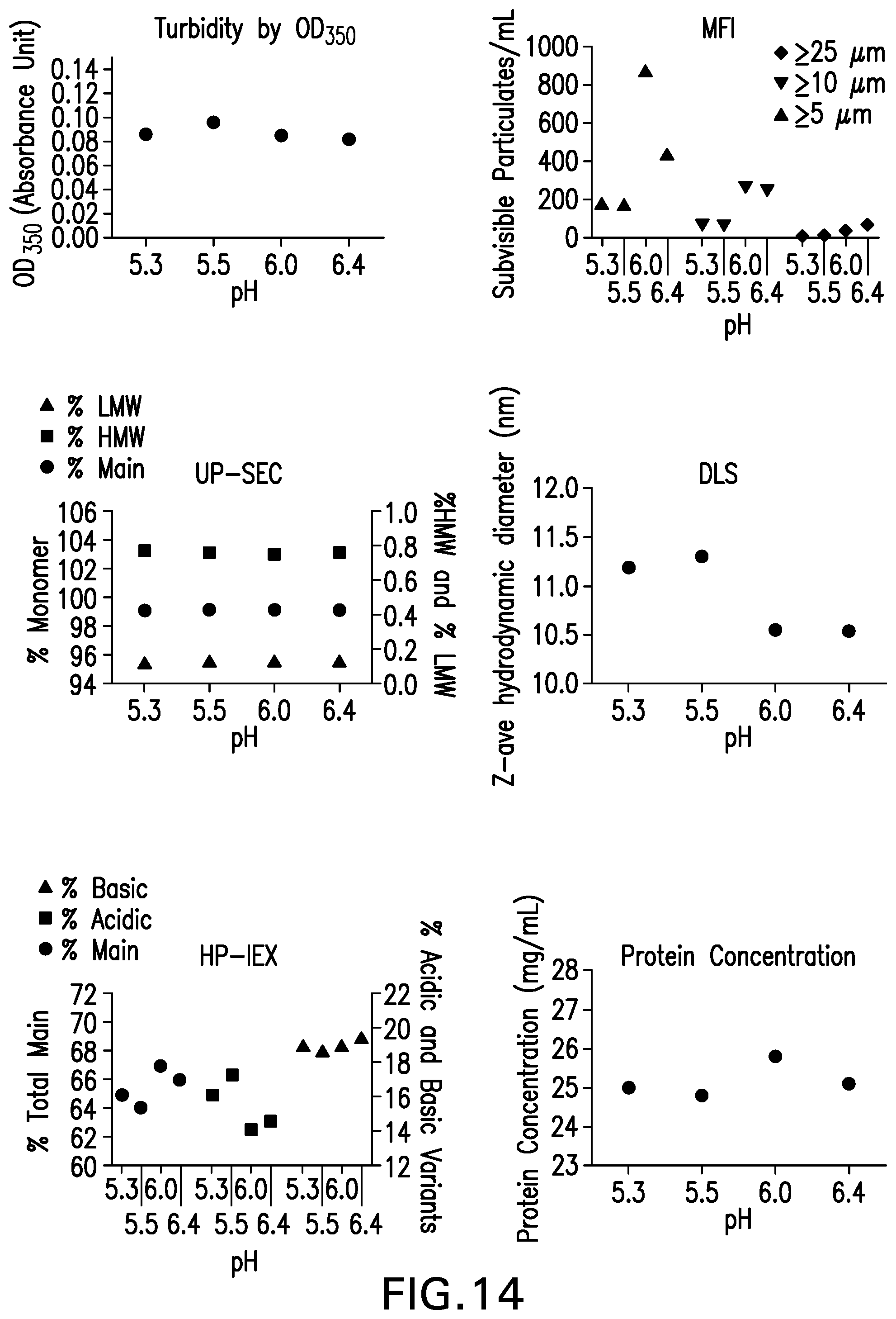
D00015

D00016

D00017

D00018

D00019

D00020

D00021

D00022

D00023

D00024

D00025

D00026

D00027

D00028

D00029

D00030

D00031

D00032
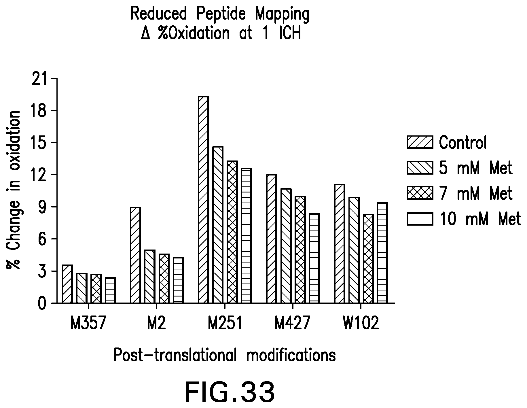
D00033

D00034

D00035

D00036

D00037

D00038

D00039

S00001
XML
uspto.report is an independent third-party trademark research tool that is not affiliated, endorsed, or sponsored by the United States Patent and Trademark Office (USPTO) or any other governmental organization. The information provided by uspto.report is based on publicly available data at the time of writing and is intended for informational purposes only.
While we strive to provide accurate and up-to-date information, we do not guarantee the accuracy, completeness, reliability, or suitability of the information displayed on this site. The use of this site is at your own risk. Any reliance you place on such information is therefore strictly at your own risk.
All official trademark data, including owner information, should be verified by visiting the official USPTO website at www.uspto.gov. This site is not intended to replace professional legal advice and should not be used as a substitute for consulting with a legal professional who is knowledgeable about trademark law.