Peptides, Combination Of Peptides, And Cell Based Medicaments For Use In Immunotherapy Against Urinary Bladder Cancer And Other
MAHR; Andrea ; et al.
U.S. patent application number 16/671981 was filed with the patent office on 2020-02-20 for peptides, combination of peptides, and cell based medicaments for use in immunotherapy against urinary bladder cancer and other . The applicant listed for this patent is Immatics Biotechnologies GmbH. Invention is credited to Jens FRITSCHE, Andrea MAHR, Oliver SCHOOR, Harpreet SINGH, Colette SONG, Toni WEINSCHENK.
| Application Number | 20200055898 16/671981 |
| Document ID | / |
| Family ID | 65529073 |
| Filed Date | 2020-02-20 |


View All Diagrams
| United States Patent Application | 20200055898 |
| Kind Code | A1 |
| MAHR; Andrea ; et al. | February 20, 2020 |
PEPTIDES, COMBINATION OF PEPTIDES, AND CELL BASED MEDICAMENTS FOR USE IN IMMUNOTHERAPY AGAINST URINARY BLADDER CANCER AND OTHER CANCERS
Abstract
The present invention relates to peptides, proteins, nucleic acids and cells for use in immunotherapeutic methods. In particular, the present invention relates to the immunotherapy of cancer. The present invention furthermore relates to tumor-associated T-cell peptide epitopes, alone or in combination with other tumor-associated peptides that can for example serve as active pharmaceutical ingredients of vaccine compositions that stimulate anti-tumor immune responses, or to stimulate T cells ex vivo and transfer into patients. Peptides bound to molecules of the major histocompatibility complex (MHC), or peptides as such, can also be targets of antibodies, soluble T-cell receptors, and other binding molecules.
| Inventors: | MAHR; Andrea; (Tuebingen, DE) ; WEINSCHENK; Toni; (Aichwald, DE) ; SONG; Colette; (Ostfildern, DE) ; SCHOOR; Oliver; (Tuebingen, DE) ; FRITSCHE; Jens; (Dusslingen, DE) ; SINGH; Harpreet; (Muenchen, DE) | ||||||||||
| Applicant: |
|
||||||||||
|---|---|---|---|---|---|---|---|---|---|---|---|
| Family ID: | 65529073 | ||||||||||
| Appl. No.: | 16/671981 | ||||||||||
| Filed: | November 1, 2019 |
Related U.S. Patent Documents
| Application Number | Filing Date | Patent Number | ||
|---|---|---|---|---|
| 16457012 | Jun 28, 2019 | |||
| 16671981 | ||||
| 15445461 | Feb 28, 2017 | 10400011 | ||
| 16457012 | ||||
| 62302010 | Mar 1, 2016 | |||
| Current U.S. Class: | 1/1 |
| Current CPC Class: | G01N 33/6893 20130101; C12N 2310/16 20130101; A61K 2039/86 20180801; G01N 2800/52 20130101; A61K 2039/868 20180801; A61K 2039/80 20180801; A61K 2039/892 20180801; A61P 35/00 20180101; C07K 2319/00 20130101; A61K 2039/884 20180801; A61K 39/001154 20180801; A61K 2039/82 20180801; A61K 39/0011 20130101; A61K 2039/804 20180801; A61K 2039/844 20180801; A61K 2039/876 20180801; A61K 2039/5154 20130101; C07K 16/18 20130101; C07K 7/06 20130101; A61K 35/17 20130101; A61K 39/001102 20180801; A61K 2035/124 20130101; C07K 14/7051 20130101; A61P 37/02 20180101; A61K 2039/852 20180801; A61K 2039/828 20180801; C07K 14/4748 20130101; C12N 15/115 20130101; A61K 38/00 20130101; C07K 7/08 20130101; A61K 2039/812 20180801; A61K 2039/836 20180801 |
| International Class: | C07K 7/08 20060101 C07K007/08; A61K 35/17 20060101 A61K035/17; C07K 7/06 20060101 C07K007/06; C07K 14/725 20060101 C07K014/725; C07K 14/47 20060101 C07K014/47; A61K 39/00 20060101 A61K039/00; C12N 15/115 20060101 C12N015/115; C07K 16/18 20060101 C07K016/18; G01N 33/68 20060101 G01N033/68 |
Foreign Application Data
| Date | Code | Application Number |
|---|---|---|
| Mar 1, 2016 | GB | 1603568.5 |
Claims
1. A peptide comprising the amino acid sequence of SEQ ID NO: 64, 1-63, or 65-205 in the form of a pharmaceutical acceptable salt.
2. The peptide of claim 1, wherein said peptide has the ability to bind to an MHC class-I or -II molecule, and wherein said peptide, when bound to said MHC, is capable of being recognized by CD4 and/or CD8 T cells.
3. The peptide of claim 1, wherein the pharmaceutically acceptable salt is chloride salt or acetate salt.
4. The peptide of claim 1, wherein the peptide consists of the amino acid sequence of SEQ ID NO: 64, 1-63, or 65-205.
5. A fusion protein comprising a peptide consisting of the amino acid sequence of SEQ ID NO: 64, 1-63, or 65-205 and the 80 N-terminal amino acids of the HLA-DR antigen-associated invariant chain (Ii) or an antibody.
6. The fusion protein of claim 5, wherein the antibody binds to an dendritic cell.
7. A pharmaceutical composition comprising the peptide of claim 1.
8. The pharmaceutical composition of claim 7, further comprising an adjuvant.
9. The pharmaceutical composition of claim 8, wherein the adjuvant is at least one selected from anti-CD40 antibody, imiquimod, resiquimod, GM-CSF, cyclophosphamide, sunitinib, bevacizumab, interferon-alpha, interferon-beta, CpG oligonucleotides and derivatives, poly-(I:C) and derivatives, RNA, sildenafil, particulate formulations with poly(lactide co-glycolide) (PLG), virosomes, interleukin (IL)-1, IL-2, IL-4, IL-7, IL-12, IL-13, IL-15, IL-21, and IL-23.
10. The pharmaceutical composition of claim 8, wherein the adjuvant is at least one selected from the group consisting of IL-2, IL-12, IL-15, and IL-21.
11. The pharmaceutical composition of claim 8, further comprising a pharmaceutically acceptable carrier.
12. The pharmaceutical composition of claim 8, further comprising a pharmaceutically acceptable stabilizer.
13. A modified peptide comprising the peptide of claim 1, wherein said peptide comprises at least one non-peptide bond or at least one D-amino acid.
14. An in vitro method for producing activated T lymphocytes, comprising contacting in vitro T cells with antigen loaded human HLA-A*02:01 expressed on the surface of a suitable antigen-presenting cell or an artificial construct mimicking an antigen-presenting cell for a period of time sufficient to activate said T cells in an antigen specific manner, wherein said antigen is a peptide consisting of the amino acid sequence of SEQ ID NO: 64, 1-63, or 65-205.
15. A method for producing a peptide consisting of the amino acid sequence of SEQ ID NO: 64, 1-63, or 65-205, comprising culturing a host cell, and isolating the peptide from said host cell and/or culture medium thereof.
16. A method for killing target cells in a patient who has cancer, wherein the target cells present a peptide consisting of the amino acid sequence of SEQ ID NO: 64, 1-63, or 65-205 in a complex with HLA-A*02:01, comprising administering to the patient the composition of claim 8, wherein said cancer is selected from the group consisting of urinary bladder cancer, acute myelogenous leukemia, breast cancer, bile duct cancer, brain cancer, chronic lymphocytic leukemia, colorectal carcinoma, esophageal cancer, gallbladder cancer, gastric cancer, hepatocellular cancer, Merkel cell carcinoma, melanoma, non-Hodgkin lymphoma, non-small cell lung cancer, ovarian cancer, pancreatic cancer, prostate cancer, renal cell cancer, small cell lung cancer, head and neck cancer (HNSCC), and uterine cancer.
17. The method of claim 16, wherein the cancer is urinary bladder cancer.
18. The method of claim 16, wherein the cancer is HNSCC.
19. A kit comprising: a) a container comprising the pharmaceutical composition of claim 7 in solution or in lyophilized form; b) optionally, a second container containing a diluent or reconstituting solution for the lyophilized formulation; c) optionally, instructions for (i) use of the solution or (ii) reconstitution and/or use of the lyophilized formulation.
20. The kit according to claim 19, further comprising one or more of (iii) a buffer, (iv) a diluent, (v) a filter, (vi) a needle, or (v) a syringe.
Description
CROSS REFERENCE TO RELATED APPLICATIONS
[0001] This application is a Continuation of Ser. No. 16/457,012, filed on 28 Jun. 2019, which is a Continuation of U.S. patent application Ser. No. 15/445,461, filed 28 Feb. 2017, which claims the benefit of U.S. Provisional Application Ser. No. 62/302,010, filed 1 Mar. 2016, and Great Britain Application No. 1603568.5, filed 1 Mar. 2016, the content of each of these applications is herein incorporated by reference in their entirety.
[0002] This application also is related to PCT/EP2017/054559 filed 28 Feb. 2017, the content of which is incorporated herein by reference in its entirety.
REFERENCE TO SEQUENCE LISTING SUBMITTED AS A COMPLIANT ASCII TEXT FILE (.txt)
[0003] Pursuant to the EFS-Web legal framework and 37 CFR .sctn..sctn. 1.821-825 (see MPEP .sctn. 2442.03(a)), a Sequence Listing in the form of an ASCII-compliant text file (entitled "2912919-063003_Sequence_Listing_ST25.txt" created on 24 Oct. 2019, and 32,371 bytes in size) is submitted concurrently with the instant application, and the entire contents of the Sequence Listing are incorporated herein by reference.
FIELD
[0004] The present invention relates to peptides, proteins, nucleic acids and cells for use in immunotherapeutic methods. In particular, the present invention relates to the immunotherapy of cancer. The present invention furthermore relates to tumor-associated T-cell peptide epitopes, alone or in combination with other tumor-associated peptides that can for example serve as active pharmaceutical ingredients of vaccine compositions that stimulate anti-tumor immune responses, or to stimulate T cells ex vivo and transfer into patients. Peptides bound to molecules of the major histocompatibility complex (MHC), or peptides as such, can also be targets of antibodies, soluble T-cell receptors, and other binding molecules.
[0005] The present invention relates to several novel peptide sequences and variants thereof derived from HLA class I molecules of human tumor cells that can be used in vaccine compositions for eliciting anti-tumor immune responses, or as targets for the development of pharmaceutically/immunologically active compounds and cells.
BACKGROUND OF THE INVENTION
[0006] The most common type of bladder cancer starts in urothelium or transitional epithelium, the innermost lining of the bladder, and is called urothelial carcinoma or transitional cell carcinoma (TCC). Urothelial cells are located in the other parts of urinary tract, which by bladder cancer patients can be infiltrated by cancer as well. The progressing bladder cancer grows further into or through the other layers in the bladder wall, spreads first of all to the lymph nodes, the bones, the lungs, or the liver (American Cancer Society, 2015).
[0007] Bladder cancers are characterized by their spread and type of growing. Depending on its expansion, bladder cancer is categorized as non-invasive and invasive. The non-invasive or superficial bladder cancer is localized exclusively in the innermost layer of bladder wall. In contrast, the invasive bladder cancer is grown into the deeper layers of bladder wall (American Cancer Society, 2015).
[0008] Depending on the type of growing, bladder cancer are divided into papillary and flat carcinomas. Papillary carcinomas grow as slender, finger-like extensions from the inner side of the bladder cancer towards the hollow center. Papillary carcinomas are often non-invasive. The very slow growing, non-invasive papillary cancer is sub classified as papillary urothelial neoplasm of low-malignant potential (PUNLMP). Those tumors mostly show a very good therapeutic outcome. Flat carcinomas do not grow towards the center of the bladder cancer. The flat bladder carcinomas, which are restricted only to the inner layer of bladder wall, are called non-invasive flat carcinoma or a flat carcinoma in situ (CIS). Both papillary and flat carcinomas, which spread further into the deeper layers of bladder wall, are called invasive urothelial (or transitional cell) carcinoma (American Cancer Society, 2015).
[0009] The other cancer types, which start in the bladder, are squamous cell and small cell carcinomas, adenocarcinomas and sarcomas. Those cancer types are rare (American Cancer Society, 2015). Bladder cancer is divided into 5 stages. At stage 0, cancer cells are restricted only to the inner layer of bladder wall. The bladder cancer at stage 0 is divided in stage 0 a by papillary carcinomas or stage 0 is by flat carcinomas. At the following stages I and II, bladder cancer cells infiltrate further layers of the bladder wall and get spread into the connective tissue and muscle tissue, respectively. At the stage III, bladder cancer cells spread further from the bladder into the surrounding fat or even reproductive organs. At the stage IV, bladder cancer infiltrates the wall of the abdomen or pelvis, lymph nodes and further distant organs like lung, bones or liver (National Cancer Institute, 2015).
[0010] In the United States, bladder cancer represents the ninth leading cause of cancer death. The percentage of death caused by bladder cancer increases with the age and is at its highest at the age of 75-84. Bladder cancer accounts to 4.5% of all new cancer cases. The people in the age of 65-84 are most frequently diagnosed with bladder cancer. Bladder cancer is diagnosed about 4 times more frequently in men than in women. Over the last 10 years, the number of new bladder cancer cases has been decreasing for 0.6% each year, the death rates and 5-years relative survival have remained stable (SEER Stat facts, 2014).
[0011] On average the 5-years relative survival for bladder cancer is about 77%. In general, the 5-years relative survival depends strongly on the stage, when the bladder cancer has been diagnosed. 51% new bladder cancer cases are recognized on the very early in situ stage, when the bladder cancer is localized only in the originating layer of bladder wall. The 5-years relative survival at this stage accounts for about 96%. 35% of the new bladder cancer cases are diagnosed at the localized stage. In those cases, bladder cancer is restricted to the primary site and the 5-years relative survival is about 70%. The 5-years relative survival of new bladder cancer cases, which are first diagnosed at the regional stage (cancer spread to lymph nodes, 7% of all new bladder cancer cases) and distant stage (metastasizing cancer, 4% of all new bladder cancer cases), accounts for 34 and 5.4%, respectively. The stage of 3% newly diagnosed bladder cancer is unknown. The 5-years relative survival for those cancer is about 47% (SEER Stat facts, 2014).
[0012] The standard treatment for bladder cancer includes surgery, radiation therapy, chemotherapy and immunotherapy.
[0013] The following types of surgery can be undertaken by the treatment of bladder cancer: transurethral resection, radical or partial cystectomy and urinary diversion. By the transurethral resection, the cancer is removed from the bladder inner wall mechanically or by the burning of the tumor with high-energy electricity. By the cystectomy, the bladder together with any lymph nodes and other by cancer invaded nearby organs is removed partially or completely. The urinary diversion is a type of surgery by which the new ways of storage and passing of urine are done. The surgery is often complemented with application of chemotherapy, which intends to lower the risk of the cancer recurrence (National Cancer Institute, 2015).
[0014] At stage 0 and I, the bladder cancer is typically treated by transurethral resection potentially followed by intravesical chemotherapy and optionally combined with intravesical immunotherapeutic treatment with BCG (Bacillus Calmette-Guerin). Partial or radical cystectomy is possible. Bladder cancer at stage II and III treated by radical or partial cystectomy, transurethral resection or external radiation with or without chemotherapy. At the stage IV, the treatment of bladder cancer depends on how far cancer cells have spread in the body. If the bladder cancer cells are still restricted only to the bladder, the cancer can be treated by the chemotherapy alone, radical cystectomy or external-beam radiation either with or without chemotherapy. By the invasion of bladder cancer to other parts of the body, the chemotherapy with or without surgery or radiation therapy is the first choice of treatment. At this stage, the external-beam radiation, urinary diversion or cystectomy can be applied as palliative therapy. At any stage of bladder cancer, patients have a possibility to enroll in a clinical trial (National Cancer Institute, 2015).
[0015] An effective immunotherapeutic approach is established in the treatment of aggressive non-muscle invasive bladder cancer (NMIBC). Thereby, a weakened form of the bacterium Mycobacterium bovis (Bacillus Calmette-Guerin=BCG) is applied as an intravesical solution. The major effect of BCG treatment is a significant long-term (up to 10 years) protection from cancer recurrence and reduced progression rate. In principle, the treatment with BCG induces a local inflammatory response which stimulates the cellular immune response. The immune response to BCG is based on the following key steps: infection of urothelial and bladder cancer cells by BCG, followed by increased expression of antigen-presenting molecules, induction of immune response mediated via cytokine release, induction of antitumor activity via involvement of various immune cells (thereunder cytotoxic T lymphocytes, neutrophils, natural killer cells, and macrophages) (Fuge et al., 2015; Gandhi et al., 2013).
[0016] BCG treatment is in general well tolerated by patients but can be fatal especially by the immunocompromised patients. BCG refractory is observed in about 30-40% of patients (Fuge et al., 2015; Steinberg et al., 2016a). The treatment of patients who failed the BCG therapy is challenging. The patients who failed the BCG treatment are at high risk for developing of muscle-invasive disease. Radical cystectomy is the preferable treatment option for non-responders (Steinberg et al., 2016b; von Rundstedt and Lerner, 2015). The FDA approved second line therapy of BCG-failed NMIBC for patients who desire the bladder preservation is the chemotherapeutic treatment with valrubicin. A number of other second line therapies are available or being currently under investigation as well, thereunder immunotherapeutic approaches like combined BCG-interferon or BCG-check point inhibitor treatments, pre-BCG transdermal vaccination, treatment with Mycobacterium phlei cell wall-nucleic acid (MCNA) complex, mono- or combination chemotherapy with various agents like mitomycin C, gemcitabine, docetaxel, nab-paclitaxel, epirubicin, mitomycin/gemcitabine, gemcitabine/docetaxel, and device-assisted chemotherapies like thermochemo-, radiochemo-, electromotive or photodynamic therapies (Fuge et al., 2015; Steinberg et al., 2016b; von Rundstedt and Lerner, 2015). Further evaluation of available therapies in clinical trials is still required.
[0017] In general, the treatment of advanced bladder cancer with muscle invasion known as muscle-invasive bladder carcinoma (MIBC) or metastatic bladder cancer is challenging and remained substantially unchanged over the last few decades. Nowadays available options for treatment of progressed bladder cancer are insufficiently effective. New trends in the clinical management of bladder cancer have been opening up by the lately emerging understanding of genetic background of urothelial cancer. Especially, the recent implementation of predictive genomic and molecular biomarkers intends to benefit the therapeutic response (Jones et al., 2016; Rouanne et al., 2016; Grivas et al., 2015; Azevedo et al., 2015; Knollman et al., 2015a).
[0018] Before 2003 cystectomy alone was established as a standard treatment of MIBC. The recurrence of cancer at distant body sites after the surgery showed the necessity of neoadjuvant chemotherapeutic treatment. The combined chemotherapy with high dose-intensity methotrexate, vinblastine, doxorubicin and cisplatin (accelerated MVAC or AMVAC) or gemcitabine and cisplatin (GC) count currently to a standard neoadjuvant treatment of MIBC in the United States. The adjuvant application of chemotherapy by the treatment of MIBC is limited by the high surgical complication rate after radical cystectomy. Nevertheless, the usage of AMVAC, GC or combination of cisplatin, methotrexate and vinblastine (CMV) is currently recommended as adjuvant chemotherapy for high-risk MIBC patients. The similar systematic chemotherapy is used by treatment of metastatic bladder cancer. In general, only 30 to 40% of patients respond to the cisplatin-based chemotherapy. Furthermore, patients with impaired renal function are ineligible for treatment with cisplatin. The second-line treatment depends on the previous treatment and has not been scandalized (Knollman et al., 2015b; Rouanne et al., 2016).
[0019] The alternative treatment options for advanced bladder cancer are being investigated in ongoing clinical trials. The current clinical trials focused on the development of molecularly targeted therapies and immunotherapies. The targeted therapies investigate the effects of cancerogenesis related pathway inhibitors (i.e. mTOR, vascular endothelial, fibroblast, or epidermal growth factor receptors, anti-angiogenesis or cell cycle inhibitors) in the treatment of bladder cancer. The development of molecularly targeted therapies remains challenging due to high degree of genetic diversity of bladder cancer. The main focus of the current immunotherapy is the development of checkpoint blockage agents like anti-PD1 monoclonal antibody and adoptive T-cell transfer (Knollman et al., 2015b; Grivas et al., 2015; Jones et al., 2016; Rouanne et al., 2016).
[0020] Considering the severe side-effects and expense associated with treating cancer, there is a need to identify factors that can be used in the treatment of cancer in general and urinary bladder cancer in particular. There is also a need to identify factors representing biomarkers for cancer in general and urinary bladder cancer in particular, leading to better diagnosis of cancer, assessment of prognosis, and prediction of treatment success.
[0021] Immunotherapy of cancer represents an option of specific targeting of cancer cells while minimizing side effects. Cancer immunotherapy makes use of the existence of tumor associated antigens.
[0022] The current classification of tumor associated antigens (TAAs) comprises the following major groups:
[0023] a) Cancer-testis antigens: The first TAAs ever identified that can be recognized by T cells belong to this class, which was originally called cancer-testis (CT) antigens because of the expression of its members in histologically different human tumors and, among normal tissues, only in spermatocytes/spermatogonia of testis and, occasionally, in placenta. Since the cells of testis do not express class I and II HLA molecules, these antigens cannot be recognized by T cells in normal tissues and can therefore be considered as immunologically tumor-specific. Well-known examples for CT antigens are the MAGE family members and NY-ESO-1.
[0024] b) Differentiation antigens: These TAAs are shared between tumors and the normal tissue from which the tumor arose. Most of the known differentiation antigens are found in melanomas and normal melanocytes. Many of these melanocyte lineage-related proteins are involved in biosynthesis of melanin and are therefore not tumor specific but nevertheless are widely used for cancer immunotherapy. Examples include, but are not limited to, tyrosinase and Melan-A/MART-1 for melanoma or PSA for prostate cancer.
[0025] c) Over-expressed TAAs: Genes encoding widely expressed TAAs have been detected in histologically different types of tumors as well as in many normal tissues, generally with lower expression levels. It is possible that many of the epitopes processed and potentially presented by normal tissues are below the threshold level for T-cell recognition, while their over-expression in tumor cells can trigger an anticancer response by breaking previously established tolerance. Prominent examples for this class of TAAs are Her-2/neu, survivin, telomerase, or WT1.
[0026] d) Tumor-specific antigens: These unique TAAs arise from mutations of normal genes (such as .beta.-catenin, CDK4, etc.). Some of these molecular changes are associated with neoplastic transformation and/or progression. Tumor-specific antigens are generally able to induce strong immune responses without bearing the risk for autoimmune reactions against normal tissues. On the other hand, these TAAs are in most cases only relevant to the exact tumor on which they were identified and are usually not shared between many individual tumors. Tumor-specificity (or -association) of a peptide may also arise if the peptide originates from a tumor- (-associated) exon in case of proteins with tumor-specific (-associated) isoforms.
[0027] e) TAAs arising from abnormal post-translational modifications: Such TAAs may arise from proteins which are neither specific nor overexpressed in tumors but nevertheless become tumor associated by posttranslational processes primarily active in tumors. Examples for this class arise from altered glycosylation patterns leading to novel epitopes in tumors as for MUC1 or events like protein splicing during degradation which may or may not be tumor specific.
[0028] f) Oncoviral proteins: These TAAs are viral proteins that may play a critical role in the oncogenic process and, because they are foreign (not of human origin), they can evoke a T-cell response. Examples of such proteins are the human papilloma type 16 virus proteins, E6 and E7, which are expressed in cervical carcinoma.
[0029] T-cell based immunotherapy targets peptide epitopes derived from tumor-associated or tumor-specific proteins, which are presented by molecules of the major histocompatibility complex (MHC). The antigens that are recognized by the tumor specific T lymphocytes, that is, the epitopes thereof, can be molecules derived from all protein classes, such as enzymes, receptors, transcription factors, etc. which are expressed and, as compared to unaltered cells of the same origin, usually up-regulated in cells of the respective tumor.
[0030] There are two classes of MHC-molecules, MHC class I and MHC class II. MHC class I molecules are composed of an alpha heavy chain and beta-2-microglobulin, MHC class II molecules of an alpha and a beta chain. Their three-dimensional conformation results in a binding groove, which is used for non-covalent interaction with peptides.
[0031] MHC class I molecules can be found on most nucleated cells. They present peptides that result from proteolytic cleavage of predominantly endogenous proteins, defective ribosomal products (DRIPs) and larger peptides. However, peptides derived from endosomal compartments or exogenous sources are also frequently found on MHC class I molecules. This non-classical way of class I presentation is referred to as cross-presentation in the literature (Brossart and Bevan, 1997; Rock et al., 1990). MHC class II molecules can be found predominantly on professional antigen presenting cells (APCs), and primarily present peptides of exogenous or transmembrane proteins that are taken up by APCs e.g. during endocytosis, and are subsequently processed.
[0032] Complexes of peptide and MHC class I are recognized by CD8-positive T cells bearing the appropriate T-cell receptor (TCR), whereas complexes of peptide and MHC class II molecules are recognized by CD4-positive-helper-T cells bearing the appropriate TCR. It is well known that the TCR, the peptide and the MHC are thereby present in a stoichiometric amount of 1:1:1.
[0033] CD4-positive helper T cells play an important role in inducing and sustaining effective responses by CD8-positive cytotoxic T cells. The identification of CD4-positive T-cell epitopes derived from tumor associated antigens (TAA) is of great importance for the development of pharmaceutical products for triggering anti-tumor immune responses (Gnjatic et al., 2003). At the tumor site, T helper cells, support a cytotoxic T cell- (CTL-) friendly cytokine milieu (Mortara et al., 2006) and attract effector cells, e.g. CTLs, natural killer (NK) cells, macrophages, and granulocytes (Hwang et al., 2007).
[0034] In the absence of inflammation, expression of MHC class II molecules is mainly restricted to cells of the immune system, especially professional antigen-presenting cells (APC), e.g., monocytes, monocyte-derived cells, macrophages, dendritic cells. In cancer patients, cells of the tumor have been found to express MHC class II molecules (Dengjel et al., 2006).
[0035] Elongated (longer) peptides of the invention can function as MHC class II active epitopes.
[0036] T-helper cells, activated by MHC class II epitopes, play an important role in orchestrating the effector function of CTLs in anti-tumor immunity. T-helper cell epitopes that trigger a T-helper cell response of the TH1 type support effector functions of CD8-positive killer T cells, which include cytotoxic functions directed against tumor cells displaying tumor-associated peptide/MHC complexes on their cell surfaces. In this way tumor-associated T-helper cell peptide epitopes, alone or in combination with other tumor-associated peptides, can serve as active pharmaceutical ingredients of vaccine compositions that stimulate anti-tumor immune responses.
[0037] It was shown in mammalian animal models, e.g., mice, that even in the absence of CD8-positive T lymphocytes, CD4-positive T cells are sufficient for inhibiting manifestation of tumors via inhibition of angiogenesis by secretion of interferon-gamma (IFN.gamma.) (Beatty and Paterson, 2001; Mumberg et al., 1999). There is evidence for CD4 T cells as direct anti-tumor effectors (Braumuller et al., 2013; Tran et al., 2014).
[0038] Since the constitutive expression of HLA class II molecules is usually limited to immune cells, the possibility of isolating class II peptides directly from primary tumors was previously not considered possible. However, Dengjel et al. were successful in identifying a number of MHC Class II epitopes directly from tumors (WO 2007/028574, EP 1 760 088 B1).
[0039] Since both types of response, CD8 and CD4 dependent, contribute jointly and synergistically to the anti-tumor effect, the identification and characterization of tumor-associated antigens recognized by either CD8+ T cells (ligand: MHC class I molecule+peptide epitope) or by CD4-positive T-helper cells (ligand: MHC class II molecule+peptide epitope) is important in the development of tumor vaccines.
[0040] For an MHC class I peptide to trigger (elicit) a cellular immune response, it also must bind to an MHC-molecule. This process is dependent on the allele of the MHC-molecule and specific polymorphisms of the amino acid sequence of the peptide. MHC-class-I-binding peptides are usually 8-12 amino acid residues in length and usually contain two conserved residues ("anchors") in their sequence that interact with the corresponding binding groove of the MHC-molecule. In this way each MHC allele has a "binding motif" determining which peptides can bind specifically to the binding groove.
[0041] In the MHC class I dependent immune reaction, peptides not only have to be able to bind to certain MHC class I molecules expressed by tumor cells, they subsequently also have to be recognized by T cells bearing specific T cell receptors (TCR).
[0042] For proteins to be recognized by T-lymphocytes as tumor-specific or -associated antigens, and to be used in a therapy, particular prerequisites must be fulfilled. The antigen should be expressed mainly by tumor cells and not, or in comparably small amounts, by normal healthy tissues. In a preferred embodiment, the peptide should be over-presented by tumor cells as compared to normal healthy tissues. It is furthermore desirable that the respective antigen is not only present in a type of tumor, but also in high concentrations (i.e. copy numbers of the respective peptide per cell). Tumor-specific and tumor-associated antigens are often derived from proteins directly involved in transformation of a normal cell to a tumor cell due to their function, e.g. in cell cycle control or suppression of apoptosis. Additionally, downstream targets of the proteins directly causative for a transformation may be up-regulated and thus may be indirectly tumor-associated. Such indirect tumor-associated antigens may also be targets of a vaccination approach (Singh-Jasuja et al., 2004). It is essential that epitopes are present in the amino acid sequence of the antigen, in order to ensure that such a peptide ("immunogenic peptide"), being derived from a tumor associated antigen, leads to an in vitro or in vivo T-cell-response.
[0043] Basically, any peptide able to bind an MHC molecule may function as a T-cell epitope. A prerequisite for the induction of an in vitro or in vivo T-cell-response is the presence of a T cell having a corresponding TCR and the absence of immunological tolerance for this particular epitope.
[0044] Therefore, TAAs are a starting point for the development of a T cell based therapy including but not limited to tumor vaccines. The methods for identifying and characterizing the TAAs are usually based on the use of T-cells that can be isolated from patients or healthy subjects, or they are based on the generation of differential transcription profiles or differential peptide expression patterns between tumors and normal tissues. However, the identification of genes over-expressed in tumor tissues or human tumor cell lines, or selectively expressed in such tissues or cell lines, does not provide precise information as to the use of the antigens being transcribed from these genes in an immune therapy. This is because only an individual subpopulation of epitopes of these antigens are suitable for such an application since a T cell with a corresponding TCR has to be present and the immunological tolerance for this particular epitope needs to be absent or minimal. In a very preferred embodiment of the invention it is therefore important to select only those over- or selectively presented peptides against which a functional and/or a proliferating T cell can be found. Such a functional T cell is defined as a T cell, which upon stimulation with a specific antigen can be clonally expanded and is able to execute effector functions ("effector T cell").
[0045] In case of targeting peptide-MHC by specific TCRs (e.g. soluble TCRs) and antibodies or other binding molecules (scaffolds) according to the invention, the immunogenicity of the underlying peptides is secondary. In these cases, the presentation is the determining factor.
SUMMARY OF THE INVENTION
[0046] In a first aspect of the present invention, the present invention relates to a peptide comprising an amino acid sequence selected from the group consisting of SEQ ID NO: 1 to SEQ ID NO: 149 or a variant sequence thereof which is at least 77%, preferably at least 88%, homologous (preferably at least 77% or at least 88% identical) to SEQ ID NO: 1 to SEQ ID NO: 149, wherein said variant binds to MHC and/or induces T cells cross-reacting with said peptide, or a pharmaceutical acceptable salt thereof, wherein said peptide is not the underlying full-length polypeptide.
[0047] The present invention further relates to a peptide of the present invention comprising a sequence that is selected from the group consisting of SEQ ID NO: 1 to SEQ ID NO: 149 or a variant thereof, which is at least 77%, preferably at least 88%, homologous (preferably at least 77% or at least 88% identical) to SEQ ID NO: 1 to SEQ ID NO: 149, wherein said peptide or variant thereof has an overall length of between 8 and 100, preferably between 8 and 30, and most preferred of between 8 and 14 amino acids.
[0048] The following tables show the peptides according to the present invention, their respective SEQ ID NOs, and the prospective source (underlying) genes for these peptides. All peptides in Table 1 and Table 2 bind to HLA-A*02. The peptides in Table 2 have been disclosed before, but have not been associated with cancer at all before. The peptides in Table 3 are additional peptides that may be useful in combination with the other peptides of the invention. The peptides in Table 4 are furthermore useful in the diagnosis and/or treatment of various other malignancies that involve an over-expression or over-presentation of the respective underlying polypeptide.
TABLE-US-00001 TABLE 1 Peptides according to the present invention. J = phospho-serine, U = phosphothreonine SEQ ID No Sequence Gene ID(s) Official Gene Symbol(s) 1 ILLQASVQV 56649 TMPRSS4 2 GLLKAYSIRTA 3855 KRT7 3 YLDEIPPKFSM 4070 TACSTD2 4 SLDVVNLLV 57182 ANKRD50 5 IQDPVIFYV 57211 GPR126 6 SIVDFLITA 10848 PPP1R13L 7 QMFEGQILDV 55057 AIM1L 8 ALSFSSSAGPGLLKA 3855 KRT7 9 SLVDARFQL 130497 OSR1 10 GLWHGMFANV 57609 DIP2B 11 AMAELRVVV 126410 CYP4F22 12 GVALTVTGV 2319 FLOT2 13 FLEEKEQAAL 10626, 147166, TRIM16, CDRT1, TRIM16L 374286 14 GLAGPVRGV 6628, 6638, 8926 SNURF, SNRPB, SNRPN 15 YLAPENGYLMEA 6625 SNRNP70 16 ILGPQGNTI 10657 KHDRBS1 17 RLSQLEGVNV 55737 VP535 18 SIAAYNPVV 10202 DHRS2 19 SLATTLTKI 1315 COPB1 20 YLPDSLTQL 55227 LRRC1 21 TLIEDDALNGA 3248 HPGD 22 SIAKEGVVGA 6623 SNCG 23 YTLSKTEFL 6282 S100A11 24 SLLGGITVV 9043 SPAG9 25 SLDSSGFSL 143 PARP4 26 GLALLYSGV 533 ATP6VOB 27 STTNGGILTV 84248 FYTTD1 28 GLIDSLMAYV 5317 PKP1 29 ALSSPPPTV 9372 ZFYVE9 30 ILDISRSEV 57720 GPR107 31 SLFDGIATGL 1788 DNMT3A 32 YQAPDIDVQL 29997 GLTSCR2 33 VLFGEITRL 4720 NDUFS2 34 ALLDEQQVNV 3397 ID1 35 KLPEPPPLA 2086 ERV3-1 36 ALWDEFNQL 6899 TBX1 37 ILSAILVHL 339766 MROH2A 38 TLTSIIVAV 339766 MROH2A 39 AMASHLTST 100526737, 10432, RBM4 5936 40 VIADRVVTV 127731 VWA5B1 41 FLDDGNQMLL 79659 DYNC2H1 42 FLIDASQRV 256076 COL6A5 43 FLIESKLLSL 92749 DRC1 44 GLAQDPKSLQL 23331 TTC28 45 IIDSSPTAL 79915 ATAD5 46 SLFIGAEIVAV 405753 DUOXA2 47 VLMDDTDPL 5700 PSMC1 48 VLMDDTDPLV 5700 PSMC1 49 SLIGGTNFV 162 AP1B1 50 VLANRVAVV 10202 DHRS2 51 ALLDKAQINL 147711, 26974 ZNF285 52 SLATLEGIQL 7776 ZNF236 53 ILVQVIPVV 3664 IRF6 54 ALNDEINFL 3855 KRT7 55 KLLETKVVTL 3855 KRT7 56 ILLRDLPTL 400451 FAM174B 57 GLAHFVNEI 7750 ZMYM2 58 GLDSSVNVQGSVL 9589 WTAP 59 WLSTSIPEA 2194 FASN 60 SLSDVRVIV 57186 RALGAPA2 61 VLLDNLPGV 27152 INTU 62 GQLDFSEFL 6282 S100A11 63 LLAGLLVGV 54991 C1orf159 64 GLLSQGSPL 2729 GCLC 65 IITDLLRSV 8880 FUBP1 66 SLWEENQAL 3911 LAMA5 67 FLTPPLLSV 160 AP2A1 68 TM IVSLAAV 23204 ARL6IP1 69 QIWDKILSV 10202 DHRS2 70 KLAEISLGV 1830 DSG3 71 LLSEDFVSV 64847 SPATA20 72 SLFTGLRSI 50506, 53905 DUOX2, DUOX1 73 VLKVFLENV 121504, 554313, HIST1H4D, HIST1H4J, 8294, 8359, 8360, HIST2H4B, HIST1H4L, 8361, 8362, 8363, HIST1H4H, HIST1H4K, 8364, 8365, 8366, HIST1H4A, HIST2H4A, 8367, 8368, 8370 HIST1H4B, HIST1H4I, HIST4H4, HIST1H4E, HIST1H4C, HIST1H4F 74 LLQEGEVYSA 10594 PRPF8 75 RVISSVISV 64123 ELTD1 76 AVVSSVNTV 6623 SNCG 77 KVFGGFQVV 3280 HES1 78 FIPDFAVAI 1293 COL6A3 79 FLDPATPRV 7050 TGIF1 80 ILLDTPLFLL 253190, 94009 SERHL2 81 SLDKGTLYI 79887 PLBD1 82 NLHNSYYSV 901 CCNG2 83 VILDKYYFL 56649 TMPRSS4 84 ALDPASISV 54165 DCUN1D1 85 HLLDSKVPSV 3691 ITGB4 86 FLIJLIISV 50835 TAS2R9 87 RLLELLQEA 150696 PROM2 88 ALASLENHV 51592 TRIM33 89 YLFPETEFI 6926 TBX3 90 GMTELYFQL 5591 PRKDC 91 NLDAATYQV 23443 SLC35A3 92 ALLDEQQVNVLL 3397 ID1 93 LLDLIQTKV 162,163 AP2B1, AP1B1 94 ALADGVPVAL 91056 AP5B1 95 YLIGQHVTA 23014 FBXO21 96 KLTNGIVVVL 162 AP1B1 97 TVGPGLLGV 7450 VWF 98 YLIGLDPENLAL 54677 CROT 99 ALIGDDVGL 55421 C17orf85 100 SLQSFIHGV 79905 TMC7 101 ILDEMRAQL 4940 OAS3 102 GLYEGLDWL 373, 375, 377, 378 ARF3, TRIM23, ARF1, ARF4 103 GLYSGEVLV 89891 WDR34 104 NAVVELVTV 10159 ATP6AP2 105 TLFPSKIGV 55233, 92597 MOB1A, MOB1B 106 ILLDTPLFL 253190, 94009 SERHL2 107 LULAKLEKV 51531 C9orf156 108 YLDPNQRDL 126231 ZNF573
TABLE-US-00002 TABLE 2 Additional peptides according to the present invention SEQ ID Official Gene No Sequence Gene ID(s) Symbol(s) 109 RLIDDMVAQA 3417 IDH1 110 VLFNIDGQGNHV 2625 GATA3 111 LLDVTPKAV 9414 TJP2 112 YLDPSLNSL 55086 CXorf57 113 FVFEPPPGV 1778 DYNC1H1 114 IITKDLFQV 1778 DYNC1H1 115 SLLDFERSL 84343 HPS3 116 QLAWFDTDL 1499 CTNNB1 117 YMLDIFHEVL 3038 HAS3 118 RLLDFPTLLV 54627 MAP10 119 SLDEKQNLV 79828 METTL8 120 IIIPEIQKV 1434 CSE1L 121 QLQGYLRSV 6128 RPL6 122 ILEPSLYTV 22818 COPZ1 123 NLAGVYSEV 4733 DRG1 124 QIDGTLSTI 100307126, FBXO34, CHMP4C, 128866, 55030, CHMP4B, 92421 CHMP4BP1 125 VLDEGSASV 1844 DUSP2 126 SLLRVGWSV 221092, 26580 BSCL2, HNRNPUL2 127 KLNATNIEL 5686 PSMA5 128 KLWGQSIQL 54823 SWT1 129 YLEPKLTQV 23198 PSME4 130 TLTSKLYSL 29796 UQCR10 131 ILTSIQSLL 7319, 7320 UBE2A, UBE2B 132 YILEGEPGKV 6730 SRP68 133 GLDPLGYEIQL 57122 NUP107 134 IVAPGTFEV 3608 ILF2 135 FLLPLIIVL 3140 MR1 136 GLSEPIFQL 55669 MFN1 137 ALFPHLLQPVL 1434 CSE1L 138 YLTNEGIQYL 100529239, RPS10-NUDT3, 6204 RPS10 139 LLYPTEITV 3675 ITGA3 140 ALLDGRVQL 375790 AGRN 141 SMFGAGLTV 533 ATP6V0B 142 FLGENISNFL 8542 APOL1 143 TLVTGLASV 55669 MFN1 144 YLAGEAPTL 7407 VARS 145 ALYPGQLVQL 64333 ARHGAP9 146 YLARIQGFQV 27067 STAU2 147 QMLELITRL 23405 DICER1 148 TLGVIPESV 84196 USP48 149 VLLRVLILL 102 ADAM10
TABLE-US-00003 TABLE 3 Peptides useful for e.g. personalized cancer therapies. SEQ ID No Sequence Gene ID(s) Official Gene Symbol(s) 150 SLFGQDVKAV 26036 ZNF451 151 SLAEEKLQASV 2194 FASN 152 RLLDVLAPLV 80781 COL18A1 153 GLLDPNVKSIFV 79033 ERI3 154 ILNVDGLIGV 47 ACLY 155 VLSSGLTAA 1459 CSNK2A2 156 KLVEFDFLGA 10460 TACC3 157 KLLDTMVDTFL 100527963, PMF1-BGLAP, PMF1 11243 158 ILADTFIGV 222223 KIAA1324L 159 SLIEDLILL 64754 SMYD3 160 SLLGGNIRL 2181 ACSL3 161 YLLEKFVAV 1663, DDX11, DDX12P 440081, 642846 162 TLLAAEFLKQV 100288772, CCT7P2, CCT7 10574 163 LLLEEGGLVQV 7353 UFD1L 164 ALADLTGTVV 23385 NCSTN 165 RLPDIPLRQV 55656 INTS8 166 YLEPYLKEV 727947, UQCRB 7381 167 SLWGGDVVL 157680 VPS13B 168 LTAPPEALLMV 79050 NOC4L 169 LLIDDEYKV 23065 EMC1 170 GLIEIISNA 23020 SNRNP200 171 ALPTVLVGV 5351 PLOD1 172 LLYGHTVTV 347734 SLC35B2 173 FVFSFPVSV 1846 DUSP4 174 SLLEKELESV 1819 DRG2 175 FIQLITGV 477, ATP1A2, ATP1A3 478 176 SMSGYDQVL 3187, HNRNPH1, HNRNPH2 3188 177 NLLQVLEKV 144501 KRT80 178 ALNEEAGRLLL 27338 UBE2S 179 TLGQIWDV 1778 DYNC1H1 180 GVIAEILRGV 10528 NOP56 181 YLGEGPRMV 5704 PSMC4 182 YLAPFLRNV 23019 CNOT1 183 YVFPGVTRL 84221 SPATC1L 184 LMTKEISSV 5591 PRKDC 185 GLSNLGIKSI 122553 TRAPPC6B 186 VLYPHEPTAV 29980, DONSON, PPP2R3A 5523 187 KLGAVFNQV 23450 SF3B3 188 KLFNEFIQL 10885 WDR3 189 ALLRTVVSV 2590 GALNT2 190 LLFPHPVNQV 8518 IKBKAP 191 VLFQEALWHV 2194 FASN 192 ALNPADITV 51497 TH1L 193 ALVQDLAKA 891 CCNB1 194 SLLSHQVLL 57221 KIAA1244 195 ATLNIIHSV 51542 VPS54 196 LLHEENFSV 6942 TCF20 197 LLLPDYYLV 27044 SND1 198 NLIEKSIYL 667 DST 199 ALANQKLYSV 23195 MDN1 200 YLNVQVKEL 10051 SMC4 201 KLLDEVTYLEA 1573 CYP2J2 202 TLITDGMRSV 29894 CPSF1 203 FLIPYAIML 11254 SLC6A14 204 GVYDGEEHSV 4113 MAGEB2 205 KIVDFSYSV 701 BUB1B
[0049] The present invention furthermore generally relates to the peptides according to the present invention for use in the treatment of proliferative diseases, such as, for example, acute myelogenous leukemia (AML), breast cancer, bile duct cancer, brain cancer, chronic lymphocytic leukemia (CLL), colorectal carcinoma, esophageal cancer, gallbladder cancer, gastric cancer, hepatocellular cancer (HCC), Merkel cell carcinoma, melanoma, non-Hodgkin lymphoma, non-small cell lung cancer (NSCLC), ovarian cancer, pancreatic cancer, prostate cancer, renal cell cancer, small cell lung cancer (SCLC) and uterine cancer.
[0050] Particularly preferred are the peptides--alone or in combination--according to the present invention selected from the group consisting of SEQ ID NO: 1 to SEQ ID NO: 149. More preferred are the peptides--alone or in combination--selected from the group consisting of SEQ ID NO: 1 to SEQ ID NO: 48 (see Table 1), and their uses in the immunotherapy of urinary bladder cancer, acute myelogenous leukemia (AML), breast cancer, bile duct cancer, brain cancer, chronic lymphocytic leukemia (CLL), colorectal carcinoma, esophageal cancer, gallbladder cancer, gastric cancer, hepatocellular cancer (HCC), Merkel cell carcinoma, melanoma, non-Hodgkin lymphoma, non-small cell lung cancer (NSCLC), ovarian cancer, pancreatic cancer, prostate cancer, renal cell cancer, small cell lung cancer (SCLC) and uterine cancer, and preferably urinary bladder cancer.
[0051] As shown in the following Table 4, many of the peptides according to the present invention are also found on other tumor types and can, thus, also be used in the immunotherapy of other indications. Also refer to FIGS. 1A-1R and Example 1.
TABLE-US-00004 TABLE 4 Peptides according to the present invention and their specific uses in other proliferative diseases, especially in other cancerous diseases. The table shows for selected peptides on which additional tumor types they were found and either over-presented on more than 5% of the measured tumor samples, or presented on more than 5% of the measured tumor samples with a ratio of geometric means tumor vs normal tissues being larger than three. Over-presentation is defined as higher presentation on the tumor sample as compared to the normal sample with highest presentation. Normal tissues against which over- presentation was tested were: adipose tissue, adrenal gland, blood cells, blood vessel, bone marrow, brain, esophagus, eye, gallbladder, heart, kidney, large intestine, liver, lung, lymph node, nerve, pancreas, parathyroid gland, peritoneum, pituitary, pleura, salivary gland, skeletal muscle, skin, small intestine, spleen, stomach, thymus, thyroid gland, trachea, ureter, and urinary bladder. NSCLC = non-small cell lung cancer, SCLC = small cell lung cancer, RCC = kidney cancer, CRC = colon or rectum cancer, GC = stomach cancer, HCC = liver cancer, PC = pancreatic cancer, PrC = prostate cancer, BrCa = breast cancer, MCC = Merkel cell carcinoma, OC = ovarian cancer, NHL = non-Hodgkin lymphoma, AML = acute myelogenous leukemia, CLL = chronic lymphatic leukemia SEQ ID No. Sequence Other relevant organs / diseases 1 ILLQASVQV PC, Esophageal Cancer, Uterine Cancer, Gallbladder Cancer, Bile Duct Cancer, HNSCC 2 GLLKAYSIRTA NSCLC, BRCA, Esophageal Cancer, Uterine Cancer 3 YLDEIPPKFSM PrC, Uterine Cancer 4 SLDVVNLLV CRC, NHL, AML, BRCA, Melanoma, Uterine Cancer, HNSCC 5 IQDPVIFYV NHL, Uterine Cancer 6 SIVDFLITA Uterine Cancer, HNSCC 7 QMFEGQILDV SCLC, Uterine Cancer, Gallbladder Cancer, Bile Duct Cancer, HNSCC 9 SLVDARFQL PrC 10 GLWHGMFANV NSCLC, SCLC, CRC, HCC, AML, OC, Brain Cancer, HNSCC 11 AMAELRVVV HCC, BRCA, HNSCC 12 GVALTVTGV CLL, RCC, GC, HCC, NHL 13 FLEEKEQAAL NSCLC, HNSCC 14 GLAGPVRGV AML, Melanoma, HCC 15 YLAPENGYLMEA SCLC, CRC, HCC, PrC, NHL, AML, Melanoma, Uterine Cancer, NSCLC, HNSCC 16 ILGPQGNTI CLL, NHL, AML, Melanoma, Uterine Cancer 17 RLSQLEGVNV NSCLC, SCLC, RCC, HCC, Melanoma, Uterine Cancer, OC, HNSCC 18 SIAAYNPVV PC, NHL 20 YLPDSLTQL Melanoma, Uterine Cancer, HCC 21 TLIEDDALNGA Melanoma, Gallbladder Cancer, Bile Duct Cancer 23 YTLSKTEFL Melanoma, OC, Uterine Cancer, NSCLC, RCC, GC, HCC, NHL 24 SLLGGITVV Melanoma, Brain Cancer, Uterine Cancer 25 SLDSSGFSL CLL, Melanoma 26 GLALLYSGV CLL, AML, BRCA, Melanoma, Uterine Cancer, HCC 27 STTNGGILTV BRCA 28 GLIDSLMAYV NSCLC, NHL, Melanoma, Esophageal Cancer, HNSCC 29 ALSSPPPTV Brain Cancer, Esophageal Cancer, HNSCC 30 ILDISRSEV HCC, PC, Uterine Cancer 31 SLFDGIATGL Brain Cancer, HCC, AML, Melanoma, Uterine Cancer 32 YQAPDIDVQL GC, CLL, NHL, Melanoma 33 VLFGEITRL HCC, NHL, AML 34 ALLDEQQVNV RCC, Esophageal Cancer, Uterine Cancer, Gallbladder Cancer, Bile Duct Cancer, Brain Cancer, HNSCC 35 KLPEPPPLA Uterine Cancer 39 AMASHLTST HCC 41 FLDDGNQMLL Melanoma, HNSCC 45 IIDSSPTAL HCC, Melanoma, Uterine Cancer 46 SLFIGAEIVAV CRC 49 SLIGGTNFV AML, BRCA, Melanoma 50 VLANRVAVV NHL 51 ALLDKAQINL SCLC, OC 53 ILVQVIPVV NSCLC, SCLC, PC, BRCA, Esophageal Cancer, Uterine Cancer, Gallbladder Cancer, Bile Duct Cancer, HNSCC 55 KLLETKWTL OC, NSCLC 56 ILLRDLPTL HCC, PrC, NHL, BRCA, OC, Uterine Cancer 57 GLAHFVNEI NSCLC, SCLC, Brain Cancer, CRC, HCC, PrC, NHL, AML, BRCA, Melanoma, Uterine Cancer 58 GLDSSVNVQGSVL NSCLC, HCC, NHL, Melanoma, OC, Uterine Cancer 59 WLSTSIPEA SCLC, RCC, CRC, HCC, CLL, NHL, AML, BRCA, Melanoma, OC, Uterine Cancer, HNSCC 60 SLSDVRVIV CRC, HCC, Gallbladder Cancer, Bile Duct Cancer 61 VLLDNLPGV SCLC, Gallbladder Cancer, Bile Duct Cancer 62 GQLDFSEFL Brain Cancer, Melanoma 63 LLAGLLVGV SCLC 64 GLLSQGSPL HNSCC 65 IITDLLRSV PC, CLL, NHL, AML, Melanoma, OC 66 SLWEENQAL BRCA, Esophageal Cancer, Uterine Cancer, Gallbladder Cancer, Bile Duct Cancer 67 FLTPPLLSV Brain Cancer, HCC, PrC, AML, BRCA, Melanoma, Esophageal Cancer, Uterine Cancer 68 TMIVSLAAV NSCLC, SCLC, Brain Cancer, CRC, NHL, AML, BRCA, Melanoma, Uterine Cancer, HNSCC 69 QIWDKILSV PC, NHL 70 KLAEISLGV NSCLC, BRCA, Esophageal Cancer, Gallbladder Cancer, Bile Duct Cancer, HNSCC 71 LLSEDFVSV BRCA, Melanoma, Uterine Cancer, Gallbladder Cancer, Bile Duct Cancer 73 VLKVFLENV GC, PC, AML, Melanoma, HCC, CLL, NHL 74 LLQEGEVYSA CRC, HCC, CLL, NHL, AML, BRCA, MCC, Melanoma, Esophageal Cancer, OC, Uterine Cancer, Gallbladder Cancer, Bile Duct Cancer 77 KVFGGFQVV NSCLC, SCLC, Brain Cancer, PrC, RCC 78 FIPDFAVAI NSCLC, SCLC, GC, BRCA, NHL 79 FLDPATPRV NSCLC, CLL, NHL, AML, Melanoma, Esophageal Cancer, OC, Gallbladder Cancer, Bile Duct Cancer, HNSCC 80 ILLDTPLFLL CLL, AML, BRCA 83 VILDKYYFL CRC, Esophageal Cancer, Uterine Cancer, Gallbladder Cancer, Bile Duct Cancer, PC, HNSCC 84 ALDPASISV HCC, Melanoma, Gallbladder Cancer, Bile Duct Cancer, NSCLC, BRCA, HNSCC 85 HLLDSKVPSV NSCLC, SCLC, HCC, Esophageal Cancer, HNSCC 87 RLLELLQEA BRCA, Esophageal Cancer, OC, Gallbladder Cancer, Bile Duct Cancer, HNSCC 89 YLFPETEFI HCC, PrC 90 GMTELYFQL PC, HNSCC 91 NLDAATYQV CRC, HCC, BRCA, Uterine Cancer 92 ALLDEQQVNVLL RCC, Gallbladder Cancer, Bile Duct Cancer, HNSCC 93 LLDLIQTKV SCLC, CLL, NHL, Melanoma, OC 94 ALADGVPVAL AML, Melanoma, Uterine Cancer, Gallbladder Cancer, Bile Duct Cancer 95 YLIGQHVTA RCC, Melanoma, OC, Uterine Cancer, HNSCC 96 KLTNGIWVL AML, Melanoma, NSCLC 97 TVGPGLLGV RCC, Brain Cancer, HCC, Melanoma, Uterine Cancer 98 YLIGLDPENLAL Brain Cancer, HCC, HNSCC 99 ALIGDDVGL CRC, CLL, NHL, AML, Melanoma, NSCLC 100 SLQSFIHGV Brain Cancer, CRC 101 ILDEMRAQL BRCA, Melanoma, Esophageal Cancer, Uterine Cancer, HNSCC 102 GLYEGLDWL PC, AML 103 GLYSGEVLV BRCA, AML 104 NAVVELVTV GC, PC 105 TLFPSKIGV Uterine Cancer 106 ILLDTPLFL HCC, BRCA, AML 108 YLDPNQRDL Brain Cancer, Melanoma 109 RLIDDMVAQA Gallbladder Cancer, Bile Duct Cancer 110 VLFNIDGQGNHV BRCA, Melanoma
111 LLDVTPKAV Brain Cancer, NHL, Melanoma, Uterine Cancer, Gallbladder Cancer, Bile Duct Cancer, HNSCC 112 YLDPSLNSL Brain Cancer 113 FVFEPPPGV NHL, BRCA, Melanoma, HNSCC 114 IITKDLFQV Uterine Cancer, Melanoma 115 SLLDFERSL HCC, BRCA, Melanoma 116 QLAWFDTDL RCC, CRC, HCC, BRCA, OC, Uterine Cancer, Gallbladder Cancer, Bile Duct Cancer 117 YMLDIFHEVL HNSCC 118 RLLDFPTLLV AML, OC 119 SLDEKQNLV HNSCC 120 IIIPEIQKV GC, PC 121 QLQGYLRSV SCLC, CRC, HCC, NHL, Esophageal Cancer, Uterine Cancer, Gallbladder Cancer, Bile Duct Cancer, HNSCC 122 ILEPSLYTV NSCLC, GC, CRC, HCC, PC, PrC, NHL, AML, BRCA, Melanoma, Esophageal Cancer, OC, Uterine Cancer, Gallbladder Cancer, Bile Duct Cancer, Brain Cancer, HNSCC 123 NLAGVYSEV Melanoma, Esophageal Cancer, Uterine Cancer 124 QIDGTLSTI AML, Brain Cancer 125 VLDEGSASV CRC, NHL, Esophageal Cancer, CLL 126 SLLRVGWSV NSCLC, SCLC, Brain Cancer, GC, HCC, PrC, CLL, NHL, AML, Melanoma, OC, HNSCC 127 KLNATNIEL BRCA, Esophageal Cancer 128 KLWGQSIQL NHL, AML, Esophageal Cancer, HNSCC 129 YLEPKLTQV NSCLC, SCLC, HCC, AML, Melanoma, Uterine Cancer, Gallbladder Cancer, Bile Duct Cancer, HNSCC 131 ILTSIQSLL CLL, NHL, Uterine Cancer, Gallbladder Cancer, Bile Duct Cancer, HCC, AML, HNSCC 132 YILEGEPGKV SCLC, Melanoma, Esophageal Cancer, HCC, HNSCC 133 GLDPLGYEIQL SCLC, RCC, CLL, NHL, AML, BRCA, OC, Uterine Cancer, Gallbladder Cancer, Bile Duct Cancer, HNSCC 134 IVAPGTFEV RCC, Brain Cancer, HCC, CLL, NHL, AML, BRCA, Melanoma, Esophageal Cancer, Uterine Cancer, GC 135 FLLPLIIVL NHL, PrC, CLL, Melanoma, AML, HNSCC 136 GLSEPIFQL Melanoma 137 ALFPHLLQPVL NSCLC, SCLC, RCC, HCC, CLL, MCC, Melanoma, OC, PC, PrC, HNSCC 138 YLTNEGIQYL SCLC, CLL, NHL, Melanoma, Esophageal Cancer, OC, Gallbladder Cancer, Bile Duct Cancer, NSCLC, HNSCC 139 LLYPTEITV RCC, Melanoma, Esophageal Cancer, Gallbladder Cancer, Bile Duct Cancer, HNSCC 140 ALLDGRVQL NSCLC, SCLC, RCC, Brain Cancer, GC, CRC, HCC, PC, PrC, CLL, AML, BRCA, Melanoma, Esophageal Cancer, OC, Uterine Cancer, Gallbladder Cancer, Bile Duct Cancer 141 SMFGAGLTV GC, HCC, PC, AML, BRCA, Melanoma 142 FLGENISNFL NSCLC, SCLC, GC, PC, Melanoma, Uterine Cancer, Gallbladder Cancer, Bile Duct Cancer, HNSCC 143 TLVTGLASV SCLC, CLL, NHL, BRCA, Melanoma, OC, Uterine Cancer, Gallbladder Cancer, Bile Duct Cancer, HNSCC 144 YLAGEAPTL HCC, CLL, NHL, AML, Melanoma, Uterine Cancer, HNSCC 145 ALYPGQLVQL CLL, AML, NHL 146 YLARIQGFQV OC, AML, NHL 147 QMLELITRL CLL, Melanoma, AML, NHL 148 TLGVIPESV SCLC, Brain Cancer, CLL, Melanoma, OC, Esophageal Cancer, Uterine Cancer, Gallbladder Cancer, Bile Duct Cancer, AML, NHL, HNSCC 149 VLLRVLILL PC, CLL, BRCA, Uterine Cancer, Gallbladder Cancer, Bile Duct Cancer
[0052] Thus, another aspect of the present invention relates to the use of at least one peptide according to the present invention according to any one of SEQ ID No. 1, 7, 21, 34, 53, 60, 61, 66, 70, 74, 79, 83, 84, 87, 92, 109, 111, 116, 121, 122, 129, 138, 139, 140, 142, 143, 148, and 149 for the--in one preferred embodiment combined--treatment of gallbladder cancer and/or bile duct cancer.
[0053] Thus, another aspect of the present invention relates to the use of at least one peptide according to the present invention according to any one of SEQ ID No. 2, 4, 11, 26, 27, 49, 53, 57, 59, 66, 67, 68, 70, 74, 78, 80, 84, 87, 101, 103, 106, 110, 113, 115, 116, 122, 127, 133, 134, 140, 141, 143, and 149 for the--in one preferred embodiment combined--treatment of BRCA.
[0054] Thus, another aspect of the present invention relates to the use of at least one peptide according to the present invention according to any one of SEQ ID No. 4, 14, 15, 16, 17, 20, 21, 23, 24, 25, 26, 28, 31, 32, 41, 45, 49, 57, 58, 59, 62, 65, 67, 68, 71, 73, 74, 79, 84, 93, 94, 95, 96, 97, 99, 101, 108, 110, 111, 113, 114, 115, 122, 123, 126, 129, 132, 134, 135, 136, 137, 138, 139, 140, 141, 142, 143, 144, 147, and 148 for the--in one preferred embodiment combined--treatment of melanoma.
[0055] Thus, another aspect of the present invention relates to the use of at least one peptide according to the present invention according to any one of SEQ ID No. 1, 18, 30, 53, 65, 69, 73, 83, 90, 102, 104, 120, 122, 137, 140, 141, 142, and 149 for the--in one preferred embodiment combined--treatment of PC.
[0056] Thus, another aspect of the present invention relates to the use of at least one peptide according to the present invention according to any one of SEQ ID No. 1, 2, 28, 29, 34, 53, 66, 67, 70, 74, 79, 83, 85, 86, 101, 121, 122, 123, 125, 127, 128, 132, 134, 138, 139, 140, and 148 for the--in one preferred embodiment combined--treatment of esophageal cancer.
[0057] Thus, another aspect of the present invention relates to the use of at least one peptide according to the present invention according to any one of SEQ ID No. 1, 3, 4, 5, 6, 7, 15, 16, 17, 20, 23, 24, 26, 30, 31, 34, 35, 45, 53, 56, 57, 58, 59, 66, 67, 68, 71, 74, 83, 91, 94, 95, 97, 101, 105, 111, 114, 116, 121, 122, 123, 131, 133, 134, 140, 142, 143, 144, 148, and 149 for the--in one preferred embodiment combined--treatment of uterine cancer.
[0058] Thus, another aspect of the present invention relates to the use of at least one peptide according to the present invention according to any one of SEQ ID No. 2, 10, 13, 15, 17, 23, 28, 53, 55, 57, 58, 68, 70, 77, 78, 79, 84, 85, 96, 99, 122, 126, 129, 137, 138, 140, and 142 for the--in one preferred embodiment combined--treatment of NSCLC.
[0059] Thus, another aspect of the present invention relates to the use of at least one peptide according to the present invention according to any one of SEQ ID No. 10, 24, 29, 30, 34, 57, 62, 67, 68, 77, 97, 98, 100, 108, 111, 112, 122, 124, 126, 134, 140, and 148 for the--in one preferred embodiment combined--treatment of brain cancer.
[0060] Thus, another aspect of the present invention relates to the use of at least one peptide according to the present invention according to any one of SEQ ID No. 4, 5, 12, 15, 16, 18, 23, 28, 32, 33, 50, 56, 57, 58, 59, 65, 68, 69, 73, 74, 78, 79, 93, 99, 111, 113, 121, 122, 125, 126, 128, 131, 133, 134, 135, 138, 143, 144, 145, 146, 147, and 148 for the--in one preferred embodiment combined--treatment of NHL.
[0061] Thus, another aspect of the present invention relates to the use of at least one peptide according to the present invention according to any one of SEQ ID No. 4, 10, 15, 46, 57, 59, 60, 68, 91, 99, 100, 116, 121, and 122 for the--in one preferred embodiment combined--treatment of CRC.
[0062] Thus, another aspect of the present invention relates to the use of at least one peptide according to the present invention according to any one of SEQ ID No. 4, 10, 14, 15, 16, 26, 31, 33, 49, 57, 65, 67, 68, 73, 74, 79, 80, 94, 96, 99, 102, 103, 106, 118, 122, 124, 126, 128, 129, 131, 133, 134, 135, 140, 141, 144, 145, 146, 147, and 148 for the--in one preferred embodiment combined--treatment of AML.
[0063] Thus, another aspect of the present invention relates to the use of at least one peptide according to the present invention according to any one of SEQ ID No. 3, 9, 15, 56, 57, 67, 89, 122, 126, 135, 137, and 140 for the--in one preferred embodiment combined--treatment of PrC.
[0064] Thus, another aspect of the present invention relates to the use of at least one peptide according to the present invention according to any one of SEQ ID No. 10, 11, 12, 14, 15, 17, 20, 23, 26, 30, 31, 33, 39, 45, 56, 57, 58, 59, 60, 67, 73, 74, 84, 85, 89, 91, 97, 98, 106, 115, 116, 121, 122, 126, 129, 131, 132, 134, 137, 140, 141, and 144 for the--in one preferred embodiment combined--treatment of HCC.
[0065] Thus, another aspect of the present invention relates to the use of at least one peptide according to the present invention according to any one of SEQ ID No. 10, 17, 23, 51, 55, 56, 58, 59, 65, 74, 79, 87, 93, 95, 116, 118, 122, 126, 133, 137, 138, 140, 143, 146, and 148 for the--in one preferred embodiment combined--treatment of OC.
[0066] Thus, another aspect of the present invention relates to the use of at least one peptide according to the present invention according to any one of SEQ ID No. 12, 17, 23, 34, 59, 77, 92, 95, 97, 116, 133, 134, 137, 139, and 140 for the--in one preferred embodiment combined--treatment of RCC.
[0067] Thus, another aspect of the present invention relates to the use of at least one peptide according to the present invention according to any one of SEQ ID No. 7, 10, 15, 17, 51, 53, 57, 59, 61, 63, 68, 77, 78, 85, 93, 121, 126, 129, 132, 133, 137, 138, 140, 142, 143, and 148 for the--in one preferred embodiment combined--treatment of SCLC.
[0068] Thus, another aspect of the present invention relates to the use of at least one peptide according to the present invention according to any one of SEQ ID No. 1, 4, 6, 7, 10, 11, 13, 15, 17, 28, 29, 34, 41, 53, 59, 64, 68, 70, 79, 83, 84, 85, 90, 92, 95, 98, 101, 111, 113, 117, 119, 121, 122, 126, 128, 129, 131, 132, 133, 135, 137, 138, 139, 142, 143, 144, and 148 for the--in one preferred embodiment combined--treatment of HNSCC.
[0069] Thus, another aspect of the present invention relates to the use of at least one peptide according to the present invention according to any one of SEQ ID No. 12, 23, 32, 73, 78, 104, 120, 122, 126, 134, 140, 141, and 142 for the--in one preferred embodiment combined--treatment of GC.
[0070] Thus, another aspect of the present invention relates to the use of at least one peptide according to the present invention according to any one of SEQ ID No. 12, 16, 25, 26, 32, 59, 65, 73, 74, 79, 80, 93, 99, 125, 126, 131, 133, 134, 135, 137, 138, 140, 143, 144, 145, 147, 148, and 149 for the--in one preferred embodiment combined--treatment of CLL.
[0071] Thus, another aspect of the present invention relates to the use of the peptides according to the present invention for the--preferably combined--treatment of a proliferative disease selected from the group of urinary bladder cancer, acute myelogenous leukemia (AML), breast cancer, bile duct cancer, brain cancer, chronic lymphocytic leukemia (CLL), colorectal carcinoma, esophageal cancer, gallbladder cancer, gastric cancer, hepatocellular cancer (HCC), Merkel cell carcinoma, melanoma, non-Hodgkin lymphoma, non-small cell lung cancer (NSCLC), ovarian cancer, pancreatic cancer, prostate cancer, renal cell cancer, small cell lung cancer (SCLC) and uterine cancer.
[0072] The present invention furthermore relates to peptides according to the present invention that have the ability to bind to a molecule of the human major histocompatibility complex (MHC) class-I or--in an elongated (longer) form, such as a length-variant--MHC class-II.
[0073] The present invention further relates to the peptides according to the present invention wherein said peptides (each) consist or consist essentially of an amino acid sequence according to SEQ ID NO: 1 to SEQ ID NO: 149.
[0074] The present invention further relates to the peptides according to the present invention, wherein said peptide is modified and/or includes non-peptide bonds.
[0075] The present invention further relates to the peptides according to the present invention, wherein said peptide is part of a fusion protein, in particular fused to the N-terminal amino acids of the HLA-DR antigen-associated invariant chain (Ii), or fused to (or into the sequence of) an antibody, such as, for example, an antibody that is specific for dendritic cells.
[0076] The present invention further relates to a nucleic acid, encoding the peptides according to the present invention. The present invention further relates to the nucleic acid according to the present invention that is DNA, cDNA, PNA, RNA or combinations thereof.
[0077] The present invention further relates to an expression vector capable of expressing and/or expressing a nucleic acid according to the present invention.
[0078] The present invention further relates to a peptide according to the present invention, a nucleic acid according to the present invention or an expression vector according to the present invention for use in the treatment of diseases and in medicine, in particular in the treatment of cancer.
[0079] The present invention further relates to antibodies that are specific against the peptides according to the present invention or complexes of said peptides according to the present invention with MHC, and methods of making these.
[0080] The present invention further relates to T-cell receptors (TCRs), in particular soluble TCR (sTCRs) and cloned TCRs engineered into autologous or allogeneic T cells, and methods of making these, as well as NK cells or other cells bearing said TCR or cross-reacting with said TCRs.
[0081] The antibodies and TCRs are additional embodiments of the immunotherapeutic use of the peptides according to the invention at hand.
[0082] The present invention further relates to a host cell comprising a nucleic acid according to the present invention or an expression vector as described before. The present invention further relates to the host cell according to the present invention that is an antigen presenting cell, and preferably is a dendritic cell.
[0083] The present invention further relates to a method for producing a peptide according to the present invention, said method comprising culturing the host cell according to the present invention, and isolating the peptide from said host cell or its culture medium.
[0084] The present invention further relates to said method according to the present invention, wherein the antigen is loaded onto class I or II MHC molecules expressed on the surface of a suitable antigen-presenting cell or artificial antigen-presenting cell by contacting a sufficient amount of the antigen with an antigen-presenting cell.
[0085] The present invention further relates to the method according to the present invention, wherein the antigen-presenting cell comprises an expression vector capable of expressing or expressing said peptide containing SEQ ID No. 1 to SEQ ID No.: 149, preferably containing SEQ ID No. 1 to SEQ ID No. 48, or a variant amino acid sequence.
[0086] The present invention further relates to activated T cells, produced by the method according to the present invention, wherein said T cell selectively recognizes a cell which expresses a polypeptide comprising an amino acid sequence according to the present invention.
[0087] The present invention further relates to a method of killing target cells in a patient which target cells aberrantly express a polypeptide comprising any amino acid sequence according to the present invention, the method comprising administering to the patient an effective number of T cells as produced according to the present invention.
[0088] The present invention further relates to the use of any peptide as described, the nucleic acid according to the present invention, the expression vector according to the present invention, the cell according to the present invention, the activated T lymphocyte, the T cell receptor or the antibody or other peptide- and/or peptide-MHC-binding molecules according to the present invention as a medicament or in the manufacture of a medicament. Preferably, said medicament is active against cancer.
[0089] Preferably, said medicament is a cellular therapy, a vaccine or a protein based on a soluble TCR or antibody.
[0090] The present invention further relates to a use according to the present invention, wherein said cancer cells are urinary bladder cancer, acute myelogenous leukemia (AML), breast cancer, bile duct cancer, brain cancer, chronic lymphocytic leukemia (CLL), colorectal carcinoma, esophageal cancer, gallbladder cancer, gastric cancer, hepatocellular cancer (HCC), Merkel cell carcinoma, melanoma, non-Hodgkin lymphoma, non-small cell lung cancer (NSCLC), ovarian cancer, pancreatic cancer, prostate cancer, renal cell cancer, small cell lung cancer (SCLC) and uterine cancer, and preferably urinary bladder cancer cells.
[0091] The present invention further relates to biomarkers based on the peptides according to the present invention, herein called "targets" that can be used in the diagnosis of cancer, preferably urinary bladder cancer. The marker can be over-presentation of the peptide(s) themselves, or over-expression of the corresponding gene(s). The markers may also be used to predict the probability of success of a treatment, preferably an immunotherapy, and most preferred an immunotherapy targeting the same target that is identified by the biomarker. For example, an antibody or soluble TCR can be used to stain sections of the tumor to detect the presence of a peptide of interest in complex with MHC. Optionally, the antibody carries a further effector function such as an immune stimulating domain or toxin.
[0092] The present invention also relates to the use of these novel targets in the context of cancer treatment.
[0093] ARF1 was shown to be over-expressed in highly invasive breast cancer cell lines and breast tumors of the most aggressive and advanced subtypes, and to play a key role in the invasion of breast cancer cells through the regulation of the Rho/MLC pathway. Thus, lowered expression of ARF1 was shown to impair growth of primary breast tumors and inhibits lung metastasis in murine xenograft model. Over-expression of ARF1 in the MCF7 (ER+) breast cancer cell line was shown to lead to epithelial-mesenchymal transition, whereby ARF1 was shown to control cell-cell adhesion, oncogenic Ras activation and expression of EMT inducers (Schlienger et al., 2016; Schlienger et al., 2014). ARF1 was shown to be up-regulated in prostate cancer. Aberrant oncogenic MAPK signaling in prostate cancer was shown to be, at least in part, under the control of ARF1, suggesting that ARF1 is a critical regulator in prostate cancer progression, and thus may represent a key molecular target for prostate cancer therapeutics and diagnosis (Davis et al., 2016).
[0094] ARF3 was shown to be a gene with de-regulated expression which is located within a breakpoint region in the radiation-transformed epithelial breast cancer cell lines B42-11 and B42-16 (Unger et al., 2010). ARF3 was shown to be down-regulated in primary gastric cancer (Chang et al., 2009).
[0095] ARF4 was shown being implicated in lung adenocarcinoma (Bidkhori et al., 2013). ARF4 was described as a mediator of the EGF-dependent signal pathway. Whereby, the regulation of ARF4 was shown to be involved in breast cancer cell migration, indicating that ARF4 may serve as a potential therapeutic target for treating breast cancer invasion and metastasis (Jang et al., 2012).
[0096] COL6A3 mutation(s) significantly predicted a better overall survival in patients with colorectal carcinoma independent of tumor differentiation and TNM staging (Yu et al., 2015). COL6A3 expression was reported to be increased in pancreatic cancer, colon cancer, gastric cancer, mucoepidermoid carcinomas and ovarian cancer. Cancer associated transcript variants including exons 3, 4 and 6 were detected in colon cancer, bladder cancer, prostate cancer and pancreatic cancer (Arafat et al., 2011; Smith et al., 2009; Yang et al., 2007; Xie et al., 2014; Leivo et al., 2005; Sherman-Baust et al., 2003; Gardina et al., 2006; Thorsen et al., 2008). In ovarian cancer COL6A3 levels correlated with higher tumor grade and in pancreatic cancer COL6A3 was shown to represent a suitable diagnostic serum biomarker (Sherman-Baust et al., 2003; Kang et al., 2014).
[0097] IDH1 mutation was described as an important prognostic marker in glioma and was shown to be common in grade II-III glioma and secondary glioblastoma. Thus, mutant IDH1 might play a critical role in the cell proliferation and angiogenesis of glioma (Zhang et al., 2015b; Shi et al., 2016). IDH1 and several other mutations were described as recurrent molecular genetic alterations that occur concurrently with 1p/19q-codeletion and allow the identification of oligodendrogliomas (Cahill et al., 2015). A novel metabolic-related IDH1 mutation was shown to be associated with metastatic pancreatic cancer and described as a rare opportunity for a targeted therapy as a treatment option for pancreatic ductal adenocarcinoma (Brody et al., 2016).
[0098] Studies have shown that the level of LAMAS was elevated in basal cell carcinoma, cervical cancer and breast carcinoma (Simonova et al., 2015; Scotto et al., 2008; Mostafa et al., 2010; Georgiou et al., 2013).
[0099] LRRC1 was shown to be aberrantly up-regulated in hepatocellular carcinoma specimens compared with adjacent non-cancerous livers (Li et al., 2013).
[0100] NUP107 encodes nucleoporin 107, a member of the nucleoporin family. It is an essential component of the nuclear pore complex (RefSeq, 2002). Fusion of MDM1-NUP107 was identified as a recurrent alternative splicing event in portal vein tumor thrombus, the most serious complication of hepatocellular carcinoma, compared to the corresponding hepatocellular carcinoma samples. NUP107 was shown to be up-regulated in pancreatic cell line with a high potential of invasion-metastasis compared to another pancreatic cell line with a low potential (Zhang et al., 2015a; Tan et al., 2010).
[0101] OSR1 is methylated in gastric cancer causing its down-regulation which is correlated with poor survival. OSR1 over-expression inhibits cell growth resulting in cell cycle arrest and induced apoptotic cell death in gastric cancer cell lines. Furthermore, OSR1 induces p53, p21, Fas, and death receptor-5 transcription and repressed the expression of TCF-1, cyclin D1, cyclin-dependent kinase 4, cytoplasmic beta-catenin, and LEF1 (Otani et al., 2014; Huang et al., 2004). OSR1 is differentially methylated in lung adenocarcinoma and may be used as biomarker (Rauch et al., 2012; Daugaard et al., 2016). OSR1 is a marker for intermediate mesoderm (Zhang et al., 2011; So and Danielian, 1999; Oeda et al., 2013). Arsen targets stem cells and partially differentiated progenitor cells for oncogenic transformation resulting in over-expression of OSR1 (Tokar et al., 2013).
[0102] PKP1 was shown to be down-regulated in prostate cancer and esophageal adenocarcinoma (Kaz et al., 2012; Yang et al., 2015). Knock-down of PKP1 in the non-neoplastic, prostatic BPH-1 cell line led to reduced apoptosis and differential expression of genes such as the prostate cancer-associated SPOCK1 gene (Yang et al., 2015). Collectively, altered expression of PKP1 and SPOCK1 appears to be frequent and critical event in prostate cancer and PKP1 is suggested to have a tumor-suppressive function (Yang et al., 2015). Reduced expression of PKP1 was shown to be associated with significantly shorter time to onset of distant metastasis in oral cavity squamous cell carcinoma (Harris et al., 2015). PKP1 loss through promoter methylation was described to be associated with the progression of Barrett's esophagus to esophageal adenocarcinoma (Kaz et al., 2012). PKP1 was shown to be up-regulated in non-small cell lung cancer and may be a good marker to distinguish squamous-cell carcinomas samples (Sanchez-Palencia et al., 2011). PKP1 was shown to be up-regulated in the well-differentiated liposarcoma cell line GOT3 (Persson et al., 2008). Decreased PKP1 expression was described to promote increased motility in head and neck squamous cell carcinoma cells (Sobolik-Delmaire et al., 2007). PKP1 loss was shown to be associated with cervical carcinogenesis (Schmitt-Graeff et al., 2007). PKP1 was shown to be associated with local recurrences or metastases as well as poor survival in patients with squamous cell carcinoma of the oropharynx (Papagerakis et al., 2003).
[0103] Stimulation of an immune response is dependent upon the presence of antigens recognized as foreign by the host immune system. The discovery of the existence of tumor associated antigens has raised the possibility of using a host's immune system to intervene in tumor growth. Various mechanisms of harnessing both the humoral and cellular arms of the immune system are currently being explored for cancer immunotherapy.
[0104] Specific elements of the cellular immune response are capable of specifically recognizing and destroying tumor cells. The isolation of T-cells from tumor-infiltrating cell populations or from peripheral blood suggests that such cells play an important role in natural immune defense against cancer. CD8-positive T-cells in particular, which recognize class I molecules of the major histocompatibility complex (MHC)-bearing peptides of usually 8 to 10 amino acid residues derived from proteins or defect ribosomal products (DRIPS) located in the cytosol, play an important role in this response. The MHC-molecules of the human are also designated as human leukocyte-antigens (HLA).
[0105] The term "T-cell response" means the specific proliferation and activation of effector functions induced by a peptide in vitro or in vivo. For MHC class I restricted cytotoxic T cells, effector functions may be lysis of peptide-pulsed, peptide-precursor pulsed or naturally peptide-presenting target cells, secretion of cytokines, preferably Interferon-gamma, TNF-alpha, or IL-2 induced by peptide, secretion of effector molecules, preferably granzymes or perforins induced by peptide, or degranulation.
[0106] The term "peptide" is used herein to designate a series of amino acid residues, connected one to the other typically by peptide bonds between the alpha-amino and carbonyl groups of the adjacent amino acids. The peptides are preferably 9 amino acids in length, but can be as short as 8 amino acids in length, and as long as 10, 11, 12, 13, 14 or 15 or longer, and in case of MHC class II peptides (elongated variants of the peptides of the invention) they can be as long as 16, 17, 18, 19, 20, 21 or 22 or more amino acids in length.
[0107] Furthermore, the term "peptide" shall include salts of a series of amino acid residues, connected one to the other typically by peptide bonds between the alpha-amino and carbonyl groups of the adjacent amino acids. Preferably, the salts are pharmaceutical acceptable salts of the peptides, such as, for example, the chloride or acetate (trifluoroacetate) salts. It has to be noted that the salts of the peptides according to the present invention differ substantially from the peptides in their state(s) in vivo, as the peptides are not salts in vivo.
[0108] The term "peptide" shall also include "oligopeptide". The term "oligopeptide" is used herein to designate a series of amino acid residues, connected one to the other typically by peptide bonds between the alpha-amino and carbonyl groups of the adjacent amino acids. The length of the oligopeptide is not critical to the invention, as long as the correct epitope or epitopes are maintained therein. The oligopeptides are typically less than about 30 amino acid residues in length, and greater than about 15 amino acids in length.
[0109] The term "polypeptide" designates a series of amino acid residues, connected one to the other typically by peptide bonds between the alpha-amino and carbonyl groups of the adjacent amino acids. The length of the polypeptide is not critical to the invention as long as the correct epitopes are maintained. In contrast to the terms peptide or oligopeptide, the term polypeptide is meant to refer to molecules containing more than about 30 amino acid residues.
[0110] A peptide, oligopeptide, protein or polynucleotide coding for such a molecule is "immunogenic" (and thus is an "immunogen" within the present invention), if it is capable of inducing an immune response. In the case of the present invention, immunogenicity is more specifically defined as the ability to induce a T-cell response. Thus, an "immunogen" would be a molecule that is capable of inducing an immune response, and in the case of the present invention, a molecule capable of inducing a T-cell response. In another aspect, the immunogen can be the peptide, the complex of the peptide with MHC, oligopeptide, and/or protein that is used to raise specific antibodies or TCRs against it.
[0111] A class I T cell "epitope" requires a short peptide that is bound to a class I MHC receptor, forming a ternary complex (MHC class I alpha chain, beta-2-microglobulin, and peptide) that can be recognized by a T cell bearing a matching T-cell receptor binding to the MHC/peptide complex with appropriate affinity. Peptides binding to MHC class I molecules are typically 8-14 amino acids in length, and most typically 9 amino acids in length.
[0112] In humans there are three different genetic loci that encode MHC class I molecules (the MHC-molecules of the human are also designated human leukocyte antigens (HLA)): HLA-A, HLA-B, and HLA-C. HLA-A*01, HLA-A*02, and HLA-B*07 are examples of different MHC class I alleles that can be expressed from these loci.
TABLE-US-00005 TABLE 5 Expression frequencies F of HLA-A*02 and HLA-A*24 and the most frequent HLA-DR serotypes. Frequencies are deduced from haplotype frequencies Gf within the American population adapted from Mori et al. (Mori et al., 1997) employing the Hardy-Weinberg formula F = 1 - (1 - Gf).sup.2. Combinations of A*02 or A*24 with certain HLA-DR alleles might be enriched or less frequent than expected from their single frequencies due to linkage disequilibrium. For details refer to Chanock et al. (Chanock et al., 2004). Calculated phenotype Allele Population from allele frequency A*02 Caucasian (North America) 49.1% A*02 African American (North America) 34.1% A*02 Asian American (North America) 43.2% A*02 Latin American (North American) 48.3% DR1 Caucasian (North America) 19.4% DR2 Caucasian (North America) 28.2% DR3 Caucasian (North America) 20.6% DR4 Caucasian (North America) 30.7% DR5 Caucasian (North America) 23.3% DR6 Caucasian (North America) 26.7% DR7 Caucasian (North America) 24.8% DR8 Caucasian (North America) 5.7% DR9 Caucasian (North America) 2.1% DR1 African (North) American 13.20% DR2 African (North) American 29.80% DR3 African (North) American 24.80% DR4 African (North) American 11.10% DR5 African (North) American 31.10% DR6 African (North) American 33.70% DR7 African (North) American 19.20% DR8 African (North) American 12.10% DR9 African (North) American 5.80% DR1 Asian (North) American 6.80% DR2 Asian (North) American 33.80% DR3 Asian (North) American 9.20% DR4 Asian (North) American 28.60% DR5 Asian (North) American 30.00% DR6 Asian (North) American 25.10% DR7 Asian (North) American 13.40% DR8 Asian (North) American 12.70% DR9 Asian (North) American 18.60% DR1 Latin (North) American 15.30% DR2 Latin (North) American 21.20% DR3 Latin (North) American 15.20% DR4 Latin (North) American 36.80% DR5 Latin (North) American 20.00% DR6 Latin (North) American 31.10% DR7 Latin (North) American 20.20% DR8 Latin (North) American 18.60% DR9 Latin (North) American 2.10% A*24 Philippines 65% A*24 Russia Nenets 61% A*24:02 Japan 59% A*24 Malaysia 58% A*24:02 Philippines 54% A*24 India 47% A*24 South Korea 40% A*24 Sri Lanka 37% A*24 China 32% A*24:02 India 29% A*24 Australia West 22% A*24 USA 22% A*24 Russia Samara 20% A*24 South America 20% A*24 Europe 18%
[0113] The peptides of the invention, preferably when included into a vaccine of the invention as described herein bind to A*02. A vaccine may also include pan-binding MHC class II peptides. Therefore, the vaccine of the invention can be used to treat cancer in patients that are A*02 positive, whereas no selection for MHC class II allotypes is necessary due to the pan-binding nature of these peptides.
[0114] If A*02 peptides of the invention are combined with peptides binding to another allele, for example A*24, a higher percentage of any patient population can be treated compared with addressing either MHC class I allele alone. While in most populations less than 50% of patients could be addressed by either allele alone, a vaccine comprising HLA-A*24 and HLA-A*02 epitopes can treat at least 60% of patients in any relevant population. Specifically, the following percentages of patients will be positive for at least one of these alleles in various regions: USA 61%, Western Europe 62%, China 75%, South Korea 77%, Japan 86% (calculated from www.allelefrequencies.net).
[0115] In a preferred embodiment, the term "nucleotide sequence" refers to a heteropolymer of deoxyribonucleotides.
[0116] The nucleotide sequence coding for a particular peptide, oligopeptide, or polypeptide may be naturally occurring or they may be synthetically constructed. Generally, DNA segments encoding the peptides, polypeptides, and proteins of this invention are assembled from cDNA fragments and short oligonucleotide linkers, or from a series of oligonucleotides, to provide a synthetic gene that is capable of being expressed in a recombinant transcriptional unit comprising regulatory elements derived from a microbial or viral operon.
[0117] As used herein the term "a nucleotide coding for (or encoding) a peptide" refers to a nucleotide sequence coding for the peptide including artificial (man-made) start and stop codons compatible for the biological system the sequence is to be expressed by, for example, a dendritic cell or another cell system useful for the production of TCRs.
[0118] As used herein, reference to a nucleic acid sequence includes both single stranded and double stranded nucleic acid. Thus, for example for DNA, the specific sequence, unless the context indicates otherwise, refers to the single strand DNA of such sequence, the duplex of such sequence with its complement (double stranded DNA) and the complement of such sequence.
[0119] The term "coding region" refers to that portion of a gene which either naturally or normally codes for the expression product of that gene in its natural genomic environment, i.e., the region coding in vivo for the native expression product of the gene.
[0120] The coding region can be derived from a non-mutated ("normal"), mutated or altered gene, or can even be derived from a DNA sequence, or gene, wholly synthesized in the laboratory using methods well known to those of skill in the art of DNA synthesis.
[0121] The term "expression product" means the polypeptide or protein that is the natural translation product of the gene and any nucleic acid sequence coding equivalents resulting from genetic code degeneracy and thus coding for the same amino acid(s).
[0122] The term "fragment", when referring to a coding sequence, means a portion of DNA comprising less than the complete coding region, whose expression product retains essentially the same biological function or activity as the expression product of the complete coding region.
[0123] The term "DNA segment" refers to a DNA polymer, in the form of a separate fragment or as a component of a larger DNA construct, which has been derived from DNA isolated at least once in substantially pure form, i.e., free of contaminating endogenous materials and in a quantity or concentration enabling identification, manipulation, and recovery of the segment and its component nucleotide sequences by standard biochemical methods, for example, by using a cloning vector. Such segments are provided in the form of an open reading frame uninterrupted by internal non-translated sequences, or introns, which are typically present in eukaryotic genes. Sequences of non-translated DNA may be present downstream from the open reading frame, where the same do not interfere with manipulation or expression of the coding regions.
[0124] The term "primer" means a short nucleic acid sequence that can be paired with one strand of DNA and provides a free 3'-OH end at which a DNA polymerase starts synthesis of a deoxyribonucleotide chain.
[0125] The term "promoter" means a region of DNA involved in binding of RNA polymerase to initiate transcription.
[0126] The term "isolated" means that the material is removed from its original environment (e.g., the natural environment, if it is naturally occurring). For example, a naturally-occurring polynucleotide or polypeptide present in a living animal is not isolated, but the same polynucleotide or polypeptide, separated from some or all of the coexisting materials in the natural system, is isolated. Such polynucleotides could be part of a vector and/or such polynucleotides or polypeptides could be part of a composition, and still be isolated in that such vector or composition is not part of its natural environment.
[0127] The polynucleotides, and recombinant or immunogenic polypeptides, disclosed in accordance with the present invention may also be in "purified" form. The term "purified" does not require absolute purity; rather, it is intended as a relative definition, and can include preparations that are highly purified or preparations that are only partially purified, as those terms are understood by those of skill in the relevant art. For example, individual clones isolated from a cDNA library have been conventionally purified to electrophoretic homogeneity. Purification of starting material or natural material to at least one order of magnitude, preferably two or three orders, and more preferably four or five orders of magnitude is expressly contemplated. Furthermore, a claimed polypeptide which has a purity of preferably 99.999%, or at least 99.99% or 99.9%; and even desirably 99% by weight or greater is expressly encompassed.
[0128] The nucleic acids and polypeptide expression products disclosed according to the present invention, as well as expression vectors containing such nucleic acids and/or such polypeptides, may be in "enriched form". As used herein, the term "enriched" means that the concentration of the material is at least about 2, 5, 10, 100, or 1000 times its natural concentration (for example), advantageously 0.01%, by weight, preferably at least about 0.1% by weight. Enriched preparations of about 0.5%, 1%, 5%, 10%, and 20% by weight are also contemplated. The sequences, constructs, vectors, clones, and other materials comprising the present invention can advantageously be in enriched or isolated form. The term "active fragment" means a fragment, usually of a peptide, polypeptide or nucleic acid sequence, that generates an immune response (i.e., has immunogenic activity) when administered, alone or optionally with a suitable adjuvant or in a vector, to an animal, such as a mammal, for example, a rabbit or a mouse, and also including a human, such immune response taking the form of stimulating a T-cell response within the recipient animal, such as a human. Alternatively, the "active fragment" may also be used to induce a T-cell response in vitro.
[0129] As used herein, the terms "portion", "segment" and "fragment", when used in relation to polypeptides, refer to a continuous sequence of residues, such as amino acid residues, which sequence forms a subset of a larger sequence. For example, if a polypeptide were subjected to treatment with any of the common endopeptidases, such as trypsin or chymotrypsin, the oligopeptides resulting from such treatment would represent portions, segments or fragments of the starting polypeptide. When used in relation to polynucleotides, these terms refer to the products produced by treatment of said polynucleotides with any of the endonucleases.
[0130] In accordance with the present invention, the term "percent identity" or "percent identical", when referring to a sequence, means that a sequence is compared to a claimed or described sequence after alignment of the sequence to be compared (the "Compared Sequence") with the described or claimed sequence (the "Reference Sequence"). The percent identity is then determined according to the following formula:
percent identity=100[1-(C/R)]
[0131] wherein C is the number of differences between the Reference Sequence and the Compared Sequence over the length of alignment between the Reference Sequence and the Compared Sequence, wherein
[0132] (i) each base or amino acid in the Reference Sequence that does not have a corresponding aligned base or amino acid in the Compared Sequence and
[0133] (ii) each gap in the Reference Sequence and
[0134] (iii) each aligned base or amino acid in the Reference Sequence that is different from an aligned base or amino acid in the Compared Sequence, constitutes a difference and
[0135] (iiii) the alignment has to start at position 1 of the aligned sequences;
[0136] and R is the number of bases or amino acids in the Reference Sequence over the length of the alignment with the Compared Sequence with any gap created in the Reference Sequence also being counted as a base or amino acid.
[0137] If an alignment exists between the Compared Sequence and the Reference Sequence for which the percent identity as calculated above is about equal to or greater than a specified minimum Percent Identity then the Compared Sequence has the specified minimum percent identity to the Reference Sequence even though alignments may exist in which the herein above calculated percent identity is less than the specified percent identity.
[0138] As mentioned above, the present invention thus provides a peptide comprising a sequence that is selected from the group of consisting of SEQ ID NO: 1 to SEQ ID NO: 149 or a variant thereof which is 88% homologous to SEQ ID NO: 1 to SEQ ID NO: 149, or a variant thereof that will induce T cells cross-reacting with said peptide. The peptides of the invention have the ability to bind to a molecule of the human major histocompatibility complex (MHC) class-I or elongated versions of said peptides to class II.
[0139] In the present invention, the term "homologous" refers to the degree of identity (see percent identity above) between sequences of two amino acid sequences, i.e. peptide or polypeptide sequences. The aforementioned "homology" is determined by comparing two sequences aligned under optimal conditions over the sequences to be compared. Such a sequence homology can be calculated by creating an alignment using, for example, the ClustalW algorithm. Commonly available sequence analysis software, more specifically, Vector NTI, GENETYX or other tools are provided by public databases.
[0140] A person skilled in the art will be able to assess, whether T cells induced by a variant of a specific peptide will be able to cross-react with the peptide itself (Appay et al., 2006; Colombetti et al., 2006; Fong et al., 2001; Zaremba et al., 1997).
[0141] By a "variant" of the given amino acid sequence the inventors mean that the side chains of, for example, one or two of the amino acid residues are altered (for example by replacing them with the side chain of another naturally occurring amino acid residue or some other side chain) such that the peptide is still able to bind to an HLA molecule in substantially the same way as a peptide consisting of the given amino acid sequence in consisting of SEQ ID NO: 1 to SEQ ID NO: 149. For example, a peptide may be modified so that it at least maintains, if not improves, the ability to interact with and bind to the binding groove of a suitable MHC molecule, such as HLA-A*02 or -DR, and in that way it at least maintains, if not improves, the ability to bind to the TCR of activated T cells.
[0142] These T cells can subsequently cross-react with cells and kill cells that express a polypeptide that contains the natural amino acid sequence of the cognate peptide as defined in the aspects of the invention. As can be derived from the scientific literature and databases (Rammensee et al., 1999; Godkin et al., 1997), certain positions of HLA binding peptides are typically anchor residues forming a core sequence fitting to the binding motif of the HLA receptor, which is defined by polar, electrophysical, hydrophobic and spatial properties of the polypeptide chains constituting the binding groove. Thus, one skilled in the art would be able to modify the amino acid sequences set forth in SEQ ID NO: 1 to SEQ ID NO 149, by maintaining the known anchor residues, and would be able to determine whether such variants maintain the ability to bind MHC class I or II molecules. The variants of the present invention retain the ability to bind to the TCR of activated T cells, which can subsequently cross-react with and kill cells that express a polypeptide containing the natural amino acid sequence of the cognate peptide as defined in the aspects of the invention.
[0143] The original (unmodified) peptides as disclosed herein can be modified by the substitution of one or more residues at different, possibly selective, sites within the peptide chain, if not otherwise stated. Preferably those substitutions are located at the end of the amino acid chain. Such substitutions may be of a conservative nature, for example, where one amino acid is replaced by an amino acid of similar structure and characteristics, such as where a hydrophobic amino acid is replaced by another hydrophobic amino acid. Even more conservative would be replacement of amino acids of the same or similar size and chemical nature, such as where leucine is replaced by isoleucine. In studies of sequence variations in families of naturally occurring homologous proteins, certain amino acid substitutions are more often tolerated than others, and these are often show correlation with similarities in size, charge, polarity, and hydrophobicity between the original amino acid and its replacement, and such is the basis for defining "conservative substitutions."
[0144] Conservative substitutions are herein defined as exchanges within one of the following five groups: Group 1-small aliphatic, nonpolar or slightly polar residues (Ala, Ser, Thr, Pro, Gly); Group 2-polar, negatively charged residues and their amides (Asp, Asn, Glu, Gln); Group 3-polar, positively charged residues (His, Arg, Lys); Group 4-large, aliphatic, nonpolar residues (Met, Leu, Ile, Val, Cys); and Group 5-large, aromatic residues (Phe, Tyr, Trp).
[0145] Less conservative substitutions might involve the replacement of one amino acid by another that has similar characteristics but is somewhat different in size, such as replacement of an alanine by an isoleucine residue. Highly non-conservative replacements might involve substituting an acidic amino acid for one that is polar, or even for one that is basic in character. Such "radical" substitutions cannot, however, be dismissed as potentially ineffective since chemical effects are not totally predictable and radical substitutions might well give rise to serendipitous effects not otherwise predictable from simple chemical principles.
[0146] Of course, such substitutions may involve structures other than the common L-amino acids. Thus, D-amino acids might be substituted for the L-amino acids commonly found in the antigenic peptides of the invention and yet still be encompassed by the disclosure herein. In addition, non-standard amino acids (i.e., other than the common naturally occurring proteinogenic amino acids) may also be used for substitution purposes to produce immunogens and immunogenic polypeptides according to the present invention.
[0147] If substitutions at more than one position are found to result in a peptide with substantially equivalent or greater antigenic activity as defined below, then combinations of those substitutions will be tested to determine if the combined substitutions result in additive or synergistic effects on the antigenicity of the peptide. At most, no more than 4 positions within the peptide would be simultaneously substituted.
[0148] A peptide consisting essentially of the amino acid sequence as indicated herein can have one or two non-anchor amino acids (see below regarding the anchor motif) exchanged without that the ability to bind to a molecule of the human major histocompatibility complex (MHC) class-I or -II is substantially changed or is negatively affected, when compared to the non-modified peptide. In another embodiment, in a peptide consisting essentially of the amino acid sequence as indicated herein, one or two amino acids can be exchanged with their conservative exchange partners (see herein below) without that the ability to bind to a molecule of the human major histocompatibility complex (MHC) class-I or -II is substantially changed, or is negatively affected, when compared to the non-modified peptide.
[0149] The amino acid residues that do not substantially contribute to interactions with the T-cell receptor can be modified by replacement with other amino acid whose incorporation does not substantially affect T-cell reactivity and does not eliminate binding to the relevant MHC. Thus, apart from the proviso given, the peptide of the invention may be any peptide (by which term the inventors include oligopeptide or polypeptide), which includes the amino acid sequences or a portion or variant thereof as given.
TABLE-US-00006 TABLE 6 Variants and motif of the peptides according to SEQ ID NO: 1, 3, and 15 Position 1 2 3 4 5 6 7 8 9 10 11 12 SEQ ID No 1 I L L Q A S V Q V Variants I L A M M I M L M A A A I A L A A V V I V L V A T T I T L T A Q Q I Q L Q A Position 1 2 3 4 5 6 7 8 9 10 11 12 SEQ ID No 3 Y L D E I P P K F S M Variants V I L A M V M I M L M A A V A I A L A A V V V I V L V A T V T I T L T A Q V Q I Q L Q A Position 1 2 3 4 5 6 7 8 9 10 11 12 SEQ ID No 15 Y L A P E N G Y L M E A Variants V I L M V M I M L M A V A I A L A V V V I V L V T V T I T L T Q V Q I Q L Q
[0150] Longer (elongated) peptides may also be suitable. It is possible that MHC class I epitopes, although usually between 8 and 11 amino acids long, are generated by peptide processing from longer peptides or proteins that include the actual epitope. It is preferred that the residues that flank the actual epitope are residues that do not substantially affect proteolytic cleavage necessary to expose the actual epitope during processing.
[0151] The peptides of the invention can be elongated by up to four amino acids, that is 1, 2, 3 or 4 amino acids can be added to either end in any combination between 4:0 and 0:4. Combinations of the elongations according to the invention can be found in Table 7.
TABLE-US-00007 TABLE 7 Combinations of the elongations of peptides of the invention C-terminus N-terminus 4 0 3 0 or 1 2 0 or 1 or 2 1 0 or 1 or 2 or 3 0 0 or 1 or 2 or 3 or 4 N-terminus C-terminus 4 0 3 0 or 1 2 0 or 1 or 2 1 0 or 1 or 2 or 3 0 0 or 1 or 2 or 3 or 4
[0152] The amino acids for the elongation/extension can be the peptides of the original sequence of the protein or any other amino acid(s). The elongation can be used to enhance the stability or solubility of the peptides.
[0153] Thus, the epitopes of the present invention may be identical to naturally occurring tumor-associated or tumor-specific epitopes or may include epitopes that differ by no more than four residues from the reference peptide, as long as they have substantially identical antigenic activity.
[0154] In an alternative embodiment, the peptide is elongated on either or both sides by more than 4 amino acids, preferably to a total length of up to 30 amino acids. This may lead to MHC class II binding peptides. Binding to MHC class II can be tested by methods known in the art.
[0155] Accordingly, the present invention provides peptides and variants of MHC class I epitopes, wherein the peptide or variant has an overall length of between 8 and 100, preferably between 8 and 30, and most preferred between 8 and 14, namely 8, 9, 10, 11, 12, 13, 14 amino acids, in case of the elongated class II binding peptides the length can also be 15, 16, 17, 18, 19, 20, 21 or 22 amino acids.
[0156] Of course, the peptide or variant according to the present invention will have the ability to bind to a molecule of the human major histocompatibility complex (MHC) class I or II. Binding of a peptide or a variant to a MHC complex may be tested by methods known in the art.
[0157] Preferably, when the T cells specific for a peptide according to the present invention are tested against the substituted peptides, the peptide concentration at which the substituted peptides achieve half the maximal increase in lysis relative to background is no more than about 1 mM, preferably no more than about 1 .mu.M, more preferably no more than about 1 nM, and still more preferably no more than about 100 pM, and most preferably no more than about 10 pM. It is also preferred that the substituted peptide be recognized by T cells from more than one individual, at least two, and more preferably three individuals.
[0158] In a particularly preferred embodiment of the invention the peptide consists or consists essentially of an amino acid sequence according to SEQ ID NO: 1 to SEQ ID NO: 149.
[0159] "Consisting essentially of" shall mean that a peptide according to the present invention, in addition to the sequence according to any of SEQ ID NO: 1 to SEQ ID NO 149 or a variant thereof contains additional N- and/or C-terminally located stretches of amino acids that are not necessarily forming part of the peptide that functions as an epitope for MHC molecules epitope.
[0160] Nevertheless, these stretches can be important to provide an efficient introduction of the peptide according to the present invention into the cells. In one embodiment of the present invention, the peptide is part of a fusion protein which comprises, for example, the 80 N-terminal amino acids of the HLA-DR antigen-associated invariant chain (p33, in the following "Ii") as derived from the NCBI, GenBank Accession number X00497. In other fusions, the peptides of the present invention can be fused to an antibody as described herein, or a functional part thereof, in particular into a sequence of an antibody, so as to be specifically targeted by said antibody, or, for example, to or into an antibody that is specific for dendritic cells as described herein.
[0161] In addition, the peptide or variant may be modified further to improve stability and/or binding to MHC molecules in order to elicit a stronger immune response. Methods for such an optimization of a peptide sequence are well known in the art and include, for example, the introduction of reverse peptide bonds or non-peptide bonds.
[0162] In a reverse peptide bond amino acid residues are not joined by peptide (--CO--NH--) linkages but the peptide bond is reversed. Such retro-inverso peptidomimetics may be made using methods known in the art, for example such as those described in Meziere et al (1997) (Meziere et al., 1997), incorporated herein by reference. This approach involves making pseudopeptides containing changes involving the backbone, and not the orientation of side chains. Meziere et al. (Meziere et al., 1997) show that for MHC binding and T helper cell responses, these pseudopeptides are useful. Retro-inverse peptides, which contain NH--CO bonds instead of CO--NH peptide bonds, are much more resistant to proteolysis.
[0163] A non-peptide bond is, for example, --CH.sub.2--NH, --CH.sub.2S--, --CH.sub.2CH.sub.2--, --CH.dbd.CH--, --COCH.sub.2--, --CH(OH)CH.sub.2--, and --CH.sub.2SO--. U.S. Pat. No. 4,897,445 provides a method for the solid phase synthesis of non-peptide bonds (--CH.sub.2--NH) in polypeptide chains which involves polypeptides synthesized by standard procedures and the non-peptide bond synthesized by reacting an amino aldehyde and an amino acid in the presence of NaCNBH.sub.3.
[0164] Peptides comprising the sequences described above may be synthesized with additional chemical groups present at their amino and/or carboxy termini, to enhance the stability, bioavailability, and/or affinity of the peptides. For example, hydrophobic groups such as carbobenzoxyl, dansyl, or t-butyloxycarbonyl groups may be added to the peptides' amino termini. Likewise, an acetyl group or a 9-fluorenylmethoxy-carbonyl group may be placed at the peptides' amino termini. Additionally, the hydrophobic group, t-butyloxycarbonyl, or an amido group may be added to the peptides' carboxy termini.
[0165] Further, the peptides of the invention may be synthesized to alter their steric configuration. For example, the D-isomer of one or more of the amino acid residues of the peptide may be used, rather than the usual L-isomer. Still further, at least one of the amino acid residues of the peptides of the invention may be substituted by one of the well-known non-naturally occurring amino acid residues. Alterations such as these may serve to increase the stability, bioavailability and/or binding action of the peptides of the invention.
[0166] Similarly, a peptide or variant of the invention may be modified chemically by reacting specific amino acids either before or after synthesis of the peptide. Examples for such modifications are well known in the art and are summarized e.g. in R. Lundblad, Chemical Reagents for Protein Modification, 3rd ed. CRC Press, 2004 (Lundblad, 2004), which is incorporated herein by reference. Chemical modification of amino acids includes but is not limited to, modification by acylation, amidination, pyridoxylation of lysine, reductive alkylation, trinitrobenzylation of amino groups with 2,4,6-trinitrobenzene sulphonic acid (TNBS), amide modification of carboxyl groups and sulphydryl modification by performic acid oxidation of cysteine to cysteic acid, formation of mercurial derivatives, formation of mixed disulphides with other thiol compounds, reaction with maleimide, carboxymethylation with iodoacetic acid or iodoacetamide and carbamoylation with cyanate at alkaline pH, although without limitation thereto. In this regard, the skilled person is referred to Chapter 15 of Current Protocols In Protein Science, Eds. Coligan et al. (John Wiley and Sons NY 1995-2000) (Coligan et al., 1995) for more extensive methodology relating to chemical modification of proteins.
[0167] Briefly, modification of e.g. arginyl residues in proteins is often based on the reaction of vicinal dicarbonyl compounds such as phenylglyoxal, 2,3-butanedione, and 1,2-cyclohexanedione to form an adduct. Another example is the reaction of methylglyoxal with arginine residues. Cysteine can be modified without concomitant modification of other nucleophilic sites such as lysine and histidine. As a result, a large number of reagents are available for the modification of cysteine. The websites of companies such as Sigma-Aldrich (http://www.sigma-aldrich.com) provide information on specific reagents.
[0168] Selective reduction of disulfide bonds in proteins is also common. Disulfide bonds can be formed and oxidized during the heat treatment of biopharmaceuticals. Woodward's Reagent K may be used to modify specific glutamic acid residues. N-(3-(dimethylamino)propyl)-N'-ethylcarbodiimide can be used to form intra-molecular crosslinks between a lysine residue and a glutamic acid residue. For example, diethylpyrocarbonate is a reagent for the modification of histidyl residues in proteins. Histidine can also be modified using 4-hydroxy-2-nonenal. The reaction of lysine residues and other .alpha.-amino groups is, for example, useful in binding of peptides to surfaces or the cross-linking of proteins/peptides. Lysine is the site of attachment of poly(ethylene)glycol and the major site of modification in the glycosylation of proteins. Methionine residues in proteins can be modified with e.g. iodoacetamide, bromoethylamine, and chloramine T.
[0169] Tetranitromethane and N-acetylimidazole can be used for the modification of tyrosyl residues. Cross-linking via the formation of dityrosine can be accomplished with hydrogen peroxide/copper ions.
[0170] Recent studies on the modification of tryptophan have used N-bromosuccinimide, 2-hydroxy-5-nitrobenzyl bromide or 3-bromo-3-methyl-2-(2-nitrophenylmercapto)-3H-indole (BPNS-skatole).
[0171] Successful modification of therapeutic proteins and peptides with PEG is often associated with an extension of circulatory half-life while cross-linking of proteins with glutaraldehyde, polyethylene glycol diacrylate and formaldehyde is used for the preparation of hydrogels.
[0172] Chemical modification of allergens for immunotherapy is often achieved by carbamylation with potassium cyanate.
[0173] A peptide or variant, wherein the peptide is modified or includes non-peptide bonds is a preferred embodiment of the invention.
[0174] Another embodiment of the present invention relates to a non-naturally occurring peptide wherein said peptide consists or consists essentially of an amino acid sequence according to SEQ ID No: 1 to SEQ ID No: 149 and has been synthetically produced (e.g. synthesized) as a pharmaceutically acceptable salt. Methods to synthetically produce peptides are well known in the art. The salts of the peptides according to the present invention differ substantially from the peptides in their state(s) in vivo, as the peptides as generated in vivo are no salts. The non-natural salt form of the peptide mediates the solubility of the peptide, in particular in the context of pharmaceutical compositions comprising the peptides, e.g. the peptide vaccines as disclosed herein. A sufficient and at least substantial solubility of the peptide(s) is required in order to efficiently provide the peptides to the subject to be treated. Preferably, the salts are pharmaceutically acceptable salts of the peptides. These salts according to the invention include alkaline and earth alkaline salts such as salts of the Hofmeister series comprising as anions PO.sub.4.sup.3-, SO.sub.4.sup.2-, CH.sub.3COO.sup.-, Cl.sup.-, Br.sup.-, NO.sub.3.sup.-, ClO.sub.4.sup.-, I.sup.-, SCN.sup.- and as cations NH.sub.4.sup.+, Rb.sup.+, K.sup.+, Na.sup.+, Cs.sup.+, Li.sup.+, Zn.sup.2+, Mg.sup.2+, Ca.sup.2+, Mn.sup.2+, Cu.sup.2+ and Ba.sup.2+. Particularly salts are selected from (NH.sub.4).sub.3PO.sub.4, (NH.sub.4).sub.2HPO.sub.4, (NH.sub.4)H.sub.2PO.sub.4, (NH.sub.4).sub.2SO.sub.4, NH.sub.4CH.sub.3COO, NH.sub.4Cl, NH.sub.4Br, NH.sub.4NO.sub.3, NH.sub.4ClO.sub.4, NH.sub.4I, NH.sub.4SCN, Rb.sub.3PO.sub.4, Rb.sub.2HPO.sub.4, RbH.sub.2PO.sub.4, Rb.sub.2SO.sub.4, Rb.sub.4CH.sub.3COO, Rb.sub.4Cl, Rb.sub.4Br, Rb.sub.4NO.sub.3, Rb.sub.4ClO.sub.4, Rb.sub.4I, Rb.sub.4SCN, K.sub.3PO.sub.4, K.sub.2HPO.sub.4, KH.sub.2PO.sub.4, K.sub.2SO.sub.4, KCH.sub.3COO, KCl, KBr, KNO.sub.3, KClO.sub.4, KI, KSCN, Na.sub.3PO.sub.4, Na.sub.2HPO.sub.4, NaH.sub.2PO.sub.4, Na.sub.2SO.sub.4, NaCH.sub.3COO, NaCl, NaBr, NaNO.sub.3, NaClO.sub.4, NaI, NaSCN, ZnCl.sub.2 Cs.sub.3PO.sub.4, Cs.sub.2HPO.sub.4, CsH.sub.2PO.sub.4, Cs.sub.2SO.sub.4, CsCH.sub.3COO, CsCl, CsBr, CsNO.sub.3, CsClO.sub.4, CSI, CsSCN, Li.sub.3PO.sub.4, Li.sub.2HPO.sub.4, LiH.sub.2PO.sub.4, Li.sub.2SO.sub.4, LiCH.sub.3COO, LiCl, LiBr, LiNO.sub.3, LiClO.sub.4, LiI, LiSCN, Cu.sub.2SO.sub.4, Mg.sub.3(PO.sub.4).sub.2, Mg.sub.2HPO.sub.4, Mg(H.sub.2PO.sub.4).sub.2, Mg.sub.2SO.sub.4, Mg(CH.sub.3COO).sub.2, MgCl.sub.2, MgBr.sub.2, Mg(NO.sub.3).sub.2, Mg(ClO.sub.4).sub.2, MgI.sub.2, Mg(SCN).sub.2, MnCl.sub.2, Ca.sub.3(PO.sub.4), Ca.sub.2HPO.sub.4, Ca(H.sub.2PO.sub.4).sub.2, CaSO.sub.4, Ca(CH.sub.3COO).sub.2, CaCl.sub.2, CaBr.sub.2, Ca(NO.sub.3).sub.2, Ca(ClO.sub.4).sub.2, CaI.sub.2, Ca(SCN).sub.2, Ba.sub.3(PO.sub.4).sub.2, Ba.sub.2HPO.sub.4, Ba(H.sub.2PO.sub.4).sub.2, BaSO.sub.4, Ba(CH.sub.3COO).sub.2, BaCl.sub.2, BaBr.sub.2, Ba(NO.sub.3).sub.2, Ba(ClO.sub.4).sub.2, BaI.sub.2, and Ba(SCN).sub.2.
[0175] Particularly preferred are NH acetate, MgCl.sub.2, KH.sub.2PO.sub.4, Na.sub.2SO.sub.4, KCl, NaCl, and CaCl.sub.2), such as, for example, the chloride or acetate (trifluoroacetate) salts.
[0176] Generally, peptides and variants (at least those containing peptide linkages between amino acid residues) may be synthesized by the Fmoc-polyamide mode of solid-phase peptide synthesis as disclosed by Lukas et al. (Lukas et al., 1981) and by references as cited therein. Temporary N-amino group protection is afforded by the 9-fluorenylmethyloxycarbonyl (Fmoc) group. Repetitive cleavage of this highly base-labile protecting group is done using 20% piperidine in N, N-dimethylformamide. Side-chain functionalities may be protected as their butyl ethers (in the case of serine threonine and tyrosine), butyl esters (in the case of glutamic acid and aspartic acid), butyloxycarbonyl derivative (in the case of lysine and histidine), trityl derivative (in the case of cysteine) and 4-methoxy-2,3,6-trimethylbenzenesulphonyl derivative (in the case of arginine). Where glutamine or asparagine are C-terminal residues, use is made of the 4,4'-dimethoxybenzhydryl group for protection of the side chain amido functionalities. The solid-phase support is based on a polydimethyl-acrylamide polymer constituted from the three monomers dimethylacrylamide (backbone-monomer), bisacryloylethylene diamine (cross linker) and acryloylsarcosine methyl ester (functionalizing agent). The peptide-to-resin cleavable linked agent used is the acid-labile 4-hydroxymethyl-phenoxyacetic acid derivative. All amino acid derivatives are added as their preformed symmetrical anhydride derivatives with the exception of asparagine and glutamine, which are added using a reversed N, N-dicyclohexyl-carbodiimide/1hydroxybenzotriazole mediated coupling procedure. All coupling and deprotection reactions are monitored using ninhydrin, trinitrobenzene sulphonic acid or isotin test procedures. Upon completion of synthesis, peptides are cleaved from the resin support with concomitant removal of side-chain protecting groups by treatment with 95% trifluoroacetic acid containing a 50% scavenger mix. Scavengers commonly used include ethanedithiol, phenol, anisole and water, the exact choice depending on the constituent amino acids of the peptide being synthesized. Also a combination of solid phase and solution phase methodologies for the synthesis of peptides is possible (see, for example, (Bruckdorfer et al., 2004), and the references as cited therein).
[0177] Trifluoroacetic acid is removed by evaporation in vacuo, with subsequent trituration with diethyl ether affording the crude peptide. Any scavengers present are removed by a simple extraction procedure which on lyophilization of the aqueous phase affords the crude peptide free of scavengers. Reagents for peptide synthesis are generally available from e.g. Calbiochem-Novabiochem (Nottingham, UK).
[0178] Purification may be performed by any one, or a combination of, techniques such as re-crystallization, size exclusion chromatography, ion-exchange chromatography, hydrophobic interaction chromatography and (usually) reverse-phase high performance liquid chromatography using e.g. acetonitrile/water gradient separation.
[0179] Analysis of peptides may be carried out using thin layer chromatography, electrophoresis, in particular capillary electrophoresis, solid phase extraction (CSPE), reverse-phase high performance liquid chromatography, amino-acid analysis after acid hydrolysis and by fast atom bombardment (FAB) mass spectrometric analysis, as well as MALDI and ESI-Q-TOF mass spectrometric analysis.
[0180] In order to select over-presented peptides, a presentation profile is calculated showing the median sample presentation as well as replicate variation. The profile juxtaposes samples of the tumor entity of interest to a baseline of normal tissue samples. Each of these profiles can then be consolidated into an over-presentation score by calculating the p-value of a Linear Mixed-Effects Model (Pinheiro et al., 2015) adjusting for multiple testing by False Discovery Rate (Benjamini and Hochberg, 1995) (cf. Example 1, FIGS. 1A-1R).
[0181] For the identification and relative quantitation of HLA ligands by mass spectrometry, HLA molecules from shock-frozen tissue samples were purified and HLA-associated peptides were isolated. The isolated peptides were separated and sequences were identified by online nano-electrospray-ionization (nanoESI) liquid chromatography-mass spectrometry (LC-MS) experiments. The resulting peptide sequences were verified by comparison of the fragmentation pattern of natural tumor-associated peptides (TUMAPs) recorded from urinary bladder cancer samples (N=15 A*02-positive samples) with the fragmentation patterns of corresponding synthetic reference peptides of identical sequences. Since the peptides were directly identified as ligands of HLA molecules of primary tumors, these results provide direct evidence for the natural processing and presentation of the identified peptides on primary cancer tissue obtained from 15 urinary bladder cancer patients.
[0182] The discovery pipeline XPRESIDENT.RTM. v2.1 (see, for example, US 2013-0096016, which is hereby incorporated by reference in its entirety) allows the identification and selection of relevant over-presented peptide vaccine candidates based on direct relative quantitation of HLA-restricted peptide levels on cancer tissues in comparison to several different non-cancerous tissues and organs. This was achieved by the development of label-free differential quantitation using the acquired LC-MS data processed by a proprietary data analysis pipeline, combining algorithms for sequence identification, spectral clustering, ion counting, retention time alignment, charge state deconvolution and normalization.
[0183] Presentation levels including error estimates for each peptide and sample were established. Peptides exclusively presented on tumor tissue and peptides over-presented in tumor versus non-cancerous tissues and organs have been identified.
[0184] HLA-peptide complexes from urinary bladder cancer tissue samples were purified and HLA-associated peptides were isolated and analyzed by LC-MS (see examples). All TUMAPs contained in the present application were identified with this approach on primary urinary bladder cancer samples confirming their presentation on primary urinary bladder cancer.
[0185] TUMAPs identified on multiple urinary bladder cancer and normal tissues were quantified using ion-counting of label-free LC-MS data. The method assumes that LC-MS signal areas of a peptide correlate with its abundance in the sample. All quantitative signals of a peptide in various LC-MS experiments were normalized based on central tendency, averaged per sample and merged into a bar plot, called presentation profile. The presentation profile consolidates different analysis methods like protein database search, spectral clustering, charge state deconvolution (decharging) and retention time alignment and normalization.
[0186] Besides over-presentation of the peptide, mRNA expression of the underlying gene was tested. mRNA data were obtained via RNASeq analyses of normal tissues and cancer tissues (cf. Example 2, FIGS. 2A-2D). An additional source of normal tissue data was a database of publicly available RNA expression data from around 3000 normal tissue samples (Lonsdale, 2013). Peptides which are derived from proteins whose coding mRNA is highly expressed in cancer tissue, but very low or absent in vital normal tissues, were preferably included in the present invention.
[0187] The present invention provides peptides that are useful in treating cancers/tumors, preferably urinary bladder cancer that over- or exclusively present the peptides of the invention. These peptides were shown by mass spectrometry to be naturally presented by HLA molecules on primary human urinary bladder cancer samples.
[0188] Many of the source gene/proteins (also designated "full-length proteins" or "underlying proteins") from which the peptides are derived were shown to be highly over-expressed in cancer compared with normal tissues--"normal tissues" in relation to this invention shall mean either healthy urinary bladder cells or other normal tissue cells, demonstrating a high degree of tumor association of the source genes (see Example 2). Moreover, the peptides themselves are strongly over-presented on tumor tissue--"tumor tissue" in relation to this invention shall mean a sample from a patient suffering from urinary bladder cancer, but not on normal tissues (see Example 1).
[0189] HLA-bound peptides can be recognized by the immune system, specifically T lymphocytes. T cells can destroy the cells presenting the recognized HLA/peptide complex, e.g. urinary bladder cancer cells presenting the derived peptides.
[0190] The peptides of the present invention have been shown to be capable of stimulating T cell responses and/or are over-presented and thus can be used for the production of antibodies and/or TCRs, such as soluble TCRs, according to the present invention (see Example 3, Example 4). Furthermore, the peptides when complexed with the respective MHC can be used for the production of antibodies and/or TCRs, in particular sTCRs, according to the present invention, as well. Respective methods are well known to the person of skill, and can be found in the respective literature as well. Thus, the peptides of the present invention are useful for generating an immune response in a patient by which tumor cells can be destroyed. An immune response in a patient can be induced by direct administration of the described peptides or suitable precursor substances (e.g. elongated peptides, proteins, or nucleic acids encoding these peptides) to the patient, ideally in combination with an agent enhancing the immunogenicity (i.e. an adjuvant). The immune response originating from such a therapeutic vaccination can be expected to be highly specific against tumor cells because the target peptides of the present invention are not presented on normal tissues in comparable copy numbers, preventing the risk of undesired autoimmune reactions against normal cells in the patient.
[0191] The present description further relates to T-cell receptors (TCRs) comprising an alpha chain and a beta chain ("alpha/beta TCRs"). Also provided are peptides according to the invention capable of binding to TCRs and antibodies when presented by an MHC molecule. The present description also relates to nucleic acids, vectors and host cells for expressing TCRs and peptides of the present description; and methods of using the same.
[0192] The present description also relates to fragments of the TCRs according to the invention that are capable of binding to a peptide antigen according to the present invention when presented by an HLA molecule. The term particularly relates to soluble TCR fragments, for example TCRs missing the transmembrane parts and/or constant regions, single chain TCRs, and fusions thereof to, for example, with Ig.
[0193] The term "T-cell receptor" (abbreviated TCR) refers to a heterodimeric molecule comprising an alpha polypeptide chain (alpha chain) and a beta polypeptide chain (beta chain), wherein the heterodimeric receptor is capable of binding to a peptide antigen presented by an HLA molecule. The term also includes so-called gamma/delta TCRs.
[0194] In one embodiment the description provides a method of producing a TCR as described herein, the method comprising culturing a host cell capable of expressing the TCR under conditions suitable to promote expression of the TCR.
[0195] The description in another aspect relates to methods according to the description, wherein the antigen is loaded onto class I or II MHC molecules expressed on the surface of a suitable antigen-presenting cell or artificial antigen-presenting cell by contacting a sufficient amount of the antigen with an antigen-presenting cell or the antigen is loaded onto class I or II MHC tetramers by tetramerizing the antigen/class I or II MHC complex monomers.
[0196] The alpha and beta chains of alpha/beta TCR's, and the gamma and delta chains of gamma/delta TCRs, are generally regarded as each having two "domains", namely variable and constant domains. The variable domain consists of a concatenation of variable region (V), and joining region (J). The variable domain may also include a leader region (L). Beta and delta chains may also include a diversity region (D). The alpha and beta constant domains may also include C-terminal transmembrane (TM) domains that anchor the alpha and beta chains to the cell membrane.
[0197] With respect to gamma/delta TCRs, the term "TCR gamma variable domain" as used herein refers to the concatenation of the TCR gamma V (TRGV) region without leader region (L), and the TCR gamma J (TRGJ) region, and the term TCR gamma constant domain refers to the extracellular TRGC region, or to a C-terminal truncated TRGC sequence. Likewise the term "TCR delta variable domain" refers to the concatenation of the TCR delta V (TRDV) region without leader region (L) and the TCR delta D/J (TRDD/TRDJ) region, and the term "TCR delta constant domain" refers to the extracellular TRDC region, or to a C-terminal truncated TRDC sequence.
[0198] TCRs of the present description preferably bind to an peptide-HLA molecule complex with a binding affinity (KD) of about 100 .mu.M or less, about 50 .mu.M or less, about 25 .mu.M or less, or about 10 .mu.M or less. More preferred are high affinity TCRs having binding affinities of about 1 .mu.M or less, about 100 nM or less, about 50 nM or less, about 25 nM or less. Non-limiting examples of preferred binding affinity ranges for TCRs of the present invention include about 1 nM to about 10 nM; about 10 nM to about 20 nM; about 20 nM to about 30 nM; about 30 nM to about 40 nM; about 40 nM to about 50 nM; about 50 nM to about 60 nM; about 60 nM to about 70 nM; about 70 nM to about 80 nM; about 80 nM to about 90 nM; and about 90 nM to about 100 nM.
[0199] As used herein in connect with TCRs of the present description, "specific binding" and grammatical variants thereof are used to mean a TCR having a binding affinity (KD) for a peptide-HLA molecule complex of 100 .mu.M or less.
[0200] Alpha/beta heterodimeric TCRs of the present description may have an introduced disulfide bond between their constant domains. Preferred TCRs of this type include those which have a TRAC constant domain sequence and a TRBC1 or TRBC2 constant domain sequence except that Thr 48 of TRAC and Ser 57 of TRBC1 or TRBC2 are replaced by cysteine residues, the said cysteines forming a disulfide bond between the TRAC constant domain sequence and the TRBC1 or TRBC2 constant domain sequence of the TCR.
[0201] With or without the introduced inter-chain bond mentioned above, alpha/beta hetero-dimeric TCRs of the present description may have a TRAC constant domain sequence and a TRBC1 or TRBC2 constant domain sequence, and the TRAC constant domain sequence and the TRBC1 or TRBC2 constant domain sequence of the TCR may be linked by the native disulfide bond between Cys4 of exon 2 of TRAC and Cys2 of exon 2 of TRBC1 or TRBC2.
[0202] TCRs of the present description may comprise a detectable label selected from the group consisting of a radionuclide, a fluorophore and biotin. TCRs of the present description may be conjugated to a therapeutically active agent, such as a radionuclide, a chemotherapeutic agent, or a toxin.
[0203] In an embodiment, a TCR of the present description having at least one mutation in the alpha chain and/or having at least one mutation in the beta chain has modified glycosylation compared to the unmutated TCR.
[0204] In an embodiment, a TCR comprising at least one mutation in the TCR alpha chain and/or TCR beta chain has a binding affinity for, and/or a binding half-life for, a peptide-HLA molecule complex, which is at least double that of a TCR comprising the unmutated TCR alpha chain and/or unmutated TCR beta chain. Affinity-enhancement of tumor-specific TCRs, and its exploitation, relies on the existence of a window for optimal TCR affinities. The existence of such a window is based on observations that TCRs specific for HLA-A2-restricted pathogens have KD values that are generally about 10-fold lower when compared to TCRs specific for HLA-A2-restricted tumor-associated self-antigens. It is now known, although tumor antigens have the potential to be immunogenic, because tumors arise from the individual's own cells only mutated proteins or proteins with altered translational processing will be seen as foreign by the immune system. Antigens that are upregulated or overexpressed (so called self-antigens) will not necessarily induce a functional immune response against the tumor: T-cells expressing TCRs that are highly reactive to these antigens will have been negatively selected within the thymus in a process known as central tolerance, meaning that only T-cells with low-affinity TCRs for self-antigens remain. Therefore, affinity of TCRs or variants of the present description to peptides can be enhanced by methods well known in the art.
[0205] The present description further relates to a method of identifying and isolating a TCR according to the present description, said method comprising incubating PBMCs from HLA-A*02-negative healthy donors with A2/peptide monomers, incubating the PBMCs with tetramer-phycoerythrin (PE) and isolating the high avidity T-cells by fluo-rescence activated cell sorting (FACS)-Calibur analysis.
[0206] The present description further relates to a method of identifying and isolating a TCR according to the present description, said method comprising obtaining a transgenic mouse with the entire human TCR.alpha..beta. gene loci (1.1 and 0.7 Mb), whose T-cells express a diverse human TCR repertoire that compensates for mouse TCR deficiency, immunizing the mouse with a peptide, incubating PBMCs obtained from the transgenic mice with tetramer-phycoerythrin (PE), and isolating the high avidity T-cells by fluorescence activated cell sorting (FACS)-Calibur analysis.
[0207] In one aspect, to obtain T-cells expressing TCRs of the present description, nucleic acids encoding TCR-alpha and/or TCR-beta chains of the present description are cloned into expression vectors, such as gamma retrovirus or lentivirus. The recombinant viruses are generated and then tested for functionality, such as antigen specificity and functional avidity. An aliquot of the final product is then used to transduce the target T-cell population (generally purified from patient PBMCs), which is expanded before infusion into the patient.
[0208] In another aspect, to obtain T-cells expressing TCRs of the present description, TCR RNAs are synthesized by techniques known in the art, e.g., in vitro transcription sys-tems. The in vitro-synthesized TCR RNAs are then introduced into primary CD8+ T-cells obtained from healthy donors by electroporation to re-express tumor specific TCR-alpha and/or TCR-beta chains.
[0209] To increase the expression, nucleic acids encoding TCRs of the present description may be operably linked to strong promoters, such as retroviral long terminal repeats (LTRs), cytomegalovirus (CMV), murine stem cell virus (MSCV) U3, phosphoglycerate kinase (PGK), .beta.-actin, ubiquitin, and a simian virus 40 (SV40)/CD43 composite promoter, elongation factor (EF)-1a and the spleen focus-forming virus (SFFV) promoter. In a preferred embodiment, the promoter is heterologous to the nucleic acid being expressed.
[0210] In addition to strong promoters, TCR expression cassettes of the present description may contain additional elements that can enhance transgene expression, including a central polypurine tract (cPPT), which promotes the nuclear translocation of lentiviral constructs (Follenzi et al., 2000), and the woodchuck hepatitis virus posttranscriptional regulatory element (wPRE), which increases the level of transgene expression by increasing RNA stability (Zufferey et al., 1999).
[0211] The alpha and beta chains of a TCR of the present invention may be encoded by nucleic acids located in separate vectors, or may be encoded by polynucleotides located in the same vector.
[0212] Achieving high-level TCR surface expression requires that both the TCR-alpha and TCR-beta chains of the introduced TCR be transcribed at high levels. To do so, the TCR-alpha and TCR-beta chains of the present description may be cloned into bi-cistronic constructs in a single vector, which has been shown to be capable of over-coming this obstacle. The use of a viral intraribosomal entry site (IRES) between the TCR-alpha and TCR-beta chains results in the coordinated expression of both chains, because the TCR-alpha and TCR-beta chains are generated from a single transcript that is broken into two proteins during translation, ensuring that an equal molar ratio of TCR-alpha and TCR-beta chains are produced (Schmitt et al., 2009).
[0213] Nucleic acids encoding TCRs of the present description may be codon optimized to increase expression from a host cell. Redundancy in the genetic code allows some amino acids to be encoded by more than one codon, but certain codons are less "op-timal" than others because of the relative availability of matching tRNAs as well as other factors (Gustafsson et al., 2004). Modifying the TCR-alpha and TCR-beta gene sequences such that each amino acid is encoded by the optimal codon for mammalian gene expression, as well as eliminating mRNA instability motifs or cryptic splice sites, has been shown to significantly enhance TCR-alpha and TCR-beta gene expression (Scholten et al., 2006).
[0214] Furthermore, mispairing between the introduced and endogenous TCR chains may result in the acquisition of specificities that pose a significant risk for autoimmunity. For example, the formation of mixed TCR dimers may reduce the number of CD3 molecules available to form properly paired TCR complexes, and therefore can significantly decrease the functional avidity of the cells expressing the introduced TCR (Kuball et al., 2007).
[0215] To reduce mispairing, the C-terminus domain of the introduced TCR chains of the present description may be modified in order to promote interchain affinity, while de-creasing the ability of the introduced chains to pair with the endogenous TCR. These strategies may include replacing the human TCR-alpha and TCR-beta C-terminus domains with their murine counterparts (murinized C-terminus domain); generating a second interchain disulfide bond in the C-terminus domain by introducing a second cysteine residue into both the TCR-alpha and TCR-beta chains of the introduced TCR (cysteine modification); swapping interacting residues in the TCR-alpha and TCR-beta chain C-terminus domains ("knob-in-hole"); and fusing the variable domains of the TCR-alpha and TCR-beta chains directly to CD3.zeta. (CD3.zeta. fusion) (Schmitt et al., 2009).
[0216] In an embodiment, a host cell is engineered to express a TCR of the present description. In preferred embodiments, the host cell is a human T-cell or T-cell progenitor. In some embodiments the T-cell or T-cell progenitor is obtained from a cancer patient. In other embodiments the T-cell or T-cell progenitor is obtained from a healthy donor. Host cells of the present description can be allogeneic or autologous with respect to a patient to be treated. In one embodiment, the host is a gamma/delta T-cell transformed to express an alpha/beta TCR.
[0217] A "pharmaceutical composition" is a composition suitable for administration to a human being in a medical setting. Preferably, a pharmaceutical composition is sterile and produced according to GMP guidelines.
[0218] The pharmaceutical compositions comprise the peptides either in the free form or in the form of a pharmaceutically acceptable salt (see also above). As used herein, "a pharmaceutically acceptable salt" refers to a derivative of the disclosed peptides wherein the peptide is modified by making acid or base salts of the agent. For example, acid salts are prepared from the free base (typically wherein the neutral form of the drug has a neutral --NH2 group) involving reaction with a suitable acid. Suitable acids for preparing acid salts include both organic acids, e.g., acetic acid, propionic acid, glycolic acid, pyruvic acid, oxalic acid, malic acid, malonic acid, succinic acid, maleic acid, fumaric acid, tartaric acid, citric acid, benzoic acid, cinnamic acid, mandelic acid, methane sulfonic acid, ethane sulfonic acid, p-toluenesulfonic acid, salicylic acid, and the like, as well as inorganic acids, e.g., hydrochloric acid, hydrobromic acid, sulfuric acid, nitric acid phosphoric acid and the like. Conversely, preparation of basic salts of acid moieties which may be present on a peptide are prepared using a pharmaceutically acceptable base such as sodium hydroxide, potassium hydroxide, ammonium hydroxide, calcium hydroxide, trimethylamine or the like.
[0219] In an especially preferred embodiment, the pharmaceutical compositions comprise the peptides as salts of acetic acid (acetates), trifluoro acetates or hydrochloric acid (chlorides).
[0220] Preferably, the medicament of the present invention is an immunotherapeutic such as a vaccine. It may be administered directly into the patient, into the affected organ or systemically i.d., i.m., s.c., i.p. and i.v., or applied ex vivo to cells derived from the patient or a human cell line which are subsequently administered to the patient, or used in vitro to select a subpopulation of immune cells derived from the patient, which are then re-administered to the patient. If the nucleic acid is administered to cells in vitro, it may be useful for the cells to be transfected so as to co-express immune-stimulating cytokines, such as interleukin-2. The peptide may be substantially pure, or combined with an immune-stimulating adjuvant (see below) or used in combination with immune-stimulatory cytokines, or be administered with a suitable delivery system, for example liposomes. The peptide may also be conjugated to a suitable carrier such as keyhole limpet haemocyanin (KLH) or mannan (see WO 95/18145 and (Longenecker et al., 1993)). The peptide may also be tagged, may be a fusion protein, or may be a hybrid molecule. The peptides whose sequence is given in the present invention are expected to stimulate CD4 or CD8 T cells. However, stimulation of CD8 T cells is more efficient in the presence of help provided by CD4 T-helper cells. Thus, for MHC Class I epitopes that stimulate CD8 T cells the fusion partner or sections of a hybrid molecule suitably provide epitopes which stimulate CD4-positive T cells. CD4- and CD8-stimulating epitopes are well known in the art and include those identified in the present invention.
[0221] In one aspect, the vaccine comprises at least one peptide having the amino acid sequence set forth SEQ ID No. 1 to SEQ ID No. 149, and at least one additional peptide, preferably two to 50, more preferably two to 25, even more preferably two to 20 and most preferably two, three, four, five, six, seven, eight, nine, ten, eleven, twelve, thirteen, fourteen, fifteen, sixteen, seventeen or eighteen peptides. The peptide(s) may be derived from one or more specific TAAs and may bind to MHC class I molecules.
[0222] A further aspect of the invention provides a nucleic acid (for example a polynucleotide) encoding a peptide or peptide variant of the invention. The polynucleotide may be, for example, DNA, cDNA, PNA, RNA or combinations thereof, either single- and/or double-stranded, or native or stabilized forms of polynucleotides, such as, for example, polynucleotides with a phosphorothioate backbone and it may or may not contain introns so long as it codes for the peptide. Of course, only peptides that contain naturally occurring amino acid residues joined by naturally occurring peptide bonds are encodable by a polynucleotide. A still further aspect of the invention provides an expression vector capable of expressing a polypeptide according to the invention.
[0223] A variety of methods have been developed to link polynucleotides, especially DNA, to vectors for example via complementary cohesive termini. For instance, complementary homopolymer tracts can be added to the DNA segment to be inserted to the vector DNA. The vector and DNA segment are then joined by hydrogen bonding between the complementary homopolymeric tails to form recombinant DNA molecules.
[0224] Synthetic linkers containing one or more restriction sites provide an alternative method of joining the DNA segment to vectors. Synthetic linkers containing a variety of restriction endonuclease sites are commercially available from a number of sources including International Biotechnologies Inc. New Haven, Conn., USA.
[0225] A desirable method of modifying the DNA encoding the polypeptide of the invention employs the polymerase chain reaction as disclosed by Saiki R K, et al. (Saiki et al., 1988). This method may be used for introducing the DNA into a suitable vector, for example by engineering in suitable restriction sites, or it may be used to modify the DNA in other useful ways as is known in the art. If viral vectors are used, pox- or adenovirus vectors are preferred.
[0226] The DNA (or in the case of retroviral vectors, RNA) may then be expressed in a suitable host to produce a polypeptide comprising the peptide or variant of the invention. Thus, the DNA encoding the peptide or variant of the invention may be used in accordance with known techniques, appropriately modified in view of the teachings contained herein, to construct an expression vector, which is then used to transform an appropriate host cell for the expression and production of the polypeptide of the invention. Such techniques include those disclosed, for example, in U.S. Pat. Nos. 4,440,859, 4,530,901, 4,582,800, 4,677,063, 4,678,751, 4,704,362, 4,710,463, 4,757,006, 4,766,075, and 4,810,648.
[0227] The DNA (or in the case of retroviral vectors, RNA) encoding the polypeptide constituting the compound of the invention may be joined to a wide variety of other DNA sequences for introduction into an appropriate host. The companion DNA will depend upon the nature of the host, the manner of the introduction of the DNA into the host, and whether episomal maintenance or integration is desired.
[0228] Generally, the DNA is inserted into an expression vector, such as a plasmid, in proper orientation and correct reading frame for expression. If necessary, the DNA may be linked to the appropriate transcriptional and translational regulatory control nucleotide sequences recognized by the desired host, although such controls are generally available in the expression vector. The vector is then introduced into the host through standard techniques. Generally, not all of the hosts will be transformed by the vector. Therefore, it will be necessary to select for transformed host cells. One selection technique involves incorporating into the expression vector a DNA sequence, with any necessary control elements, that codes for a selectable trait in the transformed cell, such as antibiotic resistance.
[0229] Alternatively, the gene for such selectable trait can be on another vector, which is used to co-transform the desired host cell.
[0230] Host cells that have been transformed by the recombinant DNA of the invention are then cultured for a sufficient time and under appropriate conditions known to those skilled in the art in view of the teachings disclosed herein to permit the expression of the polypeptide, which can then be recovered.
[0231] Many expression systems are known, including bacteria (for example E. coli and Bacillus subtilis), yeasts (for example Saccharomyces cerevisiae), filamentous fungi (for example Aspergillus spec.), plant cells, animal cells and insect cells. Preferably, the system can be mammalian cells such as CHO cells available from the ATCC Cell Biology Collection.
[0232] A typical mammalian cell vector plasmid for constitutive expression comprises the CMV or SV40 promoter with a suitable poly A tail and a resistance marker, such as neomycin. One example is pSVL available from Pharmacia, Piscataway, N.J., USA. An example of an inducible mammalian expression vector is pMSG, also available from Pharmacia. Useful yeast plasmid vectors are pRS403-406 and pRS413-416 and are generally available from Stratagene Cloning Systems, La Jolla, Calif. 92037, USA. Plasmids pRS403, pRS404, pRS405 and pRS406 are Yeast Integrating plasmids (YIps) and incorporate the yeast selectable markers HIS3, TRP1, LEU2 and URA3. Plasmids pRS413-416 are Yeast Centromere plasmids (Ycps). CMV promoter-based vectors (for example from Sigma-Aldrich) provide transient or stable expression, cytoplasmic expression or secretion, and N-terminal or C-terminal tagging in various combinations of FLAG, 3.times.FLAG, c-myc or MAT. These fusion proteins allow for detection, purification and analysis of recombinant protein. Dual-tagged fusions provide flexibility in detection.
[0233] The strong human cytomegalovirus (CMV) promoter regulatory region drives constitutive protein expression levels as high as 1 mg/L in COS cells. For less potent cell lines, protein levels are typically .about.0.1 mg/L. The presence of the SV40 replication origin will result in high levels of DNA replication in SV40 replication permissive COS cells. CMV vectors, for example, can contain the pMB1 (derivative of pBR322) origin for replication in bacterial cells, the b-lactamase gene for ampicillin resistance selection in bacteria, hGH polyA, and the f1 origin. Vectors containing the pre-pro-trypsin leader (PPT) sequence can direct the secretion of FLAG fusion proteins into the culture medium for purification using ANTI-FLAG antibodies, resins, and plates. Other vectors and expression systems are well known in the art for use with a variety of host cells.
[0234] In another embodiment two or more peptides or peptide variants of the invention are encoded and thus expressed in a successive order (similar to "beads on a string" constructs). In doing so, the peptides or peptide variants may be linked or fused together by stretches of linker amino acids, such as for example LLLLLL, or may be linked without any additional peptide(s) between them. These constructs can also be used for cancer therapy, and may induce immune responses both involving MHC I and MHC II.
[0235] The present invention also relates to a host cell transformed with a polynucleotide vector construct of the present invention. The host cell can be either prokaryotic or eukaryotic. Bacterial cells may be preferred prokaryotic host cells in some circumstances and typically are a strain of E. coli such as, for example, the E. coli strains DH5 available from Bethesda Research Laboratories Inc., Bethesda, Md., USA, and RR1 available from the American Type Culture Collection (ATCC) of Rockville, Md., USA (ATCC No 31343). Preferred eukaryotic host cells include yeast, insect and mammalian cells, preferably vertebrate cells such as those from a mouse, rat, monkey or human fibroblastic and colon cell lines. Yeast host cells include YPH499, YPH500 and YPH501, which are generally available from Stratagene Cloning Systems, La Jolla, Calif. 92037, USA. Preferred mammalian host cells include Chinese hamster ovary (CHO) cells available from the ATCC as CCL61, NIH Swiss mouse embryo cells NIH/3T3 available from the ATCC as CRL 1658, monkey kidney-derived COS-1 cells available from the ATCC as CRL 1650 and 293 cells which are human embryonic kidney cells. Preferred insect cells are Sf9 cells which can be transfected with baculovirus expression vectors. An overview regarding the choice of suitable host cells for expression can be found in, for example, the textbook of Paulina Balbas and Argelia Lorence "Methods in Molecular Biology Recombinant Gene Expression, Reviews and Protocols," Part One, Second Edition, ISBN 978-1-58829-262-9, and other literature known to the person of skill.
[0236] Transformation of appropriate cell hosts with a DNA construct of the present invention is accomplished by well-known methods that typically depend on the type of vector used. With regard to transformation of prokaryotic host cells, see, for example, Cohen et al. (Cohen et al., 1972) and (Green and Sambrook, 2012). Transformation of yeast cells is described in Sherman et al. (Sherman et al., 1986). The method of Beggs (Beggs, 1978) is also useful. With regard to vertebrate cells, reagents useful in transfecting such cells, for example calcium phosphate and DEAE-dextran or liposome formulations, are available from Stratagene Cloning Systems, or Life Technologies Inc., Gaithersburg, Md. 20877, USA. Electroporation is also useful for transforming and/or transfecting cells and is well known in the art for transforming yeast cell, bacterial cells, insect cells and vertebrate cells.
[0237] Successfully transformed cells, i.e. cells that contain a DNA construct of the present invention, can be identified by well-known techniques such as PCR. Alternatively, the presence of the protein in the supernatant can be detected using antibodies.
[0238] It will be appreciated that certain host cells of the invention are useful in the preparation of the peptides of the invention, for example bacterial, yeast and insect cells. However, other host cells may be useful in certain therapeutic methods. For example, antigen-presenting cells, such as dendritic cells, may usefully be used to express the peptides of the invention such that they may be loaded into appropriate MHC molecules. Thus, the current invention provides a host cell comprising a nucleic acid or an expression vector according to the invention.
[0239] In a preferred embodiment the host cell is an antigen presenting cell, in particular a dendritic cell or antigen presenting cell. APCs loaded with a recombinant fusion protein containing prostatic acid phosphatase (PAP) were approved by the U.S. Food and Drug Administration (FDA) on Apr. 29, 2010, to treat asymptomatic or minimally symptomatic metastatic HRPC (Sipuleucel-T) (Rini et al., 2006; Small et al., 2006).
[0240] A further aspect of the invention provides a method of producing a peptide or its variant, the method comprising culturing a host cell and isolating the peptide from the host cell or its culture medium.
[0241] In another embodiment the peptide, the nucleic acid or the expression vector of the invention are used in medicine. For example, the peptide or its variant may be prepared for intravenous (i.v.) injection, sub-cutaneous (s.c.) injection, intradermal (i.d.) injection, intraperitoneal (i.p.) injection, intramuscular (i.m.) injection. Preferred methods of peptide injection include s.c., i.d., i.p., i.m., and i.v. Preferred methods of DNA injection include i.d., i.m., s.c., i.p. and i.v. Doses of e.g. between 50 .mu.g and 1.5 mg, preferably 125 .mu.g to 500 .mu.g, of peptide or DNA may be given and will depend on the respective peptide or DNA. Dosages of this range were successfully used in previous trials (Walter et al., 2012).
[0242] The polynucleotide used for active vaccination may be substantially pure, or contained in a suitable vector or delivery system. The nucleic acid may be DNA, cDNA, PNA, RNA or a combination thereof. Methods for designing and introducing such a nucleic acid are well known in the art. An overview is provided by e.g. Teufel et al. (Teufel et al., 2005). Polynucleotide vaccines are easy to prepare, but the mode of action of these vectors in inducing an immune response is not fully understood. Suitable vectors and delivery systems include viral DNA and/or RNA, such as systems based on adenovirus, vaccinia virus, retroviruses, herpes virus, adeno-associated virus or hybrids containing elements of more than one virus. Non-viral delivery systems include cationic lipids and cationic polymers and are well known in the art of DNA delivery. Physical delivery, such as via a "gene-gun" may also be used. The peptide or peptides encoded by the nucleic acid may be a fusion protein, for example with an epitope that stimulates T cells for the respective opposite CDR as noted above.
[0243] The medicament of the invention may also include one or more adjuvants. Adjuvants are substances that non-specifically enhance or potentiate the immune response (e.g., immune responses mediated by CD8-positive T cells and helper-T (TH) cells to an antigen, and would thus be considered useful in the medicament of the present invention. Suitable adjuvants include, but are not limited to, 1018 ISS, aluminum salts, AMPLIVAX.RTM., AS15, BCG, CP-870,893, CpG7909, CyaA, dSLIM, flagellin or TLR5 ligands derived from flagellin, FLT3 ligand, GM-CSF, IC30, IC31, Imiquimod (ALDARA.RTM.), resiquimod, ImuFact IMP321, Interleukins as IL-2, IL-13, IL-21, Interferon-alpha or -beta, or pegylated derivatives thereof, IS Patch, ISS, ISCOMATRIX, ISCOMs, JuvImmune.RTM., LipoVac, MALP2, MF59, monophosphoryl lipid A, Montanide IMS 1312, Montanide ISA 206, Montanide ISA 50V, Montanide ISA-51, water-in-oil and oil-in-water emulsions, OK-432, OM-174, OM-197-MP-EC, ONTAK, OspA, PepTel.RTM. vector system, poly(lactid co-glycolid) [PLG]-based and dextran microparticles, talactoferrin SRL172, Virosomes and other Virus-like particles, YF-17D, VEGF trap, R848, beta-glucan, Pam3Cys, Aquila's QS21 stimulon, which is derived from saponin, mycobacterial extracts and synthetic bacterial cell wall mimics, and other proprietary adjuvants such as Ribi's Detox, Quil, or Superfos. Adjuvants such as Freund's or GM-CSF are preferred. Several immunological adjuvants (e.g., MF59) specific for dendritic cells and their preparation have been described previously (Allison and Krummel, 1995). Also cytokines may be used. Several cytokines have been directly linked to influencing dendritic cell migration to lymphoid tissues (e.g., TNF-), accelerating the maturation of dendritic cells into efficient antigen-presenting cells for T-lymphocytes (e.g., GM-CSF, IL-1 and IL-4) (U.S. Pat. No. 5,849,589, specifically incorporated herein by reference in its entirety) and acting as immunoadjuvants (e.g., IL-12, IL-15, IL-23, IL-7, IFN-alpha. IFN-beta) (Gabrilovich et al., 1996).
[0244] CpG immunostimulatory oligonucleotides have also been reported to enhance the effects of adjuvants in a vaccine setting. Without being bound by theory, CpG oligonucleotides act by activating the innate (non-adaptive) immune system via Toll-like receptors (TLR), mainly TLR9. CpG triggered TLR9 activation enhances antigen-specific humoral and cellular responses to a wide variety of antigens, including peptide or protein antigens, live or killed viruses, dendritic cell vaccines, autologous cellular vaccines and polysaccharide conjugates in both prophylactic and therapeutic vaccines. More importantly it enhances dendritic cell maturation and differentiation, resulting in enhanced activation of TH1 cells and strong cytotoxic T-lymphocyte (CTL) generation, even in the absence of CD4 T cell help. The TH1 bias induced by TLR9 stimulation is maintained even in the presence of vaccine adjuvants such as alum or incomplete Freund's adjuvant (IFA) that normally promote a TH2 bias. CpG oligonucleotides show even greater adjuvant activity when formulated or co-administered with other adjuvants or in formulations such as microparticles, nanoparticles, lipid emulsions or similar formulations, which are especially necessary for inducing a strong response when the antigen is relatively weak. They also accelerate the immune response and enable the antigen doses to be reduced by approximately two orders of magnitude, with comparable antibody responses to the full-dose vaccine without CpG in some experiments (Krieg, 2006). U.S. Pat. No. 6,406,705 B1 describes the combined use of CpG oligonucleotides, non-nucleic acid adjuvants and an antigen to induce an antigen-specific immune response. A CpG TLR9 antagonist is dSLIM (double Stem Loop Immunomodulator) by Mologen (Berlin, Germany) which is a preferred component of the pharmaceutical composition of the present invention. Other TLR binding molecules such as RNA binding TLR 7, TLR 8 and/or TLR 9 may also be used.
[0245] Other examples for useful adjuvants include, but are not limited to chemically modified CpGs (e.g. CpR, Idera), dsRNA analogues such as Poly(I:C) and derivates thereof (e.g. AmpliGen.RTM., Hiltonol.RTM., poly-(ICLC), poly(IC-R), poly(I:C12U), non-CpG bacterial DNA or RNA as well as immunoactive small molecules and antibodies such as cyclophosphamide, sunitinib, Bevacizumab.RTM., celebrex, NCX-4016, sildenafil, tadalafil, vardenafil, sorafenib, temozolomide, temsirolimus, XL-999, CP-547632, pazopanib, VEGF Trap, ZD2171, AZD2171, anti-CTLA4, other antibodies targeting key structures of the immune system (e.g. anti-CD40, anti-TGFbeta, anti-TNFalpha receptor) and SC58175, which may act therapeutically and/or as an adjuvant. The amounts and concentrations of adjuvants and additives useful in the context of the present invention can readily be determined by the skilled artisan without undue experimentation.
[0246] Preferred adjuvants are anti-CD40, imiquimod, resiquimod, GM-CSF, cyclophosphamide, sunitinib, bevacizumab, interferon-alpha, CpG oligonucleotides and derivates, poly-(I:C) and derivates, RNA, sildenafil, and particulate formulations with PLG or virosomes.
[0247] In a preferred embodiment, the pharmaceutical composition according to the invention the adjuvant is selected from the group consisting of colony-stimulating factors, such as Granulocyte Macrophage Colony Stimulating Factor (GM-CSF, sargramostim), cyclophosphamide, imiquimod, resiquimod, and interferon-alpha.
[0248] In a preferred embodiment, the pharmaceutical composition according to the invention the adjuvant is selected from the group consisting of colony-stimulating factors, such as Granulocyte Macrophage Colony Stimulating Factor (GM-CSF, sargramostim), cyclophosphamide, imiquimod and resiquimod. In a preferred embodiment of the pharmaceutical composition according to the invention, the adjuvant is cyclophosphamide, imiquimod or resiquimod. Even more preferred adjuvants are Montanide IMS 1312, Montanide ISA 206, Montanide ISA 50V, Montanide ISA-51, poly-ICLC (Hiltonol.RTM.) and anti-CD40 mAB, or combinations thereof.
[0249] This composition is used for parenteral administration, such as subcutaneous, intradermal, intramuscular or oral administration. For this, the peptides and optionally other molecules are dissolved or suspended in a pharmaceutically acceptable, preferably aqueous carrier. In addition, the composition can contain excipients, such as buffers, binding agents, blasting agents, diluents, flavors, lubricants, etc. The peptides can also be administered together with immune stimulating substances, such as cytokines. An extensive listing of excipients that can be used in such a composition, can be, for example, taken from A. Kibbe, Handbook of Pharmaceutical Excipients (Kibbe, 2000). The composition can be used for a prevention, prophylaxis and/or therapy of adenomatous or cancerous diseases. Exemplary formulations can be found in, for example, EP2112253.
[0250] It is important to realize that the immune response triggered by the vaccine according to the invention attacks the cancer in different cell-stages and different stages of development. Furthermore different cancer associated signaling pathways are attacked. This is an advantage over vaccines that address only one or few targets, which may cause the tumor to easily adapt to the attack (tumor escape). Furthermore, not all individual tumors express the same pattern of antigens. Therefore, a combination of several tumor-associated peptides ensures that every single tumor bears at least some of the targets. The composition is designed in such a way that each tumor is expected to express several of the antigens and cover several independent pathways necessary for tumor growth and maintenance. Thus, the vaccine can easily be used "off-the-shelf" for a larger patient population. This means that a pre-selection of patients to be treated with the vaccine can be restricted to HLA typing, does not require any additional biomarker assessments for antigen expression, but it is still ensured that several targets are simultaneously attacked by the induced immune response, which is important for efficacy (Banchereau et al., 2001; Walter et al., 2012).
[0251] As used herein, the term "scaffold" refers to a molecule that specifically binds to an (e.g. antigenic) determinant. In one embodiment, a scaffold is able to direct the entity to which it is attached (e.g. a (second) antigen binding moiety) to a target site, for example to a specific type of tumor cell or tumor stroma bearing the antigenic determinant (e.g. the complex of a peptide with MHC, according to the application at hand). In another embodiment a scaffold is able to activate signaling through its target antigen, for example a T cell receptor complex antigen. Scaffolds include but are not limited to antibodies and fragments thereof, antigen binding domains of an antibody, comprising an antibody heavy chain variable region and an antibody light chain variable region, binding proteins comprising at least one ankyrin repeat motif and single domain antigen binding (SDAB) molecules, aptamers, (soluble) TCRs and (modified) cells such as allogenic or autologous T cells. To assess whether a molecule is a scaffold binding to a target, binding assays can be performed.
[0252] "Specific" binding means that the scaffold binds the peptide-MHC-complex of interest better than other naturally occurring peptide-MHC-complexes, to an extent that a scaffold armed with an active molecule that is able to kill a cell bearing the specific target is not able to kill another cell without the specific target but presenting other peptide-MHC complex(es). Binding to other peptide-MHC complexes is irrelevant if the peptide of the cross-reactive peptide-MHC is not naturally occurring, i.e. not derived from the human HLA-peptidome. Tests to assess target cell killing are well known in the art. They should be performed using target cells (primary cells or cell lines) with unaltered peptide-MHC presentation, or cells loaded with peptides such that naturally occurring peptide-MHC levels are reached.
[0253] Each scaffold can comprise a labelling which provides that the bound scaffold can be detected by determining the presence or absence of a signal provided by the label. For example, the scaffold can be labelled with a fluorescent dye or any other applicable cellular marker molecule. Such marker molecules are well known in the art. For example a fluorescence-labelling, for example provided by a fluorescence dye, can provide a visualization of the bound aptamer by fluorescence or laser scanning microscopy or flow cytometry.
[0254] Each scaffold can be conjugated with a second active molecule such as for example IL-21, anti-CD3, and anti-CD28. For further information on polypeptide scaffolds see for example the background section of WO 2014/071978A1 and the references cited therein.
[0255] The present invention further relates to aptamers. Aptamers (see for example WO 2014/191359 and the literature as cited therein) are short single-stranded nucleic acid molecules, which can fold into defined three-dimensional structures and recognize specific target structures. They have appeared to be suitable alternatives for developing targeted therapies. Aptamers have been shown to selectively bind to a variety of complex targets with high affinity and specificity.
[0256] Aptamers recognizing cell surface located molecules have been identified within the past decade and provide means for developing diagnostic and therapeutic approaches. Since aptamers have been shown to possess almost no toxicity and immunogenicity they are promising candidates for biomedical applications. Indeed aptamers, for example prostate-specific membrane-antigen recognizing aptamers, have been successfully employed for targeted therapies and shown to be functional in xenograft in vivo models. Furthermore, aptamers recognizing specific tumor cell lines have been identified.
[0257] DNA aptamers can be selected to reveal broad-spectrum recognition properties for various cancer cells, and particularly those derived from solid tumors, while non-tumorigenic and primary healthy cells are not recognized. If the identified aptamers recognize not only a specific tumor sub-type but rather interact with a series of tumors, this renders the aptamers applicable as so-called broad-spectrum diagnostics and therapeutics.
[0258] Further, investigation of cell-binding behavior with flow cytometry showed that the aptamers revealed very good apparent affinities that are within the nanomolar range.
[0259] Aptamers are useful for diagnostic and therapeutic purposes. Further, it could be shown that some of the aptamers are taken up by tumor cells and thus can function as molecular vehicles for the targeted delivery of anti-cancer agents such as siRNA into tumor cells.
[0260] Aptamers can be selected against complex targets such as cells and tissues and complexes of the peptides comprising, preferably consisting of, a sequence according to any of SEQ ID NO 1 to SEQ ID NO 149, according to the invention at hand with the MHC molecule, using the cell-SELEX (Systematic Evolution of Ligands by Exponential enrichment) technique.
[0261] The peptides of the present invention can be used to generate and develop specific antibodies against MHC/peptide complexes. These can be used for therapy, targeting toxins or radioactive substances to the diseased tissue. Another use of these antibodies can be targeting radionuclides to the diseased tissue for imaging purposes such as PET. This use can help to detect small metastases or to determine the size and precise localization of diseased tissues.
[0262] Therefore, it is a further aspect of the invention to provide a method for producing a recombinant antibody specifically binding to a human major histocompatibility complex (MHC) class I or II being complexed with a HLA-restricted antigen, the method comprising: immunizing a genetically engineered non-human mammal comprising cells expressing said human major histocompatibility complex (MHC) class I or II with a soluble form of a MHC class I or II molecule being complexed with said HLA-restricted antigen; isolating mRNA molecules from antibody producing cells of said non-human mammal; producing a phage display library displaying protein molecules encoded by said mRNA molecules; and isolating at least one phage from said phage display library, said at least one phage displaying said antibody specifically binding to said human major histocompatibility complex (MHC) class I or II being complexed with said HLA-restricted antigen.
[0263] It is a further aspect of the invention to provide an antibody that specifically binds to a human major histocompatibility complex (MHC) class I or II being complexed with a HLA-restricted antigen, wherein the antibody preferably is a polyclonal antibody, monoclonal antibody, bi-specific antibody and/or a chimeric antibody.
[0264] Respective methods for producing such antibodies and single chain class I major histocompatibility complexes, as well as other tools for the production of these antibodies are disclosed in WO 03/068201, WO 2004/084798, WO 01/72768, WO 03/070752, and in publications (Cohen et al., 2003a; Cohen et al., 2003b; Denkberg et al., 2003), which for the purposes of the present invention are all explicitly incorporated by reference in their entireties.
[0265] Preferably, the antibody is binding with a binding affinity of below 20 nanomolar, preferably of below 10 nanomolar, to the complex, which is also regarded as "specific" in the context of the present invention.
[0266] The present invention relates to a peptide comprising a sequence that is selected from the group consisting of SEQ ID NO: 1 to SEQ ID NO: 149, or a variant thereof which is at least 88% homologous (preferably identical) to SEQ ID NO: 1 to SEQ ID NO: 149 or a variant thereof that induces T cells cross-reacting with said peptide, wherein said peptide is not the underlying full-length polypeptide.
[0267] The present invention further relates to a peptide comprising a sequence that is selected from the group consisting of SEQ ID NO: 1 to SEQ ID NO: 149 or a variant thereof which is at least 88% homologous (preferably identical) to SEQ ID NO: 1 to SEQ ID NO: 149, wherein said peptide or variant has an overall length of between 8 and 100, preferably between 8 and 30, and most preferred between 8 and 14 amino acids.
[0268] The present invention further relates to the peptides according to the invention that have the ability to bind to a molecule of the human major histocompatibility complex (MHC) class-I or -II.
[0269] The present invention further relates to the peptides according to the invention wherein the peptide consists or consists essentially of an amino acid sequence according to SEQ ID NO: 1 to SEQ ID NO: 149.
[0270] The present invention further relates to the peptides according to the invention, wherein the peptide is (chemically) modified and/or includes non-peptide bonds.
[0271] The present invention further relates to the peptides according to the invention, wherein the peptide is part of a fusion protein, in particular comprising N-terminal amino acids of the HLA-DR antigen-associated invariant chain (Ii), or wherein the peptide is fused to (or into) an antibody, such as, for example, an antibody that is specific for dendritic cells.
[0272] The present invention further relates to a nucleic acid, encoding the peptides according to the invention, provided that the peptide is not the complete (full) human protein.
[0273] The present invention further relates to the nucleic acid according to the invention that is DNA, cDNA, PNA, RNA or combinations thereof.
[0274] The present invention further relates to an expression vector capable of expressing a nucleic acid according to the present invention.
[0275] The present invention further relates to a peptide according to the present invention, a nucleic acid according to the present invention or an expression vector according to the present invention for use in medicine, in particular in the treatment of urinary bladder cancer.
[0276] The present invention further relates to a host cell comprising a nucleic acid according to the invention or an expression vector according to the invention. The present invention further relates to the host cell according to the present invention that is an antigen presenting cell, and preferably a dendritic cell.
[0277] The present invention further relates to a method of producing a peptide according to the present invention, said method comprising culturing the host cell according to the present invention, and isolating the peptide from said host cell or its culture medium.
[0278] The present invention further relates to the method according to the present invention, wherein the antigen is loaded onto class I or II MHC molecules expressed on the surface of a suitable antigen-presenting cell by contacting a sufficient amount of the antigen with an antigen-presenting cell.
[0279] The present invention further relates to the method according to the invention, wherein the antigen-presenting cell comprises an expression vector capable of expressing said peptide containing SEQ ID NO: 1 to SEQ ID NO: 149 or said variant amino acid sequence.
[0280] The present invention further relates to activated T cells, produced by the method according to the present invention, wherein said T cells selectively recognizes a cell which aberrantly expresses a polypeptide comprising an amino acid sequence according to the present invention.
[0281] The present invention further relates to a method of killing target cells in a patient which target cells aberrantly express a polypeptide comprising any amino acid sequence according to the present invention, the method comprising administering to the patient an effective number of T cells as according to the present invention.
[0282] The present invention further relates to the use of any peptide described, a nucleic acid according to the present invention, an expression vector according to the present invention, a cell according to the present invention, or an activated cytotoxic T lymphocyte according to the present invention as a medicament or in the manufacture of a medicament. The present invention further relates to a use according to the present invention, wherein the medicament is active against cancer.
[0283] The present invention further relates to a use according to the invention, wherein the medicament is a vaccine. The present invention further relates to a use according to the invention, wherein the medicament is active against cancer.
[0284] The present invention further relates to a use according to the invention, wherein said cancer cells are urinary bladder cancer cells or other solid or hematological tumor cells such as acute myelogenous leukemia (AML), breast cancer, bile duct cancer, brain cancer, chronic lymphocytic leukemia (CLL), colorectal carcinoma, esophageal cancer, gallbladder cancer, gastric cancer, hepatocellular cancer (HCC), Merkel cell carcinoma, melanoma, non-Hodgkin lymphoma, non-small cell lung cancer (NSCLC), ovarian cancer, pancreatic cancer, prostate cancer, renal cell cancer, small cell lung cancer (SCLC) and uterine cancer.
[0285] The present invention further relates to particular marker proteins and biomarkers based on the peptides according to the present invention, herein called "targets" that can be used in the diagnosis and/or prognosis of urinary bladder cancer. The present invention also relates to the use of these novel targets for cancer treatment.
[0286] The term "antibody" or "antibodies" is used herein in a broad sense and includes both polyclonal and monoclonal antibodies. In addition to intact or "full" immunoglobulin molecules, also included in the term "antibodies" are fragments (e.g. CDRs, Fv, Fab and Fc fragments) or polymers of those immunoglobulin molecules and humanized versions of immunoglobulin molecules, as long as they exhibit any of the desired properties (e.g., specific binding of a urinary bladder cancer marker (poly)peptide, delivery of a toxin to a urinary bladder cancer cell expressing a cancer marker gene at an increased level, and/or inhibiting the activity of a urinary bladder cancer marker polypeptide) according to the invention.
[0287] Whenever possible, the antibodies of the invention may be purchased from commercial sources. The antibodies of the invention may also be generated using well-known methods. The skilled artisan will understand that either full length urinary bladder cancer marker polypeptides or fragments thereof may be used to generate the antibodies of the invention.
[0288] A polypeptide to be used for generating an antibody of the invention may be partially or fully purified from a natural source, or may be produced using recombinant DNA techniques.
[0289] For example, a cDNA encoding a peptide according to the present invention, such as a peptide according to SEQ ID NO: 1 to SEQ ID NO: 149 polypeptide, or a variant or fragment thereof, can be expressed in prokaryotic cells (e.g., bacteria) or eukaryotic cells (e.g., yeast, insect, or mammalian cells), after which the recombinant protein can be purified and used to generate a monoclonal or polyclonal antibody preparation that specifically bind the urinary bladder cancer marker polypeptide used to generate the antibody according to the invention.
[0290] One of skill in the art will realize that the generation of two or more different sets of monoclonal or polyclonal antibodies maximizes the likelihood of obtaining an antibody with the specificity and affinity required for its intended use (e.g., ELISA, immunohistochemistry, in vivo imaging, immunotoxin therapy). The antibodies are tested for their desired activity by known methods, in accordance with the purpose for which the antibodies are to be used (e.g., ELISA, immunohistochemistry, immunotherapy, etc.; for further guidance on the generation and testing of antibodies, see, e.g., Greenfield, 2014 (Greenfield, 2014)). For example, the antibodies may be tested in ELISA assays or, Western blots, immunohistochemical staining of formalin-fixed cancers or frozen tissue sections. After their initial in vitro characterization, antibodies intended for therapeutic or in vivo diagnostic use are tested according to known clinical testing methods.
[0291] The term "monoclonal antibody" as used herein refers to an antibody obtained from a substantially homogeneous population of antibodies, i.e.; the individual antibodies comprising the population are identical except for possible naturally occurring mutations that may be present in minor amounts. The monoclonal antibodies herein specifically include "chimeric" antibodies in which a portion of the heavy and/or light chain is identical with or homologous to corresponding sequences in antibodies derived from a particular species or belonging to a particular antibody class or subclass, while the remainder of the chain(s) is identical with or homologous to corresponding sequences in antibodies derived from another species or belonging to another antibody class or subclass, as well as fragments of such antibodies, so long as they exhibit the desired antagonistic activity (U.S. Pat. No. 4,816,567, which is hereby incorporated in its entirety).
[0292] Monoclonal antibodies of the invention may be prepared using hybridoma methods. In a hybridoma method, a mouse or other appropriate host animal is typically immunized with an immunizing agent to elicit lymphocytes that produce or are capable of producing antibodies that will specifically bind to the immunizing agent. Alternatively, the lymphocytes may be immunized in vitro.
[0293] The monoclonal antibodies may also be made by recombinant DNA methods, such as those described in U.S. Pat. No. 4,816,567. DNA encoding the monoclonal antibodies of the invention can be readily isolated and sequenced using conventional procedures (e.g., by using oligonucleotide probes that are capable of binding specifically to genes encoding the heavy and light chains of murine antibodies).
[0294] In vitro methods are also suitable for preparing monovalent antibodies. Digestion of antibodies to produce fragments thereof, particularly Fab fragments, can be accomplished using routine techniques known in the art. For instance, digestion can be performed using papain. Examples of papain digestion are described in WO 94/29348 and U.S. Pat. No. 4,342,566. Papain digestion of antibodies typically produces two identical antigen binding fragments, called Fab fragments, each with a single antigen binding site, and a residual Fc fragment. Pepsin treatment yields a F(ab')2 fragment and a pFc' fragment.
[0295] The antibody fragments, whether attached to other sequences or not, can also include insertions, deletions, substitutions, or other selected modifications of particular regions or specific amino acids residues, provided the activity of the fragment is not significantly altered or impaired compared to the non-modified antibody or antibody fragment. These modifications can provide for some additional property, such as to remove/add amino acids capable of disulfide bonding, to increase its bio-longevity, to alter its secretory characteristics, etc. In any case, the antibody fragment must possess a bioactive property, such as binding activity, regulation of binding at the binding domain, etc. Functional or active regions of the antibody may be identified by mutagenesis of a specific region of the protein, followed by expression and testing of the expressed polypeptide. Such methods are readily apparent to a skilled practitioner in the art and can include site-specific mutagenesis of the nucleic acid encoding the antibody fragment.
[0296] The antibodies of the invention may further comprise humanized antibodies or human antibodies. Humanized forms of non-human (e.g., murine) antibodies are chimeric immunoglobulins, immunoglobulin chains or fragments thereof (such as Fv, Fab, Fab' or other antigen-binding subsequences of antibodies) which contain minimal sequence derived from non-human immunoglobulin. Humanized antibodies include human immunoglobulins (recipient antibody) in which residues from a complementary determining region (CDR) of the recipient are replaced by residues from a CDR of a non-human species (donor antibody) such as mouse, rat or rabbit having the desired specificity, affinity and capacity. In some instances, Fv framework (FR) residues of the human immunoglobulin are replaced by corresponding non-human residues. Humanized antibodies may also comprise residues which are found neither in the recipient antibody nor in the imported CDR or framework sequences. In general, the humanized antibody will comprise substantially all of at least one, and typically two, variable domains, in which all or substantially all of the CDR regions correspond to those of a non-human immunoglobulin and all or substantially all of the FR regions are those of a human immunoglobulin consensus sequence. The humanized antibody optimally also will comprise at least a portion of an immunoglobulin constant region (Fc), typically that of a human immunoglobulin.
[0297] Methods for humanizing non-human antibodies are well known in the art. Generally, a humanized antibody has one or more amino acid residues introduced into it from a source which is non-human. These non-human amino acid residues are often referred to as "import" residues, which are typically taken from an "import" variable domain. Humanization can be essentially performed by substituting rodent CDRs or CDR sequences for the corresponding sequences of a human antibody. Accordingly, such "humanized" antibodies are chimeric antibodies (U.S. Pat. No. 4,816,567), wherein substantially less than an intact human variable domain has been substituted by the corresponding sequence from a non-human species. In practice, humanized antibodies are typically human antibodies in which some CDR residues and possibly some FR residues are substituted by residues from analogous sites in rodent antibodies.
[0298] Transgenic animals (e.g., mice) that are capable, upon immunization, of producing a full repertoire of human antibodies in the absence of endogenous immunoglobulin production can be employed. For example, it has been described that the homozygous deletion of the antibody heavy chain joining region gene in chimeric and germ-line mutant mice results in complete inhibition of endogenous antibody production. Transfer of the human germ-line immunoglobulin gene array in such germ-line mutant mice will result in the production of human antibodies upon antigen challenge. Human antibodies can also be produced in phage display libraries.
[0299] Antibodies of the invention are preferably administered to a subject in a pharmaceutically acceptable carrier. Typically, an appropriate amount of a pharmaceutically-acceptable salt is used in the formulation to render the formulation isotonic. Examples of the pharmaceutically-acceptable carrier include saline, Ringer's solution and dextrose solution. The pH of the solution is preferably from about 5 to about 8, and more preferably from about 7 to about 7.5. Further carriers include sustained release preparations such as semipermeable matrices of solid hydrophobic polymers containing the antibody, which matrices are in the form of shaped articles, e.g., films, liposomes or microparticles. It will be apparent to those persons skilled in the art that certain carriers may be more preferable depending upon, for instance, the route of administration and concentration of antibody being administered.
[0300] The antibodies can be administered to the subject, patient, or cell by injection (e.g., intravenous, intraperitoneal, subcutaneous, intramuscular), or by other methods such as infusion that ensure its delivery to the bloodstream in an effective form. The antibodies may also be administered by intratumoral or peritumoral routes, to exert local as well as systemic therapeutic effects. Local or intravenous injection is preferred.
[0301] Effective dosages and schedules for administering the antibodies may be determined empirically, and making such determinations is within the skill in the art. Those skilled in the art will understand that the dosage of antibodies that must be administered will vary depending on, for example, the subject that will receive the antibody, the route of administration, the particular type of antibody used and other drugs being administered. A typical daily dosage of the antibody used alone might range from about 1 (.mu.g/kg to up to 100 mg/kg of body weight or more per day, depending on the factors mentioned above. Following administration of an antibody, preferably for treating urinary bladder cancer, the efficacy of the therapeutic antibody can be assessed in various ways well known to the skilled practitioner. For instance, the size, number, and/or distribution of cancer in a subject receiving treatment may be monitored using standard tumor imaging techniques. A therapeutically-administered antibody that arrests tumor growth, results in tumor shrinkage, and/or prevents the development of new tumors, compared to the disease course that would occurs in the absence of antibody administration, is an efficacious antibody for treatment of cancer.
[0302] It is a further aspect of the invention to provide a method for producing a soluble T-cell receptor (sTCR) (specifically) recognizing a specific peptide-MHC complex. Such soluble T-cell receptors can be generated from specific T-cell clones, and their affinity can be increased by mutagenesis targeting the complementarity-determining regions. For the purpose of T-cell receptor selection, phage display can be used (US 2010/0113300, (Liddy et al., 2012)). For the purpose of stabilization of T-cell receptors during phage display and in case of practical use as drug, alpha and beta chain can be linked e.g. by non-native disulfide bonds, other covalent bonds (single-chain T-cell receptor), or by dimerization domains (Boulter et al., 2003; Card et al., 2004; Willcox et al., 1999). The T-cell receptor can be linked to toxins, drugs, cytokines (see, for example, US 2013/0115191), and domains recruiting effector cells such as an anti-CD3 domain, etc., in order to execute particular functions on target cells. Moreover, it could be expressed in T cells used for adoptive transfer. Further information can be found in WO 2004/033685A1 and WO 2004/074322A1. A combination of sTCRs is described in WO 2012/056407A1. Further methods for the production are disclosed in WO 2013/057586A1.
[0303] In addition, the peptides and/or the TCRs or antibodies or other binding molecules of the present invention can be used to verify a pathologist's diagnosis of a cancer based on a biopsied sample.
[0304] The antibodies or TCRs may also be used for in vivo diagnostic assays. Generally, the antibody is labeled with a radionucleotide (such as .sup.111In, .sup.99Tc, .sup.14C, .sup.131I, .sup.3H, .sup.32P or .sup.35S) so that the tumor can be localized using immunoscintiography. In one embodiment, antibodies or fragments thereof bind to the extracellular domains of two or more targets of a protein selected from the group consisting of the above-mentioned proteins, and the affinity value (Kd) is less than 1.times.10 .mu.M.
[0305] Antibodies for diagnostic use may be labeled with probes suitable for detection by various imaging methods. Methods for detection of probes include, but are not limited to, fluorescence, light, confocal and electron microscopy; magnetic resonance imaging and spectroscopy; fluoroscopy, computed tomography and positron emission tomography. Suitable probes include, but are not limited to, fluorescein, rhodamine, eosin and other fluorophores, radioisotopes, gold, gadolinium and other lanthanides, paramagnetic iron, fluorine-18 and other positron-emitting radionuclides. Additionally, probes may be bi- or multi-functional and be detectable by more than one of the methods listed. These antibodies may be directly or indirectly labeled with said probes. Attachment of probes to the antibodies includes covalent attachment of the probe, incorporation of the probe into the antibody, and the covalent attachment of a chelating compound for binding of probe, amongst others well recognized in the art. For immunohistochemistry, the disease tissue sample may be fresh or frozen or may be embedded in paraffin and fixed with a preservative such as formalin. The fixed or embedded section contains the sample are contacted with a labeled primary antibody and secondary antibody, wherein the antibody is used to detect the expression of the proteins in situ.
[0306] Another aspect of the present invention includes an in vitro method for producing activated T cells, the method comprising contacting in vitro T cells with antigen loaded human MHC molecules expressed on the surface of a suitable antigen-presenting cell for a period of time sufficient to activate the T cell in an antigen specific manner, wherein the antigen is a peptide according to the invention. Preferably a sufficient amount of the antigen is used with an antigen-presenting cell.
[0307] Preferably the mammalian cell lacks or has a reduced level or function of the TAP peptide transporter. Suitable cells that lack the TAP peptide transporter include T2, RMA-S and Drosophila cells. TAP is the transporter associated with antigen processing.
[0308] The human peptide loading deficient cell line T2 is available from the American Type Culture Collection, 12301 Parklawn Drive, Rockville, Md. 20852, USA under Catalogue No CRL 1992; the Drosophila cell line Schneider line 2 is available from the ATCC under Catalogue No CRL 19863; the mouse RMA-S cell line is described in Ljunggren et al. (Ljunggren and Karre, 1985).
[0309] Preferably, before transfection the host cell expresses substantially no MHC class I molecules. It is also preferred that the stimulator cell expresses a molecule important for providing a co-stimulatory signal for T-cells such as any of B7.1, B7.2, ICAM-1 and LFA 3. The nucleic acid sequences of numerous MHC class I molecules and of the co-stimulator molecules are publicly available from the GenBank and EMBL databases.
[0310] In case of a MHC class I epitope being used as an antigen, the T cells are CD8-positive T cells.
[0311] If an antigen-presenting cell is transfected to express such an epitope, preferably the cell comprises an expression vector capable of expressing a peptide containing SEQ ID NO: 1 to SEQ ID NO: 149, or a variant amino acid sequence thereof.
[0312] A number of other methods may be used for generating T cells in vitro. For example, autologous tumor-infiltrating lymphocytes can be used in the generation of CTL. Plebanski et al. (Plebanski et al., 1995) made use of autologous peripheral blood lymphocytes (PLBs) in the preparation of T cells. Furthermore, the production of autologous T cells by pulsing dendritic cells with peptide or polypeptide, or via infection with recombinant virus is possible. Also, B cells can be used in the production of autologous T cells. In addition, macrophages pulsed with peptide or polypeptide, or infected with recombinant virus, may be used in the preparation of autologous T cells. S. Walter et al. (Walter et al., 2003) describe the in vitro priming of T cells by using artificial antigen presenting cells (aAPCs), which is also a suitable way for generating T cells against the peptide of choice. In the present invention, aAPCs were generated by the coupling of preformed MHC:peptide complexes to the surface of polystyrene particles (microbeads) by biotin:streptavidin biochemistry. This system permits the exact control of the MHC density on aAPCs, which allows to selectively eliciting high- or low-avidity antigen-specific T cell responses with high efficiency from blood samples. Apart from MHC:peptide complexes, aAPCs should carry other proteins with co-stimulatory activity like anti-CD28 antibodies coupled to their surface. Furthermore such aAPC-based systems often require the addition of appropriate soluble factors, e. g. cytokines, like interleukin-12.
[0313] Allogeneic cells may also be used in the preparation of T cells and a method is described in detail in WO 97/26328, incorporated herein by reference. For example, in addition to Drosophila cells and T2 cells, other cells may be used to present antigens such as CHO cells, baculovirus-infected insect cells, bacteria, yeast, and vaccinia-infected target cells. In addition plant viruses may be used (see, for example, Porta et al. (Porta et al., 1994) which describes the development of cowpea mosaic virus as a high-yielding system for the presentation of foreign peptides.
[0314] The activated T cells that are directed against the peptides of the invention are useful in therapy. Thus, a further aspect of the invention provides activated T cells obtainable by the foregoing methods of the invention.
[0315] Activated T cells, which are produced by the above method, will selectively recognize a cell that aberrantly expresses a polypeptide that comprises an amino acid sequence of SEQ ID NO: 1 to SEQ ID NO 149.
[0316] Preferably, the T cell recognizes the cell by interacting through its TCR with the HLA/peptide-complex (for example, binding). The T cells are useful in a method of killing target cells in a patient whose target cells aberrantly express a polypeptide comprising an amino acid sequence of the invention wherein the patient is administered an effective number of the activated T cells. The T cells that are administered to the patient may be derived from the patient and activated as described above (i.e. they are autologous T cells). Alternatively, the T cells are not from the patient but are from another individual. Of course, it is preferred if the individual is a healthy individual. By "healthy individual" the inventors mean that the individual is generally in good health, preferably has a competent immune system and, more preferably, is not suffering from any disease that can be readily tested for, and detected.
[0317] In vivo, the target cells for the CD8-positive T cells according to the present invention can be cells of the tumor (which sometimes express MHC class II) and/or stromal cells surrounding the tumor (tumor cells) (which sometimes also express MHC class II; (Dengjel et al., 2006)).
[0318] The T cells of the present invention may be used as active ingredients of a therapeutic composition. Thus, the invention also provides a method of killing target cells in a patient whose target cells aberrantly express a polypeptide comprising an amino acid sequence of the invention, the method comprising administering to the patient an effective number of T cells as defined above.
[0319] By "aberrantly expressed" the inventors also mean that the polypeptide is over-expressed compared to levels of expression in normal tissues or that the gene is silent in the tissue from which the tumor is derived but in the tumor it is expressed. By "over-expressed" the inventors mean that the polypeptide is present at a level at least 1.2-fold of that present in normal tissue; preferably at least 2-fold, and more preferably at least 5-fold or 10-fold the level present in normal tissue.
[0320] T cells may be obtained by methods known in the art, e.g. those described above.
[0321] Protocols for this so-called adoptive transfer of T cells are well known in the art. Reviews can be found in: Gattioni et al. and Morgan et al. (Gattinoni et al., 2006; Morgan et al., 2006).
[0322] Another aspect of the present invention includes the use of the peptides complexed with MHC to generate a T-cell receptor whose nucleic acid is cloned and is introduced into a host cell, preferably a T cell. This engineered T cell can then be transferred to a patient for therapy of cancer.
[0323] Any molecule of the invention, i.e. the peptide, nucleic acid, antibody, expression vector, cell, activated T cell, T-cell receptor or the nucleic acid encoding it, is useful for the treatment of disorders, characterized by cells escaping an immune response. Therefore any molecule of the present invention may be used as medicament or in the manufacture of a medicament. The molecule may be used by itself or combined with other molecule(s) of the invention or (a) known molecule(s).
[0324] The present invention is further directed at a kit comprising:
[0325] (a) a container containing a pharmaceutical composition as described above, in solution or in lyophilized form;
[0326] (b) optionally a second container containing a diluent or reconstituting solution for the lyophilized formulation; and
[0327] (c) optionally, instructions for (i) use of the solution or (ii) reconstitution and/or use of the lyophilized formulation.
[0328] The kit may further comprise one or more of (iii) a buffer, (iv) a diluent, (v) a filter, (vi) a needle, or (v) a syringe. The container is preferably a bottle, a vial, a syringe or test tube; and it may be a multi-use container. The pharmaceutical composition is preferably lyophilized.
[0329] Kits of the present invention preferably comprise a lyophilized formulation of the present invention in a suitable container and instructions for its reconstitution and/or use. Suitable containers include, for example, bottles, vials (e.g. dual chamber vials), syringes (such as dual chamber syringes) and test tubes. The container may be formed from a variety of materials such as glass or plastic. Preferably the kit and/or container contain/s instructions on or associated with the container that indicates directions for reconstitution and/or use. For example, the label may indicate that the lyophilized formulation is to be reconstituted to peptide concentrations as described above. The label may further indicate that the formulation is useful or intended for subcutaneous administration.
[0330] The container holding the formulation may be a multi-use vial, which allows for repeat administrations (e.g., from 2-6 administrations) of the reconstituted formulation. The kit may further comprise a second container comprising a suitable diluent (e.g., sodium bicarbonate solution).
[0331] Upon mixing of the diluent and the lyophilized formulation, the final peptide concentration in the reconstituted formulation is preferably at least 0.15 mg/mL/peptide (=75 .mu.g) and preferably not more than 3 mg/mL/peptide (=1500 .mu.g). The kit may further include other materials desirable from a commercial and user standpoint, including other buffers, diluents, filters, needles, syringes, and package inserts with instructions for use.
[0332] Kits of the present invention may have a single container that contains the formulation of the pharmaceutical compositions according to the present invention with or without other components (e.g., other compounds or pharmaceutical compositions of these other compounds) or may have distinct container for each component.
[0333] Preferably, kits of the invention include a formulation of the invention packaged for use in combination with the co-administration of a second compound (such as adjuvants (e.g. GM-CSF), a chemotherapeutic agent, a natural product, a hormone or antagonist, an anti-angiogenesis agent or inhibitor, an apoptosis-inducing agent or a chelator) or a pharmaceutical composition thereof. The components of the kit may be pre-complexed or each component may be in a separate distinct container prior to administration to a patient. The components of the kit may be provided in one or more liquid solutions, preferably, an aqueous solution, more preferably, a sterile aqueous solution. The components of the kit may also be provided as solids, which may be converted into liquids by addition of suitable solvents, which are preferably provided in another distinct container.
[0334] The container of a therapeutic kit may be a vial, test tube, flask, bottle, syringe, or any other means of enclosing a solid or liquid. Usually, when there is more than one component, the kit will contain a second vial or other container, which allows for separate dosing. The kit may also contain another container for a pharmaceutically acceptable liquid. Preferably, a therapeutic kit will contain an apparatus (e.g., one or more needles, syringes, eye droppers, pipette, etc.), which enables administration of the agents of the invention that are components of the present kit.
[0335] The present formulation is one that is suitable for administration of the peptides by any acceptable route such as oral (enteral), nasal, ophthal, subcutaneous, intradermal, intramuscular, intravenous or transdermal. Preferably, the administration is s.c., and most preferably i.d. administration may be by infusion pump.
[0336] Since the peptides of the invention were isolated from urinary bladder cancer, the medicament of the invention is preferably used to treat urinary bladder cancer.
[0337] The present invention further relates to a method for producing a personalized pharmaceutical for an individual patient comprising manufacturing a pharmaceutical composition comprising at least one peptide selected from a warehouse of pre-screened TUMAPs, wherein the at least one peptide used in the pharmaceutical composition is selected for suitability in the individual patient. In one embodiment, the pharmaceutical composition is a vaccine. The method could also be adapted to produce T cell clones for down-stream applications, such as TCR isolations, or soluble antibodies, and other treatment options.
[0338] A "personalized pharmaceutical" shall mean specifically tailored therapies for one individual patient that will only be used for therapy in such individual patient, including actively personalized cancer vaccines and adoptive cellular therapies using autologous patient tissue.
[0339] As used herein, the term "warehouse" shall refer to a group or set of peptides that have been pre-screened for immunogenicity and/or over-presentation in a particular tumor type. The term "warehouse" is not intended to imply that the particular peptides included in the vaccine have been pre-manufactured and stored in a physical facility, although that possibility is contemplated. It is expressly contemplated that the peptides may be manufactured de novo for each individualized vaccine produced, or may be pre-manufactured and stored. The warehouse (e.g. in the form of a database) is composed of tumor-associated peptides which were highly overexpressed in the tumor tissue of urinary bladder cancer patients with various HLA-A HLA-B and HLA-C alleles. It may contain MHC class I and MHC class II peptides or elongated MHC class I peptides. In addition to the tumor associated peptides collected from several urinary bladder cancer tissues, the warehouse may contain HLA-A*02 and HLA-A*24 marker peptides. These peptides allow comparison of the magnitude of T-cell immunity induced by TUMAPS in a quantitative manner and hence allow important conclusion to be drawn on the capacity of the vaccine to elicit anti-tumor responses. Secondly, they function as important positive control peptides derived from a "non-self" antigen in the case that any vaccine-induced T-cell responses to TUMAPs derived from "self" antigens in a patient are not observed. And thirdly, it may allow conclusions to be drawn, regarding the status of immunocompetence of the patient.
[0340] TUMAPs for the warehouse are identified by using an integrated functional genomics approach combining gene expression analysis, mass spectrometry, and T-cell immunology (XPresident.RTM.). The approach assures that only TUMAPs truly present on a high percentage of tumors but not or only minimally expressed on normal tissue, are chosen for further analysis. For initial peptide selection, urinary bladder cancer samples from patients and blood from healthy donors were analyzed in a stepwise approach:
[0341] 1. HLA ligands from the malignant material were identified by mass spectrometry
[0342] 2. Genome-wide messenger ribonucleic acid (mRNA) expression analysis was used to identify genes over-expressed in the malignant tissue (urinary bladder cancer) compared with a range of normal organs and tissues
[0343] 3. Identified HLA ligands were compared to gene expression data. Peptides over-presented or selectively presented on tumor tissue, preferably encoded by selectively expressed or over-expressed genes as detected in step 2 were considered suitable TUMAP candidates for a multi-peptide vaccine.
[0344] 4. Literature research was performed in order to identify additional evidence supporting the relevance of the identified peptides as TUMAPs
[0345] 5. The relevance of over-expression at the mRNA level was confirmed by redetection of selected TUMAPs from step 3 on tumor tissue and lack of (or infrequent) detection on healthy tissues.
[0346] 6. In order to assess, whether an induction of in vivo T-cell responses by the selected peptides may be feasible, in vitro immunogenicity assays were performed using human T cells from healthy donors as well as from urinary bladder cancer patients.
[0347] In an aspect, the peptides are pre-screened for immunogenicity before being included in the warehouse. By way of example, and not limitation, the immunogenicity of the peptides included in the warehouse is determined by a method comprising in vitro T-cell priming through repeated stimulations of CD8+ T cells from healthy donors with artificial antigen presenting cells loaded with peptide/MHC complexes and anti-CD28 antibody.
[0348] This method is preferred for rare cancers and patients with a rare expression profile. In contrast to multi-peptide cocktails with a fixed composition as currently developed, the warehouse allows a significantly higher matching of the actual expression of antigens in the tumor with the vaccine. Selected single or combinations of several "off-the-shelf" peptides will be used for each patient in a multitarget approach. In theory an approach based on selection of e.g. 5 different antigenic peptides from a library of 50 would already lead to approximately 17 million possible drug product (DP) compositions.
[0349] In an aspect, the peptides are selected for inclusion in the vaccine based on their suitability for the individual patient based on the method according to the present invention as described herein, or as below.
[0350] The HLA phenotype, transcriptomic and peptidomic data is gathered from the patient's tumor material, and blood samples to identify the most suitable peptides for each patient containing "warehouse" and patient-unique (i.e. mutated) TUMAPs. Those peptides will be chosen, which are selectively or over-expressed in the patients tumor and, where possible, show strong in vitro immunogenicity if tested with the patients' individual PBMCs.
[0351] Preferably, the peptides included in the vaccine are identified by a method comprising: (a) identifying tumor-associated peptides (TUMAPs) presented by a tumor sample from the individual patient; (b) comparing the peptides identified in (a) with a warehouse (database) of peptides as described above; and (c) selecting at least one peptide from the warehouse (database) that correlates with a tumor-associated peptide identified in the patient. For example, the TUMAPs presented by the tumor sample are identified by: (a1) comparing expression data from the tumor sample to expression data from a sample of normal tissue corresponding to the tissue type of the tumor sample to identify proteins that are over-expressed or aberrantly expressed in the tumor sample; and (a2) correlating the expression data with sequences of MHC ligands bound to MHC class I and/or class II molecules in the tumor sample to identify MHC ligands derived from proteins over-expressed or aberrantly expressed by the tumor. Preferably, the sequences of MHC ligands are identified by eluting bound peptides from MHC molecules isolated from the tumor sample, and sequencing the eluted ligands. Preferably, the tumor sample and the normal tissue are obtained from the same patient.
[0352] In addition to, or as an alternative to, selecting peptides using a warehousing (database) model, TUMAPs may be identified in the patient de novo, and then included in the vaccine. As one example, candidate TUMAPs may be identified in the patient by (a1) comparing expression data from the tumor sample to expression data from a sample of normal tissue corresponding to the tissue type of the tumor sample to identify proteins that are over-expressed or aberrantly expressed in the tumor sample; and (a2) correlating the expression data with sequences of MHC ligands bound to MHC class I and/or class II molecules in the tumor sample to identify MHC ligands derived from proteins over-expressed or aberrantly expressed by the tumor. As another example, proteins may be identified containing mutations that are unique to the tumor sample relative to normal corresponding tissue from the individual patient, and TUMAPs can be identified that specifically target the mutation. For example, the genome of the tumor and of corresponding normal tissue can be sequenced by whole genome sequencing: For discovery of non-synonymous mutations in the protein-coding regions of genes, genomic DNA and RNA are extracted from tumor tissues and normal non-mutated genomic germline DNA is extracted from peripheral blood mononuclear cells (PBMCs). The applied NGS approach is confined to the re-sequencing of protein coding regions (exome re-sequencing). For this purpose, exonic DNA from human samples is captured using vendor-supplied target enrichment kits, followed by sequencing with e.g. a HiSeq2000 (Illumina). Additionally, tumor mRNA is sequenced for direct quantification of gene expression and validation that mutated genes are expressed in the patients' tumors. The resultant millions of sequence reads are processed through software algorithms. The output list contains mutations and gene expression. Tumor-specific somatic mutations are determined by comparison with the PBMC-derived germline variations and prioritized. The de novo identified peptides can then be tested for immunogenicity as described above for the warehouse, and candidate TUMAPs possessing suitable immunogenicity are selected for inclusion in the vaccine.
[0353] In one exemplary embodiment, the peptides included in the vaccine are identified by: (a) identifying tumor-associated peptides (TUMAPs) presented by a tumor sample from the individual patient by the method as described above; (b) comparing the peptides identified in a) with a warehouse of peptides that have been prescreened for immunogenicity and overpresentation in tumors as compared to corresponding normal tissue; (c) selecting at least one peptide from the warehouse that correlates with a tumor-associated peptide identified in the patient; and (d) optionally, selecting at least one peptide identified de novo in (a) confirming its immunogenicity.
[0354] In one exemplary embodiment, the peptides included in the vaccine are identified by: (a) identifying tumor-associated peptides (TUMAPs) presented by a tumor sample from the individual patient; and (b) selecting at least one peptide identified de novo in (a) and confirming its immunogenicity.
[0355] Once the peptides for a personalized peptide based vaccine are selected, the vaccine is produced. The vaccine preferably is a liquid formulation consisting of the individual peptides dissolved in between 20-40% DMSO, preferably about 30-35% DMSO, such as about 33% DMSO.
[0356] Each peptide to be included into a product is dissolved in DMSO. The concentration of the single peptide solutions has to be chosen depending on the number of peptides to be included into the product. The single peptide-DMSO solutions are mixed in equal parts to achieve a solution containing all peptides to be included in the product with a concentration of .about.2.5 mg/ml per peptide. The mixed solution is then diluted 1:3 with water for injection to achieve a concentration of 0.826 mg/ml per peptide in 33% DMSO. The diluted solution is filtered through a 0.22 .mu.m sterile filter. The final bulk solution is obtained.
[0357] Final bulk solution is filled into vials and stored at -20.degree. C. until use. One vial contains 700 .mu.L solution, containing 0.578 mg of each peptide. Of this, 500 .mu.L (approx. 400 .mu.g per peptide) will be applied for intradermal injection.
[0358] In addition to being useful for treating cancer, the peptides of the present invention are also useful as diagnostics. Since the peptides were generated from urinary bladder cancer cells and since it was determined that these peptides are not or at lower levels present in normal tissues, these peptides can be used to diagnose the presence of a cancer.
[0359] The presence of claimed peptides on tissue biopsies in blood samples can assist a pathologist in diagnosis of cancer. Detection of certain peptides by means of antibodies, mass spectrometry or other methods known in the art can tell the pathologist that the tissue sample is malignant or inflamed or generally diseased, or can be used as a biomarker for urinary bladder cancer. Presence of groups of peptides can enable classification or sub-classification of diseased tissues.
[0360] The detection of peptides on diseased tissue specimen can enable the decision about the benefit of therapies involving the immune system, especially if T-lymphocytes are known or expected to be involved in the mechanism of action. Loss of MHC expression is a well described mechanism by which infected of malignant cells escape immuno-surveillance. Thus, presence of peptides shows that this mechanism is not exploited by the analyzed cells.
[0361] The peptides of the present invention might be used to analyze lymphocyte responses against those peptides such as T cell responses or antibody responses against the peptide or the peptide complexed to MHC molecules. These lymphocyte responses can be used as prognostic markers for decision on further therapy steps. These responses can also be used as surrogate response markers in immunotherapy approaches aiming to induce lymphocyte responses by different means, e.g. vaccination of protein, nucleic acids, autologous materials, adoptive transfer of lymphocytes. In gene therapy settings, lymphocyte responses against peptides can be considered in the assessment of side effects. Monitoring of lymphocyte responses might also be a valuable tool for follow-up examinations of transplantation therapies, e.g. for the detection of graft versus host and host versus graft diseases.
[0362] The present invention will now be described in the following examples which describe preferred embodiments thereof, and with reference to the accompanying figures, nevertheless, without being limited thereto. For the purposes of the present invention, all references as cited herein are incorporated by reference in their entireties.
BRIEF DESCRIPTION OF THE DRAWINGS
[0363] FIGS. 1A through 1R show the over-presentation of various peptides in normal tissues (white bars) and urinary bladder cancer (black bars). FIG. 1A: Gene symbol: GATA3, Peptide: VLFNIDGQGNHV (SEQ ID NO.: 110); Tissues from left to right: 3 adipose tissues, 3 adrenal glands, 16 blood cells, 15 blood vessels, 9 bone marrows, 10 brains, 7 breasts, 6 esophagi, 2 eyes, 3 gallbladders, 6 hearts, 12 kidneys, 19 large intestines, 19 livers, 45 lungs, 7 lymph nodes, 8 nerves, 3 ovaries, 8 pancreases, 3 parathyroid glands, 1 peritoneum, 5 pituitary glands, 6 placentas, 3 pleuras, 3 prostates, 7 salivary glands, 5 skeletal muscles, 12 skins, 3 small intestines, 11 spleens, 5 stomachs, 4 testes, 2 thymi, 2 thyroid glands, 9 tracheas, 6 ureters, 5 uteri, 8 urinary bladders, 15 urinary bladder cancer. The peptide has additionally been detected on 3/15 urinary bladder cancers. FIG. 1B: Gene symbol: KRT7, Peptide: GLLKAYSIRTA (SEQ ID NO.: 2); Tissues from left to right: 3 adipose tissues, 3 adrenal glands, 16 blood cells, 15 blood vessels, 9 bone marrows, 10 brains, 7 breasts, 6 esophagi, 2 eyes, 3 gallbladders, 6 hearts, 12 kidneys, 19 large intestines, 19 livers, 45 lungs, 7 lymph nodes, 8 nerves, 3 ovaries, 8 pancreases, 3 parathyroid glands, 1 peritoneum, 5 pituitary glands, 6 placentas, 3 pleuras, 3 prostates, 7 salivary glands, 5 skeletal muscles, 12 skins, 3 small intestines, 11 spleens, 5 stomachs, 4 testes, 2 thymi, 2 thyroid glands, 9 tracheas, 6 ureters, 5 uteri, 8 urinary bladders, 15 urinary bladder cancer. The peptide has additionally been detected on 3/15 urinary bladder cancers. FIG. 1C: Gene symbol: CXorf57, Peptide: YLDPSLNSL (SEQ ID NO.: 112); Tissues from left to right: 3 adipose tissues, 3 adrenal glands, 16 blood cells, 15 blood vessels, 9 bone marrows, 10 brains, 7 breasts, 6 esophagi, 2 eyes, 3 gallbladders, 6 hearts, 12 kidneys, 19 large intestines, 19 livers, 45 lungs, 7 lymph nodes, 8 nerves, 3 ovaries, 8 pancreases, 3 parathyroid glands, 1 peritoneum, 5 pituitary glands, 6 placentas, 3 pleuras, 3 prostates, 7 salivary glands, 5 skeletal muscles, 12 skins, 3 small intestines, 11 spleens, 5 stomachs, 4 testes, 2 thymi, 2 thyroid glands, 9 tracheas, 6 ureters, 5 uteri, 8 urinary bladders, 15 urinary bladder cancer. The peptide has additionally been detected on 3/15 urinary bladder cancers. FIG. 1D: Gene symbol: DYNC1H, Peptide: FVFEPPPGV (SEQ ID NO.: 113); Tissues from left to right: 3 adipose tissues, 3 adrenal glands, 16 blood cells, 15 blood vessels, 9 bone marrows, 10 brains, 7 breasts, 6 esophagi, 2 eyes, 3 gallbladders, 6 hearts, 12 kidneys, 19 large intestines, 19 livers, 45 lungs, 7 lymph nodes, 8 nerves, 3 ovaries, 8 pancreases, 3 parathyroid glands, 1 peritoneum, 5 pituitary glands, 6 placentas, 3 pleuras, 3 prostates, 7 salivary glands, 5 skeletal muscles, 12 skins, 3 small intestines, 11 spleens, 5 stomachs, 4 testes, 2 thymi, 2 thyroid glands, 9 tracheas, 6 ureters, 5 uteri, 8 urinary bladders, 15 urinary bladder cancer. The peptide has additionally been detected on 2/15 urinary bladder cancers. FIG. 1E: Gene symbol: SNRNP70, Peptide: YLAPENGYLMEA (SEQ ID NO.: 15); Tissues from left to right: 1 cell line (1 kidney), 25 cancer tissues (2 colon cancers, 1 leukocytic leukemia cancer, 3 liver cancers, 6 lung cancers, 3 lymph node cancers, 3 prostate cancers, 2 skin cancers, 3 urinary bladder cancers, 2 uterus cancers). FIG. 1F: Peptide: GLWHGMFANV (SEQ ID NO.: 10); Tissues from left to right: 8 cell lines (1 head-and-neck, 1 ovary, 2 lymphocytes, 3 skins, 1 head-and-neck), 1 normal tissue (1 spleen), 25 cancer tissues (1 brain cancer, 1 leukocytic leukemia cancer, 1 bone marrow cancer, 1 breast cancer, 3 colon cancers, 2 liver cancers, 1 head-and-neck cancer, 1 skin cancer, 2 ovarian cancers, 2 brain cancers, 7 lung cancers, 3 urinary bladder cancers); FIG. 1G: Peptide: RLSQLEGVNV (SEQ ID NO.: 17); Tissues from left to right: 7 cell lines (1 lymphocyte, 2 skins, 4 pancreas), 1 normal tissue (1 adrenal gland), 15 cancer tissues (1 liver cancer, 2 head-and-neck cancers, 2 ovarian cancers, 1 lung cancers, 1 kidney cancer, 3 lung cancers, 4 urinary bladder cancers, 1 uterus cancer); FIG. 1H: Peptide: GLALLYSGV (SEQ ID NO.: 26); Tissues from left to right: 7 cell lines (1 urinary bladder, 3 leukocytes, 3 pancreas), 1 normal tissue (1 lung), 18 cancer tissues (1 bone marrow cancer, 1 breast cancer, 1 leukocytic leukemia cancer, 7 liver cancers, 2 skin cancers, 1 stomach cancer, 1 lung cancer, 3 urinary bladder cancers, 1 uterus cancer); FIG. 1I: Peptide: GLIDSLMAYV (SEQ ID NO.: 28); Tissues from left to right: 2 normal tissues (1 head-and-neck, 1 thymus), 27 cancer tissues (11 head-and-neck cancers, 1 skin cancer, 1 lymph node cancer, 2 esophageal cancers, 9 lung cancers, 3 urinary bladder cancers); FIG. 1J: Peptide: SLIGGTNFV (SEQ ID NO.: 49); Tissues from left to right: 3 normal tissues (2 lymph nodes, 1 spleen), 38 cancer tissues (1 myeloid cells cancer, 2 leukocytic leukemia cancers, 1 bone marrow cancer, 1 prostate cancer, 3 breast cancers, 1 leukocytic leukemia cancers, 2 colon cancers, 7 liver cancers, 2 skin cancers, 8 lymph node cancers, 1 esophageal cancer, 4 lung cancers, 3 urinary bladder cancers, 2 uterus cancers); FIG. 1K: Peptide: ILLRDLPTL (SEQ ID NO.: 56); Tissues from left to right: 14 cell lines (9 lymphocytes, 4 begnin, 1 pancreas), 11 normal tissues (1 esophagus, 2 livers, 3 lungs, 1 parathyroid gland, 1 spleen, 1 stomach, 1 thyroid gland, 1 uterus), 72 cancer tissues (17 prostate cancers, 4 breast cancers, 2 colon cancers, 1 bile duct cancer, 1 gallbladder cancer, 1 liver cancer, 2 head-and-neck cancers, 1 skin cancer, 5 lymph node cancers, 3 ovarian cancers, 2 esophageal cancers, 1 esophagus and stomach cancer, 1 stomach cancer, 20 lung cancers, 1 pancreas cancer, 7 urinary bladder cancers, 3 uterus cancers); FIG. 1L: Peptide: GLDSSVNVQGSVL (SEQ ID NO.: 58); Tissues from left to right: 2 cell lines (2 leukocytes), 7 normal tissues (1 adrenal gland, 1 lymph node, 5 spleens), 40 cancer tissues (1 colon cancer, 1 bile duct cancer, 1 liver cancer, 2 head-and-neck cancers, 2 skin cancers, 4 lymph node cancers, 6 ovarian cancers, 1 esophagus and stomach cancer, 1 stomach cancer, 17 lung cancers, 3 urinary bladder cancers, 1 uterus cancer); FIG. 1M: Peptide: HLLDSKVPSV (SEQ ID NO.: 85); Tissues from left to right: 2 normal tissues (2 spleens), 25 cancer tissues (1 colon cancer, 2 liver cancers, 2 head-and-neck cancers, 2 ovarian cancers, 2 esophageal cancers, 1 brain cancer, 12 lung cancers, 3 urinary bladder cancers); FIG. 1N: Peptide: ALIGDDVGL (SEQ ID NO.: 99); Tissues from left to right: 6 normal tissues (2 leukocyte samples, 1 lymphocytes, 1 pancreas, 1 spleen, 1 thymus), 34 cancer tissues (1 myeloid cells cancer, 1 bone marrow cancer, 2 leukocytic leukemia cancers, 2 colon cancers, 1 colorectal cancer, 1 gallbladder cancer, 4 head-and-neck cancers, 2 skin cancers, 2 lymph node cancers, 2 ovarian cancers, 1 esophageal cancer, 1 stomach cancer, 9 lung cancers, 4 urinary bladder cancers, 1 uterus cancer); FIG. 1O: Peptide: FVFEPPPGV (SEQ ID NO.: 113); Tissues from left to right: 5 cell lines (1 skin, 1 urinary bladder, 1 ovary, 2 pancreas), 25 cancer tissues (1 prostate cancer, 1 breast cancer, 1 liver cancer, 2 head-and-neck cancers, 6 skin cancers, 1 lymph node cancer, 2 pancreas cancers, 1 brain cancer, 2 stomach cancers, 4 lung cancers, 4 urinary bladder cancers); FIG. 1P: Peptide: QLQGYLRSV (SEQ ID NO.: 121); Tissues from left to right: 10 cell lines (1 ovary, 2 primary cultures, 1 lymphocytes, 1 leukocytes, 1 kidney, 2 rectum, 2 pancreas), 7 normal tissues (1 adrenal gland, 1 leukocyte sample, 3 bone marrows, 1 colon, 1 trachea), 36 cancer tissues (1 cecum cancer, 1 myeloid cells cancer, 1 leukocytic leukemia cancer, 4 colon cancers, 1 rectum cancer, 1 bile duct cancer, 5 liver cancers, 6 head-and-neck cancers, 1 lymph node cancer, 1 esophageal cancer, 1 pancreas cancer, 1 brain cancer, 9 lung cancers, 1 urinary bladder cancer, 2 uterus cancers); FIG. 1Q: Peptide: ILEPSLYTV (SEQ ID NO.: 122); Tissues from left to right: 13 cell lines (1 kidney, 2 leukocytes, 1 skin, 9 pancreas), 10 normal tissues (1 adrenal gland, 5 colons, 1 liver, 2 lungs, 1 lymph node), 63 cancer tissues (2 myeloid cells cancers, 2 leukocytic leukemia cancers, 1 bone marrow cancer, 2 prostate cancers, 4 breast cancers, 2 leukocytic leukemia cancers, 1 colon cancer, 1 rectum cancer, 1 bile duct cancer, 7 liver cancers, 3 head-and-neck cancers, 5 skin cancers, 2 lymph node cancers, 2 ovarian cancers, 1 esophageal cancer, 1 pancreas cancer, 5 brain cancers, 4 stomach cancers, 5 lung cancers, 3 kidney cancers, 1 lung cancer, 4 urinary bladder cancers, 4 uterus cancers); FIG. 1R: Peptide: YLEPKLTQV (SEQ ID NO.: 129); Tissues from left to right: 25 cell lines (1 head-and-neck, 1 ovary, 1 PBMCs, 2 lymphocytes, 3 skins, 1 kidney, 16 pancreas), 8 normal tissues (3 adrenal glands, 1 bone marrow, 1 head-and-neck and salivary gland, 1 kidney, 1 liver, 1 spleen), 62 cancer tissues (1 myeloid cells cancer, 1 bone marrow cancer, 1 leukocytic leukemia cancer, 3 prostate cancers, 1 breast cancer, 1 colon cancer, 2 bile duct cancers, 6 liver cancers, 3 head-and-neck cancers, 5 skin cancers, 2 lymph node cancers, 3 ovarian cancers, 2 esophageal cancers, 2 brain cancers, 1 stomach cancer, 11 lung cancers, 1 kidney cancer, 4 lung cancers, 6 urinary bladder cancers, 6 uterus cancers).
[0364] FIGS. 2A through 2D show exemplary expression profiles of source genes of the present invention that are highly over-expressed or exclusively expressed in urinary bladder cancer in a panel of normal tissues (white bars) and 10 urinary bladder cancer samples (black bars). Tissue from left to right: 6 arteries, 2 blood cells, 2 brains, 1 heart, 2 livers, 3 lungs, 2 veins, 1 adipose tissue, 1 adrenal gland, 5 bone marrows, 1 cartilage, 1 colon, 1 esophagus, 2 eyes, 2 gallbladders, 1 kidney, 6 lymph nodes, 4 pancreases, 2 peripheral nerves, 2 pituitary glands, 1 rectum, 2 salivary glands, 2 skeletal muscles, 1 skin, 1 small intestine, 1 spleen, 1 stomach, 1 thyroid gland, 7 tracheas, 1 urinary bladder, 1 breast, 5 ovaries, 5 placentas, 1 prostate, 1 testis, 1 thymus, 1 uterus, 10 urinary bladder cancer samples. FIG. 2A: Gene symbol: TMPRSS4; FIG. 2B: Gene symbol: KRT7; FIG. 2C: Gene symbol: CYP4F22; FIG. 2D: Gene symbol: DHRS2.
[0365] FIGS. 3A and 3B show exemplary immunogenicity data: flow cytometry results after peptide-specific multimer staining.
[0366] FIGS. 4A-4C show exemplary results of peptide-specific in vitro CD8+ T cell responses of a healthy HLA-A*02+ donor. CD8+ T cells were primed using artificial APCs coated with anti-CD28 mAb and HLA-A*02 in complex with SeqID No 30 peptide (A, left panel), SeqID No 50 peptide (B, left panel) and SeqID No 111 peptide (C, left panel), respectively. After three cycles of stimulation, the detection of peptide-reactive cells was performed by 2D multimer staining with A*02/SeqID No 30 (A), A*02/SeqID No 50 (B) or A*02/SeqID No 111 (C). Right panels (A, B and C) show control staining of cells stimulated with irrelevant A*02/peptide complexes. Viable singlet cells were gated for CD8+ lymphocytes. Boolean gates helped excluding false-positive events detected with multimers specific for different peptides. Frequencies of specific multimer+ cells among CD8+ lymphocytes are indicated.
EXAMPLES
Example 1
[0367] Identification and Quantitation of Tumor Associated Peptides Presented on the Cell Surface
[0368] Tissue Samples
[0369] Patients' tumor tissues were obtained from: Asterand (Detroit, Mich., USA & Royston, Herts, UK); Geneticist Inc. (Glendale, Calif., USA); ProteoGenex Inc. (Culver City, Calif., USA); Tissue Solutions Ltd (Glasgow, UK); and University Hospital Tubingen (Tubingen, Germany)
[0370] Normal tissues were obtained from
[0371] Asterand (Detroit, Mich., USA & Royston, Herts, UK); Bio-Options Inc. (Brea, Calif., USA); BioServe (Beltsville, Md., USA); Blutspendezentrale, Zentrum fur Klinische Transfusionsmedizin, Tubingen; Capital BioScience Inc. (Rockville, Md., USA); Geneticist Inc. (Glendale, Calif., USA); Kyoto Prefectural University of Medicine (KPUM) (Kyoto, Japan); ProteoGenex Inc. (Culver City, Calif., USA); Tissue Solutions Ltd (Glasgow, UK); University Hospital Geneva (Geneva, Switzerland); University Hospital Heidelberg (Heidelberg, Germany); University Hospital Munich (Munich, Germany); and University Hospital Tubingen (Tubingen, Germany)
[0372] Written informed consents of all patients had been given before surgery or autopsy. Tissues were shock-frozen immediately after excision and stored until isolation of TUMAPs at -70.degree. C. or below.
[0373] Isolation of HLA Peptides from Tissue Samples
[0374] HLA peptide pools from shock-frozen tissue samples were obtained by immune precipitation from solid tissues according to a slightly modified protocol (Falk et al., 1991; Seeger et al., 1999) using the HLA-A*02-specific antibody BB7.2, the HLA-A, -B, C-specific antibody W6/32, CNBr-activated sepharose, acid treatment, and ultrafiltration.
[0375] Mass Spectrometry Analyses
[0376] The HLA peptide pools as obtained were separated according to their hydrophobicity by reversed-phase chromatography (nanoAcquity UPLC system, Waters) and the eluting peptides were analyzed in LTQ-velos and fusion hybrid mass spectrometers (ThermoElectron) equipped with an ESI source. Peptide pools were loaded directly onto the analytical fused-silica micro-capillary column (75 .mu.m i.d..times.250 mm) packed with 1.7 .mu.m C18 reversed-phase material (Waters) applying a flow rate of 400 nL per minute. Subsequently, the peptides were separated using a two-step 180 minute-binary gradient from 10% to 33% B at a flow rate of 300 nL per minute. The gradient was composed of Solvent A (0.1% formic acid in water) and solvent B (0.1% formic acid in acetonitrile). A gold coated glass capillary (PicoTip, New Objective) was used for introduction into the nanoESI source. The LTQ-Orbitrap mass spectrometers were operated in the data-dependent mode using a TOP5 strategy. In brief, a scan cycle was initiated with a full scan of high mass accuracy in the orbitrap (R=30 000), which was followed by MS/MS scans also in the orbitrap (R=7500) on the 5 most abundant precursor ions with dynamic exclusion of previously selected ions. Tandem mass spectra were interpreted by SEQUEST and additional manual control. The identified peptide sequence was assured by comparison of the generated natural peptide fragmentation pattern with the fragmentation pattern of a synthetic sequence-identical reference peptide.
[0377] Label-free relative LC-MS quantitation was performed by ion counting i.e. by extraction and analysis of LC-MS features (Mueller et al., 2007). The method assumes that the peptide's LC-MS signal area correlates with its abundance in the sample. Extracted features were further processed by charge state deconvolution and retention time alignment (Mueller et al., 2008; Sturm et al., 2008). Finally, all LC-MS features were cross-referenced with the sequence identification results to combine quantitative data of different samples and tissues to peptide presentation profiles. The quantitative data were normalized in a two-tier fashion according to central tendency to account for variation within technical and biological replicates. Thus each identified peptide can be associated with quantitative data allowing relative quantification between samples and tissues. In addition, all quantitative data acquired for peptide candidates was inspected manually to assure data consistency and to verify the accuracy of the automated analysis. For each peptide a presentation profile was calculated showing the mean sample presentation as well as replicate variations. The profiles juxtapose urinary bladder cancer samples to a baseline of normal tissue samples. Presentation profiles of exemplary over-presented peptides are shown in FIGS. 1A-1R. Presentation scores for exemplary peptides are shown in Table 8.
TABLE-US-00008 TABLE 8 Presentation scores. The table lists peptides that are very highly over-presented on tumors compared to a panel of normal tissues (+++), highly over-presented on tumors compared to a panel of normal tissues (++) or over-presented on tumors compared to a panel of normal tissues (+). The panel of normal tissues considered relevant for comparison with tumors consisted of: adipose tissue, adrenal gland, blood cells, blood vessel, bone marrow, brain, esophagus, eye, gallbladder, heart, kidney, large intestine, liver, lung, lymph node, nerve, pancreas, parathyroid gland, peri- toneum, pituitary, pleura, salivary gland, skeletal muscle, skin, small intestine, spleen, stomach, thymus, thyroid gland, trachea, ureter, urinary bladder. SEQ ID No. Sequence Peptide Presentation 1 ILLQASVQV +++ 2 GLLKAYSIRTA +++ 3 YLDEIPPKFSM +++ 4 SLDVVNLLV +++ 6 SIVDFLITA +++ 7 QMFEGQILDV +++ 8 ALSFSSSAGPGLLKA +++ 9 SLVDARFQL +++ 10 GLWHGMFANV +++ 11 AMAELRVVV ++ 12 GVALTVTGV +++ 13 FLEEKEQAAL +++ 14 GLAGPVRGV +++ 15 YLAPENGYLMEA +++ 16 ILGPQGNTI +++ 17 RLSQLEGVNV + 18 SIAAYNPVV +++ 19 SLATTLTKI +++ 20 YLPDSLTQL +++ 21 TLIEDDALNGA +++ 23 YTLSKTEFL +++ 24 SLLGGITVV ++ 28 GLIDSLMAYV +++ 30 ILDISRSEV +++ 31 SLFDGIATGL +++ 33 VLFGEITRL ++ 34 ALLDEQQVNV ++ 35 KLPEPPPLA +++ 36 ALWDEFNQL +++ 37 ILSAILVHL +++ 38 TLTSIIVAV +++ 40 VIADRVVTV +++ 41 FLDDGNQMLL +++ 42 FLIDASQRV +++ 43 FLIESKLLSL +++ 44 GLAQDPKSLQL +++ 45 IIDSSPTAL +++ 46 SLFIGAEIVAV +++ 47 VLMDDTDPL +++ 48 VLMDDTDPLV +++ 49 SLIGGTNFV ++ 50 VLANRVAVV + 51 ALLDKAQINL ++ 53 ILVQVIPVV +++ 54 ALNDEINFL +++ 55 KLLETKWTL + 56 ILLRDLPTL ++ 57 GLAHFVNEI ++ 58 GLDSSVNVQGSVL +++ 59 WLSTSIPEA ++ 60 SLSDVRVIV ++ 62 GQLDFSEFL + 63 LLAGLLVGV +++ 64 GLLSQGSPL +++ 65 IITDLLRSV +++ 66 SLWEENQAL ++ 67 FLTPPLLSV +++ 68 TMIVSLAAV ++ 69 QIWDKILSV + 70 KLAEISLGV +++ 73 VLKVFLENV ++ 74 LLQEGEVYSA + 77 KVFGGFQVV ++ 80 ILLDTPLFLL +++ 81 SLDKGTLYI ++ 82 NLHNSYYSV + 83 VILDKYYFL + 84 ALDPASISV + 87 RLLELLQEA ++ 89 YLFPETEFI + 91 NLDAATYQV + 92 ALLDEQQVNVLL + 94 ALADGVPVAL ++ 96 KLTNGIWVL + 97 TVGPGLLGV ++ 98 YLIGLDPENLAL ++ 99 ALIGDDVGL ++ 100 SLQSFIHGV + 102 GLYEGLDWL +++ 103 GLYSGEVLV + 104 NAVVELVTV ++ 106 ILLDTPLFL + 107 LULAKLEKV +++ 110 VLFNIDGQGNHV +++ 111 LLDVTPKAV +++ 112 YLDPSLNSL +++ 113 FVFEPPPGV +++ 114 IITKDLFQV +++ 115 SLLDFERSL +++ 116 QLAWFDTDL + 117 YMLDIFHEVL +++ 118 RLLDFPTLLV +++ 119 SLDEKQNLV +++ 120 IIIPEIQKV ++ 121 QLQGYLRSV ++ 122 ILEPSLYTV +++ 123 NLAGVYSEV ++ 124 QIDGTLSTI ++ 125 VLDEGSASV ++ 126 SLLRVGWSV ++ 129 YLEPKLTQV ++ 130 TLTSKLYSL + 132 YILEGEPGKV ++ 133 GLDPLGYEIQL + 134 IVAPGTFEV + 135 FLLPLIIVL +++ 136 GLSEPIFQL +++ 137 ALFPHLLQPVL ++ 138 YLTNEGIQYL ++ 139 LLYPTEITV +++ 140 ALLDGRVQL ++ 141 SMFGAGLTV + 142 FLGENISNFL +++ 143 TLVTGLASV + 146 YLARIQGFQV +
147 QMLELITRL + 149 VLLRVLILL +
Example 2
[0378] Expression Profiling of Genes Encoding the Peptides of the Invention
[0379] Over-presentation or specific presentation of a peptide on tumor cells compared to normal cells is sufficient for its usefulness in immunotherapy, and some peptides are tumor-specific despite their source protein occurring also in normal tissues. Still, mRNA expression profiling adds an additional level of safety in selection of peptide targets for immunotherapies. Especially for therapeutic options with high safety risks, such as affinity-matured TCRs, the ideal target peptide will be derived from a protein that is unique to the tumor and not found on normal tissues.
[0380] RNA Sources and Preparation
[0381] Surgically removed tissue specimens were provided as indicated above (see Example 1) after written informed consent had been obtained from each patient. Tumor tissue specimens were snap-frozen immediately after surgery and later homogenized with mortar and pestle under liquid nitrogen. Total RNA was prepared from these samples using TRI Reagent (Ambion, Darmstadt, Germany) followed by a cleanup with RNeasy (QIAGEN, Hilden, Germany); both methods were performed according to the manufacturer's protocol.
[0382] Total RNA from healthy human tissues for RNASeq experiments was obtained from: Asterand (Detroit, Mich., USA & Royston, Herts, UK); BioCat GmbH (Heidelberg, Germany); BioServe (Beltsville, Md., USA); Capital BioScience Inc. (Rockville, Md., USA); Geneticist Inc. (Glendale, Calif., USA); Istituto Nazionale Tumori "Pascale" (Naples, Italy); ProteoGenex Inc. (Culver City, Calif., USA); and University Hospital Heidelberg (Heidelberg, Germany).
[0383] Total RNA from tumor tissues for RNASeq experiments was obtained from: Asterand (Detroit, Mich., USA & Royston, Herts, UK); Geneticist Inc. (Glendale, Calif., USA); ProteoGenex Inc. (Culver City, Calif., USA); and Tissue Solutions Ltd (Glasgow, UK).
[0384] Quality and quantity of all RNA samples were assessed on an Agilent 2100 Bioanalyzer (Agilent, Waldbronn, Germany) using the RNA 6000 Pico LabChip Kit (Agilent).
[0385] RNAseq Experiments
[0386] Gene expression analysis of--tumor and normal tissue RNA samples was performed by next generation sequencing (RNAseq) by CeGaT (Tubingen, Germany). Briefly, sequencing libraries are prepared using the Illumina HiSeq v4 reagent kit according to the provider's protocol (Illumina Inc., San Diego, Calif., USA), which includes RNA fragmentation, cDNA conversion and addition of sequencing adaptors. Libraries derived from multiple samples are mixed equimolar and sequenced on the Illumina HiSeq 2500 sequencer according to the manufacturer's instructions, generating 50 bp single end reads. Processed reads are mapped to the human genome (GRCh38) using the STAR software. Expression data are provided on transcript level as RPKM (Reads Per Kilobase per Million mapped reads, generated by the software Cufflinks) and on exon level (total reads, generated by the software Bedtools), based on annotations of the ensembl sequence database (Ensembl77). Exon reads are normalized for exon length and alignment size to obtain RPKM values. Exemplary expression profiles of source genes of the present invention that are highly over-expressed or exclusively expressed in urinary bladder cancer are shown in FIGS. 2A-2D. Expression scores for further exemplary genes are shown in Table 9.
TABLE-US-00009 TABLE 9 Expression scores. The table lists peptides from genes that are very highly over-expressed in tumors compared to a panel of normal tissues (+++), highly over-expressed in tumors compared to a panel of normal tissues (++) or over-expressed in tumors compared to a panel of normal tissues (+). The baseline for this score was calculated from measurements of the following relevant normal tissues: adipose tissue, adrenal gland, artery, blood cells, bone marrow, brain, cartilage, colon, esophagus, eye, gallbladder, heart, kidney, liver, lung, lymph node, pancreas, peripheral nerve, pituitary, rectum, salivary gland, skeletal muscle, skin, small intestine, spleen, stomach, thyroid gland, trachea, urinary bladder, and vein. In case expression data for several samples of the same tissue type were available, the arithmetic mean of all respective samples was used for the calculation. SEQ ID No. Sequence Gene Expression 1 ILLQASVQV +++ 2 GLLKAYSIRTA +++ 3 YLDEIPPKFSM ++ 4 SLDVVNLLV ++ 5 IQDPVIFYV + 6 SIVDFLITA + 8 ALSFSSSAGPGLLKA +++ 11 AMAELRVVV +++ 18 SIAAYNPVV +++ 21 TLIEDDALNGA ++ 22 SIAKEGVVGA ++ 34 ALLDEQQVNV +++ 35 KLPEPPPLA +++ 36 ALWDEFNQL +++ 37 ILSAILVHL +++ 38 TLTSIIVAV +++ 40 VIADRVVTV + 46 SLFIGAEIVAV + 50 VLANRVAVV +++ 53 ILVQVIPVV + 54 ALNDEINFL +++ 55 KLLETKWTL +++ 56 ILLRDLPTL +++ 69 QIWDKILSV +++ 76 AVVSSVNTV ++ 83 VILDKYYFL +++ 85 HLLDSKVPSV ++ 87 RLLELLQEA +++ 89 YLFPETEFI ++ 92 ALLDEQQVNVLL +++ 100 SLQSFIHGV +++ 109 RLIDDMVAQA + 110 VLFNIDGQGNHV +++ 117 YMLDIFHEVL +++ 124 QIDGTLSTI + 135 FLLPLIIVL + 139 LLYPTEITV ++ 140 ALLDGRVQL +++ 142 FLGENISNFL +++ 149 VLLRVLILL ++
Example 3
[0387] In Vitro Immunogenicity for MHC Class I Presented Peptides
[0388] In order to obtain information regarding the immunogenicity of the TUMAPs of the present invention, the inventors performed investigations using an in vitro T-cell priming assay based on repeated stimulations of CD8+ T cells with artificial antigen presenting cells (aAPCs) loaded with peptide/MHC complexes and anti-CD28 antibody. This way the inventors could show immunogenicity for HLA-A*0201 restricted TUMAPs of the invention, demonstrating that these peptides are T-cell epitopes against which CD8+ precursor T cells exist in humans (Table 10).
[0389] In Vitro Priming of CD8+ T Cells
[0390] In order to perform in vitro stimulations by artificial antigen presenting cells loaded with peptide-MHC complex (pMHC) and anti-CD28 antibody, the inventors first isolated CD8+ T cells from fresh HLA-A*02 leukapheresis products via positive selection using CD8 microbeads (Miltenyi Biotec, Bergisch-Gladbach, Germany) of healthy donors obtained from the University clinics Mannheim, Germany, after informed consent.
[0391] PBMCs and isolated CD8+ lymphocytes were incubated in T-cell medium (TCM) until use consisting of RPMI-Glutamax (Invitrogen, Karlsruhe, Germany) supplemented with 10% heat inactivated human AB serum (PAN-Biotech, Aidenbach, Germany), 100 U/ml Penicillin/100 .mu.g/ml Streptomycin (Cambrex, Cologne, Germany), 1 mM sodium pyruvate (CC Pro, Oberdorla, Germany), 20 .mu.g/ml Gentamycin (Cambrex). 2.5 ng/ml IL-7 (PromoCell, Heidelberg, Germany) and 10 U/ml IL-2 (Novartis Pharma, Nurnberg, Germany) were also added to the TCM at this step.
[0392] Generation of pMHC/anti-CD28 coated beads, T-cell stimulations and readout was performed in a highly defined in vitro system using four different pMHC molecules per stimulation condition and 8 different pMHC molecules per readout condition.
[0393] The purified co-stimulatory mouse IgG2a anti human CD28 Ab 9.3 (Jung et al., 1987) was chemically biotinylated using Sulfo-N-hydroxysuccinimidobiotin as recommended by the manufacturer (Perbio, Bonn, Germany). Beads used were 5.6 .mu.m diameter streptavidin coated polystyrene particles (Bangs Laboratories, Illinois, USA).
[0394] pMHC used for positive and negative control stimulations were A*0201/MLA-001 (peptide ELAGIGILTV (SEQ ID NO. 206) from modified Melan-A/MART-1) and A*0201/DDX5-001 (YLLPAIVHI from DDX5, SEQ ID NO. 207), respectively.
[0395] 800,000 beads/200 .mu.l were coated in 96-well plates in the presence of 4.times.12.5 ng different biotin-pMHC, washed and 600 ng biotin anti-CD28 were added subsequently in a volume of 200 .mu.l. Stimulations were initiated in 96-well plates by co-incubating 1.times.10.sup.6 CD8+ T cells with 2.times.10.sup.5 washed coated beads in 200 .mu.l TCM supplemented with 5 ng/ml IL-12 (PromoCell) for 3 days at 37.degree. C. Half of the medium was then exchanged by fresh TCM supplemented with 80 U/ml IL-2 and incubating was continued for 4 days at 37.degree. C. This stimulation cycle was performed for a total of three times. For the pMHC multimer readout using 8 different pMHC molecules per condition, a two-dimensional combinatorial coding approach was used as previously described (Andersen et al., 2012) with minor modifications encompassing coupling to 5 different fluorochromes. Finally, multimeric analyses were performed by staining the cells with Live/dead near IR dye (Invitrogen, Karlsruhe, Germany), CD8-FITC antibody clone SK1 (BD, Heidelberg, Germany) and fluorescent pMHC multimers. For analysis, a BD LSRII SORP cytometer equipped with appropriate lasers and filters was used. Peptide specific cells were calculated as percentage of total CD8+ cells. Evaluation of multimeric analysis was done using the FlowJo software (Tree Star, Oregon, USA). In vitro priming of specific multimer+ CD8+ lymphocytes was detected by comparing to negative control stimulations. Immunogenicity for a given antigen was detected if at least one evaluable in vitro stimulated well of one healthy donor was found to contain a specific CD8+ T-cell line after in vitro stimulation (i.e. this well contained at least 1% of specific multimer+ among CD8+ T-cells and the percentage of specific multimer+ cells was at least 10.times. the median of the negative control stimulations).
[0396] In Vitro Immunogenicity for Urinary Bladder Cancer Peptides
[0397] For tested HLA class I peptides, in vitro immunogenicity could be demonstrated by generation of peptide specific T-cell lines. Exemplary flow cytometry results after TUMAP-specific multimer staining for 2 peptides of the invention are shown in FIGS. 3A and 3B together with corresponding negative controls. Exemplary flow cytometry results after TUMAP-specific multimer staining for 3 peptides of the invention are shown in FIGS. 4A-4C together with corresponding negative controls. Results for 25 peptides from the invention are summarized in Table 10A. Additional results for 26 peptides from the invention are summarized in Table 10B.
TABLE-US-00010 TABLE 10A in vitro immunogenicity of HLA class I peptides of the invention. Exemplary results of in vitro immunogenicity experiments conducted by the applicant for the peptides of the invention. <20% = +; 20% - 49% = ++; 50% - 69% = +++; >= 70% = ++++ SEQ ID No. Sequence Wells 155 VLSSGLTAA + 156 KLVEFDFLGA + 159 SLIEDLILL + 160 SLLGGNIRL ++ 162 TLLAAEFLKQV + 164 ALADLTGTVV + 167 SLWGGDVVL ++ 168 LTAPPEALLMV + 170 GLIEIISNA + 172 LLYGHTVTV ++ 173 FVFSFPVSV ++ 176 SMSGYDQVL + 177 NLLQVLEKV + 178 ALNEEAGRLLL + 182 YLAPFLRNV ++ 184 LMTKEISSV + 186 VLYPHEPTAV + 189 ALLRTVVSV + 192 ALNPADITV ++ 193 ALVQDLAKA ++ 198 NLIEKSIYL + 200 YLNVQVKEL + 203 FLIPYAIML + 204 GVYDGEEHSV + 205 KIVDFSYSV ++
TABLE-US-00011 TABLE 10B In vitro immunogenicity of HLA class I peptides of the invention. Exemplary results of in vitro immunogenicity experiments conducted by the applicant for HLA-A*02 restricted peptides of the invention. Results of in vitro immunogenicity experiments are indicated. Percentage of positive wells and donors (among evaluable) are summarized as indicated <20% = +; 20% - 49% = ++; 50% - 69% = +++; >= 70% = ++++ SEQ ID NO: Sequence Wells positive [%] 4 SLDVVNLLV + 5 IQDPVIFYV +++ 6 SIVDFLITA + 9 SLVDARFQL + 10 GLWHGMFANV ++ 14 GLAGPVRGV + 15 YLAPENGYLMEA + 16 ILGPQGNTI + 17 RLSQLEGVNV + 23 YTLSKTEFL + 24 SLLGGITVV ++ 27 STTNGGILTV + 29 ALSSPPPTV + 30 ILDISRSEV ++++ 41 FLDDGNQMLL + 50 VLANRVAVV ++++ 53 ILVQVIPVV ++ 57 GLAHFVNEI + 59 WLSTSIPEA + 60 SLSDVRVIV + 75 RVISSVISV ++ 111 LLDVTPKAV +++ 114 IITKDLFQV + 126 SLLRVGWSV ++ 146 YLARIQGFQV ++ 148 TLGVIPESV +
Example 4
[0398] Synthesis of Peptides
[0399] All peptides were synthesized using standard and well-established solid phase peptide synthesis using the Fmoc-strategy. Identity and purity of each individual peptide have been determined by mass spectrometry and analytical RP-HPLC. The peptides were obtained as white to off-white lyophilizates (trifluoro acetate salt) in purities of >50%. All TUMAPs are preferably administered as trifluoro-acetate salts or acetate salts, other salt-forms are also possible.
Example 5
[0400] MHC Binding Assays
[0401] Candidate peptides for T cell based therapies according to the present invention were further tested for their MHC binding capacity (affinity). The individual peptide-MHC complexes were produced by UV-ligand exchange, where a UV-sensitive peptide is cleaved upon UV-irradiation, and exchanged with the peptide of interest as analyzed. Only peptide candidates that can effectively bind and stabilize the peptide-receptive MHC molecules prevent dissociation of the MHC complexes. To determine the yield of the exchange reaction, an ELISA was performed based on the detection of the light chain (.beta.2m) of stabilized MHC complexes. The assay was performed as generally described in Rodenko et al. (Rodenko et al., 2006).
[0402] 96 well MAXISorp plates (NUNC) were coated over night with 2 ug/ml streptavidin in PBS at room temperature, washed 4.times. and blocked for 1 h at 37.degree. C. in 2% BSA containing blocking buffer. Refolded HLA-A*02:01/MLA-001 monomers served as standards, covering the range of 15-500 ng/ml. Peptide-MHC monomers of the UV-exchange reaction were diluted 100 fold in blocking buffer. Samples were incubated for 1 h at 37.degree. C., washed four times, incubated with 2 ug/ml HRP conjugated anti-.beta.2m for 1 h at 37.degree. C., washed again and detected with TMB solution that is stopped with NH.sub.2SO.sub.4. Absorption was measured at 450 nm. Candidate peptides that show a high exchange yield (preferably higher than 50%, most preferred higher than 75%) are generally preferred for a generation and production of antibodies or fragments thereof, and/or T cell receptors or fragments thereof, as they show sufficient avidity to the MHC molecules and prevent dissociation of the MHC complexes.
TABLE-US-00012 TABLE 11 MHC class I binding scores. Binding of HLA-class I restricted peptides to HLA-A*02:01 was ranged by peptide exchange yield: >10% = +; >20% = ++; >50 = +++; >75% = ++++ SEQ ID No Sequence Peptide exchange 1 ILLQASVQV ++++ 2 GLLKAYSIRTA +++ 3 YLDEIPPKFSM ++++ 4 SLDVVNLLV ++++ 5 IQDPVIFYV ++++ 6 SIVDFLITA +++ 7 QMFEGQILDV +++ 8 ALSFSSSAGPGLLKA +++ 9 SLVDARFQL ++++ 10 GLWHGMFANV ++++ 11 AMAELRVVV ++++ 12 GVALTVTGV ++++ 13 FLEEKEQAAL +++ 14 GLAGPVRGV +++ 15 YLAPENGYLMEA +++ 16 ILGPQGNTI +++ 17 RLSQLEGVNV +++ 18 SIAAYNPVV ++++ 19 SLATTLTKI +++ 20 YLPDSLTQL ++++ 21 TLIEDDALNGA +++ 23 YTLSKTEFL ++++ 24 SLLGGITVV ++++ 25 SLDSSGFSL +++ 26 GLALLYSGV ++++ 27 STTNGGILTV +++ 28 GLIDSLMAYV ++++ 29 ALSSPPPTV +++ 30 ILDISRSEV +++ 31 SLFDGIATGL ++++ 32 YQAPDIDVQL +++ 33 VLFGEITRL +++ 34 ALLDEQQVNV +++ 35 KLPEPPPLA +++ 36 ALWDEFNQL +++ 37 ILSAILVHL +++ 38 TLTSIIVAV +++ 39 AMASHLTST +++ 40 VIADRVVTV +++ 41 FLDDGNQMLL ++++ 42 FLIDASQRV +++ 43 FLIESKLLSL ++++ 44 GLAQDPKSLQL +++ 45 IIDSSPTAL ++ 47 VLMDDTDPL +++ 48 VLMDDTDPLV +++ 49 SLIGGTNFV +++ 50 VLANRVAVV +++ 52 SLATLEGIQL +++ 53 ILVQVIPVV +++ 54 ALNDEINFL +++ 55 KLLETKWTL ++++ 56 ILLRDLPTL +++ 57 GLAHFVNEI ++++ 58 GLDSSVNVQGSVL +++ 59 WLSTSIPEA ++++ 60 SLSDVRVIV ++++ 62 GQLDFSEFL +++ 63 LLAGLLVGV +++ 64 GLLSQGSPL +++ 65 IITDLLRSV ++++ 66 SLWEENQAL +++ 67 FLTPPLLSV ++++ 68 TMIVSLAAV +++ 69 QIWDKILSV +++ 70 KLAEISLGV ++ 71 LLSEDFVSV ++ 72 SLFTGLRSI +++ 73 VLKVFLENV ++++ 74 LLQEGEVYSA +++ 75 RVISSVISV +++ 76 AVVSSVNTV +++ 77 KVFGGFQVV ++++ 78 FIPDFAVAI ++++ 79 FLDPATPRV +++ 80 ILLDTPLFLL +++ 81 SLDKGTLYI +++ 82 NLHNSYYSV +++ 83 VILDKYYFL ++++ 84 ALDPASISV +++ 85 HLLDSKVPSV +++ 86 FLIJLIISV +++ 87 RLLELLQEA ++++ 88 ALASLENHV +++ 89 YLFPETEFI +++ 90 GMTELYFQL +++ 91 NLDAATYQV +++ 92 ALLDEQQVNVLL +++ 93 LLDLIQTKV +++ 94 ALADGVPVAL +++ 95 YLIGQHVTA ++++ 96 KLTNGIWVL +++ 97 TVGPGLLGV +++ 98 YLIGLDPENLAL +++ 99 ALIGDDVGL +++ 100 SLQSFIHGV +++ 101 ILDEMRAQL +++ 102 GLYEGLDWL +++ 103 GLYSGEVLV ++++ 105 TLFPSKIGV +++ 106 ILLDTPLFL +++ 107 LULAKLEKV ++ 108 YLDPNQRDL +++ 109 RLIDDMVAQA +++ 110 VLFNIDGQGNHV +++ 111 LLDVTPKAV +++ 112 YLDPSLNSL +++ 113 FVFEPPPGV ++++ 114 IITKDLFQV +++ 115 SLLDFERSL +++ 116 QLAWFDTDL +++ 117 YMLDIFHEVL +++ 118 RLLDFPTLLV +++ 119 SLDEKQNLV +++ 120 IIIPEIQKV +++ 121 QLQGYLRSV +++ 122 ILEPSLYTV ++++ 123 NLAGVYSEV ++++ 124 QIDGTLSTI +++ 126 SLLRVGWSV ++++ 127 KLNATNIEL +++
128 KLWGQSIQL ++++ 129 YLEPKLTQV +++ 130 TLTSKLYSL ++++ 131 ILTSIQSLL ++++ 132 YILEGEPGKV ++++ 133 GLDPLGYEIQL ++++ 134 IVAPGTFEV +++ 135 FLLPLIIVL +++ 136 GLSEPIFQL +++ 137 ALFPHLLQPVL ++++ 138 YLTNEGIQYL +++ 139 LLYPTEITV +++ 140 ALLDGRVQL ++++ 141 SMFGAGLTV ++++ 142 FLGENISNFL +++ 143 TLVTGLASV +++ 144 YLAGEAPTL +++ 145 ALYPGQLVQL +++ 146 YLARIQGFQV ++++ 147 QMLELITRL +++ 148 TLGVIPESV ++++
REFERENCE LIST
[0403] Allison, J. P. et al., Science 270 (1995): 932-933 [0404] American Cancer Society, (2015), www.cancer.org [0405] Andersen, R. S. et al., Nat. Protoc. 7 (2012): 891-902 [0406] Appay, V. et al., Eur. J Immunol. 36 (2006): 1805-1814 [0407] Arafat, H. et al., Surgery 150 (2011): 306-315 [0408] Azevedo, R. et al., J Control Release 214 (2015): 40-61 [0409] Banchereau, J. et al., Cell 106 (2001): 271-274 [0410] Beatty, G. et al., J Immunol 166 (2001): 2276-2282 [0411] Beggs, J. D., Nature 275 (1978): 104-109 [0412] Benjamini, Y. et al., Journal of the Royal Statistical Society. Series B (Methodological), Vol. 57 (1995): 289-300 [0413] Bidkhori, G. et al., PLoS. One. 8 (2013): e67552 [0414] Boulter, J. M. et al., Protein Eng 16 (2003): 707-711 [0415] Braumuller, H. et al., Nature (2013) [0416] Brody, J. R. et al., Cancer Biol Ther. (2016): 0 [0417] Brossart, P. et al., Blood 90 (1997): 1594-1599 [0418] Bruckdorfer, T. et al., Curr. Pharm. Biotechnol. 5 (2004): 29-43 [0419] Cahill, D. P. et al., CNS. Oncol 4 (2015): 287-294 [0420] Card, K. F. et al., Cancer Immunol. Immunother. 53 (2004): 345-357 [0421] Chang, W. et al., Int. J Cancer 125 (2009): 2844-2853 [0422] Chanock, S. J. et al., Hum. Immunol. 65 (2004): 1211-1223 [0423] Cohen, C. J. et al., J Mol. Recognit. 16 (2003a): 324-332 [0424] Cohen, C. J. et al., J Immunol. 170 (2003b): 4349-4361 [0425] Cohen, S. N. et al., Proc. Natl. Acad. Sci. U.S.A 69 (1972): 2110-2114 [0426] Coligan, J. E. et al., Current Protocols in Protein Science (1995) [0427] Colombetti, S. et al., J Immunol. 176 (2006): 2730-2738 [0428] Daugaard, I. et al., Sci. Rep. 6 (2016): 35807 [0429] Davis, J. E. et al., Oncotarget. (2016) [0430] Dengjel, J. et al., Clin Cancer Res 12 (2006): 4163-4170 [0431] Denkberg, G. et al., J Immunol. 171 (2003): 2197-2207 [0432] Falk, K. et al., Nature 351 (1991): 290-296 [0433] Follenzi, A. et al., Nat Genet. 25 (2000): 217-222 [0434] Fong, L. et al., Proc. Natl. Acad. Sci. U.S.A 98 (2001): 8809-8814 [0435] Fuge, O. et al., Res Rep. Urol. 7 (2015): 65-79 [0436] Gabrilovich, D. I. et al., Nat. Med 2 (1996): 1096-1103 [0437] Gandhi, A. V. et al., Ann Surg. Oncol 20 Suppl 3 (2013): S636-S643 [0438] Gardina, P. J. et al., BMC. Genomics 7 (2006): 325 [0439] Gattinoni, L. et al., Nat. Rev. Immunol. 6 (2006): 383-393 [0440] Georgiou, G. K. et al., World J Surg. Oncol 11 (2013): 213 [0441] Gnjatic, S. et al., Proc Natl. Acad. Sci. U.S.A 100 (2003): 8862-8867 [0442] Godkin, A. et al., Int. Immunol 9 (1997): 905-911 [0443] Green, M. R. et al., Molecular Cloning, A Laboratory Manual 4th (2012) [0444] Greenfield, E. A., Antibodies: A Laboratory Manual 2nd (2014) [0445] Grivas, P. D. et al., Semin. Cancer Biol 35 (2015): 125-132 [0446] Gustafsson, C. et al., Trends Biotechnol. 22 (2004): 346-353 [0447] Harris, T. M. et al., Arch. Pathol. Lab Med. 139 (2015): 494-507 [0448] Huang, Q. et al., Proc. Natl. Acad. Sci. U.S.A 101 (2004): 3456-3461 [0449] Hwang, M. L. et al., J Immunol. 179 (2007): 5829-5838 [0450] Jang, S. Y. et al., Cancer Lett. 314 (2012): 185-197 [0451] Jones, R. T. et al., Urol. Clin North Am. 43 (2016): 77-86 [0452] Jung, G. et al., Proc Natl Acad Sci USA 84 (1987): 4611-4615 [0453] Kang, C. Y. et al., J Gastrointest. Surg. 18 (2014): 7-15 [0454] Kaz, A. M. et al., Genes Chromosomes. Cancer 51 (2012): 384-393 [0455] Kibbe, A. H., Handbook of Pharmaceutical Excipients rd (2000) [0456] Knollman, H. et al., Ther. Adv. Urol. 7 (2015a): 312-330 [0457] Knollman, H. et al., Ther. Adv. Urol. 7 (2015b): 312-330 [0458] Krieg, A. M., Nat. Rev. Drug Discov. 5 (2006): 471-484 [0459] Kuball, J. et al., Blood 109 (2007): 2331-2338 [0460] Leivo, I. et al., Cancer Genet. Cytogenet. 156 (2005): 104-113 [0461] Li, Y. et al., Mol. Biol Rep. 40 (2013): 4543-4551 [0462] Liddy, N. et al., Nat. Med. 18 (2012): 980-987 [0463] Ljunggren, H. G. et al., J Exp. Med 162 (1985): 1745-1759 [0464] Longenecker, B. M. et al., Ann N.Y. Acad. Sci. 690 (1993): 276-291 [0465] Lonsdale, J., Nat. Genet. 45 (2013): 580-585 [0466] Lukas, T. J. et al., Proc. Natl. Acad. Sci. U.S.A 78 (1981): 2791-2795 [0467] Lundblad, R. L., Chemical Reagents for Protein Modification 3rd (2004) [0468] Meziere, C. et al., J Immunol 159 (1997): 3230-3237 [0469] Morgan, R. A. et al., Science 314 (2006): 126-129 [0470] Mori, M. et al., Transplantation 64 (1997): 1017-1027 [0471] Mortara, L. et al., Clin Cancer Res. 12 (2006): 3435-3443 [0472] Mostafa, W. Z. et al., J Cutan. Pathol. 37 (2010): 68-74 [0473] Mueller, L. N. et al., J Proteome. Res. 7 (2008): 51-61 [0474] Mueller, L. N. et al., Proteomics. 7 (2007): 3470-3480 [0475] Mumberg, D. et al., Proc. Natl. Acad. Sci. U.S.A 96 (1999): 8633-8638 [0476] National Cancer Institute, (May 6, 2015), www.cancer.gov [0477] Oeda, S. et al., Int. J Dev. Biol 57 (2013): 383-389 [0478] Otani, K. et al., J Pathol. 234 (2014): 302-315 [0479] Papagerakis, S. et al., Hum. Pathol. 34 (2003): 565-572 [0480] Persson, F. et al., Cancer Lett. 260 (2008): 37-47 [0481] Pinheiro, J. et al., nlme: Linear and Nonlinear Mixed Effects Models (CRAN.R-project.org/packe=nlme) (2015) [0482] Plebanski, M. et al., Eur. J Immunol 25 (1995): 1783-1787 [0483] Porta, C. et al., Virology 202 (1994): 949-955 [0484] Rammensee, H. G. et al., Immunogenetics 50 (1999): 213-219 [0485] Rauch, T. A. et al., Tumour. Biol 33 (2012): 287-296 [0486] Rini, B. I. et al., Cancer 107 (2006): 67-74 [0487] Rock, K. L. et al., Science 249 (1990): 918-921 [0488] Rodenko, B. et al., Nat. Protoc. 1 (2006): 1120-1132 [0489] Rouanne, M. et al., Crit Rev Oncol Hematol. 98 (2016): 106-115 [0490] Saiki, R. K. et al., Science 239 (1988): 487-491 [0491] Sanchez-Palencia, A. et al., Int. J Cancer 129 (2011): 355-364 [0492] Schlienger, S. et al., Mol. Biol Cell 25 (2014): 17-29 [0493] Schlienger, S. et al., Oncotarget. 7 (2016): 15811-15827 [0494] Schmitt, T. M. et al., Hum. Gene Ther. 20 (2009): 1240-1248 [0495] Schmitt-Graeff, A. et al., Histopathology 51 (2007): 87-97 [0496] Scholten, K. B. et al., Clin Immunol. 119 (2006): 135-145 [0497] Scotto, L. et al., Genes Chromosomes. Cancer 47 (2008): 755-765 [0498] Seeger, F. H. et al., Immunogenetics 49 (1999): 571-576 [0499] SEER Stat facts, (2014), seer.cancer.gov/ [0500] Sherman, F. et al., Laboratory Course Manual for Methods in Yeast Genetics (1986) [0501] Sherman-Baust, C. A. et al., Cancer Cell 3 (2003): 377-386 [0502] Shi, J. et al., Xi. Bao. Yu Fen. Zi. Mian. Yi. Xue. Za Zhi. 32 (2016): 360-363 [0503] Simonova, O. A. et al., Mol. Biol (Mosk) 49 (2015): 667-677 [0504] Singh-Jasuja, H. et al., Cancer Immunol. Immunother. 53 (2004): 187-195 [0505] Small, E. J. et al., J Clin Oncol. 24 (2006): 3089-3094 [0506] Smith, M. J. et al., Br. J Cancer 100 (2009): 1452-1464 [0507] So, P. L. et al., Mech. Dev. 84 (1999): 157-160 [0508] Sobolik-Delmaire, T. et al., Cell Commun. Adhes. 14 (2007): 99-109 [0509] Steinberg, R. L. et al., Urol. Oncol (2016a) [0510] Steinberg, R. L. et al., Urol. Oncol (2016b) [0511] Sturm, M. et al., BMC. Bioinformatics. 9 (2008): 163 [0512] Tan, X. et al., Exp. Ther. Med 1 (2010): 211-216 [0513] Teufel, R. et al., Cell Mol. Life Sci. 62 (2005): 1755-1762 [0514] Thorsen, K. et al., Mol. Cell Proteomics. 7 (2008): 1214-1224 [0515] Tokar, E. J. et al., Chem Res Toxicol. 26 (2013): 96-105 [0516] Tran, E. et al., Science 344 (2014): 641-645 [0517] Unger, K. et al., Endocr. Relat Cancer 17 (2010): 87-98 [0518] von Rundstedt, F. C. et al., Transl. Androl Urol. 4 (2015): 244-253 [0519] Walter, S. et al., J. Immunol. 171 (2003): 4974-4978 [0520] Walter, S. et al., Nat Med. 18 (2012): 1254-1261 [0521] Willcox, B. E. et al., Protein Sci. 8 (1999): 2418-2423 [0522] Xie, X. et al., Oncol Lett. 7 (2014): 1537-1543 [0523] Yang, C. et al., Tumour. Biol (2015) [0524] Yang, S. et al., Biochim. Biophys. Acta 1772 (2007): 1033-1040 [0525] Yu, J. et al., Gut 64 (2015): 636-645 [0526] Zaremba, S. et al., Cancer Res. 57 (1997): 4570-4577 [0527] Zhang, H. et al., Cancer Genet. 208 (2015a): 482-491 [0528] Zhang, X. Q. et al., J Neurooncol. 125 (2015b): 253-263 [0529] Zhang, Z. et al., Hum. Mol. Genet. 20 (2011): 4167-4174 [0530] Zufferey, R. et al., J Virol. 73 (1999): 2886-2892
Sequence CWU 1
1
20719PRTHomo sapiens 1Ile Leu Leu Gln Ala Ser Val Gln Val1
5211PRTHomo sapiens 2Gly Leu Leu Lys Ala Tyr Ser Ile Arg Thr Ala1 5
10311PRTHomo sapiens 3Tyr Leu Asp Glu Ile Pro Pro Lys Phe Ser Met1
5 1049PRTHomo sapiens 4Ser Leu Asp Val Val Asn Leu Leu Val1
559PRTHomo sapiens 5Ile Gln Asp Pro Val Ile Phe Tyr Val1 569PRTHomo
sapiens 6Ser Ile Val Asp Phe Leu Ile Thr Ala1 5710PRTHomo sapiens
7Gln Met Phe Glu Gly Gln Ile Leu Asp Val1 5 10815PRTHomo sapiens
8Ala Leu Ser Phe Ser Ser Ser Ala Gly Pro Gly Leu Leu Lys Ala1 5 10
1599PRTHomo sapiens 9Ser Leu Val Asp Ala Arg Phe Gln Leu1
51010PRTHomo sapiens 10Gly Leu Trp His Gly Met Phe Ala Asn Val1 5
10119PRTHomo sapiens 11Ala Met Ala Glu Leu Arg Val Val Val1
5129PRTHomo sapiens 12Gly Val Ala Leu Thr Val Thr Gly Val1
51310PRTHomo sapiens 13Phe Leu Glu Glu Lys Glu Gln Ala Ala Leu1 5
10149PRTHomo sapiens 14Gly Leu Ala Gly Pro Val Arg Gly Val1
51512PRTHomo sapiens 15Tyr Leu Ala Pro Glu Asn Gly Tyr Leu Met Glu
Ala1 5 10169PRTHomo sapiens 16Ile Leu Gly Pro Gln Gly Asn Thr Ile1
51710PRTHomo sapiens 17Arg Leu Ser Gln Leu Glu Gly Val Asn Val1 5
10189PRTHomo sapiens 18Ser Ile Ala Ala Tyr Asn Pro Val Val1
5199PRTHomo sapiens 19Ser Leu Ala Thr Thr Leu Thr Lys Ile1
5209PRTHomo sapiens 20Tyr Leu Pro Asp Ser Leu Thr Gln Leu1
52111PRTHomo sapiens 21Thr Leu Ile Glu Asp Asp Ala Leu Asn Gly Ala1
5 102210PRTHomo sapiens 22Ser Ile Ala Lys Glu Gly Val Val Gly Ala1
5 10239PRTHomo sapiens 23Tyr Thr Leu Ser Lys Thr Glu Phe Leu1
5249PRTHomo sapiens 24Ser Leu Leu Gly Gly Ile Thr Val Val1
5259PRTHomo sapiens 25Ser Leu Asp Ser Ser Gly Phe Ser Leu1
5269PRTHomo sapiens 26Gly Leu Ala Leu Leu Tyr Ser Gly Val1
52710PRTHomo sapiens 27Ser Thr Thr Asn Gly Gly Ile Leu Thr Val1 5
102810PRTHomo sapiens 28Gly Leu Ile Asp Ser Leu Met Ala Tyr Val1 5
10299PRTHomo sapiens 29Ala Leu Ser Ser Pro Pro Pro Thr Val1
5309PRTHomo sapiens 30Ile Leu Asp Ile Ser Arg Ser Glu Val1
53110PRTHomo sapiens 31Ser Leu Phe Asp Gly Ile Ala Thr Gly Leu1 5
103210PRTHomo sapiens 32Tyr Gln Ala Pro Asp Ile Asp Val Gln Leu1 5
10339PRTHomo sapiens 33Val Leu Phe Gly Glu Ile Thr Arg Leu1
53410PRTHomo sapiens 34Ala Leu Leu Asp Glu Gln Gln Val Asn Val1 5
10359PRTHomo sapiens 35Lys Leu Pro Glu Pro Pro Pro Leu Ala1
5369PRTHomo sapiens 36Ala Leu Trp Asp Glu Phe Asn Gln Leu1
5379PRTHomo sapiens 37Ile Leu Ser Ala Ile Leu Val His Leu1
5389PRTHomo sapiens 38Thr Leu Thr Ser Ile Ile Val Ala Val1
5399PRTHomo sapiens 39Ala Met Ala Ser His Leu Thr Ser Thr1
5409PRTHomo sapiens 40Val Ile Ala Asp Arg Val Val Thr Val1
54110PRTHomo sapiens 41Phe Leu Asp Asp Gly Asn Gln Met Leu Leu1 5
10429PRTHomo sapiens 42Phe Leu Ile Asp Ala Ser Gln Arg Val1
54310PRTHomo sapiens 43Phe Leu Ile Glu Ser Lys Leu Leu Ser Leu1 5
104411PRTHomo sapiens 44Gly Leu Ala Gln Asp Pro Lys Ser Leu Gln
Leu1 5 10459PRTHomo sapiens 45Ile Ile Asp Ser Ser Pro Thr Ala Leu1
54611PRTHomo sapiens 46Ser Leu Phe Ile Gly Ala Glu Ile Val Ala Val1
5 10479PRTHomo sapiens 47Val Leu Met Asp Asp Thr Asp Pro Leu1
54810PRTHomo sapiens 48Val Leu Met Asp Asp Thr Asp Pro Leu Val1 5
10499PRTHomo sapiens 49Ser Leu Ile Gly Gly Thr Asn Phe Val1
5509PRTHomo sapiens 50Val Leu Ala Asn Arg Val Ala Val Val1
55110PRTHomo sapiens 51Ala Leu Leu Asp Lys Ala Gln Ile Asn Leu1 5
105210PRTHomo sapiens 52Ser Leu Ala Thr Leu Glu Gly Ile Gln Leu1 5
10539PRTHomo sapiens 53Ile Leu Val Gln Val Ile Pro Val Val1
5549PRTHomo sapiens 54Ala Leu Asn Asp Glu Ile Asn Phe Leu1
5559PRTHomo sapiens 55Lys Leu Leu Glu Thr Lys Trp Thr Leu1
5569PRTHomo sapiens 56Ile Leu Leu Arg Asp Leu Pro Thr Leu1
5579PRTHomo sapiens 57Gly Leu Ala His Phe Val Asn Glu Ile1
55813PRTHomo sapiens 58Gly Leu Asp Ser Ser Val Asn Val Gln Gly Ser
Val Leu1 5 10599PRTHomo sapiens 59Trp Leu Ser Thr Ser Ile Pro Glu
Ala1 5609PRTHomo sapiens 60Ser Leu Ser Asp Val Arg Val Ile Val1
5619PRTHomo sapiens 61Val Leu Leu Asp Asn Leu Pro Gly Val1
5629PRTHomo sapiens 62Gly Gln Leu Asp Phe Ser Glu Phe Leu1
5639PRTHomo sapiens 63Leu Leu Ala Gly Leu Leu Val Gly Val1
5649PRTHomo sapiens 64Gly Leu Leu Ser Gln Gly Ser Pro Leu1
5659PRTHomo sapiens 65Ile Ile Thr Asp Leu Leu Arg Ser Val1
5669PRTHomo sapiens 66Ser Leu Trp Glu Glu Asn Gln Ala Leu1
5679PRTHomo sapiens 67Phe Leu Thr Pro Pro Leu Leu Ser Val1
5689PRTHomo sapiens 68Thr Met Ile Val Ser Leu Ala Ala Val1
5699PRTHomo sapiens 69Gln Ile Trp Asp Lys Ile Leu Ser Val1
5709PRTHomo sapiens 70Lys Leu Ala Glu Ile Ser Leu Gly Val1
5719PRTHomo sapiens 71Leu Leu Ser Glu Asp Phe Val Ser Val1
5729PRTHomo sapiens 72Ser Leu Phe Thr Gly Leu Arg Ser Ile1
5739PRTHomo sapiens 73Val Leu Lys Val Phe Leu Glu Asn Val1
57410PRTHomo sapiens 74Leu Leu Gln Glu Gly Glu Val Tyr Ser Ala1 5
10759PRTHomo sapiens 75Arg Val Ile Ser Ser Val Ile Ser Val1
5769PRTHomo sapiens 76Ala Val Val Ser Ser Val Asn Thr Val1
5779PRTHomo sapiens 77Lys Val Phe Gly Gly Phe Gln Val Val1
5789PRTHomo sapiens 78Phe Ile Pro Asp Phe Ala Val Ala Ile1
5799PRTHomo sapiens 79Phe Leu Asp Pro Ala Thr Pro Arg Val1
58010PRTHomo sapiens 80Ile Leu Leu Asp Thr Pro Leu Phe Leu Leu1 5
10819PRTHomo sapiens 81Ser Leu Asp Lys Gly Thr Leu Tyr Ile1
5829PRTHomo sapiens 82Asn Leu His Asn Ser Tyr Tyr Ser Val1
5839PRTHomo sapiens 83Val Ile Leu Asp Lys Tyr Tyr Phe Leu1
5849PRTHomo sapiens 84Ala Leu Asp Pro Ala Ser Ile Ser Val1
58510PRTHomo sapiens 85His Leu Leu Asp Ser Lys Val Pro Ser Val1 5
10869PRTHomo sapiens 86Phe Leu Ile Ser Leu Ile Ile Ser Val1
5879PRTHomo sapiens 87Arg Leu Leu Glu Leu Leu Gln Glu Ala1
5889PRTHomo sapiens 88Ala Leu Ala Ser Leu Glu Asn His Val1
5899PRTHomo sapiens 89Tyr Leu Phe Pro Glu Thr Glu Phe Ile1
5909PRTHomo sapiens 90Gly Met Thr Glu Leu Tyr Phe Gln Leu1
5919PRTHomo sapiens 91Asn Leu Asp Ala Ala Thr Tyr Gln Val1
59212PRTHomo sapiens 92Ala Leu Leu Asp Glu Gln Gln Val Asn Val Leu
Leu1 5 10939PRTHomo sapiens 93Leu Leu Asp Leu Ile Gln Thr Lys Val1
59410PRTHomo sapiens 94Ala Leu Ala Asp Gly Val Pro Val Ala Leu1 5
10959PRTHomo sapiens 95Tyr Leu Ile Gly Gln His Val Thr Ala1
5969PRTHomo sapiens 96Lys Leu Thr Asn Gly Ile Trp Val Leu1
5979PRTHomo sapiens 97Thr Val Gly Pro Gly Leu Leu Gly Val1
59812PRTHomo sapiens 98Tyr Leu Ile Gly Leu Asp Pro Glu Asn Leu Ala
Leu1 5 10999PRTHomo sapiens 99Ala Leu Ile Gly Asp Asp Val Gly Leu1
51009PRTHomo sapiens 100Ser Leu Gln Ser Phe Ile His Gly Val1
51019PRTHomo sapiens 101Ile Leu Asp Glu Met Arg Ala Gln Leu1
51029PRTHomo sapiens 102Gly Leu Tyr Glu Gly Leu Asp Trp Leu1
51039PRTHomo sapiens 103Gly Leu Tyr Ser Gly Glu Val Leu Val1
51049PRTHomo sapiens 104Asn Ala Val Val Glu Leu Val Thr Val1
51059PRTHomo sapiens 105Thr Leu Phe Pro Ser Lys Ile Gly Val1
51069PRTHomo sapiens 106Ile Leu Leu Asp Thr Pro Leu Phe Leu1
51079PRTHomo sapiens 107Leu Thr Leu Ala Lys Leu Glu Lys Val1
51089PRTHomo sapiens 108Tyr Leu Asp Pro Asn Gln Arg Asp Leu1
510910PRTHomo sapiens 109Arg Leu Ile Asp Asp Met Val Ala Gln Ala1 5
1011012PRTHomo sapiens 110Val Leu Phe Asn Ile Asp Gly Gln Gly Asn
His Val1 5 101119PRTHomo sapiens 111Leu Leu Asp Val Thr Pro Lys Ala
Val1 51129PRTHomo sapiens 112Tyr Leu Asp Pro Ser Leu Asn Ser Leu1
51139PRTHomo sapiens 113Phe Val Phe Glu Pro Pro Pro Gly Val1
51149PRTHomo sapiens 114Ile Ile Thr Lys Asp Leu Phe Gln Val1
51159PRTHomo sapiens 115Ser Leu Leu Asp Phe Glu Arg Ser Leu1
51169PRTHomo sapiens 116Gln Leu Ala Trp Phe Asp Thr Asp Leu1
511710PRTHomo sapiens 117Tyr Met Leu Asp Ile Phe His Glu Val Leu1 5
1011810PRTHomo sapiens 118Arg Leu Leu Asp Phe Pro Thr Leu Leu Val1
5 101199PRTHomo sapiens 119Ser Leu Asp Glu Lys Gln Asn Leu Val1
51209PRTHomo sapiens 120Ile Ile Ile Pro Glu Ile Gln Lys Val1
51219PRTHomo sapiens 121Gln Leu Gln Gly Tyr Leu Arg Ser Val1
51229PRTHomo sapiens 122Ile Leu Glu Pro Ser Leu Tyr Thr Val1
51239PRTHomo sapiens 123Asn Leu Ala Gly Val Tyr Ser Glu Val1
51249PRTHomo sapiens 124Gln Ile Asp Gly Thr Leu Ser Thr Ile1
51259PRTHomo sapiens 125Val Leu Asp Glu Gly Ser Ala Ser Val1
51269PRTHomo sapiens 126Ser Leu Leu Arg Val Gly Trp Ser Val1
51279PRTHomo sapiens 127Lys Leu Asn Ala Thr Asn Ile Glu Leu1
51289PRTHomo sapiens 128Lys Leu Trp Gly Gln Ser Ile Gln Leu1
51299PRTHomo sapiens 129Tyr Leu Glu Pro Lys Leu Thr Gln Val1
51309PRTHomo sapiens 130Thr Leu Thr Ser Lys Leu Tyr Ser Leu1
51319PRTHomo sapiens 131Ile Leu Thr Ser Ile Gln Ser Leu Leu1
513210PRTHomo sapiens 132Tyr Ile Leu Glu Gly Glu Pro Gly Lys Val1 5
1013311PRTHomo sapiens 133Gly Leu Asp Pro Leu Gly Tyr Glu Ile Gln
Leu1 5 101349PRTHomo sapiens 134Ile Val Ala Pro Gly Thr Phe Glu
Val1 51359PRTHomo sapiens 135Phe Leu Leu Pro Leu Ile Ile Val Leu1
51369PRTHomo sapiens 136Gly Leu Ser Glu Pro Ile Phe Gln Leu1
513711PRTHomo sapiens 137Ala Leu Phe Pro His Leu Leu Gln Pro Val
Leu1 5 1013810PRTHomo sapiens 138Tyr Leu Thr Asn Glu Gly Ile Gln
Tyr Leu1 5 101399PRTHomo sapiens 139Leu Leu Tyr Pro Thr Glu Ile Thr
Val1 51409PRTHomo sapiens 140Ala Leu Leu Asp Gly Arg Val Gln Leu1
51419PRTHomo sapiens 141Ser Met Phe Gly Ala Gly Leu Thr Val1
514210PRTHomo sapiens 142Phe Leu Gly Glu Asn Ile Ser Asn Phe Leu1 5
101439PRTHomo sapiens 143Thr Leu Val Thr Gly Leu Ala Ser Val1
51449PRTHomo sapiens 144Tyr Leu Ala Gly Glu Ala Pro Thr Leu1
514510PRTHomo sapiens 145Ala Leu Tyr Pro Gly Gln Leu Val Gln Leu1 5
1014610PRTHomo sapiens 146Tyr Leu Ala Arg Ile Gln Gly Phe Gln Val1
5 101479PRTHomo sapiens 147Gln Met Leu Glu Leu Ile Thr Arg Leu1
51489PRTHomo sapiens 148Thr Leu Gly Val Ile Pro Glu Ser Val1
51499PRTHomo sapiens 149Val Leu Leu Arg Val Leu Ile Leu Leu1
515010PRTHomo sapiens 150Ser Leu Phe Gly Gln Asp Val Lys Ala Val1 5
1015111PRTHomo sapiens 151Ser Leu Ala Glu Glu Lys Leu Gln Ala Ser
Val1 5 1015210PRTHomo sapiens 152Arg Leu Leu Asp Val Leu Ala Pro
Leu Val1 5 1015312PRTHomo sapiens 153Gly Leu Leu Asp Pro Asn Val
Lys Ser Ile Phe Val1 5 1015410PRTHomo sapiens 154Ile Leu Asn Val
Asp Gly Leu Ile Gly Val1 5 101559PRTHomo sapiens 155Val Leu Ser Ser
Gly Leu Thr Ala Ala1 515610PRTHomo sapiens 156Lys Leu Val Glu Phe
Asp Phe Leu Gly Ala1 5 1015711PRTHomo sapiens 157Lys Leu Leu Asp
Thr Met Val Asp Thr Phe Leu1 5 101589PRTHomo sapiens 158Ile Leu Ala
Asp Thr Phe Ile Gly Val1 51599PRTHomo sapiens 159Ser Leu Ile Glu
Asp Leu Ile Leu Leu1 51609PRTHomo sapiens 160Ser Leu Leu Gly Gly
Asn Ile Arg Leu1 51619PRTHomo sapiens 161Tyr Leu Leu Glu Lys Phe
Val Ala Val1 516211PRTHomo sapiens 162Thr Leu Leu Ala Ala Glu Phe
Leu Lys Gln Val1 5 1016311PRTHomo sapiens 163Leu Leu Leu Glu Glu
Gly Gly Leu Val Gln Val1 5 1016410PRTHomo sapiens 164Ala Leu Ala
Asp Leu Thr Gly Thr Val Val1 5 1016510PRTHomo sapiens 165Arg Leu
Pro Asp Ile Pro Leu Arg Gln Val1 5 101669PRTHomo sapiens 166Tyr Leu
Glu Pro Tyr Leu Lys Glu Val1 51679PRTHomo sapiens 167Ser Leu Trp
Gly Gly Asp Val Val Leu1 516811PRTHomo sapiens 168Leu Thr Ala Pro
Pro Glu Ala Leu Leu Met Val1 5 101699PRTHomo sapiens 169Leu Leu Ile
Asp Asp Glu Tyr Lys Val1 51709PRTHomo sapiens 170Gly Leu Ile Glu
Ile Ile Ser Asn Ala1 51719PRTHomo sapiens 171Ala Leu Pro Thr Val
Leu Val Gly Val1 51729PRTHomo sapiens 172Leu Leu Tyr Gly His Thr
Val Thr Val1 51739PRTHomo sapiens 173Phe Val Phe Ser Phe Pro Val
Ser Val1 517410PRTHomo sapiens 174Ser Leu Leu Glu Lys Glu Leu Glu
Ser Val1 5 101758PRTHomo sapiens 175Phe Ile Gln Leu Ile Thr Gly
Val1 51769PRTHomo sapiens 176Ser Met Ser Gly Tyr Asp Gln Val Leu1
51779PRTHomo sapiens 177Asn Leu Leu Gln Val Leu Glu Lys Val1
517811PRTHomo sapiens 178Ala Leu Asn Glu Glu Ala Gly Arg Leu Leu
Leu1 5 101798PRTHomo sapiens 179Thr Leu Gly Gln Ile Trp Asp Val1
518010PRTHomo sapiens 180Gly Val Ile Ala Glu Ile Leu Arg Gly Val1 5
101819PRTHomo sapiens 181Tyr Leu Gly Glu Gly Pro Arg Met Val1
51829PRTHomo sapiens 182Tyr Leu Ala Pro Phe Leu Arg Asn Val1
51839PRTHomo sapiens 183Tyr Val Phe Pro Gly Val Thr Arg Leu1
51849PRTHomo sapiens 184Leu Met Thr Lys Glu Ile Ser Ser Val1
518510PRTHomo sapiens 185Gly Leu Ser Asn Leu Gly Ile Lys Ser Ile1 5
1018610PRTHomo sapiens 186Val Leu Tyr Pro His Glu Pro Thr Ala Val1
5 101879PRTHomo sapiens 187Lys Leu Gly Ala Val Phe Asn Gln Val1
51889PRTHomo sapiens 188Lys Leu Phe Asn Glu Phe Ile Gln Leu1
51899PRTHomo sapiens 189Ala Leu Leu Arg Thr Val Val Ser Val1
519010PRTHomo sapiens 190Leu Leu Phe Pro His Pro Val Asn Gln Val1 5
1019110PRTHomo sapiens 191Val Leu Phe Gln Glu Ala Leu Trp His Val1
5 101929PRTHomo sapiens 192Ala Leu Asn Pro Ala Asp Ile Thr Val1
51939PRTHomo sapiens 193Ala Leu Val Gln Asp Leu Ala Lys Ala1
51949PRTHomo sapiens 194Ser Leu Leu Ser His Gln Val Leu Leu1
51959PRTHomo sapiens 195Ala Thr Leu Asn Ile Ile His Ser Val1
51969PRTHomo sapiens 196Leu Leu His Glu Glu Asn Phe Ser Val1
51979PRTHomo sapiens 197Leu
References
D00001
D00002
D00003
D00004
D00005
D00006
D00007
D00008
D00009
D00010
D00011
D00012
D00013

D00014

D00015

D00016

D00017

D00018

D00019
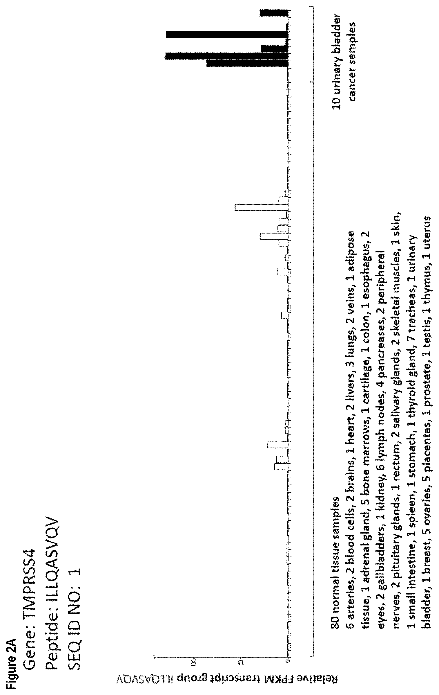
D00020

D00021

D00022

D00023

D00024
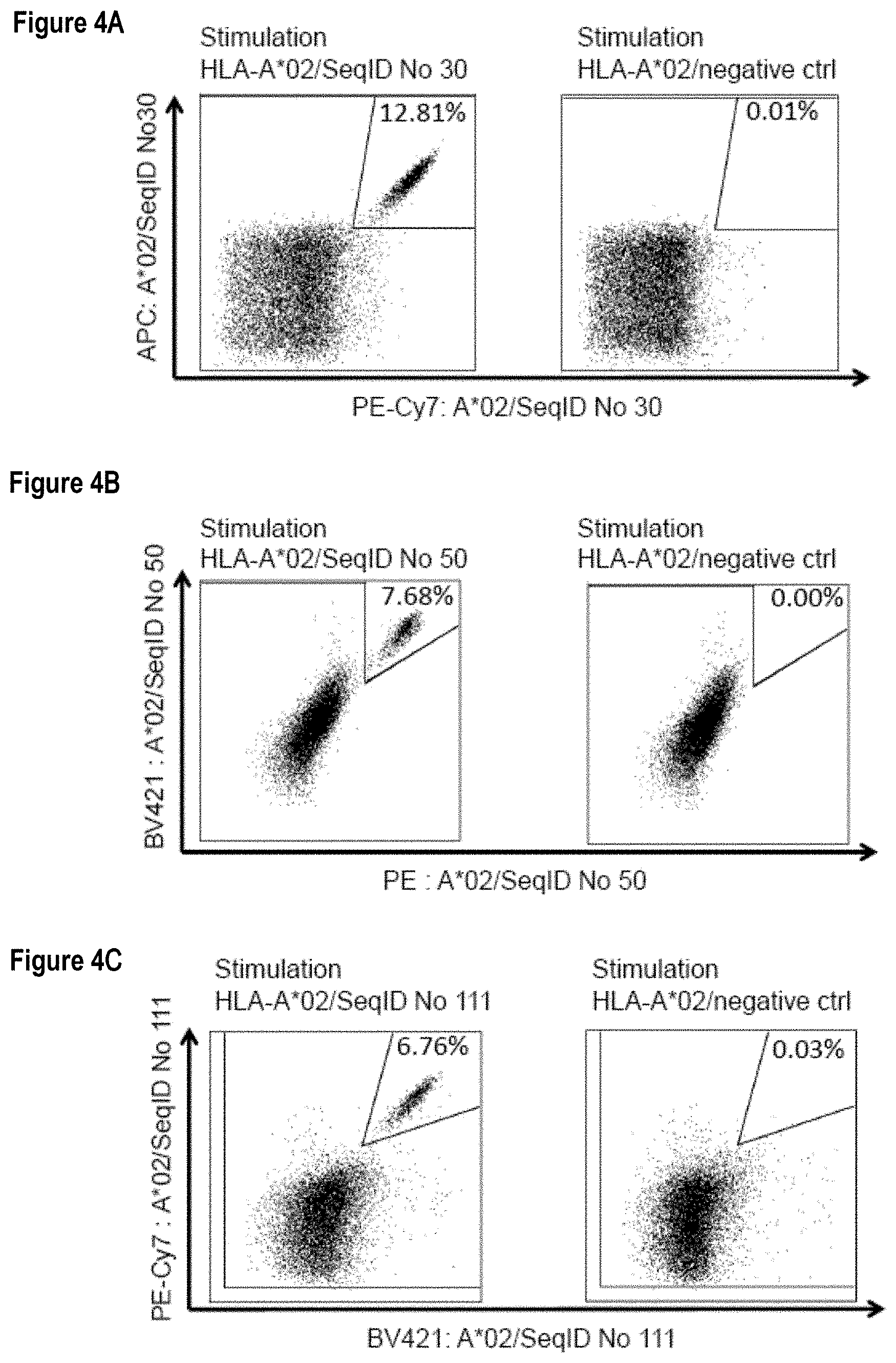
S00001
XML
uspto.report is an independent third-party trademark research tool that is not affiliated, endorsed, or sponsored by the United States Patent and Trademark Office (USPTO) or any other governmental organization. The information provided by uspto.report is based on publicly available data at the time of writing and is intended for informational purposes only.
While we strive to provide accurate and up-to-date information, we do not guarantee the accuracy, completeness, reliability, or suitability of the information displayed on this site. The use of this site is at your own risk. Any reliance you place on such information is therefore strictly at your own risk.
All official trademark data, including owner information, should be verified by visiting the official USPTO website at www.uspto.gov. This site is not intended to replace professional legal advice and should not be used as a substitute for consulting with a legal professional who is knowledgeable about trademark law.