Ror1-specific Chimeric Antigen Receptors (car) With Humanized Targeting Domains
HUDECEK; Michael ; et al.
U.S. patent application number 16/607069 was filed with the patent office on 2020-02-06 for ror1-specific chimeric antigen receptors (car) with humanized targeting domains. The applicant listed for this patent is JULIUS-MAXIMILIANS-UNIVERSITAT WURZBURG. Invention is credited to Michael HUDECEK, Andreas MADES.
| Application Number | 20200040058 16/607069 |
| Document ID | / |
| Family ID | 58640781 |
| Filed Date | 2020-02-06 |


View All Diagrams
| United States Patent Application | 20200040058 |
| Kind Code | A1 |
| HUDECEK; Michael ; et al. | February 6, 2020 |
ROR1-SPECIFIC CHIMERIC ANTIGEN RECEPTORS (CAR) WITH HUMANIZED TARGETING DOMAINS
Abstract
The invention relates to chimeric antigen receptors (CAR) with a humanized targeting domain specific to the antigen ROR1. The invention encompasses the polynucleotides, vectors encoding said CARs and the isolated cells expressing them at their surface, in particularly for their use in immunotherapy.
| Inventors: | HUDECEK; Michael; (Hochberg, DE) ; MADES; Andreas; (Wiesbaden Biebrich, DE) | ||||||||||
| Applicant: |
|
||||||||||
|---|---|---|---|---|---|---|---|---|---|---|---|
| Family ID: | 58640781 | ||||||||||
| Appl. No.: | 16/607069 | ||||||||||
| Filed: | April 27, 2018 | ||||||||||
| PCT Filed: | April 27, 2018 | ||||||||||
| PCT NO: | PCT/EP2018/060887 | ||||||||||
| 371 Date: | October 21, 2019 |
| Current U.S. Class: | 1/1 |
| Current CPC Class: | C07K 2317/24 20130101; C07K 14/70517 20130101; C07K 14/70521 20130101; C07K 16/30 20130101; C07K 14/7051 20130101; C07K 16/28 20130101; A61K 39/00 20130101; C12N 15/86 20130101; C07K 14/70578 20130101; C12N 5/0636 20130101; A61P 35/00 20180101; C07K 16/2803 20130101; C07K 14/705 20130101; C07K 2319/03 20130101; C12N 2740/15043 20130101; A61K 35/17 20130101 |
| International Class: | C07K 14/725 20060101 C07K014/725; C07K 14/705 20060101 C07K014/705; A61K 35/17 20060101 A61K035/17; A61P 35/00 20060101 A61P035/00; C12N 15/86 20060101 C12N015/86; C12N 5/0783 20060101 C12N005/0783; C07K 16/28 20060101 C07K016/28 |
Foreign Application Data
| Date | Code | Application Number |
|---|---|---|
| Apr 28, 2017 | EP | 17168805.4 |
Claims
1-57. (canceled)
58. A ROR1-specific CAR comprising a humanized targeting domain capable of binding to ROR1, wherein the humanized targeting domain comprises, preferably in an N- to C-terminal order: a) an antibody heavy chain variable domain amino acid sequence selected from the group consisting of: a1) the amino acid sequence of SEQ ID No: 1 or an amino acid sequence at least 90% identical thereto; a2) the amino acid sequence of SEQ ID No: 3 or an amino acid sequence at least 90% identical thereto; or a3) the amino acid sequence of SEQ ID No: 5 or an amino acid sequence at least 90% identical thereto; and b) an antibody light chain variable domain amino acid sequence selected from the group consisting of: b1) the amino acid sequence of SEQ ID No: 2 or an amino acid sequence at least 90% identical thereto; b2) the amino acid sequence of SEQ ID No: 4 or an amino acid sequence at least 90% identical thereto; or b3) the amino acid sequence of SEQ ID No: 6 or an amino acid sequence at least 90% identical thereto.
59. The ROR1-specific CAR according to claim 58, wherein the humanized targeting domain consists of the following sequences in an N- to C-terminal order: I) the antibody heavy chain variable domain amino acid sequence of SEQ ID No: 1, an amino acid linker sequence which is preferably the amino acid sequence of SEQ ID No: 8, and the antibody light chain variable domain amino acid sequence of SEQ ID No: 2; II) the antibody heavy chain variable domain amino acid sequence of SEQ ID No: 3, an amino acid linker sequence which is preferably the amino acid sequence of SEQ ID No: 8, and the antibody light chain variable domain amino acid sequence of SEQ ID No: 4; or III) the antibody heavy chain variable domain amino acid sequence of SEQ ID No: 5, an amino acid linker sequence which is preferably the amino acid sequence of SEQ ID No: 8, and the antibody light chain variable domain amino acid sequence of SEQ ID No: 6.
60. A combination of CARs comprising at least a first and a second CAR, the combination being ROR1-specific, wherein said first and said second CAR are present on different polypeptide chains, and wherein: c) said first CAR comprises a first humanized targeting domain comprising an antibody heavy chain variable domain amino acid sequence selected from the group consisting of: c1) the amino acid sequence of SEQ ID No: 1 or an amino acid sequence at least 90% identical thereto; c2) the amino acid sequence of SEQ ID No: 3 or an amino acid sequence at least 90% identical thereto; or c3) the amino acid sequence of SEQ ID No: 5 or an amino acid sequence at least 90% identical thereto; and d) said second CAR comprises a second humanized targeting domain comprising an antibody light chain variable domain amino acid sequence selected from the group consisting of: d1) the amino acid sequence of SEQ ID No: 2 or an amino acid sequence at least 90% identical thereto; d2) the amino acid sequence of SEQ ID No: 4 or an amino acid sequence at least 90% identical thereto; or d3) the amino acid sequence of SEQ ID No: 6 or an amino acid sequence at least 90% identical thereto.
61. The ROR1-specific CAR according to claim 58, wherein the CAR further comprises a costimulatory domain capable of mediating costimulation to immune cells, wherein at least the first or the second CAR of the combination, preferably at least the first and the second CAR of the combination, most preferably all of the CARs of the combination, further comprise a costimulatory domain capable of mediating costimulation to immune cells, optionally wherein said costimulatory domain is from 4-1BB, CD28, Ox40, ICOS or DAP10, further optionally wherein said costimulatory domain has the amino acid sequence of SEQ ID No: 12.
62. The ROR1-specific CAR according to claim 58, further comprising a transmembrane polypeptide, wherein at least the first or the second CAR of the combination, preferably at least the first and the second CAR of the combination, most preferably all of the CARs of the combination, further comprise a transmembrane polypeptide, optionally wherein said transmembrane polypeptide is a transmembrane domain from CD4, CD8 or CD28, and optionally wherein said transmembrane domain has the amino acid sequence of SEQ ID No: 11.
63. The ROR1-specific CAR according to claim 58, further comprising a CAR spacer domain, wherein at least the first or the second CAR of the combination, preferably at least the first and the second CAR of the combination, most preferably all of the CARs of the combination, further comprise a CAR spacer domain, optionally wherein said CAR spacer domain is from CD4, CD8, an FC-receptor, an immunoglobulin, or an antibody, and optionally wherein said CAR spacer domain has the amino acid sequence of SEQ ID No: 9 or SEQ ID No: 10, preferably the amino acid sequence of SEQ ID No: 9.
64. The ROR1-specific CAR according to claim 58, further comprising a suicide gene product that allows the selective killing of CAR T cells, wherein at least the first or the second CAR of the combination further comprises a suicide gene product that allows the selective killing of CAR T cells, optionally wherein said suicide gene product is iCasp9 or HSV-TK.
65. The ROR1-specific CAR according to claim 58, comprising, in an N- to C-terminal order: i) a signal peptide for direction into the endoplasmic reticulum, the signal peptide preferably having the amino acid sequence of SEQ ID No: 7; ii) a humanized targeting domain; iii) the CAR spacer domain; iv) a transmembrane polypeptide; v) a costimulatory domain; vi) a CD3z signaling domain preferably having the amino acid sequence of SEQ ID No: 13; and optionally further comprising: vii) a T2A ribosomal skipping sequence preferably having the amino acid sequence of SEQ ID No: 14; viii) a signal peptide for direction into the endoplasmic reticulum, the signal peptide preferably having the amino acid sequence of SEQ ID No: 7; and ix) a detectable marker protein sequence preferably having the EGFRt amino acid sequence of SEQ ID No: 15, optionally wherein the ROR1-specific CAR consists of said components i) to ix).
66. A polynucleotide encoding the ROR1-specific CAR according to claim 58.
67. A recombinant mammalian cell expressing at least one ROR1-specific CAR according to claim 58, optionally wherein said cell is an immune cell, further optionally wherein said cell is a lymphocyte, optionally wherein said lymphocyte is a B lymphocyte or T lymphocyte, or optionally wherein said cell is a CD8+ killer T cell, a CD4+ helper T cell, a naive T cell, a memory T cell, a central memory T cell, an effector memory T cell, a memory stem T cell, an invariant T cell, an NKT cell, a cytokine induced killer T cell, a g/d T cell, a natural killer cell, a monocyte, a macrophage, a dendritic cell, or a granulocyte, optionally wherein the cell is a human cell.
68. The recombinant mammalian cell according to claim 67, wherein the recombinant mammalian cell is capable of at least one cell division when cocultured with lethally irradiated ROR1-expressing target cells at an E:T ratio of 4:1 for 72 h in the absence of exogenous cytokines, optionally wherein the recombinant mammalian cell is capable of at least two cell divisions when cocultured with lethally irradiated ROR1-expressing target cells at an E:T ratio of 4:1 for 72 h in the absence of exogenous cytokines, optionally wherein the recombinant mammalian cell is capable of at least three cell divisions when cocultured with lethally irradiated ROR1-expressing target cells at an E:T ratio of 4:1 for 72 h in the absence of exogenous cytokines, optionally wherein the recombinant mammalian cell is capable of at least four cell divisions when cocultured with lethally irradiated ROR1-expressing target cells at an E:T ratio of 4:1 for 72 h in the absence of exogenous cytokines, optionally wherein the recombinant mammalian cell is capable of at least five cell divisions when cocultured with lethally irradiated ROR1-expressing target cells at an E:T ratio of 4:1 for 72 h in the absence of exogenous cytokines.
69. The recombinant mammalian cell according to claim 67, wherein the ROR1-specific CAR or the ROR1-specific combination of CARs is capable of binding to ROR1 with higher binding affinity than a respective recombinant mammalian control cell expressing a respective ROR1-specific CAR or a respective ROR1-specific combination of CARs where none of the targeting domains is humanized, and wherein said binding affinity is binding affinity to fluorescently labelled recombinant aggregated ROR1 as assessed by flow cytometry analysis.
70. A method for producing a recombinant mammalian cell according to claim 67, the method comprising the steps of: (a) providing a mammalian cell; (b) introducing into said mammalian cell of step (a) at least one polynucleotide encoding said at least one ROR1-specific CAR or said combination of CARs; and (c) expressing said at least one ROR1-specific CAR or said combination of CARs from said at least one polynucleotide in said cell; thereby obtaining said recombinant mammalian cell, optionally: I) wherein the method further comprises coculturing the recombinant mammalian cell with lethally irradiated ROR1-expressing target cells at a ratio of 4:1 for 72 h in the absence of exogenous cytokines, optionally wherein at least 1%, preferably at least 2%, more preferably at least 3%, and still more preferably at least 4% of the recombinant mammalian cells undergo at least 5 cell divisions during the coculturing; II) wherein said method is an in vitro method; III) wherein said mammalian cells of step (a) are cells obtained from donors; and/or IV) wherein said mammalian cells of step (a) are cells obtained from patients.
71. A recombinant mammalian cell according to claim 67, for use in medicine.
72. A recombinant mammalian cell according to claim 67, for use in a method for treating a cancer that expresses ROR1 in a patient, optionally wherein the cancer is ROR1-positive leukemia, mantle cell lymphoma, breast-cancer or lung cancer.
73. A method for treating a patient in need thereof, the method comprising administering a recombinant mammalian cell according to claim 67 to said patient, optionally wherein the method is for treating a cancer that expresses ROR1, optionally wherein the cancer is ROR1-positive leukemia, mantle cell lymphoma, breast-cancer or lung cancer.
Description
FIELD OF THE INVENTION
[0001] The invention relates to chimeric antigen receptors (CAR) with a humanized targeting domain specific to the antigen ROR1. The invention encompasses the polynucleotides, vectors encoding said CARs and the isolated cells expressing them at their surface, in particularly for their use in immunotherapy.
BACKGROUND OF THE INVENTION
[0002] The adoptive transfer of genetically modified T cells that express a T cell receptor or a chimeric antigen receptor (CAR) specific for a tumor-associated antigen is emerging as an effective modality for cancer therapy [1-5]. CARs are synthetic receptors most often constructed by linking a single chain variable fragment (scFV) of a monoclonal antibody (mAb) specific for a tumor cell surface molecule to a transmembrane domain, one or more intracellular costimulatory signaling modules, and CD3.zeta. [6-8]. CAR-modified T-cells confer non-MHC restricted recognition of tumor cells, and durable responses have been reported in patients with B-cell malignancies after treatment with autologous T-cells modified with CARs specific for the B-cell lineage restricted CD19 molecule. The major toxicities in these patients were related to tumor lysis, cytokine release, and prolonged depletion of normal B-lymphocytes [1-3, 5, 9]. A challenge in the field is to identify and validate receptor constructs specific for molecules that are expressed on a greater number of malignancies including common epithelial tumors, and that are restricted in their expression to malignant and not normal cells.
[0003] We have been investigating the receptor tyrosine kinase-like orphan receptor 1 (ROR1) as a candidate for immunotherapy with CAR-modified T-cells. ROR1 is a 120-kDa glycoprotein containing extracellular immunoglobulin (Ig)-like, Frizzled, and Kringle domains. ROR1 is expressed during embryogenesis but absent from normal adult tissues, apart from a subset of immature B-cell precursors, and low-level expression on adipocytes [10, 11]. ROR1 was first shown to be expressed in B-cell chronic lymphocytic leukemia (B-CLL) by transcriptional profiling [12, 13], and was subsequently identified on the surface of many cancers including mantle cell lymphoma (MCL), acute lymphoblastic leukemia (ALL) with a t(1;19) chromosome translocation, and a subset of lung, breast, colon, pancreas, renal, and ovarian cancers [14-21]. In both lung adenocarcinoma and t(1;19) ALL, ROR1 cooperates in oncogenic signaling, and knockdown of ROR1 with siRNA exposed a critical role for this molecule in maintaining tumor cell survival [15, 18, 22, 23]. WO 2016/115559 relates to antibodies and chimeric antigen receptors specific for ROR1.
[0004] Clinical trials with CD19 CARs have demonstrated that a paramount requirement for therapeutic effficacy is engraftment, in vivo proliferation and persistence of CAR T cells after adoptive transfer [24-26] (Clinical trial IDs: NCT02167360, NCT02030847 NCT01865617). In responding patients, CAR T cells undergo substantial proliferation, to a point where they comprise a substantial proportion of the patient's T-cell repertoire. Non-responding patients by contrast have an insufficient CAR T cell engraftment or the CAR T cell graft is rejected early after adoptive transfer. CAR T cell proliferation is an `advanced` effector function, requiring optimal binding of the CAR targeting domain to the respective antigen. Therefore, to maximize the potential of CAR T cell therapy it is important to chose a CAR that exhibits strong antigen binding properties and also mediates high CAR T cell expansion and long-term survival.
[0005] All CARs that are currently used in the clinic contain scFv targeting domains that are derived from murine antibodies and the majoritiy of CARs that are in preclinical development also use targeting domains of `foreign` origin. It has been noted that these `foreign` targeting domains contain immunogenic epitopes that are recognized by the patient's immune system and elicit cellular or humoral immune responses that eventually mediate CAR-T cell rejection; see [27], [28] and [29]. Reference [30] describes the use of targeting domains from human antibodies.
[0006] In summary, there is a need in the art for improved CAR T cell constructs and related products and methods that can be used for therapies such as immunotherapies.
DESCRIPTION OF THE INVENTION
[0007] The present invention relates to humanized binding domains based on the known anti-ROR1 antibodies R11, R12 and 2A2 and to uses thereof for the construction of CARs and CAR engineered T cells. See, for instance, reference [14] for a description of the 2A2 antibody, which is hereby incorporated by reference in its entirety for all purposes; and reference [37] for a description of the R11/R12 antibodies, which is hereby incorporated by reference in its entirety for all purposes. The CARs according to the present invention have a higher degree of "human-ness" of the humanized R11, R12 and 2A2 binding domains, as opposed to the rabbit (R11 & R12) and mouse (2A2) binding domains. According to the invention, these CARs and CAR engineered T cells are expected to exhibit lower immunogenicity in clinical use in patients, as opposed to the rabbit (R11 & R12) and mouse (2A2) binding domains.
[0008] According to the invention, humanization can be carried out by any method that is known in the art. As a non-limiting example, humanization of the VH and VL domains of R11, R12 and 2A2 anti-ROR1 monoclonal antibodies has been performed by CDR grafting and selection of best binders for the ROR-1 target by Transpo-mAb Display. This method has been described for the R11 and 2A2 antibodies in reference [31], which is hereby incorporated by reference in its entirety for all purposes. According to the invention, recombinant mammalian cells expressing CARs such as CAR T cells can be produced to express CARs with the humanized anti-ROR1 binding domains of monoclonal anti-ROR-1 antibodies R11, R12 and 2A2.
[0009] Humanization of ROR1-specific CARs is different from the known clinical approaches which rely on non-humanized ROR1-specific CARs. The present inventors have now surprisingly shown that humanized ROR1-specific CARs have higher functionality than their non-humanized counterparts. This advantage was unexpected, because humanization is thought to decrease rather than increase affinity.
[0010] Furthermore, the inventors have surprisingly shown that humanized CARs with advantageous functional properties can be produced according to the invention. For instance, CAR T cells according to the invention are capable of target cell lysis and exhibit strong proliferation upon stimulation with lethally irradiated ROR1-expressing target cells.
[0011] Accordingly, the present invention provides the following preferred embodiments: [0012] 1. A ROR1-specific CAR comprising: [0013] a humanized targeting domain obtainable by humanization of a ROR1-binding fragment of a monoclonal antibody capable of binding to ROR1, wherein said monoclonal antibody is selected from the group consisting of the monoclonal antibodies R11, R12 and 2A2. [0014] 2. The ROR1-specific CAR according to item 1, wherein said monoclonal antibody is selected from the group consisting of the monoclonal antibodies R12 and 2A2. [0015] 3. A ROR1-specific CAR comprising a humanized targeting domain capable of binding to ROR1, wherein the humanized targeting domain comprises, preferably in an N- to C-terminal order: [0016] a) an antibody heavy chain variable domain amino acid sequence selected from the group consisting of: [0017] a1) the amino acid sequence of SEQ ID No: 1 or an amino acid sequence at least 90% identical thereto; [0018] a2) the amino acid sequence of SEQ ID No: 3 or an amino acid sequence at least 90% identical thereto; or [0019] a3) the amino acid sequence of SEQ ID No: 5 or an amino acid sequence at least 90% identical thereto; [0020] and [0021] b) an antibody light chain variable domain amino acid sequence selected from the group consisting of: [0022] b1) the amino acid sequence of SEQ ID No: 2 or an amino acid sequence at least 90% identical thereto; [0023] b2) the amino acid sequence of SEQ ID No: 4 or an amino acid sequence at least 90% identical thereto; or [0024] b3) the amino acid sequence of SEQ ID No: 6 or an amino acid sequence at least 90% identical thereto. [0025] 4. The ROR1-specific CAR according to item 3, wherein the humanized targeting domain comprises the antibody heavy chain variable domain amino acid sequence of SEQ ID No: 1 or an amino acid sequence at least 90% identical thereto and the antibody light chain variable domain amino acid sequence of SEQ ID No: 2 or an amino acid sequence at least 90% identical thereto. [0026] 5. The ROR1-specific CAR according to item 4, wherein the humanized targeting domain comprises the antibody heavy chain variable domain amino acid sequence of SEQ ID No: 1 and the antibody light chain variable domain amino acid sequence of SEQ ID No: 2. [0027] 6. The ROR1-specific CAR according to item 5, wherein the humanized targeting domain consists of the following sequences in an N- to C-terminal order: the antibody heavy chain variable domain amino acid sequence of SEQ ID No: 1, an amino acid linker sequence which is preferably the amino acid sequence of SEQ ID No: 8, and the antibody light chain variable domain amino acid sequence of SEQ ID No: 2. [0028] 7. The ROR1-specific CAR according to item 3, wherein the humanized targeting domain comprises the antibody heavy chain variable domain amino acid sequence of SEQ ID No: 3 or an amino acid sequence at least 90% identical thereto and the antibody light chain variable domain amino acid sequence of SEQ ID No: 4 or an amino acid sequence at least 90% identical thereto. [0029] 8. The ROR1-specific CAR according to item 7, wherein the humanized targeting domain comprises the antibody heavy chain variable domain amino acid sequence of SEQ ID No: 3 and the antibody light chain variable domain amino acid sequence of SEQ ID No: 4. [0030] 9. The ROR1-specific CAR according to item 8, wherein the humanized targeting domain consists of the following sequences in an N- to C-terminal order: the antibody heavy chain variable domain amino acid sequence of SEQ ID No: 3, an amino acid linker sequence which is preferably the amino acid sequence of SEQ ID No: 8, and the antibody light chain variable domain amino acid sequence of SEQ ID No: 4. [0031] 10. The ROR1-specific CAR according to item 3, wherein the humanized targeting domain comprises the antibody heavy chain variable domain amino acid sequence of SEQ ID No: 5 or an amino acid sequence at least 90% identical thereto and the antibody light chain variable domain amino acid sequence of SEQ ID No: 6 or an amino acid sequence at least 90% identical thereto. [0032] 11. The ROR1-specific CAR according to item 10, wherein the humanized targeting domain comprises the antibody heavy chain variable domain amino acid sequence of SEQ ID No: 5 and the antibody light chain variable domain amino acid sequence of SEQ ID No: 6. [0033] 12. The ROR1-specific CAR according to item 11, wherein the humanized targeting domain consists of the following sequences in an N- to C-terminal order: the antibody heavy chain variable domain amino acid sequence of SEQ ID No: 5, an amino acid linker sequence which is preferably the amino acid sequence of SEQ ID No: 8, and the antibody light chain variable domain amino acid sequence of SEQ ID No: 6. [0034] 13. A combination of CARs comprising at least a first and a second CAR, the combination being ROR1-specific, wherein said first and said second CAR are present on different polypeptide chains, and wherein: [0035] c) said first CAR comprises a first humanized targeting domain comprising an antibody heavy chain variable domain amino acid sequence selected from the group consisting of: [0036] c1) the amino acid sequence of SEQ ID No: 1 or an amino acid sequence at least 90% identical thereto; [0037] c2) the amino acid sequence of SEQ ID No: 3 or an amino acid sequence at least 90% identical thereto; or [0038] c3) the amino acid sequence of SEQ ID No: 5 or an amino acid sequence at least 90% identical thereto; and [0039] d) said second CAR comprises a second humanized targeting domain comprising an antibody light chain variable domain amino acid sequence selected from the group consisting of: [0040] d1) the amino acid sequence of SEQ ID No: 2 or an amino acid sequence at least 90% identical thereto; [0041] d2) the amino acid sequence of SEQ ID No: 4 or an amino acid sequence at least 90% identical thereto; or [0042] d3) the amino acid sequence of SEQ ID No: 6 or an amino acid sequence at least 90% identical thereto. [0043] 14. The combination according to item 13, wherein the first humanized targeting domain comprises the antibody heavy chain variable domain amino acid sequence of SEQ ID No: 1 or an amino acid sequence at least 90% identical thereto and the second humanized targeting domain comprises the antibody light chain variable domain amino acid sequence of SEQ ID No: 2 or an amino acid sequence at least 90% identical thereto. [0044] 15. The combination according to item 14, wherein the first humanized targeting domain comprises the antibody heavy chain variable domain amino acid sequence of SEQ ID No: 1 and the second humanized targeting domain comprises the antibody light chain variable domain amino acid sequence of SEQ ID No: 2. [0045] 16. The combination according to item 13, wherein the first humanized targeting domain comprises the antibody heavy chain variable domain amino acid sequence of SEQ ID No: 3 or an amino acid sequence at least 90% identical thereto and the second humanized targeting domain comprises the antibody light chain variable domain amino acid sequence of SEQ ID No: 4 or an amino acid sequence at least 90% identical thereto. [0046] 17. The combination according to item 16, wherein the first humanized targeting domain comprises the antibody heavy chain variable domain amino acid sequence of SEQ ID No: 3 and the second humanized targeting domain comprises the antibody light chain variable domain amino acid sequence of SEQ ID No: 4. [0047] 18. The combination according to item 13, wherein the first humanized targeting domain comprises the antibody heavy chain variable domain amino acid sequence of SEQ ID No: 5 or an amino acid sequence at least 90% identical thereto and the second humanized targeting domain comprises the antibody light chain variable domain amino acid sequence of SEQ ID No: 6 or an amino acid sequence at least 90% identical thereto. [0048] 19. The combination according to item 18, wherein the first humanized targeting domain comprises the antibody heavy chain variable domain amino acid sequence of SEQ ID No: 5 and the second humanized targeting domain comprises the antibody light chain variable domain amino acid sequence of SEQ ID No: 6. [0049] 20. The ROR1-specific CAR according to any one of the preceding items, wherein the CAR further comprises a costimulatory domain capable of mediating costimulation to immune cells; or the combination according to any one of the preceding items, wherein at least the first or the second CAR of the combination, preferably at least the first and the second CAR of the combination, most preferably all of the CARs of the combination, further comprise a costimulatory domain capable of mediating costimulation to immune cells. [0050] 21. The ROR1-specific CAR or the combination according to item 20, wherein said costimulatory domain is from 4-1BB, CD28, Ox40, ICOS or DAP10. [0051] 22. The ROR1-specific CAR or the combination according to item 21, wherein said costimulatory domain has the amino acid sequence of SEQ ID No: 12. [0052] 23. The ROR1-specific CAR according to any one of the preceding items, further comprising a transmembrane polypeptide; or the combination according to any one of the preceding items, wherein at least the first or the second CAR of the combination, preferably at least the first and the second CAR of the combination, most preferably all of the CARs of the combination, further comprise a transmembrane polypeptide. [0053] 24. The ROR1-specific CAR or the combination according to item 23, wherein said transmembrane polypeptide is a transmembrane domain from CD4, CD8 or CD28. [0054] 25. The ROR1-specific CAR or the combination according to item 24, wherein said transmembrane domain has the amino acid sequence of SEQ ID No: 11. [0055] 26. The ROR1-specific CAR according to any one of the preceding items, further comprising a CAR spacer domain; or the combination according to any one of the preceding items, wherein at least the first or the second CAR of the combination, preferably at least the first and the second CAR of the combination, most preferably all of the CARs of the combination, further comprise a CAR spacer domain. [0056] 27. The ROR1-specific CAR or the combination according to item 26, wherein said CAR spacer domain is from CD4, CD8, an FC-receptor, an immunoglobulin, or an antibody. [0057] 28. The ROR1-specific CAR or the combination according to item 27, wherein said CAR spacer domain has the amino acid sequence of SEQ ID No: 9 or SEQ ID No: 10, preferably the amino acid sequence of SEQ ID No: 9. [0058] 29. The ROR1-specific CAR according to any one of the preceding items, further comprising a suicide gene product that allows the selective killing of CAR T cells; or the combination according to any one of the preceding items, wherein at least the first or the second CAR of the combination further comprises a suicide gene product that allows the selective killing of CAR T cells. [0059] 30. The ROR1-specific CAR or the combination according to item 29, wherein said suicide gene product is iCasp9 or HSV-TK. [0060] 31. The ROR1-specific CAR according to any one of the preceding items, comprising, in an N- to C-terminal order: i) a signal peptide for direction into the endoplasmic reticulum, the signal peptide preferably having the amino acid sequence of SEQ ID No: 7; ii) said humanized targeting domain; iii) the CAR spacer domain according to any one of items 26 to 28; iv) the transmembrane polypeptide according to any one of items 23-25; v) the costimulatory domain according to any one of items 20-22; vi) a CD3z signaling domain preferably having the amino acid sequence of SEQ ID No: 13; and optionally further comprising: vii) a T2A ribosomal skipping sequence preferably having the amino acid sequence of SEQ ID No: 14; viii) a signal peptide for direction into the endoplasmic reticulum, the signal peptide preferably having the amino acid sequence of SEQ ID No: 7; and ix) a detectable marker protein sequence preferably having the EGFRt amino acid sequence of SEQ ID No: 15. [0061] 32. The ROR1-specific CAR according to item 31, consisting of said components i) to ix). [0062] 33. A polynucleotide encoding the ROR1-specific CAR according to any one of the preceding items. [0063] 34. A recombinant mammalian cell expressing at least one ROR1-specific CAR according to any one of the preceding items or expressing the combination of CARs according to any one of the preceding items. [0064] 35. The recombinant mammalian cell according to item 34, wherein said cell is an immune cell. [0065] 36. The recombinant mammalian cell according to any one of the preceding items, wherein said cell is a lymphocyte. [0066] 37. The recombinant mammalian cell according to any one of the preceding items, wherein said lymphocyte is a B lymphocyte or T lymphocyte. [0067] 38. The recombinant mammalian cell according to item 34, wherein said cell is a CD8.sup.+ killer T cell, a CD4.sup.+ helper T cell, a naive T cell, a memory T cell, a central memory T cell, an effector memory T cell, a memory stem T cell, an invariant T cell, an NKT cell, a cytokine induced killer T cell, a g/d T cell, a natural killer cell, a monocyte, a macrophage, a dendritic cell, or a granulocyte. [0068] 39. The recombinant mammalian cell according to any one of the preceding items, wherein the cell is a human cell. [0069] 40. The recombinant mammalian cell according to any one of the preceding items, wherein the recombinant mammalian cell is capable of at least one cell division when cocultured with lethally irradiated ROR1-expressing target cells at an E:T ratio of 4:1 for 72 h in the absence of exogenous cytokines. [0070] 41. The recombinant mammalian cell according to any one of the preceding items, wherein the recombinant mammalian cell is capable of at least two cell divisions when cocultured with lethally irradiated ROR1-expressing target cells at an E:T ratio of 4:1 for 72 h in the absence of exogenous cytokines. [0071] 42. The recombinant mammalian cell according to any one of the preceding items, wherein the recombinant mammalian cell is capable of at least three cell divisions when cocultured with lethally irradiated ROR1-expressing target cells at an E:T ratio of 4:1 for 72 h in the absence of exogenous cytokines. [0072] 43. The recombinant mammalian cell according to any one of the preceding items, wherein the recombinant mammalian cell is capable of at least four cell divisions when cocultured with lethally irradiated ROR1-expressing target cells at an E:T ratio of 4:1 for 72 h in the absence of exogenous cytokines. [0073] 44. The recombinant mammalian cell according to any one of the preceding items, wherein the recombinant mammalian cell is capable of at least five cell divisions when cocultured with lethally irradiated ROR1-expressing target cells at an E:T ratio of 4:1 for 72 h in the absence of exogenous cytokines. [0074] 45. The recombinant mammalian cell according to any one of the preceding items, wherein the ROR1-specific CAR or the ROR1-specific combination of CARs is capable of binding to ROR1 with higher binding affinity than a respective recombinant mammalian control cell expressing a respective ROR1-specific CAR or a respective ROR1-specific combination of CARs where none of the targeting domains is humanized, and wherein said binding affinity is binding affinity to fluorescently labelled recombinant aggregated ROR1 as assessed by flow cytometry analysis.
[0075] 46. A method for producing a recombinant mammalian cell according to any one of the preceding items, the method comprising the steps of: [0076] (a) providing a mammalian cell; [0077] (b) introducing into said mammalian cell of step (a) at least one polynucleotide encoding said at least one ROR1-specific CAR or said combination of CARs; and [0078] (c) expressing said at least one ROR1-specific CAR or said combination of CARs from said at least one polynucleotide in said cell; thereby obtaining said recombinant mammalian cell. [0079] 47. The method of item 46, further comprising coculturing the recombinant mammalian cell with lethally irradiated ROR1-expressing target cells at a ratio of 4:1 for 72 h in the absence of exogenous cytokines. [0080] 48. The method of item 47, wherein at least 1%, preferably at least 2%, more preferably at least 3%, and still more preferably at least 4% of the recombinant mammalian cells undergo at least 5 cell divisions during the coculturing. [0081] 49. The method according to any one of the preceding items, wherein said method is an in vitro method. [0082] 50. The method according to any one of the preceding items, wherein said mammalian cells of step (a) are cells obtained from donors. [0083] 51. The method according to any one of the preceding items, wherein said mammalian cells of step (a) are cells obtained from patients. [0084] 52. A recombinant mammalian cell according to any one of the preceding items for use in medicine. [0085] 53. A recombinant mammalian cell according to any one of the preceding items for use in a method for treating a cancer that expresses ROR1 in a patient. [0086] 54. The recombinant mammalian cell according to item 53 for the use according to item 53, wherein the cancer is ROR1-positive leukemia, mantle cell lymphoma, breast-cancer or lung cancer. [0087] 55. A method for treating a patient in need thereof, the method comprising administering a recombinant mammalian cell according to any one of the preceding items to said patient. [0088] 56. The method according to item 55, wherein the method is for treating a cancer that expresses ROR1. [0089] 57. The method according to item 56, wherein the cancer is ROR1-positive leukemia, mantle cell lymphoma, breast-cancer or lung cancer.
BRIEF DESCRIPTION OF THE DRAWINGS
[0090] FIG. 1: Amino acid sequences of the humanized 2A2 VH and VL sequences and the complete h2A2 CAR coding sequence [0091] (A) Humanized 2A2 VH and VL amino acid sequences [0092] (B) Complete amino acid sequence of the h2A2 CAR, represented in an N- to C-terminal order. Note that the asterisk denotes the end of the amino acid sequence.
[0093] FIG. 2: Amino acid sequences of the humanized R11 VH and VL sequences and the complete R11 CAR coding sequence [0094] (A) Humanized R11 VH and VL amino acid sequences [0095] (B) Complete amino acid sequence of the R11 CAR, represented in an N- to C-terminal order. Note that the asterisk denotes the end of the amino acid sequence.
[0096] FIG. 3: Amino acid sequences of the humanized R12 VH and VL sequences and the complete R12 CAR coding sequence [0097] (A) Humanized R12 VH and VL amino acid sequences [0098] (B) Complete amino acid sequence of the R12 CAR, represented in an N- to C-terminal order. Note that the asterisk denotes the end of the amino acid sequence.
[0099] FIG. 4: Enrichment and detection of CAR by EGFRt transduction marker
[0100] Human CD4 or CD8 positive T cells were transduced with lentiviral vector encoding a humanized or non-humanized ROR1 CAR and subsequently enriched for CAR expressing cells by magnetic activated cell sorting (MACS) making use of the truncated epidermal growth factor receptor (EGFRt) transduction marker. The coding sequence (CDS) for EGFRt is linked to the CAR CDS by a 2A ribosomal skipping sequence and expression of EGFRt can be used as a surrogate marker for CAR expression. [0101] (A) Flow cytometry plots demonstrating the frequency of EGFRt-positive CD4.sup.+ T cells after EGFRt enrichment by MACS. [0102] (B) Flow cytometry plots demonstrating the frequency of EGFRt-positive CD8.sup.+ T cells after EGFRt enrichment by MACS.
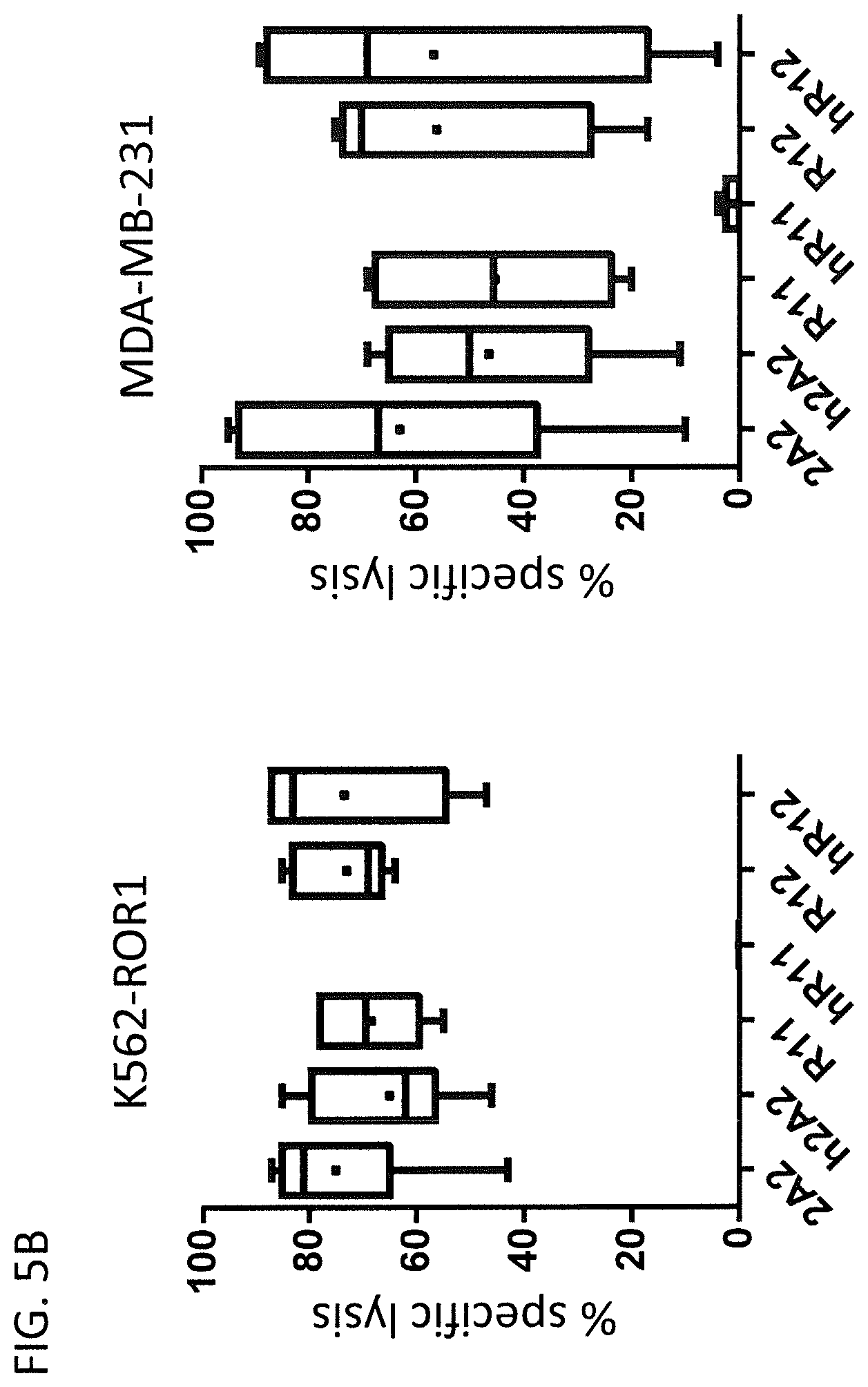
[0103] FIG. 5: Cytolytic activity of hROR1 CAR-expressing T cells [0104] (A) Cytolytic activity of primary human CD8.sup.+ T cells expressing the indicated non-humanized or humanized ROR1-specific CARs against ROR1-positive target cells. K562 is a ROR1-negative human leukemia cell line that was used as negative control. K562-ROR1 originates from the same cell line but has been engineered to stably express ROR1. MDA-MB-231 and A549 are human breast and lung cancer cell lines that endogenously express ROR1. All target cell lines were engineered to stably express a firefly (P. pyralis) luciferase. The specific lysis of the target cells was calculated based on the intensity of the luminescence signal after addition of Luciferin to a final concentration of 150 .mu.g/ml. [0105] (B) Summary of the cytolytic activity of human T cells expressing a non-humanized or humanized ROR1-specific CAR against two ROR1 positive target cells lines K562-ROR1 and MDA-MB-231. The data was collected from n=3 independent experiments. The specific lysis was calculated based on the luminesce intensity of ffluc-positive target cells after 6 h incubation with an E:T ratio of 10:1.
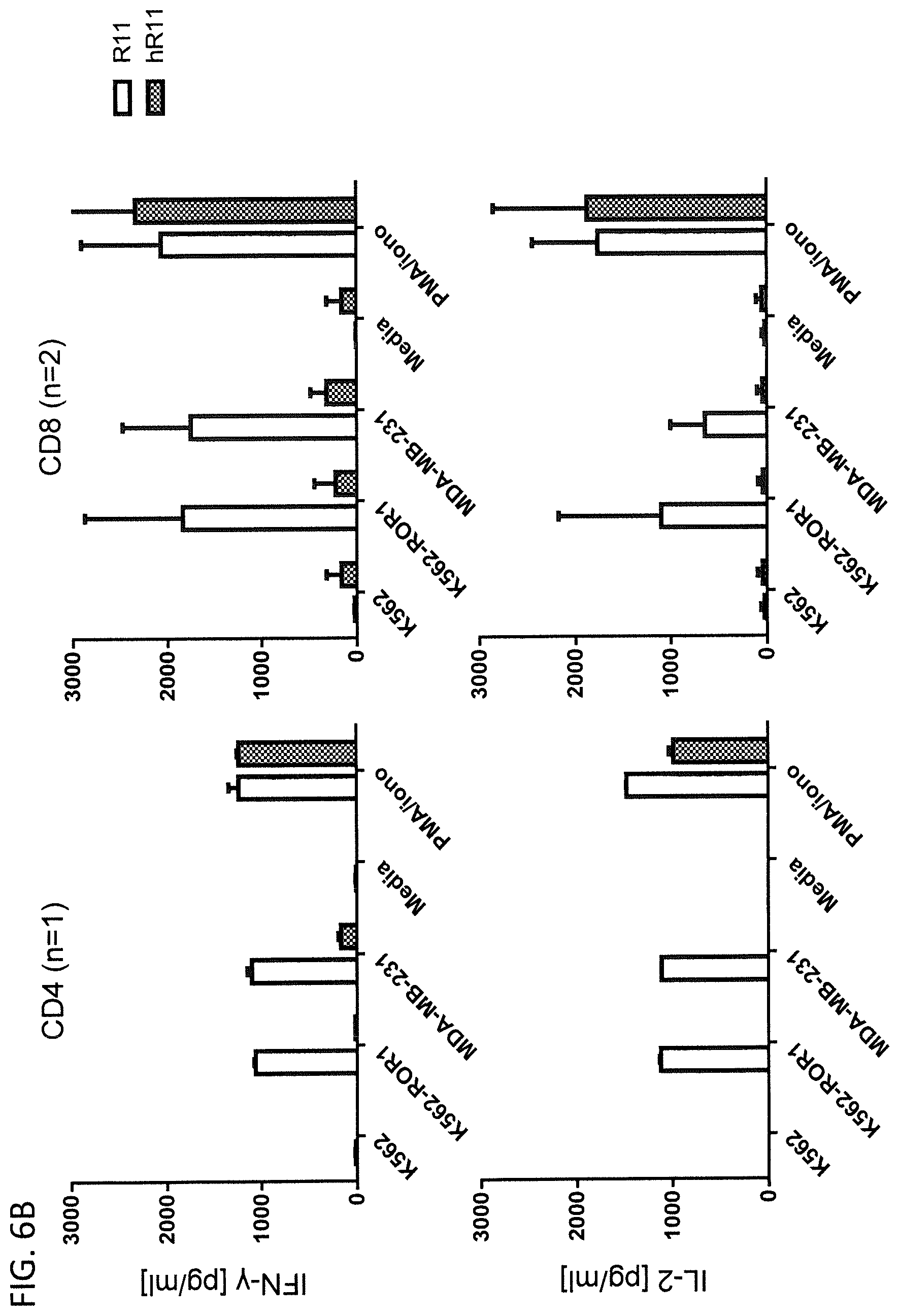
[0106] FIG. 6: Cytokine secretion of hROR1 CAR-expressing T cells
[0107] CD4.sup.+ or CD8.sup.+ CAR T cells expressing a non-humanized or humanized ROR-specific CAR were co-cultured with lethally irradiated ROR1-positive target cells at an E:T ratio of 4:1. Concentrations of the effector cytokines IL-2 and IFN-.gamma. were measured by ELISA in the cell culture supernatants after 24 h co-culture. [0108] (A) Comparison of cytokine secretion from 2A2 and h2A2 CAR T cells [0109] (B) Comparison of cytokine secretion from R11 and hR11 CAR T cells [0110] (C) Comparison of cytokine secretion from R12 and hR12 CAR T cells
[0111] FIG. 7: Proliferation of hROR1 CAR-expressing T cells
[0112] Proliferation of CD4.sup.+ ROR1-specific CAR T cells after stimulation with ROR1-positive target cells at an E:T ratio of 4:1. No exogenous cytokines were added to the culture media and the T cell proliferation was assessed by CFSE dye dilution 72 h after stimulation. For analysis, triplicate wells were pooled and the proliferation of live 7AAD.sup.-, CD4+ T cells was analyzed. [0113] (A) CFSE flow cytometry histograms of ROR1 CAR T cells with humanized (solid line) or non-humanized (dashed line). Grey filled curves are from vector control T cells (EGFRt). [0114] (B) Division indices of indicated ROR1 CAR T cells [0115] (C) Table that summarized the percentages of T cells that went through 0, 1, 2, 3, 4, and 5 cell division cycles.
[0116] FIG. 8: Binding of ROR1 protein by hROR1 CAR T cells
[0117] Human CD8.sup.+ T cells expressing non-humanized or humanized ROR1 CARs were collected, washed with PBS, 0.25% FCS and then incubated 10 min in the same buffer containing a final concentration of 5.3 .mu./ml AlexaFluor 647-labeled aggregated ROR1 protein and monoclonal .alpha.EGFR antibody. Afterwards the cells were washed with PBS, 0.25% FCS and analyzed by flow cytometry.
[0118] FIG. 9: In vivo activity of hROR1 CAR T cells
[0119] NSG mice were inoculated with ROR1-expressing Jeko-1 mantle cell lymphoma lines and received a treatment of humanized R12 or 2A2 CAR T cells 21 days later. [0120] (A) Mice were injected with luciferin at day 28 and radiance signals, which are emitted by ffluc-positive tumor cell, were detected. Displayed is the change of average radiance per mouse for the four best responding mice of each group compared to the baseline signals, which were measured on day 21. [0121] (B) Flow cytometry plots showing the frequency of EGFRt-positive CD4.sup.+ and CD8.sup.+ T cells after EGFRt in the bone marrow of mice that received hR12 or h2A2 ROR1 CAR T cells at day 56.
DETAILED DESCRIPTION OF THE INVENTION
[0122] Unless specifically defined herein, all technical and scientific terms used herein have the same meaning as commonly understood by a skilled artisan in the fields of gene therapy, immunology, biochemistry, genetics, and molecular biology.
[0123] All methods and materials similar or equivalent to those described herein can be used in the practice or testing of the present invention, with suitable methods and materials being described herein. All publications, patents and patent applications cited herein are hereby incorporated by reference in their entirety for all purposes. References referred to herein are indicated by a reference number in square brackets (e.g. as "[31]" or as "reference [31]"), which refers to the respective reference in the list of references at the end of the description. In case of conflict, the present specification, including definitions, will prevail over the cited references. Further, the materials, methods, and examples are illustrative only and are not intended to be limiting, unless otherwise specified.
[0124] Antibodies of non-human origin can be humanized by CDR grafting by methods known in the art. The humanization increases the homology of the binding domains to human antibody binding domains (i.e. the humanness), and reduces the immunogenic potential of the humanized antibody in human beings, which in turn is expected to increase the safety and therapeutic application profile in human patients. On the other hand, antibody humanization is often accompanied by a reduction of the binding affinity of the humanized antibody to its antigen, often requiring tedious affinity maturation, see reference [32], which is hereby incorporated by reference in its entirety for all purposes. It has also been experienced that the use of humanized antibody fragments for the generation of a targeting domain of a CAR can result in a lower performance of the CAR in respect to binding to the target antigen and triggering effector functions of the CAR expressing cell.
[0125] In the present invention the inventors have fused humanized VH and VL domains, as exemplified in FIGS. 1-3, of antibodies originating from the anti-ROR1-specific antibody clones 2A2 (Mus musculus, WO2010/124188, which is hereby incorporated by reference in its entirety for all purposes), R11 and R12 (Oryctolagus cuniculus, WO2012/075158, which is hereby incorporated by reference in its entirety for all purposes). The humanized VH and VL domains of these antibodies were then used to design single chain variable fragments (scFvs) that were further used for the design of CARs targeting the ROR1 antigen. Gene transfer vectors were created that allow the transduction or transfection of primary human T cells with the humanized CARs and allow the production of a CAR T cell product. The effector functions of the CAR T cells expressing ROR1 CARs with humanized ROR1 targeting domains were compared to their non-humanized counterparts. The humanized ROR1 CARs h2A2 and hR12 have conferred unexpected superior effector functions compared to their non-humanized counterparts.
[0126] The present invention describes for the first time the generation of humanized ROR1 CARs and their usage to redirect immune cells for the killing of ROR1 expressing target cells. Unexpectedly, the observed activity of two of the humanized CARs, namely h2A2 and hR12, was higher than the non-humanized forms in functional T cells assays with ROR1-expressing target cells. In contrast, the CAR that was constructed with an scFv targeting domain that originated from the R11 monoclonal antibody showed a comparatively strong reduction of effector functions, thus demonstrating the commonly expectable decline in therapeutic potential.
[0127] The finding of the present invention, that humanized ROR1 CARs can mediate antigen-specific effector functions to immune cells that are superior to those of the non-humanized counterparts is unexpected and has, to the inventors' knowledge, not been disclosed in the prior art. This finding is also unexpected, because antibody humanization is often accompanied by a loss of affinity, and/or a reduction of affinity to the target antigen. CARs whose targeting domain originates from such humanized antibodies oftentimes also exhibit lower effector functions and less therapeutic potential. It could thus neither be anticipated nor expected that our humanized ROR1 CARs demonstrate effector functions that are superior to their non-humanized counterparts.
[0128] The significantly higher function in combination with the anticipated lower immunogenicity of our novel humanized ROR1 CARs provides a substantial advantage for the clinical application of these CARs, especially, but not limited to, their usage in the context of immunotherapy against cancer.
[0129] A "recombinant mammalian cell" according to the invention can be any cell as defined herein. Preferably, a recombinant mammalian cell is an isolated cell. Recombinant mammalian cells according to the invention can be produced in accordance with known pharmaceutical standards. For instance, they can be formulated for administration to humans.
[0130] A ROR1-specific CAR or a combination of CARs according to the invention can be any possible form. In a preferred embodiment, the ROR1-specific CAR or combination of CARs is present in an isolated form. In another preferred embodiment the ROR1-specific CAR or combination of CARs according to the invention can be present in a composition, The composition may be a pharmaceutical composition.
[0131] Sequence alignments of sequences according to the invention are performed by suitable algorithms, and preferably by using the BLAST algorithm, see references [33, 34], using suitable alignment parameters as known in the art.
[0132] As used herein, each occurrence of terms such as "comprising" or "comprises" may optionally be substituted with "consisting of" or "consists of".
[0133] The sequences corresponding to the SEQ IDs referred to herein are indicated in FIGS. 1 to 3 and in the following tables:
TABLE-US-00001 GMCSF signal peptide (SEQ ID No: 7) MLLLVTSLLLCELPHPAFLLIP h2A2 heavy chain variable domain (VH) (SEQ ID No: 1) EVQLVQSGAEVKKPGASVKVSCKASGYTFSDYEMHWVRQAPGQGLEWLGA IDPETGGTAYNQKFKGRVTMTGDTSISTAYMELSRLTSDDTAVYYCTGYY DYDSFTYWGQGTLVSVSS 4(GS)x3 linker (SEQ ID No: 8) GGGGSGGGGSGGGGS h2A2 light chain variable domain (VL) (SEQ ID No: 2) DIQMTQSPSSLSTSVGDRVTITCKASQNVDAAVAWYQQKPGKAPKLLIYS ASNRYTGVASRFSGSGSGTDFTFTISSLQSEDLADYFCQQYDIYPYTFGQ GTKLEIK IgG4 hinge domain (SEQ ID No: 9) ESKYGPPCPPCP CD28 transmembrane domain (SEQ ID No: 11) MFWVLVVVGGVLACYSLLVTVAFIIFWV 4-1BB costimulatory domain (SEQ ID No: 12) KRGRKKLLYIFKQPFMRPVQTTQEEDGCSCRFPEEEEGGCEL CD3z signaling domain (SEQ ID No: 13) RVKFSRSADAPAYQQGQNQLYNELNLGRREEYDVLDKRRGRDPEMGGKPR RKNPQEGLYNELQKDKMAEAYSEIGMKGERRRGKGHDGLYQGLSTATKDT YDALHMQALPPR T2A ribosomal skipping sequence (SEQ ID No: 14) LEGGGEGRGSLLTCGDVEENPGPR GMCSF signal peptide (SEQ ID No: 7) MLLLVTSLLLCELPHPAFLLIP EGFRt (SEQ ID No: 15) RKVCNGIGIGEFKDSLSINATNIKHFKNCTSISGDLHILPVAFRGDSFTH TPPLDPQELDILKTVKEITGFLLIQAWPENRTDLHAFENLEIIRGRTKQH GQFSLAVVSLNITSLGLRSLKEISDGDVIISGNKNLCYANTINWKKLFGT SGQKTKIISNRGENSCKATGQVCHALCSPEGCWGPEPRDCVSCRNVSRGR ECVDKCNLLEGEPREFVENSECIQCHPECLPQAMNITCTGRGPDNCIQCA HYIDGPHCVKTCPAGVMGENNTLVWKYADAGHVCHLCHPNCTYGCTGPGL EGCPTNGPKIPSIATGMVGALLLLLVVALGIGLFM GMCSF signal peptide (SEQ ID No: 7) MLLLVTSLLLCELPHPAFLLIP hR11 heavy chain variable domain (VH) (SEQ ID No: 3) EVQLVQSGGGLVQPGGSLRLSCAASGSDINDYPISWVRQAPGKGLEWVSF INSGGSTWYASWVKGRFTISRDNAKNSLYLQMNSLRDDDTATYFCARGYS TYYGDFNIWGQGTLVTVSS 4(GS)x3 linker (SEQ ID No: 8) GGGGSGGGGSGGGGS hR11 light chain variable domain (VL) (SEQ ID No: 4) DIVMTQSPSSLSASVGDRVTITCQASQSIDSNLAWFQQKPGKAPKSLIYR ASNLASGVPSKFSGSGSGTDFTLTISSLQREDAATYYCLGGVGNVSYRTS FGGGTKVEIK IgG4 CH2CH3 4/2NQ (SEQ ID No: 10) ESKYGPPCPPCPAPPVAGPSVFLFPPKPKDTLMISRTPEVTCVVVDVSQE DPEVQFNWYVDGVEVHNAKTKPREEQFQSTYRVVSVLTVLHQDWLNGKEY KCKVSNKGLPSSIEKTISKAKGQPREPQVYTLPPSQEEMTKNQVSLTCLV KGFYPSDIAVEWESNGQPENNYKTTPPVLDSDGSFFLYSRLTVDKSRWQE GNVFSCSVMHEALHNHYTQKSLSLSLGK CD28 transmembrane domain (SEQ ID No: 11) MFWVLVVVGGVLACYSLLVTVAFIIFWV 4-1BB costimulatory domain (SEQ ID No: 12) KRGRKKLLYIFKQPFMRPVQTTQEEDGCSCRFPEEEEGGCEL CD3z signaling domain (SEQ ID No: 13) RVKFSRSADAPAYQQGQNQLYNELNLGRREEYDVLDKRRGRDPEMGGKPR RKNPQEGLYNELQKDKMAEAYSEIGMKGERRRGKGHDGLYQGLSTATKDT YDALHMQALPPR T2A ribosomal skipping sequence (SEQ ID No: 14) LEGGGEGRGSLLTCGDVEENPGPR GMCSF signal peptide (SEQ ID No: 7) MLLLVTSLLLCELPHPAFLLIP EGRFt (SEQ ID No: 15) RKVCNGIGIGEFKDSLSINATNIKHFKNCTSISGDLHILPVAFRGDSFTH TPPLDPQELDILKTVKEITGFLLIQAWPENRTDLHAFENLEIIRGRTKQH GQFSLAVVSLNITSLGLRSLKEISDGDVIISGNKNLCYANTINWKKLFGT SGQKTKIISNRGENSCKATGQVCHALCSPEGCWGPEPRDCVSCRNVSRGR ECVDKCNLLEGEPREFVENSECIQCHPECLPQAMNITCTGRGPDNCIQCA HYIDGPHCVKTCPAGVMGENNTLVWKYADAGHVCHLCHPNCTYGCTGPGL EGCPTNGPKIPSIATGMVGALLLLLVVALGIGLFM GMCSF signal peptide (SEQ ID No: 7) MLLLVTSLLLCELPHPAFLLIP hR12 heavy chain variable domain (VH) (SEQ ID No: 5) QVQLVESGGALVQPGGSLTLSCKASGFDFSAYYMSWVRQAPGKGLEWIAT IYPSSGKTYYAASVQGRFTISADNAKNTVYLQMNSLTAADTATYFCARDS YADDGALFNIWGQGTLVTVSS 4(GS)x3 linker (SEQ ID No: 8) GGGGSGGGGSGGGGS hR12 light chain variable domain (VL) (SEQ ID No: 6) QLVLTQSPSVSAALGSSAKITCTLSSAHKTDTIDWYQQLAGQAPRYLMYV QSDGSYEKRSGVPDRFSGSSSGADRYLIISSVQADDEADYYCGADYIGGY VFGGGTQLTVG IgG4 hinge domain (SEQ ID No: 9) ESKYGPPCPPCP CD28 transmembrane domain (SEQ ID No: 11) MFWVLVVVGGVLACYSLLVTVAFIIFWV 4-1BB costimulatory domain (SEQ ID No: 12) KRGRKKLLYIFKQPFMRPVQTTQEEDGCSCRFPEEEEGGCEL CD3z signaling domain (SEQ ID No: 13) RVKFSRSADAPAYQQGQNQLYNELNLGRREEYDVLDKRRGRDPEMGGKPR RKNPQEGLYNELQKDKMAEAYSEIGMKGERRRGKGHDGLYQGLSTATKDT YDALHMQALPPR T2A ribosomal skipping sequence (SEQ ID No: 14) LEGGGEGRGSLLTCGDVEENPGPR GMCSF signal peptide (SEQ ID No: 7) MLLLVTSLLLCELPHPAFLLIP EGFRt (SEQ ID No: 15) RKVCNGIGIGEFKDSLSINATNIKHFKNCTSISGDLHILPVAFRGDSFTH TPPLDPQELDILKTVKEITGFLLIQAWPENRTDLHAFENLEIIRGRTKQH GQFSLAVVSLNITSLGLRSLKEISDGDVIISGNKNLCYANTINWKKLFGT SGQKTKIISNRGENSCKATGQVCHALCSPEGCWGPEPRDCVSCRNVSRGR ECVDKCNLLEGEPREFVENSECIQCHPECLPQAMNITCTGRGPDNCIQCA HYIDGPHCVKTCPAGVMGENNTLVWKYADAGHVCHLCHPNCTYGCTGPGL EGCPTNGPKIPSIATGMVGALLLLLVVALGIGLFM
[0134] Additionally, preferred amino acid sequences of ROR1-binding fragments of the non-humanized monoclonal antibodies R11, R12 and 2A2 that can be used as starting sequences for humanization in accordance with the invention are as indicated below:
TABLE-US-00002 scFV of the non-humanized 2A2 antibody (SEQ ID No: 16): QVQLQQSGAELVRPGASVTLSCKASGYTFSDYEMHWVIQTPVHGLEWIGA IDPETGGTAYNQKFKGKAILTADKSSSTAYMELRSLTSEDSAVYYCTGYY DYDSFTYWGQGTLVTVSAGGGGSGGGGSGGGGSDIVMTQSQKIMSTTVGD RVSITCKASQNVDAAVAWYQQKPGQSPKLLIYSASNRYTGVPDRFTGSGS GTDFTLTISNMQSEDLADYFCQQYDIYPYTFGGGTKLEIK scFV of the non-humanized R11 antibody (SEQ ID No: 17): QSVKESEGDLVTPAGNLTLTCTASGSDINDYPISWVRQAPGKGLEWIGFI NSGGSTWYASWVKGRFTISRTSTTVDLKMTSLTTDDTATYFCARGYSTYY GDFNIWGPGTLVTISSGGGGSGGGGSGGGGSELVMTQTPSSTSGAVGGTV TINCQASQSIDSNLAWFQQKPGQPPTLLIYRASNLASGVPSRFSGSRSGT EYTLTISGVQREDAATYYCLGGVGNVSYRTSFGGGTEVVVK scFV of the non-humanized R12 antibody (SEQ ID No: 18): QEQLVESGGRLVTPGGSLTLSCKASGFDFSAYYMSWVRQAPGKGLEWIAT IYPSSGKTYYATWVNGRFTISSDNAQNTVDLQMNSLTAADRATYFCARDS YADDGALFNIWGPGTLVTISSGGGGSGGGGSGGGGSELVLTQSPSVSAAL GSPAKITCTLSSAHKTDTIDWYQQLQGEAPRYLMQVQSDGSYTKRPGVPD RFSGSSSGADRYLIIPSVQADDEADYYCGADYIGGYVFGGGTQLTVTG VH of the non-humanized 2A2 antibody (SEQ ID No: 19): QVQLQQSGAELVRPGASVTLSCKASGYTFSDYEMHWVIQTPVHGLEWIGA IDPETGGTAYNQKFKGKAILTADKSSSTAYMELRSLTSEDSAVYYCTGYY DYDSFTYWGQGTLVTVSA VL of the non-humanized 2A2 antibody (SEQ ID No: 20): DIVMTQSQKIMSTTVGDRVSITCKASQNVDAAVAWYQQKPGQSPKLLIYS ASNRYTGVPDRFTGSGSGTDFTLTISNMQSEDLADYFCQQYDIYPYTFGG GTKLEIK VH of the non-humanized R11 antibody (SEQ ID No: 21): QSVKESEGDLVTPAGNLTLTCTASGSDINDYPISWVRQAPGKGLEWIGFI NSGGSTWYASWVKGRFTISRTSTTVDLKMTSLTTDDTATYFCARGYSTYY GDFNIWGPGTLVTISS VL of the non-humanized R11 antibody (SEQ ID No: 22): ELVMTQTPSSTSGAVGGTVTINCQASQSIDSNLAWFQQKPGQPPTLLIYR ASNLASGVPSRFSGSRSGTEYTLTISGVQREDAATYYCLGGVGNVSYRTS FGGGTEVVVK VH of the non-humanized R12 antibody (SEQ ID No: 23): QEQLVESGGRLVTPGGSLTLSCKASGFDFSAYYMSWVRQAPGKGLEWIAT IYPSSGKTYYATWVNGRFTISSDNAQNTVDLQMNSLTAADRATYFCARDS YADDGALFNIWGPGTLVTISS VL of the non-humanized R12 antibody (SEQ ID No: 24): ELVLTQSPSVSAALGSPAKITCTLSSAHKTDTIDWYQQLQGEAPRYLMQV QSDGSYTKRPGVPDRFSGSSSGADRYLIIPSVQADDEADYYCGADYIGGY VFGGGTQLTVTG
FURTHER PREFERRED EMBODIMENTS
[0135] A preferred embodiment of the humanized CARs of the invention is their application in cellular immunotherapy against malignancies that are associated with the aberrant occurrence of ROR1-expressing cells. Preferably, the CAR modified cell is a CD8+ killer T cell, a CD4+ helper T cell, a naive T cell, a memory T cell, a central memory T cells, an effector memory T cell, a memory stem T cell, an invariant T cell, an NKT cell, a cytokine induced killer T cell, a gamma/delta T cell, a B lymphocyte, a natural killer cell, a monocyte, a macrophage, a dendritic cell, a granulocyte, or any other mammalian cell type desirable to be used for genetic modification.
[0136] A particularly preferred embodiment is the usage use of CARs of the invention with humanized targeting domains originating from the 2A2, R11 or R12 monoclonal antibody as immunotherapeutic agents against ROR1-positive leukemia, mantle cell lymphoma, breast-cancer, lung cancer or any other cancer that expresses ROR1.
[0137] Another preferred embodiment is the usage use of CARs of the invention with humanized targeting domains originating from the 2A2, R11 or R12 monoclonal antibody as immunotherapeutic agents for the treatment of obesity.
[0138] Another preferred embodiment is the usage use of CARs of the invention with humanized targeting domains originating from the 2A2, R11 or R12 monoclonal antibody as immunotherapeutic agents against ROR1-positive autoimmune or infectious diseases.
[0139] Another preferred embodiment is the use of the humanized targeting domains of the invention as component of CARs containing a single costimulatory domain, including but not limited to 4-1BB, CD28, Ox40, ICOS, DAP10 or any other domain that provides costimulation to immune cells.
[0140] In another embodiment the humanized targeting domains of the invention may be used as components of a CAR that mediates an inhibitory signal due to the usage of a co-inhibitory signaling domain. Such signaling domains can originate from the co-inhibitory receptors CTLA-4, PD-1, BTLA, LAG3, TIM3 or any other receptor that inhibits immune cell functions.
[0141] Another preferred embodiment is the usage of the humanized targeting domains of the invention in a CAR that encompasses a combination of two or more costimulatory or co-inhibitory domains.
[0142] In another embodiment, the humanized targeting domains of the invention may be used in a format that is different from the presented scFv format to be included into a CAR construct. As a non-limiting example such CARs may be composed of two different polypeptide chains from which one chain encompasses the variable heavy chain (VH) and one chain encompasses the variable light (VL) chain of the disclosed humanized anti-ROR1 antibodies.
[0143] In another preferred embodiment the CAR gene, containing a humanized targeting domain of the invention, is transferred into the desired cells by non-viral transfection methods like electroporation, nucleofection or together with a transposase like Sleeping Beauty, PiggyBac, Frog Prince, Himarl , Passport, Minos, hAT, Tol1, Tol2, AciDs, PIF, Harbinger, Harbinger3-DR, and Hsmar1, or any of their respective derivatives with equal, lower and/or higher transposition activity.
[0144] In another preferred embodiment the CAR gene encompassing a humanized targeting domain originating of the invention is delivered as a part of a RNA or DNA polynucleotide molecule.
EXAMPLES
[0145] The present invention is exemplified by the following non-limiting examples:
Example 1
Preparation and Functional Testing of ROR1-Specific CAR-Modified Human CD8+ and CD4+ T Cells with Humanized Targeting Domains
Materials and Methods:
[0146] Human Subjects
[0147] Blood samples were obtained from healthy donors who provided written informed consent to participate in research protocols approved by the Institutional Review Board of the University of Wurzburg (Universitatsklinikum Wurzburg, UKW). Peripheral blood mononuclear cells (PBMC) were isolated by centrifugation over Ficoll-Hypaque (Sigma, St. Louis, Mo.).
[0148] Cell Lines
[0149] The 293T, K562, MDA-MB-231 and A549 cell lines were obtained from the American Type Culture Collection. K562-ROR1 were generated by lentiviral transduction with the full-length ROR1-gene. Luciferase expressing lines were derived by lentiviral transduction of the above mentioned cell lines with the firefly (P. pyralis) luciferase (ffluc)-gene. The cells were cultured in Dulbecco's modified Eagle's medium supplemented with 10% fetal calf serum and 100 U/ml penicillin/streptomycin.
[0150] Immunophenotyping
[0151] PBMC and T cell lines were stained with one or more of the following conjugated mAb: CD3, CD4, CD8 and matched isotype controls (BD Biosciences, San Jose, Calif.). Transduced T cell lines were stained with biotin-conjugated anti-EGFR antibody (ImClone Systems Incorporated, Branchburg, N.J.) and streptavidin-PE (BD Biosciences, San Jose, Calif.). Staining with 7-AAD (BD Biosciences) was performed for live/dead cell discrimination as directed by the manufacturer. Flow analyses were done on a FACSCanto and data analyzed using FlowJo software (Treestar, Ashland, Oreg.).
[0152] Lentiviral Vector Construction, Preparation of Lentivirus, and Generation of CAR-T Cells
[0153] The construction of epHIV7 lentiviral vectors containing ROR1-specific CARs with a short or long spacer and a 4-1BB costimulatory domain has been described, see reference [35], which is hereby incorporated by reference in its entirety for all purposes. All CAR constructs encoded a truncated epidermal growth factor receptor (EGFRt; also known as tEGFR), see reference [36], which is hereby incorporated by reference in its entirety for all purposes, downstream of the CAR. The genes were linked by a T2A ribosomal skip element.
[0154] CAR/EGFRt and ffluc/eGFP-encoding lentivirus supernatants were produced in 293T cells co-transfected with each of the lentiviral vector plasmids and the packaging vectors pCHGP-2, pCMV-Rev2 and pCMV-G using Calphos transfection reagent (Clontech, Mountain View, Calif.). Medium was changed 16 h after transfection, and lentivirus collected after 72 h. CAR-T cells were generated as described [35]. In brief, CD8.sup.+ or CD4.sup.+ bulk T cells were sorted from PBMC of healthy donors, activated with anti-CD3/CD28 beads (Life Technologies), and transduced with lentiviral supernatant. Lentiviral transduction was performed on day 2 by spinoculation, and T cells propagated in RPMI-1640 with 10% human serum, GlutaminMAX (Life technologies), 100 U/mL penicillin-streptomycin and 50 U/mL IL-2. Trypan blue staining was performed to quantify viable T cells. After expansion, EGFRt.sup.+ T cells were enriched and expanded by polyclonal stimulation with the CD3-specific Okt3 antibody and irradiated allogeneic PBMC and EBV-LCL feeder cells.
[0155] Cytotoxicity, Cytokine Secretion, and CFSE Proliferation Assays
[0156] Target cells stably expressing firefly luciferase were incubated in triplicate at 5.times.10.sup.3 cells/well with effector T cells at various effector to target (E:T) ratios. After a four-hour incubation luciferin substrate was added to the co-culture and the decrease in luminescence signal in wells that contained target cells and T cells, compared to target cells alone, measured using a luminometer (Tecan). Specific lysis was calculated using the standard formula. For analysis of cytokine secretion, 5.times.10.sup.4 T cells were plated in triplicate wells with target cells at a ratio of 4:1 and IFN-.gamma., TNF-.alpha., and IL-2 measured by ELISA (Biolegend) in supernatant removed after a 24-hour incubation. For analysis of proliferation, 5.times.10.sup.4 T cells were labeled with 0.2 .mu.M carboxyfluorescein succinimidyl ester (CFSE, Invitrogen), washed and plated in triplicate wells with target cells at a ratio of 4:1 in CTL medium without exogenous cytokines. After 72 h of incubation, cells were labeled with anti-CD3 or anti-CD4 or anti-CD8 mAb and 7-AAD to exclude dead cells from analysis. Samples were analyzed by flow cytometry and cell division of live T cells assessed by CFSE dilution. The division index was calculated using FlowJo software.
Results
Generation, Detection and Enrichment of Humanized ROR1 CAR T Cells
[0157] PBMCs from healthy donors were isolated by Ficoll-Hypaque density gradient centrifugation and bulk CD4.sup.+ or CD8.sup.+ human T cells were extracted from this cell population using MACS. Directly after isolation the T cells were activated with CD3/28 Dynabeads for two days and then transduced by spinoculation with lentiviral vectors encoding non-humanized or humanized versions of the ROR1-specific CARs at a multiplicity of infection (MOI) of 5. The Dynabeads were removed 4 days after transduction and at day 10 the T cells were enriched for EGFRt-positive cells by labeling with a biotinylated monoclonal .alpha.EGFR antibody and MACS with anti-biotin microbeads. After the enrichment, the EGFRt-positive fraction reproducibly accounted for over 90% of total cells except for the hR11 CAR that usually showed a slightly lower percentage of EGFRt-positive cells (FIGS. 4A and 4B).
Cytolytic Activity of Humanized ROR1 CAR T Cells
[0158] CAR T cells were generated as described above and their cytolytic activity was assessed in 6 h cytotoxicity assays against the ROR1-positive and ffluc-expressing target cell lines K562-ROR1, MDA-MB-231 and A549 (FIG. 5A). No specific lysis was detected against ROR1-negative K562 controls. The T cells expressing the h2A2 and hR12 CARs exhibited a very potent anti-tumor effect with a high degree of target cell lysis that was dose dependent, with higher E:T ratios causing higher percentages of target cell lysis. A T cell line transduced with a vector control encoding for the EGFRt transduction marker but not for a CAR caused no lysis of any of the target cells. This demonstrates that the CAR itself induced the target cell lysis and also that CAR-independent target cells lysis, that could in principle occur due to allo-recognition by endogenous TCRs, was not detectable in our experiments. In contrast to the h2A2 and hR12 CARs the hR11 CAR was markedly impaired in its cytolytic activity and showed no detectable lysis in the 6 h assay. Notably it caused detectable and specific target cell lysis if the incubation time was increased to e.g. 24 h.
[0159] The cytotoxicity assay was repeated under the same conditions with CAR T cells that were generated from n=3 unrelated healthy donors and the ROR1-positive target cells K562-ROR1 and MDA-MB-231 (FIG. 5B). For all donors the lysis observed for the h2A2 and hR12 CARs was consistently strong while the lysis mediated by the hR11 CAR was barely detectable after 6 h incubation time.
Effector Cytokine Secretion Following ROR1-Specific Activation of hROR1 CART Cells
[0160] CD4.sup.+ or CD8.sup.+ CAR T cells were generated as described above and co-cultured with lethally irradiated ROR1-expressing target cell lines at an E:T ratio of 4:1 for 24 h. After the incubation the cell culture supernatant was collected and analyzed for the presence of the effector cytokines IL-2 and IFN-.gamma. by ELISA. As controls, the cells were co-cultured with ROR1-negative K562 cells or in absence of any target cell (media control). For controlling the general ability of the CAR T cells to produce the effector cytokines of interest the cells were polyclonally stimulated with a combination of the protein kinase C (PKC)/NF-.kappa.B-activator phorbol 12-Myristate 13-Acetate (PMA) and the Ca2.sup.+ ionopohre ionomycin. The assay procedure was repeated for up to n=3 unrelated healthy T cells donors and the measured cytokine concentrations were used for group analysis.
[0161] The humanized 2A2 CAR showed a cytokine profile of that was comparable to the non-humanized 2A2 CAR (FIG. 6A). IFN-.gamma. was detected exclusively in samples that included ROR1-positive targets or PMA/Iono and the average concentrations were in the range of 1000-1500 pg/ml for both CD4.sup.+ and CD8.sup.+ CAR T cells. IL-2 was also exclusively detected in samples that included ROR1-positive target cells and the average concentrations were in the range of 500-1000 pg/ml for CD4.sup.+ and CD8.sup.+ CAR T cells. Surprisingly, the IL-2 secretion of the h2A2 CAR T cells was elevated in comparison to the non-humanized variant for K562-ROR1 targets.
[0162] The humanized R11 CAR showed an impaired cytokine secretion as compared to the non-humanized R11 CAR (FIG. 6B). The concentrations of IFN-.gamma. and IL-2 were at background level for the hR11 CAR even in the presence of ROR1-positive target cells while for the non-humanized R11 CAR average concentrations of IFN-.gamma. in the range of 1000 pg/ml for CD4.sup.+ T cells and 1800 pg/ml for CD8.sup.+ T cells as well as average IL-2 concentrations ranging from 500-1000 pg/ml were detected. Both CAR T cells lines, either expressing the non-humanized or the humanized R11 CAR, retained the general ability to produce IFN-.gamma. and IL-2 in response to the antigen-unspecific stimulation with PMA/Iono.
[0163] The humanized R12 CAR showed a cytokine profile of that was comparable to the non-humanized R12 CAR (FIG. 6C). IFN-.gamma. was detected exclusively in samples that included ROR1-spositive targets or PMA/Iono and the average concentrations were in the range of 1000-1500 pg/ml for both CD4.sup.+ and CD8.sup.+ CAR T cells. IL-2 was also exclusively detected in samples that included ROR1-positive target cells and the average concentrations were in the range of 400 pg/ml for CD4.sup.+ and 500-800 pg/ml for CD8.sup.+ CAR T cells.
[0164] Taken together these results demonstrate that the humanization of the targeting domains of the h2A2 and the hR12 CAR did not diminish the potential of CD4.sup.+ and CD8.sup.+ human T cells expressing these CARs to secrete the effector cytokines IFN-.gamma. and IL-2 after encounter of ROR1-positive target cells. The cytokine levels detected were comparable to and in one instance higher than for the non-humanized CARs. The hR11 CAR, in contrast, mediated no detectable secretion of effector cytokines as response to ROR1-positive target cells even though the T cells retained the general ability for the secretion of both cytokines suggesting that the humanization of the hR11 targeting domain was causative for the observed loss of function. These data are evidence for the fact that the use of humanized binding domains in CARs generated by CDR grafting and only marginally decreasing the affinity of the humanized anti-ROR1 antibodies, see reference [31], cannot predict the functionality of CAR T cells comprising said humanized binding domains in comparison to CAR T cells comprising the non-humanized parental binding domains.
Proliferation of Humanized ROR1 CAR T Cells
[0165] CD4.sup.+ or CD8.sup.+ CAR T cells were generated as described, labeled with CFSE and co-cultured with lethally irradiated ROR1-expressing target cell lines at an E:T ratio of 4:1 for 72 h in the absence of exogenous cytokines. After the incubation time the T cells were collected and analyzed for CFSE dilution by flow cytometry. As a negative control, the CAR T cells were cocultured with ROR1-negative K562 cells and as a positive control in the presence of 50 Ul/ml IL-2.
[0166] ROR1-negative K562 caused no proliferation of T cells expressing any of the CAR constructs. T cells expressing a vector construct encoding the EGFRt transduction marker but lacking a CAR sequence showed no proliferation in response to any of the target cells above background proliferation (FIG. 7A-D). This demonstrates that the detected proliferation of the ROR1 CAR-T cells was mediated specifically by the CAR as a response to stimulation by ROR1-expressing cells.
[0167] Surprisingly, despite similar cytokine secretion profiles, as determined above, T cells expressing the humanized 2A2 CAR proliferated significantly stronger than T cells expressing the non-humanized 2A2 CAR in response to ROR1-positive target cells (FIG. 7A). Depending on the target cell line, the division indices of the CD4.sup.+ h2A2 CAR T cells were consistently 2-3 fold higher as compared to the non-humanized 2A2 ROR1 CAR (FIG. 7B). T cells expressing the humanized 2A2 CAR went through a higher number of cell divisions than T cells expressing the non-humanized 2A2 CAR (FIG. 7C). 60% of the h2A2 CAR T cells with MDA-MB-231 target cells went through three or more cell division cycles, for the non-humanized 2A2 CAR this fraction was 20%. Similarly, 51% of the h2A2 CAR T cells with K562-ROR1 target cells went through three or more cell division cycles, for the non-humanized 2A2 CAR this fraction was 18%. In line with the previous observations, 18% of the h2A2 CAR T cells with A549 target cells went through three or more cell division cycles, for the non-humanized 2A2 CAR this fraction was 5%.
[0168] T cells expressing the humanized R11 showed a weaker proliferation than T cells expressing the non-humanized R11 CAR but the proliferation was clearly above background level (FIG. 7A). Depending on the target cells, the division indices were reduced by a factor of 1.5-3.5.
[0169] The proliferation of T cells expressing the humanized R12 CAR was specific and overall comparable to T cells expressing the non-humanized variant. Significantly higher proliferation levels were detected for T cells expressing the humanized R12 CAR as compared to the non-humanized variant response to MDA-MB-231. The proliferation index was about 2-fold increased as compared to the non-humanized R12 CAR variant. The percentage of T cells that went through 3 or more cell divisions was 37% for the humanized R12 CAR and 20% for the non-humanized R12 CAR.
[0170] Taken together these results demonstrate that the humanized variants of the 2A2 and R12 CARs are capable of activating T cell proliferation in response to antigen encounter at a substantially higher level than the non-humanized variants. The humanized R11 on the other hand had a pronounced decrease in proliferative capacity and the proliferation levels were markedly lowered as compared to the non-humanized R11 CAR.
Example 2
Binding of ROR1-Protein by Humanized ROR1 CARs
[0171] Materials and methods:
[0172] Human Subjects
[0173] Blood samples were obtained from healthy donors who provided written informed consent to participate in research protocols approved by the Institutional Review Board of the University of Wurzburg (Universitatsklinikum Wurzburg, UKW). Peripheral blood mononuclear cells (PBMC) were isolated by centrifugation over Ficoll-Hypaque (Sigma, St. Louis, Mo.).
[0174] Immunophenotyping
[0175] PBMC and T cell lines were stained with one or more of the following conjugated mAb: CD3, CD4, CD8 and matched isotype controls (BD Biosciences, San Jose, Calif.). Transduced T cell lines were stained with biotin-conjugated anti-EGFR antibody (ImClone Systems Incorporated, Branchburg, N.J.) and streptavidin-PE (BD Biosciences, San Jose, Calif.). Staining with 7-AAD (BD Biosciences) was performed for live/dead cell discrimination as directed by the manufacturer. Flow analyses were done on a FACSCanto and data analyzed using FlowJo software (Treestar, Ashland, Oreg.).
[0176] Lentiviral Vector Construction, Preparation of Lentivirus, and Generation of CAR-T Cells
[0177] The construction of epHIV7 lentiviral vectors containing ROR1-specific CARs with a short or long spacer and a 4-1BB costimulatory domain has been described, see reference [35], which is hereby incorporated by reference in its entirety for all purposes. All CAR constructs encoded a truncated epidermal growth factor receptor (EGFRt; also known as tEGFR), see reference [36], which is hereby incorporated by reference in its entirety for all purposes, downstream of the CAR. The genes were linked by a T2A ribosomal skip element.
[0178] CAR/EGFRt and ffluc/eGFP-encoding lentivirus supernatants were produced in 293T cells co-transfected with each of the lentiviral vector plasmids and the packaging vectors pCHGP-2, pCMV-Rev2 and pCMV-G using Calphos transfection reagent (Clontech, Mountain View, Calif.). Medium was changed 16 h after transfection, and lentivirus collected after 72 h. CAR-T cells were generated as described [35]. In brief, CD8.sup.+ or CD4.sup.+ bulk T cells were sorted from PBMC of healthy donors, activated with anti-CD3/CD28 beads (Life Technologies), and transduced with lentiviral supernatant. Lentiviral transduction was performed on day 2 by spinoculation, and T cells propagated in RPMI-1640 with 10% human serum, GlutaminMAX (ThermoFisher Scientific, MA), 100 U/mL penicillin-streptomycin and 50 U/mL IL-2. Trypan blue staining was performed to quantify viable T cells. After expansion, EGFRt.sup.+ T cells were enriched and expanded by polyclonal stimulation with the CD3-specific Okt3 antibody and irradiated allogeneic PBMC and EBV-LCL feeder cells.
[0179] Binding of ROR1
[0180] Recombinant aggregated ROR1 protein was labeled with the AlexaFluor647 labeling kit (ThermoFisher Scientific, MA) and used to stain T cells expressing ROR1 CARs. The T cells were washed once in PBS, 0.25% FCS and then resuspended in the same buffer containing a final concentration of 5.3 .mu.g/ml labeled ROR1 protein and monoclonal .alpha.EGFRt antibody. After an incubation time of 15 min the cells were washed with an excess of PBS, 0.25% FCS and analyzed by flow cytometry.
Results:
[0181] Compared to their non-humanized counterparts the humanized 2A2 and R12 ROR1 CARs showed significantly stronger binding to the ROR1 protein (FIG. 8). A higher overall percentage of ROR1 protein binding was detected, suggesting a better surface availability and/or binding capability of the humanized CARs. The percentage of T cells with a distinct AlexaFluor647 signal was 62.7% for the non-humanized and 92.1% for the humanized 2A2 CAR. Similarly, the percentage of T cells with a distinct AlexaFluor647 signal was 47.6% for the non-humanized and 79.0% for the humanized R12 CAR.
[0182] Further, the percentage of CAR T cells that showed strong ROR1 binding was increased for the humanized 2A2 and R12 CARs and consequently a lesser frequency of weak ROR1-binding was detected for these samples. The percentage of T cells with high AlexaFluor647 signal was 2.61% for the non-humanized and 20.0% for the humanized 2A2 CAR. Similarly, the percentage of T cells with high AlexaFluor647 signal was 0.7% for the non-humanized and 7.68% for the humanized R12 CAR. This accounts for a roughly 10-fold increase in the number of T cells that strongly bind to the ROR1 protein.
[0183] The humanized R11 showed low overall ROR1 protein binding with 4.98% of AlexaFluor647-positive T cells as compared to 97.9% for the non-humanized R11 CAR. Similarly, the frequency of T cells with high AlexaFluor647 signal was 0.019% for the humanized R11 and 66.5% for the non-humanized variant.
[0184] In summary these data demonstrate that the humanized versions of the 2A2 and the R12 CAR have a stronger binding to the ROR1 antigen than the non-humanized versions. That was unexpected and may provide an explanation for the elevated activity in parts of the assays that were performed for example 1.
Example 3
Regression of Human Jeko-1 Mantle Cell Lymphoma in NOD/SCID/yc-/- (NSG) mice after adoptive immunotherapy with CAR-T cells expressing humanized ROR1 CARs
Materials and Methods:
[0185] Human Subjects
[0186] Blood samples were obtained from healthy donors who provided written informed consent to participate in research protocols approved by the Institutional Review Board of the University of Wurzburg (Universitatsklinikum Wurzburg, UKW). Peripheral blood mononuclear cells (PBMC) were isolated by centrifugation over Ficoll-Hypaque (Sigma, St. Louis, Mo.).
[0187] Cell Lines
[0188] The JeKo-1 (wild type), 293T, K562, MDA-MB-231 and A549 cell lines were obtained from the American Type Culture Collection. K562-ROR1 were generated by lentiviral transduction with the full-length ROR1-gene. Luciferase expressing lines were derived by lentiviral transduction of the above mentioned cell lines with the firefly (P. pyralis) luciferase (ffluc)-gene. The cells were cultured in Dulbecco's modified Eagle's medium supplemented with 10% fetal calf serum and 100 U/ml penicillin/streptomycin.
[0189] Immunophenotyping
[0190] PBMC and T cell lines were stained with one or more of the following conjugated mAb: CD3, CD4, CD8 and matched isotype controls (BD Biosciences, San Jose, Calif.). Transduced T cell lines were stained with biotin-conjugated anti-EGFR antibody (ImClone Systems Incorporated, Branchburg, N.J.) and streptavidin-PE (BD Biosciences, San Jose, Calif.). Staining with 7-AAD (BD Biosciences) was performed for live/dead cell discrimination as directed by the manufacturer. Flow analyses were done on a FACSCanto and data analyzed using FlowJo software (Treestar, Ashland, Oreg.).
[0191] Lentiviral Vector Construction, Preparation of Lentivirus, and Generation of CAR-T Cells and JeKo-1/ffluc Cells
[0192] The construction of epHIV7 lentiviral vectors containing ROR1-specific CARs with a short or long spacer and a 4-1BB costimulatory domain has been described, see reference [35], which is hereby incorporated by reference in its entirety for all purposes. All CAR constructs encoded a truncated epidermal growth factor receptor (EGFRt; also known as tEGFR), see reference [36], which is hereby incorporated by reference in its entirety for all purposes, downstream of the CAR. The genes were linked by a T2A ribosomal skip element.
[0193] CAR/EGFRt and ffluc/eGFP-encoding lentivirus supernatants were produced in 293T cells co-transfected with each of the lentiviral vector plasmids and the packaging vectors pCHGP-2, pCMV-Rev2 and pCMV-G using Calphos transfection reagent (Clontech, Mountain View, Calif.). Medium was changed 16 h after transfection, and lentivirus collected after 72 h. CAR-T cells were generated as described [35]. In brief, CD8.sup.+ or CD4.sup.+ bulk T cells were sorted from PBMC of healthy donors, activated with anti-CD3/CD28 beads (Life Technologies), and transduced with lentiviral supernatant. Lentiviral transduction was performed on day 2 by spinoculation, and T cells propagated in RPMI-1640 with 10% human serum, GlutaminMAX (Life technologies), 100 U/mL penicillin-streptomycin and 50 U/mL IL-2. Trypan blue staining was performed to quantify viable T cells. After expansion, EGFRt.sup.+ T cells were enriched and expanded by polyclonal stimulation with the CD3-specific Okt3 antibody and irradiated allogeneic PBMC and EBV-LCL feeder cells. JeKo-1/ffluc cells were generated by lentiviral transduction with the ffluc/eGFP-encoding lentivirus.
[0194] Cytotoxicity, Cytokine Secretion, and CFSE Proliferation Assays
[0195] Target cells stably expressing firefly luciferase were incubated in triplicate at 5.times.10.sup.3 cells/well with effector T cells at various effector to target (E:T) ratios. After a four-hour incubation luciferin substrate was added to the co-culture and the decrease in luminescence signal in wells that contained target cells and T cells, compared to target cells alone, measured using a luminometer (Tecan). Specific lysis was calculated using the standard formula. For analysis of cytokine secretion, 5.times.10.sup.4 T cells were plated in triplicate wells with target cells at a ratio of 4:1 and IFN-.gamma., TNF-.alpha., and IL-2 measured by ELISA (Biolegend) in supernatant removed after a 24-hour incubation. For analysis of proliferation, 5.times.10.sup.4 T cells were labeled with 0.2 .mu.M carboxyfluorescein succinimidyl ester (CFSE, Invitrogen), washed and plated in triplicate wells with target cells at a ratio of 4:1 in CTL medium without exogenous cytokines. After 72 h of incubation, cells were labeled with anti-CD3 or anti-CD4 or anti-CD8 mAb and 7-AAD to exclude dead cells from analysis. Samples were analyzed by flow cytometry and cell division of live T cells assessed by CFSE dilution. The division index was calculated using FlowJo software.
[0196] Experiments in NOD/SCID/.gamma.c-/- (NSG) Mice
[0197] The Institutional Animal Care and Use Committee approved all mouse experiments. Six-to 8-week old female NOD.Cg-Prkdcscid Il2rgtm1Wjl/SzJ (NSG) mice were obtained from the Jackson Laboratory or bred in-house. Mice were injected with 0.5.times.10.sup.6 JeKo-1/ffluc tumor cells via tail vein and received a subsequent tail vein injection of h2A2 or hR12 ROR1 CAR cells. Both ROR1 CAR T cell lines (h2A2 and hR12) expressed the transduction marker EGFRt. Control T cells expressed only the EGFRt transduction marker.
[0198] For bioluminescence imaging of tumor growth, mice received intraperitoneal injections of luciferin substrate (Caliper Life Sciences) resuspended in PBS (15 .mu.g/g body weight). Mice were anesthetized with isoflurane and imaged using an Xenogen IVIS Imaging System (Caliper) 10 minutes after luciferin injection in small binning mode at an acquisition time of 1 s to 1 min to obtain unsaturated images. Luciferase activity was analyzed using Living Image Software (Caliper). The photon flux (radiance) was measured within regions of interest that encompassed the entire body of each individual mouse.
Results:
[0199] To assess the in vivo activity of humanized ROR1 CARs, we inoculated cohorts (n=5) of immunodeficient NSG mice with the human, ROR1-expressing mantle cell lymphoma line JeKo-1/ffluc by tail vein injection. 21 days later, when the tumor was disseminated, the mice were treated with a single intravenous dose of hR12 or h2A2 ROR1 CAR T cells. Control mice were treated with T cells only expressing an EGFRt control vector or were left untreated.
[0200] We observed rapid tumor regression and improved survival in all of the mice treated with hR12 and h2A2 ROR1 CAR-T cells (response rate 100%). FIG. 9A displays the tumor regression 7 days after T cell transfer as percentage change from baseline radiance. The baseline was measured for each mouse before the treatment on day 21. In contrast to humanized ROR1 CAR-treated mice, the control groups, which were either left untreated or were treated with T cells expressing the EGFRt control vector, showed continued tumor growth in all of the mice (response rate 0%). The average radiance 7 days after CAR T cell treatment was 2*10.sup.4 p/sec/cm.sup.2/sr for hR12 and 1*10.sup.5 for h2A2. The average radiance of the control groups was 6*10.sup.6 for untreated mice and 1*10.sup.6 for mice treated with the EGFRt-only vector control.
[0201] Humanized ROR1 CAR T cells engrafted and persisted in the mice and were detectable in spleen and bone marrow of the mice until the end of the experiment. FIG. 9B shows representative flow cytometry data from bone marrow samples of one mouse from the hR12 cohort and one mouse of the h2A2 cohort at day 56. CD4 and CD8 T cells were detectable, and expressed the humanized ROR1 CAR. We confirmed the presence of humanized ROR1 CAR T cells in organ samples; the proportion of humanized ROR1 CAR T cells in the total cells varied between individual mice but was generally in the range of 1-10%.
[0202] In summary, these data demonstrate that the h2A2 and the hR12 CARs conferred a strong and specific anti-tumor activity in vivo.
INDUSTRIAL APPLICABILITY
[0203] The CARs, combinations of CARs, the recombinant mammalian cells and the methods and medical uses according to the invention are industrially applicable. For example, they can be used as, or for the production of, pharmaceutical products.
REFERENCES
[0204] 1. Kalos, M., et al., T cells with chimeric antigen receptors have potent antitumor effects and can establish memory in patients with advanced leukemia. Sci Transl Med, 2011. 3(95): p. 95ra73. [0205] 2. Kochenderfer, J. N., et al., B-cell depletion and remissions of malignancy along with cytokine-associated toxicity in a clinical trial of anti-CD19 chimeric-antigen-receptor-transduced T cells. Blood, 2012. 119(12): p. 2709-20. [0206] 3. Kochenderfer, J. N., et al., Eradication of B-lineage cells and regression of lymphoma in a patient treated with autologous T cells genetically engineered to recognize CD19. Blood, 2010. 116(20): p. 4099-102. [0207] 4. Morgan, R. A., et al., Cancer regression in patients after transfer of genetically engineered lymphocytes. Science, 2006. 314(5796): p. 126-9. [0208] 5. Porter, D. L., et al., Chimeric antigen receptor-modified T cells in chronic lymphoid leukemia. N Engl J Med, 2011. 365(8): p. 725-33. [0209] 6. Eshhar, Z., et al., Specific activation and targeting of cytotoxic lymphocytes through chimeric single chains consisting of antibody-binding domains and the gamma or zeta subunits of the immunoglobulin and T-cell receptors. Proc Natl Acad Sci USA, 1993. 90(2): p. 720-4. [0210] 7. Kershaw, M. H., et al., Supernatural T cells: genetic modification of T cells for cancer therapy. Nat Rev Immunol, 2005. 5(12): p. 928-40. [0211] 8. Sadelain, M., I. Riviere, and R. Brentjens, Targeting tumours with genetically enhanced T lymphocytes. Nat Rev Cancer, 2003. 3(1): p. 35-45. [0212] 9. Brentjens, R. J., et al., Safety and persistence of adoptively transferred autologous CD19-targeted T cells in patients with relapsed or chemotherapy refractory B-cell leukemias. Blood, 2011. 118(18): p. 4817-28. [0213] 10. Hudecek, M., et al., The B-cell tumor-associated antigen ROR1 can be targeted with T cells modified to express a ROR1-specific chimeric antigen receptor. Blood, 2010. 116(22): p. 4532-41. [0214] 11. Matsuda, T., et al., Expression of the receptor tyrosine kinase genes, Ror1 and Ror2, during mouse development. Mech Dev, 2001. 105(1-2): p. 153-6. [0215] 12. Rosenwald, A., et al., Relation of gene expression phenotype to immunoglobulin mutation genotype in B cell chronic lymphocytic leukemia. J Exp Med, 2001. 194(11): p. 1639-47. [0216] 13. Klein, U., et al., Gene expression profiling of B cell chronic lymphocytic leukemia reveals a homogeneous phenotype related to memory B cells. J Exp Med, 2001. 194(11): p. 1625-38. [0217] 14. Baskar, S., et al., Unique cell surface expression of receptor tyrosine kinase ROR1 in human B-cell chronic lymphocytic leukemia. Clin Cancer Res, 2008. 14(2): p. 396-404. [0218] 15. Bicocca, V. T., et al., Crosstalk between ROR1 and the Pre-B cell receptor promotes survival of t(1;19) acute lymphoblastic leukemia. Cancer Cell, 2012. 22(5): p. 656-67. [0219] 16. Daneshmanesh, A. H., et al., Ror1, a cell surface receptor tyrosine kinase is expressed in chronic lymphocytic leukemia and may serve as a putative target for therapy. Int J Cancer, 2008. 123(5): p. 1190-5. [0220] 17. Fukuda, T., et al., Antisera induced by infusions of autologous Ad-CD154-leukemia B cells identify ROR1 as an oncofetal antigen and receptor for Wnt5a. Proc Natl Acad Sci USA, 2008. 105(8): p. 3047-52. [0221] 18. Yamaguchi, T., et al., NKX2-1/TITF1/TTF-1-Induced ROR1 is required to sustain EGFR survival signaling in lung adenocarcinoma. Cancer Cell, 2012. 21(3): p. 348-61. [0222] 19. Zhang, S., et al., ROR1 is expressed in human breast cancer and associated with enhanced tumor-cell growth. PLoS One, 2012. 7(3): p. e31127. [0223] 20. Zhang, S., et al., The onco-embryonic antigen ROR1 is expressed by a variety of human cancers. Am J Pathol, 2012. 181(6): p. 1903-10. [0224] 21. Dave, H., et al., Restricted cell surface expression of receptor tyrosine kinase ROR1 in pediatric B-lineage acute lymphoblastic leukemia suggests targetability with therapeutic monoclonal antibodies. PLoS One, 2012. 7(12): p. e52655. [0225] 22. Gentile, A., et al., Ror1 is a pseudokinase that is crucial for Met-driven tumorigenesis. Cancer Res, 2011. 71(8): p. 3132-41. [0226] 23. Choudhury, A., et al., Silencing of ROR1 and FMOD with siRNA results in apoptosis of CLL cells. Br J Haematol, 2010. 151(4): p. 327-35. [0227] 24. Maude, S. L., et al., Chimeric antigen receptor T cells for sustained remissions in leukemia. N Engl J Med, 2014. 371(16): p. 1507-17. [0228] 25. Grupp, S. A., et al., Chimeric antigen receptor-modified T cells for acute lymphoid leukemia. N Engl J Med, 2013. 368(16): p. 1509-18. [0229] 26. Davila, M. L., et al., Efficacy and toxicity management of 19-28z CAR T cell therapy in B cell acute lymphoblastic leukemia. Sci Transl Med, 2014. 6(224): p. 224ra25. [0230] 27. Lamers, C .H., et al., Treatment of metastatic renal cell carcinoma with CAIX CAR-engineered T cells: clinical evaluation and management of on-target toxicity. Mol Ther, 2013. 21(4): p. 904-12. [0231] 28. Turtle, C. J., et al., CD19 CAR-T cells of defined CD4+:CD8+ composition in adult B cell ALL patients. J Clin Invest, 2016. 126(6): p. 2123-38. [0232] 29. Maus, M. V., et al., T cells expressing chimeric antigen receptors can cause anaphylaxis in humans. Cancer Immunol Res, 2013. 1(1): p. 26-31. [0233] 30. Sommermeyer, D., et al., Fully human CD19-specific chimeric antigen receptors for T-cell therapy. Leukemia, 2017. [0234] 31. Waldmeier, L., et al., Transpo-mAb display: Transposition-mediated B cell display and functional screening of full-length IgG antibody libraries. MAbs, 2016. 8(4): p. 726-40. [0235] 32. Baca, M., et al., Antibody humanization using monovalent phage display. J Biol Chem, 1997. 272(16): p. 10678-84. [0236] 33. Altschul, S. F., et al., Basic local alignment search tool. J Mol Biol, 1990. 215(3): p. 403-10. [0237] 34. Altschul, S. F., et al., Gapped BLAST and PSI-BLAST: a new generation of protein database search programs. Nucleic Acids Res, 1997. 25(17): p. 3389-402. [0238] 35. Hudecek, M., et al., Receptor affinity and extracellular domain modifications affect tumor recognition by ROR1-specific chimeric antigen receptor T cells. Clin Cancer Res, 2013. 19(12): p. 3153-64. [0239] 36. Wang, X., et al., A transgene-encoded cell surface polypeptide for selection, in vivo tracking, and ablation of engineered cells. Blood, 2011. 118(5): p. 1255-63. [0240] 37. Yang, J. et al., Therapeutic potential and challenges of targeting receptor tyrosine kinase ROR1 with monoclonal antibodies in B-cell malignancies. PLoS One. 2011; 6(6):e21018.
Sequence CWU 1
1
241118PRTArtificial Sequenceh2A2 VH sequence 1Glu Val Gln Leu Val
Gln Ser Gly Ala Glu Val Lys Lys Pro Gly Ala1 5 10 15Ser Val Lys Val
Ser Cys Lys Ala Ser Gly Tyr Thr Phe Ser Asp Tyr 20 25 30Glu Met His
Trp Val Arg Gln Ala Pro Gly Gln Gly Leu Glu Trp Leu 35 40 45Gly Ala
Ile Asp Pro Glu Thr Gly Gly Thr Ala Tyr Asn Gln Lys Phe 50 55 60Lys
Gly Arg Val Thr Met Thr Gly Asp Thr Ser Ile Ser Thr Ala Tyr65 70 75
80Met Glu Leu Ser Arg Leu Thr Ser Asp Asp Thr Ala Val Tyr Tyr Cys
85 90 95Thr Gly Tyr Tyr Asp Tyr Asp Ser Phe Thr Tyr Trp Gly Gln Gly
Thr 100 105 110Leu Val Ser Val Ser Ser 1152107PRTArtificial
Sequenceh2A2 VL sequence 2Asp Ile Gln Met Thr Gln Ser Pro Ser Ser
Leu Ser Thr Ser Val Gly1 5 10 15Asp Arg Val Thr Ile Thr Cys Lys Ala
Ser Gln Asn Val Asp Ala Ala 20 25 30Val Ala Trp Tyr Gln Gln Lys Pro
Gly Lys Ala Pro Lys Leu Leu Ile 35 40 45Tyr Ser Ala Ser Asn Arg Tyr
Thr Gly Val Ala Ser Arg Phe Ser Gly 50 55 60Ser Gly Ser Gly Thr Asp
Phe Thr Phe Thr Ile Ser Ser Leu Gln Ser65 70 75 80Glu Asp Leu Ala
Asp Tyr Phe Cys Gln Gln Tyr Asp Ile Tyr Pro Tyr 85 90 95Thr Phe Gly
Gln Gly Thr Lys Leu Glu Ile Lys 100 1053119PRTArtificial
SequencehR11 VH sequence 3Glu Val Gln Leu Val Gln Ser Gly Gly Gly
Leu Val Gln Pro Gly Gly1 5 10 15Ser Leu Arg Leu Ser Cys Ala Ala Ser
Gly Ser Asp Ile Asn Asp Tyr 20 25 30Pro Ile Ser Trp Val Arg Gln Ala
Pro Gly Lys Gly Leu Glu Trp Val 35 40 45Ser Phe Ile Asn Ser Gly Gly
Ser Thr Trp Tyr Ala Ser Trp Val Lys 50 55 60Gly Arg Phe Thr Ile Ser
Arg Asp Asn Ala Lys Asn Ser Leu Tyr Leu65 70 75 80Gln Met Asn Ser
Leu Arg Asp Asp Asp Thr Ala Thr Tyr Phe Cys Ala 85 90 95Arg Gly Tyr
Ser Thr Tyr Tyr Gly Asp Phe Asn Ile Trp Gly Gln Gly 100 105 110Thr
Leu Val Thr Val Ser Ser 1154110PRTArtificial SequencehR11 VL
sequence 4Asp Ile Val Met Thr Gln Ser Pro Ser Ser Leu Ser Ala Ser
Val Gly1 5 10 15Asp Arg Val Thr Ile Thr Cys Gln Ala Ser Gln Ser Ile
Asp Ser Asn 20 25 30Leu Ala Trp Phe Gln Gln Lys Pro Gly Lys Ala Pro
Lys Ser Leu Ile 35 40 45Tyr Arg Ala Ser Asn Leu Ala Ser Gly Val Pro
Ser Lys Phe Ser Gly 50 55 60Ser Gly Ser Gly Thr Asp Phe Thr Leu Thr
Ile Ser Ser Leu Gln Arg65 70 75 80Glu Asp Ala Ala Thr Tyr Tyr Cys
Leu Gly Gly Val Gly Asn Val Ser 85 90 95Tyr Arg Thr Ser Phe Gly Gly
Gly Thr Lys Val Glu Ile Lys 100 105 1105121PRTArtificial
SequencehR12 VH sequence 5Gln Val Gln Leu Val Glu Ser Gly Gly Ala
Leu Val Gln Pro Gly Gly1 5 10 15Ser Leu Thr Leu Ser Cys Lys Ala Ser
Gly Phe Asp Phe Ser Ala Tyr 20 25 30Tyr Met Ser Trp Val Arg Gln Ala
Pro Gly Lys Gly Leu Glu Trp Ile 35 40 45Ala Thr Ile Tyr Pro Ser Ser
Gly Lys Thr Tyr Tyr Ala Ala Ser Val 50 55 60Gln Gly Arg Phe Thr Ile
Ser Ala Asp Asn Ala Lys Asn Thr Val Tyr65 70 75 80Leu Gln Met Asn
Ser Leu Thr Ala Ala Asp Thr Ala Thr Tyr Phe Cys 85 90 95Ala Arg Asp
Ser Tyr Ala Asp Asp Gly Ala Leu Phe Asn Ile Trp Gly 100 105 110Gln
Gly Thr Leu Val Thr Val Ser Ser 115 1206111PRTArtificial
SequencehR12 VL sequence 6Gln Leu Val Leu Thr Gln Ser Pro Ser Val
Ser Ala Ala Leu Gly Ser1 5 10 15Ser Ala Lys Ile Thr Cys Thr Leu Ser
Ser Ala His Lys Thr Asp Thr 20 25 30Ile Asp Trp Tyr Gln Gln Leu Ala
Gly Gln Ala Pro Arg Tyr Leu Met 35 40 45Tyr Val Gln Ser Asp Gly Ser
Tyr Glu Lys Arg Ser Gly Val Pro Asp 50 55 60Arg Phe Ser Gly Ser Ser
Ser Gly Ala Asp Arg Tyr Leu Ile Ile Ser65 70 75 80Ser Val Gln Ala
Asp Asp Glu Ala Asp Tyr Tyr Cys Gly Ala Asp Tyr 85 90 95Ile Gly Gly
Tyr Val Phe Gly Gly Gly Thr Gln Leu Thr Val Gly 100 105
110722PRTArtificial SequenceGMCSF signal peptide 7Met Leu Leu Leu
Val Thr Ser Leu Leu Leu Cys Glu Leu Pro His Pro1 5 10 15Ala Phe Leu
Leu Ile Pro 20815PRTArtificial Sequence4(GS)x3 linker 8Gly Gly Gly
Gly Ser Gly Gly Gly Gly Ser Gly Gly Gly Gly Ser1 5 10
15912PRTArtificial SequenceIgG4 hinge domain 9Glu Ser Lys Tyr Gly
Pro Pro Cys Pro Pro Cys Pro1 5 1010228PRTArtificial SequenceIgG4
CH2CH3 4/2NQ 10Glu Ser Lys Tyr Gly Pro Pro Cys Pro Pro Cys Pro Ala
Pro Pro Val1 5 10 15Ala Gly Pro Ser Val Phe Leu Phe Pro Pro Lys Pro
Lys Asp Thr Leu 20 25 30Met Ile Ser Arg Thr Pro Glu Val Thr Cys Val
Val Val Asp Val Ser 35 40 45Gln Glu Asp Pro Glu Val Gln Phe Asn Trp
Tyr Val Asp Gly Val Glu 50 55 60Val His Asn Ala Lys Thr Lys Pro Arg
Glu Glu Gln Phe Gln Ser Thr65 70 75 80Tyr Arg Val Val Ser Val Leu
Thr Val Leu His Gln Asp Trp Leu Asn 85 90 95Gly Lys Glu Tyr Lys Cys
Lys Val Ser Asn Lys Gly Leu Pro Ser Ser 100 105 110Ile Glu Lys Thr
Ile Ser Lys Ala Lys Gly Gln Pro Arg Glu Pro Gln 115 120 125Val Tyr
Thr Leu Pro Pro Ser Gln Glu Glu Met Thr Lys Asn Gln Val 130 135
140Ser Leu Thr Cys Leu Val Lys Gly Phe Tyr Pro Ser Asp Ile Ala
Val145 150 155 160Glu Trp Glu Ser Asn Gly Gln Pro Glu Asn Asn Tyr
Lys Thr Thr Pro 165 170 175Pro Val Leu Asp Ser Asp Gly Ser Phe Phe
Leu Tyr Ser Arg Leu Thr 180 185 190Val Asp Lys Ser Arg Trp Gln Glu
Gly Asn Val Phe Ser Cys Ser Val 195 200 205Met His Glu Ala Leu His
Asn His Tyr Thr Gln Lys Ser Leu Ser Leu 210 215 220Ser Leu Gly
Lys2251128PRTArtificial SequenceCD28 transmembrane domain 11Met Phe
Trp Val Leu Val Val Val Gly Gly Val Leu Ala Cys Tyr Ser1 5 10 15Leu
Leu Val Thr Val Ala Phe Ile Ile Phe Trp Val 20 251242PRTArtificial
Sequence4-1BB costimulatory domain 12Lys Arg Gly Arg Lys Lys Leu
Leu Tyr Ile Phe Lys Gln Pro Phe Met1 5 10 15Arg Pro Val Gln Thr Thr
Gln Glu Glu Asp Gly Cys Ser Cys Arg Phe 20 25 30Pro Glu Glu Glu Glu
Gly Gly Cys Glu Leu 35 4013112PRTArtificial SequenceCD3z signaling
domain 13Arg Val Lys Phe Ser Arg Ser Ala Asp Ala Pro Ala Tyr Gln
Gln Gly1 5 10 15Gln Asn Gln Leu Tyr Asn Glu Leu Asn Leu Gly Arg Arg
Glu Glu Tyr 20 25 30Asp Val Leu Asp Lys Arg Arg Gly Arg Asp Pro Glu
Met Gly Gly Lys 35 40 45Pro Arg Arg Lys Asn Pro Gln Glu Gly Leu Tyr
Asn Glu Leu Gln Lys 50 55 60Asp Lys Met Ala Glu Ala Tyr Ser Glu Ile
Gly Met Lys Gly Glu Arg65 70 75 80Arg Arg Gly Lys Gly His Asp Gly
Leu Tyr Gln Gly Leu Ser Thr Ala 85 90 95Thr Lys Asp Thr Tyr Asp Ala
Leu His Met Gln Ala Leu Pro Pro Arg 100 105 1101424PRTArtificial
SequenceT2A ribosomal skipping sequence 14Leu Glu Gly Gly Gly Glu
Gly Arg Gly Ser Leu Leu Thr Cys Gly Asp1 5 10 15Val Glu Glu Asn Pro
Gly Pro Arg 2015335PRTArtificial SequenceEGFRt 15Arg Lys Val Cys
Asn Gly Ile Gly Ile Gly Glu Phe Lys Asp Ser Leu1 5 10 15Ser Ile Asn
Ala Thr Asn Ile Lys His Phe Lys Asn Cys Thr Ser Ile 20 25 30Ser Gly
Asp Leu His Ile Leu Pro Val Ala Phe Arg Gly Asp Ser Phe 35 40 45Thr
His Thr Pro Pro Leu Asp Pro Gln Glu Leu Asp Ile Leu Lys Thr 50 55
60Val Lys Glu Ile Thr Gly Phe Leu Leu Ile Gln Ala Trp Pro Glu Asn65
70 75 80Arg Thr Asp Leu His Ala Phe Glu Asn Leu Glu Ile Ile Arg Gly
Arg 85 90 95Thr Lys Gln His Gly Gln Phe Ser Leu Ala Val Val Ser Leu
Asn Ile 100 105 110Thr Ser Leu Gly Leu Arg Ser Leu Lys Glu Ile Ser
Asp Gly Asp Val 115 120 125Ile Ile Ser Gly Asn Lys Asn Leu Cys Tyr
Ala Asn Thr Ile Asn Trp 130 135 140Lys Lys Leu Phe Gly Thr Ser Gly
Gln Lys Thr Lys Ile Ile Ser Asn145 150 155 160Arg Gly Glu Asn Ser
Cys Lys Ala Thr Gly Gln Val Cys His Ala Leu 165 170 175Cys Ser Pro
Glu Gly Cys Trp Gly Pro Glu Pro Arg Asp Cys Val Ser 180 185 190Cys
Arg Asn Val Ser Arg Gly Arg Glu Cys Val Asp Lys Cys Asn Leu 195 200
205Leu Glu Gly Glu Pro Arg Glu Phe Val Glu Asn Ser Glu Cys Ile Gln
210 215 220Cys His Pro Glu Cys Leu Pro Gln Ala Met Asn Ile Thr Cys
Thr Gly225 230 235 240Arg Gly Pro Asp Asn Cys Ile Gln Cys Ala His
Tyr Ile Asp Gly Pro 245 250 255His Cys Val Lys Thr Cys Pro Ala Gly
Val Met Gly Glu Asn Asn Thr 260 265 270Leu Val Trp Lys Tyr Ala Asp
Ala Gly His Val Cys His Leu Cys His 275 280 285Pro Asn Cys Thr Tyr
Gly Cys Thr Gly Pro Gly Leu Glu Gly Cys Pro 290 295 300Thr Asn Gly
Pro Lys Ile Pro Ser Ile Ala Thr Gly Met Val Gly Ala305 310 315
320Leu Leu Leu Leu Leu Val Val Ala Leu Gly Ile Gly Leu Phe Met 325
330 33516240PRTArtificial SequencescFV of the non-humanized 2A2
antibody 16Gln Val Gln Leu Gln Gln Ser Gly Ala Glu Leu Val Arg Pro
Gly Ala1 5 10 15Ser Val Thr Leu Ser Cys Lys Ala Ser Gly Tyr Thr Phe
Ser Asp Tyr 20 25 30Glu Met His Trp Val Ile Gln Thr Pro Val His Gly
Leu Glu Trp Ile 35 40 45Gly Ala Ile Asp Pro Glu Thr Gly Gly Thr Ala
Tyr Asn Gln Lys Phe 50 55 60Lys Gly Lys Ala Ile Leu Thr Ala Asp Lys
Ser Ser Ser Thr Ala Tyr65 70 75 80Met Glu Leu Arg Ser Leu Thr Ser
Glu Asp Ser Ala Val Tyr Tyr Cys 85 90 95Thr Gly Tyr Tyr Asp Tyr Asp
Ser Phe Thr Tyr Trp Gly Gln Gly Thr 100 105 110Leu Val Thr Val Ser
Ala Gly Gly Gly Gly Ser Gly Gly Gly Gly Ser 115 120 125Gly Gly Gly
Gly Ser Asp Ile Val Met Thr Gln Ser Gln Lys Ile Met 130 135 140Ser
Thr Thr Val Gly Asp Arg Val Ser Ile Thr Cys Lys Ala Ser Gln145 150
155 160Asn Val Asp Ala Ala Val Ala Trp Tyr Gln Gln Lys Pro Gly Gln
Ser 165 170 175Pro Lys Leu Leu Ile Tyr Ser Ala Ser Asn Arg Tyr Thr
Gly Val Pro 180 185 190Asp Arg Phe Thr Gly Ser Gly Ser Gly Thr Asp
Phe Thr Leu Thr Ile 195 200 205Ser Asn Met Gln Ser Glu Asp Leu Ala
Asp Tyr Phe Cys Gln Gln Tyr 210 215 220Asp Ile Tyr Pro Tyr Thr Phe
Gly Gly Gly Thr Lys Leu Glu Ile Lys225 230 235
24017241PRTArtificial SequencescFV of the non-humanized R11
antibody 17Gln Ser Val Lys Glu Ser Glu Gly Asp Leu Val Thr Pro Ala
Gly Asn1 5 10 15Leu Thr Leu Thr Cys Thr Ala Ser Gly Ser Asp Ile Asn
Asp Tyr Pro 20 25 30Ile Ser Trp Val Arg Gln Ala Pro Gly Lys Gly Leu
Glu Trp Ile Gly 35 40 45Phe Ile Asn Ser Gly Gly Ser Thr Trp Tyr Ala
Ser Trp Val Lys Gly 50 55 60Arg Phe Thr Ile Ser Arg Thr Ser Thr Thr
Val Asp Leu Lys Met Thr65 70 75 80Ser Leu Thr Thr Asp Asp Thr Ala
Thr Tyr Phe Cys Ala Arg Gly Tyr 85 90 95Ser Thr Tyr Tyr Gly Asp Phe
Asn Ile Trp Gly Pro Gly Thr Leu Val 100 105 110Thr Ile Ser Ser Gly
Gly Gly Gly Ser Gly Gly Gly Gly Ser Gly Gly 115 120 125Gly Gly Ser
Glu Leu Val Met Thr Gln Thr Pro Ser Ser Thr Ser Gly 130 135 140Ala
Val Gly Gly Thr Val Thr Ile Asn Cys Gln Ala Ser Gln Ser Ile145 150
155 160Asp Ser Asn Leu Ala Trp Phe Gln Gln Lys Pro Gly Gln Pro Pro
Thr 165 170 175Leu Leu Ile Tyr Arg Ala Ser Asn Leu Ala Ser Gly Val
Pro Ser Arg 180 185 190Phe Ser Gly Ser Arg Ser Gly Thr Glu Tyr Thr
Leu Thr Ile Ser Gly 195 200 205Val Gln Arg Glu Asp Ala Ala Thr Tyr
Tyr Cys Leu Gly Gly Val Gly 210 215 220Asn Val Ser Tyr Arg Thr Ser
Phe Gly Gly Gly Thr Glu Val Val Val225 230 235
240Lys18248PRTArtificial SequencescFV of the non-humanized R12
antibody 18Gln Glu Gln Leu Val Glu Ser Gly Gly Arg Leu Val Thr Pro
Gly Gly1 5 10 15Ser Leu Thr Leu Ser Cys Lys Ala Ser Gly Phe Asp Phe
Ser Ala Tyr 20 25 30Tyr Met Ser Trp Val Arg Gln Ala Pro Gly Lys Gly
Leu Glu Trp Ile 35 40 45Ala Thr Ile Tyr Pro Ser Ser Gly Lys Thr Tyr
Tyr Ala Thr Trp Val 50 55 60Asn Gly Arg Phe Thr Ile Ser Ser Asp Asn
Ala Gln Asn Thr Val Asp65 70 75 80Leu Gln Met Asn Ser Leu Thr Ala
Ala Asp Arg Ala Thr Tyr Phe Cys 85 90 95Ala Arg Asp Ser Tyr Ala Asp
Asp Gly Ala Leu Phe Asn Ile Trp Gly 100 105 110Pro Gly Thr Leu Val
Thr Ile Ser Ser Gly Gly Gly Gly Ser Gly Gly 115 120 125Gly Gly Ser
Gly Gly Gly Gly Ser Glu Leu Val Leu Thr Gln Ser Pro 130 135 140Ser
Val Ser Ala Ala Leu Gly Ser Pro Ala Lys Ile Thr Cys Thr Leu145 150
155 160Ser Ser Ala His Lys Thr Asp Thr Ile Asp Trp Tyr Gln Gln Leu
Gln 165 170 175Gly Glu Ala Pro Arg Tyr Leu Met Gln Val Gln Ser Asp
Gly Ser Tyr 180 185 190Thr Lys Arg Pro Gly Val Pro Asp Arg Phe Ser
Gly Ser Ser Ser Gly 195 200 205Ala Asp Arg Tyr Leu Ile Ile Pro Ser
Val Gln Ala Asp Asp Glu Ala 210 215 220Asp Tyr Tyr Cys Gly Ala Asp
Tyr Ile Gly Gly Tyr Val Phe Gly Gly225 230 235 240Gly Thr Gln Leu
Thr Val Thr Gly 24519118PRTArtificial SequenceVH of the
non-humanized 2A2 antibody 19Gln Val Gln Leu Gln Gln Ser Gly Ala
Glu Leu Val Arg Pro Gly Ala1 5 10 15Ser Val Thr Leu Ser Cys Lys Ala
Ser Gly Tyr Thr Phe Ser Asp Tyr 20 25 30Glu Met His Trp Val Ile Gln
Thr Pro Val His Gly Leu Glu Trp Ile 35 40 45Gly Ala Ile Asp Pro Glu
Thr Gly Gly Thr Ala Tyr Asn Gln Lys Phe 50 55 60Lys Gly Lys Ala Ile
Leu Thr Ala Asp Lys Ser Ser Ser Thr Ala Tyr65 70 75 80Met Glu Leu
Arg Ser Leu Thr Ser Glu Asp Ser Ala Val Tyr Tyr Cys 85 90 95Thr Gly
Tyr Tyr Asp Tyr Asp Ser Phe Thr Tyr Trp Gly Gln Gly Thr 100 105
110Leu Val Thr Val Ser Ala
11520107PRTArtificial SequenceVL of the non-humanized 2A2 antibody
20Asp Ile Val Met Thr Gln Ser Gln Lys Ile Met Ser Thr Thr Val Gly1
5 10 15Asp Arg Val Ser Ile Thr Cys Lys Ala Ser Gln Asn Val Asp Ala
Ala 20 25 30Val Ala Trp Tyr Gln Gln Lys Pro Gly Gln Ser Pro Lys Leu
Leu Ile 35 40 45Tyr Ser Ala Ser Asn Arg Tyr Thr Gly Val Pro Asp Arg
Phe Thr Gly 50 55 60Ser Gly Ser Gly Thr Asp Phe Thr Leu Thr Ile Ser
Asn Met Gln Ser65 70 75 80Glu Asp Leu Ala Asp Tyr Phe Cys Gln Gln
Tyr Asp Ile Tyr Pro Tyr 85 90 95Thr Phe Gly Gly Gly Thr Lys Leu Glu
Ile Lys 100 10521116PRTArtificial SequenceVH of the non-humanized
R11 antibody 21Gln Ser Val Lys Glu Ser Glu Gly Asp Leu Val Thr Pro
Ala Gly Asn1 5 10 15Leu Thr Leu Thr Cys Thr Ala Ser Gly Ser Asp Ile
Asn Asp Tyr Pro 20 25 30Ile Ser Trp Val Arg Gln Ala Pro Gly Lys Gly
Leu Glu Trp Ile Gly 35 40 45Phe Ile Asn Ser Gly Gly Ser Thr Trp Tyr
Ala Ser Trp Val Lys Gly 50 55 60Arg Phe Thr Ile Ser Arg Thr Ser Thr
Thr Val Asp Leu Lys Met Thr65 70 75 80Ser Leu Thr Thr Asp Asp Thr
Ala Thr Tyr Phe Cys Ala Arg Gly Tyr 85 90 95Ser Thr Tyr Tyr Gly Asp
Phe Asn Ile Trp Gly Pro Gly Thr Leu Val 100 105 110Thr Ile Ser Ser
11522110PRTArtificial SequenceVL of the non-humanized R11 antibody
22Glu Leu Val Met Thr Gln Thr Pro Ser Ser Thr Ser Gly Ala Val Gly1
5 10 15Gly Thr Val Thr Ile Asn Cys Gln Ala Ser Gln Ser Ile Asp Ser
Asn 20 25 30Leu Ala Trp Phe Gln Gln Lys Pro Gly Gln Pro Pro Thr Leu
Leu Ile 35 40 45Tyr Arg Ala Ser Asn Leu Ala Ser Gly Val Pro Ser Arg
Phe Ser Gly 50 55 60Ser Arg Ser Gly Thr Glu Tyr Thr Leu Thr Ile Ser
Gly Val Gln Arg65 70 75 80Glu Asp Ala Ala Thr Tyr Tyr Cys Leu Gly
Gly Val Gly Asn Val Ser 85 90 95Tyr Arg Thr Ser Phe Gly Gly Gly Thr
Glu Val Val Val Lys 100 105 11023121PRTArtificial SequenceVH of the
non-humanized R12 antibody 23Gln Glu Gln Leu Val Glu Ser Gly Gly
Arg Leu Val Thr Pro Gly Gly1 5 10 15Ser Leu Thr Leu Ser Cys Lys Ala
Ser Gly Phe Asp Phe Ser Ala Tyr 20 25 30Tyr Met Ser Trp Val Arg Gln
Ala Pro Gly Lys Gly Leu Glu Trp Ile 35 40 45Ala Thr Ile Tyr Pro Ser
Ser Gly Lys Thr Tyr Tyr Ala Thr Trp Val 50 55 60Asn Gly Arg Phe Thr
Ile Ser Ser Asp Asn Ala Gln Asn Thr Val Asp65 70 75 80Leu Gln Met
Asn Ser Leu Thr Ala Ala Asp Arg Ala Thr Tyr Phe Cys 85 90 95Ala Arg
Asp Ser Tyr Ala Asp Asp Gly Ala Leu Phe Asn Ile Trp Gly 100 105
110Pro Gly Thr Leu Val Thr Ile Ser Ser 115 12024112PRTArtificial
SequenceVL of the non-humanized R12 antibody 24Glu Leu Val Leu Thr
Gln Ser Pro Ser Val Ser Ala Ala Leu Gly Ser1 5 10 15Pro Ala Lys Ile
Thr Cys Thr Leu Ser Ser Ala His Lys Thr Asp Thr 20 25 30Ile Asp Trp
Tyr Gln Gln Leu Gln Gly Glu Ala Pro Arg Tyr Leu Met 35 40 45Gln Val
Gln Ser Asp Gly Ser Tyr Thr Lys Arg Pro Gly Val Pro Asp 50 55 60Arg
Phe Ser Gly Ser Ser Ser Gly Ala Asp Arg Tyr Leu Ile Ile Pro65 70 75
80Ser Val Gln Ala Asp Asp Glu Ala Asp Tyr Tyr Cys Gly Ala Asp Tyr
85 90 95Ile Gly Gly Tyr Val Phe Gly Gly Gly Thr Gln Leu Thr Val Thr
Gly 100 105 110
D00001
D00002
D00003
D00004
D00005
D00006
D00007
D00008
D00009
D00010
D00011
D00012
D00013
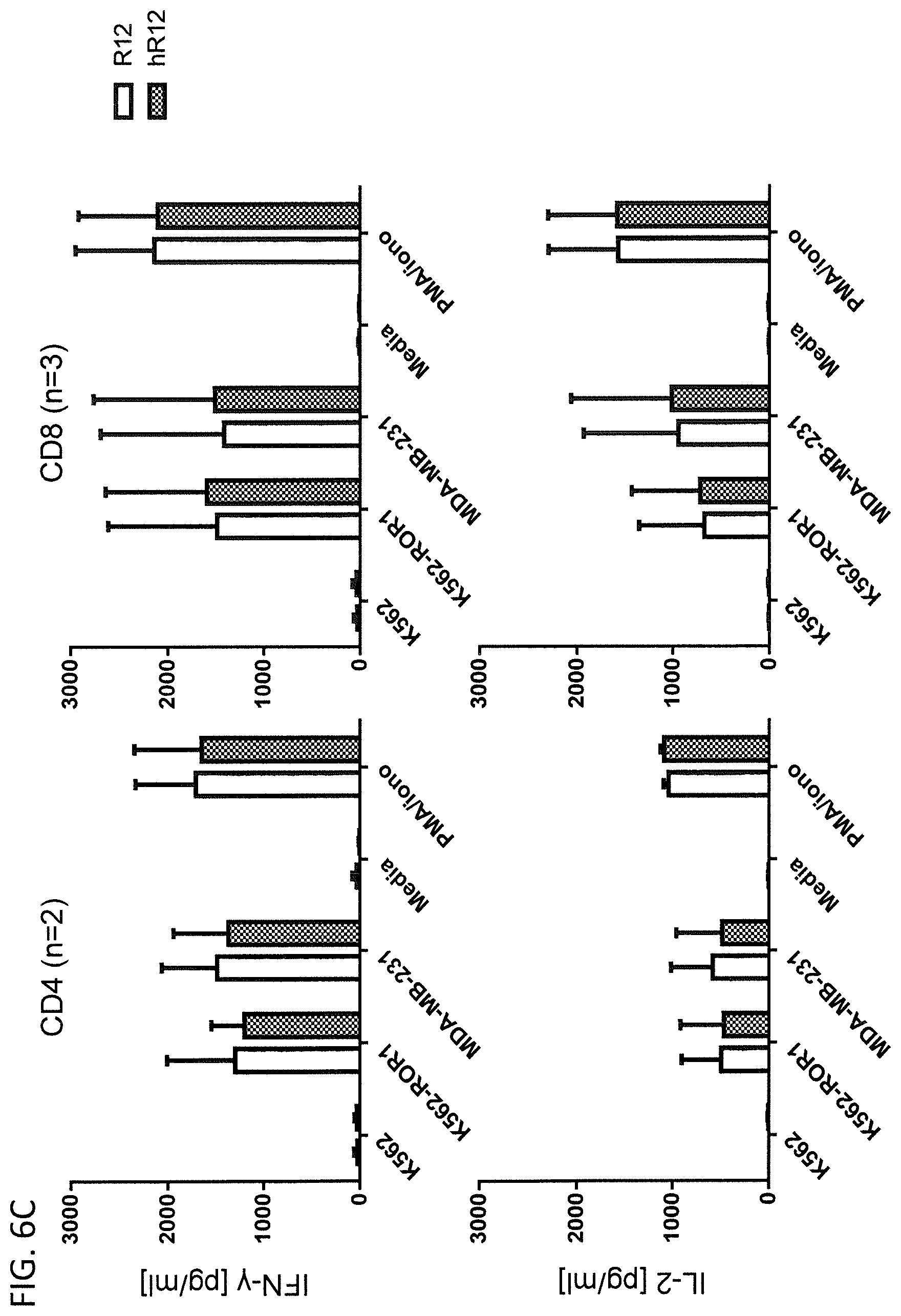
D00014

D00015

D00016

D00017

D00018

D00019
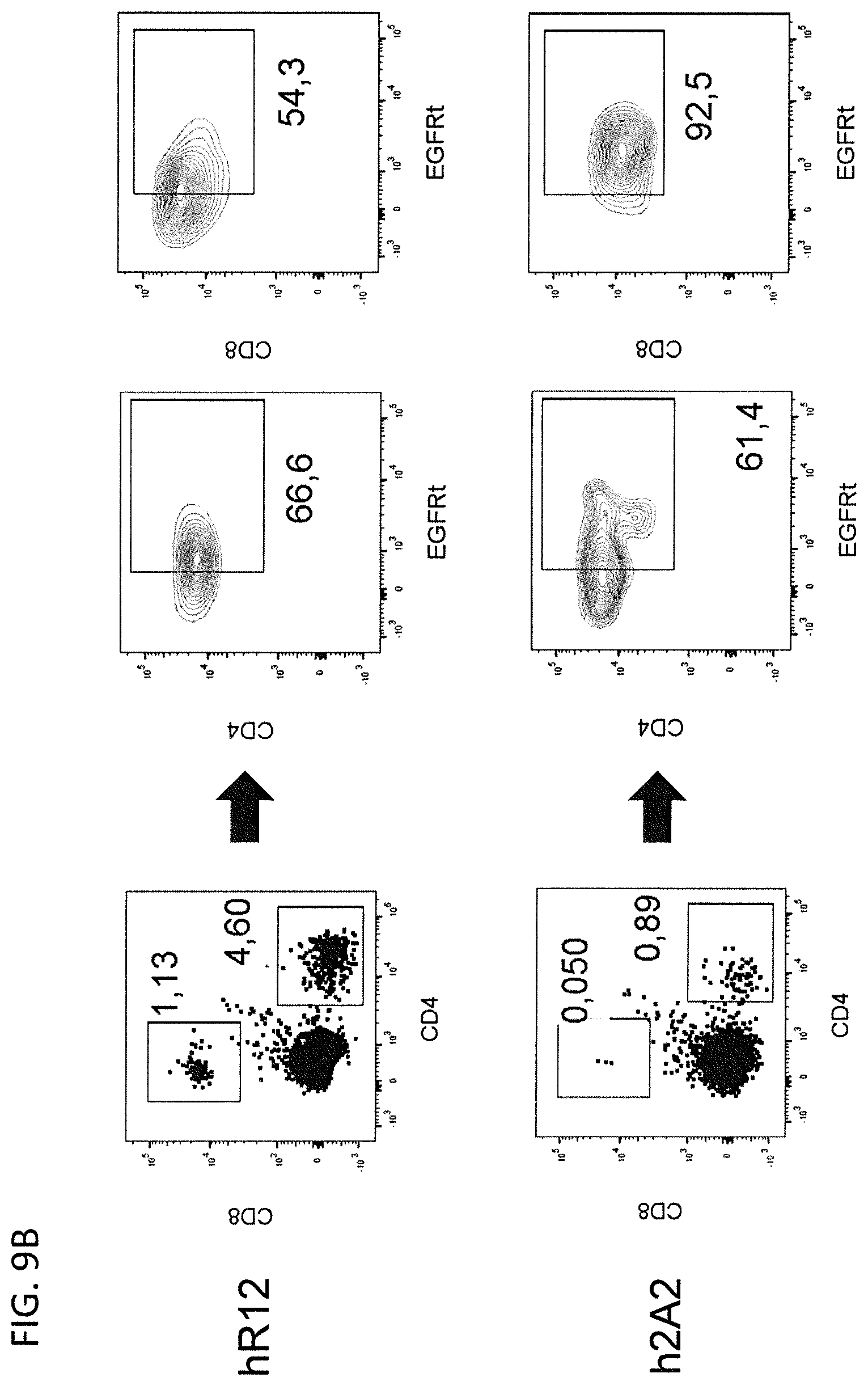
S00001
XML
uspto.report is an independent third-party trademark research tool that is not affiliated, endorsed, or sponsored by the United States Patent and Trademark Office (USPTO) or any other governmental organization. The information provided by uspto.report is based on publicly available data at the time of writing and is intended for informational purposes only.
While we strive to provide accurate and up-to-date information, we do not guarantee the accuracy, completeness, reliability, or suitability of the information displayed on this site. The use of this site is at your own risk. Any reliance you place on such information is therefore strictly at your own risk.
All official trademark data, including owner information, should be verified by visiting the official USPTO website at www.uspto.gov. This site is not intended to replace professional legal advice and should not be used as a substitute for consulting with a legal professional who is knowledgeable about trademark law.