Recombinant Glycoproteins with Reduced Antennary Fucosylation
Schmidt; Hanns-Martin ; et al.
U.S. patent application number 16/496113 was filed with the patent office on 2020-01-30 for recombinant glycoproteins with reduced antennary fucosylation. This patent application is currently assigned to Cevec Pharmaceutical GmbH. The applicant listed for this patent is Cevec Pharmaceutical GmbH. Invention is credited to Markus Ribbert, Gudrun Schiedner, Hanns-Martin Schmidt, Silke Wissing, Jens Wolfel.
| Application Number | 20200032311 16/496113 |
| Document ID | / |
| Family ID | 58489119 |
| Filed Date | 2020-01-30 |


| United States Patent Application | 20200032311 |
| Kind Code | A1 |
| Schmidt; Hanns-Martin ; et al. | January 30, 2020 |
Recombinant Glycoproteins with Reduced Antennary Fucosylation
Abstract
The present invention relates to methods for reducing antennary fucosylation of complex N-glycans in recombinantly expressed glycoproteins, cell lines that can be used in said methods, respective recombinant glycoproteins, and methods for expressing the same in said cell lines.
| Inventors: | Schmidt; Hanns-Martin; (Koln, DE) ; Ribbert; Markus; (Biberach an der Riss, DE) ; Schiedner; Gudrun; (Koln, DE) ; Wissing; Silke; (Koln, DE) ; Wolfel; Jens; (Langenfeld, DE) | ||||||||||
| Applicant: |
|
||||||||||
|---|---|---|---|---|---|---|---|---|---|---|---|
| Assignee: | Cevec Pharmaceutical GmbH Koln DE |
||||||||||
| Family ID: | 58489119 | ||||||||||
| Appl. No.: | 16/496113 | ||||||||||
| Filed: | March 15, 2018 | ||||||||||
| PCT Filed: | March 15, 2018 | ||||||||||
| PCT NO: | PCT/EP2018/056502 | ||||||||||
| 371 Date: | September 20, 2019 |
| Current U.S. Class: | 1/1 |
| Current CPC Class: | C07K 14/8125 20130101; C12Y 204/99001 20130101; C12N 5/16 20130101; C12N 9/1081 20130101; C07K 14/8121 20130101; C12Y 204/99004 20130101; C12P 21/005 20130101 |
| International Class: | C12P 21/00 20060101 C12P021/00; C07K 14/81 20060101 C07K014/81; C12N 9/10 20060101 C12N009/10; C12N 5/16 20060101 C12N005/16 |
Foreign Application Data
| Date | Code | Application Number |
|---|---|---|
| Mar 29, 2017 | EP | 17000521.9 |
Claims
1. A method for reducing antennary fucosylation of complex N-glycans in a recombinantly expressed glycoprotein, comprising the step of overexpressing together with the glycoprotein .beta.-galactoside .alpha.-2,6-sialyltransferase 1 (ST6Gal1) and/or .alpha.-2,3-sialyltransferase 4 (ST3Gal4).
2. The method of claim 1, wherein the glycoprotein is characterized by a significantly reduced antennary fucosylation of complex N-glycans as compared to the same recombinant glycoprotein expressed without overexpression of ST6Gal1 and/or ST3Gal4.
3. The method of claim 1, wherein at least 80% of the complex N-glycan antennae of the recombinantly expressed glycoprotein are not fucosylated.
4. The method of claim 1, wherein the glycoprotein is selected from the group consisting of .alpha.1-antitrypsin (AAT), hepatocyte growth factor (HGF), Factor VII (FVII), Factor VIII (FVIII), Factor IX (FIX), von Willebrand-Factor (vWF), alkaline phosphatase, and C1 esterase inhibitor (C1-inhibitor; C1 Inh).
5. The method of claim 1, wherein the glycoprotein is a mammalian, preferably a human glycoprotein.
6. The method of claim 1, wherein (i) .beta.-galactoside .alpha.-2,3-sialyltransferase 1 (ST3Gal1) is not overexpressed together with the glycoprotein, and/or (ii) the expression of .alpha.-1,3-mannosyl-glycoprotein 4-.beta.-N-acetylglucosaminyltransferase A (GnTIVa), .alpha.-1,3-mannosyl-glycoprotein 4-.beta.-N-acetylglucosaminyltransferase B (GnTIVb), and .alpha.-1,6-mannosylglycoprotein 6-.beta.-N-acetylglucosaminyltransferase A (GnTV) is not reduced.
7. A cell line that is genetically modified to overexpress .beta.-galactoside .alpha.-2,6-sialyltransferase 1 (ST6Gal1) and/or .alpha.-2,3-sialyltransferase 4 (ST3Gal4).
8. The cell line of claim 7, wherein the cell line comprises endogenous gene(s) encoding ST6Gal1 and/or ST3Gal4, and further has at least one genetic element, selected from the group consisting of a promoter, an enhancing element, and a stabilizing element inserted into the genome in one or more position(s) suitable to cause overexpression of ST6Gal1 and/or ST3Gal4.
9. The cell line of claim 7, wherein the cell line comprises exogenous nucleic acid(s) encoding ST6Gal1 and/or ST3Gal4.
10. The cell line of claims 7 to 9, wherein the cell line is derived from a cell line, selected from the group consisting of AGE.CR.RTM. cells, Vero cells, MDCK cells, BHK cells, CHO cells, HEK293 cells, HepG2 cells, Huh7 cells, AGE1.HN.RTM. cells, NC5T11 cells, Per.C6 cells, HMCLs cells, MM.1 cells, U266 cells, RPMI18226 cells, HKB11 cells, NM cells, NM-F9 cells, and CAP cells.
11. The cell line of claim 7, wherein the cell line is derived from human primary amniocytes comprising at least one nucleic acid encoding the gene products of the adenoviral E1 and pIX regions.
12. The cell line of claim 7, wherein the cell line is not genetically modified to (i) overexpress .beta.-galactoside .alpha.-2,3-sialyltransferase 1 (ST3Gal1), and/or (ii) reduce the expression of .alpha.-1,3-mannosyl-glycoprotein 4-.beta.-N-acetylglucosaminyltransferase A (GnTIVa), .alpha.-1,3-mannosyl-glycoprotein 4-.beta.-N-acetylglucosaminyltransferase B (GnTIVb), and .alpha.-1,6-mannosylglycoprotein 6-.beta.-N-acetylglucosaminyltransferase A (GnTV).
13. A recombinant glycoprotein having complex N-glycans, wherein antennary fucosylation of the complex N-glycans is reduced, so that at least 80% of the complex N-glycan antennae of the recombinant glycoprotein are not fucosylated.
14. The recombinant glycoprotein of claim 13, wherein the glycoprotein is selected from the group consisting of .alpha.1-antitrypsin (AAT), hepatocyte growth factor (HGF), Factor VII (FVII), Factor VIII (FVIII), Factor IX (FIX), von Willebrand-Factor (vWF), alkaline phosphatase, and C1 esterase inhibitor (C1-inhibitor; C1 Inh).
15. A method for the expression of a recombinant glycoprotein of claim 13, comprising the steps of: (a) providing a cell line of claim 7, (b) expressing the recombinant glycoprotein in said cell line; and (c) isolating the recombinant glycoprotein from the cells or the cell culture supernatant.
Description
[0001] The present invention relates to methods for reducing antennary fucosylation of complex N-glycans in recombinantly expressed glycoproteins, cell lines that can be used in said methods, respective recombinant glycoproteins, and methods for expressing the same in said cell lines.
[0002] Most current recombinant therapeutic proteins are glycoproteins. They have sugar residues attached to the amino-group of an asparagine (N-linked glycans) or the hydroxyl-group of a serine or threonine (O-linked glycans). The structure of the glycans is highly variable, depending on the specific protein and the host cell used for recombinant expression.
[0003] A very common structure of N-glycans found on glycoproteins expressed with mammalian expression platforms are so-called complex N-glycans, characterized by the core sugar sequence Man.alpha.1-6(Man.alpha.1-3)Man.beta.1-4GlcNAc.beta.1-4GlcNAc.beta.1-Asn-- . This core structure is extended by "antennae" which are initiated by N-acetylglucosamine (GlcNAc). Typically, complex type N-glycans have two, three, or four antennae, but in rare cases five or six antennae can be found. A typical structure of a di-antennary complex type N-glycan is depicted in FIG. 1A.
[0004] Complex N-glycans can be fucosylated. In mammalian cells, fucose is either linked to the proximal GlcNAc by .alpha.1-6 linkage (core fucose) or to a distal GlcNAc on one or several of the antennae by .alpha.1-3 linkage (antennary fucose; also called Lewis.sup.X antigen (Le.sup.x), CD15, or SSEA-1), displayed in FIG. 1B. If the Lewis.sup.x structure (Gal( -4)[Fuc(.alpha.)]GlcNAc-R) harbors an additional sialic acid, the resulting structure is called sialyl-Lewis.sup.x, sialyl-Le.sup.x, or SLe.sup.x (NeuAc(.alpha.1->4)Gal( -4)[Fuc(.alpha.1->3)]GlcNAc-R), displayed in FIG. 1C. Sialyl Lewis.sup.x structures emerge by the fucosylation of sialylated complex type N-glycans at the distal GlcNAc catalyzed by various fucosyltransferases. The role of core .alpha.1-6 fucosylation has been extensively studied for antibodies. Antibodies of the IgG class typically carry an N-glycan in the CH2 domain of the Fc region. The presence of a core fucose on these N-glycans significantly reduces the ADCC (antibody dependent cellular cytotoxicity) response mediated by the antibody in vivo. Since ADCC is usually a highly desired effect of a therapeutic antibody, several approaches are being taken to reduce the presence of core-fucose on IgGs. The highly immunogenic core .alpha.1-3 linked fucose structure only exists in plants and in invertebrates and is absent in human cells.
[0005] On immunoglobulins, typically no antennary fucose is found, however Lewis.sup.x or sialyl-Lewis.sup.x structures are easily detected on other serum-glycoproteins. Up to date little is known about its physiological role. There is evidence that antennary fucose might increase targeting to sites of inflammation via selectin interactions, but apart from that relatively little is known about the physiological role of antennary fucosylation.
[0006] When plasma proteins are recombinantly expressed, the product often shows an elevated level of antennary fucose as compared to the naturally occurring counterpart in the plasma. In order to reduce potential immunogenic effects of recombinant proteins used in replacement therapy, it is desirable that they are as similar to the endogenous protein as possible. A reduction of antennary fucose on recombinant therapeutic proteins is an important step towards this goal.
[0007] Accordingly, the technical problem underlying the present invention is to provide means for reducing antennary fucosylation (Le.sup.x or SLe.sup.x) of complex N-glycans in recombinantly expressed glycoproteins, as well as respective recombinant glycoproteins and means for producing the same.
[0008] The solution to the above technical problem is achieved by the embodiments characterized in the claims.
[0009] In particular, in a first aspect, the present invention relates to a method for reducing antennary fucosylation of complex N-glycans, either Le.sup.x or sialyl-Le.sup.x in a recombinantly expressed glycoprotein, comprising the step of overexpressing together with the glycoprotein .beta.-galactoside .alpha.-2,6-sialyltransferase 1 (ST6Gal1) and/or .alpha.-2,3-sialyltransferase 4 (ST3Gal4).
[0010] As used herein, the term "complex N-glycans" relates to N-glycans that are characterized by the core sugar sequence Man.alpha.1-6(Man.alpha.1-3)Man.beta.1-4GlcNAc.beta.1-4GlcNAc.beta.1-Asn-- . This complex N-glycan core sugar sequence can be extended by 2 to 6 antennae which are initiated by N-acetylglucosamine (GlcNAc), wherein two, three or four antennae are typical. Said antennae are sometimes referred to herein as "complex N-glycan antennae". Further, as used herein, the term "antennary fucosylation of complex N-glycans" refers to .alpha.1-3 linkage of fucose to a distal GlcNAc on at least one of the complex N-glycan antennae. In this context, the term "distal GlcNAc" refers to any GlcNAc present in an antenna other than the glycan's initial GlcNAcs.
[0011] The term "reducing antennary fucosylation of complex N-glycans" relates to the fact that according to the present invention, by way of overexpressing ST6Gal1 and/or ST3Gal4 together with the glycoprotein, recombinant glycoprotein is generated that has a lower amount of fucosylated complex N-glycan antennae as compared to a conventional recombinant glycoprotein. Preferably, such recombinant glycoprotein is characterized by a significantly reduced antennary fucosylation of complex N-glycans as compared to the same recombinant glycoprotein expressed without overexpression of ST6Gal1 and/or ST3Gal4.
[0012] In particularly preferred embodiments, at least 80%, more preferably at least 90%, and most preferably at least 95%, or more, of the complex N-glycan antennae of the recombinantly expressed glycoprotein are not fucosylated.
[0013] The glycoprotein to be subject to the methods of the present invention is not particularly limited, provided that it is a glycoprotein having complex N-glycans and respective complex N-glycan antennae. In preferred embodiments, the glycoprotein is selected from the group consisting of .alpha.1-antitrypsin (AAT), hepatocyte growth factor (HGF), Factor VII (FVII), Factor VIII (FVIII), Factor IX (FIX), von Willebrand-Factor (vWF), alkaline phosphatase, and C1 esterase inhibitor (C1-inhibitor; C1 Inh). Further, the glycoprotein is preferably a mammalian, more preferably a human glycoprotein.
[0014] As used herein, the term "recombinantly expressed glycoprotein" relates to glycoproteins that are biotechnologically produced in genetically modified organisms or cells.
[0015] Methods for recombinantly expressing glycoproteins, as well as for overexpressing ST6Gal1 and/or ST3Gal4 together with such glycoproteins, are not particularly limited and are known in the art. Further details in this respect are provided hereinafter for the second aspect of the present invention, relating to the cell lines of the present invention.
[0016] In specific embodiments of the present invention, both ST6Gal1 and ST3Gal4 are overexpressed together with the recombinant glycoprotein.
[0017] In further specific embodiments of the present invention, (i) .beta.-galactoside .alpha.-2,3-sialyltransferase 1 (ST3Gal1) is not overexpressed together with the recombinant glycoprotein, and/or (ii) the expression of .alpha.-1,3-mannosyl-glycoprotein 4-.beta.-N-acetylglucosaminyltransferase A (GnTIVa), .alpha.-1,3-mannosyl-glycoprotein 4-.beta.-N-acetylglucosaminyltransferase B (GnTIVb), and .alpha.-1,6-mannosylglycoprotein 6-.beta.-N-acetylglucosaminyltransferase A (GnTV) is not reduced.
[0018] In this context, the term "ST3Gal1 is not overexpressed together with the recombinant glycoprotein" relates to the fact that ST3Gal1 expression is not increased in any manner as compared to native ST3Gal1 expression. In particular embodiments, ST3Gal1 is not expressed at all. Further, the term "expression of GnTIVa, GnTIVb, and GnTV is not reduced" relates to the fact that expression of said proteins is not decreased in any manner as compared to native expression of said proteins.
[0019] In a second aspect, the present invention relates to a cell line, preferably an insect, avian, or mammalian cell line, more preferably a mammalian, in particular human, cell line, that is genetically modified to overexpress .beta.-galactoside .alpha.-2,6-sialyltransferase 1 (ST6Gal1) and/or .alpha.-2,3-sialyltransferase 4 (ST3Gal4).
[0020] As used herein, the term "cell line that is genetically modified to overexpress ST6Gal1 and/or ST3Gal4" indicates that upon genetic modification, the individual cells of the cell line display a higher expression of the respective sialyltransferase(s) than they did before the genetic modification.
[0021] Genetic modifications that allow the overexpression of a given protein are not particularly limited and are known in the art. In a particular example, the cell line comprises endogenous gene(s) encoding ST6Gal1 and/or ST3Gal4, such as e.g. human cell lines. In such cases, the cells can be genetically modified by inserting a promoter, enhancing element, and/or stabilizing element into the genome of the cells in a position suitable to cause overexpression of said nucleic acid. This can be done by homologous recombination using TALENS, Zn-finger proteins, CRISPR-CAS9, or other methods known in the art. Thus, in preferred embodiments, the cell line comprises endogenous gene(s) encoding ST6Gal1 and/or ST3Gal4, and further has at least one genetic element, selected from the group consisting of a promoter, an enhancing element, and a stabilizing element inserted into the genome in one or more position(s) suitable to cause overexpression of ST6Gal1 and/or ST3Gal4. Suitable promoters, enhancing elements and stabilizing elements are not particularly limited and are known in the art. For example, promoters include constitutive promoters, e.g. a CMV, EF1alpha, SV40, RSV, UbC, CAG, BOS or PGK promoter, and inducible promoters, e.g. tetracycline inducible promoters or other inducible promoters known in the art. Further, enhancing elements (enhancers) include CMV enhancer, -globin enhancer, immunoglobulin enhancer, and PGK-enhancer. Furthermore, stabilizing elements (chromatin elements) include matrix attachment regions (MARS), locus control regions (LCRs), and ubiquitously acting chromatin opening elements (UCOEs).
[0022] Alternatively, in cases where the cells do not comprise endogenous gene(s) encoding ST6Gal1 and/or ST3Gal4, or additionally, in cases where the cells do comprise endogenous gene(s) encoding ST6Gal1 and/or ST3Gal4, genetic modification of the cells can be achieved by introducing nucleic acid(s), encoding ST6Gal1 and/or ST3Gal4 into the cells. Methods for introducing nucleic acids into cells are not particularly limited and are known in the art. For example, said nucleic acids could be introduced in circular or linearized form into the cells by electroporation, nucleofection, microinjection, via viral vectors, e.g. lentiviral vectors, reagent based methods, e.g. lipids, calcium phosphate, cationic polymers or other methods known in the art. The nucleic acids can be transiently or stably introduced into the cell by episomal systems or by stable integration of the nucleic acid into the genome. Said nucleic acids can be present in the cells in the form of one or more expression vector(s), e.g. pcDNA, pCEP, pLenti, pEntr, pDest, pEF, pEAK, pCMV, pStbl, or other expression vectors known in the art. Expression of ST6Gal1 and/or ST3Gal4 can be under the control of a constitutive promoter, e.g. a CMV, EF1alpha, SV40, RSV, UbC, CAG, BOS or PGK promoter, the endogenous promoter, or of an inducible promoter, e.g. tetracycline inducible promoter or other inducible promoters known in the art. Further, the nucleic acids encoding ST6Gal1 and/or ST3Gal4 can be present as one continuous nucleic acid, or can be present as separate nucleic acids, e.g. as separate expression vectors. Said nucleic acids can contain, in addition to the coding region and a promoter, suitable restriction sites, Kozak sequences, ribosomal binding sites, chromatin modulating elements, selection cassettes, episomal replication systems, e.g. Epstein-Barr Nuclear Antigen and ori P, or SV40 ori and SV40 T-large antigen, internal ribosomal entry sites (IRES), splicing signals, and polyadenylation signals known in the art. Thus, in preferred embodiments, the cell line comprises exogenous nucleic acid(s) encoding ST6Gal1 and/or ST3Gal4.
[0023] Suitable genes encoding ST6Gal1 and/or ST3Gal4 for transfection of cell lines are not particularly limited and include any genes from any origin that encode proteins having ST6Gal1 or ST3Gal4 activity. Preferably, such genes are mammalian, more preferably human, ST6Gal1 and ST3Gal4 genes.
[0024] The cell lines according to the present invention can be derived from cell lines, e.g. mammalian cell lines, known in the art. In preferred embodiments, a cell line of the present invention can be derived from Muscovy Duck cells (AGE.CR.RTM.) African green monkey kidney epithelial cells (Vero), Madin Darby canine kidney cells (MDCK), baby hamster kidney cells (BHK), Chinese hamster ovary (CHO) cells, human hepatocarcinoma cell lines (HepG2, Huh7), human embryonic kidney 293 (HEK293) cells, human neuronal precursor cells (AGE1.HN.RTM. and NC5T11), human embryonic retinoblasts (Per.C6), myeloma cell lines (HMCLs, MM.1, U266, RPMI8226), CML tumor cell lines (NM, NM-F9), hybrid HEK293 and lymphoma cell (HKB11), or human amniocytes (CAP; cf. EP 1 230 354 B1), wherein CHO cells, HEK293 cells and CAP cells are preferred, and CAP cells are particularly preferred.
[0025] In this context, CAP cells are permanent amniocytic cell lines comprising a nucleic acid encoding the gene products of the adenovirus, in particular adenovirus type 5 (Ad5), E1A and E1 B regions. CAP cells are derived from primary human amniocytes that are transformed with a nucleic acid encoding Ad5 E1A and E1B.
[0026] Accordingly, in a preferred embodiment, the cell lines according to the present invention can be derived from human primary amniocytes comprising at least one nucleic acid encoding the gene products of the adenoviral E1 and pIX region, preferably E1 and pIX region of adenovirus type 5 (Ad5) from nt. 505 to 4079, in which E1A is under the control of the murine phosphoglycerate kinase (pgk) promoter, while E1B and pIX expression is controlled from their natural promoters. The E1B downstream intron, splice acceptor and polyA signal are replaced by corresponding motifs from SV40.
[0027] In specific embodiments of the present invention, the cell line of the present invention is genetically modified to overexpress both ST6Gal1 and ST3Gal4.
[0028] In further specific embodiments of the present invention, the cell line of the present invention is not genetically modified to (i) overexpress .beta.-galactoside .alpha.-2,3-sialyltransferase 1 (ST3Gal1), and/or (ii) reduce the expression of .alpha.-1,3-mannosyl-glycoprotein 4-.beta.-N-acetylglucosaminyltransferase A (GnTIVa), .alpha.-1,3-mannosyl-glycoprotein 4-.beta.-N-acetylglucosaminyltransferase B (GnTIVb), and .alpha.-1,6-mannosylglycoprotein 6-.beta.-N-acetylglucosaminyltransferase A (GnTV).
[0029] In this context, the term "the cell line is not genetically modified to overexpress ST3Gal1" relates to the fact that ST3Gal1 expression is not increased in any manner as compared to the cell line's native ST3Gal1 expression. In particular embodiments, ST3Gal1 is not expressed at all. Further, the term "the cell line is not genetically modified to reduce the expression of GnTIVa, GnTIVb, and GnTV" relates to the fact that expression of said proteins is not decreased in any manner as compared to native expression of said proteins in the cell line.
[0030] The cell lines according to this second aspect of the present invention are capable of reducing antennary fucosylation of complex N-glycans in recombinant glycoproteins expressed in said cell lines.
[0031] In a third aspect, the present invention relates to a recombinant glycoprotein having complex N-glycans, wherein antennary fucosylation of the complex N-glycans is reduced, so that at least 80%, more preferably at least 90%, and most preferably at least 95%, or more, of the complex N-glycan antennae of the recombinant glycoprotein are not fucosylated.
[0032] In this aspect, all relevant definitions and limitations given above for the first and second aspect of the present invention apply in an analogous manner.
[0033] Respective recombinant glycoproteins can be produced as described herein, e.g. by overexpression of ST6Gal1 and/or ST3Gal4 together with the recombinant glycoprotein. Preferably, said glycoproteins are produced in a cell line according to the present invention as described herein.
[0034] In a fourth aspect, the present invention relates to a method for the expression of a recombinant glycoprotein according to the present invention, comprising the steps of: [0035] (a) providing a cell line according to the present invention, [0036] (b) expressing the recombinant glycoprotein in said cell line; and [0037] (c) isolating the recombinant glycoprotein from the cells or the cell culture supernatant.
[0038] In this aspect, all relevant definitions and limitations given above for the first, second and third aspect of the present invention apply in an analogous manner. In particular, the recombinant glycoprotein and the cell line are as defined above.
[0039] Means for the expression of proteins in the cell lines of the present invention are not particularly limited and are known in the art. In this context, the step (b) of expressing the glycoprotein of interest in said cell line encompasses the transfection of a respective coding nucleic acid into said cell line prior to the actual expression of the glycoprotein. Further, means for isolating a glycoprotein of interest from a cell culture are not particularly limited and are known in the art.
[0040] In a related aspect, the present invention relates to the use of a cell line according to the present invention for the production of recombinant glycoproteins according to the present invention.
[0041] In this aspect, all of the definitions and preferred and/or specific embodiments described for the recombinant glycoproteins of the present invention and the cell lines of the present application apply in an analogous manner where applicable.
[0042] The figures show:
[0043] FIG. 1:
[0044] The Lewis glyco-epitope family, showing a subset of possible variants. The Le.sup.X glyco-epitopes carry fucose in an .alpha.1-3 linkage to the GlcNAc monosaccharide. A) sialylated, non-fucosylated GlcNAc; B) Lewis X (Le.sup.X) also called CD15 or SSEA-1; C) Sialyl Lewis X (sLe.sup.X).
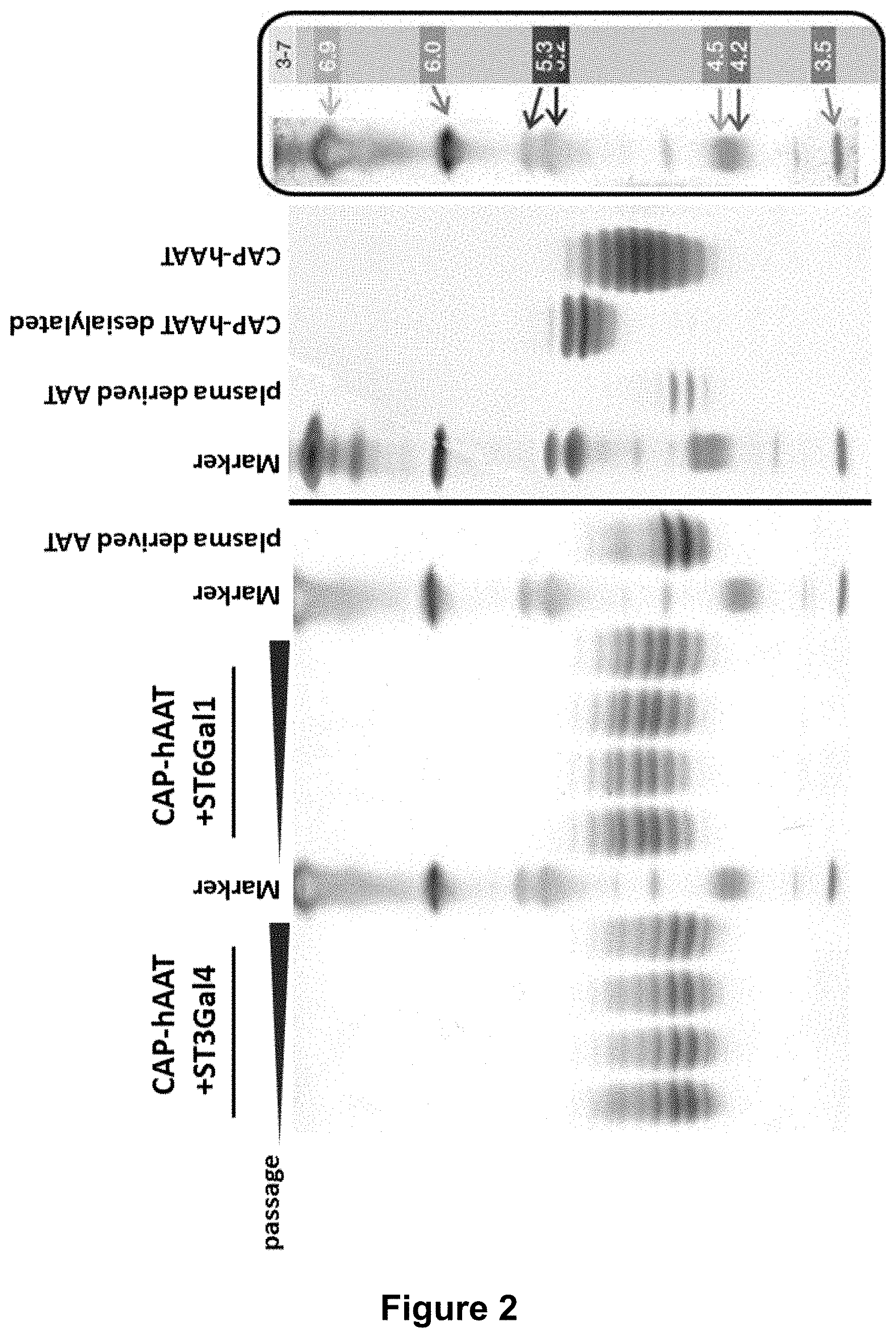
[0045] FIG. 2:
[0046] IEF (isoelectric focusing) analysis shows increased sialylation of hAAT purified from glyco-optimized CAP cells stably, recombinantly expressing hAAT and ST3Gal4 or ST6Gal1 compared to hAAT purified from non-engineered hAAT expressing CAP cells. 5 .mu.g of affinity purified hAAT per lane were subjected to isoelectric focusing. Different time points during pool generation are shown. Samples: CAP-hAAT-ST3Gal4, hAAT from CAP cell stably expressing human AAT as well as sialyltransferase ST3Gal4, CAP-hAAT-ST6Gal1, hAAT from CAP cell stably expressing human AAT as well as sialyltransferase ST6Gal1. Plasma derived hAAT (Prolastin), hAAT from non-glyco-optimized CAP cells, and desialylated hAAT from CAP cells served as controls.
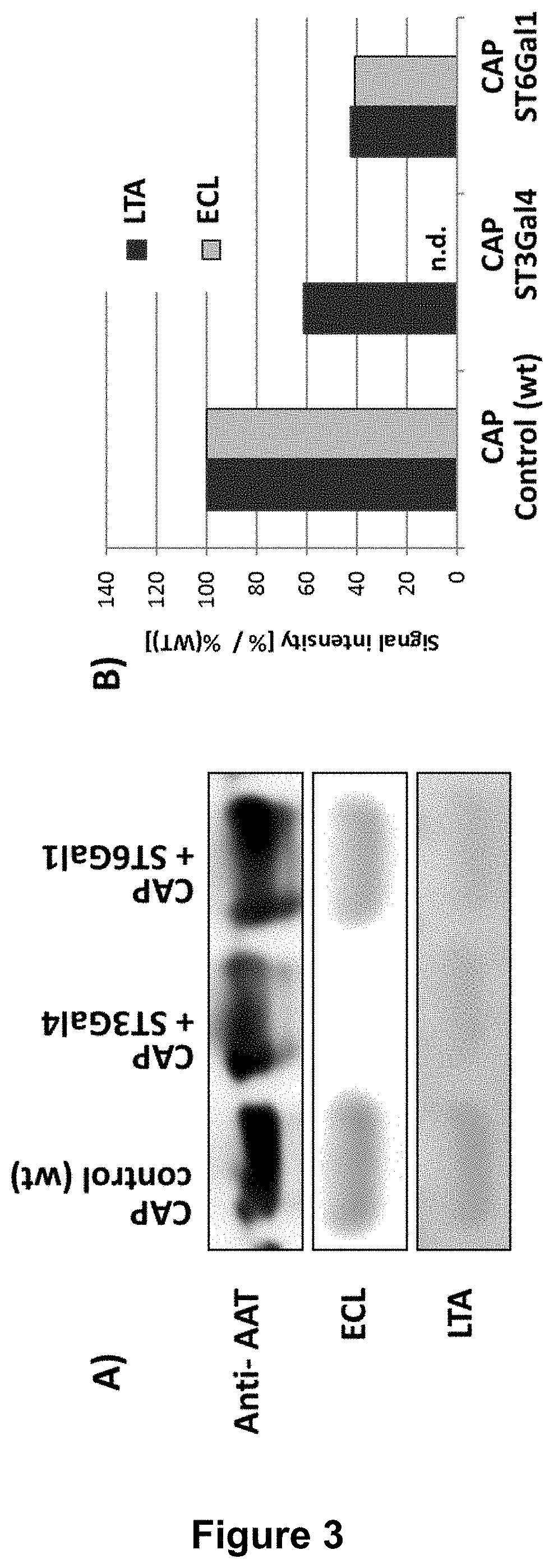
[0047] FIG. 3:
[0048] Comparative lectin blot analysis of recombinant AAT reveals that a decreased amount of fucosylation correlates with increased amounts of sialylation. Purified human rAAT from either wild-type CAP cells, CAP-ST3Gal4 cells or CAP-ST6Gal1 cells were separated by SDS-PAGE, blotted on nitrocellulose membrane and detected by specific lectins. The corresponding densitometrical analysis (B) was normalized on the AAT protein content in the matching western blot. The Erythrina crista-galli lectin (ECL-lectin) analysis detects free galactoses on N-glycans which indicates incomplete sialylation. .alpha.1-3 linked fucose is detected by Lotus tetragonolobus agglutinin (LTA).
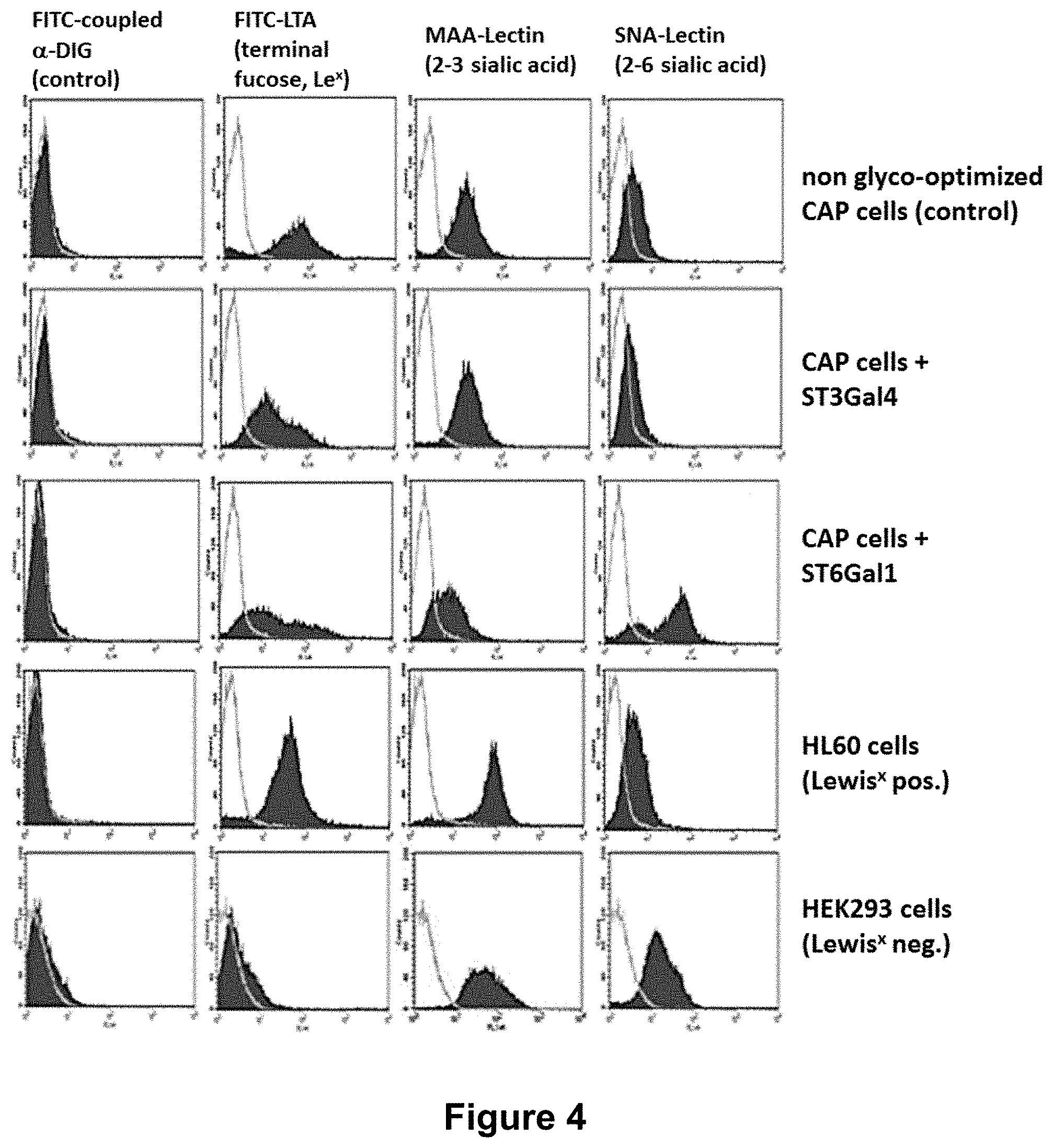
[0049] FIG. 4:
[0050] FACS analysis of cell surface glycoproteins of glyco-optimized CAP cells stably expressing ST3Gal4 or ST6Gal1 in comparison to non-engineered CAP cells reveals that overexpression of one of these two sialyltransferases not only increases the degree of sialylation on the majority of expressed glycoproteins. It also decreases the amount of antennary fucose on N-glycan structures, resulting in a reduced amount of Lewis.sup.X structures.
[0051] FIG. 5:
[0052] Increasing amounts of sialic acid (NANA) and reduced amounts of fucose (Fuc) in hAAT purified from CAP cells stably expressing either ST3Gal4 or ST6Gal1 compared to hAAT purified from cell culture supernatant from non-engineered CAP cells. Plasma derived hAAT (Prolastin) shown as control. Monosaccharide analysis was performed by high performance anion exchange chromatography with pulsed amperometric detection (HPAEC PAD).
[0053] FIG. 6:
[0054] Comparative lectin blot analysis of recombinant C1-Inhibitor reveals that an increase in sialylation correlates with a decrease in antennary fucosylation. Purified C1-Inhibitor from either wild-type CAP cells, CAP-ST3Gal4 cells or CAP-ST6Gal1 cells were separated by SDS-PAGE, blotted on nitrocellulose membrane and detected by specific lectins. The corresponding densitometrical analysis (B) was normalized to the C1-Inh protein content in the matching western blot. The Erythrina crista-galli lectin (ECL-lectin) analysis (A and B) detects free galactoses on N-glycans which indicates incomplete sialylation. .alpha.1-3 linked fucose is detected by Lotus tetragonolobus agglutinin (LTA).
[0055] FIG. 7:
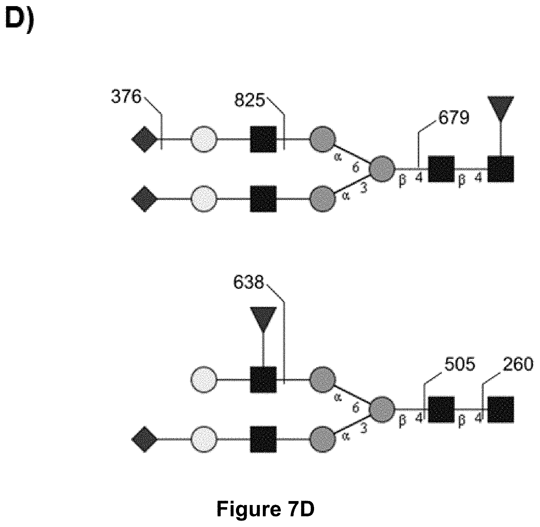
[0056] MS-MS analysis of recombinant C1-Inhibitor: PNGase F released permethylated N-glycans from purified C1-Inhibitor either from wild-type CAP cells, from CAP-ST3Gal4 cells or from CAP-ST6Gal1 cells were analyzed by MALDI TOF/TOF. Only the signal at 3196.8 in MS1 of wildtype derived C1-Inhibitor (A) contained two fucose residues and the characteristic fragmentation pattern for antennary fucose (M/z 638, 505, 260; D) in MS2. In CAP-ST3Gal4 (B) and CAP-ST6Gal1 (C) the signal at 3196.8 could not be detected, also no other signal of MS1 contained a fragmentation pattern of antennary fucose in MS2.
[0057] The present invention will be further illustrated in the following examples without being limited thereto.
EXAMPLES
Experimental Procedures:
Cell Culture and Fermentation.
[0058] The permanent human amniocyte cell line CAP 1D5 was cultured in suspension, either in chemically defined, animal component free CAP-CDM medium (CEVEC Pharmaceuticals, Germany) supplemented with 6 mM stable glutamine (Biochrom, Germany), or in serum free PEM media (Life Technologies) supplemented with 4 mM stable glutamine (Biochrom, Germany). CAP cells were cultivated at 37.degree. C. in shaker flasks (Corning, #431143, 125 mL (25 mL wv) or #431252, 3000 mL (1000 mL wv)) at 5% CO.sub.2, and 185 rpm. During fermentation, CAP cells were fed at d3, d5, and d7 with 10% CAP-CDM feed solution (CEVEC Pharmaceuticals, Germany) and 4 mM stable glutamine (Biochrom, Germany).
Cloning.
[0059] For the generation of CAP cell lines stably expressing ST3Gal4 or ST6Gal1, the cells were nucleofected with the corresponding nucleic acid constructs. Table 1 lists all cell lines created for this project.
[0060] For designing the ST3Gal4 cDNA, sequence information of the precursor protein and mature protein was based on the database entry UniProt Q11206 (SEQ ID NO: 1). For cloning, a ClaI restriction site and a Kozak sequence were added 5' of the start codon of the human ST3Gal4 cDNA and an EcoRV restriction site was added 3' of the stop codon to be inserted between the ClaI and EcoRV restriction sites in the pStbl-Neo-CMV-MCS(-) vector resulting in the expression plasmid pStbl-Neo-CMV-ST3Gal4. This vector contains a CMV promoter driving the expression of the gene of interest, followed by an SV40 intron for improved, splicing-mediated mRNA transport and a multiple cloning site for the insertion of the gene of interest. The selection marker is driven by the human ubiquitin (UbC) promoter. cDNA synthesis was performed at GeneArt (Germany, Life Technologies).
TABLE-US-00001 TABLE 1 Stable cell lines used in the present invention. overexpression of the Cell line rec. protein sialyltransferase(s) CAP / / CAP-AAT AAT / CAP-AAT-ST3Gal4 AAT ST3Gal4 CAP-AAT-ST6Gal1 AAT ST6Gal1 CAP-C1 Inh C1 Inh / CAP-C1 C1 Inh ST3Gal4 Inh-ST3Gal1 CAP-C1 C1 Inh ST6Gal1 Inh-ST3Gal4
[0061] For designing the ST6Gal1 cDNA, sequence information of the precursor protein and mature protein was based on the database entry UniProt P15907 (SEQ ID NO: 2). For cloning, a ClaI restriction site and a Kozak sequence were added 5' of the start codon of the human ST6Gal1 cDNA and an EcoRV restriction site was added 3' of the stop codon to be inserted between the ClaI and EcoRV restriction sites in the pStbl-Neo-CMV-MCS(-) vector resulting in the expression plasmid pStbl-Neo-CMV-ST6Gal1. cDNA synthesis was performed at GeneArt (Germany, Life Technologies).
Nucleofection and Pool Generation.
[0062] Nucleofection was performed using a Nucleofector II (LONZA) with the appropriate Nucleofector Kit (KitV) according to the manufacturer's protocol. Briefly, during exponential growth phase of the culture 1.times.10.sup.7 cells were harvested via centrifugation (150 g for 5 min) and re-suspended in 100 .mu.l complete Nucleofector solution and mixed with a total of 5 .mu.g plasmid. Nucleofection was performed using the X001 program. After the pulse, cells were recovered in 12 ml complete cell culture media in a 125 ml shaking flask. The cells were cultured as before at 37.degree. C., 5% CO.sub.2, and 185 rpm.
[0063] 72 to 96 h post-nucleofection cells were selected with 200 .mu.g/ml neomycin in order to generate stable pools.
Western Blot Analysis.
[0064] Purified protein solutions were separated on a NuPAGE Novex 4-12% Bis-Tris Gel under reducing conditions, according to the manufacturer's instructions. The separated proteins were transferred via a Blot Module (Invitrogen) (30 V for 60 min at RT) onto an Amersham Hybond ECL membrane (100 V for 60 min at RT). The membrane was blocked for 1 h at RT with PBSTB (phosphate-buffered saline, pH=7.4, supplemented with 0.1% Tween 20 and 1% BSA). Afterwards, the membrane was incubated with the specific horseradish peroxidase (HRP)-labeled antibody diluted in PBSTB. After washing the membrane with PBST (phosphate-buffered saline pH=7.4 supplemented with 0.1% Tween 20), the proteins were detected using the Pierce ECL WB Substrate Kit via a chemiluminescence detector (INTAS).
Lectin Immunoblotting.
[0065] Lectins are proteins that bind specific carbohydrate structures. Biotin-coupled lectins can therefore be used to analyze N-linked glycans. Erythrina crista-galli (ECL) lectin detects .beta.1-4 linked terminal galactose on N-linked glycans, Sambucus nigra agglutinin (SNA) preferentially binds to .alpha.2,6-linked sialic acid, whereas Maackia amurensis lectin (MAL) preferentially binds to .alpha.2,3-linked sialic acids. .alpha.1-3 linked fucose is detected by Lotus tetragonolobus agglutinin (LTA) and Aleuria aurantia lectin (AAL) detects .alpha.1-2-, -3, or -6 linked fucose.
[0066] Purified protein solutions from parental CAP cells with or without co-expression of ST3Gal4 and/or ST6Gal1 were separated as described above and blotted onto Amersham Hybond ECL nitrocellulose membrane (GE healthcare). The membrane was blocked for 1 h at RT with PBSTB (phosphate-buffered saline, pH=7.4, supplemented with 0.1% Tween 20 and 1% BSA). Afterwards, the membrane was incubated with the lectin diluted in PBSTB. After washing the membrane with PBST (phosphate-buffered saline, pH=7.4, supplemented with 0.1% Tween 20), the membrane was stained with streptavidin-coupled horseradish peroxidase (HRP) for 1 h at RT (diluted in PBSTB). The HRP signal was amplified using anti-streptavidin IgG and anti IgG-HRP. The proteins were detected using the Pierce ECL WB Substrate Kit via a chemiluminescence detector (INTAS).
Isoelectric Focusing (IEF) Analysis.
[0067] Isoelectric focusing (IEF) was performed in order to analyze the isoelectric point (pI) of rhAAT purified from CAP cells expressing rhAAT with or without additional expression of ST3Gal4 or ST6Gal1. The degree of sialylation correlates with a given proteins acidity and, therefore, with its pI. IEF analysis was done according to the manufacturers protocol (Invitrogen). Briefly, 5 .mu.g of purified protein were loaded on pH 3-7 gels and subjected to electrophoresis (1 h 100 V, 1 h 200 V, 30 min 500 V). Proteins were stained with SimplyBlue SafeStain according to the manufacturer's protocol (Invitrogen).
Example 1
[0068] Significantly Reduced Amount of Lewis.sup.X Structures on hAAT Protein Purified from CAP-ST3Gal4 or ST6Gal1 Cells.
[0069] .alpha.1-Antitrypsin (AAT) is a protease inhibitor belonging to the serpin superfamily. AAT is a potent inhibitor of serine proteases, in particular neutrophil elastase. AAT is a 52 kDa glycoprotein carrying 3 N-glycosylation sites.
[0070] Cells of the human amniocyte cell line CAP already stably expressing human AAT were additionally stably transfected with a plasmid encoding either the sialyltransferase ST3Gal4 to achieve an increase in 2,3-linked sialylation of terminal galactose of N-glycans or the sialyltransferase ST6Gal1 to achieve an increase in 2,6-sialylation of terminal galactose of N-glycans.
[0071] Enhanced 2,3- or 2,6-sialylation upon overexpression of sialyltransferase ST3Gal4 or ST6Gal1 were determined by isoelectric focusing (IEF) analysis of purified hAAT (FIG. 2).
[0072] As the backbones of the different rhAAT (recombinant hAAT) are identical, changes in the IEF indicate changes in the sialic acid content. Recombinant hAAT expressed in CAP cells with additional expression of ST3Gal4 results in a modified rhAAT which shifts significantly towards an acidic pI indicating an increased extent of sialylation; rhAAT expressed in parental CAP cells overexpressing ST6Gal1 also shifts towards a more acidic pI but to a lower degree (FIG. 2). This is probably due to the different substrate affinities of ST3Gal4 and ST6Gal1. ST6Gal1 catalyzes sialylation of the primary branches of N-glycans, whereas ST3Gal4 catalyzes the sialylation of the primary branches of N-glycan as well as the additional branches of tri- and tetra-antennary N-glycans. Therefore, ST3Gal4 has more acceptor substrate available than ST6Gal1.
[0073] This result could be confirmed via lectin blot analysis (FIG. 3). Increased amounts of .alpha.2,3- or .alpha.2,6-sialylation upon overexpression of sialyltransferase ST3Gal4 or ST6Gal1 were determined via Erythrina crista-galli (ECL) lectin blot analysis. ECL lectin detects .beta.1-4 linked terminal galactose on N-linked glycans. Therefore, a diminished signal in the ECL blot correlates to an increased amount of sialylation. Purified AAT from control CAP cells expressing AAT shows a clear signal in the ECL lectin blot, proving an incomplete sialylation. AAT derived from ST6Gal1 or ST3Gal4 overexpressing CAP cells, showed a strong reduction or complete absence of the ECL signal, indicating that only minimal amounts of unsialylated .beta.1-4 linked galactose on the N-linked glycan exist in these preparations (FIG. 3).
[0074] Remarkably, the degree of antennary fucose (Lewis.sup.x antigen) is reduced on rhAAT upon co-expression of ST6Gal1 or ST3Gal4 as proven by the reduced signal intensity in the Lotus tetragonolobus agglutinin (LTA Lectin) blot analysis in FIG. 3. Lotus tetragonolobus agglutinin (LTA) specifically detects the .alpha.1-3 linked antennary fucose. Therefore, overexpression of the sialyltransferases ST3Gal4 and ST6Gal1 is an unexpected but appropriate way to significantly reduce the amount of the unwanted and potentially immunogenic Lewis.sup.x structures on N-linked glycans.
Example 2
FACS Analysis of Glycoproteins on the Cell Surface of CAP Cells Expressing ST3Gal4 or ST6Gal1.
[0075] In order to determine the degree of fucosylation of glycoproteins on the cell surface with increased degree of sialylation by overexpression of sialyltransferases, flow cytometry (FACS) analyses was performed (FIG. 4).
[0076] CAP-hAAT-ST3Gal4 or CAP-hAAT-ST6Gal1 cells were stained with different antibodies and lectins to analyze sugar epitopes on the surface of the cells. Typically, 1.times.10.sup.7 cells were centrifuged for 10 min at 140.times.g and re-suspended into 100 .mu.l PBS/BSA. 10 .mu.l (10.sup.6 cells) were mixed with 10 .mu.l of antibody or lectin (1 mg/ml; FITC conjugated or DIG coupled in combination with a FITC coupled anti-DIG antibody) and 90 .mu.l PBS/BSA were added. After 10 min at 4.degree. C., the cells were washed with PBS/BSA. Cell pellets were re-suspended into 500 .mu.l PBS/BSA and subjected to FACS analysis on a Becton Dickinson FACSCalibur flow cytometer. Dead cells were identified and excluded by staining with propidium iodide. Typically, 30000 events were counted and analyzed. FITC or PE stained cells were graphically overlaid with unstained cells.
[0077] FIG. 4 shows parental CAP-hAAT cells or CAP-hAAT cells stably expressing .alpha.2,3 (CAP-hAAT+ST3Gal4) or .alpha.2,6 (CAP-hAAT+ST6Gal1) sialyltransferase, respectively. Cells were either stained with lectins specific for .alpha.2,3 coupled neuraminic acid, MAA (Maackia amurensis agglutinin), or .alpha.2,6 coupled neuraminic acid residues, SNA (Sambucus nigra agglutinin), or with LTA (Lotus tetragonolobus agglutinin) recognizing .alpha.1,3 linked fucose residues (Le.sup.X). Parental CAP-hAAT, HL60 (Le.sup.X pos.), Le.sup.X negative HEK293 cells (Le.sup.X neg.) as well as cells incubated with the FITC coupled anti-DIG antibody only are shown as controls.
[0078] As expected, overexpression of .alpha.2,3- or .alpha.2,6-sialyltransferase increases the respective coupled neuraminic acid residues on the N-glycans of cell surface glycoproteins. Interestingly, expression of ST3Gal4 or ST6Gal1 reduces the amount of the non-preferred Le.sup.X structures on glycoproteins on the cell surface, as indicated by significantly reduced staining with lectin LTA.
Example 3
[0079] Monosaccharide Analysis of hAAT Expressed in CAP-ST3Gal4 or CAP-ST6Gal1 Cells by HPAEC PAD Analysis.
[0080] In order to determine if the total amount of sialic acid and fucose of purified recombinant hAAT from CAP cells changes upon additional expression of the sialyltransferases ST3Gal4 or ST6Gal1, a monosaccharide analysis by high performance anion exchange chromatography with pulsed amperometric detection (HPAEC PAD) was performed.
[0081] FIG. 5 shows the relative amounts of sialic acid and fucose as determined by monosaccharide analysis via HPAEC-PAD in CAP-hAAT cells either stably transfected with a plasmid encoding ST3Gal4 or ST6Gal1.
[0082] FIG. 5 reveals that by expressing human sialyltransferases, sialic acid content is elevated, whereas the amount of fucose per rhAAT molecule is significantly reduced relative to an un-transfected control, indicating that the amount of the non-preferred Le.sup.X or sialyl-Le.sup.x structure is reduced upon overexpression of sialyltransferases. Of note in this experiment, the total amount of fucose was determined without differentiating between antennary fucose residues (.alpha.1-3 linked fucose) and core fucose (.alpha.1-6 linked fucose) of N-glycans. As additional expression of sialyltransferases only affects the amount of antennary .alpha.1-3 linked fucose but not the relatively abundant core fucose, the determined overall reduction of fucose upon overexpression of sialyltransferases of 25% (ST6Gal1) and 30% (ST3Gal4) indicates a pronounced reduction in the absolute amount of antennary .alpha.1-3 linked fucose.
[0083] The overall reduction in fucose residues is very surprising as the distal GlcNAc from sialylated complex N-glycans (NeuAc(.alpha.1->4)Gal( 1-4)GlcNAc-R), which will be increased upon overexpression of ST3Gal4 and/or ST6Gal1, is a substrate for the fucosyltransferases Fut5, Fut6, and Fut7. Therefore, overexpression of sialyltransferases as ST3Gal4 should rather result in an increase in sialyl-Lewis.sup.x structures than an overall decrease in fucose residues.
Example 4
[0084] Reduced Amount of Lewis.sup.x Structures on hC1 Inhibitor Protein Purified from CAP-ST3Gal4 or ST6Gal1 Cells.
[0085] Cells of the human amniocyte cell line CAP-hC1 Inh were stably transfected with a plasmid encoding either the sialyltransferase ST3Gal4 to achieve increased .alpha.2,3-linked sialylation of terminal galactose of N-glycans or sialyltransferase ST6Gal1 to achieve increased .alpha.2,6-sialylation of terminal galactose of N-glycans.
[0086] Increased amounts of .alpha.2,3- or .alpha.2,6-sialylation upon overexpression of sialyltransferase ST3Gal4 or ST6Gal1 were determined via Erythrina crista-galli (ECL) lectin blot analysis. ECL lectin detects .beta.1-4 linked terminal galactose on N-linked glycans. As shown in FIG. 6, overexpression of ST3Gal4 results in an increased sialylation of the N-linked glycans in comparison to C1 Inh purified from cell culture supernatant from non-glycomodified CAP cells. Overexpression of ST6Gal1 also results in an increased sialylation of terminal galactose on N-linked glycans in comparison to the non-glycomodified C1-Inh, although the effect is not as pronounced as upon overexpression of ST3Gal4. This is most likely due to the different substrate affinities of ST3Gal4 and ST6Gal1 as explained in Example 1.
[0087] The amount of antennary .alpha.1-3 linked fucose in the different C1 Inh protein preparations (CAP control, CAP-ST3Gal4, CAP-ST6Gal1) was determined by the Lotus tetragonolobus agglutinin (LTA) blot analysis. FIG. 6 reveals that the increased sialylation upon overexpression of ST6Gal1 or ST3Gal4 correlates with a reduced amount of antennary fucosylation proven by the decreased signal intensity in the LTA lectin blot analysis.
[0088] These results were confirmed by MS-MS analysis of N-glycans from C1 Inh derived in CAP control cells, CAP-ST3Gal4, or CAP-ST6Gal1 cells (FIG. 7). PNGaseF released permethylated N-glycans from purified C1-Inhibitor either from control CAP cells, from CAP-ST3Gal4 cells or from CAP-ST6Gal1 cells were analyzed by MALDI TOF/TOF. Only the signal at 3196.8 in MS1 in wildtype derived C1-Inhibitor (FIG. 7 A) contained two fucose residues and the characteristic fragmentation pattern for antennary fucose (M/z 638, 505, 260; D) in MS2. In CAP-ST3Gal4 and CAP-ST6Gal1 (FIGS. 7 B and C) the signal at 3196.8 could not be detected. Moreover, no other signal of MS1 contained a fragmentation pattern of antennary fucose in MS2.
[0089] The present invention relates to the following amino acid sequences.
TABLE-US-00002 SEQ ID NO: 1 Human ST3Gal4 MVSKSRWKLLAMLALVLVVMVWYSISREDRYIELFYFPIPEKKEPCLQGE AESKASKLFGNYSRDQPIFLRLEDYFWVKTPSAYELPYGTKGSEDLLLRV LAITSSSIPKNIQSLRCRRCVVVGNGHRLRNSSLGDAINKYDVVIRLNNA PVAGYEGDVGSKTTMRLFYPESAHFDPKVENNPDTLLVLVAFKAMDFHWI ETILSDKKRVRKGFWKQPPLIWDVNPKQIRILNPFFMEIAADKLLSLPMQ QPRKIKQKPTTGLLAITLALHLCDLVHIAGFGYPDAYNKKQTIHYYEQIT LKSMAGSGHNVSQEALAIKRMLEMGAIKNLTSF SEQ ID NO: 2 Human ST6Gal1 MIHTNLKKKFSCCVLVFLLFAVICVWKEKKKGSYYDSFKLQTKEFQVLKS LGKLAMGSDSQSVSSSSTQDPHRGRQTLGSLRGLAKAKPEASFQVWNKDS SSKNLIPRLQKIWKNYLSMNKYKVSYKGPGPGIKFSAEALRCHLRDHVNV SMVEVTDFPFNTSEWEGYLPKESIRTKAGPWGRCAVVSSAGSLKSSQLGR EIDDHDAVLRFNGAPTANFQQDVGTKTTIRLMNSQLVTTEKRFLKDSLYN EGILIVWDPSVYHSDIPKWYQNPDYNFFNNYKTYRKLHPNQPFYILKPQM PWELWDILQEISPEEIQPNPPSSGMLGIIIMMTLCDQVDIYEFLPSKRKT DVCYYYQKFFDSACTMGAYHPLLYEKNLVKHLNQGTDEDIYLLGKATLPG FRTIHC SEQ ID NO: 3 Human AAT MPSSVSWGILLLAGLCCLVPVSLAEDPQGDAAQKTDTSHHDQDHPTFNKI TPNLAEFAFSLYRQLAHQSNSTNIFFSPVSIATAFAMLSLGTKADTHDEI LEGLNFNLTEIPEAQIHEGFQELLRTLNQPDSQLQLTTGNGLFLSEGLKL VDKFLEDVKKLYHSEAFTVNFGDTEEAKKQINDYVEKGTQGKIVDLVKEL DRDTVFALVNYIFFKGKWERPFEVKDTEEEDFHVDQVTTVKVPMMKRLGM FNIQHCKKLSSWVLLMKYLGNATAIFFLPDEGKLQHLENELTHDIITKFL ENEDRRSASLHLPKLSITGTYDLKSVLGQLGITKVFSNGADLSGVTEEAP LKLSKAVHKAVLTIDEKGTEAAGAMFLEAIPMSIPPEVKFNKPFVFLMIE QNTKSPLFMGKVVNPTQK SEQ ID NO: 4 Human C1 Inh MASRLTLLTLLLLLLAGDRASSNPNATSSSSQDPESLQDRGEGKVATTVI SKMLFVEPILEVSSLPTTNSTTNSATKITANTTDEPTTQPTTEPTTQPTI QPTQPTTQLPTDSPTQPTTGSFCPGPVTLCSDLESHSTEAVLGDALVDFS LKLYHAFSAMKKVETNMAFSPFSIASLLTQVLLGAGENTKTNLESILSYP KDFTCVHQALKGFTTKGVTSVSQIFHSPDLAIRDTFVNASRTLYSSSPRV LSNNSDANLELINTWVAKNTNNKISRLLDSLPSDTRLVLLNAIYLSAKWK TTFDPKKTRMEPFHFKNSVIKVPMMNSKKYPVAHFIDQTLKAKVGQLQLS HNLSLVILVPQNLKHRLEDMEQALSPSVFKAIMEKLEMSKFQPTLLTLPR IKVTTSQDMLSIMEKLEFFDFSYDLNLCGLTEDPDLQVSAMQHQTVLELT ETGVEAAAASAISVARTLLVFEVQQPFLFVLWDQQHKFPVFMGRVYDPRA
Sequence CWU 1
1
41333PRTHomo sapiens 1Met Val Ser Lys Ser Arg Trp Lys Leu Leu Ala
Met Leu Ala Leu Val1 5 10 15Leu Val Val Met Val Trp Tyr Ser Ile Ser
Arg Glu Asp Arg Tyr Ile 20 25 30Glu Leu Phe Tyr Phe Pro Ile Pro Glu
Lys Lys Glu Pro Cys Leu Gln 35 40 45Gly Glu Ala Glu Ser Lys Ala Ser
Lys Leu Phe Gly Asn Tyr Ser Arg 50 55 60Asp Gln Pro Ile Phe Leu Arg
Leu Glu Asp Tyr Phe Trp Val Lys Thr65 70 75 80Pro Ser Ala Tyr Glu
Leu Pro Tyr Gly Thr Lys Gly Ser Glu Asp Leu 85 90 95Leu Leu Arg Val
Leu Ala Ile Thr Ser Ser Ser Ile Pro Lys Asn Ile 100 105 110Gln Ser
Leu Arg Cys Arg Arg Cys Val Val Val Gly Asn Gly His Arg 115 120
125Leu Arg Asn Ser Ser Leu Gly Asp Ala Ile Asn Lys Tyr Asp Val Val
130 135 140Ile Arg Leu Asn Asn Ala Pro Val Ala Gly Tyr Glu Gly Asp
Val Gly145 150 155 160Ser Lys Thr Thr Met Arg Leu Phe Tyr Pro Glu
Ser Ala His Phe Asp 165 170 175Pro Lys Val Glu Asn Asn Pro Asp Thr
Leu Leu Val Leu Val Ala Phe 180 185 190Lys Ala Met Asp Phe His Trp
Ile Glu Thr Ile Leu Ser Asp Lys Lys 195 200 205Arg Val Arg Lys Gly
Phe Trp Lys Gln Pro Pro Leu Ile Trp Asp Val 210 215 220Asn Pro Lys
Gln Ile Arg Ile Leu Asn Pro Phe Phe Met Glu Ile Ala225 230 235
240Ala Asp Lys Leu Leu Ser Leu Pro Met Gln Gln Pro Arg Lys Ile Lys
245 250 255Gln Lys Pro Thr Thr Gly Leu Leu Ala Ile Thr Leu Ala Leu
His Leu 260 265 270Cys Asp Leu Val His Ile Ala Gly Phe Gly Tyr Pro
Asp Ala Tyr Asn 275 280 285Lys Lys Gln Thr Ile His Tyr Tyr Glu Gln
Ile Thr Leu Lys Ser Met 290 295 300Ala Gly Ser Gly His Asn Val Ser
Gln Glu Ala Leu Ala Ile Lys Arg305 310 315 320Met Leu Glu Met Gly
Ala Ile Lys Asn Leu Thr Ser Phe 325 3302406PRTHomo sapiens 2Met Ile
His Thr Asn Leu Lys Lys Lys Phe Ser Cys Cys Val Leu Val1 5 10 15Phe
Leu Leu Phe Ala Val Ile Cys Val Trp Lys Glu Lys Lys Lys Gly 20 25
30Ser Tyr Tyr Asp Ser Phe Lys Leu Gln Thr Lys Glu Phe Gln Val Leu
35 40 45Lys Ser Leu Gly Lys Leu Ala Met Gly Ser Asp Ser Gln Ser Val
Ser 50 55 60Ser Ser Ser Thr Gln Asp Pro His Arg Gly Arg Gln Thr Leu
Gly Ser65 70 75 80Leu Arg Gly Leu Ala Lys Ala Lys Pro Glu Ala Ser
Phe Gln Val Trp 85 90 95Asn Lys Asp Ser Ser Ser Lys Asn Leu Ile Pro
Arg Leu Gln Lys Ile 100 105 110Trp Lys Asn Tyr Leu Ser Met Asn Lys
Tyr Lys Val Ser Tyr Lys Gly 115 120 125Pro Gly Pro Gly Ile Lys Phe
Ser Ala Glu Ala Leu Arg Cys His Leu 130 135 140Arg Asp His Val Asn
Val Ser Met Val Glu Val Thr Asp Phe Pro Phe145 150 155 160Asn Thr
Ser Glu Trp Glu Gly Tyr Leu Pro Lys Glu Ser Ile Arg Thr 165 170
175Lys Ala Gly Pro Trp Gly Arg Cys Ala Val Val Ser Ser Ala Gly Ser
180 185 190Leu Lys Ser Ser Gln Leu Gly Arg Glu Ile Asp Asp His Asp
Ala Val 195 200 205Leu Arg Phe Asn Gly Ala Pro Thr Ala Asn Phe Gln
Gln Asp Val Gly 210 215 220Thr Lys Thr Thr Ile Arg Leu Met Asn Ser
Gln Leu Val Thr Thr Glu225 230 235 240Lys Arg Phe Leu Lys Asp Ser
Leu Tyr Asn Glu Gly Ile Leu Ile Val 245 250 255Trp Asp Pro Ser Val
Tyr His Ser Asp Ile Pro Lys Trp Tyr Gln Asn 260 265 270Pro Asp Tyr
Asn Phe Phe Asn Asn Tyr Lys Thr Tyr Arg Lys Leu His 275 280 285Pro
Asn Gln Pro Phe Tyr Ile Leu Lys Pro Gln Met Pro Trp Glu Leu 290 295
300Trp Asp Ile Leu Gln Glu Ile Ser Pro Glu Glu Ile Gln Pro Asn
Pro305 310 315 320Pro Ser Ser Gly Met Leu Gly Ile Ile Ile Met Met
Thr Leu Cys Asp 325 330 335Gln Val Asp Ile Tyr Glu Phe Leu Pro Ser
Lys Arg Lys Thr Asp Val 340 345 350Cys Tyr Tyr Tyr Gln Lys Phe Phe
Asp Ser Ala Cys Thr Met Gly Ala 355 360 365Tyr His Pro Leu Leu Tyr
Glu Lys Asn Leu Val Lys His Leu Asn Gln 370 375 380Gly Thr Asp Glu
Asp Ile Tyr Leu Leu Gly Lys Ala Thr Leu Pro Gly385 390 395 400Phe
Arg Thr Ile His Cys 4053418PRTHomo sapiens 3Met Pro Ser Ser Val Ser
Trp Gly Ile Leu Leu Leu Ala Gly Leu Cys1 5 10 15Cys Leu Val Pro Val
Ser Leu Ala Glu Asp Pro Gln Gly Asp Ala Ala 20 25 30Gln Lys Thr Asp
Thr Ser His His Asp Gln Asp His Pro Thr Phe Asn 35 40 45Lys Ile Thr
Pro Asn Leu Ala Glu Phe Ala Phe Ser Leu Tyr Arg Gln 50 55 60Leu Ala
His Gln Ser Asn Ser Thr Asn Ile Phe Phe Ser Pro Val Ser65 70 75
80Ile Ala Thr Ala Phe Ala Met Leu Ser Leu Gly Thr Lys Ala Asp Thr
85 90 95His Asp Glu Ile Leu Glu Gly Leu Asn Phe Asn Leu Thr Glu Ile
Pro 100 105 110Glu Ala Gln Ile His Glu Gly Phe Gln Glu Leu Leu Arg
Thr Leu Asn 115 120 125Gln Pro Asp Ser Gln Leu Gln Leu Thr Thr Gly
Asn Gly Leu Phe Leu 130 135 140Ser Glu Gly Leu Lys Leu Val Asp Lys
Phe Leu Glu Asp Val Lys Lys145 150 155 160Leu Tyr His Ser Glu Ala
Phe Thr Val Asn Phe Gly Asp Thr Glu Glu 165 170 175Ala Lys Lys Gln
Ile Asn Asp Tyr Val Glu Lys Gly Thr Gln Gly Lys 180 185 190Ile Val
Asp Leu Val Lys Glu Leu Asp Arg Asp Thr Val Phe Ala Leu 195 200
205Val Asn Tyr Ile Phe Phe Lys Gly Lys Trp Glu Arg Pro Phe Glu Val
210 215 220Lys Asp Thr Glu Glu Glu Asp Phe His Val Asp Gln Val Thr
Thr Val225 230 235 240Lys Val Pro Met Met Lys Arg Leu Gly Met Phe
Asn Ile Gln His Cys 245 250 255Lys Lys Leu Ser Ser Trp Val Leu Leu
Met Lys Tyr Leu Gly Asn Ala 260 265 270Thr Ala Ile Phe Phe Leu Pro
Asp Glu Gly Lys Leu Gln His Leu Glu 275 280 285Asn Glu Leu Thr His
Asp Ile Ile Thr Lys Phe Leu Glu Asn Glu Asp 290 295 300Arg Arg Ser
Ala Ser Leu His Leu Pro Lys Leu Ser Ile Thr Gly Thr305 310 315
320Tyr Asp Leu Lys Ser Val Leu Gly Gln Leu Gly Ile Thr Lys Val Phe
325 330 335Ser Asn Gly Ala Asp Leu Ser Gly Val Thr Glu Glu Ala Pro
Leu Lys 340 345 350Leu Ser Lys Ala Val His Lys Ala Val Leu Thr Ile
Asp Glu Lys Gly 355 360 365Thr Glu Ala Ala Gly Ala Met Phe Leu Glu
Ala Ile Pro Met Ser Ile 370 375 380Pro Pro Glu Val Lys Phe Asn Lys
Pro Phe Val Phe Leu Met Ile Glu385 390 395 400Gln Asn Thr Lys Ser
Pro Leu Phe Met Gly Lys Val Val Asn Pro Thr 405 410 415Gln
Lys4500PRTHomo sapiens 4Met Ala Ser Arg Leu Thr Leu Leu Thr Leu Leu
Leu Leu Leu Leu Ala1 5 10 15Gly Asp Arg Ala Ser Ser Asn Pro Asn Ala
Thr Ser Ser Ser Ser Gln 20 25 30Asp Pro Glu Ser Leu Gln Asp Arg Gly
Glu Gly Lys Val Ala Thr Thr 35 40 45Val Ile Ser Lys Met Leu Phe Val
Glu Pro Ile Leu Glu Val Ser Ser 50 55 60Leu Pro Thr Thr Asn Ser Thr
Thr Asn Ser Ala Thr Lys Ile Thr Ala65 70 75 80Asn Thr Thr Asp Glu
Pro Thr Thr Gln Pro Thr Thr Glu Pro Thr Thr 85 90 95Gln Pro Thr Ile
Gln Pro Thr Gln Pro Thr Thr Gln Leu Pro Thr Asp 100 105 110Ser Pro
Thr Gln Pro Thr Thr Gly Ser Phe Cys Pro Gly Pro Val Thr 115 120
125Leu Cys Ser Asp Leu Glu Ser His Ser Thr Glu Ala Val Leu Gly Asp
130 135 140Ala Leu Val Asp Phe Ser Leu Lys Leu Tyr His Ala Phe Ser
Ala Met145 150 155 160Lys Lys Val Glu Thr Asn Met Ala Phe Ser Pro
Phe Ser Ile Ala Ser 165 170 175Leu Leu Thr Gln Val Leu Leu Gly Ala
Gly Glu Asn Thr Lys Thr Asn 180 185 190Leu Glu Ser Ile Leu Ser Tyr
Pro Lys Asp Phe Thr Cys Val His Gln 195 200 205Ala Leu Lys Gly Phe
Thr Thr Lys Gly Val Thr Ser Val Ser Gln Ile 210 215 220Phe His Ser
Pro Asp Leu Ala Ile Arg Asp Thr Phe Val Asn Ala Ser225 230 235
240Arg Thr Leu Tyr Ser Ser Ser Pro Arg Val Leu Ser Asn Asn Ser Asp
245 250 255Ala Asn Leu Glu Leu Ile Asn Thr Trp Val Ala Lys Asn Thr
Asn Asn 260 265 270Lys Ile Ser Arg Leu Leu Asp Ser Leu Pro Ser Asp
Thr Arg Leu Val 275 280 285Leu Leu Asn Ala Ile Tyr Leu Ser Ala Lys
Trp Lys Thr Thr Phe Asp 290 295 300Pro Lys Lys Thr Arg Met Glu Pro
Phe His Phe Lys Asn Ser Val Ile305 310 315 320Lys Val Pro Met Met
Asn Ser Lys Lys Tyr Pro Val Ala His Phe Ile 325 330 335Asp Gln Thr
Leu Lys Ala Lys Val Gly Gln Leu Gln Leu Ser His Asn 340 345 350Leu
Ser Leu Val Ile Leu Val Pro Gln Asn Leu Lys His Arg Leu Glu 355 360
365Asp Met Glu Gln Ala Leu Ser Pro Ser Val Phe Lys Ala Ile Met Glu
370 375 380Lys Leu Glu Met Ser Lys Phe Gln Pro Thr Leu Leu Thr Leu
Pro Arg385 390 395 400Ile Lys Val Thr Thr Ser Gln Asp Met Leu Ser
Ile Met Glu Lys Leu 405 410 415Glu Phe Phe Asp Phe Ser Tyr Asp Leu
Asn Leu Cys Gly Leu Thr Glu 420 425 430Asp Pro Asp Leu Gln Val Ser
Ala Met Gln His Gln Thr Val Leu Glu 435 440 445Leu Thr Glu Thr Gly
Val Glu Ala Ala Ala Ala Ser Ala Ile Ser Val 450 455 460Ala Arg Thr
Leu Leu Val Phe Glu Val Gln Gln Pro Phe Leu Phe Val465 470 475
480Leu Trp Asp Gln Gln His Lys Phe Pro Val Phe Met Gly Arg Val Tyr
485 490 495Asp Pro Arg Ala 500
D00001
D00002
D00003
D00004
D00005
D00006
D00007
D00008
D00009
D00010
S00001
XML
uspto.report is an independent third-party trademark research tool that is not affiliated, endorsed, or sponsored by the United States Patent and Trademark Office (USPTO) or any other governmental organization. The information provided by uspto.report is based on publicly available data at the time of writing and is intended for informational purposes only.
While we strive to provide accurate and up-to-date information, we do not guarantee the accuracy, completeness, reliability, or suitability of the information displayed on this site. The use of this site is at your own risk. Any reliance you place on such information is therefore strictly at your own risk.
All official trademark data, including owner information, should be verified by visiting the official USPTO website at www.uspto.gov. This site is not intended to replace professional legal advice and should not be used as a substitute for consulting with a legal professional who is knowledgeable about trademark law.