Compositions And Methods For Inhibition Of Lineage Specific Proteins
Bolen; Joseph ; et al.
U.S. patent application number 16/489407 was filed with the patent office on 2020-01-30 for compositions and methods for inhibition of lineage specific proteins. This patent application is currently assigned to VOR BIOPHARMA, INC. The applicant listed for this patent is VOR BIOPHARMA, INC. Invention is credited to Joseph Bolen, John Lydeard, Aleksandar Filip Radovic-Moreno.
| Application Number | 20200030381 16/489407 |
| Document ID | / |
| Family ID | 63370206 |
| Filed Date | 2020-01-30 |


View All Diagrams
| United States Patent Application | 20200030381 |
| Kind Code | A1 |
| Bolen; Joseph ; et al. | January 30, 2020 |
COMPOSITIONS AND METHODS FOR INHIBITION OF LINEAGE SPECIFIC PROTEINS
Abstract
Disclosed herein are compositions, methods, and kits for use in treating hematopoietic malignancies, the compositions, methods, and kits comprise a cytotoxic agent targeting cells expressing a lineage-specific cell-surface protein and a population of hematopoietic cells that express the lineage-specific cell-surface protein, the hematopoietic cells being manipulated such that they do not bind the cytotoxic agent.
| Inventors: | Bolen; Joseph; (Boston, MA) ; Radovic-Moreno; Aleksandar Filip; (Boston, MA) ; Lydeard; John; (Sharon, MA) | ||||||||||
| Applicant: |
|
||||||||||
|---|---|---|---|---|---|---|---|---|---|---|---|
| Assignee: | VOR BIOPHARMA, INC Boston MA |
||||||||||
| Family ID: | 63370206 | ||||||||||
| Appl. No.: | 16/489407 | ||||||||||
| Filed: | February 28, 2018 | ||||||||||
| PCT Filed: | February 28, 2018 | ||||||||||
| PCT NO: | PCT/US18/20327 | ||||||||||
| 371 Date: | August 28, 2019 |
Related U.S. Patent Documents
| Application Number | Filing Date | Patent Number | ||
|---|---|---|---|---|
| 62464975 | Feb 28, 2017 | |||
| Current U.S. Class: | 1/1 |
| Current CPC Class: | C07K 14/70521 20130101; C07K 2317/34 20130101; C07K 14/70596 20130101; C07K 14/70503 20130101; A61P 35/02 20180101; C07K 16/28 20130101; C07K 2319/33 20130101; A61K 39/0011 20130101; C07K 16/3061 20130101; C07K 14/705 20130101; A61K 2039/5156 20130101; C07K 2317/622 20130101; C07K 14/70517 20130101; A61K 35/28 20130101; A61K 35/17 20130101; A61K 35/15 20130101; C07K 2319/03 20130101; C07K 16/2803 20130101; C07K 14/7051 20130101; A61K 39/0011 20130101; A61K 2300/00 20130101 |
| International Class: | A61K 35/28 20060101 A61K035/28; C07K 14/725 20060101 C07K014/725; C07K 14/705 20060101 C07K014/705; A61K 35/17 20060101 A61K035/17 |
Claims
1. A method of treating a hematopoietic malignancy, comprising administering to a subject in need thereof: (i) an effective amount of a cytotoxic agent targeting cells expressing a lineage-specific cell-surface protein, wherein optionally the cytotoxic agent comprises an antigen-binding fragment that specifically binds an epitope of the lineage-specific cell-surface protein; and (ii) a population of hematopoietic cells, wherein the hematopoietic cells are manipulated such that they or descendants thereof have reduced binding to the cytotoxic agent.
2. The method of claim 1, wherein the hematopoietic cells or the descendants thereof express the lineage-specific cell-surface protein and are manipulated genetically such that the lineage-specific cell-surface protein lacks the epitope to which the cytotoxic agent binds.
3. The method of claim 2, wherein the lineage-specific cell-surface protein expressed on the hematopoietic cell or the descendants thereof comprises a deletion of the epitope or alteration of one or more amino acid residues in the epitope to which the antigen-binding fragment in the cytotoxic agent binds.
4. The method of claim 1, wherein the hematopoietic cells express the lineage-specific cell surface protein and are manipulated by contacting the hematopoietic cells with a blocking agent that comprises the antigen-binding fragment, and wherein the blocking agent binds the lineage-specific cell-surface protein and blocks its binding to the cytotoxic agent.
5. The method of claim 4, wherein the hematopoietic cells are incubated ex vivo with the blocking agent.
6. The method of claim 4, wherein the blocking agent is administered to the subject.
7. The method of any one of claims 1-6, wherein the epitope of the lineage-specific cell-surface protein is a non-essential epitope.
8. The method of any one of claims 1-7, wherein the antigen-binding fragment is a single-chain antibody fragment (scFv) that specifically binds the epitope of the lineage-specific cell-surface protein.
9. The method of any one of claims 1-8, wherein the cytotoxic agent is an antibody or an antibody-drug conjugate (ADC).
10. The method of any one of claims 1-8, wherein the cytotoxic agent is an immune cell expressing a chimeric receptor that comprises the antigen-binding fragment.
11. The method of claim 10, wherein the immune cells are T cells.
12. The method of claim 10 or claim 11, wherein the chimeric receptor further comprises: (a) a hinge domain (b) a transmembrane domain, (c) at least one co-stimulatory domain, (d) a cytoplasmic signaling domain, or (e) a combination thereof.
13. The method of claim 12, wherein the chimeric receptor comprises at least one co-stimulatory signaling domain, which is derived from a co-stimulatory receptor selected from the group consisting of CD27, CD28, 4-1BB, OX40, CD30, ICOS, CD2, CD7, LIGHT, NKG2C, B7-H3, GITR, HVEM, and a combination thereof.
14. The method of claim 12 or claim 13, wherein the chimeric receptor comprises a cytoplasmic signaling domain, which is from CD3.zeta..
15. The method of any one of claims 12-14, wherein the chimeric receptor comprises a hinge domain, which is from CD8a or CD28.
16. The method of any one of claims 1-15, wherein the epitope of the lineage-specific cell-surface protein is an epitope comprising at least 3 amino acids.
17. The method of claim 16, wherein the epitope consists of 6-10 amino acids.
18. The method of any one of claim 17, wherein the lineage-specific cell-surface protein expressed on the population of hematopoietic cells or the descendants thereof has a deletion of a fragment, which is encoded by an exon of a gene of the lineage-specific cell-surface protein, and wherein the fragment comprises the epitope of the lineage-specific cell-surface protein.
19. The method of any one of claims 1-18, wherein lineage-specific cell-surface protein is a type 1 lineage-specific cell-surface protein.
20. The method of claim 19, wherein the type 1 lineage-specific cell-surface protein is CD19.
21. The method of claim 20, wherein the epitope is located in the region encoded by exon 2 of the CD19 gene.
22. The method of any one of claims 1-18, wherein lineage-specific cell-surface protein is a type 2 lineage-specific cell-surface protein.
23. The method of claim 22, wherein the type 2 lineage-specific cell-surface protein is CD33.
24. The method of claim 23, wherein the epitope is located in the region encoded by exon 2 of the CD33 gene.
25. The method of any one of claims 1-24, wherein the hematopoietic cells are hematopoietic stem cells.
26. The method of claim 25, wherein the hematopoietic stem cells are from bone marrow cells, cord blood cells, or peripheral blood mononuclear cells (PBMCs).
27. The method of any one of claims 1-26, wherein the immune cells, the hematopoietic cells, or both, are allogeneic or autologous.
28. The method of claim 27, wherein the hematopoietic cells are allogeneic hematopoietic stem cells obtained from a donor having a HLA haplotype that matches with the HLA haplotype of the subject.
29. The method of any one of claims 1-28, further comprising obtaining hematopoietic cells from a donor having a HLA haplotype that matches with the HLA haplotype of the subject.
30. The method of any one of claims 2-29, further comprising preparing hematopoietic cells lacking the epitope to which the cytotoxic agent binds.
31. The method of claim 30, wherein the hematopoietic cells lacking the epitope is prepared by genetic modification of an endogenous gene of the hematopoietic cells encoding the lineage-specific cell surface protein.
32. The method of any one of claims 1-31, wherein the subject has been preconditioned prior to administering the cytotoxic agent and/or the hematopoietic cells.
33. The method of any one of claims 1-32, further comprising preconditioning the subject prior to administering the cytotoxic agent and/or the hematopoietic cells.
34. The method of claim 23 or claim 33, wherein the preconditioning comprises administering one or more chemotherapeutic agents to the subject.
35. The method of any one of claims 1-34, wherein the subject has Hodgkin's lymphoma, non-Hodgkin's lymphoma, leukemia, or multiple myeloma.
36. The method of claim 35, wherein the subject has leukemia, which is acute myeloid leukemia, chronic myelogenous leukemia, acute lymphoblastic leukemia, or chronic lymphoblastic leukemia.
37. A genetically engineered hematopoietic cell expressing a variant of a lineage-specific cell-surface protein, wherein the variant lacks a non-essential epitope in the lineage-specific cell-surface protein.
38. The genetically engineered hematopoietic cell of claim 37, wherein the hematopoietic cell is a hematopoietic stem cell.
39. The genetically engineered hematopoietic cell of claim 37 or claim 38, which are from bone marrow cells, cord blood cells, or peripheral blood mononuclear cells (PBMCs).
40. The genetically engineered hematopoietic cell of any one of claims 37-39, wherein the epitope in the lineage-specific cell-surface protein is an epitope comprising at least 3 amino acids.
41. The genetically engineered hematopoietic cell of claim 40, wherein the epitope is 6-10 amino acids.
42. The genetically engineered hematopoietic cell of any one of claims 37-41, wherein lineage-specific cell-surface protein is a type 1 lineage-specific cell-surface protein.
43. The genetically engineered hematopoietic cell of claim 42, wherein the type 1 lineage-specific cell-surface protein is CD19.
44. The genetically engineered hematopoietic cell of claim 43, wherein the epitope is located in the region encoded by exon 2 of the CD19 gene.
45. The genetically engineered hematopoietic cell of any one of claims 37-44, wherein lineage-specific cell-surface protein is a type 2 lineage-specific cell-surface protein.
46. The genetically engineered hematopoietic cell of claim 45, wherein the type 2 lineage-specific cell-surface protein is CD33.
47. The genetically engineered hematopoietic cell of claim 46, wherein the epitope is located in the region encoded by exon 2 of the CD33 gene.
48. A kit comprising: (i) a cytotoxic agent as set forth in any one of claims 1-36; and (ii) a population of hematopoietic cells as set forth in any one of claims 1-36.
49. A method for preparing genetically engineered hematopoietic cells lacking a non-essential epitope in a lineage-specific cell-surface protein, the method comprising: (i) providing a population of hematopoietic cells obtained from a human subject, wherein the population of hematopoietic cells or descendants thereof express the lineage-specific cell-surface protein; (ii) manipulating the population of hematopoietic cells genetically to introduce mutations into a candidate epitope in the lineage-specific cell-surface protein, and (iii) determining functionality of the genetically manipulated hematopoietic cells to verify that the candidate epitope is a non-essential epitope.
50. The method of claim 49, wherein the hematopoietic cells are hematopoietic stem cells.
51. The method of claim 49 or claim 50, wherein the hematopoietic cells are from bone marrow cells, cord blood cells, or peripheral blood mononuclear cells (PBMCs).
52. The method of any one of claims 49-51, wherein the candidate epitope in the lineage-specific cell-surface protein is an epitope comprising at least 3 amino acids.
53. The method of claim 52, wherein the epitope consists of 6-10 amino acids.
54. The method of any one of claims 49-53, wherein the lineage-specific cell-surface protein is a type 1 or type 2 lineage-specific cell-surface protein.
55. The method of claim 54, wherein the lineage-specific cell-surface protein is CD19 or CD33.
56. A method of identifying a non-essential epitope in a lineage-specific cell-surface protein, the method comprising: (i) providing a population of hematopoietic cells, wherein the hematopoietic cells or descendants thereof express the lineage-specific cell-surface protein; (ii) manipulating the population of hematopoietic cells genetically to introduce mutations into a candidate epitope in the lineage-specific cell-surface protein; (iii) determining functionality of the genetically manipulated hematopoietic cells; and (iv) assessing whether the candidate epitope carrying the mutations maintains lineage-specific protein function as determined in (iii), wherein the maintenance of the lineage-specific protein function indicates that the candidate epitope is a non-essential epitope.
57. The method of claim 56, wherein the hematopoietic cells are hematopoietic stem cells.
58. The method of claim 56 or claim 57, wherein the hematopoietic cells are from bone marrow cells, cord blood cells, or peripheral blood mononuclear cells (PBMCs).
59. The method of any one of claims 56-58, wherein the lineage-specific cell-surface protein is a type 1 or type 2 lineage-specific cell-surface protein.
60. The method of claim 59, wherein the lineage-specific cell-surface protein is CD33 or CD19.
Description
CROSS REFERENCE TO RELATED APPLICATION
[0001] This application claims the benefit under 35 U.S.C. .sctn. 119(e) of U.S. provisional application No. 62/464,975 filed Feb. 28, 2017. The entire contents of the referenced application are incorporated by reference herein.
BACKGROUND OF DISCLOSURE
[0002] A major challenge in designing targeted therapies is the successful identification of proteins that are uniquely expressed on cells that would be therapeutically relevant to eliminate (e.g., abnormal, malignant, or other target cells) but not present on cells that one does not wish to eliminate (e.g., normal, healthy, or other non-target cells). For example, many cancer therapeutics struggle to effectively target cancer cells while leaving normal cells unharmed.
[0003] An alternative strategy that has emerged involves targeting an entire cell lineage, which includes targeting normal cells, cancer cells, and pre-cancerous cells. For example, CD19-targeted chimeric antigen receptor T cells (CAR T cells) and anti-CD20 monoclonal antibodies (e.g. Rituximab) each target B cell lineage proteins (CD19 and CD20, respectively). While potentially effective in treating B cell malignancies, use of such therapies is limited as elimination of B cells is detrimental. Similarly, targeting lineage-specific proteins of other cell populations, for example, myeloid lineage cells (e.g., cancers arising from myeloid blasts, monocytes, megakaryocytes, etc) is not feasible, as these cell populations are necessary for survival.
SUMMARY OF DISCLOSURE
[0004] The present disclosure is based, at least in part, on the identification of epitopes (e.g., non-essential epitopes) within a lineage-specific cell-surface protein that can be targeted by a cytotoxic agent, which causes cell death of cells expressing the protein that contains that epitope, but not those cells (e.g., hematopoietic stem cells) expressing the protein in which the epitope has been manipulated (e.g., genetically) such that they have reduced binding to the cytotoxic agent and consequently evade cell death. Such methods are expected to provide a safe and efficacious treatment for hematopoietic malignancies.
[0005] Accordingly, one aspect of the present disclosure provides methods for treating a hematopoietic malignancy, the method comprising administering to a subject in need thereof (i) an effective amount of a cytotoxic agent targeting cells expressing a lineage-specific cell-surface protein, and (ii) a population of hematopoietic cells, wherein the hematopoietic cells are manipulated such that they or descendants thereof do not bind the cytotoxic agent or have reduced binding to the cytotoxic agent. In some embodiments, the cytotoxic agent comprises an antigen-binding fragment that specifically binds an epitope of the lineage specific cell surface protein. In some embodiments, the hematopoietic cells or descendants thereof express the lineage-specific cell-surface protein and are manipulated genetically such that the lineage-specific cell-surface protein lacks the epitope to which the cytotoxic agent binds. In some embodiments, the hematopoietic cells are manipulated genetically such that the lineage-specific cell-surface protein expressed on the hematopoietic cells or the descendants thereof has a mutated or variant epitope to which the cytotoxic agent has a reduced binding activity or cannot bind. In any of the embodiments described herein, the epitope of the lineage-specific cell-surface protein may be non-essential.
[0006] Optionally, any of the methods provided herein may further comprise preconditioning the subject prior to administering the cytotoxic agent and/or the hematopoietic cells, for example by administering one or more chemotherapeutic agents or other cancer therapy or therapies to the subject. In some embodiments, the subject has been preconditioned prior to administering the cytotoxic agent and/or the hematopoietic cells. In other embodiments, any of the methods provided herein may further comprise administering one or more chemotherapeutic agents or other cancer therapy or cancer therapies to the subject in conjunction with administering the cytotoxic agent and/or the hematopoietic cells. The chemotherapeutic agent or other cancer therapy may be administered before, concurrently, or subsequent to the administration of the cytotoxic agent and/or the hematopoietic cells.
[0007] Alternatively or in addition, any of the methods described herein may further comprise preparing hematopoietic cells lacking the epitope to which the cytotoxic agent binds, e.g., via genetic modification.
[0008] The cytotoxic agent for use in any of the methods described herein comprises an antigen-binding fragment (e.g., a single-chain antibody fragment or scFv) that specifically binds an epitope in a lineage-specific cell-surface protein. In some embodiments, the cytotoxic agent is an antibody or an antibody-drug conjugate (ADC). In some embodiments, the cytotoxic agent can be an immune cell (e.g., a T cell) expressing a chimeric receptor that comprises the antigen-binding fragment. The immune cell may be allogeneic or autologous.
[0009] The chimeric receptors may further comprise (a) a hinge domain, (b) a transmembrane domain, (c) at least one co-stimulatory domain, (d) a cytoplasmic signaling domain, or (e) a combination thereof. In some embodiments, the chimeric receptor comprises at least one co-stimulatory signaling domain. In some embodiments, the co-stimulatory signaling domain is derived from a co-stimulatory receptor selected from the group consisting of CD27, CD28, 4-1BB, OX40, CD30, ICOS, CD2, CD7, LIGHT, NKG2C, B7-H3, GITR, HVEM, and a combination thereof. In some embodiments, the chimeric receptor comprises at least one cytoplasmic signaling domain. In some embodiments, the cytoplasmic signaling domain is from CD3, e.g. CD3 zeta (CD3.zeta.). In some embodiments, the chimeric receptor comprises at least one hinge domain. In some embodiments, the hinge domain is from CD8a or CD28.
[0010] The hematopoietic cells (e.g., allogenic or autologous) for use in the methods described herein may be hematopoietic stem cells, which may be derived for example from bone marrow cells, cord blood cells, or peripheral blood mononuclear cells (PBMCs). In some embodiments, the hematopoietic cells are allogeneic hematopoietic stem cells obtained from a donor having a HLA haplotype that is matched with the HLA haplotype of the subject. In some embodiments, the method further comprises obtaining hematopoietic cells from a donor having a HLA that matches with the HLA haplotype of the subject.
[0011] In some embodiments, the hematopoietic cells used in the methods described herein can be manipulated by genetic modification to disrupt an epitope bound by the cytotoxic agent. Alternatively, the hematopoietic cells may be manipulated by placing them in contact with a blocking agent, which binds the lineage-specific cell-surface protein on the cells or descendants thereof and thus blocks the binding of the cytotoxic agent to the cells. This can be achieved either by incubating the hematopoietic cells with the blocking agent ex vivo, or by administering the blocking agent to the subject before, concurrently, or after the administration of the hematopoietic cells.
[0012] In some embodiments, the hematopoietic cells are genetically modified such that they express a variant lineage-specific cell-surface protein, wherein the variant lineage-specific cell-surface protein does not associate with the cytotoxic agent. In some embodiments, the hematopoietic cells are genetically modified such that they express a variant lineage-specific cell-surface protein, wherein the variant lineage-specific cell-surface protein has reduced binding (e.g., reduced binding affinity) with the cytotoxic agent. The epitope essential for cytotoxic agent binding may be contained within a linear contiguous amino acid sequence (e.g., a linear epitope) or may be dependent upon lineage-specific cell surface protein conformation whereby the cytotoxic agent binding epitope may be dependent upon non-contiguous amino acid sequences (e.g., a conformational epitope). Thus, for example, the hematopoietic cells may be genetically modified such that the region or domain of the lineage-specific cell surface protein containing the cytotoxic agent binding epitope may be deleted or mutated. Alternatively, the entire epitope may be deleted (e.g., 3-15 amino acids) or one or more of the amino acids mutated such that cytotoxic agent binding is precluded. Alternatively, the amino acids that are essential for the conformation of the lineage-specific cell surface conformation-dependent epitope may be deleted or mutated such that the conformation of the epitope is disrupted, thereby reducing or precluding binding by the cytotoxic agent.
[0013] In some embodiments, the epitope amino acids sequence may be altered to preclude or reduce the binding of the cytotoxic agent while preserving an essential structural element of the lineage-specific cell surface protein. Such alterations may be mutation of a single or multiple amino acids within the epitope of the lineage-specific cell surface protein.
[0014] In some embodiments, multiple distinct epitopes recognized by distinct cytotoxic agents may be altered, thereby permitting cytotoxic agents to be used therapeutically in combinations or used sequentially.
[0015] In some embodiments, the lineage-specific cell surface protein expressed on the population of hematopoietic cells or the descendants thereof has a deletion of a fragment, which is encoded by an exon of a gene of the lineage-specific cell-surface protein, and wherein the fragment comprises the epitope of the lineage-specific cell-surface protein.
[0016] In some embodiments, the lineage-specific cell-surface antigen is a type 2 lineage-specific cell-surface protein. In some embodiments, the type 2 lineage-specific cell-surface protein is CD33. In some embodiments, the protein expressed on the surface of the hematopoietic cells is a variant of CD33, which may lack an epitope (e.g., a non-essential epitope), to which the cytotoxic agent binds. In some examples, the epitope is located in the region encoded by exon 2 of the CD33 gene. In some embodiments, a variant of CD33 expressed on the hematopoietic cells described herein lacks exon 2 of CD33 or a portion thereof. In some embodiments, a variant of CD33 expressed on the hematopoietic cells described herein lacks amino acids W11 to T139 of SEQ ID NO: 1. In some embodiments, a variant of CD33 expressed on the hematopoietic cells described herein lacks an epitope comprising amino acids 47-51 or 248-252 of SEQ ID NO: 1. Exemplary CD33 variants may comprise an amino acid sequence of any one of SEQ ID NO: 2-7. Thus, in some embodiments, the disclosure provides hematopoietic cells genetically modified such that they express a variant CD33 protein which lacks an epitope to which the cytotoxic agent binds. In some specific embodiments, the genetically modified hematopoietic cells express a variant CD33 in which exon 2, or a portion thereof, is deleted. In some specific embodiments, the genetically modified hematopoietic cells express a variant CD33 lacking an epitope comprising amino acids 47-51 or 248-252 of SEQ ID NO: 1. In some specific embodiments, the genetically modified hematopoietic cells express a variant CD33 comprising an amino acid sequence of any one of SEQ ID NO: 2-7.
[0017] In some embodiments, the lineage-specific cell-surface protein is a type 1 lineage-specific cell-surface protein. In some embodiments, the type 1 lineage-specific cell-surface is CD19. In some embodiments, the protein expressed on the surface of the hematopoietic cells is a variant of CD19, which may lack an epitope (e.g., a non-essential epitope), to which the cytotoxic agent binds. In some examples, the epitope is located in the region encoded by exon 2 of the CD19 gene. In some embodiments, a variant of CD19 expressed on the hematopoietic cells described herein lacks exon 2 of CD19 or a portion thereof. Thus, in some embodiments, the disclosure provides hematopoietic cells genetically modified such that they express a variant CD19 protein which lacks an epitope to which the cytotoxic agent binds. In some specific embodiments, the genetically modified hematopoietic cells express a variant CD19 in which exon 2, or a portion thereof, is deleted.
[0018] In any of the methods described herein, the subject may have Hodgkin's lymphoma, non-Hodgkin's lymphoma, leukemia, or multiple myeloma. In some embodiments, the subject has leukemia, for example, acute myeloid leukemia, chronic myelogenous leukemia, acute lymphoblastic leukemia, or chronic lymphoblastic leukemia.
[0019] Any of the genetically modified hematopoietic cells described herein and uses thereof in treating a hematopoietic malignancy are also within the scope of the present disclosure.
[0020] Other aspects of the present disclosure provide methods for preparing genetically engineered hematopoietic cells lacking one or more cytotoxic agent binding epitopes in a lineage-specific cell-surface protein, the method comprising (i) providing a population of hematopoietic cells obtained from a human subject, wherein the population of hematopoietic cells express the lineage-specific cell-surface protein; (ii) manipulating the population of hematopoietic cells genetically to introduce mutations into a candidate epitope in the lineage-specific cell-surface protein, and (iii) determining functionality of the genetically manipulated hematopoietic cells to verify that the candidate epitope alteration maintains lineage-specific protein function.
[0021] Yet other aspects of the present disclosure provide methods for identifying a non-essential epitope in a lineage-specific cell-surface protein, the method comprising (i) providing a population of hematopoietic cells that express the lineage-specific cell-surface protein; (ii) manipulating the population of hematopoietic cells genetically to introduce mutations into a candidate epitope in the lineage-specific cell-surface protein; (iii) determining functionality of the genetically manipulated hematopoietic cells; and (iv) assessing whether the candidate epitope carrying the mutations maintains lineage-specific protein function as determined in (iii), wherein maintenance of the lineage-specific protein function indicates that the candidate epitope is a non-essential epitope.
[0022] Also within the scope of the present disclosure are kits for use in treating a hematopoietic malignancy, comprising (i) one or more cytotoxic agents targeting cells expressing a lineage-specific cell-surface protein, wherein the cytotoxic agent comprises an protein-binding fragment that specifically binds an epitope of the lineage-specific cell-surface protein; and (ii) a population of hematopoietic cells (e.g., hematopoietic stem cells) expressing the lineage-specific cell-surface protein, wherein the hematopoietic cells are manipulated such that they do not bind the cytotoxic agent or have reduced binding to the cytotoxic agent. In some embodiments, the hematopoietic cells are manipulated such that the lineage-specific cell-surface protein lacks the epitope to which the cytotoxic agent binds. In some embodiments, the hematopoietic cells are manipulated such that the lineage-specific cell-surface protein has a variant epitope to which the cytotoxic agent does not bind or has reduced binding.
[0023] Further, the present disclosure provides pharmaceutical compositions comprising any cytotoxic agents targeting cells expressing a lineage-specific cell-surface protein and/or any of the hematopoietic cells expressing the lineage-specific cell-surface protein that are manipulated such that they do not bind the cytotoxic agent for use in treating a hematopoietic malignancy; as well as uses of the cytotoxic agents and hematopoietic cells for manufacturing a medicament for use in treating a hematopoietic malignancy.
[0024] The details of one of more embodiments of the disclosure are set forth in the description below. Other features or advantages of the present disclosure will be apparent from the detailed description of several embodiments and also from the appended claims.
BRIEF DESCRIPTION OF THE DRAWINGS
[0025] The following drawings form part of the present specification and are included to further demonstrate certain aspects of the present disclosure, which can be better understood by reference to one or more of these drawings in combination with the detailed description of specific embodiments presented herein.
[0026] FIG. 1 is a schematic showing an example therapeutic process involving the methods described herein. A: The process includes the steps of obtaining CD34+ cells (obtained from a donor or autologously), genetically engineering the CD34+ cells, engrafting the engineered cells into a patient, performing CAR T cell therapy on the patient, resulting in cleared or reduced cancer burden and retained hematopoiesis. B: An engineered donor CD34+ cell in which the non-essential epitope of a lineage-specific cell-surface protein is modified such that it does not bind a CAR T cell that is specific for an epitope of the lineage-specific cell-surface protein.
[0027] FIG. 2 is a schematic of the extracellular and transmembrane portions of the lineage-specific cell-surface protein human CD33. Regions of CD33 that are predicted to be less deleterious when modified are indicated by the boxes. The sequence corresponds to SEQ ID NO: 51.
[0028] FIG. 3 shows CAR T cells bind to cells expressing human CD33 but not to cells expressing human CD33 in which an epitope of CD33 has been modified or deleted. A: CAR T cells targeting CD33+ acute myeloid leukemia cells leading to cell lysis. B: CAR T cells are not able to bind to genetically engineered donor graft cells in which an epitope of CD33 has been modified or deleted. As a result, these cells do not undergo lysis.
[0029] FIG. 4 is a schematic of CRISPR/Cas9-mediated genomic deletion of CD19 exon 2, resulting in expression of a CD19 variant having exon 2 deleted.
[0030] FIG. 5 includes diagrams showing investigation of various modified single guide RNAs (ms-sgRNAs) targeting CD19 in a human leukemic cell line (K562 cells). A: photos showing PCR amplicons derived from the region spanning introns 1 and 2 of the CD19 gene as determined by T7E1 assays. Samples were either treated (+) or untreated (-) with T7E1. The percentage cleavage efficiency is indicated under each lane. C=New England Biolabs (NEB) Sample Control, WT=wild-type untransfected cells, Cas9=Cas9 only. B: a chart showing the percent INDEL determined by T7E1 assays and TIDE analysis.
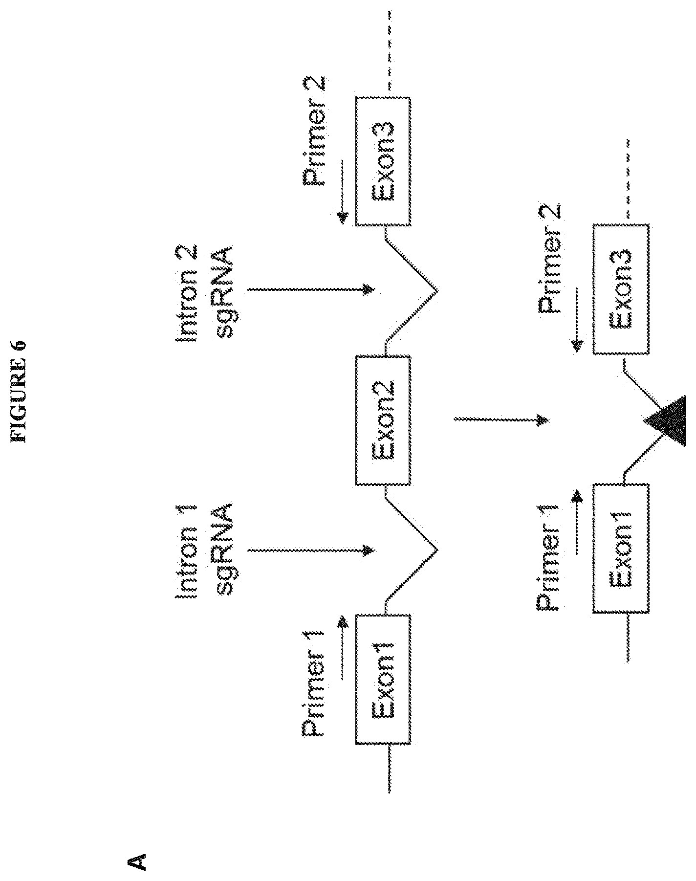
[0031] FIG. 6 includes diagrams showing dual ms-sgRNA-mediated deletion of exon 2 of CD19 in K562 cells. A: a schematic showing a PCR-based assay to detect CRISPR/Cas9-mediated genomic deletion of exon 2 of CD19 via dual ms-sgRNA-mediated CRISPR/Cas9. B: a photo showing deletion of the region between exon 1 and exon 3 after treating K562 cells with indicated pairs of ms-sgRNAs by an end-point PCR assay of genomic DNA. C: a chart showing the percentage deletion quantitated by end-point PCR.
[0032] FIG. 7 include diagrams showing screening of CD19 ms-sgRNAs targeting introns 1 or 2 in CD34+ HSCs by T7E1 assay and TIDE analysis. A: a photo showing PCR amplicons derived from the region spanning introns 1 and 2 of the CD19 gene as determined by T7E1 assays. Samples were either treated (+) or untreated (-) with T7E1. The percent insertion/deletion (INDEL) and cleavage efficiency are indicated under each lane. C=NEB Sample Control, Cas9=Cas9 only. B: PCR amplicons derived from the region spanning introns 1 and 2 of the CD19 gene were analyzed by T7E1 Assay or TIDE analysis, and the percent INDEL was determined. Cas9=cas9 only control.
[0033] FIG. 8 includes diagrams showing dual ms-sgRNA-mediated deletion of CD19 exon 2 in CD34+ HSCs. A: a photo showing the smaller deletion PCR product compared to the larger parental band as determined by PCR across the genomic deletion region. B: a chart showing the percent deletion quantified by end-point PCR.
[0034] FIG. 9 includes diagrams showing investigation of ms-sgRNAs targeting introns 1 or 2 of CD19 in CD34+ HSCs. A: a photo showing PCR amplicons derived from the region spanning introns 1 and 2 of the CD19 gene as determined by T7E1 assays. The percent cleavage efficiency is indicated under each lane. B: a chart showing PCR amplicons derived from the region spanning introns 1 and 2 of the CD19 gene as analyzed by T7E1 assay, and the percent INDEL. Cas9=cas9 only control.
[0035] FIG. 10 includes diagrams showing efficient dual ms-sgRNA-mediated deletion of exon 2 of CD19in CD34+ HSCs. A: a photo showing the smaller deletion PCR product compared to the larger parental band as determined by PCR across the genomic deletion region. The percent deletion is indicated under each lane. B: a chart showing the percent deletion quantified by end-point PCR.
[0036] FIG. 11 is a schematic work flow to assess differentiation potential of edited CD34+ HSCs. d=days, w=weeks, w/o=week old, RNP=ribonucleoprotein.
[0037] FIG. 12 is a schematic work flow to assess in vivo selectivity and efficacy of CART19 therapy in a Raji Burkitt's lymphoma tumor model. d=days, w=weeks, w/o=week old.
[0038] FIG. 13 includes diagrams showing the generation of Raji-fluc-GFP cells in which exon 2 of CD19 has been deleted. A: diagrams showing expression of CD19 in Raji-fluc-GFP cell lines transfected with the indicated combinations of ms-sgRNAs as determined by FACS. Parental Raji cells and Raj-fluc-GFP nucleofected with Cas9 only are included as controls. B: is a chart showing the percentage of live cells in each population of cells (CD19 "hi," CD19 "int," and CD19 "lo"). C: is a photo showing the smaller PCR product for the exon 2 deletion compared to the larger parental band as determined by PCR across the genomic deletion region. D: is a chart showing the percentage of cells having a deleted exon 2 of CD19 in the bulk population of cells as determined by end-point PCR.
[0039] FIG. 14 includes diagrams showing the level of CART19 cytotoxicity against Raji cells in which CD19 exon 2 has been deleted. A: a line graph showing that cells in which exon 2 of CD19 has been deleted are resistant to CART19 cytotoxicity. B: a bar graph showing that cells in which exon 2 of CD19 has been deleted are resistant to CART19 cytotoxicity.
[0040] FIG. 15 is a schematic showing an exemplary in vivo model assessing the efficacy and selectivity of a CART therapeutic paired with edited HSCs involving the methods described herein.
[0041] FIG. 16 is a schematic showing CD33 exon 2 editing, resulting in expression of the CD33m variant.
[0042] FIG. 17 is a chart showing investigation of various ms-sgRNAs targeting introns 1 or 2 of CD33 in CD34+ HSCs by TIDE analysis. PCR amplicons derived from the region spanning introns 1 and 2 of the CD33 gene were analyzed by TIDE analysis and the percent INDEL was determined.
[0043] FIG. 18 includes diagrams showing characterization of CD33-edited primary CD34+ HSCs. A: a chart showing selected ms-sgRNAs targeting introns 1 or 2 of CD33 investigated in CD34+ HSCs by TIDE analysis and the percent INDEL. "Sg" and "811" represent control sgRNAs targeting exons 2 and 3, respectively. B: a photo showing the smaller deletion PCR product compared to the larger parental band as determined by PCR across the genomic deletion region. C: a diagram showing loss of the CD33 V domain encoded by exon 2 as assessed by flow cytometry analysis.
DETAILED DESCRIPTION OF DISCLOSURE
[0044] Successfully identifying suitable proteins for targeted cancer therapies presents a significant challenge. Many potential target proteins are present on both the cell surface of a cancer cell and on the cell surface of normal, non-cancer cells, which may be required or critically involved in the development and/or survival of the subject. Many of the target proteins contribute to the functionality of such essential cells. Thus, therapies targeting these proteins may lead to deleterious effects in the subject, such as significant toxicity and/or other side effects.
[0045] The present disclosure provides methods, cells, compositions, and kits aimed at addressing at least the above-stated problems. The methods, cells, compositions, and kits described herein provide a safe and effective treatment for hematological malignancies, allowing for targeting of lineage-specific cell surface proteins (e.g., type 0, type 1, or type 2 proteins) that are present not only on cancer cells but also on cells critical for the development and/or survival of the subject. The methods described herein involve eliminating cells that express a target lineage-specific cell-surface protein by administering to a subject in need of treatment a cytotoxic agent that specifically binds an epitope of the lineage-specific cell-surface protein; and providing the subject with hematopoietic cells, which, or descendants of which, express the lineage-specific cell-surface protein, wherein the hematopoietic cells are manipulated (e.g., genetically) such that they cannot be targeted, or have reduced targeting, by the cytotoxic agent. For example, the binding epitope in the lineage-specific cell-surface protein is either deleted, mutated, or blocked from binding to the cytotoxic agent. "Expressing a lineage-specific cell-surface protein" means that at least a portion of the lineage-specific cell-surface protein can be detected on the surface of the hematopoietic cells or descendants thereof. In some embodiments, the manipulated hematopoietic cells for use in the methods described herein express a biologically functional lineage-specific cell-surface protein. In some embodiments, the manipulated hematopoietic cells for use in the methods described herein may not express a biologically functional lineage-specific cell-surface protein; however, cells differentiated therefrom (e.g., descendants thereof) express such a functional lineage-specific cell-surface protein.
[0046] Accordingly, described herein are compositions and methods involving the use of cytotoxic agents that target a lineage-specific cell-surface protein, such as any of the lineage-specific cell-surface proteins described herein or otherwise known in the art, for example CD33 or CD19 and hematopoietic cells, such as hematopoietic stem cells (HSCs), which, or descendants of which express the lineage-specific cell-surface protein and are manipulated such that they do not bind the cytotoxic agent or have reduced binding to the cytotoxic agent, which compositions and methods can be used in the treatment of a hematopoietic malignancy. Provided herein are genetically engineered hematopoietic cells that express a variant of a lineage-specific cell-surface protein that lacks an epitope of the lineage-specific cell-surface protein as well as methods of preparing such cells. Also described herein are methods for identifying non-essential epitopes of a lineage-specific cell-surface protein.
Cytotoxic Agents Targeting Cells Expressing Lineage-Specific Cell-Surface Proteins
[0047] Aspects of the disclosure provide cytotoxic agents targeting cells (e.g., cancer cells) expressing a lineage-specific cell-surface protein. As used herein, the term "cytotoxic agent" refers to any agent that can directly or indirectly induce cytotoxicity of a target cell, which expresses the lineage-specific cell-surface protein (e.g., a target cancer cell). Such a cytotoxic agent may comprise a protein-binding fragment that binds and targets an epitope of the lineage-specific cell-surface protein. In some instances, the cytotoxic agent may comprise an antibody, which may be conjugated to a drug (e.g., an anti-cancer drug) to form an antibody-drug conjugate (ADC).
[0048] The cytotoxic agent for use in the methods described herein may directly cause cell death of a target cell. For example, the cytotoxic agent can be an immune cell (e.g., a cytotoxic T cell) expressing a chimeric receptor. Upon engagement of the protein binding domain of the chimeric receptor with the corresponding epitope in a lineage-specific cell-surface protein, a signal (e.g., activation signal) may be transduced to the immune cell resulting in release of cytotoxic molecules, such as peroforins and granzymes, as well as activation of effector functions, leading to death of the target cell. In another example, the cytotoxic agent may be an ADC molecule. Upon binding to a target cell, the drug moiety in the ADC would exert cytotoxic activity, leading to target cell death.
[0049] In other embodiments, the cytotoxic agent may indirectly induce cell death of the target cell. For example, the cytotoxic agent may be an antibody, which, upon binding to the target cell, would trigger effector activities (e.g., ADCC) and/or recruit other factors (e.g., complements), resulting in target cell death.
[0050] A. Lineage-Specific Cell-Surface Proteins
[0051] As used herein, the terms "protein," "peptide," and "polypeptide" may be used interchangeably and refer to a polymer of amino acid residues linked together by peptide bonds. In general, a protein may be naturally occurring, recombinant, synthetic, or any combination of these. Also within the scope of the term are variant proteins, which comprise a mutation (e.g., substitution, insertion, or deletion) of one or more amino acid residues relative to the wild-type counterpart.
[0052] As used herein, the terms "lineage-specific cell-surface protein" and "cell-surface lineage-specific protein" may be used interchangeably and refer to any protein that is sufficiently present on the surface of a cell and is associated with one or more populations of cell lineage(s). For example, the protein may be present on one or more populations of cell lineage(s) and absent (or at reduced levels) on the cell-surface of other cell populations.
[0053] In general, lineage-specific cell-surface proteins can be classified based on a number of factors, such as whether the protein and/or the populations of cells that present the protein are required for survival and/or development of the host organism. A summary of exemplary types of lineage-specific proteins is provide in Table 1 below.
TABLE-US-00001 TABLE 1 Classification of Lineage Specific Proteins Type of Lineage Specific Protein Characteristics of the Lineage Specific Protein Type 0 a) protein is required for survival of an organism, and b) cell type carrying type 0 protein is required for survival of an organism and is not unique to a tumor, or tumor-associated virus Type 1 a) protein is not required for survival of an organism, and b) cell type carrying type 1 protein is not required for survival of an organism Type 2 a) protein is not required for survival of an organism, and b) cell type carrying type 2 protein is required for the survival of an organism Type 3 a) protein is not required for the survival of an organism; b) cell type carrying protein is not required for survival of an organism; and c) The protein is unique to a tumor, or a tumor associated virus An example is the LMP-2 protein in EBV infected cells, including EBV infected tumor cells (Nasopharyngeal carcinoma and Burkitts Lymphoma)
[0054] As shown in Table 1, type 0 lineage-specific cell-surface proteins are necessary for the tissue homeostasis and survival, and cell types carrying type 0 lineage-specific cell-surface protein may be also necessary for survival of the subject. Thus, given the importance of type 0 lineage-specific cell-surface proteins, or cells carrying type 0 lineage-specific cell-surface proteins, in homeostasis and survival, targeting this category of proteins may be challenging using conventional CAR T cell immunotherapies, as the inhibition or removal of such proteins and cell carrying such proteins may be detrimental to the survival of the subject. Consequently, lineage-specific cell-surface proteins (such as type 0 lineage-specific proteins) and/or the cell types that carry such proteins may be required for the survival, for example because it performs a vital non-redundant function in the subject, then this type of lineage specific protein may be a poor target for conventional CAR T cell based immunotherapies.
[0055] In contrast to type 0 proteins, type 1 cell-surface lineage-specific proteins and cells carrying type 1 cell-surface lineage-specific proteins are not required for tissue homeostasis or survival of the subject. Targeting type 1 cell-surface lineage-specific proteins is not likely to lead to acute toxicity and/or death of the subject. For example, as described in Elkins et al. (Mol. Cancer Ther. (2012) 10:2222-32) a CAR T cell engineered to target CD307, a type 1 protein expressed uniquely on both normal plasma cells and multiple myeloma (MM) cells would lead to elimination of both cell types. However, since the plasma cell lineage is expendable for the survival of the organism, CD307 and other type 1 lineage specific proteins are proteins that are suitable for CAR T cell based immunotherapy. Lineage specific proteins of type 1 class may be expressed in a wide variety of different tissues, including, ovaries, testes, prostate, breast, endometrium, and pancreas. In some embodiments, the agent targets a cell-surface lineage-specific protein that is a type 1 protein. Such methods may be designed to improve the longer-term survival and quality of life of the patient. For example, targeting all plasma cells, while not expected to lead to acute toxicity and/or death, could have longer-term consequences such as reduced function of the humoral immune system leading to increased risk of infection.
[0056] Targeting type 2 proteins presents a significant difficulty as compared to type 1 proteins. Type 2 proteins are those characterized where: (1) the protein is dispensable for the survival of an organism (i.e., is not required for the survival), and (2) the cell lineage carrying the protein is indispensable for the survival of an organism (i.e., the particular cell lineage is required for the survival). For example, CD33 is a type 2 protein expressed in both normal myeloid cells as well as in Acute Myeloid Leukemia (AML) cells (Dohner et al., NEJM 373:1136 (2015)). As a result, a CAR T cell engineered to target CD33 protein could lead to the killing of both normal myeloid cells as well as AML cells, which may be incompatible with survival of the subject. In some embodiments, the agent targets a lineage-specific cell-surface protein that is a type 2 protein.
[0057] A wide variety of proteins may be targeted by the methods and compositions of the present disclosure. Monoclonal antibodies to these proteins may be purchased commercially or generated using standard techniques, including immunization of an animal with the protein of interest followed by conventional monoclonal antibody methodologies. The antibodies or nucleic acids encoding for the antibodies may be sequenced using any standard DNA or protein sequencing techniques.
[0058] In some embodiments, the cell-surface lineage-specific protein is BCMA, CD19, CD20, CD30, ROR1, B7H6, B7H3, CD23, CD38, C-type lectin like molecule-1, CS1, IL-5, L1-CAM, PSCA, PSMA, CD138, CD133, CD70, CD7, NKG2D, NKG2D ligand, CLEC12A, CD11, CD123, CD56, CD34, CD14, CD33, CD66b, CD41, CD61, CD62, CD235a, CD146, CD326, LMP2, CD22, CD52, CD10, CD3/TCR, CD79/BCR, and CD26. In some embodiments, the cell-surface lineage-specific protein is CD33 or CD19.
[0059] Alternatively or in addition, the cell-surface lineage-specific protein may be a cancer protein, for example a cell-surface lineage-specific protein that is differentially present on cancer cells. In some embodiments, the cancer protein is a protein that is specific to a tissue or cell lineage. Examples of cell-surface lineage-specific protein that are associated with a specific type of cancer include, without limitation, CD20, CD22 (Non-Hodgkin's lymphoma, B-cell lymphoma, chronic lymphocytic leukemia (CLL)), CD52 (B-cell CLL), CD33 (Acute myelogenous leukemia (AML)), CD10 (gp100) (Common (pre-B) acute lymphocytic leukemia and malignant melanoma), CD3/T-cell receptor (TCR) (T-cell lymphoma and leukemia), CD79/B-cell receptor (BCR) (B-cell lymphoma and leukemia), CD26 (epithelial and lymphoid malignancies), human leukocyte antigen (HLA)-DR, HLA-DP, and HLA-DQ (lymphoid malignancies), RCAS1 (gynecological carcinomas, biliary adenocarcinomas and ductal adenocarcinomas of the pancreas) as well as prostate specific membrane antigen. In some embodiments, the cell-surface protein CD33 and is associated with AML cells.
[0060] Any of the cytotoxic agents described herein target a lineage-specific cell-surface protein, e.g., comprising a protein-binding fragment that specifically binds an epitope in the lineage-specific protein.
[0061] As used herein, the term "epitope" refers to an amino acid sequence (linear or conformational) of a protein, such as a lineage-specific cell-surface protein, that is bound by the CDRs of an antibody. In some embodiments, the cytotoxic agent binds to one or more (e.g., at least 2, 3, 4, 5 or more) epitopes of a lineage-specific cell-surface protein. In some embodiments, the cytotoxic agent binds to more than one epitope of the lineage-specific cell-surface protein and the hematopoietic cells are manipulated such that each of the epitopes is absent and/or unavailable for binding by the cytotoxic agent.
[0062] In some embodiments, the lineage-specific cell-surface protein is CD33. As will be known to one of ordinary skill in the art, CD33 is encoded by seven exons, including the alternatively spliced exons 7A and 7B (Brinkman-Van der Linden et al. Mol Cell. Biol. (2003) 23: 4199-4206).
[0063] In some embodiments, the lineage-specific cell-surface protein is CD19. In some embodiments, the lineage-specific cell-surface protein is CD33.
[0064] 1. Non-Essential Epitope of a Lineage-Specific Cell-Surface Protein
[0065] In some embodiments, the cytotoxic agent for use in the methods described herein target a non-essential epitope in a lineage-specific cell-surface protein. A non-essential epitope (or a fragment comprising such) refers to a domain within the lineage-specific protein, the mutation in which (e.g., deletion) is less likely to substantially affect the bioactivity of the lineage-specific protein and thus the bioactivity of the cells expressing such. For example, when hematopoietic cells comprising a deletion or mutation of a non-essential epitope of a lineage-specific cell-surface protein, such hematopoietic cells are able to proliferate and/or undergo erythropoeitic differentiation to a similar level as hematopoietic cells that express a wild-type lineage-specific cell-surface protein.
[0066] Non-essential epitopes of a lineage-specific cell-surface protein can be identified by the methods described herein or by conventional methods relating to protein structure-function prediction. For example, a non-essential epitope of a protein can be predicted based on comparing the amino acid sequence of a protein from one species with the sequence of the protein from other species. Non-conserved domains are usually not essential to the functionality of the protein. As will be evident to one of ordinary skill in the art, non-essential epitope of a protein is predicted using an algorithm or software, such as the PROVEAN software (see, e.g., see: provean.jcvi.org; Choi et al. PLoS ONE (2012) 7(10): e46688), to predict potential non-essential epitopes in a lineage-specific protein of interest ("candidate non-essential epitope"). Mutations, including substitution and/or deletion, many be made in any one or more amino acid residues of a candidate non-essential epitope using convention nucleic acid modification technologies. The protein variants thus prepared may be introduced into a suitable type of cells, such as hematopoietic cells, and the functionality of the protein variant can be investigated to confirm that the candidate non-essential epitope is indeed a non-essential epitope.
[0067] Alternatively, a non-essential epitope of a lineage-specific cell-surface protein may be identified by introducing a mutation into a candidate region in a lineage-specific protein of interest in a suitable type of host cells (e.g., hematopoietic cells) and examining the functionality of the mutated lineage-specific protein in the host cells. If the mutated lineage-specific protein maintains substantially the biological activity of the native counterpart, this indicates that the region where the mutation is introduced is non-essential to the function of the lineage-specific protein.
[0068] Methods for assessing the functionality of the lineage-specific cell-surface protein and the hematopoietic cells or descendants thereof will be known in the art and include, for example, proliferation assays, differentiation assays, colony formation, expression analysis (e.g., gene and/or protein), protein localization, intracellular signaling, functional assays, and in vivo humanized mouse models.
[0069] Any of the methods for identifying and/or verifying non-essential epitopes in lineage-specific cell-surface proteins is also within the scope of the present disclosure.
[0070] 2. Variants of Lineage-Specific Cell-Surface Proteins
[0071] In some embodiments, the hematopoietic cells for use in the methods described herein express a variant of a lineage-specific cell-surface protein of interest, which has reduced binding to a cytotoxic agent as described herein. The variant may lack the epitope to which the cytotoxic agent binds. Alternatively, the variant may carry one or more mutations of the epitope to which the cytotoxic agent binds, such that binding to the cytotoxic agent is reduced or abolished as compared to the natural or wild-type lineage-specific cell-surface protein counterpart. Such a variant is preferred to maintain substantially similar biological activity as the wild-type counterpart.
[0072] The variant may share a sequence homology of at least 80% (e.g., 85%, 90%, 95%, 97%, 98%, 99%, or above) as the wild-type counterpart and, in some embodiments, may contain no other mutations in addition to those for mutating or deleting the epitope of interest. The "percent identity" of two amino acid sequences is determined using the algorithm of Karlin and Altschul Proc. Natl. Acad. Sci. USA 87:2264-68, 1990, modified as in Karlin and Altschul Proc. Natl. Acad. Sci. USA 90:5873-77, 1993. Such an algorithm is incorporated into the NBLAST and XBLAST programs (version 2.0) of Altschul, et al. J. Mol. Biol. 215:403-10, 1990. BLAST protein searches can be performed with the XBLAST program, score=50, wordlength=3 to obtain amino acid sequences homologous to the protein molecules of the invention. Where gaps exist between two sequences, Gapped BLAST can be utilized as described in Altschul et al., Nucleic Acids Res. 25(17):3389-3402, 1997. When utilizing BLAST and Gapped BLAST programs, the default parameters of the respective programs (e.g., XBLAST and NBLAST) can be used.
[0073] In some instances, the variant contains one or more amino acid residue substitutions (e.g., 2, 3, 4, 5, or more) within the epitope of interest such that the cytotoxic agent does not bind or has reduced binding to the mutated epitope. Such a variant may have substantially reduced binding affinity to the cytotoxic agent (e.g., having a binding affinity that is at least 40%, 50%, 60%, 70%, 80% or 90% lower than its wild-type counterpart). In some examples, such a variant may have abolished binding activity to the cytotoxic agent. In other instances, the variant contains a deletion of a region that comprises the epitope of interest. Such a region may be encoded by an exon. In some embodiments, the region is a domain of the lineage-specific cell-surface protein of interest that encodes the epitope. In one example, the variant has just the epitope deleted. The length of the deleted region may range from 3-60 amino acids, e.g., 5-50, 5-40, 10-30, 10-20, etc.
[0074] The mutation(s) or deletions in a variant of a lineage-specific cell-surface protein may be within or surround a non-essential epitope such that the mutation(s) or deletion(s) do not substantially affect the bioactivity of the protein.
[0075] In some examples, provided herein are variants of CD33, which may comprise a deletion or mutation of a fragment of the protein that is encoded by any one of the exons of CD33, or a deletion or mutation in a non-essential epitope. The predicted structure of CD33 includes two immunoglobulin domains, an IgV domain and an IgC2 domain. In some embodiments, a portion of the immunoglobulin V domain of CD33 is deleted or mutated. In some embodiments, a portion of the immunoglobulin C domain of CD33 is deleted or mutated. In some embodiments, exon 2 of CD33 is deleted or mutated. In some embodiments, the CD33 variant lacks amino acid residues W11 to T139 of SEQ ID NO: 1. In some embodiments, the deleted or mutated fragment overlaps or encompasses the epitope to which the cytotoxic agent binds. As described in Example 1, in some embodiments, the epitope comprises amino acids 47-51 or 248-252 of the extracellular portion of CD33 (SEQ ID NO: 1). In some embodiments, the epitope comprises amino acids 248-252 (SEQ ID NO: 8), 47-51 (SEQ ID NO: 9), 249-253 (SEQ ID NO: 10), 250-254 (SEQ ID NO: 11), 48-52 (SEQ ID NO: 12), or 251-255 (SEQ ID NO: 13) of the extracellular portion of CD33 (SEQ ID NO: 1).
[0076] In some examples, provided herein are variants of CD19, which may comprise a deletion or mutation of a fragment of the protein that is encoded by any one of the exons of CD19, or deletion or mutation in a non-essential epitope of CD19. The whole sequence of the CD19 gene, containing fifteen exons, is known in the art. See, e.g., GenBank accession no. NC_000016. For example, one or more epitopes located in the region encoded by exon 2 the CD19 gene may be deleted or mutated. Certain modifications to the region of the CD19 gene encoding exon 2 have been shown to result in successful CD19 protein expression, membrane localization, and partial maintenance of protein function (Sotillo et al. Cancer Discovery. (2015) 5: 1282-1295). For example, missense or frameshift mutations in exon 2 of the CD19 gene, or alternatively, modifications that permanently or transiently reduce expression of the splicing factor SRSF3, which is involved in retention of CD19 exon 2, may reduce CD19 expression in vivo. In some embodiments, one or more epitopes located in the region encoded by exon 2 of the CD19 gene are mutated or deleted. For example, the FMC63 epitope of CD19, which is a known target of CD19-targeted CAR therapies may be mutated or deleted (Sotillo et al. Cancer Discovery. (2015) 5: 1282-129; Nicholson et al. Mol Immunol. (1997) 34:1157-1165; Zola et al. Immunol Cell Biol. (1991) 69:411-422). In some embodiments, exon 2 of CD19 is mutated or deleted.
[0077] B. Cytotoxic Agents
[0078] 1. Antibodies and Antigen-Binding Fragments
[0079] Any antibody or an antigen-binding fragment thereof can be used as a cytotoxic agent or for constructing a cytotoxic agent that targets an epitope of a lineage-specific cell-surface protein as described herein. Such an antibody or antigen-binding fragment can be prepared by a conventional method, for example, the hybridoma technology or recombinant technology.
[0080] As used herein, the term "antibody" refers to a glycoprotein comprising at least two heavy (H) chains and two light (L) chains inter-connected by disulfide bonds, i.e., covalent heterotetramers comprised of two identical Ig H chains and two identical L chains that are encoded by different genes. Each heavy chain is comprised of a heavy chain variable region (abbreviated herein as HCVR or VH) and a heavy chain constant region. The heavy chain constant region is comprised of three domains, CH1, CH2 and CH3. Each light chain is comprised of a light chain variable region (abbreviated herein as LCVR or VL) and a light chain constant region. The light chain constant region is comprised of one domain, CL. The VH and VL regions can be further subdivided into regions of hypervariability, termed complementarity determining regions (CDR), interspersed with regions that are more conserved, termed framework regions (FR). Each VH and VL is composed of three CDRs and four FRs, arranged from amino-terminus to carboxy-terminus in the following order: FR1, CDR1, FR2, CDR2, FR3, CDR3, FR4. The variable regions of the heavy and light chains contain a binding domain that interacts with an antigen. The constant regions of the antibodies may mediate the binding of the immunoglobulin to host tissues or factors, including various cells of the immune system (e.g., effector cells) and the first component (Clq) of the classical complement system. Formation of a mature functional antibody molecule can be accomplished when two proteins are expressed in stoichiometric quantities and self-assemble with the proper configuration.
[0081] In some embodiments, the antigen-binding fragment is a single-chain antibody fragment (scFv) that specifically binds the epitope of the lineage-specific cell-surface protein. In other embodiments, the antigen-binding fragment is a full-length antibody that specifically binds the epitope of the lineage-specific cell-surface protein.
[0082] As described herein and as will be evident to a skilled artisan, the CDRs of an antibody specifically bind to the epitope of a target protein (the lineage-specific cell-surface protein).
[0083] In some embodiments, the antibodies are full-length antibodies, meaning the antibodies comprise a fragment crystallizable (Fc) portion and a fragment antigen-binding (Fab) portion. In some embodiments, the antibodies are of the isotype IgG, IgA, IgM, IgA, or IgD. In some embodiments, a population of antibodies comprises one isotype of antibody. In some embodiments, the antibodies are IgG antibodies. In some embodiments, the antibodies are IgM antibodies. In some embodiments, a population of antibodies comprises more than one isotype of antibody. In some embodiments, a population of antibodies is comprised of a majority of one isotype of antibodies but also contains one or more other isotypes of antibodies. In some embodiments, the antibodies are selected from the group consisting of IgG1, IgG2, IgG3, IgG4, IgM, IgA1, IgA2, IgAsec, IgD, IgE.
[0084] The antibodies described herein may specifically bind to a target protein. As used herein, "specific binding" refers to antibody binding to a predetermined protein, such as a cancer antigen. "Specific binding" involves more frequent, more rapid, greater duration of interaction, and/or greater affinity to a target protein relative to alternative proteins. In some embodiments, a population of antibodies specifically binds to a particular epitope of a target protein, meaning the antibodies bind to the particular protein with more frequently, more rapidly, for greater duration of interaction, and/or with greater affinity to the epitope relative to alternative epitopes of the same target protein or to epitopes of another protein. In some embodiments, the antibodies that specifically bind to a particular epitope of a target protein may not bind to other epitopes of the same protein.
[0085] Antibodies or fragments thereof may be selected based on the binding affinity of the antibody to the target protein or epitope. Alternatively or in additional, the antibodies may be mutated to introduce one or more mutations to modify (e.g., enhance or reduce) the binding affinity of the antibody to the target protein or epitope.
[0086] The present antibodies or antigen-binding portions can specifically bind with a dissociation constant (K.sub.D) of less than about 10.sup.-7 M, less than about 10.sup.-8 M, less than about 10.sup.-9 M, less than about 10.sup.-10 M, less than about 10.sup.-11 M, or less than about 10.sup.-12 M. Affinities of the antibodies according to the present disclosure can be readily determined using conventional techniques (see, e.g., Scatchard et al., Ann. N.Y. Acad. Sci. (1949) 51:660; and U.S. Pat. Nos. 5,283,173, 5,468,614, or the equivalent).
[0087] The binding affinity or binding specificity for an epitope or protein can be determined by a variety of methods including equilibrium dialysis, equilibrium binding, gel filtration, ELISA, surface plasmon resonance, or spectroscopy.
[0088] For example, antibodies (of antigen-binding fragments thereof) specific to an epitope of a lineage-specific protein of interest can be made by the conventional hybridoma technology. The lineage-specific protein, which may be coupled to a carrier protein such as KLH, can be used to immunize a host animal for generating antibodies binding to that complex. The route and schedule of immunization of the host animal are generally in keeping with established and conventional techniques for antibody stimulation and production, as further described herein. General techniques for production of mouse, humanized, and human antibodies are known in the art and are described herein. It is contemplated that any mammalian subject including humans or antibody producing cells therefrom can be manipulated to serve as the basis for production of mammalian, including human hybridoma cell lines. Typically, the host animal is inoculated intraperitoneally, intramuscularly, orally, subcutaneously, intraplantar, and/or intradermally with an amount of immunogen, including as described herein.
[0089] Hybridomas can be prepared from the lymphocytes and immortalized myeloma cells using the general somatic cell hybridization technique of Kohler, B. and Milstein, C. (1975) Nature 256:495-497 or as modified by Buck, D. W., et al., In Vitro, 18:377-381 (1982). Available myeloma lines, including but not limited to X63-Ag8.653 and those from the Salk Institute, Cell Distribution Center, San Diego, Calif., USA, may be used in the hybridization. Generally, the technique involves fusing myeloma cells and lymphoid cells using a fusogen such as polyethylene glycol, or by electrical means well known to those skilled in the art. After the fusion, the cells are separated from the fusion medium and grown in a selective growth medium, such as hypoxanthine-aminopterin-thymidine (HAT) medium, to eliminate unhybridized parent cells. Any of the media described herein, supplemented with or without serum, can be used for culturing hybridomas that secrete monoclonal antibodies. As another alternative to the cell fusion technique, EBV immortalized B cells may be used to produce the TCR-like monoclonal antibodies described herein. The hybridomas are expanded and subcloned, if desired, and supernatants are assayed for anti-immunogen activity by conventional immunoassay procedures (e.g., radioimmunoassay, enzyme immunoassay, or fluorescence immunoassay).
[0090] Hybridomas that may be used as source of antibodies encompass all derivatives, progeny cells of the parent hybridomas that produce monoclonal antibodies capable of binding to a lineage-specific protein. Hybridomas that produce such antibodies may be grown in vitro or in vivo using known procedures. The monoclonal antibodies may be isolated from the culture media or body fluids, by conventional immunoglobulin purification procedures such as ammonium sulfate precipitation, gel electrophoresis, dialysis, chromatography, and ultrafiltration, if desired. Undesired activity if present, can be removed, for example, by running the preparation over adsorbents made of the immunogen attached to a solid phase and eluting or releasing the desired antibodies off the immunogen. Immunization of a host animal with a target protein or a fragment containing the target amino acid sequence conjugated to a protein that is immunogenic in the species to be immunized, e.g., keyhole limpet hemocyanin, serum albumin, bovine thyroglobulin, or soybean trypsin inhibitor using a bifunctional or derivatizing agent, for example maleimidobenzoyl sulfosuccinimide ester (conjugation through cysteine residues), N-hydroxysuccinimide (through lysine residues), glutaraldehyde, succinic anhydride, SOCl, or R1N.dbd.C.dbd.NR, where R and R1 are different alkyl groups, can yield a population of antibodies (e.g., monoclonal antibodies).
[0091] If desired, an antibody of interest (e.g., produced by a hybridoma) may be sequenced and the polynucleotide sequence may then be cloned into a vector for expression or propagation. The sequence encoding the antibody of interest may be maintained in vector in a host cell and the host cell can then be expanded and frozen for future use. In an alternative, the polynucleotide sequence may be used for genetic manipulation to "humanize" the antibody or to improve the affinity (affinity maturation), or other characteristics of the antibody. For example, the constant region may be engineered to more resemble human constant regions to avoid immune response if the antibody is used in clinical trials and treatments in humans. It may be desirable to genetically manipulate the antibody sequence to obtain greater affinity to the lineage-specific protein. It will be apparent to one of skill in the art that one or more polynucleotide changes can be made to the antibody and still maintain its binding specificity to the target protein.
[0092] In other embodiments, fully human antibodies can be obtained by using commercially available mice that have been engineered to express specific human immunoglobulin proteins. Transgenic animals that are designed to produce a more desirable (e.g., fully human antibodies) or more robust immune response may also be used for generation of humanized or human antibodies. Examples of such technology are Xenomouse.RTM. from Amgen, Inc. (Fremont, Calif.) and HuMAb-Mouse.RTM. and TC Mouse.TM. from Medarex, Inc. (Princeton, N.J.). In another alternative, antibodies may be made recombinantly by phage display or yeast technology. See, for example, U.S. Pat. Nos. 5,565,332; 5,580,717; 5,733,743; and 6,265,150; and Winter et al., (1994) Annu. Rev. Immunol. 12:433-455. Alternatively, the phage display technology (McCafferty et al., (1990) Nature 348:552-553) can be used to produce human antibodies and antibody fragments in vitro, from immunoglobulin variable (V) domain gene repertoires from unimmunized donors.
[0093] Antigen-binding fragments of an intact antibody (full-length antibody) can be prepared via routine methods. For example, F(ab').sub.2 fragments can be produced by pepsin digestion of an antibody molecule, and Fab fragments that can be generated by reducing the disulfide bridges of F(ab')2 fragments.
[0094] Genetically engineered antibodies, such as humanized antibodies, chimeric antibodies, single-chain antibodies, and bi-specific antibodies, can be produced via, e.g., conventional recombinant technology. In one example, DNA encoding a monoclonal antibodies specific to a target protein can be readily isolated and sequenced using conventional procedures (e.g., by using oligonucleotide probes that are capable of binding specifically to genes encoding the heavy and light chains of the monoclonal antibodies). The hybridoma cells serve as a preferred source of such DNA. Once isolated, the DNA may be placed into one or more expression vectors, which are then transfected into host cells such as E. coli cells, simian COS cells, Chinese hamster ovary (CHO) cells, or myeloma cells that do not otherwise produce immunoglobulin protein, to obtain the synthesis of monoclonal antibodies in the recombinant host cells. See, e.g., PCT Publication No. WO 87/04462. The DNA can then be modified, for example, by substituting the coding sequence for human heavy and light chain constant domains in place of the homologous murine sequences, Morrison et al., (1984) Proc. Nat. Acad. Sci. 81:6851, or by covalently joining to the immunoglobulin coding sequence all or part of the coding sequence for a non-immunoglobulin polypeptide. In that manner, genetically engineered antibodies, such as "chimeric" or "hybrid" antibodies; can be prepared that have the binding specificity of a target protein.
[0095] Techniques developed for the production of "chimeric antibodies" are well known in the art. See, e.g., Morrison et al. (1984) Proc. Natl. Acad. Sci. USA 81, 6851; Neuberger et al. (1984) Nature 312, 604; and Takeda et al. (1984) Nature 314:452.
[0096] Methods for constructing humanized antibodies are also well known in the art. See, e.g., Queen et al., Proc. Natl. Acad. Sci. USA, 86:10029-10033 (1989). In one example, variable regions of VH and VL of a parent non-human antibody are subjected to three-dimensional molecular modeling analysis following methods known in the art. Next, framework amino acid residues predicted to be important for the formation of the correct CDR structures are identified using the same molecular modeling analysis. In parallel, human VH and VL chains having amino acid sequences that are homologous to those of the parent non-human antibody are identified from any antibody gene database using the parent VH and VL sequences as search queries. Human VH and VL acceptor genes are then selected.
[0097] The CDR regions within the selected human acceptor genes can be replaced with the CDR regions from the parent non-human antibody or functional variants thereof. When necessary, residues within the framework regions of the parent chain that are predicted to be important in interacting with the CDR regions (see above description) can be used to substitute for the corresponding residues in the human acceptor genes.
[0098] A single-chain antibody can be prepared via recombinant technology by linking a nucleotide sequence coding for a heavy chain variable region and a nucleotide sequence coding for a light chain variable region. Preferably, a flexible linker is incorporated between the two variable regions. Alternatively, techniques described for the production of single chain antibodies (U.S. Pat. Nos. 4,946,778 and 4,704,692) can be adapted to produce a phage or yeast scFv library and scFv clones specific to a lineage-specific protein can be identified from the library following routine procedures. Positive clones can be subjected to further screening to identify those that bind lineage-specific protein.
[0099] In some instances, the cytotoxic agent for use in the methods described herein comprises an antigen-binding fragment that targets the lineage-specific protein CD33. In other examples, the cytotoxic agent for use in the methods described herein comprises an antigen-binding fragment that targets the lineage-specific protein CD19. Antibodies and antigen-binding fragments targeting CD33 or CD19 can be prepared by routine practice. Non-limited examples of antigen-binding fragments that target CD19 can be found in Porter D L et al. NEJM (2011) 365:725-33 and Kalos M et al. Sci Transl Med. (2011) 3:95ra73. See also descriptions herein. Such CD19-targeting antigen-binding fragments can be used for making the CAR constructs described herein.
[0100] 2. Immune Cells Expressing Chimeric Antigen Receptors
[0101] In some embodiments, the cytotoxic agent that targets an epitope of a lineage-specific cell-surface protein as described herein is an immune cell that expresses a chimeric receptor, which comprises an antigen-binding fragment (e.g., a single-chain antibody) capable of binding to the epitope of the lineage-specific protein (e.g., CD33 or CD19). Recognition of a target cell (e.g., a cancer cell) having the epitope of the lineage-specific protein on its cell surface by the antigen-binding fragment of the chimeric receptor transduces an activation signal to the signaling domain(s) (e.g., co-stimulatory signaling domain and/or the cytoplasmic signaling domain) of the chimeric receptor, which may activate an effector function in the immune cell expressing the chimeric receptor.
[0102] As used herein, a chimeric receptor refers to a non-naturally occurring molecule that can be expressed on the surface of a host cell and comprises an antigen-binding fragment that binds to an epitope of a cell-surface lineage-specific protein. In general, chimeric receptors comprise at least two domains that are derived from different molecules. In addition to the epitope-binding fragment described herein, the chimeric receptor may further comprise one or more of the following: a hinge domain, a transmembrane domain, a co-stimulatory domain, a cytoplasmic signaling domain, and combinations thereof. In some embodiments, the chimeric receptor comprises from N terminus to C terminus, an antigen-binding fragment that binds to a cell-surface lineage-specific protein, a hinge domain, a transmembrane domain, and a cytoplasmic signaling domain. In some embodiments, the chimeric receptor further comprises at least one co-stimulatory domain.
[0103] In some embodiments, the chimeric receptors described herein comprise one or more hinge domain(s). In some embodiments, the hinge domain may be located between the antigen-binding fragment and a transmembrane domain. A hinge domain is an amino acid segment that is generally found between two domains of a protein and may allow for flexibility of the protein and movement of one or both of the domains relative to one another. Any amino acid sequence that provides such flexibility and movement of the antigen-binding fragment relative to another domain of the chimeric receptor can be used.
[0104] The hinge domain may contain about 10-200 amino acids, e.g., 15-150 amino acids, 20-100 amino acids, or 30-60 amino acids. In some embodiments, the hinge domain may be of about 10, 11, 12, 13, 14, 15, 16, 17, 18, 19, 20, 21, 22, 23, 24, 25, 26, 27, 28, 29, 30, 35, 40, 45, 50, 55, 60, 65, 70, 75, 80, 85, 90, 95, 100, 110, 120, 130, 140, 150, 160, 170, 180, 190, or 200 amino acids in length.
[0105] In some embodiments, the hinge domain is a hinge domain of a naturally occurring protein. Hinge domains of any protein known in the art to comprise a hinge domain are compatible for use in the chimeric receptors described herein. In some embodiments, the hinge domain is at least a portion of a hinge domain of a naturally occurring protein and confers flexibility to the chimeric receptor. In some embodiments, the hinge domain is of CD8a or CD28. In some embodiments, the hinge domain is a portion of the hinge domain of CD8a, e.g., a fragment containing at least 15 (e.g., 20, 25, 30, 35, or 40) consecutive amino acids of the hinge domain of CD8a or CD28.
[0106] Hinge domains of antibodies, such as an IgG, IgA, IgM, IgE, or IgD antibody, are also compatible for use in the chimeric receptors described herein. In some embodiments, the hinge domain is the hinge domain that joins the constant domains CH1 and CH2 of an antibody. In some embodiments, the hinge domain is of an antibody and comprises the hinge domain of the antibody and one or more constant regions of the antibody. In some embodiments, the hinge domain comprises the hinge domain of an antibody and the CH3 constant region of the antibody. In some embodiments, the hinge domain comprises the hinge domain of an antibody and the CH2 and CH3 constant regions of the antibody. In some embodiments, the antibody is an IgG, IgA, IgM, IgE, or IgD antibody. In some embodiments, the antibody is an IgG antibody. In some embodiments, the antibody is an IgG1, IgG2, IgG3, or IgG4 antibody. In some embodiments, the hinge region comprises the hinge region and the CH2 and CH3 constant regions of an IgG1 antibody. In some embodiments, the hinge region comprises the hinge region and the CH3 constant region of an IgG1 antibody.
[0107] Also within the scope of the present disclosure are chimeric receptors comprising a hinge domain that is a non-naturally occurring peptide. In some embodiments, the hinge domain between the C-terminus of the extracellular ligand-binding domain of an Fc receptor and the N-terminus of the transmembrane domain is a peptide linker, such as a (Gly.sub.xSer)n linker, wherein x and n, independently can be an integer between 3 and 12, including 3, 4, 5, 6, 7, 8, 9, 10, 11, 12, or more.
[0108] Additional peptide linkers that may be used in a hinge domain of the chimeric receptors described herein are known in the art. See, e.g., Wriggers et al. Current Trends in Peptide Science (2005) 80(6): 736-746 and PCT Publication WO 2012/088461.
[0109] In some embodiments, the chimeric receptors described herein may comprise one or more transmembrane domain(s). The transmembrane domain for use in the chimeric receptors can be in any form known in the art. As used herein, a "transmembrane domain" refers to any protein structure that is thermodynamically stable in a cell membrane, preferably a eukaryotic cell membrane. Transmembrane domains compatible for use in the chimeric receptors used herein may be obtained from a naturally occurring protein. Alternatively, the transmembrane domain may be a synthetic, non-naturally occurring protein segment, e.g., a hydrophobic protein segment that is thermodynamically stable in a cell membrane.
[0110] Transmembrane domains are classified based on the transmembrane domain topology, including the number of passes that the transmembrane domain makes across the membrane and the orientation of the protein. For example, single-pass membrane proteins cross the cell membrane once, and multi-pass membrane proteins cross the cell membrane at least twice (e.g., 2, 3, 4, 5, 6, 7 or more times). In some embodiments, the transmembrane domain is a single-pass transmembrane domain. In some embodiments, the transmembrane domain is a single-pass transmembrane domain that orients the N terminus of the chimeric receptor to the extracellular side of the cell and the C terminus of the chimeric receptor to the intracellular side of the cell. In some embodiments, the transmembrane domain is obtained from a single pass transmembrane protein. In some embodiments, the transmembrane domain is of CD8a. In some embodiments, the transmembrane domain is of CD28. In some embodiments, the transmembrane domain is of ICOS.
[0111] In some embodiments, the chimeric receptors described herein comprise one or more costimulatory signaling domains. The term "co-stimulatory signaling domain," as used herein, refers to at least a portion of a protein that mediates signal transduction within a cell to induce an immune response, such as an effector function. The co-stimulatory signaling domain of the chimeric receptor described herein can be a cytoplasmic signaling domain from a co-stimulatory protein, which transduces a signal and modulates responses mediated by immune cells, such as T cells, NK cells, macrophages, neutrophils, or eosinophils.
[0112] In some embodiments, the chimeric receptor comprises more than one (at least 2, 3, 4, or more) co-stimulatory signaling domains. In some embodiments, the chimeric receptor comprises more than one co-stimulatory signaling domains obtained from different costimulatory proteins. In some embodiments, the chimeric receptor does not comprise a co-stimulatory signaling domain.
[0113] In general, many immune cells require co-stimulation, in addition to stimulation of an antigen-specific signal, to promote cell proliferation, differentiation and survival, and to activate effector functions of the cell. Activation of a co-stimulatory signaling domain in a host cell (e.g., an immune cell) may induce the cell to increase or decrease the production and secretion of cytokines, phagocytic properties, proliferation, differentiation, survival, and/or cytotoxicity. The co-stimulatory signaling domain of any co-stimulatory protein may be compatible for use in the chimeric receptors described herein. The type(s) of co-stimulatory signaling domain is selected based on factors such as the type of the immune cells in which the chimeric receptors would be expressed (e.g., primary T cells, T cell lines, NK cell lines) and the desired immune effector function (e.g., cytotoxicity). Examples of co-stimulatory signaling domains for use in the chimeric receptors can be the cytoplasmic signaling domain of co-stimulatory proteins, including, without limitation, CD27, CD28, 4-1BB, OX40, CD30, ICOS, CD2, CD7, LIGHT, NKG2C, B7-H3. In some embodiments, the co-stimulatory domain is derived from 4-1BB, CD28, or ICOS. In some embodiments, the costimulatory domain is derived from CD28 and chimeric receptor comprises a second co-stimulatory domain from 4-1BB or ICOS.
[0114] In some embodiments, the costimulatory domain is a fusion domain comprising more than one costimulatory domain or portions of more than one costimulatory domains. In some embodiments, the costimulatory domain is a fusion of costimulatory domains from CD28 and ICOS.
[0115] In some embodiments, the chimeric receptors described herein comprise one or more cytoplasmic signaling domain(s). Any cytoplasmic signaling domain can be used in the chimeric receptors described herein. In general, a cytoplasmic signaling domain relays a signal, such as interaction of an extracellular ligand-binding domain with its ligand, to stimulate a cellular response, such as inducing an effector function of the cell (e.g., cytotoxicity).
[0116] As will be evident to one of ordinary skill in the art, a factor involved in T cell activation is the phosphorylation of immunoreceptor tyrosine-based activation motif (ITAM) of a cytoplasmic signaling domain. Any ITAM-containing domain known in the art may be used to construct the chimeric receptors described herein. In general, an ITAM motif may comprise two repeats of the amino acid sequence YxxL/I separated by 6-8 amino acids, wherein each x is independently any amino acid, producing the conserved motif YxxL/Ix(6-8)YxxL/I. In some embodiments, the cytoplasmic signaling domain is from CD3.zeta..
[0117] In some embodiments, the chimeric receptor described herein targets a type 2 protein. In some embodiments, the chimeric receptor targets CD33. In some embodiments, the chimeric receptor described herein targets a type 1 protein. In some embodiments, the chimeric receptor targets CD19. Such a chimeric receptor may comprise an antigen-binding fragment (e.g., an scFv) comprising a heavy chain variable region and a light chain variable region that bind to CD19. Alternatively, the chimeric receptor may comprise an antigen-binding fragment (e.g., scFv) comprising a heavy chain variable region and a light chain variable region that bind to CD33.
[0118] A chimeric receptor construct targeting CD33 or CD19 may further comprise at least a hinge domain (e.g., from CD28, CD8a, or an antibody), a transmembrane domain (e.g., from CD8a, CD28 or ICOS), one or more co-stimulatory domains (from one or more of CD28, ICOS, or 4-1BB) and a cytoplasmic signaling domain (e.g., from CD3.zeta.), or a combination thereof.
[0119] Any of the chimeric receptors described herein can be prepared by routine methods, such as recombinant technology. Methods for preparing the chimeric receptors herein involve generation of a nucleic acid that encodes a polypeptide comprising each of the domains of the chimeric receptors, including the antigen-binding fragment and optionally, the hinge domain, the transmembrane domain, at least one co-stimulatory signaling domain, and the cytoplasmic signaling domain. In some embodiments, nucleic acids encoding the components of a chimeric receptor are joined together using recombinant technology.
[0120] Sequences of each of the components of the chimeric receptors may be obtained via routine technology, e.g., PCR amplification from any one of a variety of sources known in the art. In some embodiments, sequences of one or more of the components of the chimeric receptors are obtained from a human cell. Alternatively, the sequences of one or more components of the chimeric receptors can be synthesized. Sequences of each of the components (e.g., domains) can be joined directly or indirectly (e.g., using a nucleic acid sequence encoding a peptide linker) to form a nucleic acid sequence encoding the chimeric receptor, using methods such as PCR amplification or ligation. Alternatively, the nucleic acid encoding the chimeric receptor may be synthesized. In some embodiments, the nucleic acid is DNA. In other embodiments, the nucleic acid is RNA.
[0121] Mutation of one or more residues within one or more of the components of the chimeric receptor (e.g., the antigen-binding fragment, etc) may be performed prior to or after joining the sequences of each of the components. In some embodiments, one or more mutations in a component of the chimeric receptor may be made to modulate (increase or decrease) the affinity of the component for an epitope (e.g., the antigen-binding fragment for the target protein) and/or modulate the activity of the component.
[0122] Any of the chimeric receptors described herein can be introduced into a suitable immune cell for expression via conventional technology. In some embodiments, the immune cells are T cells, such as primary T cells or T cell lines. Alternatively, the immune cells can be NK cells, such as established NK cell lines (e.g., NK-92 cells). In some embodiments, the immune cells are T cells that express CD8 (CD8.sup.+) or CD8 and CD4 (CD8.sup.+/CD4.sup.+). In some embodiments, the T cells are T cells of an established T cell line, for example, 293T cells or Jurkat cells.
[0123] Primary T cells may be obtained from any source, such as peripheral blood mononuclear cells (PBMCs), bone marrow, tissues such as spleen, lymph node, thymus, or tumor tissue. A source suitable for obtaining the type of immune cells desired would be evident to one of skill in the art. In some embodiments, the population of immune cells is derived from a human patient having a hematopoietic malignancy, such as from the bone marrow or from PBMCs obtained from the patient. In some embodiments, the population of immune cells is derived from a healthy donor. In some embodiments, the immune cells are obtained from the subject to whom the immune cells expressing the chimeric receptors will be subsequently administered. Immune cells that are administered to the same subject from which the cells were obtained are referred to as autologous cells, whereas immune cells that are obtained from a subject who is not the subject to whom the cells will be administered are referred to as allogeneic cells.
[0124] The type of host cells desired may be expanded within the population of cells obtained by co-incubating the cells with stimulatory molecules, for example, anti-CD3 and anti-CD28 antibodies may be used for expansion of T cells.
[0125] To construct the immune cells that express any of the chimeric receptor constructs described herein, expression vectors for stable or transient expression of the chimeric receptor construct may be constructed via conventional methods as described herein and introduced into immune host cells. For example, nucleic acids encoding the chimeric receptors may be cloned into a suitable expression vector, such as a viral vector in operable linkage to a suitable promoter. The nucleic acids and the vector may be contacted, under suitable conditions, with a restriction enzyme to create complementary ends on each molecule that can pair with each other and be joined with a ligase. Alternatively, synthetic nucleic acid linkers can be ligated to the termini of the nucleic acid encoding the chimeric receptors. The synthetic linkers may contain nucleic acid sequences that correspond to a particular restriction site in the vector. The selection of expression vectors/plasmids/viral vectors would depend on the type of host cells for expression of the chimeric receptors, but should be suitable for integration and replication in eukaryotic cells.
[0126] A variety of promoters can be used for expression of the chimeric receptors described herein, including, without limitation, cytomegalovirus (CMV) intermediate early promoter, a viral LTR such as the Rous sarcoma virus LTR, HIV-LTR, HTLV-1 LTR, Maloney murine leukemia virus (MMLV) LTR, myeoloproliferative sarcoma virus (MPSV) LTR, spleen focus-forming virus (SFFV) LTR, the simian virus 40 (SV40) early promoter, herpes simplex tk virus promoter, elongation factor 1-alpha (EF1-.alpha.) promoter with or without the EF1-.alpha. intron. Additional promoters for expression of the chimeric receptors include any constitutively active promoter in an immune cell. Alternatively, any regulatable promoter may be used, such that its expression can be modulated within an immune cell.
[0127] Additionally, the vector may contain, for example, some or all of the following: a selectable marker gene, such as the neomycin gene for selection of stable or transient transfectants in host cells; enhancer/promoter sequences from the immediate early gene of human CMV for high levels of transcription; transcription termination and RNA processing signals from SV40 for mRNA stability; 5'- and 3'-untranslated regions for mRNA stability and translation efficiency from highly-expressed genes like .alpha.-globin or .beta.-globin; SV40 polyoma origins of replication and ColE1 for proper episomal replication; internal ribosome binding sites (IRESes), versatile multiple cloning sites; T7 and SP6 RNA promoters for in vitro transcription of sense and antisense RNA; a "suicide switch" or "suicide gene" which when triggered causes cells carrying the vector to die (e.g., HSV thymidine kinase, an inducible caspase such as iCasp9), and reporter gene for assessing expression of the chimeric receptor. See section VI below. Suitable vectors and methods for producing vectors containing transgenes are well known and available in the art. Examples of the preparation of vectors for expression of chimeric receptors can be found, for example, in US2014/0106449, herein incorporated by reference in its entirety.
[0128] In some embodiments, the chimeric receptor construct or the nucleic acid encoding said chimeric receptor is a DNA molecule. In some embodiments, chimeric receptor construct or the nucleic acid encoding said chimeric receptor is a DNA vector and may be electroporated to immune cells (see, e.g., Till, et al. Blood (2012) 119(17): 3940-3950). In some embodiments, the nucleic acid encoding the chimeric receptor is an RNA molecule, which may be electroporated to immune cells.
[0129] Any of the vectors comprising a nucleic acid sequence that encodes a chimeric receptor construct described herein is also within the scope of the present disclosure. Such a vector may be delivered into host cells such as host immune cells by a suitable method. Methods of delivering vectors to immune cells are well known in the art and may include DNA, RNA, or transposon electroporation, transfection reagents such as liposomes or nanoparticles to delivery DNA, RNA, or transposons; delivery of DNA, RNA, or transposons or protein by mechanical deformation (see, e.g., Sharei et al. Proc. Natl. Acad. Sci. USA (2013) 110(6): 2082-2087); or viral transduction. In some embodiments, the vectors for expression of the chimeric receptors are delivered to host cells by viral transduction. Exemplary viral methods for delivery include, but are not limited to, recombinant retroviruses (see, e.g., PCT Publication Nos. WO 90/07936; WO 94/03622; WO 93/25698; WO 93/25234; WO 93/11230; WO 93/10218; WO 91/02805; U.S. Pat. Nos. 5,219,740 and 4,777,127; GB Patent No. 2,200,651; and EP Patent No. 0 345 242), alphavirus-based vectors, and adeno-associated virus (AAV) vectors (see, e.g., PCT Publication Nos. WO 94/12649, WO 93/03769; WO 93/19191; WO 94/28938; WO 95/11984 and WO 95/00655). In some embodiments, the vectors for expression of the chimeric receptors are retroviruses. In some embodiments, the vectors for expression of the chimeric receptors are lentiviruses. In some embodiments, the vectors for expression of the chimeric receptors are adeno-associated viruses.
[0130] In examples in which the vectors encoding chimeric receptors are introduced to the host cells using a viral vector, viral particles that are capable of infecting the immune cells and carry the vector may be produced by any method known in the art and can be found, for example in PCT Application No. WO 1991/002805A2, WO 1998/009271 A1, and U.S. Pat. No. 6,194,191. The viral particles are harvested from the cell culture supernatant and may be isolated and/or purified prior to contacting the viral particles with the immune cells.
[0131] The methods of preparing host cells expressing any of the chimeric receptors described herein may comprise activating and/or expanding the immune cells ex vivo. Activating a host cell means stimulating a host cell into an activate state in which the cell may be able to perform effector functions (e.g., cytotoxicity). Methods of activating a host cell will depend on the type of host cell used for expression of the chimeric receptors. Expanding host cells may involve any method that results in an increase in the number of cells expressing chimeric receptors, for example, allowing the host cells to proliferate or stimulating the host cells to proliferate. Methods for stimulating expansion of host cells will depend on the type of host cell used for expression of the chimeric receptors and will be evident to one of skill in the art. In some embodiments, the host cells expressing any of the chimeric receptors described herein are activated and/or expanded ex vivo prior to administration to a subject.
[0132] 3. Antibody-Drug Conjugate
[0133] In some embodiments, the cytotoxic agent targeting an epitope of a cell-surface lineage-specific protein is an antibody-drug conjugate (ADC). As will be evident to one of ordinary skill in the art, the term "antibody-drug conjugate" can be used interchangeably with "immunotoxin" and refers to a fusion molecule comprising an antibody (or antigen-binding fragment thereof) conjugated to a toxin or drug molecule. Binding of the antibody to the corresponding epitope of the target protein allows for delivery of the toxin or drug molecule to a cell that presents the protein (and epitope thereof) on the cell surface (e.g., target cell), thereby resulting in death of the target cell. In some embodiments, the antibody-drug conjugate (or antigen-binding fragment thereof) binds to its corresponding epitope of a lineage-specific cell-surface protein but does not bind to a lineage-specific cell-surface protein that lacks the epitope or in which the epitope has been mutated.
[0134] In some embodiments, the agent is an antibody-drug conjugate. In some embodiments, the antibody-drug conjugate comprises an antigen-binding fragment and a toxin or drug that induces cytotoxicity in a target cell. In some embodiments, the antibody-drug conjugate targets a type 2 protein. In some embodiments, the antibody-drug conjugate targets CD33. In some embodiments, the antibody-drug conjugate targets a type 1 protein. In some embodiments, the antibody-drug conjugate targets CD19.
[0135] Toxins or drugs compatible for use in antibody-drug conjugate are well known in the art and will be evident to one of ordinary skill in the art. See, e.g., Peters et al. Biosci. Rep.(2015) 35(4): e00225. In some embodiments, the antibody-drug conjugate may further comprise a linker (e.g., a peptide linker, such as a cleavable linker) attaching the antibody and drug molecule.
[0136] In some embodiments, two or more epitopes of a lineage-specific cell-surface protein have been modified, enabling two different cytotoxic agents (e.g., two ADCs) to be targeted to the two epitopes. In some embodiments, the toxins carried by the ADCs could work synergistically to enhance efficacy (e.g., death of the target cells).
[0137] An ADC described herein may be used as a follow-on treatment to subjects who have been undergone the combined therapy as described herein.
Hematopoietic Cells
[0138] The present disclosure also provides hematopoietic cells or descendant of which express the lineage-specific cell-surface protein or variants thereof for use in the treatment methods described herein. The hematopoietic cells or descendant thereof are manipulated such that they do not bind the cytotoxic agent or have reduced binding to the cytotoxic agent. As used herein, "descendants" of hematopoietic cells include any cell type or lineage of cells that arise from the hematopoietic cells. In some embodiments, the descendants of the hematopoietic cells are a cell type or lineage of cells that have differentiated from the hematopoietic cells.
[0139] As used herein, the term "reduced binding" refers to binding that is reduced by at least 25%. The level of binding may refer to the amount of binding of the cytotoxic agent to a hematopoietic cell or descendant thereof or the amount of binding of the cytotoxic agent to the lineage-specific cell-surface protein. The level of binding of a hematopoietic cell or descendant thereof that has been manipulated to a cytotoxic agent may be relative to the level of binding of the cytotoxic agent to a hematopoietic cell or descendant thereof that has not been manipulated as determined by the same assay under the same conditions. Alternatively, the level of binding of a lineage-specific cell-surface protein that lacks an epitope to a cytotoxic agent may be relative to the level of binding of the cytotoxic agent to a lineage-specific cell-surface protein that contains the epitope (e.g., a wild-type protein) as determined by the same assay under the same conditions. In some embodiments, the binding is reduced by at least 25%, 30%, 40%, 50%, 55%, 60%, 65%, 70%, 75%, 80%, 81%, 82%, 83%, 84%, 85%, 86%, 87%, 88%, 89%, 90%, 91%, 92%, 93%, 94%, 95%, 96%, 97%, 98%, 99%, or 100%. In some embodiments, the binding is reduced such that there is substantially no detectable binding in a conventional assay.
[0140] As used herein, "no binding" refers to substantially no binding, e.g., no detectable binding or only baseline binding as determined in a conventional binding assay. In some embodiments, there is no binding between the hematopoietic cells or descendants thereof that have been manipulated and the cytotoxic agent. In some embodiments, there is no detectable binding between the hematopoietic cells or descendants thereof that have been manipulated and the cytotoxic agent. In some embodiments, no binding of the hematopoietic cells or descendant thereof to the cytotoxic agent refers to a baseline level of binding, as shown using any conventional binding assay known in the art. In some embodiments, the level of binding of the hematopoietic cells or descendants thereof that have been manipulated and the cytotoxic agent is not biologically significant. The term "no binding" is not intended to require the absolute absence of binding.
[0141] In some embodiments, the hematopoietic cells are hematopoietic stem cells. Hematopoietic stem cells (HSCs) are capable of giving rise to both myeloid and lymphoid progenitor cells that further give rise to myeloid cells (e.g., monocytes, macrophages, neutrophils, basophils, dendritic cells, erythrocytes, platelets, etc) and lymphoid cells (e.g., T cells, B cells, NK cells), respectively. HSCs are characterized by the expression of the cell surface marker CD34 (e.g., CD34.sup.+), which can be used for the identification and/or isolation of HSCs, and absence of cell surface markers associated with commitment to a cell lineage.
[0142] In some embodiments, the HSCs are obtained from a subject, such as a mammalian subject. In some embodiments, the mammalian subject is a non-human primate, a rodent (e.g., mouse or rat), a bovine, a porcine, an equine, or a domestic animal. In some embodiments, the HSCs are obtained from a human patient, such as a human patient having a hematopoietic malignancy. In some embodiments, the HSCs are obtained from a healthy donor. In some embodiments, the HSCs are obtained from the subject to whom the immune cells expressing the chimeric receptors will be subsequently administered. HSCs that are administered to the same subject from which the cells were obtained are referred to as autologous cells, whereas HSCs that are obtained from a subject who is not the subject to whom the cells will be administered are referred to as allogeneic cells.
[0143] In some embodiments, the HSCs that are administered to the subject are allogeneic cells. In some embodiments, the HSCs are obtained from a donor having a HLA haplotype that is matched with the HLA haplotype of the subject. Human Leukocyte Antigen (HLA) encodes major histocompatibility complex (MHC) proteins in humans. MHC molecules are present on the surface of antigen-presenting cells as well as many other cell types and present peptides of self and non-self (e.g., foreign) antigens for immunosurveillance. However, HLA are highly polymorphic, which results in many distinct alleles. Different (foreign, non-self) alleles may be antigenic and stimulate robust adverse immune responses, particularly in organ and cell transplantation. HLA molecules that are recognized as foreign (non-self) can result in transplant rejection. In some embodiments, it is desirable to administer HSCs from donor that has the same HLA type as the patient to reduce the incidence of rejection.
[0144] The HLA loci of a donor subject may be typed to identify an individual as a HLA-matched donor for the subject. Methods for typing the HLA loci will be evident to one of ordinary skill in the art and include, for example, serology (serotyping), cellular typing, gene sequencing, phenotyping, and PCR methods. A HLA from a donor is considered "matched" with the HLA of the subject if the HLA loci of the donor and the subject are identical or sufficiently similar such that an adverse immune response is not expected.
[0145] In some embodiments, the HLA from the donor is not matched with the HLA of the subject. In some embodiments, the subject is administered HSCs that are not HLA matched with the HLA of the subject. In some embodiments, the subject is further administered one or more immunosuppressive agents to reduce or prevent rejection of the donor HSC cells.
[0146] HSCs may be obtained from any suitable source using convention means known in the art. In some embodiments, HSCs are obtained from a sample from a subject (or donor), such as bone marrow sample or from a blood sample. Alternatively or in addition, HSCs may be obtained from an umbilical cord. In some embodiments, the HSCs are from bone marrow, cord blood cells, or peripheral blood mononuclear cells (PBMCs). In general, bone marrow cells may be obtained from iliac crest, femora, tibiae, spine, rib or other medullary spaces of a subject (or donor). Bone marrow may be taken out of the patient and isolated through various separations and washing procedures known in the art. An exemplary procedure for isolation of bone marrow cells comprises the following steps: a) extraction of a bone marrow sample; b) centrifugal separation of bone marrow suspension in three fractions and collecting the intermediate fraction, or buffycoat; c) the buffycoat fraction from step (b) is centrifuged one more time in a separation fluid, commonly Ficoll.TM., and an intermediate fraction which contains the bone marrow cells is collected; and d) washing of the collected fraction from step (c) for recovery of re-transfusable bone marrow cells.
[0147] HSCs typically reside in the bone marrow but can be mobilized into the circulating blood by administering a mobilizing agent in order to harvest HSCs from the peripheral blood. In some embodiments, the subject (or donor) from which the HSCs are obtained is administered a mobilizing agent, such as granulocyte colony-stimulating factor (G-CSF). The number of the HSCs collected following mobilization using a mobilizing agent is typically greater than the number of cells obtained without use of a mobilizing agent.
[0148] The HSCs for use in the methods described herein may express the lineage-specific cell-surface protein of interest. Upon any of the modifications described herein (e.g., genetic modification or incubation with a blocking agent), the HSCs would not be targeted by the cytotoxicity agent also described herein. Alternatively, the HSCs for use in the methods described herein may not express the lineage-specific cell surface protein of interest (e.g., CD19); however, descendant cells differentiated from the HSCs (e.g., B cells) express the lineage-specific cell surface protein. Upon genetic modification, an endogenous gene of the HSCs coding for the lineage-specific cell surface protein may be disrupted at a region encoding a non-essential epitope of the lineage-specific cell surface protein. Descendant cells differentiated from such modified HSCs (e.g., in vivo) would express a modified lineage-specific cell surface protein having the non-essential epitope mutated such that they would not be targeted by the cytotoxicity agent capable of binding the non-essential epitope.
[0149] In some embodiments, a sample is obtained from a subject (or donor) and is then enriched for a desired cell type (e.g. CD34.sup.+/CD33.sup.- cells). For example, PBMCs and/or CD34.sup.+ hematopoietic cells can be isolated from blood as described herein. Cells can also be isolated from other cells, for example by isolation and/or activation with an antibody binding to an epitope on the cell surface of the desired cell type. Another method that can be used includes negative selection using antibodies to cell surface markers to selectively enrich for a specific cell type without activating the cell by receptor engagement.
[0150] Populations of HSC can be expanded prior to or after manipulating the HSC such that they don't bind the cytotoxic agent or have reduced binding to the cytotoxic agent. The cells may be cultured under conditions that comprise an expansion medium comprising one or more cytokines, such as stem cell factor (SCF), Flt-3 ligand (Flt3L), thrombopoietin (TPO), Interleukin 3 (IL-3), or Interleukin 6 (IL-6). The cell may be expanded for about 1, 2, 3, 4, 5, 6, 7, 8, 9, 10, 11, 12, 13, 14, 15, 16, 17, 18, 19, 20, 21, 22, 23, 24, 25 days or any range necessary. In some embodiments, the HSC are expanded after isolation of a desired cell population (e.g., CD34.sup.+/CD33.sup.-) from a sample obtained from a subject (or donor) and prior to manipulation (e.g., genetic engineering, contact with a blocking agent). In some embodiments, the HSC are expanded after genetic engineering, thereby selectively expanding cells that have undergone the genetic modification and lack the epitope (e.g., have a deletion or substitution of at least a portion of the epitope) of the lineage-specific cell-surface protein to which the cytotoxic agent binds. In some embodiments, a cell ("a clone") or several cells having a desired characteristic (e.g., phenotype or genotype) following genetic modification may be selected and independently expanded. In some embodiments, the HSC are expanded prior to contacting the HSC with a blocking agent that binds the epitope of the lineage-specific cell-surface protein, thereby providing a population of HSC expressing the lineage-specific cell-surface protein that cannot be bound by the cytotoxic agent due to blocking of the corresponding epitope by the blocking agent.
[0151] As described herein, the hematopoietic cells or descendants thereof express the lineage-specific cell-surface protein targeted by the cytotoxic agent but are manipulated such that the cytotoxic agent does not bind or has reduced binding to the lineage-specific cell-surface protein. The term "manipulated," as used herein, refers to genetic manipulation (i.e., genetic engineering) or any other form of manipulation or modification that results in the epitope of the lineage-specific cell-surface protein being absent, mutated, and/or unavailable for binding by the cytotoxic agent. In some embodiments, the hematopoietic cells are manipulated by contacting the hematopoietic cells with a blocking agent that comprises the antigen-binding fragment, which blocks the epitope of the lineage-specific cell-surface protein from binding by the cytotoxic agent. The hematopoietic cells may be contacted with the blocking agent ex vivo, for example by incubating the cells with the blocking agent in tissue culture. Alternative or in addition, the hematopoietic cells may be contacted with the blocking agent in vivo, for example, the blocking agent is co-administered to the subject concomitantly with the hematopoietic cells.
[0152] In some embodiments, the hematopoietic cells are genetically engineered such that the cells lack the epitope of the cell-surface lineage-specific protein to which the cytotoxic agent (the antigen-binding fragment thereof) binds. In some embodiments, the hematopoietic cells are genetically engineered such at it expresses any of the cell-surface lineage-specific protein variant described herein, in which the epitope for cytotoxic agent binding is mutated or deleted. In yet other embodiments, two or more epitopes are genetically engineered to enable targeting of two or more cytotoxic agents or immunomodulators to the cells for which cell death is desired. As used herein, manipulated hematopoietic cells, including a lineage-specific cell-surface protein present on the hematopoietic cells, are considered as not binding the cytotoxic agent if there is a substantial reduction (or absence) of binding, including predicted binding, of the cytotoxic agent to the manipulated lineage-specific cell-surface protein such that no significant responses would be induced when the cytotoxic agent is in contact with the hematopoietic cells. In some examples, the cytotoxic agent does not bind at all a lineage-specific protein variant expressed on the hematopoietic cells, i.e., only a base-level binding can be detected by a conventional assay method as compared with a blank or negative control as known in the art.
[0153] In some embodiments, the epitope to which the cytotoxic agent binds is not present (i.e., the epitope or at least a portion of the epitope has been deleted) in the lineage-specific cell-surface protein. In some embodiments, the epitope to which the cytotoxic agent binds has been mutated (e.g., at least 1, 2, 3, 4, 5, or more residues of the epitope) such that the epitope is no longer present and/or the epitope is no longer recognized by the cytotoxic agent. The binding of a cytotoxic agent to an epitope of a protein can be assessed by any means known in the art. For example, the presence an epitope of a lineage-specific cell-surface protein can be assessed by detecting the epitope with an antigen-specific antibody (e.g., flow cytometry methods, Western blotting).
[0154] Any of the genetically engineering hematopoietic cells, such as HSCs, that lack an epitope of a lineage-specific cell-surface protein can be prepared by a routine method or by a method described herein. In some embodiments, the genetic engineering is performed using genome editing. As used herein, "genome editing" refers to a method of modifying the genome, including any protein-coding or non-coding nucleotide sequence, of an organism to knock out the expression of a target gene. In general, genome editing methods involve use of an endonuclease that is capable of cleaving the nucleic acid of the genome, for example at a targeted nucleotide sequence. Repair of the double-stranded breaks in the genome may be repaired introducing mutations and/or exogenous nucleic acid may be inserted into the targeted site.
[0155] Genome editing methods are generally classified based on the type of endonuclease that is involved in generating double stranded breaks in the target nucleic acid. These methods include use of zinc finger nucleases (ZFN), transcription activator-like effector-based nuclease (TALEN), meganucleases, and CRISPR/Cas systems.
[0156] In one aspect of the present disclosure, the replacement of cancer cells by a modified population of normal cells is performed using normal cells that have been manipulated such that the cells do not bind the cytotoxic agent. Such modification may include the deletion or mutation of an epitope of the lineage specific protein using a CRISPR-Cas9 system, where the Clustered Regularly Interspaced Short Palindromic Repeats (CRISPR)-Cas9 system is an engineered, non-naturally occurring CRISPR-Cas9 system.
[0157] The present disclosure utilizes the CRISPR/Cas9 system that hybridizes with a target sequence in a lineage specific protein polynucleotide, where the CRISPR/Cas9 system comprises a Cas9 nuclease and an engineered crRNA/tracrRNA (or single guide RNA). CRISPR/Cas9 complex can bind to the lineage specific protein polynucleotide and allow the cleavage of the protein polynucleotide, thereby modifying the polynucleotide.
[0158] The CRISPR/Cas system of the present disclosure may bind to and/or cleave the region of interest within a cell-surface lineage-specific protein in a coding or non-coding region, within or adjacent to the gene, such as, for example, a leader sequence, trailer sequence or intron, or within a non-transcribed region, either upstream or downstream of the coding region. The guide RNAs (gRNAs) used in the present disclosure may be designed such that the gRNA directs binding of the Cas9-gRNA complexes to a pre-determined cleavage sites (target site) in a genome. The cleavage sites may be chosen so as to release a fragment that contains a region of unknown sequence, or a region containing a SNP, nucleotide insertion, nucleotide deletion, rearrangement, etc.
[0159] Cleavage of a gene region may comprise cleaving one or two strands at the location of the target sequence by the Cas enzyme. In one embodiment, such, cleavage can result in decreased transcription of a target gene. In another embodiment, the cleavage can further comprise repairing the cleaved target polynucleotide by homologous recombination with an exogenous template polynucleotide, wherein the repair results in an insertion, deletion, or substitution of one or more nucleotides of the target polynucleotide.
[0160] The terms "gRNA," "guide RNA" and "CRISPR guide sequence" may be used interchangeably throughout and refer to a nucleic acid comprising a sequence that determines the specificity of a Cas DNA binding protein of a CRISPR/Cas system. A gRNA hybridizes to (complementary to, partially or completely) a target nucleic acid sequence in the genome of a host cell. The gRNA or portion thereof that hybridizes to the target nucleic acid may be between 15-25 nucleotides, 18-22 nucleotides, or 19-21 nucleotides in length. In some embodiments, the gRNA sequence that hybridizes to the target nucleic acid is 15, 16, 17, 18, 19, 20, 21, 22, 23, 24, or 25 nucleotides in length. In some embodiments, the gRNA sequence that hybridizes to the target nucleic acid is between 10-30, or between 15-25, nucleotides in length.
[0161] In addition to a sequence that binds to a target nucleic acid, in some embodiments, the gRNA also comprises a scaffold sequence. Expression of a gRNA encoding both a sequence complementary to a target nucleic acid and scaffold sequence has the dual function of both binding (hybridizing) to the target nucleic acid and recruiting the endonuclease to the target nucleic acid, which may result in site-specific CRISPR activity. In some embodiments, such a chimeric gRNA may be referred to as a single guide RNA (sgRNA).
[0162] As used herein, a "scaffold sequence," also referred to as a tracrRNA, refers to a nucleic acid sequence that recruits a Cas endonuclease to a target nucleic acid bound (hybridized) to a complementary gRNA sequence. Any scaffold sequence that comprises at least one stem loop structure and recruits an endonuclease may be used in the genetic elements and vectors described herein. Exemplary scaffold sequences will be evident to one of skill in the art and can be found, for example, in Jinek, et al. Science (2012) 337(6096):816-821, Ran, et al. Nature Protocols (2013) 8:2281-2308, PCT Application No. WO2014/093694, and PCT Application No. WO2013/176772.
[0163] In some embodiments, the gRNA sequence does not comprise a scaffold sequence and a scaffold sequence is expressed as a separate transcript. In such embodiments, the gRNA sequence further comprises an additional sequence that is complementary to a portion of the scaffold sequence and functions to bind (hybridize) the scaffold sequence and recruit the endonuclease to the target nucleic acid.
[0164] In some embodiments, the gRNA sequence is at least 50%, 55%, 60%, 65%, 70%, 75%, 80%, 85%, 90%, 95%, 96%, 97%, 98%, 99%, or at least 100% complementary to a target nucleic acid (see also U.S. Pat. No. 8,697,359, which is incorporated by reference for its teaching of complementarity of a gRNA sequence with a target polynucleotide sequence). It has been demonstrated that mismatches between a CRISPR guide sequence and the target nucleic acid near the 3' end of the target nucleic acid may abolish nuclease cleavage activity (Upadhyay, et al. Genes Genome Genetics (2013) 3(12):2233-2238). In some embodiments, the gRNA sequence is at least 50%, 55%, 60%, 65%, 70%, 75%, 80%, 85%, 90%, 95%, 96%, 97%, 98%, 99%, or at least 100% complementary to the 3' end of the target nucleic acid (e.g., the last 5, 6, 7, 8, 9, or 10 nucleotides of the 3' end of the target nucleic acid).
[0165] Example sgRNA sequences targeting introns 1 and 2 of CD19 are provided in Table 3. Example sgRNA sequence targeting introns 1 and 2 of CD33 are provided in Table 4. As will be evident to one of ordinary skill in the art, selection of sgRNA sequences may depend on factors such as the number of predicted on-target and/or off-target binding sites. In some embodiments, the sgRNA sequence is selected to maximize potential on-target and minimize potential off-target sites.
[0166] The target nucleic acid is flanked on the 3' side by a protospacer adjacent motif (PAM) that may interact with the endonuclease and be further involved in targeting the endonuclease activity to the target nucleic acid. It is generally thought that the PAM sequence flanking the target nucleic acid depends on the endonuclease and the source from which the endonuclease is derived. For example, for Cas9 endonucleases that are derived from Streptococcus pyogenes, the PAM sequence is NGG. For Cas9 endonucleases derived from Staphylococcus aureus, the PAM sequence is NNGRRT. For Cas9 endonucleases that are derived from Neisseria meningitidis, the PAM sequence is NNNNGATT. For Cas9 endonucleases derived from Streptococcus thermophilus, the PAM sequence is NNAGAA. For Cas9 endonuclease derived from Treponema denticola, the PAM sequence is NAAAAC. For a Cpf1 nuclease, the PAM sequence is TTN.
[0167] In some embodiments, genetically engineering a cell also comprises introducing a Cas endonuclease into the cell. In some embodiments, the Cas endonuclease and the nucleic acid encoding the gRNA are provided on the same nucleic acid (e.g., a vector). In some embodiments, the Cas endonuclease and the nucleic acid encoding the gRNA are provided on different nucleic acids (e.g., different vectors). Alternatively or in addition, the Cas endonuclease may be provided or introduced into the cell in protein form.
[0168] In some embodiments, the Cas endonuclease is a Cas9 enzyme or variant thereof. In some embodiments, the Cas9 endonuclease is derived from Streptococcus pyogenes, Staphylococcus aureus, Neisseria meningitidis, Streptococcus thermophilus, or Treponema denticola. In some embodiments, the nucleotide sequence encoding the Cas endonuclease may be codon optimized for expression in a host cell. In some embodiments, the endonuclease is a Cas9 homolog or ortholog.
[0169] In some embodiments, the nucleotide sequence encoding the Cas9 endonuclease is further modified to alter the activity of the protein. In some embodiments, the Cas9 endonuclease is a catalytically inactive Cas9. For example, dCas9 contains mutations of catalytically active residues (D10 and H840) and does not have nuclease activity. Alternatively or in addition, the Cas9 endonuclease may be fused to another protein or portion thereof. In some embodiments, dCas9 is fused to a repressor domain, such as a KRAB domain. In some embodiments, such dCas9 fusion proteins are used with the constructs described herein for multiplexed gene repression (e.g. CRISPR interference (CRISPRi)). In some embodiments, dCas9 is fused to an activator domain, such as VP64 or VPR. In some embodiments, such dCas9 fusion proteins are used with the constructs described herein for gene activation (e.g., CRISPR activation (CRISPRa)). In some embodiments, dCas9 is fused to an epigenetic modulating domain, such as a histone demethylase domain or a histone acetyltransferase domain. In some embodiments, dCas9 is fused to a LSD1 or p300, or a portion thereof. In some embodiments, the dCas9 fusion is used for CRISPR-based epigenetic modulation. In some embodiments, dCas9 or Cas9 is fused to a Fok1 nuclease domain. In some embodiments, Cas9 or dCas9 fused to a Fok1 nuclease domain is used for genome editing. In some embodiments, Cas9 or dCas9 is fused to a fluorescent protein (e.g., GFP, RFP, mCherry, etc.). In some embodiments, Cas9/dCas9 proteins fused to fluorescent proteins are used for labeling and/or visualization of genomic loci or identifying cells expressing the Cas endonuclease.
[0170] In some embodiments, the endonuclease is a base editor. In some embodiments, the endonuclease comprises a dCas9 fused to a uracil glycosylase inhibitor (UGI) domain. In some embodiments, the endonuclease comprises a dCas9 fused to an adenine base editor (ABE), for example an ABE evolved from the RNA adenine deaminase TadA.
[0171] Alternatively or in addition, the Cas endonuclease is a Cpf1 nuclease. In some embodiments, the host cell expresses a Cpf1 nuclease derived from Provetella spp. or Francisella spp. In some embodiments, the nucleotide sequence encoding the Cpf1 nuclease may be codon optimized for expression in a host cell.
[0172] In some embodiments, the present disclosure provides compositions and methods for inhibiting a cell-surface lineage-specific protein in hematopoietic cells using a CRISPR/Cas9 system, wherein guide RNA sequence hybridizes to the nucleotide sequence encoding an epitope of the lineage-specific cell-surface protein. In some embodiments, the guide RNA sequence hybridizes to the nucleotide sequence encoding an exon of the lineage-specific cell-surface protein. In some embodiments, the cell-surface lineage-specific protein is CD33 or CD19 and the gRNA hybridizes to a portion of the nucleotide sequence that encodes an epitope of CD33 or CD19.
[0173] In some embodiments, it may be desired to further genetically engineer the HSC, particularly allogeneic HSCs, to reduce the graft-versus-host effects. For example, the standard therapy for relapsed AML is hematopoietic stem cell transplantation (HSCT). However, at least one of the limiting factors for successful HSCT is graft-versus-host disease (GVHD), in which expression of the cell surface molecule CD45 has been implicated. See, e.g., Van Besie, Hematology Am. Soc. Hematol Educ Program (2013)56; Mawad Curr. Hematol. Malig. Rep. (2013) 8(2):132. CD45RA and CD45RO are isoforms of CD45 (found on all hematopoietic cells except erythrocytes). In T lymphocytes, CD45RA is expressed on naive cells, while CD45RO is expressed on memory cells. CD45RA T cells have a high potential for reactivity against recipient-specific proteins following HSCT, resulting in GVHD. CD45 is a type 1 lineage protein, as CD45-bearing cells are required for survival; however, the antigenic portion of CD45 may be deleted from stem cells using CRISPR to prevent and/or reduce the incidence or extent of GvHD.
[0174] Also provided herein are methods of producing a cell that lacks an epitope of a lineage-specific cell-surface protein involving providing a cell and introducing into the cell components of a CRISPR Cas system for genome editing. In some embodiments, a nucleic acid that comprises a CRISPR-Cas guide RNA (gRNA) that hybridizes or is predicted to hybridize to a portion of the nucleotide sequence that encodes the lineage-specific cell-surface protein is introduced into the cell. In some embodiments, the gRNA is introduced into the cell on a vector. In some embodiments, a Cas endonuclease is introduced into the cell. In some embodiments, the Cas endonuclease is introduced into the cell as a nucleic acid encoding a Cas endonuclease. In some embodiments, the gRNA and a nucleotide sequence encoding a Cas endonuclease are introduced into the cell on the same nucleic acid (e.g., the same vector). In some embodiments, the Cas endonuclease is introduced into the cell in the form of a protein. In some embodiments, the Cas endonuclease and the gRNA are pre-formed in vitro and are introduced to the cell in as a ribonucleoprotein complex.
[0175] Vectors of the present disclosure can drive the expression of one or more sequences in mammalian cells using a mammalian expression vector. Examples of mammalian expression vectors include pCDM8 (Seed, Nature (1987) 329: 840) and pMT2PC (Kaufman, et al., EMBO J. (1987) 6: 187). When used in mammalian cells, the expression vector's control functions are typically provided by one or more regulatory elements. For example, commonly used promoters are derived from polyoma, adenovirus 2, cytomegalovirus, simian virus 40, and others disclosed herein and known in the art. For other suitable expression systems for both prokaryotic and eukaryotic cells see, e.g., Chapters 16 and 17 of Sambrook, et al., MOLECULAR CLONING: A LABORATORY MANUAL. 2nd eds., Cold Spring Harbor Laboratory, Cold Spring Harbor Laboratory Press, Cold Spring Harbor, N.Y., 1989.
[0176] The vectors of the present disclosure are capable of directing expression of the nucleic acid preferentially in a particular cell type (e.g., tissue-specific regulatory elements are used to express the nucleic acid). Such regulatory elements include promoters that may be tissue specific or cell specific. The term "tissue-specific" as it applies to a promoter refers to a promoter that is capable of directing selective expression of a nucleotide sequence of interest to a specific type of tissue (e.g., seeds) in the relative absence of expression of the same nucleotide sequence of interest in a different type of tissue. The term "cell type specific" as applied to a promoter refers to a promoter that is capable of directing selective expression of a nucleotide sequence of interest in a specific type of cell in the relative absence of expression of the same nucleotide sequence of interest in a different type of cell within the same tissue. The term "cell type specific" when applied to a promoter also means a promoter capable of promoting selective expression of a nucleotide sequence of interest in a region within a single tissue. Cell type specificity of a promoter may be assessed using methods well known in the art, e.g., immunohistochemical staining.
[0177] Conventional viral and non-viral based gene transfer methods can be used to introduce nucleic acids encoding CRISPR/Cas9 in mammalian cells or target tissues. Such methods can be used to administer nucleic acids encoding components of a CRISPR-Cas system to cells in culture, or in a host organism. Non-viral vector delivery systems include DNA plasmids, RNA (e.g., a transcript of a vector described herein), naked nucleic acid, and nucleic acid complexed with a delivery vehicle. Viral vector delivery systems include DNA and RNA viruses, which have either episomal or integrated genomes after delivery to the cell.
[0178] Viral vectors can be administered directly to patients (in vivo) or they can be used to manipulate cells in vitro or ex vivo, where the modified cells may be administered to patients. In one embodiment, the present disclosure utilizes viral based systems including, but not limited to retroviral, lentivirus, adenoviral, adeno-associated and herpes simplex virus vectors for gene transfer. Furthermore, the present disclosure provides vectors capable of integration in the host genome, such as retrovirus or lentivirus. Preferably, the vector used for the expression of a CRISPR-Cas system of the present disclosure is a lentiviral vector.
[0179] In one embodiment, the disclosure provides for introducing one or more vectors encoding CRISPR-Cas into eukaryotic cell. The cell can be a cancer cell. Alternatively, the cell is a hematopoietic cell, such as a hematopoietic stem cell. Examples of stem cells include pluripotent, multipotent and unipotent stem cells. Examples of pluripotent stem cells include embryonic stem cells, embryonic germ cells, embryonic carcinoma cells and induced pluripotent stem cells (iPSCs). In a preferred embodiment, the disclosure provides introducing CRISPR-Cas9 into a hematopoietic stem cell.
[0180] The vectors of the present disclosure are delivered to the eukaryotic cell in a subject. Modification of the eukaryotic cells via CRISPR/Cas9 system can takes place in a cell culture, where the method comprises isolating the eukaryotic cell from a subject prior to the modification. In some embodiments, the method further comprises returning said eukaryotic cell and/or cells derived therefrom to the subject.
Methods of Treatment and Combination Therapies
[0181] As described herein, cytotoxic agents comprising an antigen-binding fragment that binds to an epitope of a lineage-specific cell-surface protein may be administered to a subject in combination with hematopoietic cells expressing the lineage-specific cell-surface protein but have been manipulated such that the cells do not bind the cytotoxic agent.
[0182] Thus, the present disclosure provides methods for treating a hematopoietic malignancy, the method comprising administering to a subject in need thereof (i) an effective amount of a cytotoxic agent targeting cells expressing a lineage-specific cell-surface protein; and (ii) a population of hematopoietic cells, wherein the hematopoietic cells are manipulated such that they or descendants thereof do not bind the cytotoxic agent or have reduced binding to the cytotoxic agent. In some embodiments, the methods for treating a hematopoietic malignancy comprises administering to a subject in need thereof (i) an effective amount of a cytotoxic agent targeting cells expressing a lineage-specific cell-surface protein, wherein the cytotoxic agent comprises an antigen-binding fragment that specifically binds an epitope of the lineage-specific cell-surface protein; and (ii) a population of hematopoietic cells, wherein the hematopoietic cells are manipulated such that they or descendants thereof do not bind the cytotoxic agent or have reduced binding to the cytotoxic agent. In some embodiments, the hematopoietic cells are manipulated genetically such that the lineage-specific cell-surface protein expressed on the hematopoietic cells or the descendants thereof lacks the epitope to which the cytotoxic agent binds. In some embodiments, the hematopoietic cells are manipulated genetically such that the lineage-specific cell-surface protein expressed on the hematopoietic cells or the descendants thereof has a mutated or variant epitope which the cytotoxic agent cannot bind (or has reduced binding to). In some embodiments the epitope of the lineage-specific cell-surface is non-essential.
[0183] As used herein, "subject," "individual," and "patient" are used interchangeably, and refer to a vertebrate, preferably a mammal such as a human. Mammals include, but are not limited to, human primates, non-human primates or murine, bovine, equine, canine or feline species. In some embodiments, the subject is a human patient having a hematopoietic malignancy.
[0184] In some embodiments, the cytotoxic agents and/or the hematopoietic cells may be mixed with a pharmaceutically acceptable carrier to form a pharmaceutical composition, which is also within the scope of the present disclosure.
[0185] To perform the methods described herein, an effective amount of the cytotoxic agent comprising an antigen-binding fragment that binds to an epitope of the cell-surface lineage-specific protein and an effective amount of hematopoietic cells can be co-administered to a subject in need of the treatment. As used herein the term "effective amount" may be used interchangeably with the term "therapeutically effective amount" and refers to that quantity of a cytotoxic agent, cell population, or pharmaceutical composition (e.g., a composition comprising cytotoxic agents and/or hematopoietic cells) that is sufficient to result in a desired activity upon administration to a subject in need thereof. Within the context of the present disclosure, the term "effective amount" refers to that quantity of a compound, cell population, or pharmaceutical composition that is sufficient to delay the manifestation, arrest the progression, relieve or alleviate at least one symptom of a disorder treated by the methods of the present disclosure. Note that when a combination of active ingredients is administered the effective amount of the combination may or may not include amounts of each ingredient that would have been effective if administered individually.
[0186] Effective amounts vary, as recognized by those skilled in the art, depending on the particular condition being treated, the severity of the condition, the individual patient parameters including age, physical condition, size, gender and weight, the duration of the treatment, the nature of concurrent therapy (if any), the specific route of administration and like factors within the knowledge and expertise of the health practitioner. In some embodiments, the effective amount alleviates, relieves, ameliorates, improves, reduces the symptoms, or delays the progression of any disease or disorder in the subject. In some embodiments, the subject is a human. In some embodiments, the subject is a human patient having a hematopoietic malignancy.
[0187] As described herein, the hematopoietic cells and/or immune cells expressing chimeric receptors may be autologous to the subject, i.e., the cells are obtained from the subject in need of the treatment, manipulated such that the cells do not bind the cytotoxic agents, and then administered to the same subject. Administration of autologous cells to a subject may result in reduced rejection of the host cells as compared to administration of non-autologous cells. Alternatively, the host cells are allogeneic cells, i.e., the cells are obtained from a first subject, manipulated such that the cells do not bind the cytotoxic agents, and then administered to a second subject that is different from the first subject but of the same species. For example, allogeneic immune cells may be derived from a human donor and administered to a human recipient who is different from the donor.
[0188] In some embodiments, the immune cells and/or hematopoietic cells are allogeneic cells and have been further genetically engineered to reduced graft-versus-host disease. For example, as described herein, the hematopoietic stem cells may be genetically engineered (e.g., using genome editing) to have reduced expression of CD45RA.
[0189] In some embodiments, the immune cells expressing any of the chimeric receptors described herein are administered to a subject in an amount effective in to reduce the number of target cells (e.g., cancer cells) by least 20%, e.g., 50%, 80%, 100%, 2-fold, 5-fold, 10-fold, 20-fold, 50-fold, 100-fold or more.
[0190] A typical amount of cells, i.e., immune cells or hematopoietic cells, administered to a mammal (e.g., a human) can be, for example, in the range of about 10.sup.6 to 10.sup.11 cells. In some embodiments it may be desirable to administer fewer than 10.sup.6 cells to the subject. In some embodiments, it may be desirable to administer more than 10.sup.11 cells to the subject. In some embodiments, one or more doses of cells includes about 10.sup.6 cells to about 10.sup.11 cells, about 10.sup.7 cells to about 10.sup.10 cells, about 10.sup.8 cells to about 10.sup.9 cells, about 10.sup.6 cells to about 10.sup.8 cells, about 10.sup.7 cells to about 10.sup.9 cells, about 10.sup.7 cells to about 10.sup.10 cells, about 10.sup.7 cells to about 10.sup.11 cells, about 10.sup.8 cells to about 10.sup.10 cells, about 10.sup.8 cells to about 10.sup.11 cells, about 10.sup.9 cells to about 10.sup.10 cells, about 10.sup.9 cells to about 10.sup.11 cells, or about 10.sup.10 cells to about 10.sup.11 cells.
[0191] In some embodiments, the subject is preconditioned prior to administration of the cytotoxic agent and/or hematopoietic cells. In some embodiments, the method further comprises pre-conditioning the subject. In general, preconditioning a subject involves subjecting the patient to one or more therapy, such as a chemotherapy or other type of therapy, such as irradiation. In some embodiments, the preconditioning may induce or enhance the patient's tolerance of one or more subsequent therapy (e.g., a targeted therapy, as described herein. In some embodiments, the pre-conditioning involves administering one or more chemotherapeutic agents to the subject. Non-limiting examples of chemotherapeutic agents include actinomycin, azacitidine, azathioprine, bleomycin, bortezomib, carboplatin, capecitabine, cisplatin, chlorambucil, cyclophosphamide, cytarabine, daunorubicin, docetaxel, doxifluridine, doxorubicin, epirubicin, epothilone, etoposide, fludarabine, fluorouracil, gemcitabine, hydroxyurea, idarubicin, imatinib, irinotecan, mechlorethamine, mercaptopurine, methotrexate, mitoxantrone, oxaliplatin, paclitaxel, pemetrexed, teniposide, tioguanine, topotecan, valrubicin, vinblastine, vincristine, vindesine, and vinorelbine.
[0192] In some embodiments, the subject is preconditioned at least one day, two days, three days, four days, 5 days, 6 days, 7 days, 8 days, 9 days, 10 days, 11 days, 12 days, 13 days, two weeks, three weeks, four weeks, five weeks, six weeks, seven weeks, eight weeks, nine weeks, ten weeks, two months, three months, four months, five months, or at least six months prior to administering the cytotoxic agent and/or hematopoietic cells.
[0193] In other embodiments, the chemotherapy(ies) or other therapy(ies) are administered concurrently with the cytotoxic agent and manipulated hematopoietic cells. In other embodiments, the chemotherapy(ies) or other therapy(ies) are administered after the cytotoxic agent and manipulated hematopoietic cells.
[0194] In one embodiment, the chimeric receptor (e.g., a nucleic acid encoding the chimeric receptor) is introduced into an immune cell, and the subject (e.g., human patient) receives an initial administration or dose of the immune cells expressing the chimeric receptor. One or more subsequent administrations of the cytotoxic agent (e.g., immune cells expressing the chimeric receptor) may be provided to the patient at intervals of 15 days, 14, 13, 12, 11, 10, 9, 8, 7, 6, 5, 4, 3, or 2 days after the previous administration. More than one dose of the cytotoxic agent can be administered to the subject per week, e.g., 2, 3, 4, or more administrations of the agent. The subject may receive more than one doses of the cytotoxic agent (e.g., an immune cell expressing a chimeric receptor) per week, followed by a week of no administration of the agent, and finally followed by one or more additional doses of the cytotoxic agent (e.g., more than one administration of immune cells expressing a chimeric receptor per week). The immune cells expressing a chimeric receptor may be administered every other day for 3 administrations per week for two, three, four, five, six, seven, eight or more weeks.
[0195] Any of the methods described herein may be for the treatment of a hematological malignancy in a subject. As used herein, the terms "treat," "treating," and "treatment" mean to relieve or alleviate at least one symptom associated with the disease or disorder, or to slow or reverse the progression of the disease or disorder. Within the meaning of the present disclosure, the term "treat" also denotes to arrest, delay the onset (i.e., the period prior to clinical manifestation of a disease) and/or reduce the risk of developing or worsening a disease. For example, in connection with cancer, the term "treat" may mean eliminate or reduce the number or replication of cancer cells, and/or prevent, delay or inhibit metastasis, etc.
[0196] In some embodiments, a cytotoxic agent comprising an antigen-binding fragment that binds an epitope of the cell-surface lineage-specific protein and a population of hematopoietic cells deficient that expresses the cell-surface lineage-specific protein but have been manipulated such that they do not bind the cytotoxic agent are administered to a subject. Accordingly, in such therapeutic methods, the cytotoxic agent recognizes (binds) a target cell expressing the epitope of the cell-surface lineage-specific protein for targeted killing. The hematopoietic cells that express the protein but do not bind the cytotoxic acid (e.g., because they lack the epitope of the protein) allow for repopulation of a cell type that is targeted by the agent. In some embodiments, the treatment of the patient can involve the following steps: (1) administering a therapeutically effective amount of the cytotoxic agent to the patient and (2) infusing or reinfusing the patient with hematopoietic stem cells, either autologous or allogenic, where the hematopoietic cells have been manipulated such that they do not bind the cytotoxic agent. In some embodiments, the treatment of the patient can involve the following steps: (1) administering a therapeutically effective amount of an immune cell expressing a chimeric receptor to the patient, wherein the immune cell comprises a nucleic acid sequence encoding a chimeric receptor that binds an epitope of a cell-surface lineage-specific, disease-associated protein; and (2) infusing or reinfusing the patient with hematopoietic cells (e.g., hematopoietic stem cells), either autologous or allogenic, where the hematopoietic cells have been manipulated such that they do not bind the cytotoxic agent.
[0197] The efficacy of the therapeutic methods using an agent comprising an antigen-binding fragment that binds a cell-surface lineage-specific protein and a population of hematopoietic cells deficient in the cell-surface lineage-specific protein may be assessed by any method known in the art and would be evident to a skilled medical professional. For example, the efficacy of the therapy may be assessed by survival of the subject or cancer burden in the subject or tissue or sample thereof. In some embodiments, the efficacy of the therapy is assessed by quantifying the number of cells belonging to a particular population or lineage of cells. In some embodiments, the efficacy of the therapy is assessed by quantifying the number of cells presenting the cell-surface lineage-specific protein.
[0198] In some embodiments, the cytotoxic agent comprising an antigen-binding fragment that binds to the epitope of the cell-surface lineage-specific protein and the population of hematopoietic cells is administered concomitantly.
[0199] In some embodiments, the cytotoxic agent comprising an antigen-binding fragment that binds an epitope of a cell-surface lineage-specific protein (e.g., immune cells expressing a chimeric receptor as described herein) is administered prior to administration of the hematopoietic cells. In some embodiments, the agent comprising an antigen-binding fragment that binds an epitope of a cell-surface lineage-specific protein (e.g., immune cells expressing a chimeric receptor as described herein) is administered at least about 1 day, 2 days, 3 days, 4 days, 5 days, 6 days, 1 week, 2 weeks, 3 weeks, 4 weeks, 5 weeks, 6 weeks, 7 weeks, 8 weeks, 9 weeks, 10 weeks, 11 weeks, 12 weeks, 3 months, 4 months, 5 months, 6 months or more prior to administration of the hematopoietic cells.
[0200] In some embodiments, the hematopoietic cells are administered prior to the cytotoxic agent comprising an antigen-binding fragment that binds an epitope of the cell-surface lineage-specific protein (e.g., immune cells expressing a chimeric receptor as described herein). In some embodiments, the population of hematopoietic cells is administered at least about 1 day, 2 days, 3 days, 4 days, 5 days, 6 days, 1 week, 2 weeks, 3 weeks, 4 weeks, 5 weeks, 6 weeks, 7 weeks, 8 weeks, 9 weeks, 10 weeks, 11 weeks, 12 weeks, 3 months, 4 months, 5 months, 6 months or more prior to administration of the cytotoxic agent comprising an antigen-binding fragment that binds to an epitope of the cell-surface lineage-specific protein.
[0201] In some embodiments, the cytotoxic agent targeting the cell-surface lineage-specific protein and the population of hematopoietic cells are administered at substantially the same time. In some embodiments, the cytotoxic agent targeting the cell-surface lineage-specific protein is administered and the patient is assessed for a period of time, after which the population of hematopoietic cells is administered. In some embodiments, the population of hematopoietic cells is administered and the patient is assessed for a period of time, after which the cytotoxic agent targeting the cell-surface lineage-specific protein is administered.
[0202] Also within the scope of the present disclosure are multiple administrations (e.g., doses) of the cytotoxic agents and/or populations of hematopoietic cells. In some embodiments, the cytotoxic agents and/or populations of hematopoietic cells are administered to the subject once. In some embodiments, cytotoxic agents and/or populations of hematopoietic cells are administered to the subject more than once (e.g., at least 2, 3, 4, 5, or more times). In some embodiments, the cytotoxic agents and/or populations of hematopoietic cells are administered to the subject at a regular interval, e.g., every six months.
[0203] In some embodiments, the subject is a human subject having a hematopoietic malignancy. As used herein a hematopoietic malignancy refers to a malignant abnormality involving hematopoietic cells (e.g., blood cells, including progenitor and stem cells). Examples of hematopoietic malignancies include, without limitation, Hodgkin's lymphoma, non-Hodgkin's lymphoma, leukemia, or multiple myeloma. Exemplary leukemias include, without limitation, acute myeloid leukemia, acute lymphoid leukemia, chronic myelogenous leukemia, acute lymphoblastic leukemia or chronic lymphoblastic leukemia, and chronic lymphoid leukemia.
[0204] In some embodiments, cells involved in the hematopoietic malignancy are resistant to convention or standard therapeutics used to treat the malignancy. For example, the cells (e.g., cancer cells) may be resistant to a chemotherapeutic agent and/or CAR T cells used to treat the malignancy.
[0205] In some embodiments, the hematopoietic malignancy is associated with CD19.sup.+ cells. Examples include, but are not limited to, B cell malignancies such as non-Hodgkin's lymphoma, Hodgkin's lymphoma, leukemia, multiple myeloma, acute lymphoblastic leukemia, acute lymphoid leukemia, acute lymphocytic leukemia, chronic lymphoblastic leukemia, chronic lymphoid leukemia, and chronic lymphocytic leukemia.
[0206] In some embodiments, the leukemia is acute myeloid leukemia (AML). AML is characterized as a heterogeneous, clonal, neoplastic disease that originates from transformed cells that have progressively acquired critical genetic changes that disrupt key differentiation and growth-regulatory pathways. (Dohner et al., NEJM, (2015) 373:1136). CD33 glycoprotein is expressed on the majority of myeloid leukemia cells as well as on normal myeloid and monocytic precursors and has been considered to be an attractive target for AML therapy (Laszlo et al., Blood Rev. (2014) 28(4):143-53). While clinical trials using anti-CD33 monoclonal antibody based therapy have shown improved survival in a subset of AML patients when combined with standard chemotherapy, these effects were also accompanied by safety and efficacy concerns.
[0207] Any of the immune cells expressing chimeric receptors described herein may be administered in a pharmaceutically acceptable carrier or excipient as a pharmaceutical composition.
[0208] The phrase "pharmaceutically acceptable," as used in connection with compositions and/or cells of the present disclosure, refers to molecular entities and other ingredients of such compositions that are physiologically tolerable and do not typically produce untoward reactions when administered to a mammal (e.g., a human). Preferably, as used herein, the term "pharmaceutically acceptable" means approved by a regulatory agency of the Federal or a state government or listed in the U.S. Pharmacopeia or other generally recognized pharmacopeia for use in mammals, and more particularly in humans. "Acceptable" means that the carrier is compatible with the active ingredient of the composition (e.g., the nucleic acids, vectors, cells, or therapeutic antibodies) and does not negatively affect the subject to which the composition(s) are administered. Any of the pharmaceutical compositions and/or cells to be used in the present methods can comprise pharmaceutically acceptable carriers, excipients, or stabilizers in the form of lyophilized formations or aqueous solutions.
[0209] Pharmaceutically acceptable carriers, including buffers, are well known in the art, and may comprise phosphate, citrate, and other organic acids; antioxidants including ascorbic acid and methionine; preservatives; low molecular weight polypeptides; proteins, such as serum albumin, gelatin, or immunoglobulins; amino acids; hydrophobic polymers; monosaccharides; disaccharides; and other carbohydrates; metal complexes; and/or non-ionic surfactants. See, e.g. Remington: The Science and Practice of Pharmacy 20th Ed. (2000) Lippincott Williams and Wilkins, Ed. K. E. Hoover.
Kits for Therapeutic Uses
[0210] Also within the scope of the present disclosure are kits for use of the cytotoxic agents targeting lineage-specific cell-surface proteins in combination with populations of hematopoietic cells that express the cell-surface lineage-specific protein but have been manipulated such that they do not bind the cytotoxic agent or have reduced binding to the cytotoxic agent. Such kits may include one or more containers comprising a first pharmaceutical composition that comprises any cytotoxic agent comprising an antigen-binding fragment that binds a cell-surface lineage-specific protein (e.g., immune cells expressing chimeric receptors described herein), and a pharmaceutically acceptable carrier, and a second pharmaceutical composition that comprises a population of hematopoietic cells (e.g., a hematopoietic stem cell) that express the cell-surface lineage-specific protein but have been manipulated such that they do not bind or have reduced binding to the cytotoxic agent, and a pharmaceutically acceptable carrier.
[0211] In some embodiments, the kit can comprise instructions for use in any of the methods described herein. The included instructions can comprise a description of administration of the first and second pharmaceutical compositions to a subject to achieve the intended activity in a subject. The kit may further comprise a description of selecting a subject suitable for treatment based on identifying whether the subject is in need of the treatment. In some embodiments, the instructions comprise a description of administering the first and second pharmaceutical compositions to a subject who is in need of the treatment.
[0212] The instructions relating to the use of the cytotoxic agents targeting cell-surface lineage-specific proteins and the first and second pharmaceutical compositions described herein generally include information as to dosage, dosing schedule, and route of administration for the intended treatment. The containers may be unit doses, bulk packages (e.g., multi-dose packages) or sub-unit doses. Instructions supplied in the kits of the disclosure are typically written instructions on a label or package insert. The label or package insert indicates that the pharmaceutical compositions are used for treating, delaying the onset, and/or alleviating a disease or disorder in a subject.
[0213] The kits provided herein are in suitable packaging. Suitable packaging includes, but is not limited to, vials, bottles, jars, flexible packaging, and the like. Also contemplated are packages for use in combination with a specific device, such as an inhaler, nasal administration device, or an infusion device. A kit may have a sterile access port (for example, the container may be an intravenous solution bag or a vial having a stopper pierceable by a hypodermic injection needle). The container may also have a sterile access port. At least one active agent in the pharmaceutical composition is a chimeric receptor variants as described herein.
[0214] Kits optionally may provide additional components such as buffers and interpretive information. Normally, the kit comprises a container and a label or package insert(s) on or associated with the container. In some embodiment, the disclosure provides articles of manufacture comprising contents of the kits described above.
General Techniques
[0215] The practice of the present disclosure will employ, unless otherwise indicated, conventional techniques of molecular biology (including recombinant techniques), microbiology, cell biology, biochemistry, and immunology, which are within the skill of the art. Such techniques are explained fully in the literature, such as Molecular Cloning: A Laboratory Manual, second edition (Sambrook, et al., 1989) Cold Spring Harbor Press; Oligonucleotide Synthesis (M. J. Gait, ed. 1984); Methods in Molecular Biology, Humana Press; Cell Biology: A Laboratory Notebook (J. E. Cellis, ed., 1989) Academic Press; Animal Cell Culture (R. I. Freshney, ed. 1987); Introduction to Cell and Tissue Culture (J. P. Mather and P. E. Roberts, 1998) Plenum Press; Cell and Tissue Culture: Laboratory Procedures (A. Doyle, J. B. Griffiths, and D. G. Newell, eds. 1993-8) J. Wiley and Sons; Methods in Enzymology (Academic Press, Inc.); Handbook of Experimental Immunology (D. M. Weir and C. C. Blackwell, eds.): Gene Transfer Vectors for Mammalian Cells (J. M. Miller and M. P. Calos, eds., 1987); Current Protocols in Molecular Biology (F. M. Ausubel, et al. eds. 1987); PCR: The Polymerase Chain Reaction, (Mullis, et al., eds. 1994); Current Protocols in Immunology (J. E. Coligan et al., eds., 1991); Short Protocols in Molecular Biology (Wiley and Sons, 1999); Immunobiology (C. A. Janeway and P. Travers, 1997); Antibodies (P. Finch, 1997); Antibodies: a practice approach (D. Catty., ed., IRL Press, 1988-1989); Monoclonal antibodies: a practical approach (P. Shepherd and C. Dean, eds., Oxford University Press, 2000); Using antibodies: a laboratory manual (E. Harlow and D. Lane (Cold Spring Harbor Laboratory Press, 1999); The Antibodies (M. Zanetti and J. D. Capra, eds. Harwood Academic Publishers, 1995); DNA Cloning: A practical Approach, Volumes I and II (D. N. Glover ed. 1985); Nucleic Acid Hybridization (B. D. Hames & S. J. Higgins eds.(1985 ; Transcription and Translation (B. D. Hames & S. J. Higgins, eds. (1984 ; Animal Cell Culture (R. I. Freshney, ed. (1986 ; Immobilized Cells and Enzymes (IRL Press, (1986 ; and B. Perbal, A practical Guide To Molecular Cloning (1984); F. M. Ausubel et al. (eds.).
[0216] Without further elaboration, it is believed that one skilled in the art can, based on the above description, utilize the present disclosure to its fullest extent. The following specific embodiments are, therefore, to be construed as merely illustrative, and not limitative of the remainder of the disclosure in any way whatsoever. All publications cited herein are incorporated by reference for the purposes or subject matter referenced herein.
EXAMPLES
Example I: Identification and Mutation of Epitopes in CD33 Expressed in Hematopoietic Cells
[0217] Using human CD33 as an exemplary lineage-specific cell-surface antigen, regions of the protein in which mutation and/or deletion of amino acids are less likely to result in deleterious effects (e.g., a reduction or abrogation of function) were predicted using PROVEAN software (see: provean.jcvi.org; Choi et al. PLoS ONE (2012) 7(10): e46688). Examples of the predicted regions are shown in boxes in FIG. 2 and exemplary deletions in the predicated regions are presented in Table 2. Numbering of the amino acid residues is based on the amino acid sequence of human CD33 provided by SEQ ID NO: 1.
TABLE-US-00002 TABLE 2 Exemplary deletions in CD33 PROVEAN Epitope targeted Deletion Score by cytotoxic agent S248-E252 -5.508 SGKQE (SEQ ID NO: 8) I47-D51 -5.661 IPYYD (SEQ ID NO: 9) G249-T253 -7.078 GKQET (SEQ ID NO: 10) K250-R254 -7.184 KQETR (SEQ ID NO: 11) P48-K52 -7.239 PYYDK (SEQ ID NO: 12) Q251-A255 -7.888 QETRA (SEQ ID NO: 13)
[0218] The nucleotide sequence encoding CD33 are genetically manipulated to delete any epitope of the protein (of the extracellular portion of CD33), or a fragment containing such, using conventional methods of nucleic acid manipulation. The amino acid sequences provided below are exemplary sequences of CD33 mutants that have been manipulated to lack each of the epitopes in Table 2.
[0219] The amino acid sequence of the extracellular portion of CD33 is provided by SEQ ID NO: 1. The signal peptide is shown in italics and sites for manipulation are shown in underline and boldface. The transmembrane domain is shown in italics with underline.
TABLE-US-00003 (SEQ ID NO: 1) MPLLLLLPLL WAGALAMDPN FWLQVQESVT VQEGLCVLVP CTFFHPIPYY DKNSPVHGYW FREGAIISRD SPVATNKLDQ EVQEETQGRF RLLGDPSRNN CSLSIVDARR RDNGSYFFRM ERGSTKYSYK SPQLSVHVTD LTHRPKILIP GTLEPGHSKN LTCSVSWACE QGTPPIFSWL SAAPTSLGPR TTHSSVLIIT PRPQDHGTNL TCQVKFAGAG VTTERTIQLN VTYVPQNPTT GIFPGDGSGK QETRAGVVHG AIGGAGVTAL LALCLCLIFF IVKTHRRKAA RTAVGRNDTH PTTGSASPKH QKKSKLHGPT ETSSCSGAAP TVEMDEELHY ASLNFHGMNP SKDTSTEYSE VRTQ
[0220] The amino acid sequence of the extracellular portion of CD33 comprising a deletion of residues S248 through E252 is provided by SEQ ID NO: 2. The signal peptide is shown in italics and the transmembrane domain is shown in italics with underline.
S248_E252insdelTARND; PROVEAN score=-1.916
TABLE-US-00004 (SEQ ID NO: 2) MPLLLLLPLL WAGALAMDPN FWLQVQESVT VQEGLCVLVP CTFFHPIPYY DKNSPVHGYW FREGAIISRD SPVATNKLDQ EVQEETQGRF RLLGDPSRNN CSLSIVDARR RDNGSYFFRM ERGSTKYSYK SPQLSVHVTD LTHRPKILIP GTLEPGHSKN LTCSVSWACE QGTPPIFSWL SAAPTSLGPR TTHSSVLIIT PRPQDHGTNL TCQVKFAGAG VTTERTIQLN VTYVPQNPTT GIFPGDGTAR NDTRAGVVHG AIGGAGVTAL LALCLCLIFF IVKTHRRKAA RTAVGRNDTH PTTGSASPKH QKKSKLHGPT ETSSCSGAAP TVEMDEELHY ASLNFHGMNP SKDTSTEYSE VRTQ
[0221] The amino acid sequence of the extracellular portion of CD33 comprising a deletion of residues 147 through D51 is provided by SEQ ID NO: 3. The signal peptide is shown in italics and the transmembrane domain is shown in italics with underline.
I47_D5linsdelVPFFE; PROVEAN score=-1.672
TABLE-US-00005 (SEQ ID NO: 3) MPLLLLLPLL WAGALAMDPN FWLQVQESVT VQEGLCVLVP CTFFHPVPFF EKNSPVHGYW FREGAIISRD SPVATNKLDQ EVQEETQGRF RLLGDPSRNN CSLSIVDARR RDNGSYFFRM ERGSTKYSYK SPQLSVHVTD LTHRPKILIP GTLEPGHSKN LTCSVSWACE QGTPPIFSWL SAAPTSLGPR TTHSSVLIIT PRPQDHGTNL TCQVKFAGAG VTTERTIQLN VTYVPQNPTT GIFPGDGTAR NDTRAGVVHG AIGGAGVTAL LALCLCLIFF IVKTHRRKAA RTAVGRNDTH PTTGSASPKH QKKSKLHGPT ETSSCSGAAP TVEMDEELHY ASLNFHGMNP SKDTSTEYSE VRTQ
[0222] The amino acid sequence of the extracellular portion of CD33 comprising a deletion of residues G249 through T253 is provided by SEQ ID NO: 4. The signal peptide is shown in italics and the transmembrane domain is shown in italics with underline.
TABLE-US-00006 (SEQ ID NO: 4) MPLLLLLPLL WAGALAMDPN FWLQVQESVT VQEGLCVLVP CTFFHPIPYY DKNSPVHGYW FREGAIISRD SPVATNKLDQ EVQEETQGRF RLLGDPSRNN CSLSIVDARR RDNGSYFFRM ERGSTKYSYK SPQLSVHVTD LTHRPKILIP GTLEPGHSKN LTCSVSWACE QGTPPIFSWL SAAPTSLGPR TTHSSVLIIT PRPQDHGTNL TCQVKFAGAG VTTERTIQLN VTYVPQNPTT GIFPGDGSRA GVVHGAIGGA GVTALLALCL CLIFFIVKTH RRKAARTAVG RNDTHPTTGS ASPKHQKKSK LHGPTETSSC SGAAPTVEMD EELHYASLNF HGMNPSKDTS TEYSEVRTQ
[0223] The amino acid sequence of the extracellular portion of CD33 comprising a deletion of residues K250 through R254 is provided by SEQ ID NO: 5. The signal peptide is shown in italics and the transmembrane domain is shown in italics with underline.
TABLE-US-00007 (SEQ ID NO: 5) MPLLLLLPLL WAGALAMDPN FWLQVQESVT VQEGLCVLVP CTFFHPIPYY DKNSPVHGYW FREGAIISRD SPVATNKLDQ EVQEETQGRF RLLGDPSRNN CSLSIVDARR RDNGSYFFRM ERGSTKYSYK SPQLSVHVTD LTHRPKILIP GTLEPGHSKN LTCSVSWACE QGTPPIFSWL SAAPTSLGPR TTHSSVLIIT PRPQDHGTNL TCQVKFAGAG VTTERTIQLN VTYVPQNPTT GIFPGDGSGA GVVHGAIGGA GVTALLALCL CLIFFIVKTH RRKAARTAVG RNDTHPTTGS ASPKHQKKSK LHGPTETSSC SGAAPTVEMD EELHYASLNF HGMNPSKDTS TEYSEVRTQ
[0224] The amino acid sequence of the extracellular portion of CD33 comprising a deletion of residues P48 through K52 is provided by SEQ ID NO: 6. The signal peptide is shown in italics and the transmembrane domain is shown in italics with underline.
TABLE-US-00008 (SEQ ID NO: 6) MPLLLLLPLL WAGALAMDPN FWLQVQESVT VQEGLCVLVP CTFFHPINSP VHGYWFREGA IISRDSPVAT NKLDQEVQEE TQGRFRLLGD PSRNNCSLSI VDARRRDNGS YFFRMERGST KYSYKSPQLS VHVTDLTHRP KILIPGTLEP GHSKNLTCSV SWACEQGTPP IFSWLSAAPT SLGPRTTHSS VLIITPRPQD HGTNLTCQVK FAGAGVTTER TIQLNVTYVP QNPTTGIFPG DGSGKQETRA GVVHGAIGGA GVTALLALCL CLIFFIVKTH RRKAARTAVG RNDTHPTTGS ASPKHQKKSK LHGPTETSSC SGAAPTVEMD EELHYASLNF HGMNPSKDTS TEYSEVRTQ
[0225] The amino acid sequence of the extracellular portion of CD33 comprising a deletion of residues Q251 through A255 is provided by SEQ ID NO: 7. The signal peptide is shown in italics and the transmembrane domain is shown in italics with underline.
TABLE-US-00009 (SEQ ID NO: 7) MPLLLLLPLL WAGALAMDPN FWLQVQESVT VQEGLCVLVP CTFFHPIPYY DKNSPVHGYW FREGAIISRD SPVATNKLDQ EVQEETQGRF RLLGDPSRNN CSLSIVDARR RDNGSYFFRM ERGSTKYSYK SPQLSVHVTD LTHRPKILIP GTLEPGHSKN LTCSVSWACE QGTPPIFSWL SAAPTSLGPR TTHSSVLIIT PRPQDHGTNL TCQVKFAGAG VTTERTIQLN VTYVPQNPTT GIFPGDGSGK GVVHGAIGGA GVTALLALCL CLIFFIVKTH RRKAARTAVG RNDTHPTTGS ASPKHQKKSK LHGPTETSSC SGAAPTVEMD EELHYASLNF HGMNPSKDTS TEYSEVRTQ
Example 2: Generation and Characterization of Cells
[0226] Primary human CD8.sup.+ T cells are isolated from patients' peripheral blood by immunomagnetic separation (Miltenyi Biotec). T cells are cultured and stimulated with anti-CD3 and anti-CD28 mAbs-coated beads (Invitrogen) as previously described (Levine et al., J. Immunol. (1997) 159(12):5921).
[0227] Chimeric receptors that bind to an epitope of CD33 are generated using conventional recombinant DNA technologies and inserted into a lentiviral vector. The vectors containing the chimeric receptors are used to generate lentiviral particles, which are used to transduce primary CD8.sup.+ T cells. Human recombinant IL-2 may be added every other day (50 IU/mL). T cells are cultured for .about.14 days after stimulation. Expression of the chimeric receptors can be confirmed using methods, such as Western blotting and flow cytometry.
[0228] T cells expressing the chimeric receptors are selected and assessed for their ability to bind CD33 and to induce cytotoxicity of cells expressing CD33. Immune cells expressing the chimeric receptor are also evaluated for their ability to induce cytotoxicity of cells expressing CD33 that has been manipulated to lack the epitope to which the chimeric receptor binds. Preferably, immune cells expressing chimeric receptors that bind to CD33 but not to CD33 that lacks the epitope are selected (FIG. 3).
[0229] The cells (e.g., hematopoietic stem cells) that express CD33 but lack an epitope of CD33 are also assessed for various characteristics, including proliferation, erythopoeitic differentiation, and colony formation to confirm that manipulation of the epitope did not significantly affect function of CD33.
Example 3: Treatment of Hematologic Disease
[0230] An example treatment regimen using the methods, cells, and agents described herein for acute myeloid leukemia is provided below.
[0231] 1) Identify a patient with AML that is a candidate for receiving a hematopoietic cell transplant (HCT);
[0232] 2) Identify a HCT donor with matched HLA haplotypes, using standard methods and techniques;
[0233] 3) Extract the bone marrow from the donor;
[0234] 4) Genetically manipulate the donor bone marrow cells ex vivo. Briefly, introduce a targeted modification (deletion, substitution) of an epitope of the lineage-specific cell-surface protein. In general, the epitope should generally be at least 3 amino acids (e.g., about 6-10 amino acids). Genetic modification of this epitope of the targeted lineage-specific cell-surface protein on the donor bone marrow cells should not substantially impact the function of the protein, and as a consequence, should not substantially impact the function of the bone marrow cells, including their ability to successfully engraft in the patient and mediate graft-vs-tumor (GVT) effects;
Optional Steps 5-7:
[0235] In some embodiments, Steps 5-7 provided below may be performed (once or multiple times) in an exemplary treatment method as described herein:
[0236] 5) Pre-condition the AML patient using standard techniques, such as infusion of chemotherapy agents (e.g., etoposide, cyclophosphamide) and/or irradiation;
[0237] 6) Administer the engineered donor bone marrow to the AML patient, allowing for successful engraftment;
[0238] 7) Follow up with a cytotoxic agent, such as immune cells expressing a chimeric receptor (e.g., CAR T cell) or antibody-drug conjugate, wherein the epitope to which the cytotoxic agent binds is the same epitope that was modified and is no longer present on the donor engineered bone marrow graft. The targeted therapy should thus specifically target the epitope of the lineage-specific cell-surface protein, without simultaneously eliminating the bone marrow graft, in which the epitope is not present;
Optional Steps 8-10:
[0239] In some embodiments, Steps 8-10 may be performed (once or multiple times) in an exemplary treatment method as described herein:
[0240] 8) Administer a cytotoxic agent, such as immune cells expressing a chimeric receptor (e.g., CAR T cell) or antibody-drug conjugate that targets an epitope of a lineage-specific cell-surface protein. This targeted therapy would be expected to eliminate both cancerous cells as well as the patient's non-cancerous cells;
[0241] 9) Pre-condition the AML patient using standard techniques, such as infusion of chemotherapy agents;
[0242] 10) Administer the engineered donor bone marrow to the AML patient, allowing for successful engraftment.
[0243] The steps 8-10 result in the elimination of the patient's cancerous and normal cells expressing the targeted protein, while replenishing the normal cell population with donor cells that are resistant to the targeted therapy.
Example 4: Deleting Exon 2 of CD19 or CD33 Via CRISPR/Cas9-Mediated Gene Editing
Materials and Methods
[0244] Design of sgRNA Constructs
[0245] All sgRNAs were designed by manual inspection for the SpCas9 PAM (5'-NGG-3') with close proximity to the target region and prioritized according to predicted specificity by minimizing potential off-target sites in the human genome with an online search algorithm (Benchling, Doench et al 2016, Hsu et al 2013). All designed synthetic sgRNAs were purchased from Synthego with chemically modified nucleotides at the three terminal positions at both the 5' and 3' ends. Modified nucleotides contained 2'-O-methyl-3'-phosphorothioate (abbreviated as "ms") and the ms-sgRNAs were HPLC-purified. Cas9 protein was purchased from Synthego (FIGS. 5-8) and Aldervon (FIGS. 9, 10, 14, 17, 18).
Cell Maintenance and Electroporation of Immortalized Human Cell Lines
[0246] K562 human leukemia cell lines were obtained from American Type Culture Collection (ATCC) and maintained in DMEM+10% FBS and maintained at 37.degree. C. at 5% CO2. K562 cells were edited by electroporation of the Cas9 ribonucleoprotein (RNP) using the Lonza Nucleofector (program SF-220) and the Human P3 Cell Nucleofection Kit (VPA-1002, Lonza). Raji-Fluc-GFP cells were purchased from Capital Biosciences and maintained in RPMI+10% FBS+1% Glutamine at 37.degree. C. at 5% CO2. Raji-Fluc-GFP cells were edited by electroporation of RNP using the Lonza Nucleofector (program DS-104) and SG Cell line 4D-Nucleofector X Kit S (V4XC-3032, Lonza). Cas9 RNP was made by incubating protein with ms-sgRNA at a molar ratio of 1:9 (20:180 pmol) at 25.degree. C. for 10 minutes immediately before electroporation. After electroporation, cells were incubated for 10 minutes in the cuvette, transferred to 1 mL of the above medium, and cultured for 24-72 hrs for downstream analysis.
Editing in Primary Human CD34+ HSCs
[0247] Frozen CD34+ HSCs derived from mobilized peripheral blood were purchased from AllCells and thawed according to manufacturer's instructions. Frozen CD34+ HSCs derived from cord blood were either purchased frozen from AllCells or Stemcell and thawed and maintained according to manufacturer's instructions. To edit HSCs, .about.1e6 HSCs were thawed and cultured in StemSpan SFEM medium supplemented with StemSpan CC110 cocktail (StemCell Technologies) for 24 h before electroporation with RNP. To electroporate HSCs, 1.5e5 were pelleted and resuspended in 20 .mu.L Lonza P3 solution, and mixed with 10 uL Cas9 RNP as described above. CD34+ HSCs were electroporated using the Lonza Nucleofector 2 (program DU-100) and the Human P3 Cell Nucleofection Kit (VPA-1002, Lonza).
Genomic DNA Analysis
[0248] For all genomic analysis, DNA was harvested from cells using the Qiagen DNeasy kit. For T7E1 assays, PCR was performed with primers flanking the CRISPR cut sites. Products were purified by PCR purification (Qiagen) and 200 ng was denatured and re-annealed in a thermocycler and digested with T7 Endonuclease I (New England Biolabs) according to manufacturer's protocol. Digested DNA were electrophoresed in a 1% agarose gel and viewed on a BioRad ChemiDoc imager. Band intensities were analyzed using the Image Lab Software (Bio-Rad) and allele modification frequencies (INDEL) were calculated with the formula: 100.times.(1-(1-fraction cleaved){circumflex over ( )}0.5). For analyzing allele modification frequencies using TIDE (Tracking of In/dels by Decomposition), the purified PCR products were Sanger-sequenced (Eton) using both PCR primers and each sequence chromatogram was analyzed with the online TIDE software (Deskgen). Analyses were performed using a reference sequence from a mock-transfected (Cas9 protein only) sample. Parameters were set to the default maximum indel size of 10 nucleotides and the decomposition window to cover the largest possible window with high quality traces. All TIDE analyses below the detection sensitivity of 3.5% were set to 0%.
[0249] To determine the extent genomic deletion with dual ms-sgRNAs, endpoint PCR was performed with primers flanking CRISPR cut sites that amplify a 804 bp region. PCR products were electrophoresed in a 1% agarose gel and viewed on a BioRad ChemiDoc imager to observe the intact parental band and the expected smaller (400-600 bp depending on ms-sgRNA combination) deletion product. Band intensities were analyzed using the Image Lab Software (Bio-Rad) and percent deletions were calculated with the formula: 100.times.fraction cleaved). Gel bands were extracted with a gel extraction kit (Qiagen) and further purified by PCR purification (Qiagen) for Sanger sequencing (Eton Bioscience).
Flow Cytometry and FACS Analysis
[0250] Raji-fluc-GFP cells nucleofected with RNP as described above were maintained in cell culture for 48 hrs. Live cells were stained with PE-conjugated CD19 antibody (IM1285U; Beckman Coulter) and analyzed sorted on a BD FACS Aria by expression of CD19. CD34+ HSCs were stained for CD33 using an anti-CD33 antibody (P67.7) and analyzed by flow cytometry on the Attune NxT flow cytometer (Life Technologies).
CAR-T Cell Cytotoxicity Assays
[0251] CD19-directed CAR-T cells (CART19) were generated by transduction of CART19-expressing lentivirus into CD4+ and CD8+ T cells from healthy human donors. CART19 construct contains a CD19-recognizing domain (single chain variable fragment derived from FMC63 monoclonal antibody), a costimulatory domain derived from CD28, and the CD3 zeta domain. The cytotoxicity of CART19 was assessed by flow cytometry-based assay. Raji-fluc-GFP cells stained with CellTrace Violet dye served as target cells. T cells not transduced with CART19 construct were used as a negative control for the cytotoxicity assay. The effector (E) and tumor target (T) cells were co-cultured at the indicated E/T ratios (10:1, 3:1, 0:1), with 1.times.10.sup.4 target cells in a total volume of 200 .mu.l per well in CTS OpTmizer-based serum free medium. After 20 hours of incubation, cells were stained for Propidium Iodide and analyzed by Attune NxT flow cytometer (Life Technologies). Live target cells were gated as Propidium Iodide-negative and CellTrace Violet-positive. Cytotoxicity was calculated as (1-(Live target cell fraction in CART19 group)/(Live target cell fraction in negative control group)).times.100%.
In Vivo Engraftment Experiments
[0252] For CD19 In Vivo Engraftment Experiments, Cells are Engrafted into NOD Scid Gamma Mice (NSG.TM. Mice; the Jackson Laboratory). For CD33 In Vivo Engraftment Experiments, Cells are Engrafted into NSG-SGM3 Mice (the Jackson Laboratory).
Targeting Exon 2 of CD19
[0253] Selection of gRNAs
[0254] Exon 2 of CD19 was targeted for CRISPR/Cas9-mediated genomic deletion as exemplified in FIG. 4. A pair of sgRNAs, one sgRNA targeting intron 1 and one sgRNA targeting intron 2, leads to simultaneous generation of DNA double stranded breaks (DSBs) by Cas9 and excision of the region including complete loss of exon 2 of CD19. The ends distal to the cut site are repaired through ligation of introns 1 and 2 via non-homologous end joining (NHEJ). Transcription of the modified CD19 gene results in expression of a CD19 variant lacking exon 2 ("CD19exon2 deletion") via exon 2 skipping during RNA splicing.
[0255] A panel of sgRNAs targeting introns 1 and 2 was designed by manual inspection for the SpCas9 PAM (5'-NGG-3') with close proximity to CD19 exon 2 and prioritized according to predicted specificity by maximizing on-target and minimizing potential off-target sites in the human genome with an online search algorithm (Benchling, Doench et al (2016); Hsu et al (2013))(Table 3). For each of the example CD19 sgRNAs, the sequence targets CD19 and the Cas type is SpCas9.
TABLE-US-00010 TABLE 3 CD19 sgRNA panel On Target Off Target (Doench et (Hsu et al Name sgRNA Sequence Location Strand PAM al 2016).sup.1 2013).sup.1 CD19_sgRNA- GAGGCTGGAAACTTGAGTTG Intron 1 1 TGG 57 67 1 (SEQ ID NO: 14) CD19_sgRNA- GAGGGTAAGTTACTCAGCCA Intron 1 -1 AGG 68 60 3 (SEQ ID NO: 15) CD19_sgRNA- AAATTCAGGAAAGGGTTGGA Intron 1 1 AGG 53 62 4 (SEQ ID NO: 16) CD19_sgRNA- AAGGGTTGGAAGGACTCTGC Intron 1 1 CGG 60 64 5 (SEQ ID NO: 17) CD19_sgRNA- AGCAGAGGACTCCAAAAGCT Intron 1 -1 GGG 62 59 6 (SEQ ID NO: 18) CD19_sgRNA- CACACCAGGTTATAGAGCAG Intron 1 -1 AGG 63 67 7 (SEQ ID NO: 19) CD19_sgRNA- CTGCTCTATAACCTGGTGTG Intron 1 1 AGG 71 63 8 (SEQ ID NO: 20) CD19_sgRNA- ACCTGGTGTGAGGAGTCGGG Intron 1 1 GGG 58 69 9 (SEQ ID NO: 21) CD19_sgRNA- CACAGCGTTATCTCCCTCTG Exon 2 -1 TGG 68 69 10 (SEQ ID NO: 22) CD19_sgRNA- CGGACCTCTTCTGTCCATGG Intron 2 -1 TGG 65 65 13 (SEQ ID NO: 23) CD19_sgRNA- CCATGGACAGAAGAGGTCCG Intron 2 1 CGG 72 65 14 (SEQ ID NO: 24) CD19_sgRNA- GGGCGAAACTCGGAGCTAGG Intron 2 1 TGG 80 65 15 (SEQ ID NO: 25) CD19_sgRNA- GCTAGGTGGGCAGACTCCTG Intron 2 1 GGG 59 60 16 (SEQ ID NO: 26) CD19_sgRNA- GAGGCTGGAAACTTGAGTTG Intron 1 1 TGG 57 67 1 (SEQ ID NO: 14) CD19_sgRNA- GAGGGTAAGTTACTCAGCCA Intron 1 -1 AGG 68 60 3 (SEQ ID NO: 15) CD19_sgRNA- AAATTCAGGAAAGGGTTGGA Intron 1 1 AGG 53 62 4 (SEQ ID NO: 16) .sup.1On and Off-target predictions based on the indicated published algorithms. Score is out of 100 and is a prediction of success.
[0256] For gene editing, the sgRNAs were modified as described in the Materials and Methods. The modified sgRNAs are denoted with "ms" prefix.
[0257] The CD19 sgRNAs targeting either intron 1 or 2 were screened in K562 cells, a human leukemic cell line and analyzed by T7E1 assay and TIDE analysis (FIG. 5). Of the 12 ms-sgRNAs assessed, ms-sgRNAs 1, 3-9 target intron 1, ms-sgRNA 10 targets exon 2, and ms-sgRNA 14-16 target intron 2.
[0258] The percent INDEL for ms-sgRNA-1 was not calculated for this sample because the size change between edited and unedited bands could not be accurately distinguished using the current set of PCR primers.
[0259] Pairs of ms-sgRNAs were used to delete exon 2 of CD19 in K562 cells, and a PCR-based assay was used to detect CRISPR/Cas9-mediated genomic deletion of CD19 exon 2 (FIG. 6). The combined activity of ms-sgRNAs targeting intron 1 (ms-sgRNAs 3, 4, 5, 6, 9) were screened in combination with ms-sgRNAs targeting intron 2 (ms-sgRNAs 14, 15, 16) to generate genomic deletions. PCR across the genomic deletion region shows the smaller deletion PCR product (400-560 bp) compared to the larger parental band (801 bp). The editing efficiency was quantified as percent deletion by end-point PCR (FIG. 6, panel C).
[0260] The CD19 sgRNAs targeting either intron 1 or 2 were also screened in CD34+ HSCs (FIGS. 7 and 9).
[0261] Pairs of ms-gRNAs were used to delete exon 2 of CD19 in CD34+ HSCs. The combined activity of ms-sgRNAs targeting intron 1 (ms-sgRNAs 4, 6, 9) were screened in combination with ms-sgRNAs targeting intron 2 (ms-gRNAs 14, 15, 16) to generate genomic deletions (FIG. 8). PCR across the genomic deletion region shows the smaller deletion PCR product compared to the larger parental band. The editing efficiency was quantified a percent deletion by end-point PCR.
[0262] Additional pairs of ms-gRNAs were used to delete exon 2 of CD19 in CD34+ HSCs. The combined us of ms-sgRNAs targeting intron 1 (ms-sgRNAs 1, 6, 7) in combination with ms-sgRNAs targeting intron 2 (ms-gRNAs 14, 15, 16) were found to efficiently generate genomic deletions of exon 2 (FIG. 10).
Differentiation Potential of Edited CD34+ HSCs
[0263] The differentiation potential of any of the edited cells produced using the methods described herein may be assessed.
[0264] Edited CD34+ HSCs that are deficient in exon 2 are generated ex vivo and assayed as described in the Materials and Methods. Edited CD34+ HSCs are generated ex vivo as described in Materials and Methods. Briefly, CD34+ HSCs are thawed and contacted with the pre-formed ribonucleoprotein (RNP). Samples are split into two fractions: 2% of cells are characterized in vitro and the remaining fraction is engraftment into 6-8 week old NOD scid gamma mice (NOD.Cg-Prkdc.sup.scidIl2rg.sup.tm1Wjl/SzJ (NSG.TM. mice); The Jackson Laboratory) (FIG. 11)). The in vitro fraction is characterized by colony forming unit (CFU) assay and genotyping.
[0265] The in vivo fraction is administered to irradiated NSG.TM. mice. The groups of mice are shown in Table 4. Blood samples are obtained from the mice at various time points (e.g., 4 weeks, 8 weeks, 12 weeks) and analyzed by genotyping and to assess the percentage human CD45+ cells. At 16 weeks, the mice are sacrificed and peripheral blood, bone marrow, and spleens are harvested for analysis. The primary endpoint is percent engraftment, which is assessed by genotyping and flow cytometric analysis (e.g., mouse vs human CD45, CD20/CD19, CD19 deficient in exon 2, Cd34, CD33, CD3). A secondary endpoint is expression of CD19 that is deficient in exon 2 by Western blotting and/or qRT-PCR.
TABLE-US-00011 TABLE 4 In vivo characterization groups Group Group Name Comment # Mice 1 Untreated 5 2 Mock Donor pool #1 10 3 Test 10 4 Untreated 5 5 Mock Donor pool #2 10 6 Test 10
In Vivo Raji Tumor Model
[0266] An in vivo Raji tumor model may be used to assay the efficacy of any of the treatment methods described herein.
[0267] Raji-fluc-GFP cells expressing endogenous CD19 deficient in exon 2 (CD19exon2 delete) were generated ex vivo as described in the Materials and Methods. Following enrichment of edited cells, samples are split into two fractions: one fraction is characterized in vitro and the remaining fraction is xenografted into 6-8 week old NSG mice (FIG. 12).
[0268] The in vitro fraction is characterized by cytotoxicity and molecular assays as described in the Materials and Methods.
[0269] The in vivo fraction is assessed for efficacy and selectivity of CART19 in Burkett Lymphoma mouse model and assayed by the indicated assays and as described in Materials and Methods. The groups of mice are shown in Table 5. Briefly, one week following injection of the Raji-fluc-GFP cells expressing endogenous CD19 deficient in exon 2, the mice are infused CART19 cells. The mice are assessed at various time points (e.g., 6 days, 12 days, 18 days, 35 days) by in vivo imaging system (IVIS) to determine the abundance of Raji cells (CD19/CD19ex2). Blood samples are also obtained from the mice to quantify the number of CART19 cells.
TABLE-US-00012 TABLE 5 In vivo characterization groups Group Condition CART19 # Mice 1 Untreated control - 4 2 Untreated control + 10 43 Raji Fluc GFP; - 10 CD19+/+ 4 Raji Fluc GFP; + 10 CD19+/+ 5 Raji Fluc GFP; - 10 CD19exon2DEL 6 Raji Fluc GFP; + 10 CD19exon2DEL
[0270] The primary endpoint of treatment efficacy is assessed, for example, by survival, tumor burden volume, and tumor burden by IVIS imaging. The primary endpoint of treatment selectivity is assessed, for example, by determining persistence of Raji-GFP cells.
[0271] Secondary endpoints for CART19 therapy include pharmacokinetics and tumor infiltration, and secondary endpoints for CD19 include expression of CD19 that is deficient in exon 2.
[0272] It is expected that Raji cells expressing exon 2 of CD19 will be killed by the CART19 cells, whereas the Raji cells that have been manipulated to delete exon 2 of CD19 will survive and evade CART killing.
Generation of Raji-Fluc-GFP Cells Lines Deficient in CD19 Exon 2
[0273] Raji-fluc-GFP cell lines were transfected with pairs of ms-sgRNAs and assayed for CD19 expression by fluorescence-activated cell sorting (FACS). Cells were gated into three populations based on relative CD19 expression: "hi" (high), "int" (intermediate), and "lo" (low) (FIG. 13). Parental Raji cells and Raj-fluc-GFP nucleofected with Cas9 only were included as controls. The percentage of live cells in each condition was quantified (FIG. 13, panel B). PCR was also performed across the genomic deletion region of cells in each condition showing the smaller deletion PCR product compared to the larger parental band (FIG. 13, panel C). The percentage CD19 exon 2 in the bulk population was also assayed by end-point PCR in each condition (FIG. 13, panel D), indicating there was a higher percentage of cells with the CD19 exon 2 deletion in the CD19 "int" and CD19 "lo" cell populations.
CART Cytotoxicity
[0274] CD19-directed CAR-T cells (CART19) were generated as described in the Materials and Methods and incubated with Raji-fluc-GFP cells. Following 20 hours of incubation, cytotoxicity was assessed by flow cytometry. FIG. 14 shows there was reduced specific lysis of CD19 "low" Raji cells as compared to CD19 "hi" populations.
[0275] As shown in FIG. 13, the Raji "hi" population is genotypically mixed population of cells. Single cells may be enriched to analyze clonal populations as well as unedited parental populations. The control CD19-hi population is a mixed genotype (20-40% CD19exon2 delete), and enhanced killing is expected with wild-type control populations.
In Vivo Efficacy and Selectivity
[0276] FIG. 15 outlines a comprehensive in vivo model assessing efficacy and selectivity of CART therapy paired with edited HSCs. Briefly, HSCs deficient in exon 2 of CD19 (CD19ex2delete) are prepared. Groups of mice are administered either control (unedited) HSCs or HSCs deficient in exon 2 of CD19. After four weeks, the mice are administered Raji Burkitt's lymphoma cells, followed by CART19 cells one week later. The mice are assessed weekly by IVIS imaging, and blood samples are obtained every four weeks. After 12 weeks, the mice are sacrificed and peripheral blood, bone marrow, and spleens are harvested for analysis.
Targeting Exon 2 of CD33
[0277] Selection of gRNAs
[0278] The CD33 gene encodes two major isoforms, one of which retains exon 2, referred to as CD33M, and one that excludes exon 2, referred to as CD33m (FIG. 16). A therapeutic targeting an epitope in exon 2 of CD33 such as gemtuzumab ozogamicin (Mylotarg) can be paired with HSCs that are deficient in exon 2 of CD33 (e.g., CD33m).
[0279] As shown in FIG. 14, the Cas9 nuclease is targeted to introns 1 and 2 of CD33 by two sgRNAs. Simultaneous generation of DNA double stranded breaks (DSBs) by Cas9 leads to excision of the region including complete loss of exon 2. The ends distal to the cut site are repaired through ligation of introns 1 and 2 via non-homologous end joining (NHEJ) with the repaired junction indicated by the triangle. Transcription of the modified genome results in expression of CD33m isoform.
[0280] A panel of ms-sgRNAs was designed by manual inspection for the SpCas9 PAM (5'-NGG-3') with close proximity to CD33 exon 2 and prioritized according to predicted specificity by minimizing potential off-target sites in the human genome with an online search algorithm (Benchling, Doench et al (2016); Hsu et al (2013)) (Table 6). A subset of ms-sgRNAs targeting either intron 1 or 2 was then selected based on in vitro gene editing efficiency. Each of the sgRNAs target human CD33 and use Cas9 type SpCas9.
TABLE-US-00013 TABLE 6 CD33 sgRNA panel On Target Off Target (Doench et (Hsu et al Name sgRNA Sequence PAM Location al 2016).sup.1 2013).sup.1 CD33_sgRNA- GCTGTGGGGAGAGGGGTTGT CGG Intron 1 39 29 1 (SEQ ID NO: 27) CD33_sgRNA- CTGTGGGGAGAGGGGTTGTC GGG Intron 1 46 35 2 (SEQ ID NO: 28) CD33_sgRNA- TGGGGAAACGAGGGTCAGCT CGG Intron 1 60 29 3 (SEQ ID NO: 29) CD33_sgRNA- GGGCCCCTGTGGGGAAACGA GGG Intron 1 65 40 4 (SEQ ID NO: 30) CD33_sgRNA- AGGGCCCCTGTGGGGAAACG AGG Intron 1 50 36 5 (SEQ ID NO: 31) CD33_sgRNA- GCTGACCCTCGTTTCCCCAC AGG Intron 1 47 31 6 (SEQ ID NO: 32) CD33_sgRNA- CTGACCCTCGTTTCCCCACA GGG Intron 1 52 27 7 (SEQ ID NO: 33) CD33_sgRNA- TGACCCTCGTTTCCCCACAG GGG Intron 1 71 29 8 (SEQ ID NO: 34) CD33_sgRNA- CCATAGCCAGGGCCCCTGTG GGG Intron 1 61 24 9 (SEQ ID NO: 35) CD33_sgRNA- GCATGTGACAGGTGAGGCAC AGG Intron 2 56 36 10 (SEQ ID NO: 36) CD33_sgRNA- TGAGGCACAGGCTTCAGAAG TGG Intron 2 55 32 11 (SEQ ID NO: 37) CD33_sgRNA- AGGCTTCAGAAGTGGCCGCA AGG Intron 2 54 39 12 (SEQ ID NO: 38) CD33_sgRNA- GGCTTCAGAAGTGGCCGCAA GGG Intron 2 58 44 13 (SEQ ID NO: 39) CD33_sgRNA- GTACCCATGAACTTCCCTTG CGG Intron 2 75 40 14 (SEQ ID NO: 40) CD33_sgRNA- GTGGCCGCAAGGGAAGTTCA TGG Intron 2 63 42 15 (SEQ ID NO: 41) CD33_sgRNA- TGGCCGCAAGGGAAGTTCAT GGG Intron 2 53 43 16 (SEQ ID NO: 42) CD33_sgRNA- GGAAGTTCATGGGTACTGCA GGG Intron 2 66 42 17 (SEQ ID NO: 43) CD33_sgRNA- TTCATGGGTACTGCAGGGCA GGG Intron 2 59 32 18 (SEQ ID NO: 44) CD33_sgRNA- CTAAACCCCTCCCAGTACCA GGG Intron 2 61 40 19 (SEQ ID NO: 45) CD33_sgRNA- CACTCACCTGCCCACAGCAG GGG Intron 1 56 23 20 (SEQ ID NO: 46) CD33_sgRNA- CCCTGCTGTGGGCAGGTGAG TGG Intron 1 44 20 21 (SEQ ID NO: 47) CD33_sgRNA- TGGGCAGGTGAGTGGCTGTG GGG Intron 1 61 26 22 (SEQ ID NO: 48) CD33_sgRNA- GGTGAGTGGCTGTGGGGAGA GGG Intron 1 42 24 23 (SEQ ID NO: 49) CD33_sgRNA- GTGAGTGGCTGTGGGGAGAG GGG Intron 1 49 20 24 (SEQ ID NO: 50) .sup.1On and Off-target predictions based on the indicated published algorithms. Score is out of 100 and is a prediction of success.
[0281] The CD33 ms-sgRNAs targeting introns 1 or 2 were screened in primary CD34+ HSCs by TIDE assay (FIGS. 17 and 18).
[0282] Pairs of ms-gRNAs were used tested in CD34+ HSCs (FIG. 18, panels B and C). Efficient deletion of exons 2 and 3 was observed using control sgRNAs targeting exons 2 and 3 (Sg and 811, respectively). A reduction in CD33 containing exon 2 was observed with pairs of sgRNAs targeting introns 1 and 2 (e.g., sgRNAs 17 and 23; sgRNAs 17 and 24).
[0283] Further pairs of sgRNAs to delete exon 2 of CD33 may be screened to identify pairs that achieve efficient loss of exon 2.
Other Embodiments
[0284] All of the features disclosed in this specification may be combined in any combination. Each feature disclosed in this specification may be replaced by an alternative feature serving the same, equivalent, or similar purpose. Thus, unless expressly stated otherwise, each feature disclosed is only an example of a generic series of equivalent or similar features.
[0285] From the above description, one of skill in the art can easily ascertain the essential characteristics of the present disclosure, and without departing from the spirit and scope thereof, can make various changes and modifications of the disclosure to adapt it to various usages and conditions. Thus, other embodiments are also within the claims.
EQUIVALENTS
[0286] While several inventive embodiments have been described and illustrated herein, those of ordinary skill in the art will readily envision a variety of other means and/or structures for performing the function and/or obtaining the results and/or one or more of the advantages described herein, and each of such variations and/or modifications is deemed to be within the scope of the inventive embodiments described herein. More generally, those skilled in the art will readily appreciate that all parameters, dimensions, materials, and configurations described herein are meant to be exemplary and that the actual parameters, dimensions, materials, and/or configurations will depend upon the specific application or applications for which the inventive teachings is/are used. Those skilled in the art will recognize, or be able to ascertain using no more than routine experimentation, many equivalents to the specific inventive embodiments described herein. It is, therefore, to be understood that the foregoing embodiments are presented by way of example only and that, within the scope of the appended claims and equivalents thereto, inventive embodiments may be practiced otherwise than as specifically described and claimed. Inventive embodiments of the present disclosure are directed to each individual feature, system, article, material, kit, and/or method described herein. In addition, any combination of two or more such features, systems, articles, materials, kits, and/or methods, if such features, systems, articles, materials, kits, and/or methods are not mutually inconsistent, is included within the inventive scope of the present disclosure.
[0287] All definitions, as defined and used herein, should be understood to control over dictionary definitions, definitions in documents incorporated by reference, and/or ordinary meanings of the defined terms.
[0288] All references, patents and patent applications disclosed herein are incorporated by reference with respect to the subject matter for which each is cited, which in some cases may encompass the entirety of the document.
[0289] The indefinite articles "a" and "an," as used herein in the specification and in the claims, unless clearly indicated to the contrary, should be understood to mean "at least one."
[0290] The phrase "and/or," as used herein in the specification and in the claims, should be understood to mean "either or both" of the elements so conjoined, i.e., elements that are conjunctively present in some cases and disjunctively present in other cases. Multiple elements listed with "and/or" should be construed in the same fashion, i.e., "one or more" of the elements so conjoined. Other elements may optionally be present other than the elements specifically identified by the "and/or" clause, whether related or unrelated to those elements specifically identified. Thus, as a non-limiting example, a reference to "A and/or B", when used in conjunction with open-ended language such as "comprising" can refer, in one embodiment, to A only (optionally including elements other than B); in another embodiment, to B only (optionally including elements other than A); in yet another embodiment, to both A and B (optionally including other elements); etc.
[0291] As used herein in the specification and in the claims, "or" should be understood to have the same meaning as "and/or" as defined above. For example, when separating items in a list, "or" or "and/or" shall be interpreted as being inclusive, i.e., the inclusion of at least one, but also including more than one, of a number or list of elements, and, optionally, additional unlisted items. Only terms clearly indicated to the contrary, such as "only one of" or "exactly one of," or, when used in the claims, "consisting of," will refer to the inclusion of exactly one element of a number or list of elements. In general, the term "or" as used herein shall only be interpreted as indicating exclusive alternatives (i.e. "one or the other but not both") when preceded by terms of exclusivity, such as "either," "one of," "only one of," or "exactly one of." "Consisting essentially of," when used in the claims, shall have its ordinary meaning as used in the field of patent law.
[0292] As used herein in the specification and in the claims, the phrase "at least one," in reference to a list of one or more elements, should be understood to mean at least one element selected from any one or more of the elements in the list of elements, but not necessarily including at least one of each and every element specifically listed within the list of elements and not excluding any combinations of elements in the list of elements. This definition also allows that elements may optionally be present other than the elements specifically identified within the list of elements to which the phrase "at least one" refers, whether related or unrelated to those elements specifically identified. Thus, as a non-limiting example, "at least one of A and B" (or, equivalently, "at least one of A or B," or, equivalently "at least one of A and/or B") can refer, in one embodiment, to at least one, optionally including more than one, A, with no B present (and optionally including elements other than B); in another embodiment, to at least one, optionally including more than one, B, with no A present (and optionally including elements other than A); in yet another embodiment, to at least one, optionally including more than one, A, and at least one, optionally including more than one, B (and optionally including other elements); etc.
[0293] It should also be understood that, unless clearly indicated to the contrary, in any methods claimed herein that include more than one step or act, the order of the steps or acts of the method is not necessarily limited to the order in which the steps or acts of the method are recited.
Sequence CWU 1
1
511363PRTArtificial SequenceSynthetic polypeptide 1Met Pro Leu Leu
Leu Leu Leu Pro Leu Leu Trp Ala Gly Ala Leu Ala1 5 10 15Met Asp Pro
Asn Phe Trp Leu Gln Val Gln Glu Ser Val Thr Val Gln 20 25 30Glu Gly
Leu Cys Val Leu Val Pro Cys Thr Phe Phe His Pro Ile Pro 35 40 45Tyr
Tyr Asp Lys Asn Ser Pro Val His Gly Tyr Trp Phe Arg Glu Gly 50 55
60Ala Ile Ile Ser Arg Asp Ser Pro Val Ala Thr Asn Lys Leu Asp Gln65
70 75 80Glu Val Gln Glu Glu Thr Gln Gly Arg Phe Arg Leu Leu Gly Asp
Pro 85 90 95Ser Arg Asn Asn Cys Ser Leu Ser Ile Val Asp Ala Arg Arg
Arg Asp 100 105 110Asn Gly Ser Tyr Phe Phe Arg Met Glu Arg Gly Ser
Thr Lys Tyr Ser 115 120 125Tyr Lys Ser Pro Gln Leu Ser Val His Val
Thr Asp Leu Thr His Arg 130 135 140Pro Lys Ile Leu Ile Pro Gly Thr
Leu Glu Pro Gly His Ser Lys Asn145 150 155 160Leu Thr Cys Ser Val
Ser Trp Ala Cys Glu Gln Gly Thr Pro Pro Ile 165 170 175Phe Ser Trp
Leu Ser Ala Ala Pro Thr Ser Leu Gly Pro Arg Thr Thr 180 185 190His
Ser Ser Val Leu Ile Ile Thr Pro Arg Pro Gln Asp His Gly Thr 195 200
205Asn Leu Thr Cys Gln Val Lys Phe Ala Gly Ala Gly Val Thr Thr Glu
210 215 220Arg Thr Ile Gln Leu Asn Val Thr Tyr Val Pro Gln Asn Pro
Thr Thr225 230 235 240Gly Ile Phe Pro Gly Asp Gly Ser Gly Lys Gln
Glu Thr Arg Ala Gly 245 250 255Val Val His Gly Ala Ile Gly Gly Ala
Gly Val Thr Ala Leu Leu Ala 260 265 270Leu Cys Leu Cys Leu Ile Phe
Phe Ile Val Lys Thr His Arg Arg Lys 275 280 285Ala Ala Arg Thr Ala
Val Gly Arg Asn Asp Thr His Pro Thr Thr Gly 290 295 300Ser Ala Ser
Pro Lys His Gln Lys Lys Ser Lys Leu His Gly Pro Thr305 310 315
320Glu Thr Ser Ser Cys Ser Gly Ala Ala Pro Thr Val Glu Met Asp Glu
325 330 335Glu Leu His Tyr Ala Ser Leu Asn Phe His Gly Met Asn Pro
Ser Lys 340 345 350Asp Thr Ser Thr Glu Tyr Ser Glu Val Arg Thr 355
3602364PRTArtificial SequenceSynthetic polypeptide 2Met Pro Leu Leu
Leu Leu Leu Pro Leu Leu Trp Ala Gly Ala Leu Ala1 5 10 15Met Asp Pro
Asn Phe Trp Leu Gln Val Gln Glu Ser Val Thr Val Gln 20 25 30Glu Gly
Leu Cys Val Leu Val Pro Cys Thr Phe Phe His Pro Ile Pro 35 40 45Tyr
Tyr Asp Lys Asn Ser Pro Val His Gly Tyr Trp Phe Arg Glu Gly 50 55
60Ala Ile Ile Ser Arg Asp Ser Pro Val Ala Thr Asn Lys Leu Asp Gln65
70 75 80Glu Val Gln Glu Glu Thr Gln Gly Arg Phe Arg Leu Leu Gly Asp
Pro 85 90 95Ser Arg Asn Asn Cys Ser Leu Ser Ile Val Asp Ala Arg Arg
Arg Asp 100 105 110Asn Gly Ser Tyr Phe Phe Arg Met Glu Arg Gly Ser
Thr Lys Tyr Ser 115 120 125Tyr Lys Ser Pro Gln Leu Ser Val His Val
Thr Asp Leu Thr His Arg 130 135 140Pro Lys Ile Leu Ile Pro Gly Thr
Leu Glu Pro Gly His Ser Lys Asn145 150 155 160Leu Thr Cys Ser Val
Ser Trp Ala Cys Glu Gln Gly Thr Pro Pro Ile 165 170 175Phe Ser Trp
Leu Ser Ala Ala Pro Thr Ser Leu Gly Pro Arg Thr Thr 180 185 190His
Ser Ser Val Leu Ile Ile Thr Pro Arg Pro Gln Asp His Gly Thr 195 200
205Asn Leu Thr Cys Gln Val Lys Phe Ala Gly Ala Gly Val Thr Thr Glu
210 215 220Arg Thr Ile Gln Leu Asn Val Thr Tyr Val Pro Gln Asn Pro
Thr Thr225 230 235 240Gly Ile Phe Pro Gly Asp Gly Thr Ala Arg Asn
Asp Thr Arg Ala Gly 245 250 255Val Val His Gly Ala Ile Gly Gly Ala
Gly Val Thr Ala Leu Leu Ala 260 265 270Leu Cys Leu Cys Leu Ile Phe
Phe Ile Val Lys Thr His Arg Arg Lys 275 280 285Ala Ala Arg Thr Ala
Val Gly Arg Asn Asp Thr His Pro Thr Thr Gly 290 295 300Ser Ala Ser
Pro Lys His Gln Lys Lys Ser Lys Leu His Gly Pro Thr305 310 315
320Glu Thr Ser Ser Cys Ser Gly Ala Ala Pro Thr Val Glu Met Asp Glu
325 330 335Glu Leu His Tyr Ala Ser Leu Asn Phe His Gly Met Asn Pro
Ser Lys 340 345 350Asp Thr Ser Thr Glu Tyr Ser Glu Val Arg Thr Gln
355 3603364PRTArtificial SequenceSynthetic polypeptide 3Met Pro Leu
Leu Leu Leu Leu Pro Leu Leu Trp Ala Gly Ala Leu Ala1 5 10 15Met Asp
Pro Asn Phe Trp Leu Gln Val Gln Glu Ser Val Thr Val Gln 20 25 30Glu
Gly Leu Cys Val Leu Val Pro Cys Thr Phe Phe His Pro Val Pro 35 40
45Phe Phe Glu Lys Asn Ser Pro Val His Gly Tyr Trp Phe Arg Glu Gly
50 55 60Ala Ile Ile Ser Arg Asp Ser Pro Val Ala Thr Asn Lys Leu Asp
Gln65 70 75 80Glu Val Gln Glu Glu Thr Gln Gly Arg Phe Arg Leu Leu
Gly Asp Pro 85 90 95Ser Arg Asn Asn Cys Ser Leu Ser Ile Val Asp Ala
Arg Arg Arg Asp 100 105 110Asn Gly Ser Tyr Phe Phe Arg Met Glu Arg
Gly Ser Thr Lys Tyr Ser 115 120 125Tyr Lys Ser Pro Gln Leu Ser Val
His Val Thr Asp Leu Thr His Arg 130 135 140Pro Lys Ile Leu Ile Pro
Gly Thr Leu Glu Pro Gly His Ser Lys Asn145 150 155 160Leu Thr Cys
Ser Val Ser Trp Ala Cys Glu Gln Gly Thr Pro Pro Ile 165 170 175Phe
Ser Trp Leu Ser Ala Ala Pro Thr Ser Leu Gly Pro Arg Thr Thr 180 185
190His Ser Ser Val Leu Ile Ile Thr Pro Arg Pro Gln Asp His Gly Thr
195 200 205Asn Leu Thr Cys Gln Val Lys Phe Ala Gly Ala Gly Val Thr
Thr Glu 210 215 220Arg Thr Ile Gln Leu Asn Val Thr Tyr Val Pro Gln
Asn Pro Thr Thr225 230 235 240Gly Ile Phe Pro Gly Asp Gly Thr Ala
Arg Asn Asp Thr Arg Ala Gly 245 250 255Val Val His Gly Ala Ile Gly
Gly Ala Gly Val Thr Ala Leu Leu Ala 260 265 270Leu Cys Leu Cys Leu
Ile Phe Phe Ile Val Lys Thr His Arg Arg Lys 275 280 285Ala Ala Arg
Thr Ala Val Gly Arg Asn Asp Thr His Pro Thr Thr Gly 290 295 300Ser
Ala Ser Pro Lys His Gln Lys Lys Ser Lys Leu His Gly Pro Thr305 310
315 320Glu Thr Ser Ser Cys Ser Gly Ala Ala Pro Thr Val Glu Met Asp
Glu 325 330 335Glu Leu His Tyr Ala Ser Leu Asn Phe His Gly Met Asn
Pro Ser Lys 340 345 350Asp Thr Ser Thr Glu Tyr Ser Glu Val Arg Thr
Gln 355 3604359PRTArtificial SequenceSynthetic polypeptide 4Met Pro
Leu Leu Leu Leu Leu Pro Leu Leu Trp Ala Gly Ala Leu Ala1 5 10 15Met
Asp Pro Asn Phe Trp Leu Gln Val Gln Glu Ser Val Thr Val Gln 20 25
30Glu Gly Leu Cys Val Leu Val Pro Cys Thr Phe Phe His Pro Ile Pro
35 40 45Tyr Tyr Asp Lys Asn Ser Pro Val His Gly Tyr Trp Phe Arg Glu
Gly 50 55 60Ala Ile Ile Ser Arg Asp Ser Pro Val Ala Thr Asn Lys Leu
Asp Gln65 70 75 80Glu Val Gln Glu Glu Thr Gln Gly Arg Phe Arg Leu
Leu Gly Asp Pro 85 90 95Ser Arg Asn Asn Cys Ser Leu Ser Ile Val Asp
Ala Arg Arg Arg Asp 100 105 110Asn Gly Ser Tyr Phe Phe Arg Met Glu
Arg Gly Ser Thr Lys Tyr Ser 115 120 125Tyr Lys Ser Pro Gln Leu Ser
Val His Val Thr Asp Leu Thr His Arg 130 135 140Pro Lys Ile Leu Ile
Pro Gly Thr Leu Glu Pro Gly His Ser Lys Asn145 150 155 160Leu Thr
Cys Ser Val Ser Trp Ala Cys Glu Gln Gly Thr Pro Pro Ile 165 170
175Phe Ser Trp Leu Ser Ala Ala Pro Thr Ser Leu Gly Pro Arg Thr Thr
180 185 190His Ser Ser Val Leu Ile Ile Thr Pro Arg Pro Gln Asp His
Gly Thr 195 200 205Asn Leu Thr Cys Gln Val Lys Phe Ala Gly Ala Gly
Val Thr Thr Glu 210 215 220Arg Thr Ile Gln Leu Asn Val Thr Tyr Val
Pro Gln Asn Pro Thr Thr225 230 235 240Gly Ile Phe Pro Gly Asp Gly
Ser Arg Ala Gly Val Val His Gly Ala 245 250 255Ile Gly Gly Ala Gly
Val Thr Ala Leu Leu Ala Leu Cys Leu Cys Leu 260 265 270Ile Phe Phe
Ile Val Lys Thr His Arg Arg Lys Ala Ala Arg Thr Ala 275 280 285Val
Gly Arg Asn Asp Thr His Pro Thr Thr Gly Ser Ala Ser Pro Lys 290 295
300His Gln Lys Lys Ser Lys Leu His Gly Pro Thr Glu Thr Ser Ser
Cys305 310 315 320Ser Gly Ala Ala Pro Thr Val Glu Met Asp Glu Glu
Leu His Tyr Ala 325 330 335Ser Leu Asn Phe His Gly Met Asn Pro Ser
Lys Asp Thr Ser Thr Glu 340 345 350Tyr Ser Glu Val Arg Thr Gln
3555359PRTArtificial SequenceSynthetic polypeptide 5Met Pro Leu Leu
Leu Leu Leu Pro Leu Leu Trp Ala Gly Ala Leu Ala1 5 10 15Met Asp Pro
Asn Phe Trp Leu Gln Val Gln Glu Ser Val Thr Val Gln 20 25 30Glu Gly
Leu Cys Val Leu Val Pro Cys Thr Phe Phe His Pro Ile Pro 35 40 45Tyr
Tyr Asp Lys Asn Ser Pro Val His Gly Tyr Trp Phe Arg Glu Gly 50 55
60Ala Ile Ile Ser Arg Asp Ser Pro Val Ala Thr Asn Lys Leu Asp Gln65
70 75 80Glu Val Gln Glu Glu Thr Gln Gly Arg Phe Arg Leu Leu Gly Asp
Pro 85 90 95Ser Arg Asn Asn Cys Ser Leu Ser Ile Val Asp Ala Arg Arg
Arg Asp 100 105 110Asn Gly Ser Tyr Phe Phe Arg Met Glu Arg Gly Ser
Thr Lys Tyr Ser 115 120 125Tyr Lys Ser Pro Gln Leu Ser Val His Val
Thr Asp Leu Thr His Arg 130 135 140Pro Lys Ile Leu Ile Pro Gly Thr
Leu Glu Pro Gly His Ser Lys Asn145 150 155 160Leu Thr Cys Ser Val
Ser Trp Ala Cys Glu Gln Gly Thr Pro Pro Ile 165 170 175Phe Ser Trp
Leu Ser Ala Ala Pro Thr Ser Leu Gly Pro Arg Thr Thr 180 185 190His
Ser Ser Val Leu Ile Ile Thr Pro Arg Pro Gln Asp His Gly Thr 195 200
205Asn Leu Thr Cys Gln Val Lys Phe Ala Gly Ala Gly Val Thr Thr Glu
210 215 220Arg Thr Ile Gln Leu Asn Val Thr Tyr Val Pro Gln Asn Pro
Thr Thr225 230 235 240Gly Ile Phe Pro Gly Asp Gly Ser Gly Ala Gly
Val Val His Gly Ala 245 250 255Ile Gly Gly Ala Gly Val Thr Ala Leu
Leu Ala Leu Cys Leu Cys Leu 260 265 270Ile Phe Phe Ile Val Lys Thr
His Arg Arg Lys Ala Ala Arg Thr Ala 275 280 285Val Gly Arg Asn Asp
Thr His Pro Thr Thr Gly Ser Ala Ser Pro Lys 290 295 300His Gln Lys
Lys Ser Lys Leu His Gly Pro Thr Glu Thr Ser Ser Cys305 310 315
320Ser Gly Ala Ala Pro Thr Val Glu Met Asp Glu Glu Leu His Tyr Ala
325 330 335Ser Leu Asn Phe His Gly Met Asn Pro Ser Lys Asp Thr Ser
Thr Glu 340 345 350Tyr Ser Glu Val Arg Thr Gln 3556359PRTArtificial
SequenceSynthetic polypeptide 6Met Pro Leu Leu Leu Leu Leu Pro Leu
Leu Trp Ala Gly Ala Leu Ala1 5 10 15Met Asp Pro Asn Phe Trp Leu Gln
Val Gln Glu Ser Val Thr Val Gln 20 25 30Glu Gly Leu Cys Val Leu Val
Pro Cys Thr Phe Phe His Pro Ile Asn 35 40 45Ser Pro Val His Gly Tyr
Trp Phe Arg Glu Gly Ala Ile Ile Ser Arg 50 55 60Asp Ser Pro Val Ala
Thr Asn Lys Leu Asp Gln Glu Val Gln Glu Glu65 70 75 80Thr Gln Gly
Arg Phe Arg Leu Leu Gly Asp Pro Ser Arg Asn Asn Cys 85 90 95Ser Leu
Ser Ile Val Asp Ala Arg Arg Arg Asp Asn Gly Ser Tyr Phe 100 105
110Phe Arg Met Glu Arg Gly Ser Thr Lys Tyr Ser Tyr Lys Ser Pro Gln
115 120 125Leu Ser Val His Val Thr Asp Leu Thr His Arg Pro Lys Ile
Leu Ile 130 135 140Pro Gly Thr Leu Glu Pro Gly His Ser Lys Asn Leu
Thr Cys Ser Val145 150 155 160Ser Trp Ala Cys Glu Gln Gly Thr Pro
Pro Ile Phe Ser Trp Leu Ser 165 170 175Ala Ala Pro Thr Ser Leu Gly
Pro Arg Thr Thr His Ser Ser Val Leu 180 185 190Ile Ile Thr Pro Arg
Pro Gln Asp His Gly Thr Asn Leu Thr Cys Gln 195 200 205Val Lys Phe
Ala Gly Ala Gly Val Thr Thr Glu Arg Thr Ile Gln Leu 210 215 220Asn
Val Thr Tyr Val Pro Gln Asn Pro Thr Thr Gly Ile Phe Pro Gly225 230
235 240Asp Gly Ser Gly Lys Gln Glu Thr Arg Ala Gly Val Val His Gly
Ala 245 250 255Ile Gly Gly Ala Gly Val Thr Ala Leu Leu Ala Leu Cys
Leu Cys Leu 260 265 270Ile Phe Phe Ile Val Lys Thr His Arg Arg Lys
Ala Ala Arg Thr Ala 275 280 285Val Gly Arg Asn Asp Thr His Pro Thr
Thr Gly Ser Ala Ser Pro Lys 290 295 300His Gln Lys Lys Ser Lys Leu
His Gly Pro Thr Glu Thr Ser Ser Cys305 310 315 320Ser Gly Ala Ala
Pro Thr Val Glu Met Asp Glu Glu Leu His Tyr Ala 325 330 335Ser Leu
Asn Phe His Gly Met Asn Pro Ser Lys Asp Thr Ser Thr Glu 340 345
350Tyr Ser Glu Val Arg Thr Gln 3557359PRTArtificial
SequenceSynthetic polypeptide 7Met Pro Leu Leu Leu Leu Leu Pro Leu
Leu Trp Ala Gly Ala Leu Ala1 5 10 15Met Asp Pro Asn Phe Trp Leu Gln
Val Gln Glu Ser Val Thr Val Gln 20 25 30Glu Gly Leu Cys Val Leu Val
Pro Cys Thr Phe Phe His Pro Ile Pro 35 40 45Tyr Tyr Asp Lys Asn Ser
Pro Val His Gly Tyr Trp Phe Arg Glu Gly 50 55 60Ala Ile Ile Ser Arg
Asp Ser Pro Val Ala Thr Asn Lys Leu Asp Gln65 70 75 80Glu Val Gln
Glu Glu Thr Gln Gly Arg Phe Arg Leu Leu Gly Asp Pro 85 90 95Ser Arg
Asn Asn Cys Ser Leu Ser Ile Val Asp Ala Arg Arg Arg Asp 100 105
110Asn Gly Ser Tyr Phe Phe Arg Met Glu Arg Gly Ser Thr Lys Tyr Ser
115 120 125Tyr Lys Ser Pro Gln Leu Ser Val His Val Thr Asp Leu Thr
His Arg 130 135 140Pro Lys Ile Leu Ile Pro Gly Thr Leu Glu Pro Gly
His Ser Lys Asn145 150 155 160Leu Thr Cys Ser Val Ser Trp Ala Cys
Glu Gln Gly Thr Pro Pro Ile 165 170 175Phe Ser Trp Leu Ser Ala Ala
Pro Thr Ser Leu Gly Pro Arg Thr Thr 180 185 190His Ser Ser Val Leu
Ile Ile Thr Pro Arg Pro Gln Asp His Gly Thr 195 200 205Asn Leu Thr
Cys Gln Val Lys Phe Ala Gly Ala Gly Val Thr Thr Glu 210 215 220Arg
Thr Ile Gln Leu Asn Val Thr Tyr Val Pro Gln Asn Pro Thr Thr225 230
235 240Gly Ile Phe Pro Gly Asp Gly Ser Gly Lys Gly Val Val His Gly
Ala 245 250 255Ile Gly Gly Ala Gly Val Thr Ala Leu Leu Ala Leu Cys
Leu Cys Leu
260 265 270Ile Phe Phe Ile Val Lys Thr His Arg Arg Lys Ala Ala Arg
Thr Ala 275 280 285Val Gly Arg Asn Asp Thr His Pro Thr Thr Gly Ser
Ala Ser Pro Lys 290 295 300His Gln Lys Lys Ser Lys Leu His Gly Pro
Thr Glu Thr Ser Ser Cys305 310 315 320Ser Gly Ala Ala Pro Thr Val
Glu Met Asp Glu Glu Leu His Tyr Ala 325 330 335Ser Leu Asn Phe His
Gly Met Asn Pro Ser Lys Asp Thr Ser Thr Glu 340 345 350Tyr Ser Glu
Val Arg Thr Gln 35585PRTArtificial SequenceSynthetic polypeptide
8Ser Gly Lys Gln Glu1 595PRTArtificial SequenceSynthetic
polypeptide 9Ile Pro Tyr Tyr Asp1 5105PRTArtificial
SequenceSynthetic polypeptide 10Gly Lys Gln Glu Thr1
5115PRTArtificial SequenceSynthetic polypeptide 11Lys Gln Glu Thr
Arg1 5125PRTArtificial SequenceSynthetic polypeptide 12Pro Tyr Tyr
Asp Lys1 5135PRTArtificial SequenceSynthetic polypeptide 13Gln Glu
Thr Arg Ala1 51420DNAArtificial SequenceSynthetic polynucleotide
14gaggctggaa acttgagttg 201520DNAArtificial SequenceSynthetic
polynucleotide 15gagggtaagt tactcagcca 201620DNAArtificial
SequenceSynthetic polynucleotide 16aaattcagga aagggttgga
201720DNAArtificial SequenceSynthetic polynucleotide 17aagggttgga
aggactctgc 201820DNAArtificial SequenceSynthetic polynucleotide
18agcagaggac tccaaaagct 201920DNAArtificial SequenceSynthetic
polynucleotide 19cacaccaggt tatagagcag 202020DNAArtificial
SequenceSynthetic polynucleotide 20ctgctctata acctggtgtg
202120DNAArtificial SequenceSynthetic polynucleotide 21acctggtgtg
aggagtcggg 202220DNAArtificial SequenceSynthetic polynucleotide
22cacagcgtta tctccctctg 202320DNAArtificial SequenceSynthetic
polynucleotide 23cggacctctt ctgtccatgg 202420DNAArtificial
SequenceSynthetic polynucleotide 24ccatggacag aagaggtccg
202520DNAArtificial SequenceSynthetic polynucleotide 25gggcgaaact
cggagctagg 202620DNAArtificial SequenceSynthetic polynucleotide
26gctaggtggg cagactcctg 202720DNAArtificial SequenceSynthetic
polynucleotide 27gctgtgggga gaggggttgt 202820DNAArtificial
SequenceSynthetic polynucleotide 28ctgtggggag aggggttgtc
202920DNAArtificial SequenceSynthetic polynucleotide 29tggggaaacg
agggtcagct 203020DNAArtificial SequenceSynthetic polynucleotide
30gggcccctgt ggggaaacga 203120DNAArtificial SequenceSynthetic
polynucleotide 31agggcccctg tggggaaacg 203220DNAArtificial
SequenceSynthetic polynucleotide 32gctgaccctc gtttccccac
203320DNAArtificial SequenceSynthetic polynucleotide 33ctgaccctcg
tttccccaca 203420DNAArtificial SequenceSynthetic polynucleotide
34tgaccctcgt ttccccacag 203520DNAArtificial SequenceSynthetic
polynucleotide 35ccatagccag ggcccctgtg 203620DNAArtificial
SequenceSynthetic polynucleotide 36gcatgtgaca ggtgaggcac
203720DNAArtificial SequenceSynthetic polynucleotide 37tgaggcacag
gcttcagaag 203820DNAArtificial SequenceSynthetic polynucleotide
38aggcttcaga agtggccgca 203920DNAArtificial SequenceSynthetic
polynucleotide 39ggcttcagaa gtggccgcaa 204020DNAArtificial
SequenceSynthetic polynucleotide 40gtacccatga acttcccttg
204120DNAArtificial SequenceSynthetic polynucleotide 41gtggccgcaa
gggaagttca 204220DNAArtificial SequenceSynthetic polynucleotide
42tggccgcaag ggaagttcat 204320DNAArtificial SequenceSynthetic
polynucleotide 43ggaagttcat gggtactgca 204420DNAArtificial
SequenceSynthetic polynucleotide 44ttcatgggta ctgcagggca
204520DNAArtificial SequenceSynthetic polynucleotide 45ctaaacccct
cccagtacca 204620DNAArtificial SequenceSynthetic polynucleotide
46cactcacctg cccacagcag 204720DNAArtificial SequenceSynthetic
polynucleotide 47ccctgctgtg ggcaggtgag 204820DNAArtificial
SequenceSynthetic polynucleotide 48tgggcaggtg agtggctgtg
204920DNAArtificial SequenceSynthetic polynucleotide 49ggtgagtggc
tgtggggaga 205020DNAArtificial SequenceSynthetic polynucleotide
50gtgagtggct gtggggagag 2051281PRTArtificial SequenceSynthetic
polypeptide 51Met Pro Leu Leu Leu Leu Leu Pro Leu Leu Trp Ala Gly
Ala Leu Ala1 5 10 15Met Asp Pro Asn Phe Trp Leu Gln Val Gln Glu Ser
Val Thr Val Gln 20 25 30Glu Gly Leu Cys Val Leu Val Pro Cys Thr Phe
Phe His Pro Ile Asn 35 40 45Ser Pro Val His Gly Tyr Trp Phe Arg Glu
Gly Ala Ile Ile Ser Arg 50 55 60Asp Ser Pro Val Ala Thr Asn Lys Leu
Asp Gln Glu Val Gln Glu Glu65 70 75 80Thr Gln Gly Arg Phe Arg Leu
Leu Gly Asp Pro Ser Arg Asn Asn Cys 85 90 95Ser Leu Ser Ile Val Asp
Ala Arg Arg Arg Asp Asn Gly Ser Tyr Phe 100 105 110Phe Arg Met Glu
Arg Gly Ser Thr Lys Tyr Ser Tyr Lys Ser Pro Gln 115 120 125Leu Ser
Val His Val Thr Asp Leu Thr His Arg Pro Lys Ile Leu Ile 130 135
140Pro Gly Thr Leu Glu Pro Gly His Ser Lys Asn Leu Thr Cys Ser
Val145 150 155 160Ser Trp Ala Cys Glu Gln Gly Thr Pro Pro Ile Phe
Ser Trp Leu Ser 165 170 175Ala Ala Pro Thr Ser Leu Gly Pro Arg Thr
Thr His Ser Ser Val Leu 180 185 190Ile Ile Thr Pro Arg Pro Gln Asp
His Gly Thr Asn Leu Thr Cys Gln 195 200 205Val Lys Phe Ala Gly Ala
Gly Val Thr Thr Glu Arg Thr Ile Gln Leu 210 215 220Asn Val Thr Tyr
Val Pro Gln Asn Pro Thr Thr Gly Ile Phe Pro Gly225 230 235 240Asp
Gly Ser Gly Lys Gln Glu Thr Arg Ala Gly Val Val His Gly Ala 245 250
255Ile Gly Gly Ala Gly Val Thr Ala Leu Leu Ala Leu Cys Leu Cys Leu
260 265 270Ile Phe Phe Ile Val Lys Thr His Arg 275 280
D00000
D00001
D00002
D00003
D00004
D00005
D00006
D00007
D00008
D00009
D00010
D00011
D00012

D00013

D00014
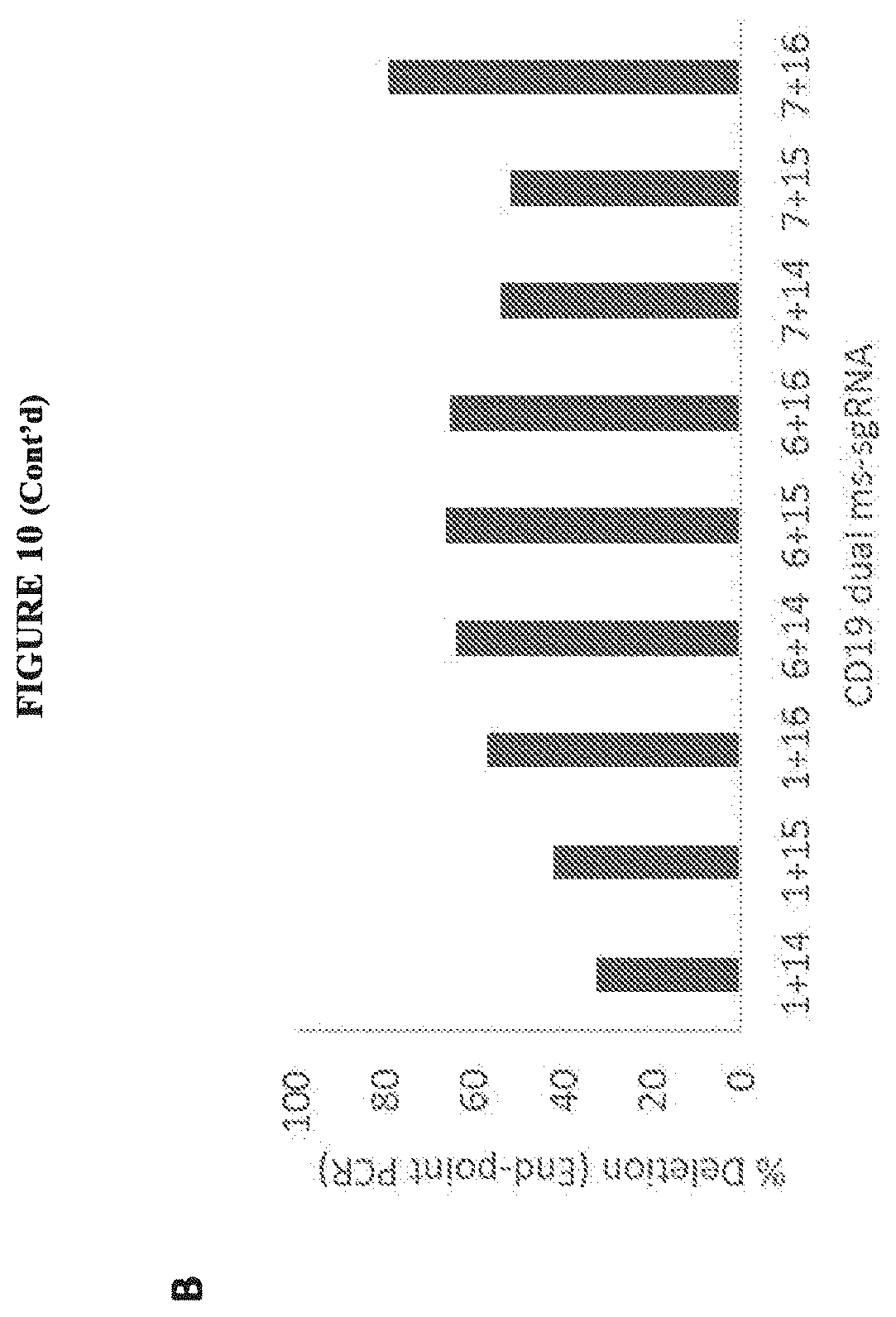
D00015

D00016

D00017

D00018

D00019

D00020

D00021

D00022

D00023

D00024
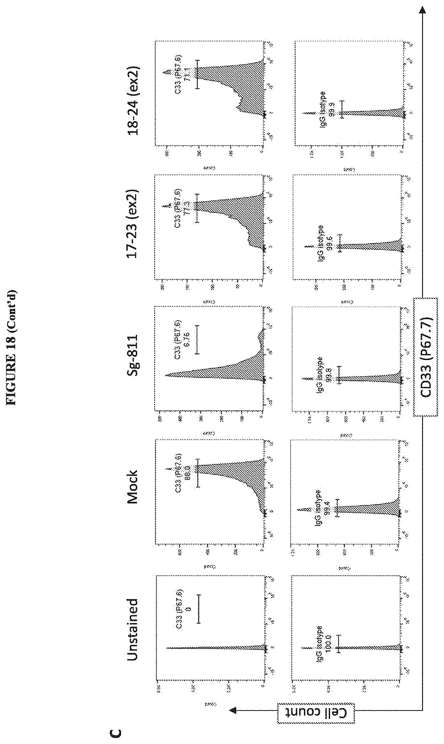
S00001
XML
uspto.report is an independent third-party trademark research tool that is not affiliated, endorsed, or sponsored by the United States Patent and Trademark Office (USPTO) or any other governmental organization. The information provided by uspto.report is based on publicly available data at the time of writing and is intended for informational purposes only.
While we strive to provide accurate and up-to-date information, we do not guarantee the accuracy, completeness, reliability, or suitability of the information displayed on this site. The use of this site is at your own risk. Any reliance you place on such information is therefore strictly at your own risk.
All official trademark data, including owner information, should be verified by visiting the official USPTO website at www.uspto.gov. This site is not intended to replace professional legal advice and should not be used as a substitute for consulting with a legal professional who is knowledgeable about trademark law.