Exosomes And Micro-ribonucleic Acids For Tissue Regeneration
Marban; Eduardo ; et al.
U.S. patent application number 16/572101 was filed with the patent office on 2020-01-23 for exosomes and micro-ribonucleic acids for tissue regeneration. The applicant listed for this patent is Cedars-Sinai Medical Center. Invention is credited to Ke Cheng, Ahmed Ibrahim, Eduardo Marban.
| Application Number | 20200024604 16/572101 |
| Document ID | / |
| Family ID | 50101594 |
| Filed Date | 2020-01-23 |


View All Diagrams
| United States Patent Application | 20200024604 |
| Kind Code | A1 |
| Marban; Eduardo ; et al. | January 23, 2020 |
EXOSOMES AND MICRO-RIBONUCLEIC ACIDS FOR TISSUE REGENERATION
Abstract
Several embodiments relate to methods of repairing and/or regenerating damaged or diseased tissue comprising administering to the damaged or diseased tissues compositions comprising exosomes. In several embodiments, the exosomes comprise one or more microRNA that result in alterations in gene or protein expression, which in tum result in improved cell or tissue viability and/or function.
| Inventors: | Marban; Eduardo; (Los Angeles, CA) ; Cheng; Ke; (Los Angeles, CA) ; Ibrahim; Ahmed; (Los Angeles, CA) | ||||||||||
| Applicant: |
|
||||||||||
|---|---|---|---|---|---|---|---|---|---|---|---|
| Family ID: | 50101594 | ||||||||||
| Appl. No.: | 16/572101 | ||||||||||
| Filed: | September 16, 2019 |
Related U.S. Patent Documents
| Application Number | Filing Date | Patent Number | ||
|---|---|---|---|---|
| 15790962 | Oct 23, 2017 | 10457942 | ||
| 16572101 | ||||
| 14421355 | Feb 12, 2015 | 9828603 | ||
| PCT/US2013/054732 | Aug 13, 2013 | |||
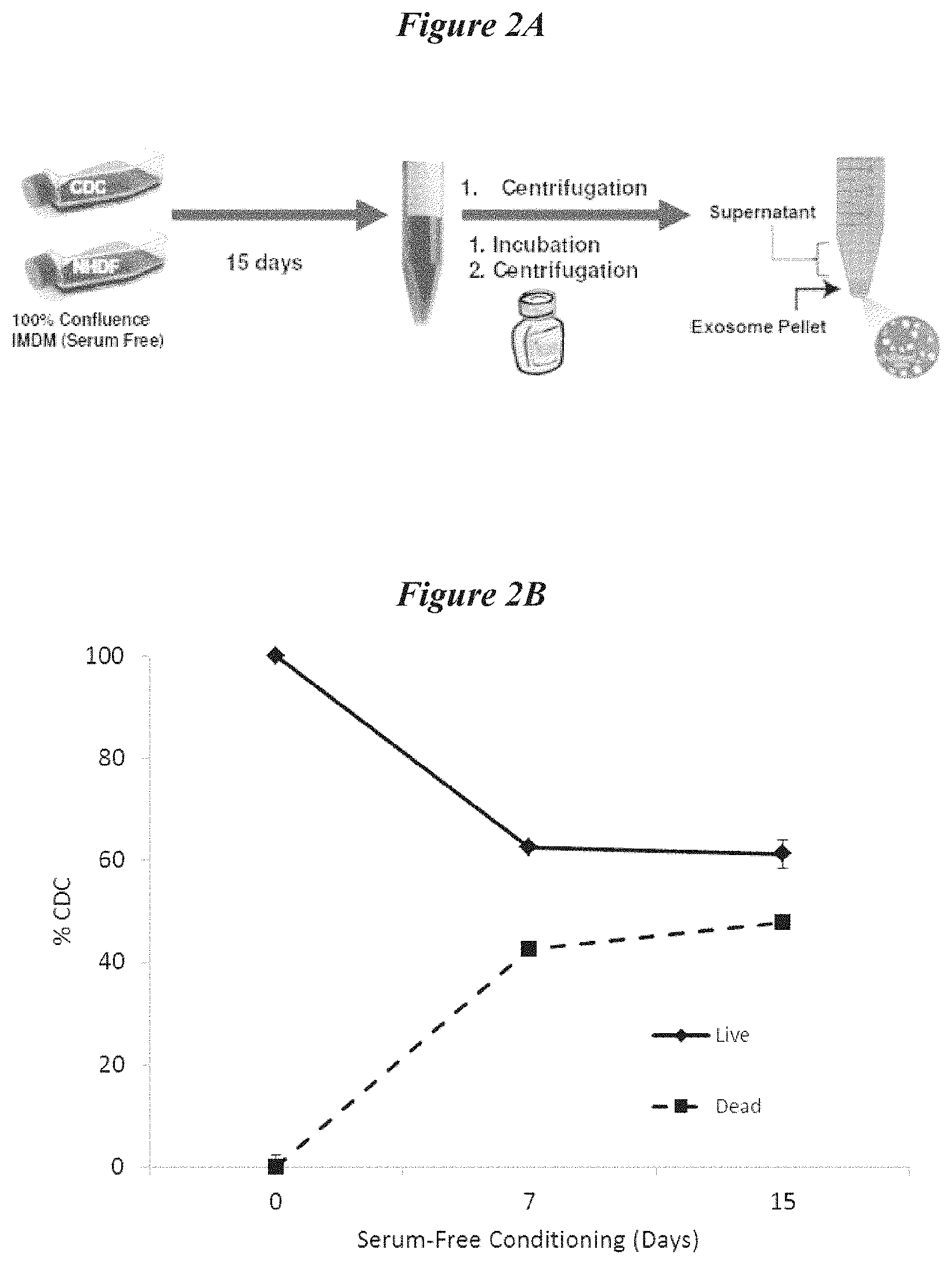
| 15790962 | ||||
| 61682666 | Aug 13, 2012 | |||
| Current U.S. Class: | 1/1 |
| Current CPC Class: | C12N 2320/30 20130101; A61K 47/46 20130101; Y02A 50/463 20180101; A61K 9/0019 20130101; C12N 5/0657 20130101; C12N 15/113 20130101; A61P 9/10 20180101; A61P 9/00 20180101; A61K 31/7105 20130101; A61K 47/42 20130101; C12N 2502/1329 20130101; C12N 2502/28 20130101; A61P 43/00 20180101; C12N 2310/141 20130101; A61P 29/00 20180101; A61P 9/14 20180101; Y02A 50/30 20180101; A61K 35/34 20130101; C12N 15/111 20130101; A61P 35/00 20180101; A61K 35/34 20130101; A61K 2300/00 20130101 |
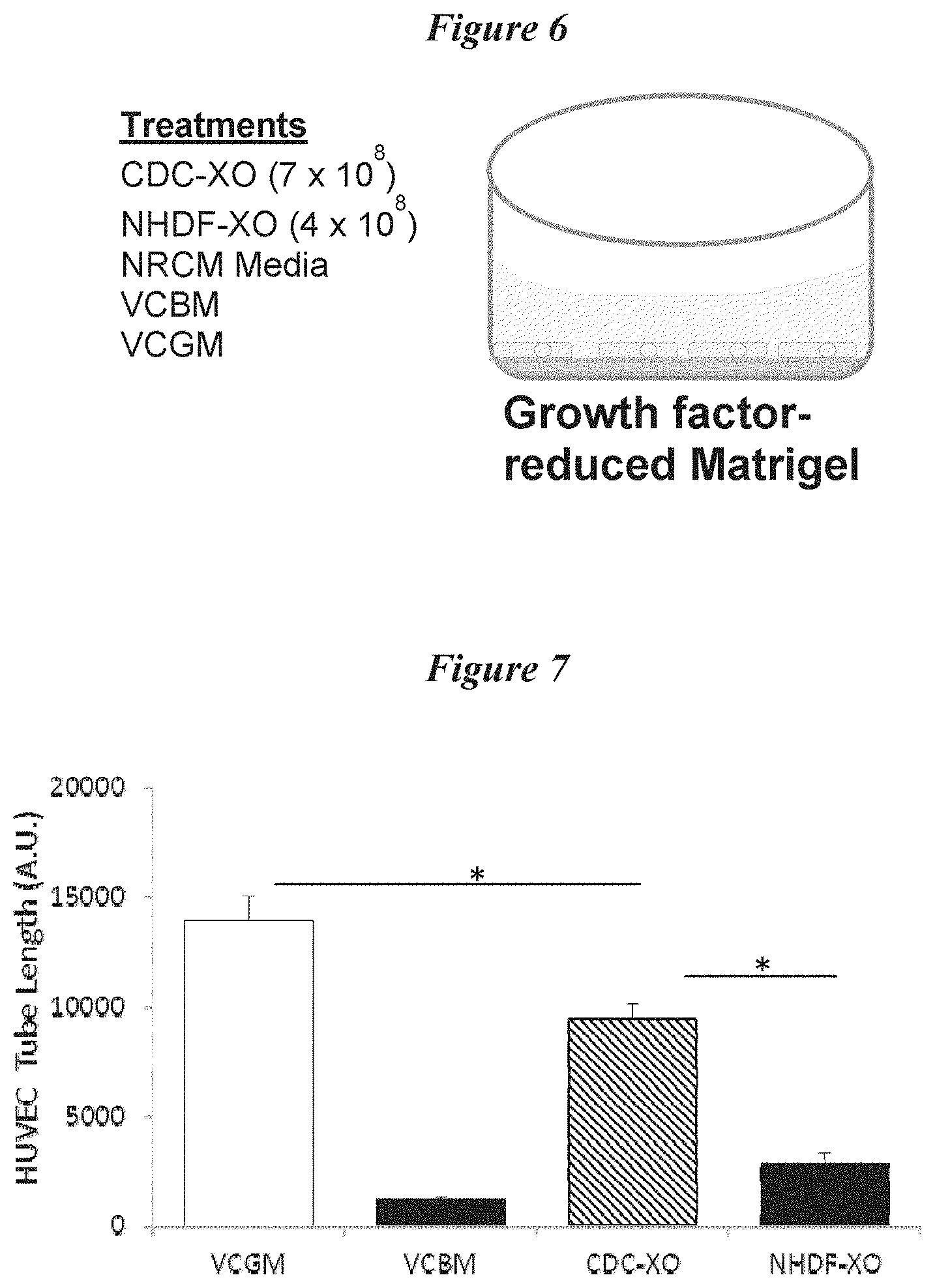
| International Class: | C12N 15/113 20060101 C12N015/113; C12N 15/11 20060101 C12N015/11; A61K 31/7105 20060101 A61K031/7105; A61K 35/34 20060101 A61K035/34; C12N 5/077 20060101 C12N005/077; A61K 47/46 20060101 A61K047/46 |
Goverment Interests
STATEMENT REGARDING GOVERNMENT SPONSORED GRANT
[0002] The inventions disclosed herein were made with Government support under the Research Project Grant (ROI HL083109) by the National Institutes of Health. The United States Government has certain rights in this invention.
Claims
1. A preparation of exosomes harvested from cardiospheres or cardiosphere-derived cells (CDCs).
2. The preparation of claim 1, wherein the preparation comprises exosomes having a diameter from about 15 nm to about 205 nm.
3. The preparation of claim 1, wherein the preparation comprises exosomes having a diameter from about 20 nm to about 90 nm.
4. The preparation of claim 1, wherein the preparation comprises exosomes having a diameter from about 15 nm to about 95 nm.
5. The preparation of claim 1, wherein said exosomes comprise miR-146a.
6. The preparation of claim 5, wherein said exosomes further comprise miR-210.
7. The preparation of claim 6, wherein said exosomes further comprise one or more additional miRNAs selected from the group consisting of miR-26a, miR27-a, let-7e, miR-19b, miR-125b, miR-27b, let-7a, miR-19a, let-7c, miR-140-3p, miR-125a-5p, miR-150, miR-155, mir-210, let-7b, miR-24, miR-423-5p, miR-22, let-7f, miR-146a, and combinations thereof.
8. A preparation of exosomes harvested from cardiosphere-derived cells (CDCs).
9. The preparation of claim 8, wherein the preparation comprises exosomes having a diameter from about 15 nm to about 205 nm.
10. The preparation of claim 8, wherein the preparation comprises exosomes having a diameter from about 20 nm to about 90 nm.
11. The preparation of claim 8, wherein the preparation comprises exosomes having a diameter from about 15 nm to about 95 nm.
12. The preparation of claim 8, wherein said exosomes comprise miR-146a.
13. The preparation of claim 12, wherein said exosomes further comprise miR-210.
14. The preparation of claim 13, wherein said exosomes further comprise one or more additional miRNAs selected from the group consisting of miR-26a, miR27-a, let-7e, miR-19b, miR-125b, miR-27b, let-7a, miR-19a, let-7c, miR-140-3p, miR-125a-5p, miR-150, miR-155, mir-210, let-7b, miR-24, miR-423-5p, miR-22, let-7f, miR-146a, and combinations thereof.
Description
RELATED APPLICATIONS
[0001] This application is a divisional application of U.S. application Ser. No. 15/790,962, filed Oct. 23, 2017, which is divisional application of U.S. application Ser. No. 14/421,355, filed Feb. 12, 2015, which is the U.S. National Phase entry under 35 U.S.C. .sctn. 371 of International Application No. PCT/US2013/054732, filed Aug. 13, 2013, which claims the benefit of U.S. Provisional Application No. 61/682,666, filed Aug. 13, 2012, the entire disclosures of each of which is hereby incorporated by reference herein.
BACKGROUND
[0003] The present application relates generally to methods and compositions for the repair or regeneration of damaged or diseased cells or tissue. Several embodiments relate to administration of exosomes (or protein and/or nucleic acids from the exosomes) isolated from cells or synthetic surrogates in order to repair and/or regenerate damage or diseased tissues. In particular, several embodiments, relate to exosomes derived from certain cell types, such as for example cardiac stem cells, and use of the exosomes in the repair and/or regeneration of cardiac tissue.
Description of the Related Art
[0004] Many diseases, injuries and maladies involve loss of or damage to cells and tissues. Examples include, but are not limited to neurodegenerative disease, endocrine diseases, cancers, and cardiovascular disease. Just these non-limiting examples are the source of substantial medical costs, reduced quality of life, loss of productivity in workplaces, workers compensation costs, and of course, loss of life. For example, coronary heart disease is one of the leading causes of death in the United States, taking more than 650,000 lives annually. Approximately 1.3 million people suffer from a heart attack (or myocardial infarction, MI) every year in the United States (roughly 800,000 first heart attacks and roughly 500,000 subsequent heart attacks). Even among those who survive the MI, many will still die within one year, often due to reduced cardiac function, associated side effects, or progressive cardiac disease. Heart disease is the leading cause of death for both men and women, and coronary heart disease, the most common type of heart disease, led to approximately 400,000 deaths in 2008 in the US. Regardless of the etiology, most of those afflicted with coronary heart disease or heart failure have suffered permanent heart tissue damage, which often leads to a reduced quality of life.
SUMMARY
[0005] There exists a need for methods and compositions to repair and/or regenerate tissue that has been damaged (or is continuing to undergo damage) due to injury, disease, or combinations thereof. While classical therapies such as pharmacological intervention or device-based intervention or surgery provide positive effects, there are provided herein methods and compositions that yield unexpectedly beneficial effects in the repair or regeneration of damaged or diseased tissues (though in some embodiments, these methods and compositions are used to complement classical therapies).
[0006] As such, there are provided herein methods for regenerating tissue in an individual having damaged tissue, comprising, identifying an individual having damaged tissue and administering a plurality of exosomes to the individual, wherein the exosomes are secreted from regenerative cells, wherein the exosomes comprise one or more microRNA fragments, and wherein after administration of the plurality of exosomes, the one or more microRNA fragments alter gene expression in the damaged tissue, improve the viability of the damaged tissue, and/or facilitate the formation of new tissue in the individual. In several embodiments, administration of the exosomes results in functional improvement in the tissue, in combination with one or more of the above-mentioned positive results. In several embodiments, the exosomes are synthetic in origin. In some such embodiments, the synthetic exosomes are generated in order to replicate, substantially, or closely mimic exosomes that are secreted from regenerative cells.
[0007] In several embodiments, the regenerative cells are mammalian in origin. In several embodiments, the regenerative cells are human cells. In some embodiments, the cells are non-embryonic human regenerative cells. In several embodiments, the regenerative cells are autologous to the individual while in several other embodiments the regenerative cells are allogeneic to the individual. Xenogeneic or syngeneic cells are used in certain other embodiments.
[0008] In several embodiments, there is provided a method of regenerating tissue in an individual having damaged tissue, comprising identifying an individual having damaged tissue and administering one or more microRNA fragments, or derivatives thereof, to the individual, wherein after administration of the one or more microRNA fragments, the one or more microRNA fragments alter gene expression in the damaged tissue, improve the viability of the damaged tissue, and/or facilitate the formation of new tissue in the individual. Thus, in some embodiments, exosomes need not be administered, but rather miRNAs (and/or proteins) that are thought to or known to be present in a certain exosome, can be directly administered to effect regeneration of damaged tissue. In several such embodiments, the microRNA fragments, or derivatives thereof, are synthetically generated. In one embodiment, the microRNA fragments, or derivatives thereof are synthesized with a sequence that mimics one or more endogenous microRNA molecules. Alternatively, in several embodiments, miRNAs are complementary to certain genes in the target cell and can reduce the expression of target genes. Combinations of complementary miRNAs (e.g., antisense molecules known as antagomiRs) and miRNAs (or miRNA mimics) are used in several embodiments. In several embodiments, modifications (e.g., chemical modifications) are made in order to enhance the stability of the microRNAs, thereby improving the ability to administer the microRNA (or fragments/derivatives thereof). In some embodiments, administration is of only microRNA fragments, mimics thereof, derivatives thereof, or chemical replicas thereof, or combinations thereof (e.g., no exosomes). However, in several embodiments, as discussed herein, administration comprises administration of a plurality of synthetic liposomes that comprise the one or more microRNA fragments, or derivatives thereof. In additional embodiments, a plurality of regenerative cells is administered along with exosomes, and/or miRNAs.
[0009] In several embodiments, the damaged tissue comprises cardiac tissue. In several embodiments, the regenerative cells comprise cardiospheres. In several embodiments, the regenerative cells comprise cardiosphere-derived cells (CDCs). In several embodiments, the use of cardiospheres and/or CDCs as a source of exosomes is particularly advantageous, as the resultant exosomes provide unexpectedly superior therapeutic benefits (as compared to exosomes from other cell types). In some embodiments, such benefits include, but are not limited to, reduced degradation, enhanced specificity for cardiac regeneration, lower immunogenicity, etc. Additionally, in several embodiments, the cardiospheres and or CDCs are screened to identify an miRNA expression profile that is unique to those cells. That profile, in several embodiments, is replicated, at least in part, by the generation and administration of synthetic exosomes and/or miRNAs. Thus, the therapeutic efficacy of cardiospheres and/or CDCs can unexpectedly be mirrored, without administration of the cells themselves. In several embodiments, this results in improved therapeutic efficacy as the exosomes and/or miRNAs result in reduced immune response in the target tissue.
[0010] In several embodiments, the damaged tissue comprises one or more of neural and/or nervous tissue, epithelial tissue, skeletal muscle tissue, endocrine tissue, vascular tissue, smooth muscle tissue, liver tissue, pancreatic tissue, lung tissue, intestinal tissue, osseous tissue, connective tissue, or combinations thereof. In several embodiments, the damaged tissue is in need of repair, regeneration, or improved function due to an acute event. Acute events include, but are not limited to, trauma such as laceration, crush or impact injury, shock, loss of blood or oxygen flow, infection, chemical or heat exposure, poison or venom exposure, drug overuse or overexposure, and the like. For example, in several embodiments, the damaged tissue is cardiac tissue and the acute event comprises a myocardial infarction. In some embodiments, administration of the exosomes results in an increase in cardiac wall thickness in the area subjected to the infarction. In additional embodiments, the tissue is damaged due to chronic disease or ongoing injury. For example, progressive degenerative diseases can lead to tissue damage that propagates over time (at times, even in view of attempted therapy). Chronic disease need not be degenerative to continue to generate damaged tissue, however. In several embodiments, chronic disease/injury includes, but it not limited to epilepsy, Alzheimer's disease, Parkinson's disease, Huntington's disease, dopaminergic impairment, dementia, ischemia including focal cerebral ischemia, ensuing effects from physical trauma (e.g., crush or compression injury in the CNS), neurodegeneration, immune hyperactivity or deficiency, bone marrow replacement or functional supplementation, arthritis, auto-immune disorders, inflammatory bowel disease, cancer, diabetes, muscle weakness (e.g., muscular dystrophy, amyotrophic lateral sclerosis, and the like), blindness and hearing loss. Cardiac tissue, in several embodiments, is also subject to damage due to chronic disease, such as for example congestive heart failure, ischemic heart disease, diabetes, valvular heart disease, dilated cardiomyopathy, infection, and the like. Other sources of damage also include, but are not limited to, injury, age-related degeneration, cancer, and infection. In several embodiments, the regenerative cells are from the same tissue type as is in need of repair or regeneration. In several other embodiments, the regenerative cells are from a tissue type other than the tissue in need of repair or regeneration. In several embodiments, the regenerative cells comprise somatic cells, while in additional embodiments, they comprise germ cells. In still additional embodiments, combinations of one or more cell types are used to obtain exosomes (or the contents of the exosomes).
[0011] In several embodiments, the exosomes are about 15 nm to about 95 nm in diameter, including about 15 nm to about 20 nm, about 20 nm to about 25 nm, about 25 nm to about 30 nm, about 30 nm to about 35 nm, about 35 nm to about 40 nm, about 40 nm to about 50 nm, about 50 nm to about 60 nm, about 60 nm to about 70 nm, about 70 nm to about 80 nm, about 80 nm to about 90 nm, about 90 nm to about 95 nm and overlapping ranges thereof. In certain embodiments, larger exosomes are obtained are larger in diameter (e.g., those ranging from about 140 to about 210 nm). Advantageously, in several embodiments, the exosomes comprise synthetic membrane bound particles (e.g., exosome surrogates), which depending on the embodiment, are configured to a specific range of diameters. In such embodiments, the diameter of the exosome surrogates is tailored for a particular application (e.g., target site or route of delivery). In still additional embodiments, the exosome surrogates are labeled or modified to enhance trafficking to a particular site or region post-administration.
[0012] In several embodiments, exosomes are obtained via centrifugation of the regenerative cells. In several embodiments, ultracentrifugation is used. However, in several embodiments, ultracentrifugation is not used. In several embodiments, exosomes are obtained via size-exclusion filtration of the regenerative cells. As disclosed above, in some embodiments, synthetic exosomes are generated, which can be isolated by similar mechanisms as those above.
[0013] In several embodiments, the exosomes induce altered gene expression by repressing translation and/or cleaving mRNA. In some embodiments, the alteration of gene expression results in inhibition of undesired proteins or other molecules, such as those that are involved in cell death pathways, or induce further damage to surrounding cells (e.g., free radicals). In several embodiments, the alteration of gene expression results directly or indirectly in the creation of desired proteins or molecules (e.g., those that have a beneficial effect). The proteins or molecules themselves need not be desirable per se (e.g., the protein or molecule may have an overall beneficial effect in the context of the damage to the tissue, but in other contexts would not yield beneficial effects). In some embodiments, the alteration in gene expression causes repression of an undesired protein, molecule or pathway (e.g., inhibition of a deleterious pathway). In several embodiments, the alteration of gene expression reduces the expression of one or more inflammatory agents and/or the sensitivity to such agents. Advantageously, the administration of exosomes, or miRNAs, in several embodiments, results in downregulation of certain inflammatory molecules and/or molecules involved in inflammatory pathways. As such, in several embodiments, cells that are contacted with the exosomes or miRNAs enjoy enhanced viability, even in the event of post-injury inflammation or inflammation due to disease.
[0014] In several embodiments, the exosomes fuse with one or more recipient cells of the damaged tissue. In several embodiments, the exosomes release the microRNA into one or more recipient cells of the damaged tissue, thereby altering at least one pathway in the one or more cells of the damaged tissue. In some embodiments, the exosomes exerts their influence on cells of the damaged tissue by altering the environment surrounding the cells of the damaged tissue. In some embodiments, signals generated by or as a result of the content or characteristics of the exosomes, lead to increases or decreases in certain cellular pathways. For example, the exosomes (or their contents/characteristics) can alter the cellular milieu by changing the protein and/or lipid profile, which can, in turn, lead to alterations in cellular behavior in this environment. Additionally, in several embodiments, the miRNA of an exosome can alter gene expression in a recipient cell, which alters the pathway in which that gene was involved, which can then further alter the cellular environment. In several embodiments, the influence of the exosomes directly or indirectly stimulates angiogenesis. In several embodiments, the influence of the exosomes directly or indirectly affects cellular replication. In several embodiments, the influence of the exosomes directly or indirectly inhibits cellular apoptosis.
[0015] The beneficial effects of the exosomes (or their contents) need not only be on directly damaged or injured cells. In some embodiments, for example, the cells of the damaged tissue that are influenced by the disclosed methods are healthy cells. However, in several embodiments, the cells of the damaged tissue that are influenced by the disclosed methods are damaged cells.
[0016] In several embodiments, regeneration comprises improving the function of the tissue. For example, in certain embodiments in which cardiac tissue is damaged, functional improvement may comprise increased cardiac output, contractility, ventricular function and/or reduction in arrhythmia (among other functional improvements). For other tissues, improved function may be realized as well, such as enhanced cognition in response to treatment of neural damage, improved blood-oxygen transfer in response to treatment of lung damage, improved immune function in response to treatment of damaged immunological-related tissues.
[0017] In several embodiments, the microRNA fragments are selected from the group consisting of miR-23a, miR-23b, miR-24, miR-26a, miR27-a, miR-30c, let-7e, mir-19b, miR-125b, mir-27b, let-7a, miR-19a, let-7c, miR-140-3p, miR-125a-5p, miR-132, miR-150, miR-155, mir-210, let-7b, miR-24, miR-423-5p, miR-22, let-7f, miR-146a, and combinations thereof. In several embodiments, one, two, three or more of these miRNAs are used to treat cardiac tissue. In one embodiment, the microRNA comprises miR-146a. In one embodiment, the microRNA comprises miR-210. In additional embodiments, the miRNA comprises one or more of miR-17, miR-21, miR-92, miR92a, miR-29, miR-29a, miR-29b, miR-29c, miR-34, mi-R34a, miR-150, miR-451, miR-145, miR-143, miR-144, miR-193a-3p, miR-133a, miR-155, miR-181a, miR-214, miR-199b, miR-199a, miR-210, miR-126, miR-378, miR-363 and miR-30b, and miR-499. In several embodiments, exosomes do not contain any of miR-92, miR-17, miR-21, miR-92, miR92a, miR-29, miR-29a, miR-29b, miR-29c, miR-34, mi-R34a, miR-150, miR-451, miR-145, miR-143, miR-144, miR-193a-3p, miR-133a, miR-155, miR-181a, miR-214, miR-199b, miR-199a, miR-126, miR-378, miR-363 and miR-30b, or miR-499. In several embodiments, the exosomes further comprise at least one protein that further facilitates regeneration and/or improved function of the tissue.
[0018] Administration can be via a variety of routes, depending on the embodiment. For example, in some embodiments, delivery is locally to the tissue. In some embodiments, delivery is systemically. In one embodiment, delivery is via an intramyocardial route, while in other embodiments, delivery is via an intracoronary route. Combinations of delivery routes are used, in certain embodiments, in order to improve the speed with which positive effects are realized and or improve the duration of treatment. For example, in some embodiments, miRNAs are delivered directly to a target tissue and exosomes are delivered via a systemic route.
[0019] In several embodiments, the method further comprises administering the regenerative cells from which the exosomes were obtained to the individual, either prior to, concurrent with, or after administration of the exosomes. Administration of these cells can be by the same route or an alternative route.
[0020] In several embodiments, there is provided a composition for the repair or regeneration of damaged or diseased cardiac tissue comprising, a plurality of exosomes isolated from a population of cardiac stem cells, wherein the cardiac stem cells comprise a population of cardiosphere-derived cells, wherein the exosomes comprise at least one microRNA, wherein the microRNA is selected from the group consisting of miR-146a, miR-22, miR-24, and miR-26a, and wherein upon administration to a subject having damaged or diseased cardiac tissue, the exosomes increase one or more of cardiac cell viability, cardiac cell proliferation, and cardiac cell function. In one embodiment, the composition further comprises a plurality of cardiac stem cells. In one embodiment, the miRNA payload of the exosome comprises, consists of, or consists essentially of miR-146a. In one embodiment, the miRNA payload of the exosome comprises, consists of, or consists essentially of miR-210. In several embodiments, there is provided a use of a composition comprising a plurality of exosomes isolated from a population of cardiosphere-derived cells for the treatment of damaged or diseased cardiac tissue. In several embodiments, there is provided a use of a composition comprising a plurality of miRNA, a plurality of exosome, and/or a plurality of cardiosphere-derived cells for the treatment of damaged or diseased cardiac tissue.
[0021] There is also provided a composition for the repair or regeneration of damaged or diseased cardiac tissue comprising synthetic microRNA-146a and a pharmaceutically acceptable carrier. In one embodiment, the synthetic miRNA consists of or consists essentially of miR-146a. In some embodiments, the synthetic miRNA also comprises a synthetic miR210. In one embodiment, the synthetic miRNA consists of or consists essentially of miR-210. In some embodiments, the microRNA is directly administered, while in some embodiments, it is administered via delivery of an exosome (either isolated or synthetically generated).
[0022] In several embodiments, there is provided a method comprising identifying a subject in need of repair of damaged tissue and instructing the administration of a composition comprising exosomes derived from regenerative cells to the subject, thereby resulting in repair of the damaged tissue.
[0023] In several embodiments, there is provided a method comprising identifying a subject in need of repair of damaged tissue and instructing the administration of a composition comprising one or more miRNA to the subject, thereby resulting in repair of the damaged tissue.
[0024] In several embodiments, there is provided a method comprising identifying a subject in need of repair of damaged tissue and instructing the administration of a composition comprising one or more of exosomes derived from regenerative cells, miRNA, and regenerative cells to the subject, thereby resulting in repair of the damaged tissue.
[0025] In several such embodiments, the repair of the damaged tissue comprises both anatomical repair (e.g., tissue regeneration) and functional repair.
[0026] In several embodiments, there is provided a method of generating exosomes, comprising obtaining a population of non-embryonic human regenerative cells, culturing the population of non-embryonic human regenerative cells, and exposing the cultured population of non-embryonic human regenerative cells to a hydrolase enzyme to induce the cells to secrete exosomes, thereby generating exosomes. In several embodiments, the method further comprises harvesting the secreted exosomes. In several embodiments, the hydrolase comprises a member of the DNAse I superfamily of enzymes. In several embodiments, the hydrolase comprises a sphingomyelinase, such as for example a sphingomyelinase of a type selected from the group consisting of lysosomal acid sphingomyelinase, secreted zinc-dependent acid sphingomyelinase, neutral sphingomyelinase, and alkaline sphingomyelinase. In several embodiments, a neutral sphingomyelinase is used. In one embodiment, the neutral sphingomyelinase comprises one or more of magnesium-dependent neutral sphingomyelinase and magnesium-independent neutral sphingomyelinase. In additional embodiments, the neutral sphingomyelinase comprises one or more of neutral sphingomyelinase type I, neutral sphingomyelinase type 2, and neutral sphingomyelinase type 3. As discussed above, in several embodiments the exosomes are synthetically manufactured in vitro by established methods to generate lipid bilayers. In such embodiments, the synthetic exosomes can advantageously be customized to regenerate a certain tissue type and optionally damage due to a specific source of damage.
[0027] The methods summarized above and set forth in further detail below describe certain actions taken by a practitioner; however, it should be understood that they can also include the instruction of those actions by another party. Thus, actions such as "administering exosomes" include "instructing the administration of exosomes."
BRIEF DESCRIPTION OF THE DRAWINGS
[0028] FIG. 1 depicts a general schematic of the various components of cellular and tissue regeneration, including direct and indirect mechanisms.
[0029] FIGS. 2A-2D depict information related to the isolation of exosomes and characterization of cells during the isolation protocol. FIG. 2A depicts a schematic for the isolation of exosomes from cultured cells according to several embodiments disclosed herein. FIG. 2B depicts the survival of CDCs in serum free culture conditions prior in preparation for exosome isolation. FIG. 2C and 2D show bright-field microscopic images of CDCs at Day 0 and Day 15 (respectively) of culture in serum-free conditions.
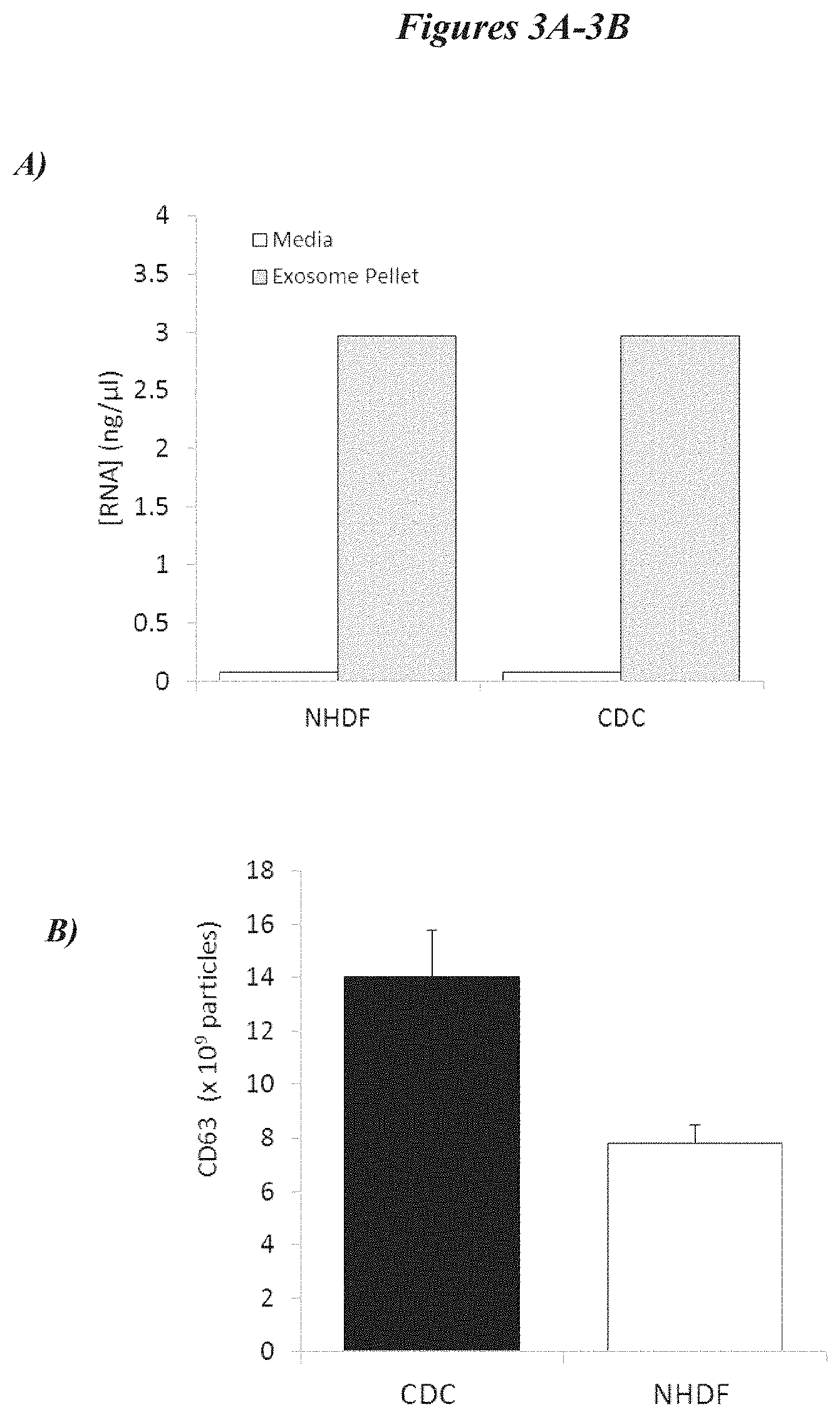
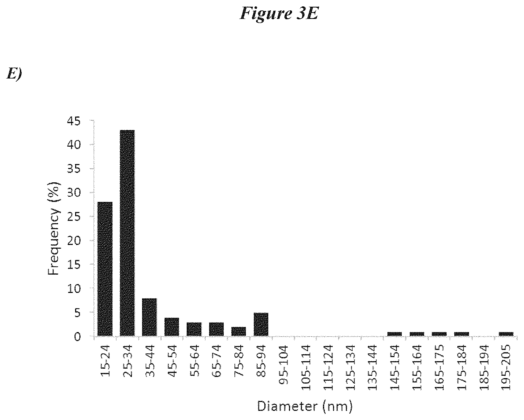
[0030] FIGS. 3A-3E depict exosome characterization data. FIG. 3A depicts data related to the RNA content of the supernatant and exosome fractions of cells. FIG. 3B shows data related to the number of exosomes generated from the isolation scheme outlined in FIG. 2A. FIG. 3C shows differences in expression of various surface genes on NHDF and CDCs. FIG. 3D shows microscopic images of exosomes. FIG. 3E depicts analysis of the frequency of exosomes as compared to their diameter.
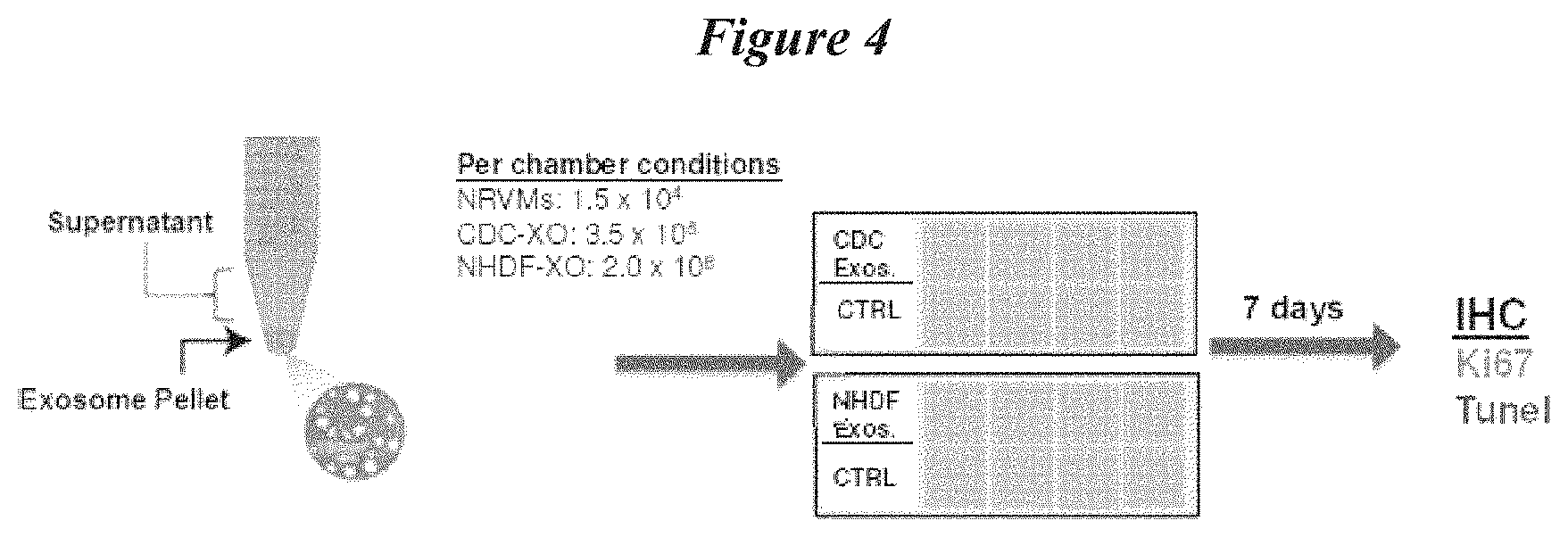
[0031] FIG. 4 depicts a schematic protocol for the evaluation of the effects of exosome treatment on cellular proliferation and cell death.
[0032] FIGS. 5A-5D depict data related to the effects of exosome treatment on cell death and cellular proliferation. FIG. 5A shows data related to apoptosis of cells after incubation with exosomes from various sources. FIG. 5B shows data related to proliferative activity of cells after incubation with exosomes from various sources. FIG. 5C shows immunofluorescent TUNEL staining that depicts apoptosis of cells after exposure to various exosome compositions. FIG. 5D shows immunofluorescent Ki-67 staining that depicts proliferative activity of cells after exposure to various exosome compositions.
[0033] FIG. 6 depicts a schematic protocol for the evaluation of the effects of exosome treatment on angiogenesis.
[0034] FIG. 7 depicts summary data related to angiogenesis after treatment of endothelial cells with various media and exosome preparations.
[0035] FIGS. 8A-8E depict photomicrographs of the results of an angiogenesis by tube formation assay.
[0036] FIG. 9 depicts data related to the survival of mice subject to myocardial infarction and treated with various exosome preparations.
[0037] FIG. 10 depicts cardiac functional data after myocardial infarction and treatment with exosome preparations.
[0038] FIGS. 11A-11C depicts echocardiography (ECHO) data after myocardial infarction and treatment with exosome preparations.
[0039] FIGS. 12A-12H depict data related to the anatomical improvements in cardiac tissue after exosome administration. FIGS. 12A-12D depict Masson's trichrome staining data after myocardial infarction and treatment with exosome preparations from various cell sources. Summary data related to tissue viability scar mass, viable mass, and wall thickness are shown in FIGS. 12E-12H, respectively.
[0040] FIG. 13 depicts data related to the reduced myocardial levels of inflammatory markers after treatment with exosomes derived from cardiosphere-derived cells (CDCs).
[0041] FIGS. 14A-14C depicts data related to mechanisms of exosome secretion. FIG. 14A depicts dose-response data related to inhibition of secretion of CDC-derived exosomes with a neutral sphingomyelinase inhibitor (GW4869). FIG. 14B indicates cell viability in response to inhibition of exosome secretion. FIG. 1 4C summarizes cardiac functional data after administration of exosomes derived from control cells or cells treated with a neutral sphingomyelinase inhibitor (GW4869).
[0042] FIGS. 15A-15B depict ECHO data after administration of exosomes derived from cells treated with a neutral sphingomyelinase inhibitor (GW4869) or control cells (CDCs).
[0043] FIGS. 16A-16B depict Masson's trichrome staining of cardiac tissue treated with exosomes derived from cells treated with a neutral sphingomyelinase inhibitor (GW4869) or control cells (CDCs).
[0044] FIGS. 17A-17D depict data related to the amount of viable tissue (in the risk region, 17A), scar mass (17B), overall viable mass (17C) or infarct thickness (17D) after animals were treated with exosomes derived from cells treated with a neutral sphingomyelinase inhibitor (GW4869) or control cells (CDCs).
[0045] FIGS. 18A-18B depicts profiling of miRNA expression from exosomes isolated from CDCs, as compared to control cells (normal human dermal fibroblast: NHDF). FIG. 18A depicts relative expression of selected miRNAs in exosomes from CDCs as compared to NHDF cells. FIG. 18B shows a listing of those miRNAs that are equivalently expressed in NHDF and CDCs, those that are significantly upregulated, and those that are significantly downregulated.
[0046] FIG. 19 depicts a schematic for an in vitro study to determine the effects of administration of mi146a.
[0047] FIGS. 20A-20D depict data related to cell viability and death after cells were treated with either mi146a or a control miRNA. FIG. 20A depicts results of calcein staining to evaluate cell viability 6 hours after NRVM were transfected with miR146a. FIG. 20B depicts results of ETHD-1 staining to evaluate cell viability 12 hours after NRVM were transfected with miR146a. FIG. 20C depicts data showing the protective effects of miR146a on NRVM exposed to hydrogen peroxide. FIG. 20D depicts data showing the protective effects of miR146a on NRVMs exposed to cobalt chloride.
[0048] FIGS. 21A-21G relate to in vivo data showing the regenerative capacity of miR146a. FIG. 21A shows two infarcted hearts, while 21B shows Masson's Trichrome of a heart treated with control mimic miRNA and 21C shows Masson's Trichrome of a heart treated with miR146a. FIG. 21D shows the ejection fraction in control and treated mice over 30 days post-MI. FIG. 21E, 21F, and 21G show overall viable tissue mass, scar mass, and wall thickness (respectively) of hearts from animals treated with miR146a or a control mimic miR.
[0049] FIG. 22 shows expression data related to known inflammatory molecules in cultured cardiomyocytes transfected with miR146a.
[0050] FIG. 23 shows data related to cell viability of cultured cardiomyocytes transfected with miR210 after exposure to hydrogen peroxide.
DETAILED DESCRIPTION
[0051] Several embodiments of the methods and compositions disclosed herein are useful for the treatment of tissues that are damaged or adversely affected by disease(s). The vast majority of diseases lead to at least some compromise (even if acute) in cellular or tissue function. Several embodiments of the methods and compositions disclosed herein allow for repair and/or regeneration of cells and/or tissues that have been damaged, limited in their functionality, or otherwise compromised as a result of a disease. In several embodiments, methods and compositions disclosed herein may also be used as adjunct therapies to ameliorate adverse side effects of a disease treatment that negatively impacts cells or tissues.
Treatment Modalities for Damaged or Diseased Tissues
[0052] Generally, the use of one or more relatively common therapeutic modalities are used to treat damaged or diseased tissues in an effort to halt progression of the disease, reverse damage that has already occurred, prevent additional damage, and generally improve the well-being of the patient. For example, many conditions can be readily treated with holistic methodologies or changes in lifestyle (e.g., improved diet to reduce risk of cardiovascular disease, diabetes, and the like). Often more serious conditions require more advanced medical intervention. Drug therapy or pharmaceutical therapies are routinely administered to treat patients suffering from a particular disease. For example, a patient suffering from high blood pressure might be prescribed an angiotensin-converting-enzyme (ACE) inhibitor, in order to reduce the tension of blood vessels and blood volume, thereby treating high blood pressure. Further, cancer patients are often prescribed panels of various anticancer compounds in an attempt to limit the spread and/or eradicate a cancerous tumor. Surgical methods may also be employed to treat certain diseases or injuries. In some cases, implanted devices are used in addition to or in place of pharmaceutical or surgical therapies (e.g., a cardiac pacemaker). Recently, additional therapy types have become very promising, such as, for example, gene therapy, protein therapy, and cellular therapy.
[0053] Cell therapy, generally speaking, involves the administration of population of cells to subject with the intent of the administered cells functionally or physically replacing cells that have been damaged, either by injury, by disease, or combinations thereof. A variety of different cell types can be administered in cell therapy, with stem cells being particularly favored (in certain cases) due to their ability to differentiate into multiple cell types, thus providing flexibility for what disease or injury they could be used to treat.
[0054] Protein therapy involves the administration of exogenous proteins that functionally replace deficient proteins in the subject suffering from a disease or injury. For example, synthesized acid alpha-glucosidase is administered to patients suffering from glycogen storage disease type II.
[0055] In addition, nucleic acid therapy is being investigated as a possible treatment for certain diseases or conditions. Nucleic acid therapy involves the administration of exogenous nucleic acids, or short fragments thereof, to the subject in order to alter gene expression pathways through a variety of mechanisms, such as, for example, translational repression of the target gene, cleavage of a target gene, such that the target gene product is never expressed.
[0056] With the knowledge that certain cellular therapies provide profound regenerative effects, several embodiments disclosed herein involve methods and compositions that produce those regenerative effects without the need for administration of cells to a subject (though cells may optionally be administered in certain embodiments).
Exosomes and Vesicle Bound Nucleic Acid and Protein Products
[0057] Nucleic acids are generally not present in the body as free nucleic acids, as they are quickly degraded by nucleases. Certain types of nucleic acids are associated with membrane-bound particles. Such membrane-bound particles are shed from most cell types and consist of fragments of plasma membrane and contain DNA, RNA, mRNA, microRNA, and proteins. These particles often mirror the composition of the cell from which they are shed. Exosomes are one type of such membrane bound particles and typically range in diameter from about 15 nm to about 95 nm in diameter, including about 15 nm to about 20 nm, 20 nm to about 30 nm, about 30 nm to about 40 nm, about 40 nm to about 50 nm, about 50 nm to about 60 nm, about 60 nm to about 70 nm, about 70 nm to about 80 nm, about 80 nm to about 90 nm, about 90 nm to about 95 nm, and overlapping ranges thereof. In several embodiments, exosomes are larger (e.g., those ranging from about 140 to about 210 run, including about 140 nm to about 150 nm, 150 nm to about 160 run, 160 nm to about 170 run, 170 nm to about 180 nm, 180 nm to about 190 run, 190 nm to about 200 run, 200 nm to about 210 nm, and overlapping ranges thereof). In some embodiments, the exosomes that are generated from the original cellular body are 100, 200, 300, 400, 500, 600, 700, 800, 900, 1000, 2000, 5000, 10,000 times smaller in at least one dimension (e.g., diameter) than the original cellular body.
[0058] Alternative nomenclature is also often used to refer to exosomes. Thus, as used herein the term "exosome" shall be given its ordinary meaning and may also include terms including microvesicles, epididimosomes, argosomes, exosome-like vesicles, microparticles, promininosomes, prostasomes, dexosomes, texosomes, dex, tex, archeosomes and oncosomes. Exosomes are secreted by a wide range of mammalian cells and are secreted under both normal and pathological conditions. Exosomes, in some embodiments, function as intracellular messengers by virtue of carrying mRNA, miRNA or other contents from a first cell to another cell (or plurality of cells). In several embodiments, exosomes are involved in blood coagulation, immune modulation, metabolic regulation, cell division, and other cellular processes. Because of the wide variety of cells that secret exosomes, in several embodiments, exosome preparations can be used as a diagnostic tool (e.g., exosomes can be isolated from a particular tissue, evaluated for their nucleic acid or protein content, which can then be correlated to disease state or risk of developing a disease).
[0059] Exosomes, in several embodiments, are isolated from cellular preparations by methods comprising one or more of filtration, centrifugation, antigen-based capture and the like. For example, in several embodiments, a population of cells grown in culture are collected and pooled. In several embodiments, monolayers of cells are used, in which case the cells are optionally treated in advance of pooling to improve cellular yield (e.g., dishes are scraped and/or enzymatically treated with an enzyme such as trypsin to liberate cells). In several embodiments, cells grown in suspension are used. The pooled population is then subject to one or more rounds of centrifugation (in several embodiments ultracentrifugation and/or density centrifugation is employed) in order to separate the exosome fraction from the remainder of the cellular contents and debris from the population of cells. In some embodiments, centrifugation need not be performed to harvest exosomes. In several embodiments, pre-treatment of the cells is used to improve the efficiency of exosome capture. For example, in several embodiments, agents that increase the rate of exosome secretion from cells are used to improve the overall yield of exosomes. In some embodiments, augmentation of exosome secretion is not performed. In some embodiments, size exclusion filtration is used in conjunction with, or in place of centrifugation, in order to collect a particular size (e.g., diameter) of exosome. In several embodiments, filtration need not be used. In still additional embodiments, exosomes (or subpopulations of exosomes are captured by selective identification of unique markers on or in the exosomes (e.g., transmembrane proteins)). In such embodiments, the unique markers can be used to selectively enrich a particular exosome population. In some embodiments, enrichment, selection, or filtration based on a particular marker or characteristic of exosomes is not performed.
[0060] Upon administration (discussed in more detail below) exosomes can fuse with the cells of a target tissue. As used herein, the term "fuse" shall be given its ordinary meaning and shall also refer to complete or partial joining, merging, integration, or assimilation of the exosome and a target cell. In several embodiments, the exosomes fuse with healthy cells of a target tissue. In some embodiments, the fusion with healthy cells results in alterations in the healthy cells that leads to beneficial effects on the damaged or diseased cells (e.g., alterations in the cellular or intercellular environment around the damaged or diseased cells). In some embodiments, the exosomes fuse with damaged or diseased cells. In some such embodiments, there is a direct effect on the activity, metabolism, viability, or function of the damaged or diseased cells that results in an overall beneficial effect on the tissue. In several embodiments, fusion of the exosomes with either healthy or damaged cells is not necessary for beneficial effects to the tissue as a whole (e.g., in some embodiments, the exosomes affect the intercellular environment around the cells of the target tissue). Thus, in several embodiments, fusion of the exosome to another cell does not occur. In several embodiments, there is no cell-exosome contact, yet the exosomes still influence the recipient cells.
Administration and Therapy
[0061] There are provided herein methods and compositions for use in the repair or regeneration of cells or tissue after the cells or tissue have been subject to injury, damage, disease, or some other event that leads to loss of function and/or viability. Methods and compositions for preventing damage and/or for shuttling nucleic acids (or proteins) between cells are also provided, regardless of whether tissue damage is present.
[0062] In addition, methods are provided for facilitating the generation of exosomes. In several such embodiments, a hydrolase is used to facilitate the liberation (e.g., secretion) of exosomes from cells. In certain embodiments, hydrolases that cleave one or more of ester bonds, sugars (e.g., DNA), ether bonds, peptide bonds, carbon-nitrogen bonds, acid anhyrides, carbon-carbon bonds, halide bonds, phosphorous-nitrogen bonds, sulpher-nitrogen bonds, carbon-phosphorous bonds, sulfur-sulfur bonds, and/or carbon-sulfur bonds are used. In some embodiments, the hydrolases are DNAses (e.g., cleave sugars). Certain embodiments employ specific hydrolases, such as for example, one or more of lysosomal acid sphingomyelinase, secreted zinc-dependent acid sphingomyelinase, neutral sphingomyelinase, and alkaline sphingomyelinase.
[0063] In several embodiments, exosomes are administered to a subject in order to initiate the repair or regeneration of cells or tissue. In several embodiments, the exosomes are derived from a stem cell. In several embodiments, the stem cells are non-embryonic stem cells. In some embodiments, the non-embryonic stem cells are adult stem cells. However, in certain embodiments, embryonic stem cells are optionally used as a source for exosomes. In some embodiments, somatic cells are used as a source for exosomes. In still additional embodiments, germ cells are used as a source for exosomes.
[0064] In several embodiments employing stem cells as an exosome source, the nucleic acid and/or protein content of exosomes from stem cells are particularly suited to effect the repair or regeneration of damaged or diseased cells. In several embodiments, exosomes are isolated from stem cells derived from the tissue to be treated. For example, in some embodiments where cardiac tissue is to be repaired, exosomes are derived from cardiac stem cells. Cardiac stem cells are obtained, in several embodiments, from various regions of the heart, including but not limited to the atria, septum, ventricles, auricola, and combinations thereof (e.g., a partial or whole heart may be used to obtain cardiac stem cells in some embodiments). In several embodiments, exosomes are derived from cells (or groups of cells) that comprise cardiac stem cells or can be manipulated in culture to give rise to cardiac stem cells (e.g., cardiospheres and/or cardiosphere derived cells (CDCs)). Further information regarding the isolation of cardiospheres can be found in U.S. Pat. No. 8,268,619, issued on Sep. 18, 2012, which is incorporated in its entirety by reference herein. In several embodiments, the cardiac stem cells are cardiosphere-derived cells (CDCs). Further information regarding methods for the isolation of CDCs can be found in U.S. patent application Ser. No. 11/666,685, filed on Apr. 21, 2008, and Ser. No. 13/412,051, filed on Mar. 5, 2012, both of which are incorporated in their entirety by reference herein. Other varieties of stem cells may also be used, depending on the embodiment, including but not limited to bone marrow stem cells, adipose tissue derived stem cells, mesenchymal stem cells, induced pluripotent stem cells, hematopoietic stem cells, and neuronal stem cells.
[0065] In several embodiments, administration of exosomes is particularly advantageous because there are reduced complications due to immune rejection by the recipient. Certain types of cellular or gene therapies are hampered by the possible immune response of a recipient of the therapy. As with organ transplants or tissue grafts, certain types of foreign cells (e.g., not from the recipient) are attacked and eliminated (or rendered partially or completely non-functional) by recipient immune function. One approach to overcome this is to co-administer immunosuppressive therapy, however this can be costly, and leads to a patient being subject to other infectious agents. Thus, exosomal therapy is particularly beneficial because the immune response is limited. In several embodiments, this allows the use of exosomes derived from allogeneic cell sources (though in several embodiments, autologous sources may be used). Moreover, the reduced potential for immune response allows exosomal therapy to be employed in a wider patient population, including those that are immune-compromised and those that have hyperactive immune systems. Moreover, in several embodiments, because the exosomes do not carry a full complement of genetic material, there is a reduced risk of unwanted cellular growth (e.g., teratoma formation) post-administration. Advantageously, the exosomes can be derived, depending on the embodiment, from cells obtained from a source that is allogeneic, autologous, xenogeneic, or syngeneic with respect to the eventual recipient of the exosomes. Moreover, master banks of exosomes that have been characterized for their expression of certain miRNAs and/or proteins can be generated and stored long-term for subsequent use in defined subjects on an "off-the-shelf " basis. However, in several embodiments, exosomes are isolated and then used without long-term or short-term storage (e.g., they are used as soon as practicable after their generation).
[0066] In several embodiments, exosomes need not be administered; rather the nucleic acid and/or protein carried by exosomes can be administered to a subject in need of tissue repair. In such embodiments, exosomes are harvested as described herein and subjected to methods to liberate and collect their protein and/or nucleic acid contents. For example, in several embodiments, exosomes are lysed with a detergent (or non-detergent) based solution in order to disrupt the exosomal membrane and allow for the collection of proteins from the exosome. As discussed above, specific methods can then be optionally employed to identify and selected particularly desired proteins. In several embodiments, nucleic acids are isolated using chaotropic disruption of the exosomes and subsequent isolation of nucleic acids. Other established methods for nucleic acid isolation may also be used in addition to, or in place of chaotropic disruption. Nucleic acids that are isolated may include, but are not limited to DNA, DNA fragments, and DNA plasmids, total RNA, mRNA, tRNA, snRNA, saRNA, miRNA, rRNA, regulating RNA, non-coding and coding RNA, and the like. In several embodiments in which RNA is isolated, the RNA can be used as a template in an RT-PCR-based (or other amplification) method to generate large copy numbers (in DNA form) of the RNA of interest. In such instances, should a particular RNA or fragment be of particular interest, the exosomal isolation and preparation of the RNA can optionally be supplemented by the in vitro synthesis and co-administration of that desired sequence.
[0067] In several embodiments, exosomes derived from cells are administered in combination with one or more additional agents. For example, in several embodiments, the exosomes are administered in combination with one or more proteins or nucleic acids derived from the exosome (e.g., to supplement the exosomal contents). In several embodiments, the cells from which the exosomes are isolated are administered in conjunction with the exosomes. In several embodiments, such an approach advantageously provides an acute and more prolonged duration of exosome delivery (e.g., acute based on the actual exosome delivery and prolonged based on the cellular delivery, the cells continuing to secrete exosomes post-delivery).
[0068] In several embodiments, exosomes are delivered in conjunction with a more traditional therapy, e.g., surgical therapy or pharmaceutical therapy. In several embodiments such combinations of approaches result in synergistic improvements in the viability and/or function of the target tissue. In some embodiments, exosomes may be delivered in conjunction with a gene therapy vector (or vectors), nucleic acids (e.g., those used as siRNA or to accomplish RNA interference), and/or combinations of exosomes derived from other cell types.
[0069] The compositions disclosed herein can be administered by one of many routes, depending on the embodiment. For example, exosome administration may be by local or systemic administration. Local administration, depending on the tissue to be treated, may in some embodiments be achieved by direct administration to a tissue (e.g., direct injection, such as intramyocardial injection). Local administration may also be achieved by, for example, lavage of a particular tissue (e.g., intra-intestinal or peritoneal lavage). In several embodiments, systemic administration is used and may be achieved by, for example, intravenous and/or intra-arterial delivery. In certain embodiments, intracoronary delivery is used. In several embodiments, the exosomes are specifically targeted to the damaged or diseased tissues. In some such embodiments, the exosomes are modified (e.g., genetically or otherwise) to direct them to a specific target site. For example, modification may, in some embodiments, comprise inducing expression of a specific cell-surface marker on the exosome, which results in specific interaction with a receptor on a desired target tissue. In one embodiment, the native contents of the exosome are removed and replaced with desired exogenous proteins or nucleic acids. In one embodiment, the native contents of exosomes are supplemented with desired exogenous proteins or nucleic acids. In some embodiments, however, targeting of the exosomes is not performed. In several embodiments, exosomes are modified to express specific nucleic acids or proteins, which can be used, among other things, for targeting, purification, tracking, etc. In several embodiments, however, modification of the exosomes is not performed. In some embodiments, the exosomes do not comprise chimeric molecules.
[0070] In some embodiments, subcutaneous or transcutaneous delivery methods are used. Due to the relatively small size, exosomes are particularly advantageous for certain types of therapy because they can pass through blood vessels down to the size of the microvasculature, thereby allowing for significant penetration into a tissue. In some embodiments, this allows for delivery of the exosomes directly to central portion of the damaged or diseased tissue (e.g., to the central portion of a tumor or an area of infarcted cardiac tissue). In addition, in several embodiments, use of exosomes is particularly advantageous because the exosomes can deliver their payload (e.g., the resident nucleic acids and/or proteins) across the blood brain barrier, which has historically presented an obstacle to many central nervous system therapies. In certain embodiments, however, exosomes may be delivered to the central nervous system by injection through the blood brain barrier. In several embodiments, exosomes are particularly beneficial for administration because they permit lower profile delivery devices for administration (e.g., smaller size catheters and/or needles). In several embodiments, the smaller size of exosomes enables their navigation through smaller and/or more convoluted portions of the vasculature, which in turn allows exosomes to be delivered to a greater portion of most target tissues.
[0071] The dose of exosomes administered, depending on the embodiment, ranges from about 1.0.times.10.sup.5 to about 1.0.times.10.sup.9 exosomes, including about 1.0.times.10.sup.5 to about 1.0.times.10.sup.6, about 1.0.times.10.sup.6 to about 1.0.times.10.sup.7, about 1.0.times.10.sup.7 to about 5.0.times.10.sup.7, about 5.0.times.10.sup.7 to about 1.0.times.10.sup.8 , about 1.0.times.10.sup.8 to about 2.0.times.10.sup.8 , about 2.0.times.10.sup.8 to about 3.5.times.10.sup.8 , about 3.5.times.10.sup.8 to about 5.0.times.10.sup.8 , about 5.0.times.10.sup.8 to about 7.5.times.10.sup.8 , about 7.5.times.10.sup.8 to about 1.0.times.10.sup.9, and overlapping ranges thereof. In certain embodiments, the exosome dose is administered on a per kilogram basis, for example, about 1.0.times.10.sup.5 exosomes/kg to about 1.0.times.10.sup.9 exosomes/kg. In additional embodiments, exosomes are delivered in an amount based on the mass of the target tissue, for example about 1.0.times.10.sup.5 exosomes/gram of target tissue to about 1.0.times.109 exosomes/gram of target tissue. In several embodiments, exosomes are administered based on a ratio of the number of exosomes the number of cells in a particular target tissue, for example exosome:target cell ratio ranging from about 10.sup.9:1 to about 1:1, including about 10.sup.8:1, about 10.sup.7:1, about 10.sup.6:1, about 10.sup.5:1, about 10.sup.4:1, about 10.sup.3:1, about 10.sup.2:1, about 10:1, and ratios in between these ratios. In additional embodiments, exosomes are administered in an amount about 10-fold to an amount of about 1,000,000-fold greater than the number of cells in the target tissue, including about 50-fold, about 100-fold, about 500-fold, about 1000-fold, about 10,000-fold, about 100,000-fold, about 500,000-fold, about 750,000-fold, and amounts in between these amounts. If the exosomes are to be administered in conjunction with the concurrent therapy (e.g., cells that can still shed exosomes, pharmaceutical therapy, nucleic acid therapy, and the like) the dose of exosomes administered can be adjusted accordingly (e.g., increased or decreased as needed to achieve the desired therapeutic effect).
[0072] In several embodiments, the exosomes are delivered in a single, bolus dose. In some embodiments, however, multiple doses of exosomes may be delivered. In certain embodiments, exosomes can be infused (or otherwise delivered) at a specified rate over time. In several embodiments, when exosomes are administered within a relatively short time frame after an adverse event (e.g., an injury or damaging event, or adverse physiological event such as an MI), their administration prevents the generation or progression of damage to a target tissue. For example, if exosomes are administered within about 20 to about 30 minutes, within about 30 to about 40 minutes, within about 40 to about 50 minutes, within about 50 to about 60 minutes post-adverse event, the damage or adverse impact on a tissue is reduced (as compared to tissues that were not treated at such early time points). In some embodiments, the administration is as soon as possible after an adverse event. In some embodiments the administration is as soon as practicable after an adverse event (e.g., once a subject has been stabilized in other respects). In several embodiments, administration is within about 1 to about 2 hours, within about 2 to about 3 hours, within about 3 to about 4 hours, within about 4 to about 5 hours, within about 5 to about 6 hours, within about 6 to about 8 hours, within about 8 to about 10 hours, within about 10 to about 12 hours, and overlapping ranges thereof. Administration at time points that occur longer after an adverse event are effective at preventing damage to tissue, in certain additional embodiments.
[0073] As discussed above, exosomes provide, at least in part, a portion of the indirect tissue regeneration effects seen as a result of certain cellular therapies. Thus, in some embodiments, delivery of exosomes (alone or in combination with an adjunct agent such as nucleic acid) provide certain effects (e.g., paracrine effects) that serve to promote repair of tissue, improvement in function, increased viability, or combinations thereof. In some embodiments, the protein content of delivered exosomes is responsible for at least a portion of the repair or regeneration of a target tissue. For example, proteins that are delivered by exosomes may function to replace damaged, truncated, mutated, or otherwise mis-functioning or nonfunctional proteins in the target tissue. In some embodiments, proteins delivered by exosomes, initiate a signaling cascade that results in tissue repair or regeneration. In several embodiments, miRNA delivery by exosomes is responsible, in whole or in part, for repair and/or regeneration of damaged tissue. As discussed above, miRNA delivery may operate to repress translation of certain messenger RNA (for example, those involved in programmed cell death), or may result in messenger RNA cleavage. In either case, and in some embodiments, in combination, these effects alter the cell signaling pathways in the target tissue and, as demonstrated by the data disclosed herein, can result in improved cell viability, increased cellular replication, beneficial anatomical effects, and/or improved cellular function, each of which in turn contributes to repair, regeneration, and/or functional improvement of a damaged or diseased tissue as a whole.
Causes of Damage or Disease
[0074] The methods and compositions disclosed herein can be used to repair or regenerate cells or tissues affected by a wide variety of types of damage or disease. The compositions and methods disclosed herein can be used to treat inherited diseases, cellular or body dysfunctions, combat normal or abnormal cellular ageing, induce tolerance, modulate immune function. Additionally, cells or tissues may be damaged by trauma, such as blunt impact, laceration, loss of blood flow and the like. Cells or tissues may also be damaged by secondary effects such as post-injury inflammation, infection, auto-digestion (for example, by proteases liberated as a result of an injury or trauma). The methods and compositions disclosed herein can also be used, in certain embodiments, to treat acute events, including but not limited to, myocardial infarction, spinal cord injury, stroke, and traumatic brain injury. In several embodiments, the methods and compositions disclosed herein can be used to treat chronic diseases, including but not limited to neurological impairments or neurodegenerative disorders (e.g., multiple sclerosis, amyotrophic lateral sclerosis, heat stroke, epilepsy, Alzheimer's disease, Parkinson's disease, Huntington's disease, dopaminergic impairment, dementia resulting from other causes such as AIDS, cerebral ischemia including focal cerebral ischemia, physical trauma such as crush or compression injury in the CNS, including a crush or compression injury of the brain, spinal cord, nerves or retina, and any other acute injury or insult producing neurodegeneration), immune deficiencies, facilitation of repopulation of bone marrow (e.g., after bone marrow ablation or transplantation), arthritis, auto-immune disorders, inflammatory bowel disease, cancer, diabetes, muscle weakness (e.g., muscular dystrophy, amyotrophic lateral sclerosis, and the like), progressive blindness (e.g. macular degeneration), and progressive hearing loss.
[0075] In several embodiments, exosomes can be administered to treat a variety of cancerous target tissues, including but not limited to those affected with one or of acute lymphoblastic leukemia (ALL), acute myeloid leukemia (AML), adrenocortical carcinoma, kaposi sarcoma, lymphoma, gastrointestinal cancer, appendix cancer, central nervous system cancer, basal cell carcinoma, bile duct cancer, bladder cancer, bone cancer, brain tumors (including but not limited to astrocytomas, spinal cord tumors, brain stem glioma, craniopharyngioma, ependymoblastoma, ependymoma, medulloblastoma, medulloepithelioma, breast cancer, bronchial tumors, burkitt lymphoma, cervical cancer, colon cancer, chronic lymphocytic leukemia (CLL), chronic myelogenous leukemia (CML), chronic myeloproliferative disorders, ductal carcinoma, endometrial cancer, esophageal cancer, gastric cancer, Hodgkin lymphoma, non-Hodgkin lymphoma hairy cell leukemia, renal cell cancer, leukemia, oral cancer, liver cancer, lung cancer, lymphoma, melanoma, ocular cancer, ovarian cancer, pancreatic cancer, prostate cancer, pituitary cancer, uterine cancer, and vaginal cancer.
[0076] Alternatively, in several embodiments, exosomes are delivered to an infected target tissue, such as a target tissue infected with one or more bacteria, viruses, fungi, and/or parasites. In some embodiments, exosomes are used to treat tissues with infections of bacterial origin (e.g., infectious bacteria is selected the group of genera consisting of Bordetella, Borrelia, Brucella, Campylobacter, Chlamydia and Chlamydophila, Clostridium, Corynebacterium, Enterococcus, Escherichia, Francisella, Haemophilus, Helicobacter, Legionella, Leptospira, Listeria, Mycobacterium, Mycoplasma, Neisseria, Pseudomonas, Rickettsia, Salmonella, Shigella, Staphylococcus, Streptococcus, Treponema, Vibrio, and Yersinia, and mutants or combinations thereof). In several embodiments, the exosomes inhibit or prevent one or more bacterial functions, thereby reducing the severity and/or duration of an infection. In several embodiments, administration of exosomes sensitizes the bacteria (or other pathogen) to an adjunct therapy (e.g., an antibiotic).
[0077] In some embodiments, the infection is viral in origin and the result of one or more viruses selected from the group consisting of adenovirus, Coxsackievirus, Epstein-Barr virus, hepatitis a virus, hepatitis b virus, hepatitis c virus, herpes simplex virus type 1, herpes simplex virus type 2, cytomegalovirus, ebola virus, human herpes virus type 8, HIV, influenza virus, measles virus, mumps virus, human papillomavirus, parainfluenza virus, poliovirus, rabies virus, respiratory syncytial virus, rubella virus, and varicella-zoster virus. Exosomes can be used to treat a wide variety of cell types as well, including but not limited to vascular cells, epithelial cells, interstitial cells, musculature (skeletal, smooth, and/or cardiac), skeletal cells (e.g., bone, cartilage, and connective tissue), nervous cells (e.g., neurons, glial cells, astrocytes, Schwann cells), liver cells, kidney cells, gut cells, lung cells, skin cells or any other cell in the body.
Therapeutic Compositions
[0078] In several embodiments, there are provided compositions comprising exosomes for use in repair or regeneration of tissues that have been adversely impacted by damage or disease. In several embodiments, the compositions comprise, consist of, or consist essentially of exosomes. In some embodiments, the exosomes comprise nucleic acids, proteins, or combinations thereof. In several embodiments, the nucleic acids within the exosomes comprise one or more types of RNA (though certain embodiments involved exosomes comprising DNA). The RNA, in several embodiments, comprises one or more of messenger RNA, snRNA, saRNA, miRNA, and combinations thereof. In several embodiments, the miRNA comprises one or more of miR-26a, miR27-a, let-7e, mir-19b, miR-125b, mir-27b, let-7a, miR-19a, let-7c, miR-140-3p, miR-125a-5p, miR-150, miR-155, mir-210, let-7b, miR-24, miR-423-5p, miR-22, let-7f, miR-146a, and combinations thereof. In several embodiments, the compositions comprise, consist of, or consist essentially of a synthetic microRNA and a pharmaceutically acceptable carrier. In some such embodiments, the synthetic microRNA comprises miR146a. In several embodiments the miRNA is pre-miRNA (e.g., not mature), while in some embodiments, the miRNA is mature, and in still additional embodiments, combinations of pre-miRNA and mature miRNA are used.
[0079] In several embodiments, the compositions comprise exosomes derived from a population of cells, as well as one or more cells from the population (e.g., a combination of exosomes and their "parent cells"). In several embodiments, the compositions comprise a plurality of exosomes derived from a variety of cell types (e.g., a population of exosomes derived from a first and a second type of "parent cell"). As discussed above, in several embodiments, the compositions disclosed herein may be used alone, or in conjunction with one or more adjunct therapeutic modalities (e.g., pharmaceutical, cell therapy, gene therapy, protein therapy, surgery, etc.).
EXAMPLES
[0080] Examples provided below are intended to be non-limiting embodiments of the invention.
Example 1
Isolation and Characterization of Exosomes
[0081] Prior studies in the area of cardiac tissue repair and regeneration have demonstrated that the repair and/or regeneration of cardiac tissue is a result of both direct and indirect factors. For example, it has been shown that CDCs account for approximately 10% of regenerated cardiac tissue. Such studies suggest that alternative mechanisms, such as indirect effects, are at play. As discussed above, exosomes and their nucleic acid content may be involved, at least in part, in providing cellular or tissue repair and/or regeneration via indirect mechanisms. The present example was designed to characterize exosomes and their nucleic acid content.
[0082] In order to isolate exosomes, cultured cells were grown to 100% confluence in serum free media. For this experiment, exosome yield and RNA content was compared between cultured CDCs and normal human dermal fibroblast (NHDF) cells. It shall be appreciated that, in several embodiments, exosomes may be isolated from other cell types, and may be harvested at time points were confluence is less than 100%. After about 15 days in culture, the cells were displaced from the culture vessel and centrifuged to remove cellular debris. After incubation in EXOQUICK exosome precipitation solution (System Biosciences, Mountain View, Calif., USA), the cells were centrifuged (1500.times.g for 30 min; though in some embodiments, other conditions are used) to yield an exosome pellet fraction and a supernatant fraction. In some embodiments, the incubation in exosome precipitation solution enhances isolation of exosomes (or the contents thereof) without the need for ultracentrifugation. However, m some embodiments, ultracentrifugation is optionally used. In some embodiments, other reagents and/or incubation conditions may be used, depending on the downstream use of the exosomes (or their contents) following exosome isolation. For example, in several embodiments, PBS incubations are used when exosomes are to be studied by electron microscopy or flow cytometry. Cell growth medium (exosome depleted in some embodiments) is used in certain embodiments wherein functional studies are to be performed. Lysis buffer is used in certain embodiments, wherein protein and/or RNA is to be isolated from the exosomes. A schematic of the isolation process is shown in FIG. 2A. The RNA concentration was determined for both cell types, and both isolated fractions. As shown in FIG. 3A, the exosome pellet fraction for both CDCs and NHDF cells contain the vast majority of RNA The amount of proteinaceous material isolated from CDCs, as compared to NHDF cells, was compared by evaluating CD63 (a marker of transmembrane proteins) content of the exosome pellet fraction. Data are shown in FIG. 3B. FIG. 3C shows additional gene expression data comparing CDCs and NHDF. CD81 encodes a protein that is a member of the transmembrane 4 superfamily (also known as the tetraspanin family). This family of proteins mediate a variety of signal transduction events involved in, for example, regulation of cell development, activation, growth and motility. These proteins also complex with integrins and thus may play a role in cell attachment and fusion. LAMP 1 (also known as CD107a) encodes a protein that is a membrane glycoprotein that is related to activation of immune cells. Ezrin (or cytovillin) encodes a peripheral membrane protein that functions as a tyrosine-kinase substrate and serves as a functional linker between the membrane of cells and the actin cytoskeleton. As such, this protein has important function in maintenance of cell adhesion, cell migration and cellular organization. ALIX (Apoptosis-Linked gene 2 Interacting protein X) encodes a cytoplasmic protein, but it has previously been established as being concentrated in Exosomes and phagosomes. Thus, it serves as an additional marker of exosomes that can be used to characterization preparations from various cells. FIG. 3D depicts scanning electron microscopic images of at various magnifications. FIG. 3E shows a histogram of exosome diameter versus frequency. Exosomes range in diameter from between about 15 nm to about 205 nm, with the majority of the exosomes in the range of about 15 nm to about 95 nm in diameter.
[0083] These data indicate that CDCs are a rich source of both mRNA and protein, which may play a role in the indirect regenerative effects realized after CDC administration.
Example 2
Exosomes Promote Survival and Proliferation of Other Cells
[0084] In vitro experiments were undertaken to evaluate the pro-regenerative and anti-apoptotic effects of exosomes on other cell types. Exosomes were isolated from CDCs or NHDF cells as discussed above. A portion of the exosome pellet fraction was then co-incubated with cultured neonatal rat ventricular myocytes (NRVM) in chamber slides for approximately 7 days. At the end of seven days, the co-cultures were evaluated by immunohistochemistry for changes in indices of proliferation or cell death (as measured by markers of apoptosis). A schematic for this protocol is shown in FIG. 4. FIG. 5A shows data related to death of the NRVM cells, as measured by TUNEL staining. Incubation of NRVM cells with exosomes isolated from CDCs resulted in a significantly lower degree of apoptosis, as compared to both control cells and cells incubated with exosomes from NHDF cells (CDC: 25.2.+-.0.04%; NHDF: 45.1.+-.0.05%, p<0.01); Control: 41.4.+-.0.05%, n=4, p<0.05). FIG. 5B indicates that incubation of NRVM cells with exosomes isolated from CDCs resulted in a significantly more cellular proliferative activity (as measured by Ki67), as compared to both control cells and cells incubated with exosomes from NHDF cells (CDC: 42.7.+-.0.04%; NHDF 22.5.+-.0.04%; control: 9.1%.+-.0.03%, n=4, p<0.001). FIG. 5C shows confocal fluorescent microscopic analysis of TUNEL staining in NRCM incubated without exosomes, with exosomes from NHDF cells, or with exosomes from CDCs. As in FIG. 5A, incubation of NRCM with exosomes reduced apoptosis (less TUNEL-positive staining), with CDC-derived exosomes providing a more significant reduction than those from NHDF. FIG. 5D shows confocal fluorescent microscopic analysis of Ki67 staining. Again, recapitulating the data shown in FIG. 5B, CDC-derived exosomes result in an increased proliferative activity of NRCMs. Taken together, these data suggest, that in comparison to other cell types, CDCs provide exosomes that may be particularly beneficial in the context of tissue repair and/or regeneration, based on their ability to reduce cell death and increase proliferative activity. These effects, in several embodiments, if realized in an acutely damaged cell or tissue, or even a chronically damaged or diseased cell or tissue, aid in the repair or regeneration of the damaged cells or tissue.
Example 3
Exosomes Promote Angiogenesis
[0085] In addition to increased proliferation and/or reduced death of cells or tissue in a region of damage or disease, reestablishment or maintenance of blood flow may play a pivotal role in the repair or regeneration of cells or tissue. As such, the ability of exosomes to promote angiogenesis was evaluated. Human umbilical vein endothelial cells (HUVEC) were subjected to various co-incubation conditions. These conditions are depicted in FIG. 6. Briefly, HUVEC cells were grown in culture dishes on growth factor reduced MATRIGEL. The cells were grown in either neonatal rat cardiomyocyte media (NRCM), MRCM supplemented with CDC-derived exosomes, MRCM supplemented with NHDF-derived exosomes, vascular cell basal media (VCBM), or vascular cell growth media (VCGM). As shown in FIG. 7, VCGM induced robust tube formation as compared to VCBM (CDC: 9393.+-.689; NHDF: 2813.+-.494.5, control, 1097.+-.116.1, n=3, p<0.05). Media from NRCM resulted in tube formation similar to VCBM (data not shown). As shown, media supplemented with exosomes derived from CDCs also induced a significant tube formation, while media supplemented with exosomes derived from NHDF showed less tube formation. Representative photomicrographs of tube formation resulting from the various treatment conditions are shown in FIG. 8A-8E. These data demonstrate that, in addition to the positive effects on cellular proliferation and the reduction in cell death that exosomes derived from certain cell types have the ability to promote tube formation, which is representative of the capacity to generate new vasculature in vivo. Thus, in several embodiments, administration of exosomes (or the contents of exosomes, e.g. mRNA or proteins) to a region of damage or diseased tissue results in increased angiogenesis. This in turn, has the capacity to improve the viability and/or the function of the cells and tissue in the target region.
Example 4
Effects of Exosomes In Vivo
[0086] In view of the in vitro experimental results described above, in vivo experiments were performed to determine the effects of exosomes administration on cardiac tissue regeneration after myocardial infarction. Acute myocardial infarction (MI) was created in SCID/Beige mice of approximately 3 months of age by ligation of the mid-left anterior descending coronary artery and exosome preparations or vehicle was injected under direct visualization at two peri-infarct sites. As disclosed herein, other delivery routes (e.g., intracoronary, intramyocardially, IV, etc.) are used in some embodiments. Animals received either control solution (Iscove's Modified Dulbecco's Medium; IMDM), exosomes isolated from mesenchymal stem cells (MSC-XO), exosomes isolated from NHDF (NHDF-XO), or exosomes isolated from CDCs (CDC-XO). After injection, the survival rate of each of the experimental groups was tracked over time. In addition, MM images were collected at one day post infarct, 14 days post infarct, and 30 days post infarct, to characterize the dimensions of the cardiac tissue. FIG. 9 summarizes the results of the survival experiment. Notably, seven of eight CDC exosome-injected mice survived for 30 days. In contrast, only six of ten NHDF exosome-injected mice survived for 30 days. Six of seven control mice survived for 30 days. MSC-XO data not shown. In addition to improved overall survival, administration of exosomes isolated from CDCs resulted in improved function. These data are depicted in FIG. 10, which shows left ventricular ejection fraction (LVEF) that was improved significantly at both two weeks and four weeks post-myocardial infarction in the group treated with exosomes derived from CDCs. The improvement in LVEF as a result of treatment with exosomes derived from CDCs is surprising in view of the decline in cardiac function seen in the NHDF exosome group (which was no different than control or cells treated with MSC-XO). At two weeks, LVEF in the CDC exosome group was 40.8.+-.2.33% (compared to 32.34.+-.2.0% in the NHDF group, 32.41.+-.1.9% in the MSC-XO group, and 31.31.+-.3.2% in the control group; any n=6, p<0.05). At four weeks, LVEF in the CDC exosome group was 44.03.+-.1.5% (compared to 31.8.+-.1.7% in the NHDF group, 31.17.+-.1.5% in the MSC-XO group, and 31.5.+-.2.7% in the control group; any n=6, p<0.05).
[0087] In addition to these functional improvements, administration of exosomes derived from CDCs resulted in an increase in the amount of regenerated cardiac tissue (see e.g., FIG. 11C as compared to FIGS. 11A-11B). Echo data for MSC-XO not shown. Additional data relating to anatomical improvements (e.g. regenerated myocardium) is shown in FIG. 12. FIGS. 12A-12D depict representative Masson's trichrome stained sections of cardiac tissue from each of the various treatment groups. Comparing FIG. 12D to FIG. 12A, demonstrates that exosomes derived from CDC's increase the wall thickness and reduce the chamber volume, which translates to improved cardiac function. Exosomes derived from NHDF (12B) also increased wall thickness as compared to control, but not to the same extent as exosomes from CDCs. In contrast, exosomes from MSCs failed to regenerate myocardium to the same degree as either NHDF or CDCs.
[0088] FIG. 12E depicts data relating to the percent viability of tissue in the risk region (the area around the infarct site). Exosomes derived from CDCs significantly improved cell viability as compared to control (p<0.01) as well as compared to NHDF exosomes (p<0.01). The viability in the risk region was not significantly different when compared to MSC-XO treated mice.
[0089] Further indications of anatomical improvements are shown in FIGS. 12F-12H. FIG. 12F shows the absolute amounts of scar mass resulting from the induced myocardial infarction. NHDF exosomes did not significantly reduce scar mass as compared to control, however, exosomes derived from CDCs significantly reduced the scar mass, not only compared to control, but as compared to all other treatment groups (p<0.05 v. MSC-XO, p<0.01 v. control and NHDF-XO). Not only does this represent an anatomical improvement, because scar tissue has reduced contractility, but the reduction in scar tissue is often associated with improved functionality. FIG. 12G indicates that exosomes derived from CDCs yield a significant increase in the overall viable mass of cardiac tissue as compared to control or any other treatment group (p<0.05). Finally, FIG. 12H indicates that exosomes derived from CDCs result in a significant increase in cardiac wall thickness in the infarct region (as compared to both control and MSC exosomes, p<0.01, and compared to NHDF exosomes, p<0.05). Again, this increased thickness, in several embodiments, contributes, at least in part, to increased cardiac function.
[0090] These data indicate that, in several embodiments, functional improvements result from the administration of exosomes. In several embodiments, anatomical improvements result. In still additional embodiments, both functional and anatomical improvements are realized. Moreover, administration of exosomes, in several embodiments, results in an increase in the viability of cells or tissue in the region of damage or disease. In some embodiments, exosomes themselves need not be administered, but rather the contents or a portion of the contents of the exosomes can be administered (e.g., nucleic acids, proteins, or combinations thereof) to result in functional and/or anatomical improvements.
[0091] In addition to these anatomical and functional improvements, in several embodiments, administration of exosomes to a damaged or diseased tissue can ameliorate one or more secondary effects of the damage or disease, such secondary effects, often leading to potentiation of injury or loss of function in the damaged tissue. In several embodiments, inflammation is one such secondary effect. The infiltration of inflammatory cells into a tissue that has been damaged or is subject to disease, can oftentimes induce additional damage and or, loss of function. For example, inflammatory cells may initiate certain pathways, which result in the further destruction of cells, including those that are not directly injured or diseased. In order to evaluate the effect of exosome delivery on secondary effects, the expression level of a panel of inflammatory markers was evaluated one month post myocardial infarction. These data are shown in FIG. 13. As depicted, exosomes derived from CDCs (the left bar in each group), are associated with lower levels of inflammatory associated markers. Depending on the marker, exosomes derived from CDCs display significantly reduced expression of the inflammatory marker (see e.g., expression for fractalkine, GCSF, IL12p40p70, MIP-1g). In several embodiments, the methods disclosed herein result in a decrease in the expression of (or inflammatory activity associated with) one or more of BLC, CD30L, eotaxin, eotaxin 2, FasL, fractalkine, GCSF, GM-CSF, interferon gamma, IL-1a, IL-1b, IL-2, IL-3, IL-4, IL-6, IL-9, IL-10, IL-12p40p70, IL-20p70, IL-13, IL-17, I-TAC, KC, leptin, LIX, lymphotactin, MCP-1, MCSF, MIG, MIP-1a, MIP-1g, RANTES, SDF-1, TCA-3, TECK, TIMP-2, tumor necrosis factor alpha, sTNF-R1, and sTNF-R2. In some embodiments, administration of exosomes results in a reduction in inflammatory markers at time points earlier than 30 days.
[0092] For example, in some embodiments, immediate reduction in inflammatory markers post injury results in less subsequent damage to the tissue due to inflammation. Thus, in some embodiments, inflammatory markers are reduced by exosomes administration on a timeframe ranging from about 2 to about 5 hours, about five to about seven hours, about seven to about 10 hours, about 10 to about 15 hours, about 15 to about 20 hours, about 20 to about 24 hours, and overlapping ranges thereof. In still additional embodiments, exosomes administration results in a reduction of inflammatory markers on a timeframe from about one day to about three days, about three days to about five days, about five days to about 10 days, about 10 days to about 15 days, about 15 days to about 20 days, about 20 days to about 30 days, and overlapping ranges thereof. Additionally, in several embodiments, administration of exosomes reduces the expression and/or infiltration of inflammatory mediators for longer periods of time.
Example 5
Mechanisms of Exosome Secretion
[0093] Not only is the understanding that exosomes are capable of facilitating repair and/or regeneration of diseased or damaged tissues important, it is also important to understand processes for the efficient collection of exosomes. Understanding the mechanisms involved in exosomes secretion, and several embodiments, allow for optimization of the efficiency of exosome isolation.
[0094] Generally speaking, exosomes are membrane bound bodies that are derived from the endocytic recycling pathway. During endocytosis, endocytic vesicles form at the plasma membrane and fuse to form early endosomes. After maturing into late endosomes, intraluminal vesicles known as multivesicular bodies (MVB) bud off into the intracytoplasmic lumen. Instead of fusing with the lysosome, however, MVB directly fuse with the plasma membrane and release exosomes into the extracellular space. In many cases, specific signaling molecules, or complexes of molecules are necessary to achieve exosomal release. Sphingomyelinases are enzymes that cleave certain lipids and may play a role in exosomal release. To investigate this, experiments were performed with an inhibitor of neutral sphingomyelinase (GW4869, Cayman Chemical). CDCs were incubated with either DMSO (control) or GW4869 and thereafter, exosomes were collected as described above. FIG. 14A shows data related to the dose-dependent reduction in exosome secretion from CDCs due to exposure of cultured CDCs to GW4869. FIG. 14B shows data that confirms that the reduction in secretion is not due to reduced CDC viability. As shown, CDCs exposed to DMSO (as a control) or GW4869 showed no significant differences in viability (based on calcein staining). To test the in vivo effects of sphingomyelinase inhibition, mice were subjected to acute myocardial infarction (as above) and treated with either exosomes derived from CDCs that were exposed to DMSO (control) or exosomes derived from CDCs that were exposed to GW4869. FIG. 14C shows that, as a result of exposure to GW4869, LVEF failed to improve, whereas, in contrast, exosomes derived from CDCs exposed to DMSO (solvent control) resulted in improvements in LVEF. These improvements in LVEF were statistically significant, even as 30 days after the MI. FIGS. 15A-15B show MM data depicting greater anatomical improvements with administration of the exosomes from DMSO exposed CDCs (15B), and lack of anatomical improvements after administration of exosomes from CDCs exposed to GW4869.
[0095] FIGS. 16A-16B further demonstrate that exosomes play a critical role in cardiac tissue repair and regeneration resulting from CDC administration. As shown in FIG. 16A, treatment of CDCs with the inhibitor of exosome secretion GW4869 and administration of the resultant exosomes results in decreased cardiac wall thickness after acute MI. In contrast, as shown in FIG. 16B, incubation of CDCs with DMSO does not adversely impact the beneficial effects of the exosomes, as demonstrated by the increased wall thickness. Additional data, shown in FIGS. 17A-17D further demonstrate inhibition of exosome release from CDCs results in reduced positive benefits (e.g., reduced cell viability in the infarct region, increased scar mass, reduced viable tissue mass, and decreased wall thickness). In several embodiments, cells that are to be used as a source of exosomes can be treated with one or more agents that prevent inhibition of exosome release and/or one or more agents that promote exosome release. Thus, in several embodiments, the eventual efficacy of cellular repair or regeneration using exosomes can be modified by particular treatments of the cells that give rise to the exosomes. In some embodiments, exosomes alone are administered to result in cellular repair regeneration, while in some embodiments, exosomes are administered in combination with the cells that give rise to those exosomes (e.g., a combination cell-exosome therapy). The latter approach, in some embodiments, potentiates the regenerative effects because the cells can continue to produce exosomes post-administration. However, in certain embodiments, neither exosomes nor cells need be administered, rather isolated products from the exosomes or the cells (e.g., nucleic acids or proteins, or combinations thereof) can be administered to yield the positive regenerative effects.
Example 6
Exosome MicroRNA Profiling and Regenerative Efficacy
[0096] As discussed above, in some embodiments, products from exosomes (e.g., nucleic acids or proteins, or combinations thereof) can be administered in order to provide regenerative effects on damaged or diseased cells or tissues. In certain embodiments, DNA can be isolated from exosomes, while in some embodiments, RNA can be isolated from exosomes (in addition to or in place of DNA). Certain types of RNA are known to be carried by exosomes, such as, for example, microRNA (miRNA or miR). As discussed above, miRNAs function as post-transcriptional regulators, often through binding to complementary sequences on target messenger RNA transcripts (mRNAs), thereby resulting in translational repression, target mRNA degradation and/or gene silencing. In order to gain a better understanding of the miRNA contained in exosomes, an miRNA profiling experiment was performed. Exosomes were prepared as described above from both CDCs and NHDF, and total RNA was isolated from the exosomes by established methods. cDNA was generated from the total RNA and used as a template in RT PCR reactions to determine the expression levels of a panel of miRNAs. FIG. 1 8A depicts expression levels of various miRNA in CDCs as compared to NHDF cells (data are expressed as fold change relative to NHDF expression, which is the "0" value on the X-axis). As depicted, there are a variety of miRNAs that exhibit differential expression between the CDCs and the control cells. In some embodiments, miRNAs that exhibit an increase in expression over control cell are candidates for subsequent use in tissue regeneration (e.g., by administration of exosomes containing such miRNAs, or alternatively, direct administration of the miRNAs). In particular, miR146a exhibits over a 250 fold increased expression in CDCs. FIG. 18B shows a listing of those miRNA that are equivalently expressed in NHDF cells as compared to CDCs (equivalence was set for this embodiment as a less than 10-fold change in expression), those that are significantly upregulated in CDCs (right) and those that are significantly downregulated in CDCs (left). In some embodiments, however, other miRNAs exhibit altered expression, depending on the cell types tested. For example, in some embodiments, miRNAs that exhibit a decrease in expression as compared to control cells are candidates for subsequent use in tissue regeneration. Whether the miRNA expression is up or down regulated may be related to whether the miRNA is involved in a pathway in the context of subsequent suppression of translation, or alternatively, disk inhibition of translation.
[0097] Given the large expression of mi146a, in vitro studies were performed to determine the ability of the miRNA itself to provide regenerative effects. A schematic for the experiment is shown in FIG. 19, where an miR-mimic is produced (corresponding to mi146a in size and structure, but derived from C. elegans, so it has no specificity to human mRNA) that is complementary to a protein coding region of a target gene that is important for NRVM survival. NRVM were transfected with 40 nM of either mi146a or control miRNA and grown as a monolayer (10% media). Cellular viability was assessed at 6 by calcein fluorescence and 12 hours by ETHD-1 fluorescence, respectively (FIGS. 20A and 20B, respectively). As shown in FIG. 20A, at 6 hours, cells transfected with mi146a express significantly more calcein fluorescence, which is indicative of viable cells (calcein AM, is the non-fluorescent, hydrophobic compound that easily permeates intact, live cells and hydrolysis of Calcein AM by intracellular esterases produces calcein, a hydrophilic, strongly fluorescent compound that is well-retained in the cell cytoplasm). Further, at 12 hours, cells transfected with mi146a express significantly less ETHD-1 fluorescence (20B), which also indicates enhanced cell viability (ETHD-1 produces fluorescence in damaged or dead cells). FIG. 20C shows that miR146a induces a protective effect when transfected into cells that are subsequently exposed to oxidizing conditions. Cultured NRVM that had been transfected with either miR146a or a control mimic were exposed to hydrogen peroxide to mimic oxidative conditions that result from tissue ischemia (using established protocols). Calcein staining was used to evaluate viability and these data show that the miR146a transfected NRVM show significantly greater viability than control NRVM. Similarly, when exposed to cobalt chloride (mimicking hypoxia, FIG. 20D), miR146a provides a protective effect. These data therefore indicate that mi146a alone (e.g., without the source exosome or cell) is capable of resulting in increased cell viability, despite adverse conditions that, as shown by the control data, reduce viability of untreated cells. As such, in some embodiments, cellular regeneration is accomplished through the administration of micro RNAs alone (e.g., with a suitable physiological carrier, but without exosomes or cells). In some embodiments, the administration of exosomes and/or cells can be potentiated by administration of micro RNA prior to, concurrently with, or subsequent to administration of exosomes and/or cells.
[0098] To further evaluate the regenerative capacity of miRNAs themselves, miR146a was evaluated in an in vivo MI model. According to the MI protocol above an infarction was generated in mice that had received miR146a or a mimic control. The miRNAs were delivered at a concentration of 50 nm by peri-infarct injection. Functional evaluation was performed at 15 and 30 days post-MI, and tissue regeneration was assessed at 30 days post-MI by methods discussed above. Also as discussed above, other concentrations or delivery routes of miRNAs (or exosomes and/or cells) can be used, depending on the embodiment.
[0099] As shown in FIG. 21A, hearts from mice receiving control mimic miRNA (left heart) have a larger infarct region as grossly compared to those receiving miR146a (right heart). FIGS. 21B and 21C further corroborate that gross comparison. FIG. 21B shows Masson's Trichrome staining of an infarcted heart from a mouse that received miR mimic as a control. The wall thickness is notably thinner and has less muscle fiber than the heart shown in FIG. 21C, which is from an infarcted heart of a mouse treated with miR-146a. This histological analysis indicates that treatment with miR-146a results in reduction of collagen (e.g., scar formation) and tissue regeneration post-infarct. Not only is there an increase in tissue, that increase is also associated with an increase in cardiac function. As shown in FIG. 21D, mice that were treated with miR146a had significantly greater ejection fraction at 15 and 30 days post-MI. This increase in function, coupled with the increase in wall thickness leads, in several embodiments, to significantly improved clinical outcomes. FIGS. 21E-21G reaffirm the histological data shown in 21B and 21C. Overall viable tissue mass was significantly greater (p<0.05) in mice treated with miR146a (FIG. 21E). Scar mass was significantly less (p<0.01) in miR146a treated mice (FIG. 21F) and infarct width (e.g., wall thickness) was significantly greater in miR146a-treated mice (FIG. 21G). Taken together, these data confirm that miR146a, even if administered alone (e.g., without associated stem cells, such as CDCs, and without exosomes) are highly efficacious in repairing damaged cardiac tissue, not only anatomically by reducing scar size and increasing viable tissue, but also in terms of function. This dual impact is clinically profound, as regeneration of cardiac tissue or reduction in scar size alone, while important in development of cardiac therapy, falls short of the ultimate goal of treating a patient post-MI. Thus, in several embodiments, the administration of microRNAs, such as, for example those that are upregulated in therapeutically efficacious cells, such as CDCs, leads unexpectedly to both functional and anatomical repair of damaged cardiac tissue. In several embodiments, miR146a is particularly efficacious.
[0100] The efficacy of the miRNAs alone may be due, at least in part, various physiological mechanisms induced by the miRNA. For example, the administration of miRNA may support increased metabolic activity of cells and/or increased protein synthesis, which may enable cells to better survive adverse conditions that result from cardiac injury or disease. microRNA may also be efficacious due to the limited induction of inflammation that results from miRNA administration. FIG. 22 shows differential gene expression data that was collected after cardiomyocytes were transfected with miR146a or a mimic control in vitro. Data was collected by gene chip analysis by established methods. As shown, cardiomyocytes treated with miR146a resulted in upregulation of MRPS12 (a mitochondrial ribosomal protein), CTH (cystothionase), MTFP 1 (mitochondrial fission process protein 1), and SLC50A1 (a sugar transporter), which may be related to increased metabolic activity of the treated cardiomyoctes and/or protein synthesis. Notably, cardiomyocytes treated with miR146a show reduced levels of IRAK1 (interleukin-I receptor-associated kinase 1) and TRAF6 (a member of the TNF receptor associated factor family), both of which are established as being involved in early signaling stages of inflammatory pathways. As such, the reduction in expression of these genes (as compared to cardiomyocytes treated with control mimic miRNA) may decrease the amount and/or intensity of immune activity in the cardiomyocytes. As a result, the treated cardiomyocytes may enjoy improved viability, despite the inflammatory response that can result after cardiac injury. This improved viability may in turn, in several embodiments, be a mechanism by which miRNAs can provide positive therapeutic benefits (both anatomic and functional).
[0101] Other miRNAs that are upregulated also, in several embodiments, can be used to effect positive therapeutic outcomes. For example, miR210, which is upregulated in CDCs approximately 30-fold (as compared to NHDF), improved cardiomyocyte viability in a dose-response fashion, when cardiomyocytes were exposed to H.sub.2O.sub.2. FIG. 23 shows this data. Increasing the amount of miR210 transfected from 40 nM up to 320 nM resulted in greater than a 10-fold increase in cardiomyocyte viability (based on calcein fluorescence). Accordingly, the improved ability of cells such as cardiomyocytes to survive adverse conditions after being contacted with selected miRNAs supports the use, according to several embodiments disclosed herein, of miRNAs alone to treat cardiac tissue damage. In several embodiments, the administration of miRNAs in a therapeutic context comprises delivery of the miRNAs directly to a target cell (such as a cardiomyocyte). In several embodiments, that delivery is accomplished by administering the miRNA in a vehicle that enables the miRNA to contact and/or enter the target cell. This may include, depending on the embodiment, a lipid-based transfection agent, a viral agent (e.g., adenovirus, adeno-associated virus, lentivirus, etc.) or particle based agents (e.g., gene guns). The miRNA can be delivered, in several embodiments, in concentrations ranging from about 10 nM to about 10 including about 10 nM to about 20 nM, about 20 nM to about 30 nM, about 30 nM to about 40 nM, about 40 nM to about 50 nM, about 50 nM to about 60 nM, about 60 nM to about 70 nM, about 70 nM to about 80 nM, about 80 nM to about 90 nM, about 90 nM to about 100 nM, about 100 nM to about 150 nM, about 150 nM to about 200 nM, about 200 nM to about 400 nM, about 400 nM to about 800 nM, about 800 nM to about 1 about 1 .mu.M to about 2 about 2 .mu.M to about 4 about 4 .mu.M to about 6 about 6 .mu.M to about 8 about 8 .mu.M to about 10 and any concentration between these ranges.
[0102] It is contemplated that various combinations or subcombinations of the specific features and aspects of the embodiments disclosed above may be made and still fall within one or more of the inventions. Further, the disclosure herein of any particular feature, aspect, method, property, characteristic, quality, attribute, element, or the like in connection with an embodiment can be used in all other embodiments set forth herein. Accordingly, it should be understood that various features and aspects of the disclosed embodiments can be combined with or substituted for one another in order to form varying modes of the disclosed inventions. Thus, it is intended that the scope of the present inventions herein disclosed should not be limited by the particular disclosed embodiments described above. Moreover, while the invention is susceptible to various modifications, and alternative forms, specific examples thereof have been shown in the drawings and are herein described in detail. It should be understood, however, that the invention is not to be limited to the particular forms or methods disclosed, but to the contrary, the invention is to cover all modifications, equivalents, and alternatives falling within the spirit and scope of the various embodiments described and the appended claims. Any methods disclosed herein need not be performed in the order recited. The methods disclosed herein include certain actions taken by a practitioner; however, they can also include any third-party instruction of those actions, either expressly or by implication. For example, actions such as "administering exosomes" include "instructing the administration of exosomes." The ranges disclosed herein also encompass any and all overlap, sub-ranges, and combinations thereof. Language such as "up to," "at least," "greater than," "less than," "between," and the like includes the number recited. Numbers preceded by a term such as "about" or "approximately" include the recited numbers. For example, "about 3 mm" includes "3 mm."
* * * * *
D00001
D00002
D00003
D00004
D00005
D00006
D00007
D00008
D00009
D00010
D00011
D00012
D00013

D00014

D00015

D00016

D00017

D00018

D00019

D00020

D00021

D00022

D00023

D00024
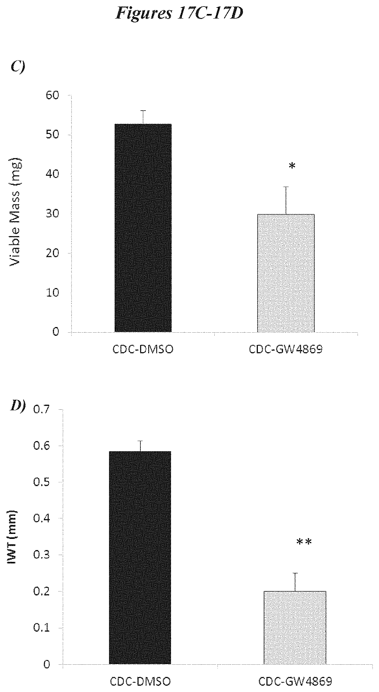
D00025

D00026

D00027

D00028

D00029

D00030

D00031

D00032

D00033

D00034

XML
uspto.report is an independent third-party trademark research tool that is not affiliated, endorsed, or sponsored by the United States Patent and Trademark Office (USPTO) or any other governmental organization. The information provided by uspto.report is based on publicly available data at the time of writing and is intended for informational purposes only.
While we strive to provide accurate and up-to-date information, we do not guarantee the accuracy, completeness, reliability, or suitability of the information displayed on this site. The use of this site is at your own risk. Any reliance you place on such information is therefore strictly at your own risk.
All official trademark data, including owner information, should be verified by visiting the official USPTO website at www.uspto.gov. This site is not intended to replace professional legal advice and should not be used as a substitute for consulting with a legal professional who is knowledgeable about trademark law.