Compositions And Methods For Treating Cancer
Chanteux; Stephanie ; et al.
U.S. patent application number 16/443744 was filed with the patent office on 2020-01-23 for compositions and methods for treating cancer. The applicant listed for this patent is Innate Pharma. Invention is credited to Stephanie Chanteux, Laurent Gauthier, Nicolas Gourdin, Carine Paturel, Ivan Perrot, Benjamin Rossi.
| Application Number | 20200024357 16/443744 |
| Document ID | / |
| Family ID | 67137899 |
| Filed Date | 2020-01-23 |


View All Diagrams
| United States Patent Application | 20200024357 |
| Kind Code | A1 |
| Chanteux; Stephanie ; et al. | January 23, 2020 |
COMPOSITIONS AND METHODS FOR TREATING CANCER
Abstract
The present invention relates to antigen-binding compounds that inhibit the enzymatic activity of soluble human CD39. The invention also relates to cells producing such compounds; methods of making such compounds, and antibodies, fragments, variants, and derivatives thereof; pharmaceutical compositions comprising the same; methods of using the compounds to diagnose, treat or prevent diseases, e.g., cancer.
| Inventors: | Chanteux; Stephanie; (Marseille, FR) ; Gauthier; Laurent; (Marseille, FR) ; Gourdin; Nicolas; (Marseille, FR) ; Paturel; Carine; (Marcy l'Etoile, FR) ; Perrot; Ivan; (Cassis, FR) ; Rossi; Benjamin; (Marseille, FR) | ||||||||||
| Applicant: |
|
||||||||||
|---|---|---|---|---|---|---|---|---|---|---|---|
| Family ID: | 67137899 | ||||||||||
| Appl. No.: | 16/443744 | ||||||||||
| Filed: | June 17, 2019 |
Related U.S. Patent Documents
| Application Number | Filing Date | Patent Number | ||
|---|---|---|---|---|
| 62686165 | Jun 18, 2018 | |||
| Current U.S. Class: | 1/1 |
| Current CPC Class: | C07K 2317/52 20130101; C07K 16/2896 20130101; C07K 2317/92 20130101; C07K 2317/33 20130101; C07K 2317/41 20130101; C07K 2317/76 20130101; C07K 2317/565 20130101; C07K 2317/55 20130101; C07K 2317/74 20130101; C07K 2317/71 20130101; C07K 2317/567 20130101; G01N 33/57492 20130101; C07K 2317/24 20130101; C07K 2317/94 20130101; A61P 35/00 20180101; C07K 2317/34 20130101 |
| International Class: | C07K 16/28 20060101 C07K016/28; A61P 35/00 20060101 A61P035/00; G01N 33/574 20060101 G01N033/574 |
Claims
1. An antibody or antibody fragment that binds a human CD39 polypeptide and that is capable of inhibiting the ATPase activity of a soluble extracellular domain human CD39 polypeptide, wherein the antibody or antibody fragment comprises a heavy chain variable region (VH) comprising the amino acid sequence of SEQ ID NO: 31 and a light chain variable region (VL) comprising the amino acid sequence of SEQ ID NOS: 36 or 37.
2. The antibody or antibody fragment of claim 1, wherein the antibody or antibody fragment comprises a heavy chain comprising an amino acid sequence at least 95% identical to the amino acid sequence of SEQ ID NO: 38 and a light chain comprising an amino acid sequence at least 95% identical to an amino acid sequence selected from the group consisting of SEQ ID NOS: 39 or 40.
3. The antibody or antibody fragment of claim 2, wherein the antibody or antibody fragment comprises heavy chain framework FR1, FR2 and FR3 amino acid sequences from the human IGHV1-3 gene; and light chain framework FR1, FR2 and FR3 amino acid sequences from the human IGKV4-1 gene.
4-5. (canceled)
6. The antibody or antibody fragment of claim 1, wherein the antibody or antibody fragment comprises a heavy chain variable region comprising the amino acid sequence of SEQ ID NO: 31 and a light chain variable region comprising the amino acid sequence of SEQ ID NO: 36.
7. An antibody or antibody fragment, wherein the antibody or antibody fragment comprises a heavy chain comprising the amino acid sequence of SEQ ID NO: 38 and a light chain comprising the amino acid sequence of SEQ ID NO: 39.
8. The antibody or antibody fragment of claim 1, wherein the antibody or antibody fragment comprises a heavy chain variable region comprising the amino acid sequence of SEQ ID NO: 31 and a light chain variable region comprising the amino acid sequence of SEQ ID NO: 37.
9. An antibody or antibody fragment, wherein the antibody or antibody fragment comprises a heavy chain comprising the amino acid sequence of SEQ ID NO: 38 and a light chain comprising the amino acid sequence of SEQ ID NO: 40.
10-33. (canceled)
34. The antibody or antibody fragment of claim 1, wherein the antibody wherein the antibody is an antibody fragment.
35. The antibody or antibody fragment of claim 1, wherein the antibody is an antibody having a human Fc domain that is modified to reduce binding between the Fc domain and a human Fey receptor.
36-37. (canceled)
38. The antibody or antibody fragment of claim 1, wherein the antibody is a full-length antibody.
39. The antibody or antibody fragment of claim 1, wherein the antibody or antibody fragment comprises a modified human IgG1 Fc domain comprising N-linked glycosylation at Kabat residue N297 and comprising an amino acid substitution at Kabat residue(s) 234 and 235.
40. A pharmaceutical composition comprising an antibody claim 1, and a pharmaceutically acceptable carrier.
41. A kit comprising the antibody or antibody fragment of claim 1, and a labeled secondary antibody or antibody fragment that specifically recognizes the antibody or antibody fragment of claim 1.
42. A nucleic acid encoding a heavy and/or light chain of an antibody or antibody fragment of claim 1.
43. A recombinant host cell producing the antibody or antibody fragment of claim 1.
44. A method for the treatment or prevention of cancer in an individual in need thereof, the method comprising administering to the individual an effective amount of an antibody or antibody fragment of claim 1.
45. A method for reducing the ATPase activity of soluble CD39 protein in an individual, the method comprising administering to said patient an effective amount of an antibody or antibody fragment of claim 1.
46. (canceled)
47. A method for increasing T, NK and/or B cell activity in a subject having a cancer, and/or for relieving adenosine-mediated inhibition of T, NK and/or B cell activity in a subject having a cancer, the method comprising administering to said subject an effective amount of an antibody or antibody fragment of claim 1.
48-50. (canceled)
51. A method for the treatment or prevention of a cancer in an individual in need thereof, the method comprising: a) detecting soluble CD39 protein in circulation and/or in a tumor environment, and b) upon a determination that soluble CD39 protein is present in circulation and/or the tumor environment, administering to the individual an antibody or antibody fragment of claim 1.
52-55. (canceled)
56. The method of claim 44, wherein the tumor or cancer is a head and neck squamous cell carcinoma, bladder cancer, ovarian cancer, colorectal carcinoma, melanoma, stomach cancer, esophageal cancer or a breast cancer.
Description
CROSS-REFERENCE TO RELATED APPLICATIONS
[0001] This application claims the benefit of U.S. Provisional Application No. 62/686,165 filed 18 Jun. 2018; which is incorporated herein by reference in its entirety; including any drawings.
REFERENCE TO SEQUENCE LISTING
[0002] The present application is being filed along with a Sequence Listing in electronic format. The Sequence Listing is provided as a file entitled "CD39-9_ST25", created May 27, 2019 which is 72 KB in size. The information in the electronic format of the Sequence Listing is incorporated herein by reference in its entirety.
FIELD OF THE INVENTION
[0003] The present invention relates to antigen-binding compounds (e.g., antibodies) that inhibit the enzymatic activity of soluble human CD39. The invention also relates to cells producing such compounds; methods of making such compounds, and antibodies, fragments, variants, and derivatives thereof; pharmaceutical compositions comprising the same; methods of using the compounds to diagnose, treat or prevent diseases, e.g., cancer.
BACKGROUND
[0004] Eight different ENTPD genes encode members of the NTPDase protein family. The individual NTPDase subtypes differ in cellular location and functional properties. Plasma membrane-bound nucleoside triphosphate diphosphohydrolases control nucleotide levels at the cell surface by hydrolyzing the c and b phosphates of nucleotides.
[0005] NTPDase 1 (ectonucleoside triphosphate diphosphohydrolase1), also known as CD39/ENTPD1 or vascular CD39, functions together with another enzyme, CD73 (ecto-5'-nucleotidase), to hydrolyze extracellular adenosine triphosphate (ATP) and adenosine diphosphate (ADP) to generate adenosine, which binds to adenosine receptors and inhibits T-cell and natural killer (NK)-cell responses, thereby suppressing the immune system. The generation of adenosine via the CD73/CD39 pathway is recognized as a major mechanism of regulatory T cell (Treg) immunosuppressive function. The number of CD39.sup.+ Tregs is increased in some human cancers, and the importance of CD39.sup.+ Tregs in promoting tumor growth and metastasis has been demonstrated using several in vivo models. However, CD39 is also expressed by tumor cells and CD39.sup.+ tumor cells can mediate immunosuppression via the adenosine pathway. CD39 in cancer cells displays ATPase activity and, together with CD73, generates adenosine. CD73.sup.+CD39.sup.+ cancer cells inhibited the proliferation of CD4 and CD8 T cells and the generation of cytotoxic effector CD8 T cells (CTL) in a CD39- and adenosine-dependent manner. CD39 has been reported to be increased in several solid tumors (colorectal cancer, head and neck cancer, pancreatic cancer) as well as in chronic lymphocytic leukemia. Antibodies that bind and inhibit CD39 in CD39-expressing cells are disclosed in WO2009/095478. Antibody "A1" (eBiosciences, Inc.) is used for staining applications and does not exhibit the ability to neutralize CD39 activity in cells. Hayes et al. (2015) Am. J. Transl. Res. 7(6):1181-1188 makes use of an anti-CD39 that binds Fc.gamma.R and has effector function but it is stated to also be blocking. CD39 expression on different cell types, including leukocytes and tumor cells, combined with use of antibodies that either do not actually block CD39 or are not pure blockers, create a complex setting for evaluation of the underlying activity of antibodies. To date, the only reported inhibitor of the CD39 active site remains small molecule non-hydrolysable ATP analogues exemplified by ARL67156, suggesting that direct inhibition of the active site is required. ARL67156 however, is not specific for CD39 and also inhibits other NTPDases such as NTPDase1, NTPDase3, NPP1 or mouse NTPDase8, and furthermore only as a weak competitive inhibitor (Levesque et al. (2007) Br. J. Pharmacol. 152:141-150).
[0006] CD39 has two transmembrane domains near the N- and C-terminal ends, short cytoplasmic N- and C-terminal segments, and a large extracellular domain containing the active site. However, while CD39 is typically anchored to the membrane by the two transmembrane domains at the two ends of the molecule, it has recently also been reported that a soluble catalytically active form of CD39 can be found in circulation in humans and mice (Yegutkin et al., (2012) FASEB J. 26(9): 3875-3883). Despite various anti-CD39 antibodies described, no antibody has been reported to be able to inhibit the ATPase activity of soluble CD39 protein.
SUMMARY OF THE INVENTION
[0007] The inventors have obtained antibodies that inhibit the enzymatic (ATPase activity) activity of soluble (extracellular domain) human CD39 protein. The antibodies additionally bind an epitope present on human CD39 protein expressed at the surface of cells, including tumor cells and potently inhibit the enzymatic (ATPase activity) activity of the cell membrane bound CD39 enzyme (CD39 as expressed at the surface of cells). The antibodies can be used advantageously to achieve greater neutralization of CD39 activity in an individual by neutralizing both membrane-bound and soluble CD39 protein, including soluble CD39 released or shed from tumor cells, thereby reducing immunosuppression, e.g., for the treatment of cancer and/or infectious disease. While other anti-CD39 antibodies have been previously described that inhibit the enzymatic (ATPase activity) activity of the membrane bound CD39 enzyme, those antibodies do not inhibit soluble CD39 protein which is not bound to the cell membrane.
[0008] In one embodiment, provided is an anti-CD39 antigen binding domain, or a protein that comprises such (e.g., an antibody or antibody fragment, a multispecific binding protein, a bispecific antibody, etc.), the antigen binding domain comprising a heavy chain variable region (VH) comprising a CDR1, CDR2 and CDR3 having the respective amino acid sequences shown in SEQ ID NOS: 8, 9 and 10 and framework FR1, FR2 and FR3 amino acid sequences from the human IGHV1-3 gene, e.g., IGHV1-3*01 (and optionally further framework 4 (FR4) amino acid sequences from the human IGHJ1 gene, e.g. IGHJ1*01); and a light chain variable region (VL) CDR1, CDR2 and CDR3 having the respective amino acid sequences shown in SEQ ID NOS: 11, 12 and 13, and framework FR1, FR2 and FR3 amino acid sequences from the human IGKV4-1 (e.g. IGK4-1*01) gene, and optionally further framework 4 (FR4) amino acid sequences from the human IGKJ4 (e.g. IGKJ4*01) gene. In one embodiment, the VH further comprises one or more amino acid substitutions of a residue present in a human framework sequence by a different residue (e.g. a residue present in a non-human framework) at Kabat positions selected from the group consisting of 48, 67, 71 and 76. In one embodiment, the VH comprises one or more amino acid substitutions in the heavy chain CDR2, e.g. at Kabat positions 60 and/or 64. Optionally the residue at position 60 is a serine (e.g. the CDR2 comprises a N60S substitution). Optionally the residue present at Kabat position 64 is a glutamine (e.g. the CDR2 comprises a K64Q substitution). In one embodiment, the residue present in the VL at Kabat position 24 is a lysine (e.g. the CDR1 comprises a R24K substitution). Optionally wherein a phenylalanine is present in the VL at Kabat position 36.
[0009] In one embodiment, provided is an anti-CD39 antigen binding domain, or a protein that comprises the antigen binding domain (e.g., an antibody or antibody fragment, a multispecific binding protein, a bispecific antibody, etc.), the antigen binding domain or protein comprising such antigen binding domain comprises a heavy chain variable region (VH) comprising an amino acid sequence at least 70%, 80%, 85%, 90%, 92%, 94%, 95%, 96%, 97%, 98% or 99% identical to the amino acid sequence of any one of SEQ ID NOS: 27-34, and optionally further comprising one or more amino acid substitutions of a residue present in a human framework sequence by a different residue (e.g. a residue present in a non-human framework) at Kabat positions selected from the group consisting of 48, 67, 71 and 76; and a light chain variable region (VL) comprising an amino acid sequence at least 70%, 80%, 85%, 90%, 92%, 94%, 95%, 96%, 97%, 98% or 99% identical to the amino acid sequence of any one of SEQ ID NO: 35-37, optionally wherein a phenylalanine is present at Kabat position 36. In one embodiment, the VH further comprises one or more amino acid substitutions in the heavy chain CDR2 at Kabat positions 60 and/or 64. Optionally the residue present in the heavy chain at Kabat position 60 is a serine residue. Optionally the residue present in the heavy chain at Kabat position 64 is a glutamine residue. In one embodiment, the VL further comprises an amino acid substitution in the light chain CDR2 at Kabat light chain position 24, optionally further wherein the residue present in the light chain at position 24 is a lysine residue.
[0010] Optionally, the amino acid at Kabat heavy chain position 48 is an isoleucine. Optionally, the amino acid at Kabat heavy chain position 67 is an alanine. Optionally, the amino acid at Kabat heavy chain position 71 is a valine. Optionally, the amino acid at Kabat heavy chain position 76 is an arginine.
[0011] In one embodiment, a VH comprises an alanine residue at Kabat position 67 and a valine at position 71.
[0012] In one embodiment, a VH comprises an isoleucine residue at Kabat position 48, an alanine residue at Kabat position 67, a valine at Kabat position 71 and an arginine at Kabat position 76.
[0013] In one embodiment, a VL comprises a phenylalanine at Kabat position 36 (FR2). In one embodiment, a VL comprises a lysine at Kabat position 24 (CDR1).
[0014] In any embodiment, an anti-CD39 antigen binding domain, or a protein that comprises the antigen binding domain (e.g., a monoclonal antibody or antibody fragment, a multispecific binding protein, a bispecific antibody, etc.), can be characterized as comprising a heavy chain variable region (VH) comprising an amino acid sequence at least 70%, 80%, 85%, 90%, 92%, 94%, 95%, 96%, 97%, 98% or 99% identical to the amino acid sequence of SEQ ID NO: 31, and a light chain variable region (VL) comprising an amino acid sequence at least 70%, 80%, 85%, 90%, 92%, 94%, 95%, 96%, 97%, 98% or 99% identical to the amino acid sequence of SEQ ID NOS: 36 or 37.
[0015] In one embodiment, provided is an anti-CD39 antigen binding domain or a protein that comprises the antigen binding domain (e.g., a monoclonal antibody or antibody fragment, a multispecific binding protein, a bispecific antibody, etc.), the antigen binding domain comprising a heavy chain variable region (VH) comprising CDR1, CDR2 and CDR3 having the respective amino acid sequences shown in SEQ ID NOS: 8, 9 and 10 and human frameworks (e.g., FR1, FR2, FR3 and FR4 of human origin); and a light chain variable region (VL) CDR1, CDR2 and CDR3 comprising the respective amino acid sequences shown in SEQ ID NOS: 11 (or 17 or 18), 12 and 13 and human frameworks (e.g., FR1, FR2, FR3 and FR4 of human origin), wherein the (VH) comprises an amino acid sequence at least 70%, 80%, 85%, 90%, 92%, 94%, 95%, 96%, 97%, 98% or 99% identical to the amino acid sequence of SEQ ID NOS: 31 or 6, and a light chain variable region (VL) comprising an amino acid sequence at least 70%, 80%, 85%, 90%, 92%, 94%, 95%, 96%, 97%, 98% or 99% identical to the amino acid sequence of any of SEQ ID NOS: 36 or 37.
[0016] In any embodiment, a VH can be characterized as comprising a substitution at one, two, three or all of the Kabat positions 48, 67, 71 and 76. In one embodiment, the residue at position 48 is an isoleucine (e.g., a M481 substitution). In one embodiment, the residue at position 67 is an alanine (e.g., a V67A substitution). In one embodiment, the residue at position 71 is a valine (e.g., a R71V substitution). In one embodiment, the residue at position 76 is an arginine (e.g., a S76R substitution). In any embodiment, a VL can be characterized as comprising a substitution at Kabat position 36. In one embodiment, the residue at position 36 is a phenylalanine (e.g., a Y36F substitution).
[0017] In one embodiment, the VH comprises human VH framework amino acid sequences and the VL comprises human VL framework amino acid sequences. In one embodiment, the VH segment of the VH human acceptor framework is from a human IGHV1-3 gene segment, optionally further wherein the J-segment is from a human IGHJ1 gene segment. In one embodiment, the VH human framework is from a human IGHV1-3*01 gene segment. In one embodiment, the VL domain human acceptor framework is from a human IGKV4-1 gene segment, optionally further wherein the J-segment is from a human IGKJ4 gene segment.
[0018] In one embodiment, provided is an anti-CD39 antigen binding domain, or a protein that comprises the antigen binding domain (e.g., a monoclonal antibody, a multispecific binding protein, a bispecific antibody, etc.), the antigen binding domain selected from the group consisting of:
[0019] (a) an antibody binding domain comprising a light chain variable region comprising the amino acid sequence of SEQ ID NO: 36 and a heavy chain variable region comprising an amino acid sequence selected from the group consisting of the amino acid sequences of SEQ ID NOS: 29, 30, 31, 32, 33 or 34; and
[0020] (b) an antibody binding domain comprising a light chain variable region comprising the amino acid sequence of SEQ ID NO: 37 and a heavy chain variable region comprising an amino acid sequence selected from the group consisting of the amino acid sequences of SEQ ID NOS: 29, 30, 31, 32, 33 or 34.
[0021] In any embodiment, an antibody heavy chain comprises a human CH1 constant domain and a human Fc domain, optionally of human IgG1 isotype, optionally further comprising an amino acid sequence of any one of SEQ ID NOS: 23, 24, 25 or 26. In any embodiment, an antibody light chain comprises a human light chain constant domain, optionally wherein the constant domain is a human kappa domain.
[0022] In one embodiment, provided is an anti-CD39 antibody comprising a heavy chain comprising an amino acid sequence at least 70%, 80%, 85%, 90%, 92%, 94%, 95%, 96%, 97%, 98% or 99% identical to the amino acid sequence of SEQ ID NO: 38, and a light chain comprising an amino acid sequence at least 70%, 80%, 85%, 90%, 92%, 94%, 95%, 96%, 97%, 98% or 99% identical to the amino acid sequence of SEQ ID NO: 39.
[0023] In one embodiment, provided is an anti-CD39 antibody comprising a heavy chain comprising an amino acid sequence at least 70%, 80%, 85%, 90%, 92%, 94%, 95%, 96%, 97%, 98% or 99% identical to the amino acid sequence of SEQ ID NO: 38, and a light chain comprising an amino acid sequence at least 70%, 80%, 85%, 90%, 92%, 94%, 95%, 96%, 97%, 98% or 99% identical to the amino acid sequence of SEQ ID NO: 40.
[0024] In one embodiment, provided is an anti-CD39 antibody or antibody fragment comprising a heavy chain comprising the amino acid sequence of SEQ ID NO: 38 and a light chain comprising the amino acid sequence of SEQ ID NO: 39.
[0025] In one embodiment, provided is an anti-CD39 antibody or antibody fragment comprising a heavy chain comprising the amino acid sequence of SEQ ID NO: 38 and a light chain comprising the amino acid sequence of SEQ ID NO: 40.
[0026] In any embodiment, an antigen binding domain or a protein comprising such, optionally an antibody or antibody fragment, can be characterized as binding to and inhibiting or neutralizing the ATPase activity of a soluble CD39 protein (sCD39). In one embodiment the sCD39 protein lacks the two transmembrane domains (i.e. the transmembrane domains near the N- and C-terminal ends) found in membrane bound CD39. In one embodiment, sCD39 is a non-membrane bound sCD39 protein found in circulation, e.g., in a human individual. In one embodiment, sCD39 comprises or consists of the amino acid sequence of SEQ ID NO: 43, optionally further comprising a C-terminal tag or another non-CD39-derived amino acid sequence; optionally wherein the amino acid sequence of SEQ ID NO: 43 further lacks at its N-terminal residues 1 to 37 of the sequence of SEQ ID NO: 1. The sCD39 protein be characterized as comprising or consisting of the Thr38-Val478 fragment of CD39. Thr38-Val478 protein with C-terminal His tag is available commercially from R&D Systems, Inc., (product number 4397-EN). In one embodiment, the protein, antibody or antibody fragment inhibits the ATPase activity of sCD39 when incubated with sCD39 in solution, e.g., according to the methods or assays conducted in the absence of cells as disclosed herein (see, e.g. Examples, Methods), e.g. in tumor cell supernatants. In one embodiment, the protein, antibody or antibody fragment specifically binds the human CD39 protein, both in soluble (extracellular domain protein) and in membrane-bound form.
[0027] Without wishing to be bound by theory, some antibodies may neutralize membrane-bound CD39 by inhibiting the domain motion of membrane-bound CD39 (memCD39), however without similarly affecting the activity of the soluble CD39 protein (sCD39). It has been reported that memCD39 occurs as a homo-multimer while sCD39 is a monomer, and moreover that the transmembrane domains in memCD39 undergo dynamic motions that underlie a functional relationship with the active site. Consequently, unlike sCD39, memCD39 may present a setting that makes antibody-mediated neutralization possible. One possibility is that use of a bivalent antibody that binds simultaneously to two memCD39 molecules (e.g., within a memCD39 homo-multimer) is required for functional neutralization.
[0028] The present antibodies that neutralize the activity of sCD39 (and memCD39) may, in addition to use as bivalent binders, also be effective as monovalent binders, whether they are targeting memCD39 in addition to sCD39. Consequently, in one embodiment, provided is an antigen binding protein that binds monovalently to a human CD39 protein (sCD39 and/or memCD39) and neutralizes the enzymatic (ATPase) activity thereof. The antigen binding protein can optionally be specified as binding to a single CD39 protein and/or bearing a single antigen binding domain capable of binding to a CD39 protein. In one embodiment, provided is an antibody fragment, optionally a F(ab) fragment, a single chain antibody, a scFv, a multispecific antibody, that binds monovalently to a human CD39 protein (sCD39 and/or memCD39) and neutralizes the enzymatic (ATPase) activity thereof. In one embodiment, a CD39-neutralizing antigen binding protein that binds monovalently to a human CD39 protein is a multi-specific antigen binding protein, e.g., a multi-specific antibody, a bi-specific antibody, a tri-specific antibody, etc. In one embodiment, a CD39-neutralizing antigen binding protein that binds monovalently to a human CD39 protein comprises a first (or a single) antigen binding domain that binds CD39 (sCD39 and/or memCD39) and a second antigen binding domain that binds a protein other than CD39.
[0029] Advantageously, in one embodiment the antibody comprises a human Fc domain that is modified to have decreased or substantially lack binding to a human Fc.gamma. receptor, e.g., one or more (or all of) human CD16, CD32a, CD32b and CD64. In one aspect, the antibodies do not depend on ADCC-, CDC- or toxin-mediated depletion of CD39-expressing cells for their CD39 inhibitory activity. These antibodies can be used as "pure" CD39 blockers, permitting immunomodulatory activity.
[0030] In alternative embodiment, the binding molecule can be produced such that it retains and/or mediates effector function via its Fc domain. In one embodiment the antibody comprises a human Fc domain that binds to a human Fc.gamma. receptor, e.g., one or more (or all of) human CD16, CD32a, CD32b and CD64.
[0031] In another embodiment, the Fc domain can be modified to reduce Fc.gamma. receptor binding, optionally by retaining binding to one or more human Fc.gamma. receptor(s) but having decreased binding to one or more other human Fc.gamma. receptor(s).
[0032] In one aspect, the antibodies specifically bind vascular CD39, e.g., the antibody binds a polypeptide having the sequence of SEQ ID NO: 1 but not does bind a secreted CD39 isoform polypeptide, e.g., a CD39-L2 and/or -L4 polypeptide. Optionally, the anti-CD39 antibody specifically binds vascular CD39, e.g., the antibody binds a polypeptide having the sequence of SEQ ID NO: 1 but not does bind a membrane bound CD39 isoform, e.g., CD39-L1 and/or -L3 polypeptide.
[0033] The antibodies of the disclosure can inhibit the enzymatic activity of membrane-bound CD39 protein expressed at the surface of cells.
[0034] In one aspect, the antibodies do not depend on CD39 down-modulation for their CD39 inhibitory activity.
[0035] The antibodies of the disclosure can in addition to inhibiting soluble CD39 be capable of inhibiting the enzymatic activity of membrane-bound CD39 protein expressed at the surface of cells, with or without induction of CD39 internalization, and with or without binding of CD16 (Fc.gamma.III receptor) and/or with or without substantially directing ADCC and/or CDC toward a CD39-expressing cell. Optionally, the antibodies retain an Fc domain and retain binding to human FcRn.
[0036] While antibodies that function by inducing ADCC and/or CDC may be efficient even without complete neutralization/inhibition of the ATPase activity of CD39, as long as enough antibody is bound to a CD39-expressing cell to induce ADCC, neutralizing non-depleting antibodies may require stronger inhibition of the enzymatic activity of ATPase. In one embodiment, a non-depleting antibody will provide an at least 50%, 60%, 70%, 80% or 90% reduction in the ATPase activity of a soluble CD39 protein (e.g., as assessed by the methods disclosed herein), optionally further at a concentration compatible with administration of an antibody to a human. In one embodiment, a non-depleting antibody will provide an at least 70%, 80%, 90% reduction in the ATPase activity of a CD39-expressing cell (e.g., as assessed by decrease in AMP generation by a CD39+ cell such as a B cell, a Ramos cell, as measured by the methods disclosed herein), optionally further at a concentration compatible with administration of an antibody to a human.
[0037] The epitope on CD39 bound by the antibodies is present on CD39 polypeptides as expressed by a range of cells, e.g., cancer cells, CD4 T cells, CD8 T cells, B cells, transfected cells, and binds with high affinity as determined by flow cytometry.
[0038] An antibody can optionally be characterized by an EC.sub.50, as determined by flow cytometry, of no more than 2 .mu.g/ml, no more than 1 .mu.g/ml, no more than 0.5 .mu.g/ml, no more than 0.1 .mu.g/ml or no more than 0.05 .mu.g/ml, for binding to cells that express at their surface a CD39 polypeptide. In one embodiment the cells are cells that are made to express CD39 at their surface. In one embodiment the cells are cells that endogenously express CD39 at their surface, e.g., regulatory T (TReg) cells, B cells, cancer cells, lymphoma cells (e.g., Ramos cells), leukemia cells, bladder cancer cells, glioma cells, glioblastoma cells, ovarian cancer cells, melanoma cells, prostate cancer cells, thyroid cancer cells, esophageal cancer cells or breast cancer cells.
[0039] In one aspect, an anti-CD39 antibody is capable of: (a) inhibiting the enzymatic activity of membrane-bound CD39 protein (e.g., comprising an amino acid sequence of SEQ ID NO: 1) expressed at the surface of cells, and (b) inhibiting the enzymatic activity of soluble CD39 protein. In one embodiment, the antibodies do not substantially bind (e.g., via their Fc domain) to human Fc.gamma. receptors (e.g., CD16, CD32a, CD32b, CD64) and/or C1q, and/or do not substantially directing ADCC and/or CDC toward a CD39-expressing cell. Optionally, the antibodies retain an Fc domain and retain binding to human FcRn.
[0040] In one embodiment, the antibodies are administered in an amount effective to neutralize the enzymatic activity of sCD39 and/or memCD39 for a desired period of time, e.g., 1 week, 2 weeks, a month, until the next successive administration of anti-CD39 antibody.
[0041] In one embodiment, the antibodies are administered at a dosage and/or frequency that provides a blood concentration of antibody equal to at least the EC.sub.50, EC.sub.70 or EC.sub.100 for inhibition of ATPase activity of sCD39 protein, optionally wherein the concentration is maintained for at least 1 week, 2 weeks, a month, or until the next successive administration of the anti-CD39 antibody.
[0042] In one aspect, the antibody binds an epitope on CD39 comprising an amino acid residue (e.g., one, two or three of the residues) selected from the group consisting of R138, M139 and E142 (with reference to SEQ ID NO: 1).
[0043] In one aspect, an anti-CD39 antibody exhibits reduced binding (e.g. substantially complete loss of binding) to a CD39 polypeptide having a mutation at one, two or three of the residues selected from the group consisting of: R138, M139 and E142 (with reference to SEQ ID NO: 1), compared to a wild-type CD39 polypeptide (a CD39 polypeptide of SEQ ID NO: 1); optionally, the mutant CD39 polypeptide has the mutations: R138A, M139A and E142K. In one optional aspect, the antibody does not have a loss of binding to any of the mutant CD39 polypeptide of Table 1 other than mutant 19.
[0044] In one embodiment, the CD39 neutralizing antibodies can be characterized by being capable, in purified form, of causing a decrease in the ATPase activity of sCD39 protein in a cell-free assay (e.g. sCD39 from tumor cell culture supernatants), optionally causing a decrease of AMP generation by sCD39, by at least 70%, 80% or 90%; optionally causing an increase in ATP present (compared to a negative control), e.g., as assessed in the assays disclosed herein. For example sCD39 inhibition can be assessed by quantifying luminescence units which are proportional to the amount of ATP present following incubation with anti-CD39 antibody. In one embodiment, the CD39-neutralizing antibodies can be characterized by an EC.sub.50 for inhibition of ATPase activity of sCD39 protein of no more than 1 .mu.g/ml, optionally no more than 0.5 .mu.g/ml, optionally no more than 0.1 .mu.g/ml.
[0045] Optionally, inhibition of ATPase activity of sCD39 protein is determined by quantifying luminescence units using the Cell Titer Glo.TM. (Promega), in a cell-free version of the assay in which dose ranges of test antibody are incubated with soluble recombinant human CD39 protein described in Examples, Methods, for 1 hour at 37.degree. C., where 20 .mu.M ATP is added to the plates for 30 additional minutes at 37.degree. C. before addition of Cell Titer Glo.TM. (CTG) reagent, and emitted light is quantified using an Enspire.TM. luminometer after incubation for 5 minutes in the dark (see, e.g., Examples, Methods).
[0046] In one embodiment, the sCD39 protein is shed sCD39 protein found in or obtained from human tumor cell culture supernatants, optionally from a tumor cell line that expresses CD39 at a high level, optionally from Ramos tumor cells.
[0047] Optionally, the CD39 neutralizing antibodies can further be characterized by being capable, in purified form, of causing a decrease in cells' ATPase activity of CD39, optionally causing a decrease of AMP generation by a CD39-expressing cell, by at least 70%, 80% or 90%. In one embodiment, the CD39-neutralizing antibodies can be characterized by an EC.sub.50 for inhibition of ATPase activity (e.g., EC.sub.50 for inhibition of AMP generation by a CD39-expressing cell) of CD39 expressed by a cell of no more than 1 .mu.g/ml, optionally no more than 0.5 .mu.g/ml, optionally no more than 0.1 .mu.g/ml.
[0048] Optionally, inhibition of ATPase activity of CD39 expressed by a cell is determined by assessing neutralization of ATPase activity in Ramos cells by quantifying AMP generated by hydrolysis of ATP (see, e.g., Examples, Methods).
[0049] In one aspect, neutralization of the ATPase activity by a CD39-expressing cell is determined by bringing CD39-expressing cells (e.g., Ramos lymphoma cells as used herein, available for example from the ATCC, reference CRL-1596) into contact with an antibody, and assessing production of AMP, e.g., by mass spectrometry, wherein a decrease in AMP generated indicates neutralization of ATPase activity. Optionally an antibody causes a decrease of AMP generated by at least 70%, 80% or 90% in this assay. Optionally an antibody causes a decrease of extracellular ATPase activity by a B cell of at least 70%, 80% or 90%.
[0050] In one aspect, a neutralizing anti-CD39 antibody binds an antigenic determinant present on both sCD39 and CD39 expressed at the cell surface (memCD39).
[0051] In one aspect a neutralizing anti-CD39 antibody competes for binding to an epitope on CD39 bound by antibody mAb20, mAb21 (or their parental I-394 antibody), (e.g., that competes for binding to an epitope on a CD39 polypeptide with an antibody having the heavy and light chain CDRs or variable regions of any of mAb20, mAb21 or I-394).
[0052] In one aspect of any of the embodiments herein, an antigen-binding compound binds the same epitope and/or competes for binding to a CD39 polypeptide with monoclonal antibody mAb20, mAb21 (or their parental I-394 antibody) (e.g., that binds the same epitope and/or competes for binding to a CD39 polypeptide with an antibody having the heavy and light chain CDRs or variable regions of mAb20, mAb21 (or I-394)). In one embodiment, an antigen-binding compound binds the same epitope and/or competes for binding to a CD39 polypeptide with an antibody having respectively a VH and VL region of SEQ ID NOS: 38 and 39.
[0053] In one embodiment, an anti-CD39 antibody binds an epitope comprising one, two or three amino acid residues selected from the group consisting of the amino acid residues on CD39 bound by mAb20, mAb21 (or I-394).
[0054] In any embodiment, the binding molecule (e.g., antibody or antibody fragment) comprises the variable heavy chain domain (V.sub.H) comprising a heavy chain CDR1, 2 and 3 (e.g., as described herein) for antibody I-394, and a variable light chain domain (V.sub.L) comprising a light chain CDR1, 2 and 3 (e.g., as described herein) for the respective I-394 antibody, or an amino acid sequence in which the CDR (or set of heavy and/or light chain CDRs) has at least 70%, 80%, 90% or 95% amino acid identity to said CDR (or said set of heavy and/or light chain CDRs), wherein the VH and VL each comprise framework domains of human origin (e.g. the VH and VL are different from the respective VH and VL of SEQ ID NOS: 6 and 7). Optionally, CDRs are determined according to Kabat or IMGT numbering schemes.
[0055] In one aspect, an antibody or antibody fragment comprising protein comprises an Fc domain that is modified (compared to a wild-type Fc domain of the same isotype) to reduce binding between the Fc domain and human CD16A, CD16B, CD32A, CD32B and/or CD64 polypeptides, wherein the antibody comprises: (i) a heavy chain comprising CDR 1, 2 and 3 of the heavy chain variable region of SEQ ID NO: 31, and (ii) a light chain comprising CDR 1, 2 and 3 of the light chain variable region of SEQ ID NO: 36 or 37. In one aspect, the Fc domain is modified (compared to a wild-type Fc domain of the same isotype) to reduce binding between the Fc domain and human C1q polypeptide. In one embodiment, the antibody comprises an amino acid substitution in a heavy chain constant region at any one, two, three, four, five or more of residues selected from the group consisting of: 220, 226, 229, 233, 234, 235, 236, 237, 238, 243, 264, 268, 297, 298, 299, 309, 310, 318, 320, 322, 327, 330 and 331 (Kabat EU numbering). In one embodiment, the antibody has an amino acid substitution in a heavy chain constant region at any three, four, five or more of residues selected from the group consisting of: 234, 235, 237, 322, 330 and 331.
[0056] In one embodiment, the antibodies are administered to an individual having a cancer in an amount and frequency sufficient to neutralize the activity of sCD39 in the tumor microenvironment and/or in circulation. In one embodiment, the antibodies are administered in an amount and frequency sufficient to decrease the generation and/or concentration of adenosine in the tumor microenvironment. In one embodiment, the antibodies are administered in an amount and frequency sufficient to decrease the generation and/or concentration of AMP in the tumor microenvironment. In one embodiment, the antibodies are administered in an amount and frequency sufficient to neutralize the activity of CD39 expressed by tumor cells. In one embodiment, the antibodies are administered in an amount and frequency sufficient to neutralize the activity of CD39 expressed by leukocytes or lymphocytes, e.g., CD4 T cells, CD8 T cells, TReg cells and/or B cells.
[0057] The antibodies will be useful in inhibiting CD39-mediated ATP hydrolysis, e.g., thereby leading to a decrease in the concentration of adenosine in the tumor microenvironment and/or in circulation. These antibodies will therefore be useful in reversing the immunosuppressive effect of CD39 and/or adenosine on T cells, B cells and other cells that express adenosine receptors (A2A receptors), for example in the treatment of cancer. In one embodiment, the anti-CD39 antibody neutralizes adenosine-mediated inhibition of proliferation, cytokine production, cytotoxicity and/or NF.kappa.B activity in T cells.
[0058] In another aspect provided is a method for treating an individual, the method comprising administering to an individual (e.g., an individual having a disease, a tumor, etc.) a therapeutically active amount of any of the anti-CD39 antigen binding compounds described herein.
[0059] The antibodies will be useful in inhibiting the production, amounts and/or concentrations of adenosine into the tumor microenvironment and/or in circulation, including but not limited to tumors characterized by detectable, significant, increased or elevated adenosine generation, ATP catabolism or catabolic activity of the CD39/CD73 axis (e.g. compared to a reference value). Furthermore, at increasing concentrations, the antibodies that neutralize soluble CD39 provide substantially complete inhibition of the catabolic activity of the CD39/CD73 axis. In one embodiment, the antibodies will be useful in inhibiting the production, amounts and/or concentrations of adenosine into the tumor microenvironment in tumors characterized by the presence of CD73 protein (e.g. tumors with soluble CD73 and/or CD73 expressing cells; CD73-positive tumors).
[0060] In one embodiment, the antibodies of the disclosure that neutralize soluble CD39 protein can advantageously be used in combination with CD73 blockade, e.g., the antibodies of the disclosure can be administered to an individual having a cancer in combination with an agent that inhibits the activity of CD73.
[0061] In one aspect provided is a method for treating an individual, the method comprising, consisting essentially of or consisting of: administering to an individual (e.g., an individual having a disease, a tumor, etc.) a therapeutically active amount of an antigen binding compound of the disclosure that inhibits a CD39 polypeptide. In one embodiment, the anti-CD39 antigen binding compound (e.g., antibody) is administered to an individual in combination with a second therapeutic agent. In one embodiment, the second therapeutic agent is an agent (e.g., antibody) that neutralizes the 5'-ectonucleotidase activity of human CD73. In one embodiment, the second therapeutic agent is an agent (e.g., antibody) that neutralizes the inhibitory activity of human PD-1, optionally an anti-PD-1 antibody, optionally an anti-PD-L1 antibody. In one embodiment, the second therapeutic agent comprises an agent or treatment (e.g., a chemotherapeutic agent, a taxane, an anthracycline, a camptothecin, an epothilones, a mytomycin, a combretastatin, a vinca alkaloid, a nitrogen mustard, a maytansinoids, a calicheamycin, a duocarmycin, a tubulysin, a dolastatin or auristatin, an enediyne, an amatoxin, a pyrrolobenzodiazepine, an ethylenimine, a radioisotope, a therapeutic protein or peptide, or a toxin) that induces the extracellular release of ATP from tumor cells and/or induces the death of tumor cells.
[0062] In one embodiment, the anti-CD39 antigen binding compound (e.g., antibody) is administered to an individual having a cancer and who has a poor response, or prognostic for response, to treatment with an agent that neutralizes the inhibitory activity of human PD-1. In one embodiment, the antibody inhibits a CD39 polypeptide in a cellular assay. The compound is in one embodiment a non-depleting antibody (an antibody that does not deplete cells to which it binds, e.g., an Fc silent antibody). Optionally, the compound binds to CD39 polypeptides in bivalent manner. Optionally, the antibody is a chimeric, humanized or human antibody. Optionally, the antibody comprises a heavy chain constant region of IgG (e.g., IgG1) isotype modified to eliminate binding to human Fc.gamma. receptors (e.g., CD16A, CD16B, CD32A, CD32B and/or CD64).
[0063] In one aspect provided is a method for decreasing ATP hydrolysis by a CD39-expressing cell (e.g., a leukocyte and/or a tumor cell in an individual), or a method for neutralizing of the enzymatic activity of cellular CD39, the method comprising: bringing the CD39-expressing cell into contact with an antigen binding compound (e.g. antibody or antibody fragment) of the disclosure that inhibits CD39. In one embodiment, the step of bringing the CD39-expressing cell into contact with an antigen binding compound of the disclosure comprises administering to an individual a therapeutically active amount of an antigen binding compound that inhibits CD39. In one embodiment the individual has a cancer.
[0064] In one aspect provided is a method for decreasing adenosine present in the tumor environment (e.g., in an individual), the method comprising, consisting essentially of or consisting of: administering to an individual a therapeutically active amount of an antigen binding compound (e.g. antibody or antibody fragment) of the disclosure that inhibits a CD39 polypeptide. In one embodiment the individual has a cancer.
[0065] In one embodiment, the active amount of an antibody that inhibits a CD39 polypeptide is an amount effective to achieve and/or maintain (e.g., until the subsequent administration of antigen binding compound) a blood concentration of at least the EC.sub.50, optionally the EC.sub.70, optionally substantially the EC.sub.100, for inhibition of CD39-mediated catabolism of ATP to AMP in an individual. In one embodiment, the active amount of an antigen binding compound that inhibits a CD39 polypeptide is an amount effective to achieve the EC.sub.50, optionally the EC.sub.70, optionally substantially the EC.sub.100, for inhibition of CD39-mediated catabolism of ATP to AMP in an extravascular tissue of an individual. In one embodiment, the active amount an antigen binding compound that inhibits a CD39 polypeptide is an amount effective to achieve the EC.sub.50, optionally the EC.sub.70, optionally substantially the EC.sub.100, for inhibition of CD39-mediated catabolism of ATP to AMP in an individual. In one embodiment, the active amount of an antigen binding compound that inhibits a CD39 polypeptide is between 1 and 20 mg/kg body weight. In one embodiment, the active amount is administered to an individual weekly, every two weeks, monthly or every two months.
[0066] Optionally the individual is a human having or who is susceptible to having a cancer. Optionally the individual is a human having or who is susceptible to having a cancer characterized by malignant cells that express CD39 and/or presence (secretion or shedding) or soluble CD39 protein. Optionally the individual is a human having or who is susceptible to having a cancer and who has detectable levels of circulating soluble extracellular CD39 protein or tumor-infiltrating leukocytes that express CD39.
[0067] The antibodies are optionally characterized by binding affinity (K.sub.D) for a human CD39 polypeptide of less than (better than) 10.sup.-9 M, preferably less than 10.sup.-10 M, or preferably less than 10.sup.-11M, and/or by binding human CD39 with an EC.sub.50 lower than (better binding than) 1 .mu.g/ml, preferably wherein the antibody has an EC.sub.50 of no more than 0.5 .mu.g/ml, optionally no more than 0.2 .mu.g/ml, optionally no more than 0.1 .mu.g/ml, for binding to cells (e.g., tumor cells) expressing human CD39 at the cell surface.
[0068] The antibodies are optionally chimeric, human or humanized antibodies.
[0069] The antibodies are optionally characterized by an EC.sub.50 for neutralization of the enzymatic activity of CD39 in CD39-expressing cells (e.g., Ramos tumor cells) of less than (better than) 1 .mu.g/ml, optionally less than 0.5 .mu.g/ml.
[0070] In one embodiment, the antibody is a monoclonal antibody or a fragment thereof that retains binding specificity and ability to neutralize the enzymatic activity of CD39. In one embodiment, the antibody is an IgG1 antibody. For example, the antibody may be an antibody comprising an Fc domain of human IgG1 isotype modified to reduce binding between the Fc domain and an Fc.gamma. receptor (e.g., CD16). In one embodiment, the antibody or a fragment lacks an Fc domain or comprises an Fc domain that does not induce antibody mediated cellular cytotoxicity (ADCC) and/or CDC; optionally the antibody or a fragment thereof comprises an Fc domain that does not bind to a Fc.gamma.RIIIA (CD16) polypeptide. In one embodiment, the Fc domain (e.g., of human IgG1, IgG2, IgG3 or IgG4 isotype) comprises an amino acid modification (e.g., substitution) compared to a wild-type Fc domain, wherein the substitution reduces the ability of the Fc domain (or antibodies containing it) to bind to an Fc.gamma. receptor (e.g., CD16) and/or to bind complement. In one embodiment, the antibody or a fragment thereof is not linked to a toxic moiety.
[0071] Also provided are nucleic acids encoding the humanized antibody or antibody fragment having any of the foregoing properties, a vector comprising such a nucleic acid, a cell comprising such a vector, and a method of producing a human anti-CD39 antibody, comprising culturing such a cell under conditions suitable for expression of the anti-CD39 antibody, and optionally recovering or purifying the produced antibody. The disclosure also relates to compositions, such as pharmaceutically acceptable compositions and kits, comprising such proteins, nucleic acids, vectors, and/or cells and typically one or more additional ingredients that can be active ingredients or inactive ingredients that promote formulation, delivery, stability, or other characteristics of the composition (e.g., various carriers). The disclosure further relates various new and useful methods making and using such antibodies, nucleic acids, vectors, cells, organisms, and/or compositions, such as in the modulation of CD39-mediated biological activities, for example in the treatment of diseases related thereto, notably cancers.
[0072] The disclosure also provides a method of potentiating the activity of lymphocytes (e.g., T cells) in a subject in need thereof, or for restoring the activity of lymphocytes (e.g., T cells), or a method of relieving the adenosine-mediated inhibition of lymphocytes (e.g., T cells), which method comprises administering to the subject an effective amount of any of the foregoing compositions. In one embodiment, the subject is a patient suffering from cancer. For example, the patient may be suffering from a solid tumor, e.g., colorectal cancer, renal cancer, ovarian cancer, lung cancer, breast cancer or malignant melanoma. Alternatively, the patient may be suffering from a hematopoietic cancer, e.g., acute myeloid leukaemia, chronic myeloid leukaemia, multiple myeloma, or non-Hodgkin's lymphoma.
[0073] The disclosure also provides a method for treatment of disease in an individual, the treatment comprising administering to the individual an anti-CD39 antibody that neutralizes the enzymatic activity of CD39 for at least one administration cycle in which the anti-CD39 antibody is administered at least once, optionally at least twice, in an amount effective to achieve, and/or to maintain between two successive administrations of the anti-CD39 antibody, a concentration in blood (serum) or an extravascular tissue (e.g., tumor environment) that corresponds to at least the EC.sub.50 (e.g., an EC.sub.50 between 0.01 and 0.5 .mu.g/ml), optionally the EC.sub.70 or optionally the EC.sub.100, for neutralization of the enzymatic activity of CD39 (e.g., an EC.sub.100 between 0.05 and 1 .mu.g/ml, between 0.1 and 1 .mu.g/ml). The antibody can for example be administered in an amount to achieve and/or maintained a concentration in circulation or in an extravascular tissue (e.g., tumor environment) of at least about 0.1 .mu.g/ml, 0.5 .mu.g/ml, 1 .mu.g/ml or 2 .mu.g/ml). For example, to achieve a concentration in an extravascular tissue of between 0.05 and 1 .mu.g/ml, or between 0.1 and 1 .mu.g/ml, the anti-CD39 antibody is administered in amounts effective to achieve a concentration in circulation of the anti-CD39 antibody of between 0.5 and 10 .mu.g/ml, or between 1 and 10 .mu.g/ml. Optionally, the anti-CD39 antibody is administered at least twice and in amounts effective to maintain the concentration of the anti-CD39 antibody at least the aforementioned concentration for at least 1 week, 2 weeks, 3 weeks, 4 weeks, between two successive administrations of the anti-CD39 antibody and/or throughout the administration cycle.
[0074] The disclosure also provides a method for treatment of disease in an individual, the treatment comprising administering to the individual an anti-CD39 antibody that neutralizes the enzymatic activity of CD39 for at least one administration cycle in which the anti-CD39 antibody is administered at least once, optionally at least twice, in an amount effective to achieve, and/or to maintain between two successive administrations of the anti-CD39 antibody, a blood or tissue concentration of anti-CD39 antibody of at least 1 .mu.g/ml, optionally at least 10 .mu.g/ml, optionally between 1 and 100 .mu.g/ml. Optionally, the anti-CD39 antibody is administered at least twice and in amounts effective to maintain a continuous blood or tissue concentration of the anti-CD39 antibody of at least 1 .mu.g/ml, optionally at least 10 .mu.g/ml, optionally between 1 and 100 .mu.g/ml, for at least 1 week, 2 weeks, 3 weeks, 4 weeks, between two successive administrations of the anti-CD39 antibody and/or throughout the administration cycle.
[0075] These aspects are more fully described in, and additional aspects, features, and advantages will be apparent from, the description provided herein.
BRIEF DESCRIPTION OF THE DRAWINGS
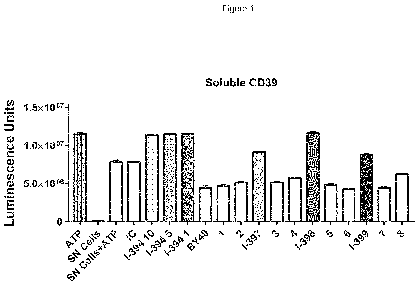
[0076] FIG. 1 shows a representative screening result, showing antibodies I-397, I-398 and I-399 compared to positive control I-394 antibody.
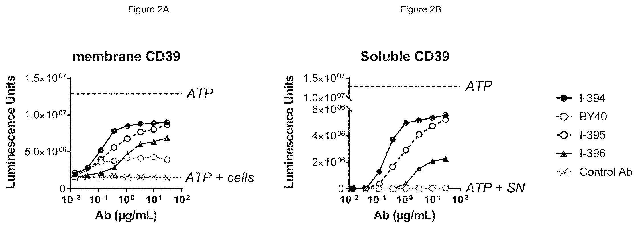
[0077] FIG. 2A shows that antibodies BY40, I-394, 1-395 and I-396 inhibit cell-membrane bound CD39, with both I-394 and 1-395 showing greater potency at all concentrations as well as greater maximal inhibition of cellular CD39 compared to BY40. FIG. 2B shows that antibodies I-395 and I-396 both inhibit soluble CD39 in comparison to negative control (BY40) and positive control (I-394) antibodies.
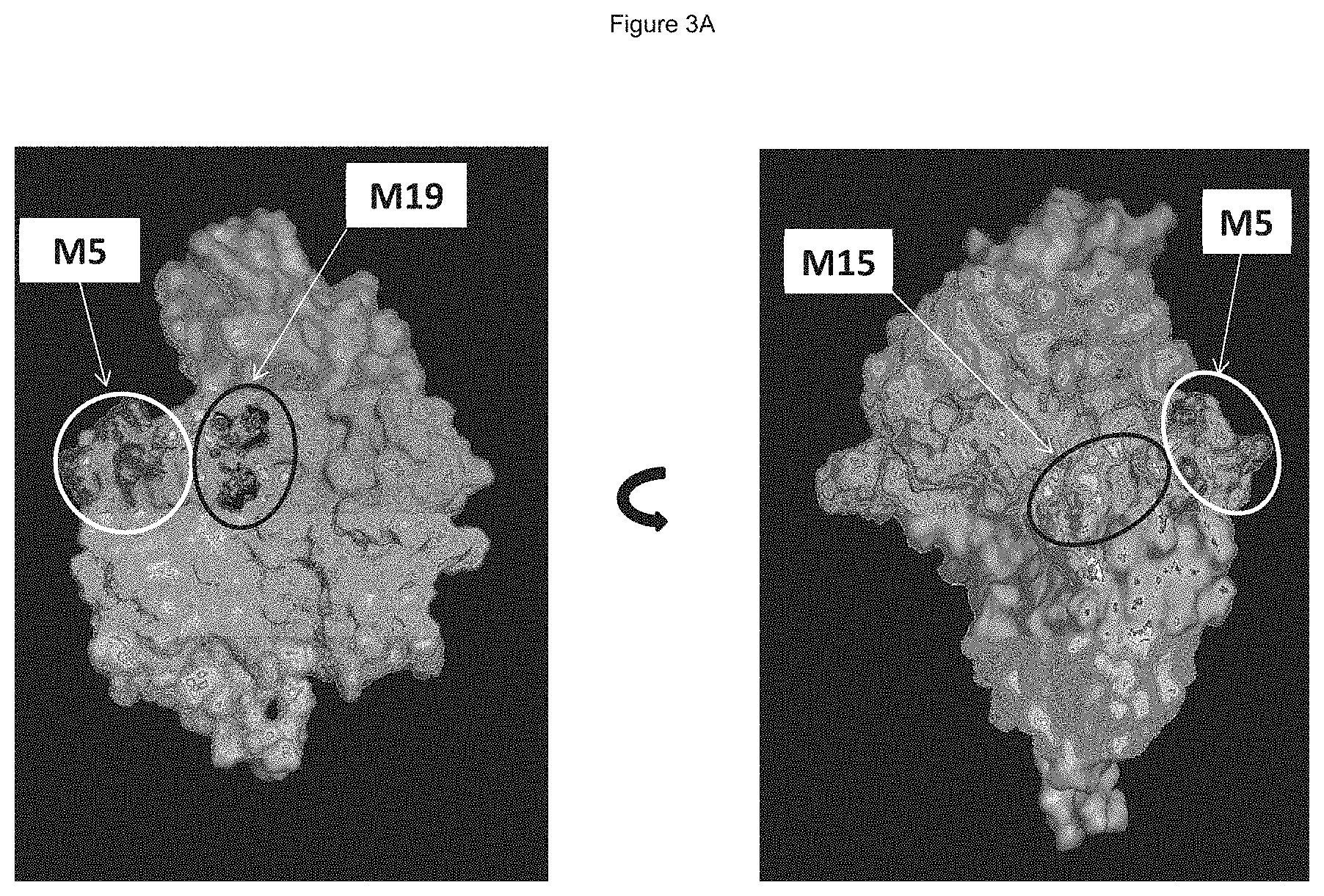
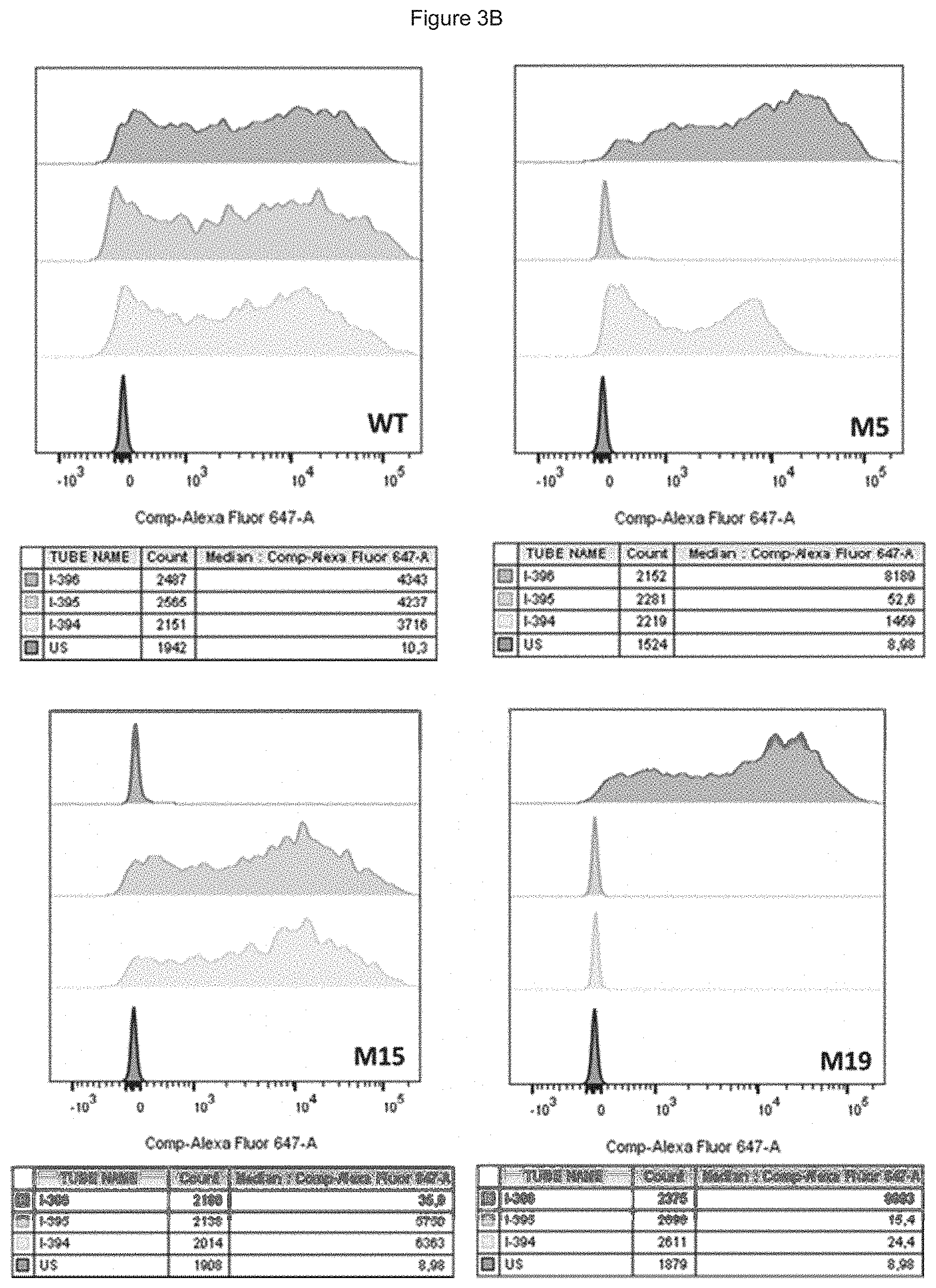
[0078] FIG. 3A shows the position of residues mutated in mutants 5 (M5), 15 (M15) and 19 (M19) on the surface of the CD39 protein. FIG. 3B shows results of binding to mutants 5, 15 and 19 for different antibodies.
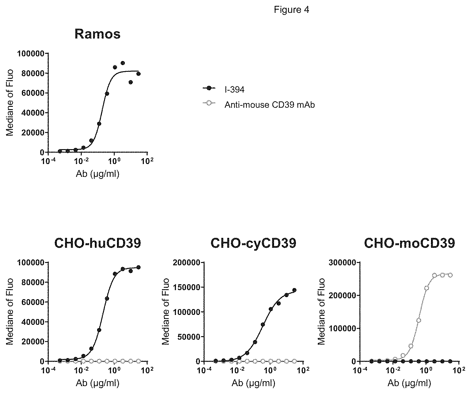
[0079] FIG. 4 shows binding of antibody I-394 to cells expressing human CD39, as assessed by flow cytometry. I-394 binds cells expressing human CD39 (CHO-huCD39), cells expressing cynomolgus CD39 (CHO-cyCD39) and to Ramos lymphoma cells, but not to cells expressing murine CD39 (CHO-moCD39).
[0080] FIG. 5 shows antibody I-394 is highly potent at blocking CD39 enzymatic activity in tumor (Ramos) cells, in cells expressing human CD39 (CHO-huCD39), and in cells expressing cynomolgus CD39 (CHO-cyCD39), as assessed by quantifying luminescence units which are proportional to the amount of ATP present.
[0081] FIG. 6 shows antibody I-394 is highly potent at blocking the enzymatic activity of soluble recombinant human CD39 protein, as assessed by quantifying luminescence units which are proportional to the amount of ATP present.
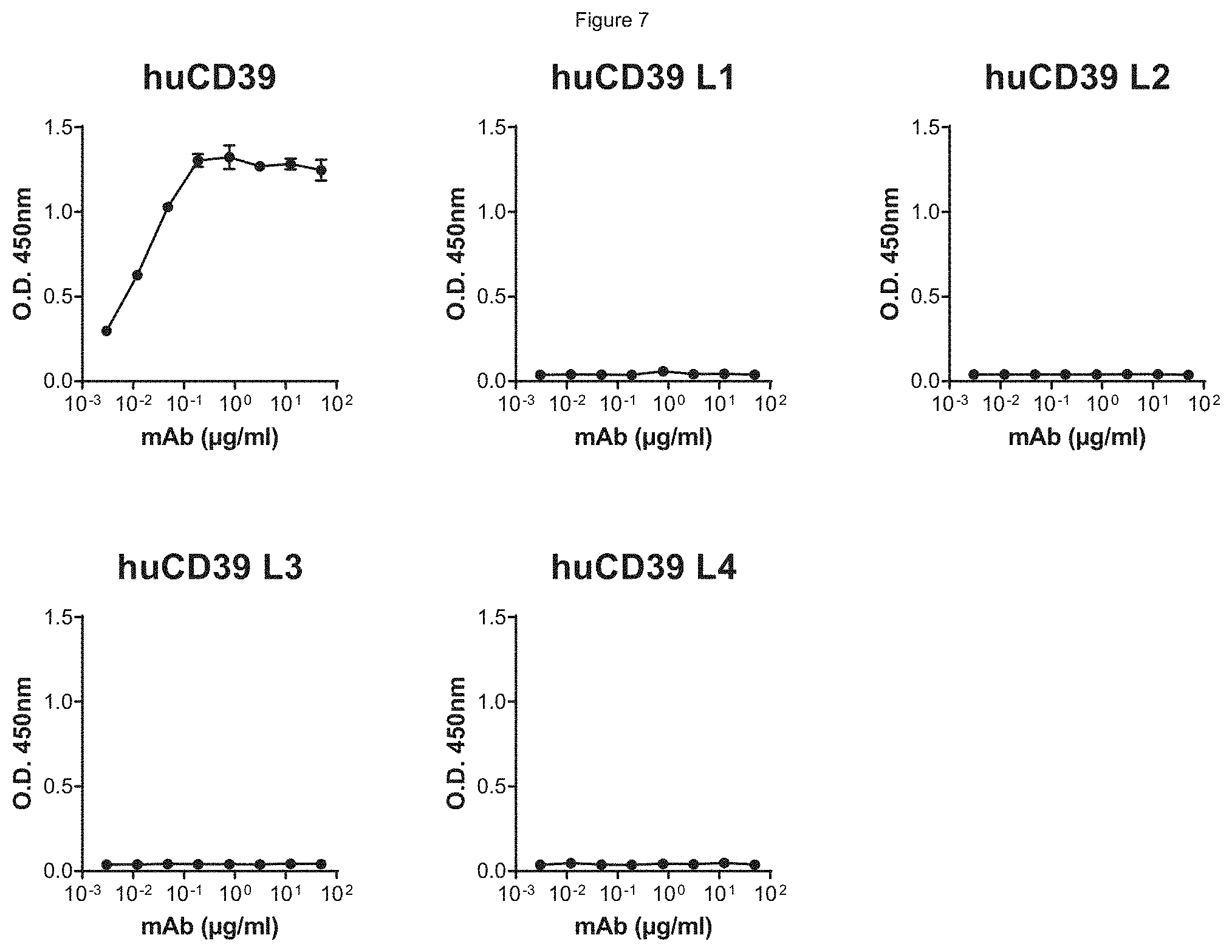
[0082] FIG. 7 shows antibody I-394 binds to human CD39 but not to any of the human isoforms CD39-L1, -L2, -L3 or -L4, as assessed in an ELISA assay.
[0083] FIG. 8 shows the experimental procedure for assessing the effect of ATP-mediated DC activation on CD4 T cells activation, ATP-activated DC were washed and then incubated with allogenic CD4 T cells (ratio 1 MoDC/4 T cells) for a mixed lymphocytes reaction (MLR) during 5 days. T cells activation and proliferation were analyzed through CD25 expression and Cell Trace Violet dilution by flow cytometry.
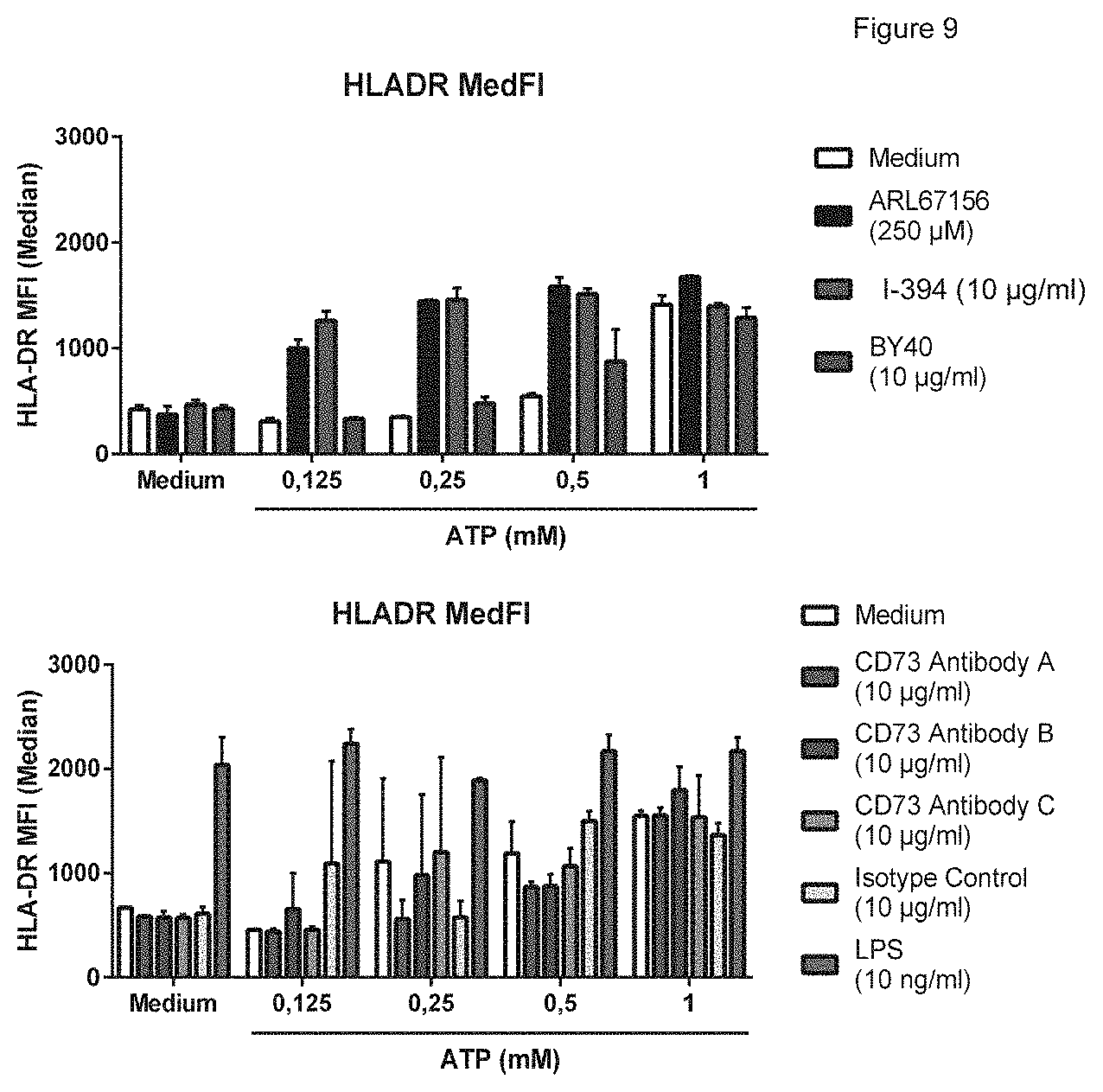
[0084] FIG. 9 shows HLA-DR expression on moDC and FIG. 10 shows CD83 expression on moDC. These figures show that the anti-CD39 blocking antibody I-394 and chemical inhibitors of CD39 lead to moDC activation at each of 0.125 mM, 0.25 mM or 0.5 mM. However, anti-CD39 antibody BY40 or anti-CD73 antibodies were not able to favor ATP-induced activation of dendritic cell (DC), suggesting that antibodies are not able to block enzymatic activity sufficiently to avoid ATP catabolism. The legends, top to bottom, correspond to the bars in the graph, from left to right.
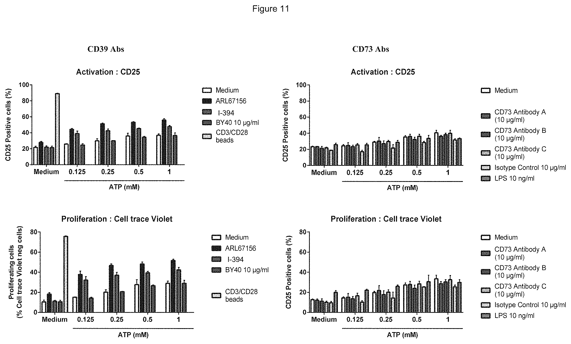
[0085] FIG. 11 shows CD25 expression shows that MoDC activated in presence of ATP were able to induce T cell activation and proliferation in a MLR assay; the enhancement of ATP-mediated MoDC activation by anti-CD39 blocking antibody I-394 resulted in higher T cell proliferation and activation. The legends, top to bottom, correspond to the bars in the graph, from left to right.
[0086] FIG. 12A shows the dose range of anti-CD73 antibodies on CD4 T cell proliferation, in the presence of added ATP, at 3 different doses of anti-sCD39 antibodies, either 0.01 .mu.g/ml, 0.1 .mu.g/ml and 1 .mu.g/ml. The anti-CD39 antibodies that are capable of neutralizing soluble human CD39 show a strong potentiation of anti-CD73 antibodies in restoring CD4 T cell proliferation. FIG. 12B shows the dose range of anti-CD73 antibodies on CD8 T cell proliferation, in the presence of added ATP, anti-sCD39 antibodies show a strong potentiation of anti-CD73 antibodies in restoring CD8 T cell proliferation.
[0087] FIG. 13 shows antibodies titrated on Ramos lymphoma cells by flow cytometry. Antibodies H2L1, H2L1*, H4L1 and H4L1* antibodies (mAbs8, 9, 20 and 21, respectively) showed best binding.
[0088] FIG. 14 shows inhibition of ATPase activity in Ramos and Mino tumor cells lines that express membrane-bound CD39. H4L1 and H4L1* antibodies (mAb20 and mAb21) were the most potent at blocking CD39 enzymatic activity.
[0089] FIG. 15A shows that antibody I-394 (parental light and heavy chains) has a higher aggregation temperature (TAgg) and improved stability compared to antibody BY40. FIG. 15B shows that I-394 antibody humanized variant antibodies with variable regions H2L1 (mAb 8), H2L1* (mAb9), H4L1 (mAb20) and H4L1* (mAb21) all have high aggregation temperature (TAgg) and good stability.
DETAILED DESCRIPTION OF THE INVENTION
Definitions
[0090] Where "comprising" is used, this can optionally be replaced by "consisting essentially of" or by "consisting of".
[0091] Human CD39, also known as "vascular" CD39, NTPdase1, ENTPD1, ATPDase and vascular ATP diphosphohydrolase, exhibits ATPase activity. CD39 hydrolyzes extracellular ATP and ADP to AMP, which is further converted to adenosine by another enzyme, 5-prime nucleotidase. The amino acid sequence of the "vascular" human CD39 mature polypeptide chain is shown in Genbank under accession number P49961, the entire disclosure of which is incorporated herein by reference, and as follows:
TABLE-US-00001 (SEQ ID NO: 1) 1 MEDTKESNVK TFCSKNILAI LGFSSIIAVI ALLAVGLTQN KALPENVKYG IVLDAGSSHT 61 SLYIYKWPAE KENDTGVVHQ VEECRVKGPG ISKFVQKVNE IGIYLTDCME RAREVIPRSQ 121 HQETPVYLGA TAGMRLLRME SEELADRVLD VVERSLSNYP FDFQGARIIT GQEEGAYGWI 181 TINYLLGKFS QKTRWFSIVP YETNNQETFG ALDLGGASTQ VTFVPQNQTI ESPDNALQFR 241 LYGKDYNVYT HSFLCYGKDQ ALWQKLAKDI QVASNEILRD PCFHPGYKKV VNVSDLYKTP 301 CTKRFEMTLP FQQFEIQGIG NYQQCHQSIL ELFNTSYCPY SQCAFNGIFL PPLQGDFGAF 361 SAFYFVMKFL NLTSEKVSQE KVTEMMKKFC AQPWEEIKTS YAGVKEKYLS EYCFSGTYIL 421 SLLLQGYHFT ADSWEHIHFI GKIQGSDAGW TLGYMLNLTN MIPAEQPLST PLSHSTYVFL 481 MVLFSLVLFT VAIIGLLIFH KPSYFWKDMV.
[0092] In the context herein, "neutralize" or neutralizing" when referring to the CD39 polypeptide (e.g., "neutralize CD39", "neutralize the activity of CD39" or "neutralize the enzymatic activity of CD39"), refers to a process in which the ATP hydrolysis (ATPase) activity of CD39 is inhibited. This comprises, notably the inhibition of CD39-mediated generation of AMP and/or ADP, i.e. the inhibition of CD39-mediated catabolism of ATP to AMP and/or ADP. For membrane-bound CD39, this can be measured for example in a cellular assay that measures the capacity of a test compound to inhibit the conversion of ATP to AMP and/or ADP, either directly or indirectly. For soluble CD39, this can be measured by incubating recombinant soluble CD39 as described herein with a test compound and measuring the conversion of ATP to AMP and/or ADP, either directly or indirectly. For example, disappearance of ATP and/or generation of AMP can be assessed, as described herein, e.g., by quantifying luminescence units which are proportional to the amount of ATP present. In one embodiment, an antibody preparation causes at least a 60% decrease in the conversion of ATP to AMP, at least a 70% decrease in the conversion of ATP to AMP, or at least an 80% or 90% decrease in the conversion of ATP to AMP, referring, for example, to the assays described herein (e.g., disappearance of ATP and/or generation of AMP).
[0093] Whenever "treatment of cancer" or the like is mentioned with reference to anti-CD39 binding agent (e.g., antibody), this can include: (a) method of treatment of cancer, said method comprising the step of administering (for at least one treatment) an anti-CD39 binding agent, (preferably in a pharmaceutically acceptable carrier material) to an individual, a mammal, especially a human, in need of such treatment, in a dose that allows for the treatment of cancer, (a therapeutically effective amount), preferably in a dose (amount) as specified herein; (b) the use of an anti-CD39 binding agent for the treatment of cancer, or an anti-CD39 binding agent, for use in said treatment (especially in a human); (c) the use of an anti-CD39 binding agent for the manufacture of a pharmaceutical preparation for the treatment of cancer, a method of using an anti-CD39 binding agent for the manufacture of a pharmaceutical preparation for the treatment of cancer, optionally comprising admixing an anti-CD39 binding agent with a pharmaceutically acceptable carrier, or a pharmaceutical preparation comprising an effective dose of an anti-CD39 binding agent that is appropriate for the treatment of cancer; or (d) any combination of a), b), and c), in accordance with the subject matter allowable for patenting in a country where this application is filed.
[0094] As used herein, the term "antigen binding domain" refers to a domain comprising a three-dimensional structure capable of immunospecifically binding to an epitope. Thus, in one embodiment, said domain can comprise a hypervariable region, optionally a VH and/or VL domain of an antibody chain, optionally at least a VH domain. In another embodiment, the binding domain may comprise at least one complementarity determining region (CDR) of an antibody chain. In another embodiment, the binding domain may comprise a polypeptide domain from a non-immunoglobulin scaffold.
[0095] The term "antibody," as used herein, can include polyclonal and monoclonal antibodies. Depending on the type of constant domain in the heavy chains, antibodies are assigned to one of five major classes: IgA, IgD, IgE, IgG, and IgM. Several of these are further divided into subclasses or isotypes, such as IgG1, IgG2, IgG3, IgG4, and the like. An exemplary immunoglobulin (antibody) structural unit comprises a tetramer. Each tetramer is composed of two identical pairs of polypeptide chains, each pair having one "light" (about 25 kDa) and one "heavy" chain (about 50-70 kDa). The N-terminus of each chain defines a variable region of about 100 to 110 or more amino acids that is primarily responsible for antigen recognition. The terms variable light chain (V.sub.L) and variable heavy chain (V.sub.H) refer to these light and heavy chains respectively. The heavy-chain constant domains that correspond to the different classes of immunoglobulins are termed "alpha," "delta," "epsilon," "gamma" and "mu," respectively. The subunit structures and three-dimensional configurations of different classes of immunoglobulins are well known. IgG are the exemplary classes of antibodies employed herein because they are the most common antibodies in the physiological situation and because they are most easily made in a laboratory setting. Optionally the antibody is a monoclonal antibody. Particular examples of antibodies are humanized, chimeric, human, or otherwise-human-suitable antibodies. "Antibodies" also includes any fragment or derivative of any of the herein described antibodies.
[0096] The term "specifically binds to" means that an antibody can bind preferably in a competitive binding assay to the binding partner, e.g., CD39, as assessed using either recombinant forms of the proteins, epitopes therein, or native proteins present on the surface of isolated target cells. Competitive binding assays and other methods for determining specific binding are further described below and are well known in the art.
[0097] When an antibody is said to "compete with" a particular monoclonal antibody (e.g. antibody I-394), it means that the antibody competes with the monoclonal antibody in a binding assay using either recombinant CD39 molecules or surface expressed CD39 molecules. For example, if a test antibody reduces the binding of a reference antibody to a CD39 polypeptide or CD39-expressing cell in a binding assay, the antibody is said to "compete" respectively with the reference antibody.
[0098] The term "affinity", as used herein, means the strength of the binding of an antibody to an epitope. The affinity of an antibody is given by the dissociation constant Kd, defined as [Ab].times.[Ag]/[Ab-Ag], where [Ab-Ag] is the molar concentration of the antibody-antigen complex, [Ab] is the molar concentration of the unbound antibody and [Ag] is the molar concentration of the unbound antigen. The affinity constant K.sub.a is defined by 1/Kd. Methods for determining the affinity of mAbs can be found in Harlow, et al., Antibodies: A Laboratory Manual, Cold Spring Harbor Laboratory Press, Cold Spring Harbor, N.Y., 1988), Coligan et al., eds., Current Protocols in Immunology, Greene Publishing Assoc. and Wiley Interscience, N.Y., (1992, 1993), and Muller, Meth. Enzymol. 92:589-601 (1983), which references are entirely incorporated herein by reference. One standard method well known in the art for determining the affinity of mAbs is the use of surface plasmon resonance (SPR) screening (such as by analysis with a BIAcore.TM. SPR analytical device).
[0099] Within the context herein a "determinant" designates a site of interaction or binding on a polypeptide.
[0100] The term "epitope" refers to an antigenic determinant, and is the area or region on an antigen to which an antibody binds. A protein epitope may comprise amino acid residues directly involved in the binding as well as amino acid residues which are effectively blocked by the specific antigen binding antibody or peptide, i.e., amino acid residues within the "footprint" of the antibody. It is the simplest form or smallest structural area on a complex antigen molecule that can combine with e.g., an antibody or a receptor. Epitopes can be linear or conformational/structural. The term "linear epitope" is defined as an epitope composed of amino acid residues that are contiguous on the linear sequence of amino acids (primary structure). The term "conformational or structural epitope" is defined as an epitope composed of amino acid residues that are not all contiguous and thus represent separated parts of the linear sequence of amino acids that are brought into proximity to one another by folding of the molecule (secondary, tertiary and/or quaternary structures). A conformational epitope is dependent on the 3-dimensional structure. The term `conformational` is therefore often used interchangeably with `structural`.
[0101] The term "internalization", used interchangeably with "intracellular internalization", refers to the molecular, biochemical and cellular events associated with the process of translocating a molecule from the extracellular surface of a cell to the intracellular surface of a cell. The processes responsible for intracellular internalization of molecules are well-known and can involve, inter alia, the internalization of extracellular molecules (such as hormones, antibodies, and small organic molecules); membrane-associated molecules (such as cell-surface receptors); and complexes of membrane-associated molecules bound to extracellular molecules (for example, a ligand bound to a transmembrane receptor or an antibody bound to a membrane-associated molecule). Thus, "inducing and/or increasing internalization" comprises events wherein intracellular internalization is initiated and/or the rate and/or extent of intracellular internalization is increased.
[0102] The term "agent" is used herein to denote a chemical compound, a mixture of chemical compounds, a biological macromolecule, or an extract made from biological materials. The term "therapeutic agent" refers to an agent that has biological activity.
[0103] For the purposes herein, a "humanized" antibody refers to an antibody in which the constant and variable framework region of one or more human immunoglobulins is fused with the binding region, e.g., the CDR, of an animal immunoglobulin. Such antibodies are designed to maintain the binding specificity of the non-human antibody from which the binding regions are derived, but to avoid an immune reaction against the non-human antibody.
[0104] The term "hypervariable region" when used herein refers to the amino acid residues of an antibody that are responsible for antigen binding. The hypervariable region generally comprises amino acid residues from a "complementarity-determining region" or "CDR" (e.g., residues 24-34 (L1), 50-56 (L2) and 89-97 (L3) in the light-chain variable domain and 31-35 (H1), 50-65 (H2) and 95-102 (H3) in the heavy-chain variable domain; Kabat et al. 1991) and/or those residues from a "hypervariable loop" (e.g., residues 26-32 (L1), 50-52 (L2) and 91-96 (L3) in the light-chain variable domain and 26-32 (H1), 53-55 (H2) and 96-101 (H3) in the heavy-chain variable domain; Chothia and Lesk, J. Mol. Biol 1987; 196:901-917), or a similar system for determining essential amino acids responsible for antigen binding. Typically, the numbering of amino acid residues in this region is performed by the method described in Kabat et al., supra. Phrases such as "Kabat position", "variable domain residue numbering as in Kabat" and "according to Kabat" herein refer to this numbering system for heavy chain variable domains or light chain variable domains. Using the Kabat numbering system, the actual linear amino acid sequence of a peptide may contain fewer or additional amino acids corresponding to a shortening of, or insertion into, a FR or CDR of the variable domain. For example, a heavy chain variable domain may include a single amino acid insert (residue 52a according to Kabat) after residue 52 of CDR H2 and inserted residues (e.g., residues 82a, 82b, and 82c, etc. according to Kabat) after heavy chain FR residue 82. The Kabat numbering of residues may be determined for a given antibody by alignment at regions of homology of the sequence of the antibody with a "standard" Kabat numbered sequence.
[0105] By "framework" or "FR" residues as used herein is meant the region of an antibody variable domain exclusive of those regions defined as CDRs. Each antibody variable domain framework can be further subdivided into the contiguous regions separated by the CDRs (FR1, FR2, FR3 and FR4).
[0106] The terms "Fc domain," "Fc portion," and "Fc region" refer to a C-terminal fragment of an antibody heavy chain, e.g., from about amino acid (aa) 230 to about aa 450 of human .gamma. (gamma) heavy chain or its counterpart sequence in other types of antibody heavy chains (e.g., .alpha., .delta., .epsilon. and .mu. for human antibodies), or a naturally occurring allotype thereof. Unless otherwise specified, the commonly accepted Kabat amino acid numbering for immunoglobulins is used throughout this disclosure (see Kabat et al. (1991) Sequences of Protein of Immunological Interest, 5th ed., United States Public Health Service, National Institute of Health, Bethesda, Md.).
[0107] The terms "isolated", "purified" or "biologically pure" refer to material that is substantially or essentially free from components which normally accompany it as found in its native state. Purity and homogeneity are typically determined using analytical chemistry techniques such as polyacrylamide gel electrophoresis or high performance liquid chromatography. A protein that is the predominant species present in a preparation is substantially purified.
[0108] The terms "polypeptide," "peptide" and "protein" are used interchangeably herein to refer to a polymer of amino acid residues. The terms apply to amino acid polymers in which one or more amino acid residue is an artificial chemical mimetic of a corresponding naturally occurring amino acid, as well as to naturally occurring amino acid polymers and non-naturally occurring amino acid polymer.
[0109] The term "recombinant" when used with reference, e.g., to a cell, or nucleic acid, protein (e.g. antibody or antibody fragment), or vector, indicates that the cell, nucleic acid, protein or vector, has been modified by the introduction of a heterologous nucleic acid or protein or the alteration of a native nucleic acid or protein, or that the cell is derived from a cell so modified. Thus, for example, recombinant cells express genes that are not found within the native (non-recombinant) form of the cell or express native genes that are otherwise abnormally expressed, under expressed or not expressed at all.
[0110] Within the context herein, the term antibody that "binds" a polypeptide or epitope designates an antibody that binds said determinant with specificity and/or affinity.
[0111] The term "identity" or "identical", when used in a relationship between the sequences of two or more polypeptides, refers to the degree of sequence relatedness between polypeptides, as determined by the number of matches between strings of two or more amino acid residues. "Identity" measures the percent of identical matches between the smaller of two or more sequences with gap alignments (if any) addressed by a particular mathematical model or computer program (i.e., "algorithms"). Identity of related polypeptides can be readily calculated by known methods. Such methods include, but are not limited to, those described in Computational Molecular Biology, Lesk, A. M., ed., Oxford University Press, New York, 1988; Biocomputing: Informatics and Genome Projects, Smith, D. W., ed., Academic Press, New York, 1993; Computer Analysis of Sequence Data, Part 1, Griffin, A. M., and Griffin, H. G., eds., Humana Press, New Jersey, 1994; Sequence Analysis in Molecular Biology, von Heinje, G., Academic Press, 1987; Sequence Analysis Primer, Gribskov, M. and Devereux, J., eds., M. Stockton Press, New York, 1991; and Carillo et al., SIAM J. Applied Math. 48, 1073 (1988).
[0112] Methods for determining identity are designed to give the largest match between the sequences tested. Methods of determining identity are described in publicly available computer programs. Computer program methods for determining identity between two sequences include the GCG program package, including GAP (Devereux et al., Nucl. Acid. Res. 12, 387 (1984); Genetics Computer Group, University of Wisconsin, Madison, Wis.), BLASTP, BLASTN, and FASTA (Altschul et al., J. Mol. Biol. 215, 403-410 (1990)). The BLASTX program is publicly available from the National Center for Biotechnology Information (NCBI) and other sources (BLAST Manual, Altschul et al. NCB/NLM/NIH Bethesda, Md. 20894; Altschul et al., supra). The well-known Smith Waterman algorithm may also be used to determine identity.
Production of Antibodies
[0113] The anti-CD39 antigen binding domain, or a protein (e.g., antibody or antibody fragment) that comprises such domain, binds and neutralizes soluble human CD39 polypeptide, e.g., a human CD39 polypeptide lacking the two transmembrane domains near the N- and C-terminal ends found in membrane bound CD39. In one embodiment the agent inhibits the ATPase activity of CD39. In one embodiment the antibody inhibits CD39-mediated generation of adenosine. In one embodiment the antibody inhibits CD39-mediated catabolism of ATP to AMP. In one embodiment the antibody inhibits adenosine-mediated inhibition of lymphocyte activity (e.g., T cells). In one aspect, the antibody is selected from a full-length antibody, an antibody fragment, and a synthetic or semi-synthetic antibody-derived molecule.
[0114] The antibodies that potently inhibit the enzymatic (ATPase activity) activity of the soluble (and optionally the membrane-bound) CD39 protein may, in one embodiment, immobilize or restrict the domain movement of the soluble (and optionally the membrane-bound) CD39 protein in one of its conformations thereby preventing it from hydrolyzing its substrate. The antibodies may achieve this by binding to both C- and N-terminal domains of soluble (and optionally the membrane-bound) CD39 at the same time.
[0115] In one embodiment, an anti-CD39 antigen binding domain, or an antigen-binding protein that comprises the antigen binding domain (e.g., an antibody or antibody fragment, a multispecific binding protein, a bispecific antibody, etc.), comprises complementary determining regions (CDR) and framework regions (FR). The antigen binding domains can be designed or modified so as to provide desired and/or improved properties.
[0116] In one embodiment, an anti-CD39 antigen-binding protein is capable of binding to and inhibiting the activity of a human CD39 polypeptide, the antigen-binding protein comprising a VH and a VL that each comprise a framework (e.g., a framework having an amino acid sequence of human origin) and a CDR1, CDR2 and CDR3. In one embodiment, the antigen-binding protein restricts the domain movement of CD39 when bound to CD39. Optionally, the VH and/or VL framework (e.g., FR1, FR2, FR3 and/or FR4) is of human origin.
[0117] In certain embodiment, the binding molecules and domains can be derived from immunoglobulin variable domains, for example in the form of associated V.sub.L and V.sub.H domains found on two polypeptide chains, or a single chain antigen binding domain such as a scFv, a V.sub.H domain, a V.sub.L domain, a dAb, a V-NAR domain or a V.sub.HH domain.
[0118] In one aspect, the CD39 binding agent is an antibody selected from a fully human antibody, a humanized antibody, and a chimeric antibody.
[0119] In one aspect, the agent is a fragment of an antibody comprising a constant or Fc domain derived from a human IgG1 constant or Fc domain, e.g., modified, as further disclosed herein.
[0120] In one aspect, the agent comprises an antibody fragment selected from a Fab fragment, a Fab' fragment, a Fab'-SH fragment, a F(ab)2 fragment, a F(ab')2 fragment, an Fv fragment, a Heavy chain Ig (a llama or camel Ig), a V.sub.HH fragment, a single domain FV, and a single-chain antibody fragment. In one aspect, the agent comprises a synthetic or semisynthetic antibody-derived molecule selected from a scFV, a dsFV, a minibody, a diabody, a triabody, a kappa body, an IgNAR; and a multispecific (e.g., bispecific) antibody. The agent can optionally further comprise an Fc domain.
[0121] In one aspect, the antibody is in at least partially purified form.
[0122] In one aspect, the antibody is in essentially isolated form.
[0123] Antibodies may be produced by a variety of techniques known in the art. In one embodiment, antibodies of the disclosure are produced by selection from an antibody library (e.g., as generated from phage display library). In another embodiment, antibodies are produced by immunization of a non-human animal, preferably a mouse, with an immunogen comprising a CD39 polypeptide, preferably a soluble human CD39 extracellular domain polypeptide. The CD39 polypeptide may optionally be or comprise a fragment or derivative of a full-length CD39 polypeptide, typically an immunogenic fragment, i.e., a portion of the polypeptide comprising an epitope exposed on the surface of cells expressing a CD39 polypeptide. Such fragments typically contain at least about 7 consecutive amino acids of the mature polypeptide sequence, even more preferably at least about 10 consecutive amino acids thereof. Fragments typically are essentially derived from the extra-cellular domain of the receptor. In one embodiment, the immunogen comprises a wild-type human CD39 polypeptide in a lipid membrane, typically at the surface of a cell. In a specific embodiment, the immunogen comprises intact cells, particularly intact human cells, optionally treated or lysed. In another embodiment, the polypeptide is a recombinant CD39 polypeptide.
[0124] The step of immunizing a non-human mammal with an antigen may be carried out in any manner well known in the art for stimulating the production of antibodies in a mouse (see, for example, E. Harlow and D. Lane, Antibodies: A Laboratory Manual., Cold Spring Harbor Laboratory Press, Cold Spring Harbor, N.Y. (1988), the entire disclosure of which is herein incorporated by reference). Isolation of hybridomas producing the antibodies is well known and can be carried out in any manner well known in the art.
[0125] Antibodies may also be produced by selection of combinatorial libraries of immunoglobulins, as disclosed for instance in (Ward et al. Nature, 341 (1989) p. 544, the entire disclosure of which is herein incorporated by reference).
[0126] The identification of one or more antibodies that bind(s) to CD39, particularly substantially or essentially the same region on CD39 as monoclonal antibody mAb20 or mAb21, can be readily determined using any one of a variety of immunological screening assays in which antibody competition can be assessed. Many such assays are routinely practiced and are well known in the art (see, e. g., U.S. Pat. No. 5,660,827, issued Aug. 26, 1997, which is specifically incorporated herein by reference).
[0127] For example, where the test antibodies to be examined are obtained from different source animals, or are even of a different Ig isotype, a simple competition assay may be employed in which the control (mAb20 or mAb21, for example) and test antibodies are admixed (or pre-adsorbed) and applied to a sample containing CD39 polypeptides, for example as disclosed in PCT publication no. WO2018/167267, the disclosure of which is incorporated herein by reference. Protocols based upon western blotting and the use of BIACORE analysis are suitable for use in such competition studies.
[0128] Determination of whether an antibody binds within an epitope region can be carried out in ways known to the person skilled in the art. As one example of such mapping/characterization methods, an epitope region for an anti-CD39 antibody may be determined by epitope "foot-printing" using chemical modification of the exposed amines/carboxyls in the CD39 protein. One specific example of such a foot-printing technique is the use of HXMS (hydrogen-deuterium exchange detected by mass spectrometry) wherein a hydrogen/deuterium exchange of receptor and ligand protein amide protons, binding, and back exchange occurs, wherein the backbone amide groups participating in protein binding are protected from back exchange and therefore will remain deuterated. Relevant regions can be identified at this point by peptic proteolysis, fast microbore high-performance liquid chromatography separation, and/or electrospray ionization mass spectrometry. See, e. g., Ehring H, Analytical Biochemistry, Vol. 267 (2) pp. 252-259 (1999) Engen, J. R. and Smith, D. L. (2001) Anal. Chem. 73, 256A-265A. Another example of a suitable epitope identification technique is nuclear magnetic resonance epitope mapping (NMR), where typically the position of the signals in two-dimensional NMR spectra of the free antigen and the antigen complexed with the antigen binding peptide, such as an antibody, are compared. The antigen typically is selectively isotopically labeled with 15N so that only signals corresponding to the antigen and no signals from the antigen binding peptide are seen in the NMR-spectrum. Antigen signals originating from amino acids involved in the interaction with the antigen binding peptide typically will shift position in the spectrum of the complex compared to the spectrum of the free antigen, and the amino acids involved in the binding can be identified that way. See, e. g., Ernst Schering Res Found Workshop. 2004; (44): 149-67; Huang et al., Journal of Molecular Biology, Vol. 281 (1) pp. 61-67 (1998); and Saito and Patterson, Methods. 1996 June; 9 (3): 516-24.
[0129] Epitope mapping/characterization also can be performed using mass spectrometry methods. See, e.g., Downard, J Mass Spectrom. 2000 April; 35 (4): 493-503 and Kiselar and Downard, Anal Chem. 1999 May 1; 71 (9): 1792-1801. Protease digestion techniques also can be useful in the context of epitope mapping and identification. Antigenic determinant-relevant regions/sequences can be determined by protease digestion, e.g., by using trypsin in a ratio of about 1:50 to CD39 or o/n digestion at and pH 7-8, followed by mass spectrometry (MS) analysis for peptide identification. The peptides protected from trypsin cleavage by the anti-CD39 binder can subsequently be identified by comparison of samples subjected to trypsin digestion and samples incubated with antibody and then subjected to digestion by e.g., trypsin (thereby revealing a footprint for the binder). Other enzymes like chymotrypsin, pepsin, etc., also or alternatively can be used in similar epitope characterization methods. Moreover, enzymatic digestion can provide a quick method for analyzing whether a potential antigenic determinant sequence is within a region of the CD39 polypeptide that is not surface exposed and, accordingly, most likely not relevant in terms of immunogenicity/antigenicity.
[0130] Site-directed mutagenesis is another technique useful for elucidation of a binding epitope. For example, in "alanine-scanning", each residue within a protein segment is replaced with an alanine residue, and the consequences for binding affinity measured. If the mutation leads to a significant reduction in binding affinity, it is most likely involved in binding. Monoclonal antibodies specific for structural epitopes (i.e., antibodies which do not bind the unfolded protein) can be used to verify that the alanine-replacement does not influence over-all fold of the protein. See, e.g., Clackson and Wells, Science 1995; 267:383-386; and Wells, Proc Natl Acad Sci USA 1996; 93:1-6.
[0131] Electron microscopy can also be used for epitope "foot-printing". For example, Wang et al., Nature 1992; 355:275-278 used coordinated application of cryoelectron micros-copy, three-dimensional image reconstruction, and X-ray crystallography to determine the physical footprint of a Fab-fragment on the capsid surface of native cowpea mosaic virus.
[0132] Other forms of "label-free" assay for epitope evaluation include surface plasmon resonance (SPR, BIACORE) and reflectometric interference spectroscopy (RifS). See, e.g., Fagerstam et al., Journal Of Molecular Recognition 1990; 3:208-14; Nice et al., J. Chroma-togr. 1993; 646:159-168; Leipert et al., Angew. Chem. Int. Ed. 1998; 37:3308-3311; Kroger et al., Biosensors and Bioelectronics 2002; 17:937-944.
[0133] Typically, an anti-CD39 antibody provided herein has an affinity for a CD39 polypeptide (e.g., a monomeric CD39 polypeptide as produced in the Examples herein) in the range of about 10.sup.4 to about 10.sup.11 M.sup.-1 (e.g., about 10.sup.8 to about 10.sup.10 M.sup.-1). For example, anti-CD39 antibodies can have an average disassociation constant (K.sub.D) of less than 1.times.10.sup.-9 M with respect to CD39, as determined by, e.g., surface plasmon resonance (SPR) screening (such as by analysis with a BIAcore.TM. SPR analytical device). In a more particular exemplary aspect, the disclosure provides anti-CD39 antibodies that have a KD of about 1.times.10.sup.-8 M to about 1.times.10.sup.-10 M, or about 1.times.10.sup.-9 M to about 1.times.10.sup.-11 M, for CD39.
[0134] Antibodies can be characterized for example by a mean K.sub.D of no more than about (i.e. better affinity than) 100, 60, 10, 5, or 1 nanomolar, preferably sub-nanomolar or optionally no more than about 500, 200, 100 or 10 picomolar. K.sub.D can be determined for example for example by immobilizing recombinantly produced human CD39 proteins on a chip surface, followed by application of the antibody to be tested in solution. In one embodiment, the method further comprises a step (d), selecting antibodies from (b) that are capable of competing for binding to CD39 with antibody I-394, or for example any of mAbs 1-24.
[0135] DNA encoding the monoclonal antibodies of the disclosure, e.g., antibody mAb20 or mAb21, can be readily isolated and sequenced using conventional procedures (e. g., by using oligonucleotide probes that are capable of binding specifically to genes encoding the heavy and light chains of murine antibodies). In one aspect, provided is a nucleic acid encoding a heavy chain or a light chain of an anti-CD39 antibody of any embodiment herein. Once isolated, the DNA can be placed into expression vectors, which are then transfected into host cells such as E. coli cells, simian COS cells, Chinese hamster ovary (CHO) cells, or myeloma cells that do not otherwise produce immunoglobulin protein, to obtain the synthesis of monoclonal antibodies in the recombinant host cells. As described elsewhere in the present specification, such DNA sequences can be modified for any of a large number of purposes, e.g., for humanizing antibodies, producing fragments or derivatives, or for modifying the sequence of the antibody, e.g., in the antigen binding site in order to optimize the binding specificity of the antibody. In one embodiment, provided is an isolated nucleic acid sequence encoding a light chain and/or a heavy chain of an antibody (e.g., mAb20 or mAb21), as well as a recombinant host cell comprising (e.g., in its genome) such nucleic acid. Recombinant expression in bacteria of DNA encoding the antibody is well known in the art (see, for example, Skerra et al., Curr. Opinion in Immunol., 5, pp. 256 (1993); and Pluckthun, Immunol. 130, p. 151 (1992).
[0136] Once antibodies are identified that are capable of binding sCD39 and/or memCD39, and/or having other desired properties, they will also typically be assessed, using methods such as those described herein, for their ability to bind to other polypeptides, including unrelated polypeptides. Ideally, the antibodies bind with substantial affinity only to CD39, and do not bind at a significant level to unrelated polypeptides, or other polypeptides of the NTPDase family, notably CD39-L1, L2, L3 and L4 or NTPDase8. However, it will be appreciated that, as long as the affinity for CD39 is substantially greater (e.g., 10.times., 100.times., 500.times., 1000.times., 10,000.times., or more) than it is for other, unrelated polypeptides), then the antibodies are suitable for use in the present methods.
[0137] In one embodiment, the anti-CD39 antibodies can be prepared such that they do not have substantial specific binding to human Fc.gamma. receptors, e.g., any one or more of CD16A, CD16B, CD32A, CD32B and/or CD64). Such antibodies may comprise constant regions of various heavy chains that are known to lack or have low binding to Fc.gamma. receptors. Alternatively, antibody fragments that do not comprise (or comprise portions of) constant regions, such as F(ab')2 fragments, can be used to avoid Fc receptor binding. Fc receptor binding can be assessed according to methods known in the art, including for example testing binding of an antibody to Fc receptor protein in a BIACORE assay. Also, generally any antibody IgG isotype can be used in which the Fc portion is modified (e.g., by introducing 1, 2, 3, 4, 5 or more amino acid substitutions) to minimize or eliminate binding to Fc receptors (see, e.g., WO 03/101485, the disclosure of which is herein incorporated by reference). Assays such as cell based assays, to assess Fc receptor binding are well known in the art, and are described in, e.g., WO 03/101485.
[0138] In one embodiment, the antibody can comprise one or more specific mutations in the Fc region that result in "Fc silent" antibodies that have minimal interaction with effector cells. Silenced effector functions can be obtained by mutation in the Fc region of the antibodies and have been described in the art: N297A mutation, the LALA mutations, (Strohl, W., 2009, Curr. Opin. Biotechnol. Vol. 20(6):685-691); and D265A (Baudino et al., 2008, J. Immunol. 181: 6664-69) see also Heusser et al., WO2012/065950, the disclosures of which are incorporated herein by reference. In one embodiment, an antibody comprises one, two, three or more amino acid substitutions in the hinge region. In one embodiment, the antibody is an IgG1 or IgG2 and comprises one, two or three substitutions at residues 233-236, optionally 233-238 (EU numbering). In one embodiment, the antibody is an IgG4 and comprises one, two or three substitutions at residues 327, 330 and/or 331 (EU numbering). Examples of silent Fc IgG1 antibodies are the LALA mutant comprising L234A and L235A mutation in the IgG1 Fc amino acid sequence. Another example of an Fc silent mutation is a mutation at residue D265, or at D265 and P329 for example as used in an IgG1 antibody as the DAPA (D265A, P329A) mutation (U.S. Pat. No. 6,737,056). Another silent IgG1 antibody comprises a mutation at residue N297 (e.g., N297A, N297S mutation), which results in aglycosylated/non-glycosylated antibodies. Other silent mutations include: substitutions at residues L234 and G237 (L234A/G237A); substitutions at residues S228, L235 and R409 (S228P/L235E/R409K,T,M,L); substitutions at residues H268, V309, A330 and A331 (H268Q/V309L/A330S/A331S); substitutions at residues C220, C226, C229 and P238 (C220S/C226S/C229S/P238S); substitutions at residues C226, C229, E233, L234 and L235 (C226S/C229S/E233P/L234V/L235A; substitutions at residues K322, L235 and L235 (K322A/L234A/L235A); substitutions at residues L234, L235 and P331 (L234F/L235E/P331S); substitutions at residues 234, 235 and 297; substitutions at residues E318, K320 and K322 (L235E/E318A/K320A/K322A); substitutions at residues (V234A, G237A, P238S); substitutions at residues 243 and 264; substitutions at residues 297 and 299; substitutions such that residues 233, 234, 235, 237, and 238 defined by the EU numbering system, comprise a sequence selected from PAAAP, PAAAS and SAAAS (see WO2011/066501).
[0139] In one embodiment, the antibody can comprise one or more specific mutations in the Fc region. For example, such an antibody can comprise an Fc domain of human IgG1 origin, comprises a mutation at Kabat residue(s) 234, 235, 237, 330 and/or 331. One example of such an Fc domain comprises substitutions at Kabat residues L234, L235 and P331 (e.g., L234A/L235E/P331S or (L234F/L235E/P331S). Another example of such an Fc domain comprises substitutions at Kabat residues L234, L235, G237 and P331 (e.g., L234A/L235E/G237A/P331S). Another example of such an Fc domain comprises substitutions at Kabat residues L234, L235, G237, A330 and P331 (e.g., L234A/L235E/G237A/A330S/P331S). In one embodiment, the antibody comprises an Fc domain, optionally of human IgG1 isotype, comprising: a L234X.sub.1 substitution, a L235X.sub.2 substitution, and a P331X.sub.3 substitution, wherein X.sub.1 is any amino acid residue other than leucine, X.sub.2 is any amino acid residue other than leucine, and X.sub.3 is any amino acid residue other than proline; optionally wherein X.sub.1 is an alanine or phenylalanine or a conservative substitution thereof; optionally wherein X.sub.2 is glutamic acid or a conservative substitution thereof; optionally wherein X.sub.3 is a serine or a conservative substitution thereof. In another embodiment, the antibody comprises an Fc domain, optionally of human IgG1 isotype, comprising: a L234X.sub.1 substitution, a L235X.sub.2 substitution, a G237X.sub.4 substitution and a P331X.sub.4 substitution, wherein X.sub.1 is any amino acid residue other than leucine, X.sub.2 is any amino acid residue other than leucine, X.sub.3 is any amino acid residue other than glycine, and X.sub.4 is any amino acid residue other than proline; optionally wherein X.sub.1 is an alanine or phenylalanine or a conservative substitution thereof; optionally wherein X.sub.2 is glutamic acid or a conservative substitution thereof; optionally, X.sub.3 is alanine or a conservative substitution thereof; optionally X.sub.4 is a serine or a conservative substitution thereof. In another embodiment, the antibody comprises an Fc domain, optionally of human IgG1 isotype, comprising: a L234X.sub.1 substitution, a L235X.sub.2 substitution, a G237X.sub.4 substitution, G330X.sub.4 substitution, and a P331X.sub.5 substitution, wherein X.sub.1 is any amino acid residue other than leucine, X.sub.2 is any amino acid residue other than leucine, X.sub.3 is any amino acid residue other than glycine, X.sub.4 is any amino acid residue other than alanine, and X.sub.5 is any amino acid residue other than proline; optionally wherein X.sub.1 is an alanine or phenylalanine or a conservative substitution thereof; optionally wherein X.sub.2 is glutamic acid or a conservative substitution thereof; optionally, X.sub.3 is alanine or a conservative substitution thereof; optionally, X.sub.4 is serine or a conservative substitution thereof; optionally X.sub.5 is a serine or a conservative substitution thereof. In the shorthand notation used here, the format is: Wild type residue: Position in polypeptide: Mutant residue, wherein residue positions are indicated according to EU numbering according to Kabat.
[0140] In one embodiment, an antibody comprises a heavy chain constant region comprising the amino acid sequence below, or an amino acid sequence at least 90%, 95% or 99% identical thereto but retaining the amino acid residues at Kabat positions 234, 235 and 331 (underlined):
TABLE-US-00002 (SEQ ID NO: 23) A S T K G P S V F P L A P S S K S T S G G T A A L G C L V K D Y F P E P V T V S W N S G A L T S G V H T F P A V L Q S S G L Y S L S S V V T V P S S S L G T Q T Y I C N V N H K P S N T K V D K R V E P K S C D K T H T C P P C P A P E A E G G P S V F L F P P K P K D T L M I S R T P E V T C V V V D V S H E D P E V K F N W Y V D G V E V H N A K T K P R E E Q Y N S T Y R V V S V L T V L H Q D W L N G K E Y K C K V S N K A L P A S I E K T I S K A K G Q P R E P Q V Y T L P P S R E E M T K N Q V S L T C L V K G F Y P S D I A V E W E S N G Q P E N N Y K T T P P V L D S D G S F F L Y S K L T V D K S R W Q Q G N V F S C S V M H E A L H N H Y T Q K S L S L S P G K.
[0141] In one embodiment, an antibody comprises a heavy chain constant region comprising the amino acid sequence below, or an amino acid sequence at least 90%, 95% or 99% identical thereto but retaining the amino acid residues at Kabat positions 234, 235 and 331 (underlined):
TABLE-US-00003 (SEQ ID NO: 24) A S T K G P S V F P L A P S S K S T S G G T A A L G C L V K D Y F P E P V T V S W N S G A L T S G V H T F P A V L Q S S G L Y S L S S V V T V P S S S L G T Q T Y I C N V N H K P S N T K V D K R V E P K S C D K T H T C P P C P A P E F E G G P S V F L F P P K P K D T L M I S R T P E V T C V V V D V S H E D P E V K F N W Y V D G V E V H N A K T K P R E E Q Y N S T Y R V V S V L T V L H Q D W L N G K E Y K C K V S N K A L P A S I E K T I S K A K G Q P R E P Q V Y T L P P S R E E M T K N Q V S L T C L V K G F Y P S D I A V E W E S N G Q P E N N Y K T T P P V L D S D G S F F L Y S K L T V D K S R W Q Q G N V F S C S V M H E A L H N H Y T Q K S L S L S P G K.
[0142] In one embodiment, an antibody comprises a heavy chain constant region comprising the amino acid sequence below, or an amino acid sequence at least 90%, 95% or 99% identical thereto but retaining the amino acid residues at Kabat positions 234, 235, 237, 330 and 331 (underlined):
TABLE-US-00004 (SEQ ID NO: 25) A S T K G P S V F P L A P S S K S T S G G T A A L G C L V K D Y F P E P V T V S W N S G A L T S G V H T F P A V L Q S S G L Y S L S S V V T V P S S S L G T Q T Y I C N V N H K P S N T K V D K R V E P K S C D K T H T C P P C P A P E G A P S V F L F P P K P K D T L M I S R T P E V T C V V V D V S H E D P E V K F N W Y V D G V E V H N A K T K P R E E Q Y N S T Y R V V S V L T V L H Q D W L N G K E Y K C K V S N K A L P S S I E K T I S K A K G Q P R E P Q V Y T L P P S R E E M T K N Q V S L T C L V K G F Y P S D I A V E W E S N G Q P E N N Y K T T P P V L D S D G S F F L Y S K L T V D K S R W Q Q G N V F S C S V M H E A L H N H Y T Q K S L S L S P G K.
[0143] In one embodiment, an antibody comprises a heavy chain constant region comprising the amino acid sequence below, or a sequence at least 90%, 95% or 99% identical thereto but retaining the amino acid residues at Kabat positions 234, 235, 237 and 331 (underlined):
TABLE-US-00005 (SEQ ID NO: 26) A S T K G P S V F P L A P S S K S T S G G T A A L G C L V K D Y F P E P V T V S W N S G A L T S G V H T F P A V L Q S S G L Y S L S S V V T V P S S S L G T Q T Y I C N V N H K P S N T K V D K R V E P K S C D K T H T C P P C P A P E A E G A P S V F L F P P K P K D T L M I S R T P E V T C V V V D V S H E D P E V K F N W Y V D G V E V H N A K T K P R E E Q Y N S T Y R V V S V L T V L H Q D W L N G K E Y K C K V S N K A L P A S I E K T I S K A K G Q P R E P Q V Y T L P P S R E E M T K N Q V S L T C L V K G F Y P S D I A V E W E S N G Q P E N N Y K T T P P V L D S D G S F F L Y S K L T V D K S R W Q Q G N V F S C S V M H E A L H N H Y T Q K S L S L S P G K.
[0144] Fc silent antibodies mediate no or low ADCC activity, meaning that an Fc silent antibody exhibits an ADCC activity that is below 50% specific cell lysis. Preferably an antibody substantially lacks ADCC activity, e.g., the Fc silent antibody exhibits an ADCC activity (specific cell lysis) that is below 5% or below 1%. Fc silent antibodies can also result in lack of Fc.gamma.R-mediated cross-linking of CD39 at the surface of a CD39-expressing cell.
[0145] In one embodiment, the antibody has a substitution in a heavy chain constant region at any one, two, three, four, five or more of residues selected from the group consisting of: 220, 226, 229, 233, 234, 235, 236, 237, 238, 243, 264, 268, 297, 298, 299, 309, 310, 318, 320, 322, 327, 330, 331 and 409 (numbering of residues in the heavy chain constant region is according to EU numbering according to Kabat). In one embodiment, the antibody comprises a substitution at residues 234, 235 and 322. In one embodiment, the antibody has a substitution at residues 234, 235 and 331. In one embodiment, the antibody has a substitution at residues 234, 235, 237 and 331. In one embodiment, the antibody has a substitution at residues 234, 235, 237, 330 and 331. In one embodiment, the Fc domain is of human IgG1 subtype. Amino acid residues are indicated according to EU numbering according to Kabat.
[0146] In one embodiment, the antibody comprises an Fc domain comprising an amino acid substitution that increases binding to human FcRn polypeptides in order to increase the in vivo half-life of the antibody. Exemplary mutations are described in Strohl, W., 2009, Curr. Opin. Biotechnol. Vol. 20(6):685-691, the disclosure of which is incorporated herein by reference. Examples of substitutions used in antibodies of human IgG1 isotype are substitutions at Kabat residues M252, S254 and T256; substitutions at residues T250 and M428; substitutions at residue N434; substitutions at residues H433 and N434; substitutions at residues T307, E380 and N434; substitutions at residues T307, E380, and N434; substitutions at residues M252, S254, T256, H433, N434 and 436; substitutions at residue I253; substitutions at residues P257, N434, D376 and N434.
[0147] In one embodiment, the antibody comprises an Fc domain comprising an amino acid substitution that confers decreased sensitivity to cleavage by proteases. Matrix metalloproteinases (MMPs) represent the most prominent family of proteinases associated with tumorigenesis. While cancer cells can express MMPs, the bulk of the extracellular MMP is provided by different types of stromal cells that infiltrate the tumor and each produce a specific set of proteinases and proteinase inhibitors, which are released into the extracellular space and specifically alter the milieu around the tumor. The MMPs present in the tumor microenvironment can cleave antibodies within the hinge region and may thus lead to the inactivation of therapeutic antibodies that are designed to function within the tumor site. In one embodiment, the Fc domain comprising an amino acid substitution has decreased sensitivity to cleavage by any one, two, three or more (or all of) of the proteases selected from the group consisting of: GluV8, IdeS, gelatinase A (MMP2), gelatinase B (MMP-9), matrix metalloproteinase-7 (MMP-7), stromelysin (MMP-3), and macrophage elastase (MMP-12). In one embodiment, the antibody decreased sensitivity to cleavage comprises an Fc domain comprising an amino acid substitution at residues E233-L234 and/or L235. In one embodiment, the antibody comprises an Fc domain comprising an amino acid substitution at residues E233, L234, L235 and G236. In one embodiment, the antibody comprises an Fc domain comprising an amino acid substitution at one or more residues 233-238, e.g., such that E233-L234-L235-G236 sequence is replaced by P233-V234-A235 (G236 is deleted). See, e.g., WO99/58572 and WO2012087746, the disclosures of which are incorporated herein by reference.
[0148] An antigen-binding compound can at any desired stage be assessed for its ability to inhibit the enzymatic activity of CD39, notably to block the ATPase activity of sCD39 and to reduce the production of ADP and AMP (and, together with CD73, adenosine) by soluble CD39 protein and optionally further by a CD39-expressing cell, and in turn restore the activity of and/or relieve the adenosine-mediated inhibition of lymphocytes.
[0149] The inhibitory activity (e.g., immune enhancing potential) of an antibody can be assessed for example, in an assay to detect the disappearance (hydrolysis) of ATP and/or the generation of AMP.
[0150] The ability of an antibody to inhibit soluble recombinant human CD39 protein can be tested by detecting ATP after incubating test antibody with soluble CD39 protein. Briefly, ATP can be quantified using the Cell Titer Glo.TM. (Promega), in an assay in which dose ranges of test antibody are incubated with soluble recombinant human CD39 protein described in Example 1, for 1 hour at 37.degree. C. 20 .mu.M ATP are added to the plates for 30 additional minutes at 37.degree. C. before addition of CTG reagent. Emitted light is quantified using an Enspire.TM. luminometer after a short incubation period of 5 min in the dark.
[0151] The ability of an antibody to inhibit cells expressing CD39 protein can be tested by detecting ATP after incubating test antibody with cells (e.g., Ramos cells, cells transfected with CD39, etc.). See, e.g., Examples, Methods. Cells can be incubated for 1 hour at 37.degree. C. with test antibody. Cells are then incubated with 20 .mu.M ATP for 1 additional hour at 37.degree. C. Plates are centrifuged for 2 min at 400 g and cell supernatant are transferred in a luminescence microplate (white wells). CTG is added to the supernatant and emitted light is quantified after a 5 min incubation in the dark using an Enspire.TM. luminometer. Anti-CD39 antibody efficacy is determined by comparing emitted light in presence of antibody with ATP alone (maximal light emission) and ATP together with cells (minimal light emission).
[0152] A decrease in hydrolysis of ATP into AMP, and/or an increase of ATP and/or a decrease in generation of AMP, in the presence of antibody indicate the antibody inhibits CD39. In one embodiment, an antibody preparation is capable of causing at least a 60% decrease in the enzymatic activity of a CD39 polypeptide expressed by a cell, preferably the antibody causes at least a 70%, 80% or 90% decrease in the enzymatic activity of a CD39 polypeptide in a cell, as assessed by detecting ATP using the Cell Titer Glo.TM. (Promega) after incubating cells expressing CD39 polypeptide (e.g., Ramos cells) with a test antibody, e.g., as in Examples, Methods.
[0153] In one embodiment, an antibody preparation is capable of causing at least a 60% decrease in the enzymatic activity of a soluble recombinant CD39 polypeptide (e.g. in the absence of cells), preferably at least a 70%, 80% or 90% decrease in the enzymatic activity of a soluble recombinant CD39 polypeptide, as assessed by detecting ATP using the Cell Titer Glo.TM. (Promega) after incubating soluble recombinant CD39 polypeptide with a test antibody, e.g., as in Example, Methods.
[0154] The activity of an antibody can also be measured in an indirect assay for its ability to modulate the activity of immune cells (e.g., adenosine receptor-expressing immune cells; A2A-receptor expressing cells), for example to relieve the adenosine-mediated inhibition of lymphocyte activity, or to cause the activation of lymphocyte activity. This can be addressed, for example, using a cytokine-release assay. In another example, an antibody can be evaluated in an indirect assay for its ability to modulate the proliferation of lymphocytes.
Epitopes on CD39
[0155] In one aspect, the antibodies bind an antigenic determinant present on CD39 expressed at the cell surface.
[0156] In one aspect, the antibodies bind substantially the same epitope as antibody having the VH and VL of mAbs1-24 (or I-394). In one embodiment, the antibodies bind to an epitope of CD39 that at least partially overlaps with, or includes at least one residue in, the epitope bound by antibody mAbs1-24 (or I-394). The residues bound by the antibody can be specified as being present on the surface of the CD39 polypeptide, e.g., in a CD39 polypeptide expressed on the surface of a cell.
[0157] Binding of anti-CD39 antibody to cells transfected with CD39 mutants can be measured and compared to the ability of anti-CD39 antibody to bind wild-type CD39 polypeptide (e.g., SEQ ID NO: 1). A reduction in binding between an anti-CD39 antibody and a mutant CD39 polypeptide (e.g., a mutant of Table 1) means that there is a reduction in binding affinity (e.g., as measured by known methods such FACS testing of cells expressing a particular mutant, or by Biacore testing of binding to mutant polypeptides) and/or a reduction in the total binding capacity of the anti-CD39 antibody (e.g., as evidenced by a decrease in Bmax in a plot of anti-CD39 antibody concentration versus polypeptide concentration). A significant reduction in binding indicates that the mutated residue is directly involved in binding to the anti-CD39 antibody or is in close proximity to the binding protein when the anti-CD39 antibody is bound to CD39.
[0158] In some embodiments, a significant reduction in binding means that the binding affinity and/or capacity between an anti-CD39 antibody and a mutant CD39 polypeptide is reduced by greater than 40%, greater than 50%, greater than 55%, greater than 60%, greater than 65%, greater than 70%, greater than 75%, greater than 80%, greater than 85%, greater than 90% or greater than 95% relative to binding between the antibody and a wild type CD39 polypeptide. In certain embodiments, binding is reduced below detectable limits. In some embodiments, a significant reduction in binding is evidenced when binding of an anti-CD39 antibody to a mutant CD39 polypeptide is less than 50% (e.g., less than 45%, 40%, 35%, 30%, 25%, 20%, 15% or 10%) of the binding observed between the anti-CD39 antibody and a wild-type CD39 polypeptide.
[0159] In some embodiments, anti-CD39 antibodies are provided that exhibit significantly lower binding for a mutant CD39 polypeptide in which a residue in a segment comprising an amino acid residue bound by antibody mAbs1-24 (or I-394) is substituted with a different amino acid, compared to a binding to a wild-type CD39 polypeptide not comprising such substitution(s) (e.g. a polypeptide of SEQ ID NO: 1).
[0160] In one embodiment, an antibody has reduced binding to a mutant CD39 polypeptide comprising a mutation at one or more (or all of) residues selected from the group consisting of R138, M139 and E142 (with reference to SEQ ID NO: 1), in each case relative to binding between the antibody and a wild-type CD39 polypeptide comprising the amino acid sequence of SEQ ID NO: 1.
Exemplary Antibody Variable Region Sequences
[0161] Examples of antibodies of the disclosure include antibodies comprising the VH domain and the VL domain of any one of the antibodies mAb1 to mAb24. The VH and VL sequences of anti-CD39 antibodies mAb1 to mAb24 are provided in Tables A and B (Example 13).
[0162] One exemplary high potency anti-CD39 VH and VL pair according to the disclosure is that of antibody mAb20, the amino acid sequence of the heavy chain variable region of which is listed below (SEQ ID NO: 31), and the amino acid sequence of the light chain variable region of which is listed below (SEQ ID NO: 36). Such an antibody may for example have a heavy chain comprising an amino acid sequence of SEQ ID NO: 38 and a light chain comprising an amino acid sequence of SEQ ID NO: 39.
[0163] Another exemplary high potency anti-CD39 VH and VL pair according to the disclosure is that of antibody mAb21, the amino acid sequence of the heavy chain variable region of which is listed below (SEQ ID NO: 31), and the amino acid sequence of the light chain variable region of which is listed below (SEQ ID NO: 37). Such an antibody may for example have a heavy chain comprising an amino acid sequence of SEQ ID NO: 38 and a light chain comprising an amino acid sequence of SEQ ID NO: 40.
[0164] In any aspect, an isolated antibody that binds a human CD39 polypeptide can be specified as comprising VH and VL frameworks (e.g., FR1, FR2, FR3 and FR4) of human origin. In one aspect, the antibody comprises: a HCDR1 comprising an amino acid sequence: DYNMH (SEQ ID NO: 8), or a sequence of at least 4 contiguous amino acids thereof, optionally wherein one or more of these amino acids may be substituted by a different amino acid; a HCDR2 comprising an amino acid sequence: YIVPLNGGSTFNQKFKG (SEQ ID NO: 9), or a sequence of at least 4, 5, 6, 7, 8, 9 or 10 contiguous amino acids thereof, optionally wherein one or more of these amino acids may be substituted by a different amino acid, optionally wherein the asparagine at Kabat position 61 is substituted, optionally wherein the lysine at Kabat position 65 is substituted; a HCDR3 comprising an amino acid sequence: GGTRFAY (SEQ ID NO: 10), or a sequence of at least 4, 5 or 6 contiguous amino acids thereof, optionally wherein one or more of these amino acids may be substituted by a different amino acid; a LCDR1 comprising an amino acid sequence: RASESVDNFGVSFMY (SEQ ID NO: 11), or a sequence of at least 4, 5, 6, 7, 8, 9 or 10 contiguous amino acids thereof, optionally wherein one or more of these amino acids may be substituted by a different amino acid, optionally wherein the arginine at Kabat position 24 is substituted; a LCDR2 region comprising an amino acid sequence: GASNQGS (SEQ ID NO: 12) or a sequence of at least 4, 5 or 6 contiguous amino acids thereof, optionally wherein one or more of these amino acids may be substituted by a different amino acid; and/or a LCDR3 region comprising an amino acid sequence: QQTKEVPYT (SEQ ID NO: 13), or a sequence of at least 4, 5, 6, 7 or 8 contiguous amino acids thereof, optionally wherein one or more of these amino acids may be deleted or substituted by a different amino acid. CDR positions may be according to Kabat numbering.
[0165] In one embodiment, a HCDR2 comprises an amino acid sequence of Formula I:
[0166] Y-I-V-P-L-N-G-G-S-T-F-Xaa.sub.1-Q-K-F-Xaa.sub.2-G (SEQ ID NO: 14), or a subsequence thereof, wherein Xaa.sub.1 may be any amino acid residue, optionally wherein Xaa.sub.1 is asparagine or serine; wherein Xaa.sub.2 may be any amino acid residue, optionally wherein Xaa.sub.2 is lysine or glutamine. In one embodiment, a HCDR2 comprises an amino acid sequence: YIVPLNGGSTFSQKFKG (SEQ ID NO: 15). In one embodiment, a HCDR2 comprises an amino acid sequence: YIVPLNGGSTFSQKFQG (SEQ ID NO: 16).
[0167] In one embodiment, a LCDR1 comprises an amino acid sequence of Formula II:
[0168] Xaa.sub.3-A-S-E-S-V-D-N-F-G-V-S-F-M-Y (SEQ ID NO: 17), wherein Xaa.sub.3 may be any amino acid residue, optionally wherein Xaa.sub.3 is lysine or arginine. In one embodiment, a LCDR1 comprises an amino acid sequence: KASESVDNFGVSFMY (SEQ ID NO:18).
[0169] In one embodiment, the antibody comprises a heavy chain framework from the human subgroup IGHV1-3 (optionally together with IGHJ1), optionally the IGHV1-3 is IGHV1-3*01. In one embodiment, the humanized antibody comprises a light chain framework from the human subgroup IGKV4-1 (optionally together with IGKJ4).
[0170] In one aspect, the invention provides an antigen binding domain or antibody that binds a human CD39 polypeptide, comprising:
[0171] (a) a CDR-H1 comprising the amino acid sequence of SEQ ID NO:8;
[0172] (b) a CDR-H2 comprising the amino acid sequence of SEQ ID NO: 9, 14, 15 or 16;
[0173] (d) a CDR-L1 comprising the amino acid sequence of SEQ ID NO: 11, 17 or 18;
[0174] (e) a CDR-L2 comprising the amino acid sequence of SEQ ID NO: 12;
[0175] (f) a CDR-L3 comprising the amino acid sequence of SEQ ID NO:13; and
[0176] (g) human heavy and light chain framework sequences.
[0177] The antibody may further comprise one, two, three, four, five or more amino acid substitutions across the human heavy and/or light chain frameworks, to, e.g., enhance affinity, stability, or other properties of the antibody. Optionally, the substitution introduces a residue present at the particular position in a non-human mammal (e.g., a mouse or a rat).
[0178] In any of the embodiments of the VH sequences herein, the amino acid at Kabat heavy chain position 67 can be an alanine.
[0179] In any of the embodiments of the VH sequences herein, the amino acid at Kabat heavy chain position 71 is a valine.
[0180] In any of the embodiments of the VH sequences herein, the amino acid at Kabat heavy chain position 76 is an arginine.
[0181] In some embodiments of the VH sequences herein, the amino acid at Kabat heavy chain position 48 can be an isoleucine. In other embodiments of the VH sequences herein, the amino acid at Kabat heavy chain position 48 can be a methionine.
[0182] In one embodiment, a VH comprises an alanine residue at Kabat position 67 and a valine at position 71.
[0183] In one embodiment, a VH comprises an isoleucine residue at Kabat position 48, an alanine residue at Kabat position 67, a valine at Kabat position 71 and an arginine at Kabat position 76.
[0184] In any of the embodiments of the VL sequences herein, the VL comprises a phenylalanine at Kabat position 36 (FR2). In one embodiment, a VL comprises a lysine at Kabat position 24 (CDR1).
[0185] Positions in the VH and VL domains herein are described using the Kabat numbering system (Kabat et al. (1991) Sequences of Protein of Immunological Interest, 5th ed., United States Public Health Service, National Institute of Health, Bethesda, Md.).
[0186] In one aspect, the anti-CD39 antibody comprises a heavy chain having at least about 80% sequence identity (e.g., at least about 85%, 90%, 95%, 97%, 98%, 99% or more identity) to the heavy chain having the amino acid sequence of SEQ ID NO: 38.
[0187] In one aspect, the anti-CD39 antibody comprises a light chain having at least about 80% sequence identity (e.g., at least about 85%, 90%, 95%, 97%, 98%, 99% or more identity) to the light chain having the amino acid sequence of SEQ ID NOS: 39 or 40.
[0188] In any aspect, the specified heavy chains, light chains, variable region, FR and/or CDR sequences may comprise one or more sequence modifications, e.g., a substitution (1, 2, 3, 4, 5, 6, 7, 8 or more sequence modifications). In one embodiment the substitution is a conservative modification.
[0189] A further object of the present invention also encompasses function-conservative variants of the antibodies disclosed herein. "Function-conservative variants" are those in which a given amino acid residue in a protein or enzyme has been changed without altering the overall conformation and function of the polypeptide, including, but not limited to, replacement of an amino acid with one having similar properties (such as, for example, polarity, hydrogen bonding potential, acidic, basic, hydrophobic, aromatic, and the like). Amino acids other than those indicated as conserved may differ in a protein so that the percent protein or amino acid sequence similarity between any two proteins of similar function may vary and may be, for example, from 70% to 99% as determined according to an alignment scheme such as by the Cluster Method, wherein similarity is based on the MEGALIGN algorithm. A "function-conservative variant" also includes a polypeptide which has at least 60% amino acid identity as determined by BLAST or FASTA algorithms, preferably at least 75%, more preferably at least 85%, still preferably at least 90%, and even more preferably at least 95%, and which has the same or substantially similar properties or functions as the native or parent protein to which it is compared.
[0190] In any embodiment, the antibody or antibody fragment may optionally be specified as being an antibody other than antibody I-394 (e.g. having the VH and VL amino acid sequences shown in SEQ ID NO: 6 and 7, respectively). In any embodiment, the antibody or antibody fragment may optionally be specified as being an antibody other than antibody I-395, I-396, I-397, I-398 or I-399 (e.g. having the VH and VL amino acid sequences disclosed in PCT patent application no. PCT/EP2018/056661 filed 16 Mar. 2018, the disclosure of which is incorporated herein by reference). In any embodiment, the antibody or antibody fragment may optionally be specified as being an antibody other than antibody By40, Ba54 g or BY12 disclosed in US patent publication US 2016/0137747A1 (e.g. having the VH and VL amino acid sequences of By40, Ba54 g or BY12; an antibody other than the antibody BY40 produced by the hybridoma cell line producing such antibody.
[0191] Fragments and derivatives of antibodies (which are encompassed by the term "antibody" or "antibodies" as used in this application, unless otherwise stated or clearly contradicted by context) can be produced by techniques that are known in the art. "Fragments" comprise a portion of the intact antibody, generally the antigen binding site or variable region. Examples of antibody fragments include Fab, Fab', Fab'-SH, F (ab') 2, and Fv fragments; diabodies; any antibody fragment that is a polypeptide having a primary structure consisting of one uninterrupted sequence of contiguous amino acid residues (referred to herein as a "single-chain antibody fragment" or "single chain polypeptide"), including without limitation (1) single-chain Fv molecules (2) single chain polypeptides containing only one light chain variable domain, or a fragment thereof that contains the three CDRs of the light chain variable domain, without an associated heavy chain moiety and (3) single chain polypeptides containing only one heavy chain variable region, or a fragment thereof containing the three CDRs of the heavy chain variable region, without an associated light chain moiety; and multispecific (e.g., bispecific) antibodies formed from antibody fragments. Included, inter alia, are a nanobody, domain antibody, single domain antibody or a "dAb".
[0192] An anti-CD39 antibody can be incorporated in a pharmaceutical formulation comprising in a concentration from 1 mg/ml to 500 mg/ml, wherein said formulation has a pH from 2.0 to 10.0. The formulation may further comprise a buffer system, preservative(s), tonicity agent(s), chelating agent(s), stabilizers and surfactants. In one embodiment, the pharmaceutical formulation is an aqueous formulation, i.e., formulation comprising water. Such formulation is typically a solution or a suspension. In a further embodiment, the pharmaceutical formulation is an aqueous solution. The term "aqueous formulation" is defined as a formulation comprising at least 50% w/w water. Likewise, the term "aqueous solution" is defined as a solution comprising at least 50% w/w water, and the term "aqueous suspension" is defined as a suspension comprising at least 50% w/w water.
[0193] In another embodiment, the pharmaceutical formulation is a freeze-dried formulation, whereto the physician or the patient adds solvents and/or diluents prior to use.
[0194] In another embodiment, the pharmaceutical formulation is a dried formulation (e.g., freeze-dried or spray-dried) ready for use without any prior dissolution.
[0195] In a further aspect, the pharmaceutical formulation comprises an aqueous solution of such an antibody, and a buffer, wherein the antibody is present in a concentration from 1 mg/ml or above, and wherein said formulation has a pH from about 2.0 to about 10.0.
[0196] In a another embodiment, the pH of the formulation is in the range selected from the list consisting of from about 2.0 to about 10.0, about 3.0 to about 9.0, about 4.0 to about 8.5, about 5.0 to about 8.0, and about 5.5 to about 7.5.
[0197] In a further embodiment, the buffer is selected from the group consisting of sodium acetate, sodium carbonate, citrate, glycylglycine, histidine, glycine, lysine, arginine, sodium dihydrogen phosphate, disodium hydrogen phosphate, sodium phosphate, and tris(hydroxymethyl)-aminomethan, bicine, tricine, malic acid, succinate, maleic acid, fumaric acid, tartaric acid, aspartic acid or mixtures thereof. Each one of these specific buffers constitutes an alternative embodiment.
[0198] In a further embodiment, the formulation further comprises a pharmaceutically acceptable preservative. In a further embodiment, the formulation further comprises an isotonic agent. In a further embodiment, the formulation also comprises a chelating agent. In a further embodiment the formulation further comprises a stabilizer. In a further embodiment, the formulation further comprises a surfactant. For convenience reference is made to Remington: The Science and Practice of Pharmacy, 19.sup.th edition, 1995.
[0199] It is possible that other ingredients may be present in the peptide pharmaceutical formulation. Such additional ingredients may include wetting agents, emulsifiers, antioxidants, bulking agents, tonicity modifiers, chelating agents, metal ions, oleaginous vehicles, proteins (e.g., human serum albumin, gelatine or proteins) and a zwitterion (e.g., an amino acid such as betaine, taurine, arginine, glycine, lysine and histidine). Such additional ingredients, of course, should not adversely affect the overall stability of the pharmaceutical formulation.
[0200] Pharmaceutical compositions containing an antibody may be administered to a patient in need of such treatment at several sites, for example, at topical sites, for example, skin and mucosal sites, at sites which bypass absorption, for example, administration in an artery, in a vein, in the heart, and at sites which involve absorption, for example, administration in the skin, under the skin, in a muscle or in the abdomen. Administration of pharmaceutical compositions may be through several routes of administration, for example, subcutaneous, intramuscular, intraperitoneal, intravenous, lingual, sublingual, buccal, in the mouth, oral, in the stomach and intestine, nasal, pulmonary, for example, through the bronchioles and alveoli or a combination thereof, epidermal, dermal, transdermal, vaginal, rectal, ocular, for examples through the conjunctiva, uretal, and parenteral to patients in need of such a treatment.
[0201] Suitable antibody formulations can also be determined by examining experiences with other already developed therapeutic monoclonal antibodies. Several monoclonal antibodies have been shown to be efficient in clinical situations, such as Rituxan (Rituximab), Herceptin (Trastuzumab) Xolair (Omalizumab), Bexxar (Tositumomab), Campath (Alemtuzumab), Zevalin, Oncolym and similar formulations may be used with the antibodies. For example, a monoclonal antibody can be supplied at a concentration of 10 mg/mL in either 100 mg (10 mL) or 500 mg (50 mL) single-use vials, formulated for IV administration in 9.0 mg/mL sodium chloride, 7.35 mg/mL sodium citrate dihydrate, 0.7 mg/mL polysorbate 80, and Sterile Water for Injection. The pH is adjusted to 6.5. In another embodiment, the antibody is supplied in a formulation comprising about 20 mM Na-Citrate, about 150 mM NaCl, at pH of about 6.0.
Diagnosis and Treatment of Disease
[0202] Methods of treating an individual, notably a human individual, using an anti-CD39 agent of the disclosure, are also provided for. In one embodiment, the disclosure provides for the use of an antibody or antibody fragment as described herein in the preparation of a pharmaceutical composition for administration to a human patient. Typically, the individual suffers from, or is at risk for, cancer or an infectious disease (e.g., a viral infection, bacterial infection). In one embodiment, the individual has detectable soluble (extracellular) CD39 protein in circulation and/or in a tissue sample (e.g., a tumor or tumor-adjacent tissue sample).
[0203] For example, in one aspect, provided is a method of restoring or potentiating the activity of lymphocytes in an individual in need thereof, comprising the step of administering to said individual a neutralizing anti-CD39 antibody or antibody fragment of the disclosure. In one embodiment, the method is directed at increasing the activity of lymphocytes (e.g., T cells) in an individual having a disease in which increased lymphocyte activity is beneficial or which is caused or characterized by immunosuppression, immunosuppressive cells, or, e.g., adenosine generated by CD4 T cells, CD8 T cells, B cells). The methods will be particularly useful for example to treat an individual having a solid tumor in which it is suspected the tumor microenvironment (and CD39-mediated adenosine production therein) may contribute to lack of recognition by the immune system (immune escape). The tumor environment (tumor tissue or tumor adjacent tissue) may, for example, be characterized by the presence of CD39-expressing immune cells, e.g., CD4 T cells, CD8 T cells, B cells.
[0204] More specifically, the methods and compositions are utilized for the treatment of a variety of cancers and other proliferative diseases, and infectious diseases. Because these methods operate by reducing adenosine that inhibits the anti-target cell (e.g., anti-tumor) activity of lymphocytes and possibly additionally by increasing ATP that can increase the anti-tumor activity of lymphocytes, they are applicable to a very broad range of cancers and infectious disease. In one embodiment, the anti-CD39 compositions are useful to treat cancer in individuals who are poor responders to (or not sensitive to) treatment with agent that neutralizes the inhibitory activity of human PD-1, e.g., that inhibits the interaction between PD-1 and PD-L1. Representative examples of cancers that can be treated include in particular solid tumors in which adenosine in the tumor microenvironment may play a strong role in suppressing the anti-tumor immune response. In one embodiment, a human patient treated with an anti-CD39 antibody has liver cancer, bone cancer, pancreatic cancer, skin cancer, cancer of the head or neck, including head and neck squamous cell carcinoma (HNSCC), breast cancer, lung cancer, non-small cell lung cancer (NSCLC), castrate resistant prostate cancer (CRPC), melanoma, uterine cancer, colon cancer, rectal cancer, cancer of the anal region, stomach cancer, testicular cancer, uterine cancer, carcinoma of the fallopian tubes, carcinoma of the endometrium, carcinoma of the cervix, carcinoma of the vagina, carcinoma of the vulva, non-Hodgkin's lymphoma, cancer of the esophagus, cancer of the small intestine, cancer of the endocrine system, cancer of the thyroid gland, cancer of the parathyroid gland, cancer of the adrenal gland, sarcoma of soft tissue, cancer of the urethra, cancer of the penis, solid tumors of childhood, lymphocytic lymphoma, cancer of the bladder, cancer of the kidney or ureter, carcinoma of the renal pelvis, neoplasm of the central nervous system (CNS), primary CNS lymphoma, tumor angiogenesis, spinal axis tumor, brain stem glioma, pituitary adenoma, Kaposi's sarcoma, epidermoid cancer, squamous cell cancer, environmentally induced cancers including those induced by asbestos, hematologic malignancies including, for example, multiple myeloma, B-cell lymphoma, Hodgkin lymphoma/primary mediastinal B-cell lymphoma, non-Hodgkin's lymphomas, acute myeloid lymphoma, chronic myelogenous leukemia, chronic lymphoid leukemia, follicular lymphoma, diffuse large B-cell lymphoma, Burkitt's lymphoma, immunoblastic large cell lymphoma, precursor B-lymphoblastic lymphoma, mantle cell lymphoma, acute lymphoblastic leukemia, mycosis fungoides, anaplastic large cell lymphoma, T-cell lymphoma, and precursor T-lymphoblastic lymphoma, and any combinations of said cancers. The present disclosure is also applicable to treatment of metastatic cancers. Patients can be tested or selected for one or more of the above described clinical attributes prior to, during or after treatment.
[0205] In one embodiment the anti-CD39 antibody or antibody fragment is used in the treatment of a cancer characterized detectable and/or elevated levels of soluble (extracellular) CD39 protein, e.g., in circulation and/or in tissues, for example in tumor or tumor adjacent tissue.
[0206] In one embodiment the anti-CD39 antibody or antibody fragment is used in the treatment of a cancer characterized by malignant cells expressing CD39.
[0207] In one embodiment, the anti-CD39 antibody or antibody fragment is administered in an amount effective to achieve and/or maintain in an individual (e.g., for 1, 2, 3, 4 weeks, and/or until the subsequent administration of antigen binding compound) a blood concentration of at least the EC.sub.50, optionally the EC.sub.70, optionally substantially the EC.sub.100, for neutralization of the enzymatic activity of CD39, optionally sCD39, optionally memCD39. In one embodiment, the active amount of anti-CD39 antibody is an amount effective to achieve the EC.sub.50, optionally the EC.sub.70, optionally substantially the EC.sub.100, for neutralization of the enzymatic activity of CD39, optionally sCD39, optionally memCD39, in an extravascular tissue of an individual. In one embodiment, the active amount of anti-CD39 antibody is an amount effective to achieve (or maintain) in an individual the EC.sub.50, optionally the EC.sub.70, optionally substantially the EC.sub.100, for inhibition of neutralize the enzymatic activity of CD39, optionally sCD39, optionally memCD39.
[0208] Optionally, in one embodiment, in contrast to some antibodies that are directed to the depletion of CD39-expressing tumor cells by ADCC (which, e.g., can provide full efficacy at concentrations equal or substantially lower than that which provides receptor saturation), the anti-CD39 antibody does not exhibit substantial Fc.gamma. receptor-mediated activity and is administered in an amount effective to neutralize the enzymatic activity of, optionally further CD39, without substantially causing down-modulation of CD39 expression, for a desired period of time, e.g., 1 week, 2 weeks, a month, until the next successive administration of anti-CD39 antibody.
[0209] In one embodiment, the anti-CD39 antibody or antibody fragment is administered in an amount effective to achieve and/or maintain (e.g., for 1, 2, 3, 4 weeks, and/or until the subsequent administration of anti-CD39 antibody) in an individual a blood concentration of at least the EC.sub.50, optionally the EC.sub.70, optionally substantially the EC.sub.100, for inhibition of CD39-mediated catabolism of ATP to AMP (e.g., by assessing neutralization of ATPase activity of sCD39; by assessing neutralization of ATPase activity of soluble (extracellular) CD39 protein, see Examples, Methods).
[0210] In one embodiment, provided is a method for treating or preventing cancer in an individual, the method comprising administering to an individual having disease an anti-CD39 antibody or antibody fragment in an amount that achieves or maintains for a specified period of time a concentration in circulation, optionally in an extravascular tissue of interest (e.g., the tumor or tumor environment), that is higher than the concentration required for 50%, 70%, or full (e.g., 90%) receptor saturation CD39-expressing cells in circulation (for example as assessed in PBMC). Optionally the concentration achieved is at least 20%, 50% or 100% higher than the concentration required for the specified receptor saturation.
[0211] In one embodiment, provided is a method for treating or preventing cancer in an individual, the method comprising administering to the individual an anti-CD39 antibody or antibody fragment in an amount that achieves or maintains for a specified period of time a concentration in circulation, optionally in an extravascular tissue of interest (e.g., the tumor or tumor environment), that is higher than the EC.sub.50, optionally EC.sub.70 or optionally EC.sub.100, for binding to CD39-expressing cells (e.g., as assessed by flow cytometry, by titrating anti-CD39 antibody on CD39-expressing cells, for example Ramos cells as in Examples, Methods). Optionally the concentration achieved is at least 20%, 50% or 100% higher than the EC.sub.50, optionally EC.sub.70 or optionally EC.sub.100, for binding to CD39-expressing cells.
[0212] The EC.sub.50, EC.sub.70 or the EC.sub.100 can be assessed for example in a cellular assay for neutralization of the enzymatic activity of CD39 as shown in the Examples herein, e.g., neutralization of ATPase activity in B cells by quantifying hydrolysis of ATP to AMP (or ATP to downstream adenosine), see Examples, Methods. "EC.sub.50" with respect to neutralization of the enzymatic activity of CD39, refers to the efficient concentration of anti-CD39 antibody which produces 50% of its maximum response or effect with respect to neutralization of the enzymatic activity. "EC.sub.70" with respect to neutralization of the enzymatic activity of CD39, refers to the efficient concentration of anti-CD39 antibody which produces 70% of its maximum response or effect. "EC.sub.100" with respect to neutralization of the enzymatic activity of CD39, refers to the efficient concentration of anti-CD39 antibody which produces its substantially maximum response or effect with respect to such neutralization of the enzymatic activity.
[0213] In some embodiments, particularly for the treatment of solid tumors, the concentration achieved is designed to lead to a concentration in tissues (outside of the vasculature, e.g., in the tumor or tumor environment) that corresponds to at least the EC.sub.50 or EC.sub.70 for neutralization of the enzymatic activity, optionally at about, or at least about, the EC.sub.100.
[0214] In one embodiment, the amount of anti-CD39 antibody is between 1 and 20 mg/kg body weight. In one embodiment, the amount is administered to an individual weekly, every two weeks, monthly or every two months.
[0215] In one embodiment provided is a method of treating a human individual having a cancer, comprising administering to the individual an effective amount of an anti-CD39 antibody of the disclosure for at least one administration cycle (optionally at least 2, 3, 4 or more administration cycles), wherein the cycle is a period of eight weeks or less, wherein for each of the at least one cycles, one, two, three or four doses of the anti-CD39 antibody are administered at a dose of 1-20 mg/kg body weight. In one embodiment, the anti-CD39 antibody is administered by intravenous infusion.
[0216] Suitable treatment protocols for treating a human include, for example, administering to the patient an amount as disclosed herein of an anti-CD39 antibody, wherein the method comprises at least one administration cycle in which at least one dose of the anti-CD39 antibody is administered. Optionally, at least 2, 3, 4, 5, 6, 7 or 8 doses of the anti-CD39 antibody are administered. In one embodiment, the administration cycle is between 2 weeks and 8 weeks.
[0217] In one embodiment, provided is a method for treating or preventing a disease (e.g., a cancer, a solid tumor, a hematological tumor) in an individual, the method comprising administering to an individual having disease (e.g., a cancer, a solid tumor, a hematological tumor) an anti-CD39 antibody that neutralizes the enzymatic activity of CD39 for at least one administration cycle, the administration cycle comprising at least a first and second (and optionally a 3.sup.rd, 4.sup.th, 5.sup.th, 6.sup.th, 7.sup.th, and/or 8.sup.th or further) administration of the anti-CD39 antibody, wherein the anti-CD39 antibody is administered in an amount effective to achieve, or to maintain between two successive administrations, a blood (serum) concentration of anti-CD39 antibody of at least 0.1 .mu.g/ml, optionally at least 0.2 .mu.g/ml, optionally at least 1 .mu.g/ml, or optionally at least 2 .mu.g/ml (e.g., for treatment of a hematological tumor), or optionally at least about 1 .mu.g/ml, 2 .mu.g/ml, 10 .mu.g/ml, or 20 .mu.g/ml, e.g., between 1-100 .mu.g/ml, 1-50 .mu.g/ml, 1-20 .mu.g/ml, or 1-10 .mu.g/ml (e.g., for treatment of a solid tumor, for treatment of a hematological tumor). In one embodiment, a specified continuous blood concentration is maintained, wherein the blood concentration does not drop substantially below the specified blood concentration for the duration of the specified time period (e.g., between two administrations of antibody, number of weeks, 1 week, 2 weeks, 3 weeks, 4 weeks), i.e. although the blood concentration can vary during the specified time period, the specified blood concentration maintained represents a minimum or "trough" concentration. In one embodiment, a therapeutically active amount of an anti-CD39 antibody is an amount of such antibody capable of providing (at least) the EC.sub.50 concentration, optionally the EC.sub.70 concentration optionally the EC.sub.100 concentration, in blood and/or in a tissue for neutralization of the enzymatic activity of CD39 for a period of at least about 1 week, about 2 weeks, or about one month, following administration of the antibody.
[0218] Prior to or during a course of treatment with an anti-CD39 antibody of the disclosure, presence or levels or soluble (extracellular) CD39 protein, CD39-expressing cells, adenosine, ATP, ADP and/or AMP levels can be assessed within and/or adjacent to a patient's tumor to assess whether the patient is suitable for treatment (e.g., to predict whether the patient is likely to respond to treatment). Increased presence or levels or soluble (extracellular) CD39, CD39-expressing cells, levels of adenosine, ATP, ADP and/or AMP may indicate an individual is suitable for treatment with (e.g., likely to benefit from) an anti-CD39 antibody of the disclosure (including but not limited to an antibody that inhibits substrate-bound CD39).
[0219] Prior to or during a course of treatment with an anti-CD39 antibody of the disclosure, adenosine, ADP and/or AMP levels can optionally also be assessed within and/or adjacent to a patient's tumor to assess whether the patient is benefitting from treatment with an anti-CD39 antibody. Decreased levels of adenosine, ATP, ADP and/or AMP compared following an administration (or dosing of antibody) compared to levels prior to treatment (or dosing of antibody) may indicate an individual is benefitting from treatment with an anti-CD39 antibody of the disclosure (including but not limited to an antibody that inhibits substrate-bound CD39). Optionally, if a patient is benefitting from treatment with the anti-CD39 antibody, methods can further comprise administering a further dose of the anti-CD39 antibody to the patient (e.g., continuing treatment).
[0220] In one embodiment, assessing adenosine, ADP and/or AMP levels within and/or adjacent to a patient's tumor the tissue sample comprises obtaining from the patient a biological sample of a human tissue selected from the group consisting of tissue from a cancer patient, e.g., cancer tissue, tissue proximal to or at the periphery of a cancer, cancer adjacent tissue, adjacent non-tumorous tissue or normal adjacent tissue, and detecting adenosine, ATP, ADP and/or AMP levels within the tissue. The levels from the patient can be compared to the level to a reference level, e.g., corresponding to a healthy individual.
[0221] In one embodiment, the disclosure provides a method for the treatment or prevention of a cancer in an individual in need thereof, the method comprising:
[0222] a) detecting soluble (extracellular) CD39 protein and/or CD39-expressing cells in circulation or in the tumor environment, optionally within the tumor and/or within adjacent tissue, and
[0223] b) upon a determination that soluble (extracellular) CD39 protein and/or CD39-expressing cells are comprised in circulation or the tumor environment, optionally at a level that is increased compared to a reference level (e.g., the level observed in healthy tissue; optionally a level corresponding to a healthy individual or an individual not deriving substantial benefit from an anti-CD39 antibody), administering to the individual an anti-CD39 antibody. The CD39-expressing cells may comprise tumor cells or leukocytes, for example circulating or tumor infiltrating cells, for example CD4 T cells, CD8 T cells, TReg cells, B cells.
[0224] In one embodiment, the disclosure provides a method for the treatment or prevention of a cancer in an individual in need thereof, the method comprising:
[0225] a) assessing whether the individual has detectable soluble (extracellular) CD39, optionally in circulation, optionally within the tumor and/or within adjacent tissue, and
[0226] b) upon a detection of soluble (extracellular) CD39, optionally at a level that is increased compared to a reference level (e.g., corresponding to a healthy individual or an individual not deriving substantial benefit from an anti-CD39 antibody of the disclosure), administering to the individual an anti-CD39 antibody of the disclosure.
[0227] Optionally, in any of the methods, detecting soluble CD39 protein and/or CD39-expressing cells (or adenosine, ATP, ADP and/or AMP) within the tumor environment comprises obtaining from the individual a biological sample that comprises cancer tissue and/or tissue proximal to or at the periphery of a cancer (e.g., cancer adjacent tissue, adjacent non-tumorous tissue or normal adjacent tissue), and detecting levels of sCD39 protein, CD39-expressing cells (or adenosine, ATP, ADP and/or AMP). CD39-expressing cells may comprise, for example, tumor cells, CD4 T cells, CD8 T cells, TReg cells, B cells.
[0228] An individual having a cancer can be treated with the anti-CD39 antibody with our without a prior detection step to assess presence of sCD39 and/or expression of CD39 on circulating cells or on cells in the tumor microenvironment (e.g., on tumor cells, CD4 T cells, CD8 T cells, TReg cells, B cells). Optionally, the treatment method can comprise a step of detecting a CD39 nucleic acid or polypeptide in a biological sample from blood or of a tumor from an individual (e.g., in cancer tissue, tissue proximal to or at the periphery of a cancer, cancer adjacent tissue, adjacent non-tumorous tissue or normal adjacent tissue). A determination that a biological sample comprises cells expressing CD39 (e.g., prominently expressing; expressing CD39 at a high level, high intensity of staining with an anti-CD39 antibody, compared to a reference, for example healthy tissue) indicates that the patient has a cancer that may have a strong benefit from treatment with an agent that inhibits CD39. In one embodiment, the method comprises determining the level of expression of a CD39 nucleic acid or polypeptide in a biological sample and comparing the level to a reference level corresponding to a healthy individual (e.g. healthy tissue). A determination that a biological sample comprises sCD39 protein and/or cells expressing CD39 nucleic acid or polypeptide at a level that is increased compared to the reference level indicates that the patient has a cancer that can be advantageously treated with an anti-CD39 antibody of the disclosure. In one embodiment, detecting a CD39 polypeptide in a biological sample comprises detecting soluble extracellular CD39 protein. In one embodiment, detecting a CD39 polypeptide in a biological sample comprises detecting CD39 polypeptide expressed on the surface of a malignant cell, a CD4 T cell, CD8 T cell, TReg cell, B cell. In one embodiment, a determination that a biological sample comprises cells that prominently expresses CD39 nucleic acid or polypeptide indicates that the patients has a cancer that can be advantageously treated with an anti-CD39 antibody of the disclosure. "Prominently expressed", when referring to a CD39 polypeptide, means that the CD39 polypeptide is expressed in a substantial number of cells taken from a given patient. While the definition of the term "prominently expressed" is not bound by a precise percentage value, in some examples a receptor said to be "prominently expressed" will be present on at least 10%, 20% 30%, 40%, 50.degree. %, 60%, 70%, 80%, or more of the tumor cells taken from a patient.
[0229] Determining whether an individual has a cancer characterized by cells that express a CD39 polypeptide can for example comprise obtaining a biological sample (e.g., by performing a biopsy) from the individual that comprises cells from the cancer environment (e.g., tumor or tumor adjacent tissue), bringing said cells into contact with an antibody that binds an CD39 polypeptide, and detecting whether the cells express CD39 on their surface. Optionally, determining whether an individual has cells that express CD39 comprises conducting an immunohistochemistry assay.
[0230] In one embodiment, the anti-CD39 antibodies described herein can be used advantageously to treat a cancer that is CD73-positive. CD73 expression has been reported in a range of tumor cells, including, among others, leukemia, bladder cancer, glioma, glioblastoma, ovarian cancer, melanoma, prostate cancer, thyroid cancer, esophageal cancer and breast cancer. CD73 expression has also been associated with a prometastatic phenotype in melanoma and breast cancer.
[0231] Accordingly, provided is a method for treating or preventing a cancer or infectious disease in an individual having a CD73-positive cancer, the method comprising administering to the individual an anti-CD39 antibody or antibody fragment of the disclosure. In one embodiment, the disclosure provides a method for the treatment or prevention of a CD73-positive cancer in an individual, the method comprising: administering to the individual an antibody of the disclosure that binds and inhibits the activity of soluble human CD39 protein. In one embodiment, the CD73-positive cancer is a cancer known to be generally characterized by presence of CD73-expressing cells in the tumor or tumor environment.
[0232] A patient having a cancer can be treated with the anti-CD39 antibody with our without a prior detection step to assess expression of CD73 on cells in the tumor microenvironment (e.g. on tumor cells, CD4 T cells, CD8 T cells, B cells). Optionally, the treatment methods can comprises a step of detecting a CD73 nucleic acid or polypeptide in a biological sample of a tumor from an individual (e.g., in cancer tissue, tissue proximal to or at the periphery of a cancer, cancer adjacent tissue, adjacent non-tumorous tissue or normal adjacent tissue). A determination that a biological sample comprises cells expressing CD73 (e.g. prominently expressing; expressing CD73 at a high level, high intensity of staining with an anti-CD73 antibody, compared to a reference, for example healthy tissue) indicates that the patient has a cancer that may have a strong benefit from treatment with an agent that inhibits sCD39 (optionally further in combination with an agent that inhibits CD73). In one embodiment, the method comprises determining the level of expression of a CD73 nucleic acid or polypeptide in a biological sample and comparing the level to a reference level corresponding to a healthy individual, for example (e.g., healthy tissue). A determination that a biological sample comprises cells expressing CD73 nucleic acid or polypeptide at a level that is increased compared to the reference level indicates that the patient has a cancer that can be treated with an anti-CD39 antibody. Optionally, detecting a CD73 polypeptide in a biological sample comprises detecting CD73 polypeptide expressed on the surface of a malignant cell, a CD4 T cell, CD8 T cell, B cell. In one embodiment, a determination that a biological sample comprises cells that expresses CD73 nucleic acid or polypeptide indicates that the patients has a cancer that can derive particular benefit from treatment with an anti-CD39 antibody. A CD73 polypeptide may for example be expressed in a substantial number of cells taken from a given patient, for example CD73 may be detected on at least 10%, 20% 30%, 40%, 50.degree. %, 60%, 70%, 80%, or more of the tumor cells taken from a patient.
[0233] Determining whether an individual has a cancer characterized by cells that express a CD73 polypeptide can for example comprise obtaining a biological sample (e.g. by performing a biopsy) from the individual that comprises cells from the cancer environment (e.g. tumor or tumor adjacent tissue), bringing said cells into contact with an antibody that binds an CD73 polypeptide, and detecting whether the cells express CD73 on their surface. Optionally, determining or detecting whether an individual has cells that express CD73 comprises conducting an immunohistochemistry assay.
[0234] In one embodiment, the disclosure provides a method for the treatment or prevention of a cancer in an individual in need thereof, the method comprising:
[0235] a) detecting CD73-expressing cells in the tumor environment, optionally within the tumor and/or within adjacent tissue, and
[0236] b) upon a determination that tumor environment comprises CD73-expressing cells, optionally at a level that is increased compared to a reference level (e.g., the level observed healthy tissue), administering to the individual an antibody of the disclosure that binds and inhibits the activity of soluble human CD39 protein. Optionally, detecting CD73-expressing cells within the tumor environment comprises obtaining from the individual a biological sample that comprises cancer tissue and/or tissue proximal to or at the periphery of a cancer (e.g., cancer adjacent tissue, adjacent non-tumorous tissue or normal adjacent tissue), and detecting levels of CD73-expressing cells (e.g. by conducting an immunohistochemistry assay). CD73-expressing cells may comprise, for example, tumor cells, CD4 T cells, CD8 T cells, B cells.
[0237] The anti-CD39 antibody compositions of the disclosure may be used in as monotherapy or combined treatments with one or more other therapeutic agents, including agents normally utilized for the particular therapeutic purpose for which the antibody is being administered. The additional therapeutic agent will normally be administered in amounts and treatment regimens typically used for that agent in a monotherapy for the particular disease or condition being treated.
[0238] In one embodiment, the anti-CD39 antibody compositions of the disclosure may be used in combined treatments with a chemotherapeutic agent capable of causing extracellular release of ATP from tumor cells.
[0239] In one embodiment, the anti-CD39 neutralizing antibodies lack binding to human CD16 yet potentiate the activity of CD16-expressing effector cells (e.g., NK or effector T cells). Accordingly, in one embodiment, the second or additional second therapeutic agent is an antibody or other Fc domain-containing protein capable of inducing ADCC toward a cell to which it is bound, e.g., via CD16 expressed by an NK cell. Typically, such second agent antibody or other protein will comprise a domain that binds to an antigen of interest, e.g., an antigen present on a tumor cell (tumor antigen), and an Fc domain or portion thereof, and will exhibit binding to the antigen via the antigen binding domain and to Fc.gamma. receptors (e.g., CD16) via the Fc domain. In one embodiment, its ADCC activity will be mediated at least in part by CD16. In one embodiment, the additional therapeutic agent is an antibody having a native or modified human Fc domain, for example a Fc domain from a human IgG1 or IgG3 antibody. The term "antibody-dependent cell-mediated cytotoxicity" or "ADCC" is a term well understood in the art, and refers to a cell-mediated reaction in which non-specific cytotoxic cells that express Fc receptors (FcRs) recognize bound antibody on a target cell and subsequently cause lysis of the target cell. Non-specific cytotoxic cells that mediate ADCC include natural killer (NK) cells, macrophages, monocytes, neutrophils, and eosinophils. The term "ADCC-inducing antibody" refers to an antibody that demonstrates ADCC as measured by assay(s) known to those of skill in the art. Such activity is typically characterized by the binding of the Fc region with various FcRs. Without being limited by any particular mechanism, those of skill in the art will recognize that the ability of an antibody to demonstrate ADCC can be, for example, by virtue of it subclass (such as IgG1 or IgG3), by mutations introduced into the Fc region, or by virtue of modifications to the carbohydrate patterns in the Fc region of the antibody. Examples of antibodies that induce ADCC include rituximab (for the treatment of lymphomas, CLL, trastuzumab (for the treatment of breast cancer), alemtuzumab (for the treatment of chronic lymphocytic leukemia) and cetuximab (for the treatment of colorectal cancer, head and neck squamous cell carcinoma). Examples of ADCC-enhanced antibodies include but are not limited to: GA-101 (hypofucosylated anti-CD20), margetuximab (Fc enhanced anti-HER2), mepolizumab, MEDI-551 (Fc engineered anti-CD19), obinutuzumab (glyco-engineered/hypofucosuylated anti-CD20), ocaratuzumab (Fc engineered anti-CD20), XmAb.RTM.5574/MOR208 (Fc engineered anti-CD19).
[0240] In one embodiment, the anti-CD39 neutralizing antibodies augment the efficacy of agents that neutralizes the inhibitory activity of human PD-1, e.g., that inhibits the interaction between PD-1 and PD-L1, notably in individuals who are poor responders to (or not sensitive to) treatment with agent that neutralizes the inhibitory activity of human PD-1. Accordingly, in one embodiment, the second or additional second therapeutic agent is an antibody or other agent that neutralizes the inhibitory activity of human PD-1.
[0241] Programmed Death 1 (PD-1) (also referred to as "Programmed Cell Death 1") is an inhibitory member of the CD28 family of receptors. The complete human PD-1 sequence can be found under GenBank Accession No. U64863. Inhibition or neutralization the inhibitory activity of PD-1 can involve use of a polypeptide agent (e.g., an antibody, a polypeptide fused to an Fc domain, an immunoadhesin, etc.) that prevents PD-L1-induced PD-1 signalling. There are currently at least six agents blocking the PD-1/PD-L1 pathway that are marketed or in clinical evaluation. One agent is BMS-936558 (Nivolumab/ONO-4538, Bristol-Myers Squibb; formerly MDX-1106). Nivolumab, (Trade name Opdivo.RTM.) is an FDA-approved fully human IgG4 anti-PD-L1 mAb that inhibits the binding of the PD-L1 ligand to both PD-1 and CD80 and is described as antibody 5C4 in WO 2006/121168, the disclosure of which is incorporated herein by reference. For melanoma patients, the most significant OR was observed at a dose of 3 mg/kg, while for other cancer types it was at 10 mg/kg. Nivolumab is generally dosed at 10 mg/kg every 3 weeks until cancer progression. The terms "reduces the inhibitory activity of human PD-1", "neutralizes PD-1" or "neutralizes the inhibitory activity of human PD-1" refers to a process in which PD-1 is inhibited in its signal transduction capacity resulting from the interaction of PD-1 with one or more of its binding partners, such as PD-L1 or PD-L2. An agent that neutralizes the inhibitory activity of PD-1 decreases, blocks, inhibits, abrogates or interferes with signal transduction resulting from the interaction of PD-1 with one or more of its binding partners, such as PD-L1, PD-L2. Such an agent can thereby reduce the negative co-stimulatory signal mediated by or through cell surface proteins expressed on T lymphocytes, so as to enhance T-cell effector functions such as proliferation, cytokine production and/or cytotoxicity.
[0242] MK-3475 (human IgG4 anti-PD1 mAb from Merck), also referred to as lambrolizumab or pembrolizumab (Trade name Keytruda.RTM.) has been approved by the FDA for the treatment of melanoma and is being tested in other cancers. Pembrolizumab was tested at 2 mg/kg or 10 mg/kg every 2 or 3 weeks until disease progression. MK-3475, also known as Merck 3745 or SCH-900475, is also described in WO2009/114335.
[0243] MPDL3280A/RG7446 (atezolizumab, trade name Tecentriq.TM., anti-PD-L1 from Roche/Genentech) is a human anti-PD-L1 mAb that contains an engineered Fc domain designed to optimize efficacy and safety by minimizing Fc.gamma.R binding and consequential antibody-dependent cellular cytotoxicity (ADCC). Doses of 10, 15, and 25 mg/kg MPDL3280A were administered every 3 weeks for up to 1 year. In phase 3 trial, MPDL3280A is administered at 1200 mg by intravenous infusion every three weeks in NSCLC.
[0244] AMP-224 (Amplimmune and GSK) is an immunoadhesin comprising a PD-L2 extracellular domain fused to an Fc domain. Other examples of agents that neutralize PD-1 may include an antibody that binds PD-L2 (an anti-PD-L2 antibody) and blocks the interaction between PD-1 and PD-L2.
[0245] Pidlizumab (CT-011; CureTech) (humanized IgG1 anti-PD1 mAb from CureTech/Teva), Pidlizumab (CT-011; CureTech) (see e.g., WO2009/101611) is another example; the agent was tested in thirty patients with rituximab-sensitive relapsed FL were treated with 3 mg/kg intravenous CT-011 every 4 weeks for 4 infusions in combination with rituximab dosed at 375 mg/m2 weekly for 4 weeks, starting 2 weeks after the first infusion of CT-011.
[0246] Further known PD-1 antibodies and other PD-1 inhibitors include AMP-224 (a B7-DC/IgG1 fusion protein licensed to GSK), AMP-514 described in WO 2012/145493, antibody MEDI-4736 (durvalumab, trade name Imfinzi.TM., an anti-PD-L1 developed by AstraZeneca/Medimmune) described in WO2011/066389 and US2013/034559, antibody YW243.55.S70 (an anti-PD-L1) described in WO2010/077634, MDX-1105, also known as BMS-936559, is an anti-PD-L1 antibody developed by Bristol-Myers Squibb described in WO2007/005874, and antibodies and inhibitors described in WO2006/121168, WO2009/014708, WO2009/114335 and WO2013/019906, the disclosures of which are hereby incorporated by reference. Further examples of anti-PD1 antibodies are disclosed in WO2015/085847 (Shanghai Hengrui Pharmaceutical Co. Ltd.), for example antibodies having light chain variable domain CDR1, 2 and 3 of SEQ ID NO: 6, SEQ ID NO: 7 and/or SEQ ID NO: 8, respectively, and antibody heavy chain variable domain CDR1, 2 and 3 of SEQ ID NO: 3, SEQ ID NO: 4 or SEQ ID NO: 5, respectively, wherein the SEQ ID NO references are the numbering according to WO2015/085847, the disclosure of which is incorporated herein by reference. Antibodies that compete with any of these antibodies for binding to PD-1 or PD-L1 also can be used.
[0247] In some embodiments, the PD-1 neutralizing agent is an anti-PD-L1 mAb that inhibits the binding of PD-L1 to PD-1. In some embodiments, the PD-1 neutralizing agent is an anti-PD1 mAb that inhibits the binding of PD-1 to PD-L1. In some embodiments, the PD-1 neutralizing agent is an immunoadhesin (e.g., an immunoadhesin comprising an extracellular or PD-1 binding portion of PD-L1 or PD-L2 fused to a constant region (e.g., an Fc region of an immunoglobulin sequence).
[0248] In the treatment methods, the anti-CD39 antibody and the second therapeutic agent can be administered separately, together or sequentially, or in a cocktail. In some embodiments, the antigen-binding compound is administered prior to the administration of the second therapeutic agent. For example, the anti-CD39 antibody can be administered approximately 0 to 30 days prior to the administration of the second therapeutic agent. In some embodiments, an anti-CD39 antibody is administered from about 30 minutes to about 2 weeks, from about 30 minutes to about 1 week, from about 1 hour to about 2 hours, from about 2 hours to about 4 hours, from about 4 hours to about 6 hours, from about 6 hours to about 8 hours, from about 8 hours to 1 day, or from about 1 to 5 days prior to the administration of the second therapeutic agent. In some embodiments, an anti-CD39 antibody is administered concurrently with the administration of the second therapeutic agent. In some embodiments, an anti-CD39 antibody is administered after the administration of the second therapeutic agent. For example, an anti-CD39 antibody can be administered approximately 0 to 30 days after the administration of the second therapeutic agent. In some embodiments, an anti-CD39 antibody is administered from about 30 minutes to about 2 weeks, from about 30 minutes to about 1 week, from about 1 hour to about 2 hours, from about 2 hours to about 4 hours, from about 4 hours to about 6 hours, from about 6 hours to about 8 hours, from about 8 hours to 1 day, or from about 1 to 5 days after the administration of the second therapeutic agent.
EXAMPLES
Methods
[0249] Generation of CD39 mutants
[0250] CD39 mutants were generated by PCR. The sequences amplified were run on agarose gel and purified using the Macherey Nagel PCR Clean-Up Gel Extraction kit (reference 740609). The purified PCR products generated for each mutant were then ligated into an expression vector, with the ClonTech InFusion system. The vectors containing the mutated sequences were prepared as Miniprep and sequenced. After sequencing, the vectors containing the mutated sequences were prepared as Midiprep using the Promega PureYield.TM. Plasmid Midiprep System. HEK293T cells were grown in DMEM medium (Invitrogen), transfected with vectors using Invitrogen's Lipofectamine 2000 and incubated at 37.degree. C. in a CO.sub.2 incubator for 48 hours prior to testing for transgene expression. Mutants were transfected in Hek-293T cells, as shown in the table below. The targeted amino acid mutations in the table 1 below are shown using numbering of SEQ ID NO: 1.
TABLE-US-00006 TABLE 1 Mutant Substitutions 1 V77G H79Q Q444K G445D 2A V81S E82A R111A V115A 2B E110A R113T E114A 3 R118A S119A Q120K Q122H E123A 4 D150A E153S R154A S157K N158A L278F 5 Q96A N99A E143A R147E 6 K188R Replacement of the residues 190 to 207 by KTPGGS 7 A273S N275A I277S R279A 8 S294A K298G K303A E306A T308K Q312A 9 K288E K289A V290A E315R 10A Q354A D356S E435A H436Q 10B H428A T430A A431D D432A 11 N371K L372K E375A K376G Insertion- V377S 377V 12 K388N Q392K P393S E396A 13 A402P G403A K405A E406A 15 K87A E100A D107A 16 Q323A Q324A Q327A E331K 17 N334A S336A Y337G N346A 18 Q228A I230S D234A Q238A 19 R138A M139A E142K
Cloning, Production and Purification of Soluble huCD39
[0251] Molecular Biology
[0252] The huCD39 protein was cloned from human PBMC cDNA using the following primers TACGACTCACAAGCTTGCCGCCACCATGGAAGATACAAAGGAGTC (SEQ ID NO: 41) (Forward), and CCGCCCCGACTCTAGATCACTTGTCATCGTCATCT TTGTAATCGACATAGGTGGAGTGGGAGAG (SEQ ID NO: 42) (Reverse). The purified PCR product was then cloned into an expression vector using the InFusion cloning system. A M2 tag (FLAG tag, underlined in SEQ ID NO: 44) was added at the C-terminal part of the protein for the purification step; it will be appreciated that a CD39 extracellular domain protein (e.g., of SEQ ID NO: 44) can in any embodiment optionally be specified to lack the M2 tag.
[0253] Expression and Purification of the huCD39 Proteins
[0254] After validation of the sequence cloned, CHO cells were nucleofected and the producing pool was then sub-cloned to obtain a cell clone producing the huCD39 protein. Supernatant from the huCD39 clone grown in roller was harvested and purified using M2 chromatography column and eluted using the M2 peptide. The purified proteins were then loaded onto a S200 size exclusion chromatography column. The purified protein corresponding to a monomer was formulated in a TBS PH7.5 buffer. The amino acid sequence of the CD39-M2 extracellular domain recombinant protein without M2 tag was as follows:
TABLE-US-00007 (SEQ ID NO: 43) MEDTKESNVKTFCSKNILAILGFSSIIAVIALLAVGLTQNKALPENVKYG IVLDAGSSHTSLYIYKWPAEKENDTGVVHQVEECRVKGPGISKFVQKVNE IGIYLTDCMERAREVIPRSQHQETPVYLGATAGMRLLRMESEELADRVLD WERSLSNYPFDFQGARIITGQEEGAYGWITINYLLGKFSQKTRWFSIVPY ETNNQETFGALDLGGASTQVTFVPQNQTIESPDNALQFRLYGKDYNVYTH SFLCYGKDQALWQKLAKDIQVASNEILRDPCFHPGYKKVVNVSDLYKTPC TKRFEMTLPFQQFEIQGIGNYQQCHQSILELFNTSYCPYSQCAFNGIFLP PLQGDFGAFSAFYFVMKFLNLTSEKVSQEKVTEMMKKFCAQPWEEIKTSY AGVKEKYLSEYCFSGTYILSLLLQGYHFTADSWEHIHFIGKIQGSDAGWT LGYMLNLTNMIPAEQPLSTPLSHSTYV.
[0255] The final amino acid sequence of the CD39-M2 extracellular domain recombinant protein with the M2 tag was as follows:
TABLE-US-00008 (SEQ ID NO: 44) MEDTKESNVKTFCSKNILAILGFSSIIAVIALLAVGLTQNKALPENVKYG IVLDAGSSHTSLYIYKWPAEKENDTGVVHQVEECRVKGPGISKFVQKVNE IGIYLTDCMERAREVIPRSQHQETPVYLGATAGMRLLRMESEELADRVLD VVERSLSNYPFDFQGARIITGQEEGAYGWITINYLLGKFSQKTRWFSIVP YETNNQETFGALDLGGASTQVTFVPQNQTIESPDNALQFRLYGKDYNVYT HSFLCYGKDQALWQKLAKDIQVASNEILRDPCFHPGYKKWNVSDLYKTPC TKRFEMTLPFQQFEIQGIGNYQQCHQSILELFNTSYCPYSQCAFNGIFLP PLQGDFGAFSAFYFVMKFLNLTSEKVSQEKVTEMMKKFCAQPWEEIKTSY AGVKEKYLSEYCFSGTYILSLLLQGYHFTADSWEHIHFIGKIQGSDAGWT LGYMLNLTNMIPAEQPLSTPLSHSTYVDYKDDDDK.
[0256] Inhibition of the Enzymatic Activity of Soluble CD39
[0257] The inhibition by antibodies of the enzymatic activity of soluble CD39 protein produced was evaluated using Cell Titer Glo.TM. (Promega, reference G7571) that allows assessment of ATP hydrolysis through use of a reagent that generates a luminescent signal proportional to the amount of ATP present. In this way, inhibition of the soluble-CD39-meidated ATP hydrolysis can be assessed. Briefly, dose ranges of anti-CD39 antibodies from 100 .mu.g/ml to 6.times.10.sup.-3 .mu.g/ml were incubated with 400 ng/ml of soluble recombinant human CD39 protein having the amino acid sequence described in the Methods section (SEQ ID NO: 44), for 1 h at 37.degree. C. 20 .mu.M ATP was added to the plates for 30 additional minutes at 37.degree. C. before addition of CTG (Cell Titer Glo) reagent. Emitted light was quantified using an Enspire.TM. luminometer after a short incubation period of 5 min in the dark. Anti-CD39 antibody efficacy was determined by comparing emitted light in presence of antibody with ATP alone (maximal light emission) and ATP together with soluble CD39 protein (minimal light emission).
Inhibition of the Enzymatic Activity of Cellular CD39
[0258] The inhibition of the CD39 enzymatic activity in CD39-expressing cells by antibodies was evaluated using Cell Titer Glo.TM. (Promega, reference G7571) that allows assessment of ATP hydrolysis through use of a reagent that generates a luminescent signal proportional to the amount of ATP present. The assay was thus designed to permit assessment of the inhibition of ATP hydrolyzed by CD39 in the cell culture supernatant. Briefly, 5.times.10.sup.4 Ramos human lymphoma cells, 5.times.10.sup.3 human CD39-, cynomolgus CD39- and mouse CD39-expressing CHO cells, were incubated 1 hour at 37.degree. C. with anti-CD39 antibodies from 30 .mu.g/ml to 5.times.10.sup.-4 .mu.g/ml. Cells were then incubated with 20 .mu.M ATP for 1 additional hour at 37.degree. C. Plates were centrifuged for 2 min at 400 g and 50 .mu.l cell supernatant are transferred in a luminescence microplate (white wells). 50 .mu.l CellTiter-Glo.TM. Reagent (CTG) was added to the supernatant and emitted light was quantified after a 5 min incubation in the dark using a Enspire.TM. luminometer. Anti-CD39 antibody efficacy was determined by comparing emitted light in presence of antibody with ATP alone (maximal light emission) and ATP together with cells (minimal light emission).
Generation of Antibodies: Immunization and Screening in Mice
[0259] To obtain anti-human CD39 antibodies, Balb/c mice were immunized with the recombinant human CD39-M2 extracellular domain recombinant protein described above. Mice received one primo-immunization with an emulsion of 50 .mu.g CD39 protein and Complete Freund Adjuvant, intraperitoneally, a 2nd immunization with an emulsion of 50 .mu.g CD39 protein and Incomplete Freund Adjuvant, intraperitoneally, and finally a boost with 10 .mu.g CD39 protein, intravenously. Immune spleen cells were fused 3 days after the boost with X63.Ag8.653 immortalized B cells, and cultured in the presence of irradiated spleen cells. Hydridomas were plated in semi-solid methylcellulose-containing medium and growing clones were picked using a Clonepix.TM. 2 apparatus (Molecular Devices Corp.).
Example 1: Epitope Mapping of Known Neutralizing CD39 mAbs
[0260] In order to gain insight into how antibodies that are able to inhibit the enzymatic (ATPase) activity of cellular CD39, we investigated the epitopes bound by antibodies that have been reported to inhibit the ATPase activity of CD39 in cellular assays: BY40 disclosed in PCT publication no. WO2009/095478.
[0261] In order to define the epitopes of anti-CD39 antibodies, we designed CD39 mutants defined by substitutions of amino acids exposed at the molecular surface over the surface of CD39. Mutants were transfected in Hek-293T cells, as shown in Table 1, using numbering of SEQ ID NO: 1.
[0262] Dose-ranges of I-394 (10-2.5-0.625-0.1563-0.0391-0.0098-0.0024-0.0006 .mu.g/ml) are tested on the 20 generated mutants by flow cytometry. BY40 antibodies both had complete loss of binding to cells expressing mutant 5 of CD39, without loss of binding to any other mutant. Mutant 5 contains amino acid substitutions at residues Q96, N99, E143 and R147. The position of Mutant 5 on the surface of CD39 is shown in FIG. 3A.
Example 2: Known Neutralizing CD39 mAbs are Unable to Inhibit the ATPase Activity of Recombinant Soluble CD39 Protein
[0263] The two antibodies that have been reported to inhibit the ATPase activity of CD39 in cellular assays (BY40 and BY12) were assessed to determine whether are able to inhibit the ATPase activity of recombinant soluble CD39 protein. The inhibition by antibodies of the enzymatic activity of soluble CD39 protein produced as described above was evaluated using Cell Titer Glo.TM. (Promega, reference G7571). The inhibition by antibodies of the enzymatic activity of cellular CD39 protein was evaluated as indicated above.
[0264] As expected, BY40 inhibited the ATPase activity of CD39 protein in cells. However, BY40 was unable to inhibit the enzymatic activity of soluble CD39 protein. FIG. 2B shows a comparison of BY40 with the new antibodies identified herein.
Example 3: Screening for New mAbs to Block sCD39 Activity
[0265] A series of immunizations were carried out in order to seek antibodies that neutralize the ATPase activity of sCD39. To obtain anti-human CD39 antibodies, animals were immunized with the recombinant human CD39-M2 extracellular domain recombinant protein described above. In total, the series of immunizations included different protocols and in different animals, including different mice strains, rats and rabbits.
[0266] In initial immunization protocols, the primary screen involved testing supernatant (SN) of growing clones by flow cytometry using wild type CHO and CHO expressing huCD39 cell lines. Cells were stained with 0.1 .mu.M and 0.005 .mu.M CFSE, respectively. For the flow cytometry screening, all cells were equally mixed and the presence of reacting antibodies in supernatants was revealed by Goat anti-mouse polyclonal antibody (pAb) labeled with APC. For antibodies that bound huCD39, supernatants were then screened for inhibition of the enzymatic activity of soluble CD39 using the screening assay developed and described above (Methods).
[0267] Results showed that while numerous specific CD39-binding antibodies could be obtained, none of the antibodies from any of these immunizations showed any inhibition of the enzymatic activity of soluble CD39. One possibility is that dominant epitopes on CD39 do not include any epitopes suitably positioned at or near that catalytic site of CD39. In view of the few antibodies available that inhibit cellular CD39 and the known difficulties in inhibiting the catalytic sites of enzymes using antibodies, the absence of antibodies that neutralize sCD39 may indicate that it is not possible to obtain antibodies that inhibit soluble (extracellular domain) CD39. Other possibilities relate to non-functional screening assays and/or improperly folded or functioning soluble CD39 protein, particularly since the lack of any antibody that can inhibit soluble CD39 hampers validation of sCD39 blockade assays.
[0268] In view of the absence of antibodies able to inhibit soluble CD39, a further immunization was carried out with a screening protocol designed to favor the generation of antibodies that bind the active site of CD39 as identified by the epitope of antibody BY40. Briefly, the primary screen involved testing supernatant (SN) of growing clones by flow cytometry using wild type CHO and CHO expressing huCD39 cell lines, as in the preceding immunizations, followed by screening for loss of binding Hek-293T cells expressing CD39 mutant 5, compared to wild-type CD39, as shown in Table 1. Mutant 5 has substitutions at residues Q96, N99, E143 and R147. However, again results showed that while numerous specific CD39-binding antibodies could be obtained that showed loss of binding to mutant 5, none of the antibodies from any of the initial immunizations showed any inhibition of the enzymatic activity of soluble CD39.
Example 4: Identification I-394 Antibody
[0269] We sought to identify anti-CD39 antibodies that do not bind the Q96, N99, E143 and R147 region (defined by mutant 5) in order to have antibodies that do not compete with BY40-like antibodies. Such antibodies which need not have any ability to block the ATPase activity of CD39 can be useful for pharmacology studies of antibodies that inhibit cellular CD39 which bind to the BY40 binding site, e.g., to detect and quantify free CD39 proteins on cells in the presence of BY40 or BY40-like antibodies that inhibit cellular CD39.
[0270] Starting from the results of the immunization of Example 3 in which hybridomas were screened for loss of binding to CD39 mutant 5, a hybridoma was selected that was not among those that showed loss of binding to CD39 mutant 5. This hybridoma (I-394) was among the broader pool due to inconclusive data indicating possible partial decrease in binding to mutant 5, but did not lose binding to mutant 5 and was therefore not initially retained.
[0271] In the context of ongoing screening of supernatants from further immunizations for inhibition of the enzymatic activity of soluble CD39, the antibody I-394 that had been cloned and produced was included as a control. Surprisingly, despite antibody I-394 not being among the clones retained in the epitope-directed screen, this antibody showed strong inhibition of the enzymatic activity of soluble CD39 in the assay described above (Methods).
[0272] I-394 was produced with human constant regions of IgG1 isotype, with a modified Fc domain having the mutations L234A/L235E/G237A/A330S/P331S (Kabat EU numbering) which results in lack of binding to human Fc.gamma. receptors CD16A, CD16B, CD32A, CD32B and CD64, Briefly, the VH and Vk sequences of the I-394 antibody (the VH and Vk variable regions shown in SEQ ID NOS: 6 and 7, respectively) were cloned into expression vectors containing the hulgG1 constant domains harboring the aforementioned mutations and the huCk constant domain respectively. The two obtained vectors were co-transfected into the CHO cell line. The established pool of cell was used to produce the antibody in the CHO medium. The antibody was then purified using protein A. The amino acid sequences of the respective heavy and light chain variable domains of I-394 are shown below (Kabat CDRs underlined).
TABLE-US-00009 I-394 heavy chain variable domain sequence: (SEQ ID NO: 6) EVQLQQSGPELVKPGASVKMSCKASGYTFTDYNMHWVKQSHGRTLEWIGY IVPLNGGSTFNQKFKGRATLTVNTSSRTAYMELRSLTSEDSAAYYCARGG TRFAYWGQGTLVTVSA. I-394 light chain variable domain sequence: (SEQ ID NO: 7) DIVLTQSPASLAVSLGQRATISCRASESVDNEGVSEMYWFQQKPGQPPNL LIYGASNQGSGVPARFRGSGSGTDFSLNIHPMEADDTAMYFCQQTKEVPY TFGGGTKLEIK.
[0273] Antibody I-394 was then tested for loss of binding to CD39 mutants defined by substitutions of amino acids exposed at the molecular surface over the surface of CD39. Mutants were transfected in Hek-293T cells, as shown in the table 1, using numbering of SEQ ID NO: 1. Dose-ranges of antibodies I-394 were tested on the 20 mutants by flow cytometry. As shown in FIG. 3B, I-394 showed complete loss of binding to cells expressing mutant 19 of CD39. Mutant 19 includes substitutions at residues R138, M139 and E142. The core epitope of I-394 thus includes one or more (or all of) residues R138, M139 and E142.
[0274] Unlike prior antibody BY40 which loses binding to mutant 5 and has the ability to inhibit cellular CD39 but not soluble CD39, antibody I-394 loses binding to the adjacent mutant 19, with strongly reduced binding to mutant 5 (but with some residual binding to mutant 5). Interestingly, the residues of mutant 19 are in close proximity or adjacent to those of residue 5, such that I-394 may represent a shift in epitope compared to BY40. Antibody I-394 thus presents a valuable new epitope for anti-CD39 antibodies that permits inhibition of the ATPase activity of soluble CD39 protein. It also provides a specific positive control that permits the validation and testing of screening assays for detecting further antibodies that neutralize the ATPase activity of soluble CD39 protein.
Example 5: A Non-Epitope Directed Screen for sCD39-Neutralizating mAbs
[0275] Based on the results for Example 4 indicating the antibody-mediated inhibition of soluble CD39 is possible, fusions from the different immunizations using different protocols from Example 3 were revisited in order to seek antibodies that neutralize the ATPase activity of sCD39.
[0276] Different approaches for screening for ATPase inhibition were then evaluated. In one experiment, I-394 antibody was used to spike supernatants from hybridomas of an immunization of Example 3 that were found negative for ability to inhibit the ATPase activity of soluble CD39. This addition of I-394 to supernatant did not restore the ability of negative supernatants to inhibit ATPase activity of CD39. Antibody I-394 was then purified from the negative supernatant using Protein A coated beads, and we observed that the purified I-394 was again able to inhibit of ATPase activity was restored.
[0277] In view of the foregoing results, new immunization and screening protocols were developed in which growing clones from new and past immunizations were screened by flow cytometry using wild type CHO and CHO expressing huCD39 cell lines without assessment of inhibition of soluble CD39 or cellular CD39 ATPase activity, and without screening bias for epitopes. While data regarding loss of binding to mutant 5 or 19 was available for some hybridomas, such data was not used for clone selection but only retained for purposes of rescuing hybridomas for cloning in the event of negative results in the ATPase blocking assay. Hybridomas that bind CD39 were selected and cloned, and then purified using Protein A according to the following protocol: [0278] Add to 300 .mu.l of hybridomas supernatant 10 .mu.l of protein A beads [0279] Add NaCl to be at a final concentration of 1.5M [0280] Rotate the tubes for 3-4 h at 4.degree. C. [0281] Centrifuge 1 min at 1500 rpm [0282] Eliminate the supernatant and perform three washes with 1 ml of TBS [0283] Eliminate all the TBS after the third wash [0284] Add 50 .mu.l of Citrate 0.1M pH3, homogenize and incubate at RT for 5 min [0285] Centrifuge the beads for 1 min at 1500 rpm [0286] Harvest the 50 .mu.l of elution and add rapidly 450 .mu.l of TBS and store at 4.degree. C.
[0287] The antibodies obtained were then screened in a comparative assay for the ability to inhibit the ATPase activity of CD39 to a similar degree as I-394. Assays used for inhibition of the enzymatic activity of soluble and cellular CD39 were as described above (Methods). Surprisingly, among the exemplary antibodies produced in this way, several showed inhibition of soluble CD39 (as well as inhibition of cellular CD39). FIG. 1 shows a representative screening result, showing antibodies I-397, I-398 and I-399 compared to positive control I-394 antibody. Similarly, antibodies I-395 and I-396 from different immunization inhibited the enzymatic activity of soluble CD39 protein. FIGS. 2A and 2B shows results for antibodies I-395 and I-396 for which greater quantities of antibodies were available for additional experiments for both soluble and cellular CD39 neutralization. FIG. 2A shows that antibodies I-395 and I-396 both inhibit cell-membrane bound CD39 in comparison to BY40 and I-394 antibodies, with both I-394 and 1-395 showing greater potency and maximal inhibition of cellular CD39 compared to BY40. FIG. 2B shows that antibodies I-395 and I-396 both inhibit soluble CD39 in comparison to BY40 and I-394 antibodies. While BY40 does not inhibit soluble CD39 at any concentration, I-394, 1-395 and I-396 all inhibit soluble CD39 with I-394 showing the greatest potency, followed by I-395 and then I-396 with lower potency.
[0288] The results obtained raise the possibility that factor(s) in hybridoma supernatants are rapidly hydrolyzing ATP in both cell culture and in the soluble CD39 assay, such that no signal for ATP is detected in screening of antibodies using conventional methods. The soluble factor may be CD39 or some other enzyme, for example produced by the fusion partner.
[0289] Antibodies were then cloned, with modification to have a human constant regions with an IgG1 Fc domain having the mutations L234A/L235E/G237A/A330S/P331S (Kabat EU numbering) which results in lack of binding to human Fc.gamma. receptors CD16A, CD16B, CD32A, CD32B and CD64, in the same way as shown herein for I-394. The resulting antibodies can then be subjected to titrations and then more detailed activity assessment as shown in Example 7-9 (titration, inhibition of ATPase activity) to assess EC.sub.50 and IC.sub.50 determinations to rank antibodies according to potency.
Example 6: Epitope Mapping of sCD39 Neutralizing mAbs
[0290] As shown in Example 4, 1-394 showed complete loss of binding to cells expressing mutant 19 of CD39, but did not lose binding to mutant 5. In order to define the epitopes of the further anti-CD39 antibodies of Example 5, they were tested for loss of binding to the panel of CD39 mutants as described in Example 1 and Table 1. Mutants were transfected in Hek-293T cells, as shown in the table 1, using numbering of SEQ ID NO: 1. Dose-ranges of test antibodies (10-2.5-0.625-0.1563-0.0391-0.0098-0.0024-0.0006 .mu.g/ml) are tested on the 20 generated mutants by flow cytometry.
[0291] Results showed that the antibodies selected in Example 5 for ability to inhibit soluble CD39 represented several different epitopes. Among the antibodies that showed inhibition of soluble extracellular CD39 in Example 5, antibody I-395 is an example of an antibody that displayed loss of binding to mutant 5 having substitutions at residues Q96, N99, E143 and R147, and also loss of binding to mutant 19 having substitutions at residues R138, M139 and E142. Mutant 19 includes substitutions at residues R138, M139 and E142. The core epitope on CD39 of I-395 thus comprises one, two, three or four of residues Q96, N99, E143 and R147 as well as one, two or three of residues R138, M139 and E142.
[0292] Antibody I-398 on the other hand, is an example of an antibody that displayed loss of binding to mutant 19 having substitutions at residues R138, M139 and E142, but does not have decreased or loss of binding to mutant 5 having substitutions at residues Q96, N99, E143 and R147.
[0293] Other antibodies that showed inhibition of soluble extracellular CD39 in Example 5 had very different epitopes and did not show loss of binding to either of mutants 5 or 19, suggesting that soluble CD39 can also be inhibited by binding to other sites on sCD39. For some antibodies, loss of binding to one of the 20 mutants of Table 1 permitted the localization of binding site on CD39, while for others the binding site remained to be determined as they did not lose binding to any of the 20 mutants. Among the antibodies showing inhibition of ATPase activity of soluble CD39 in Example 5, antibody I-396 showed loss of binding to mutant 15 having substitutions K87A, E100A and D107A, without loss of binding to any of the other 20 mutants. The core epitope on CD39 of this antibody thus comprises one or more (or all of) residues K87, E100 and D107. Antibody I-399 showed loss of binding to mutant 11 having substitutions N371K, L372K, E375A, K376G, V377A and an insertion of a valine between K376 and V377 (referred to in Table 1 as "insertion 377V"), without loss of binding to any of the other 20 mutants. The core epitope on CD39 of this antibody thus comprises one or more (or all of) residues N371, L372, E375, K376 and V377. FIG. 3A shows the position of residues mutated in mutants 5 (M5), 15 (M15) and 19 (M19) on the surface of the CD39 protein. FIG. 3B shows results of binding to mutants 5, 15 and 19 for different antibodies.
[0294] The results thus show that antibodies that inhibit soluble CD39 can be obtained against different epitopes. The epitopes include epitopes defined by one or more residues of mutant 19 which are located adjacent to the binding site of the BY40 or BY40-like antibodies that inhibit only cellular CD39 but not soluble CD39 (which lose binding to mutant 5), epitopes that are defined by one or more residues of mutant 19 but also partly by mutant 5, indicating possibly a smaller shift compared to BY40 or BY40-like antibodies, epitopes defined by one or more residues of mutant 19 and not by residues of mutant 5, as well as other epitopes such as those defined by one or more residues of mutant 11 or one or more residues of mutant 15, or further by other antibodies that do not have any reduced binding to any of mutants 5, 15 or 19 for which localization of epitopes remain to be determined.
Example 7: Antibody Titration on CD39 Expressing Cells by Flow Cytometry
[0295] Antibody I-394 was tested in two repeated experiments for binding to CHO cells expressing human CD39, CHO cells expressing cynomolgus (Macaca fascicularis) CD39, CHO cells expressing murine CD39, and human Ramos lymphoma cells (ATCC.TM., reference CRL-1596). Cells were incubated with various concentration of unlabeled anti-CD39 antibody from 30 .mu.g/ml to 5.times.10-.sup.4 .mu.g/ml, for 30 minutes at 4.degree. C. After washes, cells were incubated with Goat anti-mouse H+L labeled secondary antibody for 30 min at 4.degree. C.
[0296] Results are shown in FIG. 4. Antibody I-394 bound to cells expressing human CD39 (CHO-huCD39), cells expressing cynomolgus CD39 (CHO-cyCD39) and to Ramos lymphoma cells, but not to cells expressing murine CD39 (CHO-moCD39). I-394 bound to Ramos cells with EC.sub.50 values of 0.16 .mu.g/ml and 0.19 .mu.g/ml in the respective first and second set of experiments. Several other anti-CD39 antibodies showed comparable EC.sub.50 values for binding to Ramos cells.
Example 8: IC50 Determination for Inhibition of Cellular ATPase Activity
[0297] The inhibition by antibody I-394 of the ATPase activity of CD39 in CD39-expressing cells was evaluated using the assay used for inhibition of the enzymatic activity of cellular CD39 as described above (Methods).
[0298] Results are shown in FIG. 5. I-394 is highly potent at blocking CD39 enzymatic activity in tumor (Ramos) cells, with greater potency compared to all other antibodies tested. I-394 also blocks CD39 enzymatic activity in cells expressing human CD39 (CHO-huCD39), and in cells expressing cynomolgus CD39 (CHO-cyCD39). Cells expressing murine CD39 (CHO-moCD39) are shown as a negative control. The calculated IC.sub.50 (inhibition of 50% of the enzymatic activity of CD39 expressed by 50,000 Ramos cells) is 0.05 .mu.g/ml. The inhibition maximum was 81.6%. Isotype control had no effect.
Example 9: IC50 Determination for Inhibition of the ATPase Activity of Recombinant Soluble CD39 Protein
[0299] The inhibition by antibody I-394 of the ATPase activity of soluble CD39 protein was evaluated using the assays used for inhibition of the enzymatic activity of soluble CD39 as described above (Methods). Results are shown in FIG. 6. I-394 inhibits the enzymatic activity of soluble CD39 protein. Antibody BY40 in comparison did not inhibit the enzymatic activity of soluble CD39 protein. The calculated IC.sub.50 is 0.003 .mu.g/ml. The inhibition maximum was 74.9%.
Example 10: ELISA Titration on CD39-L1, L2, L3, L4 Isoforms
[0300] Antibody I-394 was tested for binding to recombinant human CD39 isoforms (Rec-huCD39 isoforms) having amino acid sequences shown below were coated in 96-well plate in PBS 1.times. at 500 ng/ml or 1 .mu.g/ml at 4.degree. C. overnight. Wells were washed in TBS Tween 20, and further saturated 2H at RT in TBS Blocking buffer. Dose range concentration of primary antibody was incubated in TBS blocking buffer for 2 h at RT. Wells were washed in TBS Tween 20. Secondary Antibody (GAM-HRP or GAH-HRP in TBS blocking buffer) was incubated for 1H at RT, and was revealed with TMB. Optical density was measured on Enspire.TM. at OD=450.
Amino Acid Sequence of the Cloned huCD39 (Vascular Isoform): Human CD39-L1, also known as NTPDase2 or ENTPD2:
TABLE-US-00010 (SEQ ID NO: 2) 1 MAGKVRSLLP PLLLAAAGLA GLLLLCVPTR DVREPPALKY GIVLDAGSSH TSMFIYKWPA 61 DKENDTGIVG QHSSCDVPGG GISSYADNPS GASQSLVGCL EQALQDVPKE RHAGTPLYLG 121 ATAGMRLLNL TNPEASTSVL MAVTHTLTQY PFDFRGARIL SGQEEGVFGW VTANYLLENF 181 IKYGWVGRWF RPRKGTLGAM DLGGASTQIT FETTSPAEDR ASEVQLHLYG QHYRVYTHSF 241 LCYGRDQVLQ RLLASALQTH GFHPCWPRGF STQVLLGDVY QSPCTMAQRP QNFNSSARVS 301 LSGSSDPHLC RDLVSGLFSF SSCPFSRCSF NGVFQPPVAG NFVAFSAFFY TVDFLRTSMG 361 LPVATLQQLE AAAVNVCNQT WAQQLLSRGY GFDERAFGGV IFQKKAADTA VGWALGYMLN 421 LTNLIPADPP GLRKGTDFSS WVVLLLLFAS ALLAALVLLL RQVHSAKLPS TI.
Human CD39-L2, also known as NTPDase6 or ENTPD6:
TABLE-US-00011 (SEQ ID NO: 3) 1 MKKGIRYETS RKTSYIFQQP QHGPWQTRMR KISNHGSLRV AKVAYPLGLC VGVFIYVAYI 61 KWHRATATQA FFSITRAAPG ARWGQQAESP LGTAADGHEV FYGIMFDAGS TGTRVHVFQF 121 TRPPRETPTL THETFKALKP GLSAYADDVE KSAQGIRELL DVAKQDIPFD FWKATPLVLK 181 ATAGLRLLPG EKAQKLLQKV KEVFKASPFL VGDDCVSIMN GTDEGVSAWI TINFLTGSLK 241 TPGGSSVGML DLGGGSTQIA FLPRVEGTLQ ASPPGYLTAL RMFNRTYKLY SYSYLGLGLM 301 SARLAILGGV EGQPAKDGKE LVSPCLSPSF KGEWEHAEVT YRVSGQKAAA SLHELCAARV 361 SEVLQNRVHR TEEVKHVDFY AFSYYYDLAA GVGLIDAEKG GSLVVGDFEI AAKYVCRTLE 421 TQPQSSPFSC MDLTYVSLLL QEFGFPRSKV LKLTRKIDNV ETSWALGAIF HYIDSLNRQK 481 SPAS.
Human CD39-L3, also known as NTPDase3 or ENTPD3:
TABLE-US-00012 (SEQ ID NO: 4) 1 MFTVLTRQPC EQAGLKALYR TPTIIALVVL LVSIVVLVSI TVIQIHKQEV LPPGLKYGIV 61 LDAGSSRTTV YVYQWPAEKE NNTGVVSQTF KCSVKGSGIS SYGNNPQDVP RAFEECMQKV 121 KGQVPSHLHG STPIHLGATA GMRLLRLQNE TAANEVLESI QSYFKSQPFD FRGAQIISGQ 181 EEGVYGWITA NYLMGNFLEK NLWHMWVHPH GVETTGALDL GGASTQISFV AGEKMDLNTS 241 DIMQVSLYGY VYTLYTHSFQ CYGRNEAEKK FLAMLLQNSP TKNHLTNPCY PRDYSISFTM 301 GHVFDSLCTV DQRPESYNPN DVITFEGTGD PSLCKEKVAS IFDFKACHDQ ETCSFDGVYQ 361 PKIKGPFVAF AGFYYTASAL NLSGSFSLDT FNSSTWNFCS QNWSQLPLLL PKFDEVYARS 421 YCFSANYIYH LFVNGYKFTE ETWPQIHFEK EVGNSSIAWS LGYMLSLTNQ IPAESPLIRL 481 PIEPPVFVGT LAFFTAAALL CLAFLAYLCS ATRRKRHSEH AFDHAVDSD.
Human CD39-L4, also known as NTPDase5 or ENTPD5:
TABLE-US-00013 (SEQ ID NO: 5) 1 MATSWGTVFF MLVVSCVCSA VSHRNQQTWF EGIFLSSMCP INVSASTLYG IMFDAGSTGT 61 RIHVYTFVQK MPGQLPILEG EVFDSVKPGL SAFVDQPKQG AETVQGLLEV AKDSIPRSHW 121 KKTPVVLKAT AGLRLLPEHK AKALLFEVKE IFRKSPFLVP KGSVSIMDGS DEGILAWVTV 181 NFLTGQLHGH RQETVGTLDL GGASTQITFL PQFEKTLEQT PRGYLTSFEM FNSTYKLYTH 241 SYLGFGLKAA RLATLGALET EGTDGHTFRS ACLPRWLEAE WIFGGVKYQY GGNQEGEVGF 301 EPCYAEVLRV VRGKLHQPEE VQRGSFYAFS YYYDRAVDTD MIDYEKGGIL KVEDFERKAR 361 EVCDNLENFT SGSPFLCMDL SYITALLKDG FGFADSTVLQ LTKKVNNIET GWALGATFHL 421 LQSLGISH.
[0301] I-394 bound to the CD39 but not to any of the isoforms CD39-L1, -L2, -L3 or -L4. Isotype control antibodies (IC) did not bind to any CD39 or CD39-L molecule. Results are shown in FIG. 7.
Example 11: Activation of Dendritic Cells
[0302] While ATP has pro-inflammatory activity, CD39-mediated catabolism of ATP is believed to be able to impair dendritic cell (DC) activation, in turn altering a broader adaptive immune response against tumor antigen. In order to evaluate whether CD39 blockade using anti-CD39 antibodies could overcome CD39-mediated alteration of dendritic cell (DC) activation in the presence of ATP, we incubated monocyte-derived DC (moDC) with anti-CD39 antibodies in the presence of ATP.
[0303] Briefly, human monocytes were purified from human healthy blood and differentiated into MoDC in presence of GM-CSF and IL-4 during 6 days. Then MoDC were activated in presence of ATP (Sigma, 0.25-1 mM) during 24 hours and DC activation were assessed by analyzing CD80, CD83 and HLA-DR expression by flow cytometry. In some cases, MoDC were preincubated for 1 hour in presence of CD39 inhibitor: ARL6716 (Tocris, 250 .mu.M), CD73 inhibitor: APCP (Tocris 50 .mu.M), anti-CD39 blocking antibody I-394 or BY40 (for BY40 see WO2009/095478), or anti-CD73 blocking antibodies. LPS (Invivogen, 10 ng/ml) was used as positive control. To assess resulting effect of ATP-mediated DC activation on CD4 T cells activation, ATP-activated DC were washed and then incubated with allogenic CD4 T cells (ratio 1 MoDC/4 T cells) for a mixed lymphocytes reaction (MLR) during 5 days. T cells activation and proliferation were analyzed through CD25 expression and Cell Trace Violet dilution by flow cytometry (FIG. 8).
[0304] Results are shown in FIGS. 9, 10 and 11. In the presence of negative control (medium), moDC activation was observed in the presence of 1 mM ATP, however ATP at 0.125 mM, 0.25 mM or 0.5 mM did not permit moDC activation. Addition of chemical inhibitors of CD39 which are believed to fully block CD39 enzymatic activity by binding to the active site lead to moDC activation at each of 0.125 mM, 0.25 mM or 0.5 mM. However, anti-CD39 antibodies such as BY40 or anti-CD73 antibodies were not able to favor ATP-induced activation of dendritic cell (DC), suggesting that antibodies are not able to block enzymatic activity sufficiently to avoid ATP catabolism. Surprisingly, the anti-CD39 blocking antibody I-394 (shown in Figures at concentration 10 .mu.g/ml) which substantially fully blocks the ATPase activity of CD39 and can therefore permit accumulation of ATP, permitted moDC activation as assessed by HLA-DR or CD83 expression at each of 0.125 mM, 0.25 mM or 0.5 mM (FIGS. 9 and 10). Interestingly, the MoDC activated in presence of ATP were able to induce better T cells activation and proliferation in a MLR assay. Moreover, the enhancement of ATP-mediated MoDC activation by anti-CD39 blocking antibody I-394 resulted in higher T cells proliferation and activation (FIG. 11).
[0305] Assessment of the ability to CD39 inhibitors to activate DC in the presence of ATP provides a method to identify and evaluate anti-CD39 antibodies that are able to achieve a high degree of inhibition of CD39. Furthermore, the possibility of using anti-CD39 antibodies to relieve the immunosuppressive effect exerted by CD39 upon DC can provide for enhancement of the adaptive immune response toward antigens, notably on tumors cells. Furthermore, such anti-CD39 antibodies may be of particular interest when used to enhance the immunogenic effect of chemotherapeutic agents. Numerous chemotherapeutic agents that cause necrosis of tumor cells are able to induce ATP; combined use of such agents together with anti-CD39 antibodies can be particularly useful to enhance the anti-tumor response.
Example 12: Antibodies that Inhibit the ATPase Activity of Recombinant Soluble CD39 Protein Strongly Potentiate CD73 Blockade in the Presence of ATP
[0306] T Cell Proliferation Assay
[0307] Peripheral blood from healthy donors was obtained from EFS, and mononuclear cells were isolated on a Ficoll.TM. gradient. Lymphocytes were further enriched on a 52% Percoll.TM. gradient by collection of the cell pellets and stained with a Cell Trace dye (Thermofisher) following the TDS provided by the manufacturer. 5.times.104 to 1.times.105 of stained cells were distributed in 96 round-bottom plates, incubated for 1 hour at 37.degree. C. with anti-huCD73 antibodies (antibody 6E1 described in WO2016/131950) and/or anti-huCD39 Abs (I-394 described herein) and activated for 3 to 5 days by addition of anti-CD3/anti-CD28-coated beads (bead:cell=1:4; Life Technologies). Inhibition of T cell proliferation was achieved by addition of ATP (200 .mu.M). T cell proliferation and ability of Abs to block immune suppressive effect of AMP were assessed by flow cytometry by quantifying the dye dilution in the proliferating T cell subset.
[0308] Percentage of proliferating T cells vs. anti-CD73 Ab concentration is plotted in graphs using GraphPad Prism.TM. software.
[0309] Results
[0310] Antibodies were tested for the ability to restore CD4 or CD8 T cell proliferation in the presence of added ATP, intended to represent conditions as may be found in the tumor environment. Each of anti-CD73 and CD39 were tested in a dose range at 3 different doses of the other of the anti-CD73 or anti-CD39 antibody. Anti-CD39 antibody I-394 strongly potentiation the effect of anti-CD73 antibodies in restoring CD4 or CD8 T cell proliferation, such that even low concentrations of anti-CD73 antibodies (e.g. below 0.01 .mu.g/ml, below 0.001 .mu.g/ml and even below 0.001 .mu.g/ml) strongly enhanced CD4 or CD8 T cell proliferation, when used in combination with anti-CD39 antibodies. Furthermore, when tested in a dose range alone without anti-CD73, the anti-CD39 antibody I-394 resulted in a remarkable enhancement of CD4 or CD8 T cell proliferation at concentrations of 0.1 .mu.g/ml and 1 .mu.g/ml. FIG. 12A shows the dose range of anti-CD73 antibody 6E1 on CD4 T cell proliferation at 3 different doses of anti-CD39 antibody I-394, either 0.01 .mu.g/ml, 0.1 .mu.g/ml and 1 .mu.g/ml. The anti-CD39 antibodies that are capable of neutralizing soluble and/or monomeric human CD39 show a strong potentiation of the effect with anti-CD73 antibodies in restoring CD4 T cell proliferation. The effect was particularly strong at concentrations where anti-CD73 antibodies were sub-optimally active, corresponding to concentrations ranges that can be observed in tumor tissues during the course of treatment with an anti-CD73 antibody. At a concentration of 0.01 .mu.g/ml, the anti-CD39 antibodies provided an approximately 1-log increase in potency of anti-CD73 antibodies, and a concentration of 0.1 .mu.g/ml, the anti-CD39 antibodies provided an approximately 4-log increase in potency of anti-CD73 antibodies. The anti-CD39 antibodies can therefore be useful to enhance the activity of anti-CD73 antibodies, particularly in tumor tissue, for example in tumors harboring CD73-expressing cells. Furthermore, while the anti-CD73 antibodies tested (that are capable of neutralizing soluble CD73 protein) possessed high capacity to restore CD4 T cell proliferation, other antibodies having lower potency (e.g. as assessed in an enzymatic inhibition assay, in a T cell proliferation assay, or other suitable assay) and may benefit even more from combination with the anti-sCD39 antibodies. FIG. 12B shows the dose range of anti-CD73 antibodies on CD8 T cell proliferation. Again, anti-CD39 antibodies show a strong synergy and/or additive effect with anti-CD73 antibodies in restoring CD8 T cell proliferation. The effect was particularly strong at concentrations where anti-CD73 antibodies were sub-optimally active, corresponding to concentrations ranges that can be observed in tumor tissues during the course of treatment with an anti-CD73 antibody.
Example 13: Generation of Potent Humanized Variants of Antibody I-394
[0311] Parental antibody I-394 having VH and VL amino acid sequences of SEQ ID NOS: 6 and 7, respectively, was modified by the introduction into the VH of heavy chain frameworks (FR1, FR2, FR3) from the human subgroup IGHV1-3 together with IGHJ1*01 (FR4), and the introduction into the VL of light chain frameworks (FR1, FR2, FR3) from the human subgroup IGKV4-1, together with IGKJ4*01 (FR4).
[0312] Three-dimensional models based on different human VH and VL gene segments were superimposed and all amino acid differences were scrutinized one by one. The in silico molecular design was challenged using 3D models of both parental chimeric (HPLP) and humanized (H0L0) antibodies. 3D models of Fab fragments were built using the Model Antibody protocol of Discovery Studio (DS version 4.5).
[0313] The heavy and light chain sequences used for modelling a chimeric Fab version of I-394 with human IgG1 constant regions, including an Fc domain comprising a N297S substitution (lacking N297-linked glycosylation) or L234A/L235E/G237A/A330S/P331S substitutions (retaining N297-linked glycosylation)] were the following:
TABLE-US-00014 I-394-LP (parental Fab light chain): (SEQ ID NO: 19) DIVMTQSPASLAVSLGQRATISCRASESVDNFGVSFMYWFQQKPGQPPNL LIYGASNQGSGVPARFRGSGSGTDFSLNIHPMEADDTAMYFCQQTKEVPY TFGGGTKLEIKRTVAAPSVFIFPPSDEQLKSGTASVVCLLNNFYPREAKV QWKVDNALQSGNSQESVTEQDSKDSTYSLSSTLTLSKADYEKHKVYACEV THQGLSSPVTKSFNRGEC. I-394-HP (parental Fab heavy chain): (SEQ ID NO: 20) EVKLQQSGPELVKPGASVKMSCKASGYTFTDYNMHMKQSPGRTLEWIGYI VPLNGGSTFNQKFKGRATLTVNTSSRTAYMELRSLTSEDSAAYYCARGGT RFAYWGQGTLVTVSAASTKGPSVFPLAPSSKSTSGGTAALGCLVKDYFPE PVTVSWNSGALTSGVHTFPAVLQSSGLYSLSSVVTVPSSSLGTQTYICNV NHKPSNTKVDKRVEPKSCDK.
[0314] Heavy and light chain template structures were identified, and Protein Data Bank (PDB) references 4M7K, 117Z and 3D85 were used for VH/VL interface, LC and HC modelling respectively. PDB database used is the RCSB PDB from the Research Collaboratory for Structural Bioinformatics managed by RCSB members Rutgers and UCSD/SDSC, see www.rcsb.org and H. M. Berman, et al. (2000) The Protein Data Bank Nucleic Acids Research, 28: 235-242. Reference of PDB entry 4M7K: Teplyakov, A., et al. (2014) Proteins 82: 1563-1582. Reference of PDB entry 117Z: Larsen, N. A., et al., (2001) J. Mol. Biol. 311: 9-15. Reference of PDB entry 3D85: Beyer, B. M., et al. (2008) J. Mol. Biol. 382: 942-955.
[0315] The heavy and light chain sequences used for modelling a humanized Fab version of I-394 were as follows.
TABLE-US-00015 I-394 Fab L0 light chain: (SEQ ID NO: 21) DIVMTQSPDSLAVSLGERATINCRASESVDNFGVSFMYWYQQKPGQPPKL LIYGASNQGSGVPDRFSGSGSGTDFTLTISSLQAEDVAVYYCQQTKEVPY TFGGGTKVEIKRTVAAPSVFIFPPSDEQLKSGTASVVCLLNNFYPREAKV QWKVDNALQSGNSQESVTEQDSKDSTYSLSSTLTLSKADYEKHKVYACEV THQGLSSPVTKSFNRGEC. I-394 Fab H0 heavy chain: (SEQ ID NO: 22) QVQLVQSGAEVKKPGASVKVSCKASGYTFTDYNMHWVRQAPGQRLEWMGY IVPLNGGSTFNQKFKGRVTITRDTSASTAYMELSSLRSEDTAVYYCARGG TRFAYWGQGTLVTVSSASTKGPSVFPLAPSSKSTSGGTAALGCLVKDYFP EPVTVSWNSGALTSGVHTFPAVLQSSGLYSLSSVVTVPSSSLGTQTYICN VNHKPSNTKVDKRVEPKSCDK.
[0316] For the humanized Fab version of I-394, PDB references 4NWT, 4I77 and 4JPI were used for VH/VL interface, LC and HC modelling respectively. Reference of PDB entry 4NWT: Bowers, P. M., et al., (2014) J. Biol. Chem. 289: 33557-33567. Reference of PDB entry 4I77: Ultsch, M., et al., (2013) J. Mol. Biol. 425: 1330-1339. Reference of PDB entry 4JPI: Jardine, J., et al., (2013) Science 340: 711-716.
[0317] For the intermediate selection of light and heavy chain humanized variants, HPLP and H0L0 3D models were superimposed and all amino acid differences were scrutinized one by one. Intrachain and extrachain connections between residues were also assessed in order not to disrupt any important low energy bond by introducing back-mutation in a given chain. Additionally, for light and heavy chain humanized variants, amino acids impacted by the discrepancies between Kabat and IMGT CDR numbering schemes were specifically scrutinized in the HPLP and H0L0 3D model overlay; findings prompted the design of VH variants (designated by a * as H2*, H3* and H4*) that retained the parental residue (tyrosine) present at Kabat residue 50 in the VH (a Kabat but not IGMT CDR2 residue), but that did not retain the parental residues at positions 60 and 64 (both Kabat CDR2 residues). Similarly, the VL variant L1* was produced that did not retain the parental residue at position 24 (a Kabat CDR1 residue).
[0318] Amino acid modifications were introduced into parental sequences. The anti-CD39 antibody VH and VL sequences are provided below in Table A. Compared to the parental H0 VH of SEQ ID NO: 27, H1 contains a R72V substitution (FR3); H2 contains a V68A (FR3) and R72V (FR3) substitution; H2* contains a V68A (FR3) and R72V (FR3) substitution as well as a N61S (CDR2) substitution; H3 contains a M481 (FR2), V68A (FR3) and R72V (FR3) substitution; H3 contains a M481 (FR2), V68A (FR3) and R72V (FR3) substitution, as well as N61S and K65Q substitutions in CDR2; H4 contains a M481 (FR2), V68A (FR3), R72V (FR3) and S77R FR3) substitution; and H4* contains a M481 (FR2), V68A (FR3), R72V (FR3) and S77R (FR3) substitution, as well as N61S and K65Q substitutions in CDR2. Compared to the parental L0 chain VL of SEQ ID NO: 35, L1 contains a Y40F substitution (FR2) and L1* contains a Y40F substitution (FR2) and a R24K substitution (CDR1).
TABLE-US-00016 TABLE A SEQ V domain ID Amino acid sequence VH ''H0'' 27 QVQLVQSGAEVKKPGASVKVSCKASGYTFTDYNMHWVRQAPGQRLEWMGYIVPL NGGSTFNQKFKGRVTITRDTSASTAYMELSSLRSEDTAVYYCARGGTRFAYWGQ GTLVTVSS VH ''H1'' 28 QVQLVQSGAEVKKPGASVKVSCKASGYTFTDYNMHWVRQAPGQRLEWMGYIVPL NGGSTFNQKFKGRVTITVDTSASTAYMELSSLRSEDTAVYYCARGGTRFAYWGQ GTLVTVS VH ''H2'' 29 QVQLVQSGAEVKKPGASVKVSCKASGYTFTDYNMHWVRQAPGQRLEWMGYIVPL NGGSTFNQKFKGRATITVDTSASTAYMELSSLRSEDTAVYYCARGGTRFAYWGQ GTLVTVSS VH ''H3'' 30 QVQLVQSGAEVKKPGASVKVSCKASGYTFTDYNMHWVRQAPGQRLEWIGYIVPL NGGSTFNQKFKGRATITVDTSASTAYMELSSLRSEDTAVYYCARGGTRFAYWGQ GTLVTVSS VH ''H4'' 31 QVQLVQSGAEVKKPGASVKVSCKASGYTFTDYNMHWVRQAPGQRLEWIGYIVPL NGGSTFNQKFKGRATITVDTSARTAYMELSSLRSEDTAVYYCARGGTRFAYWGQ GTLVTVSS VH ''H2*'' 32 QVQLVQSGAEVKKPGASVKVSCKASGYTFTDYNMHWVRQAPGQRLEWMGYIVPL NGGSTFSQKFKGRATITVDTSASTAYMELSSLRSEDTAVYYCARGGTRFAYWGQ GTLVTVSS VH ''H3*'' 33 QVQLVQSGAEVKKPGASVKVSCKASGYTFTDYNMHWVRQAPGQRLEWIGYIVPL NGGSTFSQKFQGRATITVDTSASTAYMELSSLRSEDTAVYYCARGGTRFAYWGQ GTLVTVSS VH ''H4*'' 34 QVQLVQSGAEVKKPGASVKVSCKASGYTFTDYNMHWVRQAPGQRLEWIGYIVPL NGGSTFSQKFQGRATITVDTSARTAYMELSSLRSEDTAVYYCARGGTRFAYWGQ GTLVTVSS VL ''L0'' 35 DIVMTQSPDSLAVSLGERATINCRASESVDNFGVSFMYWYQQKPGQPPKLLIYG ASNQGSGVPDRFSGSGSGTDFTLTISSLQAEDVAVYYCQQTKEVPYTFGGGTKV EIK VL ''L1'' 36 DIVMTQSPDSLAVSLGERATINCRASESVDNFGVSFMYWFQQKPGQPPKLLIYG ASNQGSGVPDRFSGSGSGTDFTLTISSLQAEDVAVYYCQQTKEVPYTFGGGTKV EIK VL ''L1*'' 37 DIVMTQSPDSLAVSLGERATINCKASESVDNFGVSFMYWFQQKPGQPPKLLIYG ASNQGSGVPDRFSGSGSGTDFTLTISSLQAEDVAVYYCQQTKEVPYTFGGGTKV EIK
[0319] The antibodies having the VH and VL variable regions were produced as Fc silent recombinant chimeric human IgG1 antibodies with heavy chain substitutions L234A/L235E/G237A/A330S/P331S (Kabat EU numbering) mutation which results in loss of binding to human Fc.gamma. receptors CD16A, CD16B, CD32A, CD32B and CD64.
[0320] Briefly, the VH and Vk sequences shown below were cloned into vectors containing the hulgG1 constant domains (harbouring the L234A/L235E/G237A/A330S/P331S substitutions) and the huCk constant domain respectively. The two obtained vectors were co-transfected into the CHO cell line in combinatorial manner so as to generate combinations of VH and VL. The established pool of cell was used to produce the antibody in the CHO medium. The antibody was then purified using protein A.
[0321] In addition to a parental CDR-grafted humanized antibody (mAb1), 23 further humanized variants of antibodies were constructed that contained the different amino acid substitutions compared to the parental CDR-grafted version. All the antibody variants were successfully produced in CHO cells as human IgG1 antibodies. The VH and VL of the resulting antibodies mAbs1 to mAbs24 are shown in Table B.
TABLE-US-00017 TABLE B mAb reference VH VL mAb1 H0L0 H0 L0 (SEQ ID NO: 27) (SEQ ID NO: 35) mAb2 H0L1 H0 L1 (SEQ ID NO: 27) (SEQ ID NO: 36) mAb3 H0L1* H0 L1* (SEQ ID NO: 27) (SEQ ID NO: 37) mAb4 H1L0 H1 L0 (SEQ ID NO: 28) (SEQ ID NO: 35) mAb5 H1L1 H1 L1 (SEQ ID NO: 28) (SEQ ID NO: 36) mAb6 H1L1* H1 L1* (SEQ ID NO: 28) (SEQ ID NO: 37) mAb7 H2L0 H2 L0 (SEQ ID NO: 29) (SEQ ID NO: 35) mAb8 H2L1 H2 L1 (SEQ ID NO: 29) (SEQ ID NO: 36) mAb9 H2L1* H2 L1* (SEQ ID NO: 29) (SEQ ID NO: 37) mAb10 H2*L0 H2* L0 (SEQ ID NO: 32) (SEQ ID NO: 35) mAb11 H2*L1 H2* L1 (SEQ ID NO: 32) (SEQ ID NO: 36) mAb12 H2*L1* H2* L1* (SEQ ID NO: 32) (SEQ ID NO: 37) mAb13 H3L0 H3 L0 (SEQ ID NO: 30) (SEQ ID NO: 35) mAb14 H3L1 H3 L1 (SEQ ID NO: 30) (SEQ ID NO: 36) mAb15 H3L1* H3 L1* (SEQ ID NO: 30) (SEQ ID NO: 37) mAb16 H3*L0 H3* L0 (SEQ ID NO: 33) (SEQ ID NO: 35) mAb17 H3*L1 H3* L1 (SEQ ID NO: 33) (SEQ ID NO: 36) mAb18 H3*L1* H3* L1* (SEQ ID NO: 33) (SEQ ID NO: 37) mAb19 H4L0 H4 LO (SEQ ID NO: 31) (SEQ ID NO: 35) mAb20 H4L1 H4 L1 (SEQ ID NO: 31) (SEQ ID NO: 36) mAb21 H4L1* H4 L1* (SEQ ID NO: 31) (SEQ ID NO: 37) mAb22 H4*L0 H4* L0 (SEQ ID NO: 34) (SEQ ID NO: 35) mAb23 H4*L1 H4* L1 (SEQ ID NO: 34) (SEQ ID NO: 36) mAb24 H4*L1* H4* L1* (SEQ ID NO: 34) (SEQ ID NO: 37)
[0322] Antibodies mAb1-21 were assessed for binding to CD39 by flow cytometry as described in Example 7, using CHO cells expressing human CD39 and CHO cells expressing cynomolgus (Macaca fascicularis) CD39. All the mAbs displayed comparable binding to the parental I-394 antibody on human and cynomolgus CD39 CHO cell lines.
Example 14: Activity of Humanized Variants of Antibody I-394
[0323] The mAbs1-24 were further assessed for binding and inhibition of CD39 by various assays, including binding to CD39 as present on Ramos tumor cell lines that were found to express particularly high levels of CD39, inhibition of enzymatic activity of recombinantly produced sCD39 as well as inhibition of enzymatic activity of sCD39 protein shed in cell culture supernatants from CHO cells expressing human CD39 and from Ramos tumor cells.
[0324] Antibodies were titrated on Ramos lymphoma cells by flow cytometry according to the methods described in Example 7. Results showed that all L0 variants bind less strongly on the Ramos cell line compared to the parental I-394 antibody, whereas H2L1, H2L1*, H4L1 and H4L1* antibodies (mAbs8, 9, 20 and 21, respectively) showed best binding, and were all similar to the parental I-394 antibody. See FIG. 13.
[0325] Antibodies were tested for capacity to inhibit the ATPase activity of soluble CD39 protein using the assays used for inhibition of the enzymatic activity of soluble CD39 as described above (Methods). All antibodies displayed good activity, with H3L1, H3L1*, H4L1 and H4L1* (mAbs14, 15, 20 and 21 respectively) all being comparable to the parental I-394 antibody, whereas other antibodies had slightly lower potency. Antibodies were also tested for capacity to inhibit the ATPase activity of soluble CD39 protein released in cell culture supernatant from CHO cells expressing human CD39, using the assays used for inhibition of the enzymatic activity of soluble CD39 as described above (Methods). All antibodies displayed good activity, with H4L1 and H4L1* (mAb20 and mAb21) both being comparable to the parental I-394 antibody, whereas the other antibodies had slightly lower potency.
[0326] Antibodies were tested to assess their efficacy in reducing T cell suppression in the assay described in Example 12. The test the effect of ATP-mediated DC activation on CD4 T cells activation, ATP-activated DC were washed and then incubated with allogenic CD4 T cells (ratio 1 MoDC/4 T cells) for a mixed lymphocytes reaction (MLR) during 5 days. T cells activation and proliferation were analyzed through CD25 expression and Cell Trace Violet dilution by flow cytometry. Results showed that antibodies with the heavy H2, H3 or H4 chains combined with L1 light chains were all as efficient as the parental I-394 antibody, while antibodies with the L0 light chains were less efficient.
[0327] Antibodies were tested for potency in inhibition of ATPase activity in cell lines that express membrane-bound CD39. The inhibition by the antibodies of the ATPase activity of CD39 in CD39-expressing cells was evaluated using the assay used for inhibition of the enzymatic activity of cellular CD39 as described above (Methods). Antibodies were first evaluated on CHO cells expressing human CD39; in this setting no substantial differences were observed between variants mAb1-24 and parental I-394 antibody on CHO transfected cell lines. However, when antibodies were evaluated on tumor cells lines Ramos and Mino, H4L1 and H4L1* antibodies (mAb20 and mAb21) were more potent at blocking CD39 enzymatic activity compared to all other antibodies. FIG. 14 shows results in Mino cells. The differences observed between assays in transfectants and in tumor cells may arise from the particularly high CD39 expression found in these tumor cell lines compared to CHO cells, permitting the revelation of differences in potency among the antibodies. Antibodies having the H4* heavy chain and the L1 or L1* light chains were also tested on Ramos tumor cells; in this setting the H4* antibodies mAb23 and mAb24 had slightly lower potency than mAb20 and mAb21. In summary, the most potent inhibitors of tumor cell-derived sCD39 (e.g. in tumor cells that express high levels of CD39) were the antibodies having the H4 heavy chains, followed by antibodies having the H2 heavy chain, followed by antibodies with the H3 heavy chain, in each case with the L1 light chain. One possible explanation is that there is a detrimental back-mutation (BM) in the H3 variant rendering them less potent than H2 variants, but which is in turn balanced by the substitution in the H4 heavy chain variants which restores activity to the level of the parental I-394. The most potent antibodies overall among the humanized variants were thus the H4L1 antibody (having the VH of SEQ ID NO: 31 and the VL of SEQ ID NO: 36), and the H4L1* antibody (having the VH of SEQ ID NO: 31 and the VL of SEQ ID NO: 37). mAb20 has the respective heavy and light chain CDRs of shown in SEQ ID NOS: 8-13, with heavy chain frameworks (FR1, FR2, FR3) from the human IGHV1-3 gene together with the IGHJ1*01 gene (FR4), and the following substitutions (Kabat numbering): M481 (FR2), V68A (FR3), R72V (FR3) and S77R (FR3); and light chain frameworks (FR1, FR2, FR3) from the human subgroup IGKV4-1, together with IGKJ4*01 (FR4), and a Y40F substitution (FR2). mAb21 additionally bears a substitution in the light chain CDR1 at Kabat residue 24 (R24K substitution).
[0328] The full heavy chain of the H4L1 antibody (mAb20) with L234A/L235E/G237A/A330S/P331S substitutions is shown below:
TABLE-US-00018 (SEQ ID NO: 38) QVQLVQSGAEVKKPGASVKVSCKASGYTFTDYNMHWVRQAPGQRLEWIG YIVPLNGGSTFNQKFKGRATITVDTSARTAYMELSSLRSEDTAVYYCAR GGTRFAYWGQGTLVTVSSASTKGPSVFPLAPSSKSTSGGTAALGCLVKD YFPEPVTVSWNSGALTSGVHTFPAVLQSSGLYSLSSVVTVPSSSLGTQT YICNVNHKPSNTKVDKRVEPKSCDKTHTCPPCPAPEAEGAPSVFLFPPK PKDTLMISRTPEVTCVVVDVSHEDPEVKFNWYVDGVEVHNAKTKPREEQ YNSTYRVVSVLTVLHQDWLNGKEYKCKVSNKALPSSIEKTISKAKGQPR EPQVYTLPPSREEMTKNQVSLTCLVKGFYPSDIAVEWESNGQPENNYKT TPPVLDSDGSFFLYSKLTVDKSRWQQGNVFSCSVMHEALHNHYTQKSLS LSPGK.
[0329] The full light chain of the H4L1 antibody (mAb20) is shown below:
TABLE-US-00019 (SEQ ID NO: 39) DIVMTQSPDSLAVSLGERATINCRASESVDNFGVSFMYWFQQKPGQPP KLLIYGASNQGSGVPDRFSGSGSGTDFTLTISSLQAEDVAVYYCQQTK EVPYTFGGGTKVEIKRTVAAPSVFIFPPSDEQLKSGTASVVCLLNNFY PREAKVQWKVDNALQSGNSQESVTEQDSKDSTYSLSSTLTLSKADYEK HKVYACEVTHQGLSSPVTKSFNRGEC.
The full light chain of the H4L1* antibody (mAb21) is shown below:
TABLE-US-00020 (SEQ ID NO: 40) DIVMTQSPDSLAVSLGERATINCKASESVDNFGVSFMYWFQQKPGQPPK LLIYGASNQGSGVPDRFSGSGSGTDFTLTISSLQAEDVAVYYCQQTKEV PYTFGGGTKVEIKRTVAAPSVFIFPPSDEQLKSGTASVVCLLNNFYPRE AKVQWKVDNALQSGNSQESVTEQDSKDSTYSLSSTLTLSKADYEKHKVY ACEVTHQGLSSPVTKSFNRGEC.
Example 15: Stability of Humanized Variants of Antibody I-394
[0330] Antibodies mAb1-24 and prior art anti-CD39 antibody BY40, all produced as human IgG1 isotypes with L234A/L235E/G237A/A330S/P331S substitutions were tested for stability in the following reference formulation at a concentration of approximatively 7 mg/mL: pH 6.0; histidine buffer (10 mM); sucrose (200 mM); NaCl (50 mM); Polysorbate 80 (PS80) (0.2 g/L). The stability of the formulations was monitored in two storage conditions (at +5.degree. C..+-.3.degree. C. and at +40.+-.3.degree. C. For each study, 3 time points were performed: T0, T15D (15 days) and T1M (1 month). A freeze thaw (F/T) and a thermal shift stability assay (TSSA) were conducted for the format comparison. To perform F/T cycles, the samples were frozen at least 2 hours at -20.degree. C. and thawed at least 1 hour at room temperature, the F/T cycle is repeated three times and samples are tested 24 h after the last Freeze/Thaw cycle. At each time point, the following tests were performed: [0331] Particulate Matter (MFI) [0332] Visual Inspection (Appearance) [0333] Impurities (SE-HPLC) [0334] Turbidity (400 nm) [0335] Protein Concentration (280 nm) (performed with Nanodrop, Thermo Fisher Scientific Inc.)
[0336] The resulting antibodies H2L1 (mAb 8), H2L1* (mAb9), H4L1 (mAb20) and H4L1* (mAb21) all showed good physicochemical stability. Aggregation temperature (T.sub.Agg) is shown in FIG. 15A for the parental I-394 antibody compared to antibody BY40, and in FIG. 15B for antibodies having the H2L1, H2L1*, H4L1 or H4L1* chain combinations. Each of the I-394 and H2L1, H2L1*, H4L1 and H4L1* antibodies displayed T.sub.Agg approaching 70.degree. C. Compared to antibody BY40 having a T.sub.Agg closer to 60.degree. C., the H2L1, H2L1*, H4L1 and H4L1* antibodies display a significant stability advantage. One possible reason for the relatively low inherent stability of antibody BY40 are the numerous aromatic amino acid residues at the surface of the mAb, located in the CDRs, particularly in heavy chain CDR3, which confer a relatively high predicted hydrophobicity to the BY40 antibody.
[0337] All references, including publications, patent applications, and patents, cited herein are hereby incorporated by reference in their entirety and to the same extent as if each reference were individually and specifically indicated to be incorporated by reference and were set forth in its entirety herein (to the maximum extent permitted by law), regardless of any separately provided incorporation of particular documents made elsewhere herein.
[0338] The use of the terms "a" and "an" and "the" and similar references are to be construed to cover both the singular and the plural, unless otherwise indicated herein or clearly contradicted by context.
[0339] Unless otherwise stated, all exact values provided herein are representative of corresponding approximate values (e.g., all exact exemplary values provided with respect to a particular factor or measurement can be considered to also provide a corresponding approximate measurement, modified by "about," where appropriate).
[0340] The description herein of any aspect or embodiment herein using terms such as "comprising", "having," "including," or "containing" with reference to an element or elements is intended to provide support for a similar aspect or embodiment herein that "consists of", "consists essentially of", or "substantially comprises" that particular element or elements, unless otherwise stated or clearly contradicted by context (e.g., a composition described herein as comprising a particular element should be understood as also describing a composition consisting of that element, unless otherwise stated or clearly contradicted by context).
[0341] The use of any and all examples, or exemplary language (e.g., "such as") provided herein, is intended merely to better illuminate the invention and does not pose a limitation on the scope of the invention unless otherwise claimed. No language in the specification should be construed as indicating any non-claimed element as essential to the practice of the invention.
Sequence CWU 1
1
441510PRTHomo sapiens 1Met Glu Asp Thr Lys Glu Ser Asn Val Lys Thr
Phe Cys Ser Lys Asn1 5 10 15Ile Leu Ala Ile Leu Gly Phe Ser Ser Ile
Ile Ala Val Ile Ala Leu 20 25 30Leu Ala Val Gly Leu Thr Gln Asn Lys
Ala Leu Pro Glu Asn Val Lys 35 40 45Tyr Gly Ile Val Leu Asp Ala Gly
Ser Ser His Thr Ser Leu Tyr Ile 50 55 60Tyr Lys Trp Pro Ala Glu Lys
Glu Asn Asp Thr Gly Val Val His Gln65 70 75 80Val Glu Glu Cys Arg
Val Lys Gly Pro Gly Ile Ser Lys Phe Val Gln 85 90 95Lys Val Asn Glu
Ile Gly Ile Tyr Leu Thr Asp Cys Met Glu Arg Ala 100 105 110Arg Glu
Val Ile Pro Arg Ser Gln His Gln Glu Thr Pro Val Tyr Leu 115 120
125Gly Ala Thr Ala Gly Met Arg Leu Leu Arg Met Glu Ser Glu Glu Leu
130 135 140Ala Asp Arg Val Leu Asp Val Val Glu Arg Ser Leu Ser Asn
Tyr Pro145 150 155 160Phe Asp Phe Gln Gly Ala Arg Ile Ile Thr Gly
Gln Glu Glu Gly Ala 165 170 175Tyr Gly Trp Ile Thr Ile Asn Tyr Leu
Leu Gly Lys Phe Ser Gln Lys 180 185 190Thr Arg Trp Phe Ser Ile Val
Pro Tyr Glu Thr Asn Asn Gln Glu Thr 195 200 205Phe Gly Ala Leu Asp
Leu Gly Gly Ala Ser Thr Gln Val Thr Phe Val 210 215 220Pro Gln Asn
Gln Thr Ile Glu Ser Pro Asp Asn Ala Leu Gln Phe Arg225 230 235
240Leu Tyr Gly Lys Asp Tyr Asn Val Tyr Thr His Ser Phe Leu Cys Tyr
245 250 255Gly Lys Asp Gln Ala Leu Trp Gln Lys Leu Ala Lys Asp Ile
Gln Val 260 265 270Ala Ser Asn Glu Ile Leu Arg Asp Pro Cys Phe His
Pro Gly Tyr Lys 275 280 285Lys Val Val Asn Val Ser Asp Leu Tyr Lys
Thr Pro Cys Thr Lys Arg 290 295 300Phe Glu Met Thr Leu Pro Phe Gln
Gln Phe Glu Ile Gln Gly Ile Gly305 310 315 320Asn Tyr Gln Gln Cys
His Gln Ser Ile Leu Glu Leu Phe Asn Thr Ser 325 330 335Tyr Cys Pro
Tyr Ser Gln Cys Ala Phe Asn Gly Ile Phe Leu Pro Pro 340 345 350Leu
Gln Gly Asp Phe Gly Ala Phe Ser Ala Phe Tyr Phe Val Met Lys 355 360
365Phe Leu Asn Leu Thr Ser Glu Lys Val Ser Gln Glu Lys Val Thr Glu
370 375 380Met Met Lys Lys Phe Cys Ala Gln Pro Trp Glu Glu Ile Lys
Thr Ser385 390 395 400Tyr Ala Gly Val Lys Glu Lys Tyr Leu Ser Glu
Tyr Cys Phe Ser Gly 405 410 415Thr Tyr Ile Leu Ser Leu Leu Leu Gln
Gly Tyr His Phe Thr Ala Asp 420 425 430Ser Trp Glu His Ile His Phe
Ile Gly Lys Ile Gln Gly Ser Asp Ala 435 440 445Gly Trp Thr Leu Gly
Tyr Met Leu Asn Leu Thr Asn Met Ile Pro Ala 450 455 460Glu Gln Pro
Leu Ser Thr Pro Leu Ser His Ser Thr Tyr Val Phe Leu465 470 475
480Met Val Leu Phe Ser Leu Val Leu Phe Thr Val Ala Ile Ile Gly Leu
485 490 495Leu Ile Phe His Lys Pro Ser Tyr Phe Trp Lys Asp Met Val
500 505 5102472PRTHomo sapiens 2Met Ala Gly Lys Val Arg Ser Leu Leu
Pro Pro Leu Leu Leu Ala Ala1 5 10 15Ala Gly Leu Ala Gly Leu Leu Leu
Leu Cys Val Pro Thr Arg Asp Val 20 25 30Arg Glu Pro Pro Ala Leu Lys
Tyr Gly Ile Val Leu Asp Ala Gly Ser 35 40 45Ser His Thr Ser Met Phe
Ile Tyr Lys Trp Pro Ala Asp Lys Glu Asn 50 55 60Asp Thr Gly Ile Val
Gly Gln His Ser Ser Cys Asp Val Pro Gly Gly65 70 75 80Gly Ile Ser
Ser Tyr Ala Asp Asn Pro Ser Gly Ala Ser Gln Ser Leu 85 90 95Val Gly
Cys Leu Glu Gln Ala Leu Gln Asp Val Pro Lys Glu Arg His 100 105
110Ala Gly Thr Pro Leu Tyr Leu Gly Ala Thr Ala Gly Met Arg Leu Leu
115 120 125Asn Leu Thr Asn Pro Glu Ala Ser Thr Ser Val Leu Met Ala
Val Thr 130 135 140His Thr Leu Thr Gln Tyr Pro Phe Asp Phe Arg Gly
Ala Arg Ile Leu145 150 155 160Ser Gly Gln Glu Glu Gly Val Phe Gly
Trp Val Thr Ala Asn Tyr Leu 165 170 175Leu Glu Asn Phe Ile Lys Tyr
Gly Trp Val Gly Arg Trp Phe Arg Pro 180 185 190Arg Lys Gly Thr Leu
Gly Ala Met Asp Leu Gly Gly Ala Ser Thr Gln 195 200 205Ile Thr Phe
Glu Thr Thr Ser Pro Ala Glu Asp Arg Ala Ser Glu Val 210 215 220Gln
Leu His Leu Tyr Gly Gln His Tyr Arg Val Tyr Thr His Ser Phe225 230
235 240Leu Cys Tyr Gly Arg Asp Gln Val Leu Gln Arg Leu Leu Ala Ser
Ala 245 250 255Leu Gln Thr His Gly Phe His Pro Cys Trp Pro Arg Gly
Phe Ser Thr 260 265 270Gln Val Leu Leu Gly Asp Val Tyr Gln Ser Pro
Cys Thr Met Ala Gln 275 280 285Arg Pro Gln Asn Phe Asn Ser Ser Ala
Arg Val Ser Leu Ser Gly Ser 290 295 300Ser Asp Pro His Leu Cys Arg
Asp Leu Val Ser Gly Leu Phe Ser Phe305 310 315 320Ser Ser Cys Pro
Phe Ser Arg Cys Ser Phe Asn Gly Val Phe Gln Pro 325 330 335Pro Val
Ala Gly Asn Phe Val Ala Phe Ser Ala Phe Phe Tyr Thr Val 340 345
350Asp Phe Leu Arg Thr Ser Met Gly Leu Pro Val Ala Thr Leu Gln Gln
355 360 365Leu Glu Ala Ala Ala Val Asn Val Cys Asn Gln Thr Trp Ala
Gln Gln 370 375 380Leu Leu Ser Arg Gly Tyr Gly Phe Asp Glu Arg Ala
Phe Gly Gly Val385 390 395 400Ile Phe Gln Lys Lys Ala Ala Asp Thr
Ala Val Gly Trp Ala Leu Gly 405 410 415Tyr Met Leu Asn Leu Thr Asn
Leu Ile Pro Ala Asp Pro Pro Gly Leu 420 425 430Arg Lys Gly Thr Asp
Phe Ser Ser Trp Val Val Leu Leu Leu Leu Phe 435 440 445Ala Ser Ala
Leu Leu Ala Ala Leu Val Leu Leu Leu Arg Gln Val His 450 455 460Ser
Ala Lys Leu Pro Ser Thr Ile465 4703484PRTHomo sapiens 3Met Lys Lys
Gly Ile Arg Tyr Glu Thr Ser Arg Lys Thr Ser Tyr Ile1 5 10 15Phe Gln
Gln Pro Gln His Gly Pro Trp Gln Thr Arg Met Arg Lys Ile 20 25 30Ser
Asn His Gly Ser Leu Arg Val Ala Lys Val Ala Tyr Pro Leu Gly 35 40
45Leu Cys Val Gly Val Phe Ile Tyr Val Ala Tyr Ile Lys Trp His Arg
50 55 60Ala Thr Ala Thr Gln Ala Phe Phe Ser Ile Thr Arg Ala Ala Pro
Gly65 70 75 80Ala Arg Trp Gly Gln Gln Ala His Ser Pro Leu Gly Thr
Ala Ala Asp 85 90 95Gly His Glu Val Phe Tyr Gly Ile Met Phe Asp Ala
Gly Ser Thr Gly 100 105 110Thr Arg Val His Val Phe Gln Phe Thr Arg
Pro Pro Arg Glu Thr Pro 115 120 125Thr Leu Thr His Glu Thr Phe Lys
Ala Leu Lys Pro Gly Leu Ser Ala 130 135 140Tyr Ala Asp Asp Val Glu
Lys Ser Ala Gln Gly Ile Arg Glu Leu Leu145 150 155 160Asp Val Ala
Lys Gln Asp Ile Pro Phe Asp Phe Trp Lys Ala Thr Pro 165 170 175Leu
Val Leu Lys Ala Thr Ala Gly Leu Arg Leu Leu Pro Gly Glu Lys 180 185
190Ala Gln Lys Leu Leu Gln Lys Val Lys Glu Val Phe Lys Ala Ser Pro
195 200 205Phe Leu Val Gly Asp Asp Cys Val Ser Ile Met Asn Gly Thr
Asp Glu 210 215 220Gly Val Ser Ala Trp Ile Thr Ile Asn Phe Leu Thr
Gly Ser Leu Lys225 230 235 240Thr Pro Gly Gly Ser Ser Val Gly Met
Leu Asp Leu Gly Gly Gly Ser 245 250 255Thr Gln Ile Ala Phe Leu Pro
Arg Val Glu Gly Thr Leu Gln Ala Ser 260 265 270Pro Pro Gly Tyr Leu
Thr Ala Leu Arg Met Phe Asn Arg Thr Tyr Lys 275 280 285Leu Tyr Ser
Tyr Ser Tyr Leu Gly Leu Gly Leu Met Ser Ala Arg Leu 290 295 300Ala
Ile Leu Gly Gly Val Glu Gly Gln Pro Ala Lys Asp Gly Lys Glu305 310
315 320Leu Val Ser Pro Cys Leu Ser Pro Ser Phe Lys Gly Glu Trp Glu
His 325 330 335Ala Glu Val Thr Tyr Arg Val Ser Gly Gln Lys Ala Ala
Ala Ser Leu 340 345 350His Glu Leu Cys Ala Ala Arg Val Ser Glu Val
Leu Gln Asn Arg Val 355 360 365His Arg Thr Glu Glu Val Lys His Val
Asp Phe Tyr Ala Phe Ser Tyr 370 375 380Tyr Tyr Asp Leu Ala Ala Gly
Val Gly Leu Ile Asp Ala Glu Lys Gly385 390 395 400Gly Ser Leu Val
Val Gly Asp Phe Glu Ile Ala Ala Lys Tyr Val Cys 405 410 415Arg Thr
Leu Glu Thr Gln Pro Gln Ser Ser Pro Phe Ser Cys Met Asp 420 425
430Leu Thr Tyr Val Ser Leu Leu Leu Gln Glu Phe Gly Phe Pro Arg Ser
435 440 445Lys Val Leu Lys Leu Thr Arg Lys Ile Asp Asn Val Glu Thr
Ser Trp 450 455 460Ala Leu Gly Ala Ile Phe His Tyr Ile Asp Ser Leu
Asn Arg Gln Lys465 470 475 480Ser Pro Ala Ser4529PRTHomo sapiens
4Met Phe Thr Val Leu Thr Arg Gln Pro Cys Glu Gln Ala Gly Leu Lys1 5
10 15Ala Leu Tyr Arg Thr Pro Thr Ile Ile Ala Leu Val Val Leu Leu
Val 20 25 30Ser Ile Val Val Leu Val Ser Ile Thr Val Ile Gln Ile His
Lys Gln 35 40 45Glu Val Leu Pro Pro Gly Leu Lys Tyr Gly Ile Val Leu
Asp Ala Gly 50 55 60Ser Ser Arg Thr Thr Val Tyr Val Tyr Gln Trp Pro
Ala Glu Lys Glu65 70 75 80Asn Asn Thr Gly Val Val Ser Gln Thr Phe
Lys Cys Ser Val Lys Gly 85 90 95Ser Gly Ile Ser Ser Tyr Gly Asn Asn
Pro Gln Asp Val Pro Arg Ala 100 105 110Phe Glu Glu Cys Met Gln Lys
Val Lys Gly Gln Val Pro Ser His Leu 115 120 125His Gly Ser Thr Pro
Ile His Leu Gly Ala Thr Ala Gly Met Arg Leu 130 135 140Leu Arg Leu
Gln Asn Glu Thr Ala Ala Asn Glu Val Leu Glu Ser Ile145 150 155
160Gln Ser Tyr Phe Lys Ser Gln Pro Phe Asp Phe Arg Gly Ala Gln Ile
165 170 175Ile Ser Gly Gln Glu Glu Gly Val Tyr Gly Trp Ile Thr Ala
Asn Tyr 180 185 190Leu Met Gly Asn Phe Leu Glu Lys Asn Leu Trp His
Met Trp Val His 195 200 205Pro His Gly Val Glu Thr Thr Gly Ala Leu
Asp Leu Gly Gly Ala Ser 210 215 220Thr Gln Ile Ser Phe Val Ala Gly
Glu Lys Met Asp Leu Asn Thr Ser225 230 235 240Asp Ile Met Gln Val
Ser Leu Tyr Gly Tyr Val Tyr Thr Leu Tyr Thr 245 250 255His Ser Phe
Gln Cys Tyr Gly Arg Asn Glu Ala Glu Lys Lys Phe Leu 260 265 270Ala
Met Leu Leu Gln Asn Ser Pro Thr Lys Asn His Leu Thr Asn Pro 275 280
285Cys Tyr Pro Arg Asp Tyr Ser Ile Ser Phe Thr Met Gly His Val Phe
290 295 300Asp Ser Leu Cys Thr Val Asp Gln Arg Pro Glu Ser Tyr Asn
Pro Asn305 310 315 320Asp Val Ile Thr Phe Glu Gly Thr Gly Asp Pro
Ser Leu Cys Lys Glu 325 330 335Lys Val Ala Ser Ile Phe Asp Phe Lys
Ala Cys His Asp Gln Glu Thr 340 345 350Cys Ser Phe Asp Gly Val Tyr
Gln Pro Lys Ile Lys Gly Pro Phe Val 355 360 365Ala Phe Ala Gly Phe
Tyr Tyr Thr Ala Ser Ala Leu Asn Leu Ser Gly 370 375 380Ser Phe Ser
Leu Asp Thr Phe Asn Ser Ser Thr Trp Asn Phe Cys Ser385 390 395
400Gln Asn Trp Ser Gln Leu Pro Leu Leu Leu Pro Lys Phe Asp Glu Val
405 410 415Tyr Ala Arg Ser Tyr Cys Phe Ser Ala Asn Tyr Ile Tyr His
Leu Phe 420 425 430Val Asn Gly Tyr Lys Phe Thr Glu Glu Thr Trp Pro
Gln Ile His Phe 435 440 445Glu Lys Glu Val Gly Asn Ser Ser Ile Ala
Trp Ser Leu Gly Tyr Met 450 455 460Leu Ser Leu Thr Asn Gln Ile Pro
Ala Glu Ser Pro Leu Ile Arg Leu465 470 475 480Pro Ile Glu Pro Pro
Val Phe Val Gly Thr Leu Ala Phe Phe Thr Ala 485 490 495Ala Ala Leu
Leu Cys Leu Ala Phe Leu Ala Tyr Leu Cys Ser Ala Thr 500 505 510Arg
Arg Lys Arg His Ser Glu His Ala Phe Asp His Ala Val Asp Ser 515 520
525Asp5428PRTHomo sapiens 5Met Ala Thr Ser Trp Gly Thr Val Phe Phe
Met Leu Val Val Ser Cys1 5 10 15Val Cys Ser Ala Val Ser His Arg Asn
Gln Gln Thr Trp Phe Glu Gly 20 25 30Ile Phe Leu Ser Ser Met Cys Pro
Ile Asn Val Ser Ala Ser Thr Leu 35 40 45Tyr Gly Ile Met Phe Asp Ala
Gly Ser Thr Gly Thr Arg Ile His Val 50 55 60Tyr Thr Phe Val Gln Lys
Met Pro Gly Gln Leu Pro Ile Leu Glu Gly65 70 75 80Glu Val Phe Asp
Ser Val Lys Pro Gly Leu Ser Ala Phe Val Asp Gln 85 90 95Pro Lys Gln
Gly Ala Glu Thr Val Gln Gly Leu Leu Glu Val Ala Lys 100 105 110Asp
Ser Ile Pro Arg Ser His Trp Lys Lys Thr Pro Val Val Leu Lys 115 120
125Ala Thr Ala Gly Leu Arg Leu Leu Pro Glu His Lys Ala Lys Ala Leu
130 135 140Leu Phe Glu Val Lys Glu Ile Phe Arg Lys Ser Pro Phe Leu
Val Pro145 150 155 160Lys Gly Ser Val Ser Ile Met Asp Gly Ser Asp
Glu Gly Ile Leu Ala 165 170 175Trp Val Thr Val Asn Phe Leu Thr Gly
Gln Leu His Gly His Arg Gln 180 185 190Glu Thr Val Gly Thr Leu Asp
Leu Gly Gly Ala Ser Thr Gln Ile Thr 195 200 205Phe Leu Pro Gln Phe
Glu Lys Thr Leu Glu Gln Thr Pro Arg Gly Tyr 210 215 220Leu Thr Ser
Phe Glu Met Phe Asn Ser Thr Tyr Lys Leu Tyr Thr His225 230 235
240Ser Tyr Leu Gly Phe Gly Leu Lys Ala Ala Arg Leu Ala Thr Leu Gly
245 250 255Ala Leu Glu Thr Glu Gly Thr Asp Gly His Thr Phe Arg Ser
Ala Cys 260 265 270Leu Pro Arg Trp Leu Glu Ala Glu Trp Ile Phe Gly
Gly Val Lys Tyr 275 280 285Gln Tyr Gly Gly Asn Gln Glu Gly Glu Val
Gly Phe Glu Pro Cys Tyr 290 295 300Ala Glu Val Leu Arg Val Val Arg
Gly Lys Leu His Gln Pro Glu Glu305 310 315 320Val Gln Arg Gly Ser
Phe Tyr Ala Phe Ser Tyr Tyr Tyr Asp Arg Ala 325 330 335Val Asp Thr
Asp Met Ile Asp Tyr Glu Lys Gly Gly Ile Leu Lys Val 340 345 350Glu
Asp Phe Glu Arg Lys Ala Arg Glu Val Cys Asp Asn Leu Glu Asn 355 360
365Phe Thr Ser Gly Ser Pro Phe Leu Cys Met Asp Leu Ser Tyr Ile Thr
370 375 380Ala Leu Leu Lys Asp Gly Phe Gly Phe Ala Asp Ser Thr Val
Leu Gln385 390 395 400Leu Thr Lys Lys Val Asn Asn Ile Glu Thr Gly
Trp Ala Leu Gly Ala 405 410 415Thr Phe His Leu Leu Gln Ser Leu Gly
Ile Ser His 420 4256116PRTArtificialSynthetic 6Glu Val Gln Leu Gln
Gln Ser Gly Pro Glu Leu Val Lys Pro Gly Ala1 5 10 15Ser Val Lys Met
Ser Cys Lys Ala Ser Gly Tyr Thr Phe Thr Asp Tyr 20 25 30Asn Met His
Trp Val Lys
Gln Ser His Gly Arg Thr Leu Glu Trp Ile 35 40 45Gly Tyr Ile Val Pro
Leu Asn Gly Gly Ser Thr Phe Asn Gln Lys Phe 50 55 60Lys Gly Arg Ala
Thr Leu Thr Val Asn Thr Ser Ser Arg Thr Ala Tyr65 70 75 80Met Glu
Leu Arg Ser Leu Thr Ser Glu Asp Ser Ala Ala Tyr Tyr Cys 85 90 95Ala
Arg Gly Gly Thr Arg Phe Ala Tyr Trp Gly Gln Gly Thr Leu Val 100 105
110Thr Val Ser Ala 1157111PRTArtificialSynthetic 7Asp Ile Val Leu
Thr Gln Ser Pro Ala Ser Leu Ala Val Ser Leu Gly1 5 10 15Gln Arg Ala
Thr Ile Ser Cys Arg Ala Ser Glu Ser Val Asp Asn Phe 20 25 30Gly Val
Ser Phe Met Tyr Trp Phe Gln Gln Lys Pro Gly Gln Pro Pro 35 40 45Asn
Leu Leu Ile Tyr Gly Ala Ser Asn Gln Gly Ser Gly Val Pro Ala 50 55
60Arg Phe Arg Gly Ser Gly Ser Gly Thr Asp Phe Ser Leu Asn Ile His65
70 75 80Pro Met Glu Ala Asp Asp Thr Ala Met Tyr Phe Cys Gln Gln Thr
Lys 85 90 95Glu Val Pro Tyr Thr Phe Gly Gly Gly Thr Lys Leu Glu Ile
Lys 100 105 11085PRTMus musculus 8Asp Tyr Asn Met His1 5917PRTMus
musculus 9Tyr Ile Val Pro Leu Asn Gly Gly Ser Thr Phe Asn Gln Lys
Phe Lys1 5 10 15Gly107PRTMus musculus 10Gly Gly Thr Arg Phe Ala
Tyr1 51115PRTMus musculus 11Arg Ala Ser Glu Ser Val Asp Asn Phe Gly
Val Ser Phe Met Tyr1 5 10 15127PRTMus musculus 12Gly Ala Ser Asn
Gln Gly Ser1 5139PRTMus musculus 13Gln Gln Thr Lys Glu Val Pro Tyr
Thr1 51417PRTArtificialSyntheticX(12)..(12)X may be any amino acid
residueX(16)..(16)X may be any amino acid residue 14Tyr Ile Val Pro
Leu Asn Gly Gly Ser Thr Phe Xaa Gln Lys Phe Xaa1 5 10
15Gly1517PRTArtificialSynthetic 15Tyr Ile Val Pro Leu Asn Gly Gly
Ser Thr Phe Ser Gln Lys Phe Lys1 5 10
15Gly1617PRTArtificialSynthetic 16Tyr Ile Val Pro Leu Asn Gly Gly
Ser Thr Phe Ser Gln Lys Phe Gln1 5 10
15Gly1715PRTArtificialSyntheticX(1)..(1)X may be any amino acid
residue 17Xaa Ala Ser Glu Ser Val Asp Asn Phe Gly Val Ser Phe Met
Tyr1 5 10 151815PRTArtificialSynthetic 18Lys Ala Ser Glu Ser Val
Asp Asn Phe Gly Val Ser Phe Met Tyr1 5 10
1519218PRTArtificialSynthetic 19Asp Ile Val Met Thr Gln Ser Pro Ala
Ser Leu Ala Val Ser Leu Gly1 5 10 15Gln Arg Ala Thr Ile Ser Cys Arg
Ala Ser Glu Ser Val Asp Asn Phe 20 25 30Gly Val Ser Phe Met Tyr Trp
Phe Gln Gln Lys Pro Gly Gln Pro Pro 35 40 45Asn Leu Leu Ile Tyr Gly
Ala Ser Asn Gln Gly Ser Gly Val Pro Ala 50 55 60Arg Phe Arg Gly Ser
Gly Ser Gly Thr Asp Phe Ser Leu Asn Ile His65 70 75 80Pro Met Glu
Ala Asp Asp Thr Ala Met Tyr Phe Cys Gln Gln Thr Lys 85 90 95Glu Val
Pro Tyr Thr Phe Gly Gly Gly Thr Lys Leu Glu Ile Lys Arg 100 105
110Thr Val Ala Ala Pro Ser Val Phe Ile Phe Pro Pro Ser Asp Glu Gln
115 120 125Leu Lys Ser Gly Thr Ala Ser Val Val Cys Leu Leu Asn Asn
Phe Tyr 130 135 140Pro Arg Glu Ala Lys Val Gln Trp Lys Val Asp Asn
Ala Leu Gln Ser145 150 155 160Gly Asn Ser Gln Glu Ser Val Thr Glu
Gln Asp Ser Lys Asp Ser Thr 165 170 175Tyr Ser Leu Ser Ser Thr Leu
Thr Leu Ser Lys Ala Asp Tyr Glu Lys 180 185 190His Lys Val Tyr Ala
Cys Glu Val Thr His Gln Gly Leu Ser Ser Pro 195 200 205Val Thr Lys
Ser Phe Asn Arg Gly Glu Cys 210 21520221PRTArtificialSynthetic
20Glu Val Lys Leu Gln Gln Ser Gly Pro Glu Leu Val Lys Pro Gly Ala1
5 10 15Ser Val Lys Met Ser Cys Lys Ala Ser Gly Tyr Thr Phe Thr Asp
Tyr 20 25 30Asn Met His Trp Val Lys Gln Ser Pro Gly Arg Thr Leu Glu
Trp Ile 35 40 45Gly Tyr Ile Val Pro Leu Asn Gly Gly Ser Thr Phe Asn
Gln Lys Phe 50 55 60Lys Gly Arg Ala Thr Leu Thr Val Asn Thr Ser Ser
Arg Thr Ala Tyr65 70 75 80Met Glu Leu Arg Ser Leu Thr Ser Glu Asp
Ser Ala Ala Tyr Tyr Cys 85 90 95Ala Arg Gly Gly Thr Arg Phe Ala Tyr
Trp Gly Gln Gly Thr Leu Val 100 105 110Thr Val Ser Ala Ala Ser Thr
Lys Gly Pro Ser Val Phe Pro Leu Ala 115 120 125Pro Ser Ser Lys Ser
Thr Ser Gly Gly Thr Ala Ala Leu Gly Cys Leu 130 135 140Val Lys Asp
Tyr Phe Pro Glu Pro Val Thr Val Ser Trp Asn Ser Gly145 150 155
160Ala Leu Thr Ser Gly Val His Thr Phe Pro Ala Val Leu Gln Ser Ser
165 170 175Gly Leu Tyr Ser Leu Ser Ser Val Val Thr Val Pro Ser Ser
Ser Leu 180 185 190Gly Thr Gln Thr Tyr Ile Cys Asn Val Asn His Lys
Pro Ser Asn Thr 195 200 205Lys Val Asp Lys Arg Val Glu Pro Lys Ser
Cys Asp Lys 210 215 22021218PRTArtificialSynthetic 21Asp Ile Val
Met Thr Gln Ser Pro Asp Ser Leu Ala Val Ser Leu Gly1 5 10 15Glu Arg
Ala Thr Ile Asn Cys Arg Ala Ser Glu Ser Val Asp Asn Phe 20 25 30Gly
Val Ser Phe Met Tyr Trp Tyr Gln Gln Lys Pro Gly Gln Pro Pro 35 40
45Lys Leu Leu Ile Tyr Gly Ala Ser Asn Gln Gly Ser Gly Val Pro Asp
50 55 60Arg Phe Ser Gly Ser Gly Ser Gly Thr Asp Phe Thr Leu Thr Ile
Ser65 70 75 80Ser Leu Gln Ala Glu Asp Val Ala Val Tyr Tyr Cys Gln
Gln Thr Lys 85 90 95Glu Val Pro Tyr Thr Phe Gly Gly Gly Thr Lys Val
Glu Ile Lys Arg 100 105 110Thr Val Ala Ala Pro Ser Val Phe Ile Phe
Pro Pro Ser Asp Glu Gln 115 120 125Leu Lys Ser Gly Thr Ala Ser Val
Val Cys Leu Leu Asn Asn Phe Tyr 130 135 140Pro Arg Glu Ala Lys Val
Gln Trp Lys Val Asp Asn Ala Leu Gln Ser145 150 155 160Gly Asn Ser
Gln Glu Ser Val Thr Glu Gln Asp Ser Lys Asp Ser Thr 165 170 175Tyr
Ser Leu Ser Ser Thr Leu Thr Leu Ser Lys Ala Asp Tyr Glu Lys 180 185
190His Lys Val Tyr Ala Cys Glu Val Thr His Gln Gly Leu Ser Ser Pro
195 200 205Val Thr Lys Ser Phe Asn Arg Gly Glu Cys 210
21522221PRTArtificialSynthetic 22Gln Val Gln Leu Val Gln Ser Gly
Ala Glu Val Lys Lys Pro Gly Ala1 5 10 15Ser Val Lys Val Ser Cys Lys
Ala Ser Gly Tyr Thr Phe Thr Asp Tyr 20 25 30Asn Met His Trp Val Arg
Gln Ala Pro Gly Gln Arg Leu Glu Trp Met 35 40 45Gly Tyr Ile Val Pro
Leu Asn Gly Gly Ser Thr Phe Asn Gln Lys Phe 50 55 60Lys Gly Arg Val
Thr Ile Thr Arg Asp Thr Ser Ala Ser Thr Ala Tyr65 70 75 80Met Glu
Leu Ser Ser Leu Arg Ser Glu Asp Thr Ala Val Tyr Tyr Cys 85 90 95Ala
Arg Gly Gly Thr Arg Phe Ala Tyr Trp Gly Gln Gly Thr Leu Val 100 105
110Thr Val Ser Ser Ala Ser Thr Lys Gly Pro Ser Val Phe Pro Leu Ala
115 120 125Pro Ser Ser Lys Ser Thr Ser Gly Gly Thr Ala Ala Leu Gly
Cys Leu 130 135 140Val Lys Asp Tyr Phe Pro Glu Pro Val Thr Val Ser
Trp Asn Ser Gly145 150 155 160Ala Leu Thr Ser Gly Val His Thr Phe
Pro Ala Val Leu Gln Ser Ser 165 170 175Gly Leu Tyr Ser Leu Ser Ser
Val Val Thr Val Pro Ser Ser Ser Leu 180 185 190Gly Thr Gln Thr Tyr
Ile Cys Asn Val Asn His Lys Pro Ser Asn Thr 195 200 205Lys Val Asp
Lys Arg Val Glu Pro Lys Ser Cys Asp Lys 210 215
22023330PRTArtificialSynthetic 23Ala Ser Thr Lys Gly Pro Ser Val
Phe Pro Leu Ala Pro Ser Ser Lys1 5 10 15Ser Thr Ser Gly Gly Thr Ala
Ala Leu Gly Cys Leu Val Lys Asp Tyr 20 25 30Phe Pro Glu Pro Val Thr
Val Ser Trp Asn Ser Gly Ala Leu Thr Ser 35 40 45Gly Val His Thr Phe
Pro Ala Val Leu Gln Ser Ser Gly Leu Tyr Ser 50 55 60Leu Ser Ser Val
Val Thr Val Pro Ser Ser Ser Leu Gly Thr Gln Thr65 70 75 80Tyr Ile
Cys Asn Val Asn His Lys Pro Ser Asn Thr Lys Val Asp Lys 85 90 95Arg
Val Glu Pro Lys Ser Cys Asp Lys Thr His Thr Cys Pro Pro Cys 100 105
110Pro Ala Pro Glu Ala Glu Gly Gly Pro Ser Val Phe Leu Phe Pro Pro
115 120 125Lys Pro Lys Asp Thr Leu Met Ile Ser Arg Thr Pro Glu Val
Thr Cys 130 135 140Val Val Val Asp Val Ser His Glu Asp Pro Glu Val
Lys Phe Asn Trp145 150 155 160Tyr Val Asp Gly Val Glu Val His Asn
Ala Lys Thr Lys Pro Arg Glu 165 170 175Glu Gln Tyr Asn Ser Thr Tyr
Arg Val Val Ser Val Leu Thr Val Leu 180 185 190His Gln Asp Trp Leu
Asn Gly Lys Glu Tyr Lys Cys Lys Val Ser Asn 195 200 205Lys Ala Leu
Pro Ala Ser Ile Glu Lys Thr Ile Ser Lys Ala Lys Gly 210 215 220Gln
Pro Arg Glu Pro Gln Val Tyr Thr Leu Pro Pro Ser Arg Glu Glu225 230
235 240Met Thr Lys Asn Gln Val Ser Leu Thr Cys Leu Val Lys Gly Phe
Tyr 245 250 255Pro Ser Asp Ile Ala Val Glu Trp Glu Ser Asn Gly Gln
Pro Glu Asn 260 265 270Asn Tyr Lys Thr Thr Pro Pro Val Leu Asp Ser
Asp Gly Ser Phe Phe 275 280 285Leu Tyr Ser Lys Leu Thr Val Asp Lys
Ser Arg Trp Gln Gln Gly Asn 290 295 300Val Phe Ser Cys Ser Val Met
His Glu Ala Leu His Asn His Tyr Thr305 310 315 320Gln Lys Ser Leu
Ser Leu Ser Pro Gly Lys 325 33024330PRTArtificialSynthetic 24Ala
Ser Thr Lys Gly Pro Ser Val Phe Pro Leu Ala Pro Ser Ser Lys1 5 10
15Ser Thr Ser Gly Gly Thr Ala Ala Leu Gly Cys Leu Val Lys Asp Tyr
20 25 30Phe Pro Glu Pro Val Thr Val Ser Trp Asn Ser Gly Ala Leu Thr
Ser 35 40 45Gly Val His Thr Phe Pro Ala Val Leu Gln Ser Ser Gly Leu
Tyr Ser 50 55 60Leu Ser Ser Val Val Thr Val Pro Ser Ser Ser Leu Gly
Thr Gln Thr65 70 75 80Tyr Ile Cys Asn Val Asn His Lys Pro Ser Asn
Thr Lys Val Asp Lys 85 90 95Arg Val Glu Pro Lys Ser Cys Asp Lys Thr
His Thr Cys Pro Pro Cys 100 105 110Pro Ala Pro Glu Phe Glu Gly Gly
Pro Ser Val Phe Leu Phe Pro Pro 115 120 125Lys Pro Lys Asp Thr Leu
Met Ile Ser Arg Thr Pro Glu Val Thr Cys 130 135 140Val Val Val Asp
Val Ser His Glu Asp Pro Glu Val Lys Phe Asn Trp145 150 155 160Tyr
Val Asp Gly Val Glu Val His Asn Ala Lys Thr Lys Pro Arg Glu 165 170
175Glu Gln Tyr Asn Ser Thr Tyr Arg Val Val Ser Val Leu Thr Val Leu
180 185 190His Gln Asp Trp Leu Asn Gly Lys Glu Tyr Lys Cys Lys Val
Ser Asn 195 200 205Lys Ala Leu Pro Ala Ser Ile Glu Lys Thr Ile Ser
Lys Ala Lys Gly 210 215 220Gln Pro Arg Glu Pro Gln Val Tyr Thr Leu
Pro Pro Ser Arg Glu Glu225 230 235 240Met Thr Lys Asn Gln Val Ser
Leu Thr Cys Leu Val Lys Gly Phe Tyr 245 250 255Pro Ser Asp Ile Ala
Val Glu Trp Glu Ser Asn Gly Gln Pro Glu Asn 260 265 270Asn Tyr Lys
Thr Thr Pro Pro Val Leu Asp Ser Asp Gly Ser Phe Phe 275 280 285Leu
Tyr Ser Lys Leu Thr Val Asp Lys Ser Arg Trp Gln Gln Gly Asn 290 295
300Val Phe Ser Cys Ser Val Met His Glu Ala Leu His Asn His Tyr
Thr305 310 315 320Gln Lys Ser Leu Ser Leu Ser Pro Gly Lys 325
33025330PRTArtificialSynthetic 25Ala Ser Thr Lys Gly Pro Ser Val
Phe Pro Leu Ala Pro Ser Ser Lys1 5 10 15Ser Thr Ser Gly Gly Thr Ala
Ala Leu Gly Cys Leu Val Lys Asp Tyr 20 25 30Phe Pro Glu Pro Val Thr
Val Ser Trp Asn Ser Gly Ala Leu Thr Ser 35 40 45Gly Val His Thr Phe
Pro Ala Val Leu Gln Ser Ser Gly Leu Tyr Ser 50 55 60Leu Ser Ser Val
Val Thr Val Pro Ser Ser Ser Leu Gly Thr Gln Thr65 70 75 80Tyr Ile
Cys Asn Val Asn His Lys Pro Ser Asn Thr Lys Val Asp Lys 85 90 95Arg
Val Glu Pro Lys Ser Cys Asp Lys Thr His Thr Cys Pro Pro Cys 100 105
110Pro Ala Pro Glu Ala Glu Gly Ala Pro Ser Val Phe Leu Phe Pro Pro
115 120 125Lys Pro Lys Asp Thr Leu Met Ile Ser Arg Thr Pro Glu Val
Thr Cys 130 135 140Val Val Val Asp Val Ser His Glu Asp Pro Glu Val
Lys Phe Asn Trp145 150 155 160Tyr Val Asp Gly Val Glu Val His Asn
Ala Lys Thr Lys Pro Arg Glu 165 170 175Glu Gln Tyr Asn Ser Thr Tyr
Arg Val Val Ser Val Leu Thr Val Leu 180 185 190His Gln Asp Trp Leu
Asn Gly Lys Glu Tyr Lys Cys Lys Val Ser Asn 195 200 205Lys Ala Leu
Pro Ser Ser Ile Glu Lys Thr Ile Ser Lys Ala Lys Gly 210 215 220Gln
Pro Arg Glu Pro Gln Val Tyr Thr Leu Pro Pro Ser Arg Glu Glu225 230
235 240Met Thr Lys Asn Gln Val Ser Leu Thr Cys Leu Val Lys Gly Phe
Tyr 245 250 255Pro Ser Asp Ile Ala Val Glu Trp Glu Ser Asn Gly Gln
Pro Glu Asn 260 265 270Asn Tyr Lys Thr Thr Pro Pro Val Leu Asp Ser
Asp Gly Ser Phe Phe 275 280 285Leu Tyr Ser Lys Leu Thr Val Asp Lys
Ser Arg Trp Gln Gln Gly Asn 290 295 300Val Phe Ser Cys Ser Val Met
His Glu Ala Leu His Asn His Tyr Thr305 310 315 320Gln Lys Ser Leu
Ser Leu Ser Pro Gly Lys 325 33026330PRTArtificialSynthetic 26Ala
Ser Thr Lys Gly Pro Ser Val Phe Pro Leu Ala Pro Ser Ser Lys1 5 10
15Ser Thr Ser Gly Gly Thr Ala Ala Leu Gly Cys Leu Val Lys Asp Tyr
20 25 30Phe Pro Glu Pro Val Thr Val Ser Trp Asn Ser Gly Ala Leu Thr
Ser 35 40 45Gly Val His Thr Phe Pro Ala Val Leu Gln Ser Ser Gly Leu
Tyr Ser 50 55 60Leu Ser Ser Val Val Thr Val Pro Ser Ser Ser Leu Gly
Thr Gln Thr65 70 75 80Tyr Ile Cys Asn Val Asn His Lys Pro Ser Asn
Thr Lys Val Asp Lys 85 90 95Arg Val Glu Pro Lys Ser Cys Asp Lys Thr
His Thr Cys Pro Pro Cys 100 105 110Pro Ala Pro Glu Ala Glu Gly Ala
Pro Ser Val Phe Leu Phe Pro Pro 115 120 125Lys Pro Lys Asp Thr Leu
Met Ile Ser Arg Thr Pro Glu Val Thr Cys 130 135 140Val Val Val Asp
Val Ser His Glu Asp Pro Glu Val Lys Phe Asn Trp145 150 155 160Tyr
Val Asp Gly Val Glu Val His Asn Ala Lys Thr Lys Pro Arg Glu 165 170
175Glu Gln Tyr Asn Ser Thr Tyr Arg Val Val Ser Val
Leu Thr Val Leu 180 185 190His Gln Asp Trp Leu Asn Gly Lys Glu Tyr
Lys Cys Lys Val Ser Asn 195 200 205Lys Ala Leu Pro Ala Ser Ile Glu
Lys Thr Ile Ser Lys Ala Lys Gly 210 215 220Gln Pro Arg Glu Pro Gln
Val Tyr Thr Leu Pro Pro Ser Arg Glu Glu225 230 235 240Met Thr Lys
Asn Gln Val Ser Leu Thr Cys Leu Val Lys Gly Phe Tyr 245 250 255Pro
Ser Asp Ile Ala Val Glu Trp Glu Ser Asn Gly Gln Pro Glu Asn 260 265
270Asn Tyr Lys Thr Thr Pro Pro Val Leu Asp Ser Asp Gly Ser Phe Phe
275 280 285Leu Tyr Ser Lys Leu Thr Val Asp Lys Ser Arg Trp Gln Gln
Gly Asn 290 295 300Val Phe Ser Cys Ser Val Met His Glu Ala Leu His
Asn His Tyr Thr305 310 315 320Gln Lys Ser Leu Ser Leu Ser Pro Gly
Lys 325 33027116PRTArtificialSynthetic 27Gln Val Gln Leu Val Gln
Ser Gly Ala Glu Val Lys Lys Pro Gly Ala1 5 10 15Ser Val Lys Val Ser
Cys Lys Ala Ser Gly Tyr Thr Phe Thr Asp Tyr 20 25 30Asn Met His Trp
Val Arg Gln Ala Pro Gly Gln Arg Leu Glu Trp Met 35 40 45Gly Tyr Ile
Val Pro Leu Asn Gly Gly Ser Thr Phe Asn Gln Lys Phe 50 55 60Lys Gly
Arg Val Thr Ile Thr Arg Asp Thr Ser Ala Ser Thr Ala Tyr65 70 75
80Met Glu Leu Ser Ser Leu Arg Ser Glu Asp Thr Ala Val Tyr Tyr Cys
85 90 95Ala Arg Gly Gly Thr Arg Phe Ala Tyr Trp Gly Gln Gly Thr Leu
Val 100 105 110Thr Val Ser Ser 11528115PRTArtificialSynthetic 28Gln
Val Gln Leu Val Gln Ser Gly Ala Glu Val Lys Lys Pro Gly Ala1 5 10
15Ser Val Lys Val Ser Cys Lys Ala Ser Gly Tyr Thr Phe Thr Asp Tyr
20 25 30Asn Met His Trp Val Arg Gln Ala Pro Gly Gln Arg Leu Glu Trp
Met 35 40 45Gly Tyr Ile Val Pro Leu Asn Gly Gly Ser Thr Phe Asn Gln
Lys Phe 50 55 60Lys Gly Arg Val Thr Ile Thr Val Asp Thr Ser Ala Ser
Thr Ala Tyr65 70 75 80Met Glu Leu Ser Ser Leu Arg Ser Glu Asp Thr
Ala Val Tyr Tyr Cys 85 90 95Ala Arg Gly Gly Thr Arg Phe Ala Tyr Trp
Gly Gln Gly Thr Leu Val 100 105 110Thr Val Ser
11529116PRTArtificialSynthetic 29Gln Val Gln Leu Val Gln Ser Gly
Ala Glu Val Lys Lys Pro Gly Ala1 5 10 15Ser Val Lys Val Ser Cys Lys
Ala Ser Gly Tyr Thr Phe Thr Asp Tyr 20 25 30Asn Met His Trp Val Arg
Gln Ala Pro Gly Gln Arg Leu Glu Trp Met 35 40 45Gly Tyr Ile Val Pro
Leu Asn Gly Gly Ser Thr Phe Asn Gln Lys Phe 50 55 60Lys Gly Arg Ala
Thr Ile Thr Val Asp Thr Ser Ala Ser Thr Ala Tyr65 70 75 80Met Glu
Leu Ser Ser Leu Arg Ser Glu Asp Thr Ala Val Tyr Tyr Cys 85 90 95Ala
Arg Gly Gly Thr Arg Phe Ala Tyr Trp Gly Gln Gly Thr Leu Val 100 105
110Thr Val Ser Ser 11530116PRTArtificialSynthetic 30Gln Val Gln Leu
Val Gln Ser Gly Ala Glu Val Lys Lys Pro Gly Ala1 5 10 15Ser Val Lys
Val Ser Cys Lys Ala Ser Gly Tyr Thr Phe Thr Asp Tyr 20 25 30Asn Met
His Trp Val Arg Gln Ala Pro Gly Gln Arg Leu Glu Trp Ile 35 40 45Gly
Tyr Ile Val Pro Leu Asn Gly Gly Ser Thr Phe Asn Gln Lys Phe 50 55
60Lys Gly Arg Ala Thr Ile Thr Val Asp Thr Ser Ala Ser Thr Ala Tyr65
70 75 80Met Glu Leu Ser Ser Leu Arg Ser Glu Asp Thr Ala Val Tyr Tyr
Cys 85 90 95Ala Arg Gly Gly Thr Arg Phe Ala Tyr Trp Gly Gln Gly Thr
Leu Val 100 105 110Thr Val Ser Ser 11531116PRTArtificialSynthetic
31Gln Val Gln Leu Val Gln Ser Gly Ala Glu Val Lys Lys Pro Gly Ala1
5 10 15Ser Val Lys Val Ser Cys Lys Ala Ser Gly Tyr Thr Phe Thr Asp
Tyr 20 25 30Asn Met His Trp Val Arg Gln Ala Pro Gly Gln Arg Leu Glu
Trp Ile 35 40 45Gly Tyr Ile Val Pro Leu Asn Gly Gly Ser Thr Phe Asn
Gln Lys Phe 50 55 60Lys Gly Arg Ala Thr Ile Thr Val Asp Thr Ser Ala
Arg Thr Ala Tyr65 70 75 80Met Glu Leu Ser Ser Leu Arg Ser Glu Asp
Thr Ala Val Tyr Tyr Cys 85 90 95Ala Arg Gly Gly Thr Arg Phe Ala Tyr
Trp Gly Gln Gly Thr Leu Val 100 105 110Thr Val Ser Ser
11532116PRTArtificialSynthetic 32Gln Val Gln Leu Val Gln Ser Gly
Ala Glu Val Lys Lys Pro Gly Ala1 5 10 15Ser Val Lys Val Ser Cys Lys
Ala Ser Gly Tyr Thr Phe Thr Asp Tyr 20 25 30Asn Met His Trp Val Arg
Gln Ala Pro Gly Gln Arg Leu Glu Trp Met 35 40 45Gly Tyr Ile Val Pro
Leu Asn Gly Gly Ser Thr Phe Ser Gln Lys Phe 50 55 60Lys Gly Arg Ala
Thr Ile Thr Val Asp Thr Ser Ala Ser Thr Ala Tyr65 70 75 80Met Glu
Leu Ser Ser Leu Arg Ser Glu Asp Thr Ala Val Tyr Tyr Cys 85 90 95Ala
Arg Gly Gly Thr Arg Phe Ala Tyr Trp Gly Gln Gly Thr Leu Val 100 105
110Thr Val Ser Ser 11533116PRTArtificialSynthetic 33Gln Val Gln Leu
Val Gln Ser Gly Ala Glu Val Lys Lys Pro Gly Ala1 5 10 15Ser Val Lys
Val Ser Cys Lys Ala Ser Gly Tyr Thr Phe Thr Asp Tyr 20 25 30Asn Met
His Trp Val Arg Gln Ala Pro Gly Gln Arg Leu Glu Trp Ile 35 40 45Gly
Tyr Ile Val Pro Leu Asn Gly Gly Ser Thr Phe Ser Gln Lys Phe 50 55
60Gln Gly Arg Ala Thr Ile Thr Val Asp Thr Ser Ala Ser Thr Ala Tyr65
70 75 80Met Glu Leu Ser Ser Leu Arg Ser Glu Asp Thr Ala Val Tyr Tyr
Cys 85 90 95Ala Arg Gly Gly Thr Arg Phe Ala Tyr Trp Gly Gln Gly Thr
Leu Val 100 105 110Thr Val Ser Ser 11534116PRTArtificialSynthetic
34Gln Val Gln Leu Val Gln Ser Gly Ala Glu Val Lys Lys Pro Gly Ala1
5 10 15Ser Val Lys Val Ser Cys Lys Ala Ser Gly Tyr Thr Phe Thr Asp
Tyr 20 25 30Asn Met His Trp Val Arg Gln Ala Pro Gly Gln Arg Leu Glu
Trp Ile 35 40 45Gly Tyr Ile Val Pro Leu Asn Gly Gly Ser Thr Phe Ser
Gln Lys Phe 50 55 60Gln Gly Arg Ala Thr Ile Thr Val Asp Thr Ser Ala
Arg Thr Ala Tyr65 70 75 80Met Glu Leu Ser Ser Leu Arg Ser Glu Asp
Thr Ala Val Tyr Tyr Cys 85 90 95Ala Arg Gly Gly Thr Arg Phe Ala Tyr
Trp Gly Gln Gly Thr Leu Val 100 105 110Thr Val Ser Ser
11535111PRTArtificialSynthetic 35Asp Ile Val Met Thr Gln Ser Pro
Asp Ser Leu Ala Val Ser Leu Gly1 5 10 15Glu Arg Ala Thr Ile Asn Cys
Arg Ala Ser Glu Ser Val Asp Asn Phe 20 25 30Gly Val Ser Phe Met Tyr
Trp Tyr Gln Gln Lys Pro Gly Gln Pro Pro 35 40 45Lys Leu Leu Ile Tyr
Gly Ala Ser Asn Gln Gly Ser Gly Val Pro Asp 50 55 60Arg Phe Ser Gly
Ser Gly Ser Gly Thr Asp Phe Thr Leu Thr Ile Ser65 70 75 80Ser Leu
Gln Ala Glu Asp Val Ala Val Tyr Tyr Cys Gln Gln Thr Lys 85 90 95Glu
Val Pro Tyr Thr Phe Gly Gly Gly Thr Lys Val Glu Ile Lys 100 105
11036111PRTArtificialSynthetic 36Asp Ile Val Met Thr Gln Ser Pro
Asp Ser Leu Ala Val Ser Leu Gly1 5 10 15Glu Arg Ala Thr Ile Asn Cys
Arg Ala Ser Glu Ser Val Asp Asn Phe 20 25 30Gly Val Ser Phe Met Tyr
Trp Phe Gln Gln Lys Pro Gly Gln Pro Pro 35 40 45Lys Leu Leu Ile Tyr
Gly Ala Ser Asn Gln Gly Ser Gly Val Pro Asp 50 55 60Arg Phe Ser Gly
Ser Gly Ser Gly Thr Asp Phe Thr Leu Thr Ile Ser65 70 75 80Ser Leu
Gln Ala Glu Asp Val Ala Val Tyr Tyr Cys Gln Gln Thr Lys 85 90 95Glu
Val Pro Tyr Thr Phe Gly Gly Gly Thr Lys Val Glu Ile Lys 100 105
11037111PRTArtificialSynthetic 37Asp Ile Val Met Thr Gln Ser Pro
Asp Ser Leu Ala Val Ser Leu Gly1 5 10 15Glu Arg Ala Thr Ile Asn Cys
Lys Ala Ser Glu Ser Val Asp Asn Phe 20 25 30Gly Val Ser Phe Met Tyr
Trp Phe Gln Gln Lys Pro Gly Gln Pro Pro 35 40 45Lys Leu Leu Ile Tyr
Gly Ala Ser Asn Gln Gly Ser Gly Val Pro Asp 50 55 60Arg Phe Ser Gly
Ser Gly Ser Gly Thr Asp Phe Thr Leu Thr Ile Ser65 70 75 80Ser Leu
Gln Ala Glu Asp Val Ala Val Tyr Tyr Cys Gln Gln Thr Lys 85 90 95Glu
Val Pro Tyr Thr Phe Gly Gly Gly Thr Lys Val Glu Ile Lys 100 105
11038446PRTArtificialSynthetic 38Gln Val Gln Leu Val Gln Ser Gly
Ala Glu Val Lys Lys Pro Gly Ala1 5 10 15Ser Val Lys Val Ser Cys Lys
Ala Ser Gly Tyr Thr Phe Thr Asp Tyr 20 25 30Asn Met His Trp Val Arg
Gln Ala Pro Gly Gln Arg Leu Glu Trp Ile 35 40 45Gly Tyr Ile Val Pro
Leu Asn Gly Gly Ser Thr Phe Asn Gln Lys Phe 50 55 60Lys Gly Arg Ala
Thr Ile Thr Val Asp Thr Ser Ala Arg Thr Ala Tyr65 70 75 80Met Glu
Leu Ser Ser Leu Arg Ser Glu Asp Thr Ala Val Tyr Tyr Cys 85 90 95Ala
Arg Gly Gly Thr Arg Phe Ala Tyr Trp Gly Gln Gly Thr Leu Val 100 105
110Thr Val Ser Ser Ala Ser Thr Lys Gly Pro Ser Val Phe Pro Leu Ala
115 120 125Pro Ser Ser Lys Ser Thr Ser Gly Gly Thr Ala Ala Leu Gly
Cys Leu 130 135 140Val Lys Asp Tyr Phe Pro Glu Pro Val Thr Val Ser
Trp Asn Ser Gly145 150 155 160Ala Leu Thr Ser Gly Val His Thr Phe
Pro Ala Val Leu Gln Ser Ser 165 170 175Gly Leu Tyr Ser Leu Ser Ser
Val Val Thr Val Pro Ser Ser Ser Leu 180 185 190Gly Thr Gln Thr Tyr
Ile Cys Asn Val Asn His Lys Pro Ser Asn Thr 195 200 205Lys Val Asp
Lys Arg Val Glu Pro Lys Ser Cys Asp Lys Thr His Thr 210 215 220Cys
Pro Pro Cys Pro Ala Pro Glu Ala Glu Gly Ala Pro Ser Val Phe225 230
235 240Leu Phe Pro Pro Lys Pro Lys Asp Thr Leu Met Ile Ser Arg Thr
Pro 245 250 255Glu Val Thr Cys Val Val Val Asp Val Ser His Glu Asp
Pro Glu Val 260 265 270Lys Phe Asn Trp Tyr Val Asp Gly Val Glu Val
His Asn Ala Lys Thr 275 280 285Lys Pro Arg Glu Glu Gln Tyr Asn Ser
Thr Tyr Arg Val Val Ser Val 290 295 300Leu Thr Val Leu His Gln Asp
Trp Leu Asn Gly Lys Glu Tyr Lys Cys305 310 315 320Lys Val Ser Asn
Lys Ala Leu Pro Ser Ser Ile Glu Lys Thr Ile Ser 325 330 335Lys Ala
Lys Gly Gln Pro Arg Glu Pro Gln Val Tyr Thr Leu Pro Pro 340 345
350Ser Arg Glu Glu Met Thr Lys Asn Gln Val Ser Leu Thr Cys Leu Val
355 360 365Lys Gly Phe Tyr Pro Ser Asp Ile Ala Val Glu Trp Glu Ser
Asn Gly 370 375 380Gln Pro Glu Asn Asn Tyr Lys Thr Thr Pro Pro Val
Leu Asp Ser Asp385 390 395 400Gly Ser Phe Phe Leu Tyr Ser Lys Leu
Thr Val Asp Lys Ser Arg Trp 405 410 415Gln Gln Gly Asn Val Phe Ser
Cys Ser Val Met His Glu Ala Leu His 420 425 430Asn His Tyr Thr Gln
Lys Ser Leu Ser Leu Ser Pro Gly Lys 435 440
44539218PRTArtificialSynthetic 39Asp Ile Val Met Thr Gln Ser Pro
Asp Ser Leu Ala Val Ser Leu Gly1 5 10 15Glu Arg Ala Thr Ile Asn Cys
Arg Ala Ser Glu Ser Val Asp Asn Phe 20 25 30Gly Val Ser Phe Met Tyr
Trp Phe Gln Gln Lys Pro Gly Gln Pro Pro 35 40 45Lys Leu Leu Ile Tyr
Gly Ala Ser Asn Gln Gly Ser Gly Val Pro Asp 50 55 60Arg Phe Ser Gly
Ser Gly Ser Gly Thr Asp Phe Thr Leu Thr Ile Ser65 70 75 80Ser Leu
Gln Ala Glu Asp Val Ala Val Tyr Tyr Cys Gln Gln Thr Lys 85 90 95Glu
Val Pro Tyr Thr Phe Gly Gly Gly Thr Lys Val Glu Ile Lys Arg 100 105
110Thr Val Ala Ala Pro Ser Val Phe Ile Phe Pro Pro Ser Asp Glu Gln
115 120 125Leu Lys Ser Gly Thr Ala Ser Val Val Cys Leu Leu Asn Asn
Phe Tyr 130 135 140Pro Arg Glu Ala Lys Val Gln Trp Lys Val Asp Asn
Ala Leu Gln Ser145 150 155 160Gly Asn Ser Gln Glu Ser Val Thr Glu
Gln Asp Ser Lys Asp Ser Thr 165 170 175Tyr Ser Leu Ser Ser Thr Leu
Thr Leu Ser Lys Ala Asp Tyr Glu Lys 180 185 190His Lys Val Tyr Ala
Cys Glu Val Thr His Gln Gly Leu Ser Ser Pro 195 200 205Val Thr Lys
Ser Phe Asn Arg Gly Glu Cys 210 21540218PRTArtificialSynthetic
40Asp Ile Val Met Thr Gln Ser Pro Asp Ser Leu Ala Val Ser Leu Gly1
5 10 15Glu Arg Ala Thr Ile Asn Cys Lys Ala Ser Glu Ser Val Asp Asn
Phe 20 25 30Gly Val Ser Phe Met Tyr Trp Phe Gln Gln Lys Pro Gly Gln
Pro Pro 35 40 45Lys Leu Leu Ile Tyr Gly Ala Ser Asn Gln Gly Ser Gly
Val Pro Asp 50 55 60Arg Phe Ser Gly Ser Gly Ser Gly Thr Asp Phe Thr
Leu Thr Ile Ser65 70 75 80Ser Leu Gln Ala Glu Asp Val Ala Val Tyr
Tyr Cys Gln Gln Thr Lys 85 90 95Glu Val Pro Tyr Thr Phe Gly Gly Gly
Thr Lys Val Glu Ile Lys Arg 100 105 110Thr Val Ala Ala Pro Ser Val
Phe Ile Phe Pro Pro Ser Asp Glu Gln 115 120 125Leu Lys Ser Gly Thr
Ala Ser Val Val Cys Leu Leu Asn Asn Phe Tyr 130 135 140Pro Arg Glu
Ala Lys Val Gln Trp Lys Val Asp Asn Ala Leu Gln Ser145 150 155
160Gly Asn Ser Gln Glu Ser Val Thr Glu Gln Asp Ser Lys Asp Ser Thr
165 170 175Tyr Ser Leu Ser Ser Thr Leu Thr Leu Ser Lys Ala Asp Tyr
Glu Lys 180 185 190His Lys Val Tyr Ala Cys Glu Val Thr His Gln Gly
Leu Ser Ser Pro 195 200 205Val Thr Lys Ser Phe Asn Arg Gly Glu Cys
210 2154145DNAHomo sapiens 41tacgactcac aagcttgccg ccaccatgga
agatacaaag gagtc 454264DNAHomo sapiens 42ccgccccgac tctagatcac
ttgtcatcgt catctttgta atcgacatag gtggagtggg 60agag 6443478PRThomo
sapiens 43Met Glu Asp Thr Lys Glu Ser Asn Val Lys Thr Phe Cys Ser
Lys Asn1 5 10 15Ile Leu Ala Ile Leu Gly Phe Ser Ser Ile Ile Ala Val
Ile Ala Leu 20 25 30Leu Ala Val Gly Leu Thr Gln Asn Lys Ala Leu Pro
Glu Asn Val Lys 35 40 45Tyr Gly Ile Val Leu Asp Ala Gly Ser Ser His
Thr Ser Leu Tyr Ile 50 55 60Tyr Lys Trp Pro Ala Glu Lys Glu Asn Asp
Thr Gly Val Val His Gln65 70 75 80Val Glu Glu Cys Arg Val Lys Gly
Pro Gly Ile Ser Lys Phe Val Gln 85
90 95Lys Val Asn Glu Ile Gly Ile Tyr Leu Thr Asp Cys Met Glu Arg
Ala 100 105 110Arg Glu Val Ile Pro Arg Ser Gln His Gln Glu Thr Pro
Val Tyr Leu 115 120 125Gly Ala Thr Ala Gly Met Arg Leu Leu Arg Met
Glu Ser Glu Glu Leu 130 135 140Ala Asp Arg Val Leu Asp Val Val Glu
Arg Ser Leu Ser Asn Tyr Pro145 150 155 160Phe Asp Phe Gln Gly Ala
Arg Ile Ile Thr Gly Gln Glu Glu Gly Ala 165 170 175Tyr Gly Trp Ile
Thr Ile Asn Tyr Leu Leu Gly Lys Phe Ser Gln Lys 180 185 190Thr Arg
Trp Phe Ser Ile Val Pro Tyr Glu Thr Asn Asn Gln Glu Thr 195 200
205Phe Gly Ala Leu Asp Leu Gly Gly Ala Ser Thr Gln Val Thr Phe Val
210 215 220Pro Gln Asn Gln Thr Ile Glu Ser Pro Asp Asn Ala Leu Gln
Phe Arg225 230 235 240Leu Tyr Gly Lys Asp Tyr Asn Val Tyr Thr His
Ser Phe Leu Cys Tyr 245 250 255Gly Lys Asp Gln Ala Leu Trp Gln Lys
Leu Ala Lys Asp Ile Gln Val 260 265 270Ala Ser Asn Glu Ile Leu Arg
Asp Pro Cys Phe His Pro Gly Tyr Lys 275 280 285Lys Val Val Asn Val
Ser Asp Leu Tyr Lys Thr Pro Cys Thr Lys Arg 290 295 300Phe Glu Met
Thr Leu Pro Phe Gln Gln Phe Glu Ile Gln Gly Ile Gly305 310 315
320Asn Tyr Gln Gln Cys His Gln Ser Ile Leu Glu Leu Phe Asn Thr Ser
325 330 335Tyr Cys Pro Tyr Ser Gln Cys Ala Phe Asn Gly Ile Phe Leu
Pro Pro 340 345 350Leu Gln Gly Asp Phe Gly Ala Phe Ser Ala Phe Tyr
Phe Val Met Lys 355 360 365Phe Leu Asn Leu Thr Ser Glu Lys Val Ser
Gln Glu Lys Val Thr Glu 370 375 380Met Met Lys Lys Phe Cys Ala Gln
Pro Trp Glu Glu Ile Lys Thr Ser385 390 395 400Tyr Ala Gly Val Lys
Glu Lys Tyr Leu Ser Glu Tyr Cys Phe Ser Gly 405 410 415Thr Tyr Ile
Leu Ser Leu Leu Leu Gln Gly Tyr His Phe Thr Ala Asp 420 425 430Ser
Trp Glu His Ile His Phe Ile Gly Lys Ile Gln Gly Ser Asp Ala 435 440
445Gly Trp Thr Leu Gly Tyr Met Leu Asn Leu Thr Asn Met Ile Pro Ala
450 455 460Glu Gln Pro Leu Ser Thr Pro Leu Ser His Ser Thr Tyr
Val465 470 47544486PRTArtificialSynthetic 44Met Glu Asp Thr Lys Glu
Ser Asn Val Lys Thr Phe Cys Ser Lys Asn1 5 10 15Ile Leu Ala Ile Leu
Gly Phe Ser Ser Ile Ile Ala Val Ile Ala Leu 20 25 30Leu Ala Val Gly
Leu Thr Gln Asn Lys Ala Leu Pro Glu Asn Val Lys 35 40 45Tyr Gly Ile
Val Leu Asp Ala Gly Ser Ser His Thr Ser Leu Tyr Ile 50 55 60Tyr Lys
Trp Pro Ala Glu Lys Glu Asn Asp Thr Gly Val Val His Gln65 70 75
80Val Glu Glu Cys Arg Val Lys Gly Pro Gly Ile Ser Lys Phe Val Gln
85 90 95Lys Val Asn Glu Ile Gly Ile Tyr Leu Thr Asp Cys Met Glu Arg
Ala 100 105 110Arg Glu Val Ile Pro Arg Ser Gln His Gln Glu Thr Pro
Val Tyr Leu 115 120 125Gly Ala Thr Ala Gly Met Arg Leu Leu Arg Met
Glu Ser Glu Glu Leu 130 135 140Ala Asp Arg Val Leu Asp Val Val Glu
Arg Ser Leu Ser Asn Tyr Pro145 150 155 160Phe Asp Phe Gln Gly Ala
Arg Ile Ile Thr Gly Gln Glu Glu Gly Ala 165 170 175Tyr Gly Trp Ile
Thr Ile Asn Tyr Leu Leu Gly Lys Phe Ser Gln Lys 180 185 190Thr Arg
Trp Phe Ser Ile Val Pro Tyr Glu Thr Asn Asn Gln Glu Thr 195 200
205Phe Gly Ala Leu Asp Leu Gly Gly Ala Ser Thr Gln Val Thr Phe Val
210 215 220Pro Gln Asn Gln Thr Ile Glu Ser Pro Asp Asn Ala Leu Gln
Phe Arg225 230 235 240Leu Tyr Gly Lys Asp Tyr Asn Val Tyr Thr His
Ser Phe Leu Cys Tyr 245 250 255Gly Lys Asp Gln Ala Leu Trp Gln Lys
Leu Ala Lys Asp Ile Gln Val 260 265 270Ala Ser Asn Glu Ile Leu Arg
Asp Pro Cys Phe His Pro Gly Tyr Lys 275 280 285Lys Val Val Asn Val
Ser Asp Leu Tyr Lys Thr Pro Cys Thr Lys Arg 290 295 300Phe Glu Met
Thr Leu Pro Phe Gln Gln Phe Glu Ile Gln Gly Ile Gly305 310 315
320Asn Tyr Gln Gln Cys His Gln Ser Ile Leu Glu Leu Phe Asn Thr Ser
325 330 335Tyr Cys Pro Tyr Ser Gln Cys Ala Phe Asn Gly Ile Phe Leu
Pro Pro 340 345 350Leu Gln Gly Asp Phe Gly Ala Phe Ser Ala Phe Tyr
Phe Val Met Lys 355 360 365Phe Leu Asn Leu Thr Ser Glu Lys Val Ser
Gln Glu Lys Val Thr Glu 370 375 380Met Met Lys Lys Phe Cys Ala Gln
Pro Trp Glu Glu Ile Lys Thr Ser385 390 395 400Tyr Ala Gly Val Lys
Glu Lys Tyr Leu Ser Glu Tyr Cys Phe Ser Gly 405 410 415Thr Tyr Ile
Leu Ser Leu Leu Leu Gln Gly Tyr His Phe Thr Ala Asp 420 425 430Ser
Trp Glu His Ile His Phe Ile Gly Lys Ile Gln Gly Ser Asp Ala 435 440
445Gly Trp Thr Leu Gly Tyr Met Leu Asn Leu Thr Asn Met Ile Pro Ala
450 455 460Glu Gln Pro Leu Ser Thr Pro Leu Ser His Ser Thr Tyr Val
Asp Tyr465 470 475 480Lys Asp Asp Asp Asp Lys 485
References
D00001
D00002
D00003
D00004
D00005
D00006
D00007
D00008
D00009
D00010
D00011
D00012
D00013
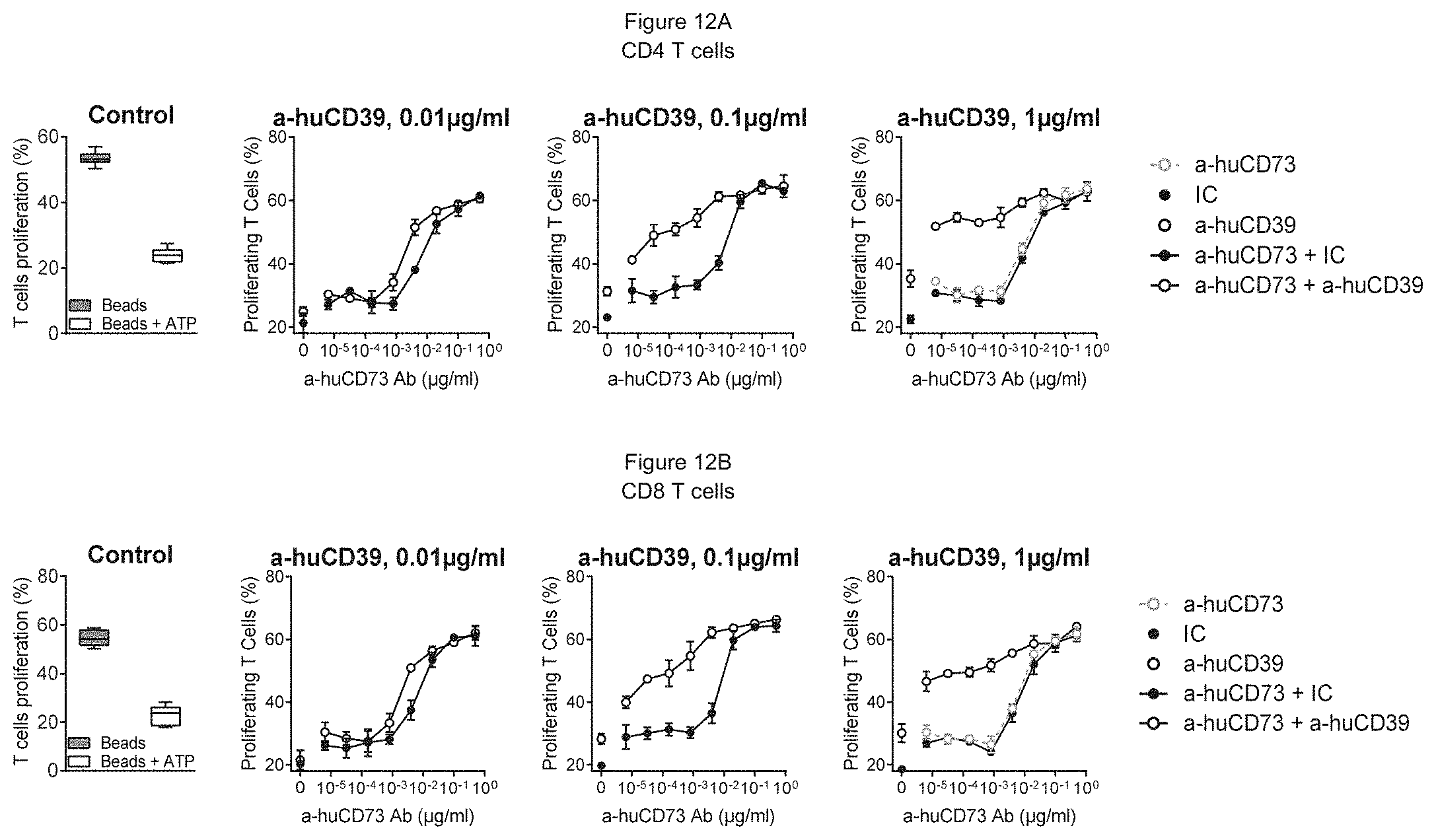
D00014

D00015

D00016

P00001

S00001
XML
uspto.report is an independent third-party trademark research tool that is not affiliated, endorsed, or sponsored by the United States Patent and Trademark Office (USPTO) or any other governmental organization. The information provided by uspto.report is based on publicly available data at the time of writing and is intended for informational purposes only.
While we strive to provide accurate and up-to-date information, we do not guarantee the accuracy, completeness, reliability, or suitability of the information displayed on this site. The use of this site is at your own risk. Any reliance you place on such information is therefore strictly at your own risk.
All official trademark data, including owner information, should be verified by visiting the official USPTO website at www.uspto.gov. This site is not intended to replace professional legal advice and should not be used as a substitute for consulting with a legal professional who is knowledgeable about trademark law.