Chimeric Antigen Receptors And Compositions And Methods Of Use Thereof
Epstein; Alan L.
U.S. patent application number 16/335570 was filed with the patent office on 2020-01-16 for chimeric antigen receptors and compositions and methods of use thereof. The applicant listed for this patent is University of Southern California. Invention is credited to Alan L. Epstein.
| Application Number | 20200016201 16/335570 |
| Document ID | / |
| Family ID | 61689763 |
| Filed Date | 2020-01-16 |


View All Diagrams
| United States Patent Application | 20200016201 |
| Kind Code | A1 |
| Epstein; Alan L. | January 16, 2020 |
CHIMERIC ANTIGEN RECEPTORS AND COMPOSITIONS AND METHODS OF USE THEREOF
Abstract
Disclosed herein are novel chimeric antigen receptors (CARs) targeting human LHR, B7-H4, HLA-G, or HLA-DR, and therapeutic methods of their use. LHR, B7-H4, HLA-G, or HLA-DR are expressed in the context of many human cancers including thyroid, prostate, colon, breast, ovarian, and renal cancers, as well as B-cell leukymias and lymphomas.
| Inventors: | Epstein; Alan L.; (Los Angeles, CA) | ||||||||||
| Applicant: |
|
||||||||||
|---|---|---|---|---|---|---|---|---|---|---|---|
| Family ID: | 61689763 | ||||||||||
| Appl. No.: | 16/335570 | ||||||||||
| Filed: | September 22, 2017 | ||||||||||
| PCT Filed: | September 22, 2017 | ||||||||||
| PCT NO: | PCT/US2017/052974 | ||||||||||
| 371 Date: | March 21, 2019 |
Related U.S. Patent Documents
| Application Number | Filing Date | Patent Number | ||
|---|---|---|---|---|
| 62399244 | Sep 23, 2016 | |||
| Current U.S. Class: | 1/1 |
| Current CPC Class: | C07K 14/723 20130101; C07K 2319/30 20130101; A61K 2039/5156 20130101; C07K 14/70521 20130101; C07K 2317/622 20130101; C07K 2317/73 20130101; C07K 14/70517 20130101; A61P 13/08 20180101; A61P 43/00 20180101; C07K 16/30 20130101; C07K 14/7051 20130101; A61K 35/17 20130101; A61P 1/00 20180101; C07K 14/70532 20130101; A61P 35/00 20180101; C07K 16/3092 20130101; C07K 2319/00 20130101; C07K 16/2833 20130101; C07K 2319/03 20130101; C07K 2319/33 20130101; A61P 35/02 20180101; A61K 2039/5158 20130101; A61P 15/00 20180101; C07K 14/70578 20130101; C07K 16/2869 20130101 |
| International Class: | A61K 35/17 20060101 A61K035/17; C07K 14/725 20060101 C07K014/725; C07K 14/705 20060101 C07K014/705; C07K 14/72 20060101 C07K014/72; C07K 16/28 20060101 C07K016/28; C07K 16/30 20060101 C07K016/30 |
Claims
1. A chimeric antigen receptor (CAR) comprising: (a) an antigen binding domain of an anti-luteinizing hormone receptor ("LHR") antibody, an anti-B7-H4 antibody, an anti-HLA-G, or an HLA-DR antibody (b) a CD8 .alpha. hinge domain; (c) a CD8 .alpha. transmembrane domain; (d) two or more costimulatory signaling regions; and (e) a CD3 zeta signaling domain.
2. The CAR of claim 1, wherein the two or more costimulatory signaling regions are selected from CD27, CD28, 4-IBB (CD 137), OX40, CD30, CD40, PD-1, ICOS, lymphocyte function-associated antigen-1 (LFA-1), CD2, CD7, CD27, LIGHT, NKG2C, and B7-H3.
3. The CAR of claim 1, wherein the antigen binding domain of the anti-LHR antibody, anti-B7-H4 antibody, an anti-HLA-G, or an HLA-DR antibody comprises an anti-LHR heavy chain (HC) variable region and an anti-LHR light chain (LC) variable region.
4. The CAR of claim 3, further comprising a linker polypeptide located between the anti-LHR HC, anti-B7-H4 HC, anti-HLA-G HC, or anti-HLA-DR HC variable region and the anti-LHR LC, anti-B7-H4 LC, anti-HLA-G LC or anti-HLA-DR LC variable region.
5. The CAR of claim 1, wherein the anti-LHR antibody HC comprises: (a) a CDR1 comprising the amino acid sequence of GYSITSGYG, GFSLTTYG, or GYSFTGYY, or an equivalent of each thereof; and/or (b) a CDR2 comprising the amino acid sequence of IHYSGST, IWGDGST, or IYPYNGVS, or an equivalent of each thereof; and/or (c) a CDR3 comprising the amino acid sequence of ARSLRY, AEGSSLFAY, or ARERGLYQLRAMDY, or an equivalent of each thereof; and/or the anti-LHR antibody LC comprises: (a) a CDR1 comprising the amino acid sequence of SSVNY, QSLLNSGNQKNY, or QSISNN, or an equivalent of each thereof; and/or (b) a CDR2 comprising the amino acid sequence of DTS, WAS, or NAS, or an equivalent of each thereof; and/or (c) a CDR3 comprising the amino acid sequence of HQWSSYPYT, QNDYSYPLT, or QQSNSWPYT, or an equivalent of each thereof, the anti-B7-H4 antibody HC comprises: (a) a CDR1 comprising the amino acid sequence of GXTF GFTFSSFG, GFTFSSYG, or GYTFTDY, or an equivalent of each thereof; and/or (b) a CDR2 comprising the amino acid sequence of ISSXXXT, INPNNGGT, ISSGSSTL, or ISSSNSTI, or an equivalent of each thereof; and/or (c) a CDR3 comprising the amino acid sequence of ARPXYY, ARPLYYYGSVMDY, or ARPYYYGSSYDY or an equivalent of each thereof; and/or the anti-B7-H4 antibody LC comprises: (a) a CDR1 comprising the amino acid sequence of QSIVHXNGTY, ENIGSY, QSIVHRNGNTY, or QSIVHSNGNTY or an equivalent of each thereof; and/or (b) a CDR2 comprising the amino acid sequence of KVS or AAT, or an equivalent of each thereof; and/or (c) a CDR3 comprising the amino acid sequence of FQGSXVPXT, QHYYSTLVT, FQGSYVPPT, or FQGSHVPLT or an equivalent of each thereof, the anti-HLA-G antibody HC comprises: (a) a CDR1 comprising the amino acid sequence of GFNIKDTY or GFTFNTYA, or an equivalent of each thereof; and/or (b) a CDR2 comprising the amino acid sequence of IDPANGNT or IRSKSNNYAT, or an equivalent of each thereof; and/or (c) a CDR3 comprising the amino acid sequence of ARSYYGGFAY, or VRGGYWSFDV, or an equivalent of each thereof; and/or the anti-HLA-G LC comprises: (a) a CDR1 comprising the amino acid sequence of KSVSTSGYSY or KSLLHSNGNTY, or an equivalent of each thereof; and/or (b) a CDR2 comprising the amino acid sequence of LVS or RMS, or an equivalent of each thereof; and/or (c) a CDR3 comprising the amino acid sequence of QHSRELPRT or MQHLEYPYT, or an equivalent of each thereof, wherein the anti-HLA-DR HC comprises: (a) a CDR1 comprising the amino acid sequence of a CDRH1 of a Lym-1 antibody or a CDRH1 of a Lym-2 antibody, or an equivalent of each thereof; and/or (b) a CDR2 comprising the amino acid sequence of a CDRH2 of a Lym-1 antibody or a CDRH2 of a Lym-2 antibody, or an equivalent of each thereof; and/or (c) a CDR3 comprising the amino acid sequence of a CDRH3 of a Lym-1 antibody or a CDRH3 of a Lym-2 antibody, or an equivalent of each thereof; and/or the anti-HLA-DR LC comprises: (a) a CDR1 comprising the amino acid sequence of (i) a CDRL1 of a Lym-1 antibody or a CDRL1 of a Lym-2 antibody, or an equivalent of each thereof; and/or (b) a CDR2 comprising the amino acid sequence of a CDRL2 of a Lym-1 antibody or a CDRL2 of a Lym-2 antibody, or an equivalent of each thereof; and/or (c) a CDR3 comprising the amino acid sequence of a CDRL3 of a Lym-1 antibody or a CDRL3 of a Lym-2 antibody, or an equivalent of each thereof.
6.-11. (canceled)
12. The CAR of claim 5, wherein an equivalent comprises a polypeptide having at least 80% amino acid identity to polypeptide or a polypeptide that is encoded by a polynucleotide that hybridizes under conditions of high stringency to the complement of a polynucleotide encoding the polypeptide.
13. (canceled)
14. (canceled)
15. An isolated nucleic acid sequence encoding the CAR of claim 1.
16. (canceled)
17. The isolated nucleic acid sequence of claim 15, further comprising a Kozak consensus sequence located upstream of the antigen binding domain of the anti-LHR antibody, anti-B7-H4 antibody, anti-HLA-G antibody, anti-HLA-DR antibody, or an enhancer.
18. The isolated nucleic acid sequence of claim 15, further comprising an antibiotic resistance polynucleotide.
19. The isolated nucleic acid sequence of claim 15, further comprising a switch mechanism for controlling expression and/or activation of the CAR.
20.-23. (canceled)
24. An isolated cell comprising the CAR of claim 1.
25. The isolated cell of claim 24, wherein the isolated cell is an immune cell, that is optionally a T-cell or a natural killer (NK) cell.
26. (canceled)
27. A composition comprising a carrier and the CAR of claim 1.
28. The composition of claim 27, further comprising an antigen binding fragment capable of binding a peptide, wherein the peptide comprises an LHR protein or a fragment thereof, a B7-H4 protein or a fragment thereof, an HLA-G protein or a fragment thereof, or an HLA-DR protein or a fragment thereof.
29.-32. (canceled)
33. A method of producing anti-LHR CAR, anti-B7-H4 CAR, anti-HLA-G CAR, or anti-HLA-DR CAR expressing cells comprising: (i) introducing a population of immune cells with a nucleic acid sequence encoding the CAR of claim 1; and (ii) selecting a subpopulation of immune cells that have been successfully transduced with said nucleic acid sequence of step (i) thereby producing anti-LHR CAR, anti-B7-H4 CAR, anti-HLA-G CAR, or anti-HLA-DR CAR expressing cells.
34. The method of claim 33, wherein the immune cells are T-cells or a natural killer (NK) cells.
35. The method of claim 34, wherein the population of T-cells have been modified to reduce or eliminate expression of endogenous T-cell receptors.
36. The method of claim 35, wherein the population of T-cells were modified using a method that employs RNA interference or CRISPR.
37. A method of inhibiting the growth of a tumor and/or treating a cancer in a subject in need thereof, comprising administering to the subject an effective amount of the anti-LHR CAR, anti-B7-H4 CAR, anti-HLA-G CAR or anti-HLA-DR CAR expressing cells of claim 25.
38. The method of claim 37, wherein the anti-LHR CAR, anti-B7-H4 CAR, anti-HLA-G CAR or anti-HLA-DR CAR expressing cells are autologous or allogenic to the subject being treated.
39. The method of claim 37, wherein the tumor or cancer expresses or overexpresses LHR, B7-H4, HLA-G, or HLA-DR.
40. The method of claim 37, wherein the tumor is a solid tumor, optionally an ovarian tumor or a prostate cancer tumor and/or the cancer is and ovarian cancer or a prostate cancer.
41. The method of claim 37, wherein the subject is a human, an animal, a non-human primate, a dog, cat, a sheep, a mouse, a horse, or a cow.
42.-157. (canceled)
Description
CROSS REFERENCE TO RELATED APPLICATION
[0001] The present application claims priority under 35 U.S.C. .sctn. 119(e) to U.S. Provisional Application 62/399,244, filed on Sep. 23, 2016, the contents of which are hereby incorporated by reference in its entirety.
TECHNICAL FIELD
[0002] This disclosure relates to novel luteinizing hormone receptor (LHR), B7-H4, HLA-G, or HLA-DR chimeric antigen receptor (CAR), cells or compositions comprising the same, and methods for using the same for therapy including solid tumors. Also provided herein are isolated peptides and fusion proteins containing immunogenic determinants for the luteinizing hormone receptor, B7-H4, HLA-G, or HLA-DR chimeric antigen receptor.
BACKGROUND
[0003] The following discussion of the background of the disclosure is merely provided to aid the reader in the understanding the invention and is not admitted to describe or constitute prior art to the present invention.
[0004] Ovarian carcinoma is the most common cause of cancer death from gynecologic tumors (Siegel, R. et al. (2012) CA Cancer J. Clin. 62:10-29). Approximately 25,000 new cases and 14,000 deaths are expected to occur in the United States every year (Siegel, R. et al. (2012) CA Cancer J. Clin. 62:10-29). Overall survival of ovarian carcinoma appears to have improved in the last 30 years as median survival during the 1960s was approximately 12 months compared to the current 38 months. However, the 5-year survival for stage III ovarian cancer has not changed significantly and remains at 25%. The improvement in median survival can be explained in part due to the improvement in front line chemotherapy. The standard initial chemotherapy for patients with ovarian cancer involves a platinum-paclitaxel based regimen (Marcus, C. S. et al. (2014) J. Cancer 5:25-30). Approximately 70% of patients will achieve a clinical response to this therapy. Despite this, most women will relapse and eventually succumb to their disease. Therefore, in an attempt to decrease distant metastasis, prolong time to recurrence and improve overall survival, it is essential to identify novel therapy targets and develop new agents.
[0005] In 2014, an estimated 232,670 new agents.cases of invasive breast cancer will be diagnosed in US women and an estimated 40,000 US women will die from metastatic disease. The risk of contracting breast cancer increases with age so that 77% of cases are over the age of 50 at the time of diagnosis. In general, the mortality rate for patients with breast cancer has decreased since 1989 due to earlier detection, improved treatments, and possibly a decreased incidence because of the declining use of postmenopausal hormone therapy. When detected early, the 5-year survival for localized breast cancer is 99%. By contrast, the 5-year survival for regional disease is 84% and importantly, for metastatic disease, it drops precipitously to 24%.
[0006] This year, an estimated 63,920 adults (39,140 men and 24,780 women) in the United States will be diagnosed with renal cancer. It is estimated that 13,860 deaths (8,900 men and 4,960 women) from this disease will occur this year. Renal cancer is the sixth most common cancer and the tenth most common cause of cancer death for men, and it is the eighth most common cause of cancer for women. The five-year survival rate for renal cancer patients is 72%. Approximately 63% of cases do not have metastatic disease at the time of diagnosis. For this group, the five-year survival rate improves to 92%. By contrast, the five-year survival for renal cancer in the pelvis (metastatic disease) is 51%.
[0007] Therefore, a need exists for a safe and effective treatment of ovarian and other solid tumor cancers, e.g., prostate cancer. This disclosure satisfies this need and provides related advantages as well.
SUMMARY OF THE DISCLOSURE
[0008] Due to the unprecedented results being recently obtained in B-cell lymphomas and leukemias using autologous treatment with genetically engineered chimeric antigen receptor (CAR) T-cells, a number of laboratories have begun to apply this approach to solid tumors including ovarian cancer. CAR modified T-cells combine the HLA-independent targeting specificity of a monoclonal antibody with the cytolytic activity, proliferation, and homing properties of activated T-cells, but do not respond to checkpoint suppression. Because of their ability to kill antigen expressing targets directly, CAR T-cells are highly toxic to any antigen positive cells or tissues making it a requirement to construct CARs with highly tumor specific antibodies. To date, CAR modified T-cells to ovarian carcinomas have been constructed against the .alpha.-folate receptor, mesothelin, and MUC-CD, but all of these have some off-target expression of antigen.
[0009] For instance, in one aspect, disclosed herein are novel anti-B7-H4 antibodies and methods of their use diagnostically and therapeutically. In one aspect, In this regard, provide herein is an isolated antibody comprising a heavy chain (HC) immunoglobulin variable domain sequence and a light chain (LC) immunoglobulin variable domain sequence, wherein the antibody binds to an epitope of human B7-H4 comprising the amino acid sequence:
[0010] IGEDGILSCTFEPDIKLSDIVIQWLKEGVLGLVHEFKEGKDELSEQDEMFRGRT AVFADQVIVGNASLRLKNVQLTDAGTYKCYIITSKGKGNANLEYKTGAFSMPEVNV DYNASSETLRCEAPRWFPQPTVVWASQVDQGANFSEVSNTSFELNSENVTMKVVSV LYNVTINNTYSCMIENDIAKATGDIKVTESEIKRRSHLQLLNSKA or an equivalent thereof.
[0011] HLA-G is is a non-classical MHC class I molecule which primarily serves to suppress cytotoxic immune cell function, particularly as a ligand for the inhibitory NK cell receptors.
[0012] For instance, in one aspect, disclosed herein are novel anti-HLA-G antibodies and methods of their use diagnostically and therapeutically. In one aspect, In this regard, provide herein is an isolated antibody comprising a heavy chain (HC) immunoglobulin variable domain sequence and a light chain (LC) immunoglobulin variable domain sequence, wherein the antibody binds to an epitope of human HLA-G comprising the amino acid sequence:
[0013] GSHSMRYFSA AVSRPGRGEP RFIAMGYVDD TQFVRFDSDS ACPRMEPRAP WVEQEGPEYW EEETRNTKAH AQTDRMNLQT LRGYYNQSEA SSHTLQWMIG CDLGSDGRLL RGYEQYAYDG KDYLALNEDL RSWTAADTAA QISKRKCEAA NVAEQRRAYL EGTCVEWHLA-G YLENGKEMLQ RADPPKTHVT HHPVFDYEAT LRCWALGFYP AEIILTWQRD GEDQTQDVEL VETRPAGDGT FQKWAAVVVP SGEEQRYTCH VQHEGLPEPL MLRWKQSSLP TIPIMGI VAGLVVLAAV VTGAAVAAVL WRKKSSD, or an equivalent thereof.
[0014] Lym-1 and Lym-2 are directed against MHC class II HLA-DR molecules which are primarily expressed on the surface of human B cells, dendritic cells, and B-cell derived lymphomas and leukemias. Aspects of the disclosure relate to an isolated nucleic acid sequence encoding a Lym1 or Lym-2 CARs, antibodies, and vectors comprising the isolated nucleic acid sequences.
[0015] This disclosure provides a new target for the treatment of solid tumors that include, but are not limited to, ovarian, breast, renal, and prostate carcinomas as well as a B-cell lymphoma or leukemia. The targets, which include LHR, B7-H4, HLA-G, and HLA-DR are often expressed on the majority of these tumors but has restricted off-target positivity and therefore a desirable safety profile. Thus, in one aspect, the compositions are particularly useful in the treatment of tumors or cancerous cell that express or overexpress LHR, B7-H4, HLA-G, HLA-DR.
[0016] In one aspect, the antibodies possess a specific binding affinity of at least 10.sup.-6M. In certain aspects, antibodies bind with affinities of at least about 10.sup.-7M, and preferably 10.sup.-8 M, 10.sup.-9M, 10.sup.-10M, 10.sup.-11M, or 10.sup.-12M.
[0017] In one aspect, the present disclosure provides an isolated antibodies, the antibodies comprising a heavy chain (HC) immunoglobulin variable domain sequence and a light chain (LC) immunoglobulin variable domain sequence, wherein the antibody binds to an epitope of a luteinizing hormone receptor (LHR), B7-H4, HLA-G, or HLA-DR. In a further aspect, this disclosure provides an isolated anti-LHR, anti-B7-H4, anti-HLA-g, or anti-HLA-DR antibodies or fragments thereof as disclosed herein and a detectable or purification label, alone or in combination with an LHR, B7-H4, HLA-G, or HLA-DR antigen or fragment thereof. Further provided herein is an ex vivo cell comprising this antigen/antibody complex.
[0018] Aspects of the disclosure relate to a chimeric antigen receptor (CAR) comprising: (a) an antigen binding domain of an LHR, B7-H4, HLA-G, or HLA-DR antibody; (b) a hinge domain; (c) a transmembrane domain; and (d) an intracellular domain. Further aspects of the disclosure relate to a chimeric antigen receptor (CAR) comprising: (a) an antigen binding domain of a LHR, B7-H4, HLA-G, or HLA-DR antibody; (b) a hinge domain; (c) a CD28 transmembrane domain; (d) one or more costimulatory regions selected from a CD28 costimulatory signaling region, a 4-1BB costimulatory signaling region, an ICOS costimulatory signaling region, and an OX40 costimulatory region; and (e) a CD3 zeta signaling domain and alternatives thereof.
[0019] In a further aspect, the present disclosure provides a chimeric antigen receptor (CAR) comprising: (a) an antigen binding domain of an anti-luteinizing hormone receptor ("LHR"), B7-H4, HLA-G, or HLA-DR antibody, (b) a CD8 .alpha. hinge domain; (c) a CD8 .alpha. transmembrane domain; (d) a CD28 and/or a 4-1BB costimulatory signaling region; and (e) a CD3 zeta signaling domain and alternatives thereof.
[0020] In another aspect, the present disclosure provides an isolated nucleic acid sequence encoding the anti-LHR, -B7-H4, -HLA-G, or HLA-DR antibody, or the anti-LHR, -B7-H4, -HLA-G, or -HLA-DR CAR.
[0021] In another aspect, the present disclosure provides a vector comprising the isolated nucleic acid sequence encoding the anti-LHR, -B7-H4, -HLA-G, or -HLA-DR antibody, or the anti-LHR, -B7-H4, -HLA-G, or -HLA-DR CAR.
[0022] In another aspect, the present disclosure provides a vector comprising the isolated nucleic acid sequence encoding the anti-LHR, -B7-H4, -HLA-G, or -HLA-DR antibody, or the anti-LHR, -B7-H4, -HLA-G, or -HLA-DR CAR.
[0023] In another aspect, the present disclosure provides a composition comprising a carrier and one or more of: the anti-LHR, -B7-H4, -HLA-G, or -HLA-DR antibody; and/or the anti-LHR, -B7-H4, -HLA-G, or -HLA-DR CAR; and/or the isolated nucleic acid encoding the anti-LHR, -B7-H4, -HLA-G, or -HLA-DR antibody or the anti-LHR, -B7-H4, -HLA-G, or -HLA-DR CAR; and/or the vector comprising the isolated nucleic acid sequence encoding the anti-LHR, -B7-H4, -HLA-G, or -HLA-DR antibody, or the anti-LHR, -B7-H4, -HLA-G, or -HLA-DR CAR; and/or an isolated cell comprising the anti-LHR, -B7-H4, -HLA-G, or -HLA-DR CAR.
[0024] Other aspects of the disclosure relate to an isolated cell comprising a LHR, B7-H4, HLA-G, or HLA-DR CAR and methods of producing such cells. Still other method aspects of the disclosure relate to methods for inhibiting the growth of a tumor, e.g., a solid tumor, and treating a cancer patient comprising administering an effective amount of the isolated cell.
[0025] In one aspect, the disclosure provides a composition comprising, or alternatively consisting essentially of, or yet further consisting of a carrier and one or more of: an antibody or fragment thereof, a nucleic acid encoding the antibody or fragment thereof, an isolated cell comprising an anti-LHR, -B7-H4, -HLA-G, or -HLA-DR CAR; and/or the isolated nucleic acid encoding the CAR; and/or the vector comprising the nucleic acid encoding the CAR; and/or the isolated cell expressing an anti-LHR CAR, -B7-H4, -HLA-G, or -HLA-DR; and/or the anti-LHR, -B7-H4, -HLA-G, or -HLA-DR antibody.
BRIEF DESCRIPTION OF THE DRAWINGS
[0026] FIGS. 1A-1C show flow cytometry profiles of (FIG. 1A) LHR on TOV21G, (FIG. 1B) mesothelin on SKOV3, and (FIG. 1C) MUC16 on CAOV3 cell lines.
[0027] FIGS. 2A-2C show positive immunohistochemistry staining patterns of (FIG. 2A) LHR antibody on a Stage 2 serous papillary adenocarcinoma; (FIG. 2B) MUC16 antibody on a Stage IIIC endometrioid adenocarcinoma; and (FIG. 2C) mesothelin antibody on a Stage 1C serous papillary adenocarcinoma.
[0028] FIG. 3 shows the sequence used to generate LHR-Fc. Amino acid structure of LHR G-protein showing sequence (outlined area) used to generate a LHR-Fc used in immunization and screening methods to identify potential LHR binding antibodies useful for the generation of LHR CARs.
[0029] FIG. 4 shows typical flow cytometry screen of LHR-Fc ELISA positive antibodies on the ES-2 ovarian carcinoma cell line demonstrating strong reactivity by hybridoma 8B7 only.
[0030] FIG. 5 shows flow cytometry of 5 candidate LHR antibody subclones with highest MFI values on ES-2 human ovarian carcinoma cells.
[0031] FIG. 6 shows a schematic diagram of the DNA sequence for, and the theoretical structure of an anti-LHR CAR in the plasma membrane.
[0032] FIG. 7 shows the alignments of the heavy chain and light chain sequences of LHR antibody subclones.
[0033] FIGS. 8A-D shows a distribution of LHR positive cancers(FIG. 8A); the distribution of LHR intensity with multiple tumor histology groups (FIG. 8B); LHR staining intensity in patients with ovarian, peritoneal, or fallopian tube cancer (FIG. 8C); and LHR staining intensity by tumor pathologic stage group (FIG. 8D).
[0034] FIGS. 9A-D LHR expression in prostate cancer, in histology (FIG. 9A), relative mRNA levels in (AD) prostate cancer and castration resistant (CR) prostate cancer (FIG. 9B) and Western blot (FIG. 9C-D).
[0035] FIG. 10 shows the backbone of the gene transfer vector is an HIV-based, bicistronic lentiviral vector, pLVX-IRES-ZsGreen containing HIV-1 5' and 3' long terminal repeats (LTRs), packaging signal (.PSI.), EF1.alpha. promoter, internal ribosome entry site (IRES), ZsGreen, a green fluorescent protein, woodchuck hepatitis virus post-transcriptional regulatory element (WPRE), and simian virus 40 origin (SV40). Constitutive expression of the transgene comprising of a scFV specific to LHR, a CD8 hinge and transmembrane region and CD28, 4-1BB and CD3.zeta. signaling domain, is insured by the presence of the EF-1.alpha. promoter. Expression of the detection protein, ZsGreen is carried out by the IRES region. Integration of the vector was assayed by the presence of ZsGreen in the cells, via fluorescent microscopy.
[0036] FIG. 11 depicts the results of the cytotoxicity assay of LHR CAR T-cells. Cytotoxicity of the LHR CAR expressing T-cells was determined using an LDH cytotoxicity kit as described in the Methods. Prior to the assay, T-cells were activated using .alpha.CD3/CD8 beads (Stem Cell Technologies, 30 ul to 2 ml of media). The activated T-cells were transduced with LHR lentiviral particles, following which the T cells were activated for using the .alpha.CD3/CD8 beds. Un-transduced, activated T-cells were used as a control. 3,000 SKOV3 cells were plated per well. LHR transduced T cells were added in ratios of 20:1, 10:1, 5:1 and 1:1 (60,000-3000) to the wells. Each data point represents the average of triplicate measurements.
[0037] FIG. 12 depicts mRNA expression of the LHR CAR in primary T-cells. Primary T-cells transduced with the LHR CAR show expression of the LHR mRNA. Primers used spanned the area between the CD8 hinge and the 4-1BB signaling domain (300 bp).
[0038] FIGS. 13A-13C show a schematic diagram and HPLC Analysis of Human B7-H4-Fc Fusion Protein Used as Antigen. (FIG. 1A) The vector used to construct the gene; (FIG. 1B) the completed B7-H4-Fc fusion protein in which the B7-H4 was fused to the N-terminus of the immunoglobulin Fc region of human IgG1 producing a dimeric protein used as antigen. (FIG. 1C) HPLC analysis of purified B7-H4-Fc showing the expected retention time indicative of its molecular weight.
[0039] FIG. 14 shows representative flow cytometry data for mouse monoclonal anti-human B7-H4 on SKBR-3, HT-29, JAR, and T47D cell lines derived from breast adenocarcinoma, colorectal adenocarcinoma, choriocarcinoma, and breast ductal carcinoma, respectively. Darker line represents cells stained for B7-H4, and lighter line represents cells stained with isotype control. A sheep anti-mouse IgG conjugated to FITC was used as the secondary antibody. Cell surface expression of B7-H4 matches q-PCR data for b7-h4 expression in these cell lines (data not shown).
[0040] FIG. 15 shows flow cytometry screening data of newly generated and purified monoclonal antibodies to human B7-H4. Subclones of positive hybridomas (35-8 and 5F6-6) were selected for the generation of CAR T-cells based upon these results. Clone 35-8 was then sequenced and used to produce B7-H4 CAR T-cells for immunotherapy.
[0041] FIGS. 16A-B show representative images of B7-H4 antibody (clone #35-8) staining on 16normal and cancer tissue microarrays. (FIG. 16A) B7-H4 staining on normal tissues. (FIG. 16B) B7-H4 staining on normal and cancer tissue of the breast. Other normal tissues found negative for B7-H4 positivity (not shown) include the following: adrenal gland, bone marrow, cerebellum, esophagus, hypophysis, intestine, lymph node, ovary, prostate, stomach, testis, thyroid, thymus, tongue, uterine, skin, and nerve tissue.
[0042] FIG. 17 shows a schematic diagram of the DNA sequence for, and the theoretical structure of third generation anti-B7-H4 CAR in the plasma membrane.
[0043] FIG. 18A-B shows immunohistochemistry staining of B7-H4 on sections of (FIG. 18A) human breast carcinoma biopsy and (FIG. 18B) SKBR3 human breast cancer cell line pellet showing cell surface positivity for antigen (brown staining).
[0044] FIG. 19 shows a schematic representation of the gene transfer vector and of the transgene. The backbone of the gene transfer vector is an HIV-based, bicistronic lentiviral vector, pLVX-IRES-ZsGreen containing HIV-1 5' and 3' long terminal repeats (LTRs), packaging signal (.PSI.), EF1.alpha. promoter, internal ribosome entry site (IRES), ZsGreen, a green fluorescent protein, woodchuck hepatitis virus post-transcriptional regulatory element (WPRE), and simian virus 40 origin (SV40). Constitutive expression of the transgene comprising of a scFV specific to B7-H4, a CD8 hinge and transmembrane region and CD28, 4-1BB and CD3.zeta. signaling domain, is insured by the presence of the EF-1.alpha. promoter. Expression of the detection protein, ZsGreen is carried out by the IRES region. Integration of the vector can be assayed by the presence of ZsGreen in the cells, via fluorescent microscopy
[0045] FIG. 20 shows cytotoxicity of the B7-H4 CAR T-cells. Cytotoxicity of the B7-H4 CAR expressing T-cells was determined using an LDH cytotoxicity kit as described in the Methods. Prior to the assay, T-cells were activated using .alpha.CD3/CD8 beads (Stem Cell Technologies, 30 .mu.l to 2 ml of media). The activated T-cells were transduced with B7-H4 lentiviral particles, following which the T cells were activated for using the .alpha.CD3/CD8 beads. Un-transduced, activated T-cells were used as a control. 3000 SKBR3 cells were plated per well. B7-H4 transduced T cells were added in ratios of 20:1, 10:1, 5:1 and 1:1 (60,000-3000 cells) to the wells. Each data point represents the average of triplicate measurements.
[0046] FIG. 21 shows flow cytometry screening data of newly generated monoclonal antibodies to human HLA-G. Subclones of positive hybridomas (3H11-12 and 4E3-1) were selected for the generation of CAR T-cells based upon these results.
[0047] FIGS. 22A-22D show immunohistochemistry of HLA-G reactivity in papillary thyroid cancer and normal thyroid tissue with HLA-ABC control staining. FIG. 22A shows low magnification of HLA-G positive papillary thyroid carcinoma section using antibody 4E3-1 (100.times.). FIG. 22B shows higher magnification of second papillary thyroid carcinoma positive for HLA-G (250.times.). FIG. 22C shows negative reactivity of normal thyroid tissues for HLA-G (250.times.), and FIG. 22D shows positive reactivity of normal thyroid tissue for HLA-ABC (100.times.).
[0048] FIG. 23 shows schematic diagram of the DNA sequence for, and the theoretical structure of third generation anti-HLA-G CAR in the plasma membrane.
[0049] FIG. 24 shows additional antibody screening, as described in FIG. 1.
[0050] FIG. 25 depicts a schematic of the gene-transfer vector and the transgene. The backbone of the gene transfer vector is an HIV-based, bicistronic lentiviral vector, pLVX-IRES-ZsGreen containing HIV-1 5' and 3' long terminal repeats (LTRs), packaging signal (.PSI.), EF1.alpha. promoter, internal ribosome entry site (IRES), ZsGreen, a green fluorescent protein, woodchuck hepatitis virus post-transcriptional regulatory element (WPRE), and simian virus 40 origin (SV40). Constitutive expression of the transgene comprising of a scFV specific to HLA-G, a CD8 hinge and transmembrane region and CD28, 4-1BB and CD3.zeta. signaling domain, is insured by the presence of the EF-1.alpha. promoter. Expression of the detection protein, ZsGreen is carried out by the IRES region. Integration of the vector can be assayed by the presence of ZsGreen in the cells, via fluorescent microscopy.
[0051] FIG. 26 shows cytotoxicity of the HLA-G CAR T-cells. Cytotoxicity of the HLA-G CAR expressing T-cells was determined using an LDH cytotoxicity kit as described in the Methods. Prior to the assay, T-cells were activated using .alpha.CD3/CD8 beads (Stem Cell Technologies, 30 ul to 2 ml of media). The activated T-cells were transduced with HLA-G lentiviral particles, following which the T cells were activated for using the .alpha.CD3/CD8 beads. Un-transduced, activated T-cells and the TLBR-2 T lymphoma cell line were used as controls. 3,000 SKOV3 or TLBR-2 cells were plated per well. HLA-G transduced T cells were added in ratios of 20:1, 10:1, 5:1 and 1:1 (60,000-3000 cells) to the wells. Each data point represents the average of triplicate measurements.
[0052] FIG. 27 shows protein expression of the HLA-G CAR. T-cells transduced with the HLA-G CAR lentiviral particles express protein for the HLA-G CAR. The estimated size of the CAR protein is 60 kDa. A CD3.zeta. antibody was used to detect the protein. Fifty .mu.g of protein was used for the western blot. .beta.-actin was used as a loading control.
[0053] FIGS. 28A-28F show flow cytometric analysis of (FIG. 28A) negative control; (FIG. 28B) Lym-1; (FIG. 28C) Lym-1 and B1; (FIG. 28D) B1 only; (FIG. 28E) Lym-2; and (FIG. 28F) Lym-2 and B1 staining reactivity with normal peripheral blood lymphocytes of patients. Both Lym-1 and Lym-2 have different profiles of binding to normal human peripheral B cells.
[0054] FIGS. 29A-29B show Lym-1 and Lym-2 staining of normal human tonsil demonstrating membrane positivity in B-cell germinal centers. Differences in staining patterns are evident between Lym-1 (FIG. 29A) and Lym-2 (FIG. 29B). Only scattered interfollicular dendritic cells are positive for both antibodies in the T-cell zones (IHC, frozen sections, .times.325).
[0055] FIGS. 30A and 30B show immunoperoxidase staining of Lym-1 and Lym-2 monoclonal antibodies with an intermediate grade malignant B-cell lymphoma. Immunoperoxidase staining of Lym-1 (FIG. 30A) and Lym-2 (FIG. 30B) monoclonal antibodies with an intermediate grade malignant B-cell lymphoma (frozen sections, .times.720). Note prominent membrane staining pattern of majority of cells in the section.
[0056] FIGS. 31A-31C show binding profiles and Scatchard Plots of (FIG. 31A) Binding profiles of Lym-1 monoclonal antibodies to Raji cells and Lym-2 monoclonal antibodies to ARH-77 cells; (FIG. 31B) Scatchard plot analysis of Lym-1 monoclonal antibodies with Raji cells; (FIG. 31C) Scatchard plot analysis of Lym-2 monoclonal antibodies with ARH-77 cells.
[0057] FIGS. 32A and 32B show immunoprecipitation of .sup.35S-methionine and .sup.14C-leucine-labeled Raji proteins by Lym-1 (FIG. 32A) and SC-2 anti-HLA-DR antibody (FIG. 32B).
[0058] FIGS. 33A and 33B show a construction schematic of (FIG. 33A) Lym-1 and (FIG. 33B) Lym-2 CAR T-cells for immunotherapy. FIGS. 6A and 6B disclose a flexible linker sequence.
[0059] FIG. 34 depicts a schematic a non-limiting exemplary Lym-1 gene-transfer vector and transgene. The backbone of the gene transfer vector is an HIV-based, bicistronic lentiviral vector, pLVX-IRES-ZsGreen containing HIV-1 5' and 3' long terminal repeats (LTRs), packaging signal (.PSI.), EF1.alpha. promoter, internal ribosome entry site (IRES), ZsGreen, a green fluorescent protein, woodchuck hepatitis virus post-transcriptional regulatory element (WPRE), and simian virus 40 origin (SV40).Constitutive expression of the transgene comprising of .alpha.CD8 leader sequence, a scFV specific to Lym-1, a CD8 hinge and transmembrane region and 4-1BB and CD3.zeta. signaling domain, is insured by the presence of the EF-1.alpha. promoter. Expression of the detection protein, ZsGreen is carried out by the IRES region. Integration of the vector can be assayed by the presence of ZsGreen in the cells, via fluorescent microscopy.
[0060] FIG. 35 shows expression of Lym-1 CAR on primary human T-cells. T-cells were transduced with the Lym-1 CAR and stained with Biotein-Protein L, followed by Streptavidin-PE. Cells were analyzed by flow cytometry.
[0061] FIG. 36 shows cytotoxicity of the Lym-1-CAR T-cells. Cytotoxicity of the Lym-1 CAR expressing T-cells was determined using an LDH cytotoxicity kit as described in the Methods. Prior to the assay, T-cells were activated using .alpha.CD3/CD8 beads (Stem Cell Technologies, 30 ul to 2 ml of media). The activated T-cells were transduced with Lym-1 CAR lentiviral particles, following which the T cells were activated using the .alpha.CD3/CD8 beads. Un-transduced, activated T-cells were used as a control. 15,000 Raji cells were plated per well. Lym-1 CAR transduced T cells were added in ratios of 20:1, 10:1, 5:1 and 1:1 to the wells. Each data point represents the average of triplicate measurements.
[0062] FIG. 37 depicts a schematic a non-limiting exemplary Lym-2 gene-transfer vector and transgene. The backbone of the gene transfer vector is an HIV-based, bicistronic lentiviral vector, pLVX-IRES-ZsGreen containing HIV-1 5' and 3' long terminal repeats (LTRs), packaging signal (.PSI.), EF1.alpha. promoter, internal ribosome entry site (IRES), ZsGreen, a green fluorescent protein, woodchuck hepatitis virus post-transcriptional regulatory element (WPRE), and simian virus 40 origin (SV40).Constitutive expression of the transgene comprising of a CD8 leader sequence, an scFV specific to Lym-2, a CD8 hinge and transmembrane region and CD28, 4-1BB and CD3.zeta. signaling domain, is insured by the presence of the EF-1.alpha. promoter. Expression of the detection protein, ZsGreen is carried out by the IRES region. Integration of the vector can be assayed by the presence of ZsGreen in the cells, via fluorescent microscopy.
[0063] FIG. 38 shows expression of Lym-2 CAR on primary human T-cells. T-cells were transduced with the Lym-2 CAR and stained with Biotein-Protein L, followed by Streptavidin-PE. Cells were analyzed by flow cytometry.
[0064] FIG. 39 shows cytotoxicity of the Lym-2-CAR T-cells. Cytotoxicity of the Lym-2 CAR expressing T-cells was determined using an LDH cytotoxicity kit as described in the Methods. Prior to the assay, T-cells were activated using .alpha.CD3/CD8 beads (Stem Cell Technologies, 30 ul to 2 ml of media). The activated T-cells were transduced with Lym-2 CAR lentiviral particles, following which the T cells were activated using the .alpha.CD3/CD8 beads. Un-transduced, activated T-cells were used as a control. 15,000 Raji cells were plated per well. Lym-2 CAR transduced T cells were added in ratios of 20:1, 10:1, 5:1 and 1:1 to the wells. Each data point represents the average of triplicate measurements.
[0065] FIG. 40 demonstrates that Lym-1, Lym-2, and CD19 CAR T-cells are highly cytotoxic to human lymphoma Raji cells. Raji Burkitt's lymphoma cells are positive for both HLA-Dr targeted by Lym-1 and Lym-2 and also CD19 which acted as a positive control for CD19 CAR T-cells. Negative controls consisted of CD3+ T cells and Zsgreen cells.
[0066] FIG. 41 demonstrates that Lym-1, Lym-2, but not CD19 CAR are highly cytolytic against HLA-Dr positive but CD19 negative TLBR-2 human T lymphoma cells in vitro. TLBR-2 human T-lymphoma cells derived from a breast implant associated lymphoma is positive for HLA-Dr but not CD19 (Lechner et al. (2012) Clin. Cancer Res. 18 (17):4549-4559). These results demonstrate the specificity of the Lym-1 and Lym-2 CAR T-cells and their potency in killing HLA-Dr positive tumors. The percentage of Lym-1 CAR-T and CD19 CAR-T positive cells were adjusted to 50% using regular un-transduced primary T cells. The percentage of Lym-2 CAR-T cells was 24%.
[0067] FIG. 42 shows the results of FACs analysis of transfected NK cells.
DETAILED DESCRIPTION
[0068] It is to be understood that the present disclosure is not limited to particular aspects described, as such may, of course, vary. It is also to be understood that the terminology used herein is for the purpose of describing particular aspects only, and is not intended to be limiting, since the scope of the present disclosure will be limited only by the appended claims.
[0069] Unless defined otherwise, all technical and scientific terms used herein have the same meanings as commonly understood by one of ordinary skill in the art to which this technology belongs. Although any methods and materials similar or equivalent to those described herein can be used in the practice or testing of the present technology, the preferred methods, devices and materials are now described. All technical and patent publications cited herein are incorporated herein by reference in their entirety. Nothing herein is to be construed as an admission that the present technology is not entitled to antedate such disclosure by virtue of prior invention.
[0070] The practice of the present technology will employ, unless otherwise indicated, conventional techniques of tissue culture, immunology, molecular biology, microbiology, cell biology, and recombinant DNA, which are within the skill of the art. See, e.g., Sambrook and Russell eds. (2001) Molecular Cloning: A Laboratory Manual, 3rd edition; the series Ausubel et al. eds. (2007) Current Protocols in Molecular Biology; the series Methods in Enzymology (Academic Press, Inc., N.Y.); MacPherson et al. (1991) PCR 1: A Practical Approach (IRL Press at Oxford University Press); MacPherson et al. (1995) PCR 2: A Practical Approach; Harlow and Lane eds. (1999) Antibodies, A Laboratory Manual; Freshney (2005) Culture of Animal Cells: A Manual of Basic Technique, 5th edition; Gait ed. (1984) Oligonucleotide Synthesis; U.S. Pat. No. 4,683,195; Hames and Higgins eds. (1984) Nucleic Acid Hybridization; Anderson (1999) Nucleic Acid Hybridization; Hames and Higgins eds. (1984) Transcription and Translation; Immobilized Cells and Enzymes (IRL Press (1986)); Perbal (1984) A Practical Guide to Molecular Cloning; Miller and Calos eds. (1987) Gene Transfer Vectors for Mammalian Cells (Cold Spring Harbor Laboratory); Makrides ed. (2003) Gene Transfer and Expression in Mammalian Cells; Mayer and Walker eds. (1987) Immunochemical Methods in Cell and Molecular Biology (Academic Press, London); and Herzenberg et al. eds (1996) Weir's Handbook of Experimental Immunology.
[0071] All numerical designations, e.g., pH, temperature, time, concentration, and molecular weight, including ranges, are approximations which are varied (+) or (-) by increments of 1.0 or 0.1, as appropriate, or alternatively by a variation of +/-15%, or alternatively 10%, or alternatively 5%, or alternatively 2%. It is to be understood, although not always explicitly stated, that all numerical designations are preceded by the term "about". It also is to be understood, although not always explicitly stated, that the reagents described herein are merely exemplary and that equivalents of such are known in the art.
[0072] It is to be inferred without explicit recitation and unless otherwise intended, that when the present technology relates to a polypeptide, protein, polynucleotide or antibody, an equivalent or a biologically equivalent of such is intended within the scope of the present technology.
Definitions
[0073] As used in the specification and claims, the singular form "a", "an", and "the" include plural references unless the context clearly dictates otherwise. For example, the term "a cell" includes a plurality of cells, including mixtures thereof.
[0074] As used herein, the term "animal" refers to living multi-cellular vertebrate organisms, a category that includes, for example, mammals and birds. The term "mammal" includes both human and non-human mammals.
[0075] The terms "subject," "host," "individual," and "patient" are as used interchangeably herein to refer to human and veterinary subjects, for example, humans, animals, non-human primates, dogs, cats, sheep, mice, horses, and cows. In some embodiments, the subject is a human.
[0076] As used herein, the term "antibody" collectively refers to immunoglobulins or immunoglobulin-like molecules including by way of example and without limitation, IgA, IgD, IgE, IgG and IgM, combinations thereof, and similar molecules produced during an immune response in any vertebrate, for example, in mammals such as humans, goats, rabbits and mice, as well as non-mammalian species, such as shark immunoglobulins. Unless specifically noted otherwise, the term "antibody" includes intact immunoglobulins and "antibody fragments" or "antigen binding fragments" that specifically bind to a molecule of interest (or a group of highly similar molecules of interest) to the substantial exclusion of binding to other molecules (for example, antibodies and antibody fragments that have a binding constant for the molecule of interest that is at least 10.sup.3M.sup.-1 greater, at least 10.sup.4M.sup.-1 greater or at least 10.sup.5 M.sup.-1 greater than a binding constant for other molecules in a biological sample). The term "antibody" also includes genetically engineered forms such as chimeric antibodies (for example, humanized murine antibodies), heteroconjugate antibodies (such as, bispecific antibodies). See also, Pierce Catalog and Handbook, 1994-1995 (Pierce Chemical Co., Rockford, Ill.); Kuby, J., Immunology, 3.sup.rd Ed., W.H. Freeman & Co., New York, 1997. An "antigen binding fragment" of an antibody is a portion of an antibody that retains the ability to specifically bind to the target antigen of the antibody.
[0077] As used herein, the term "monoclonal antibody" refers to an antibody produced by a single clone of B-lymphocytes or by a cell into which the light and heavy chain genes of a single antibody have been transfected. Monoclonal antibodies are produced by methods known to those of skill in the art, for instance by making hybrid antibody-forming cells from a fusion of myeloma cells with immune spleen cells. Monoclonal antibodies include humanized monoclonal antibodies and human antibodies.
[0078] In terms of antibody structure, an immunoglobulin has heavy (H) chains and light (L) chains interconnected by disulfide bonds. There are two types of light chain, lambda (.lamda.) and kappa (.kappa.). There are five main heavy chain classes (or isotypes) which determine the functional activity of an antibody molecule: IgM, IgD, IgG, IgA and IgE. Each heavy and light chain contains a constant region and a variable region, (the regions are also known as "domains"). In combination, the heavy and the light chain variable regions specifically bind the antigen. Light and heavy chain variable regions contain a "framework" region interrupted by three hypervariable regions, also called "complementarity-determining regions" or "CDRs". The extent of the framework region and CDRs have been defined (see, Kabat et al., Sequences of Proteins of Immunological Interest, U.S. Department of Health and Human Services, 1991, which is hereby incorporated by reference). The Kabat database is now maintained online. The sequences of the framework regions of different light or heavy chains are relatively conserved within a species. The framework region of an antibody, that is the combined framework regions of the constituent light and heavy chains, largely adopts a .beta.-sheet conformation and the CDRs form loops which connect, and in some cases form part of, the .beta.-sheet structure. Thus, framework regions act to form a scaffold that provides for positioning the CDRs in correct orientation by inter-chain, non-covalent interactions.
[0079] The CDRs are primarily responsible for binding to an epitope of an antigen. The CDRs of each chain are typically referred to as CDR1, CDR2, and CDR3, numbered sequentially starting from the N-terminus, and are also typically identified by the chain in which the particular CDR is located. Thus, a V.sub.H CDR3 is located in the variable domain of the heavy chain of the antibody in which it is found, whereas a V.sub.L CDR1 is the CDR1 from the variable domain of the light chain of the antibody in which it is found. An antibody that binds LHR, B7-H4, HLA-G, or HLA-DR will have a specific V.sub.H region and the V.sub.L region sequence, and thus specific CDR sequences. Antibodies with different specificities (i.e. different combining sites for different antigens) have different CDRs. Although it is the CDRs that vary from antibody to antibody, only a limited number of amino acid positions within the CDRs are directly involved in antigen binding. These positions within the CDRs are called specificity determining residues (SDRs).
[0080] As used herein, the term "antigen" refers to a compound, composition, or substance that may be specifically bound by the products of specific humoral or cellular immunity, such as an antibody molecule or T-cell receptor. Antigens can be any type of molecule including, for example, haptens, simple intermediary metabolites, sugars (e.g., oligosaccharides), lipids, and hormones as well as macromolecules such as complex carbohydrates (e.g., polysaccharides), phospholipids, and proteins. Common categories of antigens include, but are not limited to, viral antigens, bacterial antigens, fungal antigens, protozoa and other parasitic antigens, tumor antigens, antigens involved in autoimmune disease, allergy and graft rejection, toxins, and other miscellaneous antigens.
[0081] As used herein, the term "antigen binding domain" refers to any protein or polypeptide domain that can specifically bind to an antigen target.
[0082] The term "chimeric antigen receptor" (CAR), as used herein, refers to a fused protein comprising an extracellular domain capable of binding to an antigen, a transmembrane domain derived from a polypeptide different from a polypeptide from which the extracellular domain is derived, and at least one intracellular domain. The "chimeric antigen receptor (CAR)" is sometimes called a "chimeric receptor", a "T-body", or a "chimeric immune receptor (CIR)." The "extracellular domain capable of binding to an antigen" means any oligopeptide or polypeptide that can bind to a certain antigen. The "intracellular domain" means any oligopeptide or polypeptide known to function as a domain that transmits a signal to cause activation or inhibition of a biological process in a cell. In certain embodiments, the intracellular domain may comprise, alternatively consist essentially of, or yet further comprise one or more costimulatory signaling domains in addition to the primary signaling domain. The "transmembrane domain" means any oligopeptide or polypeptide known to span the cell membrane and that can function to link the extracellular and signaling domains. A chimeric antigen receptor may optionally comprise a "hinge domain" which serves as a linker between the extracellular and transmembrane domains. Non-limiting exemplary polynucleotide sequences that encode for components of each domain are disclosed herein, e.g.:
[0083] Hinge domain: IgG1 heavy chain hinge sequence:
TABLE-US-00001 CTCGAGCCCAAATCTTGTGACAAAACTCACACATGCCCACCGTGCCCG
[0084] Transmembrane domain: CD28 transmembran region:
TABLE-US-00002 TTTTGGGTGCTGGTGGTGGTTGGTGGAGTCCTGGCTTGCTATAGCTTGC TAGTAACAGTGGCCTTTATTATTTTCTGGGTG
[0085] Intracellular domain: 4-1BB co-stimulatory signaling region:
TABLE-US-00003 AAACGGGGCAGAAAGAAACTCCTGTATATATTCAAACAACCATTTATGAG ACCAGTACAAACTACTCAAGAGGAAGATGGCTGTAGCTGCCGATTTCCAG AAGAAGAAGAAGGAGGATGTGAACTG
[0086] Intracellular domain: CD28 co-stimulatory signaling region:
TABLE-US-00004 AGGAGTAAGAGGAGCAGGCTCCTGCACAGTGACTACATGAACATGACTCC CCGCCGCCCCGGGCCCACCCGCAAGCATTACCAGCCCTATGCCCCACCAC GCGACTTCGCAGCCTATCGCTCC
[0087] Intracellular domain: CD3 zeta signaling region:
TABLE-US-00005 AGAGTGAAGTTCAGCAGGAGCGCAGACGCCCCCGCGTACCAGCAGGGCC AGAACCAGCTCTATAACGAGCTCAATCTAGGACGAAGAGAGGAGTACGAT GTTTTGGACAAGAGACGTGGCCGGGACCCTGAGATGGGGGGAAAGCCGAG AAGGAAGAACCCTCAGGAAGGCCTGTACAATGAACTGCAGAAAGATAAGA TGGCGGAGGCCTACAGTGAGATTGGGATGAAAGGCGAGCGCCGGAGGGGC AAGGGGCACGATGGCCTTTACCAGGGTCTCAGTACAGCCACCAAGGACAC CTACGACGCCCTTCACATGCAGGCCCTGCCCCCTCGCTAA
[0088] Further embodiments of each exemplary domain component include other proteins that have analogous biological function that share at least 70%, or alternatively at least 80% amino acid sequence identity, preferably 90% sequence identity, more preferably at least 95% sequence identity with the proteins encoded by the above disclosed nucleic acid sequences. Further, non-limiting examples of such domains are provided herein.
[0089] As used herein, the term "HLA-DR" (refers to an MHC class II cell surface receptor associated with this name and any other molecules that have analogous biological function that share at least 80% amino acid sequence identity, preferably 90% sequence identity, or alternatively at least 95% sequence identity with any HLA-DR variant, including but not limited to any one of its several variants, including but not limited to HLA-DR serotypes DR1 to DR 75 comprising a combination of HLA-DRA and HLA-DRB haplotypes. Examples of the HLA-DR sequences are known in the art and non-limited examples of such are disclosed in Rose, L. M. et al. (1996) Cancer Immunol. Immunother. 43:26-30:
[0090] HLA-DRB1*1001 [DR10]
[0091] GDTRPRFLEEVKFECHFFNGTERVRLLERRVHNQEEYARYDSDVGEYRAVTELGRP DAEWNSQKDLLERRRAAVDTYCRHNYGVGESFTVQRRVQPKVTVYPSKTQPLQH HNLLVCSVNGFYPGSIEVRWFRNGQEEKTGVVSTGLIQNGDWTFQTLVMLETVPQS GEVYTCQVEHPSVMSPLTVEWRARSESAQSKMLSGVGGFVLGLLFLGAGLFIYFRN QKGHSGLPPTGFLS;
[0092] HLA-DRB3*0201 [DR52]
[0093] GDTRPRFLELLKSECHFFNGTERVRFLERHFHNQEEYARFDSDVGEYRAVFELGRPD AEYWNSQKDLLEQKRGQVDNYCRHNYGVVESFTVQRRVHPQVTVYPAKTQPLQH HNLLVCSVSGFYPGSIEVRWFRNGQEEKAGVVSTGLIQNGDWTFQTLVMLETFPRSG EVYTCQVEHPSVTSPLTVEWSARSESAQSKMLSGVGGFVLGLLFLGAGLFIYFRNQK GHSGLQPTGFLS;
[0094] HLA-DRB1*0301 [DR17 (3)]
[0095] GDTRPRFLEYSTSECHFFNGTERVRYLDRYFHNQEENVRFDSDVGEFRAVTELGRPD AEWNSQKDLLEQKRGRVDNYCRHNYGVVESFTVQRRVHPKVTVYPSKTQPLQHH NLLVCSVSGFYPGSIEVRWFRNGQEEKTGVVSTGLIQNGDWTFQTLVMLETVPRSGE VYTCQVEHPSVTSPLTVEWRARSESAQSKMLSGVGGFVLGLLFLGAGLFIYFRNQKG HSGLQPRGFLS, as well as equivalents of each thereof.
[0096] Rose et al. also discloses an exemplary epitope to which an HLA-DR specific antibody may bind and therefore can serve as an immunogen for the generation of additional antibodies, monoclonal antibodies and antigen binding fragments of each thereof. The sequences associated with each of the listed reference(s) and GenBank Accession Numbers that correspond to the name HLA-DR or its equivalents including but not limited to the specified HLA-DR subtypes are herein incorporated by reference as additional non-limiting examples.
[0097] A "composition" typically intends a combination of the active agent, e.g., a CAR T cell or a CAR NK cell, an antibody, a compound or composition, and a naturally-occurring or non-naturally-occurring carrier, inert (for example, a detectable agent or label) or active, such as an adjuvant, diluent, binder, stabilizer, buffers, salts, lipophilic solvents, preservative, adjuvant or the like and include pharmaceutically acceptable carriers. Carriers also include pharmaceutical excipients and additives proteins, peptides, amino acids, lipids, and carbohydrates (e.g., sugars, including monosaccharides, di-, tri-, tetra-oligosaccharides, and oligosaccharides; derivatized sugars such as alditols, aldonic acids, esterified sugars and the like; and polysaccharides or sugar polymers), which can be present singly or in combination, comprising alone or in combination 1-99.99% by weight or volume. Exemplary protein excipients include serum albumin such as human serum albumin (HSA), recombinant human albumin (rHA), gelatin, casein, and the like. Representative amino acid/antibody components, which can also function in a buffering capacity, include alanine, arginine, glycine, arginine, betaine, histidine, glutamic acid, aspartic acid, cysteine, lysine, leucine, isoleucine, valine, methionine, phenylalanine, aspartame, and the like. Carbohydrate excipients are also intended within the scope of this technology, examples of which include but are not limited to monosaccharides such as fructose, maltose, galactose, glucose, D-mannose, sorbose, and the like; disaccharides, such as lactose, sucrose, trehalose, cellobiose, and the like; polysaccharides, such as raffinose, melezitose, maltodextrins, dextrans, starches, and the like; and alditols, such as mannitol, xylitol, maltitol, lactitol, xylitol sorbitol (glucitol) and myoinositol.
[0098] The term "consensus sequence" as used herein refers to an amino acid or nucleic acid sequence that is determined by aligning a series of multiple sequences and that defines an idealized sequence that represents the predominant choice of amino acid or base at each corresponding position of the multiple sequences. Depending on the sequences of the series of multiple sequences, the consensus sequence for the series can differ from each of the sequences by zero, one, a few, or more substitutions. Also, depending on the sequences of the series of multiple sequences, more than one consensus sequence may be determined for the series. The generation of consensus sequences has been subjected to intensive mathematical analysis. Various software programs can be used to determine a consensus sequence.
[0099] As used herein, the term "luteinizing hormone receptor" (LHR) refers to a specific molecule associated with this name and any other molecules that have analogous biological function that share at least 70%, or alternatively at least 80% amino acid sequence identity, preferably 90% sequence identity, more preferably at least 95% sequence identity with the LHR sequence as shown herein. The protein sequences associated with GenBank Accession Nos. AAB19917.2 (Homo sapiens), or AAA39432.1 (Mus musculus), or AAA41529.1 (Rattus norvegicus) provide additional example sequences of LHR. Non-limiting examples of such include:
TABLE-US-00006 Luteinizing hormone receptor [Homo sapiens]: MKQRFSALQLLKLLLLLQPPLPRALREALCPEPCNCVPDGALRCPGPTAGL TRLSLAYLPVKVIPSQAFRGLNEVIKIEISQIDSLERIEANAFDNLLNLSE ILIQNTKNLRYIEPGAFINLPRLKYLSICNTGIRKFPDVTKVFSSESNFIL EICDNLHITTIPGNAFQGMNNESVTLKLYGNGFEEVQSHAFNGTTLTSLEL KENVHLEKMEINGAFRGATGPKTLDISSTKLQALPSYGLESIQRLIATSSY SLKKLPSRETFVNLLEATLTYPSHCCAFRNLPTKEQNFSHSISENFSKQCE STVRKVNNKTLYSSMLAESELSGWDYEYGFCLPKTPRCAPEPDAFNPCEDI MGYDFLRVLIWLINILAIMGNMTVLFVLLTSRYKLTVPRFLMCNLSFADFC MGLYLLLIASVDSQTKGQYYNHAIDWQTGSGCSTAGFFTVFASELSVYTLT VITLERWHTITYAIHLDQKLRLRHAILIMLGGWLFSSLIAMLPLVGVSNYM KVSICFPMDVETTLSQVYILTILILNVVAFFIICACYIKIYFAVRNPELMA TNKDTKIAKKMAILIFTDFTCMAPISFFAISAAFKVPLITVTNSKVLLVLF YPINSCANPFLYAIFTKTFQRDFFLLLSKFGCCKRRAELYRRKDFSAYTSN CKNGFTGSNKPSQSTLKLSTLHCQGTALLDKTRYTEC Luteinizing hormone receptor [Mus musculus]: MGRRVPALRQLLVLAMLVLKQSQLHSPELSGSRCPEPCDCAPDGALRCPGP RAGLARLSLTYLPVKVIPSQAFRGLNEVVKIEISQSDSLERIEANAFDNLL NLSEILIQNTKNLLYIEPGAFTNLPRLKYLSICNTGIRTLPDVSKISSSEF NFILEICDNLYITTIPGNAFQGMNNESITLKLYGNGFEEVQSHAFNGTTLI SLELKENIYLEKMHSGTFQGATGPSILDVSSTKLQALPSHGLESIQTLIAT SSYSLKTLPSREKFTSLLVATLTYPSHCCAFRNLPKKEQNFSFSIFENFSK QCESTVREANNETLYSAIFEENELSGWDYDYDFCSPKTLQCTPEPDAFNPC EDIMGYAFLRVLIWLINILAIFGNLTVLFVLLTSRYKLTVPRFLMCNLSFA DFCMGLYLLLIASVDSQTKGQYYNHAIDWQTGSGCSAAGFFTVFASELSVY TLTVITLERWHTITYAVQLDQKLRLRHAIPIMLGGWIFSTLMATLPLVGVS SYMKVSICLPMDVESTLSQVYILSILLLNAVAFVVICACYVRIYFAVQNPE LTAPNKDTKIAKKMAILIFTDFTCMAPISFFAISAAFKVPLITVTNSKVLL VLFYPVNSCANPFLYAVFTKAFQRDFFLLLSRFGCCKHRAELYRRKEFSAC TFNSKNGFPRSSKPSQAALKLSIVHCQQPTPPRVLIQ Luteinizing hormone receptor [Rattus norvegicus]: MGRRVPALRQLLVLAVLLLKPSQLQSRELSGSRCPEPCDCAPDGALRCPGP RAGLARLSLTYLPVKVIPSQAFRGLNEVVKIEISQSDSLERIEANAFDNLL NLSELLIQNTKNLLYIEPGAFTNLPRLKYLSICNTGIRTLPDVTKISSSEF NFILEICDNLHITTIPGNAFQGMNNESVTLKLYGNGFEEVQSHAFNGTTLI SLELKENIYLEKMHSGAFQGATGPSILDISSTKLQALPSHGLESIQTLIAL SSYSLKTLPSKEKFTSLLVATLTYPSHCCAFRNLPKKEQNFSFSIFENFSK QCESTVRKADNETLYSAIFEENELSGWDYDYGFCSPKTLQCAPEPDAFNPC EDIMGYAFLRVLIWLINILAIFGNLTVLFVLLTSRYKLTVPRFLMCNLSFA DFCMGLYLLLIASVDSQTKGQYYNHAIDWQTGSGCGAAGFFTVFASELSVY TLTVITLERWHTITYAVQLDQKLRLRHAIPIMLGGWLFSTLIATMPLVGIS NYMKVSICLPMDVESTLSQVYILSILILNVVAFVVICACYIRIYFAVQNPE LTAPNKDTKIAKKMAILIFTDFTCMAPISFFAISAAFKVPLITVTNSKILL VLFYPVNSCANPFLYAIFTKAFQRDFLLLLSRFGCCKRRAELYRRKEFSAY TSNCKNGFPGASKPSQATLKLSTVHCQQPIPPRALTH
[0100] As used herein, the term "B7-H4" (also known as VTCN1, H4, B7h.5, B7S1, B7X, or PRO129) refers to a specific molecule associated with this name and any other molecules that have analogous biological function that share at least 80% amino acid sequence identity, preferably 90% sequence identity, more preferably at least 95% sequence identity with B7-H4. Examples of the B7-H4 sequence are provided herein. In addition, the protein sequences associated with GenBank Accession Nos. AY280973.1 (Mus musculus) and NP_078902 (Homo sapiens) provide example sequences of B7-H4 in various animals; the referenced genes have 87% homology. The sequences associated with each of the listed GenBank Accession Nos. are herein incorporated by reference. As used herein, the term "anti-B7-H4," in reference to an antibody or receptor, refers to an antibody or receptor that specifically binds to B7-H4 and includes reference to any antibody which is generated against B7-H4.
[0101] Provided are novel anti-B7-H4 antibodies and methods of their use diagnostically and therapeutically. In one aspect, In this regard, provide herein is an isolated antibody comprising a heavy chain (HC) immunoglobulin variable domain sequence and a light chain (LC) immunoglobulin variable domain sequence, wherein the antibody binds to an epitope of human B7-H4 comprising the amino acid sequence:
[0102] IGEDGILSCTFEPDIKLSDIVIQWLKEGVLGLVHEFKEGKDELSEQDEMFRGRT AVFADQVIVGNASLRLKNVQLTDAGTYKCYIITSKGKGNANLEYKTGAFSMPEVNV DYNASSETLRCEAPRWFPQPTVVWASQVDQGANFSEVSNTSFELNSENVTMKVVSV LYNVTINNTYSCMIENDIAKATGDIKVTESEIKRRSHLQLLNSKA or an equivalent thereof.
[0103] In certain embodiments disclosed herein, the antibody comprises a heavy chain (HC) immunoglobulin variable domain sequence and a light chain (LC) immunoglobulin variable domain sequence, wherein the antibody binds to an epitope of human B7-H4 comprising, or alternatively consisting essentially of, or yet further consisting of, an amino acid sequence wherein the HC comprises any one of the following a HC CDRH1 comprising the amino acid sequence GFTFSSFG, GFTFSSYG, or GYTFTDY; and/or a HC CDRH2 comprising the amino acid sequence ISSGSSTL, ISSSNSTI, or INPNNGGT; and/or a HC CDRH3 comprising the amino acid sequence ARPLYYYGSVMDY or RPYYYGSSYDY.
[0104] In certain embodiments disclosed herein, the antibody comprises a heavy chain (HC) immunoglobulin variable domain sequence and a light chain (LC) immunoglobulin variable domain sequence, wherein the antibody binds to an epitope of human B7-H4 comprising, or alternatively consisting essentially of, or yet further consisting of, an amino acid sequence wherein the LC comprises a LC CDRL1 comprising the amino acid QSIVHRNGNTY, QSIVHSNGNTY, or ENIGSY; and/or a LC CDRL2 comprising the amino acid sequence KVS or AAT; and/or a LC CDRL3 comprising the amino acid sequence FQGSYVPPT, FQGSHVPLT, QHYYSTLVT.
[0105] As used herein, the term "HLA-G" (also known as B2 Microglobulin or MHC-G) refers to a specific molecule associated with this name and any other molecules that have analogous biological function that share at least 80% amino acid sequence identity, preferably 90% sequence identity, more preferably at least 95% sequence identity with HLA-G, including but not limited to any one of its several isoforms, including by not limited to membrane-bound isoforms (e.g., HLA-G1, HLA-G2, HLA-G3, HLA-G4), soluble isoforms (e.g., HLA-G5, HLA-G6, HLA-G7), and soluble forms generated by proteolytic cleavage of membrane-bound isoforms (e.g. sHLA-G1). Examples of the HLA-G sequence are provided herein. In addition, the protein sequences associated with GenBan Accession Nos. are exemplary: NM_002127.5 XM_006715080.1 XM_006725041.1 XM_006725700.1 XM_0067259091 An example is NM_002127.5 Sequence:
TABLE-US-00007 MVVMAPRTLFLLLSGALTLTETWAGSHSMRYFSAAVSRPGRGEPRFIAMG YVDDTQFVRFDSDSACPRMEPRAPWVEQEGPEYWEEETRNTKAHAQTDRM NLQTLRGYYNQSEASSHTLQWMIGCDLGSDGRLLRGYEQYAYDGKDYLAL NEDLRSWTAADTAAQISKRKCEAANVAEQRRAYLEGTCVEWLHRYLENGK EMLQRADPPKTHVTHHPVFDYEATLRCWALGEYPAEIILTWQRDGEDQTQ DVELVETRPAGDGTFQKWAAVVVPSGEEQRYTCHVQHEGLPEPLMLRWKQ SSLPTIPIMGIVAGLVVLAAVVTGAAVAAVLWRKKSSD
[0106] The sequences associated with each of the above listed GenBank Accession Nos. are herein incorporated by reference.
[0107] As used herein, the term "CD8 .alpha. hinge domain" refers to a specific protein fragment associated with this name and any other molecules that have analogous biological function that share at least 70%, or alternatively at least 80% amino acid sequence identity, preferably 90% sequence identity, more preferably at least 95% sequence identity with the CD8 .alpha. hinge domain sequence as shown herein. The example sequences of CD8 .alpha. hinge domain for human, mouse, and other species are provided in Pinto, R. D. et al. (2006) Vet. Immunol. Immunopathol. 110:169-177. Non-limiting examples of such include:
[0108] Human CD8 alpha hinge domain:
TABLE-US-00008 PAKPTTTPAPRPPTPAPTIASQPLSLRPEACRPAAGGAVHTRGLDFACDI Y
[0109] Mouse CD8 alpha hinge domain:
TABLE-US-00009 KVNSTTTKPVLRTPSPVHPTGTSQPQRPEDCRPRGSVKGTGLDFACDIY
[0110] Cat CD8 alpha hinge domain:
TABLE-US-00010 PVKPTTTPAPRPPTQAPITTSQRVSLRPGTCQPSAGSTVEASGLDLSCDI Y
[0111] As used herein, the term "CD8 .alpha. transmembrane domain" refers to a specific protein fragment associated with this name and any other molecules that have analogous biological function that share at least 70%, or alternatively at least 80% amino acid sequence identity, preferably 90% sequence identity, more preferably at least 95% sequence identity with the CD8 .alpha. transmembrane domain sequence as shown herein. The fragment sequences associated with the amino acid positions 183 to 203 of the human T-cell surface glycoprotein CD8 alpha chain (NCBI Reference Sequence: NP_001759.3), or the amino acid positions 197 to 217 of the mouse T-cell surface glycoprotein CD8 alpha chain (NCBI Reference Sequence: NP_001074579.1), and the amino acid positions190 to 210 of the rat T-cell surface glycoprotein CD8 alpha chain (NCBI Reference Sequence: NP_113726.1) provide additional example sequences of the CD8 .alpha. transmembrane domain. The sequences associated with each of the listed NCBI are provided as follows:
[0112] Human CD8 alpha transmembrane domain:
TABLE-US-00011 IYIWAPLAGTCGVLLLSLVIT
[0113] Mouse CD8 alpha transmembrane domain:
TABLE-US-00012 IWAPLAGICVALLLSLIITLI
[0114] Rat CD8 alpha transmembrane domain:
TABLE-US-00013 IWAPLAGICAVLLLSLVITLI
[0115] As used herein, the term "CD28 transmembrane domain" refers to a specific protein fragment associated with this name and any other molecules that have analogous biological function that share at least 70%, or alternatively at least 80% amino acid sequence identity, at least 90% sequence identity, or alternatively at least 95% sequence identity with the CD28 transmembrane domain sequence as shown herein. The fragment sequences associated with the GenBank Accession Nos: XM_006712862.2 and XM_009444056.1 provide additional, non-limiting, example sequences of the CD28 transmembrane domain. The sequences associated with each of the listed accession numbers are incorporated herein.
[0116] As used herein, the term "4-1BB costimulatory signaling region" refers to a specific protein fragment associated with this name and any other molecules that have analogous biological function that share at least 70%, or alternatively at least 80% amino acid sequence identity, preferably 90% sequence identity, more preferably at least 95% sequence identity with the 4-1BB costimulatory signaling region sequence as shown herein. The example sequence of the 4-1BB costimulatory signaling region is provided in U.S. application Ser. No. 13/826,258. The sequence of the 4-1BB costimulatory signaling region associated disclosed in the U.S. application Ser. No. 13/826,258 is listed as follows:
[0117] The 4-1BB costimulatory signaling region:
TABLE-US-00014 KRGRKKLLYIFKQPFMRPVQTTQEEDGCSCRFPEEEEGGCEL
[0118] As used herein, the term "CD28 costimulatory signaling region" refers to a specific protein fragment associated with this name and any other molecules that have analogous biological function that share at least 70%, or alternatively at least 80% amino acid sequence identity, preferably 90% sequence identity, more preferably at least 95% sequence identity with the CD28 costimulatory signaling region sequence shown herein. Exemplary CD28 costimulatory signaling domains are provided in U.S. Pat. No. 5,686,281; Geiger, T. L. et al., Blood 98: 2364-2371 (2001); Hombach, A. et al., J Immunol 167: 6123-6131 (2001); Maher, J. et al. Nat Biotechnol 20: 70-75 (2002); Haynes, N. M. et al., J Immunol 169: 5780-5786 (2002); Haynes, N. M. et al., Blood 100: 3155-3163 (2002). Non-limiting examples include residues 114-220 of the below CD28 Sequence: MLRLLLALNL FPSIQVTGNK ILVKQSPMLV AYDNAVNLSC KYSYNLFSRE FRASLHKGLDSAVEVCVVYG NYSQQLQVYS KTGFNCDGKL GNESVTFYLQ NLYVNQTDIY FCKIEVMYPPPYLDNEKSNG TIIHVKGKHL CPSPLFPGPS KPFWVLVVVG GVLACYSLLVTVAFIIFWVR SKRSRLLHSD YMNMTPRRPG PTRKHYQPYA PPRDFAAYRS, and equivalents thereof.
[0119] As used herein, the term "ICOS costimulatory signaling region" refers to a specific protein fragment associated with this name and any other molecules that have analogous biological function that share at least 70%, or alternatively at least 80% amino acid sequence identity, preferably 90% sequence identity, more preferably at least 95% sequence identity with the ICOS costimulatory signaling region sequence as shown herein. Non-limiting example sequences of the ICOS costimulatory signaling region are provided in U.S. Publication 2015/0017141A1 the exemplary polynucleotide sequence provided below.
[0120] ICOS costimulatory signaling region:
TABLE-US-00015 ACAAAAAAGA AGTATTCATC CAGTGTGCAC GACCCTAACG GTGAATACAT GTTCATGAGA GCAGTGAACA CAGCCAAAAA ATCCAGACTC ACAGATGTGA CCCTA
[0121] As used herein, the term "OX40 costimulatory signaling region" refers to a specific protein fragment associated with this name and any other molecules that have analogous biological function that share at least 70%, or alternatively at least 80% amino acid sequence identity, or alternativley 90% sequence identity, or alternatively at least 95% sequence identity with the OX40 costimulatory signaling region sequence as shown herein. Non-limiting example sequences of the OX40 costimulatory signaling region are disclosed in U.S. Publication 2012/20148552A1, and include the exemplary sequence provided below.
[0122] OX40 costimulatory signaling region:
TABLE-US-00016 AGGGACCAG AGGCTGCCCC CCGATGCCCA CAAGCCCCCT GGGGGAGGCA GTTTCCGGAC CCCCATCCAA GAGGAGCAGG CCGACGCCCA CTCCACCCTG GCCAAGATC
[0123] As used herein, the term "CD3 zeta signaling domain" refers to a specific protein fragment associated with this name and any other molecules that have analogous biological function that share at least 70%, or alternatively at least 80% amino acid sequence identity, preferably 90% sequence identity, more preferably at least 95% sequence identity with the CD3 zeta signaling domain sequence as shown herein. The example sequences of the CD3 zeta signaling domain are provided in U.S. Pub. No. US 2013/0266551A1. The sequence associated with the CD3 zeta signaling domain is listed as follows:
[0124] The CD3 zeta signaling domain:
TABLE-US-00017 RVKFSRSADAPAYQQGQNQLYNELNLGRREEYDVLDKRRGRDPEMGGKPR RKNPQEGLYNELQKDKMAEAYSEIGMKGERRRGKGHDGLYQGLSTATKDT YDALHMQALPPR
[0125] As used herein, the term "B cell," refers to a type of lymphocyte in the humoral immunity of the adaptive immune system. B cells principally function to make antibodies, serve as antigen presenting cells, release cytokines, and develop memory B cells after activation by antigen interaction. B cells are distinguished from other lymphocytes, such as T cells, by the presence of a B-cell receptor on the cell surface. B cells may either be isolated or obtained from a commercially available source. Non-limiting examples of commercially available B cell lines include lines AHH-1 (ATCC.RTM. CRL-8146.TM.), BC-1 (ATCC.RTM. CRL-2230.TM.), BC-2 (ATCC.RTM. CRL-2231.TM.), BC-3 (ATCC.RTM. CRL-2277.TM.), CA46 (ATCC.RTM. CRL-1648.TM.), DG-75 [D.G.-75] (ATCC.RTM. CRL-2625.TM.), DS-1 (ATCC.RTM. CRL-11102.TM.) EB-3 [EB3] (ATCC.RTM. CCL-85.TM.), Z-138 (ATCC #CRL-3001), DB (ATCC CRL-2289), Toledo (ATCC CRL-2631), Pfiffer (ATCC CRL-2632), SR (ATCC CRL-2262), JM-1 (ATCC CRL-10421), NFS-5 C-1 (ATCC CRL-1693); NFS-70 C10 (ATCC CRL-1694), NFS-25 C-3 (ATCC CRL-1695), AND SUP-B15 (ATCC CRL-1929). Further examples include but are not limited to cell lines derived from anaplastic and large cell lymphomas, e.g., DEL, DL-40, FE-PD, JB6, Karpas 299, Ki-JK, Mac-2A Ply1, SR-786, SU-DHL-1, -2, -4,-5,-6,-7,-8,-9,-10, and -16, DOHH-2, NU-DHL-1, U-937, Granda 519, USC-DHL-1, RL; Hodgkin's lymphomas, e.g., DEV, HD-70, HDLM-2, HD-MyZ, HKB-1, KM-H2, L 428, L 540, L1236, SBH-1, SUP-HD1, SU/RH-HD-1. Non-limiting exemplary sources for such commercially available cell lines include the American Type Culture Collection, or ATCC, (www.atcc.org/) and the German Collection of Microorganisms and Cell Cultures (https://www.dsmz.de/).
[0126] As used herein, the term "T cell," refers to a type of lymphocyte that matures in the thymus. T cells play an important role in cell-mediated immunity and are distinguished from other lymphocytes, such as B cells, by the presence of a T-cell receptor on the cell surface. T-cells may either be isolated or obtained from a commercially available source. "T cell" includes all types of immune cells expressing CD3 including T-helper cells (CD4+ cells), cytotoxic T-cells (CD8+ cells), natural killer T-cells, T-regulatory cells (Treg) and gamma-delta T cells. A "cytotoxic cell" includes CD8+ T cells, natural-killer (NK) cells, and neutrophils, which cells are capable of mediating cytotoxicity responses. Non-limiting examples of commercially available T-cell lines include lines BCL2 (AAA) Jurkat (ATCC.RTM. CRL-2902.TM.), BCL2 (S70A) Jurkat (ATCC.RTM. CRL-2900.TM.), BCL2 (S87A) Jurkat (ATCC.RTM. CRL-2901.TM.), BCL2 Jurkat (ATCC.RTM. CRL-2899.TM.), Neo Jurkat (ATCC.RTM. CRL-2898.TM.), TALL-104 cytotoxic human T cell line (ATCC #CRL-11386). Further examples include but are not limited to mature T-cell lines, e.g., such as Deglis, EBT-8, HPB-MLp-W, HUT 78, HUT 102, Karpas 384, Ki 225, My-La, Se-Ax, SKW-3, SMZ-1 and T34; and immature T-cell lines, e.g., ALL-SIL, Be13, CCRF-CEM, CML-T1, DND-41, DU.528, EU-9, HD-Mar, HPB-ALL, H-SB2, HT-1, JK-T1, Jurkat, Karpas 45, KE-37, KOPT-K1, K-T1, L-KAW, Loucy, MAT, MOLT-1, MOLT 3, MOLT-4, MOLT 13, MOLT-16, MT-1, MT-ALL, P12/Ichikawa, Peer, PER0117, PER-255, PF-382, PFI-285, RPMI-8402, ST-4, SUP-T1 to T14, TALL-1, TALL-101, TALL-103/2, TALL-104, TALL-105, TALL-106, TALL-107, TALL-197, TK-6, TLBR-1, -2, -3, and -4, CCRF-HSB-2 (CCL-120.1), J.RT3-T3.5 (ATCC TIB-153), J45.01 (ATCC CRL-1990), J.CaM1.6 (ATCC CRL-2063), RS4; 11 (ATCC CRL-1873), CCRF-CEM (ATCC CRM-CCL-119); and cutaneous T-cell lymphoma lines, e.g., HuT78 (ATCC CRM-TIB-161), MJ[G11] (ATCC CRL-8294), HuT102 (ATCC TIB-162). Null leukemia cell lines, including but not limited to REH, NALL-1, KM-3, L92-221, are a another commercially available source of immune cells, as are cell lines derived from other leukemias and lymphomas, such as K562 erythroleukemia, THP-1 monocytic leukemia, U937 lymphoma, HEL erythroleukemia, HL60 leukemia, HMC-1 leukemia, KG-1 leukemia, U266 myeloma. Non-limiting exemplary sources for such commercially available cell lines include the American Type Culture Collection, or ATCC, (http://www.atcc.org/) and the German Collection of Microorganisms and Cell Cultures (https://www.dsmz.de/).
[0127] As used herein, the term "NK cell," also known as natural killer cell, refers to a type of lymphocyte that originates in the bone marrow and play a critical role in the innate immune system. NK cells provide rapid immune responses against viral-infected cells, tumor cells or other stressed cell, even in the absence of antibodies and major histocompatibility complex on the cell surfaces. NK cells may either be isolated or obtained from a commercially available source. Non-limiting examples of commercial NK cell lines include lines NK-92 (ATCC.RTM. CRL-2407.TM.), NK-92MI (ATCC.RTM. CRL-2408.TM.). Further examples include but are not limited to NK lines HANK1, KHYG-1, NKL, NK-YS, NOI-90, and YT. Non-limiting exemplary sources for such commercially available cell lines include the American Type Culture Collection, or ATCC, (http://www.atcc.org/) and the German Collection of Microorganisms and Cell Cultures (https://www.dsmz.de/).
[0128] As used herein, the terms "nucleic acid sequence" and "polynucleotide" are used interchangeably to refer to a polymeric form of nucleotides of any length, either ribonucleotides or deoxyribonucleotides. Thus, this term includes, but is not limited to, single-, double-, or multi-stranded DNA or RNA, genomic DNA, cDNA, DNA-RNA hybrids, or a polymer comprising purine and pyrimidine bases or other natural, chemically or biochemically modified, non-natural, or derivatized nucleotide bases.
[0129] The term "encode" as it is applied to nucleic acid sequences refers to a polynucleotide which is said to "encode" a polypeptide if, in its native state or when manipulated by methods well known to those skilled in the art, can be transcribed and/or translated to produce the mRNA for the polypeptide and/or a fragment thereof. The antisense strand is the complement of such a nucleic acid, and the encoding sequence can be deduced therefrom.
[0130] As used herein, the term signal peptide or signal polypeptide intends an amino acid sequence usually present at the N-terminal end of newly synthesized secretory or membrane polypeptides or proteins. It acts to direct the polypeptide across or into a cell membrane and is then subsequently removed. Examples of such are well known in the art. Non-limiting examples are those described in U.S. Pat. Nos. 8,853,381 and 5,958,736.
[0131] As used herein, the term "vector" refers to a nucleic acid construct deigned for transfer between different hosts, including but not limited to a plasmid, a virus, a cosmid, a phage, a BAC, a YAC, etc. In some embodiments, plasmid vectors may be prepared from commercially available vectors. In other embodiments, viral vectors may be produced from baculoviruses, retroviruses, adenoviruses, AAVs, etc. according to techniques known in the art. In one embodiment, the viral vector is a lentiviral vector.
[0132] The term "promoter" as used herein refers to any sequence that regulates the expression of a coding sequence, such as a gene. Promoters may be constitutive, inducible, repressible, or tissue-specific, for example. A "promoter" is a control sequence that is a region of a polynucleotide sequence at which initiation and rate of transcription are controlled. It may contain genetic elements at which regulatory proteins and molecules may bind such as RNA polymerase and other transcription factors.
[0133] As used herein, the term "isolated cell" generally refers to a cell that is substantially separated from other cells of a tissue. "Immune cells" includes, e.g., white blood cells (leukocytes) which are derived from hematopoietic stem cells (HSC) produced in the bone marrow, lymphocytes (T cells, B cells, natural killer (NK) cells), myeloid-derived cells (neutrophil, eosinophil, basophil, monocyte, macrophage, dendritic cells), as well as precursors thereof committed to immune lineages. Precursors of T-cells are lineage restricted stem and progenitor cells capable of differentiating to produce a mature T-cell. Precursors of T-cells include HSCs, long term HSCs, short term HSCs, multipotent progenitor cells (MPPs), lymphoid primed multipotent progenitor cells (LMPPs), early lymphoid progenitor cells (ELPs), common lymphoid progenitor cells (CLPs), Pro-T-cells (ProT), early T-lineage progenitors/double negative 1 cells (ETPs/DN1), double negative (DN) 2a, DN2b, DN3a, DN3b, DN4, and double positive (DP) cells. Markers of such T-cell precursors in humans include but are not limited to: HSCs: CD34+ and, optionally, CD38-; long term HSCs: CD34+ CD38- and lineage negative, wherein lineage negative means negative for one or more lineage specific markers selected from the group of TER119, Mac1, Gr1, CD45R/B220, CD3, CD4, and CD8; MPPs: CD34+ CD38- CD45RA- CD90- and, optionally, lineage negative; CLP: CD34+ CD38+ CD10+ and, optionally, lineage negative; LMPP/ELP: CD45RA+ CD62L+ CD38- and, optionally, lineage negative; DN1: CD117- CD34+ CD38- CD1a-; DN2: CD117+ CD34+ CD38+ CD1a-; DN3: CD34+ CD38+ CD1a+; DN4: CD4+ CD3-; DP: CD4+ CD8+ and, optionally, CD3+. Precursors of NK cells are lineage restricted stem and progenitor cells capable of differentiating to produce a mature NK cell. NK precursors include HSCs, long term HSCs, short term HSCs, multipotent progenitor cells (MPPs), common myeloid progenitors (CMP), granulocyte-macrophage progenitors (GMP), pro-NK, pre-NK, and immature NK (iNK). Markers of such NK precursors include but are not limited to: CMP: CD56- CD36- CD33+ CD34+ NKG2D- NKp46-; GMP: CD56- CD36- CD33+ CD34+ NKG2D- NKp46-; pro-NK: CD34+ CD45RA+ CD10+ CD117- CD161-; pre-NK: CD34+ CD45RA+ CD10- CD117+ CD161+/-; and iNK: CD34- CD117+ CD161+ NKp46- CD94/NKG2A-. In some aspects, markers of NK cell precursors include but are not limited to CD117+ CD161+ CD244+ CD33+ CD56- NCR- CD94/NKG2A- and LFA-1-. Phenotyping reagents to detect precursor cell surface markers are available from, for example, BD Biosciences (San Jose, Calif.) and BioLegend (San Diego, Calif.). "T cell" includes all types of immune cells expressing CD3 including T-helper cells (CD4+ cells), cytotoxic T-cells (CD8+ cells), natural killer T-cells, T-regulatory cells (Treg) and gamma-delta T cells. A "cytotoxic cell" includes CD8+ T cells, natural-killer (NK) cells, and neutrophils, which cells are capable of mediating cytotoxicity responses.
[0134] The term "transduce" or "transduction" as it is applied to the production of chimeric antigen receptor cells refers to the process whereby a foreign nucleotide sequence is introduced into a cell. In some embodiments, this transduction is done via a vector.
[0135] As used herein, the term "CRISPR" refers to a technique of sequence specific genetic manipulation relying on the clustered regularly interspaced short palindromic repeats pathway (CRISPR). CRISPR can be used to perform gene editing and/or gene regulation, as well as to simply target proteins to a specific genomic location. Gene editing refers to a type of genetic engineering in which the nucleotide sequence of a target polynucleotide is changed through introduction of deletions, insertions, or base substitutions to the polynucleotide sequence. In some aspects, CRISPR-mediated gene editing utilizes the pathways of nonhomologous end-joining (NHEJ) or homologous recombination to perform the edits. Gene regulation refers to increasing or decreasing the production of specific gene products such as protein or RNA.
[0136] The term "guide RNA" or "gRNA" as used herein refers to the guide RNA sequences used to target the CRISPR complex to a specific nucleotide sequence such as a specific region of a cell's genome. Techniques of designing gRNAs and donor therapeutic polynucleotides for target specificity are well known in the art. For example, Doench, J., et al. Nature biotechnology 2014; 32(12):1262-7, Mohr, S. et al. (2016) FEBS Journal 283: 3232-38, and Graham, D., et al. Genome Biol. 2015; 16: 260. gRNA comprises or alternatively consists essentially of, or yet further consists of a fusion polynucleotide comprising CRISPR RNA (crRNA) and trans-activating CRIPSPR RNA (tracrRNA); or a polynucleotide comprising CRISPR RNA (crRNA) and trans-activating CRIPSPR RNA (tracrRNA). In some aspects, a gRNA is synthetic (Kelley, M. et al. (2016) J of Biotechnology 233 (2016) 74-83).
[0137] The term "inhibitory RNA" refers to an RNA molecule capable of RNA interference, a mechanism whereby an inhibitory RNA molecule targets a messenger RNA (mRNA) molecule, resulting in inhibition gene expression and/or translation. RNA interference is also known as post-transcriptional gene silencing. Exemplary inhibitory RNAs include but are not limited to antisense RNAs, microRNAs (miRNA), small interfering RNAs (siRNA), short hairpin RNAs (shRNA), double stranded RNA (dsRNA) and intermediates thereof. Methods of designing, cloning, and expressing inhibitory RNAs are known in the art (e.g. McIntyre et al, BMC Biotechnol 2006; 6:1; Moore et al. Methods Mol Biol. 2010; 629: 141-158) and custom RNAi kits are commercially available (e.g. GeneAssist.TM. Custom siRNA Builder, ThermoFisher Scientific, Waltham, Mass.).
[0138] As used herein, the term "autologous," in reference to cells refers to cells that are isolated and infused back into the same subject (recipient or host). "Allogeneic" refers to non-autologous cells.
[0139] An " effective amount" or "efficacious amount" refers to the amount of an agent, or combined amounts of two or more agents, that, when administered for the treatment of a mammal or other subject, is sufficient to effect such treatment for the disease. The "effective amount" will vary depending on the agent(s), the disease and its severity and the age, weight, etc., of the subject to be treated.
[0140] A "solid tumor" is an abnormal mass of tissue that usually does not contain cysts or liquid areas. Solid tumors can be benign or malignant. Different types of solid tumors are named for the type of cells that form them. Examples of solid tumors include sarcomas, carcinomas, and lymphomas.
[0141] The term "ovarian cancer" refers to a type of cancer that forms in issues of the ovary, and has undergone a malignant transformation that makes the cells within the cancer pathological to the host organism with the ability to invade or spread to other parts of the body. The ovarian cancer herein comprises type I cancers of low histological grade and type II cancer of higher histological grade. Particularly, the ovarian cancer includes but is not limited to epithelial carcinoma, serous carcinoma, clear-cell carcinoma, sex cord stromal tumor, germ cell tumor, dysgerminoma, mixed tumors, secondary ovarian cancer, low malignant potential tumors.
[0142] The term "prostate cancer" refers to a type of cancer that develops in the prostate, a gland in the male reproductive system. The prostate cancer herein includes but is not limited to adenocarcinoma, sarcomas, small cell carcinomas, neuroendocrine tumors, transitional cell carcinomas.
[0143] The term "B cell lymphoma or leukemia" refers to a type of cancer that forms in issues of the lymphatic system or bone marrow, and has undergone a malignant transformation that makes the cells within the cancer pathological to the host organism with the ability to invade or spread to other parts of the body.
[0144] The term "thyroid cancer" refers to a type of cancer that develops in the thyroid.
[0145] As used herein, the term "comprising" is intended to mean that the compositions and methods include the recited elements, but do not exclude others. "Consisting essentially of" when used to define compositions and methods, shall mean excluding other elements of any essential significance to the combination for the intended use. For example, a composition consisting essentially of the elements as defined herein would not exclude trace contaminants from the isolation and purification method and pharmaceutically acceptable carriers, such as phosphate buffered saline, preservatives and the like. "Consisting of" shall mean excluding more than trace elements of other ingredients and substantial method steps for administering the compositions disclosed herein. Aspects defined by each of these transition terms are within the scope of the present disclosure.
[0146] As used herein, the term "detectable marker" refers to at least one marker capable of directly or indirectly, producing a detectable signal. A non-exhaustive list of this marker includes enzymes which produce a detectable signal, for example by colorimetry, fluorescence, luminescence, such as horseradish peroxidase, alkaline phosphatase, .beta.-galactosidase, glucose-6-phosphate dehydrogenase, chromophores such as fluorescent, luminescent dyes, groups with electron density detected by electron microscopy or by their electrical property such as conductivity, amperometry, voltammetry, impedance, detectable groups, for example whose molecules are of sufficient size to induce detectable modifications in their physical and/or chemical properties, such detection may be accomplished by optical methods such as diffraction, surface plasmon resonance, surface variation, the contact angle change or physical methods such as atomic force spectroscopy, tunnel effect, or radioactive molecules such as .sup.32 P, .sup.35 S or .sup.125 I.
[0147] As used herein, the term "purification marker" refers to at least one marker useful for purification or identification. A non-exhaustive list of this marker includes His, lacZ, GST, maltose-binding protein, NusA, BCCP, c-myc, CaM, FLAG, GFP, YFP, cherry, thioredoxin, poly(NANP), V5, Snap, HA, chitin-binding protein, Softag 1, Softag 3, Strep, or S-protein. Suitable direct or indirect fluorescence marker comprise FLAG, GFP, YFP, RFP, dTomato, cherry, Cy3, Cy 5, Cy 5.5, Cy 7, DNP, AMCA, Biotin, Digoxigenin, Tamra, Texas Red, rhodamine, Alexa fluors, FITC, TRITC or any other fluorescent dye or hapten.
[0148] As used herein, the term "expression" refers to the process by which polynucleotides are transcribed into mRNA and/or the process by which the transcribed mRNA is subsequently being translated into peptides, polypeptides, or proteins. If the polynucleotide is derived from genomic DNA, expression may include splicing of the mRNA in a eukaryotic cell. The expression level of a gene may be determined by measuring the amount of mRNA or protein in a cell or tissue sample. In one aspect, the expression level of a gene from one sample may be directly compared to the expression level of that gene from a control or reference sample. In another aspect, the expression level of a gene from one sample may be directly compared to the expression level of that gene from the same sample following administration of a compound.
[0149] As used herein, the term "switch" refers to a mechanism by which the expression, activation, or stability of a CAR or a component of a CAR is controlled (i.e. a mechanism to turn CARs "on" or "off"). Switch mechanisms include but are not limited to CAR expression systems that require co-expression of more than one construct to be activated, suicide switches, safety switches, and CARs that require multimerization for activation. In some embodiments, a switch is inducible.
[0150] A "Kozak consensus sequence" or "Kozak sequence" is an mRNA sequence that is recognized by a ribosome as a translation start site. A Kozak sequence comprises a start codon (also known as an initiation codon) for initiation of translation and additional flanking nucleotides. The start codon specifies a methionine amino acid at the N-terminus of a translated polypeptide. The Kozak consensus sequence for vertebrates is known in the art (e.g. Kozak, M. 1987 Nucleic Acids Res. 15(20): 8125-48). In some embodiments, Kozak sequences can be modified to be "strong", meaning that the nucleotide sequence closely matches the consensus sequence, particularly at nucleotides +4 and -3 relative to the number one nucleotide. An "adequate" Kozak sequence has just one of these matching nucleotides while a "weak" Kozak sequence has neither matching nucleotide. The strength of a Kozak sequence directly correlates with the amount of polypeptides translated from an expressed mRNA. In general, strong Kozak sequences result in greater efficiency of translation of an expressed mRNA while fewer polypeptides are transcribed from mRNAs with weak Kozak sequences.
[0151] As used herein, "homology" or "identical", percent "identity" or "similarity", when used in the context of two or more nucleic acids or polypeptide sequences, refers to two or more sequences or subsequences that are the same or have a specified percentage of nucleotides or amino acid residues that are the same, e.g., at least 60% identity, preferably at least 65%, 70%, 75%, 80%, 85%, 90%, 91%, 92%, 93%, 94%, 95%, 96%, 97%, 98%, 99%, or higher identity over a specified region (e.g., nucleotide sequence encoding an antibody described herein or amino acid sequence of an antibody described herein). Homology can be determined by comparing a position in each sequence which may be aligned for purposes of comparison. When a position in the compared sequence is occupied by the same base or amino acid, then the molecules are homologous at that position. A degree of homology between sequences is a function of the number of matching or homologous positions shared by the sequences. The alignment and the percent homology or sequence identity can be determined using software programs known in the art, for example those described in Current Protocols in Molecular Biology (Ausubel et al., eds. 1987) Supplement 30, section 7.7.18, Table 7.7.1. Preferably, default parameters are used for alignment. A preferred alignment program is BLAST, using default parameters. In particular, preferred programs are BLASTN and BLASTP, using the following default parameters: Genetic code=standard; filter=none; strand=both; cutoff=60; expect=10; Matrix=BLOSUM62; Descriptions=50 sequences; sort by=HIGH SCORE; Databases=non-redundant, GenBank+EMBL+DDBJ+PDB+GenBank CDS translations+SwissProtein+SPupdate+PIR. Details of these programs can be found at the following Internet address: ncbi.nlm.nih.gov/cgi-bin/BLAST. The terms "homology" or "identical", percent "identity" or "similarity" also refer to, or can be applied to, the complement of a test sequence. The terms also include sequences that have deletions and/or additions, as well as those that have substitutions. As described herein, the preferred algorithms can account for gaps and the like. Preferably, identity exists over a region that is at least about 25 amino acids or nucleotides in length, or more preferably over a region that is at least 50-100 amino acids or nucleotides in length. An "unrelated" or "non-homologous" sequence shares less than 40% identity, or alternatively less than 25% identity, with one of the sequences disclosed herein.
[0152] The phrase "first line" or "second line" or "third line" refers to the order of treatment received by a patient. First line therapy regimens are treatments given first, whereas second or third line therapy are given after the first line therapy or after the second line therapy, respectively. The National Cancer Institute defines first line therapy as "the first treatment for a disease or condition. In patients with cancer, primary treatment can be surgery, chemotherapy, radiation therapy, or a combination of these therapies. First line therapy is also referred to those skilled in the art as "primary therapy and primary treatment." See National Cancer Institute website at www.cancer.gov, last visited on May 1, 2008. Typically, a patient is given a subsequent chemotherapy regimen because the patient did not show a positive clinical or sub-clinical response to the first line therapy or the first line therapy has stopped.
[0153] In one aspect, the term "equivalent" or "biological equivalent" of an antibody means the ability of the antibody to selectively bind its epitope protein or fragment thereof as measured by ELISA or other suitable methods. Biologically equivalent antibodies include, but are not limited to, those antibodies, peptides, antibody fragments, antibody variant, antibody derivative and antibody mimetics that bind to the same epitope as the reference antibody.
[0154] It is to be inferred without explicit recitation and unless otherwise intended, that when the present disclosure relates to a polypeptide, protein, polynucleotide or antibody, an equivalent or a biologically equivalent of such is intended within the scope of this disclosure. As used herein, the term "biological equivalent thereof" is intended to be synonymous with "equivalent thereof" when referring to a reference protein, antibody, polypeptide or nucleic acid, intends those having minimal homology while still maintaining desired structure or functionality. Unless specifically recited herein, it is contemplated that any polynucleotide, polypeptide or protein mentioned herein also includes equivalents thereof. For example, an equivalent intends at least about 70% homology or identity, or at least 80% homology or identity and alternatively, or at least about 85%, or alternatively at least about 90%, or alternatively at least about 95%, or alternatively 98% percent homology or identity and exhibits substantially equivalent biological activity to the reference protein, polypeptide or nucleic acid. Alternatively, when referring to polynucleotides, an equivalent thereof is a polynucleotide that hybridizes under stringent conditions to the reference polynucleotide or its complement.
[0155] A polynucleotide or polynucleotide region (or a polypeptide or polypeptide region) having a certain percentage (for example, 80%, 85%, 90%, or 95%) of "sequence identity" to another sequence means that, when aligned, that percentage of bases (or amino acids) are the same in comparing the two sequences. The alignment and the percent homology or sequence identity can be determined using software programs known in the art, for example those described in Current Protocols in Molecular Biology (Ausubel et al., eds. 1987) Supplement 30, section 7.7.18, Table 7.7.1. Preferably, default parameters are used for alignment. A preferred alignment program is BLAST, using default parameters. In particular, preferred programs are BLASTN and BLASTP, using the following default parameters: Genetic code=standard; filter=none; strand=both; cutoff=60; expect=10; Matrix=BLOSUM62; Descriptions=50 sequences; sort by=HIGH SCORE; Databases=non-redundant, GenBank+EMBL+DDBJ+PDB+GenBank CDS translations+SwissProtein+SPupdate+PIR. Details of these programs can be found at the following Internet address: ncbi.nlm.nih.gov/cgi-bin/BLAST.
[0156] "Hybridization" refers to a reaction in which one or more polynucleotides react to form a complex that is stabilized via hydrogen bonding between the bases of the nucleotide residues. The hydrogen bonding may occur by Watson-Crick base pairing, Hoogstein binding, or in any other sequence-specific manner. The complex may comprise two strands forming a duplex structure, three or more strands forming a multi-stranded complex, a single self-hybridizing strand, or any combination of these. A hybridization reaction may constitute a step in a more extensive process, such as the initiation of a PCR reaction, or the enzymatic cleavage of a polynucleotide by a ribozyme.
[0157] Examples of stringent hybridization conditions include: incubation temperatures of about 25.degree. C. to about 37.degree. C.; hybridization buffer concentrations of about 6.times.SSC to about 10.times.SSC; formamide concentrations of about 0% to about 25%; and wash solutions from about 4.times.SSC to about 8.times.SSC. Examples of moderate hybridization conditions include: incubation temperatures of about 40.degree. C. to about 50.degree. C.; buffer concentrations of about 9.times.SSC to about 2.times.SSC; formamide concentrations of about 30% to about 50%; and wash solutions of about 5.times.SSC to about 2.times.SSC. Examples of high stringency conditions include: incubation temperatures of about 55.degree. C. to about 68.degree. C.; buffer concentrations of about 1.times.SSC to about 0.1.times.SSC; formamide concentrations of about 55% to about 75%; and wash solutions of about 1.times.SSC, 0.1.times.SSC, or deionized water. In general, hybridization incubation times are from 5 minutes to 24 hours, with 1, 2, or more washing steps, and wash incubation times are about 1, 2, or 15 minutes. SSC is 0.15 M NaCl and 15 mM citrate buffer. It is understood that equivalents of SSC using other buffer systems can be employed.
[0158] A "normal cell corresponding to the tumor tissue type" refers to a normal cell from a same tissue type as the tumor tissue. A non-limiting example is a normal lung cell from a patient having lung tumor, or a normal colon cell from a patient having colon tumor.
[0159] The term "isolated" as used herein refers to molecules or biologicals or cellular materials being substantially free from other materials. In one aspect, the term "isolated" refers to nucleic acid, such as DNA or RNA, or protein or polypeptide (e.g., an antibody or derivative thereof), or cell or cellular organelle, or tissue or organ, separated from other DNAs or RNAs, or proteins or polypeptides, or cells or cellular organelles, or tissues or organs, respectively, that are present in the natural source. The term "isolated" also refers to a nucleic acid or peptide that is substantially free of cellular material, viral material, or culture medium when produced by recombinant DNA techniques, or chemical precursors or other chemicals when chemically synthesized. Moreover, an "isolated nucleic acid" is meant to include nucleic acid fragments which are not naturally occurring as fragments and would not be found in the natural state. The term "isolated" is also used herein to refer to polypeptides which are isolated from other cellular proteins and is meant to encompass both purified and recombinant polypeptides. The term "isolated" is also used herein to refer to cells or tissues that are isolated from other cells or tissues and is meant to encompass both cultured and engineered cells or tissues.
[0160] As used herein, the term "monoclonal antibody" refers to an antibody produced by a single clone of B-lymphocytes or by a cell into which the light and heavy chain genes of a single antibody have been transfected. Monoclonal antibodies are produced by methods known to those of skill in the art, for instance by making hybrid antibody-forming cells from a fusion of myeloma cells with immune spleen cells. Monoclonal antibodies include humanized monoclonal antibodies.
[0161] The term "protein", "peptide" and "polypeptide" are used interchangeably and in their broadest sense to refer to a compound of two or more subunit amino acids, amino acid analogs or peptidomimetics. The subunits may be linked by peptide bonds. In another aspect, the subunit may be linked by other bonds, e.g., ester, ether, etc. A protein or peptide must contain at least two amino acids and no limitation is placed on the maximum number of amino acids which may comprise a protein's or peptide's sequence. As used herein the term "amino acid" refers to either natural and/or unnatural or synthetic amino acids, including glycine and both the D and L optical isomers, amino acid analogs and peptidomimetics.
[0162] The terms "polynucleotide" and "oligonucleotide" are used interchangeably and refer to a polymeric form of nucleotides of any length, either deoxyribonucleotides or ribonucleotides or analogs thereof. Polynucleotides can have any three-dimensional structure and may perform any function, known or unknown. The following are non-limiting examples of polynucleotides: a gene or gene fragment (for example, a probe, primer, EST or SAGE tag), exons, introns, messenger RNA (mRNA), transfer RNA, ribosomal RNA, RNAi, ribozymes, cDNA, recombinant polynucleotides, branched polynucleotides, plasmids, vectors, isolated DNA of any sequence, isolated RNA of any sequence, nucleic acid probes and primers. A polynucleotide can comprise modified nucleotides, such as methylated nucleotides and nucleotide analogs. If present, modifications to the nucleotide structure can be imparted before or after assembly of the polynucleotide. The sequence of nucleotides can be interrupted by non-nucleotide components. A polynucleotide can be further modified after polymerization, such as by conjugation with a labeling component. The term also refers to both double- and single-stranded molecules. Unless otherwise specified or required, any aspect of this technology that is a polynucleotide encompasses both the double-stranded form and each of two complementary single-stranded forms known or predicted to make up the double-stranded form.
[0163] As used herein, the term "purified" does not require absolute purity; rather, it is intended as a relative term. Thus, for example, a purified nucleic acid, peptide, protein, biological complexes or other active compound is one that is isolated in whole or in part from proteins or other contaminants. Generally, substantially purified peptides, proteins, biological complexes, or other active compounds for use within the disclosure comprise more than 80% of all macromolecular species present in a preparation prior to admixture or formulation of the peptide, protein, biological complex or other active compound with a pharmaceutical carrier, excipient, buffer, absorption enhancing agent, stabilizer, preservative, adjuvant or other co-ingredient in a complete pharmaceutical formulation for therapeutic administration. More typically, the peptide, protein, biological complex or other active compound is purified to represent greater than 90%, often greater than 95% of all macromolecular species present in a purified preparation prior to admixture with other formulation ingredients. In other cases, the purified preparation may be essentially homogeneous, wherein other macromolecular species are not detectable by conventional techniques.
[0164] As used herein, the term "specific binding" means the contact between an antibody and an antigen with a binding affinity of at least 10.sup.-6 M. In certain aspects, antibodies bind with affinities of at least about 10.sup.-7M, and preferably 10.sup.-8M, 10.sup.-9M, 10.sup.-10M, 10.sup.-11M, or 10.sup.-12 M.
[0165] As used herein, the term "recombinant protein" refers to a polypeptide which is produced by recombinant DNA techniques, wherein generally, DNA encoding the polypeptide is inserted into a suitable expression vector which is in turn used to transform a host cell to produce the heterologous protein.
[0166] As used herein, "treating" or "treatment" of a disease in a subject refers to (1) preventing the symptoms or disease from occurring in a subject that is predisposed or does not yet display symptoms of the disease; (2) inhibiting the disease or arresting its development; or (3) ameliorating or causing regression of the disease or the symptoms of the disease. As understood in the art, "treatment" is an approach for obtaining beneficial or desired results, including clinical results. For the purposes of the present technology, beneficial or desired results can include one or more, but are not limited to, alleviation or amelioration of one or more symptoms, diminishment of extent of a condition (including a disease), stabilized (i.e., not worsening) state of a condition (including disease), delay or slowing of condition (including disease), progression, amelioration or palliation of the condition (including disease), states and remission (whether partial or total), whether detectable or undetectable. When the disease is cancer, the following clinical end points are non-limiting examples of treatment: reduction in tumor burden, slowing of tumor growth, longer overall survival, longer time to tumor progression, inhibition of metastasis or a reduction in metastasis of the tumor.
[0167] As used herein, the term "overexpress" with respect to a cell, a tissue, or an organ expresses a protein to an amount that is greater than the amount that is produced in a control cell, a control issue, or an organ. A protein that is overexpressed may be endogenous to the host cell or exogenous to the host cell. As used herein, "endogenous" refers to a gene or gene product that is naturally present within a cell and was not introduced through genetic engineering such as transfection or transduction of genetic material.
[0168] As used herein, the term "linker sequence" relates to any amino acid sequence comprising from 1 to 10, or alternatively, 8 amino acids, or alternatively 6 amino acids, or alternatively 5 amino acids that may be repeated from 1 to 10, or alternatively to about 8, or alternatively to about 6, or alternatively about 5, or 4 or alternatively 3, or alternatively 2 times. For example, the linker may comprise up to 15 amino acid residues consisting of a pentapeptide repeated three times. In one aspect, the linker sequence is a (Glycine4Serine)3 flexible polypeptide linker comprising three copies of gly-gly-gly-gly-ser, or equivalents thereof. Non-limiting examples of linker sequences are known in the art, e.g., GGGGSGGGGSGGGG (and equivalents thereof); the tripeptide EFM; or Glu-Phe-Gly-Ala-Gly-Leu-Val-Leu-Gly-Gly-Gln-Phe-Met, and equivalents of each thereof.
[0169] As used herein, the term "matrix" refers to a composition such as a gel electrophoresis gel or a matrix commonly used for western blotting (such as membranes made of nitrocellulose or polyvinylidene difluoride), which compositions are useful for electrophoretic and/or immunoblotting techniques, such as Western blotting. As used herein, "solid support" refers to a material, composite material, surface, or functionalized surface capable of supporting a peptide and/or peptide synthesis. Exemplary solid supports include but are not limited to stable beaded gel resins, end group acryloylated long-chain polyethylene glycols, polystyrene resins, and amide bond free PEG-based resins. As used herein, the term "solution" refers to a liquid phase mixture or composition.
[0170] As used herein, the term "enhancer", as used herein, denotes sequence elements that augment, improve or ameliorate transcription of a nucleic acid sequence irrespective of its location and orientation in relation to the nucleic acid sequence to be expressed. An enhancer may enhance transcription from a single promoter or simultaneously from more than one promoter. As long as this functionality of improving transcription is retained or substantially retained (e.g., at least 70%, at least 80%, at least 90% or at least 95% of wild-type activity, that is, activity of a full-length sequence), any truncated, mutated or otherwise modified variants of a wild-type enhancer sequence are also within the above definition.
[0171] As used herein, the term "WPRE" or "Woodchuck Hepatitis Virus (WHP) Post-transcriptional Regulatory Element" refers to a specific nucleotide fragment associated with this name and any other molecules that have analogous biological function that share at least 70%, or alternatively at least 80% amino acid sequence identity, preferably 90% sequence identity, more preferably at least 95% sequence identity with the WPRE sequence as shown herein. For example, WPRE refers to a region similar to the human hepatitis B virus posttranscriptional regulatory element (HBVPRE) present in the Woodchuck hepatitis virus genomic sequence (GenBank Accession No. J04514), and that the 592 nucleotides from position 1093 to 1684 of this genomic sequence correspond to the post-transcriptional regulatory region (Journal of Virology, Vol. 72, p. 5085-5092, 1998). The analysis using retroviral vectors revealed that WPRE inserted into the 3'-terminal untranslated region of a gene of interest increases the amount of protein produced by 5 to 8 folds. It has also been reported that the introduction of WPRE suppresses mRNA degradation (Journal of Virology, Vol. 73, p. 2886-2892, 1999). In a broad sense, elements such as WPRE that increase the efficiency of amino acid translation by stabilizing mRNAs are also thought to be enhancers.
List of Abbreviations
[0172] CAR: chimeric antigen receptor
[0173] HLA: histocompatibility lymphocyte antigen
[0174] Ip: intraperitoneal
[0175] IRES: internal ribosomal entry site
[0176] LHR: leuteinizing hormone receptor
[0177] MFI: mean fluorescence intensity
[0178] MOI: multiplicity of infection
[0179] PBMC: peripheral blood mononuclear cells
[0180] PBS: phosphate buffered saline
[0181] scFv: single chain variable fragment
[0182] WPRE: woodchuck hepatitis virus post-transcriptional regulatory element
[0183] The sequences associated with each of the above listed GenBank Accession Nos., UniProt Reference Nos., and references are herein incorporated by reference.
MODES FOR CARRYING OUT THE DISCLOSURE
[0184] Due to the unprecedented results being recently obtained in B-cell lymphomas and leukemia's using autologous treatment with genetically engineered chimeric antigen receptor (CAR) T-cells (Maude, S. L. et al. (2014) New Engl. J. Med. 371:1507-1517; Porter, D. L. et al. (2011) New Engl. J. Med.
[0185] CAR T-cells are genetically engineered autologous T-cells in which single chain antibody fragments (scFv) or ligands are attached to the T-cell signaling domain capable of facilitating T-cell activation (Maher, J. (2012) ISRN Oncol. 2012:278093; Curran, K. J. et al. (2012) J. Gene Med. 14:405-415; Fedorov, V. D. et al. (2014) Cancer J. 20:160-165; Barrett, D. M. et al. (2014) Annu. Rev. Med. 65:333-347). CARs combine HLA-independent targeting specificity of a monoclonal antibody with the cytolytic activity and homing properties of activated T-cells. These properties enable the recognition of target cells with reduced HLA expression or down-regulated antigen processing pathways, two common methods tumors employ to evade the host immune response (Jakobsen, M. K. et al. (1995) J. Immunother. Emphasis Tumor Immunol. 17:222-228; Lou, Y. et al. (2008) Clin. Cancer Res. 14:1494-1501; Singh, R. et al. (2007) Cancer Res. 67:1887-1892). CAR-modified T-cells have shown great promise in preclinical and clinical settings as novel therapeutics in various diseases including ovarian carcinomas (Chu, C. S. et al. (2008) Expert Rev. Anticancer Ther. 8:243-257; Chekmasova, A. A. et al. (2010) Discov. Med. 9:62-70; Porter, D. L. et al. (2011) NEJM 365:725-733). To date, CAR T-cells generated against mesothelin (Kelly, R. J. et al. (2012) Mol. Cancer Ther. 11:517-525; Beatty, G. L. et al. (2014) Cancer Immunol. Res. 2:112-120) are currently in clinical trial at the National Cancer Institute (protocol ID: 120111; NCT01583686), the University of Pennsylvania (just enrolling patients), and in China (4 patients completed). These studies are very preliminary and except for the .alpha.-folate receptor (Kandalaft, L. E. et al. (2012) J. Transl. Med. 10:157-167) and MUC16 (Chekmasova, A. A. et al. (2010) Clin. Cancer Res. 16:3594-606; Rao, T. D. et al. (2010) Appl. Immunohistochem. Mol. Morphology 18:462-472), no other targets to our knowledge are currently under development for the treatment of ovarian cancer.
[0186] To date, CAR modified T-cells to human solid tumors have been constructed against the .alpha.-folate receptor, mesothelin, and MUC-CD, PSMA, and other targets but most have some off-target expression of antigen in normal tissues. These constructs have not shown the same exceptional results in patients emphasizing the need for additional studies to identify new targets and methods of CAR T-cell construction that can be used against solid tumors.
[0187] Additionally, this disclosure provides antibodies specific to LHR, B7-H4, HLA-G, HLA-DR and methods and compositions relating to the use and production thereof. In addition, this disclosure provides as a chimeric antigen receptor (CAR) comprising an antigen binding domain specific to LHR, B7-H4, HLA-G, HLA-DR, that in some aspects, is the antigen binding domain of an anti-LHR, B7-H4, HLA-G, HLA-DR antibody and methods and compositions relating to the use and production thereof.
[0188] Consistent with these principles and discoveries, this disclosure provides the following embodiments.
[0189] Provided herein is a chimeric antigen receptor (CAR) comprising, consisting of, or consisting essentially of: (a) an antigen binding domain of an anti-luteinizing hormone receptor ("LHR") antibody, (b) a CD8 .alpha. hinge domain; (c) a CD8 .alpha. transmembrane domain; (d) two or more costimulatory signaling regions; and (e) a CD3 zeta signaling domain. In some embodiments, the two or more costimulatory signaling regions are selected from CD27, CD28, 4-IBB (CD 137), OX40, CD30, CD40, PD-1, ICOS, lymphocyte function-associated antigen-1 (LFA-1), CD2, CD7, CD27, LIGHT, NKG2C, and B7-H3.
[0190] In some embodiments, the antigen binding domain of the anti-LHR antibody of the CAR comprises, consists of, or consists essentially of an anti-LHR heavy chain (HC) variable region and an anti-LHR light chain (LC) variable region. In some embodiments, the CAR further comprises, consists of, or consists essentially of a linker polypeptide located between the anti-LHR HC variable region and the anti-LHR LC variable region.
[0191] In some embodiments, the HC of the anti-LHR antibody of the CAR comprises, consists of, or consists essentially of a CDR1 comprising, consisting of, or consisting essentially of the amino acid sequence of GYSITSGYG or an equivalent of each thereof; and/or a CDR2 comprising, consisting of, or consisting essentially of the amino acid sequence of IHYSGST or an equivalent of each thereof; and/or a CDR3 comprising, consisting of, or consisting essentially of the amino acid sequence of ARSLRY or an equivalent of each thereof. In some embodiments, the LC comprises, consists of, or consists essentially of a CDR1 comprising, consisting of, or consisting essentially of the amino acid sequence of SSVNY or an equivalent of each thereof; and/or a CDR2 comprising, consisting of, or consisting essentially of the amino acid sequence of DTS or an equivalent of each thereof and/or a CDR3 comprising, consisting of, or consisting essentially of the amino acid sequence of HQWSSYPYT or an equivalent of each thereof.
[0192] In some embodiments, the HC of the anti-LHR antibody of the CAR comprises, consists of, or consists essentially of a CDR1 comprising, consisting of, or consisting essentially of the amino acid sequence of GFSLTTYG or an equivalent of each thereof and/or a CDR2 comprising the amino acid sequence of IWGDGST or an equivalent of each thereof and/or a CDR3 comprising, consisting of, or consisting essentially of the amino acid sequence of AEGSSLFAY or an equivalent of each thereof. In some embodiments, the LC comprises, consists of, or consists essentially of a CDR1 comprising, consisting of, or consisting essentially of the amino acid sequence of QSLLNSGNQKNY or an equivalent of each thereof and/or a CDR2 comprising, consisting of, or consisting essentially of the amino acid sequence of WAS or an equivalent of each thereof; and/or a CDR3 comprising, consisting of, or consisting essentially of the amino acid sequence of QNDYSYPLT or an equivalent of each thereof.
[0193] In some embodiments, the HC of the anti-LHR antibody of the CAR comprises, consists of, or consists essentially of a CDR1 comprising, consisting of, or consisting essentially of the amino acid sequence of GYSFTGYY or an equivalent of each thereof and/or a CDR2 comprising, consisting of, or consisting essentially of the amino acid sequence of IYPYNGVS or an equivalent of each thereof; and/or a CDR3 comprising, consisting of, or consisting essentially of the amino acid sequence of ARERGLYQLRAMDY or an equivalent of each thereof. In some embodiments, the LC comprises, consists of, or consists essentially of a CDR1 comprising, consisting of, or consisting essentially of the amino acid sequence of QSISNN or an equivalent of each thereof; and/or a CDR2 comprising, consisting of, or consisting essentially of the amino acid sequence of NAS or an equivalent of each thereof; and/or a CDR3 comprising, consisting of, or consisting essentially of the amino acid sequence of QQSNSWPYT or an equivalent of each thereof.
[0194] In some embodiments, the HC variable region of the anti-LHR antibody of the CAR comprises, consists of, or consists essentially of a polypeptide selected from a sequence disclosed herein or an equivalent of each thereof. In some embodiments, the anti-LHR light chain variable region comprises, consists of, or consists essentially of a polypeptide selected from a sequence disclosed herein or an equivalent of each thereof. In some embodiments, the anti-LHR heavy chain variable region comprises, consists of, or consists essentially of a polypeptide with a consensus sequence selected from a sequence disclosed herein or an equivalent of each thereof. In some embodiments, the anti-LHR light chain variable region comprises, consists of, or consists essentially of a polypeptide with a consensus sequence selected from a sequence disclosed herein or an equivalent of each thereof.
[0195] In some embodiments, an equivalent of a polypeptide comprises, consists of, or consists essentially of an polypeptide having at least 80% amino acid identity to the polypeptide or a polypeptide that is encoded by a polynucleotide that hybridizes under conditions of high stringency to the complement of a polynucleotide encoding the polypeptide.
[0196] In some embodiments, the CAR further comprises, consists of, or consists essentially of a detectable marker or a purification marker.
[0197] In some embodiments, the further comprises, consists of, or consists essentially of an antigen binding domain derived from an antibody against MUC-16 or an antibody against mesothelin.
[0198] Also provided herein is an isolated nucleic acid sequence encoding a chimeric antigen receptor (CAR) comprising, consisting of, or consisting essentially of: (a) an antigen binding domain of an anti-luteinizing hormone receptor ("LHR") antibody, (b) a CD8 .alpha. hinge domain; (c) a CD8 .alpha. transmembrane domain; (d) two or more costimulatory signaling regions; and (e) a CD3 zeta signaling domain. In some embodiments, the isolated nucleic acid sequence comprises, consists of, or consists essentially of a sequence selected from any one of a sequence disclosed herein, or an equivalent of each thereof.
[0199] In some embodiments, the isolated nucleic acid sequence further comprises, consists of, or consists essentially of a Kozak consensus sequence located upstream of the antigen binding domain of the anti-LHR antibody or an enhancer.
[0200] In some embodiments, the isolated nucleic acid sequence further comprises, consists of, or consists essentially of an antibiotic resistance polynucleotide.
[0201] In some embodiments, the isolated nucleic acid sequence further comprises, consists of, or consists essentially of a switch mechanism for controlling expression and/or activation of the CAR.
[0202] Also provided herein is a vector comprising, consisting of, or consisting essentially of an isolated nucleic acid sequence encoding a chimeric antigen receptor (CAR) comprising, consisting of, or consisting essentially of: (a) an antigen binding domain of an anti-luteinizing hormone receptor ("LHR") antibody, (b) a CD8 .alpha. hinge domain; (c) a CD8 .alpha. transmembrane domain; (d) two or more costimulatory signaling regions; and (e) a CD3 zeta signaling domain. In some embodiments, the vector is a plasmid. In some embodiments, the vector is selected from a group of a retroviral vector, a lentiviral vector, an adenoviral vector, and an adeno-associated viral vector. In some embodiments, the vector is a CRISPR vector or a vector comprising CRISPR.
[0203] Also provided herein is an isolated cell comprising a chimeric antigen receptor (CAR) comprising, consisting of, or consisting essentially of: (a) an antigen binding domain of an anti-luteinizing hormone receptor ("LHR") antibody, (b) a CD8 .alpha. hinge domain; (c) a CD8 .alpha. transmembrane domain; (d) two or more costimulatory signaling regions; and (e) a CD3 zeta signaling domain. In some embodiments, provided herein is an isolated cell comprising, consisting of, or consisting essentially of an; an isolated nucleic acid sequence encoding a chimeric antigen receptor (CAR) comprising, consisting of, or consisting essentially of: (a) an antigen binding domain of an anti-luteinizing hormone receptor ("LHR") antibody, (b) a CD8 .alpha. hinge domain; (c) a CD8 .alpha. transmembrane domain; (d) two or more costimulatory signaling regions; and (e) a CD3 zeta signaling domain. In some embodiments, provided herein is an isolated cell comprising, consisting of, or consisting essentially of a vector comprising, consisting of, or consisting essentially of an isolated nucleic acid sequence encoding a chimeric antigen receptor (CAR) comprising, consisting of, or consisting essentially of: (a) an antigen binding domain of an anti-luteinizing hormone receptor ("LHR") antibody, (b) a CD8 .alpha. hinge domain; (c) a CD8 .alpha. transmembrane domain; (d) two or more costimulatory signaling regions; and (e) a CD3 zeta signaling domain. In some embodiments, the isolated cell comprises, consists of, or consists essentially of one or more of the CAR, isolated nucleic acid, or vector.
[0204] In some embodiments, the isolated cell is an immune cell. In some embodiments, the immune cell is a T-cell or a natural killer (NK) cell. In some embodiments, the isolated cell is a T-cell precursor or an NK cell precursor.
[0205] Provided herein is a composition comprising, consisting of, or consisting essentially of a carrier and one or more of: a CAR comprising, consisting of, or consisting essentially of: (a) an antigen binding domain of an anti-luteinizing hormone receptor ("LHR") antibody, (b) a CD8 .alpha. hinge domain; (c) a CD8 .alpha. transmembrane domain; (d) two or more costimulatory signaling regions; and (e) a CD3 zeta signaling domain, an isolated nucleic acid encoding the CAR, a vector comprising, consisting of, or consisting essentially of the isolated nucleic acid, or an isolated cell comprising the CAR, isolated nucleic acid, and/or vector.
[0206] In some embodiments, the composition further comprises, consists of, or consists essentially of an antigen binding fragment capable of binding a peptide, wherein the peptide comprises an LHR protein or a fragment thereof. In some embodiments, the peptide is associated with a cell. In some embodiments, the peptide is bound to a solid support. In some embodiments, the peptide is disposed in a solution. In some embodiments, the peptide is associated with a matrix.
[0207] Also provided herein is a method of producing anti-LHR CAR expressing cells comprising, consisting of, or consisting essentially of: (i) introducing a population of immune cells with a nucleic acid sequence encoding a CAR comprising, consisting of, or consisting essentially of: (a) an antigen binding domain of an anti-luteinizing hormone receptor ("LHR") antibody, (b) a CD8 .alpha. hinge domain; (c) a CD8 .alpha. transmembrane domain; (d) two or more costimulatory signaling regions; and (e) a CD3 zeta signaling domain; and (ii) selecting a subpopulation of immune cells that have been successfully transduced with said nucleic acid sequence of step (i) thereby producing anti-LHR CAR expressing cells.
[0208] In some embodiments of the method, the immune cells are T-cells. In some embodiments of the method, the population of T-cells have been modified to reduce or eliminate expression of endogenous T-cell receptors.
[0209] In some embodiments of the method, the population of T-cells were modified using a method that employs RNA interference or CRISPR.
[0210] Also provided herein is a method of inhibiting the growth of a tumor and/or treating a cancer in a subject in need thereof, comprising, consisting of, or consisting essentially of administering to the subject an effective amount of isolated anti-LHR CAR expressing cells according to any of the embodiments provided herein.
[0211] In some embodiments of the method, the anti-LHR CAR expressing cells are autologous or allogenic to the subject being treated.
[0212] In some embodiments of the method, the tumor or cancer expresses or overexpresses LHR. In some embodiments, the tumor is a solid tumor, optionally an ovarian tumor or a prostate cancer tumor and/or the cancer is and ovarian cancer or a prostate cancer.
[0213] In some embodiments of the method, the subject in need thereof is a human, an animal, a non-human primate, a dog, cat, a sheep, a mouse, a horse, or a cow.
[0214] Provided herein is a chimeric antigen receptor (CAR) comprising, consisting of, or consisting essentially of: (a) an antigen binding domain of an anti-B7-H4 antibody, (b) a CD8 .alpha. hinge domain; (c) a CD8 .alpha. transmembrane domain; (d) two or more costimulatory signaling regions; and (e) a CD3 zeta signaling domain. In some embodiments, the two or more costimulatory signaling regions are selected from CD27, CD28, 4-IBB (CD 137), OX40, CD30, CD40, PD-1, ICOS, lymphocyte function-associated antigen-1 (LFA-1), CD2, CD7, CD27, LIGHT, NKG2C, and B7-H3.
[0215] In some embodiments, the antigen binding domain of the anti-B7-H4 antibody of the CAR comprises, consists of, or consists essentially of an anti-B7-H4 heavy chain (HC) variable region and an anti-B7-H4 light chain (LC) variable region. In some embodiments, the CAR further comprises a linker polypeptide located between the anti-B7-H4 HC variable region and the anti-B7-H4 LC variable region.
[0216] In some embodiments, the HC of the anti-B7-H4 antibody comprises, consists of, or consists essentially of: a CDR1 comprising, consisting of, or consisting essentially of the amino acid sequence of GXTF or an equivalent of each thereof; and/or a CDR2 comprising, consisting of, or consisting essentially of the amino acid sequence of (i) ISSXXXT, (ii) INPNNGGT, or an equivalent of each thereof; and/or a CDR3 comprising, consisting of, or consisting essentially of the amino acid sequence of ARPXYY or an equivalent of each thereof; and/or the LC comprises, consists of, or consists essentially of a CDR1 comprising, consisting of, or consisting essentially of the amino acid sequence of (i) QSIVHXNGTY, (ii) ENIGSY, or an equivalent of each thereof; and/or a CDR2 comprising, consisting of, or consisting essentially of the amino acid sequence of (i) KVS, (ii) AAT, or an equivalent of each thereof; and/or a CDR3 comprising, consisting of, or consisting essentially of the amino acid sequence of (i) FQGSXVPXT, (ii) QHYYSTLVT, or an equivalent of each thereof.
[0217] In some embodiments, the HC of the anti-B7-H4 antibody comprises, consists of, or consists essentially of, consists of, or consists essentially of: a CDR1 comprising, consisting of, or consisting essentially of the amino acid sequence of (i) GFTFSSFG, (ii) GFTFSSYG, (iii) GYTFTDY or an equivalent of each thereof; and/or a CDR2 comprising, consisting of, or consisting essentially of the amino acid sequence of (i) ISSGSSTL, (ii) ISSSNSTI, or an equivalent of each thereof; and/or a CDR3 comprising, consisting of, or consisting essentially of the amino acid sequence of (i) ARPLYYYGSVMDY, (ii) ARPYYYGSSYDY, or an equivalent of each thereof and/or the LC comprises, consists of, or consists essentially of a CDR1 comprising, consisting of, or consisting essentially of the amino acid sequence of (i) QSIVHRNGNTY, (ii) QSIVHSNGNTY,or an equivalent of each thereof; and/or a CDR3 comprising, consisting of, or consisting essentially of the amino acid sequence of (i) FQGSYVPPT, (ii) FQGSHVPLT, or an equivalent of each thereof.
[0218] In some embodiments, the anti-B7-H4 heavy chain variable region of the CAR comprises, consists of, or consists essentially of a polypeptide selected from a sequence disclosed herein or an equivalent of each thereof. In some embodiments, the anti-B7-H4 light chain variable region of the CAR comprises, consists of, or consists essentially of a polypeptide selected from a sequence disclosed herein or an equivalent of each thereof. In some embodiments, the anti-B7-H4 heavy chain variable region of the CAR comprises, consists of, or consists essentially of a polypeptide with a consensus sequence selected from a sequence disclosed herein or an equivalent of each thereof. In some embodiments, the anti-B7-H4 light chain variable region comprises, consists of, or consists essentially of a polypeptide with a consensus sequence selected from a sequence disclosed herein or an equivalent of each thereof.
[0219] In some embodiments, the CAR further comprises, consists of, or consists essentially of a detectable marker or a purification marker.
[0220] Also provided herein is an isolated nucleic acid sequence encoding a chimeric antigen receptor (CAR) comprising, consisting of, or consisting essentially of: (a) an antigen binding domain of an anti-B7-H4 antibody, (b) a CD8 .alpha. hinge domain; (c) a CD8 .alpha. transmembrane domain; (d) two or more costimulatory signaling regions; and (e) a CD3 zeta signaling domain. In some embodiments, the isolated nucleic acid sequence comprises a sequence selected from any one of a sequence disclosed herein, or an equivalent of each thereof.
[0221] In some embodiments, the isolated nucleic acid sequence further comprises, consists of, or consists essentially of a Kozak consensus sequence located upstream of the antigen binding domain of the anti-B7-H4 antibody or an enhancer.
[0222] In some embodiments, the isolated nucleic acid sequence further comprises, consists of, or consists essentially of an antibiotic resistance polynucleotide.
[0223] In some embodiments, the isolated nucleic acid sequence further comprises, consists of, or consists essentially of a switch mechanism for controlling expression and/or activation of the CAR.
[0224] Provided herein is a vector comprising, consisting of, or consisting essentially of an isolated nucleic acid sequence encoding a chimeric antigen receptor (CAR) comprising, consisting of, or consisting essentially of: (a) an antigen binding domain of an anti-B7-H4 antibody, (b) a CD8 .alpha. hinge domain; (c) a CD8 .alpha. transmembrane domain; (d) two or more costimulatory signaling regions; and (e) a CD3 zeta signaling domain. In some embodiments, the vector is a plasmid. In some embodiments, the vector is selected from a retroviral vector, a lentiviral vector, an adenoviral vector, and an adeno-associated viral vector. In some embodiments, the vector is a CRISPR vector.
[0225] Also provided herein is an isolated cell comprising, consisting of, or consisting essentially of, a chimeric antigen receptor (CAR) comprising, consisting of, or consisting essentially of: (a) an antigen binding domain of an anti-B7-H4 antibody, (b) a CD8 .alpha. hinge domain; (c) a CD8 .alpha. transmembrane domain; (d) two or more costimulatory signaling regions; and (e) a CD3 zeta signaling domain; and/or an isolated nucleic acid encoding the CAR; and/or a vector comprising, consisting of, or consisting essentially of the isolated nucleic acid. In some embodiments, the isolated cell is an immune cell. In some embodiments, the immune cell is a T-cell or a natural killer (NK) cell. In some embodiments, the isolated cell is a T-cell precursor or an NK cell precursor.
[0226] Provided herein is a composition comprising, consisting of, or consisting essentially of a carrier and one or more of: a chimeric antigen receptor (CAR) comprising, consisting of, or consisting essentially of: (a) an antigen binding domain of an anti-B7-H4 antibody, (b) a CD8 .alpha. hinge domain; (c) a CD8 .alpha. transmembrane domain; (d) two or more costimulatory signaling regions; and (e) a CD3 zeta signaling domain; an isolated nucleic acid sequence encoding the CAR; a vector comprising the isolated nucleic acid sequence; and/or an isolated cell comprising the CAR, vector, or isolated nucleic acid sequence.
[0227] In some embodiments, the composition further comprises an antigen binding fragment capable of binding a peptide, wherein the peptide comprises an B7-H4 protein or a fragment thereof. In some embodiments, the peptide is associated with a cell. In some embodiments, the peptide is bound to a solid support. In some embodiments, the peptide is disposed in a solution. In some embodiments, the peptide is associated with a matrix.
[0228] Provided herein is a method of producing anti-B7-H4 CAR expressing cells comprising: (i) introducing a population of immune cells with a nucleic acid sequence encoding a chimeric antigen receptor (CAR) comprising, consisting of, or consisting essentially of: (a) an antigen binding domain of an anti-B7-H4 antibody, (b) a CD8 .alpha. hinge domain; (c) a CD8 .alpha. transmembrane domain; (d) two or more costimulatory signaling regions; and (e) a CD3 zeta signaling domain; and (ii) selecting a subpopulation of immune cells that have been successfully transduced with said nucleic acid sequence of step (i) thereby producing anti-B7-H4 CAR expressing cells. In some embodiments, the immune cells are T-cells. In some embodiments, the population of T-cells have been modified to reduce or eliminate expression of endogenous T-cell receptors. In some embodiments, the population of T-cells were modified using a method that employs RNA interference or CRISPR.
[0229] Also provided is a method of inhibiting the growth of a tumor and/or treating a cancer in a subject in need thereof, comprising administering to the subject an effective amount of anti-B7-H4 CAR expressing cells comprising, consisting of, or consisting essentially of, a chimeric antigen receptor (CAR) comprising, consisting of, or consisting essentially of: (a) an antigen binding domain of an anti-B7-H4 antibody, (b) a CD8 .alpha. hinge domain; (c) a CD8 .alpha. transmembrane domain; (d) two or more costimulatory signaling regions; and (e) a CD3 zeta signaling domain; and/or an isolated nucleic acid encoding the CAR; and/or a vector comprising, consisting of, or consisting essentially of the isolated nucleic acid. In some embodiments, the anti-B7-H4 CAR expressing cells are autologous or allogenic to the subject being treated. In some embodiments, the tumor or cancer expresses or overexpresses B7-H4. In some embodiments, the tumor is a solid tumor, optionally, a breast, colon, or chorio-carcinoma tumor and/or the cancer is a breast, color or a chorio-carcinoma. In some embodiments, the subject is a human, an animal, a non-human primate, a dog, cat, a sheep, a mouse, a horse, or a cow.
[0230] Provided herein is a chimeric antigen receptor (CAR) comprising, consisting of, or consisting essentially of: (a) an antigen binding domain of an anti-HLA-G antibody, (b) a CD8 .alpha. hinge domain; (c) a CD8 .alpha. transmembrane domain; (d) two or more costimulatory signaling regions; and (e) a CD3 zeta signaling domain. In some embodiments, the two or more costimulatory signaling regions are selected from CD27, CD28, 4-IBB (CD 137), OX40, CD30, CD40, PD-1, ICOS, lymphocyte function-associated antigen-1 (LFA-1), CD2, CD7, CD27, LIGHT, NKG2C, and B7-H3.
[0231] In some embodiments, the antigen binding domain of the anti-HLA-G antibody comprises, consists of, or consists essentially of an anti-HLA-G heavy chain (HC) variable region and an anti-HLA-G light chain (LC) variable region. In some embodiments, the CAR further comprises, consists of, or consists essentially of a linker polypeptide located between the anti-HLA-G HC variable region and the anti-HLA-G LC variable region.
[0232] In some embodiments, the HC of the anti-HLA-G antibody comprises, consists of, or consists essentially of: a CDR1 comprising, consisting of, or consisting essentially of the amino acid sequence of (i) GFNIKDTY, (ii) GFTFNTYA, or an equivalent of each thereof; and/or a CDR2 comprising, consisting of, or consisting essentially of the amino acid sequence of (i) IDPANGNT, (ii) IRSKSNNYAT, or an equivalent of each thereof; and/or a CDR3 comprising, consisting of, or consisting essentially of the amino acid sequence of (i) ARSYYGGFAY, (ii) VRGGYWSFDV, or an equivalent of each thereof; and/or the LC comprises, consists of, or consists essentially of a CDR1 comprising, consisting of, or consisting essentially of the amino acid sequence of (i) KSVSTSGYSY, (ii) KSLLHSNGNTY, or an equivalent of each thereof; and/or a CDR2 comprising, consisting of, or consisting essentially of the amino acid sequence of (i) LVS, (ii) RMS, or an equivalent of each thereof and/or a CDR3 comprising, consisting of, or consisting essentially of the amino acid sequence of (i) QHSRELPRT, (ii) MQHLEYPYT, or an equivalent of each thereof.
[0233] In some embodiments, the anti-HLA-G heavy chain variable region of the CAR comprises, consists of, or consists essentially of a polypeptide selected from a sequence disclosed herein or an equivalent of each thereof. In some embodiments, the anti-HLA-G light chain variable region comprises, consists of, or consists essentially of a polypeptide selected from a sequence disclosed herein or an equivalent of each thereof. In some embodiments, the anti-HLA-G heavy chain variable region comprises, consists of, or consists essentially of a polypeptide with a consensus sequence selected from a sequence disclosed herein or an equivalent of each thereof. In some embodiments, the anti-HLA-G light chain variable region comprises, consists of, or consists essentially of a polypeptide with a consensus sequence selected from a sequence disclosed herein or an equivalent of each thereof.
[0234] In some embodiments, the CAR further comprises a detectable marker or a purification marker.
[0235] Provided herein is an isolated nucleic acid sequence encoding a chimeric antigen receptor (CAR) comprising, consisting of, or consisting essentially of: (a) an antigen binding domain of an anti-HLA-G antibody, (b) a CD8 .alpha. hinge domain; (c) a CD8 .alpha. transmembrane domain; (d) two or more costimulatory signaling regions; and (e) a CD3 zeta signaling domain. In some embodiments, the nucleic acid sequence comprises a sequence selected from any one of a sequence disclosed herein, or an equivalent of each thereof.
[0236] In some embodiments, the isolated nucleic acid further comprises, consists of, or consists essentially of a Kozak consensus sequence located upstream of the antigen binding domain of the anti-HLA-G antibody or an enhancer. In some embodiments, the isolated nucleic acid further comprises, consists of, or consists essentially of an antibiotic resistance polynucleotide. In some embodiments, the isolated nucleic acid further comprises, consists of, or consists essentially of a switch mechanism for controlling expression and/or activation of the CAR.
[0237] Also provided herein is a vector comprising, consisting of, or consisting essentially of an isolated nucleic acid sequence encoding a chimeric antigen receptor (CAR) comprising, consisting of, or consisting essentially of: (a) an antigen binding domain of an anti-HLA-G antibody, (b) a CD8 .alpha. hinge domain; (c) a CD8 .alpha. transmembrane domain; (d) two or more costimulatory signaling regions; and (e) a CD3 zeta signaling domain. In some embodiments, the vector is a plasmid. In some embodiments, the vector is selected from a group consisting of a retroviral vector, a lentiviral vector, an adenoviral vector, and an adeno-associated viral vector. In some embodiments, the vector is a CRISPR vector or a vector comprising CRISPR.
[0238] Provided herein is an isolated cell comprising, consisting of, or consisting essentially of a chimeric antigen receptor (CAR) comprising, consisting of, or consisting essentially of: (a) an antigen binding domain of an anti-HLA-G antibody, (b) a CD8 .alpha. hinge domain; (c) a CD8 .alpha. transmembrane domain; (d) two or more costimulatory signaling regions; and (e) a CD3 zeta signaling domain; and/or an isolated nucleic acid sequence encoding the CAR; and/or a vector comprising, consisting of, or consisting essentially of the isolated nucleic acid sequence. In some embodiments, the isolated cell is an immune cell. In some embodiments, the isolated cell is a T-cell or a natural killer (NK) cell. In some embodiments, the isolated cell is a T-cell precursor or an NK cell precursor.
[0239] Also provided herein is a composition comprising, consisting of, or consisting essentially of a carrier and one or more of: a chimeric antigen receptor (CAR) comprising, consisting of, or consisting essentially of: (a) an antigen binding domain of an anti-HLA-G antibody, (b) a CD8 .alpha. hinge domain; (c) a CD8 .alpha. transmembrane domain; (d) two or more costimulatory signaling regions; and (e) a CD3 zeta signaling domain; and/or an isolated nucleic acid sequence encoding the CAR; and/or a vector comprising, consisting of, or consisting essentially of the isolated nucleic acid sequence; and/or an isolated cell comprising, consisting of, or consisting essentially of the CAR, isolated nucleic acid sequence, or vector.
[0240] In some embodiments, the composition further comprises, consists of, or consists essentially of an antigen binding fragment capable of binding a peptide, wherein the peptide comprises an HLA-G protein or a fragment thereof. In some embodiments, the peptide is associated with a cell. In some embodiments, the peptide is bound to a solid support. In some embodiments, the peptide is disposed in a solution. In some embodiments, the peptide is associated with a matrix.
[0241] Provided herein is a method of producing anti-HLA-G CAR expressing cells comprising, consisting of, or consisting essentially of: (i) introducing a population of immune cells with a nucleic acid sequence encoding a chimeric antigen receptor (CAR) comprising, consisting of, or consisting essentially of: (a) an antigen binding domain of an anti-HLA-G antibody, (b) a CD8 .alpha. hinge domain; (c) a CD8 .alpha. transmembrane domain; (d) two or more costimulatory signaling regions; and (e) a CD3 zeta signaling domain; and (ii) selecting a subpopulation of immune cells that have been successfully transduced with said nucleic acid sequence of step (i) thereby producing anti-HLA-G CAR expressing cells. In some embodiments, the immune cells are T-cells. In some embodiments, the population of T-cells have been modified to reduce or eliminate expression of endogenous T-cell receptors. In some embodiments, the population of T-cells were modified using a method that employs RNA interference or CRISPR.
[0242] Also provided herein is a method of inhibiting the growth of a tumor and/or treating a cancer in a subject in need thereof, comprising, consisting of, or consisting essentially of administering to the subject an effective amount of anti-HLA-G CAR expressing cells comprising, consisting of, or consisting essentially of a chimeric antigen receptor (CAR) comprising, consisting of, or consisting essentially of: (a) an antigen binding domain of an anti-HLA-G antibody, (b) a CD8 .alpha. hinge domain; (c) a CD8 .alpha. transmembrane domain; (d) two or more costimulatory signaling regions; and (e) a CD3 zeta signaling domain; and/or an isolated nucleic acid sequence encoding the CAR; and/or a vector comprising, consisting of, or consisting essentially of the isolated nucleic acid sequence. In some embodiments, the anti-HLA-G CAR expressing cells are autologous or allogenic to the subject being treated.
[0243] In some embodiments of the method, the tumor or cancer expresses or overexpresses HLA-G. In some embodiments, the tumor is a solid tumor, optionally a thyroid tumor, an ovarian tumor or a prostate cancer tumor and/or the cancer is a thyroid cancer, ovarian cancer, or prostate cancer.
[0244] In some embodiments of the method, the subject is a human, an animal, a non-human primate, a dog, cat, a sheep, a mouse, a horse, or a cow.
[0245] Provided herein is a chimeric antigen receptor (CAR) comprising, consisting of, or consisting essentially of: (a) an antigen binding domain of an anti-HLA-DR antibody, (b) a CD8 .alpha. hinge domain; (c) a CD8 .alpha. transmembrane domain; (d) two or more costimulatory signaling regions; and (e) a CD3 zeta signaling domain. In some embodiments, the two or more costimulatory signaling regions are selected from CD27, CD28, 4-IBB (CD 137), OX40, CD30, CD40, PD-1, ICOS, lymphocyte function-associated antigen-1 (LFA-1), CD2, CD7, CD27, LIGHT, NKG2C, and B7-H3.
[0246] In some embodiments, the antigen binding domain of the anti-HLA-DR antibody comprises, consists of, or consists essentially of an anti-HLA-DR heavy chain (HC) variable region and an anti-HLA-DR light chain (LC) variable region. In some embodiments, the CAR further comprises, consists of, or consists essentially of a linker polypeptide located between the anti-HLA-DR HC variable region and the anti-HLA-DR LC variable region.
[0247] In some embodiments, the HC of the anti-HLA-DR antibody of the CAR comprises, consists of, or consists essentially of: a CDR1 comprising, consisting of, or consisting essentially of the amino acid sequence of (i) a CDRH1 of a Lym-1 antibody, (ii) a CDRH1 of a Lym-2 antibody, or an equivalent of each thereof; and/or a CDR2 comprising, consisting of, or consisting essentially of the amino acid sequence of (i) a CDRH2 of a Lym-1 antibody, (ii) a CDRH2 of a Lym-2 antibody, or an equivalent of each thereof; and/or a CDR3 comprising, consisting of, or consisting essentially of the amino acid sequence of (i(i) a CDRH3 of a Lym-1 antibody, (ii) a CDRH1 of a Lym-2 antibody, or an equivalent of each thereof; and/or the LC comprises, consists of, or consists essentially of a CDR1 comprising, consisting of, or consisting essentially of the amino acid sequence of (i) a CDRL1 of a Lym-1 antibody, (ii) a CDRL1 of a Lym-2 antibody, or an equivalent of each thereof; and/or a CDR2 comprising, consisting of, or consisting essentially of the amino acid sequence of (i) a CDRL2 of a Lym-1 antibody, (ii) a CDRL2 of a Lym-2 antibody, or an equivalent of each thereof; and/or a CDR3 comprising, consisting of, or consisting essentially of the amino acid sequence of (i) a CDRL3 of a Lym-1 antibody, (ii) a CDRL3 of a Lym-2 antibody, or an equivalent of each thereof.
[0248] In some embodiments, the anti-HLA-DR heavy chain variable region comprises a polypeptide selected from a sequence disclosed herein or an equivalent of each thereof. In some embodiments, the anti-HLA-DR light chain variable region comprises a polypeptide selected from a sequence disclosed herein or an equivalent of each thereof. In some embodiments, the anti-HLA-DR heavy chain variable region comprises a polypeptide with a consensus sequence selected from a sequence disclosed herein or an equivalent of each thereof. In some embodiments, the anti-HLA-DR light chain variable region comprises a polypeptide with a consensus sequence selected from a sequence disclosed herein or an equivalent of each thereof.
[0249] In some embodiments, the CAR further comprises, consists of, or consists essentially of a detectable marker or a purification marker.
[0250] Provided herein is an isolated nucleic acid sequence encoding a chimeric antigen receptor (CAR) comprising, consisting of, or consisting essentially of: (a) an antigen binding domain of an anti-HLA-DR antibody, (b) a CD8 .alpha. hinge domain; (c) a CD8 .alpha. transmembrane domain; (d) two or more costimulatory signaling regions; and (e) a CD3 zeta signaling domain. In some embodiments, the nucleic acid sequence comprises a sequence selected from any one of a sequence disclosed herein, or an equivalent of each thereof.
[0251] In some embodiments, the isolated nucleic acid further comprises, consists of, or consists essentially of a Kozak consensus sequence located upstream of the antigen binding domain of the anti-HLA-DR antibody or an enhancer.
[0252] In some embodiments, the isolated nucleic acid further comprises, consists of, or consists essentially of an antibiotic resistance polynucleotide.
[0253] In some embodiments, the isolated nucleic acid further comprises, consists of, or consists essentially of a switch mechanism for controlling expression and/or activation of the CAR.
[0254] Provided herein is a vector comprising, consisting of, or consisting essentially of a isolated isolated nucleic acid sequence encoding a chimeric antigen receptor (CAR) comprising, consisting of, or consisting essentially of: (a) an antigen binding domain of an anti-HLA-DR antibody, (b) a CD8 .alpha. hinge domain; (c) a CD8 .alpha. transmembrane domain; (d) two or more costimulatory signaling regions; and (e) a CD3 zeta signaling domain. In some embodiments, the vector is a plasmid. In some embodiments, the vector is selected from a group consisting of a retroviral vector, a lentiviral vector, an adenoviral vector, and an adeno-associated viral vector. In some embodiments, the vector is a CRISPR vector or a vector comprising CRISPR.
[0255] Provided herein is an isolated cell comprising a chimeric antigen receptor (CAR) comprising, consisting of, or consisting essentially of: (a) an antigen binding domain of an anti-HLA-DR antibody, (b) a CD8 .alpha. hinge domain; (c) a CD8 .alpha. transmembrane domain; (d) two or more costimulatory signaling regions; and (e) a CD3 zeta signaling domain; and/or an isolated nucleic acid encoding the CAR; and/or a vector of comprising, consisting of, or consisting essentially of the isolated nucleic acid encoding the CAR. In some embodiments, the isolated cell is an immune cell. In some embodiments, the immune cell is a T-cell or a natural killer (NK) cell. In some embodiments, the isolated cell is a T-cell or NK-cell precursor.
[0256] Provided herein is a composition comprising a carrier and one or more of: a chimeric antigen receptor (CAR) comprising, consisting of, or consisting essentially of: (a) an antigen binding domain of an anti-HLA-DR antibody, (b) a CD8 .alpha. hinge domain; (c) a CD8 .alpha. transmembrane domain; (d) two or more costimulatory signaling regions; and (e) a CD3 zeta signaling domain; and/or an isolated nucleic acid encoding the CAR; and/or a vector of comprising, consisting of, or consisting essentially of the isolated nucleic acid sequence encoding the CAR; and/or an isolated cell comprising at least one of the CAR, isolated nucleic acid sequence, and vector.
[0257] In some embodiments, the composition further comprises, consists of, or consists essentially of an antigen binding fragment capable of binding a peptide, wherein the peptide comprises an HLA-DR protein or a fragment thereof. In some embodiments, the peptide is associated with a cell. In some embodiments, the peptide is bound to a solid support. In some embodiments, the peptide is disposed in a solution. In some embodiments, the peptide is associated with a matrix.
[0258] Provided herein is a method of producing anti-HLA-DR CAR expressing cells comprising, consisting of, or consisting essentially of a chimeric antigen receptor (CAR) comprising, consisting of, or consisting essentially of: (a) an antigen binding domain of an anti-HLA-DR antibody, (b) a CD8 .alpha. hinge domain; (c) a CD8 .alpha. transmembrane domain; (d) two or more costimulatory signaling regions; and (e) a CD3 zeta signaling domain; and/or an isolated nucleic acid encoding the CAR; and/or a vector of comprising, consisting of, or consisting essentially of the isolated nucleic acid encoding the CAR, the method comprising: (i) introducing a population of immune cells with a nucleic acid sequence encoding the CAR of any one of claims 119 to 129; and (ii) selecting a subpopulation of immune cells that have been successfully transduced with said nucleic acid sequence of step (i) thereby producing anti-HLA-DR CAR expressing cells. In some embodiments, the immune cells are T-cells. In some embodiments, the population of T-cells have been modified to reduce or eliminate expression of endogenous T-cell receptors. In some embodiments, the population of T-cells were modified using a method that employs RNA interference or CRISPR.
[0259] Also provided herein is a method of inhibiting the growth of a tumor and/or treating a cancer in a subject in need thereof, comprising administering to the subject an effective amount of the anti-HLA-DR CAR expressing cells comprising, consisting of, or consisting essentially of a chimeric antigen receptor (CAR) comprising, consisting of, or consisting essentially of: (a) an antigen binding domain of an anti-HLA-DR antibody, (b) a CD8 .alpha. hinge domain; (c) a CD8 .alpha. transmembrane domain; (d) two or more costimulatory signaling regions; and (e) a CD3 zeta signaling domain; and/or an isolated nucleic acid encoding the CAR; and/or a vector of comprising, consisting of, or consisting essentially of the isolated nucleic acid encoding the CAR. In some embodiments, the anti-HLA-DR CAR expressing cells are autologous or allogenic to the subject being treated.
[0260] In some embodiments of the method, the tumor or cancer expresses or overexpresses HLA-DR as compared to a normal, non-cancerous counterpart cell. In some embodiments, the tumor is a B-cell lymphoma tumor or a leukemia tumor and/or the cancer is a B-cell lymphoma or a leukemia.
[0261] In some embodiments of the method, the subject is a human, an animal, a non-human primate, a dog, cat, a sheep, a mouse, a horse, or a cow.
[0262] Provided herein is a kit comprising one or more of a CAR, isolated nucleic acid sequence, vector, isolated cell, and composition disclosed herein and instructions for use according to one or more methods disclosed herein.
Antibodies and Uses Thereof
[0263] I. Compositions
[0264] The general structure of antibodies is known in the art and will only be briefly summarized here. An immunoglobulin monomer comprises two heavy chains and two light chains connected by disulfide bonds. Each heavy chain is paired with one of the light chains to which it is directly bound via a disulfide bond. Each heavy chain comprises a constant region (which varies depending on the isotype of the antibody) and a variable region. The variable region comprises three hypervariable regions (or complementarity determining regions) which are designated CDRH1, CDRH2 and CDRH3 and which are supported within framework regions. Each light chain comprises a constant region and a variable region, with the variable region comprising three hypervariable regions (designated CDRL1, CDRL2 and CDRL3) supported by framework regions in an analogous manner to the variable region of the heavy chain.
[0265] The hypervariable regions of each pair of heavy and light chains mutually cooperate to provide an antigen binding site that is capable of binding a target antigen. The binding specificity of a pair of heavy and light chains is defined by the sequence of CDR1, CDR2 and CDR3 of the heavy and light chains. Thus once a set of CDR sequences (i.e. the sequence of CDR1, CDR2 and CDR3 for the heavy and light chains) is determined which gives rise to a particular binding specificity, the set of CDR sequences can, in principle, be inserted into the appropriate positions within any other antibody framework regions linked with any antibody constant regions in order to provide a different antibody with the same antigen binding specificity.
[0266] In one embodiment, the disclosure provides an isolated antibody comprising a heavy chain (HC) immunoglobulin variable domain sequence and a light chain (LC) immunoglobulin variable domain sequence, wherein the antibody binds to an epitope of a luteinizing hormone receptor (LHR), B7-H4, HLA-G, or HLA-DR.
Anti-LHR Antibodies
[0267] As described in more detail below, the inventors have demonstrated that LHR is a potent target for CAR T-cell therapy. As shown below in Table 1 and FIG. 1, flow cytometric studies utilizing 9 well established human ovarian cell lines showed LHR to be an excellent target compared to mesothelin and MUC16, which were only positive on half or less of the cell lines tested. These targets were also tested on a multi-block slide of human ovarian cancers by immunohistochemistry as shown in Table 2. Consistent with the flow cytometric results, LHR positivity was more consistently seen than mesothelin and MUC16 positivity by these methods regardless of the stage or grade of tumor tested. As shown in FIG. 2, the immunohistochemical staining patterns were somewhat different with each antibody. Both the MUC16 and mesothelin antibodies tended to stain the luminal surfaces of tumor nodules and did not stain the cell surface of all cells especially those more on the periphery of tumor nodules. By contrast, the LHR antibody stained both the cytoplasm and cell surface and tended to stain all the cells of the tumor nodules. Finally, the off-target staining of each antibody was tested on multi-tissue arrays of normal tissues. The results of these studies shown in Table 3 below and show that all three targets have limited reactivity on normal tissues.
[0268] In one aspect, the HC of the antibody comprises or alternatively consists essentially of, or yet further consists of one or more of a CDR1 comprising the amino acid sequence of GYSITSGYG or an equivalent of each thereof; and/or a CDR2 comprising the amino acid sequence of IHYSGST or an equivalent of each thereof; and/or a CDR3 comprising the amino acid sequence of ARSLRY or an equivalent of each thereof; and/or the LC comprises the antibody of comprises or alternatively consists essentially of, or yet further consists of a CDR1 comprising the amino acid sequence of SSVNY or an equivalent of each thereof; and/or a CDR2 comprising the amino acid sequence of DTS or an equivalent of each thereof; and/or a CDR3 comprising the amino acid sequence of HQWSSYPYT or an equivalent of each thereof.
[0269] In one aspect, the antibody comprises a HC that comprises, or alternatively consists essentially of, or yet further consists of a one or more of: a CDR1 comprising the amino acid sequence of GFSLTTYG or an equivalent of each thereof; and/or a CDR2 comprising the amino acid sequence of IWGDGST or an equivalent of each thereof; and/or a CDR3 comprising the amino acid sequence of AEGSSLFAY or an equivalent of each thereof; and/or the LC of the antibody comprises, or alternatively consists essentially of, or yet further consists of a CDR1 comprising the amino acid sequence of QSLLNSGNQKNY or an equivalent of each thereof; and/or a CDR2 comprising the amino acid sequence of WAS or an equivalent of each thereof; and/or a CDR3 comprising the amino acid sequence of QNDYSYPLT or an equivalent of each thereof.
[0270] In another aspect, the HC of the antibody comprises, or alternatively consists essentially of, or yet further consists of one or more of: a CDR1 comprising the amino acid sequence of GYSFTGYY or an equivalent of each thereof; and/or a CDR2 comprising the amino acid sequence of IYPYNGVS or an equivalent of each thereof; and/or a CDR3 comprising the amino acid sequence of ARERGLYQLRAMDY or an equivalent of each thereof and/or the LC of the antibody comprises, or alternatively consists essentially of, or yet further consists of a CDR1 comprising the amino acid sequence of QSISNN or an equivalent of each thereof; and/or a CDR2 comprising the amino acid sequence of NAS or an equivalent of each thereof; and/or a CDR3 comprising the amino acid sequence of QQSNSWPYT or an equivalent of each thereof.
[0271] In one aspect, the disclosure provides an isolated anti-LHR antibody which is generated against a LHR fragment.
[0272] In one embodiment, the LHR fragment is part of LHR G protein with the following amino acid sequence:
TABLE-US-00018 REALCPEPCNCVPDGALRCPGPTAGLTRLSLAYLPVKVIPSQAFRGLNEV IKIEISQIDSLERIEANAFDNLLNLSEILIQNTK.
[0273] In another embodiment, the LHR fragment is the N-terminal of LHR protein with the following amino acid sequence:
TABLE-US-00019 RALREALCPEPCNCVPDGALRCPGPTAGLTRLSLAYLPVKVIPSQAFRGL NEVIKIEISQIDSLERIEANAFDNLLNLSEILIQNTKNLRYIEPGAFINL PRLKYLSICNTGIRKFPDVTKVFSSESNFILEICDNLHITTIPGNAFQGM NNESVTLKLYGNGFEEVQSHAFNGTTLTSLELKENVHLEKMHNGAFRGAT GPKTLDISSTKLQALPSYGLESIQRLIATSSYSLKKLPSRETFVNLLEAT LTYPS.
[0274] In another embodiment, the antibody is a monoclonal antibody comprising an anti-LHR heavy chain variable region comprising, or alternatively consisting essentially of, or yet further consisting of a polypeptide selected from those disclosed herein or an equivalent of each thereof, and an anti-LHR light chain variable region comprising, or alternatively consisting essentially of, or yet further consisting of a polypeptide selected from those disclosed herein or an equivalent of each thereof.
[0275] In another aspect, the antibody is a chimeric antibody or a humanized antibody.
[0276] In some embodiments, the disclosed antibodies possess a binding affinity of at least 10.sup.-6 M. In certain aspects, antibodies bind with affinities of at least about 10.sup.--7 M, and preferably 10.sup.-8 M, 10.sup.-9 M, 10.sup.-10 M, 10.sup.-11 M, or 10.sup.-12 M.
[0277] In another aspect, the heavy chain variable region comprises, or alternatively consists essentially of, or yet further consists of a polypeptide with a consensus sequence selected from disclosed herein, and an anti-LHR light chain variable region comprises, or alternatively consists essentially of, or yet further consists of a polypeptide with a consensus sequence selected from those disclosed herein.
[0278] In another aspect, the disclosure provides an isolated nucleic acid encoding the isolated anti-LHR antibody. In further embodiment, the isolated nucleic acid comprising, or alternatively consisting essentially of, or yet further consisting of a nucleic acid sequence selected from those disclosed herein, or an equivalent of each therefore.
[0279] In one aspect, the HC of the antibody comprises or alternatively consists essentially of, or yet further consists of one or more of: a CDR1 comprising the amino acid sequence of GYSITSGYG or an equivalent of each thereof; and/or a CDR2 comprising the amino acid sequence of IHYSGST or an equivalent of each thereof; and/or a CDR3 comprising the amino acid sequence of ARSLRY or an equivalent of each thereof, followed by an additional 50 amino acids, or alternatively about 40 amino acids, or alternatively about 30 amino acids, or alternatively about 20 amino acids, or alternatively about 10 amino acids, or alternatively about 5 amino acids, or alternatively about 4, or 3, or 2 or 1 amino acids at the carboxy-terminus.
[0280] In some embodiments, the light chain variable regions of the antibodies comprises or alternatively consists essentially of, or yet further consists of one or more of: a CDR1 comprising the amino acid sequence of SSVNY or an equivalent of each thereof; and/or a CDR2 comprising the amino acid sequence of DTS or an equivalent of each thereof; and/or a CDR3 comprising the amino acid sequence of HQWSSYPYT or an equivalent of each thereof, followed by an additional 50 amino acids, or alternatively about 40 amino acids, or alternatively about 30 amino acids, or alternatively about 20 amino acids, or alternatively about 10 amino acids, or alternatively about 5 amino acids, or alternatively about 4, or 3, or 2 or 1 amino acids at the carboxy-terminus.
[0281] In one aspect, the HC of the antibody comprises, or alternatively consists essentially of, or yet further consists of one or more of a CDR1 comprising the amino acid sequence of GFSLTTYG or an equivalent of each thereof; and/or a CDR2 comprising the amino acid sequence of IWGDGST or an equivalent of each thereof; and/or a CDR3 comprising the amino acid sequence of AEGSSLFAY or an equivalent of each thereof; and/or the LC of the antibody comprises, or alternatively consists essentially of, or yet further consists of a CDR1 comprising the amino acid sequence of QSLLNSGNQKNY or an equivalent of each thereof; and/or a CDR2 comprising the amino acid sequence of WAS or an equivalent of each thereof; and/or a CDR3 comprising the amino acid sequence of QNDYSYPLT or an equivalent of each thereof, followed by an additional 50 amino acids, or alternatively about 40 amino acids, or alternatively about 30 amino acids, or alternatively about 20 amino acids, or alternatively about 10 amino acids, or alternatively about 5 amino acids, or alternatively about 4, or 3, or 2 or 1 amino acids at the carboxy-terminus.
[0282] In another aspect, the HC of the antibody comprises, or alternatively consists essentially of, or yet further consists of one or more of: a CDR1 comprising the amino acid sequence of GYSFTGYY or an equivalent of each thereof; and/or a CDR2 comprising the amino acid sequence of IYPYNGVS or an equivalent of each thereof; and/or a CDR3 comprising the amino acid sequence of ARERGLYQLRAMDY or an equivalent of each thereof; and/or the LC of the antibody comprises, or alternatively consists essentially of, or yet further consists of a CDR1 comprising the amino acid sequence of QSISNN or an equivalent of each thereof; and/or a CDR2 comprising the amino acid sequence of NAS or an equivalent of each thereof; and/or a CDR3 comprising the amino acid sequence of QQSNSWPYT or an equivalent of each thereof.
[0283] In one aspect, the disclosure provides an isolated anti-LHR antibody which is generated against a LHR fragment.
[0284] In one embodiment, the LHR fragment against which the antibody is raised is part of LHR protein with the following amino acid sequence:
TABLE-US-00020 REALCPEPCNCVPDGALRCPGPTAGLTRLSLAYLPVKVIPSQAFRGLNEV IKIEISQIDSLERIEANAFDNLLNLSEILIQNTK.
[0285] In another embodiment, the LHR fragment is the N-terminal of LHR protein with the following amino acid sequence:
TABLE-US-00021 RALREALCPEPCNCVPDGALRCPGPTAGLTRLSLAYLPVKVIPSQAFRGL NEVIKIEISQIDSLERIEANAFDNLLNLSEILIQNTKNLRYIEPGAFINL PRLKYLSICNTGIRKFPDVTKVFSSESNFILEICDNLHITTIPGNAFQGM NNESVTLKLYGNGFEEVQSHAFNGTTLTSLELKENVHLEKMHNGAFRGAT GPKTLDISSTKLQALPSYGLESIQRLIATSSYSLKKLPSRETFVNLLEAT LTYPS.
[0286] In another embodiment, the antibody is a monoclonal antibody comprising an anti-LHR heavy chain variable region comprising, or alternatively consisting essentially of, or yet further consisting of a polypeptide selected from those disclosed herein or an equivalent of each thereof.
[0287] In another embodiment, the antibody is a monoclonal antibody comprising an anti-LHR light chain variable region comprising, or alternatively consisting essentially of, or yet further consisting of a polypeptide selected from those disclosed herein or an equivalent of each thereof.
[0288] In another aspect, the anti-LHR antibody is a chimeric antibody, human or a humanized antibody.
[0289] In another aspect, the heavy chain variable region comprises, or alternatively consists essentially of, or yet further consists of a polypeptide with a consensus sequence selected from those disclosed herein, and an anti-LHR light chain variable region comprises, or alternatively consists essentially of, or yet further consists of a polypeptide with a consensus sequence selected from those disclosed herein, or equivalents of each thereof.
[0290] In another aspect of the present technology, the isolated antibody includes one or more of the following characteristics:
[0291] (a) the light chain immunoglobulin variable domain sequence comprises one or more CDRs that are at least 85% identical to a CDR of a light chain variable domain of any of the disclosed light chain sequences;
[0292] (b) the heavy chain immunoglobulin variable domain sequence comprises one or more CDRs that are at least 85% identical to a CDR of a heavy chain variable domain of any of the disclosed heavy chain sequences;
[0293] (c) the light chain immunoglobulin variable domain sequence is at least 85% identical to a light chain variable domain of any of the disclosed light chain sequences;
[0294] (d) the HC immunoglobulin variable domain sequence is at least 85% identical to a heavy chain variable domain of any of the disclosed light chain sequences; and
[0295] (e) the antibody binds an epitope that overlaps with an epitope bound by any of the disclosed sequences.
[0296] In one aspect, the present disclosure provides an isolated antibody that is at least 85% identical to the anti-LHR antibodies, e.g., 5F4-21, 4A7-4, 8B7-3 or 138-2, as disclosed herein.
[0297] In some of the aspects of the antibodies provided herein, the antibody binds human LHR with a dissociation constant (K.sub.D) of less than 10.sup.-4M, 10.sup.-5M, 10.sup.-6M, 10.sup.-7M, 10.sup.-8M, 10.sup.-9M, 10.sup.-10M, 10.sup.-11M, or 10.sup.-12M. In some of the aspects of the antibodies provided herein, the antigen binding site specifically binds to human LHR.
Anti-B7-H4 Antibodies
[0298] In one aspect, the present disclosure provides an isolated antibody comprising a heavy chain (HC) immunoglobulin variable domain sequence and a light chain (LC) immunoglobulin variable domain sequence, wherein the heavy chain and light chain immunoglobulin variable domain sequences form an antigen binding site that binds to an epitope of human B7-H4. In one aspect, the antibodies possess a binding affinity of at least 10.sup.-6M. In certain aspects, antibodies bind with affinities of at least about 10.sup.-7M, and preferably 10.sup.-8M, 10.sup.-9 M, 10.sup.-10M, 10.sup.-11M, or 10.sup.-12M.
[0299] In some embodiments, the heavy chain variable region comprises a CDRH1 sequence comprising, or alternatively consisting essentially of, or yet further consisting of, an amino acid sequence beginning with GXTF followed by an additional 50 amino acids, or alternatively about 40 amino acids, or alternatively about 30 amino acids, or alternatively about 20 amino acids, or alternatively about 10 amino acids, or alternatively about 5 amino acids, or alternatively about 4, or 3, or 2 or 1 amino acids at the carboxy-terminus. In further embodiments, the CDRH1 sequence comprises, or alternatively consists essentially of, or yet further consisting of, an amino acid sequence beginning with any one of the following sequences: (i) GFTFSSFG, (ii) GFTFSSYG, (iii) GYTFTDY, or equivalents thereof, followed by an additional 50 amino acids, or alternatively about 40 amino acids, or alternatively about 30 amino acids, or alternatively about 20 amino acids, or alternatively about 10 amino acids, or alternatively about 5 amino acids, or alternatively about 4, or 3, or 2 or 1 amino acids at the carboxy-terminus.
[0300] In some embodiments, the heavy chain variable region comprises a CDRH2 sequence comprising, or alternatively consisting essentially of, or yet further consisting of, an amino acid sequence beginning with ISSXXXT followed by an additional 50 amino acids, or alternatively about 40 amino acids, or alternatively about 30 amino acids, or alternatively about 20 amino acids, or alternatively about 10 amino acids, or alternatively about 5 amino acids, or alternatively about 4, or 3, or 2 or 1 amino acids at the carboxy-terminus. In further embodiments, the CDRH2 sequence comprises, or alternatively consists essentially of, or yet further consists of, an amino acid sequence beginning with any one of the following sequences: (i) ISSGSSTL, (ii) ISSSNSTI, or equivalents thereof, followed by an additional 50 amino acids, or alternatively about 40 amino acids, or alternatively about 30 amino acids, or alternatively about 20 amino acids, or alternatively about 10 amino acids, or alternatively about 5 amino acids, or alternatively about 4, or 3, or 2 or 1 amino acids at the carboxy-terminus.
[0301] In other embodiments, the heavy chain variable region comprises a CDRH2 sequence comprising, or alternatively consisting essentially of, or yet further consisting of, an amino acid sequence beginning with INPNNGGT or an equivalent thereof, followed by an additional 50 amino acids, or alternatively about 40 amino acids, or alternatively about 30 amino acids, or alternatively about 20 amino acids, or alternatively about 10 amino acids, or alternatively about 5 amino acids, or alternatively about 4, or 3, or 2 or 1 amino acids at the carboxy-terminus.
[0302] In some embodiments, the heavy chain variable region comprises a CDRH3 sequence comprising, or alternatively consisting essentially of, or yet further consisting of, an amino acid sequence beginning with ARPXYY followed by an additional 50 amino acids, or alternatively about 40 amino acids, or alternatively about 30 amino acids, or alternatively about 20 amino acids, or alternatively about 10 amino acids, or alternatively about 5 amino acids, or alternatively about 4, or 3, or 2 or 1 amino acids at the carboxy-terminus. In further embodiments, the CDRH3 sequence comprises, or alternatively consists essentially of, or yet further consisting of, an amino acid sequence beginning with any one of the following sequences: (i) ARPLYYYGSVMDY, (ii) ARPYYYGSSYDY, or equivalents thereof, followed by followed by an additional 50 amino acids, or alternatively about 40 amino acids, or alternatively about 30 amino acids, or alternatively about 20 amino acids, or alternatively about 10 amino acids, or alternatively about 5 amino acids, or alternatively about 4, or 3, or 2 or 1 amino acids at the carboxy-terminus.
[0303] In some embodiments, the heavy chain variable region comprises, or alternatively consists essentially of, or yet further consists of, the polypeptide encoded by the below noted polynucleotide sequences: GAGGTGCAGCTGGAGGAGTCTGGGGGAGGCTTAGTGCAGCCTGGAGGGTCCCGG AAACTCTCCTGTGCAGCCTCTGGATTCACTTTCAGTAGCTTTGGAATGCACTGGG TTCGTCAGGCTCCAGAGAAGGGGCTGGAGTGGGTCGCATACATTAGTAGTGGCA GTAGTACCCTCCACTATGCAGACACAGTGAAGGGCCGATTCACCATCTCCAGAG ACAATCCCAAGAACACCCTGTTCCTGCAAATGAAACTACCCTCACTATGCTATGG ACTACTGGGGTCAAGGAACCTCAGTCACCGTCTCCTC or an antigen binding fragment thereof or an equivalent of each thereof.
[0304] In some embodiments, the heavy chain variable region comprises, or alternatively consists essentially of, or yet further consists of, the amino acid sequence: EVQLEESGGGLVQPGGSRKLSCAASGFTFSSFGMHWVRQAPEKGLEWVAYISSGSST LHYADTVKGRFTISRDNPKNTLFLQMKLPSLCYGLLGSRNLSHRLL (B7-H4 5F6 Heavy Variable) or an antigen binding fragment thereof or an equivalent of each thereof.
[0305] In some embodiments, the heavy chain variable region comprises, or alternatively consists essentially of, or yet further consists of, the polypeptide encoded by the below noted polynucleotide sequences: GATGTGCAGCTGGTGGAGTCTGGGGGAGGTTTAGTGCAGCCTGGAGGGTCCCGG AAACTCTCCTGTGCAGCCTCTGGATTCACTTTCAGTAGCTATGGAATTCACTGGG TTCGTCAGGTTCCAGAGAAGGGGCTGGAGTGGGTCGCATTTATTAGTAGTAGCAA TTCTACCATCTACTATGCAGACACAGTGAAGGGCCGATTCACCATCTCCAGAGAC AATGCCGAGAACACCCTGTTCCTGCAAATGACCAGTCTAAGGTCTGAGGACACG GCCATGTATTACTGTGCAAGACCCCTTTACTACTATGGTAGCGTTATGGACTACT GGGGTCAAGGAACCTCTGTCACCGTCTCCTCA or an antigen binding fragment thereof or an equivalent of each thereof.
[0306] In some embodiments, the heavy chain variable region comprises, or alternatively consists essentially of, or yet further consists of, the amino acid sequence: DVQLVESGGGLVQPGGSRKLSCAASGFTFSSYGIHWVRQVPEKGLEWVAFISSSNSTI YYADTVKGRFTISRDNAENTLFLQMTSLRSEDTAMYYCARPLYYYGSVMDYWGQG TSVTVSS (B7-H4 #33-14 Heavy Variable) or an antigen binding fragment thereof or an equivalent of each thereof.
[0307] In some embodiments, the heavy chain variable region comprises, or alternatively consists essentially of, or yet further consists of, the polypeptide encoded by the below noted polynucleotide sequences: GAGGTCCAGCTGCAACAATCTGGACCTGAGCTGGTGAAGCCTGGGGCTTCAGTG AAGATATCCTGTAAGGCTTCTGGATACACGTTCACTGACTACTACATGAACTGGA TGAAGCAGAGCCATGGAAAGAGTCTTGAGTGGATTGGAGATATTAATCCTAACA ATGGTGGTACTAGCTACAACCAGAAGTTCAAGGGCAAGGCCACATTGACTGTAG ACAAGTCCTCCAGCACAGCCTACATGGAACTCCGCAGCCTGACATCTGAGGACT CTGCAGTCTATTACTGTGCAAGACCTTATTACTACGGTAGTAGCTACGACTACTG GGGCCAAGGCACCACTCTCACAGTCTCCTCA or an antigen binding fragment thereof or an equivalent of each thereof.
[0308] In some embodiments, the heavy chain variable region comprises, or alternatively consists essentially of, or yet further consists of, the amino acid sequence: EVQLQQSGPELVKPGASVKISCKASGYTFTDYYMNWMKQSHGKSLEWIGDINPNNG GTSYNQKFKGKATLTVDKSSSTAYMELRSLTSEDSAVYYCARPYYYGSSYDYWGQ GTTLTVS (B7-H4 #36-1 Heavy Variable) or an antigen binding fragment thereof or an equivalent of each thereof.
[0309] In some embodiments, the light chain variable region comprises a CDRL1 sequence comprising, or alternatively consisting essentially of, or yet further consisting of, an amino acid sequence beginning with QSIVHXNGTY followed by an additional 50 amino acids, or alternatively about 40 amino acids, or alternatively about 30 amino acids, or alternatively about 20 amino acids, or alternatively about 10 amino acids, or alternatively about 5 amino acids, or alternatively about 4, or 3, or 2 or 1 amino acids at the carboxy-terminus. In further embodiments, the CDRL1 sequence comprises, or alternatively consists essentially of, or yet further consists of, an amino acid sequence beginning with any one of the following sequences: (i) QSIVHRNGNTY, (ii) QSIVHSNGNTY, or equivalents thereof, followed by an additional 50 amino acids, or alternatively about 40 amino acids, or alternatively about 30 amino acids, or alternatively about 20 amino acids, or alternatively about 10 amino acids, or alternatively about 5 amino acids, or alternatively about 4, or 3, or 2 or 1 amino acids at the carboxy-terminus.
[0310] In other embodiments, the light chain variable region comprises a CDRL1 sequence comprising, or alternatively consisting essentially of, or yet further consisting of, an amino acid sequence beginning with ENIGSY or an equivalent thereof, followed by an additional 50 amino acids, or alternatively about 40 amino acids, or alternatively about 30 amino acids, or alternatively about 20 amino acids, or alternatively about 10 amino acids, or alternatively about 5 amino acids, or alternatively about 4, or 3, or 2 or 1 amino acids at the carboxy-terminus.
[0311] In some embodiments, the light chain variable region comprises a CDRL2 sequence comprising, or alternatively consisting essentially of, or yet further consisting of, an amino acid sequence beginning with KVS followed by an additional 50 amino acids, or alternatively about 40 amino acids, or alternatively about 30 amino acids, or alternatively about 20 amino acids, or alternatively about 10 amino acids, or alternatively about 5 amino acids, or alternatively about 4, or 3, or 2 or 1 amino acids at the carboxy-terminus.
[0312] In other embodiments, the light chain variable region comprises a CDRL2 sequence comprising, or alternatively consisting essentially of, or yet further consisting of, an amino acid sequence beginning with AAT or an equivalent thereof, followed by an additional 50 amino acids, or alternatively about 40 amino acids, or alternatively about 30 amino acids, or alternatively about 20 amino acids, or alternatively about 10 amino acids, or alternatively about 5 amino acids, or alternatively about 4, or 3, or 2 or 1 amino acids at the carboxy-terminus.
[0313] In some embodiments, the light chain variable region comprises a CDRL3 sequence comprising, or alternatively consisting essentially of, or yet further consisting of, an amino acid sequence beginning with FQGSXVPXT followed by an additional 50 amino acids, or alternatively about 40 amino acids, or alternatively about 30 amino acids, or alternatively about 20 amino acids, or alternatively about 10 amino acids, or alternatively about 5 amino acids, or alternatively about 4, or 3, or 2 or 1 amino acids at the carboxy-terminus. In further embodiments, the CDRL1 sequence comprises, or alternatively consists essentially of, or yet further consists of, an amino acid sequence beginning with any one of the following sequences: (i) FQGSYVPPT, (ii) FQGSHVPLT, or equivalents thereof, followed by an additional 50 amino acids, or alternatively about 40 amino acids, or alternatively about 30 amino acids, or alternatively about 20 amino acids, or alternatively about 10 amino acids, or alternatively about 5 amino acids, or alternatively about 4, or 3, or 2 or 1 amino acids at the carboxy-terminus.
[0314] In other embodiments, the light chain variable region comprises a CDRL3 sequence comprising, or alternatively consisting essentially of, or yet further consisting of, an amino acid sequence beginning with QHYYSTLVT or an equivalent thereof, followed by an additional 50 amino acids, or alternatively about 40 amino acids, or alternatively about 30 amino acids, or alternatively about 20 amino acids, or alternatively about 10 amino acids, or alternatively about 5 amino acids, or alternatively about 4, or 3, or 2 or 1 amino acids at the carboxy-terminus.
[0315] In some embodiments, the light chain variable region comprises, or alternatively consists essentially of, or yet further consists of, the polypeptide encoded by the polynucleotide sequence: GACATTGTGATCACCCAAACTCCACTCTCCCTGCCTGTCAGTCTTGGAGATCAAG CCTCCATCTCTTGCAGATCTAGTCAGAGCATTGTACATAGGAATGGAAACACCTA TTTAGAATGGTACTTGCAGCAACCAGGCCAGTCTCCAAAGCTCCTGATCTACAAA GTTTCCAACCGATTTTCTGGGGTCCCAGACAGGTTCAGTGGCAGTGGATCAGGGA CAGATTTCACACTCAAGATCAGCAGAGTGGAGGCTGAAGATCTGGGAGTTTATT ACTGCTTTCAAGGTTCATATGTTCCTCCGACGTTCGGTGGAGGCACCAAGCTGGA AATCAAA or an antigen binding fragment thereof or an equivalent of each thereof.
[0316] In some embodiments, the light chain variable region comprises, or alternatively consists essentially of, or yet further consists of, the amino acid sequence: DIVITQTPLSLPVSLGDQASISCRSSQSIVHRNGNTYLEWYLQQPGQSPKLLIYKVSNR FSGVPDRFSGSGSGTDFTLKISRVEAEDLGVYYCFQGSYVPPTFGGGTKLEIK (B7-H4 5F6 Light Variable) or an antigen binding fragment thereof or an equivalent of each thereof.
[0317] In some embodiments, the light chain variable region comprises, or alternatively consists essentially of, or yet further consists of, the polypeptide encoded by the polynucleotide sequence: GATGTTTTGATGACCCAAACTCCACTCTCCCTGCCTGTCAGTCTTGGAGATCAAG CCTCCATCTCTTGCAGATCTAGTCAGAGCATTGTACATAGTAATGGAAACACCTA TTTAGAATGGTACCTGCAGAAACCAGGCCAGTCTCCAAAGCTCCTGATCTACAAA GTTTCCAACCGATTTTCTGGGGTCCCAGACAGGTTCAGTGGCAGTGGATCAGGGA CAGATTTCACACTCAAGATAAGTAGAGTGGAGGCTGAGGATCTGGGAGTTTATT ACTGCTTTCAAGGTTCACATGTTCCTCTCACGTTCGGTGCAGGGACCAAGCTGGA ACTGAAA or an antigen binding fragment thereof or an equivalent of each thereof.
[0318] In some embodiments, the light chain variable region comprises, or alternatively consists essentially of, or yet further consists of, the amino acid sequence: DVLMTQTPLSLPVSLGDQASISCRSSQSIVHSNGNTYLEWYLQKPGQSPKLLIYKVSN RFSGVPDRFSGSGSGTDFTLKISRVEAEDLGVYYCFQGSHVPLTFGAGTKLELK (B7-H4 #33-14 Light Variable) or an antigen binding fragment thereof or an equivalent of each thereof.
[0319] In some embodiments, the light chain variable region comprises, or alternatively consists essentially of, or yet further consists of, the polypeptide encoded by the polynucleotide sequence: GACATCCAGATGACTCAGTCTCCAGCTTCCCTGTCTGCATCTGTGGGAGAAACTG TCACCATCACATGTCGAGCAAGTGAAAATATTGGCAGTTATTTAGCATGGTATCA GCAGAAACAGGGAAAATCTCCTCAGCTCCTGGTCTATGCTGCAACACTCTTAGCA GATGGTGTGCCATCAAGGTTCAGTGGCAGTGGATCAGGCACACAGTTTTCTCTCA AGATCAACAGCCTGCAGTCTGAAGATGTTGCGAGATATTACTGTCAACATTATTA TAGTACTCTGGTCACGTTCGGTGCTGGGACCAAGCTGGAACTGAAA or an antigen binding fragment thereof or an equivalent of each thereof.
[0320] In some embodiments, the light chain variable region comprises, or alternatively consists essentially of, or yet further consists of, the amino acid sequence: DIQMTQSPASLSASVGETVTITCRASENIGSYLAWYQQKQGKSPQLLVYAATLLADG VPSRFSGSGSGTQFSLKINSLQSEDVARYYCQHYYSTLVTFGAGTKLELK (B7-H4 #36-1 Light Variable) or an antigen binding fragment thereof or an equivalent of each thereof.
[0321] In another aspect of the present technology, the isolated antibody includes one or more of the following characteristics:
[0322] (a) the light chain immunoglobulin variable domain sequence comprises one or more CDRs that are at least 85% identical to a CDR of a light chain variable domain of any of the disclosed light chain sequences;
[0323] (b) the heavy chain immunoglobulin variable domain sequence comprises one or more CDRs that are at least 85% identical to a CDR of a heavy chain variable domain of any of the disclosed heavy chain sequences;
[0324] (c) the light chain immunoglobulin variable domain sequence is at least 85% identical to a light chain variable domain of any of the disclosed light chain sequences;
[0325] (d) the HC immunoglobulin variable domain sequence is at least 85% identical to a heavy chain variable domain of any of the disclosed light chain sequences; and
[0326] (e) the antibody binds an epitope that overlaps with an epitope bound by any of the disclosed sequences.
[0327] In one aspect, the present disclosure provides an isolated antibody that is at least 85% identical to an antibody selected from the group consisting of B7H4 5F6, B7H4 #33-14, and B7H4 #36-1.
[0328] In a further aspect, the antibodies identified above possess a binding affinity of at least 10.sup.-6M. In certain aspects, antibodies bind with affinities of at least about 10.sup.-7M, and preferably 10.sup.-8M, 10.sup.-9M, 10.sup.-10M, 10.sup.-11M or 10.sup.-12M.
[0329] In one aspect, the present disclosure provides an isolated antibody comprising the CDRs of B7H4 5F6. In one aspect, the present disclosure provides an isolated antibody that is at least 85% identical to B7H4 5F6.
[0330] In one aspect, the present disclosure provides an isolated antibody comprising the CDRs of B7H4 #33-14. In one aspect, the present disclosure provides an isolated antibody that is at least 85% identical to B7H4 #33-14.
[0331] In one aspect, the present disclosure provides an isolated antibody comprising the CDRs of B7H4 #36-1. In one aspect, the present disclosure provides an isolated antibody that is at least 85% identical to B7H4 #36-1.
[0332] In some aspects of the antibodies provided herein, the HC variable domain sequence comprises a variable domain sequence of B7H4 5F6 and the LC variable domain sequence comprises a variable domain sequence of B7H4 5F6.
[0333] In some aspects of the antibodies provided herein, the HC variable domain sequence comprises a variable domain sequence of B7H4 #33-14 and the LC variable domain sequence comprises a variable domain sequence of B7H4 #33-14.
[0334] In some aspects of the antibodies provided herein, the HC variable domain sequence comprises a variable domain sequence of B7H4 #36-1 and the LC variable domain sequence comprises a variable domain sequence of B7H4 #36-1.
[0335] In some of the aspects of the antibodies provided herein, the antibody binds human B7-H4 with a dissociation constant (K.sub.D) of less than 10.sup.-4M, 10.sup.-5M, 10.sup.-6M, 10.sup.-7M, 10.sup.-8M, 10.sup.-9M, 10.sup.-10M, 10.sup.-11M, or 10.sup.-12M. In some of the aspects of the antibodies provided herein, the antigen binding site specifically binds to human B7-H4.
[0336] In some aspects of the antibodies provided herein, the antibody binds to the epitope bound by B7H4 5F6, B7H4 #33-14, and B7H4 #36-1 antibodies.
[0337] In some aspects of the antibodies provided herein, the B7-H4-specific antibody competes for binding to human B7-H4 with B7H4 5F6, B7H4 #33-14, and B7H4 #36-1.
Anti-HLA-G Antibodies
[0338] In one aspect, the present disclosure provides an isolated antibody comprising a heavy chain (HC) immunoglobulin variable domain sequence and a light chain (LC) immunoglobulin variable domain sequence, wherein the heavy chain and light chain immunoglobulin variable domain sequences form an antigen binding site that binds to an epitope of human HLA-G.
[0339] In some embodiments, the heavy chain variable region comprises a CDRH1 sequence comprising, or alternatively consisting essentially of, or yet further consisting of, an amino acid sequence beginning with any one of the following sequences: (i) GFNIKDTY, (ii) GFTFNTYA (, or equivalents of each thereof, followed by an additional 50 amino acids, or alternatively about 40 amino acids, or alternatively about 30 amino acids, or alternatively about 20 amino acids, or alternatively about 10 amino acids, or alternatively about 5 amino acids, or alternatively about 4, or 3, or 2 or 1 amino acids at the carboxy-terminus.
[0340] In some embodiments, the heavy chain variable region comprises a CDRH2 sequence comprising, or alternatively consisting essentially of, or yet further consisting of, an amino acid sequence beginning with any one of the following sequences: (i) IDPANGNT, (ii) IRSKSNNYAT, or equivalents of each thereof, followed by an additional 50 amino acids, or alternatively about 40 amino acids, or alternatively about 30 amino acids, or alternatively about 20 amino acids, or alternatively about 10 amino acids, or alternatively about 5 amino acids, or alternatively about 4, or 3, or 2 or 1 amino acids at the carboxy-terminus.
[0341] In some embodiments, the heavy chain variable region comprises a CDRH3 sequence comprising, or alternatively consisting essentially of, or yet further consisting of, an amino acid sequence beginning with any one of the following sequences: (i) ARSYYGGFAY, (ii) VRGGYWSFDV, or equivalents of each thereof, followed by an additional 50 amino acids, or alternatively about 40 amino acids, or alternatively about 30 amino acids, or alternatively about 20 amino acids, or alternatively about 10 amino acids, or alternatively about 5 amino acids, or alternatively about 4, or 3, or 2 or 1 amino acids at the carboxy-terminus.
[0342] In some embodiments, the heavy chain variable region comprises, or alternatively consists essentially of, or yet further consists of, the polypeptide encoded by the below noted polynucleotide sequences: CAGGTGCAGCTGCAGGAGTCAGGGGCAGAGCTTGTGAAGCCAGGGGCCTCAGTC AAGTTGTCCTGCACAGCTTCTGGCTTCAACATTAAAGACACCTATATGCACTGGG TGAAGCAGAGGCCTGAACAGGGCCTGGAGTGGATTGGAAGGATTGATCCTGCGA ATGGTAATACTAAATATGACCCGAAGTTCCAGGGCAAGGCCACTATAACAGCAG ACACATCCTCCAACACAGCCTACCTGCAGCTCAGCAGCCTGACATCTGAGGACA CTGCCGTCTATTACTGTGCTAGGAGTTACTACGGGGGGTTTGCTTACTGGGGCCA AGGGACTCTGGTCACTGTCTCTGCA or an antigen binding fragment thereof or an equivalent of each thereof.
[0343] In some embodiments, the heavy chain variable region comprises, or alternatively consists essentially of, or yet further consists of, the amino acid sequence: QVQLQESGAELVKPGASVKLSCTASGFNIKDTYMHWVKQRPEQGLEWIGRIDPANG NTKYDPKFQGKATITADTSSNTAYLQLSSLTSEDTAVYYCARSYYGGFAYWGQGTL VTVSA (3H11 Heavy Variable Chain) or an antigen binding fragment thereof or an equivalent of each thereof.
[0344] In some embodiments, the heavy chain variable region comprises, or alternatively consists essentially of, or yet further consists of, the polypeptide encoded by the below noted polynucleotide sequences: GAGGTGCAGCTGCAGGAGTCTGGTGGAGGATTGGTGCAGCCTAAAGGATCATTG AAACTCTCATGTGCCGCCTTTGGTTTCACCTTCAATACCTATGCCATGCACTGGGT CCGCCAGGCTCCAGGAAAGGGTTTGGAATGGGTTGCTCGCATAAGAAGTAAAAG TAATAATTATGCAACATATTATGCCGATTCAGTGAAAGACAGATTCACCATCTCC AGAGATGATTCACAAAGCATGCTCTCTCTGCAAATGAACAACCTGAAAACTGAG GACACAGCCATTTATTACTGTGTGAGAGGGGGTTACTGGAGCTTCGATGTCTGGG GCGCAGGGACCACGGTCACCGTCTCCTCA or an antigen binding fragment thereof or an equivalent of each thereof.
[0345] In some embodiments, the heavy chain variable region comprises, or alternatively consists essentially of, or yet further consists of, the amino acid sequence: EVQLQESGGGLVQPKGSLKLSCAAFGFTFNTYAMHWVRQAPGKGLEWVARIRSKS NNYATYYADSVKDRFTISRDDSQSMLSLQMNNLKTEDTAIYYCVRGGYWSFDVWG AGTTVTVSS (HLA-G 4E3 Heavy Variable Chain) or an antigen binding fragment thereof or an equivalent of each thereof.
[0346] In some embodiments, the light chain variable region comprises a CDRL1 sequence comprising, or alternatively consisting essentially of, or yet further consisting of, an amino acid sequence beginning with any one of the following sequences: (i) KSVSTSGYSY, (ii) KSLLHSNGNTY, or equivalents of each thereof, followed by an additional 50 amino acids, or alternatively about 40 amino acids, or alternatively about 30 amino acids, or alternatively about 20 amino acids, or alternatively about 10 amino acids, or alternatively about 5 amino acids, or alternatively about 4, or 3, or 2 or 1 amino acids at the carboxy-terminus.
[0347] In some embodiments, the light chain variable region comprises a CDRL2 sequence comprising, or alternatively consisting essentially of, or yet further consisting of, an amino acid sequence beginning with LVS, or an equivalent thereof, followed by an additional 50 amino acids, or alternatively about 40 amino acids, or alternatively about 30 amino acids, or alternatively about 20 amino acids, or alternatively about 10 amino acids, or alternatively about 5 amino acids, or alternatively about 4, or 3, or 2 or 1 amino acids at the carboxy-terminus.
[0348] In other embodiments, the light chain variable region comprises a CDRL2 sequence comprising, or alternatively consisting essentially of, or yet further consisting of, an amino acid sequence beginning with RMS or an equivalent thereof, followed by an additional 50 amino acids, or alternatively about 40 amino acids, or alternatively about 30 amino acids, or alternatively about 20 amino acids, or alternatively about 10 amino acids, or alternatively about 5 amino acids, or alternatively about 4, or 3, or 2 or 1 amino acids at the carboxy-terminus.
[0349] In some embodiments, the light chain variable region comprises a CDRL3 sequence comprising, or alternatively consisting essentially of, or yet further consisting of, an amino acid sequence beginning with any one of the following sequences: (i) QHSRELPRT, (ii) MQHLEYPYT, or equivalent of each thereof, followed by an additional 50 amino acids, or alternatively about 40 amino acids, or alternatively about 30 amino acids, or alternatively about 20 amino acids, or alternatively about 10 amino acids, or alternatively about 5 amino acids, or alternatively about 4, or 3, or 2 or 1 amino acids at the carboxy-terminus.
[0350] In some embodiments, the light chain variable region comprises, or alternatively consists essentially of, or yet further consists of, the polypeptide encoded by the polynucleotide sequence: GATATTGTGCTCACACAGTCTCCTGCTTCCTTAGCTGTATCTCTGGGGCAGAGGG CCACCATCTCATGCAGGGCCAGCAAAAGTGTCAGTACATCTGGCTATAGTTATAT GCACTGGTACCAACAGAAACCAGGACAGCCACCCAAACTCCTCATCTATCTTGTA TCCAACCTAGAATCTGGGGTCCCTGCCAGGTTCAGTGGCAGTGGGTCTGGGACAG ACTTCACCCTCAACATCCATCCTGTGGAGGAGGAGGATGCTGCAACCTATTACTG TCAGCACAGTAGGGAGCTTCCTCGGACGTTCGGTGGAGGCACCAAGCTGGAAAT CAAA or an antigen binding fragment thereof or an equivalent of each thereof.
[0351] In some embodiments, the light chain variable region comprises, or alternatively consists essentially of, or yet further consists of, the amino acid sequence: DIVLTQSPASLAVSLGQRATISCRASKSVSTSGYSYMHWYQQKPGQPPKLLIYLVSNL ESGVPARFSGSGSGTDFTLNIHPVEEEDAATYYCQHSRELPRTFGGGTKLEIK (3H11 Light Variable Chain) or an antigen binding fragment thereof or an equivalent of each thereof.
[0352] In some embodiments, the light chain variable region comprises, or alternatively consists essentially of, or yet further consists of, the polypeptide encoded by the polynucleotide sequence: GATATTGTGATCACACAGACTACACCCTCTGTACCTGTCACTCCTGGAGAGTCAG TATCCATCTCCTGTAGGTCTAGTAAGAGTCTCCTGCATAGTAATGGCAACACTTA CTTGTATTGGTTCCTGCAGAGGCCAGGCCAGTCTCCTCAGCTCCTGATATCTCGG ATGTCCAGCCTTGCCTCAGGAGTCCCAGACAGGTTCAGTGGCAGTGGGTCAGGA ACTGCTTTCACACTGAGAATCAGTAGAGTGGAGGCTGAGGATGTGGGTGTTTATT ACTGTATGCAACATCTAGAATATCCGTATACGTTCGGAGGGGGGACCAAGCTGG AAATAAAA or an antigen binding fragment thereof or an equivalent of each thereof.
[0353] In some embodiments, the light chain variable region comprises, or alternatively consists essentially of, or yet further consists of, the amino acid sequence: DIVITQTTPSVPVTPGESVSISCRSSKSLLHSNGNTYLYWFLQRPGQSPQLLISRMSSLA SGVPDRFSGSGSGTAFTLRISRVEAEDVGVYYCMQHLEYPYTFGGGTKLEIK (HLA-G 4E3 Light Variable Chain) or an antigen binding fragment thereof or an equivalent of each thereof.
[0354] In another aspect of the present technology, the isolated antibody includes one or more of the following characteristics:
[0355] (a) the light chain immunoglobulin variable domain sequence comprises one or more CDRs that are at least 85% identical to a CDR of a light chain variable domain of any of the disclosed light chain sequences;
[0356] (b) the heavy chain immunoglobulin variable domain sequence comprises one or more CDRs that are at least 85% identical to a CDR of a heavy chain variable domain of any of the disclosed heavy chain sequences;
[0357] (c) the light chain immunoglobulin variable domain sequence is at least 85% identical to a light chain variable domain of any of the disclosed light chain sequences;
[0358] (d) the HC immunoglobulin variable domain sequence is at least 85% identical to a heavy chain variable domain of any of the disclosed light chain sequences; and
[0359] (e) the antibody binds an epitope that overlaps with an epitope bound by any of the disclosed sequences.
[0360] Exemplary antibodies comprising the disclosed CDR sequences and heavy and light chain variable sequences are disclosed in Table 1 and Table 2, respectively.
[0361] In one aspect, the present disclosure provides an isolated antibody that is at least 85% identical to an antibody selected from the group consisting of 3H11 and HLA-G 4E3.
[0362] In one aspect, the present disclosure provides an isolated antibody comprising the CDRs of 3H11. In one aspect, the present disclosure provides an isolated antibody that is at least 85% identical to 3H11.
[0363] In one aspect, the present disclosure provides an isolated antibody comprising the CDRs of HLA-G 4E3. In one aspect, the present disclosure provides an isolated antibody that is at least 85% identical to HLA-G 4E3.
[0364] In some aspects of the antibodies provided herein, the HC variable domain sequence comprises a variable domain sequence of 3H11 and the LC variable domain sequence comprises a variable domain sequence of 3H11.
[0365] In some aspects of the antibodies provided herein, the HC variable domain sequence comprises a variable domain sequence of HLA-G 4E3 and the LC variable domain sequence comprises a variable domain sequence of HLA-G 4E3.
[0366] In some of the aspects of the antibodies provided herein, the antibody binds human HLA-G with a dissociation constant (K.sub.D) of less than 10.sup.-4M, 10.sup.-5M, 10.sup.-6M, 10.sup.-7M, 10.sup.-8M, 10.sup.-9M, 10.sup.-10M, 10.sup.-11M, or 10.sup.-12M. In some of the aspects of the antibodies provided herein, the antigen binding site specifically binds to human HLA-G.
Anti-HLA-DR Antibodies
[0367] In some embodiments, the heavy chain variable region comprises a CDRH1 sequence comprising, or alternatively consisting essentially of, or yet further consisting of, an amino acid sequence beginning with any one of the following sequences (i) GFSLTSYG, (ii) GFTFSNYW, or equivalents of each thereof, followed by an additional 50 amino acids, or alternatively about 40 amino acids, or alternatively about 30 amino acids, or alternatively about 20 amino acids, or alternatively about 10 amino acids, or alternatively about 5 amino acids, or alternatively about 4, or 3, or 2 or 1 amino acids at the carboxy-terminus.
[0368] In some embodiments, the heavy chain variable region comprises a CDRH2 sequence comprising, or alternatively consisting essentially of, or yet further consisting of, an amino acid sequence beginning with any one of the following sequences: (i) IWSDGST, (ii) IRFKSHNYAT, or equivalents of each thereof, followed by an additional 50 amino acids, or alternatively about 40 amino acids, or alternatively about 30 amino acids, or alternatively about 20 amino acids, or alternatively about 10 amino acids, or alternatively about 5 amino acids, or alternatively about 4, or 3, or 2 or 1 amino acids at the carboxy-terminus.
[0369] In some embodiments, the heavy chain variable region comprises a CDRH3 sequence comprising, or alternatively consisting essentially of, or yet further consisting of, an amino acid sequence beginning with any one of the following sequences: (i) ASHYGSTLAFAS, (ii) TRRIGNSDYDWWYFDV, or equivalents of each thereof, followed by an additional 50 amino acids, or alternatively about 40 amino acids, or alternatively about 30 amino acids, or alternatively about 20 amino acids, or alternatively about 10 amino acids, or alternatively about 5 amino acids, or alternatively about 4, or 3, or 2 or 1 amino acids at the carboxy-terminus.
[0370] In some embodiments, the heavy chain variable region comprises, or alternatively consists essentially of, or yet further consists of, the polypeptide encoded by the below noted polynucleotide sequence: CAGGTGCAGCTGAAGGAGTCAGGACCTGGCCTGGTGGCGCCCTCACAGAGCCTG TCCATCACATGCACCATCTCAGGGTTCTCATTAACCAGCTATGGTGTACACTGGG TTCGCCAGCCTCCAGGAAAGGGTCTGGAGTGGCTGGTAGTGATATGGAGTGATG GAAGCACAACCTATAATTCAGCTCTCAAATCCAGACTGAGCATCAGCAAGGACA ACTCCAAGAGCCAAGTTTTCTTAAAAATGAACAGTCTCCAAACTGATGACACAGC CATATACTACTGTGCCAGTCACTACGGTAGTACCCTTGCCTTTGCTTCCTGGGGCC ACGGGACTCTGGTCACTGTCTCTGCA (Lym-1 Heavy Variable Chain),or an antigen binding fragment thereof or an equivalent of each thereof.
[0371] In some embodiments, the heavy chain variable region comprises, or alternatively consists essentially of, or yet further consists of, the amino acid sequence: QLKESGPGLVAPSQSLSITCTISGFSLTSYGVHWVRQPPGKGLEWLVVIWSDGSTTYN SALKSRLSISKDNSKSQVFLKMNSLQTDDTAIYYCASHYGSTLAFASWGHGTLVTVS A (Lym-1 Heavy Variable Chain), or an antigen binding fragment thereof or an equivalent of each thereof.
[0372] In some embodiments, the heavy chain variable region comprises, or alternatively consists essentially of, or yet further consists of, the polypeptide encoded by the below noted polynucleotide sequence: GAAGTGCAGCTTGAGGAGTCTGGAGGAGGCTTGGTGCAACCTGGAGGCTCCATG AAACTCTCCTGTGTTGCCTCTGGATTCACTTTCAGTAACTATTGGATGAACTGGGT CCGCCAGTCTCCAGAGAAGGGGCTTGAGTGGGTTGCTGAAATTAGATTTAAATCT CATAATTATGCAACACATTTTGCGGAGTCTGTGAAAGGGAGGTTCACCATCTCAA GAGATGATTCCAAAAGTAGTGTCTACCTGCAAATGAACAACTTAAGAGCTGAAG ACACTGGCATTTATTACTGTACCAGGAGGATAGGAAACTCTGATTACGACTGGTG GTACTTCGATGTCTGGGGCGCAGGGACCTCAGTCACCGTCTCCTCAGCTAGC (Lym-2 Light Heavy Chain), or an antigen binding fragment thereof or an equivalent of each thereof.
[0373] In some embodiments, the heavy chain variable region comprises, or alternatively consists essentially of, or yet further consists of, the amino acid sequence: EVQLEESGGGLVQPGGSMKLSCVASGFTFSNYWMNWVRQSPEKGLEWVAEIRFKS HNYATHFAESVKGRFTISRDDSKSSVYLQMNNLRAEDTGIYYCTRRIGNSDYDWWY FDVWGAGTSVTVSSAS (Lym-2 Heavy Variable Chain), or an antigen binding fragment thereof or an equivalent of each thereof.
[0374] In some embodiments, the light chain variable region comprises a CDRL1 sequence comprising, or alternatively consisting essentially of, or yet further consisting of, an amino acid sequence beginning with any one of the following sequences (i) VNIYSY, (ii) QNVGNN, or equivalents of each thereof, followed by an additional 50 amino acids, or alternatively about 40 amino acids, or alternatively about 30 amino acids, or alternatively about 20 amino acids, or alternatively about 10 amino acids, or alternatively about 5 amino acids, or alternatively about 4, or 3, or 2 or 1 amino acids at the carboxy-terminus.
[0375] In some embodiments, the light chain variable region comprises a CDRL2 sequence comprising, or alternatively consisting essentially of, or yet further consisting of, an amino acid sequence beginning with (i) NAK, (ii) SAS, or equivalents of each thereof, followed by an additional 50 amino acids, or alternatively about 40 amino acids, or alternatively about 30 amino acids, or alternatively about 20 amino acids, or alternatively about 10 amino acids, or alternatively about 5 amino acids, or alternatively about 4, or 3, or 2 or 1 amino acids at the carboxy-terminus.
[0376] In other embodiments, the light chain variable region comprises a CDRL3 sequence comprising, or alternatively consisting essentially of, or yet further consisting of, an amino acid sequence beginning (i) QHHYGTFT, (ii) QQYNTYPFT, or equivalents of each thereof, followed by an additional 50 amino acids, or alternatively about 40 amino acids, or alternatively about 30 amino acids, or alternatively about 20 amino acids, or alternatively about 10 amino acids, or alternatively about 5 amino acids, or alternatively about 4, or 3, or 2 or 1 amino acids at the carboxy-terminus.
[0377] In some embodiments, the light chain variable region comprises, or alternatively consists essentially of, or yet further consists of, the polypeptide encoded by the polynucleotide sequence: GACATCCAGATGACTCAGTCTCCAGCCTCCCTATCTGCATCTGTGGGAGAAACTG TCACCATCATATGTCGAGCAAGTGTGAATATTTACAGTTATTTAGCATGGTATCA GCAGAAACAGGGAAAATCTCCTCAGCTCCTGGTCTATAATGCCAAAATCTTAGCA GAAGGTGTGCCATCAAGGTTCAGTGGCAGTGGATCAGGCACACAGTTTTCTCTGA AGATCAACAGCCTGCAGCCTGAAGATTTTGGGAGTTATTACTGTCAACATCATTA TGGTACATTCACGTTCGGCTCGGGGACAAAGTTGGAAATAAAA (Lym-1 Light Variable Chain), or an antigen binding fragment thereof or an equivalent of each thereof.
[0378] In some embodiments, the light chain variable region comprises, or alternatively consists essentially of, or yet further consists of, the amino acid sequence: DIQMTQSPASLSASVGETVTIICRASVNIYSYLAWYQQKQGKSPQLLVYNAKILAEGV PSRFSGSGSGTQFSLKINSLQPEDFGSYYCQHHYGTFTFGSGTKLEIK (Lym-1 Light Variable Chain), or an antigen binding fragment thereof or an equivalent of each thereof.
[0379] In some embodiments, the light chain variable region comprises, or alternatively consists essentially of, or yet further consists of, the polypeptide encoded by the polynucleotide sequence: GACATTGTGATGACCCAGTCTCACAAATTCATGTCCACATCAGTAGGAGACAGG GTCAGCGTCACCTGCAAGGCCAGTCAGAATGTGGGTAATAATGTAGCCTGGTATC AACAGAAACCAGGGCAATCTCCTAAAGTACTGATTTACTCGGCATCCTACCGGTA CAGTGGAGTCCCTGATCGCTTCACAGGCAGTGGATCTGGGACAGATTTCACTCTC ACCATCAGTAATGTGCAGTCTGAAGACTTGGCAGAGTATTTCTGTCAGCAATATA ACACCTATCCATTCACGTTCGGCTCGGGGACAAAGTTGGAAATAAAA (Lym-2 Light Variable Chain), or an antigen binding fragment thereof or an equivalent of each thereof.
[0380] In some embodiments, the light chain variable region comprises, or alternatively consists essentially of, or yet further consists of, the amino acid sequence: DIVMTQSHKFMSTSVGDRVSVTCKASQNVGNNVAWYQQKPGQSPKVLIYSASYRY SGVPDRFTGSGSGTDFTLTISNVQSEDLAEYFCQQYNTYPFTFGSGTKLEIK (Lym-2 Light Variable Chain), or an antigen binding fragment thereof or an equivalent of each thereof.
[0381] In another aspect of the present technology, the isolated antibody includes one or more of the following characteristics:
[0382] (a) the light chain immunoglobulin variable domain sequence comprises one or more CDRs that are at least 85% identical to a CDR of a light chain variable domain of any of the disclosed light chain sequences;
[0383] (b) the heavy chain immunoglobulin variable domain sequence comprises one or more CDRs that are at least 85% identical to a CDR of a heavy chain variable domain of any of the disclosed heavy chain sequences;
[0384] (c) the light chain immunoglobulin variable domain sequence is at least 85% identical to a light chain variable domain of any of the disclosed light chain sequences;
[0385] (d) the HC immunoglobulin variable domain sequence is at least 85% identical to a heavy chain variable domain of any of the disclosed light chain sequences; and
[0386] (e) the antibody binds an epitope that overlaps with an epitope bound by any of the disclosed sequences.
[0387] In one aspect, the present disclosure provides an isolated antibody that is at least 85% identical to an antibody selected from the group consisting of Lym-1 and Lym-2.
[0388] In one aspect, the present disclosure provides an isolated antibody comprising the CDRs of Lym-1. In one aspect, the present disclosure provides an isolated antibody that is at least 85% identical to Lym-1.
[0389] In one aspect, the present disclosure provides an isolated antibody comprising the CDRs of Lym-2. In one aspect, the present disclosure provides an isolated antibody that is at least 85% identical to Lym-2.
[0390] In some aspects of the antibodies provided herein, the HC variable domain sequence comprises, or consists essentially of, or yet further consists of, a variable domain sequence of Lym-1 and the LC variable domain sequence comprises, or consists essentially of, or yet further consists of a variable domain sequence of Lym-1.
[0391] In some aspects of the antibodies provided herein, the HC variable domain sequence comprises, or consists essentially of, or yet further consists of, a variable domain sequence of Lym-2 and the LC variable domain sequence comprises, or consists essentially of, or yet further consists of a variable domain sequence of Lym-2.
[0392] In some of the aspects of the antibodies provided herein, the antibody binds human HLA-DR with a dissociation constant (K.sub.D) of less than 10.sup.-4M, 10.sup.-5M, 10.sup.-6M, 10.sup.-7M, 10.sup.-8M, 10.sup.-9M, 10.sup.-10M, 10.sup.-11M, or 10.sup.-12M. In some of the aspects of the antibodies provided herein, the antigen binding site specifically binds to human HLA-DR.
Antibody Features and Functions
[0393] In some of the aspects of the antibodies provided herein, the antibody is soluble Fab.
[0394] In some of the aspects of the antibodies provided herein, the HC and LC variable domain sequences are components of the same polypeptide chain. In some of the aspects of the antibodies provided herein, the HC and LC variable domain sequences are components of different polypeptide chains.
[0395] In some of the aspects of the antibodies provided herein, the antibody is a full-length antibody. In other aspect, antigen binding fragments of the antibodies are provided.
[0396] In some of the aspects of the antibodies provided herein, the antibody is a monoclonal antibody.
[0397] In some of the aspects of the antibodies provided herein, the antibody is chimeric or humanized.
[0398] In some of the aspects of the antibodies provided herein, the antibody fragment is selected from the group consisting of Fab, F(ab)'2, Fab', scF.sub.v, and F.sub.v.
[0399] In some of the aspects of the antibodies provided herein, the antibody antibody comprises an Fc domain. In some of the aspects of the antibodies provided herein, the antibody is a rabbit antibody. In some of the aspects of the antibodies provided herein, the antibody is a human or humanized antibody or is non-immunogenic in a human. In some of the aspects of the antibodies provided herein comprise a human antibody framework region.
[0400] In other aspects, one or more amino acid residues in a CDR of the antibodies provided herein are substituted with another amino acid. The substitution may be "conservative" in the sense of being a substitution within the same family of amino acids. The naturally occurring amino acids may be divided into the following four families and conservative substitutions will take place within those families:
[0401] 1) Amino acids with basic side chains: lysine, arginine, histidine;
[0402] 2) Amino acids with acidic side chains: aspartic acid, glutamic acid;
[0403] 3) Amino acids with uncharged polar side chains: asparagine, glutamine, serine, threonine, tyrosine;
[0404] 4) Amino acids with nonpolar side chains: glycine, alanine, valine, leucine, isoleucine, proline, phenylalanine, methionine, tryptophan, cysteine.
[0405] In another aspect, one or more amino acid residues are added to or deleted from one or more CDRs of an antibody. Such additions or deletions occur at the N or C termini of the CDR or at a position within the CDR.
[0406] By varying the amino acid sequence of the CDRs of an antibody by addition, deletion or substitution of amino acids, various effects such as increased binding affinity for the target antigen may be obtained.
[0407] It is to be appreciated that antibodies of the present disclosure comprising such varied CDR sequences still bind LHR, B7-H4, HLA-G, or HLA-DR with similar specificity and sensitivity profiles as the disclosed antibodies. This may be tested by way of the binding assays.
[0408] The constant regions of antibodies may also be varied. For example, antibodies may be provided with Fc regions of any isotype: IgA (IgA1, IgA2), IgD, IgE, IgG (IgG1, IgG2, IgG3, IgG4) or IgM. Non-limiting examples of constant region sequences include:
TABLE-US-00022 Human IgD constant region, Uniprot: P01880 APTKAPDVFPIISGCRHPKDNSPVVLACLITGYHPTSVTVTWYMGTQSQP QRTFPEIQRRDSYYMTSSQLSTPLQQWRQGEYKCVVQHTASKSKKEIFRW PESPKAQASSVPTAQPQAEGSLAKATTAPATTRNTGRGGEEKKKEKEKEE QEERETKTPECPSHTQPLGVYLLTPAVQDLWLRDKATFTCFVVGSDLKDA HLTWEVAGKVPTGGVEEGLLERHSNGSQSQHSRLTLPRSLWNAGTSVTCT LNHPSLPPQRLMALREPAAQAPVKLSLNLLASSDPPEAASWLLCEVSGFS PPNILLMWLEDQREVNTSGFAPARPPPQPGSTTFWAWSVLRVPAPPSPQP ATYTCVVSHEDSRTLLNASRSLEVSYVTDHGPMK Human IgG1 constant region, Uniprot: P01857 ASTKGPSVFPLAPSSKSTSGGTAALGCLVKDYFPEPVTVSWNSGALTSGV HTFPAVLQSSGLYSLSSVVTVPSSSLGTQTYICNVNHKPSNTKVDKKVEP KSCDKTHTCPPCPAPELLGGPSVFLFPPKPKDTLMISRTPEVTCVVVDVS HEDPEVKFNWYVDGVEVHNAKTKPREEQYNSTYRVVSVLTVLHQDWLNGK EYKCKVSNKALPAPIEKTISKAKGQPREPQVYTLPPSRDELTKNQVSLTC LVKGFYPSDIAVEWESNGQPENNYKTTPPVLDSDGSFFLYSKLTVDKSRW QQGNVFSCSVMHEALHNHYTQKSLSLSPGK Human IgG2 constant region, Uniprot: P01859 ASTKGPSVFPLAPCSRSTSESTAALGCLVKDYFPEPVTVSWNSGALTSGV HTFPAVLQSSGLYSLSSVVTVPSSNFGTQTYTCNVDHKPSNTKVDKTVER KCCVECPPCPAPPVAGPSVFLFPPKPKDTLMISRTPEVTCVVVDVSHEDP EVQFNWYVDGVEVHNAKTKPREEQFNSTFRVVSVLTVVHQDWLNGKEYKC KVSNKGLPAPIEKTISKTKGQPREPQVYTLPPSREEMTKNQVSLTCLVKG FYPSDISVEWESNGQPENNYKTTPPMLDSDGSFFLYSKLTVDKSRWQQGN VFSCSVMHEALHNHYTQKSLSLSPGK Human IgG3 constant region, Uniprot: P01860 ASTKGPSVFPLAPCSRSTSGGTAALGCLVKDYFPEPVTVSWNSGALTSGV HTFPAVLQSSGLYSLSSVVTVPSSSLGTQTYTCNVNHKPSNTKVDKRVEL KTPLGDTTHTCPRCPEPKSCDTPPPCPRCPEPKSCDTPPPCPRCPEPKSC DTPPPCPRCPAPELLGGPSVFLFPPKPKDTLMISRTPEVTCVVVDVSHED PEVQFKWYVDGVEVHNAKTKPREEQYNSTFRVVSVLTVLHQDWLNGKEYK CKVSNKALPAPIEKTISKTKGQPREPQVYTLPPSREEMTKNQVSLTCLVK GFYPSDIAVEWESSGQPENNYNTTPPMLDSDGSFFLYSKLTVDKSRWQQG NIFSCSVMHEALHNRFTQKSLSLSPGK Human IgM constant region, Uniprot: P01871 GSASAPTLFPLVSCENSPSDTSSVAVGCLAQDFLPDSITLSWKYKNNSDI SSTRGFPSVLRGGKYAATSQVLLPSKDVMQGTDEHVVCKVQHPNGNKEKN VPLPVIAELPPKVSVFVPPRDGFFGNPRKSKLICQATGFSPRQIQVSWLR EGKQVGSGVTTDQVQAEAKESGPTTYKVTSTLTIKESDWLGQSMFTCRVD HRGLTFQQNASSMCVPDQDTAIRVFAIPPSFASIFLTKSTKLTCLVTDLT TYDSVTISWTRQNGEAVKTHTNISESHPNATFSAVGEASICEDDWNSGER FTCTVTHTDLPSPLKQTISRPKGVALHRPDVYLLPPAREQLNLRESATIT CLVTGFSPADVFVQWMQRGQPLSPEKYVTSAPMPEPQAPGRYFAHSILTV SEEEWNTGETYTCVAHEALPNRVTERTVDKSTGKPTLYNVSLVMSDTAGT CY Human IgG4 constant region, Uniprot: P01861 ASTKGPSVFPLAPCSRSTSESTAALGCLVKDYFPEPVTVSWNSGALTSGV HTFPAVLQSSGLYSLSSVVTVPSSSLGTKTYTCNVDHKPSNTKVDKRVES KYGPPCPSCPAPEFLGGPSVFLFPPKPKDTLMISRTPEVTCVVVDVSQED PEVQFNWYVDGVEVHNAKTKPREEQFNSTYRVVSVLTVLHQDWLNGKEYK CKVSNKGLPSSIEKTISKAKGQPREPQVYTLPPSQEEMTKNQVSLTCLVK GFYPSDIAVEWESNGQPENNYKTTPPVLDSDGSFFLYSRLTVDKSRWQEG NVFSCSVMHEALHNHYTQKSLSLSLGK Human IgA1 constant region, Uniprot: P01876 ASPTSPKVFPLSLCSTQPDGNVVIACLVQGFFPQEPLSVTWSESGQGVTA RNFPPSQDASGDLYTTSSQLTLPATQCLAGKSVTCHVKHYTNPSQDVTVP CPVPSTPPTPSPSTPPTPSPSCCHPRLSLHRPALEDLLLGSEANLTCTLT GLRDASGVTFTWTPSSGKSAVQGPPERDLCGCYSVSSVLPGCAEPWNHGK TFTCTAAYPESKTPLTATLSKSGNTFRPEVHLLPPPSEELALNELVTLTC LARGFSPKDVLVRWLQGSQELPREKYLTWASRQEPSQGTTTFAVTSILRV AAEDWKKGDTFSCMVGHEALPLAFTQKTIDRLAGKPTHVNVSVVMAEVDG TCY Human IgA2 constant region, Uniprot: P01877 ASPTSPKVFPLSLDSTPQDGNVVVACLVQGFFPQEPLSVTWSESGQNVTA RNFPPSQDASGDLYTTSSQLTLPATQCPDGKSVTCHVKHYTNPSQDVTVP CPVPPPPPCCHPRLSLHRPALEDLLLGSEANLTCTLTGLRDASGATFTWT PSSGKSAVQGPPERDLCGCYSVSSVLPGCAQPWNHGETFTCTAAHPELKT PLTANITKSGNTFRPEVHLLPPPSEELALNELVTLTCLARGFSPKDVLVR WLQGSQELPREKYLTWASRQEPSQGTTTFAVTSILRVAAEDWKKGDTFSC MVGHEALPLAFTQKTIDRMAGKPTHVNVSVVMAEVDGTCY Human Ig kappa constant region, Uniprot: P01834 TVAAPSVFIFPPSDEQLKSGTASVVCLLNNFYPREAKVQWKVDNALQSGN SQESVTEQDSKDSTYSLSSTLTLSKADYEKHKVYACEVTHQGLSSPVTKS FNRGEC
[0409] In some aspects, the antibodies comprise a heavy chain constant region that is at least 80% identical to any one of those disclosed herein.
[0410] In some aspects, the antibodies comprise a light chain constant region that is at least 80% identical to any one of those disclosed herein.
[0411] In some aspects of the antibodies provided herein, the antibody contains structural modifications to facilitate rapid binding and cell uptake and/or slow release. In some aspects, the LHR, B7-H4, HLA-G, or HLA-DR antibody contains a deletion in the CH2 constant heavy chain region of the antibody to facilitate rapid binding and cell uptake and/or slow release. In some aspects, a Fab fragment is used to facilitate rapid binding and cell uptake and/or slow release. In some aspects, a F(ab)'2 fragment is used to facilitate rapid binding and cell uptake and/or slow release.
[0412] The antibodies, fragments, and equivalents thereof can be combined with a carrier, e.g., a pharmaceutically acceptable carrier or other agents to provide a formulation for use and/or storage.
[0413] Further provided is an isolated polypeptide comprising, or alternatively consisting essentially of, or yet further consisting of, the amino acid sequence of LHR, B7-H4, HLA-G, or HLA-DR or a fragment thereof, that are useful to generate antibodies that bind to LHR, B7-H4, HLA-G, or HLA-DR, as well as isolated polynucleotides that encode them. In one aspect, the isolated polypeptides or polynucleotides further comprise a label or selection marker and/or contiguous polypeptide sequences (e.g., keyhole limpet haemocyanin (KLH) carrier protein) or in the case of polynucleotides, polynucleotides encoding the sequence, operatively coupled to polypeptide or polynucleotide. The polypeptides or polynucleotides can be combined with various carriers, e.g., phosphate buffered saline. Further provided are host cells, e.g., prokaryotic or eukaryotic cells, e.g., bacteria, yeast, mammalian (rat, simian, hamster, or human), comprising the isolated polypeptides or polynucleotides. The host cells can be combined with a carrier.
[0414] Yet further provided are the isolated nucleic acids encoding the antibodies and fragments thereof as disclosed herein. They can be combined with a vector or appropriate host cell, and/or a suitable carrier for diagnostic or therapeutic use. In one aspect, the nucleic acids are contained with a host cell for recombinant production of polypeptides and proteins. The host cells can be eukaryotic or prokaryotic.
[0415] II. Processes for Preparing Compositions
[0416] Antibodies, their manufacture and uses are well known and disclosed in, for example, Harlow, E. and Lane, D., Antibodies: A Laboratory Manual, Cold Spring Harbor Laboratory Press, Cold Spring Harbor, N.Y., 1999. The antibodies may be generated using standard methods known in the art. Examples of antibodies include (but are not limited to) monoclonal, single chain, and functional fragments of antibodies. Methods for generating such antibodies are known in the art; see, e.g. Collarini et al. (2009) J. Immunol. 183(10):6338-6345.
[0417] Antibodies may be produced in a range of hosts, for example goats, rabbits, rats, mice, humans, and others. They may be immunized by injection with a target antigen or a fragment or oligopeptide thereof which has immunogenic properties, such as a C-terminal fragment of LHR or B-7-H4, HLA-G, or HLA-DR an isolated polypeptide thereof. Depending on the host species, various adjuvants may be added and used to increase an immunological response. Such adjuvants include, but are not limited to, Freund's, mineral gels such as aluminum hydroxide, and surface active substances such as lysolecithin, pluronic polyols, polyanions, peptides, oil emulsions, keyhole limpet hemocyanin, and dinitrophenol. Among adjuvants used in humans, BCG (Bacille Calmette-Guerin) and Corynebacterium parvum are particularly useful. This this disclosure also provides the isolated polypeptide and an adjuvant.
[0418] In certain aspects, the antibodies of the present disclosure are polyclonal, i.e., a mixture of plural types of anti-LHR, B7-H4, HLA-G, or HLA-DR antibodies having different amino acid sequences. In one aspect, the polyclonal antibody comprises a mixture of plural types of anti-LHR, B7-H4, HLA-G, or HLA-DR antibodies having different CDRs. As such, a mixture of cells which produce different antibodies is cultured, and an antibody purified from the resulting culture can be used (see WO 2004/061104).
[0419] Monoclonal Antibody Production. Monoclonal antibodies to LHR, B7-H4, HLA-G, or HLA-DR may be prepared using any technique which provides for the production of antibody molecules by continuous cell lines in culture. Such techniques include, but are not limited to, the hybridoma technique (see, e.g., Kohler & Milstein, Nature 256: 495-497 (1975)); the trioma technique; the human B-cell hybridoma technique (see, e.g., Kozbor, et al., Immunol. Today 4: 72 (1983)) and the EBV hybridoma technique to produce human monoclonal antibodies (see, e.g., Cole, et al., in: MONOCLONAL ANTIBODIES AND CANCER THERAPY, Alan R. Liss, Inc., pp. 77-96 (1985)). Human monoclonal antibodies can be utilized in the practice of the present technology and can be produced by using human hybridomas (see, e.g., Cote, et al., Proc. Natl. Acad. Sci. 80: 2026-2030 (1983)) or by transforming human B-cells with Epstein Barr Virus in vitro (see, e.g., Cole, et al., in: MONOCLONAL ANTIBODIES AND CANCER THERAPY, Alan R. Liss, Inc., pp. 77-96 (1985)). For example, a population of nucleic acids that encode regions of antibodies can be isolated. PCR utilizing primers derived from sequences encoding conserved regions of antibodies is used to amplify sequences encoding portions of antibodies from the population and then reconstruct DNAs encoding antibodies or fragments thereof, such as variable domains, from the amplified sequences. Such amplified sequences also can be fused to DNAs encoding other proteins--e.g., a bacteriophage coat, or a bacterial cell surface protein--for expression and display of the fusion polypeptides on phage or bacteria. Amplified sequences can then be expressed and further selected or isolated based, e.g., on the affinity of the expressed antibody or fragment thereof for an antigen or epitope present on the LHR, B7-H4, HLA-G, or HLA-DR polypeptide. Alternatively, hybridomas expressing anti-LHR, B7-H4, HLA-G, or HLA-DR monoclonal antibodies can be prepared by immunizing a subject, e.g., with an isolated polypeptide comprising, or alternatively consisting essentially of, or yet further consisting of, the amino acid sequence of LHR, B7-H4, HLA-G, or HLA-DR or a fragment thereof, and then isolating hybridomas from the subject's spleen using routine methods. See, e.g., Milstein et al., (Galfre and Milstein, Methods Enzymol 73: 3-46 (1981)). Screening the hybridomas using standard methods will produce monoclonal antibodies of varying specificity (i.e., for different epitopes) and affinity. A selected monoclonal antibody with the desired properties, e.g., LHR, B7-H4, HLA-G, or HLA-DR binding, can be (i) used as expressed by the hybridoma, (ii) bound to a molecule such as polyethylene glycol (PEG) to alter its properties, or (iii) a cDNA encoding the monoclonal antibody can be isolated, sequenced and manipulated in various ways. In one aspect, the anti-LHR, B7-H4, HLA-G, or HLA-DR monoclonal antibody is produced by a hybridoma which includes a B cell obtained from a transgenic non-human animal, e.g., a transgenic mouse, having a genome comprising a human heavy chain transgene and a light chain transgene fused to an immortalized cell. Hybridoma techniques include those known in the art and taught in Harlow et al., Antibodies: A Laboratory Manual Cold Spring Harbor Laboratory, Cold Spring Harbor, N.Y., 349 (1988); Hammerling et al., Monoclonal Antibodies And T-Cell Hybridomas, 563-681 (1981).
[0420] Phage Display Technique. As noted above, the antibodies of the present disclosure can be produced through the application of recombinant DNA and phage display technology. For example, anti-LHR, B7-H4, HLA-G, or HLA-DR antibodies, can be prepared using various phage display methods known in the art. In phage display methods, functional antibody domains are displayed on the surface of a phage particle which carries polynucleotide sequences encoding them. Phage with a desired binding property is selected from a repertoire or combinatorial antibody library (e.g., human or murine) by selecting directly with an antigen, typically an antigen bound or captured to a solid surface or bead. Phage used in these methods are typically filamentous phage including fd and M13 with Fab, F.sub.v or disulfide stabilized F.sub.v antibody domains are recombinantly fused to either the phage gene III or gene VIII protein. In addition, methods can be adapted for the construction of Fab expression libraries (see, e.g., Huse, et al., Science 246: 1275-1281, 1989) to allow rapid and effective identification of monoclonal Fab fragments with the desired specificity for a LHR, B7-H4, HLA-G, or HLA-DR polypeptide, e.g., a polypeptide or derivatives, fragments, analogs or homologs thereof. Other examples of phage display methods that can be used to make the isolated antibodies of the present disclosure include those disclosed in Huston et al., Proc. Natl. Acad. Sci. U.S.A., 85: 5879-5883 (1988); Chaudhary et al., Proc. Natl. Acad. Sci. U.S.A., 87: 1066-1070 (1990); Brinkman et al., J. Immunol. Methods 182: 41-50 (1995); Ames et al., J. Immunol. Methods 184: 177-186 (1995); Kettleborough et al., Eur. J. Immunol. 24: 952-958 (1994); Persic et al., Gene 187: 9-18 (1997); Burton et al., Advances in Immunology 57: 191-280 (1994); PCT/GB91/01134; WO 90/02809; WO 91/10737; WO 92/01047; WO 92/18619; WO 93/11236; WO 95/15982; WO 95/20401; WO 96/06213; WO 92/01047 (Medical Research Council et al.); WO 97/08320 (Morphosys); WO 92/01047 (CAT/MRC); WO 91/17271 (Affymax); and U.S. Pat. Nos. 5,698,426, 5,223,409, 5,403,484, 5,580,717, 5,427,908, 5,750,753, 5,821,047, 5,571,698, 5,427,908, 5,516,637, 5,780,225, 5,658,727 and 5,733,743.
[0421] Methods useful for displaying polypeptides on the surface of bacteriophage particles by attaching the polypeptides via disulfide bonds have been described by Lohning, U.S. Pat. No. 6,753,136. As described in the above references, after phage selection, the antibody coding regions from the phage can be isolated and used to generate whole antibodies, including human antibodies, or any other desired antigen binding fragment, and expressed in any desired host including mammalian cells, insect cells, plant cells, yeast, and bacteria. For example, techniques to recombinantly produce Fab, Fab' and F(ab').sub.2 fragments can also be employed using methods known in the art such as those disclosed in WO 92/22324; Mullinax et al., BioTechniques 12: 864-869 (1992); Sawai et al., AJRI 34: 26-34 (1995); and Better et al., Science 240: 1041-1043 (1988).
[0422] Generally, hybrid antibodies or hybrid antibody fragments that are cloned into a display vector can be selected against the appropriate antigen in order to identify variants that maintained good binding activity, because the antibody or antibody fragment will be present on the surface of the phage or phagemid particle. See e.g. Barbas III et al., Phage Display, A Laboratory Manual (Cold Spring Harbor Laboratory Press, Cold Spring Harbor, N.Y., 2001). However, other vector formats could be used for this process, such as cloning the antibody fragment library into a lytic phage vector (modified T7 or Lambda Zap systems) for selection and/or screening.
[0423] Alternate Methods of Antibody Production. Antibodies may also be produced by inducing in vivo production in the lymphocyte population or by screening recombinant immunoglobulin libraries or panels of highly specific binding reagents (Orlandi et al., PNAS 86: 3833-3837 (1989); Winter, G. et al., Nature, 349: 293-299 (1991)).
[0424] Alternatively, techniques for the production of single chain antibodies may be used. Single chain antibodies (scF.sub.vs) comprise a heavy chain variable region and a light chain variable region connected with a linker peptide (typically around 5 to 25 amino acids in length). In the scF.sub.v, the variable regions of the heavy chain and the light chain may be derived from the same antibody or different antibodies. scF.sub.vs may be synthesized using recombinant techniques, for example by expression of a vector encoding the scF.sub.v in a host organism such as E. coli. DNA encoding scF.sub.v can be obtained by performing amplification using a partial DNA encoding the entire or a desired amino acid sequence of a DNA selected from a DNA encoding the heavy chain or the variable region of the heavy chain of the above-mentioned antibody and a DNA encoding the light chain or the variable region of the light chain thereof as a template, by PCR using a primer pair that defines both ends thereof, and further performing amplification combining a DNA encoding a polypeptide linker portion and a primer pair that defines both ends thereof, so as to ligate both ends of the linker to the heavy chain and the light chain, respectively. An expression vector containing the DNA encoding scF.sub.v and a host transformed by the expression vector can be obtained according to conventional methods known in the art.
[0425] Antigen binding fragments may also be generated, for example the F(ab').sub.2 fragments which can be produced by pepsin digestion of the antibody molecule and the Fab fragments which can be generated by reducing the disulfide bridges of the F(ab').sub.2 fragments. Alternatively, Fab expression libraries may be constructed to allow rapid and easy identification of monoclonal Fab fragments with the desired specificity (Huse et al., Science, 256: 1275-1281 (1989)).
[0426] Antibody Modifications. The antibodies of the present disclosure may be multimerized to increase the affinity for an antigen. The antibody to be multimerized may be one type of antibody or a plurality of antibodies which recognize a plurality of epitopes of the same antigen. As a method of multimerization of the antibody, binding of the IgG CH3 domain to two scF.sub.v molecules, binding to streptavidin, introduction of a helix-turn-helix motif and the like can be exemplified.
[0427] The antibody compositions disclosed herein may be in the form of a conjugate formed between any of these antibodies and another agent (immunoconjugate). In one aspect, the antibodies disclosed herein are conjugated to radioactive material. In another aspect, the antibodies disclosed herein can be bound to various types of molecules such as polyethylene glycol (PEG).
[0428] Antibody Screening. Various immunoassays may be used for screening to identify antibodies having the desired specificity. Numerous protocols for competitive binding or immunoradiometric assays using either polyclonal or monoclonal antibodies with established specificities are well known in the art. Such immunoassays typically involve the measurement of complex formation between LHR, B7-H4, HLA-G, or HLA-DR, or any fragment or oligopeptide thereof and its specific antibody. A two-site, monoclonal-based immunoassay utilizing monoclonal antibodies specific to two non-interfering LHR, B7-H4, HLA-G, or HLA-DR epitopes may be used, but a competitive binding assay may also be employed (Maddox et al., J. Exp. Med., 158: 1211-1216 (1983)).
[0429] Automated immunohistochemistry (IHC) screening of potential anti-LHR, B7-H4, HLA-G, or HLA-DR antibodies can be performed using a Ventana Medical Systems, Inc (VMSI) Discovery XT and formalin-fixed, paraffin-embedded human tissue on glass slides. Tissue samples first undergo deparaffinization, antigen retrieval, followed by the addition of the potential anti-LHR, B7-H4, HLA-G, or HLA-DR antibody and a detection antibody. The detection antibody is visualized using a chromogen detection reagent from VMSI. Stained slides are manually screened under a microscope. Samples having a correct primary antibody staining pattern are selected as potential anti-LHR, B7-H4, HLA-G, or HLA-DR candidates.
[0430] Antibody Purification. The antibodies disclosed herein can be purified to homogeneity. The separation and purification of the antibodies can be performed by employing conventional protein separation and purification methods.
[0431] By way of example only, the antibody can be separated and purified by appropriately selecting and combining use of chromatography columns, filters, ultrafiltration, salt precipitation, dialysis, preparative polyacrylamide gel electrophoresis, isoelectric focusing electrophoresis, and the like. Strategies for Protein Purification and Characterization: A Laboratory Course Manual, Daniel R. Marshak et al. eds., Cold Spring Harbor Laboratory Press (1996); Antibodies: A Laboratory Manual. Ed Harlow and David Lane, Cold Spring Harbor Laboratory (1988).
[0432] Examples of chromatography include affinity chromatography, ion exchange chromatography, hydrophobic chromatography, gel filtration chromatography, reverse phase chromatography, and adsorption chromatography. In one aspect, chromatography can be performed by employing liquid chromatography such as HPLC or FPLC.
[0433] In one aspect, a Protein A column or a Protein G column may be used in affinity chromatography. Other exemplary columns include a Protein A column, Hyper D, POROS, Sepharose F. F. (Pharmacia) and the like.
[0434] III. Methods of Use
[0435] General. The antibodies disclosed herein are useful in methods known in the art relating to the localization and/or quantitation of a LHR, B7-H4, HLA-G, or HLA-DR polypeptide (e.g., for use in measuring levels of the LHR, B7-H4, HLA-G, or HLA-DR polypeptide within appropriate physiological samples, for use in diagnostic methods, for use in imaging the polypeptide, and the like). The antibodies disclosed herein are useful in isolating a LHR, B7-H4, HLA-G, or HLA-DR polypeptide by standard techniques, such as affinity chromatography or immunoprecipitation. A LHR, B7-H4, HLA-G, or HLA-DR antibody disclosed herein can facilitate the purification of natural LHR, B7-H4, HLA-G, or HLA-DR polypeptides from biological samples, e.g., mammalian sera or cells as well as recombinantly-produced LHR, B7-H4, HLA-G, or HLA-DR polypeptides expressed in a host system. Moreover, LHR, B7-H4, HLA-G, or HLA-DR antibody can be used to detect a LHR, B7-H4, HLA-G, or HLA-DR polypeptide (e.g., in plasma, a cellular lysate or cell supernatant) in order to evaluate the abundance and pattern of expression of the polypeptide. The LHR, B7-H4, HLA-G, or HLA-DR antibodies disclosed herein can be used diagnostically to monitor LHR, B7-H4, HLA-G, or HLA-DR levels in tissue as part of a clinical testing procedure, e.g., to determine the efficacy of a given treatment regimen. The detection can be facilitated by coupling (i.e., physically linking) the LHR, B7-H4, HLA-G, or HLA-DR antibodies disclosed herein to a detectable substance.
[0436] In another aspect, provided herein is a composition comprising an antibody or antigen binding fragment as disclosed herein bound to a peptide comprising, for example, a human LHR, B7-H4, HLA-G, or HLA-DR protein or a fragment thereof. In one aspect, the peptide is associated with a cell. For example, the composition may comprise a disaggregated cell sample labeled with an antibody or antibody fragment as disclosed herein, which composition is useful in, for example, affinity chromatography methods for isolating cells or for flow cytometry-based cellular analysis or cell sorting. As another example, the composition may comprise a fixed tissue sample or cell smear labeled with an antibody or antibody fragment as disclosed herein, which composition is useful in, for example, immunohistochemistry or cytology analysis. In another aspect, the antibody or the antibody fragment is bound to a solid support, which is useful in, for example: ELISAs; affinity chromatography or immunoprecipitation methods for isolating LHR, B7-H4, HLA-G, or HLA-DR proteins or fragments thereof, LHR, B7-H4, HLA-G, or HLA-DR-positive cells, or complexes containing LHR, B7-H4, HLA-G, or HLA-DR and other cellular components. In another aspect, the peptide is bound to a solid support. For example, the peptide may be bound to the solid support via a secondary antibody specific for the peptide, which is useful in, for example, sandwich ELISAs. As another example, the peptide may be bound to a chromatography column, which is useful in, for example, isolation or purification of antibodies according to the present technology. In another aspect, the peptide is disposed in a solution, such as a lysis solution or a solution containing a sub-cellular fraction of a fractionated cell, which is useful in, for example, ELISAs and affinity chromatography or immunoprecipitation methods of isolating LHR, B7-H4, HLA-G, or HLA-DR proteins or fragments thereof or complexes containing LHR, B7-H4, HLA-G, or HLA-DR and other cellular components. In another aspect, the peptide is associated with a matrix, such as, for example, a gel electrophoresis gel or a matrix commonly used for western blotting (such as membranes made of nitrocellulose or polyvinylidene difluoride), which compositions are useful for electrophoretic and/or immunoblotting techniques, such as Western blotting.
[0437] Detection of LHR, B7-H4, HLA-G, or HLA-DR Polypeptides. An exemplary method for detecting the level of LHR, B7-H4, HLA-G, or HLA-DR polypeptides in a biological sample involves obtaining a biological sample from a subject and contacting the biological sample with a LHR, B7-H4, HLA-G, or HLA-DR antibody disclosed herein which is capable of detecting the LHR, B7-H4, HLA-G, or HLA-DR polypeptides.
[0438] In one aspect, the disclosed antibodies (e.g., 5F4-21, 4A7-4, 8B7-3, 138-2, B7-H4 5F6, B7-H4 #33-14, B7-H4 #36-1, HLA-G 4E3, 3H11, Lym-1, or Lym-2) or fragments thereof are detectably labeled. The term "labeled", with regard to the antibody is intended to encompass direct labeling of the antibody by coupling (i.e., physically linking) a detectable substance to the antibody, as well as indirect labeling of the antibody by reactivity with another compound that is directly labeled. Non-limiting examples of indirect labeling include detection of a primary antibody using a fluorescently-labeled secondary antibody and end-labeling of a DNA probe with biotin such that it can be detected with fluorescently-labeled streptavidin.
[0439] The detection method of the present disclosure can be used to detect expression levels of LHR, B7-H4, HLA-G, or HLA-DR polypeptides in a biological sample in vitro as well as in vivo. In vitro techniques for detection of LHR, B7-H4, HLA-G, or HLA-DR polypeptides include enzyme linked immunosorbent assays (ELISAs), Western blots, flow cytometry, immunoprecipitations, radioimmunoassay, and immunofluorescence (e.g., IHC). Furthermore, in vivo techniques for detection of LHR, B7-H4, HLA-G, or HLA-DR polypeptides include introducing into a subject a labeled anti-LHR, B7-H4, HLA-G, or HLA-DR antibody. By way of example only, the antibody can be labeled with a radioactive marker whose presence and location in a subject can be detected by standard imaging techniques. In one aspect, the biological sample contains polypeptide molecules from the test subject.
[0440] Immunoassay and Imaging. A LHR, B7-H4, HLA-G, or HLA-DR antibody disclosed herein can be used to assay LHR, B7-H4, HLA-G, or HLA-DR polypeptide levels in a biological sample (e.g. human plasma) using antibody-based techniques. For example, protein expression in tissues can be studied with classical immunohistochemical (IHC) staining methods. Jalkanen, M. et al., J. Cell. Biol. 101: 976-985 (1985); Jalkanen, M. et al., J. Cell. Biol. 105: 3087-3096 (1987). Other antibody-based methods useful for detecting protein gene expression include immunoassays, such as the enzyme linked immunosorbent assay (ELISA) and the radioimmunoassay (RIA). Suitable antibody assay labels are known in the art and include enzyme labels, such as, glucose oxidase, and radioisotopes or other radioactive agents, such as iodine (.sup.125I, .sup.121I, .sup.131I), carbon (.sup.14C), sulfur (.sup.35S), tritium (.sup.3H), indium (.sup.112In), and technetium (.sup.99mTc), and fluorescent labels, such as fluorescein and rhodamine, and biotin.
[0441] In addition to assaying LHR, B7-H4, HLA-G, or HLA-DR polypeptide levels in a biological sample, LHR, B7-H4, HLA-G, or HLA-DR polypeptide levels can also be detected in vivo by imaging. Labels that can be incorporated with anti-LHR, B7-H4, HLA-G, or HLA-DR antibodies for in vivo imaging of LHR, B7-H4, HLA-G, or HLA-DR polypeptide levels include those detectable by X-radiography, NMR or ESR. For X-radiography, suitable labels include radioisotopes such as barium or cesium, which emit detectable radiation but are not overtly harmful to the subject. Suitable markers for NMR and ESR include those with a detectable characteristic spin, such as deuterium, which can be incorporated into the LHR, B7-H4, HLA-G, or HLA-DR antibody by labeling of nutrients for the relevant scF.sub.v clone.
[0442] A LHR, B7-H4, HLA-G, or HLA-DR antibody which has been labeled with an appropriate detectable imaging moiety, such as a radioisotope (e.g.,.sup.131I, .sup.112In, .sup.99mTc), a radio-opaque substance, or a material detectable by nuclear magnetic resonance, is introduced (e.g., parenterally, subcutaneously, or intraperitoneally) into the subject. It will be understood in the art that the size of the subject and the imaging system used will determine the quantity of imaging moiety needed to produce diagnostic images. In the case of a radioisotope moiety, for a human subject, the quantity of radioactivity injected will normally range from about 5 to 20 millicuries of .sup.99mTc. The labeled LHR, B7-H4, HLA-G, or HLA-DR antibody will then preferentially accumulate at the location of cells which contain the specific target polypeptide. For example, in vivo tumor imaging is described in S. W. Burchiel et al., Tumor Imaging: The Radiochemical Detection of Cancer 13 (1982).
[0443] In some aspects, LHR, B7-H4, HLA-G, or HLA-DR antibodies containing structural modifications that facilitate rapid binding and cell uptake and/or slow release are useful in in vivo imaging detection methods. In some aspects, the LHR, B7-H4, HLA-G, or HLA-DR antibody contains a deletion in the CH2 constant heavy chain region of the antibody to facilitate rapid binding and cell uptake and/or slow release. In some aspects, a Fab fragment is used to facilitate rapid binding and cell uptake and/or slow release. In some aspects, a F(ab)'2 fragment is used to facilitate rapid binding and cell uptake and/or slow release.
[0444] Diagnostic Uses of LHR, B7-H4, HLA-G, or HLA-DR antibodies. The LHR, B7-H4, HLA-G, or HLA-DR antibody compositions disclosed herein are useful in diagnostic and prognostic methods. As such, the present disclosure provides methods for using the antibodies disclosed herein in the diagnosis of LHR, B7-H4, HLA-G, or HLA-DR-related medical conditions in a subject. Antibodies disclosed herein may be selected such that they have a high level of epitope binding specificity and high binding affinity to the LHR, B7-H4, HLA-G, or HLA-DR polypeptide. In general, the higher the binding affinity of an antibody, the more stringent wash conditions can be performed in an immunoassay to remove nonspecifically bound material without removing the target polypeptide. Accordingly, LHR, B7-H4, HLA-G, or HLA-DR antibodies of the present technology useful in diagnostic assays usually have binding affinities of at least 10.sup.-6, 10.sup.-7, 10.sup.-8, 10.sup.-9, 10.sup.-10, 10.sup.-11, or 10.sup.-12M. In certain aspects, LHR, B7-H4, HLA-G, or HLA-DR antibodies used as diagnostic reagents have a sufficient kinetic on-rate to reach equilibrium under standard conditions in at least 12 hours, at least 5 hours, at least 1 hour, or at least 30 minutes.
[0445] Some methods of the present technology employ polyclonal preparations of anti-LHR, B7-H4, HLA-G, or HLA-DR antibodies and polyclonal anti-LHR, B7-H4, HLA-G, or HLA-DR antibody compositions as diagnostic reagents, and other methods employ monoclonal isolates. In methods employing polyclonal human anti-LHR, B7-H4, HLA-G, or HLA-DR antibodies prepared in accordance with the methods described above, the preparation typically contains an assortment of LHR, B7-H4, HLA-G, or HLA-DR antibodies, e.g., antibodies, with different epitope specificities to the target polypeptide. The monoclonal anti-LHR, B7-H4, HLA-G, or HLA-DR antibodies of the present disclosure are useful for detecting a single antigen in the presence or potential presence of closely related antigens.
[0446] The LHR, B7-H4, HLA-G, or HLA-DR antibodies of the present disclosure can be used as diagnostic reagents for any kind of biological sample. In one aspect, the LHR, B7-H4, HLA-G, or HLA-DR antibodies disclosed herein are useful as diagnostic reagents for human biological samples. LHR, B7-H4, HLA-G, or HLA-DR antibodies can be used to detect LHR, B7-H4, HLA-G, or HLA-DR polypeptides in a variety of standard assay formats. Such formats include immunoprecipitation, Western blotting, ELISA, radioimmunoassay, flow cytometry, IHC and immunometric assays. See Harlow & Lane, Antibodies, A Laboratory Manual (Cold Spring Harbor Publications, New York, 1988); U.S. Pat. Nos. 3,791,932; 3,839,153; 3,850,752; 3,879,262; 4,034,074, 3,791,932; 3,817,837; 3,839,153; 3,850,752; 3,850,578; 3,853,987; 3,867,517; 3,879,262; 3,901,654; 3,935,074; 3,984,533; 3,996,345; 4,034,074; and 4,098,876. Biological samples can be obtained from any tissue (including biopsies), cell or body fluid of a subject.
[0447] Prognostic Uses of LHR, B7-H4, HLA-G, or HLA-DR antibodies. The present disclosure also provides for prognostic (or predictive) assays for determining whether a subject is at risk of developing a medical disease or condition associated with increased LHR, B7-H4, HLA-G, or HLA-DR polypeptide expression or activity (e.g., detection of a precancerous cell). Such assays can be used for prognostic or predictive purpose to thereby prophylactically treat an individual prior to the onset of a medical disease or condition characterized by or associated with LHR, B7-H4, HLA-G, or HLA-DR polypeptide expression.
[0448] Another aspect of the present disclosure provides methods for determining LHR, B7-H4, HLA-G, or HLA-DR expression in a subject to thereby select appropriate therapeutic or prophylactic compounds for that subject.
[0449] Alternatively, the prognostic assays can be utilized to identify a subject having or at risk for developing cancer and/or solid tumors. In certain embodiments, the cancer and/or tumor is of the thyroid, breast, colon, prostate, ovary or more specifically a chrio-carcinoma or the caner and/or tumor is a B-cell lymphoma or leukemia. Thus, the present disclosure provides a method for identifying a disease or condition associated with increased LHR, B7-H4, HLA-G, or HLA-DR polypeptide expression levels in which a test sample is obtained from a subject and the LHR, B7-H4, HLA-G, or HLA-DR polypeptide detected, wherein the presence of increased levels of LHR, B7-H4, HLA-G, or HLA-DR polypeptides compared to a control sample is predictive for a subject having or at risk of developing a disease or condition associated with increased LHR, B7-H4, HLA-G, or HLA-DR polypeptide expression levels. In some aspects, the disease or condition associated with increased LHR, B7-H4, HLA-G, or HLA-DR polypeptide expression levels is selected from the group consisting of cancer and/or solid tumors. In certain embodiments, the cancer and/or tumor is of the thyroid, breast, colon, prostate, ovary, or a chrio-carcinoma or a B-cell lymphoma or leukemia.
[0450] In another aspect, the present disclosure provides methods for determining whether a subject can be effectively treated with a compound for a disorder or condition associated with increased LHR, B7-H4, HLA-G, or HLA-DR polypeptide expression wherein a biological sample is obtained from the subject and the LHR, B7-H4, HLA-G, or HLA-DR polypeptide is detected using the LHR, B7-H4, HLA-G, or HLA-DR antibody. The expression level of the LHR, B7-H4, HLA-G, or HLA-DR polypeptide in the biological sample obtained from the subject is determined and compared with the LHR, B7-H4, HLA-G, or HLA-DR expression levels found in a biological sample obtained from a subject or Isolated from a patient population who is free of the disease. Elevated levels of the LHR, B7-H4, HLA-G, or HLA-DR polypeptide in the sample obtained from the subject suspected of having the disease or condition compared with the sample obtained from the healthy subject is indicative of the LHR, B7-H4, HLA-G, or HLA-DR-associated disease or condition in the subject being tested. Increased expression of the LHR, B7-H4, HLA-G, or HLA-DR polypeptide, as compared to the expression level of the polypeptide or protein in the patient sample(s) from the patients free of disease indicates that the patient is likely to be responsive to the CAR T cell or CAR NK cell therapy of this disclosure, and lack of elevated expression indicates that the patient is not likely to be responsive to the CAR T cell or CAR NK cell therapy. Non-limiting examples of samples include, e.g., any body fluid including, but not limited to, e.g., sputum, serum, plasma, lymph, cystic fluid, urine, stool, cerebrospinal fluid, ascite fluid or blood and including biopsy samples of body tissue. The samples are also a tumor cell. The test sample used in the above-described method will vary based on the assay format, nature of the detection method and the tissues, cells or extracts used as the sample to be assayed.
[0451] In a particular aspect, the present disclosure relates to methods for determining if a patient is likely to respond or is not likely to LHR, B7-H4, HLA-G, or HLA-DR CAR therapy. In specific embodiments, this method comprises contacting a tumor sample isolated from the patient with an effective amount of an LHR, B7-H4, HLA-G, or HLA-DR binding agent, e.g., an LHR, B7-H4, HLA-G, or HLA-DR antibody and detecting the presence of any agent or antibody bound to the tumor sample. In further embodiments, the presence of agent or antibody bound to the tumor sample indicates that the patient is likely to respond to the LHR, B7-H4, HLA-G, or HLA-DR CAR therapy and the absence of antibody bound to the tumor sample indicates that the patient is not likely to respond to the LHR, B7-H4, HLA-G, or HLA-DR therapy. Non-limiting examples of samples include, e.g., any body fluid including, but not limited to, e.g., sputum, serum, plasma, lymph, cystic fluid, urine, stool, cerebrospinal fluid, ascite fluid or blood and including biopsy samples of body tissue. The samples are also a tumor cell. The test sample used in the above-described method will vary based on the assay format, nature of the detection method and the tissues, cells or extracts used as the sample to be assayed. In some embodiments, the method comprises the additional step of administering an effective amount of the LHR, B7-H4, HLA-G, or HLA-DR CAR therapy to a patient that is determined likely to respond to the LHR, B7-H4, HLA-G, or HLA-DR CAR therapy. In some embodiments, the patient a LHR, B7-H4, HLA-G, or HLA-DR expressing tumor and/or cancer.
[0452] There are a number of disease states in which the elevated expression level of LHR, B7-H4, HLA-G, or HLA-DR polypeptides is known to be indicative of whether a subject with the disease is likely to respond to a particular type of therapy or treatment. Non-limiting examples of such disease states include cancer, e.g., a carcinoma, a sarcoma or a leukemia. Thus, the method of detecting a LHR, B7-H4, HLA-G, or HLA-DR polypeptide in a biological sample can be used as a method of prognosis, e.g., to evaluate the likelihood that the subject will respond to the therapy or treatment. The level of the LHR, B7-H4, HLA-G, or HLA-DR polypeptide in a suitable tissue or body fluid sample from the subject is determined and compared with a suitable control, e.g., the level in subjects with the same disease but who have responded favorably to the treatment. Non-limiting examples of samples include, e.g., any body fluid including, but not limited to, e.g., sputum, serum, plasma, lymph, cystic fluid, urine, stool, cerebrospinal fluid, ascite fluid or blood and including biopsy samples of body tissue. The samples are also a tumor cell. The test sample used in the above-described method will vary based on the assay format, nature of the detection method and the tissues, cells or extracts used as the sample to be assayed. Methods for preparing protein extracts or membrane extracts of cells are known in the art and can be readily adapted in order to obtain a sample which is compatible with the system utilized.
[0453] In one aspect, the present disclosure provides for methods of monitoring the influence of agents (e.g., drugs, compounds, or small molecules) on the expression of LHR, B7-H4, HLA-G, or HLA-DR polypeptides. Such assays can be applied in basic drug screening and in clinical trials. For example, the effectiveness of an agent to decrease LHR, B7-H4, HLA-G, or HLA-DR polypeptide levels can be monitored in clinical trials of subjects exhibiting elevated expression of LHR, B7-H4, HLA-G, or HLA-DR, e.g., patients diagnosed with cancer. An agent that affects the expression of LHR, B7-H4, HLA-G, or HLA-DR polypeptides can be identified by administering the agent and observing a response. In this way, the expression pattern of the LHR, B7-H4, HLA-G, or HLA-DR polypeptide can serve as a marker, indicative of the physiological response of the subject to the agent. Accordingly, this response state may be determined before, and at various points during, treatment of the subject with the agent. In some embodiments, the method further comprises the additional step of administering an effective amount of the LHR, B7-H4, HLA-G, or HLA-DR CAR therapy to a patient that is determined to require additional therapy.
[0454] Further aspects of the present disclosure relate to methods for determining if a patient is likely to respond or is not likely to LHR, B7-H4, HLA-G, or HLA-DR CAR therapy. In specific embodiments, this method comprises contacting a tumor sample isolated from the patient with an effective amount of an LHR, B7-H4, HLA-G, or HLA-DR antibody and detecting the presence of any antibody bound to the tumor sample. In further embodiments, the presence of antibody bound to the tumor sample indicates that the patient is likely to respond to the LHR, B7-H4, HLA-G, or HLA-DR CAR therapy and the absence of antibody bound to the tumor sample indicates that the patient is not likely to respond to the LHR, B7-H4, HLA-G, or HLA-DR therapy. In some embodiments, the method comprises the additional step of administering an effective amount of the LHR, B7-H4, HLA-G, or HLA-DR CAR therapy to a patient that is determined likely to respond to the LHR, B7-H4, HLA-G, or HLA-DR CAR therapy. In some embodiments, the patient a B7-H4 expressing tumor and/or cancer. In some embodiments, the tumor and/or cancer is a solid tumor, e.g., breast, colon, prostate, thyroid, or chorio-carcinoma. In some embodiments, the cancer/tumor is a B-cell lymphoma or leukemia.
[0455] Automated Embodiments. A person of ordinary skill in the art will appreciate that aspects of the methods for using the LHR, B7-H4, HLA-G, or HLA-DR antibodies disclosed herein can be automated. Particular aspects of LHR, B7-H4, HLA-G, or HLA-DR staining procedures can be conducted using various automated processes.
[0456] IV. Kits
[0457] As set forth herein, the present disclosure provides diagnostic methods for determining the expression level of LHR, B7-H4, HLA-G, or HLA-DR. In one particular aspect, the present disclosure provides kits for performing these methods as well as instructions for carrying out the methods of the present disclosure such as collecting tissue and/or performing the screen, and/or analyzing the results.
[0458] The kit comprises, or alternatively consists essentially of, or yet further consists of, a LHR, B7-H4, HLA-G, or HLA-DR antibody composition (e.g., monoclonal antibodies) disclosed herein, and instructions for use. The kits are useful for detecting the presence of LHR, B7-H4, HLA-G, or HLA-DR polypeptides in a biological sample e.g., any bodily fluid including, but not limited to, e.g., sputum, serum, plasma, lymph, cystic fluid, urine, stool, cerebrospinal fluid, acitic fluid or blood and including biopsy samples of body tissue. The test samples may also be a tumor cell, a normal cell adjacent to a tumor, a normal cell corresponding to the tumor tissue type, a blood cell, a peripheral blood lymphocyte, or combinations thereof. The test sample used in the above-described method will vary based on the assay format, nature of the detection method and the tissues, cells or extracts used as the sample to be assayed. Methods for preparing protein extracts or membrane extracts of cells are known in the art and can be readily adapted in order to obtain a sample which is compatible with the system utilized.
[0459] In some aspects, the kit can comprise: one or more LHR, B7-H4, HLA-G, or HLA-DR antibodies capable of binding a LHR, B7-H4, HLA-G, or HLA-DR polypeptide in a biological sample (e.g., an antibody or antigen-binding fragment thereof having the same antigen-binding specificity of LHR, B7-H4, HLA-G, or HLA-DR antibody B7H4 5F6, B7H4 #33-14, or B7H4 #36-1); means for determining the amount of the LHR, B7-H4, HLA-G, or HLA-DR polypeptide in the sample; and means for comparing the amount of the LHR, B7-H4, HLA-G, or HLA-DR polypeptide in the sample with a standard. One or more of the LHR, B7-H4, HLA-G, or HLA-DR antibodies may be labeled. The kit components, (e.g., reagents) can be packaged in a suitable container. The kit can further comprise instructions for using the kit to detect the LHR, B7-H4, HLA-G, or HLA-DR polypeptides. In certain aspects, the kit comprises a first antibody, e.g., attached to a solid support, which binds to a LHR, B7-H4, HLA-G, or HLA-DR polypeptide; and, optionally; 2) a second, different antibody which binds to either the LHR, B7-H4, HLA-G, or HLA-DR polypeptide or the first antibody and is conjugated to a detectable label.
[0460] The kit can also comprise, e.g., a buffering agent, a preservative or a protein-stabilizing agent. The kit can further comprise components necessary for detecting the detectable-label, e.g., an enzyme or a substrate. The kit can also contain a control sample or a series of control samples, which can be assayed and compared to the test sample. Each component of the kit can be enclosed within an individual container and all of the various containers can be within a single package, along with instructions for interpreting the results of the assays performed using the kit. The kits of the present disclosure may contain a written product on or in the kit container. The written product describes how to use the reagents contained in the kit.
[0461] As amenable, these suggested kit components may be packaged in a manner customary for use by those of skill in the art. For example, these suggested kit components may be provided in solution or as a liquid dispersion or the like.
[0462] V. Carriers
[0463] The antibodies of the present disclosure also can be bound to many different carriers. Thus, this disclosure also provides compositions containing the antibodies and another substance, active or inert. Examples of well-known carriers include glass, polystyrene, polypropylene, polyethylene, dextran, nylon, amylases, natural and modified celluloses, polyacrylamides, agaroses and magnetite. The nature of the carrier can be either soluble or insoluble for purposes of the disclosure. Those skilled in the art will know of other suitable carriers for binding antibodies, or will be able to ascertain such, using routine experimentation.
Chimeric Antigen Receptors and Uses Thereof
[0464] I. Compositions
[0465] The present disclosure provides chimeric antigen receptors (CAR) that bind to LHR, B7-H4, HLA-G, or HLA-DR comprising, consisting, or consisting essentially of, a cell activation moiety comprising an extracellular, transmembrane, and intracellular domain. The extracellular domain comprises a target-specific binding element otherwise referred to as the antigen binding domain. The intracellular domain or cytoplasmic domain comprises, at least one costimulatory signaling region and a zeta chain portion.
[0466] Spacer Domain. The CAR may optionally further comprise a spacer domain of up to 300 amino acids, preferably 10 to 100 amino acids, more preferably 25 to 50 amino acids. For example, the spacer may be 1, 2, 3, 4, 5, 6, 7, 8, 9, 10, 11, 12, 13, 14, 15, 16, 17, 18, 19, 20, 21, 22, 23, 24, 25, 26, 27, 28, 29, 30, 31, 32, 33, 34, 35, 36, 37, 38, 39, 40, 41, 42, 43, 44, 45, 46, 47, 48, 49, or 50 amino acids. A spacer domain may comprises, for example, a portion of a human Fc domain, a CH3 domain, or the hinge region of any immunoglobulin, such as IgA, IgD, IgE, IgG, or IgM, or variants thereof. For example, some embodiments may comprise an IgG4 hinge with or without a S228P, L235E, and/or N297Q mutation (according to Kabat numbering). Additional spacers include, but are not limited to, CD4, CD8, and CD28 hinge regions.
[0467] Antigen Binding Domain. In certain aspects, the present disclosure provides a CAR that comprises, consists, or alternatively consists essentially thereof of an antigen binding domain specific to LHR, B7-H4, HLA-G, or HLA-DR. In some embodiments, the antigen binding domain comprises, or alternatively consists essentially thereof, or yet consists of the antigen binding domain of an anti-LHR, B7-H4, HLA-G, or HLA-DR antibody. In further embodiments, the heavy chain variable region and light chain variable region of an anti-LHR, B7-H4, HLA-G, or HLA-DR antibody comprises, or alternatively consists essentially thereof, or yet consists of the antigen binding domain the anti-LHR, B7-H4, HLA-G, or HLA-DR antibody. In some embodiments, the antigen binding domain comprises, consists, or consists essentially of a fragment of the target-specific antibody (i.e. an anti-LHR, B7-H4, HLA-G, or HLA-DR antibody), for example, an scFv.
[0468] An scFv region can comprise the variable regions of the heavy (V.sub.H) and light chains (V.sub.L) of immunoglobulins, connected with a short linker peptide. The linker peptide may be from 1 to 50 amino acids, for instance, 1, 2, 3, 4, 5, 6, 7, 8, 9, 10, 11, 12, 13, 14, 15, 16, 17, 18, 19, 20, 21, 22, 23, 24, 25, 26, 27, 28, 29, 30, 31, 32, 33, 34, 35, 36, 37, 38, 39, 40, 41, 42, 43, 44, 45, 46, 47, 48, 49, or 50 amino acids. In some embodiments, the linker is glycine rich, although it may also contain serine or threonine.
[0469] In some embodiments, the heavy chain variable region of the antibody comprises, or consists essentially thereof, or consists of those disclosed herein or an equivalent of each thereof and/or comprises one or more CDR regions comprising those disclosed herein or an equivalent of each thereof. In some embodiments, the light chain variable region of the antibody comprises, or consists essentially thereof, or consists of those disclosed herein or an equivalent of each thereof and/or comprises one or more CDR regions comprising those disclosed herein or an equivalent of each thereof.
[0470] Transmembrane Domain. The transmembrane domain may be derived either from a natural or from a synthetic source. Where the source is natural, the domain may be derived from any membrane-bound or transmembrane protein. Transmembrane regions of particular use in this disclosure may be derived from CD8, CD28, CD3, CD45, CD4, CDS, CDS, CD9, CD 16, CD22, CD33, CD37, CD64, CD80, CD86, CD 134, CD137, CD 154, TCR. Alternatively the transmembrane domain may be synthetic, in which case it will comprise predominantly hydrophobic residues such as leucine and valine. Preferably a triplet of phenylalanine, tryptophan and valine will be found at each end of a synthetic transmembrane domain. Optionally, a short oligo- or polypeptide linker, preferably between 2 and 10 amino acids in length may form the linkage between the transmembrane domain and the cytoplasmic signaling domain of the CAR. A glycine-serine doublet provides a particularly suitable linker.
[0471] Cytoplasmic Domain. The cytoplasmic domain or intracellular signaling domain of the CAR is responsible for activation of at least one of the traditional effector functions of an immune cell in which a CAR has been placed. The intracellular signaling domain refers to a portion of a protein which transduces the effector function signal and directs the immune cell to perform its specific function. An entire signaling domain or a truncated portion thereof may be used so long as the truncated portion is sufficient to transduce the effector function signal. Cytoplasmic sequences of the T-cell receptor (TCR) and co-receptors, as well as derivatives or variants thereof, can function as intracellular signaling domains for use in a CAR. Intracellular signaling domains of particular use in this disclosure may be derived from FcR, TCR, CD3, CDS, CD22, CD79a, CD79b, CD66d. In some embodiments, the signaling domain of the CAR can comprise a CD3 signaling domain.
[0472] Since signals generated through the TCR are alone insufficient for full activation of a T cell, a secondary or co-stimulatory signal may also be required. Thus, the intracellular region of at least one co-stimulatory signaling molecule, including but not limited to CD27, CD28, 4-IBB (CD 137), OX40, CD30, CD40, PD-1, ICOS, lymphocyte function-associated antigen-1 (LFA-1), CD2, CD7, LIGHT, NKG2C, B7-H3, or a ligand that specifically binds with CD83, may also be included in the cytoplasmic domain of the CAR. CARs of the present disclosure can comprise one or more co-stimulatory domain. For instance, a CAR may comprise one, two, or more co-stimulatory domains, in addition to a signaling domain (e.g., a CD3 .zeta. signaling domain).
[0473] In some embodiments, the cell activation moiety of the chimeric antigen receptor is a T-cell signaling domain comprising, or alternatively consisting essentially of, or yet further consisting of, one or more proteins or fragments thereof selected from the group consisting of CD8 protein, CD28 protein, 4-1BB protein, OX40, CD30, CD40, PD-1, ICOS, LFA-1, CD2, CD7, CD27, LIGHT, NKG2C, B7-H3 and CD3-zeta protein.
[0474] In specific embodiments, the CAR comprises, or alternatively consists essentially thereof, or yet consists of an antigen binding domain of an anti-LHR, B7-H4, HLA-G, or HLA-DR antibody or fragment (e.g., scFv) thereof, a CD8 .alpha. hinge domain, a CD8 .alpha. transmembrane domain, at least one costimulatory signaling region, and a CD3 zeta signaling domain. In further embodiments, the costimulatory signaling region comprises either or both a CD28 costimulatory signaling region and a 4-1BB costimulatory signaling region.
[0475] In some embodiments, the CAR can further comprise a detectable marker or purification marker.
[0476] Switch Mechanisms. In some embodiments, the CAR may also comprise a switch mechanism for controlling expression and/or activation of the CAR. For example, a CAR may comprise, consist, or consist essentially of an extracellular, transmembrane, and intracellular domain, in which the extracellular domain comprises a target-specific binding element that comprises a label, binding domain, or tag that is specific for a molecule other than the target antigen that is expressed on or by a target cell. In such embodiments, the specificity of the CAR is provided by a second construct that comprises, consists, or consists essentially of a target antigen binding domain (e.g., an anti-LHR, B7-H4, HLA-G, or HLA-DR antibody or fragment thereof or a bispecific antibody that binds LHR, B7-H4, HLA-G, or HLA-DR and the label or tag on the CAR) and a domain that is recognized by or binds to the label, binding domain, or tag on the CAR. See, e.g., WO 2013/044225, WO 2016/000304, WO 2015/057834, WO 2015/057852, WO 2016/070061, U.S. Pat. No. 9,233,125, US 2016/0129109. In this way, a T-cell that expresses the CAR can be administered to a subject, but it cannot bind its target antigen (i.e., LHR, B7-H4, HLA-G, or HLA-DR) until the second composition comprising an LHR, B7-H4, HLA-G, or HLA-DR-specific binding domain is administered.
[0477] CARs of the present disclosure may likewise require multimerization in order to activate their signaling function (see, e.g., US 2015/0368342, US 2016/0175359, US 2015/0368360) and/or an exogenous signal, such as a small molecule drug (US 2016/0166613, Yung et al., Science, 2015) in order to elicit a T-cell response.
[0478] Furthermore, the disclosed CARs can comprise a "suicide switch" or "safety switch" to induce cell death of the CAR T-cells following treatment (Buddee et al., PLoS One, 2013) or to downregulate expression of the CAR following binding to the target antigen (WO 2016/011210). For example, CARs can be modified with a suicide gene that confers sensitivity to an antibody or prodrug that can be administered to cease CAR activity. In some embodiments, the antibody or prodrug is administered to a subject that has received CAR therapy upon the occurrence of an adverse event. Exemplary suicide genes include but are not limited to herpes simplex virus-thymidine kinase (HSV-TK) which renders cells susceptible to ganciclovir (Bonini et al. Science 276: 1719-1724 (1997)), inducible Caspase 9 which allows for dimerization and activation of apoptosis when activated by a dimerizer drug (Gargett et al., Front Pharmacol, 2014 5:235), and truncated EGFR which renders cells susceptible to cetuximab (Wang et al. Blood 118: 1255-63 (2011)).
[0479] In a further aspect, this disclosure provides complex comprising an HLA-DR CAR cell bound to its target cell. In a further aspect, the complex is detectably labeled. Detectable labels are known in the art and briefly described herein.
[0480] II. Process for Preparing CARs
[0481] Also provided herein is a method of producing LHR, B7-H4, HLA-G, or HLA-DRCAR expressing cells comprising, or alternatively consisting essentially of, or yet further consisting of the steps: (i) transducing a population of isolated cells with a nucleic acid sequence encoding the CAR as described herein; and (ii) selecting a subpopulation of said isolated cells that have been successfully transduced with said nucleic acid sequence of step (i) thereby producing LHR, B7-H4, HLA-G, or HLA-DRCAR expressing cells. In one aspect, the isolated cells are selected from a group consisting of T-cells and NK-cells.
[0482] Aspects of the present disclosure relate to an isolated cell comprising a LHR, B7-H4, HLA-G, or HLA-DR-specific CAR and methods of producing such cells. The cell is a prokaryotic or a eukaryotic cell. In one aspect, the cell is a T cell or an NK cell. The eukaryotic cell can be from any preferred species, e.g., an animal cell, a mammalian cell such as a human, a feline or a canine cell.
[0483] In some aspects of the present disclosure, the population of isolated cells transduced with the nucleic acid sequence encoding the CAR as described herein is a population of NK precursor cells and/or T-cell precursor cells. Transduction of precursor cells results in a long-lived population of cells capable of differentiating into CAR T-cells and/or CAR NK cells. T-cell precursors include but are not limited to HSCs; long term HSCs; MPPs; CLPs; LMPPs/ELPs; DN1s; DN2s; DN3s; DN4s; DPs. NK precursors include but are not limited to HSCs, long term HSCs, MPPs, CMPs, GMPs, pro-NK, pre-NK, and iNK cells. In a specific aspect, the population of isolated cells includes both mature T-cells and T-cell precursors to provide both short lived effector CAR T-cells and long lived CAR T-cell precursors for transplant into the subject. In another aspect, the population of isolated cells includes both mature NK cells and NK precursors to provide both short lived effector CAR NK cells and long lived CAR NK precursors for transplant into the subject.
[0484] In specific embodiments, the isolated cell comprises, or alternatively consists essentially of, or yet further consists of an exogenous CAR comprising, or alternatively consisting essentially of, or yet further consisting of, an antigen binding domain of an anti-LHR, B7-H4, HLA-G, or HLA-DR antibody, a CD8 .alpha. hinge domain, a CD8 .alpha. transmembrane domain, a CD28 costimulatory signaling region and/or a 4-1BB costimulatory signaling region, and a CD3 zeta signaling domain. In certain embodiments, the isolated cell is a T-cell, e.g., an animal T-cell, a mammalian T-cell, a feline T-cell, a canine T-cell or a human T-cell. In certain embodiments, the isolated cell is an NK-cell, e.g., an animal NK-cell, a mammalian NK-cell, a feline NK-cell, a canine NK-cell or a human NK-cell.
[0485] In certain embodiments, methods of producing LHR, B7-H4, HLA-G, or HLA-DR CAR expressing cells are disclosed comprising, or alternatively consisting essentially of: (i) transducing a population of isolated cells with a nucleic acid sequence encoding a LHR, B7-H4, HLA-G, or HLA-DR CAR and (ii) selecting a subpopulation of cells that have been successfully transduced with said nucleic acid sequence of step (i). In some embodiments, the isolated cells are T-cells, an animal T-cell, a mammalian T-cell, a feline T-cell, a canine T-cell or a human T-cell, thereby producing LHR, B7-H4, HLA-G, or HLA-DR CAR T-cells. In certain embodiments, the isolated cell is an NK-cell, e.g., an animal NK-cell, a mammalian NK-cell, a feline NK-cell, a canine NK-cell or a human NK-cell, thereby producing LHR, B7-H4, HLA-G, or HLA-DR CAR NK-cells.
[0486] In some embodiments, T-cells expressing the disclosed CARs may be further modified to reduce or eliminate expression of endogenous TCRs. Reduction or elimination of endogenous TCRs can reduce off-target effects and increase the effectiveness of the T cells. T cells stably lacking expression of a functional TCR may be produced using a variety of approaches. T cells internalize, sort, and degrade the entire T cell receptor as a complex, with a half-life of about 10 hours in resting T cells and 3 hours in stimulated T cells (von Essen, M. et al. 2004. J. Immunol. 173:384-393). Proper functioning of the TCR complex requires the proper stoichiometric ratio of the proteins that compose the TCR complex. TCR function also requires two functioning TCR zeta proteins with ITAM motifs. The activation of the TCR upon engagement of its MHC-peptide ligand requires the engagement of several TCRs on the same T cell, which all must signal properly. Thus, if a TCR complex is destabilized with proteins that do not associate properly or cannot signal optimally, the T cell will not become activated sufficiently to begin a cellular response.
[0487] Accordingly, in some embodiments, TCR expression may eliminated using RNA interference (e.g., shRNA, siRNA, miRNA, etc.), CRISPR, or other methods that target the nucleic acids encoding specific TCRs (e.g., TCR-.alpha. and TCR-.beta.) and/or CD3 chains in primary T cells. By blocking expression of one or more of these proteins, the T cell will no longer produce one or more of the key components of the TCR complex, thereby destabilizing the TCR complex and preventing cell surface expression of a functional TCR. Even though some TCR complexes can be recycled to the cell surface when RNA interference is used, the RNA (e.g., shRNA, siRNA, miRNA, etc.) will prevent new production of TCR proteins resulting in degradation and removal of the entire TCR complex, resulting in the production of a T cell having a stable deficiency in functional TCR expression.
[0488] Expression of inhibitory RNAs (e.g., shRNA, siRNA, miRNA, etc.) in primary T cells can be achieved using any conventional expression system, e.g., a lentiviral expression system. Although lentiviruses are useful for targeting resting primary T cells, not all T cells will express the shRNAs. Some of these T cells may not express sufficient amounts of the RNAs to allow enough inhibition of TCR expression to alter the functional activity of the T cell. Thus, T cells that retain moderate to high TCR expression after viral transduction can be removed, e.g., by cell sorting or separation techniques, so that the remaining T cells are deficient in cell surface TCR or CD3, enabling the expansion of an isolated population of T cells deficient in expression of functional TCR or CD3.
[0489] Expression of CRISPR in primary T cells can be achieved using conventional CRISPR/Cas sytems and guide RNAs specific to the target TCRs. Suitable expression systems, e.g. lentiviral or adenoviral expression systems are known in the art. Similar to the delivery of inhibitor RNAs, the CRISPR system can be use to specifically target resting primary T cells or other suitable immune cells for CAR cell therapy. Further, to the extent that CRISPR editing is unsuccessful, cells can be selected for success according to the methods disclosed above. For example, as noted above, T cells that retain moderate to high TCR expression after viral transduction can be removed, e.g., by cell sorting or separation techniques, so that the remaining T cells are deficient in cell surface TCR or CD3, enabling the expansion of an isolated population of T cells deficient in expression of functional TCR or CD3. It is further appreciated that a CRISPR editing construct may be useful in both knocking out the endogenous TCR and knocking in the CAR constructs disclosed herein. Accordingly, it is appreciated that a CRISPR system can be designed for to accomplish one or both of these purposes.
[0490] Sources of Isolated Cells. Prior to expansion and genetic modification of the cells disclosed herein, cells may be obtained from a subject--for instance, in embodiments involving autologous therapy--or a commercially available culture, that are available from the American Type Culture Collection (ATCC), for example.
[0491] Cells can be obtained from a number of sources in a subject, including peripheral blood mononuclear cells, bone marrow, lymph node tissue, cord blood, thymus tissue, tissue from a site of infection, ascites, pleural effusion, spleen tissue, and tumors.
[0492] Methods of isolating relevant cells are well known in the art and can be readily adapted to the present application; an exemplary method is described in the examples below. Isolation methods for use in relation to this disclosure include, but are not limited to Life Technologies Dynabeads.RTM. system; STEMcell Technologies EasySep.TM., RoboSep.TM. RosetteSep.TM., SepMate.TM.; Miltenyi Biotec MACS.TM. cell separation kits, and other commercially available cell separation and isolation kits. Particular subpopulations of immune cells and precursors may be isolated through the use of fluorescence-activated cell sorting (FACS), beads, or other binding agents available in such kits specific to unique cell surface markers. For example, MACS.TM. CD4+ and CD8+ MicroBeads may be used to isolate CD4+ and CD8+ T-cells.
[0493] Alternatively, cells may be obtained through commercially available cell cultures, including but not limited to, for T-cells, lines BCL2 (AAA) Jurkat (ATCC.RTM. CRL-2902.TM.) BCL2 (S70A) Jurkat (ATCC.RTM. CRL-2900.TM.), BCL2 (S87A) Jurkat (ATCC.RTM. CRL-2901.TM.), BCL2 Jurkat (ATCC.RTM. CRL-2899.TM.), Neo Jurkat (ATCC.RTM. CRL-2898.TM.); and, for NK cells, lines NK-92 (ATCC.RTM. CRL-2407.TM.), NK-92MI (ATCC.RTM. CRL-2408.TM.).
[0494] In some aspects, the subject may be administered a conditioning regimen to induce precursor cell mobilization into the peripheral blood prior to obtaining the cells from the subject. For example, a subject may be administered an effective amount of at least one of granulocyte colony-stimulating factor (G-CSF), filgrastim (Neupogen), sargramostim (Leukine), pegfilgrastim (Neulasta), and mozobil (Plerixafor) up to two weeks prior to or concurrently with isolation of cells from the subject. Mobilized precursor cells can be obtained from the subject by any method known in the art, including, for example, leukapheresis 1-14 days following administration of the conditioning regimen. In some embodiments, specific precursor cell populations are further isolated by
[0495] Vectors. CARs may be prepared using vectors. Aspects of the present disclosure relate to an isolated nucleic acid sequence encoding a LHR, B7-H4, HLA-G, or HLA-DR CAR and vectors comprising, or alternatively consisting essentially of, or yet further consisting of, an isolated nucleic acid sequence encoding the CAR and its complement and equivalents of each thereof.
[0496] The preparation of exemplary vectors and the generation of CAR expressing cells using said vectors is discussed in detail in the examples below. In summary, the expression of natural or synthetic nucleic acids encoding CARs is typically achieved by operably linking a nucleic acid encoding the CAR polypeptide or portions thereof to a promoter, and incorporating the construct into an expression vector. The vectors can be suitable for replication and integration eukaryotes.
[0497] In some embodiments, the isolated nucleic acid sequence encodes for a CAR comprising, or alternatively consisting essentially of, or yet further consisting of an antigen binding domain of an anti-LHR, B7-H4, HLA-G, or HLA-DR antibody, a CD8 .alpha. hinge domain, a CD8 .alpha. transmembrane domain, a CD28 costimulatory signaling region and/or a 4-1BB costimulatory signaling region, and a CD3 zeta signaling domain. In specific embodiments, the isolated nucleic acid sequence comprises, or alternatively consisting essentially thereof, or yet further consisting of, sequences encoding (a) an antigen binding domain of an anti-LHR, B7-H4, HLA-G, or HLA-DR antibody followed by (b) a CD8 .alpha. hinge domain, (c) a CD8 .alpha. transmembrane domain followed by (d) a CD28 costimulatory signaling region and/or a 4-1BB costimulatory signaling region followed by (e) a CD3 zeta signaling domain.
[0498] In some embodiments, the isolated nucleic acid sequence comprises, or alternatively consists essentially thereof, or yet further consists of, a Kozak consensus sequence upstream of the sequence encoding the antigen binding domain of the anti-LHR, B7-H4, HLA-G, or HLA-DR antibody. In some embodiments, the isolated nucleic acid comprises a polynucleotide conferring antibiotic resistance.
[0499] In some embodiments, the isolated nucleic acid sequence is comprised in a vector. In certain embodiments, the vector is a plasmid. In other embodiments, the vector is a viral vector. In specific embodiments, the vector is a lentiviral vector.
[0500] The preparation of exemplary vectors and the generation of CAR expressing cells using said vectors is discussed in detail in the examples below. In summary, the expression of natural or synthetic nucleic acids encoding CARs is typically achieved by operably linking a nucleic acid encoding the CAR polypeptide or portions thereof to a promoter, and incorporating the construct into an expression vector. The vectors can be suitable for replication and integration eukaryotes. Methods for producing cells comprising vectors and/or exogenous nucleic acids are well-known in the art. See, for example, Sambrook et al. (2001, Molecular Cloning: A Laboratory Manual, Cold Spring Harbor Laboratory, New York).
[0501] In one aspect, the term "vector" intends a recombinant vector that retains the ability to infect and transduce non-dividing and/or slowly-dividing cells and integrate into the target cell's genome. In several aspects, the vector is derived from or based on a wild-type virus. In further aspects, the vector is derived from or based on a wild-type lentivirus. Examples of such, include without limitation, human immunodeficiency virus (HIV), equine infectious anemia virus (EIAV), simian immunodeficiency virus (SIV) and feline immunodeficiency virus (Hy). Alternatively, it is contemplated that other retrovirus can be used as a basis for a vector backbone such murine leukemia virus (MLV). It will be evident that a viral vector according to the disclosure need not be confined to the components of a particular virus. The viral vector may comprise components derived from two or more different viruses, and may also comprise synthetic components. Vector components can be manipulated to obtain desired characteristics, such as target cell specificity.
[0502] The recombinant vectors of this disclosure may be derived from primates and non-primates. Examples of primate lentiviruses include the human immunodeficiency virus (HIV), the causative agent of human acquired immunodeficiency syndrome (AIDS), and the simian immunodeficiency virus (SIV). The non-primate lentiviral group includes the prototype "slow virus" visna/maedi virus (VMV), as well as the related caprine arthritis-encephalitis virus (CAEV), equine infectious anemia virus (EIAV) and the more recently described feline immunodeficiency virus (FIV) and bovine immunodeficiency virus (BIV). Prior art recombinant lentiviral vectors are known in the art, e.g., see U.S. Pat. Nos. 6,924,123; 7,056,699; 7,07,993; 7,419,829 and 7,442,551, incorporated herein by reference.
[0503] U.S. Pat. No. 6,924,123 discloses that certain retroviral sequence facilitate integration into the target cell genome. This patent teaches that each retroviral genome comprises genes called gag, pol and env which code for virion proteins and enzymes. These genes are flanked at both ends by regions called long terminal repeats (LTRs). The LTRs are responsible for proviral integration, and transcription. They also serve as enhancer-promoter sequences. In other words, the LTRs can control the expression of the viral genes. Encapsidation of the retroviral RNAs occurs by virtue of a psi sequence located at the 5' end of the viral genome. The LTRs themselves are identical sequences that can be divided into three elements, which are called U3, R and U5. U3 is derived from the sequence unique to the 3' end of the RNA. R is derived from a sequence repeated at both ends of the RNA, and U5 is derived from the sequence unique to the 5'end of the RNA. The sizes of the three elements can vary considerably among different retroviruses. For the viral genome. and the site of poly (A) addition (termination) is at the boundary between R and U5 in the right hand side LTR. U3 contains most of the transcriptional control elements of the provirus, which include the promoter and multiple enhancer sequences responsive to cellular and in some cases, viral transcriptional activator proteins.
[0504] With regard to the structural genes gag, pol and env themselves, gag encodes the internal structural protein of the virus. Gag protein is proteolytically processed into the mature proteins MA (matrix), CA (capsid) and NC (nucleocapsid). The pol gene encodes the reverse transcriptase (RT), which contains DNA polymerase, associated RNase H and integrase (IN), which mediate replication of the genome.
[0505] For the production of viral vector particles, the vector RNA genome is expressed from a DNA construct encoding it, in a host cell. The components of the particles not encoded by the vector genome are provided in trans by additional nucleic acid sequences (the "packaging system", which usually includes either or both of the gag/pol and env genes) expressed in the host cell. The set of sequences required for the production of the viral vector particles may be introduced into the host cell by transient transfection, or they may be integrated into the host cell genome, or they may be provided in a mixture of ways. The techniques involved are known to those skilled in the art.
[0506] Retroviral vectors for use in this disclosure include, but are not limited to Invitrogen's pLenti series versions 4, 6, and 6.2 "ViraPower" system. Manufactured by Lentigen Corp.; pHIV-7-GFP, lab generated and used by the City of Hope Research Institute; "Lenti-X" lentiviral vector, pLVX, manufactured by Clontech; pLKO.1-puro, manufactured by Sigma-Aldrich; pLemiR, manufactured by Open Biosystems; and pLV, lab generated and used by Charite Medical School, Institute of Virology (CBF), Berlin, Germany.
[0507] Regardless of the method used to introduce exogenous nucleic acids into a host cell or otherwise expose a cell to the inhibitor of the present disclosure, in order to confirm the presence of the recombinant DNA sequence in the host cell, a variety of assays may be performed. Such assays include, for example, "molecular biological" assays well known to those of skill in the art, such as Southern and Northern blotting, RT-PCR and PCR; "biochemical" assays, such as detecting the presence or absence of a particular peptide, e.g., by immunological means (ELISAs and Western blots) or by assays described herein to identify agents falling within the scope of the disclosure.
[0508] Packaging vector and cell lines. CARs can be packaged into a lentiviral or retroviral packaging system by using a packaging vector and cell lines. The packaging plasmid includes, but is not limited to retroviral vector, lentiviral vector, adenoviral vector, and adeno-associated viral vector. The packaging vector contains elements and sequences that facilitate the delivery of genetic materials into cells. For example, the retroviral constructs are packaging plasmids comprising at least one retroviral helper DNA sequence derived from a replication-incompetent retroviral genome encoding in trans all virion proteins required to package a replication incompetent retroviral vector, and for producing virion proteins capable of packaging the replication-incompetent retroviral vector at high titer, without the production of replication-competent helper virus. The retroviral DNA sequence lacks the region encoding the native enhancer and/or promoter of the viral 5' LTR of the virus, and lacks both the psi function sequence responsible for packaging helper genome and the 3' LTR, but encodes a foreign polyadenylation site, for example the SV40 polyadenylation site, and a foreign enhancer and/or promoter which directs efficient transcription in a cell type where virus production is desired. The retrovirus is a leukemia virus such as a Moloney Murine Leukemia Virus (MMLV), the Human Immunodeficiency Virus (HIV), or the Gibbon Ape Leukemia virus (GALV). The foreign enhancer and promoter may be the human cytomegalovirus (HCMV) immediate early (IE) enhancer and promoter, the enhancer and promoter (U3 region) of the Moloney Murine Sarcoma Virus (MMSV), the U3 region of Rous Sarcoma Virus (RSV), the U3 region of Spleen Focus Forming Virus (SFFV), or the HCMV IE enhancer joined to the native Moloney Murine Leukemia Virus (MMLV) promoter. The retroviral packaging plasmid may consist of two retroviral helper DNA sequences encoded by plasmid based expression vectors, for example where a first helper sequence contains a cDNA encoding the gag and pol proteins of ecotropic MMLV or GALV and a second helper sequence contains a cDNA encoding the env protein. The Env gene, which determines the host range, may be derived from the genes encoding xenotropic, amphotropic, ecotropic, polytropic (mink focus forming) or 10A1 murine leukemia virus env proteins, or the Gibbon Ape Leukemia Virus (GALV env protein, the Human Immunodeficiency Virus env (gp160) protein, the Vesicular Stomatitus Virus (VSV) G protein, the Human T cell leukemia (HTLV) type I and II env gene products, chimeric envelope gene derived from combinations of one or more of the aforementioned env genes or chimeric envelope genes encoding the cytoplasmic and transmembrane of the aforementioned env gene products and a monoclonal antibody directed against a specific surface molecule on a desired target cell.
[0509] In the packaging process, the packaging plasmids and retroviral vectors expressing the LHR, B7-H4, HLA-G, or HLA-DR are transiently co-transfected into a first population of mammalian cells that are capable of producing virus, such as human embryonic kidney cells, for example 293 cells (ATCC No. CRL1573, ATCC, Rockville, Md.) to produce high titer recombinant retrovirus-containing supernatants. In another method of the invention this transiently transfected first population of cells is then co-cultivated with mammalian target cells, for example human lymphocytes, to transduce the target cells with the foreign gene at high efficiencies. In yet another method of the invention the supernatants from the above described transiently transfected first population of cells are incubated with mammalian target cells, for example human lymphocytes or hematopoietic stem cells, to transduce the target cells with the foreign gene at high efficiencies.
[0510] In another aspect, the packaging plasmids are stably expressed in a first population of mammalian cells that are capable of producing virus, such as human embryonic kidney cells, for example 293 cells. Retroviral or lentiviral vectors are introduced into cells by either cotransfection with a selectable marker or infection with pseudotyped virus. In both cases, the vectors integrate. Alternatively, vectors can be introduced in an episomally maintained plasmid. High titer recombinant retrovirus-containing supernatants are produced.
[0511] Activation and Expansion of T Cells. Whether prior to or after genetic modification of the T cells to express a desirable CAR, the cells can be activated and expanded using generally known methods such as those described in U.S. Pat. Nos. 6,352,694; 6,534,055; 6,905,680; 6,692,964; 5,858,358; 6,887,466; 6,905,681; 7,144,575; 7,067,318; 7,172,869; 7,232,566; 7,175,843; 5,883,223; 6,905,874; 6,797,514; 6,867,041. Stimulation with the LHR, B7-H4, HLA-G, or HLA-DR antigen ex vivo can activate and expand the selected CAR expressing cell subpopulation. Alternatively, the cells may be activated in vivo by interaction with LHR, B7-H4, HLA-G, or HLA-DR antigen.
[0512] Methods of activating relevant cells are well known in the art and can be readily adapted to the present application; an exemplary method is described in the examples below. Isolation methods for use in relation to this disclosure include, but are not limited to Life Technologies Dynabeads.RTM. system activation and expansion kits; BD Biosciences Phosflow.TM. activation kits, Miltenyi Biotec MACS.TM. activation/expansion kits, and other commercially available cell kits specific to activation moieties of the relevant cell. Particular subpopulations of immune cells may be activated or expanded through the use of beads or other agents available in such kits. For example, .alpha.-CD.sup.3/.alpha.-CD28 Dynabeads.RTM. may be used to activate and expand a population of isolated T-cells
[0513] III. Methods of Use
[0514] Therapeutic Application. The CAR T-cells of the present disclosure may be used to treat tumors and cancers. The CAR-T cells of the present invention may be administered either alone or in combination with diluents, known anti-cancer therapeutics, and/or with other components such as cytokines or other cell populations that are immunostimulatory.
[0515] Accordingly, method aspects of the present disclosure relate to methods for inhibiting the growth of a tumor in a subject in need thereof and/or for treating a cancer patient in need thereof. In some embodiments, the tumor is a solid tumor or a B-cell lymphoma or leukemia. In some embodiments, the tumors/cancer is thyroid, breast, colon, chiro-carcinoma, ovarian or prostate tumors/cancer or a B-cell lymphoma or leukemia. In some embodiments, the tumor or cancer expresses or overexpresses LHR, B7-H4, HLA-G, or HLA-DR. In certain embodiments, these methods comprise, or alternatively consist essentially of, or yet further consist of, administering to the subject or patient an effective amount of the isolated cell. In further embodiments, this isolated cell comprises a LHR, B7-H4, HLA-G, or HLA-DR CAR. In still further embodiments, the isolated cell is a T-cell or an NK cell. In some embodiments, the isolated cell is autologous to the subject or patient being treated. In a further aspect, the tumor expresses LHR, B7-H4, HLA-G, or HLA-DR antigen and the subject has been selected for the therapy by a diagnostic, such as the one described herein.
[0516] The CAR cells as disclosed herein may be administered either alone or in combination with diluents, known anti-cancer therapeutics, and/or with other components such as cytokines or other cell populations that are immunostimulatory. They may be administered as a first line therapy, a second line therapy, a third line therapy, or further therapy. As such, the disclosed CARs may be combined with other therapies (e.g., chemotherapy, radiation, etc.). Non-limiting examples of additional therapies include chemotherapeutics or biologics. Appropriate treatment regimens will be determined by the treating physician or veterinarian.
[0517] In some embodiments, the disclosed CARs may be delivered or administered into a cavity formed by the resection of tumor tissue (i.e. intracavity delivery) or directly into a tumor prior to resection (i.e. intratumoral delivery). In some embodiments, the disclosed CARs may be administered intravenously, intrathecally, intraperitoneally, intramuscularly, subcutaneously, or by other suitable means of administration.
[0518] Pharmaceutical compositions of the present invention may be administered in a manner appropriate to the disease to be treated or prevented. The quantity and frequency of administration will be determined by such factors as the condition of the patient, and the type and severity of the patient's disease, although appropriate dosages may be determined by clinical trials.
[0519] IV. Carriers
[0520] Additional aspects of the invention relate to compositions comprising a carrier and one or more of the products--e.g., an isolated cell comprising a LHR, B7-H4, HLA-G, or HLA-DR CAR, an isolated nucleic acid, a vector, an isolated cell of any anti-LHR, B7-H4, HLA-G, or HLA-DR antibody or CAR cell, an anti-LHR, B7-H4, HLA-G, or HLA-DR--described in the embodiments disclosed herein.
[0521] Briefly, pharmaceutical compositions of the present invention including but not limited to any one of the claimed compositions may comprise a target cell population as described herein, in combination with one or more pharmaceutically or physiologically acceptable carriers, diluents or excipients. Such compositions may comprise buffers such as neutral buffered saline, phosphate buffered saline and the like; carbohydrates such as glucose, mannose, sucrose or dextrans, mannitol; proteins; polypeptides or amino acids such as glycine; antioxidants; chelating agents such as EDTA or glutathione; adjuvants (e.g., aluminum hydroxide); and preservatives. Compositions of the present disclosure may be formulated for oral, intravenous, topical, enteral, and/or parenteral administration. In certain embodiments, the compositions of the present disclosure are formulated for intravenous administration.
[0522] Administration of the cells or compositions can be effected in one dose, continuously or intermittently throughout the course of treatment. Methods of determining the most effective means and dosage of administration are known to those of skill in the art and will vary with the composition used for therapy, the purpose of the therapy and the subject being treated. Single or multiple administrations can be carried out with the dose level and pattern being selected by the treating physician. Suitable dosage formulations and methods of administering the agents are known in the art. In a further aspect, the cells and composition of the invention can be administered in combination with other treatments.
[0523] The cells and populations of cell are administered to the host using methods known in the art and described, for example, in PCT/US2011/064191. This administration of the cells or compositions of the invention can be done to generate an animal model of the desired disease, disorder, or condition for experimental and screening assays.
[0524] Additional aspects of the invention relate to compositions comprising a carrier and one or more of the products--e.g., an isolated cell comprising a LHR, B7-H4, HLA-G, or HLA-DR CAR, an isolated nucleic acid, a vector, an isolated cell of any anti-LHR, B7-H4, HLA-G, or HLA-DR antibody or CAR cell, an anti-LHR, B7-H4, HLA-G, or HLA-DR--described in the embodiments disclosed herein.
[0525] Briefly, pharmaceutical compositions of the present invention including but not limited to any one of the claimed compositions may comprise a target cell population as described herein, in combination with one or more pharmaceutically or physiologically acceptable carriers, diluents or excipients. Such compositions may comprise buffers such as neutral buffered saline, phosphate buffered saline and the like; carbohydrates such as glucose, mannose, sucrose or dextrans, mannitol; proteins; polypeptides or amino acids such as glycine; antioxidants; chelating agents such as EDTA or glutathione; adjuvants (e.g., aluminum hydroxide); and preservatives. Compositions of the present disclosure may be formulated for oral, intravenous, topical, enteral, and/or parenteral administration. In certain embodiments, the compositions of the present disclosure are formulated for intravenous administration.
[0526] Briefly, pharmaceutical compositions of the present invention including but not limited to any one of the claimed compositions may comprise a target cell population as described herein, in combination with one or more pharmaceutically or physiologically acceptable carriers, diluents or excipients. Such compositions may comprise buffers such as neutral buffered saline, phosphate buffered saline and the like; carbohydrates such as glucose, mannose, sucrose or dextrans, mannitol; proteins; polypeptides or amino acids such as glycine; antioxidants; chelating agents such as EDTA or glutathione; adjuvants (e.g., aluminum hydroxide); and preservatives. Compositions of the present invention are preferably formulated for intravenous administration.
[0527] Pharmaceutical compositions of the present disclosure may be administered in a manner appropriate to the disease to be treated or prevented. The quantity and frequency of administration will be determined by such factors as the condition of the patient, and the type and severity of the patient's disease, although appropriate dosages may be determined by clinical trials.
[0528] The following examples are illustrative of procedures which can be used in various instances in carrying the disclosure into effect.
EXAMPLES
Example 1
Generation of Mouse Anti-LHR Monoclonal Antibodies
[0529] Antibodies against the lysine rich extracellular hormone binding domain of LHR were generated by repeated immunization of 4 week-old-BALB/c and NIH Swiss mice with genetically engineered LHR-Fc. As shown below in FIG. 3, the leader sequence and first part of the human LHR G-protein was used to generate the LHR-Fc used in the immunization and screening methods to generate and identify high binding antibodies. Since flow cytometry has previously been shown to be the best predictor of functional antibodies for CAR generation, this method was used to identify potential candidate antibodies from over 7 fusions performed in the laboratory. A typical flow cytometry screen of hybridomas positive by initial ELISA screen using LHR-Fc coated plates is shown below in FIG. 4 using the ES-2 ovarian carcinoma cell line. As seen in this figure with hybridoma 8B7, only rare LHR hybridomas were shown to produce high MFI by flow cytometry. These few candidate hybridomas were then subcloned by dilution in 96 well plates and expanded for freezing in vials. After further screening by flow cytometry, specific subclones were selected for large scale production using 2 L vessels (GRrex, 100 L, Wolfson). Filtered supernatants were then subjected to antibody purification using tandon protein A or G and ion exchange chromatography methods performed routinely in the laboratory. Once purified, five antibody subclones designated 8B7-3, 5F4-21, 5B1-1, 2H11-37, and 138-2 were sequenced to facilitate the engineering of single chain genes used for the construction of LHR CARs described below. For comparison, the 5 selected hybridoma subclones were tested on the ES-2 human ovarian carcinoma cell line by flow cytometry to demonstrated their relative mean fluorescence intensity (MFI) (FIG. 5).
Example 2
Anti-LHR Monoclonal Antibodies Detecting the Expression of LHR in Ovarian Cancer
[0530] The overall hypothesis is that ovarian cancer can be treated effectively and safely with LHR chimeric antigen receptor modified T-cells. As a target, LHR has significant advantages over other targets due to its common expression on ovarian cancers and its lack of expression on normal human tissues. LHR CAR T-cells are produced in vitro and in vivo to identify a potential clinical candidate for subsequent clinical trials or use with dual targeting CAR modified T-cells.
Construction and Synthesis Single Chain LHR Antibody Genes
[0531] The DNA sequences for the 5 high binding anti-LHR antibodies (8B7-3, 5F4-21, 5B1-1, 2H11-37, and 138-2) were sequenced by MCLAB (South San Francisco, Calif.). All five antibodies are tested to determine which one produces the most effective CAR in assays described below. As shown below in FIG. 6, third generation CAR vectors were constructed consisting of the following tandem genes: a kozak consensus sequence; the CD8 signal peptide; the anti-LHR heavy chain variable region; a (Glycine4Serine).sub.3 flexible polypeptide linker; the respective anti-LHR light chain variable region; CD8 hinge and transmembrane domains; and the CD28, 4-1BB, and CD3.zeta. intracellular co-stimulatory signaling domains. Hinge, transmembrane, and signaling domain DNA sequences were known in the art (see US Patent Application No. 20130287748 A1). Anti-LHR CAR genes can be synthesized within a pUC57 vector backbone containing a beta-lactamase ("bla") gene, which confers ampicillin resistance to the vector host. The pUC57 vector sequence is disclosed herein by referring to GeneBank accession No. Y14837 with the sequence of the beta-lactamase gene disclosed in the listed GeneBank accession No. The sequence associated with the listed GeneBank Accession number is herein incorporated by reference.
Subcloning of CAR Genes into Lentiviral Plasmids
[0532] NovaBlue Singles.TM. chemically-competent E. coli cells were transformed with anti-LHR plasmid cDNA. Following growth of the transformed E. coli cells, the CAR plasmids were purified and digested with the appropriate restriction enzymes inserted into an HIV-1-based lentiviral vector containing HIV-1 long terminal repeats (LTRs), packaging signal (.PSI.), EF1.alpha. promoter, internal ribosome entry site (IRES), and woodchuck hepatitis virus post-transcriptional regulatory element (WPRE) via overnight T.sub.4 DNA ligase reaction (New England Biosciences; Ipswich, Mass.). NovaBlue Singles.TM. chemically-competent E. coli cells are then transformed with the resulting anti-LHR containing lentiviral plasmid.
Production of Lentiviral Particles
[0533] Prior to transfection, HEK293T cells were seeded at 4.0.times.10.sup.6 cells/100 mm tissue-culture-treated plate in 10 mL complete-Tet-DMEM and incubated overnight at 37.degree. C. in a humidified 5% CO.sub.2 incubator. Once 80-90% confluent, HEK293T cells were co-transfected with CAR-gene lentiviral plasmids and lentiviral packaging plasmids containing genes necessary to form lentiviral envelope and capsid components to facilitate the formation of plasmid-containing nanoparticles that bind HEK293T cells. After incubating transfected-HEK293T cell cultures for 4 hours at 37.degree. C., the transfection medium was replaced with 10 mL fresh complete Tet DMEM. HEK293T cells are then incubated for an additional 48 hours, after which cell supernatants are harvested and tested for lentiviral particles via sandwich ELISA against p24, the main lentiviral capsid protein. Lentivirus-containing supernatants were aliquoted and stored at -80.degree. C. until use for transduction of target CD4.sup.+ and CD8.sup.+ T cells.
Purification, Activation, and Enrichment of Human CD4.sup.+ and CD8.sup.+ Peripheral Blood T-Cells
[0534] Peripheral blood mononuclear cells (PBMCs) enriched by density gradient centrifugation with Ficoll-Paque Plus (GE Healthcare; Little Chalfont, Buckinghamshire, UK) were recovered and washed by centrifugation with PBS containing 0.5% bovine serum albumin (BSA) and 2 mM EDTA. MACS CD4.sup.+ and CD8.sup.+ MicroBeads (Miltenyi Biotec; San Diego, Calif.) kits can be used to isolate these human T-cell subsets using magnetically activated LS columns to positive select for CD4.sup.+ and CD8.sup.+ T-cells. Magnetically-bound T-cells were then removed from the magnetic MACS separator, flushed from the LS column, and washed in fresh complete medium. The purity of CD4.sup.+ and CD8.sup.+ T-cell populations were assessed by flow cytometry using Life Technologies Acoustic Attune.RTM. Cytometer, and were enriched by Fluorescence-Activated Cell Sorting performed at USC's flow cytometry core facilities if needed. CD4.sup.+ and CD8.sup.+ T-cells were maintained at a density of 1.0.times.10.sup.6 cells/mL in complete medium supplemented with 100 IU/mL IL-2 in a suitable cell culture vessel, to which .alpha.-CD.sup.3/.alpha.-CD28 Human T-cell Dynabeads (Life Technologies; Carlsbad, Calif.) were added to activate cultured T cells. T-cells were incubated at 37.degree. C. in a 5% CO.sub.2 incubator for 2 days prior to transduction with CAR-lentiviral particles.
Lentiviral Transduction of CD4.sup.+ CD8.sup.+ T-Cells
[0535] Activated T-cells are collected and dead cells were removed by Ficoll-Hypaque density gradient centrifugation or the use of MACS Dead Cell Removal Kit (Miltenyi Biotec; San Diego, Calif.). In a 6-well plate, activated T-cells were plated at a concentration of 1.0.times.10.sup.6 cells/mL complete medium. To various wells, LHR CAR-containing lentiviral particles were added to cell suspensions at varying multiplicity of infections (MOIs), such as 1, 5, 10, and 50. Polybrene, a cationic polymer that aids transduction by facilitating interaction between lentiviral particles and the target cell surface, was added at a final concentration of 4 .mu.g/mL. Plates are centrifuged at 800.times.g for 1 hour at 32.degree. C. Following centrifugation, lentivirus-containing medium was aspirated and cell pellets are re-suspended in fresh complete medium with 100 IU/mL IL-2. Cells were placed in a 5% CO.sub.2 humidified incubator at 37.degree. C. overnight. Three days post-transduction, cells were pelleted and re-suspended in fresh complete medium with IL-2 and 400 .mu.g/mL Geneticin (G418 sulfate) (Life Technologies; Carlsbad, Calif.). LHR CAR modified T-cells are assessed by flow cytometry and southern blot analysis to demonstrate successful transduction procedures. Prior to in vitro and in vivo assays, LHR CAR T-cells were enriched by FACS and mixed 1:1 for the in vivo studies.
In Vitro Assessment of CAR Efficacy by Calcein-Release Cytotoxicity Assays
[0536] LHR antigen positive and negative target cells were collected, washed, and re-suspended in complete medium at a concentration of 1.0.times.10.sup.6 cells/mL. Calcein-acetoxymethyl (AM) was added to target cell samples at 15 which was then incubated at 37.degree. C. in a 5% CO.sub.2 humidified incubator for 30 minutes. Dyed positive and negative target cells were washed twice and re-suspended in complete medium by centrifugation and added to a 96-well plate at 1.0.times.10.sup.4 cells/well. LHR CAR T-cells was added to the plate in complete medium at effector-to-target cell ratios of 50:1, 5:1, and 1:1. Dyed-target cells suspended in complete medium and complete medium with 2% triton X-100 serves as spontaneous and maximal release controls, respectively. The plates were centrifuged at 365.times.g and 20.degree. C. for 2 minutes before being placed back in the incubator for 3 hours. The plates were then centrifuged for 10 minutes and cell supernatants were aliquoted to respective wells on a black polystyrene 96-well plate and assessed for fluorescence on a Bio-Tek.RTM. Synergy.TM. HT microplate reader at excitation and emissions of 485/20 nm and 528/20 nm, respectively.
Quantification of Human Cytokines by Luminex Bioassay.
[0537] Supernatants of LHR CAR modified T-cells and LHR positive and negative tumor cell lines were measured for cytokine secretion as a measure of CAR T-cell activation using standard procedures known in the art. Data were compared to medium alone and to cultures using unactivated human T-cells to identify background activity. The concentration of IL-2, IFN-g, IL-12, and other pertinent cytokines are measured over time during the incubation process.
In Vivo Assessment of CAR T-Cell Efficacy in Two Xenograft Ovarian Cancer Models
[0538] LHR CAR T-cells are further evaluated in vivo using two different human ovarian cell xenograft tumor models. In the first model, solid human ovarian tumors are established subcutaneously in nude mice by injection of 5.times.10.sup.6 LHR positive ovarian cancer cell lines or LHR negative solid tumor cell lines. When the tumors reach 0.5 cm in diameter, groups of mice (n=5) are treated intravenously with 1 or 3.times.10.sup.7 human T-cells as negative controls or LHR CARs constructed from the most active LHR antibodies based upon the in vitro study results. Tumor volumes are then be measured by caliper 3.times./week and volume growth curves are generated to demonstrate the effectiveness of experimental treatments over controls. In the second tumor model which is modified from Chekmasova et al. (Chekmasova, A. A. et al. (2010) Clin. Cancer Res. 16:3594-606), groups (n=5) of NOD/SCID/.gamma.-chain -/-6-8 week old female mice (Jackson Laboratories, Inc.) are injected intraperitoneally with 3.times.10.sup.6 GFP transfected tumor cells from LHR positive or negative (control) human cell lines. Unlike Chekmasova et al. (Chekmasova, A. A. et al. (2010) Clin. Cancer Res. 16:3594-606) who treated mice 7 days after implantation, however, CAR T-cell therapy is not be initiated until the establishment of ascites at 3 weeks after implantation. At this time, 1 or 3.times.10.sup.7 LHR CAR T-cell preparations are injected intraperitoneally and.tumor volume is then monitored by fluorescent imaging weekly thereafter. Mice showing tumor progression are sacrificed at the appropriate time to alleviate morbidity. Kaplan Meier plots of mouse survival are generated from the data in order to compare the survival of control and experimental treatment groups. At sacrifice, blood and ascites are analyzed for the presence of CAR T-cells using human specific antibodies and flow cytometry. In addition, cytokine secretion is quantified by Luminex bead assay (Life Technologies, Inc.) for type 1 and 2 cytokines as a measure of CAR T-cell activation.
Studies with Dual Expressing CAR Modified T-Cells
[0539] In order to increase the specificity of LHR CAR modified T-cells, dual LHR CAR T-cells with either MUC-CD or mesothelin single chains are prepared. The principal of dual targeting CAR T-cells has successfully been tested in breast cancer using ERB/2 and MUC1 (Wilkie, S. et al. (2012) J. Clin. Immunol. 32:1059-1070), mesothelin and .alpha.-folate receptor (Lanitis, E. et al. (2013) Cancer Immunol. Res. 1:45-53), and PSMA and PSCA for the treatment of prostate cancer (Kloss, C. C. et al. (2013) Nat. Biotechnol. 31:71-75). MUC16, a mucin family member is over expressed on most ovarian cancers and is an established surrogate serum marker (CA125) for the progression and detection of ovarian cancers. MUC16 is composed of CA125, a large domain that gets cleaved, and a retained domain (MUC-CD) which contains an extracellular fragment, a transmembrane domain and cytoplasmic tail (Rao, T. D. et al. (2010) Appl. Immunohistochem. Mol. Morphology 18:462-472). MUC16 is also expressed at low levels in the uterus, endometrium, fallopian, tubes, ovaries and serosa of the abdominal and thoracic cavities. CAR modified MUC-CD targeted T cells exhibited efficient MUC-CD specific cytolytic activity against human ovarian cancer cell lines in vitro as well as successful eradication of established peritoneal ovarian tumors in SCID-Beige mice (Chekmasova, A. A. et al. (2010) Clin. Cancer Res. 16:3594-606). Hence, MUC-CD is a viable target for CAR therapy and an excellent choice for dual targeting CAR modified T-cells to reduce the potential on-target off-tumor effects. Both MUC-CD and mesothelin CAR modified T-cells have been shown to be effective, and in combination with LHR, may provide a safer alternative if required for optimal clinical use.
Data and Statistical Analysis Plan
[0540] For the in vitro calcein-release assays, the percent of target cells lysed are compared using a one-way ANOVA, followed by an appropriate multiple comparisons test if significance (p<0.05) is found in the one-way ANOVA. In order to compare survival between CAR T-cells used in experimental and control groups in the ascites xenograft model, Kaplan Meier plots are constructed and a log rank test used to test for significance (p<0.05). For the subcutaneous tumor model, an ANOVA is used to compare tumor volume curves, followed by an appropriate multiple comparison test if significance (p<0.05) is found in the ANOVA.
TABLE-US-00023 TABLE 1 Expression of Three Potential Cell Surface Targets (LHR, mesothelin, MUC16) on Nine Human Ovarian Cell Lines Using Flow Cytometry. Ovarian Cell Line LHR Mesothelin MUC16 EFO-27 + - - EFO-21 + + + ES-2 + - - HEY + + - SKOV3 + + - TOV21G + + - NIHOVCAR3 + - + CAOV3 + - + SW626 + - -
TABLE-US-00024 TABLE 2 Immunohistochemical expression of LHR, MUC16, and Mesothelin on Panel of Human Ovarian Tumors and Tissue Microarrays. Position Pathology Diagnosis Grade Stage Type LHR MUC16 Mesothelin A1 Serous papillary 1 Ic Malignant + + + adenocarcinoma A2 Serous papillary 2 I Malignant + + + adenocarcinoma A3 Serous papillary 2 Ib Malignant + - - adenocarcinoma A4 Mucinous 1 Ia Malignant + - - adenocarcinoma A5 Serous papillary 1 Ic Malignant + + + adenocarcinoma with necrosis A6 Serous papillary 2 I Malignant + + + adenocarcinoma A7 Serous papillary 2 Ib Malignant + - - adenocarcinoma A8 Mucinous 1 Ia Malignant + - - adenocarcinoma B1 Mucinous 1-2 Ib Malignant + - - adenocarcinoma B2 Clear cell carcinoma -- I Malignant + + - B3 Clear cell carcinoma -- Ia Malignant + - - B4 Endometrioid 1-2 Ib Malignant + - - adenocarcinoma B5 Mucinous 1-2 Ib Malignant + - - adenocarcinoma B6 Clear cell carcinoma -- I Malignant + + - B7 Clear cell carcinoma -- Ia Malignant + - - B8 Endometrioid 1-2 Ib Malignant + - - adenocarcinoma with necrosis C1 Endometrioid 2 IIIc Malignant + + + adenocarcinoma C2 Granular cell tumor -- I Malignant - - - C3 Ovary tissue -- -- Normal - - - C4 Ovary tissue -- -- Normal + - - C5 Endometrioid 2 IIIc Malignant + + + adenocarcinoma C6 Granular cell tumor -- I Malignant - - - C8 Ovary tissue -- -- Normal - - - Adrenal Pheochromocytoma -- -- Malignant - - - glad (tissue marker)
TABLE-US-00025 TABLE 3 Normal tissue reactivity of LHR, Mesothelin, and MUC-16 by Immunohistochemistry Pathology Position Organ Diagnosis Type LHR MUC16 Mesothelin A1 Cerebellum Normal Normal - - - A2 Small Normal Normal - - - intestine A3 Lung Normal Normal - - - A4 Pituitary Normal Normal - - - Gland A5 Spleen Normal Normal - - - A6 Umbilical Normal Normal - - - cord B1 Cerebral Normal Normal - - cortex B2 Stomach Normal Normal - - - B3 Liver Cirrhosis Normal - - - B4 Parathyroid Adenoma Normal - - - B5 Spinal cord Normal Normal - - - B6 Thyroid Normal Normal - - - B7 Tonsil Reactive Inflammation - - - C1 Bone marrow Normal Normal - - - C2 Fallopian Normal Normal - - - tube C3 Kidney Normal Normal +? +? +? tubules tubules tubules? C4 Pancreas Normal Normal - - - C5 Skin Normal Normal - - - C6 Thymus Normal Normal - - - C7 Uterus Normal Normal - + - endometrium D1 Bladder Normal epithelial Normal - - - D2 Breast Normal Normal - - - D3 Heart Normal Normal - - - D4 Ovary Normal Normal - - - D5 Prostate Normal Normal - - - D6 Testis Normal Normal - - - D7 Uterus Normal cervix Normal + + - E1 Adrenal Normal Normal - - - Gland E2 Breast Normal Normal - - - E3 Colon Normal Normal - - - E4 Lymph node Reactive Inflammation - - - E5 Placenta Normal Normal + - - E6 Stratified Normal Normal - - - muscle E7 Urethra Normal Normal +/- - -
TABLE-US-00026 TABLE 4 Anti- Heavy chain variable region body CDR1 CDR2 CDR3 5F4 GYSITSGYG IHYSGST ARSLRY 4A7 GFSLTTYG IWGDGST AEGSSLFAY 8B7 GYSFTGYY IYPYNGVS ARERGLYQLRAMDY Anti- Light chain variable region body CDR1 CDR2 CDR3 5F4 SSVNY DTS HQWSSYPYT 4A7 QSLLNSGNQKNY WAS QNDYSYPLT 8B7 QSISNN NAS QQSNSWPYT
TABLE-US-00027 TABLE 5 LHR Staining in Normal Tissues Site total cases staining (positive/total) Testis 3 3/3 Ovary 3 3/3 Appendix 3 0/3 Aorta 3 0/3 Bladder 3 0/3 Brain 1 0/1 Esophagus 3 0/3 Gallbladder 3 0/3 Heart 3 0/3 Kidney 3 0/3 Large intestine 3 0/3 Liver 3 0/3 Lung 3 0/3 Rectum 3 0/3 Small intestine 3 0/3 Spleen 3 0/3 Thyroid 3 3/3 Urethra 3 0/3 Vena cava 3 0/3
Example 3
Anti-LHR CAR T-Cells
Construction of the CAR Lentiviral Constructs
[0541] The CAR consists of an extracellular antigen binding moiety or scFV which binds LHR. The scFV is connected via a CD8 hinge region to the cytoplasmic signaling domain, comprised of the CD8 transmembrane region, and the signaling domains from CD28, 4-1BB and CD3z. The entire CAR sequence including the signaling domains, were synthetically synthesized by Genewiz Gene Synthesis Services (Piscataway, N.J.) (FIG. 10). The plasmids are purified and digested with the appropriate restriction enzymes to be inserted into an HIV-1-based, bicistronic lentiviral vector (pLVX-IRES-ZsGreen, Clontech, Signal Hill, Calif.) containing HIV-1 5' and 3' long terminal repeats (LTRs), packaging signal (.PSI.), EF1.alpha. promoter, internal ribosome entry site (IRES), woodchuck hepatitis virus post-transcriptional regulatory element (WPRE), and simian virus 40 origin (SV40) via overnight T.sub.4 DNA ligase reaction (New England Biosciences; Ipswich, Mass.). NovaBlue Singles.TM. chemically-competent E. coli cells are then transformed with the resulting LHR-CAR-containing lentiviral plasmid.
Production of Lentiviral Particles
[0542] Prior to transfection, HEK 293T cells are seeded at 4.0.times.10.sup.6 cells/150 cm.sup.2 tissue-culture-treated flask in 20 mL DMEM supplemented with 10% dialyzed FCS and incubated overnight at 37.degree. C. in a humidified 5% CO.sub.2 incubator. Once 80-90% confluent, HEK 293T cells are incubated in 20 ml DMEM supplemented with 1-% dialyzed FCS without penicillin/streptamycin for two hours in a 37.degree. C. humidified 5% CO.sub.2 incubator. HEK293T cells are co-transfected with the specific pLVX-CAR plasmid and lentiviral packaging plasmids containing genes necessary to form the lentiviral envelope & capsid components. A proprietary reaction buffer and polymer to facilitate the formation of plasmid-containing nanoparticles that bind HEK 293T cells are also added. After incubating the transfected-HEK 293T cell cultures for 24 hours at 37.degree. C., the transfection medium is replaced with 20 mL fresh complete DMEM. Lentivirus supernatants are collected every 24 hours for three days and the supernatants will be spun down at 1,250 rpm for 5 minutes at 4.degree. C., followed by filter sterilization and centrifugation in an ultracentrifuge at 20,000 g for 2 hrs at 4.degree. C. The concentrated lentivirus is re-suspended in PBS containing 7% trehalose and 1% BSA for long term storage. The lentivirus is aliquoted and stored at -80.degree. C. until use for transduction of target CD4.sup.+ and CD8.sup.+ T cells. The cell supernatants harvested after 24 hours are tested for lentiviral particles via sandwich ELISA against p24, the main lentiviral capsid protein. Transfection efficiency as determined by the expression of the protein marker ZsGreen, was estimated between 20%-50%, by visualization under a fluorescent microscope.
Purification, Activation, and Enrichment of Human CD4.sup.+ and CD8.sup.+ Peripheral Blood T-Cells
[0543] Peripheral blood mononuclear cells (PBMCs) enriched by density gradient centrifugation with Ficoll-Paque Plus (GE Healthcare; Little Chalfont, Buckinghamshire, UK) are recovered and washed by centrifugation with PBS containing 0.5% bovine serum albumin (BSA) and 2 mM EDTA. T-cell enrichment kits (Stem Cell Technologies) are used to magnetically isolate these human T-cell subsets using negative selection for CD4.sup.+ and CD8.sup.+ T-cells. The purity of CD4.sup.+ and CD8.sup.+ T-cell populations are assessed by flow cytometry using Life Technologies Acoustic Attune.RTM. Cytometer, and are enriched by Fluorescence-Activated Cell Sorting. CD4.sup.+ and CD8.sup.+ T-cells mixed 1:1 are maintained at a density of 1.0.times.10.sup.6 cells/mL in complete 50% Click's medium/50% RPMI-1640 medium supplemented with 100 IU/mL IL-2 in a suitable cell culture vessel, to which .alpha.-CD.sup.3/.alpha.-CD28 Human T-cell activator beads (Stem Cell Technologies) are added to activate cultured T cells. T-cells are incubated at 37.degree. C. in a 5% CO.sub.2 incubator for 2 days prior to transduction with CAR lentiviral particles.
Lentiviral Transduction of CD4.sup.+ CD8.sup.+ T-Cells
[0544] Activated T-cells are collected and dead cells are removed by Ficoll-Hypaque density gradient centrifugation or the use of MACS Dead Cell Removal Kit (Miltenyi Biotec; San Diego, Calif.). In a 6-well plate, activated T-cells are plated at a concentration of 1.0.times.10.sup.6 cells/mL in complete medium. Cells are transduced with the lentiviral particles supplemented with Lentiblast, a transfection aid (Oz Biosciences, San Diego, Calif.) to the cells. Transduced cells are incubated for 24 hours at 37.degree. C. in a humidified 5% CO.sub.2 incubator. The cells are then pelleted by centrifugation and the media changed, followed by addition of the T-cell activator beads (Stem Cell Technologies, San Diego, Calif.).
RT-PCR for mRNA Expression
[0545] mRNA from transduced T-cells are isolated using the Nucleospin RNA kit (Clontech, Signal Hill, Calif.). RT-PCR is run using the OneTaq One Step RNA kit (New England Biolabs, Boston, Mass.), using the following primers, 5' CGCCTGTGATATCTACATCTGGGC 3' and 5' ATCGGCAGCTACAGCCATCT 3'. Samples are run on a 1% agarose gel.
Cell Cytotoxicity Assays
[0546] Cytotoxicity of the CAR T-cells are determined using the lactate dehydrogenase (LDH) cytotoxicity kit (Thermo Scientific, Carlsbad, Calif.). Activated T-cells are collected and 1.times.10.sup.6 cells are transduced with the appropriate CAR lentiviral construct as described above. Cells are activated used the T-cell activator beads (Stem Cell Technologies, San Diego, Calif.) for two days prior to cytotoxicity assays. The optimal number of target cells is determined as per the manufacturer's protocol. For the assays, the appropriate target cells are plated in triplicate in a 96 well plate for 24 hours at 37.degree. C. in a 5% CO.sub.2 incubator, followed by the addition of activated CAR T-cells in ratios of 20:1, 10:1, 5:1 and 1:1, and incubated for 24 hours at 37.degree. C. in a 5% CO.sub.2 incubator. Cells are then lysed at 37.degree. C. for 45 mins and centrifuged at 1,250 rpm for 5 mins. The supernatant is transferred to a fresh 96 well plate, followed by the addition of the reaction mixture for 30 minutes. The reaction is the stopped using the stop solution and the plate read at 450nm with an absorbance correction at 650 nm.
In Vivo Tumor Regression Assay
[0547] Foxn1 null mice are injected with SKOV3, an ovarian tumor cell line which expresses LHR. Two .times.106 cells in 200 ul of phosphate buffered saline are injected into the left flank of the mice using a 0.2 mL inoculum. Naive T-cells are activated for 2 days using the .alpha.CD3/CD28 activator complex (Stem Cell Technologies, San Diego, Calif.). The activated T-cells are then transduced with the pLVX-LHR-CAR lentiviral particles as described above, and activated for 2 days. 2.5.times.106 of the activated T-cells expressing the LHR CAR are injected intravenously into the mice on day 7 after tumor inoculation. Tumor sizes are assessed twice a week using Vernier calipers and the volume calculated.
Cytotoxicity for LHR CAR T-Cells
[0548] The cytolytic activity of the LHR CAR-T-cells was examined using the SKOV3 ovarian cancer cell line as target cells. SKOV3 was shown to express LHR by FACS analysis. CAR T-cells were added in ratios of 20:1, 10:1, 5:1 and 1:1 of effector cells to target cells. After 24 hours of incubation, the LHR CAR T-cells effectively lysed SKOV3 at a ratio of 10:1, showing a 30% lysis rate (FIG. 11). In comparison, uninduced T-cells did not show any cytotoxic activity at any of the ratios of effector cells to target cells used.
RNA Expression for LHR CAR
[0549] RT-PCR using mRNA isolated from T-cells transduced with the LHR CAR show mRNA expression of the chimeric CAR (FIG. 12). The RT-PCR was performed with primers that span the chimeric CAR between the CD8 hinge and the 4-1BB signaling domain, and is therefore highly specific to the expression of the CAR.
Example 4
Generation of Mouse Anti-Human B7-H4 Monoclonal Antibodies
Construction of the B7H4-Fc Fusion Protein
[0550] Expression vector encoding the human B7-H4 signal and extracellular domains fused to the Fc region of human IgG.sub.1 were constructed as follows: cDNA encoding the signal and extracellular domains of human B7-H4 were generated by PCR amplification from full-length cDNA purchased from Open Biosystem (Lafayette, Co.). The cDNA extends from the initiation Met in the signal sequence through Gly.sub.236 of the total protein sequence. Primary PCR of B7-H4 was performed with the 5' and 3' primers 5'-TCG ATC AAG CTT GCC GCC ACC ATG GCT TCC CTG GGG CAG ATC-3' AND 5'-TGT GTG AGT TTT GTC AGC CTT TGA CAG CTG-3', respectively. The hinge-CH2-CH3 portion of human IgG.sub.1 was PCR amplified with 5' primer 5'-CTA AAC TCA AAG GCT GAC AAA ACT CAC ACA TGC CCA-3' and 3' primer 5'-TGA TTA ATG ATC AAT GAA TTC TCA TTT ACC CGG AGA CAG GGA-3'. The gene encoding huB7-H4-Fc was produced by assembling with 5'primer of B7-H4 and 3' primer of human Fc, respectively. The full sequence of the B7-H4-Fc used was as follows (Bold: B7-H4); Non-bold: human Fc):
TABLE-US-00028 IGEDGILSCTFEPDIKLSDIVIQWLKEGVLGLVHEFKEGKDELSEQDEMF RGRTAVFADQVIVGNASLRLKNVQLTDAGTYKCYIITSKGKGNANLEYKT GAFSMPEVNVDYNASSETLRCEAPRWFPQPTVVIVASQVDQGANFSEVSN TSFELNSENVTMKVVSVLYNVTINNTYSCMIENDIAKATGDIKVTESEIK RRSHLQLLNSKADKTHTCPPCPAPELLGGPSVFLEPPKPKDTLMISRTPE VTCVVVDVSHEDPEVKFNWYVDGVEVHNAKTKPREEQYNSTYRVVSVLTV LHQDWLNGKEYKCKVSNKALPAPIEKTISKAKGQPREPQVYTLPPSRDEL TKNQVSLTCLVKGFYPSDIAVEWESNGQPENNYKTTPPVLDSDGSFFLYS KLTVDKSRWQQGNVFSCSVMHEALHNHYTQKSLSLSPGK
[0551] The B7H4-Fc fusion gene was then digested with Hind3 and EcoRI and inserted into Hind3 and EcoRI sites of pN24 expression vector, resulting in the expression vector pN24/B7-H4-Fc.
Expression, Purification, and Characterization of B7-H4-Fc Antigen
[0552] B7-H4-Fc fusion protein was expressed in NSO murine myeloma cells for long-term stable expression according to the manufacturer's protocol (Lonza Biologics, Inc.). The highest producing clone was scaled up for incubation in an aerated 3L stir-flask bioreactor using 3% heat-inactivated dialyzed fetal calf serum. The fusion protein was then purified from the filtered spent culture medium by sequential Protein A affinity chromatography and ion-exchange chromatography procedures. The fusion protein was analyzed by HPLC and sodium dodecyl sulfate-polyacrylamide gel electrophoresis (SDS-PAGE) under reducing conditions and stained with Coomassie Blue to demonstrate proper assembly and purity. A schematic of the completed vector and molecule is shown in FIGS. 13A-13C along with HPLC data verifying its size.
Immunization Procedures
[0553] Four week old female BALB/c mice purchased from Harlan Laboratories were immunized every two weeks .times.4 with 10 ug of KLH-conjugated huB7-H4-Fc emulsified with Complete Freund's Adjuvant (first and second immunization) or incomplete Freund's Adjuvant (third and fourth immunization). Mice were injected intradermally with a total of 25 ug of antigen/adjuvant divided into three separate spots on the back of the mice per immunization. Ten days after the last immunization, blood samples were obtained and tittered by ELISA procedures on antigen coated plates. Mice showing the highest titers then received a fifth immunization boost of B7-H4-Fc without adjuvant or KLH conjugation intravenously in which 10 ug were injected via the lateral tail vein in a 100 ul solution of sterile Phosphate Buffered Saline.
Hybridoma Production
[0554] Four days later, boosted mice were sacrificed and the spleens removed for the hybridoma procedure. After dispersing the splenocytes in a solution of RPMI-1640 medium containing Pen/Strep antibiotics, the splenocytes were fused with murine NSO cells using PEG (Hybri MAX, mol wt 1450, Cat. No: p7181, Sigma). HAT selection was then used to enable only fused cells to grow. Supernatant from wells with growing hybridoma cells were then screened initially by ELISA against B7-H4-Fc antigen coated plates and secondarily by flow cytometry on B7-H4 positive and negative human tumor cell lines (SK-BR-3 and HT-29, respectively). To eliminate positive hybridomas to the Fc region of B7-H4-Fc, supernatants were also screened against IL-2-Fc coated plates and those clones showing positivity to both antigens, were eliminated from further study. Hybridomas showing a positive and high mean fluorescent index (MFI) were selected for subcloning by limiting dilution methods. Subclones were then retested by flow cytometry, frozen in liquid nitrogen, and expanded in 2 L vessels before antibody was purified by tandori Protein A or G and ion exchange chromatography methods. Purified antibodies were then vialed and stored at -20.degree. C. until used.
Flow Cytometry Data
[0555] To determine the best binding antibodies, flow cytometry was performed on B7-H4 positive (SK-BR-3) and negative (HT-29, JAR, and T47D) cell lines using aliquots of purified antibodies. As shown in FIG. 14, positive cell lines had increased binding characteristics compared to negative antibody isotype controls. A comparison of positive subclones showed that hybridomas 35-8 and 5F6-6 produced the highest MFI to the B7-H4 expressing SK-BR-3 cell line (FIG. 15) and were therefore selected as candidates for CAR T-cell construction as described below.
Immunohistochemistry Data
[0556] Using these monoclonal antibodies, tissue microarrays (FDA808c, Biomax, Inc.) of human normal tissues were screened to determine antibody binding in 24 organs, with 3 donors per organ. While most tissues were negative for staining, there was inconsistent cytoplasmic staining in epithelial cells of the gastrointestinal tract, and in the proximal and distal convoluted tubules of the kidneys (FIGS. 16A-16B). Strong, consistent membranous staining was only found in the apical portion of breast ductal cells and in some of the tubules in the kidney (FIGS. 16A-16B). Staining in normal breast tissue, however, paled in comparison to staining in breast cancer tissue as shown below, where strong membranous and cytoplasmic staining was noted in five out of five different cancer cases.
[0557] From the antibodies generated against human B7-H4-Fc, two monoclonal antibodies have been shown to produce high binding profiles by flow cytometry against B7-H4 positive but not negative tumor cells lines. To prevent the possibility of a human anti-mouse response against B7-H4 CAR T-cell, humanized antibodies can be generated for their construction prior to their use in patients.
Example 5
Generation of B7-H4 CAR T-Cells
Construction and Synthesis of Single Chain Anti-Human B7-H4 Antibody Genes
[0558] The DNA sequences for 35-8 and 5F6-6 high binding anti-B7-H4 antibodies generated are obtained from MCLAB (South San Francisco, Calif.). Both antibodies are tested to determine which one produces the most effective CAR T-cells in assays described below. For these studies, second or third (FIG. 17) generation CAR vectors are constructed consisting of the following tandem genes: a kozak consensus sequence; the CD8 signal peptide; the anti-B7-H4 heavy chain variable region; a (Glycine4Serine)3 flexible polypeptide linker; the respective anti-B7-H4 light chain variable region; CD8 hinge and transmembrane domains; and the CD28, 4-1BB, and CD3 intracellular co-stimulatory signaling domains. Hinge, transmembrane, and signaling domain DNA sequences are ascertained from a patent by Carl June (see US 20130287748 A1). Anti-B7-H4 CAR genes are synthesized by Genewiz, Inc. (South Plainfield, N.J.) within a pUC57 vector backbone containing the bla gene, which confers ampicillin resistance to the vector host.
Subcloning of CAR Genes into Lentiviral Plasmids
[0559] NovaBlue Singles.TM. chemically-competent E. coli cells are transformed with anti-B7-H4 plasmid cDNA. Following growth of the transformed E. coli cells, the CAR plasmids are purified and digested with the appropriate restriction enzymes to be inserted into an HIV-1-based lentiviral vector containing HIV-1 long terminal repeats (LTRs), packaging signal (.PSI.), EF1.alpha. promoter, internal ribosome entry site (IRES), and woodchuck hepatitis virus post-transcriptional regulatory element (WPRE) via overnight T4 DNA ligase reaction (New England Biosciences; Ipswich, Mass.). NovaBlue Singles.TM. chemically-competent E. coli cells are then transformed with the resulting anti-B7-H4 containing lentiviral plasmid.
Production of Lentiviral Particles
[0560] Prior to transfection, HEK293T cells are seeded at 4.0.times.10.sup.6 cells/100 mm tissue-culture-treated plate in 10 mL complete-Tet-DMEM and incubated overnight at 37.degree. C. in a humidified 5% CO.sub.2 incubator. Once 80-90% confluent, HEK293T cells are co-transfected with CAR-gene lentiviral plasmids and lentiviral packaging plasmids containing genes necessary to form lentiviral envelope & capsid components, in addition to a proprietary reaction buffer and polymer to facilitate the formation of plasmid-containing nanoparticles that bind HEK293T cells. After incubating transfected-HEK293T cell cultures for 4 hours at 37.degree. C., the transfection medium is replaced with 10 mL fresh complete Tet DMEM. HEK293T cells are then incubated for an additional 48 hours, after which cell supernatants are be harvested and tested for lentiviral particles via sandwich ELISA against p24, the main lentiviral capsid protein. Lentivirus-containing supernatants are aliquoted and stored at -80.degree. C. until use for transduction of target CD4.sup.+ and CD8.sup.+ T cells.
Purification, Activation, and Enrichment of Human CD4.sup.+ and CD8.sup.+ Peripheral Blood T-Cells
[0561] Peripheral blood mononuclear cells (PBMCs) enriched by density gradient centrifugation with Ficoll-Paque Plus (GE Healthcare; Little Chalfont, Buckinghamshire, UK) are recovered and washed by centrifugation with PBS containing 0.5% bovine serum albumin (BSA) and 2 mM EDTA. MACS CD4.sup.+ and CD8.sup.+ MicroBeads (Miltenyi Biotec; San Diego, Calif.) kits are used to isolate these human T-cell subsets using magnetically activated LS columns to positive select for CD4.sup.+ and CD8.sup.+ T-cells. Magnetically-bound T-cells are then removed from the magnetic MACS separator, flushed from the LS column, and washed in fresh complete medium. The purity of CD4.sup.+ and CD8.sup.+ T-cell populations are assessed by flow cytometry using Life Technologies Acoustic Attune.RTM. Cytometer, and are enriched by Fluorescence-Activated Cell Sorting performed at USC's flow cytometry core facilities if needed. CD4.sup.+ and CD8.sup.+ T-cells are maintained at a density of 1.0.times.10.sup.6 cells/mL in complete medium supplemented with 100 IU/mL IL-2 in a suitable cell culture vessel, to which .alpha.-CD.sup.3/.alpha.-CD28 Human T-cell Dynabeads (Life Technologies; Carlsbad, Calif.) are added to activate cultured T cells. T-cells are incubated at 37.degree. C. in a 5% CO.sub.2 incubator for 2 days prior to transduction with CAR-lentiviral particles.
Lentiviral Transduction of CD4+ CD8+ T-Cells
[0562] Activated T-cells are collected and dead cells are removed by Ficoll-Hypaque density gradient centrifugation or the use of MACS Dead Cell Removal Kit (Miltenyi Biotec; San Diego, Calif.). In a 6-well plate, activated T-cells are plated at a concentration of 1.0.times.10.sup.6 cells/mL complete medium. To various wells, B7-H4 CAR-containing lentiviral particles are added to cell suspensions at varying multiplicity of infections (MOIs), such as 1, 5, 10, and 50. Polybrene, a cationic polymer that aids transduction by facilitating interaction between lentiviral particles and the target cell surface, is added at a final concentration of 4 .mu.g/mL. Plates are centrifuged at 800.times.g for lhr at 32.degree. C. Following centrifugation, lentivirus-containing medium is aspirated and cell pellets are resuspended in fresh complete medium with 100 IU/mL IL-2. Cells are placed in a 5% CO.sub.2 humidified incubator at 37.degree. C. overnight. Three days post-transduction, cells are pelleted and resuspended in fresh complete medium with IL-2 and 400 .mu.g/mL Geneticin (G418 sulfate) (Life Technologies; Carlsbad, Calif.). B7-H4 CAR modified T-cells are assessed by flow cytometry and southern blot analysis to demonstrate successful transduction procedures. Prior to in vitro and in vivo assays, B7-H4 CAR T-cells are enriched by FACS and mixed 1:1 for the in vivo studies.
In Vitro Assessment of CAR Efficacy by Calcein-Release Cytotoxicity Assays
[0563] B7-H4 antigen positive and negative human cell lines are collected, washed, and resuspended in complete medium at a concentration of 1.0.times.10.sup.6 cells/mL. Calcein-acetoxymethyl (AM) are added to target cell samples at 15 which are then incubated at 37.degree. C. in a 5% CO.sub.2 humidified incubator for 30 minutes. Dyed positive and negative target cells are washed twice and resuspended in complete medium by centrifugation and added to a 96-well plate at 1.0.times.10.sup.4 cells/well. B7-H4 CAR T-cells are added to the plate in complete medium at effector-to-target cell ratios of 50:1, 5:1, and 1:1. Dyed-target cells suspended in complete medium and complete medium with 2% triton X-100 serve as spontaneous and maximal release controls, respectively. The plates are centrifuged at 365.times.g and 20.degree. C. for 2 minutes before being placed back in the incubator 3 hours. The plates are then centrifuged 10 minutes and cell supernatants are aliquoted to respective wells on a black polystyrene 96-well plate and assessed for fluorescence on a Bio-Tek.RTM. Synergy.TM. HT microplate reader at excitation and emissions of 485/20 nm and 528/20 nm, respectively.
Quantification of Human Cytokines by Luminex Bioassay.
[0564] Supernatants of B7-H4 CAR modified T-cells and B7-H4 positive and negative tumor cell lines are measured for cytokine secretion as a measure of CAR T-cell activation using standard procedures performed routinely in the laboratory. Data are compared to medium alone and to cultures using non-activated human T-cells to identify background activity. The concentration of IL-2, IFN-g, IL-12, and other pertinent cytokines are measured over time during the incubation process.
In Vivo Assessment of CAR T-Cell Efficacy in Two Xenograft B7-H4 Positive Cancer Models
[0565] B7-H4 CAR T-cells are further evaluated in vivo using two different human tumor cell line xenograft tumor models. For both, solid tumors are established subcutaneously in 6-8 week old female nude mice by injection of 5.times.106 B7-H4 positive or negative solid tumor cell lines. When the tumors reach 0.5 cm in diameter, groups of mice (n=5) are treated intravenously with 1 or 3.times.10.sup.7 human T-cells as negative controls or B7-H4 CAR T-cells constructed from the candidate B7-H4 antibodies based upon the in vitro study results. Tumor volumes are then measured by caliper 3.times./week and volume growth curves are generated to demonstrate the effectiveness of experimental treatments over controls.
[0566] In general, B7-H4 is expressed on tumors to suppress the immune response. Its expression on normal tissues is very limited making it a viable target for CAR T-cells.
Example 6
Anti-B7-H4 CAR T-Cells
Construction of the CAR Lentiviral Constructs
[0567] The CAR consists of an extracellular antigen binding moiety or scFV which binds B7-H4. The scFV is connected via a CD8 hinge region to the cytoplasmic signaling domain, comprised of the CD8 transmembrane region, and the signaling domains from CD28, 4-1BB and CD3z (FIG. 19). The scFV sequence including the signaling domains, were synthetically synthesized by Genewiz Gene Synthesis services (Piscataway, N.J.). The plasmids are purified and digested with the appropriate restriction enzymes to be inserted into an HIV-1-based bicistronic lentiviral vector (pLVX-IRES-ZsGreen, Clontech, Signal Hill, Calif.) containing HIV-1 5' and 3' long terminal repeats (LTRs), packaging signal (.PSI.), EF 1.alpha. promoter, internal ribosome entry site (IRES), woodchuck hepatitis virus post-transcriptional regulatory element (WPRE) and simian virus 40 origin (SV40) via overnight T4 DNA ligase reaction (New England Biosciences; Ipswich, Mass.). NovaBlue Singles.TM. chemically-competent E. coli cells will then be transformed with the resulting CAR-containing lentiviral plasmid.
Production of Lentiviral Particles
[0568] Prior to transfection, HEK 293T cells are seeded at 4.0.times.106 cells in a 150 cm2 tissue-culture-treated flask in 20 mL DMEM supplemented with 10% dialyzed FCS and incubated overnight at 37.degree. C. in a humidified 5% CO2 incubator. Once 80-90% confluent, HEK 293T cells are incubated in 20 mL DMEM supplemented with 1-% dialyzed FCS without penicillin/streptamycin for two hours in at 37.degree. C. in a humidified 5% CO2 incubator. HEK293T cells are co-transfected with the pLVX-B7-H4-CAR plasmid and lentiviral packaging plasmids containing genes necessary to form the lentiviral envelope & capsid components. A proprietary reaction buffer and polymer to facilitate the formation of plasmid-containing nanoparticles that bind HEK 293T cells are also added. After incubating the transfected-HEK 293T cell cultures for 24 hours at 37.degree. C., the transfection medium is replaced with 20 mL fresh complete DMEM. Lentivirus supernatants is then collected every 24 hours for three days and the supernatants centrifuged at 1,250 rpm for 5 mins at 4.degree. C., followed by filter sterilization and centrifugation in an ultracentrifuge at 20,000 g for 2 hrs at 4.degree. C. The concentrated lentivirus is re-suspended in PBS containing 7% trehalose and 1% BSA. The lentivirus is then aliquoted and stored at -80.degree. C. until use for transduction of target CD4+ and CD8+ T cells. The cell supernatants harvested after 24 hours are tested for lentiviral particles via sandwich ELISA against p24, the main lentiviral capsid protein. Transfection efficiency as determined by the expression of the protein marker ZsGreen was estimated between 20%-50%, by visualization under a fluorescent microscope.
Purification, Activation, and Enrichment of Human CD4.sup.+ and CD8.sup.+ Peripheral Blood T-Cells
[0569] Peripheral blood mononuclear cells (PBMCs) enriched by density gradient centrifugation with Ficoll-Paque Plus (GE Healthcare; Little Chalfont, Buckinghamshire, UK) are recovered and washed by centrifugation with PBS containing 0.5% bovine serum albumin (BSA) and 2 mM EDTA. T-cell enrichment kits (Stem Cell Technologies) are used to isolate these human T-cell subsets magnetically using negative selection for CD4+ and CD8+ T-cells. The purity of CD4+ and CD8+ T-cell populations are assessed by flow cytometry using Life Technologies Acoustic Attune.RTM. Cytometer, and are enriched by Fluorescence-Activated Cell Sorting. CD4+ and CD8+ T-cells mixed 1:1 are maintained at a density of 1.0.times.106 cells/mL in complete 50% Click's medium/50 RPMI-1640 medium supplemented with 100 IU/mL IL-2 in a suitable cell culture vessel, to which .alpha.CD.sup.3/.alpha.CD28 Human T-cell activator beads (Stem Cell Technologies) are added to activate the cultured T cells. T-cells are then incubated at 37.degree. C. in a 5% CO2 incubator for 2 days prior to transduction with CAR lentiviral particles.
Lentiviral Transduction of CD4.sup.+ CD8.sup.+ T-Cells
[0570] Activated T-cells are collected and dead cells are removed by Ficoll-Hypaque density gradient centrifugation or the use of MACS Dead Cell Removal Kit (Miltenyi Biotec; San Diego, Calif.). In a 6-well plate, activated T-cells are plated at a concentration of 1.0.times.106 cells/mL in complete medium. Cells are then transduced with the lentiviral particles supplemented with Lentiblast, a transfection aid (Oz Biosciences, San Diego, Calif.) to the cells. Transduced cells are incubated for 24 hours at 37.degree. C. in a humidified 5% CO2 incubator. The cells are spun down and the media changed, followed by addition of the T-cell activator beads (Stem Cell Technologies, San Diego, Calif.).
Cell Cytotoxicity Assays
[0571] Cytotoxicity of the CAR T-cells are determined using the lactate dehydrogenase (LDH) cytotoxicity kit (Thermo Scientific, Carlsbad, Calif.). Activated T-cells are collected and 1.times.10.sup.6 cells are transduced with the B7-H4 CAR lentiviral construct as described above. Cells are activated used the T-cell activator beads (Stem Cell Technologies, San Diego, Calif.) for two days prior to cytotoxicity assays. The optimal number of target cells is determined as per manufacturer's protocol. For the assays, the appropriate target cells are plated in triplicate in a 96 well plate for 24 hours at 37.degree. C. in a 5% CO.sub.2 incubator, followed by addition of activated CAR T-cells in ratios of 20:1, 10:1, 5:1 and 1:1, and incubated for 24 hours at 37.degree. C. in a 5% CO.sub.2 incubator. Cells are then lysed at 37.degree. C. for 45 mins and centrifuged at 1,250 rpm for 5 mins. The supernatant is transferred to a fresh 96 well plate, followed by the addition of the reaction mixture for 30 mins. The reaction will be stopped using the stop solution and the plate read at 450nm with an absorbance correction at 650 nm.
In Vivo Tumor Regression Assay
[0572] Foxn1 null mice are injected with immortalized breast carcinoma cell line MDA-MB-468, which expresses B7-H4. Two .times.106 tumor cells in 200 ul of phosphate buffered saline (PBS) are injected into the left flank of the mice using a 0.2 mL inoculum. T-cells are activated for 2 days with the .alpha.CD3/CD28 activator complex (Stem Cell Technologies, San Diego, Calif.). The activated T-cells are then transduced with B7-H4 CAR lentiviral particles, followed by activation with the .alpha.CD3/CD28 activator complex for an additional 2 days. The activated B7-H4 CAR T-cells (2.5.times.106) are then injected intravenously into the mice on day 7 after tumor inoculation. Tumor sizes are assessed twice a week using Vernier calipers and the volume calculated.
Cytotoxicity for B7-H4 CAR T-Cells
[0573] The cytolytic activity of the B7-H4 CAR T-cells was examined using SKBR3, a breast carcinoma cell line. SKBR3 expresses B7-H4, as determined by FACS analysis (FIG. 20). B7-H4 CAR T-cells were added to the SKBR3 in ratios of 20:1, 10:1, 5:1 and 1:1 of effector to target cells. At a ratio of 10,000:1, B7-H4 CAR T-cells show increased lysis of the target SKBR3 cells with a lysis rate of 25%. In comparison, untransduced T-cells did not lyse SKBR3 cells at any of the ratios tested.
Example 7
Generation of Mouse Anti-Human HLA-G Monoclonal Antibodies Antigen
[0574] The HLA Class I Histocompatibility Antigen, alpha chain G antigen was purchased from MybioSource.com (catalogue number MBS717410). It is a recombinant protein made in bacteria and has a HIS Tag, a molecular weight of 50 KD (90% purity), and a sequence of:
TABLE-US-00029 GSHSMRYFSA AVSRPGRGEP RFIAMGYVDD TQFVRFDSDS ACPRMEPRAP WVEQEGPEYW EEETRNTKAH AQTDRMNLQT LRGYYNQSEA SSHTLQWMIG CDLGSDGRLL RGYEQYAYDG KDYLALNEDL RSWTAADTAA QISKRKCEAA NVAEQRRAYL EGTCVEWHLA-G YLENGKEMLQ RADPPKTHVT HHPVFDYEAT LRCWALGFYP AEIILTWQRD GEDQTQDVEL VETRPAGDGT FQKWAAVVVP SGEEQRYTCH VQHEGLPEPL MLRWKQSSLP TIPEVIGI VAGLVVLAAV VTGAAVAAVL WRKKSSD.
Immunization Procedures
[0575] Four week old female BALB/c mice purchased from Harlan Laboratories were immunized every two weeks .times.4 with 10 .mu.g of antigen emulsified with Complete Freund's Adjuvant (first and second immunization) or incomplete Freund's Adjuvant (third and fourth immunization). Mice were injected intradermally with a total of 25 .mu.g of antigen/adjuvant divided into three separate spots on the back of the mice per immunization. Ten days after the last immunization, blood samples were obtained and tittered by ELISA procedures on antigen coated plates. Mice showing the highest titers then received a fifth immunization boost intravenously without adjuvant in which 10 .mu.g were injected via the lateral tail vein in a 100 .mu.l solution of sterile Phosphate Buffered Saline.
Generation of Hybridomas
[0576] Four days later, these mice were sacrificed and the spleens removed for the hybridoma procedure. After dispersing the splenocytes in a solution of RPMI-1640 medium containing Pen/Strep antibiotics, the splenocytes were fused with murine NSO cells using PEG (Hybri MAX, mol wt 1450, Cat. No: p'7181, Sigma). HAT selection was then used to enable only fused cells to grow. Supernatant from wells with growing hybridoma cells were then screened initially by ELISA against antigen coated plates and secondarily by flow cytometry on HLA-G positive and negative human tumor cell lines (JAR Trophoblastic Carcinoma). Hybridomas showing a positive and high mean fluorescent index (MFI) were selected for subcloning by limiting dilution methods. Subclones were then retested by flow cytometry, frozen in liquid nitrogen, and expanded in 2 L vessels to before antibody was purified by tandon Protein A or G and ion exchange chromatography methods. Purified antibodies were then vialed and stored at -20.degree. C. until used.
Flow Cytometry Procedures and Data
[0577] Screening methods using flow cytometry were performed on HLA-G positive (JEG-3 trhophoblastic carcinoma) and negative (K562, Jurkat) cell lines using supernatant from hybridomas found positive by ELISA to antigen coated plates. Those hybridomas producing high mean fluorescent indexes (MFI) were then subcloned and rescreened for selective positivity to HLA-G. As shown below in FIG. 21, subclones of parental hybridomas 3H11 and 4E3 continued to produce high MFI to the HLA-G expressing JEG-3 cell line. From these data, 3H11-12 and 4E3-1 were selected to generate CAR-T cells as described below.
Immunohistochemistry with Selected Antibodies
[0578] Antibody 4E3 and its subclones were found to stain HLA-G positive tissues using standard immunohistochemical procedures and antigen retrieval methods. As shown in FIGS. 22A-22D, HLA-G positivity was seen both in the cytoplasm and cell membrane of antigen positive tumors such as papillary thyroid carcinoma (FIGS. 22A, 22B) but was negative in normal thyroid tissues (FIG. 22C) which retained its HLA expression (FIG. 22D). The availability of a companion diagnostic antibody for HLA-G using immunohistochemistry will enable the identification of patients likely to benefit from HLA-G CAR T-cell therapy in upcoming clinical trials.
Example 8
Generation of HLA-G CAR T-Cells
Construction and Synthesis Single Chain HLA-G Antibody Genes
[0579] The DNA sequences for 2 high binding anti-HLA-G antibodies generated in our laboratory (4E3-1 and 3H11-12) have been obtained from MCLAB (South San Francisco, Calif.). Both antibodies are tested to determine which one produces the most effective CAR in assays described below. As shown below, second or third (FIG. 23) generation CAR vectors are constructed consisting of the following tandem genes: a kozak consensus sequence; the CD8 signal peptide; the anti-HLA-G heavy chain variable region; a (Glycine4Serine)3 flexible polypeptide linker; the respective anti-HLA-G light chain variable region; CD8 hinge and transmembrane domains; and the CD28, 4-1BB, and CD3 intracellular co-stimulatory signaling domains. Hinge, transmembrane, and signaling domain DNA sequences are ascertained from a patent by Carl June (see US 20130287748 A1). Anti-HLA-G CAR genes are synthesized by Genewiz, Inc. (South Plainfield, N.J.) within a pUC57 vector backbone containing the bla gene, which confers ampicillin resistance to the vector host.
Subcloning of CAR Genes into Lentiviral Plasmids
[0580] NovaBlue Singles.TM. chemically-competent E. coli cells are transformed with anti-HLA-G plasmid cDNA. Following growth of the transformed E. coli cells, the CAR plasmids are purified and digested with the appropriate restriction enzymes to be inserted into an HIV-1-based lentiviral vector containing HIV-1 long terminal repeats (LTRs), packaging signal (.PSI.), EF1.alpha. promoter, internal ribosome entry site (IRES), and woodchuck hepatitis virus post-transcriptional regulatory element (WPRE) via overnight T.sub.4 DNA ligase reaction (New England Biosciences; Ipswich, Mass.). NovaBlue Singles.TM. chemically-competent E. coli cells will then be transformed with the resulting anti-HLA-G containing lentiviral plasmid.
Production of Lentiviral Particles
[0581] Prior to transfection, HEK293T cells are seeded at 4.0.times.10.sup.6 cells/100 mm tissue-culture-treated plate in 10 mL complete-Tet-DMEM and incubated overnight at 37.degree. C. in a humidified 5% CO.sub.2 incubator. Once 80-90% confluent, HEK293T cells are co-transfected with CAR-gene lentiviral plasmids and lentiviral packaging plasmids containing genes necessary to form lentiviral envelope & capsid components, in addition to a proprietary reaction buffer and polymer to facilitate the formation of plasmid-containing nanoparticles that bind HEK293T cells. After incubating transfected-HEK293T cell cultures for 4 hours at 37.degree. C., the transfection medium is replaced with 10 mL fresh complete Tet DMEM. HEK293T cells will then be incubated for an additional 48 hours, after which cell supernatants are harvested and tested for lentiviral particles via sandwich ELISA against p24, the main lentiviral capsid protein. Lentivirus-containing supernatants are aliquoted and stored at -80.degree. C. until use for transduction of target CD4.sup.+ and CD8.sup.+ T cells.
Purification, Activation, and Enrichment of Human CD4.sup.+ and CD8.sup.+ Peripheral Blood T-Cells
[0582] Peripheral blood mononuclear cells (PBMCs) enriched by density gradient centrifugation with Ficoll-Paque Plus (GE Healthcare; Little Chalfont, Buckinghamshire, UK) are recovered and washed by centrifugation with PBS containing 0.5% bovine serum albumin (BSA) and 2 mM EDTA. MACS CD4.sup.+ and CD8.sup.+ MicroBeads (Miltenyi Biotec; San Diego, Calif.) kits are used to isolate these human T-cell subsets using magnetically activated LS columns to positive select for CD4.sup.+ and CD8.sup.+ T-cells. Magnetically-bound T-cells are then removed from the magnetic MACS separator, flushed from the LS column, and washed in fresh complete medium. The purity of CD4.sup.+ and CD8.sup.+ T-cell populations are assessed by flow cytometry using Life Technologies Acoustic Attune.RTM. Cytometer, and are enriched by Fluorescence-Activated Cell Sorting performed at USC's flow cytometry core facilities if needed. CD4.sup.+ and CD8.sup.+ T-cells are maintained at a density of 1.0.times.10.sup.6 cells/mL in complete medium supplemented with 100 IU/mL IL-2 in a suitable cell culture vessel, to which .alpha.-CD.sup.3/.alpha.-CD28 Human T-cell Dynabeads (Life Technologies; Carslbad, Calif.) are added to activate cultured T cells. T-cells are incubated at 37.degree. C. in a 5% CO.sub.2 incubator for 2 days prior to transduction with CAR-lentiviral particles.
Lentiviral Transduction of CD4.sup.+ CD8.sup.+ T-Cells
[0583] Activated T-cells are collected and dead cells are removed by Ficoll-Hypaque density gradient centrifugation or the use of MACS Dead Cell Removal Kit (Miltenyi Biotec; San Diego, Calif.). In a 6-well plate, activated T-cells are plated at a concentration of 1.0.times.10.sup.6 cells/mL complete medium. To various wells, HLA-G CAR-containing lentiviral particles are added to cell suspensions at varying multiplicity of infections (MOIs), such as 1, 5, 10, and 50. Polybrene, a cationic polymer that aids transduction by facilitating interaction between lentiviral particles and the target cell surface, are added at a final concentration of 4 .mu.g/mL. Plates are centrifuged at 800.times.g for 1 hr at 32.degree. C. Following centrifugation, lentivirus-containing medium are aspirated and cell pellets are resuspended in fresh complete medium with 100 IU/mL IL-2. Cells are placed in a 5% CO.sub.2 humidified incubator at 37.degree. C. overnight. Three days post-transduction, cells are pelleted and resuspended in fresh complete medium with IL-2 and 400 .mu.g/mL Geneticin (G418 sulfate) (Life Technologies; Carlsbad, Calif.). HLA-G CAR modified T-cells are assessed by flow cytometry and southern blot analysis to demonstrate successful transduction procedures. Prior to in vitro and in vivo assays, HLA-G CAR T-cells are enriched by FACS and mixed 1:1 for the in vivo studies.
In Vitro Assessment of CAR Efficacy by Calcein-Release Cytotoxicity Assays
[0584] HLA-G antigen positive and negative human cell lines are collected, washed, and resuspended in complete medium at a concentration of 1.0.times.10.sup.6 cells/mL. Calcein-acetoxymethyl (AM) are added to target cell samples at 15 .mu.M, which will then be incubated at 37.degree. C. in a 5% CO.sub.2 humidified incubator for 30 minutes. Dyed positive and negative target cells are washed twice and resuspended in complete medium by centrifugation and added to a 96-well plate at 1.0.times.10.sup.4 cells/well. HLA-G CAR T-cells are added to the plate in complete medium at effector-to-target cell ratios of 50:1, 5:1, and 1:1. Dyed-target cells suspended in complete medium and complete medium with 2% triton X-100 will serve as spontaneous and maximal release controls, respectively. The plates are centrifuged at 365.times.g and 20.degree. C. for 2 minutes before being placed back in the incubator 3 hours. The plates are then centrifuged 10 minutes and cell supernatants are aliquoted to respective wells on a black polystyrene 96-well plate and assessed for fluorescence on a Bio-Tek.RTM. Synergy.TM. HT microplate reader at excitation and emissions of 485/20 nm and 528/20 nm, respectively.
Quantification of Human Cytokines by Luminex Bioassay.
[0585] Supernatants of HLA-G CAR modified T-cells and HLA-G positive and negative tumor cell lines are measured for cytokine secretion as a measure of CAR T-cell activation using standard procedures performed routinely in the laboratory. Data are compared to medium alone and to cultures using non-activated human T-cells to identify background activity. The concentration of IL-2, IFN-g, IL-12, and other pertinent cytokines are measured over time during the incubation process.
[0586] In Vivo Assessment of CAR T-Cell Efficacy in Two Xenograft HLA-G Positive Cancer Models
[0587] HLA-G CAR T-cells are further evaluated in vivo using two different human tumor cell line xenograft tumor models. For both, solid tumors are established subcutaneously in 6-8 week old female nude mice by injection of 5.times.10.sup.6 HLA-G positive or HLA-G negative solid tumor cell lines. When the tumors reach 0.5 cm in diameter, groups of mice (n=5) are treated intravenously with 1 or 3.times.10.sup.7 human T-cells as negative controls or HLA-G CAR T-cells constructed from the most active HLA-G antibodies based upon the in vitro study results. Tumor volumes will then be measured by caliper 3.times./week and volume growth curves are generated to demonstrate the effectiveness of experimental treatments over controls.
[0588] HLA-G is found to be an outstanding target for CAR T-cell development to treat human solid tumors that lose their expression of HLA-A,B,C to avoid immune recognition. It has minimal expression in normal tissues with the exception of the placenta in pregnancy and, therefore, should have very limited off-target positivity and toxicity in patients.
Example 9
Anti-HLA-G CAR T-Cells
Construction of the CAR Lentiviral Constructs
[0589] The CAR consists of an extracellular antigen binding moiety or scFV which binds specifically to HLA-G. The scFV is connected via a CD8 hinge region to the cytoplasmic signaling domain, comprised of the CD8 transmembrane region, and the signaling domains from CD28, 4-1BB and CD3z (FIG. 25). The scFV sequence including the signaling domains, were synthetically synthesized by Genewiz Gene Synthesis services (Piscataway, N.J.). The plasmids are purified and digested with the appropriate restriction enzymes to be inserted into an HIV-1-based bicistronic lentiviral vector (pLVX-IRES-ZsGreen, Clontech, Signal Hill, Calif.) containing HIV-1 5' and 3' long terminal repeats (LTRs), packaging signal (.PSI.), EF1.alpha. promoter, internal ribosome entry site (IRES), woodchuck hepatitis virus post-transcriptional regulatory element (WPRE) and simian virus 40 origin (SV40) via overnight T.sub.4 DNA ligase reaction (New England Biosciences; Ipswich, Mass.). NovaBlue Singles.TM. chemically-competent E. coli cells are then transformed with the resulting CAR-containing lentiviral plasmid.
Production of Lentiviral Particles
[0590] Prior to transfection, HEK 293T cells are seeded at 4.0.times.106 cells in a 150 cm2 tissue-culture-treated flask in 20 mL DMEM supplemented with 10% dialysed FCS and incubated overnight at 37.degree. C. in a humidified 5% CO2 incubator. Once 80-90% confluent, HEK 293T cells are incubated in 20 ml DMEM supplemented with 1-% dialyzed FCS without penicillin/streptamycin for two hours in at 37.degree. C. in a humidified 5% CO2 incubator. HEK293T cells are co-transfected with the pLVX-B7-H4-CAR plasmid and lentiviral packaging plasmids containing genes necessary to form the lentiviral envelope & capsid components. A proprietary reaction buffer and polymer to facilitate the formation of plasmid-containing nanoparticles that bind HEK 293T cells are also added. After incubating the transfected-HEK 293T cell cultures for 24 hours at 37.degree. C., the transfection medium is replaced with 20 mL fresh complete DMEM. Lentivirus supernatants are collected every 24 hours for three days and the supernatants are centrifuged at 1,250 rpm for 5 mins at 4.degree. C., followed by filter sterilization and centrifugation in an ultracentrifuge at 20,000 g for 2 hrs at 4.degree. C. The concentrated lentivirus is re-suspended in PBS supplemented with 7% trehalose and 1% BSA. The lentivirus is then stored in aliquots at -80.degree. C. until used for transduction of target CD4+ and CD8+ T cells. The cell supernatants harvested after 24 hours are tested for lentiviral particles via sandwich ELISA against p24, the main lentiviral cased protein. Transfection efficiency was estimated between 30%-60% as determined by the visualization of the fluorescent protein marker ZsGreen, under a fluorescent microscope.
Purification, Activation, and Enrichment of Human CD4.sup.+ and CD8.sup.+ Peripheral Blood T-Cells
[0591] Peripheral blood mononuclear cells (PBMCs) enriched by density gradient centrifugation with Ficoll-Paque Plus (GE Healthcare; Little Chalfont, Buckinghamshire, UK) are recovered and washed by centrifugation with PBS containing 0.5% bovine serum albumin (BSA) and 2 mM EDTA. T-cell enrichment kits (Stem Cell Technologies) are used to isolate these human T-cell subsets magnetically using negative selection for CD4+ and CD8+ T-cells. The purity of CD4+ and CD8+ T-cell populations are assessed by flow cytometry using Life Technologies Acoustic Attune.RTM. Cytometer, and are enriched by Fluorescence-Activated Cell Sorting. CD4+ and CD8+ T-cells mixed 1:1 are maintained at a density of 1.0.times.106 cells/mL in complete 50% Click's medium/50 RPMI-1640 medium supplemented with 100 IU/mL IL-2 in a suitable cell culture vessel, to which .alpha.-CD.sup.3/.alpha.-CD28 Human T-cell activator beads (Stem Cell Technologies) are added to activate cultured T cells. T-cells are then incubated at 37.degree. C. in a 5% CO2 incubator for 2 days prior to transduction with CAR lentiviral particles.
Lentiviral Transduction of CD4.sup.+ CD8.sup.+ T-Cells
[0592] Activated T-cells are collected and dead cells are removed by Ficoll-Hypaque density gradient centrifugation or the use of MACS Dead Cell Removal Kit (Miltenyi Biotec; San Diego, Calif.). In a 6-well plate, activated T-cells are plated at a concentration of 1.0.times.10.sup.6 cells/mL in complete medium. Cells are transduced with the lentiviral particles supplemented with Lentiblast, a transfection aid (Oz Biosciences, San Diego, Calif.) to the cells. Transduced cells are then incubated for 24 hours at 37.degree. C. in a humidified 5% CO.sub.2 incubator. The cells are spun down and the media changed, followed by addition of the T-cell activator beads (Stem Cell Technologies, San Diego, Calif.).
Cell Cytotoxicity Assays
[0593] Cytotoxicity of the CAR T-cells is determined using the lactate dehydrogenase (LDH) cytotoxicity kit (Thermo Scientific, Carlsbad, Calif.). Activated T-cells are collected and 1.times.106 cells are transduced with the HLA-G CAR lentiviral construct as described above. Cells are activated used the T-cell activator beads (Stem Cell Technologies, San Diego, Calif.) for two days prior to cytotoxicity assays. The optimal number of target cells is determined as per the manufacturer's protocol. For the assays, the appropriate target cells are plated in triplicate in a 96 well plate for 24 hours at 37.degree. C. in a 5% CO2 incubator, followed by addition of activated CAR T-cells in ratios of 20:1, 10:1, 5:1 and 1:1, and incubated for 24 hours at 37.degree. C. in a 5% CO2 incubator. Cells are lysed at 37.degree. C. for 45 mins and centrifuged at 1,250 rpm for 5 mins. The supernatants are then transferred to a fresh 96 well plate, followed by the addition of the reaction mixture for 30 mins. The reaction is stopped using the stop solution and the plate read at 450nm with an absorbance correction at 650 nm.
Western Blotting
[0594] T-cells expressing the HLA-CAR are lysed using RIPA buffer. Protein concentrations are estimated by the Bradford Method. Fifty microgram of the protein lysate are run on a 12% reducing poly-acrylamide gel, followed by transfer to a nitrocellulose membrane. The membranes are blocked for an hour in 5% non-fat milk in TBS supplemented with 0.05% Tween. The membranes are then incubated overnight using an antibody specific for CD3.zeta. (1:250) at 4.degree. C. After three washes, the membranes are incubated in secondary antibody and the bands detected using chemiluminescence. The membranes are stripped and re-probed for .beta.-actin.
In Vivo Tumor Regression Assay
[0595] Foxn1 null mice will be injected with the malignant ovarian cancer cell line, SKOV3, which expresses HLA-G. Two .times.106 SKOV3 cells in 200 ul of phosphate buffered saline (PBS) are injected into the left flank of the mice using a 0.2 mL inoculum. T-cells are activated for 2 days with the .alpha.CD3/CD28 activator complex (Stem Cell Technologies, San Diego, Calif.). The activated T-cells are then transduced with HLA-G CAR lentiviral particles, followed by activation with the .alpha.CD3/CD28 activator complex for an additional 2 days. The activated T-cells expressing the HLA-G CAR (2.5.times.106) are injected into the mice on day 7 after tumor inoculation. Tumor sizes are assessed twice a week using Vernier calipers and the volume calculated.
Cytotoxicity for HLA-GCAR T-Cells
[0596] The cytolytic activity of the HLA-G CAR T-cells was examined using SKOV3, an ovarian cell line (FIG. 26). SKOV3 expresses HLA-G, as determined by FACS analysis. HLA-G CAR T-cells were added to the SKOV3 in ratios of 20:1, 10:1, 5:1 and 1:1 of effector to target cells. At a ratio of 10:1, HLA-G CAR T-cells show increased lysis of the target SKOV3 cells with a lysis rate of 42%. In comparison, untransduced T-cells did not lyse SKOV3 cells at any of the ratios tested.
Protein Expression for HLA-G CAR
[0597] T-cells transduced with the HLA-G CAR express the protein for the CAR as shown by western blotting (FIG. 27). The estimated size of the CAR is around 60 kDA. .beta.-actin was used as a loading control. A CD3.zeta. antibody which targets the signaling domain used for the CAR was used to detect the CAR protein.
Example 10
Generation of Mouse Anti-Human HLA-DR Monoclonal Antibodies
Antigen
[0598] Raji African Burkitt's lymphoma cell nuclei were used as the antigen for producing the Lym-1 antibody. CLL biopsy cell nuclei were used as the antigen for producing the Lym-2 antibody.
Immunization Procedures
[0599] Four week old female BALB/c mice purchased from Harlan Laboratories were immunized every two weeks .times.4 with 10.sup.7 nuclei emulsified with Complete Freund's Adjuvant (first and second immunization) or incomplete Freund's Adjuvant (third and fourth immunization). Mice were injected intradermally with a total of 10.sup.7 nuclei/adjuvant divided into three separate spots on the back of the mice per immunization. Ten days after the last immunization, blood samples were obtained and tittered by ELISA procedures on antigen coated plates. Mice showing the highest titers then received a fifth immunization boost intravenously without adjuvant in which 10.sup.6 nuclei were injected via the lateral tail vein in a 100 .mu.l solution of sterile Phosphate Buffered Saline.
Generation of Hybridomas
[0600] Four days later, these mice were sacrificed and the spleens removed for the hybridoma procedure. After dispersing the splenocytes in a solution of RPMI-1640 medium containing Pen/Strep antibiotics, the splenocytes were fused with murine NSO cells using PEG (Hybri MAX, mol wt 1450, Cat. No: p'7181, Sigma). HAT selection was then used to enable only fused cells to grow. Supernatant from wells with growing hybridoma cells were then screened initially by ELISA against antigen coated plates and secondarily by flow cytometry on HLA-DR positive (Raji) and negative human tumor cell lines (CEM T-cell leukemia). Hybridomas showing a positive and high mean fluorescent index (MFI) were selected for subcloning by limiting dilution methods. Subclones were then retested by flow cytometry, frozen in liquid nitrogen, and expanded in 2 L vessels to before antibody was purified by tandon Protein A or G and ion exchange chromatography methods. Purified antibodies were then vialed and stored at -20.degree. C. until used.
Flow Cytometry Procedures and Data
[0601] Screening methods using flow cytometry were performed on HLA-DR positive (Raji) and negative (CEM) cell lines using supernatant from hybridomas found positive by ELISA to antigen coated plates. Those hybridomas producing high mean fluorescent indexes (MFI) were then subcloned and rescreened for selective positivity to HLA-DR. As shown below in FIGS. 28A-28F, Lym-1 and Lym-2 produced high MFI to the HLA-DR expressing Raji cell line with a different profile than B1 antibody. From these data, Lym-1 and Lym-2 were selected to generate CAR-T cells as described below.
Immunohistochemistry with Selected Antibodies
[0602] Antibodies Lym-1 and Lym-2 were found to stain HLA-DR positive cells in the germinal centers of human tonsil tissue using standard immunohistochemical procedures and antigen retrieval methods as shown in FIGS. 29A-29B. Staining in thymus, spleen and bone marrow was restricted to B-cell or dendritic cells expressing the HLA-DR antigen (Table 6).
TABLE-US-00030 TABLE 6 Reactivity of Lym-1 and Lym-2 with human normal lymphoid and hematopoietic tissues in frozen sections or cytospins Organ Lym-1 Lym-2 Lymph node Germinal center +++.sup.a ++ Mantle zone + +++ T-cell zones - - Interdigitating histiocytes ++ ++ Sinus histiocytes - - Endothelium - - Thymus Cortex - - Medulla ++ Dendritic cells - Spleen White pulp ++ B-cell zones ++ B-cell zones Red pulp - - Bone marrow Myeloid - - Erythroid - - Megakaryocytes - - .sup.aIntensity of immunoperoxidase staining from - to +++.
[0603] As shown in FIGS. 30A-30B, HLA-DR positivity was seen on the cell membrane of antigen positive tumors such as intermediate grade B-cell lymphomas. Finally, tissue sections from normal tissues and organs showed restricted reactivity to lymphoid B-cells and macrophages of the skin (Table 7). The availability of a companion diagnostic antibody for HLA-DR using immunohistochemistry enables the identification of patients likely to benefit from HLA-DR CAR T-cell therapy in upcoming clinical trials.
TABLE-US-00031 TABLE 7 Reactivity of Lym-1 and Lym-2 with normal non-lymphoid tissues in frozen sections Reactivity Tissue Lym-1 Lym-2 Adrenal -.sup.a - Brain - - Breast - - Cervix - - Colon + surface epithelium - Duodenum - - Heart - - Kidney - - Liver - - Lung - - Ovary - - Pancreas - - Salivary glands - - Skin + macrophages only - Skeletal muscle - - Smooth muscle - - Stomach - - Testis - - Thyroid - - .sup.aIntensity of immunoperoxidase staining from - to +++.
Live Cell Radioimmunoassay
[0604] Using Lym-1 or Lym-2, a panel of human lymphoma and solid tumor cell lines were screened for binding using a live cell radioimmunoassay procedure. For this assay, suspension cultures and solid tumor cell lines which were dislodged from their flasks with EDTA-trypsin were washed twice in cold buffer consisting of PBS, bovine serum albumin (1 mg/ml), and 0.02% sodium azide. Cells (5.times.10.sup.5) resuspended in 100 .mu.l of wash buffer were pipetted into microwells pretreated overnight with BSA (10 mg/ml) in PBS to prevent antibody binding to the wells. Lym-1 or Lym-2 supernatant were then added (100 .mu./l well) for a 30 minute incubation period with continuous shaking using a microshaker apparatus for 96 well plates at room temperature. After 4 washes, 100,000 cpm of I-125 goat anti-mouse
[0605] IgG was then added in 100 .mu.l and incubated with the cells for an additional 30 minute incubation with continuous shaking. After 4 final washes, the wells were counted in a gamma counter to determine antibody binding to each cell preparation. The results of these studies showed that for a large panel of human lymphoma and leukemia biopsies, reactivity of Lym-1 and Lym-2 was restricted to tumors of B-cell but not T-cell origin (Table 8).
TABLE-US-00032 TABLE 8 Reactivity of Lym-1 and Lym-2 with human malignant lymphoma and leukemia biopsy specimens Diagnosis Lym-1.sup.a Lym-2.sup.a Lymphomas.sup.b (frozen sections of lymph node biopsies.sup.c) Well-differentiated lymphocytic 1/3 3/3 Poorly differentiated lymphocytic, nodular 0/2 2/2 Poorly differentiated lymphocytic, diffuse 1/3 3/3 Mixed lymphocytic and histiocytic 8/9 7/9 Histiocytic (B-cell) 12/17 12/17 T-cell 0/2 0/2 Leukemias (cytospins of peripheral blood.sup.d) Chronic lymphocytic B-cell type 4/10 8/10 T-cell type 0/5 0/5 .sup.aPositive/total. .sup.bRappaport classification. .sup.cImmunoperoxidase technique. .sup.dIndirect immunofluorescence.
[0606] Consistent with these results, Lym-1 and Lym-2 was found to bind to a select number of human lymphoma and leukemia cell lines as shown in Table 9.
TABLE-US-00033 TABLE 9 Reactivity of Lym-1 and Lym-2 with human malignant lymphoma cell lines by live cell radioimmunoassay Cell Line Lym-1 Lym-2 Burkitt's Lymphoma Raji ++++.sup.a ++ EB3 - - DG-75 ++++ ++++ NK-9 ++ ++++ AL-1 - + Daudi + +++ NU-AmB-1 + ++ SU-AmB-1 - + SU-AmB-2 - - RAMOS - - Chevallier ++++ - B46M + + B35M ++++ ++++ DND-39 + - U-698-M + ++ HRIK - + Large Cell Lymphoma SU-DHL-1 - - SU-DHL-2 - - SU-DHL-4 - ++++ SU-DHL-5 + ++ SU-DHL-6 +++ +++ SU-DHL-7 + - SU-DHL-8 + - SU-DHL-9 + + SU-DHL-10 - ++++ SU-DHL-16 - - NU-DHL-1 ++++ - U-937 - - Undifferentiated lymphoma NU-DUL-1 - + .sup.a-, <2,000 cpm; +, 2,000-6,000cpm; ++, 6,000-10,000 cpm; +++, 10,000-15,000 cpm; ++++, >15,000 cpm.
[0607] By contrast, Lym-1 and Lym-2 was not found to bind to 35 human solid tumor cell lines using live cell radioimmunoassay procedures described above (Table 10).
TABLE-US-00034 TABLE 10 Reactivity of Lym-1 and Lym-2 with 35 human solid tumor cell lines by live cell radioimmunoassay Cell line Derivation Lym-1 Lym-2 734B Breast carcinoma -.sup.a - 578T Breast carcinoma - - C-399 Colon carcinoma - - Hutu-80 Colon carcinoma - - HT-29 Colon carcinoma - - HeLa Cervical carcinoma - - SW 733 Papillary carcinoma of bladder - - SW 780 Transitional cell carcinoma of bladder - - SW 451 Squamous cell carcinoma of esophagus - - SW 579 Squamous cell carcinoma of thyroid - - SW 156 Hypernephroma - - 60 Small cell carcinoma of lung - - 464 Small cell carcinoma of lung - - NCI-H69 Small cell carcinoma of lung - - 125 Adenocarcinoma of lung - - A427 Adenocarcinoma of lung - - A549 Adenocarcinoma of lung - - SW 1503 Mesothelioma - - BM 166 Neuroblastoma - - IMR-5 Neuroblastoma - - Y79 Retinoblastoma - - A172 Astrocytoma - - SW 608 Astrocytoma - - U118 MG Glioblastoma - - NU-04 Glioblastoma - - CaCl 74-36 Melanoma - - Colo 38 Melanoma - - SW 872 Liposarcoma - - HS 919 Liposarcoma - - SW 1045 Synovial sarcoma - - SW 80 Rhabdomyoscarcoma - - SW 1353 Chondrosarcoma - - 4-998 Osteogenic sarcoma - - 4-906 Osteogenic sarcoma - - SU-CCS-1 Clear cell sarcoma - - .sup.a-, <2,000 cpm; +, 2,000-6,000cpm; ++, 6,000-10,000 cpm; +++, 10,000-15,000 cpm; ++++, >15,000 cpm.
Binding Profiles of Lym-1 and Lym-2 Antibodies and Identification of Lym-1 Antigen
[0608] Binding profiles and Scatchard plot analyses of Lym-1 binding with Raji cells is shown in FIG. 31A. Likewise, Scatchard plot analyses of Lym-2 binding with the ARH-77 myeloma cell line are shown in FIG. 31B. These data demonstrated that both antibodies have 10.sup.8 M.sup.-1binding affinities to antigen positive tumor cell lines. As shown in Table 11, when compared to normal peripheral blood B cells, there was a two to four-fold decrease in binding affinities compared to that seen with tumor cells. In addition, metabolic labeling of Raji cells with .sup.35S-methionine and .sup.14C-leucine showed the characteristic banding pattern seen for HLA-DR (FIGS. 32A-32B). As a control, the SC-1 anti-HLA-DR antibody was used in parallel and gave the same banding pattern with identical protein molecular weights by SDS-gel electrophoresis.
TABLE-US-00035 TABLE 11 Avidity constants of Lym-1 and Lym-2 using target tumor cell lines (Raji, ARH-77) and tonsil lymphocytes Monoclonal antibody Tumor cell line Tonsil Lym-1 4.02 .times. 10.sup.8 M.sup.-1 0.88 .times. 10.sup.8 M.sup.-1 Lym-2 2.33 .times. 10.sup.8 M.sup.-1 1.23 .times. 10.sup.8 M.sup.-1
Example 11
Generation of HLA-DR CAR T-Cells
Construction and Synthesis Single Chain HLA-DR Antibody Genes
[0609] The DNA sequences for 2 high binding anti-HLA-DR antibodies generated in the laboratory (Lym-1 and Lym-2) are obtained from MCLAB (South San Francisco, Calif.). Both antibodies are tested to determine which one produces the most effective CAR in assays described below. As shown below, second or third (FIG. 33) generation CAR vectors are constructed consisting of the following tandem genes: a kozak consensus sequence; the CD8 signal peptide; the anti-HLA-DR heavy chain variable region; a (Glycine4Serine)3 flexible polypeptide linker; the respective anti-HLA-DR light chain variable region; CD8 hinge and transmembrane domains; and the CD28, 4-1BB, and CD3 intracellular co-stimulatory signaling domains. Hinge, transmembrane, and signaling domain DNA sequences are ascertained from a patent by Carl June (see U.S. Patent Application Publication No. 2013/0287748 A1). Anti-HLA-DR CAR genes are synthesized by Genewiz, Inc. (South Plainfield, N.J.) within a pUC57 vector backbone containing the bla gene, which confers ampicillin resistance to the vector host.
Subcloning of CAR Genes into Lentiviral Plasmids
[0610] NovaBlue Singles.TM. chemically-competent E. coli cells are transformed with anti-HLA-DR plasmid cDNA. Following growth of the transformed E. coli cells, the CAR plasmids are purified and digested with the appropriate restriction enzymes to be inserted into an HIV-1-based lentiviral vector containing HIV-1 long terminal repeats (LTRs), packaging signal (.PSI.), EF1.alpha. promoter, internal ribosome entry site (IRES), and woodchuck hepatitis virus post-transcriptional regulatory element (WPRE) via overnight T.sub.4 DNA ligase reaction (New England Biosciences; Ipswich, Mass.). NovaBlue Singles.TM. chemically-competent E. coli cells are then transformed with the resulting anti-HLA-DR containing lentiviral plasmid.
Production of Lentiviral Particles
[0611] Prior to transfection, HEK293T cells are seeded at 4.0.times.10.sup.6 cells/100 mm tissue-culture-treated plate in 10 mL complete-Tet-DMEM and incubated overnight at 37.degree. C. in a humidified 5% CO.sub.2 incubator. Once 80-90% confluent, HEK293T cells are co-transfected with CAR-gene lentiviral plasmids and lentiviral packaging plasmids containing genes necessary to form lentiviral envelope & capsid components, in addition to a proprietary reaction buffer and polymer to facilitate the formation of plasmid-containing nanoparticles that bind HEK293T cells. After incubating transfected-HEK293T cell cultures for 4 hours at 37.degree. C., the transfection medium is replaced with 10 mL fresh complete Tet DMEM. HEK293T cells are then incubated for an additional 48 hours, after which cell supernatants are harvested and tested for lentiviral particles via sandwich ELISA against p24, the main lentiviral capsid protein. Lentivirus-containing supernatants are aliquoted and stored at -80.degree. C. until use for transduction of target CD4.sup.+ and CD8.sup.+ T cells.
Purification, Activation, and Enrichment of Human CD4.sup.+ and CD8.sup.+ Peripheral Blood T-Cells
[0612] Peripheral blood mononuclear cells (PBMCs) are enriched by density gradient centrifugation with Ficoll-Paque Plus (GE Healthcare; Little Chalfont, Buckinghamshire, UK) are recovered and washed by centrifugation with PBS containing 0.5% bovine serum albumin (BSA) and 2 mM EDTA. MACS CD4.sup.+ and CD8.sup.+ MicroBeads (Miltenyi Biotec; San Diego, Calif.) kits are used to isolate these human T-cell subsets using magnetically activated LS columns to positive select for CD4.sup.+ and CD8.sup.+ T-cells. Magnetically-bound T-cells are then removed from the magnetic MACS separator, flushed from the LS column, and washed in fresh complete medium. The purity of CD4.sup.+ and CD8.sup.+ T-cell populations are assessed by flow cytometry using Life Technologies Acoustic Attune.RTM. Cytometer, and are enriched by Fluorescence-Activated Cell Sorting performed at USC's flow cytometry core facilities if needed. CD4.sup.+ and CD8.sup.+ T-cells are maintained at a density of 1.0.times.10.sup.6 cells/mL in complete medium supplemented with 100 IU/mL IL-2 in a suitable cell culture vessel, to which .alpha.-CD.sup.3/.alpha.-CD28 Human T-cell Dynabeads (Life Technologies; Carslbad, Calif.) are added to activate cultured T cells. T-cells are incubated at 37.degree. C. in a 5% CO.sub.2 incubator for 2 days prior to transduction with CAR-lentiviral particles.
Lentiviral Transduction of CD4.sup.+ CD8.sup.+ T-Cells
[0613] Activated T-cells are collected and dead cells are removed by Ficoll-Hypaque density gradient centrifugation or the use of MACS Dead Cell Removal Kit (Miltenyi Biotec; San Diego, Calif.). In a 6-well plate, activated T-cells are plated at a concentration of 1.0.times.10.sup.6 cells/mL complete medium. To various wells, HLA-DR CAR-containing lentiviral particles are added to cell suspensions at varying multiplicity of infections (MOIs), such as 1, 5, 10, and 50. Polybrene, a cationic polymer that aids transduction by facilitating interaction between lentiviral particles and the target cell surface, are added at a final concentration of 4 .mu.g/mL. Plates are centrifuged at 800.times.g for 1 hr at 32.degree. C. Following centrifugation, lentivirus-containing medium are aspirated and cell pellets are resuspended in fresh complete medium with 100 IU/mL IL-2. Cells are placed in a 5% CO.sub.2 humidified incubator at 37.degree. C. overnight. Three days post-transduction, cells are pelleted and resuspended in fresh complete medium with IL-2 and 400 .mu.g/mL Geneticin (G418 sulfate) (Life Technologies; Carlsbad, Calif.). HLA-DR CAR modified T-cells are assessed by flow cytometry and southern blot analysis to demonstrate successful transduction procedures. Prior to in vitro and in vivo assays, HLA-DR CAR T-cells are enriched by FACS and mixed 1:1 for the in vivo studies.
In Vitro Assessment of Car Efficacy by Calcein-Release Cytotoxicity Assays
[0614] HLA-DR antigen positive and negative human cell lines are collected, washed, and resuspended in complete medium at a concentration of 1.0.times.10.sup.6 cells/mL. Calcein-acetoxymethyl (AM) are added to target cell samples at 15 which are then incubated at 37.degree. C. in a 5% CO.sub.2 humidified incubator for 30 minutes. Dyed positive and negative target cells are washed twice and resuspended in complete medium by centrifugation and added to a 96-well plate at 1.0.times.10.sup.4 cells/well. HLA-DR CAR T-cells are added to the plate in complete medium at effector-to-target cell ratios of 50:1, 5:1, and 1:1. Dyed-target cells suspended in complete medium and complete medium with 2% triton X-100 serve as spontaneous and maximal release controls, respectively. The plates are centrifuged at 365.times.g and 20.degree. C. for 2 minutes before being placed back in the incubator 3 hours. The plates are then centrifuged 10 minutes and cell supernatants are aliquoted to respective wells on a black polystyrene 96-well plate and assessed for fluorescence on a Bio-Tek.RTM. Synergy.TM. HT microplate reader at excitation and emissions of 485/20 nm and 528/20 nm, respectively.
Quantification of Human Cytokines by Luminex Bioassay
[0615] Supernatants of HLA-DR CAR modified T-cells and HLA-DR positive and negative tumor cell lines are measured for cytokine secretion as a measure of CAR T-cell activation using standard procedures performed routinely in the laboratory. Data are compared to medium alone and to cultures using non-activated human T-cells to identify background activity. The concentration of IL-2, IFN-g, IL-12, and other pertinent cytokines are measured over time during the incubation process.
In Vivo Assessment of CAR T-Cell Efficacy in Two Xenograft HLA-DR Positive Cancer Models
[0616] HLA-DR CAR T-cells are further evaluated in vivo using two different human tumor cell line xenograft tumor models. For both, solid tumors are established subcutaneously in 6-8 week old female nude mice by injection of 5.times.10.sup.6 HLA-DR positive or HLA-DR negative solid tumor cell lines. When the tumors reach 0.5 cm in diameter, groups of mice (n=5) are treated intravenously with 1 or 3.times.10.sup.7 human T-cells as negative controls or HLA-DR CAR T-cells constructed from the most active HLA-DR antibodies based upon the in vitro study results. Tumor volumes are then measured by caliper 3.times./week and volume growth curves are generated to demonstrate the effectiveness of experimental treatments over controls.
[0617] HLA-DR is found to be an outstanding target for CAR T-cell development.
Example 12
Lym-1 CAR Cells
Construction of the CAR Lentiviral Constructs
[0618] The Lym-1 CAR vector contains a CD8 leader sequence followed by the extracellular antigen binding moiety or scFV, which binds specifically to Lym-1 antigen. The scFV is connected via a CD8 hinge region to the cytoplasmic signaling domain, comprised of the CD8 transmembrane region, and the signaling domains from 4-1BB and CD3 (FIG. 7). The CAR sequence including the signaling domains, were synthetically synthesized by Genewiz Gene Synthesis services (Piscataway, N.J.). The plasmids are purified and digested with the appropriate restriction enzymes to be inserted into an HIV-1-based lentiviral vector (pLVX-IRES-ZsGreen, Clontech, Signal Hill, Calif.) containing HIV-1 5' and 3' long terminal repeats (LTRs), packaging signal (.PSI.), EF1.alpha. promoter, internal ribosome entry site (IRES), woodchuck hepatitis virus post-transcriptional regulatory element (WPRE) and simian virus 40 origin (SV40) via overnight T4 DNA ligase reaction (New England Biosciences; Ipswich, Mass.), followed by deletion of the IRES-ZsGreen using restriction enzyme digestion and ligation with T4 DNA ligase. NovaBlue Singles.TM. chemically-competent E. coli cells are then transformed with the resulting CAR-containing lentiviral plasmid.
Production of Lentiviral Particles
[0619] Prior to transfection, HEK 293T cells are seeded at 4.0.times.106 cells in a 150 cm2 tissue-culture-treated flask in 20 mL DMEM supplemented with 10% dialyzed FCS and incubated overnight at 37.degree. C. in a humidified 5% CO2 incubator. Once 80-90% confluent, HEK 293T cells are incubated in 20 ml DMEM supplemented with 1-% dialyzed FCS without penicillin/streptamycin for two hours in at 37.degree. C. in a humidified 5% CO2 incubator. HEK293T cells are co-transfected with the CAR plasmid and lentiviral packaging plasmids containing genes necessary to form the lentiviral envelope & capsid components. A proprietary reaction buffer and polymer to facilitate the formation of plasmid-containing nanoparticles that bind HEK 293T cells are also added. After incubating the transfected-HEK 293T cell cultures for 24 hours at 37.degree. C., the transfection medium is replaced with 20 mL fresh complete DMEM. Lentivirus supernatants are collected every 24 hours for three days and the supernatants are centrifuged at 1,250 rpm for 5 mins at 4.degree. C., followed by filter sterilization and centrifugation in an ultracentrifuge at 20,000 g for 2 hrs at 4.degree. C. The concentrated lentivirus is re-suspended in PBA containing 7% trehalose and 1% BSA. The lentivirus is then aliquoted and stored at -80.degree. C. until use for transduction of target CD4+ and CD8+ T cells. The cell supernatants harvested after 24 hours are tested for lentiviral particles via sandwich ELISA against p24, the main lentiviral capsid protein. Transfection efficiency was estimated between 20%-50%, by staining with a biotin-labeled Protein L antibody (Genscript, Piscataway, N.J.), followed by incubation with a streptavidin conjugated to PE, and detection by FACS analysis.
Purification, Activation, and Enrichment of Human CD4+ and CD8+ Peripheral Blood T-Cells
[0620] Peripheral blood mononuclear cells (PBMCs) enriched by density gradient centrifugation with Ficoll-Paque Plus (GE Healthcare; Little Chalfont, Buckinghamshire, UK) are recovered and washed by centrifugation with PBS containing 0.5% bovine serum albumin (BSA) and 2 mM EDTA. T-cell enrichment kits (Stem Cell Technologies) are used to isolate these human T-cell subsets magnetically using negative selection for CD4+ and CD8+ T-cells. The purity of CD4+ and CD8+ T-cell populations is assessed by flow cytometry using Life Technologies Acoustic Attune.RTM. Cytometer, and are enriched by Fluorescence-Activated Cell Sorting. CD4+ and CD8+ T-cells mixed 1:1 are maintained at a density of 1.0.times.106 cells/mL in complete 50% Click's medium/50 RPMI-1640 medium supplemented with 100 IU/mL IL-2 in a suitable cell culture vessel, to which .alpha.-CD.sup.3/.alpha.-CD28 Human T-cell activator beads (Stem Cell Technologies) are added to activate cultured T cells. T-cells are then incubated at 37.degree. C. in a 5% CO2 incubator for 2 days prior to transduction with CAR lentiviral particles.
Lentiviral Transduction of CD4+ CD8+ T-Cells
[0621] Activated T-cells are collected and dead cells are removed by Ficoll-Hypaque density gradient centrifugation or the use of MACS Dead Cell Removal Kit (Miltenyi Biotec; San Diego, Calif.). In a 6-well plate, activated T-cells will be plated at a concentration of 1.0.times.106 cells/mL in complete medium. Cells will be transduced with the lentiviral particles supplemented with Lentiblast, a transfection aid (Oz Biosciences, San Diego, Calif.) to the cells. Transduced cells were incubated for 24 hours at 37.degree. C. in a humidified 5% CO2 incubator. The cells are spun down and the media changed, followed by addition of the T-cell activator beads (Stem Cell Technologies, San Diego, Calif.).
Detection of Lym-1 CAR Expression by Flow Cytometry
[0622] Seven days after Lentivirus transduction, primary T-cells are washed 3.times. using wash buffer (4% BSA in PBS). Cells are incubated with Biotein-Protein L (2 ug, Genscript, Piscataway, N.J.) at 4.degree. C. for 45 min. Cells are again washed 3.times. with wash buffer, followed by incubation with 2 ul of Streptavidin-PE (BD Sciences, La Jolla, Calif.) at 4.degree. C. for 45 min. Cells are washed 3.times. and analyzed using flow cytometry (Attune Cytometer, Applied Biosciences, Carlsbad, Calif.).
Cell Cytotoxicity Assays
[0623] Cytotoxicity of the Lym-1 CAR T-cells are determined using the lactate dehydrogenase (LDH) cytotoxicity kit (Thermo Scientific, Carlsbad, Calif.). Activated T-cells are collected and 1.times.106 cells are transduced with the Lym-1 CAR lentiviral construct as described above. Cells are activated used the T-cell activator beads (Stem Cell Technologies, San Diego, Calif.) for two days prior to cytotoxicity assays. The optimal number of target cells is determined as per the manufacturer's protocol. For the assays, the appropriate target cells are plated in triplicate in a 96 well plate for 24 hours at 37.degree. C. in a 5% CO2 incubator, followed by addition of activated CAR T-cells in ratios of 20:1, 10:1, 5:1 and 1:1, and incubated for 24 hours at 37.degree. C. in a 5% CO2 incubator. Cells are lysed at 37.degree. C. for 45 mins and spun down at 1,250 rpm for 5 minutes. The supernatants are transferred to a fresh 96 well plate, followed by the addition of the reaction mixture for 30 minutes. The reaction is stopped using the stop solution and the plate read at 450nm with an absorbance correction at 650 nm.
In Vivo Tumor Regression Assay
[0624] Foxn1 null mice are injected with immortalized B lymphoma cell line, Raji, which expresses the Lym-1 antigen. Two .times.106 Raji cells with 1.times.106 human fibroblasts in 200 ul of phosphate buffered saline (PBS) are injected into the left flank of pre-irradiated mice (400 rads) to reduce the number of circulating NK cells enabling the heterotransplants to implant at a high frequency. T-cells are activated for 2 days with the .alpha.CD3/CD28 activator complex (Stem Cell Technologies, San Diego, Calif.). The activated T-cells are then transduced with Lym-1 CAR lentiviral particles, followed by activation with the .alpha.CD3/CD28 activator complex for an additional 2 days. The activated T-cells expressing the Lym-1 CAR (2.5.times.106) are injected intravenously via the lateral tail vein into the mice on day 7 after tumor inoculation. Tumor sizes are assessed 3.times./week using Vernier calipers and the tumor volumes calculated.
Detection of Lym-1 CAR Expression
[0625] Analysis of the Lym-1 CAR T-cells for expression of the Lym-1 CAR, showed 62.5% of the transduced T-cells positive for Lym-1 (FIG. 35 middle panel). In contrast, only 1% of the un-transduced T-cells used as a control were positive for CAR expression (FIG. 35 left panel). CD19 transduced T-cells were used as a positive control and showed 52% expression of the CD19 CAR (FIG. 35 right panel).
Cytotoxicity for Lym-1 CAR T-Cells
[0626] The cytolytic activity of the Lym-1 CAR T-cells was examined using Raji, a B-cell lymphoma cell line. Raji expresses the Lym-1 antigen (HLA-Dr10), as determined by FACS analysis. Lym-1 CAR T-cells were added to the Raji cells in ratios of 20:1, 10:1, 5:1 and 1:1 of effector to target cells. Lym-1 CAR T-cells showed increased lysis of the target Raji cells at ratios of 5:1, 10:1 and 20:1 with a lysis rate of 22%. In comparison, untransduced T-cells did not lyse Raji cells at any of the ratios tested.
Example 13
Lym-2 CAR Cells
Construction of the CAR Lentiviral Constructs
[0627] The Lym-2 CAR vector contains a CD8 leader sequence followed by the extracellular antigen binding moiety or scFV, which binds specifically to the Lym-2 antigen (HLA-Dr). The scFV is connected via a CD8 hinge region to the cytoplasmic signaling domain, comprised of the CD8 transmembrane region, and the signaling domains from 4-1BB and CD3.zeta.. The CAR sequence including the signaling domains, were synthetically synthesized by Genewiz Gene Synthesis services (Piscataway, N.J.). The plasmids are purified and digested with the appropriate restriction enzymes to be inserted into an HIV-1-based lentiviral vector (pLVX-IRES-ZsGreen, Clontech, Signal Hill, Calif.) containing HIV-1 5' and 3' long terminal repeats (LTRs), packaging signal (.PSI.), EF1.alpha. promoter, internal ribosome entry site (IRES), woodchuck hepatitis virus post-transcriptional regulatory element (WPRE) and simian virus 40 origin (SV40) via overnight T.sub.4 DNA ligase reaction (New England Biosciences; Ipswich, Mass.), followed by deletion of the IRES-ZsGreen using restriction enzyme digestion and ligation with T.sub.4 DNA ligase. NovaBlue Singles.TM. chemically-competent E. coli cells are then transformed with the resulting CAR-containing lentiviral plasmid.
Production of Lentiviral Particles
[0628] Prior to transfection, HEK 293T cells are seeded at 4.0.times.10.sup.6 cells in a 150 cm.sup.2 tissue-culture-treated flask in 20 mL DMEM supplemented with 10% dialyzed FCS and incubated overnight at 37.degree. C. in a humidified 5% CO.sub.2 incubator. Once 80-90% confluent, HEK 293T cells are incubated in 20 ml DMEM supplemented with 1-% dialyzed FCS without penicillin/streptamycin for two hours in a 37.degree. C. humidified 5% CO.sub.2 incubator. HEK293T cells are co-transfected with the CAR plasmid and lentiviral packaging plasmids containing genes necessary to form the lentiviral envelope & capsid components. A proprietary reaction buffer and polymer to facilitate the formation of plasmid-containing nanoparticles that bind HEK 293T cells are also added. After incubating the transfected-HEK 293T cell cultures for 24 hours at 37.degree. C., the transfection medium is replaced with 20 mL fresh complete DMEM. Lentivirus supernatants are collected every 24 hours for 3 days and the supernatants are centrifuged at 1,250 rpm for 5 mins at 4.degree. C., followed by filter sterilization and centrifugation in an ultracentrifuge at 20,000 g for 2 hrs at 4.degree. C. The concentrated lentivirus is re-suspended in PBA containing 7% trehalose and 1% BSA. The lentivirus is aliquoted and stored at -80.degree. C. until use for transduction of target CD4.sup.+ and CD8.sup.+ T cells. The cell supernatants harvested after 24 hours are tested for lentiviral particles via a sandwich ELISA against p24, the main lentiviral capsid protein. Transfection efficiency was estimated between 20%-50%, by staining with a biotin-labeled Protein L antibody (Genscript, Piscataway, N.J.), followed by incubation with a streptavidin conjugated to PE, and detection by FACS analysis.
Purification, Activation, and Enrichment of Human CD4.sup.+ and CD8.sup.+ Peripheral Blood T-Cells
[0629] Peripheral blood mononuclear cells (PBMCs) enriched by density gradient centrifugation with Ficoll-Paque Plus (GE Healthcare; Little Chalfont, Buckinghamshire, UK) are recovered and washed by centrifugation with PBS containing 0.5% bovine serum albumin (BSA) and 2 mM EDTA. T-cell enrichment kits (Stem Cell Technologies) are used to isolate these human T-cell subsets magnetically using negative selection for CD4.sup.+ and CD8.sup.+ T-cells. The purity of CD4.sup.+ and CD8.sup.+ T-cell populations is assessed by flow cytometry using Life Technologies Acoustic Attune.RTM. Cytometer, and will be enriched by Fluorescence-Activated Cell Sorting. CD4.sup.+ and CD8.sup.+ T-cells mixed 1:1 are maintained at a density of 1.0.times.10.sup.6 cells/mL in complete 50% Click's medium/50 RPMI-1640 medium supplemented with 100 IU/mL IL-2 in a suitable cell culture vessel, to which .alpha.-CD.sup.3/.alpha.-CD28 Human T-cell activator beads (Stem Cell Technologies) are added to activate cultured T cells. T-cells are then incubated at 37.degree. C. in a 5% CO.sub.2 humidified incubator for 2 days prior to transduction with CAR lentiviral particles.
Lentiviral Transduction of CD4.sup.+ CD8.sup.+ T-Cells
[0630] Activated T-cells are collected and dead cells removed by Ficoll-Hypaque density gradient centrifugation or the use of MACS Dead Cell Removal Kit (Miltenyi Biotec; San Diego, Calif.). In a 6-well plate, activated T-cells are plated at a concentration of 1.0.times.10.sup.6 cells/mL in complete medium. Cells are transduced with the lentiviral particles supplemented with Lentiblast, a transfection aid (Oz Biosciences, San Diego, Calif.) to the cells. Transduced cells are incubated for 24 hours at 37.degree. C. in a 37.degree. C. humidified 5% CO.sub.2 incubator. The cells are spun down and the media changed, followed by addition of the T-cell activator beads (Stem Cell Technologies, San Diego, Calif.).
Cell Cytotoxicity Assays
[0631] Cytotoxicity of the Lym-2 CAR T-cells are determined using the lactate dehydrogenase (LDH) cytotoxicity kit (Thermo Scientific, Carlsbad, Calif.). Activated T-cells are collected and 1.times.10.sup.6 cells are transduced with the Lym-2 CAR lentiviral construct as described above. Cells are activated used the T-cell activator beads (Stem Cell Technologies, San Diego, Calif.) for two days prior to cytotoxicity assays. The optimal number of target cells will be determined as per the manufacturer's protocol. For the assays, the appropriate target cells will be plated in triplicate in a 96 well plate for 24 hours at 37.degree. C. in a 37.degree. C. humidified 5% CO.sub.2 incubator, followed by addition of activated CAR T-cells in ratios of 20:1, 10:1, 5:1 and 1:1, and incubated for 24 as above. Cells will be lysed at 37.degree. C. for 45 mins and centrifuged at 1,250 rpm for 5 minutes. The supernatants are transferred to a fresh 96 well plate, followed by the addition of the reaction mixture for 30 minutes. The reaction is stopped using the stop solution and the plate read at 450nm with an absorbance correction at 650 nm.
In Vivo Tumor Regression Assay
[0632] Foxn1 null mice are injected with immortalized B lymphoma cell line, Raji, which expresses the Lym-2 antigen. Two .times.10.sup.6 Raji cells with 1.times.10.sup.6 human fibroblasts in 200 ul of phosphate buffered saline (PBS) are injected into the left flank of the pre-irradiated (400 rads) BALB/c mice in insure a high take rate of tumor. T-cells are activated for 2 days with the .alpha.CD3/CD28 activator complex (Stem Cell Technologies, San Diego, Calif.). The activated T-cells are then transduced with Lym-2 CAR lentiviral particles, followed by activation with the .alpha.CD3/CD28 activator complex for an additional 2 days. The activated T-cells expressing the Lym-2 CAR (2.5.times.10.sup.6) are injected intravenously into the mice on day 7 after tumor inoculation. Tumor sizes are assessed 3.times./week using Vernier calipers and the tumor volumes calculated.
Detection of Lym-2 CAR Expression
[0633] Analysis of the Lym-2 CAR T-cells for expression of the Lym-1 CAR, showed 28% of the transduced T-cells positive for Lym-2 (FIG. 38 middle panel). In contrast, only 1% of the un-transduced T-cells used as a control were positive for CAR expression (FIG. 38 left panel). CD19 transduced T-cells were used as a positive control and showed 52% expression of the CD19 CAR (FIG. 38 right panel).
Cytotoxicity for Lym-2 CAR T-Cells
[0634] The cytolytic activity of the Lym-2 CAR T-cells was determined using Raji, a B-cell lymphoma cell line. Raji expresses the Lym-2 antigen, as determined by FACS analysis. Lym-2 CAR T-cells were added to the Raji cells in ratios of 20:1, 10:1, 5:1 and 1:1 of effector to target cells. Lym-2 CAR T-cells show increased lysis of the target Raji cells at ratios of 5:1 and 10:1 with a lysis rate of 22%. In comparison, untransduced T-cells did not lyse Raji cells at any of the ratios tested.
Example 14
NK Cell Transduction
NK-92MI Transduction
[0635] NK-92Mi cell line was purchased from ATCC (CRL-2408) and maintained in RPMI-1640 with 10% FBS. Before transduction, non-tissue treated 24-wells were incubated with 10 .mu.g RetroNectin (Clontech T100A) in 300 .mu.L Phosphate Buffered Saline (PBS) at room temperature for 2 hours. One million NK-92Mi cells and lentivirus (MOI=5) were mixed and added to the RetroNectin coated plates. The plates were then centrifuged at 28.degree. C. 800g for 90 min. After centrifugation, the cells were maintained in a cell culture incubator overnight. After incubation, the cells were washed with PBS three times the following morning and the transduced NK-92Mi cells were then transferred to 24 well G-Rex (Wilson Wolf) plates for expansion. Seven days after Lentivirus transduction, the cells were washed 3.times. in wash buffer (4%BSA in PBS), stained with Biotein-Protein L (1 ug/1 million cells. Genscript) at 4.degree. C. for 45 min, and washed 3.times. with wash buffer before adding 2 ul Streptavidin-APC (BD science) at 4.degree. C. for 45 min. After a final 3 washes in wash buffer, the cells were analyzed by FACs (Attune) (FIG. 42).
Equivalents
[0636] Unless otherwise defined, all technical and scientific terms used herein have the same meaning as commonly understood by one of ordinary skill in the art to which this technology belongs.
[0637] The present technology illustratively described herein may suitably be practiced in the absence of any element or elements, limitation or limitations, not specifically disclosed herein. Thus, for example, the terms "comprising," "including," "containing," etc. shall be read expansively and without limitation. Additionally, the terms and expressions employed herein have been used as terms of description and not of limitation, and there is no intention in the use of such terms and expressions of excluding any equivalents of the features shown and described or portions thereof, but it is recognized that various modifications are possible within the scope of the present technology claimed.
[0638] Thus, it should be understood that the materials, methods, and examples provided here are representative of preferred aspects, are exemplary, and are not intended as limitations on the scope of the present technology.
[0639] The present technology has been described broadly and generically herein. Each of the narrower species and sub-generic groupings falling within the generic disclosure also form part of the present technology. This includes the generic description of the present technology with a proviso or negative limitation removing any subject matter from the genus, regardless of whether or not the excised material is specifically recited herein.
[0640] In addition, where features or aspects of the present technology are described in terms of Markush groups, those skilled in the art will recognize that the present technology is also thereby described in terms of any individual member or subgroup of members of the Markush group.
[0641] All publications, patent applications, patents, and other references mentioned herein are expressly incorporated by reference in their entirety, to the same extent as if each were incorporated by reference individually. In case of conflict, the present specification, including definitions, will control.
[0642] Other aspects are set forth within the following claims.
Sequence CWU 1
1
1381211PRTHomo sapiens 1Ile Gly Glu Asp Gly Ile Leu Ser Cys Thr Phe
Glu Pro Asp Ile Lys1 5 10 15Leu Ser Asp Ile Val Ile Gln Trp Leu Lys
Glu Gly Val Leu Gly Leu 20 25 30Val His Glu Phe Lys Glu Gly Lys Asp
Glu Leu Ser Glu Gln Asp Glu 35 40 45Met Phe Arg Gly Arg Thr Ala Val
Phe Ala Asp Gln Val Ile Val Gly 50 55 60Asn Ala Ser Leu Arg Leu Lys
Asn Val Gln Leu Thr Asp Ala Gly Thr65 70 75 80Tyr Lys Cys Tyr Ile
Ile Thr Ser Lys Gly Lys Gly Asn Ala Asn Leu 85 90 95Glu Tyr Lys Thr
Gly Ala Phe Ser Met Pro Glu Val Asn Val Asp Tyr 100 105 110Asn Ala
Ser Ser Glu Thr Leu Arg Cys Glu Ala Pro Arg Trp Phe Pro 115 120
125Gln Pro Thr Val Val Trp Ala Ser Gln Val Asp Gln Gly Ala Asn Phe
130 135 140Ser Glu Val Ser Asn Thr Ser Phe Glu Leu Asn Ser Glu Asn
Val Thr145 150 155 160Met Lys Val Val Ser Val Leu Tyr Asn Val Thr
Ile Asn Asn Thr Tyr 165 170 175Ser Cys Met Ile Glu Asn Asp Ile Ala
Lys Ala Thr Gly Asp Ile Lys 180 185 190Val Thr Glu Ser Glu Ile Lys
Arg Arg Ser His Leu Gln Leu Leu Asn 195 200 205Ser Lys Ala
2102315PRTHomo sapiens 2Gly Ser His Ser Met Arg Tyr Phe Ser Ala Ala
Val Ser Arg Pro Gly1 5 10 15Arg Gly Glu Pro Arg Phe Ile Ala Met Gly
Tyr Val Asp Asp Thr Gln 20 25 30Phe Val Arg Phe Asp Ser Asp Ser Ala
Cys Pro Arg Met Glu Pro Arg 35 40 45Ala Pro Trp Val Glu Gln Glu Gly
Pro Glu Tyr Trp Glu Glu Glu Thr 50 55 60Arg Asn Thr Lys Ala His Ala
Gln Thr Asp Arg Met Asn Leu Gln Thr65 70 75 80Leu Arg Gly Tyr Tyr
Asn Gln Ser Glu Ala Ser Ser His Thr Leu Gln 85 90 95Trp Met Ile Gly
Cys Asp Leu Gly Ser Asp Gly Arg Leu Leu Arg Gly 100 105 110Tyr Glu
Gln Tyr Ala Tyr Asp Gly Lys Asp Tyr Leu Ala Leu Asn Glu 115 120
125Asp Leu Arg Ser Trp Thr Ala Ala Asp Thr Ala Ala Gln Ile Ser Lys
130 135 140Arg Lys Cys Glu Ala Ala Asn Val Ala Glu Gln Arg Arg Ala
Tyr Leu145 150 155 160Glu Gly Thr Cys Val Glu Trp His Leu Ala Gly
Tyr Leu Glu Asn Gly 165 170 175Lys Glu Met Leu Gln Arg Ala Asp Pro
Pro Lys Thr His Val Thr His 180 185 190His Pro Val Phe Asp Tyr Glu
Ala Thr Leu Arg Cys Trp Ala Leu Gly 195 200 205Phe Tyr Pro Ala Glu
Ile Ile Leu Thr Trp Gln Arg Asp Gly Glu Asp 210 215 220Gln Thr Gln
Asp Val Glu Leu Val Glu Thr Arg Pro Ala Gly Asp Gly225 230 235
240Thr Phe Gln Lys Trp Ala Ala Val Val Val Pro Ser Gly Glu Glu Gln
245 250 255Arg Tyr Thr Cys His Val Gln His Glu Gly Leu Pro Glu Pro
Leu Met 260 265 270Leu Arg Trp Lys Gln Ser Ser Leu Pro Thr Ile Pro
Ile Met Gly Ile 275 280 285Val Ala Gly Leu Val Val Leu Ala Ala Val
Val Thr Gly Ala Ala Val 290 295 300Ala Ala Val Leu Trp Arg Lys Lys
Ser Ser Asp305 310 315348DNAArtificial SequenceDescription of
Artificial Sequence Synthetic oligonucleotide 3ctcgagccca
aatcttgtga caaaactcac acatgcccac cgtgcccg
48481DNAUnknownDescription of Unknown Sequence CD28 transmembrane
region sequence 4ttttgggtgc tggtggtggt tggtggagtc ctggcttgct
atagcttgct agtaacagtg 60gcctttatta ttttctgggt g
815126DNAUnknownDescription of Unknown Sequence 4-1BB
co-stimulatory signaling region sequence 5aaacggggca gaaagaaact
cctgtatata ttcaaacaac catttatgag accagtacaa 60actactcaag aggaagatgg
ctgtagctgc cgatttccag aagaagaaga aggaggatgt 120gaactg
1266123DNAUnknownDescription of Unknown Sequence CD28
co-stimulatory signaling region sequence 6aggagtaaga ggagcaggct
cctgcacagt gactacatga acatgactcc ccgccgcccc 60gggcccaccc gcaagcatta
ccagccctat gccccaccac gcgacttcgc agcctatcgc 120tcc
1237339DNAUnknownDescription of Unknown Sequence CD3 zeta signaling
region sequence 7agagtgaagt tcagcaggag cgcagacgcc cccgcgtacc
agcagggcca gaaccagctc 60tataacgagc tcaatctagg acgaagagag gagtacgatg
ttttggacaa gagacgtggc 120cgggaccctg agatgggggg aaagccgaga
aggaagaacc ctcaggaagg cctgtacaat 180gaactgcaga aagataagat
ggcggaggcc tacagtgaga ttgggatgaa aggcgagcgc 240cggaggggca
aggggcacga tggcctttac cagggtctca gtacagccac caaggacacc
300tacgacgccc ttcacatgca ggccctgccc cctcgctaa 3398237PRTHomo
sapiens 8Gly Asp Thr Arg Pro Arg Phe Leu Glu Glu Val Lys Phe Glu
Cys His1 5 10 15Phe Phe Asn Gly Thr Glu Arg Val Arg Leu Leu Glu Arg
Arg Val His 20 25 30Asn Gln Glu Glu Tyr Ala Arg Tyr Asp Ser Asp Val
Gly Glu Tyr Arg 35 40 45Ala Val Thr Glu Leu Gly Arg Pro Asp Ala Glu
Tyr Trp Asn Ser Gln 50 55 60Lys Asp Leu Leu Glu Arg Arg Arg Ala Ala
Val Asp Thr Tyr Cys Arg65 70 75 80His Asn Tyr Gly Val Gly Glu Ser
Phe Thr Val Gln Arg Arg Val Gln 85 90 95Pro Lys Val Thr Val Tyr Pro
Ser Lys Thr Gln Pro Leu Gln His His 100 105 110Asn Leu Leu Val Cys
Ser Val Asn Gly Phe Tyr Pro Gly Ser Ile Glu 115 120 125Val Arg Trp
Phe Arg Asn Gly Gln Glu Glu Lys Thr Gly Val Val Ser 130 135 140Thr
Gly Leu Ile Gln Asn Gly Asp Trp Thr Phe Gln Thr Leu Val Met145 150
155 160Leu Glu Thr Val Pro Gln Ser Gly Glu Val Tyr Thr Cys Gln Val
Glu 165 170 175His Pro Ser Val Met Ser Pro Leu Thr Val Glu Trp Arg
Ala Arg Ser 180 185 190Glu Ser Ala Gln Ser Lys Met Leu Ser Gly Val
Gly Gly Phe Val Leu 195 200 205Gly Leu Leu Phe Leu Gly Ala Gly Leu
Phe Ile Tyr Phe Arg Asn Gln 210 215 220Lys Gly His Ser Gly Leu Pro
Pro Thr Gly Phe Leu Ser225 230 2359237PRTHomo sapiens 9Gly Asp Thr
Arg Pro Arg Phe Leu Glu Leu Leu Lys Ser Glu Cys His1 5 10 15Phe Phe
Asn Gly Thr Glu Arg Val Arg Phe Leu Glu Arg His Phe His 20 25 30Asn
Gln Glu Glu Tyr Ala Arg Phe Asp Ser Asp Val Gly Glu Tyr Arg 35 40
45Ala Val Phe Glu Leu Gly Arg Pro Asp Ala Glu Tyr Trp Asn Ser Gln
50 55 60Lys Asp Leu Leu Glu Gln Lys Arg Gly Gln Val Asp Asn Tyr Cys
Arg65 70 75 80His Asn Tyr Gly Val Val Glu Ser Phe Thr Val Gln Arg
Arg Val His 85 90 95Pro Gln Val Thr Val Tyr Pro Ala Lys Thr Gln Pro
Leu Gln His His 100 105 110Asn Leu Leu Val Cys Ser Val Ser Gly Phe
Tyr Pro Gly Ser Ile Glu 115 120 125Val Arg Trp Phe Arg Asn Gly Gln
Glu Glu Lys Ala Gly Val Val Ser 130 135 140Thr Gly Leu Ile Gln Asn
Gly Asp Trp Thr Phe Gln Thr Leu Val Met145 150 155 160Leu Glu Thr
Phe Pro Arg Ser Gly Glu Val Tyr Thr Cys Gln Val Glu 165 170 175His
Pro Ser Val Thr Ser Pro Leu Thr Val Glu Trp Ser Ala Arg Ser 180 185
190Glu Ser Ala Gln Ser Lys Met Leu Ser Gly Val Gly Gly Phe Val Leu
195 200 205Gly Leu Leu Phe Leu Gly Ala Gly Leu Phe Ile Tyr Phe Arg
Asn Gln 210 215 220Lys Gly His Ser Gly Leu Gln Pro Thr Gly Phe Leu
Ser225 230 23510237PRTHomo sapiens 10Gly Asp Thr Arg Pro Arg Phe
Leu Glu Tyr Ser Thr Ser Glu Cys His1 5 10 15Phe Phe Asn Gly Thr Glu
Arg Val Arg Tyr Leu Asp Arg Tyr Phe His 20 25 30Asn Gln Glu Glu Asn
Val Arg Phe Asp Ser Asp Val Gly Glu Phe Arg 35 40 45Ala Val Thr Glu
Leu Gly Arg Pro Asp Ala Glu Tyr Trp Asn Ser Gln 50 55 60Lys Asp Leu
Leu Glu Gln Lys Arg Gly Arg Val Asp Asn Tyr Cys Arg65 70 75 80His
Asn Tyr Gly Val Val Glu Ser Phe Thr Val Gln Arg Arg Val His 85 90
95Pro Lys Val Thr Val Tyr Pro Ser Lys Thr Gln Pro Leu Gln His His
100 105 110Asn Leu Leu Val Cys Ser Val Ser Gly Phe Tyr Pro Gly Ser
Ile Glu 115 120 125Val Arg Trp Phe Arg Asn Gly Gln Glu Glu Lys Thr
Gly Val Val Ser 130 135 140Thr Gly Leu Ile Gln Asn Gly Asp Trp Thr
Phe Gln Thr Leu Val Met145 150 155 160Leu Glu Thr Val Pro Arg Ser
Gly Glu Val Tyr Thr Cys Gln Val Glu 165 170 175His Pro Ser Val Thr
Ser Pro Leu Thr Val Glu Trp Arg Ala Arg Ser 180 185 190Glu Ser Ala
Gln Ser Lys Met Leu Ser Gly Val Gly Gly Phe Val Leu 195 200 205Gly
Leu Leu Phe Leu Gly Ala Gly Leu Phe Ile Tyr Phe Arg Asn Gln 210 215
220Lys Gly His Ser Gly Leu Gln Pro Arg Gly Phe Leu Ser225 230
23511699PRTHomo sapiens 11Met Lys Gln Arg Phe Ser Ala Leu Gln Leu
Leu Lys Leu Leu Leu Leu1 5 10 15Leu Gln Pro Pro Leu Pro Arg Ala Leu
Arg Glu Ala Leu Cys Pro Glu 20 25 30Pro Cys Asn Cys Val Pro Asp Gly
Ala Leu Arg Cys Pro Gly Pro Thr 35 40 45Ala Gly Leu Thr Arg Leu Ser
Leu Ala Tyr Leu Pro Val Lys Val Ile 50 55 60Pro Ser Gln Ala Phe Arg
Gly Leu Asn Glu Val Ile Lys Ile Glu Ile65 70 75 80Ser Gln Ile Asp
Ser Leu Glu Arg Ile Glu Ala Asn Ala Phe Asp Asn 85 90 95Leu Leu Asn
Leu Ser Glu Ile Leu Ile Gln Asn Thr Lys Asn Leu Arg 100 105 110Tyr
Ile Glu Pro Gly Ala Phe Ile Asn Leu Pro Arg Leu Lys Tyr Leu 115 120
125Ser Ile Cys Asn Thr Gly Ile Arg Lys Phe Pro Asp Val Thr Lys Val
130 135 140Phe Ser Ser Glu Ser Asn Phe Ile Leu Glu Ile Cys Asp Asn
Leu His145 150 155 160Ile Thr Thr Ile Pro Gly Asn Ala Phe Gln Gly
Met Asn Asn Glu Ser 165 170 175Val Thr Leu Lys Leu Tyr Gly Asn Gly
Phe Glu Glu Val Gln Ser His 180 185 190Ala Phe Asn Gly Thr Thr Leu
Thr Ser Leu Glu Leu Lys Glu Asn Val 195 200 205His Leu Glu Lys Met
His Asn Gly Ala Phe Arg Gly Ala Thr Gly Pro 210 215 220Lys Thr Leu
Asp Ile Ser Ser Thr Lys Leu Gln Ala Leu Pro Ser Tyr225 230 235
240Gly Leu Glu Ser Ile Gln Arg Leu Ile Ala Thr Ser Ser Tyr Ser Leu
245 250 255Lys Lys Leu Pro Ser Arg Glu Thr Phe Val Asn Leu Leu Glu
Ala Thr 260 265 270Leu Thr Tyr Pro Ser His Cys Cys Ala Phe Arg Asn
Leu Pro Thr Lys 275 280 285Glu Gln Asn Phe Ser His Ser Ile Ser Glu
Asn Phe Ser Lys Gln Cys 290 295 300Glu Ser Thr Val Arg Lys Val Asn
Asn Lys Thr Leu Tyr Ser Ser Met305 310 315 320Leu Ala Glu Ser Glu
Leu Ser Gly Trp Asp Tyr Glu Tyr Gly Phe Cys 325 330 335Leu Pro Lys
Thr Pro Arg Cys Ala Pro Glu Pro Asp Ala Phe Asn Pro 340 345 350Cys
Glu Asp Ile Met Gly Tyr Asp Phe Leu Arg Val Leu Ile Trp Leu 355 360
365Ile Asn Ile Leu Ala Ile Met Gly Asn Met Thr Val Leu Phe Val Leu
370 375 380Leu Thr Ser Arg Tyr Lys Leu Thr Val Pro Arg Phe Leu Met
Cys Asn385 390 395 400Leu Ser Phe Ala Asp Phe Cys Met Gly Leu Tyr
Leu Leu Leu Ile Ala 405 410 415Ser Val Asp Ser Gln Thr Lys Gly Gln
Tyr Tyr Asn His Ala Ile Asp 420 425 430Trp Gln Thr Gly Ser Gly Cys
Ser Thr Ala Gly Phe Phe Thr Val Phe 435 440 445Ala Ser Glu Leu Ser
Val Tyr Thr Leu Thr Val Ile Thr Leu Glu Arg 450 455 460Trp His Thr
Ile Thr Tyr Ala Ile His Leu Asp Gln Lys Leu Arg Leu465 470 475
480Arg His Ala Ile Leu Ile Met Leu Gly Gly Trp Leu Phe Ser Ser Leu
485 490 495Ile Ala Met Leu Pro Leu Val Gly Val Ser Asn Tyr Met Lys
Val Ser 500 505 510Ile Cys Phe Pro Met Asp Val Glu Thr Thr Leu Ser
Gln Val Tyr Ile 515 520 525Leu Thr Ile Leu Ile Leu Asn Val Val Ala
Phe Phe Ile Ile Cys Ala 530 535 540Cys Tyr Ile Lys Ile Tyr Phe Ala
Val Arg Asn Pro Glu Leu Met Ala545 550 555 560Thr Asn Lys Asp Thr
Lys Ile Ala Lys Lys Met Ala Ile Leu Ile Phe 565 570 575Thr Asp Phe
Thr Cys Met Ala Pro Ile Ser Phe Phe Ala Ile Ser Ala 580 585 590Ala
Phe Lys Val Pro Leu Ile Thr Val Thr Asn Ser Lys Val Leu Leu 595 600
605Val Leu Phe Tyr Pro Ile Asn Ser Cys Ala Asn Pro Phe Leu Tyr Ala
610 615 620Ile Phe Thr Lys Thr Phe Gln Arg Asp Phe Phe Leu Leu Leu
Ser Lys625 630 635 640Phe Gly Cys Cys Lys Arg Arg Ala Glu Leu Tyr
Arg Arg Lys Asp Phe 645 650 655Ser Ala Tyr Thr Ser Asn Cys Lys Asn
Gly Phe Thr Gly Ser Asn Lys 660 665 670Pro Ser Gln Ser Thr Leu Lys
Leu Ser Thr Leu His Cys Gln Gly Thr 675 680 685Ala Leu Leu Asp Lys
Thr Arg Tyr Thr Glu Cys 690 69512700PRTMus musculus 12Met Gly Arg
Arg Val Pro Ala Leu Arg Gln Leu Leu Val Leu Ala Met1 5 10 15Leu Val
Leu Lys Gln Ser Gln Leu His Ser Pro Glu Leu Ser Gly Ser 20 25 30Arg
Cys Pro Glu Pro Cys Asp Cys Ala Pro Asp Gly Ala Leu Arg Cys 35 40
45Pro Gly Pro Arg Ala Gly Leu Ala Arg Leu Ser Leu Thr Tyr Leu Pro
50 55 60Val Lys Val Ile Pro Ser Gln Ala Phe Arg Gly Leu Asn Glu Val
Val65 70 75 80Lys Ile Glu Ile Ser Gln Ser Asp Ser Leu Glu Arg Ile
Glu Ala Asn 85 90 95Ala Phe Asp Asn Leu Leu Asn Leu Ser Glu Ile Leu
Ile Gln Asn Thr 100 105 110Lys Asn Leu Leu Tyr Ile Glu Pro Gly Ala
Phe Thr Asn Leu Pro Arg 115 120 125Leu Lys Tyr Leu Ser Ile Cys Asn
Thr Gly Ile Arg Thr Leu Pro Asp 130 135 140Val Ser Lys Ile Ser Ser
Ser Glu Phe Asn Phe Ile Leu Glu Ile Cys145 150 155 160Asp Asn Leu
Tyr Ile Thr Thr Ile Pro Gly Asn Ala Phe Gln Gly Met 165 170 175Asn
Asn Glu Ser Ile Thr Leu Lys Leu Tyr Gly Asn Gly Phe Glu Glu 180 185
190Val Gln Ser His Ala Phe Asn Gly Thr Thr Leu Ile Ser Leu Glu Leu
195 200 205Lys Glu Asn Ile Tyr Leu Glu Lys Met His Ser Gly Thr Phe
Gln Gly 210 215 220Ala Thr Gly Pro Ser Ile Leu Asp Val Ser Ser Thr
Lys Leu Gln Ala225 230 235 240Leu Pro Ser His Gly Leu Glu Ser Ile
Gln Thr Leu Ile Ala Thr Ser 245 250 255Ser Tyr Ser Leu Lys Thr Leu
Pro Ser Arg Glu Lys Phe Thr Ser Leu 260 265 270Leu Val Ala Thr Leu
Thr Tyr Pro Ser His Cys Cys Ala Phe Arg Asn 275 280 285Leu Pro Lys
Lys Glu Gln Asn Phe Ser Phe Ser Ile Phe Glu Asn Phe 290 295 300Ser
Lys Gln Cys Glu Ser Thr Val Arg Glu Ala Asn Asn Glu Thr Leu305
310 315 320Tyr Ser Ala Ile Phe Glu Glu Asn Glu Leu Ser Gly Trp Asp
Tyr Asp 325 330 335Tyr Asp Phe Cys Ser Pro Lys Thr Leu Gln Cys Thr
Pro Glu Pro Asp 340 345 350Ala Phe Asn Pro Cys Glu Asp Ile Met Gly
Tyr Ala Phe Leu Arg Val 355 360 365Leu Ile Trp Leu Ile Asn Ile Leu
Ala Ile Phe Gly Asn Leu Thr Val 370 375 380Leu Phe Val Leu Leu Thr
Ser Arg Tyr Lys Leu Thr Val Pro Arg Phe385 390 395 400Leu Met Cys
Asn Leu Ser Phe Ala Asp Phe Cys Met Gly Leu Tyr Leu 405 410 415Leu
Leu Ile Ala Ser Val Asp Ser Gln Thr Lys Gly Gln Tyr Tyr Asn 420 425
430His Ala Ile Asp Trp Gln Thr Gly Ser Gly Cys Ser Ala Ala Gly Phe
435 440 445Phe Thr Val Phe Ala Ser Glu Leu Ser Val Tyr Thr Leu Thr
Val Ile 450 455 460Thr Leu Glu Arg Trp His Thr Ile Thr Tyr Ala Val
Gln Leu Asp Gln465 470 475 480Lys Leu Arg Leu Arg His Ala Ile Pro
Ile Met Leu Gly Gly Trp Ile 485 490 495Phe Ser Thr Leu Met Ala Thr
Leu Pro Leu Val Gly Val Ser Ser Tyr 500 505 510Met Lys Val Ser Ile
Cys Leu Pro Met Asp Val Glu Ser Thr Leu Ser 515 520 525Gln Val Tyr
Ile Leu Ser Ile Leu Leu Leu Asn Ala Val Ala Phe Val 530 535 540Val
Ile Cys Ala Cys Tyr Val Arg Ile Tyr Phe Ala Val Gln Asn Pro545 550
555 560Glu Leu Thr Ala Pro Asn Lys Asp Thr Lys Ile Ala Lys Lys Met
Ala 565 570 575Ile Leu Ile Phe Thr Asp Phe Thr Cys Met Ala Pro Ile
Ser Phe Phe 580 585 590Ala Ile Ser Ala Ala Phe Lys Val Pro Leu Ile
Thr Val Thr Asn Ser 595 600 605Lys Val Leu Leu Val Leu Phe Tyr Pro
Val Asn Ser Cys Ala Asn Pro 610 615 620Phe Leu Tyr Ala Val Phe Thr
Lys Ala Phe Gln Arg Asp Phe Phe Leu625 630 635 640Leu Leu Ser Arg
Phe Gly Cys Cys Lys His Arg Ala Glu Leu Tyr Arg 645 650 655Arg Lys
Glu Phe Ser Ala Cys Thr Phe Asn Ser Lys Asn Gly Phe Pro 660 665
670Arg Ser Ser Lys Pro Ser Gln Ala Ala Leu Lys Leu Ser Ile Val His
675 680 685Cys Gln Gln Pro Thr Pro Pro Arg Val Leu Ile Gln 690 695
70013700PRTRattus norvegicus 13Met Gly Arg Arg Val Pro Ala Leu Arg
Gln Leu Leu Val Leu Ala Val1 5 10 15Leu Leu Leu Lys Pro Ser Gln Leu
Gln Ser Arg Glu Leu Ser Gly Ser 20 25 30Arg Cys Pro Glu Pro Cys Asp
Cys Ala Pro Asp Gly Ala Leu Arg Cys 35 40 45Pro Gly Pro Arg Ala Gly
Leu Ala Arg Leu Ser Leu Thr Tyr Leu Pro 50 55 60Val Lys Val Ile Pro
Ser Gln Ala Phe Arg Gly Leu Asn Glu Val Val65 70 75 80Lys Ile Glu
Ile Ser Gln Ser Asp Ser Leu Glu Arg Ile Glu Ala Asn 85 90 95Ala Phe
Asp Asn Leu Leu Asn Leu Ser Glu Leu Leu Ile Gln Asn Thr 100 105
110Lys Asn Leu Leu Tyr Ile Glu Pro Gly Ala Phe Thr Asn Leu Pro Arg
115 120 125Leu Lys Tyr Leu Ser Ile Cys Asn Thr Gly Ile Arg Thr Leu
Pro Asp 130 135 140Val Thr Lys Ile Ser Ser Ser Glu Phe Asn Phe Ile
Leu Glu Ile Cys145 150 155 160Asp Asn Leu His Ile Thr Thr Ile Pro
Gly Asn Ala Phe Gln Gly Met 165 170 175Asn Asn Glu Ser Val Thr Leu
Lys Leu Tyr Gly Asn Gly Phe Glu Glu 180 185 190Val Gln Ser His Ala
Phe Asn Gly Thr Thr Leu Ile Ser Leu Glu Leu 195 200 205Lys Glu Asn
Ile Tyr Leu Glu Lys Met His Ser Gly Ala Phe Gln Gly 210 215 220Ala
Thr Gly Pro Ser Ile Leu Asp Ile Ser Ser Thr Lys Leu Gln Ala225 230
235 240Leu Pro Ser His Gly Leu Glu Ser Ile Gln Thr Leu Ile Ala Leu
Ser 245 250 255Ser Tyr Ser Leu Lys Thr Leu Pro Ser Lys Glu Lys Phe
Thr Ser Leu 260 265 270Leu Val Ala Thr Leu Thr Tyr Pro Ser His Cys
Cys Ala Phe Arg Asn 275 280 285Leu Pro Lys Lys Glu Gln Asn Phe Ser
Phe Ser Ile Phe Glu Asn Phe 290 295 300Ser Lys Gln Cys Glu Ser Thr
Val Arg Lys Ala Asp Asn Glu Thr Leu305 310 315 320Tyr Ser Ala Ile
Phe Glu Glu Asn Glu Leu Ser Gly Trp Asp Tyr Asp 325 330 335Tyr Gly
Phe Cys Ser Pro Lys Thr Leu Gln Cys Ala Pro Glu Pro Asp 340 345
350Ala Phe Asn Pro Cys Glu Asp Ile Met Gly Tyr Ala Phe Leu Arg Val
355 360 365Leu Ile Trp Leu Ile Asn Ile Leu Ala Ile Phe Gly Asn Leu
Thr Val 370 375 380Leu Phe Val Leu Leu Thr Ser Arg Tyr Lys Leu Thr
Val Pro Arg Phe385 390 395 400Leu Met Cys Asn Leu Ser Phe Ala Asp
Phe Cys Met Gly Leu Tyr Leu 405 410 415Leu Leu Ile Ala Ser Val Asp
Ser Gln Thr Lys Gly Gln Tyr Tyr Asn 420 425 430His Ala Ile Asp Trp
Gln Thr Gly Ser Gly Cys Gly Ala Ala Gly Phe 435 440 445Phe Thr Val
Phe Ala Ser Glu Leu Ser Val Tyr Thr Leu Thr Val Ile 450 455 460Thr
Leu Glu Arg Trp His Thr Ile Thr Tyr Ala Val Gln Leu Asp Gln465 470
475 480Lys Leu Arg Leu Arg His Ala Ile Pro Ile Met Leu Gly Gly Trp
Leu 485 490 495Phe Ser Thr Leu Ile Ala Thr Met Pro Leu Val Gly Ile
Ser Asn Tyr 500 505 510Met Lys Val Ser Ile Cys Leu Pro Met Asp Val
Glu Ser Thr Leu Ser 515 520 525Gln Val Tyr Ile Leu Ser Ile Leu Ile
Leu Asn Val Val Ala Phe Val 530 535 540Val Ile Cys Ala Cys Tyr Ile
Arg Ile Tyr Phe Ala Val Gln Asn Pro545 550 555 560Glu Leu Thr Ala
Pro Asn Lys Asp Thr Lys Ile Ala Lys Lys Met Ala 565 570 575Ile Leu
Ile Phe Thr Asp Phe Thr Cys Met Ala Pro Ile Ser Phe Phe 580 585
590Ala Ile Ser Ala Ala Phe Lys Val Pro Leu Ile Thr Val Thr Asn Ser
595 600 605Lys Ile Leu Leu Val Leu Phe Tyr Pro Val Asn Ser Cys Ala
Asn Pro 610 615 620Phe Leu Tyr Ala Ile Phe Thr Lys Ala Phe Gln Arg
Asp Phe Leu Leu625 630 635 640Leu Leu Ser Arg Phe Gly Cys Cys Lys
Arg Arg Ala Glu Leu Tyr Arg 645 650 655Arg Lys Glu Phe Ser Ala Tyr
Thr Ser Asn Cys Lys Asn Gly Phe Pro 660 665 670Gly Ala Ser Lys Pro
Ser Gln Ala Thr Leu Lys Leu Ser Thr Val His 675 680 685Cys Gln Gln
Pro Ile Pro Pro Arg Ala Leu Thr His 690 695 700148PRTArtificial
SequenceDescription of Artificial Sequence Synthetic peptide 14Gly
Phe Thr Phe Ser Ser Phe Gly1 5158PRTArtificial SequenceDescription
of Artificial Sequence Synthetic peptide 15Gly Phe Thr Phe Ser Ser
Tyr Gly1 5167PRTArtificial SequenceDescription of Artificial
Sequence Synthetic peptide 16Gly Tyr Thr Phe Thr Asp Tyr1
5178PRTArtificial SequenceDescription of Artificial Sequence
Synthetic peptide 17Ile Ser Ser Gly Ser Ser Thr Leu1
5188PRTArtificial SequenceDescription of Artificial Sequence
Synthetic peptide 18Ile Ser Ser Ser Asn Ser Thr Ile1
5198PRTArtificial SequenceDescription of Artificial Sequence
Synthetic peptide 19Ile Asn Pro Asn Asn Gly Gly Thr1
52013PRTArtificial SequenceDescription of Artificial Sequence
Synthetic peptide 20Ala Arg Pro Leu Tyr Tyr Tyr Gly Ser Val Met Asp
Tyr1 5 102111PRTArtificial SequenceDescription of Artificial
Sequence Synthetic peptide 21Arg Pro Tyr Tyr Tyr Gly Ser Ser Tyr
Asp Tyr1 5 102211PRTArtificial SequenceDescription of Artificial
Sequence Synthetic peptide 22Gln Ser Ile Val His Arg Asn Gly Asn
Thr Tyr1 5 102311PRTArtificial SequenceDescription of Artificial
Sequence Synthetic peptide 23Gln Ser Ile Val His Ser Asn Gly Asn
Thr Tyr1 5 10246PRTArtificial SequenceDescription of Artificial
Sequence Synthetic peptide 24Glu Asn Ile Gly Ser Tyr1
5259PRTArtificial SequenceDescription of Artificial Sequence
Synthetic peptide 25Phe Gln Gly Ser Tyr Val Pro Pro Thr1
5269PRTArtificial SequenceDescription of Artificial Sequence
Synthetic peptide 26Phe Gln Gly Ser His Val Pro Leu Thr1
5279PRTArtificial SequenceDescription of Artificial Sequence
Synthetic peptide 27Gln His Tyr Tyr Ser Thr Leu Val Thr1
528338PRTHomo sapiens 28Met Val Val Met Ala Pro Arg Thr Leu Phe Leu
Leu Leu Ser Gly Ala1 5 10 15Leu Thr Leu Thr Glu Thr Trp Ala Gly Ser
His Ser Met Arg Tyr Phe 20 25 30Ser Ala Ala Val Ser Arg Pro Gly Arg
Gly Glu Pro Arg Phe Ile Ala 35 40 45Met Gly Tyr Val Asp Asp Thr Gln
Phe Val Arg Phe Asp Ser Asp Ser 50 55 60Ala Cys Pro Arg Met Glu Pro
Arg Ala Pro Trp Val Glu Gln Glu Gly65 70 75 80Pro Glu Tyr Trp Glu
Glu Glu Thr Arg Asn Thr Lys Ala His Ala Gln 85 90 95Thr Asp Arg Met
Asn Leu Gln Thr Leu Arg Gly Tyr Tyr Asn Gln Ser 100 105 110Glu Ala
Ser Ser His Thr Leu Gln Trp Met Ile Gly Cys Asp Leu Gly 115 120
125Ser Asp Gly Arg Leu Leu Arg Gly Tyr Glu Gln Tyr Ala Tyr Asp Gly
130 135 140Lys Asp Tyr Leu Ala Leu Asn Glu Asp Leu Arg Ser Trp Thr
Ala Ala145 150 155 160Asp Thr Ala Ala Gln Ile Ser Lys Arg Lys Cys
Glu Ala Ala Asn Val 165 170 175Ala Glu Gln Arg Arg Ala Tyr Leu Glu
Gly Thr Cys Val Glu Trp Leu 180 185 190His Arg Tyr Leu Glu Asn Gly
Lys Glu Met Leu Gln Arg Ala Asp Pro 195 200 205Pro Lys Thr His Val
Thr His His Pro Val Phe Asp Tyr Glu Ala Thr 210 215 220Leu Arg Cys
Trp Ala Leu Gly Phe Tyr Pro Ala Glu Ile Ile Leu Thr225 230 235
240Trp Gln Arg Asp Gly Glu Asp Gln Thr Gln Asp Val Glu Leu Val Glu
245 250 255Thr Arg Pro Ala Gly Asp Gly Thr Phe Gln Lys Trp Ala Ala
Val Val 260 265 270Val Pro Ser Gly Glu Glu Gln Arg Tyr Thr Cys His
Val Gln His Glu 275 280 285Gly Leu Pro Glu Pro Leu Met Leu Arg Trp
Lys Gln Ser Ser Leu Pro 290 295 300Thr Ile Pro Ile Met Gly Ile Val
Ala Gly Leu Val Val Leu Ala Ala305 310 315 320Val Val Thr Gly Ala
Ala Val Ala Ala Val Leu Trp Arg Lys Lys Ser 325 330 335Ser
Asp2951PRTHomo sapiens 29Pro Ala Lys Pro Thr Thr Thr Pro Ala Pro
Arg Pro Pro Thr Pro Ala1 5 10 15Pro Thr Ile Ala Ser Gln Pro Leu Ser
Leu Arg Pro Glu Ala Cys Arg 20 25 30Pro Ala Ala Gly Gly Ala Val His
Thr Arg Gly Leu Asp Phe Ala Cys 35 40 45Asp Ile Tyr 503049PRTMus
musculus 30Lys Val Asn Ser Thr Thr Thr Lys Pro Val Leu Arg Thr Pro
Ser Pro1 5 10 15Val His Pro Thr Gly Thr Ser Gln Pro Gln Arg Pro Glu
Asp Cys Arg 20 25 30Pro Arg Gly Ser Val Lys Gly Thr Gly Leu Asp Phe
Ala Cys Asp Ile 35 40 45Tyr3151PRTFelis catus 31Pro Val Lys Pro Thr
Thr Thr Pro Ala Pro Arg Pro Pro Thr Gln Ala1 5 10 15Pro Ile Thr Thr
Ser Gln Arg Val Ser Leu Arg Pro Gly Thr Cys Gln 20 25 30Pro Ser Ala
Gly Ser Thr Val Glu Ala Ser Gly Leu Asp Leu Ser Cys 35 40 45Asp Ile
Tyr 503221PRTHomo sapiens 32Ile Tyr Ile Trp Ala Pro Leu Ala Gly Thr
Cys Gly Val Leu Leu Leu1 5 10 15Ser Leu Val Ile Thr 203321PRTMus
musculus 33Ile Trp Ala Pro Leu Ala Gly Ile Cys Val Ala Leu Leu Leu
Ser Leu1 5 10 15Ile Ile Thr Leu Ile 203421PRTRattus norvegicus
34Ile Trp Ala Pro Leu Ala Gly Ile Cys Ala Val Leu Leu Leu Ser Leu1
5 10 15Val Ile Thr Leu Ile 203542PRTUnknownDescription of Unknown
4-1BB costimulatory signaling region sequence 35Lys Arg Gly Arg Lys
Lys Leu Leu Tyr Ile Phe Lys Gln Pro Phe Met1 5 10 15Arg Pro Val Gln
Thr Thr Gln Glu Glu Asp Gly Cys Ser Cys Arg Phe 20 25 30Pro Glu Glu
Glu Glu Gly Gly Cys Glu Leu 35 4036220PRTUnknownDescription of
Unknown CD28 sequence 36Met Leu Arg Leu Leu Leu Ala Leu Asn Leu Phe
Pro Ser Ile Gln Val1 5 10 15Thr Gly Asn Lys Ile Leu Val Lys Gln Ser
Pro Met Leu Val Ala Tyr 20 25 30Asp Asn Ala Val Asn Leu Ser Cys Lys
Tyr Ser Tyr Asn Leu Phe Ser 35 40 45Arg Glu Phe Arg Ala Ser Leu His
Lys Gly Leu Asp Ser Ala Val Glu 50 55 60Val Cys Val Val Tyr Gly Asn
Tyr Ser Gln Gln Leu Gln Val Tyr Ser65 70 75 80Lys Thr Gly Phe Asn
Cys Asp Gly Lys Leu Gly Asn Glu Ser Val Thr 85 90 95Phe Tyr Leu Gln
Asn Leu Tyr Val Asn Gln Thr Asp Ile Tyr Phe Cys 100 105 110Lys Ile
Glu Val Met Tyr Pro Pro Pro Tyr Leu Asp Asn Glu Lys Ser 115 120
125Asn Gly Thr Ile Ile His Val Lys Gly Lys His Leu Cys Pro Ser Pro
130 135 140Leu Phe Pro Gly Pro Ser Lys Pro Phe Trp Val Leu Val Val
Val Gly145 150 155 160Gly Val Leu Ala Cys Tyr Ser Leu Leu Val Thr
Val Ala Phe Ile Ile 165 170 175Phe Trp Val Arg Ser Lys Arg Ser Arg
Leu Leu His Ser Asp Tyr Met 180 185 190Asn Met Thr Pro Arg Arg Pro
Gly Pro Thr Arg Lys His Tyr Gln Pro 195 200 205Tyr Ala Pro Pro Arg
Asp Phe Ala Ala Tyr Arg Ser 210 215 22037105DNAUnknownDescription
of Unknown ICOS costimulatory signaling region sequence
37acaaaaaaga agtattcatc cagtgtgcac gaccctaacg gtgaatacat gttcatgaga
60gcagtgaaca cagccaaaaa atccagactc acagatgtga cccta
10538108DNAUnknownDescription of Unknown OX40 costimulatory
signaling region sequence 38agggaccaga ggctgccccc cgatgcccac
aagccccctg ggggaggcag tttccggacc 60cccatccaag aggagcaggc cgacgcccac
tccaccctgg ccaagatc 10839112PRTUnknownDescription of Unknown CD3
zeta signaling domain sequence 39Arg Val Lys Phe Ser Arg Ser Ala
Asp Ala Pro Ala Tyr Gln Gln Gly1 5 10 15Gln Asn Gln Leu Tyr Asn Glu
Leu Asn Leu Gly Arg Arg Glu Glu Tyr 20 25 30Asp Val Leu Asp Lys Arg
Arg Gly Arg Asp Pro Glu Met Gly Gly Lys 35 40 45Pro Arg Arg Lys Asn
Pro Gln Glu Gly Leu Tyr Asn Glu Leu Gln Lys 50 55 60Asp Lys Met Ala
Glu Ala Tyr Ser Glu Ile Gly Met Lys Gly Glu Arg65 70 75 80Arg Arg
Gly Lys Gly His Asp Gly Leu Tyr Gln Gly Leu Ser Thr Ala 85 90 95Thr
Lys Asp Thr Tyr Asp Ala Leu His Met Gln Ala Leu Pro Pro Arg 100 105
1104015PRTArtificial SequenceDescription of Artificial Sequence
Synthetic peptide 40Gly Gly Gly Gly Ser Gly Gly Gly Gly Ser Gly Gly
Gly Gly Ser1 5 10 15415PRTArtificial SequenceDescription of
Artificial Sequence Synthetic peptide 41Gly Gly Gly Gly Ser1
54214PRTArtificial SequenceDescription of Artificial Sequence
Synthetic
peptide 42Gly Gly Gly Gly Ser Gly Gly Gly Gly Ser Gly Gly Gly Gly1
5 104313PRTArtificial SequenceDescription of Artificial Sequence
Synthetic peptide 43Glu Phe Gly Ala Gly Leu Val Leu Gly Gly Gln Phe
Met1 5 10449PRTArtificial SequenceDescription of Artificial
Sequence Synthetic peptide 44Gly Tyr Ser Ile Thr Ser Gly Tyr Gly1
5457PRTArtificial SequenceDescription of Artificial Sequence
Synthetic peptide 45Ile His Tyr Ser Gly Ser Thr1 5466PRTArtificial
SequenceDescription of Artificial Sequence Synthetic peptide 46Ala
Arg Ser Leu Arg Tyr1 5475PRTArtificial SequenceDescription of
Artificial Sequence Synthetic peptide 47Ser Ser Val Asn Tyr1
5489PRTArtificial SequenceDescription of Artificial Sequence
Synthetic peptide 48His Gln Trp Ser Ser Tyr Pro Tyr Thr1
5498PRTArtificial SequenceDescription of Artificial Sequence
Synthetic peptide 49Gly Phe Ser Leu Thr Thr Tyr Gly1
5507PRTArtificial SequenceDescription of Artificial Sequence
Synthetic peptide 50Ile Trp Gly Asp Gly Ser Thr1 5519PRTArtificial
SequenceDescription of Artificial Sequence Synthetic peptide 51Ala
Glu Gly Ser Ser Leu Phe Ala Tyr1 55212PRTArtificial
SequenceDescription of Artificial Sequence Synthetic peptide 52Gln
Ser Leu Leu Asn Ser Gly Asn Gln Lys Asn Tyr1 5 10539PRTArtificial
SequenceDescription of Artificial Sequence Synthetic peptide 53Gln
Asn Asp Tyr Ser Tyr Pro Leu Thr1 5548PRTArtificial
SequenceDescription of Artificial Sequence Synthetic peptide 54Gly
Tyr Ser Phe Thr Gly Tyr Tyr1 5558PRTArtificial SequenceDescription
of Artificial Sequence Synthetic peptide 55Ile Tyr Pro Tyr Asn Gly
Val Ser1 55614PRTArtificial SequenceDescription of Artificial
Sequence Synthetic peptide 56Ala Arg Glu Arg Gly Leu Tyr Gln Leu
Arg Ala Met Asp Tyr1 5 10576PRTArtificial SequenceDescription of
Artificial Sequence Synthetic peptide 57Gln Ser Ile Ser Asn Asn1
5589PRTArtificial SequenceDescription of Artificial Sequence
Synthetic peptide 58Gln Gln Ser Asn Ser Trp Pro Tyr Thr1
5597PRTArtificial SequenceDescription of Artificial Sequence
Synthetic peptideMOD_RES(4)..(6)Any amino acid 59Ile Ser Ser Xaa
Xaa Xaa Thr1 5606PRTArtificial SequenceDescription of Artificial
Sequence Synthetic peptideMOD_RES(4)..(4)Any amino acid 60Ala Arg
Pro Xaa Tyr Tyr1 56110PRTArtificial SequenceDescription of
Artificial Sequence Synthetic peptideMOD_RES(6)..(6)Any amino acid
61Gln Ser Ile Val His Xaa Asn Gly Thr Tyr1 5 10629PRTArtificial
SequenceDescription of Artificial Sequence Synthetic
peptideMOD_RES(5)..(5)Any amino acidMOD_RES(8)..(8)Any amino acid
62Phe Gln Gly Ser Xaa Val Pro Xaa Thr1 56312PRTArtificial
SequenceDescription of Artificial Sequence Synthetic peptide 63Ala
Arg Pro Tyr Tyr Tyr Gly Ser Ser Tyr Asp Tyr1 5 10648PRTArtificial
SequenceDescription of Artificial Sequence Synthetic peptide 64Gly
Phe Asn Ile Lys Asp Thr Tyr1 5658PRTArtificial SequenceDescription
of Artificial Sequence Synthetic peptide 65Gly Phe Thr Phe Asn Thr
Tyr Ala1 5668PRTArtificial SequenceDescription of Artificial
Sequence Synthetic peptide 66Ile Asp Pro Ala Asn Gly Asn Thr1
56710PRTArtificial SequenceDescription of Artificial Sequence
Synthetic peptide 67Ile Arg Ser Lys Ser Asn Asn Tyr Ala Thr1 5
106810PRTArtificial SequenceDescription of Artificial Sequence
Synthetic peptide 68Ala Arg Ser Tyr Tyr Gly Gly Phe Ala Tyr1 5
106910PRTArtificial SequenceDescription of Artificial Sequence
Synthetic peptide 69Val Arg Gly Gly Tyr Trp Ser Phe Asp Val1 5
107010PRTArtificial SequenceDescription of Artificial Sequence
Synthetic peptide 70Lys Ser Val Ser Thr Ser Gly Tyr Ser Tyr1 5
107111PRTArtificial SequenceDescription of Artificial Sequence
Synthetic peptide 71Lys Ser Leu Leu His Ser Asn Gly Asn Thr Tyr1 5
10729PRTArtificial SequenceDescription of Artificial Sequence
Synthetic peptide 72Gln His Ser Arg Glu Leu Pro Arg Thr1
5739PRTArtificial SequenceDescription of Artificial Sequence
Synthetic peptide 73Met Gln His Leu Glu Tyr Pro Tyr Thr1
57484PRTUnknownDescription of Unknown LHR protein sequence 74Arg
Glu Ala Leu Cys Pro Glu Pro Cys Asn Cys Val Pro Asp Gly Ala1 5 10
15Leu Arg Cys Pro Gly Pro Thr Ala Gly Leu Thr Arg Leu Ser Leu Ala
20 25 30Tyr Leu Pro Val Lys Val Ile Pro Ser Gln Ala Phe Arg Gly Leu
Asn 35 40 45Glu Val Ile Lys Ile Glu Ile Ser Gln Ile Asp Ser Leu Glu
Arg Ile 50 55 60Glu Ala Asn Ala Phe Asp Asn Leu Leu Asn Leu Ser Glu
Ile Leu Ile65 70 75 80Gln Asn Thr Lys75255PRTUnknownDescription of
Unknown LHR protein sequence 75Arg Ala Leu Arg Glu Ala Leu Cys Pro
Glu Pro Cys Asn Cys Val Pro1 5 10 15Asp Gly Ala Leu Arg Cys Pro Gly
Pro Thr Ala Gly Leu Thr Arg Leu 20 25 30Ser Leu Ala Tyr Leu Pro Val
Lys Val Ile Pro Ser Gln Ala Phe Arg 35 40 45Gly Leu Asn Glu Val Ile
Lys Ile Glu Ile Ser Gln Ile Asp Ser Leu 50 55 60Glu Arg Ile Glu Ala
Asn Ala Phe Asp Asn Leu Leu Asn Leu Ser Glu65 70 75 80Ile Leu Ile
Gln Asn Thr Lys Asn Leu Arg Tyr Ile Glu Pro Gly Ala 85 90 95Phe Ile
Asn Leu Pro Arg Leu Lys Tyr Leu Ser Ile Cys Asn Thr Gly 100 105
110Ile Arg Lys Phe Pro Asp Val Thr Lys Val Phe Ser Ser Glu Ser Asn
115 120 125Phe Ile Leu Glu Ile Cys Asp Asn Leu His Ile Thr Thr Ile
Pro Gly 130 135 140Asn Ala Phe Gln Gly Met Asn Asn Glu Ser Val Thr
Leu Lys Leu Tyr145 150 155 160Gly Asn Gly Phe Glu Glu Val Gln Ser
His Ala Phe Asn Gly Thr Thr 165 170 175Leu Thr Ser Leu Glu Leu Lys
Glu Asn Val His Leu Glu Lys Met His 180 185 190Asn Gly Ala Phe Arg
Gly Ala Thr Gly Pro Lys Thr Leu Asp Ile Ser 195 200 205Ser Thr Lys
Leu Gln Ala Leu Pro Ser Tyr Gly Leu Glu Ser Ile Gln 210 215 220Arg
Leu Ile Ala Thr Ser Ser Tyr Ser Leu Lys Lys Leu Pro Ser Arg225 230
235 240Glu Thr Phe Val Asn Leu Leu Glu Ala Thr Leu Thr Tyr Pro Ser
245 250 25576309DNAArtificial SequenceDescription of Artificial
Sequence Synthetic polynucleotide 76gaggtgcagc tggaggagtc
tgggggaggc ttagtgcagc ctggagggtc ccggaaactc 60tcctgtgcag cctctggatt
cactttcagt agctttggaa tgcactgggt tcgtcaggct 120ccagagaagg
ggctggagtg ggtcgcatac attagtagtg gcagtagtac cctccactat
180gcagacacag tgaagggccg attcaccatc tccagagaca atcccaagaa
caccctgttc 240ctgcaaatga aactaccctc actatgctat ggactactgg
ggtcaaggaa cctcagtcac 300cgtctcctc 30977103PRTArtificial
SequenceDescription of Artificial Sequence Synthetic polypeptide
77Glu Val Gln Leu Glu Glu Ser Gly Gly Gly Leu Val Gln Pro Gly Gly1
5 10 15Ser Arg Lys Leu Ser Cys Ala Ala Ser Gly Phe Thr Phe Ser Ser
Phe 20 25 30Gly Met His Trp Val Arg Gln Ala Pro Glu Lys Gly Leu Glu
Trp Val 35 40 45Ala Tyr Ile Ser Ser Gly Ser Ser Thr Leu His Tyr Ala
Asp Thr Val 50 55 60Lys Gly Arg Phe Thr Ile Ser Arg Asp Asn Pro Lys
Asn Thr Leu Phe65 70 75 80Leu Gln Met Lys Leu Pro Ser Leu Cys Tyr
Gly Leu Leu Gly Ser Arg 85 90 95Asn Leu Ser His Arg Leu Leu
10078360DNAArtificial SequenceDescription of Artificial Sequence
Synthetic polynucleotide 78gatgtgcagc tggtggagtc tgggggaggt
ttagtgcagc ctggagggtc ccggaaactc 60tcctgtgcag cctctggatt cactttcagt
agctatggaa ttcactgggt tcgtcaggtt 120ccagagaagg ggctggagtg
ggtcgcattt attagtagta gcaattctac catctactat 180gcagacacag
tgaagggccg attcaccatc tccagagaca atgccgagaa caccctgttc
240ctgcaaatga ccagtctaag gtctgaggac acggccatgt attactgtgc
aagacccctt 300tactactatg gtagcgttat ggactactgg ggtcaaggaa
cctctgtcac cgtctcctca 36079120PRTArtificial SequenceDescription of
Artificial Sequence Synthetic polypeptide 79Asp Val Gln Leu Val Glu
Ser Gly Gly Gly Leu Val Gln Pro Gly Gly1 5 10 15Ser Arg Lys Leu Ser
Cys Ala Ala Ser Gly Phe Thr Phe Ser Ser Tyr 20 25 30Gly Ile His Trp
Val Arg Gln Val Pro Glu Lys Gly Leu Glu Trp Val 35 40 45Ala Phe Ile
Ser Ser Ser Asn Ser Thr Ile Tyr Tyr Ala Asp Thr Val 50 55 60Lys Gly
Arg Phe Thr Ile Ser Arg Asp Asn Ala Glu Asn Thr Leu Phe65 70 75
80Leu Gln Met Thr Ser Leu Arg Ser Glu Asp Thr Ala Met Tyr Tyr Cys
85 90 95Ala Arg Pro Leu Tyr Tyr Tyr Gly Ser Val Met Asp Tyr Trp Gly
Gln 100 105 110Gly Thr Ser Val Thr Val Ser Ser 115
12080357DNAArtificial SequenceDescription of Artificial Sequence
Synthetic polynucleotide 80gaggtccagc tgcaacaatc tggacctgag
ctggtgaagc ctggggcttc agtgaagata 60tcctgtaagg cttctggata cacgttcact
gactactaca tgaactggat gaagcagagc 120catggaaaga gtcttgagtg
gattggagat attaatccta acaatggtgg tactagctac 180aaccagaagt
tcaagggcaa ggccacattg actgtagaca agtcctccag cacagcctac
240atggaactcc gcagcctgac atctgaggac tctgcagtct attactgtgc
aagaccttat 300tactacggta gtagctacga ctactggggc caaggcacca
ctctcacagt ctcctca 35781118PRTArtificial SequenceDescription of
Artificial Sequence Synthetic polypeptide 81Glu Val Gln Leu Gln Gln
Ser Gly Pro Glu Leu Val Lys Pro Gly Ala1 5 10 15Ser Val Lys Ile Ser
Cys Lys Ala Ser Gly Tyr Thr Phe Thr Asp Tyr 20 25 30Tyr Met Asn Trp
Met Lys Gln Ser His Gly Lys Ser Leu Glu Trp Ile 35 40 45Gly Asp Ile
Asn Pro Asn Asn Gly Gly Thr Ser Tyr Asn Gln Lys Phe 50 55 60Lys Gly
Lys Ala Thr Leu Thr Val Asp Lys Ser Ser Ser Thr Ala Tyr65 70 75
80Met Glu Leu Arg Ser Leu Thr Ser Glu Asp Ser Ala Val Tyr Tyr Cys
85 90 95Ala Arg Pro Tyr Tyr Tyr Gly Ser Ser Tyr Asp Tyr Trp Gly Gln
Gly 100 105 110Thr Thr Leu Thr Val Ser 11582336DNAArtificial
SequenceDescription of Artificial Sequence Synthetic polynucleotide
82gacattgtga tcacccaaac tccactctcc ctgcctgtca gtcttggaga tcaagcctcc
60atctcttgca gatctagtca gagcattgta cataggaatg gaaacaccta tttagaatgg
120tacttgcagc aaccaggcca gtctccaaag ctcctgatct acaaagtttc
caaccgattt 180tctggggtcc cagacaggtt cagtggcagt ggatcaggga
cagatttcac actcaagatc 240agcagagtgg aggctgaaga tctgggagtt
tattactgct ttcaaggttc atatgttcct 300ccgacgttcg gtggaggcac
caagctggaa atcaaa 33683112PRTArtificial SequenceDescription of
Artificial Sequence Synthetic polypeptide 83Asp Ile Val Ile Thr Gln
Thr Pro Leu Ser Leu Pro Val Ser Leu Gly1 5 10 15Asp Gln Ala Ser Ile
Ser Cys Arg Ser Ser Gln Ser Ile Val His Arg 20 25 30Asn Gly Asn Thr
Tyr Leu Glu Trp Tyr Leu Gln Gln Pro Gly Gln Ser 35 40 45Pro Lys Leu
Leu Ile Tyr Lys Val Ser Asn Arg Phe Ser Gly Val Pro 50 55 60Asp Arg
Phe Ser Gly Ser Gly Ser Gly Thr Asp Phe Thr Leu Lys Ile65 70 75
80Ser Arg Val Glu Ala Glu Asp Leu Gly Val Tyr Tyr Cys Phe Gln Gly
85 90 95Ser Tyr Val Pro Pro Thr Phe Gly Gly Gly Thr Lys Leu Glu Ile
Lys 100 105 11084336DNAArtificial SequenceDescription of Artificial
Sequence Synthetic polynucleotide 84gatgttttga tgacccaaac
tccactctcc ctgcctgtca gtcttggaga tcaagcctcc 60atctcttgca gatctagtca
gagcattgta catagtaatg gaaacaccta tttagaatgg 120tacctgcaga
aaccaggcca gtctccaaag ctcctgatct acaaagtttc caaccgattt
180tctggggtcc cagacaggtt cagtggcagt ggatcaggga cagatttcac
actcaagata 240agtagagtgg aggctgagga tctgggagtt tattactgct
ttcaaggttc acatgttcct 300ctcacgttcg gtgcagggac caagctggaa ctgaaa
33685112PRTArtificial SequenceDescription of Artificial Sequence
Synthetic polypeptide 85Asp Val Leu Met Thr Gln Thr Pro Leu Ser Leu
Pro Val Ser Leu Gly1 5 10 15Asp Gln Ala Ser Ile Ser Cys Arg Ser Ser
Gln Ser Ile Val His Ser 20 25 30Asn Gly Asn Thr Tyr Leu Glu Trp Tyr
Leu Gln Lys Pro Gly Gln Ser 35 40 45Pro Lys Leu Leu Ile Tyr Lys Val
Ser Asn Arg Phe Ser Gly Val Pro 50 55 60Asp Arg Phe Ser Gly Ser Gly
Ser Gly Thr Asp Phe Thr Leu Lys Ile65 70 75 80Ser Arg Val Glu Ala
Glu Asp Leu Gly Val Tyr Tyr Cys Phe Gln Gly 85 90 95Ser His Val Pro
Leu Thr Phe Gly Ala Gly Thr Lys Leu Glu Leu Lys 100 105
11086321DNAArtificial SequenceDescription of Artificial Sequence
Synthetic polynucleotide 86gacatccaga tgactcagtc tccagcttcc
ctgtctgcat ctgtgggaga aactgtcacc 60atcacatgtc gagcaagtga aaatattggc
agttatttag catggtatca gcagaaacag 120ggaaaatctc ctcagctcct
ggtctatgct gcaacactct tagcagatgg tgtgccatca 180aggttcagtg
gcagtggatc aggcacacag ttttctctca agatcaacag cctgcagtct
240gaagatgttg cgagatatta ctgtcaacat tattatagta ctctggtcac
gttcggtgct 300gggaccaagc tggaactgaa a 32187107PRTArtificial
SequenceDescription of Artificial Sequence Synthetic polypeptide
87Asp Ile Gln Met Thr Gln Ser Pro Ala Ser Leu Ser Ala Ser Val Gly1
5 10 15Glu Thr Val Thr Ile Thr Cys Arg Ala Ser Glu Asn Ile Gly Ser
Tyr 20 25 30Leu Ala Trp Tyr Gln Gln Lys Gln Gly Lys Ser Pro Gln Leu
Leu Val 35 40 45Tyr Ala Ala Thr Leu Leu Ala Asp Gly Val Pro Ser Arg
Phe Ser Gly 50 55 60Ser Gly Ser Gly Thr Gln Phe Ser Leu Lys Ile Asn
Ser Leu Gln Ser65 70 75 80Glu Asp Val Ala Arg Tyr Tyr Cys Gln His
Tyr Tyr Ser Thr Leu Val 85 90 95Thr Phe Gly Ala Gly Thr Lys Leu Glu
Leu Lys 100 10588351DNAArtificial SequenceDescription of Artificial
Sequence Synthetic polynucleotide 88caggtgcagc tgcaggagtc
aggggcagag cttgtgaagc caggggcctc agtcaagttg 60tcctgcacag cttctggctt
caacattaaa gacacctata tgcactgggt gaagcagagg 120cctgaacagg
gcctggagtg gattggaagg attgatcctg cgaatggtaa tactaaatat
180gacccgaagt tccagggcaa ggccactata acagcagaca catcctccaa
cacagcctac 240ctgcagctca gcagcctgac atctgaggac actgccgtct
attactgtgc taggagttac 300tacggggggt ttgcttactg gggccaaggg
actctggtca ctgtctctgc a 35189117PRTArtificial SequenceDescription
of Artificial Sequence Synthetic polypeptide 89Gln Val Gln Leu Gln
Glu Ser Gly Ala Glu Leu Val Lys Pro Gly Ala1 5 10 15Ser Val Lys Leu
Ser Cys Thr Ala Ser Gly Phe Asn Ile Lys Asp Thr 20 25 30Tyr Met His
Trp Val Lys Gln Arg Pro Glu Gln Gly Leu Glu Trp Ile 35 40 45Gly Arg
Ile Asp Pro Ala Asn Gly Asn Thr Lys Tyr Asp Pro Lys Phe 50 55 60Gln
Gly Lys Ala Thr Ile Thr Ala Asp Thr Ser Ser Asn Thr Ala Tyr65 70 75
80Leu Gln Leu Ser Ser Leu Thr Ser Glu Asp Thr Ala Val Tyr Tyr Cys
85 90 95Ala Arg Ser Tyr Tyr Gly Gly Phe Ala Tyr Trp Gly Gln Gly Thr
Leu 100 105 110Val Thr Val Ser Ala 11590357DNAArtificial
SequenceDescription of Artificial Sequence Synthetic polynucleotide
90gaggtgcagc tgcaggagtc tggtggagga ttggtgcagc ctaaaggatc attgaaactc
60tcatgtgccg cctttggttt caccttcaat acctatgcca tgcactgggt ccgccaggct
120ccaggaaagg gtttggaatg ggttgctcgc ataagaagta aaagtaataa
ttatgcaaca 180tattatgccg attcagtgaa agacagattc accatctcca
gagatgattc acaaagcatg 240ctctctctgc aaatgaacaa cctgaaaact
gaggacacag ccatttatta ctgtgtgaga 300gggggttact ggagcttcga
tgtctggggc
gcagggacca cggtcaccgt ctcctca 35791119PRTArtificial
SequenceDescription of Artificial Sequence Synthetic polypeptide
91Glu Val Gln Leu Gln Glu Ser Gly Gly Gly Leu Val Gln Pro Lys Gly1
5 10 15Ser Leu Lys Leu Ser Cys Ala Ala Phe Gly Phe Thr Phe Asn Thr
Tyr 20 25 30Ala Met His Trp Val Arg Gln Ala Pro Gly Lys Gly Leu Glu
Trp Val 35 40 45Ala Arg Ile Arg Ser Lys Ser Asn Asn Tyr Ala Thr Tyr
Tyr Ala Asp 50 55 60Ser Val Lys Asp Arg Phe Thr Ile Ser Arg Asp Asp
Ser Gln Ser Met65 70 75 80Leu Ser Leu Gln Met Asn Asn Leu Lys Thr
Glu Asp Thr Ala Ile Tyr 85 90 95Tyr Cys Val Arg Gly Gly Tyr Trp Ser
Phe Asp Val Trp Gly Ala Gly 100 105 110Thr Thr Val Thr Val Ser Ser
11592333DNAArtificial SequenceDescription of Artificial Sequence
Synthetic polynucleotide 92gatattgtgc tcacacagtc tcctgcttcc
ttagctgtat ctctggggca gagggccacc 60atctcatgca gggccagcaa aagtgtcagt
acatctggct atagttatat gcactggtac 120caacagaaac caggacagcc
acccaaactc ctcatctatc ttgtatccaa cctagaatct 180ggggtccctg
ccaggttcag tggcagtggg tctgggacag acttcaccct caacatccat
240cctgtggagg aggaggatgc tgcaacctat tactgtcagc acagtaggga
gcttcctcgg 300acgttcggtg gaggcaccaa gctggaaatc aaa
33393111PRTArtificial SequenceDescription of Artificial Sequence
Synthetic polypeptide 93Asp Ile Val Leu Thr Gln Ser Pro Ala Ser Leu
Ala Val Ser Leu Gly1 5 10 15Gln Arg Ala Thr Ile Ser Cys Arg Ala Ser
Lys Ser Val Ser Thr Ser 20 25 30Gly Tyr Ser Tyr Met His Trp Tyr Gln
Gln Lys Pro Gly Gln Pro Pro 35 40 45Lys Leu Leu Ile Tyr Leu Val Ser
Asn Leu Glu Ser Gly Val Pro Ala 50 55 60Arg Phe Ser Gly Ser Gly Ser
Gly Thr Asp Phe Thr Leu Asn Ile His65 70 75 80Pro Val Glu Glu Glu
Asp Ala Ala Thr Tyr Tyr Cys Gln His Ser Arg 85 90 95Glu Leu Pro Arg
Thr Phe Gly Gly Gly Thr Lys Leu Glu Ile Lys 100 105
11094336DNAArtificial SequenceDescription of Artificial Sequence
Synthetic polynucleotide 94gatattgtga tcacacagac tacaccctct
gtacctgtca ctcctggaga gtcagtatcc 60atctcctgta ggtctagtaa gagtctcctg
catagtaatg gcaacactta cttgtattgg 120ttcctgcaga ggccaggcca
gtctcctcag ctcctgatat ctcggatgtc cagccttgcc 180tcaggagtcc
cagacaggtt cagtggcagt gggtcaggaa ctgctttcac actgagaatc
240agtagagtgg aggctgagga tgtgggtgtt tattactgta tgcaacatct
agaatatccg 300tatacgttcg gaggggggac caagctggaa ataaaa
33695112PRTArtificial SequenceDescription of Artificial Sequence
Synthetic polypeptide 95Asp Ile Val Ile Thr Gln Thr Thr Pro Ser Val
Pro Val Thr Pro Gly1 5 10 15Glu Ser Val Ser Ile Ser Cys Arg Ser Ser
Lys Ser Leu Leu His Ser 20 25 30Asn Gly Asn Thr Tyr Leu Tyr Trp Phe
Leu Gln Arg Pro Gly Gln Ser 35 40 45Pro Gln Leu Leu Ile Ser Arg Met
Ser Ser Leu Ala Ser Gly Val Pro 50 55 60Asp Arg Phe Ser Gly Ser Gly
Ser Gly Thr Ala Phe Thr Leu Arg Ile65 70 75 80Ser Arg Val Glu Ala
Glu Asp Val Gly Val Tyr Tyr Cys Met Gln His 85 90 95Leu Glu Tyr Pro
Tyr Thr Phe Gly Gly Gly Thr Lys Leu Glu Ile Lys 100 105
110968PRTArtificial SequenceDescription of Artificial Sequence
Synthetic peptide 96Gly Phe Ser Leu Thr Ser Tyr Gly1
5978PRTArtificial SequenceDescription of Artificial Sequence
Synthetic peptide 97Gly Phe Thr Phe Ser Asn Tyr Trp1
5987PRTArtificial SequenceDescription of Artificial Sequence
Synthetic peptide 98Ile Trp Ser Asp Gly Ser Thr1 59910PRTArtificial
SequenceDescription of Artificial Sequence Synthetic peptide 99Ile
Arg Phe Lys Ser His Asn Tyr Ala Thr1 5 1010012PRTArtificial
SequenceDescription of Artificial Sequence Synthetic peptide 100Ala
Ser His Tyr Gly Ser Thr Leu Ala Phe Ala Ser1 5 1010116PRTArtificial
SequenceDescription of Artificial Sequence Synthetic peptide 101Thr
Arg Arg Ile Gly Asn Ser Asp Tyr Asp Trp Trp Tyr Phe Asp Val1 5 10
15102354DNAArtificial SequenceDescription of Artificial Sequence
Synthetic polynucleotide 102caggtgcagc tgaaggagtc aggacctggc
ctggtggcgc cctcacagag cctgtccatc 60acatgcacca tctcagggtt ctcattaacc
agctatggtg tacactgggt tcgccagcct 120ccaggaaagg gtctggagtg
gctggtagtg atatggagtg atggaagcac aacctataat 180tcagctctca
aatccagact gagcatcagc aaggacaact ccaagagcca agttttctta
240aaaatgaaca gtctccaaac tgatgacaca gccatatact actgtgccag
tcactacggt 300agtacccttg cctttgcttc ctggggccac gggactctgg
tcactgtctc tgca 354103116PRTArtificial SequenceDescription of
Artificial Sequence Synthetic polypeptide 103Gln Leu Lys Glu Ser
Gly Pro Gly Leu Val Ala Pro Ser Gln Ser Leu1 5 10 15Ser Ile Thr Cys
Thr Ile Ser Gly Phe Ser Leu Thr Ser Tyr Gly Val 20 25 30His Trp Val
Arg Gln Pro Pro Gly Lys Gly Leu Glu Trp Leu Val Val 35 40 45Ile Trp
Ser Asp Gly Ser Thr Thr Tyr Asn Ser Ala Leu Lys Ser Arg 50 55 60Leu
Ser Ile Ser Lys Asp Asn Ser Lys Ser Gln Val Phe Leu Lys Met65 70 75
80Asn Ser Leu Gln Thr Asp Asp Thr Ala Ile Tyr Tyr Cys Ala Ser His
85 90 95Tyr Gly Ser Thr Leu Ala Phe Ala Ser Trp Gly His Gly Thr Leu
Val 100 105 110Thr Val Ser Ala 115104381DNAArtificial
SequenceDescription of Artificial Sequence Synthetic polynucleotide
104gaagtgcagc ttgaggagtc tggaggaggc ttggtgcaac ctggaggctc
catgaaactc 60tcctgtgttg cctctggatt cactttcagt aactattgga tgaactgggt
ccgccagtct 120ccagagaagg ggcttgagtg ggttgctgaa attagattta
aatctcataa ttatgcaaca 180cattttgcgg agtctgtgaa agggaggttc
accatctcaa gagatgattc caaaagtagt 240gtctacctgc aaatgaacaa
cttaagagct gaagacactg gcatttatta ctgtaccagg 300aggataggaa
actctgatta cgactggtgg tacttcgatg tctggggcgc agggacctca
360gtcaccgtct cctcagctag c 381105127PRTArtificial
SequenceDescription of Artificial Sequence Synthetic polypeptide
105Glu Val Gln Leu Glu Glu Ser Gly Gly Gly Leu Val Gln Pro Gly Gly1
5 10 15Ser Met Lys Leu Ser Cys Val Ala Ser Gly Phe Thr Phe Ser Asn
Tyr 20 25 30Trp Met Asn Trp Val Arg Gln Ser Pro Glu Lys Gly Leu Glu
Trp Val 35 40 45Ala Glu Ile Arg Phe Lys Ser His Asn Tyr Ala Thr His
Phe Ala Glu 50 55 60Ser Val Lys Gly Arg Phe Thr Ile Ser Arg Asp Asp
Ser Lys Ser Ser65 70 75 80Val Tyr Leu Gln Met Asn Asn Leu Arg Ala
Glu Asp Thr Gly Ile Tyr 85 90 95Tyr Cys Thr Arg Arg Ile Gly Asn Ser
Asp Tyr Asp Trp Trp Tyr Phe 100 105 110Asp Val Trp Gly Ala Gly Thr
Ser Val Thr Val Ser Ser Ala Ser 115 120 1251066PRTArtificial
SequenceDescription of Artificial Sequence Synthetic peptide 106Val
Asn Ile Tyr Ser Tyr1 51076PRTArtificial SequenceDescription of
Artificial Sequence Synthetic peptide 107Gln Asn Val Gly Asn Asn1
51088PRTArtificial SequenceDescription of Artificial Sequence
Synthetic peptide 108Gln His His Tyr Gly Thr Phe Thr1
51099PRTArtificial SequenceDescription of Artificial Sequence
Synthetic peptide 109Gln Gln Tyr Asn Thr Tyr Pro Phe Thr1
5110318DNAArtificial SequenceDescription of Artificial Sequence
Synthetic polynucleotide 110gacatccaga tgactcagtc tccagcctcc
ctatctgcat ctgtgggaga aactgtcacc 60atcatatgtc gagcaagtgt gaatatttac
agttatttag catggtatca gcagaaacag 120ggaaaatctc ctcagctcct
ggtctataat gccaaaatct tagcagaagg tgtgccatca 180aggttcagtg
gcagtggatc aggcacacag ttttctctga agatcaacag cctgcagcct
240gaagattttg ggagttatta ctgtcaacat cattatggta cattcacgtt
cggctcgggg 300acaaagttgg aaataaaa 318111106PRTArtificial
SequenceDescription of Artificial Sequence Synthetic polypeptide
111Asp Ile Gln Met Thr Gln Ser Pro Ala Ser Leu Ser Ala Ser Val Gly1
5 10 15Glu Thr Val Thr Ile Ile Cys Arg Ala Ser Val Asn Ile Tyr Ser
Tyr 20 25 30Leu Ala Trp Tyr Gln Gln Lys Gln Gly Lys Ser Pro Gln Leu
Leu Val 35 40 45Tyr Asn Ala Lys Ile Leu Ala Glu Gly Val Pro Ser Arg
Phe Ser Gly 50 55 60Ser Gly Ser Gly Thr Gln Phe Ser Leu Lys Ile Asn
Ser Leu Gln Pro65 70 75 80Glu Asp Phe Gly Ser Tyr Tyr Cys Gln His
His Tyr Gly Thr Phe Thr 85 90 95Phe Gly Ser Gly Thr Lys Leu Glu Ile
Lys 100 105112321DNAArtificial SequenceDescription of Artificial
Sequence Synthetic polynucleotide 112gacattgtga tgacccagtc
tcacaaattc atgtccacat cagtaggaga cagggtcagc 60gtcacctgca aggccagtca
gaatgtgggt aataatgtag cctggtatca acagaaacca 120gggcaatctc
ctaaagtact gatttactcg gcatcctacc ggtacagtgg agtccctgat
180cgcttcacag gcagtggatc tgggacagat ttcactctca ccatcagtaa
tgtgcagtct 240gaagacttgg cagagtattt ctgtcagcaa tataacacct
atccattcac gttcggctcg 300gggacaaagt tggaaataaa a
321113107PRTArtificial SequenceDescription of Artificial Sequence
Synthetic polypeptide 113Asp Ile Val Met Thr Gln Ser His Lys Phe
Met Ser Thr Ser Val Gly1 5 10 15Asp Arg Val Ser Val Thr Cys Lys Ala
Ser Gln Asn Val Gly Asn Asn 20 25 30Val Ala Trp Tyr Gln Gln Lys Pro
Gly Gln Ser Pro Lys Val Leu Ile 35 40 45Tyr Ser Ala Ser Tyr Arg Tyr
Ser Gly Val Pro Asp Arg Phe Thr Gly 50 55 60Ser Gly Ser Gly Thr Asp
Phe Thr Leu Thr Ile Ser Asn Val Gln Ser65 70 75 80Glu Asp Leu Ala
Glu Tyr Phe Cys Gln Gln Tyr Asn Thr Tyr Pro Phe 85 90 95Thr Phe Gly
Ser Gly Thr Lys Leu Glu Ile Lys 100 105114384PRTHomo sapiens 114Ala
Pro Thr Lys Ala Pro Asp Val Phe Pro Ile Ile Ser Gly Cys Arg1 5 10
15His Pro Lys Asp Asn Ser Pro Val Val Leu Ala Cys Leu Ile Thr Gly
20 25 30Tyr His Pro Thr Ser Val Thr Val Thr Trp Tyr Met Gly Thr Gln
Ser 35 40 45Gln Pro Gln Arg Thr Phe Pro Glu Ile Gln Arg Arg Asp Ser
Tyr Tyr 50 55 60Met Thr Ser Ser Gln Leu Ser Thr Pro Leu Gln Gln Trp
Arg Gln Gly65 70 75 80Glu Tyr Lys Cys Val Val Gln His Thr Ala Ser
Lys Ser Lys Lys Glu 85 90 95Ile Phe Arg Trp Pro Glu Ser Pro Lys Ala
Gln Ala Ser Ser Val Pro 100 105 110Thr Ala Gln Pro Gln Ala Glu Gly
Ser Leu Ala Lys Ala Thr Thr Ala 115 120 125Pro Ala Thr Thr Arg Asn
Thr Gly Arg Gly Gly Glu Glu Lys Lys Lys 130 135 140Glu Lys Glu Lys
Glu Glu Gln Glu Glu Arg Glu Thr Lys Thr Pro Glu145 150 155 160Cys
Pro Ser His Thr Gln Pro Leu Gly Val Tyr Leu Leu Thr Pro Ala 165 170
175Val Gln Asp Leu Trp Leu Arg Asp Lys Ala Thr Phe Thr Cys Phe Val
180 185 190Val Gly Ser Asp Leu Lys Asp Ala His Leu Thr Trp Glu Val
Ala Gly 195 200 205Lys Val Pro Thr Gly Gly Val Glu Glu Gly Leu Leu
Glu Arg His Ser 210 215 220Asn Gly Ser Gln Ser Gln His Ser Arg Leu
Thr Leu Pro Arg Ser Leu225 230 235 240Trp Asn Ala Gly Thr Ser Val
Thr Cys Thr Leu Asn His Pro Ser Leu 245 250 255Pro Pro Gln Arg Leu
Met Ala Leu Arg Glu Pro Ala Ala Gln Ala Pro 260 265 270Val Lys Leu
Ser Leu Asn Leu Leu Ala Ser Ser Asp Pro Pro Glu Ala 275 280 285Ala
Ser Trp Leu Leu Cys Glu Val Ser Gly Phe Ser Pro Pro Asn Ile 290 295
300Leu Leu Met Trp Leu Glu Asp Gln Arg Glu Val Asn Thr Ser Gly
Phe305 310 315 320Ala Pro Ala Arg Pro Pro Pro Gln Pro Gly Ser Thr
Thr Phe Trp Ala 325 330 335Trp Ser Val Leu Arg Val Pro Ala Pro Pro
Ser Pro Gln Pro Ala Thr 340 345 350Tyr Thr Cys Val Val Ser His Glu
Asp Ser Arg Thr Leu Leu Asn Ala 355 360 365Ser Arg Ser Leu Glu Val
Ser Tyr Val Thr Asp His Gly Pro Met Lys 370 375 380115330PRTHomo
sapiens 115Ala Ser Thr Lys Gly Pro Ser Val Phe Pro Leu Ala Pro Ser
Ser Lys1 5 10 15Ser Thr Ser Gly Gly Thr Ala Ala Leu Gly Cys Leu Val
Lys Asp Tyr 20 25 30Phe Pro Glu Pro Val Thr Val Ser Trp Asn Ser Gly
Ala Leu Thr Ser 35 40 45Gly Val His Thr Phe Pro Ala Val Leu Gln Ser
Ser Gly Leu Tyr Ser 50 55 60Leu Ser Ser Val Val Thr Val Pro Ser Ser
Ser Leu Gly Thr Gln Thr65 70 75 80Tyr Ile Cys Asn Val Asn His Lys
Pro Ser Asn Thr Lys Val Asp Lys 85 90 95Lys Val Glu Pro Lys Ser Cys
Asp Lys Thr His Thr Cys Pro Pro Cys 100 105 110Pro Ala Pro Glu Leu
Leu Gly Gly Pro Ser Val Phe Leu Phe Pro Pro 115 120 125Lys Pro Lys
Asp Thr Leu Met Ile Ser Arg Thr Pro Glu Val Thr Cys 130 135 140Val
Val Val Asp Val Ser His Glu Asp Pro Glu Val Lys Phe Asn Trp145 150
155 160Tyr Val Asp Gly Val Glu Val His Asn Ala Lys Thr Lys Pro Arg
Glu 165 170 175Glu Gln Tyr Asn Ser Thr Tyr Arg Val Val Ser Val Leu
Thr Val Leu 180 185 190His Gln Asp Trp Leu Asn Gly Lys Glu Tyr Lys
Cys Lys Val Ser Asn 195 200 205Lys Ala Leu Pro Ala Pro Ile Glu Lys
Thr Ile Ser Lys Ala Lys Gly 210 215 220Gln Pro Arg Glu Pro Gln Val
Tyr Thr Leu Pro Pro Ser Arg Asp Glu225 230 235 240Leu Thr Lys Asn
Gln Val Ser Leu Thr Cys Leu Val Lys Gly Phe Tyr 245 250 255Pro Ser
Asp Ile Ala Val Glu Trp Glu Ser Asn Gly Gln Pro Glu Asn 260 265
270Asn Tyr Lys Thr Thr Pro Pro Val Leu Asp Ser Asp Gly Ser Phe Phe
275 280 285Leu Tyr Ser Lys Leu Thr Val Asp Lys Ser Arg Trp Gln Gln
Gly Asn 290 295 300Val Phe Ser Cys Ser Val Met His Glu Ala Leu His
Asn His Tyr Thr305 310 315 320Gln Lys Ser Leu Ser Leu Ser Pro Gly
Lys 325 330116326PRTHomo sapiens 116Ala Ser Thr Lys Gly Pro Ser Val
Phe Pro Leu Ala Pro Cys Ser Arg1 5 10 15Ser Thr Ser Glu Ser Thr Ala
Ala Leu Gly Cys Leu Val Lys Asp Tyr 20 25 30Phe Pro Glu Pro Val Thr
Val Ser Trp Asn Ser Gly Ala Leu Thr Ser 35 40 45Gly Val His Thr Phe
Pro Ala Val Leu Gln Ser Ser Gly Leu Tyr Ser 50 55 60Leu Ser Ser Val
Val Thr Val Pro Ser Ser Asn Phe Gly Thr Gln Thr65 70 75 80Tyr Thr
Cys Asn Val Asp His Lys Pro Ser Asn Thr Lys Val Asp Lys 85 90 95Thr
Val Glu Arg Lys Cys Cys Val Glu Cys Pro Pro Cys Pro Ala Pro 100 105
110Pro Val Ala Gly Pro Ser Val Phe Leu Phe Pro Pro Lys Pro Lys Asp
115 120 125Thr Leu Met Ile Ser Arg Thr Pro Glu Val Thr Cys Val Val
Val Asp 130 135 140Val Ser His Glu Asp Pro Glu Val Gln Phe Asn Trp
Tyr Val Asp Gly145 150 155 160Val Glu Val His Asn Ala Lys Thr Lys
Pro Arg Glu Glu Gln Phe Asn 165 170 175Ser Thr Phe Arg Val Val Ser
Val Leu Thr Val Val His Gln Asp Trp 180 185 190Leu Asn Gly Lys Glu
Tyr Lys Cys Lys Val Ser Asn Lys Gly Leu Pro 195
200 205Ala Pro Ile Glu Lys Thr Ile Ser Lys Thr Lys Gly Gln Pro Arg
Glu 210 215 220Pro Gln Val Tyr Thr Leu Pro Pro Ser Arg Glu Glu Met
Thr Lys Asn225 230 235 240Gln Val Ser Leu Thr Cys Leu Val Lys Gly
Phe Tyr Pro Ser Asp Ile 245 250 255Ser Val Glu Trp Glu Ser Asn Gly
Gln Pro Glu Asn Asn Tyr Lys Thr 260 265 270Thr Pro Pro Met Leu Asp
Ser Asp Gly Ser Phe Phe Leu Tyr Ser Lys 275 280 285Leu Thr Val Asp
Lys Ser Arg Trp Gln Gln Gly Asn Val Phe Ser Cys 290 295 300Ser Val
Met His Glu Ala Leu His Asn His Tyr Thr Gln Lys Ser Leu305 310 315
320Ser Leu Ser Pro Gly Lys 325117377PRTHomo sapiens 117Ala Ser Thr
Lys Gly Pro Ser Val Phe Pro Leu Ala Pro Cys Ser Arg1 5 10 15Ser Thr
Ser Gly Gly Thr Ala Ala Leu Gly Cys Leu Val Lys Asp Tyr 20 25 30Phe
Pro Glu Pro Val Thr Val Ser Trp Asn Ser Gly Ala Leu Thr Ser 35 40
45Gly Val His Thr Phe Pro Ala Val Leu Gln Ser Ser Gly Leu Tyr Ser
50 55 60Leu Ser Ser Val Val Thr Val Pro Ser Ser Ser Leu Gly Thr Gln
Thr65 70 75 80Tyr Thr Cys Asn Val Asn His Lys Pro Ser Asn Thr Lys
Val Asp Lys 85 90 95Arg Val Glu Leu Lys Thr Pro Leu Gly Asp Thr Thr
His Thr Cys Pro 100 105 110Arg Cys Pro Glu Pro Lys Ser Cys Asp Thr
Pro Pro Pro Cys Pro Arg 115 120 125Cys Pro Glu Pro Lys Ser Cys Asp
Thr Pro Pro Pro Cys Pro Arg Cys 130 135 140Pro Glu Pro Lys Ser Cys
Asp Thr Pro Pro Pro Cys Pro Arg Cys Pro145 150 155 160Ala Pro Glu
Leu Leu Gly Gly Pro Ser Val Phe Leu Phe Pro Pro Lys 165 170 175Pro
Lys Asp Thr Leu Met Ile Ser Arg Thr Pro Glu Val Thr Cys Val 180 185
190Val Val Asp Val Ser His Glu Asp Pro Glu Val Gln Phe Lys Trp Tyr
195 200 205Val Asp Gly Val Glu Val His Asn Ala Lys Thr Lys Pro Arg
Glu Glu 210 215 220Gln Tyr Asn Ser Thr Phe Arg Val Val Ser Val Leu
Thr Val Leu His225 230 235 240Gln Asp Trp Leu Asn Gly Lys Glu Tyr
Lys Cys Lys Val Ser Asn Lys 245 250 255Ala Leu Pro Ala Pro Ile Glu
Lys Thr Ile Ser Lys Thr Lys Gly Gln 260 265 270Pro Arg Glu Pro Gln
Val Tyr Thr Leu Pro Pro Ser Arg Glu Glu Met 275 280 285Thr Lys Asn
Gln Val Ser Leu Thr Cys Leu Val Lys Gly Phe Tyr Pro 290 295 300Ser
Asp Ile Ala Val Glu Trp Glu Ser Ser Gly Gln Pro Glu Asn Asn305 310
315 320Tyr Asn Thr Thr Pro Pro Met Leu Asp Ser Asp Gly Ser Phe Phe
Leu 325 330 335Tyr Ser Lys Leu Thr Val Asp Lys Ser Arg Trp Gln Gln
Gly Asn Ile 340 345 350Phe Ser Cys Ser Val Met His Glu Ala Leu His
Asn Arg Phe Thr Gln 355 360 365Lys Ser Leu Ser Leu Ser Pro Gly Lys
370 375118452PRTHomo sapiens 118Gly Ser Ala Ser Ala Pro Thr Leu Phe
Pro Leu Val Ser Cys Glu Asn1 5 10 15Ser Pro Ser Asp Thr Ser Ser Val
Ala Val Gly Cys Leu Ala Gln Asp 20 25 30Phe Leu Pro Asp Ser Ile Thr
Leu Ser Trp Lys Tyr Lys Asn Asn Ser 35 40 45Asp Ile Ser Ser Thr Arg
Gly Phe Pro Ser Val Leu Arg Gly Gly Lys 50 55 60Tyr Ala Ala Thr Ser
Gln Val Leu Leu Pro Ser Lys Asp Val Met Gln65 70 75 80Gly Thr Asp
Glu His Val Val Cys Lys Val Gln His Pro Asn Gly Asn 85 90 95Lys Glu
Lys Asn Val Pro Leu Pro Val Ile Ala Glu Leu Pro Pro Lys 100 105
110Val Ser Val Phe Val Pro Pro Arg Asp Gly Phe Phe Gly Asn Pro Arg
115 120 125Lys Ser Lys Leu Ile Cys Gln Ala Thr Gly Phe Ser Pro Arg
Gln Ile 130 135 140Gln Val Ser Trp Leu Arg Glu Gly Lys Gln Val Gly
Ser Gly Val Thr145 150 155 160Thr Asp Gln Val Gln Ala Glu Ala Lys
Glu Ser Gly Pro Thr Thr Tyr 165 170 175Lys Val Thr Ser Thr Leu Thr
Ile Lys Glu Ser Asp Trp Leu Gly Gln 180 185 190Ser Met Phe Thr Cys
Arg Val Asp His Arg Gly Leu Thr Phe Gln Gln 195 200 205Asn Ala Ser
Ser Met Cys Val Pro Asp Gln Asp Thr Ala Ile Arg Val 210 215 220Phe
Ala Ile Pro Pro Ser Phe Ala Ser Ile Phe Leu Thr Lys Ser Thr225 230
235 240Lys Leu Thr Cys Leu Val Thr Asp Leu Thr Thr Tyr Asp Ser Val
Thr 245 250 255Ile Ser Trp Thr Arg Gln Asn Gly Glu Ala Val Lys Thr
His Thr Asn 260 265 270Ile Ser Glu Ser His Pro Asn Ala Thr Phe Ser
Ala Val Gly Glu Ala 275 280 285Ser Ile Cys Glu Asp Asp Trp Asn Ser
Gly Glu Arg Phe Thr Cys Thr 290 295 300Val Thr His Thr Asp Leu Pro
Ser Pro Leu Lys Gln Thr Ile Ser Arg305 310 315 320Pro Lys Gly Val
Ala Leu His Arg Pro Asp Val Tyr Leu Leu Pro Pro 325 330 335Ala Arg
Glu Gln Leu Asn Leu Arg Glu Ser Ala Thr Ile Thr Cys Leu 340 345
350Val Thr Gly Phe Ser Pro Ala Asp Val Phe Val Gln Trp Met Gln Arg
355 360 365Gly Gln Pro Leu Ser Pro Glu Lys Tyr Val Thr Ser Ala Pro
Met Pro 370 375 380Glu Pro Gln Ala Pro Gly Arg Tyr Phe Ala His Ser
Ile Leu Thr Val385 390 395 400Ser Glu Glu Glu Trp Asn Thr Gly Glu
Thr Tyr Thr Cys Val Ala His 405 410 415Glu Ala Leu Pro Asn Arg Val
Thr Glu Arg Thr Val Asp Lys Ser Thr 420 425 430Gly Lys Pro Thr Leu
Tyr Asn Val Ser Leu Val Met Ser Asp Thr Ala 435 440 445Gly Thr Cys
Tyr 450119327PRTHomo sapiens 119Ala Ser Thr Lys Gly Pro Ser Val Phe
Pro Leu Ala Pro Cys Ser Arg1 5 10 15Ser Thr Ser Glu Ser Thr Ala Ala
Leu Gly Cys Leu Val Lys Asp Tyr 20 25 30Phe Pro Glu Pro Val Thr Val
Ser Trp Asn Ser Gly Ala Leu Thr Ser 35 40 45Gly Val His Thr Phe Pro
Ala Val Leu Gln Ser Ser Gly Leu Tyr Ser 50 55 60Leu Ser Ser Val Val
Thr Val Pro Ser Ser Ser Leu Gly Thr Lys Thr65 70 75 80Tyr Thr Cys
Asn Val Asp His Lys Pro Ser Asn Thr Lys Val Asp Lys 85 90 95Arg Val
Glu Ser Lys Tyr Gly Pro Pro Cys Pro Ser Cys Pro Ala Pro 100 105
110Glu Phe Leu Gly Gly Pro Ser Val Phe Leu Phe Pro Pro Lys Pro Lys
115 120 125Asp Thr Leu Met Ile Ser Arg Thr Pro Glu Val Thr Cys Val
Val Val 130 135 140Asp Val Ser Gln Glu Asp Pro Glu Val Gln Phe Asn
Trp Tyr Val Asp145 150 155 160Gly Val Glu Val His Asn Ala Lys Thr
Lys Pro Arg Glu Glu Gln Phe 165 170 175Asn Ser Thr Tyr Arg Val Val
Ser Val Leu Thr Val Leu His Gln Asp 180 185 190Trp Leu Asn Gly Lys
Glu Tyr Lys Cys Lys Val Ser Asn Lys Gly Leu 195 200 205Pro Ser Ser
Ile Glu Lys Thr Ile Ser Lys Ala Lys Gly Gln Pro Arg 210 215 220Glu
Pro Gln Val Tyr Thr Leu Pro Pro Ser Gln Glu Glu Met Thr Lys225 230
235 240Asn Gln Val Ser Leu Thr Cys Leu Val Lys Gly Phe Tyr Pro Ser
Asp 245 250 255Ile Ala Val Glu Trp Glu Ser Asn Gly Gln Pro Glu Asn
Asn Tyr Lys 260 265 270Thr Thr Pro Pro Val Leu Asp Ser Asp Gly Ser
Phe Phe Leu Tyr Ser 275 280 285Arg Leu Thr Val Asp Lys Ser Arg Trp
Gln Glu Gly Asn Val Phe Ser 290 295 300Cys Ser Val Met His Glu Ala
Leu His Asn His Tyr Thr Gln Lys Ser305 310 315 320Leu Ser Leu Ser
Leu Gly Lys 325120353PRTHomo sapiens 120Ala Ser Pro Thr Ser Pro Lys
Val Phe Pro Leu Ser Leu Cys Ser Thr1 5 10 15Gln Pro Asp Gly Asn Val
Val Ile Ala Cys Leu Val Gln Gly Phe Phe 20 25 30Pro Gln Glu Pro Leu
Ser Val Thr Trp Ser Glu Ser Gly Gln Gly Val 35 40 45Thr Ala Arg Asn
Phe Pro Pro Ser Gln Asp Ala Ser Gly Asp Leu Tyr 50 55 60Thr Thr Ser
Ser Gln Leu Thr Leu Pro Ala Thr Gln Cys Leu Ala Gly65 70 75 80Lys
Ser Val Thr Cys His Val Lys His Tyr Thr Asn Pro Ser Gln Asp 85 90
95Val Thr Val Pro Cys Pro Val Pro Ser Thr Pro Pro Thr Pro Ser Pro
100 105 110Ser Thr Pro Pro Thr Pro Ser Pro Ser Cys Cys His Pro Arg
Leu Ser 115 120 125Leu His Arg Pro Ala Leu Glu Asp Leu Leu Leu Gly
Ser Glu Ala Asn 130 135 140Leu Thr Cys Thr Leu Thr Gly Leu Arg Asp
Ala Ser Gly Val Thr Phe145 150 155 160Thr Trp Thr Pro Ser Ser Gly
Lys Ser Ala Val Gln Gly Pro Pro Glu 165 170 175Arg Asp Leu Cys Gly
Cys Tyr Ser Val Ser Ser Val Leu Pro Gly Cys 180 185 190Ala Glu Pro
Trp Asn His Gly Lys Thr Phe Thr Cys Thr Ala Ala Tyr 195 200 205Pro
Glu Ser Lys Thr Pro Leu Thr Ala Thr Leu Ser Lys Ser Gly Asn 210 215
220Thr Phe Arg Pro Glu Val His Leu Leu Pro Pro Pro Ser Glu Glu
Leu225 230 235 240Ala Leu Asn Glu Leu Val Thr Leu Thr Cys Leu Ala
Arg Gly Phe Ser 245 250 255Pro Lys Asp Val Leu Val Arg Trp Leu Gln
Gly Ser Gln Glu Leu Pro 260 265 270Arg Glu Lys Tyr Leu Thr Trp Ala
Ser Arg Gln Glu Pro Ser Gln Gly 275 280 285Thr Thr Thr Phe Ala Val
Thr Ser Ile Leu Arg Val Ala Ala Glu Asp 290 295 300Trp Lys Lys Gly
Asp Thr Phe Ser Cys Met Val Gly His Glu Ala Leu305 310 315 320Pro
Leu Ala Phe Thr Gln Lys Thr Ile Asp Arg Leu Ala Gly Lys Pro 325 330
335Thr His Val Asn Val Ser Val Val Met Ala Glu Val Asp Gly Thr Cys
340 345 350Tyr121340PRTHomo sapiens 121Ala Ser Pro Thr Ser Pro Lys
Val Phe Pro Leu Ser Leu Asp Ser Thr1 5 10 15Pro Gln Asp Gly Asn Val
Val Val Ala Cys Leu Val Gln Gly Phe Phe 20 25 30Pro Gln Glu Pro Leu
Ser Val Thr Trp Ser Glu Ser Gly Gln Asn Val 35 40 45Thr Ala Arg Asn
Phe Pro Pro Ser Gln Asp Ala Ser Gly Asp Leu Tyr 50 55 60Thr Thr Ser
Ser Gln Leu Thr Leu Pro Ala Thr Gln Cys Pro Asp Gly65 70 75 80Lys
Ser Val Thr Cys His Val Lys His Tyr Thr Asn Pro Ser Gln Asp 85 90
95Val Thr Val Pro Cys Pro Val Pro Pro Pro Pro Pro Cys Cys His Pro
100 105 110Arg Leu Ser Leu His Arg Pro Ala Leu Glu Asp Leu Leu Leu
Gly Ser 115 120 125Glu Ala Asn Leu Thr Cys Thr Leu Thr Gly Leu Arg
Asp Ala Ser Gly 130 135 140Ala Thr Phe Thr Trp Thr Pro Ser Ser Gly
Lys Ser Ala Val Gln Gly145 150 155 160Pro Pro Glu Arg Asp Leu Cys
Gly Cys Tyr Ser Val Ser Ser Val Leu 165 170 175Pro Gly Cys Ala Gln
Pro Trp Asn His Gly Glu Thr Phe Thr Cys Thr 180 185 190Ala Ala His
Pro Glu Leu Lys Thr Pro Leu Thr Ala Asn Ile Thr Lys 195 200 205Ser
Gly Asn Thr Phe Arg Pro Glu Val His Leu Leu Pro Pro Pro Ser 210 215
220Glu Glu Leu Ala Leu Asn Glu Leu Val Thr Leu Thr Cys Leu Ala
Arg225 230 235 240Gly Phe Ser Pro Lys Asp Val Leu Val Arg Trp Leu
Gln Gly Ser Gln 245 250 255Glu Leu Pro Arg Glu Lys Tyr Leu Thr Trp
Ala Ser Arg Gln Glu Pro 260 265 270Ser Gln Gly Thr Thr Thr Phe Ala
Val Thr Ser Ile Leu Arg Val Ala 275 280 285Ala Glu Asp Trp Lys Lys
Gly Asp Thr Phe Ser Cys Met Val Gly His 290 295 300Glu Ala Leu Pro
Leu Ala Phe Thr Gln Lys Thr Ile Asp Arg Met Ala305 310 315 320Gly
Lys Pro Thr His Val Asn Val Ser Val Val Met Ala Glu Val Asp 325 330
335Gly Thr Cys Tyr 340122106PRTHomo sapiens 122Thr Val Ala Ala Pro
Ser Val Phe Ile Phe Pro Pro Ser Asp Glu Gln1 5 10 15Leu Lys Ser Gly
Thr Ala Ser Val Val Cys Leu Leu Asn Asn Phe Tyr 20 25 30Pro Arg Glu
Ala Lys Val Gln Trp Lys Val Asp Asn Ala Leu Gln Ser 35 40 45Gly Asn
Ser Gln Glu Ser Val Thr Glu Gln Asp Ser Lys Asp Ser Thr 50 55 60Tyr
Ser Leu Ser Ser Thr Leu Thr Leu Ser Lys Ala Asp Tyr Glu Lys65 70 75
80His Lys Val Tyr Ala Cys Glu Val Thr His Gln Gly Leu Ser Ser Pro
85 90 95Val Thr Lys Ser Phe Asn Arg Gly Glu Cys 100
10512324DNAArtificial SequenceDescription of Artificial Sequence
Synthetic primer 123cgcctgtgat atctacatct gggc 2412420DNAArtificial
SequenceDescription of Artificial Sequence Synthetic primer
124atcggcagct acagccatct 2012542DNAArtificial SequenceDescription
of Artificial Sequence Synthetic primer 125tcgatcaagc ttgccgccac
catggcttcc ctggggcaga tc 4212630DNAArtificial SequenceDescription
of Artificial Sequence Synthetic primer 126tgtgtgagtt ttgtcagcct
ttgacagctg 3012736DNAArtificial SequenceDescription of Artificial
Sequence Synthetic primer 127ctaaactcaa aggctgacaa aactcacaca
tgccca 3612842DNAArtificial SequenceDescription of Artificial
Sequence Synthetic primer 128tgattaatga tcaatgaatt ctcatttacc
cggagacagg ga 42129438PRTArtificial SequenceDescription of
Artificial Sequence Synthetic polypeptide 129Ile Gly Glu Asp Gly
Ile Leu Ser Cys Thr Phe Glu Pro Asp Ile Lys1 5 10 15Leu Ser Asp Ile
Val Ile Gln Trp Leu Lys Glu Gly Val Leu Gly Leu 20 25 30Val His Glu
Phe Lys Glu Gly Lys Asp Glu Leu Ser Glu Gln Asp Glu 35 40 45Met Phe
Arg Gly Arg Thr Ala Val Phe Ala Asp Gln Val Ile Val Gly 50 55 60Asn
Ala Ser Leu Arg Leu Lys Asn Val Gln Leu Thr Asp Ala Gly Thr65 70 75
80Tyr Lys Cys Tyr Ile Ile Thr Ser Lys Gly Lys Gly Asn Ala Asn Leu
85 90 95Glu Tyr Lys Thr Gly Ala Phe Ser Met Pro Glu Val Asn Val Asp
Tyr 100 105 110Asn Ala Ser Ser Glu Thr Leu Arg Cys Glu Ala Pro Arg
Trp Phe Pro 115 120 125Gln Pro Thr Val Val Trp Ala Ser Gln Val Asp
Gln Gly Ala Asn Phe 130 135 140Ser Glu Val Ser Asn Thr Ser Phe Glu
Leu Asn Ser Glu Asn Val Thr145 150 155 160Met Lys Val Val Ser Val
Leu Tyr Asn Val Thr Ile Asn Asn Thr Tyr 165 170 175Ser Cys Met Ile
Glu Asn Asp Ile Ala Lys Ala Thr Gly Asp Ile Lys 180 185 190Val Thr
Glu Ser Glu Ile Lys Arg Arg Ser His Leu Gln Leu Leu Asn 195 200
205Ser Lys Ala Asp Lys Thr His Thr Cys Pro Pro Cys Pro Ala Pro Glu
210 215 220Leu Leu Gly Gly Pro Ser Val Phe Leu Phe Pro Pro Lys Pro
Lys Asp225 230 235
240Thr Leu Met Ile Ser Arg Thr Pro Glu Val Thr Cys Val Val Val Asp
245 250 255Val Ser His Glu Asp Pro Glu Val Lys Phe Asn Trp Tyr Val
Asp Gly 260 265 270Val Glu Val His Asn Ala Lys Thr Lys Pro Arg Glu
Glu Gln Tyr Asn 275 280 285Ser Thr Tyr Arg Val Val Ser Val Leu Thr
Val Leu His Gln Asp Trp 290 295 300Leu Asn Gly Lys Glu Tyr Lys Cys
Lys Val Ser Asn Lys Ala Leu Pro305 310 315 320Ala Pro Ile Glu Lys
Thr Ile Ser Lys Ala Lys Gly Gln Pro Arg Glu 325 330 335Pro Gln Val
Tyr Thr Leu Pro Pro Ser Arg Asp Glu Leu Thr Lys Asn 340 345 350Gln
Val Ser Leu Thr Cys Leu Val Lys Gly Phe Tyr Pro Ser Asp Ile 355 360
365Ala Val Glu Trp Glu Ser Asn Gly Gln Pro Glu Asn Asn Tyr Lys Thr
370 375 380Thr Pro Pro Val Leu Asp Ser Asp Gly Ser Phe Phe Leu Tyr
Ser Lys385 390 395 400Leu Thr Val Asp Lys Ser Arg Trp Gln Gln Gly
Asn Val Phe Ser Cys 405 410 415Ser Val Met His Glu Ala Leu His Asn
His Tyr Thr Gln Lys Ser Leu 420 425 430Ser Leu Ser Pro Gly Lys
435130710PRTArtificial SequenceDescription of Artificial Sequence
Synthetic
polypeptideMISC_FEATURE(1)..(374)ExtracellularMISC_FEATURE(19)..(29)This
region may or may not be present in its
entiretyMOD_RES(125)..(125)Ile or PheMOD_RES(135)..(135)Arg or
GlnMOD_RES(142)..(142)Cys or ArgMOD_RES(155)..(155)Val or
PheMOD_RES(205)..(205)Phe or ValMOD_RES(302)..(302)Asn or
SerMOD_RES(323)..(323)Asn or SerMISC_FEATURE(328)..(335)This region
may or may not be present in its entiretyMOD_RES(354)..(354)Cys or
SerMOD_RES(365)..(365)Glu or
LysMISC_FEATURE(375)..(396)TransmembraneMOD_RES(379)..(379)Leu or
PheMOD_RES(384)..(384)Ala or ValMOD_RES(385)..(385)Ile or
ThrMISC_FEATURE(397)..(406)IntracellularMOD_RES(403)..(403)Thr or
IleMISC_FEATURE(407)..(429)TransmembraneMOD_RES(409)..(409)Met or
ThrMISC_FEATURE(430)..(450)ExtracellularMISC_FEATURE(450)..(525)Disulfide
bondMISC_FEATURE(451)..(473)TransmembraneMOD_RES(468)..(468)Leu or
ArgMISC_FEATURE(474)..(493)IntracellularMISC_FEATURE(494)..(516)Transmemb-
raneMOD_RES(502)..(502)Any amino
acidMISC_FEATURE(517)..(536)ExtracellularMISC_FEATURE(537)..(559)Transmem-
braneMOD_RES(540)..(540)Leu or PheMOD_RES(553)..(553)Ile or
LeuMOD_RES(554)..(554)Cys or ArgMOD_RES(556)..(556)Any amino
acidMISC_FEATURE(560)..(581)IntracellularMOD_RES(565)..(565)Any
amino acidMOD_RES(575)..(575)Asp or GlyMOD_RES(579)..(579)Ala or
ValMISC_FEATURE(582)..(604)TransmembraneMOD_RES(582)..(582)Met or
IleMOD_RES(583)..(583)Ala or ValMOD_RES(586)..(586)Ile or
LeuMOD_RES(588)..(588)Thr or IleMOD_RES(589)..(589)Asp, His, Tyr,
Gly, or GluMOD_RES(592)..(592)Cys or ArgMOD_RES(600)..(600)Any
amino acidMOD_RES(604)..(604)Ala or
PheMISC_FEATURE(605)..(615)ExtracellularMISC_FEATURE(616)..(638)Transmemb-
raneMOD_RES(623)..(623)Any amino acidMOD_RES(627)..(627)Ser or
TyrMOD_RES(636)..(636)Ile or
LysMISC_FEATURE(639)..(710)Intracellular 130Met Lys Gln Arg Phe Ser
Ala Leu Gln Leu Leu Lys Leu Leu Leu Leu1 5 10 15Leu Gln Leu Leu Lys
Leu Leu Leu Leu Leu Gln Leu Gln Pro Pro Leu 20 25 30Pro Arg Ala Leu
Arg Glu Ala Leu Cys Pro Glu Pro Cys Asn Cys Val 35 40 45Pro Asp Gly
Ala Leu Arg Cys Pro Gly Pro Thr Ala Gly Leu Thr Arg 50 55 60Leu Ser
Leu Ala Tyr Leu Pro Val Lys Val Ile Pro Ser Gln Ala Phe65 70 75
80Arg Gly Leu Asn Glu Val Ile Lys Ile Glu Ile Ser Gln Ile Asp Ser
85 90 95Leu Glu Arg Ile Glu Ala Asn Ala Phe Asp Asn Leu Leu Asn Leu
Ser 100 105 110Glu Ile Leu Ile Gln Asn Thr Lys Asn Leu Arg Tyr Xaa
Glu Pro Gly 115 120 125Ala Phe Ile Asn Leu Pro Xaa Leu Lys Tyr Leu
Ser Ile Xaa Asn Thr 130 135 140Gly Ile Arg Lys Phe Pro Asp Val Thr
Lys Xaa Phe Ser Ser Glu Ser145 150 155 160Asn Phe Ile Leu Glu Ile
Cys Asp Asn Leu His Ile Thr Thr Ile Pro 165 170 175Gly Asn Ala Phe
Gln Gly Met Asn Asn Glu Ser Val Thr Leu Lys Leu 180 185 190Tyr Gly
Asn Gly Phe Glu Glu Val Gln Ser His Ala Xaa Asn Gly Thr 195 200
205Thr Leu Thr Ser Leu Glu Leu Lys Glu Asn Val His Leu Glu Lys Met
210 215 220His Asn Gly Ala Phe Arg Gly Ala Thr Gly Pro Lys Thr Leu
Asp Ile225 230 235 240Ser Ser Thr Lys Leu Gln Ala Leu Pro Ser Tyr
Gly Leu Glu Ser Ile 245 250 255Gln Arg Leu Ile Ala Thr Ser Ser Tyr
Ser Leu Lys Lys Leu Pro Ser 260 265 270Arg Glu Thr Phe Val Asn Leu
Leu Glu Ala Thr Leu Thr Tyr Pro Ser 275 280 285His Cys Cys Ala Phe
Arg Asn Leu Pro Thr Lys Glu Gln Xaa Phe Ser 290 295 300His Ser Ile
Ser Glu Asn Phe Ser Lys Gln Cys Glu Ser Thr Val Arg305 310 315
320Lys Val Xaa Asn Lys Thr Leu Tyr Ser Ser Met Leu Ala Glu Ser Glu
325 330 335Leu Ser Gly Trp Asp Tyr Glu Tyr Gly Phe Cys Leu Pro Lys
Thr Pro 340 345 350Arg Xaa Ala Pro Glu Pro Asp Ala Phe Asn Pro Cys
Xaa Asp Ile Met 355 360 365Gly Tyr Asp Phe Leu Arg Val Leu Ile Trp
Xaa Ile Asn Ile Leu Xaa 370 375 380Xaa Met Gly Asn Met Thr Val Leu
Phe Val Leu Leu Thr Ser Arg Tyr385 390 395 400Lys Leu Xaa Val Pro
Arg Phe Leu Xaa Cys Asn Leu Ser Phe Ala Asp 405 410 415Phe Cys Met
Gly Leu Tyr Leu Leu Leu Ile Ala Ser Val Asp Ser Gln 420 425 430Thr
Lys Gly Gln Tyr Tyr Asn His Ala Ile Asp Trp Gln Thr Gly Ser 435 440
445Gly Cys Ser Thr Ala Gly Phe Phe Thr Val Phe Ala Ser Glu Leu Ser
450 455 460Val Tyr Thr Xaa Thr Val Ile Thr Leu Glu Arg Trp His Thr
Ile Thr465 470 475 480Tyr Ala Ile His Leu Asp Gln Lys Leu Arg Leu
Arg His Ala Ile Leu 485 490 495Ile Met Leu Gly Gly Xaa Leu Phe Ser
Ser Leu Ile Ala Met Leu Pro 500 505 510Leu Val Gly Val Ser Asn Tyr
Met Lys Val Ser Ile Cys Phe Pro Met 515 520 525Asp Val Glu Thr Thr
Leu Ser Gln Val Tyr Ile Xaa Thr Ile Leu Ile 530 535 540Leu Asn Val
Val Ala Phe Phe Ile Xaa Xaa Ala Xaa Tyr Ile Lys Ile545 550 555
560Tyr Phe Ala Val Xaa Asn Pro Glu Leu Met Ala Thr Asn Lys Xaa Thr
565 570 575Lys Ile Xaa Lys Lys Xaa Xaa Ile Leu Xaa Phe Xaa Xaa Phe
Thr Xaa 580 585 590Met Ala Pro Ile Ser Phe Phe Xaa Ile Ser Ala Xaa
Phe Lys Val Pro 595 600 605Leu Ile Thr Val Thr Asn Ser Lys Val Leu
Leu Val Leu Phe Xaa Pro 610 615 620Ile Asn Xaa Cys Ala Asn Pro Phe
Leu Tyr Ala Xaa Phe Thr Lys Thr625 630 635 640Phe Gln Arg Asp Phe
Phe Leu Leu Leu Ser Lys Phe Gly Cys Cys Lys 645 650 655Arg Arg Ala
Glu Leu Tyr Arg Arg Lys Asp Phe Ser Ala Tyr Thr Ser 660 665 670Asn
Cys Lys Asn Gly Phe Thr Gly Ser Asn Lys Pro Ser Gln Ser Thr 675 680
685Leu Lys Leu Ser Thr Leu His Cys Gln Gly Thr Ala Leu Leu Asp Lys
690 695 700Thr Arg Tyr Thr Glu Cys705 710131113PRTArtificial
SequenceDescription of Artificial Sequence Synthetic polypeptide
131Glu Val Gln Leu Gln Glu Ser Gly Pro Asp Leu Val Lys Pro Ser Gln1
5 10 15Ser Leu Ser Leu Thr Cys Thr Val Thr Gly Tyr Ser Ile Thr Ser
Gly 20 25 30Tyr Gly Trp His Trp Ile Arg Gln Phe Pro Gly Asn Lys Leu
Glu Trp 35 40 45Met Gly Tyr Ile His Tyr Ser Gly Ser Thr Thr Tyr Asn
Pro Ser Leu 50 55 60Lys Ser Arg Ile Ser Ile Ser Arg Asp Thr Ser Lys
Asn Gln Phe Phe65 70 75 80Leu Gln Leu Asn Ser Val Thr Thr Glu Asp
Thr Ala Thr Tyr Tyr Cys 85 90 95Ala Arg Ser Leu Arg Tyr Trp Gly Gln
Gly Thr Thr Leu Thr Val Ser 100 105 110Ser132115PRTArtificial
SequenceDescription of Artificial Sequence Synthetic polypeptide
132Gln Val Gln Leu Lys Glu Ser Gly Pro Gly Leu Val Ala Pro Ser Gln1
5 10 15Ser Leu Ser Ile Thr Cys Thr Val Ser Gly Phe Ser Leu Thr Thr
Tyr 20 25 30Gly Val Ser Trp Val Arg Gln Pro Pro Gly Lys Gly Leu Glu
Trp Leu 35 40 45Gly Val Ile Trp Gly Asp Gly Ser Thr Tyr Tyr His Ser
Ala Leu Ile 50 55 60Ser Arg Leu Ser Ile Ser Lys Asp Asn Ser Lys Ser
Gln Val Phe Leu65 70 75 80Lys Leu Asn Ser Leu Gln Thr Asp Asp Thr
Ala Thr Tyr Tyr Cys Ala 85 90 95Glu Gly Ser Ser Leu Phe Ala Tyr Trp
Gly Gln Gly Thr Leu Val Thr 100 105 110Val Ser Ala
115133121PRTArtificial SequenceDescription of Artificial Sequence
Synthetic polypeptide 133Gln Val Lys Leu Gln Gln Ser Gly Pro Glu
Leu Val Lys Pro Gly Ala1 5 10 15Ser Val Lys Ile Ser Cys Lys Ala Ser
Gly Tyr Ser Phe Thr Gly Tyr 20 25 30Tyr Met His Trp Val Lys Gln Ser
His Gly Asn Ile Leu Asp Trp Ile 35 40 45Gly Tyr Ile Tyr Pro Tyr Asn
Gly Val Ser Ser Tyr Asn Gln Lys Phe 50 55 60Lys Gly Lys Ala Thr Leu
Thr Val Asp Lys Ser Ser Ser Thr Ala Tyr65 70 75 80Met Glu Leu Arg
Ser Leu Thr Ser Glu Asp Ser Ala Val Tyr Tyr Cys 85 90 95Ala Arg Glu
Arg Gly Leu Tyr Gln Leu Arg Ala Met Asp Tyr Trp Gly 100 105 110Gln
Gly Thr Ser Val Thr Val Ser Ser 115 120134103PRTArtificial
SequenceDescription of Artificial Sequence Synthetic polypeptide
134Glu Val Gln Leu Glu Gln Ser Gly Gly Gly Leu Val Gln Pro Gly Gly1
5 10 15Ser Arg Lys Leu Ser Cys Ala Ala Ser Gly Phe Thr Phe Ser Ser
Phe 20 25 30Gly Met His Trp Val Arg Gln Ala Pro Glu Lys Gly Leu Glu
Trp Val 35 40 45Ala Tyr Ile Ser Ser Gly Ser Ser Thr Leu His Tyr Ala
Asp Thr Val 50 55 60Lys Gly Arg Phe Thr Ile Ser Arg Asp Asn Pro Lys
Asn Thr Leu Phe65 70 75 80Leu Gln Met Lys Leu Pro Ser Leu Cys Tyr
Gly Leu Leu Gly Ser Arg 85 90 95Asn Leu Ser His Arg Leu Leu
100135106PRTArtificial SequenceDescription of Artificial Sequence
Synthetic polypeptide 135Asp Ile Val Met Thr Gln Thr Pro Ala Ile
Met Ser Ala Ser Pro Gly1 5 10 15Gln Lys Val Thr Ile Thr Cys Ser Ala
Ser Ser Ser Val Asn Tyr Met 20 25 30His Trp Tyr Gln Gln Lys Leu Gly
Ser Ser Pro Lys Leu Trp Ile Tyr 35 40 45Asp Thr Ser Lys Leu Ala Pro
Gly Val Pro Ala Arg Phe Ser Gly Ser 50 55 60Gly Ser Gly Thr Ser Tyr
Ser Leu Thr Ile Ser Ser Met Glu Ala Glu65 70 75 80Asp Ala Ala Ser
Tyr Phe Cys His Gln Trp Ser Ser Tyr Pro Tyr Thr 85 90 95Phe Gly Ser
Gly Thr Lys Leu Glu Ile Lys 100 105136107PRTArtificial
SequenceDescription of Artificial Sequence Synthetic polypeptide
136Asp Ile Val Leu Thr Gln Thr Pro Ser Ser Leu Ser Ala Ser Leu Gly1
5 10 15Asp Thr Ile Thr Ile Thr Cys His Ala Ser Gln Asn Ile Asn Val
Trp 20 25 30Leu Phe Trp Tyr Gln Gln Lys Pro Gly Asn Ile Pro Lys Leu
Leu Ile 35 40 45Tyr Lys Ala Ser Asn Leu Leu Thr Gly Val Pro Ser Arg
Phe Ser Gly 50 55 60Ser Gly Ser Gly Thr Gly Phe Thr Leu Thr Ile Ser
Ser Leu Gln Pro65 70 75 80Glu Asp Ile Ala Thr Tyr Tyr Cys Gln Gln
Gly Gln Ser Phe Pro Trp 85 90 95Thr Phe Gly Gly Gly Thr Lys Leu Glu
Ile Lys 100 105137113PRTArtificial SequenceDescription of
Artificial Sequence Synthetic polypeptideMOD_RES(89)..(89)Any amino
acid 137Asp Ile Val Met Thr Gln Ser Pro Ser Ser Leu Thr Val Thr Ala
Gly1 5 10 15Glu Lys Val Thr Met Ser Cys Lys Ser Ser Gln Ser Leu Leu
Asn Ser 20 25 30Gly Asn Gln Lys Asn Tyr Leu Thr Trp Tyr Gln Gln Lys
Pro Gly Gln 35 40 45Pro Pro Lys Leu Leu Ile Tyr Trp Ala Ser Thr Arg
Gln Ser Gly Val 50 55 60Pro Asp Arg Phe Thr Gly Ser Gly Ser Gly Thr
Asp Phe Thr Leu Thr65 70 75 80Ile Ser Ser Val Gln Ala Glu Asp Xaa
Ala Val Tyr Tyr Cys Gln Asn 85 90 95Asp Tyr Ser Tyr Pro Leu Thr Phe
Gly Ser Gly Thr Lys Leu Glu Ile 100 105 110Lys138107PRTArtificial
SequenceDescription of Artificial Sequence Synthetic polypeptide
138Asp Ile Val Leu Thr Gln Thr Pro Ala Thr Leu Ser Val Thr Pro Gly1
5 10 15Asp Ser Val Ser Leu Ser Cys Arg Ala Ser Gln Ser Ile Ser Asn
Asn 20 25 30Leu His Trp Tyr Gln Gln Lys Ser His Glu Ser Pro Arg Leu
Leu Ile 35 40 45Lys Asn Ala Ser Gln Ser Ile Ser Gly Ile Pro Ser Lys
Phe Ser Gly 50 55 60Ser Gly Ser Gly Thr Asp Phe Thr Leu Arg Ile Asn
Ser Val Glu Thr65 70 75 80Glu Asp Phe Gly Met Tyr Phe Cys Gln Gln
Ser Asn Ser Trp Pro Tyr 85 90 95Thr Phe Gly Ser Gly Thr Lys Leu Glu
Ile Lys 100 105
References
D00000
D00001
D00002
D00003
D00004
D00005
D00006
D00007
D00008
D00009
D00010
D00011
D00012
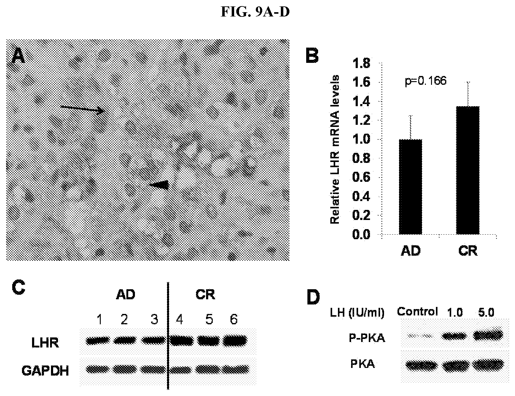
D00013

D00014

D00015
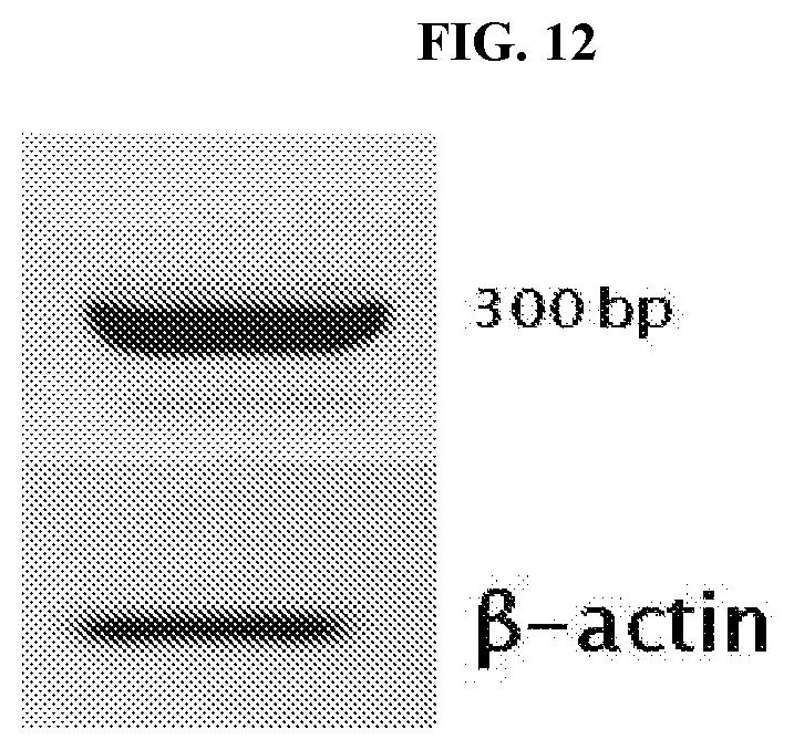
D00016

D00017

D00018

D00019

D00020

D00021

D00022

D00023

D00024

D00025

D00026

D00027

D00028

D00029

D00030

D00031

D00032

D00033

D00034

D00035

D00036
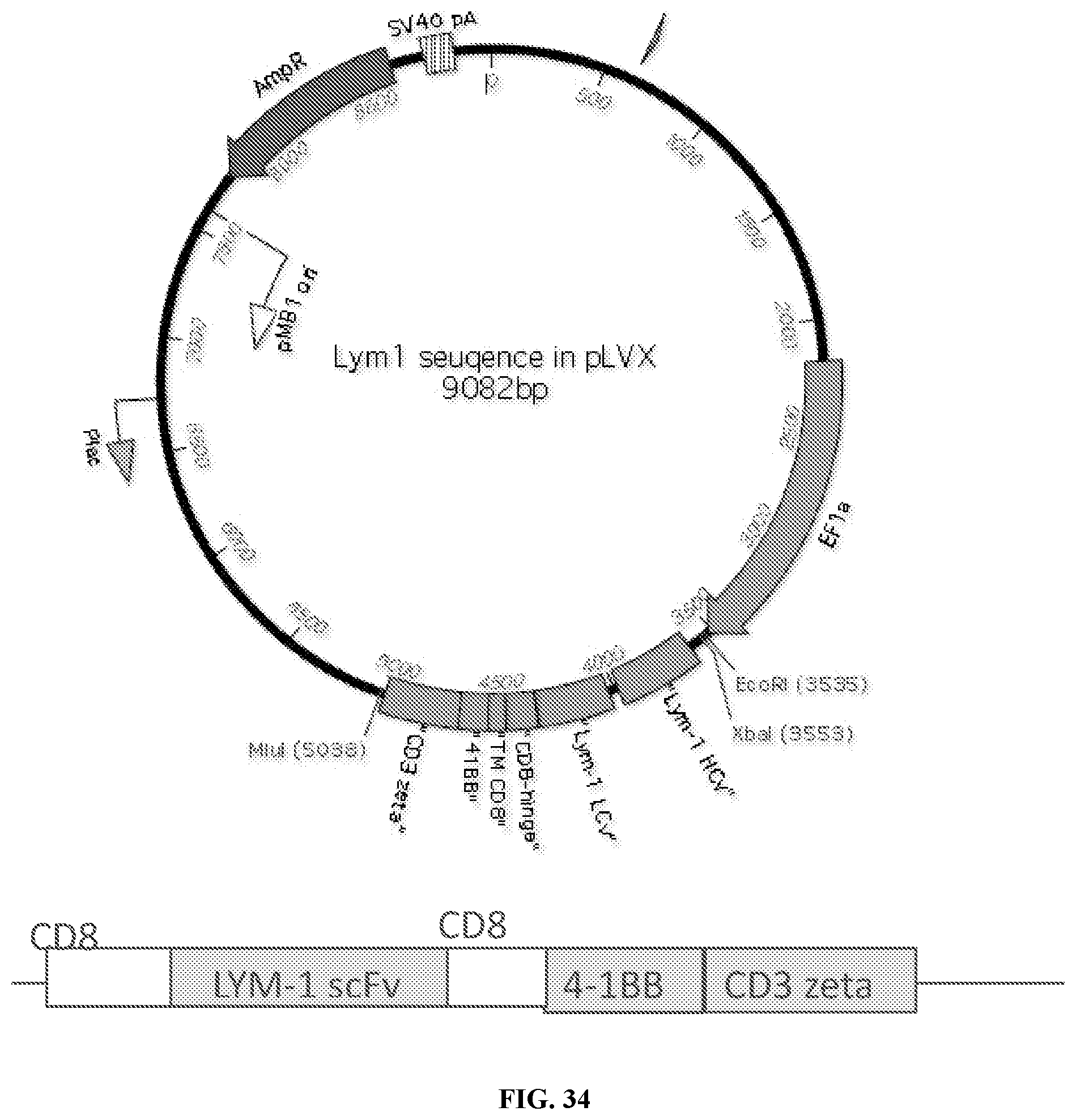
D00037

D00038

D00039

D00040

D00041

S00001
XML
uspto.report is an independent third-party trademark research tool that is not affiliated, endorsed, or sponsored by the United States Patent and Trademark Office (USPTO) or any other governmental organization. The information provided by uspto.report is based on publicly available data at the time of writing and is intended for informational purposes only.
While we strive to provide accurate and up-to-date information, we do not guarantee the accuracy, completeness, reliability, or suitability of the information displayed on this site. The use of this site is at your own risk. Any reliance you place on such information is therefore strictly at your own risk.
All official trademark data, including owner information, should be verified by visiting the official USPTO website at www.uspto.gov. This site is not intended to replace professional legal advice and should not be used as a substitute for consulting with a legal professional who is knowledgeable about trademark law.