Combination Therapy By Using Anti-globo H Or Anti-ssea-4 Antibody With Anti-negative Immune Check Points Antibody
YU; Cheng-Der Tony ; et al.
U.S. patent application number 16/429614 was filed with the patent office on 2019-12-26 for combination therapy by using anti-globo h or anti-ssea-4 antibody with anti-negative immune check points antibody. The applicant listed for this patent is OBI Pharma, Inc.. Invention is credited to Jo-Fan CHANG, Jiann-Shiun LAI, Yi-Chien TSAI, Cheng-Der Tony YU.
| Application Number | 20190389963 16/429614 |
| Document ID | / |
| Family ID | 68698449 |
| Filed Date | 2019-12-26 |


View All Diagrams
| United States Patent Application | 20190389963 |
| Kind Code | A1 |
| YU; Cheng-Der Tony ; et al. | December 26, 2019 |
COMBINATION THERAPY BY USING ANTI-GLOBO H OR ANTI-SSEA-4 ANTIBODY WITH ANTI-NEGATIVE IMMUNE CHECK POINTS ANTIBODY
Abstract
The present disclosure relates to treatment of cancer patients with anti-Globo series antigens (Globo H and SSEA-4) antibodies in combination with anti-negative immune check point antibody to rescue the inhibited T cell activity.
| Inventors: | YU; Cheng-Der Tony; (San Diego, CA) ; LAI; Jiann-Shiun; (Taipei City, TW) ; TSAI; Yi-Chien; (Taipei City, TW) ; CHANG; Jo-Fan; (Taipei City, TW) | ||||||||||
| Applicant: |
|
||||||||||
|---|---|---|---|---|---|---|---|---|---|---|---|
| Family ID: | 68698449 | ||||||||||
| Appl. No.: | 16/429614 | ||||||||||
| Filed: | June 3, 2019 |
Related U.S. Patent Documents
| Application Number | Filing Date | Patent Number | ||
|---|---|---|---|---|
| 62679510 | Jun 1, 2018 | |||
| Current U.S. Class: | 1/1 |
| Current CPC Class: | C07K 2317/76 20130101; C07K 16/2818 20130101; C07K 16/30 20130101; C07K 16/2827 20130101; C07K 2317/24 20130101; A61K 2039/545 20130101; C07K 16/2809 20130101; C07K 16/18 20130101; A61P 35/00 20180101; C07K 16/2896 20130101 |
| International Class: | C07K 16/30 20060101 C07K016/30; A61P 35/00 20060101 A61P035/00; C07K 16/28 20060101 C07K016/28 |
Claims
1. A method for treating cancer, wherein the method comprising administering to a subject in need thereof a therapeutically effective amount of a pharmaceutical composition comprising an Anti-Globo series antigens antibody in combination with an Anti-negative immune checkpoint antibody.
2. The method of claim 1, wherein the Globo series antigen is stage-specific embryonic antigen-4 (Neu5Ac.alpha.2.fwdarw.3Gal.beta.1.fwdarw.3GalNAc.beta.1.fwdarw.3Gal.alph- a.1.fwdarw.4Gal.beta.1.fwdarw.4Glc.beta.1) or Globo H (Fuc.alpha.1.fwdarw.2 Gal.beta.1.fwdarw.3 GalNAc.beta.1.fwdarw.3 Gal.alpha.1.fwdarw.4 Gal.beta.1.fwdarw.4 Glc).
3. The method according to claim 1, wherein the immune checkpoint antigen molecule is selected from the group consisting of PD-1/PD-L1 antigen, CTLA-4 (Cytotoxic T-lymphocyte-Associated Protein 4), LAG-3 (Lymphocyte Activation Gene 3), TIGIT (T-cell ImmunoGlobulin and Immunoreceptor Tyrosine-based inhibitory motif domain), Ceacam 1 (Carcinoembryonic antigen-related cell adhesion molecule 1), LAIR-1 (leucocyte-associated immunoglobulin-like receptor- 1) or TIM-3 (T cell Immunoglobulin and Mucin domain-3).
4. The method of claim 1, wherein the Anti-Globo series antigen antibody is OBI-888 or OBI-898.
5. The method according to claim 1, wherein the Anti-negative immune checkpoint agent is a PD-1/PD-L antagonist.
6. The method of claim 5, wherein the Anti-PD-1/PD-L1 antibody is Bavencio (avelumab), Opdivo (nivolumab), Keytruda (pembrolizumab), Imfinzi (durvalumab) and/or Tecentriq (atezolizumab).
7. The method of claim 1, wherein the cancer is selected from the group consisting of breast cancer, lung cancer, esophageal cancer, rectal cancer, biliary cancer, liver cancer, buccal cancer, gastric cancer, colon cancer, nasopharyngeal cancer, kidney cancer, prostate cancer, ovarian cancer, cervical cancer, endometrial cancer, pancreatic cancer, testicular cancer, bladder cancer, head and neck cancer, oral cancer, neuroendocrine cancer, adrenal cancer, thyroid cancer, bone cancer, skin cancer, basal cell carcinoma, squamous cell carcinoma, melanoma, or brain tumor.
8. The method of claim 1, wherein comprising administering one Anti-Globo series antigens antibody or a fragment thereof and one anti-PD-1/PD-L1 antibody or a fragment thereof.
9. The method of claim 1, wherein the Anti-Globo series antibody and/or the at least one inhibitor of the immune check point is a monoclonal antibody selected from a murine antibody, a recombinant antibody, humanized or fully human antibodies, chimeric antibody, multispecific antibody, in particular bispecific antibody or a fragment thereof.
10. The method of claim 9, wherein the least one inhibitor of the immune checkpoint is an antibody, a protein, a small molecules and/or a si-RNA.
11. The method of claim 1, wherein the Anti-Globo series antibody or a fragment thereof is a humanized antibody that comprises: SEQ. ID Nos: 1-108 as set forth in Tables 1-2 or Anti-SSEA4 antibody that comprises: SEQ. ID Nos. 109-182 as set forth in Tables 6-9.
12. The method of claim 9, wherein the inhibitor of the immune checkpoint is an antibody or a fragment thereof that binds to the antigens of claim 3 (PD-1/PD-L1, CTLA-4, LAG-3, TIGIT, Ceacam 1, LAIR-1 or TIM-3).
13. The method of claim 1, wherein the Anti-Globo series antigen antibody or a fragment thereof and the at least one inhibitor of the immune checkpoint are administered simultaneously, separately or sequentially.
14. The method of claim 1, wherein the subject is human.
15. The method of claim 1 whereby the targeting of Globo series antigen (with Anti-Globo H or Anti-SSEA-4) antibodies in combination with anti-negative immune checkpoint blockage acts corporately, additively, and/or synergistically to rescue the T cell inactivation and improve therapeutic efficacy.
16. The method of claim 1, whereby the therapeutic efficacy is enhanced by the rescue of T cell inactivation.
17. The method of claim 1, whereby the growth or progression of the cancer is inhibited and/or decreased.
18. The method of claim 1, whereby the tumor volume is decreased.
19. A method for rescuing T cell inactivation, wherein the method comprising administering to a subject in need thereof a therapeutically effective amount of a pharmaceutical composition comprising an Anti-Globo series antigens antibody in combination with an Anti-negative immune checkpoint antibody.
20. A method for decreasing and/or inhibiting cancer growth/progression, wherein the method comprising administering to a subject in need thereof a therapeutically effective amount of a pharmaceutical composition comprising an Anti-Globo series antigens antibody in combination with an Anti-negative immune checkpoint antibody.
21. The method of claim 19 or 20, wherein said Anti-negative immune checkpoint antibody inhibitor comprises anti-PD-1 antibody selected from Keytruda (pembrolizumab), and/or Opdivo (nivolumab) and said anti-PD-L1 antibody selected from Bavencio (avelumab), Imfinzi (durvalumab), and/or Tecentriq (atezolizumab).
22. The method of claim 19 or 20, wherein said Globo series antigen is stage-specific embryonic antigen-4 (Neu5Ac.alpha.2.fwdarw.3Gal.beta.1.fwdarw.3GalNAc.beta.1.fwdarw.3Gal.alph- a.1.fwdarw.4Gal.beta.1.fwdarw.4Glc.beta.1) or Globo H (Fuc.alpha.1.fwdarw.2 Gal.beta.1.fwdarw.3 GalNAc.beta.1.fwdarw.3 Gal.alpha.1.fwdarw.4 Gal.beta.1.fwdarw.4 Glc)
23. The method of claim 19 or 20, wherein the Anti-Globo series antigen antibody is OBI-888 or OBI-898.
24. A pharmaceutical composition with dual negative immune check point molecules targeting, comprising: a combination of Anti-Globo series antigens antibody and Anti-negative immune check point antibody; and a pharmaceutical acceptable carrier.
25. The composition of claim 24, further binding two or more immune check point molecules.
26. The composition of claim 25, wherein the immune checkpoint molecule is selected from the group consisting of PD-1/PD-L1 antigen, CTLA-4 (Cytotoxic T-lymphocyte-Associated Protein 4), LAG-3 (Lymphocyte Activation Gene 3), TIGIT (T-cell ImmunoGlobulin and Immunoreceptor Tyrosine-based inhibitory motif domain), Ceacam 1 (Carcinoembryonic antigen-related cell adhesion molecule 1), LAIR-1 (leucocyte-associated immunoglobulin-like receptor-1) or TIM-3 (T cell Immunoglobulin and Mucin domain-3).
27. The composition of claim 24, wherein the Globo series antigen is stage-specific embryonic antigen-4 (Neu5Ac.alpha.2.fwdarw.3Gal.beta.1.fwdarw.3GalNAc.beta.1.fwdarw.3Gal.alph- a.1.fwdarw.4Gal.beta.11.fwdarw.4Glc.beta.1) or Globo H (Fuc.alpha.1.fwdarw.2 Gal.beta.1.fwdarw.3 GalNAc.beta.1.fwdarw.3 Gal.alpha.1.fwdarw.4 Gal.beta.1.fwdarw.4 Glc).
28. The composition of claim 24, wherein the Anti-Globo series antigen antibody is OBI-888 or OBI-898.
29. The method of claim 24, wherein the Anti-Globo series antigens antibody or a fragment thereof is Anti-Globo H antibody that comprises: SEQ. ID Nos: 1-108 as set forth in Tables 1-2 or Anti-SSEA4 antibody that comprises: SEQ. ID Nos: 109-182 as set forth in Tables 6-9.
30. A kit comprising the pharmaceutical composition of claim 24 and instructions for use thereof.
Description
CROSS-REFERENCE TO RELATED APPLICATIONS
[0001] This application claims the benefit of U.S. Patent Application No. 62/679,510, filed on Jun. 1, 2018, the disclosure of all of which are incorporated by reference herein in their entirety.
SEQUENCE LISTING
[0002] The instant application contains a Sequence Listing which has been filed electronically in ASCII format and is hereby incorporated by reference in its entirety. Said ASCII copy, created on Jul. 4, 2019, is named G3004-01201_SL.txt and is 87,936 bytes in size.
FIELD
[0003] The present invention relates to Anti-Globo H or Anti-SSEA-4 carbohydrate antibody combined with Anti-PD-1 or PD-L1 antibodies. Results are provided for the rationale of co-administering of Anti-Globo H or Anti-SSEA-4 carbohydrate antibody combined with Anti-PD-1 or Anti-PD-L1 antibodies to synergistically rescue T cell inactivation induced by Globo H ceramide or SSEA-4 ceramide and PD-1/PD-L1 engagement. The disclosure provides methods for treating cancers using Anti-Globo H or Anti-SSEA-4 carbohydrate antibody combined with Anti-PD-1 or Anti-PD-L1 antibodies.
BACKGROUND OF INVENTION
[0004] Numerous surface carbohydrates are expressed in malignant tumor cells. For example, the carbohydrate antigen Globo H (Fuc.alpha.1.fwdarw.2 Gal.beta.1.fwdarw.3 GalNAc.beta.1.fwdarw.3 Gal.alpha.1.fwdarw.4 Gal.beta.1.fwdarw.4 Glc) was first isolated as a ceramide-linked Glycolipid and identified in 1984 from breast cancer MCF-7 cells. (Bremer E G, et al. (1984) J Biol Chem 259:14773-14777). Previous studies have also shown that Globo H and stage-specific embryonic antigen 3 (Gal.beta.1.fwdarw.3GalNAc.beta.1.fwdarw.3Gal.alpha.1.fwdarw.4Gal.beta.1.- fwdarw.4Glc.beta.1) (SSEA-3, also called Gb5) was observed on breast cancer cells and breast cancer stem cells (WW Chang et al. (2008) Proc Natl Acad Sci USA, 105(33): 11667-11672). In addition, SSEA-4 (stage-specific embryonic antigen-4) (Neu5Ac.alpha.2.fwdarw.3Gal.beta.1.fwdarw.3GalNAc.beta.1.fwdarw.3Gal.alph- a.1.fwdarw.4Gal.beta.1.fwdarw.4Glc.beta.1) has been commonly used as a cell surface marker for pluripotent human embryonic stem cells and has been used to isolate mesenchymal stem cells and enrich neural progenitor cells (Kannagi R et al. (1983) EMBO J, 2:2355-2361). These findings support that Globo series antigens (Globo H, SSEA-3 and SSEA-4) are unique targets for cancer therapies and can be used to direct therapeutic agents to targeting cancer cells effectively.
[0005] Program death 1 (PD-1) is an inhibitory receptor expressed on T cells, B cells, or monocytes (Ishida et al. (1992) EMBO J. 11: 3887-2895; Agata et al. (1996) Int. Immunol. 8: 765-772). PD-L1 and PD-L2 are ligands for PD-1 which have been identified to downregulate T cell activation and cytokine secretion upon binding to PD-1 (Freeman et al. (2000) J Exp Med 192:1027-34; Latchman et al. (2001) Nat Immunol 2:261-8). Engagement of PD-1 with PD-L1 or PD-L2 leads to down-regulation of immune responses. Hence, blocking of the PD-1/PD-L1 pathway has been proposed to attenuate central and peripheral immune responses against cancer. Targeting PD-1 and PD-L1 pathway have shown the clinical efficacy in more than 15 cancer types including melanoma, non-small cell lung cancer (NSCLC), renal cell carcinoma (RCC), bladder carcinoma and Hodgkin's lymphoma (Sharma et al. (2015) Science 348(6230):56-61). However, there are still many patients fail to respond; some patients showed initial responses but acquire resistance over time. Therefore, there is an urgent need to identify mechanisms of resistance for combination therapy.
[0006] Globo H ceramide has been identified to shed into the tumor microenvironment. Uptake of Globo H ceramide by immune cells was reported to inhibit cell proliferation and cytokine production suggesting that Globo H ceramide acts as an immune checkpoint to escape from immune surveillance (YC Tsai et al. (2013) J Cancer Sci Ther 5: 264-270). There are broad spectrum of co-receptors, for example, CTLA4, LAG3, TIGIT and TIM3, expressed by T cells that negatively regulate T cell activation (Sledzinska et al. (2015) Mol Oncol. December; 9(10):1936-65).
SUMMARY OF THE INVENTION
[0007] Accordingly, depletion of Globo H ceramide by Anti-Globo H antibody combined with blockage of negative immune checkpoint might be effective in overcoming immunosuppression. Our findings support that targeting Globo series antigen (Globo H or SSEA-4) with anti-negative immune checkpoint blockage acts corporately to rescue the T cell inactivation.
[0008] Therefore, a first embodiment of the present invention relates to a combination comprising an anti-Globo H and/or anti-SSEA-4 antibody or a fragment thereof and at least one inhibitor of the immune check point. In certain specific embodiment, the immune check point inhibitor is an anti-negative immune check point antibody.
[0009] In a preferred embodiment the combination of the present invention comprises one anti-Globo-H and/or anti-SSEA-4 antibody or a fragment thereof and one Anti-PD-1 antibody or a fragment thereof.
[0010] Preferably said antibody suitable for combination therapy with Globo H or SSEA-4 antibody is selected from Keytruda and/or Tecentriq.
[0011] In one non-limiting embodiment, the Keytruda and Tecentriq is sourced from:
TABLE-US-00001 Keytruda Tecentriq Brand MSD Ireland Roche Lot 7302614A13 H0125B11
[0012] For the purpose of the present invention the antibodies are preferably selected from the group consisting of murine antibody, recombinant antibody, humanized or fully human antibody, chimeric antibody, multispecific antibody, in particular bispecific antibody, or a fragment thereof.
[0013] In a further embodiment, the active principles of the combination of the present invention, that are the anti-Globo H or anti-SSEA-4 antibodies or a fragment thereof and the at least an inhibitor of the immune check point, can be administered simultaneously, separately or sequentially, also following different route of administration for each active principle.
[0014] According to a further embodiment of the present invention, the active principles of the combination can be administered together, through the same route of administration or through different route of administration, or they can be administered separately through the same route of administration or through different route of administration.
[0015] In a preferred aspect of the present invention, the anti-Globo H or anti-SSEA-4 antibody or a fragment thereof can be formulated in injectable form, oral form, in form of tablets, capsules, solutions, suspensions, granules and oily capsules, while the at least an inhibitor of the immune check point is formulated parenterally, such as an aqueous buffer solution or an oily suspension.
[0016] According to a preferred embodiment of the present invention, the formulation containing the anti-Globo-H or SSEA-4 antibodies or a fragment thereof are administered weekly or several times a week, while the formulation containing the at least one inhibitor of the immune check point are administered through parenteral route, preferably from one to several times a week.
[0017] Accordingly, the present disclosure is based on the discovery that Globo series antigens on cancers can be shed into microenvironment and incorporated to T cells. T cell activation was inhibited after incorporation of Globo H ceramide or SSEA-4 ceramide. Adding of Anti-Globo H antibody or Anti-SSEA-4 antibody to inhibit the incorporation of Globo H ceramide or SSEA-4 ceramide to T cells can inhibit Globo H ceramide or SSEA-4 ceramide induced immunosuppression. PD-1/PD-L1 engagement suppressed the TCR signaling pathway. Adding Globo H ceramide or SSEA-4 ceramide to T cells further inhibit the TCR signaling. Incorporation of Globo H ceramide or SSEA-4 ceramide reduced the exertion effect of TCR signaling, which was a result of anti-PD-1 or anti-PD-L1 antibody to block the suppression by PD-1/PD-L1 engagement (i.e., the immune check-point effect). Adding Anti-Globo H antibody or Anti-SSEA-4 antibody with Anti-PD-1 or Anti-PD-L1 antibody synergistically reverse the TCR signaling suppressed by Globo H ceramide or SSEA-4 ceramide and PD-1/PD-L1 engagement. Cancers expressing Globo H or SSEA-4 antigens include, but are not limited to, sarcoma, skin cancer, leukemia, lymphoma, brain cancer, glioblastoma, lung cancer, breast cancer, oral cancer, head-and-neck cancer, nasopharyngeal cancer, esophagus cancer, stomach cancer, liver cancer, bile duct cancer, gallbladder cancer, bladder cancer, pancreatic cancer, intestinal cancer, colorectal cancer, kidney cancer, cervix cancer, endometrial cancer, ovarian cancer, testicular cancer, buccal cancer, oropharyngeal cancer, laryngeal cancer and prostate cancer.
[0018] In one aspect, the present disclosure provides a method for treating cancer, wherein the method comprising administering to a subject in need thereof a therapeutically effective amount of a pharmaceutical composition comprising an Anti-Globo series antigens antibody in combination with an Anti-negative immune check point antibody.
[0019] In one embodiment, the Globo series antigen is stage-specific embryonic antigen-4 (Neu5Ac.alpha.2.fwdarw.3Gal.beta.1.fwdarw.3GalNAc.beta.1.fwdarw.3Gal.alph- a.1.fwdarw.4Gal.beta.1.fwdarw.4Glc.beta.1), stage-specific embryonic antigen-3 (SSEA-3; Gal.beta.1.fwdarw.3GalNAc.beta.1.fwdarw.3Gal.alpha.1.fwdarw.4Gal.beta.1.f- wdarw.4Glc.beta.1) or Globo H (Fuc.alpha.1.fwdarw.2 Gal.beta.1.fwdarw.3 GalNAc.beta.1.fwdarw.3 Gal.alpha.1.fwdarw.4 Gal.beta.1.fwdarw.4 Glc).
[0020] In one embodiment, the immune checkpoint antigen molecule is selected from the group consisting of PD-1/PD-L1 antigen, CTLA-4 (Cytotoxic T-lymphocyte-Associated Protein 4), LAG-3 (Lymphocyte Activation Gene 3), TIGIT (T-cell ImmunoGlobulin and Immunoreceptor Tyrosine-based inhibitory motif domain), Ceacam 1 (Carcinoembryonic antigen-related cell adhesion molecule 1), LAIR-1 (leucocyte-associated immunoglobulin-like receptor-1) or TIM-3 (T cell Immunoglobulin and Mucin domain-3).
[0021] In one embodiment, the Anti-Globo series antigen antibody is OBI-888 or OBI-898.
[0022] In one embodiment, the Anti-negative immune checkpoint agent is a PD-1/PD-L1 antagonist.
[0023] In one embodiment, the Anti-PD-1/PD-L1 antibody is Bavencio (avelumab), Opdivo (nivolumab), Keytruda (pembrolizumab), Imfinzi (durvalumab) and/or Tecentriq (atezolizumab).
[0024] In one embodiment, the cancer is selected from the group consisting of breast cancer, lung cancer, esophageal cancer, rectal cancer, biliary cancer, liver cancer, buccal cancer, gastric cancer, colon cancer, nasopharyngeal cancer, kidney cancer, prostate cancer, ovarian cancer, cervical cancer, endometrial cancer, pancreatic cancer, testicular cancer, bladder cancer, head and neck cancer, oral cancer, neuroendocrine cancer, adrenal cancer, thyroid cancer, bone cancer, skin cancer, basal cell carcinoma, squamous cell carcinoma, melanoma, or brain tumor.
[0025] In one embodiment, the method comprising administering of one Anti-Globo series antigens antibody or a fragment thereof and one Anti-PD-1/PD-L1 antibody or a fragment thereof.
[0026] In one embodiment, the Anti-Globo series antigens antibody and/or the at least one inhibitor of the immune check point is a monoclonal antibody selected from a murine antibody, a recombinant antibody, humanized or fully human antibodies, chimeric antibody, multispecific antibody, in particular bispecific antibody or a fragment thereof.
[0027] In one embodiment, the least one inhibitor of the immune checkpoint is an antibody, a protein, a small molecules and/or a si-RNA.
[0028] In one embodiment, the Anti-Globo series antigens antibody or a fragment thereof is Anti-Globo H antibody that comprises: SEQ. ID Nos. 1-108 as set forth in Tables 1-2 or Anti-SSEA4 antibody that comprises: SEQ. ID Nos: 109-182 as set forth in Tables 6-9.
[0029] In one embodiment, the inhibitor of the immune checkpoint is an antibody or a fragment thereof that binds to the antigens (PD-1/PD-L1, CTLA-4, LAG-3, TIGIT, Ceacam 1, LAIR-1 or TIM-3).
[0030] In one embodiment, the Anti-Globo series antigens antibody or a fragment thereof and the at least one inhibitor of the immune checkpoint are administered simultaneously, separately or sequentially.
[0031] In one embodiment, the subject is human.
[0032] In one embodiment, the targeting of Globo series antigen (with Globo H or SSEA-4) antibodies in combination with Anti-negative immune checkpoint blockage acts corporately, additively, and/or synergistically to rescue the T cell inactivation and improve therapeutic efficacy.
[0033] In one embodiment, the therapeutic efficacy is enhanced by the rescue ofT cell inactivation.
[0034] In one embodiment, the growth or progression of the cancer is inhibited and/or decreased.
[0035] In one embodiment, the tumor volume is decreased.
[0036] In one aspect, the present disclosure provides a method for rescuing T cell inactivation, wherein the method comprising administering to a subject in need thereof a therapeutically effective amount of a pharmaceutical composition comprising an Anti-Globo series antigens antibody in combination with an Anti-negative immune checkpoint antibody.
[0037] In one aspect, the present disclosure provides method for decreasing and/or inhibiting cancer growth/progression, wherein the method comprising administering to a subject in need thereof a therapeutically effective amount of a pharmaceutical composition comprising an Anti-Globo series antigens antibody in combination with an Anti-negative immune checkpoint antibody.
[0038] In one embodiment, said Anti-negative immune checkpoint antibody inhibitor comprises Anti-PD-1 antibody selected from Keytruda (pembrolizumab), and/or Opdivo (nivolumab) and said Anti-PD-L1 antibody selected from Bavencio (avelumab), Imfinzi (durvalumab), and/or Tecentriq (atezolizumab).
[0039] In one aspect, the present disclosure provides a pharmaceutical composition comprising an Anti-Globo series antigens antibody and an Anti-negative immune checkpoint antibody and a pharmaceutical acceptable carrier.
[0040] In one aspect, the present disclosure provides a kit comprising the pharmaceutical composition and instructions for use thereof.
BRIEF DESCRIPTION OF THE FIGURES
[0041] A more complete understanding of the invention may be obtained by reference to the accompanying drawings, when considered in conjunction with the subsequent detailed description. The embodiments illustrated in the drawings are intended only to exemplify the invention and should not be construed as limiting the invention to the illustrated embodiments.
[0042] FIG. 1. Shedding of Globo H or SSEA-4 from various cancer cells to human CD3+ T cells.
[0043] FIG. 2. Suppress the T cell activation by Globo H ceramide or SSEA-4 ceramide.
[0044] FIG. 3. Reverse the Globo H ceramide induced T cell inactivation by Anti-Globo H antibody.
[0045] FIG. 4. Reverse the SSEA-4 ceramide induced T cell inactivation by Anti-SSEA-4 antibody.
[0046] FIG. 5. Globo H ceramide or SSEA-4 ceramide with PD-1/PD-L1 engagement enhanced the inhibition on TCR signaling.
[0047] FIG. 6. Reduced the Keytruda or Tecentriq released PD-1/PD-L1 engagement inhibited TCR signaling by Globo H ceramide.
[0048] FIG. 7. Reduced the Keytruda or Tecentriq released PD-1/PD-L1 engagement inhibited TCR signaling by SSEA-4 ceramide.
[0049] FIG. 8. Released the Globo H ceramide and PD-1/PD-L1 engagement inhibited TCR signaling by Anti-Globo H antibody combined with Keytruda or Tecentriq.
[0050] FIG. 9. Released the SSEA-4 ceramide and PD-1/PD-L1 engagement inhibited TCR signaling by Anti-SSEA-4 antibody combined with Keytruda or Tecentriq.
[0051] FIG. 10. Schematic of the mechanism of action of Globo H ceramide or SSEA-4 ceramide with negative immune check point engagement to suppress the T cell activity.
[0052] FIG. 11. Schematic of the mechanism of action of Anti-Globo H antibody or Anti-SSEA-4 antibody with anti-negative immune check point antibody to rescue the T cell activity.
DETAILED DESCRIPTION OF THE INVENTION
[0053] The present disclosure relates to Anti-Globo H or Anti-SSEA-4 antigens antibodies combined with Anti-PD-1 or PD-L1 antibody to treat cancer patients.
[0054] Accordingly, the present disclosure is based on the discovery that Globo series antigens on cancers can be shed into microenvironment and incorporated to T cells. T cell activation was inhibited after incorporation of Globo H ceramide or SSEA-4 ceramide. Adding of Anti-Globo H antibody or Anti-SSEA-4 antibody to inhibit the incorporation of Globo H ceramide or SSEA-4 ceramide to T cells can inhibit Globo H ceramide or SSEA-4 ceramide induced immunosuppression. PD-1/PD-L1 engagement suppressed the TCR signaling pathway. Adding Globo H ceramide or SSEA-4 ceramide to T cells further inhibit the TCR signaling. Incorporation of Globo H ceramide or SSEA-4 ceramide reduced the exertion effect of TCR signaling, which was a result of anti-PD-1 or anti-PD-L1 antibody to block the suppression by PD-1/PD-L1 engagement (i.e., the immune check-point effect). Adding Anti-Globo H antibody or Anti-SSEA-4 antibody with Anti-PD-1 or Anti-PD-L1 antibody synergistically reverse the TCR signaling suppressed by Globo H ceramide or SSEA-4 ceramide and PD-1/PD-L1 engagement. Cancers expressing Globo H or SSEA-4 antigens include, but are not limited to, sarcoma, skin cancer, leukemia, lymphoma, brain cancer, glioblastoma, lung cancer, breast cancer, oral cancer, head-and-neck cancer, nasopharyngeal cancer, esophagus cancer, stomach cancer, liver cancer, bile duct cancer, gallbladder cancer, bladder cancer, pancreatic cancer, intestinal cancer, colorectal cancer, kidney cancer, cervix cancer, endometrial cancer, ovarian cancer, testicular cancer, buccal cancer, oropharyngeal cancer, laryngeal cancer and prostate cancer.
Definitions
[0055] As used herein, the term "antigen" is defined as any substance capable of eliciting an immune response.
[0056] As used herein, the term "immunogenicity" refers to the ability of an immunogen, antigen, or vaccine to elicit an immune response.
[0057] As used herein, the term "epitope" is defined as the parts of an antigen molecule which contact the antigen binding site of an antibody or a T cell receptor.
[0058] As used herein, the term "vaccine" refers to a preparation that contains an antigen, consisting of whole disease-causing organisms (killed or weakened) or components of such organisms, such as proteins, peptides, or polysaccharides, that is used to confer immunity against the disease that the organisms cause. Vaccine preparations can include or exclude any one of natural, synthetic or recombinantly derived preparations. Recombinantly derived preparations can be obtained, for example, by recombinant DNA technology.
[0059] As used herein, the term "antigen specific" refers to a property of a cell population such that the supply of a particular antigen, or a fragment of the antigen, results in specific cell proliferation.
[0060] As used herein, the term "CD1d" refers to a member of the CD1 (cluster of differentiation 1) family of glycoproteins expressed on the surface of various human antigen-presenting cells. CD1d presented lipid antigens activate natural killer T cells. CD1d has a deep antigen-binding groove into which glycolipid antigens bind. CD1d molecules expressed on dendritic cells can bind and present glycolipids, including GalCer analogs such as C34.
[0061] As used herein, the term "glycan" refers to a polysaccharide, or oligosaccharide. Glycan is also used herein to refer to the carbohydrate portion of a glycoconjugate, such as a glycoprotein, glycolipid, glycopeptide, glycoproteome, peptidoglycan, lipopolysaccharide, or a proteoglycan. Glycans usually consist solely of O-glycosidic linkages between monosaccharides. For example, cellulose is a glycan (or more specifically a glucan) composed of -1,4-linked D-glucose, and chitin is a glycan composed of -1,4-linked N-acetyl-D-glucosamine. Glycans can be homopolymers or heteropolymers of monosaccharide residues and can be linear or branched. Glycans can be found attached to proteins as in glycoproteins and proteoglycans. They are generally found on the exterior surface of cells. O- and N-linked glycans are very common in eukaryotes but may also be found, although less commonly, in prokaryotes. N-Linked glycans are found attached to the R-group nitrogen (N) of asparagine in the sequon. The sequon is a Asn-X-Ser or Asn-X-Thr sequence, where X is any amino acid except praline.
[0062] As used herein, the term "specifically binding," refers to the interaction between binding pairs (e.g., an antibody and an antigen). In various instances, specifically binding can be embodied by an affinity constant of about 10-6 moles/liter, about 10-7 moles/liter, or about 10-8 moles/liter, or less.
[0063] As used herein, the term "Flow cytometry" or "FACS" means a technique for examining the physical and chemical properties of particles or cells suspended in a stream of fluid, through optical and electronic detection devices.
[0064] As used herein, the terms glycoenzymes refers to at least in part the enzymes in the globoseries biosynthetic pathway; exemplary glycoenzymes include alpha-4GalT; beta-4GalNAcT-I; or beta-3GalT-V enzymes.
[0065] An "isolated" antibody is one which has been identified and separated and/or recovered from a component of its natural environment. Contaminant components of its natural environment are materials which would interfere with research, diagnostic or therapeutic uses for the antibody, and may include enzymes, hormones, and other proteinaceous or nonproteinaceous solutes. In one embodiment, the antibody will be purified (1) to greater than 95% by weight of antibody as determined by, for example, the Lowry method, and in some embodiments more than 99% by weight, (2) to a degree sufficient to obtain at least 15 residues of N-terminal or internal amino acid sequence by use of, for example, a spinning cup sequenator, or (3) to homogeneity by SDS-PAGE under reducing or nonreducing conditions using, for example, Coomassie blue or silver stain. Isolated antibody includes the antibody in situ within recombinant cells since at least one component of the antibody's natural environment will not be present. Ordinarily, however, isolated antibody will be prepared by at least one purification step.
[0066] The term "support" or "substrate" as used interchangeably herein refers to a material or group of materials, comprising one or a plurality of components, with which one or more molecules are directly or indirectly bound, attached, synthesized upon, linked, or otherwise associated. A support may be constructed from materials that are biological, non-biological, inorganic, organic or a combination of these. A support may be in any appropriate size or configuration based upon its use within a particular embodiment.
[0067] The term "target" as used herein refers to a species of interest within an assay. Targets may be naturally occurring or synthetic, or a combination. Targets may be unaltered (e.g., utilized directly within the organism or a sample thereof), or altered in a manner appropriate for the assay (e.g., purified, amplified, filtered). Targets may be bound through a suitable means to a binding member within certain assays. Non-limiting examples of targets include, but are not restricted to, antibodies or fragments thereof, cell membrane receptors, monoclonal antibodies and antisera reactive with specific antigenic determinants (such as on viruses, cells or other materials), drugs, oligonucleotides, nucleic acids, peptides, cofactors, sugars, lectins polysaccharides, cells, cellular membranes, and organelles. Target may be any suitable size depending on the assay.
[0068] The phrase "substantially similar," "substantially the same", "equivalent", or "substantially equivalent", as used herein, denotes a sufficiently high degree of similarity between two numeric values (for example, one associated with a molecule and the other associated with a reference/comparator molecule) such that one of skill in the art would consider the difference between the two values to be of little or no biological and/or statistical significance within the context of the biological characteristic measured by said values (e.g., Kd values, anti-viral effects, etc.). The difference between said two values is, for example, less than about 50%, less than about 40%, less than about 30%, less than about 20%, and/or less than about 10% as a function of the value for the reference/comparator molecule.
[0069] Thus, anti-cancer antibodies of the present invention include in combination with a heavy chain or light chain variable region, a heavy chain or light chain constant region, a framework region, or any portion thereof, of non-murine origin, preferably of human origin, which can be incorporated into an antibody of the present invention.
[0070] Antibodies of the present invention are capable of modulating, decreasing, antagonizing, mitigating, alleviating, blocking, inhibiting, abrogating and/or interfering with at least one Globo-H and/or SSEA-4 expressing cancer cell activity in vitro, in situ and/or in vivo.
[0071] Antibodies of the present invention include any protein or peptide that comprise at least one complementarity determining region (CDR) of a heavy or light chain, or a ligand binding portion thereof, derived from an antibody produced by the hybridoma designated 2C2 (deposited under ATCC Accession No.: PTA-121138), the hybridoma designated 3D7 (deposited under ATCC Accession No.: PTA-121310), the hybridoma designated 7A11 (deposited under ATCC Accession No.: PTA-121311), the hybridoma designated 2F8 (deposited under ATCC Accession No.: PTA-121137), or the hybridoma designated 1E1 (deposited under ATCC Accession No.: PTA-121312) as described herein. Antibodies include antibody fragments, antibody variants, monoclonal antibodies, polyclonal antibodies, and recombinant antibodies and the like. Antibodies can be generated in mice, rabbits or humans.
[0072] The term "antibody" is further intended to encompass antibodies, digestion fragments, specified portions and variants thereof, including antibody mimetics or comprising portions of antibodies that mimic the structure and/or function of an anti-cancer antibody or specified fragment or portion thereof, including single chain antibodies and fragments thereof, each containing at least one CDR derived from an anti-cancer antibody of the present invention.
[0073] For example, functional fragments include antigen-binding fragments that bind to a Globo-H expressing cancer cells. For example, antibody fragments capable of binding to Globo-H expression cancer cells or portions thereof, including, but not limited to Fab (e.g., by papain digestion), Fab' (e.g., by pepsin digestion and partial reduction) and F(ab')2 (e.g., by pepsin digestion), facb (e.g., by plasmin digestion), pFc' (e.g., by pepsin or plasmin digestion), Fd (e.g., by pepsin digestion, partial reduction and reaggregation), Fv or scFv (e.g., by molecular biology techniques) fragments, are encompassed by the invention (see, e.g., Colligan, Immunology, supra).
[0074] An antigen-binding portion of an antibody may include a portion of an antibody that specifically binds to a carbohydrate antigen (e.g., Globo H, SSEA-4).
[0075] The humanized antibody of the present invention is an antibody from a non-human species where the amino acid sequence in the non-antigen binding regions (and/or the antigen-binding regions) has been altered so that the antibody more closely resembles a human antibody while retaining its original binding ability.
[0076] Humanized antibodies can be generated by replacing sequences of the variable region that are not directly involved in antigen binding with equivalent sequences from human variable regions. Those methods include isolating, manipulating, and expressing the nucleic acid sequences that encode all or part of variable regions from at least one of a heavy or light chain. Sources of such nucleic acid are well known to those skilled in the art. The recombinant DNA encoding the humanized antibody, or fragment thereof, can then be cloned into an appropriate expression vector.
[0077] The humanized antibodies of the present invention can be produced by methods well known in the art. For example, once non-human (e.g., murine) antibodies are obtained, variable regions can be sequenced, and the location of the CDRs and framework residues determined. Kabat, E. A., et al. (1991) Sequences of Proteins of Immunological Interest, Fifth Edition, U.S. Department of Health and Human Services, NIH Publication No. 91-3242. Chothia, C. et al. (1987) J. Mol. Biol., 196:901-917. DNA encoding the light and heavy chain variable regions can, optionally, be ligated to corresponding constant regions and then subcloned into an appropriate expression vector. CDR-grafted antibody molecules can be produced by CDR-grafting or CDR substitution. One, two, or all CDRs of an immunoglobulin chain can be replaced. For example, all of the CDRs of a particular antibody may be from at least a portion of a non-human animal (e.g., mouse such as CDRs) or only some of the CDRs may be replaced. It is only necessary to keep the CDRs required for binding of the antibody to a predetermined carbohydrate antigen (e.g., Globo H). Morrison, S. L., 1985, Science, 229:1202-1207. Oi et al., 1986, BioTechniques, 4:214. U.S. Pat. Nos. 5,585,089; 5,225,539; 5,693,761 and 5,693,762. EP 519596. Jones et al., 1986, Nature, 321:552-525. Verhoeyan et al., 1988, Science, 239:1534. Beidler et al., 1988, J. Immunol., 141:4053-4060.
[0078] Also encompassed by the present invention are antibodies or antigen-binding portions thereof comprising one or two variable regions as disclosed herein, with the other regions replaced by sequences from at least one different species including, but not limited to, human, rabbits, sheep, dogs, cats, cows, horses, goats, pigs, monkeys, apes, gorillas, chimpanzees, ducks, geese, chickens, amphibians, reptiles and other animals.
[0079] A chimeric antibody is a molecule in which different portions are derived from different animal species. For example, an antibody may contain a variable region derived from a murine mAb and a human immunoglobulin constant region. Chimeric antibodies can be produced by recombinant DNA techniques. Morrison, et al., Proc Natl Acad Sci, 81:6851-6855 (1984). For example, a gene encoding a murine (or other species) antibody molecule is digested with restriction enzymes to remove the region encoding the murine Fc, and the equivalent portion of a gene encoding a human Fc constant region is then substituted into the recombinant DNA molecule. Chimeric antibodies can also be created by recombinant DNA techniques where DNA encoding murine V regions can be ligated to DNA encoding the human constant regions. Better et al., Science, 1988, 240:1041-1043. Liu et al. PNAS, 1987 84:3439-3443. Liu et al., J. Immunol., 1987, 139:3521-3526. Sun et al. PNAS, 1987, 84:214-218. Nishimura et al., Canc. Res., 1987, 47:999-1005. Wood et al. Nature, 1985, 314:446-449. Shaw et al., J. Natl. Cancer Inst., 1988, 80:1553-1559. International Patent Publication Nos. WO1987002671 and WO 86/01533. European Patent Application Nos. 184, 187; 171,496; 125,023; and 173,494. U.S. Pat. No. 4,816,567.
[0080] The antibodies can be full-length or can comprise a fragment (or fragments) of the antibody having an antigen-binding portion, including, but not limited to, Fab, F(ab')2, Fab', F(ab)', Fv, single chain Fv (scFv), bivalent scFv (bi-scFv), trivalent scFv (tri-scFv), Fd, dAb fragment (e.g., Ward et al., Nature, 341:544-546 (1989)), an isolated CDR, diabodies, triabodies, tetrabodies, linear antibodies, single-chain antibody molecules, and multispecific antibodies formed from antibody fragments. Single chain antibodies produced by joining antibody fragments using recombinant methods, or a synthetic linker, are also encompassed by the present invention. Bird et al. Science, 1988, 242:423-426. Huston et al., Proc. Natl. Acad. Sci. USA, 1988, 85:5879-5883.
[0081] The antibodies or antigen-binding portions thereof of the present invention may be monospecific, bi-specific or multispecific. Multispecific or bi-specific antibodies or fragments thereof may be specific for different epitopes of one target carbohydrate (e.g., Globo H) or may contain antigen-binding domains specific for more than one target carbohydrate (e.g., antigen-binding domains specific for Globo H and SSEA-4). In one embodiment, a multispecific antibody or antigen-binding portion thereof comprises at least two different variable domains, wherein each variable domain is capable of specifically binding to a separate carbohydrate antigen or to a different epitope on the same carbohydrate antigen. Tutt et al., 1991, J. Immunol. 147:60-69. Kufer et al., 2004, Trends Biotechnol. 22:238-244. The present antibodies can be linked to or co-expressed with another functional molecule, e.g., another peptide or protein. For example, an antibody or fragment thereof can be functionally linked (e.g., by chemical coupling, genetic fusion, noncovalent association or otherwise) to one or more other molecular entities, such as another antibody or antibody fragment to produce a bi-specific or a multispecific antibody with a second binding specificity. Multispecific or bi-specific antibodies or fragments thereof may be specific for different epitopes of one target carbohydrate (e.g., Globo H) or may contain antigen-binding domains specific for more than one target carbohydrate (e.g., antigen-binding domains specific for Globo H and SSEA-4). In one embodiment, a multispecific antibody or antigen-binding portion thereof comprises at least two different variable domains, wherein each variable domain is capable of specifically binding to a separate carbohydrate antigen or to a different epitope on the same carbohydrate antigen. Tutt et al., 1991, J. Immunol. 147:60-69. Kufer et al., 2004, Trends Biotechnol. 22:238-244. The antibodies of the present invention can be linked to or co-expressed with another functional molecule, e.g., another peptide or protein. For example, an antibody or fragment thereof can be functionally linked (e.g., by chemical coupling, genetic fusion, noncovalent association or otherwise) to one or more other molecular entities, such as another antibody or antibody fragment to produce a bi-specific or a multispecific antibody with a second binding specificity.
[0082] An antibody light or heavy chain variable region comprises a framework region (FW) interrupted by three hypervariable regions, referred to as complementarity determining regions or CDRs. According to one aspect of the invention, the antibody or the antigen-binding portion thereof may have the following structure: [0083] Leader Sequence-FW 1-CDR1-FW2-CDR2-FW3-CDR3- wherein the amino acid sequences of FW1, FW2, FW3, CDR1, CDR2 and CDR3 of the present invention are disclosed.
[0084] Also within the scope of the invention are antibodies or antigen-binding portions thereof in which specific amino acids have been substituted, deleted or added. In an exemplary embodiment, these alternations (i.e., conservative substitution, conservative deletion or conservative addition) do not have a substantial effect on the peptide's biological properties such as the effector function or the binding affinity. For purposes of classifying amino acids alteration as conservative or non-conservative, amino acids may be grouped as follows: hydrophobic, neutral, acidic, and basic. Conservative substitutions involve substitutions between amino acids in the same group. Non-conservative substitutions constitute exchanging a member of one of these groups for a member of another. Ng et al. (Predicting the Effects of Amino Acid Substitutions on Protein Function, Annu. Rev. Genomics Hum. Genet. 2006. 7:61-80) provides an overview of various amino acid substitution (AAS) prediction methods to allow a skilled artisan to predict and select an amino acid substitution, without changing the protein function.
[0085] In another exemplary embodiment, antibodies may have amino acid substitutions in the CDRs, such as to improve binding affinity of the antibody to the antigen. In yet another exemplary embodiment, a selected, small number of acceptor framework residues can be replaced by the corresponding donor amino acids. The donor framework can be a mature or germline human antibody framework sequence or a consensus sequence. Guidance concerning how to make phenotypically silent amino acid substitutions is provided in Bowie et al., Science, 247: 1306-1310 (1990). Cunningham et al., Science, 244: 1081-1085 (1989). Ausubel (ed.), Current Protocols in Molecular Biology, John Wiley and Sons, Inc. (1994). T. Maniatis, E. F. Fritsch and J. Sambrook, Molecular Cloning: A Laboratory Manual, Cold Spring Harbor laboratory, Cold Spring Harbor, N.Y. (1989). Pearson, Methods Mol. Biol. 243:307-31 (1994). Gonnet et al., Science 256:1443-45 (1992).
[0086] According to one aspect of the invention, the amino acid substitutions described herein occur at positions corresponding to the Kabat numbering scheme (e.g., Kabat et al., Sequences of Immunological Interest. 5th Ed. Public Health Service, National Institutes of Health, Bethesda, Md. (1991)).
[0087] As used herein, "normal levels" can be, for example, a reference value or range based on measurements of the levels of TACA bound antibodies in samples from normal patients or a population of normal patients. "Normal levels" can also be, for example, a reference value or range based on measurements of the TACAs in samples from normal patients or a population of normal patients.
[0088] As used herein a "subject" is a mammal. Such mammals include domesticated animals, farm animals, animals used in experiments, zoo animals and the like. In some embodiments, the subject is a human.
[0089] The term "Globoseries-related disorder" refers to or describes a disorder that is typically characterized by or contributed to by aberrant functioning or presentation of the pathway. Examples of such disorders include, but are not limited to, hyperproliferative diseases, including cancer. Examples of the hyperproliferative disease and/or condition includes neoplasm/hyperplasia and cancer, including, but not limited to, brain cancer, lung cancer, breast cancer, oral cancer, esophagus cancer, stomach cancer, liver cancer, bile duct cancer, pancreas cancer, colon cancer, kidney cancer, cervix cancer, ovary cancer and prostate cancer. In some embodiments, the cancer is brain cancer, lung cancer, breast cancer, ovarian cancer, prostate cancer, colon cancer, or pancreas cancer. In other embodiments, the hyperproliferative disease state is associated with breast, ovary, lung, pancreatic, stomach (gastric), colorectal, prostate, liver, cervix, esophagus, brain, oral, and kidney.
[0090] In one embodiment, the present disclosure provides a method for determining the therapeutic efficacy of an antineoplastic agent in treatment of a subject in need thereof, comprising: (a) providing a sample form a subject; (b) contacting a sample collected from a subject; (c) assaying the binding of one or more of tumor associated antigens (TACAs) or antibodies; and (d) determining the therapeutic effect of an antineoplastic agent in treatment for neoplasm based on the assayed value of the glycan detection. The present disclosure provides evidence of surprising additive and/or synergistic efficacy and utility in the combination usage of the linker-glycoconjugates (e.g. Globo H) in the detection of cancer. This provides the bases that the linkers and the conjugates herein are useful as companion diagnostic compositions and methods for any therapeutics targeting the determinants and molecules associated with globoseries glycoproteins. Exemplary therapeutic methods and compositions comprising antineoplastic agents suitable for use in combination with the present disclosure as companion diagnostic methods and uses are described (e.g. OBI-822, OBI-833 and OBI-888) in the disclosures of for example, patent publication numbers: WO2015159118, WO2014107652 and WO2015157629). The contents of each of which is incorporated by reference.
[0091] As used herein, the term "specific binding," refers to the interaction between binding pairs (e.g., an antibody and an antigen). In various instances, specific binding can be embodied by an affinity constant of about 10.sup.-6 moles/liter, about 10.sup.-7 moles/liter, or about 10.sup.-8 moles/liter, or less.
[0092] The phrase "substantially reduced," or "substantially different", as used herein, denotes a sufficiently high degree of difference between two numeric values (generally one associated with a molecule and the other associated with a reference/comparator molecule) such that one of skill in the art would consider the difference between the two values to be of statistical significance within the context of the biological characteristic measured by said values (e.g., Kd values). The differences between said two values are, for example, greater than about 10%, greater than about 20%, greater than about 30%, greater than about 40%, and/or greater than about 50% as a function of the value for the reference/comparator molecule.
[0093] "Binding affinity", as used herein, generally refers to the strength of the sum of total noncovalent interactions between a single binding site of a molecule (e.g., an antibody) and its binding partner (e.g., an antigen). Unless indicated otherwise, as used herein, "binding affinity" refers to the intrinsic binding affinity which reflects a 1:1 interaction between members of a binding pair (e.g., antibody and antigen). The affinity of a molecule X for its partner Y can generally be represented by the dissociation constant (Kd). Affinity can be measured by common methods known in the art, including those described herein. Low-affinity antibodies generally bind antigen slowly and tend to dissociate readily, whereas high-affinity antibodies generally bind antigen faster and tend to remain bound longer. A variety of methods of measuring binding affinity are known in the art, any of which can be used for purposes of the present invention. Specific illustrative embodiments are described in the following.
[0094] In certain embodiments, the "Kd" or "Kd value" according to this invention is measured by a radiolabeled antigen binding assay (RIA) performed with the Fab version of an antibody of interest and its antigen as described by the following assay. Solution binding affinity of Fabs for antigen is measured by equilibrating Fab with a minimal concentration of (125I)-labeled antigen in the presence of a titration series of unlabeled antigen, then capturing bound antigen with an anti-Fab antibody-coated plate (Chen, et al., (1999) J. Mol Biol 293:865-881). To establish conditions for the assay, microtiter plates (Dynex) are coated overnight with 5 .mu.g/mL of a capturing anti-Fab antibody (Cappel Labs) in 50 mM sodium carbonate (pH 9.6), and subsequently blocked with 2% (w/v) bovine serum albumin in PBS for two to five hours at room temperature (approximately 23.degree. C.). In a non-adsorbent plate (Nunc, Cat #269620), 100 .mu.M or 26 .mu.M [125I]-antigen are mixed with serial dilutions of a Fab of interest (e.g., consistent with assessment of an anti-VEGF antibody, Fab-12, in Presta et al., (1997) Cancer Res. 57:4593-4599). The Fab of interest is then incubated overnight; however, the incubation may continue for a longer period (e.g., 65 hours) to ensure that equilibrium is reached. Thereafter, the mixtures are transferred to the capture plate for incubation at room temperature (e.g., for one hour). The solution is then removed and the plate washed eight times with 0.1% Tween-20 in PBS. When the plates have dried, 150 .mu.L/well of scintillant (MicroScint-20; Packard) is added, and the plates are counted on a Topcount gamma counter (Packard) for ten minutes. Concentrations of each Fab that give less than or equal to 20% of maximal binding are chosen for use in competitive binding assays. According to another embodiment the Kd or Kd value is measured by using surface plasmon resonance assays using a BIAcore.TM.-2000 or a BIAcore.TM.-3000 (BIAcore, Inc., Piscataway, N.J.) at 25.degree. C., with immobilized antigen CM5 chips at .sup..about.10 response units (RU). Briefly, carboxymethylated dextran biosensor chips (CM5, BIAcore Inc.) are activated with N-ethyl-N'-(3-dimethylaminopropyl)-carbodiimide hydrochloride (EDC) and N-hydroxysuccinimide (NHS) according to the supplier's instructions. Antigen is diluted with 10 mM sodium acetate, pH 4.8, to 5 .mu.g/mL (.sup..about.0.2 .mu.M) before injection at a flow rate of 5 jiL/minute to achieve approximately 10 response units (RU) of coupled protein. Following the injection of antigen, 1 M ethanolamine is injected to block unreacted groups. In each experiment, a spot was activated and ethanolamine blocked without immobilizing protein, to be used for reference subtraction. For kinetics measurements, two-fold serial dilutions of Fab (0.78 nM to 500 nM) are injected in PBS with 0.05% Tween 20 (PBST) at 25.degree. C. at a flow rate of approximately 25 .mu.L/min. Association rates (kon) and dissociation rates (koff) are calculated using a simple one-to-one Langmuir binding model (BIAcore Evaluation Software version 3.2) by simultaneously fitting the association and dissociation sensorgrams. The equilibrium dissociation constant (Kd) is calculated as the ratio koff/kon. See, e.g., Chen, Y., et al., (1999) J. Mol Biol 293:865-881. If the on-rate exceeds 10.sup.6 M.sup.-1s.sup.-1 by the surface plasmon resonance assay above, then the on-rate can be determined by using a fluorescent quenching technique that measures the increase or decrease in fluorescence emission intensity (excitation=295 nm; emission=340 nm, 16 nm band-pass) at 25.degree. C. of a 20 nM anti-antigen antibody (Fab form) in PBS, pH 7.2, in the presence of increasing concentrations of antigen as measured in a spectrometer, such as a stop-flow equipped spectrophometer (Aviv Instruments) or a 8000-series SLM-Aminco spectrophotometer (ThermoSpectronic) with a stirred cuvette.
[0095] An "on-rate" or "rate of association" or "association rate" or "kon" according to this invention can also be determined with the same surface plasmon resonance technique described above using a BIAcore.TM.-2000 or a BIAcore.TM.-3000 (BIAcore, Inc., Piscataway, N.J.) at 25.degree. C. with immobilized antigen CM5 chips at or "association rate" or "kon" according to this invention can also be determined with the same surface plasmon N-ethyl-N'-(3-dimethylaminopropyl)-carbodiimide hydrochloride (EDC) and N-hydroxysuccinimide (NHS) according to the supplier's instructions. Antigen is diluted with 10 mM sodium acetate, pH 4.8, to 5 .mu.g/mL (.sup..about.0.2 .mu.M) before injection at a flow rate of 5 .mu.L/minute to achieve approximately 10 response units (RU) of coupled protein. Following the injection of antigen, 1 M ethanolamine is injected to block unreacted groups. For kinetics measurements, two-fold serial dilutions of Fab (0.78 nM to 500 nM) are injected in PBS with 0.05% Tween 20 (PBST) at 25.degree. C. at a flow rate of approximately 25 .mu.L/min. Association rates (kon) and dissociation rates (koff) are calculated using a simple one-to-one Langmuir binding model (BIAcore Evaluation Software version 3.2) by simultaneously fitting the association and dissociation sensorgram. The equilibrium dissociation constant (Kd) was calculated as the ratio koff/kon. See, e.g., Chen, Y., et al., (1999) J. Mol Biol 293:865-881. However, if the on-rate exceeds 10.sup.6 M.sup.-1s.sup.-1 by the surface plasmon resonance assay above, then the on-rate can be determined by using a fluorescent quenching technique that measures the increase or decrease in fluorescence emission intensity (excitation=295 nm; emission=340 nm, 16 nm band-pass) at 25.degree. C. of a 20 nM anti-antigen antibody (Fab form) in PBS, pH 7.2, in the presence of increasing concentrations of antigen as measured in a spectrometer, such as a stop-flow equipped spectrophometer (Aviv Instruments) or a 8000-series SLM-Aminco spectrophotometer (ThermoSpectronic) with a stirred cuvette.
[0096] The term "vector", as used herein, is intended to refer to a nucleic acid molecule capable of transporting another nucleic acid to which it has been linked. One type of vector is a "plasmid", which refers to a circular double stranded DNA loop into which additional DNA segments may be ligated. Another type of vector is a phage vector. Another type of vector is a viral vector, wherein additional DNA segments may be ligated into the viral genome. Certain vectors are capable of autonomous replication in a host cell into which they are introduced (e.g., bacterial vectors having a bacterial origin of replication and episomal mammalian vectors). Other vectors (e.g., non-episomal mammalian vectors) can be integrated into the genome of a host cell upon introduction into the host cell, and thereby are replicated along with the host genome. Moreover, certain vectors are capable of directing the expression of genes to which they are operatively linked. Such vectors are referred to herein as "recombinant expression vectors" (or simply, "recombinant vectors"). In general, expression vectors of utility in recombinant DNA techniques are often in the form of plasmids. In the present specification, "plasmid" and "vector" may be used interchangeably as the plasmid is the most commonly used form of vector.
[0097] "Polynucleotide," or "nucleic acid," as used interchangeably herein, refer to polymers of nucleotides of any length, and include DNA and RNA. The nucleotides can be deoxyribonucleotides, ribonucleotides, modified nucleotides or bases, and/or their analogs, or any substrate that can be incorporated into a polymer by DNA or RNA polymerase, or by a synthetic reaction. A polynucleotide may comprise modified nucleotides, such as methylated nucleotides and their analogs. If present, modification to the nucleotide structure may be imparted before or after assembly of the polymer. The sequence of nucleotides may be interrupted by non-nucleotide components.
[0098] "Oligonucleotide," as used herein, generally refers to short, single-stranded, synthetic polynucleotides that are typically, but not necessarily, less than about 200 nucleotides in length. The terms "oligonucleotide" and "polynucleotide" are not mutually exclusive. The description above for polynucleotides is equally and fully applicable to oligonucleotides.
[0099] "Antibodies" (Abs) and "immunoglobulins" (Igs), as used herein, are glycoproteins having the same structural characteristics. While antibodies exhibit binding specificity to a specific antigen, immunoglobulins include both antibodies and other antibody-like molecules which generally lack antigen specificity. Polypeptides of the latter kind are, for example, produced at low levels by the lymph system and at increased levels by myelomas.
[0100] The terms "antibody" and "immunoglobulin", as used herein, are used interchangeably in the broadest sense and include monoclonal antibodies (e.g., full length or intact monoclonal antibodies), polyclonal antibodies, monovalent, multivalent antibodies, multispecific antibodies (e.g., bispecific antibodies so long as they exhibit the desired biological activity), and may also include certain antibody fragments, as described in greater detail herein. An antibody can be chimeric, human, humanized, and/or affinity matured.
[0101] The "variable region" or "variable domain" of an antibody, as used herein, refers to the amino-terminal domains of heavy or light chain of the antibody. These domains are generally the most variable parts of an antibody and contain the antigen-binding sites.
[0102] The term "variable", as used herein, refers to the fact that certain portions of the variable domains differ extensively in sequence among antibodies and are used in the binding and specificity of each particular antibody for its particular antigen. However, the variability is not evenly distributed throughout the variable domains of antibodies. It is concentrated in three segments called complementarity-determining regions (CDRs) or hypervariable regions both in the light-chain and the heavy-chain variable domains. The more highly conserved portions of variable domains are called the framework (FR). The variable domains of native heavy and light chains each comprise four FR regions, largely adopting a beta-sheet configuration, connected by three CDRs, which form loops connecting, and in some cases forming part of, the beta-sheet structure. The CDRs in each chain are held together in close proximity by the FR regions and, with the CDRs from the other chain, contribute to the formation of the antigen-binding site of antibodies (see Kabat et al., Sequences of Proteins of Immunological Interest, Fifth Edition, National Institute of Health, Bethesda, Md. (1991)). The constant domains are not involved directly in binding an antibody to an antigen, but exhibit various effector functions, such as participation of the antibody in antibody-dependent cellular toxicity.
[0103] Papain digestion of antibodies produces two identical antigen-binding fragments, called "Fab" fragments, each with a single antigen-binding site, and a residual "Fc" fragment, whose name reflects its ability to crystallize readily. Pepsin treatment yields an F(ab')2 fragment that has two antigen-combining sites and is still capable of cross-linking antigen.
[0104] "Fv" is the minimum antibody fragment which contains a complete antigen-recognition and -binding site. In a two-chain Fv species, this region consists of a dimer of one heavy- and one light-chain variable domain in tight, non-covalent association. In a single-chain Fv species, one heavy- and one light-chain variable domain can be covalently linked by a flexible peptide linker such that the light and heavy chains can associate in a "dimeric" structure analogous to that in a two-chain Fv species. It is in this configuration that the three CDRs of each variable domain interact to define an antigen-binding site on the surface of the VH-VL dimer. Collectively, the six CDRs confer antigen-binding specificity to the antibody. However, even a single variable domain (or half of an Fv comprising only three CDRs specific for an antigen) has the ability to recognize and bind antigen, although at a lower affinity than the entire binding site.
[0105] The Fab fragment also contains the constant domain of the light chain and the first constant domain (CH 1) of the heavy chain. Fab' fragments differ from Fab fragments by the addition of a few residues at the carboxyl terminus of the heavy chain CH1 domain including one or more cysteines from the antibody hinge region. Fab'-SH is the designation herein for Fab' in which the cysteine residue(s) of the constant domains bear a free thiol group. F(ab').sub.2 antibody fragments originally were produced as pairs of Fab' fragments which have hinge cysteines between them. Other chemical couplings of antibody fragments are also known.
[0106] The "light chains" of antibodies (immunoglobulins) from any vertebrate species can be assigned to one of two clearly distinct types, called kappa (.kappa.) and lambda (k), based on the amino acid sequences of their constant domains.
[0107] Depending on the amino acid sequences of the constant domains of their heavy chains, antibodies (immunoglobulins) can be assigned to different classes. There are five major classes of immunoglobulins: IgA, IgD, IgE, IgG and IgM, and several of these may be further divided into subclasses (isotypes), e.g., IgG.sub.1, IgG.sub.2, IgG.sub.3, IgG.sub.4, IgA.sub.1, and IgA.sub.2. The heavy chain constant domains that correspond to the different classes of immunoglobulins are called bulins) can be assigned to different classes. There are five three-dimensional configurations of different classes of immunoglobulins are well known and described generally in, for example, Abbas et al. Cellular and Mol. Immunology, 4th ed. (2000). An antibody may be part of a larger fusion molecule, formed by covalent or non-covalent association of the antibody with one or more other proteins or peptides.
[0108] The terms "full length antibody," "intact antibody" and "whole antibody" are used herein interchangeably, to refer to an antibody in its substantially intact form, not antibody fragments as defined below. The terms particularly refer to an antibody with heavy chains that contain the Fc region.
[0109] "Antibody fragments", as used herein, comprise only a portion of an intact antibody, wherein the portion retains at least one, and as many as most or all, of the functions normally associated with that portion when present in an intact antibody. In one embodiment, an antibody fragment comprises an antigen binding site of the intact antibody and thus retains the ability to bind antigen. In another embodiment, an antibody fragment, for example one that comprises the Fc region, retains at least one of the biological functions normally associated with the Fc region when present in an intact antibody, such as FcRn binding, antibody half-life modulation, ADCC function and complement binding. In one embodiment, an antibody fragment is a monovalent antibody that has an in vivo half-life substantially similar to an intact antibody. For example, such an antibody fragment may comprise an antigen binding arm linked to an Fc sequence capable of conferring in vivo stability to the fragment.
[0110] The term "monoclonal antibody" as used herein refers to an antibody obtained from a population of substantially homogeneous antibodies, i.e., the individual antibodies comprising the population are identical except for possible naturally occurring mutations that may be present in minor amounts. Thus, the modifier "monoclonal" indicates the character of the antibody as not being a mixture of discrete antibodies. Such monoclonal antibody typically includes an antibody comprising a polypeptide sequence that binds a target, wherein the target-binding polypeptide sequence was obtained by a process that includes the selection of a single target binding polypeptide sequence from a plurality of polypeptide sequences. In certain embodiments, the monoclonal antibody may exclude natural sequences. In some aspects, the selection process can be the selection of a unique clone from a plurality of clones, such as a pool of hybridoma clones, phage clones or recombinant DNA clones. It should be understood that the selected target binding sequence can be further altered, for example, to improve affinity for the target, to humanize the target binding sequence, to improve its production in cell culture, to reduce its immunogenicity in vivo, to create a multispecific antibody, etc., and that an antibody comprising the altered target binding sequence is also a monoclonal antibody of this invention. In contrast to polyclonal antibody preparations which typically include different antibodies directed against different determinants (e.g., epitopes), each monoclonal antibody of a monoclonal antibody preparation is directed against a single determinant on an antigen. In addition to their specificity, the monoclonal antibody preparations are advantageous in that they are typically uncontaminated by other immunoglobulins. The modifier "monoclonal" indicates the character of the antibody as being obtained from a substantially homogeneous population of antibodies and is not to be construed as requiring production of the antibody by any particular method. For example, the monoclonal antibodies to be used in accordance with the present invention may be made by a variety of techniques, including, for example, the hybridoma method (e.g., Kohler et al., Nature, 256: 495 (1975); Harlow et al., Antibodies: A Laboratory Manual, (Cold Spring Harbor Laboratory Press, 2nd ed. 1988); Hammerling et al., in: Monoclonal Antibodies and T-Cell hybridomas 563-681 (Elsevier, N.Y., 1981)), recombinant DNA methods (see, e.g., U.S. Pat. No. 4,816,567), phage display technologies (see, e.g., Clackson et al., Nature, 352: 624-628 (1991); Marks et al., J. Mol. Biol. 222: 581-597 (1992); Sidhu et al., J. Mol. Biol. 338(2): 299-310 (2004); Lee et al., J. Mol. Biol. 340(5): 1073-1093 (2004); Fellouse, Proc. Natl. Acad. Sci. USA 101(34): 12467-12472 (2004); and Lee et al., J. Immunol. Methods 284(1-2): 119-132 (2004), and technologies for producing human or human-like antibodies in animals that have parts or all of the human immunoglobulin loci or genes encoding human immunoglobulin sequences (see, e.g., WO98/24893; WO96/34096; WO96/33735; WO91/10741; Jakobovits et al., Proc. Natl. Acad. Sci. USA 90: 2551 (1993); Jakobovits et al., Nature 362: 255-258 (1993); Bruggemann et al., Year in Immunol. 7:33 (1993); U.S. Pat. Nos. 5,545,807; 5,545,806; 5,569,825; 5,625,126; 5,633,425; 5,661,016; Marks et al., Bio. Technology 10: 779-783 (1992); Lonberg et al., Nature 368: 856-859 (1994); Morrison, Nature 368: 812-813 (1994); Fishwild et al., Nature Biotechnol. 14: 845-851 (1996); Neuberger, Nature Biotechnol. 14: 826 (1996) and Lonberg and Huszar, Intern. Rev. Immunol. 13: 65-93 (1995).
[0111] The monoclonal antibodies herein specifically include "chimeric" antibodies in which a portion of the heavy and/or light chain is identical with or homologous to corresponding sequences in antibodies derived from a particular species or belonging to a particular antibody class or subclass, while the remainder of the chain(s) is identical with or homologous to corresponding sequences in antibodies derived from another species or belonging to another antibody class or subclass, as well as fragments of such antibodies, so long as they exhibit the desired biological activity (U.S. Pat. No. 4,816,567; and Morrison et al., Proc. Natl. Acad. Sci. USA 81:6851-6855 (1984)).
[0112] Antibodies of the present invention also include chimerized or humanized monoclonal antibodies generated from antibodies of the present invention.
[0113] The antibodies can be full-length or can comprise a fragment (or fragments) of the antibody having an antigen-binding portion, including, but not limited to, Fab, F(ab').sub.2, Fab', F(ab)', Fv, single chain Fv (scFv), bivalent scFv (bi-scFv), trivalent scFv (tri-scFv), Fd, dAb fragment (e.g., Ward et al, Nature, 341:544-546 (1989)), an CDR, diabodies, triabodies, tetrabodies, linear antibodies, single-chain antibody molecules, and multispecific antibodies formed from antibody fragments. Single chain antibodies produced by joining antibody fragments using recombinant methods, or a synthetic linker, are also encompassed by the present invention. Bird et al. Science, 1988, 242:423-426. Huston et al, Proc. Natl. Acad. Sci. USA, 1988, 85:5879-5883.
[0114] The antibodies or antigen-binding portions thereof of the present invention may be monospecific, bi-specific or multispecific.
[0115] All antibody isotypes are encompassed by the present invention, including IgG (e.g., IgG.sub.1, IgG.sub.2, IgG.sub.3, IgG.sub.4), IgM, IgA (IgA.sub.1, IgA.sub.2), IgD or IgE (all classes and subclasses are encompassed by the present invention). The antibodies or antigen-binding portions thereof may be mammalian (e.g., mouse, human) antibodies or antigen-binding portions thereof. The light chains of the antibody may be of kappa or lambda type.
[0116] Antibodies with a variable heavy chain region and a variable light chain region that are at least about 70%, at least about 75%, at least about 80%, at least about 81%, at least about 82%, at least about 83%, at least about 84%, at least about 85%, at least about 86%, at least about 87%>, at least about 88%>, at least about 89%>, at least about 90%>, at least about 91>, at least about 92%>, at least about 93%>, at least about 94%>, at least about 95%), at least about 96%>, at least about 97%>, at least about 98%>, at least about 99%> or about 100% (or any number ranging between two of the above listed values) homologous to the variable heavy chain region and variable light chain region of the antibody produced by the reference antibody, and can also bind to a carbohydrate antigen (e.g., Globo H, SSEA-4). Homology can be present at either the amino acid or nucleotide sequence level. In some aspects the sequence of the antibodies having the recited homologies to either the amino acid or nucleotide sequences will exclude naturally occurring antibody sequences. In some aspects the sequence of the antibodies having the recited homologies to either the amino acid or nucleotide sequences will include naturally occurring antibody sequences.
[0117] In certain embodiments, CDRs have sequence variations. For example, CDRs, in which 1, 2, 3, 4, 5, 6, 7 or 8 residues, or less than 20%, less than 30%, or less than about 40% of total residues in the CDR, are substituted or deleted can be present in an antibody (or antigen-binding portion thereof) that binds a carbohydrate antigen.
[0118] The antibodies or antigen-binding portions may be peptides. Such peptides can include variants, analogs, orthologs, homologs and derivatives of peptides, that exhibit a biological activity, e.g., binding of a carbohydrate antigen. The peptides may contain one or more analogs of an amino acid (including, for example, non-naturally occurring amino acids, amino acids which only occur naturally in an unrelated biological system, modified amino acids from mammalian systems etc.), peptides with substituted linkages, as well as other modifications known in the art.
[0119] Also within the scope of the invention are antibodies or antigen-binding portions thereof in which specific amino acids have been substituted, deleted, or added. In an exemplary embodiment, these alternations do not have a substantial effect on the peptide's biological properties such as binding affinity. In another exemplary embodiment, antibodies may have amino acid substitutions in the framework region, such as to improve binding affinity of the antibody to the antigen. In yet another exemplary embodiment, a selected, small number of acceptor framework residues can be replaced by the corresponding donor amino acids. The donor framework can be a mature or germline human antibody framework sequence or a consensus sequence. Guidance concerning how to make phenotypically silent amino acid substitutions is provided in Bowie et al., Science, 247: 1306-1310 (1990). Cunningham et al, Science, 244: 1081-1085 (1989). Ausubel (ed.), Current Protocols in Molecular Biology, John Wiley and Sons, Inc. (1994). T. Maniatis, E. F. Fritsch and J. Sambrook, Molecular Cloning: A Laboratory Manual, Cold Spring Harbor laboratory, Cold Spring Harbor, N.Y. (1989). Pearson, Methods Mol. Biol. 243:307-31 (1994). Gonnet et al., Science 256: 1443-45 (1992).
[0120] The antibody, or antigen-binding portion thereof, can be derivatized or linked to another functional molecule. For example, an antibody can be functionally linked (by chemical coupling, genetic fusion, noncovalent interaction, etc.) to one or more other molecular entities, such as another antibody, a detectable agent, a cytotoxic agent, a pharmaceutical agent, a protein or peptide that can mediate association with another molecule (such as a streptavidin core region or a polyhistidine tag), amino acid linkers, signal sequences, immunogenic carriers, or ligands useful in protein purification, such as glutathione-S-transferase, histidine tag, and staphylococcal protein A. One type of derivatized protein is produced by crosslinking two or more proteins (of the same type or of different types). Suitable crosslinkers include those that are heterobifunctional, having two distinct reactive groups separated by an appropriate spacer (e.g., m-maleimidobenzoyl-N-hydroxysuccinimide ester) or homobifunctional (e.g., disuccinimidyl suberate). Such linkers are available from Pierce Chemical Company, Rockford, 111. Useful detectable agents with which a protein can be derivatized (or labeled) include fluorescent compounds, various enzymes, prosthetic groups, luminescent materials, bioluminescent materials, and radioactive materials. Non-limiting, exemplary fluorescent detectable agents include fluorescein, fluorescein isothiocyanate, rhodamine, and, phycoerythrin. A protein or antibody can also be derivatized with detectable enzymes, such as alkaline phosphatase, horseradish peroxidase, beta-galactosidase, acetylcholinesterase, glucose oxidase and the like. A protein can also be derivatized with a prosthetic group (e.g., streptavidin/biotin and avidin/biotin).
[0121] Nucleic acids encoding a functionally active variant of the present antibody or antigen-binding portion thereof are also encompassed by the present invention. These nucleic acid molecules may hybridize with a nucleic acid encoding any of the present antibody or antigen-binding portion thereof under medium stringency, high stringency, or very high stringency conditions. Guidance for performing hybridization reactions can be found in Current Protocols in Molecular Biology, John Wiley & Sons, N.Y. 6.3.1-6.3.6, 1989, which is incorporated herein by reference. Specific hybridization conditions referred to herein are as follows: 1) medium stringency hybridization conditions: 6.times.SSC at about 45.degree. C., followed by one or more washes in 0.2.times.SSC, 0.1% SDS at 60.degree. C.; 2) high stringency hybridization conditions: 6.times.SSC at about 45.degree. C., followed by one or more washes in 0.2.times.SSC, 0.1% SDS at 65.degree. C.; and 3) very high stringency hybridization conditions: 0.5 M sodium phosphate, 7% SDS at 65.degree. C., followed by one or more washes at 0.2.times.SSC, 1% SDS at 65.degree. C.
[0122] A nucleic acid encoding the present antibody or antigen-binding portion thereof may be introduced into an expression vector that can be expressed in a suitable expression system, followed by isolation or purification of the expressed antibody or antigen-binding portion thereof. Optionally, a nucleic acid encoding the present antibody or antigen-binding portion thereof can be translated in a cell-free translation system. U.S. Pat. No. 4,816,567. Queen et al, Proc Natl Acad Sci USA, 86: 10029-10033 (1989).
[0123] The present antibodies or antigen-binding portions thereof can be produced by host cells transformed with DNA encoding light and heavy chains (or portions thereof) of a desired antibody. Antibodies can be isolated and purified from these culture supernatants and/or cells using standard techniques. For example, a host cell may be transformed with DNA encoding the light chain, the heavy chain, or both, of an antibody. Recombinant DNA technology may also be used to remove some or all of the DNA encoding either or both of the light and heavy chains that is not necessary for binding (e.g., the constant region).
[0124] The present nucleic acids can be expressed in various suitable cells, including prokaryotic and eukaryotic cells, e.g., bacterial cells, (e.g., E. coli), yeast cells, plant cells, insect cells, and mammalian cells. A number of mammalian cell lines are known in the art and include immortalized cell lines available from the American Type Culture Collection (ATCC). Non-limiting examples of the cells include all cell lines of mammalian origin or mammalian-like characteristics, including but not limited to, parental cells, derivatives and/or engineered variants of monkey kidney cells (COS, e.g., COS-1, COS-7), HEK293, baby hamster kidney (BHK, e.g., BHK21), Chinese hamster ovary (CHO), NSO, PerC6, BSC-1, human hepatocellular carcinoma cells (e.g., Hep G2), SP2/0, HeLa, Madin-Darby bovine kidney (MDBK), myeloma and lymphoma cells. The engineered variants include, e.g., glycan profile modified and/or site-specific integration site derivatives.
[0125] The present invention also provides for cells comprising the nucleic acids described herein. The cells may be a hybridoma or transfectant.
[0126] Alternatively, the present antibody or antigen-binding portion thereof can be synthesized by solid phase procedures well known in the art. Solid Phase Peptide Synthesis: A Practical Approach by E. Atherton and R. C. Sheppard, published by IRL at Oxford University Press (1989). Methods in Molecular Biology, Vol. 35: Peptide Synthesis Protocols (ed. M. W. Pennington and B. M. Dunn), chapter 7. Solid Phase Peptide Synthesis, 2nd Ed., Pierce Chemical Co., Rockford, Ill. (1984). G. Barany and R. B. Merrifield, The Peptides: Analysis, Synthesis, Biology, editors E. Gross and J. Meienhofer, Vol. 1 and Vol. 2, Academic Press, New York, (1980), pp. 3-254. M. Bodansky, Principles of Peptide Synthesis, Springer-Verlag, Berlin (1984).
[0127] "Humanized" forms of non-human (e.g., murine) antibodies are chimeric antibodies that contain minimal sequence derived from non-human immunoglobulin. In one embodiment, a humanized antibody is a human immunoglobulin (recipient antibody) in which residues from a hypervariable region of the recipient are replaced by residues from a hypervariable region of a non-human species (donor antibody) such as mouse, rat, rabbit or nonhuman primate having the desired specificity, affinity, and/or capacity. In some instances, framework region (FR) residues of the human immunoglobulin are replaced by corresponding non-human residues. Furthermore, humanized antibodies may comprise residues that are not found in the recipient antibody or in the donor antibody. These modifications are made to further refine antibody performance. In general, the humanized antibody will comprise substantially all of at least one, and typically two, variable domains, in which all or substantially all of the hypervariable loops correspond to those of a non-human immunoglobulin and all or substantially all of the FRs are those of a human immunoglobulin sequence. The humanized antibody optionally will also comprise at least a portion of an immunoglobulin constant region (Fc), typically that of a human immunoglobulin. For further details, see Jones et al., Nature 321:522-525 (1986); Riechmann et al., Nature 332:323-329 (1988); and Presta, Curr. Op. Struct. Biol. 2:593-596 (1992). See also the following review articles and references cited therein: Vaswani and Hamilton, Ann. Allergy, Asthma & Immunol. 1:105-115 (1998); Harris, Biochem. Soc. Transactions 23:1035-1038 (1995); Hurle and Gross, Curr. Op. Biotech. 5:428-433 (1994).
[0128] The term "hypervariable region", "HVR", or "HV", when used herein refers to the regions of an antibody variable domain which are hypervariable in sequence and/or form structurally defined loops. Generally, antibodies comprise six hypervariable regions; three in the VH (H1, H2, H3), and three in the VL (L1, L2, L3). A number of hypervariable region delineations are in use and are encompassed herein. The Kabat Complementarity Determining Regions (CDRs) are based on sequence variability and are the most commonly used (Kabat et al., Sequences of Proteins of Immunological Interest, 5th Ed. Public Health Service, National Institutes of Health, Bethesda, Md. (1991)). Chothia refers instead to the location of the structural loops (Chothia and Lesk J. Mol. Biol. 196:901-917 (1987)).
[0129] "Framework" or "FW" residues, as used herein, are those variable domain residues other than the hypervariable region residues as herein defined.
[0130] The term "variable domain residue numbering as in Kabat" or "amino acid position numbering as in Kabat" and variations thereof, refers to the numbering system used for heavy chain variable domains or light chain variable domains of the compilation of antibodies in Kabat et al., Sequences of Proteins of Immunological Interest, 5th Ed. Public Health Service, National Institutes of Health, Bethesda, Md. (1991). Using this numbering system, the actual linear amino acid sequence may contain fewer or additional amino acids corresponding to a shortening of, or insertion into, a FR or HVR of the variable domain. For example, a heavy chain variable domain may include a single amino acid insert (e.g., residue 52a according to Kabat) after residue 52 of H2 and inserted residues (e.g., residues 82a, 82b, and 82c, etc. according to Kabat) after heavy chain FR residue 82. The Kabat numbering of residues may be determined for a given antibody by alignment at regions of homology of the sequence of the antibody with a "standard" Kabat numbered sequence.
[0131] "Single-chain Fv" or "scFv" antibody fragments, as used herein, comprise the VH and VL domains of antibody, wherein these domains are present in a single polypeptide chain. Generally, the scFv polypeptide further comprises a polypeptide linker between the VH and VL domains which enables the scFv to form the desired structure for antigen binding. For a review of scFv see Pluckthun, in The Pharmacology of Monoclonal Antibodies, vol. 113, Rosenburg and Moore eds., Springer-Verlag, New York, pp. 269-315 (1994).
[0132] The term "diabodies", as used herein, refers to small antibody fragments with two antigen-binding sites, which fragments comprise a heavy-chain variable domain (VH) connected to a light-chain variable domain (VL) in the same polypeptide chain (VH-VL). By using a linker that is too short to allow pairing between the two domains on the same chain, the domains are forced to pair with the complementary domains of another chain and create two antigen-binding sites. Diabodies are described more fully in, for example, EP 404,097; WO93/1161; and Hollinger et al., Proc. Natl. Acad. Sci. USA 90: 6444-6448 (1993).
[0133] A "human antibody", as used herein, is one which possesses an amino acid sequence which corresponds to that of an antibody produced by a human and/or has been made using any of the techniques for making human antibodies as disclosed herein. This definition of a human antibody specifically excludes a humanized antibody comprising non-human antigen-binding residues.
[0134] An "affinity matured antibody", as used herein, is one with one or more alterations in one or more HVRs thereof which result in an improvement in the affinity of the antibody for antigen, compared to a parent antibody which does not possess those alteration(s). In one embodiment, an affinity matured antibody has nanomolar or even picomolar affinities for the target antigen. Affinity matured antibodies are produced by procedures known in the art. Marks et al. Bio/Technology 10:779-783 (1992) describes affinity maturation by VH and VL domain shuffling. Random mutagenesis of CDR and/or framework residues is described by: Barbas et al. Proc Nat. Acad. Sci. USA 91:3809-3813 (1994); Schier et al. Gene 169:147-155 (1995); Yelton et al. J. Immunol. 155:1994-2004 (1995); Jackson et al., J. Immunol. 154(7):3310-9 (1995); and Hawkins et al, J. Mol. Biol. 226:889-896 (1992).
[0135] A "blocking antibody" or an "antagonist antibody", as used herein, is one which inhibits or reduces biological activity of the antigen it binds. Certain blocking antibodies or antagonist antibodies substantially or completely inhibit the biological activity of the antigen.
[0136] An "agonist antibody", as used herein, is an antibody which mimics at least one of the functional activities of a polypeptide of interest.
[0137] A "disorder", as used herein, is any condition that would benefit from treatment with an antibody of the invention. This includes chronic and acute disorders or diseases including those pathological conditions which predispose the mammal to the disorder in question. Non-limiting examples of disorders to be treated herein include cancer.
[0138] The terms "cell proliferative disorder" and "proliferative disorder", as used herein, refer to disorders that are associated with some degree of abnormal cell proliferation. In one embodiment, the cell proliferative disorder is cancer.
[0139] "Tumor" as used herein, refers to all neoplastic cell growth and proliferation, whether malignant or benign, and all pre-cancerous and cancerous cells and tissues. The terms "cancer," "cancerous," "cell proliferative disorder," "proliferative disorder" and "tumor" are not mutually exclusive as referred to herein.
[0140] The terms "cancer" and "cancerous", as used herein, refer to or describe the physiological condition in mammals that is typically characterized by unregulated cell growth/proliferation. Examples of cancer include, but are not limited to, carcinoma, lymphoma (e.g., Hodgkin's and non-Hodgkin's lymphoma), blastoma, sarcoma, and leukemia. More particular examples of such cancers include squamous cell cancer, small-cell lung cancer, non-small cell lung cancer, adenocarcinoma of the lung, squamous carcinoma of the lung, cancer of the peritoneum, hepatocellular cancer, gastrointestinal cancer, pancreatic cancer, glioblastoma, cervical cancer, ovarian cancer, liver cancer, bladder cancer, hepatoma, breast cancer, colon cancer, colorectal cancer, endometrial or uterine carcinoma, salivary gland carcinoma, kidney cancer, liver cancer, prostate cancer, vulvar cancer, thyroid cancer, hepatic carcinoma, leukemia and other lymphoproliferative disorders, and various types of head and neck cancer.
[0141] As used herein, "treatment" refers to clinical intervention in an attempt to alter the natural course of the individual or cell being treated and can be performed either for prophylaxis or during the course of clinical pathology. Desirable effects of treatment include preventing occurrence or recurrence of disease, alleviation of symptoms, diminishment of any direct or indirect pathological consequences of the disease, preventing or decreasing inflammation and/or tissue/organ damage, decreasing the rate of disease progression, amelioration or palliation of the disease state, and remission or improved prognosis. In certain embodiments, antibodies of the invention are used to delay development of a disease or disorder.
[0142] As used herein, "antibody-drug conjugates (ADCs)" refers to an antibody conjugated to a cytotoxic agent such as a chemotherapeutic agent, a drug, a growth inhibitory agent, a toxin (e.g., an enzymatically active toxin of bacterial, fungal, plant, or animal origin, or fragments thereof), or a radioactive isotope (i.e., a radioconjugate).
[0143] As used herein, "T cell surface antigen" refers to an antigen can include representative T cell surface markers known in the art, including T-cell antigen receptor (TcR), which is the principle defining marker of all T-cells which are used by the T-cell for specific recognition of MHC-associated peptide antigens. An exemplar associated with the TcR is a complex of proteins known as CD3, which participate in the transduction of an intracellular signal following TcR binding to its cognate MHC/antigen complex. Other examples of T cell sufrace antigen can include (or exclude) CD2, CD4, CD5, CD6, CD8, CD28, CD40L and/or CD44.
[0144] An "individual" or a "subject", as used herein, is a vertebrate. In certain embodiments, the vertebrate is a mammal. Mammals include, but are not limited to, farm animals (such as cows), sport animals, pets (such as cats, dogs, and horses), primates, mice and rats. In certain embodiments, the vertebrate is a human.
[0145] "Mammal" for purposes of treatment, as used herein, refers to any animal classified as a mammal, including humans, domestic and farm animals, and zoo, sports, or pet animals, such as dogs, horses, cats, cows, etc. In certain embodiments, the mammal is human.
[0146] An "effective amount", as used herein, refers to an amount effective, at dosages and for periods of time necessary, to achieve the desired therapeutic or prophylactic result.
[0147] A "therapeutically effective amount" of a substance/molecule of the invention may vary according to factors such as the disease state, age, sex, and weight of the individual, and the ability of the substance/molecule, to elicit a desired response in the individual. A therapeutically effective amount is also one in which any toxic or detrimental effects of the substance/molecule are outweighed by the therapeutically beneficial effects. A "prophylactically effective amount" refers to an amount effective, at dosages and for periods of time necessary, to achieve the desired prophylactic result. Typically but not necessarily, since a prophylactic dose is used in subjects prior to or at an earlier stage of disease, the prophylactically effective amount would be less than the therapeutically effective amount.
[0148] The term "cytotoxic agent" as used herein refers to a substance that inhibits or prevents the function of cells and/or causes destruction of cells. The term is intended to include radioactive isotopes (e.g., At211, 1131, 1125, Y90, Re186, Re188, Sm153, Bi212, P32, Pb212 and radioactive isotopes of Lu), chemotherapeutic agents (e.g., methotrexate, adriamycin, vinca alkaloids, vincristine, vinblastine, etoposide, doxorubicin, melphalan, mitomycin C, chlorambucil, daunorubicin, or other intercalating agents), enzymes, and fragments thereof such as nucleolyticenzymes, antibiotics, and toxins such as small molecule toxins or enzymatically active toxins of bacterial, fungal, plant or animal origin, including fragments and/or variants thereof, and the various antitumor or anticancer agents disclosed below. Other cytotoxic agents are described below. A tumoricidal agent causes destruction of tumor cells.
[0149] A "chemotherapeutic agent", as used herein, is a chemical compound useful in the treatment of cancer. Examples of chemotherapeutic agents include alkylating agents such as thiotepa and CYTOXAN.RTM. cyclosphosphamide; alkyl sulfonates such as busulfan, improsulfan and piposulfan; aziridines such as benzodopa, carboquone, meturedopa, and uredopa; ethylenimines and methylamelamines including altretamine, triethylenemelamine, trietylenephosphoramide, triethiylenethiophosphoramide and trimethylolomelamine; acetogenins (especially bullatacin and bullatacinone); delta-9-tetrahydrocannabinol (dronabinol, MARINOL.RTM.); beta-lapachone; lapachol; colchicines; betulinic acid; a camptothecin (including the synthetic analogue topotecan (HYCAMTIN.RTM.), CPT-11 (irinotecan, CAMPTOSAR.RTM.), acetylcamptothecin, scopolectin, and 9-aminocamptothecin); bryostatin; callystatin; CC-1065 (including its adozelesin, carzelesin and bizelesin synthetic analogues); podophyllotoxin; podophyllinic acid; teniposide; cryptophycins (particularly cryptophycin 1 and cryptophycin 8); dolastatin; duocarmycin (including the synthetic analogues, KW-2189 and CB1-TM1); eleutherobin; pancratistatin; a sarcodictyin; spongistatin; nitrogen mustards such as chlorambucil, chlomaphazine, cholophosphamide, estramustine, ifosfamide, mechlorethamine, mechlorethamine oxide hydrochloride, melphalan, novembichin, phenesterine, prednimustine, trofosfamide, uracil mustard; nitrosureas such as carmustine, chlorozotocin, fotemustine, lomustine, nimustine, and ranimnustine; antibiotics such as the enediyne antibiotics (e.g., calicheamicin, especially calicheamicin gammalI and calicheamicin omegall (see, e.g., Agnew, Chem. Intl. Ed. Engl., 33: 183-186 (1994)); dynemicin, including dynemicin A; an esperamicin; as well as neocarzinostatin chromophore and related chromoprotein enediyne antiobiotic chromophores), aclacinomysins, actinomycin, authramycin, azaserine, bleomycins, cactinomycin, carabicin, caminomycin, carzinophilin, chromomycinis, dactinomycin, daunorubicin, detorubicin, 6-diazo-5-oxo-L-norleucine, ADRIAMYCIN.RTM. doxorubicin (including morpholino-doxorubicin, cyanomorpholino-doxorubicin, 2-pyrrolino-doxorubicin and deoxydoxorubicin), epirubicin, esorubicin, idarubicin, marcellomycin, mitomycins such as mitomycin C, mycophenolic acid, nogalamycin, olivomycins, peplomycin, potfiromycin, puromycin, quelamycin, rodorubicin, streptonigrin, streptozocin, tubercidin, ubenimex, zinostatin, zorubicin; anti-metabolites such as methotrexate and 5-fluorouracil (5-FU); folic acid analogues such as denopterin, methotrexate, pteropterin, trimetrexate; purine analogs such as fludarabine, 6-mercaptopurine, thiamiprine, thioguanine; pyrimidine analogs such as ancitabine, azacitidine, 6-azauridine, carmofur, cytarabine, dideoxyuridine, doxifluridine, enocitabine, floxuridine; androgens such as calusterone, dromostanolone propionate, epitiostanol, mepitiostane, testolactone; anti-adrenals such as aminoglutethimide, mitotane, trilostane; folic acid replenisher such as frolinic acid; aceglatone; aldophosphamide glycoside; aminolevulinic acid; eniluracil; anmsacrine; bestrabucil; bisantrene; edatraxate; defofamine; demecolcine; diaziquone; elformithine; elliptinium acetate; an epothilone; etoglucid; gallium nitrate; hydroxyurea; lentinan; lonidainine; maytansinoids such as maytansine and ansamitocins; mitoguazone; mitoxantrone; mopidanmol; nitraerine; pentostatin; phenamet; pirarubicin; losoxantrone; 2-ethylhydrazide; procarbazine; PSK.RTM. polysaccharide complex (JHS Natural Products, Eugene, Oreg.); razoxane; rhizoxin; sizofuran; spirogermanium; tenuazonic acid; triaziquone; 2,2',2''-trichlorotriethylamine; trichothecenes (especially T-2 toxin, verracurin A, roridin A and anguidine); urethan; vindesine (ELDISINE.RTM., FILDESIN); dacarbazine; mannomustine; mitobronitol; mitolactol; pipobroman; gacytosine; arabinoside ("Ara-C"); thiotepa; taxoids, e.g., TAXOL.RTM. paclitaxel (Bristol-Myers Squibb Oncology, Princeton, N.J.), ABRAXANE.TM. Cremophor-free, albumin-engineered nanoparticle formulation of paclitaxel (American Pharmaceutical Partners, Schaumberg, Ill.), and TAXOTERE.RTM. doxetaxel (Rh{circumflex over ( )}ne-Poulenc Rorer, Antony, France); chloranbucil; gemcitabine (GEMZAR.RTM.); 6-thioguanine; mercaptopurine; methotrexate; platinum analogs such as cisplatin and carboplatin; vinblastine (VELBAN.RTM.); platinum; etoposide (VP-16); ifosfamide; mitoxantrone; vincristine (ONCOVIN.RTM.); oxaliplatin; leucovovin; vinorelbine (NAVELBINE.RTM.); novantrone; edatrexate; daunomycin; aminopterin; ibandronate; topoisomerase inhibitor RFS 2000; difluoromethylomithine (DMFO); retinoids such as retinoic acid; capecitabine (XELODA.RTM.); pharmaceutically acceptable salts, acids or derivatives of any of the above; as well as combinations of two or more of the above such as CHOP, an abbreviation for a combined therapy of cyclophosphamide, doxorubicin, vincristine, and prednisolone, and FOLFOX, an abbreviation for a treatment regimen with oxaliplatin (ELOXATIN.TM.) combined with 5-FU and leucovovin.
Pharmaceutical Composition
[0150] In some embodiments, the present invention provides pharmaceutical compositions comprising an antibody or antigen-binding portion thereof described herein, and a pharmaceutically acceptable carrier. Pharmaceutically acceptable carriers include any and all solvents, dispersion media, isotonic and absorption delaying agents, and the like that are physiologically compatible. In one embodiment, the pharmaceutical composition is effective to inhibit cancer cells in a subject. In some embodiments, the formulation is a combined formulation containing two or more therapeutic agents.
Routes of Administration
[0151] Routes of administration of the present pharmaceutical compositions include, but are not limited to, intravenous, intramuscular, intranasal, subcutaneous, oral, topical, subcutaneous, intradermal, transdermal, subdermal, parenteral, rectal, spinal, or epidermal administration.
Formulation
[0152] The pharmaceutical compositions of the present combination therapy can be prepared as separate monotherapy or coformulated as injectables, either as liquid solutions or suspensions, or as solid forms which are suitable for solution or suspension in liquid vehicles prior to injection. The pharmaceutical composition can also be prepared in solid form, emulsified or the active ingredient encapsulated in liposome vehicles or other particulate carriers used for sustained delivery. For example, the pharmaceutical composition can be in the form of an oil emulsion, water-in-oil emulsion, water-in-oil-in-water emulsion, site-specific emulsion, long-residence emulsion, stickyemulsion, microemulsion, nanoemulsion, liposome, microparticle, microsphere, nanosphere, nanoparticle and various natural or synthetic polymers, such as nonresorbable impermeable polymers such as ethylenevinyl acetate copolymers and Hytrel.RTM. copolymers, swellable polymers such as hydrogels, or resorbable polymers such as collagen and certain polyacids or polyesters such as those used to make resorbable sutures, that allow for sustained release of the pharmaceutical composition.
[0153] Naturally, the pharmaceutical compositions to be used for in vivo administration must be sterile; sterilization may be accomplished be conventional techniques, e.g. by filtration through sterile filtration membranes. It may be useful to increase the concentration of the antibody to come to a so-called high concentration liquid formulation (HCLF); various ways to generate such HCLFs have been described.
[0154] The pharmaceutical compositions can be co-administered as a combination, and/or mixed with yet another therapeutic agent. The combination product may be a mixture of the two or more ingredients or they may be covalently attached. In certain embodiments, the antibodies can also be administered in combinations with a cancer vaccine, e.g., Globo H conjugated with Diphtheria Toxin and a saponin adjuvant. The additional therapeutic agent may be administered simultaneously with, optionally as a component of the same pharmaceutical preparation, or before or after administration of the claimed antibody of the invention. Actual methods of preparing such dosage forms are known, or will be modified, to those skilled in the art. See, e.g., Remington's Pharmaceutical Sciences, Mack Publishing Company, Easton, Pa., 21st edition.
Dosing and Dosage Form
[0155] Pharmaceutical compositions can be administered in a single dose treatment or in multiple dose treatments on a schedule and over a time period appropriate to the age, weight and condition of the subject, the particular composition used, and the route of administration, whether the pharmaceutical composition is used for prophylactic or curative purposes, etc. For example, in one embodiment, the pharmaceutical composition according to the invention is administered once per month, twice per month, three times per month, every other week (qow), once per week (qw), twice per week (biw), three times per week (tiw), four times per week, five times per week, six times per week, every other day (qod), daily (qd), twice a day (qid), or three times a day (tid).
[0156] The duration of administration of an antibody combination therapy according to the invention, e.g., the period of time over which the pharmaceutical composition is administered, can vary, depending on any of a variety of factors, e.g., subject response, etc. For example, the pharmaceutical composition can be administered over a period of time ranging from about one or more seconds to one or more hours, one day to about one week, from about two weeks to about four weeks, from about one month to about two months, from about two months to about four months, from about four months to about six months, from about six months to about eight months, from about eight months to about 1 year, from about 1 year to about 2 years, or from about 2 years to about 4 years, or more.
[0157] For ease of administration and uniformity of dosage, oral or parenteral pharmaceutical compositions in dosage unit form may be used. Dosage unit form as used herein refers to physically discrete units suited as unitary dosages for the subject to be treated; each unit containing a predetermined quantity of active compound calculated to produce the desired therapeutic effect in association with the required pharmaceutical carrier.
[0158] The data obtained from the cell culture assays and animal studies can be used in formulating a range of dosage for use in humans. In one embodiment, the dosage of such compounds lies within a range of circulating concentrations that include the EDso with little or no toxicity. The dosage can vary within this range depending upon the dosage form employed and the route of administration utilized. In another embodiment, the therapeutically effective dose can be estimated initially from cell culture assays. A dose can be formulated in animal models to achieve a circulating plasma concentration range that includes the IC.sub.50 (i.e., the concentration of the test compound which achieves a half-maximal inhibition of symptoms) as determined in cell culture. Sonderstrup, Springer, Sem. Immunopathol. 25: 35-45, 2003. Nikula et al., Inhal. Toxicol. 4(12): 123-53, 2000.
[0159] An exemplary, non-limiting range for a therapeutically or prophylactically effective amount of an antibody or antigen-binding portion of the invention is from about 0.001 to about 60 mg/kg body weight, about 0.01 to about 30 mg/kg body weight, about 0.01 to about 25 mg/kg body weight, about 0.5 to about 25 mg/kg body weight, about 0.1 to about 20 mg/kg body weight, about 10 to about 20 mg/kg body weight, about 0.75 to about 10 mg/kg body weight, about 1 to about 10 mg/kg body weight, about 2 to about 9 mg/kg body weight, about 1 to about 2 mg/kg body weight, about 3 to about 8 mg/kg body weight, about 4 to about 7 mg/kg body weight, about 5 to about 6 mg/kg body weight, about 8 to about 13 mg/kg body weight, about 8.3 to about 12.5 mg/kg body weight, about 4 to about 6 mg/kg body weight, about 4.2 to about 6.3 mg/kg body weight, about 1.6 to about 2.5 mg/kg body weight, about 2 to about 3 mg/kg body weight, or about 10 mg/kg body weight.
[0160] The pharmaceutical composition is formulated to contain an effective amount of the present antibody or antigen-binding portion thereof, wherein the amount depends on the animal to be treated and the condition to be treated. In one embodiment, the present antibody or antigen-binding portion thereof is administered at a dose ranging from about 0.01 mg to about 10 g, from about 0.1 mg to about 9 g, from about 1 mg to about 8 g, from about 2 mg to about 7 g, from about 3 mg to about 6 g, from about 10 mg to about 5 g, from about 20 mg to about 1 g, from about 50 mg to about 800 mg, from about 100 mg to about 500 mg, from about 0.01 .mu.g to about 10 g, from about 0.05 .mu.g to about 1.5 mg, from about 10 .mu.g to about 1 mg protein, from about 30 .mu.g to about 500 .mu.g, from about 40 .mu.g to about 300 .mu.g, from about 0.1 .quadrature.g to about 200 .quadrature.g, from about 0.1 .quadrature.g to about 5 .quadrature.g, from about 5 .quadrature.g to about 10 .quadrature.g, from about 10 .quadrature.g to about 25 .quadrature.g, from about 25 .quadrature.g to about 50 .quadrature.g, from about 50 .quadrature.g to about 100 .quadrature.g, from about 100 .quadrature.g to about 500 .quadrature.g, from about 500 .quadrature.g to about 1 mg, from about 1 mg to about 2 mg. The specific dose level for any particular subject depends upon a variety of factors including the activity of the specific peptide, the age, body weight, general health, sex, diet, time of administration, route of administration, and rate of excretion, drug combination and the severity of the particular disease undergoing therapy and can be determined by one of ordinary skill in the art without undue experimentation.
[0161] As used herein, the term "vaccine" refers to a preparation that contains an antigen, consisting of whole disease-causing organisms (killed or weakened) or components of such organisms, such as proteins, peptides, or polysaccharides, that is used to confer immunity against the disease that the organisms cause. Vaccine preparations can be natural, synthetic or derived by recombinant DNA technology.
[0162] As used herein, the term "specifically binding," refers to the interaction between binding pairs (e.g., an antibody and an antigen). In various instances, specifically binding can be embodied by an affinity constant of about 10-6 moles/liter, about 10-7 moles/liter, or about 10-8 moles/liter, or less.
[0163] As used herein, the terms glycoenzymes refers to at least in part the enzymes in the globoseries biosynthetic pathway; exemplary glycoenzymes include alpha-4GalT; beta-4GalNAcT-I; or beta-3GalT-V enzymes.
Description of Examples of OBI-888 (Globo H Antibody) Suitable for Combination
[0164] In certain embodiment, the antibody is OBI-888 (Anti-Globo H monoclonal antibody). Exemplary OBI-888 is as described in PCT patent publications (WO2015157629A2 and WO2017062792A1), patent applications, the contents of which are incorporated by reference in its entirety.
[0165] The present invention provides for Globo H antibodies, or antigen-binding portions thereof, comprising a variable domain that bind to a carbohydrate antigen, conjugated versions of these antibodies, encoding or complementary nucleic acids, vectors, host cells, compositions, formulations, devices, transgenic animals, transgenic plants related thereto, and methods of making and using thereof, as described and enabled herein, in combination with what is known in the art. The antibody or antigen-binding portion thereof may have a dissociation constant (K.sub.D) of about 10 E-7 M or less, about 10 E-8 M or less, about 10 E-9 M or less, about 10 E-10 M or less, about 10 E-11 M or less, or about 10 E-12 M or less. The antibody or antigen-binding portion thereof may be humanized or chimeric.
[0166] In one embodiment, the present invention provides for an antibody, or an antigen-binding portion thereof, comprising a heavy chain variable domain comprising an amino acid sequence about 80% to about 100% homologous to the amino acid sequence shown in SEQ ID NO: 3
[0167] In another embodiment, the present invention provides for an antibody, or an antigen-binding portion thereof, comprising a light chain variable domain comprising an amino acid sequence about 80% to about 100% homologous to the amino acid sequence shown in SEQ ID NO: 4.
[0168] In yet another embodiment, the present invention provides for an antibody, or an antigen-binding portion thereof, comprising a heavy chain variable domain comprising an amino acid sequence about 80% to about 100% homologous to the amino acid sequence shown in SEQ ID NO: 3; and a light chain variable domain comprising an amino acid sequence about 80% to about 100% homologous to the amino acid sequence shown in SEQ ID NO: 4.
[0169] In a fourth embodiment, the present invention provides an antibody, or an antigen-binding portion thereof, comprises a heavy chain region, wherein the heavy chain region comprises three complementarity determining regions (CDRs), CDR1, CDR2 and CDR3, having amino acid sequences about 80% to about 100% homologous to the amino acid sequences set forth in SEQ ID NOs: 5, 6 and 7, respectively. In an exemplary embodiment, the heavy chain further comprises a framework between a leader sequence and said CDR1 having an amino acid sequence about 80% to about 100% homologous to SEQ ID NO: 87. In another embodiment, the heavy chain further comprises a framework between said CDR2 and said CDR3 having an amino acid sequence about 80% to about 100% homologous to SEQ ID NO: 89. In yet another exemplary embodiment, the heavy chain further comprises a framework between said CDR1 and said CDR2 of the heavy chain having amino acid sequence about 80% to about 100% homologous to SEQ ID NO: 11, wherein the framework contains glycine at position 9 and the antibody or the antigen-binding portion thereof binds to a carbohydrate antigen, such as Globo H.
[0170] In a fifth embodiment, the present invention provides an antibody, or an antigen-binding portion thereof, comprises a light chain region, wherein the light chain region comprises three CDRs, CDR1, CDR2 and CDR3, having amino acid sequences about 80% to about 100% homologous to the amino acid sequences set forth in SEQ ID NOs: 8, 9 and 10, respectively. In an exemplary embodiment, the light chain further comprises a framework between a leader sequence and said CDR1 having an amino acid sequence about 80% to about 100% homologous to SEQ ID NO: 88. In another exemplary embodiment, the light chain further comprises a framework between said CDR2 and said CDR3 of the light chain, having an amino acid sequence about 80% to about 100% homologous to SEQ ID NO: 90. In yet another exemplary embodiment, the light chain further comprises a framework between said CDR1 and said CDR2 of the light chain having amino acid sequence about 80% to about 100% homologous to SEQ ID NO: 12, wherein the framework contains proline at position 12, and the antibody or the antigen-binding portion thereof binds to Globo H. In yet another exemplary embodiment, the light chain further comprises a framework between said CDR1 and said CDR2 of the light chain having amino acid sequence about 80% to about 100% homologous to SEQ ID NO: 12, wherein the framework contains tryptophan at position 13, and the antibody or the antigen-binding portion thereof binds to a carbohydrate antigen, such as Globo H.
[0171] In a sixth embodiment, the present invention provides an antibody, or an antigen-binding portion thereof, comprising a heavy chain region and a light chain region, wherein the heavy chain region comprises three CDRs, CDR1, CDR2 and CDR3, having amino acid sequences about 80% to about 100% homologous to the amino acid sequences set forth in SEQ ID NOs: 5, 6 and 7, respectively, and wherein the light chain region comprises three CDRs, CDR1, CDR2 and CDR3, having amino acid sequences about 80% to about 100% homologous to the amino acid sequences set forth in SEQ ID NOs: 8, 9 and 10, respectively.
[0172] In some embodiments, an antibody, or an antigen-binding portion thereof, comprising: a heavy chain region, wherein the heavy chain region comprises a CDR having an amino acid sequence about 80% to about 100% homologous to the amino acid sequence selected from SEQ ID NOs: 5, 6 or 7 are provided. In other embodiments, an antibody, or an antigen-binding portion thereof, comprising a light chain region, wherein the light chain region comprises a CDR having an amino acid sequence about 80% to about 100% homologous to the amino acid sequence selected from SEQ ID NOs: 8, 9 or 10 are provided.
[0173] The present invention is also directed to an antibody, or an antigen-binding portion thereof, comprising: a heavy chain variable domain comprising an amino acid sequence about 80% to about 100% homologous to the amino acid sequence shown in SEQ ID NO: 13.
[0174] The present invention is also directed to an antibody, or an antigen-binding portion thereof, comprising: a light chain variable domain comprising an amino acid sequence about 80% to about 100% homologous to the amino acid sequence shown in SEQ ID NO: 14.
[0175] The present invention is also directed to an antibody, or an antigen-binding portion thereof, comprising: a heavy chain variable domain comprising an amino acid sequence about 80% to about 100% homologous to the amino acid sequence shown in SEQ ID NO: 13; and a light chain variable domain comprising an amino acid sequence about 80% to about 100% homologous to the amino acid sequence shown in SEQ ID NO: 14.
[0176] An exemplary embodiment provides an antibody, or an antigen-binding portion thereof, comprises a heavy chain region, wherein the heavy chain region comprises three CDRs, CDR1, CDR2 and CDR3, having amino acid sequences about 80% to about 100% homologous to the amino acid sequences set forth in SEQ ID NOs: 15, 16 and 17, respectively. Another exemplary embodiment provides an antibody, or an antigen-binding portion thereof, comprises a light chain region, wherein the light chain region comprises three CDRs, CDR1, CDR2 and CDR3, having amino acid sequences about 80% to about 100% homologous to the amino acid sequences set forth in SEQ ID NOs: 18, 19 and 20, respectively.
[0177] Another exemplary embodiment provides an antibody, or an antigen-binding portion thereof, comprising a heavy chain region and a light chain region, wherein the heavy chain region comprises three CDRs, CDR1, CDR2 and CDR3, having amino acid sequences about 80% to about 100% homologous to the amino acid sequences set forth in SEQ ID NOs: 15, 16 and 17, respectively, and wherein the light chain region comprises three CDRs, CDR1, CDR2 and CDR3, having amino acid sequences about 80% to about 100% homologous to the amino acid sequences set forth in SEQ ID NOs: 18, 19 and 20, respectively.
[0178] In some embodiments, an antibody, or an antigen-binding portion thereof, comprising: a heavy chain region, wherein the heavy chain region comprises a CDR having an amino acid sequence about 80% to about 100% homologous to the amino acid sequence selected from SEQ ID NOs: 15, 16 or 17 are provided. In other embodiments, an antibody, or an antigen-binding portion thereof, comprising a light chain region, wherein the light chain region comprises a CDR having an amino acid sequence about 80% to about 100% homologous to the amino acid sequence selected from SEQ ID NOs: 18, 19 or 20 are provided.
[0179] One embodiment of the present invention is an antibody, or an antigen-binding portion thereof, comprising: a heavy chain variable domain comprising an amino acid sequence about 80% to about 100% homologous to the amino acid sequence shown in SEQ ID NO: 21.
[0180] Another embodiment of the present invention is an antibody, or an antigen-binding portion thereof, comprising: a light chain variable domain comprising an amino acid sequence about 80% to about 100% homologous to the amino acid sequence shown in SEQ ID NO: 22.
[0181] In yet another embodiment of the present invention is an antibody, or an antigen-binding portion thereof, comprising: a heavy chain variable domain comprising an amino acid sequence about 80% to about 100% homologous to the amino acid sequence shown in SEQ ID NO: 21; and a light chain variable domain comprising an amino acid sequence about 80% to about 100% homologous to the amino acid sequence shown in SEQ ID NO: 22.
[0182] An exemplary embodiment provides an antibody, or an antigen-binding portion thereof, comprises a heavy chain region, wherein the heavy chain region comprises three CDRs, CDR1, CDR2 and CDR3, having amino acid sequences about 80% to about 100% homologous to the amino acid sequences set forth in SEQ ID NOs: 23, 24 and 25, respectively. Another exemplary embodiment provides an antibody, or an antigen-binding portion thereof, comprises a light chain region, wherein the light chain region comprises three CDRs, CDR1, CDR2 and CDR3, having amino acid sequences about 80% to about 100% homologous to the amino acid sequences set forth in SEQ ID NOs: 26, 27 and 28, respectively.
[0183] Another exemplary embodiment provides an antibody, or an antigen-binding portion thereof, comprising a heavy chain region and a light chain region, wherein the heavy chain region comprises three CDRs, CDR1, CDR2 and CDR3, having amino acid sequences about 80% to about 100% homologous to the amino acid sequences set forth in SEQ ID NOs: 23, 24 and 25, respectively, and wherein the light chain region comprises three CDRs, CDR1, CDR2 and CDR3, having amino acid sequences about 80% to about 100% homologous to the amino acid sequences set forth in SEQ ID NOs: 26, 27 and 28, respectively.
[0184] In some embodiments, an antibody, or an antigen-binding portion thereof, comprising: a heavy chain region, wherein the heavy chain region comprises a CDR having an amino acid sequence about 80% to about 100% homologous to the amino acid sequence selected from SEQ ID NOs: 23, 24 or 25 are provided. In other embodiments, an antibody, or an antigen-binding portion thereof, comprising a light chain region, wherein the light chain region comprises a CDR having an amino acid sequence about 80% to about 100% homologous to the amino acid sequence selected from SEQ ID NOs: 26, 27 or 28 are provided.
[0185] The present invention also discloses an antibody, or an antigen-binding portion thereof, comprising: a heavy chain variable domain comprising an amino acid sequence about 80% to about 100% homologous to the amino acid sequence shown in SEQ ID NO: 29.
[0186] The present invention also discloses an antibody, or an antigen-binding portion thereof, comprising: a light chain variable domain comprising an amino acid sequence about 80% to about 100% homologous to the amino acid sequence shown in SEQ ID NO: 30.
[0187] The present invention also discloses an antibody, or an antigen-binding portion thereof, comprising: a heavy chain variable domain comprising an amino acid sequence about 80% to about 100% homologous to the amino acid sequence shown in SEQ ID NO: 29; and a light chain variable domain comprising an amino acid sequence about 80% to about 100% homologous to the amino acid sequence shown in SEQ ID NO: 30.
[0188] An exemplary embodiment provides an antibody, or an antigen-binding portion thereof, comprises a heavy chain region, wherein the heavy chain region comprises three CDRs, CDR1, CDR2 and CDR3, having amino acid sequences about 80% to about 100% homologous to the amino acid sequences set forth in SEQ ID NOs: 31, 32 and 33, respectively. Another exemplary embodiment provides an antibody, or an antigen-binding portion thereof, comprises a light chain region, wherein the light chain region comprises three CDRs, CDR1, CDR2 and CDR3, having amino acid sequences about 80% to about 100% homologous to the amino acid sequences set forth in SEQ ID NOs: 34, 35 and 36, respectively.
[0189] Another exemplary embodiment provides an antibody, or an antigen-binding portion thereof, comprising a heavy chain region and a light chain region, wherein the heavy chain region comprises three CDRs, CDR1, CDR2 and CDR3, having amino acid sequences about 80% to about 100% homologous to the amino acid sequences set forth in SEQ ID NOs: 31, 32 and 33, respectively, and wherein the light chain region comprises three CDRs, CDR1, CDR2 and CDR3, having amino acid sequences about 80% to about 100% homologous to the amino acid sequences set forth in SEQ ID NOs: 34, 35 and 36, respectively.
[0190] In some embodiments, an antibody, or an antigen-binding portion thereof, comprising: a heavy chain region, wherein the heavy chain region comprises a CDR having an amino acid sequence about 80% to about 100% homologous to the amino acid sequence selected from SEQ ID NOs: 31, 32 or 33 are provided. In other embodiments, an antibody, or an antigen-binding portion thereof, comprising a light chain region, wherein the light chain region comprises a CDR having an amino acid sequence about 80% to about 100% homologous to the amino acid sequence selected from SEQ ID NOs: 34, 35 or 36 are provided.
[0191] One embodiment of the present invention provides an antibody, or an antigen-binding portion thereof, comprising: a heavy chain variable domain comprising an amino acid sequence about 80% to about 100% homologous to the amino acid sequence shown in SEQ ID NO: 37.
[0192] Another embodiment of the present invention provides an antibody, or an antigen-binding portion thereof, comprising: a light chain variable domain comprising an amino acid sequence about 80% to about 100% homologous to the amino acid sequence shown in SEQ ID NO: 38.
[0193] Another embodiment of the present invention provides an antibody, or an antigen-binding portion thereof, comprising: a heavy chain variable domain comprising an amino acid sequence about 80% to about 100% homologous to the amino acid sequence shown in SEQ ID NO: 37; and a light chain variable domain comprising an amino acid sequence about 80% to about 100% homologous to the amino acid sequence shown in SEQ ID NO: 38.
[0194] An exemplary embodiment provides an antibody, or an antigen-binding portion thereof, comprises a heavy chain region, wherein the heavy chain region comprises three CDRs, CDR1, CDR2 and CDR3, having amino acid sequences about 80% to about 100% homologous to the amino acid sequences set forth in SEQ ID NOs: 39, 40 and 41, respectively. Another exemplary embodiment discloses an antibody, or an antigen-binding portion thereof, comprises a light chain region, wherein the light chain region comprises three CDRs, CDR1, CDR2 and CDR3, having amino acid sequences about 80% to about 100% homologous to the amino acid sequences set forth in SEQ ID NOs: 42, 43 and 44, respectively.
[0195] Another exemplary embodiment provides an antibody, or an antigen-binding portion thereof, comprising a heavy chain region and a light chain region, wherein the heavy chain region comprises three CDRs, CDR1, CDR2 and CDR3, having amino acid sequences about 80% to about 100% homologous to the amino acid sequences set forth in SEQ ID NOs: 39, 40 and 41, respectively, and wherein the light chain region comprises three CDRs, CDR1, CDR2 and CDR3, having amino acid sequences about 80% to about 100% homologous to the amino acid sequences set forth in SEQ ID NOs: 42, 43 and 44, respectively.
[0196] In some embodiments, an antibody, or an antigen-binding portion thereof, comprising: a heavy chain region, wherein the heavy chain region comprises a CDR having an amino acid sequence about 80% to about 100% homologous to the amino acid sequence selected from SEQ ID NOs: 39, 40 or 41 are provided. In other embodiments, an antibody, or an antigen-binding portion thereof, comprising a light chain region, wherein the light chain region comprises a CDR having an amino acid sequence about 80% to about 100% homologous to the amino acid sequence selected from SEQ ID NOs: 42, 43 or 44 are provided.
[0197] The present invention provides for a pharmaceutical composition comprising the antibody or antigen-binding portion thereof as described herein and at least one pharmaceutically acceptable carrier.
[0198] The present invention also provides for a method of inhibiting Globo H expressing cancer cells, comprising administering to a subject in need thereof an effective amount of the antibody or antigen-binding portion thereof described herein, wherein the Globo H expressing cancer cells are inhibited.
[0199] The present invention also provides for hybridoma clones designated as 2C2 (deposited under American Type Culture Collection (ATCC) Accession Number PTA-121138), 3D7 (deposited under ATCC Accession Number PTA-121310), 7A11 (deposited under ATCC Number PTA-121311), 2F8 (deposited under ATCC Accession Number PTA-121137) and 1E1 (deposited under ATCC Accession Number PTA-121312), and antibodies or antigen-binding portions produced therefrom.
[0200] The present antibodies or antigen-binding portions thereof specifically bind to Globo H with a dissociation constant (K.sub.D) of less than about 10 E-7 M, less than about 10 E-8 M, less than about 10 E-9 M, less than about 10 E-10 M, less than about 10 E-11 M, or less than about 10 E-12 M. In one embodiment, the antibody or the antibody binding portion thereof has a dissociation constant (K.sub.D) of 1.about.10.times.10 E-9 or less. In another embodiment, the Kd is determined by surface plasmon resonance.
[0201] Antibodies with a variable heavy chain region and a variable light chain region that are at least about 70%, at least about 75%, at least about 80%, at least about 81%, at least about 82%, at least about 83%, at least about 84%, at least about 85%, at least about 86%, at least about 87%, at least about 88%, at least about 89%, at least about 90%, at least about 91%, at least about 92%, at least about 93%, at least about 94%, at least about 95%, at least about 96%, at least about 97%, at least about 98%, at least about 99% or about 100% homologous to the variable heavy chain region and variable light chain region of the antibody produced by clone 2C2, and can also bind to a carbohydrate antigen (e.g. Globo H). Homology can be present at either the amino acid or nucleotide sequence level.
[0202] In some embodiments, the antibodies or antigen-binding portions thereof include, for example, the variable heavy chains and/or variable light chains of the antibodies produced by hybridoma 2C2, hybridoma 3D7, hybridoma 7A11, hybridoma 2F8 and hybridoma IEl, are shown in Table 1.
[0203] In related embodiments, the antibodies or antigen-binding portions thereof include, for example, the CDRs of the variable heavy chains and/or the CDRs of the variable light chains of the antibodies produced from hybridoma 2C2, hybridoma 3D7, hybridoma 7A11, hybridoma 2F8 and hybridoma 1E1. The CDRs and frameworks of the variable heavy chains and the variable light chains from these hybridoma clones are shown in Table 1.
TABLE-US-00002 TABLE 1 GH 888 2015 SEQ ID NO. 1-90 Hybridoma GH 888 2015 Clone Chain Region Sequence SEQ ID No. 2C2 Heavy Chain Nucleic acid Sequence 1 Variable Region TCTGGCCCTGGGATATTGCAGCCCTCCCAGACC (Vh) CTCAGTCTGACTTGTTCTTTCTCTGGATTTTCAC TGTACACTTTTGATATGGGTGTAGGCTGGATTCG TCAGCCTTCAGGGAAGGGTCTGGAGTGGCTGG CACACATTTGGTGGGATGATGATAAGTACTATAA CCCAGCCCTGAAGAGTCGGCTCACAGTCTCCA AGGATACCTCCAAAAACCAGGTCTTCCTCAAG ATCCCCAATGTGGACACTGCAGATAGTGCCACA TACTACTGTGCTCGAGTAAGGGGCCTCCATGAT TATTACTACTGGTTTGCTTACTGGGGCCAAGGG ACTCTGGTCACTGTCTCT 2C2 Light Chain Nucleic acid Sequence 2 Variable Region GCATCTCCAGGGGAGAAGGTCACAATGACTTG (VL) CAGGGCCAGTTCAAGTGTAAGTTACATGCACTG GTACCAGCAGAAGCCAGGATCCTCCCCCAAAC CCTGGATTTATGCCACATCCAACCTGGCGTCTG GAGTCCCTGCTCGCTTCAGTGGCAGTGGGTCTG GGACCTCTTACTCTCTCACAATCAGCAGAGTGG AGGCTGAAGATGCTGCCACTTATTTCTGCCAGC AGTGGAGTCGAAACCCATTCACGTTCGGCTCG GGGACAAAGTTGGAAATAAGA 2C2 Heavy Chain Amino Acid Sequence 3 Variable Region SGPG ILQPSQTLSL TCSFSGFSLY TFDMGVGWIR (Vh) QPSGKGLEWL AHIWWDDDKY YNPALKSRLT VSKDTSKNQV FLKIPNVDTA DSATYYCARV RGLHDYYYWF AYWGQGTLVT VS 2C2 Light Chain Amino Acid Sequence 4 (VL) ASPGEKVT MTCRASSSVS YMHWYQQKPG SSPKPWIYAT SNLASGVPAR FSGSGSGTSY SLTISRVEAE DAATYFCQQW SRNPFTFGSG TKLEIR 2C2 Heavy Chain Amino Acid Sequence 5 CDR1 YTFDMGVG 2C2 Heavy Chain Amino Acid Sequence 6 CDR2 HIWWDDDKYYNPALKS 2C2 Heavy Chain Amino Acid Sequence 7 CDR3 VRGLHDYYYWFAY 2C2 Light Chain Amino Acid Sequence 8 CDR1 RASSSVSYMH 2C2 Light Chain Amino Acid Sequence 9 CDR2 ATSNLAS 2C2 Light Chain Amino Acid Sequence 10 CDR3 QQWSRNPFT 2C2 Heavy Chain Amino Acid Sequence 11 Frame work 2 WIRQPSGKGLEWLA 2C2 Light Chain Amino Acid Sequence 12 Frame work 2 WYQQKPGSSPKPWIY 3D7 Heavy Chain Amino Acid Sequence 13 Variable Region SGPGILQPSQTLSLTCSFSGFSLYTFDMGVGWIRQ (Vh) PSGKGLEWLAHIWWDDDKYYNPALKSRLTVSK DTSKNQVFLKIPNVDTADSATYYCARVRGLHDY YYWFAYWGQGTLVTVS 3D7 Light Chain Amino Acid Sequence 14 Variable Region ASPGEKVTMTCRASSSVSYMHWYQQKPGSSPKP (VL) WIYATSNLASGVPARFSGSGSGTSYSLTISRVEAE DAATYFCQQWSRNPFTFGSGTKLEIR 3D7 Heavy Chain Amino Acid Sequence 15 CDR1 YTFDMGVG 3D7 Heavy Chain Amino Acid Sequence 16 CDR2 HIWWDDDKYYNPALKS 3D7 Heavy Chain Amino Acid Sequence 17 CDR3 VRGLHDYYYWFAY 3D7 Light Chain Amino Acid Sequence 18 CDR1 RASSSVSYMH 3D7 Light Chain Amino Acid Sequence 19 CDR2 ATSNLAS 3D7 Light Chain Amino Acid Sequence 20 CDR3 QQWSRNPFT 7A11 Heavy Chain Amino Acid Sequence 21 Variable Region SGPGILQPSQTLSLTCSFSGFSLYTFDMGVGWIRQ (Vh) PSGKGLEWLAQIWWDDDKYYNPGLKSRLTISKD TSKNQVFLKIPNVDTADSATYYCARIRGLRDYYY WFAYWGQGTLVTVS 7A11 Light Chain Amino Acid Sequence 22 Variable Region ASPGEKVTMTCRASSSVSYMHWYQQKPGSSPKP (VL) WIYATSNLASGVPARFSGSGSGTSYSLTISRVEAE DAATYFCQQWSRNPFTFGSGTKLEIR 7A11 Heavy Chain Amino Acid Sequence 23 CDR1 YTFDMGVG 7A11 Heavy Chain Amino Acid Sequence 24 CDR2 QIWWDDDKYYNPGLKS 7A11 Heavy Chain Amino Acid Sequence 25 CDR3 IRGLRDYYYWFAY 7A11 Light Chain Amino Acid Sequence 26 CDR1 RASSSVSYMH 7A11 Light Chain Amino Acid Sequence 27 CDR2 ATSNLAS 7A11 Light Chain Amino Acid Sequence 28 CDR3 QQWSRNPFT 2F8 Heavy Chain Amino Acid Sequence 29 Variable Region SGPGILQPSQTLSLTCSFSGFSLSTFGLGVGWIRQP (Vh) SGKGLEWLAHIWWDDDKSYNPALKSRLTISKDT SKNQVFLMIANVDTADTATYYCARIGPKWSNYY YYCDYWGQGTTLTVS 2F8 Light Chain Amino Acid Sequence 30 Variable Region ASPGEKVTMTCRASSSVSYMHWYQQKPGSSPKP (VL) YIYATSNLSSGVPARFSGSGSGTSYSLTISRVEAE DAATYYCQQWSSNPFTFGSGTKLEIK 2F8 Heavy Chain Amino Acid Sequence 31 CDR1 STFGLGVG 2F8 Heavy Chain Amino Acid Sequence 32 CDR2 HIWWDDDKSYNPALKS 2F8 Heavy Chain Amino Acid Sequence 33 CDR3 IGPKWSNYYYYCDY 2F8 Light Chain Amino Acid Sequence 34 CDR1 RASSSVSYMH 2F8 Light Chain Amino Acid Sequence 35 CDR2 ATSNLSS 2F8 Light Chain Amino Acid Sequence 36 CDR3 QQWSSNPFT 1E1 Heavy Chain Amino Acid Sequence 37 Variable Region SGPGILQPSQTLSLTCSFSGFSLSTFGLGVGWIRQP (Vh) SGKGLEWLAHIWWDDDKSYNPALKSQLTISKDT SKNQVLLKIANVDTADTATYYCARIGPKWSNYY YYCDYWGQGTTLTVS 1E1 Light Chain Amino Acid Sequence 38 Variable Region ASPGEKVTMTCRASSSVSYMHWYQQKPGSSPKP (VL) YIYATSNLSSGVPARFSGSGSGTSYSLTISRVEAE DAATYYCQQWSSNPFTFGSGTKLEIK 1E1 Heavy Chain Amino Acid Sequence 39 CDR1 STFGLGVG 1E1 Heavy Chain Amino Acid Sequence 40 CDR2 HIWWDDDKSYNPALKS 1E1 Heavy Chain Amino Acid Sequence 41 CDR3 IGPKWSNYYYYCDY 1E1 Light Chain Amino Acid Sequence 42 CDR1 RASSSVSYMH 1E1 Light Chain Amino Acid Sequence 43 CDR2 ATSNLSS 1E1 Light Chain Amino Acid Sequence 44 CDR3 QQWSSNPFT 2C2 Heavy Chain Nucleic Acid Sequence 45 CDR1 TACACTTTTGATATGGGTGTAGGC 2C2 Heavy Chain Nucleic Acid Sequence 46 CDR2 CACATTTGGTGGGATGATGATAAGTACTATAACC CAGCCCTGAAGAGT 2C2 Heavy Chain Nucleic Acid Sequence 47 CDR3 GTAAGGGGCCTCCATGATTATTACTACTGGTTTT GCTTAC 2C2 Light Chain Nucleic Acid Sequence 48 CDR1 AGGGCCAGTTCAAGTGTAAGTTACATGCAC 2C2 Light Chain Nucleic Acid Sequence 49 CDR2 GCCACATCCAACCTGGCGTCT 2C2 Light Chain Nucleic Acid Sequence 50 CDR3 CAGCAGTGGAGTCGAAACCCATTCACG 3D7 Heavy Chain Nucleic Acid Sequence 51 Variable Region TCTGGCCCTGGGATATTGCAGCCCTCCCAGACC (Vh) CTCAGTCTGACTTGTTCTTTCTCTGGATTTTCAC TGTACACTTTTGATATGGGTGTAGGCTGGATTC GTCAGCCTTCAGGGAAGGGTCTGGAGTGGCTG GCACACATTTGGTGGGATGATGATAAGTACTA TAACCCAGCCCTGAAGAGTCGGCTCACAGTCT CCAAGGATACCTCCAAAAACCAGGTCTTCCTC AAGATCCCCAATGTGGACACTGCAGATAGTGC CACATACTACTGTGCTCGAGTAAGGGGCCTCC ATGATTATTACTACTGGTTTGCTTACTGGGGCC AAGGGACTCTGGTCACTGTCTCT 3D7 Light Chain Nucleic Acid Sequence 52 Variable Region GCATCTCCAGGGGAGAAGGTCACAATGACTTG (VL) CAGGGCCAGTTCAAGTGTAAGTTACATGCACT GGTACCAGCAGAAGCCAGGATCCTCCCCCAAA CCCTGGATTTATGCCACATCCAACCTGGCGTCT GGAGTCCCTGCTCGCTTCAGTGGCAGTGGGTCT GGGACCTCTTACTCTCTCACAATCAGCAGAGT GGAGGCTGAAGATGCTGCCACTTATTTCTGCC AGCAGTGGAGTCGAAACCCATTCACGTTCGGC TCGGGGACAAAGTTGGAAATAAGA 3D7 Heavy Chain Nucleic Acid Sequence 53 CDR1 TACACTTTTGATATGGGTGTAGGC 3D7 Heavy Chain Nucleic Acid Sequence 54 CDR2 CACATTTGGTGGGATGATGATAAGTACTATAA CCCAGCCCTGAAGAGT 3D7 Heavy Chain Nucleic Acid Sequence 55 CDR3 GTAAGGGGCCTCCATGATTATTACTACTGGTTT GCTTAC 3D7 Light Chain Nucleic Acid Sequence 56 CDR1 AGGGCCAGTTCAAGTGTAAGTTACATGCAC 3D7 Light Chain Nucleic Acid Sequence 57 CDR2 GCCACATCCAACCTGGCGTCT 3D7 Light Chain Nucleic Acid Sequence 58 CDR3 CAGCAGTGGAGTCGAAACCCATTCACG 7A11 Heavy Chain Nucleic Acid Sequence 59 Variable Region TCTGGCCCTGGGATATTGCAGCCCTCCCAGACC (Vh) CTCAGTCTGACTTGTTCTTTCTCTGGATTTTCAC TGTACACTTTTGATATGGGTGTAGGCTGGATTC
GTCAGCCTTCAGGGAAGGGTCTGGAGTGGCTG GCACAAATTTGGTGGGATGATGATAAGTACTA TAACCCAGGCCTGAAGAGTCGGCTCACAATCT CCAAGGATACCTCCAAAAACCAGGTATTCCTC AAGATCCCCAATGTGGACACTGCAGATAGTGC CACATACTACTGTGCTCGAATAAGGGGCCTCC GTGATTATTACTACTGGTTTGCTTACTGGGGCC AAGGGACTCTGGTCACTGTCTCT 7A11 Light Chain Nucleic Acid Sequence 60 Variable Region GCATCTCCAGGGGAGAAGGTCACAATGACTTG (VL) CAGGGCCAGCTCAAGTGTAAGTTACATGCACT GGTACCAGCAGAAGCCAGGATCCTCCCCCAAA CCCTGGATTTATGCCACATCCAACCTGGCTTCT GGAGTCCCTGCTCGCTTCAGTGGCAGTGGGTCT GGGACCTCTTACTCTCTCACAATCAGCAGAGT GGAGGCTGAAGATGCTGCCACTTATTTCTGCC AGCAGTGGAGTCGAAACCCATTCACGTTCGGC TCGGGGACAAAGTTGGAAATAAGA 7A11 Heavy Chain Nucleic Acid Sequence 61 CDR1 TACACTTTTGATATGGGTGTAGGC 7A11 Heavy Chain Nucleic Acid Sequence 62 CDR2 CAAATTTGGTGGGATGATGATAAGTACTATAA CCCAGGCCTGAAGAGT 7A11 Heavy Chain Nucleic Acid Sequence 63 CDR3 ATAAGGGGCCTCCGTGATTATTACTACTGGTTT GCTTAC 7A11 Light Chain Nucleic Acid Sequence 64 CDR1 AGGGCCAGCTCAAGTGTAAGTTACATGCAC 7A11 Light Chain Nucleic Acid Sequence 65 CDR2 GCCACATCCAACCTGGCTTCT 7A11 Light Chain Nucleic Acid Sequence 66 CDR3 CAGCAGTGGAGTCGAAACCCATTCACG 2F8 Heavy Chain Nucleic Acid Sequence 67 Variable Region TCTGGCCCTGGGATATTGCAGCCCTCCCAGACC (Vh) CTCAGTCTGACTTGTTCTTTCTCTGGGTTTTCGC TGAGCACTTTTGGTTTGGGTGTAGGCTGGATTC GTCAGCCTTCAGGGAAGGGTCTGGAGTGGCTG GCACACATTTGGTGGGATGATGATAAGTCCTA TAACCCAGCCCTGAAGAGTCGGCTCACAATCT CCAAGGATACCTCCAAAAACCAGGTCTTCCTC ATGATCGCCAATGTGGACACTGCAGATACTGC CACATACTACTGTGCTCGAATAGGCCCGAAAT GGAGCAACTACTACTACTACTGTGACTACTGG GGCCAAGGCACCACTCTCACAGTCTCC 2F8 Light Chain Nucleic Acid Sequence 68 Variable Region GCATCTCCAGGGGAGAAGGTCACAATGACTTG (VL) CAGGGCCAGCTCAAGTGTTAGTTACATGCACTG GTACCAGCAGAAGCCAGGATCCTCCCCCAAAC CCTACATTTATGCCACATCCAACCTGTCTTCTGG AGTCCCTGCTCGCTTCAGTGGCAGTGGGTCTGG GACCTCTTACTCTCTCACAATCAGCAGAGTGGA GGCTGAAGATGCTGCCACTTATTACTGCCAGCA GTGGAGTAGTAACCCCTTCACGTTCGGCTCGGG GACAAAGTTGGAAATAAAA 2F8 Heavy Chain Nucleic Acid Sequence 69 CDR1 AGCACTTTTGGTTTGGGTGTAGGC 2F8 Heavy Chain Nucleic Acid Sequence 70 CDR2 CACATTTGGTGGGATGATGATAAGTCCTATAA CCCAGCCCTGAAGAGT 2F8 Heavy Chain Nucleic Acid Sequence 71 CDR3 ATAGGCCCGAAATGGAGCAACTACTACTACTA CTGTGACTAC 2F8 Light Chain Nucleic Acid Sequence 72 CDR1 AGGGCCAGCTCAAGTGTTAGTTACATGCAC 2F8 Light Chain Nucleic Acid Sequence 73 CDR2 GCCACATCCAACCTGTCTTCT 2F8 Light Chain Nucleic Acid Sequence 74 CDR3 CAGCAGTGGAGTAGTAACCCCTTCACG 1E1 Heavy Chain Nucleic Acid Sequence 75 Variable Region TCTGGCCCTGGGATATTGCAGCCCTCCCAGACC (Vh) CTCAGTCTGACTTGTTCTTTCTCTGGGTTTTCGC TGAGCACTTTTGGTTTGGGTGTAGGCTGGATTC GTCAGCCTTCAGGGAAGGGTCTGGAGTGGCTG GCACACATTTGGTGGGATGATGATAAGTCCTA TAACCCAGCCCTGAAGAGTCAGCTCACAATCT CCAAGGATACCTCCAAAAACCAGGTACTCCTC AAGATCGCCAATGTGGACACTGCAGATACTGC CACATACTACTGTGCTCGAATAGGCCCGAAAT GGAGCAACTACTACTACTACTGTGACTACTGG GGCCAAGGCACCACTCTCACAGTCTCC 1E1 Light Chain Nucleic Acid Sequence 76 Variable Region GCATCTCCAGGGGAGAAGGTCACAATGACTT (VL) GCAGGGCCAGCTCAAGTGTTAGTTACATGCA CTGGTACCAGCAGAAGCCAGGATCCTCCCCC AAACCCTACATTTATGCCACATCCAACCTGT CTTCTGGAGTCCCTGCTCGCTTCAGTGGCAG TGGGTCTGGGACCTCTTACTCTCTCACAATC AGCAGAGTGGAGGCTGAAGATGCTGCCACT TATTACTGCCAGCAGTGGAGTAGTAACCCCT TCACGTTCGGCTCGGGGACAAAGTTGGAAAT AAAA 1E1 Heavy Chain Nucleic Acid Sequence 77 CDR1 AGCACTTTTGGTTTGGGTGTAGGC 1E1 Heavy Chain Nucleic Acid Sequence 78 CDR2 CACATTTGGTGGGATGATGATAAGTCCTATAA CCCAGCCCTGAAGAGT 1E1 Heavy Chain Nucleic Acid Sequence 79 CDR3 ATAGGCCCGAAATGGAGCAACTACTACTACTA CTGTGACTAC 1E1 Light Chain Nucleic Acid Sequence 80 CDR1 AGGGCCAGCTCAAGTGTTAGTTACATGCAC 1E1 Light Chain Nucleic Acid Sequence 81 CDR2 GCCACATCCAACCTGTCTTCT 1E1 Light Chain Nucleic Acid Sequence 82 CDR3 CAGCAGTGGAGTAGTAACCCCTTCACG 2C2 Heavy Chain Amino Acid Sequence 83 Frame work 1 SGPGILQPSQTLSLTCSFSGFSL 2C2 Light Chain Amino Acid Sequence 84 Frame work 1 ASPGEKVTMTC 2C2 Heavy Chain Amino Acid Sequence 85 Frame work 3 RLTVSKDTSKNQVFLKIPNVDTADSATYYCAR 2C2 Light Chain Amino Acid Sequence 86 Frame work 3 GVPARFSGSGSGTSYSLTISRVEAEDAATYFC 2C2 Heavy Chain Amino Acid Sequence of Humanized Antibody 87 Frame work 1 SGPTLVKPTQTLTLTCTFSGFSL 2C2 Light Chain Amino Acid Sequence of Humanized Antibody 88 Frame work 1 LSPGERATLSC 2C2 Heavy Chain Amino Acid Sequence of Humanized Antibody 89 Frame work 3 RLTISKDTSKNQVVLTMTNMDPVDTATYYCAR 2C2 Light Chain Amino Acid Sequence of Humanized Antibody 90 Frame work 3 GVPSRFSGSGSGTDFTFTISSLQPEDIATYYC
[0204] The invention also encompasses a nucleic acid encoding the present antibody or antigen-binding portion thereof that specifically binds to a carbohydrate antigen. In one embodiment, the carbohydrate antigen is Globo H.
[0205] In yet another embodiment, the carbohydrate antigen is SSEA-4. The nucleic acid may be expressed in a cell to produce the present antibody or antigen-binding portion thereof.
[0206] In certain embodiments, the antibodies or antigen-binding portions thereof include a variable heavy chain region comprising an amino acid sequence that is at least about 70%, at least about 75%, at least about 80%, at least about 81%, at least about 82%, at least about 83%, at least about 84%, at least about 85%, at least about 86%, at least about 87%, at least about 88%, at least about 89%, at least about 90%, at least about 91%, at least about 92%, at least about 93%, at least about 94%, at least about 95%, at least about 96%, at least about 97%, at least about 98%, at least about 99% or about 100% homologous to any of the following:
SEQ ID NO: 3 (Hybridoma 2C2); SEQ ID NO: 13 (Hybridoma 3D7); SEQ ID NO: 21 (Hybridoma 7A11); SEQ ID NO: 29 (Hybridoma 2F8); or SEQ ID NO: 37 (Hybridoma 1E1).
[0207] In certain embodiments, the antibodies or antigen-binding portions thereof include a variable light chain region comprising an amino acid sequence that is at least about 70%, at least about 75%, at least about 80%, at least about 81%, at least about 82%, at least about 83%, at least about 84%, at least about 85%, at least about 86%, at least about 87%, at least about 88%, at least about 89%, at least about 90%, at least about 91%, at least about 92%, at least about 93%, at least about 94%, at least about 95%, at least about 96%, at least about 97%, at least about 98%, at least about 99% or about 100% homologous to any of the following:
SEQ ID NO: 4 (Hybridoma 2C2); SEQ ID NO: 14 (Hybridoma 3D7); SEQ ID NO: 22 (Hybridoma 7A11); SEQ ID NO: 30 (Hybridoma 2F8); or SEQ ID NO: 38 (Hybridoma 1E1).
[0208] In one aspect of the invention, the unmodified antibody or the antigen-binding portion thereof comprises a heavy chain variable region, wherein the heavy chain variable region comprises three CDRs, CDR1, CDR2 and CDR3, having amino acid sequences about 80%, about 81%, about 82%, about 83%, about 84%, about 85%, about 86%, about 87%, about 88%, about 89%, about 90%, about 91%, about 92%, about 93%, about 94%, about 95%, about 96%, about 97%, about 98%, about 99% or about 100% homologous to the amino acid sequence set forth in SEQ ID NOs: 91, 92 and 93 respectively.
[0209] In some embodiments, the heavy chain variable region of the unmodified antibody or the antigen-binding portion thereof further comprises at least one framework selected from (i) a framework between a leader sequence and said CDR1 of the heavy chain, having an amino acid sequence about 80%, about 81%, about 82%, about 83%, about 84%, about 85%, about 86%, about 87%, about 88%, about 89%, about 90%, about 91%, about 92%, about 93%, about 94%, about 95%, about 96%, about 97%, about 98%, about 99% or about 100% homologous to SEQ ID NO: 94, (ii) a framework between said CDR1 and said CDR2 of the heavy chain, having an amino acid sequence about 80%, about 81%, about 82%, about 83%, about 84%, about 85%, about 86%, about 87%, about 88%, about 89%, about 90%, about 91%, about 92%, about 93%, about 94%, about 95%, about 96%, about 97%, about 98%, about 99% or about 100% homologous to SEQ ID NO: 95, or (iii) a framework between said CDR2 and said CDR3 of the heavy chain, having an amino acid sequence about 80%, about 81%, about 82%, about 83%, about 84%, about 85%, about 86%, about 87%, about 88%, about 89%, about 90%, about 91%, about 92%, about 93%, about 94%, about 95%, about 96%, about 97%, about 98%, about 99% or about 100% homologous to SEQ ID NO: 96.
[0210] In other embodiment, amino acid residue 46 in framework 2 (or the 6th amino acid residue from the C-terminal of framework 2) of the heavy chain variable region (SEQ ID NO. 95) is glycine and not substituted. The position of the amino acid residues of SEQ ID NO. 95 is illustrated below:
TABLE-US-00003 Amino Acid Residue W* I R Q P S G K G L E W L A** Position NO. 38 39 40 41 42 43 44 45 46 47 48 49 50 51 *Amino acid residue 38 of framework 2 (W) is the residue adjacent to CDR1 or the first amino acid residue from the N terminal of framework 2. **Amino acid residue 51 of framework 2 (A) is the residue adjacent to CDR2 or the first amino acid residue from the C-terminal of framework 2.
[0211] In other embodiment, amino acid residue 46 in framework 2 (or the 6th amino acid residue from the C-terminal of framework 2) of the heavy chain variable region (SEQ ID NO. 95) is glycine and not substituted. The position of the amino acid residues of SEQ ID NO. 95 is illustrated below:
TABLE-US-00004 Amino Acid Residue W* I R Q P P G K G L E W L A** Position NO. 38 39 40 41 42 43 44 45 46 47 48 49 50 51 *Amino acid residue 38 of framework 2 (W) is the residue adjacent to CDR1 or the first amino acid residue from the N terminal of framework 2. **Amino acid residue 51 of framework 2 (A) is the residue adjacent to CDR2 or the first amino acid residue from the C-terminal of framework 2.
[0212] In another aspect of the invention, the unmodified antibody or the antigen-binding portion thereof comprises a light chain variable region, wherein the light chain variable region comprises three CDRs, CDR1, CDR2 and CDR3, having amino acid sequences about 80%, about 81%, about 82%, about 83%, about 84%, about 85%, about 86%, about 87%, about 88%, about 89%, about 90%, about 91%, about 92%, about 93%, about 94%, about 95%, about 96%, about 97%, about 98%, about 99% or about 100% homologous to the amino acid sequence set forth in SEQ ID NOs: 97, 98 and 99 respectively.
[0213] In some embodiments, the light chain variable region of the unmodified antibody or the antigen-binding portion thereof further comprises at least one framework selected from (a) a framework between a leader sequence and said CDR1 of the light chain, having an amino acid sequence about 80%, about 81%, about 82%, about 83%, about 84%, about 85%, about 86%, about 87%, about 88%, about 89%, about 90%, about 91%, about 92%, about 93%, about 94%, about 95%, about 96%, about 97%, about 98%, about 99% or about 100% homologous to SEQ ID NO: 100, (b) a framework between said CDR1 and said CDR2 of the light heavy chain, having an amino acid sequence about 80%, about 81%, about 82%, about 83%, about 84%, about 85%, about 86%, about 87%, about 88%, about 89%, about 90%, about 91%, about 92%, about 93%, about 94%, about 95%, about 96%, about 97%, about 98%, about 99% or about 100% homologous to SEQ ID NO: 101, or (c) a framework between said CDR2 and said CDR3 of the light chain, having an amino acid sequence about 80%, about 81%, about 82%, about 83%, about 84%, about 85%, about 86%, about 87%, about 88%, about 89%, about 90%, about 91%, about 92%, about 93%, about 94%, about 95%, about 96%, about 97%, about 98%, about 99% or about 100% homologous to SEQ ID NO: 102. In other embodiment, amino acid residue 45 in framework 2 (or the 4th amino acid residue from the C-terminal of framework 2) of the light chain (SEQ ID NO. 101) is proline and/or amino acid residue 46 in framework 2 (the 3rd amino acid residue from the C-terminal of framework 2) of the light chain is tryptophan, with the proviso that amino acid residue 45 and/or amino acid residue 46 not substituted. The position of the amino acid of SEQ ID NO: 101 is illustrated below:
TABLE-US-00005 Amino Acid Residue W* Y Q Q K P G K S P K P W I Y** Position NO. 34 35 36 37 38 39 40 41 42 43 44 45 46 47 48 *The amino acid at position 34 of framework 2 (W) is the residue adjacent to CDR1 or the first amino acid residue from the N-terminal of framework 2. **The amino acid at position 48 of framework 2 (Y) is the residue adjacent to CDR2 or the first amino acid residue from the C-terminal of framework 2.
[0214] The unmodified antibodies of the present invention also include humanized antibodies that bind to a tumor carbohydrate or a fragment thereof. In one embodiment, the humanized antibody comprises a heavy chain variable region having an amino acid sequence about 80%, about 81%, about 82%, about 83%, about 84%, about 85%, about 86%, about 87%, about 88%, about 89%, about 90%, about 91%, about 92%, about 93%, about 94%, about 95%, about 96%, about 97%, about 98%, about 99% or about 100% homologous to SEQ ID NO: 103, and/or a light chain variable region comprises a light chain having an amino acid sequence about 80%, about 81%, about 82%, about 83%, about 84%, about 85%, about 86%, about 87%, about 88%, about 89%, about 90%, about 91%, about 92%, about 93%, about 94%, about 95%, about 96%, about 97%, about 98%, about 99% or about 100% homologous to SEQ ID NO: 104.
[0215] The unmodified antibodies of the present invention also include chimeric antibodies that bind to a tumor carbohydrate or a fragment thereof. In one embodiment, the chimeric antibody comprises a heavy chain variable region having an amino acid sequence about 80%, about 81%, about 82%, about 83%, about 84%, about 85%, about 86%, about 87%, about 88%, about 89%, about 90%, about 91%, about 92%, about 93%, about 94%, about 95%, about 96%, about 97%, about 98%, about 99% or about 100% homologous to SEQ ID NO: 105, and/or a light chain variable region comprises a light chain having an amino acid sequence about 80%, about 81%, about 82%, about 83%, about 84%, about 85%, about 86%, about 87%, about 88%, about 89%, about 90%, about 91%, about 92%, about 93%, about 94%, about 95%, about 96%, about 97%, about 98%, about 99% or about 100% homologous to SEQ ID NO: 106.
[0216] Table 2 shows the amino acid sequences of the heavy chain variable region, the light chain variable region, the CDRs, and FWs of the unmodified antibodies and one exemplary embodiment of the modified antibodies.
TABLE-US-00006 TABLE 2 GH 888 2017 SEQ ID No. 91-108 GH 888 2017 Variable Region Amino Acid Sequences SEQ ID NO. Heavy Chain CDR1 GFSLYTFDMGVG 91 Heavy Chain CDR2 HIWWDDDKYYNPALKS 92 Heavy Chain CDR3 VRGLHDYYYWFAY 93 Humanized QITLKESGPTLVKPTQTLTLTCTFS 94 Heavy Chain FW1 Humanized WIRQPPGKGLEWLA 95 Heavy Chain FW2 Humanized RLTISKDTSKNQVVLTMTNMDPVDTATYYCAR 96 Heavy Chain FW3 Light Chain CDR1 RASSSVSYMH 97 Light Chain CDR2 ATSNLAS 98 Light Chain CDR3 QQWSRNPFT 99 Humanized EIVLTQSPATLSLSPGERATLSC 100 Light Chain FW1 Humanized WYQQKPGKSPKPWIY 101 Light Chain FW2 Humanized GVPSRFSGSGSGTDFTFTISSLQPEDIATYYC 102 Light Chain FW3 Heavy Chain QITLKESGPTLVKPTQTLTLTCTFSGFSLYTFDMGVGWI Variable Region of RQPPGKGLEWLAHIWWDDDKYYNPALKSRLTISKDTSKN 103 Humanized QVVLTMTNMDPVDTATYYCARVRGLHDYYYWFAY Antibody Light Chain EIVLTQSPATLSLSPGERATLSCRASSSVSYMHWYQQKP Variable Region of GKSPKPWIYATSNLASGVPSRFSGSGSGTDFTFTISSLQ 104 Humanized PEDIATYYCQQWSRNPFT Antibody Heavy Chain QVTLKESGPGILQPSQTLSLTCSFSGFSLYTFDMGVGWI 105 Variable Region of RQPSGKGLEWLAHIWWDDDKYYNPALKSRLTVSKDTSKN Chimeric Antibody QVFLKIPNVDTADSATYYCARVRGLHDYYYWFAY Light Chain Variable QIVLSQSPTILSASPGEKVTMTCRASSSVSYMHWYQQKP Region of Chimeric GSSPKPWIYATSNLASGVPARFSGSGSGTSYSLTISRVE 106 Antibody AEDAATYFCQQWSRNPFT Heavy Chain QITLKESGPTLVKPTQTLTLTCTFSGFSLYTFDMGVGWI 107 Variable Region of RQPPGKGLEWLAHIWWDGDKYYNPALKSRLTISKDTSKN Modified Antibody QVVLTMTNMDPVDTATYYCARVRGLHRYYYWFAYWGQGT (Humanized R28 mAb) LVTVSSASTKGPSVFPLAPSSKSTSGGTAALGCLVKDYF PEPVTVSWNSGALTSGVHTFPAVLQSSGLYSLSSVVTVP SSSLGTQTYICNVNHKPSNTKVDKKVEPKSCDKTHTCPP CPAPELLGGPSVFLFPPKPKDTLMISRTPEVTCVVVDVS HEDPEVKFNWYVDGVEVHNAKTKPREEQYNSTYRVVSVL TVLHQDWLNGKEYKCKVSNKALPAPIEKTISKAKGQPRE PQVYTLPPSRDELTKNQVSLTCLVKGFYPSDIAVEWESN GQPENNYKTTPPVLDSDGSFFLYSKLTVDKSRWQQGNVF SCSVMHEALHNHYTQKSLSLSPGK Light Chain Variable EIVLTQSPATLSLSPGERATLSCRASSSVSYMHWYQQKP 108 Region of Modified GKSPKPWIYATSNKASGVPSRFSGSGSGTDFTFTISSLQ Antibody PEDIATYYCQQWSRRPFTFGQGTKVEIKRTVAAPSVFIF (Humanized R28 mAb) PPSDEQLKSGTASVVCLLNNFYPREAKVQWKVDNALQSG NSQESVTEQDSKDSTYSLSSTLTLSKADYEKHKVYACEV THQGLSSPVTKSFNRGEC
[0217] In accordance with this description and the teachings of the art, it is contemplated that in some embodiments, a modified antibody of the invention may comprise one or more alterations, e.g. in one or more CDRs, as compared to the wild type counterpart antibody. The modified antibody would retain substantially the same characteristics required for therapeutic utility as compared to their unmodified wild type counterpart. However, it is thought that certain alterations in amino acid residues at positions described herein would result in a modified antibody with improved or optimized binding affinity for the tumor-associate carbohydrates, compared to the unmodified wild type antibody from which it is generated. In one embodiment, the modified antibody of the present invention is an "affinity matured" antibody.
[0218] One type of alterations involves substituting one or more amino acid residues of a CDR of a wild type/unmodified antibody to generate a modified antibody. Such modified antibody may be conveniently generated using phage display-based affinity maturation techniques. Briefly, several hypervariable region sites (e.g. 6-7 sites) are mutated to generate all possible amino acid substitutions at each site. The antibodies thus generated are displayed from filamentous phage particles as fusions to at least part of a phage coat protein (e.g., the gene III product of M13) packaged within each particle. The phage-displayed variants are then screened for their biological activity (e.g. binding affinity). In order to identify candidate hypervariable region sites for modification, scanning mutagenesis (e.g., alanine scanning) can be performed to identify hypervariable region residues contributing significantly to antigen binding. Alternatively, or additionally, it may be beneficial to analyze a crystal structure of the antigen-antibody complex to identify contact points between the antibody and antigen. Such contact residues and neighboring residues are candidates for substitution according to techniques known in the art, including those elaborated herein. Once such modified antibodies are generated, the panel of variants is subjected to screening using techniques known in the art, including those described herein, and modified antibodies with superior properties in one or more relevant assays may be selected for further development.
[0219] The modified antibodies may also be produced by methods described, for example, by Marks et al., 1992, (affinity maturation by variable heavy chain (VH) and variable light chain (VL) domain shuffling), or Barbas, et al., 1994; Shier et al., 1995; Yelton et al., 1995; Jackson et al., 1995; and Hawkins et al., 1992 (random mutagenesis of CDR and/or framework residues).
[0220] In one aspect of the invention, the modified antibody or the antigen binding portion thereof of the present invention comprises a heavy chain variable region wherein the heavy chain variable region comprises three CDRs, CDR1, CDR2 and CDR3, having amino acid sequences about 80%, about 81%, about 82%, about 83%, about 84%, about 85%, about 86%, about 87%, about 88%, about 89%, about 90%, about 91%, about 92%, about 93%, about 94%, about 95%, about 96%, about 97%, about 98%, about 99% or about 100% homologous to the amino acid sequence set forth in SEQ ID NOs: 91, 92 and 93 respectively; in which at least one amino acid residue, selected from amino acid residues 28, 31, 57, 63 or 105, is substituted with another amino acid which is different from that present in the unmodified antibody, thereby increasing the binding affinity of the unmodified antibody by about 5%, about 10%, about 20%, about 30%, about 40%, about 50%, about 60%, about 70%, about 80%, about 90%, about 100%, about 150%, about 200%, about 300%, about 400%, about 500%, about 600% or about 700%.
[0221] In one embodiment, the heavy chain variable region of the modified antibody comprises at least one of the following amino acid substitutions: [0222] (a) Amino acid residue 28 (Serine) in CDR1 is substituted with a basic amino acid, a neutral amino acid with the proviso that the neutral amino acid is not Serine, or a hydrophobic amino acid, [0223] (b) Amino acid residue 31 (Threonine) in CDR1 is substituted with a basic amino acid, [0224] (c) Amino acid residue 57 (Aspartic Acid) in CDR2 is substituted with a neutral, a basic or a hydrophobic amino acid, [0225] (d) Amino acid residue 63 (Proline) in CDR2 is substituted with a neutral amino acid, a basic amino acid or a hydrophobic amino acid, with the proviso that the hydrophobic amino acid is not Proline, or [0226] (e) Amino acid residue 105 (Aspartic Acid) in CDR3 is substituted with a basic amino acid, a hydrophobic amino acid or a neutral amino acid.
[0227] The twenty amino acids are divided into four classes (Basic, Neutral, Hydrophobic and Acidic) according to its side chain. Table 3 lists the four classes of amino acids.
TABLE-US-00007 TABLE 3 GH 888 2017 four classes of amino acids Side Chain Amino Acid Basic Arginine (R), Lysine (K) or Histidine (H) Neutral Cysteine (C), Tyrosine (Y), Glycine (G), Glutamine (Q), Threonine (T), Asparagine (N) or Serine (S) Hydrophobic Isoleucine (I), Leucine (L), Methionine (M), Tryptophan (W), Proline (P), Valine (V), Phenylalanine (F) or Alanine (A) Acidic Aspartic Acid (D) or Glutamic Acid (E)
[0228] Embodiments include modified antibodies with at least one of the following amino acid substitutions in the heavy chain region: (a) Amino acid residue 28 in CDR1 (or the 3rd amino acid residue from the N-terminal of CDR1) is substituted with a basic amino acid, a neutral amino acid other than Serine, Glycine or Glutamine, or a hydrophobic amino acid other than Isoleucine, Leucine, Methionine or Tryptophan, (b) Amino acid residue 31 in CDR1 (or the 6th amino acid residue from the N-terminal of CDR1) is substituted with a basic amino acid other than Histidine, (c) Amino acid residue 57 in CDR2 (or the 6th amino acid residue from the N-terminal of CDR2) is substituted with a neutral amino acid other than Asparagine or Threonine, a basic amino acid or a hydrophobic amino acid other than Isoleucine, Proline or Valine, (d) Amino acid residue 63 in CDR2 (or the 5th amino acid residue from the C-terminal of CDR2) is substituted with a neutral amino acid other than Asparagine, Glutamine or Threonine, a basic amino acid, or a hydrophobic amino acid other than Proline or Methionine, or (e) Amino acid residue 105 in CDR3 (or the 6th amino acid residue from the N-terminal of CDR3) is substituted with a basic amino acid, a neutral amino acid or a hydrophobic amino acid other than Leucine.
[0229] Table 4 provides examples of the amino acid substitution of the heavy chain variable region of the modified antibody. For each substitution, the first letter indicates the amino acid of the unmodified antibody, the number indicates the position according to Kabat numbering scheme, and the second letter indicates the amino acid of the modified antibody. For example, Serine at amino acid residue 28 is substituted with Lysine (S028K) or Arginine (S028R), Tyrosine (S028Y), Phenylalanine (S028F), Threonine at amino acid residue 31 is substituted with Lysine (T031K) or Arginine (T031R), Aspartic Acid at amino acid residue 57 is substituted with Glycine (D057G), Serine (D57S), Glutamine (D057Q), Histidine (D057H) or Tryptophan (D57W), Proline at amino acid residue 63 is substituted with Histidine (P063H), Arginine (P063R), Tyrosine (P063Y), Alanine (P063A), Leucine (P063L) or Valine (P063V), Aspartic Acid at amino acid residue 105 is substituted with Arginine (D105R), Glycine (D105G), Threonine (D105T), Methionine (D105M), Alanine (D105A), Isoleucine (D105I), Lysine (D105K) or Valine (D105V).
TABLE-US-00008 TABLE 4 GH 888 2017 examples of the amino acid substitution of heavy chain variable region Amino Amino Amino Amino Amino Acid Acid Acid Acid Acid Substituting Residue Residue Residue Residue Residue Amino acid 28 31 57 63 105 Basic S028K T031K D057H P063H D105R Amino Acid S028R T031R P063R D105K Neutral S028Y D057G P063Y D105G Amino Acid D057S D105T D057Q Hydrophobic S028F D057W P063A D105M Amino Acid P063L D105A P063V D105I D105V
[0230] In another aspect of the invention, the modified antibody or the antigen binding thereof of the present invention comprises a light chain variable region wherein the light chain variable region comprises three CDRs, CDR1, CDR2 and CDR3, having amino acid sequences about 80%, about 81%, about 82%, about 83%, about 84%, about 85%, about 86%, about 87%, about 88%, about 89%, about 90%, about 91%, about 92%, about 93%, about 94%, about 95%, about 96%, about 97%, about 98%, about 99% or about 100% homologous to the amino acid sequence set forth in SEQ ID NOs: 97, 98 and 99 respectively; in which at least one amino acid residue, selected from amino acid residues 24, 32, 49, 53 or 93, is substituted with another amino acid which is different from that present in the unmodified antibody, thereby increasing the binding affinity of the unmodified antibody by about 5%, about 10%, about 20%, about 30%, about 40%, about 50%, about 60%, about 70%, about 80%, about 90%, about 100%, about 150%, about 200%, about 300%, about 400%, about 500%, about 600% or about 700%.
[0231] In one embodiment, the light chain variable region of the modified antibody comprises at least one of the following amino acid substitutions: [0232] (a) Amino acid residue 24 (Arginine) in CDR1 (or the 1st amino acid residue from the N-terminal of CDR1) is substituted with a neutral amino acid or a hydrophobic amino acid, [0233] (b) Amino acid residue 32 (Methionine) in CDR1 (or the 2nd amino acid residue from the C-terminal of CDR1) is substituted with a neutral amino acid or a hydrophobic amino acid, with the proviso that the hydrophobic amino acid is not Methionine, [0234] (c) Amino acid residue 49 (Alanine) in CDR2 (or the 1st amino acid residue from the N-terminal of CDR2 is substituted with a neutral amino acid, [0235] (d) Amino acid residue 53 (Leucine) in CDR2 (or the 5th amino acid residue from the N-terminal of CDR2) is substituted with a neutral amino acid or a basic amino acid, or [0236] (e) Amino acid residue 93 (Asparagine) in CDR3 (or the 6th amino acid residue from the N-terminal of CDR3) is substituted with a neutral amino acid with the proviso that the neutral amino acid is not Asparagine, a basic amino acid or a hydrophobic amino acid.
[0237] Embodiments include modified antibodies with at least one of the following amino acid substitutions in the light chain region: (a) Amino acid residue 24 in CDR1 is substituted with a neutral amino acid other than Threonine or a hydrophobic amino acid other than Methionine, Proline or Valine, (b) Amino acid residue 32 in CDR1 is substituted with a neutral amino acid other than Serine or Threonine, or a hydrophobic amino acid other than Methionine, Leucine or Tryptophan, (c) Amino acid residue 49 in CDR2 is substituted with a neutral amino acid with the proviso that is it not Asparagine or Threonine, (d) Amino acid residue 53 in CDR2 is substituted with a neutral amino acid other than Asparagine or Serine or a basic amino acid other than Arginine, or (e) Amino acid residue 93 in CDR3 is substituted with a neutral amino acid with the proviso that the neutral amino acid is not Asparagine, a basic amino acid or a hydrophobic amino acid with the proviso that the hydrophobic amino acid is not Valine.
[0238] Table 5 provides examples of the amino acid substitution of the light chain variable region of the modified antibody. For example, the amino acid residue at 24, using Kabat numbering scheme, is substituted with Glycine (R024G), Serine (R024S) or Tryptophan (R024W), the amino acid residue at 32 is substituted with Glycine (M032G), Glutamine (M032Q) or Valine (M032V), the amino acid residue at 49 is substituted with Glycine (A049G), the amino acid residue at 53 is substituted with Lysine (L053K), Glutamine (L053G), or Threonine (L053T), the amino acid residue at 93 is substituted with Arginine (N093R), Glutamine (N093Q), Serine (N093S), Threonine (N093T), Phenylalanine (N093F), Leucine (N093L), Methionine (N093M).
TABLE-US-00009 TABLE 5 GH 888 2017 examples of the amino acid substitution of light chain variable region Amino Amino Amino Amino Amino Acid Acid Acid Acid Acid Substituting Residue Residue Residue Residue Residue Amino acid 24 32 49 53 93 Basic L053K N093R Amino Acid Neutral R024G M032G A049G L053G N093Q Amino Acid R024S M032Q L053T N093S N093T Hydrophobic R024W M032V N093F Amino Acid N093L N093M
[0239] In one embodiment, the modified antibody comprises: [0240] (a) a heavy chain variable region comprises three CDRs, CDR1, CDR2 and CDR3, having amino acid sequences about 80%, about 81%, about 82%, about 83%, about 84%, about 85%, about 86%, about 87%, about 88%, about 89%, about 90%, about 91%, about 92%, about 93%, about 94%, about 95%, about 96%, about 97%, about 98%, about 99% or about 100% homologous to the amino acid sequence set forth in SEQ ID NOs: 91, 92 and 93 respectively and includes at least one of the following amino acid substitution: [0241] (i) Amino acid residue 28 in CDR1 is substituted with Lysine (S028K), Arginine (S028R), Tyrosine (S028Y) or Phenylalanine (S028F), [0242] (ii) Amino acid residue 31 in CDR1 is substituted with Lysine (T031K) or Arginine (T031R), [0243] (iii) Amino acid residue 57 in CDR2 is substituted with Histidine (D057H), Glycine (D057G), Serine (D057S), Glutamine (D057Q) or Tryptophan (D057W), [0244] (iv) Amino acid residue 63 in CDR2 is substituted with Histidine (P063H), Arginine (P063R), Tyrosine (P063Y), Alanine (P063A), Leucine (P063L) or Valine (P063V), [0245] (v) Amino acid residue 105 in CDR3 is substituted with Arginine (D105R), Glycine (D105G), Threonine (D105T), Methionine (D105M), Alanine (D105A), Isoleucine (D105I), Lysine (D105K) or Valine (D105V), and/or [0246] (b) a light chain variable region, comprises three CDRs, CDR1, CDR2 and CDR3, having amino acid sequences about 80%, about 81%, about 82%, about 83%, about 84%, about 85%, about 86%, about 87%, about 88%, about 89%, about 90%, about 91%, about 92%, about 93%, about 94%, about 95%, about 96%, about 97%, about 98%, about 99% or about 100% homologous to the amino acid sequence set forth in SEQ ID NOs: 97, 98 and 99 respectively and includes at least one of the following amino acid substitution: [0247] (i) Amino acid residue 24 in CDR1 is substituted with Glycine (R024G), Serine (R024S) or Tryptophan (R024W), [0248] (ii) Amino acid residue 32 in CDR1 is substituted with Glycine (M032G), Glutamine (M032Q) or Valine (M032V), [0249] (iii) Amino acid residue 49 in CDR2 is substituted with Glycine (A049G), [0250] (iv) Amino acid residue 53 in CDR2 is substituted with Lysine (L053K), Glutamine (L053G), or Threonine (L053T), [0251] (v) Amino acid residue 93 in CDR3 is substituted with Arginine (N093R), Glutamine (N093Q), Serine (N093S), Threonine (N093T), Phenylalanine (N093F), Leucine (N093L) or Methionine (N093M).
[0252] In another embodiment, the modified antibody comprises: [0253] (a) a heavy chain variable region comprises three CDRs, CDR1, CDR2 and CDR3, having amino acid sequences about 80%, about 81%, about 82%, about 83%, about 84%, about 85%, about 86%, about 87%, about 88%, about 89%, about 90%, about 91%, about 92%, about 93%, about 94%, about 95%, about 96%, about 97%, about 98%, about 99% or about 100% homologous to the amino acid sequence set forth in SEQ ID NOs: 91, 92 and 93 respectively and includes at least one of the following amino acid substitution: [0254] (i) Amino acid residue 28 in CDR1 is substituted with Arginine (S028R), [0255] (ii) Amino acid residue 31 in CDR1 is substituted with Arginine (T031R), [0256] (iii) Amino acid residue 57 in CDR2 is substituted with Glycine (D057G), [0257] (iv) Amino acid residue 63 in CDR2 is substituted with Tyrosine (P063Y), [0258] (v) Amino acid residue 105 in CDR3 is substituted with Arginine (D105R), and/or [0259] (b) a light chain variable region, comprises three CDRs, CDR1, CDR2 and CDR3, having amino acid sequences about 80%, about 81%, about 82%, about 83%, about 84%, about 85%, about 86%, about 87%, about 88%, about 89%, about 90%, about 91%, about 92%, about 93%, about 94%, about 95%, about 96%, about 97%, about 98%, about 99% or about 100% homologous to the amino acid sequence set forth in SEQ ID NOs: 97, 98 and 99 respectively and includes at least one of the following amino acid substitution: [0260] (i) Amino acid residue 24 in CDR1 is substituted with Tryptophan (R024W), [0261] (ii) Amino acid residue 32 in CDR1 is substituted with Glutamine (M032Q), [0262] (iii) Amino acid residue 49 in CDR2 is substituted with Glycine (A049G), [0263] (iv) Amino acid residue 53 in CDR2 is substituted with Lysine (L053K), [0264] (v) Amino acid residue 93 in CDR3 is substituted with Arginine (N093R).
Descriptions of Examples of OBI-898 (SSEA-4 Antibody) Suitable for Combination
[0265] In certain combination embodiment, the antibody is OBI-898 (Anti-SSEA-4 monoclonal antibody). Exemplary OBI-898 is as described in PCT patent publication (WO2017172990A1), US patent publication (US2018339061A1), patent applications, the contents of which are incorporated by reference in its entirety.
[0266] Antibody methods and compositions directed to the markers for use in diagnosing and treating a broad spectrum of cancers are provided. Anti-SSEA-4 antibodies were developed and disclosed herein. Methods of use include, without limitation, cancer therapies and diagnostics. The antibodies described herein can bind to a broad spectrum of SSEA-4-expressing cancer cells, thereby facilitating cancer diagnosis and treatment. Cells that can be targeted by the antibodies include carcinomas, such as those in skin, blood, lymph node, brain, lung, breast, mouse, esophagus, stomach, liver, bile duct, pancreas, colon, kidney, cervix, ovary, prostate cancer, etc.
[0267] The exemplary SSEA-4 antibodies and binding fragments of the present disclosure are based on the discovery that stage-specific embryonic antigen 4 (SSEA-4) is abundantly expressed in a broad spectrum of cancers, but not on normal cells. Cancers expressing SSEA-4 include, but are not limited to, breast cancer, lung cancer, esophageal cancer, rectal cancer, biliary cancer, liver cancer, buccal cancer, gastric cancer, colon cancer, nasopharyngeal cancer, kidney cancer, prostate cancer, ovarian cancer, cervical cancer, endometrial cancer, pancreatic cancer, testicular cancer, bladder cancer, head and neck cancer, oral cancer, neuroendocrine cancer, adrenal cancer, thyroid cancer, bone cancer, skin cancer, basal cell carcinoma, squamous cell carcinoma, melanoma, or brain tumor.
[0268] In one aspect, the present disclosure features an antibody or binding fragment thereof specific to SSEA-4. The anti-SSEA-4 antibody binds to Neu5Ac.alpha.2.fwdarw.3Gal.beta.1.fwdarw.3GalNAc.beta.1-3Gal.alp- ha.1.fwdarw.4Gal.beta.1.fwdarw.4Glc.beta.1.
[0269] In certain aspects, the present disclosure provides for hybridoma clones designated as 1J1s (deposited under American Type Culture Collection (ATCC) Accession Number PTA-122679), 1G1s (deposited under ATCC Accession Number PTA-122678), 2F20s (deposited under ATCC Number PTA-122676), and antibodies or antigen-binding fragments produced therefrom.
[0270] In one aspect, the present disclosure provides an antibody, or an antigen-binding fragment thereof, comprising: a heavy chain variable domain (VH) comprises of an amino acid sequence of at least about 80% sequence homology to the amino acid sequence set forth in SEQ ID NO: 111 and/or a light chain variable domain (VL) comprises of an amino acid sequence of at least about 80% homology to the amino acid sequence as set forth in SEQ ID NO: 112. In some aspects, the amino acid sequence of the heavy chain variable domain (VH), which comprises of an amino acid sequence of at least about 80% sequence homology to the amino acid sequence set forth in SEQ ID NO: 111, will include or exclude naturally occurring sequences. In some aspects the amino acid sequence of the light chain variable domain (VL), which comprises of an amino acid sequence of at least about 80% sequence homology to the amino acid sequence set forth in SEQ ID NO: 112, will include or exclude naturally occurring sequences.
[0271] In certain embodiments, the antibody or antigen-binding fragment further comprising: H-CDR1, H-CDR2, and H-CDR3 selected from (i)-(iii) as set forth:
(i) H-CDR1 selected from SEQ ID NO: 121; (ii) H-CDR2 selected from SEQ ID NO: 123; (iii) H-CDR3 selected from SEQ ID NO: 125, respectively; and comprising L-CDR1, L-CDR2 and L-CDR3 selected from (iv)-(vi): (iv) L-CDR1 selected from SEQ ID NO: 114; (v) L-CDR2 selected from SEQ ID NO: 116; and (vi) L-CDR3 selected from SEQ ID NO: 118, respectively.
[0272] In certain embodiments, antibody or antigen-binding fragment thereof, comprises a heavy chain region, wherein the heavy chain region comprises a complementarity determining region (CDR) amino acid sequence of at least about 80% homology to the amino acid sequence selected from SEQ ID NOs: 121, 123 or 125. In certain embodiments, the antibody or antigen-binding fragment thereof, comprise a light chain region, wherein the light chain region comprises a complementarity determining region (CDR) amino acid sequence of at least about 80% homology to the amino acid sequence selected from SEQ ID NOs: 114, 116 or 118. In certain embodiments, the antibody or antigen-binding fragment excludes naturally occurring sequences. In certain embodiments, the antibody or antigen-binding fragment includes naturally occurring sequences.
[0273] In certain embodiments, the antibody or antigen-binding fragment further comprising: H-FW1, H-FW2, H-FW3, and H-FW4, selected from (i)-(iv) as set forth:
(i) H-FW1 selected from SEQ ID NO: 120; (ii) H-FW2 selected from SEQ ID NO: 122; (iii) H-FW3 selected from SEQ ID NO: 124, (iv) H-FW4 selected from SEQ ID NO: 126, respectively; and comprising L-FW1, L-FW2, L-FW3, and L-FW4 selected from (v)-(viii): (v) L-FW1 selected from SEQ ID NO: 113; (vi) L-FW2 selected from SEQ ID NO: 115; (vii) L-FW3 selected from SEQ ID NO: 117, (viii) L-FW4 selected from SEQ ID NO: 119, respectively.
[0274] In one aspect, the present disclosure provides an antibody, or an antigen-binding fragment thereof, produced by the hybridoma designated as 1J1s deposited under ATCC Accession Number PTA-122679.
[0275] In one aspect, the present disclosure provides a hybridoma designated as 1J1s deposited under ATCC Accession Number PTA-122679.
[0276] In certain aspects, the present disclosure provides an antibody, or an antigen-binding fragment thereof, comprising: a heavy chain variable domain (VH) comprises of an amino acid sequence of at least about 80% sequence homology to the amino acid sequence set forth in SEQ ID NO: 129 and/or a light chain variable domain (VL) comprises of an amino acid sequence of at least about 80% homology to the amino acid sequence as set forth in SEQ ID NO: 130. In some aspects, the amino acid sequence of the heavy chain variable domain (VH), which comprises of an amino acid sequence of at least about 80% sequence homology to the amino acid sequence set forth in SEQ ID NO: 129, will include or exclude naturally occurring sequences. In some aspects, the amino acid sequence of the light chain variable domain (LH), which comprises of an amino acid sequence of at least about 80% sequence homology to the amino acid sequence set forth in SEQ ID NO: 130, will include or exclude naturally occurring sequences.
[0277] In certain embodiments, the antibody, or antigen-binding fragment further comprising H-CDR1, H-CDR2, and H-CDR3 selected from (i)-(iii) as set forth:
(i) H-CDR1 selected from SEQ ID NO: 139; (ii) H-CDR2 selected from SEQ ID NO: 141; (iii) H-CDR3 selected from SEQ ID NO: 143, respectively; and comprising L-CDR1, L-CDR2 and L-CDR3 selected from (iv)-(vi): (iv) L-CDR1 selected from SEQ ID NO: 132; (v) L-CDR2 selected from SEQ ID NO: 134; and (vi) L-CDR3 selected from SEQ ID NO: 136, respectively.
[0278] In certain embodiments the antibody, or antigen-binding fragment thereof, comprises a heavy chain region, wherein the heavy chain region comprises a complementarity determining region (CDR) amino acid sequence of at least about 80% homology to the amino acid sequence selected from SEQ ID NOs: 139, 141, or 143. In certain embodiments the antibody, or antigen-binding fragment thereof, comprises a light chain region, wherein the light chain region comprises a complementarity determining region (CDR) amino acid sequence of at least about 80% homology to the amino acid sequence selected from SEQ ID NOs: 132, 134 or 136. In certain embodiments the antibody or antigen-binding fragment includes or excludes naturally occurring sequences.
[0279] In certain embodiments, the antibody or antigen-binding fragment further comprising: H-FW1, H-FW2, H- FW3 and H-FW4, selected from (i)-(iv) as set forth:
(i) H-FW1 selected from SEQ ID NO: 138; (ii) H-FW2 selected from SEQ ID NO: 140; (iii) H-FW3 selected from SEQ ID NO: 142, (iv) H-FW4 selected from SEQ ID NO: 144, respectively; and comprising L-FW1, L-FW2, L-FW3 and L-FW4 selected from (v)-(viii): (v) L-FW1 selected from SEQ ID NO: 131; (vi) L-FW2 selected from SEQ ID NO: 133; (vii) L-FW3 selected from SEQ ID NO: 135, (viii) L-FW4 selected from SEQ ID NO: 137, respectively. In one aspect, the present disclosure provides an antibody, or an antigen-binding fragment thereof, produced by the hybridoma designated as 1G1s deposited under ATCC Accession Number PTA-122678.
[0280] In one aspect, the present disclosure provides a hybridoma designated as 1G1s deposited under ATCC Accession Number PTA-122678.
[0281] In certain aspects, the present disclosure provides an antibody, or an antigen-binding fragment thereof, comprises a heavy chain variable domain (VH) comprises of an amino acid sequence of at least about 80% sequence homology to the amino acid sequence set forth in SEQ ID NO: 147 and/or a light chain variable domain (VL) comprises an amino acid sequence of at least about 80% homology to the amino acid sequence as set forth in SEQ ID NO: 148. In some aspects the amino acid sequence of the heavy chain variable domain (VH), which comprises an amino acid sequence of at least about 80% sequence homology to the amino acid sequence set forth in SEQ ID NO: 147, will include or exclude naturally occurring sequences. In some aspects the amino acid sequence of the light chain variable domain (VH), which comprises of an amino acid sequence of at least about 80% sequence homology to the amino acid sequence set forth in SEQ ID NO: 148, will include or exclude naturally occurring sequences.
[0282] In certain embodiments, the antibody, or antigen-binding fragment thereof further comprising H-CDR1, H-CDR2, and H-CDR3 selected from (i)-(iii) as set forth:
(i) H-CDR1 selected from SEQ ID NO: 157; (ii) H-CDR2 selected from SEQ ID NO: 159; (iii) H-CDR3 selected from SEQ ID NO: 161, respectively; and comprising L-CDR1, L-CDR2 and L-CDR3 selected from (iv)-(vi): (iv) L-CDR1 selected from SEQ ID NO: 150; (v) L-CDR2 selected from SEQ ID NO: 152; and (vi) L-CDR3 selected from SEQ ID NO: 154, respectively. In certain embodiments of the antibody, or antigen-binding fragment thereof, comprises a heavy chain region, wherein the heavy chain region comprises a complementarity determining region (CDR) amino acid sequence of at least about 80% homology to the amino acid sequence selected from SEQ ID NOs: 157, 159 or 161. In certain embodiments the antibody, or antigen-binding fragment thereof, comprises a light chain region, wherein the light chain region comprises a complementarity determining region (CDR) amino acid sequence of at least about 80% homology to the amino acid sequence selected from SEQ ID NOs: 150, 152 or 154. In certain embodiments the antibody or antigen-binding fragment includes or excludes naturally occurring sequences.
[0283] In certain embodiments, the antibody or antigen-binding fragment further comprising: H-FW1, H-FW2, H-FW3 and H-FW4, selected from (i)-(iv) as set forth:
(i) H-FW1 selected from SEQ ID NO: 156; (ii) H-FW2 selected from SEQ ID NO: 158; (iii) H-FW3 selected from SEQ ID NO: 160, (iv) H-FW4 selected from SEQ ID NO: 162, respectively; and comprising L-FW1, L-FW2, L-FW3 and L-FW4 selected from (v)-(viii): (v) L-FW1 selected from SEQ ID NO: 149; (vi) L-FW2 selected from SEQ ID NO: 151; (vii) L-FW3 selected from SEQ ID NO: 153, (viii) L-FW4 selected from SEQ ID NO: 155, respectively.
[0284] In one aspect, the present disclosure provides an antibody, or an antigen-binding fragment thereof, produced by the hybridoma designated as 2F20s deposited under ATCC Accession Number PTA-122676.
[0285] In one aspect, the present disclosure provides a hybridoma designated as 2F20s deposited under ATCC Accession Number PTA-122676.
[0286] In certain embodiments, the antibody, or antigen-binding fragment thereof further comprising H-CDR1, H-CDR2, and H-CDR3 selected from (i)-(iii) as set forth:
(i) H-CDR1 selected from SEQ ID NO: 175; (ii) H-CDR2 selected from SEQ ID NO: 176; (iii) H-CDR3 selected from SEQ ID NO: 177, respectively; and comprising L-CDR1, L-CDR2 and L-CDR3 selected from (iv)-(vi): (iv) L-CDR1 selected from SEQ ID NO: 180; (v) L-CDR2 selected from SEQ ID NO: 181; and (vi) L-CDR3 selected from SEQ ID NO: 182, respectively. In certain embodiments of the antibody, or antigen-binding fragment thereof, comprises a heavy chain region, wherein the heavy chain region comprises a complementarity determining region (CDR) amino acid sequence of at least about 80% homology to the amino acid sequence selected from SEQ ID NOs: 175, 176 or 177. In certain embodiments the antibody, or antigen-binding fragment thereof, comprises a light chain region, wherein the light chain region comprises a complementarity determining region (CDR) amino acid sequence of at least about 80% homology to the amino acid sequence selected from SEQ ID NOs: 180, 181 or 182. In certain embodiments the antibody or antigen-binding fragment includes or excludes naturally occurring sequences.
[0287] In certain embodiments, the exemplary antibody or antigen-binding fragment thereof, includes variable domain capable of binding to one or more carbohydrate antigens.
[0288] In certain embodiments, the antibody or antigen-binding fragment thereof, targets carbohydrate antigen SSEA-4 (Neu5Ac.alpha.2.fwdarw.3Gal.beta.1.fwdarw.3GalNAc.beta.1.fwdarw.3Gal.alph- a.1.fwdarw.4Gal.beta.1.fwdarw.4Glc.beta.1) (SSEA-4 hexasaccharide).
[0289] In certain embodiments, the antibody or antigen-binding fragment thereof is selected from: (a) a whole immunoglobulin molecule;
(b) an scFv; (c) a Fab fragment;
(d) an F(ab').sub.2; or
[0290] (e) a disulfide linked Fv.
[0291] In certain embodiments, the antibody is a humanized antibody.
[0292] In certain embodiments, the antibody is an IgG or IgM.
[0293] In one aspect, the present disclosure provides a pharmaceutical composition comprises an antibody or an antigen-binding fragment; and at least one pharmaceutically acceptable carrier.
[0294] In certain embodiments, the pharmaceutical composition further comprises at least one additional therapeutic agent.
[0295] In one aspect, the present disclosure provides a method for inhibiting the proliferation of cancer cells, comprising the administering of an effective amount of an exemplary pharmaceutical composition to a subject in need thereof, wherein the proliferation of cancer cells is inhibited.
[0296] In certain embodiments, the present disclosure provides a method of treating cancer in a subject. The method comprises administering to a subject in need thereof an effective amount of the exemplary antibody described herein.
[0297] In certain embodiments, the cancer is selected from the group consisting breast cancer, lung cancer, esophageal cancer, rectal cancer, biliary cancer, liver cancer, buccal cancer, gastric cancer, colon cancer, nasopharyngeal cancer, kidney cancer, prostate cancer, ovarian cancer, cervical cancer, endometrial cancer, pancreatic cancer, testicular cancer, bladder cancer, head and neck cancer, oral cancer, neuroendocrine cancer, adrenal cancer, thyroid cancer, bone cancer, skin cancer, basal cell carcinoma, squamous cell carcinoma, melanoma, or brain tumor.
[0298] In one aspect, the present disclosure provides a method for staging cancer in a subject, comprising:
(a) applying one or more antibodies that detect the expression of SSEA-4 to a cell or tissue sample obtained from the subject; (b) assaying the binding of one or more antibodies to the cell or the tissue sample; (c) comparing the binding with a normal control to determine the presence of the cancer in the subject; and (d) categorizing disease progression stage based on relative levels of corresponding antibody binding compared to normal baseline index.
Antibodies Targeting SSEA-4
[0299] One aspect of the present disclosure features the new antibody targeting the SSEA-4 related antigens.
[0300] The mAb 1J1s (ATCC Accession No. PTA-122679) is a monoclonal antibody, produced by the hybridoma cell line (ATCC Accession No. PTA-122679). The antibody described herein can contain the same VH and VL chains as antibody 1J1s. Antibodies binding to the same epitope as 1J1s are also within the scope of this disclosure.
[0301] Exemplars and their amino acid and nucleic acid structures/sequences are provided below:
TABLE-US-00010 TABLE 6 SSEA-4 898 Amino Acid and Nucleotide Sequences of Antibody 1J1s SSEA-4 898 2017 Chain region Sequence SEQ ID. NO. 1J1s VH CAGGTGCAGCTGAAGGAGTCAGGACCTGGCCTGGTGGCGCCCTCACAGAG 109 nucleotide CCTGTCCATCACTTGCACTGTCTCTGGGTTTTCATTAATCAGCTATGGTG sequence TAGACTGGGTTCGCCAGCCTCCAGGAAAGGGTCTGGAGTGGCTGGGAGTA ATATGGGGTGGTGGAAATACAAATTATAATTCATCTCTCATGTCCAGACT GAGCATCAGCAAAGACAACTCCAAGAGCCAAGTTTTCTTAAAAATGAACA GTCTGCAAACTGATGACACAGCCATGTACTACTGTGCCAAAACTGGGACC GGATATGCTTTGGAGTACTGGGGTCAAGGAACCTCAGTCACCGTCTCCTC C 1J1s VL GAAAATGTTCTCACCCAGTCTCCAGCAATCATGTCTGCATCTCCAGGGGA 110 nucleotide AAAGGTCACCATGACCTGCAGTGCCAGGTCAAGTGTAAGTTACATGCACT sequence GGTACCAGCAGAAGTCAACCGCCTCCCCCAAACTCTGGATTTATGACACA TCCAAACTGGCTTCTGGAGTCCCAGGTCGCTTCAGTGGCAGTGGGTCTGG AAACTCTTACTCTCTCACGATCAGCAGCATGGAGGCTGAAGATGTTGCCA CTTATTACTGTTTTCAGGCGAGTGGGTACCCGCTCACGTTCGGTGCTGGG ACCAAGCTGGAGCTGAAACGG 1J1s VH QVQLKESGPGLVAPSQSLSITCTVSGFSLISYGVDWVRQPPGKGLEWLGV 111 amino acid IWGGGNTNYNSSLMSRLSISKDNSKSQVFLKMNSLQTDDTAMYYCAKTGT sequence GYALEYWGQGTSVTVSS 1J1s VL ENVLTQSPAIMSASPGEKVTMTCSARSSVSYMHWYQQKSTASPKLWIYDT 112 amino acid SKLASGVPGRFSGSGSGNSYSLTISSMEAEDVATYYCFQASGYPLTFGAG sequence TKLELKR iJ1s VL FW1 ENVLTQSPAIMSASPGEKVTMTC 113 amino acid sequence 1J1s VL CDR1 SARSSVSYMH 114 amino acid sequence 1J1s VL FW2 WYQQKSTASPKLWIY 115 amino acid sequence 1J1s VL CDR2 DTSKLAS 116 amino acid sequence 1J1s VL FW3 GVPGRFSGSGSGNSYSLTISSMEAEDVATYYC 117 amino acid sequence 1J1s VL CDR3 FQASGYPLT 118 amino acid sequence 1J1s VL FW4 FGAGTKLELKR 119 amino acid sequence 1J1s VH FW1 QVQLKESGPGLVAPSQSLSITCTVS 120 amino acid sequence iJ1s VH CDR1 GFSLISYGVD 121 amino acid sequence 1J1s VH FW2 WVRQPPGKGLEWLG 122 amino acid sequence iJ1s VH CDR2 VIWGGGNTNYNSSLMS 123 amino acid sequence 1J1s VH FW3 RLSISKDNSKSQVFLKMNSLQTDDTAMYYCAK 124 amino acid sequence 1J1s VH CDR3 TGTGYALEY 125 amino acid sequence 1J1s VH FW4 WGQGTSVTVSS 126 amino acid sequence
[0302] The mAb 1G1s (ATCC Accession No. PTA-122678) is a mouse monoclonal antibody, produced by the hybridoma cell line (ATCC Accession No. PTA-122678). The antibodies described herein can contain the same VH and VL chains as antibody 1G1s. Antibodies binding to the same epitope as 1G1s are also within the scope of this disclosure.
[0303] Exemplars and their amino acid and nucleic acid structures/sequences are provided below:
TABLE-US-00011 TABLE 7 SSEA-4 898 Amino Acid and Nucleotide Sequences of Antibody 1G1s SSEA-4 898 2017 Chain region Sequence SEQ ID. NO. 1G1s VH CAGGTGCAGCTGAAGGAGTCAGGACCTGGCCTGGTGGCGCCCTCAC 127 nucleotide AGAGCCTGTCCATCACTTGTACTGTCTCTGGGTTTTCATTAAGCAG sequence CTATGGTGTAGACTGGGTTCGCCAACCTCCAGGAAAGGGTCTGGAG TGGCTGGGAGTAATATGGGGTGGTGGAAGCATAAATTATAATTCAG CTCTCATGTCCAGACTGAGCATCAGCAAAGACAATTCCAAGAGCCA AATTTTCTTAAAAATGAACAGTCTGCAAACTGATGACACAGCCATA TACTACTGTACCACACATGAGGATTACGGTCCTTTTGCTTACTGGG GCCAAGGGACTCTGGTCACTGTCTCTGCA 1G1s VL CAAATTGTTCTCTCCCAGTCTCCAGCAATCCTGTCTGCATCTCCAG 128 nucleotide GGGAGAAGGTCACAATGACTTGCAGGGCCAGCTCAAGTGTAAGTTA sequence CATGCACTGGTACCAGCAGAAGCCAGGATCCTCCCCCAAATCCTGG ATTTATGCCACATCCAACCTGGCTTCTGGAGTCCCTGCTCGCTTCA GTGGCAGTGGGTCTGGGACCTCTTACTCTCTCACAATCAGCAGAGT GGAGGCTGAAGATGCTGCCACTTATTACTGCCAGCAGTGGGGTAGT TACCCGTGGACGTTCGGTGGAGGCACCAAGCTGGAAATCAAACGG 1G1s VH QVQLKESGPGLVAPSQSLSITCTVSGFSLSSYGVDWVRQPPGKGLE 129 amino acid WLGVIWGGGSINYNSALMSRLSISKDNSKSQIFLKMNSLQTDDTAI sequence YYCTTHEDYGPFAYWGQGTLVTVSA 1G1s VL QIVLSQSPAILSASPGEKVTMTCRASSSVSYMHWYQQKPGSSPKSW 130 amino acid IYATSNLASGVPARFSGSGSGTSYSLTISRVEAEDAATYYCQQWGS sequence YPWTFGGGTKLEIKR 1Gls VL FW1 QIVLSQSPAILSASPGEKVTMTC 131 amino acid sequence 1G1s VL CDR1 RASSSVSYMH 132 amino acid sequence 1G1s VL FW2 WYQQKPGSSPKSWIY 133 amino acid sequence 1G1s VL CDR2 ATSNLAS 134 amino acid sequence 1G1s VL FW3 GVPARFSGSGSGTSYSLTISRVEAEDAATYYC 135 amino acid sequence 1G1s VL CDR3 QQWGSYPWT 136 amino acid sequence 1G1s VL FW4 FGGGTKLEIKR 137 amino acid sequence 1G1s VH FW1 QVQLKESGPGLVAPSQSLSITCTVS 138 amino acid sequence 1G1s VH CDR1 GFSLSSYGVD 139 amino acid sequence 1G1s VH FW2 WVRQPPGKGLEWLG 140 amino acid sequence 1G1s VH CDR2 VIWGGGSINYNSALMS 141 amino acid sequence 1G1s VH FW3 RLSISKDNSKSQIFLKMNSLQTDDTAIYYCTT 142 amino acid sequence 1G1s VH CDR3 HEDYGPFAY 143 amino acid sequence 1G1s VH FW4 WGQGTLVTVSA 144 amino acid sequence
[0304] The mAb 2F20s (ATCC Accession No. PTA-122676) is a monoclonal antibody, produced by the hybridoma cell line (ATCC Accession No. PTA-122676). The antibodies described herein can contain the same VH and VL chains as antibody 2F20s. Antibodies binding to the same epitope as 2F20s are also within the scope of this disclosure.
[0305] Exemplars and their amino acid and nucleic acid structures/sequences are provided below:
TABLE-US-00012 TABLE 8 SSEA-4 898 Amino Acid and Nucleotide Sequences of Antibody 2F20s SSEA-4 898 2017 Chain region Sequence SEQ ID. NO. 2F20s VH CAGGTGCAGCTGAAGGAGTCAGGACCTGGCCTGGTGGCGCCC 145 nucleotide TCACAGAGCCTGTCCATCACATGCACTGTCTCAGGGTTTTCA sequence TTAACCAGTTATGGTGTAAGCTGGGCTCGCCAGCCTCCAGGA AAGGGTCTGGAGTGGCTGGGAGTAATATGGGGTGACGGGAGC ACAAATTATCATTCAGCTCTCATATCCAGACTGAGCATCAGC AAGGATAACTCCAAGAGCCAAGTTTTCTTAAAACTGAACAGT CTGCAAACTGATGACACAGCCACGTACTACTGTGCCAAACCG GAAAACTGGGACGGCTTCGATGTCTGGGGCCCAGGGACCACG GTCACCGTCTCCTCA 2F20s VL CAAATTGTTCTCTCCCAGTCTCCAGCAATCCTGTCTGCA 146 nucleotide TCTCCAGGGGAGAAGGTCACAATGACTTGCAGGGCCAGCTCA sequence AGTGTAAGTTACATGCACTGGTACCGACAGAAGCCAGGATCC TCCCCCAAACCCTGGATTTATGCCACATCCGACCTGGCTTCT GGAGTCCCTACTCGCTTCAGTGGCAGTGGGTCTGGGACCTCT TACTCTCTCACAATCAGCAGAGTGGAGGCTGAAGATGCTGCC ACTTATTACTGCCAGCAGTGGAGTAGTTACCCGTGGACGTTC GGTGGAGGCACCAAGCTGGAAATCAAACGG 2F20s VH QVQLKESGPGLVAPSQSLSITCTVSGFSLTSYGVSWARQPPG 147 amino acid KGLEWLGVIWGDGSTNYHSALISRLSISKDNSKSQVFLKLNS sequence LQTDDTATYYCAKPENWDGFDVWGPGTTVTVSS 2F20s VL QIVLSQSPAILSASPGEKVTMTCRASSSVSYMHWYRQKPGSS 148 amino acid PKPWIYATSDLASGVPTRFSGSGSGTSYSLTISRVEAEDAAT sequence YYCQQWSSYPWTFGGGTKLEIKR 2F20s VL FW1 QIVLSQSPAILSASPGEKVTMTC 149 amino acid sequence 2F20s VL CDR1 RASSSVSYMH 150 amino acid sequence 2F20s VL FW2 WYRQKPGSSPKPWIY 151 amino acid sequence 2F20s VL CDR2 ATSDLAS 152 amino acid sequence 2F20s VL FW3 VPTRFSGSGSGTSYSLTISRVEAEDAATYYC 153 amino acid sequence 2F20s VL CDR3 QQWSSYPWT 154 amino acid sequence 2F20s VL FW4 FGGGTKLEIKR 155 amino acid sequence 2F20s VH FW1 QVQLKESGPGLVAPSQSLSITCTVS 156 amino acid sequence 2F20s VH CDR1 GFSLTSYGVS 157 amino acid sequence 2F20s VH FW2 WARQPPGKGLEWLG 158 amino acid sequence 2F20s VH CDR2 VIWGDGSTNYHSALIS 159 amino acid sequence 2F20s VH FW3 RLSISKDNSKSQVFLKLNSLQTDDTATYYCAK 160 amino acid sequence 2F20s VH CDR3 PENWDGFDV 161 amino acid sequence 2F20s VH FW4 WGPGTTVTVSS 162 amino acid sequence
[0306] Exemplars and their amino acid sequences of SSEA-4 898 humanized clone are provided below:
TABLE-US-00013 TABLE 9 SSEA-4 898 humanized clone Amino Acid Sequences list SSEA-4 898 2018 Clone name Amino Acid sequence SEQ ID. NO. H4 QVQLQESGPGLVKPSQTLSLTCTVSGFSLSSYGVDWVRQPP 163 Heavy Chain (V.sub.H) GKGLEWVGVIWGGGNTNYNSSLMSRFTISRDNSKNTLYLQM NSLKTEDTAVYYCAKTGTGYALEYWGQGTTVTVSS H4-16 QVKLKESGPGLVKPTQTLTLTCTVSGFSLSSYGVDWVRQPP 164 Heavy Chain (V.sub.H) GKGLEWVGVIWGGGNTNYNSSLMSRFTISRDNSKNTLYLQM NSLKTEDTAVYYCAKTGTGYALEYWGQGTTVTVSS H4-16-N56S QVKLKESGPGLVKPTQTLTLTCTVSGFSLSSYGVDWVRQPP 165 Heavy Chain (V.sub.H) GKGLEWVGVIWGGGSTNYNSSLMSRFTISRDNSKNTLYLQM NSLKTEDTAVYYCAKTGTGYALEYWGQGTTVTVSS H4-16-N56Q QVKLKESGPGLVKPTQTLTLTCTVSGFSLSSYGVDWVRQPP 166 Heavy Chain (V.sub.H) GKGLEWVGVIWGGGQTNYNSSLMSRFTISRDNSKNTLYLQM NSLKTEDTAVYYCAKTGTGYALEYWGQGTTVTVSS H4-16-N58Y QVKLKESGPGLVKPTQTLTLTCTVSGFSLSSYGVDWVRQPP 167 Heavy Chain (V.sub.H) GKGLEWVGVIWGGGNTYYNSSLMSRFTISRDNSKNTLYLQM NSLKTEDTAVYYCAKTGTGYALEYWGQGTTVTVSS H4-16-K3T-N56S QVTLKESGPGLVKPTQTLTLTCTVSGFSLSSYGVDWVRQPP 168 Heavy Chain (V.sub.H) GKGLEWVGVIWGGGSTNYNSSLMSRFTISRDNSKNTLYLQM NSLKTEDTAVYYCAKTGTGYALEYWGQGTTVTVSS H4-16-K3T-N56Q QVTLKESGPGLVKPTQTLTLTCTVSGFSLSSYGVDWVRQPP 169 Heavy Chain (V.sub.H) GKGLEWVGVIWGGGQTNYNSSLMSRFTISRDNSKNTLYLQM NSLKTEDTAVYYCAKTGTGYALEYWGQGTTVTVSS H4-16-K3T-N58Y QVTLKESGPGLVKPTQTLTLTCTVSGFSLSSYGVDWVRQPP 170 Heavy Chain (V.sub.H) GKGLEWVGVIWGGGNTYYNSSLMSRFTISRDNSKNTLYLQM NSLKTEDTAVYYCAKTGTGYALEYWGQGTTVTVSS H4-4 QVTLKESGPALVKPTQTLTLTCTVSGFSLSSYGVDWVRQPP 171 Heavy Chain (V.sub.H) GKGLEWVGVIWGGGNTNYNSSLMSRFTISRDNSKNTLYLQM NSLKTEDTAVYYCAKTGTGYALEYWGQGTTVTVSS H4-14 QVKLKESGPALVKPSQTLTLTCTVSGFSLSSYGVDWVRQPP 172 Heavy Chain (V.sub.H) GKGLEWVGVIWGGGNTNYNSSLMSRFTISRDNSKNTLYLQM NSLKTEDTAVYYCAKTGTGYALEYWGQGTTVTVSS H4-18 QVKLKESGPGLVKPSQTLTLTCTVSGFSLSSYGVDWVRQPP 173 Heavy Chain (V.sub.H) GKGLEWVGVIWGGGNTNYNSSLMSRFTISRDNSKNTLYLQM NSLKTEDTAVYYCAKTGTGYALEYWGQGTTVTVSS H4-19 QVKLQESGPALVKPSQTLTLTCTVSGFSLSSYGVDWVRQPP 174 Heavy Chain (V.sub.H) GKGLEWVGVIWGGGNTNYNSSLMSRFTISRDNSKNTLYLQM NSLKTEDTAVYYCAKTGTGYALEYWGQGTTVTVSS HCDR1 GFSLSSYGVDW 175 HCDR2 VIWGGGNTNYNSSLMSR 176 HCDR3 TGTGYALE 177 vK1 DIQMTQSPSSLSASVGDRVTITCSARSSVSYMHWYQQKPGK 178 VPKLLIYDTSKLASGVPSRFSGSGSGTDFTLTISSLQPEDV Light Chain (V.sub.L) ATYYCFQASGYPLTFGGGTKVEIKR Vk2 EIVLTQSPATLSLSPGERATLSCSARSSVSYMHWYQQKPGQ 179 Light Chain (V.sub.L) APRLLIYDTSKLASGIPARFSGSGSGTDFTLTISSLEPEDF AVYYCFQASGYPLTFGGGTKVEIKR LCDR1 SARSSVSYMH 180 LCDR2 DTSKLAS 181 LCDR3 FQASGYPLT 182
[0307] One aspect of the present disclosure features the new antibodies specific to SSEA-4. The anti-SSEA-4 antibody binds to Neu5Ac.alpha.2.fwdarw.3Gal.beta.1.fwdarw.3GalNAc.beta.1.fwdarw.3Gal.alpha- .1.fwdarw.4Gal.beta.1.fwdarw.4Glc.beta.1 (SSEA-4 hexasaccharide).
Immunization of Host Animals and Hybridoma Technology
[0308] In one embodiment, the Any of the antibodies described herein can be a full-length antibody or an antigen-binding fragment thereof. In some examples, the antigen binding fragment is a Fab fragment, a F(ab').sub.2 fragment, or a single-chain Fv fragment. In some examples, the antigen binding fragment is a Fab fragment, a F(ab').sub.2 fragment, or a single-chain Fv fragment. In some examples, the antibody is a human antibody, a humanized antibody, a chimeric antibody, or a single-chain antibody.
[0309] Any of the antibodies described herein has one or more characteristics of: (a) is a recombinant antibody, a monoclonal antibody, a chimeric antibody, a humanized antibody, a human antibody, an antibody fragment, a bispecific antibody, a monospecific antibody, a monovalent antibody, an IgG.sub.1 antibody, an IgG.sub.2 antibody, or derivative of an antibody; (b) is a human, murine, humanized, or chimeric antibody, antigen-binding fragment, or derivative of an antibody; (c) is a single-chain antibody fragment, a multibody, a Fab fragment, and/or an immunoglobulin of the IgG, IgM, IgA, IgE, IgD isotypes and/or subclasses thereof; (d) has one or more of the following characteristics: (i) mediates ADCC and/or CDC of cancer cells; (ii) induces and/or promotes apoptosis of cancer cells; (iii) inhibits proliferation of target cells of cancer cells; (iv) induces and/or promotes phagocytosis of cancer cells; and/or (v) induces and/or promotes the release of cytotoxic agents; (e) specifically binds the tumor-associated carbohydrate antigen, which is a tumor-specific carbohydrate antigen; (f) does not bind an antigen expressed on non-cancer cells, non-tumor cells, benign cancer cells and/or benign tumor cells; and/or (g) specifically binds a tumor-associated carbohydrate antigen expressed on cancer stem cells and on normal cancer cells.
[0310] Preferably the binding of the antibodies to their respective antigens is specific. The term "specific" is generally used to refer to the situation in which one member of a binding pair will not show any significant binding to molecules other than its specific binding partner (s) and e.g. has less than about 30%, preferably 20%, 10%, or 1% cross-reactivity with any other molecule other than those specified herein.
[0311] The antibodies are suitable bind to the target epitopes with a high affinity (low K.sub.D value), and preferably K.sub.D is in the nanomolar range or lower. Affinity can be measured by methods known in the art, such as, for example; surface plasmon resonance.
Exemplary Antibody Preparation
[0312] Exemplary Antibodies capable of binding to the Globo H epitopes and SSEA-4 epitopes described herein can be made by any method known in the art. See, for example, Harlow and Lane, (1988) Antibodies: A Laboratory Manual, Cold present invention provides for a method for making a hybridoma that expresses an antibody that specifically binds to a carbohydrate antigen (e.g., Globo H). The method contains the following steps: immunizing an animal with a composition that includes a carbohydrate antigen (e.g., Globo H); isolating splenocytes from the animal; generating hybridomas from the splenocytes; and selecting a hybridoma that produces an antibody that specifically binds to Globo H. Kohler and Milstein, Nature, 256: 495, 1975. Harlow, E. and Lane, D. Antibodies: A Laboratory Manual, Cold Spring Harbor Laboratory Press, Cold Spring Harbor, N.Y., 1988.
[0313] In one embodiment, carbohydrate antigen is used to immunize mice subcutaneously. One or more boosts may or may not be given. The titers of the antibodies in the plasma can be monitored by, e.g., ELISA (enzyme-linked immunosorbent assay) or flow cytometry. Mice with sufficient titers of anti-carbohydrate antigen antibodies are used for fusions. Mice may or may not be boosted with antigen 3 days before sacrifice and removal of the spleen. The mouse splenocytes are isolated and fused with PEG to a mouse myeloma cell line. The resulting hybridomas are then screened for the production of antigen-specific antibodies. Cells are plated, and then incubated in selective medium. Supernatant from individual wells are then screened by ELISA for human anti-carbohydrate antigen monoclonal antibodies. The antibody secreting hybridomas are repeated, screened again, and if still positive for anti-carbohydrate antigen antibodies, can be subcloned by limiting dilution.
[0314] Adjuvants that may be used to increase the immunogenicity of one or more of the carbohydrate antigens. Non-limiting examples of adjuvants include aluminum phosphate, aluminum hydroxide, MF59 (4.3% w/v squalene, 0.5% w/v polysorbate 80 (Tween 80), 0.5%>w/v sorbitan trioleate (Span 85)), CpG-containing nucleic acid, QS21 (saponin adjuvant), a -Galactosyl-ceramides or synthetic analogs thereof (e.g., C34, see U.S. Pat. No. 8,268,969), MPL (Monophosphoryl Lipid A), 3DMPL (3-O-deacylated MPL), extracts from Aquilla, ISCOMS (see, e.g., Sjolander et al. (1998) J. Leukocyte Biol. 64:713; WO90/03184; WO96/11711; WO 00/48630; WO98/36772; WO00/41720; WO06/134423 and WO07/026190), LT/CT mutants, poly(D,L-lactide-co-glycolide) (PLG) microparticles, Quil A, interleukins, Freund's, N-acetyl-muramyl-L-threonyl-D-isoglutamine (thr-MDP), N-acetyl-nor-muramyl-L-alanyl-D-isoglutamine (CGP 11637, referred to as nor-MDP), N-acetylmuramyl-L-alanyl-D-isoglutaminyl-L-alanine-2-(-2'-dip-almitoyl-sn- -glycero-3-hydroxyphosphoryloxy)-ethylamine (CGP 19835 A, referred to as MTP-PE), and RIBI, which contains three components extracted from bacteria, monophosphoryl lipid A, trehalose dimycolate and cell wall skeleton (MPL+TDM+CWS) in a 2%>squalene/Tween 80 emulsion.
[0315] Exemplary Polyclonal antibodies against the anti-SSEA-4 antibodies may be prepared by collecting blood from the immunized mammal examined for the increase of desired antibodies in the serum, and by separating serum from the blood by any conventional method. Polyclonal antibodies include serum containing the polyclonal antibodies, as well as the fraction containing the polyclonal antibodies may be isolated from the serum.
[0316] Polyclonal antibodies are generally raised in host animals (e.g., rabbit, mouse, horse, or goat) by multiple subcutaneous (sc) or intraperitoneal (ip) injections of the relevant antigen and an adjuvant. It may be useful to conjugate the relevant antigen to a protein that is immunogenic in the species to be immunized, e.g., keyhole limpet hemocyanin, serum albumin, bovine thyroglobulin, or soybean trypsin inhibitor using a bifunctional or derivatizing agent, for example, maleimidobenzoyl sulfosuccinimide ester (conjugation through cysteine residues), N-hydroxysuccinimide (through lysine residues), glutaraldehyde, succinic anhydride, SOC12, etc.
[0317] Any mammalian animal may be immunized with the antigen for producing the desired antibodies. In general, animals of Rodentia, Lagomorpha, or Primates can be used. Animals of Rodentia include, for example, mouse, rat, and hamster. Animals of Lagomorpha include, for example, rabbit. Animals of Primates include, for example, a monkey of Catarrhini (old world monkey) such as Macaca fascicularis, rhesus monkey, baboon, and chimpanzees.
[0318] Methods for immunizing animals with antigens are known in the art. Intraperitoneal injection or subcutaneous injection of antigens is a standard method for immunization of mammals. More specifically, antigens may be diluted and suspended in an appropriate amount of phosphate buffered saline (PBS), physiological saline, etc. If desired, the antigen suspension may be mixed with an appropriate amount of a standard adjuvant, such as Freund's complete adjuvant, made into emulsion, and then administered to mammalian animals. Animals are immunized against the antigen, immunogenic conjugates, or derivatives by combining 1 mg or 1 plg of the peptide or conjugate (for rabbits or mice, respectively) with 3 volumes of Freund's incomplete adjuvant.
[0319] Animals can be boosted until the titer plateaus by several administrations of antigen mixed with an appropriately amount of Freund's incomplete adjuvant every 4 to 21 days. Animals are boosted with 1/5 to 1/10 the original amount of peptide or conjugate in Freund's complete adjuvant by subcutaneous injection at multiple sites. Seven to 14 days later the animals are bled and the serum is assayed for antibody titer. An appropriate carrier may also be used for immunization. After immunization as above, serum is examined by a standard method for an increase in the amount of desired antibodies. Preferably, the animal is boosted with the conjugate of the same antigen, but conjugated to a different protein and/or through a different cross-linking reagent. Conjugates also can be made in recombinant cell culture as protein fusions. Also, aggregating agents such as alum are suitably used to enhance the immune response.
[0320] Over the past two to three decades, a number of methodologies have been developed to prepare chimeric, humanized or human antibodies for human in-vivo therapeutic applications. The most used and proven methodology is to prepare mouse mAbs using hybridoma methodology and then to humanize the mAbs by converting the framework regions of the VH and VL domains and constant domains of the mAbs into most homologous human framework regions of human VH and VL domains and constant regions of a desirable human .gamma. immunoglobulin isotype and subclass. Many mAbs, such as Xolair, used clinically are humanized mAbs of human .gamma.1, .kappa. isotype and subclass and prepared using this methodology.
[0321] In certain embodiments, antibodies can be made by the conventional hybridoma technology. Kohler et al., Nature, 256:495 (1975). In the hybridoma method, a mouse or other appropriate host animal, such as a hamster or rabbit, is immunized as hereinabove described to elicit lymphocytes that produce or are capable of producing antibodies that will specifically bind to the protein used for immunization. Alternatively, lymphocytes may be immunized in vitro.
[0322] To prepare monoclonal antibodies, immune cells are collected from the mammal immunized with the antigen and checked for the increased level of desired antibodies in the serum as described above, and are subjected to cell fusion. The immune cells used for cell fusion are preferably obtained from spleen. Other preferred parental cells to be fused with the above immunocyte include, for example, myeloma cells of mammalians, and more preferably myeloma cells having an acquired property for the selection of fused cells by drugs.
[0323] Preferred myeloma cells are those that fuse efficiently, support stable high-level production of antibody by the selected antibody-producing cells, and sensitive to a medium such as HAT medium. Among these, preferred myeloma cell lines are murine myeloma lines, such as those derived from MOPC-21 and MPC- 1 mouse tumors available from the Salk Institute Cell Distribution Center, San Diego, Calif. USA, and SP-2 cells available from the American Type Culture Collection, Rockville, Md. USA. Human myeloma and mouse-human heteromyeloma cell lines also have been described for the production of human monoclonal antibodies (Kozbor, J. Immunol., 133:3001 (1984); Brodeur et al., Monoclonal Antibody Production Techniques and Applications, pp. 51-63 (Marcel Dekker, Inc., New York, 1987)).
[0324] The above immunocyte and myeloma cells can be fused according to known methods, for example, the method of Milstein et al. (Galfre et al., Methods Enzymol. 73:3-46, 1981). Lymphocytes are fused with myeloma cells using a suitable fusing agent, such as polyethylene glycol, to form a hybridoma cell (Goding, Monoclonal Antibodies: Principles and Practice, pp. 59-103 (Academic Press, 1986)). Resulting hybridomas obtained by the cell fusion may be selected by cultivating them in a standard selection medium, such as HAT medium (hypoxanthine, aminopterin, and thymidine containing medium). The cell culture is typically continued in the HAT medium for several days to several weeks, the time being sufficient to allow all the other cells, with the exception of the desired hybridoma (non-fused cells), to die. Then, the standard limiting dilution is performed to screen and clone a hybridoma cell producing the desired antibody.
[0325] The hybridoma cells thus prepared are seeded and grown in a suitable culture medium that preferably contains one or more substances that inhibit the growth or survival of the unfused, parental myeloma cells. For example, if the parental myeloma cells lack the enzyme hypoxanthine guanine phosphoribosyl transferase (HGPRT or HPRT), the culture medium for the hybridomas typically will include hypoxanthine, aminopterin, and thymidine (HAT medium), which substances prevent the growth of HGPRT-deficient cells.
[0326] Culture medium in which hybridoma cells are growing is assayed for production of monoclonal antibodies directed against the antigen. Preferably, the binding specificity of monoclonal antibodies produced by hybridoma cells is determined by immunoprecipitation or by an in vitro binding assay. Measurement of absorbance in enzyme-linked immunosorbent assay (ELISA), enzyme immunoassay (EIA), radioimmunoassay (RIA), and/or immunofluorescence may be used to measure the antigen binding activity of the antibody of the invention. In ELISA, the antibody of the present invention is immobilized on a plate, protein of the invention is applied to the plate, and then a sample containing a desired antibody, such as culture supernatant of antibody producing cells or purified antibodies, is applied. Then, a secondary antibody that recognizes the primary antibody and is labeled with an enzyme, such as alkaline phosphatase, is applied, and the plate is incubated. Next, after washing, an enzyme substrate, such as p-nitrophenyl phosphate, is added to the plate, and the absorbance is measured to evaluate the antigen binding activity of the sample. A fragment of the protein, such as a C-terminal or N-terminal fragment may be used in this method. BIAcore (Pharmacia) may be used to evaluate the activity of the antibody according to the present invention. The binding affinity of the monoclonal antibody can, for example, be determined by the Scatchard analysis of Munson et al., Anal. Biochem., 107:220 (1980).
[0327] Applying any of the conventional methods, including those described above, hybridoma cells producing antibodies that bind to epitopes described herein can be identified and selected for further characterization.
[0328] After hybridoma cells are identified that produce antibodies of the desired specificity, affinity, and/or activity, the clones may be subcloned by limiting dilution procedures and grown by standard methods (Goding, Monoclonal Antibodies: Principles and Practice, pp. 59-103 (Academic Press, 1986)). Suitable culture media for this purpose include, for example, D-MEM or RPMI-1640 medium. The monoclonal antibodies secreted by the subclones are suitably separated from the culture medium, ascites fluid, or serum by conventional immunoglobulin purification procedures such as, for example, protein A-Sepharose, hydroxylapatite chromatography, gel electrophoresis, dialysis, or affinity chromatography.
[0329] In addition, the hybridoma cells may be grown in vivo as ascites tumors in an animal. For example, the obtained hybridomas can be subsequently transplanted into the abdominal cavity of a mouse and the ascites are harvested.
[0330] The obtained monoclonal antibodies can be purified by, for example, ammonium sulfate precipitation, a protein A or protein G column, DEAE ion exchange chromatography, or an affinity column to which the protein of the present invention is coupled. The antibody of the present invention can be used not only for purification and detection of the protein of the present invention, but also as a candidate for agonists and antagonists of the protein of the present invention. In addition, this antibody can be applied to the antibody treatment for diseases related to the protein of the present invention.
Recombinant Technology
[0331] The monoclonal antibodies thus obtained can be also recombinantly prepared using genetic engineering techniques (see, for example, Borrebaeck C. A. K. and Larrick J. W. Therapeutic Monoclonal Antibodies, published in the United Kingdom by MacMillan Publishers LTD, 1990). A DNA encoding an antibody may be cloned from an immune cell, such as a hybridoma or an immunized lymphocyte producing the antibody, inserted into an appropriate vector, and introduced into host cells to prepare a recombinant antibody. The present invention also provides recombinant antibodies prepared as described above.
[0332] When the obtained antibody is to be administered to the human body (antibody treatment), a human antibody or a humanized antibody is preferable for reducing immunogenicity. For example, transgenic animals having a repertory of human antibody genes may be immunized with an antigen selected from a protein, protein expressing cells, or their lysates. Antibody producing cells are then collected from the animals and fused with myeloma cells to obtain hybridoma, from which human antibodies against the protein can be prepared. Alternatively, an immune cell, such as an immunized lymphocyte, producing antibodies may be immortalized by an oncogene and used for preparing monoclonal antibodies.
[0333] DNA encoding the monoclonal antibodies can be readily isolated and sequenced using conventional procedures (e.g., by using oligonucleotide probes that are capable of binding specifically to genes encoding the heavy and light chains of murine antibodies). The hybridoma cells serve as a preferred source of such DNA. Once isolated, the DNA may be placed into expression vectors, which are then transfected into host cells such as E. coli, simian COS cells, Chinese hamster ovary (CHO) cells, or myeloma cells that do not otherwise produce immunoglobulin protein, to obtain the synthesis of monoclonal antibodies in the recombinant host cells. Review articles on recombinant expression in bacteria of DNA encoding the antibody include Skerra et al., Curr. Opinion in Immunol., 5:256-262 (1993) and Pluckthun, Immunol. Rev., 130:151-188 (1992).
[0334] DNAs encoding the antibodies produced by the hybridoma cells described above can be genetically modified, via routine technology, to produce genetically engineered antibodies. Genetically engineered antibodies, such as humanized antibodies, chimeric antibodies, single-chain antibodies, and bi-specific antibodies, can be produced via, e.g., conventional recombinant technology. The DNA can then be modified, for example, by substituting the coding sequence for human heavy and light chain constant domains in place of the homologous murine sequences, Morrison et al., (1984) Proc. Nat. Acad. Sci. 81:6851, or by covalently joining to the immunoglobulin coding sequence all or part of the coding sequence for a non-immunoglobulin polypeptide. In that manner, genetically engineered antibodies, such as "chimeric" or "hybrid" antibodies; can be prepared that have the binding specificity of a target antigen.
[0335] Techniques developed for the production of "chimeric antibodies" are well known in the art. See, e.g., Morrison et al. (1984) Proc. Natl. Acad. Sci. USA 81, 6851; Neuberger et al. (1984) Nature 312, 604; and Takeda et al. (1984) Nature 314:452.
[0336] Typically such non-immunoglobulin polypeptides are substituted for the constant domains of an antibody, or they are substituted for the variable domains of one antigen-combining site of an antibody to create a chimeric bivalent antibody comprising one antigen-combining site having specificity for an antigen and another antigen-combining site having specificity for a different antigen.
[0337] Chimeric or hybrid antibodies also may be prepared in vitro using known methods in synthetic protein chemistry, including those involving crosslinking agents. For example, immunotoxins may be constructed using a disulfide-exchange reaction or by forming a thioether bond. Examples of suitable reagents for this purpose include iminothiolate and methyl-4-mercaptobutyrimidate.
[0338] Methods for humanizing non-human antibodies are well known in the art. Generally, a humanized antibody has one or more amino acid residues introduced into it from a source which is non-human. These non-human amino acid residues are often referred to as "import" residues, which are typically taken from an "import" variable domain. Humanization can be essentially performed following the method of Winter and co-workers (Jones et al., Nature, 321:522-525 (1986); Riechmann et al., Nature, 332:323-327 (1988); Verhoeyen et al., Science, 239:1534-1536 (1988)), by substituting rodent CDRs or CDR sequences for the corresponding sequences of a human antibody. Accordingly, such "humanized" antibodies are chimeric antibodies (U.S. Pat. No. 4,816,567), wherein substantially less than an intact human variable domain has been substituted by the corresponding sequence from a non-human species. In practice, humanized antibodies are typically human antibodies in which some CDR residues and possibly some FR residues are substituted by residues from analogous sites in rodent antibodies.
[0339] The choice of human variable domains, both light and heavy, to be used in making the humanized antibodies is very important to reduce antigenicity. According to the so-called "best-fit" method, the sequence of the variable domain of a rodent antibody is screened against the entire library of known human variable-domain sequences. The human sequence which is closest to that of the rodent is then accepted as the human framework (FR) for the humanized antibody (Sims et al., J. Immunol., 151:2296 (1993); Chothia et al., J. Mol. Biol., 196:901 (1987)). Another method uses a particular framework derived from the consensus sequence of all human antibodies of a particular subgroup of light or heavy chains. The same framework may be used for several different humanized antibodies (Carter et al., Proc. Natl. Acad Sci. USA, 89:4285 (1992); Presta et al., J. Immnol., 151:2623 (1993)).
[0340] It is further important that antibodies be humanized with retention of high affinity for the antigen and other favorable biological properties. To achieve this goal, according to a preferred method, humanized antibodies are prepared by a process of analysis of the parental sequences and various conceptual humanized products using three-dimensional models of the parental and humanized sequences. Three-dimensional immunoglobulin models are commonly available and are familiar to those skilled in the art. Computer programs are available which illustrate and display probable three-dimensional conformational structures of selected candidate immunoglobulin sequences. Inspection of these displays permits analysis of the likely role of the residues in the functioning of the candidate immunoglobulin sequence, i.e., the analysis of residues that influence the ability of the candidate immunoglobulin to bind its antigen. In this way, FR residues can be selected and combined from the recipient and import sequences so that the desired antibody characteristic, such as increased affinity for the target antigen(s), is achieved. In general, the CDR residues are directly and most substantially involved in influencing antigen binding.
[0341] Alternatively, it is now possible to produce transgenic animals (e.g., mice) that are capable, upon immunization, of producing a full repertoire of human antibodies in the absence of endogenous immunoglobulin production. For example, it has been described that the homozygous deletion of the antibody heavy-chain joining region (JH) gene in chimeric and germ-line mutant mice results in complete inhibition of endogenous antibody production. Transfer of the human germ-line immunoglobulin gene array in such germ-line mutant mice will result in the production of human antibodies upon antigen challenge. See, e.g., Jakobovits et al., Proc. Natl. Acad. Sci. USA, 90:2551 (1993); Jakobovits et al., Nature, 362:255-258 (1993); Bruggermann et al., Year in Immuno., 7:33 (1993). Human antibodies can also be derived from phage-display libraries (Hoogenboom et al., J. Mol. Biol., 227:381 (1991); Marks et al., J. Mol. Biol., 222:581-597 (1991)).
[0342] Any of the nucleic acid encoding the anti-SSEA-4 antibodies described herein (including heavy chain, light chain, or both), vectors such as expression vectors comprising one or more of the nucleic acids, and host cells comprising one or more of the vectors are also within the scope of the present disclosure. In some examples, a vector comprises a nucleic acid comprising a nucleotide sequence encoding either the heavy chain variable region or the light chain variable region of an anti-Globo H antibody as described herein. In some examples, a vector comprises a nucleic acid comprising a nucleotide sequence encoding either the heavy chain variable region or the light chain variable region of an anti-SSEA-4 antibody as described herein. In other examples, the vector comprises nucleotide sequences encoding both the heavy chain variable region and the light chain variable region, the expression of which can be controlled by a single promoter or two separate promoters. Also provided here are methods for producing any of the anti-Globo Hand anti-SSEA-4 antibodies as described herein, e.g., via the recombinant technology described herein.
Other Technology for Preparing Antibodies
[0343] In certain embodiments, fully human antibodies can be obtained by using commercially available mice that have been engineered to express specific human immunoglobulin proteins. Transgenic animals that are designed to produce a more desirable (e.g., fully human antibodies) or more robust immune response may also be used for generation of humanized or human antibodies. Examples of such technology are Xenomouse.RTM. from Amgen, Inc. (Fremont, Calif.) and HuMAb-Mouse.RTM. and TC Mouse.RTM. from Medarex, Inc. (Princeton, N.J.). Alternatively, antibodies may be made recombinantly by phage display technology. See, for example, U.S. Pat. Nos. 5,565,332; 5,580,717; 5,733,743; and 6,265,150; and Winter et al., (1994) Annu. Rev. Immunol. 12:433-455. Alternatively, the phage display technology (McCafferty et al., (1990) Nature 348:552-553) can be used to produce human antibodies and antibody fragments in vitro, from immunoglobulin variable (V) domain gene repertoires from unimmunized donors.
[0344] Antigen-binding fragments of an intact antibody, (i.e., full-length antibody), can be prepared via routine methods. For example, F(ab').sub.2 fragments can be produced by pepsin digestion of an antibody molecule, and Fab fragments that can be generated by reducing the disulfide bridges of F(ab').sub.2 fragments.
[0345] Alternatively, the anti-Globo H and anti-SSEA-4 antibodies described herein can be isolated from antibody phage libraries (e.g., single-chain antibody phage libraries) generated using the techniques described in McCafferty et al., Nature, 348:552-554 (1990). Clackson et al., Nature, 352:624-628 (1991) and Marks et al., J. Mol Biol., 222:581-597 (1991). Subsequent publications describe the production of high affinity (nM range) human antibodies by chain shuffling (Marks et al., Bio/Technology, 10:779-783 (1992)), as well as combinatorial infection and in vivo recombination as a strategy for constructing very large phage libraries (Waterhouse et al., Nuc. Acids. Res., 21:2265-2266 (1993)). Thus, these techniques are viable alternatives to traditional monoclonal antibody hybridoma techniques for isolation of monoclonal antibodies.
[0346] Antibodies obtained as described herein may be purified to homogeneity. For example, the separation and purification of the antibody can be performed according to separation and purification methods used for general proteins. For example, the antibody may be separated and isolated by the appropriately selected and combined use of column chromatographies, such as affinity chromatography, filter, ultrafiltration, salting-out, dialysis, SDS polyacrylamide gel electrophoresis, isoelectric focusing, and others (Antibodies: A Laboratory Manual. Ed Harlow and David Lane, Cold Spring Harbor Laboratory, 1988), but are not limited thereto. The concentration of the antibodies obtained as above may be determined by the measurement of absorbance, Enzyme-linked immunosorbent assay (ELISA), or so on. Exemplary chromatography, with the exception of affinity includes, for example, ion-exchange chromatography, hydrophobic chromatography, gel filtration, reverse-phase chromatography, adsorption chromatography, and so on (Strategies for Protein Purification and Characterization: A Laboratory Course Manual. Ed Daniel R. Marshak et al., Cold Spring Harbor Laboratory Press, 1996). The chromatographic procedures can be carried out by liquid-phase chromatography, such as HPLC or FPLC.
[0347] The antibodies can be characterized using methods well known in the art. For example, one method is to identify the epitope to which the antigen binds, or "epitope mapping." There are many methods known in the art for mapping and characterizing the location of epitopes on proteins, including solving the crystal structure of an antibody-antigen complex, competition assays, gene fragment expression assays, and synthetic peptide-based assays, as described, for example, in Chapter 11 of Harlow and Lane, Using Antibodies, a Laboratory Manual, Cold Spring Harbor Laboratory Press, Cold Spring Harbor, N.Y., 1999. In additional, epitope mapping can be used to determine the sequence to which an antibody binds. The epitope can be a linear epitope, (e.g., contained in a single stretch of amino acids), or a conformational epitope formed by a three-dimensional interaction of amino acids that may not necessarily be contained in a single stretch (primary structure linear sequence). Peptides of varying lengths (e.g., at least 4-6 amino acids long) can be isolated or synthesized (e.g., recombinantly) and used for binding assays with an antibody. In another example, the epitope to which the antibody binds can be determined in a systematic screening by using overlapping peptides derived from the target antigen sequence and determining binding by the antibody. According to the gene fragment expression assays, the open reading frame encoding the target antigen is fragmented either randomly or by specific genetic constructions and the reactivity of the expressed fragments of the antigen with the antibody to be tested is determined. The gene fragments may, for example, be produced by PCR and then transcribed and translated into protein in vitro, in the presence of radioactive amino acids. The binding of the antibody to the radioactively labeled antigen fragments is then determined by immunoprecipitation and gel electrophoresis. Certain epitopes can also be identified by using large libraries of random peptide sequences displayed on the surface of phage particles (phage libraries). Alternatively, a defined library of overlapping peptide fragments can be tested for binding to the test antibody in simple binding assays.
[0348] In an additional example, mutagenesis of an antigen binding domain, domain swapping experiments and alanine scanning mutagenesis can be performed to identify residues required, sufficient, and/or necessary for epitope binding. For example, domain swapping experiments can be performed using a mutant of a target antigen in which various residues in the binding epitope for the candidate antibody have been replaced (swapped) with sequences from a closely related, but antigenically distinct protein (such as another member of the neurotrophin protein family). By assessing binding of the antibody to the mutant target protein, the importance of the particular antigen fragment to antibody binding can be assessed.
[0349] Alternatively, competition assays can be performed using other antibodies known to bind to the same antigen to determine whether an antibody binds to the same epitope (e.g., the MC45 antibody described herein) as the other antibodies. Competition assays are well known to those of skill in the art.
Additional Aspects of Exemplary Suitable General Antibody Production Methods
[0350] Methods of making monoclonal and polyclonal antibodies and fragments thereof in animals (e.g., mouse, rabbit, goat, sheep, or horse) are well known in the art. See, for example, Harlow and Lane, (1988) Antibodies: A Laboratory Manual, Cold Spring Harbor Laboratory, New York. The term "antibody" includes intact immunoglobulin molecules as well as fragments thereof, such as Fab, F(ab').sub.2, Fv, scFv (single chain antibody), and dAb (domain antibody; Ward, et. al. (1989) Nature, 341, 544).
[0351] The compositions disclosed herein can be included in a pharmaceutical composition together with additional active agents, carriers, vehicles, excipients, or auxiliary agents identifiable by a person skilled in the art upon reading of the present disclosure.
[0352] The pharmaceutical compositions preferably comprise at least one pharmaceutically acceptable carrier. In such pharmaceutical compositions, the compositions disclosed herein form the "active compound", also referred to as the "active agent." As used herein the language "pharmaceutically acceptable carrier" includes solvents, dispersion media, coatings, antibacterial and antifungal agents, isotonic and absorption delaying agents, and the like, compatible with pharmaceutical administration. Supplementary active compounds can also be incorporated into the compositions. A pharmaceutical composition is formulated to be compatible with its intended route of administration. Examples of routes of administration include parenteral, e.g., intravenous, intradermal, subcutaneous, oral (e.g., inhalation), transdermal (e.g., topical), transmucosal, and rectal administration. Solutions or suspensions used for parenteral, intradermal, or subcutaneous application can include the following components: a sterile diluent such as water for injection, saline solution, fixed oils, polyethylene glycols, glycerine, propylene glycol, or other synthetic solvents; antibacterial agents such as benzyl alcohol or methyl parabens; antioxidants such as ascorbic acid or sodium bisulfite; chelating agents such as ethylenediaminetetraacetic acid; buffers such as acetates, citrates, or phosphates and agents for the adjustment of tonicity such as sodium chloride or dextrose. pH can be adjusted with acids or bases, such as hydrochloric acid or sodium hydroxide. The parenteral preparation can be enclosed in ampoules, disposable syringes, or multiple dose vials made of glass or plastic.
[0353] Compositions comprising at least one anti-SSEA-4 antibody or at least one polynucleotide comprising sequences encoding an anti-SSEA-4 antibody are provided. In certain embodiments, a composition may be a pharmaceutical composition. As used herein, compositions comprise one or more antibodies that bind to one or more SSEA-4 and/or one or more polynucleotides comprising sequences encoding one or more antibodies that bind to one or more SSEA-4. These compositions may further comprise suitable carriers, such as pharmaceutically acceptable excipients including buffers, which are well known in the art.
[0354] In one embodiment, anti-SSEA-4 antibodies are monoclonal. In another embodiment, fragments of the anti-SSEA-4 antibodies (e.g., Fab, Fab'-SH and F(ab').sub.2 fragments) are provided. These antibody fragments can be created by traditional means, such as enzymatic digestion, or may be generated by recombinant techniques. Such antibody fragments may be chimeric, humanized, or human. These fragments are useful for the diagnostic and therapeutic purposes set forth below.
[0355] A variety of methods are known in the art for generating phage display libraries from which an antibody of interest can be obtained. One method of generating antibodies of interest is through the use of a phage antibody library as described in Lee et al., J. Mol. Biol. (2004), 340(5): 1073-93.
[0356] The anti-SSEA-4 antibodies of the invention can be made by using combinatorial libraries to screen for synthetic antibody clones with the desired activity or activities. In principle, synthetic antibody clones are selected by screening phage libraries containing phage that display various fragments of antibody variable region (Fv) fused to phage coat protein. Such phage libraries are panned by affinity chromatography against the desired antigen. Clones expressing Fv fragments capable of binding to the desired antigen are adsorbed to the antigen and thus separated from the non-binding clones in the library. The binding clones are then eluted from the antigen, and can be further enriched by additional cycles of antigen adsorption/elution. Any of the anti-SSEA-4 antibodies of the invention can be obtained by designing a suitable antigen screening procedure to select for the phage clone of interest followed by construction of a full length anti-SSEA-4 antibody clone using the Fv sequences from the phage clone of interest and suitable constant region (Fc) sequences described in Kabat et al., Sequences of Proteins of Immunological Interest, Fifth Edition, NIH Publication 91-3242, Bethesda Md. (1991), vols. 1-3.
[0357] The antigen-binding domain of an antibody is formed from two variable (V) regions of about 110 amino acids, one each from the light (VL) and heavy (VH) chains, that both present three hypervariable loops or complementarity-determining regions (CDRs). Variable domains can be displayed functionally on phage, either as single-chain Fv (scFv) fragments, in which VH and VL are covalently linked through a short, flexible peptide, or as Fab fragments, in which they are each fused to a constant domain and interact non-covalently, as described in Winter et al., Ann. Rev. Immunol., 12: 433-455 (1994). As used herein, scFv encoding phage clones and Fab encoding phage clones are collectively referred to as "Fv phage clones" or "Fv clones".
[0358] Repertoires of VH and VL genes can be separately cloned by polymerase chain reaction (PCR) and recombined randomly in phage libraries, which can then be searched for antigen-binding clones as described in Winter et al., Ann. Rev. Immunol., 12: 433-455 (1994). Libraries from immunized sources provide high-affinity antibodies to the immunogen without the requirement of constructing hybridomas. Alternatively, the naive repertoire can be cloned to provide a single source of human antibodies to a wide range of non-self and also self antigens without any immunization as described by Griffiths et al., EMBO J, 12: 725-734 (1993). Finally, naive libraries can also be made synthetically by cloning the unrearranged V-gene segments from stem cells, and using PCR primers containing random sequence to encode the highly variable CDR3 regions and to accomplish rearrangement in vitro as described by Hoogenboom and Winter, J. Mol. Biol., 227: 381-388 (1992).
[0359] Filamentous phage is used to display antibody fragments by fusion to the minor coat protein pIII. The antibody fragments can be displayed as single chain Fv fragments, in which VH and VL domains are connected on the same polypeptide chain by a flexible polypeptide spacer, e.g. as described by Marks et al., J. Mol. Biol., 222: 581-597 (1991), or as Fab fragments, in which one chain is fused to pIII and the other is secreted into the bacterial host cell periplasm where assembly of a Fab-coat protein structure which becomes displayed on the phage surface by displacing some of the wild type coat proteins, e.g. as described in Hoogenboom et al., Nucl. Acids Res., 19: 4133-4137 (1991).
[0360] In general, nucleic acids encoding antibody gene fragments are obtained from immune cells harvested from humans or animals. If a library biased in favor of anti-SSEA-4 clones is desired, the subject is immunized with SSEA-4 to generate an antibody response, and spleen cells and/or circulating B cells or other peripheral blood lymphocytes (PBLs) are recovered for library construction. In one embodiment, a human antibody gene fragment library biased in favor of anti-human SSEA-4 clones is obtained by generating an anti-human SSEA-4 antibody response in transgenic mice carrying a functional human immunoglobulin gene array (and lacking a functional endogenous antibody production system) such that SSEA-4 immunization gives rise to B cells producing human antibodies against SSEA-4. The generation of human antibody-producing transgenic mice is described below.
[0361] Additional enrichment for anti-SSEA-4 reactive cell populations can be obtained by using a suitable screening procedure to isolate B cells expressing SSEA-4-specific antibody, e.g., by cell separation with SSEA-4 affinity chromatography or adsorption of cells to fluorochrome-labeled I/SSEA-4/followed by flow-activated cell sorting (FACS).
[0362] Alternatively, the use of spleen cells and/or B cells or other PBLs from an unimmunized donor provides a better representation of the possible antibody repertoire, and also permits the construction of an antibody library using any animal (human or non-human) species in which SSEA-4 is not antigenic. For libraries incorporating in vitro antibody gene construction, stem cells are harvested from the subject to provide nucleic acids encoding unrearranged antibody gene segments. The immune cells of interest can be obtained from a variety of animal species, such as human, mouse, rat, lagomorpha, luprine, canine, feline, porcine, bovine, equine, and avian species, etc.
[0363] Nucleic acid encoding antibody variable gene segments (including VH and VL segments) are recovered from the cells of interest and amplified. In the case of rearranged VH and VL gene libraries, the desired DNA can be obtained by isolating genomic DNA or mRNA from lymphocytes followed by polymerase chain reaction (PCR) with primers matching the 5' and 3' ends of rearranged VH and VL genes as described in Orlandi et al., Proc. Natl. Acad. Sci. (USA), 86: 3833-3837 (1989), thereby making diverse V gene repertoires for expression. The V genes can be amplified from cDNA and genomic DNA, with back primers at the 5' end of the exon encoding the mature V-domain and forward primers based within the J-segment as described in Orlandi et al. (1989) and in Ward et al., Nature, 341: 544-546 (1989). However, for amplifying from cDNA, back primers can also be based in the leader exon as described in Jones et al., Biotechnol., 9: 88-89 (1991), and forward primers within the constant region as described in Sastry et al., Proc. Natl. Acad. Sci. (USA), 86: 5728-5732 (1989). To maximize complementarity, degeneracy can be incorporated in the primers as described in Orlandi et al. (1989) or Sastry et al. (1989). In certain embodiments, the library diversity is maximized by using PCR primers targeted to each V-gene family in order to amplify all available VH and VL arrangements present in the immune cell nucleic acid sample, e.g. as described in the method of Marks et al., J. Mol. Biol., 222: 581-597 (1991) or as described in the method of Orum et al., Nucleic Acids Res., 21: 4491-4498 (1993). For cloning of the amplified DNA into expression vectors, rare restriction sites can be introduced within the PCR primer as a tag at one end as described in Orlandi et al. (1989), or by further PCR amplification with a tagged primer as described in Clackson et al., Nature, 352: 624-628 (1991).
[0364] Repertoires of synthetically rearranged V genes can be derived in vitro from V gene segments. Most of the human VH-gene segments have been cloned and sequenced (reported in Tomlinson et al., J. Mol. Biol., 227: 776-798 (1992)), and mapped (reported in Matsuda et al., Nature Genet., 3: 88-94 (1993); these cloned segments (including all the major conformations of the H1 and H2 loop) can be used to generate diverse VH gene repertoires with PCR primers encoding H3 loops of diverse sequence and length as described in Hoogenboom and Winter, J. Mol. Biol., 227: 381-388 (1992). VH repertoires can also be made with all the sequence diversity focused in a long H3 loop of a single length as described in Barbas et al., Proc. Natl. Acad. Sci. USA, 89: 4457-4461 (1992). Human V.kappa. and V.lamda. segments have been cloned and sequenced (reported in Williams and Winter, Eur. J. Immunol., 23: 1456-1461 (1993)) and can be used to make synthetic light chain repertoires. Synthetic V gene repertoires, based on a range of VH and VL folds, and L3 and H3 lengths, will encode antibodies of considerable structural diversity. Following amplification of V-gene encoding DNAs, germline V-gene segments can be rearranged in vitro according to the methods of Hoogenboom and Winter, J. Mol. Biol., 227: 381-388 (1992).
[0365] Repertoires of antibody fragments can be constructed by combining VH and VL gene repertoires together in several ways. Each repertoire can be created in different vectors, and the vectors recombined in vitro, e.g., as described in Hogrefe et al., Gene, 128: 119-126 (1993), or in vivo by combinatorial infection, e.g., the loxP system described in Waterhouse et al., Nucl. Acids Res., 21: 2265-2266 (1993). The in vivo recombination approach exploits the two-chain nature of Fab fragments to overcome the limit on library size imposed by E. coli transformation efficiency. Naive VH and VL repertoires are cloned separately, one into a phagemid and the other into a phage vector. The two libraries are then combined by phage infection of phagemid-containing bacteria so that each cell contains a different combination and the library size is limited only by the number of cells present (about 1012 clones). Both vectors contain in vivo recombination signals so that the VH and VL genes are recombined onto a single replicon and are co-packaged into phage virions. These huge libraries provide large numbers of diverse antibodies of good affinity.
[0366] Alternatively, the repertoires may be cloned sequentially into the same vector, e.g., as described in Barbas et al., Proc. Natl. Acad. Sci. USA, 88: 7978-7982 (1991), or assembled together by PCR and then cloned, e.g. as described in Clackson et al., Nature, 352: 624-628 (1991). PCR assembly can also be used to join VH and VL DNAs with DNA encoding a flexible peptide spacer to form single chain Fv (scFv) repertoires. In yet another technique, "in cell PCR assembly" is used to combine VH and VL genes within lymphocytes by PCR and then clone repertoires of linked genes as described in Embleton et al., Nucl. Acids Res., 20: 3831-3837 (1992).
[0367] Screening of the libraries can be accomplished by any art-known technique. For example, SSEA-4 targets can be used to coat the wells of adsorption plates, expressed on host cells affixed to adsorption plates or used in cell sorting, or conjugated to biotin for capture with streptavidin-coated beads, or used in any other art-known method for panning phage display libraries.
[0368] The phage library samples are contacted with immobilized SSEA-4 under conditions suitable for binding of at least a portion of the phage particles with the adsorbent. Normally, the conditions, including pH, ionic strength, temperature and the like are selected to mimic physiological conditions. The phages bound to the solid phase are washed and then eluted by acid, e.g. as described in Barbas et al., Proc. Natl. Acad. Sci. USA, 88: 7978-7982 (1991), or by alkali, e.g. as described in Marks et al., J. Mol. Biol., 222: 581-597 (1991), or by SSEA-43/antigen competition, e.g. in a procedure similar to the antigen competition method of Clackson et al., Nature, 352: 624-628 (1991). Phages can be enriched 20-1,000-fold in a single round of selection. Moreover, the enriched phages can be grown in bacterial culture and subjected to further rounds of selection.
[0369] The efficiency of selection depends on many factors, including the kinetics of dissociation during washing, and whether multiple antibody fragments on a single phage can simultaneously engage with antigen. Antibodies with fast dissociation kinetics (and weak binding affinities) can be retained by use of short washes, multivalent phage display and high coating density of antigen in solid phase. The high density not only stabilizes the phage through multivalent interactions, but favors rebinding of phage that has dissociated. The selection of antibodies with slow dissociation kinetics (and good binding affinities) can be promoted by use of long washes and monovalent phage display as described in Bass et al., Proteins, 8: 309-314 (1990) and in WO 92/09690, and a low coating density of antigen as described in Marks et al., Biotechnol., 10: 779-783 (1992).
[0370] It is possible to select between phage antibodies of different affinities, even with affinities that differ slightly, for SSEA-4. However, random mutation of a selected antibody (e.g. as performed in some of the affinity maturation techniques described above) is likely to give rise to many mutants, most binding to antigen, and a few with higher affinity. With limiting SSEA-4, rare high affinity phage could be competed out. To retain all the higher affinity mutants, phages can be incubated with excess biotinylated SSEA-4, but with the biotinylated SSEA-4 at a concentration of lower molarity than the target molar affinity constant for SSEA-4. The high affinity-binding phages can then be captured by streptavidin-coated paramagnetic beads. Such "equilibrium capture" allows the antibodies to be selected according to their affinities of binding, with sensitivity that permits isolation of mutant clones with as little as two-fold higher affinity from a great excess of phages with lower affinity. Conditions used in washing phages bound to a solid phase can also be manipulated to discriminate on the basis of dissociation kinetics.
[0371] Anti-SSEA-4 clones may be selected. In one embodiment, the invention provides anti-SSEA-4 antibodies that block the binding between a SSEA-4 ligand and SSEA-4, but do not block the binding between a SSEA-4 ligand and a second protein. Fv clones corresponding to such anti-SSEA-4 antibodies can be selected by (1) isolating anti-SSEA-4 clones from a phage library as described in Section B(I)(2) above, and optionally amplifying the isolated population of phage clones by growing up the population in a suitable bacterial host; (2) selecting SSEA-4 and a second protein against which blocking and non-blocking activity, respectively, is desired; (3) adsorbing the anti-SSEA-4 phage clones to immobilized SSEA-4; (4) using an excess of the second protein to elute any undesired clones that recognize SSEA-4-binding determinants which overlap or are shared with the binding determinants of the second protein; and (5) eluting the clones which remain adsorbed following step (4). Optionally, clones with the desired blocking/non-blocking properties can be further enriched by repeating the selection procedures described herein one or more times.
[0372] DNA encoding the Fv clones of the invention is readily isolated and sequenced using conventional procedures (e.g. by using oligonucleotide primers designed to specifically amplify the heavy and light chain coding regions of interest from hybridoma or phage DNA template). Once isolated, the DNA can be placed into expression vectors, which are then transfected into host cells such as E. coli, simian COS cells, Chinese hamster ovary (CHO) cells, or myeloma cells that do not otherwise produce immunoglobulin protein, to obtain the synthesis of the desired monoclonal antibodies in the recombinant host cells. Review articles on recombinant expression in bacteria of antibody-encoding DNA include Skerra et al., Curr. Opinion in Immunol., 5: 256 (1993) and Pluckthun, Immunol. Revs, 130: 151 (1992).
[0373] DNA encoding the Fv clones of the invention can be combined with known DNA sequences encoding heavy chain and/or light chain constant regions (e.g. the appropriate DNA sequences can be obtained from Kabat et al., supra) to form clones encoding full or partial length heavy and/or light chains. It will be appreciated that constant regions of any isotype can be used for this purpose, including IgG, IgM, IgA, IgD, and IgE constant regions, and that such constant regions can be obtained from any human or animal species. A Fv clone derived from the variable domain DNA of one animal (such as human) species and then fused to constant region DNA of another animal species to form coding sequence(s) for "hybrid", full length heavy chain and/or light chain is included in the definition of "chimeric" and "hybrid" antibody as used herein. In one embodiment, a Fv clone derived from human variable DNA is fused to human constant region DNA to form coding sequence(s) for all human, full or partial length heavy and/or light chains.
[0374] The antibodies produced by naive libraries (either natural or synthetic) can be of moderate affinity, but affinity maturation can also be mimicked in vitro by constructing and reselecting from secondary libraries as described in Winter et al. (1994), supra. In some aspects the antibodies may exclude naturally occurring antibody sequences. In some aspects, mutation can be introduced at random in vitro by using error-prone polymerase (reported in Leung et al., Technique, 1: 11-15 (1989)) in the method of Hawkins et al., J. Mol. Biol., 226: 889-896 (1992) or in the method of Gram et al., Proc. Natl. Acad. Sci. USA, 89: 3576-3580 (1992). Additionally, affinity maturation can be performed by randomly mutating one or more CDRs, e.g. using PCR with primers carrying random sequence spanning the CDR of interest, in selected individual Fv clones and screening for higher affinity clones. WO 9607754 (published 14 Mar. 1996) described a method for inducing mutagenesis in a complementarity determining region of an immunoglobulin light chain to create a library of light chain genes. Another effective approach is to recombine the VH or VL domains selected by phage display with repertoires of naturally occurring V domain variants obtained from unimmunized donors and screen for higher affinity in several rounds of chain reshuffling as described in Marks et al., Biotechnol., 10: 779-783 (1992). This technique allows the production of antibodies and antibody fragments with affinities in the 10.sup.-9 M range. Other Methods of
Generating Anti-SSEA-4 Antibodies
[0375] Other methods of generating and assessing the affinity of antibodies are well known in the art and are described, e.g., in Kohler et al., Nature 256: 495 (1975); U.S. Pat. No. 4,816,567; Goding, Monoclonal Antibodies: Principles and Practice, pp. 59-103 (Academic Press, 1986; Kozbor, J. Immunol., 133:3001 (1984); Brodeur et al., Monoclonal Antibody Production Techniques and Applications, pp. 51-63 (Marcel Dekker, Inc., New York, 1987; Munson et al., Anal. Biochem., 107:220 (1980); Engels et al., Agnew. Chem. Int. Ed. Engl., 28: 716-734 (1989); Abrahmsen et al., EMBO J., 4: 3901 (1985); Methods in Enzymology, vol. 44 (1976); Morrison et al., Proc. Natl. Acad. Sci. USA, 81: 6851-6855 (1984).
General Methods
[0376] In general, the invention provides affinity-matured SSEA-4 antibodies. These antibodies have increased affinity and specificity for SSEA-4. This increase in affinity and sensitivity permits the molecules of the invention to be used for applications and methods that are benefited by (a) the increased sensitivity of the molecules of the invention and/or (b) the tight binding of SSEA-4 by the molecules of the invention.
[0377] In one embodiment, SSEA-4 antibodies that are useful for treatment of SSEA-4-mediated disorders in which a partial or total blockade of one or more SSEA-4 activities is desired. In one embodiment, the anti SSEA-4 antibodies of the invention are used to treat cancer.
[0378] The anti- SSEA-4 antibodies of the invention permit the sensitive and specific detection of the epitopes in straightforward and routine biomolecular assays such as immunoprecipitations, ELISAs, or immunomicroscopy without the need for mass spectrometry or genetic manipulation. In turn, this provides a significant advantage in both observing and elucidating the normal functioning of these pathways and in detecting when the pathways are functioning aberrantly.
[0379] The SSEA-4 antibodies of the invention can also be used to determine the role in the development and pathogenesis of disease. For example, as described above, the SSEA-4 antibodies of the invention can be used to determine whether the TACAs are normally temporally expressed which can be correlated with one or more disease states.
[0380] The SSEA-4 antibodies of the invention can further be used to treat diseases in which one or more SSEA-4s are aberrantly regulated or aberrantly functioning without interfering with the normal activity of SSEA-4s for which the anti-SSEA-4 antibodies of the invention are not specific.
[0381] In another aspect, the anti- SSEA-4 antibodies of the invention find utility as reagents for detection of cancer states in various cell types and tissues.
[0382] In yet another aspect, the present anti- SSEA-4 antibodies are useful for the development of SSEA-4 antagonists with blocking activity patterns similar to those of the subject antibodies of the invention. For example, anti- SSEA-4 antibodies of the invention can be used to determine and identify other antibodies that have the same SSEA-4 binding characteristics and/or capabilities of blocking SSEA-4 pathways.
[0383] As a further example, anti- SSEA-4 antibodies of the invention can be used to identify other anti-SSEA-4 antibodies that bind substantially the same antigenic determinant(s) of SSEA-4 as the antibodies exemplified herein, including linear and conformational epitopes.
[0384] The anti-SSEA-4 antibodies of the invention can be used in assays based on the physiological pathways in which SSEA-4 is involved to screen for small molecule antagonists of SSEA-4 which will exhibit similar pharmacological effects in blocking the binding of one or more binding partners to SSEA-4 as the antibody does.
[0385] Generation of antibodies can be achieved using routine skills in the art, including those described herein, such as the hybridoma technique and screening of phage displayed libraries of binder molecules. These methods are well-established in the art.
[0386] Briefly, the anti-SSEA-4 antibodies of the invention can be made by using combinatorial libraries to screen for synthetic antibody clones with the desired activity or activities. In principle, synthetic antibody clones are selected by screening phage libraries containing phage that display various fragments of antibody variable region (Fv) fused to phage coat protein. Such phage libraries are panned by affinity chromatography against the desired antigen. Clones expressing Fv fragments capable of binding to the desired antigen are adsorbed to the antigen and thus separated from the non-binding clones in the library. The binding clones are then eluted from the antigen, and can be further enriched by additional cycles of antigen adsorption/elution. Any of the anti-SSEA-4 antibodies of the invention can be obtained by designing a suitable antigen screening procedure to select for the phage clone of interest followed by construction of a full length anti-SSEA-4 antibody clone using the Fv sequences from the phage clone of interest and suitable constant region (Fc) sequences described in Kabat et al., Sequences of Proteins of Immunological Interest, Fifth Edition, NIH Publication 91-3242, Bethesda Md. (1991), vols. 1-3.
[0387] In one embodiment, anti-SSEA-4 antibodies of the invention are monoclonal. Also encompassed within the scope of the invention are antibody fragments such as Fab, Fab', Fab'-SH and F(ab').sub.2 fragments, and variations thereof, of the anti-SSEA-4 antibodies provided herein. These antibody fragments can be created by traditional means, such as enzymatic digestion, or may be generated by recombinant techniques. Such antibody fragments may be chimeric, human or humanized. These fragments are useful for the experimental, diagnostic, and therapeutic purposes set forth herein.
[0388] Monoclonal antibodies can be obtained from a population of substantially homogeneous antibodies, i.e., the individual antibodies comprising the population are identical except for possible naturally occurring mutations that may be present in minor amounts. Thus, the modifier "monoclonal" indicates the character of the antibody as not being a mixture of discrete antibodies.
[0389] The anti-SSEA-4 monoclonal antibodies of the invention can be made using a variety of methods known in the art, including the hybridoma method first described by Kohler et al., Nature, 256:495 (1975), or alternatively they may be made by recombinant DNA methods (e.g., U.S. Pat. No. 4,816,567).
Vectors, Host Cells and Recombinant Methods
[0390] For recombinant production of an antibody of the invention, the nucleic acid encoding it is isolated and inserted into a replicable vector for further cloning (amplification of the DNA) or for expression. DNA encoding the antibody is readily isolated and sequenced using conventional procedures (e.g., by using oligonucleotide probes that are capable of binding specifically to genes encoding the heavy and light chains of the antibody). Many vectors are available. The choice of vector depends in part on the host cell to be used. Host cells include, but are not limited to, cells of either prokaryotic or eukaryotic (generally mammalian) origin. It will be appreciated that constant regions of any isotype can be used for this purpose, including IgG, IgM, IgA, IgD, and IgE constant regions, and that such constant regions can be obtained from any human or animal species.
Generating Antibodies Using Prokaryotic Host Cells
Vector Construction
[0391] Polynucleotide sequences encoding polypeptide components of the antibody of the invention can be obtained using standard recombinant techniques. Desired polynucleotide sequences may be isolated and sequenced from antibody producing cells such as hybridoma cells. Alternatively, polynucleotides can be synthesized using nucleotide synthesizer or PCR techniques. Once obtained, sequences encoding the polypeptides are inserted into a recombinant vector capable of replicating and expressing heterologous polynucleotides in prokaryotic hosts. Many vectors that are available and known in the art can be used for the purpose of the present invention. Selection of an appropriate vector will depend mainly on the size of the nucleic acids to be inserted into the vector and the particular host cell to be transformed with the vector. Each vector contains various components, depending on its function (amplification or expression of heterologous polynucleotide, or both) and its compatibility with the particular host cell in which it resides. The vector components generally include, but are not limited to: an origin of replication, a selection marker gene, a promoter, a ribosome binding site (RBS), a signal sequence, the heterologous nucleic acid insert and a transcription termination sequence.
[0392] In general, plasmid vectors containing replicon and control sequences which are derived from species compatible with the host cell are used in connection with these hosts. The vector ordinarily carries a replication site, as well as marking sequences which are capable of providing phenotypic selection in transformed cells. For example, E. coli is typically transformed using pBR322, a plasmid derived from an E. coli species. pBR322 contains genes encoding ampicillin (Amp) and tetracycline (Tet) resistance and thus provides easy means for identifying transformed cells. pBR322, its derivatives, or other microbial plasmids or bacteriophage may also contain, or be modified to contain, promoters which can be used by the microbial organism for expression of endogenous proteins. Examples of pBR322 derivatives used for expression of particular antibodies are described in detail in Carter et al., U.S. Pat. No. 5,648,237.
[0393] In addition, phage vectors containing replicon and control sequences that are compatible with the host microorganism can be used as transforming vectors in connection with these hosts. For example, bacteriophage such as XGEM.TM.-11 may be utilized in making a recombinant vector which can be used to transform susceptible host cells such as E. coli LE392.
[0394] The expression vector of the invention may comprise two or more promoter-cistron pairs, encoding each of the polypeptide components. A promoter is an untranslated regulatory sequence located upstream (5') to a cistron that modulates its expression. Prokaryotic promoters typically fall into two classes, inducible and constitutive. Inducible promoter is a promoter that initiates increased levels of transcription of the cistron under its control in response to changes in the culture condition, e.g. the presence or absence of a nutrient or a change in temperature.
[0395] A large number of promoters recognized by a variety of potential host cells are well known. The selected promoter can be operably linked to cistron DNA encoding the light or heavy chain by removing the promoter from the source DNA via restriction enzyme digestion and inserting the isolated promoter sequence into the vector of the invention. Both the native promoter sequence and many heterologous promoters may be used to direct amplification and/or expression of the target genes. In certain embodiments, heterologous promoters are utilized, as they generally permit greater transcription and higher yields of expressed target gene as compared to the native target polypeptide promoter.
[0396] Promoters suitable for use with prokaryotic hosts include the PhoA promoter, the tion and higher yields of expressed target gene as compared to the native target polypeptide promoter in by removing the promoter from the source DNA via restriction enzyme ditional in bacteria (such as other known bacterial or phage promoters) are suitable as well. Their nucleotide sequences have been published, thereby enabling a skilled worker operably to ligate them to cistrons encoding the target light and heavy chains (Siebenlist et al. (1980) Cell 20: 269) using linkers or adaptors to supply any required restriction sites.
[0397] In one aspect of the invention, each cistron within the recombinant vector comprises a secretion signal sequence component that directs translocation of the expressed polypeptides across a membrane. In general, the signal sequence may be a component of the vector, or it may be a part of the target polypeptide DNA that is inserted into the vector. The signal sequence selected for the purpose of this invention should be one that is recognized and processed (i.e. cleaved by a signal peptidase) by the host cell. For prokaryotic host cells that do not recognize and process the signal sequences native to the heterologous polypeptides, the signal sequence is substituted by a prokaryotic signal sequence selected, for example, from the group consisting of the alkaline phosphatase, penicillinase, Ipp, or heat-stable enterotoxin II (STII) leaders, LamB, PhoE, PelB, OmpA and MBP. In one embodiment of the invention, the signal sequences used in both cistrons of the expression system are STII signal sequences or variants thereof.
[0398] The production of the immunoglobulins according to the invention can occur in the cytoplasm of the host cell, and therefore does not require the presence of secretion signal sequences within each cistron. In that regard, immunoglobulin light and heavy chains are expressed, folded and assembled to form functional immunoglobulins within the cytoplasm. Certain host strains (e.g., the E. coli trxB- strains) provide cytoplasm conditions that are favorable for disulfide bond formation, thereby permitting proper folding and assembly of expressed protein subunits. Proba and Pluckthun Gene, 159:203 (1995).
[0399] Antibodies of the invention can also be produced by using an expression system in which the quantitative ratio of expressed polypeptide components can be modulated in order to maximize the yield of secreted and properly assembled antibodies of the invention. Such modulation is accomplished at least in part by simultaneously modulating translational strengths for the polypeptide components.
[0400] One technique for modulating translational strength is disclosed in Simmons et al., U.S. Pat. No. 5,840,523. It utilizes variants of the translational initiation region (TIR) within a cistron. For a given TIR, a series of amino acid or nucleic acid sequence variants can be created with a range of translational strengths, thereby providing a convenient means by which to adjust this factor for the desired expression level of the specific chain. TIR variants can be generated by conventional mutagenesis techniques that result in codon changes which can alter the amino acid sequence. In certain embodiments, changes in the nucleotide sequence are silent. Alterations in the TIR can include, for example, alterations in the number or spacing of Shine-Dalgarno sequences, along with alterations in the signal sequence. One method for generating mutant signal sequences is the generation of a "codon bank" at the beginning of a coding sequence that does not change the amino acid sequence of the signal sequence (i.e., the changes are silent). This can be accomplished by changing the third nucleotide position of each codon; additionally, some amino acids, such as leucine, serine, and arginine, have multiple first and second positions that can add complexity in making the bank. This method of mutagenesis is described in detail in Yansura et al. (1992) METHODS: A Companion to Methods in Enzymol. 4:151-158.
[0401] In one embodiment, a set of vectors is generated with a range of TIR strengths for each cistron therein. This limited set provides a comparison of expression levels of each chain as well as the yield of the desired antibody products under various TIR strength combinations. TIR strengths can be determined by quantifying the expression level of a reporter gene as described in detail in Simmons et al. U.S. Pat. No. 5,840,523. Based on the translational strength comparison, the desired individual TIRs are selected to be combined in the expression vector constructs of the invention.
[0402] Prokaryotic host cells suitable for expressing antibodies of the invention include Archaebacteria and Eubacteria, such as Gram-negative or Gram-positive organisms. Examples of useful bacteria include Escherichia (e.g., E. coli), Bacilli (e.g., B. subtilis), Enterobacteria, Pseudomonas species (e.g., P. aeruginosa), Salmonella typhimurium, Serratia marcescans, Klebsiella, Proteus, Shigella, Rhizobia, Vitreoscilla, or Paracoccus. In one embodiment, gram-negative cells are used. In one embodiment, E. coli cells are used as hosts for the invention. Examples of E. coli strains include strain W3110 (Bachmann, Cellular and Molecular Biology, vol. 2 (Washington, D.C.: American Society for Microbiology, 1987), pp. 1190-1219; ATCC Deposit No. 27,325) and derivatives thereof, including strain 33D3 having genotype W3110 hmann, Cellular and Molecular Biology, vol. 2 (Washington, D.C.: American Society for Microbiology, 1987), pp. 1190-1219; ATCC Deposit N E. coli 294 (ATCC 31,446), E. coli B, E. coli E. coli CC 31,446), enod E. coli RV308 (ATCC 31,608) are also suitable. These examples are illustrative rather than limiting. Methods for constructing derivatives of any of the above-mentioned bacteria having defined genotypes are known in the art and described in, for example, Bass et al., Proteins, 8:309-314 (1990). It is generally necessary to select the appropriate bacteria taking into consideration replicability of the replicon in the cells of a bacterium. For example, E. coli, Serratia, or Salmonella species can be suitably used as the host when well-known plasmids such as pBR322, pBR325, pACYC 177, or pKN410 are used to supply the replicon. Typically the host cell should secrete minimal amounts of proteolytic enzymes, and additional protease inhibitors may desirably be incorporated in the cell culture.
Antibody Production
[0403] Host cells are transformed with the above-described expression vectors and cultured in conventional nutrient media modified as appropriate for inducing promoters, selecting transformants, or amplifying the genes encoding the desired sequences.
[0404] Transformation means introducing DNA into the prokaryotic host so that the DNA is replicable, either as an extrachromosomal element or by chromosomal integrant. Depending on the host cell used, transformation is done using standard techniques appropriate to such cells. The calcium treatment employing calcium chloride is generally used for bacterial cells that contain substantial cell-wall barriers. Another method for transformation employs polyethylene glycol/DMSO. Yet another technique used is electroporation.
[0405] Prokaryotic cells used to produce the polypeptides of the invention are grown in media known in the art and suitable for culture of the selected host cells. Examples of suitable media include luria broth (LB) plus necessary nutrient supplements. In certain embodiments, the media also contains a selection agent, chosen based on the construction of the expression vector, to selectively permit growth of prokaryotic cells containing the expression vector. For example, ampicillin is added to media for growth of cells expressing ampicillin resistant gene.
[0406] Any necessary supplements besides carbon, nitrogen, and inorganic phosphate sources may also be included at appropriate concentrations introduced alone or as a mixture with another supplement or medium such as a complex nitrogen source. Optionally the culture medium may contain one or more reducing agents selected from the group consisting of glutathione, cysteine, cystamine, thioglycollate, dithioerythritol and dithiothreitol.
[0407] The prokaryotic host cells are cultured at suitable temperatures. For E. coli growth, for example, growth occurs at a temperature range including, but not limited to, about 20.degree. C. to about 39.degree. C., about 25.degree. C. to about 37.degree. C., and at about 30.degree. C. The pH of the medium may be any pH ranging from about 5 to about 9, depending mainly on the host organism. For E. coli, the pH can be from about 6.8 to about 7.4, or about 7.0.
[0408] If an inducible promoter is used in the expression vector of the invention, protein expression is induced under conditions suitable for the activation of the promoter. In one aspect of the invention, PhoA promoters are used for controlling transcription of the polypeptides. Accordingly, the transformed host cells are cultured in a phosphate-limiting medium for induction. In one embodiment, the phosphate-limiting medium is the C.R.A.P medium (see, e.g., Simmons et al., J. Immunol. Methods (2002), 263:133-147). A variety of other inducers may be used, according to the vector construct employed, as is known in the art.
[0409] In one embodiment, the expressed polypeptides of the present invention are secreted into and recovered from the periplasm of the host cells. Protein recovery typically involves disrupting the microorganism, generally by such means as osmotic shock, sonication or lysis. Once cells are disrupted, cell debris or whole cells may be removed by centrifugation or filtration. The proteins may be further purified, for example, by affinity resin chromatography. Alternatively, proteins can be transported into the culture media and isolated therein. Cells may be removed from the culture and the culture supernatant being filtered and concentrated for further purification. The expressed polypeptides can be further isolated and identified using commonly known methods such as polyacrylamide gel electrophoresis (PAGE) and Western blot assay.
[0410] In one aspect of the invention, antibody production is conducted in large quantity by a fermentation process. Various large-scale fed-batch fermentation procedures are available for production of recombinant proteins. Large-scale fermentations have at least 1000 liters of capacity, for example about 1,000 to 100,000 liters of capacity. These fermentors use agitator impellers to distribute oxygen and nutrients, especially glucose (a common carbon/energy source). Small scale fermentation refers generally to fermentation in a fermentor that is no more than approximately 100 liters in volumetric capacity, and can range from about 1 liter to about 100 liters.
[0411] In a fermentation process, induction of protein expression is typically initiated after the cells have been growing under suitable conditions to a desired density at which stage the cells are in the early stationary phase (e.g., an OD550 of about 180-220). A variety of inducers may be used, according to the vector construct employed, as is known in the art and described above. Cells may be grown for shorter periods prior to induction. Cells are usually induced for about 12-50 hours, although longer or shorter induction time may be used.
[0412] To improve the production yield and quality of the polypeptides of the invention, various fermentation conditions can be modified. For example, to improve the proper assembly and folding of the secreted antibody polypeptides, additional vectors overexpressing chaperone proteins, such as Dsb proteins (DsbA, DsbB, DsbC, DsbD and or DsbG) or FkpA (a peptidylprolyl cis, trans-isomerase with chaperone activity) can be used to co-transform the host prokaryotic cells. The chaperone proteins have been demonstrated to facilitate the proper folding and solubility of heterologous proteins produced in bacterial host cells. Chen et al. (1999) J Bio Chem 274:19601-19605; Georgiou et al., U.S. Pat. No. 6,083,715; Georgiou et al., U.S. Pat. No. 6,027,888; Bothmann and Pluckthun (2000) J. Biol. Chem. 275:17100-17105; Ramm and Pluckthun (2000) J. Biol. Chem. 275:17106-17113; Arie et al. (2001) Mol. Microbiol. 39:199-210.
[0413] To minimize proteolysis of expressed heterologous proteins (especially those that are proteolytically sensitive), certain host strains deficient for proteolytic enzymes can be used for the present invention. For example, host cell strains may be modified to effect genetic mutation(s) in the genes encoding known bacterial proteases such as Protease III, OmpT, DegP, Tsp, Protease I, Protease Mi, Protease V, Protease VI and combinations thereof. Some E. coli protease-deficient strains are available and described in, for example, Joly et al. (1998), supra; Georgiou et al., U.S. Pat. No. 5,264,365; Georgiou et al., U.S. Pat. No. 5,508,192; Hara et al., Microbial Drug Resistance, 2:63-72 (1996).
[0414] In one embodiment, E. coli strains deficient for proteolytic enzymes and transformed with plasmids overexpressing one or more chaperone proteins are used as host cells in the expression system of the invention.
Antibody Purification
[0415] In one embodiment, the antibody protein produced herein is further purified to obtain preparations that are substantially homogeneous for further assays and uses. Standard protein purification methods known in the art can be employed. The following procedures are exemplary of suitable purification procedures: fractionation on immunoaffinity or ion-exchange columns, ethanol precipitation, reverse phase HPLC, chromatography on silica or on a cation-exchange resin such as DEAE, chromatofocusing, SDS-PAGE, ammonium sulfate precipitation, and gel filtration using, for example, Sephadex G-75.
[0416] In one aspect, Protein A immobilized on a solid phase is used for immunoaffinity purification of the antibody products of the invention. Protein A is a 41 kD cell wall protein from Staphylococcus aureus which binds with a high affinity to the Fc region of antibodies. Lindmark et al (1983) J. Immunol. Meth. 62:1-13. The solid phase to which Protein A is immobilized can be a column comprising a glass or silica surface, or a controlled pore glass column or a silicic acid column. In some applications, the column is coated with a reagent, such as glycerol, to possibly prevent nonspecific adherence of contaminants.
[0417] As the first step of purification, the preparation derived from the cell culture as described above can be applied onto a Protein A immobilized solid phase to allow specific binding of the antibody of interest to Protein A. The solid phase would then be washed to remove contaminants non-specifically bound to the solid phase. Finally the antibody of interest is recovered from the solid phase by elution.
Generating Antibodies Using Eukaryotic Host Cells
[0418] The vector components generally include, but are not limited to, one or more of the following: a signal sequence, an origin of replication, one or more marker genes, an enhancer element, a promoter, and a transcription termination sequence.
(i) Signal Sequence Component
[0419] A vector for use in a eukaryotic host cell may also contain a signal sequence or other polypeptide having a specific cleavage site at the N-terminus of the mature protein or polypeptide of interest. The heterologous signal sequence selected generally is one that is recognized and processed (i.e., cleaved by a signal peptidase) by the host cell. In mammalian cell expression, mammalian signal sequences as well as viral secretory leaders, for example, the herpes simplex gD signal, are available.
[0420] The DNA for such precursor region is ligated in reading frame to DNA encoding the antibody.
(ii) Origin of Replication
[0421] Generally, an origin of replication component is not needed for mammalian expression vectors. For example, the SV40 origin may typically be used only because it contains the early promoter.
(iii) Selection Gene Component
[0422] Expression and cloning vectors may contain a selection gene, also termed a selectable marker. Typical selection genes encode proteins that (a) confer resistance to antibiotics or other toxins, e.g., ampicillin, neomycin, methotrexate, or tetracycline, (b) complement auxotrophic deficiencies, where relevant, or (c) supply critical nutrients not available from complex media.
[0423] One example of a selection scheme utilizes a drug to arrest growth of a host cell. Those cells that are successfully transformed with a heterologous gene produce a protein conferring drug resistance and thus survive the selection regimen. Examples of such dominant selection use the drugs neomycin, mycophenolic acid and hygromycin.
[0424] Another example of suitable selectable markers for mammalian cells are those that enable the identification of cells competent to take up the antibody nucleic acid, such as DHFR, thymidine kinase, metallothionein-I and -II (e.g., primate metallothionein genes), adenosine deaminase, omithine decarboxylase, etc.
[0425] For example, cells transformed with the DHFR selection gene may first be identified by culturing all of the transformants in a culture medium that contains methotrexate (Mtx), a competitive antagonist of DHFR. Appropriate host cells when wild-type DHFR is employed include, for example, the Chinese hamster ovary (CHO) cell line deficient in DHFR activity (e.g., ATCC CRL-9096).
[0426] Alternatively, host cells (particularly wild-type hosts that contain endogenous DHFR) transformed or co-transformed with DNA sequences encoding an antibody, wild-type DHFR protein, and another selectable marker such as aminoglycoside 3'-phosphotransferase (APH) can be selected by cell growth in medium containing a selection agent for the selectable marker such as an aminoglycosidic antibiotic, e.g., kanamycin, neomycin, or G418. See U.S. Pat. No. 4,965,199.
(iv) Promoter Component
[0427] Expression and cloning vectors usually contain a promoter that is recognized by the host organism and is operably linked to nucleic acid encoding a polypeptide of interest (e.g., an antibody). Promoter sequences are known for eukaryotes. Virtually all eukaryotic genes have an AT-rich region located approximately 25 to 30 bases upstream from the site where transcription is initiated. Another sequence found 70 to 80 bases upstream from the start of transcription of many genes is a CNCAAT region where N may be any nucleotide. At the 3' end of most eukaryotic genes is an AATAAA sequence that may be the signal for addition of the poly A tail to the 3' end of the coding sequence. All of these sequences are suitably inserted into eukaryotic expression vectors.
[0428] Antibody polypeptide transcription from vectors in mammalian host cells can be controlled, for example, by promoters obtained from the genomes of viruses such as polyoma virus, fowlpox virus, adenovirus (such as Adenovirus 2), bovine papilloma virus, avian sarcoma virus, cytomegalovirus, a retrovirus, hepatitis-B virus and Simian Virus 40 (SV40), from heterologous mammalian promoters, e.g., the actin promoter or an immunoglobulin promoter, or from heat-shock promoters, provided such promoters are compatible with the host cell systems.
[0429] The early and late promoters of the SV40 virus are conveniently obtained as an SV40 restriction fragment that also contains the SV40 viral origin of replication. The immediate early promoter of the human cytomegalovirus is conveniently obtained as a HindIII E restriction fragment. A system for expressing DNA in mammalian hosts using the bovine papilloma virus as a vector is disclosed in U.S. Pat. No. 4,419,446. A modification of this system is described in U.S. Pat. No. 4,601,978. See also Reyes et al., Nature 297:598-601 (1982) on expression of human f3-interferon cDNA in mouse cells under the control of a thymidine kinase promoter from herpes simplex virus. Alternatively, the Rous Sarcoma Virus long terminal repeat can be used as the promoter.
(v) Enhancer Element Component
[0430] Transcription of DNA encoding an antibody polypeptide of the invention by higher eukaryotes can often be increased by inserting an enhancer sequence into the vector. Many enhancer sequences are now known from mammalian genes (globin, elastase, albumin, a-fetoprotein, and insulin). Typically, however, one will use an enhancer from a eukaryotic cell virus. Examples include the SV40 enhancer on the late side of the replication origin (bp 100-270), the cytomegalovirus early promoter enhancer, the polyoma enhancer on the late side of the replication origin, and adenovirus enhancers. See also Yaniv, Nature 297:17-18 (1982) on enhancing elements for activation of eukaryotic promoters. The enhancer may be spliced into the vector at a position 5' or 3' to the antibody polypeptide-encoding sequence, but is generally located at a site 5' from the promoter.
(vi) Transcription Termination Component
[0431] Expression vectors used in eukaryotic host cells will typically also contain sequences necessary for the termination of transcription and for stabilizing the mRNA. Such sequences are commonly available from the 5' and, occasionally 3', untranslated regions of eukaryotic or viral DNAs or cDNAs. These regions contain nucleotide segments transcribed as polyadenylated fragments in the untranslated portion of the mRNA encoding an antibody. One useful transcription termination component is the bovine growth hormone polyadenylation region. See WO94/11026 and the expression vector disclosed therein.
(vii) Selection and Transformation of Host Cells
[0432] Suitable host cells for cloning or expressing the DNA in the vectors herein include higher eukaryote cells described herein, including vertebrate host cells. Propagation of vertebrate cells in culture (tissue culture) has become a routine procedure. Examples of useful mammalian host cell lines are monkey kidney CV1 line transformed by SV40 (COS-7, ATCC CRL 1651); human embryonic kidney line (293 or 293 cells subcloned for growth in suspension culture, Graham et al., J. Gen Virol. 36:59 (1977)); baby hamster kidney cells (BHK, ATCC CCL 10); Chinese hamster ovary cells/-DHFR (CHO, Urlaub et al., Proc. Natl. Acad. Sci. USA 77:4216 (1980)); mouse sertoli cells (TM4, Mather, Biol. Reprod. 23:243-251 (1980)); monkey kidney cells (CV1 ATCC CCL 70); African green monkey kidney cells (VERO-76, ATCC CRL-1587); human cervical carcinoma cells (HELA, ATCC CCL 2); canine kidney cells (MDCK, ATCC CCL 34); buffalo rat liver cells (BRL 3A, ATCC CRL 1442); human lung cells (W138, ATCC CCL 75); human liver cells (Hep G2, HB 8065); mouse mammary tumor (MMT 060562, ATCC CCL51); TRI cells (Mather et al., Annals N.Y. Acad. Sci. 383:44-68 (1982)); MRC 5 cells; FS4 cells; and a human hepatoma line (Hep G2).
[0433] Host cells are transformed with the above-described expression or cloning vectors for antibody production and cultured in conventional nutrient media modified as appropriate for inducing promoters, selecting transformants, or amplifying the genes encoding the desired sequences.
(viii) Culturing the Host Cells
[0434] The host cells used to produce an antibody of this invention may be cultured in a variety of media. Commercially available media such as Ham's F10 (Sigma), Minimal Essential Medium (MEM), (Sigma), RPMI-1640 (Sigma), and Dulbecco's Modified Eagle's Medium (DMEM), (Sigma) are suitable for culturing the host cells. In addition, any of the media described in Ham et al., Meth. Enz. 58:44 (1979), Barnes et al., Anal. Biochem. 102:255 (1980), U.S. Pat. No. 4,767,704; 4,657,866; 4,927,762; 4,560,655; or 5,122,469; WO 90/03430; WO 87/00195; or U.S. Pat. Re. 30,985 may be used as culture media for the host cells. Any of these media may be supplemented as necessary with hormones and/or other growth factors (such as insulin, transferrin, or epidermal growth factor), salts (such as sodium chloride, calcium, magnesium, and phosphate), buffers (such as HEPES), nucleotides (such as adenosine and thymidine), antibiotics (such as GENTAMYCIN.TM. drug), trace elements (defined as inorganic compounds usually present at final concentrations in the micromolar range), and glucose or an equivalent energy source. Any other necessary supplements may also be included at appropriate concentrations that would be known to those skilled in the art. The culture conditions, such as temperature, pH, and the like, are those previously used with the host cell selected for expression, and will be apparent to the ordinarily skilled artisan.
(ix) Purification of Antibody
[0435] When using recombinant techniques, the antibody can be produced intracellularly, or directly secreted into the medium. If the antibody is produced intracellularly, as a first step, the particulate debris, either host cells or lysed fragments, are generally removed, for example, by centrifugation or ultrafiltration. Where the antibody is secreted into the medium, supematants from such expression systems are generally first concentrated using a commercially available protein concentration filter, for example, an Amicon or Millipore Pellicon ultrafiltration unit. A protease inhibitor such as PMSF may be included in any of the foregoing steps to inhibit proteolysis and antibiotics may be included to prevent the growth of adventitious contaminants.
[0436] The antibody composition prepared from the cells can be purified using, for example, hydroxylapatite chromatography, gel electrophoresis, dialysis, and affinity chromatography, with affinity chromatography being a generally acceptable purification technique. The suitability of affinity reagents such as protein A as an affinity ligand depends on the species and isotype of any immunoglobulin Fc domain that is present in the antibody. Protein A can be used to purify antibodies that are based on human .gamma.1, .gamma.2, or .gamma.4 heavy chains (Lindmark et al., J. Immunol. Meth. 62:1-13 (1983)). Protein G is recommended for all mouse isotypes and for human .gamma.3 (Guss et al., EMBO J. 5:15671575 (1986)). The matrix to which the affinity ligand is attached is most often agarose, but other matrices are available. Mechanically stable matrices such as controlled pore glass or poly(styrenedivinyl)benzene allow for faster flow rates and shorter processing times than can be achieved with agarose. Where the antibody comprises a CH3 domain, the Bakerbond ABX.TM. resin (J. T. Baker, Phillipsburg, N.J.) is useful for purification. Other techniques for protein purification such as fractionation on an ion-exchange column, ethanol precipitation, Reverse Phase HPLC, chromatography on silica, chromatography on heparin SEPHAROSE.TM. chromatography on an anion or cation exchange resin (such as a polyaspartic acid column), chromatofocusing, SDS-PAGE, and ammonium sulfate precipitation are also available depending on the antibody to be recovered.
[0437] Following any preliminary purification step(s), the mixture comprising the antibody of interest and contaminants may be subjected to further purification steps, as necessary, for example by low pH hydrophobic interaction chromatography using an elution buffer at a pH between about 2.5-4.5, generally performed at low salt concentrations (e.g., from about 0-0.25M salt).
[0438] It should be noted that, in general, techniques and methodologies for preparing antibodies for use in research, testing and clinical use are well-established in the art, consistent with the above and/or as deemed appropriate by one skilled in the art for the particular antibody of interest.
[0439] Activity Assays
[0440] Antibodies of the invention can be characterized for their physical/chemical properties and biological functions by various assays known in the art.
[0441] Antibodies, or antigen-binding fragments, variants or derivatives thereof of the present disclosure can also be described or specified in terms of their binding affinity to an antigen. The affinity of an antibody for a carbohydrate antigen can be determined experimentally using any suitable method (see, e.g., Berzofsky et al, "Antibody- Antigen Interactions," In Fundamental Immunology, Paul, W. E., Ed., Raven Press: New York, N.Y. (1984); Kuby, Janis Immunology, W. H. Freeman and Company: New York, N.Y. (1992); and methods described herein). The measured affinity of a particular antibody-carbohydrate antigen interaction can vary if measured under different conditions (e.g., salt concentration, pH). Thus, measurements of affinity and other antigen-binding parameters (e.g., K.sub.D, K.sub.a, Ka) are preferably made with standardized solutions of antibody and antigen, and a standardized buffer.
[0442] The present antibodies or antigen-binding portions thereof have in vitro and in vivo therapeutic, prophylactic, and/or diagnostic utilities. For example, these antibodies can be administered to cells in culture, e.g., in vitro or ex vivo, or to a subject, e.g., in vivo, to treat, inhibit, prevent relapse, and/or diagnose cancer.
[0443] Purified antibodies can be further characterized by a series of assays including, but not limited to, N-terminal sequencing, amino acid analysis, non-denaturing size exclusion high pressure liquid chromatography (HPLC), mass spectrometry, ion exchange chromatography and papain digestion.
[0444] Where necessary, antibodies are analyzed for their biological activity. In certain embodiments, antibodies of the invention are tested for their antigen binding activity. The antigen binding assays that are known in the art and can be used herein include without limitation any direct or competitive binding assays using techniques such as western blots, radioimmunoassays, ELISA (enzyme linked immunosorbent assay), "sandwich" immunoassays, immunoprecipitation assays, fluorescent immunoassays, chemiluminescent immunoassays, nanoparticle immunoassays, aptamer immunoassays, and protein A immunoassays.
Antibody Fragments
[0445] The present invention encompasses antibody fragments. In certain circumstances there are advantages of using antibody fragments, rather than whole antibodies. The smaller size of the fragments allows for rapid clearance, and may lead to improved access to solid tumors.
[0446] Various techniques have been developed for the production of antibody fragments. Traditionally, these fragments were derived via proteolytic digestion of intact antibodies (see, e.g., Morimoto et al., Journal of Biochemical and Biophysical Methods 24:107-117 (1992); and Brennan et al., Science, 229:81 (1985)). However, these fragments can now be produced directly by recombinant host cells. Fab, Fv and ScFv antibody fragments can all be expressed in and secreted from E. coli, thus allowing the facile production of large amounts of these fragments. Antibody fragments can be isolated from the antibody phage libraries discussed above. Alternatively, Fab'-SH fragments can be directly recovered from E. coli and chemically coupled to form F(ab').sub.2 fragments (Carter et al., Bio/Technology 10: 163-167 (1992)). According to another approach, F(ab').sub.2 fragments can be isolated directly from recombinant host cell culture. Fab and F(ab').sub.2 fragment with increased in vivo half-life comprising salvage receptor binding epitope residues are described in U.S. Pat. No. 5,869,046. Other techniques for the production of antibody fragments will be apparent to the skilled practitioner. In other embodiments, the antibody of choice is a single chain Fv fragment (scFv). See WO 93/16185; U.S. Pat. Nos. 5,571,894; and 5,587,458. Fv and sFv are the only species with intact combining sites that are devoid of constant regions; thus, they are suitable for reduced nonspecific binding during in vivo use. sFv fusion proteins may be constructed to yield fusion of an effector protein at either the amino or the carboxy terminus of an sFv. See Antibody Engineering, ed. Borrebaeck, supra. The antibody fragment may also be a "linear antibody", e.g., as described in U.S. Pat. No. 5,641,870 for example. Such linear antibody fragments may be monospecific or bispecific.
Humanized Antibodies
[0447] The invention encompasses humanized antibodies. Various methods for humanizing non-human antibodies are known in the art. For example, a humanized antibody can have one or more amino acid residues introduced into it from a source which is non-human. These non-human amino acid residues are often referred to as "import" residues, which are typically taken from an "import" variable domain. Humanization can be essentially performed following the method of Winter and co-workers (Jones et al. (1986) Nature 321:522-525; Riechmann et al. (1988) Nature 332:323-327; Verhoeyen et al. (1988) Science 239:1534-1536), by substituting hypervariable region sequences for the corresponding sequences of a human antibody. Accordingly, such "humanized" antibodies are chimeric antibodies (U.S. Pat. No. 4,816,567) wherein substantially less than an intact human variable domain has been substituted by the corresponding sequence from a non-human species. In practice, humanized antibodies are typically human antibodies in which some hypervariable region residues and possibly some FR residues are substituted by residues from analogous sites in rodent antibodies.
[0448] The choice of human variable domains, both light and heavy, to be used in making the humanized antibodies can be important to reduce antigenicity. According to the so-called "best-fit" method, the sequence of the variable domain of a rodent antibody is screened against the entire library of known human variable-domain sequences. The human sequence which is closest to that of the rodent is then accepted as the human framework for the humanized antibody (Sims et al. (1993) J. Immunol. 151:2296; Chothia et al. (1987) J. Mol. Biol. 196:901. Another method uses a particular framework derived from the consensus sequence of all human antibodies of a particular subgroup of light or heavy chains. The same framework may be used for several different humanized antibodies (Carter et al. (1992) Proc. Natl. Acad. Sci. USA, 89:4285; Presta et al. (1993) J. Immunol., 151:2623.
[0449] It is generally further desirable that antibodies be humanized with retention of high affinity for the antigen and other favorable biological properties. To achieve this goal, according to one method, humanized antibodies are prepared by a process of analysis of the parental sequences and various conceptual humanized products using three-dimensional models of the parental and humanized sequences. Three-dimensional immunoglobulin models are commonly available and are familiar to those skilled in the art. Computer programs are available which illustrate and display probable three-dimensional conformational structures of selected candidate immunoglobulin sequences. Inspection of these displays permits analysis of the likely role of the residues in the functioning of the candidate immunoglobulin sequence, i.e., the analysis of residues that influence the ability of the candidate immunoglobulin to bind its antigen. In this way, FR residues can be selected and combined from the recipient and import sequences so that the desired antibody characteristic, such as increased affinity for the target antigen(s), is achieved. In general, the hypervariable region residues are directly and most substantially involved in influencing antigen binding.
[0450] Human anti-SSEA-4 antibodies of the invention can be constructed by combining Fv clone variable domain sequence(s) selected from human-derived phage display libraries with known human constant domain sequences(s) as described above. Alternatively, human monoclonal anti-SSEA-4 antibodies of the invention can be made by the hybridoma method. Human myeloma and mouse-human heteromyeloma cell lines for the production of human monoclonal antibodies have been described, for example, by Kozbor J. Immunol., 133: 3001 (1984); Brodeur et al., Monoclonal Antibody Production Techniques and Applications, pp. 51-63 (Marcel Dekker, Inc., New York, 1987); and Boemer et al., J. Immunol., 147: 86 (1991).
[0451] It is now possible to produce transgenic animals (e.g., mice) that are capable, upon immunization, of producing a full repertoire of human antibodies in the absence of endogenous immunoglobulin production. For example, it has been described that the homozygous deletion of the antibody heavy-chain joining region (JH) gene in chimeric and germ-line mutant mice results in complete inhibition of endogenous antibody production. Transfer of the human germ-line immunoglobulin gene array in such germ-line mutant mice will result in the production of human antibodies upon antigen challenge. See, e.g., Jakobovits et al., Proc. Natl. Acad. Sci. USA, 90: 2551 (1993); Jakobovits et al., Nature, 362: 255 (1993); Bruggermann et al., Year in Immunol., 7: 33 (1993).
[0452] Gene shuffling can also be used to derive human antibodies from non-human, e.g. rodent, antibodies, where the human antibody has similar affinities and specificities to the starting non-human antibody. According to this method, which is also called "epitope imprinting", either the heavy or light chain variable region of a non-human antibody fragment obtained by phage display techniques as described above is replaced with a repertoire of human V domain genes, creating a population of non-human chain/human chain scFv or Fab chimeras. Selection with antigen results in isolation of a non-human chain/human chain chimeric scFv or Fab wherein the human chain restores the antigen binding site destroyed upon removal of the corresponding non-human chain in the primary phage display clone, i.e. the epitope governs (imprints) the choice of the human chain partner. When the process is repeated in order to replace the remaining non-human chain, a human antibody is obtained (see PCT WO 93/06213 published Apr. 1, 1993). Unlike traditional humanization of non-human antibodies by CDR grafting, this technique provides completely human antibodies, which have no FR or CDR residues of non-human origin.
Bispecific Antibodies
[0453] Bispecific antibodies are monoclonal antibodies that have binding specificities for at least two different antigens. In certain embodiments, bispecific antibodies are human or humanized antibodies. In certain embodiments, one of the binding specificities is for SSEA-4 including a specific lysine linkage and the other is for any other antigen. In certain embodiments, bispecific antibodies may bind to two different SSEA-4s having two different lysine linkages. Bispecific antibodies can be prepared as full length antibodies or antibody fragments (e.g., F(ab').sub.2 bispecific antibodies).
[0454] Methods for making bispecific antibodies are known in the art. Traditionally, the recombinant production of bispecific antibodies is based on the co-expression of two immunoglobulin heavy chain-light chain pairs, where the two heavy chains have different specificities (Milstein and Cuello, Nature, 305: 537 (1983)). Because of the random assortment of immunoglobulin heavy and light chains, these hybridomas (quadromas) produce a potential mixture of 10 different antibody molecules, of which only one has the correct bispecific structure. The purification of the correct molecule, which is usually done by affinity chromatography steps, is rather cumbersome, and the product yields are low. Similar procedures are disclosed in WO 93/08829 published May 13, 1993, and in Traunecker et al., EMBO J., 10: 3655 (1991).
[0455] According to a different embodiment, antibody variable domains with the desired binding specificities (antibody-antigen combining sites) are fused to immunoglobulin constant domain sequences. The fusion, for example, is with an immunoglobulin heavy chain constant domain, comprising at least part of the hinge, CH2, and CH3 regions. In certain embodiments, the first heavy-chain constant region (CH1), containing the site necessary for light chain binding, is present in at least one of the fusions. DNAs encoding the immunoglobulin heavy chain fusions and, if desired, the immunoglobulin light chain, are inserted into separate expression vectors, and are co-transfected into a suitable host organism. This provides for great flexibility in adjusting the mutual proportions of the three polypeptide fragments in embodiments when unequal ratios of the three polypeptide chains used in the construction provide the optimum yields. It is, however, possible to insert the coding sequences for two or all three polypeptide chains in one expression vector when the expression of at least two polypeptide chains in equal ratios results in high yields or when the ratios are of no significance.
[0456] In one embodiment, the bispecific antibodies are composed of a hybrid immunoglobulin heavy chain with a first binding specificity in one arm, and a hybrid immunoglobulin heavy chain-light chain pair (providing a second binding specificity) in the other arm. It was found that this asymmetric structure facilitates the separation of the desired bispecific compound from unwanted immunoglobulin chain combinations, as the presence of an immunoglobulin light chain in only one half of the bispecific molecule provides for a facile way of separation. This approach is disclosed in WO 94/04690. For further details of generating bispecific antibodies see, for example, Suresh et al., Methods in Enzymology, 121:210 (1986).
[0457] According to another approach, the interface between a pair of antibody molecules can be engineered to maximize the percentage of heterodimers which are recovered from recombinant cell culture. The interface comprises at least a part of the CH3 domain of an antibody constant domain. In this method, one or more small amino acid side chains from the interface of the first antibody molecule are replaced with larger side chains (e.g. tyrosine or tryptophan). Compensatory "cavities" of identical or similar size to the large side chain(s) are created on the interface of the second antibody molecule by replacing large amino acid side chains with smaller ones (e.g. alanine or threonine). This provides a mechanism for increasing the yield of the heterodimer over other unwanted end-products such as homodimers.
[0458] Bispecific antibodies include cross-linked or "heteroconjugate" antibodies. For example, one of the antibodies in the heteroconjugate can be coupled to avidin, the other to biotin. Such antibodies have, for example, been proposed to target immune system cells to unwanted cells (U.S. Pat. No. 4,676,980), and for treatment of HIV infection (WO 91/00360, WO 92/00373, and EP 03089). Heteroconjugate antibodies may be made using any convenient cross-linking methods. Suitable cross-linking agents are well known in the art, and are disclosed in U.S. Pat. No. 4,676,980, along with a number of cross-linking techniques.
[0459] Techniques for generating bispecific antibodies from antibody fragments have also been described in the literature. For example, bispecific antibodies can be prepared using chemical linkage. Brennan et al., Science, 229: 81 (1985) describe a procedure wherein intact antibodies are proteolytically cleaved to generate F(ab').sub.2 fragments. These fragments are reduced in the presence of the dithiol complexing agent sodium arsenite to stabilize vicinal dithiols and prevent intermolecular disulfide formation. The Fab' fragments generated are then converted to thionitrobenzoate (TNB) derivatives. One of the Fab'-TNB derivatives is then reconverted to the Fab'-thiol by reduction with mercaptoethylamine and is mixed with an equimolar amount of the other Fab'-TNB derivative to form the bispecific antibody. The bispecific antibodies produced can be used as agents for the selective immobilization of enzymes.
[0460] Recent progress has facilitated the direct recovery of Fab'-SH fragments from E. coli, which can be chemically coupled to form bispecific antibodies. Shalaby et al., J. Exp. Med., 175: 217-225 (1992) describe the production of a fully humanized bispecific antibody F(ab').sub.2 molecule. Each Fab' fragment was separately secreted from E. coli and subjected to directed chemical coupling in vitro to form the bispecific antibody. The bispecific antibody thus formed was able to bind to cells overexpressing the HER2 receptor and normal human T cells, as well as trigger the lytic activity of human cytotoxic lymphocytes against human breast tumor targets.
[0461] Various techniques for making and isolating bispecific antibody fragments directly from recombinant cell culture have also been described. For example, bispecific antibodies have been produced using leucine zippers. Kostelny et al., J. Immunol., 148(5): 1547-1553 (1992). The leucine zipper peptides from the Fos and Jun proteins were linked to the Fab' portions of two different antibodies by gene fusion. The antibody homodimers were reduced at the hinge region to form monomers and then re-oxidized to form the antibody heterodimers. This method can also be utilized for the production of antibody homodimers. The "diabody" technology described by Hollinger et al., Proc. Natl. Acad. Sci. USA, 90:6444-6448 (1993) has provided an alternative mechanism for making bispecific antibody fragments. The fragments comprise a heavy-chain variable domain (VH) connected to a light-chain variable domain (VL) by a linker which is too short to allow pairing between the two domains on the same chain. Accordingly, the VH and VL domains of one fragment are forced to pair with the complementary VL and VH domains of another fragment, thereby forming two antigen-binding sites. Another strategy for making bispecific antibody fragments by the use of single-chain Fv (sFv) dimers has also been reported. See Gruber et al., J. Immunol., 152:5368 (1994).
[0462] Antibodies with more than two valencies are contemplated. For example, trispecific antibodies can be prepared. Tutt et al. J. Immunol. 147: 60 (1991).
Multivalent Antibodies
[0463] A multivalent antibody may be internalized (and/or catabolized) faster than a bivalent antibody by a cell expressing an antigen to which the antibodies bind. The antibodies of the present invention can be multivalent antibodies (which are other than of the IgM class) with three or more antigen binding sites (e.g. tetravalent antibodies), which can be readily produced by recombinant expression of nucleic acid encoding the polypeptide chains of the antibody. The multivalent antibody can comprise a dimerization domain and three or more antigen binding sites. The dimerization domain comprises (or consists of), for example, an Fc region or a hinge region. In this scenario, the antibody will comprise an Fc region and three or more antigen binding sites amino-terminal to the Fc region. In one embodiment, a multivalent antibody comprises (or consists of), for example, three to about eight, or four antigen binding sites. The multivalent antibody comprises at least one polypeptide chain (for example, two polypeptide chains), wherein the polypeptide chain(s) comprise two or more variable domains. For instance, the polypeptide chain(s) may comprise VD1-(X1)n-VD2-(X2)n-Fc, wherein VD1 is a first variable domain, VD2 is a second variable domain, Fc is one polypeptide chain of an Fc region, X1 and X2 represent an amino acid or polypeptide, and n is 0 or 1. For instance, the polypeptide chain(s) may comprise: VH-CH1-flexible linker-VH-CH1-Fc region chain; or VH-CH1-VH-CH1-Fc region chain. The multivalent antibody herein may further comprise at least two (for example, four) light chain variable domain polypeptides. The multivalent antibody herein may, for instance, comprise from about two to about eight light chain variable domain polypeptides. The light chain variable domain polypeptides contemplated here comprise a light chain variable domain and, optionally, further comprise a CL domain.
Antibody Variants
[0464] In certain embodiments, amino acid sequence modification(s) of the antibodies described herein are contemplated. For example, it may be desirable to improve the binding affinity and/or other biological properties of the antibody. Amino acid sequence variants of the antibody are prepared by introducing appropriate nucleotide changes into the antibody nucleic acid, or by peptide synthesis. Such modifications include, for example, deletions from, and/or insertions into and/or substitutions of, residues within the amino acid sequences of the antibody. Any combination of deletion, insertion, and substitution can be made to arrive at the final construct, provided that the final construct possesses the desired characteristics. The amino acid alterations may be introduced in the subject antibody amino acid sequence at the time that sequence is made.
[0465] A useful method for identification of certain residues or regions of the antibody that are preferred locations for mutagenesis is called "alanine scanning mutagenesis" as described by Cunningham and Wells (1989) Science, 244:1081-1085. Here, a residue or group of target residues are identified (e.g., charged residues such as arg, asp, his, lys, and glu) and replaced by a neutral or negatively charged amino acid (e.g., alanine or polyalanine) to affect the interaction of the amino acids with antigen. Those amino acid locations demonstrating functional sensitivity to the substitutions then are refined by introducing further or other variants at, or for, the sites of substitution. Thus, while the site for introducing an amino acid sequence variation is predetermined, the nature of the mutation per se need not be predetermined. For example, to analyze the performance of a mutation at a given site, ala scanning or random mutagenesis is conducted at the target codon or region and the expressed immunoglobulins are screened for the desired activity.
[0466] Amino acid sequence insertions include amino- and/or carboxyl-terminal fusions ranging in length from one residue to polypeptides containing a hundred or more residues, as well as intrasequence insertions of single or multiple amino acid residues. Examples of terminal insertions include an antibody with an N-terminal methionyl residue or the antibody fused to a cytotoxic polypeptide. Other insertional variants of the antibody molecule include the fusion to the N- or C-terminus of the antibody to an enzyme (e.g. for ADEPT) or a polypeptide which increases the serum half-life of the antibody.
[0467] Another type of variant is an amino acid substitution variant. These variants have at least one amino acid residue in the antibody molecule replaced by a different residue. The sites of greatest interest for substitutional mutagenesis include the hypervariable regions, but FrameWork alterations are also contemplated.
[0468] Substantial modifications in the biological properties of the antibody are accomplished by selecting substitutions that differ significantly in their effect on maintaining (a) the structure of the polypeptide backbone in the area of the substitution, for example, as a sheet or helical conformation, (b) the charge or hydrophobicity of the molecule at the target site, or (c) the bulk of the side chain. Amino acids may be grouped according to similarities in the properties of their side chains (in A. L. Lehninger, in Biochemistry, second ed., pp. 73-75, Worth Publishers, New York (1975)):
(1) non-polar: Ala (A), Val (V), Leu (L), Ile (I), Pro (P), Phe (F), Trp (W), Met (M) (2) uncharged polar: Gly (G), Ser (S), Thr (T), Cys (C), Tyr (Y), Asn (N), Gin (0) (3) acidic: Asp (D), Glu (E) (4) basic: Lys (K), Arg (R), His (H)
[0469] Alternatively, naturally occurring residues may be divided into groups based on common side-chain properties:
(1) hydrophobic: Norleucine, Met, Ala, Val, Leu, Ile; (2) neutral hydrophilic: Cys, Ser, Thr, Asn, Gin; (3) acidic: Asp, Glu; (4) basic: His, Lys, Arg; (5) residues that influence chain orientation: Gly, Pro; (6) aromatic: Trp, Tyr, Phe.
[0470] Non-conservative substitutions will entail exchanging a member of one of these classes for another class. Such substituted residues also may be introduced into the conservative substitution sites or, into the remaining (non-conserved) sites.
[0471] One type of substitutional variant involves substituting one or more hypervariable region residues of a parent antibody (e.g. a humanized or human antibody). Generally, the resulting variant(s) selected for further development will have modified (e.g., improved) biological properties relative to the parent antibody from which they are generated. A convenient way for generating such substitutional variants involves affinity maturation using phage display. Briefly, several hypervariable region sites (e.g. 6-7 sites) are mutated to generate all possible amino acid substitutions at each site. The antibodies thus generated are displayed from filamentous phage particles as fusions to at least part of a phage coat protein (e.g., the gene III product of M13) packaged within each particle. The phage-displayed variants are then screened for their biological activity (e.g. binding affinity) as herein disclosed. In order to identify candidate hypervariable region sites for modification, scanning mutagenesis (e.g., alanine scanning) can be performed to identify hypervariable region residues contributing significantly to antigen binding. Alternatively, or additionally, it may be beneficial to analyze a crystal structure of the antigen-antibody complex to identify contact points between the antibody and antigen. Such contact residues and neighboring residues are candidates for substitution according to techniques known in the art, including those elaborated herein. Once such variants are generated, the panel of variants is subjected to screening using techniques known in the art, including those described herein, and antibodies with superior properties in one or more relevant assays may be selected for further development.
[0472] Nucleic acid molecules encoding amino acid sequence variants of the antibody are prepared by a variety of methods known in the art. These methods include, but are not limited to, isolation from a natural source (in the case of naturally occurring amino acid sequence variants) or preparation by oligonucleotide-mediated (or site-directed) mutagenesis, PCR mutagenesis, and cassette mutagenesis of an earlier prepared variant or a non-variant version of the antibody. In some aspects the nucleic acid molecules will exclude naturally occurring sequences.
[0473] It may be desirable to introduce one or more amino acid modifications in an Fc region of antibodies of the invention, thereby generating an Fc region variant. The Fc region variant may comprise a human Fc region sequence (e.g., a human IgG.sub.1, IgG.sub.2, IgG.sub.3 or IgG.sub.4 Fc region) comprising an amino acid modification (e.g. a substitution) at one or more amino acid positions including that of a hinge cysteine.
Immunoconjugates
[0474] In another aspect, the invention provides immunoconjugates, or antibody-drug conjugates (ADC), comprising an antibody conjugated to a cytotoxic agent such as a chemotherapeutic agent, a drug, a growth inhibitory agent, a toxin (e.g., an enzymatically active toxin of bacterial, fungal, plant, or animal origin, or fragments thereof), or a radioactive isotope (i.e., a radioconjugate).
[0475] The use of antibody-drug conjugates for the local delivery of cytotoxic or cytostatic agents, i.e. drugs to kill or inhibit tumor cells in the treatment of cancer (Syrigos and Epenetos (1999) Anticancer Research 19:605-614; Niculescu-Duvaz and Springer (1997) Adv. Drg Del. Rev. 26:151-172; U.S. Pat. No. 4,975,278) allows targeted delivery of the drug moiety to tumors, and intracellular accumulation therein, where systemic administration of these unconjugated drug agents may result in unacceptable levels of toxicity to normal cells as well as the tumor cells sought to be eliminated (Baldwin et al., (1986) Lancet pp. (Mar. 15, 1986):603-05; Thorpe, (1985) "Antibody Carriers Of Cytotoxic Agents In Cancer Therapy: A Review," in Monoclonal Antibodies '84: Biological And Clinical Applications, A. Pinchera et al. (ed.s), pp. 475-506). Maximal efficacy with minimal toxicity is sought thereby. Both polyclonal antibodies and monoclonal antibodies have been reported as useful in these strategies (Rowland et al., (1986) Cancer Immunol. Immunother., 21:183-87). Drugs used in these methods include daunomycin, doxorubicin, methotrexate, and vindesine (Rowland et al., (1986) supra). Toxins used in antibody-toxin conjugates include bacterial toxins such as diphtheria toxin, plant toxins such as ricin, small molecule toxins such as geldanamycin (Mandler et al (2000) Jour. of the Nat. Cancer Inst. 92(19): 1573-1581; Mandler et al (2000) Bioorganic & Med. Chem. Letters 10:1025-1028; Mandler et al (2002) Bioconjugate Chem. 13:786-791), maytansinoids (EP 1391213; Liu et al., (1996) Proc. Natl. Acad. Sci. USA 93:8618-8623), and calicheamicin (Lode et al (1998) Cancer Res. 58:2928; Hinman et al (1993) Cancer Res. 53:3336-3342). The toxins may affect their cytotoxic and cytostatic effects by mechanisms including tubulin binding, DNA binding, or topoisomerase inhibition. Some cytotoxic drugs tend to be inactive or less active when conjugated to large antibodies or protein receptor ligands.
Antibody Derivatives
[0476] Antibodies of the invention can be further modified to contain additional nonproteinaceous moieties that are known in the art and readily available. In one embodiment, the moieties suitable for derivatization of the antibody are water soluble polymers. Non-limiting examples of water soluble polymers include, but are not limited to, polyethylene glycol (PEG), copolymers of ethylene glycol/propylene glycol, carboxymethylcellulose, dextran, polyvinyl alcohol, polyvinyl pyrrolidone, poly-1,3-dioxolane, poly-1,3,6-trioxane, ethylene/maleic anhydride copolymer, polyaminoacids (either homopolymers or random copolymers), and dextran or poly(n-vinyl pyrrolidone)polyethylene glycol, propropylene glycol homopolymers, prolypropylene oxide/ethylene oxide co-polymers, polyoxyethylated polyols (e.g., glycerol), polyvinyl alcohol, and mixtures thereof. Polyethylene glycol propionaldehyde may have advantages in manufacturing due to its stability in water. The polymer may be of any molecular weight, and may be branched or unbranched. The number of polymers attached to the antibody may vary, and if more than one polymer is attached, the polymers can be the same or different molecules. In general, the number and/or type of polymers used for derivatization can be determined based on considerations including, but not limited to, the particular properties or functions of the antibody to be improved, whether the antibody derivative will be used in a therapy under defined conditions, etc.
[0477] In another embodiment, conjugates of an antibody and nonproteinaceous moiety that may be selectively heated by exposure to radiation are provided. In one embodiment, the nonproteinaceous moiety is a carbon nanotube (Kam et al., Proc. Natl. Acad. Sci. 102: 11600-11605 (2005)). The radiation may be of any wavelength, and includes, but is not limited to, wavelengths that do not harm ordinary cells, but which heat the nonproteinaceous moiety to a temperature at which cells proximal to the antibody-nonproteinaceous moiety are killed.
Pharmaceutical Formulations
[0478] In one embodiment, the present invention provides pharmaceutical compositions comprising an antibody or antigen-binding portion thereof described herein, and a pharmaceutically acceptable carrier. In another embodiment, the pharmaceutical composition comprises a nucleic acid encoding the present antibody or antigen-binding portion thereof, and a pharmaceutically acceptable carrier. Pharmaceutically acceptable carriers include any and all solvents, dispersion media, isotonic and absorption delaying agents, and the like that are physiologically compatible. In one embodiment, the composition is effective to inhibit cancer cells in a subject.
[0479] Routes of administration of the present pharmaceutical compositions include, but are not limited to, intravenous, intramuscular, intransal, subcutaneous, oral, topical, subcutaneous, intradermal, transdermal, subdermal, parenteral, rectal, spinal, or epidermal administration.
[0480] The pharmaceutical compositions of the present invention can be prepared as injectables, either as liquid solutions or suspensions, or as solid forms which are suitable for solution or suspension in liquid vehicles prior to injection. The pharmaceutical composition can also be prepared in solid form, emulsified or the active ingredient encapsulated in liposome vehicles or other particulate carriers used for sustained delivery. For example, the pharmaceutical composition can be in the form of an oil emulsion, water-in-oil emulsion, water-in-oil-in-water emulsion, site-specific emulsion, long-residence emulsion, stickyemulsion, microemulsion, nanoemulsion, liposome, microparticle, microsphere, nanosphere, nanoparticle and various natural or synthetic polymers, such as nonresorbable impermeable polymers such as ethylenevinyl acetate copolymers and Hytrel.RTM. copolymers, swellable polymers such as hydrogels, or resorbable polymers such as collagen and certain polyacids or polyesters such as those used to make resorbable sutures, that allow for sustained release of the pharmaceutical composition.
[0481] The present antibodies or antigen-binding portions thereof are formulated into pharmaceutical compositions for delivery to a mammalian subject. The pharmaceutical composition is administered alone, and/or mixed with a pharmaceutically acceptable vehicle, excipient or carrier. Suitable vehicles are, for example, water, saline, dextrose, glycerol, ethanol, or the like, and combinations thereof. In addition, the vehicle can contain minor amounts of auxiliary substances such as wetting or emulsifying agents, pH buffering agents, or adjuvants. Pharmaceutically acceptable carriers can contain a physiologically acceptable compound that acts to, e.g., stabilize, or increase or decrease the absorption or clearance rates of the pharmaceutical compositions of the invention. Physiologically acceptable compounds can include, e.g., carbohydrates, such as glucose, sucrose, or dextrans, antioxidants, such as ascorbic acid or glutathione, chelating agents, low molecular weight proteins, detergents, liposomal carriers, or excipients or other stabilizers and/or buffers. Other physiologically acceptable compounds include wetting agents, emulsifying agents, dispersing agents or preservatives. See, for example, the 21.sup.st edition of Remington's Pharmaceutical Science, Mack Publishing Company, Easton, Pa. ("Remington's"). The pharmaceutical compositions of the present invention can also include ancillary substances, such as pharmacological agents, cytokines, or other biological response modifiers.
[0482] Furthermore, the pharmaceutical compositions can be formulated into pharmaceutical compositions in either neutral or salt forms. Pharmaceutically acceptable salts include the acid addition salts (formed with the free amino groups of the active polypeptides) and which are formed with inorganic acids such as, for example, hydrochloric or phosphoric acids, or organic acids such as acetic, oxalic, tartaric, mandelic, and the like. Salts formed from free carboxyl groups can also be derived from inorganic bases such as, for example, sodium, potassium, ammonium, calcium, or ferric hydroxides, and such organic bases as isopropylamine, trimethylamine, 2-ethylamino ethanol, histidine, procaine, and the like.
[0483] Actual methods of preparing such dosage forms are known, or will be apparent, to those skilled in the art. See, for example, Remington's Pharmaceutical Sciences, Mack Publishing Company, Easton, Pa., 21.sup.st edition.
[0484] Pharmaceutical compositions can be administered in a single dose treatment or in multiple dose treatments on a schedule and over a time period appropriate to the age, weight, and condition of the subject, the particular composition used, and the route of administration, whether the pharmaceutical composition is used for prophylactic or curative purposes, etc. For example, in one embodiment, the pharmaceutical composition according to the invention is administered once per month, twice per month, three times per month, every other week (qow), once per week (qw), twice per week (biw), three times per week (tiw), four times per week, five times per week, six times per week, every other day (qod), daily (qd), twice a day (qid), or three times a day (tid).
[0485] The duration of administration of an antibody according to the invention, i.e., the period of time over which the pharmaceutical composition is administered, can vary, depending on any of a variety of factors, e.g., subject response, etc. For example, the pharmaceutical composition can be administered over a period of time ranging from about one or more seconds to one or more hours, one day to about one week, from about two weeks to about four weeks, from about one month to about two months, from about two months to about four months, from about four months to about six months, from about six months to about eight months, from about eight months to about 1 year, from about 1 year to about 2 years, or from about 2 years to about 4 years, or more.
[0486] For ease of administration and uniformity of dosage, oral or parenteral pharmaceutical compositions in dosage unit form may be used. Dosage unit form as used herein refers to physically discrete units suited as unitary dosages for the subject to be treated; each unit containing a predetermined quantity of active compound calculated to produce the desired therapeutic effect in association with the required pharmaceutical carrier.
[0487] The data obtained from the cell culture assays and animal studies can be used in formulating a range of dosage for use in humans. In one embodiment, the dosage of such compounds lies within a range of circulating concentrations that include the EDso with little or no toxicity. The dosage can vary within this range depending upon the dosage form employed and the route of administration utilized. In another embodiment, the therapeutically effective dose can be estimated initially from cell culture assays. A dose can be formulated in animal models to achieve a circulating plasma concentration range that includes the IC.sub.50 (i.e., the concentration of the test compound which achieves a half-maximal inhibition of symptoms) as determined in cell culture. Sonderstrup, Springer, Sem. Immunopathol. 25: 35-45, 2003. Nikula et al., Inhal. Toxicol. 4(12): 123-53, 2000.
[0488] An exemplary, non- limiting range for a therapeutically or prophylactically effective amount of an antibody or antigen-binding portion of the invention is from about 0.001 to about 60 mg/kg body weight, about 0.01 to about 30 mg/kg body weight, about 0.01 to about 25 mg/kg body weight, about 0.5 to about 25 mg/kg body weight, about 0.1 to about 20 mg/kg body weight, about 10 to about 20 mg/kg body weight, about 0.75 to about 10 mg/kg body weight, about 1 to about 10 mg/kg body weight, about 2 to about 9 mg/kg body weight, about 1 to about 2 mg/kg body weight, about 3 to about 8 mg/kg body weight, about 4 to about 7 mg/kg body weight, about 5 to about 6 mg/kg body weight, about 8 to about 13 mg/kg body weight, about 8.3 to about 12.5 mg/kg body weight, about 4 to about 6 mg/kg body weight, about 4.2 to about 6.3 mg/kg body weight, about 1.6 to about 2.5 mg/kg body weight, about 2 to about 3 mg/kg body weight, or about 10 mg/kg body weight.
[0489] The pharmaceutical composition is formulated to contain an effective amount of the present antibody or antigen-binding portion thereof, wherein the amount depends on the animal to be treated and the condition to be treated. In one embodiment, the present antibody or antigen-binding portion thereof is administered at a dose ranging from about 0.01 mg to about 10 g, from about 0.1 mg to about 9 g, from about 1 mg to about 8 g, from about 2 mg to about 7 g, from about 3 mg to about 6 g, from about 10 mg to about 5 g, from about 20 mg to about 1 g, from about 50 mg to about 800 mg, from about 100 mg to about 500 mg, from about 0.05 .mu.g to about 1.5 mg, from about 10 .mu.g to about 1 mg protein, from about 30 .mu.g to about 500 .mu.g, from about 40 .mu.g to about 300 .mu.g, from about 0.1 .mu.g to about 200 .mu.g, from about 0.1 .mu.g to about 5 .mu.g, from about 5 .mu.g to about 10 .mu.g, from about 10 .mu.g to about 25 .mu.g, from about 25 .mu.g to about 50 .mu.g, from about 50 .mu.g to about 100 .mu.g, from about 100 .mu.g to about 500 .mu.g, from about 500 .mu.g to about 1 mg, from about 1 mg to about 2 mg. The specific dose level for any particular subject depends upon a variety of factors including the activity of the specific peptide, the age, body weight, general health, sex, diet, time of administration, route of administration, and rate of excretion, drug combination and the severity of the particular disease undergoing therapy and can be determined by one of ordinary skill in the art without undue experimentation.
[0490] Therapeutic formulations comprising an antibody of the invention are prepared for storage by mixing the antibody having the desired degree of purity with optional physiologically acceptable carriers, excipients or stabilizers (Remington's Pharmaceutical Sciences 16.sup.th edition, Osol, A. Ed. (1980)), in the form of aqueous solutions, lyophilized or other dried formulations. Acceptable carriers, excipients, or stabilizers are nontoxic to recipients at the dosages and concentrations employed, and include buffers such as phosphate, citrate, histidine and other organic acids; antioxidants including ascorbic acid and methionine; preservatives (e.g., octadecyldimethylbenzyl ammonium chloride; hexamethonium chloride; benzalkonium chloride, benzethonium chloride; phenol, butyl or benzyl alcohol; alkyl parabens such as methyl or propyl paraben; catechol; resorcinol; cyclohexanol; 3-pentanol; and m-cresol); low molecular weight (less than about 10 residues) polypeptides; proteins, such as serum albumin, gelatin, or immunoglobulins; hydrophilic polymers such as polyvinylpyrrolidone; amino acids such as glycine, glutamine, asparagine, histidine, arginine, or lysine; monosaccharides, disaccharides, and other carbohydrates including glucose, mannose, or dextrins; chelating agents such as EDTA; sugars such as sucrose, mannitol, trehalose or sorbitol; salt-forming counter-ions such as sodium; metal complexes (e.g., Zn-protein complexes); and/or non-ionic surfactants such as TWEEN.TM., PLURONICS.TM. or polyethylene glycol (PEG).
[0491] The formulation herein may also contain more than one active compound as necessary for the particular indication being treated, including, but not limited to those with complementary activities that do not adversely affect each other. Such molecules are suitably present in combination in amounts that are effective for the purpose intended.
[0492] The active ingredients may also be entrapped in microcapsule prepared, for example, by coacervation techniques or by interfacial polymerization, for example, hydroxymethylcellulose or gelatin-microcapsule and poly-(methylmethacylate) microcapsule, respectively, in colloidal drug delivery systems (for example, liposomes, albumin microspheres, microemulsions, nano-particles and nanocapsules) or in macroemulsions. Such techniques are disclosed in Remington's Pharmaceutical Sciences 16th edition, Osol, A. Ed. (1980).
[0493] The formulations to be used for in vivo administration must be sterile. This is readily accomplished by filtration through sterile filtration membranes.
[0494] Sustained-release preparations may be prepared. Suitable examples of sustained-release preparations include semipermeable matrices of solid hydrophobic polymers containing the immunoglobulin of the invention, which matrices are in the form of shaped articles, e.g., films, or microcapsule. Examples of sustained-release matrices include polyesters, hydrogels (for example, poly(2-hydroxyethyl-methacrylate), or poly(vinylalcohol)), polylactides (U.S. Pat. No. 3,773,919), copolymers of L-glutamic acid and 7 ethyl-L-glutamate, non-degradable ethylene-vinyl acetate, degradable lactic acid-glycolic acid copolymers such as the LUPRON DEPOT.TM. (injectable microspheres composed of lactic acid-glycolic acid copolymer and leuprolide acetate), and poly-D-(-)-3-hydroxybutyric acid. While polymers such as ethylene-vinyl acetate and lactic acid-glycolic acid enable release of molecules for over 100 days, certain hydrogels release proteins for shorter time periods. When encapsulated immunoglobulins remain in the body for a long time, they may denature or aggregate as a result of exposure to moisture at 37.degree. C., resulting in a loss of biological activity and possible changes in immunogenicity. Rational strategies can be devised for stabilization depending on the mechanism involved. For example, if the aggregation mechanism is discovered to be intermolecular S-S bond formation through thio-disulfide interchange, stabilization may be achieved by modifying sulfhydryl residues, lyophilizing from acidic solutions, controlling moisture content, using appropriate additives, and developing specific polymer matrix compositions.
Uses
[0495] An antibody of the invention may be used in, for example, in vitro, ex vivo, and in vivo therapeutic methods. Antibodies of the invention can be used as an antagonist to partially or fully block the specific antigen activity in vitro, ex vivo and/or in vivo. Moreover, at least some of the antibodies of the invention can neutralize antigen activity from other species. Accordingly, antibodies of the invention can be used to inhibit a specific antigen activity, e.g., in a cell culture containing the antigen, in human subjects or in other mammalian subjects having the antigen with which an antibody of the invention cross-reacts (e.g. chimpanzee, baboon, marmoset, cynomolgus and rhesus, pig or mouse). In one embodiment, an antibody of the invention can be used for inhibiting antigen activities by contacting the antibody with the antigen such that antigen activity is inhibited. In one embodiment, the antigen is a human protein molecule.
[0496] In one embodiment, an antibody of the invention can be used in a method for inhibiting an antigen in a subject suffering from a disorder in which the antigen activity is detrimental, comprising administering to the subject an antibody of the invention such that the antigen activity in the subject is inhibited. In one embodiment, the antigen is a human protein molecule and the subject is a human subject. Alternatively, the subject can be a mammal expressing the antigen with which an antibody of the invention binds. Still further the subject can be a mammal into which the antigen has been introduced (e.g., by administration of the antigen or by expression of an antigen transgene). An antibody of the invention can be administered to a human subject for therapeutic purposes. Moreover, an antibody of the invention can be administered to a non-human mammal expressing an antigen with which the antibody cross-reacts (e.g., a primate, pig or mouse) for veterinary purposes or as an animal model of human disease. Regarding the latter, such animal models may be useful for evaluating the therapeutic efficacy of antibodies of the invention (e.g., testing of dosages and time courses of administration). Antibodies of the invention can be used to treat, inhibit, delay progression of, prevent/delay recurrence of, ameliorate, or prevent diseases, disorders or conditions associated with abnormal expression and/or activity of SSEA-4s and SSEA-4ated proteins, including but not limited to cancer, muscular disorders, ubiquitin-pathway-related genetic disorders, immune/inflammatory disorders, neurological disorders, and other ubiquitin pathway-related disorders.
[0497] In one aspect, a blocking antibody of the invention is specific for SSEA-4.
[0498] In certain embodiments, an immunoconjugate comprising an antibody of the invention conjugated with a cytotoxic agent is administered to the patient. In certain embodiments, the immunoconjugate and/or antigen to which it is bound is/are internalized by cells expressing one or more proteins on their cell surface which are associated with SSEA-4, resulting in increased therapeutic efficacy of the immunoconjugate in killing the target cell with which it is associated. In one embodiment, the cytotoxic agent targets or interferes with nucleic acid in the target cell. Examples of such cytotoxic agents include any of the chemotherapeutic agents noted herein (such as a maytansinoid or a calicheamicin), a radioactive isotope, or a ribonuclease or a DNA endonuclease.
[0499] An antibody of the invention (and adjunct therapeutic agent) can be administered by any suitable means, including parenteral, subcutaneous, intraperitoneal, intrapulmonary, and intranasal, and, if desired for local treatment, intralesional administration. Parenteral infusions include intramuscular, intravenous, intraarterial, intraperitoneal, or subcutaneous administration. In addition, the antibody is suitably administered by pulse infusion, particularly with declining doses of the antibody. Dosing can be by any suitable route, for example, by injections (e.g., intravenous or subcutaneous injections), depending in part on whether the administration is brief or chronic.
[0500] The location of the binding target of an antibody of the invention may be taken into consideration in preparation and administration of the antibody. When the binding target is an intracellular molecule, certain embodiments of the invention provide for the antibody or antigen-binding fragment thereof to be introduced into the cell where the binding target is located. In one embodiment, an antibody of the invention can be expressed intracellularly as an intrabody. The term "intrabody," as used herein, refers to an antibody or antigen-binding portion thereof that is expressed intracellularly and that is capable of selectively binding to a target molecule, as described in Marasco, Gene Therapy 4: 11-15 (1997); Kontermann, Methods 34: 163-170 (2004); U.S. Pat. Nos. 6,004,940 and 6,329,173; U.S. Patent Application Publication No. 2003/0104402, and PCT Publication No. WO2003/077945. Intracellular expression of an intrabody is effected by introducing a nucleic acid encoding the desired antibody or antigen-binding portion thereof (lacking the wild-type leader sequence and secretory signals normally associated with the gene encoding the antibody or antigen-binding fragment) into a target cell. Any standard method of introducing nucleic acids into a cell may be used, including, but not limited to, microinjection, ballistic injection, electroporation, calcium phosphate precipitation, liposomes, and transfection with retroviral, adenoviral, adeno-associated viral and vaccinia vectors carrying the nucleic acid of interest. One or more nucleic acids encoding all or a portion of an anti-SSEA-4 antibody of the invention can be delivered to a target cell, such that one or more intrabodies are expressed which are capable of intracellular binding to a SSEA-4 and modulation of one or more SSEA-4-mediated cellular pathways.
[0501] In another embodiment, internalizing antibodies are provided. Antibodies can possess certain characteristics that enhance delivery of antibodies into cells, or can be modified to possess such characteristics. Techniques for achieving this are known in the art. For example, cationization of an antibody is known to facilitate its uptake into cells (see, e.g., U.S. Pat. No. 6,703,019). Lipofections or liposomes can also be used to deliver the antibody into cells. Where antibody fragments are used, the smallest inhibitory fragment that specifically binds to the binding domain of the target protein is generally advantageous. For example, based upon the variable-region sequences of an antibody, peptide molecules can be designed that retain the ability to bind the target protein sequence. Such peptides can be synthesized chemically and/or produced by recombinant DNA technology. See, e.g., Marasco et al., Proc. Natl. Acad. Sci. USA, 90: 7889-7893 (1993).
[0502] Entry of modulator polypeptides into target cells can be enhanced by methods known in the art. For example, certain sequences, such as those derived from HIV Tat or the Antennapedia homeodomain protein are able to direct efficient uptake of heterologous proteins across cell membranes. See, e.g., Chen et al., Proc. Natl. Acad. Sci. USA (1999), 96:4325-4329.
[0503] When the binding target is located in the brain, certain embodiments of the invention provide for the antibody or antigen-binding fragment thereof to traverse the blood-brain barrier. Certain neurodegenerative diseases are associated with an increase in permeability of the blood-brain barrier, such that the antibody or antigen-binding fragment can be readily introduced to the brain. When the blood-brain barrier remains intact, several art-known approaches exist for transporting molecules across it, including, but not limited to, physical methods, lipid-based methods, and receptor and channel-based methods.
[0504] Physical methods of transporting the antibody or antigen-binding fragment across the blood-brain barrier include, but are not limited to, circumventing the blood-brain barrier entirely, or by creating openings in the blood-brain barrier. Circumvention methods include, but are not limited to, direct injection into the brain (see, e.g., Papanastassiou et al., Gene Therapy 9: 398-406 (2002)), interstitial infusion/convection-enhanced delivery (see, e.g., Bobo et al., Proc. Natl. Acad. Sci. USA 91: 2076-2080 (1994)), and implanting a delivery device in the brain (see, e.g., Gill et al., Nature Med. 9: 589-595 (2003); and Gliadel Wafers.TM., Guildford Pharmaceutical). Methods of creating openings in the barrier include, but are not limited to, ultrasound (see, e.g., U.S. Patent Publication No. 2002/0038086), osmotic pressure (e.g., by administration of hypertonic mannitol (Neuwelt, E. A., Implication of the Blood-Brain Barrier and its Manipulation, Vols 1 & 2, Plenum Press, N.Y. (1989))), permeabilization by, e.g., bradykinin or permeabilizer A-7 (see, e.g., U.S. Pat. Nos. 5,112,596, 5,268,164, 5,506,206, and 5,686,416), and transfection of neurons that straddle the blood-brain barrier with vectors containing genes encoding the antibody or antigen-binding fragment (see, e.g., U.S. Patent Publication No. 2003/0083299).
[0505] Lipid-based methods of transporting the antibody or antigen-binding fragment across the blood-brain barrier include, but are not limited to, encapsulating the antibody or antigen-binding fragment in liposomes that are coupled to antibody binding fragments that bind to receptors on the vascular endothelium of the blood-brain barrier (see, e.g., U.S. Patent Application Publication No. 20020025313), and coating the antibody or antigen-binding fragment in low-density lipoprotein particles (see, e.g., U.S. Patent Application Publication No. 20040204354) or apolipoprotein E (see, e.g., U.S. Patent Application Publication No. 20040131692).
[0506] Receptor and channel-based methods of transporting the antibody or antigen-binding fragment across the blood-brain barrier include, but are not limited to, using glucocorticoid blockers to increase permeability of the blood-brain barrier (see, e.g., U.S. Patent Application Publication Nos. 2002/0065259, 2003/0162695, and 2005/0124533); activating potassium channels (see, e.g., U.S. Patent Application Publication No. 2005/0089473), inhibiting ABC drug transporters (see, e.g., U.S. Patent Application Publication No. 2003/0073713); coating antibodies with a transferrin and modulating activity of the one or more transferrin receptors (see, e.g., U.S. Patent Application Publication No. 2003/0129186), and cationizing the antibodies (see, e.g., U.S. Pat. No. 5,004,697).
[0507] The antibody composition of the invention would be formulated, dosed, and administered in a fashion consistent with good medical practice. Factors for consideration in this context include the particular disorder being treated, the particular mammal being treated, the clinical condition of the individual patient, the cause of the disorder, the site of delivery of the agent, the method of administration, the scheduling of administration, and other factors known to medical practitioners. The antibody need not be, but is optionally formulated with one or more agents currently used to prevent or treat the disorder in question. The effective amount of such other agents depends on the amount of antibodies of the invention present in the formulation, the type of disorder or treatment, and other factors discussed above. These are generally used in the same dosages and with administration routes as described herein, or about from 1 to 99% of the dosages described herein, or in any dosage and by any route that is empirically/clinically determined to be appropriate.
[0508] For the prevention or treatment of disease, the appropriate dosage of an antibody of the invention (when used alone or in combination with other agents such as chemotherapeutic agents) will depend on the type of disease to be treated, the type of antibody, the severity and course of the disease, whether the antibody is administered for preventive or therapeutic purposes, previous therapy, the patient's clinical history and response to the antibody, and the discretion of the attending physician. The antibody is suitably administered to the patient at one time or over a series of treatments. Depending on the type and severity of the disease, about 1 .mu.g/kg to 15 mg/kg (e.g. 0.1 mg/kg-10 mg/kg) of antibody can be an initial candidate dosage for administration to the patient, whether, for example, by one or more separate administrations, or by continuous infusion. One typical daily dosage might range from about 1 .mu.g for the prevention or treatment of disease, the appropriate dosage of an antibody of the invention (with several days or longer, depending on the condition, the treatment would generally be sustained until a desired suppression of disease symptoms occurs. One exemplary dosage of the antibody would be in the range from about 0.05 mg/kg to about 10 mg/kg. Thus, one or more doses of about 0.5 mg/kg, 2.0 mg/kg, 4.0 mg/kg or 10 mg/kg (or any combination thereof) may be administered to the patient. Such doses may be administered intermittently, e.g. every week or every three weeks (e.g. such that the patient receives from about two to about twenty, or e.g. about six doses of the antibody). An initial higher loading dose, followed by one or more lower doses may be administered. An exemplary dosing regimen comprises administering an initial loading dose of about 4 mg/kg, followed by a weekly maintenance dose of about 2 mg/kg of the antibody. However, other dosage regimens may be useful. The progress of this therapy is easily monitored by conventional techniques and assays.
Articles of Manufacture
[0509] In another aspect of the invention, an article of manufacture containing materials useful for the treatment, prevention and/or diagnosis of the disorders described above is provided. The article of manufacture comprises a container and a label or package insert on or associated with the container. Suitable containers include, for example, bottles, vials, syringes, etc. The containers may be formed from a variety of materials such as glass or plastic. The container holds a composition which is by itself or when combined with another composition effective for treating, preventing and/or diagnosing the condition and may have a sterile access port (for example the container may be an intravenous solution bag or a vial having a stopper by a hypodermic injection needle). At least one active agent in the composition is an antibody of the invention. The label or package insert indicates that the composition is used for treating the condition of choice. Moreover, the article of manufacture may comprise (a) a first container with a composition contained therein, wherein the composition comprises an antibody of the invention; and (b) a second container with a composition contained therein, wherein the composition comprises a further cytotoxic or otherwise therapeutic agent. The article of manufacture in this embodiment of the invention may further comprise a package insert indicating that the compositions can be used to treat a particular condition. Alternatively, or additionally, the article of manufacture may further comprise a second (or third) container comprising a pharmaceutically-acceptable buffer, such as bacteriostatic water for injection (BWFI), phosphate-buffered saline, Ringer's solution and dextrose solution. It may further include other materials desirable from a commercial and user standpoint, including other buffers, diluents, filters, needles, and syringes.
[0510] In certain embodiments, the subject being treated is a mammal. In certain embodiments, the subject is a human. In certain embodiments, the subject is a domesticated animal, such as a dog, cat, cow, pig, horse, sheep, or goat. In certain embodiments, the subject is a companion animal such as a dog or cat. In certain embodiments, the subject is a livestock animal such as a cow, pig, horse, sheep, or goat. In certain embodiments, the subject is a zoo animal. In another embodiment, the subject is a research animal such as a rodent, dog, or non-human primate. In certain embodiments, the subject is a non-human transgenic animal such as a transgenic mouse or transgenic pig.
Pharmaceutical Compositions and Formulations
[0511] After preparation of the antibodies as described herein, "pre-lyophilized formulation" can be produced. The antibody for preparing the formulation is preferably essentially pure and desirably essentially homogeneous (i.e. free from contaminating proteins etc.). "Essentially pure" protein means a composition comprising at least about 90% by weight of the protein, based on total weight of the composition, preferably at least about 95% by weight. "Essentially homogeneous" protein means a composition comprising at least about 99% by weight of protein, based on total weight of the composition. In certain embodiments, the protein is an antibody.
[0512] The amount of antibody in the pre-lyophilized formulation is determined taking into account the desired dose volumes, mode(s) of administration etc. Where the protein of choice is an intact antibody (a full-length antibody), from about 2 mg/mL to about 50 mg/mL, preferably from about 5 mg/mL to about 40 mg/mL and most preferably from about 20-30 mg/mL is an exemplary starting protein concentration. The protein is generally present in solution. For example, the protein may be present in a pH-buffered solution at a pH from about 4-8, and preferably from about 5-7. Exemplary buffers include histidine, phosphate, Tris, citrate, succinate and other organic acids. The buffer concentration can be from about 1 mM to about 20 mM, or from about 3 mM to about 15 mM, depending, for example, on the buffer and the desired isotonicity of the formulation (e.g. of the reconstituted formulation). The preferred buffer is histidine in that, as demonstrated below, this can have lyoprotective properties. Succinate was shown to be another useful buffer.
[0513] The lyoprotectant is added to the pre-lyophilized formulation. In preferred embodiments, the lyoprotectant is a non-reducing sugar such as sucrose or trehalose. The amount of lyoprotectant in the pre-lyophilized formulation is generally such that, upon reconstitution, the resulting formulation will be isotonic. However, hypertonic reconstituted formulations may also be suitable. In addition, the amount of lyoprotectant must not be too low such that an unacceptable amount of degradation/aggregation of the protein occurs upon lyophilization. Where the lyoprotectant is a sugar (such as sucrose or trehalose) and the protein is an antibody, exemplary lyoprotectant concentrations in the pre-lyophilized formulation are from about 10 mM to about 400 mM, and preferably from about 30 mM to about 300 mM, and most preferably from about 50 mM to about 100 mM.
[0514] The ratio of protein to lyoprotectant is selected for each protein and lyoprotectant combination. In the case of an antibody as the protein of choice and a sugar (e.g., sucrose or trehalose) as the lyoprotectant for generating an isotonic reconstituted formulation with a high protein concentration, the molar ratio of lyoprotectant to antibody may be from about 100 to about 1500 moles lyoprotectant to 1 mole antibody, and preferably from about 200 to about 1000 moles of lyoprotectant to 1 mole antibody, for example from about 200 to about 600 moles of lyoprotectant to 1 mole antibody.
[0515] In preferred embodiments of the invention, it has been found to be desirable to add a surfactant to the pre-lyophilized formulation. Alternatively, or in addition, the surfactant may be added to the lyophilized formulation and/or the reconstituted formulation. Exemplary surfactants include nonionic surfactants such as polysorbates (e.g. polysorbates 20 or 80); poloxamers (e.g. poloxamer 188); Triton; sodium dodecyl sulfate (SDS); sodium laurel sulfate; sodium octyl glycoside; lauryl-, myristyl-, linoleyl-, or stearyl-sulfobetaine; lauryl-, myristyl-, linoleyl- or stearyl-sarcosine; linoleyl-, myristyl-, or cetyl-betaine; lauroamidopropyl-, cocamidopropyl-, linoleamidopropyl-, myristamidopropyl-, palnidopropyl-, or isostearamidopropyl-betaine (e.g lauroamidopropyl); myristamidopropyl-, palmidopropyl-, or isostearamidopropyl-dimethylamine; sodium methyl cocoyl-, or disodium methyl oleyl-taurate; and the MONAQUAT.TM. series (Mona Industries, Inc., Paterson, N.J.), polyethyl glycol, polypropyl glycol, and copolymers of ethylene and propylene glycol (e.g. Pluronics, PF68 etc). The amount of surfactant added is such that it reduces aggregation of the reconstituted protein and minimizes the formation of particulates after reconstitution. For example, the surfactant may be present in the pre-lyophilized formulation in an amount from about 0.001-0.5%, and preferably from about 0.005-0.05%.
[0516] In certain embodiments of the invention, a mixture of the lyoprotectant (such as sucrose or trehalose) and a bulking agent (e.g. mannitol or glycine) is used in the preparation of the pre-lyophilization formulation. The bulking agent may allow for the production of a uniform lyophilized cake without excessive pockets therein etc.
[0517] Other pharmaceutically acceptable carriers, excipients or stabilizers such as those described in Remington's Pharmaceutical Sciences 16th edition, Osol, A. Ed. (1980) may be included in the pre-lyophilized formulation (and/or the lyophilized formulation and/or the reconstituted formulation) provided that they do not adversely affect the desired characteristics of the formulation. Acceptable carriers, excipients or stabilizers are nontoxic to recipients at the dosages and concentrations employed and include; additional buffering agents; preservatives; co-solvents; antioxidants including ascorbic acid and methionine; chelating agents such as EDTA; metal complexes (e.g. Zn-protein complexes); biodegradable polymers such as polyesters; and/or salt-forming counterions such as sodium.
[0518] The pharmaceutical compositions and formulations described herein are preferably stable. A "stable" formulation/composition is one in which the antibody therein essentially retains its physical and chemical stability and integrity upon storage. Various analytical techniques for measuring protein stability are available in the art and are reviewed in Peptide and Protein Drug Delivery, 247-301, Vincent Lee Ed., Marcel Dekker, Inc., New York, N.Y., Pubs. (1991) and Jones, A. Adv. Drug Delivery Rev. 10: 29-90 (1993). Stability can be measured at a selected temperature for a selected time period.
[0519] The formulations to be used for in vivo administration must be sterile. This is readily accomplished by filtration through sterile filtration membranes, prior to, or following, lyophilization and reconstitution. Alternatively, sterility of the entire mixture may be accomplished by autoclaving the ingredients, except for protein, at about 120.degree. C. for about 30 minutes, for example.
[0520] After the protein, lyoprotectant and other optional components are mixed together, the formulation is lyophilized. Many different freeze-dryers are available for this purpose such as Hull50.RTM. (Hull, USA) or GT20.RTM. (Leybold-Heraeus, Germany) freeze-dryers. Freeze-drying is accomplished by freezing the formulation and subsequently subliming ice from the frozen content at a temperature suitable for primary drying. Under this condition, the product temperature is below the eutectic point or the collapse temperature of the formulation. Typically, the shelf temperature for the primary drying will range from about -30 to 25.degree. C. (provided the product remains frozen during primary drying) at a suitable pressure, ranging typically from about 50 to 250 mTorr. The formulation, size and type of the container holding the sample (e.g., glass vial) and the volume of liquid will mainly dictate the time required for drying, which can range from a few hours to several days (e.g. 40-60 hours). A secondary drying stage may be carried out at about 0-40.degree. C., depending primarily on the type and size of container and the type of protein employed. However, it was found herein that a secondary drying step may not be necessary. For example, the shelf temperature throughout the entire water removal phase of lyophilization may be from about 15-30.degree. C. (e.g., about 20.degree. C.). The time and pressure required for secondary drying will be that which produces a suitable lyophilized cake, dependent, e.g., on the temperature and other parameters. The secondary drying time is dictated by the desired residual moisture level in the product and typically takes at least about 5 hours (e.g. 10-15 hours). The pressure may be the same as that employed during the primary drying step. Freeze-drying conditions can be varied depending on the formulation and vial size.
[0521] In some instances, it may be desirable to lyophilize the protein formulation in the container in which reconstitution of the protein is to be carried out in order to avoid a transfer step. The container in this instance may, for example, be a 3, 5, 10, 20, 50 or 100 cc vial. As a general proposition, lyophilization will result in a lyophilized formulation in which the moisture content thereof is less than about 5%, and preferably less than about 3%.
[0522] At the desired stage, typically when it is time to administer the protein to the patient, the lyophilized formulation may be reconstituted with a diluent such that the protein concentration in the reconstituted formulation is at least 50 mg/mL, for example from about 50 mg/mL to about 400 mg/mL, more preferably from about 80 mg/mL to about 300 mg/mL, and most preferably from about 90 mg/mL to about 150 mg/mL. Such high protein concentrations in the reconstituted formulation are considered to be particularly useful where subcutaneous delivery of the reconstituted formulation is intended. However, for other routes of administration, such as intravenous administration, lower concentrations of the protein in the reconstituted formulation may be desired (for example from about 5-50 mg/mL, or from about 10-40 mg/mL protein in the reconstituted formulation). In certain embodiments, the protein concentration in the reconstituted formulation is significantly higher than that in the pre-lyophilized formulation. For example, the protein concentration in the reconstituted formulation may be about 2-40 times, preferably 3-10 times and most preferably 3-6 times (e.g. at least three fold or at least four fold) that of the pre-lyophilized formulation.
[0523] Reconstitution generally takes place at a temperature of about 25.degree. C. to ensure complete hydration, although other temperatures may be employed as desired. The time required for reconstitution will depend, e.g., on the type of diluent, amount of excipient(s) and protein. Exemplary diluents include sterile water, bacteriostatic water for injection (BWFI), a pH buffered solution (e.g. phosphate-buffered saline), sterile saline solution, Ringer's solution or dextrose solution. The diluent optionally contains a preservative. Exemplary preservatives have been described above, with aromatic alcohols such as benzyl or phenol alcohol being the preferred preservatives. The amount of preservative employed is determined by assessing different preservative concentrations for compatibility with the protein and preservative efficacy testing. For example, if the preservative is an aromatic alcohol (such as benzyl alcohol), it can be present in an amount from about 0.1-2.0% and preferably from about 0.5-1.5%, but most preferably about 1.0-1.2%. Preferably, the reconstituted formulation has less than 6000 particles per vial which are >10 .mu.m size.
Therapeutic Applications
[0524] Described herein are therapeutic methods that include administering to a subject in need of such treatment a therapeutically effective amount of a composition that includes one or more antibodies described herein.
[0525] In certain embodiments, the subject (e.g., a human patient) in need of the treatment is diagnosed with, suspected of having, or at risk for cancer. Examples of the cancer include, but are not limited to, sarcoma, skin cancer, leukemia, lymphoma, brain cancer, lung cancer, breast cancer, oral cancer, esophagus cancer, stomach cancer, liver cancer, bile duct cancer, pancreas cancer, colon cancer, kidney cancer, cervix cancer, ovary cancer and prostate cancer. In certain embodiments, the cancer is sarcoma, skin cancer, leukemia, lymphoma, brain cancer, lung cancer, breast cancer, ovarian cancer, prostate cancer, colon cancer, or pancreas cancer. In some preferred embodiments, the cancer is brain cancer or glioblastoma multiforme (GBM) cancer.
[0526] In preferred embodiments, the antibody is capable of targeting SSEA-4-expressing cancer cells. In certain embodiments, the antibody is capable of targeting SSEA-4 on cancer cells. In certain embodiments, the antibody is capable of targeting SSEA-4 in cancers.
[0527] The treatment results in reduction of tumor size, elimination of malignant cells, prevention of metastasis, prevention of relapse, reduction or killing of disseminated cancer, prolongation of survival and/or prolongation of time to tumor cancer progression.
[0528] In certain embodiments, the treatment further comprises administering an additional therapy to said subject prior to, during or subsequent to said administering of the antibodies. In certain embodiments, the additional therapy is treatment with a chemotherapeutic agent. In certain embodiments, the additional therapy is radiation therapy.
[0529] The methods of the invention are particularly advantageous in treating and preventing early stage tumors, thereby preventing progression to the more advanced stages resulting in a reduction in the morbidity and mortality associated with advanced cancer. The methods of the invention are also advantageous in preventing the recurrence of a tumor or the regrowth of a tumor, for example, a dormant tumor that persists after removal of the primary tumor, or in reducing or preventing the occurrence of a tumor.
[0530] The subject to be treated by the methods described herein can be a mammal, more preferably a human. Mammals include, but are not limited to, farm animals, sport animals, pets, primates, horses, dogs, cats, mice and rats. A human subject who needs the treatment may be a human patient having, at risk for, or suspected of having cancer, which include, but not limited to, breast cancer, lung cancer, esophageal cancer, rectal cancer, biliary cancer, liver cancer, buccal cancer, gastric cancer, colon cancer, nasopharyngeal cancer, kidney cancer, prostate cancer, ovarian cancer, cervical cancer, endometrial cancer, pancreatic cancer, testicular cancer, bladder cancer, head and neck cancer, oral cancer, neuroendocrine cancer, adrenal cancer, thyroid cancer, bone cancer, skin cancer, basal cell carcinoma, squamous cell carcinoma, melanoma, or brain tumor. A subject having cancer can be identified by routine medical examination.
[0531] "An effective amount" as used herein refers to the amount of each active agent required to confer therapeutic effect on the subject, either alone or in combination with one or more other active agents. Effective amounts vary, as recognized by those skilled in the art, depending on the particular condition being treated, the severity of the condition, the individual patient parameters including age, physical condition, size, gender and weight, the duration of the treatment, the nature of concurrent therapy, if any, the specific route of administration and like factors within the knowledge and expertise of the health practitioner. These factors are well known to those of ordinary skill in the art and can be addressed with no more than routine experimentation. It is generally preferred that a maximum dose of the individual components or combinations thereof be used, that is, the highest safe dose according to sound medical judgment. It will be understood by those of ordinary skill in the art, however, that a patient may insist upon a lower dose or tolerable dose for medical reasons, psychological reasons or for virtually any other reasons.
[0532] Empirical considerations, such as the half-life, generally will contribute to the determination of the dosage. For example, antibodies that are compatible with the human immune system, such as humanized antibodies or fully human antibodies, may be used to prolong half-life of the antibody and to prevent the antibody being attacked by the host's immune system. Frequency of administration may be determined and adjusted over the course of therapy, and is generally, but not necessarily, based on treatment and/or suppression and/or amelioration and/or delay of cancer. Alternatively, sustained continuous release formulations of the antibodies described herein may be appropriate. Various formulations and devices for achieving sustained release are known in the art.
[0533] In one example, dosages for an antibody as described herein may be determined empirically in individuals who have been given one or more administration(s) of the antibody. Individuals are given incremental dosages of the antibody. To assess efficacy of the antibody, an indicator of the disease (e.g., cancer) can be followed according to routine practice.
[0534] Generally, for administration of any of the antibodies described herein, an initial candidate dosage can be about 2 mg/kg. For the purpose of the present disclosure, a typical daily dosage might range from about any of 0.1 .mu.g/kg to 3 .mu.g/kg to 30 .mu.g/kg to 300 .mu.g/kg to 3 mg/kg, to 30 mg/kg to 100 mg/kg or more, depending on the factors mentioned above. For repeated administrations over several days or longer, depending on the condition, the treatment is sustained until a desired suppression of symptoms occurs or until sufficient therapeutic levels are achieved to alleviate cancer, or a symptom thereof. An exemplary dosing regimen comprises administering an initial dose of about 2 mg/kg, followed by a weekly maintenance dose of about 1 mg/kg of the antibody, or followed by a maintenance dose of about 1 mg/kg every other week. However, other dosage regimens may be useful, depending on the pattern of pharmacokinetic decay that the practitioner wishes to achieve. For example, dosing from one-four times a week is contemplated. In certain embodiments, dosing ranging from about 3 .mu.g/mg to about 2 mg/kg (such as about 3 .mu.g/mg, about 10 .mu.g/mg, about 30 .mu.g/mg, about 100 .mu.g/mg, about 300 .mu.g/mg, about 1 mg/kg, and about 2 mg/kg) may be used. In certain embodiments, dosing frequency is once every week, every 2 weeks, every 4 weeks, every 5 weeks, every 6 weeks, every 7 weeks, every 8 weeks, every 9 weeks, or every 10 weeks; or once every month, every 2 months, or every 3 months, or longer. The progress of this therapy is easily monitored by conventional techniques and assays. The dosing regimen, including the antibody used can vary over time.
[0535] For the purpose of the present disclosure, the appropriate dosage of an antibody described herein will depend on the specific antibody (or compositions thereof) employed, the type and severity of the cancer, whether the antibody is administered for preventive or therapeutic purposes, previous therapy, the patient's clinical history and response to the antibody, and the discretion of the attending physician. The administration of the antibodies described herein may be essentially continuous over a preselected period of time or may be in a series of spaced dose, e.g., either before, during, or after developing cancer.
[0536] As used herein, the term "treating" refers to the application or administration of a composition including one or more active agents to a subject, who has cancer, a symptom of cancer, or a predisposition toward cancer, with the purpose to cure, heal, alleviate, relieve, alter, remedy, ameliorate, improve, or affect cancer, the symptom of cancer, or the predisposition toward cancer.
[0537] Alleviating cancer includes delaying the development or progression of cancer, or reducing cancer severity. Alleviating cancer does not necessarily require curative results. As used therein, "delaying" the development of cancer means to defer, hinder, slow, retard, stabilize, and/or postpone progression of cancer. This delay can be of varying lengths of time, depending on the history of cancer and/or individuals being treated. A method that "delays" or alleviates the development of cancer, or delays the onset of cancer, is a method that reduces probability (the risk) of developing one or more symptoms of cancer in a given time frame and/or reduces extent of the symptoms in a given time frame, when compared to not using the method. Such comparisons are typically based on clinical studies, using a number of subjects sufficient to give a statistically significant result.
[0538] "Development" or "progression" of cancer means initial manifestations and/or ensuing progression of cancer. Development of cancer can be detectable and assessed using standard clinical techniques as well known in the art. However, development also refers to progression that may be undetectable. For purpose of this disclosure, development or progression refers to the biological course of the symptoms. "Development" includes occurrence, recurrence, and onset. As used herein "onset" or "occurrence" of cancer includes initial onset and/or recurrence.
[0539] Conventional methods, known to those of ordinary skill in the art of medicine, can be used to administer the pharmaceutical composition to the subject, depending upon the type of disease to be treated or the site of the disease. This composition can also be administered via other conventional routes, e.g., administered orally, parenterally, by inhalation spray, topically, rectally, nasally, buccally, vaginally or via an implanted reservoir. The term "parenteral" as used herein includes subcutaneous, intracutaneous, intravenous, intramuscular, intraarticular, intraarterial, intrasynovial, intrastemal, intrathecal, intralesional, and intracranial injection or infusion techniques. In addition, it can be administered to the subject via injectable depot routes of administration such as using 1-, 3-, or 6-month depot injectable or biodegradable materials and methods.
[0540] Injectable compositions may contain various carriers such as vegetable oils, dimethylactamide, dimethyformamide, ethyl lactate, ethyl carbonate, isopropyl myristate, ethanol, and polyols (glycerol, propylene glycol, liquid polyethylene glycol, and the like). For intravenous injection, water soluble antibodies can be administered by the drip method, whereby a pharmaceutical formulation containing the antibody and a physiologically acceptable excipients is infused. Physiologically acceptable excipients may include, for example, 5% dextrose, 0.9% saline, Ringer's solution or other suitable excipients. Intramuscular preparations, e.g., a sterile formulation of a suitable soluble salt form of the antibody, can be dissolved and administered in a pharmaceutical excipient such as Water-for-Injection, 0.9% saline, or 5% glucose solution.
[0541] A "chemical therapeutic agent" is a chemical compound useful in the treatment of cancer. Examples of chemotherapeutic agents include Monomethyl auristatin E (MMAE), Monomethyl auristatin F (MMAF), mertansine (DM1), anthracycline, pyrrolobenzodiazepine, a-amanitin, tubulysin, benzodiazepine, erlotinib, bortezomib, fulvestrant, sunitinib, letrozole, imatinib mesylate, PTK787/ZK 222584, oxaliplatin, leucovorin, rapamycin, lapatinib, lonafamib (SARASAR.RTM., SCH 66336), sorafenib, gefitinib, AG1478, AG1571, alkylating agent; alkyl sulfonate; aziridines; ethylenimine; methylamelamine; acetogenins; camptothecin; bryostatin; callystatin; CC-1065; cryptophycins; dolastatin; duocarmycin; eleutherobin; pancratistatin; sarcodictyin; spongistatin; chlorambucil; chlomaphazine; cholophosphamide; estramustine; ifosfamide; mechlorethamine; mechlorethamine oxide hydrochloride; melphalan; novembichin; phenesterine; prednimustine; trofosfamide; uracil mustard; carmustine; chlorozotocin; fotemustine; lomustine; nimustine; ranimustine; calicheamicin; dynemicin; clodronate; esperamicin; neocarzinostatin chromophore; aclacinomysins; actinomycin; authramycin; azaserine; bleomycins; cactinomycin; carabicin; caminomycin; carzinophilin; chromomycinis; dactinomycin; daunorubicin; detorubicin; 6-diazo-5-oxo-L-norleucine; doxorubicin; epirubicin; esorubicin; idarubicin; marcellomycin; mitomycin; mycophenolic acid; nogalamycin; olivomycins; peplomycin; potfiromycin; puromycin; quelamycin; rodorubicin; streptonigrin; streptozocin, tubercidin, ubenimex, zinostatin, zorubicin; methotrexate; 5-fluorouracil (5-FU); denopterin; pteropterin; trimetrexate; fludarabine; 6-mercaptopurine; thiamiprine; thioguanine; ancitabine; azacitidine; 6-azauridine; carmofur; cytarabine; dideoxyuridine; doxifluridine; enocitabine; floxuridine; calusterone; dromostanolone propionate; epitiostanol; mepitiostane; testolactone; aminoglutethimide; mitotane; trilostane; frolinic acid; aceglatone; aldophosphamide glycoside; aminolevulinic acid; eniluracil; amsacrine; bestrabucil; bisantrene; edatraxate; defofamine; demecolcine; diaziquone; elformithine; elliptinium acetate; epothilone; etoglucid; gallium nitrate; hydroxyurea; lentinan; lonidainine; maytansine; ansamitocins; mitoguazone; mitoxantrone; mopidanmol; nitraerine; pentostatin; phenamet; pirarubicin; losoxantrone; podophyllinic acid; 2-ethylhydrazide; procarbazine; razoxane; rhizoxin; sizofiran; spirogermanium; tenuazonic acid; triaziquone; 2,2',2''-trichlorotriethylamine; trichothecene; urethan; vindesine; dacarbazine; mannomustine; mitobronitol; mitolactol; pipobroman; gacytosine; arabinoside; cyclophosphamide; thiotepa; taxoid; paclitaxel; doxetaxel; chloranbucil; gemcitabine; 6-thioguanine; mercaptopurine; methotrexate; cisplatin; carboplatin; vinblastine; platinum; etoposide; ifosfamide; mitoxantrone; vincristine; vinorelbine; novantrone; teniposide; edatrexate; daunomycin; aminopterin; xeloda; ibandronate; topoisomerase inhibitor; difluoromethylomithine (DMFO); retinoid or capecitabine.
[0542] A "biological therapeutic agent" is a biological molecule useful in the treatment of cancer. Examples of chemotherapeutic agents include PD-1 antagonists, PD-1 antibodies, CTLA antagonists, CTLA antibodies, interleukin, cytokines, GM-CSF, agents that interfere with receptor tyrosine kinases (RTKs), mammalian target of rapamycin (mTOR) inhibitors, human epidermal growth factor receptor 2 (HER2) inhibitors, epidermal growth factor receptor (EGFR) inhibitors, integrin blockers, CDK4/6 inhibitors, PI3K inhibitors, mTOR inhibitors, AKT inhibitors, or Anti-Globo series antigens antibodies.
[0543] An "Anti-Globo series antigens antibodies" is including Anti-Globo H antibody, antibody or Anti-SSEA-4 antibody.
Descriptions Of Examples of OBI-868 (Globo H Ceramide or SSEA-4 Ceramide) Suitable for Combination
[0544] In certain embodiment, the structure of Globo H ceramide or SSEA-4 ceramide is as described in PCT patent publication (WO2017041027A1), patent applications, the contents of which are incorporated by reference in its entirety.
[0545] Globo H and SSEA-4 (the Globo series of carbohydrate glycans) and Sialyl Lewis A (SLe.sup.a), Lewis A (Le.sup.y), Sialyl Lewis X (SLe.sup.x), and Lewis X (Le.sup.x) are antigens expressed on the surface of cancer cells and are specific to a wide range of different cancer types, including breast, pancreatic, gastric, colorectal, lung, oral, ovarian and prostate.
[0546] Globo H is a hexasaccharide having the structure (Fuc.alpha.1.fwdarw.2 Gal.beta.1.fwdarw.3 GalNAc.beta.1.fwdarw.3 Gal.alpha.1.fwdarw.4 Gal.beta.1.fwdarw.4 Glc), which is a member of a family of antigenic carbohydrates that are highly expressed on a various types of cancers, especially cancers of breast, prostate and lung (Kannagi R, et al. J Biol Chem 258:8934-8942, 1983; Zhang S L, et al. Int J Cancer 73:42-49, 1997; Hakomori S, et al. Chem Biol 4:97-104, 1997; Dube D H, et al. Nat Rev Drug Discov 4:477-488, 2005). Globo H is expressed on the cancer cell surface as a glycolipid and possibly as a glycoprotein (Menard S, et al. Cancer Res 43:1295-1300, 1983; Livingston P O Cancer Biol 6:357-366, 1995). The serum of breast cancer patients contains high levels of antibodies against the Globo H epitope (Menard S, et al. Cancer Res 43:1295-1300, 1983).
[0547] The Globo H ceramide and/or SSEA-4 ceramide of the present disclosure relates in one aspect to linker compositions and methods of use thereof which can facilitate efficient detection and binding of glycans, for example, the globoseries glycans (globoseries glycosphingolipid antigens) and/or tumor associated carbohydrate antigens (TACAs).
[0548] TACAs can be divided into two classes: glycoprotein antigens and glycolipid antigens. Glycoprotein antigens can include or exclude, for example: (1) Mucins can include or exclude, for example: .alpha.-2,6-N-acetylgalactosaminyl (Tn), Thomnsen-Friendreich (TF), and Sialyl-Tn (sTn) and (2) Polysialic acid (PSA). Glycolipid antigens can include or exclude, for example: (1) Globo series antigens can include or exclude, for example: Globo H, SSEA-3 (or Gb5), SSEA-4, Gb3 and Gb4; (2) Blood group determinants can include or exclude, for example: Lewis x (Le.sup.x), Lewis y (Le.sup.y), Lewis a (Le.sup.a), Sialyl Lewis x (sLe.sup.x), and Sialyl Lewis a (SLe.sup.a) and (3) Gangliosides can include or exclude, for example: GD1a, GT1b, A2B5, GD2, GD3, GM1, GM2, GM3, fucosyl-GM1, and Neu5GcGM3.
[0549] In one aspect, the invention provides linkers that may be used in a variety of applications. For example, the linkers of the invention may be used to attach molecules to substrates, which can include or exclude: surfaces, solid surfaces, particles, arrays or beads. The linker may, in some aspects, comprise a first moiety that interacts with a carbohydrate and a second moiety that interacts with a surface.
[0550] In some aspects, this disclosure provides linkers, and conjugates of linkers and glycans, which can include or exclude: linker-TACAs, including linker-globoseries glycans or other TACAs, linker--globo series glycoprotein conjugates, and methods of making and using the same. Exemplary globoseries glycans can include or exclude SSEA-4, and Globo H. Exemplary globoseries glycoprotein can include or exclude SSEA-4, and Globo H attached to a peptide or protein. Additional TACA glycans can include or exclude, for example, Le.sup.y, SLe.sup.a, and SLe.sup.x. TACAs also comprise n-pentylamine-functionalized variants of any of the exemplary glycans, for example, n-pentylamine-functionalized variants of SSEA-4, Gb3, Gb4, Globo H, Le.sup.y, SLe.sup.a, and SLe.sup.x.
[0551] In some aspects, this disclosure provides glycans conjugated to substrates, including by means of a linker.
[0552] Another aspect of the invention is a method of detecting cancer, including breast cancer, in a test sample which may comprise (a) contacting a test sample with linkers covalently attached to glycans comprising Globo H, SSEA-3, SSEA-4, Le.sup.y, SLe.sup.a, and SLe.sup.x; (b) determining whether antibodies in the test sample bind to molecules/determinants associated with Globo H, SSEA-3, SSEA-4, Le.sup.y, SLe.sup.a, and SLe.sup.x. [0553] wherein the chiral carbon atom e.g. C1 is racemic or chiral; n is an integer ranging from 5 to 9, including n=7; and TACA is selected from one of Globo H, SSEA-4, Gb3, Gb4, Le.sup.y, Le.sup.x, SLe.sup.a, or SLe.sup.x.
[0554] In some aspects, this disclosure relates to a plurality of beads for use in disease diagnosis, recurrence monitoring and drug discovery, wherein each bead has a unique identifier on or within each bead, wherein bead-n comprises a plurality of G1-A-Z moieties, wherein G1 is one TACA, and bead-n comprises a plurality of Gn-A-Z, wherein Gn is a second TACA which is substantially the same as the G1 TACA.
[0555] In one aspect, this disclosure relates to a compound of formula: G-A-Z-X (Formula 1) wherein: G is a glycan; A is a moiety comprising an ester or an amide; X is a substrate, for example, a surface, solid surface, transparent solid, non-transparent solid, a solid transparent to selected wavelengths of visible or non-visible light, a particle, an array, a microbubble, or a bead, coated substrate, coated surface, polymer surface, nitrocellulose-coated surface, or bead surface; a spacer group attached to the substrate or a spacer group with a group for adhering the linker to the substrate; and Z is one or a plurality of lipid chains, one or a plurality of a spacer group with lipid chains.
[0556] In one aspect, this disclosure features a compound having the following formula:
##STR00001##
[0557] In one aspect, this disclosure features a compound having the following formula:
##STR00002##
[0558] In one aspect, this disclosure features an exemplary G-A-Z compound having the following formula:
##STR00003##
wherein Q may be
##STR00004##
or hydrogen, C2 may be chiral or non-chiral, C3 has the chirality as shown, [Lipid chain] may be any C4-C16 linear or branched alkyl or alkoxy chain, m may have the integer value ranging from one to ten; wherein TACA is selected from one of the following: Globo H, SSEA-3 (or Gb5), SSEA-4, Gb3, Gb4, Le.sup.y, Le.sup.x, SLe.sup.a, or SLe.sup.x, and/or n-pentylamine-functionalized variants thereof. As indicated above, this formula is an exemplary G-A-Z.
[0559] In one aspect, an exemplary G-A-Z compound has the following formula:
##STR00005##
wherein C2 may be chiral or non-chiral, C3 has the chirality as shown, [Lipid chain 1] may be any C4-C16 linear or branched alkyl or alkoxy chain, [Lipid chain 2] may be hydrogen or any unsaturated C4-C16 alkyl chain comprising a least one hydroxy moiety, m may have the integer value ranging from one to ten; wherein TACA is selected from one of the following: Globo H, SSEA-3 (or Gb5), SSEA-4, Gb3, Gb4, Le.sup.y, Le.sup.x, SLe.sup.a, or SLe.sup.x, and/or n-pentylamine-functionalized variants thereof.
[0560] In one aspect, m may be five, [Lipid chain 1] may be the following formula:
##STR00006##
wherein n is an integer from one to ten, including seven, and the wavy line represents the bond to the carbonyl carbon connected to [Lipid chain 1].
[0561] In one aspect, a compound according to the following formula is provided:
##STR00007##
wherein C2 may be chiral or non-chiral, C3 has the chirality as shown, m may have the integer value ranging from one to ten, including one; wherein TACA is selected from one of the following: Globo H, SSEA-3 (or Gb5), SSEA-4, Gb3, Gb4, Le, Le.sup.x, SLe.sup.x, or SLe.sup.x, and/or n-pentylamine-functionalized variants thereof.
[0562] In one aspect, a compound according to the following formula is provided:
##STR00008##
wherein [Lipid chain], also referred to herein as "Lipid", may be any C4-C16 linear or branched alkyl or alkoxy chain, m may have the integer value ranging from one to ten; wherein TACA is selected from one of the following: Globo H, SSEA-3 (or Gb5), SSEA-4, Gb3, Gb4, Le.sup.y, Le.sup.x, SLe.sup.a, or SLe.sup.x, and/or n-pentylamine-functionalized variants thereof.
[0563] In one aspect, a compound according to the following formula is provided:
##STR00009## [0564] wherein the chiral carbon atom C1 is racemic or chiral; [0565] wherein R1 and R2 can be alkyl, aryl, halo, heteroaryl, haloalkyl, benzyl, phenyl, and interlinked such that R1 and R2 can form a cyclic bond; [0566] wherein n=an integer ranging from 4 to 9, including n=7; and [0567] wherein TACA is selected from one of Globo H, SSEA-3, Gb3, Gb4, SSEA-4, Le.sup.y, SLe.sup.a, and SLe.sup.x, and/or n-pentylamine-functionalized variants thereof.
[0568] In one aspect a compound according to any one of the following formula is provided:
##STR00010## [0569] wherein the chiral carbon atom C1 is racemic or chiral; [0570] wherein n=an integer ranging from 4 to 9, including n=7; and [0571] wherein TACA is selected from one of Globo H, SSEA-3 (or Gb5), Gb3, Gb4, SSEA-4, Le.sup.y, SLe.sup.a, or SLe.sup.x, and/or n-pentylamine-functionalized variants thereof.
[0572] In one aspect, it is provided a method of preparing the compounds herein, wherein Lipid chain-1 or Lipid chain-2 is reacted with pentylamine-functionalized Globo H to form an amide bond.
[0573] In one aspect, a compound according to the following formula is provided:
##STR00011## [0574] wherein m may have the integer value ranging from one to ten; [0575] wherein V may be oxygen or carbon; [0576] wherein q may have the integer value ranging from one to three; [0577] wherein TACA is selected from one of the following: Globo H, SSEA-3 (or Gb5), SSEA-4, Gb3, Gb4, Le.sup.y, Le.sup.x, SLe.sup.a, or SLe.sup.x, and/or n-pentylamine-functionalized variants thereof.
[0578] In one aspect, provided is a method of improving the sensitivity in an array wherein the method comprises the use of the linkers disclosed herein.
[0579] In one aspect, this disclosure relates to a cancer diagnostic method, comprising (a) providing a sample containing antibodies from a subject suspected of having cancer; (b) contacting the sample with an array comprising one or more TACAs; (c) forming complexes of antibodies in the sample bound to one or more TACAs; (d) detecting the amount of antibodies bound to one or more TACAs; and (e) determining the disease state of the subject based on the amounts of said antibodies bound to said one or more TACAs compared to normal levels of antibodies bound to said one or more TACAs. In some aspects, the normal levels can be, for example, a reference value or range based on measurements of the levels of TACA bound antibodies in samples from normal patients or a population of normal patients. In some aspects, the TACA binding antibodies detected are circulating antibodies. In one aspect the detection comprises the determination of at least one antibody against at least one TACA. In some aspects, the TACAs on the array may be selected from one or more of Tn, TF, sTn, Polysialic acid, Globo H, SSEA-3, SSEA-4, Gb3, Gb4, Le.sup.x, Le.sup.y, Le.sup.a, sLe.sup.x, SLe.sup.a, GD1a, GT1b, A2B5, GD2, GD3, GM1, GM2, GM3, fucosyl-GM1 or Neu5GcGM3.
[0580] In one aspect the sample is a body fluid (serum, saliva, lymph node fluid, urine, vaginal swab, or buccal swab).
[0581] In one aspect this disclosure relates to screening libraries of glycan binding partners for TACA binding partners. In some aspects the molecules or libraries may comprise, for example, antibodies, nanobodies, antibody fragments, aptamers, lectins, peptides, or combinatorial library molecules. In one aspect the screening of said libraries to identify said TACA binding partners comprises the use of a TACA glycan array, as disclosed herein.
[0582] In some aspects, the TACA binding partners may be used in various applications. For example, in one aspect, this disclosure relates to a method for determining the disease state of a subject in need thereof, the method comprising (a) providing a sample from a subject; (b) contacting the sample with one or more TACA binding partners; (c) measuring the specificity of binding between the TACA and the binding partner, and (d) detecting the level of tumor associated carbohydrate antigen (TACA) expressed.
[0583] The TACA binding partners may be used, for example, as a therapeutic to treat patients in need thereof, for example, patients that have a TACA expressing cancer, tumor, neoplasm, or hyperplasia.
[0584] In one aspect, the detection comprises the detection of a TACA. In one aspect the detection of said TACA comprises the use of a TACA glycan array.
[0585] In some aspects, the method comprises assaying a sample selected from one or more of sarcoma, skin cancer, leukemia, lymphoma, brain cancer, glioblastoma, lung cancer, breast cancer, oral cancer, head-and-neck cancer, nasopharyngeal cancer, esophagal cancer, stomach cancer, liver cancer, bile duct cancer, gallbladder cancer, bladder cancer, pancreatic cancer, intestinal cancer, colorectal cancer, kidney cancer, cervix cancer, endometrial cancer, ovarian cancer, testicular cancer, buccal cancer, oropharyngeal cancer, laryngeal cancer and/or prostate cancer. In one aspect, the method comprises the assaying of a sample selected from one or more of breast, ovary, lung, pancreatic, stomach (gastric), colorectal, prostate, liver, cervix, esophagus, brain, oral, and/or kidney cancer. In some aspects, the method comprises detecting one or more of cancer, neoplasm, hyperplasia of breast, ovary, lung, pancreatic, stomach (gastric), colorectal, prostate, liver, cervix, bladder, esophagus, brain, oral, and/or kidney cancer.
[0586] In one aspect, the one or more of the disease states is characterized by B cell lymphoma, melanoma, neuroblastoma, sarcoma, non-small cell lung carcinoma (NSCLC).
[0587] In one aspect, the present disclosure relates to a method of using the novel arrays of this disclosure for determining the therapeutic efficacy of an antineoplastic agent in treatment of a subject in need thereof, the method comprising: (a) providing a sample form a subject; (b) contacting the sample with a TACA array (c) assaying the binding of one or more of TACAs or antibodies, and (d) determining the therapeutic effect of an antineoplastic agent in the treatment for neoplasm based on the assayed value of the glycan detection; is provided.
[0588] In one aspect, a method of using the novel arrays of this disclosure for determining the therapeutic efficacy of an antineoplastic agent during treatment of a subject in need thereof, comprising: (a) providing a sample form a subject prior to treatment; (b) contacting the sample with a TACA array; (c) assaying the titer of TACA binding moieties prior to treatment (d) providing one or a plurality of samples from the subject following administration of the antineoplastic agent; (e) contacting the one or a plurality of samples with the TACAs array; (f) assaying the TACA titer in the one or a plurality of samples, and (g) determining the therapeutic effect of an antineoplastic agent in treatment for neoplasm based on the change in TACA titer. In some aspects the TACA binding moieties can be antibodies.
[0589] In one aspect, the antineoplastic agent comprises a vaccine. The vaccine may comprise a carbohydrate antigen or a carbohydrate immunogenic fragment conjugated to a carrier protein. In some aspects, the carbohydrate antigen or a carbohydrate immunogenic fragment may comprise Globo H, Stage-specific embryonic antigen 3 (SSEA-3), Stage-specific embryonic antigen 4 (SSEA-4), Tn, TF, sTn, Polysialic acid, Globo H, SSEA-3, SSEA-4, Gb3, Gb4, Le.sup.x, Le, Le.sup.a, sLe.sup.x, SLe.sup.a, GD1a, GT1b, A2B5, GD2, GD3, GM1, GM2, GM3, fucosyl-GM1 or Neu5GcGM3. In one aspect, the carrier protein comprises KLH (Keyhole limpet hemocyanin), DT-CRM 197 (diphtheria toxin cross-reacting material 197), diphtheria toxoid or tetanus toxoid. In one aspect, the vaccine is provided as a pharmaceutical composition. In one aspect, the pharmaceutical composition comprises Globo H-KLH and an additional adjuvant. In one aspect, the additional adjuvant is selected from saponin, Freund's adjuvant or a-galactosyl-ceramide (a-GalCer) adjuvant. In one aspect, the pharmaceutical composition comprises OBI-822/OBI-821, as described herein. In one aspect, the antineoplastic agent comprises an antibody or an antigen-binding portion thereof capable of binding one or more carbohydrate antigens.
[0590] In one aspect, the subject in need thereof is suspected of having one or more of cancer, carcinoma, neoplasm, or hyperplasia. In one aspect, the cancer is selected from the group consisting of: sarcoma, skin cancer, leukemia, lymphoma, brain cancer, glioblastoma, lung cancer, breast cancer, oral cancer, head-and-neck cancer, nasopharyngeal cancer, esophagal cancer, stomach cancer, liver cancer, bile duct cancer, gallbladder cancer, bladder cancer, pancreatic cancer, intestinal cancer, colorectal cancer, kidney cancer, cervix cancer, endometrial cancer, ovarian cancer, testicular cancer, buccal cancer, oropharyngeal cancer, laryngeal cancer and prostate cancer.
[0591] The glycans used on the arrays of the invention may include two or more sugar units. The glycans of the invention may include straight chain and branched oligosaccharides as well as naturally occurring and synthetic glycans. It is contemplated that any type of sugar unit may be present in the glycans of the invention, including allose, altrose, arabinose, glucose, galactose, gulose, fucose, fructose, idose, lyxose, mannose, ribose, talose, xylose, neuraminic acid or other sugar units. Such sugar units may have a variety of substituents. For example, substituents that may be present instead of, or in addition to, the substituents typically present on the sugar units include amino, carboxy including ionic carboxy and salts thereof (e.g., sodium carboxylate), thiol, azide, N-acetyl, N-acetylneuraminic acid, oxy (.dbd.O), sialic acid, sulfate (--SO4-) including ionic sulfate and salts thereof, phosphate (--PO4-), including ionic phosphate and salts thereof, lower alkoxy, lower alkanoyloxy, lower acyl, and/or lower alkanoylaminoalkyl. Fatty acids, lipids, amino acids, peptides and proteins may also be attached to the glycans of the invention. In some aspects, the glycans can include or exclude: Globo H, SSEA-3, SSEA-4, Le.sup.y, SLe.sup.a, SLe.sup.x, or any combination thereof. In some aspects, the glycans include or exclude n-pentylamine-functionalized variants of Globo H, SSEA-3, SSEA-4, Le.sup.y, SLe.sup.a, SLe.sup.x or any combination of functionalized glycan variants and/or non-functionalized glycans.
[0592] In another aspect, the invention provides a microarray that includes a solid substrate and a multitude of defined glycan locations on the solid support, each glycan location defining a region of the solid support comprising multiple copies of one type of glycan molecule attached thereto, wherein the glycans are attached to the microarray by a linker, as described herein. These microarrays may have, for example, between about 1 to about 100,000 different glycan locations, or between about 1 to about 10,000 different glycan locations, or between about 2 to about 100 different glycan locations, or between about 2 to about 5 different glycan locations. In some aspects, the glycans attached to the array are referred to as glycan probes.
[0593] In another aspect, the invention provides a method of identifying whether a test molecule or test substance can bind to a glycan present on an array or microarray of the invention. The method involves contacting the array with the test molecule or test substance and observing whether the test molecule or test substance binds to the glycan in a glycan library, or on the array. In some aspects, this disclosure relates to test molecules or test substances in a library, as described herein.
[0594] In another aspect, the invention provides a method of identifying to which glycan a test molecule or test substance can bind, wherein the glycan is present on an array of the invention. The method involves contacting the array with the test molecule or test substance and observing to which glycan the array the test molecule or test substance can bind.
[0595] The density of glycans at each glycan location may be modulated by varying the concentration of the glycan solution applied to the derivatized glycan location.
[0596] Another aspect of the invention related to an array of molecules which may comprise a library of molecules attached to an array through a linker molecule, wherein the cleavable linker has the following structure:
G-A-Z-X Formula 1
wherein G is a glycan; A is a moiety comprising an ester or an amide; X is a substrate, for example, a surface, solid surface, transparent solid, non-transparent solid, a solid transparent to selected wavelengths of visible or non-visible light, a particle, an array, a microbubble, or a bead, coated substrate, coated surface, polymer surface, nitrocellulose-coated surface, or bead surface; a spacer group attached to the substrate or a spacer group with a group for adhering the linker to the substrate; and Z is one or a plurality of linkers, wherein said linkers may comprise lipid chains, one or a plurality of a spacer group with lipid chains.
[0597] In some aspects, the array includes a substrate and a multitude of defined glycan probe locations on the solid support, each glycan probe location defining a region of the solid support that has multiple copies of one type of similar glycan molecules attached thereto.
[0598] The interaction between A and X may, in some aspects, be a covalent bond, Van der Waals interaction, hydrogen bond, ionic bond, or hydrophobic interactions.
[0599] Another aspect of the invention is a method of testing whether a molecule in a test sample can bind to the array of molecules which may comprise (a) contacting the array with the test sample and (b) observing whether a molecule in the test sample binds to a molecule attached to the array.
[0600] Another aspect of the invention is a method of determining which molecular structures bind to biomolecule in a test sample which may comprise contacting an array of molecules with a test sample, washing the array and cleaving the cleavable linker to permit structural or functional analysis of molecular structures of the molecules attached to an array. For example, the biomolecule can be an antibody, a receptor or a protein complex.
[0601] Another aspect of the invention is a method of detecting cancer, including breast cancer, in a test sample which may comprise (a) contacting a test sample with linkers covalently attached to glycans comprising Globo H, SSEA-3, SSEA-4, Le.sup.y, SLe.sup.a, and SLe.sup.x; (b) determining whether antibodies in the test sample bind to molecules/determinants associated with Globo H, SSEA-3, SSEA-4, Le.sup.y, SLe.sup.a, and SLe.
[0602] In one aspect, this disclosure features a compound having the following formula:
##STR00012## ##STR00013##
General Aspects of the Invention
[0603] Accordingly, the present disclosure is based on the discovery that Globo series antigens on cancers can be shed into microenvironment and incorporated to T cells. T cell activation was inhibited after incorporation of Globo H ceramide or SSEA-4 ceramide. Adding of Anti-Globo H antibody or Anti-SSEA-4 antibody to inhibit the incorporation of Globo H ceramide or SSEA-4 ceramide to T cells can inhibit Globo H ceramide or SSEA-4 ceramide induced immunosuppression. PD-1/PD-L1 engagement suppressed the TCR signaling pathway. Adding Globo H ceramide or SSEA-4 ceramide to T cells further inhibit the TCR signaling. Incorporation of Globo H ceramide or SSEA-4 ceramide reduced the exertion effect of TCR signaling, which was a result of anti-PD-1 or anti-PD-L1 antibody to block the suppression by PD-1/PD-L1 engagement (i.e., the immune check-point effect). Adding Anti-Globo H antibody or Anti-SSEA-4 antibody with Anti-PD-1 or Anti-PD-L1 antibody synergistically reverse the TCR signaling suppressed by Globo H ceramide or SSEA-4 ceramide and PD-1/PD-L1 engagement. Cancers expressing Globo H or SSEA-4 antigens include, but are not limited to, sarcoma, skin cancer, leukemia, lymphoma, brain cancer, glioblastoma, lung cancer, breast cancer, oral cancer, head-and-neck cancer, nasopharyngeal cancer, esophagus cancer, stomach cancer, liver cancer, bile duct cancer, gallbladder cancer, bladder cancer, pancreatic cancer, intestinal cancer, colorectal cancer, kidney cancer, cervix cancer, endometrial cancer, ovarian cancer, testicular cancer, buccal cancer, oropharyngeal cancer, laryngeal cancer and prostate cancer.
Descriptions of Non-Limiting Examples of Check Point Inhibitors in Combination Therapy
[0604] Immune checkpoint inhibitors, that are molecules that inhibit/block the immune checkpoint system have emerged as effective therapies for advanced neoplasia; among these are therapeutic antibodies that block cytotoxic T lymphocyte associated antigen 4 (CTLA4) and programmed cell death protein 1 (PD-1), that have been used for several tumors (Topalian S L et al., Nat Rev Cancer. 2016 May; 16(5):275-87). PD-1 (Programmed cell Death protein, CD279), (a member of the B7/CD28 family of receptors, is a monomeric molecule expressed on the cell surface of activated leucocytes, including T, B, NK and myeloid-derived suppressor cells, whose expression is finely regulated by an interplay between genetic and epigenetic mechanisms. Known ligands of PD-1 are PD-L1 and PD-L2 (Farkona S. et al., BMC Med. 2016 May 5; 14:73).
[0605] PD-L1 (Programmed cell Death Protein Ligand 1, B7H1, CD274) is expressed at low levels, and up- regulated upon cell activation, on hematopoietic cells, including T, B, myeloid, and dendritic cells, and non- hematopoietic (such as lung, heart, endothelial, pancreatic islet cells, keratinocytes) and specially cancer cells. PD-L2 (Programmed cell Death Protein Ligand 2, B7-DC, CD273) is expressed on macrophages, dendritic cells (DCs), activated CD4+ and CD8+ lymphocytes and some solid tumors (ovarian carcinoma, small cell lung cancer, esophageal cancer). PD-L and PD-L2 expression has also been detected on normal and cancer- associated fibroblasts Both PD-L1 and PD-L2 interact with additional receptors: PD-L1 with the CD28 ligand CD80 and PD-L2 with Repulsive Guidance Molecule (RGM) b, expressed on macrophages and other cell types. The cytoplasmic tail of PD-1 contains an Immunoreceptor Tyrosine-based Inhibition Motif (ITIM) and an immunoreceptor tyrosine- based switch motif (ITSM). In T lymphocytes, PD-1 interaction with its ligands results in the phosphorylation of two tyrosines at the intracellular tail of PD-1; the recruitment of SH2 domain- containing protein tyrosine phosphatases (SHP-1 and/or SHP-2) to the ITSM cytoplasmic region of PD-1 then inhibits downstream signals of the T-cell receptor, thereby inhibiting T cell proliferation and cytokine production. PD-1 exerts also other effects on T cells; for example, by inhibiting Akt and Ras pathways, PD-1 triggering suppresses transcription of the ubiquitin ligase component SKP2: this results in impairing SKP2- mediated degradation of p27(kip1), an inhibitor of cyclin-dependent kinases, and thereby in blocking cell cycle progression. In addition, PD-1 can promote apoptosis by more than one mechanism Besides directly inhibiting T cell activation, PD-1 triggering by PD-L 1 can induce the development of T regulatory cells (Treg), key mediators of peripheral tolerance that actively suppress effector T cells. Treg induction by PD-1 triggering is mediated by modulation of key signaling molecules, such as phospho-Akt, whose levels are kept low by the PD-1 induced activity of PTEN. Several types of cancer cells do express PD-L1. Furthermore, non-neoplastic cells (endothelial cells, leucocytes, fibroblasts) in the tumor microenvironment can also express PD-L. This suggests that they can tolerate tumor- infiltrating PD-1+T lymphocytes (TILs), and/or induce Treg development; indeed a growing body of evidence indicate that treatment of patients affected by some cancer types (melanoma, renal carcinoma, Non-Small Cell Lung Cancer, etc.) with anti-PD-1/PD-L1 monoclonal antibodies (mAbs) can reduce tumor growth.
[0606] Currently, more than 100 clinical trials are investigating PD-1 and PD-L1 blocking clinical efficacy in a variety of cancers. However, despite the very encouraging results, it is clear that a) not all tumor types show significant response to Anti-PD-1 or Anti-PD-L1 mAbs, and b) in the subsets of responding cancers, not all patients are responsive and some responses are very partial. These pieces of evidence, in conjunction with the uncertainty, at this stage of the studies, on the durability of responses, indicate the need for effective therapeutic combinations between anti-PD-1/PD-L1 mAbs and tools that act on other pathways (Topalian S L et al. Cancer Cell. 2015 Apr. 13; 27(4):450-61).
[0607] Immune checkpoint inhibitors are known to provide some anti-tumor activity in humans, this partial anti-tumor activity is only observed in a fraction of treated subjects. Checkpoint inhibitors can include or exclude proteins, polypeptides, including amino acid residues and monoclonal or polyclonal antibodies. The compositions described herein can include or be administered along with more than one check point inhibitor. In some embodiments, the checkpoint inhibitors bind to ligands or proteins that are found on any of the family of T cell regulators, including CD28/CTLA-4. Targets of checkpoint inhibitors can include or exclude receptors or co-receptors (e.g., CTLA-4; CD8) expressed on immune system effector or regulator cells (e.g., T cells); proteins expressed on the surface of antigen-presenting cells (i.e., expressed on the surface of activated T cells, which can include or exclude PD-1, PD-2, PD-L1 and PD-L2); metabolic enzymes or metabolic enzymes that are expressed by both tumor and tumor-infiltrating cells (e.g., indoleamine (IDO), including isoforms, such as IDO1 and IDO2); proteins that belong to the immunoglobulin superfamily (e.g., lymphocyte-activation gene 3, also known as LAG3); proteins that belong to the B7 superfamily (e.g., B7-H3 or homologs thereof). B7 proteins can be found on both activated antigen presenting cells and T cells. In some embodiments, two or more checkpoint inhibitors can be combined or paired together. For example, a B7 family check point inhibitor, found on an antigen presenting cell, can be paired with a CD28 or CTLA-4 inhibitor, expressed on surface of a T cell, to produce a co-inhibitory signal to decrease the activity between these two types of cells. A co-receptor refers to the presence of two different receptors located on the same cell that after binding to an external ligand can regulate internal cellular processes. Co-receptors can be stimulatory or inhibitory. Co-receptors are sometimes called accessory receptors or co-signally receptors. As used herein, the term "co-inhibitory," refers to the result of more than one molecule binding to their respective receptors on the surface of a cell thereby slowing down or preventing an intracellular process from occurring.
[0608] In certain embodiments, immune checkpoint inhibitors can comprise an antagonist of an inhibitory receptor which inhibits the PD-1 or CTLA-4 pathway, such as an Anti-PD-1, Anti-PD-L1 or Anti-CTLA-4 antibody or inhibitor. Examples of PD-1 or PD-L1 inhibitors can include, without limitation, humanized antibodies blocking human PD-1 such as lambrolizumab (Anti-PD-1 Ab, trade name Keytruda) or pidilizumab (Anti-PD-1 Ab), bavencio (Anti-PD-L1 Ab, avelumab), imfinzi (Anti-PD-L1 Ab, durvalumab), and tecentriq (Anti-PD-L1 Ab, atezolizumab) as well as fully human antibodies such as nivolumab (Anti-PD-1 Ab, trade name Opdivo). Other PD-1 inhibitors may include presentations of soluble PD-1 ligand including without limitation PD-L2 Fc fusion protein also known as B7-DC-Ig or AMP-244 and other PD-1 inhibitors presently under investigation and/or development for use in therapy. In addition, immune checkpoint inhibitors may include without limitation humanized or fully human antibodies blocking PD-L1 such as durvalumab and MIH 1 and other PD-L1 inhibitors presently under investigation. In some embodiments, the immune checkpoint inhibitor is CTLA-4, PD-L1 or PD-1 antibodies. In some embodiments, the PD-1 or CTLA-4 inhibitors include without limitation humanized antibodies blocking human PD-1 such as lambrolizumab (Anti-PD-1 Ab, trade name Keytruda) or pidilizumab (Anti-PD-1 Ab), nivolumab (Anti-PD-1 Ab, trade name Opdivo), ticilimumab (Anti-CTLA-4 Ab), ipilimumab (Anti-CTLA-4 Ab), MPDL3280A, BMS-936559, AMP-224, IMP321 (ImmuFact), MGA271, Indoximod, and INCB024360.
Combination Therapy
[0609] Accordingly, depletion of Globo H ceramide by Anti-Globo H antibody combined with blockage of negative immune checkpoint might be effective in overcoming immunosuppression. Our findings support that targeting Globo series antigen (Globo H or SSEA-4) with anti-negative immune checkpoint blockage acts corporately, additively and/or synergistically to rescue the T cell inactivation.
[0610] Therefore, a first embodiment of the present invention relates to a combination comprising an anti-Globo H and/or anti-SSEA-4 antibody or a fragment thereof and at least one inhibitor of the immune check point. In certain specific embodiment, the immune checkpoint inhibitor is an anti-negative immune check point antibody.
[0611] The present disclosure provides a method for combination therapy for a subject in need of anti-tumor immune treatment, wherein the subject needs increased efficacy or improved tumor response via enhanced or increased modulation of check point inhibitor.
[0612] In one aspect, the combination therapy is administered simultaneously or sequentially, either as separate monotherapy formulation or combined coformulation. The sequence of administration can be staggered or nested in order to achieve maximal therapeutic efficacy. In one aspect, the therapeutic agent is a vaccine and the checkpoint inhibitor is PD-1 inhibitors.
[0613] In one aspect, the treatment efficacy is enhanced by 1) an increase in anti-tumor activity by the T cells, increase of tumor regression or tumor volume shrinkage or tumor necrosis. In a particular embodiment, said checkpoint inhibitor is PD-1, PD-L1 or CTLA-4 checkpoint inhibitors.
EXAMPLES
Example 1. Demonstration of the Shedding of Globo H or SSEA-4 from Various Cancer Cells to Human CD3+ T Cells
[0614] Human cancer cell lines (HCC 1428, MDA-MB-231, SKOV-3, ACHN, or NCI-H526; all purchased from ATCC, Manassas, Va.) were seeded in the individually ATCC suggested complete growth medium in a 24-well plate and incubated at 37.degree. C. with 5% CO.sub.2 for 3 days. After three days of incubation, human peripheral blood mononuclear cells (hPBMCs) were added and cultured with or without cancer cells at 37.degree. C. 5% CO.sub.2 for 2 days. Cancer cells, PBMC cultured with and without cancer cells were respectively harvested for cell surface multiple staining with Alexa Fluor 488-conjugated Anti-Globo H, Alexa Fluor 647-conjugated Anti-SSEA-4, and APC/Cy7-conjugated anti-human CD3 monoclonal antibody (BioLegend, Inc. Cat#344818). The results in FIG. 1 showed that Globo H or SSEA-4 can be shed from tumor cells to human T cells.
Example 2. Demonstration of Suppression of T Cell Activation by Globo Series Glycosphingolipids
[0615] Jurkat/NFAT-Re Luc cells (Promega, Inc., Cat# G7102) were pre-incubated with or without various concentrations of chemically synthesized Globo H ceramide (GHCer) or SSEA-4 ceramide (SSEA4Cer) for 18-24 hours in 48-well culture plate. Cells were collected and transferred to white, flat-bottom 96-well assay plates (Greiner Bio-One GmbH, Cat#655073) coated overnight with Anti-human CD3 (100 ng/well) (BioLegend, Inc. Cat#317326), and Anti-human CD28 (300 ng/well) (BioLegend, Inc. Cat#302914) in 37.degree. C. incubator for a 6 hours activation. Assay plates were removed from the incubator and equilibrated to room temperature (22-25.degree. C.) for 15 minutes. 75 .mu.L of Bio-Glo.TM. Luciferase Assay Reagent (Promega, Inc. Cat#G7940) was added and incubated the plates at RT for 15 minutes. Luminescence was measured using a microplate reader SpectraMax L (Molecular Devices, LLC.). The Photomultiplier Tube (PMT) sensitivity was set as autorange and calibrated at 570 nm. Fold of induction was calculated by RLUactivated/RLUunstimulated. The results in FIG. 2 showed that GHCer or SSEA4Cer suppress the Jurkat T cell activation to Anti-CD3/28 stimulation in a dose dependent manner.
Example 3. Demonstration of the Reversal of the Globo H Ceramide-Induced T Cell Inactivation by Anti-Globo H Antibody
[0616] 40, 20 and 5 .mu.M GHCer were incubated with 10 .mu.M OBI-888, an Anti-Globo H antibody, in assay medium containing RPMI-1640 medium (Life Technologies, Cat#A1049101) with 0.5% super Low IgG Fetal Bovine Serum (Hyclone, Cat# SH30898.03) at 37.degree. C. for 3 hours. Samples were centrifuge at 5000.times.g for 5 minutes and supernatant were harvested and incubated with Jurkat/NFAT-Re Luc cells for 18-24 hours. Cells were collected and transferred to white, flat-bottom 96-well assay plates coated overnight with Anti-human CD3 (100 ng/well), and Anti-human CD28 (300 ng/well) in 37.degree. C. incubator for a 6 hours activation. Assay plates were removed from the incubator and equilibrated to room temperature (22-25.degree. C.) for 15 minutes. 75 .mu.L of Bio-Glo.TM. Luciferase Assay Reagent was added and incubated the plates at RT for 15 minutes. Luminescence was measured using a microplate reader SpectraMax L. The Photomultiplier Tube (PMT) sensitivity was set as autorange and calibrated at 570 nm. Fold of induction was calculated by RLUactivated/RLUunstimulated. The results set forth in FIG. 3 showed that OBI-888 (exemplary anti-Globo H antibody) can reverse the GHCer induced immunosuppression on Jurkat T cells activated by Anti-CD3/28.
Example 4. Demonstration of the Reversal of the SSEA-4 Ceramide-Induced T Cell Inactivation by Anti-SSEA-4 Antibody
[0617] 40, 20 and 10 .mu.M SSEA4Cer were incubated with 5 l.mu.M OBI-898, an Anti-SSEA-4 antibody, in assay medium containing RPMI-1640 medium (Life Technologies, Cat#11875093) with 0.1% super Low IgG Fetal Bovine Serum (Hyclone, Cat# SH30898.03) at 37.degree. C. for 5 hours. Samples were centrifuge at 7000.times.g for 5 minutes twice and supernatant were harvested and incubate with Jurkat/NF-.kappa.B-Re Luc cells (Signosis, Inc., Cat#SL-0050-NP) for 18-24 hours. Cells were collected and transferred to white, flat-bottom 96-well assay plates coated overnight with Anti-human CD3 (100 ng/well), and Anti-human CD28 (300 ng/well) in 37.degree. C. incubator for a 6 hours activation. Assay plates were removed from the incubator and equilibrated to room temperature (22-25.degree. C.) for 15 minutes. 75 jtL of Bio-Glo.TM. Luciferase Assay Reagent was added and incubated the plates at RT for 15 minutes. Luminescence was measured using a microplate reader SpectraMax L. The Photomultiplier Tube (PMT) sensitivity was set as autorange and calibrated at 570 nm. Fold of induction was calculated by RLUactivated/RLUunstimulated. The results as set forth in FIG. 4 showed that OBI-898 (exemplary anti-SSEA-4 antibody) can reverse the SSEA4Cer induced immunosuppression on Jurkat T cells activated by Anti-CD3/28.
Example 5. Demonstration of Synergistic Response of Globo Series Glycosphingolipids with PD-1/PD-L1 Engagement in the Enhancement of the Inhibition on TCR Signaling
[0618] Various concentration of GHCer or SSEA4Cer was incubated with PD-1 Effector Cells (PD-1/PD-L1 Blockade Bioassay Kit, Promega, Cat# J3011), then incubate for 24 hours at 37.degree. C. PD-L+ target cells (PD-1/PD-L1 Blockade Bioassay Kit, Promega, Cat# J3011) were seeded in 96 well plate and incubate for 24 hours at 37.degree. C. with 5% CO.sub.2. The growth medium from the plate coated PD- L1+ cells was replaced by the GHCer or SSEA4Cer/Effector cells R.times.n and incubated for 6 hours. Plate was removed to ambient temperature for 10 mins. Bio-Glo.TM. Reagent was added and incubate for 15 mins then read by luminometer. The results in FIG. 6 showed that GHCer or SSEA4Cer acts synergistically with PD-1/PD-L1 engagement to suppress the TCR activation signaling pathway.
Example 6. Reduced the Keytruda or Tecentriq Released PD-1/PD-L1 Engagement Inhibited TCR Signaling by Globo H Ceramide
[0619] 40 .mu.M GHCer was incubated with PD-1 Effector Cells (PD-1/PD-L1 Blockade Bioassay Kit, Promega, Cat# J3011), then incubate for 24 hours at 37.degree. C. with 5% CO.sub.2. PD-L1+ target cells were seeded in 96 well plate and incubate for 24 hours at 37.degree. C. with 5% CO.sub.2. The growth medium from the plates coated PD-L+ cells was replaced by the GHCer/Effector cells R.times.n with 2 .mu.M Keytruda, the Anti-PD-1 mAb, or 2 l.mu.M Tecentriq, the Anti-PD-L1 mAb, and incubate for 6 hours at 37.degree. C. with 5% CO.sub.2. Plate was removed to ambient temperature for 10 mins. Bio-Glo.TM. Reagent was added and incubate for 15 mins then read by luminometer. The results in FIG. 7 showed incorporation of GHCer on effector cells reduced Keytruda or Tecentriq released PD-1/PD-L1 engagement inhibited TCR signaling.
Example 7. Reduced the Keytruda or Tecentriq Released PD-1/PD-L1 Engagement Inhibited TCR Signaling by SSEA-4 Ceramide
[0620] 40 .mu.M SSEA4Cer was incubated with PD-1 Effector Cells (PD-1/PD-L1 Blockade Bioassay Kit, Promega, Cat# J3011), then incubate for 24 hours at 37.degree. C. with 5% CO.sub.2. PD-L1+ target cells were seeded in 96 well plate and incubate for 24 hours at 37.degree. C. with 5% CO.sub.2. The growth medium from the plates coated PD-L+ cells was replaced by the SSEA4Cer/Effector cells R.times.n with 2 .mu.M Keytruda, the Anti-PD-1 mAb, or 2 .mu.M Tecentriq, the Anti-PD-L1 mAb, and incubate for 6 hours at 37.degree. C. with 5% CO.sub.2. Plate was removed to ambient temperature for 10 mins. Bio-Glo.TM. Reagent was added and incubate for 15 mins then read by luminometer. The results in FIG. 8 showed incorporation of SSEA4Cer on effector cells reduced Keytruda or Tecentriq released PD-1/PD-L1 engagement inhibited TCR signaling.
Example 8. Reversal of the Globo H Ceramide and PD-1/PD-L1 Engagement Inhibited TCR Signaling by Anti-Globo H Antibody Combined with Keytruda or Tecentriq Antibody
[0621] GHCer were incubated with 10 .mu.M OBI-888 in assay medium containing 99% RPMI 1640/1% FBS (PD-1/PD-L1 Blockade Bioassay Kit, Promega, Cat# J3011) at 37.degree. C. for 4 hours. Samples were centrifuged at 8000.times.g for 5 minutes twice and supernatant were harvested and incubated with PD-1 Effector Cells for 24 hours. PD-L1+ target cells were seeded in 96 well plate and incubate for 24 hours at 37.degree. C. with 5% CO.sub.2. The growth medium from the plates coated PD- L1 cells was replaced by the GHCer/Effector cells R.times.n with 2 .mu.M Keytruda or 2 .mu.M Tecentriq and incubate for 6 hours at 37.degree. C. with 5% CO.sub.2. Plate was removed to ambient temperature for 10 mins. Bio-Glo.TM. Reagent was added and incubate for 15 mins then read by luminometer. The results in FIG. 10 showed that OBI-888 acts synergistically with Keytruda or Tecentriq to rescue the GHCer and PD-1/PD-L1 engagement inhibited TCR signaling.
Example 9. Reversal of the SSEA-4 Ceramide and PD-1/PD-L1 Engagement Inhibited TCR Signaling by Anti-SSEA-4 Antibody Combined with Keytruda or Tecentriq Antibody
[0622] SSEA4Cer were incubated with 5 .mu.M OBI-898 in assay medium at 37.degree. C. for 4 hours. Samples were centrifuged at 8000.times.g for 5 minutes twice and supernatant were harvested and incubated with PD-1 Effector Cells for 24 hours. PD-L+ target cells were seeded in 96 well plate and incubate for 24 hours at 37.degree. C. with 5% CO.sub.2. The growth medium from the plates coated PD- L1 cells was replaced by the SSEA4Cer/Effector cells R.times.n with 2 .mu.M Keytruda or 2 .mu.M Tecentriq and incubate for 6 hours at 37.degree. C. with 5% CO.sub.2. Plate was removed to ambient temperature for 10 mins. Bio-Glo.TM. Reagent was added and incubate for 15 mins then read by luminometer. The results in FIG. 11 showed that OBI-898 acts synergistically with Keytruda or Tecentriq to rescue the SSEA4Cer and PD-1/PD-L1 engagement inhibited TCR signaling.
[0623] Unless defined otherwise, all technical and scientific terms and any acronyms used herein have the same meanings as commonly understood by one of ordinary skill in the art in the field of this invention. Although any compositions, methods, kits, and means for communicating information similar or equivalent to those described herein can be used to practice this invention, the preferred compositions, methods, kits, and means for communicating information are described herein.
[0624] All references cited herein are incorporated herein by reference to the full extent allowed by law. The discussion of those references is intended merely to summarize the assertions made by their authors. No admission is made that any reference (or a portion of any reference) is relevant prior art. Applicants reserve the right to challenge the accuracy and pertinence of any cited reference.
Sequence CWU 1
1
1821348DNAArtificial SequenceDescription of Artificial Sequence
Synthetic polynucleotide 1tctggccctg ggatattgca gccctcccag
accctcagtc tgacttgttc tttctctgga 60ttttcactgt acacttttga tatgggtgta
ggctggattc gtcagccttc agggaagggt 120ctggagtggc tggcacacat
ttggtgggat gatgataagt actataaccc agccctgaag 180agtcggctca
cagtctccaa ggatacctcc aaaaaccagg tcttcctcaa gatccccaat
240gtggacactg cagatagtgc cacatactac tgtgctcgag taaggggcct
ccatgattat 300tactactggt ttgcttactg gggccaaggg actctggtca ctgtctct
3482282DNAArtificial SequenceDescription of Artificial Sequence
Synthetic polynucleotide 2gcatctccag gggagaaggt cacaatgact
tgcagggcca gttcaagtgt aagttacatg 60cactggtacc agcagaagcc aggatcctcc
cccaaaccct ggatttatgc cacatccaac 120ctggcgtctg gagtccctgc
tcgcttcagt ggcagtgggt ctgggacctc ttactctctc 180acaatcagca
gagtggaggc tgaagatgct gccacttatt tctgccagca gtggagtcga
240aacccattca cgttcggctc ggggacaaag ttggaaataa ga
2823116PRTArtificial SequenceDescription of Artificial Sequence
Synthetic polypeptide 3Ser Gly Pro Gly Ile Leu Gln Pro Ser Gln Thr
Leu Ser Leu Thr Cys1 5 10 15Ser Phe Ser Gly Phe Ser Leu Tyr Thr Phe
Asp Met Gly Val Gly Trp 20 25 30Ile Arg Gln Pro Ser Gly Lys Gly Leu
Glu Trp Leu Ala His Ile Trp 35 40 45Trp Asp Asp Asp Lys Tyr Tyr Asn
Pro Ala Leu Lys Ser Arg Leu Thr 50 55 60Val Ser Lys Asp Thr Ser Lys
Asn Gln Val Phe Leu Lys Ile Pro Asn65 70 75 80Val Asp Thr Ala Asp
Ser Ala Thr Tyr Tyr Cys Ala Arg Val Arg Gly 85 90 95Leu His Asp Tyr
Tyr Tyr Trp Phe Ala Tyr Trp Gly Gln Gly Thr Leu 100 105 110Val Thr
Val Ser 115494PRTArtificial SequenceDescription of Artificial
Sequence Synthetic polypeptide 4Ala Ser Pro Gly Glu Lys Val Thr Met
Thr Cys Arg Ala Ser Ser Ser1 5 10 15Val Ser Tyr Met His Trp Tyr Gln
Gln Lys Pro Gly Ser Ser Pro Lys 20 25 30Pro Trp Ile Tyr Ala Thr Ser
Asn Leu Ala Ser Gly Val Pro Ala Arg 35 40 45Phe Ser Gly Ser Gly Ser
Gly Thr Ser Tyr Ser Leu Thr Ile Ser Arg 50 55 60Val Glu Ala Glu Asp
Ala Ala Thr Tyr Phe Cys Gln Gln Trp Ser Arg65 70 75 80Asn Pro Phe
Thr Phe Gly Ser Gly Thr Lys Leu Glu Ile Arg 85 9058PRTArtificial
SequenceDescription of Artificial Sequence Synthetic peptide 5Tyr
Thr Phe Asp Met Gly Val Gly1 5616PRTArtificial SequenceDescription
of Artificial Sequence Synthetic peptide 6His Ile Trp Trp Asp Asp
Asp Lys Tyr Tyr Asn Pro Ala Leu Lys Ser1 5 10 15713PRTArtificial
SequenceDescription of Artificial Sequence Synthetic peptide 7Val
Arg Gly Leu His Asp Tyr Tyr Tyr Trp Phe Ala Tyr1 5
10810PRTArtificial SequenceDescription of Artificial Sequence
Synthetic peptide 8Arg Ala Ser Ser Ser Val Ser Tyr Met His1 5
1097PRTArtificial SequenceDescription of Artificial Sequence
Synthetic peptide 9Ala Thr Ser Asn Leu Ala Ser1 5109PRTArtificial
SequenceDescription of Artificial Sequence Synthetic peptide 10Gln
Gln Trp Ser Arg Asn Pro Phe Thr1 51114PRTArtificial
SequenceDescription of Artificial Sequence Synthetic peptide 11Trp
Ile Arg Gln Pro Ser Gly Lys Gly Leu Glu Trp Leu Ala1 5
101215PRTArtificial SequenceDescription of Artificial Sequence
Synthetic peptide 12Trp Tyr Gln Gln Lys Pro Gly Ser Ser Pro Lys Pro
Trp Ile Tyr1 5 10 1513116PRTArtificial SequenceDescription of
Artificial Sequence Synthetic polypeptide 13Ser Gly Pro Gly Ile Leu
Gln Pro Ser Gln Thr Leu Ser Leu Thr Cys1 5 10 15Ser Phe Ser Gly Phe
Ser Leu Tyr Thr Phe Asp Met Gly Val Gly Trp 20 25 30Ile Arg Gln Pro
Ser Gly Lys Gly Leu Glu Trp Leu Ala His Ile Trp 35 40 45Trp Asp Asp
Asp Lys Tyr Tyr Asn Pro Ala Leu Lys Ser Arg Leu Thr 50 55 60Val Ser
Lys Asp Thr Ser Lys Asn Gln Val Phe Leu Lys Ile Pro Asn65 70 75
80Val Asp Thr Ala Asp Ser Ala Thr Tyr Tyr Cys Ala Arg Val Arg Gly
85 90 95Leu His Asp Tyr Tyr Tyr Trp Phe Ala Tyr Trp Gly Gln Gly Thr
Leu 100 105 110Val Thr Val Ser 1151494PRTArtificial
SequenceDescription of Artificial Sequence Synthetic polypeptide
14Ala Ser Pro Gly Glu Lys Val Thr Met Thr Cys Arg Ala Ser Ser Ser1
5 10 15Val Ser Tyr Met His Trp Tyr Gln Gln Lys Pro Gly Ser Ser Pro
Lys 20 25 30Pro Trp Ile Tyr Ala Thr Ser Asn Leu Ala Ser Gly Val Pro
Ala Arg 35 40 45Phe Ser Gly Ser Gly Ser Gly Thr Ser Tyr Ser Leu Thr
Ile Ser Arg 50 55 60Val Glu Ala Glu Asp Ala Ala Thr Tyr Phe Cys Gln
Gln Trp Ser Arg65 70 75 80Asn Pro Phe Thr Phe Gly Ser Gly Thr Lys
Leu Glu Ile Arg 85 90158PRTArtificial SequenceDescription of
Artificial Sequence Synthetic peptide 15Tyr Thr Phe Asp Met Gly Val
Gly1 51616PRTArtificial SequenceDescription of Artificial Sequence
Synthetic peptide 16His Ile Trp Trp Asp Asp Asp Lys Tyr Tyr Asn Pro
Ala Leu Lys Ser1 5 10 151713PRTArtificial SequenceDescription of
Artificial Sequence Synthetic peptide 17Val Arg Gly Leu His Asp Tyr
Tyr Tyr Trp Phe Ala Tyr1 5 101810PRTArtificial SequenceDescription
of Artificial Sequence Synthetic peptide 18Arg Ala Ser Ser Ser Val
Ser Tyr Met His1 5 10197PRTArtificial SequenceDescription of
Artificial Sequence Synthetic peptide 19Ala Thr Ser Asn Leu Ala
Ser1 5209PRTArtificial SequenceDescription of Artificial Sequence
Synthetic peptide 20Gln Gln Trp Ser Arg Asn Pro Phe Thr1
521116PRTArtificial SequenceDescription of Artificial Sequence
Synthetic polypeptide 21Ser Gly Pro Gly Ile Leu Gln Pro Ser Gln Thr
Leu Ser Leu Thr Cys1 5 10 15Ser Phe Ser Gly Phe Ser Leu Tyr Thr Phe
Asp Met Gly Val Gly Trp 20 25 30Ile Arg Gln Pro Ser Gly Lys Gly Leu
Glu Trp Leu Ala Gln Ile Trp 35 40 45Trp Asp Asp Asp Lys Tyr Tyr Asn
Pro Gly Leu Lys Ser Arg Leu Thr 50 55 60Ile Ser Lys Asp Thr Ser Lys
Asn Gln Val Phe Leu Lys Ile Pro Asn65 70 75 80Val Asp Thr Ala Asp
Ser Ala Thr Tyr Tyr Cys Ala Arg Ile Arg Gly 85 90 95Leu Arg Asp Tyr
Tyr Tyr Trp Phe Ala Tyr Trp Gly Gln Gly Thr Leu 100 105 110Val Thr
Val Ser 1152294PRTArtificial SequenceDescription of Artificial
Sequence Synthetic polypeptide 22Ala Ser Pro Gly Glu Lys Val Thr
Met Thr Cys Arg Ala Ser Ser Ser1 5 10 15Val Ser Tyr Met His Trp Tyr
Gln Gln Lys Pro Gly Ser Ser Pro Lys 20 25 30Pro Trp Ile Tyr Ala Thr
Ser Asn Leu Ala Ser Gly Val Pro Ala Arg 35 40 45Phe Ser Gly Ser Gly
Ser Gly Thr Ser Tyr Ser Leu Thr Ile Ser Arg 50 55 60Val Glu Ala Glu
Asp Ala Ala Thr Tyr Phe Cys Gln Gln Trp Ser Arg65 70 75 80Asn Pro
Phe Thr Phe Gly Ser Gly Thr Lys Leu Glu Ile Arg 85
90238PRTArtificial SequenceDescription of Artificial Sequence
Synthetic peptide 23Tyr Thr Phe Asp Met Gly Val Gly1
52416PRTArtificial SequenceDescription of Artificial Sequence
Synthetic peptide 24Gln Ile Trp Trp Asp Asp Asp Lys Tyr Tyr Asn Pro
Gly Leu Lys Ser1 5 10 152513PRTArtificial SequenceDescription of
Artificial Sequence Synthetic peptide 25Ile Arg Gly Leu Arg Asp Tyr
Tyr Tyr Trp Phe Ala Tyr1 5 102610PRTArtificial SequenceDescription
of Artificial Sequence Synthetic peptide 26Arg Ala Ser Ser Ser Val
Ser Tyr Met His1 5 10277PRTArtificial SequenceDescription of
Artificial Sequence Synthetic peptide 27Ala Thr Ser Asn Leu Ala
Ser1 5289PRTArtificial SequenceDescription of Artificial Sequence
Synthetic peptide 28Gln Gln Trp Ser Arg Asn Pro Phe Thr1
529117PRTArtificial SequenceDescription of Artificial Sequence
Synthetic polypeptide 29Ser Gly Pro Gly Ile Leu Gln Pro Ser Gln Thr
Leu Ser Leu Thr Cys1 5 10 15Ser Phe Ser Gly Phe Ser Leu Ser Thr Phe
Gly Leu Gly Val Gly Trp 20 25 30Ile Arg Gln Pro Ser Gly Lys Gly Leu
Glu Trp Leu Ala His Ile Trp 35 40 45Trp Asp Asp Asp Lys Ser Tyr Asn
Pro Ala Leu Lys Ser Arg Leu Thr 50 55 60Ile Ser Lys Asp Thr Ser Lys
Asn Gln Val Phe Leu Met Ile Ala Asn65 70 75 80Val Asp Thr Ala Asp
Thr Ala Thr Tyr Tyr Cys Ala Arg Ile Gly Pro 85 90 95Lys Trp Ser Asn
Tyr Tyr Tyr Tyr Cys Asp Tyr Trp Gly Gln Gly Thr 100 105 110Thr Leu
Thr Val Ser 1153094PRTArtificial SequenceDescription of Artificial
Sequence Synthetic polypeptide 30Ala Ser Pro Gly Glu Lys Val Thr
Met Thr Cys Arg Ala Ser Ser Ser1 5 10 15Val Ser Tyr Met His Trp Tyr
Gln Gln Lys Pro Gly Ser Ser Pro Lys 20 25 30Pro Tyr Ile Tyr Ala Thr
Ser Asn Leu Ser Ser Gly Val Pro Ala Arg 35 40 45Phe Ser Gly Ser Gly
Ser Gly Thr Ser Tyr Ser Leu Thr Ile Ser Arg 50 55 60Val Glu Ala Glu
Asp Ala Ala Thr Tyr Tyr Cys Gln Gln Trp Ser Ser65 70 75 80Asn Pro
Phe Thr Phe Gly Ser Gly Thr Lys Leu Glu Ile Lys 85
90318PRTArtificial SequenceDescription of Artificial Sequence
Synthetic peptide 31Ser Thr Phe Gly Leu Gly Val Gly1
53216PRTArtificial SequenceDescription of Artificial Sequence
Synthetic peptide 32His Ile Trp Trp Asp Asp Asp Lys Ser Tyr Asn Pro
Ala Leu Lys Ser1 5 10 153314PRTArtificial SequenceDescription of
Artificial Sequence Synthetic peptide 33Ile Gly Pro Lys Trp Ser Asn
Tyr Tyr Tyr Tyr Cys Asp Tyr1 5 103410PRTArtificial
SequenceDescription of Artificial Sequence Synthetic peptide 34Arg
Ala Ser Ser Ser Val Ser Tyr Met His1 5 10357PRTArtificial
SequenceDescription of Artificial Sequence Synthetic peptide 35Ala
Thr Ser Asn Leu Ser Ser1 5369PRTArtificial SequenceDescription of
Artificial Sequence Synthetic peptide 36Gln Gln Trp Ser Ser Asn Pro
Phe Thr1 537117PRTArtificial SequenceDescription of Artificial
Sequence Synthetic polypeptide 37Ser Gly Pro Gly Ile Leu Gln Pro
Ser Gln Thr Leu Ser Leu Thr Cys1 5 10 15Ser Phe Ser Gly Phe Ser Leu
Ser Thr Phe Gly Leu Gly Val Gly Trp 20 25 30Ile Arg Gln Pro Ser Gly
Lys Gly Leu Glu Trp Leu Ala His Ile Trp 35 40 45Trp Asp Asp Asp Lys
Ser Tyr Asn Pro Ala Leu Lys Ser Gln Leu Thr 50 55 60Ile Ser Lys Asp
Thr Ser Lys Asn Gln Val Leu Leu Lys Ile Ala Asn65 70 75 80Val Asp
Thr Ala Asp Thr Ala Thr Tyr Tyr Cys Ala Arg Ile Gly Pro 85 90 95Lys
Trp Ser Asn Tyr Tyr Tyr Tyr Cys Asp Tyr Trp Gly Gln Gly Thr 100 105
110Thr Leu Thr Val Ser 1153894PRTArtificial SequenceDescription of
Artificial Sequence Synthetic polypeptide 38Ala Ser Pro Gly Glu Lys
Val Thr Met Thr Cys Arg Ala Ser Ser Ser1 5 10 15Val Ser Tyr Met His
Trp Tyr Gln Gln Lys Pro Gly Ser Ser Pro Lys 20 25 30Pro Tyr Ile Tyr
Ala Thr Ser Asn Leu Ser Ser Gly Val Pro Ala Arg 35 40 45Phe Ser Gly
Ser Gly Ser Gly Thr Ser Tyr Ser Leu Thr Ile Ser Arg 50 55 60Val Glu
Ala Glu Asp Ala Ala Thr Tyr Tyr Cys Gln Gln Trp Ser Ser65 70 75
80Asn Pro Phe Thr Phe Gly Ser Gly Thr Lys Leu Glu Ile Lys 85
90398PRTArtificial SequenceDescription of Artificial Sequence
Synthetic peptide 39Ser Thr Phe Gly Leu Gly Val Gly1
54016PRTArtificial SequenceDescription of Artificial Sequence
Synthetic peptide 40His Ile Trp Trp Asp Asp Asp Lys Ser Tyr Asn Pro
Ala Leu Lys Ser1 5 10 154114PRTArtificial SequenceDescription of
Artificial Sequence Synthetic peptide 41Ile Gly Pro Lys Trp Ser Asn
Tyr Tyr Tyr Tyr Cys Asp Tyr1 5 104210PRTArtificial
SequenceDescription of Artificial Sequence Synthetic peptide 42Arg
Ala Ser Ser Ser Val Ser Tyr Met His1 5 10437PRTArtificial
SequenceDescription of Artificial Sequence Synthetic peptide 43Ala
Thr Ser Asn Leu Ser Ser1 5449PRTArtificial SequenceDescription of
Artificial Sequence Synthetic peptide 44Gln Gln Trp Ser Ser Asn Pro
Phe Thr1 54524DNAArtificial SequenceDescription of Artificial
Sequence Synthetic oligonucleotide 45tacacttttg atatgggtgt aggc
244648DNAArtificial SequenceDescription of Artificial Sequence
Synthetic oligonucleotide 46cacatttggt gggatgatga taagtactat
aacccagccc tgaagagt 484740DNAArtificial SequenceDescription of
Artificial Sequence Synthetic oligonucleotide 47gtaaggggcc
tccatgatta ttactactgg ttttgcttac 404830DNAArtificial
SequenceDescription of Artificial Sequence Synthetic
oligonucleotide 48agggccagtt caagtgtaag ttacatgcac
304921DNAArtificial SequenceDescription of Artificial Sequence
Synthetic oligonucleotide 49gccacatcca acctggcgtc t
215027DNAArtificial SequenceDescription of Artificial Sequence
Synthetic oligonucleotide 50cagcagtgga gtcgaaaccc attcacg
2751348DNAArtificial SequenceDescription of Artificial Sequence
Synthetic polynucleotide 51tctggccctg ggatattgca gccctcccag
accctcagtc tgacttgttc tttctctgga 60ttttcactgt acacttttga tatgggtgta
ggctggattc gtcagccttc agggaagggt 120ctggagtggc tggcacacat
ttggtgggat gatgataagt actataaccc agccctgaag 180agtcggctca
cagtctccaa ggatacctcc aaaaaccagg tcttcctcaa gatccccaat
240gtggacactg cagatagtgc cacatactac tgtgctcgag taaggggcct
ccatgattat 300tactactggt ttgcttactg gggccaaggg actctggtca ctgtctct
34852282DNAArtificial SequenceDescription of Artificial Sequence
Synthetic polynucleotide 52gcatctccag gggagaaggt cacaatgact
tgcagggcca gttcaagtgt aagttacatg 60cactggtacc agcagaagcc aggatcctcc
cccaaaccct ggatttatgc cacatccaac 120ctggcgtctg gagtccctgc
tcgcttcagt ggcagtgggt ctgggacctc ttactctctc 180acaatcagca
gagtggaggc tgaagatgct gccacttatt tctgccagca gtggagtcga
240aacccattca cgttcggctc ggggacaaag ttggaaataa ga
2825324DNAArtificial SequenceDescription of Artificial Sequence
Synthetic oligonucleotide 53tacacttttg atatgggtgt aggc
245448DNAArtificial SequenceDescription of Artificial Sequence
Synthetic oligonucleotide 54cacatttggt gggatgatga taagtactat
aacccagccc tgaagagt 485539DNAArtificial SequenceDescription of
Artificial Sequence Synthetic oligonucleotide 55gtaaggggcc
tccatgatta ttactactgg tttgcttac 395630DNAArtificial
SequenceDescription of Artificial Sequence Synthetic
oligonucleotide 56agggccagtt caagtgtaag ttacatgcac
305721DNAArtificial SequenceDescription of Artificial Sequence
Synthetic oligonucleotide 57gccacatcca acctggcgtc t
215827DNAArtificial SequenceDescription of Artificial Sequence
Synthetic oligonucleotide 58cagcagtgga gtcgaaaccc attcacg
2759348DNAArtificial SequenceDescription of Artificial Sequence
Synthetic polynucleotide 59tctggccctg ggatattgca gccctcccag
accctcagtc tgacttgttc tttctctgga 60ttttcactgt acacttttga tatgggtgta
ggctggattc gtcagccttc agggaagggt 120ctggagtggc tggcacaaat
ttggtgggat gatgataagt
actataaccc aggcctgaag 180agtcggctca caatctccaa ggatacctcc
aaaaaccagg tattcctcaa gatccccaat 240gtggacactg cagatagtgc
cacatactac tgtgctcgaa taaggggcct ccgtgattat 300tactactggt
ttgcttactg gggccaaggg actctggtca ctgtctct 34860282DNAArtificial
SequenceDescription of Artificial Sequence Synthetic polynucleotide
60gcatctccag gggagaaggt cacaatgact tgcagggcca gctcaagtgt aagttacatg
60cactggtacc agcagaagcc aggatcctcc cccaaaccct ggatttatgc cacatccaac
120ctggcttctg gagtccctgc tcgcttcagt ggcagtgggt ctgggacctc
ttactctctc 180acaatcagca gagtggaggc tgaagatgct gccacttatt
tctgccagca gtggagtcga 240aacccattca cgttcggctc ggggacaaag
ttggaaataa ga 2826124DNAArtificial SequenceDescription of
Artificial Sequence Synthetic oligonucleotide 61tacacttttg
atatgggtgt aggc 246248DNAArtificial SequenceDescription of
Artificial Sequence Synthetic oligonucleotide 62caaatttggt
gggatgatga taagtactat aacccaggcc tgaagagt 486339DNAArtificial
SequenceDescription of Artificial Sequence Synthetic
oligonucleotide 63ataaggggcc tccgtgatta ttactactgg tttgcttac
396430DNAArtificial SequenceDescription of Artificial Sequence
Synthetic oligonucleotide 64agggccagct caagtgtaag ttacatgcac
306521DNAArtificial SequenceDescription of Artificial Sequence
Synthetic oligonucleotide 65gccacatcca acctggcttc t
216627DNAArtificial SequenceDescription of Artificial Sequence
Synthetic oligonucleotide 66cagcagtgga gtcgaaaccc attcacg
2767351DNAArtificial SequenceDescription of Artificial Sequence
Synthetic polynucleotide 67tctggccctg ggatattgca gccctcccag
accctcagtc tgacttgttc tttctctggg 60ttttcgctga gcacttttgg tttgggtgta
ggctggattc gtcagccttc agggaagggt 120ctggagtggc tggcacacat
ttggtgggat gatgataagt cctataaccc agccctgaag 180agtcggctca
caatctccaa ggatacctcc aaaaaccagg tcttcctcat gatcgccaat
240gtggacactg cagatactgc cacatactac tgtgctcgaa taggcccgaa
atggagcaac 300tactactact actgtgacta ctggggccaa ggcaccactc
tcacagtctc c 35168282DNAArtificial SequenceDescription of
Artificial Sequence Synthetic polynucleotide 68gcatctccag
gggagaaggt cacaatgact tgcagggcca gctcaagtgt tagttacatg 60cactggtacc
agcagaagcc aggatcctcc cccaaaccct acatttatgc cacatccaac
120ctgtcttctg gagtccctgc tcgcttcagt ggcagtgggt ctgggacctc
ttactctctc 180acaatcagca gagtggaggc tgaagatgct gccacttatt
actgccagca gtggagtagt 240aaccccttca cgttcggctc ggggacaaag
ttggaaataa aa 2826924DNAArtificial SequenceDescription of
Artificial Sequence Synthetic oligonucleotide 69agcacttttg
gtttgggtgt aggc 247048DNAArtificial SequenceDescription of
Artificial Sequence Synthetic oligonucleotide 70cacatttggt
gggatgatga taagtcctat aacccagccc tgaagagt 487142DNAArtificial
SequenceDescription of Artificial Sequence Synthetic
oligonucleotide 71ataggcccga aatggagcaa ctactactac tactgtgact ac
427230DNAArtificial SequenceDescription of Artificial Sequence
Synthetic oligonucleotide 72agggccagct caagtgttag ttacatgcac
307321DNAArtificial SequenceDescription of Artificial Sequence
Synthetic oligonucleotide 73gccacatcca acctgtcttc t
217427DNAArtificial SequenceDescription of Artificial Sequence
Synthetic oligonucleotide 74cagcagtgga gtagtaaccc cttcacg
2775351DNAArtificial SequenceDescription of Artificial Sequence
Synthetic polynucleotide 75tctggccctg ggatattgca gccctcccag
accctcagtc tgacttgttc tttctctggg 60ttttcgctga gcacttttgg tttgggtgta
ggctggattc gtcagccttc agggaagggt 120ctggagtggc tggcacacat
ttggtgggat gatgataagt cctataaccc agccctgaag 180agtcagctca
caatctccaa ggatacctcc aaaaaccagg tactcctcaa gatcgccaat
240gtggacactg cagatactgc cacatactac tgtgctcgaa taggcccgaa
atggagcaac 300tactactact actgtgacta ctggggccaa ggcaccactc
tcacagtctc c 35176282DNAArtificial SequenceDescription of
Artificial Sequence Synthetic polynucleotide 76gcatctccag
gggagaaggt cacaatgact tgcagggcca gctcaagtgt tagttacatg 60cactggtacc
agcagaagcc aggatcctcc cccaaaccct acatttatgc cacatccaac
120ctgtcttctg gagtccctgc tcgcttcagt ggcagtgggt ctgggacctc
ttactctctc 180acaatcagca gagtggaggc tgaagatgct gccacttatt
actgccagca gtggagtagt 240aaccccttca cgttcggctc ggggacaaag
ttggaaataa aa 2827724DNAArtificial SequenceDescription of
Artificial Sequence Synthetic oligonucleotide 77agcacttttg
gtttgggtgt aggc 247848DNAArtificial SequenceDescription of
Artificial Sequence Synthetic oligonucleotide 78cacatttggt
gggatgatga taagtcctat aacccagccc tgaagagt 487942DNAArtificial
SequenceDescription of Artificial Sequence Synthetic
oligonucleotide 79ataggcccga aatggagcaa ctactactac tactgtgact ac
428030DNAArtificial SequenceDescription of Artificial Sequence
Synthetic oligonucleotide 80agggccagct caagtgttag ttacatgcac
308121DNAArtificial SequenceDescription of Artificial Sequence
Synthetic oligonucleotide 81gccacatcca acctgtcttc t
218227DNAArtificial SequenceDescription of Artificial Sequence
Synthetic oligonucleotide 82cagcagtgga gtagtaaccc cttcacg
278323PRTArtificial SequenceDescription of Artificial Sequence
Synthetic peptide 83Ser Gly Pro Gly Ile Leu Gln Pro Ser Gln Thr Leu
Ser Leu Thr Cys1 5 10 15Ser Phe Ser Gly Phe Ser Leu
208411PRTArtificial SequenceDescription of Artificial Sequence
Synthetic peptide 84Ala Ser Pro Gly Glu Lys Val Thr Met Thr Cys1 5
108532PRTArtificial SequenceDescription of Artificial Sequence
Synthetic polypeptide 85Arg Leu Thr Val Ser Lys Asp Thr Ser Lys Asn
Gln Val Phe Leu Lys1 5 10 15Ile Pro Asn Val Asp Thr Ala Asp Ser Ala
Thr Tyr Tyr Cys Ala Arg 20 25 308632PRTArtificial
SequenceDescription of Artificial Sequence Synthetic polypeptide
86Gly Val Pro Ala Arg Phe Ser Gly Ser Gly Ser Gly Thr Ser Tyr Ser1
5 10 15Leu Thr Ile Ser Arg Val Glu Ala Glu Asp Ala Ala Thr Tyr Phe
Cys 20 25 308723PRTArtificial SequenceDescription of Artificial
Sequence Synthetic peptide 87Ser Gly Pro Thr Leu Val Lys Pro Thr
Gln Thr Leu Thr Leu Thr Cys1 5 10 15Thr Phe Ser Gly Phe Ser Leu
208811PRTArtificial SequenceDescription of Artificial Sequence
Synthetic peptide 88Leu Ser Pro Gly Glu Arg Ala Thr Leu Ser Cys1 5
108932PRTArtificial SequenceDescription of Artificial Sequence
Synthetic polypeptide 89Arg Leu Thr Ile Ser Lys Asp Thr Ser Lys Asn
Gln Val Val Leu Thr1 5 10 15Met Thr Asn Met Asp Pro Val Asp Thr Ala
Thr Tyr Tyr Cys Ala Arg 20 25 309032PRTArtificial
SequenceDescription of Artificial Sequence Synthetic polypeptide
90Gly Val Pro Ser Arg Phe Ser Gly Ser Gly Ser Gly Thr Asp Phe Thr1
5 10 15Phe Thr Ile Ser Ser Leu Gln Pro Glu Asp Ile Ala Thr Tyr Tyr
Cys 20 25 309112PRTArtificial SequenceDescription of Artificial
Sequence Synthetic peptide 91Gly Phe Ser Leu Tyr Thr Phe Asp Met
Gly Val Gly1 5 109216PRTArtificial SequenceDescription of
Artificial Sequence Synthetic peptide 92His Ile Trp Trp Asp Asp Asp
Lys Tyr Tyr Asn Pro Ala Leu Lys Ser1 5 10 159313PRTArtificial
SequenceDescription of Artificial Sequence Synthetic peptide 93Val
Arg Gly Leu His Asp Tyr Tyr Tyr Trp Phe Ala Tyr1 5
109425PRTArtificial SequenceDescription of Artificial Sequence
Synthetic peptide 94Gln Ile Thr Leu Lys Glu Ser Gly Pro Thr Leu Val
Lys Pro Thr Gln1 5 10 15Thr Leu Thr Leu Thr Cys Thr Phe Ser 20
259514PRTArtificial SequenceDescription of Artificial Sequence
Synthetic peptide 95Trp Ile Arg Gln Pro Pro Gly Lys Gly Leu Glu Trp
Leu Ala1 5 109632PRTArtificial SequenceDescription of Artificial
Sequence Synthetic polypeptide 96Arg Leu Thr Ile Ser Lys Asp Thr
Ser Lys Asn Gln Val Val Leu Thr1 5 10 15Met Thr Asn Met Asp Pro Val
Asp Thr Ala Thr Tyr Tyr Cys Ala Arg 20 25 309710PRTArtificial
SequenceDescription of Artificial Sequence Synthetic peptide 97Arg
Ala Ser Ser Ser Val Ser Tyr Met His1 5 10987PRTArtificial
SequenceDescription of Artificial Sequence Synthetic peptide 98Ala
Thr Ser Asn Leu Ala Ser1 5999PRTArtificial SequenceDescription of
Artificial Sequence Synthetic peptide 99Gln Gln Trp Ser Arg Asn Pro
Phe Thr1 510023PRTArtificial SequenceDescription of Artificial
Sequence Synthetic peptide 100Glu Ile Val Leu Thr Gln Ser Pro Ala
Thr Leu Ser Leu Ser Pro Gly1 5 10 15Glu Arg Ala Thr Leu Ser Cys
2010115PRTArtificial SequenceDescription of Artificial Sequence
Synthetic peptide 101Trp Tyr Gln Gln Lys Pro Gly Lys Ser Pro Lys
Pro Trp Ile Tyr1 5 10 1510232PRTArtificial SequenceDescription of
Artificial Sequence Synthetic polypeptide 102Gly Val Pro Ser Arg
Phe Ser Gly Ser Gly Ser Gly Thr Asp Phe Thr1 5 10 15Phe Thr Ile Ser
Ser Leu Gln Pro Glu Asp Ile Ala Thr Tyr Tyr Cys 20 25
30103112PRTArtificial SequenceDescription of Artificial Sequence
Synthetic polypeptide 103Gln Ile Thr Leu Lys Glu Ser Gly Pro Thr
Leu Val Lys Pro Thr Gln1 5 10 15Thr Leu Thr Leu Thr Cys Thr Phe Ser
Gly Phe Ser Leu Tyr Thr Phe 20 25 30Asp Met Gly Val Gly Trp Ile Arg
Gln Pro Pro Gly Lys Gly Leu Glu 35 40 45Trp Leu Ala His Ile Trp Trp
Asp Asp Asp Lys Tyr Tyr Asn Pro Ala 50 55 60Leu Lys Ser Arg Leu Thr
Ile Ser Lys Asp Thr Ser Lys Asn Gln Val65 70 75 80Val Leu Thr Met
Thr Asn Met Asp Pro Val Asp Thr Ala Thr Tyr Tyr 85 90 95Cys Ala Arg
Val Arg Gly Leu His Asp Tyr Tyr Tyr Trp Phe Ala Tyr 100 105
11010496PRTArtificial SequenceDescription of Artificial Sequence
Synthetic polypeptide 104Glu Ile Val Leu Thr Gln Ser Pro Ala Thr
Leu Ser Leu Ser Pro Gly1 5 10 15Glu Arg Ala Thr Leu Ser Cys Arg Ala
Ser Ser Ser Val Ser Tyr Met 20 25 30His Trp Tyr Gln Gln Lys Pro Gly
Lys Ser Pro Lys Pro Trp Ile Tyr 35 40 45Ala Thr Ser Asn Leu Ala Ser
Gly Val Pro Ser Arg Phe Ser Gly Ser 50 55 60Gly Ser Gly Thr Asp Phe
Thr Phe Thr Ile Ser Ser Leu Gln Pro Glu65 70 75 80Asp Ile Ala Thr
Tyr Tyr Cys Gln Gln Trp Ser Arg Asn Pro Phe Thr 85 90
95105112PRTArtificial SequenceDescription of Artificial Sequence
Synthetic polypeptide 105Gln Val Thr Leu Lys Glu Ser Gly Pro Gly
Ile Leu Gln Pro Ser Gln1 5 10 15Thr Leu Ser Leu Thr Cys Ser Phe Ser
Gly Phe Ser Leu Tyr Thr Phe 20 25 30Asp Met Gly Val Gly Trp Ile Arg
Gln Pro Ser Gly Lys Gly Leu Glu 35 40 45Trp Leu Ala His Ile Trp Trp
Asp Asp Asp Lys Tyr Tyr Asn Pro Ala 50 55 60Leu Lys Ser Arg Leu Thr
Val Ser Lys Asp Thr Ser Lys Asn Gln Val65 70 75 80Phe Leu Lys Ile
Pro Asn Val Asp Thr Ala Asp Ser Ala Thr Tyr Tyr 85 90 95Cys Ala Arg
Val Arg Gly Leu His Asp Tyr Tyr Tyr Trp Phe Ala Tyr 100 105
11010696PRTArtificial SequenceDescription of Artificial Sequence
Synthetic polypeptide 106Gln Ile Val Leu Ser Gln Ser Pro Thr Ile
Leu Ser Ala Ser Pro Gly1 5 10 15Glu Lys Val Thr Met Thr Cys Arg Ala
Ser Ser Ser Val Ser Tyr Met 20 25 30His Trp Tyr Gln Gln Lys Pro Gly
Ser Ser Pro Lys Pro Trp Ile Tyr 35 40 45Ala Thr Ser Asn Leu Ala Ser
Gly Val Pro Ala Arg Phe Ser Gly Ser 50 55 60Gly Ser Gly Thr Ser Tyr
Ser Leu Thr Ile Ser Arg Val Glu Ala Glu65 70 75 80Asp Ala Ala Thr
Tyr Phe Cys Gln Gln Trp Ser Arg Asn Pro Phe Thr 85 90
95107453PRTArtificial SequenceDescription of Artificial Sequence
Synthetic polypeptide 107Gln Ile Thr Leu Lys Glu Ser Gly Pro Thr
Leu Val Lys Pro Thr Gln1 5 10 15Thr Leu Thr Leu Thr Cys Thr Phe Ser
Gly Phe Ser Leu Tyr Thr Phe 20 25 30Asp Met Gly Val Gly Trp Ile Arg
Gln Pro Pro Gly Lys Gly Leu Glu 35 40 45Trp Leu Ala His Ile Trp Trp
Asp Gly Asp Lys Tyr Tyr Asn Pro Ala 50 55 60Leu Lys Ser Arg Leu Thr
Ile Ser Lys Asp Thr Ser Lys Asn Gln Val65 70 75 80Val Leu Thr Met
Thr Asn Met Asp Pro Val Asp Thr Ala Thr Tyr Tyr 85 90 95Cys Ala Arg
Val Arg Gly Leu His Arg Tyr Tyr Tyr Trp Phe Ala Tyr 100 105 110Trp
Gly Gln Gly Thr Leu Val Thr Val Ser Ser Ala Ser Thr Lys Gly 115 120
125Pro Ser Val Phe Pro Leu Ala Pro Ser Ser Lys Ser Thr Ser Gly Gly
130 135 140Thr Ala Ala Leu Gly Cys Leu Val Lys Asp Tyr Phe Pro Glu
Pro Val145 150 155 160Thr Val Ser Trp Asn Ser Gly Ala Leu Thr Ser
Gly Val His Thr Phe 165 170 175Pro Ala Val Leu Gln Ser Ser Gly Leu
Tyr Ser Leu Ser Ser Val Val 180 185 190Thr Val Pro Ser Ser Ser Leu
Gly Thr Gln Thr Tyr Ile Cys Asn Val 195 200 205Asn His Lys Pro Ser
Asn Thr Lys Val Asp Lys Lys Val Glu Pro Lys 210 215 220Ser Cys Asp
Lys Thr His Thr Cys Pro Pro Cys Pro Ala Pro Glu Leu225 230 235
240Leu Gly Gly Pro Ser Val Phe Leu Phe Pro Pro Lys Pro Lys Asp Thr
245 250 255Leu Met Ile Ser Arg Thr Pro Glu Val Thr Cys Val Val Val
Asp Val 260 265 270Ser His Glu Asp Pro Glu Val Lys Phe Asn Trp Tyr
Val Asp Gly Val 275 280 285Glu Val His Asn Ala Lys Thr Lys Pro Arg
Glu Glu Gln Tyr Asn Ser 290 295 300Thr Tyr Arg Val Val Ser Val Leu
Thr Val Leu His Gln Asp Trp Leu305 310 315 320Asn Gly Lys Glu Tyr
Lys Cys Lys Val Ser Asn Lys Ala Leu Pro Ala 325 330 335Pro Ile Glu
Lys Thr Ile Ser Lys Ala Lys Gly Gln Pro Arg Glu Pro 340 345 350Gln
Val Tyr Thr Leu Pro Pro Ser Arg Asp Glu Leu Thr Lys Asn Gln 355 360
365Val Ser Leu Thr Cys Leu Val Lys Gly Phe Tyr Pro Ser Asp Ile Ala
370 375 380Val Glu Trp Glu Ser Asn Gly Gln Pro Glu Asn Asn Tyr Lys
Thr Thr385 390 395 400Pro Pro Val Leu Asp Ser Asp Gly Ser Phe Phe
Leu Tyr Ser Lys Leu 405 410 415Thr Val Asp Lys Ser Arg Trp Gln Gln
Gly Asn Val Phe Ser Cys Ser 420 425 430Val Met His Glu Ala Leu His
Asn His Tyr Thr Gln Lys Ser Leu Ser 435 440 445Leu Ser Pro Gly Lys
450108213PRTArtificial SequenceDescription of Artificial Sequence
Synthetic polypeptide 108Glu Ile Val Leu Thr Gln Ser Pro Ala Thr
Leu Ser Leu Ser Pro Gly1 5 10 15Glu Arg Ala Thr Leu Ser Cys Arg Ala
Ser Ser Ser Val Ser Tyr Met 20 25 30His Trp Tyr Gln Gln Lys Pro Gly
Lys Ser Pro Lys Pro Trp Ile Tyr 35 40 45Ala Thr Ser Asn Lys Ala Ser
Gly Val Pro Ser Arg Phe Ser Gly Ser 50 55 60Gly Ser Gly Thr Asp Phe
Thr Phe Thr Ile Ser Ser Leu Gln Pro Glu65 70 75 80Asp Ile Ala Thr
Tyr Tyr Cys Gln Gln Trp Ser Arg Arg Pro Phe Thr 85
90 95Phe Gly Gln Gly Thr Lys Val Glu Ile Lys Arg Thr Val Ala Ala
Pro 100 105 110Ser Val Phe Ile Phe Pro Pro Ser Asp Glu Gln Leu Lys
Ser Gly Thr 115 120 125Ala Ser Val Val Cys Leu Leu Asn Asn Phe Tyr
Pro Arg Glu Ala Lys 130 135 140Val Gln Trp Lys Val Asp Asn Ala Leu
Gln Ser Gly Asn Ser Gln Glu145 150 155 160Ser Val Thr Glu Gln Asp
Ser Lys Asp Ser Thr Tyr Ser Leu Ser Ser 165 170 175Thr Leu Thr Leu
Ser Lys Ala Asp Tyr Glu Lys His Lys Val Tyr Ala 180 185 190Cys Glu
Val Thr His Gln Gly Leu Ser Ser Pro Val Thr Lys Ser Phe 195 200
205Asn Arg Gly Glu Cys 210109351DNAArtificial SequenceDescription
of Artificial Sequence Synthetic polynucleotide 109caggtgcagc
tgaaggagtc aggacctggc ctggtggcgc cctcacagag cctgtccatc 60acttgcactg
tctctgggtt ttcattaatc agctatggtg tagactgggt tcgccagcct
120ccaggaaagg gtctggagtg gctgggagta atatggggtg gtggaaatac
aaattataat 180tcatctctca tgtccagact gagcatcagc aaagacaact
ccaagagcca agttttctta 240aaaatgaaca gtctgcaaac tgatgacaca
gccatgtact actgtgccaa aactgggacc 300ggatatgctt tggagtactg
gggtcaagga acctcagtca ccgtctcctc c 351110321DNAArtificial
SequenceDescription of Artificial Sequence Synthetic polynucleotide
110gaaaatgttc tcacccagtc tccagcaatc atgtctgcat ctccagggga
aaaggtcacc 60atgacctgca gtgccaggtc aagtgtaagt tacatgcact ggtaccagca
gaagtcaacc 120gcctccccca aactctggat ttatgacaca tccaaactgg
cttctggagt cccaggtcgc 180ttcagtggca gtgggtctgg aaactcttac
tctctcacga tcagcagcat ggaggctgaa 240gatgttgcca cttattactg
ttttcaggcg agtgggtacc cgctcacgtt cggtgctggg 300accaagctgg
agctgaaacg g 321111117PRTArtificial SequenceDescription of
Artificial Sequence Synthetic polypeptide 111Gln Val Gln Leu Lys
Glu Ser Gly Pro Gly Leu Val Ala Pro Ser Gln1 5 10 15Ser Leu Ser Ile
Thr Cys Thr Val Ser Gly Phe Ser Leu Ile Ser Tyr 20 25 30Gly Val Asp
Trp Val Arg Gln Pro Pro Gly Lys Gly Leu Glu Trp Leu 35 40 45Gly Val
Ile Trp Gly Gly Gly Asn Thr Asn Tyr Asn Ser Ser Leu Met 50 55 60Ser
Arg Leu Ser Ile Ser Lys Asp Asn Ser Lys Ser Gln Val Phe Leu65 70 75
80Lys Met Asn Ser Leu Gln Thr Asp Asp Thr Ala Met Tyr Tyr Cys Ala
85 90 95Lys Thr Gly Thr Gly Tyr Ala Leu Glu Tyr Trp Gly Gln Gly Thr
Ser 100 105 110Val Thr Val Ser Ser 115112107PRTArtificial
SequenceDescription of Artificial Sequence Synthetic polypeptide
112Glu Asn Val Leu Thr Gln Ser Pro Ala Ile Met Ser Ala Ser Pro Gly1
5 10 15Glu Lys Val Thr Met Thr Cys Ser Ala Arg Ser Ser Val Ser Tyr
Met 20 25 30His Trp Tyr Gln Gln Lys Ser Thr Ala Ser Pro Lys Leu Trp
Ile Tyr 35 40 45Asp Thr Ser Lys Leu Ala Ser Gly Val Pro Gly Arg Phe
Ser Gly Ser 50 55 60Gly Ser Gly Asn Ser Tyr Ser Leu Thr Ile Ser Ser
Met Glu Ala Glu65 70 75 80Asp Val Ala Thr Tyr Tyr Cys Phe Gln Ala
Ser Gly Tyr Pro Leu Thr 85 90 95Phe Gly Ala Gly Thr Lys Leu Glu Leu
Lys Arg 100 10511323PRTArtificial SequenceDescription of Artificial
Sequence Synthetic peptide 113Glu Asn Val Leu Thr Gln Ser Pro Ala
Ile Met Ser Ala Ser Pro Gly1 5 10 15Glu Lys Val Thr Met Thr Cys
2011410PRTArtificial SequenceDescription of Artificial Sequence
Synthetic peptide 114Ser Ala Arg Ser Ser Val Ser Tyr Met His1 5
1011515PRTArtificial SequenceDescription of Artificial Sequence
Synthetic peptide 115Trp Tyr Gln Gln Lys Ser Thr Ala Ser Pro Lys
Leu Trp Ile Tyr1 5 10 151167PRTArtificial SequenceDescription of
Artificial Sequence Synthetic peptide 116Asp Thr Ser Lys Leu Ala
Ser1 511732PRTArtificial SequenceDescription of Artificial Sequence
Synthetic polypeptide 117Gly Val Pro Gly Arg Phe Ser Gly Ser Gly
Ser Gly Asn Ser Tyr Ser1 5 10 15Leu Thr Ile Ser Ser Met Glu Ala Glu
Asp Val Ala Thr Tyr Tyr Cys 20 25 301189PRTArtificial
SequenceDescription of Artificial Sequence Synthetic peptide 118Phe
Gln Ala Ser Gly Tyr Pro Leu Thr1 511911PRTArtificial
SequenceDescription of Artificial Sequence Synthetic peptide 119Phe
Gly Ala Gly Thr Lys Leu Glu Leu Lys Arg1 5 1012025PRTArtificial
SequenceDescription of Artificial Sequence Synthetic peptide 120Gln
Val Gln Leu Lys Glu Ser Gly Pro Gly Leu Val Ala Pro Ser Gln1 5 10
15Ser Leu Ser Ile Thr Cys Thr Val Ser 20 2512110PRTArtificial
SequenceDescription of Artificial Sequence Synthetic peptide 121Gly
Phe Ser Leu Ile Ser Tyr Gly Val Asp1 5 1012214PRTArtificial
SequenceDescription of Artificial Sequence Synthetic peptide 122Trp
Val Arg Gln Pro Pro Gly Lys Gly Leu Glu Trp Leu Gly1 5
1012316PRTArtificial SequenceDescription of Artificial Sequence
Synthetic peptide 123Val Ile Trp Gly Gly Gly Asn Thr Asn Tyr Asn
Ser Ser Leu Met Ser1 5 10 1512432PRTArtificial SequenceDescription
of Artificial Sequence Synthetic polypeptide 124Arg Leu Ser Ile Ser
Lys Asp Asn Ser Lys Ser Gln Val Phe Leu Lys1 5 10 15Met Asn Ser Leu
Gln Thr Asp Asp Thr Ala Met Tyr Tyr Cys Ala Lys 20 25
301259PRTArtificial SequenceDescription of Artificial Sequence
Synthetic peptide 125Thr Gly Thr Gly Tyr Ala Leu Glu Tyr1
512611PRTArtificial SequenceDescription of Artificial Sequence
Synthetic peptide 126Trp Gly Gln Gly Thr Ser Val Thr Val Ser Ser1 5
10127351DNAArtificial SequenceDescription of Artificial Sequence
Synthetic polynucleotide 127caggtgcagc tgaaggagtc aggacctggc
ctggtggcgc cctcacagag cctgtccatc 60acttgtactg tctctgggtt ttcattaagc
agctatggtg tagactgggt tcgccaacct 120ccaggaaagg gtctggagtg
gctgggagta atatggggtg gtggaagcat aaattataat 180tcagctctca
tgtccagact gagcatcagc aaagacaatt ccaagagcca aattttctta
240aaaatgaaca gtctgcaaac tgatgacaca gccatatact actgtaccac
acatgaggat 300tacggtcctt ttgcttactg gggccaaggg actctggtca
ctgtctctgc a 351128321DNAArtificial SequenceDescription of
Artificial Sequence Synthetic polynucleotide 128caaattgttc
tctcccagtc tccagcaatc ctgtctgcat ctccagggga gaaggtcaca 60atgacttgca
gggccagctc aagtgtaagt tacatgcact ggtaccagca gaagccagga
120tcctccccca aatcctggat ttatgccaca tccaacctgg cttctggagt
ccctgctcgc 180ttcagtggca gtgggtctgg gacctcttac tctctcacaa
tcagcagagt ggaggctgaa 240gatgctgcca cttattactg ccagcagtgg
ggtagttacc cgtggacgtt cggtggaggc 300accaagctgg aaatcaaacg g
321129117PRTArtificial SequenceDescription of Artificial Sequence
Synthetic polypeptide 129Gln Val Gln Leu Lys Glu Ser Gly Pro Gly
Leu Val Ala Pro Ser Gln1 5 10 15Ser Leu Ser Ile Thr Cys Thr Val Ser
Gly Phe Ser Leu Ser Ser Tyr 20 25 30Gly Val Asp Trp Val Arg Gln Pro
Pro Gly Lys Gly Leu Glu Trp Leu 35 40 45Gly Val Ile Trp Gly Gly Gly
Ser Ile Asn Tyr Asn Ser Ala Leu Met 50 55 60Ser Arg Leu Ser Ile Ser
Lys Asp Asn Ser Lys Ser Gln Ile Phe Leu65 70 75 80Lys Met Asn Ser
Leu Gln Thr Asp Asp Thr Ala Ile Tyr Tyr Cys Thr 85 90 95Thr His Glu
Asp Tyr Gly Pro Phe Ala Tyr Trp Gly Gln Gly Thr Leu 100 105 110Val
Thr Val Ser Ala 115130107PRTArtificial SequenceDescription of
Artificial Sequence Synthetic polypeptide 130Gln Ile Val Leu Ser
Gln Ser Pro Ala Ile Leu Ser Ala Ser Pro Gly1 5 10 15Glu Lys Val Thr
Met Thr Cys Arg Ala Ser Ser Ser Val Ser Tyr Met 20 25 30His Trp Tyr
Gln Gln Lys Pro Gly Ser Ser Pro Lys Ser Trp Ile Tyr 35 40 45Ala Thr
Ser Asn Leu Ala Ser Gly Val Pro Ala Arg Phe Ser Gly Ser 50 55 60Gly
Ser Gly Thr Ser Tyr Ser Leu Thr Ile Ser Arg Val Glu Ala Glu65 70 75
80Asp Ala Ala Thr Tyr Tyr Cys Gln Gln Trp Gly Ser Tyr Pro Trp Thr
85 90 95Phe Gly Gly Gly Thr Lys Leu Glu Ile Lys Arg 100
10513123PRTArtificial SequenceDescription of Artificial Sequence
Synthetic peptide 131Gln Ile Val Leu Ser Gln Ser Pro Ala Ile Leu
Ser Ala Ser Pro Gly1 5 10 15Glu Lys Val Thr Met Thr Cys
2013210PRTArtificial SequenceDescription of Artificial Sequence
Synthetic peptide 132Arg Ala Ser Ser Ser Val Ser Tyr Met His1 5
1013315PRTArtificial SequenceDescription of Artificial Sequence
Synthetic peptide 133Trp Tyr Gln Gln Lys Pro Gly Ser Ser Pro Lys
Ser Trp Ile Tyr1 5 10 151347PRTArtificial SequenceDescription of
Artificial Sequence Synthetic peptide 134Ala Thr Ser Asn Leu Ala
Ser1 513532PRTArtificial SequenceDescription of Artificial Sequence
Synthetic polypeptide 135Gly Val Pro Ala Arg Phe Ser Gly Ser Gly
Ser Gly Thr Ser Tyr Ser1 5 10 15Leu Thr Ile Ser Arg Val Glu Ala Glu
Asp Ala Ala Thr Tyr Tyr Cys 20 25 301369PRTArtificial
SequenceDescription of Artificial Sequence Synthetic peptide 136Gln
Gln Trp Gly Ser Tyr Pro Trp Thr1 513711PRTArtificial
SequenceDescription of Artificial Sequence Synthetic peptide 137Phe
Gly Gly Gly Thr Lys Leu Glu Ile Lys Arg1 5 1013825PRTArtificial
SequenceDescription of Artificial Sequence Synthetic peptide 138Gln
Val Gln Leu Lys Glu Ser Gly Pro Gly Leu Val Ala Pro Ser Gln1 5 10
15Ser Leu Ser Ile Thr Cys Thr Val Ser 20 2513910PRTArtificial
SequenceDescription of Artificial Sequence Synthetic peptide 139Gly
Phe Ser Leu Ser Ser Tyr Gly Val Asp1 5 1014014PRTArtificial
SequenceDescription of Artificial Sequence Synthetic peptide 140Trp
Val Arg Gln Pro Pro Gly Lys Gly Leu Glu Trp Leu Gly1 5
1014116PRTArtificial SequenceDescription of Artificial Sequence
Synthetic peptide 141Val Ile Trp Gly Gly Gly Ser Ile Asn Tyr Asn
Ser Ala Leu Met Ser1 5 10 1514232PRTArtificial SequenceDescription
of Artificial Sequence Synthetic polypeptide 142Arg Leu Ser Ile Ser
Lys Asp Asn Ser Lys Ser Gln Ile Phe Leu Lys1 5 10 15Met Asn Ser Leu
Gln Thr Asp Asp Thr Ala Ile Tyr Tyr Cys Thr Thr 20 25
301439PRTArtificial SequenceDescription of Artificial Sequence
Synthetic peptide 143His Glu Asp Tyr Gly Pro Phe Ala Tyr1
514411PRTArtificial SequenceDescription of Artificial Sequence
Synthetic peptide 144Trp Gly Gln Gly Thr Leu Val Thr Val Ser Ala1 5
10145351DNAArtificial SequenceDescription of Artificial Sequence
Synthetic polynucleotide 145caggtgcagc tgaaggagtc aggacctggc
ctggtggcgc cctcacagag cctgtccatc 60acatgcactg tctcagggtt ttcattaacc
agttatggtg taagctgggc tcgccagcct 120ccaggaaagg gtctggagtg
gctgggagta atatggggtg acgggagcac aaattatcat 180tcagctctca
tatccagact gagcatcagc aaggataact ccaagagcca agttttctta
240aaactgaaca gtctgcaaac tgatgacaca gccacgtact actgtgccaa
accggaaaac 300tgggacggct tcgatgtctg gggcccaggg accacggtca
ccgtctcctc a 351146321DNAArtificial SequenceDescription of
Artificial Sequence Synthetic polynucleotide 146caaattgttc
tctcccagtc tccagcaatc ctgtctgcat ctccagggga gaaggtcaca 60atgacttgca
gggccagctc aagtgtaagt tacatgcact ggtaccgaca gaagccagga
120tcctccccca aaccctggat ttatgccaca tccgacctgg cttctggagt
ccctactcgc 180ttcagtggca gtgggtctgg gacctcttac tctctcacaa
tcagcagagt ggaggctgaa 240gatgctgcca cttattactg ccagcagtgg
agtagttacc cgtggacgtt cggtggaggc 300accaagctgg aaatcaaacg g
321147117PRTArtificial SequenceDescription of Artificial Sequence
Synthetic polypeptide 147Gln Val Gln Leu Lys Glu Ser Gly Pro Gly
Leu Val Ala Pro Ser Gln1 5 10 15Ser Leu Ser Ile Thr Cys Thr Val Ser
Gly Phe Ser Leu Thr Ser Tyr 20 25 30Gly Val Ser Trp Ala Arg Gln Pro
Pro Gly Lys Gly Leu Glu Trp Leu 35 40 45Gly Val Ile Trp Gly Asp Gly
Ser Thr Asn Tyr His Ser Ala Leu Ile 50 55 60Ser Arg Leu Ser Ile Ser
Lys Asp Asn Ser Lys Ser Gln Val Phe Leu65 70 75 80Lys Leu Asn Ser
Leu Gln Thr Asp Asp Thr Ala Thr Tyr Tyr Cys Ala 85 90 95Lys Pro Glu
Asn Trp Asp Gly Phe Asp Val Trp Gly Pro Gly Thr Thr 100 105 110Val
Thr Val Ser Ser 115148107PRTArtificial SequenceDescription of
Artificial Sequence Synthetic polypeptide 148Gln Ile Val Leu Ser
Gln Ser Pro Ala Ile Leu Ser Ala Ser Pro Gly1 5 10 15Glu Lys Val Thr
Met Thr Cys Arg Ala Ser Ser Ser Val Ser Tyr Met 20 25 30His Trp Tyr
Arg Gln Lys Pro Gly Ser Ser Pro Lys Pro Trp Ile Tyr 35 40 45Ala Thr
Ser Asp Leu Ala Ser Gly Val Pro Thr Arg Phe Ser Gly Ser 50 55 60Gly
Ser Gly Thr Ser Tyr Ser Leu Thr Ile Ser Arg Val Glu Ala Glu65 70 75
80Asp Ala Ala Thr Tyr Tyr Cys Gln Gln Trp Ser Ser Tyr Pro Trp Thr
85 90 95Phe Gly Gly Gly Thr Lys Leu Glu Ile Lys Arg 100
10514923PRTArtificial SequenceDescription of Artificial Sequence
Synthetic peptide 149Gln Ile Val Leu Ser Gln Ser Pro Ala Ile Leu
Ser Ala Ser Pro Gly1 5 10 15Glu Lys Val Thr Met Thr Cys
2015010PRTArtificial SequenceDescription of Artificial Sequence
Synthetic peptide 150Arg Ala Ser Ser Ser Val Ser Tyr Met His1 5
1015115PRTArtificial SequenceDescription of Artificial Sequence
Synthetic peptide 151Trp Tyr Arg Gln Lys Pro Gly Ser Ser Pro Lys
Pro Trp Ile Tyr1 5 10 151527PRTArtificial SequenceDescription of
Artificial Sequence Synthetic peptide 152Ala Thr Ser Asp Leu Ala
Ser1 515331PRTArtificial SequenceDescription of Artificial Sequence
Synthetic polypeptide 153Val Pro Thr Arg Phe Ser Gly Ser Gly Ser
Gly Thr Ser Tyr Ser Leu1 5 10 15Thr Ile Ser Arg Val Glu Ala Glu Asp
Ala Ala Thr Tyr Tyr Cys 20 25 301549PRTArtificial
SequenceDescription of Artificial Sequence Synthetic peptide 154Gln
Gln Trp Ser Ser Tyr Pro Trp Thr1 515511PRTArtificial
SequenceDescription of Artificial Sequence Synthetic peptide 155Phe
Gly Gly Gly Thr Lys Leu Glu Ile Lys Arg1 5 1015625PRTArtificial
SequenceDescription of Artificial Sequence Synthetic peptide 156Gln
Val Gln Leu Lys Glu Ser Gly Pro Gly Leu Val Ala Pro Ser Gln1 5 10
15Ser Leu Ser Ile Thr Cys Thr Val Ser 20 2515710PRTArtificial
SequenceDescription of Artificial Sequence Synthetic peptide 157Gly
Phe Ser Leu Thr Ser Tyr Gly Val Ser1 5 1015814PRTArtificial
SequenceDescription of Artificial Sequence Synthetic peptide 158Trp
Ala Arg Gln Pro Pro Gly Lys Gly Leu Glu Trp Leu Gly1 5
1015916PRTArtificial SequenceDescription of Artificial Sequence
Synthetic peptide 159Val Ile Trp Gly Asp Gly Ser Thr Asn Tyr His
Ser Ala Leu Ile Ser1 5 10 1516032PRTArtificial SequenceDescription
of Artificial Sequence Synthetic polypeptide 160Arg Leu Ser Ile Ser
Lys Asp Asn Ser Lys Ser Gln Val Phe Leu Lys1 5 10 15Leu Asn Ser Leu
Gln Thr Asp Asp Thr Ala Thr Tyr Tyr Cys Ala Lys 20 25
301619PRTArtificial SequenceDescription of Artificial Sequence
Synthetic
peptide 161Pro Glu Asn Trp Asp Gly Phe Asp Val1 516211PRTArtificial
SequenceDescription of Artificial Sequence Synthetic peptide 162Trp
Gly Pro Gly Thr Thr Val Thr Val Ser Ser1 5 10163117PRTArtificial
SequenceDescription of Artificial Sequence Synthetic polypeptide
163Gln Val Gln Leu Gln Glu Ser Gly Pro Gly Leu Val Lys Pro Ser Gln1
5 10 15Thr Leu Ser Leu Thr Cys Thr Val Ser Gly Phe Ser Leu Ser Ser
Tyr 20 25 30Gly Val Asp Trp Val Arg Gln Pro Pro Gly Lys Gly Leu Glu
Trp Val 35 40 45Gly Val Ile Trp Gly Gly Gly Asn Thr Asn Tyr Asn Ser
Ser Leu Met 50 55 60Ser Arg Phe Thr Ile Ser Arg Asp Asn Ser Lys Asn
Thr Leu Tyr Leu65 70 75 80Gln Met Asn Ser Leu Lys Thr Glu Asp Thr
Ala Val Tyr Tyr Cys Ala 85 90 95Lys Thr Gly Thr Gly Tyr Ala Leu Glu
Tyr Trp Gly Gln Gly Thr Thr 100 105 110Val Thr Val Ser Ser
115164117PRTArtificial SequenceDescription of Artificial Sequence
Synthetic polypeptide 164Gln Val Lys Leu Lys Glu Ser Gly Pro Gly
Leu Val Lys Pro Thr Gln1 5 10 15Thr Leu Thr Leu Thr Cys Thr Val Ser
Gly Phe Ser Leu Ser Ser Tyr 20 25 30Gly Val Asp Trp Val Arg Gln Pro
Pro Gly Lys Gly Leu Glu Trp Val 35 40 45Gly Val Ile Trp Gly Gly Gly
Asn Thr Asn Tyr Asn Ser Ser Leu Met 50 55 60Ser Arg Phe Thr Ile Ser
Arg Asp Asn Ser Lys Asn Thr Leu Tyr Leu65 70 75 80Gln Met Asn Ser
Leu Lys Thr Glu Asp Thr Ala Val Tyr Tyr Cys Ala 85 90 95Lys Thr Gly
Thr Gly Tyr Ala Leu Glu Tyr Trp Gly Gln Gly Thr Thr 100 105 110Val
Thr Val Ser Ser 115165117PRTArtificial SequenceDescription of
Artificial Sequence Synthetic polypeptide 165Gln Val Lys Leu Lys
Glu Ser Gly Pro Gly Leu Val Lys Pro Thr Gln1 5 10 15Thr Leu Thr Leu
Thr Cys Thr Val Ser Gly Phe Ser Leu Ser Ser Tyr 20 25 30Gly Val Asp
Trp Val Arg Gln Pro Pro Gly Lys Gly Leu Glu Trp Val 35 40 45Gly Val
Ile Trp Gly Gly Gly Ser Thr Asn Tyr Asn Ser Ser Leu Met 50 55 60Ser
Arg Phe Thr Ile Ser Arg Asp Asn Ser Lys Asn Thr Leu Tyr Leu65 70 75
80Gln Met Asn Ser Leu Lys Thr Glu Asp Thr Ala Val Tyr Tyr Cys Ala
85 90 95Lys Thr Gly Thr Gly Tyr Ala Leu Glu Tyr Trp Gly Gln Gly Thr
Thr 100 105 110Val Thr Val Ser Ser 115166117PRTArtificial
SequenceDescription of Artificial Sequence Synthetic polypeptide
166Gln Val Lys Leu Lys Glu Ser Gly Pro Gly Leu Val Lys Pro Thr Gln1
5 10 15Thr Leu Thr Leu Thr Cys Thr Val Ser Gly Phe Ser Leu Ser Ser
Tyr 20 25 30Gly Val Asp Trp Val Arg Gln Pro Pro Gly Lys Gly Leu Glu
Trp Val 35 40 45Gly Val Ile Trp Gly Gly Gly Gln Thr Asn Tyr Asn Ser
Ser Leu Met 50 55 60Ser Arg Phe Thr Ile Ser Arg Asp Asn Ser Lys Asn
Thr Leu Tyr Leu65 70 75 80Gln Met Asn Ser Leu Lys Thr Glu Asp Thr
Ala Val Tyr Tyr Cys Ala 85 90 95Lys Thr Gly Thr Gly Tyr Ala Leu Glu
Tyr Trp Gly Gln Gly Thr Thr 100 105 110Val Thr Val Ser Ser
115167117PRTArtificial SequenceDescription of Artificial Sequence
Synthetic polypeptide 167Gln Val Lys Leu Lys Glu Ser Gly Pro Gly
Leu Val Lys Pro Thr Gln1 5 10 15Thr Leu Thr Leu Thr Cys Thr Val Ser
Gly Phe Ser Leu Ser Ser Tyr 20 25 30Gly Val Asp Trp Val Arg Gln Pro
Pro Gly Lys Gly Leu Glu Trp Val 35 40 45Gly Val Ile Trp Gly Gly Gly
Asn Thr Tyr Tyr Asn Ser Ser Leu Met 50 55 60Ser Arg Phe Thr Ile Ser
Arg Asp Asn Ser Lys Asn Thr Leu Tyr Leu65 70 75 80Gln Met Asn Ser
Leu Lys Thr Glu Asp Thr Ala Val Tyr Tyr Cys Ala 85 90 95Lys Thr Gly
Thr Gly Tyr Ala Leu Glu Tyr Trp Gly Gln Gly Thr Thr 100 105 110Val
Thr Val Ser Ser 115168117PRTArtificial SequenceDescription of
Artificial Sequence Synthetic polypeptide 168Gln Val Thr Leu Lys
Glu Ser Gly Pro Gly Leu Val Lys Pro Thr Gln1 5 10 15Thr Leu Thr Leu
Thr Cys Thr Val Ser Gly Phe Ser Leu Ser Ser Tyr 20 25 30Gly Val Asp
Trp Val Arg Gln Pro Pro Gly Lys Gly Leu Glu Trp Val 35 40 45Gly Val
Ile Trp Gly Gly Gly Ser Thr Asn Tyr Asn Ser Ser Leu Met 50 55 60Ser
Arg Phe Thr Ile Ser Arg Asp Asn Ser Lys Asn Thr Leu Tyr Leu65 70 75
80Gln Met Asn Ser Leu Lys Thr Glu Asp Thr Ala Val Tyr Tyr Cys Ala
85 90 95Lys Thr Gly Thr Gly Tyr Ala Leu Glu Tyr Trp Gly Gln Gly Thr
Thr 100 105 110Val Thr Val Ser Ser 115169117PRTArtificial
SequenceDescription of Artificial Sequence Synthetic polypeptide
169Gln Val Thr Leu Lys Glu Ser Gly Pro Gly Leu Val Lys Pro Thr Gln1
5 10 15Thr Leu Thr Leu Thr Cys Thr Val Ser Gly Phe Ser Leu Ser Ser
Tyr 20 25 30Gly Val Asp Trp Val Arg Gln Pro Pro Gly Lys Gly Leu Glu
Trp Val 35 40 45Gly Val Ile Trp Gly Gly Gly Gln Thr Asn Tyr Asn Ser
Ser Leu Met 50 55 60Ser Arg Phe Thr Ile Ser Arg Asp Asn Ser Lys Asn
Thr Leu Tyr Leu65 70 75 80Gln Met Asn Ser Leu Lys Thr Glu Asp Thr
Ala Val Tyr Tyr Cys Ala 85 90 95Lys Thr Gly Thr Gly Tyr Ala Leu Glu
Tyr Trp Gly Gln Gly Thr Thr 100 105 110Val Thr Val Ser Ser
115170117PRTArtificial SequenceDescription of Artificial Sequence
Synthetic polypeptide 170Gln Val Thr Leu Lys Glu Ser Gly Pro Gly
Leu Val Lys Pro Thr Gln1 5 10 15Thr Leu Thr Leu Thr Cys Thr Val Ser
Gly Phe Ser Leu Ser Ser Tyr 20 25 30Gly Val Asp Trp Val Arg Gln Pro
Pro Gly Lys Gly Leu Glu Trp Val 35 40 45Gly Val Ile Trp Gly Gly Gly
Asn Thr Tyr Tyr Asn Ser Ser Leu Met 50 55 60Ser Arg Phe Thr Ile Ser
Arg Asp Asn Ser Lys Asn Thr Leu Tyr Leu65 70 75 80Gln Met Asn Ser
Leu Lys Thr Glu Asp Thr Ala Val Tyr Tyr Cys Ala 85 90 95Lys Thr Gly
Thr Gly Tyr Ala Leu Glu Tyr Trp Gly Gln Gly Thr Thr 100 105 110Val
Thr Val Ser Ser 115171117PRTArtificial SequenceDescription of
Artificial Sequence Synthetic polypeptide 171Gln Val Thr Leu Lys
Glu Ser Gly Pro Ala Leu Val Lys Pro Thr Gln1 5 10 15Thr Leu Thr Leu
Thr Cys Thr Val Ser Gly Phe Ser Leu Ser Ser Tyr 20 25 30Gly Val Asp
Trp Val Arg Gln Pro Pro Gly Lys Gly Leu Glu Trp Val 35 40 45Gly Val
Ile Trp Gly Gly Gly Asn Thr Asn Tyr Asn Ser Ser Leu Met 50 55 60Ser
Arg Phe Thr Ile Ser Arg Asp Asn Ser Lys Asn Thr Leu Tyr Leu65 70 75
80Gln Met Asn Ser Leu Lys Thr Glu Asp Thr Ala Val Tyr Tyr Cys Ala
85 90 95Lys Thr Gly Thr Gly Tyr Ala Leu Glu Tyr Trp Gly Gln Gly Thr
Thr 100 105 110Val Thr Val Ser Ser 115172117PRTArtificial
SequenceDescription of Artificial Sequence Synthetic polypeptide
172Gln Val Lys Leu Lys Glu Ser Gly Pro Ala Leu Val Lys Pro Ser Gln1
5 10 15Thr Leu Thr Leu Thr Cys Thr Val Ser Gly Phe Ser Leu Ser Ser
Tyr 20 25 30Gly Val Asp Trp Val Arg Gln Pro Pro Gly Lys Gly Leu Glu
Trp Val 35 40 45Gly Val Ile Trp Gly Gly Gly Asn Thr Asn Tyr Asn Ser
Ser Leu Met 50 55 60Ser Arg Phe Thr Ile Ser Arg Asp Asn Ser Lys Asn
Thr Leu Tyr Leu65 70 75 80Gln Met Asn Ser Leu Lys Thr Glu Asp Thr
Ala Val Tyr Tyr Cys Ala 85 90 95Lys Thr Gly Thr Gly Tyr Ala Leu Glu
Tyr Trp Gly Gln Gly Thr Thr 100 105 110Val Thr Val Ser Ser
115173117PRTArtificial SequenceDescription of Artificial Sequence
Synthetic polypeptide 173Gln Val Lys Leu Lys Glu Ser Gly Pro Gly
Leu Val Lys Pro Ser Gln1 5 10 15Thr Leu Thr Leu Thr Cys Thr Val Ser
Gly Phe Ser Leu Ser Ser Tyr 20 25 30Gly Val Asp Trp Val Arg Gln Pro
Pro Gly Lys Gly Leu Glu Trp Val 35 40 45Gly Val Ile Trp Gly Gly Gly
Asn Thr Asn Tyr Asn Ser Ser Leu Met 50 55 60Ser Arg Phe Thr Ile Ser
Arg Asp Asn Ser Lys Asn Thr Leu Tyr Leu65 70 75 80Gln Met Asn Ser
Leu Lys Thr Glu Asp Thr Ala Val Tyr Tyr Cys Ala 85 90 95Lys Thr Gly
Thr Gly Tyr Ala Leu Glu Tyr Trp Gly Gln Gly Thr Thr 100 105 110Val
Thr Val Ser Ser 115174117PRTArtificial SequenceDescription of
Artificial Sequence Synthetic polypeptide 174Gln Val Lys Leu Gln
Glu Ser Gly Pro Ala Leu Val Lys Pro Ser Gln1 5 10 15Thr Leu Thr Leu
Thr Cys Thr Val Ser Gly Phe Ser Leu Ser Ser Tyr 20 25 30Gly Val Asp
Trp Val Arg Gln Pro Pro Gly Lys Gly Leu Glu Trp Val 35 40 45Gly Val
Ile Trp Gly Gly Gly Asn Thr Asn Tyr Asn Ser Ser Leu Met 50 55 60Ser
Arg Phe Thr Ile Ser Arg Asp Asn Ser Lys Asn Thr Leu Tyr Leu65 70 75
80Gln Met Asn Ser Leu Lys Thr Glu Asp Thr Ala Val Tyr Tyr Cys Ala
85 90 95Lys Thr Gly Thr Gly Tyr Ala Leu Glu Tyr Trp Gly Gln Gly Thr
Thr 100 105 110Val Thr Val Ser Ser 11517511PRTArtificial
SequenceDescription of Artificial Sequence Synthetic peptide 175Gly
Phe Ser Leu Ser Ser Tyr Gly Val Asp Trp1 5 1017617PRTArtificial
SequenceDescription of Artificial Sequence Synthetic peptide 176Val
Ile Trp Gly Gly Gly Asn Thr Asn Tyr Asn Ser Ser Leu Met Ser1 5 10
15Arg1778PRTArtificial SequenceDescription of Artificial Sequence
Synthetic peptide 177Thr Gly Thr Gly Tyr Ala Leu Glu1
5178107PRTArtificial SequenceDescription of Artificial Sequence
Synthetic polypeptide 178Asp Ile Gln Met Thr Gln Ser Pro Ser Ser
Leu Ser Ala Ser Val Gly1 5 10 15Asp Arg Val Thr Ile Thr Cys Ser Ala
Arg Ser Ser Val Ser Tyr Met 20 25 30His Trp Tyr Gln Gln Lys Pro Gly
Lys Val Pro Lys Leu Leu Ile Tyr 35 40 45Asp Thr Ser Lys Leu Ala Ser
Gly Val Pro Ser Arg Phe Ser Gly Ser 50 55 60Gly Ser Gly Thr Asp Phe
Thr Leu Thr Ile Ser Ser Leu Gln Pro Glu65 70 75 80Asp Val Ala Thr
Tyr Tyr Cys Phe Gln Ala Ser Gly Tyr Pro Leu Thr 85 90 95Phe Gly Gly
Gly Thr Lys Val Glu Ile Lys Arg 100 105179107PRTArtificial
SequenceDescription of Artificial Sequence Synthetic polypeptide
179Glu Ile Val Leu Thr Gln Ser Pro Ala Thr Leu Ser Leu Ser Pro Gly1
5 10 15Glu Arg Ala Thr Leu Ser Cys Ser Ala Arg Ser Ser Val Ser Tyr
Met 20 25 30His Trp Tyr Gln Gln Lys Pro Gly Gln Ala Pro Arg Leu Leu
Ile Tyr 35 40 45Asp Thr Ser Lys Leu Ala Ser Gly Ile Pro Ala Arg Phe
Ser Gly Ser 50 55 60Gly Ser Gly Thr Asp Phe Thr Leu Thr Ile Ser Ser
Leu Glu Pro Glu65 70 75 80Asp Phe Ala Val Tyr Tyr Cys Phe Gln Ala
Ser Gly Tyr Pro Leu Thr 85 90 95Phe Gly Gly Gly Thr Lys Val Glu Ile
Lys Arg 100 10518010PRTArtificial SequenceDescription of Artificial
Sequence Synthetic peptide 180Ser Ala Arg Ser Ser Val Ser Tyr Met
His1 5 101817PRTArtificial SequenceDescription of Artificial
Sequence Synthetic peptide 181Asp Thr Ser Lys Leu Ala Ser1
51829PRTArtificial SequenceDescription of Artificial Sequence
Synthetic peptide 182Phe Gln Ala Ser Gly Tyr Pro Leu Thr1 5

D00000

D00001

D00002

D00003
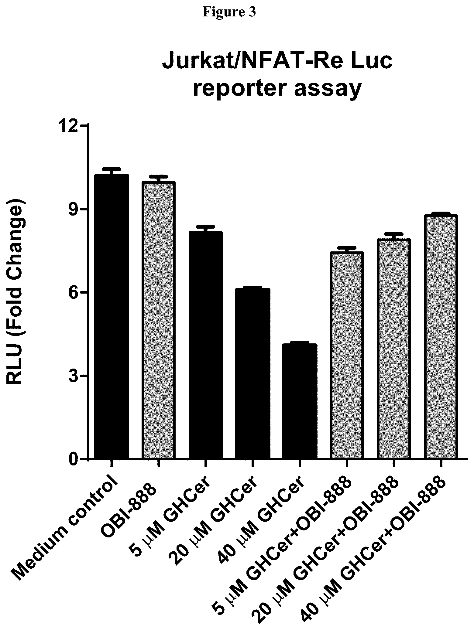
D00004

D00005

D00006
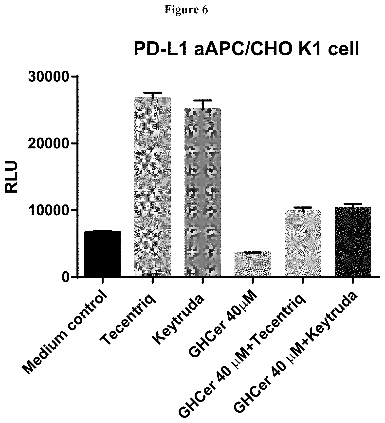
D00007

D00008

D00009

D00010

D00011

S00001
XML
uspto.report is an independent third-party trademark research tool that is not affiliated, endorsed, or sponsored by the United States Patent and Trademark Office (USPTO) or any other governmental organization. The information provided by uspto.report is based on publicly available data at the time of writing and is intended for informational purposes only.
While we strive to provide accurate and up-to-date information, we do not guarantee the accuracy, completeness, reliability, or suitability of the information displayed on this site. The use of this site is at your own risk. Any reliance you place on such information is therefore strictly at your own risk.
All official trademark data, including owner information, should be verified by visiting the official USPTO website at www.uspto.gov. This site is not intended to replace professional legal advice and should not be used as a substitute for consulting with a legal professional who is knowledgeable about trademark law.