PD-1 TARGETED HETERODIMERIC FUSION PROTEINS CONTAINING IL-15/IL-15Ra Fc-FUSION PROTEINS AND PD-1 ANTIGEN BINDING DOMAINS AND USE
Bernett; Matthew ; et al.
U.S. patent application number 16/388811 was filed with the patent office on 2019-12-26 for pd-1 targeted heterodimeric fusion proteins containing il-15/il-15ra fc-fusion proteins and pd-1 antigen binding domains and use. The applicant listed for this patent is Xencor, Inc.. Invention is credited to Matthew Bernett, Christine Bonzon, John Desjarlais, Rumana Rashid, Rajat Varma.
| Application Number | 20190389933 16/388811 |
| Document ID | / |
| Family ID | 66630342 |
| Filed Date | 2019-12-26 |


View All Diagrams
| United States Patent Application | 20190389933 |
| Kind Code | A1 |
| Bernett; Matthew ; et al. | December 26, 2019 |
PD-1 TARGETED HETERODIMERIC FUSION PROTEINS CONTAINING IL-15/IL-15Ra Fc-FUSION PROTEINS AND PD-1 ANTIGEN BINDING DOMAINS AND USES THEREOF
Abstract
The present invention is directed to novel PD-1-targeted IL-15/R.alpha.-Fc fusion proteins comprising an IL-15/IL-15R.alpha. Fc-fusion protein and a PD-1 antigen binding domain. The PD-1-targeted IL-15/R.alpha.-Fc fusion proteins can be administered to a patient to treat cancer.
| Inventors: | Bernett; Matthew; (Monrovia, CA) ; Desjarlais; John; (Pasadena, CA) ; Rashid; Rumana; (Temple City, CA) ; Varma; Rajat; (Monrovia, CA) ; Bonzon; Christine; (Los Angeles, CA) | ||||||||||
| Applicant: |
|
||||||||||
|---|---|---|---|---|---|---|---|---|---|---|---|
| Family ID: | 66630342 | ||||||||||
| Appl. No.: | 16/388811 | ||||||||||
| Filed: | April 18, 2019 |
Related U.S. Patent Documents
| Application Number | Filing Date | Patent Number | ||
|---|---|---|---|---|
| 62659571 | Apr 18, 2018 | |||
| Current U.S. Class: | 1/1 |
| Current CPC Class: | C07K 2319/00 20130101; A61K 39/3955 20130101; C07K 2317/24 20130101; A61P 35/00 20180101; C07K 2317/92 20130101; A61K 38/2086 20130101; C07K 14/5443 20130101; A61K 38/00 20130101; A61K 38/1793 20130101; C07K 14/7155 20130101; C07K 2317/524 20130101; C07K 2317/76 20130101; C07K 2317/75 20130101; C07K 2317/90 20130101; C07K 2319/30 20130101; C07K 2317/526 20130101; C07K 2317/624 20130101; C07K 16/2818 20130101 |
| International Class: | C07K 14/715 20060101 C07K014/715; A61K 38/17 20060101 A61K038/17; A61K 38/20 20060101 A61K038/20; C07K 14/54 20060101 C07K014/54; A61K 39/395 20060101 A61K039/395 |
Claims
1. A PD-1 targeted IL-15/R.alpha. heterodimeric Fc fusion protein comprising: a) a first monomer comprising, from N- to C-terminal: i) an IL-15 receptor alpha (IL-15R.alpha.) sushi domain; ii) a first domain linker, iii) a variant IL-15 domain, and iv) a second domain linker, and v) a first variant Fc domain comprising CH2-CH3; and b) a second monomer comprising, from N- to C-terminal: a heavy chain comprising VH-CH1-hinge-CH2-CH3, wherein said CH2-CH3 is a second variant Fc domain; and c) a light chain comprising VL-CL; wherein said VH and VL form an antigen binding domain that binds human PD-1 and have sequences selected from the pairs consisting of 1C11[PD-1]_H3L3 from XENP22553 (SEQ ID NOS:186-187), 1C11[PD-1]_H3.234_L3.144 from XENP25806 (SEQ ID NOS:578-579), 1C11[PD-1]_H3.240_L3.148 from XENP25812 (SEQ ID NO:584), 1C11[PD-1]_H3.241_L3.148 from XENP25813 (SEQ ID NO:585), 1C11[PD-1]_H3.241_L3.92 from XENP25819 (SEQ ID NO:591), 1C11[PD-1]_H3.303_L3.152 from XENP26940 (SEQ ID NOS:642 and 1103), 1C11[PD-1]_H3.329_L3.220 from XENP28026 (SEQ ID NOS:708 and 1169), and 1C11[PD-1]_H3.328_L3.152 from XENP28652 (SEQ ID NOS:719 and 1180); and wherein said first variant and said second variant Fc domains have a set of amino acid substitutions selected from the group consisting of S267K/L368D/K370S:S267K/LS364K/E357Q; S364K/E357Q:L368D/K370S; L368D/K370S:S364K; L368E/K370S:S364K; T411E/K360E/Q362E:D401K; L368D/K370S:S364K/E357L; L368D/K370S:S364K/E357Q; and K370S:S364K/E357Q, respectively and according to EU numbering.
2. The heterodimeric Fc fusion protein according to claim 1, wherein said first variant Fc domain and/or said second variant Fc domain have amino acid substitutions comprising Q295E/N384D/Q418E/N421D, according to EU numbering.
3. The heterodimeric Fc fusion protein according to claim 1 or 2, wherein said first variant and said variant second Fc domains each have amino acid substitutions selected from the group consisting of G236R/L328R, E233P/L234V/L235A/G236del/S239K, E233P/L234V/L235A/G236del/S267K, E233P/L234V/L235A/G236del/S239K/A327G, E233P/L234V/L235A/G236del/S267K/A327G and E233P/L234V/L235A/G236del, according to EU numbering.
4. The heterodimeric Fc fusion protein according to any one of claims 1 to 3, wherein said first variant and said second variant Fc domains each have amino acid substitution M428L/N434S, according to EU numbering.
5. The heterodimeric Fc fusion protein according to any one of claims 1 to 4, wherein said variant IL-15 domain comprises the amino acid sequence of SEQ ID NO:2.
6. The heterodimeric Fc fusion protein according to any one of claims 1 to 5 wherein said variant IL-15 domain comprises the amino acid sequence of SEQ ID NO:2 and amino acid substitutions selected from the group consisting of N4D/N65D, D30N/N65D, and D30N/E64Q/N65D.
7. The heterodimeric Fc fusion protein according to any one of claims 1 to 5, wherein said IL-15R.alpha. sushi domain comprises the amino acid sequence of SEQ ID NO:4.
8. The heterodimeric Fc fusion protein according to any one of claims 1 to 7, selected from the group consisting of XENP29482 set forth in SEQ ID NOS 925, 926, and 1216, XENP25937 set forth in SEQ ID NOS: 70-372, and any one depicted in FIG. 126A (SEQ ID NOS:925-929), FIG. 126B (SEQ ID NOS:930-935), FIG. 126C (SEQ ID NOS:936-941), FIG. 126D (SEQ ID NOS:942-947), FIG. 127A (SEQ ID NOS:948-953), FIG. 127B (SEQ ID NOS:954-959), FIG. 127C (SEQ ID NOS:960-965), FIG. 127D (SEQ ID NOS:966-971), FIG. 128A (SEQ ID NOS:972-977), FIG. 128B (SEQ ID NOS:978-983), FIG. 128C (SEQ ID NOS:984-989), FIG. 128D (SEQ ID NOS:990-995), FIG. 128E (SEQ ID NOS:996-1001), FIG. 128F (SEQ ID NOS:1002-1007), FIG. 128G (SEQ ID NOS:1008-1013), FIG. 128H (SEQ ID NOS:1014-1019), FIG. 128I (SEQ ID NOS:1020-1025), FIG. 128J (SEQ ID NOS:1026-1031), FIG. 128K (SEQ ID NOS:1032-1035), FIG. 128L (SEQ ID NOS:1036-1041).
9. A nucleic acid composition comprising: a) a first nucleic acid encoding the first monomer of the heterodimeric Fc fusion protein according to any one of claims 1 to 8; b) a second nucleic acid encoding the second monomer of the heterodimeric Fc fusion protein according to any one of claims 1 to 8; and c) a third nucleic acid encoding the light chain of the heterodimeric Fc fusion protein according to any one of claims 1 to 8, respectively.
10. An expression vector composition comprising: a) a first expression vector comprising said first nucleic acid of claim 9; b) a second expression vector comprising said second nucleic acid of claim 9; and c) a third expression vector comprising said third nucleic acid of claim 9.
11. A host cell comprising the nucleic acid composition of claim 9 or the expression vector composition of claim 10.
12. A method of producing a PD-1 targeted IL-15/R.alpha. heterodimeric Fc fusion protein according to any one of claims 1 to 8 comprising: culturing the host cell of claim 11 under suitable conditions, wherein said heterodimeric Fc fusion protein is expressed; and recovering said protein.
13. A PD-1 targeted IL-15/R.alpha. heterodimeric Fc fusion protein selected from the group consisting of XENP29482 set forth in SEQ ID NOS:925, 926, and 1216, XENP25937 set forth in SEQ ID NOS:370-372, and any one depicted in FIG. 126A (SEQ ID NOS:925-929), FIG. 126B (SEQ ID NOS:930-935), FIG. 126C (SEQ ID NOS:936-941), FIG. 126D (SEQ ID NOS:942-947), FIG. 127A (SEQ ID NOS:948-953), FIG. 127B (SEQ ID NOS:954-959), FIG. 127C (SEQ ID NOS:960-965), FIG. 127D (SEQ ID NOS:966-971), FIG. 128A (SEQ ID NOS:972-977), FIG. 128B (SEQ ID NOS:978-983), FIG. 128C (SEQ ID NOS:984-989), FIG. 128D (SEQ ID NOS:990-995), FIG. 128E (SEQ ID NOS:996-1001), FIG. 128F (SEQ ID NOS:1002-1007), FIG. 128G (SEQ ID NOS:1008-1013), FIG. 128H (SEQ ID NOS:1014-1019), FIG. 128I (SEQ ID NOS:1020-1025), FIG. 128J (SEQ ID NOS:1026-1031), FIG. 128K (SEQ ID NOS:1032-1035), FIG. 128L (SEQ ID NOS:1036-1041).
14. A method of treating cancer in a patient in need thereof comprising administering a therapeutically effective amount of a PD-1 targeted IL-15/R.alpha. heterodimeric Fc fusion protein according to any one of claims 1 to 8 and 13 to said patient.
15. The method of claim 14, further comprising administering a therapeutically effective amount of a checkpoint blockade antibody.
16. The method according to claim 15, wherein said checkpoint blockade antibody is selected from the group consisting of an anti-PD-1 antibody, an anti-PD-L1 antibody, an anti-TIM3 antibody, an anti-TIGIT antibody, an anti-LAG3 antibody, and an anti-CTLA-4 antibody.
17. The method according to claim 16, wherein said anti-PD-L1 antibody is atezolizumab, avelumab, or durbalumab.
Description
CROSS-REFERENCING TO RELATED APPLICATIONS
[0001] This application claims priority to U.S. Provisional Application No. 62/659,571, filed Apr. 18, 2018, the disclosure is herein incorporated by reference in its entirety.
SEQUENCE LISTING
[0002] The instant application contains a Sequence Listing which has been filed electronically in ASCII format and is hereby incorporated by reference in its entirety. Said ASCII copy, created on May 31, 2019, is named 067461-5204-US_SL.txt and is 3,115,252 bytes in size.
BACKGROUND OF THE INVENTION
[0003] Cytokines such as IL-2 and IL-15 function in aiding the proliferation and differentiation of B cells, T cells, and NK cells. Both cytokines exert their cell signaling function through binding to a trimeric complex consisting of two shared receptors, the common gamma chain (.gamma.c; CD132) and IL-2 receptor beta-chain (IL-2R ; CD122), as well as an alpha chain receptor unique to each cytokine: IL-2 receptor alpha (IL-2R.alpha.; CD25) or IL-15 receptor alpha (IL-15R.alpha.; CD215). Both cytokines are considered as potentially valuable therapeutics in oncology, and IL-2 has been approved for use in patients with metastatic renal-cell carcinoma and malignant melanoma. Currently, there are no approved uses of recombinant IL-15, although several clinical trials are ongoing. However, as potential drugs, both cytokines suffer from a very fast clearance, with half-lives measured in minutes. IL-2 immunotherapy has been associated with systemic toxicity when administered in high doses to overcome fast clearance. Such systemic toxicity has also been reported with IL-15 immunotherapy in recent clinical trials (Guo et al., J Immunol, 2015, 195(5):2353-64).
[0004] Immune checkpoint proteins such as PD-1 are up-regulated following T cell activation to preclude autoimmunity by exhausting activated T cells upon binding to immune checkpoint ligands such as PD-L1. However, immune checkpoint proteins are also up-regulated in tumor-infiltrating lymphocytes (TILs), and immune checkpoint ligands are overexpressed on tumor cells, contributing to immune escape by tumor cells.
[0005] There remains an unmet need in oncology treatment for therapeutic strategies with cytokines which do not require high doses and are targeted to tumors to avoid systemic toxicity. The present invention addresses this need by providing PD-1-targeted IL-15 fusion proteins (FIG. 2) with enhanced half-life and more selective targeting of TILs to improve safety profile.
BRIEF SUMMARY OF THE INVENTION
[0006] The present invention is directed to novel PD-1 targeted IL-15/R.alpha. heterodimeric Fc fusion proteins, their uses, and methods of making the heterodimeric Fc fusion proteins. comprising:
[0007] Accordingly in some aspects, the invention provides PD-1 targeted IL-15/R.alpha. heterodimeric Fc fusion proteins. In this aspect, the PD-1 targeted IL-15/R.alpha. heterodimeric Fc fusion protein comprises: [0008] (a) a first monomer comprising, from N- to C-terminal: [0009] (i) an IL-15 receptor alpha (IL-15R.alpha.) sushi domain, [0010] (ii) a first domain linker, [0011] (iii) a variant IL-15 domain, and [0012] (iv) a second domain linker, and [0013] (v) a first variant Fc domain comprising CH2-CH3; and [0014] (b) a second monomer comprising, from N- to C-terminal: a heavy chain comprising VH-CH1-hinge-CH2-CH3, wherein said CH2-CH3 is a second variant Fc domain; and [0015] (c) a light chain comprising VL-CL; wherein said VH and VL form an antigen binding domain that binds human PD-1 and have sequences selected from the pairs consisting of 1C11[PD-1]_H3L3 from XENP22553 (SEQ ID NOS:186 and 187), 1C11[PD-1]_H3.234_L3.144 from XENP25806 (SEQ ID NOS:578-57), 1C11[PD-1]_H3.240_L3.148 from XENP25812 (SEQ ID NO:584), 1C11[PD-1]_H3.241_L3.148 from XENP25813 (SEQ ID NO:585), 1C11[PD-1]_H3.241_L3.92 from XENP25819 (SEQ ID NO:591), 1C11[PD-1]_H3.303_L3.152 from XENP26940 (SEQ ID NOS:642 and 1103), 1C11[PD-1]_H3.329_L3.220 from XENP28026 (SEQ ID NOS:708 and 1169), and 1C11[PD-1]_H3.328_L3.152 from XENP28652 (SEQ ID NOS:719 and 1180); and wherein said first variant and said second variant Fc domains have a set of amino acid substitutions selected from the group consisting of S267K/L368D/K370S:S267K/LS364K/E357Q; S364K/E357Q:L368D/K370S; L368D/K370S:S364K; L368E/K370S:S364K; T411E/K360E/Q362E:D401K; L368D/K370S:S364K/E357L; L368D/K370S:S364K/E357Q; and K370S:S364K/E357Q, respectively and according to EU numbering.
[0016] In some embodiments, the first variant Fc domain and/or the second variant Fc domain of the PD-1 targeted IL-15/R.alpha. heterodimeric Fc fusion protein have amino acid substitutions comprising Q295E/N384D/Q418E/N421D, according to EU numbering.
[0017] In some embodiments, the first variant and the variant second Fc domains each have amino acid substitutions selected from the group consisting of G236R/L328R, E233P/L234V/L235A/G236del/S239K, E233P/L234V/L235A/G236del/S267K, E233P/L234V/L235A/G236del/S239K/A327G, E233P/L234V/L235A/G236del/S267K/A327G and E233P/L234V/L235A/G236del, according to EU numbering.
[0018] In some embodiments, the first variant and the second variant Fc domains each have amino acid substitution M428L/N434S, according to EU numbering.
[0019] In some embodiments, the variant IL-15 domain comprises the amino acid sequence of SEQ ID NO:2. In other embodiments, the variant IL-15 domain comprises the amino acid sequence of SEQ ID NO:2 and amino acid substitutions selected from the group consisting of N4D/N65D, D30N/N65D, and D30N/E64Q/N65D.
[0020] In some embodiments, the IL-15R.alpha. sushi domain comprises the amino acid sequence of SEQ ID NO:4.
[0021] In some embodiments, the PD-1 targeted IL-15/R.alpha. heterodimeric Fc fusion protein is selected from the group consisting of XENP29482 set forth in SEQ ID NOS:925,926, and 1216, XENP25937 set forth in SEQ ID NOS:370-372, and any one depicted in FIG. 126A (SEQ ID NOS:925-929), FIG. 126B (SEQ ID NOS:930-935), FIG. 126C (SEQ ID NOS:936-941), FIG. 126D (SEQ ID NOS:942-947), FIG. 127A (SEQ ID NOS:948-953), FIG. 127B (SEQ ID NOS:954-959), FIG. 127C (SEQ ID NOS:960-965), FIG. 127D (SEQ ID NOS:966-971), FIG. 128A (SEQ ID NOS:972-977), FIG. 128B (SEQ ID NOS:978-983), FIG. 128C (SEQ ID NOS:984-989), FIG. 128D (SEQ ID NOS:990-995), FIG. 128E (SEQ ID NOS:996-1001), FIG. 128F (SEQ ID NOS:1002-1007), FIG. 128G (SEQ ID NOS:1008-1013), FIG. 128H (SEQ ID NOS:1014-1019), FIG. 128I (SEQ ID NOS:1020-1025), FIG. 128J (SEQ ID NOS:1026-1031), FIG. 128K (SEQ ID NOS:1032-1035), FIG. 128L (SEQ ID NOS:1036-1041).
[0022] In further aspects, provided herein is a PD-1 targeted IL-15/R.alpha. heterodimeric Fc fusion protein comprising: [0023] (a) a first monomer comprising, from N- to C-terminal: [0024] (i) an IL-15 receptor alpha (IL-15R.alpha.) sushi domain, [0025] (ii) a first domain linker, [0026] (iii) a variant IL-15 domain, [0027] (iv) a second domain linker, and [0028] (v) a first variant Fc domain comprising CH2-CH3; and [0029] (b) a second monomer comprising, from N- to C-terminal: a [0030] (i) a single chain Fv domain (scFv) that binds human PD-1, wherein said scFv comprises: [0031] (1) a variable heavy domain (VH), [0032] (2) a scFv linker, and [0033] (3) a variable light domain (VL), and [0034] (ii) a second variant Fc domain; [0035] wherein the VHCDR1, VHCDR2, VHCDR3, VLCDR1, VLCDR2, and VLCDR3 are selected from the group consisting of the CDRs from 1C11[PD-1]_H3L3 from XENP22538 (SEQ ID NO:417), 1C11[PD-1]_H3.234_L3.144 from XENP25806 (SEQ ID NOS:578-579), 1C11[PD-1]_H3.240_L3.148 from XENP25812 (SEQ ID NO:584), 1C11[PD-1]_H3.241_L3.148 from XENP25813 (SEQ ID NO:585), 1C11[PD-1]_H3.241_L3.92 from XENP25819 (SEQ ID NO:591), 1C11[PD-1]_H3.303_L3.152 from XENP26940 (SEQ ID NOS:642 and 1103), 1C11[PD-1]_H3.329_L3.220 from XENP28026 (SEQ ID NOS:708 and 1169), and 1C11[PD-1]_H3.328_L3.152 from XENP28652 (SEQ ID NOS:719 and 1180); and [0036] wherein said first variant and said second variant Fc domains have a set of amino acid substitutions selected from the group consisting of S267K/L368D/K370S:S267K/LS364K/E357Q; S364K/E357Q:L368D/K370S; L368D/K370S:S364K; L368E/K370S:S364K; T411E/K360E/Q362E:D401K; L368D/K370S:S364K/E357L; L368D/K370S:S364K/E357Q; and K370S:S364K/E357Q, respectively and according to EU numbering.
[0037] In some embodiments, the VH and VL of the second monomer are selected from the pairs consisting of 1C11[PD-1]_H3L3 from XENP22538 (SEQ ID NO:417), 1C11[PD-1]_H3.234_L3.144 from XENP25806 (SEQ ID NOS:578-579), 1C11[PD-1]_H3.240_L3.148 from XENP25812 (SEQ ID NO:584), 1C11[PD-1]_H3.241_L3.148 from XENP25813 (SEQ ID NO:585), 1C11[PD-1]_H3.241_L3.92 from XENP25819 (SEQ ID NO:591), 1C11[PD-1]_H3.303_L3.152 from XENP26940 (SEQ ID NOS:642 and 1103), 1C11[PD-1]_H3.329_L3.220 from XENP28026 (SEQ ID NOS:708 and 1169), and 1C11[PD-1]_H3.328_L3.152 from XENP28652 (SEQ ID NOS:719 and 1180).
[0038] In some embodiments, the first variant and the second variant Fc domains have an additional set of amino acid substitutions comprising Q295E/N384D/Q418E/N421D, according to EU numbering.
[0039] In some embodiments, the first variant and the variant second Fc domains each have an additional set of amino acid substitutions consisting of G236R/L328R, E233P/L234V/L235A/G236del/S239K, E233P/L234V/L235A/G236del/S267K, E233P/L234V/L235A/G236del/S239K/A327G, E233P/L234V/L235A/G236del/S267K/A327G and E233P/L234V/L235A/G236del, according to EU numbering.
[0040] In some embodiments, the first variant and the second variant Fc domains each have an additional amino acid substitution M428L/N434S, according to EU numbering.
[0041] In some embodiments, the variant IL-15 domain comprises the amino acid sequence of SEQ ID NO:2. In certain embodiments, the variant IL-15 domain comprises the amino acid sequence of SEQ ID NO:2 and amino acid substitutions selected from the group consisting of N4D/N65D, D30N/N65D, and D30N/E64Q/N65D.
[0042] In some embodiments, the IL-15R.alpha. sushi domain has the amino acid sequence of SEQ ID NO:4.
[0043] In some embodiments, the first monomer comprises: the IL-15R.alpha. sushi domain of SEQ ID NO:4 and the variant IL-15 domain of SEQ ID NO:2 having amino acid substitutions selected from the group consisting of N4D/N65D, D30N/N65D, and D30N/E64Q/N65D; and the scFv comprises: the VH and VL are selected from the pairs consisting of 1C11[PD-1]_H3.234_L3.144 from XENP25806 (SEQ ID NOS:578-579), 1C11[PD-1]_H3.240_L3.148 from XENP25812 (SEQ ID NO:584), 1C11[PD-1]_H3.241_L3.148 from XENP25813 (SEQ ID NO:585), 1C11[PD-1]_H3.241_L3.92 from XENP25819 (SEQ ID NO:591), 1C11[PD-1]_H3.303_L3.152 from XENP26940 (SEQ ID NOS:642 and 1103), 1C11[PD-1]_H3.329_L3.220 from XENP28026 (SEQ ID NOS:708 and 1169), and 1C11[PD-1]_H3.328_L3.152 from XENP28652 (SEQ ID NOS:719 and 1180).
[0044] In other aspects, provided herein is a nucleic acid composition encoding the first monomer of any heterodimeric Fc fusion protein outlined herein. Also, provided herein is a nucleic acid composition encoding the second monomer of any heterodimeric Fc fusion protein outlined herein. Also, provided is a nucleic acid composition encoding the light chain of any heterodimeric Fc fusion protein outlined herein.
[0045] In some aspects, provided herein is an expression vector comprising any of the nucleic acid composition encoding any one of the first monomers described herein. Also, provided herein is an expression vector comprising any of the nucleic acid composition encoding any one of the second monomers described herein. Also, provided herein is an expression vector comprising any of the nucleic acid composition encoding any one of the light chains described herein such that the VL and VH of the heterodimeric Fc fusion protein binds human PD-1.
[0046] Provided herein is an expression vector comprising one or more of the nucleic acid compositions described herein. Provided herein is a host cell comprising one or more expression vectors.
[0047] In some aspects, provided herein is a method of producing a PD-1 targeted IL-15/R.alpha. heterodimeric Fc fusion protein comprising: culturing the host cell described herein under suitable conditions, wherein the heterodimeric Fc fusion protein is expressed; and recovering the protein.
[0048] In some aspects, the invention provides a PD-1 targeted IL-15/R.alpha. heterodimeric Fc fusion protein selected from the group consisting of XENP29482 set forth in SEQ ID NOS:925, 926, and 1216, XENP25937 set forth in SEQ ID NOS: 370-372, and any one depicted in FIG. 126A (SEQ ID NOS:925-929), FIG. 126B (SEQ ID NOS: 930-935), FIG. 126C (SEQ ID NOS:936-941), FIG. 126D (SEQ ID NOS:942-947), FIG. 127A (SEQ ID NOS:948-953), FIG. 127B (SEQ ID NOS:954-959), FIG. 127C (SEQ ID NOS:960-965), FIG. 127D (SEQ ID NOS:966-971), FIG. 128A (SEQ ID NOS:972-977), FIG. 128B (SEQ ID NOS:978-983), FIG. 128C (SEQ ID NOS:984-989), FIG. 128D (SEQ ID NOS:990-995), FIG. 128E (SEQ ID NOS:996-1001), FIG. 128F (SEQ ID NOS:1002-1007), FIG. 128G (SEQ ID NOS:1008-1013), FIG. 128H (SEQ ID NOS:1014-1019), FIG. 128I (SEQ ID NOS:1020-1025), FIG. 128J (SEQ ID NOS:1026-1031), FIG. 128K (SEQ ID NOS:1032-1035), FIG. 128L (SEQ ID NOS:1036-1041).
[0049] In other aspects, the invention provides a pharmaceutical composition comprising a PD-1 targeted IL-15/R.alpha. heterodimeric Fc fusion protein selected from the group consisting of XENP29482 set forth in SEQ ID NOS:925, 926, and 1216, XENP25937 set forth in SEQ ID NOS: 370-372, and any one depicted in FIG. 126A (SEQ ID NOS:925-929), FIG. 126B (SEQ ID NOS: 930-935), FIG. 126C (SEQ ID NOS:936-941), FIG. 126D (SEQ ID NOS:942-947), FIG. 127A (SEQ ID NOS:948-953), FIG. 127B (SEQ ID NOS:954-959), FIG. 127C (SEQ ID NOS:960-965), FIG. 127D (SEQ ID NOS:966-971), FIG. 128A (SEQ ID NOS:972-977), FIG. 128B (SEQ ID NOS:978-983), FIG. 128C (SEQ ID NOS:984-989), FIG. 128D (SEQ ID NOS:990-995), FIG. 128E (SEQ ID NOS:996-1001), FIG. 128F (SEQ ID NOS:1002-1007), FIG. 128G (SEQ ID NOS:1008-1013), FIG. 128H (SEQ ID NOS:1014-1019), FIG. 128I (SEQ ID NOS:1020-1025), FIG. 128J (SEQ ID NOS:1026-1031), FIG. 128K (SEQ ID NOS:1032-1035), FIG. 128L (SEQ ID NOS:1036-1041), and a pharmaceutically acceptable carrier.
[0050] In certain aspects, the invention provides method of treating cancer in a patient in need thereof comprising administering a therapeutically effective amount of any one of the PD-1 targeted IL-15/R.alpha. heterodimeric Fc fusion proteins described herein, or a pharmaceutical composition thereof.
[0051] In some embodiments, the method also comprises administering a therapeutically effective amount of a checkpoint blockade antibody.
[0052] In some embodiments, the checkpoint blockade antibody is selected from the group consisting of an anti-PD-1 antibody, an anti-PD-L1 antibody, an anti-TIM3 antibody, an anti-TIGIT antibody, an anti-LAG3 antibody, and an anti-CTLA-4 antibody.
[0053] In some embodiments, the said anti-PD-1 antibody is nivolumab, pembrolizumab, or pidilizumab. In some embodiments, the anti-PD-L1 antibody is atezolizumab, avelumab, or durbalumab.
BRIEF DESCRIPTION OF THE DRAWINGS
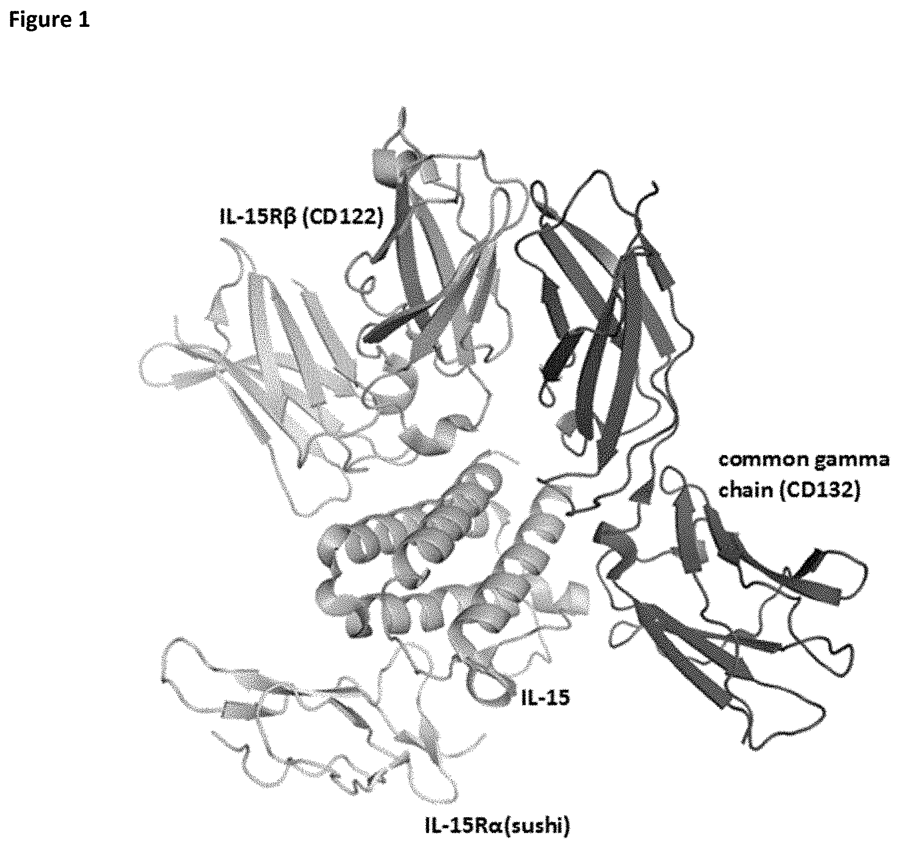
[0054] FIG. 1 depicts the structure of IL-15 in complex with its receptors IL-15R.alpha. (CD215), IL-15R.beta. (CD122), and the common gamma chain (CD132).
[0055] FIG. 2 depicts selectivity of PD-1-targeted IL-15/R.alpha.-Fc fusion proteins for tumor-reactive tumor-infiltrating lymphocytes expressing PD-1.
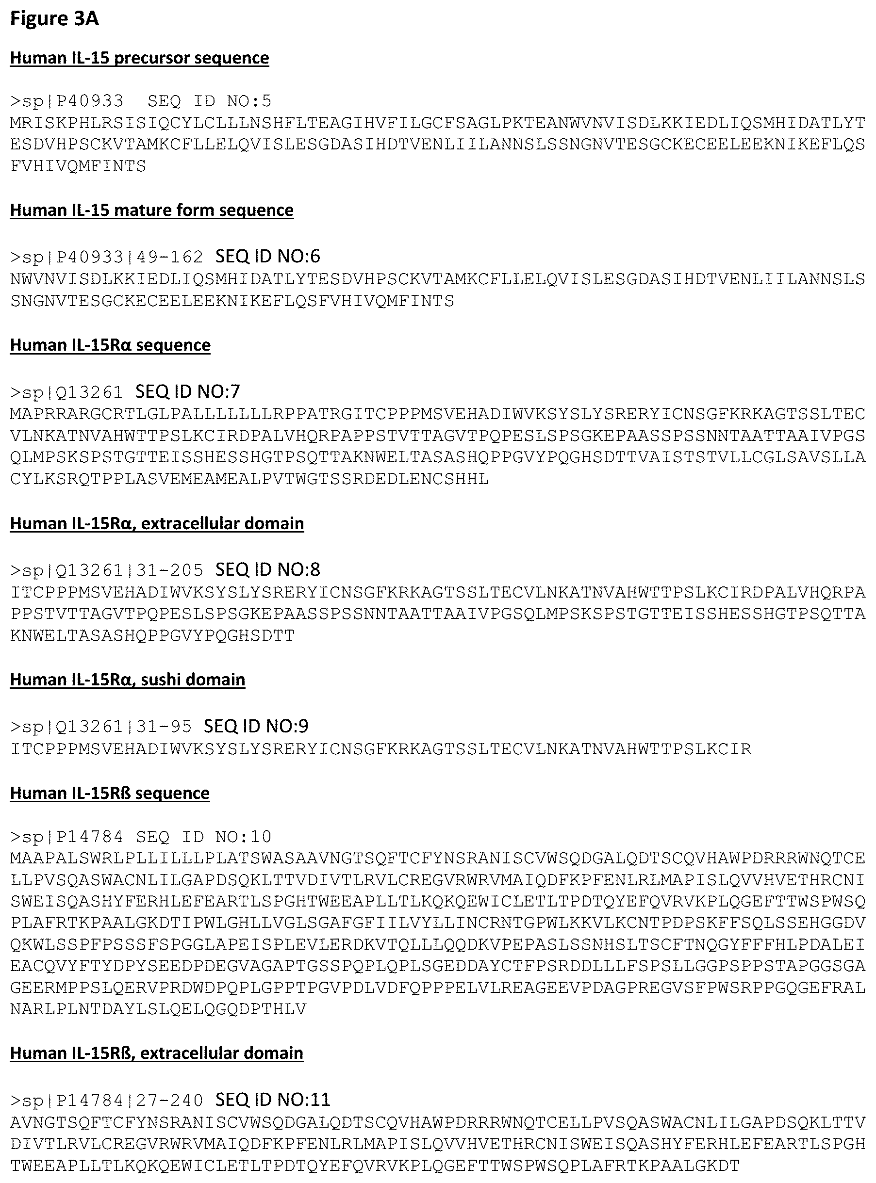
[0056] FIG. 3A-FIG. 3B depict the sequences for IL-15 and its receptors.
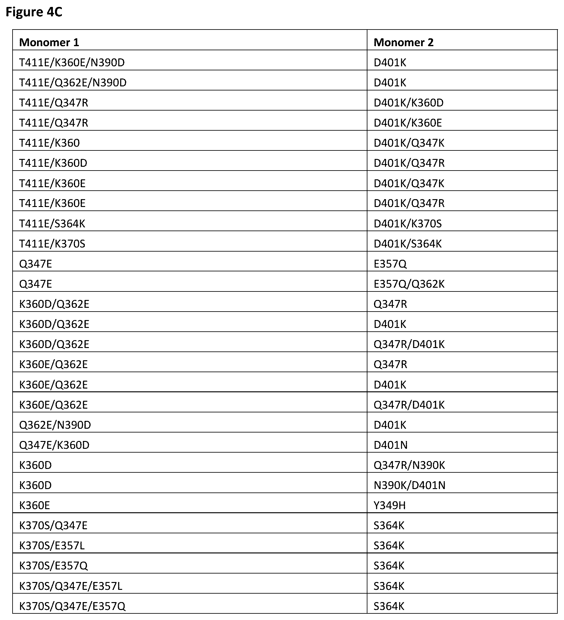
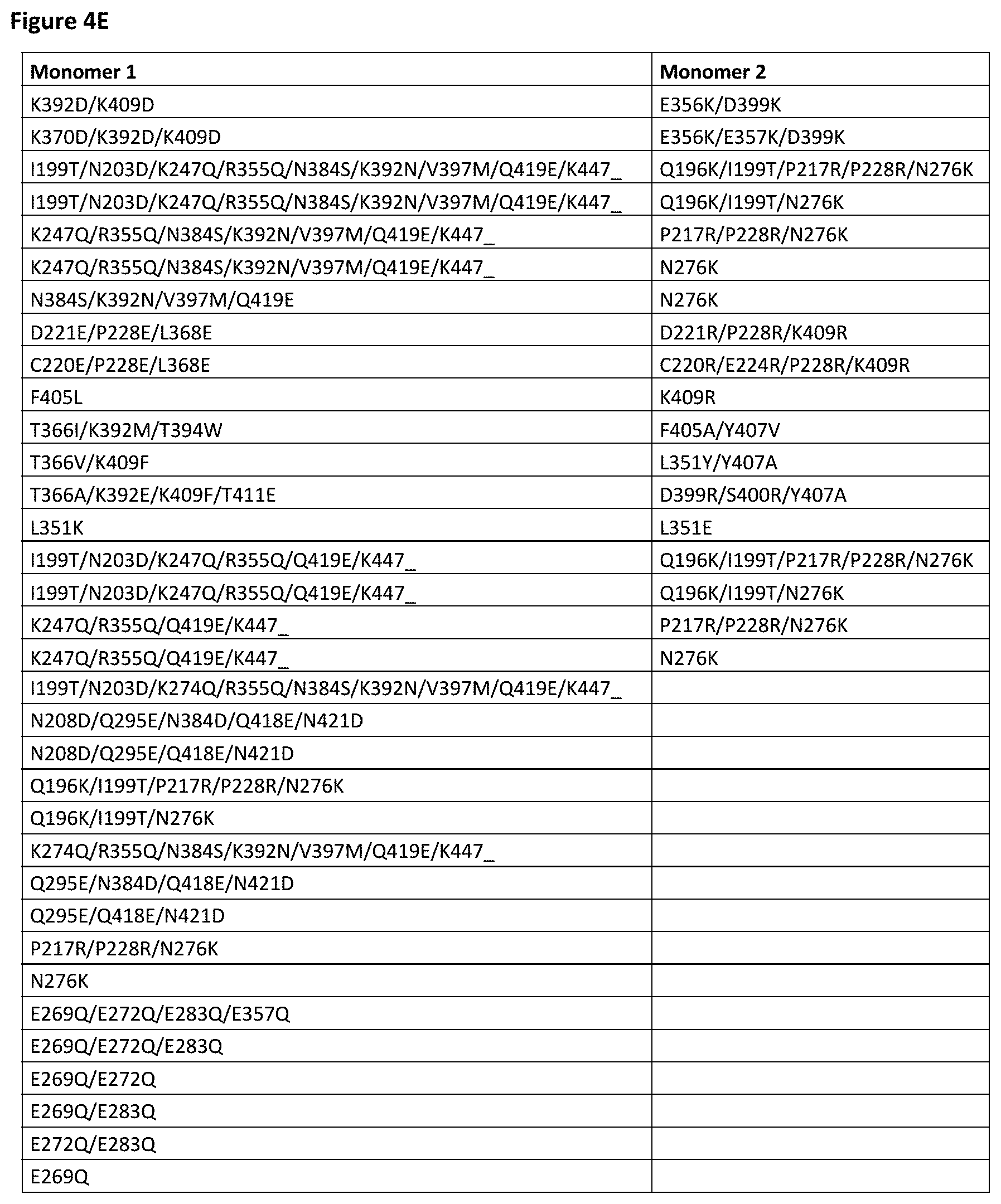
[0057] FIG. 4A-FIG. 4E depict useful pairs of Fc heterodimerization variant sets (including skew and pI variants). There are variants for which there are no corresponding "monomer 2" variants; these are pI variants which can be used alone on either monomer.
[0058] FIG. 5 depicts a list of isosteric variant antibody constant regions and their respective substitutions. pI_(-) indicates lower pI variants, while pI_(+) indicates higher pI variants. These can be optionally and independently combined with other heterodimerization variants of the inventions (and other variant types as well, as outlined herein).
[0059] FIG. 6 depicts useful ablation variants that ablate Fc.gamma.R binding (sometimes referred to as "knock outs" or "KO" variants). Generally, ablation variants are found on both monomers, although in some cases they may be on only one monomer.
[0060] FIG. 7A-FIG. 7E show a particularly useful embodiments of "non-cytokine" components of the IL-15/R.alpha.-Fc fusion proteins of the invention.
[0061] FIG. 8A-FIG. 8F show particularly useful embodiments of "non-cytokine"/"non-Fv" components of the PD-1-targeted IL-15/R.alpha.-Fc fusion proteins of the invention.
[0062] FIG. 9 depicts a number of exemplary variable length linkers for use in IL-15/R.alpha.-Fc fusion proteins. In some embodiments, these linkers find use linking the C-terminus of IL-15 and/or IL-15R.alpha.(sushi) to the N-terminus of the Fc region. In some embodiments, these linkers find use fusing IL-15 to the IL-15R.alpha.(sushi).
[0063] FIG. 10 depicts a number of charged scFv linkers that find use in increasing or decreasing the pI of heterodimeric antibodies that utilize one or more scFv as a component. The (+H) positive linker finds particular use herein. A single prior art scFv linker with single charge is referenced as "Whitlow", from Whitlow et al., Protein Engineering 6(8):989-995 (1993). It should be noted that this linker was used for reducing aggregation and enhancing proteolytic stability in scFvs.
[0064] FIG. 11A-FIG. 11D show the sequences of several useful IL-15/R.alpha.-Fc format backbones based on human IgG1, without the cytokine sequences (e.g., the IL-15 and/or IL-15R.alpha.(sushi)). It is important to note that these backbones can also find use in certain embodiments of PD-1 targeted IL-15/R.alpha.-Fc proteins. Backbone 1 is based on human IgG1 (356E/358M allotype), and includes C220S on both chain, the S364K/E357Q:L368D/K370S skew variants, the Q295E/N384D/Q418E/N421D pI variants on the chain with L368D/K370S skew variants and the E233P/L234V/L235A/G236del/S267K ablation variants on both chains. Backbone 2 is based on human IgG1 (356E/358M allotype), and includes C220S on both chain, the S364K:L368D/K370S skew variants, the Q295E/N384D/Q418E/N421D pI variants on the chain with L368D/K370S skew variants and the E233P/L234V/L235A/G236del/S267K ablation variants on both chains. Backbone 3 is based on human IgG1 (356E/358M allotype), and includes C220S on both chain, the S364K:L368E/K370S skew variants, the Q295E/N384D/Q418E/N421D pI variants on the chain with L368E/K370S skew variants and the E233P/L234V/L235A/G236del/S267K ablation variants on both chains. Backbone 4 is based on human IgG1 (356E/358M allotype), and includes C220S on both chain, the D401K:K360E/Q362E/T411E skew variants, the Q295E/N384D/Q418E/N421D pI variants on the chain with K360E/Q362E/T411E skew variants and the E233P/L234V/L235A/G236del/S267K ablation variants on both chains. Backbone 5 is based on human IgG1 (356D/358L allotype), and includes C220S on both chain, the S364K/E357Q:L368D/K370S skew variants, the Q295E/N384D/Q418E/N421D pI variants on the chain with L368D/K370S skew variants and the E233P/L234V/L235A/G236del/S267K ablation variants on both chains. Backbone 6 is based on human IgG1 (356E/358M allotype), and includes C220S on both chain, the S364K/E357Q:L368D/K370S skew variants, Q295E/N384D/Q418E/N421D pI variants on the chain with L368D/K370S skew variants and the E233P/L234V/L235A/G236del/S267K ablation variants on both chains, as well as an N297A variant on both chains. Backbone 7 is identical to 6 except the mutation is N297S. Alternative formats for backbones 6 and 7 can exclude the ablation variants E233P/L234V/L235A/G236del/S267K in both chains. Backbone 8 is based on human IgG4, and includes the S364K/E357Q:L368D/K370S skew variants, the Q295E/N384D/Q418E/N421D pI variants on the chain with L368D/K370S skew variants, as well as a S228P (EU numbering, this is S241P in Kabat) variant on both chains that ablates Fab arm exchange as is known in the art. Backbone 9 is based on human IgG2, and includes the S364K/E357Q:L368D/K370S skew variants, the Q295E/N384D/Q418E/N421D pI variants on the chain with L368D/K370S skew variants. Backbone 10 is based on human IgG2, and includes the S364K/E357Q:L368D/K370S skew variants, the Q295E/N384D/Q418E/N421D pI variants on the chain with L368D/K370S skew variants as well as a S267K variant on both chains. Backbone 11 is identical to backbone 1, except it includes M428L/N434S Xtend mutations. Backbone 12 is based on human IgG1 (356E/358M allotype), and includes C220S on both identical chain, the E233P/L234V/L235A/G236del/S267K ablation variants on both identical chains. Backbone 13 is based on human IgG1 (356E/358M allotype), and includes C220S on both chain, the S364K/E357Q:L368D/K370S skew variants, the P217R/P229R/N276K pI variants on the chain with S364K/E357Q skew variants and the E233P/L234V/L235A/G236del/S267K ablation variants on both chains.
[0065] As will be appreciated by those in the art and outlined below, these sequences can be used with any IL-15 and IL-15R.alpha.(sushi) pairs outlined herein, including but not limited to IL-15/R.alpha.-heteroFc, ncIL-15/R.alpha., and scIL-15/R.alpha., as schematically depicted in FIGS. 22 and 36. Additionally, any IL-15 and/or IL-15R.alpha.(sushi) variants can be incorporated into these FIGS. 11A-11D backbones in any combination.
[0066] Included within each of these backbones are sequences that are 90%, 95%, 98%, and 99% identical (as defined herein) to the recited sequences, and/or contain from 1, 2, 3, 4, 5, 6, 7, 8, 9 or 10 additional amino acid substitutions (as compared to the "parent" of the Figure, which, as will be appreciated by those in the art, already contain a number of amino acid modifications as compared to the parental human IgG1 (or IgG2 or IgG4, depending on the backbone). That is, the recited backbones may contain additional amino acid modifications (generally amino acid substitutions) in addition to the skew, pI and ablation variants contained within the backbones of FIGS. 11A-11D.
[0067] FIG. 12 shows the sequences of several useful PD-1-targeted IL-15/R.alpha.-Fc fusion format backbones based on human IgG1, without the cytokine sequences (e.g., the 11-15 and/or IL-15R.alpha.(sushi)) or VH, and further excluding light chain backbones which are depicted in FIG. 13. Backbone 1 is based on human IgG1 (356E/358M allotype), and includes the S364K/E357Q:L368D/K370S skew variants, C220S and the Q295E/N384D/Q418E/N421D pI variants on the chain with L368D/K370S skew variants and the E233P/L234V/L235A/G236del/S267K ablation variants on both chains. Backbone 2 is based on human IgG1 (356E/358M allotype), and includes the S364K/E357Q:L368D/K370S skew variants, the N208D/Q295E/N384D/Q418E/N421D pI variants on the chain with L368D/K370S skew variants, C220S in the chain with S364K/E357Q variants, and the E233P/L234V/L235A/G236del/S267K ablation variants on both chains. Backbone 3 is based on human IgG1 (356E/358M allotype), and includes the S364K/E357Q:L368D/K370S skew variants, the N208D/Q295E/N384D/Q418E/N421D pI variants on the chains with L368D/K370S skew variants, the Q196K/I199T/P217R/P228R/N276K pI variants on the chains with S364K/E357Q variants, and the E233P/L234V/L235A/G236del/S267K ablation variants on both chains.
[0068] In certain embodiments, these sequences can be of the 356D/358L allotype. In other embodiments, these sequences can include either the N297A or N297S substitutions. In some other embodiments, these sequences can include the M428L/N434S Xtend mutations. In yet other embodiments, these sequences can instead be based on human IgG4, and include a S228P (EU numbering, this is S241P in Kabat) variant on both chains that ablates Fab arm exchange as is known in the art. In yet further embodiments, these sequences can instead be based on human IgG2. Further, these sequences may instead utilize the other skew variants, pI variants, and ablation variants depicted in FIGS. 4A-4E, 5 and 6.
[0069] As will be appreciated by those in the art and outlined below, these sequences can be used with any IL-15 and IL-15R.alpha.(sushi) pairs outlined herein, including but not limited to scIL-15/R.alpha., ncIL-15/R.alpha., and dsIL-15R.alpha., as schematically depicted in FIGS. 65A-65K. Further as will be appreciated by those in the art and outlined below, any IL-15 and/or IL-15R.alpha.(sushi) variants can be incorporated in these backbones. Furthermore as will be appreciated by those in the art and outlined below, these sequences can be used with any VH and VL pairs outlined herein, including either a scFv or a Fab.
[0070] Included within each of these backbones are sequences that are 90%, 95%, 98% and 99% identical (as defined herein) to the recited sequences, and/or contain from 1, 2, 3, 4, 5, 6, 7, 8, 9 or 10 additional amino acid substitutions (as compared to the "parent" of the Figure, which, as will be appreciated by those in the art, already contain a number of amino acid modifications as compared to the parental human IgG1 (or IgG2 or IgG4, depending on the backbone). That is, the recited backbones may contain additional amino acid modifications (generally amino acid substitutions) in addition to the skew, pI and ablation variants contained within the backbones of this figure.
[0071] FIG. 13 depicts the "non-Fv" backbone of light chains (i.e., constant light chain) which find use in PD-1-targeted IL-15/R.alpha.-Fc fusion proteins of the invention.
[0072] FIG. 14 depicts the variable region sequences for an illustrative anti-PD-1 binding domain. The CDRs are underlined. As noted herein and is true for every sequence herein containing CDRs, the exact identification of the CDR locations may be slightly different depending on the numbering used as is shown in Table 1, and thus included herein are not only the CDRs that are underlined but also CDRs included within the V.sub.H and VL domains using other numbering systems. Furthermore, as for all the sequences in the figures, these V.sub.H and VL sequences can be used either in a scFv format or in a Fab format.
[0073] FIG. 15A-FIG. 15F depict the variable regions of additional PD-1-3 ABDs which may find use in the PD-1-targeted IL-15/R.alpha.-Fc fusion proteins of the invention. The CDRs are underlined. As noted herein and is true for every sequence herein containing CDRs, the exact identification of the CDR locations may be slightly different depending on the numbering used as is shown in Table 1, and thus included herein are not only the CDRs that are underlined but also CDRs included within the V.sub.H and VL domains using other numbering systems. Furthermore, as for all the sequences in the Figures, these V.sub.H and VL sequences can be used either in a scFv format or in a Fab format.
[0074] FIG. 16 depicts the sequences for XENP21575, a chimeric anti-PD-1 antibody based on the variable regions of hybridoma clone 1C11 and human IgG1 with E233P/L234V/L235A/G236del/S267K substitutions in the heavy chain. The CDRs are in bold, and the slashes indicate the borders of the variable domains. As note herein and is true for every sequence herein containing CDRs, the exact identification of the CDR locations may be slightly different depending on numbering used as is shown in Table 1, and thus included herein are not only the CDRs that are underlined but also CDRs included within the V.sub.H and V.sub.L domains using other numbering systems.
[0075] FIG. 17 depicts blocking of PD-1/PD-L1 interaction on PD-1 transfected HEK293T cells by anti-PD-1 clone 1C11.
[0076] FIG. 18 depicts the binding of anti-PD-1 clone 1C11 to SEB-stimulated T cells.
[0077] FIG. 19A-FIG. 19B depict cytokine release assays (FIG. 19A: IL-2; FIG. 19B: IFN.gamma.) after SEB stimulation of human PBMCs and treatment with anti-PD-1 clone 1C11.
[0078] FIG. 20A-FIG. 20C depict the sequences for illustrative humanized variants of anti-PD-1 clone 1C11 as a bivalent antibodies in the human IgG1 format with E233P/L234V/L235A/G236del/S267K substitutions in the heavy chain. The CDRs are in bold, and the slashes indicate the borders of the variable domains. As note herein and is true for every sequence herein containing CDRs, the exact identification of the CDR locations may be slightly different depending on numbering used as is shown in Table 1, and thus included herein are not only the CDRs that are bolded but also CDRs included within the V.sub.H and V.sub.L domains using other numbering systems. As will be appreciated by those in the art, the V.sub.H and V.sub.L domains can be formatted as Fab or scFvs for use in the PD-1 targeted IL-15/R.alpha.-Fc fusion proteins of the invention.
[0079] FIG. 21 depicts the affinity of XENP22553 for PD-1 as determined by Octet (as well as the associated sensorgram).
[0080] FIG. 22A-FIG. 22G depict several formats for the IL-15/R.alpha.-Fc fusion proteins of the present invention. IL-15R.alpha. Heterodimeric Fc fusion or "IL-15/R.alpha.-heteroFc" (FIG. 22A) comprises IL-15 recombinantly fused to one side of a heterodimeric Fc and IL-15R.alpha.(sushi) recombinantly fused to the other side of a heterodimeric Fc. The IL-15 and IL-15R.alpha.(sushi) may have a variable length Gly-Ser linker between the C-terminus and the N-terminus of the Fc region. Single-chain IL-15/R.alpha.-Fc fusion or "scIL-15/R.alpha.-Fc" (FIG. 22B) comprises IL-15R.alpha.(sushi) fused to IL-15 by a variable length linker (termed a "single-chain" IL-15/IL-15R.alpha.(sushi) complex or "scIL-15/R.alpha.") which is then fused to the N-terminus of a heterodimeric Fc-region, with the other side of the molecule being "Fc-only" or "empty Fc". Non-covalent IL-15/R.alpha.-Fc or "ncIL-15/R.alpha.-Fc" (FIG. 22C) comprises IL-15R.alpha.(sushi) fused to a heterodimeric Fc region, while IL-15 is transfected separately so that a non-covalent IL-15/R.alpha. complex is formed, with the other side of the molecule being "Fc-only" or "empty Fc". Bivalent non-covalent IL-15/R.alpha.-Fc fusion or "bivalent ncIL-15/R.alpha.-Fc" (FIG. 22D) comprises IL-15R.alpha.(sushi) fused to the N-terminus of a homodimeric Fc region, while IL-15 is transfected separately so that a non-covalent IL-15/R.alpha. complex is formed. Bivalent single-chain IL-15/R.alpha.-Fc fusion or "bivalent scIL-15/R.alpha.-Fc" (FIG. 22E) comprises IL-15 fused to IL-15R.alpha.(sushi) by a variable length linker (termed a "single-chain" IL-15/IL-15R.alpha.(sushi) complex or "scIL-15/R.alpha.") which is then fused to the N-terminus of a homodimeric Fc-region. Fc-non-covalent IL-15/R.alpha. fusion or "Fc-ncIL-15/R.alpha." (FIG. 22F) comprises IL-15R.alpha.(sushi) fused to the C-terminus of a heterodimeric Fc region, while IL-15 is transfected separately so that a non-covalent IL-15/R.alpha. complex is formed, with the other side of the molecule being "Fc-only" or "empty Fc". Fc-single-chain IL-15/R.alpha. fusion or "Fc-scIL-15/R.alpha." (FIG. 22G) comprises IL-15 fused to IL-15R.alpha.(sushi) by a variable length linker (termed a "single-chain" IL-15/IL-15R.alpha.(sushi) complex or "scIL-15/R.alpha.") which is then fused to the C-terminus of a heterodimeric Fc region, with the other side of the molecule being "Fc-only" or "empty Fc".
[0081] FIG. 23 depicts sequences of XENP20818 and XENP21475, illustrative IL-15/R.alpha.-Fc fusion proteins of the "IL-15/R.alpha.-heteroFc" format. IL-15 and IL-15R.alpha.(sushi) are underlined, linkers are double underlined (although as will be appreciated by those in the art, the linkers can be replaced by other linkers, some of which are depicted in FIGS. 9 and 10), and slashes (/) indicate the border(s) between IL-15, IL-15R.alpha., linkers, and Fc regions.
[0082] FIG. 24 depicts sequences of XENP21478, an illustrative IL-15/R.alpha.-Fc fusion protein of the "scIL-15/R.alpha.-Fc" format. IL-15 and IL-15R.alpha.(sushi) are underlined, linkers are double underlined (although as will be appreciated by those in the art, the linkers can be replaced by other linkers, some of which are depicted in FIGS. 9 and 10), and slashes (/) indicate the border(s) between IL-15, IL-15R.alpha., linkers, and Fc regions.
[0083] FIG. 25A and FIG. 25B depict sequences of XENP21479, XENP22366 and XENP24348, illustrative IL-15/R.alpha.-Fc fusion proteins of the "ncIL-15/R.alpha.-Fc" format. IL-15 and IL-15R.alpha.(sushi) are underlined, linkers are double underlined (although as will be appreciated by those in the art, the linkers can be replaced by other linkers, some of which are depicted in FIGS. 9 and 10), and slashes (/) indicate the border(s) between IL-15, IL-15R.alpha., linkers, and Fc regions.
[0084] FIG. 26 depicts sequences of XENP21978, an illustrative IL-15/R.alpha.-Fc fusion protein of the "bivalent ncIL-15/R.alpha.-Fc" format. IL-15 and IL-15R.alpha.(sushi) are underlined, linkers are double underlined (although as will be appreciated by those in the art, the linkers can be replaced by other linkers, some of which are depicted in FIGS. 9 and 10), and slashes (/) indicate the border(s) between IL-15, IL-15R.alpha., linkers, and Fc regions.
[0085] FIG. 27 depicts sequences of an illustrative IL-15/R.alpha.-Fc fusion protein of the "bivalent scIL-15/R.alpha.-Fc" format. IL-15 and IL-15R.alpha.(sushi) are underlined, linkers are double underlined (although as will be appreciated by those in the art, the linkers can be replaced by other linkers, some of which are depicted in FIGS. 9 and 10), and slashes (/) indicate the border(s) between IL-15, IL-15R.alpha., linkers, and Fc regions.
[0086] FIG. 28 depicts sequences of XENP22637, an illustrative IL-15/R.alpha.-Fc fusion protein of the "Fc-ncIL-15/R.alpha." format. IL-15 and IL-15R.alpha.(sushi) are underlined, linkers are double underlined (although as will be appreciated by those in the art, the linkers can be replaced by other linkers, some of which are depicted in FIGS. 9 and 10), and slashes (/) indicate the border(s) between IL-15, IL-15R.alpha., linkers, and Fc regions.
[0087] FIG. 29 depicts sequences of an illustrative IL-15/R.alpha.-Fc fusion protein of the "Fc-scIL-15/R.alpha." format. IL-15 and IL-15R.alpha.(sushi) are underlined, linkers are double underlined (although as will be appreciated by those in the art, the linkers can be replaced by other linkers, some of which are depicted in FIGS. 9 and 10), and slashes (/) indicate the border(s) between IL-15, IL-15R.alpha., linkers, and Fc regions.
[0088] FIG. 30A-FIG. 30C depict the induction of (FIG. 30A) NK (CD56.sup.+/CD16.sup.+) cells, (FIG. 30B) CD4.sup.+ T cells, and (FIG. 30C) CD8.sup.+ T cells proliferation by illustrative IL-15/R.alpha.-Fc fusion proteins of Format A with different linker lengths based on Ki67 expression as measured by FACS.
[0089] FIG. 31A-FIG. 31C depict the induction of (FIG. 31A) NK (CD56.sup.+/CD16.sup.+) cells, (FIG. 31B) CD4.sup.+ T cells, and (FIG. 31C) CD8.sup.+ T cells proliferation by illustrative IL-15/R.alpha.-Fc fusion proteins of scIL-15/R.alpha.-Fc format (XENP21478) and ncIL-15/R.alpha.-Fc format (XENP21479) based on Ki67 expression as measured by FACS.
[0090] FIG. 32 depicts a structural model of the IL-15/R.alpha. heterodimer showing locations of engineered disulfide bond pairs.
[0091] FIG. 33 depicts sequences for illustrative IL-15R.alpha.(sushi) variants engineered with additional residues at the C-terminus to serve as a scaffold for engineering cysteine residues.
[0092] FIG. 34 depicts sequences for illustrative IL-15 variants engineered with cysteines in order to form covalent disulfide bonds with IL-15R.alpha.(sushi) variants engineered with cysteines.
[0093] FIG. 35 depicts sequences for illustrative IL-15R.alpha.(sushi) variants engineered with cysteines in order to form covalent disulfide bonds with IL-15 variants engineered with cysteines.
[0094] FIG. 36A-FIG. 36D depict additional formats for the IL-15/R.alpha.-Fc fusion proteins of the present invention with engineered disulfide bonds. Disulfide-bonded IL-15/R.alpha. heterodimeric Fc fusion or "dsIL-15/R.alpha.-heteroFc" (FIG. 36A) is the same as "IL-15/R.alpha.-heteroFc", but wherein IL-15R.alpha.(sushi) and IL-15 are further covalently linked as a result of engineered cysteines. Disulfide-bonded IL-15/R.alpha. Fc fusion or "dsIL-15/R.alpha.-Fc" (FIG. 36B) is the same as "ncIL-15/R.alpha.-Fc", but wherein IL-15R.alpha.(sushi) and IL-15 are further covalently linked as a result of engineered cysteines. Bivalent disulfide-bonded IL-15/R.alpha.-Fc or "bivalent dsIL-15/R.alpha.-Fc" (FIG. 36C) is the same as "bivalent ncIL-15/R.alpha.-Fc", but wherein IL-15R.alpha.(sushi) and IL-15 are further covalently linked as a result of engineered cysteines. Fc-disulfide-bonded IL-15/R.alpha. fusion or "Fc-dsIL-15/R.alpha." (FIG. 36D) is the same as "Fc-ncIL-15/R.alpha.", but wherein IL-15R.alpha.(sushi) and IL-15 are further covalently linked as a result of engineered cysteines.
[0095] FIG. 37A-FIG. 37B depict sequences of XENP22013, XENP22014, XENP22015, and XENP22017, illustrative IL-15/R.alpha.-Fc fusion protein of the "dsIL-15/R.alpha.-heteroFc" format. IL-15 and IL-15R.alpha.(sushi) are underlined, linkers are double underlined (although as will be appreciated by those in the art, the linkers can be replaced by other linkers, some of which are depicted in FIG. 9), and slashes (/) indicate the border(s) between IL-15, IL-15R.alpha., linkers, and Fc regions.
[0096] FIG. 38A-FIG. 38B depict sequences of XENP22357, XENP22358, XENP22359, XENP22684, and XENP22361, illustrative IL-15/R.alpha.-Fc fusion proteins of the "dsIL-15/R.alpha.-Fc" format. IL-15 and IL-15R.alpha.(sushi) are underlined, linkers are double underlined (although as will be appreciated by those in the art, the linkers can be replaced by other linkers, some of which are depicted in FIGS. 9 and 10), and slashes (/) indicate the border(s) between IL-15, IL-15R.alpha., linkers, and Fc regions.
[0097] FIG. 39 depicts sequences of XENP22634, XENP22635, and XENP22636, illustrative IL-15/R.alpha.-Fc fusion proteins of the "bivalent dsIL-15/R.alpha.-Fc" format. IL-15 and IL-15R.alpha.(sushi) are underlined, linkers are double underlined (although as will be appreciated by those in the art, the linkers can be replaced by other linkers, some of which are depicted in FIG. 9 and FIG. 10), and slashes (/) indicate the border(s) between IL-15, IL-15R.alpha., linkers, and Fc regions.
[0098] FIG. 40 depicts sequences of XENP22639 and XENP22640, illustrative IL-15/R.alpha.-Fc fusion proteins of the "Fc-dsIL-15/R.alpha." format. IL-15 and IL-15R.alpha.(sushi) are underlined, linkers are double underlined (although as will be appreciated by those in the art, the linkers can be replaced by other linkers, some of which are depicted in FIGS. 9 and 10), and slashes (/) indicate the border(s) between IL-15, IL-15R.alpha., linkers, and Fc regions.
[0099] FIG. 41 depicts the purity and homogeneity of illustrative IL-15/R.alpha.-Fc fusion proteins with and without engineered disulfide bonds as determined by CEF.
[0100] FIG. 42 depicts the induction of A) NK (CD56+/CD16+) cell, B) CD8.sup.+ T cell, and C) CD4.sup.+ T cell proliferation by illustrative IL-15/R.alpha.-Fc fusion proteins with and without engineered disulfide bonds based on Ki67 expression as measured by FACS.
[0101] FIG. 43 depicts the structure of IL-15 complexed with IL-15R.alpha., IL-2R , and common gamma chain. Locations of substitutions designed to reduce potency are shown.
[0102] FIG. 44 depicts sequences for illustrative IL-15 variants engineered for reduced potency. Included within each of these variant IL-15 sequences are sequences that are 90, 95, 98 and 99% identical (as defined herein) to the recited sequences, and/or contain from 1, 2, 3, 4, 5, 6, 7, 8, 9 or 10 additional amino acid substitutions. In a non-limiting example, the recited sequences may contain additional amino acid modifications such as those contributing to formation of covalent disulfide bonds as described in Example 3B.
[0103] FIG. 45A-FIG. 45D depict sequences of XENP22821, XENP22822, XENP23554, XENP23557, XENP23561, XENP24018, XENP24019, XENP24045, XENP24051, and XENP24052, illustrative IL-15/R.alpha.-Fc fusion proteins of the "IL-15/R.alpha.-heteroFc" format engineered for reduced potency. IL-15 and IL-15R.alpha.(sushi) are underlined, linkers are double underlined (although as will be appreciated by those in the art, the linkers can be replaced by other linkers, some of which are depicted in FIGS. 9 and 10), and slashes (/) indicate the border(s) between IL-15, IL-15R.alpha., linkers, and Fc regions.
[0104] FIG. 46A-FIG. 46C depict sequences of XENP24015, XENP24050, XENP24475, XENP24476, XENP24478, XENP24479, and XENP24481, illustrative IL-15/R.alpha.-Fc fusion proteins of the "scIL-15/R.alpha.-Fc" format engineered for reduced potency. IL-15 and IL-15R.alpha.(sushi) are underlined, linkers are double underlined (although as will be appreciated by those in the art, the linkers can be replaced by other linkers, some of which are depicted in FIGS. 9 and 10), and slashes (/) indicate the border(s) between IL-15, IL-15R.alpha., linkers, and Fc regions.
[0105] FIG. 47A-FIG. 47B depict sequences of XENP24349, XENP24890, and XENP25138, illustrative IL-15/R.alpha.-Fc fusion proteins of the "ncIL-15/R.alpha.-Fc" format engineered for reduced potency. IL-15 and IL-15R.alpha.(sushi) are underlined, linkers are double underlined (although as will be appreciated by those in the art, the linkers can be replaced by other linkers, some of which are depicted in FIG. 9 and FIG. 10), and slashes (/) indicate the border(s) between IL-15, IL-15R.alpha., linkers, and Fc regions.
[0106] FIG. 48 depicts sequences of XENP22801 and XENP22802, illustrative ncIL-15/R.alpha. heterodimers engineered for reduced potency. It is important to note that these sequences were generated using polyhistidine (His.times.6 (SEQ ID NO: 1221) or HHHHHH (SEQ ID NO: 1221)) C-terminal tags at the C-terminus of IL-15R.alpha.(sushi).
[0107] FIG. 49 depicts sequences of XENP24342, an illustrative IL-15/R.alpha.-Fc fusion protein of the "bivalent ncIL-15/R.alpha.-Fc" format engineered for reduced potency. IL-15 and IL-15R.alpha.(sushi) are underlined, linkers are double underlined (although as will be appreciated by those in the art, the linkers can be replaced by other linkers, some of which are depicted in FIG. 9 and FIG. 10), and slashes (/) indicate the border(s) between IL-15, IL-15R.alpha., linkers, and Fc regions.
[0108] FIG. 50 depicts sequences of XENP23472 and XENP23473, illustrative IL-15/R.alpha.-Fc fusion proteins of the "dsIL-15/R.alpha.-Fc" format engineered for reduced potency. IL-15 and IL-15R.alpha.(sushi) are underlined, linkers are double underlined (although as will be appreciated by those in the art, the linkers can be replaced by other linkers, some of which are depicted in FIGS. 9 and 10), and slashes (/) indicate the border(s) between IL-15, IL-15R.alpha., linkers, and Fc regions.
[0109] FIG. 51 depicts the induction of A) NK cell, B) CD8.sup.+ (CD45RA-) T cell, and C) CD4.sup.+ (CD45RA-) T cell proliferation by variant IL-15/R.alpha.-Fc fusion proteins based on Ki67 expression as measured by FACS.
[0110] FIG. 52 depicts EC50 for induction of NK and CD8.sup.+ T cells proliferation by variant IL-15/R.alpha.-Fc fusion proteins, and fold reduction in EC50 relative to XENP20818.
[0111] FIG. 53A-FIG. 53C depict the gating of lymphocytes and subpopulations for the experiments depicted in FIG. 56. FIG. 53A shows the gated lymphocyte population.
[0112] FIG. 53B shows the CD3-negative and CD3-positive subpopulations. FIG. 53C shows the CD16=negative and CD16-positive subpopulations of the CD3-negative cells.
[0113] FIG. 54A-FIG. 54C depict the gating of CD3.sup.+ lymphocyte subpopulations for the experiments depicted in FIG. 56. FIG. 54A shows the CD4.sup.+, CD8.sup.+ and .gamma..delta. T cell subpopulations of the CD3.sup.+ T cells. FIG. 54B shows the CD45RA(-) and CD45RA(+) subpopulations of the CD4.sup.+ T cells. FIG. 54C shows the CD45RA(-) and CD45RA(+) subpopulation s of the CD8.sup.+ T cells.
[0114] FIG. 55A-FIG. 55B depict CD69 and CD25 expression before (FIG. 55A) and after (FIG. 55B) incubation of human PBMCs with XENP22821.
[0115] FIG. 56A-FIG. 56D depict cell proliferation in human PBMCs incubated for four days with the indicated variant IL-15/R.alpha.-Fc fusion proteins. FIGS. 56A-56C show the percentage of proliferating NK cells (CD3-CD16.sup.+) (FIG. 56A), CD8.sup.+ T cells (CD3+CD8+CD45RA-) (FIG. 56B) and CD4.sup.+ T cells (CD3+CD4+CD45RA-) (FIG. 56C). FIG. 56D shows the fold change in EC50 of various IL15/IL15R.alpha. Fc heterodimers relative to control (XENP20818).
[0116] FIG. 57A-FIG. 57D depict cell proliferation in human PBMCs incubated for three days with the indicated variant IL-15/R.alpha.-Fc fusion proteins. FIGS. 57A-C show the percentage of proliferating CD8.sup.+ (CD45RA-) T cells (FIG. 57A), CD4.sup.+ (CD45RA-) T cells (FIG. 57B), .gamma..delta. T cells (FIG. 57C), and NK cells (FIG. 57D).
[0117] FIG. 58A-FIG. 58C depict the percentage of Ki67 expression on (FIG. 58A) CD8.sup.+ T cells, (FIG. 58B) CD4.sup.+ T cells, and (FIG. 58C) NK cells following treatment with additional IL-15/R.alpha. variants.
[0118] FIG. 59A-FIG. 59E depict the percentage of Ki67 expression on (FIG. 59A) CD8.sup.+ (CD45RA-) T cells, (FIG. 59B) CD4.sup.+ (CD45RA-) T cells, (FIG. 59C) .gamma..delta. T cells, (FIG. 59D) NK (CD16+CD8.alpha.-) cells, and (FIG. 59E) NK (CD56+CD8.alpha.-) cells following treatment with IL-15/R.alpha. variants.
[0119] FIG. 60A-FIG. 60E depict the percentage of Ki67 expression on (FIG. 60A) CD8.sup.+ (CD45RA-) T cells, (FIG. 60B) CD4.sup.+ (CD45RA-) T cells, (FIG. 60C) .gamma..delta. T cells, (FIG. 60D) NK (CD16.sup.+CD8.alpha.-) cells, and (FIG. 60E) NK (CD56.sup.+CD8.alpha.-) cells following treatment with IL-15/R.alpha. variants.
[0120] FIG. 61A-FIG. 61D depict the percentage of Ki67 expression on (FIG. 61A) CD8.sup.+ T cells, (FIG. 61B) CD4.sup.+ T cells, (FIG. 61C) .gamma..delta. T cells and (FIG. 61D) NK (CD16+) cells following treatment with additional IL-15/R.alpha. variants.
[0121] FIG. 62A-FIG. 62D depict the percentage of Ki67 expression on (FIG. 62A) CD8.sup.+ T cells, (FIG. 62B) CD4.sup.+ T cells, (FIG. 62C) .gamma..delta. T cells and (FIG. 62D) NK (CD16+) cells following treatment with additional IL-15/R.alpha. variants.
[0122] FIG. 63 depicts IV-TV Dose PK of various IL-15/R.alpha. Fc fusion proteins or controls in C57BL/6 mice at 0.1 mg/kg single dose.
[0123] FIG. 64 depicts the correlation of half-life vs NK cell potency following treatment with IL-15/R.alpha.-Fc fusion proteins engineered for lower potency.
[0124] FIG. 65A-FIG. 65K depict several formats for the PD-1-targeted IL-15/R.alpha.-Fc fusion proteins of the present invention. The "scIL-15/R.alpha..times.scFv" format (FIG. 65A) comprises IL-15R.alpha.(sushi) fused to IL-15 by a variable length linker (termed "scIL-15/R.alpha.") which is then fused to the N-terminus of a heterodimeric Fc-region, with an scFv fused to the other side of the heterodimeric Fc. The "scFv.times.ncIL-15/R.alpha." format (FIG. 65B) comprises an scFv fused to the N-terminus of a heterodimeric Fc-region, with IL-15R.alpha.(sushi) fused to the other side of the heterodimeric Fc, while IL-15 is transfected separately so that a non-covalent IL-15/R.alpha. complex is formed. The "scFv.times.dsIL-15/R.alpha." format (FIG. 65C) is the same as the "scFv.times.ncIL-15/R.alpha." format, but wherein IL-15R.alpha.(sushi) and IL-15 are covalently linked as a result of engineered cysteines. The "scIL-15/R.alpha..times.Fab" format (FIG. 65D) comprises IL-15R.alpha.(sushi) fused to IL-15 by a variable length linker (termed "scIL-15/R.alpha.") which is then fused to the N-terminus of a heterodimeric Fc-region, with a variable heavy chain (VH) fused to the other side of the heterodimeric Fc, while a corresponding light chain is transfected separately so as to form a Fab with the VH. The "ncIL-15/R.alpha..times.Fab" format (FIG. 65E) comprises a VH fused to the N-terminus of a heterodimeric Fc-region, with IL-15R.alpha.(sushi) fused to the other side of the heterodimeric Fc, while a corresponding light chain is transfected separately so as to form a Fab with the VH, and while IL-15 is transfected separately so that a non-covalent IL-15/R.alpha. complex is formed. The "dsIL-15/R.alpha..times.Fab" format (FIG. 65F) is the same as the "ncIL-15/R.alpha..times.Fab" format, but wherein IL-15R.alpha.(sushi) and IL-15 are covalently linked as a result of engineered cysteines. The "mAb-scIL-15/R.alpha." format (FIG. 65G) comprises VH fused to the N-terminus of a first and a second heterodimeric Fc, with IL-15 is fused to IL-15R.alpha.(sushi) which is then further fused to the C-terminus of one of the heterodimeric Fc-region, while corresponding light chains are transfected separately so as to form Fabs with the VHs. The "mAb-ncIL-15/R.alpha." format (FIG. 65H) comprises VH fused to the N-terminus of a first and a second heterodimeric Fc, with IL-15R.alpha.(sushi) fused to the C-terminus of one of the heterodimeric Fc-region, while corresponding light chains are transfected separately so as to form a Fabs with the VHs, and while and while IL-15 is transfected separately so that a non-covalent IL-15/R.alpha. complex is formed. The "mAb-dsIL-15/R.alpha." format (FIG. 65I) is the same as the "mAb-ncIL-15/R.alpha." format, but wherein IL-15R.alpha.(sushi) and IL-15 are covalently linked as a result of engineered cysteines. The "central-IL-15/R.alpha." format (FIG. 65J) comprises a VH recombinantly fused to the N-terminus of IL-15 which is then further fused to one side of a heterodimeric Fc and a VH recombinantly fused to the N-terminus of IL-15R.alpha.(sushi) which is then further fused to the other side of the heterodimeric Fc, while corresponding light chains are transfected separately so as to form a Fabs with the VHs. The "central-scIL-15/R.alpha." format (FIG. 65K) comprises a VH fused to the N-terminus of IL-15R.alpha.(sushi) which is fused to IL-15 which is then further fused to one side of a heterodimeric Fc and a VH fused to the other side of the heterodimeric Fc, while corresponding light chains are transfected separately so as to form a Fabs with the VHs.
[0125] FIG. 66 depicts sequences of XENP21480, an illustrative PD-1-targeted IL-15/R.alpha.-Fc fusion protein of the "scIL-15/R.alpha..times.scFv" format. The CDRs are in bold. As noted herein and is true for every sequence herein containing CDRs, the exact identification of the CDR locations may be slightly different depending on the numbering used as is shown in Table 1, and thus included herein are not only the CDRs that are in bold but also CDRs included within the V.sub.H and V.sub.L domains using other numbering systems. IL-15 and IL-15R.alpha.(sushi) are underlined, linkers are double underlined (although as will be appreciated by those in the art, the linkers can be replaced by other linkers, some of which are depicted in FIG. 9 and FIG. 10), and slashes (/) indicate the border(s) between IL-15, IL-15R.alpha., linkers, variable regions, and constant/Fc regions.
[0126] FIG. 67 depicts sequences of an illustrative PD-1-targeted IL-15/R.alpha.-Fc fusion protein of the "scFv.times.ncIL-15/R.alpha." format. The CDRs are in bold. As noted herein and is true for every sequence herein containing CDRs, the exact identification of the CDR locations may be slightly different depending on the numbering used as is shown in Table 1, and thus included herein are not only the CDRs that are in bold but also CDRs included within the V.sub.H and V.sub.L domains using other numbering systems. IL-15 and IL-15R.alpha.(sushi) are underlined, linkers are double underlined (although as will be appreciated by those in the art, the linkers can be replaced by other linkers, some of which are depicted in FIG. 9 and FIG. 10), and slashes (/) indicate the border(s) between IL-15, IL-15R.alpha., linkers, variable regions, and constant/Fc regions.
[0127] FIG. 68 depicts sequences of an illustrative PD-1-targeted IL-15/R.alpha.-Fc fusion protein of the "scFv.times.dsIL-15/R.alpha." format. The CDRs are in bold. As noted herein and is true for every sequence herein containing CDRs, the exact identification of the CDR locations may be slightly different depending on the numbering used as is shown in Table 1, and thus included herein are not only the CDRs that are in bold but also CDRs included within the V.sub.H and V.sub.L domains using other numbering systems. IL-15 and IL-15R.alpha.(sushi) are underlined, linkers are double underlined (although as will be appreciated by those in the art, the linkers can be replaced by other linkers, some of which are depicted in FIG. 9 and FIG. 10), and slashes (/) indicate the border(s) between IL-15, IL-15R.alpha., linkers, variable regions, and constant Fc regions.
[0128] FIG. 69A-FIG. 69C depict sequences of illustrative PD-1-targeted IL-15/R.alpha.-Fc fusion proteins of the "scIL-15/R.alpha..times.Fab" format. The CDRs are in bold. As noted herein and is true for every sequence herein containing CDRs, the exact identification of the CDR locations may be slightly different depending on the numbering used as is shown in Table 1, and thus included herein are not only the CDRs that are in bold but also CDRs included within the V.sub.H and V.sub.L domains using other numbering systems. IL-15 and IL-15R.alpha.(sushi) are underlined, linkers are double underlined (although as will be appreciated by those in the art, the linkers can be replaced by other linkers, some of which are depicted in FIG. 9 and FIG. 10), and slashes (/) indicate the border(s) between IL-15, IL-15R.alpha., linkers, variable regions, and constant Fc regions.
[0129] FIG. 70 depicts sequences of XENP22112, an illustrative PD-1-targeted IL-15/R.alpha.-Fc fusion protein of the "Fab.times.ncIL-15/R.alpha." format. The CDRs are in bold. As noted herein and is true for every sequence herein containing CDRs, the exact identification of the CDR locations may be slightly different depending on the numbering used as is shown in Table 1, and thus included herein are not only the CDRs that are in bold but also CDRs included within the V.sub.H and V.sub.L domains using other numbering systems. IL-15 and IL-15R.alpha.(sushi) are underlined, linkers are double underlined (although as will be appreciated by those in the art, the linkers can be replaced by other linkers, some of which are depicted in FIG. 9 and FIG. 10), and slashes (/) indicate the border(s) between IL-15, IL-15R.alpha., linkers, variable regions, and constant Fc regions.
[0130] FIG. 71 depicts sequences of XENP22641, an illustrative PD-1-targeted IL-15/R.alpha.-Fc fusion protein of the "Fab.times.dsIL-15/R.alpha." format. The CDRs are in bold. As noted herein and is true for every sequence herein containing CDRs, the exact identification of the CDR locations may be slightly different depending on the numbering used as is shown in Table 1, and thus included herein are not only the CDRs that are in bold but also CDRs included within the V.sub.H and V.sub.L domains using other numbering systems. IL-15 and IL-15R.alpha.(sushi) are underlined, linkers are double underlined (although as will be appreciated by those in the art, the linkers can be replaced by other linkers, some of which are depicted in FIG. 9 and FIG. 10), and slashes (/) indicate the border(s) between IL-15, IL-15R.alpha., linkers, variable regions, and constant/Fc regions.
[0131] FIG. 72A-FIG. 72B depict sequences of an illustrative PD-1-targeted IL-15/R.alpha.-Fc fusion protein of the "mAb.times.scIL-15/R.alpha." format. The CDRs are in bold. As noted herein and is true for every sequence herein containing CDRs, the exact identification of the CDR locations may be slightly different depending on the numbering used as is shown in Table 1, and thus included herein are not only the CDRs that are in bold but also CDRs included within the V.sub.H and V.sub.L domains using other numbering systems. IL-15 and IL-15R.alpha.(sushi) are underlined, linkers are double underlined (although as will be appreciated by those in the art, the linkers can be replaced by other linkers, some of which are depicted in FIG. 9 and FIG. 10), and slashes (/) indicate the border(s) between IL-15, IL-15R.alpha., linkers, variable regions, and constant/Fc regions.
[0132] FIG. 73A-FIG. 73B depict sequences of XENP22642 and XENP22643, illustrative PD-1-targeted IL-15/R.alpha.-Fc fusion proteins of the "mAb.times.ncIL-15/R.alpha." format. The CDRs are in bold. As noted herein and is true for every sequence herein containing CDRs, the exact identification of the CDR locations may be slightly different depending on the numbering used as is shown in Table 1, and thus included herein are not only the CDRs that are in bold but also CDRs included within the V.sub.H and V.sub.L domains using other numbering systems. IL-15 and IL-15R.alpha.(sushi) are underlined, linkers are double underlined (although as will be appreciated by those in the art, the linkers can be replaced by other linkers, some of which are depicted in FIG. 9 and FIG. 10), and slashes (/) indicate the border(s) between IL-15, IL-15R.alpha., linkers, variable regions, and constant/Fc regions.
[0133] FIG. 74 depicts sequences of XENP22644 and XENP22645, illustrative PD-1-targeted IL-15/R.alpha.-Fc fusion proteins of the "mAb.times.dsIL-15/R.alpha." format. The CDRs are in bold. As noted herein and is true for every sequence herein containing CDRs, the exact identification of the CDR locations may be slightly different depending on the numbering used as is shown in Table 1, and thus included herein are not only the CDRs that are in bold but also CDRs included within the V.sub.H and V.sub.L domains using other numbering systems. IL-15 and IL-15R.alpha.(sushi) are underlined, linkers are double underlined (although as will be appreciated by those in the art, the linkers can be replaced by other linkers, some of which are depicted in FIG. 9 and FIG. 10), and slashes (/) indicate the border(s) between IL-15, IL-15R.alpha., linkers, variable regions, and constant/Fc regions.
[0134] FIG. 75 depicts sequences of illustrative PD-1-targeted IL-15/R.alpha.-Fc fusion proteins of the "central-IL-15/R.alpha." format. The CDRs are in bold. As noted herein and is true for every sequence herein containing CDRs, the exact identification of the CDR locations may be slightly different depending on the numbering used as is shown in Table 1, and thus included herein are not only the CDRs that are in bold but also CDRs included within the V.sub.H and V.sub.L domains using other numbering systems. IL-15 and IL-15R.alpha.(sushi) are underlined, linkers are double underlined (although as will be appreciated by those in the art, the linkers can be replaced by other linkers, some of which are depicted in FIG. 9 and FIG. 10), and slashes (/) indicate the border(s) between IL-15, IL-15R.alpha., linkers, variable regions, and constant/Fc regions.
[0135] FIG. 76 depicts sequences of illustrative PD-1-targeted IL-15/R.alpha.-Fc fusion proteins of the "central-scIL-15/R.alpha." format. The CDRs are in bold. As noted herein and is true for every sequence herein containing CDRs, the exact identification of the CDR locations may be slightly different depending on the numbering used as is shown in Table 1, and thus included herein are not only the CDRs that are in bold but also CDRs included within the V.sub.H and V.sub.L domains using other numbering systems. IL-15 and IL-15R.alpha.(sushi) are underlined, linkers are double underlined (although as will be appreciated by those in the art, the linkers can be replaced by other linkers, some of which are depicted in FIG. 9 and FIG. 10), and slashes (/) indicate the border(s) between IL-15, IL-15R.alpha., linkers, variable regions, and constant/Fc regions.
[0136] FIG. 77A-FIG. 77F provide data for an illustrative PD-1-targeted IL-15/R.alpha.-Fc fusion protein XENP21480. FIG. 77A depicts the format for an illustrative PD-1 targeted IL-15/R.alpha.-Fc fusion protein XENP21480. FIG. 77B depicts the purity and homogeneity of XENP21480 as determined by SEC. FIG. 77C depicts the purity and homogeneity of XENP21480 as determined by CEF. FIG. 77D depicts the affinity of XENP21480 for IL-2R.beta. as determined by Octet. FIG. 77E depicts the affinity of XENP21480 for PD-1 as determined by Octet. FIG. 77F depicts the stability of XENP21480 as determined by DSF.
[0137] FIG. 78A-FIG. 78B depict the sensorgrams from Octet experiment for confirming the binding of two batches of XENP25850 to IL-2R :common gamma chain complex (FIG. 78A) and PD-1 (FIG. 78B).
[0138] FIG. 79A-FIG. 79C depict the induction of NK (CD56.sup.+/CD16.sup.+) cells (FIG. 79A), CD4.sup.+ T cells (FIG. 79B), and CD8.sup.+ T cells (FIG. 79C) proliferation by illustrative PD-1 targeted IL-15/R.alpha.-Fc fusion proteins and controls.
[0139] FIG. 80 depicts enhancement of IL-2 secretion by an illustrative PD-1 targeted IL-15/R.alpha.-Fc fusion protein and controls over PBS in an SEB-stimulated PBMC assay.
[0140] FIG. 81 depicts IFN.gamma. level on Days 4, 7, and 11 in serum of huPBMC engrafted mice following treatment with an illustrative PD-1 targeted IL-15/R.alpha.-Fc fusion protein XENP25850 and controls.
[0141] FIG. 82A-FIG. 82C depict CD8.sup.+ T cell count on Day 4 (FIG. 82A), Day 7 (FIG. 82B), and Day 11 (FIG. 82C) in whole blood of huPBMC engrafted mice following treatment with an illustrative PD-1 targeted IL-15/R.alpha.-Fc fusion protein XENP25850 and controls.
[0142] FIG. 83A-FIG. 83C depict CD4.sup.+ T cell count on Day 4 (FIG. 83A), Day 7 (FIG. 83B), and Day 11 (FIG. 83C) in whole blood of huPBMC engrafted mice following treatment with an illustrative PD-1 targeted IL-15/R.alpha.-Fc fusion protein XENP25850 and controls.
[0143] FIG. 84A-FIG. 84C depict CD45.sup.+ cell count on Day 4 (FIG. 84A), Day 7 (FIG. 84A), and Day 11 (FIG. 84A) in whole blood of huPBMC engrafted mice following treatment with an illustrative PD-1 targeted IL-15/R.alpha.-Fc fusion protein XENP25850 and controls.
[0144] FIG. 85A-FIG. 85C depict the body weight as a percentage of initial body weight of huPBMC engrafted mice on Day 4 (FIG. 85A), Day 7 (FIG. 85B), and Day 11 (FIG. 85C) following treatment with an illustrative PD-1 targeted IL-15/R.alpha.-Fc fusion protein XENP25850 and controls. Each point represents a single NSG mouse. Mice whose body weights dropped below 70% initial body weight were euthanized. Dead mice are represented as 70%.
[0145] FIG. 86 depicts the sequences for XENP16432, a bivalent anti-PD-1 mAb with an ablation variant (E233P/L234V/L235A/G236del/S267K, "IgG1_PVA_/S267k"). The CDRs are underlined. As noted herein and is true for every sequence herein containing CDRs, the exact identification of the CDR locations may be slightly different depending on the numbering used as is shown in Table 1, and thus included herein are not only the CDRs that are underlined but also CDRs included within the V.sub.H and V.sub.L domains using other numbering systems.
[0146] FIG. 87 depicts the sequences for an illustrative humanized variant of anti-PD-1 clone 1C11 one-armed antibody (XENP25951) in the human IgG1 format with E233P/L234V/L235A/G236del/S267K substitutions in the heavy chain. The CDRs are in bold, and the slashes indicate the borders of the variable domains. As note herein and is true for every sequence herein containing CDRs, the exact identification of the CDR locations may be slightly different depending on numbering used as is shown in Table 1, and thus included herein are not only the CDRs that are underlined but also CDRs included within the V.sub.H and V.sub.L domains using other numbering systems. As will be appreciated by those in the art, the V.sub.H and V.sub.L domains can be formatted as Fab or scFvs for use in the IL-15/R.alpha..times.anti-PD-1 heterodimeric proteins of the invention.
[0147] FIG. 88A-FIG. 88C depict the CD45.sup.+ cell count in NSG mice on Day 4 (FIG. 88A), Day 7 (FIG. 88B), and Day 11 (FIG. 88C) following treatment with the indicated test articles.
[0148] FIG. 89A-FIG. 89C depict the CD3.sup.+ cell count in NSG mice on Day 4 (FIG. 89A), Day 7 (FIG. 89B), and Day 11 (FIG. 89C) following treatment with the indicated test articles.
[0149] FIG. 90A-FIG. 90C depict the CD4.sup.+ cell count in NSG mice on Day 4 (FIG. 90A), Day 7 (FIG. 90B), and Day 11 (FIG. 90C) following treatment with XENP24050 (0.61 mg/kg), XENP25951 (0.82 mg/kg), XENP25951 (0.82 mg/kg)+XENP24050 (0.61 mg/kg), or XENP25850 (1.0 mg/kg).
[0150] FIG. 91A-FIG. 91C depict the CD8.sup.+ cell count in NSG mice on Day 4 (FIG. 91A), Day 7 (FIG. 91B), and Day 11 (FIG. 91C) following treatment with the indicated test articles.
[0151] FIG. 92A-FIG. 92H depict induction of STAT5 phosphorylation on CD4.sup.+CD45RA.sup.+CD25.sup.- (FIG. 92A), CD4.sup.+CD45RA.sup.+CD25.sup.+ (FIG. 92B), CD4.sup.+CD45RA.sup.- CD25.sup.+ (FIG. 92C), CD4.sup.+CD45RA.sup.-CD25.sup.- (FIG. 92D), CD8.sup.+CD45RA+CD25.sup.- (FIG. 92E), CD8.sup.+CD45RA+CD25.sup.+ (FIG. 92F), CD8.sup.+CD45RA.sup.-CD25.sup.+ (FIG. 92G), and CD8.sup.+CD45RA.sup.-CD25.sup.- (FIG. 92H) by XENP20818 (WT IL-15/R.alpha.-Fc), XENP24050 (an illustrative reduced potency IL-15/R.alpha.-Fc), and XENP25850 (an illustrative PD-1-targeted IL-15/R.alpha.-Fc fusion). Fresh cells are indicated in dotted lines, and activated cells are indicated in solid lines. Fresh cells are all CD25.sup.-.
[0152] FIG. 93A-FIG. 93T depict sequences for illustrative scFv variants of anti-PD-1 clone 1C11. The scFv variant name is in bold and the CDRs are underlined, the scFv linker is double underlined (in the sequences, the scFv linker is a positively charged scFv (GKPGS).sub.4 linker, although as will be appreciated by those in the art, this linker can be replaced by other linkers, including uncharged or negatively charged linkers, such as but not limit to those in FIG. 9 and FIG. 10), and the slashes indicate the borders of the variable domains. As noted herein and is true for every sequence herein containing CDRs, the exact identification of the CDR locations may be slightly different depending on numbering used as is shown in Table 1, and thus included herein are not only the CDRs that are underlined but also CDRs included within the V.sub.H and V.sub.L domains using other numbering systems. Further, the naming convention illustrates the orientation of the scFv from N- to C-terminus; some of the sequences in this Figure are oriented as V.sub.H-scFv linker-V.sub.L (from N- to C-terminus), while some are oriented as V.sub.L-scFv linker-V.sub.H (from N- to C-terminus), although as will be appreciated by those in the art, these sequences may also be used in the opposition orientation from their depiction herein. Furthermore, as will be appreciated by those in the art, the V.sub.H and V.sub.L domains can be formatted as Fabs or scFvs. Additionally, each CDR has its own SEQ ID NO: or sequence identifier in the sequence listing, and each VH and VL domain has its own SEQ ID NO: or sequence identifier in the sequence listing.
[0153] FIG. 94A-FIG. 94AP depict sequences for illustrative variant anti-PD-1 mAbs based on clone 1C11. The variant anti-PD-1 mAb name is in bold and the CDRs are underlined, and the slashes indicate the borders of the variable domains. As noted herein and is true for every sequence herein containing CDRs, the exact identification of the CDR locations may be slightly different depending on numbering used as is shown in Table 1, and thus included herein are not only the CDRs that are underlined but also CDRs included within the V.sub.H and V.sub.L domains using other numbering systems. As will be appreciated by those in the art, the V.sub.H and V.sub.L domains can be formatted as Fabs or scFvs. Additionally, each CDR has its own SEQ ID NO or sequence identifier in the sequence listing, and each VH and VL domain has its own SEQ ID NO or sequence identifier in the sequence listing.
[0154] FIG. 95A-FIG. 95J depict sequences for variant heavy chains based on the heavy chain of XENP22553. The variable heavy chain name is in bold and the CDRs are underlined. As noted herein and is true for every sequence herein containing CDRs, the exact identification of the CDR locations may be slightly different depending on numbering used as is shown in Table 1, and thus included herein are not only the CDRs that are underlined but also CDRs included within the V.sub.H domain. As will be appreciated by those in the art, the V.sub.H domains can be used in Fabs or scFvs. Additionally, each CDR has its own SEQ ID NO or sequence identifier in the sequence listing, and each VH domain has its own SEQ ID NO or sequence identifier in the sequence listing.
[0155] FIG. 96A-FIG. 96F depict sequences for variant light chains based on the light chain of XENP22553. The variable light chain name is in bold and the CDRs are underlined. As noted herein and is true for every sequence herein containing CDRs, the exact identification of the CDR locations may be slightly different depending on numbering used as is shown in Table 1, and thus included herein are not only the CDRs that are underlined but also CDRs included within the V.sub.L domains using other numbering systems. As will be appreciated by those in the art, the V.sub.L domains can be used in Fabs or scFvs. Additionally, each CDR has its own SEQ ID NO or sequence identifier in the sequence listing, and each VL domain has its own SEQ ID NO or sequence identifier in the sequence listing.
[0156] FIG. 97A-FIG. 97P depict the stability of variant anti-PD-1 scFvs as determined by DSF and equilibrium dissociation constants (K.sub.D), association rates (k.sub.a), and dissociation rates (k.sub.d) of anti-PD-1 mAbs based on the VH/VL from the variant scFvs as determined by Octet. XENP for scFvs are in bold, and XENP for full-length mAb are in parentheses.
[0157] FIG. 98 depicts the of equilibrium dissociation constants (K.sub.D), association rates (k.sub.a), and dissociation rates (k.sub.d) of variant anti-PD-1 mAbs as determined by Octet.
[0158] FIG. 99 depicts the of equilibrium dissociation constants (K.sub.D), association rates (k.sub.a), and dissociation rates (k.sub.d) of variant anti-PD-1 mAbs as determined by Octet.
[0159] FIG. 100 depicts the affinity/dissociation constants (K.sub.D), association rates (k.sub.a), and dissociation rates (k.sub.d) of anti-PD-11C11 variants for human PD-1 as determined by Octet.
[0160] FIG. 101 depicts the affinity/dissociation constants (K.sub.D), association rates (k.sub.a), and dissociation rates (k.sub.d) of anti-PD-11C11 variants for human PD-1 as determined by Octet.
[0161] FIG. 102 depicts the affinity/dissociation constants (K.sub.D), association rates (k.sub.a), and dissociation rates (k.sub.d) of anti-PD-11C11 variants for human PD-1 as determined by Octet.
[0162] FIG. 103 depicts the affinity/dissociation constants (K.sub.D), association rates (k.sub.a), and dissociation rates (k.sub.d) of anti-PD-11C11 variants for human PD-1 as determined by Octet.
[0163] FIG. 104 depicts the affinity/dissociation constants (K.sub.D), association rates (k.sub.a), and dissociation rates (k.sub.d) of anti-PD-11C11 variants for human PD-1 and cynomolgus PD-1 as determined by Octet.
[0164] FIG. 105A-FIG. 105E depict the of equilibrium dissociation constants (K.sub.D), association rates (k.sub.a), and dissociation rates (k.sub.d) of variant anti-PD-1 mAbs as determined by Octet. Variants are defined by heavy chain and light chain XenDs as depicted in FIG. 95A-FIG. 95J and FIG. 96A-FIG. 96F.
[0165] FIG. 106 depicts the of equilibrium dissociation constants (K.sub.D), association rates (k.sub.a), and dissociation rates (k.sub.d) of variant anti-PD-1 mAbs as determined by Octet. Variants are defined by heavy chain and light chain XenDs as depicted in FIG. 95A-FIG. 95J and FIG. 96A-FIG. 96F.
[0166] FIG. 107 depicts the affinity (K.sub.D) of anti-PD-11C11 variants as determined by Biacore.
[0167] FIG. 108 depicts the binding of affinity optimized anti-PD-11C11 variants to SEB-stimulated T cells.
[0168] FIG. 109A-FIG. 109D depict sequences of illustrative PD-1-targeted IL-15/R.alpha.-Fc fusions comprising affinity-optimized PD-1-targeting arms. The CDRs are in bold. As noted herein and is true for every sequence herein containing CDRs, the exact identification of the CDR locations may be slightly different depending on the numbering used as is shown in Table 1 and thus included herein are not only the CDRs that are in bold but also CDRs included within the V.sub.H and V.sub.L domains using other numbering systems. IL-15 and IL-15R.alpha.(sushi) are underlined, linkers are double underlined (although as will be appreciated by those in the art, the linkers can be replaced by other linkers, some of which are depicted in FIG. 9 and FIG. 10 and slashes (/) indicate the border(s) between IL-15, IL-15R.alpha., linkers, variable regions, and constant/Fc regions.
[0169] FIG. 110A-FIG. 110B depict induction of A) CD8.sup.+ T cells and B) CD4.sup.+ T cells proliferation by PD-1-targeted IL-15/R.alpha.-Fc fusions (and controls) as indicated by percentage proliferating cells (determined based on CFSE dilution). The data show that PD-1-targeted IL-15/R.alpha.-Fc fusions are more potent in inducing proliferation of CD4.sup.+ T cells in comparison to untargeted IL-15(D30N/E64Q/N65D)/R.alpha.-Fc fusion (as well as control RSV-targeted IL-15/R.alpha.-Fc fusion). Notably, XENP29159 which has a higher-affinity PD-1 binding domain was more potent than XENP25850 (as well as XENP24306 and XENP26007) in proliferation of both CD8.sup.+ and CD4.sup.+ T cells.
[0170] FIG. 111A-FIG. 111B depict induction of A) CD8 memory T cell and B) CD8 naive T cell proliferation by PD-1-targeted IL-15/R.alpha.-Fc fusions (and controls) as indicated by percentage proliferating cells (determined based on CFSE dilution). The data show that PD-1-targeted IL-15/R.alpha.-Fc fusions are more potent in inducing proliferation of CD8 memory T cells in comparison to untargeted IL-15(D30N/E64Q/N65D)/R.alpha.-Fc fusion (as well as control RSV-targeted IL-15/R.alpha.-Fc fusion). Notably, XENP29159 which has a higher-affinity PD-1 binding domain was more potent than XENP25850 in proliferation of CD8 memory T cells.
[0171] FIG. 112A-FIG. 112B depict induction of A) CD8 memory T cell and B) CD8 naive T cell proliferation by PD-1-targeted IL-15/R.alpha.-Fc fusions (and controls) as indicated by cell counts.
[0172] FIG. 113A-FIG. 113B depict induction of A) CD4 memory T cell and B) CD4 naive T cell proliferation by PD-1-targeted IL-15/R.alpha.-Fc fusions (and controls) as indicated by percentage proliferating cells (determined based on CFSE dilution). The data show that PD-1-targeted IL-15/R.alpha.-Fc fusions are more potent in inducing proliferation of CD4 memory T cells in comparison to untargeted IL-15(D30N/E64Q/N65D)/R.alpha.-Fc fusion (as well as control RSV-targeted IL-15/R.alpha.-Fc fusion). Notably, XENP29159 which has a higher-affinity PD-1 binding domain was more potent than XENP25850 in proliferation of CD4 memory T cells.
[0173] FIG. 114A-FIG. 143B depict induction of A) CD4 memory T cell and B) CD4 naive T cell proliferation by PD-1-targeted IL-15/R.alpha.-Fc fusions (and controls) as indicated by cell counts. The data show that PD-1-targeted IL-15/R.alpha.-Fc fusions are more potent in expanding CD4 memory T cells in comparison to untargeted IL-15(D30N/E64Q/N65D)/R.alpha.-Fc fusion (as well as control RSV-targeted IL-15/R.alpha.-Fc fusion). Notably, XENP29159 which has a higher-affinity PD-1 binding domain was more potent than XENP25850 in proliferation of CD4 memory T cells.
[0174] FIG. 115A-FIG. 115B depict induction of NK cells proliferation by PD-1-targeted IL-15/R.alpha.-Fc fusions (and controls) as indicated A) by percentage proliferating cells (determined based on CFSE dilution) and B) by cell counts.
[0175] FIG. 116A-FIG. 116D depict activation of T cells as indicated by A) percentage CD8 memory T cells expressing CD25, B) percentage CD8 naive T cells expressing CD25, C) percentage CD4 memory T cells expressing CD25, and D) percentage CD4 naive T cells expressing CD25 following incubation with PD-1-targeted IL-15/R.alpha.-Fc fusions (and controls). The data show that PD-1-targeted IL-15/R.alpha.-Fc fusions appear to upregulate CD25 on CD8.sup.+ and CD4.sup.+ T cells more potently in comparison to untargeted IL-15(D30N/E64Q/N65D)/R.alpha.-Fc fusion (as well as control RSV-targeted IL-15/R.alpha.-Fc fusion).
[0176] FIG. 117A-FIG. 117D depict activation of CD8.sup.+ T cells as indicated by A) HLA-DR MFI on CD8 memory T cells, B) percentage CD8 memory T cells expressing HLA-DR, C) HLA-DR MFI on CD8 naive T cells, and D) percentage CD8 naive T cells expressing HLA-DR following incubation with PD-1-targeted IL-15/R.alpha.-Fc fusions (and controls).
[0177] FIG. 118A-FIG. 118D depict activation of CD4.sup.+ T cells as indicated by A) HLA-DR MFI on CD4 memory T cells, B) percentage CD4 memory T cells expressing HLA-DR, C) HLA-DR MFI on CD4 naive T cells, and D) percentage CD4 naive T cells expressing HLA-DR following incubation with PD-1-targeted IL-15/R.alpha.-Fc fusions (and controls).
[0178] FIG. 119 depicts the sequences of XENP22853, an IL-15/R.alpha.-heteroFc fusion comprising a wild-type IL-15 and Xtend Fc (M428L/N434S) variant. IL-15 and IL-15R.alpha.(sushi) are underlined, linkers are double underlined (although as will be appreciated by those in the art, the linkers can be replaced by other linkers, some of which are depicted in FIG. 9 and FIG. 10, and slashes (/) indicate the border(s) between IL-15, IL-15R.alpha., linkers, and constant/Fc regions.
[0179] FIG. 120 depicts the sequences of XENP4113, an IL-15/R.alpha.-heteroFc fusion comprising a IL-15(N4D/N65D) variant and Xtend Fc (M428L/N434S) variant. IL-15 and IL-15R.alpha.(sushi) are underlined, linkers are double underlined (although as will be appreciated by those in the art, the linkers can be replaced by other linkers, some of which are depicted in FIG. 9 and FIG. 10, and slashes (/) indicate the border(s) between IL-15, IL-15R.alpha., linkers, and constant/Fc regions.
[0180] FIG. 121 depicts the sequences of XENP24294, an scIL-15/R.alpha.-Fc fusion comprising an IL-15(N4D/N65D) variant and Xtend Fc (M428L/N434S) substitution. IL-15 and IL-15R.alpha.(sushi) are underlined, linkers are double underlined (although as will be appreciated by those in the art, the linkers can be replaced by other linkers, some of which are depicted in FIG. 9 and FIG. 10, and slashes (/) indicate the border(s) between IL-15, IL-15R.alpha., linkers, and constant/Fc regions.
[0181] FIG. 122 depicts the sequences of XENP24306, an IL-15/R.alpha.-heteroFc fusion comprising an IL-15(D30N/E64Q/N65D) variant and Xtend Fc (M428L/N434S) substitution. IL-15 and IL-15R.alpha.(sushi) are underlined, linkers are double underlined (although as will be appreciated by those in the art, the linkers can be replaced by other linkers, some of which are depicted in FIG. 9 and FIG. 10, and slashes (/) indicate the border(s) between IL-15, IL-15R.alpha., linkers, and constant/Fc regions.
[0182] FIG. 123 depicts the serum concentration of the indicated test articles over time in cynomolgus monkeys following a first dose at the indicated relative concentrations.
[0183] FIG. 124A-FIG. 124C depict sequences of illustrative scIL-15/R.alpha.-Fc fusions comprising additional IL-15 potency variants. IL-15 and IL-15R.alpha.(sushi) are underlined, linkers are double underlined (although as will be appreciated by those in the art, the linkers can be replaced by other linkers, some of which are depicted in FIG. 9 and FIG. 10), and slashes (/) indicate the border(s) between IL-15, IL-15R.alpha., linkers, variable regions, and constant/Fc regions. Additionally, each component of the scIL-15/R.alpha.-Fc fusion protein has its own SEQ ID NO: in the sequence listing.
[0184] FIG. 125A-FIG. 125G depict percentage of A) CD4.sup.+CD45RA.sup.-, B) CD4.sup.+CD45RA.sup.+, C) CD8.sup.+CD45RA.sup.-, D) CD8.sup.+CD45RA.sup.+, E) CD16.sup.+ NK cells, F) CD56.sup.+ NK cells, and G) .gamma..delta. cells expression Ki67 following incubation of PBMCs with the indicated test articles for 3 days.
[0185] FIG. 126A-FIG. 126D depict sequences of illustrative PD-1-targeted IL-15/R.alpha.-Fc fusions comprising IL-15(D30N/N65D) variant. The CDRs are in bold. As noted herein and is true for every sequence herein containing CDRs, the exact identification of the CDR locations may be slightly different depending on the numbering used as is shown in Table 1, and thus included herein are not only the CDRs that are in bold but also CDRs included within the V.sub.H and V.sub.L domains using other numbering systems. IL-15 and IL-15R.alpha.(sushi) are underlined, linkers are double underlined (although as will be appreciated by those in the art, the linkers can be replaced by other linkers, some of which are depicted in FIG. 9 and FIG. 10), and slashes (/) indicate the border(s) between IL-15, IL-15R.alpha., linkers, variable regions, and constant/Fc regions. Additionally, each CDR has its own SEQ ID NO: in the sequence listing, and each VL domain has its own SEQ ID NO: in the sequence listing.
[0186] FIG. 127A-FIG. 127D depict sequences of illustrative PD-1-targeted IL-15/R.alpha.-Fc fusions comprising IL-15(D30N/E64Q/N65D) variant. The CDRs are in bold. As noted herein and is true for every sequence herein containing CDRs, the exact identification of the CDR locations may be slightly different depending on the numbering used as is shown in Table 1, and thus included herein are not only the CDRs that are in bold but also CDRs included within the V.sub.H and V.sub.L domains using other numbering systems. IL-15 and IL-15R.alpha.(sushi) are underlined, linkers are double underlined (although as will be appreciated by those in the art, the linkers can be replaced by other linkers, some of which are depicted in FIG. 9 and FIG. 10), and slashes (/) indicate the border(s) between IL-15, IL-15R.alpha., linkers, variable regions, and constant/Fc regions. Additionally, each CDR has its own SEQ ID NO: in the sequence listing, and each VL domain has its own SEQ ID NO: in the sequence listing.
[0187] FIG. 128A-FIG. 128L depict sequences of illustrative PD-1-targeted IL-15/R.alpha.-Fc fusions comprising Xtend (M428L/N434S) substitutions for enhancing serum half-life. The CDRs are in bold. As noted herein and is true for every sequence herein containing CDRs, the exact identification of the CDR locations may be slightly different depending on the numbering used as is shown in Table 1, and thus included herein are not only the CDRs that are in bold but also CDRs included within the V.sub.H and V.sub.L domains using other numbering systems. IL-15 and IL-15R.alpha.(sushi) are underlined, linkers are double underlined (although as will be appreciated by those in the art, the linkers can be replaced by other linkers, some of which are depicted in FIG. 9 and FIG. 10), and slashes (/) indicate the border(s) between IL-15, IL-15R.alpha., linkers, variable regions, and constant/Fc regions. It should be noted that any of the sequences depicted herein may include or exclude the M428L/N434S substitutions. Additionally, each CDR has its own SEQ ID NO: in the sequence listing, and each VL domain has its own SEQ ID NO: in the sequence listing.
[0188] FIG. 129A-FIG. 129B depict the sequences of XENP26007, XENP29481, and XENP30432, control RSV-targeted IL-15/R.alpha.-Fc fusions. The CDRs are underlined. As noted herein and is true for every sequence herein containing CDRs, the exact identification of the CDR locations may be slightly different depending on the numbering used as is shown in Table 1, and thus included herein are not only the CDRs that are in bold but also CDRs included within the V.sub.H and V.sub.L domains using other numbering systems. IL-15 and IL-15R.alpha.(sushi) are italicized, linkers are double underlined (although as will be appreciated by those in the art, the linkers can be replaced by other linkers, some of which are depicted in FIG. 9 and FIG. 10), and slashes (/) indicate the border(s) between IL-15, IL-15R.alpha., linkers, variable regions, and constant/Fc regions. Additionally, each CDR has its own SEQ ID NO: in the sequence listing, and each VL domain has its own SEQ ID NO: in the sequence listing.
DETAILED DESCRIPTION OF THE INVENTION
I. Incorporation of Materials
[0189] A. Figures and Legends
[0190] All the figures, accompanying legends and sequences (with their identifiers and/or descriptions) of U.S. Provisional Application No. 62/659,571 filed Apr. 18, 2018, and International Application No. WO2018/071918 filed Oct. 16, 2017, and U.S. Patent Application No. 2018/0118828 filed Oct. 16, 2017, all which are expressly and independently incorporated by reference herein in their entirety, particularly the amino acid sequences depicted therein.
[0191] Additional IL-15/IL-15R.alpha. heterodimeric Fc fusion proteins are described in detail, for example, in U.S. Provisional Application titled "IL-15/IL-Ra Heterodimeric Fc Fusion Proteins and Uses Thereof" and filed concurrently, U.S. Provisional Application No. 62/408,655, filed Oct. 14, 2016, U.S. Provisional Application No. 62/416,087, filed Nov. 1, 2016, U.S. Provisional Application No. 62/443,465, filed Jan. 6, 2017, U.S. Provisional Application No. 62/477,926, filed Mar. 28, 2017, U.S. patent application Ser. No. 15/785,401, filed on Oct. 16, 2017, and PCT International Application No. PCT/US2017/056829, filed on Oct. 16, 2017, which are expressly incorporated herein by reference in their entirety, with particular reference to the figures, legends and claims therein.
[0192] Additional PD-1-targeted IL-15/IL-15R.alpha.-Fc fusion proteins are described in detail, for example, in U.S. Provisional Application No. 62/408,655, filed on Oct. 14, 2016, U.S. Provisional Application No. 62/416,087, filed on Nov. 1, 2016, U.S. Provisional Application No. 62/443,465, filed on Jan. 6, 2017, U.S. Provisional Application No. 62/477,926, filed on Mar. 28, 2017, U.S. patent application Ser. No. 15/785,393, filed on Oct. 16, 2017, and PCT International Application No. PCT/US2017/056826, filed on Oct. 16, 2017, which are expressly incorporated herein by reference in their entirety, with particular reference to the figures, legends and claims therein.
[0193] B. Sequences
[0194] Reference is made to the accompanying sequence listing as following: anti-PD-1 sequences suitable for use as ABDs include SEQ ID NOS of the PD-1 scFv sequences of FIGS. 93A-93S, although the Fv sequences therein can be formatted as scFvs) and SEQ ID NOS of the PD-1 Fab sequences of FIGS. 94A-94AP, although the Fab sequences therein can be formatted as scFvs). As will be understood from those in the art, these sequence identifiers come in "pairs" for the variable heavy and light chains, as will be apparent from the sequence identifiers.
[0195] IL-15 sequences suitable for use in the PD-1-targeted IL-15/IL-15R.alpha.-Fc fusion proteins include the SEQ ID NO of human mature IL-15 of FIG. 3A, the SEQ ID NO of human mature IL-15 of FIG. 3A having amino acid substitutions N4D/N65D, the SEQ ID NO: of human mature IL-15 of FIG. 3A having amino acid substitutions D30N/N65D, and the SEQ ID NO: of human mature IL-15 of FIG. 3A having amino acid substitutions D30N/E64Q/N65D. In some embodiments, the IL-15 of the PD-1-targeted IL-15/IL-15R.alpha.-Fc fusion protein of the invention includes the SEQ ID NO of human mature IL-15 of FIG. 3A having one or more amino acid substitutions selected from the group consisting of N1D, N4D, D8N, D30N, D61N, E64Q, N65D, Q108E, and those depicted in FIGS. 44A-44C and the corresponding sequence identifiers. IL-15 R.alpha. sequences suitable for use in the PD-1-targeted IL-15/IL-15R.alpha.-Fc fusion proteins include the SEQ ID NO of human IL-15R.alpha.(sushi) domain of FIG. 3A.
[0196] C. Nomenclature
[0197] The PD-1-targeted IL-15/IL-15R.alpha.-Fc fusion proteins of the invention are listed in several formats. In some cases, a polypeptide is given a unique "XENP" number (or in some cases, a "XENCS" number), although as will be appreciated in the art, a longer sequence might contain a shorter one. These XENP numbers are in the sequence listing as well as identifiers, and used in the Figures. In addition, one molecule, comprising the three components, gives rise to multiple sequence identifiers. For example, the listing of the Fab monomer has the full length sequence, the variable heavy sequence and the three CDRs of the variable heavy sequence; the light chain has a full length sequence, a variable light sequence and the three CDRs of the variable light sequence; and the scFv-Fc domain has a full length sequence, an scFv sequence, a variable light sequence, 3 light CDRs, a scFv linker, a variable heavy sequence and 3 heavy CDRs. In some cases, molecules herein with a scFv domain use a single charged scFv linker (+H), although others can be used. In addition, the naming nomenclature of particular variable domains uses a "Hx.xx_Ly.yy" type of format, with the numbers being unique identifiers to particular variable chain sequences. Thus, the variable domain of the Fab side of XENP25937 is "1C11[PD-1]_H3L3", which indicates that the variable heavy domain H3 was combined with the light domain L3. In the case of scFv sequences such as XENP25812, the designation "1C11_H3.240_L3.148", indicates that the variable heavy domain H3.240 was combined with the light domain L3.148 and is in vh-linker-vl orientation, from N- to C-terminus. This molecule with the identical sequences of the heavy and light variable domains but in the reverse order would be named "1C11_L3.148_H3.240". Similarly, different constructs may "mix and match" the heavy and light chains as will be evident from the sequence listing and the Figures.
II. Definitions
[0198] In order that the application may be more completely understood, several definitions are set forth below. Such definitions are meant to encompass grammatical equivalents.
[0199] By "ablation" herein is meant a decrease or removal of activity. Thus for example, "ablating Fc.gamma.R binding" means the Fc region amino acid variant has less than 50% starting binding as compared to an Fc region not containing the specific variant, with less than 70-80-90-95-98% loss of activity being preferred, and in general, with the activity being below the level of detectable binding in a Biacore assay. Of particular use in the ablation of Fc.gamma.R binding are those shown in FIG. 6. However, unless otherwise noted, the Fc monomers of the invention retain binding to the FcRn receptor.
[0200] By "ADCC" or "antibody dependent cell-mediated cytotoxicity" as used herein is meant the cell-mediated reaction wherein nonspecific cytotoxic cells that express Fc.gamma.Rs recognize bound antibody on a target cell and subsequently cause lysis of the target cell. ADCC is correlated with binding to Fc.gamma.RIIIa; increased binding to Fc.gamma.RIIIa leads to an increase in ADCC activity. As is discussed herein, many embodiments of the invention ablate ADCC activity entirely.
[0201] By "ADCP" or antibody dependent cell-mediated phagocytosis as used herein is meant the cell-mediated reaction wherein nonspecific cytotoxic cells that express Fc.gamma.Rs recognize bound antibody on a target cell and subsequently cause phagocytosis of the target cell.
[0202] By "antigen binding domain" or "ABD" herein is meant a set of six Complementary Determining Regions (CDRs) that, when present as part of a polypeptide sequence, specifically binds a target antigen as discussed herein. Thus, a "checkpoint antigen binding domain" binds a target checkpoint antigen as outlined herein. As is known in the art, these CDRs are generally present as a first set of variable heavy CDRs (vhCDRs or V.sub.HCDRs) and a second set of variable light CDRs (vlCDRs or V.sub.LCDRs), each comprising three CDRs: vhCDR1, vhCDR2, vhCDR3 for the heavy chain and vlCDR1, vlCDR2 and vlCDR3 for the light. The CDRs are present in the variable heavy and variable light domains, respectively, and together form an Fv region. Thus, in some cases, the six CDRs of the antigen binding domain are contributed by a variable heavy and variable light chain. In a "Fab" format, the set of 6 CDRs are contributed by two different polypeptide sequences, the variable heavy domain (vh or V.sub.H; containing the vhCDR1, vhCDR2 and vhCDR3) and the variable light domain (vl or V.sub.L; containing the vlCDR1, vlCDR2 and vlCDR3), with the C-terminus of the vh domain being attached to the N-terminus of the CH1 domain of the heavy chain and the C-terminus of the vl domain being attached to the N-terminus of the constant light domain (and thus forming the light chain). In a scFv format, the vh and vl domains are covalently attached, generally through the use of a linker as outlined herein, into a single polypeptide sequence, which can be either (starting from the N-terminus) vh-linker-vl or vl-linker-vh, with the former being generally preferred (including optional domain linkers on each side, depending on the format used (e.g., from FIG. 1 of U.S. 62/353,511).
[0203] By "modification" herein is meant an amino acid substitution, insertion, and/or deletion in a polypeptide sequence or an alteration to a moiety chemically linked to a protein. For example, a modification may be an altered carbohydrate or PEG structure attached to a protein. By "amino acid modification" herein is meant an amino acid substitution, insertion, and/or deletion in a polypeptide sequence. For clarity, unless otherwise noted, the amino acid modification is always to an amino acid coded for by DNA, e.g., the 20 amino acids that have codons in DNA and RNA.
[0204] By "amino acid substitution" or "substitution" herein is meant the replacement of an amino acid at a particular position in a parent polypeptide sequence with a different amino acid. In particular, in some embodiments, the substitution is to an amino acid that is not naturally occurring at the particular position, either not naturally occurring within the organism or in any organism. For example, the substitution E272Y refers to a variant polypeptide, in this case an Fc variant, in which the glutamic acid at position 272 is replaced with tyrosine. For clarity, a protein which has been engineered to change the nucleic acid coding sequence but not change the starting amino acid (for example exchanging CGG (encoding arginine) to CGA (still encoding arginine) to increase host organism expression levels) is not an "amino acid substitution"; that is, despite the creation of a new gene encoding the same protein, if the protein has the same amino acid at the particular position that it started with, it is not an amino acid substitution.
[0205] By "amino acid insertion" or "insertion" as used herein is meant the addition of an amino acid sequence at a particular position in a parent polypeptide sequence. For example, -233E or 233E designates an insertion of glutamic acid after position 233 and before position 234. Additionally, -233ADE or A233ADE designates an insertion of AlaAspGlu after position 233 and before position 234.
[0206] By "amino acid deletion" or "deletion" as used herein is meant the removal of an amino acid sequence at a particular position in a parent polypeptide sequence. For example, E233- or E233#, E233( ) or E233del designates a deletion of glutamic acid at position 233. Additionally, EDA233- or EDA233# designates a deletion of the sequence GluAspAla that begins at position 233.
[0207] By "variant protein" or "protein variant", or "variant" as used herein is meant a protein that differs from that of a parent protein by virtue of at least one amino acid modification. Protein variant may refer to the protein itself, a composition comprising the protein, or the amino sequence that encodes it. Preferably, the protein variant has at least one amino acid modification compared to the parent protein, e.g. from about one to about seventy amino acid modifications, and preferably from about one to about five amino acid modifications compared to the parent. As described below, in some embodiments the parent polypeptide, for example an Fc parent polypeptide, is a human wild type sequence, such as the Fc region from IgG1, IgG2, IgG3 or IgG4, although human sequences with variants can also serve as "parent polypeptides", for example the IgG1/2 hybrid can be included. The protein variant sequence herein will preferably possess at least about 80% identity with a parent protein sequence, and most preferably at least about 90% identity, more preferably at least about 95-98-99% identity. Variant protein can refer to the variant protein itself, compositions comprising the protein variant, or the DNA sequence that encodes it.
[0208] Accordingly, by "antibody variant" or "variant antibody" as used herein is meant an antibody that differs from a parent antibody by virtue of at least one amino acid modification, "IgG variant" or "variant IgG" as used herein is meant an antibody that differs from a parent IgG (again, in many cases, from a human IgG sequence) by virtue of at least one amino acid modification, and "immunoglobulin variant" or "variant immunoglobulin" as used herein is meant an immunoglobulin sequence that differs from that of a parent immunoglobulin sequence by virtue of at least one amino acid modification. "Fc variant" or "variant Fc" as used herein is meant a protein comprising an amino acid modification in an Fc domain as compared to an Fc domain of human IgG1, IgG2, IgG3 or IgG4. The Fc variants of the present invention are defined according to the amino acid modifications that compose them. Thus, for example, N434S or 434S is an Fc variant with the substitution serine at position 434 relative to the parent Fc polypeptide, wherein the numbering is according to the EU index. Likewise, M428L/N434S defines an Fc variant with the substitutions M428L and N434S relative to the parent Fc polypeptide. The identity of the WT amino acid may be unspecified, in which case the aforementioned variant is referred to as 428L/434S. It is noted that the order in which substitutions are provided is arbitrary, that is to say that, for example, 428L/434S is the same Fc variant as M428L/N434S, and so on. For all positions discussed in the present invention that relate to antibodies, unless otherwise noted, amino acid position numbering is according to the EU index. The EU index or EU index as in Kabat or EU numbering scheme refers to the numbering of the EU antibody (Edelman et al., 1969, Proc Natl Acad Sci USA 63:78-85, hereby entirely incorporated by reference.) The modification can be an addition, deletion, or substitution. Substitutions can include naturally occurring amino acids and, in some cases, synthetic amino acids. Examples include U.S. Pat. No. 6,586,207; WO 98/48032; WO 03/073238; US2004/0214988A1; WO 05/35727A2; WO 05/74524A2; J. W. Chin et al., (2002), Journal of the American Chemical Society 124:9026-9027; J. W. Chin, & P. G. Schultz, (2002), ChemBioChem 11:1135-1137; J. W. Chin, et al., (2002), PICAS United States of America 99:11020-11024; and, L. Wang, & P. G. Schultz, (2002), Chem. 1-10, all entirely incorporated by reference.
[0209] As used herein, "protein" herein is meant at least two covalently attached amino acids, which includes proteins, polypeptides, oligopeptides and peptides. In addition, polypeptides may include synthetic derivatization of one or more side chains or termini, glycosylation, PEGylation, circular permutation, cyclization, linkers to other molecules, fusion to proteins or protein domains, and addition of peptide tags or labels.
[0210] By "residue" as used herein is meant a position in a protein and its associated amino acid identity. For example, Asparagine 297 (also referred to as Asn297 or N297) is a residue at position 297 in the human antibody IgG1.
[0211] By "Fab" or "Fab region" as used herein is meant the polypeptide that comprises the VH, CH1, VL, and CL immunoglobulin domains. Fab may refer to this region in isolation, or this region in the context of a full length antibody, antibody fragment or Fab fusion protein. In the context of a Fab, the Fab comprises an Fv region in addition to the CH1 and CL domains.
[0212] By "Fv" or "Fv fragment" or "Fv region" as used herein is meant a polypeptide that comprises the VL and VH domains of a single antibody. As will be appreciated by those in the art, these generally are made up of two chains, or can be combined (generally with a linker as discussed herein) to form an scFv.
[0213] By "single chain Fv" or "scFv" herein is meant a variable heavy domain covalently attached to a variable light domain, generally using a scFv linker as discussed herein, to form a scFv or scFv domain. A scFv domain can be in either orientation from N to C-terminus (vh-linker-vl or vl-linker-vh). In the sequences depicted in the sequence listing and in the figures, the order of the vh and vl domain is indicated in the name, e.g. H.X_L.Y means N- to C-terminal is vh-linker-vl, and L.Y_H.X is vl-linker-vh.
[0214] By "IgG subclass modification" or "isotype modification" as used herein is meant an amino acid modification that converts one amino acid of one IgG isotype to the corresponding amino acid in a different, aligned IgG isotype. For example, because IgG1 comprises a tyrosine and IgG2 a phenylalanine at EU position 296, a F296Y substitution in IgG2 is considered an IgG subclass modification.
[0215] By "non-naturally occurring modification" as used herein is meant an amino acid modification that is not isotypic. For example, because none of the IgGs comprise a serine at position 434, the substitution 434S in IgG1, IgG2, IgG3, or IgG4 (or hybrids thereof) is considered a non-naturally occurring modification.
[0216] By "amino acid" and "amino acid identity" as used herein is meant one of the 20 naturally occurring amino acids that are coded for by DNA and RNA.
[0217] By "effector function" as used herein is meant a biochemical event that results from the interaction of an antibody Fc region with an Fc receptor or ligand. Effector functions include but are not limited to ADCC, ADCP, and CDC.
[0218] By "IgG Fc ligand" as used herein is meant a molecule, preferably a polypeptide, from any organism that binds to the Fc region of an IgG antibody to form an Fc/Fc ligand complex. Fc ligands include but are not limited to Fc.gamma.RIs, Fc.gamma.RIIs, Fc.gamma.RIIIs, FcRn, C1q, C3, mannan binding lectin, mannose receptor, staphylococcal protein A, streptococcal protein G, and viral Fc.gamma.R. Fc ligands also include Fc receptor homologs (FcRH), which are a family of Fc receptors that are homologous to the Fc.gamma.Rs (Davis et al., 2002, Immunological Reviews 190:123-136, entirely incorporated by reference). Fc ligands may include undiscovered molecules that bind Fc. Particular IgG Fc ligands are FcRn and Fc gamma receptors. By "Fc ligand" as used herein is meant a molecule, preferably a polypeptide, from any organism that binds to the Fc region of an antibody to form an Fc/Fc ligand complex.
[0219] By "Fc gamma receptor", "Fc.gamma.R" or "FcgammaR" as used herein is meant any member of the family of proteins that bind the IgG antibody Fc region and is encoded by an Fc.gamma.R gene. In humans this family includes but is not limited to Fc.gamma.RI (CD64), including isoforms Fc.gamma.RIa, Fc.gamma.RIb, and Fc.gamma.RIc; Fc.gamma.RII (CD32), including isoforms Fc.gamma.RIIa (including allotypes H131 and R131), Fc.gamma.RIIb (including Fc.gamma.RIIb-1 and Fc.gamma.RIIb-2), and Fc.gamma.RIIc; and Fc.gamma.RIII (CD16), including isoforms Fc.gamma.RIIIa (including allotypes V158 and F158) and Fc.gamma.RIIIb (including allotypes Fc.gamma.RIIb-NA1 and Fc.gamma.RIIb-NA2) (Jefferis et al., 2002, Immunol Lett 82:57-65, entirely incorporated by reference), as well as any undiscovered human Fc.gamma.Rs or Fc.gamma.R isoforms or allotypes. An Fc.gamma.R may be from any organism, including but not limited to humans, mice, rats, rabbits, and monkeys. Mouse Fc.gamma.Rs include but are not limited to Fc.gamma.RI (CD64), Fc.gamma.RII (CD32), Fc.gamma.RIII (CD16), and Fc.gamma.RIII-2 (CD16-2), as well as any undiscovered mouse Fc.gamma.Rs or Fc.gamma.R isoforms or allotypes.
[0220] By "FcRn" or "neonatal Fc Receptor" as used herein is meant a protein that binds the IgG antibody Fc region and is encoded at least in part by an FcRn gene. The FcRn may be from any organism, including but not limited to humans, mice, rats, rabbits, and monkeys. As is known in the art, the functional FcRn protein comprises two polypeptides, often referred to as the heavy chain and light chain. The light chain is beta-2-microglobulin and the heavy chain is encoded by the FcRn gene. Unless otherwise noted herein, FcRn or an FcRn protein refers to the complex of FcRn heavy chain with beta-2-microglobulin. A variety of FcRn variants can be used to increase binding to the FcRn receptor, and in some cases, to increase serum half-life. In general, unless otherwise noted, the Fc monomers of the invention retain binding to the FcRn receptor (and, as noted below, can include amino acid variants to increase binding to the FcRn receptor).
[0221] By "parent polypeptide" as used herein is meant a starting polypeptide that is subsequently modified to generate a variant. The parent polypeptide may be a naturally occurring polypeptide, or a variant or engineered version of a naturally occurring polypeptide. Parent polypeptide may refer to the polypeptide itself, compositions that comprise the parent polypeptide, or the amino acid sequence that encodes it. Accordingly, by "parent immunoglobulin" as used herein is meant an unmodified immunoglobulin polypeptide that is modified to generate a variant, and by "parent antibody" as used herein is meant an unmodified antibody that is modified to generate a variant antibody. It should be noted that "parent antibody" includes known commercial, recombinantly produced antibodies as outlined below.
[0222] By "Fc" or "Fc region" or "Fc domain" as used herein is meant the polypeptide comprising the constant region of an antibody excluding the first constant region immunoglobulin domain (e.g., CH1) and in some cases, part of the hinge. Thus Fc refers to the last two constant region immunoglobulin domains (e.g., CH2 and CH3) of IgA, IgD, and IgG, the last three constant region immunoglobulin domains of IgE and IgM, and the flexible hinge N-terminal to these domains. For IgA and IgM, Fc may include the J chain. For IgG, the Fc domain comprises immunoglobulin domains C.gamma.2 and C.gamma.3 (C.gamma.2 and C.gamma.3) and the lower hinge region between C.gamma.1 (C.gamma.1) and C.gamma.2 (C.gamma.2). Although the boundaries of the Fc region may vary, the human IgG heavy chain Fc region is usually defined to include residues C226 or P230 to its carboxyl-terminus, wherein the numbering is according to the EU index as in Kabat. In some embodiments, as is more fully described below, amino acid modifications are made to the Fc region, for example to alter binding to one or more Fc.gamma.R receptors or to the FcRn receptor.
[0223] By "heavy constant region" herein is meant the CH1-hinge-CH2-CH3 portion of an antibody.
[0224] By "Fc fusion protein" or "immunoadhesin" herein is meant a protein comprising an Fc region, generally linked (optionally through a linker moiety, as described herein) to a different protein, such as to IL-15 and/or IL-15R, as described herein. In some instances, two Fc fusion proteins can form a homodimeric Fc fusion protein or a heterodimeric Fc fusion protein with the latter being preferred. In some cases, one monomer of the heterodimeric Fc fusion protein comprises an Fc domain alone (e.g., an empty Fc domain) and the other monomer is a Fc fusion, comprising a variant Fc domain and a protein domain, such as a receptor, ligand or other binding partner.
[0225] By "position" as used herein is meant a location in the sequence of a protein. Positions may be numbered sequentially, or according to an established format, for example the EU index for antibody numbering.
[0226] By "strandedness" in the context of the monomers of the heterodimeric antibodies of the invention herein is meant that, similar to the two strands of DNA that "match", heterodimerization variants are incorporated into each monomer so as to preserve the ability to "match" to form heterodimers. For example, if some pI variants are engineered into monomer A (e.g., making the pI higher) then steric variants that are "charge pairs" that can be utilized as well do not interfere with the pI variants, e.g., the charge variants that make a pI higher are put on the same "strand" or "monomer" to preserve both functionalities. Similarly, for "skew" variants that come in pairs of a set as more fully outlined below, the skilled artisan will consider pI in deciding into which strand or monomer that incorporates one set of the pair will go, such that pI separation is maximized using the pI of the skews as well.
[0227] By "target antigen" as used herein is meant the molecule that is bound specifically by the variable region of a given antibody. A target antigen may be a protein, carbohydrate, lipid, or other chemical compound. A wide number of suitable target antigens are described below.
[0228] By "target cell" as used herein is meant a cell that expresses a target antigen.
[0229] By "variable region" as used herein is meant the region of an immunoglobulin that comprises one or more Ig domains substantially encoded by any of the V.kappa., V.lamda., and/or VH genes that make up the kappa, lambda, and heavy chain immunoglobulin genetic loci respectively.
[0230] By "wild type or WT" herein is meant an amino acid sequence or a nucleotide sequence that is found in nature, including allelic variations. A WT protein has an amino acid sequence or a nucleotide sequence that has not been intentionally modified.
[0231] The biospecific heterodimeric proteins of the present invention are generally isolated or recombinant. "Isolated," when used to describe the various polypeptides disclosed herein, means a polypeptide that has been identified and separated and/or recovered from a cell or cell culture from which it was expressed. Ordinarily, an isolated polypeptide will be prepared by at least one purification step. An "isolated protein," refers to a protein which is substantially free of other proteins having different binding specificities. "Recombinant" means the proteins are generated using recombinant nucleic acid techniques in exogeneous host cells.
[0232] "Percent (%) amino acid sequence identity" with respect to a protein sequence is defined as the percentage of amino acid residues in a candidate sequence that are identical with the amino acid residues in the specific (parental) sequence, after aligning the sequences and introducing gaps, if necessary, to achieve the maximum percent sequence identity, and not considering any conservative substitutions as part of the sequence identity. Alignment for purposes of determining percent amino acid sequence identity can be achieved in various ways that are within the skill in the art, for instance, using publicly available computer software such as BLAST, BLAST-2, ALIGN or Megalign (DNASTAR) software. Those skilled in the art can determine appropriate parameters for measuring alignment, including any algorithms needed to achieve maximal alignment over the full length of the sequences being compared. One particular program is the ALIGN-2 program outlined at paragraphs [0279] to [0280] of US Pub. No. 20160244525, hereby incorporated by reference.
[0233] The degree of identity between an amino acid sequence of the present invention ("invention sequence") and the parental amino acid sequence is calculated as the number of exact matches in an alignment of the two sequences, divided by the length of the "invention sequence," or the length of the parental sequence, whichever is the shortest. The result is expressed in percent identity.
[0234] In some embodiments, two or more amino acid sequences are at least 50%, 60%, 70%, 80%, or 90% identical. In some embodiments, two or more amino acid sequences are at least 95%, 97%, 98%, 99%, or even 100% identical.
[0235] "Specific binding" or "specifically binds to" or is "specific for" a particular antigen or an epitope means binding that is measurably different from a non-specific interaction. Specific binding can be measured, for example, by determining binding of a molecule compared to binding of a control molecule, which generally is a molecule of similar structure that does not have binding activity. For example, specific binding can be determined by competition with a control molecule that is similar to the target.
[0236] Specific binding for a particular antigen or an epitope can be exhibited, for example, by an antibody having a KD for an antigen or epitope of at least about 10.sup.-4 M, at least about 10.sup.-5M, at least about 10.sup.-6 M, at least about 10.sup.-7 M, at least about 10.sup.-8 M, at least about 10.sup.-9 M, alternatively at least about 10.sup.-10 M, at least about 10.sup.-11 M, at least about 10.sup.-12 M, or greater, where KD refers to a dissociation rate of a particular antibody-antigen interaction. Typically, an antibody that specifically binds an antigen will have a KD that is 20-, 50-, 100-, 500-, 1000-, 5,000-, 10,000- or more times greater for a control molecule relative to the antigen or epitope.
[0237] Also, specific binding for a particular antigen or an epitope can be exhibited, for example, by an antibody having a KA or Ka for an antigen or epitope of at least 20-, 50-, 100-, 500-, 1000-, 5,000-, 10,000- or more times greater for the epitope relative to a control, where KA or Ka refers to an association rate of a particular antibody-antigen interaction. Binding affinity is generally measured using a Biacore assay.
III. Introduction
[0238] The invention provides targeted heterodimeric fusion proteins that can bind to the checkpoint inhibitor PD-1 antigen and can complex with the common gamma chain (.gamma.c; CD132) and/or the 11-2 receptor (3-chain (IL-2R.beta.; CD122). In general, the heterodimeric fusion proteins of the invention have three functional components: an IL-15/IL-15R.alpha.(sushi) component, generally referred to herein as an "IL-15 complex", an anti-PD-1 component, and an Fc component, each of which can take different forms and each of which can be combined with the other components in any configuration. The IL-15/IL-15R.alpha.-Fc fusion protein can include as IL-15 protein covalently attached to an IL-15R.alpha., and an Fc domain. In some embodiments, the IL-15 protein and IL-15R.alpha. protein are noncovalently attached.
[0239] As shown in the figures, the IL-15 complex can take several forms. As stated above, the IL-15 protein on its own is less stable than when complexed with the IL-15R.alpha. protein. As is known in the art, the IL-15R.alpha. protein contains a "sushi domain", which is the shortest region of the receptor that retains IL-15 binding activity. Thus, while heterodimeric fusion proteins comprising the entire IL-15R.alpha. protein can be made, preferred embodiments herein include complexes that just use the sushi domain, the sequence of which is shown in the figures.
[0240] Accordingly, the IL-15 complex of the PD-1-targeted IL-15/R.alpha. heterodimeric Fc fusion proteins of the invention generally comprises the human mature IL-15 protein (including human mature IL-15 protein variants) and the sushi domain of IL-15R.alpha. (unless otherwise noted that the full length sequence is used, "IL-15R.alpha.", "IL-15R.alpha.(sushi)" and "sushi" are used interchangeably throughout). This complex can be used in multiple different formats. As shown in FIGS. 22A, 22C, 22D, and 22F, the IL-15 protein and the IL-15R.alpha.(sushi) are not covalently attached, but rather are self-assembled through regular ligand-ligand interactions. As is more fully described herein, it can be either the IL-15 variant or the IL-15R.alpha. sushi domain that is covalently linked to the Fc domain (generally using an optional domain linker). Amino acid sequences of the formats are provided in FIG. 23 ("IL-15/R.alpha.-heteroFc" format), FIG. 24 ("scIL-15/R.alpha.-Fc" format), FIGS. 25A-25B ("ncIL-15/R.alpha.-Fc" format), FIG. 26 ("bivalent ncIL-15/R.alpha.-Fc" format), FIG. 27 ("bivalent scIL-15/R.alpha.-Fc" format), FIG. 28 ("Fc-ncIL-15/R.alpha." format), and FIG. 29 ("Fc-scIL-15/R.alpha." format). Alternatively, they can be covalently attached using a domain linker as generally shown in FIGS. 22B, 22E, and 22G. FIG. 22E depicts the sushi domain as the N-terminal domain, although this can be reversed. Finally, each of the IL-15 and IL-15R.alpha. sushi domains can be engineered to contain a cysteine amino acid, that forms a disulfide bond to form the complex as is generally shown in FIGS. 36A, 36B, 36C, and 36D, again, with either the IL-15 domain or the IL-15R.alpha. sushi domain being covalently attached (using an optional domain linker) to the Fc domain. Amino acid sequences of the formats are provided in FIGS. 37A-37B ("dsIL-15/R.alpha.-heteroFc" format), FIG. 38A-38B ("dsIL-15/R.alpha.-Fc" format), FIG. 39 ("bivalent dsIL-15/R.alpha.-Fc" format), and FIG. 40 ("Fc-dsIL-15/R.alpha." format).
[0241] In some embodiments, the PD-1-targeted IL-15/R.alpha. Fc fusion proteins have been engineered to exhibit reduced potency compared to their parental construct. For instance, one or more amino acid substitutions can be introduced into the amino acid sequence of the human mature IL-15 protein of the IL-15/R.alpha. complex. In some embodiments, the PD-1-targeted IL-15/R.alpha. Fc fusion protein of the invention comprises human mature IL-15 protein variant having amino acid substitutions N4D/N65D. In certain embodiments, the PD-1-targeted IL-15/R.alpha. Fc fusion protein of the invention comprises human mature IL-15 protein variant having amino acid substitutions D30N/N65D. In particular embodiments, the PD-1-targeted IL-15/R.alpha. Fc fusion protein of the invention comprises human mature IL-15 protein variant having amino acid substitutions D30N/E64Q/N65D. Exemplary embodiments of PD-1-targeted IL-15/R.alpha. Fc fusion proteins with reduced potency and amino acid sequences thereof are provided in FIGS. 45A-45D, 46A-46C, 47A-47B, 48, 49, 50, 126, and 127.
[0242] A. PD-1 Antigen Binding Domains
[0243] The PD-1 antigen binding domain (ABD) (e.g., the anti-PD-1 component) of the invention is generally a set of 6 CDRs and/or a variable heavy domain and a variable light domain that form an Fv domain that can bind human PD-1. As shown herein, the anti-PD-1 ABD can be in the form of a scFv, wherein the vh and vl domains are joined using an scFv linker, which can be optionally a charged scFv linker. As will be appreciated by those in the art, the scFv can be assembled from N- to C-terminus as N-vh-scFv linker-vl-C or as N-vl-scFv linker-vh-C, with the C terminus of the scFv domain generally being linked to the hinge-CH2-CH3 Fc domain. Suitable Fvs (including CDR sets and variable heavy/variable light domains) can be used in scFv formats or Fab formats are shown in the Figures as well as disclosed in WO2017/218707 and PCT/US2018/059887 filed Nov. 8, 2018, hereby expressly incorporated in their entirety, and specifically for Figures, Legends, and SEQ identifiers that depict anti-PD-1 sequences. In some embodiments, PD-1 ABDs of the present invention are based on the 1C11 clone, shown in the Figures, specifically FIGS. 93A-93S and 94A-94AP. In some embodiments, PD-1 ABDs of the present invention are based on a variant heavy chain based on the heavy chain of 1C11 clone (XENP22553) shown in FIGS. 96A-96F. In some embodiments, PD-1 ABDs of the present invention are based on a variant light chain based on the light chain of 1C11 clone (XENP22553) shown in FIGS. 97A-97Q.
[0244] In useful embodiments, the PD-1-targeted IL-15/R.alpha. Fc fusion proteins of the invention include an ABD to human PD-1. In some embodiments, the six CDRs that confer binding to PD-1 are selected from those depicted in any of FIGS. 93A-93S and 94A-94AP.
[0245] In some embodiments, the PD-1-targeted IL-15/R.alpha. Fc fusion proteins of the invention include an ABD to human PD-1 in a scFv format. In some embodiments, ABD to human PD-1 contains the six CDRs that confer binding to PD-1 are selected from those depicted in any of FIGS. 93A-93S, or the VH and VL domain of any ABD of FIGS. 93A-93S.
[0246] In particular embodiments, the PD-1-targeted IL-15/R.alpha. Fc fusion proteins of the invention include an ABD to human PD-1 in a Fab format. In some embodiments, ABD to human PD-1 contains the six CDRs that confer binding to PD-1 are selected from those depicted in any of FIGS. 94A-94AP, or the VH and VL domain of any ABD of FIGS. 94A-94AP. As will be understood from those in the art, these sequence identifiers come in "pairs" for the variable heavy and light chains, as will be apparent from the sequence identifiers.
[0247] In certain embodiments, the PD-1-targeted IL-15/R.alpha. Fc fusion proteins of the invention include an ABD to human PD-1. In some instances, the CDRs of the variable heavy domain of the ABD are selected from those depicted in any of FIGS. 95A-95J and the CDRs of the variable light domain of the ABD are selected from those depicted in any of FIGS. 96A-96F.
[0248] Of particular use in many embodiments that have a scFv ABD to human PD-1 is the ABD of XENP25806 1C11[PD-1]_H3.234_L3.144 as depicted in FIG. 93R, including SEQ ID NOS: 578-579. Thus, the six CDRs and/or the VH and VL domains from XENP25806 (SEQ ID NOS: 578-579) can be used in the constructs of the invention.
[0249] Of particular use in many embodiments that have a scFv ABD to human PD-1 is the ABD of XENP25812 1C11[PD-1]_H3.240_L3.148 as depicted in FIG. 93R, including SEQ ID NO:584. Thus, the six CDRs and/or the VH and VL domains from XENP25812 (SEQ ID NO:584) can be used in the constructs of the invention.
[0250] Of particular use in many embodiments that have a scFv ABD to human PD-1 is the ABD of XENP25813 1C11[PD-1]_H3.241_L3.148 as depicted in FIG. 93R, including SEQ ID NO:585. Thus, the six CDRs and/or the VH and VL domains from XENP25813 (SEQ ID NO:585) can be used in the constructs of the invention.
[0251] Of particular use in many embodiments that have a scFv ABD to human PD-1 is the ABD of XENP25819 1C11[PD-1]_H3.241_L3.92 as depicted in FIG. 93S, including SEQ ID NO:591. Thus, the six CDRs and/or the VH and VL domains from XENP25819 (SEQ ID NO:591) can be used in the constructs of the invention.
[0252] Of particular use in many embodiments that have a Fab ABD to human PD-1 is the ABD of XENP26940 1C11[PD-1]_H3.303_L3.152 as depicted in FIG. 94N, including SEQ ID NOS:642 and 1103. Thus, the six CDRs and/or the VH and VL domains from XENP26940 (SEQ ID NOS:642 and 1103) can be used in the constructs of the invention.
[0253] Of particular use in many embodiments that have a Fab ABD to human PD-1 is the ABD of XENP28026 1C11[PD-1]_H3.329_L3.220 as depicted in FIG. 94AE, including SEQ ID NOS:708 and 1169. Thus, the six CDRs and/or the VH and VL domains from XENP28026 (SEQ ID NOS:708 and 1169) can be used in the constructs of the invention.
[0254] Of particular use in many embodiments that have a Fab ABD to human PD-1 is the ABD of XENP28652 1C11[PD-1]_H3.328_L3.152 as depicted in FIG. 94AG, including SEQ ID NOS:719 and 1180. Thus, the six CDRs and/or the VH and VL domains from XENP28652 (SEQ ID NOS:719 and 1180) can be used in the constructs of the invention.
[0255] B. Fc Domains
[0256] The Fc domain component of the invention is as described herein, which generally contains skew variants and/or optional pI variants and/or ablation variants are outlined herein.
[0257] The Fc domains can be derived from IgG Fc domains, e.g., IgG1, IgG2, IgG3 or IgG4 Fc domains, with IgG1 Fc domains finding particular use in the invention. The following describes Fc domains that are useful for IL-15/IL-15R.alpha. Fc fusion monomers and checkpoint antibody fragments of the bispecific heterodimer proteins of the present invention.
[0258] The carboxy-terminal portion of each chain defines a constant region primarily responsible for effector function Kabat et al. collected numerous primary sequences of the variable regions of heavy chains and light chains. Based on the degree of conservation of the sequences, they classified individual primary sequences into the CDR and the framework and made a list thereof (see SEQUENCES OF IMMUNOLOGICAL INTEREST, 5th edition, NIH publication, No. 91-3242, E. A. Kabat et al., entirely incorporated by reference). Throughout the present specification, the Kabat numbering system is generally used when referring to a residue in the variable domain (approximately, residues 1-107 of the light chain variable region and residues 1-113 of the heavy chain variable region) and the EU numbering system for Fc regions (e.g., Kabat et al., supra (1991)).
[0259] In the IgG subclass of immunoglobulins, there are several immunoglobulin domains in the heavy chain. By "immunoglobulin (Ig) domain" herein is meant a region of an immunoglobulin having a distinct tertiary structure. Of interest in the present invention are the heavy chain domains, including, the constant heavy (CH) domains and the hinge domains. In the context of IgG antibodies, the IgG isotypes each have three CH regions. Accordingly, "CH" domains in the context of IgG are as follows: "CH1" refers to positions 118-220 according to the EU index as in Kabat. "CH2" refers to positions 237-340 according to the EU index as in Kabat, and "CH3" refers to positions 341-447 according to the EU index as in Kabat. As shown herein and described below, the pI variants can be in one or more of the CH regions, as well as the hinge region, discussed below.
[0260] Another type of Ig domain of the heavy chain is the hinge region. By "hinge" or "hinge region" or "antibody hinge region" or "immunoglobulin hinge region" herein is meant the flexible polypeptide comprising the amino acids between the first and second constant domains of an antibody. Structurally, the IgG CH1 domain ends at EU position 220, and the IgG CH2 domain begins at residue EU position 237. Thus for IgG the antibody hinge is herein defined to include positions 221 (D221 in IgG1) to 236 (G236 in IgG1), wherein the numbering is according to the EU index as in Kabat. In some embodiments, for example in the context of an Fc region, the lower hinge is included, with the "lower hinge" generally referring to positions 226 or 230. As noted herein, pI variants can be made in the hinge region as well.
[0261] Thus, the present invention provides different antibody domains, e.g., different Fc domains. As described herein and known in the art, the heterodimeric proteins of the invention comprise different domains, which can be overlapping as well. These domains include, but are not limited to, the Fc domain, the CH1 domain, the CH2 domain, the CH3 domain, the hinge domain, and the heavy constant domain (CH1-hinge-Fc domain or CH1-hinge-CH2-CH3).
[0262] Thus, the "Fc domain" includes the --CH2-CH3 domain, and optionally a hinge domain, and can be from human IgG1, IgG2, IgG3 or IgG4. When from IgG1, the Fc domain can be a variant human IgG1 domain, for example including amino acid substitutions 427L/434S. Additionally, the variant IgG1 Fc domain can comprises ablation variants such as E233P/L234V/L235A/G236del/S267K substitutions.
[0263] In some of the embodiments herein, when a protein fragment, e.g., IL-15 or IL-15R.alpha. is attached to an Fc domain, it is the C-terminus of the IL-15 or IL-15R.alpha. construct that is attached to all or part of the hinge of the Fc domain; for example, it is generally attached to the sequence EPKS (SEQ ID NO: 1220) which is the beginning of the hinge. In other embodiments, when a protein fragment, e.g., IL-15 or IL-15R.alpha., is attached to an Fc domain, it is the C-terminus of the IL-15 or IL-15R.alpha. construct that is attached to the CH1 domain of the Fc domain.
[0264] In some of the constructs and sequences outlined herein of an Fc domain protein, the C-terminus of the IL-15 or IL-15R.alpha. protein fragment is attached to the N-terminus of a domain linker, the C-terminus of which is attached to the N-terminus of a constant Fc domain (N-IL-15 or IL-15R.alpha. protein fragment-linker-Fc domain-C) although that can be switched (N-Fc domain-linker-IL-15 or IL-15R.alpha. protein fragment-C). In other constructs and sequence outlined herein, C-terminus of a first protein fragment is attached to the N-terminus of a second protein fragment, optionally via a domain linker, the C-terminus of the second protein fragment is attached to the N-terminus of a constant Fc domain, optionally via a domain linker. In yet other constructs and sequences outlined herein, a constant Fc domain that is not attached to a first protein fragment or a second protein fragment is provided. A heterodimer Fc fusion protein can contain two or more of the exemplary monomeric Fc domain proteins described herein.
[0265] In some embodiments, the linker is a "domain linker", used to link any two domains as outlined herein together, some of which are depicted in FIG. 9. While any suitable linker can be used, many embodiments utilize a glycine-serine polymer, including for example (GS)n, (GSGGS)n (SEQ ID NO:1217), (GGGGS)n (SEQ ID NO:1218), and (GGGS)n (SEQ ID NO:1219), where n is an integer of at least one (and generally from 1 to 2 to 3 to 4 to 5) as well as any peptide sequence that allows for recombinant attachment of the two domains with sufficient length and flexibility to allow each domain to retain its biological function. In some cases, and with attention being paid to "strandedness", as outlined below, charged domain linkers.
[0266] In one embodiment, heterodimeric Fc fusion proteins contain at least two constant domains which can be engineered to produce heterodimers, such as pI engineering. Other Fc domains that can be used include fragments that contain one or more of the CH1, CH2, CH3, and hinge domains of the invention that have been pI engineered. In particular, the formats depicted in FIG. 21 and FIG. 64 are heterodimeric Fc fusion proteins, meaning that the protein has two associated Fc sequences self-assembled into a heterodimeric Fc domain and at least one protein fragment (e.g., 1, 2 or more protein fragments) as more fully described below. In some cases, a first protein fragment is linked to a first Fc sequence and a second protein fragment is linked to a second Fc sequence. In other cases, a first protein fragment is linked to a first Fc sequence, and the first protein fragment is non-covalently attached to a second protein fragment that is not linked to an Fc sequence. In some cases, the heterodimeric Fc fusion protein contains a first protein fragment linked to a second protein fragment which is linked a first Fc sequence, and a second Fc sequence that is not linked to either the first or second protein fragments.
[0267] Accordingly, in some embodiments the present invention provides heterodimeric Fc fusion proteins that rely on the use of two different heavy chain variant Fc sequences, that will self-assemble to form a heterodimeric Fc domain fusion polypeptide.
[0268] The present invention is directed to novel constructs to provide heterodimeric Fc fusion proteins that allow binding to one or more binding partners, ligands or receptors. The heterodimeric Fc fusion constructs are based on the self-assembling nature of the two Fc domains of the heavy chains of antibodies, e.g., two "monomers" that assemble into a "dimer". Heterodimeric Fc fusions are made by altering the amino acid sequence of each monomer as more fully discussed below. Thus, the present invention is generally directed to the creation of heterodimeric Fc fusion proteins which can co-engage binding partner(s) or ligand(s) or receptor(s) in several ways, relying on amino acid variants in the constant regions that are different on each chain to promote heterodimeric formation and/or allow for ease of purification of heterodimers over the homodimers.
[0269] There are a number of mechanisms that can be used to generate the heterodimers of the present invention. In addition, as will be appreciated by those in the art, these mechanisms can be combined to ensure high heterodimerization. Thus, amino acid variants that lead to the production of heterodimers are referred to as "heterodimerization variants". As discussed below, heterodimerization variants can include steric variants (e.g. the "knobs and holes" or "skew" variants described below and the "charge pairs" variants described below) as well as "pI variants", which allows purification of homodimers away from heterodimers. As is generally described in WO2014/145806, hereby incorporated by reference in its entirety and specifically as below for the discussion of "heterodimerization variants", useful mechanisms for heterodimerization include "knobs and holes" ("KIH"; sometimes herein as "skew" variants (see discussion in WO2014/145806), "electrostatic steering" or "charge pairs" as described in WO2014/145806, pI variants as described in WO2014/145806, and general additional Fc variants as outlined in WO2014/145806 and below.
[0270] In the present invention, there are several basic mechanisms that can lead to ease of purifying heterodimeric antibodies; one relies on the use of pI variants, such that each monomer has a different pI, thus allowing the isoelectric purification of A-A, A-B and B-B dimeric proteins. Alternatively, some formats also allow separation on the basis of size. As is further outlined below, it is also possible to "skew" the formation of heterodimers over homodimers. Thus, a combination of steric heterodimerization variants and pI or charge pair variants find particular use in the invention.
[0271] In general, embodiments of particular use in the present invention rely on sets of variants that include skew variants, that encourage heterodimerization formation over homodimerization formation, coupled with pI variants, which increase the pI difference between the two monomers.
[0272] Additionally, as more fully outlined below, depending on the format of the heterodimer Fc fusion protein, pI variants can be either contained within the constant and/or Fc domains of a monomer, or domain linkers can be used. That is, the invention provides pI variants that are on one or both of the monomers, and/or charged domain linkers as well. In addition, additional amino acid engineering for alternative functionalities may also confer pI changes, such as Fc, FcRn and KO variants.
[0273] In the present invention that utilizes pI as a separation mechanism to allow the purification of heterodimeric proteins, amino acid variants can be introduced into one or both of the monomer polypeptides; that is, the pI of one of the monomers (referred to herein for simplicity as "monomer A") can be engineered away from monomer B, or both monomer A and B change be changed, with the pI of monomer A increasing and the pI of monomer B decreasing. As discussed, the pI changes of either or both monomers can be done by removing or adding a charged residue (e.g., a neutral amino acid is replaced by a positively or negatively charged amino acid residue, e.g., glycine to glutamic acid), changing a charged residue from positive or negative to the opposite charge (e.g. aspartic acid to lysine) or changing a charged residue to a neutral residue (e.g., loss of a charge; lysine to serine). A number of these variants are shown in the Figures.
[0274] Accordingly, this embodiment of the present invention provides for creating a sufficient change in pI in at least one of the monomers such that heterodimers can be separated from homodimers. As will be appreciated by those in the art, and as discussed further below, this can be done by using a "wild type" heavy chain constant region and a variant region that has been engineered to either increase or decrease its pI (wt A-+B or wt A--B), or by increasing one region and decreasing the other region (A+-B- or A-B+).
[0275] Thus, in general, a component of some embodiments of the present invention are amino acid variants in the constant regions that are directed to altering the isoelectric point (pI) of at least one, if not both, of the monomers of a dimeric protein by incorporating amino acid substitutions ("pI variants" or "pI substitutions") into one or both of the monomers. As shown herein, the separation of the heterodimers from the two homodimers can be accomplished if the pIs of the two monomers differ by as little as 0.1 pH unit, with 0.2, 0.3, 0.4 and 0.5 or greater all finding use in the present invention.
[0276] As will be appreciated by those in the art, the number of pI variants to be included on each or both monomer(s) to get good separation will depend in part on the starting pI of the components. As is known in the art, different Fcs will have different starting pIs which are exploited in the present invention. In general, as outlined herein, the pIs are engineered to result in a total pI difference of each monomer of at least about 0.1 logs, with 0.2 to 0.5 being preferred as outlined herein.
[0277] As will be appreciated by those in the art, the number of pI variants to be included on each or both monomer(s) to get good separation will depend in part on the starting pI of the components. That is, to determine which monomer to engineer or in which "direction" (e.g., more positive or more negative), the sequences of the Fc domains, and in some cases, the protein domain(s) linked to the Fc domain are calculated and a decision is made from there. As is known in the art, different Fc domains and/or protein domains will have different starting pIs which are exploited in the present invention. In general, as outlined herein, the pIs are engineered to result in a total pI difference of each monomer of at least about 0.1 logs, with 0.2 to 0.5 being preferred as outlined herein.
[0278] Furthermore, as will be appreciated by those in the art and outlined herein, in some embodiments, heterodimers can be separated from homodimers on the basis of size. As shown in the Figures, for example, several of the formats allow separation of heterodimers and homodimers on the basis of size.
[0279] In the case where pI variants are used to achieve heterodimerization, by using the constant region(s) of Fc domains(s), a more modular approach to designing and purifying heterodimeric Fc fusion proteins is provided. Thus, in some embodiments, heterodimerization variants (including skew and purification heterodimerization variants) must be engineered. In addition, in some embodiments, the possibility of immunogenicity resulting from the pI variants is significantly reduced by importing pI variants from different IgG isotypes such that pI is changed without introducing significant immunogenicity. Thus, an additional problem to be solved is the elucidation of low pI constant domains with high human sequence content, e.g. the minimization or avoidance of non-human residues at any particular position.
[0280] A side benefit that can occur with this pI engineering is also the extension of serum half-life and increased FcRn binding. That is, as described in U.S. Pat. No. 8,637,641 (incorporated by reference in its entirety), lowering the pI of antibody constant domains (including those found in antibodies and Fc fusions) can lead to longer serum retention in vivo. These pI variants for increased serum half-life also facilitate pI changes for purification.
[0281] In addition, it should be noted that the pI variants of the heterodimerization variants give an additional benefit for the analytics and quality control process of Fc fusion proteins, as the ability to either eliminate, minimize and distinguish when homodimers are present is significant. Similarly, the ability to reliably test the reproducibility of the heterodimeric Fc fusion protein production is important.
[0282] C. Heterodimerization Variants
[0283] The present invention provides heterodimeric proteins, including heterodimeric Fc fusion proteins in a variety of formats, which utilize heterodimeric variants to allow for heterodimeric formation and/or purification away from homodimers. The heterodimeric fusion constructs are based on the self-assembling nature of the two Fc domains, e.g., two "monomers" that assemble into a "dimer".
[0284] There are a number of suitable pairs of sets of heterodimerization skew variants. These variants come in "pairs" of "sets". That is, one set of the pair is incorporated into the first monomer and the other set of the pair is incorporated into the second monomer. It should be noted that these sets do not necessarily behave as "knobs in holes" variants, with a one-to-one correspondence between a residue on one monomer and a residue on the other; that is, these pairs of sets form an interface between the two monomers that encourages heterodimer formation and discourages homodimer formation, allowing the percentage of heterodimers that spontaneously form under biological conditions to be over 90%, rather than the expected 50% (25% homodimer A/A:50% heterodimer A/B:25% homodimer B/B).
[0285] D. Steric Variants
[0286] In some embodiments, the formation of heterodimers can be facilitated by the addition of steric variants. That is, by changing amino acids in each heavy chain, different heavy chains are more likely to associate to form the heterodimeric structure than to form homodimers with the same Fc amino acid sequences. Suitable steric variants are included in in the FIG. 29 of US2016/0355608, all of which is hereby incorporated by reference in its entirety, as well as in FIGS. 1A-1E.
[0287] One mechanism is generally referred to in the art as "knobs and holes", referring to amino acid engineering that creates steric influences to favor heterodimeric formation and disfavor homodimeric formation can also optionally be used; this is sometimes referred to as "knobs and holes", as described in Ridgway et al., Protein Engineering 9(7):617 (1996); Atwell et al., J. Mol. Biol. 1997 270:26; U.S. Pat. No. 8,216,805, all of which are hereby incorporated by reference in their entirety. The Figures identify a number of "monomer A-monomer B" pairs that rely on "knobs and holes". In addition, as described in Merchant et al., Nature Biotech. 16:677 (1998), these "knobs and hole" mutations can be combined with disulfide bonds to skew formation to heterodimerization.
[0288] An additional mechanism that finds use in the generation of heterodimers is sometimes referred to as "electrostatic steering" as described in Gunasekaran et al., J. Biol. Chem. 285(25):19637 (2010), hereby incorporated by reference in its entirety. This is sometimes referred to herein as "charge pairs". In this embodiment, electrostatics are used to skew the formation towards heterodimerization. As those in the art will appreciate, these may also have an effect on pI, and thus on purification, and thus could in some cases also be considered pI variants. However, as these were generated to force heterodimerization and were not used as purification tools, they are classified as "steric variants". These include, but are not limited to, D221E/P228E/L368E paired with D221R/P228R/K409R (e.g., these are "monomer corresponding sets) and C220E/P228E/368E paired with C220R/E224R/P228R/K409R.
[0289] Additional monomer A and monomer B variants that can be combined with other variants, optionally and independently in any amount, such as pI variants outlined herein or other steric variants that are shown in FIG. 37 of US 2012/0149876, all of which are incorporated expressly by reference herein.
[0290] In some embodiments, the steric variants outlined herein can be optionally and independently incorporated with any pI variant (or other variants such as Fc variants, FcRn variants, etc.) into one or both monomers, and can be independently and optionally included or excluded from the proteins of the invention.
[0291] A list of suitable skew variants is found in FIGS. 4A-4C. Of particular use in many embodiments are the pairs of sets including, but not limited to, S364K/E357Q:L368D/K370S; L368D/K370S:S364K; L368E/K370S:S364K; T411E/K360E/Q362E:D401K; L368D/K370S:S364K/E357L, K370S:S364K/E357Q and T366S/L368A/Y407V:T366W (optionally including a bridging disulfide, T366S/L368A/Y407V/Y349C:T366W/S354C). In terms of nomenclature, the pair "S364K/E357Q:L368D/K370S" means that one of the monomers has the double variant set S364K/E357Q and the other has the double variant set L368D/K370S; as above, the "strandedness" of these pairs depends on the starting pI.
[0292] E. pI (Isoelectric Point) Variants for Heterodimers
[0293] In general, as will be appreciated by those in the art, there are two general categories of pI variants: those that increase the pI of the protein (basic changes) and those that decrease the pI of the protein (acidic changes). As described herein, all combinations of these variants can be done: one monomer may be wild type, or a variant that does not display a significantly different pI from wild-type, and the other can be either more basic or more acidic. Alternatively, each monomer is changed, one to more basic and one to more acidic.
[0294] Preferred combinations of pI variants are shown in FIG. 30 of US2016/0355608, all of which are herein incorporated by reference in its entirety. As outlined herein and shown in the figures, these changes are shown relative to IgG1, but all isotypes can be altered this way, as well as isotype hybrids. In the case where the heavy chain constant domain is from IgG2-4, R133E and R133Q can also be used.
[0295] In one embodiment, a preferred combination of pI variants has one monomer comprising 208D/295E/384D/418E/421D variants (N208D/Q295E/N384D/Q418E/N421D when relative to human IgG1) if one of the Fc monomers includes a CH1 domain. In some instances, the second monomer comprising a positively charged domain linker, including (GKPGS).sub.4. In some cases, the first monomer includes a CH1 domain, including position 208. Accordingly, in constructs that do not include a CH1 domain (for example for heterodimeric Fc fusion proteins that do not utilize a CH1 domain on one of the domains), a preferred negative pI variant Fc set includes 295E/384D/418E/421D variants (Q295E/N384D/Q418E/N421D when relative to human IgG1).
[0296] In some embodiments, mutations are made in the hinge domain of the Fc domain, including positions 221, 222, 223, 224, 225, 233, 234, 235 and 236. It should be noted that changes in 233-236 can be made to increase effector function (along with 327A) in the IgG2 backbone. Thus, pI mutations and particularly substitutions can be made in one or more of positions 221-225, with 1, 2, 3, 4 or 5 mutations finding use in the present invention. Again, all possible combinations are contemplated, alone or with other pI variants in other domains.
[0297] Specific substitutions that find use in lowering the pI of hinge domains include, but are not limited to, a deletion at position 221, a non-native valine or threonine at position 222, a deletion at position 223, a non-native glutamic acid at position 224, a deletion at position 225, a deletion at position 235 and a deletion or a non-native alanine at position 236. In some cases, only pI substitutions are done in the hinge domain, and in others, these substitution(s) are added to other pI variants in other domains in any combination.
[0298] In some embodiments, mutations can be made in the CH2 region, including positions 274, 296, 300, 309, 320, 322, 326, 327, 334 and 339. Again, all possible combinations of these 10 positions can be made; e.g., a pI antibody may have 1, 2, 3, 4, 5, 6, 7, 8, 9 or 10 CH2 pI substitutions.
[0299] Specific substitutions that find use in lowering the pI of CH2 domains include, but are not limited to, a non-native glutamine or glutamic acid at position 274, a non-native phenylalanine at position 296, a non-native phenylalanine at position 300, a non-native valine at position 309, a non-native glutamic acid at position 320, a non-native glutamic acid at position 322, a non-native glutamic acid at position 326, a non-native glycine at position 327, a non-native glutamic acid at position 334, a non-native threonine at position 339, and all possible combinations within CH2 and with other domains.
[0300] In this embodiment, the mutations can be independently and optionally selected from position 355, 359, 362, 384, 389, 392, 397, 418, 419, 444 and 447. Specific substitutions that find use in lowering the pI of CH3 domains include, but are not limited to, a non-native glutamine or glutamic acid at position 355, a non-native serine at position 384, a non-native asparagine or glutamic acid at position 392, a non-native methionine at position 397, a non-native glutamic acid at position 419, a non-native glutamic acid at position 359, a non-native glutamic acid at position 362, a non-native glutamic acid at position 389, a non-native glutamic acid at position 418, a non-native glutamic acid at position 444, and a deletion or non-native aspartic acid at position 447. Exemplary embodiments of pI variants are provided in the Figures including FIG. 5.
[0301] F. Isotypic Variants
[0302] In addition, many embodiments of the invention rely on the "importation" of pI amino acids at particular positions from one IgG isotype into another, thus reducing or eliminating the possibility of unwanted immunogenicity being introduced into the variants. A number of these are shown in FIG. 21 of US2014/0370013, hereby incorporated by reference. That is, IgG1 is a common isotype for therapeutic antibodies for a variety of reasons, including high effector function. However, the heavy constant region of IgG1 has a higher pI than that of IgG2 (8.10 versus 7.31). By introducing IgG2 residues at particular positions into the IgG1 backbone, the pI of the resulting monomer is lowered (or increased) and additionally exhibits longer serum half-life. For example, IgG1 has a glycine (pI 5.97) at position 137, and IgG2 has a glutamic acid (pI 3.22); importing the glutamic acid will affect the pI of the resulting protein. As is described below, a number of amino acid substitutions are generally required to significant affect the pI of the variant Fc fusion protein. However, it should be noted as discussed below that even changes in IgG2 molecules allow for increased serum half-life.
[0303] In other embodiments, non-isotypic amino acid changes are made, either to reduce the overall charge state of the resulting protein (e.g., by changing a higher pI amino acid to a lower pI amino acid), or to allow accommodations in structure for stability, etc. as is more further described below.
[0304] In addition, by pI engineering both the heavy and light constant domains, significant changes in each monomer of the heterodimer can be seen. As discussed herein, having the pIs of the two monomers differ by at least 0.5 can allow separation by ion exchange chromatography or isoelectric focusing, or other methods sensitive to isoelectric point.
[0305] G. Calculating pI
[0306] The pI of each monomer can depend on the pI of the variant heavy chain constant domain and the pI of the total monomer, including the variant heavy chain constant domain and the fusion partner. Thus, in some embodiments, the change in pI is calculated on the basis of the variant heavy chain constant domain, using the chart in the FIG. 19 of US2014/0370013. As discussed herein, which monomer to engineer is generally decided by the inherent pI of each monomer.
[0307] H. pI Variants that Also Confer Better FcRn In Vivo Binding
[0308] In the case where the pI variant decreases the pI of the monomer, they can have the added benefit of improving serum retention in vivo.
[0309] Although still under examination, Fc regions are believed to have longer half-lives in vivo, because binding to FcRn at pH 6 in an endosome sequesters the Fc (Ghetie and Ward, 1997 Immunol Today. 18(12): 592-598, entirely incorporated by reference). The endosomal compartment then recycles the Fc to the cell surface. Once the compartment opens to the extracellular space, the higher pH, .about.7.4, induces the release of Fc back into the blood. In mice, Dall'Acqua et al. showed that Fc mutants with increased FcRn binding at pH 6 and pH 7.4 actually had reduced serum concentrations and the same half-life as wild-type Fc (Dall'Acqua et al. 2002, J. Immunol. 169:5171-5180, entirely incorporated by reference). The increased affinity of Fc for FcRn at pH 7.4 is thought to forbid the release of the Fc back into the blood. Therefore, the Fc mutations that will increase Fc's half-life in vivo will ideally increase FcRn binding at the lower pH while still allowing release of Fc at higher pH. The amino acid histidine changes its charge state in the pH range of 6.0 to 7.4. Therefore, it is not surprising to find His residues at important positions in the Fc/FcRn complex.
[0310] Exemplary embodiments of pI variants are provided in the Figures including FIG. 5.
[0311] I. Additional Fc Variants for Additional Functionality
[0312] In addition to pI amino acid variants, there are a number of useful Fc amino acid modification that can be made for a variety of reasons, including, but not limited to, altering binding to one or more Fc.gamma.R receptors, altered binding to FcRn receptors, etc.
[0313] Accordingly, the proteins of the invention can include amino acid modifications, including the heterodimerization variants outlined herein, which includes the pI variants and steric variants. Each set of variants can be independently and optionally included or excluded from any particular heterodimeric protein.
[0314] J. Fc.gamma.R Variants
[0315] Accordingly, there are a number of useful Fc substitutions that can be made to alter binding to one or more of the Fc.gamma.R receptors. Substitutions that result in increased binding as well as decreased binding can be useful. For example, it is known that increased binding to Fc.gamma.RIIIa results in increased ADCC (antibody dependent cell-mediated cytotoxicity; the cell-mediated reaction wherein nonspecific cytotoxic cells that express Fc.gamma.Rs recognize bound antibody on a target cell and subsequently cause lysis of the target cell). Similarly, decreased binding to Fc.gamma.RIIb (an inhibitory receptor) can be beneficial as well in some circumstances. Amino acid substitutions that find use in the present invention include those listed in Ser. No. 11/124,620 (particularly FIG. 41), Ser. Nos. 11/174,287, 11/396,495, 11/538,406, all of which are expressly incorporated herein by reference in their entirety and specifically for the variants disclosed therein. Particular variants that find use include, but are not limited to, 236A, 239D, 239E, 332E, 332D, 239D/332E, 267D, 267E, 328F, 267E/328F, 236A/332E, 239D/332E/330Y, 239D, 332E/330L, 243A, 243L, 264A, 264V and 299T.
[0316] In addition, amino acid substitutions that increase affinity for Fc.gamma.RIIc can also be included in the Fc domain variants outlined herein. The substitutions described in, for example, Ser. Nos. 11/124,620 and 14/578,305 are useful.
[0317] In addition, there are additional Fc substitutions that find use in increased binding to the FcRn receptor and increased serum half-life, as specifically disclosed in U.S. Ser. No. 12/341,769, hereby incorporated by reference in its entirety, including, but not limited to, 434S, 434A, 428L, 308F, 259I, 428L/434S, 259I/308F, 436I/428L, 436I or V/434S, 436V/428L and 259I/308F/428L.
[0318] K. Ablation Variants
[0319] Similarly, another category of functional variants are "Fc.gamma.R ablation variants" or "Fc knock out (FcKO or KO)" variants. In these embodiments, for some therapeutic applications, it is desirable to reduce or remove the normal binding of the Fc domain to one or more or all of the Fc.gamma. receptors (e.g., Fc.gamma.R1, Fc.gamma.RIIa, Fc.gamma.RIIb, Fc.gamma.RIIIa, etc.) to avoid additional mechanisms of action. That is, for example, in many embodiments, particularly in the use of bispecific immunomodulatory antibodies desirable to ablate Fc.gamma.RIIIa binding to eliminate or significantly reduce ADCC activity such that one of the Fc domains comprises one or more Fc.gamma. receptor ablation variants. These ablation variants are depicted in FIG. 31 of U.S. Ser. No. 15/141,350, all of which are herein incorporated by reference in its entirety, and each can be independently and optionally included or excluded, with preferred aspects utilizing ablation variants selected from the group consisting of G236R/L328R, E233P/L234V/L235A/G236del/S239K, E233P/L234V/L235A/G236del/S267K, E233P/L234V/L235A/G236del/S239K/A327G, E233P/L234V/L235A/G236del/S267K/A327G and E233P/L234V/L235A/G236del, according to the EU index. It should be noted that the ablation variants referenced herein ablate Fc.gamma.R binding but generally not FcRn binding.
[0320] L. Combination of Heterodimeric and Fc Variants
[0321] As will be appreciated by those in the art, all of the recited heterodimerization variants (including skew and/or pI variants) can be optionally and independently combined in any way, as long as they retain their "strandedness" or "monomer partition". In addition, all of these variants can be combined into any of the heterodimerization formats.
[0322] In the case of pI variants, while embodiments finding particular use are shown in the Figures, other combinations can be generated, following the basic rule of altering the pI difference between two monomers to facilitate purification.
[0323] In addition, any of the heterodimerization variants, skew and pI, are also independently and optionally combined with Fc ablation variants, Fc variants, FcRn variants, as generally outlined herein.
[0324] In addition, a monomeric Fc domain can comprise a set of amino acid substitutions that includes C220S/S267K/L368D/K370S or C220S/S267K/S364K/E357Q.
[0325] In addition, the heterodimeric Fc fusion proteins can comprise skew variants (e.g., a set of amino acid substitutions as shown in FIGS. 1A-1C of U.S. Ser. No. 15/141,350, all of which are herein incorporated by reference in its entirety), with particularly useful skew variants being selected from the group consisting of S364K/E357Q:L368D/K370S; L368D/K370S:S364K; L368E/K370S:S364K; T411E/K360E/Q362E:D401K; L368D/K370S:S364K/E357L, K370S:S364K/E357Q, T366S/L368A/Y407V:T366W and T366S/L368A/Y407V/Y349C:T366W/S354C, optionally ablation variants, optionally charged domain linkers and the heavy chain comprises pI variants.
[0326] In some embodiments, the Fc domain comprising an amino acid substitution selected from the group consisting of: 236R, 239D, 239E, 243L, M252Y, V259I, 267D, 267E, 298A, V308F, 328F, 328R, 330L, 332D, 332E, M428L, N434A, N434S, 236R/328R, 239D/332E, M428L, 236R/328F, V259/V308F, 267E/328F, M428L/N434S, Y436I/M428L, Y436V/M428L, Y436I/N434S, Y436V/N434S, 239D/332E/330L, M252Y/S254T/T256E, V259/V308F/M428L, E233P/L234V/L235A/G236del/S267K, G236R/L328R and PVA/S267K. In some cases, the Fc domain comprises the amino acid substitution 239D/332E. In other cases, the Fc domain comprises the amino acid substitution G236R/L328R or PVA/S267K.
[0327] In one embodiment, a particular combination of skew and pI variants that finds use in the present invention is T366S/L368A/Y407V:T366W (optionally including a bridging disulfide, T366S/L368A/Y407V/Y349C:T366W/S354C) with one monomer comprises Q295E/N384D/Q418E/N481D and the other a positively charged domain linker. As will be appreciated in the art, the "knobs in holes" variants do not change pI, and thus can be used on either monomer.
[0328] Useful pairs of Fc dimermization variant sets (including skew and pI variants) are provided in FIGS. 4A-4E. Additional pI variants are provided in FIG. 5. Useful ablation variants are provided in FIG. 6. Useful embodiments of the non-cytokine components of the PD-1-targeted IL-15/IL-15R.alpha.-Fc fusion proteins of the present invention are provided in FIGS. 7A-7E and 8A-8F. In addition, useful IL-15/R.alpha.-Fc format backbones based on human IgG1, without the IL-15 and IL-15R.alpha. (sushi) domain sequences. IV. Useful Formats of the Invention
[0329] As shown in FIGS. 65A-65K, there are a number of useful formats of the PD-1-targeted IL-15/IL-15R.alpha.-Fc fusion proteins (also referred to as PD-1-targeted IL-15/IL-15R.alpha. heterodimeric proteins or heterodimeric fusion proteins) of the invention. In general, the heterodimeric fusion proteins of the invention have three functional components: an IL-15/IL-15R.alpha.(sushi) component, an anti-PD-1 component, and an Fc component, each of which can take different forms as outlined herein and each of which can be combined with the other components in any configuration.
[0330] The first and the second Fc domains of the Fc component can have a set of amino acid substitutions selected from the group consisting of a) S267K/L368D/K370S:S267K/LS364K/E357Q; b) S364K/E357Q:L368D/K370S; c) L368D/K370S:S364K; d) L368E/K370S:S364K; e) T411E/K360E/Q362E:D401K; f) L368D/K370S:S364K/E357L; and g) K370S:S364K/E357Q, according to EU numbering.
[0331] In some instances, the first and second Fc domains have the substitutions L368D/K370S:S364K/E357Q, respectively. In certain instances, the first and second Fc domains have the substitutions S364K/E357Q:L368D/K370S, respectively.
[0332] In some embodiments, the first and/or the second Fc domains have an additional set of amino acid substitutions comprising Q295E/N384D/Q418E/N421D, according to EU numbering.
[0333] Optionally, the first and/or the second Fc domains have an additional set of amino acid substitutions consisting of G236R/L328R, E233P/L234V/L235A/G236del/S239K, E233P/L234V/L235A/G236del/S267K, E233P/L234V/L235A/G236del/S239K/A327G, E233P/L234V/L235A/G236del/S267K/A327G and E233P/L234V/L235A/G236del, according to EU numbering.
[0334] Optionally, the first and/or second Fc domains have M428L/N434S variants for half-life extension. In some embodiments, the first and/or second Fc domains have 428L/434S variants for half-life extension. In some embodiments, the first and the second Fc domains each have M428L/N434S variants.
[0335] A. scIL-15/R.alpha..times.scFv
[0336] One embodiment is shown in FIG. 65A, and comprises two monomers. This is generally referred to as "scIL-15/R.alpha..times.scFv", with the "sc" standing for "single chain" referring to the attachment of the IL-15 and IL-15R.alpha. sushi domain using a covalent linker. The "scIL-15/R.alpha..times.scFv" format (see FIG. 65A) comprises a human IL-15R.alpha.(sushi) domain fused to a human mature IL-15 by a variable length linker (termed "scIL-15/R.alpha.") which is then fused to the N-terminus of a first Fc monomer, with an scFv fused to the N-terminus of a second Fc monomer. In some embodiments, the second Fc monomer comprises all or part of the hinge-CH2-CH3.
[0337] In some embodiments, the first monomer comprises, from N- to C-terminus, the human IL-15R.alpha. sushi domain-domain linker-human IL-15-optional domain linker-CH2-CH3, and the second monomer comprises vh-scFv linker-vl-hinge-CH2-CH3 or vl-scFv linker-vh-hinge-CH2-CH3, although in either orientation a domain linker can be substituted for the hinge. Such combinations of Fc variants for this embodiment are found in FIGS. 8A and 8B.
[0338] As noted in FIGS. 93A-93S, FIGS. 94A-94AP, FIGS. 95A-95J, and FIGS. 96A-96F and is true for every sequence herein containing CDRs, the exact identification of the CDR locations may be slightly different depending on numbering used as is shown in Table 1, and thus included herein are not only the CDRs that are underlined but also CDRs included wherein the VH and VL domain using other numbering systems. Additionally, each CDR has its own SEQ ID NO: or sequence identifier, and each VH and VL domain has its own SEQ ID NO: or sequence identifier in the sequence listing.
[0339] In the scIL-15/R.alpha..times.scFv format, one embodiment utilizes the anti-PD-1 ABD having the sequence 1G6_L1.194_H1.279 as shown in FIG. 66 including the sequence identifiers. Amino acid sequences of an illustrative PD-1-targeted IL-15/IL-15R.alpha.-Fc fusion protein of the scIL-15/R.alpha..times.scFv format such as XENP21480 is provided in FIG. 66. In the scIL-15/R.alpha..times.scFv format, one embodiment utilizes the skew variant pair S364K/E357Q:L368D/K370S. In the scIL-15/R.alpha..times.scFv format, one embodiment utilizes the anti-PD-1 ABD having the sequence 1G6_L1.194_H1.279 as shown in FIG. 66 and the skew variant pair S364K/E357Q:L368D/K370S. In the scIL-15/R.alpha..times.scFv format, one embodiment utilizes the anti-PD-1 ABD having the sequence 1G6_L1.194_H1.279 as shown in FIG. 66, in the FIG. 8A format: e.g., the skew variants S364K/E357Q (on the scFv-Fc monomer) and L368D/K370S (on the IL-15 complex monomer), the pI variants Q295E/N384D/Q418E/N421D (on the IL-15 complex side), the ablation variants E233P/L234V/L235A/G236del/S267K on both monomers, and optionally the 428L/434S variants on both sides.
[0340] In the scIL-15/R.alpha..times.scFv format, one embodiment utilizes the anti-PD-1 ABD having the variable heavy and variable light sequences from 1C11[PD-1]_H3L3 of XENP22538 as shown in FIG. 93A. In the scIL-15/R.alpha..times.scFv format, one embodiment utilizes an anti-PD-1 ABD having the sequence 1C11[PD-1]_H3L3 of XENP22538 as shown in FIG. 93A in the FIG. 8A format. One embodiment utilizes an anti-PD-1 ABD having the sequence of a scFv variant of 1C11[PD-1]_H3L3 as depicted in FIG. 93A-FIG. 93S, including the sequence identifiers. One embodiment utilizes an anti-PD-1 ABD having a variable heavy chain sequence of a variant of 1C11[PD-1]_H3L3 as depicted in FIG. 95A-FIG. 95J including the sequence identifiers and a variable light chain sequence of a variant of 1C11[PD-1]_H3L3 as depicted in FIG. 96A-FIG. 96F including the sequence identifiers. In some embodiments, an anti-PD-1 ABD of an scIL-15/R.alpha..times.scFv fusion protein comprises CDRs of the variable heavy chain sequence of a variant of 1C11[PD-1]_H3L3 depicted in FIG. 95A-FIG. 95J including the sequence identifiers and CDRs of the variable light chain sequence of a variant of 1C11[PD-1]_H3L3 depicted in FIG. 96A-FIG. 96F including the sequence identifiers.
[0341] In some embodiments of an scIL-15/R.alpha..times.scFv fusion protein, the anti-PD-1 scFv utilizes the sequences of the ABD of any one of the XENP or corresponding SEQ ID NOS as depicted in FIGS. 93A-93S. In some instances, the anti-PD-1 scFv has a sequence of the ABD selected from the group consisting of XENP22538, XENP23577, XENP23579, XENP23589, XENP23601, XENP23605, XENP23609, XENP23615, XENP23616, XENP23624, XENP23626, XENP23628, XENP23629, XENP23633, XENP23636, XENP23640, XENP23755, XENP23758, XENP23760, XENP23765, XENP23770, XENP23776, XENP23779, XENP23780, XENP23781, XENP23789, XENP23793, XENP23796, XENP23811, XENP24201, XENP24207, XENP24208, XENP24209, XENP24210, XENP24211, XENP24212, XENP24213, XENP24214, XENP24215, XENP24216, XENP24217, XENP24218, XENP24221, XENP24222, XENP24226, XENP24227, XENP24228, XENP24247, XENP42450, XENP24254, XENP24256, XENP24263, XENP24266, XENP24267, XENP24268, XENP24270, XENP24274, XENP24278, XENP24279, XENP24287, XENP24291, XENP24372, XENP24373, XENP24374, XENP24375, XENP24376, XENP24377, XENP24378, XENP24379, XENP24380, XENP24381, XENP24382, XENP24414, XENP24415, XENP24416, XENP24417, XENP24418, XENP24419, XENP24420, XENP24421, XENP24422, XENP24423, XENP24424, XENP24425, XENP24426, XENP24427, XENP24428, XENP24429, XENP24430, XENP24431, XENP24432, XENP24433, XENP24434, XENP24435, XENP24436, XENP24437, XENP24438, XENP24439, XENP24440, XENP24441, XENP24442, XENP24443, XENP24827, XENP24828, XENP24829, XENP24830, XENP24831, XENP24832, XENP24833, XENP24834, XENP24835, XENP24836, XENP24837, XENP24838, XENP24839, XENP24840, XENP24841, XENP24842, XENP24843, XENP24844, XENP24845, XENP24846, XENP24847, XENP24848, XENP24849, XENP24850, XENP24851, XENP24852, XENP24853, XENP24854, XENP24855, XENP24856, XENP24857, XENP24858, XENP25295, XENP25296, XENP25301, XENP23502, XENP25303, XENP25304, XENP25305, XENP25306, XENP25307, XENP25308, XENP25309, XENP25310, XENP25311, XENP25312, XENP25313, XENP25314, XENP25315, XENP25316, XENP25317, XENP25318, XENP25319, XENP25320, XENP25321, XENP25802, XENP25803, XENP25804, XENP25805, XENP25806, XENP25807, XENP25808, XENP25809, XENP25810, XENP25811, XENP25812, XENP25813, XENP25814, XENP25815, XENP25816, XENP25817, XENP25818, and XENP25819, including the corresponding SEQ ID NOS.
[0342] In some embodiments, the scIL-15/R.alpha..times.scFv format utilizes a scFv ABD to human PD-1 having the sequence of the ABD of XENP25806 or 1C11[PD-1]_H3.234_L3.144, as depicted in FIG. 93R including the SEQ ID NOS. In other words, the six CDRs and/or the VH and VL domains from XENP25806 can be used in an exemplary scIL-15/R.alpha..times.anti-PD-1 scFv format.
[0343] In certain embodiments, the scIL-15/R.alpha..times.scFv format utilizes a scFv ABD to human PD-1 having the sequence of the ABD of XENP25812 or 1C11[PD-1]_H3.240_L3.148, as depicted in FIG. 93R including the SEQ ID NOS. In other words, the six CDRs and/or the VH and VL domains from XENP25812 can be used in an exemplary scIL-15/R.alpha..times.anti-PD-1 scFv format.
[0344] In particular embodiments, the scIL-15/R.alpha..times.scFv format utilizes a scFv ABD to human PD-1 having the sequence of the ABD of XENP25813 or 1C11[PD-1]_H3.241_L3.148, as depicted in FIG. 93R including the SEQ ID NOS. In other words, the six CDRs and/or the VH and VL domains from XENP25813 can be used in an exemplary scIL-15/R.alpha..times.anti-PD-1 scFv format.
[0345] In other embodiments, the scIL-15/R.alpha..times.scFv format utilizes a scFv ABD to human PD-1 having the sequence of the ABD of XENP25819 or 1C11[PD-1]_H3.241_L3.92, as depicted in FIG. 93S including the SEQ ID NOS. In other words, the six CDRs and/or the VH and VL domains from XENP25819 can be used in an exemplary scIL-15/R.alpha..times.anti-PD-1 scFv format.
[0346] In the scIL-15/R.alpha..times.scFv format, a preferred embodiment utilizes the IL-15 complex (sushi domain-linker-IL-15) of chain 1 of XENP22022 as depicted in FIG. 69A including the SEQ ID NOS. In the scIL-15/R.alpha..times.scFv format, one preferred embodiment utilizes the IL-15 complex (sushi domain-linker-IL-15 variant N4D/N65D) of chain 2 of XENP25850 as depicted in FIG. 69C. In the scIL-15/R.alpha..times.scFv format, another preferred embodiment utilizes the IL-15 complex (sushi domain-linker-IL-15 variant D30N/N65D) of chain 1 of XENP29482 as depicted in FIG. 126A. In the scIL-15/R.alpha..times.scFv format, another preferred embodiment utilizes the IL-15 complex (sushi domain-linker-IL-15 variant D30N/E64Q/N65D) of chain 1 of XENP29286 as depicted in FIG. 124C.
[0347] In the scIL-15/R.alpha..times.anti-PD-1 scFv format, one preferred embodiment utilizes the anti-PD-1 ABD having the sequence 1G6_L1.194_H1.279 as shown in FIG. 66 and the IL-15 complex (sushi domain-linker-IL-15) of chain 1 of XENP22022 as depicted in FIG. 69A. In some instances, the scIL-15/R.alpha..times.anti-PD-1 scFv Fc fusion protein contains an anti-PD-1 ABD having the sequence 1G6_L1.194_H1.279 as shown in FIG. 66 and an IL-15 complex (sushi domain-linker-IL-15 variant N4D/N65D) of chain 2 of XENP25850 as depicted in FIG. 69C. In other instances, the Fc fusion protein contains an anti-PD-1 ABD having the sequence 1G6_L1.194_H1.279 as shown in FIG. 66 and an IL-15 complex (sushi domain-linker-IL-15 variant D30N/N65D) of chain 1 of XENP29482 as depicted in FIG. 126A. In certain instances, the Fc fusion protein contains an anti-PD-1 ABD having the sequence 1G6_L1.194_H1.279 as shown in FIG. 66 and an IL-15 complex (sushi domain-linker-IL-15 variant D30N/E64Q/N65D) of XENP29286 as depicted in FIG. 124C.
[0348] In the scIL-15/R.alpha..times.anti-PD-1 scFv format, some embodiments include an anti-PD-1 ABD having the sequence 1C11[PD-1]_H3L3 of XENP22538 as shown in FIG. 93A and an IL-15 complex (sushi domain-linker-IL-15) of chain 1 of XENP22022 as depicted in FIG. 69A. In some embodiments, the scIL-15/R.alpha..times.anti-PD-1 scFv Fc fusion protein contains an anti-PD-1 ABD having the sequence 1C11[PD-1]_H3L3 of XENP22538 as shown in FIG. 93A and an IL-15 complex (sushi domain-linker-IL-15 variant N4D/N65D) of chain 2 of XENP25850 as depicted in FIG. 69C. In other embodiments, such Fc fusion proteins contain an anti-PD-1 ABD having the sequence 1C11[PD-1]_H3L3 of XENP22538 as shown in FIG. 93A and an IL-15 complex (sushi domain-linker-IL-15 variant D30N/N65D) of chain 1 of XENP29482 as depicted in FIG. 126A. In certain embodiments, such Fc fusion proteins contain an anti-PD-1 ABD having the sequence 1C11[PD-1]_H3L3 of XENP22538 as shown in FIG. 93A and an IL-15 complex (sushi domain-linker-IL-15 variant D30N/E64Q/N65D) of chain 1 of XENP29286 as depicted in FIG. 124C.
[0349] In the scIL-15/R.alpha..times.anti-PD-1 scFv format, some embodiments include an anti-PD-1 ABD having a variable heavy chain sequence of a variant of 1C11[PD-1]_H3L3 as depicted in FIG. 95A-FIG. 95J and a variable light chain sequence of a variant of 1C11[PD-1]_H3L3 as depicted in FIG. 96A-FIG. 96F and the IL-15 complex (sushi domain-linker-IL-15) of chain 1 of XENP22022 as depicted in FIG. 69A. In some embodiments, the scIL-15/R.alpha..times.anti-PD-1 scFv Fc fusion protein contains an anti-PD-1 ABD having a variable heavy chain sequence of a variant of 1C11[PD-1]_H3L3 as depicted in FIG. 95A-FIG. 95J and a variable light chain sequence of a variant of 1C11[PD-1]_H3L3 as depicted in FIG. 96A-FIG. 96F and the IL-15 complex (sushi domain-linker-IL-15 variant N4D/N65D) of chain 2 of XENP25850 as depicted in FIG. 69C. In other embodiments, such Fc fusion proteins contain an anti-PD-1 ABD having a variable heavy chain sequence of a variant of 1C11[PD-1]_H3L3 as depicted in FIG. 95A-FIG. 95J and a variable light chain sequence of a variant of 1C11[PD-1]_H3L3 as depicted in FIG. 96A-FIG. 96F and the IL-15 complex (sushi domain-linker-IL-15 variant D30N/N65D) of chain 1 of XENP29482 as depicted in FIG. 126A. In some embodiments, such Fc fusion proteins contain an anti-PD-1 ABD having a variable heavy chain sequence of a variant of 1C11[PD-1]_H3L3 as depicted in FIG. 95A-FIG. 95J and a variable light chain sequence of a variant of 1C11[PD-1]_H3L3 as depicted in FIG. 96A-FIG. 96F and the IL-15 complex (sushi domain-linker-IL-15 variant D30N/E64Q/N65D) of chain 1 of XENP29286 as depicted in FIG. 124C.
[0350] In the scIL-15/R.alpha..times.anti-PD-1 scFv format, some embodiments include an anti-PD-1 ABD having the sequence of XENP25806 or 1C11[PD-1]_H3.234_L3.144 as depicted in FIG. 93R and the IL-15 complex (sushi domain-linker-IL-15) of chain 1 of XENP22022 as depicted in FIG. 69A. In some embodiments, the scIL-15/R.alpha..times.anti-PD-1 scFv Fc fusion protein contains an anti-PD-1 ABD having the sequence of XENP25806 or 1C11[PD-1]_H3.234_L3.144 as depicted in FIG. 93R and an IL-15 complex (sushi domain-linker-IL-15 variant N4D/N65D) of chain 2 of XENP25850 as depicted in FIG. 69C. In some embodiments, such Fc fusion proteins contain an anti-PD-1 ABD having the sequence of XENP25806 or 1C11[PD-1]_H3.234_L3.144 as depicted in FIG. 93R and an IL-15 complex (sushi domain-linker-IL-15 variant D30N/N65D) of chain 1 of XENP29482 as depicted in FIG. 126A. In some embodiments, such Fc fusion proteins contain an anti-PD-1 ABD having the sequence of XENP25806 or 1C11[PD-1]_H3.234_L3.144 as depicted in FIG. 93R and the IL-15 complex (sushi domain-linker-IL-15 variant D30N/E64Q/N65D) of chain 1 of XENP29286 as depicted in FIG. 124C.
[0351] In the scIL-15/R.alpha..times.anti-PD-1 scFv format, some embodiments include an anti-PD-1 ABD having the sequence of XENP25812 or 1C11[PD-1]_H3.240_L3.148, as depicted in FIG. 93R and the IL-15 complex (sushi domain-linker-IL-15) of chain 1 of XENP22022 as depicted in FIG. 69A. In some embodiments, the scIL-15/R.alpha..times.anti-PD-1 scFv Fc fusion protein contains an anti-PD-1 ABD having the sequence of XENP25812 or 1C11[PD-1]_H3.240_L3.148, as depicted in FIG. 93R and an IL-15 complex (sushi domain-linker-IL-15 variant N4D/N65D) of chain 2 of XENP25850 as depicted in FIG. 69C. In some embodiments, such Fc fusion proteins contain an anti-PD-1 ABD having the sequence of XENP25812 or 1C11[PD-1]_H3.240_L3.148, as depicted in FIG. 93R and an IL-15 complex (sushi domain-linker-IL-15 variant D30N/N65D) of chain 1 of XENP29482 as depicted in FIG. 126A. In some embodiments, such Fc fusion proteins contain an anti-PD-1 ABD having the sequence of XENP25812 or 1C11[PD-1]_H3.240_L3.148, as depicted in FIG. 93R and an IL-15 complex (sushi domain-linker-IL-15 variant D30N/E64Q/N65D) of chain 1 of XENP29286 as depicted in FIG. 124C.
[0352] In the scIL-15/R.alpha..times.anti-PD-1 scFv format, some embodiments include an anti-PD-1 ABD having the sequence of XENP25813 or 1C11[PD-1]_H3.241_L3.148 as depicted in FIG. 93R and the IL-15 complex (sushi domain-linker-IL-15) of chain 1 of XENP22022 as depicted in FIG. 69A. In some embodiments, the scIL-15/R.alpha..times.anti-PD-1 scFv Fc fusion protein contains an anti-PD-1 ABD having the sequence of XENP25813 or 1C11[PD-1]_H3.241_L3.148 as depicted in FIG. 93R and an IL-15 complex (sushi domain-linker-IL-15 variant N4D/N65D) of chain 2 of XENP25850 as depicted in FIG. 69C. In some embodiments, such Fc fusion proteins contain an anti-PD-1 ABD having the sequence of XENP25813 or 1C11[PD-1]_H3.241_L3.148 as depicted in FIG. 93R and an IL-15 complex (sushi domain-linker-IL-15 variant D30N/N65D) of chain 1 of XENP29482 as depicted in FIG. 126A. In some embodiments, such Fc fusion proteins contain an anti-PD-1 ABD having the sequence of XENP25813 or 1C11[PD-1]_H3.241_L3.148 as depicted in FIG. 93R and an IL-15 complex (sushi domain-linker-IL-15 variant D30N/E64Q/N65D) of chain 1 of XENP29286 as depicted in FIG. 124C.
[0353] In the scIL-15/R.alpha..times.anti-PD-1 scFv format, some embodiments include an anti-PD-1 ABD having the sequence of XENP25819 or 1C11[PD-1]_H3.241_L3.92 as depicted in FIG. 93S and the IL-15 complex (sushi domain-linker-IL-15) of chain 1 of XENP22022 as depicted in FIG. 69A. In some embodiments, the scIL-15/R.alpha..times.anti-PD-1 scFv Fc fusion protein contains an anti-PD-1 ABD having the sequence XENP25819 or 1C11[PD-1]_H3.241_L3.92 as depicted in FIG. 93S and the IL-15 complex (sushi domain-linker-IL-15 variant N4D/N65D) of chain 2 of XENP25850 as depicted in FIG. 69C. In some embodiments, such Fc fusion proteins contain an anti-PD-1 ABD having the sequence XENP25819 or 1C11[PD-1]_H3.241_L3.92 as depicted in FIG. 93S and the IL-15 complex (sushi domain-linker-IL-15 variant D30N/N65D) of chain 1 of XENP29482 as depicted in FIG. 126A. In some embodiments, such Fc fusion proteins contain an anti-PD-1 ABD having the sequence XENP25819 or 1C11[PD-1]_H3.241_L3.92 as depicted in FIG. 93S and the IL-15 complex (sushi domain-linker-IL-15 variant D30N/E64Q/N65D) of chain 1 of XENP29286 as depicted in FIG. 124C.
[0354] In the scIL-15/R.alpha..times.anti-PD-1 scFv format, one preferred embodiment utilizes the anti-PD-1 ABD having the VH and VL sequences of XENP26940 or 1C11_H3.303_L3.152 as depicted in FIG. 94N and the IL-15 complex (sushi domain-linker-IL-15) of chain 1 of XENP22022 as depicted in FIG. 69A. In some embodiments, the scIL-15/R.alpha..times.scFv comprises anti-PD-1 ABD having the VH and VL sequences of XENP26940 or 1C11_H3.303_L3.152 as depicted in FIG. 94N and the IL-15 complex (sushi domain-linker-IL-15 variant N4D/N65D) of chain 2 of XENP25850 as depicted in FIG. 69C. In some embodiments, the scIL-15/R.alpha..times.scFv comprises anti-PD-1 ABD having the VH and VL sequences of XENP26940 or 1C11_H3.303_L3.152 as depicted in FIG. 94N and the IL-15 complex (sushi domain-linker-IL-15 variant D30N/N65D) of chain 1 of XENP29482 as depicted in FIG. 126A. In some embodiments, the scIL-15/R.alpha..times.scFv comprises anti-PD-1 ABD having the VH and VL sequences of XENP26940 or 1C11_H3.303_L3.152 as depicted in FIG. 94N and the IL-15 complex (sushi domain-linker-IL-15 variant D30N/E64Q/N65D) of chain 1 of XENP29286 as depicted in FIG. 124C.
[0355] In the scIL-15/R.alpha..times.anti-PD-1 scFv format, one preferred embodiment utilizes the anti-PD-1 ABD having the VH and VL sequences of XENP28026 or 1C11_H3.329_L3.220 as shown in FIG. 94AE and the IL-15 complex (sushi domain-linker-IL-15) of chain 1 of XENP22022 as depicted in FIG. 69A. In some embodiments, the scIL-15/R.alpha..times.scFv comprises anti-PD-1 ABD having the VH and VL sequences of XENP28026 or 1C11_H3.329_L3.220 as shown in FIG. 94AE and the IL-15 complex (sushi domain-linker-IL-15 variant N4D/N65D) of chain 2 of XENP25850 as depicted in FIG. 69C. In some embodiments, the scIL-15/R.alpha..times.scFv comprises anti-PD-1 ABD having the VH and VL sequences of XENP28026 or 1C11_H3.329_L3.220 as shown in FIG. 94AE and the IL-15 complex (sushi domain-linker-IL-15 variant D30N/N65D) of chain 1 of XENP29482 as depicted in FIG. 126A. In some embodiments, the scIL-15/R.alpha..times.scFv comprises anti-PD-1 ABD having the VH and VL sequences of XENP28026 or 1C11_H3.329_L3.220 as shown in FIG. 94AE and the IL-15 complex (sushi domain-linker-IL-15 variant D30N/E64Q/N65D) of chain 1 of XENP29286 as depicted in FIG. 124C.
[0356] In the scIL-15/R.alpha..times.scFv format, one preferred embodiment utilizes the anti-PD-1 ABD having the VH and VL sequences of XENP28652 or 1C11_H3.328_L3.152 as depicted in FIG. 94AG and the IL-15 complex (sushi domain-linker-IL-15) of chain 1 of XENP22022 as depicted in FIG. 69A. In some embodiments, the scIL-15/R.alpha..times.scFv comprises anti-PD-1 ABD having the VH and VL sequences of XENP28652 or 1C11_H3.328_L3.152 as depicted in FIG. 94AG and the IL-15 complex (sushi domain-linker-IL-15 variant N4D/N65D) of chain 2 of XENP25850 as depicted in FIG. 69C. In some embodiments, the scIL-15/R.alpha..times.scFv comprises anti-PD-1 ABD having the VH and VL sequences of XENP28652 or 1C11_H3.328_L3.152 as depicted in FIG. 94AG and the IL-15 complex (sushi domain-linker-IL-15 variant D30N/N65D) of chain 1 of XENP29482 as depicted in FIG. 126A. In some embodiments, the scIL-15/R.alpha..times.scFv comprises anti-PD-1 ABD having the VH and VL sequences of XENP28652 or 1C11_H3.328_L3.152 as depicted in FIG. 94AG and the IL-15 complex (sushi domain-linker-IL-15 variant D30N/E64Q/N65D) of chain 1 of XENP29286 as depicted in FIG. 124C.
[0357] B. scFv.times.ncIL-15/R.alpha.
[0358] This embodiment is shown in FIG. 65B, and comprises three monomers. This is generally referred to as "ncIL-15/R.alpha..times.scFv" or "scFv.times.ncIL-15/R.alpha." with the "nc" standing for "non-covalent" referring to the self-assembling non-covalent attachment of the IL-15 and IL-15R.alpha. sushi domain. The "scFv.times.ncIL-15/R.alpha." format (see FIG. 65B) comprises an scFv fused to the N-terminus of a first Fc monomer, with human IL-15R.alpha.(sushi) fused to a second Fc monomer, while human mature IL-15 (such as a human mature IL-15 variant) is transfected separately so that a non-covalent IL-15/R.alpha. complex is formed.
[0359] In some embodiments, the first monomer comprises, from N- to C-terminus, the sushi domain-domain linker-CH2-CH3, and the second monomer comprises vh-scFv linker-vl-hinge-CH2-CH3 or vl-scFv linker-vh-hinge-CH2-CH3, although in either orientation a domain linker can be substituted for the hinge. The third monomer is the mature IL-15 domain. Preferred combinations of variants for this embodiment are found in FIGS. 8A and 8B.
[0360] In the ncIL-15/R.alpha..times.scFv format, one preferred embodiment utilizes the anti-PD-1 ABD having the sequence 1G6_L1.194_H1.279 as shown in FIG. 66. Amino acid sequences of an illustrative IL-15/R.alpha..times.anti-PD-1 heterodimeric protein of the scFv.times.ncIL-15/R.alpha. format is provided in FIG. 67. In some embodiments, the anti-PD-1 ABD has the sequence 1G6_L1.194_H1.279_scFv as shown in chain 1 of FIG. 67.
[0361] In the ncIL-15/R.alpha..times.scFv format, one preferred embodiment utilizes the skew variant pair S364K/E357Q:L368D/K370S. In the ncIL-15/R.alpha..times.scFv format, one preferred embodiment utilizes the anti-PD-1 ABD having the sequence 1G6_L1.194_H1.279 as shown in FIG. 66 and the skew variant pair S364K/E357Q:L368D/K370S. In the ncIL-15/R.alpha..times.scFv format, one preferred embodiment utilizes the anti-PD-1 ABD having the sequence 1G6_L1.194_H1.279 as shown in FIG. 66, in the FIG. 8B format: e.g., the skew variants S364K/E357Q (on the IL-15R.alpha.(sushi)-Fc monomer) and L368D/K370S (on the scFv-Fc monomer), the pI variants Q295E/N384D/Q418E/N421D (on the scFv-Fc monomer), the ablation variants E233P/L234V/L235A/G236del/S267K on both monomers, and optionally the 428L/434S variants on both sides. In the ncIL-15/R.alpha..times.scFv format, one preferred embodiment utilizes the anti-PD-1 ABD having the sequence 1G6_L1.194_H1.279 as shown in FIG. 66, in the FIG. 8B format.
[0362] In the ncIL-15/R.alpha..times.scFv format, one preferred embodiment utilizes the anti-PD-1 ABD having the variable heavy and variable light sequences from 1C11[PD-1]_H3L3 of XENP22538 as shown in FIG. 93A. In the ncIL-15/R.alpha..times.scFv format, one preferred embodiment utilizes the anti-PD-1 ABD having the sequence 1C11[PD-1]_H3L3 of XENP22538 as shown in FIG. 93A, in the FIG. 8B format: e.g., the skew variants S364K/E357Q (on the IL-15R.alpha.(sushi)-Fc monomer) and L368D/K370S (on the scFv-Fc monomer), the pI variants Q295E/N384D/Q418E/N421D (on the scFv-Fc monomer), the ablation variants E233P/L234V/L235A/G236del/S267K on both monomers, and optionally the 428L/434S variants on both sides. In the ncIL-15/R.alpha..times.scFv format, one preferred embodiment utilizes the anti-PD-1 ABD having the sequence 1C11[PD-1]_H3L3 of XENP22538 as shown in FIG. 93A in the FIG. 8B format. In the ncIL-15/R.alpha..times.scFv format, one preferred embodiment utilizes the anti-PD-1 ABD having the sequence of a scFv variant of 1C11[PD-1]_H3L3 as depicted in FIG. 93A-FIG. 93S. In the ncIL-15/R.alpha..times.scFv format, one preferred embodiment utilizes the anti-PD-1 ABD having a variable heavy chain sequence of a variant of 1C11[PD-1]_H3L3 as depicted in FIG. 95A-FIG. 95J and a variable light chain sequence of a variant of 1C11[PD-1]_H3L3 as depicted in FIG. 96A-FIG. 96F.
[0363] In some embodiments, the ncIL-15/R.alpha..times.scFv format utilizes a scFv ABD to human PD-1 having the sequence of the ABD of XENP25806 or 1C11[PD-1]_H3.234_L3.144, as depicted in FIG. 93R. In other words, the six CDRs and/or the VH and VL domains from XENP25806 can be used in an exemplary ncIL-15/R.alpha..times.anti-PD-1 scFv format. In some embodiments, the ncIL-15/R.alpha..times.scFv format utilizes a scFv ABD to human PD-1 having the sequence of the ABD of XENP25806 or 1C11[PD-1]_H3.234_L3.144, as depicted in FIG. 93R in the FIG. 8B format: e.g., the skew variants S364K/E357Q (on the IL-15R.alpha.(sushi)-Fc monomer) and L368D/K370S (on the scFv-Fc monomer), the pI variants Q295E/N384D/Q418E/N421D (on the scFv-Fc monomer), the ablation variants E233P/L234V/L235A/G236del/S267K on both monomers, and optionally the 428L/434S variants on both sides.
[0364] In certain embodiments, the ncIL-15/R.alpha..times.scFv format utilizes a scFv ABD to human PD-1 having the sequence of the ABD of XENP25812 or 1C11[PD-1]_H3.240_L3.148, as depicted in FIG. 93R. In other words, the six CDRs and/or the VH and VL domains from XENP25812 can be used in an exemplary ncIL-15/R.alpha..times.anti-PD-1 scFv format. In some embodiments, the ncIL-15/R.alpha..times.scFv format utilizes a scFv ABD to human PD-1 having the sequence of the ABD of XENP25812 or 1C11[PD-1]_H3.240_L3.148, as depicted in FIG. 93R in the FIG. 8B format: e.g., the skew variants S364K/E357Q (on the IL-15R.alpha.(sushi)-Fc monomer) and L368D/K370S (on the scFv-Fc monomer), the pI variants Q295E/N384D/Q418E/N421D (on the scFv-Fc monomer), the ablation variants E233P/L234V/L235A/G236del/S267K on both monomers, and optionally the 428L/434S variants on both sides.
[0365] In particular embodiments, the ncIL-15/R.alpha..times.scFv format utilizes a scFv ABD to human PD-1 having the sequence of the ABD of XENP25813 or 1C11[PD-1]_H3.241_L3.148, as depicted in FIG. 93R. In other words, the six CDRs and/or the VH and VL domains from XENP25813 can be used in an exemplary ncIL-15/R.alpha..times.anti-PD-1 scFv format. In some embodiments, the ncIL-15/R.alpha..times.scFv format utilizes a scFv ABD to human PD-1 having the sequence of the ABD of XENP25813 or 1C11[PD-1]_H3.241_L3.148, as depicted in FIG. 93R in the FIG. 8B format: e.g., the skew variants S364K/E357Q (on the IL-15R.alpha.(sushi)-Fc monomer) and L368D/K370S (on the scFv-Fc monomer), the pI variants Q295E/N384D/Q418E/N421D (on the scFv-Fc monomer), the ablation variants E233P/L234V/L235A/G236del/S267K on both monomers, and optionally the 428L/434S variants on both sides.
[0366] In other embodiments, the ncIL-15/R.alpha..times.scFv format utilizes a scFv ABD to human PD-1 having the sequence of the ABD of XENP25819 or 1C11[PD-1]_H3.241_L3.92, as depicted in FIG. 93S. In other words, the six CDRs and/or the VH and VL domains from XENP25819 can be used in an exemplary ncIL-15/R.alpha..times.anti-PD-1 scFv format. In some embodiments, the ncIL-15/R.alpha..times.scFv format utilizes a scFv ABD to human PD-1 having the sequence of the ABD of XENP25819 or 1C11[PD-1]_H3.241_L3.92, as depicted in FIG. 93S in the FIG. 8B format: e.g., the skew variants S364K/E357Q (on the IL-15R.alpha.(sushi)-Fc monomer) and L368D/K370S (on the scFv-Fc monomer), the pI variants Q295E/N384D/Q418E/N421D (on the scFv-Fc monomer), the ablation variants E233P/L234V/L235A/G236del/S267K on both monomers, and optionally the 428L/434S variants on both sides.
[0367] In some embodiments, the anti-PD-1 scFv of any of the ncIL-15/R.alpha..times.scFv fusion protein outlined herein comprises the VH and VL sequences of XENP26940 or 1C11_H3.303_L3.152 as depicted in FIG. 94N, the VH and VL sequences of XENP28026 or 1C11_H3.329_L3.220 as shown in FIG. 94AE, or the VH and VL sequences of XENP28652 or 1C11_H3.328_L3.152 as depicted in FIG. 94AG.
[0368] In some embodiments, the ncIL-15/R.alpha..times.scFv comprises a human IL-15R.alpha.(sushi) domain and a human mature IL-15. In certain embodiments, the ncIL-15/R.alpha..times.scFv comprises a human IL-15R.alpha.(sushi) domain and a human mature IL-15 variant having amino acid substitutions N4D/N65D. In particular embodiments, the ncIL-15/R.alpha..times.scFv comprises a human IL-15R.alpha.(sushi) domain and a human mature IL-15 variant having amino acid substitutions D30N/N65D. In certain embodiments, the ncIL-15/R.alpha..times.scFv comprises a human IL-15R.alpha.(sushi) domain and a human mature IL-15 variant having amino acid substitutions D30N/E64Q/N65D.
[0369] In the ncIL-15/R.alpha..times.scFv format, one preferred embodiment utilizes the anti-PD-1 ABD having the variable heavy and variable light sequences from 1C11[PD-1]_H3L3 of XENP22538 as shown in FIG. 93A. In the ncIL-15/R.alpha..times.scFv format, one preferred embodiment utilizes the anti-PD-1 ABD having the sequence 1C11[PD-1]_H3L3 of XENP22538 as shown in FIG. 93A in the FIG. 8B format: e.g., the skew variants S364K/E357Q (on the IL-15R.alpha.(sushi)-Fc monomer) and L368D/K370S (on the scFv-Fc monomer), the pI variants Q295E/N384D/Q418E/N421D (on the scFv-Fc monomer), the ablation variants E233P/L234V/L235A/G236del/S267K on both monomers, and optionally the 428L/434S variants on both sides. In the ncIL-15/R.alpha..times.scFv format, one preferred embodiment utilizes the anti-PD-1 ABD having the sequence 1C11[PD-1]_H3L3 of XENP22538 as shown in FIG. 93A in the FIG. 8B format. In the ncIL-15/R.alpha..times.scFv format, one preferred embodiment utilizes the anti-PD-1 ABD having the sequence of a scFv variant of 1C11[PD-1]_H3L3 as depicted in FIG. 93A-FIG. 93S. In the ncIL-15/R.alpha..times.scFv format, one preferred embodiment utilizes the anti-PD-1 ABD having a variable heavy chain sequence of a variant of 1C11[PD-1]_H3L3 as depicted in FIG. 95A-FIG. 95J and a variable light chain sequence of a variant of 1C11[PD-1]_H3L3 as depicted in FIG. 96A-FIG. 96F. In some embodiments, the ncIL-15/R.alpha..times.scFv comprises a human IL-15R.alpha.(sushi) domain and a human mature IL-15. In certain embodiments, the ncIL-15/R.alpha..times.scFv comprises a human IL-15R.alpha.(sushi) domain and a human mature IL-15 variant having amino acid substitutions N4D/N65D. In particular embodiments, the ncIL-15/R.alpha..times.scFv comprises a human IL-15R.alpha.(sushi) domain and a human mature IL-15 variant having amino acid substitutions D30N/N65D. In certain embodiments, the ncIL-15/R.alpha..times.scFv comprises a human IL-15R.alpha.(sushi) domain and a human mature IL-15 variant having amino acid substitutions D30N/E64Q/N65D.
[0370] In the ncIL-15/R.alpha..times.scFv format, one preferred embodiment utilizes the anti-PD-1 ABD having the sequence of a scFv variant of 1C11[PD-1]_H3L3 as depicted in FIG. 93A-FIG. 93S, a human IL-15R.alpha.(sushi) domain and a human mature IL-15 (such as a human mature IL-15 variant). In certain embodiments, the ncIL-15/R.alpha..times.scFv comprises of a scFv variant of 1C11[PD-1]_H3L3 as depicted in FIG. 93A-FIG. 93S, a human IL-15R.alpha.(sushi) domain and a human mature IL-15 variant having amino acid substitutions N4D/N65D. In particular embodiments, the ncIL-15/R.alpha..times.scFv comprises of a scFv variant of 1C11[PD-1]_H3L3 as depicted in FIG. 93A-FIG. 93S, a human IL-15R.alpha.(sushi) domain and a human mature IL-15 variant having amino acid substitutions D30N/N65D. In certain embodiments, the ncIL-15/R.alpha..times.scFv comprises of a scFv variant of 1C11[PD-1]_H3L3 as depicted in FIG. 93A-FIG. 93S, a human IL-15R.alpha.(sushi) domain and a human mature IL-15 variant having amino acid substitutions D30N/E64Q/N65D.
[0371] In some embodiments, the ncIL-15/R.alpha..times.scFv format utilizes a scFv ABD to human PD-1 having the sequence of the ABD of XENP25806 or 1C11[PD-1]_H3.234_L3.144 as depicted in FIG. 93R, a human IL-15R.alpha.(sushi) domain and a human mature IL-15 (such as a human mature IL-15 variant). In certain embodiments, the ncIL-15/R.alpha..times.scFv comprises a scFv ABD to human PD-1 having the sequence of the ABD of XENP25806 or 1C11[PD-1]_H3.234_L3.144 as depicted in FIG. 93R, a human IL-15R.alpha.(sushi) domain and a human mature IL-15 variant having amino acid substitutions N4D/N65D. In particular embodiments, the ncIL-15/R.alpha..times.scFv comprises a scFv ABD to human PD-1 having the sequence of the ABD of XENP25806 or 1C11[PD-1]_H3.234_L3.144 as depicted in FIG. 93R, a human IL-15R.alpha.(sushi) domain and a human mature IL-15 variant having amino acid substitutions D30N/N65D. In certain embodiments, the ncIL-15/R.alpha..times.scFv comprises a scFv ABD to human PD-1 having the sequence of the ABD of XENP25806 or 1C11[PD-1]_H3.234_L3.144 as depicted in FIG. 93R, a human IL-15R.alpha.(sushi) domain and a human mature IL-15 variant having amino acid substitutions D30N/E64Q/N65D.
[0372] In certain embodiments, the ncIL-15/R.alpha..times.scFv format utilizes a scFv ABD to human PD-1 having the sequence of the ABD of XENP25812 or 1C11[PD-1]_H3.240_L3.148 as depicted in FIG. 93R, a human IL-15R.alpha.(sushi) domain and a human mature IL-15 (such as a human mature IL-15 variant). In certain embodiments, the ncIL-15/R.alpha..times.scFv comprises a scFv ABD to human PD-1 having the sequence of the ABD of XENP25812 or 1C11[PD-1]_H3.240_L3.148 as depicted in FIG. 93R, a human IL-15R.alpha.(sushi) domain and a human mature IL-15 variant having amino acid substitutions N4D/N65D. In particular embodiments, the ncIL-15/R.alpha..times.scFv comprises a scFv ABD to human PD-1 having the sequence of the ABD of XENP25812 or 1C11[PD-1]_H3.240_L3.148 as depicted in FIG. 93R, a human IL-15R.alpha.(sushi) domain and a human mature IL-15 variant having amino acid substitutions D30N/N65D. In certain embodiments, the ncIL-15/R.alpha..times.scFv comprises a scFv ABD to human PD-1 having the sequence of the ABD of XENP25812 or 1C11[PD-1]_H3.240_L3.148 as depicted in FIG. 93R, a human IL-15R.alpha.(sushi) domain and a human mature IL-15 variant having amino acid substitutions D30N/E64Q/N65D.
[0373] In particular embodiments, the ncIL-15/R.alpha..times.scFv format utilizes a scFv ABD to human PD-1 having the sequence of the ABD of XENP25813 or 1C11[PD-1]_H3.241_L3.148 as depicted in FIG. 93R, a human IL-15R.alpha.(sushi) domain and a human mature IL-15 (such as a human mature IL-15 variant). In certain embodiments, the ncIL-15/R.alpha..times.scFv comprises a scFv ABD to human PD-1 having the sequence of the ABD of XENP25813 or 1C11[PD-1]_H3.241_L3.148 as depicted in FIG. 93R, a human IL-15R.alpha.(sushi) domain and a human mature IL-15 variant having amino acid substitutions N4D/N65D. In particular embodiments, the ncIL-15/R.alpha..times.scFv comprises a scFv ABD to human PD-1 having the sequence of the ABD of XENP25813 or 1C11[PD-1]_H3.241_L3.148 as depicted in FIG. 93R, a human IL-15R.alpha.(sushi) domain and a human mature IL-15 variant having amino acid substitutions D30N/N65D. In certain embodiments, the ncIL-15/R.alpha..times.scFv comprises a scFv ABD to human PD-1 having the sequence of the ABD of XENP25813 or 1C11[PD-1]_H3.241_L3.148 as depicted in FIG. 93R, a human IL-15R.alpha.(sushi) domain and a human mature IL-15 variant having amino acid substitutions D30N/E64Q/N65D.
[0374] In other embodiments, the ncIL-15/R.alpha..times.scFv format utilizes a scFv ABD to human PD-1 having the sequence of the ABD of XENP25819 or 1C11[PD-1]_H3.241_L3.92, as depicted in FIG. 93S, a human IL-15R.alpha.(sushi) domain and a human mature IL-15 (such as a human mature IL-15 variant). In certain embodiments, the ncIL-15/R.alpha..times.scFv comprises a scFv ABD to human PD-1 having the sequence of the ABD of XENP25819 or 1C11[PD-1]_H3.241_L3.92, as depicted in FIG. 93S, a human IL-15R.alpha.(sushi) domain and a human mature IL-15 variant having amino acid substitutions N4D/N65D. In particular embodiments, the ncIL-15/R.alpha..times.scFv comprises a scFv ABD to human PD-1 having the sequence of the ABD of XENP25819 or 1C11[PD-1]_H3.241_L3.92, as depicted in FIG. 93S, a human IL-15R.alpha.(sushi) domain and a human mature IL-15 variant having amino acid substitutions D30N/N65D. In certain embodiments, the ncIL-15/R.alpha..times.scFv comprises a scFv ABD to human PD-1 having the sequence of the ABD of XENP25819 or 1C11[PD-1]_H3.241_L3.92, as depicted in FIG. 93S, a human IL-15R.alpha.(sushi) domain and a human mature IL-15 variant having amino acid substitutions D30N/E64Q/N65D.
[0375] C. scFv.times.dsIL-15/R.alpha.
[0376] This embodiment is shown in FIG. 65C, and comprises three monomers. This is generally referred to as "scFv.times.dsIL-15/R.alpha." or dsIL-15/R.alpha..times.scFv, with the "ds" standing for "disulfide". The "scFv.times.dsIL-15/R.alpha." format (FIG. 65C) is the same as the "scFv.times.ncIL-15/R.alpha." format, but wherein IL-15R.alpha.(sushi) and IL-15 are covalently linked as a result of engineered cysteines. The "scFv.times.dsIL-15/R.alpha." format comprises an scFv fused to the N-terminus of a first Fc monomer, with human IL-15R.alpha.(sushi) fused to a second Fc monomer, while human mature IL-15 (such as a human mature IL-15 variant) is transfected separately so that a covalently linked IL-15/R.alpha. complex is formed.
[0377] In some embodiments, the first monomer comprises, from N- to C-terminus, the sushi domain-domain linker-CH2-CH3, wherein the sushi domain has an engineered cysteine residue and the second monomer comprises vh-scFv linker-vl-hinge-CH2-CH3 or vl-scFv linker-vh-hinge-CH2-CH3, although in either orientation a domain linker can be substituted for the hinge. The third monomer is the IL-15 domain, also engineered to have a cysteine variant amino acid, thus allowing a disulfide bridge to form between the sushi domain and the IL-15 domain. Preferred combinations of variants for this embodiment are found in FIGS. 8A and 8B.
[0378] In the dsIL-15/R.alpha..times.scFv format, one preferred embodiment utilizes the anti-PD-1 ABD having the sequence 1G6_L1.194_H1.279 as shown in FIG. 66. Amino acid sequences of an illustrative IL-15/R.alpha..times.anti-PD-1 heterodimeric protein of the "scFv.times.dsIL-15/R.alpha." format is provided in FIG. 68. In some embodiments, the anti-PD-1 ABD includes the sequence 1G6_L1.194_H1.279_scFv as shown in chain 1 of FIG. 66.
[0379] In the dsIL-15/R.alpha..times.scFv format, one preferred embodiment utilizes the skew variant pair S364K/E357Q:L368D/K370S. In the dsIL-15/R.alpha..times.scFv format, one preferred embodiment utilizes the skew variant pair S364K/E357Q:L368D/K370S and the anti-PD-1 ABD having the sequence 1G6_L1.194_H1.279 as shown in FIG. 66. In the dsIL-15/R.alpha..times.scFv format, one preferred embodiment utilizes the anti-PD-1 ABD having the sequence 1G6_L1.194_H1.279 as shown in FIG. 66, in the FIG. 8B format: e.g., the skew variants S364K/E357Q (on the IL-15R.alpha.(sushi)-Fc monomer) and L368D/K370S (on the scFv-Fc monomer), the pI variants Q295E/N384D/Q418E/N421D (on the scFv-Fc monomer), the ablation variants E233P/L234V/L235A/G236del/S267K on both monomers, and optionally the 428L/434S variants on both sides. In the dsIL-15/R.alpha..times.scFv format, one preferred embodiment utilizes the anti-PD-1 ABD having the sequence 1G6_L1.194_H1.279 as shown in FIG. 66, in the FIG. 8B format.
[0380] In the dsIL-15/R.alpha..times.scFv format, one preferred embodiment utilizes the anti-PD-1 ABD having the sequence 1C11[PD-1]_H3L3 as shown in FIG. 93A. In the dsIL-15/R.alpha..times.scFv format, one preferred embodiment utilizes the anti-PD-1 ABD having the sequence 1C11[PD-1]_H3L3 as shown in FIG. 93A in the FIG. 8B format: e.g., the skew variants S364K/E357Q (on the IL-15R.alpha.(sushi)-Fc monomer) and L368D/K370S (on the scFv-Fc monomer), the pI variants Q295E/N384D/Q418E/N421D (on the scFv-Fc monomer), the ablation variants E233P/L234V/L235A/G236del/S267K on both monomers, and optionally the 428L/434S variants on both sides. In the dsIL-15/R.alpha..times.scFv format, one preferred embodiment utilizes the anti-PD-1 ABD having the sequence 1C11[PD-1]_H3L3 as shown in FIG. 93A in the FIG. 8B format.
[0381] In the dsIL-15/R.alpha..times.scFv format, one preferred embodiment utilizes the anti-PD-1 ABD having the variable heavy and variable light sequences from 1C11[PD-1]_H3L3 of XENP22538 as shown in FIG. 93A. In the dsIL-15/R.alpha..times.scFv format, one preferred embodiment utilizes the anti-PD-1 ABD having the sequence 1C11[PD-1]_H3L3 of XENP22538 as shown in FIG. 93A in the FIG. 8B format: e.g., the skew variants S364K/E357Q (on the IL-15R.alpha.(sushi)-Fc monomer) and L368D/K370S (on the scFv-Fc monomer), the pI variants Q295E/N384D/Q418E/N421D (on the scFv-Fc monomer), the ablation variants E233P/L234V/L235A/G236del/S267K on both monomers, and optionally the 428L/434S variants on both sides. In the dsIL-15/R.alpha..times.scFv format, one preferred embodiment utilizes the anti-PD-1 ABD having the sequence 1C11[PD-1]_H3L3 of XENP22538 as shown in FIG. 93A in the FIG. 8B format. In the dsIL-15/R.alpha..times.scFv format, one preferred embodiment utilizes the anti-PD-1 ABD having the sequence of a scFv variant of 1C11[PD-1]_H3L3 as depicted in FIG. 93A-FIG. 93S. In the dsIL-15/R.alpha..times.scFv format, one preferred embodiment utilizes the anti-PD-1 ABD having a variable heavy chain sequence of a variant of 1C11[PD-1]_H3L3 as depicted in FIG. 95A-FIG. 95J and a variable light chain sequence of a variant of 1C11[PD-1]_H3L3 as depicted in FIG. 96A-FIG. 96F.
[0382] In some embodiments, the dsIL-15/R.alpha..times.scFv format utilizes a scFv ABD to human PD-1 having the sequence of the ABD of XENP25806 or 1C11[PD-1]_H3.234_L3.144, as depicted in FIG. 93R in the FIG. 8B format: e.g., the skew variants S364K/E357Q (on the IL-15R.alpha.(sushi)-Fc monomer) and L368D/K370S (on the scFv-Fc monomer), the pI variants Q295E/N384D/Q418E/N421D (on the scFv-Fc monomer), the ablation variants E233P/L234V/L235A/G236del/S267K on both monomers, and optionally the 428L/434S variants on both sides. In other words, the six CDRs and/or the VH and VL domains from XENP25806 can be used in an exemplary dsIL-15/R.alpha..times.anti-PD-1 scFv format.
[0383] In certain embodiments, the dsIL-15/R.alpha..times.scFv format utilizes a scFv ABD to human PD-1 having the sequence of the ABD of XENP25812 or 1C11[PD-1]_H3.240_L3.148, as depicted in FIG. 93R. In other words, the six CDRs and/or the VH and VL domains from XENP25812 can be used in an exemplary dsIL-15/R.alpha..times.anti-PD-1 scFv format.
[0384] In particular embodiments, the dsIL-15/R.alpha..times.scFv format utilizes a scFv ABD to human PD-1 having the sequence of the ABD of XENP25813 or 1C11[PD-1]_H3.241_L3.148, as depicted in FIG. 93R in the FIG. 8B format: e.g., the skew variants S364K/E357Q (on the IL-15R.alpha.(sushi)-Fc monomer) and L368D/K370S (on the scFv-Fc monomer), the pI variants Q295E/N384D/Q418E/N421D (on the scFv-Fc monomer), the ablation variants E233P/L234V/L235A/G236del/S267K on both monomers, and optionally the 428L/434S variants on both sides. In other words, the six CDRs and/or the VH and VL domains from XENP25813 can be used in an exemplary dsIL-15/R.alpha..times.anti-PD-1 scFv format.
[0385] In other embodiments, the dsIL-15/R.alpha..times.scFv format utilizes a scFv ABD to human PD-1 having the sequence of the ABD of XENP25819 or 1C11[PD-1]_H3.241_L3.92, as depicted in FIG. 93S in the FIG. 8B format: e.g., the skew variants S364K/E357Q (on the IL-15R.alpha.(sushi)-Fc monomer) and L368D/K370S (on the scFv-Fc monomer), the pI variants Q295E/N384D/Q418E/N421D (on the scFv-Fc monomer), the ablation variants E233P/L234V/L235A/G236del/S267K on both monomers, and optionally the 428L/434S variants on both sides. In other words, the six CDRs and/or the VH and VL domains from XENP25819 can be used in an exemplary dsIL-15/R.alpha..times.anti-PD-1 scFv format.
[0386] In the dsIL-15/R.alpha..times.scFv format, one preferred embodiment utilizes the anti-PD-1 ABD having the sequence of a scFv variant of 1C11[PD-1]_H3L3 as depicted in FIG. 93A-FIG. 93S, a human IL-15R.alpha.(sushi) domain and a human mature IL-15 (such as a human mature IL-15 variant). In certain embodiments, the dsIL-15/R.alpha..times.scFv comprises a scFv variant of 1C11[PD-1]_H3L3 as depicted in FIG. 93A-FIG. 93S, a human IL-15R.alpha.(sushi) domain and a human mature IL-15 variant having amino acid substitutions N4D/N65D. In particular embodiments, the dsIL-15/R.alpha..times.scFv comprises a scFv variant of 1C11[PD-1]_H3L3 as depicted in FIG. 93A-FIG. 93S, a human IL-15R.alpha.(sushi) domain and a human mature IL-15 variant having amino acid substitutions D30N/N65D. In certain embodiments, the dsIL-15/R.alpha..times.scFv comprises a scFv variant of 1C11[PD-1]_H3L3 as depicted in FIG. 93A-FIG. 93S, a human IL-15R.alpha.(sushi) domain and a human mature IL-15 variant having amino acid substitutions D30N/E64Q/N65D.
[0387] In the dsIL-15/R.alpha..times.scFv format, one preferred embodiment utilizes the anti-PD-1 ABD having a variable heavy chain sequence of a variant of 1C11[PD-1]_H3L3 as depicted in FIG. 95A-FIG. 95J and a variable light chain sequence of a variant of 1C11[PD-1]_H3L3 as depicted in FIG. 96A-FIG. 96F, a human IL-15R.alpha.(sushi) domain and a human mature IL-15 (such as a human mature IL-15 variant). In some embodiments, the dsIL-15/R.alpha..times.scFv comprises an anti-PD-1 ABD having a variable heavy chain sequence of a variant of 1C11[PD-1]_H3L3 as depicted in FIG. 95A-FIG. 95J and a variable light chain sequence of a variant of 1C11[PD-1]_H3L3 as depicted in FIG. 96A-FIG. 96F, a human IL-15R.alpha.(sushi) domain, and a human mature IL-15 variant having amino acid substitutions N4D/N65D. In particular embodiments, the dsIL-15/R.alpha..times.scFv comprises an anti-PD-1 ABD having a variable heavy chain sequence of a variant of 1C11[PD-1]_H3L3 as depicted in FIG. 95A-FIG. 95J and a variable light chain sequence of a variant of 1C11[PD-1]_H3L3 as depicted in FIG. 96A-FIG. 96F, a human IL-15R.alpha.(sushi) domain, and a human mature IL-15 variant having amino acid substitutions D30N/N65D. In certain embodiments, the dsIL-15/R.alpha..times.scFv comprises an anti-PD-1 ABD having a variable heavy chain sequence of a variant of 1C11[PD-1]_H3L3 as depicted in FIG. 95A-FIG. 95J and a variable light chain sequence of a variant of 1C11[PD-1]_H3L3 as depicted in FIG. 96A-FIG. 96F, a human IL-15R.alpha.(sushi) domain, and a human mature IL-15 variant having amino acid substitutions D30N/E64Q/N65D.
[0388] In some embodiments, the dsIL-15/R.alpha..times.scFv format utilizes a scFv ABD to human PD-1 having the sequence of the ABD of XENP25806 or 1C11[PD-1]_H3.234_L3.144 as depicted in FIG. 93R, a human IL-15R.alpha.(sushi) domain and a human mature IL-15 variant. In certain embodiments, the dsIL-15/R.alpha..times.scFv comprises a scFv ABD to human PD-1 having the sequence of the ABD of XENP25806 or 1C11[PD-1]_H3.234_L3.144 as depicted in FIG. 93R, a human IL-15R.alpha.(sushi) domain and a human mature IL-15 variant having amino acid substitutions N4D/N65D. In particular embodiments, the dsIL-15/R.alpha..times.scFv comprises a scFv ABD to human PD-1 having the sequence of the ABD of XENP25806 or 1C11[PD-1]_H3.234_L3.144 as depicted in FIG. 93R, a human IL-15R.alpha.(sushi) domain and a human mature IL-15 variant having amino acid substitutions D30N/N65D. In certain embodiments, the dsIL-15/R.alpha..times.scFv comprises a scFv ABD to human PD-1 having the sequence of the ABD of XENP25806 or 1C11[PD-1]_H3.234_L3.144 as depicted in FIG. 93R, a human IL-15R.alpha.(sushi) domain and a human mature IL-15 variant having amino acid substitutions D30N/E64Q/N65D.
[0389] In certain embodiments, the dsIL-15/R.alpha..times.scFv format utilizes a scFv ABD to human PD-1 having the sequence of the ABD of XENP25812 or 1C11[PD-1]_H3.240_L3.148 as depicted in FIG. 93R, a human IL-15R.alpha.(sushi) domain and a human mature IL-15 variant. In certain embodiments, the dsIL-15/R.alpha..times.scFv comprises a scFv ABD to human PD-1 having the sequence of the ABD of XENP25812 or 1C11[PD-1]_H3.240_L3.148 as depicted in FIG. 93R, a human IL-15R.alpha.(sushi) domain and a human mature IL-15 variant having amino acid substitutions N4D/N65D. In particular embodiments, the dsIL-15/R.alpha..times.scFv comprises a scFv ABD to human PD-1 having the sequence of the ABD of XENP25812 or 1C11[PD-1]_H3.240_L3.148 as depicted in FIG. 93R, a human IL-15R.alpha.(sushi) domain and a human mature IL-15 variant having amino acid substitutions D30N/N65D. In certain embodiments, the dsIL-15/R.alpha..times.scFv comprises a scFv ABD to human PD-1 having the sequence of the ABD of XENP25812 or 1C11[PD-1]_H3.240_L3.148 as depicted in FIG. 93R, a human IL-15R.alpha.(sushi) domain and a human mature IL-15 variant having amino acid substitutions D30N/E64Q/N65D.
[0390] In particular embodiments, the dsIL-15/R.alpha..times.scFv format utilizes a scFv ABD to human PD-1 having the sequence of the ABD of XENP25813 or 1C11[PD-1]_H3.241_L3.148 as depicted in FIG. 93R, a human IL-15R.alpha.(sushi) domain and a human mature IL-15 variant. In certain embodiments, the dsIL-15/R.alpha..times.scFv comprises a scFv ABD to human PD-1 having the sequence of the ABD of XENP25813 or 1C11[PD-1]_H3.241_L3.148 as depicted in FIG. 93R, a human IL-15R.alpha.(sushi) domain and a human mature IL-15 variant having amino acid substitutions N4D/N65D. In particular embodiments, the dsIL-15/R.alpha..times.scFv comprises a scFv ABD to human PD-1 having the sequence of the ABD of XENP25813 or 1C11[PD-1]_H3.241_L3.148 as depicted in FIG. 93R, a human IL-15R.alpha.(sushi) domain and a human mature IL-15 variant having amino acid substitutions D30N/N65D. In certain embodiments, the dsIL-15/R.alpha..times.scFv comprises a scFv ABD to human PD-1 having the sequence of the ABD of XENP25813 or 1C11[PD-1]_H3.241_L3.148 as depicted in FIG. 93R, a human IL-15R.alpha.(sushi) domain and a human mature IL-15 variant having amino acid substitutions D30N/E64Q/N65D.
[0391] In other embodiments, the dsIL-15/R.alpha..times.scFv format utilizes a scFv ABD to human PD-1 having the sequence of the ABD of XENP25819 or 1C11[PD-1]_H3.241_L3.92 as depicted in FIG. 93S, a human IL-15R.alpha.(sushi) domain and a human mature IL-15 variant. In certain embodiments, the dsIL-15/R.alpha..times.scFv comprises a scFv ABD to human PD-1 having the sequence of the ABD of XENP25819 or 1C11[PD-1]_H3.241_L3.92 as depicted in FIG. 93S, a human IL-15R.alpha.(sushi) domain and a human mature IL-15 variant having amino acid substitutions N4D/N65D. In particular embodiments, the dsIL-15/R.alpha..times.scFv comprises a scFv ABD to human PD-1 having the sequence of the ABD of XENP25819 or 1C11[PD-1]_H3.241_L3.92 as depicted in FIG. 93S, a human IL-15R.alpha.(sushi) domain and a human mature IL-15 variant having amino acid substitutions D30N/N65D. In certain embodiments, the dsIL-15/R.alpha..times.scFv comprises a scFv ABD to human PD-1 having the sequence of the ABD of XENP25819 or 1C11[PD-1]_H3.241_L3.92 as depicted in FIG. 93S, a human IL-15R.alpha.(sushi) domain and a human mature IL-15 variant having amino acid substitutions D30N/E64Q/N65D.
[0392] In some embodiments, the anti-PD-1 scFv of any of the dsIL-15/R.alpha..times.scFv fusion protein outlined herein comprises the VH and VL sequences of XENP26940 or 1C11_H3.303_L3.152 as depicted in FIG. 94N, the VH and VL sequences of XENP28026 or 1C11_H3.329_L3.220 as shown in FIG. 94AE, or the VH and VL sequences of XENP28652 or 1C11_H3.328_L3.152 as depicted in FIG. 94AG.
[0393] D. scIL-15/R.alpha..times.Fab
[0394] This embodiment is shown in FIG. 65D, and comprises three monomers. This is generally referred to as "scIL-15/R.alpha..times.Fab" or "Fab.times.scIL-15/R.alpha.," as used interchangeably, with the "sc" standing for "single chain". The "scIL-15/R.alpha..times.Fab" format (FIG. 65D) comprises IL-15R.alpha.(sushi) fused to IL-15 by a variable length linker (termed "scIL-15/R.alpha.") which is then fused to the N-terminus of a first Fc monomer, with a variable heavy chain (VH) fused to the other side of a second Fc monomer, while a corresponding light chain is transfected separately so as to form a Fab with the VH.
[0395] As noted in FIGS. 94A-94AP, FIGS. 95A-95J, FIGS. 96A-96F, FIGS. 126A-126D, FIGS. 127A-127D, and FIGS. 128A-128L and is true for every sequence herein containing CDRs, the exact identification of the CDR locations may be slightly different depending on numbering used as is shown in Table 1, and thus included herein are not only the CDRs that are bolded but also CDRs included wherein the VH and VL domain using other numbering systems. Additionally, each CDR has its own SEQ ID NO: or sequence identifier, and each VH and VL domain has its own SEQ ID NO: or sequence identifier in the sequence listing.
[0396] In some embodiments, the first monomer comprises, from N- to C-terminus, the human IL-15R.alpha. sushi domain-domain linker-human mature IL-15-optional domain linker-CH2-CH3 and the second monomer comprises a heavy chain, VH-CH1-hinge-CH2-CH3. The third monomer is a light chain, VL-CL. Preferred combinations of Fc variants for this embodiment are found in FIG. 8C.
[0397] In some embodiments, the illustrative PD-1-targeted.times.IL-15/R.alpha.-Fc fusion proteins of the scIL-15/R.alpha..times.Fab format comprises amino acid sequences of XENP22022, XENP25849, XENP24535, XENP24536, XENP25850, and XENP25937 are provided in FIGS. 69A-69C.
[0398] In some embodiments, the scIL-15/R.alpha..times.Fab comprises the skew variants S364K/E357Q (on the second monomer or heavy chain-Fc monomer) and L368D/K370S (on the first monomer or IL-15 complex-Fc monomer), the pI variants Q295E/N384D/Q418E/N421D (on the IL-15 complex side), the ablation variants E233P/L234V/L235A/G236del/S267K on both monomers, and optionally the 428L/434S variants on both sides. In the scIL-15/R.alpha..times.Fab format, one preferred embodiment utilizes the skew variant pair S364K/E357Q:L368D/K370S.
[0399] In the scIL-15/R.alpha..times.Fab format, one preferred embodiment utilizes the anti-PD-1 ABD having the sequence 1G6_H1.279_L1.194 as shown in FIG. 14. In some embodiments, the anti-PD-1 ABD has CDRs and/or the VH and VL domains of 1G6_H1.279_L1.194. In the scIL-15/R.alpha..times.Fab format, one preferred embodiment utilizes the anti-PD-1 ABD having the sequence 1G6_H1.279_L1.194 as shown in FIG. 14, in the FIG. 8C format: e.g., the skew variants L368D/K370S (on the IL-15 complex Fc-monomer) and S364K/E357Q (on the heavy chain-Fc monomer), the pI variants Q295E/N384D/Q418E/N421D (on the IL-15 complex side), the ablation variants E233P/L234V/L235A/G236del/S267K on both monomers, and optionally the 428L/434S variants on both sides. In the scIL-15/R.alpha..times.Fab format, one preferred embodiment utilizes the anti-PD-1 ABD having the sequence 1G6_H1.279_L1.194 as shown in FIG. 14 in the FIG. 8C format. In the scIL-15/R.alpha..times.Fab format, one preferred embodiment utilizes the anti-PD-1 ABD having the sequence 1G6_H1.279_L1.194 as shown in FIG. 14 and the skew variant pair S364K/E357Q:L368D/K370S.
[0400] In some embodiments, the anti-PD-1 Fab utilizes the sequences 1G6_H1.278 [PD-1] as shown in chain 2 of XENP22022 and 1G6_L1.188[PD-1] as shown in chain 3 of XENP22022 of FIG. 69A. In certain embodiments, the anti-PD-1 Fab utilizes the sequences 1C11[PD-1]_H3 as shown in chain 1 of XENP25849 and 1C11[PD-1]_L3 as shown in chain 3 of XENP25849 of FIG. 69A. In other embodiments, the anti-PD-1 Fab utilizes the sequences 1C11[PD-1]_H3 as shown in chain 1 of XENP24535 and 1C11[PD-1]_L3 as shown in chain 3 of XENP24535 of FIG. 69B. In some embodiments, the anti-PD-1 Fab utilizes the sequences 1C11[PD-1]_H3 as shown in chain 1 of XENP24536 and 1C11[PD-1]_L3 as shown in chain 3 of XENP24536 of FIG. 69B. In some embodiments, the anti-PD-1 Fab utilizes the sequences 1C11[PD-1]_H3L3 as shown in chain 1 of XENP25850 and 1C11[PD-1]_L3 as shown in chain 3 of XENP25850 of FIG. 69C. In certain embodiments, the anti-PD-1 Fab utilizes the sequences 1C11[PD-1]_H3 as shown in chain 1 of XENP259357 and 1C11[PD-1]_L3 as shown in chain 3 of XENP25937 of FIG. 69C. In some embodiments, the anti-PD-1 Fab utilizes the sequences of XENP22553 or 1C11_H3L3 as depicted in FIG. 94A. In some instances, the anti-PD-1 Fab utilizes the CDRs and/or the VH and VL domains from XENP22553 or 1C11_H3L3. In the scIL-15/R.alpha..times.Fab format, one preferred embodiment utilizes the anti-PD-1 ABD having the sequence 1C11[PD-1]_H3L3 as shown in FIG. 94. In the scIL-15/R.alpha..times.Fab format, one preferred embodiment utilizes the anti-PD-1 ABD having the sequence 1C11[PD-1]_H3L3 as shown in FIG. 94 in the FIG. 8C format: e.g., the skew variants L368D/K370S (on the IL-15 complex Fc-monomer) and S364K/E357Q (on the heavy chain-Fc monomer), the pI variants Q295E/N384D/Q418E/N421D (on the IL-15 complex side), the ablation variants E233P/L234V/L235A/G236del/S267K on both monomers, and optionally the 428L/434S variants on both sides.
[0401] In some embodiments, the anti-PD-1 Fab utilizes the sequences of XENP26940 or 1C11_H3.303_L3.152 as depicted in FIG. 94N. In some instances, the anti-PD-1 Fab utilizes the CDRs and/or the VH and VL domains from XENP26940 or 1C11_H3.303_L3.152. In the scIL-15/R.alpha..times.Fab format, one preferred embodiment utilizes the anti-PD-1 ABD having the sequence of XENP26940 or 1C11_H3.303_L3.152. In the scIL-15/R.alpha..times.Fab format, one preferred embodiment utilizes the anti-PD-1 ABD having the sequence of XENP26940 or 1C11_H3.303_L3.152 as shown in FIG. 94N, in the FIG. 8C format: e.g., the skew variants L368D/K370S (on the IL-15 complex Fc-monomer) and S364K/E357Q (on the heavy chain-Fc monomer), the pI variants Q295E/N384D/Q418E/N421D (on the IL-15 complex side), the ablation variants E233P/L234V/L235A/G236del/S267K on both monomers, and optionally the 428L/434S variants on both sides. In the scIL-15/R.alpha..times.Fab format, one preferred embodiment utilizes the anti-PD-1 ABD having the sequence of XENP26940 or 1C11_H3.303_L3.152 as shown in FIG. 94N in the FIG. 8C format. In the scIL-15/R.alpha..times.Fab format, one preferred embodiment utilizes the anti-PD-1 ABD having the sequence of XENP26940 or 1C11_H3.303_L3.152 as shown in FIG. 94N and the skew variant pair S364K/E357Q:L368D/K370S.
[0402] In some embodiments, the anti-PD-1 Fab utilizes the sequences of XENP28026 or 1C11_H3.329_L3.220 as depicted in FIG. 94AE. In some instances, the anti-PD-1 Fab utilizes the CDRs and/or the VH and VL domains from XENP28026 or 1C11_H3.329_L3.220. In the scIL-15/R.alpha..times.Fab format, one preferred embodiment utilizes the anti-PD-1 ABD having the sequence of 1C11_H3.329_L3.220. In the scIL-15/R.alpha..times.Fab format, one preferred embodiment utilizes the anti-PD-1 ABD having the sequence of XENP28026 or 1C11_H3.329_L3.220 as shown in FIG. 94AE, in the FIG. 8C format: e.g., the skew variants L368D/K370S (on the IL-15 complex Fc-monomer) and S364K/E357Q (on the heavy chain-Fc monomer), the pI variants Q295E/N384D/Q418E/N421D (on the IL-15 complex side), the ablation variants E233P/L234V/L235A/G236del/S267K on both monomers, and optionally the 428L/434S variants on both sides. In the scIL-15/R.alpha..times.Fab format, one preferred embodiment utilizes the anti-PD-1 ABD having the sequence of XENP28026 or 1C11_H3.329_L3.220 as shown in FIG. 94AE in the FIG. 8C format. In the scIL-15/R.alpha..times.Fab format, one preferred embodiment utilizes the anti-PD-1 ABD having the sequence of XENP28026 or 1C11_H3.329_L3.220 as shown in FIG. 94AE and the skew variant pair S364K/E357Q:L368D/K370S.
[0403] In some embodiments, the anti-PD-1 Fab utilizes the sequences of XENP28652 or 1C11_H3.328_L3.152 as depicted in FIG. 94AG. In some instances, the anti-PD-1 Fab utilizes the CDRs and/or the VH and VL domains from XENP28652 or 1C11_H3.328_L3.152. In the scIL-15/R.alpha..times.Fab format, one preferred embodiment utilizes the anti-PD-1 ABD having the sequence of XENP28652 or 1C11_H3.328_L3.152. In the scIL-15/R.alpha..times.Fab format, one preferred embodiment utilizes the anti-PD-1 ABD having the sequence of XENP28652 or 1C11_H3.328_L3.152 as shown in FIG. 94AG, in the FIG. 8C format: e.g., the skew variants L368D/K370S (on the IL-15 complex Fc-monomer) and S364K/E357Q (on the heavy chain-Fc monomer), the pI variants Q295E/N384D/Q418E/N421D (on the IL-15 complex side), the ablation variants E233P/L234V/L235A/G236del/S267K on both monomers, and optionally the 428L/434S variants on both sides. In the scIL-15/R.alpha..times.Fab format, one preferred embodiment utilizes the anti-PD-1 ABD having the sequence of XENP28652 or 1C11_H3.328_L3.152 as shown in FIG. 94AG in the FIG. 8C format. In the scIL-15/R.alpha..times.Fab format, one preferred embodiment utilizes the anti-PD-1 ABD having the sequence of XENP28652 or 1C11_H3.328_L3.152 as shown in FIG. 94AG and the skew variant pair S364K/E357Q:L368D/K370S.
[0404] In one embodiment, the anti-PD-1 Fab utilizes the sequences of any one of the XENP or corresponding SEQ ID NO identifiers as depicted in FIGS. 94A-94AP. In some instances, the anti-PD-1 Fab has a sequence selected from the group consisting of XENP22553, XENP25338, XENP25339, XENP26321, XENP26322, XENP26323, XENP26324, XENP26325, XENP26326, XENP26327, XENP26328, XENP26329, XENP26330, XENP26331, XENP26332, XENP26333, XENP26334, XENP26335, XENP26336, XENP26337, XENP26338, XENP26339, XENP26340, XENP26341, XENP26342, XENP26343, XENP26344, XENP26917, XENP26918, XENP26919, XENP26920, XENP26921, XENP26922, XENP26923, XENP26924, XENP26925, XENP26926, XENP26927, XENP26928, XENP26929, XENP26930, XENP26931, XENP26932, XENP26933, XENP26934, XENP26935, XENP26936, XENP26937, XENP26938, XENP26939, XENP26940, XENP26941, XENP26942, XENP26943, XENP26944, XENP26945, XENP26946, XENP26947, XENP26949, XENP26950, XENP26951, XENP26952, XENP26953, XENP26954, XENP26955, XENP27643, XENP27644, XENP27645, XENP27646, XENP27647, XENP47648, XENP27649, XENP27650, XENP27651, XENP27652, XENP27839, XENP27840, XENP27841, XENP27842, XENP27843, XENP27844, XENP27845, XENP27846, XENP27847, XENP27848, XENP27849, XENP27850, XENP27851, XENP27852, XENP27853, XENP27854, XENP27855, XENP27856, XENP27857, XENP27858, XENP27859, XENP27860, XENP27861, XENP27862, XENP27863, XENP27864, XENP27865, XENP27866, XENP27867, XENP27868, XENP27869, XENP27870, XENP27871, XENP27872, XENP27959, XENP27960, XENP27961, XENP27962, XENP27963, XENP28024, XENP28025, XENP28026, XENP28027, XENP28028, XENP28029, XENP28030, XENP28031, XENP28032, XENP28033, XENP28034, XENP28035, XENP28651, XENP28652, XENP28653, XENP28654, XENP28655, XENP28656, XENP28657, XENP28658, XENP28659, XENP29029, XENP29030, XENP29031, XENP29032, XENP29033, XENP29034, XENP29035, XENP29036, XENP29037, XENP29038, XENP29039, XENP29040, XENP29041, XENP29042, XENP29043, XENP29044, XENP29045, XENP29046, XENP29047, XENP29048, XENP29049, XENP29050, XENP29051, XENP29052, XENP29053, XENP29054, XENP29055, and XENP29056, including the corresponding SEQ ID NO identifiers.
[0405] In another embodiment, the anti-PD-1 Fab utilizes the variable heavy chain sequence of any one of the XenD or corresponding SEQ ID NO identifier as depicted in FIGS. 95A-95J and the variable light chain sequence of any one of the XenD or corresponding SEQ ID NO identifier as depicted in FIGS. 96A-96F. In some cases, the sequence of the variable heavy chain is selected from the group consisting of XenD17478, XenD18576, XenD22097, XenD22098, XenD22099, XenD22100, XenD22101, XenD22102, XenD22103, XenD22104, XenD22105, XenD22106, XenD22107, XenD22108, XenD22109, XenD22110, XenD22111, XenD22112, XenD22113, XenD22114, XenD22115, XenD22116, XenD22117, XenD22118, XenD22119, XenD22120, XenD22121, XenD22122, XenD22123, XenD22124, XenD22125, XenD22126, XenD22127, XenD22128, XenD22129, XenD22130, XenD22131, XenD22132, XenD22133, XenD22134, XenD22135, XenD22136, XenD22137, XenD22138, XenD22139, XenD22140, XenD22141, XenD22142, XenD22143, XenD22144, XenD22145, XenD22146, XenD22147, XenD22148, XenD22149, XenD22150, XenD22150, XenD22152, XenD22153, XenD22154, XenD22155, XenD22156, XenD22157, XenD22158, XenD22159, XenD22160, XenD22161, and XenD22162, including the corresponding SEQ ID NO identifiers. In some cases, the sequence of the variable light chain is selected from the group consisting of XenD17482, XenD18472, XenD22163, XenD22164, XenD22165, XenD22166, XenD22167, XenD22168, XenD22169, XenD22170, XenD22157, XenD22158, XenD22159, XenD22161, XenD22162, XenD22171, XenD22172, XenD22173, XenD22174, XenD22175, XenD22176, XenD22177, XenD22178, XenD22179, XenD22180, XenD22181, XenD22182, XenD22183, XenD22184, XenD22185, XenD22186, XenD22184, XenD22185, XenD22186, XenD22187, XenD22188, XenD22189, XenD22190, XenD22191, XenD22192, XenD22193, XenD22194, XenD22195, XenD22196, XenD22197, XenD22198, XenD22199, XenD22200, XenD22201, XenD22202, XenD22203, XenD22204, XenD22205, XenD22206, XenD22207, XenD22208, XenD22209, XenD22210, XenD22211, XenD22212, XenD22213, XenD22214, XenD22215, XenD22216, XenD22217, XenD22218, XenD22219, XenD22220, XenD22221, XenD22222, and XenD22223 of FIG. 96, including the corresponding SEQ ID NO identifiers.
[0406] In the scIL-15/R.alpha..times.Fab format, one preferred embodiment utilizes the anti-PD-1 ABD having the sequence of 1G6_H1.279_L1.194 (1G6_L1.194_H1.279) as shown in FIG. 14 and the IL-15 complex (sushi domain-linker-IL-15) of chain 1 of XENP22022 as depicted in FIG. 69A. In some embodiments, the scIL-15/R.alpha..times.Fab comprises anti-PD-1 ABD having the sequence of 1G6_H1.279_L1.194 (1G6_L1.194_H1.279) as shown in FIG. 14 and the IL-15 complex (sushi domain-linker-IL-15 variant N4D/N65D) of chain 2 of XENP25850 as depicted in FIG. 69C. In some embodiments, the scIL-15/R.alpha..times.Fab comprises anti-PD-1 ABD having the sequence of 1G6_H1.279_L1.194 (1G6_L1.194_H1.279) as shown in FIG. 14 and the IL-15 complex (sushi domain-linker-IL-15 variant D30N/N65D) of chain 1 of XENP29482 as depicted in FIG. 126A. In some embodiments, the scIL-15/R.alpha..times.Fab comprises anti-PD-1 ABD having the sequence of 1G6_H1.279_L1.194 (1G6_L1.194_H1.279) as shown in FIG. 14 and the IL-15 complex (sushi domain-linker-IL-15 variant D30N/E64Q/N65D) of chain 1 of the constructs as depicted in FIG. 124C.
[0407] In the scIL-15/R.alpha..times.Fab format, one preferred embodiment utilizes the anti-PD-1 ABD having the sequence of 1C11_H3L3 as shown in FIG. 94A and the IL-15 complex (sushi domain-linker-IL-15) of chain 1 of XENP22022 as depicted in FIG. 69A. In some embodiments, the scIL-15/R.alpha..times.Fab comprises anti-PD-1 ABD having the sequence of 1C11_H3L3 as shown in FIG. 94A and the IL-15 complex (sushi domain-linker-IL-15 variant N4D/N65D) of chain 2 of XENP25850 as depicted in FIG. 69C. In some embodiments, the scIL-15/R.alpha..times.Fab comprises anti-PD-1 ABD having the sequence of 1C11_H3L3 as shown in FIG. 94A and the IL-15 complex (sushi domain-linker-IL-15 variant D30N/N65D) of chain 1 of XENP29482 as depicted in FIG. 126A. In some embodiments, the scIL-15/R.alpha..times.Fab comprises anti-PD-1 ABD having the sequence of 1C11_H3L3 as shown in FIG. 94A and the IL-15 complex (sushi domain-linker-IL-15 variant D30N/E64Q/N65D) of chain 1 of XENP29286 as depicted in FIG. 124C.
[0408] In the scIL-15/R.alpha..times.Fab format, one preferred embodiment utilizes the anti-PD-1 ABD having the sequence of XENP26940 or 1C11_H3.303_L3.152 as depicted in FIG. 94N and the IL-15 complex (sushi domain-linker-IL-15) of chain 1 of XENP22022 as depicted in FIG. 69A. In some embodiments, the scIL-15/R.alpha..times.Fab comprises anti-PD-1 ABD having the sequence of XENP26940 or 1C11_H3.303_L3.152 as depicted in FIG. 94N and the IL-15 complex (sushi domain-linker-IL-15 variant N4D/N65D) of chain 2 of XENP25850 as depicted in FIG. 69C. In some embodiments, the scIL-15/R.alpha..times.Fab comprises anti-PD-1 ABD having the sequence of XENP26940 or 1C11_H3.303_L3.152 as depicted in FIG. 94N and the IL-15 complex (sushi domain-linker-IL-15 variant D30N/N65D) of chain 1 of XENP29482 as depicted in FIG. 126A. In some embodiments, the scIL-15/R.alpha..times.Fab comprises anti-PD-1 ABD having the sequence of XENP26940 or 1C11_H3.303_L3.152 as depicted in FIG. 94N and the IL-15 complex (sushi domain-linker-IL-15 variant D30N/E64Q/N65D) of chain 1 of XENP29286 as depicted in FIG. 124C.
[0409] In the scIL-15/R.alpha..times.Fab format, one preferred embodiment utilizes the anti-PD-1 ABD having the sequence of XENP28026 or 1C11_H3.329_L3.220 as shown in FIG. 94AE and the IL-15 complex (sushi domain-linker-IL-15) of chain 1 of XENP22022 as depicted in FIG. 69A. In some embodiments, the scIL-15/R.alpha..times.Fab comprises anti-PD-1 ABD having the sequence of XENP28026 or 1C11_H3.329_L3.220 as shown in FIG. 94AE and the IL-15 complex (sushi domain-linker-IL-15 variant N4D/N65D) of chain 2 of XENP25850 as depicted in FIG. 69C. In some embodiments, the scIL-15/R.alpha..times.Fab comprises anti-PD-1 ABD having the sequence of XENP28026 or 1C11_H3.329_L3.220 as shown in FIG. 94AE and the IL-15 complex (sushi domain-linker-IL-15 variant D30N/N65D) of chain 1 of XENP29482 as depicted in FIG. 126A. In some embodiments, the scIL-15/R.alpha..times.Fab comprises anti-PD-1 ABD having the sequence of XENP28026 or 1C11_H3.329_L3.220 as shown in FIG. 94AE and the IL-15 complex (sushi domain-linker-IL-15 variant D30N/E64Q/N65D) of chain 1 of XENP29286 as depicted in FIG. 124C.
[0410] In the scIL-15/R.alpha..times.Fab format, one preferred embodiment utilizes the anti-PD-1 ABD having the sequence of XENP28652 or 1C11_H3.328_L3.152 as depicted in FIG. 94AG and the IL-15 complex (sushi domain-linker-IL-15) of chain 1 of XENP22022 as depicted in FIG. 69A. In some embodiments, the scIL-15/R.alpha..times.Fab comprises anti-PD-1 ABD having the sequence of XENP28652 or 1C11_H3.328_L3.152 as depicted in FIG. 94AG and the IL-15 complex (sushi domain-linker-IL-15 variant N4D/N65D) of chain 2 of XENP25850 as depicted in FIG. 69C. In some embodiments, the scIL-15/R.alpha..times.Fab comprises anti-PD-1 ABD having the sequence of XENP28652 or 1C11_H3.328_L3.152 as depicted in FIG. 94AG and the IL-15 complex (sushi domain-linker-IL-15 variant D30N/N65D) of chain 1 of XENP29482 as depicted in FIG. 126A. In some embodiments, the scIL-15/R.alpha..times.Fab comprises anti-PD-1 ABD having the sequence of XENP28652 or 1C11_H3.328_L3.152 as depicted in FIG. 94AG and the IL-15 complex (sushi domain-linker-IL-15 variant D30N/E64Q/N65D) of chain 1 of XENP29286 as depicted in FIG. 124C.
[0411] In some embodiments, the anti-PD-1 Fab of the scIL-15/R.alpha..times.Fab format has the heavy chain and light chain sequences of XENP25806 or 1C11[PD-1]_H3.234_L3.144, as depicted in FIG. 93R, the heavy chain and light chain sequences of the ABD of XENP25812 or 1C11[PD-1]_H3.240_L3.148, as depicted in FIG. 93R, the heavy chain and light chain sequences of XENP25813 or 1C11[PD-1]_H3.241_L3.148, as depicted in FIG. 93R, or the heavy chain and light chain sequences of XENP25819 or 1C11[PD-1]_H3.241_L3.92, as depicted in FIG. 93S.
[0412] E. ncIL-15/R.alpha..times.Fab
[0413] This embodiment is shown in FIG. 65E, and comprises three monomers. This is generally referred to as "ncIL-15/R.alpha..times.Fab" or "Fab.times.ncIL-15/R.alpha.," as used interchangeably, with the "nc" standing for "non-covalent" referring to the self-assembling non-covalent attachment of the IL-15 and IL-15R.alpha. sushi domain. The ncIL-15/R.alpha..times.Fab format (see FIG. 65E) comprises a VH fused to the N-terminus of a heterodimeric Fc-region, with IL-15R.alpha.(sushi) fused to the other side of the heterodimeric Fc, while a corresponding light chain is transfected separately so as to form a Fab with the VH, and while IL-15 is transfected separately so that a non-covalent IL-15/R.alpha. complex is formed. Amino acid sequences of an illustrative PD-1-targeted.times.IL-15/R.alpha.-Fc fusion proteins of the Fab.times.ncIL-15/R.alpha. format such as XENP22112 is provided in FIG. 70.
[0414] In some embodiments, the first monomer comprises, from N- to C-terminus, the IL-15R.alpha. sushi domain-optional domain linker-CH2-CH3, and the second monomer comprises a heavy chain, VH-CH1-hinge-CH2-CH3. The third monomer is the IL-15 domain. Preferred combinations of Fc variants for this embodiment are found in FIG. 8D.
[0415] In the ncIL-15/R.alpha..times.Fab format, one preferred embodiment utilizes the skew variant pair S364K/E357Q:L368D/K370S. In the ncIL-15/R.alpha..times.Fab format, one preferred embodiment utilizes the anti-PD-1 ABD having the sequence 1G6_L1.194_H1.279 as shown in FIG. 14. In the ncIL-15/R.alpha..times.Fab format, one preferred embodiment utilizes the anti-PD-1 ABD having the sequence 1G6_L1.194_H1.279 (1G6_H1.279_L1.194) as shown in FIG. 14, in the FIG. 8D format: e.g., the skew variants L368D/K370S (on the heavy chain-Fc monomer) and S364K/E357Q (on the sushi domain-Fc-monomer), the pI variants Q295E/N384D/Q418E/N421D (on the heavy chain-Fc monomer), the ablation variants E233P/L234V/L235A/G236del/S267K on both monomers, and optionally the 428L/434S variants on both sides.
[0416] In the ncIL-15/R.alpha..times.Fab format, one preferred embodiment utilizes the anti-PD-1 ABD having the sequence 1C11[PD-1]_H3L3 as shown in FIG. 20C and the skew variant pair S364K/E357Q:L368D/K370S. In the ncIL-15/R.alpha..times.Fab format, one preferred embodiment utilizes the anti-PD-1 ABD having the sequence 1C11[PD-1]_H3L3 as shown in FIG. 20C in the FIG. 8D format: e.g., the skew variants L368D/K370S (on the heavy chain-Fc monomer) and S364K/E357Q (on the sushi domain-Fc-monomer), the pI variants Q295E/N384D/Q418E/N421D (on the heavy chain-Fc monomer), the ablation variants E233P/L234V/L235A/G236del/S267K on both monomers, and optionally the 428L/434S variants on both sides.
[0417] In some embodiments, ncIL-15/R.alpha..times.Fab of the invention comprises an anti-PD-1 ABD having the sequence of XENP26940 or 1C11_H3.303_L3.152 as shown in FIG. 94N, a human IL-15 R.alpha.(sushi) domain, and a human mature IL-15 (including a human mature IL-15 variant). In some embodiments, ncIL-15/R.alpha..times.Fab of the invention comprises an anti-PD-1 ABD having the sequence of XENP26940 or 1C11_H3.303_L3.152 as shown in FIG. 94N, a human IL-15 R.alpha.(sushi) domain, and a human mature IL-15 variant having amino acid substitutions N4D/N65D. In some embodiments, ncIL-15/R.alpha..times.Fab comprises an anti-PD-1 ABD having the sequence of XENP26940 or 1C11_H3.303_L3.152 as shown in FIG. 94N, a human IL-15 R.alpha.(sushi) domain, and a human mature IL-15 variant having amino acid substitutions D30N/N65D. In some embodiments, ncIL-15/R.alpha..times.Fab comprises an anti-PD-1 ABD having the sequence of XENP26940 or 1C11_H3.303_L3.152 as shown in FIG. 94N, a human IL-15 R.alpha.(sushi) domain, and a human mature IL-15 variant having amino acid substitutions D30N/E64Q/N65D.
[0418] In some embodiments, ncIL-15/R.alpha..times.Fab of the invention comprises an anti-PD-1 ABD having the sequence of XENP28026 or 1C11_H3.329_L3.220 as shown in FIG. 94AE, a human IL-15 R.alpha.(sushi) domain, and a human mature IL-15 (including a human mature IL-15 variant). In some embodiments, ncIL-15/R.alpha..times.Fab of the invention comprises an anti-PD-1 ABD having the sequence of XENP28026 or 1C11_H3.329_L3.220 as shown in FIG. 94AE, a human IL-15 R.alpha.(sushi) domain, and a human mature IL-15 variant having amino acid substitutions N4D/N65D. In some embodiments, ncIL-15/R.alpha..times.Fab comprises an anti-PD-1 ABD having the sequence of XENP28026 or 1C11_H3.329_L3.220 as shown in FIG. 94AE, a human IL-15 R.alpha.(sushi) domain, and a human mature IL-15 variant having amino acid substitutions D30N/N65D. In some embodiments, ncIL-15/R.alpha..times.Fab comprises an anti-PD-1 ABD having the sequence of XENP28026 or 1C11_H3.329_L3.220 as shown in FIG. 94AE, a human IL-15 R.alpha.(sushi) domain, and a human mature IL-15 variant having amino acid substitutions D30N/E64Q/N65D.
[0419] In some embodiments, ncIL-15/R.alpha..times.Fab of the invention comprises an anti-PD-1 ABD having the sequence of XENP28652 or 1C11_H3.328_L3.152 as shown in FIG. 94AG, a human IL-15 R.alpha.(sushi) domain, and a human mature IL-15 (including a human mature IL-15 variant). In some embodiments, ncIL-15/R.alpha..times.Fab of the invention comprises an anti-PD-1 ABD having the sequence of XENP28652 or 1C11_H3.328_L3.152 as shown in FIG. 94AG, a human IL-15 R.alpha.(sushi) domain, and a human mature IL-15 variant having amino acid substitutions N4D/N65D. In some embodiments, ncIL-15/R.alpha..times.Fab comprises an anti-PD-1 ABD having the sequence of XENP28652 or 1C11_H3.328_L3.152 as shown in FIG. 94AG, a human IL-15 R.alpha.(sushi) domain, and a human mature IL-15 variant having amino acid substitutions D30N/N65D. In some embodiments, ncIL-15/R.alpha..times.Fab comprises an anti-PD-1 ABD having the sequence of XENP28652 or 1C11_H3.328_L3.152 as shown in FIG. 94AG, a human IL-15 R.alpha.(sushi) domain, and a human mature IL-15 variant having amino acid substitutions D30N/E64Q/N65D.
[0420] In some embodiments, the anti-PD-1 Fab of the ncIL-15/R.alpha..times.Fab format has the heavy chain and light chain sequences of XENP25806 or 1C11[PD-1]_H3.234_L3.144, as depicted in FIG. 93R, the heavy chain and light chain sequences of the ABD of XENP25812 or 1C11[PD-1]_H3.240_L3.148, as depicted in FIG. 93R, the heavy chain and light chain sequences of XENP25813 or 1C11[PD-1]_H3.241_L3.148, as depicted in FIG. 93R, or the heavy chain and light chain sequences of XENP25819 or 1C11[PD-1]_H3.241_L3.92, as depicted in FIG. 93S.
[0421] F. dsIL-15/R.alpha..times.Fab
[0422] This embodiment is shown in FIG. 65F, and comprises three monomers. This is generally referred to as "dsIL-15/R.alpha..times.Fab" or "Fab.times.dsIL-15/R.alpha.," as used interchangeably, with the "ds" standing for "disulfide" referring to the self-assembling non-covalent attachment of the IL-15 and sushi domain. The dsIL-15/R.alpha..times.Fab format (see FIG. 65F) is the same as the "ncIL-15/R.alpha..times.Fab" format, but wherein IL-15R.alpha.(sushi) and IL-15 are covalently linked as a result of engineered cysteines. Amino acid sequences of an illustrative PD-1-targeted.times.IL-15/R.alpha.-Fc fusion protein of the Fab.times.dsIL-15/R.alpha. format such as XENP22641 is provided in FIG. 71.
[0423] In some embodiments, the first monomer comprises, from N- to C-terminus, the sushi domain-domain linker-CH2-CH3, wherein the sushi domain has been engineered to contain a cysteine residue, and the second monomer comprises a heavy chain, VH-CH1-hinge-CH2-CH3. The third monomer is the IL-15 domain, also engineered to have a cysteine residue, such that a disulfide bridge is formed under native cellular conditions. Preferred combinations of variants for this embodiment are found in FIG. 7 of WO2018/071918.
[0424] In the dsIL-15/R.alpha..times.Fab format, one preferred embodiment utilizes the skew variant pair S364K/E357Q:L368D/K370S. In the dsIL-15/R.alpha..times.Fab format, one preferred embodiment utilizes the anti-PD-1 ABD having the sequence 1G6_L1.194_H1.279 as shown in FIG. 14. In the dsIL-15/R.alpha..times.Fab format, one preferred embodiment utilizes the anti-PD-1 ABD having the sequence 1G6_L1.194_H1.279 (1G6_H1.279_L1.194) as shown in FIG. 14, in the FIG. 8D format: e.g., the skew variants L368D/K370S (on the heavy chain-Fc monomer) and S364K/E357Q (on the IL-15 complex Fc-monomer), the pI variants Q295E/N384D/Q418E/N421D (on the heavy chain-Fc monomer), the ablation variants E233P/L234V/L235A/G236del/S267K on both monomers, and optionally the 428L/434S variants on both sides.
[0425] In the dsIL-15/R.alpha..times.Fab format, one preferred embodiment utilizes the anti-PD-1 ABD having the sequence 1C11[PD-1]_H3L3 as shown in FIG. 20C and the skew variant pair S364K/E357Q:L368D/K370S. In the dsIL-15/R.alpha..times.Fab format, one preferred embodiment utilizes the anti-PD-1 ABD having the sequence 1C11[PD-1]_H3L3 as shown in FIG. 20C in the FIG. 8D format.
[0426] In some embodiments, dsIL-15/R.alpha..times.Fab of the invention comprises an anti-PD-1 ABD having the sequence of XENP26940 or 1C11_H3.303_L3.152 as shown in FIG. 94N. In some embodiments, dsIL-15/R.alpha..times.Fab of the invention comprises an anti-PD-1 ABD having the sequence of XENP28026 or 1C11_H3.329_L3.220 as shown in FIG. 94AE. In some embodiments, dsIL-15/R.alpha..times.Fab of the invention comprises an anti-PD-1 ABD having the sequence of XENP28652 or 1C11_H3.328_L3.152 as shown in FIG. 94AG.
[0427] In some embodiments, dsIL-15/R.alpha..times.Fab of the invention comprises an anti-PD-1 ABD having the sequence of XENP26940 or 1C11_H3.303_L3.152 as shown in FIG. 94N, a human IL-15 R.alpha.(sushi) domain, and a human mature IL-15 (including a human mature IL-15 variant). In some embodiments, dsIL-15/R.alpha..times.Fab of the invention comprises an anti-PD-1 ABD having the sequence o of XENP26940 or 1C11_H3.303_L3.152 as shown in FIG. 94N, a human IL-15 R.alpha.(sushi) domain, and a human mature IL-15 variant having amino acid substitutions N4D/N65D. In some embodiments, dsIL-15/R.alpha..times.Fab comprises an anti-PD-1 ABD having the sequence of XENP26940 or 1C11_H3.303_L3.152 as shown in FIG. 94N, a human IL-15 R.alpha.(sushi) domain, and a human mature IL-15 variant having amino acid substitutions D30N/N65D. In some embodiments, dsIL-15/R.alpha..times.Fab comprises an anti-PD-1 ABD having the sequence of XENP26940 or 1C11_H3.303_L3.152 as shown in FIG. 94N, a human IL-15 R.alpha.(sushi) domain, and a human mature IL-15 variant having amino acid substitutions D30N/E64Q/N65D.
[0428] In some embodiments, dsIL-15/R.alpha..times.Fab of the invention comprises an anti-PD-1 ABD having the sequence of XENP28026 or 1C11_H3.329_L3.220 as shown in FIG. 94AE, a human IL-15 R.alpha.(sushi) domain, and a human mature IL-15 (including a human mature IL-15 variant). In some embodiments, dsIL-15/R.alpha..times.Fab of the invention comprises an anti-PD-1 ABD having the sequence of XENP28026 or 1C11_H3.329_L3.220 as shown in FIG. 94AE, a human IL-15 R.alpha.(sushi) domain, and a human mature IL-15 variant having amino acid substitutions N4D/N65D. In some embodiments, dsIL-15/R.alpha..times.Fab comprises an anti-PD-1 ABD having the sequence of XENP28026 or 1C11_H3.329_L3.220 as shown in FIG. 94AE, a human IL-15 R.alpha.(sushi) domain, and a human mature IL-15 variant having amino acid substitutions D30N/N65D. In some embodiments, dsIL-15/R.alpha..times.Fab comprises an anti-PD-1 ABD having the sequence of XENP28026 or 1C11_H3.329_L3.220 as shown in FIG. 94AE, a human IL-15 R.alpha.(sushi) domain, and a human mature IL-15 variant having amino acid substitutions D30N/E64Q/N65D.
[0429] In some embodiments, dsIL-15/R.alpha..times.Fab of the invention comprises an anti-PD-1 ABD having the sequence of XENP28652 or 1C11_H3.328_L3.152 as shown in FIG. 94AG, a human IL-15 R.alpha.(sushi) domain, and a human mature IL-15 (including a human mature IL-15 variant). In some embodiments, dsIL-15/R.alpha..times.Fab of the invention comprises an anti-PD-1 ABD having the sequence of XENP28652 or 1C11_H3.328_L3.152 as shown in FIG. 94AG, a human IL-15 R.alpha.(sushi) domain, and a human mature IL-15 variant having amino acid substitutions N4D/N65D. In some embodiments, dsIL-15/R.alpha..times.Fab comprises an anti-PD-1 ABD having the sequence of XENP28652 or 1C11_H3.328_L3.152 as shown in FIG. 94AG, a human IL-15 R.alpha.(sushi) domain, and a human mature IL-15 variant having amino acid substitutions D30N/N65D. In some embodiments, dsIL-15/R.alpha..times.Fab comprises an anti-PD-1 ABD having the sequence of XENP28652 or 1C11_H3.328_L3.152 as shown in FIG. 94AG, a human IL-15 R.alpha.(sushi) domain, and a human mature IL-15 variant having amino acid substitutions D30N/E64Q/N65D.
[0430] In some embodiments, the anti-PD-1 Fab of the dsIL-15/R.alpha..times.Fab format has the heavy chain and light chain sequences of XENP25806 or 1C11[PD-1]_H3.234_L3.144, as depicted in FIG. 93R, the heavy chain and light chain sequences of the ABD of XENP25812 or 1C11[PD-1]_H3.240_L3.148, as depicted in FIG. 93R, the heavy chain and light chain sequences of XENP25813 or 1C11[PD-1]_H3.241_L3.148, as depicted in FIG. 93R, or the heavy chain and light chain sequences of XENP25819 or 1C11[PD-1]_H3.241_L3.92, as depicted in FIG. 93S.
[0431] G. mAb-scIL-15/R.alpha.
[0432] This embodiment is shown in FIG. 65G, and comprises three monomers (although the fusion protein is a tetramer). This is generally referred to as "mAb-scIL-15/R.alpha.", with the "sc" standing for "single chain". The mAb-scIL-15/R.alpha. format (see FIG. 65G) comprises VH fused to the N-terminus of a first and a second heterodimeric Fc, with IL-15 is fused to IL-15R.alpha.(sushi) which is then further fused to the C-terminus of one of the heterodimeric Fc-region, while corresponding light chains are transfected separately so as to form Fabs with the VHs. Amino acid sequences of illustrative PD-1-targeted.times.IL-15/R.alpha.-Fc fusion protein of the mAb.times.scIL-15/R.alpha. format are provided in FIGS. 72A-72B.
[0433] In some embodiments, the first monomer comprises a heavy chain, VH-CH1-hinge-CH2-CH3. The second monomer comprises a heavy chain with a scIL-15 complex, VH-CH1-hinge-CH2-CH3-domain linker-IL-15R.alpha. sushi domain-domain linker-IL-15. The third (and fourth) monomer are light chains, VL-CL. This is generally referred to as "mAb-scIL-15/R.alpha.", with the "sc" standing for "single chain".
[0434] In the mAb-scIL-15/R.alpha. format, one preferred embodiment utilizes the anti-PD-1 ABD having the sequence 1G6_L1.194_H1.279 as shown in FIG. 14. In the mAb-scIL-15/R.alpha. format, one preferred embodiment utilizes the skew variant pair S364K/E357Q:L368D/K370S. In the mAb-scIL-15/R.alpha. format, one preferred embodiment utilizes the anti-PD-1 ABD having the sequence 1G6_L1.194_H1.279 as shown in FIG. 14, in a useful format of FIGS. 8A-8F.
[0435] In the mAb-scIL-15/R.alpha. format, one preferred embodiment utilizes the anti-PD-1 ABD having the sequence 1C11[PD-1]_H3L3 as shown in FIG. 20C.
[0436] In the mAb-scIL-15/R.alpha. format, one preferred embodiment utilizes the anti-PD-1 ABD having the sequence 1C11[PD-1]_H3L3 as shown in FIG. 20C and the skew variant pair S364K/E357Q:L368D/K370S. In the mAb-scIL-15/R.alpha. format, one preferred embodiment utilizes the anti-PD-1 ABD having the sequence 1C11[PD-1]_H3L3 as shown in FIG. 20C in a useful format of FIGS. 8A-8F.
[0437] In some embodiments, the mAb-scIL-15/R.alpha. comprises any of the anti-PD-1 ABDs described herein. In some embodiments, the mAb-scIL-15/R.alpha. comprises an anti-PD-1 ABD comprising: heavy chain and light chain sequences of XENP25806 or 1C11[PD-1]_H3.234_L3.144, as depicted in FIG. 93R, heavy chain and light chain sequences of the ABD of XENP25812 or 1C11[PD-1]_H3.240_L3.148, as depicted in FIG. 93R, heavy chain and light chain sequences of XENP25813 or 1C11[PD-1]_H3.241_L3.148, as depicted in FIG. 93R, or heavy chain and light chain sequences of XENP25819 or 1C11[PD-1]_H3.241_L3.92, as depicted in FIG. 93S. In some embodiments, the mAb-scIL-15/R.alpha. of the invention comprises an anti-PD-1 ABD having the sequence of XENP26940 or 1C11_H3.303_L3.152 as shown in FIG. 94N. In some embodiments, the mAb-scIL-15/R.alpha. comprises an anti-PD-1 ABD having the sequence of XENP28026 or 1C11_H3.329_L3.220 as shown in FIG. 94AE. In some embodiments, the mAb-scIL-15/R.alpha. comprises an anti-PD-1 ABD having the sequence of XENP28652 or 1C11_H3.328_L3.152 as shown in FIG. 94AG.
[0438] In the mAb-scIL-15/R.alpha. format, one preferred embodiment utilizes any of the IL-15 complex sequences described herein. In some embodiments, the IL-15 complex comprises from N- to C-terminus: a human IL-15 R.alpha. sushi domain, a domain linker, and a human mature IL-15 domain (such as a human mature IL-15 variant). In some embodiments, the IL-15 complex comprises from N- to C-terminus: a human IL-15 R.alpha. sushi domain, a domain linker, and a human mature IL-15 variant having amino acid substitutions N4D/N65D. In some embodiments, the IL-15 complex comprises from N- to C-terminus: a human IL-15 R.alpha. sushi domain, a domain linker, and a human mature IL-15 variant having amino acid substitutions D30N/N65D. In some embodiments, the IL-15 complex comprises from N- to C-terminus: a human IL-15 R.alpha. sushi domain, a domain linker, and a human mature IL-15 variant having amino acid substitutions D30N/E64Q/N65D.
[0439] In the mAb-scIL-15/R.alpha. format, one preferred embodiment utilizes the anti-PD-1 ABD having the sequence of 1C11_H3L3 as shown in FIG. 94A and the IL-15 complex (sushi domain-linker-IL-15) such as in chain 1 of XENP22022 as depicted in FIG. 69A. In some embodiments, the mAb-scIL-15/R.alpha. comprises anti-PD-1 ABD having the sequence of 1C11_H3L3 as shown in FIG. 94A and the IL-15 complex (sushi domain-linker-IL-15 variant N4D/N65D) such as in chain 2 of XENP25850 as depicted in FIG. 69C. In some embodiments, the mAb-scIL-15/R.alpha. comprises anti-PD-1 ABD having the sequence of 1C11_H3L3 as shown in FIG. 94A and the IL-15 complex (sushi domain-linker-IL-15 variant D30N/N65D) such as in chain 1 of XENP29482 as depicted in FIG. 126A. In some embodiments, the mAb-scIL-15/R.alpha. comprises anti-PD-1 ABD having the sequence of 1C11_H3L3 as shown in FIG. 94A and the IL-15 complex (sushi domain-linker-IL-15 variant D30N/E64Q/N65D) such as in chain 1 of XENP29286 as depicted in FIG. 124C.
[0440] In some embodiments, the mAb-scIL-15/R.alpha. comprises an anti-PD-1 ABD having the sequence of XENP26940 or 1C11_H3.303_L3.152 as shown in FIG. 94N, a human IL-15 R.alpha.(sushi) domain, and a human mature IL-15 (including a human mature IL-15 variant). In some embodiments, the mAb-scIL-15/R.alpha. comprises an anti-PD-1 ABD having the sequence of XENP26940 or 1C11_H3.303_L3.152 as shown in FIG. 94N, a human IL-15 R.alpha.(sushi) domain, and a human mature IL-15 variant having amino acid substitutions N4D/N65D. In some embodiments, the mAb-scIL-15/R.alpha. comprises an anti-PD-1 ABD having the sequence of XENP26940 or 1C11_H3.303_L3.152 as shown in FIG. 94N, a human IL-15 R.alpha.(sushi) domain, and a human mature IL-15 variant having amino acid substitutions D30N/N65D. In some embodiments, the mAb-scIL-15/R.alpha. comprises an anti-PD-1 ABD having the sequence of XENP26940 or 1C11_H3.303_L3.152 as shown in FIG. 94N, a human IL-15 R.alpha.(sushi) domain, and a human mature IL-15 variant having amino acid substitutions D30N/E64Q/N65D.
[0441] In some embodiments, the mAb-scIL-15/R.alpha. comprises an anti-PD-1 ABD having the sequence of XENP28026 or 1C11_H3.329_L3.220 as shown in FIG. 94AE, a human IL-15 R.alpha.(sushi) domain, and a human mature IL-15 (including a human mature IL-15 variant). In some embodiments, the mAb-scIL-15/R.alpha. comprises an anti-PD-1 ABD having the sequence of XENP28026 or 1C11_H3.329_L3.220 as shown in FIG. 94AE, a human IL-15 R.alpha.(sushi) domain, and a human mature IL-15 variant having amino acid substitutions N4D/N65D. In some embodiments, the mAb-scIL-15/R.alpha. comprises an anti-PD-1 ABD having the sequence of XENP28026 or 1C11_H3.329_L3.220 as shown in FIG. 94AE, a human IL-15 R.alpha.(sushi) domain, and a human mature IL-15 variant having amino acid substitutions D30N/N65D. In some embodiments, the mAb-scIL-15/R.alpha. comprises an anti-PD-1 ABD having the sequence of XENP28026 or 1C11_H3.329_L3.220 as shown in FIG. 94AE, a human IL-15 R.alpha.(sushi) domain, and a human mature IL-15 variant having amino acid substitutions D30N/E64Q/N65D.
[0442] In some embodiments, the mAb-scIL-15/R.alpha. comprises an anti-PD-1 ABD having the sequence of XENP28652 or 1C11_H3.328_L3.152 as shown in FIG. 94AG, a human IL-15 R.alpha.(sushi) domain, and a human mature IL-15 (including a human mature IL-15 variant). In some embodiments, the mAb-scIL-15/R.alpha. comprises an anti-PD-1 ABD having the sequence of XENP28652 or 1C11_H3.328_L3.152 as shown in FIG. 94AG, a human IL-15 R.alpha.(sushi) domain, and a human mature IL-15 variant having amino acid substitutions N4D/N65D. In some embodiments, the mAb-scIL-15/R.alpha. comprises an anti-PD-1 ABD having the sequence of XENP28652 or 1C11_H3.328_L3.152 as shown in FIG. 94AG, a human IL-15 R.alpha.(sushi) domain, and a human mature IL-15 variant having amino acid substitutions D30N/N65D. In some embodiments, the mAb-scIL-15/R.alpha. comprises an anti-PD-1 ABD having the sequence of XENP28652 or 1C11_H3.328_L3.152 as shown in FIG. 94AG, a human IL-15 R.alpha.(sushi) domain, and a human mature IL-15 variant having amino acid substitutions D30N/E64Q/N65D.
[0443] H. mAb-ncIL-15/R.alpha.
[0444] This embodiment is shown in FIG. 65H, and comprises four monomers (although the heterodimeric fusion protein is a pentamer). This is generally referred to as "mAb-ncIL-15/R.alpha.", with the "nc" standing for "non-covalent". The mAb-ncIL-15/R.alpha. format (FIG. 65H) comprises VH fused to the N-terminus of a first and a second heterodimeric Fc, with IL-15R.alpha.(sushi) fused to the C-terminus of one of the heterodimeric Fc-region, while corresponding light chains are transfected separately so as to form Fabs with the VHs, and while IL-15 is transfected separately so that a non-covalent IL-15/R.alpha. complex is formed. Amino acid sequences of illustrative IL-15/R.alpha..times.anti-PD-1 heterodimeric proteins of the mAb.times.ncIL-15/R.alpha. format such as XENP22642 and XENP22643 are provided in FIGS. 73A-73B.
[0445] In some embodiments, the first monomer comprises a heavy chain, VH-CH1-hinge-CH2-CH3. The second monomer comprises a heavy chain with an IL-15R.alpha.(sushi) domain, VH-CH1-hinge-CH2-CH3-domain linker-sushi domain. The third monomer is an IL-15 domain. The fourth (and fifth) monomer are light chains, VL-CL. Preferred combinations of Fc variants for this embodiment are found in FIGS. 8A-8F.
[0446] In the mAb-ncIL-15/R.alpha. format, one preferred embodiment utilizes the anti-PD-1 ABD having the sequence 1G6_L1.194_H1.279 as shown in FIG. 14. In the mAb-ncIL-15/R.alpha. format, one preferred embodiment utilizes the skew variant pair S364K/E357Q:L368D/K370S. In the mAb-ncIL-15/R.alpha. format, one preferred embodiment utilizes the anti-PD-1 ABD having the sequence 1G6_L1.194_H1.279 as shown in FIG. 14, in a useful format of FIGS. 8A-8F.
[0447] In the mAb-ncIL-15/R.alpha. format, one preferred embodiment utilizes the anti-PD-1 ABD having the sequence 1C11[PD-1]_H3L3 as shown in FIG. 20C.
[0448] In the mAb-ncIL-15/R.alpha. format, one preferred embodiment utilizes the anti-PD-1 ABD having the sequence 1C11[PD-1]_H3L3 as shown in FIG. 20C and the skew variant pair S364K/E357Q:L368D/K370S. In the mAb-ncIL-15/R.alpha. format, one preferred embodiment utilizes the anti-PD-1 ABD having the sequence 1C11[PD-1]_H3L3 as shown in FIG. 20C in a useful format of FIGS. 8A-8F.
[0449] In some embodiments, the mAb-ncIL-15/R.alpha. comprises any of the anti-PD-1 ABDs described herein. In some embodiments, the mAb-ncIL-15/R.alpha. comprises an anti-PD-1 ABD comprising: heavy chain and light chain sequences of XENP25806 or 1C11[PD-1]_H3.234_L3.144, as depicted in FIG. 93R, heavy chain and light chain sequences of the ABD of XENP25812 or 1C11[PD-1]_H3.240_L3.148, as depicted in FIG. 93R, heavy chain and light chain sequences of XENP25813 or 1C11[PD-1]_H3.241_L3.148, as depicted in FIG. 93R, or heavy chain and light chain sequences of XENP25819 or 1C11[PD-1]_H3.241_L3.92, as depicted in FIG. 93S. In some embodiments, mAb-ncIL-15/R.alpha. of the invention comprises an anti-PD-1 ABD having the sequence of XENP26940 or 1C11_H3.303_L3.152 as shown in FIG. 94N. In some embodiments, the mAb-ncIL-15/R.alpha. comprises an anti-PD-1 ABD having the sequence of XENP28026 or 1C11_H3.329_L3.220 as shown in FIG. 94AE. In some embodiments, the mAb-ncIL-15/R.alpha. comprises an anti-PD-1 ABD having the sequence XENP28652 or 1C11_H3.328_L3.152 as shown in FIG. 94AG.
[0450] In the mAb-ncIL-15/R.alpha. format, one preferred embodiment utilizes any of the IL-15 complex sequences described herein.
[0451] In the mAb-ncIL-15/R.alpha. format, one preferred embodiment utilizes any of the IL-15 complex sequences described herein. In some embodiments, the IL-15 complex comprises a human IL-15 R.alpha. sushi domain and a human mature IL-15 domain (such as a human mature IL-15 variant). In some embodiments, the IL-15 complex comprises a human IL-15 R.alpha. sushi domain and a human mature IL-15 variant having amino acid substitutions N4D/N65D. In some embodiments, the IL-15 complex comprises a human IL-15 R.alpha. sushi domain and a human mature IL-15 variant having amino acid substitutions D30N/N65D. In some embodiments, the IL-15 complex comprises a human IL-15 R.alpha. sushi domain and a human mature IL-15 variant having amino acid substitutions D30N/E64Q/N65D.
[0452] In some embodiments, the mAb-ncIL-15/R.alpha. comprises an anti-PD-1 ABD having the sequence of XENP26940 or 1C11_H3.303_L3.152 as shown in FIG. 94N, a human IL-15 R.alpha.(sushi) domain, and a human mature IL-15 (including a human mature IL-15 variant). In some embodiments, the mAb-ncIL-15/R.alpha. comprises an anti-PD-1 ABD having the sequence of XENP26940 or 1C11_H3.303_L3.152 as shown in FIG. 94N, a human IL-15 R.alpha.(sushi) domain, and a human mature IL-15 variant having amino acid substitutions N4D/N65D. In some embodiments, the mAb-ncIL-15/R.alpha. comprises an anti-PD-1 ABD having the sequence of XENP26940 or 1C11_H3.303_L3.152 as shown in FIG. 94N, a human IL-15 R.alpha.(sushi) domain, and a human mature IL-15 variant having amino acid substitutions D30N/N65D. In some embodiments, the mAb-ncIL-15/R.alpha. comprises an anti-PD-1 ABD having the sequence of XENP26940 or 1C11_H3.303_L3.152 as shown in FIG. 94N, a human IL-15 R.alpha.(sushi) domain, and a human mature IL-15 variant having amino acid substitutions D30N/E64Q/N65D.
[0453] In some embodiments, the mAb-ncIL-15/R.alpha. comprises an anti-PD-1 ABD having the sequence of XENP28026 or 1C11_H3.329_L3.220 as shown in FIG. 94AE, a human IL-15 R.alpha.(sushi) domain, and a human mature IL-15 (including a human mature IL-15 variant). In some embodiments, the mAb-ncIL-15/R.alpha. comprises an anti-PD-1 ABD having the sequence of XENP28026 or 1C11_H3.329_L3.220 as shown in FIG. 94AE, a human IL-15 R.alpha.(sushi) domain, and a human mature IL-15 variant having amino acid substitutions N4D/N65D. In some embodiments, the mAb-ncIL-15/R.alpha. comprises an anti-PD-1 ABD having the sequence of XENP28026 or 1C11_H3.329_L3.220 as shown in FIG. 94AE, a human IL-15 R.alpha.(sushi) domain, and a human mature IL-15 variant having amino acid substitutions D30N/N65D. In some embodiments, the mAb-ncIL-15/R.alpha. comprises an anti-PD-1 ABD having the sequence o of XENP28026 or 1C11_H3.329_L3.220 as shown in FIG. 94AE, a human IL-15 R.alpha.(sushi) domain, and a human mature IL-15 variant having amino acid substitutions D30N/E64Q/N65D.
[0454] In some embodiments, the mAb-ncIL-15/R.alpha. comprises an anti-PD-1 ABD having the sequence of XENP28652 or 1C11_H3.328_L3.152 as shown in FIG. 94AG, a human IL-15 R.alpha.(sushi) domain, and a human mature IL-15 (including a human mature IL-15 variant). In some embodiments, the mAb-ncIL-15/R.alpha. comprises an anti-PD-1 ABD having the sequence of XENP28652 or 1C11_H3.328_L3.152 as shown in FIG. 94AG, a human IL-15 R.alpha.(sushi) domain, and a human mature IL-15 variant having amino acid substitutions N4D/N65D. In some embodiments, the mAb-ncIL-15/R.alpha. comprises an anti-PD-1 ABD having the sequence of XENP28652 or 1C11_H3.328_L3.152 as shown in FIG. 94AG, a human IL-15 R.alpha.(sushi) domain, and a human mature IL-15 variant having amino acid substitutions D30N/N65D. In some embodiments, the mAb-ncIL-15/R.alpha. comprises an anti-PD-1 ABD having the sequence of XENP28652 or 1C11_H3.328_L3.152 as shown in FIG. 94AG, a human IL-15 R.alpha.(sushi) domain, and a human mature IL-15 variant having amino acid substitutions D30N/E64Q/N65D.
[0455] I. mAb-dsIL-15/R.alpha.
[0456] This embodiment is shown in FIG. 65I, and comprises four monomers (although the heterodimeric fusion protein is a pentamer). This is generally referred to as "mAb-ncIL-15/R.alpha.", with the "nc" standing for "non-covalent". The mAb-ncIL-15/R.alpha. format (see FIG. 65H) comprises VH fused to the N-terminus of a first and a second heterodimeric Fc, with IL-15R.alpha.(sushi) fused to the C-terminus of one of the heterodimeric Fc-region, while corresponding light chains are transfected separately so as to form a Fabs with the VHs, and while and while IL-15 is transfected separately so that a non-covalent IL-15/R.alpha. complex is formed. Amino acid sequences of illustrative IL-15/R.alpha..times.anti-PD-1 heterodimeric proteins of the mAb.times.dsIL-15/R.alpha. format such as XENP22644 and XENP22645 are provided in FIGS. 74A-74B.
[0457] In some embodiments, the anti-PD-1 ABD includes the sequence Nivolumab_H0 as shown in chain 2 and chain 3 of FIGS. 74A and 74B. In some embodiments, the anti-PD-1 ABD includes the sequence Nivolumab_L0 as shown in chain 4 of FIGS. 74A and 74B.
[0458] The first monomer comprises a heavy chain, VH-CH1-hinge-CH2-CH3. The second monomer comprises a heavy chain with an IL-15R.alpha.(sushi) domain: VH-CH1-hinge-CH2-CH3-domain linker-sushi domain, where the sushi domain has been engineered to contain a cysteine residue. The third monomer is an IL-15 domain, which has been engineered to contain a cysteine residue, such that the IL-15 complex is formed under physiological conditions. The fourth (and fifth) monomer are light chains, VL-CL. Useful combinations of Fc variants for this embodiment are found in FIGS. 8A-8F.
[0459] In the mAb-dsIL-15/R.alpha. format, one preferred embodiment utilizes the anti-PD-1 ABD having the sequence 1G6_L1.194_H1.279 as shown in FIG. 14.
[0460] In the mAb-dsIL-15/R.alpha. format, one preferred embodiment utilizes the skew variant pair S364K/E357Q:L368D/K370S. In some embodiments, the mAb-dsIL-15/R.alpha. comprises an anti-PD-1 ABD having the sequence 1G6_L1.194_H1.279 as shown in FIG. 14 and the skew variant pair S364K/E357Q:L368D/K370S. In some embodiments, the mAb-dsIL-15/R.alpha. comprises an anti-PD-1 ABD having the sequence 1G6_L1.194_H1.279 as shown in FIG. 14, in a useful format of FIGS. 8A-8F.
[0461] In the mAb-dsIL-15/R.alpha. format, one preferred embodiment utilizes the anti-PD-1 ABD having the sequence 1C11[PD-1]_H3L3 as shown in FIG. 20C.
[0462] In some embodiments, the mAb-dsIL-15/R.alpha. comprises the skew variant pair S364K/E357Q:L368D/K370S. In some embodiments, the mAb-dsIL-15/R.alpha. comprises the anti-PD-1 ABD having the sequence 1C11[PD-1]_H3L3 as shown in FIG. 20C and the skew variant pair S364K/E357Q:L368D/K370S. In the mAb-dsIL-15/R.alpha. format, one preferred embodiment utilizes the anti-PD-1 ABD having the sequence 1C11[PD-1]_H3L3 as shown in FIG. 20C, in a useful format of FIGS. 8A-8F.
[0463] In some embodiments, the mAb-dsIL-15/R.alpha. comprises any of the anti-PD-1 ABDs described herein. In some embodiments, the mAb-dsIL-15/R.alpha. comprises an anti-PD-1 ABD comprising: heavy chain and light chain sequences of XENP25806 or 1C11[PD-1]_H3.234_L3.144, as depicted in FIG. 93R, heavy chain and light chain sequences of the ABD of XENP25812 or 1C11[PD-1]_H3.240_L3.148, as depicted in FIG. 93R, heavy chain and light chain sequences of XENP25813 or 1C11[PD-1]_H3.241_L3.148, as depicted in FIG. 93R, or heavy chain and light chain sequences of XENP25819 or 1C11[PD-1]_H3.241_L3.92, as depicted in FIG. 93S. In some embodiments, mAb-dsIL-15/R.alpha. of the invention comprises an anti-PD-1 ABD having the sequence of XENP26940 or 1C11_H3.303_L3.152 as shown in FIG. 94N. In some embodiments, the mAb-dsIL-15/R.alpha. comprises an anti-PD-1 ABD having the sequence of XENP28026 or 1C11_H3.329_L3.220 as shown in FIG. 94AE. In some embodiments, the mAb-dsIL-15/R.alpha. comprises an anti-PD-1 ABD having the sequence of XENP28652 or 1C11_H3.328_L3.152 as shown in FIG. 94AG.
[0464] In the mAb-dsIL-15/R.alpha. format, one preferred embodiment utilizes any of the IL-15 complex sequences described herein. In the mAb-dsIL-15/R.alpha. format, one preferred embodiment utilizes any of the IL-15 complex sequences described herein. In some embodiments, the IL-15 complex comprises a human IL-15 R.alpha. sushi domain and a human mature IL-15 domain (such as a human mature IL-15 variant). In some embodiments, the IL-15 complex comprises a human IL-15 R.alpha. sushi domain and a human mature IL-15 variant having amino acid substitutions N4D/N65D. In some embodiments, the IL-15 complex comprises a human IL-15 R.alpha. sushi domain and a human mature IL-15 variant having amino acid substitutions D30N/N65D. In some embodiments, the IL-15 complex comprises a human IL-15 R.alpha. sushi domain and a human mature IL-15 variant having amino acid substitutions D30N/E64Q/N65D.
[0465] In some embodiments, the mAb-dsIL-15/R.alpha. comprises an anti-PD-1 ABD having the sequence of XENP26940 or 1C11_H3.303_L3.152 as shown in FIG. 94N, a human IL-15 R.alpha.(sushi) domain, and a human mature IL-15 (including a human mature IL-15 variant). In some embodiments, the mAb-dsIL-15/R.alpha. comprises an anti-PD-1 ABD having the sequence of XENP26940 or 1C11_H3.303_L3.152 as shown in FIG. 94N, a human IL-15 R.alpha.(sushi) domain, and a human mature IL-15 variant having amino acid substitutions N4D/N65D. In some embodiments, the mAb-dsIL-15/R.alpha. comprises an anti-PD-1 ABD having the sequence of XENP26940 or 1C11_H3.303_L3.152 as shown in FIG. 94N, a human IL-15 R.alpha.(sushi) domain, and a human mature IL-15 variant having amino acid substitutions D30N/N65D. In some embodiments, the mAb-dsIL-15/R.alpha. comprises an anti-PD-1 ABD having the sequence of XENP26940 or 1C11_H3.303_L3.152 as shown in FIG. 94N, a human IL-15 R.alpha.(sushi) domain, and a human mature IL-15 variant having amino acid substitutions D30N/E64Q/N65D.
[0466] In some embodiments, the mAb-dsIL-15/R.alpha. comprises an anti-PD-1 ABD having the sequence of XENP28026 or 1C11_H3.329_L3.220 as shown in FIG. 94AE, a human IL-15 R.alpha.(sushi) domain, and a human mature IL-15 (including a human mature IL-15 variant). In some embodiments, the mAb-dsIL-15/R.alpha. comprises an anti-PD-1 ABD having the sequence XENP28026 or 1C11_H3.329_L3.220 as shown in FIG. 94AE, a human IL-15 R.alpha.(sushi) domain, and a human mature IL-15 variant having amino acid substitutions N4D/N65D. In some embodiments, the mAb-dsIL-15/R.alpha. comprises an anti-PD-1 ABD having the sequence XENP28026 or 1C11_H3.329_L3.220 as shown in FIG. 94AE, a human IL-15 R.alpha.(sushi) domain, and a human mature IL-15 variant having amino acid substitutions D30N/N65D. In some embodiments, the mAb-dsIL-15/R.alpha. comprises an anti-PD-1 ABD having the sequence XENP28026 or 1C11_H3.329_L3.220 as shown in FIG. 94AE, a human IL-15 R.alpha.(sushi) domain, and a human mature IL-15 variant having amino acid substitutions D30N/E64Q/N65D.
[0467] In some embodiments, the mAb-dsIL-15/R.alpha. comprises an anti-PD-1 ABD having the sequence of XENP28652 or 1C11_H3.328_L3.152 as shown in FIG. 94AG, a human IL-15 R.alpha.(sushi) domain, and a human mature IL-15 (including a human mature IL-15 variant). In some embodiments, the mAb-dsIL-15/R.alpha. comprises an anti-PD-1 ABD having the sequence of XENP28652 or 1C11_H3.328_L3.152 as shown in FIG. 94AG, a human IL-15 R.alpha.(sushi) domain, and a human mature IL-15 variant having amino acid substitutions N4D/N65D. In some embodiments, the mAb-dsIL-15/R.alpha. comprises an anti-PD-1 ABD having the sequence of XENP28652 or 1C11_H3.328_L3.152 as shown in FIG. 94AG, a human IL-15 R.alpha.(sushi) domain, and a human mature IL-15 variant having amino acid substitutions D30N/N65D. In some embodiments, the mAb-dsIL-15/R.alpha. comprises an anti-PD-1 ABD having the sequence of XENP28652 or 1C11_H3.328_L3.152 as shown in FIG. 94AG, a human IL-15 R.alpha.(sushi) domain, and a human mature IL-15 variant having amino acid substitutions D30N/E64Q/N65D.
[0468] J. Central-IL-15/R.alpha.
[0469] This embodiment is shown in FIG. 65J, and comprises four monomers forming a tetramer. This is generally referred to as "Central-IL-15/R.alpha.". The central-IL-15/R.alpha. format (see FIG. 65J) comprises a VH recombinantly fused to the N-terminus of IL-15 which is then further fused to one side of a heterodimeric Fc and a VH recombinantly fused to the N-terminus of IL-15R.alpha.(sushi) which is then further fused to the other side of the heterodimeric Fc, while corresponding light chains are transfected separately so as to form a Fabs with the VHs. Amino acid sequences of illustrative IL-15/R.alpha..times.anti-PD-1 heterodimeric proteins of the central-IL-15/R.alpha. format are provided in FIG. 75.
[0470] In some embodiments, the anti-PD-1 ABD includes the sequence 1C11[PD-1]_H3 as shown in chain 1 and chain 2 of FIG. 75. In some embodiments, the anti-PD-1 ABD includes the sequence 1C11[PD-1]_L3 as shown in chain 3 of FIG. 75.
[0471] In the central-IL-15/R.alpha. format, one preferred embodiment utilizes the anti-PD-1 ABD having the sequence 1G6_L1.194_H1.279 as shown in FIG. 14.
[0472] In the central-IL-15/R.alpha. format, one preferred embodiment utilizes the skew variant pair S364K/E357Q:L368D/K370S. In some embodiments, the central-IL-15/R.alpha. comprises an anti-PD-1 ABD having the sequence 1G6_L1.194_H1.279 as shown in FIG. 14 and the skew variant pair S364K/E357Q:L368D/K370S. In some embodiments, the mAb-dsIL-15/R.alpha. comprises an anti-PD-1 ABD having the sequence 1G6_L1.194_H1.279 as shown in FIG. 14, in a useful format of FIGS. 8A-8F.
[0473] In the central-IL-15/R.alpha. format, one preferred embodiment utilizes the anti-PD-1 ABD having the sequence 1C11[PD-1]_H3L3 as shown in FIG. 20C.
[0474] In some embodiments, the central-IL-15/R.alpha. comprises the skew variant pair S364K/E357Q:L368D/K370S. In some embodiments, the central-IL-15/R.alpha. comprises the anti-PD-1 ABD having the sequence 1C11[PD-1]_H3L3 as shown in FIG. 20C and the skew variant pair S364K/E357Q:L368D/K370S. In the central-IL-15/R.alpha. format, one preferred embodiment utilizes the anti-PD-1 ABD having the sequence 1C11[PD-1]_H3L3 as shown in FIG. 20C, in a useful format of FIGS. 8A-8F.
[0475] In some embodiments, the central-IL-15/R.alpha. comprises any of the anti-PD-1 ABDs described herein. In some embodiments, the central-IL-15/R.alpha. comprises an anti-PD-1 ABD comprising: heavy chain and light chain sequences of XENP25806 or 1C11[PD-1]_H3.234_L3.144, as depicted in FIG. 93R, heavy chain and light chain sequences of the ABD of XENP25812 or 1C11[PD-1]_H3.240_L3.148, as depicted in FIG. 93R, heavy chain and light chain sequences of XENP25813 or 1C11[PD-1]_H3.241_L3.148, as depicted in FIG. 93R, or heavy chain and light chain sequences of XENP25819 or 1C11[PD-1]_H3.241_L3.92, as depicted in FIG. 93S. In some embodiments, central-IL-15/R.alpha. comprises an anti-PD-1 ABD having the sequence of XENP26940 or 1C11_H3.303_L3.152 as shown in FIG. 94N. In some embodiments, the central-IL-15/R.alpha. comprises an anti-PD-1 ABD having the sequence of XENP28026 or 1C11_H3.329_L3.220 as shown in FIG. 94AE. In some embodiments, the central-IL-15/R.alpha. comprises an anti-PD-1 ABD having the sequence of XENP28652 or 1C11_H3.328_L3.152 as shown in FIG. 94AG.
[0476] In the central-IL-15/R.alpha. format, one preferred embodiment utilizes any of the IL-15 complex sequences described herein. In the central-IL-15/R.alpha. format, one preferred embodiment utilizes any of the IL-15 complex sequences described herein. In some embodiments, the IL-15 complex comprises a human IL-15 R.alpha. sushi domain and a human mature IL-15 domain (such as a human mature IL-15 variant). In some embodiments, the IL-15 complex comprises a human IL-15 R.alpha. sushi domain and a human mature IL-15 variant having amino acid substitutions N4D/N65D. In some embodiments, the IL-15 complex comprises a human IL-15 R.alpha. sushi domain and a human mature IL-15 variant having amino acid substitutions D30N/N65D. In some embodiments, the IL-15 complex comprises a human IL-15 R.alpha. sushi domain and a human mature IL-15 variant having amino acid substitutions D30N/E64Q/N65D.
[0477] In some embodiments, the central-IL-15/R.alpha. comprises an anti-PD-1 ABD having the sequence of XENP26940 or 1C11_H3.303_L3.152 as shown in FIG. 94N, a human IL-15 R.alpha.(sushi) domain, and a human mature IL-15 (including a human mature IL-15 variant). In some embodiments, the central-IL-15/R.alpha. comprises an anti-PD-1 ABD having the sequence of XENP26940 or 1C11_H3.303_L3.152 as shown in FIG. 94N, a human IL-15 R.alpha.(sushi) domain, and a human mature IL-15 variant having amino acid substitutions N4D/N65D. In some embodiments, the central-IL-15/R.alpha. comprises an anti-PD-1 ABD having the sequence of XENP26940 or 1C11_H3.303_L3.152 as shown in FIG. 94N, a human IL-15 R.alpha.(sushi) domain, and a human mature IL-15 variant having amino acid substitutions D30N/N65D. In some embodiments, the central-IL-15/R.alpha. comprises an anti-PD-1 ABD having the sequence of XENP26940 or 1C11_H3.303_L3.152 as shown in FIG. 94N, a human IL-15 R.alpha.(sushi) domain, and a human mature IL-15 variant having amino acid substitutions D30N/E64Q/N65D.
[0478] In some embodiments, the central-IL-15/R.alpha. comprises an anti-PD-1 ABD having the sequence of XENP28026 or 1C11_H3.329_L3.220 as shown in FIG. 94AE, a human IL-15 R.alpha.(sushi) domain, and a human mature IL-15 (including a human mature IL-15 variant). In some embodiments, the central-IL-15/R.alpha. comprises an anti-PD-1 ABD having the sequence XENP28026 or 1C11_H3.329_L3.220 as shown in FIG. 94AE, a human IL-15 R.alpha.(sushi) domain, and a human mature IL-15 variant having amino acid substitutions N4D/N65D. In some embodiments, the central-IL-15/R.alpha. comprises an anti-PD-1 ABD having the sequence XENP28026 or 1C11_H3.329_L3.220 as shown in FIG. 94AE, a human IL-15 R.alpha.(sushi) domain, and a human mature IL-15 variant having amino acid substitutions D30N/N65D. In some embodiments, the central-IL-15/R.alpha. comprises an anti-PD-1 ABD having the sequence XENP28026 or 1C11_H3.329_L3.220 as shown in FIG. 94AE, a human IL-15 R.alpha.(sushi) domain, and a human mature IL-15 variant having amino acid substitutions D30N/E64Q/N65D.
[0479] In some embodiments, the central-IL-15/R.alpha. comprises an anti-PD-1 ABD having the sequence of XENP28652 or 1C11_H3.328_L3.152 as shown in FIG. 94AG, a human IL-15 R.alpha.(sushi) domain, and a human mature IL-15 (including a human mature IL-15 variant). In some embodiments, the central-IL-15/R.alpha. comprises an anti-PD-1 ABD having the sequence of XENP28652 or 1C11_H3.328_L3.152 as shown in FIG. 94AG, a human IL-15 R.alpha.(sushi) domain, and a human mature IL-15 variant having amino acid substitutions N4D/N65D. In some embodiments, the central-IL-15/R.alpha. comprises an anti-PD-1 ABD having the sequence of XENP28652 or 1C11_H3.328_L3.152 as shown in FIG. 94AG, a human IL-15 R.alpha.(sushi) domain, and a human mature IL-15 variant having amino acid substitutions D30N/N65D. In some embodiments, the central-IL-15/R.alpha. comprises an anti-PD-1 ABD having the sequence of XENP28652 or 1C11_H3.328_L3.152 as shown in FIG. 94AG, a human IL-15 R.alpha.(sushi) domain, and a human mature IL-15 variant having amino acid substitutions D30N/E64Q/N65D.
[0480] K. Central scIL-15/R.alpha.
[0481] This embodiment is shown in FIG. 64K, and comprises four monomers forming a tetramer. This is generally referred to as "central-scIL-15/R.alpha.", with the "sc" standing for "single chain". The central-scIL-15/R.alpha. format (see FIG. 65K) comprises a VH fused to the N-terminus of IL-15R.alpha.(sushi) which is fused to IL-15 which is then further fused to one side of a heterodimeric Fc and a VH fused to the other side of the heterodimeric Fc, while corresponding light chains are transfected separately so as to form a Fabs with the VHs. Amino acid sequences of illustrative IL-15/R.alpha..times.anti-PD-1 heterodimeric proteins of the central-scIL-15/R.alpha. format are provided in FIG. 76.
[0482] In some embodiments, the anti-PD-1 ABD includes the sequence 1C11[PD-1]_H3 as shown in chain 1 and chain 2 of FIG. 76. In some embodiments, the anti-PD-1 ABD includes the sequence 1C11[PD-1]_L3 as shown in chain 3 of FIG. 76.
[0483] The first monomer comprises a VH-CH1-[optional domain linker]-sushi domain-domain linker-IL-15-[optional domain linker]-CH2-CH3, with the second optional domain linker sometimes being the hinge domain. The second monomer comprises a VH-CH1-hinge-CH2-CH3. The third (and fourth) monomers are light chains, VL-CL. Preferred combinations of variants for this embodiment are found in FIGS. 8A-8F.
[0484] In the central-scIL-15/R.alpha. format, one preferred embodiment utilizes the anti-PD-1 ABD having the sequence 1G6_L1.194_H1.279 as shown in FIG. 14.
[0485] In the central-scIL-15/R.alpha. format, one preferred embodiment utilizes the skew variant pair S364K/E357Q:L368D/K370S. In some embodiments, the central-scIL-15/R.alpha. comprises an anti-PD-1 ABD having the sequence 1G6_L1.194_H1.279 as shown in FIG. 14 and the skew variant pair S364K/E357Q:L368D/K370S. In some embodiments, the central-scIL-15/R.alpha. comprises an anti-PD-1 ABD having the sequence 1G6_L1.194_H1.279 as shown in FIG. 14, in a useful format of FIGS. 8A-8F.
[0486] In the central-scIL-15/R.alpha. format, one preferred embodiment utilizes the anti-PD-1 ABD having the sequence 1C11[PD-1]_H3L3 as shown in FIG. 20C.
[0487] In some embodiments, the central-scIL-15/R.alpha. comprises the skew variant pair S364K/E357Q:L368D/K370S. In some embodiments, the central-scIL-15/R.alpha. comprises the anti-PD-1 ABD having the sequence 1C11[PD-1]_H3L3 as shown in FIG. 20C and the skew variant pair S364K/E357Q:L368D/K370S. In the central-scIL-15/R.alpha. format, one preferred embodiment utilizes the anti-PD-1 ABD having the sequence 1C11[PD-1]_H3L3 as shown in FIG. 20C, in a useful format of FIGS. 8A-8F.
[0488] In some embodiments, the central-scIL-15/R.alpha. comprises any of the anti-PD-1 ABDs described herein. In some embodiments, the central-scIL-15/R.alpha. comprises an anti-PD-1 ABD comprising: heavy chain and light chain sequences of XENP25806 or 1C11[PD-1]_H3.234_L3.144, as depicted in FIG. 93R, heavy chain and light chain sequences of the ABD of XENP25812 or 1C11[PD-1]_H3.240_L3.148, as depicted in FIG. 93R, heavy chain and light chain sequences of XENP25813 or 1C11[PD-1]_H3.241_L3.148, as depicted in FIG. 93R, or heavy chain and light chain sequences of XENP25819 or 1C11[PD-1]_H3.241_L3.92, as depicted in FIG. 93S. n some embodiments, central-scIL-15/R.alpha. comprises an anti-PD-1 ABD having the sequence of XENP26940 or 1C11_H3.303_L3.152 as shown in FIG. 94N. In some embodiments, the central-scIL-15/R.alpha. comprises an anti-PD-1 ABD having the sequence of XENP28026 or 1C11_H3.329_L3.220 as shown in FIG. 94AE. In some embodiments, the central-scIL-15/R.alpha. comprises an anti-PD-1 ABD having the sequence of XENP28652 or 1C11_H3.328_L3.152 as shown in FIG. 94AG.
[0489] In the central-scIL-15/R.alpha. format, one preferred embodiment utilizes any of the IL-15 complex sequences described herein. In some embodiments, the IL-15 complex comprises from N- to C-terminus: a human IL-15R.alpha. sushi domain, a domain linker, and a human mature IL-15 domain (such as a human mature IL-15 variant). In some embodiments, the IL-15 complex comprises from N- to C-terminus: a human IL-15 R.alpha. sushi domain, a domain linker, and a human mature IL-15 variant having amino acid substitutions N4D/N65D. In some embodiments, the IL-15 complex comprises from N- to C-terminus: a human IL-15 R.alpha. sushi domain, a domain linker, and a human mature IL-15 variant having amino acid substitutions D30N/N65D. In some embodiments, the IL-15 complex comprises from N- to C-terminus: a human IL-15 R.alpha. sushi domain, a domain linker, and a human mature IL-15 variant having amino acid substitutions D30N/E64Q/N65D.
[0490] In some embodiments, the central-scIL-15/R.alpha. comprises an anti-PD-1 ABD having the sequence of XENP26940 or 1C11_H3.303_L3.152 as shown in FIG. 94N, a human IL-15 R.alpha.(sushi) domain, and a human mature IL-15 (including a human mature IL-15 variant). In some embodiments, the central-scIL-15/R.alpha. comprises an anti-PD-1 ABD having the sequence of XENP26940 or 1C11_H3.303_L3.152 as shown in FIG. 94N, a human IL-15 R.alpha.(sushi) domain, and a human mature IL-15 variant having amino acid substitutions N4D/N65D. In some embodiments, the central-scIL-15/R.alpha. comprises an anti-PD-1 ABD having the sequence of XENP26940 or 1C11_H3.303_L3.152 as shown in FIG. 94N, a human IL-15 R.alpha.(sushi) domain, and a human mature IL-15 variant having amino acid substitutions D30N/N65D. In some embodiments, the central-scIL-15/R.alpha. comprises an anti-PD-1 ABD having the sequence of XENP26940 or 1C11_H3.303_L3.152 as shown in FIG. 94N, a human IL-15 R.alpha.(sushi) domain, and a human mature IL-15 variant having amino acid substitutions D30N/E64Q/N65D.
[0491] In some embodiments, the central-scIL-15/R.alpha. comprises an anti-PD-1 ABD having the sequence of XENP28026 or 1C11_H3.329_L3.220 as shown in FIG. 94AE, a human IL-15 R.alpha.(sushi) domain, and a human mature IL-15 (including a human mature IL-15 variant). In some embodiments, the central-scIL-15/R.alpha. comprises an anti-PD-1 ABD having the sequence of XENP28026 or 1C11_H3.329_L3.220 as shown in FIG. 94AE, a human IL-15 R.alpha.(sushi) domain, and a human mature IL-15 variant having amino acid substitutions N4D/N65D. In some embodiments, the central-scIL-15/R.alpha. comprises an anti-PD-1 ABD having the sequence of XENP28026 or 1C11_H3.329_L3.220 as shown in FIG. 94AE, a human IL-15 R.alpha.(sushi) domain, and a human mature IL-15 variant having amino acid substitutions D30N/N65D. In some embodiments, the central-scIL-15/R.alpha. comprises an anti-PD-1 ABD having the sequence of XENP28026 or 1C11_H3.329_L3.220 as shown in FIG. 94AE, a human IL-15 R.alpha.(sushi) domain, and a human mature IL-15 variant having amino acid substitutions D30N/E64Q/N65D.
[0492] In some embodiments, the central-scIL-15/R.alpha. comprises an anti-PD-1 ABD having the sequence of XENP28652 or 1C11_H3.328_L3.152 as shown in FIG. 94AG, a human IL-15 R.alpha.(sushi) domain, and a human mature IL-15 (including a human mature IL-15 variant). In some embodiments, the central-scIL-15/R.alpha. comprises an anti-PD-1 ABD having the sequence of XENP28652 or 1C11_H3.328_L3.152 as shown in FIG. 94AG, a human IL-15 R.alpha.(sushi) domain, and a human mature IL-15 variant having amino acid substitutions N4D/N65D. In some embodiments, the central-scIL-15/R.alpha. comprises an anti-PD-1 ABD having the sequence o of XENP28652 or 1C11_H3.328_L3.152 as shown in FIG. 94AG, a human IL-15 R.alpha.(sushi) domain, and a human mature IL-15 variant having amino acid substitutions D30N/N65D. In some embodiments, the central-scIL-15/R.alpha. comprises an anti-PD-1 ABD having the sequence of XENP28652 or 1C11_H3.328_L3.152 as shown in FIG. 94AG, a human IL-15 R.alpha.(sushi) domain, and a human mature IL-15 variant having amino acid substitutions D30N/E64Q/N65D.
V. IL-15/IL-15R.alpha.-Fc Fusion Monomers
[0493] The Fc fusion proteins of the present invention include an IL-15/IL-15 receptor alpha (IL-15R.alpha.)-Fc fusion monomer; reference is made to WO2018/171918, WO2018/071919, US2018/0118805, US2018/0118828, U.S. Ser. No. 62/408,655, filed on Oct. 14, 2016, U.S. Ser. No. 62/443,465, filed on Jan. 6, 2017, and U.S. Ser. No. 62/477,926, filed on Mar. 28, 2017, hereby incorporated by reference in their entirety and in particular for the figures, figure legends, and sequences outlined therein.
[0494] In some embodiments, the human IL-15 protein has the amino acid sequence set forth in NCBI Ref. Seq. No. NP_000576.1 or SEQ ID NO:1. In some cases, the coding sequence of human IL-15 is set forth in NCBI Ref. Seq. No. NM_000585. An exemplary IL-15 protein of the Fc fusion heterodimeric protein outlined herein can have the amino acid sequence of SEQ ID NO:2 or amino acids 49-162 of SEQ ID NO:1.
TABLE-US-00001 SEQ ID NO: 1 is MRISKPHLRSISIQCYLCLLLNSHFLTEAGIHVFILGCFSAGLPKTEANW VNVISDLKKIEDLIQSMHIDATLYTESDVHPSCKVTAMKCFLLELQVISL ESGDASIHDTVENLIILANNSLSSNGNVTESGCKECEELEEKNIKEFLQS FVHIVQMFINTS. SEQ ID NO: 2 is NWVNVISDLKKIEDLIQSMHIDATLYTESDVHPSCKVTAMKCFLLELQVI SLESGDASIHDTVENLIILANNSLSSNGNVTESGCKECEELEEKNIKEFL QSFVHIVQMFINTS.
[0495] In some embodiments, the IL-15 protein has at least 90%, e.g., 90%, 91%, 92%, 93%, 94%, 95%, 96%, 97%, 98%, 99%, or more sequence identity to SEQ ID NO:2. In some embodiments, the IL-15 protein has the amino acid sequence of SEQ ID NO:2 and one or more amino acid substitutions selected from the group consisting of C42S, L45C, Q48C, V49C, L52C, E53C, E87C, and E89C. In some embodiments, the IL-15 protein has one or more amino acid substitutions selected from the group consisting of N1D, N4D, D8N, D30N, D61N, E64Q, N65D, and Q108E. The IL-15 protein of the Fc fusion protein can have 1, 2, 3, 4, 5, 6, 7, 8, 9 or more amino acid substitutions. In some embodiments, the IL-15 protein has the amino acid sequence of SEQ ID NO:2 and one or more amino acid substitutions selected from the group consisting of N1D, N4D, D8N, D30N, D61N, E64Q, N65D, and Q108E. In other embodiments, the amino acid substitutions are N4D/N65D. In other embodiments, the amino acid substitutions are D30N/E64Q/N65D. In some instances, the amino acid substitutions are D30N/N65D. In some embodiments, the IL-15 protein has at least 97% or 98% sequence identity to SEQ ID NO:2 and N4D/N65D substitutions. In some embodiments, the IL-15 protein has at least 97% or 98% sequence identity to SEQ ID NO:2 and D30N/N65D substitutions. In some embodiments, the IL-15 protein has at least 96% or 97% sequence identity to SEQ ID NO:2 and D30N/N65D substitutions.
[0496] In some embodiments, the human IL-15 receptor alpha (IL-15R.alpha.) protein has the amino acid sequence set forth in NCBI Ref. Seq. No. NP_002180.1 or SEQ ID NO:3. In some cases, the coding sequence of human IL-15R.alpha. is set forth in NCBI Ref. Seq. No. NM_002189.3. An exemplary the IL-15R.alpha. protein of the Fc fusion heterodimeric protein outlined herein can comprise or consist of the sushi domain of SEQ ID NO:3 (e.g., amino acids 31-95 of SEQ ID NO:3), or in other words, the amino acid sequence of SEQ ID NO:4.
TABLE-US-00002 SEQ ID NO: 3 is MAPRRARGCRTLGLPALLLLLLLRPPATRGITCPPPMSVEHADIWVKSYSL YSRERYICNSGFKRKAGTSSLTECVLNKATNVAHWTTPSLKCIRDPALVHQ RPAPPSTVTTAGVTPQPESLSPSGKEPAASSPSSNNTAATTAAIVPGSQLM PSKSPSTGTTEISSHESSHGTPSQTTAKNWELTASASHQPPGVYPQGHSDT TVAISTSTVLLCGLSAVSLLACYLKSRQTPPLASVEMEAMEALPVTWGTSS RDEDLENCSHHL. SEQ ID NO:4 is ITCPPPMSVEHADIWVKSYSLYSRERYICNSGFKRKAGTSSLTECVLNKAT NVAHWTTPSLKCIR.
[0497] In some embodiments, the IL-15R.alpha. protein has the amino acid sequence of SEQ ID NO:4 and an amino acid insertion selected from the group consisting of D96, P97, A98, D96/P97, D96/C97, D96/P97/A98, D96/P97/C98, and D96/C97/A98, wherein the amino acid position is relative to full-length human IL-15R.alpha. protein or SEQ ID NO:3. For instance, amino acid(s) such as D (e.g., Asp), P (e.g., Pro), A (e.g., Ala), DP (e.g., Asp-Pro), DC (e.g., Asp-Cys), DPA (e.g., Asp-Pro-Ala), DPC (e.g., Asp-Pro-Cys), or DCA (e.g., Asp-Cys-Ala) can be added to the C-terminus of the IL-15R.alpha. protein of SEQ ID NO:4. In some embodiments, the IL-15R.alpha. protein has the amino acid sequence of SEQ ID NO:4 and one or more amino acid substitutions selected from the group consisting of K34C, A37C, G38C, 540C, and L42C, wherein the amino acid position is relative to SEQ ID NO:4. The IL-15R.alpha. protein can have 1, 2, 3, 4, 5, 6, 7, 8 or more amino acid mutations (e.g., substitutions, insertions and/or deletions).
VI. Domain Linkers
[0498] In some embodiments, an IL-15 protein is attached to the N-terminus of an Fc domain, and an IL-15R.alpha. protein is attached to the N-terminus of the IL-15 protein. In other embodiments, an IL-15R.alpha. protein is attached to the N-terminus of an Fc domain and the IL-15R.alpha. protein is non-covalently attached to an IL-15 protein. In yet other embodiments, an IL-15R.alpha. protein is attached to the C-terminus of an Fc domain and the IL-15R.alpha. protein is non-covalently attached to an IL-15 protein.
[0499] In some embodiments, the IL-15 protein and IL-15R.alpha. protein are attached together via a linker (e.g., a "scIL-15/R.alpha." format). Optionally, the proteins are not attached via a linker, and utilize either native self-assembly or disulfide bonds as outlined herein. In other embodiments, the IL-15 protein and IL-15R.alpha. protein are noncovalently attached. In some embodiments, the IL-15 protein is attached to an Fc domain via a linker. In certain embodiments, the IL-15 protein is attached to an Fc domain directly, such as without a linker. In particular embodiments, the IL-15 protein is attached to an Fc domain via a hinge region or a fragment thereof. In other embodiments, the IL-15R.alpha. protein is attached to an Fc domain via a linker. In other embodiments, the IL-15R.alpha. protein is attached to an Fc domain directly, such as without a linker. In particular embodiments, the IL-15R.alpha. protein is attached to an Fc domain via a hinge region or a fragment thereof. Optionally, a linker is not used to attach the IL-15 protein or IL-15R.alpha. protein to the Fc domain.
[0500] In some instances, the PD-1 ABD is covalently attached to the N-terminus of an Fc domain via a linker, such as a domain linker. In some embodiments, the PD-1 ABD is attached to an Fc domain directly, such as without a linker. In particular embodiments, the PD-1 ABD is attached to an Fc domain via a hinge region or a fragment thereof.
[0501] In some embodiments, the linker is a "domain linker", used to link any two domains as outlined herein together. While any suitable linker can be used, many embodiments utilize a glycine-serine polymer, including for example (GS)n, (GSGGS)n (SEQ ID NO: 1217), (GGGGS)n (SEQ ID NO: 1218), and (GGGS)n (SEQ ID NO: 1219), where n is an integer of at least 1 (and generally from 1 to 2 to 3 to 4 to 5) as well as any peptide sequence that allows for recombinant attachment of the two domains with sufficient length and flexibility to allow each domain to retain its biological function. In some cases, and with attention being paid to "strandedness", as outlined below, charged domain linkers can be used as discussed herein and shown in FIGS. 9 and 10.
VII. PD-1 Antibody Monomers
[0502] The present invention relates to the generation of bispecific heterodimeric proteins that bind to a PD-1 and cells expressing IL-2R.beta. and the common gamma chain (.gamma.c; CD132). The bispecific heterodimeric protein can include an antibody monomer of any useful antibody format that can bind to an immune checkpoint antigen. In some embodiments, the antibody monomer includes a Fab or a scFv linked to an Fc domain. In some cases, the PD-1 antibody monomer contains an anti-PD1(VH)-CH1-Fc and an anti-PD-1 VL-Ckappa. In some cases, the PD-1 antibody monomer contains an anti-PD-1 scFv-Fc.
[0503] In some embodiments, the PD-1 targeting arm of the heterodimeric Fc fusion proteins of the invention comprises sequences for VHCDR1, VHCDR2, VHCDR3, VLCDR1, VLCDR2, and VLCDR3 selected from the CDRs of the group consisting of 1C11[PD-1]_H3L3 from XENP22553, 1C11[PD-1]_H3.234_L3.144 from XENP25806, 1C11[PD-1]_H3.240_L3.148 from XENP25812, 1C11[PD-1]_H3.241_L3.148 from XENP25813, 1C11[PD-1]_H3.241_L3.92 from XENP25819, 1C11[PD-1]_H3.303_L3.152 from XENP26940, 1C11[PD-1]_H3.329_L3.220 from XENP28026, and 1C11[PD-1]_H3.328_L3.152 from XENP28652. In some embodiments, the sequences for VHCDR1, VHCD2, and VHCDR3 are selected from the sequences depicted in FIGS. 95A-95J, and the corresponding sequence identifiers. In some embodiments, the sequences for VHCDR1, VHCD2, and VHCDR3 are selected from the sequences depicted in FIGS. 96A-96F, and the corresponding sequence identifiers.
[0504] In some embodiments, the PD-1 targeting arm of the heterodimeric Fc fusion proteins of the invention comprises a variable heavy domain and a variable light domain from the pair selected from the group consisting of 1C11[PD-1]_H3L3 from XENP22553, 1C11[PD-1]_H3.234_L3.144 from XENP25806, 1C11[PD-1]_H3.240_L3.148 from XENP25812, 1C11[PD-1]_H3.241_L3.148 from XENP25813, 1C11[PD-1]_H3.241_L3.92 from XENP25819, 1C11[PD-1]_H3.303_L3.152 from XENP26940, 1C11[PD-1]_H3.329_L3.220 from XENP28026, and 1C11[PD-1]_H3.328_L3.152 from XENP28652. In some embodiments, the variable heavy domain of the PD-1 targeting arm has at least 85%, 86%, 87%, 88%, 89%, 90%, 91%, 92%, 93%, 94%, 95%, 96%, 97%, 98%, 99% or more sequence identity to the sequence of the variable heavy domain and the variable light domain of the PD-1 targeting arm has at least 85%, 86%, 87%, 88%, 89%, 90%, 91%, 92%, 93%, 94%, 95%, 96%, 97%, 98%, 99% or more sequence identity to the sequence of the variable light domain selected from the group consisting of the pair from 1C11[PD-1]_H3L3 from XENP22553, 1C11[PD-1]_H3.234_L3.144 from XENP25806, 1C11[PD-1]_H3.240_L3.148 from XENP25812, 1C11[PD-1]_H3.241_L3.148 from XENP25813, 1C11[PD-1]_H3.241_L3.92 from XENP25819, 1C11[PD-1]_H3.303_L3.152 from XENP26940, 1C11[PD-1]_H3.329_L3.220 from XENP28026, 1C11[PD-1]_H3.328_L3.152 from XENP28652, and the corresponding sequence identifiers.
[0505] Additional exemplary embodiments of such antibody fragments are provided in XENP21480 (chain 2; FIG. 65A), XENP22022 (chains 2 and 3; FIG. 65D), XENP22112 (chains 1 and 4; FIG. 65E), XENP22641 (chains 1 and 3; FIG. 65F), XENP22642 (chains 1-3; FIG. 65H), and XENP22644 (chains 1-3; FIG. 65I).
[0506] The ABD can be in a variety of formats, such as in a Fab format or in an scFv format. Exemplary ABDs for use in the present invention are disclosed in WO2017/218707 and PCT/US2018/059887, the contents including the figures, figure legends, and sequence listings are hereby incorporated in its entirety for all purposes.
[0507] For instance, suitable ABDs that bind PD-1 are shown in FIGS. 11 and 12 of US2018/0118836, as well as those outlined in FIG. 13 and FIG. 14 and the SEQ ID NOS: herein. As will be appreciated by those in the art, suitable ABDs can comprise a set of 6 CDRs as depicted in the Figures herein, either as they are underlined or, in the case where a different numbering scheme is used as described above, as the CDRs that are identified using other alignments within the vh and vl sequences of FIGS. 11 and 12 of US2018/0118836. Suitable ABDs can also include the entire vh and vl sequences as depicted in these Figures, used as scFvs or as Fabs. Specific scFv sequences are shown in FIG. 11 of US2018/0118836, with a particular charged linker, although other linkers, such as those depicted in FIG. 7, can also be used. In many of the embodiments herein that contain an Fv to PD-1, it is the scFv monomer that binds PD-1. In US2018/0118836, FIG. 11 shows preferred scFv sequences, and FIG. 12 depicts suitable Fab sequences, although as discussed herein, vh and vl of can be used in either configuration.
[0508] B. Antibodies
[0509] As is discussed below, the term "antibody" is used generally. Antibodies that find use in the present invention can take on a number of formats as described herein, including traditional antibodies as well as antibody derivatives, fragments and mimetics, described herein and depicted in the figures. The present invention provides antibody fusion proteins containing a checkpoint antigen binding domain and an Fc domain. In some embodiments, the antibody fusion protein forms a bispecific heterodimeric protein with an IL-15/IL-15R.alpha.-Fc protein described herein. In other embodiments, the antibody fusion protein forms a bispecific heterodimeric protein with another antibody fusion protein comprising a checkpoint antigen binding domain and an Fc domain. Embodiments of such PD-1-targeted heterodimeric proteins include, but are not limited to, XENP21480, XENP22022, XENP22112, XENP22641, XENP22642, XENP22644, XENP25850, and XENP25937.
[0510] Traditional antibody structural units typically comprise a tetramer. Each tetramer is typically composed of two identical pairs of polypeptide chains, each pair having one "light" (typically having a molecular weight of about 25 kDa) and one "heavy" chain (typically having a molecular weight of about 50-70 kDa). Human light chains are classified as kappa and lambda light chains. The present invention is directed to antibodies or antibody fragments (antibody monomers) that generally are based on the IgG class, which has several subclasses, including, but not limited to IgG1, IgG2, IgG3, and IgG4. In general, IgG1, IgG2 and IgG4 are used more frequently than IgG3. It should be noted that IgG1 has different allotypes with polymorphisms at 356 (D or E) and 358 (L or M). The sequences depicted herein use the 356D/358M allotype, however the other allotype is included herein. That is, any sequence inclusive of an IgG1 Fc domain included herein can have 356E/358L replacing the 356D/358M allotype.
[0511] In addition, many of the sequences herein have at least one the cysteines at position 220 replaced by a serine; generally this is the on the "scFv monomer" side for most of the sequences depicted herein, although it can also be on the "Fab monomer" side, or both, to reduce disulfide formation. Specifically included within the sequences herein are one or both of these cysteines replaced (C220S).
[0512] Thus, "isotype" as used herein is meant any of the subclasses of immunoglobulins defined by the chemical and antigenic characteristics of their constant regions. It should be understood that therapeutic antibodies can also comprise hybrids of isotypes and/or subclasses. For example, as shown in US2009/0163699, incorporated by reference, the present invention covers pI engineering of IgG1/G2 hybrids.
[0513] The amino-terminal portion of each chain includes a variable region of about 100 to 110 or more amino acids primarily responsible for antigen recognition, generally referred to in the art and herein as the "Fv domain" or "Fv region". In the variable region, three loops are gathered for each of the V domains of the heavy chain and light chain to form an antigen-binding site. Each of the loops is referred to as a complementarity-determining region (hereinafter referred to as a "CDR"), in which the variation in the amino acid sequence is most significant. "Variable" refers to the fact that certain segments of the variable region differ extensively in sequence among antibodies. Variability within the variable region is not evenly distributed. Instead, the V regions consist of relatively invariant stretches called framework regions (FRs) of 15-30 amino acids separated by shorter regions of extreme variability called "hypervariable regions" that are each 9-15 amino acids long or longer.
[0514] Each VH and VL is composed of three hypervariable regions ("complementary determining regions," "CDRs") and four FRs, arranged from amino-terminus to carboxy-terminus in the following order: FR1-CDR1-FR2-CDR2-FR3-CDR3-FR4.
[0515] The hypervariable region generally encompasses amino acid residues from about amino acid residues 24-34 (LCDR1; "L" denotes light chain), 50-56 (LCDR2) and 89-97 (LCDR3) in the light chain variable region and around about 31-35B (HCDR1; "H" denotes heavy chain), 50-65 (HCDR2), and 95-102 (HCDR3) in the heavy chain variable region; Kabat et al., SEQUENCES OF PROTEINS OF IMMUNOLOGICAL INTEREST, 5th Ed. Public Health Service, National Institutes of Health, Bethesda, Md. (1991) and/or those residues forming a hypervariable loop (e.g. residues 26-32 (LCDR1), 50-52 (LCDR2) and 91-96 (LCDR3) in the light chain variable region and 26-32 (HCDR1), 53-55 (HCDR2) and 96-101 (HCDR3) in the heavy chain variable region; Chothia and Lesk (1987) J. Mol. Biol. 196:901-917. Specific CDRs of the invention are described below.
[0516] As will be appreciated by those in the art, the exact numbering and placement of the CDRs can be different among different numbering systems. However, it should be understood that the disclosure of a variable heavy and/or variable light sequence includes the disclosure of the associated (inherent) CDRs. Accordingly, the disclosure of each variable heavy region is a disclosure of the vhCDRs (e.g. vhCDR1, vhCDR2 and vhCDR3) and the disclosure of each variable light region is a disclosure of the vlCDRs (e.g., vlCDR1, vlCDR2 and vlCDR3).
[0517] A useful comparison of CDR numbering is as below, see Lafranc et al., Dev. Comp. Immunol. 27(1):55-77 (2003):
TABLE-US-00003 TABLE 1 Kabat + Chothia IMGT Kabat AbM Chothia Contact Xencor vhCDR1 26-35 27-38 31-35 26-35 26-32 30-35 27-35 vhCDR2 50-65 56-65 50-65 50-58 52-56 47-58 54-61 vhCDR3 95-102 105-117 95-102 95-102 95-102 93-101 103-116 vlCDR1 24-34 27-38 24-34 24-34 24-34 30-36 27-38 vlCDR2 50-56 56-65 50-56 50-56 50-56 46-55 56-62 vlCDR3 89-97 105-117 89-97 89-97 89-97 89-96 97-105
[0518] Throughout the present specification, the Kabat numbering system is generally used when referring to a residue in the variable domain (approximately, residues 1-107 of the light chain variable region and residues 1-113 of the heavy chain variable region) and the EU numbering system for Fc regions (e.g., Kabat et al., supra (1991)).
[0519] The present invention provides a large number of different CDR sets. In this case, a "full CDR set" comprises the three variable light and three variable heavy CDRs, e.g. a vlCDR1, vlCDR2, vlCDR3, vhCDR1, vhCDR2 and vhCDR3. These can be part of a larger variable light or variable heavy domain, respectfully. In addition, as more fully outlined herein, the variable heavy and variable light domains can be on separate polypeptide chains, when a heavy and light chain is used (for example when Fabs are used), or on a single polypeptide chain in the case of scFv sequences.
[0520] The CDRs contribute to the formation of the antigen-binding, or more specifically, epitope binding site of antibodies. "Epitope" refers to a determinant that interacts with a specific antigen binding site in the variable region of an antibody molecule known as a paratope. Epitopes are groupings of molecules such as amino acids or sugar side chains and usually have specific structural characteristics, as well as specific charge characteristics. A single antigen may have more than one epitope.
[0521] The epitope may comprise amino acid residues directly involved in the binding (also called immunodominant component of the epitope) and other amino acid residues, which are not directly involved in the binding, such as amino acid residues which are effectively blocked by the specifically antigen binding peptide; in other words, the amino acid residue is within the footprint of the specifically antigen binding peptide.
[0522] Epitopes may be either conformational or linear. A conformational epitope is produced by spatially juxtaposed amino acids from different segments of the linear polypeptide chain. A linear epitope is one produced by adjacent amino acid residues in a polypeptide chain. Conformational and nonconformational epitopes may be distinguished in that the binding to the former but not the latter is lost in the presence of denaturing solvents.
[0523] An epitope typically includes at least 3, and more usually, at least 5 or 8-10 amino acids in a unique spatial conformation. Antibodies that recognize the same epitope can be verified in a simple immunoassay showing the ability of one antibody to block the binding of another antibody to a target antigen, for example "binning." As outlined below, the invention not only includes the enumerated antigen binding domains and antibodies herein, but those that compete for binding with the epitopes bound by the enumerated antigen binding domains.
[0524] The carboxy-terminal portion of each chain defines a constant region primarily responsible for effector function. Kabat et al. collected numerous primary sequences of the variable regions of heavy chains and light chains. Based on the degree of conservation of the sequences, they classified individual primary sequences into the CDR and the framework and made a list thereof (see SEQUENCES OF IMMUNOLOGICAL INTEREST, 5th edition, NIH publication, No. 91-3242, E. A. Kabat et al., entirely incorporated by reference).
[0525] In the IgG subclass of immunoglobulins, there are several immunoglobulin domains in the heavy chain. By "immunoglobulin (Ig) domain" herein is meant a region of an immunoglobulin having a distinct tertiary structure. Of interest in the present invention are the heavy chain domains, including, the constant heavy (CH) domains and the hinge domains. In the context of IgG antibodies, the IgG isotypes each have three CH regions. Accordingly, "CH" domains in the context of IgG are as follows: "CH1" refers to positions 118-220 according to the EU index as in Kabat. "CH2" refers to positions 237-340 according to the EU index as in Kabat, and "CH3" refers to positions 341-447 according to the EU index as in Kabat. As shown herein and described below, the pI variants can be in one or more of the CH regions, as well as the hinge region, discussed below.
[0526] Another type of Ig domain of the heavy chain is the hinge region. By "hinge" or "hinge region" or "antibody hinge region" or "immunoglobulin hinge region" herein is meant the flexible polypeptide comprising the amino acids between the first and second constant domains of an antibody. Structurally, the IgG CH1 domain ends at EU position 220, and the IgG CH2 domain begins at residue EU position 237. Thus for IgG the antibody hinge is herein defined to include positions 221 (D221 in IgG1) to 236 (G236 in IgG1), wherein the numbering is according to the EU index as in Kabat. In some embodiments, for example in the context of an Fc region, the lower hinge is included, with the "lower hinge" generally referring to positions 226 or 230. As noted herein, pI variants can be made in the hinge region as well.
[0527] The light chain generally comprises two domains, the variable light domain (containing the light chain CDRs and together with the variable heavy domains forming the Fv region), and a constant light chain region (often referred to as CL or C.kappa.).
[0528] Another region of interest for additional substitutions, outlined above, is the Fc region.
[0529] As described herein and known in the art, the ABDs of the invention comprise different domains within the heavy and light chains, which can be overlapping as well. These domains include, but are not limited to, the Fc domain, the CH1 domain, the CH2 domain, the CH3 domain, the hinge domain, the heavy constant domain (CH1-hinge-Fc domain or CH1-hinge-CH2-CH3), the variable heavy domain, the variable light domain, the light constant domain, Fab domains and scFv domains.
[0530] Thus, the "Fc domain" includes the --CH2-CH3 domain, and optionally a hinge domain. In the embodiments herein, when a scFv is attached to an Fc domain, it is the C-terminus of the scFv construct that is attached to all or part of the hinge of the Fc domain; for example, it is generally attached to the sequence EPKS (SEQ ID NO:1220) which is the beginning of the hinge. The heavy chain comprises a variable heavy domain and a constant domain, which includes a CH1-optional hinge-Fc domain comprising a CH2-CH3. The light chain comprises a variable light chain and the light constant domain. A scFv comprises a variable heavy chain, an scFv linker, and a variable light domain. In most of the constructs and sequences outlined herein, C-terminus of the variable light chain is attached to the N-terminus of the scFv linker, the C-terminus of which is attached to the N-terminus of a variable heavy chain (N-vh-linker-vl-C) although that can be switched (N-vl-linker-vh-C).
[0531] Some embodiments of the invention comprise at least one scFv domain, which, while not naturally occurring, generally includes a variable heavy domain and a variable light domain, linked together by a scFv linker. As outlined herein, while the scFv domain is generally from N- to C-terminus oriented as vh-scFv linker-vl, this can be reversed for any of the scFv domains (or those constructed using vh and vl sequences from Fabs), to vl-scFv linker-vh, with optional linkers at one or both ends depending on the format (see generally FIGS. 4A-4B of U.S. 62/353,511).
[0532] As shown herein, there are a number of suitable scFv linkers that can be used, including traditional peptide bonds, generated by recombinant techniques. The linker peptide may predominantly include the following amino acid residues: Gly, Ser, Ala, or Thr. The linker peptide should have a length that is adequate to link two molecules in such a way that they assume the correct conformation relative to one another so that they retain the desired activity. In one embodiment, the linker is from about 1 to 50 amino acids in length, preferably about 1 to 30 amino acids in length. In one embodiment, linkers of 1 to 20 amino acids in length may be used, with from about 5 to about 10 amino acids finding use in some embodiments. Useful linkers include glycine-serine polymers, including for example (GS)n, (GSGGS)n (SEQ ID NO: 1217), (GGGGS)n (SEQ ID NO: 1218), and (GGGS)n (SEQ ID NO: 1219), where n is an integer of at least one (and generally from 3 to 4), glycine-alanine polymers, alanine-serine polymers, and other flexible linkers. Alternatively, a variety of nonproteinaceous polymers, including but not limited to polyethylene glycol (PEG), polypropylene glycol, polyoxyalkylenes, or copolymers of polyethylene glycol and polypropylene glycol, may find use as linkers, that is may find use as linkers.
[0533] Other linker sequences may include any sequence of any length of CL/CH1 domain but not all residues of CL/CH1 domain; for example the first 5-12 amino acid residues of the CL/CH1 domains. Linkers can be derived from immunoglobulin light chain, for example C.kappa. or C.lamda.. Linkers can be derived from immunoglobulin heavy chains of any isotype, including for example C.gamma.1, C.gamma.2, C.gamma.3, C.gamma.4, C.alpha.1, C.alpha.2, C.delta., C.epsilon., and C.mu.. Linker sequences may also be derived from other proteins such as Ig-like proteins (e.g. TCR, FcR, KIR), hinge region-derived sequences, and other natural sequences from other proteins.
[0534] In some embodiments, the linker is a "domain linker", used to link any two domains as outlined herein together. While any suitable linker can be used, many embodiments utilize a glycine-serine polymer, including for example (GS)n, (GSGGS)n (SEQ ID NO: 1217), (GGGGS)n (SEQ ID NO: 1218), and (GGGS)n (SEQ ID NO: 1219), where n is an integer of at least one (and generally from 3 to 4 to 5) as well as any peptide sequence that allows for recombinant attachment of the two domains with sufficient length and flexibility to allow each domain to retain its biological function. In some cases, and with attention being paid to "strandedness", as outlined below, charged domain linkers, as used in some embodiments of scFv linkers can be used.
[0535] In some embodiments, the scFv linker is a charged scFv linker, a number of which are shown in FIG. 10. Accordingly, the present invention further provides charged scFv linkers, to facilitate the separation in pI between a first and a second monomer (e.g., an IL-15/IL-15R.alpha. monomer and PD-1 ABD monomer). That is, by incorporating a charged scFv linker, either positive or negative (or both, in the case of scaffolds that use scFvs on different monomers), this allows the monomer comprising the charged linker to alter the pI without making further changes in the Fc domains. These charged linkers can be substituted into any scFv containing standard linkers. Again, as will be appreciated by those in the art, charged scFv linkers are used on the correct "strand" or monomer, according to the desired changes in pI. For example, as discussed herein, to make triple F format heterodimeric antibody, the original pI of the Fv region for each of the desired antigen binding domains are calculated, and one is chosen to make an scFv, and depending on the pI, either positive or negative linkers are chosen.
[0536] Charged domain linkers can also be used to increase the pI separation of the monomers of the invention as well, and thus those included in FIG. 10 can be used in any embodiment herein where a linker is utilized.
[0537] In one embodiment, the antibody is an antibody fragment, as long as it contains at least one constant domain which can be engineered to produce heterodimers, such as pI engineering. Other antibody fragments that can be used include fragments that contain one or more of the CHL CH2, CH3, hinge and CL domains of the invention that have been pI engineered. In particular, the formats depicted in FIGS. 65A-65K are PD-1 targeted heterodimeric Fc fusion proteins, referred to as "bispecific heterodimeric fusion proteins", meaning that the protein has at least two associated Fc sequences self-assembled into a heterodimeric Fc domain and at least one Fv regions, whether as Fabs or as scFvs.
C. Chimeric and Humanized Antibodies
[0538] In some embodiments, the antibodies herein can be derived from a mixture from different species, e.g., a chimeric antibody and/or a humanized antibody. In general, both "chimeric antibodies" and "humanized antibodies" refer to antibodies that combine regions from more than one species. For example, "chimeric antibodies" traditionally comprise variable region(s) from a mouse (or rat, in some cases) and the constant region(s) from a human. "Humanized antibodies" generally refer to non-human antibodies that have had the variable-domain framework regions swapped for sequences found in human antibodies. Generally, in a humanized antibody, the entire antibody, except the CDRs, is encoded by a polynucleotide of human origin or is identical to such an antibody except within its CDRs. The CDRs, some or all of which are encoded by nucleic acids originating in a non-human organism, are grafted into the beta-sheet framework of a human antibody variable region to create an antibody, the specificity of which is determined by the engrafted CDRs. The creation of such antibodies is described in, e.g., WO 92/11018, Jones, 1986, Nature 321:522-525, Verhoeyen et al., 1988, Science 239:1534-1536, all entirely incorporated by reference. "Backmutation" of selected acceptor framework residues to the corresponding donor residues is often required to regain affinity that is lost in the initial grafted construct (U.S. Pat. Nos. 5,530,101; 5,585,089; 5,693,761; 5,693,762; 6,180,370; 5,859,205; 5,821,337; 6,054,297; 6,407,213, all entirely incorporated by reference). The humanized antibody optimally also will comprise at least a portion of an immunoglobulin constant region, typically that of a human immunoglobulin, and thus will typically comprise a human Fc region Humanized antibodies can also be generated using mice with a genetically engineered immune system. Roque et al., 2004, Biotechnol. Prog. 20:639-654, entirely incorporated by reference. A variety of techniques and methods for humanizing and reshaping non-human antibodies are well known in the art (See Tsurushita & Vasquez, 2004, Humanization of Monoclonal Antibodies, Molecular Biology of B Cells, 533-545, Elsevier Science (USA), and references cited therein, all entirely incorporated by reference). Humanization methods include but are not limited to methods described in Jones et al., 1986, Nature 321:522-525; Riechmann et al., 1988; Nature 332:323-329; Verhoeyen et al., 1988, Science, 239:1534-1536; Queen et al., 1989, Proc Natl Acad Sci, USA 86:10029-33; He et al., 1998, J. Immunol. 160: 1029-1035; Carter et al., 1992, Proc Natl Acad Sci USA 89:4285-9, Presta et al., 1997, Cancer Res. 57(20):4593-9; Gorman et al., 1991, Proc. Natl. Acad. Sci. USA 88:4181-4185; O'Connor et al., 1998, Protein Eng 11:321-8, all entirely incorporated by reference. Humanization or other methods of reducing the immunogenicity of nonhuman antibody variable regions may include resurfacing methods, as described for example in Roguska et al., 1994, Proc. Natl. Acad. Sci. USA 91:969-973, entirely incorporated by reference. In certain embodiments, the antibodies of the invention comprise a heavy chain variable region from a particular germline heavy chain immunoglobulin gene and/or a light chain variable region from a particular germline light chain immunoglobulin gene. For example, such antibodies may comprise or consist of a human antibody comprising heavy or light chain variable regions that are "the product of" or "derived from" a particular germline sequence. A human antibody that is "the product of" or "derived from" a human germline immunoglobulin sequence can be identified as such by comparing the amino acid sequence of the human antibody to the amino acid sequences of human germline immunoglobulins and selecting the human germline immunoglobulin sequence that is closest in sequence (i.e., greatest % identity) to the sequence of the human antibody. A human antibody that is "the product of" or "derived from" a particular human germline immunoglobulin sequence may contain amino acid differences as compared to the germline sequence, due to, for example, naturally-occurring somatic mutations or intentional introduction of site-directed mutation. However, a humanized antibody typically is at least 90% identical in amino acids sequence to an amino acid sequence encoded by a human germline immunoglobulin gene and contains amino acid residues that identify the antibody as being derived from human sequences when compared to the germline immunoglobulin amino acid sequences of other species (e.g., murine germline sequences). In certain cases, a humanized antibody may be at least 95, 96, 97, 98 or 99%, or even at least 96%, 97%, 98%, or 99% identical in amino acid sequence to the amino acid sequence encoded by the germline immunoglobulin gene. Typically, a humanized antibody derived from a particular human germline sequence will display no more than 10-20 amino acid differences from the amino acid sequence encoded by the human germline immunoglobulin gene (prior to the introduction of any skew, pI and ablation variants herein; that is, the number of variants is generally low, prior to the introduction of the variants of the invention). In certain cases, the humanized antibody may display no more than 5, or even no more than 4, 3, 2, or 1 amino acid difference from the amino acid sequence encoded by the germline immunoglobulin gene (again, prior to the introduction of any skew, pI and ablation variants herein; that is, the number of variants is generally low, prior to the introduction of the variants of the invention). In one embodiment, the parent antibody has been affinity matured, as is known in the art. Structure-based methods may be employed for humanization and affinity maturation, for example as described in U.S. Pat. No. 7,657,380. Selection based methods may be employed to humanize and/or affinity mature antibody variable regions, including but not limited to methods described in Wu et al., 1999, J. Mol. Biol. 294:151-162; Baca et al., 1997, J. Biol. Chem. 272(16):10678-10684; Rosok et al., 1996, J. Biol. Chem. 271(37): 22611-22618; Rader et al., 1998, Proc. Natl. Acad. Sci. USA 95: 8910-8915; Krauss et al., 2003, Protein Engineering 16(10):753-759, all entirely incorporated by reference. Other humanization methods may involve the grafting of only parts of the CDRs, including but not limited to methods described in U.S. Ser. No. 09/810,510; Tan et al., 2002, J. Immunol. 169:1119-1125; De Pascalis et al., 2002, J. Immunol. 169:3076-3084, all entirely incorporated by reference.
VIII. Useful Embodiments of the Invention
[0539] As will be appreciated by those in the art and discussed more fully below, the PD-1-targeted IL-15/R.alpha.-Fc heterodimeric fusion proteins of the present invention can take on a wide variety of configurations, as are generally depicted in FIGS. 65A-65K. The amino acid sequences of exemplary PD-1-targeted IL-15/R.alpha.-Fc fusion proteins are provided in FIGS. 66, 67, 68, 69A, 69B, 69C, 70, 71, 72A, 72B, 73A, 73B, 74A, 74B, 75, 76, 126A-126D, 127A-127D, and 128A-128L.
[0540] Provided herein are PD-1-targeted IL-15/R.alpha.-Fc fusion proteins of the scIL-15/R.alpha..times.Fab format. In some embodiments, the PD-1-targeted IL-15/R.alpha.-Fc fusion protein comprises: (a) a first monomer comprising from N- to C-terminal: a human IL-15R.alpha.(sushi) domain, a domain linker, a human mature IL-15 variant, a domain linker, and a first Fc variant domain comprising CH2-CH3; (b) a second monomer comprising from N- to C-terminal: a heavy chain comprising VH-CH1-hinge-CH2-CH3 such that CH2-CH3 of the second monomer is a second Fc variant domain; (c) a light chain comprising VL-CL such that the VH and VL form an antigen binding domain that binds human PD-1. In some embodiments, the VH and VL are selected from the group of pair consisting of 1C11[PD-1]_H3L3 from XENP22553, 1C11[PD-1]_H3.234_L3.144 from XENP25806, 1C11[PD-1]_H3.240_L3.148 from XENP25812, 1C11[PD-1]_H3.241_L3.148 from XENP25813, 1C11[PD-1]_H3.241_L3.92 from XENP25819, 1C11[PD-1]_H3.303_L3.152 from XENP26940, 1C11[PD-1]_H3.329_L3.220 from XENP28026, and 1C11[PD-1]_H3.328_L3.152 from XENP28652. In some embodiments, the sequences of the VH and VL of the antigen binding domain are depicted in FIGS. 93A-93S and FIGS. 94A-94AP and the corresponding sequence identifiers.
[0541] In some embodiments, the human IL-15R.alpha.(sushi) domain is SEQ ID NO:4. In some embodiments, the human mature IL-15 variant is SEQ ID NO:2. In some embodiments, the human mature IL-15 variant is SEQ ID NO:2 with amino acid substitutions N4D/N65D. In certain embodiments, the human mature IL-15 variant is SEQ ID NO:2 with amino acid substitutions D30N/N65D. In some embodiments, the human mature IL-15 variant is SEQ ID NO:2 with amino acid substitutions D30N/E64Q/N65D.
[0542] In some embodiments, the first Fc variant domain of the scIL-15/R.alpha..times.Fab format comprises amino acid substitutions C220S, L368D/K370S, Q295E/N384D/Q418E/N421D, and E233P/L234V/L235A/G236del/S267K, and optionally, M248L/N434S; and the second Fc variant domain comprises amino acid substitutions S364K/E357Q and E233P/L234V/L235A/G236del/S267K, and optionally, M248L/N434S.
[0543] In some embodiments, the PD-1-targeted IL-15/R.alpha.-Fc fusion protein comprises: a first monomer comprising from N- to C-terminal: a human IL-15R.alpha.(sushi) domain of SEQ ID NO:4, a domain linker, a human mature IL-15 variant of SEQ ID NO:2 with N4D/N65D substitutions, and a first Fc variant domain comprising amino acid substitutions C220S, L368D/K370S, Q295E/N384D/Q418E/N421D, E233P/L234V/L235A/G236del/S267K, and optionally, M248L/N434S; a second monomer comprising a heavy chain comprising VH-CH1-hinge-CH2-CH3 wherein the CH2-CH3 is a second Fc variant domain comprising amino acid substitutions S364K/E357Q, E233P/L234V/L235A/G236del/S267K, and optionally, M248L/N434S; and a light chain comprising VL-CL, wherein the VH and VL are from 1C11[PD-1]_H3L3 from XENP22553. In some embodiments, the VH and VL of the PD-1-targeted IL-15/R.alpha.-Fc fusion protein are from 1C11[PD-1]_H3.234_L3.144 from XENP25806. In some embodiments, the VH and VL of the PD-1-targeted IL-15/R.alpha.-Fc fusion protein are from 1C11[PD-1]_H3.240_L3.148 from XENP25812. In some embodiments, the VH and of the PD-1-targeted IL-15/R.alpha.-Fc fusion protein are from 1C11[PD-1]_H3.241_L3.148 from XENP25813. In some embodiments, the VH and VL of the PD-1-targeted IL-15/R.alpha.-Fc fusion protein are from 1C11[PD-1]_H3.241_L3.92 from XENP25819. In some embodiments, the VH and VL of the PD-1-targeted IL-15/R.alpha.-Fc fusion protein are from 1C11[PD-1]_H3.303_L3.152 from XENP26940. In some embodiments, the VH and VL are from 1C11[PD-1]_H3.329_L3.220 from XENP28026. In some embodiments, the VH and VL of the PD-1-targeted IL-15/R.alpha.-Fc fusion protein are from 1C11[PD-1]_H3.328_L3.152 from XENP28652. As will be understood from those in the art, the variable heavy and light domains of the scFv come in "pairs" as will be apparent from the sequence identifiers and corresponding FIGS. 93A-93S and FIGS. 94A-94AP. In some instances, the first Fc variant domain comprises CH2-CH3. In other instances, the first Fc variant domain comprises hinge-CH2-CH3. In some embodiments, the Fc variant domain (e.g., the first and/or second Fc variant domain) is selected from the group consisting of the Fc domain of human IgG1, IgG2, IgG3, and IgG4. In some embodiments, the Fc variant domains is selected from the group consisting of the Fc domain of human IgG1, IgG2, and IgG4. In some embodiments, the Fc variant domains is selected from the group consisting of the Fc domain of human IgG1.
[0544] In other embodiments, the PD-1-targeted IL-15/R.alpha.-Fc fusion protein comprises: a first monomer comprising from N- to C-terminal: a human IL-15R.alpha.(sushi) domain of SEQ ID NO:4, a domain linker, a human mature IL-15 variant of SEQ ID NO:2 with D30N/N65D substitutions, and a first Fc variant domain comprising amino acid substitutions C220S, L368D/K370S, Q295E/N384D/Q418E/N421D, E233P/L234V/L235A/G236del/S267K, and optionally, M248L/N434S; a second monomer comprising a heavy chain comprising VH-CH1-hinge-CH2-CH3 wherein the CH2-CH3 is a second Fc variant domain comprising amino acid substitutions S364K/E357Q, E233P/L234V/L235A/G236del/S267K, and optionally, M248L/N434S; and a light chain comprising VL-CL, wherein the VH and VL of the PD-1-targeted IL-15/R.alpha.-Fc fusion protein are from 1C11[PD-1]_H3L3 from XENP22553. In some embodiments, the VH and VL of the PD-1-targeted IL-15/R.alpha.-Fc fusion protein are from 1C11[PD-1]_H3.234_L3.144 from XENP25806. In some embodiments, the VH and VL of the PD-1-targeted IL-15/R.alpha.-Fc fusion protein are from 1C11[PD-1]_H3.240_L3.148 from XENP25812. In some embodiments, the VH and VL of the PD-1-targeted IL-15/R.alpha.-Fc fusion protein are from 1C11[PD-1]_H3.241_L3.148 from XENP25813. In some embodiments, the VH and VL of the PD-1-targeted IL-15/R.alpha.-Fc fusion protein are from 1C11[PD-1]_H3.241_L3.92 from XENP25819. In some embodiments, the VH and VL of the PD-1-targeted IL-15/R.alpha.-Fc fusion protein are from 1C11[PD-1]_H3.303_L3.152 from XENP26940. In some embodiments, the VH and VL of the PD-1-targeted IL-15/R.alpha.-Fc fusion protein are from 1C11[PD-1]_H3.329_L3.220 from XENP28026. In some embodiments, the VH and VL of the PD-1-targeted IL-15/R.alpha.-Fc fusion protein are from and 1C11[PD-1]_H3.328_L3.152 from XENP28652. As will be understood from those in the art, the variable heavy and light domains of the scFv come in "pairs" as will be apparent from the sequence identifiers and corresponding FIGS. 93A-93S and FIGS. 94A-94AP. In some instances, the first Fc variant domain comprises CH2-CH3. In other instances, the first Fc variant domain comprises hinge-CH2-CH3. In some embodiments, the Fc variant domain (e.g., the first and/or second Fc variant domain) is selected from the group consisting of the Fc domain of human IgG1, IgG2, IgG3, and IgG4. In some embodiments, the Fc variant domains is selected from the group consisting of the Fc domain of human IgG1, IgG2, and IgG4. In some embodiments, the Fc variant domains is selected from the group consisting of the Fc domain of human IgG1.
[0545] In some embodiments, the PD-1-targeted IL-15/R.alpha.-Fc fusion protein comprises: a first monomer comprising from N- to C-terminal: a human IL-15R.alpha.(sushi) domain of SEQ ID NO:4, a domain linker, a human mature IL-15 variant of SEQ ID NO:2 with D30N/E64Q/N65D substitutions, and a first Fc variant domain comprising amino acid substitutions C220S, L368D/K370S, Q295E/N384D/Q418E/N421D, E233P/L234V/L235A/G236del/S267K, and optionally, M248L/N434S; a second monomer comprising a heavy chain comprising VH-CH1-hinge-CH2-CH3 wherein the CH2-CH3 is a second Fc variant domain comprising amino acid substitutions S364K/E357Q, E233P/L234V/L235A/G236del/S267K, and optionally, M248L/N434S; and a light chain comprising VL-CL, wherein the VH and VL of the PD-1-targeted IL-15/R.alpha.-Fc fusion protein are from 1C11[PD-1]_H3L3 from XENP22553. In some embodiments, the VH and VL of the PD-1-targeted IL-15/R.alpha.-Fc fusion protein are from 1C11[PD-1]_H3.234_L3.144 from XENP25806. In some embodiments, the VH and VL of the PD-1-targeted IL-15/R.alpha.-Fc fusion protein are from 1C11[PD-1]_H3.240_L3.148 from XENP25812. In some embodiments, the VH and VL of the PD-1-targeted IL-15/R.alpha.-Fc fusion protein are from 1C11[PD-1]_H3.241_L3.148 from XENP25813. In some embodiments, the VH and VL of the PD-1-targeted IL-15/R.alpha.-Fc fusion protein are from 1C11[PD-1]_H3.241_L3.92 from XENP25819. In some embodiments, the VH and VL of the PD-1-targeted IL-15/R.alpha.-Fc fusion protein are from 1C11[PD-1]_H3.303_L3.152 from XENP26940. In some embodiments, the VH and VL of the PD-1-targeted IL-15/R.alpha.-Fc fusion protein are from 1C11[PD-1]_H3.329_L3.220 from XENP28026. In some embodiments, the VH and VL of the PD-1-targeted IL-15/R.alpha.-Fc fusion protein are from and 1C11[PD-1]_H3.328_L3.152 from XENP28652. As will be understood from those in the art, the variable heavy and light domains of the scFv come in "pairs" as will be apparent from the sequence identifiers and corresponding FIGS. 93A-93S and FIGS. 94A-94AP. In some instances, the first Fc variant domain comprises CH2-CH3. In other instances, the first Fc variant domain comprises hinge-CH2-CH3. In some embodiments, the Fc variant domain (e.g., the first and/or second Fc variant domain) is selected from the group consisting of the Fc domain of human IgG1, IgG2, IgG3, and IgG4. In some embodiments, the Fc variant domains is selected from the group consisting of the Fc domain of human IgG1, IgG2, and IgG4. In some embodiments, the Fc variant domains is selected from the group consisting of the Fc domain of human IgG1.
[0546] In some embodiments, the scIL-15/R.alpha..times.anti-PD-1 Fab is depicted in FIGS. 126A-126D, FIGS. 127A-127K, FIGS. 128A-128L, and the corresponding sequence identifiers and SEQ ID NOS of the sequence listing.
[0547] Provided herein are PD-1-targeted IL-15/R.alpha.-Fc fusion proteins of the scIL-15/R.alpha..times.scFv format. In some embodiments, the PD-1-targeted IL-15/R.alpha.-Fc fusion protein comprises: (a) a first monomer comprising from N- to C-terminal: a human IL-15R.alpha.(sushi) domain, a domain linker, a human mature IL-15 variant, an optional domain linker, and a first Fc variant domain comprising CH2-CH3; and (b) a second monomer comprising from N- to C-terminal: a scFv domain that binds human PD-1 such that the scFv comprises a variable heavy domain (VH), an scFv linker, and a variable light domain (VL) (e.g., in some cases, the scFv comprises from N- to C-terminus: a VH-scFv linker-VL or in other cases, the scFv comprises from N- to C-terminus: a VL-scFv linker-VH); and a second Fc variant domain. In some embodiments, the VH and VL are selected from the group of pair consisting of 1C11[PD-1]_H3L3 from XENP22553, 1C11[PD-1]_H3.234_L3.144 from XENP25806, 1C11[PD-1]_H3.240_L3.148 from XENP25812, 1C11[PD-1]_H3.241_L3.148 from XENP25813, 1C11[PD-1]_H3.241_L3.92 from XENP25819, 1C11[PD-1]_H3.303_L3.152 from XENP26940, 1C11[PD-1]_H3.329_L3.220 from XENP28026, and 1C11[PD-1]_H3.328_L3.152 from XENP28652. In some embodiments, the sequences of the VH and VL of the antigen binding domain are depicted in FIGS. 93A-93S and FIGS. 94A-94AP and the corresponding sequence identifiers.
[0548] In some embodiments, the human IL-15R.alpha.(sushi) domain is SEQ ID NO:4. In some embodiments, the human mature IL-15 variant is SEQ ID NO:2. In some embodiments, the human mature IL-15 variant is SEQ ID NO:2 with amino acid substitutions N4D/N65D. In certain embodiments, the human mature IL-15 variant is SEQ ID NO:2 with amino acid substitutions D30N/N65D. In some embodiments, the human mature IL-15 variant is SEQ ID NO:2 with amino acid substitutions D30N/E64Q/N65D.
[0549] In some embodiments, the first Fc variant domain of the scIL-15/R.alpha..times.scFv format comprises amino acid substitutions C220S, L368D/K370S, Q295E/N384D/Q418E/N421D, and E233P/L234V/L235A/G236del/S267K, and optionally, M248L/N434S; and the second Fc variant domain comprises amino acid substitutions C220S, S364K/E357Q and E233P/L234V/L235A/G236del/S267K, and optionally, M248L/N434S.
[0550] In particular embodiments, the PD-1-targeted IL-15/R.alpha.-Fc fusion protein comprises: a first monomer comprising from N- to C-terminal: a human IL-15R.alpha.(sushi) domain of SEQ ID NO:4, a domain linker, a human mature IL-15 variant of SEQ ID NO:2, an optional domain linker, and a first Fc variant domain comprising amino acid substitutions C220S, L368D/K370S, Q295E/N384D/Q418E/N421D, E233P/L234V/L235A/G236del/S267K, and optionally, M248L/N434S; and a second monomer comprising an anti-PD-1 scFv and a second Fc variant domain comprising amino acid substitutions C220S, S364K/E357Q, E233P/L234V/L235A/G236del/S267K, and optionally, M248L/N434S. In some embodiments, the anti-PD-1 scFv comprises sequences for VHCDR1, VHCD2, VHCDR3, VLCDR1, VLCDR2, and VLCDR3 from 1C11[PD-1]_H3L3 from XENP22553. In some embodiments, the CDRs are from 1C11[PD-1]_H3.234_L3.144 from XENP25806. In some embodiments, the CDRs are from 1C11[PD-1]_H3.240_L3.148 from XENP25812. In some embodiments, the CDRs are from 1C11[PD-1]_H3.241_L3.148 from XENP25813. In some embodiments, the CDRs are from 1C11[PD-1]_H3.241_L3.92 from XENP25819. In some embodiments, the CDRs are from 1C11[PD-1]_H3.303_L3.152 from XENP26940. In some embodiments, the CDRs are from 1C11[PD-1]_H3.329_L3.220 from XENP28026. In some embodiments, the CDRs are from and 1C11[PD-1]_H3.328_L3.152 from XENP28652. As will be understood from those in the art, the CDRs for the variable heavy and light domains of the scFv come in "pairs" as will be apparent from the sequence identifiers. In some instances, the first Fc variant domain comprises CH2-CH3. In other instances, the first Fc variant domain comprises hinge-CH2-CH3. In some embodiments, the Fc variant domain (e.g., the first and/or second Fc variant domain) is selected from the group consisting of the Fc domain of human IgG1, IgG2, IgG3, and IgG4. In some embodiments, the Fc variant domains is selected from the group consisting of the Fc domain of human IgG1, IgG2, and IgG4. In some embodiments, the Fc variant domains is selected from the group consisting of the Fc domain of human IgG1.
[0551] In some embodiments, the PD-1-targeted IL-15/R.alpha.-Fc fusion protein comprises: a first monomer comprising from N- to C-terminal: a human IL-15R.alpha.(sushi) domain of SEQ ID NO:4, a domain linker, a human mature IL-15 variant of SEQ ID NO:2 with N4D/N65D substitutions, an optional domain linker, and a first Fc variant domain comprising amino acid substitutions C220S, L368D/K370S, Q295E/N384D/Q418E/N421D, E233P/L234V/L235A/G236del/S267K, and optionally, M248L/N434S; and a second monomer comprising an anti-PD-1 scFv and a second Fc variant domain comprising amino acid substitutions C220S, S364K/E357Q, E233P/L234V/L235A/G236del/S267K, and optionally, M248L/N434S. In some embodiments, the anti-PD-1 scFv comprises sequences for VHCDR1, VHCD2, VHCDR3, VLCDR1, VLCDR2, and VLCDR3 from 1C11[PD-1]_H3L3 from XENP22553. In some embodiments, the CDRs are from 1C11[PD-1]_H3.234_L3.144 from XENP25806. In some embodiments, the CDRs are from 1C11[PD-1]_H3.240_L3.148 from XENP25812. In some embodiments, the CDRs are from 1C11[PD-1]_H3.241_L3.148 from XENP25813. In some embodiments, the CDRs are from 1C11[PD-1]_H3.241_L3.92 from XENP25819. In some embodiments, the CDRs are from 1C11[PD-1]_H3.303_L3.152 from XENP26940. In some embodiments, the CDRs are from 1C11[PD-1]_H3.329_L3.220 from XENP28026. In some embodiments, the CDRs are from and 1C11[PD-1]_H3.328_L3.152 from XENP28652. As will be understood from those in the art, the CDRs for the variable heavy and light domains of the scFv come in "pairs" as will be apparent from the sequence identifiers. In some instances, the first Fc variant domain comprises CH2-CH3. In other instances, the first Fc variant domain comprises hinge-CH2-CH3. In some embodiments, the Fc variant domain (e.g., the first and/or second Fc variant domain) is selected from the group consisting of the Fc domain of human IgG1, IgG2, IgG3, and IgG4. In some embodiments, the Fc variant domains is selected from the group consisting of the Fc domain of human IgG1, IgG2, and IgG4. In some embodiments, the Fc variant domains is selected from the group consisting of the Fc domain of human IgG1.
[0552] In some embodiments, the PD-1-targeted IL-15/R.alpha.-Fc fusion protein comprises: a first monomer comprising from N- to C-terminal: a human IL-15R.alpha.(sushi) domain of SEQ ID NO:4, a domain linker, a human mature IL-15 variant of SEQ ID NO:2 with D30N/N65D substitutions, an optional domain linker, and a first Fc variant domain comprising amino acid substitutions C220S, L368D/K370S, Q295E/N384D/Q418E/N421D, E233P/L234V/L235A/G236del/S267K, and optionally, M248L/N434S; and a second monomer comprising an anti-PD-1 scFv and a second Fc variant domain comprising amino acid substitutions C220S, S364K/E357Q, E233P/L234V/L235A/G236del/S267K, and optionally, M248L/N434S. In some embodiments, the anti-PD-1 scFv comprises sequences for VHCDR1, VHCD2, VHCDR3, VLCDR1, VLCDR2, and VLCDR3 from 1C11[PD-1]_H3L3 from XENP22553. In some embodiments, the CDRs are from 1C11[PD-1]_H3.234_L3.144 from XENP25806. In some embodiments, the CDRs are from 1C11[PD-1]_H3.240_L3.148 from XENP25812. In some embodiments, the CDRs are from 1C11[PD-1]_H3.241_L3.148 from XENP25813. In some embodiments, the CDRs are from 1C11[PD-1]_H3.241_L3.92 from XENP25819. In some embodiments, the CDRs are from 1C11[PD-1]_H3.303_L3.152 from XENP26940. In some embodiments, the CDRs are from 1C11[PD-1]_H3.329_L3.220 from XENP28026. In some embodiments, the CDRs are from and 1C11[PD-1]_H3.328_L3.152 from XENP28652. As will be understood from those in the art, the CDRs for the variable heavy and light domains of the scFv come in "pairs" as will be apparent from the sequence identifiers. In some instances, the first Fc variant domain comprises CH2-CH3. In other instances, the first Fc variant domain comprises hinge-CH2-CH3. In some embodiments, the Fc variant domain (e.g., the first and/or second Fc variant domain) is selected from the group consisting of the Fc domain of human IgG1, IgG2, IgG3, and IgG4. In some embodiments, the Fc variant domains is selected from the group consisting of the Fc domain of human IgG1, IgG2, and IgG4. In some embodiments, the Fc variant domains is selected from the group consisting of the Fc domain of human IgG1.
[0553] In particular embodiments, the PD-1-targeted IL-15/R.alpha.-Fc fusion protein comprises: a first monomer comprising from N- to C-terminal: a human IL-15R.alpha.(sushi) domain of SEQ ID NO:4, a domain linker, a human mature IL-15 variant of SEQ ID NO:2 with D30N/E64Q/N65D substitutions, an optional domain linker, and a first Fc variant domain comprising amino acid substitutions C220S, L368D/K370S, Q295E/N384D/Q418E/N421D, E233P/L234V/L235A/G236del/S267K, and optionally, M248L/N434S; and a second monomer comprising an anti-PD-1 scFv and a second Fc variant domain comprising amino acid substitutions C220S, S364K/E357Q, E233P/L234V/L235A/G236del/S267K, and optionally, M248L/N434S. In some embodiments, the anti-PD-1 scFv comprises sequences for VHCDR1, VHCD2, VHCDR3, VLCDR1, VLCDR2, and VLCDR3 from 1C11[PD-1]_H3L3 from XENP22553. In some embodiments, the CDRs are from 1C11[PD-1]_H3.234_L3.144 from XENP25806. In some embodiments, the CDRs are from 1C11[PD-1]_H3.240_L3.148 from XENP25812. In some embodiments, the CDRs are from 1C11[PD-1]_H3.241_L3.148 from XENP25813. In some embodiments, the CDRs are from 1C11[PD-1]_H3.241_L3.92 from XENP25819. In some embodiments, the CDRs are from 1C11[PD-1]_H3.303_L3.152 from XENP26940. In some embodiments, the CDRs are from 1C11[PD-1]_H3.329_L3.220 from XENP28026. In some embodiments, the CDRs are from and 1C11[PD-1]_H3.328_L3.152 from XENP28652. As will be understood from those in the art, the CDRs for the variable heavy and light domains of the scFv come in "pairs" as will be apparent from the sequence identifiers. In some instances, the first Fc variant domain comprises CH2-CH3. In other instances, the first Fc variant domain comprises hinge-CH2-CH3. In some embodiments, the Fc variant domain (e.g., the first and/or second Fc variant domain) is selected from the group consisting of the Fc domain of human IgG1, IgG2, IgG3, and IgG4. In some embodiments, the Fc variant domains is selected from the group consisting of the Fc domain of human IgG1, IgG2, and IgG4. In some embodiments, the Fc variant domains is selected from the group consisting of the Fc domain of human IgG1.
[0554] In some embodiments, the PD-1-targeted IL-15/R.alpha.-Fc fusion proteins of the invention are administered to a patient, e.g., a human patient with cancer.
[0555] In some embodiments, provided herein is a nucleic acid composition comprising a nucleic acid encoding a first monomer of the present invention and a nucleic acid encoding a second monomer of the present invention.
[0556] In some embodiments, provided herein is an expression vector comprising a nucleic acid encoding a first monomer of the present invention. In some embodiments, provided herein is an expression vector comprising a nucleic acid encoding a second monomer of the present invention. In some embodiments, provided herein is an expression vector comprising a nucleic acid encoding the first monomer and a nucleic acid encoding the second monomer. In some embodiments, a host cell comprising one or more of the expression vectors described herein.
[0557] In some embodiments, provided herein is a nucleic acid composition comprising a nucleic acid encoding a first monomer of the present invention, a nucleic acid encoding second monomer of the present invention, and a nucleic acid encoding a light chain, such that the VH and VL of the PD-1-targeted IL-15/R.alpha.-Fc fusion protein can bind PD-1.
[0558] In some embodiments, provided herein is an expression vector comprising a nucleic acid encoding a first monomer. In some embodiments, provided herein is an expression vector comprising a nucleic acid encoding second monomer. In some embodiments, provided herein is an expression vector comprising a nucleic acid encoding a first monomer and a nucleic acid encoding second monomer. In some embodiments, provided herein is an expression vector comprising a nucleic acid encoding a light chain, such that the VH and VL of the PD-1-targeted IL-15/R.alpha.-Fc fusion protein can bind PD-1. In some embodiments, provided herein is an expression vector comprising a nucleic acid encoding a first monomer and a nucleic acid encoding a light chain. In some embodiments, provided herein is an expression vector comprising a nucleic acid encoding a second monomer and a nucleic acid encoding a light chain. In some embodiments, provided herein is an expression vector comprising a nucleic acid encoding a first monomer, a nucleic acid encoding second monomer, and a nucleic acid encoding a light chain. In some embodiments, provided herein is a host cell comprising one or more of the expression vectors described herein.
[0559] Provided herein is a method of making any one of the PD-1-targeted IL-15/R.alpha.-Fc fusion proteins outlined herein comprising: culturing the host cell under conditions, such as cell culture conditions such that the PD-1-targeted IL-15/R.alpha.-Fc fusion protein is expressed by the cell, and recovering the fusion protein.
[0560] Provided herein is a method of treating cancer in a patient comprising administering the PD-1-targeted IL-15/R.alpha.-Fc fusion protein to the patient.
IX. Other Embodiments of the Invention
[0561] As will be appreciated by those in the art and discussed more fully below, the PD-1-targeted IL-15/R.alpha.-Fc heterodimeric fusion proteins of the present invention can take on a wide variety of configurations, as are generally depicted in FIGS. 65A-65K. The amino acid sequences of exemplary PD-1-targeted IL-15/R.alpha.-Fc fusion proteins are provided in FIGS. 66, 67, 68, 69A, 69B, 69C, 70, 71, 72A, 72B, 73A, 73B, 74A, 74B, 75, 76, 126A-126D, 127A-127D, and 128A-128L.
[0562] The present invention provides a PD-1-targeted IL-15/R.alpha.-Fc fusion protein comprising a fusion protein and an antibody fusion protein. The fusion protein comprises a first protein domain, a second protein domain, and a first Fc domain. In some cases, the first protein domain is covalently attached to the N-terminus of the second protein domain using a first domain linker, the second protein domain is covalently attached to the N-terminus of the first Fc domain using a second domain linker, and the first protein domain comprises an IL-15R.alpha. protein and the second protein domain comprises an IL-15 protein. The antibody fusion protein comprises a PD-1 antigen binding domain and a second Fc domain such that the PD-1 antigen binding domain is covalently attached to the N-terminus of the second Fc domain, and the PD-1 antigen binding domain is a single chain variable fragment (scFv) or a Fab fragment. In some embodiments, the first and the second Fc domains have a set of amino acid substitutions selected from the group consisting of S267K/L368D/K370S:S267K/LS364K/E357Q; S364K/E357Q:L368D/K370S; L368D/K370S:S364K; L368E/K370S:S364K; T411E/K360E/Q362E:D401K; L368D/K370S:S364K/E357L, L368D/K370S:S364K/E357Q, and K370S:S364K/E357Q, according to EU numbering. In some instances, the first and/or the second Fc domains have an additional set of amino acid substitutions comprising Q295E/N384D/Q418E/N421D, according to EU numbering. In some cases, the first and/or the second Fc domains have an additional set of amino acid substitutions consisting of G236R/L328R, E233P/L234V/L235A/G236del/S239K, E233P/L234V/L235A/G236del/S267K, E233P/L234V/L235A/G236del/S239K/A327G, E233P/L234V/L235A/G236del/S267K/A327G and E233P/L234V/L235A/G236del, according to EU numbering.
[0563] In some embodiments, the IL-15 protein has a polypeptide sequence selected from the group consisting of full-length human IL-15 and mature human IL-15, and the IL-15R.alpha. protein has a polypeptide sequence selected from the group consisting of full-length human IL-15R.alpha. and the sushi domain of human IL-15R.alpha.. The IL-15 protein of the Fc fusion protein can have 1, 2, 3, 4, 5, 6, 7, 8 or 9 amino acid substitutions. In some embodiments, the human IL-15 protein of the Fc fusion protein has the amino acid substitution N4D. In some embodiments, the human IL-15 protein of the Fc fusion protein has the amino acid substitution N65D. In some embodiments, the human IL-15 protein of the Fc fusion protein has amino acid substitutions N4D/N65D. In some embodiments, the human IL-15 protein of the Fc fusion protein has amino acid substitutions D30N/E64Q/N65D. The IL-15 protein and the IL-15R.alpha. protein can have a set of amino acid substitutions selected from the group consisting of E87C: D96/P97/C98; E87C: D96/C97/A98; V49C: S40C; L52C: S40C; E89C: K34C; Q48C: G38C; E53C: L42C; C42S: A37C; and L45C: A37C, respectively.
[0564] In some embodiments, the first protein domain is covalently attached to the N-terminus of the first Fc domain directly and without using the first domain linker and/or the second protein domain is covalently attached to the N-terminus of the second Fc domain directly and without using the second domain linker.
[0565] In some embodiments, the VH and VL of the PD-1 antigen binding domain are selected from the group of pair consisting of 1C11[PD-1]_H3L3 from XENP22553, 1C11[PD-1]_H3.234_L3.144 from XENP25806, 1C11[PD-1]_H3.240_L3.148 from XENP25812, 1C11[PD-1]_H3.241_L3.148 from XENP25813, 1C11[PD-1]_H3.241_L3.92 from XENP25819, 1C11[PD-1]_H3.303_L3.152 from XENP26940, 1C11[PD-1]_H3.329_L3.220 from XENP28026, and 1C11[PD-1]_H3.328_L3.152 from XENP28652. In some embodiments, the VHCDR1, VHCD2, VHCDR3, VLCDR1, VLCDR2, and VLCDR3 of the PD-1 antigen binding domain are selected from the CDRs of the group consisting of 1C11[PD-1]_H3L3 from XENP22553, 1C11[PD-1]_H3.234_L3.144 from XENP25806, 1C11[PD-1]_H3.240_L3.148 from XENP25812, 1C11[PD-1]_H3.241_L3.148 from XENP25813, 1C11[PD-1]_H3.241_L3.92 from XENP25819, 1C11[PD-1]_H3.303_L3.152 from XENP26940, 1C11[PD-1]_H3.329_L3.220 from XENP28026, and 1C11[PD-1]_H3.328_L3.152 from XENP28652. In some embodiments, the sequences for VHCDR1, VHCD2, and VHCDR3 are selected from the sequences depicted in FIGS. 95A-95J, and the corresponding sequence identifiers. In some embodiments, the sequences for VHCDR1, VHCD2, and VHCDR3 are selected from the sequences depicted in FIGS. 96A-96F, and the corresponding sequence identifiers.
[0566] In some embodiments, the PD-1-targeted IL-15/R.alpha.-Fc fusion protein comprises an IL-15R.alpha.(sushi) protein fused to IL-15 protein by a variable length linker which is fused to the N-terminus of a first Fc domain of the heterodimeric Fc polypeptide and an anti-PD-1 scFv fused to the N-terminus of a second Fc domain of the heterodimeric Fc polypeptide (see, "scIL-15/R.alpha..times.scFv" format and FIG. 65A). In some instances, the PD-1-targeted IL-15/R.alpha.-Fc fusion protein is XENP21480. In certain instances, the PD-1-targeted IL-15/R.alpha.-Fc fusion protein is a variant of XENP21480 comprising amino acid substitutions M428L/N434S on each Fc monomer.
[0567] In some embodiments, the PD-1-targeted IL-15/R.alpha.-Fc fusion protein comprises an IL-15R.alpha.(sushi) protein fused to an IL-15 protein by a variable length linker which is fused to the N-terminus of a first Fc domain of the heterodimeric Fc polypeptide and a variable heavy chain (VH) of an anti-PD-1 antibody fused to the N-terminus of a second Fc domain of the heterodimeric Fc polypeptide. A corresponding variable light chain (VL) of the anti-PD-1 antibody is transfected (e.g., introduced) separately and forms an anti-PD-1 Fab with the VH fused to the heterodimeric Fc polypeptide (see, "scIL-15/R.alpha..times.Fab" format and FIG. 65D). In some instances, the PD-1-targeted IL-15/R.alpha.-Fc fusion protein selected from the group consisting of XENP22022, XENP25849, XENP24535, XENP24536, XENP25850, and XENP25937.
[0568] In some embodiments, the PD-1-targeted IL-15/R.alpha.-Fc fusion protein comprises a variable heavy chain (VH) of an anti-PD-1 antibody fused to the N-terminus of a first Fc domain of the heterodimeric Fc polypeptide and an IL-15R.alpha.(sushi) protein fused to the N-terminus of a second Fc domain of the heterodimeric Fc. A corresponding variable light chain (VL) of the anti-PD-1 antibody can be transfected separately and forms a Fab with the VH fused to the heterodimeric Fc polypeptide. An IL-15 protein can be transfected (e.g., introduced) separately and a non-covalent IL-15/R.alpha. complex forms with the IL-15R.alpha.(sushi) protein fused to the heterodimeric Fc polypeptide (see, "Fab.times.ncIL-15/R.alpha." format and FIG. 65E). In some instances, the PD-1-targeted IL-15/R.alpha.-Fc fusion protein selected from the group consisting of XENP22112.
[0569] In some embodiments, the PD-1-targeted IL-15/R.alpha.-Fc fusion protein comprises a variable heavy chain (VH) of an anti-PD-1 antibody fused to the N-terminus of a first Fc domain of the heterodimeric Fc polypeptide and an IL-15R.alpha.(sushi) protein comprising one or more engineered cysteine substitutions fused to the N-terminus of a second Fc domain of the heterodimeric Fc. A corresponding variable light chain (VL) of the anti-PD-1 antibody can be transfected (e.g., introduced) separately and forms a Fab with the VH fused to the heterodimeric Fc polypeptide. An IL-15 protein comprising one or more engineered cysteine substitutions can be transfected (e.g., introduced) separately and an IL-15/R.alpha. complex forms via disulfide bonds with the IL-15R.alpha.(sushi) protein fused to the heterodimeric Fc polypeptide (see, "dsIL-15/R.alpha..times.Fab" format and FIG. 65F). In some instances, the PD-1-targeted IL-15/R.alpha.-Fc fusion protein selected from the group consisting of XENP22641.
[0570] In certain embodiments, the PD-1-targeted IL-15/R.alpha.-Fc fusion protein comprises a first variable heavy chain (VH) of an anti-PD-1 antibody fused to the N-terminus of a first Fc domain of the heterodimeric Fc polypeptide, a second variable heavy chain (VH) of the anti-PD-1 antibody fused to the N-terminus of a second Fc domain of the heterodimeric Fc polypeptide, and an IL-15R.alpha.(sushi) protein fused to the C-terminus of either the first Fc domain or the second Fc domain of the heterodimeric Fc-region. Corresponding variable light chains (VL) of the anti-PD-1 antibody can be transfected (e.g., introduced) to form a first Fab with first variable heavy chain (VH) of the anti-PD-1 antibody and a second Fab with second variable heavy chain (VH) of the anti-PD-1 antibody of the heterodimeric Fc polypeptide. An IL-15 protein can be transfected (e.g., introduced) separately and a non-covalent IL-15/R.alpha. complex forms with the IL-15R.alpha.(sushi) protein fused to the heterodimeric Fc polypeptide (see, "mAb.times.ncIL-15/R.alpha." format and FIG. 65H). In some instances, the PD-1-targeted IL-15/R.alpha.-Fc fusion protein selected from the group consisting of XENP22642 and XENP22643.
[0571] In certain embodiments, the PD-1-targeted IL-15/R.alpha.-Fc fusion protein comprises a first variable heavy chain (VH) of an anti-PD-1 antibody fused to the N-terminus of a first Fc domain of the heterodimeric Fc polypeptide, a second variable heavy chain (VH) of the anti-PD-1 antibody fused to the N-terminus of a second Fc domain of the heterodimeric Fc polypeptide, and an IL-15R.alpha.(sushi) protein comprising one or more engineered cysteine substitutions fused to the C-terminus of either the first Fc domain or the second Fc domain of the heterodimeric Fc-region. Corresponding variable light chains (VL) of the anti-PD-1 antibody can be transfected (e.g., introduced) to form a first Fab with first variable heavy chain (VH) of the anti-PD-1 antibody and a second Fab with second variable heavy chain (VH) of the anti-PD-1 antibody of the heterodimeric Fc polypeptide. An IL-15 protein comprising one or more engineered cysteine substitutions can be transfected (e.g., introduced) separately and an IL-15/R.alpha. complex forms via disulfide bonds with the IL-15R.alpha.(sushi) protein fused to the heterodimeric Fc polypeptide (see, "mAb.times.dsIL-15/R.alpha." format and FIG. 65H). In some instances, the PD-1-targeted IL-15/R.alpha.-Fc fusion protein selected from the group consisting of XENP22644 and XENP22645.
[0572] Also provided are nucleic acid compositions encoding the PD-1-targeted IL-15/R.alpha.-Fc fusion protein described herein. In some instances, an expression vector comprising one or more nucleic acid compositions described herein. In some embodiments, a host cell comprising one or two expression vectors outlined herein is provided.
[0573] Provided herein are exemplary embodiments of PD-1 antigen binding domains (PD-1 ADBs) or anti-PD-1 antibodies that can be used as a PD-1 targeting arm of a PD-1-targeted IL-15/R.alpha.-Fc fusion protein (see, e.g., Example 1).
[0574] Provided herein are PD-1-targeted IL-15/R.alpha.-Fc fusion proteins with one or more engineered amino acid substitutions of the IL-15 protein. In some embodiments, the PD-1-targeted IL-15/R.alpha.-Fc fusion proteins also include one or more engineered cysteine modifications at the IL-15/R.alpha. interface (see, e.g., Example 2). Such PD-1-targeted IL-15/R.alpha.-Fc fusion proteins can induce or promote proliferation of immune cells including NK cells, CD8.sup.+ T cells, and CD4.sup.+ T cells. Notably, IL-15/R.alpha.-Fc containing fusion proteins that have no linker (e.g., hinge region only) on the IL-15 Fc side demonstrated weaker proliferative activity.
[0575] Provided herein are PD-1-targeted IL-15/R.alpha.-Fc fusion proteins with lower potency, increased pharmacokinetics, and/or increased serum half-life. The PD-1-targeted IL-15/R.alpha.-Fc fusion proteins described herein were engineered to decrease their potency compared to a parental construct (see, Example 2 and the Figures such as but not limited to FIGS. 44A-44C, 45A-45D, 47A-47B, 51A-51C, 52, 53A-53C, and the like). In some embodiments, one or more amino acid substitutions were introduced into the IL-15/R.alpha. complex and/or in the Fc domain(s) of the heterodimeric Fc fusion protein. In some embodiments, PD-1-targeted IL-15/R.alpha.-Fc fusion proteins with reduced potency compared to a control construct (e.g., a parental construct) have a substantially longer serum half-like. In certain embodiments, the serum half-life increased by 1.times., 2.times., 3.times., 4.times., 5.times., 6.times., 7.times., 8.times., 9.times., 10.times., 15.times., 20.times., 25.times. or more.
[0576] Provided herein are PD-1-targeted IL-15/R.alpha.-Fc fusion proteins that enhanced GVHD in an animal model (e.g., a human PBMC-engrafted NSG mice) compared to the combination therapy of a control scIL-15/R.alpha.-Fc heterodimeric Fc fusion protein engineered for reduced potency and an anti-PD-1 antibody. Administration of an exemplary PD-1-targeted IL-15/R.alpha.-Fc fusion protein produced a greater effect compared to the combination of IL-15 and PD-1 blockade.
[0577] The PD-1-targeted IL-15/R.alpha.-Fc fusion proteins described herein can induce STAT5 phosphorylation in immune cells including, but not limited to activated lymphocytes, activated T cells (e.g., activated CD4.sup.+ T cells and activated CD8.sup.+ cells), and activated tumor infiltrating lymphocytes.
[0578] In some embodiments, the PD-1 targeted IL-15/R.alpha. heterodimeric Fc fusion protein is selected from the group consisting of XENP22022, XENP25849, XENP24535, XENP24536, XENP25850, and XENP25937. In certain embodiments, the PD-1 targeted IL-15/R.alpha.-Fc fusion protein is selected from the group consisting XENP25850 and XENP25937.
[0579] In some aspects, provided herein is a pharmaceutical composition comprising an PD-1 targeted IL-15/R.alpha. heterodimeric Fc fusion protein selected from the group consisting of XENP22022, XENP25849, XENP24535, XENP24536, XENP25850, and XENP25937; and a pharmaceutically acceptable carrier. In some embodiments, the PD-1 targeted IL-15/R.alpha. heterodimeric Fc fusion protein is selected from the group consisting of XENP25850, and XENP25937.
[0580] In other aspects, provided herein is a pharmaceutical composition comprising any one of the PD-1 targeted IL-15/R.alpha. heterodimeric Fc fusion proteins described herein and a pharmaceutically acceptable carrier.
[0581] In some aspects, provided herein is a method of treating cancer in a patient in need thereof comprising administering a therapeutically effective amount of any one of the PD-1 targeted IL-15/R.alpha. heterodimeric Fc fusion proteins described herein or any one of the pharmaceutical compositions described herein to said patient.
[0582] In some embodiments, the method also comprises administering a therapeutically effective amount of a checkpoint blockade antibody. In some embodiments, the checkpoint blockade antibody is selected from an anti-PD-1 antibody, an anti-PD-L1 antibody, an anti-TIM3 antibody, an anti-TIGIT antibody, an anti-LAG3 antibody, and an anti-CTLA-4 antibody. In certain embodiments, the anti-PD-1 antibody is nivolumab, pembrolizumab, or pidilizumab. In particular embodiments, the anti-PD-L1 antibody is atezolizumab, avelumab, or durbalumab.
X. Nucleic Acids of the Invention
[0583] The invention further provides nucleic acid compositions encoding the PD-1 targeted IL-15/R.alpha. heterodimeric Fc fusion protein of the invention (or, in the case of a monomer Fc domain protein, nucleic acids encoding those as well).
[0584] As will be appreciated by those in the art, the nucleic acid compositions will depend on the format of the PD-1 targeted IL-15/R.alpha.-Fc fusion protein. Thus, for example, when the format requires three amino acid sequences, three nucleic acid sequences can be incorporated into one or more expression vectors for expression. Similarly, some formats only two nucleic acids are needed; again, they can be put into one or two expression vectors.
[0585] As is known in the art, the nucleic acids encoding the components of the invention can be incorporated into expression vectors as is known in the art, and depending on the host cells used to produce the PD-1 targeted IL-15/R.alpha. Fc fusion proteins of the invention. Generally the nucleic acids are operably linked to any number of regulatory elements (promoters, origin of replication, selectable markers, ribosomal binding sites, inducers, etc.). The expression vectors can be extra-chromosomal or integrating vectors.
[0586] The nucleic acids and/or expression vectors of the invention are then transformed into any number of different types of host cells as is well known in the art, including mammalian, bacterial, yeast, insect and/or fungal cells, with mammalian cells (e.g. CHO cells), finding use in many embodiments.
[0587] In some embodiments, nucleic acids encoding each monomer or component of the PD-1 targeted IL-15/R.alpha.-Fc fusion protein, as applicable depending on the format, are each contained within a single expression vector, generally under different or the same promoter controls. In embodiments of particular use in the present invention, each of these two or three nucleic acids are contained on a different expression vector.
[0588] The PD-1 targeted IL-15/R.alpha. heterodimeric Fc fusion protein of the invention are made by culturing host cells comprising the expression vector(s) as is well known in the art. Once produced, traditional fusion protein or antibody purification steps are done, including an ion exchange chromotography step. As discussed herein, having the pIs of the two monomers differ by at least 0.5 can allow separation by ion exchange chromatography or isoelectric focusing, or other methods sensitive to isoelectric point. That is, the inclusion of pI substitutions that alter the isoelectric point (pI) of each monomer so that such that each monomer has a different pI and the heterodimer also has a distinct pI, thus facilitating isoelectric purification of the heterodimer (e.g., anionic exchange columns, cationic exchange columns). These substitutions also aid in the determination and monitoring of any contaminating homodimers post-purification (e.g., IEF gels, cIEF, and analytical IEX columns).
XI. Biological and Biochemical Functionality of PD-1-Targeted IL-15/R.alpha.-Fc Fusion Proteins
[0589] Generally the PD-1 targeted IL-15/R.alpha.-Fc fusion proteins of the invention are administered to patients with cancer, and efficacy is assessed, in a number of ways as described herein. Thus, while standard assays of efficacy can be run, such as cancer load, size of tumor, evaluation of presence or extent of metastasis, etc., immuno-oncology treatments can be assessed on the basis of immune status evaluations as well. This can be done in a number of ways, including both in vitro and in vivo assays. For example, evaluation of changes in immune status (e.g., presence of ICOS+CD4.sup.+ T cells following ipi treatment) along with "old fashioned" measurements such as tumor burden, size, invasiveness, LN involvement, metastasis, etc. can be done. Thus, any or all of the following can be evaluated: the inhibitory effects of PVRIG on CD4.sup.+ T cell activation or proliferation, CD8.sup.+ T (CTL) cell activation or proliferation, CD8.sup.+ T cell-mediated cytotoxic activity and/or CTL mediated cell depletion, NK cell activity and NK mediated cell depletion, the potentiating effects of PVRIG on Treg cell differentiation and proliferation and Treg- or myeloid derived suppressor cell (MDSC)-mediated immunosuppression or immune tolerance, and/or the effects of PVRIG on proinflammatory cytokine production by immune cells, e.g., IL-2, IFN-.gamma. or TNF-.alpha. production by T or other immune cells.
[0590] In some embodiments, assessment of treatment is done by evaluating immune cell proliferation, using for example, CFSE dilution method, Ki67 intracellular staining of immune effector cells, and .sup.3H-thymidine incorporation method,
[0591] In some embodiments, assessment of treatment is done by evaluating the increase in gene expression or increased protein levels of activation-associated markers, including one or more of: CD25, CD69, CD137, ICOS, PD1, GITR, OX40, and cell degranulation measured by surface expression of CD107A.
[0592] In general, gene expression assays are done as is known in the art.
[0593] In general, protein expression measurements are also similarly done as is known in the art.
[0594] In some embodiments, assessment of treatment is done by assessing cytotoxic activity measured by target cell viability detection via estimating numerous cell parameters such as enzyme activity (including protease activity), cell membrane permeability, cell adherence, ATP production, co-enzyme production, and nucleotide uptake activity. Specific examples of these assays include, but are not limited to, Trypan Blue or PI staining, .sup.51Cr or .sup.35S release method, LDH activity, MTT and/or WST assays, Calcein-AM assay, Luminescent based assay, and others.
[0595] In some embodiments, assessment of treatment is done by assessing T cell activity measured by cytokine production, measure either intracellularly in culture supernatant using cytokines including, but not limited to, IFN.gamma., TNF.alpha., GM-CSF, IL2, IL6, IL4, IL5, IL10, IL13 using well known techniques.
[0596] Accordingly, assessment of treatment can be done using assays that evaluate one or more of the following: (i) increases in immune response, (ii) increases in activation of .alpha..beta. and/or .gamma..delta. T cells, (iii) increases in cytotoxic T cell activity, (iv) increases in NK and/or NKT cell activity, (v) alleviation of .alpha..beta. and/or .gamma..delta. T-cell suppression, (vi) increases in pro-inflammatory cytokine secretion, (vii) increases in IL-2 secretion; (viii) increases in interferon-.gamma. production, (ix) increases in Th1 response, (x) decreases in Th2 response, (xi) decreases or eliminates cell number and/or activity of at least one of regulatory T cells (Tregs).
[0597] A. Assays to Measure Efficacy
[0598] In some embodiments, T cell activation is assessed using a Mixed Lymphocyte Reaction (MLR) assay as is known in the art. An increase in activity indicates immunostimulatory activity. Appropriate increases in activity are outlined below.
[0599] In one embodiment, the signaling pathway assay measures increases or decreases in immune response as measured for an example by phosphorylation or de-phosphorylation of different factors, or by measuring other post translational modifications. An increase in activity indicates immunostimulatory activity. Appropriate increases in activity are outlined below.
[0600] In one embodiment, the signaling pathway assay measures increases or decreases in activation of .alpha..beta. and/or .gamma..delta. T cells as measured for an example by cytokine secretion or by proliferation or by changes in expression of activation markers like for an example CD137, CD107a, PD-1, etc. An increase in activity indicates immunostimulatory activity. Appropriate increases in activity are outlined below.
[0601] In one embodiment, the signaling pathway assay measures increases or decreases in cytotoxic T cell activity as measured for an example by direct killing of target cells like for an example cancer cells or by cytokine secretion or by proliferation or by changes in expression of activation markers like for an example CD137, CD107a, PD-1, etc. An increase in activity indicates immunostimulatory activity. Appropriate increases in activity are outlined below.
[0602] In one embodiment, the signaling pathway assay measures increases or decreases in NK and/or NKT cell activity as measured for an example by direct killing of target cells like for an example cancer cells or by cytokine secretion or by changes in expression of activation markers like for an example CD107a, etc. An increase in activity indicates immunostimulatory activity. Appropriate increases in activity are outlined below.
[0603] In one embodiment, the signaling pathway assay measures increases or decreases in .alpha..beta. and/or .gamma..delta. T-cell suppression, as measured for an example by cytokine secretion or by proliferation or by changes in expression of activation markers like for an example CD137, CD107a, PD-1, etc. An increase in activity indicates immunostimulatory activity. Appropriate increases in activity are outlined below.
[0604] In one embodiment, the signaling pathway assay measures increases or decreases in pro-inflammatory cytokine secretion as measured for example by ELISA or by Luminex or by Multiplex bead based methods or by intracellular staining and FACS analysis or by Alispot etc. An increase in activity indicates immunostimulatory activity. Appropriate increases in activity are outlined below.
[0605] In one embodiment, the signaling pathway assay measures increases or decreases in IL-2 secretion as measured for example by ELISA or by Luminex or by Multiplex bead based methods or by intracellular staining and FACS analysis or by Alispot etc. An increase in activity indicates immunostimulatory activity. Appropriate increases in activity are outlined below.
[0606] In one embodiment, the signaling pathway assay measures increases or decreases in interferon-.gamma. production as measured for example by ELISA or by Luminex or by Multiplex bead based methods or by intracellular staining and FACS analysis or by Alispot etc. An increase in activity indicates immunostimulatory activity. Appropriate increases in activity are outlined below.
[0607] In one embodiment, the signaling pathway assay measures increases or decreases in Th1 response as measured for an example by cytokine secretion or by changes in expression of activation markers. An increase in activity indicates immunostimulatory activity. Appropriate increases in activity are outlined below.
[0608] In one embodiment, the signaling pathway assay measures increases or decreases in Th2 response as measured for an example by cytokine secretion or by changes in expression of activation markers. An increase in activity indicates immunostimulatory activity. Appropriate increases in activity are outlined below.
[0609] In one embodiment, the signaling pathway assay measures increases or decreases cell number and/or activity of at least one of regulatory T cells (Tregs), as measured for example by flow cytometry or by IHC. A decrease in response indicates immunostimulatory activity. Appropriate decreases are the same as for increases, outlined below.
[0610] In one embodiment, the signaling pathway assay measures increases or decreases in M2 macrophages cell numbers, as measured for example by flow cytometry or by IHC. A decrease in response indicates immunostimulatory activity. Appropriate decreases are the same as for increases, outlined below.
[0611] In one embodiment, the signaling pathway assay measures increases or decreases in M2 macrophage pro-tumorigenic activity, as measured for an example by cytokine secretion or by changes in expression of activation markers. A decrease in response indicates immunostimulatory activity. Appropriate decreases are the same as for increases, outlined below.
[0612] In one embodiment, the signaling pathway assay measures increases or decreases in N2 neutrophils increase, as measured for example by flow cytometry or by IHC. A decrease in response indicates immunostimulatory activity. Appropriate decreases are the same as for increases, outlined below.
[0613] In one embodiment, the signaling pathway assay measures increases or decreases in N2 neutrophils pro-tumorigenic activity, as measured for an example by cytokine secretion or by changes in expression of activation markers. A decrease in response indicates immunostimulatory activity. Appropriate decreases are the same as for increases, outlined below.
[0614] In one embodiment, the signaling pathway assay measures increases or decreases in inhibition of T cell activation, as measured for an example by cytokine secretion or by proliferation or by changes in expression of activation markers like for an example CD137, CD107a, PD1, etc. An increase in activity indicates immunostimulatory activity. Appropriate increases in activity are outlined below.
[0615] In one embodiment, the signaling pathway assay measures increases or decreases in inhibition of CTL activation as measured for an example by direct killing of target cells like for an example cancer cells or by cytokine secretion or by proliferation or by changes in expression of activation markers like for an example CD137, CD107a, PD1, etc. An increase in activity indicates immunostimulatory activity. Appropriate increases in activity are outlined below.
[0616] In one embodiment, the signaling pathway assay measures increases or decreases in .alpha..beta. and/or .gamma..delta. T cell exhaustion as measured for an example by changes in expression of activation markers. A decrease in response indicates immunostimulatory activity. Appropriate decreases are the same as for increases, outlined below.
[0617] In one embodiment, the signaling pathway assay measures increases or decreases .alpha..beta. and/or .gamma..delta. T cell response as measured for an example by cytokine secretion or by proliferation or by changes in expression of activation markers like for an example CD137, CD107a, PD1, etc. An increase in activity indicates immunostimulatory activity. Appropriate increases in activity are outlined below.
[0618] In one embodiment, the signaling pathway assay measures increases or decreases in stimulation of antigen-specific memory responses as measured for an example by cytokine secretion or by proliferation or by changes in expression of activation markers like for an example CD45RA, CCR7 etc. An increase in activity indicates immunostimulatory activity. Appropriate increases in activity are outlined below.
[0619] In one embodiment, the signaling pathway assay measures increases or decreases in apoptosis or lysis of cancer cells as measured for an example by cytotoxicity assays such as for an example MTT, Cr release, Calcine AM, or by flow cytometry based assays like for an example CFSE dilution or propidium iodide staining etc. An increase in activity indicates immunostimulatory activity. Appropriate increases in activity are outlined below.
[0620] In one embodiment, the signaling pathway assay measures increases or decreases in stimulation of cytotoxic or cytostatic effect on cancer cells. as measured for an example by cytotoxicity assays such as for an example MTT, Cr release, Calcine AM, or by flow cytometry based assays like for an example CFSE dilution or propidium iodide staining etc. An increase in activity indicates immunostimulatory activity. Appropriate increases in activity are outlined below.
[0621] In one embodiment, the signaling pathway assay measures increases or decreases direct killing of cancer cells as measured for an example by cytotoxicity assays such as for an example MTT, Cr release, Calcine AM, or by flow cytometry based assays like for an example CFSE dilution or propidium iodide staining etc. An increase in activity indicates immunostimulatory activity. Appropriate increases in activity are outlined below.
[0622] In one embodiment, the signaling pathway assay measures increases or decreases Th17 activity as measured for an example by cytokine secretion or by proliferation or by changes in expression of activation markers. An increase in activity indicates immunostimulatory activity. Appropriate increases in activity are outlined below.
[0623] In one embodiment, the signaling pathway assay measures increases or decreases in induction of complement dependent cytotoxicity and/or antibody dependent cell-mediated cytotoxicity, as measured for an example by cytotoxicity assays such as for an example MTT, Cr release, Calcine AM, or by flow cytometry based assays like for an example CFSE dilution or propidium iodide staining etc. An increase in activity indicates immunostimulatory activity. Appropriate increases in activity are outlined below.
[0624] In one embodiment, T cell activation is measured for an example by direct killing of target cells like for an example cancer cells or by cytokine secretion or by proliferation or by changes in expression of activation markers like for an example CD137, CD107a, PD1, etc. For T-cells, increases in proliferation, cell surface markers of activation (e.g. CD25, CD69, CD137, PD1), cytotoxicity (ability to kill target cells), and cytokine production (e.g. IL-2, IL-4, IL-6, IFN.gamma., TNF-.alpha., IL-10, IL-17A) would be indicative of immune modulation that would be consistent with enhanced killing of cancer cells.
[0625] In one embodiment, NK cell activation is measured for example by direct killing of target cells like for an example cancer cells or by cytokine secretion or by changes in expression of activation markers like for an example CD107a, etc. For NK cells, increases in proliferation, cytotoxicity (ability to kill target cells and increases CD107a, granzyme, and perforin expression), cytokine production (e.g. IFN.gamma. and TNF), and cell surface receptor expression (e.g. CD25) would be indicative of immune modulation that would be consistent with enhanced killing of cancer cells.
[0626] In one embodiment, .gamma..delta. T cell activation is measured for example by cytokine secretion or by proliferation or by changes in expression of activation markers.
[0627] In one embodiment, Th1 cell activation is measured for example by cytokine secretion or by changes in expression of activation markers.
[0628] Appropriate increases in activity or response (or decreases, as appropriate as outlined above), are increases of 10%, 20%, 30%, 40%, 50%, 60%, 70%, 80%, 90%, 95% or 98 to 99% percent over the signal in either a reference sample or in control samples, for example test samples that do not contain an anti-PVRIG antibody of the invention. Similarly, increases of at least one-, two-, three-, four- or five-fold as compared to reference or control samples show efficacy.
XII. Checkpoint Blockade Antibodies
[0629] In some embodiments, the PD-1-targeted IL-15/R.alpha.-Fc fusion proteins of the invention described herein are combined with other therapeutic agents including checkpoint blockade antibodies, such as but not limited to, a PD-1 inhibitor, a TIM3 inhibitor, a CTLA4 inhibitor, a PD-L1 inhibitor, a TIGIT inhibitor, a LAG3 inhibitor, or a combination thereof.
[0630] A. Anti-PD1 Antibodies
[0631] In some embodiments, a PD-1-targeted IL-15/R.alpha.-Fc fusion protein described herein can be administered to a subject with cancer in combination with a checkpoint blockage antibody, e.g., an anti-PD-1 antibody. In some cases, the anti-PD-1 antibody includes XENP13432 (a bivalent anti-PD-1 mAb based on nivolumab with ablated effector function; amino acid sequence of XENP13432 is depicted in FIG. 86. In other cases, the anti-PD-1 antibody includes XENP25951 (a monovalent anti-PD-1 Fab-Fc based on the PD-1 targeting arm from XENP25850; amino acid sequence of XENP25951 is depicted in FIG. 87.
[0632] Exemplary non-limiting anti-PD-1 antibody molecules are disclosed in US 2015/0210769, published on Jul. 30, 2015, entitled "Antibody Molecules to PD-1 and Uses Thereof," incorporated by reference in its entirety.
[0633] In one embodiment, the anti-PD-1 antibody molecule includes at least one or two heavy chain variable domain (optionally including a constant region), at least one or two light chain variable domain (optionally including a constant region), or both, comprising the amino acid sequence of BAP049-Clone-A, BAP049-Clone-B, BAP049-Clone-C, BAP049-Clone-D, or BAP049-Clone-E; or as described in Table 1 of US 2015/0210769, or encoded by the nucleotide sequence in Table 1; or a sequence substantially identical (e.g., at least 80%, 85%, 90%, 92%, 95%, 97%, 98%, 99% or higher identical) to any of the aforesaid sequences. The anti-PD-1 antibody molecule, optionally, comprises a leader sequence from a heavy chain, a light chain, or both, as shown in Table 4 of US 2015/0210769; or a sequence substantially identical thereto.
[0634] In yet another embodiment, the anti-PD-1 antibody molecule includes at least one, two, or three complementarity determining regions (CDRs) from a heavy chain variable region and/or a light chain variable region of an antibody described herein, e.g., an antibody chosen from any of BAP049-hum01, BAP049-hum02, BAP049-hum03, BAP049-hum04, BAP049-hum05, BAP049-hum06, BAP049-hum07, BAP049-hum08, BAP049-hum09, BAP049-hum10, BAP049-hum11, BAP049-hum12, BAP049-hum13, BAP049-hum14, BAP049-hum15, BAP049-hum16, BAP049-Clone-A, BAP049-Clone-B, BAP049-Clone-C, BAP049-Clone-D, or BAP049-Clone-E; or as described in Table 1, or encoded by the nucleotide sequence in Table 1; or a sequence substantially identical (e.g., at least 80%, 85%, 90%, 92%, 95%, 97%, 98%, 99% or higher identical) to any of the aforesaid sequences.
[0635] In yet another embodiment, the anti-PD-1 antibody molecule includes at least one, two, or three CDRs (or collectively all of the CDRs) from a heavy chain variable region comprising an amino acid sequence shown in Table 1 of US 2015/0210769, or encoded by a nucleotide sequence shown in Table 1. In one embodiment, one or more of the CDRs (or collectively all of the CDRs) have one, two, three, four, five, six or more changes, e.g., amino acid substitutions or deletions, relative to the amino acid sequence shown in Table 1, or encoded by a nucleotide sequence shown in Table 1.
[0636] In yet another embodiment, the anti-PD-1 antibody molecule includes at least one, two, or three CDRs (or collectively all of the CDRs) from a light chain variable region comprising an amino acid sequence shown in Table 1 of US 2015/0210769, or encoded by a nucleotide sequence shown in Table 1. In one embodiment, one or more of the CDRs (or collectively all of the CDRs) have one, two, three, four, five, six or more changes, e.g., amino acid substitutions or deletions, relative to the amino acid sequence shown in Table 1, or encoded by a nucleotide sequence shown in Table 1. In certain embodiments, the anti-PD-1 antibody molecule includes a substitution in a light chain CDR, e.g., one or more substitutions in a CDR1, CDR2 and/or CDR3 of the light chain. In one embodiment, the anti-PD-1 antibody molecule includes a substitution in the light chain CDR3 at position 102 of the light variable region, e.g., a substitution of a cysteine to tyrosine, or a cysteine to serine residue, at position 102 of the light variable region according to Table 1 (e.g., SEQ ID NO:16 or 24 for murine or chimeric, unmodified; or any of SEQ ID NOs:34, 42, 46, 54, 58, 62, 66, 70, 74, or 78 for a modified sequence).
[0637] In another embodiment, the anti-PD-1 antibody molecule includes at least one, two, three, four, five or six CDRs (or collectively all of the CDRs) from a heavy and light chain variable region comprising an amino acid sequence shown in Table 1 of US2015/0210769, or encoded by a nucleotide sequence shown in Table 1. In one embodiment, one or more of the CDRs (or collectively all of the CDRs) have one, two, three, four, five, six or more changes, e.g., amino acid substitutions or deletions, relative to the amino acid sequence shown in Table 1, or encoded by a nucleotide sequence of Table 1.
[0638] In one embodiment, the anti-PD-1 antibody molecule includes: [0639] (a) a heavy chain variable region (VH) comprising a VHCDR1 amino acid sequence of SEQ ID NO: 4, a VHCDR2 amino acid sequence of SEQ ID NO: 5, and a VHCDR3 amino acid sequence of SEQ ID NO: 3; and a light chain variable region (VL) comprising a VLCDR1 amino acid sequence of SEQ ID NO: 13, a VLCDR2 amino acid sequence of SEQ ID NO: 14, and a VLCDR3 amino acid sequence of SEQ ID NO: 33, each disclosed in Table 1 of US 2015/0210769; [0640] (b) a VH comprising a VHCDR1 amino acid sequence chosen from SEQ ID NO: 1; a VHCDR2 amino acid sequence of SEQ ID NO: 2; and a VHCDR3 amino acid sequence of SEQ ID NO: 3; and a VL comprising a VLCDR1 amino acid sequence of SEQ ID NO: 10, a VLCDR2 amino acid sequence of SEQ ID NO: 11, and a VLCDR3 amino acid sequence of SEQ ID NO: 32, each disclosed in Table 1 of US 2015/0210769; [0641] (c) a VH comprising a VHCDR1 amino acid sequence of SEQ ID NO: 224, a VHCDR2 amino acid sequence of SEQ ID NO: 5, and a VHCDR3 amino acid sequence of SEQ ID NO: 3; and a VL comprising a VLCDR1 amino acid sequence of SEQ ID NO: 13, a VLCDR2 amino acid sequence of SEQ ID NO: 14, and a VLCDR3 amino acid sequence of SEQ ID NO: 33, each disclosed in Table 1 of US 2015/0210769; or [0642] (d) a VH comprising a VHCDR1 amino acid sequence of SEQ ID NO: 224; a VHCDR2 amino acid sequence of SEQ ID NO: 2; and a VHCDR3 amino acid sequence of SEQ ID NO: 3; and a VL comprising a VLCDR1 amino acid sequence of SEQ ID NO: 10, a VLCDR2 amino acid sequence of SEQ ID NO: 11, and a VLCDR3 amino acid sequence of SEQ ID NO: 32, each disclosed in Table 1 of US 2015/0210769.
[0643] In another embodiment, the anti-PD-1 antibody molecule comprises (i) a heavy chain variable region (VH) comprising a VHCDR1 amino acid sequence chosen from SEQ ID NO: 1, SEQ ID NO: 4, or SEQ ID NO: 224; a VHCDR2 amino acid sequence of SEQ ID NO: 2 or SEQ ID NO: 5; and a VHCDR3 amino acid sequence of SEQ ID NO: 3; and (ii) a light chain variable region (VL) comprising a VLCDR1 amino acid sequence of SEQ ID NO: 10 or SEQ ID NO: 13, a VLCDR2 amino acid sequence of SEQ ID NO: 11 or SEQ ID NO: 14, and a VLCDR3 amino acid sequence of SEQ ID NO: 32 or SEQ ID NO: 33, each disclosed in Table 1 of US 2015/0210769.
[0644] In other embodiments, the PD-1 inhibitor is an anti-PD-1 antibody chosen from nivolumab, pembrolizumab, or pidilizumab.
[0645] In some embodiments, the anti-PD-1 antibody is nivolumab. Alternative names for nivolumab include MDX-1106, MDX-1106-04, ONO-4538, or BMS-936558. In some embodiments, the anti-PD-1 antibody is nivolumab (CAS Registry Number: 946414-94-4). Nivolumab is a fully human IgG4 monoclonal antibody which specifically blocks PD1. Nivolumab (clone 5C4) and other human monoclonal antibodies that specifically bind to PD1 are disclosed in U.S. Pat. No. 8,008,449 and WO2006/121168. In one embodiment, the inhibitor of PD-1 is nivolumab, and having a sequence disclosed herein (or a sequence substantially identical or similar thereto, e.g., a sequence at least 85%, 90%, 95% identical or higher to the sequence specified). In some embodiments, the anti-PD-1 antibody is pembrolizumab. Pembrolizumab (also referred to as lambrolizumab, MK-3475, MK03475, SCH-900475 or KEYTRUDA.RTM.; Merck) is a humanized IgG4 monoclonal antibody that binds to PD-1. Pembrolizumab and other humanized anti-PD-1 antibodies are disclosed in Hamid, O. et al. (2013) New England Journal ofMedicine 369 (2): 134-44, U.S. Pat. No. 8,354,509 and WO2009/114335.
[0646] In one embodiment, the inhibitor of PD-1 is pembrolizumab disclosed in, e.g., U.S. Pat. No. 8,354,509 and WO 2009/114335, and having a sequence disclosed herein (or a sequence substantially identical or similar thereto, e.g., a sequence at least 85%, 90%, 95% identical or higher to the sequence specified).
[0647] In some embodiments, the anti-PD-1 antibody is pidilizumab. Pidilizumab (CT-011; Cure Tech) is a humanized IgGlk monoclonal antibody that binds to PD1. Pidilizumab and other humanized anti-PD-1 monoclonal antibodies are disclosed in U.S. Pat. No. 8,747,847 and WO2009/101611.
[0648] Other anti-PD1 antibodies include AMP 514 (Amplimmune), among others, e.g., anti-PD1 antibodies disclosed in U.S. Pat. No. 8,609,089, US 2010028330, and/or US 20120114649.
[0649] In some embodiments, the PD-1 inhibitor is an immunoadhesin (e.g., an immunoadhesin comprising an extracellular or PD-1 binding portion of PD-L1 or PD-L2 fused to a constant region (e.g., an Fc region of an immunoglobulin sequence). In some embodiments, the PD-1 inhibitor is AMP-224 (B7-DCIg; Amplimmune; e.g., disclosed in WO2010/027827 and WO2011/066342), is a PD-L2 Fc fusion soluble receptor that blocks the interaction between PD-1 and B7-H1.
[0650] In some embodiments, anti-PD-1 antibodies can be used in combination with a PD-1-targeted IL-15/R.alpha.-Fc fusion protein of the invention. There are several anti-PD-1 antibodies including, but not limited to, two currently FDA approved antibodies, pembrolizumab and nivolizumab, as well as those in clinical testing currently, including, but not limited to, tislelizumab, Sym021, REGN2810 (developed by Rengeneron), JNJ-63723283 (developed by J and J), SHR-1210, pidilizumab, AMP-224, MEDIo680, PDR001 and CT-001, as well as others outlined in Liu et al., J. Hemat. & Oncol. (2017)10:136, the antibodies therein expressly incorporated by reference.
[0651] In some embodiments, a PD-1-targeted IL-15/R.alpha.-Fc fusion protein described herein can be used in combination with a PD-1 inhibitor (e.g., an anti-PD-1 antibody). In certain embodiments, a PD-1-targeted IL-15/R.alpha.-Fc fusion protein (e.g., XENP25937 and XENP25850) described herein is administered in combination with an anti-PD-1 antibody.
[0652] B. Anti-TIM3 Antibodies
[0653] Exemplary non-limiting anti-TIM-3 antibody molecules are disclosed in US 2015/0218274, published on Aug. 6, 2015, entitled "Antibody Molecules to TIM-3 and Uses Thereof," incorporated by reference in its entirety.
[0654] In one embodiment, the anti-TIM-3 antibody molecule includes at least one or two heavy chain variable domain (optionally including a constant region), at least one or two light chain variable domain (optionally including a constant region), or both, comprising the amino acid sequence of ABTIM3, ABTIM3-hum01, ABTIM3-hum02, ABTIM3-hum03, ABTIM3-hum04, ABTIM3-hum05, ABTIM3-hum06, ABTIM3-hum07, ABTIM3-hum08, ABTIM3-hum09, ABTIM3-hum10, ABTIM3-hum11, ABTIM3-hum12, ABTIM3-hum13, ABTIM3-hum14, ABTIM3-hum15, ABTIM3-hum16, ABTIM3-hum17, ABTIM3-hum18, ABTIM3-hum19, ABTIM3-hum20, ABTIM3-hum21, ABTIM3-hum22, ABTIM3-hum23; or as described in Tables 1-4 of US 2015/0218274; or encoded by the nucleotide sequence in Tables 1-4; or a sequence substantially identical (e.g., at least 80%, 85%, 90%, 92%, 95%, 97%, 98%, 99% or higher identical) to any of the aforesaid sequences. The anti-TIM-3 antibody molecule, optionally, comprises a leader sequence from a heavy chain, a light chain, or both, as shown in US 2015/0218274; or a sequence substantially identical thereto.
[0655] In yet another embodiment, the anti-TIM-3 antibody molecule includes at least one, two, or three complementarity determining regions (CDRs) from a heavy chain variable region and/or a light chain variable region of an antibody described herein, e.g., an antibody chosen from any of ABTIM3, ABTIM3-hum01, ABTIM3-hum02, ABTIM3-hum03, ABTIM3-hum04, ABTIM3-hum05, ABTIM3-hum06, ABTIM3-hum07, ABTIM3-hum08, ABTIM3-hum09, ABTIM3-hum10, ABTIM3-hum11, ABTIM3-hum12, ABTIM3-hum13, ABTIM3-hum14, ABTIM3-hum15, ABTIM3-hum16, ABTIM3-hum17, ABTIM3-hum18, ABTIM3-hum19, ABTIM3-hum20, ABTIM3-hum21, ABTIM3-hum22, ABTIM3-hum23; or as described in Tables 1-4 of US 2015/0218274; or encoded by the nucleotide sequence in Tables 1-4; or a sequence substantially identical (e.g., at least 80%, 85%, 90%, 92%, 95%, 97%, 98%, 99% or higher identical) to any of the aforesaid sequences.
[0656] In yet another embodiment, the anti-TIM-3 antibody molecule includes at least one, two, or three CDRs (or collectively all of the CDRs) from a heavy chain variable region comprising an amino acid sequence shown in Tables 1-4 of US 2015/0218274, or encoded by a nucleotide sequence shown in Tables 1-4. In one embodiment, one or more of the CDRs (or collectively all of the CDRs) have one, two, three, four, five, six or more changes, e.g., amino acid substitutions or deletions, relative to the amino acid sequence shown in Tables 1-4, or encoded by a nucleotide sequence shown in Table 1-4.
[0657] In yet another embodiment, the anti-TIM-3 antibody molecule includes at least one, two, or three CDRs (or collectively all of the CDRs) from a light chain variable region comprising an amino acid sequence shown in Tables 1-4 of US 2015/0218274, or encoded by a nucleotide sequence shown in Tables 1-4. In one embodiment, one or more of the CDRs (or collectively all of the CDRs) have one, two, three, four, five, six or more changes, e.g., amino acid substitutions or deletions, relative to the amino acid sequence shown in Tables 1-4, or encoded by a nucleotide sequence shown in Tables 1-4. In certain embodiments, the anti-TIM-3 antibody molecule includes a substitution in a light chain CDR, e.g., one or more substitutions in a CDR1, CDR2 and/or CDR3 of the light chain.
[0658] In another embodiment, the anti-TIM-3 antibody molecule includes at least one, two, three, four, five or six CDRs (or collectively all of the CDRs) from a heavy and light chain variable region comprising an amino acid sequence shown in Tables 1-4 of US 2015/0218274, or encoded by a nucleotide sequence shown in Tables 1-4. In one embodiment, one or more of the CDRs (or collectively all of the CDRs) have one, two, three, four, five, six or more changes, e.g., amino acid substitutions or deletions, relative to the amino acid sequence shown in Tables 1-4, or encoded by a nucleotide sequence shown in Tables 1-4.
[0659] In one embodiment, the anti-TIM-3 antibody molecule includes: [0660] (a) a heavy chain variable region (VH) comprising a VHCDR1 amino acid sequence chosen from SEQ ID NO: 9; a VHCDR2 amino acid sequence of SEQ ID NO: 10; and a VHCDR3 amino acid sequence of SEQ ID NO: 5; and a light chain variable region (VL) comprising a VLCDR1 amino acid sequence of SEQ ID NO: 12, a VLCDR2 amino acid sequence of SEQ ID NO: 13, and a VLCDR3 amino acid sequence of SEQ ID NO: 14, each disclosed in Tables 1-4 of US 2015/0218274; [0661] (b) a VH comprising a VHCDR1 amino acid sequence chosen from SEQ ID NO: 3; a VHCDR2 amino acid sequence of SEQ ID NO: 4; and a VHCDR3 amino acid sequence of SEQ ID NO: 5; and a VL comprising a VLCDR1 amino acid sequence of SEQ ID NO: 6, a VLCDR2 amino acid sequence of SEQ ID NO: 7, and a VLCDR3 amino acid sequence of SEQ ID NO: 8, each disclosed in Tables 1-4 of US 2015/0218274; [0662] (c) a VH comprising a VHCDR1 amino acid sequence chosen from SEQ ID NO: 9; a VHCDR2 amino acid sequence of SEQ ID NO: 25; and a VHCDR3 amino acid sequence of SEQ ID NO: 5; and a VL comprising a VLCDR1 amino acid sequence of SEQ ID NO: 12, a VLCDR2 amino acid sequence of SEQ ID NO: 13, and a VLCDR3 amino acid sequence of SEQ ID NO: 14, each disclosed in Tables 1-4 of US 2015/0218274; [0663] (d) a VH comprising a VHCDR1 amino acid sequence chosen from SEQ ID NO: 3; a VHCDR2 amino acid sequence of SEQ ID NO: 24; and a VHCDR3 amino acid sequence of SEQ ID NO: 5; and a VL comprising a VLCDR1 amino acid sequence of SEQ ID NO: 6, a VLCDR2 amino acid sequence of SEQ ID NO: 7, and a VLCDR3 amino acid sequence of SEQ ID NO: 8, each disclosed in Tables 1-4 of US 2015/0218274; [0664] (e) a VH comprising a VHCDR1 amino acid sequence chosen from SEQ ID NO: 9; a VHCDR2 amino acid sequence of SEQ ID NO: 31; and a VHCDR3 amino acid sequence of SEQ ID NO: 5; and a VL comprising a VLCDR1 amino acid sequence of SEQ ID NO: 12, a VLCDR2 amino acid sequence of SEQ ID NO: 13, and a VLCDR3 amino acid sequence of SEQ ID NO: 14, each disclosed in Tables 1-4 of US 2015/0218274; or [0665] (f) a VH comprising a VHCDR1 amino acid sequence chosen from SEQ ID NO: 3; a VHCDR2 amino acid sequence of SEQ ID NO: 30; and a VHCDR3 amino acid sequence of SEQ ID NO: 5; and a VL comprising a VLCDR1 amino acid sequence of SEQ ID NO: 6, a VLCDR2 amino acid sequence of SEQ ID NO: 7, and a VLCDR3 amino acid sequence of SEQ ID NO: 8, each disclosed in Tables 1-4 of US 2015/0218274.
[0666] Exemplary anti-TIM-3 antibodies are disclosed in U.S. Pat. No. 8,552,156, WO 2011/155607, EP 2581113 and U.S. Publication No.: 2014/044728.
[0667] In some embodiments, anti-TIM-3 antibodies can be used in combination with a PD-1-targeted IL-15/R.alpha.-Fc fusion protein of the invention. There are several TIM-3 antibodies in clinical development, including, but not limited to, MBG453 and TSR-022.
[0668] In some embodiments, a PD-1-targeted IL-15/R.alpha.-Fc fusion protein described herein can be used in combination with a TIM-3 inhibitor (e.g., an anti-TIM3 antibody). In certain embodiments, a PD-1-targeted IL-15/R.alpha.-Fc fusion protein (e.g., XENP25937 and XENP25850) described herein is administered in combination with an anti-TIM3 antibody.
[0669] C. Anti-CTLA4 Antibodies
[0670] Exemplary anti-CTLA4 antibodies include tremelimumab (IgG2 monoclonal antibody available from Pfizer, formerly known as ticilimumab, CP-675,206); and dim (CTLA-4 antibody, also known as MDX-010, CAS No. 477202-00-9). Other exemplary anti-CTLA-4 antibodies are disclosed, e.g., in U.S. Pat. No. 5,811,097.
[0671] In one embodiment, the anti-CTLA4 antibody is ipilimumab disclosed in, e.g., U.S. Pat. Nos. 5,811,097, 7,605,238, WO00/32231 and WO97/20574, and having a sequence disclosed herein (or a sequence substantially identical or similar thereto, e.g., a sequence at least 85%, 90%, 95% identical or higher to the sequence specified).
[0672] In one embodiment, the anti-CTLA4 antibody is tremelimumab disclosed in, e.g., U.S. Pat. No. 6,682,736 and WO00/37504, and having a sequence disclosed herein (or a sequence substantially identical or similar thereto, e.g., a sequence at least 85%, 90%, 95% identical or higher to the sequence specified).
[0673] In some embodiments, anti-CTLA-4 antibodies can be used in combination with a PD-1-targeted IL-15/R.alpha.-Fc fusion protein of the invention. Thus, suitable anti-CTLA-4 antibodies for use in combination therapies as outlined herein include, but are not limited to, one currently FDA approved antibody ipilimumab, and several more in development, including CP-675,206 and AGEN-1884.
[0674] In some embodiments, PD-1-targeted IL-15/R.alpha.-Fc fusion protein described herein can be used in combination with a CTLA-4 inhibitor (e.g., an anti-CTLA-4 antibody). In certain embodiments, a PD-1-targeted IL-15/R.alpha.-Fc fusion protein (e.g., XENP25937 and XENP25850) described herein is administered in combination with an anti-CTLA-4 antibody.
[0675] D. Anti-PD-L1 Antibodies
[0676] Exemplary non-limiting anti-PD-L1 antibody molecules are disclosed in US 2016/0108123, published on Apr. 21, 2016, entitled "Antibody Molecules to PD-L1 and Uses Thereof," incorporated by reference in its entirety.
[0677] In one embodiment, the anti-PD-L1 antibody molecule includes at least one or two heavy chain variable domain (optionally including a constant region), at least one or two light chain variable domain (optionally including a constant region), or both, comprising the amino acid sequence of any of BAP058-hum01, BAP058-hum02, BAP058-hum03, BAP058-hum04, BAP058-hum05, BAP058-hum06, BAP058-hum07, BAP058-hum08, BAP058-hum09, BAP058-hum10, BAP058-hum11, BAP058-hum12, BAP058-hum13, BAP058-hum14, BAP058-hum15, BAP058-hum16, BAP058-hum17, BAP058-Clone-K, BAP058-Clone-L, BAP058-Clone-M, BAP058-Clone-N, or BAP058-Clone-0; or as described in Table 1 of US 2016/0108123, or encoded by the nucleotide sequence in Table 1; or a sequence substantially identical (e.g., at least 80%, 85%, 90%, 92%, 95%, 97%, 98%, 99% or higher identical) to any of the aforesaid sequences.
[0678] In yet another embodiment, the anti-PD-L1 antibody molecule includes at least one, two, or three complementarity determining regions (CDRs) from a heavy chain variable region and/or a light chain variable region of an antibody described herein, e.g., an antibody chosen from any of BAP058-hum01, BAP058-hum02, BAP058-hum03, BAP058-hum04, BAP058-hum05, BAP058-hum06, BAP058-hum07, BAP058-hum08, BAP058-hum09, BAP058-hum10, BAP058-hum11, BAP058-hum12, BAP058-hum13, BAP058-hum14, BAP058-hum15, BAP058-hum16, BAP058-hum17, BAP058-Clone-K, BAP058-Clone-L, BAP058-Clone-M, BAP058-Clone-N, or BAP058-Clone-0; or as described in Table 1 of US 2016/0108123, or encoded by the nucleotide sequence in Table 1; or a sequence substantially identical (e.g., at least 80%, 85%, 90%, 92%, 95%, 97%, 98%, 99% or higher identical) to any of the aforesaid sequences.
[0679] In yet another embodiment, the anti-PD-L1 antibody molecule includes at least one, two, or three CDRs (or collectively all of the CDRs) from a heavy chain variable region comprising an amino acid sequence shown in Table 1 of US 2016/0108123, or encoded by a nucleotide sequence shown in Table 1. In one embodiment, one or more of the CDRs (or collectively all of the CDRs) have one, two, three, four, five, six or more changes, e.g., amino acid substitutions or deletions, relative to the amino acid sequence shown in Table 1, or encoded by a nucleotide sequence shown in Table 1.
[0680] In yet another embodiment, the anti-PD-L1 antibody molecule includes at least one, two, or three CDRs (or collectively all of the CDRs) from a light chain variable region comprising an amino acid sequence shown in Table 1 of US 2016/0108123, or encoded by a nucleotide sequence shown in Table 1. In one embodiment, one or more of the CDRs (or collectively all of the CDRs) have one, two, three, four, five, six or more changes, e.g., amino acid substitutions or deletions, relative to the amino acid sequence shown in Table 1, or encoded by a nucleotide sequence shown in Table 1. In certain embodiments, the anti-PD-L1 antibody molecule includes a substitution in a light chain CDR, e.g., one or more substitutions in a CDR1, CDR2 and/or CDR3 of the light chain.
[0681] In another embodiment, the anti-PD-L1 antibody molecule includes at least one, two, three, four, five or six CDRs (or collectively all of the CDRs) from a heavy and light chain variable region comprising an amino acid sequence shown in Table 1, or encoded by a nucleotide sequence shown in Table 1 of US 2016/0108123. In one embodiment, one or more of the CDRs (or collectively all of the CDRs) have one, two, three, four, five, six or more changes, e.g., amino acid substitutions or deletions, relative to the amino acid sequence shown in Table 1, or encoded by a nucleotide sequence shown in Table 1.
[0682] In one embodiment, the anti-PD-L1 antibody molecule includes: [0683] (i) a heavy chain variable region (VH) including a VHCDR1 amino acid sequence chosen from SEQ ID NO: 1, SEQ ID NO: 4 or SEQ ID NO: 195; a VHCDR2 amino acid sequence of SEQ ID NO: 2; and a VHCDR3 amino acid sequence of SEQ ID NO: 3, each disclosed in Table 1 of US 2016/0108123; and [0684] (ii) a light chain variable region (VL) including a VLCDR1 amino acid sequence of SEQ ID NO: 9, a VLCDR2 amino acid sequence of SEQ ID NO: 10, and a VLCDR3 amino acid sequence of SEQ ID NO: 11, each disclosed in Table 1 of US 2016/0108123.
[0685] In another embodiment, the anti-PD-L1 antibody molecule includes: [0686] (i) a heavy chain variable region (VH) including a VHCDR1 amino acid sequence chosen from SEQ ID NO: 1, SEQ ID NO: 4 or SEQ ID NO: 195; a VHCDR2 amino acid sequence of SEQ ID NO: 5, and a VHCDR3 amino acid sequence of SEQ ID NO: 3, each disclosed in Table 1 of US 2016/0108123; and [0687] (ii) a light chain variable region (VL) including a VLCDR1 amino acid sequence of SEQ ID NO: 12, a VLCDR2 amino acid sequence of SEQ ID NO: 13, and a VLCDR3 amino acid sequence of SEQ ID NO: 14, each disclosed in Table 1 of US 2016/0108123.
[0688] In one embodiment, the anti-PD-L1 antibody molecule comprises the VHCDR1 amino acid sequence of SEQ ID NO: 1. In another embodiment, the anti-PD-L1 antibody molecule comprises the VHCDR1 amino acid sequence of SEQ ID NO: 4. In yet another embodiment, the anti-PD-L1 antibody molecule comprises the VHCDR1 amino acid sequence of SEQ ID NO: 195, each disclosed in Table 1 of US 2016/0108123.
[0689] In some embodiments, the PD-L1 inhibitor is an antibody molecule. In some embodiments, the anti-PD-L1 inhibitor is chosen from YW243.55.S70, MPDL3280A, MEDI-4736, MSB-0010718C, MDX-1105, atezolizumab, durbalumab, avelumab, or BMS936559.
[0690] In some embodiments, the anti-PD-L1 antibody is atezolizumab. Atezolizumab (also referred to as MPDL3280A and Atezo.RTM.; Roche) is a monoclonal antibody that binds to PD-L1. Atezolizumab and other humanized anti-PD-L1 antibodies are disclosed in U.S. Pat. No. 8,217,149, and having a sequence disclosed herein (or a sequence substantially identical or similar thereto, e.g., a sequence at least 85%, 90%, 95% identical or higher to the sequence specified).
[0691] In some embodiments, the anti-PD-L1 antibody is avelumab. Avelumab (also referred to as A09-246-2; Merck Serono) is a monoclonal antibody that binds to PD-L1. Avelumab and other humanized anti-PD-L1 antibodies are disclosed in U.S. Pat. No. 9,324,298 and WO2013/079174, and having a sequence disclosed herein (or a sequence substantially identical or similar thereto, e.g., a sequence at least 85%, 90%, 95% identical or higher to the sequence specified).
[0692] In some embodiments, the anti-PD-L1 antibody is durvalumab. Durvalumab (also referred to as MEDI4736; AstraZeneca) is a monoclonal antibody that binds to PD-L1. Durvalumab and other humanized anti-PD-L1 antibodies are disclosed in U.S. Pat. No. 8,779,108, and having a sequence disclosed herein (or a sequence substantially identical or similar thereto, e.g., a sequence at least 85%, 90%, 95% identical or higher to the sequence specified).
[0693] In some embodiments, the anti-PD-L1 antibody is BMS-936559. BMS-936559 (also referred to as MDX-1105; BMS) is a monoclonal antibody that binds to PD-L1. BMS-936559 and other humanized anti-PD-L1 antibodies are disclosed in U.S. Pat. No. 7,943,743 and WO2007005874, and having a sequence disclosed herein (or a sequence substantially identical or similar thereto, e.g., a sequence at least 85%, 90%, 95% identical or higher to the sequence specified).
[0694] In some embodiments, anti-PD-L1 antibodies can be used in combination with an PD-1-targeted IL-15/R.alpha.-Fc fusion protein of the invention. There are several anti-PD-L1 antibodies including three currently FDA approved antibodies, atezolizumab, avelumab, durvalumab, as well as those in clinical testing currently, including, but not limited to, LY33000054 and CS1001, as well as others outlined in Liu et al., J. Hemat. & Oncol. (2017)10:136, the antibodies therein expressly incorporated by reference.
[0695] In some embodiments, an PD-1-targeted IL-15/R.alpha.-Fc fusion protein described herein can be used in combination with a PD-L1 inhibitor (e.g., an anti-PD-L1 antibody). In certain embodiments, an PD-1-targeted IL-15/R.alpha.-Fc fusion protein (e.g., XENP25937 and XENP25850) described herein is administered in combination with an anti-PD-L1 antibody.
[0696] In some embodiments, an PD-1-targeted IL-15/R.alpha.-Fc fusion protein described herein can be used in combination with a PD-L1 or PD-L2 inhibitor (e.g., an anti-PD-L1 antibody).
[0697] E. Anti-TIGIT Antibodies
[0698] In some embodiments, the anti-TIGIT antibody is OMP-313M32. OMP-313M32 (OncoMed Pharmaceuticals) is a monoclonal antibody that binds to TIGIT. OMP-313M32 and other humanized anti-TIGIT antibodies are disclosed in US20160376365 and WO2016191643, and having a sequence disclosed herein (or a sequence substantially identical or similar thereto, e.g., a sequence at least 85%, 90%, 95% identical or higher to the sequence specified).
[0699] In some embodiments, the anti-TIGIT antibody is BMS-986207. BMS-986207 (also referred to as ONO-4686; Bristol-Myers Squibb) is a monoclonal antibody that binds to TIGIT. BMS-986207 and other humanized anti-TIGIT antibodies are disclosed in US20160176963 and WO2016106302, and having a sequence disclosed herein (or a sequence substantially identical or similar thereto, e.g., a sequence at least 85%, 90%, 95% identical or higher to the sequence specified).
[0700] In some embodiments, the anti-TIGIT antibody is MTIG7192. MTIG7192 (Genentech) is a monoclonal antibody that binds to TIGIT. MTIG7192 and other humanized anti-TIGIT antibodies are disclosed in US2017088613, WO2017053748, and WO2016011264, and having a sequence disclosed herein (or a sequence substantially identical or similar thereto, e.g., a sequence at least 85%, 90%, 95% identical or higher to the sequence specified).
[0701] In some embodiments, anti-TIGIT antibodies can be used in combination with an PD-1-targeted IL-15/R.alpha.-Fc fusion protein of the invention. There are several TIGIT antibodies in clinical development, BMS-986207, OMP-313M32 and MTIG7192A.
[0702] In some embodiments, an PD-1-targeted IL-15/R.alpha.-Fc fusion protein described herein can be used in combination with a TIGIT inhibitor (e.g., an anti-TIGIT antibody). In certain embodiments, an PD-1-targeted IL-15/R.alpha.-Fc fusion protein (e.g., XENP25937 and XENP25850) described herein is administered in combination with an anti-TIGIT antibody.
[0703] F. Anti-LAG-3 Antibodies
[0704] Exemplary non-limiting anti-LAG-3 antibody molecules are disclosed in US 2015/0259420 published on Sep. 17, 2015, entitled "Antibody Molecules to LAG-3 and Uses Thereof," incorporated by reference in its entirety.
[0705] In one embodiment, the anti-LAG-3 antibody molecule includes at least one or two heavy chain variable domain (optionally including a constant region), at least one or two light chain variable domain (optionally including a constant region), or both, comprising the amino acid sequence of any of BAP050-hum01, BAP050-hum02, BAP050-hum03, BAP050-hum04, BAP050-hum05, BAP050-hum06, BAP050-hum07, BAP050-hum08, BAP050-hum09, BAP050-hum10, BAP050-hum11, BAP050-hum12, BAP050-hum13, BAP050-hum14, BAP050-hum15, BAP050-hum16, BAP050-hum17, BAP050-hum18, BAP050-hum19, BAP050-hum20, huBAP050(Ser) (e.g., BAP050-hum01-Ser, BAP050-hum02-Ser, BAP050-hum03-Ser, BAP050-hum04-Ser, BAP050-hum05-Ser, BAP050-hum06-Ser, BAP050-hum07-Ser, BAP050-hum08-Ser, BAP050-hum09-Ser, BAP050-hum10-Ser, BAP050-hum11-Ser, BAP050-hum12-Ser, BAP050-hum13-Ser, BAP050-hum14-Ser, BAP050-hum15-Ser, BAP050-hum18-Ser, BAP050-hum19-Ser, or BAP050-hum20-Ser), BAP050-Clone-F, BAP050-Clone-G, BAP050-Clone-H, BAP050-Clone-I, or BAP050-Clone-J; or as described in Table 1 of US 2015/0259420, or encoded by the nucleotide sequence in Table 1; or a sequence substantially identical (e.g., at least 80%, 85%, 90%, 92%, 95%, 97%, 98%, 99% or higher identical) to any of the aforesaid sequences.
[0706] In yet another embodiment, the anti-LAG-3 antibody molecule includes at least one, two, or three complementarity determining regions (CDRs) from a heavy chain variable region and/or a light chain variable region of an antibody described herein, e.g., an antibody chosen from any of BAP050-hum01, BAP050-hum02, BAP050-hum03, BAP050-hum04, BAP050-hum05, BAP050-hum06, BAP050-hum07, BAP050-hum08, BAP050-hum09, BAP050-hum10, BAP050-hum11, BAP050-hum12, BAP050-hum13, BAP050-hum14, BAP050-hum15, BAP050-hum16, BAP050-hum17, BAP050-hum18, BAP050-hum19, BAP050-hum20, huBAP050(Ser) (e.g., BAP050-hum01-Ser, BAP050-hum02-Ser, BAP050-hum03-Ser, BAP050-hum04-Ser, BAP050-hum05-Ser, BAP050-hum06-Ser, BAP050-hum07-Ser, BAP050-hum08-Ser, BAP050-hum09-Ser, BAP050-hum10-Ser, BAP050-hum11-Ser, BAP050-hum12-Ser, BAP050-hum13-Ser, BAP050-hum14-Ser, BAP050-hum15-Ser, BAP050-hum18-Ser, BAP050-hum19-Ser, or BAP050-hum20-Ser), BAP050-Clone-F, BAP050-Clone-G, BAP050-Clone-H, BAP050-Clone-I, or BAP050-Clone-J; or as described in Table 1 of US 2015/0259420, or encoded by the nucleotide sequence in Table 1; or a sequence substantially identical (e.g., at least 80%, 85%, 90%, 92%, 95%, 97%, 98%, 99% or higher identical) to any of the aforesaid sequences.
[0707] In yet another embodiment, the anti-LAG-3 antibody molecule includes at least one, two, or three CDRs (or collectively all of the CDRs) from a heavy chain variable region comprising an amino acid sequence shown in Table 1 of US 2015/0259420, or encoded by a nucleotide sequence shown in Table 1. In one embodiment, one or more of the CDRs (or collectively all of the CDRs) have one, two, three, four, five, six or more changes, e.g., amino acid substitutions or deletions, relative to the amino acid sequence shown in Table 1, or encoded by a nucleotide sequence shown in Table 1.
[0708] In yet another embodiment, the anti-LAG-3 antibody molecule includes at least one, two, or three CDRs (or collectively all of the CDRs) from a light chain variable region comprising an amino acid sequence shown in Table 1 of US 2015/0259420, or encoded by a nucleotide sequence shown in Table 1. In one embodiment, one or more of the CDRs (or collectively all of the CDRs) have one, two, three, four, five, six or more changes, e.g., amino acid substitutions or deletions, relative to the amino acid sequence shown in Table 1, or encoded by a nucleotide sequence shown in Table 1. In certain embodiments, the anti-PD-L1 antibody molecule includes a substitution in a light chain CDR, e.g., one or more substitutions in a CDR1, CDR2 and/or CDR3 of the light chain.
[0709] In another embodiment, the anti-LAG-3 antibody molecule includes at least one, two, three, four, five or six CDRs (or collectively all of the CDRs) from a heavy and light chain variable region comprising an amino acid sequence shown in Table 1, or encoded by a nucleotide sequence shown in Table 1 of US 2015/0259420. In one embodiment, one or more of the CDRs (or collectively all of the CDRs) have one, two, three, four, five, six or more changes, e.g., amino acid substitutions or deletions, relative to the amino acid sequence shown in Table 1, or encoded by a nucleotide sequence shown in Table 1.
[0710] In one embodiment, the anti-LAG-3 antibody molecule includes: [0711] (i) a heavy chain variable region (VH) including a VHCDR1 amino acid sequence chosen from SEQ ID NO: 1, SEQ ID NO: 4 or SEQ ID NO: 286; a VHCDR2 amino acid sequence of SEQ ID NO: 2; and a VHCDR3 amino acid sequence of SEQ ID NO: 3, each disclosed in Table 1 of US 2015/0259420; and [0712] (ii) a light chain variable region (VL) including a VLCDR1 amino acid sequence of SEQ ID NO: 10, a VLCDR2 amino acid sequence of SEQ ID NO: 11, and a VLCDR3 amino acid sequence of SEQ ID NO: 12, each disclosed in Table 1 of US 2015/0259420.
[0713] In another embodiment, the anti-LAG-3 antibody molecule includes: [0714] (i) a heavy chain variable region (VH) including a VHCDR1 amino acid sequence chosen from SEQ ID NO: 1, SEQ ID NO: 4 or SEQ ID NO: 286; a VHCDR2 amino acid sequence of SEQ ID NO: 5, and a VHCDR3 amino acid sequence of SEQ ID NO: 3, each disclosed in Table 1 of US 2015/0259420; and [0715] (ii) a light chain variable region (VL) including a VLCDR1 amino acid sequence of SEQ ID NO: 13, a VLCDR2 amino acid sequence of SEQ ID NO: 14, and a VLCDR3 amino acid sequence of SEQ ID NO: 15, each disclosed in Table 1 of US 2015/0259420.
[0716] In one embodiment, the anti-LAG-3 antibody molecule comprises the VHCDR1 amino acid sequence of SEQ ID NO: 1. In another embodiment, the anti-LAG-3 antibody molecule comprises the VHCDR1 amino acid sequence of SEQ ID NO: 4. In yet another embodiment, the anti-LAG-3 antibody molecule comprises the VHCDR1 amino acid sequence of SEQ ID NO: 286, each disclosed in Table 1 of US 2015/0259420.
[0717] In some embodiments, the anti-LAG-3 antibody is BMS-986016. BMS-986016 (also referred to as BMS986016; Bristol-Myers Squibb) is a monoclonal antibody that binds to LAG-3. BMS-986016 and other humanized anti-LAG-3 antibodies are disclosed in US 2011/0150892, WO2010/019570, and WO2014/008218.
[0718] In some embodiments, the anti-LAG3 antibody is LAG525. LAG525 (also referred to as IMP701; Novartis) is a monoclonal antibody that binds to LAG3. LAG525 and other humanized anti-LAG3 antibodies are disclosed in U.S. Pat. No. 9,244,059 and WO2008132601, and having a sequence disclosed herein (or a sequence substantially identical or similar thereto, e.g., a sequence at least 85%, 90%, 95% identical or higher to the sequence specified).
[0719] Other exemplary anti-LAG-3 antibodies are disclosed, e.g., in US2011150892 and US2018066054.
[0720] In some embodiments, anti-LAG-3 antibodies can be used in combination with a PD-1-targeted IL-15/R.alpha.-Fc fusion protein of the invention. There are several anti-LAG-3 antibodies in clinical development including REGN3767, by Regeneron and TSR-033 (Tesaro).
[0721] In some embodiments, a PD-1-targeted IL-15/R.alpha.-Fc fusion protein described herein can be used in combination with a LAG-inhibitor (e.g., an anti-LAG-3 antibody). In certain embodiments, an PD-1-targeted IL-15/R.alpha.-Fc fusion protein (e.g., XENP25937 and XENP25850) described herein is administered in combination with an anti-LAG3 antibody.
XIII. Combination Therapy
[0722] In some aspects, the PD-1-targeted IL-15/R.alpha.-Fc fusion proteins described herein is administered in combination with another therapeutic agent. Administered "in combination", as used herein, means that two (or more) different treatments are delivered to the subject during the course of the subject's affliction with the disorder, e.g., the two or more treatments are delivered after the subject has been diagnosed with the disorder and before the disorder has been cured or eliminated or treatment has ceased for other reasons. In some embodiments, the delivery of one treatment is still occurring when the delivery of the second begins, so that there is overlap in terms of administration. This is sometimes referred to herein as "simultaneous" or "concurrent delivery". In other embodiments, the delivery of one treatment ends before the delivery of the other treatment begins. In some embodiments of either case, the treatment is more effective because of combined administration. For example, the second treatment is more effective, e.g., an equivalent effect is seen with less of the second treatment, or the second treatment reduces symptoms to a greater extent, than would be seen if the second treatment were administered in the absence of the first treatment, or the analogous situation is seen with the first treatment. In some embodiments, delivery is such that the reduction in a symptom, or other parameter related to the disorder is greater than what would be observed with one treatment delivered in the absence of the other. The effect of the two treatments can be partially additive, wholly additive, or greater than additive. The delivery can be such that an effect of the first treatment delivered is still detectable when the second is delivered.
[0723] The PD-1-targeted IL-15/R.alpha.-Fc fusion protein (such as but not limited to XENP25937 and XENP25850) described herein and the at least one additional therapeutic agent can be administered simultaneously, in the same or in separate compositions, or sequentially. For sequential administration, the PD-1-targeted IL-15/R.alpha.-Fc fusion protein described herein can be administered first, and the additional agent can be administered second, or the order of administration can be reversed.
[0724] The PD-1-targeted IL-15/R.alpha.-Fc fusion protein described herein and/or other therapeutic agents, procedures or modalities can be administered during periods of active disorder, or during a period of remission or less active disease. The PD-1-targeted IL-15/R.alpha.-Fc fusion protein can be administered before the other treatment, concurrently with the treatment, post-treatment, or during remission of the disorder.
[0725] When administered in combination, the PD-1-targeted IL-15/R.alpha.-Fc fusion protein (such as, but not limited to, XENP25937 and XENP25850) and the additional agent (e.g., second or third agent), or all, can be administered in an amount or dose that is lower or the same than the amount or dosage of each agent used individually, e.g., as a monotherapy. In some embodiments, the administered amount or dosage of PD-1-targeted IL-15/R.alpha.-Fc fusion protein, the additional agent (e.g., second or third agent), or all, is lower (e.g., at least 20%, at least 30%, at least 40%, or at least 50%) than the amount or dosage of each agent used individually, e.g., as a monotherapy. In other embodiments, the amount or dosage of the PD-1-targeted IL-15/R.alpha.-Fc fusion protein, the additional agent (e.g., second or third agent), or all, that results in a desired effect (e.g., treatment of cancer) is lower (e.g., at least 20%, at least 30%, at least 40%, or at least 50% lower) than the amount or dosage of each agent used individually, e.g., as a monotherapy, required to achieve the same therapeutic effect.
[0726] In further aspects, a PD-1-targeted IL-15/R.alpha.-Fc fusion protein (such as, but not limited to, XENP25937 and XENP25850) described herein may be used in a treatment regimen in combination with chemotherapy, radiation, immunosuppressive agents, such as cyclosporin, azathioprine, methotrexate, mycophenolate, and FK506, antibodies directed against checkpoint inhibitors, or other immunoablative agents such as CAMPATH, other antibody therapies, cytoxan, fludarabine, cyclosporin, FK506, rapamycin, mycophenolic acid, steroids, FR90165, cytokines, and irradiation. peptide vaccine, such as that described in Izumoto et al. 2008 J Neurosurg 108:963-971.
[0727] In certain instances, compounds of the present invention are combined with other therapeutic agents, such as other anti-cancer agents, anti-allergic agents, anti-nausea agents (or anti-emetics), pain relievers, cytoprotective agents, and combinations thereof.
[0728] In one embodiment, a PD-1-targeted IL-15/R.alpha.-Fc fusion protein (such as, but not limited to, XENP25937 and XENP25850) described herein can be used in combination with a chemotherapeutic agent. Exemplary chemotherapeutic agents include an anthracycline (e.g., idarubicin, daunorubicin, doxorubicin (e.g., liposomal doxorubicin)), an anthracenedione derivative (e.g., mitoxantrone), a vinca alkaloid (e.g., vinblastine, vincristine, vindesine, vinorelbine), an alkylating agent (e.g., cyclophosphamide, dacarbazine, melphalan, ifosfamide, temozolomide), an immune cell antibody (e.g., alemtuzamab, gemtuzumab, rituximab, ofatumumab, tositumomab, brentuximab), an antimetabolite (including, e.g., folic acid antagonists, cytarabine, pyrimidine analogs, purine analogs and adenosine deaminase inhibitors (e.g., fludarabine)), an mTOR inhibitor, a TNFR glucocorticoid induced TNFR related protein (GITR) agonist, a proteasome inhibitor (e.g., aclacinomycin A, gliotoxin or bortezomib), an immunomodulator such as thalidomide or a thalidomide derivative (e.g., lenalidomide), a kinase inhibitor such as ibrutinib (e.g., Imbruvica), a corticosteroid (e.g., dexamethasone, prednisone), and CVP (a combination of cyclophosphamide, vincristine, and prednisone), CHOP (a combination of cyclophosphamide, hydroxydaunorubicin, Oncovin.RTM. (vincristine), and prednisone) with or without etoposide (e.g., VP-16), a combination of cyclophosphamide and pentostatin, a combination of chlorambucil and prednisone, a combination of fludarabine and cyclophosphamide, or another agent such as mechlorethamine hydrochloride (e.g. Mustargen), doxorubicin (Adriamycin.RTM.), methotrexate, oxaliplatin, or cytarabine (ara-C).
[0729] General chemotherapeutic agents considered for use in combination therapies include anastrozole (Arimidex.RTM.), bicalutamide (Casodex.RTM.), bleomycin sulfate (Blenoxane.RTM.), busulfan (Myleran.RTM.), busulfan injection (Busulfex.RTM.), capecitabine (Xeloda.RTM.), N4-pentoxycarbonyl-5-deoxy-5-fluorocytidine, carboplatin (Paraplatin.RTM.), carmustine (BiCNU.RTM.), chlorambucil (Leukeran.RTM.), cisplatin (Platinol.RTM.), cladribine (Leustatin.RTM.), cyclophosphamide (Cytoxan.RTM. or Neosar.RTM.), cytarabine, cytosine arabinoside (Cytosar-U.RTM.), cytarabine liposome injection (DepoCyt.RTM.), dacarbazine (DTIC-Dome.RTM.), dactinomycin (Actinomycin D, Cosmegan), daunorubicin hydrochloride (Cerubidine.RTM.), daunorubicin citrate liposome injection (DaunoXome.RTM.), dexamethasone, docetaxel (Taxotere.RTM.), doxorubicin hydrochloride (Adriamycin.RTM., Rubex.RTM.), etoposide (Vepesid.RTM.), fludarabine phosphate (Fludara.RTM.), 5-fluorouracil (Adrucil.RTM., Efudex.RTM.), flutamide (Eulexin.RTM.), tezacitibine, Gemcitabine (difluorodeoxycitidine), hydroxyurea (Hydrea.RTM.), Idarubicin (Idamycin.RTM.), ifosfamide (IFEX.RTM.), irinotecan (Camptosar.RTM.), L-asparaginase (ELSPAR.RTM.), leucovorin calcium, melphalan (Alkeran.RTM.), 6-mercaptopurine (Purinethol.RTM.), methotrexate (Folex.RTM.), mitoxantrone (Novantrone.RTM.), mylotarg, paclitaxel (Taxol.RTM.), phoenix (Yttrium90/MX-DTPA), pentostatin, polifeprosan 20 with carmustine implant (Gliadel.RTM.), tamoxifen citrate (Nolvadex.RTM.), teniposide (Vumon.RTM.), 6-thioguanine, thiotepa, tirapazamine (Tirazone.RTM.), topotecan hydrochloride for injection (Hycamptin.RTM.), vinblastine (Velban.RTM.), vincristine (Oncovin.RTM.), and vinorelbine (Navelbine.RTM.).
XIV. Treatments
[0730] Once made, the compositions of the invention find use in a number of oncology applications, by treating cancer, generally by promoting T cell activation (e.g., T cells are no longer suppressed) with the binding of the heterodimeric Fc fusion proteins of the invention.
[0731] Accordingly, the PD-1-targeted IL-15/R.alpha.-Fc fusion protein compositions of the invention find use in the treatment of these cancers.
[0732] A. PD-1-Targeted IL-15/R.alpha.-Fc Fusion Proteins Compositions for In Vivo Administration
[0733] Formulations of the antibodies used in accordance with the present invention are prepared for storage by mixing an antibody having the desired degree of purity with optional pharmaceutically acceptable carriers, excipients or stabilizers (as generally outlined in Remington's Pharmaceutical Sciences 16th edition, Osol, A. Ed. [1980]), in the form of lyophilized formulations or aqueous solutions. Acceptable carriers, buffers, excipients, or stabilizers are nontoxic to recipients at the dosages and concentrations employed, and include buffers such as phosphate, citrate, and other organic acids; antioxidants including ascorbic acid and methionine; preservatives (such as octadecyldimethylbenzyl ammonium chloride; hexamethonium chloride; benzalkonium chloride, benzethonium chloride; phenol, butyl or benzyl alcohol; alkyl parabens such as methyl or propyl paraben; catechol; resorcinol; cyclohexanol; 3-pentanol; and m-cresol); low molecular weight (less than about 10 residues) polypeptides; proteins, such as serum albumin, gelatin, or immunoglobulins; hydrophilic polymers such as polyvinylpyrrolidone; amino acids such as glycine, glutamine, asparagine, histidine, arginine, or lysine; monosaccharides, disaccharides, and other carbohydrates including glucose, mannose, or dextrins; chelating agents such as EDTA; sugars such as sucrose, mannitol, trehalose or sorbitol; salt-forming counter-ions such as sodium; metal complexes (e.g., Zn-protein complexes); and/or non-ionic surfactants such as TWEEN.TM., PLURONICS.TM. or polyethylene glycol (PEG).
[0734] B. Administrative Modalities
[0735] The PD-1-targeted IL-15/R.alpha.-Fc fusion proteins disclosed herein and chemotherapeutic agents of the invention are administered to a subject, in accord with known methods, such as intravenous administration as a bolus or by continuous infusion over a period of time.
[0736] C. Treatment Modalities
[0737] In the methods of the invention, therapy is used to provide a positive therapeutic response with respect to a disease or condition. By "positive therapeutic response" is intended an improvement in the disease or condition, and/or an improvement in the symptoms associated with the disease or condition. For example, a positive therapeutic response would refer to one or more of the following improvements in the disease: (1) a reduction in the number of neoplastic cells; (2) an increase in neoplastic cell death; (3) inhibition of neoplastic cell survival; (5) inhibition (i.e., slowing to some extent, preferably halting) of tumor growth; (6) an increased patient survival rate; and (7) some relief from one or more symptoms associated with the disease or condition.
[0738] Positive therapeutic responses in any given disease or condition can be determined by standardized response criteria specific to that disease or condition. Tumor response can be assessed for changes in tumor morphology (i.e., overall tumor burden, tumor size, and the like) using screening techniques such as magnetic resonance imaging (MRI) scan, x-radiographic imaging, computed tomographic (CT) scan, bone scan imaging, endoscopy, and tumor biopsy sampling including bone marrow aspiration (BMA) and counting of tumor cells in the circulation.
[0739] In addition to these positive therapeutic responses, the subject undergoing therapy may experience the beneficial effect of an improvement in the symptoms associated with the disease.
[0740] Treatment according to the present invention includes a "therapeutically effective amount" of the medicaments used. A "therapeutically effective amount" refers to an amount effective, at dosages and for periods of time necessary, to achieve a desired therapeutic result.
[0741] A therapeutically effective amount may vary according to factors such as the disease state, age, sex, and weight of the individual, and the ability of the medicaments to elicit a desired response in the individual. A therapeutically effective amount is also one in which any toxic or detrimental effects of the antibody or antibody portion are outweighed by the therapeutically beneficial effects.
[0742] A "therapeutically effective amount" for tumor therapy may also be measured by its ability to stabilize the progression of disease. The ability of a compound to inhibit cancer may be evaluated in an animal model system predictive of efficacy in human tumors.
[0743] Alternatively, this property of a composition may be evaluated by examining the ability of the compound to inhibit cell growth or to induce apoptosis by in vitro assays known to the skilled practitioner. A therapeutically effective amount of a therapeutic compound may decrease tumor size, or otherwise ameliorate symptoms in a subject. One of ordinary skill in the art would be able to determine such amounts based on such factors as the subject's size, the severity of the subject's symptoms, and the particular composition or route of administration selected.
[0744] Dosage regimens are adjusted to provide the optimum desired response (e.g., a therapeutic response). For example, a single bolus may be administered, several divided doses may be administered over time or the dose may be proportionally reduced or increased as indicated by the exigencies of the therapeutic situation. Parenteral compositions may be formulated in dosage unit form for ease of administration and uniformity of dosage. Dosage unit form as used herein refers to physically discrete units suited as unitary dosages for the subjects to be treated; each unit contains a predetermined quantity of active compound calculated to produce the desired therapeutic effect in association with the required pharmaceutical carrier.
[0745] The specification for the dosage unit forms of the present invention are dictated by and directly dependent on (a) the unique characteristics of the active compound and the particular therapeutic effect to be achieved, and (b) the limitations inherent in the art of compounding such an active compound for the treatment of sensitivity in individuals.
[0746] The efficient dosages and the dosage regimens for the PD-1-targeted IL-15/R.alpha.-Fc fusion proteins used in the present invention depend on the disease or condition to be treated and may be determined by the persons skilled in the art.
[0747] An exemplary, non-limiting range for a therapeutically effective amount of an PD-1-targeted IL-15/R.alpha.-Fc fusion protein used in the present invention is about 0.1-100 mg/kg.
[0748] All cited references are herein expressly incorporated by reference in their entirety.
[0749] Whereas particular embodiments of the invention have been described above for purposes of illustration, it will be appreciated by those skilled in the art that numerous variations of the details may be made without departing from the invention as described in the appended claims.
EXAMPLES
[0750] Examples are provided below to illustrate the present invention. These examples are not meant to constrain the present invention to any particular application or theory of operation. For all constant region positions discussed in the present invention, numbering is according to the EU index as in Kabat (Kabat et al., 1991, Sequences of Proteins of Immunological Interest, 5th Ed., United States Public Health Service, National Institutes of Health, Bethesda, entirely incorporated by reference). Those skilled in the art of antibodies will appreciate that this convention consists of nonsequential numbering in specific regions of an immunoglobulin sequence, enabling a normalized reference to conserved positions in immunoglobulin families. Accordingly, the positions of any given immunoglobulin as defined by the EU index will not necessarily correspond to its sequential sequence.
[0751] General and specific scientific techniques are outlined in US Publications 2015/0307629, 2014/0288275 and WO2014/145806, all of which are expressly incorporated by reference in their entirety and particularly for the techniques outlined therein. Examples 1 and 2 from U.S. Ser. No. 62/416,087, filed on Nov. 1, 2016 are expressly incorporated by reference in their entirety, including the corresponding figures.
XV. Example 1: Anti-PD-1 ABDs
[0752] A. 1A: Illustrative Anti-PD-1 ABDs
[0753] Examples of antigen-binding domains which bind PD-1 were described in WO 2017/218707, herein incorporated by reference, for example, illustrative sequences of variable domains for which are depicted in FIG. 14. Additional non-limiting examples of PD-1 ABDs which may find use in the PD-1-targeted IL-15/R.alpha.-Fc fusion proteins of the invention are depicted in FIG. 15.
[0754] B. 1B: Generation of Anti-PD-1 Clone 1C11
[0755] 1. 1B(a): Generation and Screening of Anti-PD-1 Hybridoma
[0756] To develop additional PD-1 targeting arms PD-1 targeted IL-15/R.alpha.-Fc fusion proteins of the invention, monoclonal antibodies were first generated by hybridoma technology through ImmunoPrecise Antibodies Ltd., through their Standard Method and Rapid Prime Method. For the Standard Method, antigen(s) was injected into 3 BALB/c mice. 7-10 days before being sacrificed for hybridoma generation, the immunized mice received an antigen boost. Antibody titer was evaluated by ELISA on the antigen and the best responding mice are chosen for fusion. A final antigen boost was given 4 days prior to fusion. Lymphocytes from the mice were pooled, purified then fused with SP2/0 myeloma cells. Fused cells were grown on HAT selective Single-Step cloning media for 10-12 days at which point the hybridomas were ready for screening. For the Rapid Prime method, antigen(s) was injected into 3 BALB/c mice. After 19 days, lymphocytes from all the mice were pooled, purified then fused with SP2/0 myeloma cells. Fused cells were grown on HAT selective Single-Step cloning media for 10-12 days at which point the hybridomas were ready for screening. Antigen(s) used were mouse Fc fusion of human PD-1 (huPD-1-mFc), mouse Fc fusion of cyno PD-1 (cynoPD-1-mFc), His-tagged human PD-1 (huPD-1-His), His-tagged cyno PD-1 (cynoPD-1-His) or mixtures thereof.
[0757] Anti-PD-1 hybridoma clones generated as described above were subject to two rounds of screening using Octet, a BioLayer Interferometry (BLI)-based method. Experimental steps for Octet generally included the following: Immobilization (capture of ligand or test article onto a biosensor); Association (dipping of ligand- or test article-coated biosensors into wells containing serial dilutions of the corresponding test article or ligand); and Dissociation (returning of biosensors to well containing buffer) in order to determine the affinity of the test articles. A reference well containing buffer alone was also included in the method for background correction during data processing.
[0758] For the first round, anti-mouse Fc (AMC) biosensors were used to capture the clones with dips into 500 nM of bivalent human and cyno PD-1-Fc-His. For the second round, clones identified in the first round that were positive for both human and cyno PD-1 were captured onto AMC biosensors and dipped into 500 nM monovalent human and cyno PD-1-His.
[0759] 2. 1B(b): Characterization of Clone 1C11
[0760] One hybridoma clone identified in Example 1B(a) was clone 1C11. DNA encoding the VH and VL of hybridoma clone 1C11 were generated by gene synthesis and subcloned using standard molecular biology techniques into expression vector pTT5 containing human IgG1 constant region with E233P/L234V/L235A/G236del/S267K substitutions to generate XENP21575, sequences for which are depicted in FIG. 16.
[0761] 1B(b)(i): PD-L1 Blocking with Clone 1C11
[0762] Blocking of checkpoint receptor/ligand interaction is necessary for T cell activation. The blocking ability of XENP21575 was investigated in a cell binding assay. HEK293T cells transfected to express PD-1 were incubated with XENP21575, as well as control antibodies. Following incubation, a murine Fc fusion of PD-L1 was added and allowed to incubate. Binding of PD-L1-mFc to HEK293T cells was detected with an anti-murine IgG secondary antibody, data for which are depicted in FIG. 17.
[0763] 1B(b)(ii): T Cell Surface Binding of Clone 1C11
[0764] Binding of anti-PD-1 clone 1C11 to T cells was measured in an SEB-stimulated PBMC assay. Staphylococcal Enterotoxin B (SEB) is a superantigen that causes T cell activation and proliferation in a manner similar to that achieved by activation via the T cell receptor (TCR), including expression of checkpoint receptors such as PD-1. Human PBMCs were stimulated with 100 ng/mL for 3 days. Following stimulation, PBMCs were incubated with the indicated test articles at indicated concentrations at 4.degree. C. for 30 min. PBMCs were stained with anti-CD3-FITC (UCHT1) and APC labeled antibody for human immunoglobulin .kappa. light chain. The binding of the test articles to T cells as indicated by APC MFI on FITC+ cells is depicted in FIG. 18.
[0765] 1B(b)(iii): T Cell Activation by Clone 1C11
[0766] T cell activation by clone 1C11, as indicated by cytokine secretion, was investigated in an SEB-stimulated PBMC assay. Human PBMCs were stimulated with 500 ng/mL SEB for 2 days. Cells were then washed twice in culture medium and stimulated with 500 ng/mL SEB in combination with indicated amounts of indicated test articles for 24 hours. Supernatants were then assayed for IL-2 and IFN.gamma. by cells, data for which are depicted in FIGS. 19A-19B.
[0767] 3. 1B(c): Humanization of Clone 1C11
[0768] Clone 1C11 humanized using string content optimization (see, e.g., U.S. Pat. No. 7,657,380, issued on Feb. 2, 2010). DNA encoding the heavy and light chains were generated by gene synthesis and subcloned using standard molecular biology techniques into the expression vector pTT5. Sequences for illustrative humanized variants of clone 1C11 in bivalent antibody format are depicted in FIGS. 20A-20C.
[0769] The affinity of XENP22553 was determined using Octet as generally described in Example 1B(a). In particular, anti-human Fc (AHC) biosensors were used to capture the test article with dips into multiple concentrations of histidine-tagged PD-1. The affinity result and corresponding sensorgram are depicted in FIG. 21.
XVI. Example 2: IL-15/R.alpha.-Fc
[0770] A. 2A: Engineering IL-15 R.alpha.-Fc Fusion Proteins
[0771] In order to address the short half-life of IL-15/IL-15R.alpha. heterodimers, we generated the IL-15/IL-15R.alpha.(sushi) complex as a Fc fusion (hereon referred to as "IL-15/R.alpha.-Fc fusion proteins") with the goal of facilitating production and promoting FcRn-mediated recycling of the complex and prolonging half-life.
[0772] Plasmids coding for IL-15 or IL-15R.alpha. sushi domain were constructed by standard gene synthesis, followed by subcloning into a pTT5 expression vector containing Fc fusion partners (e.g., constant regions as depicted in FIG. 8A-FIG. 8F). Cartoon schematics of illustrative IL-15/R.alpha.-Fc fusion protein formats are depicted in FIG. 22A-22 Figure G.
[0773] Illustrative proteins of the IL-15/R.alpha.-heteroFc format (FIG. 22A) include XENP20818 and XENP21475, sequences for which are depicted in FIG. 23. An illustrative proteins of the scIL-15/R.alpha.-Fc format (FIG. 22B) is XENP21478, sequences for which are depicted in FIG. 24. Illustrative proteins of the ncIL-15/R.alpha.-Fc format (FIG. 22C) include XENP21479, XENP22366, and XENP24348 sequences for which are depicted in FIGS. 25A-25B. An illustrative protein of the bivalent ncIL-15/R.alpha.-Fc format (FIG. 22D) is XENP21978, sequences for which are depicted in FIG. 26. Sequences for an illustrative protein of the bivalent scIL-15/R.alpha.-Fc format (FIG. 22E) are depicted in FIG. 27. An illustrative protein of the Fc-ncIL-15/R.alpha. format (FIG. 22F) is XENP22637, sequences for which are depicted in FIG. 28. Sequences for an illustrative protein of the Fc-scIL-15/R.alpha. format (FIG. 22G) are depicted in FIG. 29.
[0774] Proteins were produced by transient transfection in HEK293E cells and were purified by a two-step purification process comprising protein A chromatography (GE Healthcare) and anion exchange chromatography (HiTrapQ 5 mL column with a 5-40% gradient of 50 mM Tris pH 8.5 and 50 mM Tris pH 8.5 with 1 M NaCl).
[0775] IL-15/R.alpha.-Fc fusion proteins in the various formats as described above were tested in a cell proliferation assay. Human PBMCs were treated with the test articles at the indicated concentrations. 4 days after treatment, the PBMCs were stained with anti-CD8-FITC (RPA-T8), anti-CD4-PerCP/C.gamma.5.5 (OKT4), anti-CD27-PE (M-T271), anti-CD56-BV421 (5.1H11), anti-CD16-BV421 (3G8), and anti-CD45RA-BV605 (Hi100) to gate for the following cell types: CD4+ T cells, CD8+ T cells, and NK cells (CD56+/CD16+). Ki67 is a protein strictly associated with cell proliferation, and staining for intracellular Ki67 was performed using anti-Ki67-APC (Ki-67) and Foxp3/Transcription Factor Staining Buffer Set (Thermo Fisher Scientific, Waltham, Mass.). The percentage of Ki67 on the above cell types was measured using FACS (depicted in FIGS. 30A-30C and 31A-31C). The various IL-15/R.alpha.-Fc fusion proteins induced strong proliferation of CD8+ T cells and NK cells. Notably, differences in proliferative activity were dependent on the linker length on the IL-15-Fc side. In particular, constructs having no linker (hinge only), including XENP21471, XENP21474, and XENP21475, demonstrated weaker proliferative activity.
[0776] B. 2B: IL-15/R.alpha.-Fc Fusion Proteins with Engineered Disulfide Bonds
[0777] To further improve stability and prolong the half-life of IL-15/R.alpha.-Fc fusion proteins, we engineered disulfide bonds into the IL-15/R.alpha. interface. By examining the crystal structure of the IL-15/R.alpha. complex, as well as by modeling using Molecular Operating Environment (MOE; Chemical Computing Group, Montreal, Quebec, Canada) software, we predicted residues at the IL-15/R.alpha. interface that may be substituted with cysteine in order to form covalent disulfide bonds, as depicted in FIG. 32. Additionally, up to three amino acids following the sushi domain in IL-15R.alpha. were added to the C-terminus of IL-15R.alpha.(sushi) as a scaffold for engineering cysteines (illustrative sequences for which are depicted in FIG. 33). Sequences for illustrative IL-15 and IL-15R.alpha.(sushi) variants engineered with cysteines are respectively depicted in FIG. 34 and FIG. 35.
[0778] Plasmids coding for IL-15 or IL-15R.alpha.(sushi) were constructed by standard gene synthesis, followed by subcloning into a pTT5 expression vector containing Fc fusion partners (e.g., constant regions as depicted in FIGS. 11A-11C). Residues identified as described above were substituted with cysteines by standard mutagenesis techniques. Cartoon schematics of IL-15/R.alpha.-Fc fusion proteins with engineered disulfide bonds are depicted in FIG. 36A-FIG. 36D.
[0779] Illustrative proteins of the dsIL-15/R.alpha.-heteroFc format (FIG. 36A) include XENP22013, XENP22014, XENP22015, and XENP22017, sequences for which are depicted in FIG. 37A-37B. Illustrative proteins of the dsIL-15/R.alpha.-Fc format (FIG. 36B) include XENP22357, XENP22358, XENP22359, XENP22684, and XENP22361, sequences for which are depicted in FIGS. 38A-38B. Illustrative protein of the bivalent dsIL-15/R.alpha.-Fc format (FIG. 36C) include XENP22634, XENP22635, and XENP22636, sequences for which are depicted in FIG. 39. Illustrative proteins of the Fc-dsIL-15/R.alpha. format (FIG. 36D) include XENP22639 and XENP22640, sequences for which are depicted in FIG. 40.
[0780] Proteins were produced by transient transfection in HEK293E cells and were purified by a two-step purification process comprising protein A chromatography (GE Healthcare) and anion exchange chromatography (HiTrapQ 5 mL column with a 5-40% gradient of 50 mM Tris pH 8.5 and 50 mM Tris pH 8.5 with 1 M NaCl).
[0781] After the proteins were purified, they were characterized by capillary isoelectric focusing (CEF) for purity and homogeneity. CEF was performed using LabChip GXII Touch HT (PerkinElmer, Waltham, Mass.) using Protein Express Assay LabChip and Protein Express Assay Reagent Kit carried out using the manufacturer's instructions. Samples were run in duplicate, one under reducing (with dithiothreitol) and the other under non-reducing conditions. Many of the disulfide bonds were correctly formed as indicated by denaturing non-reducing CEF, where the larger molecular weight of the covalent complex can be seen when compared to the controls without engineered disulfide bonds (FIG. 41).
[0782] The proteins were then tested in a cell proliferation assay. IL-15/R.alpha.-Fc fusion proteins (with or without engineered disulfide bonds) or controls were incubated with PBMCs for 4 days. Following incubation, PBMCs were stained with anti-CD4-PerCP/C.gamma.5.5 (RPA-T4), anti-CD8-FITC (RPA-T8), anti-CD45RA-BV510 (HI100), anti-CD16-BV421 (3G8), anti-CD56-BV421 (HCD56), anti-CD27-PE (0323), and anti-Ki67-APC (Ki-67) to mark various cell populations and analyzed by FACS as generally described in Example 2A. Proliferation of NK cells, CD4.sup.+ T cells, and CD8.sup.+ T cells as indicated by Ki67 expression are depicted in FIGS. 42A-42C. Each of the IL-15/R.alpha.-Fc fusion proteins and the IL-15 control induced strong proliferation of NK cells, CD8.sup.+ T cells, and CD4.sup.+ T cells.
[0783] C. 2C: IL-15/R.alpha.-Fc Fusion Proteins Engineered for Lower Potency and Increased PK and Half-Life
[0784] In order to further improve PK and prolong half-life, we reasoned that decreasing the potency of IL-15 would decrease the antigen sink, and thus, increase the half-life. By examining the crystal structure of the IL-15:IL-2R and IL-15:common gamma chain interfaces, as well as by modeling using MOE software, we predicted residues at these interfaces that may be substituted in order to reduce potency. FIG. 43 depicts a structural model of the IL-15:receptor complexes showing locations of the predicted residues where we engineered isosteric substitutions (in order to reduce the risk of immunogenicity). Sequences for illustrative IL-15 variants engineered for reduced potency are depicted in FIG. 44A-FIG. 44C.
[0785] Plasmids coding for IL-15 or IL-15R.alpha.(sushi) were constructed by standard gene synthesis, followed by subcloning into a pTT5 expression vector containing Fc fusion partners (e.g., constant regions as depicted in FIG. 11). Substitutions identified as described above were incorporated by standard mutagenesis techniques. Sequences for illustrative IL-15/R.alpha.-Fc fusion proteins of the "IL-15/R.alpha.-heteroFc" format engineered for reduced potency are depicted in FIG. 45A-FIG. 45D. Sequences for illustrative IL-15/R.alpha.-Fc fusion proteins of the "scIL-15/R.alpha.-Fc" format engineered for reduced potency are depicted in FIGS. 46A-46C. Sequences for illustrative IL-15/R.alpha.-Fc fusion proteins of the "ncIL-15/R.alpha.-Fc" format engineered for reduced potency are depicted in FIG. 47A-FIG. 47B. Sequences for illustrative ncIL-15/R.alpha. heterodimers engineered for reduced potency are depicted in FIG. 48. Sequences for an illustrative IL-15/R.alpha.-Fc fusion protein of the "bivalent ncIL-15/R.alpha.-Fc" format engineered for reduced potency are depicted in FIG. 49. Sequences for illustrative IL-15/R.alpha.-Fc fusion proteins of the "dsIL-15/R.alpha.-Fc" format engineered for reduced potency are depicted in FIG. 50. Proteins were produced by transient transfection in HEK293E cells and were purified by a two-step purification process comprising protein A chromatography (GE Healthcare) and anion exchange chromatography (HiTrapQ 5 mL column with a 5-40% gradient of 50 mM Tris pH 8.5 and 50 mM Tris pH 8.5 with 1 M NaCl).
[0786] D. 2C(a): In Vitro Activity of Variant IL-15/R.alpha.-Fc Fusion Proteins Engineered for Decreased Potency
[0787] The variant IL-15/R.alpha.-Fc fusion proteins were tested in a number of cell proliferation assays.
[0788] In a first cell proliferation assay, IL-15/R.alpha.-Fc fusion proteins (with or without engineered substitutions) or control were incubated with PBMCs for 4 days. Following incubation, PBMCs were stained with anti-CD4-Evolve605 (SK-3), anti-CD8-PerCP/C.gamma.5.5 (RPA-T8), anti-CD45RA-APC/C.gamma.7 (HI100), anti-CD16-eFluor450 (CB16), anti-CD56-eFluor450 (TULY56), anti-CD3-FITC (OKT3), and anti-Ki67-APC (Ki-67) to mark various cell populations and analyzed by FACS as generally described in Example 2A. Proliferation of NK cells, CD8+ T cells, and CD4+ T cells as indicated by Ki67 expression are depicted in FIG. 51A-FIGS. 51C and 52. Most of the IL-15/R.alpha.-Fc fusion proteins induced proliferation of each cell population; however, activity varied depending on the particular engineered substitutions.
[0789] In a second cell proliferation assay, IL-15/R.alpha.-Fc fusion proteins (with or without engineered substitutions) were incubated with PBMCs for 3 days. Following incubation, PBMCs were stained with anti-CD3-FITC (OKT3), anti-CD4-Evolve604 (SK-3), anti-CD8-PerCP/C.gamma.5.5 (RPA-T8), anti-CD16-eFluor450 (CB16), anti-CD56-eFluor450 (TULY56), anti-CD27-PE (0323), anti-CD45RA-APC/C.gamma.7 (HI100) and anti-Ki67-APC (20Raj1) antibodies to mark various cell populations. FIGS. 53A-53C and 54A-54C depict selection of various cell populations following incubation with XENP22821 by FACS. Lymphocytes were first gated on the basis of side scatter (SSC) and forward scatter (FSC) (FIG. 53A). Lymphocytes were then gated based on CD3 expression (FIG. 53B). Cells negative for CD3 expression were further gated based on CD16 expression to identify NK cells (CD16+) (FIG. 53C). CD3+ T cells were further gated based on CD4 and CD8 expression to identify CD4+ T cells, CD8+ T cells, and .gamma..delta. T cells (CD3+CD4-CD8-) (FIG. 54A). The CD4+ and CD8+ T cells were gated for CD45RA expression as shown respectively in FIG. 54B-FIG. 54C. Finally, the proliferation of the various cell populations were determined based on percentage Ki67 expression, and the data are shown in FIG. 56A-FIG. 56D. NK and CD8+ T cells are more sensitive than CD4+ T cells to IL-15/R.alpha.-Fc fusion proteins, and as above, proliferative activity varied depending on the particular engineered substitutions. FIG. 56D shows the fold change in EC50 of various IL-15/R.alpha.-Fc fusion proteins relative to control XENP20818. FIGS. 55A and B further depict the activation of lymphocytes following treatment with IL-15/R.alpha.-Fc fusion proteins by gating for the expression of CD69 and CD25 (T cell activation markers) before and after incubation of PBMCs with XENP22821.
[0790] In a third experiment, additional variant IL-15/R.alpha.-Fc fusion proteins were incubated with human PBMCs for 3 days at 37.degree. C. Following incubation, PBMCs were stained with anti-CD3-FITC (OKT3), anti-CD4-SB600 (SK-3), anti-CD8-PerCP/C.gamma.5.5 (RPA-T8), anti-CD45RA-APC/C.gamma.7 (HI100), anti-CD16-eFluor450 (CB16), anti-CD25-PE (M-A251), and anti-Ki67-APC (Ki-67) to mark various cell populations and analyzed by FACS as generally described in Example 2A. Proliferation of CD8+ (CD45RA-) T cells, CD4+ (CD45RA-) T cells, .gamma..delta. T cells, and NK cells as indicated by Ki67 expression are depicted in FIG. 57A-FIG. 57D.
[0791] In a fourth experiment, human PBMCs were incubated with the additional IL-15/R.alpha.-Fc variants at the indicated concentrations for 3 days. Following incubation, PBMCs were stained with anti-CD3-FITC (OKT3), anti-CD4 (SB600), anti-CD8-PerCP/C.gamma.5.5 (RPA-T8), anti-CD16-eFluor450 (CB16), anti-CD25-PE (M-A251), anti-CD45RA-APC/C.gamma.7 (HI100), and anti-Ki67-APC (Ki67) and analyzed by FACS as generally described in Example 2A. Percentage of Ki67 on CD8+ T cells, CD4+ T cells and NK cells following treatment are depicted in FIG. 58A-FIG. 58C.
[0792] In a fifth experiment, variant IL-15/R.alpha.-Fc fusion proteins were incubated with human PBMCs for 3 days at 37.degree. C. Following incubation, cells were stained with anti-CD3-PE (OKT3), anti-CD4-FITC (RPA-T4), anti-CD8.alpha.-BV510 (SKI), anti-CD8 -APC (2ST8.5H7), anti-CD16-BV421 (3G8), anti-CD25-PerCP/C.gamma.5.5 (M-A251), anti-CD45RA-APC/C.gamma.7 (HI100), anti-CD56-BV605 (NCAM16.2), and anti-Ki67-PE/C.gamma.7 (Ki-67) and analyzed by FACS as generally described in Example 2A. Percentage of Ki67 on CD8+ T cells, CD4+ T cells, .gamma..delta. T cells, and NK cells are depicted in FIG. 59A-FIG. 59E.
[0793] In a sixth experiment, variant IL-15/R.alpha.-Fc fusion proteins were incubated with human PBMCs for 3 days at 37.degree. C. Following incubation, cells were stained with anti-CD3-PE (OKT3), anti-CD4-FITC (RPA-T4), anti-CD8.alpha.-BV510 (SKI), anti-CD8 -APC (SIDI8BEE), anti-CD16-BV421 (3G8), anti-CD25-PerCP/C.gamma.5.5 (M-A251), anti-CD45RA-APC/C.gamma.7 (HI100), anti-CD56-BV605 (NCAM16.2), and anti-Ki67-PE/C.gamma.7 (Ki-67) and analyzed by FACS as generally described in Example 2A. Percentage of Ki67 on CD8+ T cells, CD4+ T cells, .gamma..delta. T cells, and NK cells are depicted in FIG. 60A-FIG. 60E.
[0794] In a seventh experiment, variant IL-15/R.alpha.-Fc fusion proteins were incubated with human PBMCs at the indicated concentrations for 3 days at 37.degree. C. Following incubation, PBMCs were stained with anti-CD3-PE (OKT3), anti-CD4-FITC (RPA-T4), anti-CD8-APC (RPA-T8), anti-CD16-BV605 (3G8), anti-CD25-PerCP/C.gamma.5.5 (M-A251), anti-CD45RA-APC/Fire750 (HI100) and anti-Ki67-PE/C.gamma.7 (Ki-67) and analyzed by FACS as generally described in Example 2A. Percentage Ki67 on CD8+ T cells, CD4+ T cells, .gamma..delta. T cells and NK (CD16+) cells are depicted in FIGS. 61A-61D. The data show that the ncIL-15/R.alpha.-Fc fusion protein XENP21479 is the most potent inducer of CD8+ T cell, CD4+ T cell, NK (CD16+) cell, and .gamma..delta. T cell proliferation. Each of the scIL-15/R.alpha.-Fc fusion proteins were less potent than XENP21479 in inducing proliferation, but differences were dependent on both the linker length, as well as the particular engineered substitutions.
[0795] In an eighth experiment, variant IL-15/R.alpha.-Fc fusion proteins were incubated with human PBMCs at the indicated concentrations for 3 days at 37.degree. C. Following incubation, PBMCs were stained with anti-CD3-PE (OKT3), anti-CD4-FITC (RPA-T4), anti-CD8-APC (RPA-T8), anti-CD16-BV605 (3G8), anti-CD25-PerCP/C.gamma.5.5 (M-A251), anti-CD45RA-APC/Fire750 (HI100) and anti-Ki67-PE/C.gamma.7 (Ki-67) and analyzed by FACS as generally described in Example 2A. Percentage Ki67 on CD8+ T cells, CD4+ T cells, .gamma..delta. T cells and NK (CD16+) cells are respectively depicted in FIGS. 62A-62D. As above, the data show that the ncIL-15/R.alpha.-Fc fusion protein XENP21479 is the most potent inducer of CD8+ T cell, CD4+ T cell, NK (CD16+) cell, and .gamma..delta. T cell proliferation. Notably, introduction of Q108E substitution into the ncIL-15/R.alpha.-Fc format (XENP24349) drastically reduces its proliferative activity in comparison to wildtype (XENP21479).
[0796] E. 2C(b): PK of IL-15/R.alpha.-Fc Fusion Proteins Engineered for Reduced Potency
[0797] In order to investigate if IL-15/R.alpha.-Fc fusion proteins engineered for reduced potency had improved half-life and PK, we examined these variants in a PK study in C57BL/6 mice. Two cohorts of mice (5 mice per test article per cohort) were dosed with 0.1 mg/kg of the indicated test articles via IV-TV on Day 0. Serum was collected 60 minutes after dosing and then on Days 2, 4, and 7 for Cohort 1 and Days 1, 3, and 8 for Cohort 2. Serum levels of IL-15/R.alpha.-Fc fusion proteins were determined using anti-IL-15 and anti-IL-15R.alpha. antibodies in a sandwich ELISA. The results are depicted in FIG. 63. FIG. 64 depicts the correlation between potency and half-life of the test articles. Variants with reduced potency demonstrated substantially longer half-life. Notably, half-life was improved up to almost 9 days (see XENP22821 and XENP22822), as compared to 0.5 days for the wild-type control XENP20818.
XVII. Example 3: PD-1-Targeted IL-15/R.alpha.-Fc Fusions
[0798] A. 3A: Generation and Physical Characterization of PD-1-Targeted IL-15/R.alpha.-Fc Fusions
[0799] Plasmids coding for IL-15, IL-15R.alpha. sushi domain, or the anti-PD-1 variable regions were constructed by standard gene synthesis, followed by subcloning into a pTT5 expression vector containing Fc fusion partners (e.g., constant regions as depicted in FIG. 12). Cartoon schematics of illustrative PD-1-targeted IL-15/R.alpha.-Fc fusions are depicted in FIG. 65A-FIG. 65K.
[0800] The "scIL-15/R.alpha..times.scFv" format (FIG. 65A) comprises IL-15R.alpha.(sushi) fused to IL-15 by a variable length linker (termed "scIL-15/R.alpha.") which is then fused to the N-terminus of a heterodimeric Fc-region, with an scFv fused to the other side of the heterodimeric Fc. Sequences for illustrative proteins of this format are depicted in FIG. 66.
[0801] The "scFv.times.ncIL-15/R.alpha." format (FIG. 65B) comprises an scFv fused to the N-terminus of a heterodimeric Fc-region, with IL-15R.alpha.(sushi) fused to the other side of the heterodimeric Fc, while IL-15 is transfected separately so that a non-covalent IL-15/R.alpha. complex is formed. Sequences for illustrative proteins of this format are depicted in FIG. 67.
[0802] The "scFv.times.dsIL-15/R.alpha." format (FIG. 65C) is the same as the "scFv.times.ncIL-15/R.alpha." format, but wherein IL-15R.alpha.(sushi) and IL-15 are covalently linked as a result of engineered cysteines. Sequences for illustrative proteins of this format are depicted in FIG. 68.
[0803] The "scIL-15/R.alpha..times.Fab" format (FIG. 65D) comprises IL-15R.alpha.(sushi) fused to IL-15 by a variable length linker (termed "scIL-15/R.alpha.") which is then fused to the N-terminus of a heterodimeric Fc-region, with a variable heavy chain (VH) fused to the other side of the heterodimeric Fc, while a corresponding light chain is transfected separately so as to form a Fab with the VH. Sequences for illustrative proteins of this format are depicted in FIG. 69A-FIG. 69D.
[0804] The "ncIL-15/R.alpha..times.Fab" format (FIG. 65E) comprises a VH fused to the N-terminus of a heterodimeric Fc-region, with IL-15R.alpha.(sushi) fused to the other side of the heterodimeric Fc, while a corresponding light chain is transfected separately so as to form a Fab with the VH, and while IL-15 is transfected separately so that a non-covalent IL-15/R.alpha. complex is formed. Sequences for illustrative proteins of this format are depicted in FIG. 70.
[0805] The "dsIL-15/R.alpha..times.Fab" format (FIG. 65F) is the same as the "ncIL-15/R.alpha..times.Fab" format, but wherein IL-15R.alpha.(sushi) and IL-15 are covalently linked as a result of engineered cysteines. Sequences for illustrative proteins of this format are depicted in FIG. 71.
[0806] The "mAb-scIL-15/R.alpha." format (FIG. 65G) comprises VH fused to the N-terminus of a first and a second heterodimeric Fc, with IL-15 is fused to IL-15R.alpha.(sushi) which is then further fused to the C-terminus of one of the heterodimeric Fc-region, while corresponding light chains are transfected separately so as to form Fabs with the VHs. Sequences for illustrative proteins of this format are depicted in FIG. 72.
[0807] The "mAb-ncIL-15/R.alpha." format (FIG. 65H) comprises VH fused to the N-terminus of a first and a second heterodimeric Fc, with IL-15R.alpha.(sushi) fused to the C-terminus of one of the heterodimeric Fc-region, while corresponding light chains are transfected separately so as to form Fabs with the VHs, and while and while IL-15 is transfected separately so that a non-covalent IL-15/R.alpha. complex is formed. Sequences for illustrative proteins of this format are depicted in FIG. 73.
[0808] The "mAb-dsIL-15/R.alpha." format (FIG. 65I) is the same as the "mAb-ncIL-15/R.alpha." format, but wherein IL-15R.alpha.(sushi) and IL-15 are covalently linked as a result of engineered cysteines. Sequences for illustrative proteins of this format are depicted in FIG. 74.
[0809] The "central-IL-15/R.alpha." format (FIG. 65J) comprises a VH recombinantly fused to the N-terminus of IL-15 which is then further fused to one side of a heterodimeric Fc and a VH recombinantly fused to the N-terminus of IL-15R.alpha.(sushi) which is then further fused to the other side of the heterodimeric Fc, while corresponding light chains are transfected separately so as to form Fabs with the VHs. Sequences for illustrative proteins of this format are depicted in FIG. 75.
[0810] The "central-scIL-15/R.alpha." format (FIG. 65K) comprises a VH fused to the N-terminus of IL-15R.alpha.(sushi) which is fused to IL-15 which is then further fused to one side of a heterodimeric Fc and a VH fused to the other side of the heterodimeric Fc, while corresponding light chains are transfected separately so as to form Fabs with the VHs. Sequences for illustrative PD-1 targeted IL-15/R.alpha.-Fc fusion proteins of this format are depicted in FIG. 76.
[0811] PD-1-targeted IL-15/R.alpha.-Fc fusion proteins were characterized by size-exclusion chromatography (SEC) and capillary isoelectric focusing (CEF) for purity and homogeneity.
[0812] The proteins were analyzed using SEC to measure their size (i.e., hydrodynamic volume) and determine the native-like behavior of the purified samples. The analysis was performed on an Agilent 1200 high-performance liquid chromatography (HPLC) system. Samples were injected onto a Superdex.TM. 200 10/300 GL column (GE Healthcare Life Sciences) at 1.0 mL/min using 1.times.PBS, pH 7.4 as the mobile phase at 4.degree. C. for 25 minutes with UV detection wavelength at 280 nM. Analysis was performed using Agilent OpenLab Chromatography Data System (CDS) ChemStation Edition AIC version C.01.07. Chromatogram for an illustrative PD-1 targeted IL-15/R.alpha.-Fc fusion protein XENP21480 in the IL-15/R.alpha..times.scFv format is shown in FIG. 77B.
[0813] The proteins were analyzed electrophoretically via CEF using LabChip GXII Touch HT (PerkinElmer, Waltham, Mass.) using Protein Express Assay LabChip and Protein Express Assay Reagent Kit carried out using the manufacturer's instructions. Samples were run in duplicate, one under reducing (with dithiothreitol) and the other under non-reducing conditions. Gel image for XENP21480 is shown in FIG. 77C.
[0814] Affinity screens of the heterodimeric Fc-fusion proteins for IL-2R and PD-1 were performed using Octet as generally described in Example 1B(a). In a first screen, anti-human Fc (AHC) biosensors were used to capture the test articles and then dipped into multiple concentration of IL-2R (R&D Systems, Minneapolis, Minn.) or histidine-tagged PD-1 for K.sub.D determination. The affinity result and corresponding sensorgrams for XENP21480 are depicted in FIG. 77D-FIG. 77E. In a second screen, a HIS1K biosensors were used to capture either histidine-tagged IL-2R :common gamma chain complex-Fc fusion or histidine-tagged PD-1-Fc fusion and then dipped into 2 different batches of XENP25850, sensorgrams for which are depicted in FIG. 78A-FIG. 78B.
[0815] Stability of the heterodimeric Fc-fusion proteins were evaluated using Differential Scanning Fluorimetry (DSF). DSF experiments were performed using a Bio-Rad CFX Connect Real-Time PCR Detection System. Proteins were mixed with SYPRO Orange fluorescent dye and diluted to 0.2 mg/mL in PBS. The final concentration of SYPRO Orange was 10.times.. After an initial 10 minute incubation period at 25.degree. C., proteins were heated from 25 to 95.degree. C. using a heating rate of 1.degree. C./min. A fluorescence measurement was taken every 30 sec. Melting temperatures (Tm) were calculated using the instrument software. The stability result and corresponding melting curve for XENP21480 are depicted in FIG. 77F.
[0816] B. 3B: Activity of PD-1-Targeted IL-15/R.alpha.-Fc Fusions in Cell Proliferation Assays
[0817] An illustrative PD-1-targeted IL-15/R.alpha.-Fc fusion protein XENP21480 and controls were tested in a cell proliferation assay. Human PBMCs were treated with the test articles at the indicated concentrations. 4 days after treatment, the PBMCs were stained with anti-CD8-FITC (RPA-T8), anti-CD4-PerCP/C.gamma.5.5 (OKT4), anti-CD27-PE (M-T271), anti-CD56-BV421 (5.1H11), anti-CD16-BV421 (3G8), and anti-CD45RA-BV605 (Hi100) to gate for the following cell types: CD4+ T cells, CD8+ T cells, and NK cells (CD56+/CD16+). Ki67 is a protein strictly associated with cells proliferation, and staining for intracellular Ki67 was performed using anti-Ki67-APC (Ki-67) and Foxp3/Transcription Factor Staining Buffer Set (Thermo Fisher Scientific, Waltham, Mass.). The percentage of Ki67 on the above cell types was measured using FACS (depicted in FIG. 79A-FIG. 79C).
[0818] C. 3C: Activity of PD-1-Targeted IL-15/R.alpha.-Fc Fusion Proteins in an SEB-Stimulated PBMC Assay
[0819] Human PBMCs from multiple donors were stimulated with 10 ng/mL of SEB for 72 hours in combination with 20 .mu.g/mL of an PD-1-targeted IL-15/R.alpha.-Fc fusion or controls. After treatment, supernatant was collected and assayed for IL-2, data for which is depicted in FIG. 80.
[0820] D. 3D: PD-1-targeted IL-15/R.alpha.-Fc fusions enhance engraftment and disease activity in human PBMC-engrafted NSG mice
[0821] An illustrative PD-1-targeted IL-15/R.alpha.-Fc fusion protein was evaluated in a Graft-versus-Host Disease (GVHD) model conducted in NSG (NOD-SCID-gamma) immunodeficient mice. When the NSG mice are injected with human PBMCs, the human PBMCs develop an autoimmune response against mouse cells. Treatment of NSG mice injected with human PBMCs followed with PD-1-targeted IL-15/R.alpha.-Fc fusions proliferate the engrafted T cells and enhances engraftment.
[0822] In a first study, 10 million human PBMCs were engrafted into NSG mice via IV-OSP on Day -8 followed by dosing with the indicated test articles at the indicated concentrations on Day 0. IFN.gamma. levels and human CD45+ lymphocytes, CD8+ T cell and CD4+ T cell counts were measured at Days 4, 7, and 11. FIG. 81 depicts IFN.gamma. levels in mice serum on Days 4, 7, and 11. FIG. 82A-FIG. 82C respectively depict CD8+ T cell counts on Days 4, 7, and 11. FIG. 83A-FIG. 83C respectively depict CD4+ T cell counts on Days 4, 7, and 11. FIG. 84A-FIG. 84C respectively depict CD45+ cell counts on Days 4, 7, and 11. Body weight of the mice were also measured on Days 4, 7, and 11 and depicted as percentage of initial body weight in FIG. 85A-FIG. 85C.
[0823] In a second study, 10 million human PBMCs were engrafted into NSG mice via IV-OSP on Day -7 followed by dosing with the following test articles at the indicated concentrations on Days 0 and 19: XENP16432 (a bivalent anti-PD-1 mAb with ablated effector function based on nivolumab; sequences depicted in FIG. 86; 3.0 mg/kg), XENP24050 (0.61 mg/kg), XENP25951 (a monovalent anti-PD-1 Fab-Fc based on the PD-1 targeting arm from XENP25850; sequence depicted in FIG. 87; 0.82 mg/kg), XENP24050 in combination with XENP25951 (0.61 and 0.82 mg/kg respectively), and XENP25850 (1.0 mg/kg). Cell counts were measured at Day 4, 7, and 11, and are depicted in FIG. 88-FIG. 91 respectively for CD45+ cells, CD3+ cells, CD4+ cells, and CD8+ cells. The data show that the PD-1-targeted IL-15/R.alpha.-Fc fusion increased CD45+, CD3+, CD4+, and CD8+ cell counts by Day 7 indicating enhanced GVHD. Notably, XENP25850 enhanced GVHD to a much greater extent than XENP24050 in combination with XENP25951, indicating that the enhanced GVHD is attributable to PD-1 targeting of the IL-15/R.alpha.-Fc fusion rather than merely a combined effect of IL-15 and PD-1 blockade.
[0824] E. 3E: PD-1-Targeted IL-15/R.alpha.-Fc Fusion Proteins of the Invention Preferentially Expand Activated Lymphocytes
[0825] Following binding of cytokines to their receptors, Janus kinases (JAKs) associated with the receptors phosphorylate STAT proteins which then translocate into the nucleus to regulate further downstream processes. Therefore, phosphorylation of STAT proteins (in particular, STAT5, which include STAT5a and STAT5b) is one of the earliest signaling events triggered by IL-15 binding to its receptors. Accordingly, the ability of the PD-1-targeted IL-15/R.alpha.-Fc fusions to induce STAT5 phosphorylation in various cell types was investigated.
[0826] For this experiment, both fresh and activated PBMCs were used. Activated PBMCs, used as surrogates for activated lymphocytes in the tumor environment, were prepared by stimulating fresh PBMCs with 100 ng/mL plate-bound anti-CD3 (OKT3) for 2 days. Fresh and activated PBMCs were incubated with the following test articles at the indicated concentrations for 15 minutes at 37.degree. C.: XENP20818 (WT IL-15/R.alpha.-Fc), XENP24050 (an illustrative reduced potency IL-15/R.alpha.-Fc), and XENP25850 (an illustrative PD-1-targeted IL-15/R.alpha.-Fc fusion). To gate for various cell populations following incubation, PBMCs were stained with anti-CD3-BUV395 (UCHT1), anti-CD4-BV605 (RPA-T4), and anti-CD8-Alexa700 (SKI) for 30-45 minutes at room temperature. Cells were washed and incubated with pre-chilled (-20.degree. C.) 90% methanol for 20-60 minutes. After methanol incubation, cells were washed again and stained with anti-CD25-BV421 (M-A251), anti-CD45RA-BV510 (HI100), and anti-pSTATS-Alexa647 (pY687) to mark various cell populations and STAT5 phosphorylation. Data depicting induction of STAT5 phosphorylation on various CD8+ and CD4+ T cell populations are depicted in FIG. 92A-FIG. 92H. Notably, the data show that the PD-1-targeted IL-15/R.alpha.-Fc fusion protein (XENP25850) demonstrated increased effect on T cells from activated PBMCs (due to increased PD-1 expression) while maintaining minimal and in some cases reduced effect on T cells from fresh PBMCs in comparison to the equivalent non-targeted reduced potency IL-15/R.alpha.-Fc fusion (XENP24050). This suggests that, in a clinical setting, the PD-1-targeted IL-15/R.alpha.-Fc fusions will be selective for activated tumor-infiltrating lymphocytes in the tumor environment that have higher PD-1 expression.
XVIII. Example 4: PD-1-Targeted IL-15/R.alpha.-Fc Fusions with Tuned PD-1 Affinity
[0827] A. 4A: Affinity-Engineering PD-1-Targeting Arm
[0828] Next, we sought to optimize the affinity of the PD-1-targeting arm. We generated libraries of variants based on the variable regions of anti-PD-1 clone 1C11 humanized variant H3L3 (as in XENP22553) in the context of scFvs (sequences for which are depicted in FIG. 93A-FIG. 93T), in the context of bivalent mAbs (sequences for which are depicted in FIG. 94A-FIG. 94AP), and in the context of variable heavy and variable light chains (sequences for which are depicted respectively in FIG. 95A-FIG. 95J and FIG. 96A-FIG. 96F).
[0829] To determine the affinity of the variants from the scFv library, the variable regions from the scFvs were formatted as Fabs in a bivalent IgG1 with E233P/L234V/L235A/G236del/S267K substitutions. DNA encoding the heavy and light chains were generated by gene synthesis and subcloned using standard molecular biology techniques into pTT5 expression vector containing IgG1 constant regions, and transiently transfected into HEK293E cells. Affinity screens of supernatant were performed using Octet. Anti-human Fc (AHC) biosensors were used to capture 1:2 dilutions of each supernatant to a density of 2.0 nm, and dipped into PD-1-His for K.sub.D determination. Affinity results are depicted in FIG. 97A-FIG. 97Q.
[0830] Affinity screen of variants from the bivalent mAb library were also performed in a number of experiments using Octet as described above, results for which are depicted in FIG. 98-FIG. 104.
[0831] Affinity screen of variants based on combinations of variable heavy and variable light chain variants formatted in bivalent IgG1 format were also performed in a couple of experiments using Octet as described above, results for which are depicted in FIG. 105A-FIG. 105E and FIG. 106.
[0832] Affinity screen of selected 1C11 variants (as well as control mAbs based on nivolumab (XENP16432) and pembrolizumab (XENP21461)) were also determined using Biacore, a surface plasmon resonance (SPR)-based technology. Experimental steps for Biacore generally included the following: Immobilization (capture of ligand onto a sensor chip); Association (flowing of various concentrations of analyte over sensor chip); and Dissociation (flowing buffer over the sensor chips) in order to determine the affinity of the test articles. A reference flow with buffer alone was also included in the method for background correction during data processing. Binding affinities and kinetic rate constants were obtained by analyzing the processed data using a 1:1 binding model. In particular, anti-PD-1 mAbs were captured onto Protein A sensor chips, and then multiple concentrations of histidine-tagged human PD-1 or histidine tagged cyno PD-1 were flowed over the sensor chips. The resulting dissociation constants (1(D) are depicted in FIG. 107.
[0833] Finally, we investigated T cell surface binding of affinity optimized 1C11 variants. Binding of affinity optimized 1C11 variants to T cells was measured in an SEB-stimulated PBMC assay. Human PBMCs were stimulated with 500 ng/mL SEB for 3 days. Following stimulation, PBMCs were incubated with the indicated test articles at indicated concentrations 30 min. PBMCs were stained with anti-CD3-FITC (UCHT1) and A647 labeled antibody for human Fc. The binding of the test articles to T cells as indicated by A647 MFI on FITC+ cells is depicted in FIG. 108.
[0834] B. 4B: Activity of PD-1-Targeted IL-15/R.alpha.-Fc Fusions Correlate with PD-1 Affinity
[0835] We engineered and produced illustrative PD-1-targeted IL-15/R.alpha.-Fc fusions comprising affinity-engineered PD-1-targeting arms as generally described in Example 3A, sequences for which are depicted in FIG. 109A-FIG. 109D, and investigated their activity.
[0836] Human PBMCs were stimulated for 48 hours with 500 ng/ml plate-bound anti-CD3 (OKT3) and then labeled with CFSE and incubated with the following test articles for 4 days at 37.degree. C.: XENP25850 (PD-1-targeted IL-15/R.alpha.-Fc fusion based on 1C11_H3L3); XENP29159 (PD-1-targeted IL-15/R.alpha.-Fc fusion based on affinity-matured 1C11_H3.329_L3.220); XENP24306 (control untargeted IL-15(D30N/E64Q/N65D)/R.alpha.-Fc fusion having D30N/E64Q/N65D IL-15 variant); and XENP26007 (control RSV-targeted IL-15/R.alpha.-Fc fusion having N4D/N65D IL-15 variant). Cells were stained with the following antibodies: anti-LAG-3-PE (3DS223H), anti-CD8-PerCP-C.gamma.5.5 (SKI), anti-CD3-PE-C.gamma.7 (OKT3), anti-CD45RO-APC-Fire750 (UCHL1), anti-HLA-DR-Alexa700 (L243), anti-CD16-BV605 (3G6), anti-CD56-BV605 (HCD56), anti-CD25-BV711 (M-A251), anti-CD45RA-BV785 (HI100), anti-CD4-BUV395 (SK3), and Zombie Aqua-BV510 and analyzed by flow for various cell populations.
[0837] We investigated the proliferation of various T cell and NK cell populations based on CFSE dilution (Zombie Aqua to exclude dead cells), data for which are depicted in FIG. 110A-FIG. 110B, FIG. 111A-FIG. 111B, FIG. 112A-FIG. 112B, FIG. 113A-FIG. 113B, FIG. 114A-FIG. 114B, FIG. 115A-FIG. 115B. The data show that PD-1-targeted IL-15/R.alpha.-Fc fusions are much more potent in inducing proliferation of CD4.sup.+ T cells in comparison to untargeted IL-15(D30N/E64Q/N65D)/R.alpha.-Fc fusion (as well as control RSV-targeted IL-15/R.alpha.-Fc fusion). Notably, the PD-1-targeted IL-15/R.alpha.-Fc fusions preferentially targets memory T cells, suggesting that in a clinical setting, the PD-1-targeted IL-15/R.alpha.-Fc fusions will be selective for activated tumor-infiltrating lymphocytes in the tumor environment.
[0838] We also investigated the activation of various T cell populations based on expression of CD25 (a late stage T cell activation marker) and HLA-DR (another activation marker), data for which are depicted in FIG. 116A-FIG. 116D, FIG. 117A-FIG. 117D, and FIG. 118A-FIG. 118D. The data show that PD-1-targeted IL-15/R.alpha.-Fc fusions generally appear more potent in inducing activation of the various T cell populations in comparison to untargeted IL-15(D30N/E64Q/N65D)/R.alpha.-Fc fusion (as well as control RSV-targeted IL-15/R.alpha.-Fc fusion).
[0839] Collectively, the data show that activity of the PD-1-targeted IL-15/R.alpha.-Fc fusions correlate with PD-1 affinity. For example, as shown in FIG. 110A-FIG. 110D, XENP29159 (having an affinity-enhanced PD-1-targeting arm) induces proliferation of both CD8.sup.+ and CD4.sup.+ T cells more potently than does XENP25850.
XIX. Example 5: PD-1-Targeted IL-15/R.alpha.-Fc Fusions with Tuned IL-15 Potency
[0840] A. 5A: IL-15(D30N/N65D) Variant
[0841] In a study investigating the pharmacokinetics of IL-15-Fc potency variants with Xtend, cynomolgus monkeys were administered a first single intravenous (i.v.) dose of XENP22853 (WT IL-15/R.alpha.-heteroFc with Xtend; sequences depicted in FIG. 119), XENP24306 (IL-15(D30N/E64Q/N65D)/R.alpha.-heteroFc with Xtend; sequences depicted in FIG. 122), XENP24113 (IL-15(N4D/N65D)/R.alpha.-heteroFc with Xtend; sequences depicted in FIG. 120), and XENP24294 (scIL-15(N4D/N65D)/R.alpha.-Fc with Xtend; sequences depicted in FIG. 121) at varying concentrations.
[0842] FIG. 123 depicts the serum concentration of the test articles over time following the first dose. As expected, incorporating potency variants in addition to Xtend substitution (as in XENP24306 and XENP24113) greatly improves the pharmacokinetics of IL-15-Fc fusions (in comparison to XENP22583). Unexpectedly, however, IL-15/R.alpha.-heteroFc fusion XENP24113 and scIL-15/R.alpha.-Fc fusion XENP24294 (which have the same IL-15(N4D/N65D) potency variant) demonstrated reduced pharmacokinetics in comparison to XENP24306. This suggests that the reduced pharmacokinetics was due to the particular IL-15 potency variant rather than the format of the IL-15-Fc fusion. While a decrease in pharmacokinetics for XENP24113 and XENP24294 was expected on the basis of previous findings which demonstrated that the IL-15-Fc fusions having IL-15(N4D/N65D) variant had greater in vitro potency than IL-15-Fc fusions having the IL-15(D30N/E64Q/N65D) variant, the decrease in pharmacokinetics was unexpectedly disproportionate to the increase in potency. Accordingly, we sought to identify alternative IL-15 potency variants for use in the LAG-3-targeted IL-15-Fc fusions of the invention.
[0843] We noted that IL-15(N4D/N65D) has both its substitutions at the IL-15 interface responsible for binding to CD122, while IL-15(D30N/E64Q/N65D) has two substitutions (E64Q and N65D) at IL-15:CD122 interface; and one substitution (D30N) at the IL-15 interface responsible for binding to CD132. Accordingly, we reasoned that the modification at the IL-15:CD132 interface may contribute to the superior pharmacokinetics observed for XENP24306. Notably, we found that scIL-15/R.alpha.-Fc fusions comprising IL-15(N4D/N65D) variant and IL-15(D30N/N65D) variant demonstrated very similar potency in vitro, as depicted in FIG. 125. In view of the above, we conceived illustrative PD-1-targeted IL-15-Fc fusion comprising the IL-15(D30N/N65D) variants, sequences for which are depicted in FIG. 126A-FIG. 126D. We also generated a control RSV-targeted IL-15/R.alpha.-Fc fusion protein XENP29481 with IL-15(D30N/N65D) variant, sequences for which are depicted in FIG. 129A-FIG. 129B.
[0844] B. 5B: IL-15(D30N/E64Q/N65D) Variant
[0845] Although the PD-1-targeted IL-15/R.alpha.-Fc fusions were designed with the aim to be targeted to the tumor environment via the PD-1-targeting arm, the cytokine moiety is still capable of signaling before reaching the tumor site and may contribute to systemic toxicity. Accordingly, we sought to further reduce the IL-15 potency by constructing PD-1-targeted IL-15/R.alpha.-Fc fusions with IL-15(D30N/E64Q/N65D) variant, which as illustrated in Example 2C has drastically reduced activity and in FIG. 125. Sequences for illustrative PD-1-targeted IL-15/R.alpha.-Fc fusions comprising IL-15(D30N/E64Q/N65D) variant are depicted in FIG. 127A-FIG. 127D. Additionally, we constructed XENP30432, a RSV-targeted IL-15/R.alpha.-Fc fusion comprising IL-15(D30N/E64Q/N65D) variant (sequences for which are depicted in FIG. 129A-FIG. 129B), to act as a surrogate for investigating the behavior of PD-1-targeted IL-15/R.alpha.-Fc fusions comprising IL-15(D30N/E64Q/N65D) variant outside of the tumor environment.
Sequence CWU 0 SQTB SEQUENCE LISTING The patent application
contains a lengthy "Sequence Listing" section. A copy of the
"Sequence Listing" is available in electronic form from the USPTO
web site
(http://seqdata.uspto.gov/?pageRequest=docDetail&DocID=US20190389933A1).
An electronic copy of the "Sequence Listing" will also be available
from the USPTO upon request and payment of the fee set forth in 37
CFR 1.19(b)(3).
0 SQTB SEQUENCE LISTING The patent application contains a lengthy
"Sequence Listing" section. A copy of the "Sequence Listing" is
available in electronic form from the USPTO web site
(http://seqdata.uspto.gov/?pageRequest=docDetail&DocID=US20190389933A1).
An electronic copy of the "Sequence Listing" will also be available
from the USPTO upon request and payment of the fee set forth in 37
CFR 1.19(b)(3).
References
D00000
D00001
D00002
D00003
D00004
D00005
D00006
D00007
D00008
D00009
D00010
D00011
D00012
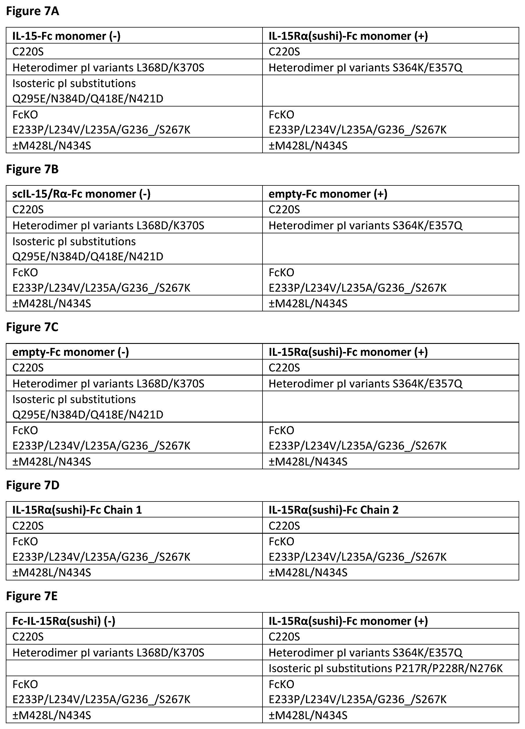
D00013

D00014

D00015

D00016

D00017

D00018
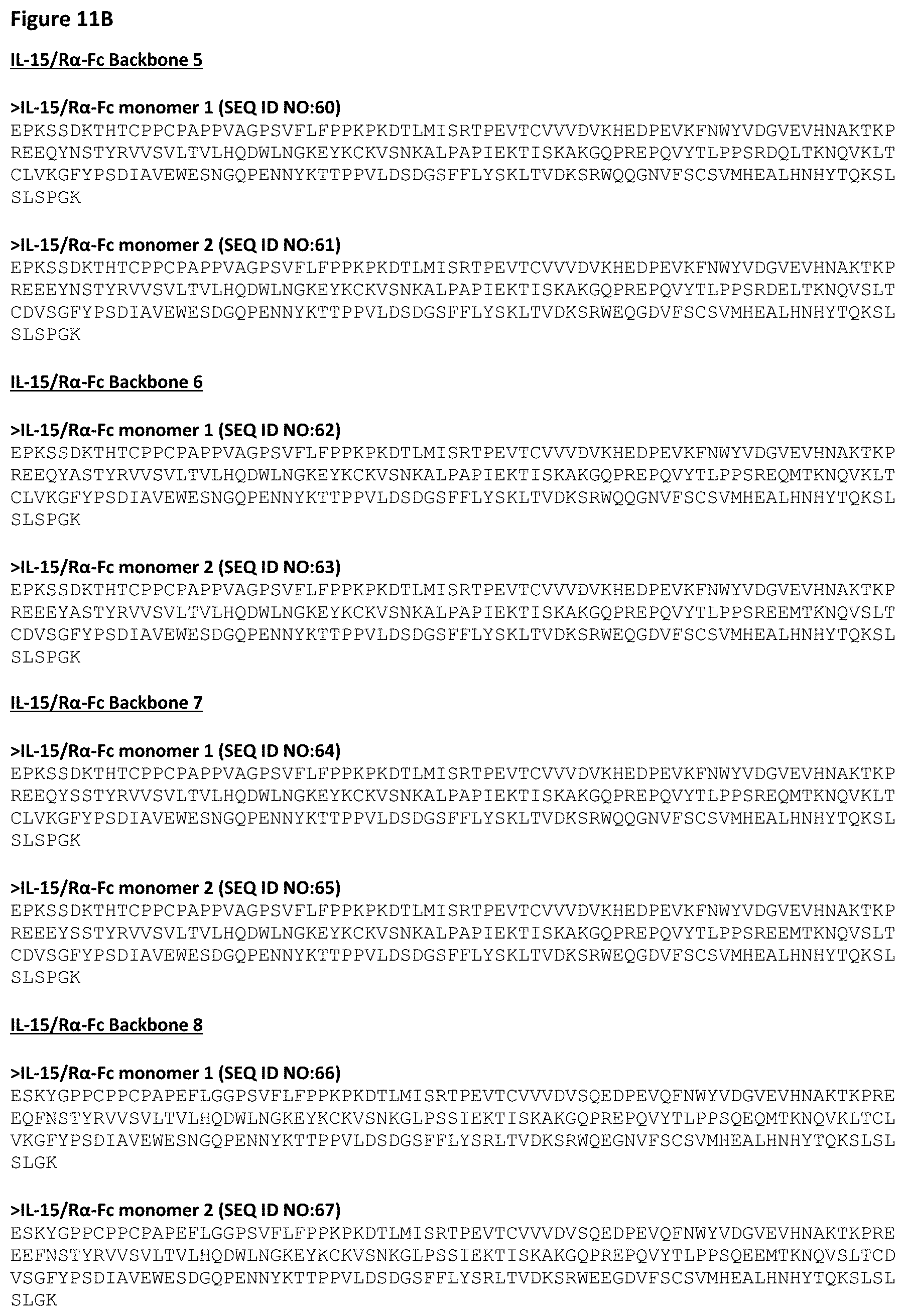
D00019

D00020

D00021

D00022

D00023

D00024
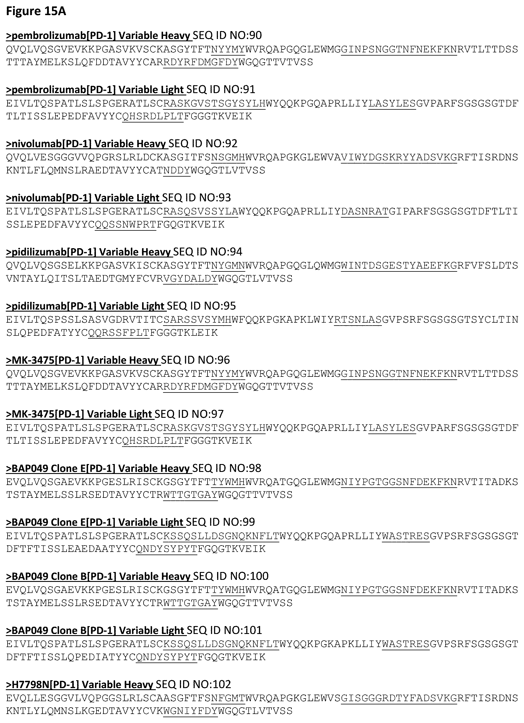
D00025

D00026

D00027

D00028

D00029

D00030

D00031
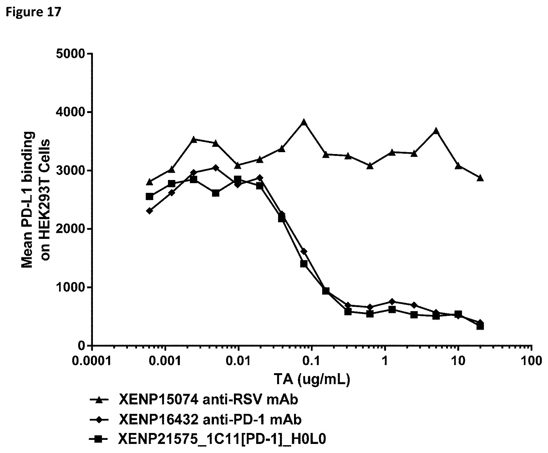
D00032

D00033

D00034

D00035

D00036
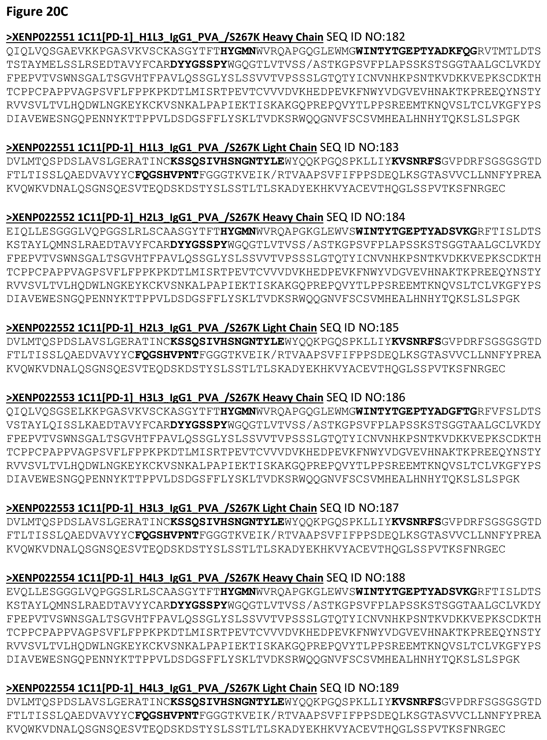
D00037
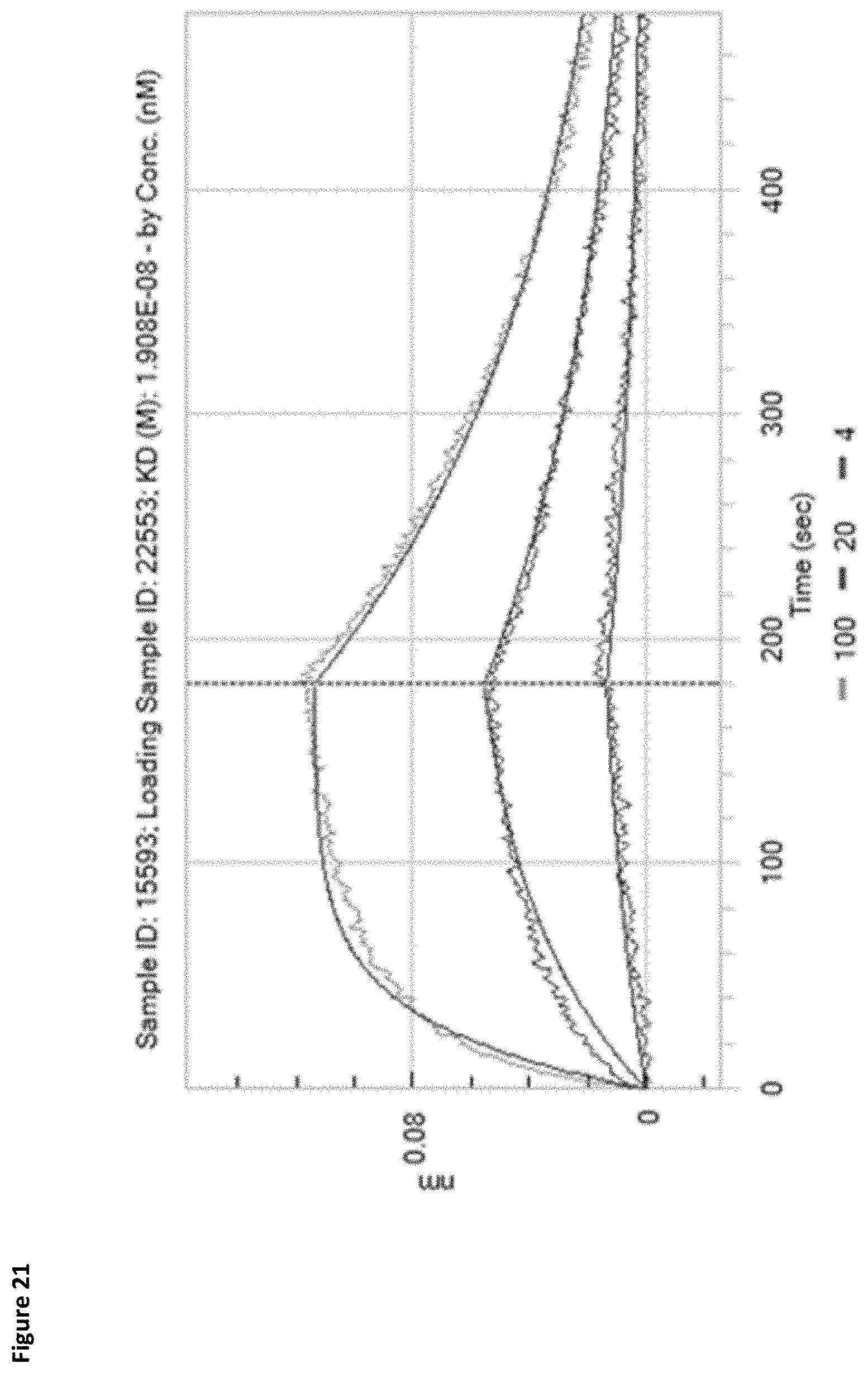
D00038

D00039
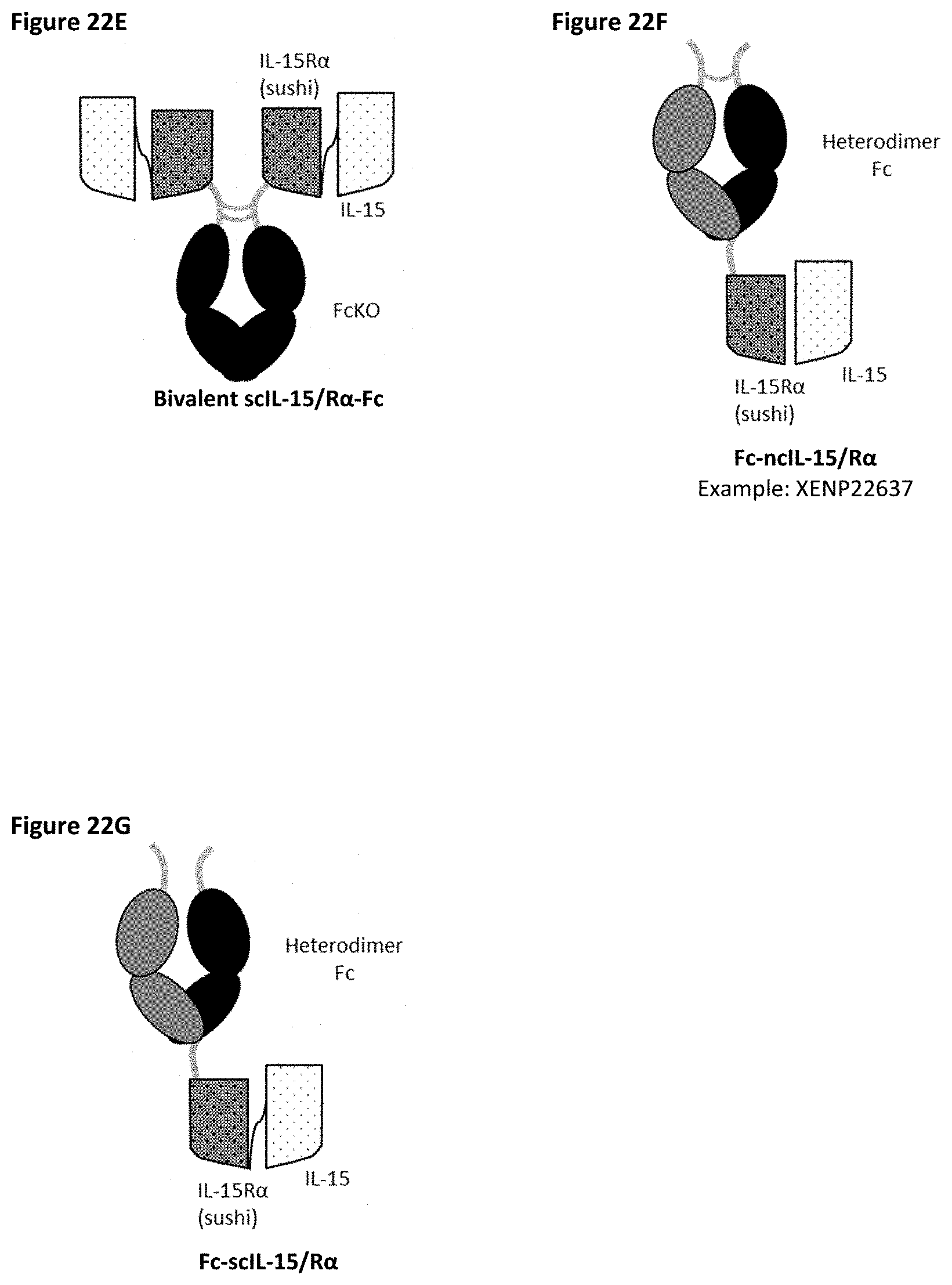
D00040
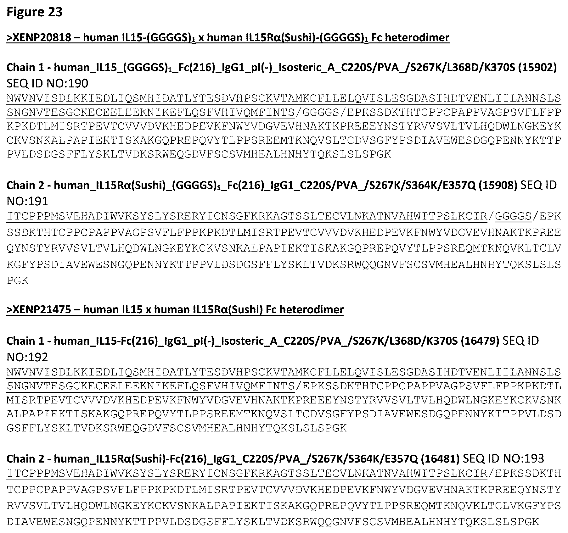
D00041
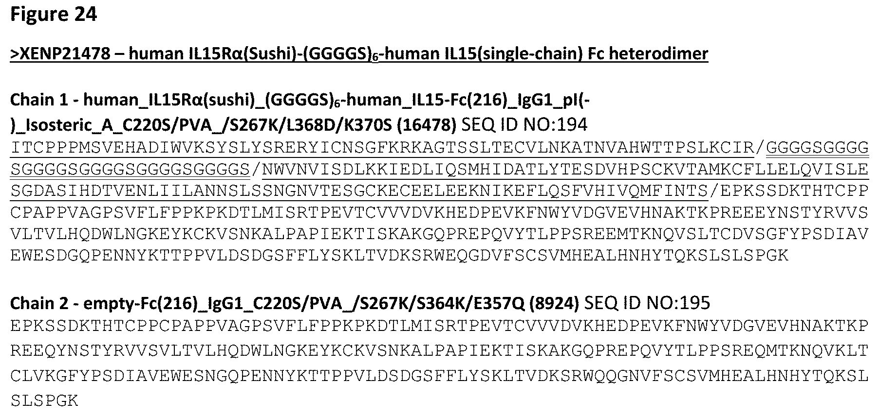
D00042
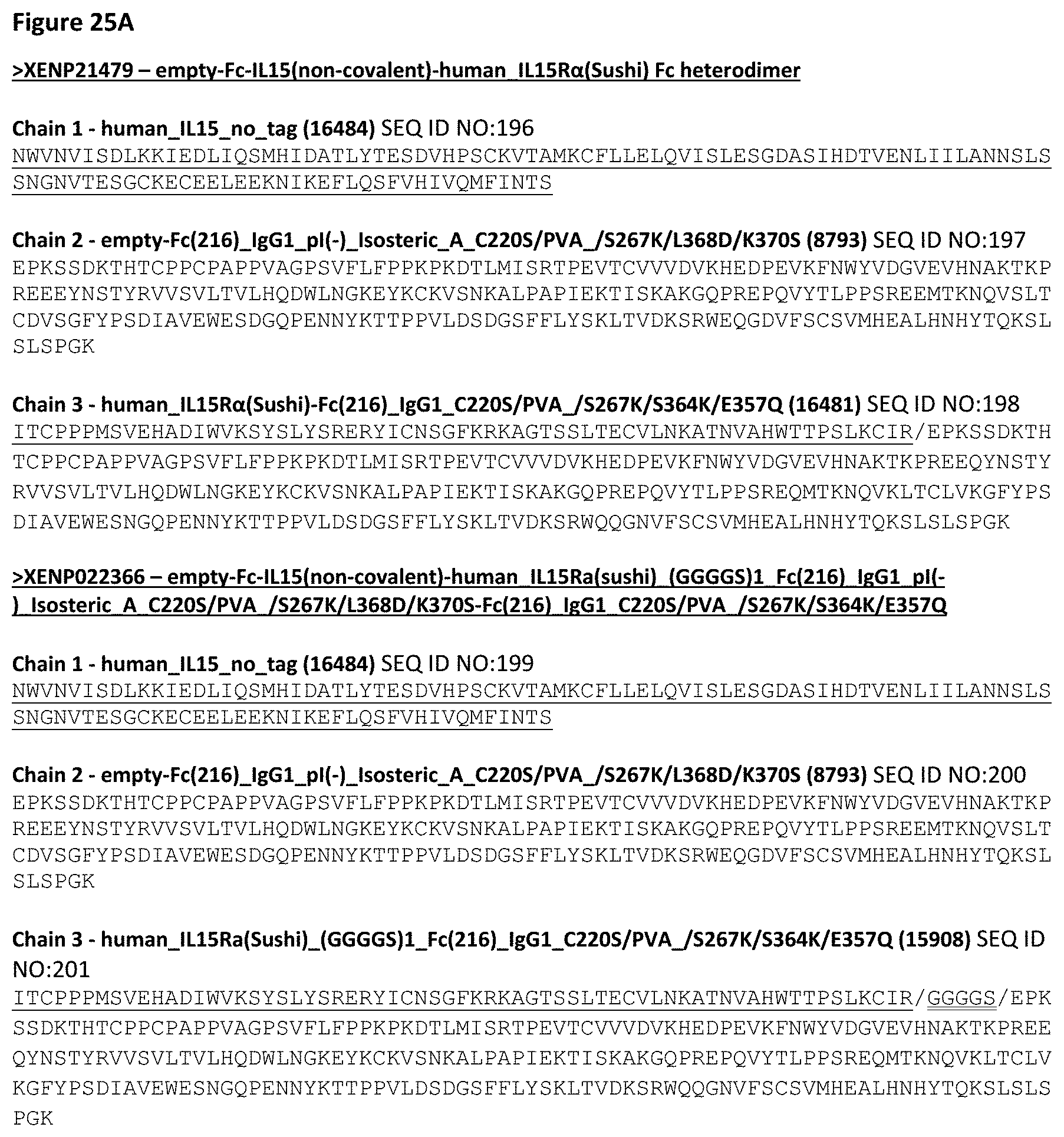
D00043

D00044

D00045
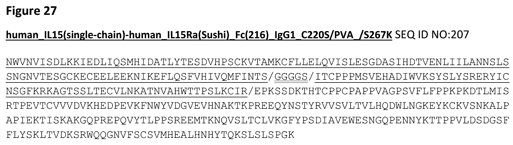
D00046
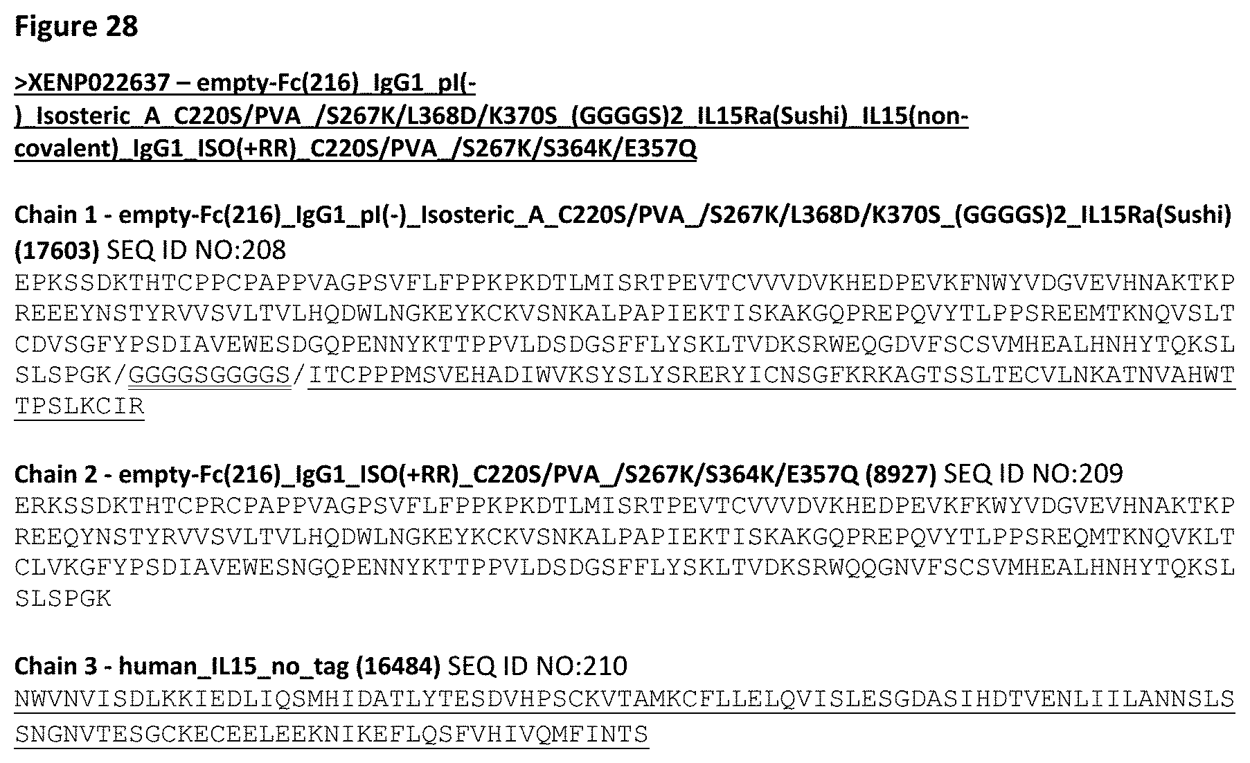
D00047
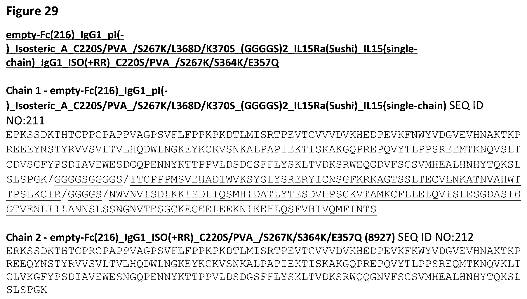
D00048

D00049

D00050

D00051
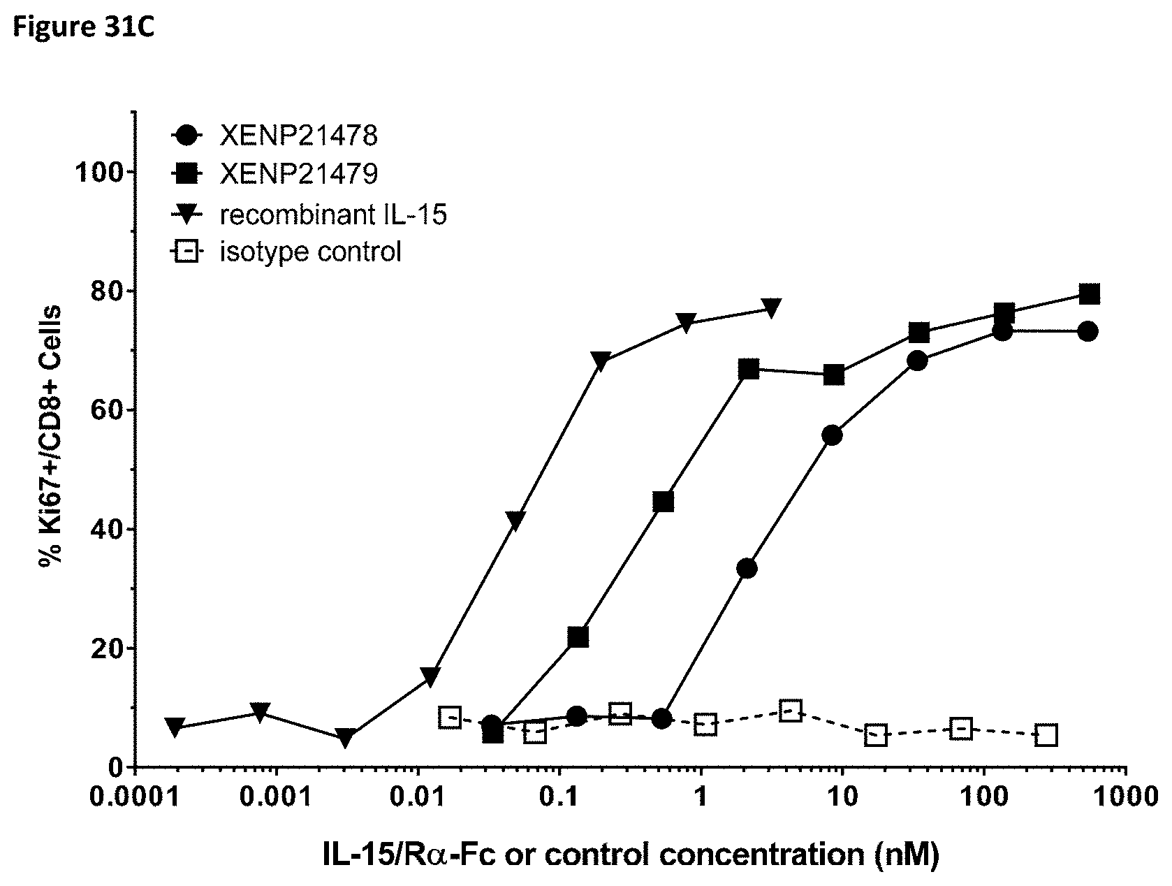
D00052
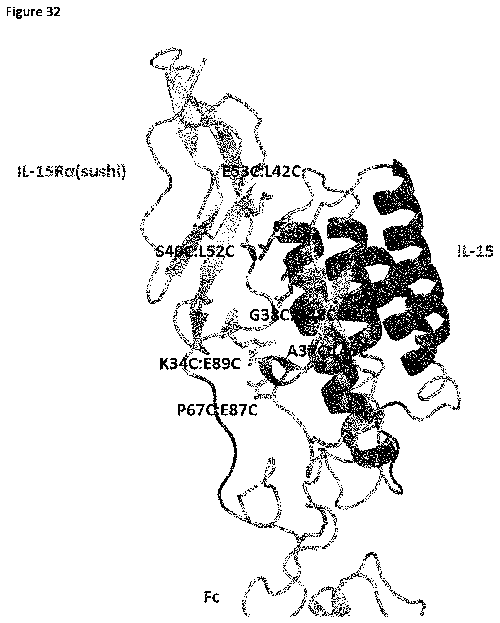
D00053

D00054

D00055

D00056

D00057
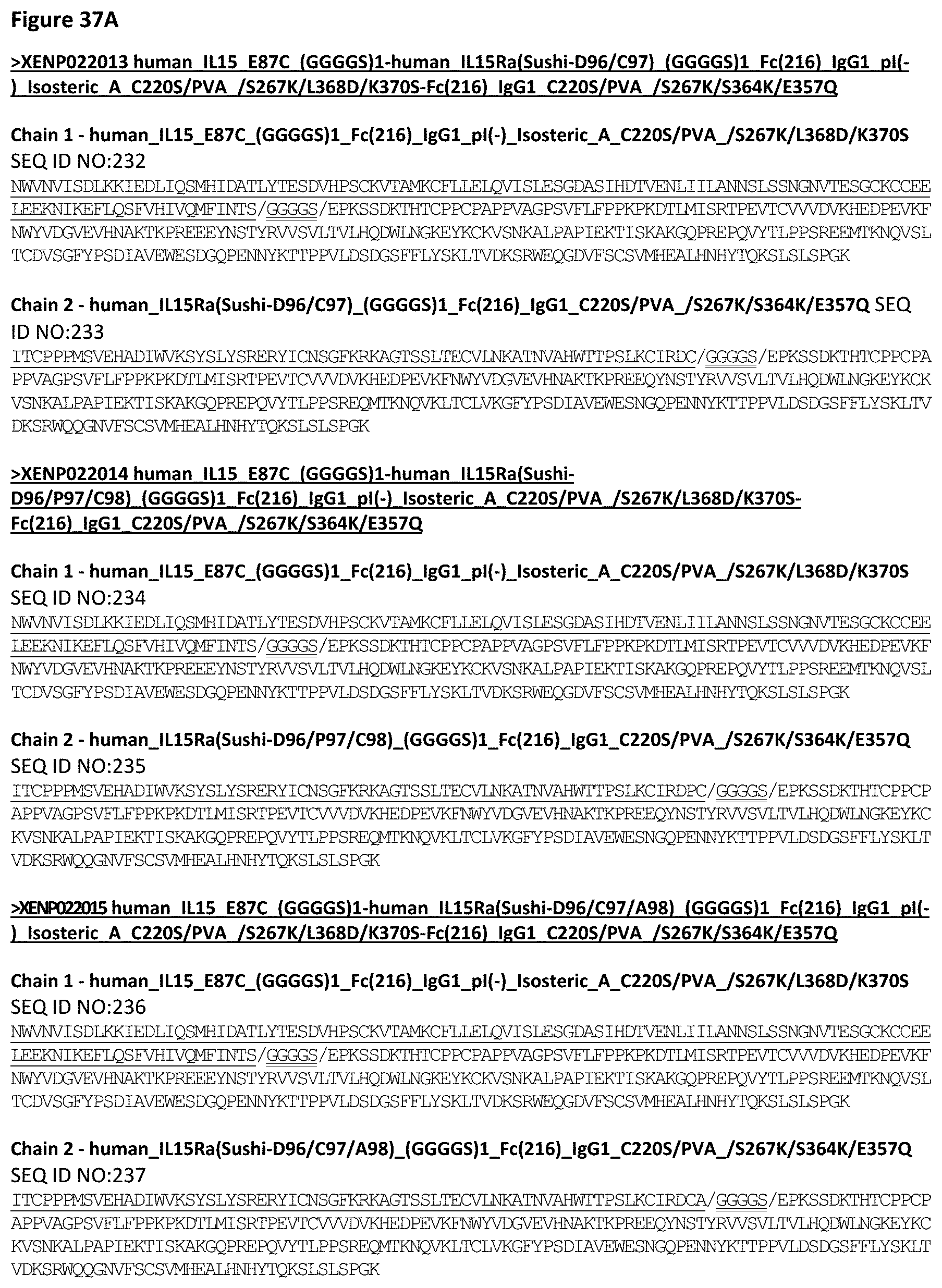
D00058

D00059
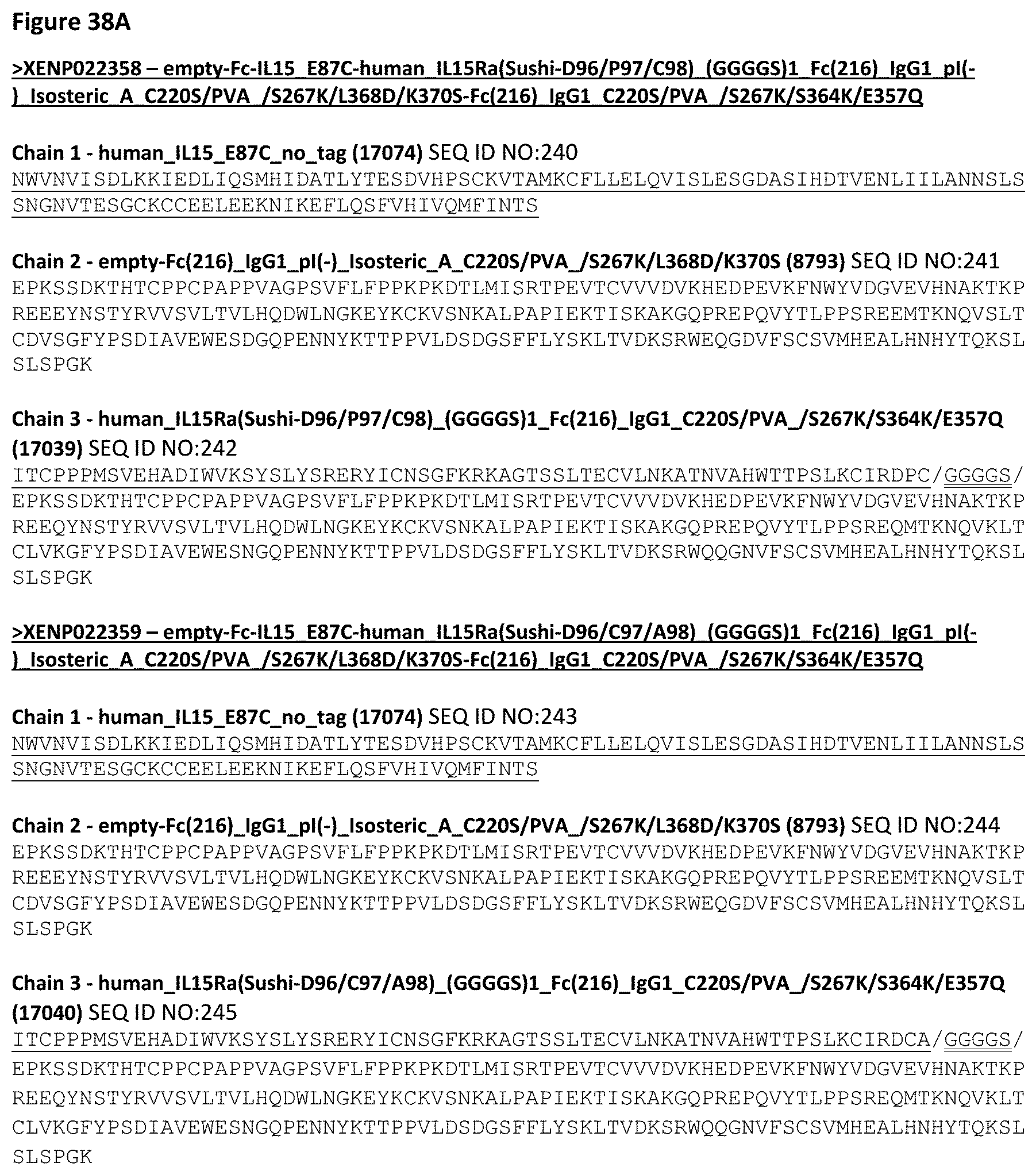
D00060

D00061

D00062
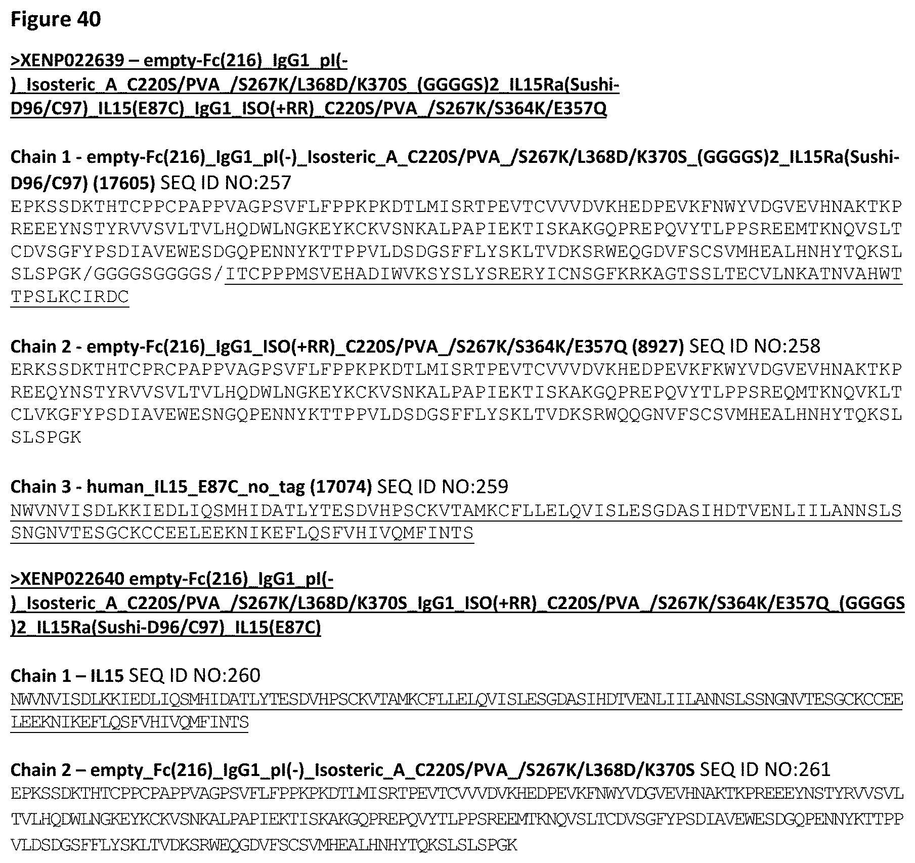
D00063

D00064

D00065

D00066

D00067
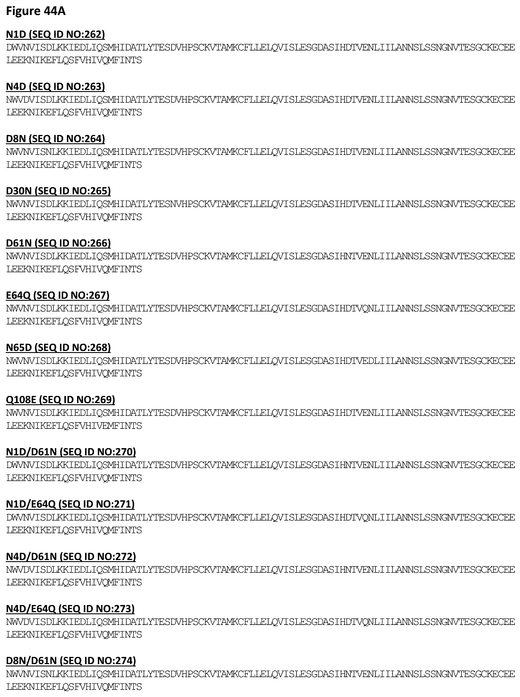
D00068
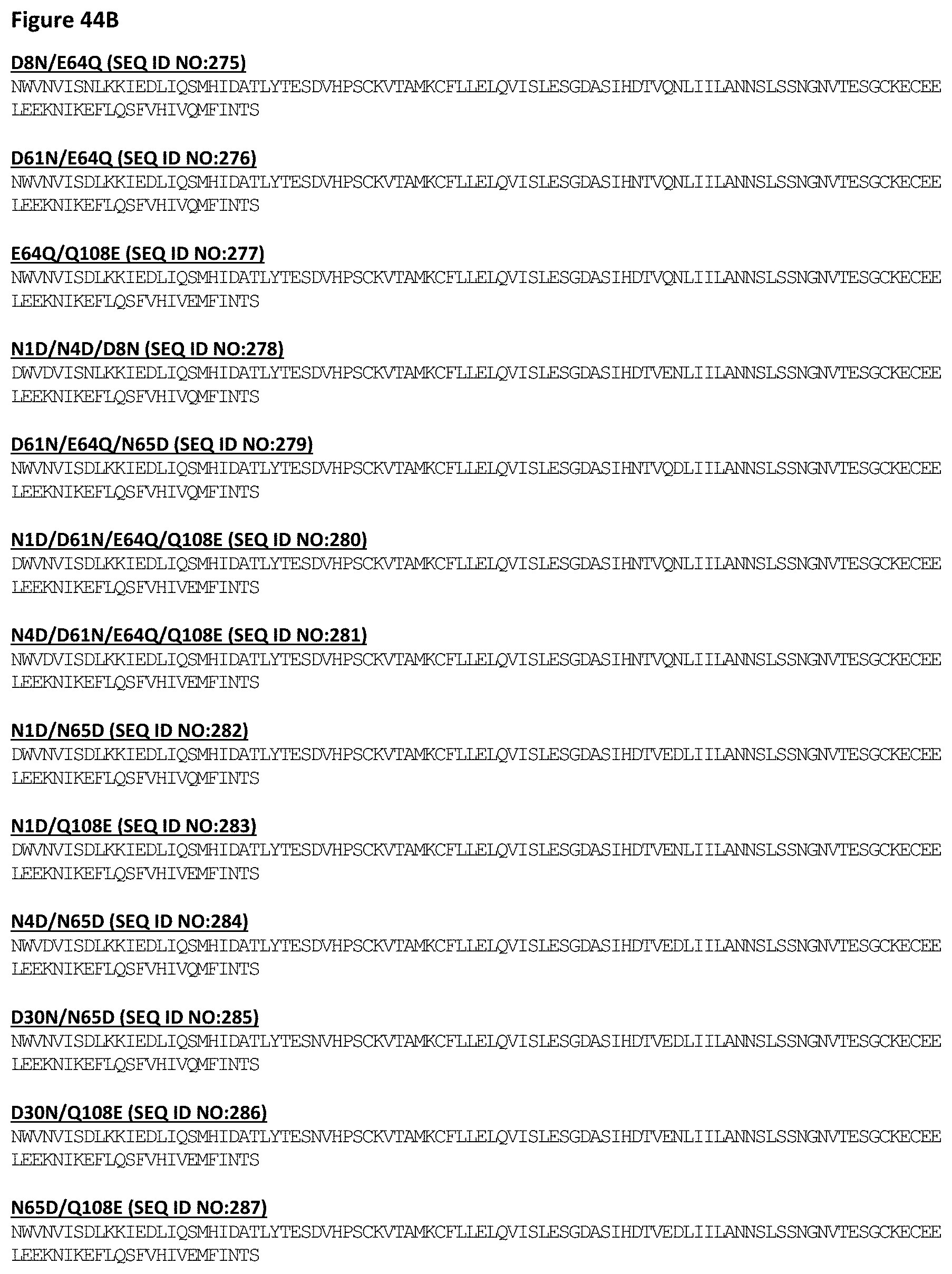
D00069
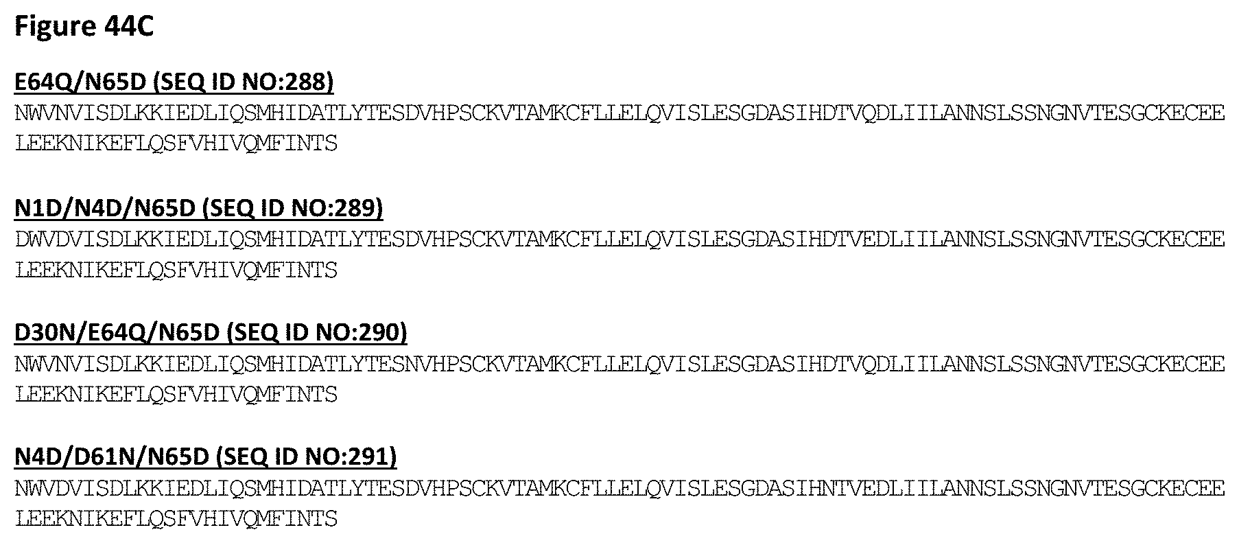
D00070

D00071
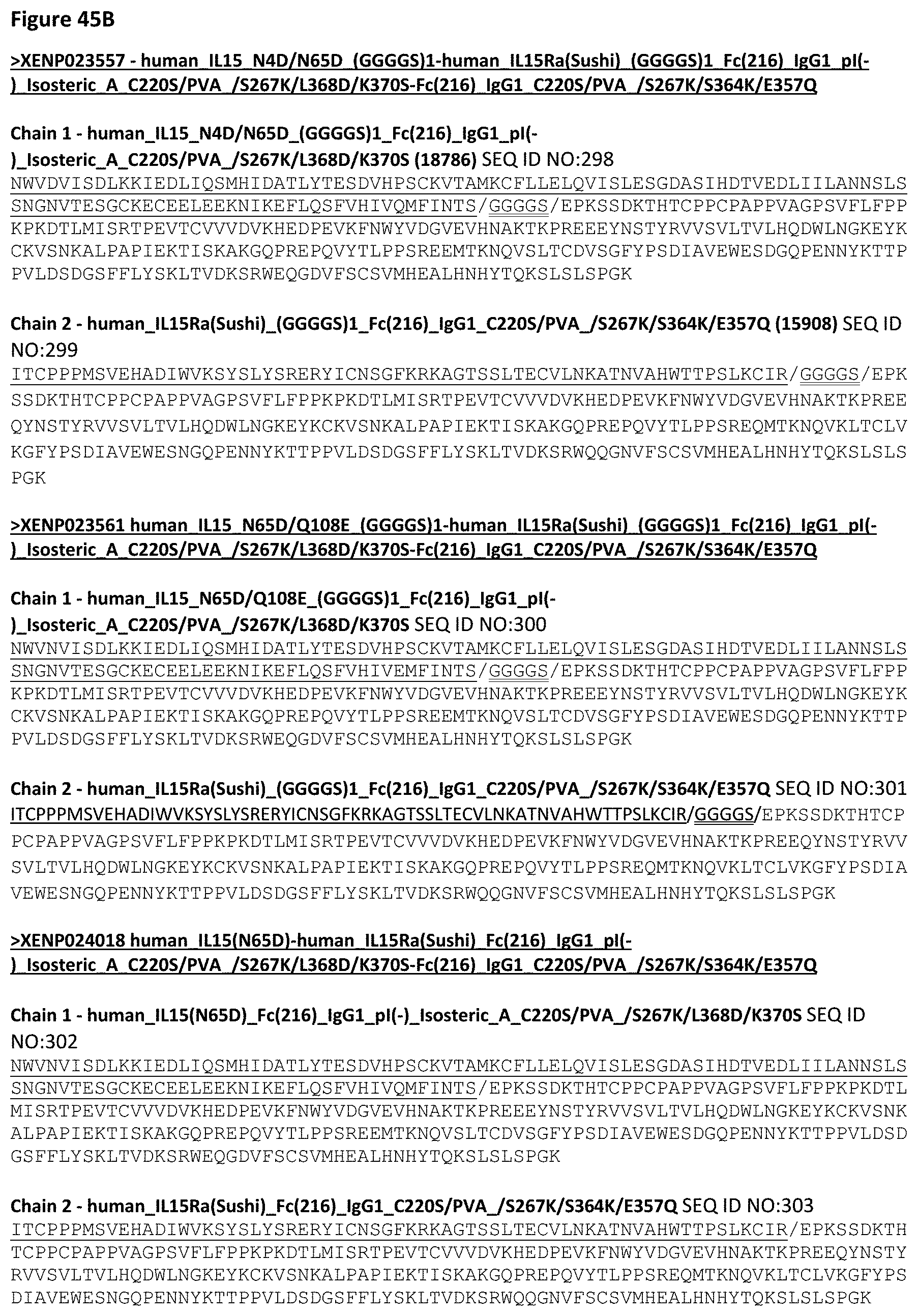
D00072

D00073

D00074

D00075

D00076

D00077

D00078

D00079
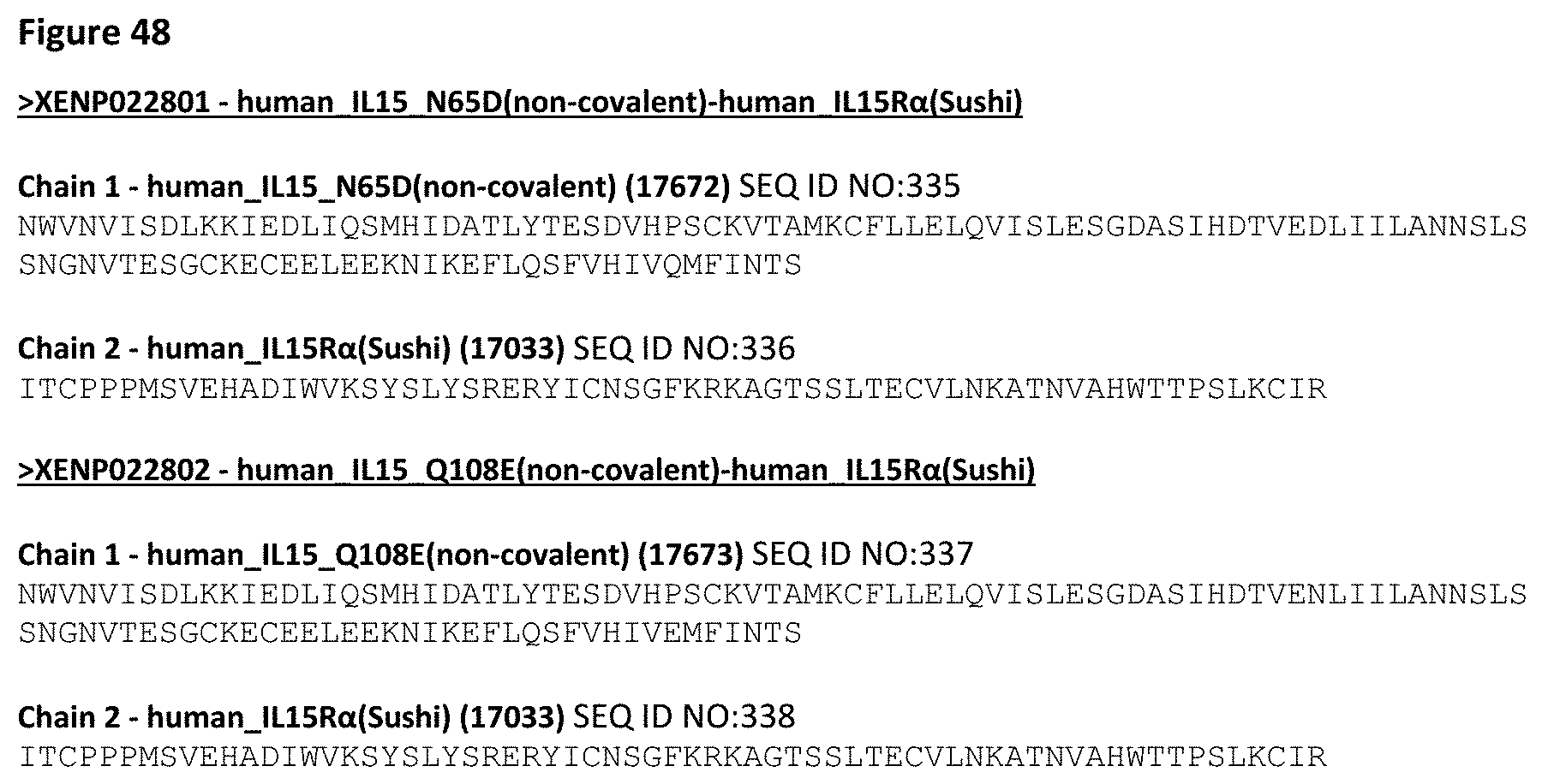
D00080
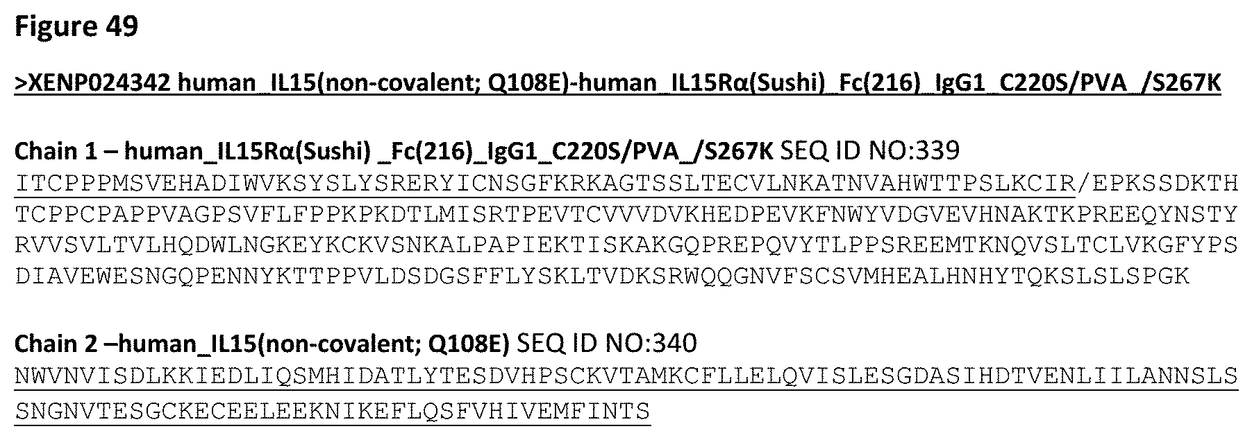
D00081

D00082

D00083

D00084

D00085

D00086

D00087

D00088

D00089

D00090

D00091
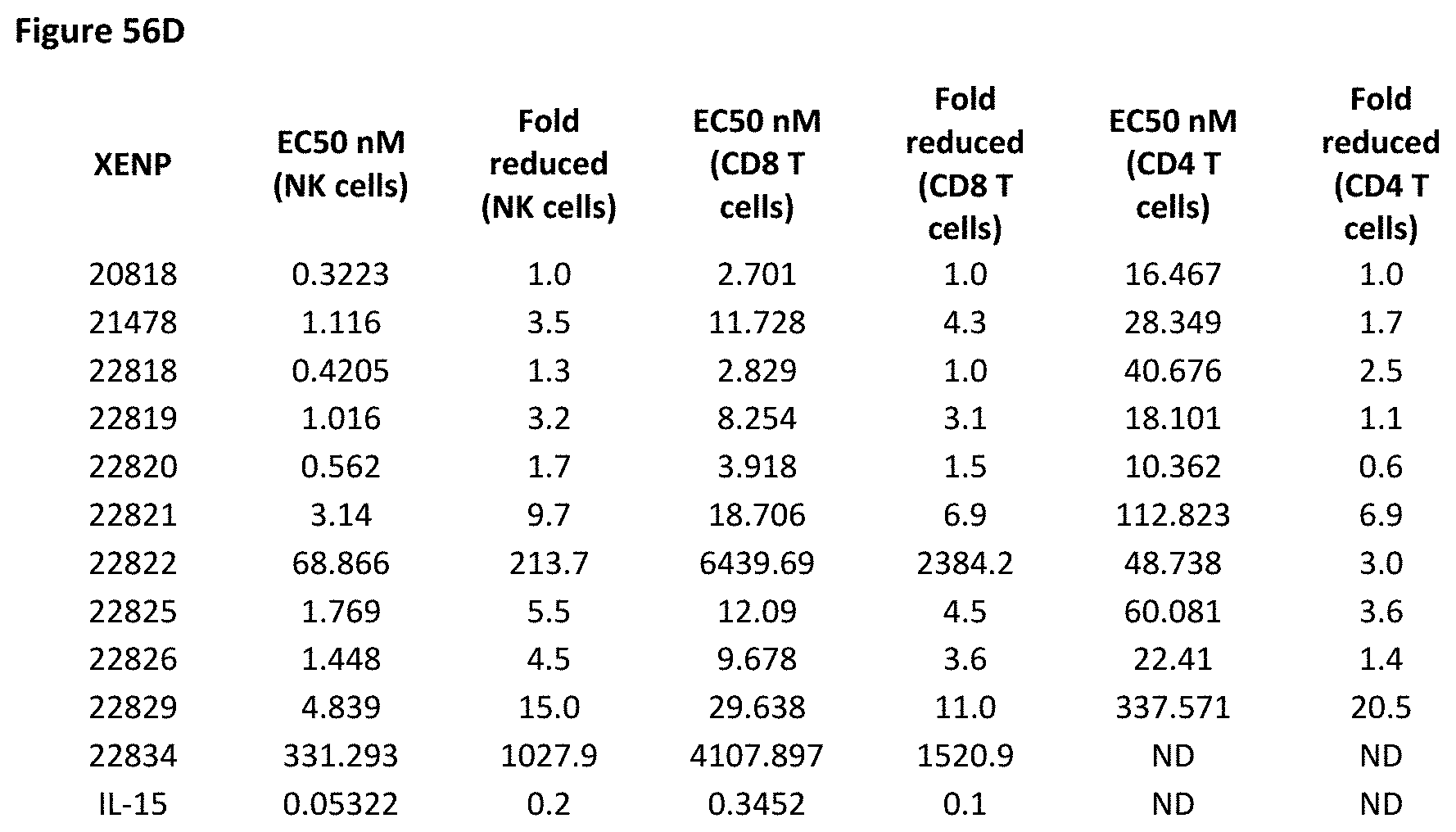
D00092

D00093
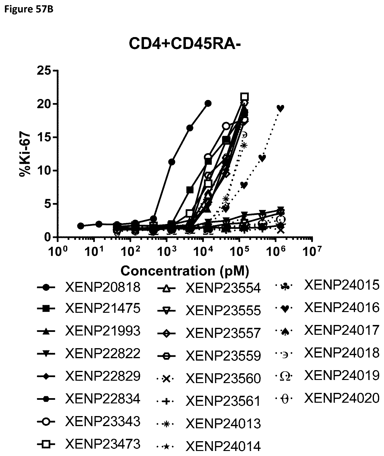
D00094
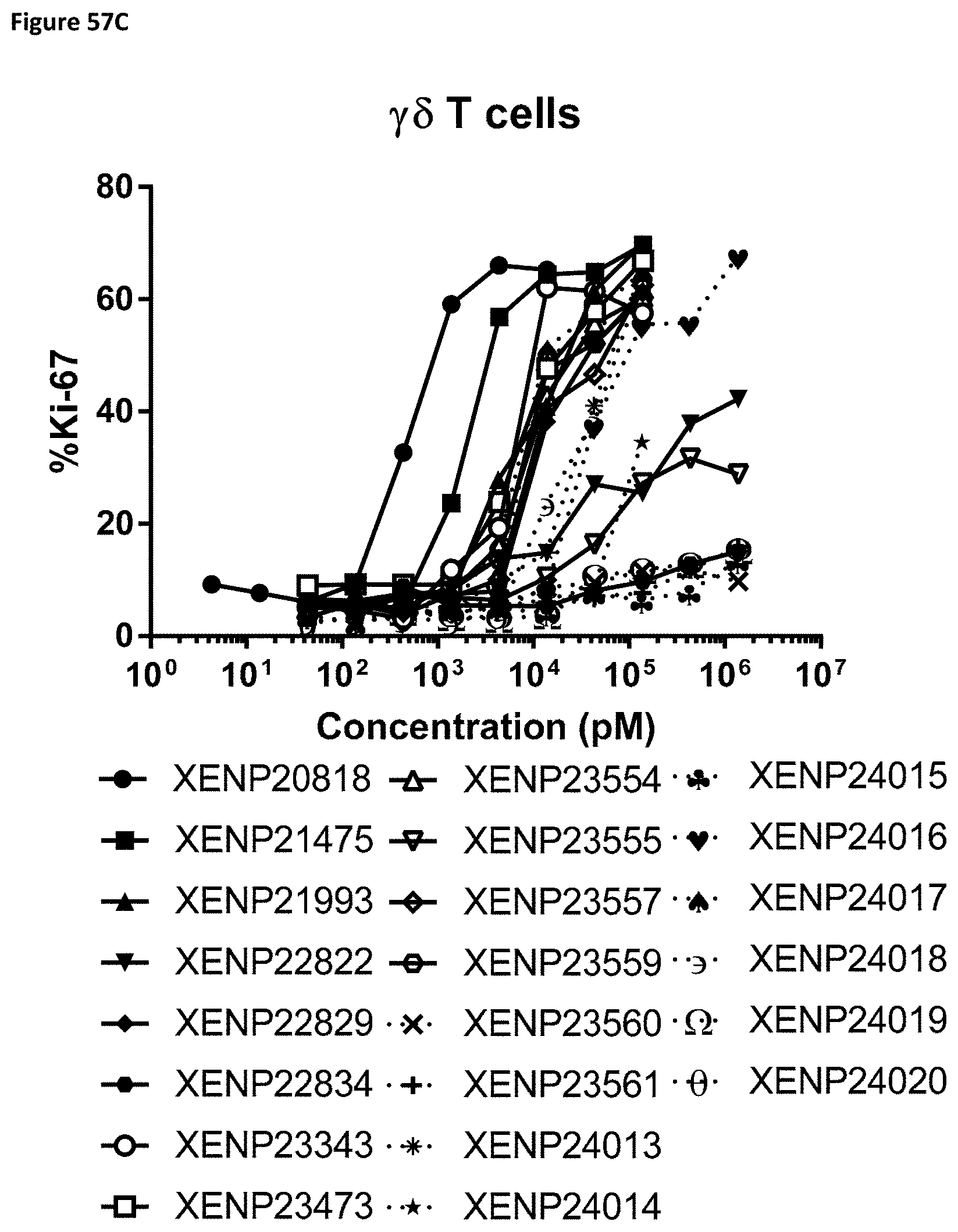
D00095

D00096

D00097

D00098

D00099

D00100

D00101

D00102

D00103

D00104
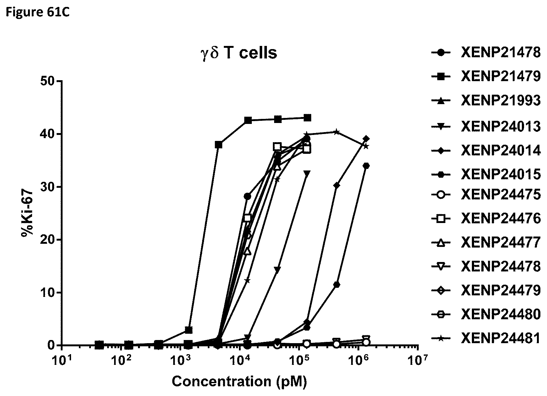
D00105

D00106

D00107

D00108
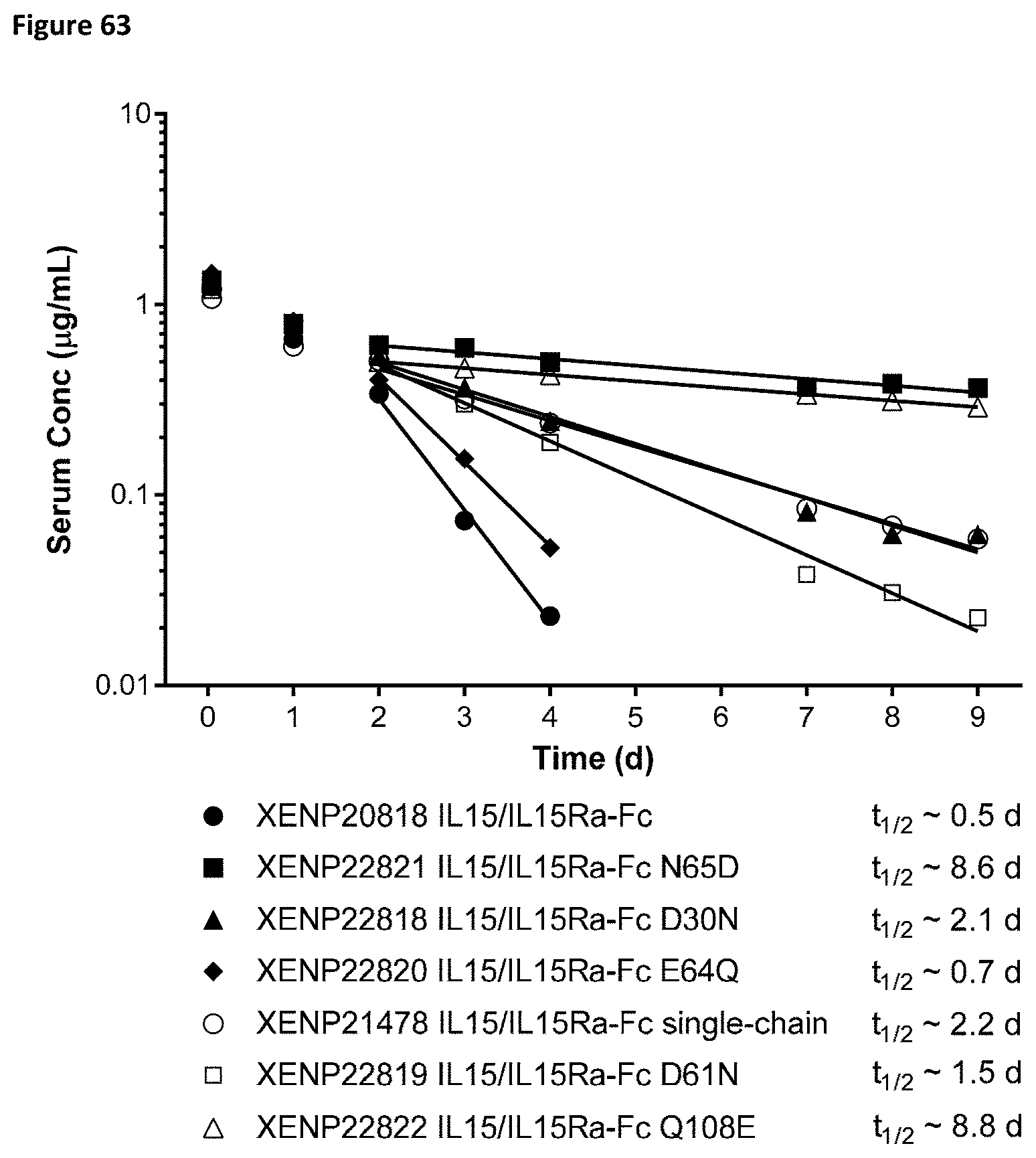
D00109

D00110

D00111
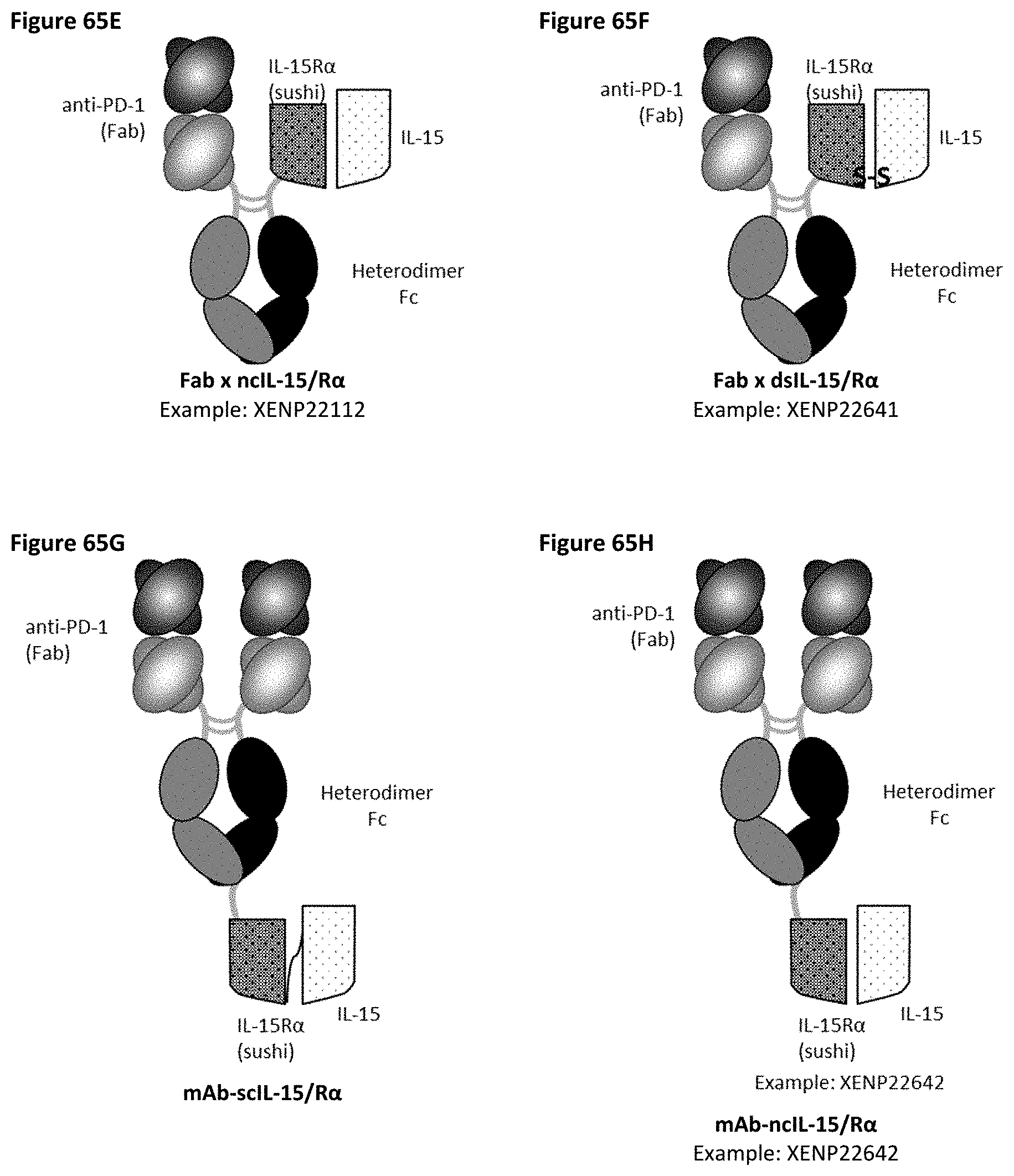
D00112

D00113
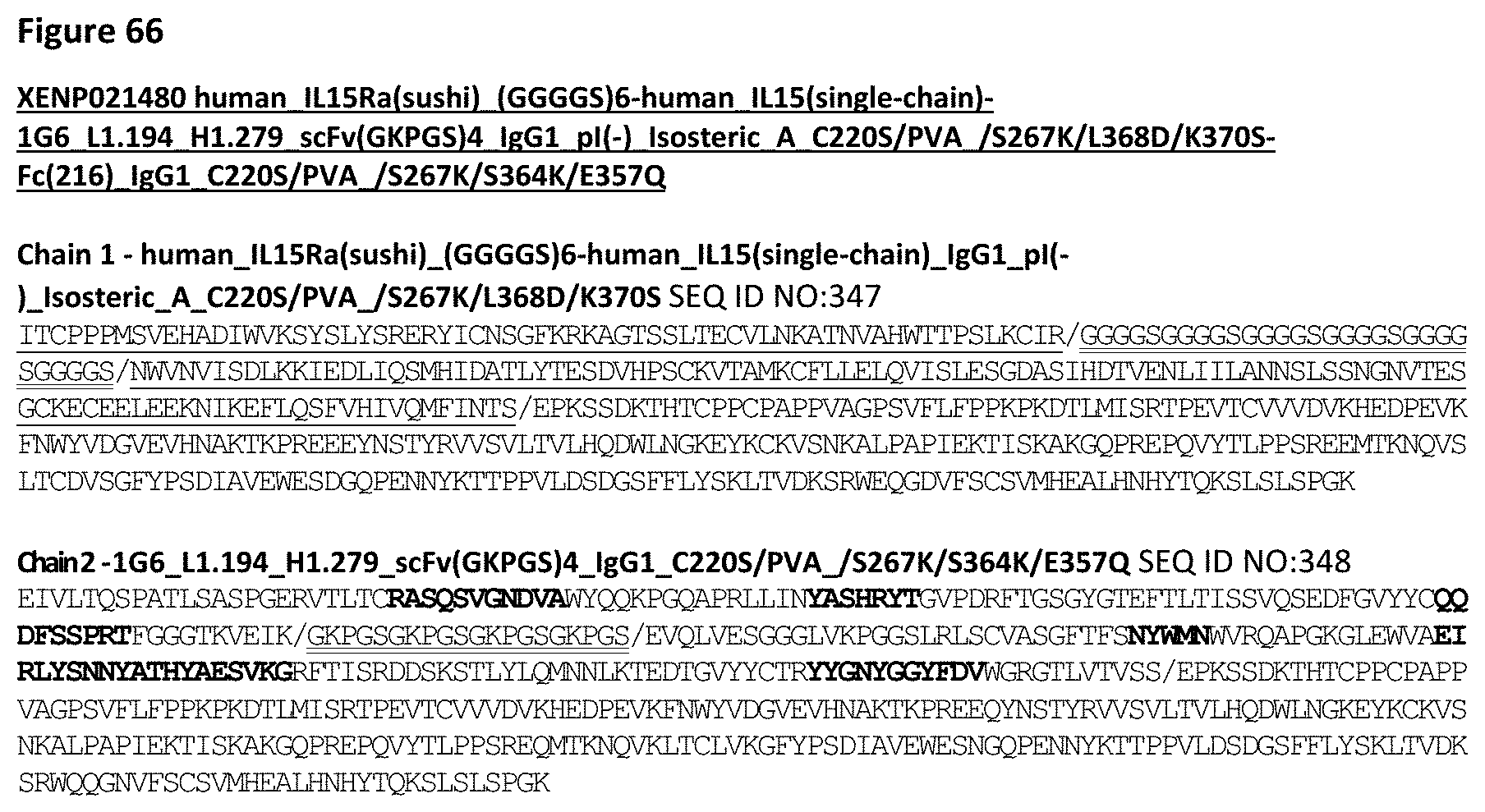
D00114

D00115

D00116

D00117

D00118

D00119
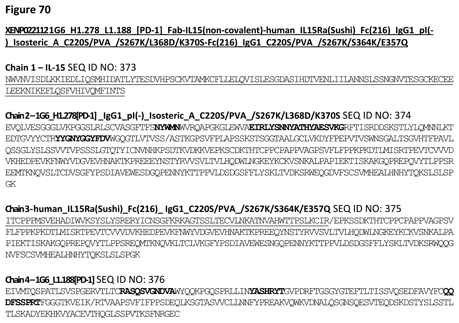
D00120

D00121

D00122
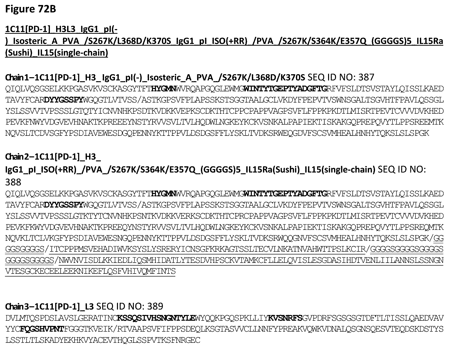
D00123

D00124

D00125

D00126
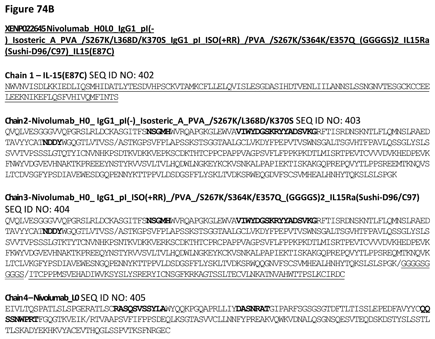
D00127
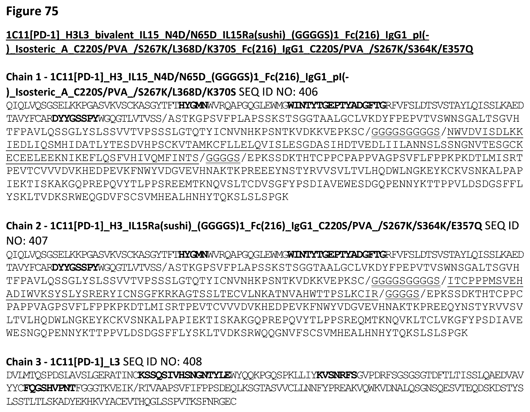
D00128

D00129

D00130

D00131

D00132

D00133

D00134

D00135

D00136

D00137

D00138

D00139

D00140
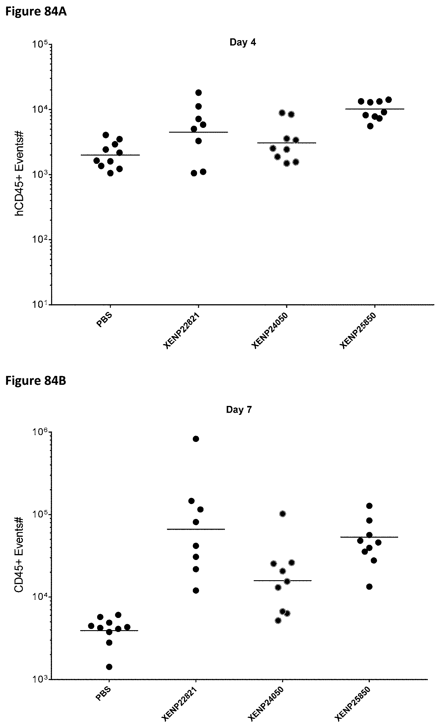
D00141

D00142

D00143
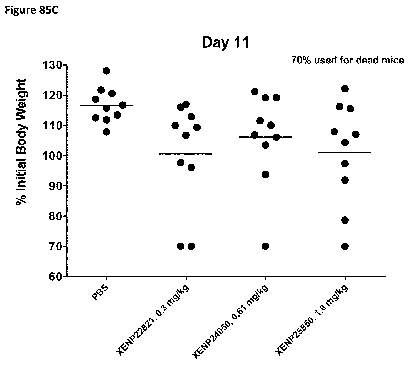
D00144

D00145
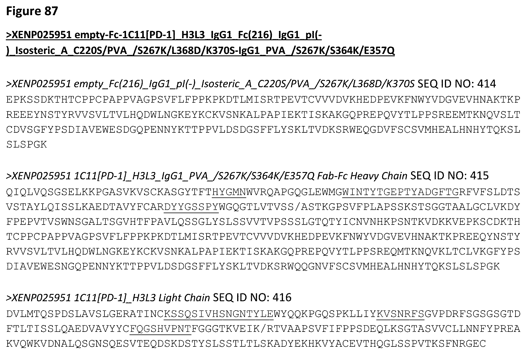
D00146

D00147
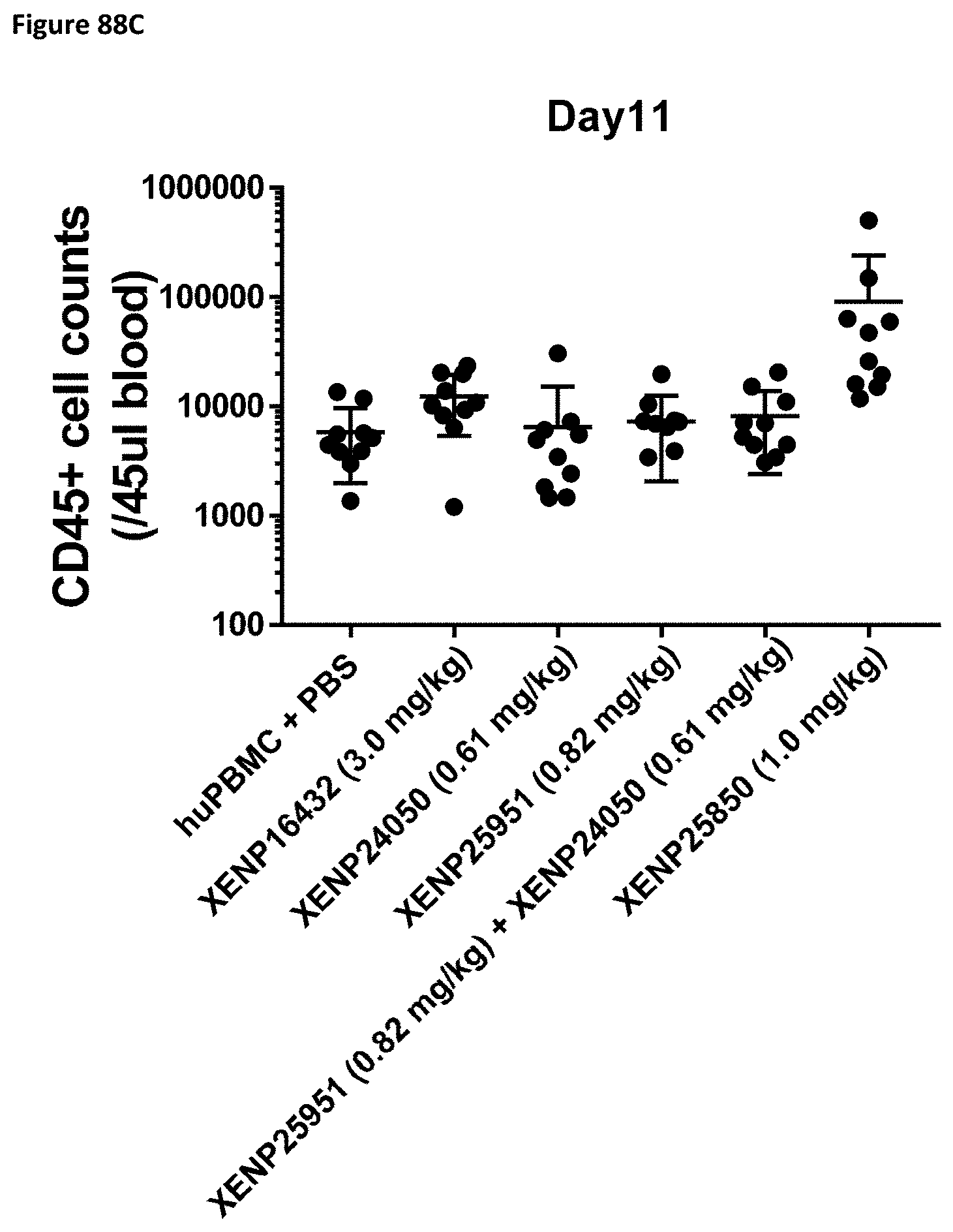
D00148

D00149

D00150

D00151
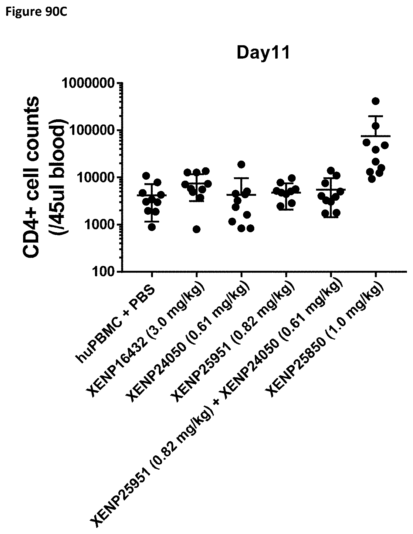
D00152

D00153
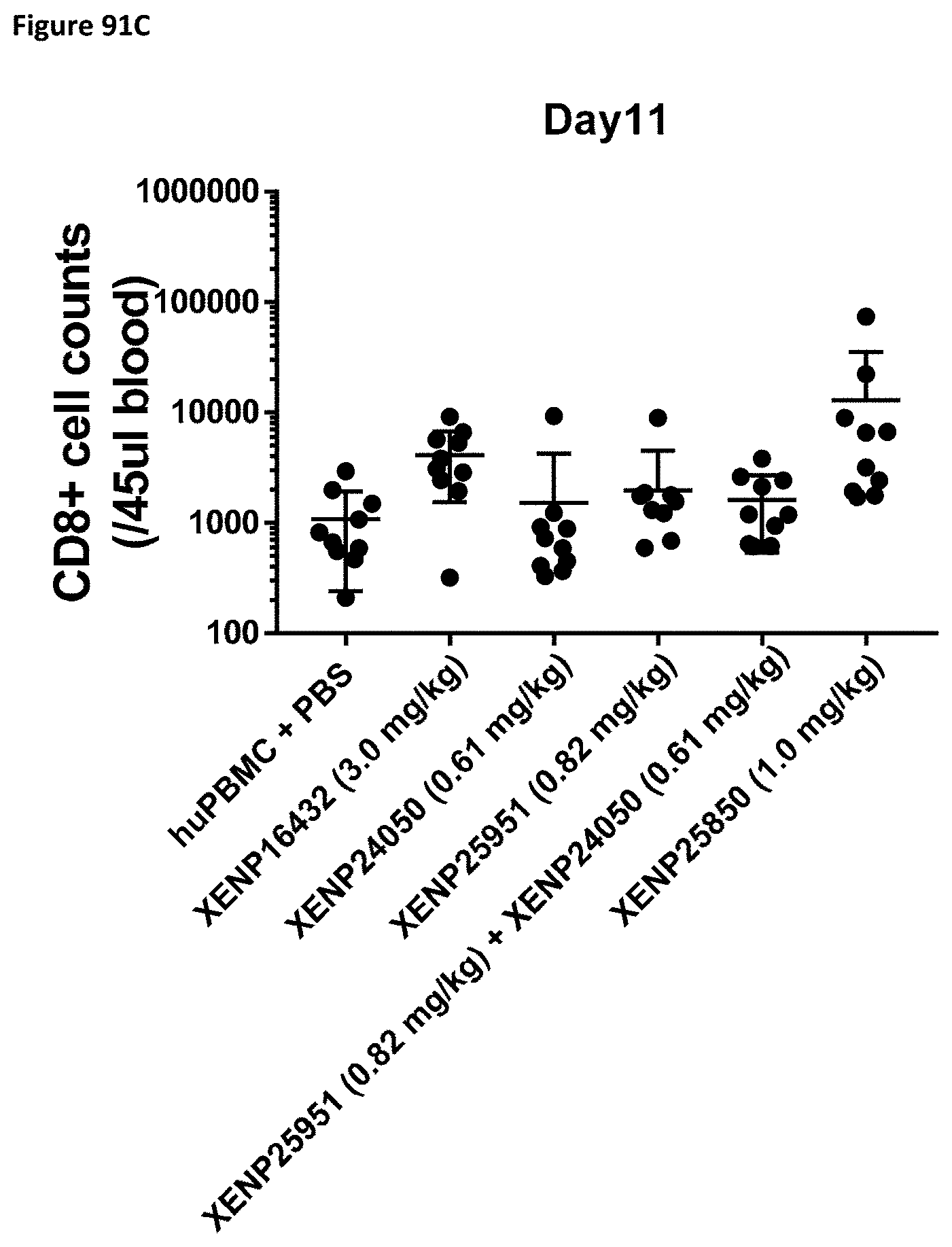
D00154

D00155

D00156

D00157
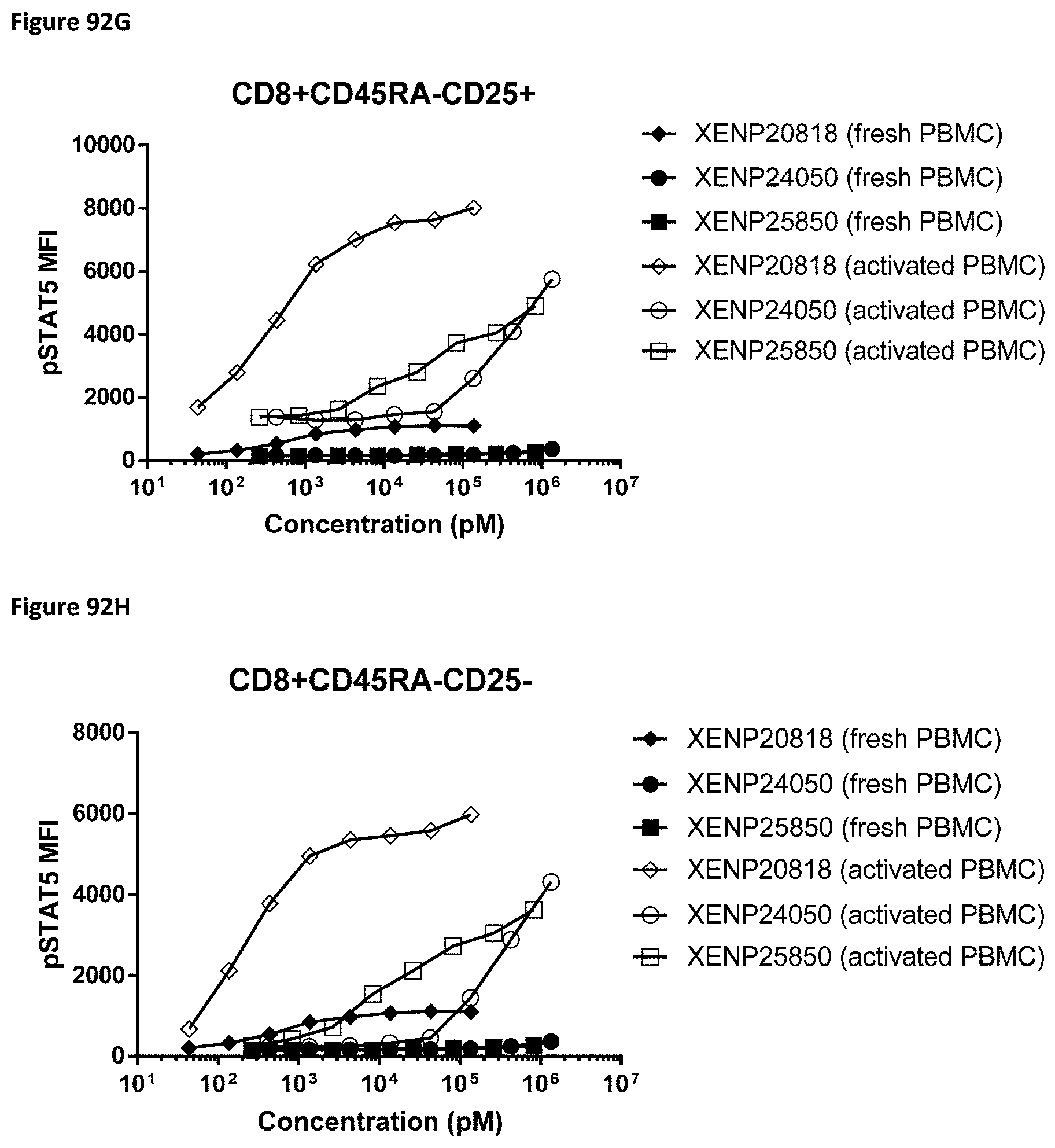
D00158
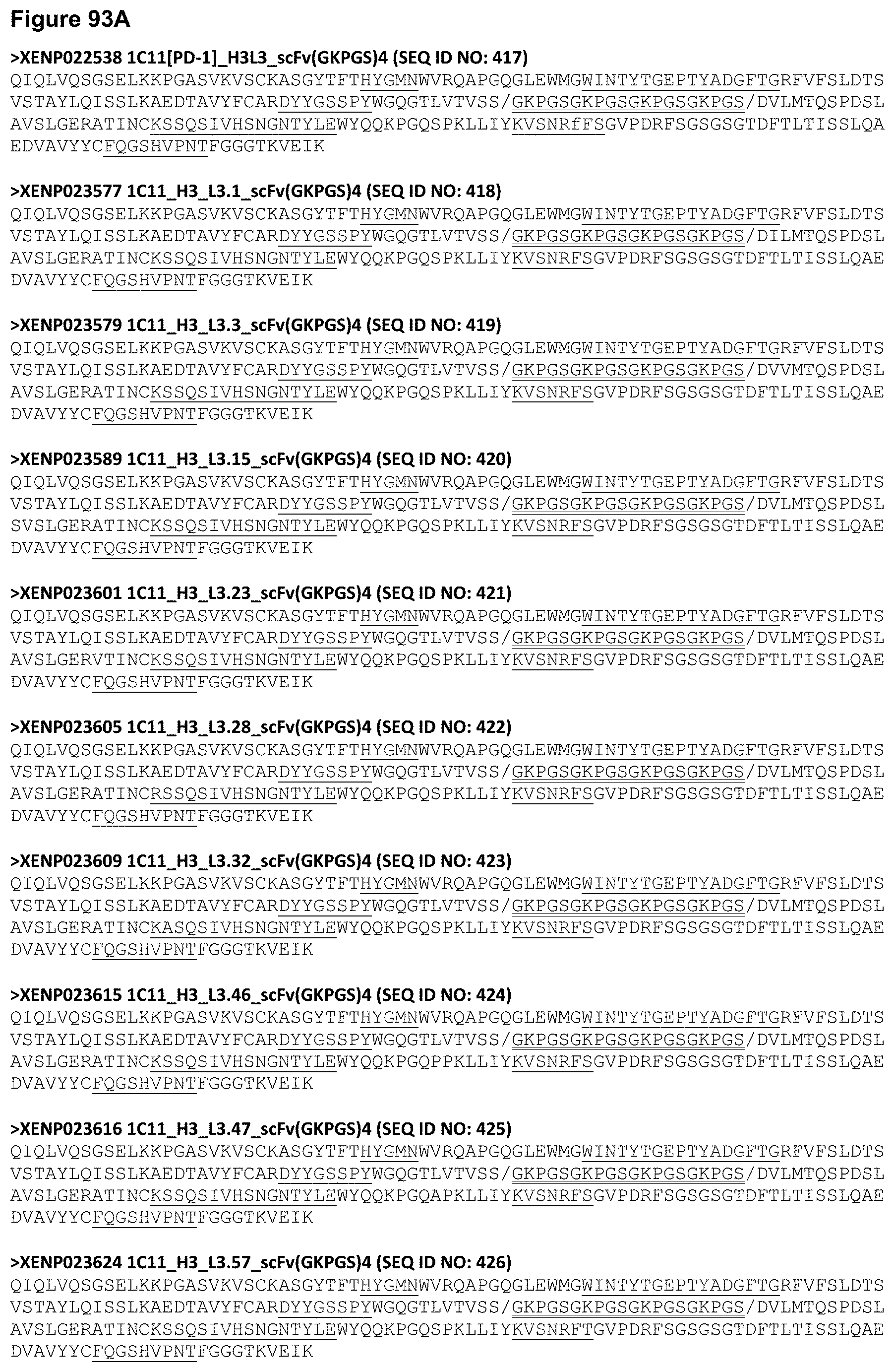
D00159

D00160
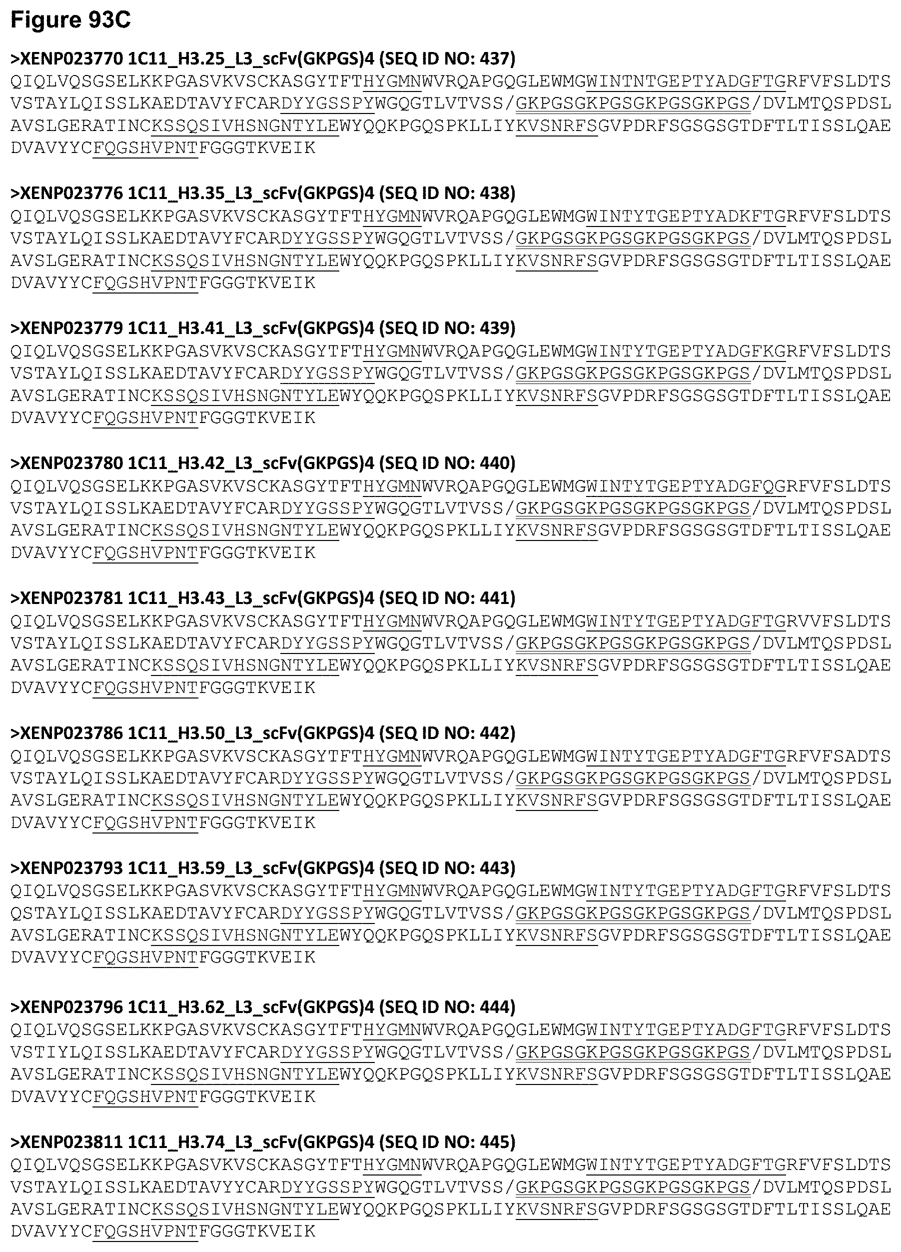
D00161

D00162

D00163

D00164

D00165
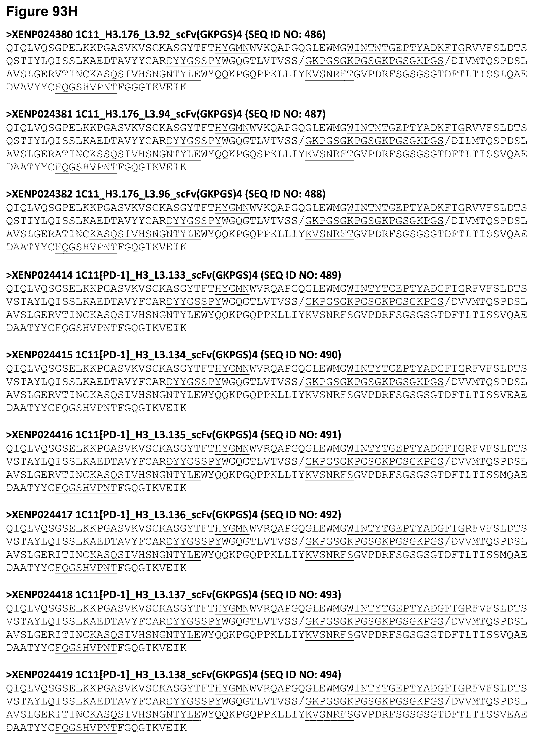
D00166
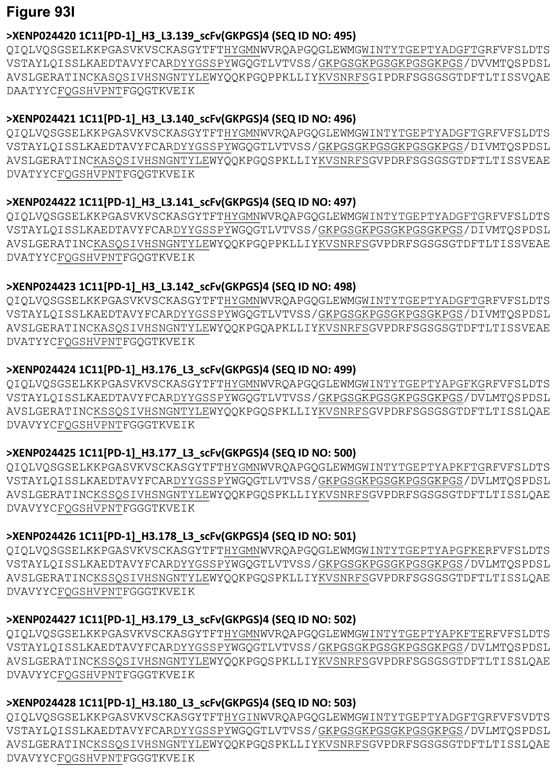
D00167

D00168
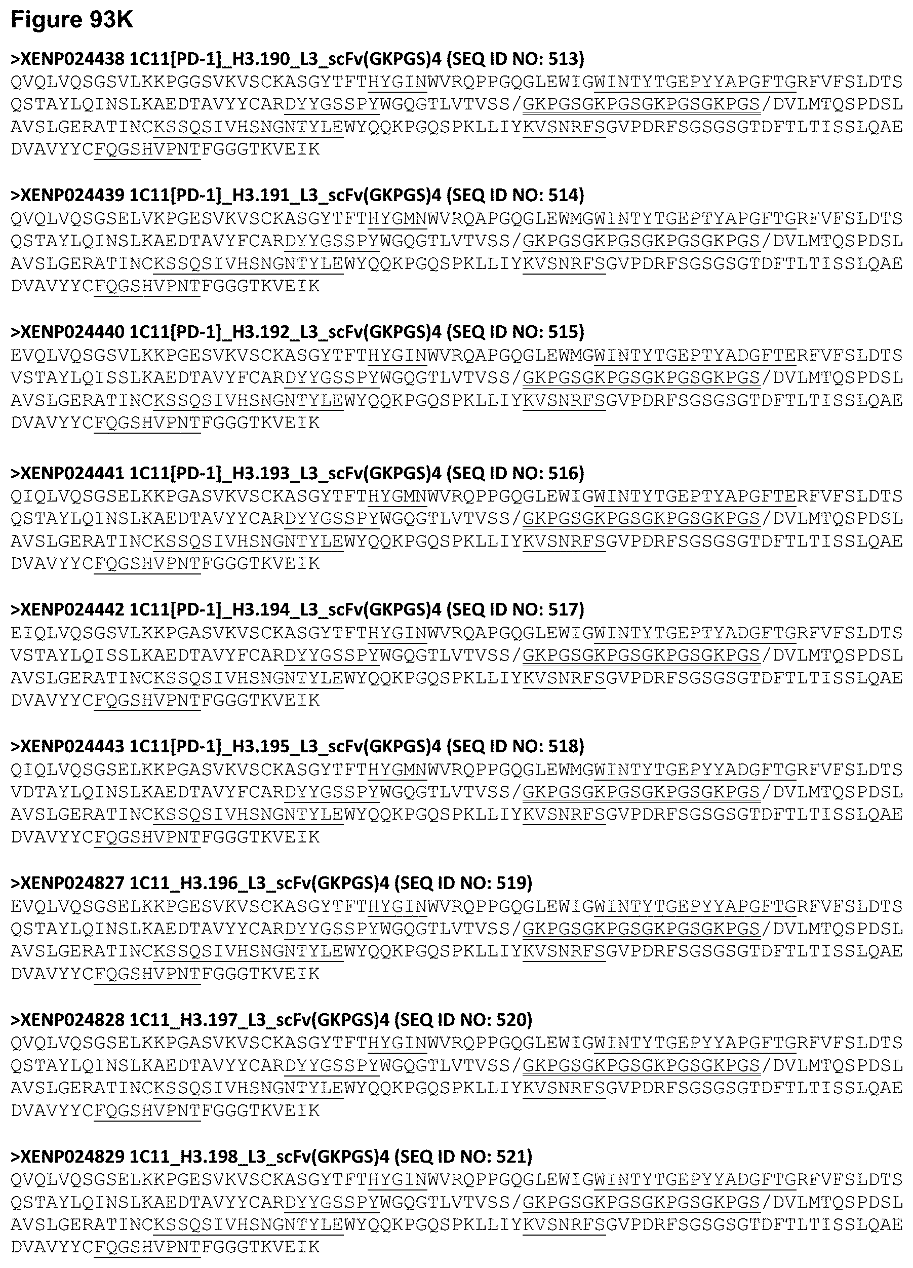
D00169
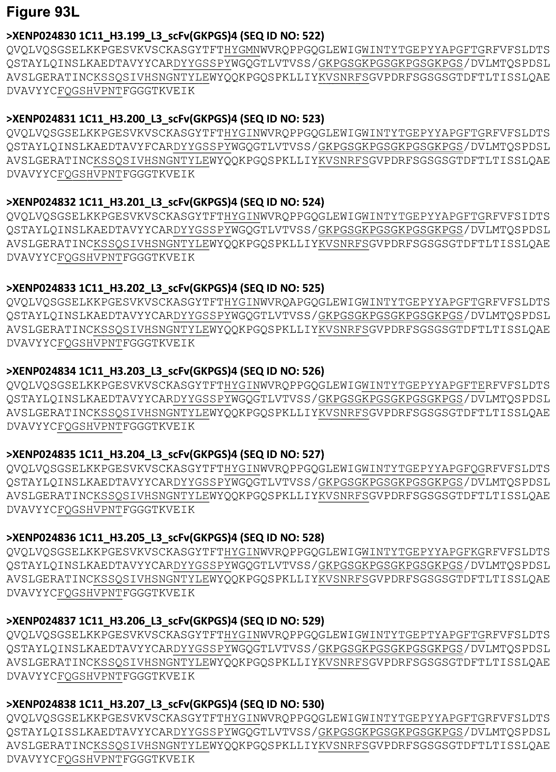
D00170

D00171

D00172
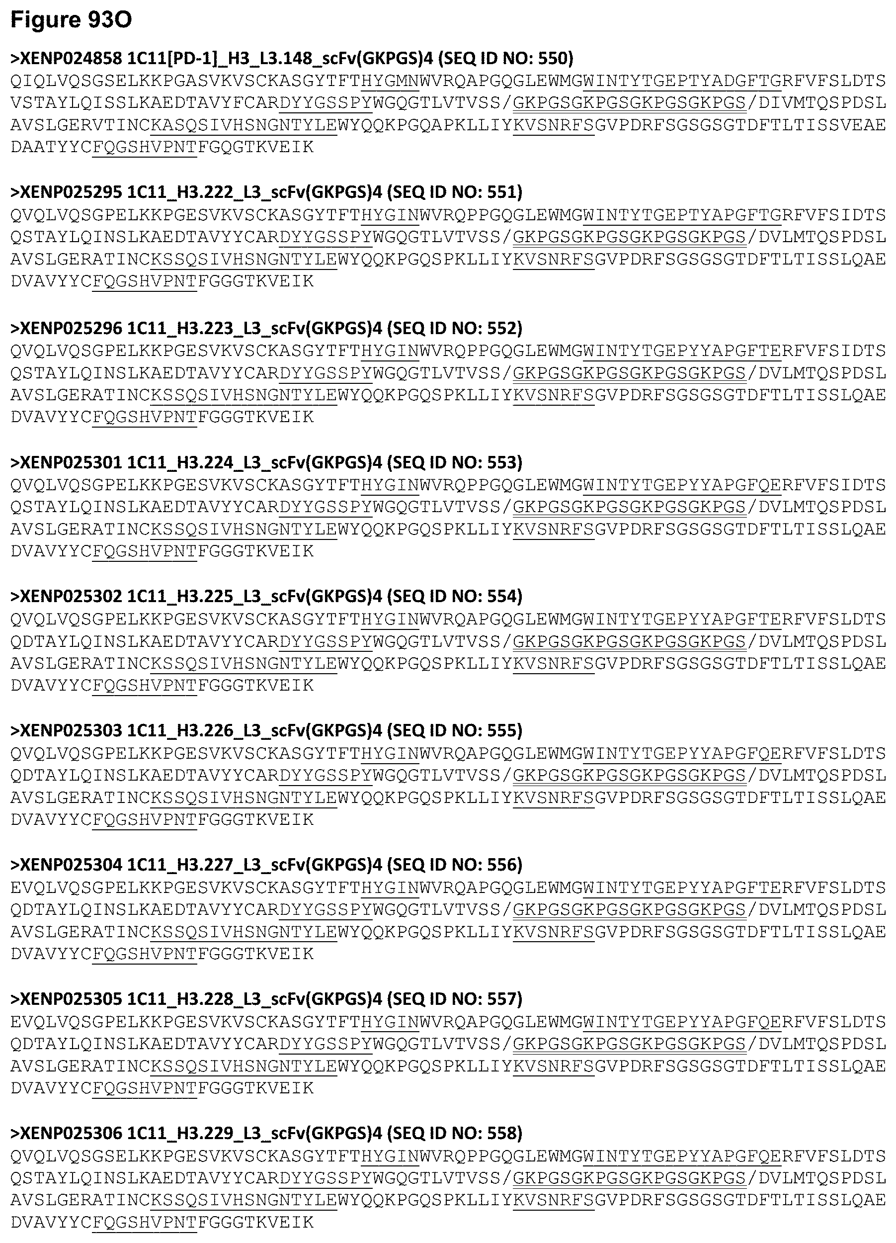
D00173
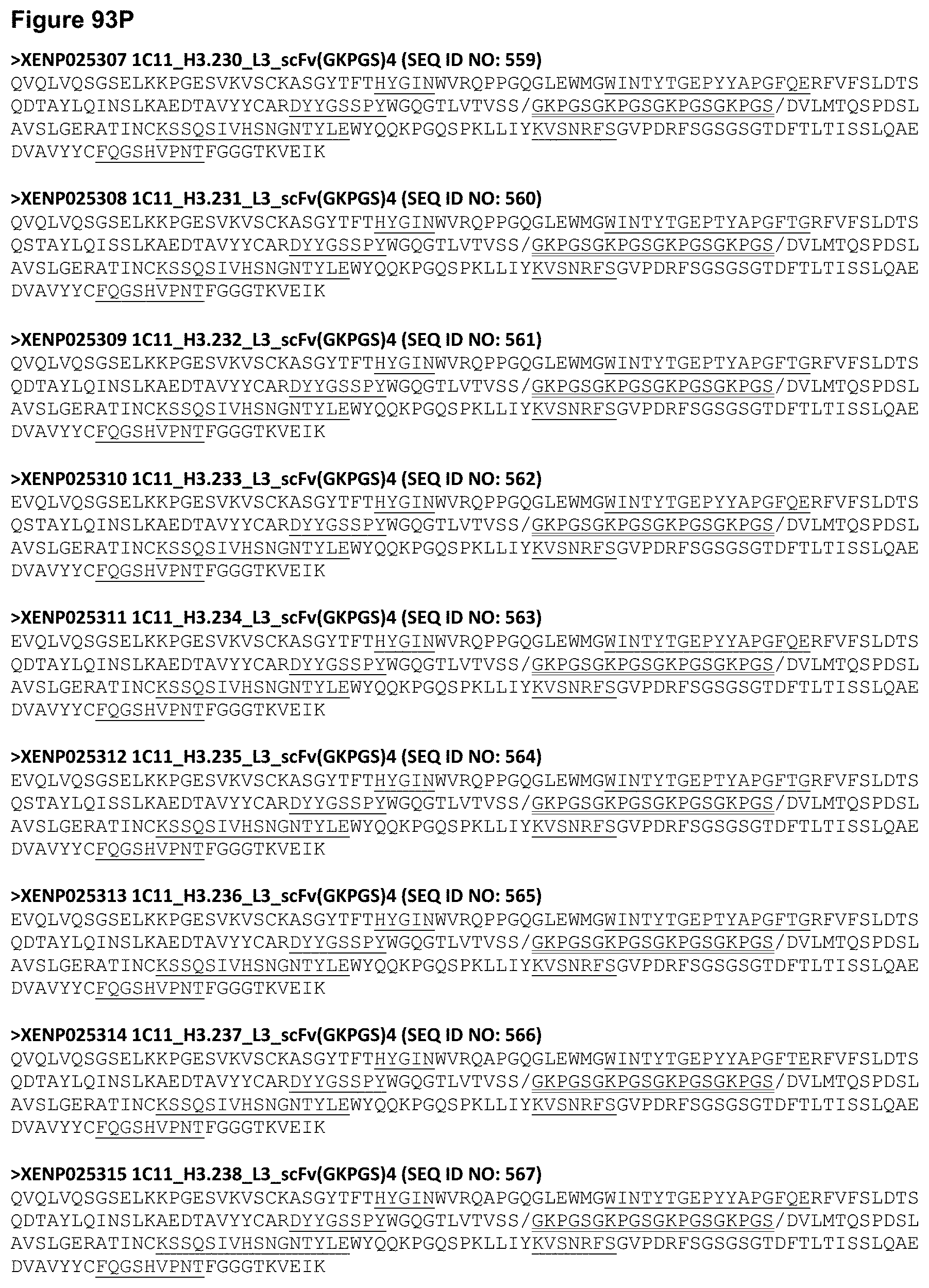
D00174

D00175
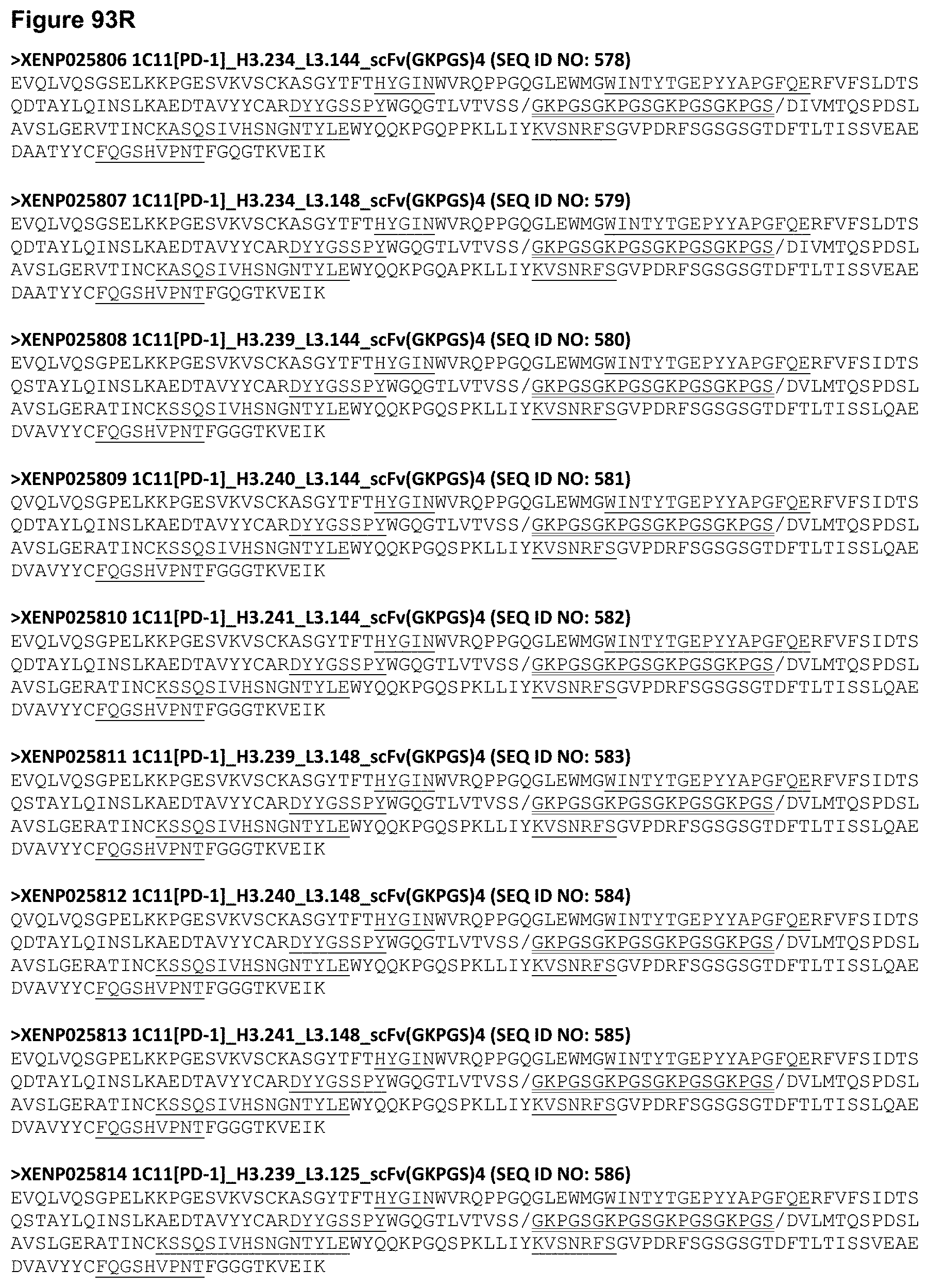
D00176

D00177
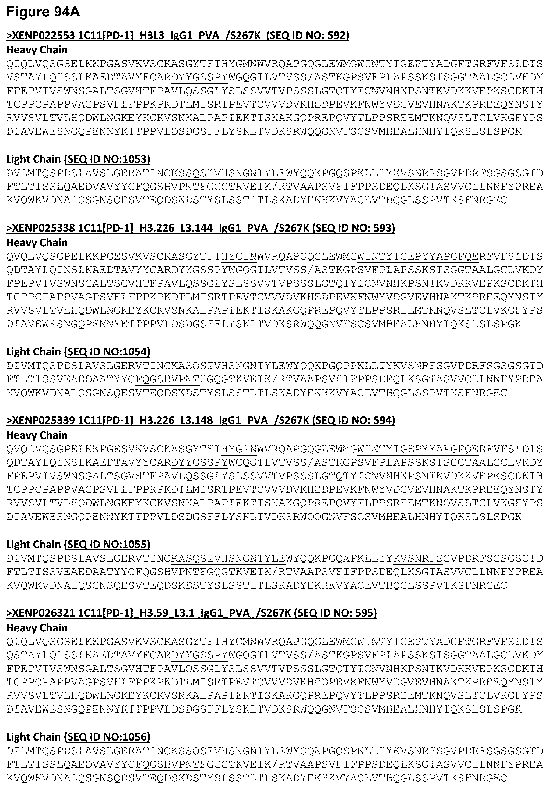
D00178
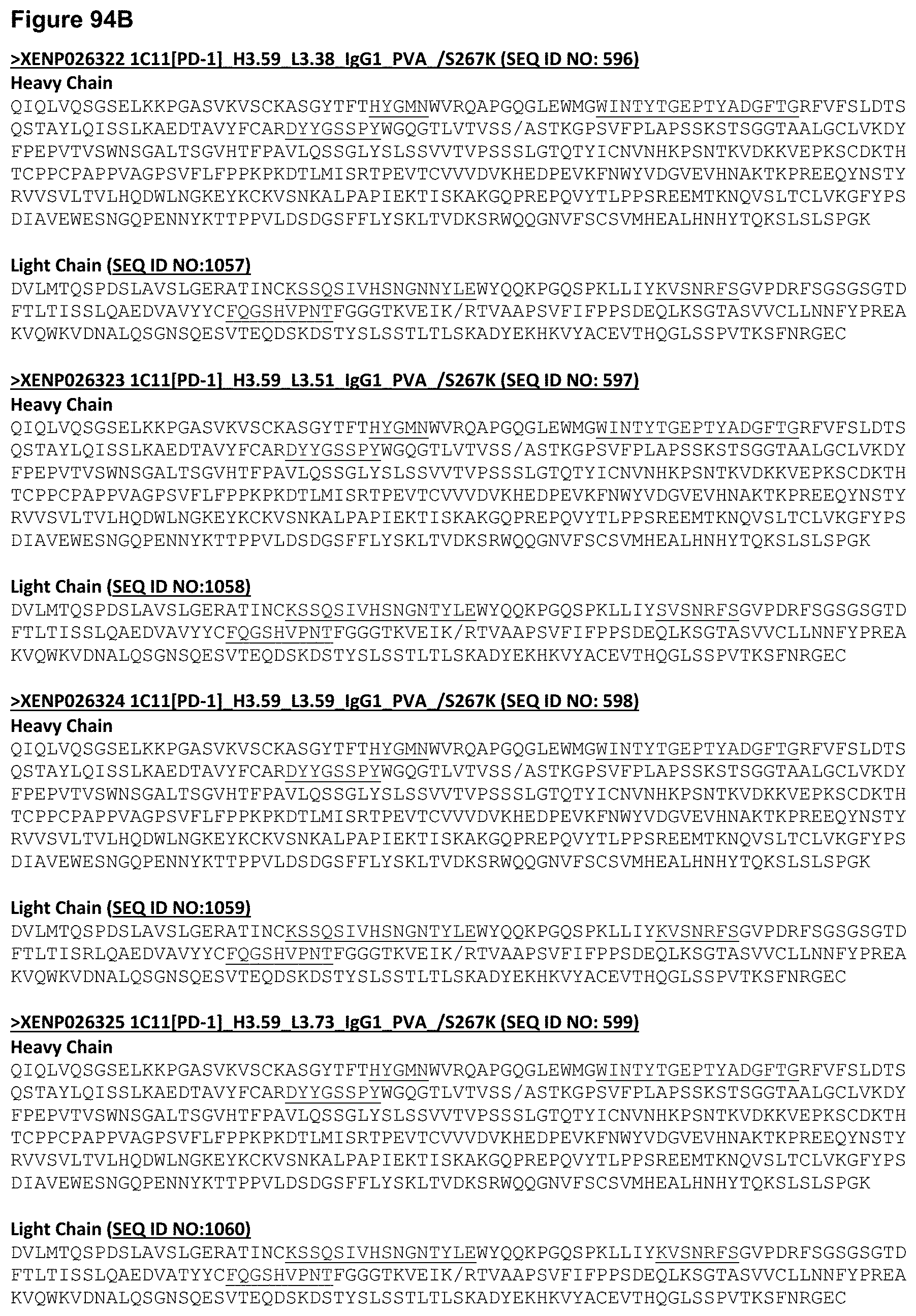
D00179

D00180
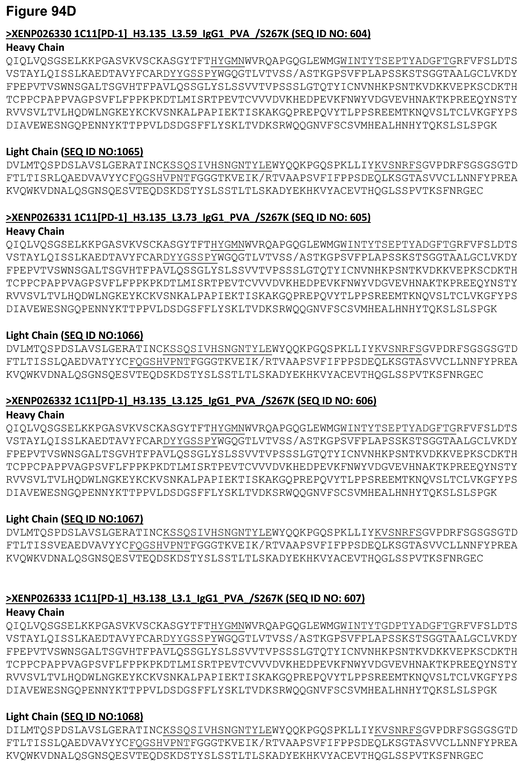
D00181

D00182

D00183
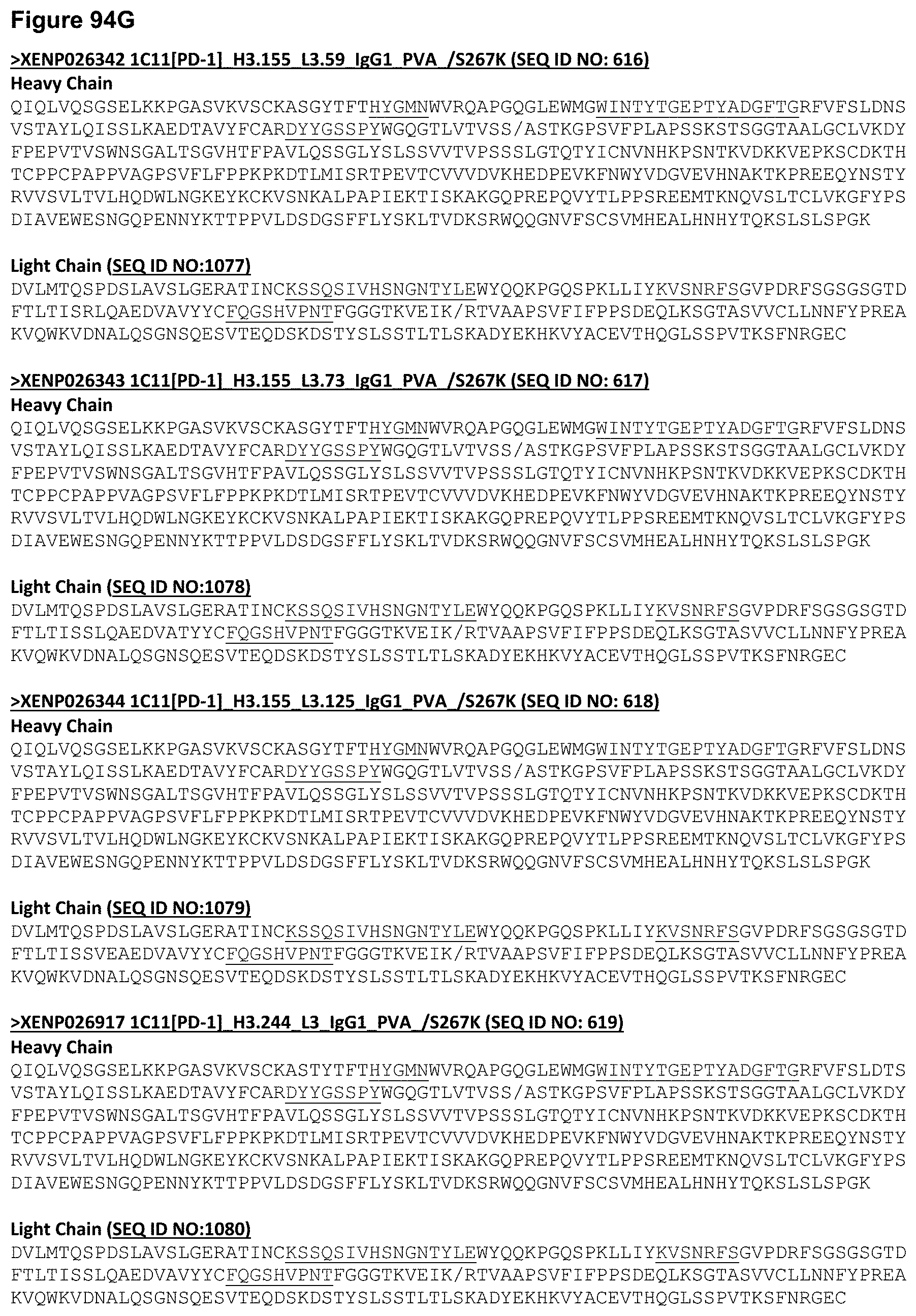
D00184

D00185

D00186

D00187

D00188

D00189

D00190

D00191
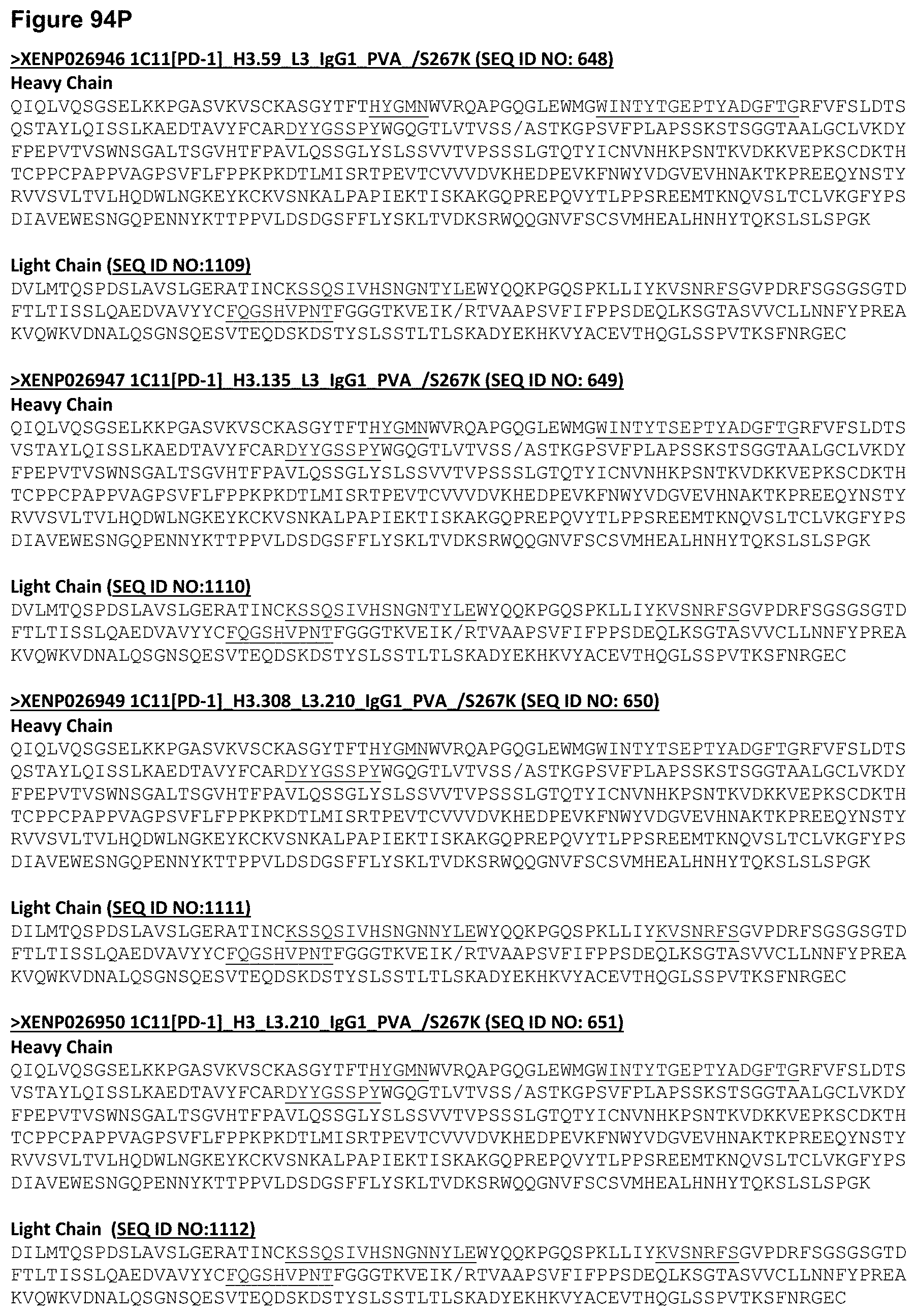
D00192
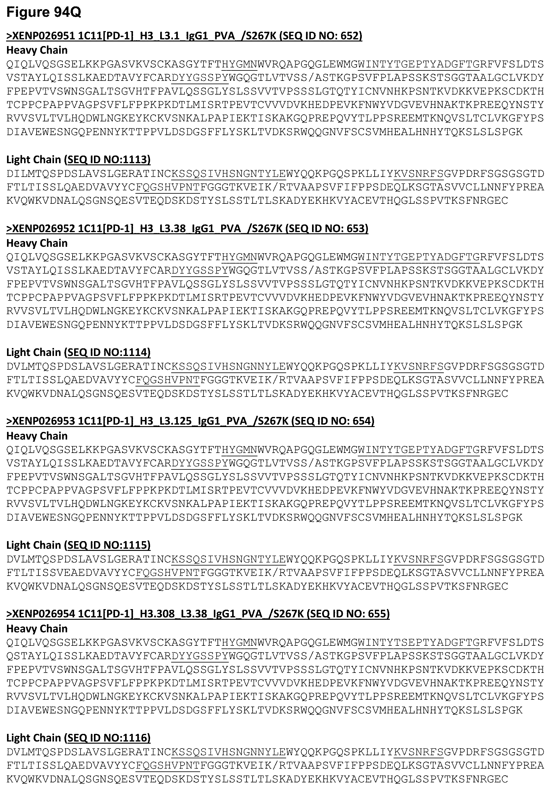
D00193
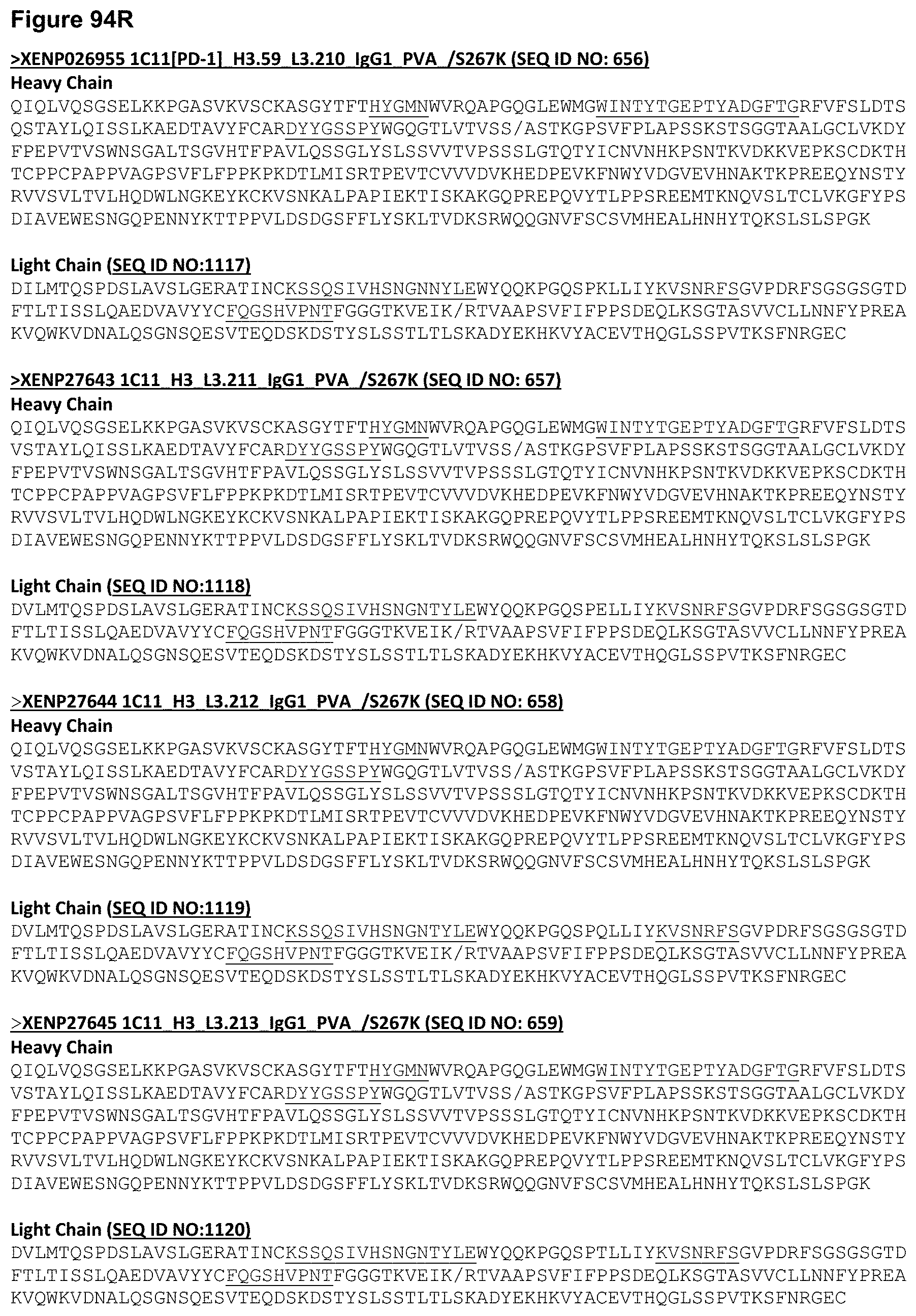
D00194

D00195

D00196

D00197

D00198

D00199

D00200

D00201

D00202

D00203

D00204

D00205

D00206

D00207
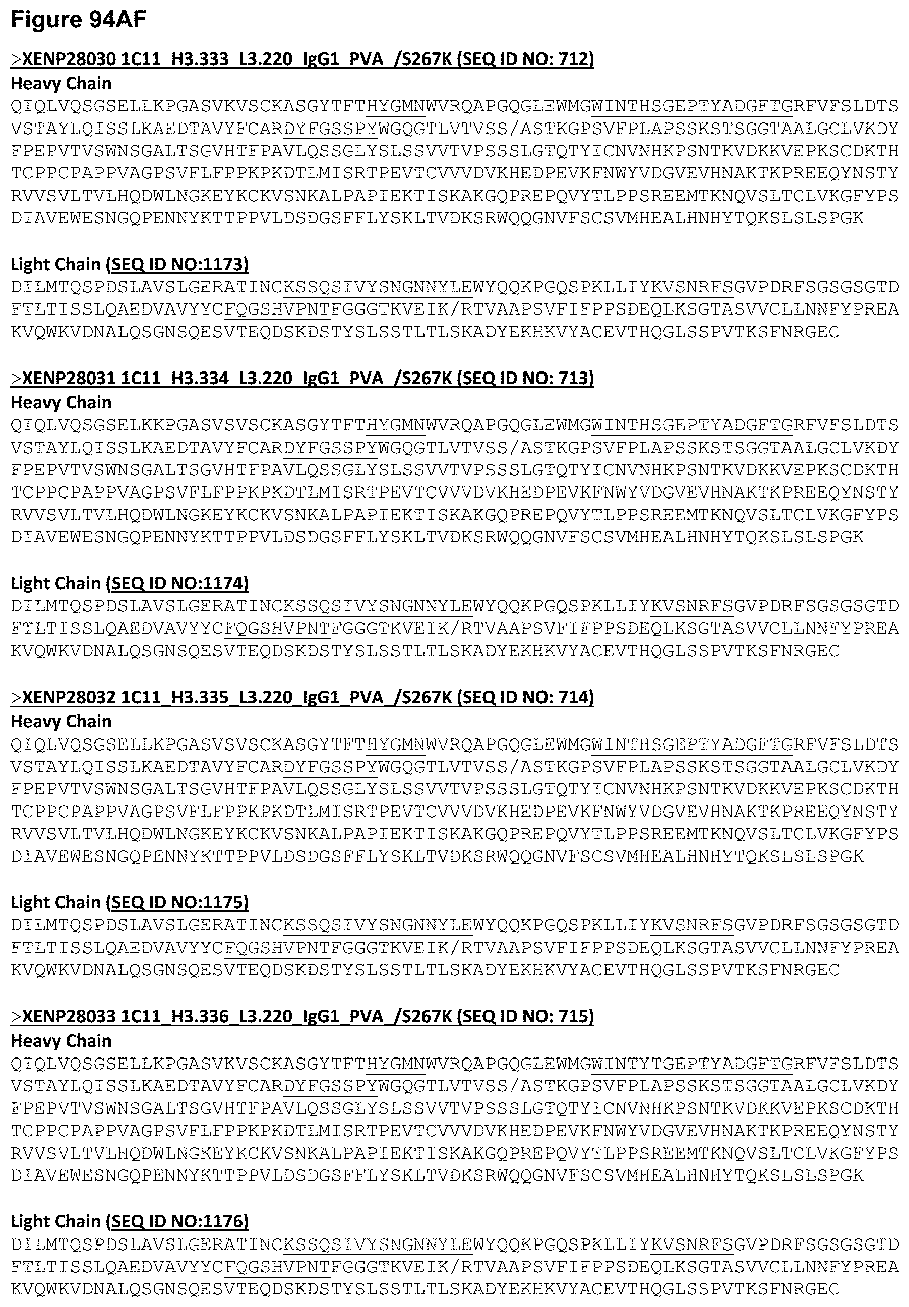
D00208

D00209
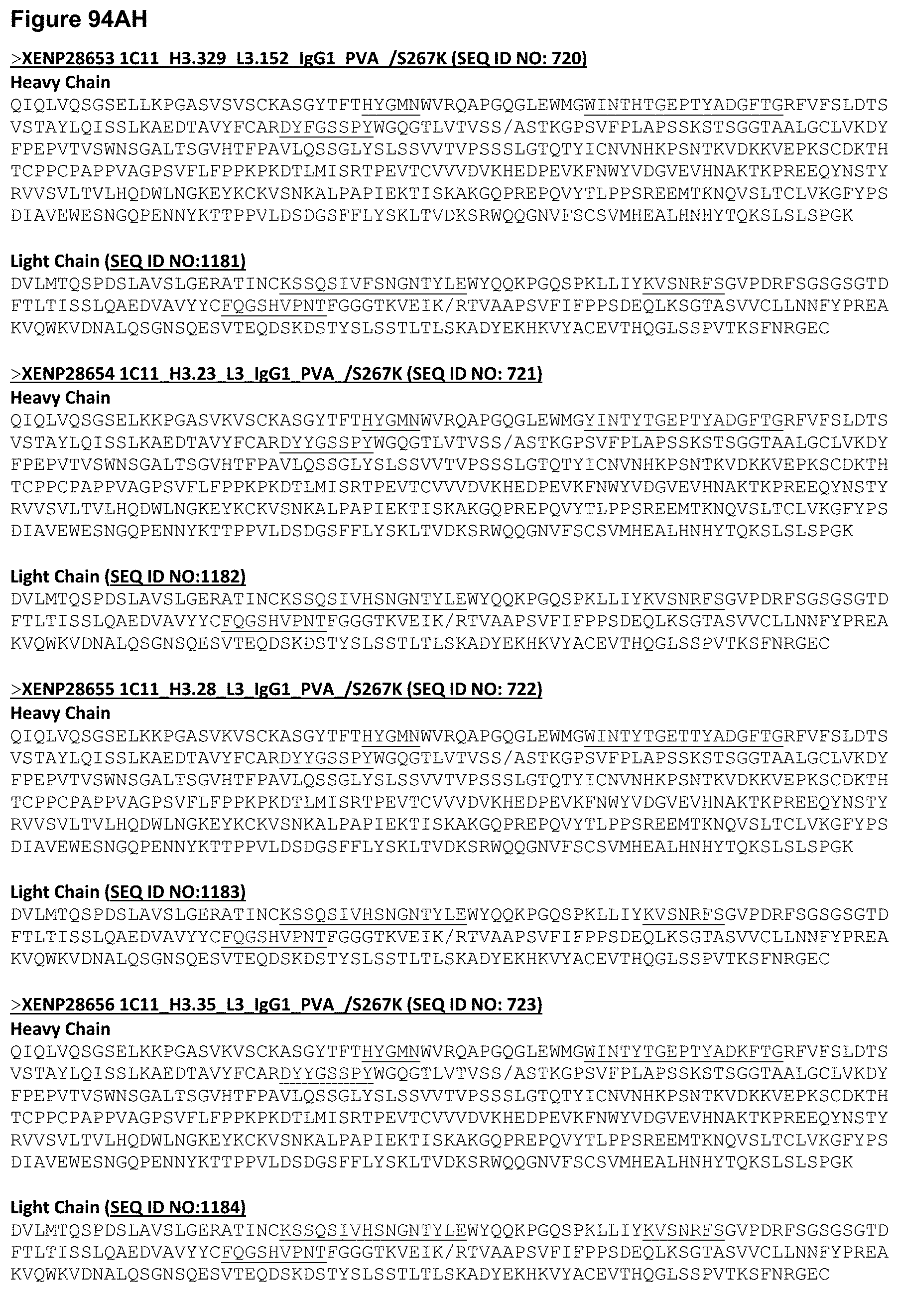
D00210
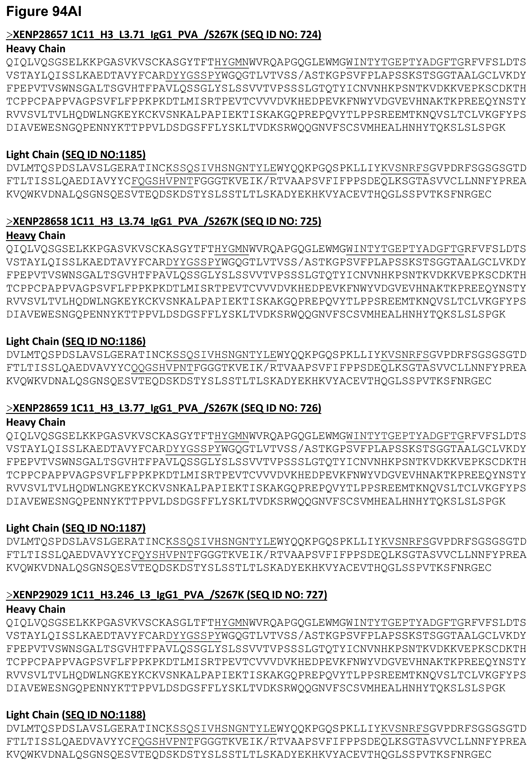
D00211
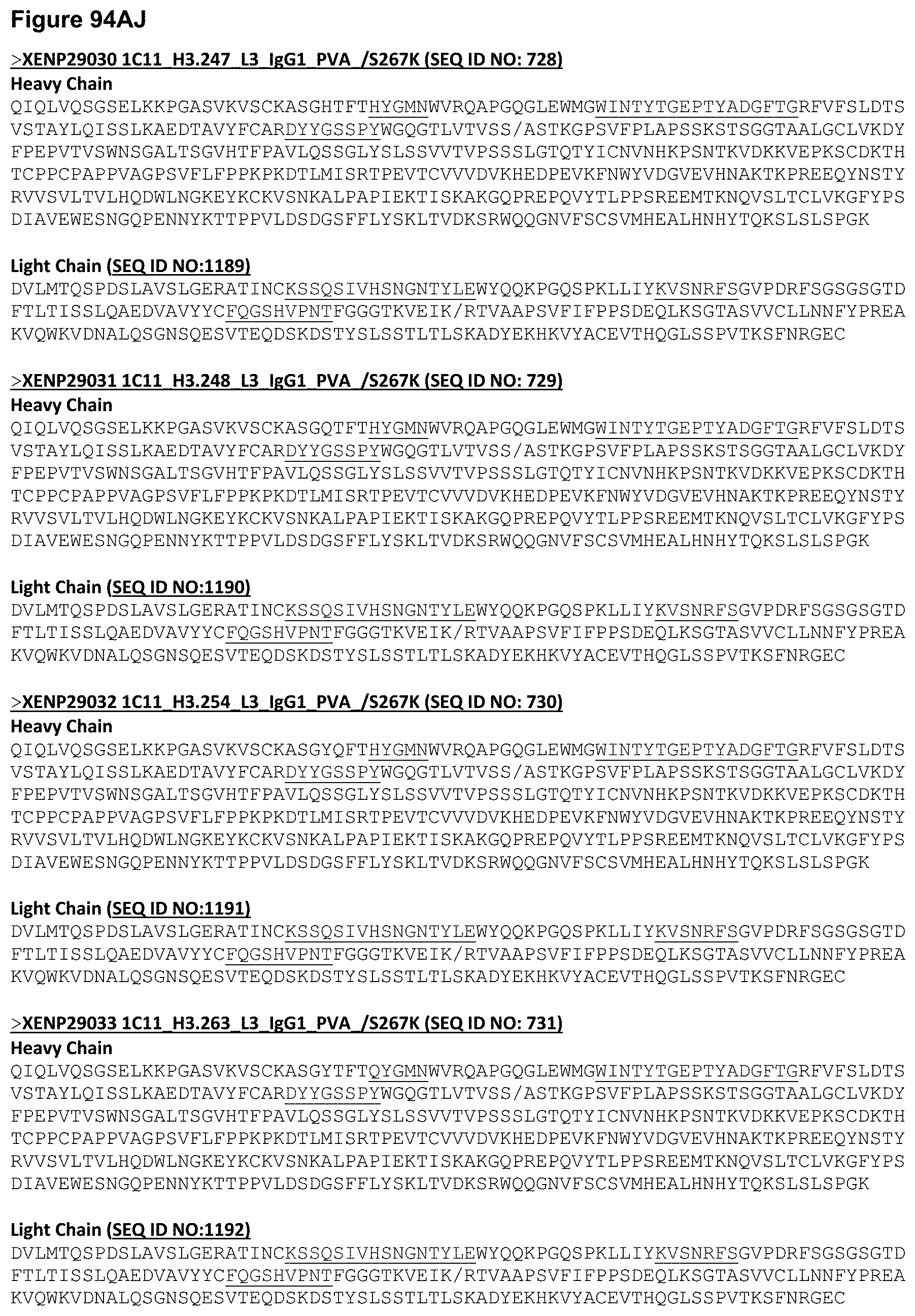
D00212
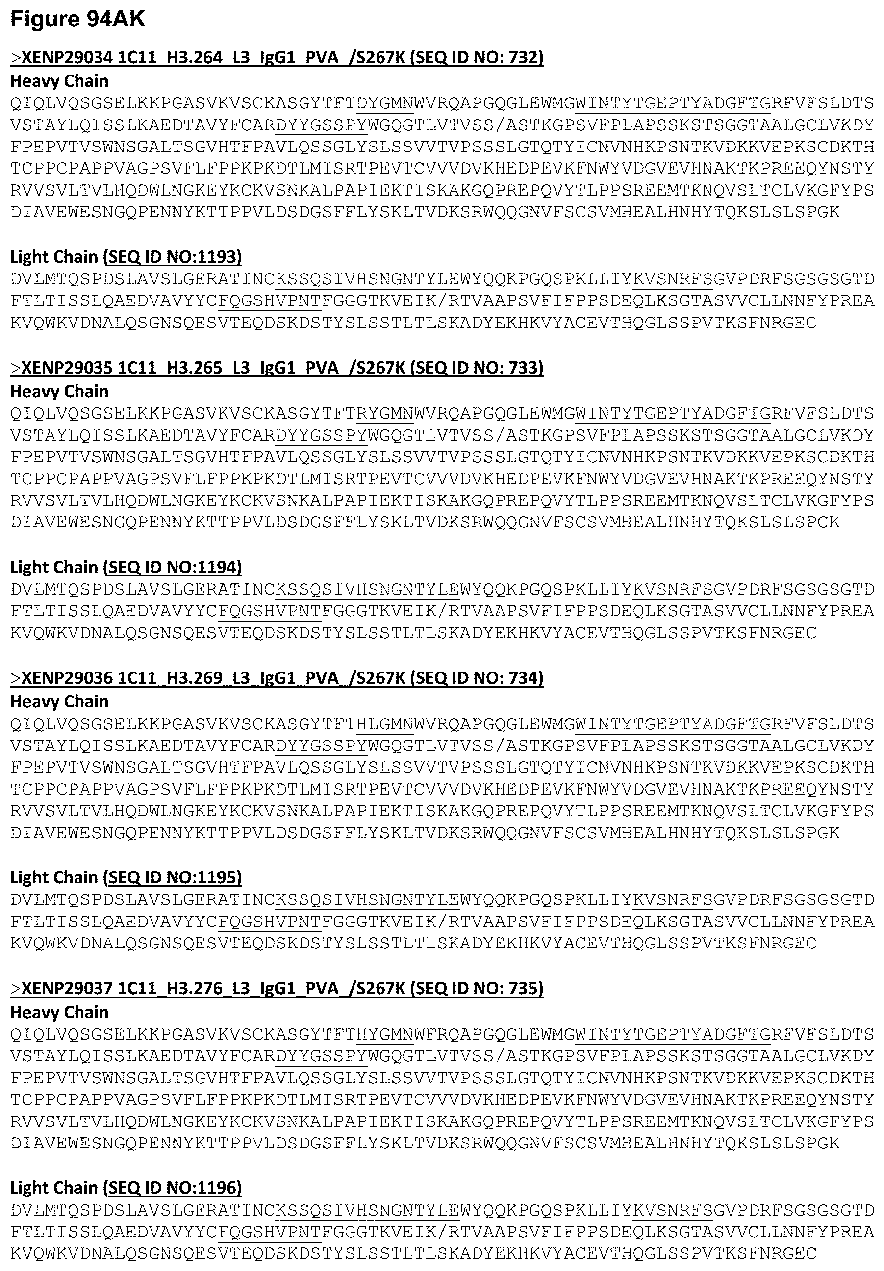
D00213

D00214

D00215
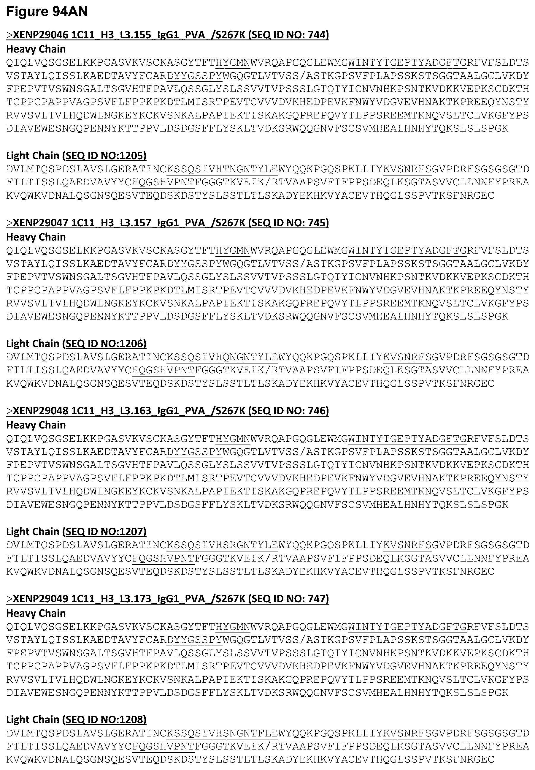
D00216

D00217
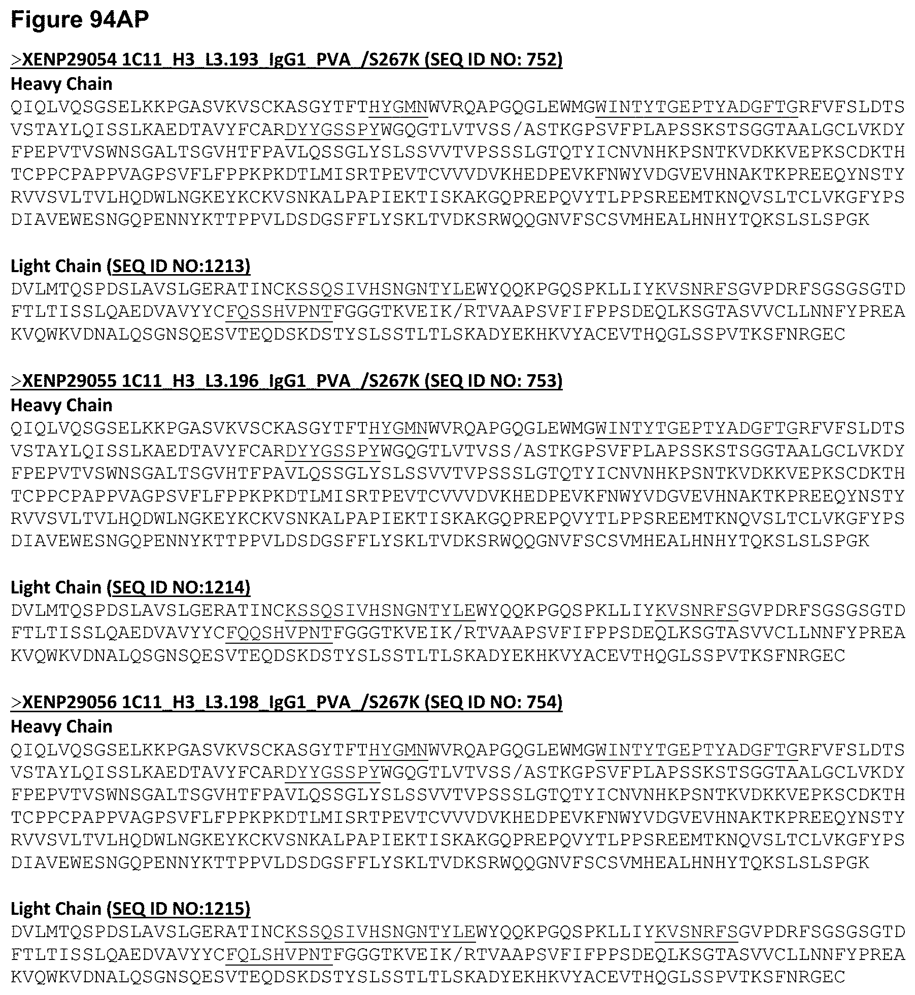
D00218
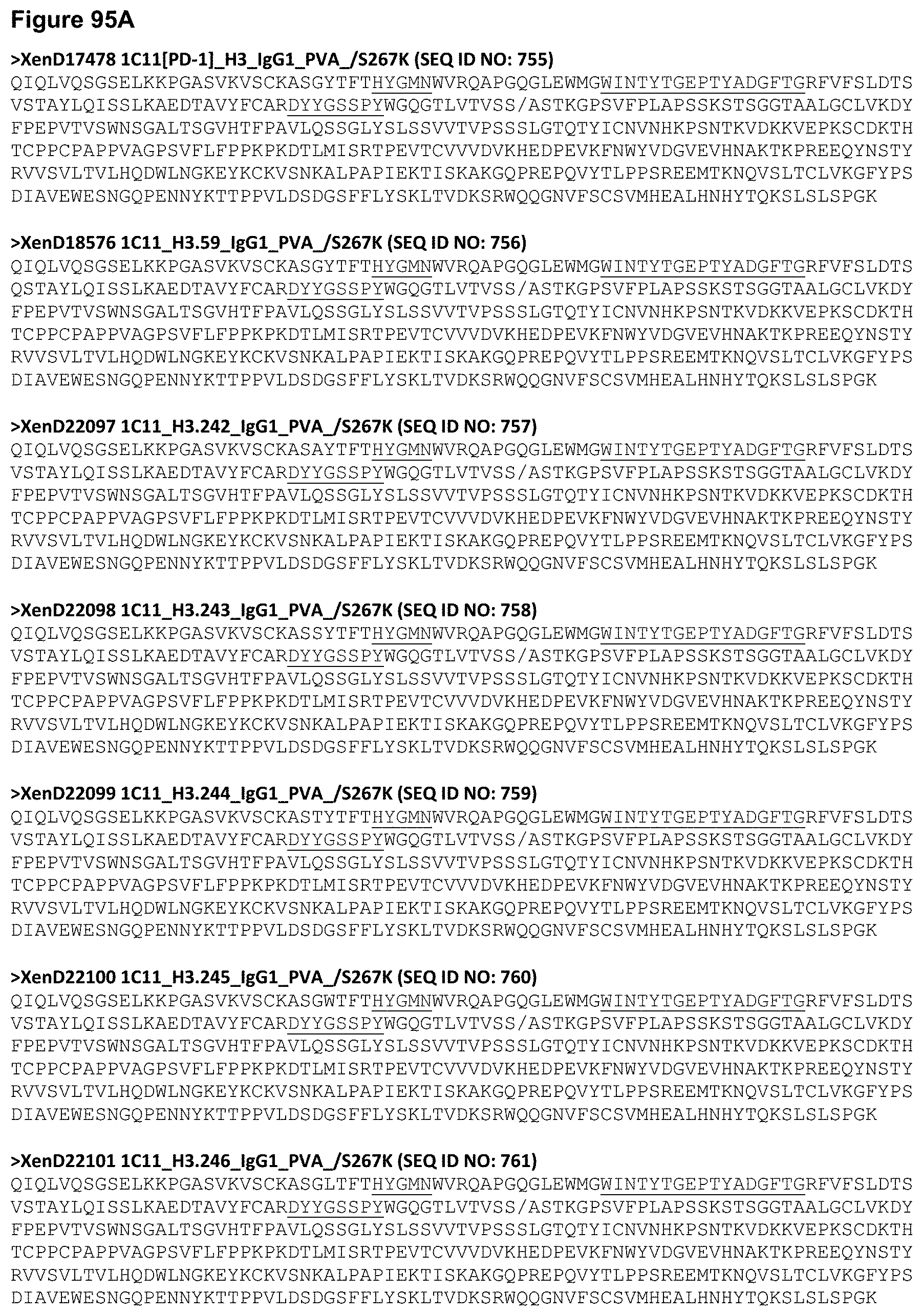
D00219

D00220

D00221

D00222

D00223

D00224
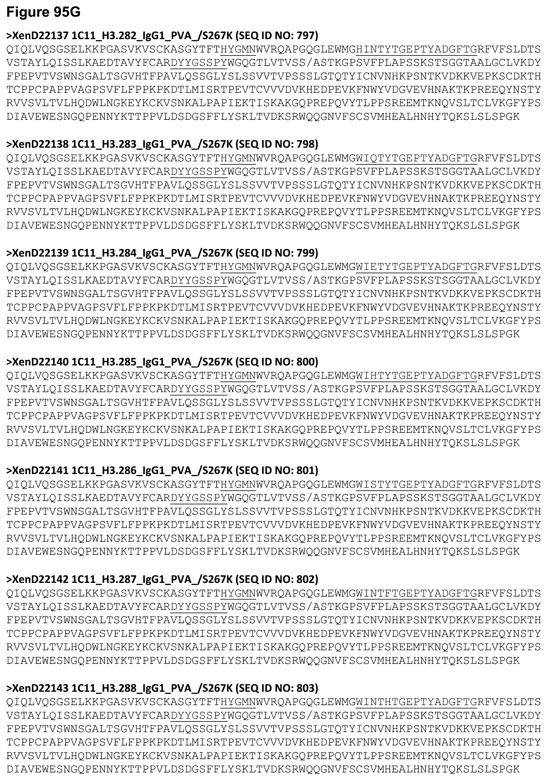
D00225

D00226

D00227
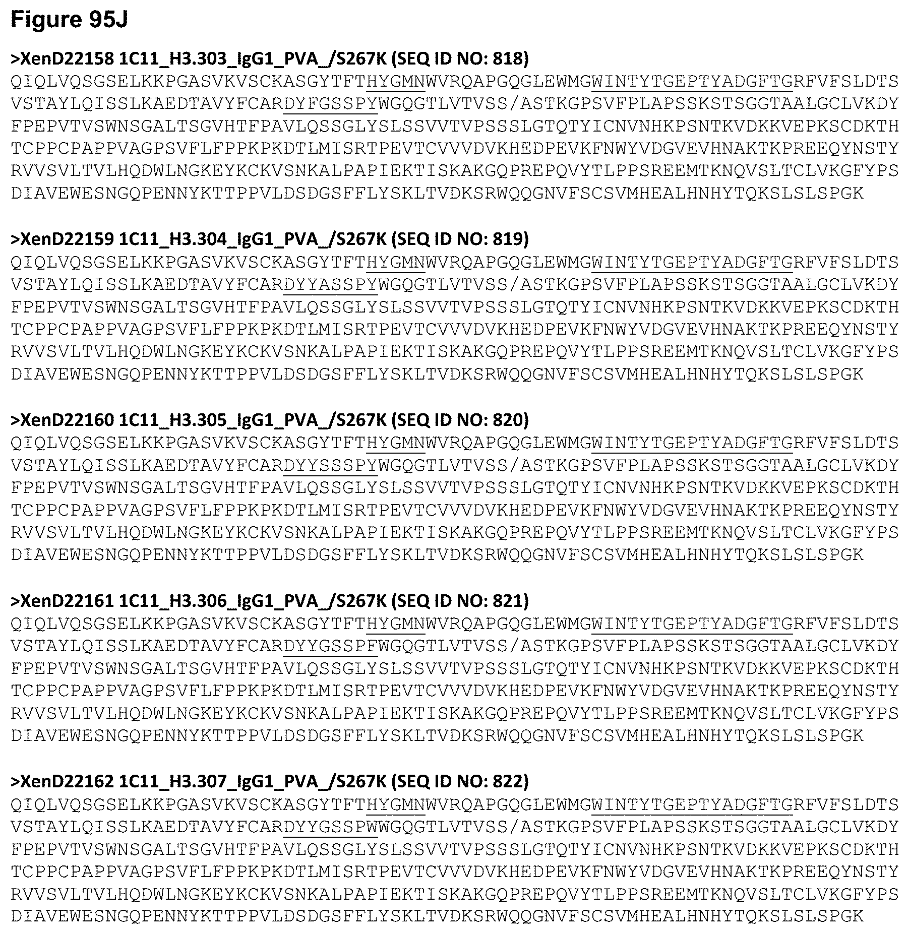
D00228
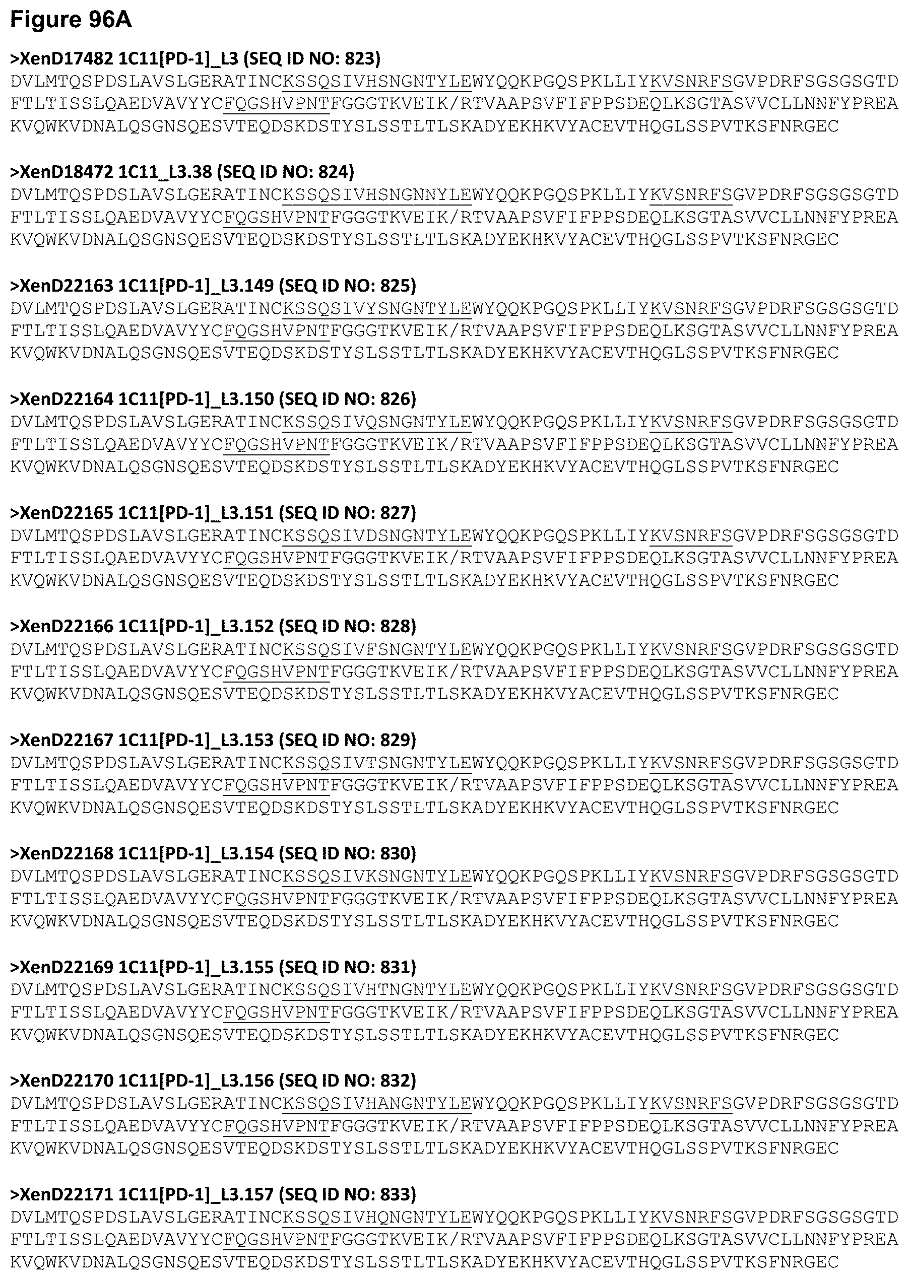
D00229

D00230

D00231
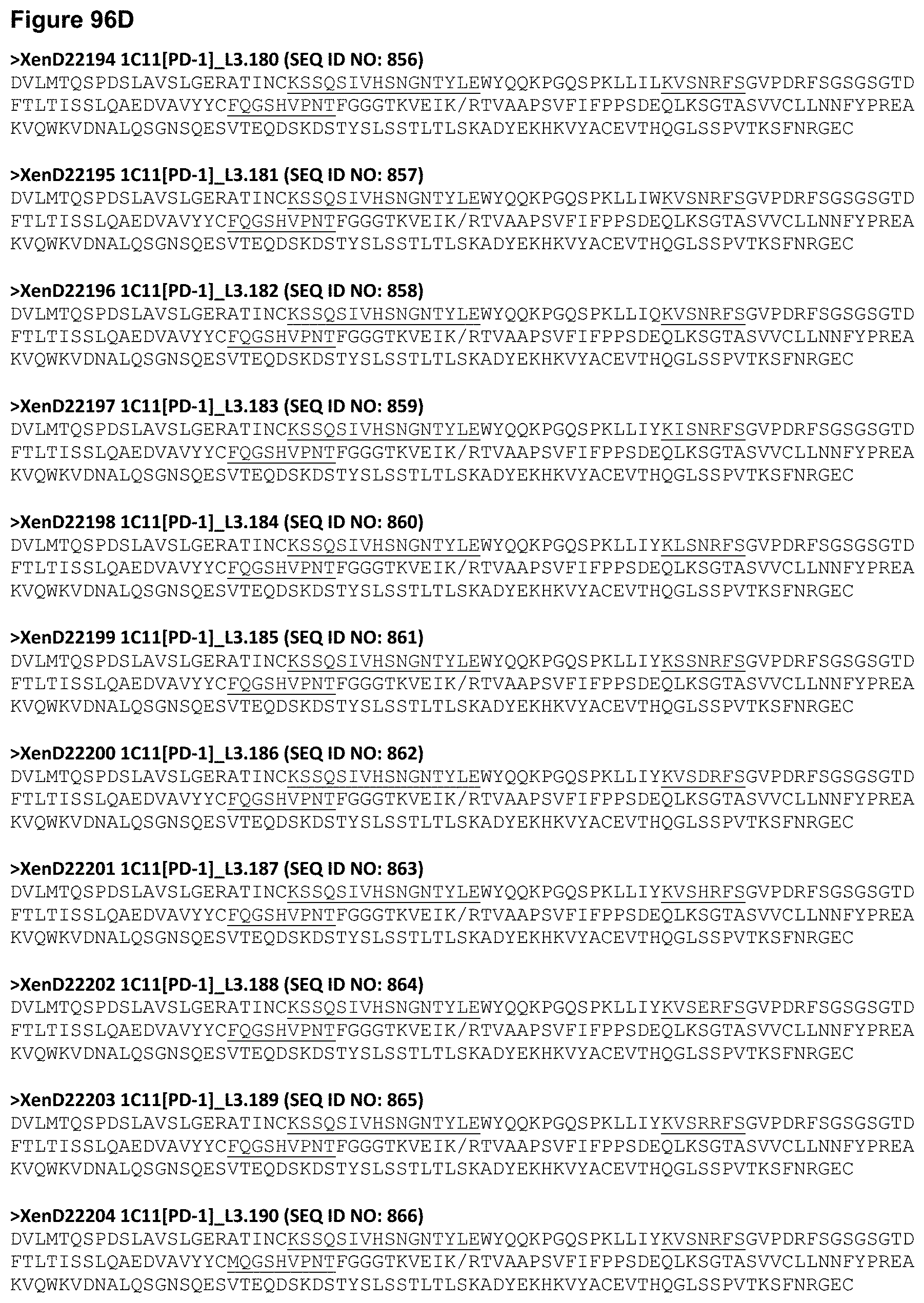
D00232

D00233
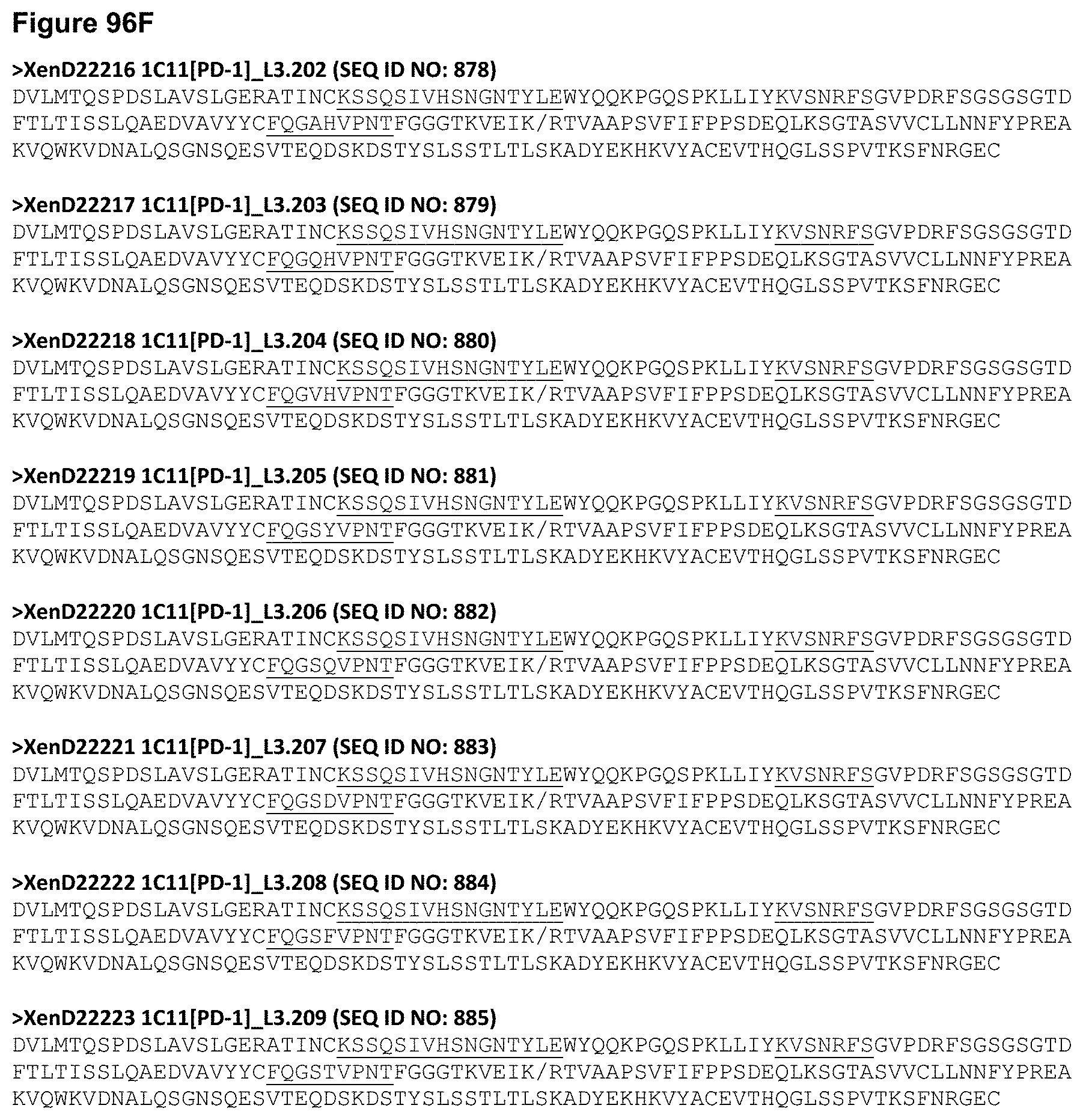
D00234

D00235

D00236

D00237

D00238

D00239
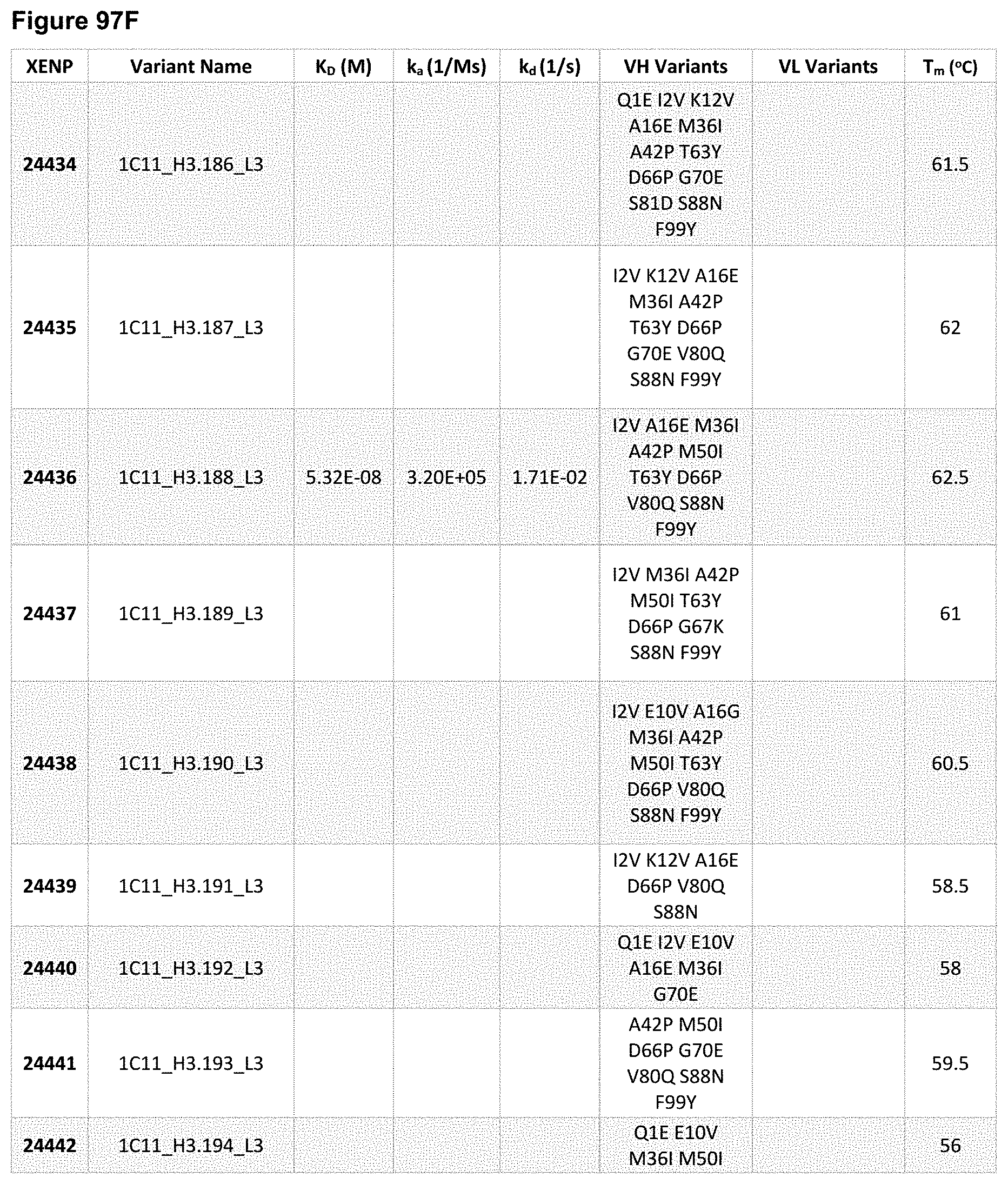
D00240

D00241

D00242
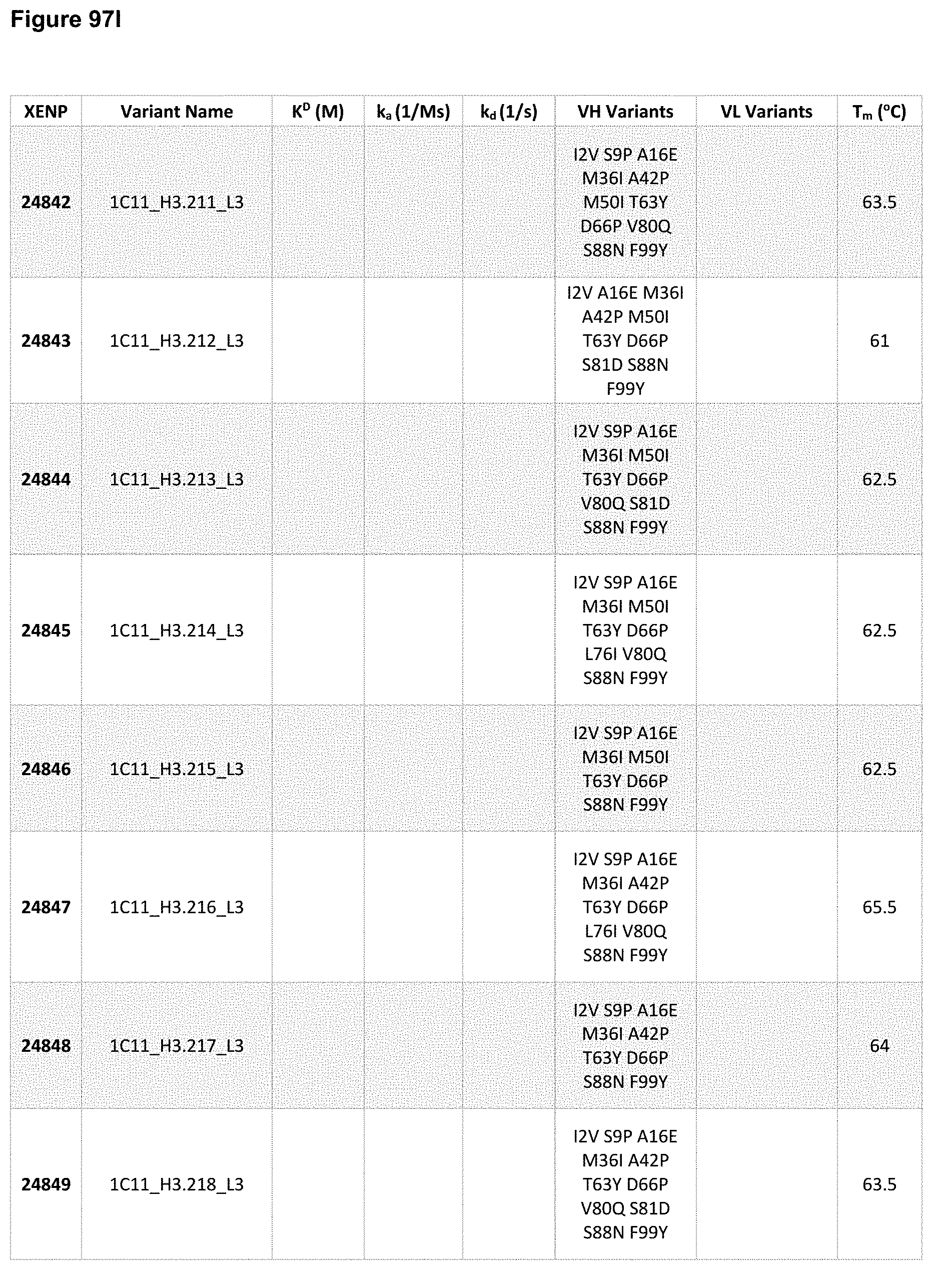
D00243

D00244

D00245
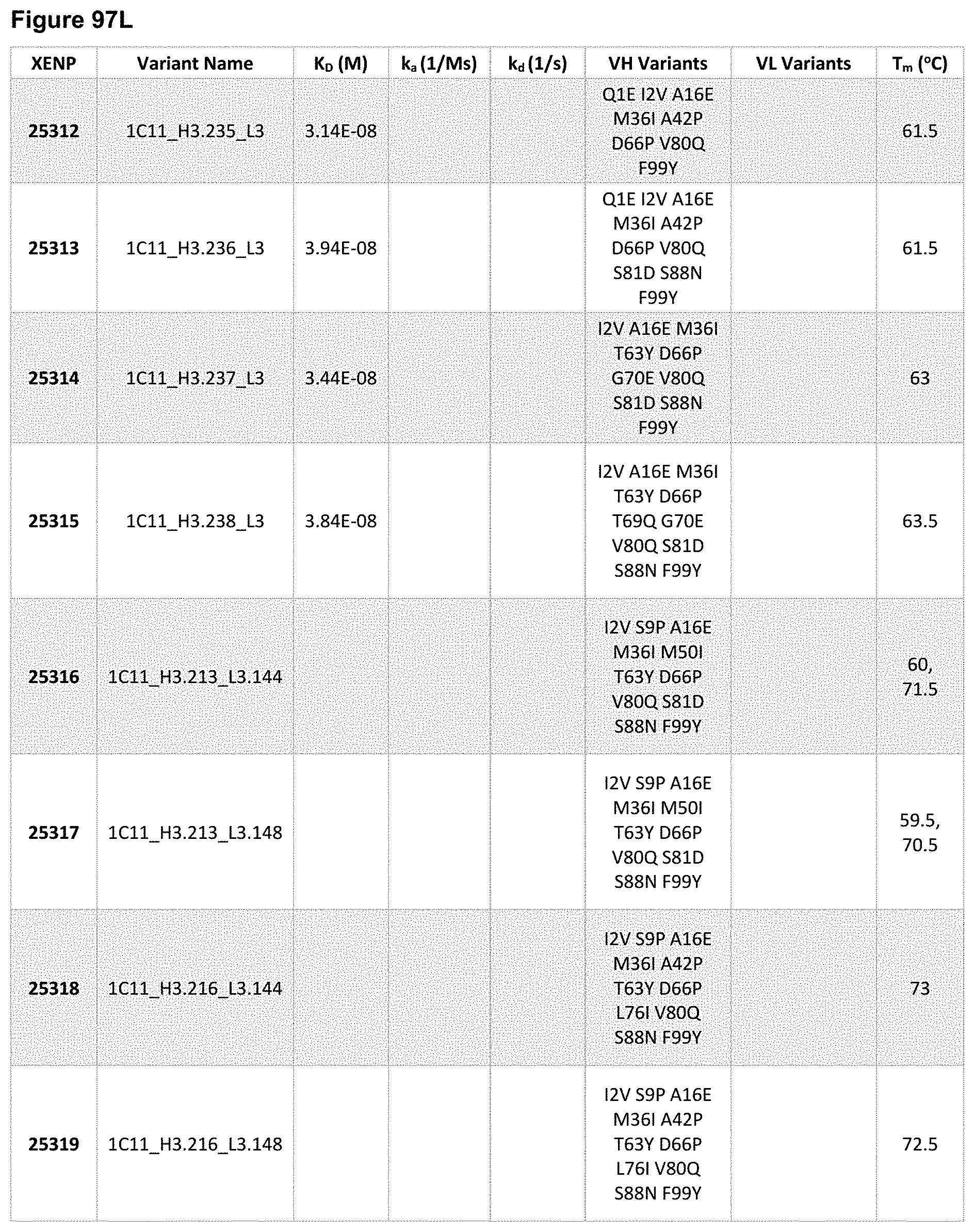
D00246
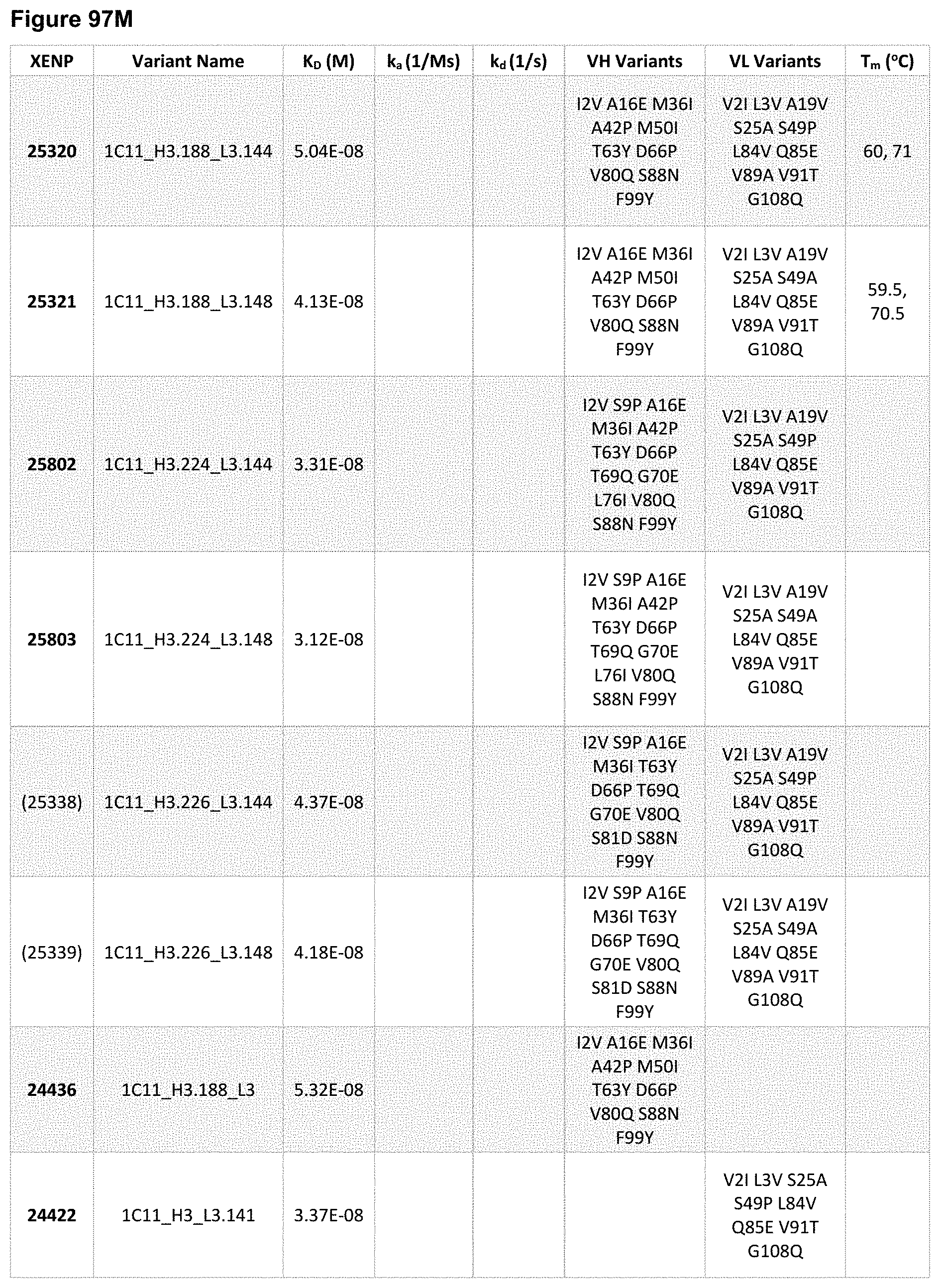
D00247
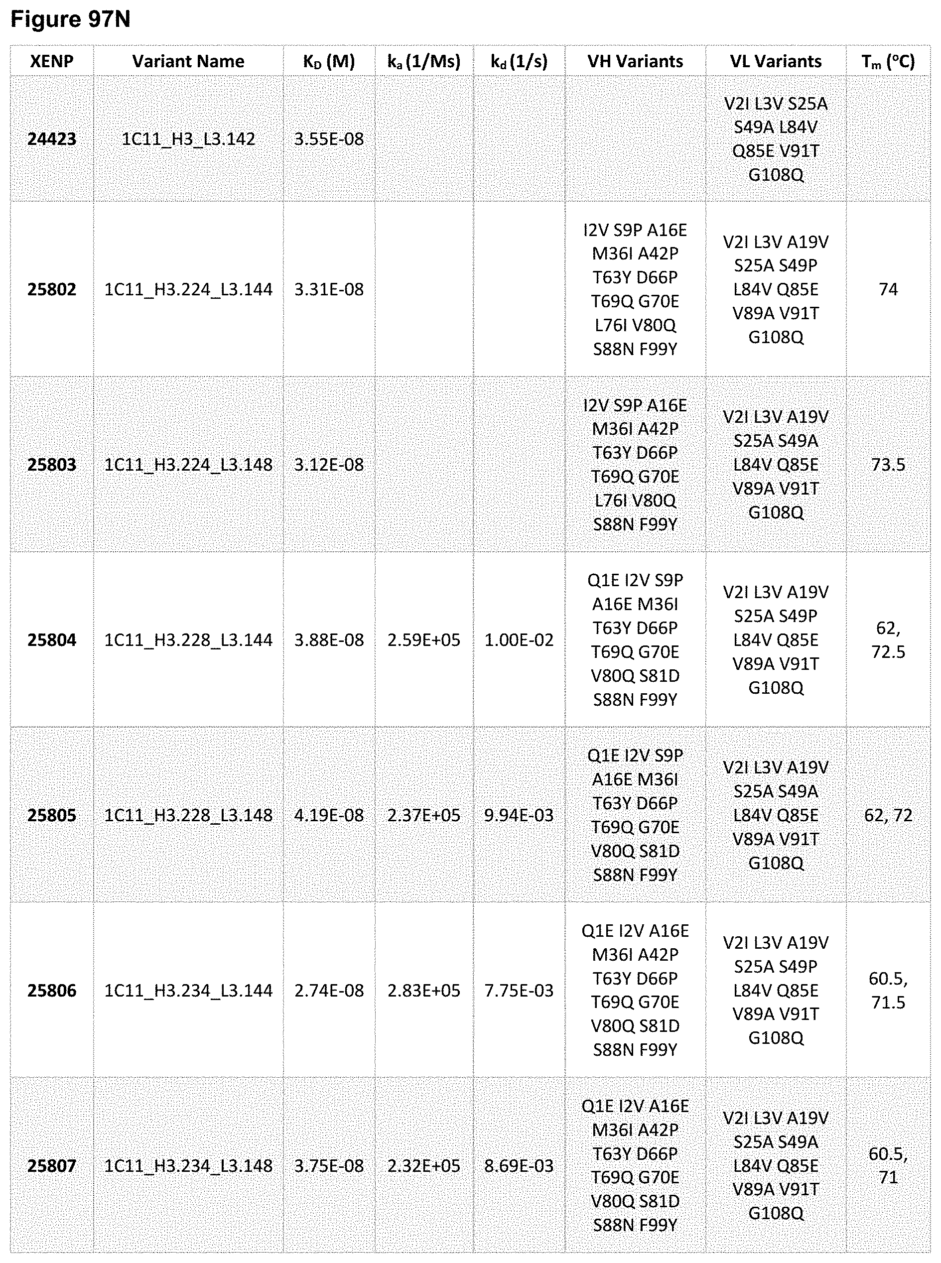
D00248

D00249

D00250
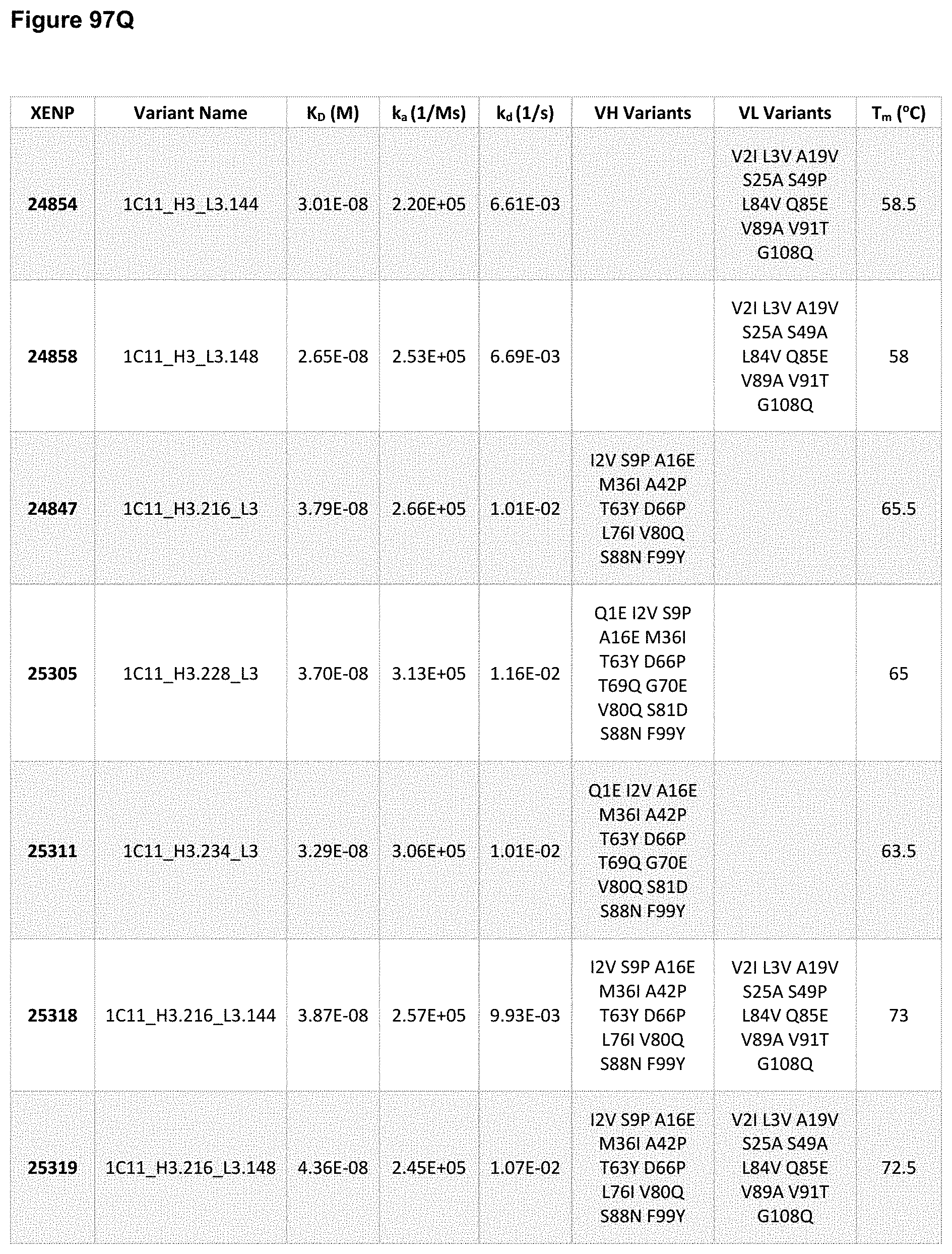
D00251
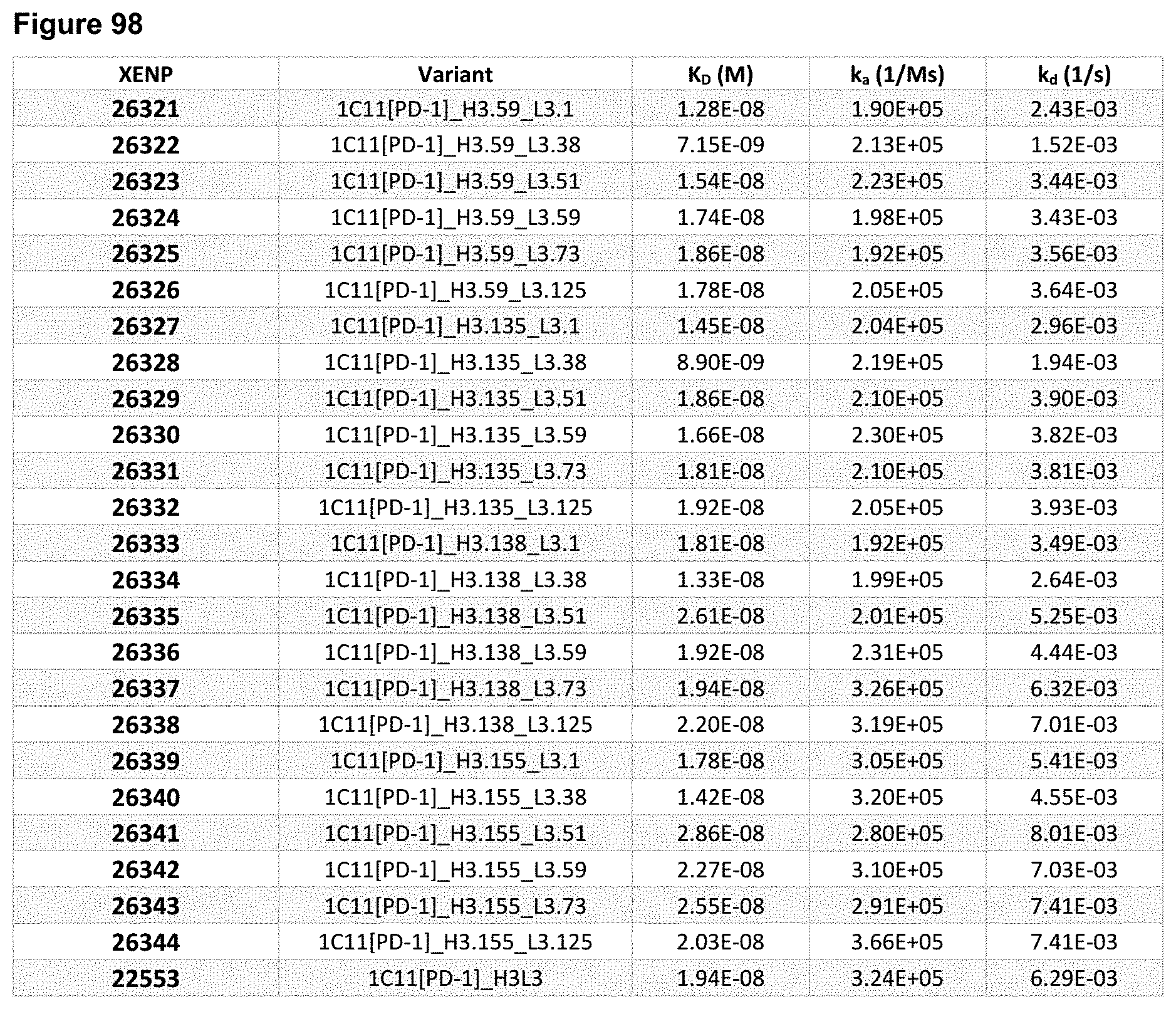
D00252

D00253

D00254

D00255
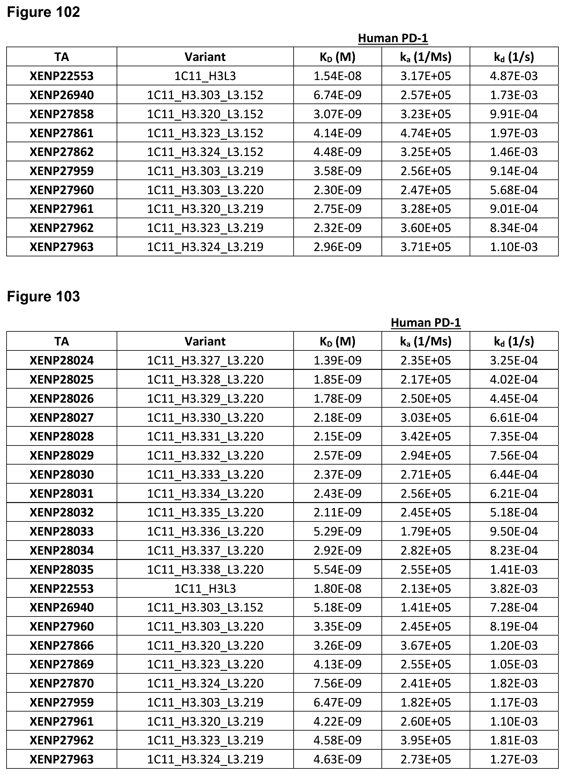
D00256

D00257

D00258
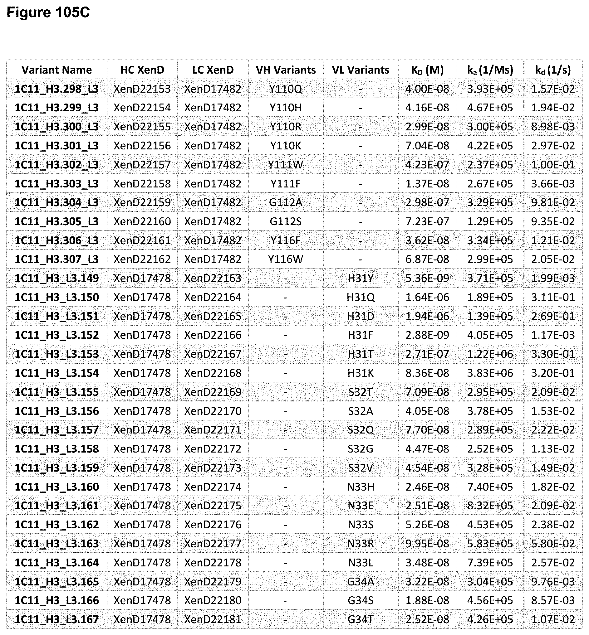
D00259
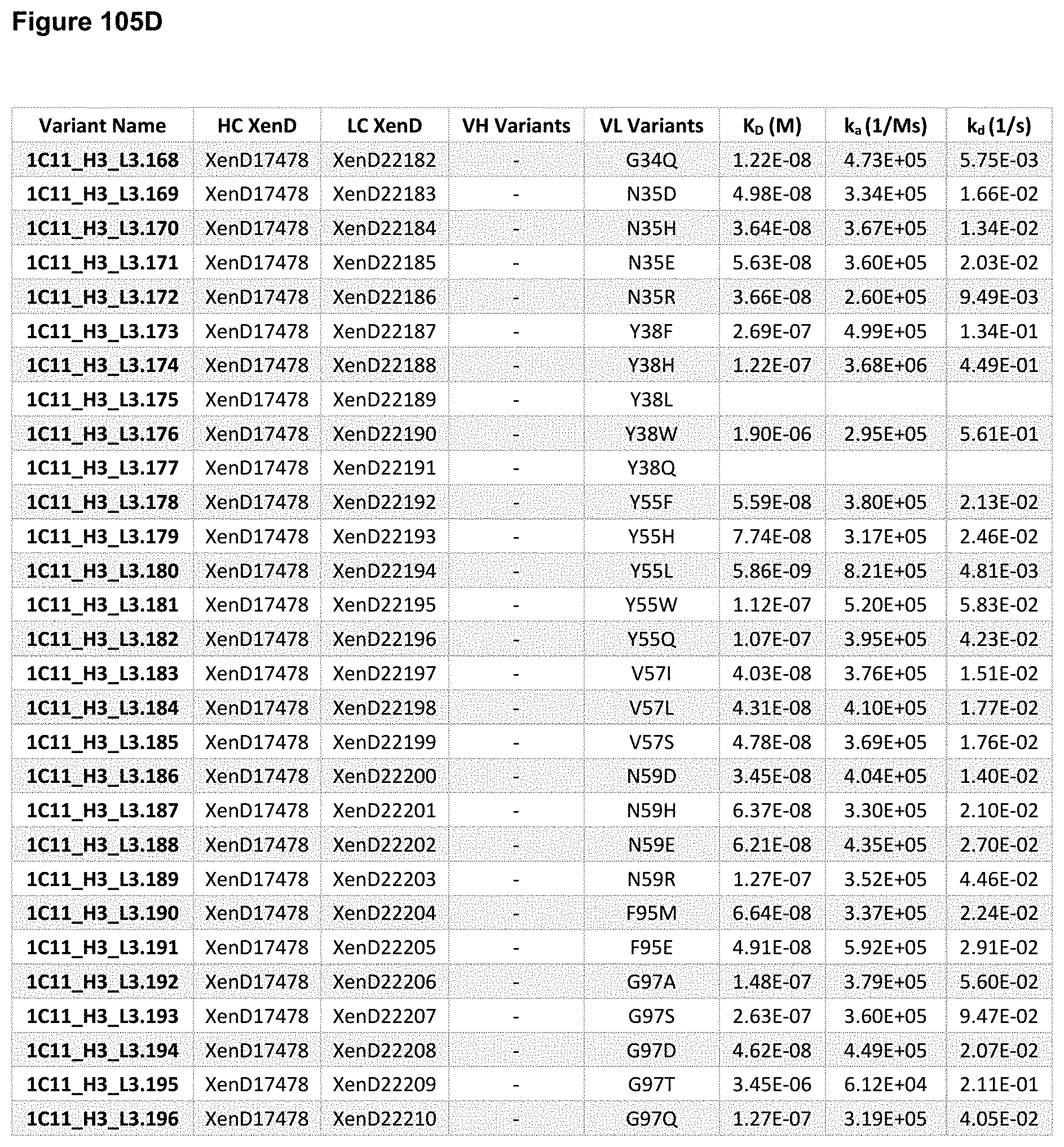
D00260

D00261
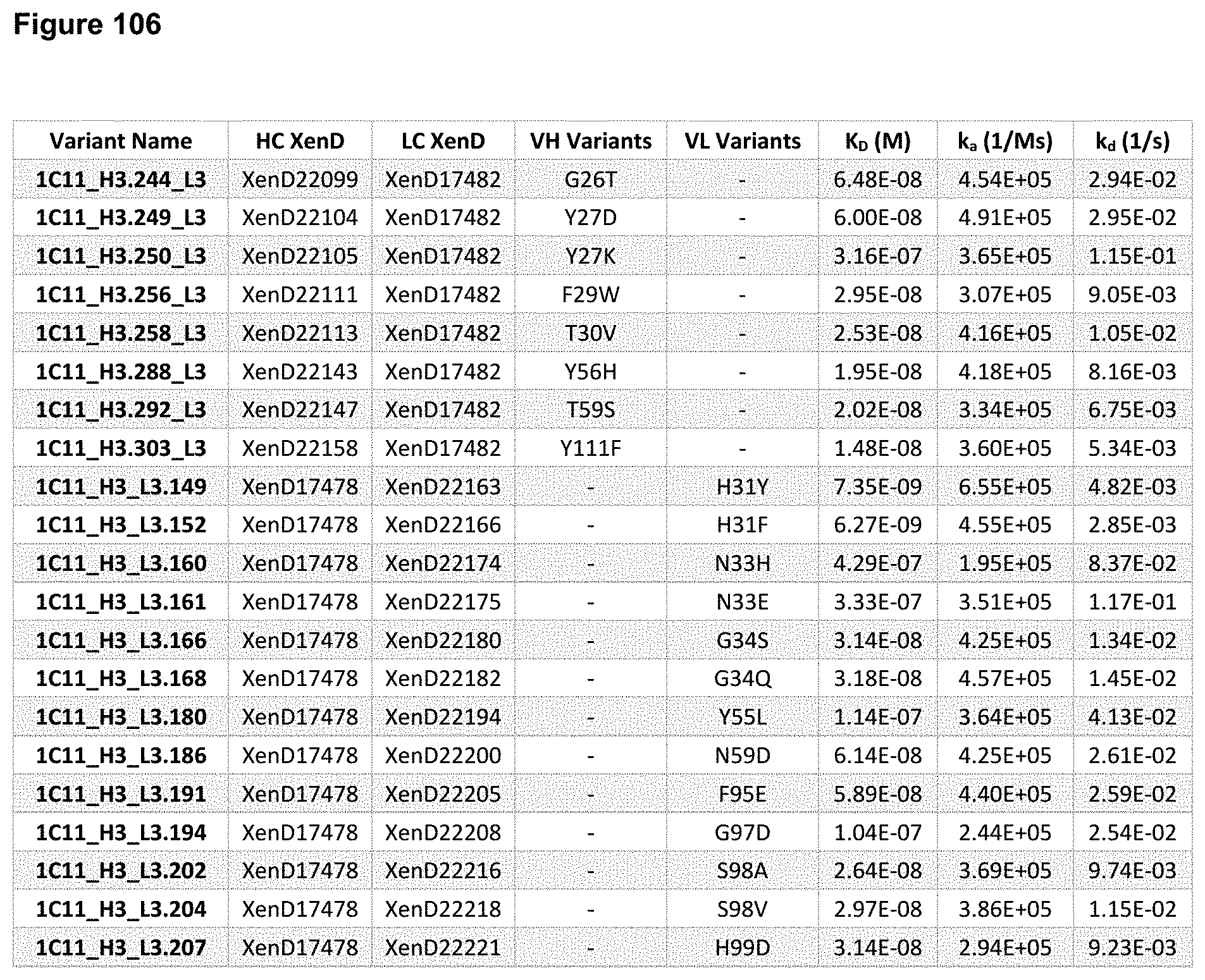
D00262

D00263
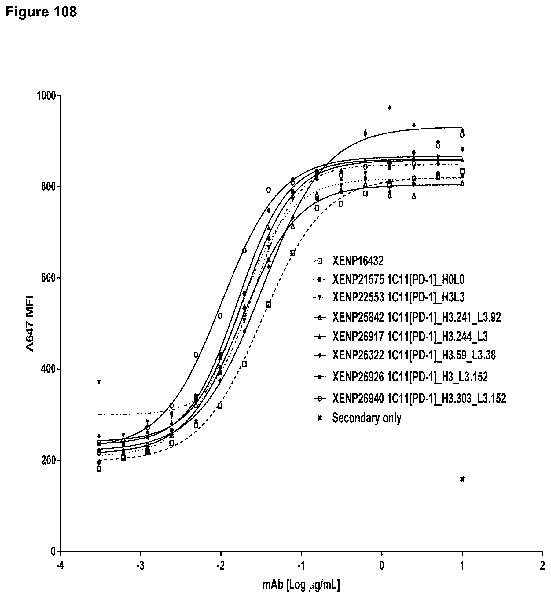
D00264
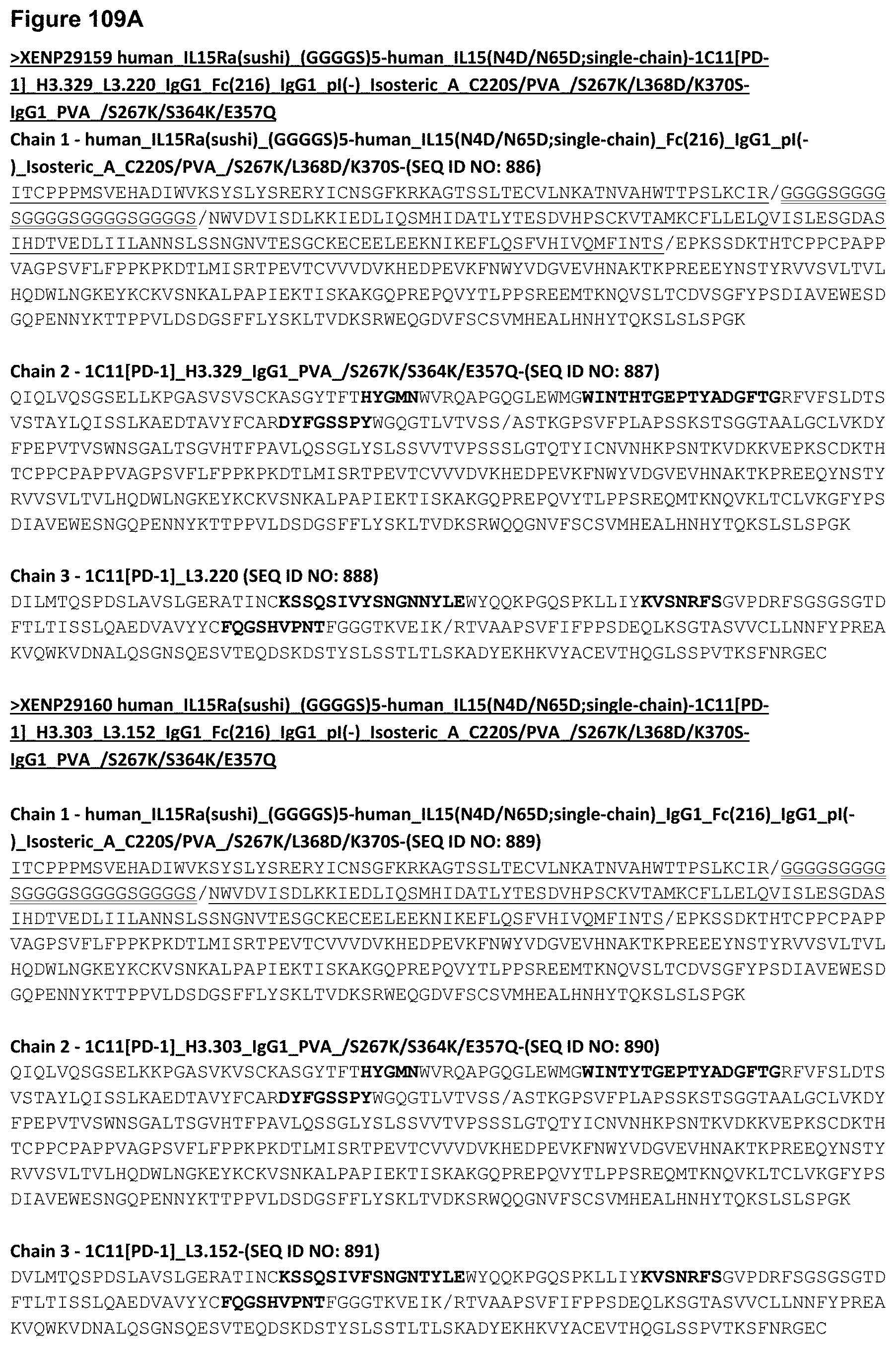
D00265

D00266
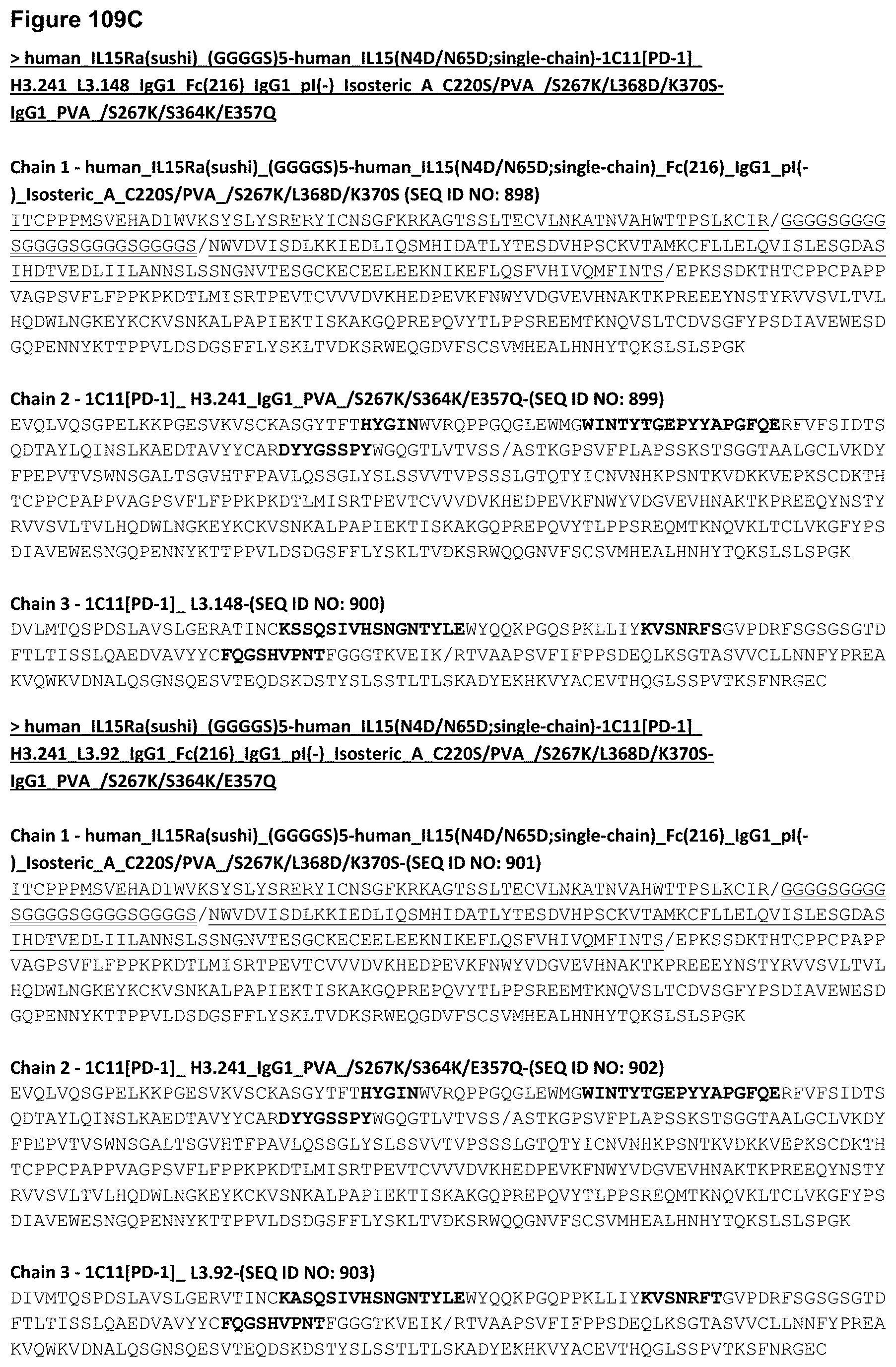
D00267

D00268

D00269

D00270

D00271

D00272
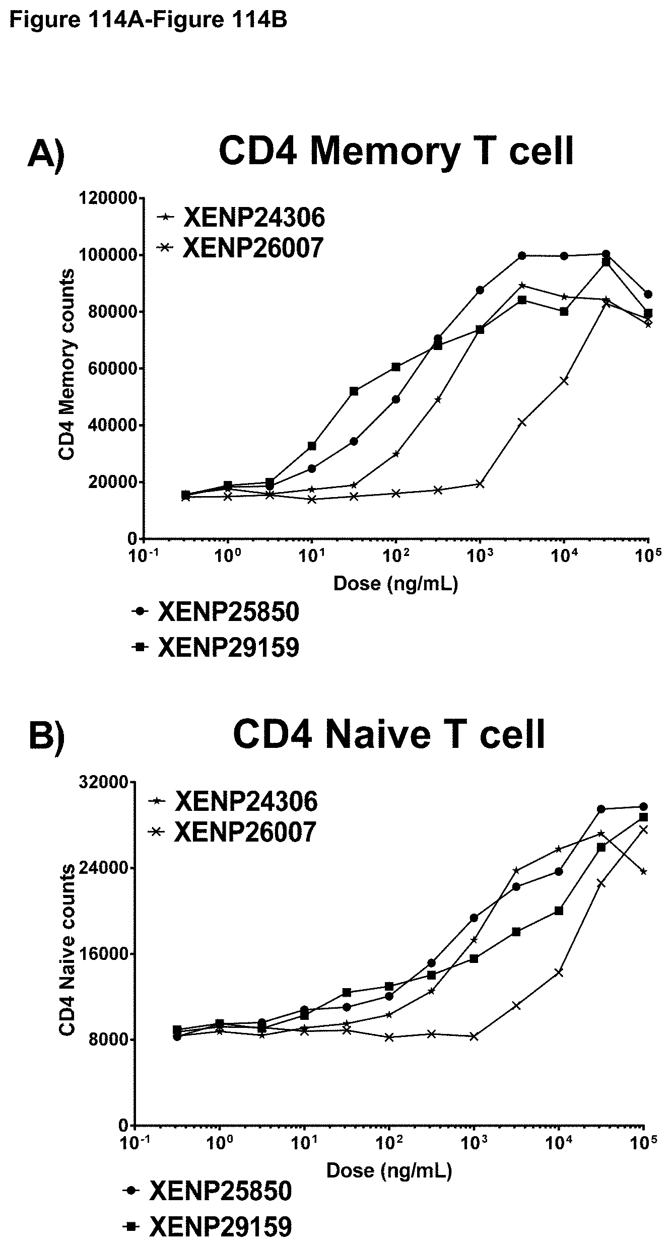
D00273
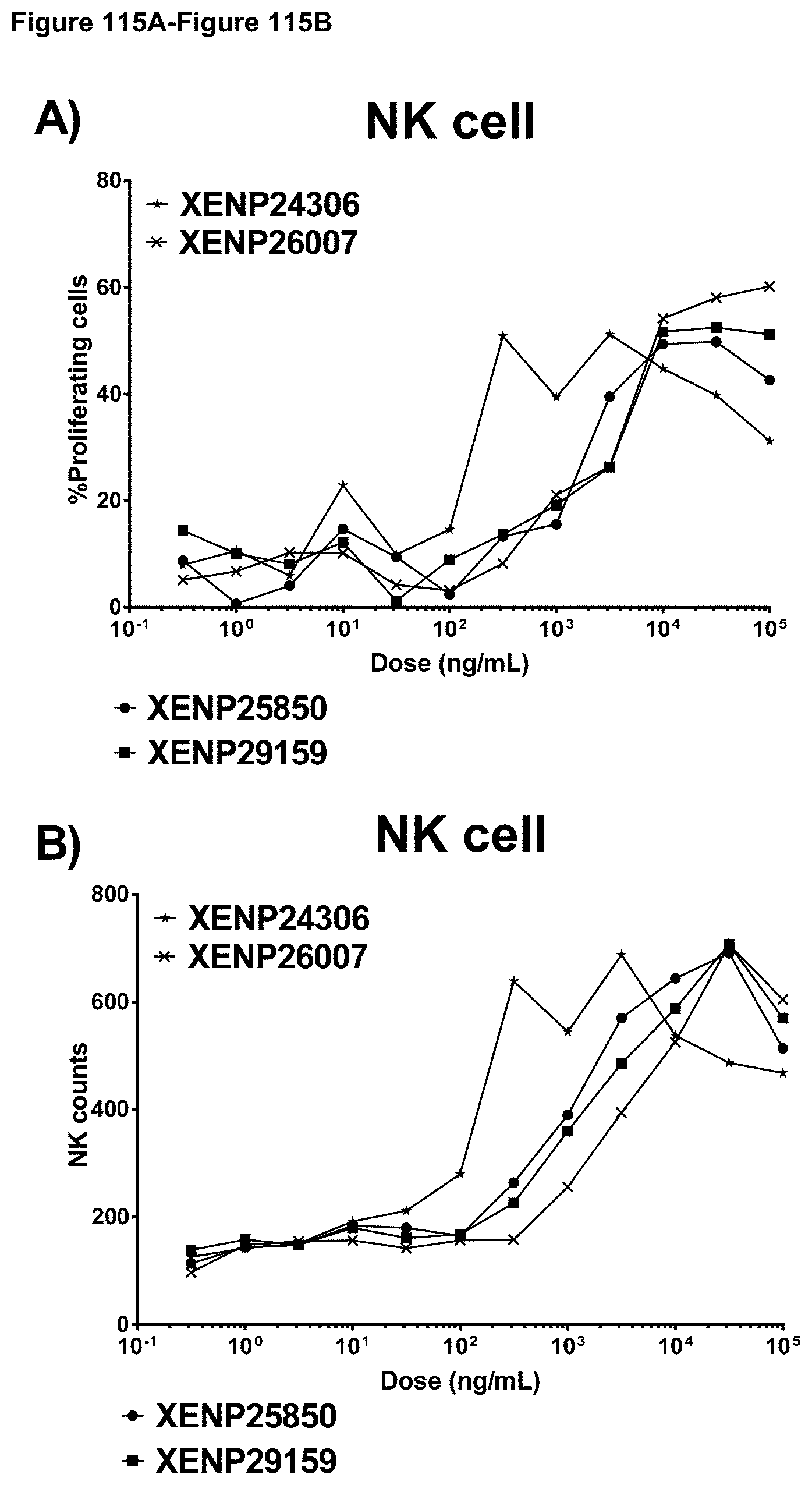
D00274
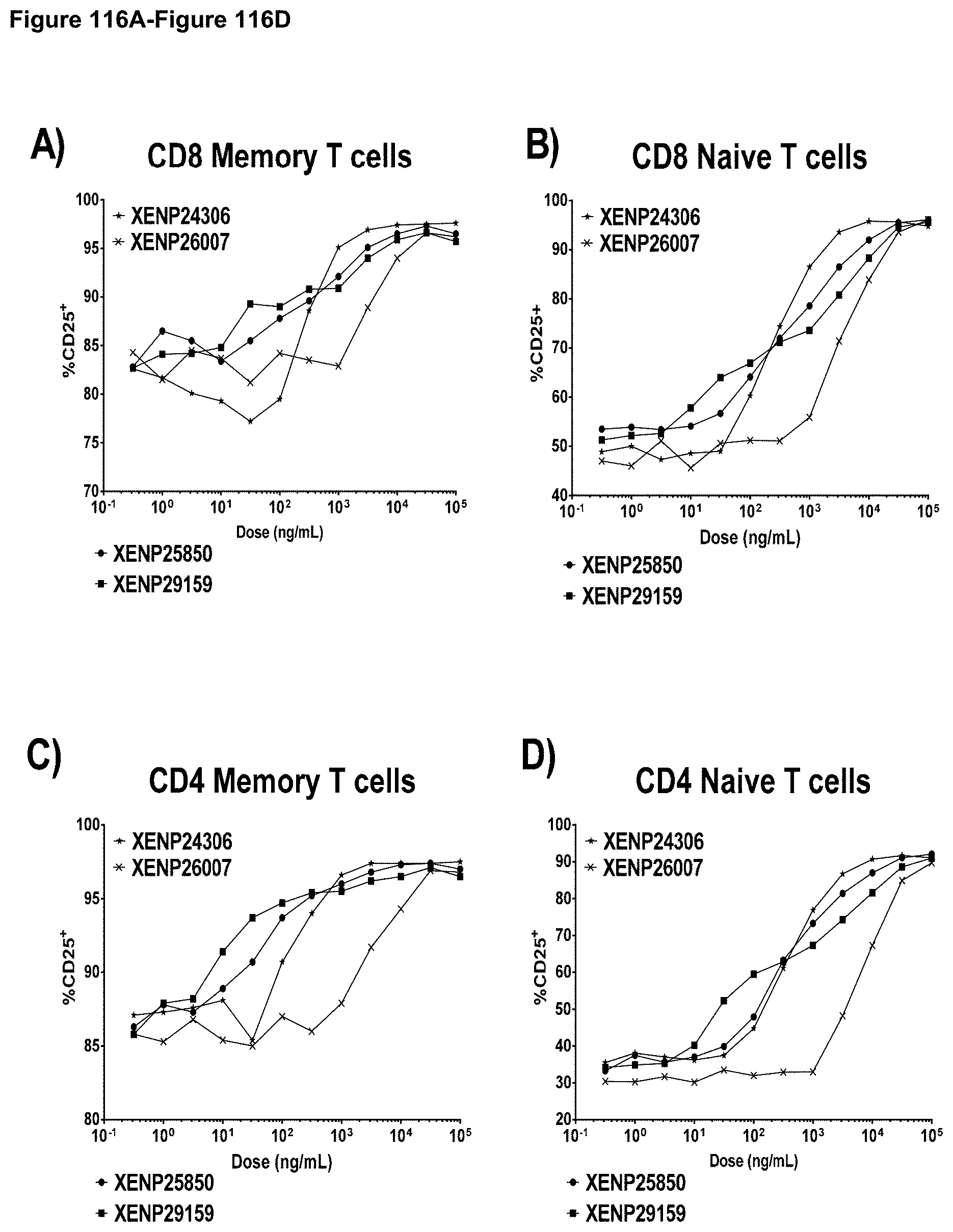
D00275
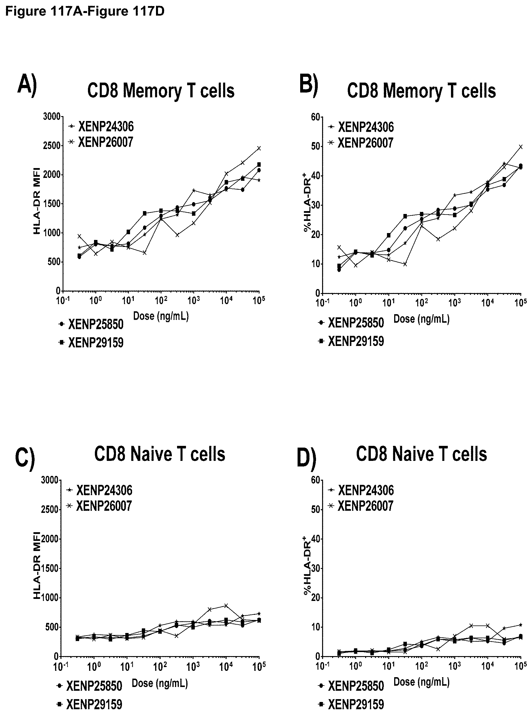
D00276

D00277
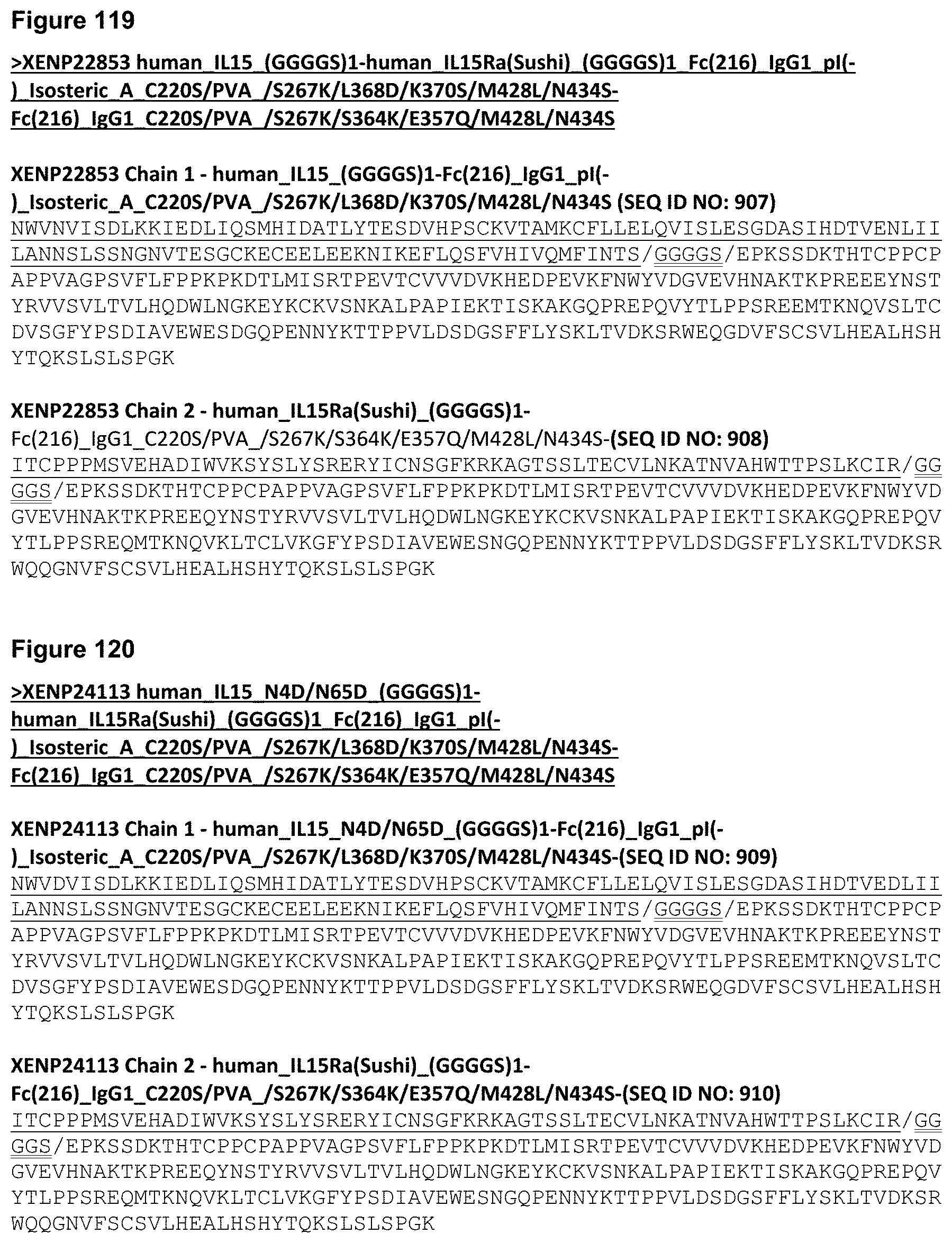
D00278

D00279

D00280

D00281
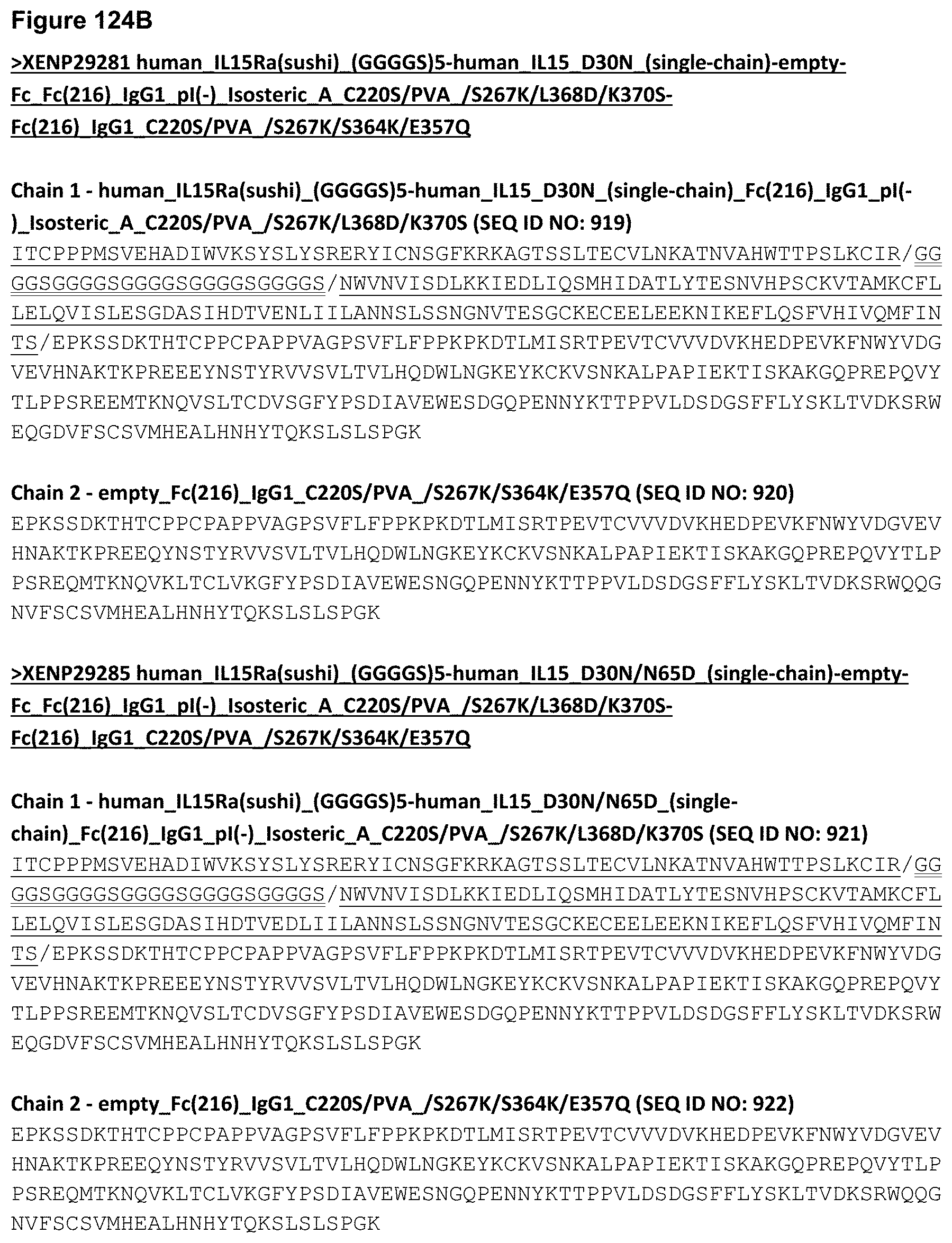
D00282

D00283

D00284
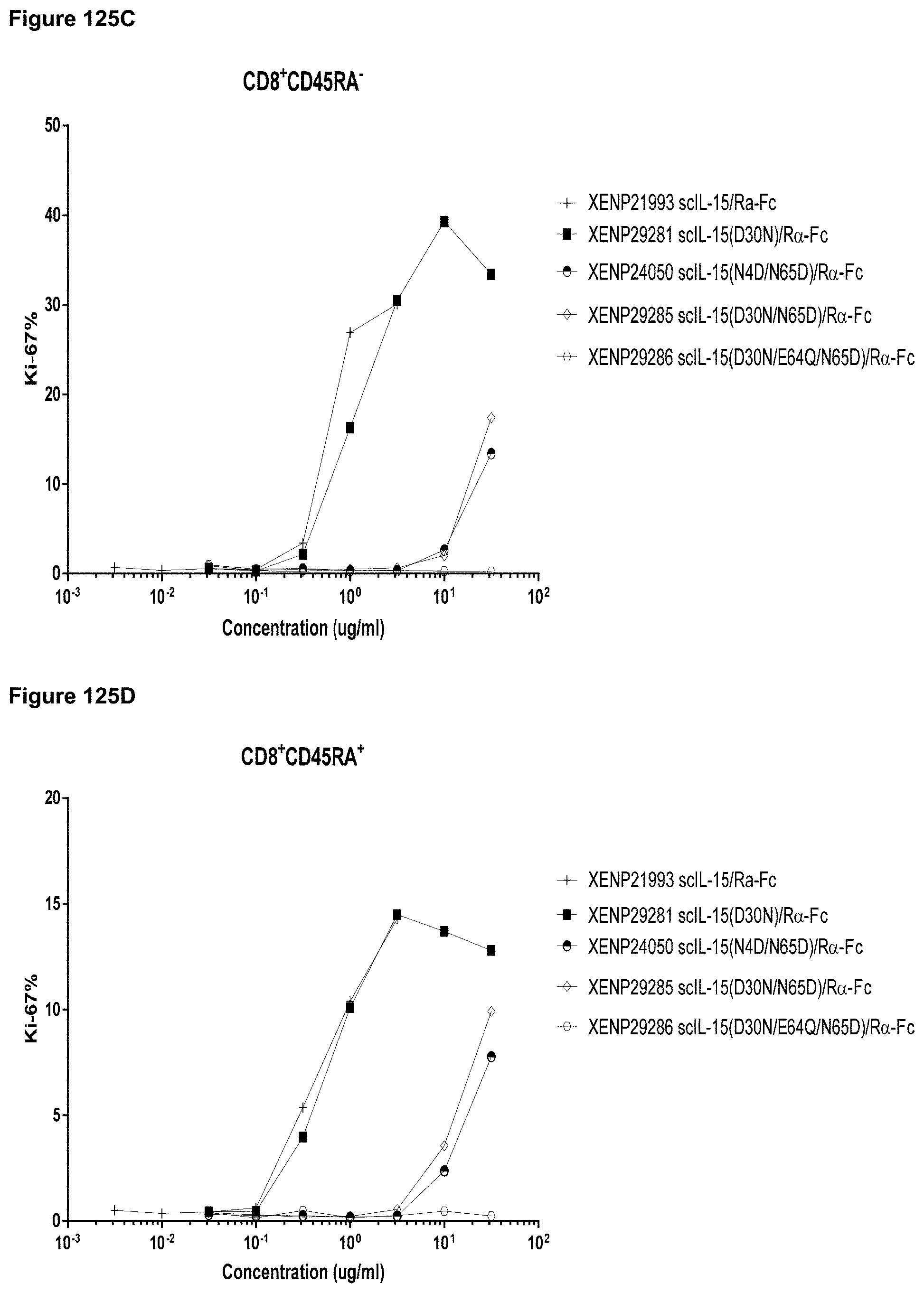
D00285

D00286

D00287

D00288
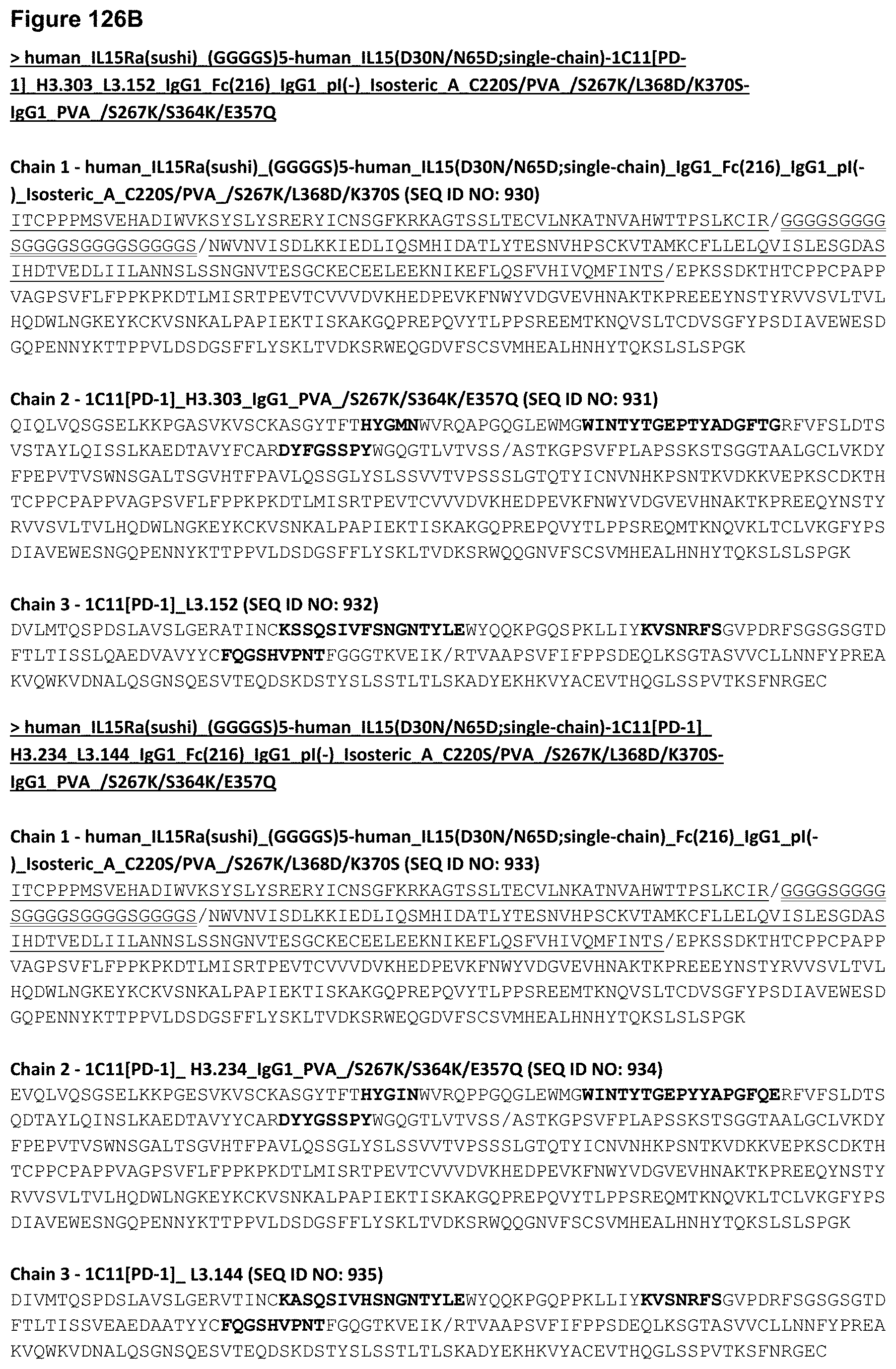
D00289

D00290

D00291

D00292
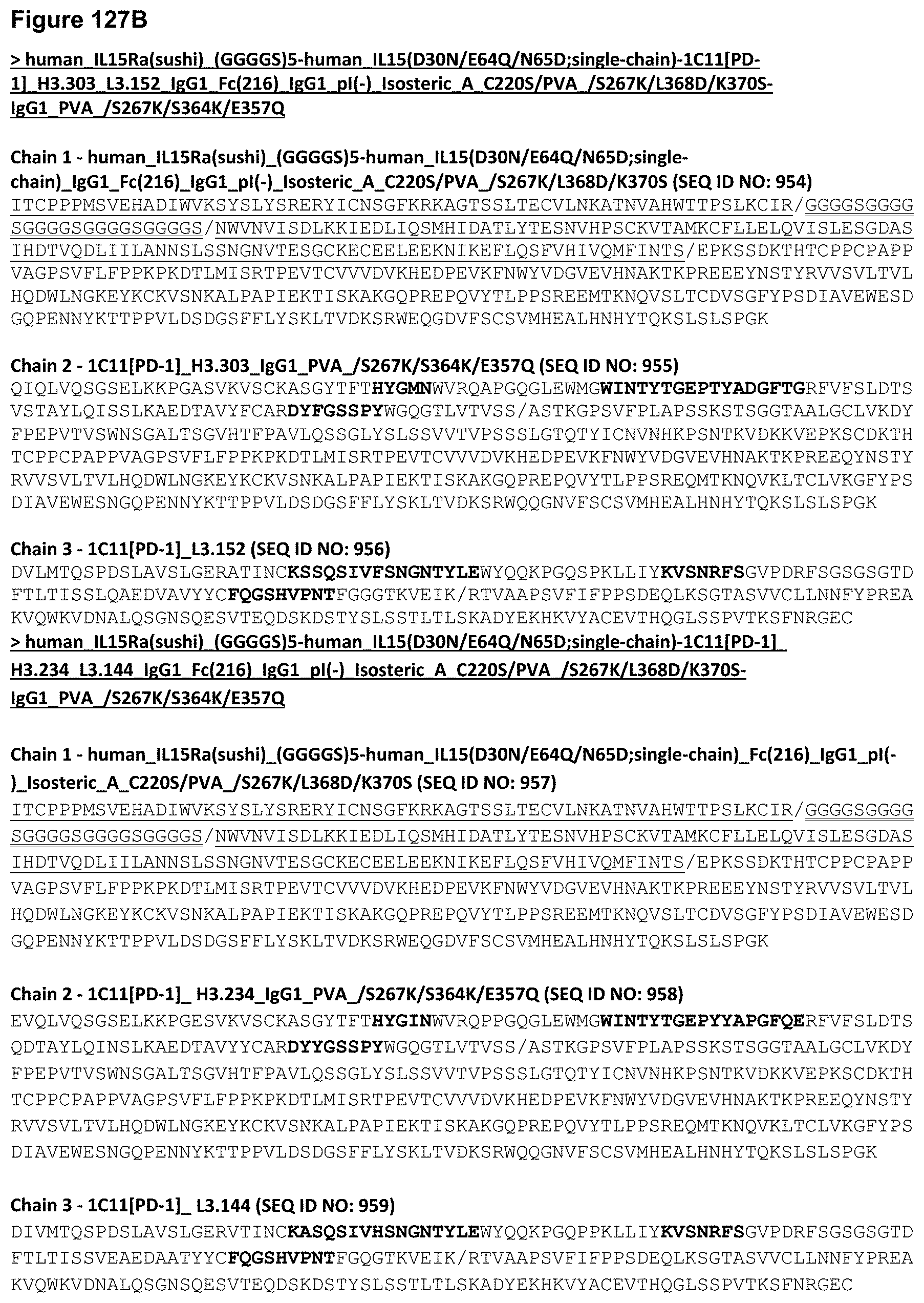
D00293
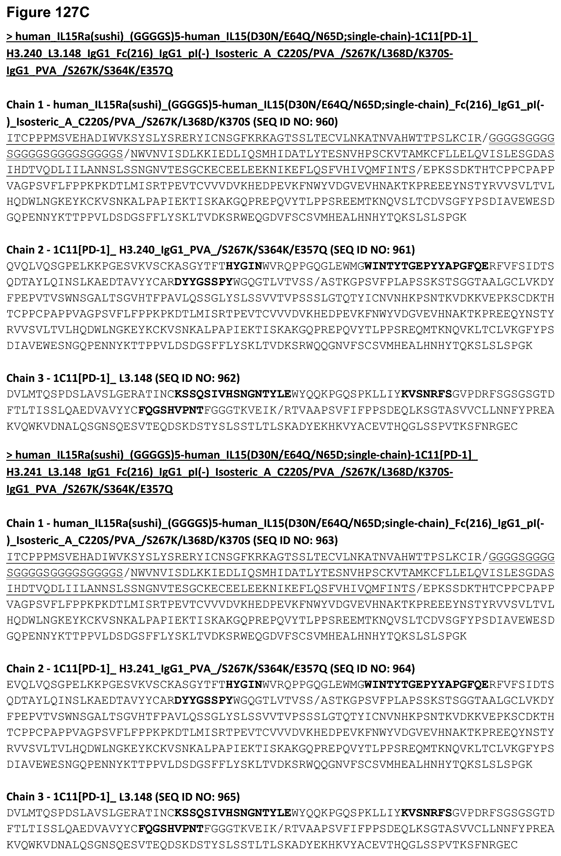
D00294

D00295

D00296
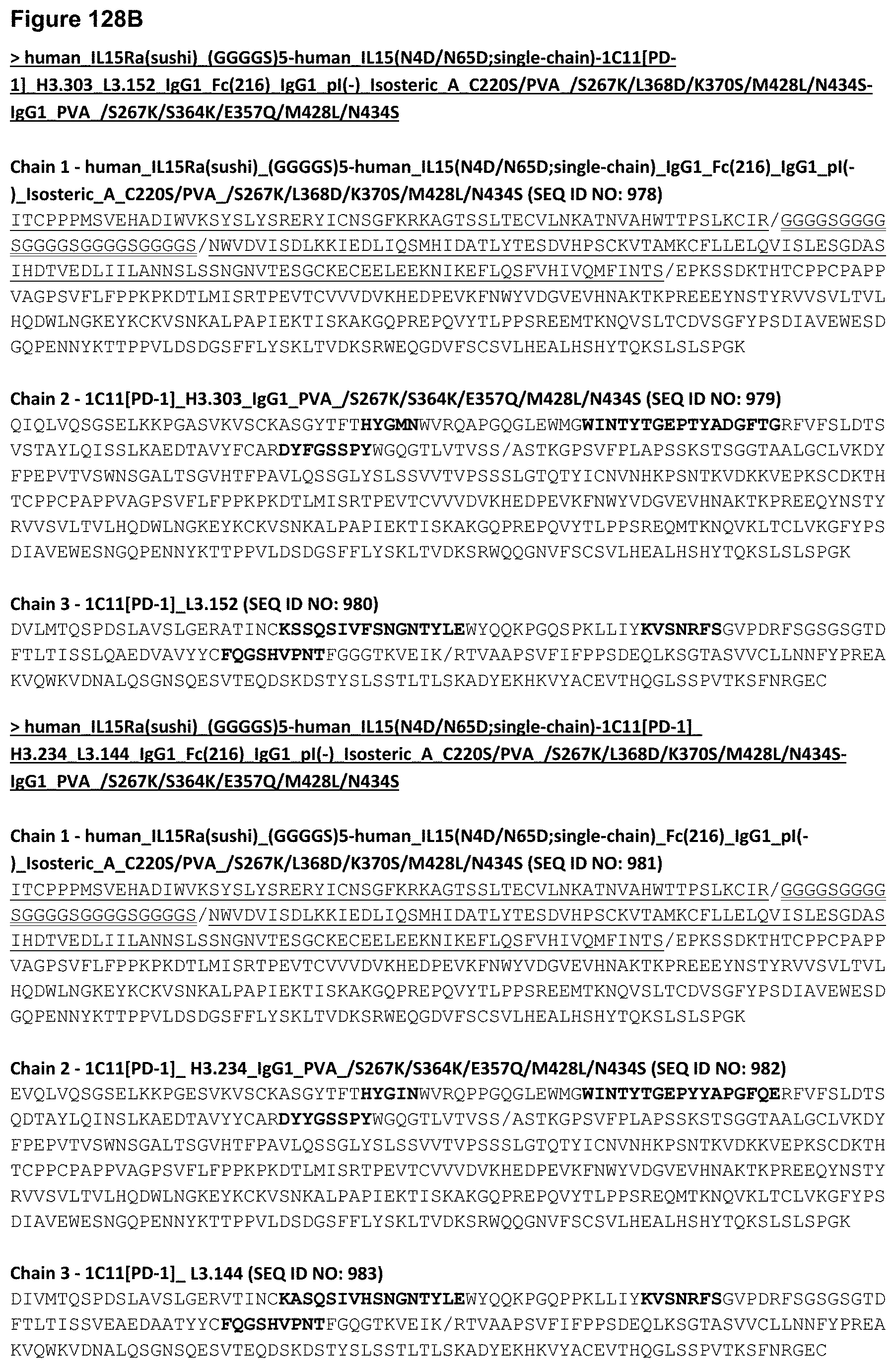
D00297

D00298

D00299
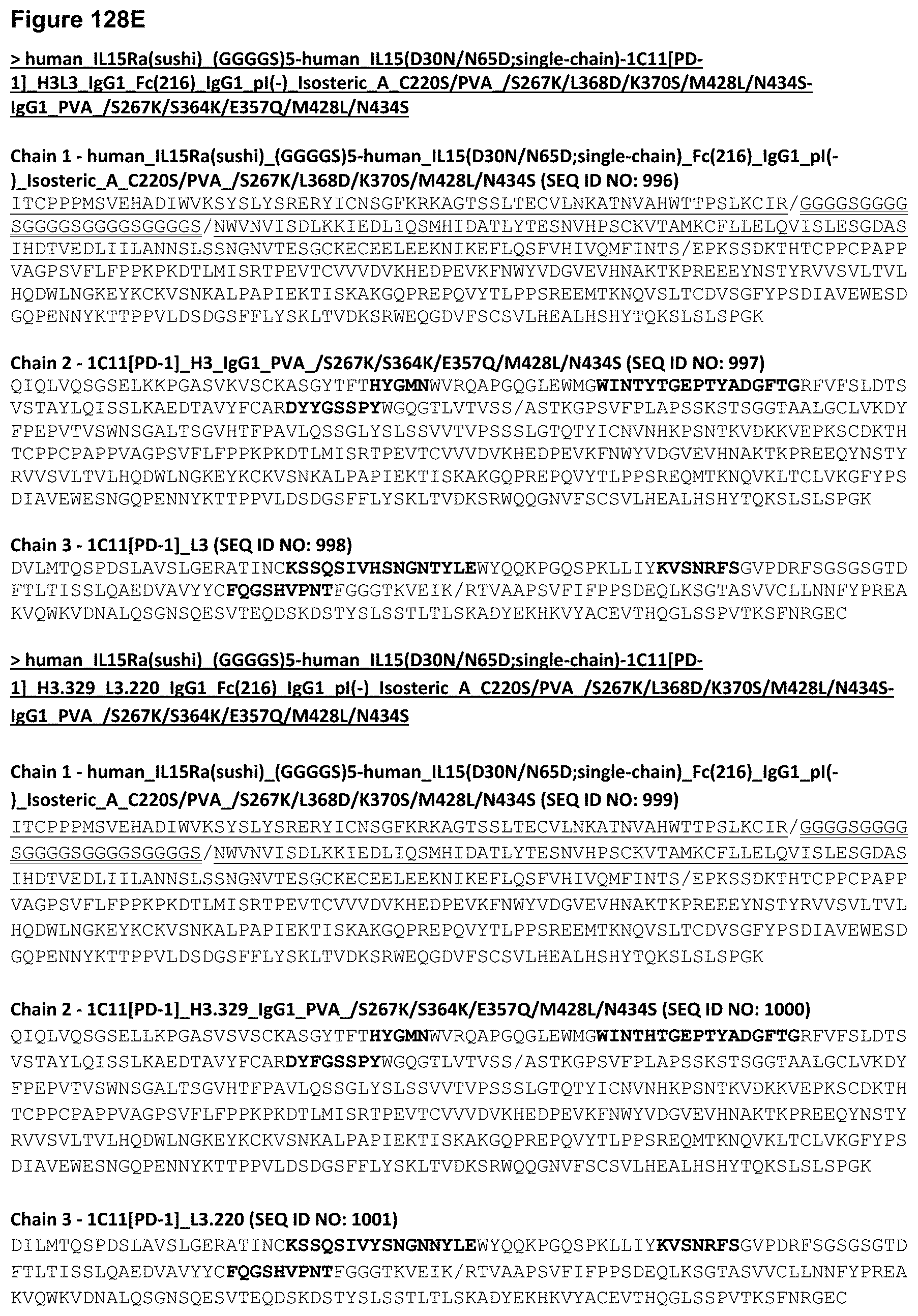
D00300
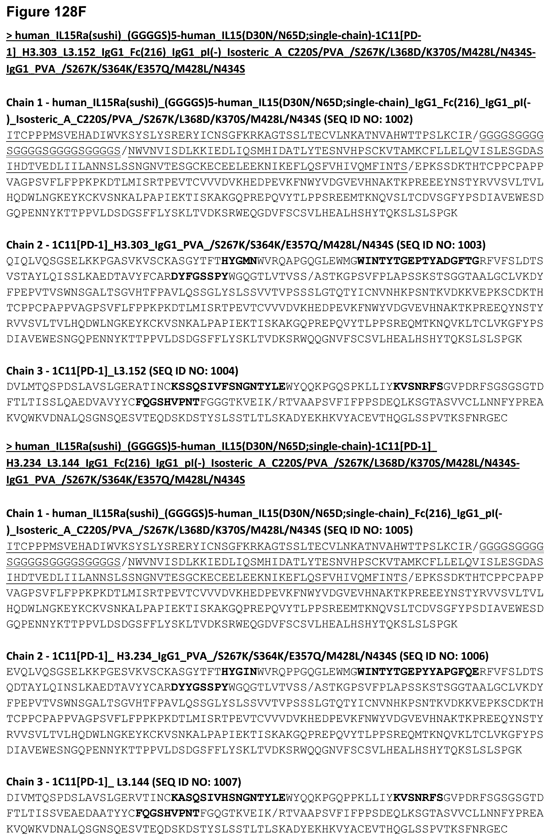
D00301
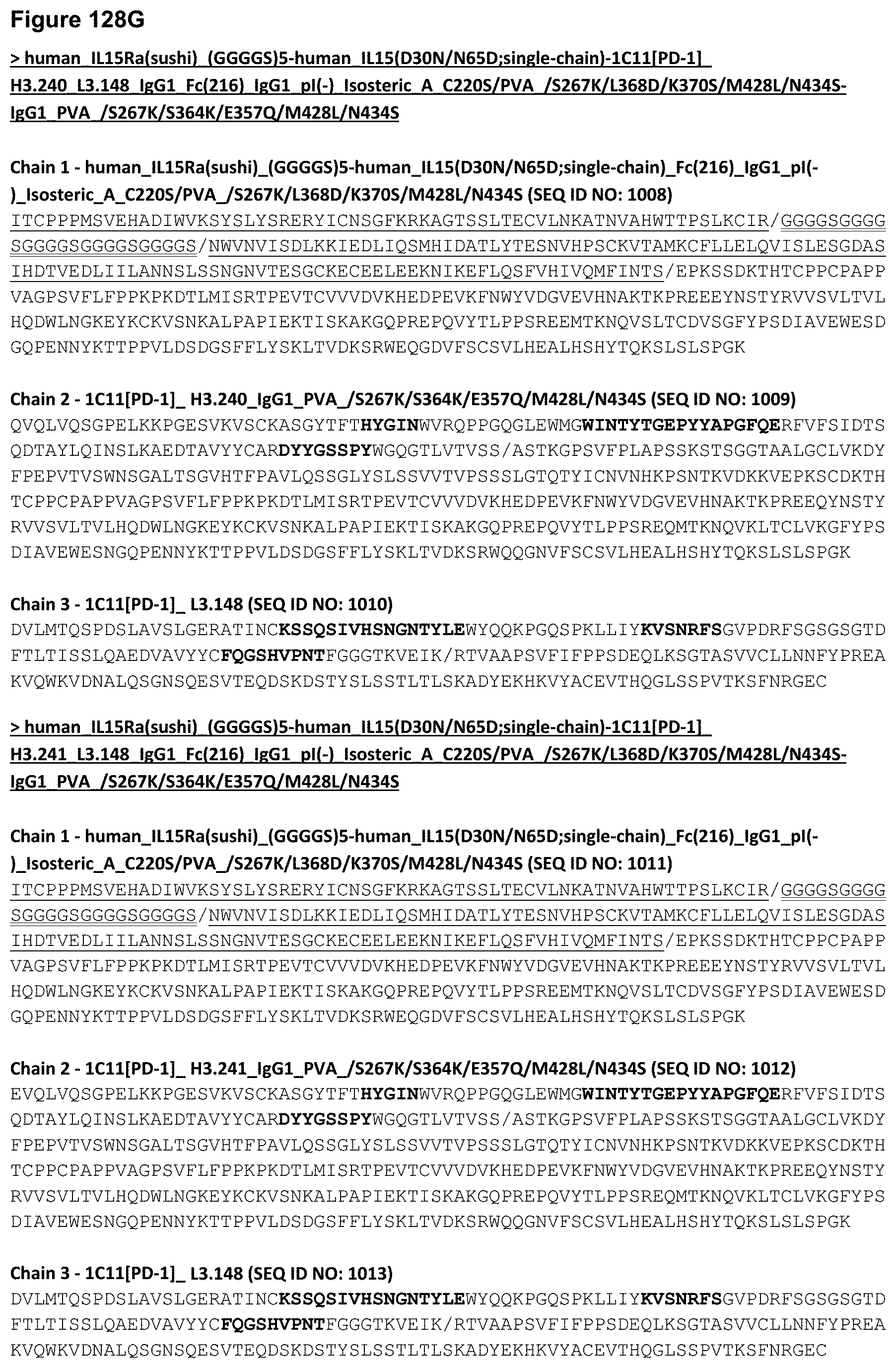
D00302

D00303

D00304
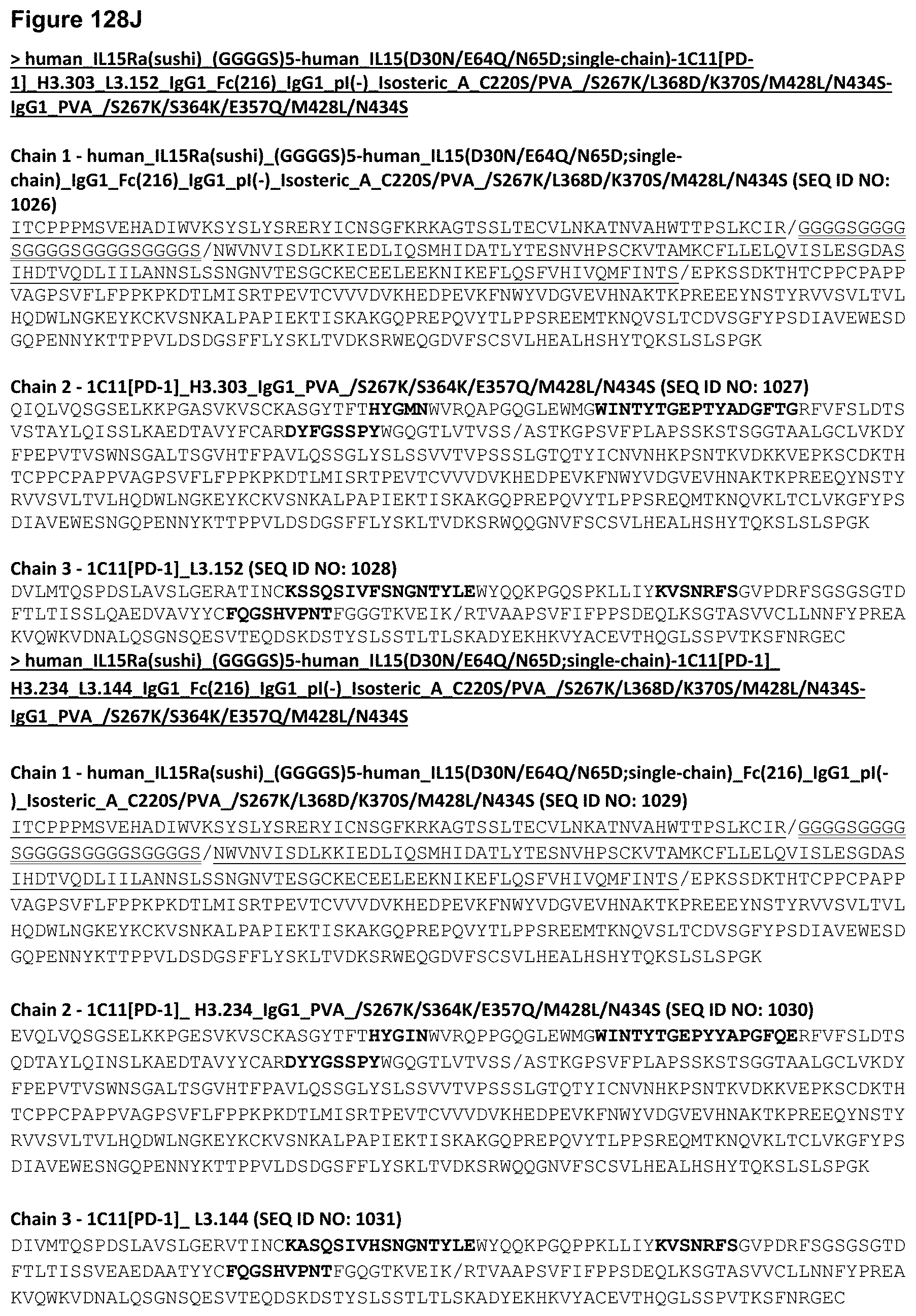
D00305

D00306

D00307

D00308

XML
uspto.report is an independent third-party trademark research tool that is not affiliated, endorsed, or sponsored by the United States Patent and Trademark Office (USPTO) or any other governmental organization. The information provided by uspto.report is based on publicly available data at the time of writing and is intended for informational purposes only.
While we strive to provide accurate and up-to-date information, we do not guarantee the accuracy, completeness, reliability, or suitability of the information displayed on this site. The use of this site is at your own risk. Any reliance you place on such information is therefore strictly at your own risk.
All official trademark data, including owner information, should be verified by visiting the official USPTO website at www.uspto.gov. This site is not intended to replace professional legal advice and should not be used as a substitute for consulting with a legal professional who is knowledgeable about trademark law.