Compositions and Methods for Making and Using Hydrolytic Enzymes for Degrading Polysaccharides made by Foodborne Pathogenic Bact
Gomelsky; Mark ; et al.
U.S. patent application number 16/542074 was filed with the patent office on 2019-12-26 for compositions and methods for making and using hydrolytic enzymes for degrading polysaccharides made by foodborne pathogenic bact. This patent application is currently assigned to University of Wyoming. The applicant listed for this patent is University of Wyoming. Invention is credited to Mark Gomelsky, Volkan Koseoglu, Kurt Miller.
| Application Number | 20190387746 16/542074 |
| Document ID | / |
| Family ID | 55858344 |
| Filed Date | 2019-12-26 |


View All Diagrams
| United States Patent Application | 20190387746 |
| Kind Code | A1 |
| Gomelsky; Mark ; et al. | December 26, 2019 |
Compositions and Methods for Making and Using Hydrolytic Enzymes for Degrading Polysaccharides made by Foodborne Pathogenic Bacteria
Abstract
Compositions and uses involving L. monocytogenes PssZ, as well as homologs, variants, and fragments thereof, are described.
| Inventors: | Gomelsky; Mark; (Laramie, WY) ; Miller; Kurt; (Laramie, WY) ; Koseoglu; Volkan; (Charlottesville, VA) | ||||||||||
| Applicant: |
|
||||||||||
|---|---|---|---|---|---|---|---|---|---|---|---|
| Assignee: | University of Wyoming Laramie WY |
||||||||||
| Family ID: | 55858344 | ||||||||||
| Appl. No.: | 16/542074 | ||||||||||
| Filed: | August 15, 2019 |
Related U.S. Patent Documents
| Application Number | Filing Date | Patent Number | ||
|---|---|---|---|---|
| 15522949 | Apr 28, 2017 | 10383338 | ||
| PCT/US2015/058038 | Oct 29, 2015 | |||
| 16542074 | ||||
| 62072358 | Oct 29, 2014 | |||
| Current U.S. Class: | 1/1 |
| Current CPC Class: | B09C 1/10 20130101; C12P 19/02 20130101; C12Y 302/01 20130101; A23V 2002/00 20130101; A01N 63/00 20130101; A23L 33/10 20160801; C11D 3/38636 20130101 |
| International Class: | A01N 63/00 20060101 A01N063/00; A23L 33/10 20060101 A23L033/10; C11D 3/386 20060101 C11D003/386; C12P 19/02 20060101 C12P019/02 |
Goverment Interests
STATEMENT REGARDING FEDERALLY SPONSORED RESEARCH
[0002] This invention was made with government support under Hatch Grant project number WYO-491-13 awarded by the United States Department of Agriculture, National Institute of Food and Agriculture; and grant number MCB1052575 awarded by the National Science Foundation. The government has certain rights in this invention.
Claims
1. A cleaning solution comprising: an enzyme, wherein the enzyme is a produced PssZ protein modified to lack a transmembrane domain, and wherein the enzyme has functional activity to hydrolyze a listerial Pss exopolysaccharide.
2. The cleaning solution of claim 1: wherein the produced PssZ protein has functional activity to hydrolyze a trisaccharide repeating unit of {4)-.beta.-ManpNAc-(1-4)-[.alpha.-Galp-(1-6)]-.beta.-ManpNAc-(1-}, wherein ManpNAc is N-acetylmannosamine and Galp is galactose.
3. The cleaning solution of claim 1, wherein the produced PssZ protein is substantially homologous to a Listeria monocytogenes PssZ protein.
4. The cleaning solution of claim 1, wherein the produced PssZ protein has at least 33% amino acid sequence identity to a Listeria monocytogenes PssZ protein.
5. The cleaning solution of claim 1, wherein the produced PssZ protein has at least 60% amino acid sequence identity to a sequence selected from the group consisting of: SEQ ID NO: 2, SEQ ID NO: 47, SEQ ID NO: 50, SEQ ID NO: 53, SEQ ID NO: 56, and SEQ ID NO: 59.
6. The cleaning solution of claim 1, wherein the cleaning solution further comprises a disinfectant or anti-bacterial agent selected from the group consisting of: sodium hypochlorite, hydrogen peroxide, benzalkonium chloride, alcohol, iodophor, quaternary ammonia compounds, chlorine solutions, peracetic acid, peroctanoic acid, nitric acid, benzoic acid, sodium hydroxide, dimethyl benzyl lauryl ammonium bromide, cationic surfactants, anionic surfactants, non-ionic surfactants, zwitterionic surfactants, nisin, lauricidin, lactoperoxidase, ampicillin, vancomycin, ciprofloxacin, azithromycin, or a proteolytic enzyme.
7. The cleaning solution of claim 1, wherein the cleaning solution has a pH in a range from about 4 to about 10.
8. The cleaning solution of claim 1, wherein a concentration of the produced PssZ protein in the cleaning solution ranges from about 0.1 nM to about 500 mM.
9. A composition for inhibiting aggregation of L. monocytogenes, comprising: an enzyme, wherein the enzyme is a produced PssZ protein modified to lack a transmembrane domain, and wherein the enzyme has functional activity to hydrolyze a listerial Pss exopolysaccharide comprising a trisaccharide repeating unit of {4)-.beta.-ManpNAc-(1-4)-[.alpha.-Galp-(1-6)]-.beta.-ManpNAc-(1-}, wherein ManpNAc is N-acetylmannosamine and Galp is galactose.
10. The composition of claim 9, wherein the composition forms a stabilized enzyme solution or compound.
11. A method for detecting a listerial biofilm on a surface comprising: providing a modified PssZ enzyme, wherein the modified PssZ enzyme: has functional activity to bind to a listerial Pss exopolysaccharide, and contacting the surface with the modified PssZ enzyme, whereby the modified PssZ enzyme binds to the listerial exopolysaccharide comprising the listerial biofilm.
12. The method of claim 11, wherein the modified PssZ enzyme has impaired hydrolytic activity for degrading the listerial Pss exopolysaccharide.
13. The method of claim 11, wherein the modified PssZ enzyme is derived from a PssZ E72Q mutant.
14. The method of claim 11, wherein the listerial Pss exopolysaccharide comprises a trisaccharide repeating unit of {4)-.beta.-ManpNAc-(1-4)-[.alpha.-Galp-(1-6)]-.beta.-ManpNAc-(1-}, wherein ManpNAc is N-acetylmannosamine and Galp is galactose.
15. The method of claim 11, wherein the modified PssZ enzyme has at least 60% amino acid sequence identity to a sequence selected from the group consisting of: SEQ ID NO: 2, SEQ ID NO: 47, SEQ ID NO: 50, SEQ ID NO: 53, SEQ ID NO: 56, and SEQ ID NO: 59.
16. The method of claim 11, further comprising: hydrolyzing the listerial Pss exopolysaccharide, thereby releasing soluble carbohydrates; and detecting soluble carbohydrates in a solution in contact with the surface.
17. The method of claim 11, further comprising: hydrolyzing the listerial Pss exopolysaccharide, thereby dispersing the listerial biofilm; and measuring turbidity in a solution in contact with the surface.
Description
RELATED APPLICATIONS
[0001] This application is a divisional of U.S. patent application Ser. No. 15/522,949, a National Stage Application of PCT/US2015/058038, filed Oct. 29, 2015, and claims the benefit of U.S. Provisional Application No. 62/072,358, filed under 35 U.S.C. .sctn. 111(b) on Oct. 29, 2014, the disclosure of each of which are incorporated herein by reference in the entirety.
SEQUENCE LISTING
[0003] The instant application contains a Sequence Listing which has been submitted electronically in ASCII format and is hereby incorporated by reference in its entirety. Said ASCII copy, created on Oct. 29, 2015, is named 53-56551_15-027_SL.txt and is 57,439 bytes in size.
BACKGROUND OF THE INVENTION
[0004] Bacteria can form exopolysaccharide-rich aggregates, or biofilms, that pose formidable challenges in medicine and industry because of their resistance to antibiotics, disinfectants, desiccation, and other treatments. One of the common foodborne pathogens, Listeria monocytogenes, can form biofilms on food products, food-processing equipment, and in food storage facilities. When consumed with food, L. monocytogenes may cause listeriosis, a severe disease that has the highest fatality rate of the foodborne diseases in the developed countries. Listeriosis is particularly dangerous for immunocompromised individuals, pregnant women, the elderly, and infants. Despite a relatively low number of cases, it is the third most costly foodborne disease in the USA, with the total annual financial loss estimated at $8.8 billion. Thus, there is a need for new and improved means for degrading biofilms and combating L. monocytogenes.
SUMMARY OF THE INVENTION
[0005] Described are uses of a Listeria monocytogenes PssZ protein, homolog, variant, or fragment, in preventing bacterial exopolysaccharide-dependent aggregation, in degrading a biofilm, and in inhibiting biofilm formation on a surface.
[0006] Provided is a L. monocytogenes PssZ protein having the sequence:
TABLE-US-00001 [SEQ ID NO: 1] MKRFILILILLIFIGAGFFIFLRPESKKTVSAPKETTPTSTSVQTYVKEN YTAKNGLIMDYKNTEEPHYLAESIGLYMEYLVEVNDSKTFQKQVNHLEKY FIAEDNFIKWEATDSTTTNAIVDDFRITEALYQASEKFSFPSYKKMADKF LTNTKKYSAEQGVPVDFYDFVHKKKADTLHLSYLNIQAMQQINYRDKAYL PIQTINADPFFTEVFQNGQFKFADQKEVNMIDQMLIAIAYYDENGDIEPN FDNFLQTELASKGKIYARYQRETKKPSSENESTAVYAFLTQYFNKTNQAK NGKITKELLEKMDTSNPETTHFFDYINKEITLKK.
[0007] Also provided are PssZ variants comprising 90%, 80%, 70%, or 60% sequence identity to the purified L. monocytogenes PssZ protein. Also provided is a PssZ E72Q mutant comprising an L. monocytogenes PssZ protein having a Glu72 site substituted with glutamine.
[0008] Also provided is a purified PssZ fragment having the sequence:
TABLE-US-00002 [SEQ ID NO: 2] RPESKKTVSAPKETTPTSTSVQTYVKENYTAKNGLIMDYKNTEEPHYLAE SIGLYMEYLVEVNDSKTFQKQVNHLEKYFIAEDNFIKWEATDSTTTNAIV DDFRITEALYQASEKFSFPSYKKMADKFLTNTKKYSAEQGVPVDFYDFVH KKKADTLHLSYLNIQAMQQINYRDKAYLPIQTINADPFFTEVFQNGQFKF ADQKEVNMIDQMLIAIAYYDENGDIEPNFDNFLQTELASKGKIYARYQRE TKKPSSENESTAVYAFLTQYFNKTNQAKNGKITKELLEKMDTSNPETTHF FDYINKEITLKK.
[0009] Also provided are homologs of the L. monocytogenes PssZ protein. In certain embodiments, the PssZ homolog is selected from the group consisting of: Exiguobacterium undae PssZ [SEQ ID NO: 47], Carnobacterium mobile PssZ [SEQ ID NO: 50], Carnobacterium jeotgali PssZ [SEQ ID NO: 53], Jeotgalibacillus campisalis PssZ [SEQ ID NO: 56], and Bacillus thermotolerans PssZ [SEQ ID NO: 59].
[0010] Also provided is the use of a purified L. monocytogenes PssZ protein, a PssZ protein homolog, or a variant or fragment thereof, in hydrolyzing a listerial exopolysaccharide, or in preventing bacterial exopolysaccharide-dependent aggregation.
[0011] Further provided is a method of hydrolyzing a listerial exopolysaccharide, where the method involves exposing listeria bacteria to a sufficient amount of a purified L. monocytogenes PssZ protein, a PssZ protein homolog, or a variant or fragment thereof, and hydrolyzing a listerial exopolysaccharide. In certain embodiments, the listerial exopolysaccharide comprises ManNAc-Gal Pss exopolysaccharide. In certain embodiments, the listeria bacteria is present in a food article.
[0012] Further provided is a method of preventing bacterial exopolysaccharide-dependent aggregation on a surface, where the method involves applying a sufficient amount of a purified L. monocytogenes PssZ protein, a PssZ protein homolog, or variant or fragment thereof, to a surface, and preventing bacterial exopolysaccharide-dependent aggregation on the surface. In certain embodiments, aggregation of L. monocytogenes is prevented. In certain embodiments, the surface comprises a food container surface. In certain embodiments, the surface comprises surface of fruit, vegetables, or other plant materials.
[0013] Further provided is a method of disintegrating bacterial exopolysaccharide-rich aggregates, where the method involves applying a sufficient amount of a purified L. monocytogenes PssZ protein, a PssZ protein homolog, or a variant or fragment thereof, to a bacterial aggregate, and disintegrating the bacterial aggregate.
[0014] Further provided is a method of inhibiting a listerial contamination in a food article, where the method involves applying a sufficient amount of a purified L. monocytogenes PssZ protein, a PssZ protein homolog, or a variant or fragment thereof, to a food article, and inhibiting a listerial growth in the food article.
[0015] Further provided is a method of ameliorating a listerial contamination in a food article, where the method involves applying a sufficient amount of a purified L. monocytogenes PssZ protein, a PssZ protein homolog, or a variant or fragment thereof, to a food article contaminated with L. monocytogenes, and applying a sufficient amount of an antibacterial agent to the food article, to ameliorate the listerial contamination in the food article. In certain embodiments, the antibacterial agent is bleach (sodium hypochlorite), hydrogen peroxide, or another disinfectant.
[0016] Further provided is a disinfecting solution that includes a PssZ protein, a PssZ protein homolog, or a variant or fragment thereof, and an antibacterial agent. In certain embodiments, the antibacterial agent is a detergent. In certain embodiments, the antibacterial agent is bleach. In certain embodiments, the antibacterial agent is selected from the group consisting of: hydrogen peroxide, alcohol, iodophor, quaternary ammonia compounds, chlorine solutions, peracetic acid, peroctanoic acid, nitric acid, benzoic acid, sodium hydroxide, dimethyl benzyl lauryl ammonium bromide, cationic surfactants, anionic surfactants, non-ionic surfactants, zwitterionic surfactants, nisin, lauricidin, lactoperoxidase, ampicillin, vancomycin, ciprofloxacin, azithromycin, or a proteolytic enzyme. In certain embodiments, the disinfecting solution has a pH ranging from about 4 to about 10.
[0017] Further provided is a polysaccharide composition that includes a ManNAc-Gal exopolysaccharide (EPS) having a trisaccharide repeating unit of {4)-.beta.-ManpNAc-(1-4)-[.alpha.-Galp-(1-6)]-.beta.-ManpNAc-(1-}, wherein ManpNAc is N-acetylmannosamine and Galp is galactose. In certain embodiments, the composition is a food additive or filler.
[0018] Further provided is a method of administering a probiotic, where the method involves protecting a therapeutically effective amount of a microorganism with an EPS coating to form a protected probiotic, and administering the protected probiotic to a patient in need thereof.
[0019] Further provided is a kit for treating a bacterial contamination, where the kit includes a first container housing a PssZ enzyme solution, and a second container housing a disinfectant solution. In certain embodiments, the PssZ enzyme solution comprises a PssZ protein, a PssZ protein homolog, or a variant or fragment thereof.
[0020] In certain embodiments, the PssZ protein homolog is selected from the group consisting of: Exiguobacterium undae PssZ [SEQ ID NO: 47], Carnobacterium mobile PssZ [SEQ ID NO: 50], Carnobacterium jeotgali PssZ [SEQ ID NO: 53], Jeotgalibacillus campisalis PssZ [SEQ ID NO: 56], and Bacillus thermotolerans PssZ [SEQ ID NO: 59].
[0021] In certain embodiments, the fragment is a PssZ fragment having the sequence:
TABLE-US-00003 [SEQ ID NO: 2] RPESKKTVSAPKETTPTSTSVQTYVKENYTAKNGLIMDYKNTEEPHYLAE SIGLYMEYLVEVNDSKTFQKQVNHLEKYFIAEDNFIKWEATDSTTTNAIV DDFRITEALYQASEKFSFPSYKKMADKFLTNTKKYSAEQGVPVDFYDFVH KKKADTLHLSYLNIQAMQQINYRDKAYLPIQTINADPFFTEVFQNGQFKF ADQKEVNMIDQMLIAIAYYDENGDIEPNFDNFLQTELASKGKIYARYQRE TKKPSSENESTAVYAFLTQYFNKTNQAKNGKITKELLEKMDTSNPETTHF FDYINKEITLKK.
[0022] In certain embodiments, the PssZ variants comprise 90%, 80%, 70%, or 60% sequence identity to the purified L. monocytogenes PssZ protein.
BRIEF DESCRIPTION OF THE DRAWINGS
[0023] The patent or application file may contain one or more drawings executed in color and/or one or more photographs. Copies of this patent or patent application publication with color drawing(s) and/or photograph(s) will be provided by the U.S. Patent and Trademark Office upon request and payment of the necessary fees.
[0024] FIG. 1: In silico analysis of genes and proteins involved in c-di-GMP signaling in L. monocytogenes. Depicted are genes believed to encode DGCs (DgcA-C), c-di-GMP PDEs (PdeB-D), a c-di-GMP receptor (PssE), and listerial EPS biosynthesis machinery. Protein domain architectures are taken from the Pfam database: 5TM, a conserved five-transmembrane module; unmarked red box, transmembrane domain; crossed GGDEF domain ("GGDEF" disclosed as SEQ ID NO: 3), enzymatically inactive GGDEF domain ("GGDEF" disclosed as SEQ ID NO: 3).
[0025] FIGS. 2A-2C: PDE activities of the L. monocytogenes proteins PdeBD. FIG. 2A shows restoration of motility in semi-solid (0.25%) agar of strain MG1655 DyhjH by L. monocytogenes PdeB, PdeC, and PdeD is indicative of their cdi-GMP PDE activities. PdeB-D were expressed as C-terminal His.sub.6-fusions ("His.sub.6" disclosed as SEQ ID NO: 4) downstream of the T7 promoter from vector pET23a. Although MG1655 does not encode a T7 RNA polymerase gene, the pde genes were expressed from a fortuitous promoter at sufficiently high levels to partially restore the swimming defect of MG1655 DyhjH in semi-solid agar. pET, empty vector (pET23a). FIG. 2B shows affinity purified L. monocytogenes PdeD (PdeD::His.sub.6) ("His.sub.6" disclosed as SEQ ID NO: 4), PdeB (PdeB::His.sub.6) ("His.sub.6" disclosed as SEQ ID NO: 4), and PdeC (MBP::PdeC) proteins used in the PDE assays. MW, molecular weight, kD. FIG. 2C shows PDE activities of PdeD::His6 ("His6" disclosed as SEQ ID NO: 4), PdeB::His6 ("His6" disclosed as SEQ ID NO: 4) and MBP::PdeC monitored by the rates of formation of pGpG, the product of c-di-GMP hydrolysis. Nucleotides were measured by HPLC as described in the examples.
[0026] FIGS. 3A-3B: DGC activities of the L. monocytogenes proteins DgcAC. FIG. 3A shows inhibition of motility in semi-solid (0.25%) agar of strain MG1655 by L. monocytogenes DgcA (plasmid pBAD-dgcA), DgcB (pBAD-dgcB), and DgcC (pBAD-dgcC) is indicative of their DGC activities. DgcA-C were expressed from the vector pBAD/Myc-His-C (pBAD). LB agar contained 0.1% arabinose. FIG. 3B shows Congo red staining of the fimbriae producing strain BL21(DE3) caused by L. monocytogenes DgcA, DgcB, and DgcC is indicative of their DGC activities. LB agar contained 0.001% arabinose.
[0027] FIGS. 4A-4E: Inhibition of motility and activation of EPS production in L. monocytogenes by elevated levels of c-di-GMP. FIG. 4A, top, shows inhibition of swimming of the wild-type L. monocytogenes in semi-solid agar by a heterologous DGC, Slr1143. FIG. 4A, bottom, shows restoration of swimming in semi-solid agar of the L. monocytogenes .DELTA.pdeB/C/D mutant by a heterologous PDE, YhjH. WT, wild type, EGD-e; A/B/C, .DELTA.pdeB/C/D mutant; pIMK, WT::pIMK2 (vector control); slr, WT::(pIMK2::slr1143); yhjH, WT::(pIMK2::yhjH). FIG. 4B shows Congo red staining of EPS in L. monocytogenes. 1, WT, wild type; 2, .DELTA.pdeB/C/D; 3, .DELTA.pdeB/C/D .DELTA.pssE; 4, .DELTA.pdeB/C/D .DELTA.pssC; 5, .DELTA.pdeB/C/D::pIMK2; 6, .DELTA.pdeB/C/D::pIMK2::yhjH; 7, .DELTA.pdeB/C/D::(pIMK2::slr1143); 8, WT::pIMK2; 9, WT::(pIMK2::yhjH); 10, WT::(pIMK2::slr1143). FIG. 4C shows deletion of all three c-di-GMP PDEs drastically inhibits motility of L. monocytogenes in semi-solid agar. WT, wild type strain, B, .DELTA.pdeB; C, .DELTA.pdeC; D, .DELTA.pdeD; B/D, .DELTA.pdeB .DELTA.pdeD; C/D, .DELTA.pdeC .DELTA.pdeD; B/C, .DELTA.pdeB .DELTA.pdeC; B/C/D, .DELTA.pdeB/C/D. FIG. 4D shows rough colony morphology and increased Congo red staining of the L. monocytogenes .DELTA.pdeB/C/D mutant and rescue of the wild-type colony morphology by the .DELTA.pssC mutation (.DELTA.pdeB/C/D .DELTA.pssC). FIG. 4E shows restoration of motility of the .DELTA.pdeB/C/D mutant by the .DELTA.pssC or .DELTA.pssE mutations.
[0028] FIGS. 5A-5B: In vitro assay of c-di-GMP binding by the PssE receptor. FIG. 5A shows the MBP-PssE protein purified via affinity (amylose resin) chromatography. The GGDEF domain of PssE (residues 107-285) ("GGDEF" disclosed as SEQ ID NO: 3) containing the putative I-site was fused downstream of MBP, MBP::GGDEFpssE ("GGDEF" disclosed as SEQ ID NO: 3), and used in c-di-GMP binding assays. FIG. 5B shows a saturation plot of equilibrium binding of c-di-GMP to the PssE receptor (MBP::GGDEFpssE) ("GGDEF" disclosed as SEQ ID NO: 3). Shown is the dependence of the ratio of bound cdi-GMP per protein in the dialysis chamber, where protein alone was loaded, versus concentration of free c-di-GMP at equilibrium.
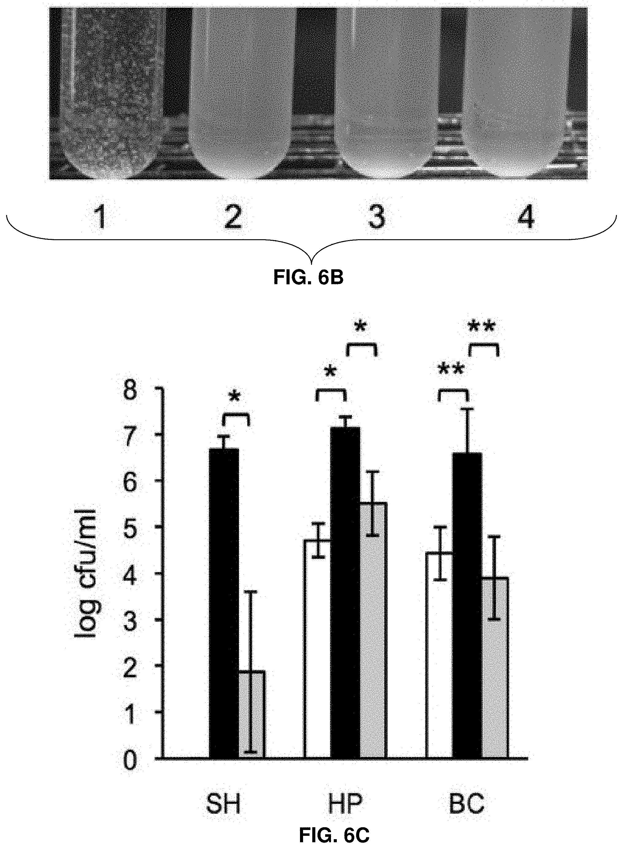
[0029] FIGS. 6A-6D: Role of the c-di-GMP-induced EPS in biofilm formation, cell aggregation, and tolerance of L. monocytogenes to disinfectants and desiccation. FIG. 6A shows biofilm formation of L. monocytogenes in 96-well polystyrene plates (measured using a Crystal violet dyebinding assay). Cultures were grown for 6 days at 30.degree. C. in LB (top panel) or LB supplemented with 3% glycerol (bottom panel). Shown are average results from two biological replicates, where each strain was grown in six wells in a replicate (i.e., six technical replicates). Black circle, wild type; red square, .DELTA.pdeB/C/D; green triangle, .DELTA.pdeB/C/D .DELTA.pssC; blue cross, .DELTA.pdeB/C/D .DELTA.pssE. FIG. 6B shows EPS-dependent L. monocytogenes cell aggregation (clumping) in HTM medium. Overnight cultures grown in BHI were inoculated into HTM liquid medium at A600 of 0.01 and incubated at 30.degree. C. with gentle shaking (rotary shaker, 125 rpm) for 48 h. 1, .DELTA.pdeB/C/D; 2, wild type; 3, .DELTA.pdeB/C/D DpssC; 4, .DELTA.pdeB/C/D .DELTA.pssE. FIG. 6C shows the protective role of the c-di-GMP-inducible EPS in disinfection. Aliquots of the HTM-grown cultures were mixed with disinfectant solutions for 10 min at room temperature. Disinfection was stopped by adding a D/E neutralizing broth (Difco); the cultures were vortexed vigorously (5 min) with glass beads to break clumps and plated on BHI agar. Colonies were enumerated after a 48-h growth at 37.degree. C. SH, sodium hydrochloride (1600 ppm); HP, hydrogen peroxide (200 mM); BC, benzalkonium chloride (100 ppm). White background, EGD-e; black, .DELTA.pdeB/C/D; grey, .DELTA.pdeB/C/D .DELTA.pssC. SH, sodium hypochlorite; HP, hydrogen peroxide; BC, benzalkonium chloride. The absence of the bar for the EGD-e strain treated with SH indicates the lack of survivors. FIG. 6D shows the protective role of the c-di-GMP inducible EPS in desiccation. Aliquots of overnight cultures grown in HTM at 37.degree. C. were spun down, the supernatants were removed, and cell pellets were stored in desiccators at room temperature for the indicated periods. The pellets were rehydrated, vortexed with glass beads for better suspension and plated on BHI agar. The numbers of surviving colonies after incubation at 37.degree. C. for 24 h are plotted. In FIGS. 6C-6D, the bars denote mean values for data from three biological replicates. *, significantly different (p,0.002), **, significantly different (p,0.02), according to Tukey test (Minitab 16 statistical software).
[0030] FIGS. 7A-7B: Impaired invasion of L. monocytogenes in HT-29 human colon adenocarcinoma cells by elevated c-di-GMP levels. FIG. 7A shows expression of the heterologous DGC, Slr1143 (WT::slr; blue bar), or deletion of the native PDEs (.DELTA.pdeB/C/D; black), strongly inhibit listerial invasion, compared to EGD-e containing an empty vector (WT::pIMK; white), while overexpression of the heterologous PDE, YhjH (WT::yhjH; yellow), improves invasion. FIG. 7B shows high intracellular c-di-GMP levels inhibit invasion more significantly than the presence of EPS. Strains shown are WT (white bar); .DELTA.pdeB/C/D mutant (black); .DELTA.pdeB/C/D .DELTA.pssC (dark-grey), and .DELTA.pdeB/C/D .DELTA.pssE (light-grey). Plotted are values of relative invasion, compared to those of WT::pIMK (FIG. 7A) or WT (FIG. 7B). Average results from three independent tests, each performed in three replicates are shown. *, significantly different (p,0.001). Prism 5 for Mac (GraphPad) was used to perform unpaired Student's t-tests.
[0031] FIG. 8: Impaired spreading of the L. monocytogenes .DELTA.pdeB/C/D mutant to the liver and gallbladder in a foodborne model of infection. Groups of BALB/c/By/J mice were fed 5.9-7.5.times.10.sup.8 CFU of the indicated L. monocytogenes strains and bacterial loads were assessed 60 h post-infection. Dashed lines indicate the limit of detection for each tissue. Bars denote mean values for pooled data from three separate experiments. **, significantly different (p,0.05). Prism 5 for Mac (GraphPad) was used to perform unpaired Student's t-tests.
[0032] FIGS. 9A-9C: Cell aggregation in HTM/G medium and scanning electron microscopy (SEM) images of corresponding cultures. Top row in FIG. 9A: the EPS overproducing .DELTA.pdeB/C/D strain grows in clumps in HTM/G medium at 30.degree. C. after 48 h whereas the wilde-type (EGD-e) and .DELTA.pdeB/C/D .DELTA.pssC strains are not aggregated. Bottom row in FIG. 9A: SEM images of the cultures from the top row. FIGS. 9B-9C show SEM images of cell-bound intercellular adhesive listerial ManNAc-Gal EPS. These images were taken from the same culture of the .DELTA.pdeB/C/D strain shown in FIG. 9A.
[0033] FIGS. 10A-10C: Phenotypic analysis of mutants in EPS biosynthesis. FIG. 10A shows a map of the pssA-E operon (lmo0527-lmo0531). FIG. 10B shows a Congo red binding assay of the .DELTA.pdeB/C/D strain containing deletions in the pss operon. Cells were grown on HTM/G agar supplemented with 40 .mu.g mL-1 Congo red dye at 30.degree. C. for 48 h. FIG. 10C shows a cell aggregation assay of the .DELTA.pdeB/C/D strain containing deletions in the pss operon. Cells were grown in HTM/G liquid medium at 30.degree. C. for 48 h. Strain designation in FIGS. 10B and 10C: 1, EGD-e; 2, .DELTA.pdeB/C/D; 3, .DELTA.pdeB/C/D .DELTA.pssE; 4, .DELTA.pdeB/C/D .DELTA.pssD; 5, .DELTA.pdeB/C/D .DELTA.pssC; 6, .DELTA.pdeB/C/D .DELTA.pssB; 7, .DELTA.pdeB/C/D .DELTA.pssA.
[0034] FIGS. 11A-11C: Phylogenetic tree analysis of proteins involved in listerial ManNAc-Gal EPS biosynthesis. FIG. 11A shows a phylogenetic tree constructed with L. monocytogenes PssC and glycosyl transferase family 2-3 proteins (seed alignment in Pfam). FIG. 11B shows a phylogenetic tree constructed with L. monocytogenes PssD and BcsB domain proteins. FIG. 11C shows a phylogenetic tree constructed with L. monocytogenes PssZ and glycosyl hydrolase family 8 (GH8) proteins.
[0035] FIG. 12: Repeating unit structure of listerial ManNAc-Gal EPS. Shown is a polymer of .beta.1-4 linked ManNAc residues containing Gal branches attached via an .alpha. or .beta. configuration.
[0036] FIGS. 13A-13C: Structural analysis of listerial ManNAc-Gal EPS. FIG. 13A shows the 1-D proton NMR spectrum of EPS-N (bottom) and EPS-H (top). The ManN anomeric signal in EPS-N appears smaller than the Gal anomeric signal. This is because it has significantly greater linewidth, as shown in the inset. Deconvolution and integration of the two signals shows that the intensity of the ManN peak is about twice that of the Gal peak. Blue lines are the fitted peaks, the green line is the sum curve, and the red line is the residue curve. FIG. 13B shows a multiplicity-edited 2D .sub.1H-.sub.13C--HSQC NMR spectra of EPS-N (left) and EPS-H (right). Multiplicity-editing causes signals from methyl and methane groups to be positive (red) and signals from methylene groups to be negative (blue). FIG. 13C shows a portion of the NOESY spectra of EPS-N (left) and EPS-H (right). Labeling in all panels refers to Table 2.
[0037] FIG. 14: Anomeric region of the HSQC spetra. Top: EPS-H. Bottom: mannosamine hydrochloride. HSQC spectra were obtained without decoupling during acquisition for the measurements of one-bond C--H coupling constants.
[0038] FIGS. 15A-15D: Structures of secreted L. monocytogenes carbohydrates. FIG. 15A shows 1D .sup.1H-NMR spetra of the soluble carbohydrate preparations from the .DELTA.pdeB/C/D and .DELTA.pdeB/C/D .DELTA.pssC strains compared to those of pure standard samples of N-acetyl-D-glucosamine and rhamnose. These spectra show how the peak patterns of the standard samples fit well with those of the strain samples. FIG. 15B shows a 2D .sup.1H-TOCSY spectrum at 298 K (5.10-3.30 ppm region) of the soluble carbohydrate prep from the .DELTA.pdeB/C/D strain showing the assignments for sugar protons. The signals at 5.00 and 4.90 ppm are assigned to the anomeric protons (H1 and H1') of N-acetylglucosamine and rhamnose, respectively. The signals of protons 2/2', 3/3', and 5'/5' overlap for both sugar molecules due to the similarities in their chemical structures. FIG. 15C shows a 2D .sup.1H-TOCSY spectrum of the soluble carbohydrate prep from the .DELTA.pdeB/C/D strain in H.sub.2O at 298 K displaying the connectivities of the NH proton in N-acetylglucosamine. FIG. 15D shows a 2D .sup.1H-TOCSY spectrum of the soluble carbohydrate prep from the .DELTA.pdeB/C/D strain in D.sub.2O at 298 K indicating the presence of the CH.sub.3 groups in N-acetylglucosamine and rhamnose. The CH.sub.3 protons in rhamnose (C'H.sub.3) exhibit several cross-connectivities with the body of this sugar. On the other hand, the CH.sub.3 group protons in N-acetylglucosamine show no connectivities due to its attachment to a quarternary carbon atom.
[0039] FIGS. 16A-16D: Phenotypic analysis of mutants in EPS degradation. FIG. 16A shows a map of the dgcA-dgcB-pssZ-pdeC (lmo1911-lmo1914) gene cluster. FIG. 16B shows a Congo red binding assay of strains with deleted or overexpressed pssZ genes. FIG. 16C shows a cell aggregation assay of strains with deleted or overexpressed pssZ genes. Strain designation in FIGS. 16B-16C: 1, .DELTA.pdeB/C/D; 2, .DELTA.pdeB/C/D .DELTA.pssZ; 3, .DELTA.pdeB/C/D .DELTA.pssZ pIMK2-pssZ; 4, .DELTA.pdeB/C/D .DELTA.pssZ pIMK2-pssZ E72Q; 5, .DELTA.pdeB/C/D pIMK2-pssZ; 6, EGD-e; 7, .DELTA.pdeB/C/D .DELTA.pssC. Note that only strains 1-5 were analyzed in FIG. 16C. FIG. 16D shows aggregation of strains with deleted or overexpressed pssZ genes assessed by drop in optical density. The drop in the optical density correlates with the degree of cell aggregation. Data are derived from two independent biological repeats each with three measurements. Note that the red and purple lines are superimposed.
[0040] FIGS. 17A-17D: ManNAc-Gal EPS-specific glycosylhydrolase activity of purified PssZ. FIG. 17A shows SDS-PAGE of purified recombinant proteins. S, protein standards; lane 1, PssZ E72Q; lane 2, PssZ. Molecular weights of protein standards are given in kDa. FIG. 17B shows alignment of PssZ proteins (residues 48-159) with the HMM alignment for GH8 family proteins. The active site Glu72 conserved in GH8 proteins is shown in red. This residue was substituted with glutamine in the PssZ E72Q mutant. Other conserved residues are indicated in blue. #HMM, consensus of the Hidden Markov Model (HMM) (SEQ ID NO: 62); #MATCH, the match between the query sequence and the HMM; #PP, posterior probability (a degree of certainty for each aligned residue, i.e., asterisk indicates the highest certainty). #Lin, L. innocua Lin2027 (SEQ ID NO: 63); #Lmo, L. monocytogenes PssZ (Lmo 1913) (SEQ ID NO: 64). Note: the alignment is divided into two parts of the ease of illustration. FIG. 17C shows a cell aggregation assay with the .DELTA.pdeB/C/D strain in the presence of PssZ and PssZ E72Q. Cells were grown in HTM/G at 30.degree. C. for 24 h in the presence of added recombinant proteins. Protein concentrations: panel 1, 0.13 .mu.g mL.sup.-1; panel 2, 1.3 .mu.g mL.sup.-1. FIG. 17D shows dispersal of preformed aggregates with PssZ. Proteins were added at 32 .mu.g mL.sup.-1 (final concentration) to the washed aggregates of strain .DELTA.pdeB/C/D and incubated with gentle shaking in HTM salts (no glucose) for 6 h at 30.degree. C.
[0041] FIGS. 18A-18B: Diguanylate cyclases (DGCs), DgcA and DgcB, control ManNAc-Gal EPS synthesis in L. monocytogenes. FIG. 18A shows a Congo red binding assay with .DELTA.pdeB/C/D strains with in-frame deletions in the DGC genes. FIG. 18B shows a cell aggregation assay with .DELTA.pdeB/C/D strains with in-frame deletions in the DGC genes. 1, .DELTA.pdeB/C/D; 2, .DELTA.pdeB/C/D .DELTA.dgcA; 3, .DELTA.pdeB/C/D .DELTA.dgcB; 4, .DELTA.pdeB/C/D .DELTA.dgcC; 5, .DELTA.pdeB/C/D .DELTA.dgcA/B. FIG. 18C shows aggregation of .DELTA.pdeB/C/D strains with in-frame deletions in the DGC genes assessed by drop in optical density. The drop in the optical density correlates with the degree of cell aggregation. Data are derived from two independent biological repeats each with three measurements.
[0042] FIG. 19: Model for listerial c-di-GMP-regulated ManNAc-Gal EPS biosynthesis machinery. The signaling molecule c-di-GMP (red diamond) is synthesized from GTP by DGCs, DgcA, and DgcB. pGpG (p, phosphate; G, guanine) is the breakdown product of c-di-GMP hydrolysis by phosphodiesterase PdeC. Proteins involved in ManNAc-Gap EPS biosynthesis are depicted according to their predicted localizations. Barrels, protein transmembrane domains. Arrows indicate the reactions catalyzed by DgcA/B and PdeC proteins. Green squares, N-acetylmannosamine (ManNAc); yellow circles, galactose (Gal).
DETAILED DESCRIPTION OF THE INVENTION
[0043] Throughout this disclosure, various publications, patents, and published patent specifications may be referenced. The disclosures of these publications, patents, and published patent specifications are hereby incorporated by reference into the present disclosure to more fully describe materials and methods which may be used in conjunction with aspects of the described invention.
[0044] Listeria monocytogenes produces an N-acetylmannosamine (ManNAc)-based exopolysaccharide (EPS), also designated as the Pss EPS, that protects it against desiccation and commonly used disinfectants, including bleach and hydrogen peroxide. For example, exopolysaccharide-aggregated listerial cells show >10.sup.6-fold higher survival rates when treated with bleach, compared to non-aggregated bacteria. In accordance with the present disclosure, the L. monocytogenes enzyme referred to herein as PssZ efficiently degrades listerial exopolysaccharide. The sequence of the gene and protein, as well as methods of recombinant PssZ purification from E. coli, and methods of using PssZ for the uses such as listerial aggregate disintegration and prevention of aggregation, are now described herein. The structure of the listerial ManNAc-based exopolysaccharide is unique. PssZ is the first enzyme that can degrade listerial exopolysaccharide.
[0045] The addition of recombinant PssZ to L. monocytogenes-containing media prevents formation of exopolysaccharide-rich aggregates. Therefore, PssZ can be used, separately or in combination with disinfectants, to disintegrate bacterial exopolysaccharide-rich aggregates and increase the efficiency of disinfection. PssZ may also be used as a food additive to prevent formation of exopolysaccharide-rich aggregates in the foods prone to contamination by L. monocytogenes or other pathogenic bacteria producing ManNAc-based exopolysaccharide.
[0046] L. monocytogenes undergoes a transformation from a soil-borne bacterial saprophyte to a life-threatening intracellular pathogen. L. monocytogenes causes the food-borne disease listeriosis, which is a relatively rare and yet highly fatal disease with a mortality rate of 20-25%. It follows Salmonella as the second leading cause of death due to food-borne bacterial outbreaks in the USA, where the annual health costs associated with listeriosis are estimated to be .about.$8.8 billion per year. In recent years, the listeriosis incidence rate has been increasing in the USA and Europe. The elderly, immunocompromised individuals, newborns, and pregnant women are at high risk for life-threatening listeriosis with variable clinical manifestations such as meningitis, sepsis, bacteremia, miscarriages and stillbirth. In healthy individuals, L. monocytogenes causes flu-like symptoms and gastroenteritis.
[0047] Listeriosis can occur after consumption of food products contaminated with relatively high numbers (approximately 10.sup.6) of L. monocytogenes cells. The food vehicles reported in listeriosis outbreaks have been deli meats, frankfurters, cheese made from unpasteurized milk, salads, sprouts, and cantaloupes. Postprocess food contamination is common because L. monocytogenes persists in food processing plants, contaminates final products, and eventually reaches high numbers in foods. Indeed, L. monocytogenes can survive and multiply in harsh conditions owing to its ability to grow at cold temperatures, tolerate acidic and osmotic stresses and disinfectants, and form long-persisting biofilms. Some of these features have been attributed to specific transcriptional regulators, alternative sigma factors, two-component systems, transport proteins, and stress tolerance systems.
[0048] L. monocytogenes can produce a ManNAc)-based EPS. Cells embedded in EPS aggregates are much more tolerant to various disinfectants and to long-term desiccation. Therefore, listerial EPS is an important factor for listerial persistence in the environment and for food safety. The EPS biosynthesis is linked to the pssA-E operon (lmo0527-lmo0531) in L. monocytogenes EGD-e. Two genes from this operon, pssC and pssE, have been found to be critical for EPS biosynthesis. pssC encodes a putative glycosyltransferase, and pssE encodes an I-site type receptor for the bacterial second messenger, c-di-GMP, which is required for activating EPS biosynthesis via the c-di-GMP-binding protein, PssE. C-di-GMP regulation of EPS biosynthetic complexes has been observed in proteobacteria. For instance, the glycosyltransferase BcsA, responsible for cellulose synthesis in many proteobacteria, binds c-di-GMP via a PilZ domain. Alginate biosynthesis in Pseudomonas aeruginosa is also regulated by the PilZ domain protein Alg44. However, the regulation of the P. aeruginosa Pel EPS synthase is also activated via an I-site type c-di-GMP receptor.
[0049] As described in the Examples herein, the evolutionary relationships among the EPS biosynthesis proteins were assessed by performing phylogenetic analysis, which revealed that the listerial EPS biosynthesis machinery has evolved within monoderms and that it is not closely related to PNAG or cellulose biosynthesis proteins. The Examples show that listerial EPS is cell bound and that its chemical composition is unique. Monosaccharide composition, linkage and NMR analyses indicate that the trisaccharide repeating unit of the EPS polymer is {4)-.beta.-ManpNAc-(1-4)-[.alpha.-Galp-(1-6)]-.beta.-ManpNAc-(1-}, where ManpNAc is N-acetylmannosamine and Galp is galactose. Using genetic analysis, it was determined that all genes in the pssA-E operon, as well as the pssZ (lmo1913) gene located elsewhere, are required for EPS production and that PssZ functions as an EPS specific glycosylhydrolase. Furthermore, two diguanylate cyclases (DGCs) primarily responsible for c-di-GMP-dependent activation of listerial EPS synthesis were uncovered.
[0050] The PssZ protein, also referred to as Lmo1913, has the sequence: MKRFILILILLIFIGAGFF IFLRPESKKTVSAPKETTPTSTSVQTYVKENYTAKNGLIMDYKNTEEPHYLAESIGLYME YLVEVNDSKTFQKQVNHLEKYFIAEDNFIKWEATDSTTTNAIVDDFRITEALYQASEKFS FPSYKKMADKFLTNTKKYSAEQGVPVDFYDFVHKKKADTLHLSYLNIQAMQQINYRD KAYLPIQTINADPFFTEVFQNGQFKFADQKEVNMIDQMLIAIAYYDENGDIEPNFDNFLQ TELASKGKIYARYQRETKKPSSENESTAVYAFLTQYFNKTNQAKNGKITKELLEKMDTS NPETTHFFDYINKEITLKK [SEQ ID NO: 1]. In accordance with the present disclosure, the PssZ protein, and also truncated fragments, mutants, and variants thereof, is useful for hydrolyzing a listerial exopolysaccharide, disintegrating bacterial aggregates, and disinfecting various articles contaminated with listerial bacteria. In particular embodiments, the PssZ protein is used without the transmembrane domain of the protein, thus having the sequence:
TABLE-US-00004 [SEQ ID NO: 2] RPESKKTVSAPKETTPTSTSVQTYVKENYTAKNGLIMDYKNTEEPHYLAE SIGLYMEYLVEVNDSKTFQKQVNHLEKYFIAEDNFIKWEATDSTTTNAIV DDFRITEALYQASEKFSFPSYKKMADKFLTNTKKYSAEQGVPVDFYDFVH KKKADTLHLSYLNIQAMQQINYRDKAYLPIQTINADPFFTEVFQNGQFKF ADQKEVNMIDQMLIAIAYYDENGDIEPNFDNFLQTELASKGKIYARYQRE TKKPSSENESTAVYAFLTQYFNKTNQAKNGKITKELLEKMDTSNPETTHF FDYINKEITLKK.
[0051] Amino acid sequence variants of the PssZ protein are encompassed within the present disclosure. Modifications to the PssZ protein can be introduced by mutagenesis or protein synthesis. Such modifications include, for example, deletions from, insertions into, and/or substitutions within the amino acid sequence of PssZ. Any combination of deletion, insertion, and substitution can be made to arrive at the final amino acid construct of the variant protein, provided that the final construct possesses the desired solubility and biological activity, such as the enzymatic activity of PssZ. Accordingly, provided herein are variants of the PssZ protein. In some embodiments, the variants have at least 85%, at least 86%, at least 87%, at least 88%, at least 89%, at least 90%, at least 91%, at least 92%, at least 93%, at least 94%, at least 95%, at least 96%, at least 97%, at least 98%, or at least 99% sequence identitity to the amino acid sequence of PssZ. Reference to a "% sequence identity" with respect to a reference polypeptide is defined as the percentage of amino acid residues in a candidate sequence that are identical with the amino acid residues in the reference polypeptide, after aligning the sequences and introducing gaps, if necessary, to achieve the maximum percent sequence identitity, and not considering any conservative substitutions as part of the sequence identity.
[0052] One non-limiting example of a suitable PssZ fragment has the sequence: RPESKKTVSAPKETTPTSTSVQTYVKENYTAKNGLIMDYKNTEEPHYLAESIGLYMEYL VEVNDSKTFQKQVNHLEKYFIAEDNFIKWEATDSTTTNAIVDDFRITEALYQASEKFSFP SYKKMADKFLTNTKKYSAEQGVPVDFYDFVHKKKADTLHLSYLNIQAMQQINYRDKA YLPIQTINADPFFTEVFQNGQFKFADQKEVNMIDQMLIAIAYYDENGDIEPNFDNFLQTE LASKGKIYARYQRETKKPSSENESTAVYAFLTQYFNKTNQAKNGKITKELLEKMDTSNP ETTHFFDYINKEITLKK [SEQ ID NO: 2]. One non-limiting example of a suitable PssZ mutant, referred to herein as a E72Q mutant, has the sequence of PssZ with a Glu72 site substituted with glutamine.
EXAMPLES
Example I
[0053] This Example describes the characterization of key components and major targets of the c-di-GMP signaling pathways in the foodborne pathogen Listeria monocytogenes, the identification of a c-di-GMP-inducible exopolysaccharide responsible for motility inhibition, cell aggregation, and enhanced tolerance to disinfectants and desiccation, and the elucidation of the role of c-di-GMP signaling in listerial virulence.
[0054] Genome-wide genetic and biochemical analyses of c-di-GMP signaling pathways revealed that L. monocytogenes has three GGDEF domain proteins ("GGDEF" disclosed as SEQ ID NO: 3), DgcA (Lmo1911), DgcB (Lmo1912), and DgcC (Lmo2174), that possess diguanylate cyclase activity, and three EAL domain proteins, PdeB (Lmo0131), PdeC (Lmo1914), and PdeD (Lmo0111), that possess c-di-GMP phosphodiesterase activity. Deletion of all phosphodiesterase genes (.DELTA.pdeB/C/D) or expression of a heterologous diguanylate cyclase stimulated production of a previously unknown exopolysaccharide. The synthesis of this exopolysaccharide was attributed to the pssA-E (lmo0527-0531) gene cluster. The last gene of the cluster encodes the fourth listerial GGDEF domain protein ("GGDEF" disclosed as SEQ ID NO: 3), PssE, that functions as an I-site c-di-GMP receptor essential for exopolysaccharide synthesis. The c-di-GMP-inducible exopolysaccharide causes cell aggregation in minimal medium and impairs bacterial migration in semi-solid agar, however, it does not promote biofilm formation on abiotic surfaces. The exopolysaccharide also greatly enhances bacterial tolerance to commonly used disinfectants as well as desiccation, which may contribute to survival of L. monocytogenes on contaminated food products and in food-processing facilities. The exopolysaccharide and another, as yet unknown c-di-GMP-dependent target, drastically decrease listerial invasiveness in enterocytes in vitro, and lower pathogen load in the liver and gallbladder of mice infected via an oral route, which indicates that elevated c-di-GMP levels play an overall negative role in listerial virulence.
[0055] Cyclic dimeric GMP (c-di-GMP) is one of the most common bacterial second messengers. The understanding of c-di-GMP-mediated signal transduction pathways has rapidly expanded. However, this expansion has been dominated by studies of Proteobacteria, and to a lesser extent Actinobacteria and Spirochetes, while studies of c-di-GMP signaling in Firmicutes have been lacking. In the Proteobacteria, elevated levels of intracellular c-di-GMP are associated with inhibition of motility and increased synthesis of biofilm components, e.g. exopolysaccharides (EPS), pili, and/or surface adhesins. In pathogens that propagate extracellularly, elevated c-di-GMP levels have been found generally detrimental for acute infections, although individual components of c-di-GMP signaling networks may play different roles during various stages of infection. In contrast, during chronic infections, c-di-GMP-induced biofilms greatly increase pathogen survival in vivo. In intracellular proteobacterial pathogens, c-di-GMP signaling pathways are required for full-scale virulence in those species that form biofilm-like intracellular structures, but appear to be detrimental, at least in some species, that do not form such structures.
[0056] In this Example, the foodborne pathogen Listeria monocytogenes was used to gain insight into c-di-GMP-based regulation in Firmicutes in general. L. monocytogenes is widespread in the environment. It has been isolated from soil, silage, groundwater, sewage, and vegetation, and actively grows at a broad range of temperatures (from 0 to 44.degree. C.), oxygen levels, pH (from 4.4 to 9.6), and salt concentrations (up to 10% w/v NaCl), and is capable of utilizing a variety of carbohydrates as well as other organic molecules as carbon sources. Listeriosis is a relatively infrequent disease but it has the highest mortality rate, .about.20%, among foodborne diseases in the developed world. The complications of listeriosis, common in immunocompromised patients, include encephalitis, meningitis, and stillbirths or infection of the central nervous system in newborns.
[0057] Common sources of listerial contamination include unpasteurized milk and milk products, raw meat, and packaged cooked meat products. In plants processing meat and milk products listerial biofilms can persist for years and even decades and cause repetitive contamination of processed foods. In recent years, listeriosis caused by contaminated fresh produce has become a significant concern. According to The Centers for Disease Control and Prevention, the 2011 outbreak caused by Listeria-contaminated cantaloupes resulted in 33 deaths and was the largest foodborne disease outbreak in US history in almost 90 years. The understanding of how listeria attach and grow on the surfaces of produce is surprisingly poor, and so is the knowledge of the mechanisms ensuring long-term listerial survival. EPS is one of the common components that facilitate bacterial attachment to plant surfaces and increases their tolerance to desiccation and disinfection, both of which are critical parameters for food safety. However, the ability of L. monocytogenes to synthesize EPS has remained controversial.
[0058] In Proteobacteria, EPS synthesis is commonly induced via c-di-GMP signaling pathways, yet studies of such pathways in Firmicutes are lacking. It is peculiar that distribution of c-di-GMP signaling pathways in Firmicutes is very uneven. Several major genera of pathogenic firmicutes, Staphylococci, Streptococci and Enterococci, lack these altogether. However, staphylococci retain remnants of c-di-GMP signaling enzymes, which are involved in biofilm regulation but are no longer associated with c-di-GMP. On the other extreme of the spectrum are certain clostridial species, e.g. Clostridium difficile, that have numerous enzymes involved in c-di-GMP synthesis and hydrolysis. It has been observed that elevated levels of c-di-GMP inhibited motility and induced cell aggregation in C. difficile. The c-di-GMP-dependent riboswitches from C. difficile expressed in a heterologous have been shown to affect gene expression in a c-di-GMP-dependent manner. One riboswitch is located upstream of the C. difficile flagellar biosynthesis operon; the other one is part of the riboswitch-ribozyme system predicted to control adhesin gene expression. Enzymes involved in c-di-GMP synthesis and degradation in Bacillus subtilis have been identified, and the role of c-di-GMP in regulating motility and biofilm formation in this species has been characterized.
[0059] In this Example, a genome-wide view of c-di-GMP signaling in L. monocytogenes is presented. Bioinformatics analysis was used to identify genes involved in c-di-GMP synthesis, degradation and signal transduction. Subsequently, genetic and biochemical approaches were applied to characterize functions of these genes in EPS synthesis, motility inhibition, tolerance to disinfection and desiccation, invasiveness in mammalian cells, and virulence in a mouse model of listeriosis.
[0060] Results
[0061] Bioinformatic Analysis of the c-di-GMP Signaling System in Listeria
[0062] C-di-GMP is synthesized by diguanylate cyclases (DGCs), which contain GGDEF domains ("GGDEF" disclosed as SEQ ID NO: 3), and degraded by c-di-GMP-specific phosphodiesterases (PDEs), which contain either EAL or HD-GYP catalytic domains. The currently sequenced strains of L. monocytogenes, and the majority of related listerial species, encode four GGDEF domain proteins ("GGDEF" disclosed as SEQ ID NO: 3), three EAL domain proteins, and no HD-GYP domain proteins (FIG. 1).
[0063] The sequence analysis indicated that three of the four GGDEF proteins ("GGDEF" disclosed as SEQ ID NO: 3) from L. monocytogenes EGD-e, Lmo1911 (DgcA), Lmo1912 (DgcB), and Lmo2174 (DgcC), contain conserved residues associated with DGC activity, and therefore they possess DGC activities. The three indicated DGCs have similar domain architectures with a GGDEF domain ("GGDEF" disclosed as SEQ ID NO: 3) preceded by either six or eight transmembrane helices (FIG. 1). This domain architecture indicates that c-di-GMP synthesis is regulated by external signals or signals derived from the cell wall or cytoplasmic membrane. The three proteins share approximately 30% identity to each other over their entire lengths, and may have resulted from ancient gene duplications. The EAL domain proteins in strain EGD-e, Lmo0131 (PdeB), Lmo1914 (PdeC), and Lmo0111 (PdeD), have conserved residues required for c-di-GMP binding and hydrolysis, and therefore are believed to possess PDE activities (FIG. 1). These putative PDEs contain only single EAL domains, indicating their cytoplasmic localization.
[0064] The dgcA and dgcB genes are codirectional and separated from each other by 20 bp, which indicates that they form an operon. The pdeC gene appears to belong to the same dgcA-dgcB-lmo1913-pdeC (lmo1911-1914) operon. Tiling microarray expression data support an operonal structure of this gene cluster. The intervening gene, lmo1913, encodes a protein of as-of-yet unknown function. Based on structural predictions, Lmo1913 belongs to the six-hairpin glycosidase superfamily (FIG. 1). Therefore, DgcA, DgcB, and PdeC may represent a signaling module involved in c-di-GMP synthesis and degradation, and this module may be involved in controlling synthesis of an unknown EPS.
[0065] The GGDEF domain ("GGDEF" disclosed as SEQ ID NO: 3) of the fourth GGDEF protein ("GGDEF" disclosed as SEQ ID NO: 3), Lmo0531, is degenerate. The signature GG(D/E)EF motif (SEQ ID NO: 5) in Lmo531 is .sup.208DKDDA (SEQ ID NO: 6), which should make this protein incapable of c-di-GMP synthesis (FIG. 1). Five amino acids upstream of the signature motif is an RxxD motif that represents a part of a c-di-GMP-binding sequence known as an I-site. Therefore, without wishing to be bound by theory, it is believed that Lmo0531 acts as a c-di-GMP receptor/effector protein similar to the I-site containing degenerate GGDEF domain proteins ("GGDEF" disclosed as SEQ ID NO: 3). It is peculiar that Lmo0531 is the only c-di-GMP receptor that can be predicted based on genome sequence analysis.
[0066] To test functions of the L. monocytogenes DGC and PDE proteins and a single identifiable c-di-GMP receptor, these genes were cloned and expressed in E. coli indicator strains that respond to changes in intracellular c-di-GMP concentrations in a predictable fashion. Where necessary, proteins were purified to test their activities in vitro.
[0067] L. Monocytogenes PdeB-D Proteins Possess c-di-GMP PDE Activities
[0068] L. monocytogenes pdeB, pdeC, and pdeD were expressed in E. coli MG1655 .DELTA.yhjH. This mutant lacks a major c-di-GMP PDE, YhjH, and as a result, is impaired in motility in semi-solid agar. It was found that expression of any one of the pde genes was sufficient to partially restore swim zones of MG1655 .DELTA.yhjH in semi-solid agar (FIG. 2A). These results are consistent with all three proteins, PdeB, PdeC, and PdeD, functioning as c-di-GMP PDEs. However, overexpressed but enzymatically inactive EAL domain proteins that retain the ability to bind (but not to hydrolyze) c-di-GMP also can lower intracellular c-di-GMP concentration, thus mimicking the phenotypes of overexpressed PDEs.
[0069] To resolve the ambiguity regarding the enzymatic activity of the PdeB-D proteins, each protein was purified and tested for its ability to hydrolyze c-di-GMP in vitro. The PdeB and PdeD proteins were overexpressed and purified as N-terminal His.sub.6-tagged fusions ("His.sub.6" disclosed as SEQ ID NO: 4). Since the His.sub.6-tagged PdeC fusion ("His.sub.6" disclosed as SEQ ID NO: 4) proved to be insoluble, PdeC was purified as a fusion to maltose-binding protein (MBP) (FIG. 2B). The ability of purified PdeB, PdeC, or PdeD to hydrolyze c-di-GMP was assessed by measuring the substrate and products of reactions over time using HPLC. FIG. 2C shows that all three recombinant proteins possess c-di-GMP PDE activities in vitro.
[0070] L. Monocytogenes DgcA-C Proteins Possess DGC Activities
[0071] The functionality of putative L. monocytogenes DGC proteins was assessed by monitoring swim zone sizes in semi-solid agar. The three dgc genes were cloned into the pBAD/Myc-His vector under the control of an arabinose-inducible promoter. Each of the three dgc genes decreased, to various degrees, the sizes of the swim zones of strain MG1655, which is highly motile in the absence of heterologous DGCs (FIG. 3A).
[0072] To exclude the possibility of nonspecific motility inhibition (e.g., due to protein toxicity), a second c-di-GMP-dependent phenotype that is independent of motility inhibition was assessed. In E. coli BL21 (DE3), c-di-GMP induces synthesis of curli fimbriae that can be detected by staining with Congo red dye. As shown in FIG. 3B, BL21 (DE3) strains expressing each of the three Dgc proteins individually exhibited more intensely colored colonies on Congo red agar compared to the negative control expressing an empty vector. Together, these results indicate that the DgcA-C proteins possess DGC activity.
[0073] L. Monocytogenes Phenotypes Associated with Perturbed Intracellular c-di-GMP Levels
[0074] Having established that L. monocytogenes EGD-e possesses functional components for c-di-GMP-mediated signaling, phenotypes associated with elevated and decreased intracellular c-di-GMP levels were examined. To perturb c-di-GMP levels, two c-di-GMP metabolizing enzymes, DGC (Slr1143 from Synechocystis sp.) and PDE (YhjH from E. coli), were expressed in the EGD-e strain and assessed for their role in swimming motility and EPS production. The use of heterologous proteins allowed the effects of changing intracellular c-di-GMP levels to be assessed without undesired changes in protein-protein interactions that may have occurred if listerial DGC and PDE enzymes were overexpressed.
[0075] L. monocytogenes uses flagella for motility. Expression of Slr1143 blocked swimming of strain EGD-e in semi-solid agar, whereas expression of YhjH had no effect (FIG. 4A, top). Expression of Slr1143 also resulted in more pigmented L. monocytogenes colonies on Congo red agar, whereas expression of YhjH had no observable phenotype (FIG. 4B, sectors 10 versus 1 and 9). Later in this Example, it is shown that YhjH is expressed and functional as a PDE in L. monocytogenes. Therefore, the lack of a phenotype associated with YhjH overexpression is interpreted as an indication that intracellular c-di-GMP levels in strain EGD-e are already low, and that c-di-GMP does not play a significant role under the conditions used in these assays. Since L. monocytogenes is not known to synthesize pili, and the genome of strain EGD-e has no candidate pili genes, Congo red staining was indicative of EPS production. An EPS has been suspected in some naturally occurring L. monocytogenes isolates. Further, Congo red staining rings within L. monocytogenes colonies exposed to dark-light cycles has been observed. However, the nature of the Congo red-binding extracellular polymer was not investigated.
[0076] Construction and Characterization of the L. Monocytogenes dgc and pde Mutants
[0077] Having identified two phenotypes associated with elevated c-di-GMP levels, the L. monocytogenes pdeB-D genes were inactivated, individually and in combination. Based on the inhibition of swim zones in semi-solid agar by the heterologous DGC, Slr1143, it was believed that pdeB-D mutations would result in smaller swim zones. However, inactivation of individual pde genes did not significantly affect swim zone sizes (FIG. 4C). Inactivation of pairs of pde genes produced relatively minor decreases in swim zones sizes, while simultaneous deletion of all three pde genes, .DELTA.pdeB/C/D, produced a mutant severely impaired in swimming in semi-solid agar (FIG. 4C). This phenotype is similar to the phenotype of the wild type EGD-e expressing the heterologous DGC, Slr1143 (FIG. 4A, top). These results indicate that the PDEs have at least partially overlapping functions in degrading intracellular c-di-GMP.
[0078] Expression of Slr1143 in the triple .DELTA.pdeB/C/D mutant did not affect the already inhibited motility any further (FIG. 4A, bottom). However, expression of YhjH in this mutant fully restored the swim zone to the size of the wild-type strain, thus showing that YhjH is expressed and functional in L. monocytogenes (FIG. 4A, bottom), and that motility inhibition in semi-solid agar was due to elevated c-di-GMP levels in the triple .DELTA.pdeB/C/D mutant.
[0079] The effects of L. monocytogenes pde mutations on Congo red binding were tested. The triple .DELTA.pdeB/C/D mutant showed significant accumulation of Congo red (FIG. 4B, sector 2 versus 1), similar to the wild type strain expressing Slr1143 (FIG. 4B, sector 10). Expression of YhjH, but not Slr1143, in the triple .DELTA.pdeB/C/D mutant, inhibited Congo red accumulation (FIG. 4B, sector 6 versus 5 or 7). Individual pde mutants did not affect Congo red staining, while among double mutants, the pdeB/C mutant showed some staining.
[0080] In addition to Congo red binding, the colonies of the .DELTA.pdeB/C/D mutant were found to have rough edges, compared to smooth-edged colonies of the wild type strain (FIG. 4D). The observed changes in colony morphology in the .DELTA.pdeB/C/D mutant were not as pronounced as the wrinkled or rough colony morphologies reported in the proteobacterial species overexpressing EPS, however, when combined with enhanced Congo red binding, these changes are indicative of EPS production.
[0081] Inactivation of the dgc genes, individually or in combination, resulted in no observable phenotypes, just like the expression of YhjH in the wild type strain produced no phenotype. It is therefore concluded that c-di-GMP plays little, if any, role in strain EGD-e grown under these laboratory conditions.
[0082] Bioinformatics-Based Identification of the Putative EPS Biosynthesis pssA-E Operon in L. Monocytogenes
[0083] The L. monocytogenes genome was searched for EPS biosynthesis genes that could be responsible for c-di-GMP-induced Congo red binding. The lmo0527-0531 operon, designated here pssA-E (polysaccharide synthesis) (FIG. 1), emerged as the prime candidate for this role based on the following reasoning. The last gene of the operon, pssE (lmo0531), encodes a degenerate GGDEF domain protein ("GGDEF" disclosed as SEQ ID NO: 3), believed to function as a c-di-GMP receptor (FIG. 1). If this belief is correct, PssE may be involved in a c-di-GMP-dependent activation of EPS synthesis, similar to activation of cellulose, alginate, and Pel EPS synthesis in Proteobacteria. An additional reason to implicate the pssA-E cluster in EPS biosynthesis was based on the presence of the putative glycosidase gene, lmo1913, in the dgcA-dgcB-lmo1913-pdeC operon that encodes enzymes for synthesis and hydrolysis of c-di-GMP (FIG. 1). Glycosidases counterbalance glycosyltransferases and are integral components of EPS synthesis and degradation apparati.
[0084] Without wishing to be bound by theory, it is believed the pssA-E operon encodes enzymes associated with biosynthesis of poly-.beta.-1,6-N-acetyl-D-glucosamine (PNAG) or poly-.beta.-1,4-D-glucopyranose (cellulose), either of which is capable of binding Congo red, or yet another EPS. The key player in this operon is PssC (Lmo0529), which is believed to function as type 2 glycosyltransferase responsible for the polymerization reaction. PssC shows the highest (.about.30%) identity (over an .about.300 amino acid region) to the N-acetylglycosyltransferases involved in PNAG synthesis from S. aureus (IcaA) and Yersinia pestis (HmsR). However, no other genes found in the staphylococcal ica or yersinial hms gene clusters are present in the pssA-E operon. Instead, the gene downstream of pssC, pssD (1m0530), encodes an ortholog of the BcsB subunit of bacterial cellulose synthases. The BcsB proteins have thus far been associated exclusively with cellulose synthases, yet they are involved in the membrane passage of the polysaccharide polymer not its synthesis, therefore BcsB is believed to be able to accommodate polymers of different composition than cellulose. It is noteworthy that the glycosyltransferases catalyzing cellulose synthesis also belong to type 2 glycosyltransferases, like the PNAG synthases. Further, PssC shares .about.25% identity with the type 2 glycosyltransferase BcsA of the cellulose synthase complex of Rhodobacter sphaeroides. The almost equal similarity of the listerial glycosyl transferase to PNAG- and cellulose synthases makes predictions of the composition of the listerial EPS unreliable.
[0085] The pssA-E Gene Cluster is Responsible for Listerial EPS Synthesis
[0086] To test the involvement of the pssA-E gene cluster in EPS biosynthesis, the believed glycosyltransferase gene, pssC, was deleted in the .DELTA.pdeB/C/D background. It was found that the constructed .DELTA.pdeB/C/D .DELTA.pssC mutant no longer bound Congo red (FIG. 4B, sector 4). This result indicates that the pssA-E operon is responsible for c-di-GMP-induced EPS biosynthesis. To verify it further, whether inactivation of pssE in the .DELTA.pdeB/C/D background will also impair EPS synthesis was tested. Indeed, the constructed .DELTA.pdeB/C/D .DELTA.pssE mutant did not bind Congo red either (FIG. 4B, sector 3). It is concluded that PssE, a putative c-di-GMP receptor, plays a critical role in EPS synthesis. Complementation of the .DELTA.pdeB/C/D .DELTA.pssC and .DELTA.pdeB/C/D .DELTA.pssE mutants with individually cloned pssC and pssE, respectively, restored Congo red binding, verifying that the .DELTA.pssC and .DELTA.pssE mutations were responsible for the mutant phenotypes. The .DELTA.pssC and .DELTA.pssE mutations in the .DELTA.pdeB/C/D mutant background reversed the rough colony phenotype back to a smooth appearance (FIG. 4D).
[0087] Biochemical Evidence that the PssE Protein is a c-di-GMP Receptor
[0088] To test the prediction that the PssE protein acts as a c-di-GMP receptor, its GGDEF domain ("GGDEF" disclosed as SEQ ID NO: 3) containing the I-site as an MBP fusion (MBP-GGDEF.sub.pssE) ("GGDEF" disclosed as SEQ ID NO: 3) was overexpressed, and this protein was purified (FIG. 5A) and analyzed for its ability to bind c-di-GMP in vitro using equilibrium dialysis. MBP-GGDEF.sub.pssE ("GGDEF" disclosed as SEQ ID NO: 3) was found to bind c-di-GMP with an apparent Kd of 0.79.+-.0.17 .mu.M (FIG. 5B). This value falls within the range of physiologically relevant intracellular c-di-GMP concentrations measured in other bacteria that are believed to be in the submicromolar to low micromolar range. The binding capacity of the MBP-GGDEF.sub.pssE protein ("GGDEF" disclosed as SEQ ID NO: 3), B.sub.max, was calculated to be 2.03.+-.0.12 .mu.M c-di-GMP (.mu.M protein).sup.-1 indicating that each PssE molecule can bind two c-di-GMP molecules at saturation. This result is consistent with the observation of an intercalated c-di-GMP dimer bound to the I-sites of crystallized GGDEF domain proteins ("GGDEF" disclosed as SEQ ID NO: 3). Therefore, PssE is a bona fide c-di-GMP receptor that is predicted to transfer the c-di-GMP signal to activate synthesis of the listerial Pss EPS.
[0089] C-di-GMP-Induced Listerial EPS Promotes Cell Aggregation but Plays Limited Role in Biofilm Formation on Abiotic Surfaces
[0090] PNAG and cellulose increase biofilm formation by the proteobacterial species on abiotic surfaces. To test the effect of c-di-GMP-induced EPS in L. monocytogenes, a conventional Crystal violet dye-binding assay that measures the biomass of cells attached to the wells of microtiter plates following removal of liquid cultures was performed. Surprisingly, an increase in biofilm levels in the .DELTA.pdeB/C/D mutant, compared to the wild type, was not observed when these strains were grown in LB medium (where biofilm formation of strain EGD-e is low). Only a marginal increase in surface-attached biofilm levels in LB supplemented with glycerol (where biofilms are greatly stimulated) was observed (FIG. 6A). Interestingly, this increase in biofilm levels was observed in all .DELTA.pdeB/C/D strains grown in LB plus glycerol, whether or not they produced EPS (FIG. 6A). These results indicate that, instead of the anticipated stimulation of biofilms, listerial EPS actually inhibits biofilm formation, at least under certain conditions. These results also implicate a c-di-GMP-activated non-EPS component in biofilm stimulation. Similar to the results on polystyrene surfaces, the .DELTA.pdeB/C/D mutant produced no more biofilm in LB medium on glass or metal (aluminum foil or steel coupons) surfaces than did the wild type.
[0091] It was noticed that incubation of the .DELTA.pdeB/C/D mutant (but not the wild type, .DELTA.pdeB/C/D .DELTA.pssC or .DELTA.pdeB/C/D .DELTA.pssE mutants) in liquid glucose-rich minimal HTM medium resulted in cell clumping (FIG. 6B). This indicates that listerial EPS strengthens intercellular interactions but not bacterial interactions with abiotic surfaces. The pssC and pssE gene deletions in the .DELTA.pdeB/C/D background completely abolished clumping, just like they decreased Congo red binding in BHI plates. This result confirms that listerial EPS is responsible for clumping.
[0092] C-di-GMP-Dependent EPS Impairs L. Monocytogenes Motility in Semi-Solid Agar
[0093] Since the EPS producing .DELTA.pdeB/C/D mutant was impaired in swimming in semi-solid agar (FIG. 4C), the effect of EPS on motility was evaluated. Surprisingly, inactivation of EPS synthesis by the pssC or pssE mutations, restored swimming of the .DELTA.pdeB/C/D mutant in semi-solid agar to the wild-type levels (FIG. 4E). Therefore, swimming in semi-solid agar was impaired exclusively due to listerial EPS.
[0094] To gain additional insight into this issue, the motility of the wild type, the .DELTA.pdeB/C/D mutant, and the .DELTA.pdeB/C/D .DELTA.pssC mutant were analyzed in liquid medium where clumping is minimal and not detectable by the naked eye. Phase contrast microscopic observations revealed that single cells of the .DELTA.pdeB/C/D and .DELTA.pdeB/C/D .DELTA.pssC mutants were as motile as the wild-type cells. These results favor the scenario whereby EPS accumulated on cell surfaces results in cell aggregation, which inhibits spreading of the cells in semi-solid agar.
[0095] C-di-GMP-Induced EPS Significantly Enhances L. Monocytogenes Tolerance to Disinfectants and Desiccation
[0096] EPS is known to protect bacteria from environmental insults. Here, the role of L. monocytogenes EPS in providing tolerance to disinfection and desiccation was evaluated. The wild-type strain EGD-e as well as its .DELTA.pdeB/C/D mutant synthesizing EPS and grown under clump-forming conditions were subjected to selected disinfectants commonly used in the food-processing industry and produce storage facilities: sodium hypochlorite (bleach), benzalkonium chloride (a quaternary ammonium compound), and hydrogen peroxide. To distinguish between the contribution of EPS versus EPS-independent c-di-GMP-responsive agents, included in the tests were the .DELTA.pdeB/C/D .DELTA.pssC mutant characterized by elevated intracellular c-di-GMP levels but defective in EPS production.
[0097] The sodium hypochlorite treatment applied here was highly effective in killing the wild-type strain, but not the EPS producing .DELTA.pdeB/C/D mutant, whose survival was approximately >10.sup.6-fold higher than the survival of the wild type (FIG. 6C). Tolerance of the .DELTA.pdeB/C/D mutant to hydrogen peroxide and benzalkonium chloride treatments was also highly, approximately 10.sup.2-fold, higher, compared to the wild type or the EPS-deficient .DELTA.pdeB/C/D .DELTA.pssC mutant (FIG. 6C). These observations indicate that the c-di-GMP-induced EPS is a critical factor responsible for increased tolerance to these agents.
[0098] EPS also enhanced survival of L. monocytogenes to long-term desiccation. In this Example, the liquid-grown cultures were centrifuged, and the pellets were kept in a desiccator for 7 or 21 days. It was found that the desiccation survival rates of the EPS producing .DELTA.pdeB/C/D strain were significantly higher, compared to those of the wild type or the .DELTA.pdeB/C/D .DELTA.pssC mutant (FIG. 6D). These results indicate that EPS provides superior protection not only against various commonly used disinfectants in food processing plants but also to desiccation, which may enhance listerial survival during food transportation and storage.
[0099] Elevated c-di-GMP Levels Inhibit L. Monocytogenes Invasion into Mammalian Cells
[0100] As a foodborne pathogen, L. monocytogenes is expected to use gut epithelial cells for primary invasion. The consequences of elevated c-di-GMP levels on bacterial invasion into HT-29 human colon adenocarcinoma cells were examined. As shown in FIG. 7A, the strains with elevated c-di-GMP levels were significantly impaired in invasion, whether elevated c-di-GMP was caused by expression of the heterologous DGC, Slr1143, or by the .DELTA.pdeB/C/D mutations. Consistent with the inhibitory role of c-di-GMP, invasion was increased, by approximately 2-fold, in the L. monocytogenes strain expressing a c-di-GMP PDE, YhjH.
[0101] Next, what role the c-di-GMP-induced EPS may have played in invasion inhibition was tested. It was observed that the .DELTA.pdeB/C/D .DELTA.pssC and .DELTA.pdeB/C/D .DELTA.pssE mutants showed approximately 2-2.5-fold greater invasiveness compared to the .DELTA.pdeB/C/D mutant (FIG. 7B), but remained approximately 10-fold less invasive than the wild type strain. These results indicate that while EPS moderately inhibits invasion, the major reason for the defective invasion is a c-di-GMP-induced component(s) different from EPS. The nature of this component(s) and the mechanisms through which it inhibits listerial invasion remain to be investigated.
[0102] Elevated c-di-GMP Levels Reduce Systemic Spread of L. Monocytogenes in Mice Infected Via an Oral Route
[0103] To assess the role of c-di-GMP signaling in vivo, we used a newly developed mouse model of foodborne listeriosis. Groups of BALB/c/By/J mice were fed either wild-type EGD-e or the .DELTA.pdeB/C/D mutant, and the bacterial load in various tissues was assessed 60 h post infection. There was no significant difference in colonization of the ileum, colon or spleen at this time point (FIG. 8). However, the .DELTA.pdeB/C/D triple mutant was significantly impaired in colonizing both the liver and the gallbladder. The decreased bacterial load in the liver appears to be linked to EPS, since the .DELTA.pssC mutation in the .DELTA.pdeB/C/D background restored the bacterial load to the wild-type level (FIG. 8). In fact, no significant differences in bacterial loads in the liver were observed when the same L. monocytogenes strains were injected intravenously, indicating that increased levels of c-di-GMP may alter the ability of the bacteria to disseminate from the gut.
[0104] Discussion
[0105] It was speculated that the L. monocytogenes EGD-e genome encodes three GGDEF domain DGCs ("GGDEF" disclosed as SEQ ID NO: 3), one inactive GGDEF domain protein ("GGDEF" disclosed as SEQ ID NO: 3) and three EAL domain PDEs. This belief was verified by a combination of genetic and biochemical tests. Interestingly, all of the enzymes involved in c-di-GMP metabolism are highly conserved not only in the genomes of L. monocytogenes isolates but also in other Listeria species, e.g. L. innocua, L. ivanovii, L. seeligeri, and L. welshimeri. The high conservation of these proteins indicates that c-di-GMP signaling pathways play important roles in the evolutionary success of Listeria. Such conservation is striking in light of the flexibility in the organization of c-di-GMP signaling pathways observed in other Firmicutes. For example, in the genus Bacillus, the number of enzymes involved in c-di-GMP synthesis and hydrolysis varies from three to eleven; it varies from eight to forty in the genus Clostridium.
[0106] It was determined that c-di-GMP regulation affects at least two targets in L. monocytogenes. One of these targets is a novel EPS (FIGS. 4B, 4D, 6B). This finding resolves the long-standing controversy regarding the ability of listeria to produce EPS. The second, and possibly additional, target of c-di-GMP regulation, whose identity remains unknown, appears to be responsible for the drastic inhibition of listerial invasiveness in mammalian cells (FIG. 7), modest stimulation of biofilm formation on abiotic surfaces in LB supplemented with glycerol (FIG. 6A), and lower pathogen accumulation in certain mouse organs following oral infection (FIG. 8).
[0107] In this Example, it is revealed that the c-di-GMP induced EPS is synthesized by the pssA-E operon. The composition of the listerial EPS is difficult to predict because, while some Pss proteins share similarity to the components of cellulose synthases and PNAG synthases, other components are unique. Interestingly, in contrast to cellulose or PNAG, both of which promote biofilm formation on abiotic surfaces, listerial EPS either does not affect or inhibits biofilm formation on abiotic surfaces. Instead, it promotes cell aggregation, in minimal media (FIG. 6B). These observations indicate that the composition of listerial EPS is different from cellulose or PNAG.
[0108] This Example identifies the mechanism through which c-di-GMP activates EPS synthesis in L. monocytogenes. C-di-GMP binds to the I-site receptor PssE, whose gene is located in the pss operon, and whose function is essential for EPS biosynthesis. Bacterial cellulose synthases studied thus far are activated via c-di-GMP-binding PilZ domains linked to the C-termini of BcsA subunits. The PNAG synthase of E. coli is activated by c-di-GMP binding to two subunits, PgaD and PgaC, one of which, PgaD, is proteolytically degraded in the absence of c-di-GMP. Perhaps the most similar c-di-GMP-dependent mechanism to that operating in L. monocytogenes Pss synthase involves the Pseudomonas aeruginosa Pel EPS synthase, which is activated via an I-site c-di-GMP receptor protein.
[0109] This Example shows that the L. monocytogenes Pss EPS is responsible for multiple phenotypes, i.e., cell aggregation, decreased motility in semi-solid media, moderate inhibition of invasiveness in mammalian cells, and drastically elevated tolerance to disinfectants and desiccation. The latter effects of c-di-GMP-induced listerial EPS are particularly noteworthy in light of the increasing frequency of listerial outbreaks associated with produce. Without wishing to be bound by theory, it is believed that EPS contributes to enhanced survival of listeria on produce surfaces during washing with disinfectants as well as during transportation and storage of listeria-contaminated produce. It is also believed that listerial EPS contributes to bacterial survival in food-processing facilities.
[0110] C-di-GMP-induced motility inhibition is common in Proteobacteria. One of the best-understood mechanisms of c-di-GMP-induced motility inhibition involves YcgR, the PilZ-domain c-di-GMP backstop brake that operates in E. coli and related enteric bacteria. YcgR binds to the flagellar switch complex and, at elevated c-di-GMP concentrations, introduces a rotational bias that decreases the frequency of flagella reversals and therefore, the frequency of changes in swimming direction. The smooth, almost unidirectional, swimming results in bacteria being trapped in blind alleys of semi-solid agar. YcgR may also slow down rotating flagella. A similar mechanism has been proposed for a PilZ domain protein in B. subtilis, however important details have yet to be elucidated. A different mechanism of c-di-GMP-induced motility inhibition was described in Caulobacter crescentus, where a PilZ domain receptor affects the abundance of a flagellum assembly regulatory subunit. B. subtilis has yet another mechanism of motility inhibition that involves a bifunctional protein EpsE that acts as a glycosyl transferase involved in EPS synthesis and as a molecular clutch that disengages the flagellum rotor from the membrane-localized energy-supplying stator. Whether an EpsE-like clutch operates in L. monocytogenes remains unknown, however it is clear that no clutch or break is induced by c-di-GMP because liquid-grown cells show no obvious motility defects. The most striking observation is that inactivation of Pss synthesis is sufficient to restore motility in semi-solid agar. Therefore, listerial spreading in semi-solid agar appears to be inhibited due to cell aggregation and possibly flagella trapping in the EPS. A similar mechanism has been described in S. enterica, which at high c-di-GMP levels, secretes cellulose.
[0111] Listerial EPS inhibits bacterial invasiveness in mammalian cells, however, only modestly, 2-2.5-fold, whereas an as yet unidentified c-di-GMP pathway is responsible for a much larger component of invasiveness inhibition. The composition of this new c-di-GMP signaling pathway remains unknown. In this regard, it is noteworthy that PssE is the only c-di-GMP receptor that can be predicted based on the genome sequence analysis. Listeria lack other identifiable c-di-GMP receptor proteins or c-di-GMP-sensing riboswitches.
[0112] In addition to uncovering the role of c-di-GMP in vitro, its role in virulence was tested using a food borne mouse disease model that closely mimics human infection. It was found that elevated c-di-GMP levels decreased listerial infection in the liver, and that this defect could be restored by abolishing EPS biosynthesis. Thus, it is possible that c-di-GMP induced EPS impairs the ability of L. monocytogenes to either efficiently disseminate from the intestine or to replicate and spread from cell-to-cell in hepatocytes. While a significant defect in the ability of the .DELTA.pdeB/C/D mutant to invade HT-29 colon carcinoma cells in vitro was observed, there was no difference in the ability of the .DELTA.pdeB/C/D mutant to colonize the murine intestines, compared to the wild type strain. Thus, increased c-di-GMP levels may impair the direct invasion of intestinal epithelial cells mediated by InlA/E-cadherin interactions, but does not significantly impede the ability of L. monocytogenes to translocate across the gut mucosa barrier, presumably because the bacteria use alternate mechanisms of invasion. L. monocytogenes can transcytose across M cells, specialized epithelial cells that are found both overlying Peyer's patches and scattered elsewhere throughout the epithelium. It is also possible that specialized subsets of dendritic cells in the intestinal lamina propria can engulf L. monocytogenes by extending dendrites into the gut lumen, a process that has been demonstrated during oral S. enterica infection.
[0113] Another issue pertaining to this Example concerns the role of cyclic dinucleotides as bacterial biomarkers recognized by the innate immune system and in the stimulation of the host intracytoplasmic surveillance response. The listerial second messenger c-di-AMP, which is structurally related to c-di-GMP, has been shown to be secreted into the cytosol of infected mammalian cells where it triggers interferon (IFN) production via the STING-signaling cascade. A robust IFN.beta. response promotes the growth of L. monocytogenes administered intravenously. This Example shows that the .DELTA.pdeB/C/D mutant, which likely has the highest c-di-GMP production achievable during L. monocytogenes intracellular growth, was overall less infective following oral infection. This indicates that elevated c-di-GMP levels play a negative role in L. monocytogenes virulence, in an apparent contrast with the role of c-di-AMP.
[0114] Materials and Methods
[0115] This work was performed in accordance with the recommendations in the Guide for the Care and Use of Laboratory Animals published by the National Institutes of Health. All procedures were approved by the Institutional Animal Care and Use Committee (IACUC) at the University of Kentucky (permit number A-3336-01).
[0116] Bacterial Strains, Plasmids and Growth Conditions
[0117] The bacterial strains and plasmids used in this study are listed in Table 1. The primers used in this study are listed in Table 2. E. coli was routinely grown in LB medium supplemented with appropriate antibiotics at 25, 30 or 37.degree. C., as indicated. L. monocytogenes was grown in Brain Heart Infusion (BHI) medium (Difco), HTM (minimal medium containing 3% glucose) or LB, supplemented with appropriate antibiotics at 25, 30, 37, or 42.degree. C., as indicated.
TABLE-US-00005 TABLE 1 Strains and plasmids used in this Example (Table discloses "GGDEF" and "His.sub.6" as SEQ ID NOS 3 and 4, respectively) Strain and plasmid Relevant genotype or description Strains Escherichia coli DH5.alpha. Strain used for plasmid maintenance and overexpression of MBP-fusions BL21(DE3) pLysS Strain used for overexpression of the His.sub.6-fusions MG1655 Wild type MG1655 .DELTA.yhjH MG1655 .DELTA.yhjH::Km.sup.r Listeria monocytogenes EGD-e Wild type .DELTA.dgcAB In-frame deletion of the dgcAB genes .DELTA.dgcC In-frame deletion in dgcC .DELTA.dgcA/B/C Deletion of the dgcAB and dgcC genes .DELTA.pdeB In-frame deletion in pdeB .DELTA.pdeC In-frame deletion in pdeC .DELTA.pdeD In-frame deletion in pdeD .DELTA.pdeB/C Deletion of the pdeB and pdeC genes .DELTA.pdeB/D Deletion of the pdeD and pdeB genes .DELTA.pdeC/D Deletion of the pdeD and pdeC genes .DELTA.pdeB/C/D Deletion of the pdeB, pdeC and pdeD genes .DELTA.pdeB/C/D .DELTA.pssC .DELTA.pdeB/C/D and in-frame deletion in pssC .DELTA.pdeB/C/D .DELTA.pssE .DELTA.pdeB/C/D and in-frame deletion in pssE .DELTA.pdeB/C/D::pIMK .DELTA.pdeB/C/D::pIMK2 .DELTA.pdeB/C/D::yhjH .DELTA.pdeB/C/D::(pIMK2::yhjH) .DELTA.pdeB/C/D::slr .DELTA.pdeB/C/D::(pIMK2-slr1143) WT::pIMK EGD-e::pIMK2 WT::slr EGDe::(pIMK2::slr1143) WT::yhjH EGDe::(pIMK2::yhjH) Plasmids pBAD/Myc-His-C Vector for arabinose-inducible expression pBAD-dgcA pBAD::dgcA pBAD-dgcB pBAD::dgcB pBAD-dgcC pBAD::dgcC pET23a Vector for T7-inducible His.sub.6-fusion protein overexpression pET-pdeD pET23a::pdeD pET-pdeB pET23a::pdeB pET-pdeC pET23a::pdeC pIMK2 L. monocytogenes chromosome integrated expression vector pIMK::slr pIMK2::slr1143 pIMK::yhjH pIMK2::yhjH pKSV7 Vector for gene replacements in L. monocytogenes pKSV7-.DELTA.dgcAB Plasmid for in-frame deletion of dgcAB pKSV7-.DELTA.dgcC Plasmid for in-frame deletion of dgcC pKSV7-.DELTA.pdeB Plasmid for in-frame deletion of pdeB pKSV7-.DELTA.pdeC Plasmid for in-frame deletion of pdeC pKSV7-.DELTA.pdeD Plasmid for in-frame deletion of pdeD pKSV7-.DELTA.pssC Plasmid for in-frame deletion of pssC pKSV7-.DELTA.pssE Plasmid for in-frame deletion of pssE pLysS Lysozyme expressing plasmid for T7-expression systems pMAL-c2x Vector for MBP-fusion protein overexpression pMAL-pdeC pMAL-c2x::pdeC pMAL-GGDEF.sub.pssE pMAL-c2x::pssE(GGDEF domain)
[0118] Plasmid and Mutant Construction
[0119] Genomic DNA of L. monocytogenes EGD-e was purified from bacterial cells using a Bactozol kit. L. monocytogenes genes were PCR amplified using genomic DNA, Vent DNA polymerase, and gene-specific primers (Table 5). PCR fragments were gel purified with the Gel Purification kit, digested with the appropriate restriction enzymes, and cloned into vector pMAL-c2x in strain DH5.alpha. or into vector pET23a in strain BL21 containing pLysS.
[0120] In-frame deletions in the pdeB/C/D, dgcA/B/C, pssC, and pssE genes were generated by site-directed mutagenesis by splice-by-overlap extension PCR. The PCR products containing genes with in-frame deletions were cloned into the temperature-sensitive shuttle vector pKSV7. The recombinant sequences were used to replace the corresponding wild type sequences in the chromosome of the L. monocytogenes EGD-e strain by allelic exchange. L. monocytogenes was electroporated.
[0121] Motility and Congo Red Dye Binding Assays
[0122] Briefly, for the analysis of swimming in semi-solid agar, 2 .mu.l of overnight cultures was inoculated onto soft agar plates containing 0.25% agar, 1% tryptone, and 0.5% NaCl. Diameters of the swimming zones were assessed after 6-h incubation at 37.degree. C. for E. coli and 12-18-h incubation at 30.degree. C. for L. monocytogenes.
[0123] For Congo red binding assays, LB (E. coli) or BHI (L. monocytogenes) agar plates containing 40-80 .mu.g ml.sup.-1 Congo red were incubated at 30.degree. C. for 48-72 h.
[0124] Biofilm Assays
[0125] Surface-adhered biofilm formation was assayed in a 96-well format using a modified version of a previously published protocol. Briefly, overnight cultures grown in BHI at 30.degree. C. (A.sub.600, 2.5-3.5) were diluted into freshly made BHI, LB or LB supplemented with 3% glycerol to an initial A.sub.600 of 0.05-0.1, and 150 .mu.l aliquots of each culture were inoculated into each of six wells. Biofilms attached to wells were measured following growth for 1-6 days at 30.degree. C. Biofilms were stained with a 0.1% aqueous solution of Crystal violet dye, which was subsequently dissolved in 33% acetic acid and quantified by measurement of A.sub.595.
[0126] Protein Overexpression and Purification
[0127] For purification of PdeB::His6 ("His.sub.6" disclosed as SEQ ID NO: 4) and PdeD::His.sub.6 ("His.sub.6" disclosed as SEQ ID NO: 4), isopropyl-.beta.-D-thiogalactopyranoside (IPTG) (final concentration, 0.2 mM) was added to exponentially (A.sub.600, 0.6-0.7) growing cultures of E. coli BL21 (DE3) pLysS containing appropriate overexpression plasmids. After 2 to 4 h of induction at 30.degree. C., the cells were chilled to 4.degree. C. and collected by centrifugation. The cell pellets were resuspended in buffer (pH 8.0) containing 300 mM NaCl, 50 mM NaH.sub.2PO.sub.4, and 10 mM imidazole and protease inhibitors (phenylmethylsulfonyl fluoride and P8849 protease inhibitor cocktail). The cell suspensions were passed through a French press mini-cell, followed by a brief sonication using a Sonifier 250. The crude cell extracts were centrifuged at 15,000.times.g for 45 min. Soluble protein fractions were collected and mixed with preequilibrated Ni.sup.2+ resin for 1 h at 4.degree. C., which was placed into a column and extensively washed with the resuspension buffer containing 20 mM imidazole. The proteins were subsequently eluted using 200 mM imidazole. The buffer was exchanged with PDE buffer using desalting columns according to the instructions of the manufacturer. Protein purity was assessed by SDS-PAGE and protein concentration was determined using a BCA protein assay kit.
[0128] For purification of MBP::PdeC and MBP::GGDEF.sub.pssE fusions ("GGDEF" disclosed as SEQ ID NO: 3), IPTG (final concentration, 0.2 mM) was added to exponentially (A.sub.600, 0.6-0.8) growing E. coli DH5.alpha. containing appropriate plasmids. After 2-h induction, cells were collected by centrifugation. Cell pellets were resuspended in a buffer containing 200 mM NaCl, 0.5 mM EDTA, 5 mM MgCl.sub.2, 20 mM Tris-HCl (pH 7.6), and 5% glycerol that also contained protease inhibitors. Following cell disruption and clearing of the crude cell extracts, as described above, soluble protein fractions were mixed with pre-equilibrated amylose resin for 1 h at 4.degree. C., which was subsequently extensively washed with the resuspension buffer. MBP fusions were eluted with maltose and the buffer was exchanged for PDE or c-di-GMP binding assay buffer using desalting columns.
[0129] PDE Assays
[0130] Briefly, assays were performed by adding a PDE enzyme (1-5 .mu.M) to PDE reaction buffer (final volume, 100 .mu.l) containing 250 .mu.M c-di-GMP, and the reaction was allowed to proceed at 37.degree. C. Aliquots were withdrawn at various time points; the reaction was stopped by addition of CaCl.sub.2 (final concentration, 10 mM), and the sample was boiled for 3 min and centrifuged. The supernatant was then filtered through a 0.22-.mu.m filter, and the reaction products were analyzed by reversed-phase HPLC using a Supelcosil LC-18-T column. The buffer system and gradient elution program were described previously.
[0131] Equilibrium Dialysis
[0132] Equilibrium dialysis experiments were performed as previously described. Briefly, MBP-GGDEF.sub.PssE ("GGDEF" disclosed as SEQ ID NO: 3) (20 .mu.M) was injected into one of the two chambers of a Dispo-Biodialyzer cassette (10 kD cutoff) filled with dialysis buffer. c-di-GMP (concentrations from 1 to 50 .mu.M) was injected into the opposite cell of the cassette. The cassettes were maintained for 25 h at room temperature under agitation, after which samples from each chamber were withdrawn, boiled for 3 min, centrifuged, and supernatants were filtered through a 0.22-.mu.m microfilter. The nucleotide concentrations were quantified by HPLC. Binding constants were calculated by the GraphPad Prism software, version 4.03 using a nonlinear regression model.
[0133] Invasion Assay
[0134] L. monocytogenes invasion properties were analyzed using a gentamicin-based assay with HT-29 human colon adenocarcinoma cell monolayers in 24-well plates. Briefly, overnight cultures of L. monocytogenes grown in BHI at 37.degree. C. were centrifuged, washed, and resuspended in DMEM medium. Monolayers of HT-29 cells were inoculated with 100 .mu.l of the L. monocytogenes suspensions (.about.5.times.10.sup.8 CFU ml.sup.-1) at a multiplicity of infection of 100 and incubated for 1.5 h at 37.degree. C. in a 7% CO.sub.2 atmosphere. The monolayers were then washed and incubated in the presence of 100 .mu.g gentamicin ml.sup.-1 (final concentration) for 1.5 h. Following this incubation, the cell monolayers were washed again and lysed with 0.1% Triton X-100. Appropriate dilutions were plated on BHI plates for enumeration of intracellular bacteria. Each experiment was done in triplicate, and experiments were performed at least three times independently. Statistical analysis was performed by using Tukey's test at p of <0.05.
[0135] Foodborne Infection of Mice
[0136] Female BALB/c/By/J mice were purchased from The Jackson Laboratory at 5 weeks of age and used in experiments when they were 6-9 weeks old. Mice were maintained in a specific pathogen free facility at the University of Kentucky and all procedures were performed in accordance with IACUC guidelines. Aliquots of early stationary phase bacteria were prepared and stored at -80.degree. C. To prepare the inoculum, aliquots were thawed on ice, cultured standing in BHI broth for 1.5 h at 30.degree. C., washed once in PBS and then suspended in 5 .mu.l of melted, salted butter and used to saturate a 2-3 mm piece of white bread. Infection by the natural feeding route was carried out at night. Briefly, mice were given unrestricted access to water but denied food for 22 h, then placed in an empty cage, and given 5-10 minutes to pick up the contaminated bread piece and eat all of it. Mice were then returned to their original cages with raised wire flooring to prevent coprophagy, and normal mouse chow was replenished.
[0137] Processing of Tissue Samples
[0138] Colon contents were removed by squeezing with sterile forceps and then flushing with 8 ml of PBS through a 25 g needle. Washed tissues were cut longitudinally and homogenized for 1 min in 2 ml of sterile water using a PowerGen 1000 homogenizer at 60% power. The total number of cell-associated (adherent plus intracellular) L. monocytogenes cells was determined by plating serial dilutions on BHI agar supplemented with 15 g LiCl l.sup.-1 and 10 g glycine l.sup.-1 (BHI/L+G). Colonies were counted after 48 h incubation at 37.degree. C. This selective agar inhibited the growth of most intestinal microbiota; suspect colonies were confirmed to be L. monocytogenes by plating on CHROMagar Listeria plates. Spleens and livers were harvested aseptically and homogenized for 30 sec in 2 ml of sterile water. Gallbladders were ruptured with sterile scissors in a microfuge tube containing 1 ml of sterile water and vortexed for 30 sec. Dilutions of each tissue were plated on BHI/L+G agar.
[0139] Disinfection and Desiccation Tolerance
[0140] Solutions of sodium hypochlorite, hydrogen peroxide, and benzalkonium chloride were prepared in sterile phosphate-buffered saline with disinfection concentrations of 1600 ppm, 200 mM, and 100 ppm, respectively. Cultures were grown in HTM with 3% glucose at 37.degree. C. for 24 h, at which point small uniform clumps are formed by the .DELTA.pdeB/C/D strain. Aliquots (250 .mu.l; 10.sup.8 cfu/ml) of these cultures were mixed with disinfectants at 1:1 vol ratios in small glass tubes that also contained 0.1 g of acid washed glass beads. Following a 10-min exposure to disinfectants at room temperature, D/E neutralizing broth (500 .mu.l) was added. Samples were vigorously vortexed for 5 min. and clumps of the .DELTA.pdeB/C/D strain were dispersed due to the action of the glass beads. Serial dilutions were plated on BHI agar and colonies were counted following a 48-h incubation at 37.degree. C.
[0141] To assess desiccation tolerance, strains were grown as described above. One milliliter of cultures (5.times.10.sup.8 cfu/ml) was centrifuged in 1.5 ml eppendorf microtubes containing 0.1 g glass beads. After supernatant removal, the tubes were stored at room temperature in a desiccator jar containing anhydrous calcium sulfate. After 7 and 21 days, the pellets were resuspended in phosphate buffered saline, vigorously vortexed, and plated on BHI agar. Colonies were counted following 48-h incubation at 37.degree. C.
Example II
[0142] Elevated levels of the second messenger c-di-GMP activate biosynthesis of an exopolysaccharide (EPS) of previously unknown composition in the food-borne pathogen Listeria monocytogenes. This EPS protects cells against disinfectants and desiccation, indicating its significance for listerial persistence in the environment and for food safety. The phylogenetic origin of this EPS was analyzed, its composition was determined, the genes involved in its biosyntehsis and hydrolysis were characterized and and diguanylate cyclases activating its synthesis were identified. Phylogenetic analysis of EPS biosynthesis proteins indicates they have evolved within monoderms. Scanning electron miscroscopy revealed that L. monocytogenes EPS is cell surface-bound. Secreted carbohydrates represent exclusively cell-wall debris. The purified EPS has a unique composition, i.e., N-acetylmannosamine (ManNAc) and galactose (Gal) in a 2:1 ratio. Linkage analysis revealed 4-ManNac, 4,6-ManNAc, and terminal-Gal residues. All genes of the pssA-E operon are required for EPS production and so is a separately located pssZ gene. The examples show that PssZ has an EPS-specific glycosylhydrolase activity. The exogenously added PssZ prevents EPS-mediated cell aggregation and disperses preformed cell aggregates, whereas the E72Q mutant in the presumed catalytic residue, is much less active. The diguanylate cyclases DgcA and DgcB, whose genes are located next to pssZ, are dedicated to EPS regulation.
[0143] All L. Monocytogenes pssA-E Operon Genes are Required for Biosynthesis of Cell-Bound EPS
[0144] The L. monocytogenes .DELTA.pdeB/C/D mutant, in which all c-di-GMP phosphodiesterase (PDE) genes are deleted, and therefore intracellular c-di-GMP levels are expected to be elevated compared with the wild type, produces copious amounts of EPS in minimal HTM medium with 3% glucose (HTM/G). This EPS is responsible for aggregation of the .DELTA.pdeB/C/D strain (FIG. 9A), which indicates that at least some EPS is cell bound. Therefore, attempts to visualize it via scanning electron microscopy (SEM) were made. Intercellular fiber-like connections between .DELTA.pdeB/C/D cells were observed, whereas no intercellular connections were seen with the EPS-negative .DELTA.pdeB/C/D .DELTA.pssC strain containing a deletion of the glycosyltransferase gene, or in the wild-type strain, EGD-e (FIGS. 9A-9C). The cell surface-attached EPS synthesized by .DELTA.pdeB/C/D cells apparently accounts for the formation of large cell aggregates in HTM/G medium. These aggregates settle out from the culture medium when flasks/test tubes are not agitated. The aggregates are not easily dispersed by vortexing but can be dispersed by vortexing in the presence of glass beads. Following dispersion, the aggregates do not reform.
[0145] Two genes of the pssA-E operon, pssC and pssE, are indispensable for EPS biosynthesis. In order to test the involvement of other pss genes, in-frame deletions in the remaining genes of this operon (FIG. 10A) were constructed and used cell aggregation in HTM/G liquid medium and Congo red dye binding in HTM/G agar medium to assess EPS production. As shown in FIGS. 10B-10C, deletions in pssA, pssB and pssD in the .DELTA.pdeB/C/D genetic background abrogated both phenotypes, thereby indicating that all pssA-E genes are involved in listerial EPS biosynthesis.
[0146] The Listerial EPS Biosynthesis Machinery has Evolved within Monoderms and the Listerial EPS has a Unique Composition
[0147] The PssC and glycosyltransferases involved in cellulose and PNAG biosynthesis are approximately similar. The PssD and the BcsB proteins (Pfam protein domain: BcsB), which have thus far been associated exclusively with cellulose synthases, are also approximately similar. Phylogenetic analysis of key listerial EPS biosynthetic proteins were conducted. Phylogenetic trees were constructed for PssC, PssD, and PssZ (an EPS-specific glycosylhydrolase). It was found that sequences of these three listerial proteins clustered within a branch of Firmicutes (and include homologs from Bacillus, Clostridium, and Lactobacillus) and are closer related to proteins from Actinobacteria and/or Green filamentous bacteria, sister monoderm (single-membrane bacteria) branches, to the exclusion of proteobacterial sequences (FIGS. 11A-11C).
[0148] Comparative genomic analysis identified gene clusters in other Listeria species (e.g. L. innocua, L. seeligeri and L. ivanovii), bacilli, including the model firmicute Bacillus subtilis (ydaJ-ydaN), and clostridia, including the emerging pathogen Peptoclostridium difficile (CD630_10280-D630_10310), which are homologous to the listerial pss operon. Therefore, listerial EPS biosynthesis proteins have evolved within monoderms rather than being acquired via horizontal gene transfer from other branches such as the proteobacteria. This analysis indicates that the composition of the listerial EPS is unique and unrelated to PNAG or cellulose.
[0149] The c-di-GMP-Activated L. Monocytogenes EPS is Composed of ManNAc and Gal
[0150] To purify the cell-bound EPS, the .DELTA.pdeB/C/D strain was used, whereas the .DELTA.pdeB/C/D .DELTA.pssC mutant impaired in the EPS synthesis was used as a negative control. The cell-bound EPS was removed from cells by boiling followed by ethanol precipitation. The insoluble cell-bound EPS was isolated from the .DELTA.pdeB/C/D strain but not from the .DELTA.pdeB/C/D .DELTA.pssC strain. This EPS contained carbohydrates, based on the anthrone reaction, but was not affected by cell-wall hydrolases (lysozyme and mutanolysin) or nucleases (DNase I and RNase A).
[0151] Glycosyl composition analysis performed at the Complex Carbohydrate Research Center showed that the purified listerial EPS is made of N-acetylmannosamine (64.9%) and galactose (33.6%) (Table 2). The preparation also contains trace amounts of other sugars (1.4% of its molar mass) that are unlikely to be genuine components of the polymer. Linkage analysis demonstrated the pyranose forms of terminal galactose (t-Galp), 4-linked N-acetyl mannosamine (4-ManpNAc), and 4,6-linked N-acetyl mannosamine (4,6-ManpNAc). Based on the presence of N-acetylmannosamine in approximately twofold abundance over terminal galactose, listerial EPS consists of a heteropolymer with a trisaccharide repeat unit of {4)-ManpNAc-(1-4)-[Galp-(1-6)]-ManpNAc-(1-}, which is referred to herein as ManNAc-Gal EPS (FIG. 12). To confirm this repeat unit and determine the configurations of the linkages between ManNAc and galactose residues, the structure of ManNAc-Gal EPS was analyzed by NMR spectroscopy.
TABLE-US-00006 TABLE 2 Monosaccharide composition of L. monocytogenes EPS Glycosyl residue Mass (.mu.g) Mol % Rhamnose (Rha) 1.2 0.4 Mannose (Man) 0.8 0.3 Galactose (Gal) 102.2 33.6 Glucose (Glc) 2.2 0.7 N-acetyl mannosamine (ManNAc) 242.3 64.9 Sample carbohydrate content, .SIGMA. 348.7 99.9
[0152] Identification and Chemical Shift Assignment of Monosaccharide Residues in ManNAc-Gal EPS
[0153] The ManNAc-Gal EPS was completely insoluble, precluding liquid-state NMR analysis on the native material. Considering that chitin, a polymer of .beta.-1,4-linked N-acetylglucosamine, is also insoluble, but chitosan, which is obtained from chitin by N-deacetylation, is soluble in dilute acid, ManNAc-Gal EPS may also be made soluble by N-deacetylation. Sodium hydroxide treatment of ManNAc-Gal EPS removed about 80% of N-acetyl groups (estimated by integration of the acetyl-CH.sub.3 signal at 2.08 ppm) and resulted in EPS-N, which was freely soluble in dilute acid. The 1-D proton NMR spectrum of EPS-N (FIG. 13A) showed two broad, partially overlapping anomeric signals at a chemical shift above 5 ppm and several broad peaks between 4.2 and 3.7 ppm. To identify the sugars belonging to the anomeric signals, a series of 2-D NMR spectra was acquired. COSY and TOCSY spectra were used to assign the proton chemical shifts, and an HSQC spectrum was obtained to assign the carbon chemical shifts of the monosaccharide residues present in EPS-N. The residues and their linkage positions were identified from the proton and carbon chemical shifts. A NOESY spectrum was used to determine the monosaccharide sequence. The spectra clearly identified one of the anomeric peaks as belonging to a terminal .alpha.-galactopyranose (.alpha.-Galp) residue (Table 3).
TABLE-US-00007 TABLE 3 NMR chemical shifts of N-deacetylated listerial EPS Chemical shift (ppm) Sample Residue 1 2 3 4 5 6 EPS-N A 4,6-.beta.-ManN.cndot.HCl 5.115 3.98 4.21 3.95 3.89 3.98/3.86 56.8 70.6 76.1 B 4-.beta.-ManN.cndot.HCl 5.106 3.97 4.19 3.91 3.70 3.93/3.80 56.8 70.7 77.7 62.9 C .alpha.-Gal 5.080 3.89 3.90 4.00 3.88 3.78/3.76 70.9 72.3 72.0 74.1 64.1 EPS-H A' 4,6-.beta.-ManN.cndot.HCl 5.113 3.98 4.21 3.95 3.91 3.99/3.86 (168 Hz) 56.8 70.6 76.3 B' 4-.beta.-ManN.cndot.HCl 5.096 3.97 4.20 3.92 3.71 3.94/3.80 (168 Hz) 56.8 70.7 77.6 62.9 C' .alpha.-Gal 5.076 3.89 3.90 4.00 3.88 3.78/3.76 (172 Hz) 70.9 72.3 72.0 74.1 64.1 Monosaccharide .alpha.-ManN.cndot.HCl 5.40 3.68 4.17 3.61 3.92 3.84/3.83 Standards 93.1 (171 Hz) 57.2 69.5 69.0 74.7 63.1 .beta.-ManN.cndot.HCl 5.20 3.72 4.01 3.54 3.47 3.90/3.76 93.7 (167 Hz) 58.3 72.1 68.8 78.8 63.2 Carbon chemical shifts are in italics; downfield carbon shifts indicating glycosylation are in bold. Measured .sup.1J.sub.CH coupling constants are in parentheses.
[0154] The residue to which the second anomeric signal belonged was difficult to identify due to spectral overlap. However, the signals that could be observed were consistent with mannosamine hydrochloride (ManpN.HCl). In order to obtain a complete assignment of the mannosamine residues, EPS-N was subjected to to partial acid hydrolysis (obtaining EPS-H). The 1-D proton NMR spectrum of the resulting EPS-H confirmed that the galactose side chains were cleaved more readily than the mannosamine backbone, showing considerable reduction of the galactose anomeric proton signal (FIG. 13A), as well as complete loss of residual N-acetyl groups. The 2-D NMR analysis of EPS-H allowed the chemical shifts of the mannosamine residues to be completely assigned. These chemical shifts were identified as 4-linked, based on the downfield chemical shift of C4 (Table 3). The spectra of EPS-H still showed a minor amount of .alpha.-galactose, and therefore a branching mannosamine residue to which the galactose is attached (FIG. 13A) was also seen. The linkage data of the native EPS had shown the presence of 4,6-linked mannosamine, and as the backbone was 1.fwdarw.4-linked, branching was seen on O-6 of mannosamine. The HSQC spectrum (FIG. 13B) displayed a weak set of CH.sub.2 signals at 68.7 ppm, exhibiting downfield displacement typical of glycosylation and indicating the residual presence of 4,6-linked mannosamine. Similar signals were present in the HSQC spectrum of EPS-N where they had about equal intensity with those from 4-linked mannosamine (FIG. 13B), indicating that branching occurs on every other mannosamine residue. In addition to identifying the H/C-6 mannosamine signals, the comparison of the spectra of EPS-N with those from EPS-H allowed the assignment of the remaining mannosamine signals in EPS-N, and this assignment is summarized in Table 3. NOESY confirmed the putative sequence by detecting correlations between the anomeric proton of galactose and H-6 of 4,6-ManN, and between the anomeric protons of the ManN residues with H-4 of ManN (FIG. 13C).
[0155] Anomeric Configurations of Monosaccharide Residues in ManNAc-Gal EPS
[0156] It is difficult to determine the anomeric configuration of sugars with manno-stereochemistry because the H1-H2 coupling constants and the chemical shifts of both anomers are similar. The anomeric 1-bond C--H J-coupling constants have been shown to often provide a more unambiguous distinction between .alpha.- and .beta.-configurations of pyranoses and methylpyranosides. Thus, .alpha.-anomeric pyranoses typically have 1JCH values between 169 and 173 Hz, and .beta.-pyranoses between 158 and 162 Hz. In order to measure the 1JCH coupling constant of the mannosamine anomeric C--H pair, an HSQC spectrum of EPS-H without decoupling during acquisition was acquired using a narrow spectral width in the carbon dimension, covering only the anomeric carbons (FIG. 14). The 1JCH coupling constants of the mannosamine residues were 168 Hz, and that of the galactose residue was 172 Hz, indicating that galactose was in the .alpha.-anomeric configuration, but giving no conclusive answer regarding the anomeric configuration of mannosamine. For the galactose residue, this was in agreement with the assigned chemical shifts. Although the 1JCH coupling constant of 168 Hz was closer to the typical range of .alpha.-pyranoses, the chemical shifts of the mannosamine residues seemed to agree better with the .beta.-configuration as C-5 of both of the mannosamine residues resonated further downfield than would be expected for the .alpha.-anomer. Furthermore, the NOESY spectrum reveals NOE contacts of the anomeric proton with H2, H3, H4, and H5 (FIG. 13C), which is only possible in the .beta.-anomer. Of these correlations, H1-H2, H1-H3, and H1-H5 are with protons of the same residue, whereas the correlation H1-H4 reveals a short distance between the anomeric proton and H4 of the neighboring ManN residue.
[0157] The 1JCH coupling constants of both anomers of fully acylated mannosamine have been reported as 178.4 and 166.7 Hz for .alpha. and .beta., respectively. However, O1-acylation increases the anomeric 1JCH coupling constant by about 5 Hz. No 1JCH coupling constants for .alpha.- and .beta.-mannosamine hydrochloride have been reported previously. In order to obtain these values, mannosamine hydrochloride was synthesized from N-acetylmannosamine by acid hydrolysis, and sufficient NMR data to assign both .alpha.- and .beta.-pyranose anomers and to measure their anomeric 1JCH coupling constants was acquired. The results are demonstrated in FIG. 14 and listed in Table 3, and show that .beta.-ManN.HCl has an unusually large 1JCH coupling constant of 167 Hz between C1 and H1. This, together with the better agreement of the carbon chemical shifts with the .beta.-anomer of the standard as well as the multiple NOESY cross peaks, indicates that the ManN in the EPS has the .beta.-configuration. The EPS trisaccharide repeating unit structure is therefore as follows:
##STR00001##
[0158] The Secreted L. Monocytogenes Carbohydrates are pss Independent
[0159] Whether some of the listerial EPS is also secreted in the growth medium, in addition to being cell-surface attached, was tested. To extract secreted carbohydrates, culture supernatants of the .DELTA.pdeB/C/D and .DELTA.pdeB/C/D .DELTA.pssC strains, from which cell wall debris, extracellular DNA, and proteins were removed, were used. Soluble carbohydrates were precipitated with ethanol. It was found that the re-solubilized ethanol precipitants derived from culture supernatants of the two strains contained the same amounts of total carbohydrates (as determined by the anthrone reaction). This result indicates that secreted carbohydrates do not depend on the glycosyltransferase PssC and pss operon.
[0160] To further investigate the nature of these secreted carbohydrates, they were subjected to NMR spectroscopic analyses. The proton NMR spectra in D.sub.2O revealed identical compositions for the secreted carbohydrates from the .DELTA.pdeB/C/D and .DELTA.pdeB/C/D .DELTA.pssC strains, which were dominated by rhamnose and N-acetylglucosamine (FIG. 15A). The identity of sugar residues was confirmed by 2D proton TOCSYNMR (FIGS. 15B-15D). Because rhamnose and N-acetylglucosamine are abundant in teichoic acids in strain EGD-e and in the peptidoglycan, and because PssC does not affect the abundance of extracellular soluble carbohydrates, the conclusion is that they are derived from the L. monocytogenes cell-wall debris and are unrelated to the ManNAc-Gal EPS. Hence, ManNAc-Gal EPS is produced exclusively in an insoluble, cell surface-attached form.
[0161] Genetic Evidence that pssZ (lmo1913) Encodes a ManNAc-Gal EPS-Specific Glycosylhydrolase
[0162] The pss operon lacks an identifiable gene for a ManNAc-Gal EPS hydrolase necessary for EPS processing. It was previously noticed that the lmo1913 gene located in the gene cluster involved in c-di-GMP synthesis and degradation, dgcA-dgcB-lmo1913-pdeC (FIG. 16A), encodes a putative glycosylhydrolase. In this operon, dgcA and dgcB encode DGC enzymes and pdeC encodes a c-di-GMP PDE.
[0163] To test whether lmo1913 encodes a ManNAc-Gal EPS-specific glycosylhydrolase, lmo1913 was overexpressed in the .DELTA.pdeB/C/D strain by integrating an additional copy of lmo1913 downstream of a strong promoter in the chromosome (strain .DELTA.pdeB/C/D-pssZ, Table 3). Overexpression of the lmo1913 gene did not change Congo red binding significantly; however, it reduced cell aggregation in liquid HTM/G medium (FIGS. 16B and 16C, strains 1 and 5; FIG. 16D). The deletion of the wild-type lmo1913 gene in the .DELTA.pdeB/C/D background (strain .DELTA.pdeB/C/D .DELTA.pssZ) drastically reduced Congo red binding and abolished cell aggregation (FIGS. 16B-16C, strains 1 and 2). These observations are consistent with the EPS glycosylhydrolase function of Lmo1913, which is herein designated PssZ [in accord with other glycosylhydrolases, e.g. BcsZ]. Notably, the .DELTA.pdeB/C/D .DELTA.pssZ strain was not impaired in ManNAc-Gal EPS synthesis and produced some ManNAc-Gal EPS, which can be inferred from its higher Congo red staining, compared with the staining of EGD-e and the EPS negative .DELTA.pdeB/C/D .DELTA.pssC strain (FIG. 16B, strains 2, 6, and 7). These results indicate that PssZ is not essential for ManNAc-Gal EPS biosynthesis but is necessary for optimal ManNAc-Gal EPS production.
[0164] Biochemical Evidence that PssZ has ManNAc-Gal EPS-Specific Glycosylhydrolase Activity
[0165] To test the ManNAc-Gal EPS-specific glycosylhydrolase activity of PssZ more directly, a fragment of this protein lacking the transmembrane domain but retaining the presumed catalytic domain was overexpressed in E. coli and purified as a C-terminal His6-fusion ("His6" disclosed as SEQ ID NO: 4) (FIG. 17A). In addition, the PssZ point mutant E72Q, which is believed to be impaired in hydrolytic activity, was constructed and purified (FIG. 17A). As shown in FIG. 9B, Glu72 is conserved in PssZ and its homolog from the bacterial species L. innocua, and is also conserved among other members of the glycosylhydrolase family 8, to which PssZ may belong. The complementation of the .DELTA.pdeB/C/D .DELTA.pssZ strain with the wild-type pssZ gene (strain .DELTA.pdeB/C/D .DELTA.pssZ-pssZ) restored Congo red binding and cell aggregation phenotypes, whereas complementation with pssZ E72Q (strain .DELTA.pdeB/C/D .DELTA.pssZ-pssZ E72Q) failed to do so (FIG. 16B-16C, strains 2, 3, and 4).
[0166] Next, purified insoluble ManNAc-Gal EPS was treated with PssZ and PssZ E72Q. The anthrone assay did not detect any PssZ activity, indicating that PssZ is not able to solubilize and hydrolyze ethanolprecipitated EPS. However, the addition of purified PssZ to the inoculum of the .DELTA.pdeB/C/D strain inhibited cell aggregation in a dose-dependent manner, i.e., 0.13 .mu.g ml.sup.-1 (final concentration) inhibited aggregation partially, whereas a 10-fold higher level of PssZ (1.3 .mu.g ml.sup.-1) inhibited it completely (FIG. 17C). When purified PssZ E72Q was tested in the same assay, it had only a minimal effect on cell aggregation possibly due to its residual hydrolytic activity (FIG. 17C). These results indicate that PssZ hydrolyzes listerial EPS.
[0167] Whether PssZ can disperse preformed cell aggregates of the .DELTA.pdeB/C/D strain was also tested. Cell aggregates from a stationary phase culture were mixed with the PssZ and PssZ E72Q proteins in medium containing HTM salts lacking a carbon source and incubated at 30.degree. C. PssZ (32 .mu.g ml.sup.-1, final concentration) dispersed cell aggregates almost completely after 6 h of incubation (FIG. 17D), whereas PssZ E72Q, used at the same concentration, decreased cell aggregates to a much lesser degree (FIG. 17D). The presence of reducing sugars in the supernatants obtained after treatment of cell aggregates with the wild-type PssZ protein was able to be detected. These results show that PssZ is a ManNAc-Gal EPS-specific glycosylhydrolase.
[0168] Contributions of Listerial DGCs to ManNAc-Gal EPS Biosynthesis
[0169] Given that the pssZ gene is located next to the dgcA and dgcB genes encoding DGCs, it was questioned whether these dgc genes were primarily responsible for stimulating ManNAc-Gal EPS biosynthesis. To analyze the role of these DGCs in EPS production, individual in-frame deletions were constructed in dgcA and dgcB, as well as a deletion in both genes in the .DELTA.pdeB/C/D mutant background. A deletion in the third DGC gene present in L. monocytogenes, dgcC, was also constructed for comparison. Deletions of either dgcA or dgcB genes drastically decreased Congo red binding as well as cell aggregation in HTM/G liquid medium (FIGS. 181-18C). Deletion of both genes resulted in a more severe defect in cell aggregation than the individual deletions, although this was not as pronounced in the Congo red binding assay. In contrast, deletion of the dgcC gene did not decrease Congo red binding (FIG. 18A) or cell aggregation (FIG. 18B and FIG. 18C). Therefore, DgcA and DgcB are the two DGCs primarily responsible for regulating ManNAc-Gal EPS synthesis.
[0170] Discussion
[0171] The ability of L. monocytogenes to produce EPS has been controversial for a long time. Indirect evidence of EPS production by various listerial strains has been previously demonstrated using various techniques, e.g. staining with ruthenium red and Congo red, fluorescein-conjugated lectin binding, fluorescent dye-conjugated antibody binding, phenolic sulfuric acid analysis, and fibril or matrix analysis via SEM. Earlier, a putative EPS biosynthesis cluster responsible for Congo red binding and cell aggregation, as well as drastically increased tolerance of L. monocytogenes to commonly used disinfectants and to desiccation, was identified. It was also shown that EPS biosynthesis is activated by c-di-GMP via the I-site c-di-GMP receptor PssE. This example elucidates the relationship between listerial EPS and other EPSs, the composition and structure of listerial EPS, the genes involved in the synthesis and hydrolysis of listerial EPS, and the DGCs involved in c-di-GMP-dependent regulation of EPS biosynthesis.
[0172] It was determined that listerial EPS produced by the pssA-E operon proteins is exclusively cell bound, as carbohydrates secreted in culture media contained sugars characteristic of cell-wall material, and their quantities were independent of the glycosyltransferase PssC. The listerial EPS has a unique structure. Based on composition, linkage, and NMR analysis, listerial EPS is a heteropolymer with a trisaccharide repeat unit consisting of {4)-.beta.-ManpNAc-(1-4)-[.alpha.-Galp-(1-6)]-.beta.-ManpNAc-(1-}. This structural analysis provides extra information indicating that the glycosyltransferase PssC belongs to the glycosyl hydrolase family 2 proteins, which utilize an inversion mechanism during polymerization of activated sugar residues (in this case, UDP-.alpha.-ManNAc). The resulting polymer synthesized by PssC therefore contains .beta.1-4 linked ManNAc residues. Finally, the ManNAc-Gal EPS structure is further supported by the sequence information of the dedicated glycosylhydrolase PssZ that belongs to the class of .beta.1-4 linkage-specific glycosylhydrolases.
[0173] The phylogenetic analyses (FIGS. 11A-11C) indicate that ManNAc-Gal biosynthesis has evolved in monoderms, rather than being recently brought in by a horizontal gene transfer, as supported by the similarities of PssC to cellulose and PNAG synthases from the proteobacteria, and the similarity of PssD to the BcsB protein. It is noteworthy that BcsB has previously been found to be associated only with cellulose biosynthesis. The recent structure of the cellulose synthase from R. sphaeroides showed that BcsB facilitates movement of the growing cellulose chain through the cell wall and periplasmic space. Without wishing to be bound by theory, it is believed that PssD performs a similar function in ManNAc-Gal EPS extrusion through the cell wall. Therefore, the BcsB domain present in the BcsB and PssD proteins in itself is not specific toward a given polysaccharide; instead, it forms an EPS extrusion scaffold.
[0174] The functions of the PssA and PssB proteins encoded in the pss operon are less clear, although, as we determined here, all of them are required for ManNAc-Gal EPS synthesis. Although the function of PssA cannot be predicted, the PssB protein seems to belong to the carbohydrate esterase 4 superfamily that contains enzymes performing deacetylation of chitin, peptidoglycan, and xylan. Therefore, it is believed that PssB deacetylates some of the ManNAc residues in the listerial EPS. Such residues would not have been detected in the listerial EPS due to the methodology used to determine its monosaccharide composition. Surprisingly, deletion of PssB, which is believed to be nonessential, abolished ManNAc-Gal EPS synthesis. Without wishing to be bound by theory, it is believed that this occurs due to a requirement for the PssB protein (and perhaps PssA) for assembly of the EPS biosynthetic machinery. Similar to this observation, PelA, which also belongs to the carbohydrate esterase 4 superfamily, is necessary for Pel biosynthesis by P. aeruginosa. One enzyme critical for ManNAc-Gal EPS synthesis that so far has escaped identification is the .alpha.-galactosyl transferase that decorates the ManNAc chain.
[0175] In addition to biosynthesis proteins, the ManNAc-Gal EPS-specific glycosylhydrolase was identified and characterized. PssZ belongs to the GH8 family, whose members cleave .beta.1-4 linkages through an inverting mechanism wherein carboxylate containing amino acid residues (i.e., Glu and Asp) perform the hydrolysis of glycosidic bonds. A conserved Glu residue present in the presumed catalytic site of PssZ was identified to be important for hydrolytic activity of PssZ (FIG. 17). The PssZ E72Q mutation largely, but not completely, abolished hydrolytic activity. This finding is consistent with the outcomes observed for mutations made in the similarly positioned Glu residues in the active sites of the cellulase BcsZ and endoglucanase K. This example also shows that exogenously added PssZ protein prevents listerial aggregation and disperses preformed cell aggregates (FIG. 17) but is inactive toward purified insoluble ManNAc-Gal EPS. A similar observation was reported for BcsZ, i.e., no hydrolytic activity was detected on carboxymethylcellulose in solution, whereas activity was shown on an agar plate containing carboxymethylcellulose. The soluble fragment of PssZ retains partial hydrolytic activity after 3 day incubation at 30.degree. C. Given its relative stability, PssZ can be used for dispersing listerial EPS aggregates in food storage or processing plants, which would make listeria more susceptible to disinfectants. According to the results observed, listerial EPS-embedded aggregates are several orders of magnitude more tolerant to commonly used disinfectants than planktonic cells.
[0176] Without wishing to be bound by theory, it is believed ManNAc-Gal EPS biosynthesis in L. monocytogenes occurs according to the model illustrated in FIG. 19. According to this model, an unknown signal(s) induces the DGC activity of DgcA and/or DgcB, which may be physically associated with the Pss biosynthetic machinery. The locally generated c-di-GMP binds to the PssE receptor and stabilizes the PssE-PssC complex in a catalytically favorable conformation for ManNAc-Gal EPS synthesis. The c-di-GMP PDE, PdeC, possibly together with other PDEs, prevents nonspecific activation of the EPS synthesis via DgcC. During EPS synthesis, PssD assists the movement of the growing polysaccharide chain onto the cell surface, similar to the BcsB subunit of cellulose synthases. PssB may modify EPS through deacetlylation, whereas PssZ periodically cleaves the ManNAc-Gal EPS chain to facilitate an unobstructed extrusion of the polymer and hydrolyzes it more rigorously for bacterial escape from aggregates.
[0177] Experimental Procedures
[0178] Bacterial Strains, Plasmids, and Growth Conditions
[0179] Bacterial strains, plasmids and their characteristics are listed in Table 4. L. monocytogenes EGD-e and derivatives of this strain were aerobically grown in brain hearth infusion (BHI) broth (Difco) with appropriate antibiotics, when needed, at 37.degree. C. Escherichia coli DH5.alpha. (Invitrogen), S17-1 and BL21 (DE3) pLysS (Invitrogen) were used for cloning, conjugation, and protein purification experiments, respectively. These strains were routinely cultured in Luria-Bertani broth (LB) (Difco) with appropriate antibiotics. pKSV7, pIMK2, and pET23a vectors were used for construction of in-frame deletions in L. monocytogenes, protein overexpression in L. monocytogenes, and protein purifications in E. coli, respectively.
TABLE-US-00008 TABLE 4 Strains and plasmids used in this example ("His6" disclosed as SEQ ID NO: 4) Strain or plasmid Description Strains Escherichia coli DH5.alpha. Strain for plasmid construction and maintenance S17-1 Strain for conjugative transformation of L. monocytogenes strains with pIMK2 constructs BL21 [DE3] pLysS Strain for protein purification Listeria monocytogenes EGD-e Wild-type (WT) .DELTA.pdeB/C/D In-frame deletion of pdeB (lmo0131), pdeC (lmo1914) and pdeD (lmo0111) genes. High c-di- GMP and EPS overproducer .DELTA.pdeB/C/D .DELTA.dgcA In-frame deletion of dgcA in .DELTA.pdeB/C/D .DELTA.pdeB/C/D .DELTA.dgcB In-frame deletion of dgcB in .DELTA.pdeB/C/D .DELTA.pdeB/C/D .DELTA.dgcC In-frame deletion of dgcC in .DELTA.pdeB/C/D .DELTA.pdeB/C/D .DELTA.dgcA/B In-frame deletion of the dgcA-dgcB locus in .DELTA.pdeB/C/D .DELTA.pdeB/C/D .DELTA.pssA In-frame deletion of pssA in .DELTA.pdeB/C/D .DELTA.pdeB/C/D .DELTA.pssB In-frame deletion of pssB in .DELTA.pdeB/C/D .DELTA.pdeB/C/D .DELTA.pssC High c-di-GMP and impaired EPS production .DELTA.pdeB/C/D .DELTA.pssD In-frame deletion of pssD in .DELTA.pdeB/C/D .DELTA.pdeB/C/D .DELTA.pssE High c-di-GMP and impaired EPS production .DELTA.pdeB/C/D .DELTA.pssZ In-frame deletion of pssZ in .DELTA.pdeB/C/D .DELTA.pdeB/C/D .DELTA.pssZ-pssZ Complementation of .DELTA.pssZ mutation by wild-type pssZ; .DELTA.pdeB/C/D .DELTA.pssZ ::pIMK2::pssZ .DELTA.pdeB/C/D .DELTA.pssZ-pssZ E72Q Complementation of .DELTA.pssZ mutation by pssZ E72Q; .DELTA.pdeB/C/D .DELTA.pssZ ::pIMK2::pssZ E72Q .DELTA.pdeB/C/D-pIMK2 Control strain for overexpression studies; .DELTA.pdeB/C/D::pIMK2 .DELTA.pdeB/C/D-pssZ Chromosomal overexpression of PssZ in .DELTA.pdeB/C/D; .DELTA.pdeB/C/D::pIMK2::pssZ Plasmids pET23a Plasmid for His.sub.6 tagged protein purification pET23a-PssZ pET23a::pssZ; PssZ-His.sub.6 overexpression plasmid pET23a-PssZ E72Q pET23a::pssZ E72Q; PssZ E72Q-His.sub.6 overexpression plasmid pIMK2 Vector for chromosomal expression in L. monocytogenes pIMK2-pssZ pIMK2::pssZ; chromosomal complementation with WT PssZ pIMK2-pssZ E72Q pIMK2::pssZ E72Q; chromosomal complementation with single amino acid substitution copy of PssZ pKSV7 Vector for gene deletion in L. monocytogenes pKSV7-.DELTA.pssA Plasmid for in-frame deletion of pssA pKSV7-.DELTA.pssB Plasmid for in-frame deletion of pssB pKSV7-.DELTA.pssD Plasmid for in-frame deletion of pssD pKSV7-.DELTA.pssZ Plasmid for in-frame deletion of pssZ
[0180] Bioinformatics Analysis
[0181] According to the Pfam database, L. innocua Lin2027 belongs to the GH8 family proteins, whereas PssZ (Lmo1913) is not recognized as a member of the GH8 family. In order to detect conserved residues in PssZ potentially involved in catalysis, the consensus sequence of GH8 proteins was aligned with the sequences of Lin2027 and PssZ. The phylogenetic analyses were performed as follows. Seed domain sequences were downloaded from phylogenetic trees in the Pfam domain database and aligned with the proteins of interest using Muscle (http://www.ebi.ac.uk/Tools/msa/muscle/). The Prot-Test server was utilized to predict models for protein evolution using multiple sequence alignments. Finally, phylogenetic trees were constructed according to the model of protein evolution for each multiple sequence alignment by the PhyML 3.0 server. Trees were visualized in the Dendroscope 3 program. The TMHMM Server v/2.0 was used for predicting protein membrane localization.
[0182] Purification of Cell-Bound EPS
[0183] For EPS purification, the EPS overproducing .DELTA.pdeB/C/D strain (Table 4), which lacks all listerial c-di-GMP PDE genes, pdeB, pdeC, and pdeD, was used. The .DELTA.pdeB/C/D .DELTA.pssC strain, in which the pssC gene encoding a glycosyltransferase was deleted, is impaired in EPS production and was used as a negative control. Listerial EPS was purified according to known procedures with the following modifications. An overnight .DELTA.pdeB/C/D BHI culture was transferred to 1 1 of Hsiang-Ning Tsai medium supplemented with 3% (wt/v) glucose (HTM/G) (5.times.200 ml in 21 flasks) at an OD600 of 0.01 and incubated with gentle shaking (125 rpm) at 30.degree. C. for 48 h. Both dispersed cells and aggregates were collected by centrifugation (5,000 rpm, 15 min, 4.degree. C.) and resuspended in 20 ml deionized water. The cell suspension was boiled for 5 min and centrifuged (15,000 rpm, 45 min, 4.degree. C.). The supernatant was collected and precipitated with 4 volumes of cold (4.degree. C.) ethanol overnight at 4.degree. C. After washing the precipitant with water, the sample was treated with lysozyme (4 mg ml.sup.-1), mutanolysin (25 .mu.g ml.sup.-1), DNase I (0.5 mg ml.sup.-1) and RNase A (0.5 mg ml.sup.-1) at 37.degree. C. for 24 h. Following enzymatic digestion, the insoluble fraction was collected by centrifugation, washed with water, and dried at 41.degree. C.
[0184] Purification of Extracellular Carbohydrates
[0185] Culture supernatants of HTM/G-grown cultures of the .DELTA.pdeB/C/D and .DELTA.pdeB/C/D .DELTA.pssC strains were collected and precipitated overnight with cold ethanol. After resuspension, pellets were digested with lysozyme and mutanolysin as described above. The protein and nucleic acid contaminants were removed from the preparations by trichloroacetic acid (TCA) (20%, final TCA concentration) precipitation. After removal of the insoluble material by centrifugation (15,000 rpm, 1 h, 4.degree. C.), cold ethanol was used to precipitate water-soluble carbohydrates. The precipitate was solubilized in deionized water and extensively dialyzed against deionized water over 24 h. Carbohydrate samples were lyophilized and stored at -80.degree. C. prior to NMR analysis.
[0186] Total Carbohydrate Content Determination
[0187] Total carbohydrates were determined using an anthrone assay. Dried EPS samples were transferred to 0.4 ml of water and mixed with an ethyl acetate solution containing anthrone reagent (2% w/v). Then, 1 ml of concentrated sulfuric acid was added to the mixture for color development. Samples were read at a wavelength of 620 nm, and glucose was used as a standard to estimate carbohydrate levels in the samples. Finally, dried EPS samples were sent to the Complex Carbohydrate Research Center for determination of monosaccharide composition, linkage, and NMR analysis.
[0188] NMR Spectroscopy of Extracellular Soluble Carbohydrates
[0189] Extracellular soluble carbohydrate samples from the .DELTA.pdeB/C/D and .DELTA.pdeB/C/D .DELTA.pssC strains were dissolved in 650 .mu.L D.sub.2O (99.9% d). Samples in H.sub.2O were prepared in a 10% D.sub.2O/90% H.sub.2O mixture to a total volume of 650 .mu.L. Reference samples were prepared by dissolving the standards N-acetyl-D-glucosamine and L-rhamnose in D.sub.2O to final volumes of 650 .mu.L and final concentrations of 50 mM.
[0190] NMR spectra were collected at 600 MHz in a BrukerAvance II 600 spectrometer with a 5.0 mm multi-nuclear broad-band observe probe. All NMRspectra were collected at 298 K. Solvent suppression for all samples was performed using a pre-saturation sequence incorporated into the one-(1D) and two-dimensional (2D) experiments. 1D spectra were obtained using a 12 .mu.s 90.degree. pulse with 32 K data points. 2D total correlation spectroscopy (TOCSY) experiments for samples in D.sub.2O and H.sub.2O were recorded with an 80 ms mixing time, 512 .mu.l points and 2,048 complex points for each free induction decay.
[0191] Processing and analysis of the 2D NMR data were performed using NMRPipe, NMRViewJ, and Topspin 3.0 software. Spectra were Fourier transformed using Lorentzian-to-Gaussian weighting and phase shifted sine-bell window functions.
[0192] NMR Spectroscopy of Cell Surface-Bound Insoluble EPS
[0193] N-deacetylation was performed to obtain soluble EPS material for NMR studies. The insoluble EPS sample was suspended in 300 .mu.l of 50% NaOH and heated to 80.degree. C. for 1 h. After cooling to room temperature, the sample was diluted with 1 ml of water and acidified with glacial acetic acid (final pH .about.4-5). The sample was dialyzed against running deionized water for 36 h using a 3,500 Da regenerated cellulose membrane and lyophilized. This material was designated `EPS-N`. EPS-N was dissolved in 5 ml of 0.4 M HCl and heated at 110.degree. C. for 2 h for partial hydrolysis. After cooling to room temperature, the sample was dialyzed twice against 41 deionized water (6 h and 24 h, respectively) using a 1,000 Da regenerated cellulose membrane and then lyophilized. This material was designated `EPS-H`.
[0194] To determine 1JCH coupling constants for both .alpha.- and .beta.-anomers, mannosamine hydrochloride was synthesized. Briefly, 40 mg N-acetylmannosamine was dissolved in 300 .mu.l 2.4 M HCl and heated in a sealed tube for 5 h at 100.degree. C. The mixture was dried with a stream of nitrogen, dissolved in D.sub.2O, and lyophilized. For NMR, the sample was dissolved in 700 .mu.l D.sub.2O.
[0195] The samples were deuterium exchanged by dissolving in 20 mM DC1 in D.sub.2O and lyophilization and were subsequently dissolved in 0.27 ml of 20 mM DC1 in D.sub.2O. 1-D proton and 2-D COSY, TOCSY, NOESY, HSQC spectra were obtained on a Varian Inova-600 MHz spectrometer at 50.degree. C. using standard Varian pulse sequences. The spectral window was from 5.76 to 1.07 ppm (2815 Hz) in the proton dimensions and from 120 to 40 ppm (1263 Hz) in the carbon dimension. Scan/increment combinations were 4/400 (COSY), 8/128 (TOCSY), 400/80 (HSQC) and 16/128 (NOESY) for EPS-N and 8/400 (COSY), 16/128 (TOCSY), 128/80 (HSQC) and 16/128 (NOESY) for EPS-H. Mixing times were 80 ms for TOCSY and 300 ms for NOESY.
[0196] The HSQC experiment without decoupling during acquisition for the determination of 1JCH coupling constants was acquired with a spectral window of 110-90 ppm (3,017 Hz) in 16 increments of 256 scans each. The spectra were processed using the MNova NMR software. Line fitting was performed in MNova using a Lorentzian-Gaussian line shape type and simulated annealing with a maximum number of 500 coarse and 100 fine iterations. Proton chemical shifts were measured relative to an internal acetone standard (.delta.H=2.218 ppm, .delta.C=33.0 ppm).
[0197] Phenotypic Assays
[0198] Cell aggregation analysis was carried out at 30.degree. C. with gentle shaking (125 rpm) for 48 h in HTM/G using overnight BHI cultures to inoculate HTM/G cultures at an OD600 level of 0.01. The extent of cell aggregation was also determined by measuring the reduction in OD600 values of HTM/G cultures standing for 2 min periods. To visualize EPS production by L. monocytogenes strains, overnight BHI cultures were streaked on HTM/G with 40 .mu.g/ml Congo red dye and incubated at 30.degree. C. for 48 h.
[0199] Scanning Electron Microscopy (SEM)
[0200] The EPS overproducer strain .DELTA.pdeB/C/D, the EPS negative strain .DELTA.pdeB/C/D .DELTA.pssC, and the wild-type strain EGD-e grown in HTM/G media were mounted on polylysine-treated microscope slides. SEM samples were prepared according to known methods with minor modifications. Briefly, cells were fixed with 3% gluteraldehyde and 0.1% ruthenium red in PBS at room temperature for 2 h. After washing three times with PBS, cells were dehydrated in 50 ml of a series of 30, 50, 60, 70, 90 and 95% ethanol solutions for 10 min at each dilution. The dehydration steps were completed by immersing slides three times in 100% ethanol for 10 min. Cells were then subjected to critical point drying and immediately coated with gold. SEM images of cells were acquired in HV mode using an accelerating voltage of 20 kV, a spot size of 3, and a working distance of 11 mm on a Quanta FEG MK2 Scanning Electron Microscope.
[0201] Construction of In-Frame Deletion Strains
[0202] The EPS synthesis genes pssA, pssB, and pssD in the pssA-E operon, pssZ, a gene encoded within a c-di-GMP signaling module (dgcA-dgcB-pssZ-pdeC), and three DGCs (dgcA, dgcB and dgcC) were deleted in-frame in the .DELTA.pdeB/C/D strain using the splice-by-overlap extension PCR method. Two 400 bp fragments flanking and overlapping the coding regions at the 5' and 3' ends of the gene of interest were amplified and combined by PCR amplifications using EGD-e genomic DNA, Pfu Turbo DNA polymerase, and primers listed, Table 5.
TABLE-US-00009 TABLE 5 Primers used (SEQ ID NOS 7-46, respectively, in order of appearance) In-frame deletion pssA.PA gggctgcagaatttgttgtaatttgtcgaca pssA.PB ttacgcattccgctcaccggatttaactttccttatcat pssA.PC ggtgagcggaatgcgtaa pssA.PD gggggatccagaagcatctgtaattgcttt pssB.PA gggctgcagaaactttgaaaaggcgacag pssB.PB atatttcttcatgtaaagccgaatacttataatcatttttttacg pssB.PC cggctttacatgaagaaatat pssB.PD gggggatccgcctgtaatattatcggtatt pssD.PA gggctgcagtaggcgcgttcgctttga pssD.PB gctccggcgatatttacgcagccacattacagtaaattt pssD.PC cgtaaatatcgccggagc pssD.PD gggggatcctttcgtgttttcttcttgaag pssZ.PA gggctgcagaaagaaataaatgattttcatgg pssZ.PB ttatttcttaagagttatttattaattaggatgaatcgtttcat pssZ.PC aaagaaataactcttaagaaataa pssZ.PD ggggtcgacaaagaaataaatgattttcatgg dgcA.PA gatcac ctgcag ccgtcctgtatctccttcgagtg dgcA.PB gcgaatatgtacttgcgccatgctgttccataatgaatcaag dgcA.PC tggaacagcatggcgcaagtacatattcgcgaaacagaaccag dgcA.PD tcgatagaattcgcagttacaaagaacagcaagaaatactcg dgcB.PA gatcacctgcagcctttgatgctgcattcaaccatg dgcB.PB ctgatgaacagcaatattttggaaccaattagggcgatattgtc dgcB.PC aattggttccaaaatattgctgttcatcagggggaac dgcB.PD tcgatagaattctctgttgcattgcttggatatttagatagc dgcC.PA atagatctgcagcggcatctggaatggggcaacaaattgc dgcC.PB taacgttctagacaactgttcgaccaaaaagcatc dgcC.PC attgcatctagagtatgtattgcagacggaaattagtctg dgcC.PD gtcataggtacccttacctcgccagtttcaagcactcg dgcAB.PA atcgatctgcaggaaattcgctaaaattaagttctagc dgcAB.PB gctctaggatccccataatgaatcaagaatatttggc dgcAB.PC cgagatggatccgaactcgaatgaaacgattcatc dgcAB.PD atctcggaattctacggagggctttttcatttgtc Chromosomal pssZ.FP gggccatggggatgaaacgattcatcctaatt expression pssZ.RP gggctgcagttatttcttaagagttatttctttatt pssZ::E72Q.PA gggccatggggatgaaacgattcatcctaatt pssZ::E72Q.PB catataaagtccaatgctctgggctagatagtggggttc pssZ::E72Q.PC cagagcattggactttatatg pssZ::E72Q.PD gggctgcagttatttcttaagagttatttcttt PssZ and PssZ E72Q FP ggggctagccgaccagagtctaaaaag purification RP gggaagctttttcttaagagttatttctttatt
[0203] The resulting 800 bp DNA in-frame deletion fragments and the suicide shuttle vector pKSV7 were digested with PstI and BamHI and ligated with T4 DNA ligase overnight at 18.degree. C. The resulting deletion constructs were introduced into and maintained in DH5.alpha. and were subsequently transferred to L. monocytogenes strains via electroporation. To insert the constructs via homologous recombination, four consecutive passages were carried out at 41.degree. C. in the presence 10 .mu.g ml.sup.-1 of chloramphenicol. To excise the vector from the chromosome, six to eight passages were performed at 30.degree. C. with no antibiotics. Revertants (wild-type or deletion mutants) were screened for on BHI agar plates supplemented with 10 .mu.g ml.sup.-1 chloramphenicol. In-frame deletion mutants were identified by colony PCR analysis of chloramphenicol sensitive clones.
[0204] Construction of pssZ Chromosomal Expression Mutants
[0205] The pssZ gene was introduced into the .DELTA.pdeB/C/D and .DELTA.pdeB/C/D .DELTA.pssZ strains for overexpression and complementation purposes, respectively. A DNA fragment prepared by PCR amplification encoding pssZ was digested with NcoI and BamHI and ligated into the pIMK2 expression vector digested with the same restriction enzymes. This expression construct, pIMK2-pssZ, was transformed into E. coli S17-1 strain, which was used in conjugation with the L. monocytogenes strains. Briefly, L. monocytogenes strains (recipients) and S17-1 harboring the expression construct (donor) were grown in BHI and LB with 50 .mu.g ml.sup.-1 kanamycin, respectively, overnight at 37.degree. C. Overnight cultures were diluted in fresh medium and grown until the OD600 reached 0.5. The donor (2.5 ml) and recipient (1.5 ml) cultures were mixed, centrifuged, washed to eliminate kanamycin, and the final pellets were resuspended in 25 .mu.L BHI and spotted on BHI agar plates. After 24 h of incubation at 30.degree. C., the conjugation mixtures were resuspended in PBS and plated on BHI agar plates supplemented with 100 .mu.g ml.sup.-1 nalidixic acid and 50 .mu.g ml.sup.-1 kanamycin. Transformed strains with the chromosomal expression construct were observed after 48 h of incubation at 30.degree. C. pIMK2::pssZ::E72Q was introduced into L. monocytogenes genomes using the same procedure as pIMK2::pssZ.
[0206] Purification of PssZ Proteins
[0207] The glycosylhydrolase activity against listerial EPS was tested in vitro using purified PssZ and PssZ E72Q proteins. Primers were designed to exclude the transmembrane domains from the N-terminal ends of the proteins and add C-terminal His.sub.6-tags (SEQ ID NO: 4) (Table 5). The pssZ and pssZ::E72Q genes were cloned into pET23a using NheI and HindIII sites and transformed into BL21 (DE3) pLysS. The two proteins were purified by a batch method using Ni-NTA affinity resin from 100 ml of LB medium supplemented with 100 .mu.g ml.sup.-1 ampicillin and 25 .mu.g ml.sup.-1 chloramphenicol after induction with 0.1 mM IPTG for 3 h at room temperature. Following induction, cells were harvested, resuspended in a binding buffer [20 mM sodium phosphate, 0.5 M NaCl and 20 mM imidazole, protease inhibitor cocktail, pH 7.4], and lysed via French Press disruption and sonication. Cleared lysates were loaded onto Ni-NTA resin, incubated at 4.degree. C. overnight, washed, and eluted (20 mM sodium phosphate, 0.5 M NaCl and 250 mM imidazole, pH 7.4). Fractions with recombinant proteins were desalted with Thermo Scientific Zeba Spin Desalting Columns and concentrated with Amicon Ultra Centrifugal Units (30 kDa cutoff). Final concentrates were filter sterilized using a 0.2 .mu.m syringe filter. Final protein preparations were analyzed by sodium dodecyl sulfate polyacrylamide gel electrophoresis, and concentrations were determined by a Bio-Rad protein assay using BSA standard.
[0208] Glycosylhydrolase Activity Assays
[0209] The EPS hydrolytic activities of PssZ and PssZ E72Q were assessed by the ability of these proteins: (i) to prevent aggregation of the growing .DELTA.pdeB/C/D strain in HTM/G medium, (ii) to disperse nongrowing .DELTA.pdeB/C/D aggregates; and (iii) to release soluble carbohydrates from the purified insoluble EPS fibers. (i) Different concentrations (0.13 and 1.3 .mu.g ml.sup.-1) of the purified PssZ and PssZ E72Q proteins were added at the beginning of strain incubation. (ii) Aggregates were washed twice and resuspended in HTM salts-MOPS medium. Proteins were added to 1 ml of aggregate suspension (32 .mu.g ml.sup.-1, final concentration), and samples were incubated at 30.degree. C. with gentle shaking. The turbidity of the samples was used as an indication of cell dispersion. (iii) Purified EPS fibers were incubated with PssZ or PssZ E72Q at 32 .mu.g ml.sup.-1 (final concentration). Released soluble carbohydrates were assayed using the anthrone reaction.
Example III
[0210] This Example describes methods for identifying and producing PssZ homologs.
[0211] Proteins homologous to L. monocytogenes PssZ are present in diverse bacteria, including extremophiles capable of living at temperatures from -10.degree. C. to 65.degree. C. Some of these bacteria live at high salinity (up to 20% [w/v] NaCl) and alkalinity (up to pH 9). L. monocytogenes PssZ is located on the outer surface of bacteria where PssZ hydrolyzes Pss exopolysaccharide. The PssZ homologs likely function in a similar manner, therefore they can be predicted to withstand the pH and salinity of environment. The relatively high percent sequence similarity among PssZ homologs indicates that the PssZ homologs are suitable for hydrolyzing listerial Pss exopolysaccharide at the temperatures, salinity, and pH in which their hosts can grow. These PssZ proteins can therefore be used for hydrolyzing listerial EPS-coated aggregates and for preventing formation of such aggregates in cleaning and washing solutions at the temperatures ranging from sub-zero to 65.degree. C., salinity from 0 to 20% [w/v] NaCl, and alkaline conditions up to pH 9.0 or higher.
[0212] Examples of selected PssZ homologs from representative bacteria with desirable properties are shown below (as BLAST alignments to PssZ).
[0213] 1. Exiguobacterium undae, a psychrophile isolated from a lake in Antarctica.
TABLE-US-00010 [SEQ ID NO: 47] 1 mfmtrqqdpv iqqvetvymk egliraydre eaqrlseslg qymvylleig dadrfaeqvd 61 ilrkqflvtt eegdfikwel tpktatnaiv ddfrisnalf aaakrfdepd yqqlsrridd 121 givehmkvsg vpidfydwnl kmqtnelrin yldatalkrl nlvepvqevl qaaprsgpff 181 heiylpedkr yktaddkevn midqaliaiv seeltgqqde afyafidqem kkgklyaryd 241 rstasprsdd esssvyalll pyvseevqrk mterldqidl tdaatthvfd ylneaiarvq 301 tsmpnk (referred to herein as Exiguobacterium undae PssZ) >ref|WP_051523998.1| hypothetical protein [Exiguobacterium undae] Length = 306 Score = 206 bits (525), Expect = 4e-60, Method: Compositional matrix adjust. Identities = 122/309 (39%), Positives = 186/309 (60%), Gaps = 27/309 (9%) (SEQ ID NOS 48 and 49, respectively, in order of appearance) Query 34 KETTPTSTSVQT-YVKENYTAKNGLIMDYKNTEEPHYLAESIGLYMEYLVEVNDSKTFQK 92 ++ P V+T Y+KE GLI Y + EE L+ES+G YM YL+E+ D+ F + Sbjct 5 RQQDPVIQQVETVYMKE------GLIRAY-DREEAQRLSESLGQYMVYLLEIGDADRFAE 57 Query 93 QVNHLEKYFIA---EDNFIKWEATDSTTTNAIVDDFRITEALYQASEKFSFPSYKKMADK 149 QV+ L K F+ E +FIKWE T T TNAIVDDFRI+ AL+ A+++F P Y++++ + Sbjct 58 QVDILRKQFLVTTEEGDFIKWELTPKTATNAIVDDFRISNALFAAAKRFDEPDYQQLSRR 117 Query 150 FLTNTKKYSAEQGVPVDFYDFVHKKKADTLHLSYLNIQAMQQINYRDKAYLPIQTI---- 205 ++ GVP+DFYD+ K + + L L+YL+ A++++N + P+Q + Sbjct 118 IDDGIVEHMKVSGVPIDFYDWNLKMQTNELRLNYLDATALKRLNLVE----PVQEVLQAA 173 Query 206 -NADPFFTEVF--QNGQFKFADQKEVNMIDQMLIAIAYYDENGDIEPNFDNFLQTELASK 262 + PFF E++ ++ ++K AD KEVNMIDQ LIAI + G + F F+ E+ K Sbjct 174 PRSGPFFHEIYLPEDKRYKTADDKEVNMIDQALIAIVSEELTGQQDEAFYAFIDQEM-KK 232 Query 263 GKIYARYQRETKKPSSENESTAVYAFLTQYFNKTNQAKNGKITKELLEKMDTSNPETTHF 322 GK+YARY R T P S++ES++VYA L Y ++ Q K E L+++D ++ TTH Sbjct 233 GKLYARYDRSTASPRSDDESSSVYALLLPYVSEEVQRK----MTERLDQIDLTDAATTHV 288 Query 323 FDYINKEIT 331 FDY+N+ I Sbjct 289 FDYLNEAIA 297
[0214] 2. Carnobacterium mobile, a psychrophile isolated from Siberian permafrost.
TABLE-US-00011 [SEQ ID NO: 50] 1 mnkkrfylil vlillissll alsfhqkigv qevvnnkyen nkgliknyak ngkvqylses 61 igqylsylll vedekefkqq vavlkknflv kqadgtfiqw vatnqtttna svddfriiav 121 lkkaseqfqe payqiladel eetliskqlt dglivdfydw elqkkaavlh lsyiddqiik 181 tdskvnkaky qkilmesvds dtpffkevyt leeqtyqlad kksvnlidql miaiqyvklt 241 nqtpaqfdqw lkaewdangk ffggylrtdl tpavpyessa vyalatlyfk lvheeayaeq 301 lhqvllkqsp fdknadyati hffdymwvkt vdvlykkdli dk (referred to herein as Carnobacterium mobile PssZ) >ref|WP_051929818.1| hypothetical protein [Carnobacterium mobile] Length = 342 Score = 182 bits (463), Expect = 2e-50, Method: Compositional matrix adjust. Identities = 112/298 (38%), Positives = 168/298 (56%), Gaps = 13/298 (4%) (SEQ ID NOS 51 and 52, respectively, in order of appearance) Query 42 SVQTYVKENYTAKNGLIMDYKNTEEPHYLAESIGLYMEYLVEVNDSKTFQKQVNHLEKYF 101 VQ V Y GLI +Y + YL+ESIG Y+ YL+ V D K F++QV L+K F Sbjct 29 GVQEVVNNKYENNKGLIKNYAKNGKVQYLSESIGQYLSYLLLVEDEKEFKQQVAVLKKNF 88 Query 102 I---AEDNFIKWEATDSTTTNAIVDDFRITEALYQASEKFSFPSYKKMADKFLTNTKKYS 158 + A+ FI+W AT+ TTTNA VDDFRI L +ASE+F P+Y+ +AD+ Sbjct 89 LVKQADGTFIQWVATNQTTTNASVDDFRIIAVLKKASEQFQEPAYQILADELEETLISKQ 148 Query 159 AEQGVPVDFYDFVHKKKADTLHLSYLNIQAMQ---QINYRDKAYLPIQTINAD-PFFTEV 214 G+ VDFYD+ +KKA LHLSY++ Q ++ ++N + ++++++D PFF EV Sbjct 149 LTDGLIVDFYDWELQKKAAVLHLSYIDDQIIKTDSKVNKAKYQKILMESVDSDTPFFKEV 208 Query 215 F--QNGQFKFADQKEVNMIDQMLIAIAYYDENGDIEPNFDNFLQTELASKGKIYARYQRE 272 + + ++ AD+K VN+IDQ++IAI Y FD +L+ E + GK + Y R Sbjct 209 YTLEEQTYQLADKKSVNLIDQLMIAIQYVKLTNQTPAQFDQWLKAEWDANGKFFGGYLRT 268 Query 273 TKKPSSENESTAVYAFLTQYFNKTNQAKNGKITKELLEKMD----TSNPETTHFFDYI 326 P+ ES+AVYA T YF ++ + ++L K ++ T HFFDY+ Sbjct 269 DLTPAVPYESSAVYALATLYFKLVHEEAYAEQLHQVLLKQSPFDKNADYATIHFFDYM 326
[0215] 3. Carnobacterium jeotgali, a bacterium able to grow at 4-37.degree. C., at pH 5.5-9.0 and in the presence of 0-5% (w/v) NaCl.
TABLE-US-00012 [SEQ ID NO: 53] 1 mskkrffmil lvillitilt llfsynkrev qeviennykn ddnliknyak nnqieylses 61 tgqylyylll vkdekefkqq vdslknnfiv krsdgtyikw ttsdqtttna svddfriiev 121 lrkggkyfqe pdyvilanel eetlnskqlt dglivdfydw elqkkattvh lsyindqiik 181 gnarvdpady qkllagstns qnpffkeiyt vdkhsylsad kntvnmidqf miaiqylkfm 241 nqvppefdqw vkqewdtngk lfggyvkstr tpavpyessa vyalaylyfk qaneekyade 301 lyamiltqps fdknpdyski hffdyiwiet anaiyktrdk tq (referred to herein as Carnobacterium jeotgali PssZ) >ref|WP_029276582.1| hypothetical protein [Carnobacterium jeotgali] Length = 342 Score = 172 bits (435), Expect = 2e-46, Method: Compositional matrix adjust. Identities = 110/301 (37%), Positives = 164/301 (54%), Gaps = 21/301 (7%) (SEQ ID NOS 54 and 55, respectively, in order of appearance) Query 43 VQTYVKENYTAKNGLIMDYKNTEEPHYLAESIGLYMEYLVEVNDSKTFQKQVNHLEKYFI 102 VQ ++ NY +LI +Y + YL+ES G Y+ YL+ V D K F++QV+ L+ FI Sbjct 30 VQEVIENNYKNDDNLIKNYAKNNQIEYLSESTGQYLYYLLLVKDEKEFKQQVDSLKNNFI 89 Query 103 ---AEDNFIKWEATDSTTTNAIVDDFRITEALYQASEKFSFPSYKKMADKFLTNTKKYSA 159 ++ +IKW +D TTTNA VDDFRI E L + + F P Y +A++ Sbjct 90 VKRSDGTYIKWTTSDQTTTNASVDDFRIIEVLRKGGKYFQEPDYVILANELEETLNSKQL 149 Query 160 EQGVPVDFYDFVHKKKADTLHLSYLNIQAMQQINYRDKA----YLPIQTINADPFFTEVF 215 G+ VDFYD+ +KKA T+HLSY+N Q ++ D A L T + +PFF E++ Sbjct 150 TDGLIVDFYDWELQKKATTVHLSYINDQIIKGNARVDPADYQKLLAGSTNSQNPFFKEIY 209 Query 216 QNGQFKF--ADQKEVNMIDQMLIAIAYYDENGDIEPNFDNFLQTELASKGKIYARYQRET 273 + + AD+ VNMIDQ +IAI Y + P FD +++ E + GK++ Y + T Sbjct 210 TVDKHSYLSADKNTVNMIDQFMIAIQYLKFMNQVPPEFDQWVKQEWDTNGKLFGGYVKST 269 Query 274 KKPSSENESTAVYAFLTQYFNKTNQAKNGK------ITKELLEKMDTSNPETT--HFFDY 325 + P+ ES+AVYA YF + N+ K +T+ +K NP+ + HFFDY Sbjct 270 RTPAVPYESSAVYALAYLYFKQANEEKYADELYAMILTQPSFDK----NPDYSKIHFFDY 325 Query 326 I 326 I Sbjct 326 I 326
[0216] 4. Jeotgalibacillus campisalis, a halophilic bacterium capable of growth in the presence of 0-20% (w/v) NaCl.
TABLE-US-00013 >ref|WP_041061425.1| hypothetical protein [Jeotgalibacillus campisalis] Length = 304 Score = 155 bits (393), Expect = 1e-40, Method: Compositional matrix adjust. Identities = 112/290 (39%), Positives = 151/290 (52%), Gaps = 14/290 (5%) [SEQ ID NO: 56] 1 mfstdptlqv vkeqytnqeg lihayplqqd seylsesigl ymeylvlvkd eerfseqyei 61 lmnnyqiqqg dlifiqwvlk mntkanalid dvriisalhd astlfeepky aesanqltla 121 itsnqksngy tvdfydwsln mpakritlsy ltneffqstt dtdnmkdllk nlddttvffp 181 eyfdvtkrky reseevhmid qlliainren igypseifkt wclnewkheg kiygrydrqt 241 ktasvtyesl avyyylntyf qkinepdlak evlehaella sestigeahf fdyihfqlmk 301 knme (referred to herein as Jeotgalibacillus campisalis PssZ) (SEQ ID NOS 57 and 58, respectively, in order of appearance) Query 47 VKENYTAKNGLIMDYKNTEEPHYLAESIGLYMEYLVEVNDSKTFQKQVNHL-EKYFIAED 105 VKE YT + GLI Y ++ YL+ESIGLYMEYLV V D + F +Q L Y I + Sbjct 11 VKEQYTNQEGLIHAYPLQQDSEYLSESIGLYMEYLVLVKDEERFSEQYEILMNNYQIQQG 70 Query 106 N--FIKWEATDSTTTNAIVDDFRITEALYQASEKFSFPSYKKMADKFLTNTKKYSAEQGV 163 + FI+W +T NA++DD RI AL+ AS F P Y + A++ G Sbjct 71 DLIFIQWVLKMNTKANALIDDVRIISALHDASTLFEEPKYAESANQLTLAITSNQKSNGY 130 Query 164 PVDFYDFVHKKKADTLHLSYLNIQAMQQINYRDKAYLPIQTI-NADPFFTEVFQNGQFKF 222 VDFYD+ A + LSYL + Q D ++ + + FF E F + K+ Sbjct 131 TVDFYDWSLNMPAKRITLSYLTNEFFQSTTDTDNMKDLLKNLDDTTVFFPEYFDVTKRKY 190 Query 223 ADQKEVNMIDQMLIAIAYYDEN-GDIEPNFDNFLQTELASKGKIYARYQRETKKPSSENE 281 + +EV+MIDQ+LIAI EN G F + E +GKIY RY R+TK S E Sbjct 191 RESEEVHMIDQLLIAIN--RENIGYPSEIFKTWCLNEWKHEGKIYGRYDRQTKTASVTYE 248 Query 282 STAVYAFLTQYFNKTNQAKNGKITKELLEKMDTSNPETT----HFFDYIN 327 S AVY +L YF K N+ + KE+LE + E+T HFFDYI+ Sbjct 249 SLAVYYYLNTYFQKINEP---DLAKEVLEHAELLASESTIGEAHFFDYIH 295
[0217] 5. Bacillus thermotolerans, a thermophilic bacterium capable of growth up to 65.degree. C. and pH 6.0-9.0.
TABLE-US-00014 >gb|KKB33529.1| hypothetical protein QY97_03202 [Bacillus thermotolerans] gb|KKB35790.1| hypothetical protein QY95_03280 [Bacillaceae bacterium MTCC 8252] Length = 298 Score = 108 bits (269), Expect = 2e-23, Method: Compositional matrix adjust. Identities = 84/258 (33%), Positives = 122/258 (47%), Gaps = 30/258 (12%) [SEQ ID NO: 59] 1 mqkdteaaws eeefirivhq yymddsgkir sygteeneey llesmglymk wlsghnreee 61 vqelrktvqs efayehasdv flswrvegdq qasvnawidd arilsvlgpa dplfnkiadt 121 lkkyqvqngl ivdfydweqe aaservvlsy gtreedalrl tsmdrlylea strsdpfype 181 fydvkekkfi esdevhmvdq lliaiqleke kgdnhefwqw lvsewekhqa isgrydrnsh 241 kgngiesgav ygiaaewall kgeeelaekw khkgfqlvnp kdhqfdhihf fdliwnap (referred to herein as Bacillus thermotolerans PssZ) (SEQ ID NOS 60 and 61, respectively, in order of appearance) Query 47 VKENYTAKNGLIMDYKNTEEPHYLAESIGLYMEYLVEVNDSKTFQKQVNHLEKYFI---A 103 V + Y +G I Y E YL ES+GLYM++L N + Q+ ++ F A Sbjct 18 VHQYYMDDSGKIRSYGTEENEEYLLESMGLYMKWLSGHNREEEVQELRKTVQSEFAYEHA 77 Query 104 EDNFIKW--EATDSTTTNAIVDDFRITEALYQASEKFSFPSYKKMADKFLTNTKKYSAEQ 161 D F+ W E + NA +DD RI L A P + K+AD KKY + Sbjct 78 SDVFLSWRVEGDQQASVNAWIDDARILSVLGPAD-----PLFNKIADTL----KKYQVQN 128 Query 162 GVPVDFYDFVHKKKADTLHLSY-------LNIQAMQQINYRDKAYLPIQTINADPFFTEV 214 G+ VDFYD+ + ++ + LSY L + +M D +YL T +DPF+ E Sbjct 129 GLIVDFYDWEQEAASERVVLSYGTREEDALRLTSM------DRLYLEAST-RSDPFYPEF 181 Query 215 FQNGQFKFADQKEVNMIDQMLIAIAYYDENGDIEPNFDNFLQTELASKGKIYARYQRETK 274 + + KF + EV+M+DQ+LIAI E GD F +L +E I RY R + Sbjct 182 YDVKEKKFIESDEVHMVDQLLIAIQLEKEKGD-NHEFWQWLVSEWEKHQAISGRYDRNSH 240 Query 275 KPSSENESTAVYAFLTQY 292 K + ES AVY ++ Sbjct 241 KGNG-IESGAVYGIAAEW 257
Example IV
[0218] This Example describes methods of producing a PssZ enzyme or a polysaccharide in an isolated and substantially purified form.
[0219] In nature, both the polysaccharide and the PssZ enzyme degrading it are bound to a living L. monocytogenes cell surface. In contrast, the PssZ enzyme for use in embodiments of the invention is provided without viable L. monocytogenes cells, and preferably in a purified form.
[0220] In an embodiment, the PssZ coding sequence from L. monocytogenes is expressed in a host cell, such as E. coli. A feature of embodiments of the invention is the expression of the sequences encoding PssZ. As is well-known in the art, DNA sequences may be expressed by operatively linking them to an expression control sequence in an appropriate expression vector and employing that expression vector to transform an appropriate host cell.
[0221] A coding sequence is operatively linked to an expression control sequence when the expression control sequence controls and regulates the transcription and translation of that coding sequence. The term expression control sequences refer to DNA sequences that control and regulate the transcription and translation of another DNA sequence (i.e., a coding sequence). Expression control sequences include, but are not limited to, promoters, enhancers, promoter-associated regulatory sequences, transcription termination and polyadenylation sequences, and their positioning and use is well understood by the ordinary skilled artisan. The term "operatively linked" includes having an appropriate start signal (e.g., ATG) in front of the DNA sequence to be expressed, and maintaining the correct reading frame to permit expression of the DNA sequence under the control of the expression control sequence and production of the desired product encoded by the DNA sequence. If a gene that one desires to insert into a recombinant DNA molecule does not contain an appropriate start signal, such a start signal can be inserted in front of the gene. The combination of the expression control sequences and the PssZ coding sequence form a PssZ expression cassette.
[0222] As used herein, an exogenous or heterologous nucleotide sequence is one which is not in nature covalently linked to a particular nucleotide sequence, e.g., an PssZ coding sequence. Examples of exogenous nucleotide sequences include, but are not limited to, plasmid vector sequences, expression control sequences not naturally associated with particular PssZ coding sequences, and viral or other vector sequences. A non-naturally occurring DNA molecule is one which does not occur in nature, and it is thus distinguished from a chromosome, for example. As used herein, a non-naturally occurring DNA molecule comprising a sequence encoding an expression product with PssZ activity is one which comprises said coding sequence and sequences which are not associated therewith in nature.
[0223] Similarly, as used herein an exogenous gene is one which does not naturally occur in a particular recombinant host cell but has been introduced in using genetic engineering techniques well known in the art. An exogenous gene as used herein can comprise a PssZ coding sequence expressed under the control of an expression control sequence not associated in nature with said coding sequence.
[0224] A wide variety of host/expression vector combinations may be employed in expressing the DNA sequences of this invention. Useful expression vectors, for example, may consist of segments of chromosomal, nonchromosomal, and synthetic DNA sequences. Suitable vectors include derivatives of SV40 and known bacterial plasmids, e.g., Escherichia coli plasmids colE1, pCR1, pBR322, pMB9, and their derivatives, plasmids such as RP4; phage DNAs, e.g., M13 derivatives, the numerous derivatives of phage .lamda., e.g., Agt11, and other phage DNA; yeast plasmids derived from the 2.mu. circle; vectors useful in eukaryotic cells, such as insect or mammalian cells; vectors derived from combinations of plasmids and phage DNAs, such as plasmids that have been modified to employ phage DNA or other expression control sequences; baculovirus derivatives; and the like. For mammalian cells there are a number of well-known expression vectors available to the art.
[0225] Any of a wide variety of expression control sequences may be used in these vectors to express the DNA sequences of this invention. Such useful expression control sequences include, for example, the early and late promoters of SV40 or adenovirus for expression in mammalian cells, the lac system, the trp system, the tac or trc system, the major operator and promoter regions of phage .lamda., the control regions of fd coat protein, the promoter for 3-phosphoglycerate kinase of phosphatase (e.g., pho5), the promoters of the yeast .alpha.-mating factors, and other sequences known to control the expression of genes of prokaryotic or eukaryotic cells or their viruses, and various combinations thereof. The skilled artisan understands which expression control sequences are appropriate to particular vectors and host cells.
[0226] A wide variety of host cells are also useful in expressing the DNA sequences of this invention. These hosts may include well-known prokaryotic and eukaryotic hosts, such as strains of E. coli, Pseudomonas, Bacillus, Streptomyces, fungi such as yeasts, and animal cells, such as Chinese Hamster Ovary (CHO), R1.1, B-W and L-M cells, African Green Monkey kidney cells (e.g., COS 1, COS-7, BSC1, BSC40, and BMT10), insect cells (e.g., Sf9), and human cells and plant cells in culture.
[0227] It is understood that not all combinations of vector, expression control sequence and host cell will function equally well to express the DNA sequences of this invention. However, one skilled in the art will be able to select the proper vector, expression control sequence, and host cell combination without undue experimentation to accomplish the desired expression without departing from the scope of this invention.
[0228] In selecting a suitable expression control sequence, a variety of factors will normally be considered. These include, for example, the relative strength of the promoter, its controllability, and its compatibility with the particular DNA sequence or gene to be expressed, e.g., with regard to potential secondary structure. Suitable hosts will be selected by consideration of factors including compatibility with the chosen vector, secretion characteristics, ability to fold proteins correctly, and fermentation requirements, as well as any toxicity to the host of the product encoded by the DNA sequences to be expressed, and the ease of purification of the expression products. The practitioner will be able to select the appropriate host cells and expression mechanisms for a particular purpose.
[0229] Several strategies are available for the isolation and purification of recombinant PssZ after expression in a host system. One method involves expressing the proteins in bacterial cells, lysing the cells, and purifying the protein. Alternatively, one can engineer the DNA sequences for secretion from cells. An PssZ protein can be readily engineered to facilitate purification and/or immobilization to a solid support of choice. For example, a stretch of 6-8 histidines can be engineered through polymerase chain reaction or other recombinant DNA technology to allow purification of expressed recombinant protein over a nickel-charged nitrilotriacetic acid (NTA) column using commercially available materials. Other oligopeptide "tags" which can be fused to a protein of interest by such techniques include, without limitation, strep-tag (Sigma-Genosys, The Woodlands, Tex.), which directs binding to streptavidin or its derivative streptactin (Sigma-Genosys); a glutathione-S-transferase gene fusion system which directs binding to glutathione coupled to a solid support (Amersham Pharmacia Biotech, Uppsala, Sweden); a calmodulin-binding peptide fusion system which allows purification using a calmodulin resin (Stratagene, La Jolla, Calif.); a maltose binding protein fusion system allowing binding to an amylose resin (New England Biolabs, Beverly, Mass.); and an oligo-histidine fusion peptide system which allows purification using a Ni.sup.2+-NTA column (Qiagen, Valencia, Calif.).
[0230] Hybridization conditions appropriate for detecting various extents of nucleotide sequence homology between probe and target sequences and theoretical and practical consideration are given, for example in B. D. Hames and S. J. Higgins (1985) Nucleic Acid Hybridization, IRL Press, Oxford, and in Sambrook et al. (1989). Under particular hybridization conditions the DNA sequences of this invention will hybridize to other DNA sequences having sufficient homology, including homologous sequences from different species. It is understood in the art that the stringency of hybridization conditions is a factor in the degree of homology required for hybridization. The skilled artisan knows how to manipulate the hybridization conditions so that the stringency of hybridization is at the desired level (high, medium, low). If attempts to identify and isolate the PssZ gene from another Listeria species or strain fail using high stringency conditions, the skilled artisan will understand how to decrease the stringency of the hybridization conditions so that a sequence with a lower degree of sequence homology will hybridize to the sequence used as a probe. The choice of the length and sequence of the probe is readily understood by the skilled artisan.
[0231] The DNA sequences of this invention refer to DNA sequences prepared or isolated using recombinant DNA techniques. These include cDNA sequences, sequences isolated using PCR, DNA sequences isolated from their native genome, and synthetic DNA sequences. As used herein, this term is not intended to encompass naturally-occurring chromosomes or genomes. These sequences can be used to direct recombinant synthesis of PssZ for enzymatic hydrolysis of EPS, especially from L. monocytogenes.
[0232] Isolated PssZ enzyme and/or polysaccharide is separated from the cells and culture medium from which it was produced. Further purification is optional and within the realm of the skilled artisan. The enzyme source can be a purified or partly purified enzyme or it can be present in a cell extract, recombinantly produced, or otherwise.
[0233] It is well-known in the biological arts that certain amino acid substitutions can be made within a protein without affecting the functioning of that protein. Preferably, such substitutions are of amino acids similar in size and/or charge properties. For example, Dayhoff et al. (1978) in Atlas of Protein Sequence and Structure, Volume 5, Supplement 3, Chapter 22, pages 345-352, which is incorporated by reference herein, provides frequency tables for amino acid substitutions which can be employed as a measure of amino acid similarity. Dayhoff et al.'s frequency tables are based on comparisons of amino acid sequences for proteins having the same function from a variety of evolutionarily different sources.
[0234] It will be a matter of routine experimentation for the ordinary skilled artisan to use the DNA sequence information presented herein to optimize PssZ expression in a particular expression vector and cell line for a desired purpose. A cell line genetically engineered to contain and express an PssZ coding sequence is useful for the recombinant expression of protein products with the characteristic enzymatic activity of the specifically exemplified enzyme. Any means known to the art can be used to introduce an expressible PssZ coding sequence into a cell to produce a recombinant host cell, i.e., to genetically engineer such a recombinant host cell. Recombinant host cell lines which express high levels of PssZ are useful as sources for the purification of this enzyme.
[0235] The amino acids which occur in the various amino acid sequences referred to in the specification have their usual three- and one-letter abbreviations routinely used in the art: A, Ala, Alanine; C, Cys, Cysteine; D, Asp, Aspartic Acid; E, Glu, Glutamic Acid; F, Phe, Phenylalanine; G, Gly, Glycine; H, His, Histidine; I, Ile, Isoleucine; K, Lys, Lysine; L, Leu, Leucine; M, Met, Methionine; N, Asn, Asparagine; P, Pro, Proline; Q, Gin, Glutamine; R, Arg, Arginine; S, Ser, Serine; T, Thr, Threonine; V, Val, Valine; W, Try, Tryptophan; Y, Tyr, Tyrosine.
[0236] A protein is considered an isolated protein if it is a protein isolated from a host cell in which it is recombinantly produced. It can be purified or it can simply be free of other proteins and biological materials with which it is associated in nature.
[0237] An isolated nucleic acid is a nucleic acid the structure of which is not identical to that of any naturally occurring nucleic acid or to that of any fragment of a naturally occurring genomic nucleic acid spanning more than three separate genes. The term therefore covers, for example, a DNA which has the sequence of part of a naturally occurring genomic DNA molecule but is not flanked by both of the coding or noncoding sequences that flank that part of the molecule in the genome of the organism in which it naturally occurs; a nucleic acid incorporated into a vector or into the genomic DNA of a prokaryote or eukaryote in a manner such that the resulting molecule is not identical to any naturally occurring vector or genomic DNA; a separate molecule such as a cDNA, a genomic fragment, a fragment produced by polymerase chain reaction (PCR), or a restriction fragment; and a recombinant nucleotide sequence that is part of a hybrid gene, i.e., a gene encoding a fusion protein. Specifically excluded from this definition are nucleic acids present in mixtures of DNA molecules, transformed or transfected cells, and cell clones, e.g., as these occur in a DNA library such as a cDNA or genomic DNA library.
[0238] In the present context, a promoter is a DNA region which includes sequences sufficient to cause transcription of an associated (downstream) sequence. The promoter may be regulated, i.e., not constitutively acting to cause transcription of the associated sequence. If inducible, there are sequences present which mediate regulation of expression so that the associated sequence is transcribed only when an inducer molecule is present in the medium in or on which the organism is cultivated.
[0239] One DNA portion or sequence is downstream of a second DNA portion or sequence when it is located 3' of the second sequence. One DNA portion or sequence is upstream of a second DNA portion or sequence when it is located 5' of that sequence.
[0240] One DNA molecule or sequence and another are heterologous to another if the two are not derived from the same ultimate natural source. The sequences may be natural sequences, or at least one sequence can be designed by man, as in the case of a multiple cloning site region. The two sequences can be derived from two different species or one sequence can be produced by chemical synthesis provided that the nucleotide sequence of the synthesized portion was not derived from the same organism as the other sequence.
[0241] An isolated or substantially pure nucleic acid molecule or polynucleotide is a PssZ-encoding polynucleotide which is substantially separated from other polynucleotide sequences which naturally accompany it in the L. monocytogenes genome. The term embraces a polynucleotide sequence which has been removed from its naturally occurring environment, and includes recombinant or cloned DNA isolates, chemically synthesized analogs, and analogs biologically synthesized by heterologous systems.
[0242] A polynucleotide is said to encode a polypeptide if, in its native state or when manipulated by methods known to those skilled in the art, it can be transcribed and/or translated to produce the polypeptide or a fragment thereof. The anti-sense strand of such a polynucleotide is also said to encode the sequence.
[0243] A nucleotide sequence is operably linked when it is placed into a functional relationship with another nucleotide sequence. For instance, a promoter is operably linked to a coding sequence if the promoter effects its transcription or expression. Generally, operably linked means that the sequences being linked are contiguous and, as necessary to join two protein coding regions, contiguous and in reading frame. However, it is known that certain genetic elements, such as enhancers, may be operably linked even at a distance even if not contiguous.
[0244] The term recombinant polynucleotide refers to a polynucleotide which is made by the combination of two otherwise separated segments of sequence accomplished by the artificial manipulation of isolated segments of polynucleotides by genetic engineering techniques or by chemical synthesis. In so doing one may join together polynucleotide segments of desired functions to generate a desired combination of functions.
[0245] Polynucleotide probes include an isolated polynucleotide attached to a label or reporter molecule and may be used to identify and isolate other PssZ coding sequences, for example, those from others strains of L. monocytogenes. Probes comprising synthetic oligonucleotides or other polynucleotides may be derived from naturally occurring or recombinant single or double stranded nucleic acids or be chemically synthesized. Polynucleotide probes may be labeled by any of the methods known in the art, e.g., random hexamer labeling, nick translation, or the Klenow fill-in reaction, or with fluors or other detectable moieties.
[0246] Large amounts of the polynucleotides may be produced by replication in a suitable host cell. Natural or synthetic DNA fragments coding for a protein of interest are incorporated into recombinant polynucleotide constructs, typically DNA constructs, capable of introduction into and replication in a prokaryotic or eukaryotic cell, especially cultured mammalian cells, wherein protein expression is desired. Usually, the construct is suitable for replication in a host cell, such as cultured mammalian cell or a bacterium, but a multicellular eukaryotic host may also be appropriate, with or without integration within the genome of the host cell. Commonly used prokaryotic hosts include strains of Escherichia coli, although other prokaryotes, such as Bacillus subtilis or a pseudomonad, may also be used. Eukaryotic host cells include mammalian cells, yeast, filamentous fungi, plant, insect, amphibian, and avian cell lines. Such factors as ease of manipulation, ability to appropriately glycosylate expressed proteins, degree and control of recombinant protein expression, ease of purification of expressed proteins away from cellular contaminants, or other factors that influence the choice of the host cell.
Example V
[0247] This Example describes materials and methods useful in degrading biofilms and controlling pathogenic bacteria. Described are uses of a produced Listeria monocytogenes PssZ protein, homolog, variant, or fragment, in preventing bacterial exopolysaccharide-dependent aggregation, in degrading a biofilm, and in inhibiting biofilm formation on a surface.
[0248] Bacterial biofilms are predominantly composed of a polysaccharide matrix which may further incorporate proteins, organic and mineral residues, and environmental particulates. An example of steps of forming a biofilm are as follows. A substratum surface is in contact with a liquid and the surface may be preconditioned by ambient molecules. Cells come in contact with the surface and some are adsorbed. A plurality of cells attach to the substratum surface and produce exopolysaccarides and quorum sensing may occur with concomitant changes. Nutrients and oxygen diffuse into the biofilm matrix. Cells in the matrix replicate and grow. The cells continue to produce exopolysaccarides. As the biofilm grows, fragments may detach and be sloughed. The fragments carrying cells embedded in the matrix may be carried in the liquid to a new location to form a biofilm on another surface.
[0249] To control contamination and combat biofilms in food production, such as in the dairy industry, cleaning-in-place (CIP) procedures are usually employed in equipment for processing lines. The basic sequence of operations is: 1. a pre-rinse with cold water to remove gross residues; 2. a circulation of detergent to remove remaining minor residues; 3. an intermediate cold water rinse to flush out detergent; 4. a circulation of disinfectant to inactivate and kill any residual microorganisms; 5. a final cold water rinse to flush out detergent. The steps may include elevated temperatures and acidic or caustic solutions, for example: water rinse, 1% sodium hydroxide at 70 C for 10 min, water rinse, 0.8% nitric acid at 70.degree. C. for 10 min, water rinse, followed by exposure to chlorine or combinations of nisin, lauricidin, and lactoperoxidase.
[0250] Addition of the PssZ enzyme at one or more rinse or detergent steps aids in degrading or digesting biofilms to make the biofilms more susceptible to cleaning and sanitizing agents. In an embodiment, a cleaning process comprises PssZ application followed by, or used in conjunction with, one or more of: water, hydrogen peroxide, alcohol, iodophor, quaternary ammonia compounds, chlorine solutions, peracetic acid, peroctanoic acid, nitric acid, benzoic acid, sodium hydroxide, dimethyl benzyl lauryl ammonium bromide, cationic surfactants, anionic surfactants, non-ionic surfactants, zwitterionic surfactants, nisin, lauricidin, lactoperoxidase, ampicillin, vancomycin, ciprofloxacin, azithromycin, or a proteolytic enzyme.
[0251] In an embodiment, the PssZ enzyme has at least 90 percent identity to Listeria monocytogenes PssZ [SEQ ID NO 1] or 90 percent identity to the L. monocytogenes PssZ protein lacking the transmembrane domain [SEQ ID NO 2]. In an embodiment, the PssZ enzyme has at least 90 percent identity to a PssZ homolog derived from at least one extremophile described in Example III [SEQ ID NOs 47, 50, 53, 56, 59]. In other embodiments, the PssZ enzyme has at least 80% identity to to L. monocytogenes PssZ [SEQ ID NO 1], L. monocytogenes PssZ protein lacking the transmembrane domain [SEQ ID NO 2], or a PssZ homolog derived from at least one extremophile described in Example III. In other embodiments, the PssZ enzyme has at least 70% identity to to L. monocytogenes PssZ [SEQ ID NO 1], L. monocytogenes PssZ protein lacking the transmembrane domain [SEQ ID NO 3], or a PssZ homolog derived from at least one extremophile described in Example III. In other embodiments, the PssZ enzyme has at least 60% identity to to L. monocytogenes PssZ [SEQ ID NO 1], L. monocytogenes PssZ protein lacking the transmembrane domain [SEQ ID NO 2], or a PssZ homolog derived from at least one extremophile described in Example III. In other embodiments, the PssZ enzyme is a PssZ homolog, or fragment thereof, identified using BLAST alignment to a known PssZ sequence.
[0252] In an embodiment, the temperature of a PssZ solution is maintained at a temperature ranging from about 0.degree. C. to about 65.degree. C. In an embodiment, the pH of a PssZ solution ranges from about 4.0 to about 10.0. In an embodiment, the concentration of a PssZ enzyme in a PssZ solution ranges from about 0.1 nM to about 500 mM, or from about 1 nM to about 100 mM, or from about 0.1 mM to about 50 mM. In an embodiment of a method of cleaning, a PssZ contact time with a surface to be cleaned ranges from about 5 seconds to about 1 hour, or from about 30 seconds to about 30 minutes, or from about 60 minute to about 2 days.
[0253] In an embodiment of the invention, a PssZ application is used to prevent, degrade, or mitigate biofilms on equipment or in a facility. In an embodiment, the equipment is selected from: conveyers, slicers, peelers, sorters, packaging equipment, milking equipment, vats, tanks, condensing units, drip pans, utensils, sponges, and cleaning brushes. In an embodiment, facility application of PssZ is made to one or more of: floors, walls, ceilings, sink drains, tables, countertops, food contact surfaces, air filters, boot baths, floor trenches, and floor drains.
[0254] In some embodiments, the PssZ enzyme is stabilized. In some embodiments, a stabilized enzyme solution or compound comprises boric acid, propylene glycol, phenylboronic acid, glycerol. In some embodiments, the PssZ enzyme is stabilized by adsorbtion, covalent binding, crosslinking, entrapment, reversed micelleation, chemical modification, lyophilization, protein engineering, propanol rinsed preparation, ionic liquid coating, or stabilizing additives.
[0255] In an embodiment, a purified PssZ enzyme is applied directly to a food product. Listeriosis is of particular concern in ready-to-eat refrigerated foods because L. monocytogenes can grow at cool temperatures. An example for application includes rinsing or soaking food products, such as leafy greens, melons, or fresh cheeses in an aqueous solution containing PssZ. In another example, the foods are sprayed with a solution or aerosol containing purified PssZ. In another example, PssZ is incorporated into a food wax, emulsifier, sausage casing, waxed carton, or packaging. PssZ may be consumed by humans or animals with no anticipated adverse impact.
[0256] In an embodiment, a purified PssZ enzyme is applied directly to animal silage. In an embodiment, the application is performed prior to long-term storage of the silage.
[0257] In an embodiment, a non-pathogenic Listeria species is engineered to express the PssZ enzyme. Listeria species selected may include: L. fleischmannii, L. grayi, L. innocua, L. marthii, L. rocourtiae, L. seeligeri, L. weihenstephanensis, or L. welshimeri. In an embodiment, the produced enzyme is harvested. In another embodiment, the engineered Listeria sp. is inoculated into a system where it competes with the pathogenic L. monocytogenes and/or L. ivanovii and aids in the degradation of biofilms.
[0258] It is understood that even though the E72Q mutant is less catalytically active than certain other PssZ proteins, fragments, or variants, the E72Q mutant can nonetheless be used in any of the embodiments described herein.
Example VI
[0259] This Example describes uses of a polysaccharide having the composition of a trisaccharide repeating unit of {4)-.beta.-ManpNAc-(1-4)-[.alpha.-Galp-(1-6)]-.beta.-ManpNAc-(1-}, wherein ManpNAc is N-acetylmannosamine and Galp is galactose. The resistance of the listerial polysaccharide to dessication and chemical degradation is evident from the persistent challenges in controlling listerial contamination. These same characteristics, in conjunction with the disclosed methods for degrading the polysaccharide, provide opportunities for controlled utilization of a polysaccharide matrix.
[0260] The present disclosure, providing the structure of the polysaccharide, further provides an edible compound. In an embodiment, the polysaccharide is used as an encapsulating compound for transporting particulates or probiotics to the intestine. In another embodiment, the polysaccharide is used as a food additive or filler. As one non-limiting example, the polysaccharide can be used in ice cream or other emulsion to prevent the product from developing frost crystals.
Example VII
[0261] Embodiments of the present disclosure can be formulated as part of a kit or kits. A non-limiting example of such a kit is a kit for ameliorating a bacterial contamination, which includes a PssZ enzyme solution and a disinfectant solution in separate containers, where the containers may or may not be present in a combined configuration. Many other kits are possible. The kits may further include instructions for using the components of the kit to practice the subject methods. The instructions for practicing the subject methods are generally recorded on a suitable recording medium. For example, the instructions may be present in the kits as a package insert or in the labeling of the container of the kit or components thereof. In other embodiments, the instructions are present as an electronic storage data file present on a suitable computer readable storage medium, such as a flash drive, CD-ROM, or diskette. In other embodiments, the actual instructions are not present in the kit, but means for obtaining the instructions from a remote source, such as via the internet, are provided. An example of this embodiment is a kit that includes a web address where the instructions can be viewed and/or from which the instructions can be downloaded. As with the instructions, this means for obtaining the instructions is recorded on a suitable substrate.
[0262] Certain embodiments of the compositions and methods disclosed herein are defined in the above examples. It should be understood that these examples, while indicating particular embodiments of the invention, are given by way of illustration only. From the above discussion and these examples, one skilled in the art can ascertain the essential characteristics of this disclosure, and without departing from the spirit and scope thereof, can make various changes and modifications to adapt the compositions and methods described herein to various usages and conditions. Various changes may be made and equivalents may be substituted for elements thereof without departing from the essential scope of the disclosure. In addition, many modifications may be made to adapt a particular situation or material to the teachings of the disclosure without departing from the essential scope thereof.
Sequence CWU 1
1
641334PRTListeria monocytogenes 1Met Lys Arg Phe Ile Leu Ile Leu
Ile Leu Leu Ile Phe Ile Gly Ala1 5 10 15Gly Phe Phe Ile Phe Leu Arg
Pro Glu Ser Lys Lys Thr Val Ser Ala 20 25 30Pro Lys Glu Thr Thr Pro
Thr Ser Thr Ser Val Gln Thr Tyr Val Lys 35 40 45Glu Asn Tyr Thr Ala
Lys Asn Gly Leu Ile Met Asp Tyr Lys Asn Thr 50 55 60Glu Glu Pro His
Tyr Leu Ala Glu Ser Ile Gly Leu Tyr Met Glu Tyr65 70 75 80Leu Val
Glu Val Asn Asp Ser Lys Thr Phe Gln Lys Gln Val Asn His 85 90 95Leu
Glu Lys Tyr Phe Ile Ala Glu Asp Asn Phe Ile Lys Trp Glu Ala 100 105
110Thr Asp Ser Thr Thr Thr Asn Ala Ile Val Asp Asp Phe Arg Ile Thr
115 120 125Glu Ala Leu Tyr Gln Ala Ser Glu Lys Phe Ser Phe Pro Ser
Tyr Lys 130 135 140Lys Met Ala Asp Lys Phe Leu Thr Asn Thr Lys Lys
Tyr Ser Ala Glu145 150 155 160Gln Gly Val Pro Val Asp Phe Tyr Asp
Phe Val His Lys Lys Lys Ala 165 170 175Asp Thr Leu His Leu Ser Tyr
Leu Asn Ile Gln Ala Met Gln Gln Ile 180 185 190Asn Tyr Arg Asp Lys
Ala Tyr Leu Pro Ile Gln Thr Ile Asn Ala Asp 195 200 205Pro Phe Phe
Thr Glu Val Phe Gln Asn Gly Gln Phe Lys Phe Ala Asp 210 215 220Gln
Lys Glu Val Asn Met Ile Asp Gln Met Leu Ile Ala Ile Ala Tyr225 230
235 240Tyr Asp Glu Asn Gly Asp Ile Glu Pro Asn Phe Asp Asn Phe Leu
Gln 245 250 255Thr Glu Leu Ala Ser Lys Gly Lys Ile Tyr Ala Arg Tyr
Gln Arg Glu 260 265 270Thr Lys Lys Pro Ser Ser Glu Asn Glu Ser Thr
Ala Val Tyr Ala Phe 275 280 285Leu Thr Gln Tyr Phe Asn Lys Thr Asn
Gln Ala Lys Asn Gly Lys Ile 290 295 300Thr Lys Glu Leu Leu Glu Lys
Met Asp Thr Ser Asn Pro Glu Thr Thr305 310 315 320His Phe Phe Asp
Tyr Ile Asn Lys Glu Ile Thr Leu Lys Lys 325 3302312PRTListeria
monocytogenes 2Arg Pro Glu Ser Lys Lys Thr Val Ser Ala Pro Lys Glu
Thr Thr Pro1 5 10 15Thr Ser Thr Ser Val Gln Thr Tyr Val Lys Glu Asn
Tyr Thr Ala Lys 20 25 30Asn Gly Leu Ile Met Asp Tyr Lys Asn Thr Glu
Glu Pro His Tyr Leu 35 40 45Ala Glu Ser Ile Gly Leu Tyr Met Glu Tyr
Leu Val Glu Val Asn Asp 50 55 60Ser Lys Thr Phe Gln Lys Gln Val Asn
His Leu Glu Lys Tyr Phe Ile65 70 75 80Ala Glu Asp Asn Phe Ile Lys
Trp Glu Ala Thr Asp Ser Thr Thr Thr 85 90 95Asn Ala Ile Val Asp Asp
Phe Arg Ile Thr Glu Ala Leu Tyr Gln Ala 100 105 110Ser Glu Lys Phe
Ser Phe Pro Ser Tyr Lys Lys Met Ala Asp Lys Phe 115 120 125Leu Thr
Asn Thr Lys Lys Tyr Ser Ala Glu Gln Gly Val Pro Val Asp 130 135
140Phe Tyr Asp Phe Val His Lys Lys Lys Ala Asp Thr Leu His Leu
Ser145 150 155 160Tyr Leu Asn Ile Gln Ala Met Gln Gln Ile Asn Tyr
Arg Asp Lys Ala 165 170 175Tyr Leu Pro Ile Gln Thr Ile Asn Ala Asp
Pro Phe Phe Thr Glu Val 180 185 190Phe Gln Asn Gly Gln Phe Lys Phe
Ala Asp Gln Lys Glu Val Asn Met 195 200 205Ile Asp Gln Met Leu Ile
Ala Ile Ala Tyr Tyr Asp Glu Asn Gly Asp 210 215 220Ile Glu Pro Asn
Phe Asp Asn Phe Leu Gln Thr Glu Leu Ala Ser Lys225 230 235 240Gly
Lys Ile Tyr Ala Arg Tyr Gln Arg Glu Thr Lys Lys Pro Ser Ser 245 250
255Glu Asn Glu Ser Thr Ala Val Tyr Ala Phe Leu Thr Gln Tyr Phe Asn
260 265 270Lys Thr Asn Gln Ala Lys Asn Gly Lys Ile Thr Lys Glu Leu
Leu Glu 275 280 285Lys Met Asp Thr Ser Asn Pro Glu Thr Thr His Phe
Phe Asp Tyr Ile 290 295 300Asn Lys Glu Ile Thr Leu Lys Lys305
31035PRTListeria monocytogenes 3Gly Gly Asp Glu Phe1
546PRTArtificial SequenceDescription of Artificial Sequence
Synthetic 6xHis tag 4His His His His His His1 555PRTListeria
monocytogenesMOD_RES(3)..(3)Asp or Glu 5Gly Gly Xaa Glu Phe1
565PRTListeria monocytogenes 6Asp Lys Asp Asp Ala1
5731DNAArtificial SequenceDescription of Artificial Sequence
Synthetic primer 7gggctgcaga atttgttgta atttgtcgac a
31839DNAArtificial SequenceDescription of Artificial Sequence
Synthetic primer 8ttacgcattc cgctcaccgg atttaacttt ccttatcat
39918DNAArtificial SequenceDescription of Artificial Sequence
Synthetic primer 9ggtgagcgga atgcgtaa 181030DNAArtificial
SequenceDescription of Artificial Sequence Synthetic primer
10gggggatcca gaagcatctg taattgcttt 301129DNAArtificial
SequenceDescription of Artificial Sequence Synthetic primer
11gggctgcaga aactttgaaa aggcgacag 291245DNAArtificial
SequenceDescription of Artificial Sequence Synthetic primer
12atatttcttc atgtaaagcc gaatacttat aatcattttt ttacg
451321DNAArtificial SequenceDescription of Artificial Sequence
Synthetic primer 13cggctttaca tgaagaaata t 211430DNAArtificial
SequenceDescription of Artificial Sequence Synthetic primer
14gggggatccg cctgtaatat tatcggtatt 301527DNAArtificial
SequenceDescription of Artificial Sequence Synthetic primer
15gggctgcagt aggcgcgttc gctttga 271639DNAArtificial
SequenceDescription of Artificial Sequence Synthetic primer
16gctccggcga tatttacgca gccacattac agtaaattt 391718DNAArtificial
SequenceDescription of Artificial Sequence Synthetic primer
17cgtaaatatc gccggagc 181830DNAArtificial SequenceDescription of
Artificial Sequence Synthetic primer 18gggggatcct ttcgtgtttt
cttcttgaag 301932DNAArtificial SequenceDescription of Artificial
Sequence Synthetic primer 19gggctgcaga aagaaataaa tgattttcat gg
322045DNAArtificial SequenceDescription of Artificial Sequence
Synthetic primer 20ttatttctta agagttattt ctttaattag gatgaatcgt
ttcat 452124DNAArtificial SequenceDescription of Artificial
Sequence Synthetic primer 21aaagaaataa ctcttaagaa ataa
242232DNAArtificial SequenceDescription of Artificial Sequence
Synthetic primer 22ggggtcgaca aagaaataaa tgattttcat gg
322335DNAArtificial SequenceDescription of Artificial Sequence
Synthetic primer 23gatcacctgc agccgtcctg tatctccttc gagtg
352442DNAArtificial SequenceDescription of Artificial Sequence
Synthetic primer 24gcgaatatgt acttgcgcca tgctgttcca taatgaatca ag
422543DNAArtificial SequenceDescription of Artificial Sequence
Synthetic primer 25tggaacagca tggcgcaagt acatattcgc gaaacagaac cag
432642DNAArtificial SequenceDescription of Artificial Sequence
Synthetic primer 26tcgatagaat tcgcagttac aaagaacagc aagaaatact cg
422736DNAArtificial SequenceDescription of Artificial Sequence
Synthetic primer 27gatcacctgc agcctttgat gctgcattca accatg
362844DNAArtificial SequenceDescription of Artificial Sequence
Synthetic primer 28ctgatgaaca gcaatatttt ggaaccaatt agggcgatat tgtc
442937DNAArtificial SequenceDescription of Artificial Sequence
Synthetic primer 29aattggttcc aaaatattgc tgttcatcag ggggaac
373042DNAArtificial SequenceDescription of Artificial Sequence
Synthetic primer 30tcgatagaat tctctgttgc attgcttgga tatttagata gc
423140DNAArtificial SequenceDescription of Artificial Sequence
Synthetic primer 31atagatctgc agcggcatct ggaatggggc aacaaattgc
403235DNAArtificial SequenceDescription of Artificial Sequence
Synthetic primer 32taacgttcta gacaactgtt cgaccaaaaa gcatc
353340DNAArtificial SequenceDescription of Artificial Sequence
Synthetic primer 33attgcatcta gagtatgtat tgcagacgga aattagtctg
403438DNAArtificial SequenceDescription of Artificial Sequence
Synthetic primer 34gtcataggta cccttacctc gccagtttca agcactcg
383538DNAArtificial SequenceDescription of Artificial Sequence
Synthetic primer 35atcgatctgc aggaaattcg ctaaaattaa gttctagc
383637DNAArtificial SequenceDescription of Artificial Sequence
Synthetic primer 36gctctaggat ccccataatg aatcaagaat atttggc
373735DNAArtificial SequenceDescription of Artificial Sequence
Synthetic primer 37cgagatggat ccgaactcga atgaaacgat tcatc
353835DNAArtificial SequenceDescription of Artificial Sequence
Synthetic primer 38atctcggaat tctacggagg gctttttcat ttgtc
353932DNAArtificial SequenceDescription of Artificial Sequence
Synthetic primer 39gggccatggg gatgaaacga ttcatcctaa tt
324036DNAArtificial SequenceDescription of Artificial Sequence
Synthetic primer 40gggctgcagt tatttcttaa gagttatttc tttatt
364132DNAArtificial SequenceDescription of Artificial Sequence
Synthetic primer 41gggccatggg gatgaaacga ttcatcctaa tt
324239DNAArtificial SequenceDescription of Artificial Sequence
Synthetic primer 42catataaagt ccaatgctct gggctagata gtggggttc
394321DNAArtificial SequenceDescription of Artificial Sequence
Synthetic primer 43cagagcattg gactttatat g 214433DNAArtificial
SequenceDescription of Artificial Sequence Synthetic primer
44gggctgcagt tatttcttaa gagttatttc ttt 334527DNAArtificial
SequenceDescription of Artificial Sequence Synthetic primer
45ggggctagcc gaccagagtc taaaaag 274633DNAArtificial
SequenceDescription of Artificial Sequence Synthetic primer
46gggaagcttt ttcttaagag ttatttcttt att 3347306PRTExiguobacterium
undae 47Met Phe Met Thr Arg Gln Gln Asp Pro Val Ile Gln Gln Val Glu
Thr1 5 10 15Val Tyr Met Lys Glu Gly Leu Ile Arg Ala Tyr Asp Arg Glu
Glu Ala 20 25 30Gln Arg Leu Ser Glu Ser Leu Gly Gln Tyr Met Val Tyr
Leu Leu Glu 35 40 45Ile Gly Asp Ala Asp Arg Phe Ala Glu Gln Val Asp
Ile Leu Arg Lys 50 55 60Gln Phe Leu Val Thr Thr Glu Glu Gly Asp Phe
Ile Lys Trp Glu Leu65 70 75 80Thr Pro Lys Thr Ala Thr Asn Ala Ile
Val Asp Asp Phe Arg Ile Ser 85 90 95Asn Ala Leu Phe Ala Ala Ala Lys
Arg Phe Asp Glu Pro Asp Tyr Gln 100 105 110Gln Leu Ser Arg Arg Ile
Asp Asp Gly Ile Val Glu His Met Lys Val 115 120 125Ser Gly Val Pro
Ile Asp Phe Tyr Asp Trp Asn Leu Lys Met Gln Thr 130 135 140Asn Glu
Leu Arg Leu Asn Tyr Leu Asp Ala Thr Ala Leu Lys Arg Leu145 150 155
160Asn Leu Val Glu Pro Val Gln Glu Val Leu Gln Ala Ala Pro Arg Ser
165 170 175Gly Pro Phe Phe His Glu Ile Tyr Leu Pro Glu Asp Lys Arg
Tyr Lys 180 185 190Thr Ala Asp Asp Lys Glu Val Asn Met Ile Asp Gln
Ala Leu Ile Ala 195 200 205Ile Val Ser Glu Glu Leu Thr Gly Gln Gln
Asp Glu Ala Phe Tyr Ala 210 215 220Phe Ile Asp Gln Glu Met Lys Lys
Gly Lys Leu Tyr Ala Arg Tyr Asp225 230 235 240Arg Ser Thr Ala Ser
Pro Arg Ser Asp Asp Glu Ser Ser Ser Val Tyr 245 250 255Ala Leu Leu
Leu Pro Tyr Val Ser Glu Glu Val Gln Arg Lys Met Thr 260 265 270Glu
Arg Leu Asp Gln Ile Asp Leu Thr Asp Ala Ala Thr Thr His Val 275 280
285Phe Asp Tyr Leu Asn Glu Ala Ile Ala Arg Val Gln Thr Ser Met Pro
290 295 300Asn Lys30548298PRTListeria monocytogenes 48Lys Glu Thr
Thr Pro Thr Ser Thr Ser Val Gln Thr Tyr Val Lys Glu1 5 10 15Asn Tyr
Thr Ala Lys Asn Gly Leu Ile Met Asp Tyr Lys Asn Thr Glu 20 25 30Glu
Pro His Tyr Leu Ala Glu Ser Ile Gly Leu Tyr Met Glu Tyr Leu 35 40
45Val Glu Val Asn Asp Ser Lys Thr Phe Gln Lys Gln Val Asn His Leu
50 55 60Glu Lys Tyr Phe Ile Ala Glu Asp Asn Phe Ile Lys Trp Glu Ala
Thr65 70 75 80Asp Ser Thr Thr Thr Asn Ala Ile Val Asp Asp Phe Arg
Ile Thr Glu 85 90 95Ala Leu Tyr Gln Ala Ser Glu Lys Phe Ser Phe Pro
Ser Tyr Lys Lys 100 105 110Met Ala Asp Lys Phe Leu Thr Asn Thr Lys
Lys Tyr Ser Ala Glu Gln 115 120 125Gly Val Pro Val Asp Phe Tyr Asp
Phe Val His Lys Lys Lys Ala Asp 130 135 140Thr Leu His Leu Ser Tyr
Leu Asn Ile Gln Ala Met Gln Gln Ile Asn145 150 155 160Tyr Arg Asp
Lys Ala Tyr Leu Pro Ile Gln Thr Ile Asn Ala Asp Pro 165 170 175Phe
Phe Thr Glu Val Phe Gln Asn Gly Gln Phe Lys Phe Ala Asp Gln 180 185
190Lys Glu Val Asn Met Ile Asp Gln Met Leu Ile Ala Ile Ala Tyr Tyr
195 200 205Asp Glu Asn Gly Asp Ile Glu Pro Asn Phe Asp Asn Phe Leu
Gln Thr 210 215 220Glu Leu Ala Ser Lys Gly Lys Ile Tyr Ala Arg Tyr
Gln Arg Glu Thr225 230 235 240Lys Lys Pro Ser Ser Glu Asn Glu Ser
Thr Ala Val Tyr Ala Phe Leu 245 250 255Thr Gln Tyr Phe Asn Lys Thr
Asn Gln Ala Lys Asn Gly Lys Ile Thr 260 265 270Lys Glu Leu Leu Glu
Lys Met Asp Thr Ser Asn Pro Glu Thr Thr His 275 280 285Phe Phe Asp
Tyr Ile Asn Lys Glu Ile Thr 290 29549293PRTExiguobacterium undae
49Arg Gln Gln Asp Pro Val Ile Gln Gln Val Glu Thr Val Tyr Met Lys1
5 10 15Glu Gly Leu Ile Arg Ala Tyr Asp Arg Glu Glu Ala Gln Arg Leu
Ser 20 25 30Glu Ser Leu Gly Gln Tyr Met Val Tyr Leu Leu Glu Ile Gly
Asp Ala 35 40 45Asp Arg Phe Ala Glu Gln Val Asp Ile Leu Arg Lys Gln
Phe Leu Val 50 55 60Thr Thr Glu Glu Gly Asp Phe Ile Lys Trp Glu Leu
Thr Pro Lys Thr65 70 75 80Ala Thr Asn Ala Ile Val Asp Asp Phe Arg
Ile Ser Asn Ala Leu Phe 85 90 95Ala Ala Ala Lys Arg Phe Asp Glu Pro
Asp Tyr Gln Gln Leu Ser Arg 100 105 110Arg Ile Asp Asp Gly Ile Val
Glu His Met Lys Val Ser Gly Val Pro 115 120 125Ile Asp Phe Tyr Asp
Trp Asn Leu Lys Met Gln Thr Asn Glu Leu Arg 130 135 140Leu Asn Tyr
Leu Asp Ala Thr Ala Leu Lys Arg Leu Asn Leu Val Glu145 150 155
160Pro Val Gln Glu Val Leu Gln Ala Ala Pro Arg Ser Gly Pro Phe Phe
165 170 175His Glu Ile Tyr Leu Pro Glu Asp Lys Arg Tyr Lys Thr Ala
Asp Asp 180 185 190Lys Glu Val Asn Met Ile Asp Gln Ala Leu Ile Ala
Ile Val Ser Glu 195 200 205Glu Leu Thr Gly Gln Gln Asp Glu Ala Phe
Tyr Ala Phe Ile Asp Gln 210 215 220Glu Met Lys Lys Gly Lys Leu Tyr
Ala Arg Tyr Asp Arg Ser Thr Ala225 230 235 240Ser Pro Arg Ser Asp
Asp Glu Ser Ser Ser Val Tyr Ala Leu Leu Leu 245 250 255Pro Tyr Val
Ser Glu Glu Val Gln Arg Lys Met Thr Glu Arg Leu Asp 260 265 270Gln
Ile Asp Leu Thr Asp Ala Ala Thr Thr His Val Phe Asp Tyr Leu 275 280
285Asn Glu Ala Ile Ala
29050342PRTCarnobacterium mobile 50Met Asn Lys Lys Arg Phe Tyr Leu
Ile Leu Val Leu Ile Leu Leu Ile1 5 10 15Ser Ser Leu Leu Ala Leu Ser
Phe His Gln Lys Ile Gly Val Gln Glu 20 25 30Val Val Asn Asn Lys Tyr
Glu Asn Asn Lys Gly Leu Ile Lys Asn Tyr 35 40 45Ala Lys Asn Gly Lys
Val Gln Tyr Leu Ser Glu Ser Ile Gly Gln Tyr 50 55 60Leu Ser Tyr Leu
Leu Leu Val Glu Asp Glu Lys Glu Phe Lys Gln Gln65 70 75 80Val Ala
Val Leu Lys Lys Asn Phe Leu Val Lys Gln Ala Asp Gly Thr 85 90 95Phe
Ile Gln Trp Val Ala Thr Asn Gln Thr Thr Thr Asn Ala Ser Val 100 105
110Asp Asp Phe Arg Ile Ile Ala Val Leu Lys Lys Ala Ser Glu Gln Phe
115 120 125Gln Glu Pro Ala Tyr Gln Ile Leu Ala Asp Glu Leu Glu Glu
Thr Leu 130 135 140Ile Ser Lys Gln Leu Thr Asp Gly Leu Ile Val Asp
Phe Tyr Asp Trp145 150 155 160Glu Leu Gln Lys Lys Ala Ala Val Leu
His Leu Ser Tyr Ile Asp Asp 165 170 175Gln Ile Ile Lys Thr Asp Ser
Lys Val Asn Lys Ala Lys Tyr Gln Lys 180 185 190Ile Leu Met Glu Ser
Val Asp Ser Asp Thr Pro Phe Phe Lys Glu Val 195 200 205Tyr Thr Leu
Glu Glu Gln Thr Tyr Gln Leu Ala Asp Lys Lys Ser Val 210 215 220Asn
Leu Ile Asp Gln Leu Met Ile Ala Ile Gln Tyr Val Lys Leu Thr225 230
235 240Asn Gln Thr Pro Ala Gln Phe Asp Gln Trp Leu Lys Ala Glu Trp
Asp 245 250 255Ala Asn Gly Lys Phe Phe Gly Gly Tyr Leu Arg Thr Asp
Leu Thr Pro 260 265 270Ala Val Pro Tyr Glu Ser Ser Ala Val Tyr Ala
Leu Ala Thr Leu Tyr 275 280 285Phe Lys Leu Val His Glu Glu Ala Tyr
Ala Glu Gln Leu His Gln Val 290 295 300Leu Leu Lys Gln Ser Pro Phe
Asp Lys Asn Ala Asp Tyr Ala Thr Ile305 310 315 320His Phe Phe Asp
Tyr Met Trp Val Lys Thr Val Asp Val Leu Tyr Lys 325 330 335Lys Asp
Leu Ile Asp Lys 34051285PRTListeria monocytogenes 51Ser Val Gln Thr
Tyr Val Lys Glu Asn Tyr Thr Ala Lys Asn Gly Leu1 5 10 15Ile Met Asp
Tyr Lys Asn Thr Glu Glu Pro His Tyr Leu Ala Glu Ser 20 25 30Ile Gly
Leu Tyr Met Glu Tyr Leu Val Glu Val Asn Asp Ser Lys Thr 35 40 45Phe
Gln Lys Gln Val Asn His Leu Glu Lys Tyr Phe Ile Ala Glu Asp 50 55
60Asn Phe Ile Lys Trp Glu Ala Thr Asp Ser Thr Thr Thr Asn Ala Ile65
70 75 80Val Asp Asp Phe Arg Ile Thr Glu Ala Leu Tyr Gln Ala Ser Glu
Lys 85 90 95Phe Ser Phe Pro Ser Tyr Lys Lys Met Ala Asp Lys Phe Leu
Thr Asn 100 105 110Thr Lys Lys Tyr Ser Ala Glu Gln Gly Val Pro Val
Asp Phe Tyr Asp 115 120 125Phe Val His Lys Lys Lys Ala Asp Thr Leu
His Leu Ser Tyr Leu Asn 130 135 140Ile Gln Ala Met Gln Gln Ile Asn
Tyr Arg Asp Lys Ala Tyr Leu Pro145 150 155 160Ile Gln Thr Ile Asn
Ala Asp Pro Phe Phe Thr Glu Val Phe Gln Asn 165 170 175Gly Gln Phe
Lys Phe Ala Asp Gln Lys Glu Val Asn Met Ile Asp Gln 180 185 190Met
Leu Ile Ala Ile Ala Tyr Tyr Asp Glu Asn Gly Asp Ile Glu Pro 195 200
205Asn Phe Asp Asn Phe Leu Gln Thr Glu Leu Ala Ser Lys Gly Lys Ile
210 215 220Tyr Ala Arg Tyr Gln Arg Glu Thr Lys Lys Pro Ser Ser Glu
Asn Glu225 230 235 240Ser Thr Ala Val Tyr Ala Phe Leu Thr Gln Tyr
Phe Asn Lys Thr Asn 245 250 255Gln Ala Lys Asn Gly Lys Ile Thr Lys
Glu Leu Leu Glu Lys Met Asp 260 265 270Thr Ser Asn Pro Glu Thr Thr
His Phe Phe Asp Tyr Ile 275 280 28552298PRTCarnobacterium mobile
52Gly Val Gln Glu Val Val Asn Asn Lys Tyr Glu Asn Asn Lys Gly Leu1
5 10 15Ile Lys Asn Tyr Ala Lys Asn Gly Lys Val Gln Tyr Leu Ser Glu
Ser 20 25 30Ile Gly Gln Tyr Leu Ser Tyr Leu Leu Leu Val Glu Asp Glu
Lys Glu 35 40 45Phe Lys Gln Gln Val Ala Val Leu Lys Lys Asn Phe Leu
Val Lys Gln 50 55 60Ala Asp Gly Thr Phe Ile Gln Trp Val Ala Thr Asn
Gln Thr Thr Thr65 70 75 80Asn Ala Ser Val Asp Asp Phe Arg Ile Ile
Ala Val Leu Lys Lys Ala 85 90 95Ser Glu Gln Phe Gln Glu Pro Ala Tyr
Gln Ile Leu Ala Asp Glu Leu 100 105 110Glu Glu Thr Leu Ile Ser Lys
Gln Leu Thr Asp Gly Leu Ile Val Asp 115 120 125Phe Tyr Asp Trp Glu
Leu Gln Lys Lys Ala Ala Val Leu His Leu Ser 130 135 140Tyr Ile Asp
Asp Gln Ile Ile Lys Thr Asp Ser Lys Val Asn Lys Ala145 150 155
160Lys Tyr Gln Lys Ile Leu Met Glu Ser Val Asp Ser Asp Thr Pro Phe
165 170 175Phe Lys Glu Val Tyr Thr Leu Glu Glu Gln Thr Tyr Gln Leu
Ala Asp 180 185 190Lys Lys Ser Val Asn Leu Ile Asp Gln Leu Met Ile
Ala Ile Gln Tyr 195 200 205Val Lys Leu Thr Asn Gln Thr Pro Ala Gln
Phe Asp Gln Trp Leu Lys 210 215 220Ala Glu Trp Asp Ala Asn Gly Lys
Phe Phe Gly Gly Tyr Leu Arg Thr225 230 235 240Asp Leu Thr Pro Ala
Val Pro Tyr Glu Ser Ser Ala Val Tyr Ala Leu 245 250 255Ala Thr Leu
Tyr Phe Lys Leu Val His Glu Glu Ala Tyr Ala Glu Gln 260 265 270Leu
His Gln Val Leu Leu Lys Gln Ser Pro Phe Asp Lys Asn Ala Asp 275 280
285Tyr Ala Thr Ile His Phe Phe Asp Tyr Met 290
29553342PRTCarnobacterium jeotgali 53Met Ser Lys Lys Arg Phe Phe
Met Ile Leu Leu Val Ile Leu Leu Ile1 5 10 15Thr Ile Leu Thr Leu Leu
Phe Ser Tyr Asn Lys Arg Glu Val Gln Glu 20 25 30Val Ile Glu Asn Asn
Tyr Lys Asn Asp Asp Asn Leu Ile Lys Asn Tyr 35 40 45Ala Lys Asn Asn
Gln Ile Glu Tyr Leu Ser Glu Ser Thr Gly Gln Tyr 50 55 60Leu Tyr Tyr
Leu Leu Leu Val Lys Asp Glu Lys Glu Phe Lys Gln Gln65 70 75 80Val
Asp Ser Leu Lys Asn Asn Phe Ile Val Lys Arg Ser Asp Gly Thr 85 90
95Tyr Ile Lys Trp Thr Thr Ser Asp Gln Thr Thr Thr Asn Ala Ser Val
100 105 110Asp Asp Phe Arg Ile Ile Glu Val Leu Arg Lys Gly Gly Lys
Tyr Phe 115 120 125Gln Glu Pro Asp Tyr Val Ile Leu Ala Asn Glu Leu
Glu Glu Thr Leu 130 135 140Asn Ser Lys Gln Leu Thr Asp Gly Leu Ile
Val Asp Phe Tyr Asp Trp145 150 155 160Glu Leu Gln Lys Lys Ala Thr
Thr Val His Leu Ser Tyr Ile Asn Asp 165 170 175Gln Ile Ile Lys Gly
Asn Ala Arg Val Asp Pro Ala Asp Tyr Gln Lys 180 185 190Leu Leu Ala
Gly Ser Thr Asn Ser Gln Asn Pro Phe Phe Lys Glu Ile 195 200 205Tyr
Thr Val Asp Lys His Ser Tyr Leu Ser Ala Asp Lys Asn Thr Val 210 215
220Asn Met Ile Asp Gln Phe Met Ile Ala Ile Gln Tyr Leu Lys Phe
Met225 230 235 240Asn Gln Val Pro Pro Glu Phe Asp Gln Trp Val Lys
Gln Glu Trp Asp 245 250 255Thr Asn Gly Lys Leu Phe Gly Gly Tyr Val
Lys Ser Thr Arg Thr Pro 260 265 270Ala Val Pro Tyr Glu Ser Ser Ala
Val Tyr Ala Leu Ala Tyr Leu Tyr 275 280 285Phe Lys Gln Ala Asn Glu
Glu Lys Tyr Ala Asp Glu Leu Tyr Ala Met 290 295 300Ile Leu Thr Gln
Pro Ser Phe Asp Lys Asn Pro Asp Tyr Ser Lys Ile305 310 315 320His
Phe Phe Asp Tyr Ile Trp Ile Glu Thr Ala Asn Ala Ile Tyr Lys 325 330
335Thr Arg Asp Lys Thr Gln 34054284PRTListeria monocytogenes 54Val
Gln Thr Tyr Val Lys Glu Asn Tyr Thr Ala Lys Asn Gly Leu Ile1 5 10
15Met Asp Tyr Lys Asn Thr Glu Glu Pro His Tyr Leu Ala Glu Ser Ile
20 25 30Gly Leu Tyr Met Glu Tyr Leu Val Glu Val Asn Asp Ser Lys Thr
Phe 35 40 45Gln Lys Gln Val Asn His Leu Glu Lys Tyr Phe Ile Ala Glu
Asp Asn 50 55 60Phe Ile Lys Trp Glu Ala Thr Asp Ser Thr Thr Thr Asn
Ala Ile Val65 70 75 80Asp Asp Phe Arg Ile Thr Glu Ala Leu Tyr Gln
Ala Ser Glu Lys Phe 85 90 95Ser Phe Pro Ser Tyr Lys Lys Met Ala Asp
Lys Phe Leu Thr Asn Thr 100 105 110Lys Lys Tyr Ser Ala Glu Gln Gly
Val Pro Val Asp Phe Tyr Asp Phe 115 120 125Val His Lys Lys Lys Ala
Asp Thr Leu His Leu Ser Tyr Leu Asn Ile 130 135 140Gln Ala Met Gln
Gln Ile Asn Tyr Arg Asp Lys Ala Tyr Leu Pro Ile145 150 155 160Gln
Thr Ile Asn Ala Asp Pro Phe Phe Thr Glu Val Phe Gln Asn Gly 165 170
175Gln Phe Lys Phe Ala Asp Gln Lys Glu Val Asn Met Ile Asp Gln Met
180 185 190Leu Ile Ala Ile Ala Tyr Tyr Asp Glu Asn Gly Asp Ile Glu
Pro Asn 195 200 205Phe Asp Asn Phe Leu Gln Thr Glu Leu Ala Ser Lys
Gly Lys Ile Tyr 210 215 220Ala Arg Tyr Gln Arg Glu Thr Lys Lys Pro
Ser Ser Glu Asn Glu Ser225 230 235 240Thr Ala Val Tyr Ala Phe Leu
Thr Gln Tyr Phe Asn Lys Thr Asn Gln 245 250 255Ala Lys Asn Gly Lys
Ile Thr Lys Glu Leu Leu Glu Lys Met Asp Thr 260 265 270Ser Asn Pro
Glu Thr Thr His Phe Phe Asp Tyr Ile 275 28055297PRTCarnobacterium
jeotgali 55Val Gln Glu Val Ile Glu Asn Asn Tyr Lys Asn Asp Asp Asn
Leu Ile1 5 10 15Lys Asn Tyr Ala Lys Asn Asn Gln Ile Glu Tyr Leu Ser
Glu Ser Thr 20 25 30Gly Gln Tyr Leu Tyr Tyr Leu Leu Leu Val Lys Asp
Glu Lys Glu Phe 35 40 45Lys Gln Gln Val Asp Ser Leu Lys Asn Asn Phe
Ile Val Lys Arg Ser 50 55 60Asp Gly Thr Tyr Ile Lys Trp Thr Thr Ser
Asp Gln Thr Thr Thr Asn65 70 75 80Ala Ser Val Asp Asp Phe Arg Ile
Ile Glu Val Leu Arg Lys Gly Gly 85 90 95Lys Tyr Phe Gln Glu Pro Asp
Tyr Val Ile Leu Ala Asn Glu Leu Glu 100 105 110Glu Thr Leu Asn Ser
Lys Gln Leu Thr Asp Gly Leu Ile Val Asp Phe 115 120 125Tyr Asp Trp
Glu Leu Gln Lys Lys Ala Thr Thr Val His Leu Ser Tyr 130 135 140Ile
Asn Asp Gln Ile Ile Lys Gly Asn Ala Arg Val Asp Pro Ala Asp145 150
155 160Tyr Gln Lys Leu Leu Ala Gly Ser Thr Asn Ser Gln Asn Pro Phe
Phe 165 170 175Lys Glu Ile Tyr Thr Val Asp Lys His Ser Tyr Leu Ser
Ala Asp Lys 180 185 190Asn Thr Val Asn Met Ile Asp Gln Phe Met Ile
Ala Ile Gln Tyr Leu 195 200 205Lys Phe Met Asn Gln Val Pro Pro Glu
Phe Asp Gln Trp Val Lys Gln 210 215 220Glu Trp Asp Thr Asn Gly Lys
Leu Phe Gly Gly Tyr Val Lys Ser Thr225 230 235 240Arg Thr Pro Ala
Val Pro Tyr Glu Ser Ser Ala Val Tyr Ala Leu Ala 245 250 255Tyr Leu
Tyr Phe Lys Gln Ala Asn Glu Glu Lys Tyr Ala Asp Glu Leu 260 265
270Tyr Ala Met Ile Leu Thr Gln Pro Ser Phe Asp Lys Asn Pro Asp Tyr
275 280 285Ser Lys Ile His Phe Phe Asp Tyr Ile 290
29556304PRTJeotgalibacillus campisalis 56Met Phe Ser Thr Asp Pro
Thr Leu Gln Val Val Lys Glu Gln Tyr Thr1 5 10 15Asn Gln Glu Gly Leu
Ile His Ala Tyr Pro Leu Gln Gln Asp Ser Glu 20 25 30Tyr Leu Ser Glu
Ser Ile Gly Leu Tyr Met Glu Tyr Leu Val Leu Val 35 40 45Lys Asp Glu
Glu Arg Phe Ser Glu Gln Tyr Glu Ile Leu Met Asn Asn 50 55 60Tyr Gln
Ile Gln Gln Gly Asp Leu Ile Phe Ile Gln Trp Val Leu Lys65 70 75
80Met Asn Thr Lys Ala Asn Ala Leu Ile Asp Asp Val Arg Ile Ile Ser
85 90 95Ala Leu His Asp Ala Ser Thr Leu Phe Glu Glu Pro Lys Tyr Ala
Glu 100 105 110Ser Ala Asn Gln Leu Thr Leu Ala Ile Thr Ser Asn Gln
Lys Ser Asn 115 120 125Gly Tyr Thr Val Asp Phe Tyr Asp Trp Ser Leu
Asn Met Pro Ala Lys 130 135 140Arg Ile Thr Leu Ser Tyr Leu Thr Asn
Glu Phe Phe Gln Ser Thr Thr145 150 155 160Asp Thr Asp Asn Met Lys
Asp Leu Leu Lys Asn Leu Asp Asp Thr Thr 165 170 175Val Phe Phe Pro
Glu Tyr Phe Asp Val Thr Lys Arg Lys Tyr Arg Glu 180 185 190Ser Glu
Glu Val His Met Ile Asp Gln Leu Leu Ile Ala Ile Asn Arg 195 200
205Glu Asn Ile Gly Tyr Pro Ser Glu Ile Phe Lys Thr Trp Cys Leu Asn
210 215 220Glu Trp Lys His Glu Gly Lys Ile Tyr Gly Arg Tyr Asp Arg
Gln Thr225 230 235 240Lys Thr Ala Ser Val Thr Tyr Glu Ser Leu Ala
Val Tyr Tyr Tyr Leu 245 250 255Asn Thr Tyr Phe Gln Lys Ile Asn Glu
Pro Asp Leu Ala Lys Glu Val 260 265 270Leu Glu His Ala Glu Leu Leu
Ala Ser Glu Ser Thr Ile Gly Glu Ala 275 280 285His Phe Phe Asp Tyr
Ile His Phe Gln Leu Met Lys Lys Asn Met Glu 290 295
30057281PRTListeria monocytogenes 57Val Lys Glu Asn Tyr Thr Ala Lys
Asn Gly Leu Ile Met Asp Tyr Lys1 5 10 15Asn Thr Glu Glu Pro His Tyr
Leu Ala Glu Ser Ile Gly Leu Tyr Met 20 25 30Glu Tyr Leu Val Glu Val
Asn Asp Ser Lys Thr Phe Gln Lys Gln Val 35 40 45Asn His Leu Glu Lys
Tyr Phe Ile Ala Glu Asp Asn Phe Ile Lys Trp 50 55 60Glu Ala Thr Asp
Ser Thr Thr Thr Asn Ala Ile Val Asp Asp Phe Arg65 70 75 80Ile Thr
Glu Ala Leu Tyr Gln Ala Ser Glu Lys Phe Ser Phe Pro Ser 85 90 95Tyr
Lys Lys Met Ala Asp Lys Phe Leu Thr Asn Thr Lys Lys Tyr Ser 100 105
110Ala Glu Gln Gly Val Pro Val Asp Phe Tyr Asp Phe Val His Lys Lys
115 120 125Lys Ala Asp Thr Leu His Leu Ser Tyr Leu Asn Ile Gln Ala
Met Gln 130 135 140Gln Ile Asn Tyr Arg Asp Lys Ala Tyr Leu Pro Ile
Gln Thr Ile Asn145 150 155 160Ala Asp Pro Phe Phe Thr Glu Val Phe
Gln Asn Gly Gln Phe Lys Phe 165 170 175Ala Asp Gln Lys Glu Val Asn
Met Ile Asp Gln Met Leu Ile Ala Ile 180 185 190Ala Tyr Tyr Asp Glu
Asn Gly Asp Ile Glu Pro Asn Phe Asp Asn Phe 195 200 205Leu Gln Thr
Glu Leu Ala Ser Lys Gly Lys Ile Tyr Ala Arg Tyr Gln 210 215 220Arg
Glu Thr Lys Lys Pro Ser Ser Glu Asn Glu Ser Thr Ala Val Tyr225 230
235 240Ala Phe Leu Thr Gln Tyr Phe Asn Lys Thr Asn Gln Ala Lys Asn
Gly 245 250 255Lys Ile Thr Lys Glu Leu Leu Glu Lys Met Asp Thr Ser
Asn Pro Glu 260 265 270Thr Thr His Phe Phe Asp Tyr Ile Asn 275
28058285PRTJeotgalibacillus campisalis 58Val Lys Glu Gln Tyr Thr
Asn Gln Glu Gly Leu Ile His Ala Tyr Pro1 5
10 15Leu Gln Gln Asp Ser Glu Tyr Leu Ser Glu Ser Ile Gly Leu Tyr
Met 20 25 30Glu Tyr Leu Val Leu Val Lys Asp Glu Glu Arg Phe Ser Glu
Gln Tyr 35 40 45Glu Ile Leu Met Asn Asn Tyr Gln Ile Gln Gln Gly Asp
Leu Ile Phe 50 55 60Ile Gln Trp Val Leu Lys Met Asn Thr Lys Ala Asn
Ala Leu Ile Asp65 70 75 80Asp Val Arg Ile Ile Ser Ala Leu His Asp
Ala Ser Thr Leu Phe Glu 85 90 95Glu Pro Lys Tyr Ala Glu Ser Ala Asn
Gln Leu Thr Leu Ala Ile Thr 100 105 110Ser Asn Gln Lys Ser Asn Gly
Tyr Thr Val Asp Phe Tyr Asp Trp Ser 115 120 125Leu Asn Met Pro Ala
Lys Arg Ile Thr Leu Ser Tyr Leu Thr Asn Glu 130 135 140Phe Phe Gln
Ser Thr Thr Asp Thr Asp Asn Met Lys Asp Leu Leu Lys145 150 155
160Asn Leu Asp Asp Thr Thr Val Phe Phe Pro Glu Tyr Phe Asp Val Thr
165 170 175Lys Arg Lys Tyr Arg Glu Ser Glu Glu Val His Met Ile Asp
Gln Leu 180 185 190Leu Ile Ala Ile Asn Arg Glu Asn Ile Gly Tyr Pro
Ser Glu Ile Phe 195 200 205Lys Thr Trp Cys Leu Asn Glu Trp Lys His
Glu Gly Lys Ile Tyr Gly 210 215 220Arg Tyr Asp Arg Gln Thr Lys Thr
Ala Ser Val Thr Tyr Glu Ser Leu225 230 235 240Ala Val Tyr Tyr Tyr
Leu Asn Thr Tyr Phe Gln Lys Ile Asn Glu Pro 245 250 255Asp Leu Ala
Lys Glu Val Leu Glu His Ala Glu Leu Leu Ala Ser Glu 260 265 270Ser
Thr Ile Gly Glu Ala His Phe Phe Asp Tyr Ile His 275 280
28559298PRTBacillus thermotolerans 59Met Gln Lys Asp Thr Glu Ala
Ala Trp Ser Glu Glu Glu Phe Ile Arg1 5 10 15Ile Val His Gln Tyr Tyr
Met Asp Asp Ser Gly Lys Ile Arg Ser Tyr 20 25 30Gly Thr Glu Glu Asn
Glu Glu Tyr Leu Leu Glu Ser Met Gly Leu Tyr 35 40 45Met Lys Trp Leu
Ser Gly His Asn Arg Glu Glu Glu Val Gln Glu Leu 50 55 60Arg Lys Thr
Val Gln Ser Glu Phe Ala Tyr Glu His Ala Ser Asp Val65 70 75 80Phe
Leu Ser Trp Arg Val Glu Gly Asp Gln Gln Ala Ser Val Asn Ala 85 90
95Trp Ile Asp Asp Ala Arg Ile Leu Ser Val Leu Gly Pro Ala Asp Pro
100 105 110Leu Phe Asn Lys Ile Ala Asp Thr Leu Lys Lys Tyr Gln Val
Gln Asn 115 120 125Gly Leu Ile Val Asp Phe Tyr Asp Trp Glu Gln Glu
Ala Ala Ser Glu 130 135 140Arg Val Val Leu Ser Tyr Gly Thr Arg Glu
Glu Asp Ala Leu Arg Leu145 150 155 160Thr Ser Met Asp Arg Leu Tyr
Leu Glu Ala Ser Thr Arg Ser Asp Pro 165 170 175Phe Tyr Pro Glu Phe
Tyr Asp Val Lys Glu Lys Lys Phe Ile Glu Ser 180 185 190Asp Glu Val
His Met Val Asp Gln Leu Leu Ile Ala Ile Gln Leu Glu 195 200 205Lys
Glu Lys Gly Asp Asn His Glu Phe Trp Gln Trp Leu Val Ser Glu 210 215
220Trp Glu Lys His Gln Ala Ile Ser Gly Arg Tyr Asp Arg Asn Ser
His225 230 235 240Lys Gly Asn Gly Ile Glu Ser Gly Ala Val Tyr Gly
Ile Ala Ala Glu 245 250 255Trp Ala Leu Leu Lys Gly Glu Glu Glu Leu
Ala Glu Lys Trp Lys His 260 265 270Lys Gly Phe Gln Leu Val Asn Pro
Lys Asp His Gln Phe Asp His Ile 275 280 285His Phe Phe Asp Leu Ile
Trp Asn Ala Pro 290 29560246PRTListeria monocytogenes 60Val Lys Glu
Asn Tyr Thr Ala Lys Asn Gly Leu Ile Met Asp Tyr Lys1 5 10 15Asn Thr
Glu Glu Pro His Tyr Leu Ala Glu Ser Ile Gly Leu Tyr Met 20 25 30Glu
Tyr Leu Val Glu Val Asn Asp Ser Lys Thr Phe Gln Lys Gln Val 35 40
45Asn His Leu Glu Lys Tyr Phe Ile Ala Glu Asp Asn Phe Ile Lys Trp
50 55 60Glu Ala Thr Asp Ser Thr Thr Thr Asn Ala Ile Val Asp Asp Phe
Arg65 70 75 80Ile Thr Glu Ala Leu Tyr Gln Ala Ser Glu Lys Phe Ser
Phe Pro Ser 85 90 95Tyr Lys Lys Met Ala Asp Lys Phe Leu Thr Asn Thr
Lys Lys Tyr Ser 100 105 110Ala Glu Gln Gly Val Pro Val Asp Phe Tyr
Asp Phe Val His Lys Lys 115 120 125Lys Ala Asp Thr Leu His Leu Ser
Tyr Leu Asn Ile Gln Ala Met Gln 130 135 140Gln Ile Asn Tyr Arg Asp
Lys Ala Tyr Leu Pro Ile Gln Thr Ile Asn145 150 155 160Ala Asp Pro
Phe Phe Thr Glu Val Phe Gln Asn Gly Gln Phe Lys Phe 165 170 175Ala
Asp Gln Lys Glu Val Asn Met Ile Asp Gln Met Leu Ile Ala Ile 180 185
190Ala Tyr Tyr Asp Glu Asn Gly Asp Ile Glu Pro Asn Phe Asp Asn Phe
195 200 205Leu Gln Thr Glu Leu Ala Ser Lys Gly Lys Ile Tyr Ala Arg
Tyr Gln 210 215 220Arg Glu Thr Lys Lys Pro Ser Ser Glu Asn Glu Ser
Thr Ala Val Tyr225 230 235 240Ala Phe Leu Thr Gln Tyr
24561240PRTBacillus thermotolerans 61Val His Gln Tyr Tyr Met Asp
Asp Ser Gly Lys Ile Arg Ser Tyr Gly1 5 10 15Thr Glu Glu Asn Glu Glu
Tyr Leu Leu Glu Ser Met Gly Leu Tyr Met 20 25 30Lys Trp Leu Ser Gly
His Asn Arg Glu Glu Glu Val Gln Glu Leu Arg 35 40 45Lys Thr Val Gln
Ser Glu Phe Ala Tyr Glu His Ala Ser Asp Val Phe 50 55 60Leu Ser Trp
Arg Val Glu Gly Asp Gln Gln Ala Ser Val Asn Ala Trp65 70 75 80Ile
Asp Asp Ala Arg Ile Leu Ser Val Leu Gly Pro Ala Asp Pro Leu 85 90
95Phe Asn Lys Ile Ala Asp Thr Leu Lys Lys Tyr Gln Val Gln Asn Gly
100 105 110Leu Ile Val Asp Phe Tyr Asp Trp Glu Gln Glu Ala Ala Ser
Glu Arg 115 120 125Val Val Leu Ser Tyr Gly Thr Arg Glu Glu Asp Ala
Leu Arg Leu Thr 130 135 140Ser Met Asp Arg Leu Tyr Leu Glu Ala Ser
Thr Arg Ser Asp Pro Phe145 150 155 160Tyr Pro Glu Phe Tyr Asp Val
Lys Glu Lys Lys Phe Ile Glu Ser Asp 165 170 175Glu Val His Met Val
Asp Gln Leu Leu Ile Ala Ile Gln Leu Glu Lys 180 185 190Glu Lys Gly
Asp Asn His Glu Phe Trp Gln Trp Leu Val Ser Glu Trp 195 200 205Glu
Lys His Gln Ala Ile Ser Gly Arg Tyr Asp Arg Asn Ser His Lys 210 215
220Gly Asn Gly Ile Glu Ser Gly Ala Val Tyr Gly Ile Ala Ala Glu
Trp225 230 235 24062118PRTUnknownDescription of Unknown PssZ
polypeptide, consensus of the Hidden Markov Model (HMM) 62Lys Ser
Lys Tyr Leu Lys Thr Asp Gly Arg Val Ile Asp Thr Ala Asn1 5 10 15Gly
Asn Lys Asp Thr Val Ser Glu Gly Gln Gly Tyr Gly Met Leu Leu 20 25
30Ala Val Ala Ala Ala Asp Arg Ala Ala Phe Asp Asn Leu Tyr Lys Trp
35 40 45Thr Lys Ala Asn Leu Ser Ser Thr Asn Glu Lys Leu Met Ala Trp
Arg 50 55 60Val Asn Lys Ser Lys Lys Asn Lys Val Glu Asp Lys Asn Ser
Ala Thr65 70 75 80Asp Gly Asp Leu Leu Ile Ala Tyr Ser Leu Leu Leu
Ala Gly Lys Gln 85 90 95Trp Gly Ser Gly Arg Tyr Asn Tyr Leu Lys Glu
Ala Lys Asn Ile Ile 100 105 110Ile Ala Ile Lys Asn Val
11563112PRTListeria innocua 63Lys Glu Asn Tyr Met Thr Lys Asn Gly
Leu Ile Ile Asp Tyr Lys Asn1 5 10 15Ala Gln Glu Pro His Tyr Leu Ala
Glu Ser Ile Gly Leu Tyr Met Glu 20 25 30Tyr Leu Val Glu Val Asn Asp
Ser Lys Thr Phe Gln Glu Gln Val Ala 35 40 45Thr Leu Gln Lys His Phe
Ile Thr Thr Asp Asn Phe Ile Lys Trp Glu 50 55 60Ala Thr Asp Ser Thr
Thr Thr Asn Ala Ile Val Asp Asp Phe Arg Ile65 70 75 80Thr Glu Ala
Leu Tyr Gln Ala Ser Lys Lys Trp Asp His Gln Ala Tyr 85 90 95Gln Thr
Leu Ala Asp Thr Leu Ile Ser Asn Thr Lys Lys Tyr Ser Ala 100 105
11064112PRTListeria monocytogenes 64Lys Glu Asn Tyr Thr Ala Lys Asn
Gly Leu Ile Met Asp Tyr Lys Asn1 5 10 15Thr Glu Glu Pro His Tyr Leu
Ala Glu Ser Ile Gly Leu Tyr Met Glu 20 25 30Tyr Leu Val Glu Val Asn
Asp Ser Lys Thr Phe Gln Lys Gln Val Asn 35 40 45His Leu Glu Lys Tyr
Phe Ile Ala Glu Asp Asn Phe Ile Lys Trp Glu 50 55 60Ala Thr Asp Ser
Thr Thr Thr Asn Ala Ile Val Asp Asp Phe Arg Ile65 70 75 80Thr Glu
Ala Leu Tyr Gln Ala Ser Glu Lys Phe Ser Phe Pro Ser Tyr 85 90 95Lys
Lys Met Ala Asp Lys Phe Leu Thr Asn Thr Lys Lys Tyr Ser Ala 100 105
110
References
D00001
D00002
D00003
D00004
D00005
D00006
D00007
D00008
D00009
D00010
D00011
D00012

D00013

D00014

D00015

D00016

D00017

D00018

D00019

D00020

D00021

D00022

D00023

D00024

D00025

D00026

D00027

D00028

D00029

D00030

D00031

D00032

D00033

D00034

D00035

P00001

P00002

P00003

P00004

P00005

P00006

P00007

P00008

P00009

S00001
XML
uspto.report is an independent third-party trademark research tool that is not affiliated, endorsed, or sponsored by the United States Patent and Trademark Office (USPTO) or any other governmental organization. The information provided by uspto.report is based on publicly available data at the time of writing and is intended for informational purposes only.
While we strive to provide accurate and up-to-date information, we do not guarantee the accuracy, completeness, reliability, or suitability of the information displayed on this site. The use of this site is at your own risk. Any reliance you place on such information is therefore strictly at your own risk.
All official trademark data, including owner information, should be verified by visiting the official USPTO website at www.uspto.gov. This site is not intended to replace professional legal advice and should not be used as a substitute for consulting with a legal professional who is knowledgeable about trademark law.