N-acetyl Mannosammine As A Therapeutic Agent
Huizing; Marjan ; et al.
U.S. patent application number 16/417488 was filed with the patent office on 2019-12-12 for n-acetyl mannosammine as a therapeutic agent. This patent application is currently assigned to THE UNITED STATES OF AMERICA, as represented by the Secretary, Department of Health and Human Servic. The applicant listed for this patent is THE UNITED STATES OF AMERICA, as represented by the Secretary, Department of Health and Human Servic, THE UNITED STATES OF AMERICA, as represented by the Secretary, Department of Health and Human Servic. Invention is credited to William A. Gahl, Marjan Huizing, Enriko Klootwijk, Irini Manoli.
| Application Number | 20190374561 16/417488 |
| Document ID | / |
| Family ID | 39952249 |
| Filed Date | 2019-12-12 |


View All Diagrams
| United States Patent Application | 20190374561 |
| Kind Code | A1 |
| Huizing; Marjan ; et al. | December 12, 2019 |
N-ACETYL MANNOSAMMINE AS A THERAPEUTIC AGENT
Abstract
The invention relates to compositions and methods for treating kidney and muscle dysfunction that involves use of therapeutic amounts of N-acetyl mannosamine.
| Inventors: | Huizing; Marjan; (Santa Cruz, CA) ; Gahl; William A.; (Kensington, MD) ; Manoli; Irini; (Rockville, MD) ; Klootwijk; Enriko; (London, GB) | ||||||||||
| Applicant: |
|
||||||||||
|---|---|---|---|---|---|---|---|---|---|---|---|
| Assignee: | THE UNITED STATES OF AMERICA, as
represented by the Secretary, Department of Health and Human
Servic Bethesda MD |
||||||||||
| Family ID: | 39952249 | ||||||||||
| Appl. No.: | 16/417488 | ||||||||||
| Filed: | May 20, 2019 |
Related U.S. Patent Documents
| Application Number | Filing Date | Patent Number | ||
|---|---|---|---|---|
| 15702529 | Sep 12, 2017 | 10335426 | ||
| 16417488 | ||||
| 14754304 | Jun 29, 2015 | |||
| 15702529 | ||||
| 13791576 | Mar 8, 2013 | 9095597 | ||
| 14754304 | ||||
| 12530433 | Mar 19, 2010 | 8410063 | ||
| PCT/US2008/006895 | May 30, 2008 | |||
| 13791576 | ||||
| 60932451 | May 31, 2007 | |||
| Current U.S. Class: | 1/1 |
| Current CPC Class: | A61K 31/7008 20130101; A61P 43/00 20180101; A61K 31/7012 20130101; A61P 21/00 20180101; A61P 21/04 20180101; A61P 13/12 20180101 |
| International Class: | A61K 31/7008 20060101 A61K031/7008; A61K 31/7012 20060101 A61K031/7012 |
Goverment Interests
STATEMENT OF GOVERNMENT SUPPORT
[0002] This invention was made with government support under grant no. ZIA HG000215 and grant no. ZIA HG200322 awarded by the National Institutes of Health, National Human Genome Research Institute; the government has certain rights in the invention.
Claims
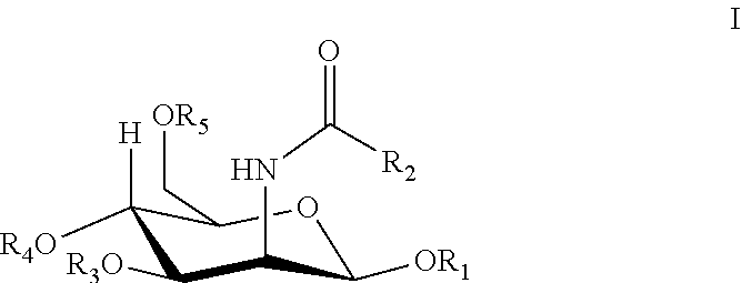
1. A method of treating a condition or disease in a mammal in need thereof comprising administering an effective amount of a sialic acid precursor, N-acetyl mannosamine or a derivative thereof, to the mammal, wherein the derivative consists of Formula I: ##STR00003## wherein R.sub.1, R.sub.3, R.sub.4, or R.sub.5 is hydrogen, lower alkanoyl, carboxylate or lower alkyl; R.sub.2 is lower alkyl, lower alkanoylalkyl, lower alkyl alkanoyloxy; and the condition or disease is muscular atrophy, muscular dystrophy, high blood pressure, or diabetes.
2. The method of claim 1, comprising administering a therapeutic amount of N-acetyl mannosamine to the mammal.
3. The method of claim 2, wherein the therapeutic amount is about 0.1 g to about 50 g N-acetyl mannosamine per day.
4. The method of claim 2, wherein the therapeutic amount is administered in a unit dosage of about 0.01 g to about 50 g N-acetyl mannosamine per unit dosage.
5. The method of claim 1, comprising sustained release of the sialic acid precursor, N-acetyl mannosamine or a derivative thereof.
6. The method of claim 1, comprising topical administration of the sialic acid precursor, N-acetyl mannosamine or a derivative thereof.
7. The method of claim 1, wherein them method reduces hematuria in the mammal.
8. A method of reducing hematuria in a mammal in need thereof comprising selecting a mammal with a condition or disease, and administering to the mammal an effective amount of a sialic acid precursor, N-acetyl mannosamine or a derivative thereof, to the mammal, wherein the derivative consists of Formula I: ##STR00004## wherein R.sub.1, R.sub.3, R.sub.4, or R.sub.5 is hydrogen, lower alkanoyl, carboxylate or lower alkyl; R.sub.2 is lower alkyl, lower alkanoylalkyl, lower alkyl alkanoyloxy; and wherein the condition or disease is hereditary inclusion body myopathy, muscular atrophy, muscular dystrophy, high blood pressure, diabetes, or diabetic nephropathy.
9. The method of claim 8, comprising administering a therapeutic amount of N-acetyl mannosamine to the mammal.
10. The method of claim 9, wherein the therapeutic amount is about 0.1 g to about 50 g N-acetyl mannosamine per day.
11. The method of claim 9, wherein the therapeutic amount is administered in a unit dosage of about 0.01 g to about 50 g N-acetyl mannosamine per unit dosage.
12. (The method of claim 8, comprising sustained release of the sialic acid precursor, N-acetyl mannosamine or a derivative thereof.
13. The method of claim 8, comprising topical administration of the sialic acid precursor, N-acetyl mannosamine or a derivative thereof.
14. The method of claim 9, wherein the N-acetyl mannosamine is administered orally to the mammal.
15. The method of claim 9, wherein the N-acetyl mannosamine is microencapsulated.
16. The method of claim 8, wherein the method also decreases proteinuria in the mammal.
Description
CROSS REFERENCE TO RELATED APPLICATIONS
[0001] This is a continuation of U.S. patent application Ser. No. 15/702,529, filed on Sep. 12, 2017, which is a continuation of U.S. patent application Ser. No. 14/754,304, filed Jun. 29, 2015, now abandoned, which is a continuation of U.S. patent application Ser. No. 13/791,576, filed Mar. 8, 2013, and issued as U.S. Pat. No. 9,095,597 on Aug. 4, 2015, which is a continuation of U.S. patent application Ser. No. 12/530,433, filed Mar. 19, 2010, and issued as U.S. Pat. No. 8,410,063 on Apr. 2, 2013, which is the U.S. National Stage of International Application No. PCT/US2008/006895, filed May 30, 2008, which was published in English under PCT Article 21(2), which in turn claims the benefit of U.S. Provisional Patent Application No. 60/932,451, filed May 31, 2007. The prior applications are all incorporated by reference.
GOVERNMENT FUNDING
[0003] The invention described herein was developed with support from the National Human Genome Research Institute (NHGRI), which is part of the National Institutes of Health (NIH). The United States Government has certain rights in the invention.
FIELD OF THE INVENTION
[0004] The invention is related to a methods and compositions involving use of the neutral sugar N-acetyl mannosamine (ManNAc) for therapeutic purposes in humans. Such therapeutic uses include treatment of myopathies (e.g., hereditary inclusion body myopathy (HIBM)) and certain kidney diseases (e.g., those involving proteinuria and hematuria).
BACKGROUND OF THE INVENTION
[0005] Hereditary inclusion body myopathy (HIBM; OMIM 600737) is a rare autosomal recessive neuromuscular disorder. Argov, et al., Neurology 60, 1519-1523 (2003); Eisenberg, et al. (2001) Nat Genet 29, 83-87 (2001); Griggs, et al. (1995) Ann Neurol 38, 705-713 (1995). The disease usually manifests at approximately 20 years of age with foot drop and slowly progressive muscle weakness and atrophy. Histologically, it is associated with muscle fiber degeneration and formation of vacuoles containing 15-18 nm tubulofilaments that immunoreact like .beta.-amyloid, ubiquitin, prion protein and other amyloid-related proteins. Askanas et al. Curr Opin Rheumatol 10, 530-542 (1998); Nishino, et al. (2005) Acta Myol 24, 80-83 (2005); Askanas, et al. Ann Neurol 34, 551-560 (1993); Argov, et al. Curr Opin Rheumatol 10, 543-547 (1998). Both weakness and histological changes initially spare the quadriceps. However, the disease is relentlessly progressive, with patients becoming incapacitated and wheelchair-confined within two to three decades. There is no treatment available.
[0006] Accordingly, new compositions and methods are needed for treating hereditary inclusion body myopathy and related diseases.
SUMMARY OF THE INVENTION
[0007] One aspect of the invention is a therapeutic method for increasing sialic acid in a mammal in need thereof comprising administering to the mammal an effective amount of N-acetyl mannosamine or a derivative thereof to thereby increase sialic acid in the mammal.
[0008] In some embodiments, such methods are performed to treat a disease or condition. For example, such a disease can be muscular atrophy or kidney disease. In general, the types of muscular atrophies that can be treated by the methods and compositions of the invention are those caused by sialic acid deficiency. Examples of such muscular atrophy diseases and conditions include distal myopathy with rimmed vacuoles (Nonaka myopathy) and hereditary inclusion body myopathy.
[0009] Surprisingly, the compositions and methods of the invention are useful for treating certain kidney conditions and diseases, for example, those involving proteinuria, hematuria resulting primarily or secondarily from hyposialylation (lack of sialic acid). Thus, the present methods are effective for treatment of kidney disorders due to poor kidney membrane formation and/or function. For example, kidney membranes that are affected by loss of sialic acid include the glomerular basement membrane and/or the podocyte membranes. Hence, the invention is useful for treating malformed or poorly functioning glomerular basement membranes and/or podocyte membranes. In general, the methods of the invention can increase sialylation of kidney podocalyxin, improve podocyte foot process morphology and/or improve glomerular basement membrane integrity.
[0010] Another aspect of the invention is a method of treating a kidney disorder in a mammal comprising administering a therapeutic amount of N-acetyl mannosamine or a derivative thereof to the mammal, wherein the kidney disorder involves proteinuria and hematuria. For example, such a therapeutic amount of N-acetyl mannosamine or a derivative thereof is about 1 g to about 20 g per day.
DESCRIPTION OF THE FIGURES
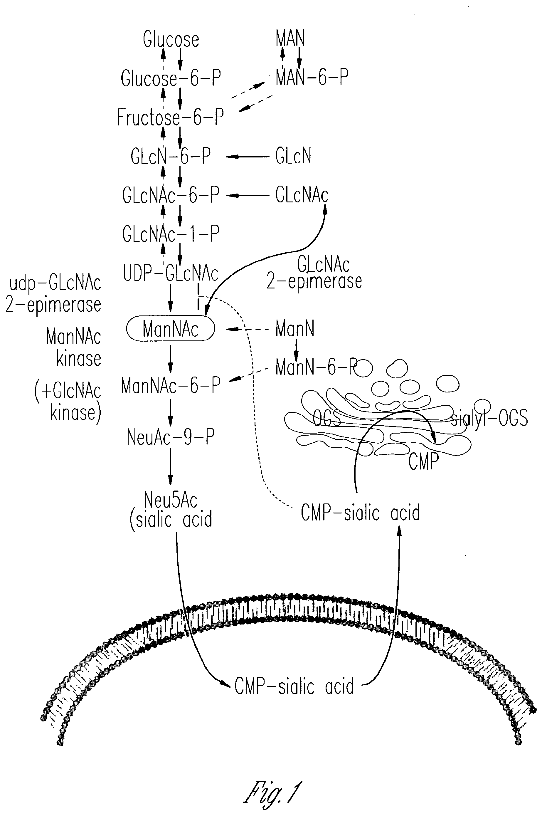
[0011] FIG. 1 schematically illustrates the intracellular metabolism of sialic acid. This figure shows portions of a cell, where the nucleus is depicted at the bottom (below the depicted nuclear membrane) and the cytoplasm is at the top of the figure. Cytosolic glucose is converted in several steps into UDP-GlcNAc, which serves as substrate for the bi-functional, rate-limiting, committed enzyme for sialic acid biosynthesis: UDP-GlcNAc 2-epimerase/ManNAc kinase (GNE/MNK). GNE catalytic activity (EC 5.1.3.14) epimerizes UDP-GlcNAc to ManNAc, followed by the phosphorylation of ManNAc to ManNAc-6-phosphate (MaNAc-6-P) by the MNK kinase catalytic domain (EC 2.7.1.60). ManNAc-6-phosphate is then condensed with phosphoenolpyruvate to Neu5Ac-9-phosphate by Neu5Ac-9-P synthetase. The phosphate is then released and Neu5Ac is activated by CMP-Neu5Ac synthetase to CMP-Neu5Ac in the nucleus. CMP-Neu5Ac then enters the trans-Golgi and serves as the substrate for different sialyltransferases involved in the production of sialylated glycoconjugates. These are subsequently cleaved in the lysosome to yield free sialic acid, which is exported to the cytoplasm and re-utilized or degraded to ManNAc and pyruvate. CMP-sialic acid strongly feedback inhibits the GNE epimerase at its allosteric site.
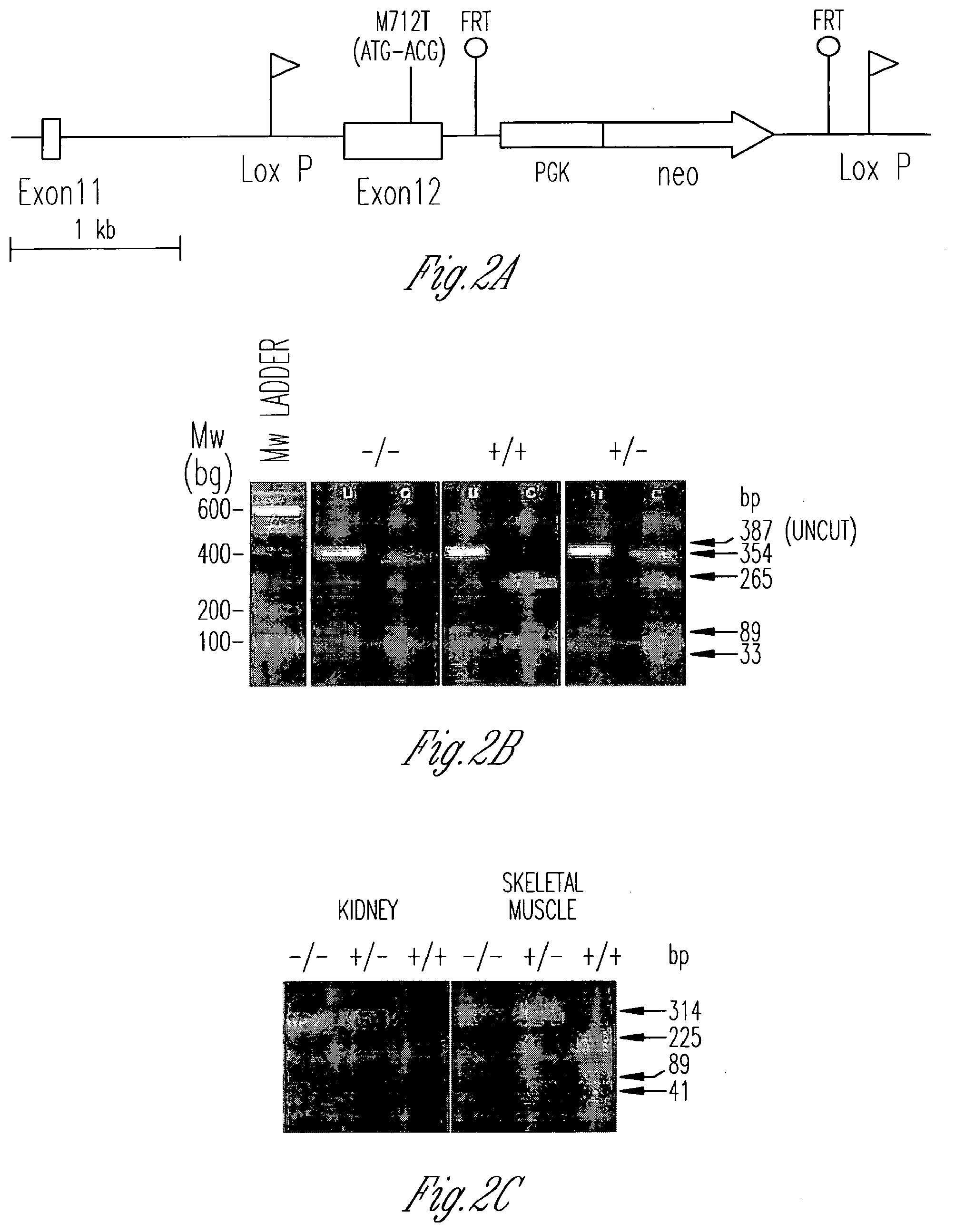
[0012] FIG. 2A-E illustrates the generation and identification of GneM712T/M712T knockin mice. FIG. 2A is a schematic diagram illustrating the murine Gne (Ueal) genomic locus, exons 11 and 12, after homologous recombination with the sequence-verified targeting vector. The M712T mutation was created in exon 12, and a neo cassette (under the PGK promoter) flanked by flippase recombinase target (FRT) sites was inserted. LoxP sites were inserted before exon 12 and after the PGK-neo gene. FIG. 2B shows the genotyping of mutant mice. A PCR-amplified 387-bp fragment of genomic DNA across the M712T (ATG to ACG) mutation was digested by the NlaII restriction endonuclease into 265-bp, 89-bp, and 33-bp fragments in a wild-type allele (+) and into 354-bp and 33-bp fragments in a mutant M712T allele (-). MW, molecular weight. FIG. 2C shows the results of RT-PCR of kidney and skeletal muscle RNA. RNA was reverse transcribed and PCR-amplified using primers covering exons 11 and 12 (355 bp). Digestion by NlaIII cut the wild-type allele (+) into 225-bp, 89-bp, and 41-bp fragments and the mutant M712T allele (-) into 314-bp and 41-bp fragments. Digestion confirmed the exclusive presence of the mutant M712T allele in Gne.sup.M712T/M712T (-/-) tissues. FIG. 2D shows the numbers and genotypes of mice at E17-E19 and P21. At P21, genotyping of 76 mice from 13 litters (9 GneM.sup.712T/+ matings) identified only 1 GneM.sup.712T/M712T offspring. Subsequent genotyping of 35 E17-E19 embryos from 4 Gne.sup.M712T/+ mice yielded a Mendelian distribution of genotypes. FIG. 2E shows that at P2, Gne.sup.M712T/M712T pups were smaller than their heterozygous (GneM.sup.712T/+) and wild-type (Gne.sup.+/+) littermates and lacked a prominent milkspot.
[0013] FIG. 3A-E provides results of histological kidney analyses. FIG. 3A illustrates gross kidney pathology. Kidneys of Gne.sup.M712T/M712T mice showed hemorrhages but were normal in size and shape compared with kidneys of wild-type (Gne.sup.+/+) and heterozygous (GneM.sup.712T/+) littermates. FIG. 3B shows representative H&E-stained sections of renal cortex (c) and medulla (m) showing tubular dilatations in Gne.sup.M712T/M712T mice (arrows). Scale bars: 1,000 .mu.m. FIG. 3C provides high magnification images of collecting ducts, renal tubules, and urinary space, filled with red blood cells in Gne.sup.M712T/M712T mice. Scale bars: 100 .mu.m. FIG. 3D provides high magnification images of glomeruli (g) with red blood cells infiltrated into the Bowman space in Gne.sup.M712T/M712T mice. Scale bars: 100 .mu.m. FIG. 3E shows representative sections of normal glomerulus (DICII, left panel) demonstrating the abundance of Gne/Mnk protein inside the glomerular space upon immunolabeling with Gne/Mnk antibodies (FITC filter, right panel). Scale bars: 50 .mu.m.
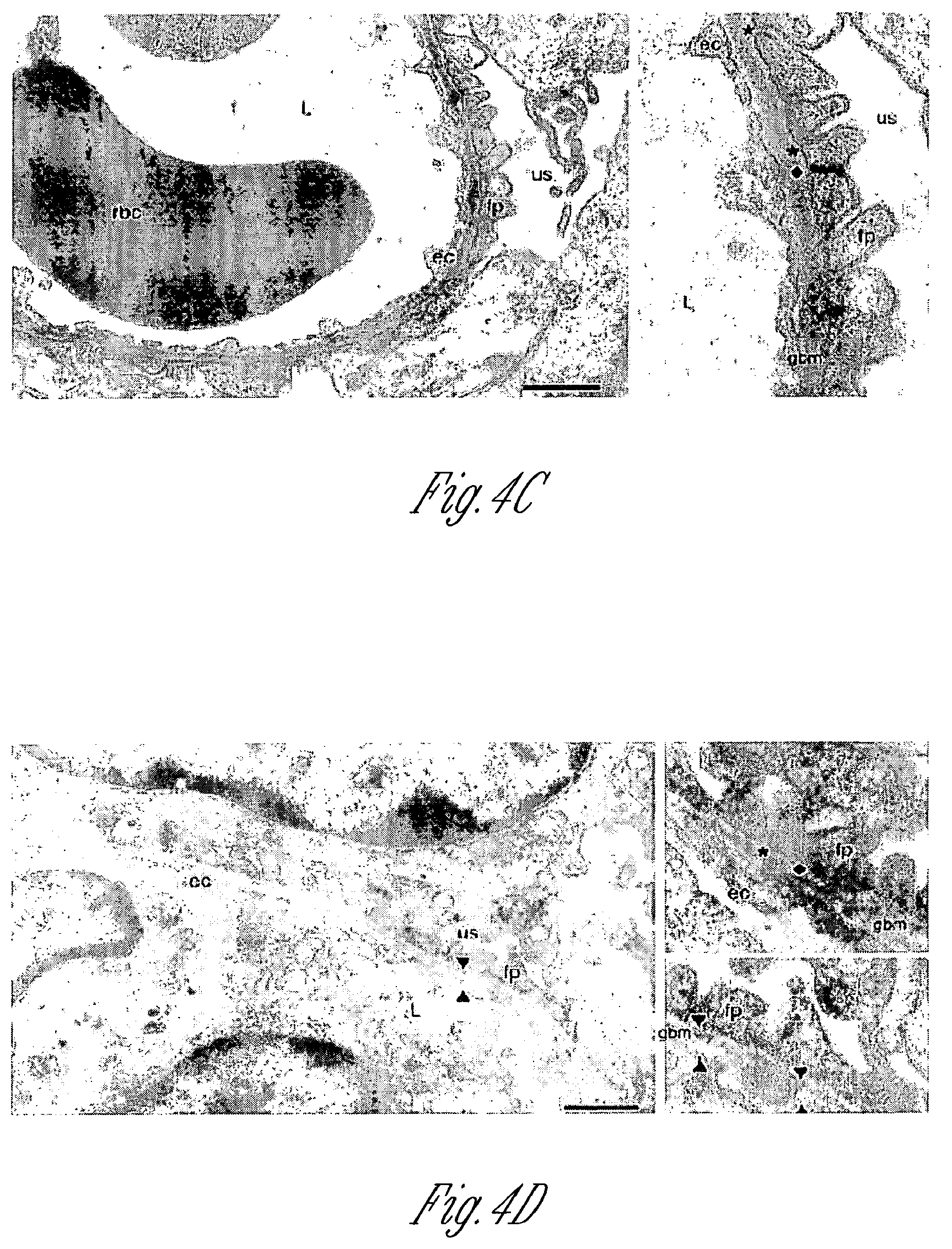
[0014] FIG. 4A-D shows transmission electron microscope images of mouse kidney sections. FIG. 4A shows representative cross-sections of glomerular capillaries in the juxtamedullar zone of a wild-type mouse (age P2). Enlarged insets (right panels) show detailed endothelial cells (ec), glomerular basement membrane (GBM); arrowheads), and foot processes (fp) of the glomerular epithelial cells (podocytes) with well formed, open filtration slits (asterisks). FIG. 4B shows representative juxtamedullary glomerular capillaries of a Gne.sup.M712T/M712T mouse (age P2, littermate of the wild-type mouse shown in FIG. 4A), indicating segmental splitting of the lamina densa of the GBM (arrowheads) as well as dramatically flattened and fused podocyte foot processes lining the GBM. The filtration slits are sparse and irregular in shape and position. Insets (right panels) show fused foot process membranes and formation of abnormal tight junction-like structures at the filtration slits (diamonds). FIGS. 4C and 4D representative glomerular capillaries of a Gne.sup.M712T/M712T mouse following ManNAc treatment (age P19). The integrity of the GBM as well as the formation of podocyte foot processes and the number of filtration slits were all improved when compared with the untreated Gne.sup.M712T/M712T mouse in FIG. 4B. Some filtration slits were open, while others still formed tight junctions. The GMB showed occasional small stretches of areas where splitting was apparent (arrowheads). L, capillary lumen; N, nucleus; us, urinary space. Scale bars: 1 .mu.m.
[0015] FIG. 5A-5F illustrates the biochemistry and renal histology of knockin mice following ManNAc treatment. FIG. 5A shows the numbers of mice surviving past age P3 after ManNAc administration in the drinking water of Gne.sup.M712T/+ mice. Six Gne.sup.M712T/+ mice received 1 mg/ml (.about.0.2 g/kg/d) ManNAc; 7 total litters were scored; 13 pups died at age P1-P3. Seven Gne.sup.M712T/+ mice received 5 mg/ml (.about.1 g/kg/d) ManNAc; 13 total litters were scored; 14 homozygous mutant pups died at age P1-P3. The percentage of survivors of each genotype is indicated. FIG. 5B-5D shows representative H&E-stained kidney sections showing renal cortex and medulla (FIG. 5B); collecting ducts, renal tubules, and urinary space (FIG. 5C); and glomeruli (FIG. 5D) following ManNAc feeding at age P6. Wild-type (Gne+/+) kidneys showed normal histology. Gne.sup.M712T/M712T kidneys showed a range from very mild (middle panel) to moderately severe (right panel) red blood cell infiltrations, but in all cases less severe than in untreated Gne.sup.M712T/M712T mice at age P2 (FIG. 5E-G). Scale bars: 500 .mu.m (FIG. 5B), 100 mm (FIGS. 5C and 5D). FIG. 5E shows two ManNAc-treated (.about.1 g/kg/d) 6-week-old male littermates. Surviving homozygous mutant mice (Gne.sup.M72T/M712T) were smaller than their wild-type littermates. FIG. 5F shows Gne/Mnk epimerase enzymatic activities in skeletal muscle. Administration of ManNAc (shaded bars) increased the activity in wild-type muscle from 100% to 114% (.+-.19.7) (n=3; P=0.2) and increased the activity in homozygous mutant (Gne.sup.M712T/M712T) muscle from 19.4% (.+-.7.5) to 31% (.+-.8.4) (n=7; P=0.05).
[0016] FIG. 6A-E shows immunoblots of muscle, kidney, and brain extracts of knockin mice. Immunoblots of muscle (FIG. 6A) and kidney (FIG. 6B) extracts exhibited decreased Gne/Mnk protein expression (upper band, arrows, 79 kDa) in homozygous mutant Gne.sup.M712T/M712T (-/-) mice compared with heterozygous (+/-) and wild-type (+/+) littermates (normalized to .beta.-actin). Gne/Mnk protein expression increased upon ManNAc feeding in Gne.sup.M712T/M712T(-/-) tissues when compared with untreated tissues. FIG. 6C shows immunoblots of kidney extracts labeled with laminin-1 antibodies. No difference in laminin-1 intensity was detected (n=6; P=0.65) between Gne.sup.+/+ (+/+) and GneM.sup.712T/M712T (-/-) littermates without or with ManNAc treatment. FIG. 6D shows representative immunoblots of brain extracts labeled with PSA-NCAM antibodies. Upon ManNAc treatment, the intensity of the PSA-NCAM signals, reflecting sialylation status, increased by 2% to 28% in treated Gne.sup.M712T/M712T (-/-) brain (n=14) when compared with untreated brain (n=10). FIG. 6E shows immunoblots of kidney extracts (age P2) labeled with antibodies against podocalyxin (.about.140-150 kDa). Top: Following desialylation of Gne.sup.+/+ (+/+), Gne.sup.M712T/+ (+/-), or Gne.sup.M712T/M712T (-/-) kidney extracts by neuraminidase (lanes 2 and 4), podocalyxin migrated more slowly (.about.160-180 kDa) than untreated samples (lanes 1 and 3). Gne.sup.M712T/M712T(-/-) kidney extracts (lanes 5 and 6) contained desialylated podocalyxin. Bottom: Sialylation of podocalyxin at P6 in Gne.sup.M712T/M712T(-/-) mice changed significantly after ManNAc treatment (lanes 3 and 4).
[0017] FIG. 7 schematically illustrates a timeline for administration of N-acetyl mannosamine during a clinical trial of human patients.
SEQUENCE LISTING
[0018] The Sequence Listing is submitted as an ASCII text file 4239-83432-17_Sequence_Listing.txt, May 14, 2019, 16.5 KB], which is incorporated by reference herein.
DETAILED DESCRIPTION OF THE INVENTION
[0019] According to the invention, N-acetyl-mannosamine and derivatives thereof are useful for treating a variety of diseases and conditions. N-acetyl-D-mannosamine is a key compound in the sialic acid biosynthetic pathway (see FIG. 1). In particular, there is a regulated, rate-limiting enzymatic step in the pathway that leads to sialic acid formation, and this rate-limiting step gives rise to N-acetyl-D-mannosamine. Hence, once N-acetyl-D-mannosamine is formed or administered, no other enzymatic step leading to the formation of sialic acid is subject to feedback inhibition. Thus, administration of N-acetyl-D-mannosamine will lead to increased amounts of sialic acid. The structure of N-acetyl-mannosamine is shown below.
##STR00001##
[0020] Therefore, according to the invention, administration of N-acetyl mannosamine (ManNAc) and/or its derivatives promotes formation of sialic acid (N-acetylneuramic acid). Sialic acids are sugars found on many cellular and tissue components. For example, sialic acids are present on most cell surfaces, and on proteins and lipids and are involved in cell to cell interactions. Sialic acid-rich oligosaccharides on the glycoconjugates found on surface membranes help keep water at the surface of cells. The sialic acid-rich regions also contribute to creating a negative charge on the cells surface. Since water is a polar molecule, it is attracted to cell surfaces and membranes. Thus, sialic acids contribute to cellular hydration and fluid uptake. Sialic acid is also a vital component of many body fluids including, serum, cerebrospinal, saliva, amniotic, and mother's milk.
N-Acetylmannosamine Derivatives
[0021] According to the invention, N-acetylmannosamine and derivatives thereof can also be used in the therapeutic methods and compositions of the invention. The structures of such N-acetylmannosamine derivatives useful in the invention are defined by Formula I.
##STR00002##
wherein:
[0022] R.sub.1, R.sub.3, R.sub.4, or R.sub.5 is hydrogen, lower alkanoyl, carboxylate or lower alkyl; and
[0023] R.sub.2 is lower alkyl, lower alkanoylalkyl, lower alkyl alkanoyloxy.
[0024] The following definitions are used, unless otherwise described: Alkyl, alkoxy, alkenyl, alkynyl, etc. denote both straight and branched groups; but reference to an individual radical such as "propyl" embraces only the straight chain radical, a branched chain isomer such as "isopropyl" being specifically referred to.
[0025] Lower alkyl refers to (C.sub.1-C.sub.6)alkyl. Such a lower alkyl or (C.sub.1-C.sub.6)alkyl can be methyl, ethyl, propyl, isopropyl, butyl, iso-butyl, sec-butyl, pentyl, 3-pentyl, or hexyl; (C.sub.3-C.sub.6)cycloalkyl can be cyclopropyl, cyclobutyl, cyclopentyl, or cyclohexyl; (C.sub.3-C.sub.6)cycloalkyl(C.sub.1-C.sub.6)alkyl can be cyclopropylmethyl, cyclobutylmethyl, cyclopentylmethyl, cyclohexylmethyl, 2-cyclopropylethyl, 2-cyclobutylethyl, 2-cyclopentylethyl, or 2-cyclohexylethyl; (C.sub.1-C.sub.6)alkoxy can be methoxy, ethoxy, propoxy, isopropoxy, butoxy, iso-butoxy, sec-butoxy, pentoxy, 3-pentoxy, or hexyloxy; (C.sub.2-C.sub.6)alkenyl can be vinyl, allyl, 1-propenyl, 2-propenyl, 1-butenyl, 2-butenyl, 3-butenyl, 1-pentenyl, 2-pentenyl, 3-pentenyl, 4-pentenyl, 1-hexenyl, 2-hexenyl, 3-hexenyl, 4-hexenyl, or 5-hexenyl; (C.sub.2-C.sub.6)alkynyl can be ethynyl, 1-propynyl, 2-propynyl, 1-butynyl, 2-butynyl, 3-butynyl, 1-pentynyl, 2-pentynyl, 3-pentynyl, 4-pentynyl, 1-hexynyl, 2-hexynyl, 3-hexynyl, 4-hexynyl, or 5-hexynyl; (C.sub.1-C.sub.6)alkanoyl can be acetyl, propanoyl or butanoyl; halo(C.sub.1-C.sub.6)alkyl can be iodomethyl, bromomethyl, chloromethyl, fluoromethyl, trifluoromethyl, 2-chloroethyl, 2-fluoroethyl, 2,2,2-trifluoroethyl, or pentafluoroethyl; hydroxy(C.sub.1-C.sub.6)alkyl can be hydroxymethyl, 1-hydroxyethyl, 2-hydroxyethyl, 1-hydroxypropyl, 2-hydroxypropyl, 3-hydroxypropyl, 1-hydroxybutyl, 4-hydroxybutyl, 1-hydroxypentyl, 5-hydroxypentyl, 1-hydroxyhexyl, or 6-hydroxyhexyl; (C.sub.1-C.sub.6)alkoxycarbonyl can be methoxycarbonyl, ethoxycarbonyl, propoxycarbonyl, isopropoxycarbonyl, butoxycarbonyl, pentoxycarbonyl, or hexyloxycarbonyl; (C.sub.1-C.sub.6)alkylthio can be methylthio, ethylthio, propylthio, isopropylthio, butylthio, isobutylthio, pentylthio, or hexylthio; (C.sub.2-C.sub.6)alkanoyloxy can be acetoxy, propanoyloxy, butanoyloxy, isobutanoyloxy, pentanoyloxy, or hexanoyloxy.
Therapeutic Methods
[0026] According to the invention, N-acetyl-D-mannosamine (ManNAc) and derivatives thereof are useful therapeutic agents for increasing production of sialic acids in mammals, and such increased production of sialic acid has profound therapeutic benefits. Sialic acids are important for proper development and functioning of many organs and tissues, and a deficiency of sialic acid can give rise to many different types of diseases and conditions. For example, abnormalities of sialic acid metabolism cause a severe infantile disease (infantile sialic acid storage disease, ISSD), characterized by failure to thrive, hepatosplenomegaly, coarse facial features, severe mental and motor retardation presenting at birth and often leading to death within the first year of life, or diseases of later onset (Salla disease, sialuria).
[0027] As shown herein, administration of ManNAc (and derivatives thereof) is useful for treating myopathies, muscular atrophy and/or muscular dystrophy (e.g., hereditary inclusion body myopathy (HIBM)) and kidney conditions and diseases (e.g., those involving proteinuria and hematuria).
[0028] Myopathies that can be treated with the present compositions and methods also include distal myopathy with rimmed vacuoles (Nonaka myopathy) and the muscular dystrophy hereditary inclusion body myopathy (HIBM).
[0029] Proteinuria involves leakage of protein from the blood into the urine. If the amount of protein in the urine is very high, this condition is often called nephrotic syndrome. While there may be many causes for nephritic syndrome, according to the invention at least one cause is a deficiency of sialic acid, which has a direct impact on the formation, structure and function of kidney glomeruli and the membranes associated therewith. Several types of diseases exhibit the symptoms of proteinuria, including high blood pressure, infections, reflux nephropathy, diabetes, various types of glomerulonephritis, including minimal change nephrosis. However, by improving the structure and function of nephron components that require sialic acid, the present compositions and methods can treat any of these diseases. Thus, for example, the methods and compositions of the invention dramatically improve kidney function by improving the structure and filtration properties of kidneys, thereby reducing the amount of protein in the urine and/or the severity or progression of proteinuria.
[0030] Hematuria simply means blood in the urine. The blood may be visible, so that the urine appears reddish or darker than normal (called gross hematuria). If the blood is invisible and is discovered only when a urine sample is examined in a laboratory urine test, the condition is called microscopic hematuria. In general, hematuria is more a symptom than a condition in itself, because it has many possible causes. A urinary tract infection, kidney or bladder stones, an enlarged prostate in men, cystitis (a bladder infection, usually in women) or bladder, kidney or prostate cancer can all cause hematuria. Other causes include injuries that result in a bruised kidneys; sickle cell anemia and other abnormal red blood cell diseases; and certain medications, such as blood thinners (e.g., aspirin and some other pain relief medicines). More specific causes of glomerular basal membrane dysfunction, such as Alport disease, thin membrane disease, and IgA nephropathy, may particularly improve when the treatment methods described herein are employed.
[0031] In general, the treatment methods of the invention involve administering to a mammal (or patient) a therapeutically effective amount of N-acetyl mannosamine and/or a derivative thereof. Such a therapeutically effective amount is generally given daily for appropriate periods of time. Effective amounts for human patients are, for example, about 0.1 g/day to about 50 g/day, of about 0.2 g/day to about 25 g/day, from about 0.3 g/day to about 12 g/day, from about 0.4 g/day to about 10 g/day, from about 0.5 g/day to about 8 g/day, and from about 0.7 g/day to about 6 g/day. Generally, N-acetyl mannosamine and/or a derivative thereof is administered for periods of time sufficient to increase the amount of sialic acid in the mammal and thereby achieve a therapeutic benefit. Therapeutic benefits that can be achieved by administration of N-acetyl mannosamine and/or a derivative thereof include improved kidney function, reduction in protein excretion in the urine, reduction in blood concentrations in the urine, increased sialylation of podocalyxin, increased sialylation of PSA-NCAM (and/or other tissue specific target glycoproteins), fewer cystic tubular dilatations in the kidney cortex and in the kidney medulla, less fusion and flattening of the podocyte foot processes, greater number of open slit diaphragms in the kidneys, improvement in the "finger shaping" of the kidney foot processes, improved overall integrity of the GBM, increased Gne/Mnk protein expression and Gne-epimerase activities.
[0032] ManNAc is a ubiquitous but rare monosaccharide involved in a range of metabolic processes. It is uncharged and crosses membranes readily. ManNAc is a constituent of numerous glycolipids and glycoproteins, and is the first committed precursor for the biosynthesis of N-acetylneuraminic (Neu5Ac, or sialic acid), which consists of N-acetyl-D-mannosamine in an ether linkage with D-pyruvic acid. ManNAc is formed from UDP-N-acetylglucosamine (UDP-GlcNAc) by the action of UDP-GlcNAc 2-epimerase. ManNAc is then phosphorylated by a specific kinase to ManNAc-6-P (FIG. 1). ManNAc is situated in the sialic acid biosynthesis pathway after the regulated, rate-limiting GNE step (FIG. 1), so its metabolism is not subject to feedback inhibition. Residual MNK activity in HIBM patients, or ancillary kinases such as GlcNAc kinase (Hinderlich et al. Eur. J. Biochem. 252: 133-139 (1998)), might convert ManNAc into ManNAc-6-phosphate for subsequent synthesis of sialic acid. In fact, hyposialylated, Gne-deficient mouse embryonic stem cells became resialylated after their growth medium was supplemented with ManNAc (Schwarzkopf et al. Proc. Natl. Acad. Sci. U.S.A. 99: 5267-70 (2002)). Furthermore, incubation of cultured cells with "unnatural" ManNAc derivatives, i.e., N-levulinoylmannosamine (ManLev) or N-azidoacetyl-mannosamine (ManNAz), resulted in incorporation of the downstream sialic acid analogs (SiaLev or SiaNAz) into cell surface glycoconjugates (Charter et al. Glycobiology 10: 1049-56 (2000)).
Hereditary Inclusion Body Myopathy (HIBM)
[0033] Studies of an Iranian-Jewish genetic isolate (Argov, et al., Neurology 60, 1519-1523 (2003)) indicate that HIBM is mapped to chromosome 9p12-13. The causative gene for HIBM is GNE, coding for the bifunctional enzyme UDP-N-acetylglucosamine-2-epimerase/N-acetylmannosamine kinase. Eisenberg, et al. (2001) Nat Genet 29, 83-87 (2001); Tanner, M. E., Bioorg Chem 33, 216-228 (2005); Stasche, et al. J Biol Chem 272, 24319-24324 (1997); Hinderlich, et al. J Biol Chem 272, 24313-24318 (1997); Jacobs, et al. Biochemistry 40, 12864-12874 (2001). The function and feedback regulation of GNE/MNK is depicted in FIG. 1. Distal Myopathy with Rimmed Vacuoles (DMRV) is a Japanese variant, allelic to HIBM. Nishino et al. Neurology 59, 1689-1693 (2002); Kayashima et al. J Hum Genet 47, 77-79 (2002); Hinderlich, et al. Neurology 61, 145 (2003). Nearly twenty GNE mutations have been reported in HIBM patients from different ethnic backgrounds, with founder effects among the Iranian Jews and Japanese. Broccolini et al. Hum Mutat 23, 632 (2004); Eisenberg, et al. Hum Mutat 21, 99 (2003); Tomimitsu, et al. Neurology 59, 451-454 (2002); Darvish, et al. Mol Genet Metab 77, 252-256 (2002). The mutations causing HIBM occur in the regions encoding either the epimerase domain or the kinase domain. Most are missense mutations and result in decreased enzyme GNE activity and underproduction of sialic acid. Sparks, et al. Glycobiology 15, 1102-1110 (2005); Penner, et al. Biochemistry 45, 2968-2977 (2006).
[0034] Sialic acids are negatively charged terminal sugar moieties added during the post-translational modification on oligosaccharide chains of proteins and lipids to create glycoproteins and glycolipids. Varki, Faseb J 11, 248-255 (1997); Varki et al. Anal Biochem 137, 236-247 (1984). They act as molecular determinants of specific biological processes such as cellular adhesion, cell-cell interactions and signal transduction. Schauer, Glycoconj J 17, 485-499 (2000); Kelm et al. Int Rev Cytol 175, 137-240 (1997).
[0035] The pathophysiology of HIBM remains largely unknown, but the dysfunction in GNE suggests that impaired sialylation of glycoproteins is involved. Such a defect could influence cell-cell interactions, intracellular trafficking, organelle biogenesis, apoptosis and secretion. In fact, UDP-GlcNAc 2-epimerase regulates sialylation of cell surface molecules (Keppler et al. Science 284: 1372-76 (1999)), and sialylation appears to be critical for mouse development (Schwarzkopf et al. Proc Natl Acad Sci USA 99, 5267-5270 (2002)).
[0036] One hypothesis for the pathophysiology of HIBM involves undersialylation of .alpha.-DG, an essential component of the dystrophin-glycoprotein complex. Michele et al. Nature 418, 417-422 (2002); Michele et al. J Biol Chem 278, 15457-15460 (2003). .alpha.-DG is heavily glycosylated with O-mannosyl glycans (mannose-N-acetylglucosamine-galactose-sialic acid) linked to a serine or threonine; these glycans are critical for t-DG's interactions with laminin and other extracellular ligands. Aberrant glycosylation of .alpha.-DG is the underlying biochemical defect in several congenital muscular dystrophies, generally termed "dystroglycanopathies," including Fukuyama's congenital muscular dystrophy, Muscle-Eye-Brain disease, Walker-Warburg syndrome and the congenital muscular dystrophies type C1C and C1D. Martin et al., Glycobiology 13, 67R-75R (2003); Martin-Rendon, et al. Trends Pharmacol Sci 24, 178-183 (2003). The inventors and others have shown variable hyposialylation of .alpha.-DG and other glycoproteins, such as Neural Crest Adhesion Molecule (NCAM), in HIBM. Huizing et al. Mol Genet Metab 81, 196-202 (2004); Savelkoul et al. Mol Genet Metab 88, 389-390 (2006); Sparks et al. BMC Neurol 7, 3 (2007); Broccolini et al. Neuromuscul Disord 15, 177-184 (2005); Ricci et al. Neurology 66, 755-758 (2006); Salama et al. Biochem Biophys Res Commun 328, 221-226 (2005); Tajima et al. Am J Pathol 166, 1121-1130 (2005).
[0037] However, prior to the present invention, the basic pathogenic mechanisms of HIBM, an HIBM animal model, and an effective therapy for HIBM were lacking. These issues are addressed by the present invention through creation of a Gne gene-targeted knockin mouse mimicking the M712T mutation of Iranian-Jewish HIBM patients and through studies using this knockin mouse model that have defined effective therapeutic methods.
[0038] A sequence for the mouse Gne protein is shown below (SEQ ID NO:1).
TABLE-US-00001 1 MEKNGNNRKL RVCVATCNRA DYSKLAPIMF GIKTEPAFFE 41 LDVVVLGSHL IDDYGNTYRM IEQDDFDINT RLHTIVRGED 81 EAAMVESVGL ALVKLPDVLN RLKPDIMIVH GDRFDALALA 121 TSAALMNIRI LHIEGGEVSG TIDDSIRHAI TKLAHYHVCC 161 TRSAEQHLIS MCEDHDRILL AGCPSYDKLL SAKNKDYMSI 201 IRMWLGDDVK CKDYIVALQH PVTTDIKHSI KMFELTLDAL 241 ISFNKRTLVL FPNIDAGSKE MVRVMRKKGI EHHPNFRAVK 281 HVPFDQFIQL VAHAGCMIGN SSCGVREVGA FGTPVINLGT 321 RQIGRETGEN VLHVRDADTQ DKILQALHLQ FGKQYPCSKI 361 YGDGNAVPRI LKFLKSIDLQ EPLQKKFCFP PVKENISQDI 401 DHILETLSAL AVDLGGTNLR VAIVSMKGEI VKKYTQFNPK 441 TYEERISLIL QMCVEAAAEA VKLNCRILGV GISTGGRVNP 481 QEGVVLHSTK LIQEWNSVDL RTPLSDTLHL PVWVDNDGNC 521 AAMAERKFGQ GKGQENFVTL ITGTGIGGGI IHQHELIHGS 561 SFCAAELGHL VVSLDGPDCS CGSHGCIEAY ASGMALQREA 601 KKLHDEDLLL VEGMSVPKDE AVGALHLIQA AKLGNVKAQS 641 ILRTAGTALG LGVVNILHTM NPSLVILSGV LASHYIHIVK 681 DVIRQQALSS VQDVDVVVSD LVDPALLGAA SMVLDYTTRR 721 IH
[0039] When this Gne protein has the M712T mutation, the sequence for Gne mutant protein arising from the Gne.sup.M712T mutation is as follows (SEQ ID NO:2), where the methionine at position 712 has been changed to a threonine (bold and underlined amino acid shown below).
TABLE-US-00002 1 MEKNGNNRKL RVCVATCNRA DYSKLAPIMF GIKTEPAFFE 41 LDVVVLGSHL IDDYGNTYRM IEQDDFDINT RLHTIVRGED 81 EAAMVESVGL ALVKLPDVLN RLKPDIMIVH GDRFDALALA 121 TSAALMNIRI LHIEGGEVSG TIDDSIRHAI TKLAHYHVCC 161 TRSAEQHLIS MCEDHDRILL AGCPSYDKLL SAKNKDYMSI 201 IRMWLGDDVK CKDYIVALQH PVTTDIKHSI KMFELTLDAL 241 ISFNKRTLVL FPNIDAGSKE MVRVMRKKGI EHHPNFRAVK 281 HVPFDQFIQL VAHAGCMIGN SSCGVREVGA FGTPVINLGT 321 RQIGRETGEN VLHVRDADTQ DKILQALHLQ FGKQYPCSKI 361 YGDGNAVPRI LKFLKSIDLQ EPLQKKFCFP PVKENISQDI 401 DHILETLSAL AVDLGGTNLR VAIVSMKGEI VKKYTQFNPK 441 TYEERISLIL QMCVEAAAEA VKLNCRILGV GISTGGRVNP 481 QEGVVLHSTK LIQEWNSVDL RTPLSDTLHL PVWVDNDGNC 521 AAMAERKFGQ GKGQENFVTL ITGTGIGGGI IHQHELIHGS 561 SFCAAELGHL VVSLDGPDCS CGSHGCIEAY ASGMALQREA 601 KKLHDEDLLL VEGMSVPKDE AVGALHLIQA AKLGNVKAQS 641 ILRTAGTALG LGVVNILHTM NPSLVILSGV LASHYIHIVK 681 DVIRQQALSS VQDVDVVVSD LVDPALLGAA STVLDYTTRR 721 IH
[0040] Although this M712T mutation gives rise to a recessive phenotype, it has dramatic effects upon the survival and physiology of mammals. For example, HIBM exhibits non life-threatening symptoms in humans that emerge in adulthood and lead to slowly progressive muscle weakness. Most patients develop symptoms while in their early 20s and become wheelchair-bound by the time they reach 40, as their arm, hand, leg and core muscles progressively weaken. The symptoms in mice are even more dramatic. For example, upon mating nine pairs of GneM.sup.712T/+ mice, 101 progeny were obtained. Of those 101 progeny 26 homozygous mutated (Gne.sup.M712T/M712T) mice were produced. However, only one male with the Gne.sup.M712T/M712T genotype survived past age P3 (FIG. 2D). The remaining 25 Gne.sup.M712T/M712T homozygous mutated offspring died at age P1-P3. This lone surviving mouse showed no muscle pathology at age P2. The lack of early myopathic features recapitulates the human HIBM phenotype. In both mice and humans, the muscle pathology occurs late or is attenuated likely by a modicum of sialic acid is provided through the actions of residual Gne/Mnk enzymatic activities (Sparks et al. Glycobiology 15: 1102-10 (2005); Noguchi et al. J. Biol. Chem. 279: 11402-407 (2004)) (FIG. 5F and FIGS. 6, A and B).
Kidney Conditions
[0041] Instead of early-onset muscle problems, homozygous Gne.sup.M712T/M712T mice exhibit early signs of severe glomerular hematuria and podocytopathy, including effacement of the podocyte foot processes and segmental splitting of the glomerular basement membrane (GBM), likely due to hyposialylation of specific membrane glycoproteins. Unexpectedly, the Gne.sup.M712T/M712T knockin mice provide a novel animal model of podocytopathy and/or segmental splitting of the GBM, demonstrating the significance of sialic acid synthesis in kidney development and function. Structural elements in the kidney that are important for filtering waste from the blood are severely impaired by the sialic acid deficiency. This outcome demonstrates the significance of the ability of the body to synthesize sialic acid for kidney development and function.
[0042] As shown in the Examples and Figures of this application, administration of ManNAc to pregnant mice had a remarkably salutary effect on the survival and renal development of homozygous pups. In particular, ManNAc administration was associated with increased enzymatic activity of Gne, increased sialylation of kidney podocalyxin, and improved morphology of the podocyte foot processes and GBM integrity.
[0043] Therefore, according to the invention, ManNAc is effective not only as a treatment for HIBM but also for treatment of kidney disorders. Thus, ManNAc may be used to treat podocytopathies, minimal change nephrosis, focal and segmental glomerulosclerosis, membranous glomerulonephritis, and other forms of unexplained idiopathic nephrotic syndrome, as well as glomerular basement membrane diseases such as Alport disease and thin membrane disease. Such kidney disorders and conditions are sometimes characterized by segmental splitting of the glomerular basement membrane and/or podocytopathy due to disturbed polyanions on podocyte membranes, or to changes in the amount or charge (sialylation) of glomerular basement membrane components.
Formulations and Administration
[0044] N-acetyl mannosamine and/or derivatives thereof are administered so as to achieve a reduction in at least one symptom associated with an indication or disease. For example, administration of N-acetyl mannosamine and/or derivatives thereof can lead to a reduction in proteinuria (e.g., lower amounts of protein in the urine), a reduction in hematuria (e.g., lower amounts of red blood cells in the urine) and improvement of muscle function (e.g., in patients with muscular atrophy).
[0045] To achieve the desired effect(s), N-acetyl mannosamine and/or derivatives thereof may be administered as single or divided dosages, for example, of at least about 0.01 mg/kg to about 500 to 750 mg/kg, of at least about 0.01 mg/kg to about 300 to 500 mg/kg, at least about 0.1 mg/kg to about 200 to 400 mg/kg or at least about 1 mg/kg to about 25 to 200 mg/kg of body weight, although other dosages may provide beneficial results. The amount administered will vary depending on various factors including, but not limited to the disease, the weight, the physical condition, the health, the age of the mammal, whether prevention or treatment is to be achieved. Such factors can be readily determined by the clinician employing animal models or other test systems that are available in the art.
[0046] Administration of the therapeutic agents in accordance with the present invention may be in a single dose, in multiple doses, in a continuous or intermittent manner, depending, for example, upon the recipient's physiological condition, whether the purpose of the administration is therapeutic or prophylactic, and other factors known to skilled practitioners. The administration of N-acetyl mannosamine and/or derivatives thereof may be essentially continuous over a pre-selected period of time or may be in a series of spaced doses. Both local and systemic administration is contemplated.
[0047] To prepare the composition, N-acetyl mannosamine and/or one or more derivatives thereof are synthesized or otherwise obtained, and purified as necessary or desired. N-acetyl mannosamine (and/or derivatives thereof) can then be added to a composition (or food product), adjusted to the appropriate concentration, and optionally combined with other agents. The absolute weight of N-acetyl mannosamine and/or its derivatives that is included in a unit dose can vary widely. For example, about 0.01 to about 2 g, or about 0.1 to about 1 g of N-acetyl mannosamine and/or derivatives thereof are often used in compositions. Alternatively, the unit dosage can vary from about 0.01 g to about 50 g, from about 0.01 g to about 35 g, from about 0.1 g to about 25 g, from about 0.5 g to about 12 g, from about 0.5 g to about 8 g, from about 0.5 g to about 4 g, or from about 0.5 g to about 2 g.
[0048] Daily doses of N-acetyl mannosamine and/or derivatives thereof can vary as well. Such daily doses can range, for example, from about 0.1 g/day to about 50 g/day, from about 0.2 g/day to about 25 g/day, from about 0.3 g/day to about 12 g/day, from about 0.4 g/day to about 10 g/day, from about 0.5 g/day to about 8 g/day, and from about 0.7 g/day to about 6 g/day.
[0049] Thus, one or more suitable unit dosage forms comprising N-acetyl mannosamine and/or derivatives thereof can be administered by a variety of routes including oral, parenteral (including subcutaneous, intravenous, intramuscular and intraperitoneal), rectal, dermal, transdermal, intrathoracic, intrapulmonary and intranasal (respiratory) routes. The therapeutic agents may also be formulated for sustained release (for example, using microencapsulation, see WO 94/07529, and U.S. Pat. No. 4,962,091). The formulations may, where appropriate, be conveniently presented in discrete unit dosage forms and may be prepared by any of the methods well known to the pharmaceutical arts. Such methods may include the step of mixing N-acetyl mannosamine and/or derivatives thereof with liquid carriers, solid matrices, semi-solid carriers, finely divided solid carriers or combinations thereof, and then, if necessary, introducing or shaping the product into the desired delivery system.
[0050] When N-acetyl mannosamine and/or its derivatives is prepared for oral administration, it is generally combined with a pharmaceutically acceptable carrier, diluent or excipient to form a pharmaceutical formulation, or unit dosage form. For oral administration, N-acetyl mannosamine (and/or derivatives thereof) may be present as a powder, a granular formulation, a solution, a suspension, an emulsion or in a natural or synthetic polymer or resin for ingestion of N-acetyl mannosamine (and/or one or more derivatives thereof) from a chewing gum. The active ingredients may also be presented as a bolus, electuary or paste. Orally administered N-acetyl mannosamine and/or derivatives thereof can also be formulated for sustained release. For example, N-acetyl mannosamine and/or derivatives thereof can be coated, microencapsulated, or otherwise placed within a sustained delivery device, for example, in order to avoid salivary bacteria degradation. The total N-acetyl mannosamine and its derivatives in such formulations comprises from 0.1 to 99.9% by weight of the formulation.
[0051] By "pharmaceutically acceptable" it is meant a carrier, diluent, excipient, and/or salt that is compatible with the other ingredients of the formulation, and not deleterious to the recipient thereof.
[0052] Pharmaceutical formulations containing N-acetyl mannosamine and/or derivatives thereof can be prepared by procedures known in the art using well-known and readily available ingredients. For example, N-acetyl mannosamine and/or its derivatives can be formulated with common excipients, diluents, or carriers, and formed into tablets, capsules, solutions, suspensions, powders, aerosols and the like. Examples of excipients, diluents, and carriers that are suitable for such formulations include buffers, as well as fillers and extenders such as starch, cellulose, sugars, mannitol, and silicic derivatives. Binding agents can also be included such as carboxymethyl cellulose, hydroxymethylcellulose, hydroxypropyl methylcellulose and other cellulose derivatives, alginates, gelatin, and polyvinyl-pyrrolidone. Moisturizing agents can be included such as glycerol, disintegrating agents such as calcium carbonate and sodium bicarbonate. Agents for retarding dissolution can also be included such as paraffin. Resorption accelerators such as quaternary ammonium compounds can also be included. Surface active agents such as cetyl alcohol and glycerol monostearate can be included. Adsorptive carriers such as kaolin and bentonite can be added. Lubricants such as talc, calcium and magnesium stearate, and solid polyethyl glycols can also be included. Preservatives may also be added. The compositions of the invention can also contain thickening agents such as cellulose and/or cellulose derivatives. They may also contain gums such as xanthan, guar or carbo gum or gum arabic, or alternatively polyethylene glycols, bentones and montmorillonites, and the like.
[0053] For example, tablets or caplets containing N-acetyl mannosamine (and/or its derivatives) can include buffering agents such as calcium carbonate, magnesium oxide and magnesium carbonate. Caplets and tablets can also include inactive ingredients such as cellulose, pre-gelatinized starch, silicon dioxide, hydroxy propyl methyl cellulose, magnesium stearate, microcrystalline cellulose, starch, talc, titanium dioxide, benzoic acid, citric acid, corn starch, mineral oil, polypropylene glycol, sodium phosphate, zinc stearate, and the like. Hard or soft gelatin capsules containing N-acetyl mannosamine (and/or its derivatives) can contain inactive ingredients such as gelatin, microcrystalline cellulose, sodium lauryl sulfate, starch, talc, and titanium dioxide, and the like, as well as liquid vehicles such as polyethylene glycols (PEGs) and vegetable oil. Moreover, enteric-coated caplets or tablets containing N-acetyl mannosamine and/or its derivatives are designed to resist disintegration in the stomach and dissolve in the more neutral to alkaline environment of the duodenum.
[0054] N-acetyl mannosamine and/or its derivatives can also be formulated as an elixir or solution for convenient oral administration or as a solution appropriate for parenteral administration, for instance by intramuscular, subcutaneous, intraperitoneal or intravenous routes. The pharmaceutical formulations of N-acetyl mannosamine and/or its derivatives can also take the form of an aqueous or anhydrous solution or dispersion, or alternatively the form of an emulsion or suspension or salve.
[0055] Thus, N-acetyl mannosamine and/or its derivatives may be formulated for parenteral administration (e.g., by injection, for example, bolus injection or continuous infusion) and may be presented in unit dose form in ampoules, pre-filled syringes, small volume infusion containers or in multi-dose containers. As noted above, preservatives can be added to help maintain the shelve life of the dosage form. The N-acetyl mannosamine, its derivatives and other ingredients may form suspensions, solutions, or emulsions in oily or aqueous vehicles, and may contain formulatory agents such as suspending, stabilizing and/or dispersing agents. Alternatively, the N-acetyl mannosamine, its derivatives and other ingredients may be in powder form, obtained by aseptic isolation of sterile solid or by lyophilization from solution, for constitution with a suitable vehicle, e.g., sterile, pyrogen-free water, before use.
[0056] These formulations can contain pharmaceutically acceptable carriers, vehicles and adjuvants that are well known in the art. It is possible, for example, to prepare solutions using one or more organic solvent(s) that is/are acceptable from the physiological standpoint, chosen, in addition to water, from solvents such as acetone, ethanol, isopropyl alcohol, glycol ethers such as the products sold under the name "Dowanol," polyglycols and polyethylene glycols, C.sub.1-C.sub.4 alkyl esters of short-chain acids, ethyl or isopropyl lactate, fatty acid triglycerides such as the products marketed under the name "Miglyol," isopropyl myristate, animal, mineral and vegetable oils and polysiloxanes.
[0057] It is possible to add other ingredients such as antioxidants, surfactants, other preservatives, film-forming, keratolytic or comedolytic agents, perfumes, flavorings and colorings. Antioxidants such as t-butylhydroquinone, butylated hydroxyanisole, butylated hydroxytoluene and .alpha.-tocopherol and its derivatives can be added.
[0058] Additionally, N-acetyl mannosamine and/or derivatives thereof are well suited to formulation in a sustained release dosage form. Thus, such formulations can be so constituted that they release the N-acetyl mannosamine and/or its derivative, for example, in a particular part of the intestinal, urogenital or respiratory tract, over a period of time. Coatings, envelopes, and protective matrices may be made, for example, from polymeric substances, such as polylactide-glycolates, liposomes, microemulsions, microparticles, nanoparticles, or waxes. These coatings, envelopes, and protective matrices are useful to coat indwelling devices, e.g., stents, catheters, peritoneal dialysis tubing, draining devices and the like.
[0059] For topical administration, N-acetyl mannosamine and/or its derivative(s) may be formulated as is known in the art for direct application to a target area. Forms chiefly conditioned for topical application take the form, for example, of creams, milks, gels, dispersion or microemulsions, lotions thickened to a greater or lesser extent, impregnated pads, ointments or sticks, aerosol formulations (e.g., sprays or foams), soaps, detergents, lotions or cakes of soap. Other conventional forms for this purpose include wound dressings, coated bandages or other polymer coverings, ointments, creams, lotions, pastes, jellies, sprays, and aerosols. Thus, N-acetyl mannosamine and/or its derivatives can be delivered via patches or bandages for dermal administration. Alternatively, N-acetyl mannosamine and/or its derivatives can be formulated to be part of an adhesive polymer, such as polyacrylate or acrylate/vinyl acetate copolymer. For long-term applications it might be desirable to use microporous and/or breathable backing laminates, so hydration or maceration of the skin can be minimized. The backing layer can be any appropriate thickness that will provide the desired protective and support functions. A suitable thickness will generally be from about 10 to about 200 microns.
[0060] Ointments and creams may, for example, be formulated with an aqueous or oily base with the addition of suitable thickening and/or gelling agents. Lotions may be formulated with an aqueous or oily base and will in general also contain one or more emulsifying agents, stabilizing agents, dispersing agents, suspending agents, thickening agents, or coloring agents. The therapeutic agents can also be delivered via iontophoresis, e.g., as disclosed in U.S. Pat. No. 4,140,122; 4,383,529; or 4,051,842. The percent by weight of a therapeutic agent of the invention present in a topical formulation will depend on various factors, but generally will be from 0.01% to 95% of the total weight of the formulation, and typically 0.1-85% by weight.
[0061] Drops, such as eye drops or nose drops, may be formulated with N-acetyl mannosamine and/or derivatives thereof in an aqueous or non-aqueous base also comprising one or more dispersing agents, solubilizing agents or suspending agents. Liquid sprays are conveniently delivered from pressurized packs. Drops can be delivered via a simple eye dropper-capped bottle, or via a plastic bottle adapted to deliver liquid contents dropwise, via a specially shaped closure.
[0062] N-acetyl mannosamine and/or its derivatives may further be formulated for topical administration in the mouth or throat. For example, N-acetyl mannosamine and/or its derivatives may be formulated as a lozenge further comprising a flavored base, usually sucrose and acacia or tragacanth; pastilles comprising the composition in an inert base such as gelatin and glycerin or sucrose and acacia; and mouthwashes comprising the composition of the present invention in a suitable liquid carrier.
[0063] The pharmaceutical formulations of the present invention may include, as optional ingredients, pharmaceutically acceptable carriers, diluents, solubilizing or emulsifying agents, and salts of the type that are available in the art.
[0064] Furthermore, N-acetyl mannosamine and/or its derivatives may also be used in combination with other therapeutic agents, for example, pain relievers, anti-inflammatory agents, and the like, whether for the conditions described or some other condition.
[0065] The present invention further pertains to a packaged pharmaceutical composition such as a kit or other container for increasing production of sialic acid in a mammal. The kit or container holds a therapeutically effective amount of a pharmaceutical composition for increasing intracellular production of sialic acid and instructions for using the pharmaceutical composition for increasing production of sialic acid in the mammal. The pharmaceutical composition includes N-acetyl mannosamine and/or its derivatives in a therapeutically effective amount such that sialic acid production is increased.
Food Supplement
[0066] According to the invention, N-acetyl mannosamine and/or its derivatives can be administered as a food supplement or incorporated into food or drink item such as a nutritional bar, snack bar, cookie, candy, cereal, pudding, ice cream, frozen confectionary, chewing gum, drink mix, soda pop, liquid supplement, sauce, salad dressing, gravy, jelly, jam, spread, margarine, peanut butter, nut spread, frosting, and the like. In essence, can be used in any food, composition or supplement in which sugar is employed. Hence, N-acetyl mannosamine and/or derivatives thereof can be used as a partial or full substitute for sugar.
[0067] Such food supplements, drinks and food items can include any other food ingredient including, for example, flour, oil, cream, butter, sugar, salt, spices and the like. In addition, the food supplements, drinks and food items can include vitamins and nutrients commonly found in other nutritional supplements.
[0068] Having now generally described the invention, the same will be more readily understood through reference to the following examples which are provided by way of illustration, and are not intended to be limiting of the present invention.
Example 1: ManNAc Administration is Useful for Treating HIBM and Renal Disorders
[0069] This Example shows that ManNAc may be a useful treatment not only for HIBM but also for renal disorders involving proteinuria and hematuria due to podocytopathy and/or segmental splitting of the glomerular basement membrane.
Methods
[0070] Gne.sup.M712T/M712T mice. Gne.sup.M712T/M712T knockin mice were generated by targeting the M712T (ATG to ACG) mutation of exon 12 of the murine Gne gene (Gne, Uae1, GenBank NM_015828) (FIG. 2A). The nucleotide sequence for this Gne, Uae1 allele (GenBank NM_015828) without the mutation is shown below for easy reference (SEQ ID NO:3).
TABLE-US-00003 1 GCTAAACCAG AGGCCAGACG GCAGCTCAGG AGTCCGACCA 41 CACCTCAGGA AACAGCTGTG CCACAGGATG GAAACACACG 81 CGCATCTCCA CAGGGAGCAG AGCTACGCAG GACCTCATGA 121 ACTCTATTTT AAGAAACTCT CAAGTAAAAA GAAGCAAGTC 161 ATGGAGAAGA ACGGGAACAA CCGAAAGCTC CGGGTTTGCG 201 TTGCCACCTG CAACCGAGCT GACTACTCCA AACTGGCCCC 241 GATCATGTTC GGCATCAAGA CAGAGCCCGC GTTCTTTGAG 281 TTGGACGTGG TGGTGCTCGG CTCCCACCTG ATTGACGACT 321 ATGGAAACAC ATACCGCATG ATTGAGCAAG ATGACTTTGA 361 CATTAACACC AGGCTCCACA CGATTGTTAG AGGGGAAGAT 401 GAAGCGGCCA TGGTAGAGTC GGTAGGCCTA GCGCTCGTGA 441 AGCTACCGGA CGTCCTCAAT CGCCTGAAGC CCGACATCAT 481 GATTGTTCAC GGAGACCGAT TTGACGCCCT TGCTCTGGCT 521 ACGTCTGCTG CCTTGATGAA CATCCGCATC CTTCACATTG 561 AAGGAGGCGA GGTCAGCGGG ACCATTGATG ACTCTATCAG 601 ACACGCCATA ACAAAACTGG CTCACTACCA TGTGTGCTGC 641 ACTAGAAGTG CAGAGCAGCA CCTGATCTCT ATGTGCGAGG 661 ACCACGACCG CATCCTGTTG GCAGGCTGCC CTTCCTATGA 721 CAAACTGCTC TCCGCCAAGA ACAAAGACTA TATGAGCATC 761 ATTCGGATGT GGCTAGGCGA TGATGTAAAA TGTAAGGATT 801 ACATCGTTGC CCTGCAGCAT CCCGTGACCA CTGACATTAA 841 GCATTCCATA AAGATGTTTG AGCTAACACT GGATGCCCTG 881 ATCTCGTTTA ACAAGAGGAC CCTAGTTCTG TTTCCAAATA 921 TCGATGCAGG CAGCAAGGAG ATGGTTCGAG TGATGCGGAA 961 GAAGGGCATC GAGCATCACC CCAATTTCCG TGCAGTCAAG 1001 CACGTCCCGT TTGACCAGTT CATACAGCTG GTCGCCCACG 1041 CTGGCTGCAT GATTGGGAAT AGCAGCTGCG GCGTGCGAGA 1081 GGTTGGCGCT TTCGGAACAC CCGTGATCAA CCTGGGCACA 1121 AGGCAGATAG GAAGAGAAAC CGGGGAGAAT GTTCTTCATG 1161 TCAGGGATGC TGACACCCAA GATAAAATAT TGCAAGCACT 1201 ACACCTCCAG TTCGGCAAAC AGTACCCTTG CTCAAAGATA 1241 TATGGGGATG GGAATGCTGT TCCAAGGATT TTAAAGTTTC 1281 TCAAATCCAT TGACCTTCAA GAGCCACTAC AGAAGAAATT 1321 CTGCTTCCCC CCTGTAAAGG AGAACATCTC TCAAGACATT 1361 GACCACATCC TGGAAACTCT GAGTGCCTTG GCTGTTGATC 1401 TTGGCGGGAC AAACCTGAGG GTGGCAATAG TTAGCATGAA 1441 GGGTGAAATC GTTAAGAAGT ACACTCAGTT CAACCCTAAA 1481 ACCTATGAAG AAAGGATTAG TTTAATCCTG CAGATGTGTG 1521 TGGAAGCTGC CGCGGAAGCT GTGAAACTCA ATTGCAGAAT 1561 TCTGGGAGTA GGCATCTCCA CAGGTGGCCG CGTGAATCCC 1601 CAGGAAGGAG TTGTGCTGCA TTCAACCAAG CTGATCCAGG 1641 AATGGAACTC CGTGGACCTC AGGACACCCC TCTCCGACAC 1681 CCTGCATCTC CCCGTGTGGG TGGACAATGA CGGCAACTGT 1721 GCCGCCATGG CAGAGAGGAA GTTCGGCCAA GGAAAAGGAC 1761 AGGAGAACTT CGTGACGCTC ATCACGGGGA CAGGGATCGG 1801 TGGGGGGATC ATCCACCAGC ACGAACTGAT CCACGGCAGC 1841 TCCTTCTGCG CGGCGGAGCT CGGCCATCTC GTGGTGTCCC 1881 TGGACGGTCC TGACTGCTCC TGTGGAAGCC ATGGGTGCAT 1921 CGAAGCGTAC GCCTCTGGAA TGGCCTTGCA GAGGGAAGCA 1961 AAGAAACTCC ATGATGAGGA CCTGCTCTTG GTGGAAGGGA 2001 TGTCAGTACC AAAAGACGAA GCTGTGGGTG CCCTCCATCT 2041 CATCCAGGCT GCCAAGCTGG GCAACGTGAA GGCCCAGAGC 2081 ATCTTACGAA CAGCTGGAAC TGCTTTGGGA CTTGGGGTTG 2121 TGAACATCCT CCACACTATG AATCCTTCCC TGGTGATCCT 2161 GTCTGGAGTC CTGGCCAGTC ACTACATCCA CATCGTGAAG 2201 GACGTCATCC GCCAGCAAGC CTTGTCCTCC GTGCAGGATG 2241 TGGACGTGGT GGTCTCAGAC TTGGTGGACC CGGCCCTGCT 2281 TGGCGCAGCC AGCATGGTTC TGGACTACAC AACGCGCAGG 2321 ATCCACTAGG TCTCCCGGGA ACGGACACGG ACAGAGACTC 2361 GGGAGCTGCT TAGAGTGGAA CCATGCTCTT CTAGATCAGT 2401 GTTTCTGCGA AGGCAAATTT GGGGGGAGGG CTGCTGAGAC 2441 AGCTCAGTGG TTAAGAGCCT GCCCTGCTCC TGCCAGTCCC 2481 CAGCACCCAT GTCAGGCAGC TCAGCTGCCT GGAAGCCAAG 2521 CTCCAGGGGA CCCAATGCCT CTCTGCCGGG GGCAGCTGCA 2561 CTCAGATGTA CATACCCCTC TCCACACACA TACAAATAAA 2601 GCTTATTTTT CAAAAGGCAA AAAAAAAAAA AAAAAAAAAA 2641 AAAAAAAAAA AAAA
The mutant mice were maintained in the C57BL/6J background. Animals were housed in an accredited specific pathogen-free facility in accordance with accepted guidelines. Cages were ventilated in a temperature- and light-controlled environment (22.degree. C., 30%-70% humidity, 12-hour light/12-hour dark cycle). The mice were fed irradiated chow (Prolab 5P75 Isopro 3000; PMI Nutrition International) and sterile water ad libitum. All euthanasia was performed by CO.sub.2 inhalation followed by cervical dislocation.
[0071] For Mendelian distribution studies, 4 pregnant mice at E17-E19 were euthanized, and embryos were retrieved by cesarean section and euthanized by decapitation. All mouse procedures were performed in accordance with protocol G04-3 and were approved by the Institutional Animal Care and Use Committee of the National Human Genome Research Institute.
[0072] Molecular Analysis.
[0073] Mouse genotyping was performed on tail genomic DNA or cDNA isolated from kidney or skeletal muscle using standard protocols. Total RNA was isolated from murine tissues using the TRIzol reagent (Invitrogen), and cDNA was prepared using the SuperScript III system (Invitrogen). PCR amplifications were performed across the M712T mutation with genomic DNA as template, using the primer set 5'-agcacttcctgagtttgatg-3' (SEQ ID NO:4) and 5'-atttgccttcgcaga-cacttga-3' (SEQ ID NO:5) (FIG. 2B) or with cDNA as template (FIG. 2C), using the primer set 5'-GCCCAGAGCATCTTACGAAC-3' (SEQ ID NO:6) and 5'-GGGTCCCCTGGAGCTTGG-3' (SEQ ID NO:7) and PuReTaq Ready-To-Go PCR beads (GE Healthcare), using standard PCR conditions. PCR fragments were digested with Nla III at 37.degree. C. to verify the mutation status (FIGS. 2B and C). Quantitative realtime PCR was performed on RNA isolated from kidney and skeletal muscle, utilizing Assays-On-Demand (Applied Biosystems) for Gne (mm00607939), Pecam-1 (mm00476702), Col4A3 (mm01269206), and .beta.-actin (mm00450174) on an ABI PRISM 7900 HT Sequence Detection System (Applied Biosystems).
[0074] Clinical Chemistry Screen.
[0075] Retroorbital blood samples (100-150 ml) from weaned mice (weighing at least 15 grams) matched for sex (male) and age were obtained bimonthly after pretreatment with a topical anesthetic (0.5% tetracaine HCl; Bausch & Lomb Pharmaceuticals). Samples were allowed to clot (30 minutes at room temperature) in MicroPrep centrifuge tubes (IRIS International Inc.), after which the serum was separated by centrifugation at 1500 g for 10 minutes and stored at -80.degree. C. until analysis. Clinical chemistry screens were performed at the Department of Laboratory Medicine at the NIH and included monitoring of creatinine, blood urea nitrogen, albumin, total protein, uric acid, alkaline phosphatase, alanine aminotransferase, aspartate aminotransferase, amylase, creatine kinase, and lactate dehydrogenase. In addition, reagent strips for protein urinalysis were used to assess proteinuria (Chemstrip 2GP; Roche Diagnostics).
[0076] Antibodies.
[0077] A rabbit polyclonal antibody was custom prepared against a Gne/Mnk peptide comprising amino acids 588-607 (EAYASGMALQREAKKLHDED, SEQ ID NO:8), coupled to keyhole limpet hemocyanine, and affinity purified against the corresponding antigenic peptide (Covance). The following additional primary antibodies were commercially obtained: dystrophin (catalog no. ab15277; Abcam); .alpha.-dystroglycan (clone IIH6C4; Upstate Biotechnology); laminin-1 (catalog no. L9393; Sigma-Aldrich); podocalyxin (catalog no. PODX11-A; Alpha Diagnostic International); podocin (catalog no. P0372; Sigma-Aldrich); laminin P31 (catalog no. MAB1928; Millipore); desmin (catalog no. 1466-1; Epitomics); .alpha.-SMA (SPM332; GeneTex Inc.); PSA-NCAM (catalog no. MAB5324; Millipore); and .beta.-actin (catalog no. AAN01; Cytoskeleton).
[0078] Mouse Histology.
[0079] Mouse tissues were collected, formalin fixed (10%) and paraffin embedded. Tissue sections (5 mm) were stained with H&E following standard procedures (American Histolabs) or subjected to immunohistochemistry with a variety of primary antibodies. Formalin-fixed tissues were deparaffinized in HistoClear II (National Diagnostics) and dehydrated in a series of ethanol solutions. Antigen retrieval was performed for sections that were to be stained with antibodies against Gne/Mnk (by boiling 5 minutes in citric acid-based solution; Vector Laboratories) and against dystrophin (by boiling in 1 mM EDTA according the manufacturer's protocol; AbCam). The sections were blocked (2% BSA, 10% donkey serum, and 0.1% Triton X-100 in PBS) and incubated with primary antibodies (Gne/Mnk 1:50; laminin 1:25; dystrophin 1:50) overnight at 4.degree. C., followed by incubation with the secondary antibody, Alexa Fluor 488-conjugated donkey anti-rabbit (1:500 in blocking solution) (Invitrogen). The sections were mounted in VECTASHIELD Mounting Medium (Vector Laboratories) and viewed and digitally imaged with a Zeiss Axiovert 200M microscope (Zeiss).
[0080] Western Blotting.
[0081] Mouse tissues (age P2) were extracted, homogenized in CelLytic buffer consisting of a mild detergent, bicine buffer, and 150 mM NaCl (Sigma-Aldrich) supplemented with protease inhibitors (Complete Mini; Roche Applied Science). The lysates were sonicated and cleared by centrifugation (1000 g for 10 minutes), and the resulting supernatants were assayed for protein (BCA Protein Assay; Pierce Biotechnology). For the neuraminidase enzymatic treatments (FIG. 6E), protein homogenates (25 mg) were incubated for 30 min at 37.degree. C. with 1 mU/mg of neuraminidase (catalog no. N6514; Sigma-Aldrich). Equal amounts of protein (25-50 mg) were electrophoresed on 4%-12% Tris-Glycine gels (Novex; Invitrogen), and electroblotted onto 0.45 mm Hybond ECL nitrocellulose membranes (GE Healthcare). The membranes were blocked (10% fat-free milk) and incubated with primary antibodies followed by HRP-conjugated secondary antibodies (GE Healthcare). Results were visualized with ECL (ECL Western Blotting Detection Reagents; GE Healthcare) and exposure to CL-XPosure Film (Pierce Biotechnology). Densitometry was performed on the digital images obtained with a Kodak Image Station and software (PerkinElmer). The protein levels were normalized to those of .beta.-actin to correct for differences in protein loading and/or transfer.
[0082] Electron Microscopy.
[0083] Kidney samples were fixed overnight at 4.degree. C. in 2% glutaraldehyde in 0.1 M cacodylate buffer (pH 7.4) and washed with cacodylate buffer. After postfixation with 1% OsO.sub.4 for 2 hours and a second wash with 0.1 M cacodylate buffer, the tissues were serially dehydrated in ethanol and embedded in Eponate 12 resin (Ted Pella). Thin sections (.about.80 nm) were obtained using a Leica Ultracut UCT Ultramicrotome (Leica Microsystems), placed onto 400 mesh copper grids, and stained with saturated uranyl acetate in 50% methanol followed by lead citrate. The grids were viewed with a Philips 410 electron microscope (FEI Company) at 80 kV, and images were recorded on Kodak SO-163 film (Kodak).
[0084] ManNAc Administration.
[0085] Breeding pairs of 6-week-old GneM.sup.712T/+ mice were divided into 3 groups. Group I consisted of 9 GneM.sup.712T/+ breeding pairs, who were administered untreated sterilized tap water. Group II consisted of 1 breeding pair of Gne.sup.+/+ mice (wild-type control) and 6 GneM.sup.712T/+ breeding pairs, who were administered water containing 1 mg/ml (.about.0.2 g/kg/day) ManNAc (Sigma-Aldrich). That dose was selected based on previous evidence of the safety of ManNAc (administered at a single dose of 0.142 g/kg/day) in a study performed in humans (21). Group III consisted of 1 Gne.sup.+/+ breeding pair and 7 GneM.sup.712T/+ breeding pairs, who were administered water supplemented with 5 mg/ml (.about.1.0 g/kg/day) ManNAc. Water was changed twice weekly. Nursing females continued to be supplied with ManNAc. All mice were weaned from ManNAc at 21 days. Selected whole litters were euthanized at age P2, P6, and P19 for histological, genetic, biochemical, or ultrastructural analysis.
[0086] Gne Enzymatic Assays.
[0087] Mouse kidney and skeletal muscle (quadriceps) tissues were homogenized and subjected to the Gne-epimerase enzymatic assay as previously described (11, 58). This assay was based on incubation with radiolabeled substrate (Uridine diphosphate N-acetyl glucosamine [1-.sup.3H]; American Radiolabeled Chemicals Inc.) and detection of radiolabeled product ([.sup.3H]ManNAc) upon separation of oligosaccharides by high-pH anion-exchange chromatography with pulsed amperometric detection (Dionex).
[0088] Statistics.
[0089] Differences between data groups were evaluated for significance using the 2-tailed Student's t test of unpaired data. For Mendelian distribution analysis, a goodness-of-fit (.chi.2) test was performed, while for comparisons of survival between treated and untreated mice of all genotypes (Gne.sup.+/+, GneM.sup.712T/+, and GneM.sup.712T/M712T), a 2-tailed Fisher's exact test using a 2.times.3 table was employed. All data are presented as the mean.+-.SD. A P value less than 0.05 was considered statistically significant.
Results
[0090] Generation of Gne.sup.M712T/M712T Knockin Mice.
[0091] A murine targeting vector for homologous recombination in C57BL/6J embryonic stem cells was constructed to include the M712T Gne mutation (FIG. 2A). The neomycin phosphotransferase and thymidine kinase genes were introduced into the vector as positive and negative selection markers, respectively (FIG. 2A). Additional LoxP (flanking exon 12 and neo) and flippase recombinase target sites (flanking neo) were inserted for transgenic models for this condition (Nagy, A. 2000. Cre recombinase: the universal reagent for genome tailoring. Genesis. 26:99-109). The entire vector was sequence verified. Genotyping of the mice was performed by PCR amplification and digestion with the restriction endonuclease NlaIII (FIG. 2B). Tissues of homozygous mutant Gne.sup.M712T/M712T and wild-type Gne.sup.+/+ mice showed comparable Gne RNA transcript levels by real-time quantitative PCR. Furthermore, NlaIII digestion of amplified cDNA demonstrated homozygous insertion of the M712T mutation in RNA of Gne.sup.M712T/M712T mice (FIG. 2C).
[0092] Early Postnatal Lethality.
[0093] Initial matings of heterozygous mice (Gne.sup.M712T/+) yielded 101 offspring from which only 1 Gne.sup.M712T/M712T animal survived beyond P21. The remaining Gne.sup.M712T/M712T offspring died at P1-P3 (FIG. 2D). However, subsequent genotyping of 35 embryos at days E17-E19 showed 26% Gne.sup.+/+, 43% GneM.sup.712T/+, and 31% GneM.sup.712T/M712T, reflecting a Mendelian distribution, statistically confirmed by goodness-of-fit testing (12=0.94, P=0.62) (FIG. 2D). At E17-E19, the embryos displayed normal exteriors, normal head and body sizes, and pink skin, which indicated good circulatory and respiratory function. By P2, however, Gne.sup.M712T/M712T mice were smaller than control littermates (FIG. 2E), weighing 70%-100% of control littermates. The Gne.sup.M712T/M712T mouse stomachs contained milk, although a prominent milkspot was not always visible. All Gne.sup.M712T/M712T mice except 1 died by P3 and had increased urinary protein. In contrast, Gne.sup.M712T/+ mice appeared unaffected.
[0094] Histological Analyses.
[0095] Tissues of Gne.sup.M712T/M712T mice and their littermates were examined between age P2 and P3. No abnormalities were identified in skeletal muscle, heart, or liver (data not shown). Moreover, immunohistochemical staining with antibodies against laminin and dystrophin failed to show differences between muscle sections of Gne.sup.M712T/M712T mice and their wild-type littermates.
[0096] At age P2, kidneys of Gne.sup.M712T/M712T mice showed petechial hemorrhages by gross examination, but were normal in size and shape compared with kidneys of Gne.sup.+/+ and Gne.sup.M712T/+ littermates (FIG. 3A). Histological analyses revealed cystic tubular dilatation (FIG. 3B). High-magnification views of Gne.sup.M712T/M712T kidneys showed red blood cell infiltrates in the proximal and distal convoluted tubules and the collecting ducts (FIG. 3C). The glomeruli of Gne.sup.M712T/M712T mice contained red blood cell infiltrates in Bowman space (FIG. 3D). Of 100 glomeruli scored in each group, 64%.+-.6% were affected in Gne.sup.M712T/M712T mice (n=4) compared with 2%.+-.1% in Gne.sup.M712T/+ mice (n=3) and 4%.+-.4.5% in Gne.sup.+/+ mice (n=4). Immunohistochemical analysis demonstrated localization of Gne/Mnk antibodies to kidney glomeruli (FIG. 3E). Examination of Gne.sup.M712T/M712T kidneys at E18 showed no histological differences compared with wild-type or heterozygous littermates (data not shown).
[0097] Ultrastructural analyses of the glomeruli at age P2 revealed that, compared with the slender, well-shaped glomerular foot processes of wild-type mice (FIG. 4A), the podocyte foot process membranes of Gne.sup.M712T/M712T mice were flattened and largely fused, with only a few wide foot processes remaining (FIG. 4B). Filtration slits were reduced in number and showed formation of tight junction-like structures (FIG. 4B). In addition, the GBM showed segmental splitting of the lamina densa (FIG. 4B). The size and shape of endothelial cells lining the basement membrane, as well as glomerular mesangial cells, appeared ultrastructurally intact.
[0098] To support these ultrastructural findings, additional analyses were performed using markers for specific glomerular compartments. The podocyte-specific markers podocin and podocalyxin (Pavestadt et al., Physiol. Rev. 83: 253-307 (2003); Dekan et al. Proc. Natl. Acad. Sci. USA 88: 5398-5402 (1991)) were tested by immunoblotting kidney extracts of all genotypes. While podocin showed no difference in expression across all genotypes (at age P1) (data not shown), podocalyxin, the major sialoglycoprotein of the podocyte apical membrane (Pavestadt et al.; Dekan et al.), demonstrated dramatically decreased sialylation (FIG. 6E, upper gel). Expression levels of GBM markers laminin-1 (FIG. 6C) and laminin 131 (data not shown) were unchanged in Gne.sup.M712T/M712T kidneys, as were RNA levels of collagen type IV .alpha.3 (Col4A3), an integral GBM component. Immunoblotting with desmin and vascular SMA, antibodies to mesangial cell markers (Ichimura et al. J. Histochem. Cytochem. 54: 1291-1301 (2006)), showed similar expression levels across all genotypes. In addition, real-time quantitative PCR analysis of the endothelial cell marker CD31/Pecam-1 revealed no difference in RNA expression levels across genotypes at P1. Serum metabolite studies on the only Gne.sup.M712T/M712T mouse that survived past weaning demonstrated elevated blood urea nitrogen levels (39.+-.10 mg/dl in Gne.sup.M712T/M712T mouse versus 21.+-.2 mg/dl in Gne.sup.+/+ mice) and increased urinary protein (>500 mg/dl protein), which indicated renal disease. All other serum metabolites tested, including creatinine and creatine kinase, were within the normal ranges. This male GneM.sup.712T/M712T survivor was euthanized at age 8.5 months. Histologic analysis revealed no structural abnormalities in the forelimb or hindlimb. However, severe bilateral hydronephrosis and changes consistent with glomerulopathy were found in the kidneys.
[0099] Rescue by ManNAc Feeding.
[0100] ManNAc, added to the drinking water at a concentration of 1 mg/ml (.about.0.2 g/kg/day) during matings of GneM.sup.712T/+ mice, yielded no surviving homozygous GneM.sup.712T/M712T mice beyond age P3 from among 51 offspring (FIG. 5A). However, at 5 mg ManNAc/ml (.about.1.0 g/kg/day), among 102 total newborns, 12 Gne.sup.M712T/M712T pups survived beyond P3, a significantly greater number compared with the 1 survivor in the untreated group (2-tailed Fisher's exact test, P=0.01) (FIG. 5A). ManNAc at the administered dose (.about.1.0 g/kg/d) was well tolerated by the mice, and no side effects were attributed to the treatment throughout the study. Surviving Gne.sup.M712T/M712T mice remained smaller than their wild-type littermates, weighing 70%-100%. At age P6, ManNAc treated Gne.sup.M712T/M712T mice exhibited no abnormalities in liver, heart, or skeletal muscle tissues (data not shown). Their kidneys demonstrated significant histological improvement (FIG. 5B-D) compared with Gne.sup.M712T/M712T mice examined at age P2 (FIG. 3B-D). Upon ManNAc treatment, there were fewer cystic tubular dilatations in the cortex and medulla (FIG. 5B) and reduced red blood cell infiltrates in the tubules and the Bowman space (FIGS. 5C and D). Ultrastructural analysis at age P19 showed less fusion and flattening of the podocyte foot processes including a greater number of open slit diaphragms and an improvement in the "finger shaping" of the foot processes (FIGS. 4C and D). The overall integrity of the GBM was also significantly improved, although occasional segmental splitting of the lamina densa was still apparent (FIGS. 4C and D).
[0101] The nursing females continued to receive ManNAc treatment until the pups were weaned (P21). Of the twelve Gne.sup.M712T/M712T mice that survived past P3, nine died between P6 and P12. One Gne.sup.M712T/M712T mouse was sacrificed at age P19 for ultrastructural analysis. Two Gne.sup.M712T/M712T mice survived past P21, when ManNAc supplementation was ceased. These two mice continued to grow without receiving additional ManNAc but remained smaller than their littermates (FIG. 5E). At 3.5 months of age, one Gne.sup.M712T/M712T survivor was sacrificed because of hydrocephalus and malocclusion. Similar events occurred in some untreated mice at different ages and were found not to be related to treatment or the disease. Skeletal muscle histology of this mouse revealed no structural or inflammatory abnormalities, but the kidneys showed mild red blood cell infiltrations in the urinary space and the tubules. The one surviving Gne.sup.M712T/M712T mouse is currently 6 months old and has no obvious myopathic features.
[0102] Biochemical Analyses Following ManNAc Feeding.
[0103] Gne enzymatic activity was measured in muscle and kidney at age P2. Skeletal muscle of Gne.sup.M712T/M712T mice showed 19.4%.+-.7.5 of the Gne activity of the Gne+/+ mice (n=4, P=0.02) (FIG. 5F). Similar decreases in Gne activities were measured in Gne.sup.M712T/M712T kidney extracts (10% of mean Gne.sup.+/+ kidney epimerase activities). Upon ManNAc treatment, Gne activities in Gne.sup.+/+ muscle (n=3) increased to 114% (.+-.19.7) (P=0.2), while Gne.sup.M712T/M712T muscle activity (n=7) increased from 19.4% (.+-.7.5) to 31% (.+-.8.4) of untreated Gne.sup.+/+ mean values of muscle Gne activity (P=0.05) (FIG. 5F). Immunoblots of muscle and kidney extracts labeled with anti-Gne/Mnk antibodies demonstrated 38.5% (.+-.27, n=4) Gne/Mnk protein in Gne.sup.M712T/M712T muscle and 32.1% (.+-.7, n=3) in Gne.sup.M712T/M712T kidney tissues when compared with Gne.sup.+/+ littermates. This improved upon ManNAc treatment of Gne.sup.M712T/M712T mice to 68.8% (.+-.20, n=4) in muscle and to 62.2% (.+-.9.7, n=4) in kidney tissues (P=0.12 and P=0.006 for muscle and kidney values respectively, relative to .beta.-actin) (FIGS. 6A and B). Immunoblots stained with antibodies against laminin-1, an integral component of the GBM (25-27), showed similar patterns across genotypes before and after treatment (FIG. 6C).
[0104] The degree of sialylation of two heavily sialylated marker proteins, PSA-NCAM and podocalyxin was evaluated. PSA-NCAM is a major sialoprotein expressed in neonatal brains (Galuska et al., J. Biol. Chem. 281: 31605-15 (2006)), where its expression is regulated by the intracellular concentration of sialic acid (Bork et al., FEBS letters 579: 5079-83 (2005)).
[0105] FIG. 6 shows that the expression of PSA-NCAM varied within and between genotypes, yet Gne.sup.M712T/M712T brains at P2 showed up to 80% decreased PSA-NCAM expression compared with that in Gne+/+ mice (FIG. 6D, upper gel). A 2%-28% increase compared with Gne.sup.M712T/M712T untreated mice following ManNAc treatment was observed (n=14 before treatment and n=10 after treatment, P=0.08) (FIG. 6D, lower gel). The expression of PSA-NCAM in normal muscle and kidney at P2 was low, and no change upon treatment in these tissues could be confirmed (data not shown). In addition, the significantly decreased sialylation status of podocalyxin in untreated Gne.sup.M712T/M712T kidneys (FIG. 6E, lower gel) markedly improved upon ManNAc treatment (FIG. 6E, upper gel).
[0106] Therefore, this Example describes a knockin mice harboring a M712T Gne/Mnk mutation that was generated by the inventors. Homozygous mutant (GneM.sup.712T/M712T) mice did not survive beyond P3 unless treated with ManNAc. At P2, significantly decreased Gne-epimerase activity was observed in Gne.sup.M712T/M712T muscle, but no myopathic features were apparent. Rather, homozygous mutant mice had glomerular hematuria, proteinuria, and podocytopathy. Renal findings included segmental splitting of the glomerular basement membrane, effacement of podocyte foot processes, and reduced sialylation of the major podocyte sialoprotein, podocalyxin. ManNAc administration led to survival beyond P3 in 43% of the Gne.sup.M712T/M712T pups. Survivors exhibited improved renal histology, increased sialylation of podocalyxin, and increased Gne/Mnk protein expression and Gne-epimerase activities. These findings indicate that ManNAc may be a useful treatment not only for HIBM but also for renal disorders involving proteinuria and hematuria due to podocytopathy and/or segmental splitting of the glomerular basement membrane.
Example 2: Administration to Humans--Clinical Studies
[0107] Patients will be recruited from the Iranian Jewish community, from groups of patients known to have HIBM, and from individuals previously enrolled in protocol 76-HG-0238, "Diagnosis and Treatment of Patients with Inborn Errors of Metabolism," protocol 01-N-0149, "Diagnostic Evaluation of Patients with Neuromuscular Diseases," or protocol 05-HG-0236, "Pilot Study of the Use of Intravenous Immune Globulin in Hereditary Inclusion Body Myopathy." Patients will also be recruited from the patient organization ARMS (Advancement of Research for Myopathies).
Materials
[0108] ManNAc for human use will be purchased from Meropharm AG (Eugensbergstrasse 14, 8268 Salenstein, Switzerland). It will be prepared as 500 mg enteric coated capsules by the Clinical Center Pharmaceutical Development Service (PDS), who will also prepare a placebo containing mannose. ManNAc or placebo will be taken orally in four divided doses, thirty minutes before meals. ManNAc and placebo capsules will be delivered monthly for each study patient and stored in the refrigerator. All bottles will be labeled in the same way and there will be no apparent difference between ManNAc and placebo capsules in color, shape or taste of the coating.
Procedures
[0109] Baseline evaluations will include a detailed determination of the extent of neuromuscular disease prior to treatment. The history and physical examination will include elucidation of the family pedigree, neurological status, and muscle strength. Baseline laboratory tests will also include a routine urinalysis. Women will receive a pregnancy test. Blood will be drawn for CBC and differential, platelets, erythrocyte sedimentation rate, electrolytes, calcium, phosphorus, liver enzymes, lipid panel, alkaline phosphatase, prothrombin time, partial thromboplastin time, creatine phosphokinase, HbA1C, fasting glucose and insulin, free T4 and TSH, FSH, LH, testosterone and estradiol. A purple top tube will be obtained for DNA/RNA extraction, a yellow top for lymphoblast transformation and separate yellow and brown tops for platelet and white cell pellets. The DNA/RNA will be used to perform or confirm mutational analysis of the GNE gene. The cells will be used for basic research studies, including assessment of the sialylation status of glycoproteins. Blood and urine will also be obtained for determination of serum and urine ManNAc and free sialic acid levels. In addition to spot urine tests, a 24-hour urine collection will be analyzed for creatinine clearance, protein, albumin, protein electrophoresis, 12-microglobulin, and amino acid analysis. Baseline blood tests and their volumes are as follows:
TABLE-US-00004 Total Volume # Drawn ml/Test (ml) CBC, ESR, platelets 1 3.0 3.0 Chem 20 panel, CPK, 1 3.5 3.5 & fasting glucose and lipid panel PT, PTT 1 4.5 4.5 HbA1C, Insulin 1 3.5 3.5 Free T4/TSH 1 3.5 3.5 FSH, LH, Test,, Estradiol 1 5.0 5.0 Lymphoblasts 1 5.0 5.0 Platelet pellet 1 5.0 5.0 Leucocyte pellet/amino acids 1 5.0 5.0 DNA/RNA 1 8.0 8.0 Serum ManNAc, SA 2 4.0 8.0 Total 54.0
In addition, up to 30 ml of blood may be removed for research purposes. However, under no circumstances will more than 450 ml of blood be withdrawn during any 6-week period. Patients' saliva will also be collected. In addition, a radiograph of the chest, an echocardiogram and an electrocardiogram and a 24h ambulatory ECG will be obtained.
[0110] The primary outcome parameter will be a change in quadriceps muscle strength, as well as secondary outcome parameters. Maximal voluntary isometric contraction (MVIC) assessment will be used to measure changes in the strength of the quadriceps muscles and 10 other muscle groups. MVIC assessment has proven reliable and sensitive to small changes in the evaluation of myopathy syndromes and their response to treatment. For MVIC, the quantitative muscle assessment (QMA) system-version 42+XL will be used. This system consists of an adjustable strap, attaching the limb to an interface SM250 force transducer. Patients will be tested on an adjustable examining table (Neurological Plinth Model), enclosed in a stable aluminum frame that anchors the transducer. The generated force is transmitted to an electronic strain-gauge tensiometer and is then recorded and amplified by the computer-assisted analog/digital data collection system (S/N A98C36). The measured force is expressed as the amount of kilograms (kg) that the patient exerts against the strain gauge.
[0111] Measurements will be performed by standardized testing modified from the protocol of Andres et al. (Neurology 36, 937-941 (1986)). The following muscle groups will be tested in a fixed order: right shoulder abductors, right elbow flexors, right elbow extensors, left shoulder abductors, left elbow flexors, left elbow extensors, right ankle dorsiflexors, left ankle dorsiflexors, right knee flexors and left knee flexors, left knee extensors, right knee extensors. All muscle groups will be tested twice with a minimum of 30 seconds rest between the trials. If measurements differ by more than 15% a third test will be performed. The average of the two more similar measurements will be used as the score for that particular test. The patients will be informed about the test purpose and procedure before the start of the study and verbal encouragement will be given during the tests.
[0112] Patients will also perform a 6-minute walk test, a timed up-and-go test, measures of pinch and grip strength, and the forward/functional reach test.
[0113] Skeletal muscle strength will also be measured by physical examination. The 10-point manual muscle testing (MMT-28) scale (Jain et al. Phys Occup Ther Pediatr 26, 5-17 (2006)), in which 0 is the lowest and 10 the highest score, will be used for grading of the response. The MMT will be performed by a physical therapist who will remain blinded and will include physical examination of the following 13 muscle groups on each side: deltoid, biceps brachii, triceps brachii, brachioradialis, wrist extensors, wrist flexors, iliopsoas, gluteus maximus, quadriceps femoris, hamstrings, and foot extensors and flexors.
[0114] Pulmonary function tests will be performed as a measure of the strength of the muscles of the thoracic cage. Spirometry, with or without bronchodilators, will be employed to assess forced vital capacity (FVC) and forced expiratory volume in 1 sec (FEV1). In addition, maximal inspiratory and expiratory pressures (MIP and MEP) and maximum voluntary ventilation (MVV) will be recorded using standard techniques. The best result of three trials will be recorded. Lung volumes and diffusion capacity will be assessed at baseline and repeated if clinically indicated.
[0115] Self-report assessments that capture global clinical improvement will include the Human Activity Profile (Fix, A. J., and Daughton, D. M. (1998) Human Activity Profile Professional Manual. Psychological Assessment Resources Inc.) and the SF-36v2 quality of life questionnaires (Ware et al. Med Care 30, 473-483 (1992)), to be completed by the patients at the beginning and end of each crossover period.
[0116] After baseline testing, patients will be randomized and ManNAc/placebo treatment will begin. The dose will be increased gradually every two days, as follows: 1 capsule (500 mg) ManNAc/placebo four times a day (q.i.d.) for two days, followed by 1 capsule q.i.d. incremental increase every two days until the full dose of .about.10 g, or 5 capsules q.i.d. is reached (FIG. 7). Serum trough and peak levels (at 30 min, 1, 2 and 4 hours post administration) of ManNAc, as well as glucose will be obtained after the first administration of ManNAc on the second day of a certain dose during the incremental dosing week. At the end of each treatment period trough and peak (1 hour) levels will be obtained after each dose over 24 hours.
[0117] Once the full dose of 10 g/day is reached, a repeat ECG or 24h ambulatory ECG monitoring (Holter), spot urinalysis and the following blood tests will be performed for safety reasons:
TABLE-US-00005 Total Volume # Drawn ml/Test (ml) CBC, ESR, platelets 1 3.0 3.0 Chem 20 panel, CPK, 1 3.5 3.5 & fasting glucose, lipids PT, PTT 1 4.5 4.5 Total 11.0
The patients will be discharged home on the 10 g/day dose, to return for follow-up admissions at one month and 3 months. This schedule will be repeated for the second crossover period after a 6-week washout period.
Follow-Up Studies
[0118] Follow-up evaluations will occur at the one-month time point for each crossover period, and at the end of each crossover period. They will consist of a one-week admission involving repeat 24-hour urine studies and the following blood tests:
TABLE-US-00006 Total Volume # Drawn ml/Test (ml) CBC, ESR, platelets 1 3.0 3.0 Chem 20 panel, CPK, 1 3.5 3.5 & fasting glucose, lipids PT, PTT 1 4.5 4.5 HbA1C, Insulin 1 3.5 3.5 Free T4/TSH 1 3.5 3.5 FSH, LH, Test., Estradiol 1 5.0 5.0 Platelet pellet 1 5.0 5.0 Leucocyte pellet/amino acids 1 5.0 5.0 Serum ManNAc, SA 1 4.0 8.0 Total 37.0
[0119] Follow-up procedures and consultations will involve repeat ECG or 24h ambulatory ECG monitoring (Holter) and echocardiogram, muscle quantitative strength assessments, functional muscle studies, PFTs, and functional and quality of life questionnaires.
[0120] To monitor treatment at home, patients will be asked to complete a weekly diary (available both electronically and in hard-copy) in order to record missed doses, GI upset or other side-effects, and observed improvements.
Statistical Considerations
[0121] Patients will be randomized using a permuted block size to either ManNAc or placebo (1:1) in a double-blind fashion (block randomization by the National Institutes of Health Clinical Center Pharmacy) to ensure balanced assignment to the two groups of patients with respect to disease duration and severity. There will be two 3-month crossover periods separated by at least 6-week washout period. Each patient will serve as his/her own control. The randomization code will not be broken until completion of the study and analysis of the results.
[0122] The primary clinical outcome parameter will be change in quadriceps muscle strength, assessed by maximum voluntary isometric contraction testing (MVIC) that will be performed at baseline, 1 month after initiation of treatment and at the end of each treatment period. Change in strength (kg) will be expressed as % of baseline. Paired t-tests and the Wilcoxon matched pairs signed rank test will be employed for the analysis.
[0123] Secondary outcome parameters will include functional muscle testing using the 6-minute walk test, functional reach, timed up-and-go, grip strength and pulmonary function tests. Lastly, each patient's global assessment of improvement will be based on the Human Activity Profile (ALSFRS) and SF-36 quality of life questionnaires, and specific self-assessment scores will be obtained for depression, fatigue, and function.
[0124] The estimated sample size is based upon different pieces of data, including the inventors' experience with four HIBM patients whose muscle strength was quantified before and after a month of intravenous immune globulin treatments as a source of sialic acid (Sparks, S., et al. BMC Neurol 7, 3 (2007)). There was a highly significant correlation between the change in left quadriceps strength and the change in right quadriceps strength. Therefore, the mean of the two sides as the primary outcome parameter will be used in larger clinical studies. Next, a change of 0% in patients before and after the 3-month placebo treatment will be assumed, based upon the fact that significant progression of muscle weakness takes years to occur. An estimated 10% standard deviation is expected for the baseline and post-treatment measurements, and a 10% standard deviation for the mean difference, which would be 0 for placebo treatment. The coefficient of variation for the MIVC method of quantitative muscle strength testing in the literature is 6-15%. Andres, P. L., et al. Neurology 36, 937-941 (1986); Colombo, R., et al. Med Eng Phys 22, 167-174 (2000); A comparison of muscle strength testing techniques in amyotrophic lateral sclerosis. Neurology 61, 1503-1507 (2003); Mayhew, J. E., et al. Muscle Nerve 35, 36-42 (2007); Symons, T. B., et al. J Gerontol A Biol Sci Med Sci 60, 114-11 (2005).
[0125] A 20% improvement in quadriceps muscle strength is predicted, based upon several considerations. First, this would be a clinically significant improvement, and a smaller benefit might not detectable or significant. Second, in intravenous immune globulin (IVIG) study conducted by the inventors, the mean (SD) improvement in strength for the 8 quadriceps of 4 treated patients was 39.+-.50%, so that a 20% improvement is conservative. Third, assuming complete absorption and conversion of ManNAc to sialic acid, 1000 times the sialic acid is delivered compared to the amount of IVIG delivered in the previous study. A standard deviation of muscle strength change under ManNAc treatment is estimated to be 26%, because the SD/mean ratio of 26/20 is the same as the SD/mean ratio of 50/39 observed in the IVIG study.
[0126] Under these conditions, a 20% difference will be detected using 18 patients with a power of 0.90 and p=0.05. Twenty patients will be treated, with the expectation that two to drop out. Up to 30 patients will hopefully be enrolled to obtain 20 who meet eligibility requirements.
[0127] This power analysis is conservative, since one-month data could strengthen the effect, a 20% improvement may be an underestimate, and the relative SD for % improvement of 18 patients is likely to be less than that of the 4 patients upon whom we based the estimate. Changes in secondary outcome parameters will be analyzed using a one-tailed t-test and p=0.025.
[0128] Oral ManNAc supplementation could provide HIBM patients with transient improvements in muscle strength and feelings of well-being. It may also prevent deterioration in their clinical course and be useful in treating the muscle weakness of HIBM. The clinical trial will also establish the safety and tolerability of ManNAc for human use. This may serve as a basis for other potential therapeutic applications of ManNAc, such as the potential benefit for podocytopathies and glomerular basement membrane diseases suggested from our animal studies.
REFERENCES
[0129] 1. Argov, Z., et al. (2003) Hereditary inclusion body myopathy: the Middle Eastern genetic cluster. Neurology 60, 1519-1523 [0130] 2. Eisenberg, I., et al. (2001) The UDP-N-acetylglucosamine 2-epimerase/N-acetylmannosamine kinase gene is mutated in recessive hereditary inclusion body myopathy. Nat Genet 29, 83-87 [0131] 3. Griggs, R. C., et al. (1995) Inclusion body myositis and myopathies. Ann Neurol 38, 705-713 [0132] 4. Askanas, V., and Engel, W. K. (1998) Sporadic inclusion-body myositis and hereditary inclusion-body myopathies: current concepts of diagnosis and pathogenesis. Curr Opin Rheumatol 10, 530-542 [0133] 5. Nishino, I., et al. (2005) Molecular pathomechanism of distal myopathy with rimmed vacuoles. Acta Myol 24, 80-83 [0134] 6. Askanas, V., et al. (1993) beta-Amyloid precursor epitopes in muscle fibers of inclusion body myositis. Ann Neurol 34, 551-560 [0135] 7. Argov, Z., et al. (1998) Genetics of inclusion body myopathies. Curr Opin Rheumatol 10, 543-547 [0136] 8. Tanner, M. E. (2005) The enzymes of sialic acid biosynthesis. Bioorg Chem 33, 216-228 [0137] 9. Stasche, R., et al. (1997) A bifunctional enzyme catalyzes the first two steps in N-acetylneuraminic acid biosynthesis of rat liver. Molecular cloning and functional expression of UDP-N-acetyl-glucosamine 2-epimerase/N-acetylmannosamine kinase. J Biol Chem 272, 24319-24324 [0138] 10. Hinderlich, S., et al. (1997) A bifunctional enzyme catalyzes the first two steps in N-acetylneuraminic acid biosynthesis of rat liver. Purification and characterization of UDP-N-acetylglucosamine 2-epimerase/N-acetylmannosamine kinase. J Biol Chem 272, 24313-24318 [0139] 11. Jacobs, C. L., et al. (2001) Substrate specificity of the sialic acid biosynthetic pathway. Biochemistry 40, 12864-12874 [0140] 12. Nishino, I., et al. (2002) Distal myopathy with rimmed vacuoles is allelic to hereditary inclusion body myopathy. Neurology 59, 1689-1693 [0141] 13. Kayashima, T., et al. (2002) Nonaka myopathy is caused by mutations in the UDP-N-acetylglucosamine-2-epimerase/N-acetylmannosamine kinase gene (GNE). J Hum Genet 47, 77-79 [0142] 14. Hinderlich, S., et al. (2003) Distal myopathy with rimmed vacuoles is allelic to hereditary inclusion body myopathy. Neurology 61, 145; author reply 145 [0143] 15. Broccolini, A., et al. (2004) Novel GNE mutations in Italian families with autosomal recessive hereditary inclusion-body myopathy. Hum Mutat 23, 632 [0144] 16. Eisenberg, I., et al. (2003) Mutations spectrum of GNE in hereditary inclusion body myopathy sparing the quadriceps. Hum Mutat 21, 99 [0145] 17. Tomimitsu, H., et al. (2002) Distal myopathy with rimmed vacuoles: novel mutations in the GNE gene. Neurology 59, 451-454 [0146] 18. Darvish, D., et al. (2002) Four novel mutations associated with autosomal recessive inclusion body myopathy (MIM: 600737). Mol Genet Metab 77, 252-256 [0147] 19. Sparks, S. E., et al. (2005) Use of a cell-free system to determine UDP-N-acetylglucosamine 2-epimerase and N-acetylmannosamine kinase activities in human hereditary inclusion body myopathy. Glycobiology 15, 1102-1110 [0148] 20. Penner, J., et al. (2006) Influence of UDP-GlcNAc 2-epimerase/ManNAc kinase mutant proteins on hereditary inclusion body myopathy. Biochemistry 45, 2968-2977 [0149] 21. Varki, A. (1997) Sialic acids as ligands in recognition phenomena. Faseb J 11, 248-255 [0150] 22. Varki, A., and Diaz, S. (1984) The release and purification of sialic acids from glycoconjugates: methods to minimize the loss and migration of O-acetyl groups. Anal Biochem 137, 236-247 [0151] 23. Schauer, R. (2000) Achievements and challenges of sialic acid research. Glycoconj J 17, 485-499 [0152] 24. Kelm, S., and Schauer, R. (1997) Sialic acids in molecular and cellular interactions. Int Rev Cytol 175, 137-240 [0153] 25. Keppler, O. T., et al. (1999) UDP-GlcNAc 2-epimerase: a regulator of cell surface sialylation. Science 284, 1372-1376 [0154] 26. Schwarzkopf, M., et al. (2002) Sialylation is essential for early development in mice. Proc Natl Acad Sci USA 99, 5267-5270 [0155] 27. Michele, D. E., et al. (2002) Post-translational disruption of dystroglycan-ligand interactions in congenital muscular dystrophies. Nature 418, 417-422 [0156] 28. Michele, D. E., and Campbell, K. P. (2003) Dystrophin-glycoprotein complex: post-translational processing and dystroglycan function. J Biol Chem 278, 15457-15460 [0157] 29. Martin, P. T., and Freeze, H. H. (2003) Glycobiology of neuromuscular disorders. Glycobiology 13, 67R-75R [0158] 30. Martin-Rendon, E., and Blake, D. J. (2003) Protein glycosylation in disease: new insights into the congenital muscular dystrophies. Trends Pharmacol Sci 24, 178-183 [0159] 31. Huizing, M., et al. (2004) Hypoglycosylation of alpha-dystroglycan in patients with hereditary IBM due to GNE mutations. Mol Genet Metab 81, 196-202 [0160] 32. Savelkoul, P. J., et al. (2006) Normal sialylation of serum N-linked and O-GalNAc-linked glycans in hereditary inclusion-body myopathy. Mol Genet Metab 88, 389-390 [0161] 33. Sparks, S., et al. (2007) Intravenous immune globulin in hereditary inclusion body myopathy: a pilot study. BMC Neurol 7, 3 [0162] 34. Broccolini, A., et al. (2005) alpha-Dystroglycan does not play a major pathogenic role in autosomal recessive hereditary inclusion-body myopathy. Neuromuscul Disord 15, 177-184 [0163] 35. Ricci, E., et al. (2006) NCAM is hyposialylated in hereditary inclusion body myopathy due to GNE mutations. Neurology 66, 755-758 [0164] 36. Salama, I., et al. (2005) No overall hyposialylation in hereditary inclusion body myopathy myoblasts carrying the homozygous M712T GNE mutation. Biochem Biophys Res Commun 328, 221-226 [0165] 37. Tajima, Y., et al. (2005) Distal myopathy with rimmed vacuoles: impaired O-glycan formation in muscular glycoproteins. Am J Pathol 166, 1121-1130 [0166] 38. Hinderlich, S., et al. (2001) Biosynthesis of N-acetylneuraminic acid in cells lacking UDP-N-acetylglucosamine 2-epimerase/N-acetylmannosamine kinase. Biol Chem 382, 291-297 [0167] 39. Maru, I., et al. (1996) Molecular cloning and identification of N-acyl-D-glucosamine 2-epimerase from porcine kidney as a renin-binding protein. J Biol Chem 271, 16294-16299 [0168] 40. Noguchi, S., et al. (2004) Reduction of UDP-N-acetylglucosamine 2-epimerase/N-acetylmannosamine kinase activity and sialylation in distal myopathy with rimmed vacuoles. J Biol Chem 279, 11402-11407 [0169] 41. Keppler, O. T., et al. (1999) Differential sialylation of cell surface glycoconjugates in a human B lymphoma cell line regulates susceptibility for CD95 (APO-1/Fas)-mediated apoptosis and for infection by a lymphotropic virus. Glycobiology 9, 557-569 [0170] 42. Thomas, G. H., et al. (1985) Accumulation of N-acetylneuraminic acid (sialic acid) in human fibroblasts cultured in the presence of N-acetylmannosamine. Biochim Biophys Acta 846, 37-43 [0171] 43. Bork, K., et al. (2005) The intracellular concentration of sialic acid regulates the polysialylation of the neural cell adhesion molecule. FEBS Lett 579, 5079-5083 [0172] 44. Gu, X., and Wang, D. I. (1998) Improvement of interferon-gamma sialylation in Chinese hamster ovary cell culture by feeding of N-acetylmannosamine. Biotechnol Bioeng 58, 642-648 [0173] 45. Sampathkumar, S. G., et al. (2006) Metabolic installation of thiols into sialic acid modulates adhesion and stem cell biology. Nat Chem Biol 2, 149-152 [0174] 46. Amir, S. M., et al. (1966) Administration of N-acetyl-D-mannosamine to mammals. Nature 211, 976-977 [0175] 47. Gagiannis, D., et al. (2007) Engineering the sialic acid in organs of mice using N-propanoylmannosamine. Biochim Biophys Acta 1770, 297-306 [0176] 48. Galeano, B., et al. (2007) Mutation in the key enzyme of sialic acid biosynthesis causes severe glomerular proteinuria and is rescued by N-acetylmannosamine. J Clinical Investigation (in press) [0177] 49. Rando, T. A. (2004) Artificial sweeteners--enhancing glycosylation to treat muscular dystrophies. N Engl J Med 351, 1254-1256 [0178] 50. Niehues, R., et al. (1998) Carbohydrate-deficient glycoprotein syndrome type Ib. Phosphomannose isomerase deficiency and mannose therapy. J Clin Invest 101, 1414-1420 [0179] 51. de Lonlay, P., et al. (1999) Hyperinsulinemic hypoglycemia as a presenting sign in phosphomannose isomerase deficiency: A new manifestation of carbohydrate-deficient glycoprotein syndrome treatable with mannose. J Pediatr 135, 379-383 [0180] 52. Witt, W., et al. (1979) Uptake and distribution of orally applied N-acetyl-(14C)neuraminosyl-lactose and N-acetyl-(14C)neuraminic acid in the organs of newborn rats. Nutr Metab 23, 51-61 [0181] 53. Hirschberg, C. B., et al. (1976) Sialic acid uptake by fibroblasts. Biochemistry 15, 3591-3599 [0182] 54. Andres, P. L., et al. (1996) A comparison of three measures of disease progression in ALS. J Neurol Sci 139 Suppl, 64-70 [0183] 55. Visser, J., et al. (2003) Comparison of maximal voluntary isometric contraction and hand-held dynamometry in measuring muscle strength of patients with progressive lower motor neuron syndrome. Neuromuscul Disord 13, 744-750 [0184] 56. Brussock, C. M., et al. (1992) Measurement of isometric force in children with and without Duchenne's muscular dystrophy. Phys Ther 72, 105-114 [0185] 57. Andres, P. L., et al. (1986) Quantitative motor assessment in amyotrophic lateral sclerosis. Neurology 36, 937-941 [0186] 58. Jain, M., et al. (2006) Intra-rater and inter-rater reliability of the 10-point Manual Muscle Test (MMT) of strength in children with juvenile idiopathic inflammatory myopathies (JIIM). Phys Occup Ther Pediatr 26, 5-17 [0187] 59. Fix, A. J., and Daughton, D. M. (1998) Human Activity Profile Professional Manual. Psychological Assessment Resources Inc [0188] 60. Ware, J. E., Jr., and Sherbourne, C. D. (1992) The MOS 36-item short-form health survey (SF-36). I. Conceptual framework and item selection. Med Care 30, 473-483 [0189] 61. Harris-Love, M., et al. (2004) Performance based assessment of functional limitation and muscle endurance: Reliability of the adult myositis assessment tool. Journal of Neurologic Physical Therapy Platforms, Thematic Posters, & Posters for CSM 2005. [0190] 62. Beck, A. T., et al. (1996) Manual for the Beck Depression Inventory-II. Psychological Corporation [0191] 63. Belza, B. L. (1995) Comparison of self-reported fatigue in rheumatoid arthritis and controls. J Rheumatol 22, 639-643 [0192] 64. Colombo, R., et al. (2000) Measurement of isometric muscle strength: a reproducibility study of maximal voluntary contraction in normal subjects and amyotrophic lateral sclerosis patients. Med Eng Phys 22, 167-174 [0193] 65. (2003) A comparison of muscle strength testing techniques in amyotrophic lateral sclerosis. Neurology 61, 1503-1507 [0194] 66. Mayhew, J. E., et al. (2007) Reliable surrogate outcome measures in multicenter clinical trials of Duchenne muscular dystrophy. Muscle Nerve 35, 36-42 [0195] 67. Symons, T. B., et al. (2005) Reliability of a single-session isokinetic and isometric strength measurement protocol in older men. J Gerontol A Biol Sci Med Sci 60, 114-119
[0196] All patents and publications referenced or mentioned herein are indicative of the levels of skill of those skilled in the art to which the invention pertains, and each such referenced patent or publication is hereby incorporated by reference to the same extent as if it had been incorporated by reference in its entirety individually or set forth herein in its entirety. Applicants reserve the right to physically incorporate into this specification any and all materials and information from any such cited patents or publications.
[0197] The specific methods and compositions described herein are representative of preferred embodiments and are exemplary and not intended as limitations on the scope of the invention. Other objects, aspects, and embodiments will occur to those skilled in the art upon consideration of this specification, and are encompassed within the spirit of the invention as defined by the scope of the claims. It will be readily apparent to one skilled in the art that varying substitutions and modifications may be made to the invention disclosed herein without departing from the scope and spirit of the invention. The invention illustratively described herein suitably may be practiced in the absence of any element or elements, or limitation or limitations, which is not specifically disclosed herein as essential. The methods and processes illustratively described herein suitably may be practiced in differing orders of steps, and that they are not necessarily restricted to the orders of steps indicated herein or in the claims. As used herein and in the appended claims, the singular forms "a," "an," and "the" include plural reference unless the context clearly dictates otherwise. Thus, for example, a reference to "an antibody" includes a plurality (for example, a solution of antibodies or a series of antibody preparations) of such antibodies, and so forth. Under no circumstances may the patent be interpreted to be limited to the specific examples or embodiments or methods specifically disclosed herein. Under no circumstances may the patent be interpreted to be limited by any statement made by any Examiner or any other official or employee of the Patent and Trademark Office unless such statement is specifically and without qualification or reservation expressly adopted in a responsive writing by Applicants.
[0198] The terms and expressions that have been employed are used as terms of description and not of limitation, and there is no intent in the use of such terms and expressions to exclude any equivalent of the features shown and described or portions thereof, but it is recognized that various modifications are possible within the scope of the invention as claimed. Thus, it will be understood that although the present invention has been specifically disclosed by preferred embodiments and optional features, modification and variation of the concepts herein disclosed may be resorted to by those skilled in the art, and that such modifications and variations are considered to be within the scope of this invention as defined by the appended claims.
[0199] The invention has been described broadly and generically herein. Each of the narrower species and subgeneric groupings falling within the generic disclosure also form part of the invention. This includes the generic description of the invention with a proviso or negative limitation removing any subject matter from the genus, regardless of whether or not the excised material is specifically recited herein.
[0200] Other embodiments are within the following claims. In addition, where features or aspects of the invention are described in terms of Markush groups, those skilled in the art will recognize that the invention is also thereby described in terms of any individual member or subgroup of members of the Markush group.
Sequence CWU 1
1
81722PRTMus musculus 1Met Glu Lys Asn Gly Asn Asn Arg Lys Leu Arg
Val Cys Val Ala Thr1 5 10 15Cys Asn Arg Ala Asp Tyr Ser Lys Leu Ala
Pro Ile Met Phe Gly Ile 20 25 30Lys Thr Glu Pro Ala Phe Phe Glu Leu
Asp Val Val Val Leu Gly Ser 35 40 45His Leu Ile Asp Asp Tyr Gly Asn
Thr Tyr Arg Met Ile Glu Gln Asp 50 55 60Asp Phe Asp Ile Asn Thr Arg
Leu His Thr Ile Val Arg Gly Glu Asp65 70 75 80Glu Ala Ala Met Val
Glu Ser Val Gly Leu Ala Leu Val Lys Leu Pro 85 90 95Asp Val Leu Asn
Arg Leu Lys Pro Asp Ile Met Ile Val His Gly Asp 100 105 110Arg Phe
Asp Ala Leu Ala Leu Ala Thr Ser Ala Ala Leu Met Asn Ile 115 120
125Arg Ile Leu His Ile Glu Gly Gly Glu Val Ser Gly Thr Ile Asp Asp
130 135 140Ser Ile Arg His Ala Ile Thr Lys Leu Ala His Tyr His Val
Cys Cys145 150 155 160Thr Arg Ser Ala Glu Gln His Leu Ile Ser Met
Cys Glu Asp His Asp 165 170 175Arg Ile Leu Leu Ala Gly Cys Pro Ser
Tyr Asp Lys Leu Leu Ser Ala 180 185 190Lys Asn Lys Asp Tyr Met Ser
Ile Ile Arg Met Trp Leu Gly Asp Asp 195 200 205Val Lys Cys Lys Asp
Tyr Ile Val Ala Leu Gln His Pro Val Thr Thr 210 215 220Asp Ile Lys
His Ser Ile Lys Met Phe Glu Leu Thr Leu Asp Ala Leu225 230 235
240Ile Ser Phe Asn Lys Arg Thr Leu Val Leu Phe Pro Asn Ile Asp Ala
245 250 255Gly Ser Lys Glu Met Val Arg Val Met Arg Lys Lys Gly Ile
Glu His 260 265 270His Pro Asn Phe Arg Ala Val Lys His Val Pro Phe
Asp Gln Phe Ile 275 280 285Gln Leu Val Ala His Ala Gly Cys Met Ile
Gly Asn Ser Ser Cys Gly 290 295 300Val Arg Glu Val Gly Ala Phe Gly
Thr Pro Val Ile Asn Leu Gly Thr305 310 315 320Arg Gln Ile Gly Arg
Glu Thr Gly Glu Asn Val Leu His Val Arg Asp 325 330 335Ala Asp Thr
Gln Asp Lys Ile Leu Gln Ala Leu His Leu Gln Phe Gly 340 345 350Lys
Gln Tyr Pro Cys Ser Lys Ile Tyr Gly Asp Gly Asn Ala Val Pro 355 360
365Arg Ile Leu Lys Phe Leu Lys Ser Ile Asp Leu Gln Glu Pro Leu Gln
370 375 380Lys Lys Phe Cys Phe Pro Pro Val Lys Glu Asn Ile Ser Gln
Asp Ile385 390 395 400Asp His Ile Leu Glu Thr Leu Ser Ala Leu Ala
Val Asp Leu Gly Gly 405 410 415Thr Asn Leu Arg Val Ala Ile Val Ser
Met Lys Gly Glu Ile Val Lys 420 425 430Lys Tyr Thr Gln Phe Asn Pro
Lys Thr Tyr Glu Glu Arg Ile Ser Leu 435 440 445Ile Leu Gln Met Cys
Val Glu Ala Ala Ala Glu Ala Val Lys Leu Asn 450 455 460Cys Arg Ile
Leu Gly Val Gly Ile Ser Thr Gly Gly Arg Val Asn Pro465 470 475
480Gln Glu Gly Val Val Leu His Ser Thr Lys Leu Ile Gln Glu Trp Asn
485 490 495Ser Val Asp Leu Arg Thr Pro Leu Ser Asp Thr Leu His Leu
Pro Val 500 505 510Trp Val Asp Asn Asp Gly Asn Cys Ala Ala Met Ala
Glu Arg Lys Phe 515 520 525Gly Gln Gly Lys Gly Gln Glu Asn Phe Val
Thr Leu Ile Thr Gly Thr 530 535 540Gly Ile Gly Gly Gly Ile Ile His
Gln His Glu Leu Ile His Gly Ser545 550 555 560Ser Phe Cys Ala Ala
Glu Leu Gly His Leu Val Val Ser Leu Asp Gly 565 570 575Pro Asp Cys
Ser Cys Gly Ser His Gly Cys Ile Glu Ala Tyr Ala Ser 580 585 590Gly
Met Ala Leu Gln Arg Glu Ala Lys Lys Leu His Asp Glu Asp Leu 595 600
605Leu Leu Val Glu Gly Met Ser Val Pro Lys Asp Glu Ala Val Gly Ala
610 615 620Leu His Leu Ile Gln Ala Ala Lys Leu Gly Asn Val Lys Ala
Gln Ser625 630 635 640Ile Leu Arg Thr Ala Gly Thr Ala Leu Gly Leu
Gly Val Val Asn Ile 645 650 655Leu His Thr Met Asn Pro Ser Leu Val
Ile Leu Ser Gly Val Leu Ala 660 665 670Ser His Tyr Ile His Ile Val
Lys Asp Val Ile Arg Gln Gln Ala Leu 675 680 685Ser Ser Val Gln Asp
Val Asp Val Val Val Ser Asp Leu Val Asp Pro 690 695 700Ala Leu Leu
Gly Ala Ala Ser Met Val Leu Asp Tyr Thr Thr Arg Arg705 710 715
720Ile His2722PRTArtificial SequenceA synthetic polypeptide 2Met
Glu Lys Asn Gly Asn Asn Arg Lys Leu Arg Val Cys Val Ala Thr1 5 10
15Cys Asn Arg Ala Asp Tyr Ser Lys Leu Ala Pro Ile Met Phe Gly Ile
20 25 30Lys Thr Glu Pro Ala Phe Phe Glu Leu Asp Val Val Val Leu Gly
Ser 35 40 45His Leu Ile Asp Asp Tyr Gly Asn Thr Tyr Arg Met Ile Glu
Gln Asp 50 55 60Asp Phe Asp Ile Asn Thr Arg Leu His Thr Ile Val Arg
Gly Glu Asp65 70 75 80Glu Ala Ala Met Val Glu Ser Val Gly Leu Ala
Leu Val Lys Leu Pro 85 90 95Asp Val Leu Asn Arg Leu Lys Pro Asp Ile
Met Ile Val His Gly Asp 100 105 110Arg Phe Asp Ala Leu Ala Leu Ala
Thr Ser Ala Ala Leu Met Asn Ile 115 120 125Arg Ile Leu His Ile Glu
Gly Gly Glu Val Ser Gly Thr Ile Asp Asp 130 135 140Ser Ile Arg His
Ala Ile Thr Lys Leu Ala His Tyr His Val Cys Cys145 150 155 160Thr
Arg Ser Ala Glu Gln His Leu Ile Ser Met Cys Glu Asp His Asp 165 170
175Arg Ile Leu Leu Ala Gly Cys Pro Ser Tyr Asp Lys Leu Leu Ser Ala
180 185 190Lys Asn Lys Asp Tyr Met Ser Ile Ile Arg Met Trp Leu Gly
Asp Asp 195 200 205Val Lys Cys Lys Asp Tyr Ile Val Ala Leu Gln His
Pro Val Thr Thr 210 215 220Asp Ile Lys His Ser Ile Lys Met Phe Glu
Leu Thr Leu Asp Ala Leu225 230 235 240Ile Ser Phe Asn Lys Arg Thr
Leu Val Leu Phe Pro Asn Ile Asp Ala 245 250 255Gly Ser Lys Glu Met
Val Arg Val Met Arg Lys Lys Gly Ile Glu His 260 265 270His Pro Asn
Phe Arg Ala Val Lys His Val Pro Phe Asp Gln Phe Ile 275 280 285Gln
Leu Val Ala His Ala Gly Cys Met Ile Gly Asn Ser Ser Cys Gly 290 295
300Val Arg Glu Val Gly Ala Phe Gly Thr Pro Val Ile Asn Leu Gly
Thr305 310 315 320Arg Gln Ile Gly Arg Glu Thr Gly Glu Asn Val Leu
His Val Arg Asp 325 330 335Ala Asp Thr Gln Asp Lys Ile Leu Gln Ala
Leu His Leu Gln Phe Gly 340 345 350Lys Gln Tyr Pro Cys Ser Lys Ile
Tyr Gly Asp Gly Asn Ala Val Pro 355 360 365Arg Ile Leu Lys Phe Leu
Lys Ser Ile Asp Leu Gln Glu Pro Leu Gln 370 375 380Lys Lys Phe Cys
Phe Pro Pro Val Lys Glu Asn Ile Ser Gln Asp Ile385 390 395 400Asp
His Ile Leu Glu Thr Leu Ser Ala Leu Ala Val Asp Leu Gly Gly 405 410
415Thr Asn Leu Arg Val Ala Ile Val Ser Met Lys Gly Glu Ile Val Lys
420 425 430Lys Tyr Thr Gln Phe Asn Pro Lys Thr Tyr Glu Glu Arg Ile
Ser Leu 435 440 445Ile Leu Gln Met Cys Val Glu Ala Ala Ala Glu Ala
Val Lys Leu Asn 450 455 460Cys Arg Ile Leu Gly Val Gly Ile Ser Thr
Gly Gly Arg Val Asn Pro465 470 475 480Gln Glu Gly Val Val Leu His
Ser Thr Lys Leu Ile Gln Glu Trp Asn 485 490 495Ser Val Asp Leu Arg
Thr Pro Leu Ser Asp Thr Leu His Leu Pro Val 500 505 510Trp Val Asp
Asn Asp Gly Asn Cys Ala Ala Met Ala Glu Arg Lys Phe 515 520 525Gly
Gln Gly Lys Gly Gln Glu Asn Phe Val Thr Leu Ile Thr Gly Thr 530 535
540Gly Ile Gly Gly Gly Ile Ile His Gln His Glu Leu Ile His Gly
Ser545 550 555 560Ser Phe Cys Ala Ala Glu Leu Gly His Leu Val Val
Ser Leu Asp Gly 565 570 575Pro Asp Cys Ser Cys Gly Ser His Gly Cys
Ile Glu Ala Tyr Ala Ser 580 585 590Gly Met Ala Leu Gln Arg Glu Ala
Lys Lys Leu His Asp Glu Asp Leu 595 600 605Leu Leu Val Glu Gly Met
Ser Val Pro Lys Asp Glu Ala Val Gly Ala 610 615 620Leu His Leu Ile
Gln Ala Ala Lys Leu Gly Asn Val Lys Ala Gln Ser625 630 635 640Ile
Leu Arg Thr Ala Gly Thr Ala Leu Gly Leu Gly Val Val Asn Ile 645 650
655Leu His Thr Met Asn Pro Ser Leu Val Ile Leu Ser Gly Val Leu Ala
660 665 670Ser His Tyr Ile His Ile Val Lys Asp Val Ile Arg Gln Gln
Ala Leu 675 680 685Ser Ser Val Gln Asp Val Asp Val Val Val Ser Asp
Leu Val Asp Pro 690 695 700Ala Leu Leu Gly Ala Ala Ser Thr Val Leu
Asp Tyr Thr Thr Arg Arg705 710 715 720Ile His32654DNAMus musculus
3gctaaaccag aggccagacg gcagctcagg agtccgacca cacctcagga aacagctgtg
60ccacaggatg gaaacacacg cgcatctcca cagggagcag agctacgcag gacctcatga
120actctatttt aagaaactct caagtaaaaa gaagcaagtc atggagaaga
acgggaacaa 180ccgaaagctc cgggtttgcg ttgccacctg caaccgagct
gactactcca aactggcccc 240gatcatgttc ggcatcaaga cagagcccgc
gttctttgag ttggacgtgg tggtgctcgg 300ctcccacctg attgacgact
atggaaacac ataccgcatg attgagcaag atgactttga 360cattaacacc
aggctccaca cgattgttag aggggaagat gaagcggcca tggtagagtc
420ggtaggccta gcgctcgtga agctaccgga cgtcctcaat cgcctgaagc
ccgacatcat 480gattgttcac ggagaccgat ttgacgccct tgctctggct
acgtctgctg ccttgatgaa 540catccgcatc cttcacattg aaggaggcga
ggtcagcggg accattgatg actctatcag 600acacgccata acaaaactgg
ctcactacca tgtgtgctgc actagaagtg cagagcagca 660cctgatctct
atgtgcgagg accacgaccg catcctgttg gcaggctgcc cttcctatga
720caaactgctc tccgccaaga acaaagacta tatgagcatc attcggatgt
ggctaggcga 780tgatgtaaaa tgtaaggatt acatcgttgc cctgcagcat
cccgtgacca ctgacattaa 840gcattccata aagatgtttg agctaacact
ggatgccctg atctcgttta acaagaggac 900cctagttctg tttccaaata
tcgatgcagg cagcaaggag atggttcgag tgatgcggaa 960gaagggcatc
gagcatcacc ccaatttccg tgcagtcaag cacgtcccgt ttgaccagtt
1020catacagctg gtcgcccacg ctggctgcat gattgggaat agcagctgcg
gcgtgcgaga 1080ggttggcgct ttcggaacac ccgtgatcaa cctgggcaca
aggcagatag gaagagaaac 1140cggggagaat gttcttcatg tcagggatgc
tgacacccaa gataaaatat tgcaagcact 1200acacctccag ttcggcaaac
agtacccttg ctcaaagata tatggggatg ggaatgctgt 1260tccaaggatt
ttaaagtttc tcaaatccat tgaccttcaa gagccactac agaagaaatt
1320ctgcttcccc cctgtaaagg agaacatctc tcaagacatt gaccacatcc
tggaaactct 1380gagtgccttg gctgttgatc ttggcgggac aaacctgagg
gtggcaatag ttagcatgaa 1440gggtgaaatc gttaagaagt acactcagtt
caaccctaaa acctatgaag aaaggattag 1500tttaatcctg cagatgtgtg
tggaagctgc cgcggaagct gtgaaactca attgcagaat 1560tctgggagta
ggcatctcca caggtggccg cgtgaatccc caggaaggag ttgtgctgca
1620ttcaaccaag ctgatccagg aatggaactc cgtggacctc aggacacccc
tctccgacac 1680cctgcatctc cccgtgtggg tggacaatga cggcaactgt
gccgccatgg cagagaggaa 1740gttcggccaa ggaaaaggac aggagaactt
cgtgacgctc atcacgggga cagggatcgg 1800tggggggatc atccaccagc
acgaactgat ccacggcagc tccttctgcg cggcggagct 1860cggccatctc
gtggtgtccc tggacggtcc tgactgctcc tgtggaagcc atgggtgcat
1920cgaagcgtac gcctctggaa tggccttgca gagggaagca aagaaactcc
atgatgagga 1980cctgctcttg gtggaaggga tgtcagtacc aaaagacgaa
gctgtgggtg ccctccatct 2040catccaggct gccaagctgg gcaacgtgaa
ggcccagagc atcttacgaa cagctggaac 2100tgctttggga cttggggttg
tgaacatcct ccacactatg aatccttccc tggtgatcct 2160gtctggagtc
ctggccagtc actacatcca catcgtgaag gacgtcatcc gccagcaagc
2220cttgtcctcc gtgcaggatg tggacgtggt ggtctcagac ttggtggacc
cggccctgct 2280tggcgcagcc agcatggttc tggactacac aacgcgcagg
atccactagg tctcccggga 2340acggacacgg acagagactc gggagctgct
tagagtggaa ccatgctctt ctagatcagt 2400gtttctgcga aggcaaattt
ggggggaggg ctgctgagac agctcagtgg ttaagagcct 2460gccctgctcc
tgccagtccc cagcacccat gtcaggcagc tcagctgcct ggaagccaag
2520ctccagggga cccaatgcct ctctgccggg ggcagctgca ctcagatgta
catacccctc 2580tccacacaca tacaaataaa gcttattttt caaaaggcaa
aaaaaaaaaa aaaaaaaaaa 2640aaaaaaaaaa aaaa 2654420DNAArtificial
SequenceA synthetic oligonucleotide 4agcacttcct gagtttgatg
20522DNAArtificial SequenceA synthetic oligonucleotide 5atttgccttc
gcagacactt ga 22620DNAArtificial SequenceA synthetic
oligonucleotide 6gcccagagca tcttacgaac 20718PRTArtificial SequenceA
synthetic oligonucleotide 7Gly Gly Gly Thr Cys Cys Cys Cys Thr Gly
Gly Ala Gly Cys Thr Thr1 5 10 15Gly Gly820PRTMus musculus 8Glu Ala
Tyr Ala Ser Gly Met Ala Leu Gln Arg Glu Ala Lys Lys Leu1 5 10 15His
Asp Glu Asp 20
D00001
D00002
D00003
D00004
D00005
D00006
D00007
D00008
D00009

D00010

S00001
XML
uspto.report is an independent third-party trademark research tool that is not affiliated, endorsed, or sponsored by the United States Patent and Trademark Office (USPTO) or any other governmental organization. The information provided by uspto.report is based on publicly available data at the time of writing and is intended for informational purposes only.
While we strive to provide accurate and up-to-date information, we do not guarantee the accuracy, completeness, reliability, or suitability of the information displayed on this site. The use of this site is at your own risk. Any reliance you place on such information is therefore strictly at your own risk.
All official trademark data, including owner information, should be verified by visiting the official USPTO website at www.uspto.gov. This site is not intended to replace professional legal advice and should not be used as a substitute for consulting with a legal professional who is knowledgeable about trademark law.