Colonic Organoids And Methods Of Making And Using Same
Wells; James M. ; et al.
U.S. patent application number 16/461147 was filed with the patent office on 2019-12-05 for colonic organoids and methods of making and using same. The applicant listed for this patent is Children's Hospital Medical Center. Invention is credited to Jorge Orlando Munera, James M. Wells.
| Application Number | 20190367882 16/461147 |
| Document ID | / |
| Family ID | 62492104 |
| Filed Date | 2019-12-05 |


View All Diagrams
| United States Patent Application | 20190367882 |
| Kind Code | A1 |
| Wells; James M. ; et al. | December 5, 2019 |
COLONIC ORGANOIDS AND METHODS OF MAKING AND USING SAME
Abstract
Disclosed herein are methods for the in vitro differentiation of a precursor cell into definitive endoderm, which may further be differentiated into a human colonic organoid (HCO), via modulation of signaling pathways. Further disclosed are HCOs and methods of using HCOs, which may be used, for example, for the HCOs may be used to determine the efficacy and/or toxicity of a potential therapeutic agent for a disease selected from colitis, colon cancer, polyposis syndromes, and/or irritable bowel syndrome.
| Inventors: | Wells; James M.; (Cincinnati, OH) ; Munera; Jorge Orlando; (Cincinnati, OH) | ||||||||||
| Applicant: |
|
||||||||||
|---|---|---|---|---|---|---|---|---|---|---|---|
| Family ID: | 62492104 | ||||||||||
| Appl. No.: | 16/461147 | ||||||||||
| Filed: | December 5, 2017 | ||||||||||
| PCT Filed: | December 5, 2017 | ||||||||||
| PCT NO: | PCT/US17/64600 | ||||||||||
| 371 Date: | May 15, 2019 |
Related U.S. Patent Documents
| Application Number | Filing Date | Patent Number | ||
|---|---|---|---|---|
| 62478962 | Mar 30, 2017 | |||
| 62429948 | Dec 5, 2016 | |||
| Current U.S. Class: | 1/1 |
| Current CPC Class: | C12N 2501/41 20130101; C12N 2533/90 20130101; C12N 2513/00 20130101; C12N 5/0679 20130101; C12N 2501/11 20130101; C12N 2501/119 20130101; C12N 2506/45 20130101; C12N 2501/117 20130101; C12N 2501/155 20130101; C12N 2501/16 20130101; C12N 2501/415 20130101; C12N 2501/113 20130101; C12N 2506/02 20130101; C07K 14/51 20130101; C07K 14/50 20130101; C12N 2501/115 20130101; C07K 14/485 20130101 |
| International Class: | C12N 5/071 20060101 C12N005/071 |
Claims
1. A method of inducing formation of a human colon organoid (HCO), comprising the steps of a. contacting a definitive endoderm (DE) with an FGF signaling pathway activator and a WNT signaling pathway activator for a period of time sufficient for said DE to form a mid-hindgut spheroid; b. contacting the mid-hindgut spheroid of step (a) with a BMP activator and an EGF signaling pathway activator for a period of time sufficient to form said human colon organoid, wherein said human colon organoid expresses SATB2.
2. The method of claim 1 wherein said DE is derived from a precursor cell selected from an embryonic stem cell, an embryonic germ cell, an induced pluripotent stem cell, a mesoderm cell, a definitive endoderm cell, a posterior endoderm cell, a hindgut cell or combinations thereof.
3. The method of claim 1, wherein said FGF signaling pathway activator is selected from a small molecule FGF signaling pathway activator, a protein-based FGF signaling pathway activator, FGF1, FGF2, FGF3, FGF4, FGF10, FGF11, FGF12, FGF13, FGF14, FGF15, FGF16, FGF17, FGF18, FGF19, FGF20, FGF21, FGF22, FGF23, or combinations thereof.
4. The method of claim 1, wherein said WNT signaling pathway activator is selected from a protein Wnt signaling pathway activator, a small molecule Wnt signaling pathway activator, preferably Lithium Chloride; 2-amino-4,6-disubstituted pyrimidine (hetero) arylpyrimidines; IQ1; QS11; NSC668036; DCA beta-catenin; 2-amino-4-[3,4-(methylenedioxy)-benzyl-amino]-6-(3-methoxyphenyl) pyrimidine, Wnt1, Wnt2, Wnt2b, Wnt3, Wnt3a, Wnt4, Wnt5a, Wnt5b, Wnt6, Wnt7a, Wnt7b, Wnt8a, Wnt8b, Wnt9a, Wnt9b, Wnt10a, Wnt10b, Wnt11, Wnt16, a GSK3 inhibitor, preferably CHIRON, or combinations thereof.
5. The method of claim 1, wherein said BMP activator is selected from BMP2, BMP4, BMP7, BMP9, a small molecule that activates the BMP pathway, a protein that activate the BMP pathway Noggin, Dorsomorphin, LDN189, DMH-1, ventromophins, and combinations thereof.
6. The method of claim 1, wherein said period of time sufficient for said DE to form a mid-hindgut spheroid is determined by expression of CDX2 by said mid-hindgut spheroid of step (a).
7. The method of claim 1, wherein said period of time sufficient for said mid-hindgut spheroid to form said human colon organoid by expression of SATB2 and CDX2 by a cell of said human colon organoid.
8. The method of claim 1, wherein said HCO is characterized by the presence of colonic enteroendocrine cells (EEC).
9. The method of claim 1, wherein said HCO is characterized by the presence of crypts and is substantially free of villi.
10. The method of claim 1, wherein said HCO comprises colon-specific goblet cells.
11. The method of claim 1, wherein said HCO is substantially free of Paneth cells.
12. The method of claim 1, wherein said HCO secretes colon-specific hormone INSL5.
13. An HCO obtained according to the method of claim 1.
14. A method of forming colonic tissue, comprising engrafting a human colon organoid (HCO) under a kidney capsule of a mammal.
15. A method of determining one or both of efficacy and toxicity of a potential therapeutic agent for a disease selected from one or more of colitis, colon cancer, polyposis syndromes, and irritable bowel syndrome, comprising the step of contacting said potential therapeutic agent with a human colon organoid (HCO) for a period of time sufficient to determine one or both of efficacy and toxicity of said potential therapeutic agent.
16. An immunocompromised rodent comprising a human colon organoid (HCO).
17. An intestinal colonoid derived from a human colon organoid (HCO).
18. The intestinal organoid of claim 17, wherein said intestinal colonoid is free of one or more features selected from an immune function, innervation, blood vessels, villi, and Paneth cells.
Description
CROSS REFERENCE TO RELATED APPLICATIONS
[0001] This application claims the benefit of U.S. Provisional Application Ser. No. 62/429,948 filed Dec. 5, 2016, which is incorporated herein by reference it its entirety for all purposes.
BACKGROUND
[0002] While the generation of gastric and small intestinal organoids from pluripotent stem cells (PSCs) has revolutionized the study of human gastrointestinal (GI) development and disease, the efforts to generate large intestinal organoids have lagged behind, in part due to the lack of a robust understanding of posterior gut tube development.
BRIEF SUMMARY
[0003] Disclosed herein are methods for the in vitro differentiation of a precursor cell into definitive endoderm, which may further be differentiated into a human colonic organoid (HCO), via modulation of signaling pathways. Further disclosed are HCOs and methods of using HCOs, which may be used, for example, for the HCOs may be used to determine the efficacy and/or toxicity of a potential therapeutic agent for a disease selected from colitis, colon cancer, polyposis syndromes, and/or irritable bowel syndrome.
BRIEF DESCRIPTION OF THE DRAWINGS
[0004] This application file contains at least one drawing executed in color. Copies of this patent or patent application publication with color drawing(s) will be provided by the Office upon request and payment of the necessary fee.
[0005] Those of skill in the art will understand that the drawings, described below, are for illustrative purposes only. The drawings are not intended to limit the scope of the present teachings in any way.
[0006] FIG. 1. Bmp signaling regulates Satb2 expression in mouse and frog embryos. (A) Whole-mount pSmad158 (red) and Foxa2 (green) staining of e8.5 mouse embryo showing nuclear staining around the developing hindgut (n=6). (B) Inset of optical slices from boxed region in (A) showing pSmad1/5/8 staining in the hindgut mesoderm and endoderm (D, dorsal; V, ventral). (C) Schematic of mouse embryo isolated at the headfold stage and cultured for 2 days +/- Bmp inhibition with DMH-1. (D,E) Whole-mount pSmad1/5/8 (red) and Foxa2 (green) staining of DMSO (0) and DMH-1 (E) treated embryos after 48 hours of culture. (F) Quantification of pSmad1/5/8 and pSmad2/3 staining in relative to Cdx2 in embryos cultured in DMSO or DMH-1 (n=3 embryos per condition). (G-J) Whole-mount immunostaining of Cdx2 (green), Satb2 (red) and Foxa2 (white) of mouse embryos (n=6 for each condition) following 2 days of culture in DMSO (G,H) or DMH-1 (I,J). Arrows in H-J point to the approximate location of the yolk stalk (BA1, first brachial arch). (K) Quantification of Satb2 expression in mouse embryos treated with DMSO or DMH-1. (L) Schematic of Bmp inhibition in Xenopus tropicalis embryos. In situ hybridization of Satb2 in Xenopus tropicalis embryos treated with DMSO (M) or DMH-1 (R). The white dotted line in (M) and (R) depict the plane of section used subsequent analysis. Mx and md=maxillary and mandibular processes of first brachial arch. Cba=Caudal brachial arches. Immunofluorescence of Satb2 (red), pSmad1/5/8 (green), DAPI (blue), and color merged images from Xenopus tropicalis embryos treated with DMSO (N-Q) or DMH-1 (S-V). Scale bars for=100 .mu.m in G-H and 50 .mu.m in all other panels. **p<0.01 and ***p 0.001 for 2 tailed t-test.
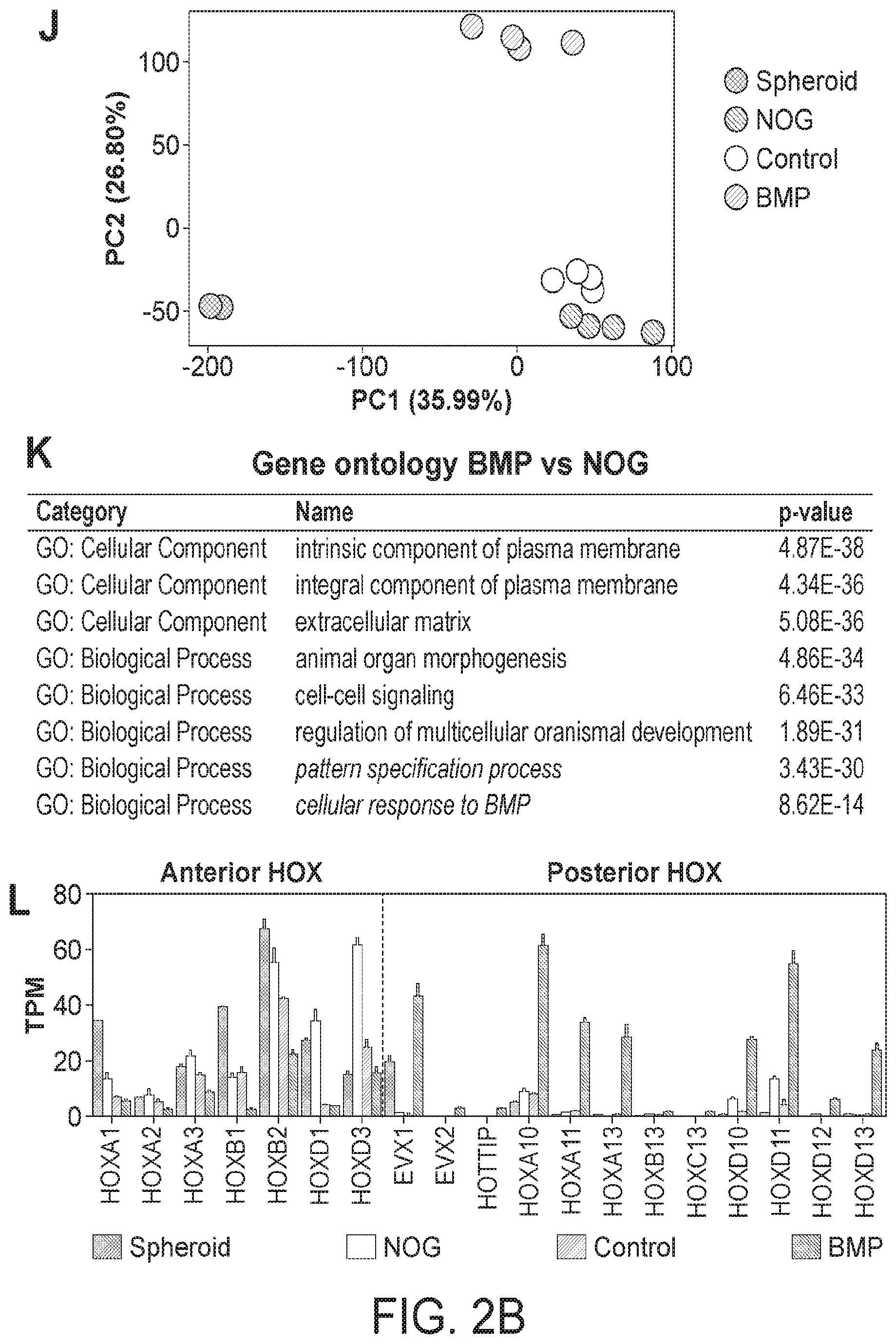
[0007] FIG. 2. BMP2 induces SATB2 and a posterior HOX code in human gut tube spheroids. (A) Schematic of gut tube spheroid patterning protocol. (B-D) BMP signaling levels as measured by pSMAD1/5/8 (red) staining of spheroids treated with NOGGIN (B), no treatment (C) and BMP2 (D) for 12 hours. (E) pSmad1/5/8 staining of adult mouse colon showing increased BMP signaling at to the top of crypts. (F-H) SATB2 expression in spheroids treated with NOGGIN (F), no treatment (G) and BMP2 (H) for 72 hours. (I) Quantification of the percentage of SATB2+ CDH1+ epithelium following patterning. (J) Principal component analysis of nascent spheroids and spheroids after 3 days of patterning. (K) Gene ontology analysis of differentially expressed genes between BMP vs NOG treated spheroids. (L) Graph of TPM (Transcripts per million) values of spheroids before and after patterning. Samples analyzed were spheroids before patterning (n=2), and NOGGIN, Control and BMP2 treated spheroids 3 days after patterning (n=4 for each group). For quantification in I, 20 organoids from at least 3 experiments were examined. Error bars represent SD. Scale bars=50 microns. ****p s 0.0001 determined by 2 tailed t-test.
[0008] FIG. 3. Regional patterning is maintained in human intestinal organoids following prolonged in vitro culture. (A-D) Whole-mount immunofluorescence and QPCR analysis with the proximal marker ONECUT1 (green) of 28-day old organoids that resulted from the initial 3 day treatment of spheroids with NOGGIN, control, or BMP2. Staining with CDX2 (red) and DAPI (blue) were also used to detect the epithelium and mesenchyme. (E-H) Expression of the posterior marker SATB2 (red) detected by IF and by QPCR. (I-L) Analysis of the pan-goblet cell marker MUC2 (red) by IF and by QPCR. (M-P) Analysis of the colon-specific goblet cell marker MUC5B (red) by IF. The number of MUC5B+ cells was quantified in (P). (Q-S) Analysis of patterning markers in isolated mesenchyme cultures relative to whole organoids. QPCR analysis of CDH1 (Q), the proximal HOX gene HOXD3 (R), and the distal HOX gene HOXA13 (S) in whole organoids and in mesenchyme cultures derived from NOGGIN, control, or BMP2 treated organoids. CDH1 was only observed in whole organoids that contained epithelial cells. Error bars represent SEM. For IF minimum of 10 organoids from at least 3 different experiments were examined for each condition. For QPCR a minimum of 5 biological replicates from 2 separate experiments were examined. Scale bars=100 microns. **p 5 0.01 and ****p 5 0.0001 determined by 2 tailed t-test.
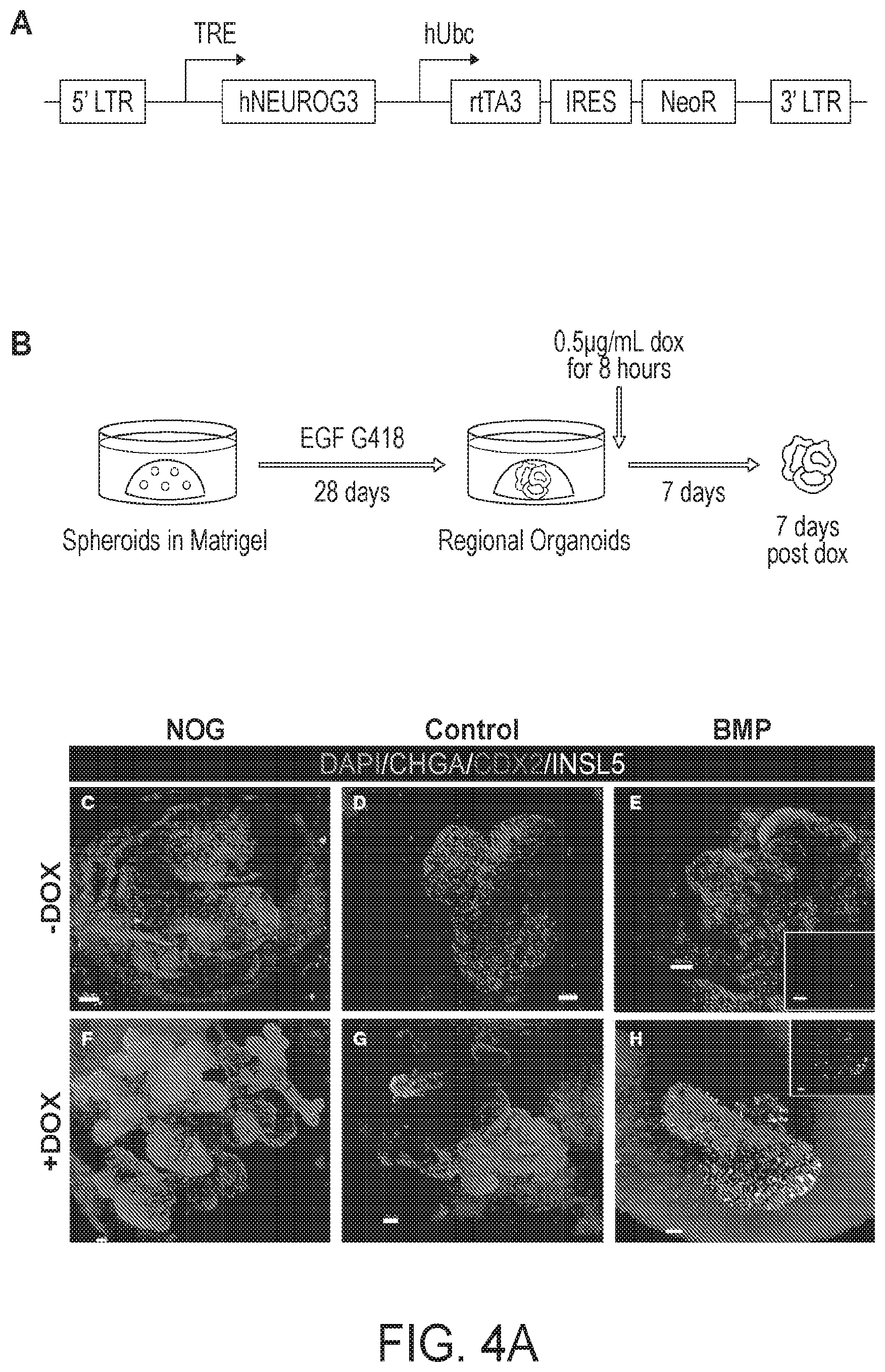
[0009] FIG. 4. HCOs but not HIOs gave rise to colon-specific enteroendocrine cells in response to expression of the proendocrine transcription factor NEUROGENIN 3. (A-B) Schematic of the doxycycline inducible NEUROG3 lentiviral construct used to generate the IPSC72.3 inducible NEUROG3 line, and the doxycycline induction protocol. Whole-mount staining with Chromagranin A (green), CDX2 (red) and INSL5 (white) of 35 day old organoids patterned with NOGGIN (C,F), untreated (D,G) or BMP (E,H). (C-E) Untreated organoids (-Dox) and (F-H) organoids with expressed NEUROG3 (+Dox). Insets in E and H show a magnified view of INSL5 staining. (I, J) QPCR analysis of NEUROG3 induction of enteroendocrine cells in HIOs and HCOs as measured by CHGA (I) and for INSL5 (J) expression. Data is representative of 2 different experiments with NOGGIN (n=3), Control (n=3) or BMP (n=6) treated organoids. Error bars represent SEM. Scale bars=50 microns. *p<0.05 determined by 2 tailed t-test.
[0010] FIG. 5. HIOs and HCOs maintained regional identity following transplantation in vivo. (A-E) H&E staining of biopsies from human jejunum and colon and of NOGGIN-derived HIOs, control HIOs, and BMP2-derived HCOs that were transplanted underneath the mouse kidney capsule and grown for 8-10 weeks in vivo. The samples of the same conditions were stained with the proximal intestinal marker GATA4 (F-J), the distal intestinal marker SATB2 (K-0), the Paneth cell marker DEFAS (P-T), and the colon-specific goblet cell marker MUC5B (U-Y). Note that although GATA4 and SATB2 double staining was done in different channels but on the same slides for panels (F-0), they are shown as individual pseudo-colored (red) images. For human biopsies n=2. For transplanted NOGGIN treated organoids n=12, for control organoids n=7, and for BMP2 treated organoids n=16. Scale bars=50 .mu.m.
[0011] FIG. 6. In vivo grown organoids express region-specific hormones. Analysis of expression of the regionally expressed hormones (A-D) Ghrelin (GHRL), Motilin (MLN), (E-H) GIP, (I-L) GLP-1, (M-P) PYY and (Q-T) INSL5 in HIOs and HCOs grown for 8-10 weeks underneath the mouse kidney capsule. The proximally enriched hormones GHRL, GIP and MLN were enriched in NOGGIN and control HIOs (A-H). The distally enriched hormones GLP-1 and PYY were enriched in BMP2-derived HCOs (1-0). The colon specific hormone INSL5 was only present in HCO (Q-T). Data is representative of a minimum of 5 transplanted organoids per condition. Insets in (A) and (B) show GHRL and MLN double positive cells. (D, H, L, P, T) FPKM values for GHRL, MLN, GIP, GLP1, PYY, and INSL5 are from RNA-seq data. FPKM values represent 3 biological replicates per condition. Scale bars=30 microns.
[0012] FIG. 7. Global transcriptional analysis of HIOs and HCOs and comparison with human small intestine and colon. (A) Principal component analysis human adult and fetal small intestine and colon compared with transplanted HIOs and HCOs. (B) Hypergeometric means test comparing human adult small intestine with HIOs and human adult colon with HCOs. (C) 4-way scatter plot comparing transcripts that were differentially expressed in human small intestine and colon compared to HIOs and HCOs.
[0013] FIG. 8. Gata4 and Satb2 mark discreet regional boundaries during development of the small and large intestines. (A) Whole-mount staining of Gata4 (green) and Satb2 (red) in an e9.5 mouse embryo showing expression boundary at the yolk stalk (n=9). (B) Model depicting Gata4 and Satb2 expression domains el 1.5 intestine showing a transitional zone of low Gata4 and low Satb2 expression. (C-E) Whole-mount staining of Gata4 and Satb2 in an e11.5 mouse embryo showing posterior boundary of Gata4 and anterior boundary of Satb2 at the yolk stalk (n=3). (F-H) Whole-mount staining of Satb2 and Foxa2 in an e12.5 mouse embryo showing that the anterior boundary of Satb2 expression is maintained (n=3). (I) Whole-mount staining of Gata4 and Satb2 in proximal intestine isolated from an e16.5 mouse embryo (n=6). (J) Whole-mount staining of Gata4 and Satb2 in distal small intestine and large intestine isolated from an e16.5 mouse embryo (n=6). Staining of GATA4 and SATB2 in section of (K) human jejunum (n=2) and (L) colon (n=2). Scale bars=50 am (B-D) and 100 1 Am (E-M). Dotted lines in (C) and (F) mark the approximate location of the umbilicus. Abbreviations: ys, yolk stalk; cb, cecal bud; tz, transition zone; mx, maxilliary; and md, mandibular portion of first brachial arch; ti, terminal ileum; icj, ileocecal junction.
[0014] FIG. 9. SATB2 is expressed in GATA4 negative human small and large intestine. SATB2 staining in human adult duodenum, small intestine, appendix, colon and rectum showing that SATB2 expression is present in distal small intestine and the entire large intestine. Analysis of GATA4 and SATB2 from published RNA-seq data from human adult and fetal intestinal samples. Samples plotted include human adult duodenum (HuSI_Duo_A), human adult small intestine distal to duodenum (HuSI_Dist_A), human adult colon (HuColon_A) and human fetal small intestine (HuSI_F). (C) Analysis of GATA4 and SATB2 expression from microarray data generated by Wang et al. 2015 on fetal intestinal stem cells from duodenum (Duo), jejunum (Jej), ileum (lle), ascending colon (AC), transverse colon (TC) and Descending colon grown in Air Liquid Interface (ALI). r2 values were determined using CORREL function in Excel.
[0015] FIG. 10. BMP mediates SHH activation of posterior HOX genes. (A) Previous model of SHH-mediated activation of posterior HOX genes. (B) New model of SHH mediated activation of posterior HOX genes and BMP-mediated activation of endoderm HOX genes. (C) QPCR analysis of HOX factors following treatment with NOGGIN, control, Smoothened agonist (SAG), or BMP2. (D) Model of BMP4 dependent activation of HOX13 genes induced by SAG. (E) QPCR analysis of HOXA13 in control, 5 .mu.M SAG, 5 .mu.M SAG+NOG and BMP2 treated organoids after 3 days. (F) Model of SHH independent activation of HOX13 genes induced by exogenous recombinant human BMP2. (G) QPCR analysis of HOXA13 in control, BMP, and BMP+Cyclopamine treated organoids after 3 days (n=6 per condition).
[0016] FIG. 11. Extended in vitro culture allows maturation of goblet cells. (A) Quantitation of the percentage of CDX2+ SATB2+ cells in organoids which were patterned and were then re-patterned. QPCR analysis of HOXB13 (B) and HOXD13 (C) in 28-day old organoids. (D-F) Whole-mount and (G-I) cross section staining with CDH1 (green), CDX2 (red), and MUC2 (white) from 44-day old NOGGIN, Control, and BMP treated organoids. (J-L) Staining of sections from 44-day old BMP2 treated organoids. White arrows points to goblet cells which were in the process of secreting Mucin 2. For QPCR a minimum of 5 biological replicates from 2 separate experiments were examined. For IF a minimum of 10 organoids per condition were examined. Scale bars=50 pm.
[0017] FIG. 12. BMP patterning of organoids is stable in vitro and in vivo. (A) Efficiency of organoid engraftment of NOGGIN, Control, and BMP patterned organoids. Quantitation of the percentage of GATA4+ CDX2+ cells (B) and SATB2+ CDX2+ cells (C) in transplanted patterned organoids. FPKM values from RNA-seq data for GATA4 (D) SATB2 (E) DEFAS (F) and MUCSB (G) in transplanted organoids. MUC2 (red) staining of (H-I) human jejunum and colon biopsies (n=2 per region) and (J-L) transplanted organoids (n=5 per condition). Scale bars=50 microns.
[0018] FIG. 13. In vitro and in vivo grown organoids contain intestinal progenitors. Representative whole-mount (A,F,K) and slice section (B,G,L) images of CDH1 and GFP from H9-LGR5-GFP derived organoids treated with NOGGIN, control, or BMP. CDX2 (red) and SOX9 (green) staining on sections from (C-E) NOGGIN, (H-J) control, or (M-O) BMP2 treated organoids. Representative images of CDX2 and LGR5-GFP (P, S,V), CDX2 and SOX9 (Q,T, W), and CDH1 and KI67 (R,U,X) stained in vivo organoids derived from H9-LGR5-GFP organoids treated with NOGGIN, control, or BMP. (Y-A') Stereomicrographs showing enteroids derived from NOGGIN, control or BMP transplants respectively. (B'-D') QPCR analysis of proximal and distal genes in control enteroids (>100 pooled enteroids from 2 transplants) and BMP2 treated colonoids (>50 colonoids from 1 transplant). Scale bars=50 nm.
[0019] FIG. 14. Ribosome and immune cell signatures are differentially expressed between transplanted organoids and primary human tissues. (A) Principal component analysis of patterned transplanted organoids and human adult and fetal small intestine and colon. (B) Gene ontology analysis of genes upregulated in transplants versus human primary tissues. (C) Gene ontology analysis of genes upregulated in human primary tissues versus transplants.
[0020] FIG. 15. (A) Wholemount immunofluorescence staining of HCOs after 15 days of growth in Matrigel. HCO cultures were stained for the endothelial marker CD31 (green) and the hindgut epithelium marker CDX2 (red). Cultures were also stained for the hematopoietic cell marker PU.1 (red right panel). (B) Schematic of hematopoietic progenitor assays. Cells were collected from HCOs, centrifuged and either stained using Giemsa Wright Stain or plated in Methocult media to assay for hematopoietic cell differentiation. (C) Representative images of Giemsa Wright stained cells with morphologies consistent with differentiation into Macrophages, Neutrophils, Basophils and Eosinophils. (D) Representative images of colonies formed after 14 days in Methocult. Erythrocyte, macrophage and granulocyte colonies were present in cells derived from HCOs but not those derived from NOGGIN treated HIOs.
[0021] FIG. 16. (A) Immunofluorescence staining of a human colon biopsy or an HCO grown for 28 days in Matrigel. Staining was done for CD68 a marker of macrophages. (B) Plots of CYTOF analysis of CD14 and CD16 in HIOs and HCOs. A small percentage of CD14+/CD16+ cells are present in HCOs (blue square) but not HIOs. Additionally, CD16 single positive cells were present in HCOs suggesting monocytes are present within the culture. (C) Luminex array analysis of supernatant collected from 14 and 28-day old HIOs and HCOs. IL6 and IL8 were detected in 28-day old HCOs (BMP) but not HIOs. (D) Luminex array analysis of supernatant collected from 14 and 28-day old HIOs and HCOs. The macrophage specific cytokines MIP1A and MIP1B were detected in 14 and 28-day old HCOs (BMP) but not in 14 or 28-day old HIOs.
DETAILED DESCRIPTION
Definitions
[0022] Unless otherwise noted, terms are to be understood according to conventional usage by those of ordinary skill in the relevant art.
[0023] The term "about" or "approximately" means within an acceptable error range for the particular value as determined by one of ordinary skill in the art, which will depend in part on how the value is measured or determined, e.g., the limitations of the measurement system. For example, "about" can mean within 1 or more than 1 standard deviation, per the practice in the art. Alternatively, "about" can mean a range of up to 20%, or up to 10%, or up to 5%, or up to 1% of a given value. Alternatively, particularly with respect to biological systems or processes, the term can mean within an order of magnitude, preferably within 5-fold, and more preferably within 2-fold, of a value. Where particular values are described in the application and claims, unless otherwise stated the term "about" meaning within an acceptable error range for the particular value should be assumed.
[0024] As used herein, the term "totipotent stem cells" (also known as omnipotent stem cells) are stem cells that can differentiate into embryonic and extra-embryonic cell types. Such cells can construct a complete, viable, organism. These cells are produced from the fusion of an egg and sperm cell. Cells produced by the first few divisions of the fertilized egg are also totipotent.
[0025] As used herein, the term "pluripotent stem cells (PSCs)," also commonly known as PS cells, encompasses any cells that can differentiate into nearly all cells, i.e., cells derived from any of the three germ layers (germinal epithelium), including endoderm (interior stomach lining, gastrointestinal tract, the lungs), mesoderm (muscle, bone, blood, urogenital), and ectoderm (epidermal tissues and nervous system). PSCs can be the descendants of totipotent cells, derived from embryonic stem cells (including embryonic germ cells) or obtained through induction of a non-pluripotent cell, such as an adult somatic cell, by forcing the expression of certain genes.
[0026] As used herein, the term "induced pluripotent stem cells (iPSCs)," also commonly abbreviated as iPS cells, refers to a type of pluripotent stem cells artificially derived from a normally non-pluripotent cell, such as an adult somatic cell, by inducing a "forced" expression of certain genes.
[0027] As used herein, the term "embryonic stem cells (ESCs)," also commonly abbreviated as ES cells, refers to cells that are pluripotent and derived from the inner cell mass of the blastocyst, an early-stage embryo. For purpose of the present invention, the term "ESCs" is used broadly sometimes to encompass the embryonic germ cells as well.
[0028] As used herein, the term "precursor cell" encompasses any cells that can be used in methods described herein, through which one or more precursor cells acquire the ability to renew itself or differentiate into one or more specialized cell types. In some aspects, a precursor cell is pluripotent or has the capacity to becoming pluripotent. In some aspects, the precursor cells are subjected to the treatment of external factors (e.g., growth factors) to acquire pluripotency. In some aspects, a precursor cell can be a totipotent (or omnipotent) stem cell; a pluripotent stem cell (induced or non-induced); a multipotent stem cell; an oligopotent stem cells and a unipotent stem cell. In some aspects, a precursor cell can be from an embryo, an infant, a child, or an adult. In some aspects, a precursor cell can be a somatic cell subject to treatment such that pluripotency is conferred via genetic manipulation or protein/peptide treatment.
[0029] In developmental biology, cellular differentiation is the process by which a less specialized cell becomes a more specialized cell type. As used herein, the term "directed differentiation" describes a process through which a less specialized cell becomes a particular specialized target cell type. The particularity of the specialized target cell type can be determined by any applicable methods that can be used to define or alter the destiny of the initial cell. Exemplary methods include but are not limited to genetic manipulation, chemical treatment, protein treatment, and nucleic acid treatment.
[0030] As used herein, the term "cellular constituents" are individual genes, proteins, mRNA expressing genes, and/or any other variable cellular component or protein activities such as the degree of protein modification (e.g., phosphorylation), for example, that is typically measured in biological experiments (e.g., by microarray or immunohistochemistry) by those skilled in the art. Significant discoveries relating to the complex networks of biochemical processes underlying living systems, common human diseases, and gene discovery and structure determination can now be attributed to the application of cellular constituent abundance data as part of the research process. Cellular constituent abundance data can help to identify biomarkers, discriminate disease subtypes and identify mechanisms of toxicity.
[0031] As described herein, methods and systems are established using a temporal series of growth factor manipulations to mimic embryonic intestinal development in culture. In particular, methods and systems are established to direct in vitro differentiation of PSCs, both human embryonic stem cells (hESC) and induced pluripotent stem cells (iPSC), into intestinal tissue
[0032] The generation of gastric and small intestinal organoids from pluripotent stem cells (PSCs) has revolutionized the study human gastrointestinal (GI) development and disease. However, efforts to generate large intestinal organoids have lagged behind, in part due to a robust molecular understanding of posterior gut tube development. Here, Applicant has found that the intestinal epithelium posterior to the umbilical cord expresses Satb2 throughout development and postnatally. Applicant has further found that BMP signaling establishes the Satb2+ domain in frog and mouse embryos, and that brief activation of BMP signaling was sufficient to activate a posterior HOX code and direct human PSC-derived gut tube cultures into colonic organoids (HCOs). HCOs grown in vitro had a marker profile and unique cell types consistent with colonic identity. Following transplantation into mice, HCOs underwent morphogenesis and maturation forming tissue with molecular, cellular and morphologic properties of the human colon. The disclosed colonic organoids may be used in future studies of colitis and colon cancer.
[0033] In one aspect, a method of inducing formation of a human colon organoid is disclosed. The method may comprise the steps of (a) contacting a definitive endoderm (DE) with an FGF signaling pathway activator and a WNT signaling pathway activator (for example, CHIRON/GSK2 inhibitor) for a period of time sufficient for said DE to form a mid-hindgut spheroid, and (b) contacting the mid-hindgut spheroid of step (a) with a BMP activator and an EGF signaling pathway activator for a period of time sufficient to form said human colon organoid, wherein said human colon organoid expresses SATB2.
[0034] In one aspect, the DE may be derived from a precursor cell selected from an embryonic stem cell, an embryonic germ cell, an induced pluripotent stem cell, a mesoderm cell, a definitive endoderm cell, a posterior endoderm cell, a hindgut cell or combinations thereof.
[0035] In one aspect, the FGF signaling pathway activator may be selected from a small molecule or protein FGF signaling pathway activator, FGF1, FGF2, FGF3, FGF4, FGF10, FGF11, FGF12, FGF13, FGF14, FGF15, FGF16, FGF17, FGF18, FGF19, FGF20, FGF21, FGF22, FGF23, or combinations thereof. The WNT signaling pathway activator may be selected from a small molecule or protein Wnt signaling pathway activator, preferably Lithium Chloride; 2-amino-4,6-disubstituted pyrimidine (hetero) arylpyrimidines; IQ1; QS11; NSC668036; DCA beta-catenin; 2-amino-4-[3,4-(methylenedioxy)-benzyl-amino]-6-(3-methoxyphenyl) pyrimidine, Wnt1, Wnt2, Wnt2b, Wnt3, Wnt3a, Wnt4, Wnt5a, Wnt5b, Wnt6, Wnt7a, Wnt7b, Wnt8a, Wnt8b, Wnt9a, Wnt9b, Wnt10a, Wnt10b, Wnt11, Wnt16, a GSK3 inhibitor, preferably CHIRON, or combinations thereof. In one aspect, the BMP activator may be selected from BMP2, BMP4, BMP7, BMP9, small molecules that activates the BMP pathway, proteins that activate the BMP pathway, and may include the following: Noggin, Dorsomorphin, LDN189, DMH-1, ventromophins, and combinations thereof.
[0036] In one aspect, the period of time sufficient for said DE to form a mid-hindgut spheroid may be determined by expression of CDX2 by said mid-hindgut spheroid of step (a). Such measurement is within the ability of one of ordinary skill in the art using routine methods.
[0037] In one aspect, the period of time sufficient for the mid-hindgut spheroid to form a human colon organoid is determined by expression of SATB2 and CDX2 by a cell of said human colon organoid, wherein when SATB2 and CDX2 is expressed, the mid-hindgut spheroid has formed a human colon organoid. Such measurement may be used in lieu of a temporal measurement, in that expression of the genes listed above indicates that steps (a) and (b) have been carried out for a sufficient duration of time.
[0038] In one aspect, an HCO obtained according to the methods described herein are disclosed. The HCOs of the instant invention may be characterized in a variety of different ways. In one aspect, the HCO may be characterized by the presence of colonic enteroendocrine cells (EEC). In one aspect, the HCO may be characterized by the presence of crypts and is substantially free of villi. In one aspect, the HCO may be characterized by the presence of colon-specific goblet cells. In one aspect, the HCO may be characterized by being substantially free of Paneth cells. In one aspect, the HCO may be characterized by the ability to secrete colon-specific hormone INSL5. The intestinal organoid may be free of one or more of an immune function, innervation, blood vessels, villi, and Paneth cells.
[0039] In one aspect, a method of forming colonic tissue is disclosed, wherein the HCO of the described invention may be engrafted under a kidney capsule of a mammal, preferably a rodent, preferably an immunocompromised rodent, preferably an immunocompromised mouse.
[0040] In one aspect, the HCOs disclosed herein may be used to determine the efficacy and/or toxicity of a potential therapeutic agent for a disease selected from colitis, colon cancer, polyposis syndromes, and/or irritable bowel syndrome. The method may comprise the step of contacting a potential therapeutic agent with an HCO as described herein, for a period of time sufficient to determine the efficacy and/or toxicity of said potential therapeutic agent.
[0041] In one aspect, an intestinal colonoid derived from the HCO of any preceding claim is contemplated.
[0042] In some aspects, stem cells that are pluripotent or can be induced to become pluripotent may be used. In some aspects, pluripotent stem cells are derived from embryonic stem cells, which are in turn derived from totipotent cells of the early mammalian embryo and are capable of unlimited, undifferentiated proliferation in vitro. Embryonic stem cells are pluripotent stem cells derived from the inner cell mass of the blastocyst, an early-stage embryo. Methods for deriving embryonic stem cells from blastocytes are well known in the art. For example, three cell lines (H1, H13, and H14) had a normal XY karyotype, and two cell lines (H7 and H9) had a normal XX karyotype. Human embryonic stem cells H9 (H9-hESCs) are used in the exemplary aspects described in the present application, but it would be understood by one of skill in the art that the methods and systems described herein are applicable to any stem cells.
[0043] Additional stem cells that can be used in aspects in accordance with the present invention include but are not limited to those provided by or described in the database hosted by the National Stem Cell Bank (NSCB), Human Embryonic Stem Cell Research Center at the University of California, San Francisco (UCSF); WISC cell Bank at the Wi Cell Research Institute; the University of Wisconsin Stem Cell and Regenerative Medicine Center (UW-SCRMC); Novocell, Inc. (San Diego, Calif.); Cellartis AB (Goteborg, Sweden); ES Cell International Pte Ltd (Singapore); Technion at the Israel Institute of Technology (Haifa, Israel); and the Stem Cell Database hosted by Princeton University and the University of Pennsylvania. Exemplary embryonic stem cells that can be used in aspects in accordance with the present invention include but are not limited to SA01 (SA001); SA02 (SA002); ES01 (HES-1); ES02 (HES-2); ES03 (HES-3); ES04 (HES-4); ES05 (HES-5); ES06 (HES-6); BG01 (BGN-01); BG02 (BGN-02); BG03 (BGN-03); TE03 (13); TE04 (14); TE06 (16); UC01 (HSF1); UC06 (HSF6); WA01 (H1); WA07 (H7); WA09 (H9); WA13 (H13); WA14 (H14).
[0044] In some aspects, the stem cells are further modified to incorporate additional properties. Exemplary modified cell lines include but not limited to H1 OCT4-EGFP; H9 Cre-LoxP; H9 hNanog-pGZ; H9 hOct4-pGZ; H9 inGFPhES; and H9 Syn-GFP.
[0045] More details on embryonic stem cells can be found in, for example, Thomson et al., 1998, "Embryonic Stem Cell Lines Derived from Human Blastocysts," Science 282 (5391):1145-1147; Andrews et al., 2005, "Embryonic stem (ES) cells and embryonal carcinoma (EC) cells: opposite sides of the same coin," Biochem Soc Trans 33:1526-1530; Martin 1980, "Teratocarcinomas and mammalian embryogenesis,". Science 209 (4458):768-776; Evans and Kaufman, 1981, "Establishment in culture of pluripotent cells from mouse embryos," Nature 292(5819): 154-156; Klimanskaya et al., 2005, "Human embryonic stem cells derived without feeder cells," Lancet 365 (9471): 1636-1641; each of which is hereby incorporated herein in its entirety.
[0046] Alternatively, pluripotent stem cells can be derived from embryonic germ cells (EGCs), which are the cells that give rise to the gametes of organisms that reproduce sexually. EGCs are derived from primordial germ cells found in the gonadal ridge of a late embryo, have many of the properties of embryonic stem cells. The primordial germ cells in an embryo develop into stem cells that in an adult generate the reproductive gametes (sperm or eggs). In mice and humans, it is possible to grow embryonic germ cells in tissue culture under appropriate conditions. Both EGCs and ESCs are pluripotent. For purpose of the present invention, the term "ESCs" is used broadly sometimes to encompass EGCs.
[0047] Induced Pluripotent Stem Cells (iPSCs)
[0048] In some aspects, iPSCs are derived by transfection of certain stem cell-associated genes into non-pluripotent cells, such as adult fibroblasts. Transfection may be achieved through viral vectors, such as retroviruses. Transfected genes include the master transcriptional regulators Oct-3/4 (Pouf51) and Sox2, although it is suggested that other genes enhance the efficiency of induction. After 3-4 weeks, small numbers of transfected cells begin to become morphologically and biochemically similar to pluripotent stem cells, and are typically isolated through morphological selection, doubling time, or through a reporter gene and antibiotic selection. As used herein, iPSCs include but are not limited to first generation iPSCs, second generation iPSCs in mice, and human induced pluripotent stem cells.
[0049] In some aspects, non-viral based technologies may be employed to generate iPSCs. In some aspects, an adenovirus can be used to transport the requisite four genes into the DNA of skin and liver cells of mice, resulting in cells identical to embryonic stem cells. Since the adenovirus does not combine any of its own genes with the targeted host, the danger of creating tumors is eliminated. In some aspects, reprogramming can be accomplished via plasmid without any virus transfection system at all, although at very low efficiencies. In other aspects, direct delivery of proteins is used to generate iPSCs, thus eliminating the need for viruses or genetic modification. In some embodiment, generation of mouse iPSCs is possible using a similar methodology: a repeated treatment of the cells with certain proteins channeled into the cells via poly-arginine anchors was sufficient to induce pluripotency. In some aspects, the expression of pluripotency induction genes can also be increased by treating somatic cells with FGF2 under low oxygen conditions.
[0050] More details on embryonic stem cells can be found in, for example, Kaji et al., 2009, "Virus free induction of pluripotency and subsequent excision of reprogramming factors," Nature 458:771-775; Woltjen et al., 2009, "piggyBac transposition reprograms fibroblasts to induced pluripotent stem cells," Nature 458:766-770; Okita et al., 2008, "Generation of Mouse Induced Pluripotent Stem Cells Without Viral Vectors," Science 322(5903):949-953; Stadtfeld et al., 2008, "Induced Pluripotent Stem Cells Generated without Viral Integration," Science 322(5903):945-949; and Zhou et al., 2009, "Generation of Induced Pluripotent Stem Cells Using Recombinant Proteins," Cell Stem Cell 4(5):381-384; each of which is hereby incorporated herein in its entirety.
[0051] In some aspects, exemplary iPS cell lines include but not limited to iPS-DF19-9; iPS-DF19-9; iPS-DF4-3; iPS-DF6-9; iPS(Foreskin); iPS(IMR90); and iPS(IMR90).
[0052] Definitive Endoderm
[0053] The HCOs of the instant disclosure may be derived from a simple sheet of cells called the definitive endoderm (DE). Methods for deriving definitive endoderm from precursor cells are well known in the art, as taught by D'Armour et al. 2005 and Spence et al. The anterior DE forms the foregut and its associated organs including the liver and pancreas and the posterior DE forms the midgut and hindgut, which forms the small and large intestines and parts of the genitourinary system. Studies using mouse, chick and frog embryos suggest that establishing the anterior-posterior pattern in DE at the gastrula stage is a prerequisite for subsequent foregut and hindgut development. The Wnt and FGF signaling pathways are believed to be critical for this process and act to promote posterior endoderm and hindgut fate and suppress anterior endoderm and foregut fate. The simple cuboidal epithelium of the hindgut first develops into a pseudostratified columnar epithelium, then into villi containing a polarized columnar epithelium and a proliferative zone at the base of the villi, which corresponds with the presumptive progenitor domain.
[0054] Applicant describes herein a robust and efficient process to direct the differentiation of DE into intestinal tissue, in particular human colon tissue, in vitro. Directed differentiation may be achieved by selectively activating certain signaling pathways in the iPSCs and/or DE cells.
[0055] Additional details of pathways relating to intestinal development in general are found in, for example, Sancho et al., 2004, "Signaling Pathways in Intestinal Development and Cancer," Annual Review of Cell and Developmental Biology 20:695-723; Logan and Nusse, 2004, "The Wnt Signaling Pathway in Development and Disease," Annual Review of Cell and Developmental Biology 20:781-810; Taipalel and Beachyl, 2001, "The Hedgehog and Wnt signalling pathways in cancer," Nature 411:349-354; Gregorieff and Clevers, 2005, "Wnt signaling in the intestinal epithelium: from endoderm to cancer," Genes & Dev. 19: 877-890; each of which is hereby incorporated by reference herein in its entirety. More details on the functions of signaling pathways relating to DE development can be found in, for example, Zorn and Wells, 2009, "Vertebrate endoderm development and organ formation," Annu Rev Cell Dev Biol 25:221-251; Dessimoz et al., 2006, "FGF signaling is necessary for establishing gut tube domains along the anterior-posterior axis in vivo," Mech Dev 123:42-55; McLin et al., 2007, "Repression of Wnt/{beta}-catenin signaling in the anterior endoderm is essential for liver and pancreas development. Development," 134:2207-2217; Wells and Melton, 2000, Development 127:1563-1572; de Santa Barbara et al., 2003, "Development and differentiation of the intestinal epithelium," Cell Mol Life Sci 60(7): 1322-1332; each of which is hereby incorporated herein in its entirety.
[0056] Any methods for producing definitive endoderm from pluripotent cells (e.g., iPSCs or ESCs) are applicable to the methods described herein. In some aspects, pluripotent cells are derived from a morula. In some aspects, pluripotent stem cells are stem cells. Stem cells used in these methods can include, but are not limited to, embryonic stem cells. Embryonic stem cells can be derived from the embryonic inner cell mass or from the embryonic gonadal ridges. Embryonic stem cells or germ cells can originate from a variety of animal species including, but not limited to, various mammalian species including humans. In some aspects, human embryonic stem cells are used to produce definitive endoderm. In some aspects, human embryonic germ cells are used to produce definitive endoderm. In some aspects, iPSCs are used to produce definitive endoderm.
[0057] In some aspects, one or more growth factors are used in the differentiation process from pluripotent stem cells to DE cells. The one or more growth factors used in the differentiation process can include growth factors from the TGF-beta superfamily. In such aspects, the one or more growth factors may comprise the Nodal/Activin and/or the BMP subgroups of the TGF-beta superfamily of growth factors. In some aspects, the one or more growth factors are selected from the group consisting of Nodal, Activin A, Activin B, BMP4, Wnt3a or combinations of any of these growth factors. In some aspects, the embryonic stem cells or germ cells and iPSCs are treated with the one or more growth factors for 6 or more hours; 12 or more hours; 18 or more hours; 24 or more hours; 36 or more hours; 48 or more hours; 60 or more hours; 72 or more hours; 84 or more hours; 96 or more hours; 120 or more hours; 150 or more hours; 180 or more hours; or 240 or more hours. In some aspects, the embryonic stem cells or germ cells and iPSCs are treated with the one or more growth factors at a concentration of 10 ng/ml or higher; 20 ng/ml or higher; 50 ng/ml or higher; 75 ng/ml or higher; 100 ng/ml or higher; 120 ng/ml or higher; 150 ng/ml or higher; 200 ng/ml or higher; 500 ng/ml or higher; 1,000 ng/ml or higher; 1,200 ng/ml or higher; 1,500 ng/ml or higher; 2,000 ng/ml or higher; 5,000 ng/ml or higher; 7,000 ng/ml or higher; 10,000 ng/ml or higher; or 15,000 ng/ml or higher. In some aspects, concentration of the growth factor is maintained at a constant level throughout the treatment. In other aspects, concentration of the growth factor is varied during the course of the treatment. In some aspects, the growth factor is suspended in media that include fetal bovine serine (FBS) with varying HyClone concentrations. One of skill in the art would understand that the regimen described herein is applicable to any known growth factors, alone or in combination. When two or more growth factors are used, the concentration of each growth factor may be varied independently.
[0058] In some aspects, populations of cells enriched in definitive endoderm cells are used. In some aspects, the definitive endoderm cells are isolated or substantially purified. In some aspects, the isolated or substantially purified definitive endoderm cells express the SOX17, FOXA2, and/or the CXRC4 marker to a greater extent than the OCT4, AFP, TM, SPARC and/or SOX7 markers. Methods for enriching a cell population with definitive endoderm are also contemplated. In some aspects, definitive endoderm cells can be isolated or substantially purified from a mixed cell population by contacting the cells with a reagent that binds to a molecule that is present on the surface of definitive endoderm cells but which is not present on the surface of other cells in the mixed cell population, and then isolating the cells bound to the reagent. In certain aspects, the cellular constituent that is present on the surface of definitive endoderm cells is CXCR4.
[0059] Additional methods for obtaining or creating DE cells that can be used in the present invention include but are not limited to those described in U.S. Pat. No. 7,510,876 to D'Amour et al.; U.S. Pat. No. 7,326,572 to Fisk et al.; Kubol et al., 2004, "Development of definitive endoderm from embryonic stem cells in culture," Development 131:1651-1662; D'Amour et al., 2005, "Efficient differentiation of human embryonic stem cells to definitive endoderm," Nature Biotechnology 23:1534-1541; and Ang et al., 1993, "The formation and maintenance of the definitive endoderm lineage in the mouse: involvement of HNF3/forkhead proteins," Development 119:1301-1315; each of which is hereby incorporated by reference herein in its entirety.
[0060] Definitive Endoderm to Mid/Hindgut Spheroids
[0061] In some aspects, posteriorized endoderm cells of the DE are further developed into one or more specialized cell types. Activin-induced definitive endoderm (DE) can further undergo FGF/Wnt induced posterior endoderm pattering, hindgut specification and morphogenesis, and finally a pro-intestinal culture system that promoted intestinal growth, morphogenesis and cytodifferentiation into functional intestinal cell types including enterocytes, goblet, Paneth and enteroendocrine cells. In some aspects, human PSCs are efficiently directed to differentiate in vitro into intestinal epithelium that may include secretory, endocrine and absorptive cell types. It will be understood that molecules such as growth factors may be added to any stage of the development to promote a particular type of intestinal tissue formation.
[0062] PSCs, such as ESCs and iPSCs, undergo directed differentiation in a step-wise or non-step-wise manner first into definitive endoderm (DE) then into mid/hindgut epithelium and mesenchyme (e.g., hindgut spheroids), and then into intestinal tissue. In some aspects, definitive endoderm cells and hESCs are treated with one or more growth factors.
[0063] In some aspects, soluble FGF and Wnt ligands are used to mimic early hindgut specification in culture to convert, through directed differentiation, DE developed from iPSCs or ESCs into hindgut epithelium that efficiently gives rise to all the major intestinal cell types. In human, directed differentiation of DE is achieved through selective activating certain signaling pathways that are important to intestinal development. It will be understood by one of skill in the art that altering the expression of any Wnt signaling protein in combination with any FGF ligand can give rise to directed differentiation as described herein.
[0064] More details are found, for example, in Liu et al., "A small-molecule agonist of the Wnt signaling pathway," Angew Chem Int Ed Engl. 44(13): 1987-1990 (2005); Miyabayashi et al., "Wnt/beta-catenin/CBP signaling maintains long-term murine embryonic stem cell pluripotency," Proc Natl Acad Sci USA. 104(13):5668-5673 (2007); Zhang et al., "Small-molecule synergist of the Wnt/beta-catenin signaling pathway," Proc Natl Acad Sci US A. 104(18):7444-7448 (2007); Neiiendam et al., "An NCAM-derived FGF-receptor agonist, the FGL-peptide, induces neurite outgrowth and neuronal survival in primary rat neurons," J Neurochem. 91(4):920-935 (2004); Shan et al., "Identification of a specific inhibitor of the dishevelled PDZ domain," Biochemistry 44(47):15495-15503 (2005); Coghlan et al., "Selective small molecule inhibitors of glycogen synthase kinase-3 modulate glycogen metabolism and gene transcription," Chem Biol. 7(10):793-803 (2000); Coghlan et al., "Selective small molecule inhibitors of glycogen synthase kinase-3 modulate glycogen metabolism and gene transcription," Chemistry & Biology 7(10):793-803; and Pai et al., "Deoxycholic acid activates beta-catenin signaling pathway and increases colon cell cancer growth and invasiveness," Mol Biol Cell. 15(5):2156-2163 (2004); each of which is hereby incorporated by reference in its entirety.
[0065] In some aspects, siRNA and/or shRNA targeting cellular constituents associated with the Wnt and/or FGF signaling pathways are used to activate these pathways.
[0066] Modulators/activators of the Wnt signaling pathway include Wnt1, Wnt2, Wnt2b, Wnt3, Wnt3a, Wnt4, Wnt5a, Wnt5b, Wnt6, Wnt7a, Wnt7b, Wnt8a, Wnt8b, Wnt9a, Wnt9b, Wnt10a, Wnt10b, Wnt11, and Wnt16. In some aspects, the modulation of the pathway may be through the use of small molecule modulators or protein modulators that activate the aforementioned pathways or proteins that activate the aforementioned pathways. For example, Small molecule modulators of the Wnt pathway included, but is not limited to Lithium Chloride; 2-amino-4,6-disubstituted pyrimidine (hetero) arylpyrimidines; IQ1; QS11; NSC668036; DCA beta-catenin; 2-amino-4-[3,4-(methylenedioxy)-benzyl-amino]-6-(3-methoxyphenyl) pyrimidine. Exemplary natural inhibitors of Wnt signaling include but are not limited to Dkk1, SFRP proteins and FrzB. In some aspects, the extrinsic molecules include but are not limited to small molecules such as WAY-316606; SB-216763; or BIO (6-bromoindirubin-3'-oxime). In some aspects, siRNA and/or shRNA targeting cellular constituents associated with the Wnt and/or FGF signaling pathways may be used to activate these pathways. It would be understood by one of skill in the art that the target cellular constituents include but are not limited to SFRP proteins; GSK3, Dkk1, and FrzB. Additional modulators include molecules or proteins that inhibit GSK3, which activates the Wnt signaling pathway. Exemplary GSK3 inhibitors include, but are not limited to: Chiron/CHIR99021, for example, which inhibits GSK30. One of ordinary skill in the art will recognize GSK3 inhibitors suitable for carrying out the disclosed methods. The GSK3 inhibitor may be administered in an amount of from about 1 uM to about 100 uM, or from about 2 uM to about 50 uM, or from about 3 uM to about 25 uM. One of ordinary skill in the art will readily appreciate the appropriate amount and duration.
[0067] Fibroblast growth factors (FGFs) are a family of growth factors involved in angiogenesis, wound healing, and embryonic development. In some aspects, it will be understood by one of skill in the art that any of the FGFs can be used in conjunction with a protein from the Wnt signaling pathway. In some aspects, soluble FGFs include and but are not limited to FGF4, FGF2, and FGF3. In some embodiments, the FGF signaling pathway is activated by contacting the precursor cell with one or more molecules selected from the group consisting of FGF1, FGF2, FGF3, FGF4, FGF10, FGF11, FGF12, FGF13, FGF14, FGF15, FGF16, FGF17, FGF18, FGF19, FGF20, FGF21, FGF22, and FGF23. In some embodiments, siRNA and/or shRNA targeting cellular constituents associated with the FGF signaling pathway may be used to activate these pathways. It will be understood by one of skill in the art that the methods and compositions described herein in connection with the Wnt and FGF signaling pathways are provided by way of examples. Similar methods and compositions are applicable to other signaling pathways disclosed herein.
[0068] In some aspects, DE culture is treated with the one or more modulators of a signaling pathway described herein for 6 or more hours; 12 or more hours; 18 or more hours; 24 or more hours; 36 or more hours; 48 or more hours; 60 or more hours; 72 or more hours; 84 or more hours; 96 or more hours; 120 or more hours; 150 or more hours; 180 or more hours; 200 or more hours, 240 or more hours; 270 or more hours; 300 or more hours; 350 or more hours; 400 or more hours; 500 or more hours; 600 or more hours; 700 or more hours; 800 or more hours; 900 or more hours; 1,000 or more hours; 1,200 or more hours; or 1,500 or more hours.
[0069] In some aspects, DE culture is treated with the one or more modulators of a signaling pathway described herein at a concentration of 10 ng/ml or higher; 20 ng/ml or higher; 50 ng/ml or higher; 75 ng/ml or higher; 100 ng/ml or higher; 120 ng/ml or higher; 150 ng/ml or higher; 200 ng/ml or higher; 500 ng/ml or higher; 1,000 ng/ml or higher; 1,200 ng/ml or higher; 1,500 ng/ml or higher; 2,000 ng/ml or higher; 5,000 ng/ml or higher; 7,000 ng/ml or higher; 10,000 ng/ml or higher; or 15,000 ng/ml or higher. In some aspects, concentration of signaling molecule is maintained at a constant throughout the treatment. In other aspects, concentration of the modulators of a signaling pathway is varied during the course of the treatment. In some aspects, a signaling molecule in accordance with the present invention is suspended in media comprising DMEM and fetal bovine serine (FBS). The FBS can be at a concentration of 2% and more; 5% and more; 10% or more; 15% or more; 20% or more; 30% or more; or 50% or more. One of skill in the art would understand that the regiment described herein is applicable to any known modulators of the signaling pathways described herein, alone or in combination, including but not limited to any molecules in the Wnt and FGF signaling pathways.
[0070] In aspects where two or more signaling molecules are used to treat the DE culture, the signaling molecules can be added simultaneously or separately. When two or more molecules are use, the concentration of each may be varied independently.
[0071] Expression of CDX2 may be used to reveal tendency of hindgut formation after DE have been incubated with an FGF signaling activator and a Wnt signaling activator, for example, FGF4 and Wnt3a, for a period of time, for example, for 12 hours or longer; 18 hours or longer; 24 hours or longer; 36 hours or longer; 48 hours or longer; 60 hours or longer; or 90 hours or longer. In some aspects, longer periods of incubation are needed to achieve a stable posterior endoderm phenotype as measured by prolonged expressed of CDX2. In such aspects, the periods of incubation can be for 60 hours or longer; 72 hours or longer; 84 hours or longer; 96 hours or longer; 108 hours or longer; 120 hours or longer; 140 hours or longer; 160 hours or longer; 180 hours or longer; 200 hours or longer; 240 hours or longer; or 300 hours or longer.
[0072] Alternatively, in some aspects, the absence of cellular constituents, such as foregut markers Sox2, Pdx1, Cldn18, and Albumin, can be used to reveal directed hindgut formation. In some aspects, intestinal transcription factors CDX2, KLF5 and SOX9 can be used to represent intestinal development. In some aspects, GATA6 protein expression can be used to represent intestinal development. In these aspects, the periods of incubation can be for 12 hours or longer; 18 hours or longer; 24 hours or longer; 36 hours or longer; 48 hours or longer; 60 hours or longer; or 90 hours or longer. Alternatively, the periods of incubation can be for 60 hours or longer; 72 hours or longer; 84 hours or longer; 96 hours or longer; 108 hours or longer; 120 hours or longer; 140 hours or longer; 160 hours or longer; 180 hours or longer; 200 hours or longer; 240 hours or longer; or 300 hours or longer.
[0073] In some aspects, abundance data of cellular constituents, for example, protein and/or gene expression levels, are determined by immunohistochemistry using primary and/or secondary antibodies targeting molecules in the relevant signaling pathways. In other aspects, abundance data of cellular constituents, for example, protein and/or gene expression levels, are determined by microarray analyses.
[0074] Still alternatively, morphological changes can be used to represent the progress of directed differentiation. In some aspects, hindgut spheroids are further subject to 3-dimensional culture conditions for further maturation. In other aspects, a highly convoluted epithelium surrounded by mesenchymal cells can be observed following hindgut spheroids formation. Additionally, intestinal organoids; polarized columnar epithelium; goblet cells; or smooth muscle cells can be observed in 6 days or longer; 7 days or longer; 9 days or longer; 10 days or longer; 12 days or longer; 15 days or longer; 20 days or longer; 25 days or longer; 28 days or longer; 32 days or longer; 36 days or longer; 40 days or longer; 45 days or longer; 50 days or longer; or 60 days or longer.
[0075] Mid/Hindgut Spheroids to Colon Organoids
[0076] It has been identified that, in addition to FGF and WNT signaling, Bone Morphogenetic Proteins (BMP) specifically BMP2 and BMP4, are capable of promoting a posterior/hindgut fate and repressing foregut fate. Additionally, BMP signaling regulates formation of distinct regional types of intestine. Inhibition of BMP with noggin after the hindgut stage promotes a proximal intestinal fate (duodenum/jejunum). Activation of BMP signaling after the hindgut stage promotes a more distal intestinal cell fate (cecum/colon).
[0077] Activation of BMP can be carried out by contacting the mid/hindgut spheroids with a BMP activator and an EGF signaling pathway activator for a period of time sufficient to form said human colon organoid. The demarcation of the incubation period may be defined by the point in time in which the human colon organoid expresses SATB2. Suitable BMP activators and EGF signaling pathway activators will be readily appreciated by one of ordinary skill in the art. Suitable BMP activators may include, for example BMP2, BMP4, BMP7, BMP9 and protein or small molecule agonists such as ventromorphins (Genthe et al. 2017) or proteins that serve as agonists. The BMP activator and EGF signaling pathway activator may be contacted with the mid-/hindgut spheroids for from about 1 day to about 3 days. BMP signaling may be activated within the first three days. In one aspect, the contacting step of the BMP activator and EGF signaling pathway activator is from 24 hours to about 10 days, or from about 48 hours to about 9 days, or from about 3 days to about 8 days, or from about 4 days to about 8 days, or from about 5 days to about 7 days. Suitable EGF activators may include, for example TGF alpha, HB-EGF, Amphiregulin, Epigen, Betacellulin and small molecules such as db-cAMP. The EGF activator may be contacted with the mid-/hindgut spheroids at a concentration of from about 10 ng/mL to 10,000 ng/ML, for a time period of from about 24 hours to about 10 days, or from about 48 hours to about 9 days, or from about 3 days to about 8 days, or from about 4 days to about 8 days, or from about 5 days to about 7 days.
[0078] The mid/hindgut spheroids may be contacted with a BMP activator and/or EGF activator at a concentration of 5 ng/ml or higher; 20 ng/ml or higher; 50 ng/ml or higher; 75 ng/ml or higher; 100 ng/ml or higher; 120 ng/ml or higher; 150 ng/ml or higher; 200 ng/ml or higher; 500 ng/ml or higher; 1,000 ng/ml or higher; 1,200 ng/ml or higher; 1,500 ng/ml or higher; 2,000 ng/ml or higher; 5,000 ng/ml or higher; 7,000 ng/ml or higher; 10,000 ng/ml or higher; or 15,000 ng/ml or higher, alone or combined. In some embodiments, concentration of signaling molecule is maintained at a constant throughout the treatment. In other embodiments, concentration of the molecules of a signaling pathway is varied during the course of the treatment. In some embodiments, a signaling molecule in accordance with the present invention is suspended in media comprising DMEM and fetal bovine serine (FBS). The FBS can be at a concentration of 2% and more; 5% and more; 10% or more; 15% or more; 20% or more; 30% or more; or 50% or more. One of skill in the art would understand that the regiment described herein is applicable to any known molecules of the signaling pathways described herein, alone or in combination
Examples
[0079] The following non-limiting examples are provided to further illustrate aspects of the invention disclosed herein. It should be appreciated by those of skill in the art that the techniques disclosed in the examples that follow represent approaches that have been found to function well in the practice of the invention, and thus can be considered to constitute examples of modes for its practice. However, those of skill in the art should, in light of the present disclosure, appreciate that many changes can be made in the specific aspects that are disclosed and still obtain a like or similar result without departing from the spirit and scope of the invention.
[0080] The epithelium of the gastrointestinal tract is derived from the definitive endoderm, one of the primary germ layers that are established during gastrulation. The process of gut tube morphogenesis transforms the definitive endoderm into a primitive gut tube with a foregut, midgut and hindgut. The midgut gives rise to the small and proximal large intestine and the hindgut gives rise to the distal large intestine and rectum (Zorn and Wells, 2009). The small intestine is further subdivided into 3 segments: The duodenum which is involved in absorption of nutrients and uptake of iron, the jejunum which is involved in the digestion and absorption of nutrients and the ileum which is involved in the absorption of bile acids and vitamin-B12 (Jeejeebhoy, 2002). The large intestine is subdivided in to the cecum, colon and rectum which are all involved in absorption of water and electrolytes (Jeejeebhoy, 2002). Although recent advances have shed light into the development of the small intestine (Finkbeiner et al., 2015; Spence et al., 2011; Watson et al., 2014), little is known about development of human large intestine/colon. Furthermore, diseases affecting this region of the gastrointestinal (GI) tract, colitis, colon cancer, polyposis syndromes and Irritable Bowel Syndrome are prevalent (Molodecky et al., 2012; Siegel et al., 2014; Zbuk and Eng, 2007). Animal models of polyposis syndromes and intestinal cancer are limited since polyps and tumors preferentially form in the small intestine and rarely in the colon or rectum (Haramis et al., 2004; He et al., 2004; Moser et al., 1990).
[0081] Applicant previously described a method in which human pluripotent stem cells can be differentiated into intestinal tissue through steps of directed differentiation that approximate embryonic development of the small intestine. First, pluripotent stem cells are differentiated into definitive endoderm by treatment with Activin A. Exposure of definitive endoderm to high levels of Wnt and FGF induces morphogenesis into mid/hindgut tube spheroids. Once formed, these midgut/hindgut spheroids, when grown in 3-dimensional culture under conditions that favor intestinal growth, transition through stages that approximate small intestinal development in vivo and form human intestinal organoids (HIOs) (Spence et al., 2011). HIOs have a small intestinal identity and have proven extremely useful for modeling small intestinal biology (Bouchi et al., 2014; Finkbeiner et al., 2015; Watson et al., 2014; Xue et al., 2013). However, until now, PSC-derived large intestinal organoids have not been developed, and given the prevalence of disease in the large intestine, such a system would allow for interrogation of development and disease mechanisms in this region of the GI tract.
[0082] To develop a method for generating large intestinal organoids, Applicant first identified Satb2 as a definitive marker of the presumptive large intestinal epithelium in frogs, mice, and humans. Using Satb2 as a marker, Applicant has shown that BMP signaling is required for specification of posterior gut endoderm of frogs and mice, consistent with the known role of BMP in posterior-ventral development (Kumar et al., 2003; Roberts et al., 1995; Sherwood et al., 2011; Tiso et al., 2002; Wills et al., 2008). Moreover, stimulation of BMP signaling in PSC-derived gut tube cultures for 3 days is sufficient to induce a posterior HOX code and the formation of SATB2-expressing colonic organoids. Human colonic organoids (HCOs) had a marker profile and cell types consistent with large intestine. Furthermore, HCOs, but not HIOs, formed colonic enteroendocrine cells (EEC) in response to expression of NEUROG3, demonstrating that HCOs were functionally committed to the colonic region. In addition, HCOs engrafted under the kidney capsule of immunocompromised mice and grown in vivo for 8-10 weeks, maintain their regional identify, formed tissues with colonic morphology, contained colon-specific cell types, had zones of proliferation and differentiation, as well as well-formed smooth muscle layers. Intestinal enteroids and colonoids that were derived from in vivo grown organoids maintained regional identify. Lastly, RNA-seq analysis demonstrated that HIOs and HCOs underwent substantial maturation and express regional markers consistent with a small and large intestinal identity respectively. In summary, Applicant identified an evolutionarily conserved BMP-HOX pathway in frogs and mice and used this to direct hindgut patterning and formation of human colonic organoids.
[0083] Results
[0084] SATB2 expression marks the gut endoderm of posterior embryonic and adult intestine.
[0085] The molecular pathways that establish the mid and hindgut, the presumptive small and large intestine, are poorly understood, in part due to a paucity of well-defined markers. This has limited the ability to direct the differentiation of human PSCs into regionally distinct intestinal organoids, in particular large intestinal organoids. Applicant therefore identified markers that distinguish different domains of the mouse embryonic gut tube and used these to interrogate signaling pathways that pattern the early intestine. Consistent with previous reports Applicant found that in e9.5 mouse embryos, Gata4 marked the gut endoderm from the posterior foregut to the yolk stalk (FIG. 8A) (Aronson et al., 2014; Battle et al., 2008; Beuling et al., 2008a; Beuling et al., 2007a; Beuling et al., 2007b; Beuling et al., 2010; Beuling et al., 2008b; Bosse et al., 2007; Kohlnhofer et al., 2016; Patankar et al., 2012a; Patankar et al., 2012b; Sherwood et al., 2009; Walker et al., 2014). At later stages of development (e11.5-e16.5), Gata4 continued to distinctly mark the anterior but not the posterior intestine (FIG. 8B-D,I-J). This expression domain remains intact into adulthood in both mice (not shown) and humans (FIG. 8K-L).
[0086] In order to identify markers of the posterior fetal intestine, Applicant mined public expression databases such as GNCPro.TM., TiGER and Human Protein Atlas for colon enriched genes (described in the Materials and Methods section) and found Satb2 as a potential marker of large intestine. Satb2 is a member of the CUT-class of homeobox genes (Holland et al., 2007), which binds nuclear matrix attachment regions and is involved in chromatin remodeling (Gyorgy et al., 2008). Immunostaining showed that Satb2 protein was first detected in the posterior endoderm of mouse embryos at e9-9.5 and formed a discreet expression boundary with Gata4 (FIG. 8A) at the yolk stalk, suggesting that the Satb2+ domain marks the posterior intestine, a broader expression domain than previously identified (Dobreva et al., 2006). Satb2 expression continued to mark the posterior intestinal endoderm throughout development (e11.5-16.5) (FIG. 8 B, C, E, F, H, J) and in the postnatal colon in mice (not shown) and humans (FIG. 8L). Using published human proteome and RNA-seq data, Applicant confirmed that GATA4 and SATB2 differentially mark proximal and distal regions of the human fetal and adult intestinal tract respectively (Bernstein et al., 2010; Fagerberg et al., 2014) (Wang et al., 2015) (FIG. 9A-C). These data demonstrate that the Gata4 and Satb2 expression boundaries are established early during development of mouse and marks future boundaries of the developing small and large intestine in mice and humans.
[0087] BMP Signaling is Required for Satb2 Expression in the Embryonic Hindgut Endoderm.
[0088] Applicant next used Satb2 as a marker to identify pathways that promote posterior intestinal fate in embryos. Applicant first determined if BMP signaling was active in the posterior gut tube, given its known role in patterning endoderm at several stages of development in zebrafish, Xenopus, chick and mouse (Kumar et al., 2003; Roberts et al., 1995; Sherwood et al., 2011; Tiso et al., 2002; Wills et al., 2008). Applicant observed that BMP signaling was highly active in the endoderm and mesoderm of the posterior gut tube of e8.5 mouse embryos as measured by phosphorylated Smad1/5/8 (pSMAD1/5/8) (FIG. 1A-B). To determine if BMP signaling is required for patterning of the posterior gut tube, Applicant cultured early headfold stage mouse embryos (e7.5) in the BMP signaling inhibitor DMH-1 (FIG. 1C). After 48 hours of DMH-1 treatment, Applicant saw a significant reduction in pSmad1/5/8 levels and a loss of Satb2 expression in the posterior gut tube (FIG. 2D-K). In addition, Satb2 expression was lost in the first brachial arch of DMH-1 treated embryos consistent with previous studies in Zebrafish (Sheehan-Rooney et al., 2013). DMH-1 had no impact on TGFI3 signaling as measured by pSmad2/3 levels (FIG. 1F). Given the evolutionary conservation of the Satb2 across vertebrate species (Li et al., 2006) Applicant investigated if BMP is required for Satb2 expression in the hindgut of frog embryos (FIG. 2L). Similar to mice, treatment of Xenopus embryos with DMH-1 (FIG. 1M-V), or transgenic expression of the BMP-antagonist Noggin (not shown) resulted in a loss of Satb2 expression in the hindgut and brachial arches. BMP signaling has been shown to directly regulate Satb2 expression in mouse embryonic mandibles through direct binding of Smad1/5 to a conserved enhancer (Bonilla-Claudio et al., 2012), suggesting that Satb2 may be a direct BMP target in the gut as well. Taken together these results revealed a conserved pathway in vertebrates whereby BMP signaling is required for defining the posterior most region of the developing gut tube that gives rise to the distal ileum and large intestine.
[0089] BMP Signaling Promotes Posterior Fate in Human Gut Tube Cultures.
[0090] Applicant next investigated if BMP signaling could be used to promote a posterior gut tube fate in humans using nascent CDX2+ gut tube spheroids derived from human PSCs as previously described (Spence et al., 2011). Applicant either inhibited or activated BMP signaling using the BMP inhibitor NOGGIN or BMP2 respectively (FIG. 2A) and monitored BMP signaling levels by accumulation of nuclear pSMAD1/5/8. Control cultures had low levels of pSMAD1/5/8 protein and addition of NOGGIN abolished this staining (FIG. 2B-D). In contrast, addition of BMP2 caused a rapid accumulation of pSMAD158 in both epithelial and mesodermal cells suggesting both cell types respond to BMP signals similar to what Applicant observed in mouse embryos (FIG. 1A-B). The specificity of pSmad1/5/8 staining was confirmed using adult mouse colon, which showed pSmad1/5/8 staining restricted to the differentiated compartment of the upper crypt (FIG. 2E) as previously reported (Hardwick et al., 2004; van Dop et al., 2009; Whissell et al., 2014). Further analysis of organoids revealed that 3 days of BMP2 treatment was sufficient to induce high levels of SATB2 protein in the epithelium compared to NOGGIN and control cultures (FIG. 2F-I). This suggests that a short pulse of BMP activity is sufficient to pattern spheroid endoderm into a posterior gut tube fate.
[0091] While BMP signaling is known to regulate anterior-posterior patterning of the endoderm, little is known about the transcriptional networks that ultimately confer positional identity along the A-P axis in mammals. Applicant used human gut tube spheroids and RNA-seq to identify how BMP signaling establishes posterior domains in the developing human gut. Principal component analysis revealed that gut tube spheroids treated with BMP for 3 days clustered separately from NOGGIN and control treated organoids (FIG. 2J). Examination of gene ontology terms (GO terms) revealed that modulation of BMP signaling affects multiple biological processes including organ morphogenesis, cell-cell signaling, pattern specification and cellular response to BMP signaling (FIG. 2K). The most definitive regulators of A-P patterning are HOX genes, and Applicant found that BMP activation resulted in down regulation of anterior HOX genes and up regulation of posterior HOX genes (FIG. 2L). In particular Applicant saw BMP-mediated increases in multiple paralogs of HOX10, 11, 12 and 13 groups. These results demonstrate that BMP signaling broadly regulates A-P hox code during patterning of the human gut tube and suggest a mechanism by which the distal GI tract is initially specified.
[0092] BMP Signaling Acts Downstream of SHH to Induce a Posterior Hox Code.
[0093] Previous studies suggest that Sonic Hedgehog (Shh) acts upstream of Bmp4 and Hox13 expression during posterior gut patterning in chick embryos (FIG. 10A) (Roberts et al., 1995). However, the relative epistatic relationship between BMP and Hox13 (FIG. 10B) was not investigated due to embryonic lethality caused by Bmp4 overexpression in the midgut and hindgut (De Santa Barbara et al., 2005; Roberts et al., 1995). Applicant used human gut tube cultures to better model the epistatic relationship of SHH-BMP-HOX13 during posterior gut tube patterning. Activation of hedgehog signaling with the smoothened agonist SAG led to a concentration dependent activation of the BMP signaling target gene MSX2 and the mesenchymal HOX factors, HOXA13 and HOXD13 (FIG. 10C). However, SAG-mediated activation of these factors was only a fraction of the activation mediated by BMP2 (FIG. 10C). Applicant further showed that the ability of HH signaling to activate HOXA13 was entirely dependent on BMP (FIG. 10D-E), confirming that BMP signaling functions downstream of SHH as previously reported (Shyer et al., 2015; Walton et al., 2012; Walton et al., 2009; Walton et al., 2016). It has not been determined if BMP signaling is sufficient to activate the posterior HOX program downstream of HH signaling. Applicant therefore examined HOXA13 induction by BMP in the presence of the SHH inhibitor Cyclopamine and found that BMP2 was sufficient to induce HOXA13 when SHH signaling is inhibited (FIG. 10F-G). Consistent with this, activation of SHH signaling during BMP patterning did not improve SATB2 expression (FIG. 11A). Experiments in Xenopus confirmed this epistatic relationship between SHH and BMP (data not shown) suggesting that this mechanism is evolutionarily conserved. Taken togetherApplicant's data suggest that BMP signaling is sufficient to activate the posterior HOX code and does so downstream of HH signaling.
[0094] BMP-Derived Organoids Cultured In Vitro Maintain a Distal Identity.
[0095] Applicant next investigated if 3 days of BMP treatment is sufficient to confer stable regional identity following extended culture of organoids for 25 days (FIG. 3). Levels of ONECUT1 (a marker of proximal small intestine) were highest in NOGGIN and control treated organoids and absent in BMP2 treated organoids (FIG. 3A-D). Conversely, SATB2 was absent in the epithelium of NOGGIN and control treated organoids but broadly expressed in nearly all of the CDX2+ epithelial cells of BMP2 treated organoids (FIG. 3 E-H, FIG. 11A). Importantly, modulation of BMP signaling had similar proximal-distal patterning effects on multiple human PSC lines, including embryonic stem cell lines H1 and H9 and induced pluripotent stem cell lines (IPSC 54.1 and IPSC 72.3) (shown below). Applicant frequently observed non-epithelial SATB2 expression in NOGGIN and control organoids (data not shown) possibly due to the presence of other cell types that are known to be present in HIOs in vitro (Spence et al., 2011). Examination of HOXB13 and HOXD13, which is expressed in posterior epithelium and mesenchyme respectively, further revealed that BMP treated organoids maintained posterior patterning following prolonged culture in vitro (FIG. 11B-C).
[0096] Goblet cells are distributed in a low-to-high gradient from proximal small intestine to distal large intestine (Rodriguez-Pineiro et al., 2013), and Applicant investigated if goblet cell numbers were lower in proximal and higher in distal organoids. Analysis of MUC2 staining at 28 days revealed that BMP2 treated organoids had high numbers of goblet cells as visualized by intracellular MUC2 (FIG. 3I-L) as compared to more proximal NOGGIN treated and control organoids, which only had rare intracellular MUC2 staining. Applicant further confirmed the regional identity of goblet cells using the marker MUC5B, which is expressed by a subset of goblet cells in the colon but not in the small intestine (van Klinken et al., 1998). MUC5B staining was absent in Noggin and control treated 28-day organoids but was present in BMP2 treated organoids (FIG. 4M-P). Goblet cell morphology became more mature in older organoids (FIG. 11D-I), where in 44-day old BMP treated organoids Applicant observed goblet cells in the process of secreting mucus into the lumen of the organoids (FIG. 11J-L). The ability to observe mucus secretion in BMP treated organoids suggests that this organoid system would be useful to study mucus secretion and the roles of mucus in intestinal pathophysiology.
[0097] While the regional pattern of organoids is stable after 28 days in culture, Applicant wanted to investigate if early patterning was fully established after the initial 3-day treatment. To do so, Applicant shifted 3-day NOGGIN-treated spheroids to BMP2-containing media for 3 days and conversely shifted 3-day BMP treated spheroids to NOGGIN-containing media for 3 days. Proximal organoids generated with NOGGIN did not express SATB2 in response to BMP2 demonstrating that proximal fate was stable following 3 days of patterning (FIG. 11A). In the converse experiment, while 3 days of BMP2 treatment was sufficient to induce a stable distal fate, a subset of organoids lost SATB2 expression in response to NOGGIN treatment (FIG. 11A). While 3 days of BMP2 treatment is sufficient to induce a colonic fate that is stable in vitro and in vivo (FIG. 12), there remains plasticity in the early posterior gut tube. This is consistent with the observation that the colonic endoderm of midgestation rat embryos is more regionally plastic than the small intestinal endoderm (Ratineau et al., 2003).
[0098] Patterning of Organoid Mesenchyme by BMP Signaling.
[0099] While stimulation of BMP signaling conferred regional identity to organoid epithelium, Applicant also observed pSMAD1/5/8 in the non-epithelial compartment of BMP2 treated organoids during patterning, and upregulation of posterior HOX factors known to be expressed in the mesenchyme. To determine if mesenchymal patterning was stable, or required continued patterning input from epithelium, Applicant isolated and expanded mesenchymal cell cultures for 2-3 weeks and analyzed them for expression of regional HOX genes. Mesenchymal cultures were lacking E-cadherin expressing cells, suggesting that they were primarily comprised of mesenchyme (FIG. 3Q). Analysis of HOXD3, which is enriched in proximal intestinal mesenchyme (Yahagi et al., 2004), confirmed that the mesenchyme from NOGGIN and control treated organoids have a stable proximal identity while BMP treated organoids had decreased expression of HOXD3 (FIG. 3R) and high levels HOXA13 (FIG. 3S), which continues to be expressed in human colon fibroblasts (Higuchi et al., 2015). Taken together, these data suggest that early modulation of BMP signaling patterns both the epithelium and the mesenchyme and that mesenchymal patterning is stable even in the absence of epithelium.
[0100] Induction of Colonic Enteroendocrine Cells is Restricted to BMP2 Treated Organoids.
[0101] The development of several ECC subtypes is regionally restricted to specific segments of small and large intestine. For example, expression of the protein INSL5 is restricted to colonic EECs (Burnicka-Turek et al., 2012; Thanasupawat et al., 2013). As a functional test of colonic identity, Applicant determined if experimental induction of the colonic EEC marker INSL5 was restricted to BMP2-treated distal organoids. To do this Applicant inducibly expressed the proendocrine transcription factor NEUROG3 using an iPSC line harboring a doxycycline (DOX) inducible NEUROG3 expression cassette (FIG. 4A) as previously described (McCracken et al., 2017; McCracken et al., 2014). Applicant performed a 6-hour pulse of DOX and after an additional 7 days in culture observed a robust induction of EECs as measured by CHGA positive cells (FIG. 4B-I). However, Applicant only observed INSL5 positive cells in BMP2 treated organoids and confirmed this by QPCR analysis (FIG. 4C-H,J). Given that INSL5-expressing cells are only in the colon, Applicant's data strongly suggest that BMP2-treated organoids are functionally committed to the colonic fate. The expression of distal markers like SATB2, MUC5B and HOXA13 and the competence to generate colon specific ECCs support the conclusion that BMP2 treated organoids are colonic, and thus will be referred to as human colonic organoids (HCOs).
[0102] Regional Identity of Patterned Organoids is Maintained In Vivo.
[0103] Previous studies of mouse and human fetal intestine have demonstrated that regional identity and tissue morphology of different regions of the intestine were maintained following orthotopic transplantation and growth in immunocompromised mice (Duluc et al., 1994; Savidge et al., 1995). To determine if HIOs and HCOs that were patterned in vitro would maintain regional identity and grow into small and large intestinal tissue, Applicant transplanted them under the mouse kidney capsule for 6-10 weeks, which Applicant previously demonstrated results in HIO maturation into small intestinal tissue (Watson et al., 2014). Applicant observed that the engraftment of NOGGIN and control HIOs was more efficient than HCOs (FIG. 12A). Consistent with their regional identity, transplanted HIOs and HCOs developed into mature tissues that morphologically resembled either small or large intestine, respectively (FIG. 5A-E). The epithelium of NOGGIN and control organoids formed well-defined crypts and tall villi, comparable to human small intestine. In contrast BMP2-treated organoids contained crypts but lacked villi, similar to colon.
[0104] In addition to their morphological resemblance to either small or large intestine, transplanted HIOs and HCOs expressed distinct regional markers and contained regionally enriched cell types. For example, the majority of the epithelium of NOGGIN and control HIOs expressed the proximal marker GATA4 and did not express the large intestinal marker SATB2 (FIG. 5F-I, K-N, FIG. 12B-E). Conversely HCO epithelia were uniformly SATB2+ but did not express GATA4 (FIG. 5J,0, FIG. 12B-E). In addition, Paneth cells expressing DEFA5 were present in the crypts of NOGGIN and control HIOs, but were absent HCOs (FIG. 5P-T, FIG. 12F) similar to the human colon (Wehkamp et al., 2006). Applicant further confirmed the colonic identity of HCOs using the colonic goblet cell marker MUC5B (van Klinken et al., 1998), which is expressed by a subset of goblet cells of HCOs but is not detectible in NOGGIN or control HIOs (FIG. 5U-Y, FIG. 12G). Additionally, the number of MUC2+ goblet cells was vastly higher in HCOs compared HIOs consistent with the abundance of goblet cells seen in the human colon (FIG. 12H-L). The patterning markers, the presence of MUC5B-expressing goblet cells, and the absence of Paneth cells all support the conclusion that transplanted HCOs have colonic epithelium.
[0105] In Vivo Matured HIOs and HCOs Express Regional Enteroendocrine Hormones.
[0106] There are at least 12 major EEC subtypes that are found in different regions of the gastrointestinal tract and Applicant analyzed HIOs and HCOs for the presence of regional EECs. Ghrelin and Motilin are found predominantly in the proximal intestine, and correspondingly these hormones were largely expressed in NOGGIN and control HIOs but not HCOs (FIG. 6A-D). Similarly, GIP, which is found in K-cells of the small intestine but is absent in the colon, were found in NOGGIN and control HIOs but not in HCOs (FIG. 6E-H). Applicant then examined presence of distally enriched EECs in HCOs by analyzing for expression of GLP-1 and PYY, which are more abundant in the colon. Applicant observed higher numbers of GLP-1 and PYY cells and higher expression of preproglucagon and PYY in HCOs than in HIOs (FIG. 61-P). In addition, Applicant found expression of the colon specific hormone INSL5 (Burnicka-Turek et al., 2012; Thanasupawat et al., 2013), only in HCOs (FIG. 6Q-T).
[0107] Analysis of Stem and Progenitor Cells in HIOs and HCOs In Vitro and In Vivo.
[0108] To determine if in vitro-derived HIOs and HCOs express markers of stem and progenitor cells, Applicant used the H9-BAC-LGR5-eGFP transgenic line that has been described previously (McCracken et al., 2014; Watson et al., 2014). Examination of LGR5-eGFP expression in organoids revealed expression in broad epithelial domains similar to the expression patterns in Lgr5-eGFP mice as early as e13.5 (Shyer et al., 2015) (FIG. 13 A, B, F, G, K, L). GFP expression was also evident outside the epithelium of organoids as determined by histology and FACS analysis which revealed a population of GFP+ EPCAM-cells (data not shown). In addition, Applicant examined the expression of SOX9, which is a marker of progenitor cells in the fetal and adult intestine and found it expressed in the epithelium of both HIOs and HCOs (FIG. 13C-E, H-J, M-0). These data suggest that embryonic/fetal intestinal progenitor cells, marked by LGR5-eGFP and SOX9, are present in HIOs and HCOs in vitro.
[0109] At later stages of intestinal development, progenitor cells become restricted to the base of developing villi, where they will eventually contribute to the intestinal stem cells (ISCs) of the crypts of Lieberkuhn. To determine if the progenitor cells that Applicant observed in vitro will undergo this this developmental transition, Applicant transplanted HIOs and HCOs and monitored LGR5-eGFP, SOX9, and KI67 protein. Following maturation of organoids in vivo, Applicant observed LGR5-eGFP, SOX9, and KI67 restricted to the base presumptive crypts (FIG. 13P-X). In addition, SOX9 was also observed in EECs in the villi of HIOs and in the cuff of the colonic epithelium transplanted HCOs consistent with SOX9 expression in these cell types. Given that Sox9 and Lgr5 mark intestinal and colonic stem cells capable of forming enteroids and colonoids in mice (Gracz et al., 2010; Ramalingam et al., 2012) Applicant investigated if the epithelium of transplanted organoids could be isolated and used to generate enteroids and colonoids. Both HIOs and HCOs gave rise to cultures of epithelial organoids that grew and could be passaged (FIG. 13Y-A'). Moreover, HCO-derived epithelial cultures expressed the colonic markers CKB, FXYD3, SATB2, and HOXB13 but did not express the proximal small intestine markers PDX1 or GATA4 suggesting that regional identity was maintained (FIG. 13B'-D'). These data suggest that HIOs and HCOs grown in vivo contain progenitor and stem cells.
[0110] Global Transcriptional Analysis of HIOs and HCOs.
[0111] In order to broadly interrogate the regional identity and maturation of HIOs and HCOs, Applicant performed RNA-seq analysis of HIOs and HCOs grown in vivo and compared them with published data sets of human fetal and adult small and large intestines. Principal component analysis revealed that primary tissues isolated from adult and fetal intestine clustered together along principle component 1 (PC1) axis, which accounted for 36.5% of the cumulative variation among samples (FIG. 14A). A GO analysis revealed that this variation was due to cell types that were present only in primary tissues and not PSC-derived transplants. For example, 6 of the top 10 biological processes present in human primary tissues and absent in transplants were related to immune cells (FIG. 14B-C). The second principle component (PC2) accounts for 17.7% of cumulative variation and separates the samples according to maturity (FIG. 7A). This component revealed that transplanted organoids are more mature than human fetal intestine and fetal colon but not as mature as adult colon and intestine. The third principle component (PC3) accounts for 6.7% of cumulative variation and separates the samples according to regional identity, and shows that HCOs are more similar to colon whereas HIOs cluster with small intestine (FIG. 7A). Interestingly, human fetal samples did not cluster based on regional identity (small intestine vs colon) suggesting that these samples may not have been cleanly isolated from the indicated region of the GI tract.
[0112] Applicant next used hypergeometric means test to determine the probability that HIOs and HCOs share similar patterns of region-specific gene expression small intestine and colon (FIG. 7B). A total of 341 transcripts are expressed in the small intestine and in NOGGIN treated HIOs as compared to colon or BMP2 treated HCOs, a proportion that is exceedingly unlikely by chance alone (P=1.5.times.10-143). Similarly, the gene set that is up-regulated in the control HIOs shares a highly significant degree of similarity with the gene set up-regulated in adult small intestine relative to the adult colon (P=2.5.times.10-203). Conversely, the gene set up-regulated in HCOs are highly enriched for genes that are up-regulated in the colon relative to the small intestine (P=4.1.times.10-53 and P=6.0.times.1073, respectively). This analysis concluded that HIO patterning is most similar to human small intestine and HCO patterning is colonic. To further explore the nature of HIOs (NOG and control treated) and HCOs, Applicant conducted differential expression analysis (adult small intestine vs. adult colon; HIOs vs. HCOs). Applicant generated 4-way scatter plot, which also demonstrated that a high proportion of genes up-regulated in the colon were also up-regulated in HCOs and the majority of genes up-regulated in the small intestine were also up-regulated in HIOs (FIG. 7C, Table 1). Lastly, analysis of biological processes that were enriched revealed that adult colon and transplanted HCOs have highly active Wnt signaling and a similar HOX code (FIG. 7D). Taken together, these data suggest Applicant has developed a robust method to differentiate PSCs into human colonic tissue.
TABLE-US-00001 TABLE 1 Genes upregulated in adult small intestine and colon which are also upregulated in HIOs and HCOs respectively. Column 1, Commonly upregulated in NOG HIOs vs HCOs & adult small intestine vs adult colon, Column 2, Commonly upregulated in Control HIOs vs HCOs & adult small intestine vs adult colon, Column 3, Commonly upregulated in HCOs vs NOG HIOs & adult colon vs adult small intestine, Column 4, Commonly upregulated in HCOs vs Control HIOs & adult colon vs adult small intestine ABCA4 AADAC AAGAB AAK1 ABCC2 ABCA4 ABCA12 ABHD10 ABCC6 ABCB8 ACTR2 ABHD17C ABCD1 ABCC10 ADAM10 ABL2 ABCG8 ABCC6 ADAM22 ACTR2 ABHD14A, ABHD14A- ACY1, ACY1 ABCD1 AGPS ACVR2B ABHD6 ABCG5 AGTPBP1 ACYP1 ACBD4 ABCG8 AKAP13, MIR7706 ADAM10 ABHD14A, ABHD14A-ACY1, ACO2 ACY1 ALDH1B1 ADAM17 ACOT11 ABHD16A ALG5 ADAM22 ACP2 ABHD4 ANKDD1B ADGRF4 ACSL5 ABHD6 ANKRD30BP2 AEBP2 ACY3 ACACB ANTXR2 AGPS ADAMTSL5 ACADS ANXA3 AHI1 AGPAT3 ACADVL ARFGEF3 AIDA AKR1B10 ACKR4 ARHGAP44 AIFM3 AKR1C1 ACO2 ARPC5 AKAP11 AKR1C3 ACOT11 ASCC1 AKAP13, MIR7706 ALDOB ACOT7 ASIP AKAP5 ALPI ACOX2 ATF6 ALG8 ANGPTL4 ACP2 ATOH1 ANKDD1B ANXA13 ACSL5 ATXN3 ANKFY1 APOA1 ACTN4 B3GALNT2 ANKS1B APOA1-AS ACY3 B3GNT6 ANP32B APOA4 ADAMTSL5 B4GALNT3 ANTXR2 APOBEC1 ADGRG4 BCAS1 AP2B1 APOBEC2 ADGRG5 BCL10 AP3M1 APOC2, APOC4, APOC4-APOC2 AGMO BCLAF1 APH1B APOC3 AGPAT1, MIR6721 BEND3 APOBR APOL2 AGPAT2 BEST2 ARFGEF3 AQP3 AGPAT3 BTBD3 ARHP1 AQP7 AK2 BZW1 ARL14EP AQP7P1 AKR1A1 C11orf58 ARMC8 ATG4D AKR1B10 C12orf75 ARPC5 AVIL AKR1C1 CA12 ARRDC3 BAK1 AKR1C3 CAMSAP1 ASB7 BCRP3 AKR1C4 CAPN2 ASPH BRE-AS1, RBKS AKR7A3 CAPRIN1 ATG4C BTD ALDH1A3 CASC18 ATL3 BTNL3 ALDH2 CBFB ATM CCPG1, DYX1C1, BTNL8 ALDOB DYX1C1-CCPG1 ATP13A3 C10orf25 ALPI CD24 ATP2A3 C10orf67 AMBRA1 CD9 ATR C11orf24 ANAPC2 CDC23 ATRX C11orf86 ANGPTL4 CEACAM6 ATXN1 C19orf66 ANPEP CEP290 ATXN3 C1orf115 ANXA13 CFC1, CFC1B B3GALNT2 C1orf116 ANXA4 CLIC4 B3GNT6 C5orf56 AP5B1 CLMN BAG5 C6orf132 APOA1 CLNS1A BAZ1B C6orf136 APOA1-AS CLSTN1 BBIP1 C8G APOA4 CNTN3 BCAS1 C9orf173 APOBEC1 CSRNP3 BCAT1 CA13 APOBEC2 DAAM1 BCLAF1 APOC2, APOC4, CALM3 APOC4-APOC2 DDX50 BEND3 CAMKK2 APOC3 DGKH BEST2 CAPN10-AS1 APOL2 DICER1 BIN1 CAPN3 APOM DIP2B BMPR2 CASP1 AQP3 DMTN BNC2 CATSPER2 AQP7 EARS2 BTBD10 CBR1 ARHGEF16 EBPL BTBD3 CCL11 ASAH2 EEA1 BTBD7 CCL25 ASB13 EHF BTF3L4 CD82 ASIC2 EPHA10 BTRC CDCA3 ASPA EPHB4 BZW1 CDH4 ATG4D EXOC5 C10orf99 CDIP1 ATP11A EXOC6 C11orf58 CDR2L ATP5SL FAM105A C18orf54 CERK ATXN2L FAM169A C3orf52 CES2 AZGP1 FAM175A C7orf60 CGREF1 B3GNT8 FAM218A CA12 CHRM4 BAG6 FAM60A CA4 CAMKID, CIB2 BAIAP2L2 FAS LOC283070 CIDEB BAIAP3 FECH CAMSAP2 CIDEC BAK1 FERMT1 CAPN8 CLDN15 BET1L FFAR4 CAPZA1 CMTR1 BLNK FGF7 CASC4 CNNM3 BRE-AS1, RBKS FKTN CASD1 CPS1 BTD FMN2 CBFB CRAT BTNL3 FRMPD3 CBX5, MIR3198-2 CREB3L3 BTNL8 G3BP1 CCDC132 CRIP3 BUD13 GARS CCDC88A CTD-3080P12.3 C10orf54 GCSH CCND1 CYP27A1 C11orf24 GGH CCNJL CYP2B7P C11orf86 GJB4 CCNT1 CYP2C9 C15orf62 GLIS3 CD24 CYP2S1 C16orf58 GLUL CD59 CYP3A5 C17orf78 GMNN CD9 CYTH2 C18orf8 GNE CDC42 DBP C19orf12 GNPTAB CDHR1 DCAF11 C19orf54 GOT2 CDK19 DECR1 C1orf116 GP9 CEACAM5 DGAT1, MIR6848 C2CD2L GRSF1 CEACAM6 DGAT2 C5orf56 GSPT1 CEBPZ DGKA C6orf132 GYG2 CELF2 DHDH C6orf136 HDAC1 CENPO DHRS1 C8G HIATL1 CEP250 DHRS11 CALCOCO1 HK2 CHD9 DHX16 CALM3 HMGA2 CHIC1 DMBT1 CAMK2G HN1L CLIC4 DNAJC22 CAMTA2 HNMT CLMN DNASE1 CAPN1 HNRNPAB CLNS1A DNPEP CAPNS1 HOXA10-AS CLTC HOXA10, HOXA10- DOLPP1 CARD10 HOXA9, HOXA9 CMTM6 DOT1L CARD6 HOXA11 CNEP1R1 DTX1 CASP1 HOXB9 CNTN3 ELMOD3 CASP4 HOXB-AS3 CPM EMB CASP9 IARS CRK ENKUR CBLC IFT74-AS1 CRYM EPHX2 CBR1 IL1R2 CSNK1A1 ERICH4 CC2D1A ILDR1 CSRNP3 ESPN CCL25 IPO5P1 CTDSPL CD302, LY75, LY75- ETV7 CD302 IQGAP1 CTTNBP2NL EXOC3L4 CD68 ITM2C CYLD F10 CD74 JAG1 CYP20A1 FAM102A CD82 JPH1 DAAM1 FAM109A CDC42BPB KCNJ2 DCBLD2 FBP1 CDC42EP4 KCNN4 DCP2 FBXO7 CDCA3 KCNRG, TRIM13 DDX50 FCHSD1 CDHR5 KCTD1 DDX6 FLJ12825 CDIP1 KCTD20 DGKH FLJ22763 CDK18 KLK15 DHRS13 FUOM CDK2 KNOP1 DHRS9 FXR2 CDK20 KPNA4 DHX57 GABRE CDR2L LEFTY1 DICER1 GALK1 CEACAM18 LIMK2 DIO3OS DISC1, TSNAX, GALT CELA3A LINC00341 TSNAX-DISC1 GATA4 CENPV LINC00858 DNAJC3 GATA5 CERS2 LIPH DNAL1 GATS CES2 LOC100507346 DPY19L1 GCHFR CFI LOC101928233 DSEL GIGYF1 CFL1 LOC101929395 DSTYK GNA11 CGREF1 LOC101929524 DTD2 GOLT1A CHP2 LRRK1 EBPL GOSR2 CHRM4 MAML2 EED, MIR6755 GPD1 CIAO1 MAPRE2 EFNA5 GPRIN1 CIB2 MARCH3 EHF GRAMD1B CIDEB MARCKSL1 EI24 GRIA1 CIDEC MBNL3 EID1 GSDMB CISD1 MCOLN2 EIF4E GSK3A CLDN15 MECOM EIF5B GSTK1 CLDN18 METTL5 EMC1 HADHA CLPTM1 MFAP3L ENAH HAGH CMBL MFHAS1 ENDOD1 HAPLN4 CNNM3 MINA ENTPD1 HEBP1 CNPY2, PAN2 MLLT3 EPAS1 HOPX COASY MPHOSPH6 ERCC6L2 HOXA4 COMMD9 MPLKIP ERMP1 HPS1 COMT, MIR4761 MPZL2 ERN2 IDH3A CPA2 MREG EXOC2 IFIT3 CPPED1 MRPL1 EXOC4 IGSF23 CRADD MRPL3 EXOC5 IL2RB CRAT MTL5 EXOC6 INTS12 CREB3L3 MYO3A FAM102B IRF1 CRELD1 MYO5C FAM103A1 IRF8 CS NAA15 FAM105A ISG20 CSK NAP1L1 FAM120A ITPK1 CTSO NCBP1 FAM13B KDM2A CXCR3 NDC1 FAM169A KDM6B CYB5A NEDD4L FAM178A KDM8 CYP27A1 NEURL1B FAM208A KHK CYP2C19 NLE1 FAM46A KIAA2013 CYP2S1 NOLC1 FAM83H-AS1 KIFC3 CYP3A4 NOTCH1 FAM8A1 KLC4 CYSLTR2 NOTCH2 FAM98B LBX2-AS1 DBP NRARP FANCI LINC00574 DCAF11 NRXN1 FAR2P2 LINC01268 DECR1 NSF, NSFP1 FBXO28 LOC100240735 DEDD NT5DC3 FBXO45 LOC284825 DEGS2 NUDT4 FCF1 LOC646471 DERA NXPE1 FEM1B LOC728989 DESI1 NXPE2 FEM1C LPCAT3 DFNA5 NXPE4 FFAR4 LPIN3 DGAT1, MIR6848 ODC1 FKBP5, LOC285847 LRRC75A DGAT2 ORC5 FKTN LRRC75B DGKA P4HA1 FLOT2 MALL DGKG PAPPA2 FNDC3B MAPKBP1 DGKQ PARM1 FOCAD METTL7B DGKZ PAWR FOXD2 MFSD2A DHDH PCDHB11 FOXO3 MGAM DHRS11 PDE3B FOXO3B, ZNF286B MICALL2 DHRS7 PDZK1IP1 FRMPD3 MICU1 DHX16 PGBD5 FRYL MIR1268A, SLC27A4 DMBT1 PHF20 FSIP2 MIR22, MIR22HG DNAJC22 PHF6 FZD4 MIR31HG DNASE1 PKIB G3BP1 MIR3615, SLC9A3R1 DNPEP PLCD3 GGH MIR5187, TOMM40L DOLPP1 PLSCR4 GIN1 MIR5193, UBA7 DPP9 POF1B GJC1 MIR6073, SOX6 DSCR3 POLR3B GLB1L2 MIR621, SLC25A15 DTX1 POSTN GLG1 MIR7703, PSME2 E2F4 PP14571 GLIS3 MISP EGFR-AS1 PPIC GLTSCR1L MME EIF6 PPP1R8 GLUL MMEL1 ENPP6 PPP2R3A GMNN MOCS1 EPB41L3 PPP3CA GNAI1 MOGAT3 EPHA1 PREP GNAQ MON1A EPHB1 PRKACB GNE MS4A8 EPHX2 PRKAR2A GOLGA3 MSRA EPS8L2 PRKRIR GOPC MST1 EPSTI1 PRMT5 GP9 MTTP ERAL1 PSMD6 GPC6 MUS81 ERF PSME4 GPX8 MYO15B ESPN PTAR1 GRM7 MYO1A ESRRA PTTG1IP GRSF1 NAGS ETV3 PUM1 GTF2F2 NELL2 ETV7 PYGL GTF3C1 NGEF EWSR1 QPCT HABP4 NOP9 EXOC3L4 R3HDM1 HEATR3 NPC1L1 F10 RAB3B HIATL1 NR0B2 FABP2 RABEP1 HMG20A NR1I3 FAH RAP1GDS1 HMGA2 FAM101A, ZNF664, NSUN6 ZNF664-FAM101A RAPGEF2 HN1L NUB1 FAM102A RBMS3 HOXA10-AS HOXA10, HOXA10- OGDH FAM109A RCC2 HOXA9, HOXA9 OTC FAM32A REXO2 HOXB5 P4HB FAM83G RIF1 HOXB6
PARP12 FBLIM1 RIMS3 HOXB7 PARP3 FBP1 RNASEH2B HOXB8 PATL2 FLJ22763 RPA2 HOXB9 PCK2 FUOM RSF1 HOXB-AS3 PCSK5 FZR1 RSL24D1 IFFO2 PDLIM2 GABRA4 RXFP4 IFNAR1 PDZD7 GAL3ST1 SATB2 IFT74-AS1 PDZK1 GALK1 SATB2-AS1 IGF1R PEBP1 GALNT6 SCLT1 IGIP PEPD GALT SERBP1 IL17RD PEX14 GATA4 SETBP1 IL1R2 PGRMC2 GATA5 SH3PXD2A-AS1 IL20RB PHEX GCNT4 SIPA1L2 IL6ST PKLR GFI1B SLC16A9 ILDR1 PLA2G6 GGT1 SLC1A3 IMPAD1 PLCB3 GLOD5 SLC39A8 INPP5F IPO11, IPO11- PLEKHS1 GLRX SLC7A2 LRRC70, LRRC70 PLIN2 GLYCTK SLC9A2 IPO5 PLIN3 GNA11 SLCO4A1-AS1 IPO5P1 PLLP GNB1 SMAD5 IPO7 PNP GOLT1A SMARCA5 IQGAP1 PP7080 GOSR2 SMC6 ITGAV PQLC2 GPD1 SNRPE ITGB1 PRAP1 GPR108 SNX13 ITM2A PRDM7 GPR35 SOCS5 ITPRIPL2 PRODH GRAMD1B SORBS2 JAG1 PSMB9 GRIA1 SPAG1 JPH1 PSMD9 GRK5 SRSF12 KBTBD6 PSME1 GRTP1-AS1 SRSF9 KCNJ2 PTPRH GSDMB ST6GAL2 KCNN4 PXDC1 GSK3A STAB2 KCNRG, TRIM13 RAB11FIP3 GSTA1 STMND1 KCTD10 RAB17 GSTA2 STS KCTD20 RAB5C GSTK1 STX19 KDM5B RAB8A GSTM4 SUSD1 KIAA0226L RARRES3 GTF2I SUV39H2 KIAA0232 RBP2 GTPBP1 TBL2 KIAA0513 REEP6 GUCD1 TCTA KIAA1143 REG1A HADHB TDGF1 KIAA1429 RPS6KA1 HAGH TFRC KIAA1715 RTKN HAPLN4 TMA16 KLHL15 RTP4 HDAC6 TMCC1-AS1 KLK15 SAT1 HDGF TMED10 KPNA4 SAT2 HDHD3 TMED2 KRR1 SCAMP5 HEBP1 TMEM123 LAPTM4A SCNN1D HECTD3 TMEM159 LARS SCRN2 HIP1R TMEM200B LEFTY1 SDHA HLA-F TMEM38A LIFR SEC14L2 HMGA1 TRABD2A LIMD1 SERP2 HNF1A TSN LIMK2 SFRP5 HNF4A-AS1 TSPAN5 LINC00341 SFXN3 HOPX TTC3 LINC00482 SH3BP1 HPS1 TTC8 LINC00515 SH3GL1 HRASLS2 TTPA LINC00657 SHBG HRH2 UBE2A LINC01006 SIDT2 HSD3B7 UBE2N LMAN2L SLC12A7 HYKK UGP2 LOC100129550 SLC15A1 IDNK UNC13B LOC100507351 SLC1A7 IFIT3 URB1 LOC101929374 SLC22A4 IFNLR1 VWA3B LOC101929524 SLC23A1 IGSF23 WDHD1 LOC105372441 SLC25A20 IL22RA1 WDR35 LOC731424 SLC25A34 IL2RB WDR78 LOC93622 SLC25A44 ILVBL WIPI1 LRCH2 SLC25A45 IMMP2L WNK4 LRRC37A4P SLC26A11 INPP5J WWP1 LRRC58 SLC2A2 INTS12 XPO4 LUZP6 SLC35B1 IQSEC2 YWHAE LYST SLC37A4 IRF1 ZBTB7C MAFA SLC39A5 ISG20 ZBTB8B MAGEF1 SLC3A2 ITM2B ZFHX3 MAGT1 SLC52A1 ITPK1 ZNF658 MAL2 SLC5A1 ITPKA ZNF774 MAP3K7 SLC5A9 KALRN ZNF780B MARCKS SLC6A19 KDF1 MATR3, SNHG4 SLC6A20 KDM6B MBNL2 SLC7A7 KDM8 MBNL3 SMIM24 KHK MBOAT2 SMIM5 KIAA0141 MBTPS2 SMLR1 KIAA1551 MDN1 SMPD3 KIAA2013 MECOM SOWAHA KLC4 METTL8 SPNS3 KLHDC8B MFAP3L ST7, ST7-OT3 LASP1 MFSD4 STRC LBX2-AS1 MFSD6L SULT1A1 LEAP2 MIB1 SULT1A2 LHPP MIER1 SYK LINC00319 MIOS SYP LINC00330 MIR1244-4, PTMA TAP2 LINC00483 MIR4680, PDCD4 TCF7 LINC00574 MIR6824, SLC26A6 TICAM1 LINC00667 MLF1 TKFC LINC01137 MLLT3 TM4SF20 LINC01347 MLXIP TM4SF4 LIPE MMGT1 TM4SF5 LIPT1 MOB1B TM6SF2 LMBR1L MON2 TMEM150B LOC100093631 MORF4L1 TMEM184A LOC100506302 MPZL1 TMEM253 LOC100507334 MREG TMEM37 LOC101927051 MRPL1 TMEM41A LOC284825 MRPS6, SLC5A3 TMEM82 LOC90768 MTHFD2 TMEM86B LPCAT3 MTMR6 TNFRSF14 LRP5 MTSS1 TNFRSF1A LRRC28 MTURN TNRC6C-AS1 LRRC41 MUC1 TOM1 LRRC66 MUC12 TREH LRRC75A MYO3A TRIM15 LSMEM2 MYO5C TRIM50 LYRM5 NAA15 TTC31 LZTS3 NAA50 TTC38 MALL NAP1L1 NBPF10, NBPF12, NBPF20, NBPF25P, UGT2B7 MAP2K3 NBPF8, NBPF9 UGT3A1 MAP3K11 NCBP1 USH1C MAPK3 NCOA3 WBP2 MAPKAPK2 NDC1 WNT3 MAPKBP1 NEK1 XAF1 MARC2 NEURL1B XDH MBD1 NFIA XPNPEP2 MCRS1 NFYB ZMYND15 MCUR1 NIFK-AS1 ZNF300 MEP1A NKIRAS1 ZSWIM8 MEP1B NOL11 METTL17 NOL9 METTL7B NOTCH1 MFSD2A NPAS1 MGAM NRXN1 MGAM2 NSF, NSFP1 MGAT3 NT5C2 MGST3 NT5DC3 MICAL1, ZBTB24 NUBPL MICU1 NUCKS1 MIR1268A, SLC27A4 NUDT4 MIR22, MIR22HG NUP133 MIR3615, SLC9A3R1 NUP205 MIR5187, TOMM40L NUS1 MIR5193, UBA7 NXPE1 MIR639, TECR NXPE4 MIR7109, PISD OPHN1 MIR7703, PSME2 ORC5 MISP PARM1 MLF2 PBRM1 MLX PCM1 MLXIPL PDE3B MME PDE4D MMEL1 PDS5A MMP24 PDS5B MOCOS PEAK1 MOCS1 PGBD5 MOGAT2 PGGT1B MOGAT3 PGM2L1 MOGS PHC3 MON1A PHF14 MOV10 PHF20 MPP1 PHF6 MS4A8 PHIP MSRA PHTF2 MST1 PIAS2 MST1R PIBF1 MTTP PIGN MUC17 PIGX MYD88 PIK3R3 MYO15B PIKFYVE MYO19 PITHD1 MYRF PJA2 NAALADL1 PKI55 NAGS PKIB NAPRT PKNOX1 NCK2 PLEKHF2 NCSTN PLXNA2 NELL2 POF1B NGEF POLR1E NIT1 POT1 NLRP6 POU2F1 NOL4L PP14571 NOP9 PPIC NPC1L1 PPIP5K2 NPY6R PPM1B NQO2 PPM1K NR0B2 PPP1R3B NR1H3 PPP2R5C NR1I3 PPP3CA NUCB1 PRKACB NUTM2B-AS1 PRKDC OCIAD2 PRKG1 OGDH PRKRIR OGG1 PRPS2 OTC PRRT3-AS1 OXNAD1 PSME4 P4HB PTAR1 PAOX PTEN PARP2 PTGDR PARP3 PTPN14 PBLD PUM1 PBX2 PURB PCBP2, PCBP2-OT1 PWWP2A PCK2 PYGB PCSK5 PYGO1 PCYT1A PYURF PDE8B QPCT PDLIM2 RAB11FIP2 PDSS1 RAB3B PDXP RAB40B PDZD7 RABEP1 PEBP1 RABGAP1 RALGAPA1, PEPD RALGAPA1P PEX14 RANBP2 PEX16 RAP1GDS1 PFKL RAP2A PFKP RAP2B PGD RAPGEF6 PGRMC2 RASA2 PGS1 RASEF PIM1 RBFOX2 PIP5K1A RBM7 PKLR RBMS3 PLCB3 RBPJ PLEK2 RBPMS-AS1 PLEKHA7 RBSN PLEKHS1 RBX1 PLIN3 RCC2 PLLP RDX PMM1 REXO2 PNLIPRP2 RGL3 PNP RIF1 POLR3H RIMKLA POMGNT1 RLIM POR RND3 PP7080 RNF139 PPIP5K1 RNF144A PPP2R5D RNF145 PPP6R1 RNF223 PQBP1 RNMT PQLC2 RSL24D1 PRAP1 RXFP4 PRDX2 SAMD13 PRKCD SAR1A PRKCZ SARAF PRKD2 SATB2 PRODH SATB2-AS1 PRR13 SBNO1 PRSS1 SCAI PRSS3P2 SCFD1 PSD4 SDC4 PSMA1 SEC22A
PSMB10 SEC23IP PSMB8 SEC62 PSMB9 SECISBP2L PSMD9 SEL1L PSME1 SEMA3C PTK2B SEMA3D PTPRH SEMA5A PVRL2 SEPT11 PXDC1 SEPT7 QRICH1 SERBP1 RAB11FIP3 SERINC5 RAB17 SERTAD2 RAB5C SESN1 RAB8A SESN3 RARA SETX RARRES3 SH3PXD2A-AS1 RASSF4 SHOC2 RBP2 SHROOM4 REEP6 SIPA1L2 REGIA SLC10A7 RFX5 SLC15A2 RGN SLC16A9 RIC3 SLC19A2 RIPK3 SLC1A3 RIPK4 SLC22A15 RMDN3 SLC25A12 RMND1 SLC25A30 RNF10 SLC2A10 RNF123 SLC30A6 RNF167 SLC35A1 RRNAD1 SLC37A3 RTKN SLC38A2 RTP4 SLC38A6 SAPCD1- AS1, VWA7 SLC39A8 SAT2 SLC44A1 SCAMP5 SLC7A2 SCARB1 SLC9A2 SCML4 SMAD5 SCNN1D SMARCA5 SCRN2 SMC6 SDHD SMG1 SEC13 SMIM14 SEC14L2 SNRPE SEC16B SNX13 SFRP5 SOCS5 SFXN3 SORBS2 SH3BP1 SPAG1 SH3GL1 SPIN2B SHPK, TRPV1 SPIRE1 SIDT2 SPTAN1 SIGLEC12 SPTSSA SLC12A7 SRSF12 SLC13A2 SSB SLC16A13 SSR1 SLC16A5 SSR3 SLC19A1 ST6GAL2 SLC22A18 ST7L SLC22A4 STAB2 SLC23A1 STAG1 SLC25A20 STMND1 SLC25A44 STRN3 SLC25A45 STS SLC25A5 STX19 SLC26A11 STX6 SLC2A12 SUMF1 SLC2A5 SUPT16H SLC2A9 SUV39H2 SLC35B1 SYT7 SLC37A4 SYTL2 SLC39A5 SYTL4 SLC3A2 TACC1 SLC52A1 TAF9B SLC5A1 TAOK1 SLC5A6 TBL1X SLC6A19 TCAM1P SLC6A20 TDGF1 SLC7A7 TEAD1 SLC7A9 TFCP2L1 SLC9A3 TFRC SLX4 TGFBR1 SMAD3 THADA TICAM2, TMED7, SMARCD1 TMED7-TICAM2 SMIM24 TINCR SMLR1 TLK1 SMOX TLN2 SMPD3 TMCC1-AS1 SOAT2 TMED10 SPANXN3 TMED9 SPHK2 TMEM106B SPNS3 TMEM123 SRC TMEM159 SSTR1 TMEM194A ST7, ST7-OT3 TMEM194B STAT6 TMEM2 STAU1 TMEM44 STK24 TMEM45A SUCLG1 TMEM87A SULT1A1 TMX1 SULT1A2 TNFRSF10D SULT2A1 TNKS SYP TNRC6C TAP2 TOP2B TBK1 TP53BP1 TCF7 TP53INP1 TFG TRABD2A THRA TRIM23 TIAM2 TRIM37 TICAM1 TRIP12 TJAP1 TRMT5 TKFC TROVE2 TLDC2 TSN TM4SF4 TSPAN5 TM4SF5 TSPAN6 TM6SF2 TSPYL4 TMED4 TTC28 TMEM116 TTC3 TMEM120A TTL TMEM139 TTPA TMEM150B TTPAL TMEM177 TWISTNB TMEM184A TWSG1 TMEM229B TXNDC15 TMEM253 UBE2D1 TMEM25, TTC36 UBE2Q2 TMEM37 UBR5 TMEM51 UGGT1 TMEM51-AS1 UGGT2 TMEM82 UNC5C TMEM86B USP13 TMEM92 USP24 TNFRSF14 USP34 TNFRSF1A USP47 TNIK USP53 TOM1 UXS1 TOM1L1 VANGL1 TOX4 VKORC1L1 TPI1 VOPP1 TRAF4 VPS13B TREH VPS13C TRIM14 WAC-AS1 TRIM15 WBP5 TRIM16 WDFY1 TRIM21 WDHD1 TTC38 WDR36 UBXN2A WDR7 UGT3A1 WDR78 UNC5CL WDR89 USF1 WNK4 USH1C XIAP USP10 XPNPEP3 USP2 XPO4 VRK3 YTHDF3 VRTN ZBTB10 WBP2 ZBTB7C WDR45 ZDHHC7 WDTC1 ZFHX3 WNT3 ZFP90 WWC1 ZFX XAF1 ZMAT2 XDH ZMPSTE24 XPNPEP1 ZMYM4 ZDHHC9 ZNF148 ZER1 ZNF260 ZFAND3 ZNF264 ZFYVE27 ZNF320 ZMYND15 ZNF555 ZNF384 ZNF644 ZNF782 ZNF652 ZRANB2-AS1 ZNF678 ZSWIM8 ZNF69 ZNF704 ZNF709 ZNF766 ZNF780B ZNF81 ZYG11B
Discussion
[0113] Historically, the classification of foregut, midgut, and hindgut are based on the development of the anterior and posterior intestinal portals and the source of mesenteric blood supply (Uppal et al., 2011). An alternative definition of midgut and hindgut have been proposed, in which the midgut is the portion of the intestine derived from the portion anterior to the umbilicus and the hindgut derives posterior to the umbilicus (Johnston, 1913; Savin et al., 2011). In either case, the historic reliance on anatomical landmarks, and lack of more precise molecular markers to distinguish fore, mid and hindgut, have made it difficult to develop methods to generate these cell/tissues in vitro from PSCs. Therefore, identification of markers that clearly demarcate regions of developing mid and hindgut is essential.
[0114] Applicant used a combination of CDX2, GATA4, ONECUT1 and SATB2 to identify that distinct molecular boundaries are established at early stages of mid and hindgut development in Xenopus, mouse and humans. Interestingly, GATA4 and SATB2 expression domains form a boundary at the yolk stalk/presumptive umbilical cord in mice, and this boundary is maintained throughout development and in the adult intestine. The fact that GATA4 expression marks the intestine anterior to the umbilicus, and SATB2 expression marks the domain posterior to the umbilicus, suggests that the umbilicus is the boundary between the midgut and hindgut (Johnston, 1913; Savin et al., 2011).
[0115] While ONECUT1 expression in HIOs and SATB2 expression is HCOs is consistent with their proximal and distal identify respectively, GATA4 was not as robustly expressed in proximal HIOs in vitro as would be expected given its embryonic expression (data not shown). In contrast, GATA4 was robustly expressed following in vivo maturation of HIOs and in enteroids generated from patient biopsies (data not shown). This could suggest that factors involved in expression of GATA4 are absent in culture conditions or that maturation in vivo is required for epithelial expression of GATA4. This data also suggests that high levels of GATA4 expression may be dispensable for early regionalization of the intestine, consistent with intestinal Gata4 knockout mice that retain normal Onecut factor expression (Battle et al., 2008). In addition, a small subset of BMP treated organoids lost CDX2 expression and activated expression of the bladder markers Keratin 13 and Uroplakin la (data not shown). This is consistent with BMP organoids having a hindgut fate since urothelial tissue is derived from the hindgut/cloaca (Georgas et al., 2015).
[0116] SATB2 is expressed throughout development of the distal ileum and large intestine, however it is not known if SATB2 is required for development of the distal intestine. Mouse knockout studies have focused on craniofacial and cortical neuronal development since mutations in SATB2 has been implicated in Cleft Palate associated with 2q32-q33 deletions and Glass Syndrome (FitzPatrick et al., 2003). However, there is indirect evidence that SATB2 may play a role human colonic physiology. SATB2 has been identified in Genome Wide Association Studies as an ulcerative colitis susceptibility gene (McGovern et al., 2010). In addition, loss of SATB2 expression has been shown to be associated with poor prognosis in colorectal cancer patients (Eberhard et al., 2012). Future studies with HCOs may allow identification of SATB2 targets in the developing colon, which could provide insight into the pathology of ulcerative colitis and colorectal cancer.
[0117] Several studies in model organisms have implicated the BMP signaling pathway in patterning endoderm during hindgut development (Kumar et al., 2003; Roberts et al., 1995; Sherwood et al., 2011; Tiso et al., 2002; Wills et al., 2008). Consistent with this, Applicant has demonstrated that posterior patterning of human definitive endoderm is dependent on BMP signaling, as inhibition of BMP abrogates the ability of WNT and FGF to promote a posterior endoderm fate (McCracken et al., 2014). However, it is not surprising that BMP signaling plays other temporally distinct roles during intestinal development. For example, after the establishment of proximal-distal regional domains, BMP signaling functions to establish the crypt-villus axis in the intestine and colon (Li, 2005). Thus, a temporal requirement for patterning allows the embryo to use the same signaling pathway for multiple purposes gut development, as has been reported in Drosophila midgut (Driver and Ohlstein, 2014; Guo et al., 2013). In a human disease context, mutations in BMPR1A are associated with a subset of patients with Juvenile Polyposis Syndrome. The HCO system was highly amenable for identifying the HOX code that is downstream of BMP during early development and it could be interesting to determine if hamartomatous polyps with BMPR1A mutations have altered HOX gene expression.
[0118] Applicant previously reported the in vitro directed differentiation and in vivo transplantation of HIOs (Spence et al., 2011; Watson et al., 2014), which were small intestinal. Given the unique physiology and pathological conditions that affect the large intestine, it was imperative to develop a colonic model system to interrogate pathophysiological questions specific to the colon. Developmentally, this system provides the opportunity to investigate fundamental questions about how regional identity is established. HIOs and HCOs develop unique cell types, such as Paneth cells in the HIOs and colon-specific goblet cells in HCOs. Moreover, HIOs and HCOs have a distinct set of EECs that are normally enriched in the small and large intestine, respectively. Regionalized organoids should provide a platform for future studies of how different regions of the intestine give rise to regionalized stem cells. In addition, generation of HCOs will allow for modeling of diseases that affect the colon such as ulcerative colitis and colorectal cancer.
[0119] Materials and Methods
[0120] Animals. Immune-deficient NOD-SCID IL-2Rynu" (NSG) mice, 8-16 weeks old, were used in transplantation experiments (obtained from the Comprehensive Mouse and Cancer Core Facility, Cincinnati, Ohio). Wild type mice were used for studies on mouse fetal intestine. All mice were housed in the animal facility at the Cincinnati Children's Hospital Medical Center (CCHMC). All experiments were performed with the approval of the Institutional Animal Care and Use Committee of CCHMC.
[0121] BMP inhibition in frog and mouse embryos. Xenopus tropicalis embryo culture and small molecule treatments were performed as previously described (Rankin et al., 2012; Rankin et al., 2015). DMH-1 (Sigma D8946) was dissolved in DMSO, and used at final concentration of 20 pM; equal concentrations of DMSO vehicle were used on sibling embryos. Inhibitor treatment experiments were repeated twice with similar effects on the markers analyzed. For Xenopus in-situ hybridization analyses, DIG-labeled antisense RNA probes were generated using linearized full-length cDNA plasmid templates (X. tropicalis satb2 was purchased from ATCC, clone 7720194; HinDIII, T7 for probe; X. laevis satb2 was a gift for Tyler Square and Daniel Medeiros, University of Colorado-Boulder; Xbal, Sp6 for probe). Complete details describing probe synthesis and the in-situ hybridization protocol are available on Xenbase (hftp://wiki.xenbase.orq/xenwiki/index.php/Protocols).
[0122] For mouse whole embryo cultures, e7.5 embryos were cultured in a 1:1 mixture of Ham's F12 medium and whole embryo culture rat serum (Harlan Labs) containing N-2 Supplement (Invitrogen). Vessels were placed on a roller culture apparatus (BTC Engineering, Cambridge, UK) and maintained for 2 days at 37.degree. C. and gassed with 20% 02 and 5% CO2. BMP signaling was inhibited by treatment with 5 pM DMH-1, with DMSO serving as a vehicle control.
[0123] Generation of human midgut/hindgut spheroids. Human intestinal organoids were generated and maintained as previously described (Watson et al., 2014). Human embryonic stem cells and induced pluripotent stem cells were grown in feeder-free conditions in six-well Nunclon surface plates (Nunc) coated with Matrigel (basement membrane matrix, BD Biosciences) and maintained in mTESR1 media (Stem Cell Technologies). For induction of definitive endoderm (DE), human ES or iPS cells were passaged with Accutase (Invitrogen) and plated at a density of 100,000 cells per well in a Matrigel-coated, Nunclon surface 24-well plate. For Accutase split cells, 10 pM Y27632 compound (Sigma) was added to the media for the first day. After the first day, media was changed to mTESR1 and cells were grown for an additional 24 hours. Cells were then treated with 100 ng/mL of Activin A for 3 days as previously described (Spence et al., 2011). DE was then treated with hindgut induction medium (RPMI 1640, 2 mM L-glutamine, 2% decomplemented FBS, penicillin-streptomycin and 100 ng/mL Activin A) for 4 d with 500 ng/mL FGF4 (R&D) and 3 pM Chiron 99021 (Tocris) to induce formation of mid-hindgut spheroids.
[0124] Patterning midgut/hingut spheroids into HIOs and HCOs. Spheroids were collected from 24 well plate and plated in Matrigel (BD). To generate proximal HIOs, spheroids were overlayed with intestinal growth medium (Advanced DMEM/F-12, N2, B27, 15 mM HEPES, 2 mM L-glutamine, penicillin-streptomycin) supplemented with 100 ng/mL EGF (R&D) alone, or 100 ng/mL EGF with 100 ng/ml NOGGIN (R&D). To generate HCOs, spheroids were overlayed with 100 ng/mL EGF plus 100 ng/mL BMP (R&D). For SHH experiments, 1 pM SAG (Tocris), 5 pM SAG or 2.5 pM Cyclopamine (Tocris) were added to control media for initial 3 days after which RNA samples were collected. Media was changed at 3 days with only EGF being maintained in the media for all patterning conditions. Media was then changed twice weekly thereafter. HIOs and HCOs were replated in fresh Matrigel every 14 days.
[0125] Generation of NEUROGENIN3 inducible line. To generate a doxycycline inducible NEUROG3 line, Applicant transduced IPSC 72.3 cells with pINDUCER21-NEUROG3 lentivirus and selected using 250 g/mL of G418. Both the IPSC 72.3 cell line and the inducible NEUROG3 have been described previously (McCracken et al., 2014). Stably transduced cells were differentiated into mid/hindgut spheroids and then patterned into HIOs or HCOs. Spheroids were grown for 28 days and were pulsed with 0.5 ug/mL of doxycycline for 8 hrs. At day 35, organoids were collected and were analyzed by QPCR and IF.
[0126] Growth of organoid mesenchyme. Mesenchymal cells from organoids which attach to the bottom of the 24-well plate attach and grow in 2 dimensions. To expand mesenchymal cells from organoids, DMEM 10% FBS+L-glutamine+ penicillin-streptomycin was added to wells from which organoids had been harvested at 14 days. Media was changed twice weekly for a total of 2-3 weeks until near 100% confluence was achieved.
[0127] Transplantation of human intestinal organoids. NSG mice were kept on antibiotic chow (275 p.p.m. Sulfamethoxazole and 1.365 p.p.m. Trimethoprim; Test Diet). Food and water was provided ad libitum before and after surgeries. A single HIO, matured in vitro for 28 days, was removed from Matrigel, washed with cold phosphate-buffered saline (DPBS; Gibco), and embedded into purified type I collagen (rat tail collagen; BD Biosciences) 12 hours before surgery to allow for formation of a solidified gel plug. These plugs were then placed into standard growth media overnight in intestinal growth medium (Advanced DMEM/F-12, B27, 15 mM HEPES, 2 mM L-glutamine, penicillin-streptomycin) supplemented with 100 ng/mL EGF (R&D). HIOs were then transplanted under the kidney capsule as previously reported (Watson et al., 2014). Briefly, the mice were anesthetized with 2% inhaled isoflurane (Butler Schein), and the left side of the mouse was then prepped in sterile fashion with isopropyl alcohol and povidine-iodine. A small left-posterior subcostal incision was made to expose the kidney. A subcapsular pocket was created and the collagen-embedded HIO was then placed into the pocket. The kidney was then returned to the peritoneal cavity and the mice were given an IP flush of Zosyn (100 mg/kg; Pfizer Inc.). The skin was closed in a double layer and the mice were given a subcutaneous injection with Buprenex (0.05 mg/kg; Midwest Veterinary Supply). At 8-10 weeks following engraftment, the mice were then humanely euthanized or subjected to further experimentation.
[0128] Tissue processing, immunofluorescence and microscopy. Tissues were fixed for 1-3 hours in 4% paraformaldehyde (PFA) on ice depending on the size of the tissue. Organoids and transplant engraftments were frozen in OCT. OCT sections were blocked using donkey serum (5% serum in 1.times.PBS plus 0.5% Triton-X) for 30 min and incubated with primary antibody overnight at 4.degree. C. Slides were then washed 3.times. with 1.times.PBS plus 0.5% Triton-X and incubated in secondary antibody with DAPI in blocking buffer for 2 h at room temperature. See Table 2 for a list of antibodies and respective dilutions. Slides were then washed 2.times. with 1.times.PBS plus 0.5% Triton-X followed by a final wash in 1.times.PBS. Coverslips were then mounted using Fluoromount-G.RTM. (SouthernBiotech). Images were captured on a Nikon A1 confocal microscope and analyzed using Imaris Imaging Software (Bitplane). For whole-mount staining, tissues were processed similarly as above and then cleared in Murray's solution. Imaging was performed with a Nikon A1 confocal microscope.
TABLE-US-00002 TABLE 2 QPCR primers used. See FIGS. 3 and 4. GENE Sequence CDH1 FWD GACCGGTGCAATCTTCAAA CDH1 REV TTGACGCCGAGAGCTACAC CHGA FWD TGTGTCGGAGATGACCTCAA CHGA REV GTCCTGGCTCTTCTGCTCTG CKB FWD CCCACACCAGGAAGGTCTTA CKB REV CCTCTTCGACAAGCCCGT FXYD3 FWD AGGGTCACCTTCTGCATGTC FXYD3 REV CTTCGGATAAACGCAGGACT GATA4 FWD TAGCCCCACAGTTGACACAC GATA4 REV GTCCTGCACAGCCTGCC HOXA13 FWD GCACCTTGGTATAAGGCACG HOXA13 REV CCTCTGGAAGTCCACTCTGC HOXB13 FWD GCTGTACGGAATGCGTTTCT HOXB13 REV AACCCACCAGGTCCCTTTT HOXD13 FWD CCTCTTCGGTAGACGCACAT HOXD13 REV CAGGTGTACTGCACCAAGGA HOXD3 FWD CACCTCCAATGTCTGCTGAA HOXD3 REV CAAAATTCAAGAAAACACACACA INSL5 FWD GAAGGTTTTGCGCTGGATT INSL5 REV GATCCCTCAAGCTCAGCAAG MSX2 FWD GGTCTTGTGTTTCCTCAGGG MSX2 REV AAATTCAGAAGATGGAGCGG MUC2 FWD TGTAGGCATCGCTCTTCTCA MUC2 REV GACACCATCTACCTCACCCG ONECUT1 Fwd TTTTTGGGTGTGTTGCCTCT ONECUT1 Rev AGACCTTCCGGAGGATGTG PDX1 FWD CGTCCGCTTGTTCTCCTC PDX1 REV CCTTTCCCATGGATGAAGTC PPIA (CPHA) FWD CCCACCGTGTTCTTCGACATT PPIA (CPHA) REV GGACCCGTATGCTTTAGGATGA SATB2 FWD CCACCTTCCCAGCTTGATT SATB2 REV TTAGCCAGCTGGTGGAGACT
[0129] Quantification of immunofluorescence images. Image quantitation of whole embryos was done by splitting images into separated channels and then measuring pixel area using ImageJ (NIH). Pixel area was determined for each channel, the ratio between channels was determined and the ratio for control treated embryos was represented as 100. Quantitation of in vitro and in vivo grown organoids was done on sections from which images were captured as explained above. The number of CDX2, GATA4 and SATB2 positive nuclei were quantified using the spot function in [marls following calibration with human biopsy samples.
[0130] RNA isolation and QPCR. RNA was extracted using Nucleospin.RTM. RNA extraction kit (Macharey-Nagel) and reverse transcribed into cDNA using Superscript VILO (Invitrogen) according to manufacturer's protocols. QPCR primers were designed using the qPrimerDepot webased tool (primerdepot.nci.nih.gov). Primer sequences are listed in Table 3. QPCR was performed using Quantitect SYBR.RTM. Green PCR kit (Qiagen) and a QuantStudio.TM. 6 Flex Real-Time PCR System (Applied Biosystems).
TABLE-US-00003 TABLE 3 Antibodies used. See FIGS. 1-6. ANTIBODY HOST Catalog number Dilution B-Catenin rabbit Santa Cruz #sc-7199 1:200 CDH17* rabbit Sigma #HPA023616 1:1,500 Cdx2 mouse BioGenex cdx2-88 1:300 Cdx2 rabbit Cell Marque EPR2764Y 1:100 monoclonal Chr-A (C20) goat Santa Cruz #sc-1488 1:100 DEFA5* mouse Novus 1:60,000 monoclonal BiologicalsNB110- 60002 E-Cadherin goat R&D #AF648 1:400 E-Cadherin rat R&D #MAB7481 1:500 (mouse-specific) E-Cadherin mouse R&D #AF648 1:500 FoxA2 goat Santa Cruz #sc-6554 1:500 GATA4 goat Santa Cruz #sc-1237 1:100 GATA4 rabbit Santa Cruz #sc-9053 1:100 GFP (green rabbit Invitrogen #A11122 1:1,000 fluorescent protein) Ghrelin goat Santa Cruz #sc-10368 1:500 GIP (Gastric goat Santa Cruz #sc-23554 1:500 Inhibitory Polypeptide) GLP-1 mouse BioVision #3104-100 1:200 HNF-6 (ONECUT1) rabbit Santa Cruz #sc-13050 1:100 INSL5 (H-110)* rabbit Santa Curz #sc-67190 1:100 KI67 rabbit Cell Marque SP6 1:100 monoclonal Motilin mouse Santa Cruz #sc-376605 1:100 monoclonal Mucin 5B* rabbit Santa Cruz #sc-20119 1:100 Mucin2 (MUC2) rabbit Santa Cruz #sc-15334 1:200 Peptide YY rabbit Abcam #ab22663 1:1000 pSmad 1/5/8 rabbit Cell Signaling 9511S 1:100 (Discontinued and replaced with 13820S) pSmad 2/3 rabbit Cell Signaling 9510S 1:100 SATB2 rabbit Cell Marque EP281 1:100 monoclonal SATB2 mouse Santa Cruz #sc-81376 1:100 (SATBA4610)* monoclonal Sox9 rabbit Millipore #AB5535 1:10,000 Alexafluor .RTM. donkey Life Technologies A- 1:500 Donkey anti- 11055 goat 488 Alexafluor .RTM. donkey Life Technologies A- 1:500 Donkey anti- 11057 goat 568 Alexafluor .RTM. donkey Life Technologies A- 1:500 Donkey anti- 10037 mouse 568 Alexafluor .RTM. donkey Life Technologies A- 1:500 Donkey anti- 31573 rabbit 647 Alexafluor .RTM. donkey Life Technologies A- 1:500 Donkey anti- 21208 rat 488
[0131] Identification of SATB2 as a Large Intestinal Marker.
[0132] To identify markers of large intestine, Applicant first used GNCPro http://gncpro.sabiosciences.comigncpro/expression_grapherphp to identify transcription factors upregulated in colon (compared to other tissues) based on the University of Tokyo database. Based on this search, SATB2 was the 6th ranked gene in colon. To verify that SATB2 is indeed upregulated in the colon, Applicant searched SATB2 expression using the TiGER database (hftp://bioinfo.wilmer.ihu.edu/tiger/db gene/SATB2-index.html). To further confirm the expression of SATB2 in the colon, and to examine protein expression across numerous tissues, Applicant used the Human Protein Atlas (http://www.proteinatlas.org/search/satb2). A similar approach was used to identify other markers of large intestine/colon.
[0133] Public RNA-seq accession numbers. Adult small intestine and large intestine RNA-seq data were downloaded from the public database E-MTAB-1733. These data sets represent whole organ tissue which includes the epithelium and muscle layers. Accession numbers for the small intestine samples: ERR315344, ERR315381, ERR315409, ERR315442, ERR315461. Accession numbers for the large intestine samples: ERR315348, ERR315357, ERR315484. For FIG. 9B, processed FPKM data was downloaded from https://qithub.com/hilldr/Finkbeiner StemCellReports2015. These data include adult duodenum (ERS326992, ERS326976) and small intestine samples listed above from E-MTAB-1733 as well as human fetal intestinal (also whole organ) samples from GSE18927. Accession numbers for human fetal small intestine are GSM1059508, GSM1059521, GSM1059486, GSM1059507, GSM1059517, GSM1220519. For FIG. 9C, data was obtained from GEO accession GSE66749 platform GLP5175. The following samples were used: GSM1385160, GSM1385161, GSM1385162, GSM1385163, GSM1385164, GSM1385165, GSM1385166, GSM1385167, GSM1385168, GSM1385169, GSM1385170, GSM1385171, GSM1614646, GSM1614646. Sample values were determined using the GEO2R "profile graph" function and searching for GATA4 and SATB2 by their ID numbers (U.S. Pat. Nos. 3,086,100 and 2,594,089 respectively).
[0134] RNA-seq sequence assembly abundance estimation. RNA library construction and RNA sequencing was performed by the Cincinnati Children's Hospital DNA Sequencing Core, using an Illumina HiSeq2500 platform. The quality of the Illumina sequencing run was evaluated by analyzing FASTQ data for each sample using FastQC version 0.10.1 http://www.bioinformatics.babraham.ac.uk/projects/fastqc to identify features of the data that may indicate quality problems (e.g. low-quality scores, over-represented sequences, inappropriate GC content, etc.). No major issues were identified by the QC analysis. Applicant used the software package Tuxedo Suite for alignment, differential expression analysis, and post-analysis diagnostics. Briefly, Applicant aligned reads to the reference transcriptome (UCSC hg19) using TopHat version 2.0.13 and Bowtie version 2.2.5 (Langmead et al., 2009). Applicant used default parameter settings for alignment, with the exception of: "-b2-very-sensitive" to maximize the accuracy of the read alignment, as well as "-no-coverage-search" and "-no-novel-juncs" limiting the read mapping to known transcripts. Cufflinks version 2.2.1 (Trapnell et al., 2012) was used for RNA abundance estimation. UCSC hg19.fa was used as the reference genome sequence and UCSC hg19.gtf was used for transcriptome annotation. Applicant applied the following parameters in Cufflinks: "-multi-read-correct" to adjust expression calculations for reads that map in more than one locus, and "-compatible-hits-norm" and "-upper-quartile-norm" for normalization of expression values. Normalized FPKM tables were generated using the CuffNorm function. RNA sequence assembly and transcriptional analysis was conducted using the 64-bit Debian Linux stable version 7.10 ("Wheezy") platform.
[0135] Differential Expression Analysis.
[0136] All plots and statistical analyses were conducted in R version 3.3.1 (2016-06-21). Plots were generated using the R package `ggplot2` (Ginestet, 2011). Differential expression analysis and statistical tests of Cufflinks output were completed with the R package `SeqRetriever` `SeqRetriever` version 0.6 https://github.com/hilldr/SeqRetrieyer. Hypergeometric means testing was used to evaluate relative enrichment of shared gene expression signatures between groups using the R package `GeneOverlap` http://shenlab-sinai.cithub.io/shenlab-sinai/. The complete RNA-seq FASTQ processing pipeline and analysis scripts are available at https://qithub.com/hilldr/Munera2016.
REFERENCES
[0137] Aronson, B. E., Aronson, S. R., Berkhout, R. P., Chavoushi, S. F., He, A., Pu, W. T., Verzi, M. P., and Krasinski, S. D. (2014). GATA4 represses an ileal program of gene expression in the proximal small intestine by inhibiting the acetylation of histone H3, lysine 27. Bba-Gene Regul Mech 1839, 1273-1282. [0138] Battle, M. A., Bondow, B. J., Iverson, M. A., Adams, S. J., Jandacek, R. J., Tso, P., and Duncan, S. A. (2008). GATA4 is essential for jejuna! function in mice. Gastroenterology 135, 1676-1686 e1671. [0139] Bernstein, B. E., Stamatoyannopoulos, J. A., Costello, J. F., Ren, B., Milosavljevic, A., Meissner, A., Kellis, M., Marra, M. A., Beaudet, A. L., Ecker, J. R., et al. (2010). The NIH Roadmap Epigenomics Mapping Consortium. Nat Biotechnol 28, 1045-1048. [0140] Beuling, E., Bosse, T., aan de Kerk, D. J., Piaseckyj, C. M., Fujiwara, Y., Katz, S. G., Orkin, S. H., Grand, R. J., and Krasinski, S. D. (2008a). GATA4 mediates gene repression in the mature mouse small intestine through interactions with friend of GATA (FOG) cofactors. Dev Biol 322, 179-189. [0141] Beuling, E., Bosse, T., Buckner, M. A., and Krasinski, S. D. (2007a). Co-localization of Gata4 and Hnfl alpha in the gastrointestinal tract is restricted to the distal stomach and proximal small intestine. Gastroenterology 132, A586-A586. [0142] Beuling, E., Bosse, T., de Kerk, D. A., Piaseckyj, C. M., Fujiwara, Y., Orkin, S. H., and Krasinski, S. D. (2007b). Fog cofactors partially mediate Gata4 function in the adult mouse small intestine. Gastroenterology 132, A692-A693. [0143] Beuling, E., Kerkhof, I. M., Nicksa, G. A., Giuffrida, M. J., Haywood, J., aan de Kerk, D. J., Piaseckyj, C. M., Pu, W. T., Buchmiller, T. L., Dawson, P. A., et al. (2010). Conditional Gata4 deletion in mice induces bile acid absorption in the proximal small intestine. Gut 59, 888-895. [0144] Beuling, E., Kerkhof, I. M., Piaseckyj, C. M., Dawson, P A., Pu, W. T., Grand, R. J., and Krasinski, S. D. (2008b). The absence of GATA4 in the distal small intestine defines the ilea! phenotype. Gastroenterology 134, A83-A84. [0145] Bonilla-Claudio, M., Wang, J., Bai, Y., Klysik, E., Selever, J., and Martin, J. F. (2012). Bmp signaling regulates a dose-dependent transcriptional program to control facial skeletal development. Development 139, 709-719. [0146] Bosse, T., Fialkovich, J. J., Piaseckyj, C. M., Beuling, E., Broekman, H., Grand, R. J., Montgomery, R. K., and Krasinski, S. D. (2007). Gata4 and Hnflalpha are partially required for the expression of specific intestinal genes during development. Am J Physiol Gastrointest Liver Physiol 292, G1302-1314. [0147] Bouchi, R., Foo, K. S., Hua, H., Tsuchiya, K., Ohmura, Y., Sandoval, P. R., Ratner, L. E., Egli, D., Leibel, R. L., and Accili, D. (2014). FOXO1 inhibition yields functional insulin-producing cells in human gut organoid cultures. Nat Commun 5, 4242. [0148] Burnicka-Turek, O., Mohamed, B. A., Shirneshan, K., Thanasupawat, T., Hombach-Klonisch, S., Klonisch, T., and Adham, I. M. (2012). INSL5-deficient mice display an alteration in glucose homeostasis and an impaired fertility. Endocrinology 153, 4655-4665. [0149] De Santa Barbara, P., Williams, J., Goldstein, A. M., Doyle, A. M., Nielsen, C., Winfield, S., Faure, S., and Roberts, D. J. (2005). Bone morphogenetic protein signaling pathway plays multiple roles during gastrointestinal tract development. Developmental dynamics: an official publication of the American Association of Anatomists 234, 312-322. [0150] Dobreva, G., Chahrour, M., Dautzenberg, M., Chirivella, L., Kanzler, B., Farinas, I., Karsenty, G., and Grosschedl, R. (2006). SATB2 is a multifunctional determinant of craniofacial patterning and osteoblast differentiation. Cell 125, 971-986. [0151] Driver, I., and Oh!stein, B. (2014). Specification of regional intestinal stem cell identity during Drosophila metamorphosis. Development 141, 1848-1856. [0152] Duluc, I., Freund, J. N., Leberquier, C., and Kedinger, M. (1994). Fetal endoderm primarily holds the temporal and positional information required for mammalian intestinal development. J Cell Biol 126, 211-221. [0153] Eberhard, J., Gaber, A., Wangefjord, S., Nodin, B., Uhlen, M., Ericson Lindquist, K., and Jirstrom, K. (2012). A cohort study of the prognostic and treatment predictive value of SATB2 expression in colorectal cancer. Br J Cancer 106, 931-938. [0154] Fagerberg, L., Hallstrom, B. M., Oksvold, P., Kampf, C., Djureinovic, D., Odeberg, J., Habuka, M., Tahmasebpoor, S., Danielsson, A., Edlund, K., et al. (2014). Analysis of the human tissue-specific expression by genome-wide integration of transcriptomics and antibody-based proteomics. Mol Cell Proteomics 13, 397-406. [0155] Finkbeiner, S. R., Hill, D. R., Altheim, C. H., Dedhia, P. H., Taylor, M. J., Tsai, Y. H., Chin, A. M., Mahe, M. M., Watson, C. L., Freeman, J. J., et al. (2015). Transcriptome-wide Analysis Reveals Hallmarks of Human Intestine Development and Maturation In Vitro and In Vivo. Stem Cell Reports. [0156] FitzPatrick, D. R., Carr, I. M., McLaren, L., Leek, J. P., Wightman, P., Williamson, K., Gautier, P., McGill, N., Hayward, C., Firth, H., et al. (2003). Identification of SATB2 as the cleft palate gene on 2q32-q33. Hum Mol Genet 12, 2491-2501. [0157] Georgas, K. M., Armstrong, J., Keast, J. R., Larkins, C. E., McHugh, K. M., Southard-Smith, E. M., Cohn, M. J., Batourina, E., Dan, H., Schneider, K., et a/. (2015). An illustrated anatomical ontology of the developing mouse lower urogenital tract. Development 142, 1893-1908. [0158] Ginestet, C. (2011). ggplot2: Elegant Graphics for Data Analysis. J R Stat Soc a Stat 174, 245-245. [0159] Gracz, A. D., Ramalingam, S., and Magness, S. T. (2010). Sox9 expression marks a subset of CD24-expressing small intestine epithelial stem cells that form organoids in vitro. Am J Physiol-Gastr L 298, G590-G600. [0160] Guo, Z., Driver, I., and Ohlstein, B. (2013). Injury-induced BMP signaling negatively regulates Drosophila midgut homeostasis. J Cell Biol 201, 945-961. [0161] Gyorgy, A. B., Szemes, M., de Juan Romero, C., Tarabykin, V., and Agoston, D. V. (2008). SATB2 interacts with chromatin-remodeling molecules in differentiating cortical neurons. Eur J Neurosci 27, 865-873. [0162] Haramis, A. P. G., Begthel, H., van den Born, M., van Es, J., Jonkheer, S., Offerhaus, G. J. A., and Clevers, H. (2004). De novo crypt formation and juvenile polyposis on BMP inhibition in mouse intestine. Science 303, 1684-1686. [0163] Hardwick, J. C., Van Den Brink, G. R., Bleuming, S. A., Ballester, I., Van Den Brande, J. M., Keller, J. J., Offerhaus, G. J., Van Deventer, S. J., and Peppelenbosch, M. P. (2004). Bone morphogenetic protein 2 is expressed by, and acts upon, mature epithelial cells in the colon. Gastroenterology 126, 111-121. [0164] He, X. C., Zhang, J. W., Tong, W. G., Tawfik, O., Ross, J., Scoville, D. H., Tian, Q., Zeng, X., He, X., Wiedemann, L. M., et a/. (2004). BMP signaling inhibits intestinal stem cell self-renewal through suppression of Wnt-beta-catenin signaling. Nature Genetics 36, 1117-1121. [0165] Higuchi, Y., Kojima, M., Ishii, G., Aoyagi, K., Sasaki, H., and Ochiai, A. (2015). Gastrointestinal Fibroblasts Have Specialized, Diverse Transcriptional Phenotypes: A Comprehensive Gene Expression Analysis of Human Fibroblasts. Plos One 10. [0166] Holland, P. W. H., Booth, H. A. F., and Bruford, E. A. (2007). Classification and nomenclature of all human homeobox genes. Bmc Biol 5. [0167] Jeejeebhoy, K. N. (2002). Short bowel syndrome: a nutritional and medical approach. CMAJ 166, 1297-1302. [0168] Johnston, T. B. (1913). Extroversion of the Bladder, complicated by the Presence of Intestinal Openings on the Surface of the Extroverted Area. J Anat Physiol 48, 89-106. [0169] Kohlnhofer, B. M., Thompson, C. A., Walker, E. M., and Battle, M. A. (2016). GATA4 regulates epithelial cell proliferation to control intestinal growth and development in mice. Cell Mol Gastroenterol Hepatol 2, 189-209. [0170] Kumar, M., Jordan, N., Melton, D., and Grapin-Botton, A. (2003). Signals from lateral plate mesoderm instruct endoderm toward a pancreatic fate. Dev Biol 259, 109-122. [0171] Langmead, B., Trapnell, C., Pop, M., and Salzberg, S. L. (2009). Ultrafast and memory-efficient alignment of short DNA sequences to the human genome. Genome Biol 10, R25. [0172] Li, H., Coghlan, A., Ruan, J., Coin, L. J., Heriche, J. K., Osmotherly, L., Li, R., Liu, T., Zhang, Z., Bolund, L., et al. (2006). TreeFam: a curated database of phylogenetic trees of animal gene families. Nucleic Acids Res 34, D572-580. [0173] Li, L. H. (2005). BMP signaling inhibits intestinal stem cell self-renewal through antagonizing Wnt signaling. Gastroenterology 128, A702-A702. [0174] McCracken, K. W., Aihara, E., Martin, B., Crawford, C. M., Broda, T., Treguier, J., Zhang, X., Shannon, J. M., Montrose, M. H., and Wells, J. M. (2017). Wnt/beta-catenin promotes gastric fundus specification in mice and humans. Nature 541, 182-187. [0175] McCracken, K. W., Cata, E. M., Crawford, C. M., Sinagoga, K. L., Schumacher, M., Rockich, B. E., Tsai, Y. H., Mayhew, C. N., Spence, J. R., Zavros, Y., et al. (2014). Modelling human development and disease in pluripotent stem-cell-derived gastric organoids. Nature 516, 400-404. [0176] McGovern, D. P., Gardet, A., Torkvist, L., Goyette, P., Essers, J., Taylor, K. D., Neale, B. M., Ong, R. T., Lagace, C., Li, C., et al. (2010). Genome-wide association identifies multiple ulcerative colitis susceptibility loci. Nat Genet 42, 332-337. [0177] Molodecky, N A., Soon, I. S., Rabi, D. M., Ghali, W. A., Ferris, M., Chernoff, G., Benchimol, E. I., Panaccione, R., Ghosh, S., Barkema, H. W., et al. (2012). Increasing incidence and prevalence of the inflammatory bowel diseases with time, based on systematic review. Gastroenterology 142, 46-54 e42; quiz e30. [0178] Moser, A. R., Pitot, H. C., and Dove, W. F. (1990). A Dominant Mutation That Predisposes to Multiple Intestinal Neoplasia in the Mouse. Science 247, 322-324. [0179] Patankar, J., Obrowsky, S., Hoefler, G., Battle, M., Kratky, D., and Levak-Frank, S. (2012a). Intestinal Deficiency of Gata4 Protects from Diet-Induced Hepatic Steatosis by Suppressing De Novo Lipogenesis and Gluconeogenesis in Mice. J Hepatol 56, S496-S496. [0180] Patankar, J. V., Obrowsky, S., Doddapattar, P., Hoefler, G., Battle, M., Levak-Frank, S., and Kratky, D. (2012b). Intestinal GATA4 deficiency protects from diet-induced hepatic steatosis. J Hepatol 57, 1061-1068. [0181] Ramalingam, S., Daughtridge, G. W., Johnston, M. J., Gracz, A. D., and Magness, S. T. (2012). Distinct levels of Sox9 expression mark colon epithelial stem cells that form colonoids in culture. Am J Physiol Gastrointest Liver Physiol 302, G10-20. [0182] Rankin, S. A., Gallas, A. L., Neto, A., Gomez-Skarmeta, J. L., and Zorn, A. M. (2012). Suppression of Bmp4 signaling by the zinc-finger repressors Osrl and Osr2 is required for Wnt/beta-catenin-mediated lung specification in Xenopus. Development 139, 3010-3020. [0183] Rankin, S. A., Thi Tran, H., Wlizla, M., Mancini, P., Shifley, E. T., Bloor, S. D., Han, L., Vleminckx, K., Wert, S. E., and Zorn, A. M. (2015). A Molecular atlas of Xenopus respiratory system development. Developmental dynamics: an official publication of the American Association of Anatomists 244, 69-85. [0184] Ratineau, C., Duluc, I., Pourreyron, C., Kedinger, M., Freund, J. N., and Roche, C. (2003). Endoderm- and mesenchyme-dependent commitment of the differentiated epithelial cell types in the developing intestine of rat. Differentiation 71, 163-169. [0185] Roberts, D. J., Johnson, R. L., Burke, A. C., Nelson, C. E., Morgan, B. A., and Tabin, C. (1995). Sonic Hedgehog Is an Endodermal Signal Inducing Bmp-4 and Hox Genes during Induction and Regionalization of the Chick Hindgut. Development 121, 3163-3174. [0186] Rodriguez-Pineiro, A. M., Bergstrom, J. H., Ermund, A., Gustafsson, J. K., Schutte, A., Johansson, M. E., and Hansson, G. C. (2013). Studies of mucus in mouse stomach, small intestine, and colon. II. Gastrointestinal mucus proteome reveals Muc2 and Muc5ac accompanied by a set of core proteins. Am J Physiol Gastrointest Liver Physiol 305, G348-356. [0187] Savidge, T. C., Morey, A. L., Ferguson, D. J., Fleming, K. A., Shmakov, A. N., and Phillips, A. D. (1995). Human intestinal development in a severe-combined immunodeficient xenograft model. Differentiation 58, 361-371. [0188] Savin, T., Kurpios, N. A., Shyer, A. E., Florescu, P., Liang, H., Mahadevan, L., and Tabin, C. J. (2011). On the growth and form of the gut. Nature 476, 57-62. [0189] Sheehan-Rooney, K., Swartz, M. E., Lovely, C. B., Dixon, M. J., and Eberhart, J. K. (2013). Bmp and Shh signaling mediate the expression of satb2 in the pharyngeal arches. PLoS One 8, e59533. [0190] Sherwood, R. I., Chen, T. Y., and Melton, D. A. (2009). Transcriptional dynamics of endodermal organ formation. Developmental dynamics: an official publication of the American Association of Anatomists 238, 29-42. [0191] Sherwood, R. I., Maehr, R., Mazzoni, E. O., and Melton, D. A. (2011). Wnt signaling specifies and patterns intestinal endoderm. Mech Dev 128, 387-400. [0192] Shyer, A. E., Huycke, T. R., Lee, C., Mahadevan, L., and Tabin, C. J. (2015). Bending gradients: how the intestinal stem cell gets its home. Cell 161, 569-580. [0193] Siegel, R., Desantis, C., and Jemal, A. (2014). Colorectal cancer statistics, 2014. CA Cancer J Clin 64, 104-117. [0194] Spence, J. R., Mayhew, C. N., Rankin, S. A., Kuhar, M. F., Valiance, J. E., Tolle, K., Hoskins, E. E., Kalinichenko, V. V., Wells, Si., Zorn, A. M., et al. (2011). Directed differentiation of human pluripotent stem cells into intestinal tissue in vitro. Nature 470, 105-109. [0195] Thanasupawat, T., Hammje, K., Adham, I., Ghia, J. E., Del Bigio, M. R., Krcek, J., Hoang-Vu, C., Klonisch, T., and Hombach-Klonisch, S. (2013). INSL5 is a novel marker for human enteroendocrine cells of the large intestine and neuroendocrine tumours. Oncol Rep 29, 149 154. [0196] Tiso, N., Filippi, A., Pauls, S., Bortolussi, M., and Argenton, F. (2002). BMP signalling regulates anteroposterior endoderm patterning in zebrafish. Mech Dev 118, 29-37. [0197] Trapnell, C., Roberts, A., Goff, L., Pertea, G., Kim, D., Kelley, D. R., Pimentel, H., Salzberg, S. L., Rinn, J. L., and Pachter, L. (2012). Differential gene and transcript expression analysis of RNA-seq experiments with TopHat and Cufflinks. Nat Protoc 7, 562-578. [0198] Uppal, K., Tubbs, R. S., Matusz, P., Shaffer, K., and Loukas, M. (2011). Meckel's diverticulum: a review. Clin Anat 24, 416-422. [0199] van Dop, W. A., Uhmann, A., Wijgerde, M., Sleddens-Linkels, E., Heijmans, J., Offerhaus, G. J., Weerman, M. A. V., Boeckxstaens, G. E., Hommes, D. W., Hardwick, J. C., et al. (2009). Depletion of the Colonic Epithelial Precursor Cell Compartment Upon Conditional Activation of the Hedgehog Pathway. Gastroenterology 136, 2195-2203. [0200] van Klinken, B. J., Dekker, J., van Gool, S. A., van Marie, J., Buller, H. A., and Einerhand, A. W. (1998). MUCSB is the prominent mucin in human gallbladder and is also expressed in a subset of colonic goblet cells. The American journal of physiology 274, G871-878.
[0201] Walker, E. M., Thompson, C. A., and Battle, M. A. (2014). GATA4 and GATA6 regulate intestinal epithelial cytodifferentiation during development. Dev Biol 392, 283-294. [0202] Walton, K. D., Kolterud, A., Czerwinski, M. J., Bell, M. J., Prakash, A., Kushwaha, J., Grosse, A. S., Schnell, S., and Gumucio, D. L. (2012). Hedgehog-responsive mesenchymal clusters direct patterning and emergence of intestinal villi. Proc Natl Acad Sci USA 109, 15817-15822. [0203] Walton, K. D., Kolterud, A., Grosse, A. S., Hu, C. B., Czerwinski, M., Richards, N., and Gumucio, D. L. (2009). Epithelial Hedgehog signals direct mesenchymal villus patterning through BMP. Dev Biol 331, 489-489. [0204] Walton, K. D., Whidden, M., Kolterud, A., Shoffner, S. K., Czerwinski, M. J., Kushwaha, J., Parmar, N., Chandhrasekhar, D., Freddo, A. M., Schnell, S., et al. (2016). Villification in the mouse: Bmp signals control intestinal villus patterning. Development 143, 427-436. [0205] Wang, X., Yamamoto, Y., Wilson, L. H., Zhang, T., Howitt, B. E., Farrow, M. A., Kern, F., Ning, G., Hong, Y., Khor, C. C., et al. (2015). Cloning and variation of ground state intestinal stem cells. Nature 522, 173-178. [0206] Watson, C. L., Mahe, M. M., Munera, J., Howell, J. C., Sundaram, N., Poling, H. M., Schweitzer, J.1., Valiance, J. E., Mayhew, C. N., Sun, Y., et al. (2014). An in vivo model of human small intestine using pluripotent stem cells. Nat Med 20, 1310-1314. [0207] Wehkamp, J., Chu, H., Shen, B., Feathers, R. W., Kays, R. J., Lee, S. K., and Bevins, C. L. (2006). Paneth cell antimicrobial peptides: topographical distribution and quantification in human gastrointestinal tissues. FEBS Left 580, 5344-5350. [0208] Whissell, G., Montagni, E., Martinelli, P., Hernando-Momblona, X., Sevillano, M., Jung, P., Cortina, C., Caton, A., Abuli, A., Castells, A., et al. (2014). The transcription factor GATA6 enables self-renewal of colon adenoma stem cells by repressing BMP gene expression. Nat Cell Biol 16, 695-707. [0209] Wills, A., Dickinson, K., Khokha, M., and Baker, J. C. (2008). Bmp signaling is necessary and sufficient for ventrolateral endoderm specification in Xenopus. Developmental dynamics: an official publication of the American Association of Anatomists 237, 2177-2186. [0210] Xue, X., Ramakrishnan, S., Anderson, E., Taylor, M., Zimmermann, E. M., Spence, J. R., Huang, S., Greenson, J. K., and Shah, Y. M. (2013). Endothelial PAS domain protein 1 activates the inflammatory response in the intestinal epithelium to promote colitis in mice. Gastroenterology 145, 831-841. [0211] Yahagi, N., Kosaki, R., Ito, T., Mitsuhashi, T., Shimada, H., Tomita, M., Takahashi, T., and Kosaki, K. (2004). Position-specific expression of Hox genes along the gastrointestinal tract. Congenit Anom (Kyoto) 44, 18-26. [0212] Zbuk, K. M., and Eng, C. (2007). Hamartomatous polyposis syndromes. Nat Clin Pract Gastr 4, 492-502. [0213] Zorn, A. M., and Wells, J. M. (2009). Vertebrate endoderm development and organ formation. Annu Rev Cell Dev Biol 25, 221-251.
[0214] All percentages and ratios are calculated by weight unless otherwise indicated. All percentages and ratios are calculated based on the total composition unless otherwise indicated.
[0215] It should be understood that every maximum numerical limitation given throughout this specification includes every lower numerical limitation, as if such lower numerical limitations were expressly written herein. Every minimum numerical limitation given throughout this specification will include every higher numerical limitation, as if such higher numerical limitations were expressly written herein. Every numerical range given throughout this specification will include every narrower numerical range that falls within such broader numerical range, as if such narrower numerical ranges were all expressly written herein.
[0216] The dimensions and values disclosed herein are not to be understood as being strictly limited to the exact numerical values recited. Instead, unless otherwise specified, each such dimension is intended to mean both the recited value and a functionally equivalent range surrounding that value. For example, a dimension disclosed as "20 mm" is intended to mean "about 20 mm."
[0217] Every document cited herein, including any cross referenced or related patent or application, is hereby incorporated herein by reference in its entirety unless expressly excluded or otherwise limited. The citation of any document is not an admission that it is prior art with respect to any invention disclosed or claimed herein or that it alone, or in any combination with any other reference or references, teaches, suggests or discloses any such invention. Further, to the extent that any meaning or definition of a term in this document conflicts with any meaning or definition of the same term in a document incorporated by reference, the meaning or definition assigned to that term in this document shall govern.
[0218] While particular embodiments of the present invention have been illustrated and described, it would be obvious to those skilled in the art that various other changes and modifications can be made without departing from the spirit and scope of the invention. It is therefore intended to cover in the appended claims all such changes and modifications that are within the scope of this invention.
Sequence CWU 1
1
32119DNAArtificial SequenceCDH1 Forward 1gaccggtgca atcttcaaa
19219DNAArtificial SequenceCDH1 Reverse 2ttgacgccga gagctacac
19320DNAArtificial SequenceCHGA Forward 3tgtgtcggag atgacctcaa
20420DNAArtificial SequenceCHGA Reverse 4gtcctggctc ttctgctctg
20520DNAArtificial SequenceCKB Forward 5cccacaccag gaaggtctta
20618DNAArtificial SequenceCKB Reverse 6cctcttcgac aagcccgt
18720DNAArtificial SequenceFXYD3 Forward 7agggtcacct tctgcatgtc
20820DNAArtificial SequenceFXYD3 Reverse 8cttcggataa acgcaggact
20920DNAArtificial SequenceGATA4 Forward 9tagccccaca gttgacacac
201017DNAArtificial SequenceGATA4 Reverse 10gtcctgcaca gcctgcc
171120DNAArtificial SequenceHOXA13 Forward 11gcaccttggt ataaggcacg
201220DNAArtificial SequenceHOXA13 Reverse 12cctctggaag tccactctgc
201320DNAArtificial SequenceHOXB13 Forward 13gctgtacgga atgcgtttct
201419DNAArtificial SequenceHOXB13 Reverse 14aacccaccag gtccctttt
191520DNAArtificial SequenceHOXD13 Forward 15cctcttcggt agacgcacat
201620DNAArtificial SequenceHOXD13 Reverse 16caggtgtact gcaccaagga
201720DNAArtificial SequenceHOXD3 Forward 17cacctccaat gtctgctgaa
201823DNAArtificial SequenceHOXD3 Reverse 18caaaattcaa gaaaacacac
aca 231919DNAArtificial SequenceINSL5 Forward 19gaaggttttg
cgctggatt 192020DNAArtificial SequenceINSL5 Reverse 20gatccctcaa
gctcagcaag 202120DNAArtificial SequenceMSX2 Forward 21ggtcttgtgt
ttcctcaggg 202220DNAArtificial SequenceMSX2 Reverse 22aaattcagaa
gatggagcgg 202320DNAArtificial SequenceMUC2 Forward 23tgtaggcatc
gctcttctca 202420DNAArtificial SequenceMUC2 Reverse 24gacaccatct
acctcacccg 202520DNAArtificial SequenceONECUT1 Forward 25tttttgggtg
tgttgcctct 202619DNAArtifi
References
-
gncpro.sabiosciences.comigncpro/expression_grapherphptoidentifytranscriptionfactorsupregulatedincolon
-
proteinatlas.org/search/satb2
-
qithub.com/hilldr/FinkbeinerStemCellReports2015
-
bioinformatics.babraham.ac.uk/projects/fastqctoidentifyfeaturesofthedatathatmayindicatequalityproblems
-
github.com/hilldr/SeqRetrieyer
-
shenlab-sinai.cithub.io/shenlab-sinai
-
D00000
D00001
D00002
D00003
D00004
D00005
D00006
D00007
D00008
D00009
D00010
D00011
D00012
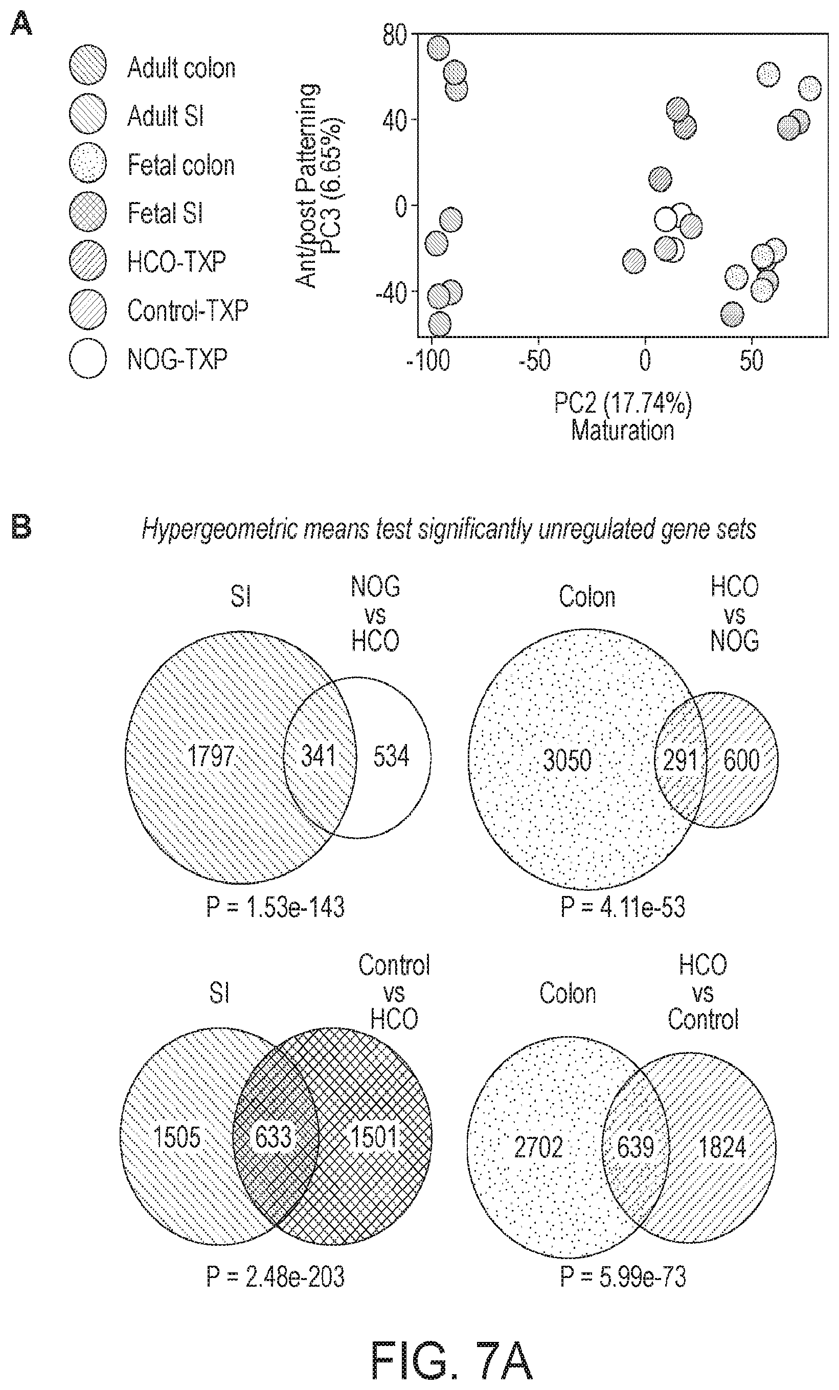
D00013

D00014

D00015

D00016

D00017

D00018

D00019
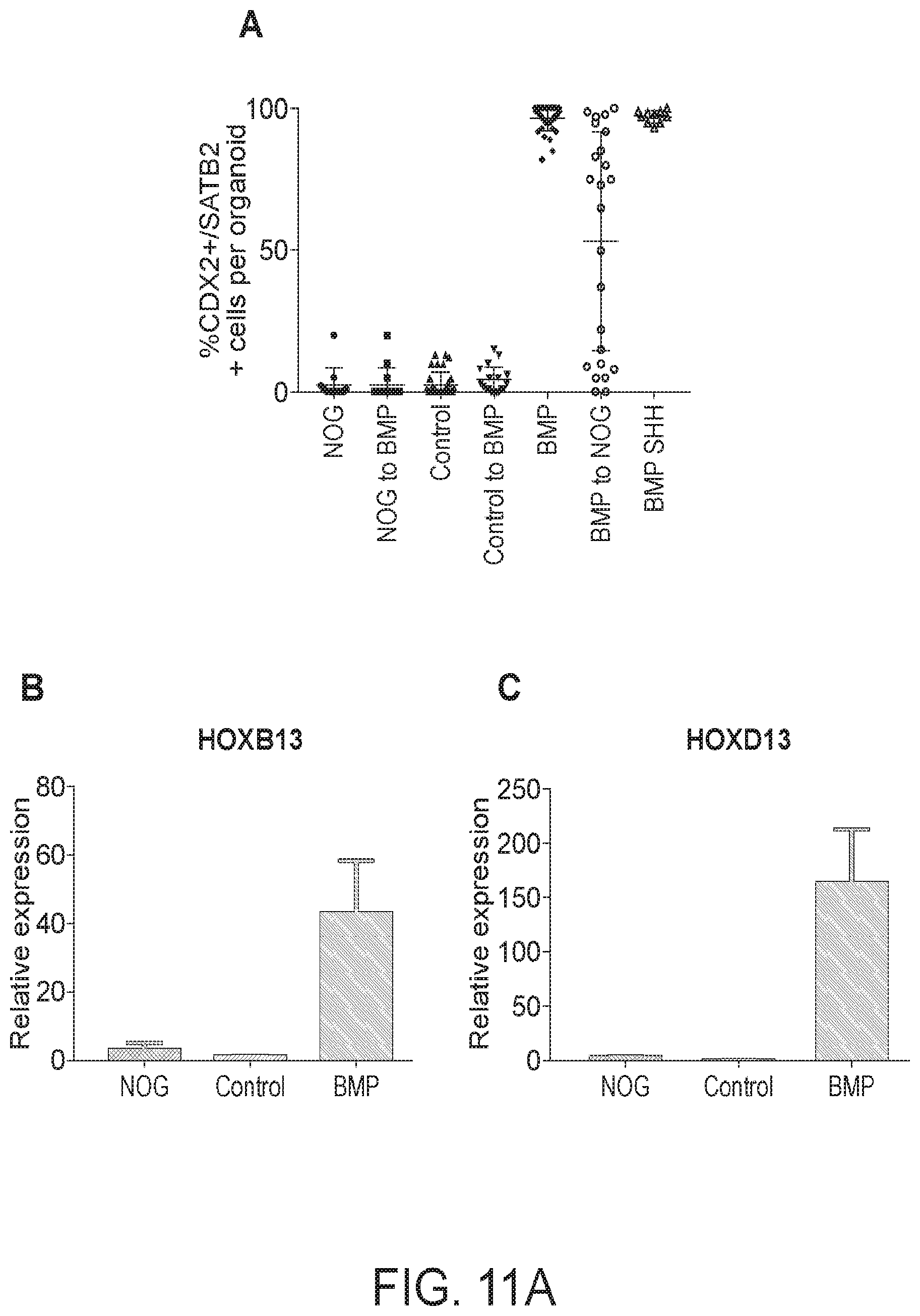
D00020

D00021

D00022

D00023

D00024

D00025

D00026

D00027

D00028
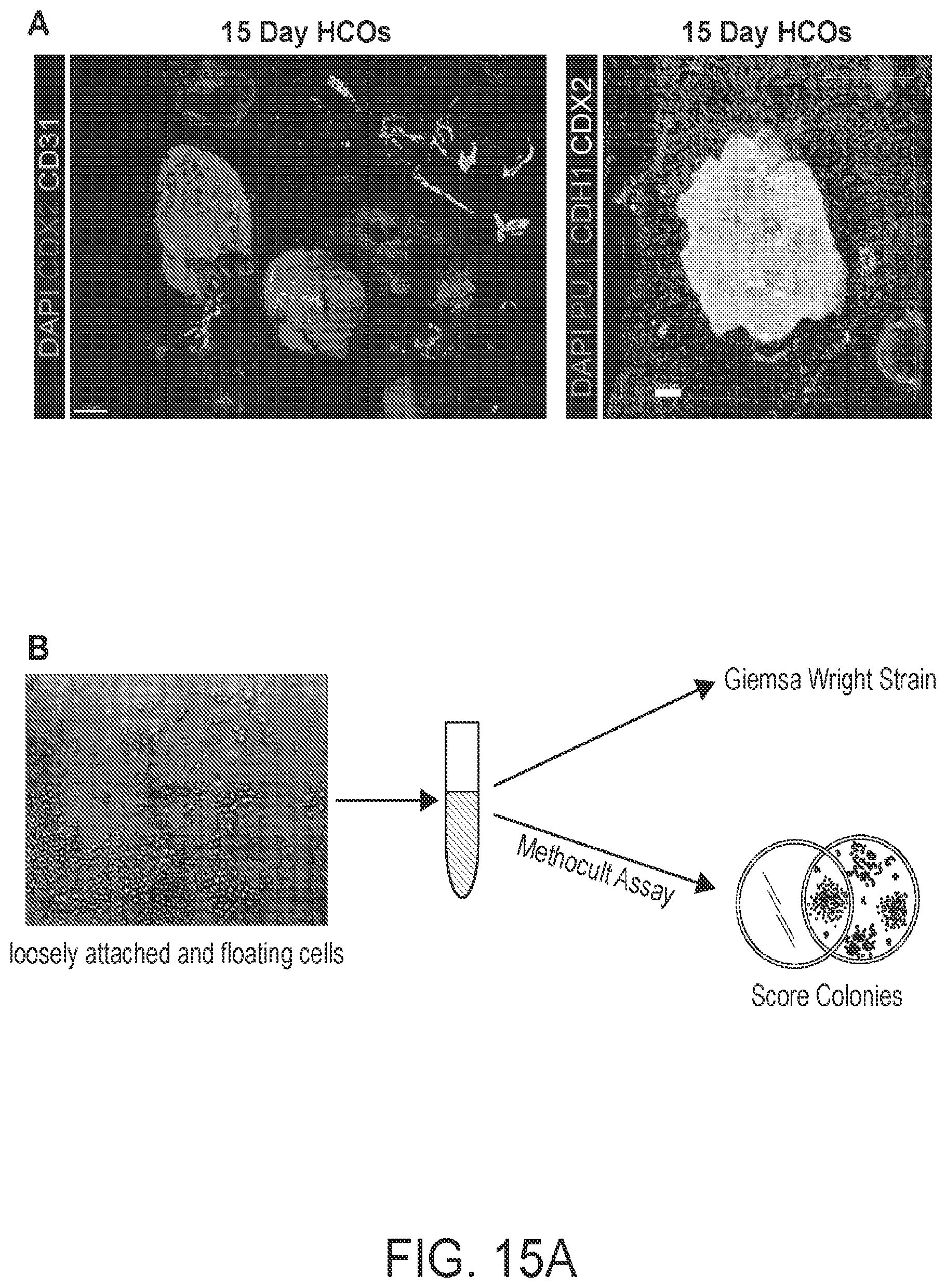
D00029

D00030

D00031

S00001
XML
uspto.report is an independent third-party trademark research tool that is not affiliated, endorsed, or sponsored by the United States Patent and Trademark Office (USPTO) or any other governmental organization. The information provided by uspto.report is based on publicly available data at the time of writing and is intended for informational purposes only.
While we strive to provide accurate and up-to-date information, we do not guarantee the accuracy, completeness, reliability, or suitability of the information displayed on this site. The use of this site is at your own risk. Any reliance you place on such information is therefore strictly at your own risk.
All official trademark data, including owner information, should be verified by visiting the official USPTO website at www.uspto.gov. This site is not intended to replace professional legal advice and should not be used as a substitute for consulting with a legal professional who is knowledgeable about trademark law.